Protease inhibitor: protease sensitive expression system and method improving the therapeutic activity and specificity of proteins and phage and phagemids delivered by bacteria
Bermudes
U.S. patent number 10,590,185 [Application Number 15/600,267] was granted by the patent office on 2020-03-17 for protease inhibitor: protease sensitive expression system and method improving the therapeutic activity and specificity of proteins and phage and phagemids delivered by bacteria. The grantee listed for this patent is David Gordon Bermudes. Invention is credited to David Gordon Bermudes.


| United States Patent | 10,590,185 |
| Bermudes | March 17, 2020 |
Protease inhibitor: protease sensitive expression system and method improving the therapeutic activity and specificity of proteins and phage and phagemids delivered by bacteria
Abstract
A genetically engineered live bacterium which is adapted to selectively replicate in and colonize a selected tissue type within the mammal, and concurrently produce within the selected tissue type at least one protease-sensitive cytotoxic molecule which is degradable by proteases within the selected tissue type, and at least one protease inhibitor peptide to inhibit the proteases within the selected tissue type from proteolytically degrading the protease sensitive cytotoxic molecule. The combination results in higher concentrations of the cytotoxic molecule local to the colonization, while permitting protease degradation of the cytotoxic molecule further away from the colonization.
| Inventors: | Bermudes; David Gordon (Woodland Hills, CA) | ||||||||||
|---|---|---|---|---|---|---|---|---|---|---|---|
| Applicant: |
|
||||||||||
| Family ID: | 46613433 | ||||||||||
| Appl. No.: | 15/600,267 | ||||||||||
| Filed: | May 19, 2017 |
Related U.S. Patent Documents
| Application Number | Filing Date | Patent Number | Issue Date | ||
|---|---|---|---|---|---|
| 14954387 | May 23, 2017 | 9657085 | |||
| 14135003 | Dec 1, 2015 | 9200289 | |||
| 13562488 | Jan 7, 2014 | 8623350 | |||
| 12703158 | Aug 14, 2012 | 8241623 | |||
| 61151019 | Feb 9, 2009 | ||||
| Current U.S. Class: | 1/1 |
| Current CPC Class: | C12N 15/1137 (20130101); C12N 9/0071 (20130101); A61K 38/00 (20130101); A61K 38/55 (20130101); C12N 15/1135 (20130101); C07K 14/005 (20130101); C07K 14/245 (20130101); C07K 14/81 (20130101); C12N 9/22 (20130101); C12N 15/74 (20130101); A61K 38/164 (20130101); C07K 2319/50 (20130101); Y02A 50/483 (20180101); Y02A 50/30 (20180101); C12N 2310/14 (20130101); C12N 2310/141 (20130101); A61K 35/74 (20130101); C07K 2319/55 (20130101) |
| Current International Class: | A61K 38/55 (20060101); C07K 14/81 (20060101); C12N 15/113 (20100101); C12N 15/74 (20060101); A61K 38/00 (20060101); C07K 14/245 (20060101) |
References Cited [Referenced By]
U.S. Patent Documents
| 4436727 | March 1984 | Ribi |
| 4906567 | March 1990 | Connelly |
| 5021234 | June 1991 | Ehrenfeld |
| 5087569 | February 1992 | Gabay et al. |
| 5126257 | June 1992 | Gabay et al. |
| 5143830 | September 1992 | Holland et al. |
| 5223409 | June 1993 | Ladner et al. |
| 5250515 | October 1993 | Fuchs et al. |
| 5318900 | June 1994 | Habuka et al. |
| 5338724 | August 1994 | Gabay et al. |
| 5344762 | September 1994 | Karapetian |
| 5354675 | October 1994 | Iida et al. |
| 5399490 | March 1995 | Balganesh et al. |
| 5403484 | April 1995 | Ladner et al. |
| 5466672 | November 1995 | Kushnaryov et al. |
| 5506139 | April 1996 | Loosmore et al. |
| 5571698 | November 1996 | Ladner et al. |
| 5604201 | February 1997 | Thomas et al. |
| 5656436 | August 1997 | Loosmore et al. |
| 5665353 | September 1997 | Loosmore et al. |
| 5705151 | January 1998 | Dow et al. |
| 5712369 | January 1998 | Old et al. |
| 5824538 | October 1998 | Branstrom et al. |
| 5830702 | November 1998 | Portnoy et al. |
| 5837500 | November 1998 | Ladner et al. |
| 5840518 | November 1998 | Morishita |
| 5869302 | February 1999 | Loosmore et al. |
| 5877159 | March 1999 | Powell et al. |
| 5935573 | August 1999 | Loosmore et al. |
| 5939297 | August 1999 | Loosmore et al. |
| 5945102 | August 1999 | de Faire et al. |
| 5958406 | September 1999 | de Faire et al. |
| 5962430 | October 1999 | Loosmore et al. |
| 5981503 | November 1999 | Loosmore et al. |
| 5997881 | December 1999 | Powell et al. |
| 6004562 | December 1999 | Campagnari |
| 6020183 | February 2000 | Loosmore et al. |
| 6022855 | February 2000 | Thomas et al. |
| 6025342 | February 2000 | Loosmore et al. |
| 6030612 | February 2000 | de Faire et al. |
| 6051237 | April 2000 | Paterson |
| 6080849 | June 2000 | Bermudes et al. |
| 6114125 | September 2000 | Loosmore et al. |
| 6143551 | November 2000 | Goebel |
| 6147057 | November 2000 | Loosmore et al. |
| 6150170 | November 2000 | Powell et al. |
| 6153580 | November 2000 | Loosmore et al. |
| 6190657 | February 2001 | Pawelek et al. |
| 6207156 | March 2001 | Kuchroo et al. |
| 6245892 | June 2001 | Oaks et al. |
| 6251406 | June 2001 | Haefliger et al. |
| 6277379 | August 2001 | Oaks et al. |
| 6291662 | September 2001 | Bandyopadhyay |
| 6348344 | February 2002 | Ayal-Hershkovitz et al. |
| 6410012 | June 2002 | Sizemore et al. |
| 6447777 | September 2002 | Terman et al. |
| 6447784 | September 2002 | Bermudes et al. |
| 6475482 | November 2002 | Bermudes et al. |
| 6475763 | November 2002 | Ayal-Hershkovitz et al. |
| 6537558 | March 2003 | Kaniga |
| 6548287 | April 2003 | Powell et al. |
| 6605286 | August 2003 | Steidler et al. |
| 6605697 | August 2003 | Kwon et al. |
| 6638912 | October 2003 | Bhatnagar et al. |
| 6680374 | January 2004 | Oaks et al. |
| 6685935 | February 2004 | Pawelek et al. |
| 6743893 | June 2004 | Engler et al. |
| 6841535 | January 2005 | Divita et al. |
| 6863894 | March 2005 | Bermudes et al. |
| 6923972 | August 2005 | Bermudes et al. |
| 6962696 | November 2005 | Bermudes et al. |
| 6979538 | December 2005 | Ladner et al. |
| 7001884 | February 2006 | Komiyama et al. |
| 7033991 | April 2006 | Lindberg et al. |
| 7118879 | October 2006 | Ladner et al. |
| 7208293 | April 2007 | Ladner et al. |
| 7258863 | August 2007 | Oaks et al. |
| 7354592 | April 2008 | Bermudes et al. |
| 7358084 | April 2008 | Kolkman |
| 7390646 | June 2008 | Andino-Pavlovsky et al. |
| 7413877 | August 2008 | Collier et al. |
| 7452531 | November 2008 | Bermudes et al. |
| 7514089 | April 2009 | Bermudes et al. |
| 7569547 | August 2009 | Lindberg et al. |
| 7635682 | December 2009 | Denmeade et al. |
| 7691599 | April 2010 | Rubin |
| 7696173 | April 2010 | Collier et al. |
| 7718618 | May 2010 | Gallo et al. |
| 7776823 | August 2010 | Gallo et al. |
| 7846678 | December 2010 | Pepe et al. |
| 7850970 | December 2010 | Shapiro |
| 7887794 | February 2011 | Luquet et al. |
| 7893007 | February 2011 | Ladner et al. |
| 7943754 | May 2011 | Bentwich et al. |
| 8030447 | October 2011 | Motin et al. |
| 8128922 | March 2012 | Wu et al. |
| 8153414 | April 2012 | Caplan et al. |
| 8231878 | July 2012 | Colonna et al. |
| 8241623 | August 2012 | Bermudes |
| 8246945 | August 2012 | Caplan et al. |
| 8283319 | October 2012 | Schulte et al. |
| 8349570 | January 2013 | Pepe et al. |
| 8372620 | February 2013 | Sibbesen et al. |
| 8440207 | May 2013 | Bermudes |
| 8507249 | August 2013 | Finlay et al. |
| 8524220 | September 2013 | Bermudes |
| 8609358 | December 2013 | Sebastian et al. |
| 8623350 | January 2014 | Bermudes |
| 8633305 | January 2014 | Shapiro |
| 8647642 | February 2014 | Bermudes |
| 8685392 | April 2014 | Helmerhorst et al. |
| 8758771 | June 2014 | Finlay et al. |
| 8771669 | July 2014 | Bermudes |
| 8795730 | August 2014 | Vachon |
| 8815251 | August 2014 | Caplan et al. |
| 8951992 | February 2015 | Nathan et al. |
| 8956859 | February 2015 | Bermudes |
| 8981061 | March 2015 | Colonna et al. |
| 9068187 | June 2015 | Bermudes |
| 9187523 | November 2015 | Motin et al. |
| 9200251 | December 2015 | Bermudes |
| 9200289 | December 2015 | Bermudes |
| 9314514 | April 2016 | Eisele |
| 9358308 | June 2016 | Primiano et al. |
| 9365625 | June 2016 | Bermudes |
| 9486513 | November 2016 | Bermudes |
| 9593339 | March 2017 | Bermudes |
| 9616114 | April 2017 | Bermudes |
| 9657085 | May 2017 | Bermudes |
| 9737592 | August 2017 | Bermudes et al. |
| 9878023 | January 2018 | Bermudes |
| 2001/0006642 | July 2001 | Steidler et al. |
| 2001/0009957 | July 2001 | Oaks et al. |
| 2001/0029043 | October 2001 | Haefliger et al. |
| 2002/0026655 | February 2002 | Bermudes et al. |
| 2002/0150881 | October 2002 | Ladner et al. |
| 2002/0197276 | December 2002 | Oaks et al. |
| 2003/0059400 | March 2003 | Szalay |
| 2003/0082219 | May 2003 | Warren et al. |
| 2003/0087827 | May 2003 | Lindberg et al. |
| 2003/0109026 | June 2003 | Bermudes et al. |
| 2003/0113293 | June 2003 | Bermudes et al. |
| 2003/0113717 | June 2003 | Ladner et al. |
| 2003/0165875 | September 2003 | Colonna et al. |
| 2003/0170276 | September 2003 | Bermudes et al. |
| 2003/0219722 | November 2003 | Ladner et al. |
| 2003/0219886 | November 2003 | Ladner et al. |
| 2004/0005539 | January 2004 | Ladner et al. |
| 2004/0023205 | February 2004 | Ladner et al. |
| 2004/0219169 | November 2004 | Bermudes et al. |
| 2004/0229338 | November 2004 | King |
| 2004/0234998 | November 2004 | Sibbesen et al. |
| 2005/0013822 | January 2005 | Oaks et al. |
| 2005/0036987 | February 2005 | Pawelek et al. |
| 2005/0063994 | March 2005 | Caplan et al. |
| 2005/0069532 | March 2005 | Weinrauch et al. |
| 2005/0079573 | April 2005 | Sibbesen |
| 2005/0106151 | May 2005 | Shapiro |
| 2005/0148504 | July 2005 | Katunuma et al. |
| 2005/0202535 | September 2005 | Collier et al. |
| 2005/0203007 | September 2005 | Komiyama et al. |
| 2005/0208033 | September 2005 | Luquet et al. |
| 2005/0249706 | November 2005 | Bermudes et al. |
| 2005/0255088 | November 2005 | Bermudes et al. |
| 2005/0260670 | November 2005 | Colonna et al. |
| 2006/0084113 | April 2006 | Ladner et al. |
| 2006/0088910 | April 2006 | Nguyen |
| 2006/0229336 | October 2006 | Kazmierski et al. |
| 2006/0241050 | October 2006 | Cameron et al. |
| 2007/0009489 | January 2007 | Bermudes et al. |
| 2007/0037744 | February 2007 | Gallo et al. |
| 2007/0041997 | February 2007 | Finlay et al. |
| 2007/0065908 | March 2007 | Gallo et al. |
| 2007/0071773 | March 2007 | Hanski et al. |
| 2007/0192905 | August 2007 | Piller et al. |
| 2007/0254329 | November 2007 | Rubin |
| 2007/0259417 | November 2007 | Ladner et al. |
| 2007/0275423 | November 2007 | Sebastian et al. |
| 2007/0298012 | December 2007 | King et al. |
| 2008/0089862 | April 2008 | Benhar et al. |
| 2008/0124355 | May 2008 | Bermudes |
| 2008/0181892 | July 2008 | Ledbetter et al. |
| 2008/0261869 | October 2008 | Shapiro |
| 2008/0311081 | December 2008 | Fruehauf et al. |
| 2009/0011974 | January 2009 | Bocharov et al. |
| 2009/0069248 | March 2009 | Motin et al. |
| 2009/0081199 | March 2009 | Colonna et al. |
| 2009/0111160 | April 2009 | Collier et al. |
| 2009/0123426 | May 2009 | Li et al. |
| 2009/0162356 | June 2009 | Lookeren Campagne |
| 2009/0169517 | July 2009 | Bermudes et al. |
| 2009/0214506 | August 2009 | Hardy et al. |
| 2009/0234101 | September 2009 | Ladner et al. |
| 2009/0294288 | December 2009 | May et al. |
| 2009/0305296 | December 2009 | Bengtsson et al. |
| 2010/0022584 | January 2010 | Kenyon et al. |
| 2010/0135961 | June 2010 | Bermudes |
| 2010/0136048 | June 2010 | Bermudes |
| 2010/0137192 | June 2010 | Shapiro |
| 2010/0166802 | July 2010 | Caplan et al. |
| 2010/0247544 | September 2010 | Vachon |
| 2010/0278819 | November 2010 | Bossuyt et al. |
| 2010/0279923 | November 2010 | Schulte et al. |
| 2010/0286251 | November 2010 | Rubin |
| 2010/0305306 | December 2010 | Colonna et al. |
| 2010/0310560 | December 2010 | Colonna et al. |
| 2011/0021416 | January 2011 | Shapiro |
| 2011/0028397 | February 2011 | Tozser et al. |
| 2011/0038917 | February 2011 | Kappers et al. |
| 2011/0104146 | May 2011 | Faraday |
| 2011/0152176 | June 2011 | Horswill |
| 2011/0190234 | August 2011 | Nathan et al. |
| 2011/0195423 | August 2011 | Selinfreund et al. |
| 2011/0223241 | September 2011 | Tardi et al. |
| 2012/0045474 | February 2012 | Motin et al. |
| 2012/0064062 | March 2012 | Goguen et al. |
| 2012/0064572 | March 2012 | Finlay et al. |
| 2012/0071545 | March 2012 | Shapiro |
| 2012/0142080 | June 2012 | Bermudes |
| 2012/0142623 | June 2012 | Lagunoff et al. |
| 2012/0230976 | September 2012 | Helmerhorst et al. |
| 2013/0023472 | January 2013 | Bristow |
| 2013/0028901 | January 2013 | Colonna et al. |
| 2013/0102017 | April 2013 | Pfaendler et al. |
| 2013/0150559 | June 2013 | Colonna et al. |
| 2013/0171109 | July 2013 | Helmerhorst et al. |
| 2013/0196432 | August 2013 | Poehlmann et al. |
| 2014/0005108 | January 2014 | Bristow |
| 2014/0056841 | February 2014 | Vachon |
| 2014/0150134 | May 2014 | Li et al. |
| 2014/0194346 | July 2014 | Aebi et al. |
| 2014/0220661 | August 2014 | Bermudes |
| 2014/0234310 | August 2014 | Shapiro |
| 2014/0296480 | October 2014 | Sanchez Garcia et al. |
| 2014/0322790 | October 2014 | Sebastian et al. |
| 2014/0370036 | December 2014 | Shapiro |
| 2015/0004705 | January 2015 | Lu et al. |
| 2015/0017204 | January 2015 | Bermudes |
| 2015/0071957 | March 2015 | Kelly |
| 2015/0139940 | May 2015 | Bermudez Humaran et al. |
| 2015/0184220 | July 2015 | Sebastian et al. |
| 0973911 | Jan 2000 | EP | |||
| 1513924 | Mar 2005 | EP | |||
| 1655370 | May 2006 | EP | |||
| WO199640238 | Dec 1996 | WO | |||
| WO1997014782 | Apr 1997 | WO | |||
| WO1999010014 | Mar 1999 | WO | |||
| WO1999010485 | Mar 1999 | WO | |||
| WO2000004919 | Feb 2000 | WO | |||
| WO2001014579 | Mar 2001 | WO | |||
| WO200125397 | Apr 2001 | WO | |||
| WO2002070645 | Sep 2002 | WO | |||
| WO2003072125 | Sep 2003 | WO | |||
| WO2003102168 | Dec 2003 | WO | |||
| WO2004076484 | Sep 2004 | WO | |||
| WO2004103404 | Dec 2004 | WO | |||
| WO2005018332 | Mar 2005 | WO | |||
| WO2005054477 | Jun 2005 | WO | |||
| WO2006048344 | May 2006 | WO | |||
| WO2006010070 | Jun 2006 | WO | |||
| WO2006116545 | Nov 2006 | WO | |||
| WO2008073148 | Jun 2008 | WO | |||
| WO2008091375 | Jul 2008 | WO | |||
| WO2009014650 | Jan 2009 | WO | |||
| WO2009086116 | Jul 2009 | WO | |||
| WO2009126189 | Oct 2009 | WO | |||
| WO2009139985 | Nov 2009 | WO | |||
| WO2009152480 | Dec 2009 | WO | |||
Other References
|
Wang et al., Design of peptide inhibitors for furin based on the C-terminal fragment of histone H1.2, Acta Biochim Biophys Sin 40: 848-854 (2008) (Year: 2008). cited by examiner. |
Primary Examiner: Ha; Julie
Assistant Examiner: Hellman; Kristina M
Attorney, Agent or Firm: Tully Rinckey PLLC Hoffberg; Steven M.
Parent Case Text
CROSS REFERENCE TO RELATED APPLICATIONS
The present application is a Continuation of U.S. patent application Ser. No. 14/954,387, filed Nov. 30, 2015, now U.S. Pat. No. 9,657,085, issued May 23, 2017, which is a Division of U.S. patent application Ser. No. 14/135,003, filed Dec. 19, 2013, now U.S. Pat. No. 9,200,289, issued Dec. 1, 2015, which is a Continuation of U.S. patent application Ser. No. 13/562,488, filed Jul. 31, 2012, now U.S. Pat. No. 8,623,350, issued Dec. 31, 2013, which is a Division of U.S. patent application Ser. No. 12/703,158, filed Feb. 9, 2010, now U.S. Pat. No. 8,241,623, issued Aug. 14, 2012, which is a non-provisional of U.S. Provisional Patent Application Ser. No. 61/151,019, filed Feb. 9, 2009, each of which is expressly incorporated herein by reference.
Claims
What is claimed is:
1. A secreted chimeric protease inhibitor peptide, comprising: a protease inhibitor peptide sequence configured to selectively inhibit a protease; a therapeutic peptide sequence having a protease cleavage site peptide sequence for the protease inhibited by the protease inhibitor peptide sequence; a targeting functionality peptide sequence; and a terminal cellular secretion signal peptide sequence, fused together, and configured to selectively cause a cell to secrete the cellular secretion signal peptide sequence outside of the cell and inhibit degradation of the therapeutic peptide sequence by the protease inhibited by the protease inhibitor peptide sequence.
2. The secreted chimeric protease inhibitor peptide according to claim 1, wherein the protease inhibitor peptide sequence comprises a plurality of protease inhibitor peptide sequences.
3. The secreted chimeric protease inhibitor peptide according to claim 1, wherein the cellular secretion signal peptide sequence is an N-terminal secretion signal.
4. The secreted chimeric protease inhibitor peptide according to claim 1, wherein the cellular secretion signal peptide sequence is a C-terminal secretion signal, and the protease cleavage site peptide sequence is provided between the protease inhibitor peptide sequence and the cellular secretion signal peptide sequence.
5. The secreted chimeric protease inhibitor peptide according to claim 1, wherein the protease cleavage site peptide sequence is cleavable by the same protease that is inhibited by the protease inhibitor peptide sequence.
6. The secreted chimeric protease inhibitor peptide according to claim 1, wherein the protease inhibitor peptide sequence is configured to inhibit a protease selected from the group consisting of tissue plasminogen activator, activated protein C, factor Xa, granzyme A, granzyme B, granzyme M, cathepsin, thrombin, plasmin, urokinase, matrix metalloproteases, prostate specific antigen, and kallikrein 2.
7. The secreted chimeric protease inhibitor peptide according to claim 1, wherein the protease inhibitor peptide sequence is selected from the group consisting of: Inhibitors of kallikrein 2: TABLE-US-00016 SEQ ID NO: 9 SRFKVWWAAG, SEQ ID NO: 10 AARRPFPAPS, SEQ ID NO: 11 PARRPFPVTA, Tissue protease inhibitor SEQ ID NO: 12 DSLGREAKCYNELNGCTKIYDPVCGTDGNTYPNECVLCFENRKRQTSILI QKSGPC (serine protease inhibitor, Kazal type 1, mature), Furin inhibitors: SEQ ID NO: 1 PAAATVTKKVAKSPKKAKAAKPKKAAKSAAKAVKPK, SEQ ID NO: 2 TKKVAKRPRAKRAA, SEQ ID NO: 3 TKKVAKRPRAKRDL, SEQ ID NO: 4 GKRPRAKRA, SEQ ID NO: 5 CKRPRAKRDL, SEQ ID NO: 6 CVAKRPRAKRDL, SEQ ID NO: 7 CKKVAKRPRAKRDL, and SEQ ID NO: 8 RRRRRR L6R (hexa-L-arginine).
8. The secreted chimeric protease inhibitor peptide according to claim 1, wherein the cellular secretion signal peptide sequence comprises an N-terminal signal sequence from a cytolethal distending toxin gene.
9. The secreted chimeric protease inhibitor peptide according to claim 1, wherein the cellular secretion signal peptide sequence comprises a C-terminal hemolysin C (hlyC) signal sequence.
10. The secreted chimeric protease inhibitor peptide according to claim 1, wherein the cellular secretion signal peptide sequence comprises a C-terminal hemolysin C (hlyC) signal sequence, separated from the protease inhibitor peptide sequence by protease cleavage site peptide sequence.
11. The secreted chimeric protease inhibitor peptide according to claim 1, wherein the protease inhibitor peptide sequence comprises at least one furin inhibitor.
12. The secreted chimeric protease inhibitor peptide according to claim 1, produced in a bacterium from a synthetic gene comprising a promoter, operably linked to a ribosomal binding site.
13. The secreted chimeric protease inhibitor peptide according to claim 1, wherein the therapeutic peptide sequence comprises at least one protease-sensitive cytotoxic molecule having the protease cleavage site peptide sequence inhibited by the protease inhibitor peptide sequence.
14. The secreted chimeric protease inhibitor peptide according to claim 13, wherein the at least one protease-sensitive cytotoxic molecule is a cytolethal distending toxin (cldt).
15. The secreted chimeric protease inhibitor peptide according to claim 1, wherein the targeting functionality peptide sequence comprises a tumor tissue targeting peptide sequence.
16. A secreted chimeric protease inhibitor peptide, comprising a fusion of: a protease inhibitor peptide sequence configured to selectively inhibit a protease; a therapeutic peptide sequence comprising a protease cleavage site peptide sequence inhibited by the protease inhibitor peptide sequence; a targeting functionality peptide sequence; and a terminal cellular secretion signal peptide sequence, configured to selectively cause a cell to secrete the chimeric protease inhibitor peptide outside of the cell and inhibit cleavage of the protease cleavage site peptide sequence by the protease inhibited by the protease inhibitor peptide sequence.
17. The secreted chimeric protease inhibitor peptide according to claim 16, wherein the therapeutic peptide sequence comprises a toxic functionality peptide sequence.
18. The secreted chimeric protease inhibitor peptide according to claim 17, wherein the toxic functionality peptide sequence comprises the protease cleavage site peptide sequence, and the protease inhibitor peptide sequence is configured to selectively inhibit a protease which cleaves at least the protease cleavage site peptide sequence.
19. A secreted chimeric protease inhibitor peptide having a plurality of fused portions, comprising: a terminal cellular secretion signal peptide sequence first fused portion, configured to selectively cause a cell to secrete the chimeric protease inhibitor peptide comprising the terminal cellular secretion signal peptide sequence outside of the cell; a protease inhibitor peptide sequence second fused portion configured to selectively inhibit a protease; a toxic functionality peptide sequence third fused portion comprising a protease cleavage site peptide sequence portion configured to be cleaved by the protease; and a targeting functionality peptide sequence fourth fused portion.
20. The secreted chimeric protease inhibitor peptide according to claim 19, wherein the protease inhibitor peptide sequence second fused portion is configured to inhibit the protease, thereby protecting the toxic functionality peptide sequence portion from cleavage by the protease.
Description
1. BACKGROUND OF THE INVENTION
1.1. Field of the Invention
This invention is generally in the field of therapeutic delivery systems, systems and methods for providing co-expression of protease inhibitors with genetically engineered protease sensitive therapeutic constructs, and chimeric proteins.
1.2. Relevant Art
Citation or identification of any reference herein, or any section of this application shall not be construed as an admission that such reference is available as prior art to the present application.
Tumor-targeted bacteria offer tremendous potential advantages for the treatment of solid tumors, including the targeting from a distant inoculation site and the ability to express therapeutic agents directly within the tumor. However, the primary shortcoming of tumor-targeted bacteria investigated in the human clinical trials (Salmonella strain VNP20009 and its derivative TAPET-CD) is that no significant antitumor activity was observed, even in patients where the bacteria was documented to target the tumor. One method of increasing the ability of the bacteria to kill tumor cells is to engineer the bacteria to express conventional bacterial toxins, but this approach poses risks of systemic toxicity. See, e.g., U.S. Pat. Nos. 7,452,531, 7,354,592, 6,962,696, 6,923,972, 6,863,894, 6,685,935, 6,475,482, 6,447,784, 6,190,657, 6,080,849 and US Pub. 2003/0059400, each of which is expressly incorporated herein by reference. These patents disclose, inter alia, pharmaceutical formulations and methods of administration to humans and animals, useful in conjunction with the present technique.
Use of protein toxins for treatment of various disorders including inflammation, autoimmunity, neurological disorders and cancer has long-suffered from off-target toxicity. Some toxins have a natural degree of specificity for their target, such as botulinum toxin which is specific for neurons. Toxin specificity has been achieved by attachment of a specific antibodies or peptide ligands (e.g., Pseudomonas endotoxin A (PE-ToxA) antibody conjugate, known as an immunotoxin). Based upon the binding specificity of the attached antibody moiety for a specific target, enhanced specificity of the target is achieved. Other toxins have been engineered to achieve specificity based upon their sight of activation. For example, aerolysin requires proteolytic activation to become cytotoxic. Substitution of the natural protease cleavage site for a tumor-specific protease cleavage site (e.g., that of the PSA protease or urokinase) results in a toxin selectively activated within tumors. However, in both these types of engineered toxins, off-target toxicity can occur. In the case of the Pseudomonas immunotoxin, several dose-limiting toxicities have been identified. Vascular leakage syndrome (VLS) is associated with hypoalbuminemia, edema, weight gain, hypotension and occasional dyspnea, which is suggested to occur by immunotoxin-mediated endothelial cell injury (Baluna et al., 2000, Exp. Cell Res. 258: 417-424), resulting in a dose-limiting toxicity. Renal injury has occurred in some patients treated with immunotoxins, which may be due to micro-aggregates of the immunotoxin (Frankel et al., 2001, Blood 98: 722a). Liver damage from immunotoxins is a frequent occurrence that is believed to be multifactorial (Frankel, 2002, Clinical Cancer Research 8: 942-944). To date, antibodies with proteinaceous toxins have limited success clinically. One explanation for the off target toxicity is that although a specific agent is targeted to the tumor and/or specifically activated there, the agent is also toxic if it diffuses out of the tumor, which is likely to occur due to the high osmotic pressure that occurs within tumors (Jain, R. K., 1994, Barriers to drug delivery in solid tumors, Scientific American 271 (11): 58-65). Once activated inside the tumor and diffused back outside, toxins such as aerolysin remain active and are able to contribute to non-target toxicity.
Another method of increasing the therapeutic activity of tumor-targeted bacteria is to use F' Salmonella for the purpose of liberating filamentous phage within the tumor that are capable of delivering genetic material to tumor cells (See, WO/2001/014579, expressly incorporated herein by reference). However, the presence of the F' factor in those studies is known to enhance the genetic exchange of the Salmonella with other bacteria such as E. coli, and therefore poses risks of releasing genes into the environment that could enhance the pathogenic potential of other bacteria. Moreover, no antitumor activity was demonstrated.
2. SUMMARY OF THE INVENTION
A Protease Inhibitor: Protease Sensitivity Expression System Improving the Therapeutic Specificity
The present invention consists of the co-expression by the expression system, or a combination of expression systems, of a protease inhibitor together with a protease sensitive therapeutic agent. Within the local high-concentration of the targeted tissue or cells such as a tumor environment, the protease inhibitor prevents the degradation of the agent, which is therapeutically active against the target tissue such as a tumor. Upon egress from the confined space of the targeted tissue such as the tumor, the inhibitor falls below the inhibitory concentration, and the therapeutic agent is freely degraded, resulting in cell or tissue-specific activity and non-target cell or tissue inactivity (Table I). A schematic diagram illustrating the effect of co-expression is shown in FIGS. 1A and 1B.
TABLE-US-00001 TABLE 1 Relative effect of toxin forms with and without protease sensitivity and protease inhibitor. Tumor Systemic Composition Efficacy Toxicity Protease activated and/or insensitive toxin +++ ++ Protease sensitive toxin + - Protease sensitive toxin + protease inhibitor +++++ -
The therapeutic agent can be a peptide or protein, toxin, chimeric toxin, cytokine, antibody, biospecific antibody including single chain antibodies, chemokine, prodrug converting enzyme or phage/phagemid. In a preferred embodiment the therapeutic agent is a toxin, or modified toxin. In another preferred embodiment, the therapeutic agent is a phage or phagemid capable of delivering DNA or RNA.
Toxins useful in conjunction with the present invention that can be modified uniquely to suit the delivery by a bacterium and may be further engineered to have tumor-selective targeting include, azurin, carboxyesterase Est55 (a prodrug converting enzyme from Geobacillus that activates CPT-11 to SN-38), Bacillus sp. cytolysins, cytolethal distending toxin (cldt), typhoid toxin (pltAB), cldt:plt hybrids, cytotoxic necrotic factor (cnf), dermonecrotic factor (dnf), shiga toxin and shiga-like toxins, colicins including colicin E492, colE3, colE7 and col-Ia, membrane lytic peptides from Staphylococcus (listed below), bacterial collagenases (e.g., that from Salmonella strain DT104, see WO/2005/018332, the entirety of which is expressly incorporated herein by reference), repeat in toxin (RTX) family members (together with the necessary acylation and secretion genes) including Actinobacillus leucotoxin, a leukotoxin:E. coli HlyA hybrid, E. coli HlyA hemolysin, and Bordetella adenylate cyclase toxin, heat stable enterotoxins from E. coli and Vibrio sp., autotransporter toxins including picU espC, and sat, clostridium enterotoxin, aerolysin, subtilase, saporin, ricin, pertussis toxin, and porB.
The toxin may be further modified by addition of one or more protease cleavage sites that enhance its degradation outside of the tumor. Preferred protease cleavage sites are those for proteases that are under-expressed within the tumor compared to normal tissues (rather than over-expressed within the tumor as utilized for aerolysin activation). However, the expression levels of many proteases are elevated within tumors. Proteases for which inhibitory peptides may be expressed include furin, tissue plasminogen activator, activated protein C, factor Xa, granzymes (A, B & M), cathepsins (A, B, C, D, E, F, G, H, K, L, S, W & X), thrombin, plasmin, urokinase, matrix metalloproteases, prostate specific antigen (PSA) and kallikrein 2.
Furin recognizes a number of specific cleavage sites, including RXRAKR.dwnarw. SEQ ID NO:57. In accordance with the present invention, the presence of this cleavage site, whether naturally occurring or introduced through genetic modification, may be compensated for within the target tissue by co-expression of a furin inhibitor, stabilizing its activity unless it escapes the target tissue such as a tumor. Use of protease inhibitors alone or in combination by bacterial delivery vectors has not previously been suggested. Indeed, Wang et al. 2008 suggested furin inhibitors could be used as antibiotics to suppress bacterial infection which would thereby interfere with delivery by a bacterial vector. Therefore, it has not been considered desirable to use a furin inhibitor or other protease inhibitors to have a positive effect on the bacteria and/or the therapeutics they release.
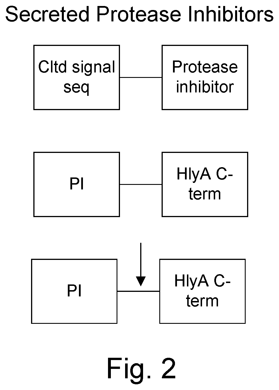
The peptide inhibitors are engineered to be secreted from the bacteria secretion signals known to those skilled in the arts, including ompA, OmpF, M13pIII, cldt N-terminal signal sequences or hlyA C-terminal signal sequence (requires addition of hlyBD and TolC). The inhibitors can be further modified to have the protease cleavage signal of the protease that they inhibit or for a different protease. Multiple protease inhibitor sequences may alternate between protease cleavage sequences or recognition sites.
Chimeric toxins may be further modified by the addition of known cell penetrating (ferry) peptide which further improves their entry into target cells. Cell penetrating peptides include those derived from the HIV TAT protein, the antennapedia homeodomain (penetraxin), Kaposi fibroblast growth factor (FGF) membrane-translocating sequence (MTS), herpes simplex virus VP22, hexahistidine, hexalysine, or hexaarginine.
The present invention also provides in accordance with some embodiments, unique chimeric modifications of the above listed toxins that contain specific combinations of components resulting in secretion by and gram-negative bacteria (e.g., Salmonella, Shigella, E. coli) and selective anti-tumor activity. The invention also provides protease sensitivity (deactivation) which may include the addition of protease cleavage sites and may be co-expressed with a protease inhibitor. The chimeric proteins may have one or more additional features or protein domains known to those skilled in the arts which are designed to 1) be active or catalytic domains that result in the death of the cell or make them susceptible to other known anticancer agents, 2) allow or facilitate them being secreted or released by autolytic peptides such as colicin release peptides, 3) membrane protein transduction (ferry) peptides, 4) autotransporter domains, 5) have targeting peptides that direct them to the target cells, and 6) protease cleavage sites for activation (e.g., release from parent peptide). However, the specific organization and combination of these domains is unique and specific to the invention.
Bombesin and gastrin are amidated peptides. Amidation of these peptides would not be expected to occur in bacteria. A unique composition in accordance with one embodiment of the present invention is the co-expression of the C-terminal amidating enzyme, which results in amidating these peptides in order for them to confer their targeting specificity.
Small lytic peptides (less than 50 amino acids) are used to construct chimeric proteins for more than one purpose. The chimeric proteins containing lytic peptides may be directly cytotoxic for the cancer cells, and/or other cells of the tumor including the tumor matrix cells and immune cells which may diminish the effects of the bacteria by eliminating them. Furthermore, the lytic peptides are useful in chimeric proteins for affecting release from the endosome. Small lytic peptides have been used in the experimental treatment of cancer. However, it is evident that most, if not all, of the commonly used antitumor small lytic peptides have strong antibacterial activity, and thus are not compatible with delivery by a bacterium (see Table 1 of Leschner and Hansel, 2004 Current Pharmaceutical Design 10: 2299-2310, the entirety of which is expressly incorporated herein by reference). Small lytic peptides useful in the invention are those derived from Staphylococcus aureus, S. epidermidis and related species, including the phenol-soluble modulin (PSM) peptides and delta-lysin (Wang et al., 2007 Nature Medicine 13: 1510-1514, expressly incorporated herein by reference). The selection of the lytic peptide depends upon the primary purpose of the construct, which may be used in combination with other constructs providing other anticancer features. Construct designed to be directly cytotoxic to cells employ the more cytoxic peptides, particularly PSM-.alpha.-3. Constructs which are designed to use the lytic peptide to affect escape from the endosome se the peptides with the lower level of cytotoxicity, such as PSM-alpha-1, PSM-alpha-2 or delta-lysin.
2.1 Non-Conjugative, Bacteria Capable of Delivering DNA and RNA Interference (RNAi) Mediated by Small Interfering RNAs (siRNA) and/or microRNAs (miRNA).
The present invention provides, according to some embodiments, a composition that would minimize the effect of bacteria released into the environment by eliminating the ability of the bacteria to exchange genetic information with related bacteria, as well as provide a delivery enhancing bacteria resulting in a greater therapeutic effect. Conjugative transfer is a major genetic exchange mechanism that may occur between Salmonella and the normal commensal gut bacterium E. coli, requiring the presence of an F' factor. The present invention provides gram-negative bacteria including E. coli, Vibrio, Shigella and Salmonella that are genetically modified in one or more ways to eliminate conjugative transfer of DNA with closely related species including E. coli. One of the modifications works on both male (F'+) and female (F'-) bacteria. These modifications facilitate the safety of a bacteria carrying phage capable of delivering DNA or small interfering RNA (siRNA) or microRNA (miRNA) molecules that mediate RNA interference (RNAi), as well as for bacteria expressing chimeric toxins. The phage/phagemids may be further modified to express membrane lytic peptides enhancing their release from the endosome. See, e.g., U.S. Pat. No. 7,390,646, US 2008/0311081, 2009/0123426, WO 2008/091375, WO 1999/010485, WO 1999/010014, WO 2009/086116, each of which is expressly incorporated herein by reference it its entirety
MicroRNAs (miRNA) are single-stranded RNA molecules of, for example, about 21-23 nucleotides in length, which regulate gene expression. miRNAs are encoded by genes from whose DNA they are transcribed but miRNAs are not translated into protein (non-coding RNA; instead each primary transcript (a pri-miRNA) is processed into a short stem-loop structure called a pre-miRNA and finally into a functional miRNA. Mature miRNA molecules are partially complementary to one or more messenger RNA (mRNA) molecules, and their main function is to down-regulate gene expression.
Small interfering RNA (siRNA), sometimes known as short interfering RNA or silencing RNA, is a class of 19-25 nucleotide-long double-stranded RNA molecules with 3'' overhangs. Asymmetric interfering RNAs have 3'' and 5'' antisense overhangs and may be only 15 base pairs in length (Sun et al. 2008 Nature Biotechnology 26: 1379-1382, incorporated in its entirety herein). Interfering RNAs play a variety of roles in biology. Most notably, siRNA is involved in the RNA interference (RNAi) pathway, where it interferes with the expression of a specific gene. In addition to their role in the RNAi pathway, siRNAs also act in RNAi-related pathways, e.g., as an antiviral mechanism or in shaping the chromatin structure of a genome.
The bacterial strains according to various aspects of the invention useful for delivery of phage/phagemids especially include any of those expressing protease inhibitors, and/or chimeric toxins. The bacteria may also be modified variously in order to accommodate the production of the phage such that they are stably maintained and released from the bacteria. These modifications may include: introduction of an F' pilus factor which allows the filamentous phage to infect the bacteria, a "helper phage" which provides phage genes and functions in trans necessary to package a phagemid, a phagemid containing a modified phage coat protein (e.g., pIII, pVIII) into which a targeting ligand is inserted, and phagemid effector genes, which may include eukaryotic promoters for tumor cell expression of anticancer genes, or genes that are transcribed into short hairpin RNAs that function as interfering RNA molecules (RNAi). More than one gene and/or siRNA and/or miRNA may be expressed from a single phagemid and may employ ribosomal reentry signals (RESs). A preferred bacterial phagemid with eukaryotic expression components (including an SV40 origin of replication, HSV-TK polyadenylation signal, a CVM IE promoter and an SV40 polyadenylation signal) is pEGFP-N1. The siRNA and/or miRNA molecules may utilize the T7 promoter. Constructs using the T7 promoter may contain one or more copies of the T7 polymerase under control of a eukaryotic promoter, which, when transcribed and translated, is capable of expressing the siRNA and/or miRNA constructs under control of the T7 promoter.
The phagemid-expressed genes may serve multiple purposes. The phagemid genes may serve to suppress certain immune responses within the tumor, including T-cells, macrophages and neutrophils that may limit the ability of the bacteria to effectively reach all the tumor cells within a tumor. The phagemid genes may also serve to directly inhibit tumor cells, either through the expression of anti-tumor genes (e.g., tumor suppressor genes such as p53) or by generating siRNA and/or miRNA or other RNAi molecules, which suppress the presence of mRNA transcripts, suppressing the neoplastic genes such as KRAS.
The F' pilus factors are provided by the F' plasmid, and are needed for phage to be able to infect a bacterial cell. The F' factor provides other functions which may be undesirable in conjunction with aspects of the present invention, including mating stabilization and DNA transfer. The present invention therefore provides, according to one aspect, a composition lacking these features by their genetic disruption on the F' factor or by the cloning of the pilus factor genes into the tumor-targeted bacterium in the absence of the other factors, and hence, resulting in a strain which is non-conjugative and significantly less likely to transfer DNA to other bacteria. The invention may also incorporate entry exclusion into the bacteria and the fertility inhibition complex (finO and finP), and thus, even in tumor-targeted bacterial strains in which the pilus factors are not incorporated (i.e., F-), the bacterial strain will remain resistant to mating with F' bacteria.
3. OBJECTS OF THE INVENTION
The present invention provides, according to one embodiment, improved live attenuated therapeutic bacterial strains that express one or more therapeutic molecules together with one or more protease inhibitor polypeptides that inhibit local proteases that could deactivate the therapeutic molecules. In particular, one aspect of the invention relates to live attenuated tumor-targeted bacterial strains that may include Salmonella vectoring chimeric anti-tumor toxins to an individual to elicit a therapeutic response against cancer. Another aspect of the invention relates to live attenuated tumor-targeted bacterial strains that may include Salmonella vectoring filamentous phage that encode anti-tumor DNA and RNA molecules to an individual to elicit a therapeutic response against cancer including cancer stem cells. The filamentous phage may also be targeted to tumor matrix cells, and immune cells.
Whereas the prior strains of Salmonella studied in human clinical trails used either no heterologous antitumor protein (i.e., VNP20009) or an antitumor protein located within the cytoplasm of the bacterium (i.e., cytosine deaminase expressed by TAPET-CD), the invention provides, according to some embodiments, methods and compositions comprising bacterial vectors that secrete protease inhibitors that protect coexpressed protease sensitive antitumor molecules that are also secreted into the tumor for the treatment of cancer.
The bacteria according to a preferred embodiment of the present invention have little or no ability to undergo bacterial conjugation, limiting incoming and outgoing exchange of genetic material, whereas the prior art fails to limit exchange of genetic material. In addition, certain of the therapeutic molecules have co-transmission requirements that are distal to the therapeutic molecule location further limiting known forms of genetic exchange.
Aspects of the present invention also provide novel chimeric bacterial toxins particularly suited for expression by gram-negative bacteria. The toxins may have added targeting ligands that render them selectively cytotoxic for tumor cells, tumor stem cells and/or matrix and tumor-infiltrating immune cells. The invention also provides means to determine optimal toxin combinations which are preferably additive or more preferably synergistic. The invention also provides means to determine the optimal combination of protein toxin with conventional cancer chemotherapeutics or biologics. Accordingly, administration to an individual, of a live Salmonella bacterial vector, in accordance with an aspect of the present invention, that is genetically engineered to express one or more protease inhibitors as described herein co-expressed with one or more cytotoxic proteins has the ability to establish a population in the tumor, kill tumor cells, tumor stem cells as well as tumor matrix and immune infiltrating cells, resulting in a therapeutic benefit.
A preferred composition will contain, for example, a sufficient amount of live bacteria expressing the protease inhibitors and cytotoxin(s) to produce a therapeutic response in the patient. Accordingly, the attenuated Salmonella strains described herein are both safe and useful as live bacterial vectors that can be orally administered to an individual to provide therapeutic benefit for the treatment of cancer.
Although not wishing to be bound by any particular mechanism, an effective antitumor response in humans by administration of genetically engineered, attenuated strains of Salmonella strains as described herein may be due to the ability of such mutant strains to persist in the tumor and to supply their own nutrient needs by killing tumor cells and further expanding the zone of the tumor that they occupy. Bacterial strains useful in accordance with a preferred aspect of the invention may carry the ability to produce a therapeutic molecule (or releases an agent such as a phagemid that carries the ability to generate therapeutic molecules) expressing plasmid or chromosomally integrated cassette that encodes and directs expression of one or more therapeutic molecules together with one or more protease inhibitors, as described herein. The protease inhibitors serve to prevent the destruction of the therapeutic molecule while within the tumor. If the cytotoxin and protease inhibitor diffuse outside of the tumor, they fall below the protease inhibitory concentration, and the cytotoxins are inactivated. Thus the protease inhibitor system both increases activity and provides tumor specificity.
The serovars of S. enterica that may be used as the attenuated bacterium of the live compositions described in accordance with various embodiments herein include, without limitation, Salmonella enterica serovar Typhimurium ("S. typhimurium"), Salmonella montevideo, Salmonella enterica serovar Typhi ("S. typhi"), Salmonella enterica serovar Paratyphi B ("S. paratyphi 13"), Salmonella enterica serovar Paratyphi C ("S. paratyphi C"), Salmonella enterica serovar Hadar ("S. hadar"), Salmonella enterica serovar Enteriditis ("S. enteriditis"), Salmonella enterica serovar Kentucky ("S. kentucky"), Salmonella enterica serovar Infantis ("S. infantis"), Salmonella enterica serovar Pullorum ("S. pullorum"), Salmonella enterica serovar Gallinarum ("S. gallinarum"), Salmonella enterica serovar Muenchen ("S. muenchen"), Salmonella enterica serovar Anatum ("S. anatum"), Salmonella enterica serovar Dublin ("S. dublin"), Salmonella enterica serovar Derby ("S. derby"), Salmonella enterica serovar Choleraesuis var. kunzendorf ("S. cholerae kunzendorf`), and Salmonella enterica serovar minnesota (S. minnesota). A preferred serotype for the treatment of bone marrow related diseases is S dublin.
By way of example, live bacteria in accordance with aspects of the invention include known strains of S. enterica serovar Typhimurium (S. typhimurium) and S. enterica serovar Typhi (S. typhi) which are further modified as provided by various embodiments of the invention. Such Strains include Ty21a, CMV906, CMV908, CMV906-htr, CMV908-htr, Ty800, aroA-/serC-, holavax, M01ZH09, VNP20009. See also, U.S. Pat. No. 6,548,287, and EP 0,973,911, each of which expressly incorporated herein by reference. These strains contain defined mutations within specific serotypes of bacteria. The invention also includes the use of these same mutational combinations contained within alternate serotypes or strains in order to avoid immune reactions which may occur in subsequent administrations. In a preferred embodiment, S. Typhimurium, S. montevideo, and S. typhi which have non-overlapping O-antigen presentation (e.g., S. typhimurium is O--1, 4, 5, 12 and S. typhi is Vi, S. montevideo is O--6, 7) may be used. Thus, for example, S. typhimurium is a suitable serotype for a first injection and another serotype such as S. typhi or S. montevideo are used for a second injection and third injections. Likewise, the flagellar antigens are also selected for non-overlapping antigenicity between different injections. The flagellar antigen may be H1 or H2 or no flagellar antigen, which, when combined with the three different O-antigen serotypes, provides three completely different antigenic profiles. Methods for deriving heterologous O-antigens have been described by Favre et al., WO/1997/014782, and Roland WO/2000/004919, each of which is expressly incorporated herein by reference.
Novel strains are also encompassed that are, for example, attenuated in virulence by mutations in a variety of metabolic and structural genes. The invention therefore may provide a live composition for treating cancer comprising a live attenuated bacterium that is a serovar of Salmonella enterica comprising an attenuating mutation in a genetic locus of the chromosome of said bacterium that attenuates virulence of said bacterium and wherein said attenuating mutation is the Suwwan deletion (Murray et al., Journal of Bacteriology, 2004) or combinations with other known attenuating mutations. Other attenuating mutation useful in the Salmonella bacterial strains described herein may be in a genetic locus selected from the group consisting of phoP, phoQ, edt, cya, crp, poxA, rpoS, htrA, nuoG, pmi, pabA, pts, damA, pur, purA, purB, purI, purF, zwf, aroA, aroB, aroC, aroD, serC, gua, cadA, rfc, rjb, rfa, ompR, msbB and combinations thereof.
The invention also encompasses gram-positive bacteria. Preferred bacteria of the invention are group B Streptococcus including S. agalaciae, and Listeria species including L. monocytogenes. It is known to those skilled in the arts that minor variations in molecular biology techniques such as use of gram-positive origins of replication, gram-positive signal sequences gram-positive promoters and filamentous phage (e.g., phage B5; Chopin et al., 2002 J. Bacteriol. 184: 2030-2033) are required and substituted as needed.
The invention also provides, according to one embodiment, a process for preparing genetically stable therapeutic bacterial strains comprising genetically engineering the therapeutic genes of interest into a bacterially codon optimized expression sequence within a bacterial plasmid expression vector or chromosomal localization expression vector for any of the deleted genes or IS200 genes within the strain and further containing engineered restriction endonuclease sites such that the bacterially codon optimized expression gene contains subcomponents which are easily and rapidly exchangeable, and the bacterial strains so produced. Administration of the strain to the patient is therapeutic for the treatment of cancer.
The present invention provides, for example, and without limitation, live bacterial compositions that are genetically engineered to express one or more protease inhibitors combined with antitumor effector molecules or phagemids capable of delivering DNA and RNA therapeutics for the treatment of cancer.
4. DEFINITIONS
In order that the invention may be more fully understood, the following terms are defined.
As used herein, "attenuated", "attenuation", and similar terms refer to elimination or reduction of the natural virulence of a bacterium in a particular host organism, such as a mammal.
"Virulence" is the degree or ability of a pathogenic microorganism to produce disease in a host organism. A bacterium may be virulent for one species of host organism (e.g., a mouse) and not virulent for another species of host organism (e.g., a human). Hence, broadly, an "attenuated" bacterium or strain of bacteria is attenuated in virulence toward at least one species of host organism that is susceptible to infection and disease by a virulent form of the bacterium or strain of the bacterium.
As used herein, the term "genetic locus" is a broad term and comprises any designated site in the genome (the total genetic content of an organism) or in a particular nucleotide sequence of a chromosome or replicating nucleic acid molecule (e.g., a plasmid), including but not limited to a gene, nucleotide coding sequence (for a protein or RNA), operon, regulon, promoter, inducible promoters (including tetracycline, arabinose, (EP1,655,370 A1, expressly incorporated in its entirety herein), salicylic acid, hypoxic, tumor cell specific inducible promoters) regulatory site (including transcriptional terminator sites, ribosome binding sites, transcriptional inhibitor binding sites, transcriptional activator binding sites), origin of replication, intercistronic region, and portions therein. It is understood that all protein expression constructs require a stop signal. A genetic locus may be identified and characterized by any of a variety of in vivo and/or in vitro methods available in the art, including but not limited to, conjugation studies, crossover frequencies, transformation analysis, transfection analysis, restriction enzyme mapping protocols, nucleic acid hybridization analyses, polymerase chain reaction (PCR) protocols, nuclease protection assays, and direct nucleic acid sequence analysis
The terms "oral", "enteral", "enterally", "orally", "non-parenteral", "non-parenterally", and the like, refer to administration of a compound or composition to an individual by a route or mode along the alimentary canal. Examples of "oral" routes of administration of a vaccine composition include, without limitation, swallowing liquid or solid forms of a vaccine composition from the mouth, administration of a vaccine composition through a nasojejunal or gastrostomy tube, intraduodenal administration of a vaccine composition, and rectal administration, e.g., using suppositories that release a live bacterial vaccine strain described herein to the lower intestinal tract of the alimentary canal.
The term "recombinant" is used to describe non-naturally altered or manipulated nucleic acids, cells transformed, electroporated, or transfected with exogenous nucleic acids, and polypeptides expressed non-naturally, e.g., through manipulation of isolated nucleic acids and transformation of cells. The term "recombinant" specifically encompasses nucleic acid molecules that have been constructed, at least in part, in vitro using genetic engineering techniques, and use of the term "recombinant" as an adjective to describe a molecule, construct, vector, cell, polypeptide, or polynucleotide specifically excludes naturally existing forms of such molecules, constructs, vectors, cells, polypeptides, or polynucleotides.
Cassette, or expression cassette is used to describe a nucleic acid sequence comprising (i) a nucleotide sequence encoding a promoter, (ii) a first unique restriction enzyme cleavage site located 5' of the nucleotide sequence encoding the promoter, and (iii) a second unique restriction enzyme cleavage site located 3' of the nucleotide sequence encoding the promoter. The cassette may also contain a multiple cloning site (MCS) and transcriptional terminator within the 5' and 3' restriction endonuclease cleavage sites. The cassette may also contain cloned genes of interest.
As used herein, the term "salmonella" (plural, "salmonellae") and "Salmonella" refers to a bacterium that is a serovar of Salmonella enterica. A number of serovars of S. enterica are known. Particularly preferred salmonella bacteria useful in the invention are attenuated strains of Salmonella enterica serovar Typhimurium ("S. typhimurium") and serovar Typhi ("S. typhi") as described herein.
As used herein, the terms "strain" and "isolate" are synonymous and refer to a particular isolated bacterium and its genetically identical progeny. Actual examples of particular strains of bacteria developed or isolated by human effort are indicated herein by specific letter and numerical designations (e.g. strains Ty21a, CMV906, CMV908, CMV906-htr, CMV908-htr, Ty800, holavax, M01ZH09, VNP20009).
The definitions of other terms used herein are those understood and used by persons skilled in the art and/or will be evident to persons skilled in the art from usage in the text.
5. BRIEF DESCRIPTION OF THE DRAWINGS
FIGS. 1A and 1B show a comparison of tumor-protease activated toxin with tumor protease inhibitor (FIG. 1A) and protease sensitive toxin expression system (FIG. 1B).
FIG. 2 shows secreted protease inhibitors.
FIGS. 3A to 3F show chimeric colicins.
FIGS. 4A to 4D show lytic peptide chimeras.
FIGS. 5A to 5D show protease activated lytic peptide chimera prodrugs.
FIGS. 6A to 6D show cytolethal distending toxin subunit B (cldtB) chimeras.
FIGS. 7A to 7D show repeat in toxin (RTX) family members and hybrid operons.
FIG. 8 shows a non-conjugative bacterium with and without the F' factor.
FIG. 9 shows segregation of required colicin toxin and immunity factors.
FIG. 10 shows a non-conjugative bacterium capable of releasing phage/phagemids carrying expression constructs for DNA and RNA therapeutics.
6. DETAILED DESCRIPTION OF THE INVENTION
The present invention provides, according to various embodiments, improved live attenuated therapeutic bacterial strains that express one or more therapeutic molecules together with one or more protease inhibitor polypeptides that inhibit local proteases that could deactivate the therapeutic molecules. In particular, one aspect of the invention relates to live attenuated tumor-targeted bacterial strains that may include Salmonella vectoring novel chimeric anti-tumor toxins to an individual to elicit a therapeutic response against cancer. The types of cancer may generally include solid tumors, leukemia, lymphoma and multiple myeloma. In addition, certain of the therapeutic molecules have co-transmission requirements that are genetically unlinked to the therapeutic molecule(s), limiting certain forms of genetic exchange. Another aspect of the invention relates to live attenuated tumor-targeted bacterial strains that may include Salmonella vectoring filamentous phage that encode anti-tumor DNA and RNA molecules to an individual to elicit a therapeutic response against cancer including cancer stem cells. The filamentous phage may also be targeted to tumor matrix cells, and immune cells. Another aspect of the invention relates to reducing or eliminating the bacteria's ability to undergo conjugation, further limiting incoming and outgoing exchange of genetic material.
For reasons of clarity, the detailed description is divided into the following subsections: protease sensitivity; protease inhibitors; targeting ligands; chimeric bacterial toxins; co-expression of protease inhibitors with bacterial toxins, segregation of required colicin cofactors; limiting bacterial conjugation; phage/phagemid producing gram negative bacteria encoding therapeutic DNA and RNA molecules.
6.1. Protease Sensitivity.
The therapeutic proteins of the invention are sensitive to proteases (in contrast pro-aerolysin or urokinase chimeric toxins that are activated by proteases). Protease digestion sites may be added to the therapeutic agent to enhance protease sensitivity. Preferred proteases for conferring greater sensitivity are those that are under-expressed in tumors and over-expressed in normal tissues. Other proteases for which sensitivity sites may be added include tissue plasminogen activator, activated protein C, factor Xa, granzyme (A, B, M), cathepsins, thrombin, plasmin, urokinase, matrix metalloproteases, prostate specific antigen (PSA) and kallikrein 2.
6.2.1 Protease Inhibitors
Protease inhibitors of the invention are preferably based on known polypeptide inhibitors. The inhibitors include both synthetic peptides and naturally occurring, endogenous peptides.
To result in the desired activity, the peptides should be secreted outside of the bacteria. Accordingly, the peptides are modified by fusing them to secretion signals. The secretion signals may be either N-terminal (derived from ompA, ompF, M13pIII, cldt) or C-terminal (last 60 amino acids of the E. coli HlyA hemolysin, together with the required HlyBD supplied in trans and endogenous tolC) as shown in FIG. 2. The N-terminal signal sequences are well known and characterized by the presence of a protease cleavage site for an endogenous bacterial protease. Thus, N-terminal signal sequences provide free protease inhibitors, free from the signal sequence. The C-terminal signal sequence may be further engineered to have a protease cleavage site in between the protease inhibitory peptide and the signal sequence. The cleavage site may be for the same protease that the peptide inactivates. Thus, the protease activates its own inhibitor. The protease cleavage site may also be for a protease other than for the protease inhibitor, thus deactivating another protease. Proteases upregulated within tumors for which protease cleavage sites may be engineered include: tissue plasminogen activator, activated protein C, factor Xa, granzyme (A, B, M), cathepsin, thrombin, plasmin, urokinase, matrix metalloproteases, prostate specific antigen (PSA) and kallikrein 2.
Suitable protease inhibitors, include, but are not limited to, those listed below.
Inhibitors of Kallikrein 2:
TABLE-US-00002 SEQ ID NO: 9 SRFKVWWAAG SEQ ID NO: 10 AARRPFPAPS SEQ ID NO: 11 PARRPFPVTA
Tissue Protease Inhibitor
TABLE-US-00003 SEQ ID NO: 12 DSLGREAKCYNELNGCTKIYDPVCGTDGNTY PNECVLCFENRKRQTSILIQKSGPC (serine protease inhibitor, Kazal type 1, mature) Furin inhibitors: SEQ ID NO: 1 PAAATVTKKVAKSPKKAKAAKPKKAAKSAAKAVKPK SEQ ID NO: 2 TKKVAKRPRAKRAA SEQ ID NO: 3 TKKVAKRPRAKRDL SEQ ID NO: 4 GKRPRAKRA SEQ ID NO: 5 CKRPRAKRDL SEQ ID NO: 6 CVAKRPRAKRDL SEQ ID NO: 7 CKKVAKRPRAKRDL SEQ ID NO: 8 RRRRRR L6R (hexa-L-arginine)
Other suitable protease inhibitors are described in Rawlings et al., 2010, MEROPS: The Peptidase Database, Nucleic Acids Res. 2010 (Database issue):D227-33, the entirety of which ius expressly incorporated herein by reference. Suitable protease inhibitors also encompass functional fragments, respective homologs, and respective analogs, of the sequences described in Rawlings et al., and also other known peptide protease inhibitors including those described in Brinkmann et al, 1991 Eur J. Biochem 202: 95-99; Dunn et al., 1983 Biochem J 209: 355-362; Feng et al., (WO 2004/076484) PEPTIDE INHIBITORS OF THROMBIN AS POTENT ANTICOAGULANTS); and Markowska et al., 2008, Effect of tripeptides on the amidolytic activities of urokinase, thrombin, plasmin and tryp sin. Int. J. Peptide Research and Therapeutics 14: 215-218, each of which is expressly incorporated herein by reference.
Targeting Ligands
Targeting ligands are used to both confer specificity to chimeric proteins or phages, but also to direct internalization. The ligands of various aspects of the present invention are peptides that can be expressed as fusions with other bacterially-expressed proteins. The peptides may be further modified, as for gastrin and bombesin, in being amidated by a peptidylglycine-alpha-amidating monooxygenase or C-terminal amidating enzyme, which is co-expressed in the bacteria that use these peptides using standard molecular genetic techniques.
TABLE-US-00004 TABLE 2 Examples of targeting peptides Peptide sequence or ligand name Receptor or Target Reference TGF-alpha EGFR SYAVALSCQCALCRR Rivero-Muller et al., CG-beta Molecular and Cellular SEQ ID NO: 13 Endocrinology 2007: 17- 25 Morbeck et al., 1993 AVALSCQCALCRR Jia et al., Journal of CG-beta (ala truncation) Pharmacy and SEQ ID NO: 14 Pharmacology 2008; 60: 1441-1448 Leuteinizing hormone- LHRH receptor releasing hormone (LHRH) pyroGlu-His-Trp-Ser-Tyr-Gly- Leu-Arg-Pro-Gly CONH2 SEQ ID NO: 15 IL2 IL2R Frankel et al. 2000, Clinical Cancer Research 6: 326-334. Tf TfR Frankel et al. 2000, Clinical Cancer Research 6: 326-334. IL4 IL4R Frankel et al. 2000, Clinical Cancer Research 6: 326-334. GM-CSF GM-CSFR Frankel et al. 2000, CD-19 Clinical Cancer Research 6: 326-334. Bombesin Gastrin releasing Dyba M., Tarasova N.J., peptide receptor Michejda C.J. Small molecule toxins targeting tumor receptors. Curr. Pharm. Des., 2004, 10(19), 2311-2334. Gastrin releasing peptide Gastrin releasing peptide receptor somatostatin octapeptide RC-121 (D-Phe-Cys-Tyr-D-Trp-Lys-Val- Cys-Thr-NH2 SEQ ID NO: 16 somatostatin Vasoactive intestinal peptide (VIP Neurotensin) Parathyroid hormone-related Parathyroid hormone protein PTHrP N-terminal 36 receptor G-protein resides also has coupled receptor nuclear targeting KLAKLAKKLALKLA Proapoptotic peptide SEQ ID NO: 17 Endoglin (CD105) KCNK9 Mesothelin EGFR Mucin Heat stable enterotoxin (ST) Guanylyl cyclase C NSSNYCCELCCNPACTGCY SEQ ID NO: 18 Mature peptide 1 VLSFSPFAQD AKPVESSKEK Heat stable enterotoxin ITLESKKCNI AKKSNKSDPE unprocessed SMNSSNYCCE LCCNPACTGC 61 Y SEQ ID NO: 19 CM-CSF AML Alfa(V)Beta(3) integrin STEAP-1 (six transmembrane antigen of the prostate) CDCRGDCFC RGD 4C: active Line et al. 46 (9): 1552. SEQ ID NO: 20 peptide targeting the (2005) Journal of Nuclear .sub.v.beta..sub.3 integrin) Medicine LGPQGPPHLVADPSKKQGP bind to the gastrin WLEEEEEAYGWMDF receptor, also known in SEQ ID NO: 59 the art as the (gastrin-34) or big gastrin cholecystokinin B (CCKB) receptor MGWMDF SEQ ID NO: 21 N-terminal truncation of gastrin VPLPAGGGTVLTKM Gastrin releasing YPRGNHWAVGHLM peptide SEQ ID NO: 22 CAYHLRRC AML Nishimra et al., 2008. J SEQ ID NO: 23 Biol Chem 283: 11752- 11762 CAY (cys-ala-tyr) Lymph node homing Nishimra et al., 2008. J SEQ ID NO: 24 Biol Chem 283: 11752- 11762 RLRR (arg-le-arg-arg) Cell penetrating Nishimra et al., 2008. J SEQ ID NO: 25 Biol Chem 283: 11752- 11762 VRPMPLQ Colonic dysplasia Hsiung et al, Nature SEQ ID NO: 26 Medicine 14: 454-458 HVGGSSV 2622 Radiation-Induced International Journal of SEQ ID NO: 27 Expression of Tax- Radiation Oncology Interacting Protein 1 Biology Physics, Volume (TIP-1) in Tumor 66, Issue 3, Pages S555- Vasculature S556 H. Wang, A. Fu, Z. Han, Binds irradiated tumors D. Hallahan ie, ones responding to therapy CGFECVRQCPERC Lung vasculature- Mori 2004 Current SEQ ID NO: 28 MOSE Pharmaceutical Design Binds membrane 10: 2335-2343 dipeptidase (MDP) SMSIARL MURINE PROSTATE Mori 2004 Current SEQ ID NO: 29 VASCULATURE Pharmaceutical Design 10: 2335-2343 VSFLEYR MURINE PROSTATE Mori 2004 Current SEQ ID NO: 30 VASCULATURE Pharmaceutical Design 10: 2335-2343 Fragment 3 of the high mobility group (HMG)N2 CKDEPQRRSARLSAKPAPP KPEPKPKKAPAKK SEQ ID NO: 31 H-VEPNCDIHVMW VEGF BINDING (WO/2006/116545) EWECFERL-NH2 PEPTIDE SPATIAL CONTROL OF SEQ ID NO: 32 SIGNAL TRANSDUCTION RLLDTNRPLLPY L-PEPTIDE Let al., 2004. Cancer SEQ ID NO: 33 Nasopharyngeal Phage Research 64: 8002-8008. derived-caused internalization of phage RGDLATL truncated Alfa(v) beta (6) Shunzi et al. (Kathyll C RGDLATLRQLAQEDGVVGVR integrin Brown SEQ ID NO: 34
6.3 Small Lytic Peptides
Small lytic peptides (less than 50 amino acids) are used to construct chimeric proteins for more than one purpose. The chimeric proteins containing lytic peptides may be directly cytotoxic for the cancer cells, and/or other cells of the tumor including the tumor matrix cells and immune cells which may diminish the effects of the bacteria by eliminating them. In order to be cytotoxic they must be secreted (FIGS. 4A to 4D and 5A to 5D) and may be provided with cell specificity by the addition of a targeting ligand. Furthermore, the lytic peptides are useful in chimeric proteins for affecting release from the endosome. Small lytic peptides have been used in the experimental treatment of cancer. However, it is evident that most, if not all, of the commonly used antitumor small lytic peptides have strong antibacterial activity, and thus are not compatible with delivery by a bacterium (see Table 1 of Leschner and Hansel, 2004 Current Pharmaceutical Design 10: 2299-2310, expressly incorporated herein by reference). Small lytic peptides useful in the invention are those derived from Staphylococcus aureus, S. epidermidis and related species, including the phenol-soluble modulin (PSM) peptides and delta-lysin (Wang et al., 2007 Nature Medicin 13: 1510-1514, expressly incorporated herein by reference). The selection of the lytic peptide depends upon the primary purpose of the construct, which may be used in combination with other constructs providing other anticancer features. That is, the therapies provided in accordance with aspects of the present invention need not be provided in isolation, and the bacteria may be engineered to provide additional therapies or advantageous attributes. Constructs designed to be directly cytotoxic to cells employ the more cytoxic peptides, particularly PSM-alpha-3. Constructs which are designed to use the lytic peptide to affect escape from the endosome use the peptides with the lower level of cytotoxicity, such as PSM-alpha-1, PSM-alpha-2 or delta-lysin.
TABLE-US-00005 TABLE 3 Membrane lytic peptides useful in the invention Peptide and source Peptide Sequence Processed MAQDIISTISDLVKWIIDTVNKFTKK short active SEQ ID NO: 35 delta lysin S aureus Delta lysin MMAADIISTIGDLVKWIIDTVNKFKK processed SEQ ID NO: 36 S epidermitidis Delta lysin from MAQDIISTISDLVKWIIDTVNKFTKK CA-MRSA SEQ ID NO:37 PSM-alpha-1 MGIIAGIIKVIKSLIEQFTGK SEQ ID NO: 38 PSM-alpha-2 MGIIAGIIKFIKGLIEKFTGK SEQ ID NO: 39 PSM-alpha-3 MEFVAKLFKFFKDLLGKFLGNN SEQ ID NO: 40 PSM-alpha-4 MAIVGTIIKIIKAIIDIFAK SEQ ID NO: 41 PSM-beta-1 MEGLFNAIKDTVTAAINNDGAKLGTSIVSIVENGVG LLGKLFGF SEQ ID NO: 42 PSM-beta-2 MTGLAEAIANTVQAAQQHDSVKLGTSIVDIVANGV SEQ ID NO: 43 GLLGKLFGF
6.4 Chimeric Bacterial Toxins
Chimeric toxins are used to adapt secreted bacterial proteins to provide therapeutic molecules that are effective in treating tumor cells, tumor stem cells as well as immune infiltrating cells. Targeting to a particular cell type uses the appropriate ligand from the Table 2 above or from other known sources.
6.4.1 Chimeric colicins. Colicins lack tumor cell targeting. In the present invention, the colicin targeting and translocation domains are replaced with an M13pIII-derived signal sequence and truncated membrane anchor together with a targeting ligand. A lytic peptide may also be added. Examples of the unique organization for chimeric colE3, colE7 and col-Ia are shown in FIGS. 3A to 3F.
6.4.2 Chimeric cytolethal distending toxin. Cytolethal distending toxin (cldt) is a three component toxin of E. coli, Citrobacter, Helicobacter and other genera. Cldt is an endonuclease toxin and has a nuclear localization signal on the B subunit. Chimeric toxins are provided that utilize fusion to apoptin, a canary virus protein that has a tumor-specific nuclear localization signal, a normal cell nuclear export signal (FIGS. 6A to 6D). The cytolethal distending toxin B and chimeric cltdB may be expressed as a polycistronic construct consisting of cldtABC. The cytolethal distending toxin B and chimeric cltdB may be expressed as a polycistronic construct consisting containing the typhoid pertussis-like toxin (plt) AB genes.
6.4.3 RTX toxins and hybrid operons. E coli HlyA(s) operon hlyCABD (+TolC), Actinobacillus actinomycetemcomitans leukotoxin ltxCABD, and a hybrid CABD operon are shown in FIGS. 7A to 7D. The ltxA may be generated as a chimera wherein it contains the C-terminal 60 amino acids of the E. coli HlyA. The ltx genes and chimeras may be expressed together with prtF and/or cyaE.
6.4.4 Saporin and ricin chimeras. Saporin and ricin can be replaced for the active portion of the colicin chimeras (FIGS. 3A to 3F). It can also be generated as a targeting peptide, saporin, HlyA C-terminus.
6.4.5 Cytotoxic necrotic factor (cnf) and Bordetella dermonecrotic factor (dnf) chimeras. Cnf and dnf can be expressed as chimeras, where the N-terminal binding domain (amino acids 53 to 190 of cnf) is replaced with a tumor cell binding ligand, such as TGF-alpha.
6.4.6 Shiga toxin (ST) and shiga-like toxin (SLT) chimeras. ST and SLT chimeras are generated wherein the GB3-binding domain is replaced with a tumor cell binding ligand, such as TGF-alpha.
6.4.7 Subtilase toxin chimeras. Subtilase chimeras are generated by replacing the binding domain with a tumor cell binding ligand, such as TGF-alpha.
6.5 Limiting Bacterial Conjugation.
The fertility inhibition complex (finO and finP), are cloned onto the chromosome using standard genetic techniques such that strains either with or without the pilus resistant to mating with F' bacteria (FIG. 8). Other known inhibitory factors may also be used.
The F' pilus factors in a Salmonella strain needed for phage to be able to infect the cell are provided by the F' plasmid using standard mating techniques from an F' E coli. The F' factor provides other functions such as traD and the mating stabilization which are deleted using standard techniques.
6.6 Co-Expression of Protease Inhibitors with Bacterial Toxins and Determination of Synergy
Each of the bacterial toxins listed herein may be improved in its therapeutic activity by co-expression with a protease inhibitor. Inhibitors are expressed as secreted proteins as described above. The effect of the protease inhibitor on in vitro cytotoxicity is determined using standard cell culture techniques and cytotoxicity assays such as MTT known to those skilled in the arts. The contribution of the protein cytotoxin and protease inhibitors is determined individually and in combination. Synergy may be determined using the median effect analysis (Chou and Talaly 1981 Eur. J. Biochem. 115: 207-216) or other standard methods. The assay may be further modified to include addition of a specific protease. The assay may also be used to determine synergy, additivity or antagonism of two or more bacterial cytotoxins. The assay may also be used to determine synergy, additivity or antagonism a bacterial cytotoxin together with a conventional small molecule cytotoxin (e.g., Cisplatin, doxorubicin, irinotecan, Paclitaxel or vincristine), targeted therapeutic (e.g., imatinib, irissa, cetuximab), proteosome inhibitor (bortezomib), mTOR inhibitor. In vivo studies may also be performed with antiangiogenic inhibitors such as Avastin, combretastatin, or thalidomide. In vivo studies with reticuloendothelial system (RES) blocker such as chlodronate which have the potential to improve the circulation time of the bacteria, vascular permeability inducing agents such as bradykinin, hyperthermia or carbogen which have the potential to improve the permeability of the tumor enhancing entry of the bacteria, or aldose reductase inhibitors.
6.7 Segregation of Required Colicin Toxin Cofactors.
The chimeric colicin toxins have active colicin components that require their respective immunity proteins, which are usually genetically linked. By unlinking the two genes and separating them on the chromosome, a single fragment or phage transduction is highly unlikely to contain both elements. Without both elements, the toxin portion cannot be carried and will kill most bacteria. Any additional genes such as other chimeric therapeutic molecules genetically linked to the colicin will also be inhibited from being transferred to other bacteria (FIG. 9)
6.8 Phage/Phagemid Producing Gram-Negative Bacteria Encoding Therapeutic DNA and RNA Molecules (FIG. 10).
The F' pilus containing bacterium (FIG. 8) with deletions relating to conjugation and is expressing a protease inhibitor (PI) that is secreted into the medium are first infected with a helper phage, such as M13K07 which is able to use the pilus for entry. The helper phage may be further modified to lack an antibiotic resistance maker such as the kanamycin marker. Next, a phagemid (hybrid plasmid:phage which has the F' origin such as one derived from pEFGP-N1) containing a pIII fusion with a targeting peptide, and optionally, a lytic peptide fusion to pVIII, and one or more therapeutic genes which could be a DNA encoding a functional p53 protein, or a gene encoding small interfering RNA molecules (siRNA) or microRNA (miRNA) molecules or other RNA interfering (RNAi) molecules or constructs that mediate RNA interference for an oncogene such as KRAS is transfected into the bacterial cell. The phagemid may also encode the T7 polymerase, and the effector gene such as one encoding the siRNA and/or miRNA and/or RNAi construct may be driven by the T7 promoter. The phage may also contain self-complementary sequences that induce the formation of double-stranded filamentous phage. Pieto and Sanchez 2007 Biochmica et Biophysica Acta 1770:1081-1084 regarding self-complementary sequences that induce the formation of double-stranded filamentous phage), expressly herein incorporated by reference. Now, the phagemid, in the presence of the helper phage, is replicated as single stranded DNA and packaged into a filamentous phagemid that is secreted outside of the bacterium. Because the phagemid contains pIII fusions with a targeting ligand, such as TGF-alpha, the phage are able to bind to the target cell, enter, release their DNA which then is transcribed into the respective therapeutic molecules and results in an antitumor effect. When administered to a patient with a tumor for which the appropriate receptor has been selected, the bacterium carrying the phagemids results in a therapeutic effect. The effect may be further enhanced by co-administration of camptothecin as described by Burg et al. See, Burg et al., "Enhanced Phagemid Particle Gene Transfer in Camptothecin-treated Carcinoma Cells", Cancer Research 62: 977-981 (2002), expressly incorporated herein by reference.
7. FIGURE LEGENDS
FIGS. 1A and 1B show a comparison of tumor-protease activated toxin with tumor protease inhibitor and protease sensitive toxin expression. FIG. 1A. Intravenously injected tumor protease activated toxin remains active if it diffuses out of the tumor. FIG. 1B. Intratumoral bacteria co-expressing a protease inhibitor and a protease sensitive toxin achieve high intratumoral activity and degradation following diffusion out of the tumor. The co-expression system results in high intratumoral activity, achieving a therapeutic benefit with low toxicity.
FIG. 2 shows secreted protease inhibitors. A) An N-terminal signal sequence from a cytolethal distending toxin gene followed by a protease inhibitor (PI). B) A PI followed by the hlyA C-terminal signal sequence. C) A PI followed by the hlyA C-terminal signal sequence with a protease cleavage site (downward arrow).
FIGS. 3A to 3F show chimeric colicins. FIG. 3A shows an M13 pIII signal sequence with amino acids 1 to 18 followed by a targeting peptide (TGF-alpha), a membrane anchor truncated M13 pIII amino acids 19 to 372 and the C-terminus of ColE3 (ribonuclease). The colicin is secreted, the signal sequence cleaved and the targeting peptide targets the EGFR-expressing cancer cell. FIG. 3B shows a lytic peptide is added between the signal sequence and the targeting peptide. Following cleavage of the signal sequence, the targeting peptide localizes to the EFGF-expressing cancer cell and the lytic peptide assists in its release from the endosome. FIG. 3C shows a ColE7 (DNase) chimera. FIG. 3D shows a ColE7 chimera with a lytic peptide. FIG. 3E shows a Col-Ia (membrane channel forming peptide) chimera. FIG. 3F shows a Col-Ia chimera with a lytic peptide.
FIGS. 4A to 4D show lytic peptide chimeras. FIG. 4A shows a lytic peptide followed by the hlyA signal sequence. FIG. 4B shows a lytic peptide, targeting peptide (TGF-alpha), hlyA signal peptide chimera. FIG. 4C shows the M13 pIII signal sequence followed by a lytic peptide, the membrane anchor truncated M13 pIII amino acids 19 to 372 and a targeting peptide (TGF-alpha). FIG. 4D shows the M13 pIII signal sequence followed by a lytic peptide and a targeting peptide (TGF-alpha).
FIGS. 5A to 5D show protease activated lytic peptide chimera prodrugs. FIG. 5A shows a blocking peptide followed by a tumor protease cleavage site, a lytic peptide followed by the hlyA signal sequence. The bracket underneath shows the active portion of the chimera following proteolytic cleavage. FIG. 5B shows a blocking peptide followed by a tumor protease cleavage site, a lytic peptide, targeting peptide (TGF-alpha) followed by a second tumor protease cleavage site and the hlyA signal peptide. FIG. 5C shows the M13 pIII signal sequence followed by a blocking peptide with a tumor protease cleavage site, a lytic peptide, the membrane anchor truncated M13 pIII amino acids 19 to 372 and a targeting peptide (TGF-alpha). FIG. 5D shows the M13 pIII signal sequence followed by a blocking peptide with a tumor protease cleavage site, a lytic peptide, a targeting peptide (TGF-alpha) with a tumor protease cleavage site and the membrane anchor truncated M13 pIII amino acids 19 to 372.
FIGS. 6A to 6D show cytolethal distending toxin subunit B (cldtB) chimeras. It is understood that full functionality requires cltdA and cltdC. FIG. 6A shows CldtB followed by apoptin 1 to 121. FIG. 6B shows CldtB followed by apoptin 33 to 121. FIG. 6C shows CldtB followed by apoptin 33-46. FIG. 6D shows CldtB followed by apoptin 81-121.
FIGS. 7A to 7D show repeat in toxin (RTX) family members and hybrid operons. FIG. 7A shows HlyCABD from E. coli. FIG. 7B shows LtxCABD from Actinobacillus. FIG. 7C shows a hybrid CABD of E coli (HlyBD) and Actinobacillus (HlyCA). FIG. 7D shows a hybid ltxCA with E. coli BD where the ltxA contains the C-terminal 60 amino acids of HlyA.
FIG. 8 shows a non-conjugative bacterium with and without the F' factor. The bacterial chromosome contains a secreted protease inhibitor construct (PI) that results in a secreted protease inhibitor. The chromosome also contains the FinO and FinP genes in order to inhibit conjugation. When present, the F' factor containing the pilus genes with deletions relating to conjugation in traD and the mating stabilization (MS) results in a pilus expressed by the bacterium.
FIG. 9 shows segregation of colicin toxin and required immunity factor(s). The bacterial chromosome has a colicin immunity protein integrated into a neutral sight (e.g., attenuating mutation or IS200 element). The colicin, or colicin hybrid is not linked to the immunity protein, but is distal to it. Other therapeutic molecules may be in the same proximity, such as in a polycistronic organization. Based on this organization, a random DNA fragment, or a portion of the genome packaged by a transducing phage, could not contain the immunity protein. If such a fragment were transferred to another bacterium, expression of the colicin without the immunity protein would kill the bacterium.
FIG. 10 shows: (A) A1. The bacterial chromosome contains a secreted protease inhibitor construct (PI) that results in a secreted protease inhibitor. A2. The F' factor containing the pilus genes with deletions relating to conjugation in traD and the mating stabilization results in a pilus expressed by the bacterium. The FinO and FinP genes are inserted onto the F' in order to further inhibit conjugation. A3. A helper phage such as M13K07 provides phage functions for replication and packaging. A4. A phagemid (hybrid plasmid:phage which has the F' origin) containing a pIII fusion with a targeting peptide, and optionally, a lytic peptide fusion to pVIII, and one or more therapeutic genes which could be a DNA encoding a functional p53 protein, or a gene encoding small interfering RNA or microRNA molecules (siRNA or miRNA) that mediate RNA interference for an oncogene such as KRAS has been transfected into the bacterial cell (B). Then, the phagemid, in the presence of the helper phage, is replicated as single stranded DNA and packaged into a filamentous phagemid that is secreted outside of the bacterium. Because the phagemid contains pIII fusions with a targeting ligand, such as TGF-alpha, the phage are able to bind to the target cells (C), enter, release their DNA which then is transcribed into the respective therapeutic molecules and results in an antitumor effect. When administered to a patient with a tumor for which the appropriate receptor has been selected, the bacterium carrying the phagemids results in a therapeutic effect.
8. EXAMPLES
In order to more fully illustrate the invention, the following examples are provided.
8.1. Example 1: Secreted Protease Inhibitors
Secreted protease inhibitors are generated using standard molecular genetic techniques and expressed in bacteria using methods known to those skilled in the arts, operably linking a promoter, ribosomal binding site and initiating methionine if not provided by the first portion of the construct. The construct may either be a plasmid or a chromosomal integration vector, for which many different integration sites exist, including but not limited to any of the attenuation mutations or any of the IS200 elements. The constructs may also be polycistronic, having multiple genes and/or gene products separated by ribosomal binding sites. The different forms of the protease inhibitor constructs are shown in FIG. 2. The constructs used have three basic forms: 1) An N-terminal signal sequence, such as that from M13pIII
TABLE-US-00006 SEQ ID NO: 44 MKKLLFAIPLVVPFYSHS,
followed by a protease inhibitor such as the furin inhibitor GKRPRAKRA; 2) a protease inhibitor such as the furin inhibitor GKRPRAKRA SEQ ID NO:45 followed by the C-terminal signal sequence of hlyA
TABLE-US-00007 STYGSQDYLNPLINEISKITSAAGNLDVKEERSAASLLQLSGNASDFSYGR NSITLTASA SEQ ID NO: 46, or 3)
a protease inhibitor such as the furin inhibitor GKRPRAKRA SEQ ID NO:47, followed by a furin cleavage signal RXRAKR.dwnarw. DL SEQ ID NO:57 followed by the C-terminal signal sequence of hlyA
TABLE-US-00008 SEQ ID NO: 48 STYGSQDYLNPLINEISKITSAAGNLDVKEERSAASLLQLSGNASDFSYG RNSITLTASA
8.2 Example 2: A Targeted Colicin E3 (colE3) Chimera
First, the colicin colE3 immunity protein is synthesized as an expression cassette and cloned into a chromosomal localization vector for an integration site distal to the that of the chimeric effector gene vector described below, e.g., an IS200 deletion vector at location. The amino acid sequence of the immunity protein is given as:
TABLE-US-00009 SEQ ID NO: 49 MGLKLDLTWFDKSTEDFKGEEYSKDFGDDGSVMESLGVPFKDNVNNGCFDV IAEWVPLLQPYFNHQIDISDNEYFVSFDYRDGDW
The sequence is reverse translated using codons optimal for Salmonella. The entire chimeric effector protein and expression cassette components are synthesized using standard DNA synthesis techniques at a contract DNA synthesis facility and integrated into the chromosome (Donnenberg and Kaper, 1991, Low et al., 2003, each of which is expressly incorporated herein by reference). The recipient stain can be any tumor-targeted gram-negative bacterium.
This example follows the chimeric pattern shown in FIG. 3A. This chimera is targeted to cancer cells over-expressing EGFR via a TGF-alpha ligand. The chimera consists of the M13 filamentous phage pIII protein 18 amino acid signal sequence, followed by the natural alanine and a 3 glycine spacer. The spacer is followed by the mature 50 amino acid peptide for TGF-alpha, the remaining pIII protein truncated after amino acid 372 of pIII, followed by the enzymatically active (ribonuclease) C-terminus of colicin E3, followed by a stop signal. The complete amino acid sequence is:
TABLE-US-00010 SEQ ID NO: 50 MKKLLFAIPLVVPFYSHSAGGGVVSHFNDCPDSHTQFCFHGTCRFLVQEDK PACVCHSGYVGARCEHADLLAAETVESCLAKSHTENSFTNVWKDDKTLDRY ANYEGCLWNATGVVVCTGDETQCYGTWVPIGLAIPENEGGGSEGGGSEGGG SEGGGTKPPEYGDTPIPGYTYINPLDGTYPPGTEQNPANPNPSLEESQPLN TFMFQNNRFRNRQGALTVYTGTVTQGTDPVKTYYQYTPVSSKAMYDAYWNG KFRDCAFHSGFNEDLFVCEYQGQSSDLPQPPVNAGGGSGGGSGGGSEGGGS EGGGSEGGGSEGGGSGGGSGSGDFDYEKMANANKGAMTENADENALQSDAK GKLDSVATDYGAAIDGFIGDVSGLANGNGATGDFAGSNSQMAQVGDGDNSP LMNNFRQYLPSLPQSVECRFAHDPMAGGHRMWQMAGLKAQRAQTDVNNKQA AFDAAAKEKSDADAALSSAMESRKKKEDKKRSAENNLNDEKNKPRKGFKDY GHDYHPAPKTENIKGLGDLKPGIPKTPKQNGGGKRKRWTGDKGRKIYEWDS QHGELEGYRASDGQHLGSFDPKTGNQLKGPDPKRNIKKYL*
The entire chimeric effector protein and expression cassette components are synthesized using standard DNA synthesis techniques, for example, at a contract DNA synthesis facility, and cloned into a chromosomal localization vector, e.g., an IS200 deletion vector, and integrated into the chromosome (Donnenberg and Kaper, 1991, Low et al., 2003, each of which is expressly incorporated herein by reference).
8.3 Example 3: A Targeted Colicin Chimera Containing a Lytic Peptide Resulting in Endosomal Release and/or Increased Anti-Cancer Cell Cytotoxicity
The lytic peptide PSM-alpha-3 is inserted between the pIII signal sequence and the TGF-alpha (FIG. 3B). The complete sequence of the construct is as follows:
TABLE-US-00011 SEQ ID NO: 51 MKKLLFAIPLVVPFYSHSAMEFVAKLFKFFKDLLGKFLGNN VVSHFNDCPDSHTQFCFHGTCRFLVQEDKPACVCHSGYVGARCEHADLLAA ETVESCLAKSHTENSFTNVWKDDKTLDRYANYEGCLWNATGVVVCTGDETQ CYGTWVPIGLAIPENEGGGSEGGGSEGGGSEGGGTKPPEYGDTP1PGYTYI NPLDGTYPPGTEQNPANPNPSLEESQPLNTFMFQNNRFRNRQGALTVYTGT VTQGTDPVKTYYQYTPVSSKAMYDAYWNGKFRDCAFHSGFNEDLFVCEYQG QSSDLPQPPVNAGGGSGGGSGGGSEGGGSEGGGSEGGGSEGGGSGGGSGSG DFDYEKMANANKGAMTENADENALQSDAKGKLDSVATDYGAAIDGFIGDVS GLANGNGATGDFAGSNSQMAQVGDGDNSPLMNNFRQYLPSLPQSVECRFAH DPMAGGHRMWQMAGLKAQRAQTDVNNKQAAFDAAAKEKSDADAALSSAMES RKKKEDKKRSAENNLNDEKNKPRKGFKDYGHDYHPAPKTENIKGLGDLKPG IPKTPKQNGGGKRKRWTGDKGRKIYEWDSQHGELEGYRASDGQHLGSFDPK TGNQLKGPDPKRNIKKYL
8.4 Example 4: A Chimeric Colicin E7
As for the other colicin E3 constructs, the colicin colE7 immunity protein is synthesized as an expression cassette and cloned into a chromosomal localization vector for an integration site distal to the that of the chimeric effector gene vector described below, e.g., an IS200 deletion vector at location.
The genetic construct of the first colicin E7 chimera follows the same pattern as shown in FIG. 3A, except that the ColE3 C-terminus is replaced with the colE7 (a DNase) C-terminus comprising amino acids 444 to 576 (FIG. 3C).
The genetic construct of the second colicin E7 chimera follows the same pattern as shown in FIG. 3C, except that the lysis peptide is inserted between the M13pIII signal sequence and the targeting peptide (TGF-alpha) (FIG. 3D).
8.5 Example 5: A Chimeric Colicin Ia
As for the other colicin E3 constructs, the colicin Ia immunity protein is synthesized as an expression cassette and cloned into a chromosomal localization vector for an integration site distal to the that of the chimeric effector gene vector described below, e.g., an IS200 deletion vector at location.
The genetic construct of the first colicin Ia chimera follows the same pattern as shown in FIG. 3A, except that the ColE3 C-terminus is replaced with the Ia (pore forming) C-terminus comprising amino acids 450 to 626 (FIG. 3 E).
The genetic construct of the second colicin Ia chimera follows the same pattern as shown in FIG. 3B, except that the lysis peptide is inserted between the M13pIII signal sequence and the targeting peptide (TGF-alpha) (FIG. 3F).
8.6 Example 6: Expression of a C-Terminal Amidating Enzyme Required to Post-Translationally Modify Gastrin and Bombesin Targeting Peptides
A C-terminal amidating enzyme composition known form serum or plasma which comprises a C-terminal amidating enzyme capable of amidating a C-terminal glycine which amidates the carboxy-terminus of the C-terminal glycine of a peptide terminating in Gly-Gly. The enzyme participating in such amidation is called peptidylglycine-.alpha.-amidating monooxygenase (C-terminal amidating enzyme) (EC.1.14.17.3) (Bradbury et al, Nature, 298, 686, 1982: Glembotski et al, J. Biol, Chem., 259, 6385, 1984, expressly incorporated herein by reference), is considered to catalyze the following reaction: --CHCONHCH.sub.2COOH.fwdarw.--CHCONH.sub.2+glyoxylic acid is produced by the recombinant.
8.7 Example 7: Expression of Antitumor Lytic Peptides
Examples of antitumor lytic peptides are shown in FIGS. 4A to 4D. It is understood that those peptides utilizing the hlyA signal sequence requires hlyBD in trans together with a functional tolC. The lytic peptide constructs consist of (FIG. 4A) lytic peptide joined to the HlyA signal sequence, (FIG. 4B) lytic peptide, targeting peptide, signals sequence, (FIG. 4C) M13 pIII signal sequence, lytic peptide, M13 pIII amino acids 19 to 372, targeting peptide, (FIG. 4D) M13 signal sequence, lytic peptide, targeting peptide, M13 pIII amino acids 19 to 372.
8.8 Example 8: Expression of Antitumor Lytic Peptide Prodrugs
Examples of antitumor lytic peptide prodrugs are shown in FIGS. 5A to 5D. It is understood that those peptides utilizing the hlyA signal sequence requires hlyBD in trans together with a functional tolC. The lytic peptide prodrug constructs consist of (FIG. 5A) a neutral (e.g., beta sheet) blocking peptide of 50 amino acids, a protease cleavage site shown by downward arrow (for a protease not being blocked by a protease inhibitor), a lytic peptide, and the hlyA signal sequence, which may contain the same protease cleavage site shown by a downward arrow, (FIG. 5B) a neutral (e.g., beta sheet) blocking peptide of 50 amino acids, a lytic peptide, a targeting peptide (e.g., TGF-alpha), a protease cleavage site shown by downward arrow (for a protease not being blocked by a protease inhibitor), and the hlyA signal sequence, which may contain the same protease cleavage site shown by a downward arrow, (FIG. 5C) the M13 pIII signal sequence, a blocking peptide, a protease cleavage sequence, a lytic peptide, M13 pIII amino acids 19 to 372, and a targeting peptide (e.g., TGF-.alpha.), and (FIG. 5D) the M13 pIII signal sequence, a blocking peptide, a protease cleavage sequence, a lytic peptide, a targeting peptide (e.g., TGF-alpha), and M13 pIII amino acids 19 to 372.
8.9 Example 9: Cytolethal Distending Toxin cltdB Fusion with Apoptin (FIGS. 6A to 6D)
A cytolethal distending toxin subunit B with tumor-specific nuclear localization and normal cell nuclear export is generated by a fusion with apoptin containing a five glycine linker in between (FIG. 6A). The complete sequence of the construct is as follows:
TABLE-US-00012 SEQ ID NO: 52 MKKYIISLIVFLSFYAQADLTDFRVATWNLQGASATTESKWNINVRQLIS GENAVDILAVQEAGSPPSTAVDTGTLIPSPGIPVRELIWNLSTNSRPQQV YIYFSAVDALGGRVNLALVSNRRADEVFVLSPVRQGGRPLLGIRIGNDAF FTAHAIAMRNNDAPALVEEVYNFFRDSRDPVHQALNWMILGDFNREPADL EMNLTVPVRRASEIISPAAATQTSQRTLDYAVAGNSVAFRPSPLQAGIVY GARRTQISSDHFPVGVSRRGGGGGMNALQEDTPPGPSTVFRPPTSSRPLE TPHCREIRIGIAGITITLSLCGCANARAPTLRSATADNSESTGFKNVPDL RTDQPKPPSKKRSCDPSEYRVSELKESLITTTPSRPRTAKRRIRL
8.10 Example 10: Cytolethal Distending Toxin cltdB Fusion with a Truncated Apoptin
A cytolethal distending toxin subunit B with tumor-specific nuclear localization and normal cell nuclear export is generated by a fusion with a truncated apoptin amino acids 33 to 121 containing a five glycine linker in between (FIG. 6B). The complete sequence of the construct is as follows:
TABLE-US-00013 SEQ ID NO: 53 MKKYIISLIVFLSFYAQADLTDFRVATWNLQGASATTESKWNINVRQLIS GENAVDILAVQEAGSPPSTAVDTGTLIPSPGIPVRELIWNLSTNSRPQQV YIYFSAVDALGGRVNLALVSNRRADEVFVLSPVRQGGRPLLGIRIGNDAF FTAHAIAMRNNDAPALVEEVYNFFRDSRDPVHQALNWMILGDFNREPADL EMNLTVPVRRASEIISPAAATQTSQRTLDYAVAGNSVAFRPSPLQAGIVY GARRTQISSDHFPVGVSRRGGGGGITPHC
D00001
D00002
D00003
D00004
D00005
D00006
D00007
S00001
XML
uspto.report is an independent third-party trademark research tool that is not affiliated, endorsed, or sponsored by the United States Patent and Trademark Office (USPTO) or any other governmental organization. The information provided by uspto.report is based on publicly available data at the time of writing and is intended for informational purposes only.
While we strive to provide accurate and up-to-date information, we do not guarantee the accuracy, completeness, reliability, or suitability of the information displayed on this site. The use of this site is at your own risk. Any reliance you place on such information is therefore strictly at your own risk.
All official trademark data, including owner information, should be verified by visiting the official USPTO website at www.uspto.gov. This site is not intended to replace professional legal advice and should not be used as a substitute for consulting with a legal professional who is knowledgeable about trademark law.