Nanopore arrays
Chen , et al. October 20, 2
U.S. patent number 10,809,244 [Application Number 15/983,426] was granted by the patent office on 2020-10-20 for nanopore arrays. This patent grant is currently assigned to Roche Sequencing Solutions, Inc.. The grantee listed for this patent is Roche Sequencing Solutions, Inc.. Invention is credited to Roger J. A. Chen, David J. Fullagar.


| United States Patent | 10,809,244 |
| Chen , et al. | October 20, 2020 |
Nanopore arrays
Abstract
A method of analyzing molecules using a nanopore array including a plurality of cells included on a chip is disclosed. Nanopores are caused to be formed in at least a portion of the plurality of the cells. A first physical measurement of the nanopores is evaluated. It is determined whether to cause the molecules to interact with the nanopores. At least a portion of the nanopores is caused to interact with the molecules. A second physical measurement of the nanopores that indicates a property of the molecules is evaluated. It is determined whether to cause the nanopores to be reformed so that the cells may be reused to interact with additional molecules.
| Inventors: | Chen; Roger J. A. (Saratoga, CA), Fullagar; David J. (Los Gatos, CA) | ||||||||||
|---|---|---|---|---|---|---|---|---|---|---|---|
| Applicant: |
|
||||||||||
| Assignee: | Roche Sequencing Solutions,
Inc. (Pleasanton, CA) |
||||||||||
| Family ID: | 51259718 | ||||||||||
| Appl. No.: | 15/983,426 | ||||||||||
| Filed: | May 18, 2018 |
Prior Publication Data
| Document Identifier | Publication Date | |
|---|---|---|
| US 20180267013 A1 | Sep 20, 2018 | |
Related U.S. Patent Documents
| Application Number | Filing Date | Patent Number | Issue Date | ||
|---|---|---|---|---|---|
| 15462483 | Mar 17, 2017 | 10012637 | |||
| 13759701 | Sep 12, 2017 | 9759711 | |||
| Current U.S. Class: | 1/1 |
| Current CPC Class: | G01N 33/48721 (20130101); G11C 7/00 (20130101); G11C 16/34 (20130101) |
| Current International Class: | G01N 33/487 (20060101) |
| Field of Search: | ;506/12 |
References Cited [Referenced By]
U.S. Patent Documents
| 2656508 | October 1953 | Coulter |
| 3638120 | January 1972 | Jost |
| 4121192 | October 1978 | Wilson |
| 4713347 | December 1987 | Mitchell |
| 4859945 | August 1989 | Stokar |
| 5021692 | June 1991 | Hughes |
| 5198543 | March 1993 | Blanco |
| 5235267 | August 1993 | Schoneberg |
| 5260663 | November 1993 | Blades |
| 5302509 | April 1994 | Cheeseman |
| 5308539 | May 1994 | Koden |
| 5457342 | October 1995 | Herbst, II |
| 5541851 | July 1996 | Sato |
| 5569950 | October 1996 | Lewis |
| 5576204 | November 1996 | Blanco |
| 5747805 | May 1998 | Youngquist |
| 5756355 | May 1998 | Lang |
| 5770367 | June 1998 | Southern |
| 5795782 | August 1998 | Church |
| 5804386 | September 1998 | Ju |
| 5814454 | September 1998 | Ju |
| 5869244 | February 1999 | Martin |
| 5876936 | March 1999 | Ju |
| 5912155 | June 1999 | Chatterjee |
| 5939301 | August 1999 | Hughes, Jr. |
| 5952180 | September 1999 | Ju |
| 5981733 | November 1999 | Gamble |
| 6012291 | January 2000 | Ema |
| 6014213 | January 2000 | Waterhouse |
| 6015714 | January 2000 | Baldarelli |
| 6046005 | April 2000 | Ju |
| 6082115 | July 2000 | Strnad |
| 6210896 | April 2001 | Chan |
| 6217731 | April 2001 | Kane |
| 6232103 | May 2001 | Short |
| 6255083 | July 2001 | Williams |
| 6261797 | July 2001 | Sorge |
| 6265193 | July 2001 | Brandis |
| 6321101 | November 2001 | Holmstrom |
| 6362002 | March 2002 | Denison |
| 6383749 | May 2002 | Bochkariov |
| 6399320 | June 2002 | Markau |
| 6399335 | June 2002 | Kao |
| 6413792 | July 2002 | Sauer |
| 6485703 | November 2002 | Cote |
| 6607883 | August 2003 | Frey |
| 6616895 | September 2003 | Dugas |
| 6627748 | September 2003 | Ju |
| 6664079 | December 2003 | Ju |
| 6673615 | January 2004 | Denison |
| 6686997 | February 2004 | Allen |
| 6699719 | March 2004 | Yamazaki |
| 6723513 | April 2004 | Lexow |
| 6746594 | June 2004 | Akeson |
| 6762048 | July 2004 | Williams |
| 6794177 | September 2004 | Markau |
| 6800933 | October 2004 | Mathews |
| 6824659 | November 2004 | Bayley |
| 6880346 | April 2005 | Tseng |
| 6891278 | May 2005 | Muller |
| 6916665 | July 2005 | Bayley |
| 6952651 | October 2005 | Su |
| 7033762 | April 2006 | Nelson |
| 7041812 | May 2006 | Kumar |
| 7052839 | May 2006 | Nelson |
| 7057026 | June 2006 | Barnes |
| 7074597 | July 2006 | Ju |
| 7153672 | December 2006 | Eickbush |
| 7189503 | March 2007 | Akeson |
| 7223541 | May 2007 | Fuller |
| 7229799 | June 2007 | Williams |
| 7238485 | July 2007 | Akeson |
| 7244602 | July 2007 | Frey |
| 7279337 | October 2007 | Zhu |
| 7321329 | January 2008 | Tooyama |
| 7368668 | May 2008 | Ren |
| 7405281 | July 2008 | Xu |
| 7410564 | August 2008 | Flory |
| 7446017 | November 2008 | Liu |
| 7452698 | November 2008 | Sood |
| 7468271 | December 2008 | Golovchenko |
| 7572624 | August 2009 | Gumbrecht |
| 7622934 | November 2009 | Hibbs |
| 7625701 | December 2009 | Williams |
| 7626379 | December 2009 | Peters |
| 7710479 | May 2010 | Nitta |
| 7727722 | June 2010 | Nelson |
| 7745116 | June 2010 | Williams |
| 7777013 | August 2010 | Xu |
| 7777505 | August 2010 | White |
| 7871777 | January 2011 | Schneider |
| 7897738 | March 2011 | Brandis |
| 7906371 | March 2011 | Kim |
| 7924335 | April 2011 | Itakura |
| 7939259 | May 2011 | Kokoris |
| 7939270 | May 2011 | Holden |
| 7947454 | May 2011 | Akeson |
| 7948015 | May 2011 | Rothberg |
| 7973146 | July 2011 | Shen |
| 7989928 | August 2011 | Liao |
| 8022511 | September 2011 | Chiu |
| 8058030 | November 2011 | Smith |
| 8058031 | November 2011 | Xu |
| 8133672 | March 2012 | Bjornson |
| 8137569 | March 2012 | Harnack |
| 8148516 | April 2012 | Williams |
| 8192961 | June 2012 | Williams |
| 8252911 | August 2012 | Bjornson |
| 8257954 | September 2012 | Clark |
| 8324914 | December 2012 | Chen |
| 8461854 | June 2013 | Chen |
| 8828208 | September 2014 | Canas |
| 8962242 | February 2015 | Chen |
| 9605307 | March 2017 | Chen |
| 9869655 | January 2018 | Chen |
| 2002/0039743 | April 2002 | Hashimoto |
| 2003/0027140 | February 2003 | Ju |
| 2003/0054360 | March 2003 | Gold |
| 2003/0080042 | May 2003 | Barth |
| 2003/0101006 | May 2003 | Mansky |
| 2003/0102263 | June 2003 | Lopez |
| 2003/0166282 | September 2003 | Brown |
| 2003/0198982 | October 2003 | Seela |
| 2004/0053337 | March 2004 | Yamazaki |
| 2004/0122335 | June 2004 | Sackellares |
| 2004/0144658 | July 2004 | Flory |
| 2004/0185466 | September 2004 | Ju |
| 2004/0262636 | December 2004 | Yang |
| 2005/0032081 | February 2005 | Ju |
| 2005/0091989 | May 2005 | Leija |
| 2005/0127035 | June 2005 | Ling |
| 2005/0136408 | June 2005 | Tom-Moy |
| 2005/0164286 | July 2005 | O'Uchi |
| 2005/0186576 | August 2005 | Chan |
| 2005/0208574 | September 2005 | Bayley |
| 2005/0221351 | October 2005 | Ryu |
| 2005/0239134 | October 2005 | Gorenstein |
| 2006/0057565 | March 2006 | Ju |
| 2006/0057585 | March 2006 | McAllister |
| 2006/0105373 | May 2006 | Pourmand |
| 2006/0105461 | May 2006 | Tom-Moy |
| 2006/0246497 | November 2006 | Huang |
| 2006/0252038 | November 2006 | Ju |
| 2006/0278992 | December 2006 | Trezza |
| 2007/0173731 | July 2007 | Meka |
| 2007/0190542 | August 2007 | Ling |
| 2007/0191015 | August 2007 | Hwang |
| 2007/0196846 | August 2007 | Hanzel |
| 2007/0275387 | November 2007 | Ju |
| 2008/0094076 | April 2008 | Hibbs |
| 2008/0101988 | May 2008 | Kang |
| 2008/0108082 | May 2008 | Rank |
| 2008/0171316 | July 2008 | Golovchenko |
| 2008/0199932 | August 2008 | Hanzel |
| 2008/0217546 | September 2008 | Steadman |
| 2008/0218184 | September 2008 | White |
| 2008/0254995 | October 2008 | Kim |
| 2008/0286768 | November 2008 | Lexow |
| 2008/0318245 | December 2008 | Smirnov |
| 2009/0029477 | January 2009 | Meller |
| 2009/0066315 | March 2009 | Hu |
| 2009/0073293 | March 2009 | Yaffe |
| 2009/0087834 | April 2009 | Lexow |
| 2009/0099786 | April 2009 | Oliver |
| 2009/0102534 | April 2009 | Schmid |
| 2009/0136958 | May 2009 | Gershow |
| 2009/0167288 | July 2009 | Reid |
| 2009/0215050 | August 2009 | Jenison |
| 2009/0233280 | September 2009 | Nomoto |
| 2009/0269759 | October 2009 | Menchen, Jr. |
| 2009/0298072 | December 2009 | Ju |
| 2010/0025238 | February 2010 | Gottlieb |
| 2010/0025249 | February 2010 | Polonsky |
| 2010/0035260 | February 2010 | Olasagasti |
| 2010/0047802 | February 2010 | Bjorson |
| 2010/0072080 | March 2010 | Karhanek |
| 2010/0075328 | March 2010 | Bjornson |
| 2010/0075332 | March 2010 | Patel |
| 2010/0078325 | April 2010 | Oliver |
| 2010/0078777 | April 2010 | Barth |
| 2010/0084276 | April 2010 | Lindsay |
| 2010/0092952 | April 2010 | Ju |
| 2010/0093555 | April 2010 | Bjornson |
| 2010/0121582 | May 2010 | Pan |
| 2010/0122907 | May 2010 | Stanford |
| 2010/0148126 | June 2010 | Guan |
| 2010/0196203 | August 2010 | Sanghera |
| 2010/0243449 | September 2010 | Oliver |
| 2010/0261247 | October 2010 | Hanzel |
| 2010/0261287 | October 2010 | Holt |
| 2010/0292101 | November 2010 | So |
| 2010/0297644 | November 2010 | Kokoris |
| 2010/0301398 | December 2010 | Rothberg |
| 2010/0320094 | December 2010 | White |
| 2010/0331194 | December 2010 | Turner |
| 2011/0005918 | January 2011 | Akeson |
| 2011/0008775 | January 2011 | Gao |
| 2011/0050200 | March 2011 | Tartagni |
| 2011/0053284 | March 2011 | Meller |
| 2011/0059505 | March 2011 | Hanzel |
| 2011/0165652 | July 2011 | Hardin |
| 2011/0168551 | July 2011 | White |
| 2011/0168968 | July 2011 | Yang |
| 2011/0174625 | July 2011 | Akeson |
| 2011/0189659 | August 2011 | Clark |
| 2011/0192723 | August 2011 | Chen |
| 2011/0193249 | August 2011 | Chen |
| 2011/0193570 | August 2011 | Chen |
| 2011/0218414 | September 2011 | Kamath |
| 2011/0226623 | September 2011 | Timp |
| 2011/0244447 | October 2011 | Korlach |
| 2011/0287414 | November 2011 | Chen |
| 2012/0034602 | February 2012 | Emig |
| 2012/0040343 | February 2012 | Timp |
| 2012/0040869 | February 2012 | Meller |
| 2012/0052188 | March 2012 | Chen |
| 2012/0094278 | April 2012 | Akeson |
| 2012/0094332 | April 2012 | Lee |
| 2012/0115736 | May 2012 | Bjornson |
| 2012/0133354 | May 2012 | Canas |
| 2012/0149021 | June 2012 | Yung |
| 2012/0160681 | June 2012 | Davis |
| 2012/0160687 | June 2012 | Akeson |
| 2012/0160688 | June 2012 | Davis |
| 2012/0187963 | July 2012 | Chen |
| 2012/0188092 | July 2012 | Chen |
| 2012/0196759 | August 2012 | Chen |
| 2012/0261261 | October 2012 | Huber |
| 2012/0322679 | December 2012 | Brown |
| 2013/0015068 | January 2013 | Chen |
| 2013/0071837 | March 2013 | Winters-Hilt |
| 2013/0118902 | May 2013 | Akeson |
| 2013/0207205 | August 2013 | Chen |
| 2013/0244340 | September 2013 | Davis |
| 2013/0263946 | October 2013 | Afzali-Ardakani |
| 2013/0327644 | December 2013 | Turner |
| 2014/0034497 | February 2014 | Dais |
| 2014/0296083 | October 2014 | Brown |
| 2014/0329693 | November 2014 | Reid |
| 2014/0346059 | November 2014 | Akeson |
| 101421616 | Apr 2009 | CN | |||
| 1236807 | Sep 2002 | EP | |||
| 1712891 | Oct 2006 | EP | |||
| 2004205495 | Jul 2004 | JP | |||
| 2004333485 | Nov 2004 | JP | |||
| 2005538377 | Dec 2005 | JP | |||
| 2008507703 | Mar 2008 | JP | |||
| 2010502936 | Jan 2010 | JP | |||
| 2010524436 | Jul 2010 | JP | |||
| 2011506994 | Mar 2011 | JP | |||
| 2012026986 | Feb 2012 | JP | |||
| 2013512447 | Apr 2013 | JP | |||
| WO-9106678 | May 1991 | WO | |||
| WO-9321340 | Oct 1993 | WO | |||
| WO-9732999 | Sep 1997 | WO | |||
| WO-9746704 | Dec 1997 | WO | |||
| WO-2002022883 | Mar 2002 | WO | |||
| WO-2002029003 | Apr 2002 | WO | |||
| WO-02079519 | Oct 2002 | WO | |||
| WO-2004007773 | Jan 2004 | WO | |||
| WO-2004055160 | Jul 2004 | WO | |||
| WO-2005084367 | Sep 2005 | WO | |||
| WO-2006020775 | Feb 2006 | WO | |||
| WO-2007002204 | Jan 2007 | WO | |||
| WO-2007053702 | May 2007 | WO | |||
| WO-2007053719 | May 2007 | WO | |||
| WO-2007062105 | May 2007 | WO | |||
| WO-2007115694 | Oct 2007 | WO | |||
| WO-2007127327 | Nov 2007 | WO | |||
| WO-2007146158 | Dec 2007 | WO | |||
| WO-2008034602 | Mar 2008 | WO | |||
| WO-2008069973 | Jun 2008 | WO | |||
| WO-2008071982 | Jun 2008 | WO | |||
| WO-2008079169 | Jul 2008 | WO | |||
| WO-2008102120 | Aug 2008 | WO | |||
| WO-2008124107 | Oct 2008 | WO | |||
| WO-2009005547 | Jan 2009 | WO | |||
| WO-2009047703 | Apr 2009 | WO | |||
| WO-2009051807 | Apr 2009 | WO | |||
| WO-2009077734 | Jun 2009 | WO | |||
| WO-2009138760 | Nov 2009 | WO | |||
| WO-2010044932 | Apr 2010 | WO | |||
| WO-2010122293 | Oct 2010 | WO | |||
| WO-2011097028 | Aug 2011 | WO | |||
| WO-2011103424 | Aug 2011 | WO | |||
| WO-2011106459 | Sep 2011 | WO | |||
| WO-2012009578 | Jan 2012 | WO | |||
| WO-2012088339 | Jun 2012 | WO | |||
| WO-2012088341 | Jun 2012 | WO | |||
| WO-2012121756 | Sep 2012 | WO | |||
| WO-2013011879 | Jan 2013 | WO | |||
| WO-2013082619 | Jun 2013 | WO | |||
Other References
|
Axopatch 2008 Patch Clamp Theory and Operation, Mar. 1999 (Year: 1999). cited by applicant . Wang et al., An Intergrated, Low Noise Patch-Clamp Amplifier for Biological Nanopore Applications, 32nd Annual International Conference of IEEE EMBS, Buenos Aires, Argentina Aug. 31-Sep. 4, 2010. cited by applicant . Akeson, et al. Microsecond time-scale discrimination among polycytidylic acid, polyadenylic acid, and plolyuridylic acid as homopolymers or a s segments within single RNA molecules. Biophys J. Dec. 1999;77(6):3227-33. cited by applicant . Aksimentiev, et al. Microscopic Kinetics of DNA Translocation through synthetic nanopores. Biophys J. Sep. 2004;87(3):2086-97. cited by applicant . Anderson, Olaf Sparre. "Sequencing and the single channel." Biophysical journal 77.6 (1999): 2899. cited by applicant . Ashkenasy, et al. Recognizing a single base in an individual DNA strand: a step toward DNA sequencing in nanopores. Angew Chem Int Ed Engl. Feb. 18, 2005:44(9):1401-4. cited by applicant . Atanasov, et al. Membrane on a chip: a functional tethered lipid bilayer membrane on silicon oxide surfaces. Biophys J. Sep. 2005;89(3):1780-8. cited by applicant . Baaken, et al. Planar microelectrode-cavity array for high-resolution and parallel electrical recording of membrane ionic currents. Lab Chip. Jun. 2008;8(6):938-44. Epub Apr. 16, 2008. cited by applicant . Bai, et al. Design and synthesis of a photocleavable biotinylated nucleotide for DNA analysis by mass spectrometry. Nucleic Acids Res. Jan. 26, 2004;32(2):535-41. Print 2004. cited by applicant . Benner et al., "Sequence-specific detection of individual DNA polymerase complexes in real time using a nanopore," Nature Nanotechnology (Nov. 2007), pp. 718-724, vol. 2. cited by applicant . Bezrukov, et al. Counting polymers moving through a single ion channel. Nature. Jul. 28, 1994;370(6487):279-81. cited by applicant . Bezrukov, et al. Dynamic partitioning of neutral polymers into a single ion channel. In NATO Advanced Research Workshop: Structure and dynamics of confined polymers. Kulwer Press. 2002; 117-130. cited by applicant . Bezrukov, et al. Dynamics and free energy of polymers partitioning into a nanoscale pore. Macromolecules. 1996; 29:8517-8522. cited by applicant . Bezrukov, et al. Neutral polymers in the nanopores of alamethicin and alpha-hemolysin. Biologicheskie Membrany 2001, 18, 451-455. cited by applicant . Boireau, et al. Unique supramolecular assembly of a redox protein with nucleic acids onto hybrid bilayer: towards a dynamic DNA chip. Biosens Bioelectron. Feb. 15, 2005;20(8):1631-7. cited by applicant . Bokhari, et al. A parallel graph decomposition algorithm for DNA sequencing with nanopores. Bioinformatics. Apr. 1, 2005;21(7):889-96. Epub Nov. 11, 2004. cited by applicant . Buchmann, et al. Electrochemical release from gold-thiolate electrodes for controlled insertion of ion channels into bilayer membranes. Bioorg Med Chem. Mar. 15, 2004;12(6):1315-24. cited by applicant . Butler et al. Determination of RNA orientation during translocation through a biological nanopore. Biophys J. Jan. 1, 2006;90(1):190-9. Epub Oct. 7, 2005. cited by applicant . Butler et al. Single-molecule DNA detection with an engineered MspA protein nanopore. Proc Natl Acad Sci U S A. Dec. 30, 2008;105(52):20647-52. Epub Dec. 19, 2008. cited by applicant . Butler, et al. Ionic current blockades from DNA and RNA molecules in the alphahemolysis nanopore. Biophys J. Nov. 1, 2007;93(9):3229-40. Epub Aug. 3, 2007. cited by applicant . Chandler, et al. Membrane surface dynamics of DNA-threaded nanopores revealed by simultaneous single-molecule optical and ensemble electrical recording. Langmuir. Feb. 3, 2004;20(3):898-905. cited by applicant . Chen et al., Probing Single DNA Molecule Transport using Fabricated Nanopores, (Nano Lett, 2004, 4(11), pp. 2293-2298). cited by applicant . Churbanov, et al. Duration learning for analysis of nanopore ionic current blockades. BMC Bioinformatics. Nov. 1, 2007;8 Suppl 7:S14. cited by applicant . Clarke, et al. Continuous base identification for single-molucule nanpore DNA sequencing. Nat Nanotechnol. Apr. 2009;4(4):265-70. Epub Feb. 22, 2009. cited by applicant . Cockroft, et al. A single-molecule nanpore device detects DNA polymerase activity with single-nucleotide resolution. J am Chem Soc. Jan. 23, 2008;130(3):818-20. Epub Jan. 1, 2008. cited by applicant . Danelon, et al. Cell membranes suspended across nanoaperture arrays. Langmuir. Jan. 3, 2006;22(1):22-5. cited by applicant . Deamer, et al. Characterization of nucleic acids by nanopore analysis. Acc Chem Res. Oct. 2002;35(10):817-25. cited by applicant . Derrington, et al. Nanopore DNA sequencing with MspA. Proc Natl Acad Sci U S A. Sep. 14, 2010;107(37):16060-5. Epub Aug. 26, 2010. cited by applicant . Einstein. Investigations on the theory of Brownian movement. Dover, New York. 1956. cited by applicant . Ervin, et al. Simultaneous alternating and direct current readout of protein ion channel blocking events using glass nanopore membranes. Anal Chem. Mar. 15, 2008;80(6):2069-76. Epub Feb. 23, 2008. cited by applicant . Flusberg, et al. Direct detection of DNA methylation during single-molecule, real-time sequencing. Nat Methods. Jun. 2010;7(6):461-5. Epub May 9, 2010. cited by applicant . Fologea, et al. Detecting single stranded DNA with a solid state nanopore. Nano Lett. Oct. 2005;5(10):1905-9. cited by applicant . Fologea, et al. Slowing DNA translocation in a solid-state nanopore. Nano Lett. Sep. 2005;5(9):1734-7. cited by applicant . Gu, et al. Stochastic sensing of organic analytes by a pore-forming protein containing a molecular adapter. Nature. Apr. 22, 1999;398(6729):686-90. cited by applicant . Haas, et al. Improvement of the quality of self assembled bilayer lipid membrances by using a negative potential. Bioelectrochemistry. Aug. 2001;54(1):1-10. cited by applicant . Halverson, et al. Anthrax biosensor, protective antigen ion channel asymmetric blockade. J Biol Chem. Oct. 7, 2005;280(40):34056-62. Epub Aug. 8, 2005. cited by applicant . Harlepp, et al. Probing complex RNA structures by mechanical force. Eur Phys J E Soft Matter. Dec. 2003;12(4):605-15. cited by applicant . Heins, et al. Detecting single porphyrin molecules in a conically shaped synthetic nanopore. Nano Lett. Sep. 2005;5(9):1824-9. cited by applicant . Heng, et al. Stretching DNA using the electric field in a synthetic nanopore. Nano Lett. Oct. 2005;5(10):1883-8. cited by applicant . Heng, et al. The electromechanics of DNA in a synthetic nanopore. Biophys J. Feb. 1, 2006;90(3):1098-106. Epub Nov. 11, 2005. cited by applicant . Henrickson, et al. Driven DNA transport into an asymmetric nanometer-scale pore. Phys Rev Lett. Oct. 2, 2000;85(14):3057-60. cited by applicant . Henrickson, et al. Probing single nanometer-scale pores with polymeric molecular rulers. J Chem Phys. Apr. 7, 2010;132(13):135101. doi: 10.1063/1.3328875. cited by applicant . Holden, et al. Direct introduction of single protein channels and pores into lipid bilayers. J Am Chem Soc. May 11, 2005;127(18):6502-3. cited by applicant . Holden, et al. Direct transfer of membrane proteins from bacteria to planar bilayers for rapid screening by single-channel recording. Nat Chem Biol. Jun. 2006;2(6):314-8. Epub May 7, 2006. cited by applicant . Hromada, et al. Single molecule measurements within individual membrane-bound ion channels using a polymer-based bilayer lipid membrane chip. Lab Chip. Apr. 2008;8(4):602-8. Epub Feb. 29, 2008. cited by applicant . International Preliminary Report on Patentability dated Dec. 24, 2008 in connection with International Application No. PCT/US2007/013559. cited by applicant . International Search Report and Written Opinion dated Aug. 28, 2012 for PCT/US2011/066627. cited by applicant . International Search Report and Written Opinion dated Aug. 28, 2012 for PCT/US2011/066632. cited by applicant . International Search Report and Written Opinion dated Jun. 2, 2013 for PCT/US2013/022273. cited by applicant . International Search Report and Written Opinion dated Mar. 18, 2013 for PCT/US2012/063099. cited by applicant . International Search Report and Written Opinion dated May 16, 2013 for PCT/US2013/026514. cited by applicant . International Search Report and Written Opinion dated May 3, 2012 for PCT/US2012/020827. cited by applicant . International Search Report and Written Opinion dated May 9, 2013 for PCT/US2013/028058. cited by applicant . International Search Report and Written Opinion dated Nov. 5, 2012 for PCT/US2011/064490. cited by applicant . International Search Report and Written Opinion dated Oct. 29, 2007 for PCT/US2007/013559. cited by applicant . International Search Report dated Feb. 24, 2013 for PCT/US2011/065640. cited by applicant . Ito, et al. Simultaneous determination of the size and surface charge of individual nanoparticles using a carbon nanotube-based Coulter counter. Anal Chem. May 15, 2003;75(10):2399-406. cited by applicant . Jetha et al. Forming an .alpha.-Hemolysin Nanopore for Single-Molecule Analysis. Micro and Nano Technologies in Bioanalysis. Humana Press, 2009. 113-127. cited by applicant . Jiang et al. Passive and Electrically Actuated Solid-State Nanopores for Sensing and Manipulating DNA. Nanopore-Based Technology. Humana Press, 2012. 241-264. cited by applicant . Ju, et al. Cassette labeling for facile construction of energy transfer fluorescent primers. Nucleic Acids Res. Mar. 15, 1996;24(6):1144-8. cited by applicant . Ju, et al. Energy transfer primers: a new fluorescence labeling paradigm for DNA sequencing and analysis. Nat Med. Feb. 1996;2(2):246-9. cited by applicant . Ju, et al. Fluorescence energy transfer dye-labeled primers for DNA sequencing and analysis. Proc Natl Acad Sci U S A. May 9, 1995;92(10):4347-51. cited by applicant . Ju, et al. Four-color DNA sequencing by synthesis using cleavable fluorescent nucleotide reversible terminators. Proc Natl Acad Sci U S A. Dec. 26, 2006;103(52):19635-40. Epub Dec. 14, 2006. cited by applicant . Jurak, et al. Wettability and topography of phospholipid DPPC multilayers deposited by spin-coating on glass, silicon and mica slides. Langmuir. Sep. 25, 2007;23(20):10156-63. Epub Aug. 28, 2007. cited by applicant . Kang, et al. A storable encapsulated bilayer chip containing a single protein nanopore. J Am Chem Soc. Apr. 18, 2007;129(15):4701-5. Epub Mar. 22, 2007. cited by applicant . Kasianowicz, et al. Characterization of individual polynucleotide molecules using a membrane channel. Proc Natl Acad Sci U S A. Nov. 26, 1996;93(24):13770-3. cited by applicant . Kasianowicz, et al. Physics of DNA threading through a nanometer pore and applications to simultaneous multianalyte sesnsing. In NATO Advanced Research Workshop: Structure and dynamics of confined polymers. Kluwer Press. 2002; 141-163. cited by applicant . Kasianowicz, et al. Simultaneous multianalysis detection with a nanopore. Anal. Chem. 2001; 73:2268-2272. cited by applicant . Kasianowicz. Nanometer-scale pores: potential applications for analyte detection and DNA characterization. Dis Markers. 2002;18(4):185-91. cited by applicant . Kasianowicz. Nanopores: flossing with DNA. Nat Mater. Jun. 2004;3(6):355-6. cited by applicant . Kawano, et al. Controlling the translocation of single-stranded DNA through alphahemolysin ion channels using viscosity. Langmuir. Jan. 20, 2009;25(2):1233-7. cited by applicant . Kim et al. Rapid fabrication of uniformly sized nanopores and nanopore arrays for parallel DNA analysis. Advanced Materials 18.23 (2006): 3149-3153. cited by applicant . Krasilnikov, et al. A simple method for the determination of the pore radius of ion channels in planar lipid bilayer membranes. FEMS Microbiol Immunol. Sep. 1992;5(1-3):93-100. cited by applicant . Krasilnikov, et al. Single polymer molecules in a protein nanopore in the limit of a strong polymer-pore attraction. Phys Rev Lett. Jul. 7, 2006;97(1):018301. Epub Jul. 5, 2006. cited by applicant . Krasilnikov, et al. Sizing channels with neutral polymers. In NATO Advanced Research Workshop: Structure and dynamics of confined polymers. Kluwer Press. 2002; 97-116. cited by applicant . Kullman, et al. Transport of maltodextrins through maltoporin: a single-channel study. Biophys J. Feb. 2002;82(2):803-12. cited by applicant . Kumar, et al. PEG-labeled nucleotides and nanopore detection for single molecule DNA sequencing by synthesis. Sci Rep. 2012;2:684. Epub Sep. 21, 2012. cited by applicant . Kutik, et al. Dissecting membrane insertion of mitochondrial beta-barrel proteins. Cell. Mar. 21, 2008;132(6):1011-24. cited by applicant . Lee, et al. Enhancing the catalytic repertoire of nucleic acids: a systematic study of linker length and rigidity. Nucleic Acids Res. Apr. 1, 2001;29(7)1565-73. cited by applicant . Li, et al. A photocleavable fluorescent nucleotide for DNA sequencing and analysis. Proc Natl Acad Sci U S A. Jan. 21, 2003;100(2):414-9. Epub Jan. 6, 2003. cited by applicant . Li, et al. Ion-beam sculpting at nanometre length scales. Nature. Jul. 12, 2001;412(6843)166-9. cited by applicant . Linear Technology, High Efficiency Thermoelectric Cooler Controller, 2001. cited by applicant . Low Noise, Dual Switched Integrator, Burr-Brown Corporation, Sep. 1994. cited by applicant . Lundquist, et al. A new tri-orthogonal strategy for peptide cyclization. Org Lett. Sep. 19, 2002;4(19):3219-21. cited by applicant . Madampage, et al. Nanopore detection of antibody prion interactions. Anal Biochem. Jan. 1, 2010;396(1):36-41. Epub Aug. 21, 2009. cited by applicant . Mager et al., "Lipid bilayer deposition and patterning via air bubble collapse." Langmuir 23.18 (2007): 9369-9377. cited by applicant . Mathe, et al. Nanopore unzipping of individual DNA hairpin molecules. Biophys J. Nov. 2004;87(5):3205-12. Epub Sep. 3, 2004. cited by applicant . Mathe, et al. Orientation discrimination of single-stranded DNA inside the alpha-hemolysin membrance channel. Proc Natl Acad Sci U S A. Aug. 30, 2005;102(35):12377-82. Epub Aug. 19, 2005. cited by applicant . Maurer, et al. Reconstitution of ion channels in agarose-supported silicon orifices. Biosens Bioelectron. May 15, 2007;22(11):2577-84. Epub Nov. 13, 2006. cited by applicant . McNally, et al. Optical recognition of converted DNA nucleotides for single-molecule DNA sequencing using nanopore arrays. Nano Lett. Jun. 9, 2010;10(6):2237-44. cited by applicant . Meller, et al. Rapid nanopore discrimination between single polynucleotide molecules. Proc Natl Acad Sci U S A. Feb. 1, 2000;97(3)1079-84. cited by applicant . Meller, et al. Single molecule measurements of DNA transport through a nanopore. Electrophoresis. Aug. 2002;23(16):2583-91. cited by applicant . Mohammad, et al. Controlling a single protein in a nanopore through electrostatic traps. J Am Chem Soc. Mar. 26, 2008;130(12)4081-8. Epub Mar. 6, 2008. cited by applicant . Mollazadeh et al. "Micropower CMOS integrated low-noise amplification, filtering, and digitization of multimodal neuropotentials." Biomedical Circuits and Systems, IEEE Transactions on 3.1 (2009): 1-10. cited by applicant . Molloy et al. "Automation of biochip array technology for quality results." Clinical Chemical Laboratory Medicine 43.12 (2005): 1303-1313. cited by applicant . Mosquera et al. Thermal decomposition and fractal properties of sputter-deposited platinum oxide thin films. Journal of Materials Research27.05 (2012): 829-836. cited by applicant . Movileanu, et al. Partitioning of a polymer into a nanoscopic protein pore obeys a simple scaling law. Proc Natl Acad Sci U S A. Aug. 28, 2001;98(18):10137-41. Epub Aug. 14, 2001. cited by applicant . Movileanu, et al. Partitioning of individual flexible polymers into a nanoscopic protein pore. Biophys J. Aug. 2003;85(2):897-910. cited by applicant . Nakane et al. "Nanopore sensors for nucleic acid analysis." Journal of Physics: Condensed Matter 15.32 (2003): R1365. cited by applicant . Nakane, et al. A Nanosensor for Transmembrane Capture and Identification of Single Nucleic Acid Molecules, Biophysical Journal, vol. 87, Issue 1, Jul. 2004, pp. 615-621, ISSN 0006-3495. cited by applicant . Office action dated Feb. 25, 2013 for U.S. Appl. No. 13/396,522. cited by applicant . Office action dated Apr. 11, 2013 for U.S. Appl. No. 12/658,603. cited by applicant . Office action dated Apr. 26, 2012 for U.S. Appl. No. 12/658,591. cited by applicant . Office action dated Apr. 26, 2012 for U.S. Appl. No. 12/658,601. cited by applicant . Office action dated Jun. 15, 2012 for U.S. Appl. No. 12/658,604. cited by applicant . Office action dated Jun. 28, 2012 for U.S. Appl. No. 12/308,091. cited by applicant . Office action dated Aug. 3, 2012 for U.S. Appl. No. 12/658,602. cited by applicant . Office action dated Oct. 2, 2012 for U.S. Appl. No. 12/658,603. cited by applicant . Office action dated Oct. 16, 2012 for U.S. Appl. No. 12/658,601. cited by applicant . Office action dated Oct. 25, 2012 for U.S. Appl. No. 12/658,591. cited by applicant . Office action dated Nov. 29, 2011 for U.S. Appl. No. 12/308,091. cited by applicant . Office action dated Dec. 17, 2012 for U.S. Appl. No. 13/620,973. cited by applicant . Osaki et al. Analytical Chemistry, Multichannel Simultaneous Measurements of Single-Molecule Translocation in a-Hemolysin Nanopore Array, 2009, 81, pp. 9866-9870. cited by applicant . Oxford Nanopore Technologies, Sensor Array Chip, Jul. 14, 2011. cited by applicant . Park, et al. DNA hybridization sensors based on electrochemical impedance spectroscopy as a detection tool. Sensors (Basel). 2009;9(12):9513-32. Epub Nov. 26, 2009. cited by applicant . Perkins, et al. Relaxation of a single DNA molecule observed by optical microscopy. Science. May 6, 1994;264(5160):822-6. cited by applicant . Pourmand, et al. Multiplex Pyrosequencing. Acids Res. Apr. 1, 2002;30(7):e31. cited by applicant . Purnell, et al. Discrimination of single base substitutions in a DNA strand immobilized in a biological nanopore. ACS Nano. Sep. 22, 2009;3(9):2533-8. cited by applicant . Reiner, et al. Temperature sculpting in yoctoliter volumes. J Am Chem Soc. Feb. 27, 2013;135(8):3087-94. doi: 10.1021/ja309892e. Epub Feb. 14, 2013. cited by applicant . Reiner, et al. Theory for polymer analysis using nanopore-based single-molecule mass spectrometry. Proc Natl Acad Sci U S A. Jul. 6, 2010;107(27):12080-5. doi: 10.1073/pnas.1002194107. Epub Jun. 21, 2010. cited by applicant . Rief, et al. Sequence-dependent mechanics of single DNA molecules. Nat Struct Biol. Apr. 1999;6(4):346-9. cited by applicant . Robertson, et al. Single-molecule mass spectrometry in solution using a solitary nanopore. Proc Natl Acad Sci U S A. May 15, 2007;104(20):8207-11. Epub May 9, 2007. cited by applicant . Rosenblum, et al. New dye-labeled terminators for improved DNA sequencing patterns. Nucleic Acids Res. Nov. 15, 1997;25(22):4500-4. cited by applicant . Rosentein et al. "Nanopore DNA sensors in CMOS with on-chip low-noise preamplifiers." Solid-State Sensors, Actuators and Microsystems Conference (Transducers), 2011 16th International. IEEE, 2011. cited by applicant . Rostovtsev, et al. A stepwise huisgen cycloaddition process: copper(I)-catalyzed regioselective "ligation" of azides and terminal alkynes. Angew Chem Int Ed Engl. Jul. 15, 2002;41(14):2596-9. cited by applicant . Rotem et al., Temperature Measurement in the Intel Core Duo Processor, 2007. cited by applicant . Saleh, et al. Direct detection of antibody-antigen binding using an on-chip artificial pore. Proc Natl Acad Sci U S A. Feb. 4, 2003;100(3):820-4. Epub Jan. 27, 2003. cited by applicant . Sanchez-Magraner, et al. Membrane insertion of Escherichia coli alphahemolysin is independent from membrane lysis. J Biol Chem. Mar. 3, 2006;281(9):5461-7. Epub Dec. 22, 2005. cited by applicant . Sauer-Budge, et al. Unzipping kinetics of double-stranded DNA in a nanopore. Phys Rev Lett. Jun. 13, 2003;90(23):238101. Epub Jun. 9, 2003. cited by applicant . Schneider et al. "DNA sequencing with nanopores."Nature biotechnology 30.4 (2012): 326-328. cited by applicant . Schuster et al., Self-Assembled .alpha.-Hemolysin Pores in an S-Layer-Supported Lipid Bilayer, Biochimica et Biophysica Acta 1370, (1998) 280-288. cited by applicant . Seo, et al. Photocleavable fluorescent nucleotides for DNA sequencing on a chip constructed by site-specific coupling chemistry. Proc Natl Acad Sci U S A. Apr. 13, 2004;101(15):5488-93. Epub Apr. 2, 2004. cited by applicant . Shim, et al. Encapsulating a single G-quadruplex aptamer in a protein nanocavity. J Phys Chem B. Jul. 17, 2008;112(28):8354-60. Epub Jun. 19, 2008. cited by applicant . Simon, et al. Formation and stability of a suspended biomimetic lipid bilayer on silicon submicrometer-sized pores. J Colliod Interface Sci. Apr. 15, 2007;308(2):337-43. Epub Jan. 31, 2007. cited by applicant . Singer et al., Nanopore Based Sequence Specific Detection of Duplex DNA for Genomic Profiling, Jan. 8, 2010, published Jan. 20, 2010, pp. 738-743. cited by applicant . Singh, et al. Synthesis of natural flutimide and analogous fully substituted pyrazine-2,6-diones, endonuclease inhibitors of influenza virus. J Org Chem. Aug. 10, 2001;66(16):5504-16. cited by applicant . Smith, et al. Overstretching B-DNA: the elastic response of individual double-stranded and single-stranded DNA molecules. Science. Feb. 9, 1996;271(5250):795-9. cited by applicant . Stanford, et al. Transport of DNA through a single nanometer-scale pore: evolution of signal structure. IEEE Workshop on Genomic Signal Processing and Statistics. Baltimore, MD. May 26, 2004. cited by applicant . Stanford, et al. Using HMMs to Quantify Signals from DNA Driven Through a Nanometer-Scale Pore. IEEE Workshop on Genomic Signal Processing and Statistics. Raleigh, NC. Oct. 11-13, 2002. cited by applicant . Stefureac, et al. Nanopore analysis of the interaction of metal ions with prion proteins and peptides. Biochem Cell Biol. Apr. 2010;88(2):347-58. cited by applicant . Stefureac, et al. Transport of alpha-helical peptides through alpha-hemolysin and aerolysis pores. Biochemistry. Aug. 1, 2006;45(30):9172-9. cited by applicant . Stoddart, et al. Nucleobase recognition in ssDNA at the central constriction of the alpha-hemolysin pore. Nano Lett. Sep. 8, 2010;10(9):3633-7. cited by applicant . Stoddart, et al. Single-nucleotide discrimination in immobilized DNA oligonucleotides with a biological nanopore. Proc Natl Acad Sci U S A. May 12, 2009;106(19):7702-7. doi: 10.1073/pnas.0901054106. Epub Apr. 20, 2009. cited by applicant . Storm, et al. Translocation of double-strand DNA through a silicon oxide nanopore. Phys Rev E Stat Nonlin Soft Matter Phys. May 2005;71(5 Pt 1):051903. Epub May 6, 2005. cited by applicant . Streater, et al. Novel 3-hydroxy-2(1 H)-pyridinones. Synthesis, iron(III)-chelating properties, and biological activity. J Med Chem. Jun. 1990;33(6):1 749-55. cited by applicant . Studer, et al. Formation of individual protein channels in lipid bilayers suspended in nanopores. Colloids Surf B Biointerfaces. Oct. 15, 2009;73(2):325-31. Epub Jun. 10, 2009. cited by applicant . Suzuki, et al. Highly reproducible method of planar lipid bilayer reconstitution in polymethyl methacrylate chip. Langmuir. Feb. 14, 2006;22(4):1937-42. cited by applicant . Thei et al., Parallel Recording of Single Ion Channels: A Heterogeneous System Approach, IEEE Transactions on Nanotechnology, vol. 9, No. 3, May 2010. cited by applicant . Thomson et al. Preliminary nanopore cheminformatics analysis of aptamer-target binding strength. BMC Bioinformatics. Nov. 1, 2007;8 Suppl 7:S11. cited by applicant . Timp et al. "Nanopore sequencing: electrical measurements of the code of life." Nanotechnology, IEEE Transactions on 9.3 (2010): 281-294. cited by applicant . U.S. Appl. No. 13/918,626, filed Jun. 14, 2013 (published as US20140034497A1 on Feb. 6, 2014). cited by applicant . U.S. Appl. No. 61/170,729 filed Apr. 20, 2009. cited by applicant . UK search and examination report dated Feb. 25, 2013 for GB Application No. 1216656.7. cited by applicant . UK search and examination report dated May 1, 2013 for GB Application No. 1216026.3. cited by applicant . Venkatesan et al. "Nanopore sensors for nucleic acid analysis." Nature nanotechnology 6.10 (2011): 615-624. cited by applicant . Vercoutere et al., "Discrimination among individual Watson-Crick base pairs at the termini of single DNA hairpin molecules," Nucleic Acids Research (2003), pp. 1311-1318, vol. 31. cited by applicant . Vercoutere et al., "Rapid discrimination among individual DNA hairpin molecules at single-nucleotide resolution using an ion channel," Nature Biotechnology (Mar. 2001) pp. 248-252, vol. 19. cited by applicant . Viasnoff, et al. Probing DNA base pairing energy profiles using a nanopore. Eur Biophys J. Feb. 2009;38(2):263-9. Epub Oct. 3, 2008. cited by applicant . Wang, et al. DNA heterogeneity and phosphorylation unveiled by single-molecule electrophoresis. Proc Natl Acad Sci U S A. Sep. 14, 2004;101(37):13472-7. Epub Sep. 1, 2004. cited by applicant . Wanunu, et al. DNA profiling using solid-state nanopores: detection of DNA-binding molecules. Nano Lett. Oct. 2009;9(10):3498-502. cited by applicant . Weng, et al. Fluid biomembranes supported on nanoporous aerogel/xerogel substrates. Langmuir. Aug. 17, 2004;20(17):7232-9. cited by applicant . Wilson, et al. Electronic control of DNA polymerase binding and unbinding to single DNA molecules. ACS Nan. Apr. 28, 2009;3(4):995-1003. cited by applicant . Wilson, et al. Feedback control of a DNA molecule tethered in a nanopore to repeatedly probe DNA-binding enzymes. Conf Proc IEEE Eng Med Biol Soc. 2008;2008:5745-8. cited by applicant . Winters-Hilt, et al. Nanopore-based kinetics analysis of individual antibody-channel and antibody-antigen interactions. BMC Bioinformatics. Nov. 1, 2007;8 Suppl 7:S20. cited by applicant . Woodside, et al. Direct measurement of the full, sequence-dependent folding landscape of a nucleic acid. Science. Nov. 10, 2006;314(5801)1001-4. cited by applicant . Woodside, et al. Nanomechanical measurements of the sequence-depepndent folding landscapes of single nucleic acid hairpins. Proc Natl Acad Sci U S A. Apr. 18, 2006;103(16):6190-5. Epub Apr. 10, 2006. cited by applicant . WP Thompson Letter May 28, 2013. cited by applicant . Wu, et al. Single-molecule detection of nitrogen mustards by covalent reaction within a protein nanopore. J Am Chem Soc. May 28, 2008;130(21):6813-9. Epub Apr. 30, 2008. cited by applicant . Zeineldin, et al. Using bicellar mixtures to form supported and suspended lipid bilayers on silicon chips. Langmuir. Sep. 12, 2006;22(19):8163-8. cited by applicant . Zwolak, et al. Electronic signature of DNA nucleotides via transverse transport. Nano Letters. Mar. 2005;5(3):421-4. cited by applicant. |
Primary Examiner: Dines; Karla A
Attorney, Agent or Firm: Roche Sequencing Solutions, Inc.
Parent Case Text
CROSS REFERENCE TO OTHER APPLICATIONS
This application is a continuation of co-pending U.S. patent application Ser. No. 15/462,483, entitled NANOPORE ARRAYS, filed Mar. 17, 2017, which is a continuation of U.S. patent application Ser. No. 13/759,701, now U.S. Pat. No. 9,759,711, entitled NANOPORE ARRAYS, filed Feb. 5, 2013, both of which are incorporated herein by reference for all purposes.
Claims
What is claimed is:
1. A method of analyzing molecules using a nanopore array comprising a plurality of cells, comprising: causing one nanopore to be formed in each of at least a portion of the plurality of the cells; receiving a first electrical measurement from each of the plurality of the cells, wherein the first electrical measurement is sensed by an electrical circuit in the particular cell; and determining formation of one nanopore in each of at least a threshold number of cells based at least in part on the sensed first electrical measurements, wherein the threshold number of cells comprise a portion of the plurality of the cells, and in the event that formation of one nanopore in each of at least the threshold number of cells is determined within a fixed period, begin to cause the molecules to interact with the formed nanopores, comprising: disabling the cells that have been determined as not having one formed nanopore, comprising disabling each of the disabled cells from drawing a current to the particular disabled cell; causing the molecules to interact with the formed nanopores in the cells that are not disabled; and receiving from each of the cells that have been determined as having one formed nanopore a second electrical measurement, wherein the second electrical measurement is sensed by the electrical circuit in the particular cell, and determining a property of the molecules based at least in part on the second electrical measurement.
2. The method of claim 1, wherein causing the molecules to interact with the formed nanopores in the cells that are not disabled comprises sending signals by a processor to a chip.
3. The method of claim 1, wherein causing one nanopore to be formed in each of at least a portion of the plurality of the cells comprises: causing lipid bilayers to be formed; receiving a third electrical measurement from each of the plurality of the cells, wherein the third electrical measurement is sensed by the electrical circuit in the particular cell; determining whether to begin to cause the nanopores to be formed, wherein the nanopores are formed in at least a portion of the lipid bilayers.
4. The method of claim 3, wherein causing lipid bilayers to be formed comprises causing a lipid bilayer forming reagent to be deposited within the plurality of cells.
5. The method of claim 3, wherein determining whether to begin to cause the nanopores to be formed comprises determining formation of one lipid bilayer in each of at least a second threshold number of cells based at least in part on the sensed third electrical measurements.
6. The method of claim 1, further comprising: determining a state for each cell.
7. The method of claim 6, wherein a state for each cell includes one of the following: a lipid bilayer is not formed, a lipid bilayer is formed, and a nanopore is formed.
8. The method of claim 6, wherein the determining of the state for each cell is based at least in part on a measurement received from the nanopore array.
9. The method of claim 1, further comprising: determining that a particular cell is mal-functioning; and causing the cell to be disabled and at least some other cells to remain enabled.
10. The method of claim 1, further comprising: causing a first stimulus to be applied to a first group of cells and cause a second stimulus to be applied to a second group of cells.
11. The method of claim 1, wherein causing one nanopore to be formed in each of at least a portion of the plurality of the cells comprises causing a nanopore forming reagent to be deposited within the plurality of cells.
12. The method of claim 1, wherein causing the molecules to interact with the formed nanopores comprises causing the molecules to be deposited within the plurality of cells.
13. The method of claim 1, further comprising: determining whether to begin to cause the nanopores to be reformed so that the cells with the reformed nanopores may be reused to interact with additional molecules.
14. The method of claim 13, wherein causing the nanopores to be reformed comprises causing contents in one or more cells to be flushed out of the cells.
15. The method of claim 13, wherein causing the nanopores to be reformed comprises causing one or more cells to be rinsed.
16. The method of claim 13, further comprising: causing a detection of any contents of interest in the cells.
17. The method of claim 16, further comprising: causing the detected contents of interest to be extracted from the cells.
Description
BACKGROUND OF THE INVENTION
Advances in micro-miniaturization within the semiconductor industry in recent years have enabled biotechnologists to begin packing their traditionally bulky sensing tools into smaller and smaller form factors, onto so-called biochips. It would be desirable to develop techniques for biochips that make them more robust, efficient, and cost-effective.
BRIEF DESCRIPTION OF THE DRAWINGS
Various embodiments of the invention are disclosed in the following detailed description and the accompanying drawings.
FIG. 1 is a block diagram illustrating an embodiment of a system 100 for analyzing molecules using nanopore devices.
FIG. 2 is a block diagram illustrating an embodiment for applying a voltage stimulus to a cell in nanopore array 102.
FIG. 3 is a diagram illustrating an embodiment of a nanopore device 300 within a cell of nanopore array 102.
FIG. 4A is a diagram illustrating that nanopore device 300 is in a state in which a lipid bilayer has not yet been formed.
FIG. 4B is a diagram illustrating that nanopore device 300 is in a state in which a lipid bilayer 302 has been formed.
FIG. 4C is a diagram illustrating that nanopore device 300 is in a state in which a nanopore structure 308 with a nanopore 310 has been inserted into lipid bilayer 302.
FIG. 5 is a flow diagram illustrating an embodiment of a process 500 for analyzing molecules using nanopore devices.
DETAILED DESCRIPTION
The invention can be implemented in numerous ways, including as a process; an apparatus; a system; a composition of matter; a computer program product embodied on a computer readable storage medium; and/or a processor, such as a processor configured to execute instructions stored on and/or provided by a memory coupled to the processor. In this specification, these implementations, or any other form that the invention may take, may be referred to as techniques. In general, the order of the steps of disclosed processes may be altered within the scope of the invention. Unless stated otherwise, a component such as a processor or a memory described as being configured to perform a task may be implemented as a general component that is temporarily configured to perform the task at a given time or a specific component that is manufactured to perform the task. As used herein, the term `processor` refers to one or more devices, circuits, and/or processing cores configured to process data, such as computer program instructions.
In various embodiments, the techniques described herein are implemented in a variety of systems or forms. In some embodiments, the techniques are implemented in hardware as an application-specific integrated circuit (ASIC) or a field-programmable gate array (FPGA). In some embodiments, a processor (e.g., an embedded one such as an ARM core) is used where the processor is provided or loaded with instructions to perform the techniques described herein. In some embodiments, the technique is implemented as a computer program product which is embodied in a computer readable storage medium and comprises computer instructions.
A detailed description of one or more embodiments of the invention is provided below along with accompanying figures that illustrate the principles of the invention. The invention is described in connection with such embodiments, but the invention is not limited to any embodiment. The scope of the invention is limited only by the claims and the invention encompasses numerous alternatives, modifications and equivalents. Numerous specific details are set forth in the following description in order to provide a thorough understanding of the invention. These details are provided for the purpose of example and the invention may be practiced according to the claims without some or all of these specific details. For the purpose of clarity, technical material that is known in the technical fields related to the invention has not been described in detail so that the invention is not unnecessarily obscured.
Advances in micro-miniaturization within the semiconductor industry in recent years have enabled biotechnologists to begin packing their traditionally bulky sensing tools into smaller and smaller form factors, onto so-called biochips. These chips are essentially miniaturized laboratories that can perform hundreds or thousands of simultaneous biochemical reactions. Biochips enable researchers to quickly screen large numbers of biological analytes for a variety of purposes, from disease diagnosis to detection of bioterrorism agents.
Typically, a biochip includes a large array of cells. For example, a biochip for nucleotide sequencing may contain thousands or millions of single cells in an array. Each cell includes a molecular complex composed of monomers that make up an oligomeric nanopore. Each cell may further include a single strand of DNA, and anything bound to that single strand of DNA. The nanopore is a small hole in an electrically insulating membrane that can be used as a single-molecule detector. A nanopore may be formed using a biological material, such as .alpha.-hemolysin or MspA. A nanopore may be formed using a solid-state material, such as a semiconductor material. When a small voltage is applied across a molecular complex containing a nanopore, an ionic current through the molecular complex can be measured to provide information about the structure of a molecule transiting the molecular complex. In a single cell of the array, an electrical circuit may be used for controlling the electrical stimulus applied across a lipid bilayer which contains a nanopore, and for detecting and analyzing the electrical patterns, or signatures, of a molecule passing through the nanopore.
FIG. 1 is a block diagram illustrating an embodiment of a system 100 for analyzing molecules using nanopore devices. System 100 includes a nanopore array 102, a master controller 104, a temperature controller 106, a fluidic system 108, a storage device 110 for storing extracted results, and a memory 112. In some embodiments, some of the modules may be combined together as a single module, and some of the modules may be optional. In some embodiments, the cells of nanopore array 102 and the nanopore devices within the cells are individually controllable and individually addressable by other modules of system 100, including by master controller 104, temperature controller 106, and fluidic system 108. In some embodiments, performance data or other data corresponding to each of the cells may be sent from nanopore array 102 to other modules in system 100. Control, address, performance, or other data signals may be communicated between nanopore array 102 and other modules in system 100 via signal lines 114, 116, and 118A, respectively.
In some embodiments, the cells of nanopore array 102 and the nanopore devices within the cells are individually controllable and individually addressable by master controller 104. This allows master controller 104 to control each of the cells or each group of cells in nanopore array 102 such that the particular cell or particular group of cells performs different functions or transits through different states independently, without affecting the functioning or progress of other cells or other groups of cells in nanopore array 102. In one example, a mal-functioning cell in nanopore array 102 may be put in a state (e.g., disabled state) by master controller 104 such that the mal-functioning cell does not affect the functioning of other cells in nanopore array 102. For example, if a lipid bilayer fails to form in a particular cell, the cell may be disabled such that no electrical stimulus is applied to the cell; otherwise, the cell may draw a large current, which may affect the performance of other cells in nanopore array 102.
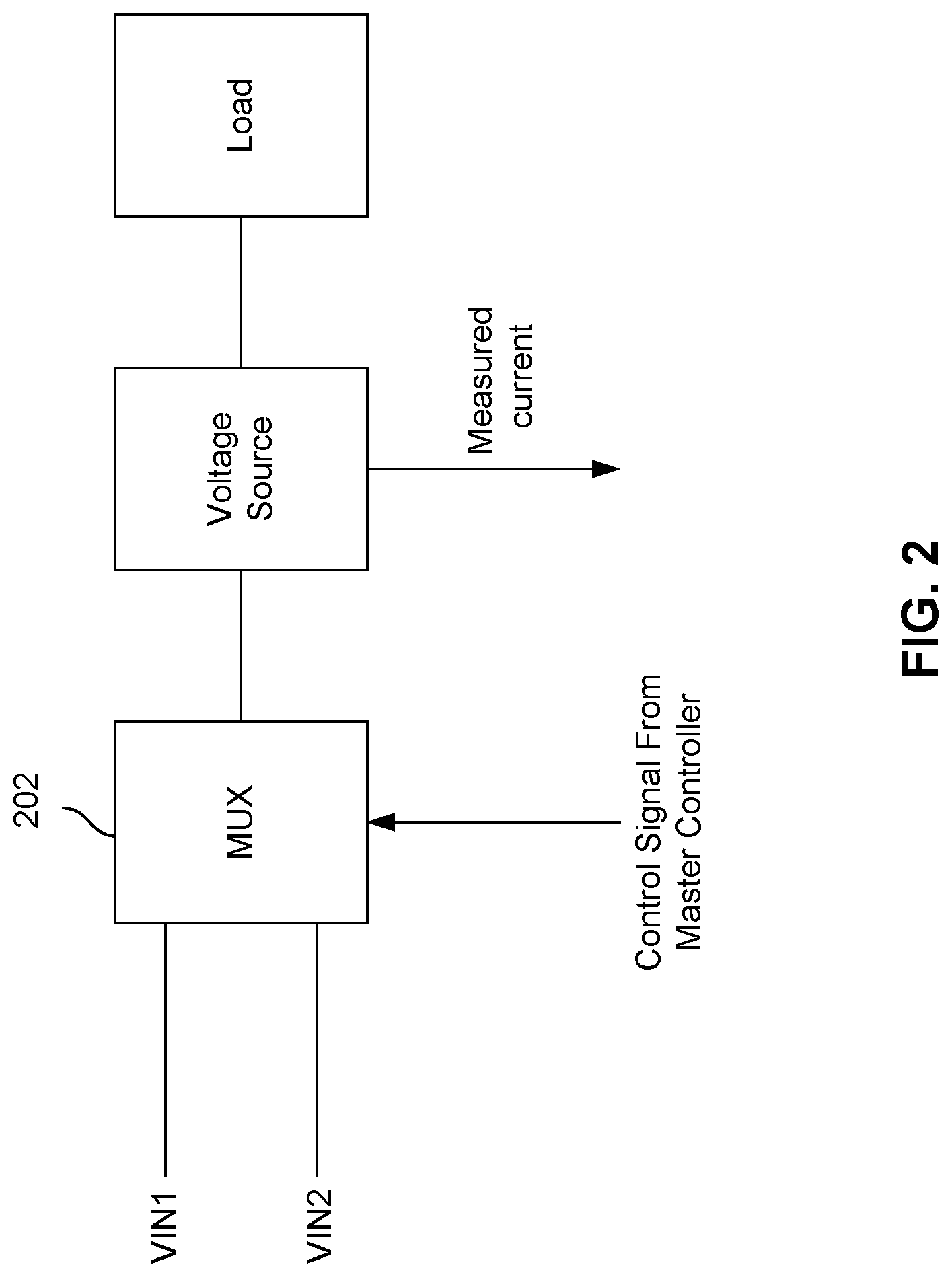
In another example, master controller 104 may send control signals to nanopore array 102 such that different stimuli are applied to different cells or groups of cells. For example, a first stimulus (e.g., a voltage) is applied to a first group of cells and a second stimulus is applied to a second group of cells at time t.sub.1. The first stimulus may be a stimulus corresponding to a particular state of a cell, and the second stimulus may be a stimulus corresponding to a different state of a cell. The stimulus that is applied to the first group of cells may vary over time, as the first group of cells transits from one state to another. FIG. 2 is a block diagram illustrating an embodiment for applying a voltage stimulus to a cell in nanopore array 102. As shown in FIG. 2, control signals from master controller 104 may be used as input to a multiplexer 202 to select one of two voltages that can be applied to a cell in nanopore array 102.
In some embodiments, performance or other data corresponding to each of the cells may be received by master controller 104. By monitoring the performance or other data of the cells, master controller 104 may determine any state transitions of the cells. The state information of the cells may be stored in memory 112 by master controller 104. In addition, if the overall performance of nanopore array 102 falls below a certain threshold, master controller 104 may reset and re-initialize nanopore array 102 such that any processes running on nanopore array 102 may be terminated or restarted again. In some embodiments, nanopore array 102 may also be reused multiple times. For example, nanopore array 102 may be used for analyzing different types of samples during different runs. In another example, nanopore array 102 may be reused for analyzing a single type of samples over multiple runs. In some embodiments, nanopore array 102 may be reused after the contents in nanopore array 102 have been flushed out or rinsed out by master controller 104 and fluidic system 108.
In some embodiments, the cells of nanopore array 102 are individually controllable and individually addressable by temperature controller 106 via signal line 116. Temperature or other data corresponding to a cell may be received by temperature controller 106 via signal line 116. Depending on the state or condition of a particular cell or a group of cells, different temperature stimuli may be applied to the cell or group of cells by temperature controller 106. In some embodiments, temperature controller 106 receives state information of the cells via signal line 120 and applies the appropriate temperature stimuli to the cells in nanopore array 102 at least in part based on the state information. In some embodiments, temperature controller 106 receives control signal via signal line 120 from master controller 104, and then temperature controller 106 applies the appropriate temperature stimuli to the cells in nanopore array 102 based on the received control signal.
In some embodiments, the cells of nanopore array 102 are individually controllable and individually addressable by fluidic system 108. The control and address information is communicated between nanopore array 102 and fluidic system 108 via signal lines 118A. Different contents may be delivered in and out of the individual cells of nanopore array 102 via channels 118B. The contents may be any fluids or reagents that are used for the operations within the cells of nanopore array 102, including saline solution for rinsing, samples to be analyzed by nanopore array 102, lipid bilayer forming reagent, nanopore forming reagent, gas catalyst, and the like. The contents delivered out of nanopore array 102 may be any molecules that are extracted from the samples that have been analyzed by nanopore array 102, and the extracted molecules may be further delivered to a storage device 110 by fluidic system 108. The contents may be in any form, including liquid or gas. Depending on the state or condition of a particular cell or a group of cells, different fluids may be delivered to or from the cell or group of cells by fluidic system 108. In some embodiments, fluidic system 108 receives state information of the cells via signal line 122 and delivers the appropriate fluid to or from the cells in nanopore array 102 at least in part based on the state information. In some embodiments, fluidic system 108 receives control signal via signal line 122 from master controller 104, and then fluidic system 108 delivers the appropriate fluid to or from the cells in nanopore array 102 based on the received control signal. In some embodiments, nanopore array 102 may be reused after the contents in nanopore array 102 have been flushed out or rinsed out by master controller 104 and fluidic system 108.
Nanopore array 102 includes a large array of cells. Each cell includes a nanopore device for analyzing and characterizing molecules. Within a nanopore device, a lipid bilayer is formed, and a nanopore structure is then formed on the lipid bilayer. The nanopore structure has a nanopore that is large enough for enclosing at least a portion of a molecule that is being analyzed or passing at least a portion of the molecule between the two sides of the lipid bilayer. The nanopore device also includes a sample chamber for holding a solution of the analyzed molecules. The solution may be provided over the lipid bilayer for introducing the analyzed molecules for characterization. The nanopore device further includes means for providing electrical stimulus, sensing electrical characteristics, detecting and processing signal of the nanopore device.
FIG. 3 is a diagram illustrating an embodiment of a nanopore device 300 within a cell of nanopore array 102. Nanopore device 300 includes a lipid bilayer 302 formed on a lipid bilayer compatible surface 304 of a conductive solid substrate 306. Lipid bilayer compatible surface 304 may be isolated by lipid bilayer incompatible surfaces 305, and conductive solid substrate 306 may be electrically isolated by insulating materials 307. Lipid bilayer 302 may be surrounded by an amorphous lipid 303 formed on lipid bilayer incompatible surfaces 305.
In some embodiments, lipid bilayer 302 is embedded with a single nanopore structure 308 having a nanopore 310 large enough for passing at least a portion of a molecule 312 being characterized and/or small ions (e.g., Na.sup.+, K.sup.+, Ca.sup.2+, Cl.sup.-) between the two sides of lipid bilayer 302. A layer of water molecules 314 (also referred to as an aqueous film 314) may be adsorbed on lipid bilayer compatible surface 304 and sandwiched between lipid bilayer 302 and lipid bilayer compatible surface 304. Aqueous film 314 adsorbed on the hydrophilic lipid bilayer compatible surface 304 may promote the ordering of lipid molecules and facilitate the formation of lipid bilayer 302 on lipid bilayer compatible surface 304.
A sample chamber 316 may be provided over lipid bilayer 302 for introducing a sample for characterization. The sample may be a solution of molecule 312 that is being characterized. The solution may be an aqueous solution containing electrolytes and buffered to an optimum ion concentration and maintained at an optimum pH to keep nanopore 310 open. In some embodiments, sample chamber 316 receives the sample from fluidic system 108. The sample may also be flushed out of nanopore device 300 by fluidic system 108 after the characterization of the sample has been performed. Sample chamber 316 may also be rinsed with saline solution by fluidic system 108 such that nanopore device 300 may be reused again.
Nanopore device 300 includes a pair of electrodes 318 (including a negative node 318a and a positive node 318b) coupled to a variable voltage source 320 for providing electrical stimulus (e.g., voltage bias) across the lipid bilayer 302 and for sensing the electrical characteristics of the lipid bilayer 302 (e.g., resistance, capacitance, and ionic current flow). The surface of the negative positive electrode 318b is or forms a part of the lipid bilayer compatible surface 304. The conductive solid substrate 306 may be coupled to or forms a part of one of the electrodes 318. Nanopore device 300 may also include an electrical circuit 322 for controlling electrical stimulation and for processing the signal detected. In some embodiments, the variable voltage source 320 is included as a part of the electrical circuit 322. The electrical circuitry 322 may include amplifiers, integrators, noise filters, feedback control logic, and/or various other components. In some embodiments, the electrical circuitry 322 may be an integrated electrical circuitry integrated within a silicon substrate 328 and may be further coupled to a computer processor 324 coupled to a memory 326. For example, computer processor 324 may be a portion of master controller 104, and memory 326 may be memory 112 that is coupled to master controller 104. Master controller 104 may control the various components of nanopore device 300 via electrical circuit 322. Master controller 104 may also receive data collected by nanopore device 300 via electrical circuit 322.
The lipid bilayer compatible surface 304 can be formed from various materials that are suitable for ion transduction and gas formation to facilitate lipid bilayer formation. In some embodiments, conductive or semi-conductive hydrophilic materials as opposed to insulating hydrophilic materials are preferred because they may allow better detection of a change in the lipid bilayer electrical characteristics. Example materials include Ag--AgCl, Ag--Au alloy, Ag--Pt alloy, or doped silicon or other semiconductor materials.
The lipid bilayer incompatible surface 305 can be formed from various materials that are not suitable for lipid bilayer formation and they are typically hydrophobic. In some embodiments, a non-conductive hydrophobic material is preferred, since it electrically insulates the lipid bilayer regions in addition to separating the lipid bilayer regions from each other. Example lipid bilayer incompatible materials include silicon nitride (e.g., Si.sub.3N.sub.4) and Teflon.
In one particular example, nanopore device 300 of FIG. 3 is a alpha hemolysin (.alpha.HL) nanopore device having a single .alpha.HL protein embedded in a diphytanoylphosphatidylcholine (DPhPC) lipid bilayer 302 formed over a lipid bilayer compatible silver-gold alloy surface 304 coated on a copper material 306. The lipid bilayer compatible silver-gold alloy surface 304 is isolated by lipid bilayer incompatible silicon nitride surfaces 305, and the copper material 306 is electrically insulated by silicon nitride materials 307. The copper 306 is coupled to electrical circuitry 322 that is integrated in a silicon substrate 328. A silver-silver chloride electrode placed on-chip or extending down from a cover plate contacts an aqueous solution containing dsDNA molecules.
The .alpha.HL nanopore is an assembly of seven individual peptides. The entrance or vestible of the .alpha.HL nanopore is approximately 26 .ANG. in diameter, which is wide enough to accommodate a portion of a dsDNA molecule. From the vestible, the .alpha.HL nanopore first widens and then narrows to a barrel having a diameter of approximately 15 .ANG., which is wide enough to allow a single ssDNA molecule to pass through but not wide enough to allow a dsDNA molecule to pass through. At a given time, approximately 1-20 DNA bases can occupy the barrel of the .alpha.HL nanopore.
In addition to DPhPC, the lipid bilayer of the nanopore device can be assembled from various other suitable amphiphilic materials, selected based on various considerations, such as the type of nanopore used, the type of molecule being characterized, and various physical, chemical and/or electrical characteristics of the lipid bilayer formed, such as stability and permeability, resistance, and capacitance of the lipid bilayer formed. Example amphiphilic materials include various phospholipids such as palmitoyl-oleoyl-phosphatidyl-choline (POPC) and dioleoyl-phosphatidyl-methylester (DOPME), diphytanoylphosphatidylcholine (DPhPC) dipalmitoylphosphatidylcholine (DPPC), phosphatidylcholine, phosphatidylethanolamine, phosphatidylserine, phosphatidic acid, phosphatidylinositol, phosphatidylglycerol, and sphingomyelin.
In addition to the .alpha.HL nanopore shown above, the nanopore may be one of various other types of nanopores; examples include .gamma.-hemolysin, leukocidin, melittin, and various other naturally occurring, modified natural, and synthetic nanopores. A suitable nanopore may be selected based on various characteristics of the analyte molecule, such as the size of the analyte molecule in relation to the pore size of the nanopore. For example, the .alpha.HL nanopore is a nanopore that has a restrictive pore size of approximately 15 .ANG.. It is suitable for analyzing DNA molecules since it allows a single strand DNA (ssDNA) to pass through while restricting a double strand DNA (dsDNA).
FIGS. 4A-4C illustrate three different states of nanopore device 300. FIG. 4A is a diagram illustrating that nanopore device 300 is in a state in which a lipid bilayer has not yet been formed. FIG. 4B is a diagram illustrating that nanopore device 300 is in a state in which a lipid bilayer 302 has been formed. FIG. 4C is a diagram illustrating that nanopore device 300 is in a state in which a nanopore structure 308 with a nanopore 310 has been inserted into lipid bilayer 302.
FIG. 5 is a flow diagram illustrating an embodiment of a process 500 for analyzing molecules using nanopore devices. In some embodiments, process 500 is a process that is performed by system 100 of FIG. 1.
At 502, various functionalities of system 100 are verified. In some embodiments, master controller 104 may send test signals to the modules of system 100, including nanopore array 102, temperature controller 106, and fluidic system 108. In response, each module may perform verification steps at the module. For example, nanopore array 102 may measure the current flowing in a particular nanopore device. After the verification steps are performed at the modules, each of the modules may send a response back to master controller 104 for verification purposes. Depending on the responses received from the various modules, master controller 104 may determine whether further verifications are needed. In some embodiments, the verification results may be stored in a log file. In some embodiments, if master controller 104 has detected any errors, then an alarm may be triggered or process 500 may be terminated.
In some embodiments, verification of the different modules may be performed at different levels, and the levels may be configurable. For example, master controller 104 may verify the functionalities of nanopore array 102 at the printed circuit board level or at the semiconductor chip level. In some embodiments, master controller 104 may verify the functionalities of a group of cells. If the number of cells within the group that are functioning properly falls below a certain threshold, then master controller 104 may determine that the group of cells is mal-functioning and that the group of cells should be disabled.
At 504, lipid bilayers are assembled. In some embodiments, master controller 104 may cause fluidic system 108 to deliver a lipid forming reagent to the cells of nanopore array 102. The lipid forming reagent is then deposited on lipid bilayer compatible surface 304 within a cell. As discussed above, the lipid bilayer may be formed using different materials, including different amphiphilic materials. Depending on the type of lipid bilayers to be formed, master controller 104 may cause different stimuli (e.g., electrical, temperature, chemical, or gas) to be applied to the cells to facilitate the assembling of the lipid bilayers.
At 506, it is determined whether the lipid bilayers are properly formed. Depending on the type of lipid bilayers to be formed, different physical or electrical property measurements (e.g., resistance, current, or capacitance measurements) may be made at the cells and then sent to master controller 104 via signal lines 114 for determining whether lipid bilayers are properly assembled. In some embodiments, steps 504 and 506 are repeated until master controller 104 has determined that lipid bilayers have been properly assembled in a minimum number of cells in nanopore array 102. In some embodiments, if the number of cells with lipid bilayers properly assembled falls below a certain threshold after a fixed period of time, master controller 104 may terminate process 500. In addition, an alarm may be triggered or an error message may be written to the log file. In some embodiments, if the number of cells with lipid bilayers properly assembled is above a certain threshold, master controller 104 may cause system 100 to proceed to step 508.
At 508, nanopore structures with nanopores are inserted. In some embodiments, master controller 104 may cause fluidic system 108 to deliver a nanopore forming reagent (e.g., a solution containing .alpha.-hemolysin) to the cells of nanopore array 102. Master controller 104 may cause different stimuli (e.g., electrical, temperature, chemical, or gas) to be applied to the cells to facilitate the insertion of the nanopore structures into the lipid bilayers.
At 510, it is determined whether the nanopore structures are properly formed. Depending on the type of nanopores to be formed, different measurements (e.g., resistance, current, or capacitance measurements) may be made at the cells and then sent to master controller 104 via signal lines 114 for determining whether nanopores are properly inserted. In some embodiments, steps 508 and 510 are repeated until master controller 104 has determined that nanopores have been properly inserted in a minimum number of cells in nanopore array 102. In some embodiments, if the number of cells with nanopores properly inserted falls below a certain threshold after a fixed period of time, master controller 104 may terminate process 500. In addition, an alarm may be triggered or an error message may be written to the log file. In some embodiments, if the number of cells with nanopores properly inserted is above a certain threshold, master controller 104 may cause system 100 to proceed to step 512.
At 512, samples are analyzed using the nanopores in nanopore array 102. In some embodiments, master controller 104 may cause fluidic system 108 to deliver samples to the sample chambers 316 in nanopore array 102. Depending on different factors, including the type of samples that are being analyzed and the type of nanopores formed, master controller 104 may cause different stimuli (e.g., electrical, temperature, chemical, or gas) to be applied to the cells to facilitate the manipulating, detecting, correlating, characterizing, analyzing and/or sequencing of molecules in the nanopores. Different measurements (e.g., resistance, current, or capacitance measurements) may be made at the cells and then sent to master controller 104 via signal lines 114. Master controller 104 may use the received measurements to detect, correlate, determine, characterize, sequence and/or discriminate various structural and chemical features of a molecule as the molecule stays inside the nanopore, traverses through the nanopore, or interacts with the nanopore.
At 514, nanopore array is reset and re-initialized for repeated uses. In some embodiments, nanopore array 102 may be reused multiple times. For example, nanopore array 102 may be used for analyzing different types of samples during different runs. In another example, nanopore array 102 may be reused for analyzing a single type of samples over multiple runs. New nanopores may be reformed in nanopore array 102 such that nanopore array 102 may be reused. New nanopores may be reformed in nanopore array 102 after the contents (e.g., lipid bilayers with nanopores inserted, lipid bilayers without nanopores inserted, and samples) in nanopore array 102 have been flushed out or rinsed out (e.g., using saline solution) by master controller 104 and fluidic system 108.
In some embodiments, master controller 104 may detect and determine whether there are any molecules or other contents of interest remaining in the cells of nanopore array 102. Master controller 104 and fluidic system 108 may selectively rinse out the contents (e.g., lipid bilayers) within cells in which no molecules or other contents of interest are found. The molecules or other contents of interest in the remaining cells may be retrieved. In one example, the molecules may be retrieved manually. In another example, master controller 104 and fluidic system 108 may deliver the molecules or other contents of interest to storage device 110 before the remaining contents are rinsed out. After 514, nanopore array 102 is ready for repeated uses again, and process 500 may be restarted at 502. In some embodiments, step 514 is performed before a nanopore array 102 is used for the first time. For example, nanopore array 102 is rinsed with saline solution before the functionalities of system 100 is checked at 502.
Although the foregoing embodiments have been described in some detail for purposes of clarity of understanding, the invention is not limited to the details provided. There are many alternative ways of implementing the invention. The disclosed embodiments are illustrative and not restrictive.
* * * * *
D00001
D00002
D00003
D00004
D00005
D00006
D00007
XML
uspto.report is an independent third-party trademark research tool that is not affiliated, endorsed, or sponsored by the United States Patent and Trademark Office (USPTO) or any other governmental organization. The information provided by uspto.report is based on publicly available data at the time of writing and is intended for informational purposes only.
While we strive to provide accurate and up-to-date information, we do not guarantee the accuracy, completeness, reliability, or suitability of the information displayed on this site. The use of this site is at your own risk. Any reliance you place on such information is therefore strictly at your own risk.
All official trademark data, including owner information, should be verified by visiting the official USPTO website at www.uspto.gov. This site is not intended to replace professional legal advice and should not be used as a substitute for consulting with a legal professional who is knowledgeable about trademark law.