Digital analyte analysis
Link , et al. July 16, 2
U.S. patent number 10,351,905 [Application Number 13/866,111] was granted by the patent office on 2019-07-16 for digital analyte analysis. This patent grant is currently assigned to Bio-Rad Laboratories, Inc.. The grantee listed for this patent is Bio-Rad Laboratories, Inc.. Invention is credited to Darren Roy Link, Benjamin J. Miller.


View All Diagrams
| United States Patent | 10,351,905 |
| Link , et al. | July 16, 2019 |
Digital analyte analysis
Abstract
The invention generally relates to detecting target molecules.
| Inventors: | Link; Darren Roy (Lexington, MA), Miller; Benjamin J. (Littleton, MA) | ||||||||||
|---|---|---|---|---|---|---|---|---|---|---|---|
| Applicant: |
|
||||||||||
| Assignee: | Bio-Rad Laboratories, Inc.
(Hercules, CA) |
||||||||||
| Family ID: | 50070172 | ||||||||||
| Appl. No.: | 13/866,111 | ||||||||||
| Filed: | April 19, 2013 |
Prior Publication Data
| Document Identifier | Publication Date | |
|---|---|---|
| US 20140045712 A1 | Feb 13, 2014 | |
Related U.S. Patent Documents
| Application Number | Filing Date | Patent Number | Issue Date | ||
|---|---|---|---|---|---|
| 13026120 | Feb 11, 2011 | 9074242 | |||
| 13460762 | Apr 30, 2012 | ||||
| 13026120 | |||||
| 61388937 | Oct 1, 2010 | ||||
| 61347158 | May 21, 2010 | ||||
| 61331490 | May 5, 2010 | ||||
| 61304163 | Feb 12, 2010 | ||||
| 61636217 | Apr 20, 2012 | ||||
| Current U.S. Class: | 1/1 |
| Current CPC Class: | C12Q 1/686 (20130101); C12Q 1/686 (20130101); C12Q 2565/629 (20130101) |
| Current International Class: | C12Q 1/68 (20180101); C12Q 1/686 (20180101) |
| Field of Search: | ;435/6.12,91.2 |
References Cited [Referenced By]
U.S. Patent Documents
| 2097692 | November 1937 | Fiegel |
| 2164172 | June 1939 | Dalton |
| 2636855 | April 1953 | Schwarz |
| 2656508 | October 1953 | Coulter |
| 2692800 | October 1954 | Nichols et al. |
| 2797149 | June 1957 | Skeggs |
| 2879141 | March 1959 | Skeggs |
| 2971700 | February 1961 | Peeps |
| 3479141 | November 1969 | Smythe et al. |
| 3608821 | September 1971 | Simm et al. |
| 3698635 | October 1972 | Sickles |
| 3784471 | January 1974 | Kaiser |
| 3816331 | June 1974 | Brown, Jr. et al. |
| 3930061 | December 1975 | Scharfenberger |
| 3960187 | June 1976 | Stock et al. |
| 3980541 | September 1976 | Aine |
| 3982541 | September 1976 | L'Esperance, Jr. |
| 4014469 | March 1977 | Sato |
| 4022575 | May 1977 | Hansen et al. |
| 4034966 | July 1977 | Suh et al. |
| 4059552 | November 1977 | Zweigle et al. |
| 4091042 | May 1978 | Alexanderson et al. |
| 4117550 | September 1978 | Folland et al. |
| 4130394 | December 1978 | Negersmith |
| 4210809 | July 1980 | Pelavin |
| 4253846 | March 1981 | Smythe et al. |
| 4266721 | May 1981 | Sickles |
| 4279345 | July 1981 | Allred |
| 4297345 | October 1981 | Howarth |
| 4315754 | February 1982 | Ruzicka et al. |
| 4378957 | April 1983 | Malkin et al. |
| 4383767 | May 1983 | Jido |
| 4439980 | April 1984 | Biblarz et al. |
| 4508265 | April 1985 | Jido |
| 4533634 | August 1985 | Maldonado et al. |
| 4585209 | April 1986 | Aine et al. |
| 4618476 | October 1986 | Columbus |
| 4675285 | June 1987 | Clark et al. |
| 4676274 | June 1987 | Brown |
| 4683195 | July 1987 | Mullis et al. |
| 4683202 | July 1987 | Mullis |
| 4739044 | April 1988 | Stabinsky |
| 4757141 | July 1988 | Fung et al. |
| 4767515 | August 1988 | Scott et al. |
| 4767929 | August 1988 | Valentine |
| 4779805 | October 1988 | Jackson et al. |
| 4795330 | January 1989 | Noakes et al. |
| 4801086 | January 1989 | Noakes |
| 4801529 | January 1989 | Perlman |
| 4829996 | May 1989 | Noakes et al. |
| 4853336 | August 1989 | Saros et al. |
| 4856363 | August 1989 | LaRocca et al. |
| 4859363 | August 1989 | Davis et al. |
| 4865444 | September 1989 | Green et al. |
| 4883750 | November 1989 | Whiteley et al. |
| 4908112 | March 1990 | Pace |
| 4931225 | June 1990 | Cheng |
| 4941959 | July 1990 | Scott |
| 4962885 | October 1990 | Coffee |
| 4963498 | October 1990 | Hillman et al. |
| 4981580 | January 1991 | Auer |
| 4996004 | February 1991 | Bucheler et al. |
| 5091652 | February 1992 | Mathies et al. |
| 5096615 | March 1992 | Prescott et al. |
| 5104813 | April 1992 | Besemer et al. |
| 5122360 | June 1992 | Harris et al. |
| 5149625 | September 1992 | Church et al. |
| 5180662 | January 1993 | Sitkovsky |
| 5185099 | February 1993 | Delpuech et al. |
| 5188290 | February 1993 | Gebauer et al. |
| 5188291 | February 1993 | Cross |
| 5192659 | March 1993 | Simons |
| 5204112 | April 1993 | Hope et al. |
| 5207973 | May 1993 | Harris et al. |
| 5241159 | August 1993 | Chatteriee et al. |
| 5260466 | November 1993 | McGibbon |
| 5262027 | November 1993 | Scott |
| 5270163 | December 1993 | Gold et al. |
| 5296375 | March 1994 | Kricka et al. |
| 5304487 | April 1994 | Wilding et al. |
| 5310653 | May 1994 | Hanausek-Walaszek et al. |
| 5313009 | May 1994 | Guenkel et al. |
| 5333675 | August 1994 | Mullis et al. |
| 5344594 | September 1994 | Sheridon |
| 5376252 | December 1994 | Ekstrom et al. |
| 5378957 | January 1995 | Kelly |
| 5397605 | March 1995 | Barbieri et al. |
| 5399461 | March 1995 | Van et al. |
| 5399491 | March 1995 | Kacian et al. |
| 5403617 | April 1995 | Haaland |
| 5413924 | May 1995 | Kosak et al. |
| 5417235 | May 1995 | Wise et al. |
| 5427946 | June 1995 | Kricka et al. |
| 5445934 | August 1995 | Fodor et al. |
| 5452878 | September 1995 | Gravesen et al. |
| 5452955 | September 1995 | Lundstrom |
| 5454472 | October 1995 | Benecke et al. |
| 5460945 | October 1995 | Springer et al. |
| 5468613 | November 1995 | Erlich et al. |
| 5475096 | December 1995 | Gold et al. |
| 5475610 | December 1995 | Atwood et al. |
| 5480614 | January 1996 | Kamahori |
| 5486335 | January 1996 | Wilding et al. |
| 5498392 | March 1996 | Wilding et al. |
| 5500415 | March 1996 | Dollat et al. |
| 5503851 | April 1996 | Mank et al. |
| 5512131 | April 1996 | Kumar et al. |
| 5516635 | May 1996 | Ekins et al. |
| 5518709 | May 1996 | Sutton et al. |
| 5523162 | June 1996 | Franz et al. |
| 5587128 | December 1996 | Wilding et al. |
| 5589136 | December 1996 | Northrup et al. |
| 5602756 | February 1997 | Atwood et al. |
| 5604097 | February 1997 | Brenner |
| 5610016 | March 1997 | Sato et al. |
| 5612188 | March 1997 | Shuler et al. |
| 5616478 | April 1997 | Chetverin et al. |
| 5617997 | April 1997 | Kobayashi et al. |
| 5635358 | June 1997 | Wilding et al. |
| 5636400 | June 1997 | Young |
| 5641658 | June 1997 | Adams et al. |
| 5643729 | July 1997 | Taniguchi et al. |
| 5655517 | August 1997 | Coffee |
| 5656155 | August 1997 | Norcross et al. |
| 5656493 | August 1997 | Mullis et al. |
| 5661222 | August 1997 | Hare |
| 5662874 | September 1997 | David |
| 5670325 | September 1997 | Lapidus et al. |
| 5681600 | October 1997 | Antinone et al. |
| 5695934 | December 1997 | Brenner |
| 5726026 | March 1998 | Wilding et al. |
| 5726404 | March 1998 | Brody |
| 5733526 | March 1998 | Trevino et al. |
| 5739036 | April 1998 | Parris |
| 5744366 | April 1998 | Kricka et al. |
| 5750988 | May 1998 | Apffel et al. |
| 5762775 | June 1998 | DePaoli |
| 5779868 | July 1998 | Parce et al. |
| 5783431 | July 1998 | Peterson et al. |
| 5840506 | November 1998 | Giordano |
| 5846719 | December 1998 | Brenner et al. |
| 5849491 | December 1998 | Radomski et al. |
| 5851769 | December 1998 | Gray et al. |
| 5858187 | January 1999 | Ramsey et al. |
| 5858655 | January 1999 | Arnold |
| 5858670 | January 1999 | Lam et al. |
| 5863722 | January 1999 | Brenner |
| 5868322 | February 1999 | Loucks |
| 5872010 | February 1999 | Karger et al. |
| 5876771 | March 1999 | Sizer et al. |
| 5880071 | March 1999 | Parce et al. |
| 5882680 | March 1999 | Suzuki et al. |
| 5882856 | March 1999 | Shuber |
| 5884846 | March 1999 | Tan |
| 5887755 | March 1999 | Hood, III |
| 5888746 | March 1999 | Tabiti et al. |
| 5888778 | March 1999 | Shuber |
| 5904933 | May 1999 | Riess et al. |
| 5921678 | July 1999 | Desai et al. |
| 5927852 | July 1999 | Serafin |
| 5928870 | July 1999 | Lapidus et al. |
| 5932100 | August 1999 | Yager et al. |
| 5935331 | August 1999 | Naka et al. |
| 5942056 | August 1999 | Singh |
| 5942443 | August 1999 | Parce et al. |
| 5958203 | September 1999 | Parce et al. |
| 5972187 | October 1999 | Parce et al. |
| 5980936 | November 1999 | Krafft et al. |
| 5989815 | November 1999 | Skolnick et al. |
| 5989892 | November 1999 | Nishimaki et al. |
| 5995341 | November 1999 | Tanaka et al. |
| 5997636 | December 1999 | Gamarnik et al. |
| 6008003 | December 1999 | Haak-Frendscho et al. |
| 6023540 | February 2000 | Walt et al. |
| 6028066 | February 2000 | Unger |
| 6042709 | March 2000 | Parce et al. |
| 6045755 | April 2000 | Lebl et al. |
| 6046056 | April 2000 | Parce et al. |
| 6048551 | April 2000 | Hilfmger et al. |
| 6048690 | April 2000 | Heller et al. |
| 6068199 | May 2000 | Coffee |
| 6074879 | June 2000 | Zelmanovic et al. |
| 6080295 | June 2000 | Parce et al. |
| 6086740 | July 2000 | Kennedy |
| 6096495 | August 2000 | Kasai et al. |
| 6103537 | August 2000 | Ullman et al. |
| 6105571 | August 2000 | Coffee |
| 6105877 | August 2000 | Coffee |
| 6107059 | August 2000 | Hart |
| 6116516 | September 2000 | Ganan-Calvo |
| 6118849 | September 2000 | Tanimori et al. |
| 6119953 | September 2000 | Ganan-Calvo et al. |
| 6120666 | September 2000 | Jacobson et al. |
| 6124388 | September 2000 | Takai et al. |
| 6124439 | September 2000 | Friedman et al. |
| 6130052 | October 2000 | Van Baren et al. |
| 6130098 | October 2000 | Handique et al. |
| 6137214 | October 2000 | Raina |
| 6138077 | October 2000 | Brenner |
| 6139303 | October 2000 | Reed et al. |
| 6140053 | October 2000 | Koster |
| 6143496 | November 2000 | Brown et al. |
| 6146828 | November 2000 | Lapidus et al. |
| 6149789 | November 2000 | Benecke et al. |
| 6150180 | November 2000 | Parce et al. |
| 6150516 | November 2000 | Brenner et al. |
| 6165778 | December 2000 | Kedar |
| 6171796 | January 2001 | An et al. |
| 6171850 | January 2001 | Nagle et al. |
| 6172214 | January 2001 | Brenner |
| 6172218 | January 2001 | Brenner |
| 6174160 | January 2001 | Lee et al. |
| 6174469 | January 2001 | Ganan-Calvo |
| 6177479 | January 2001 | Nakajima |
| 6180372 | January 2001 | Franzen |
| 6184012 | February 2001 | Neri et al. |
| 6187214 | February 2001 | Ganan-Calvo |
| 6189803 | February 2001 | Ganan-Calvo |
| 6196525 | March 2001 | Ganan-Calvo |
| 6197335 | March 2001 | Sherman |
| 6197835 | March 2001 | Ganan-Calvo |
| 6203993 | March 2001 | Shuber et al. |
| 6207372 | March 2001 | Shuber |
| 6207397 | March 2001 | Lynch et al. |
| 6210396 | April 2001 | MacDonald et al. |
| 6210891 | April 2001 | Nyren et al. |
| 6210896 | April 2001 | Chan |
| 6214558 | April 2001 | Shuber et al. |
| 6221654 | April 2001 | Quake et al. |
| 6227466 | May 2001 | Hartman et al. |
| 6234402 | May 2001 | Ganan-Calvo |
| 6235383 | May 2001 | Hong et al. |
| 6235475 | May 2001 | Brenner et al. |
| 6241159 | June 2001 | Ganan-Calvo et al. |
| 6243373 | June 2001 | Turock |
| 6248378 | June 2001 | Ganan-Calvo |
| 6251661 | June 2001 | Urabe et al. |
| 6252129 | June 2001 | Coffee |
| 6258568 | July 2001 | Nyren |
| 6258858 | July 2001 | Nakajima et al. |
| 6263222 | July 2001 | Diab et al. |
| 6266459 | July 2001 | Walt et al. |
| 6267353 | July 2001 | Friedline et al. |
| 6267858 | July 2001 | Parce et al. |
| 6268152 | July 2001 | Fodor et al. |
| 6268165 | July 2001 | O'Brien |
| 6268222 | July 2001 | Chandler et al. |
| 6274320 | August 2001 | Rothberg et al. |
| 6274337 | August 2001 | Parce et al. |
| 6280948 | August 2001 | Guilfoyle et al. |
| 6292756 | September 2001 | Lievois |
| 6294344 | September 2001 | O'Brien |
| 6296020 | October 2001 | McNeely et al. |
| 6296673 | October 2001 | Santarsiero et al. |
| 6299145 | October 2001 | Ganan-Calvo |
| 6301055 | October 2001 | Legrand et al. |
| 6306659 | October 2001 | Parce et al. |
| 6310354 | October 2001 | Hanninen et al. |
| 6310653 | October 2001 | Malcolm, Jr. et al. |
| 6316208 | November 2001 | Roberts et al. |
| 6316213 | November 2001 | O'Brien |
| 6318640 | November 2001 | Coffee |
| 6326145 | December 2001 | Whitcombe et al. |
| 6336463 | January 2002 | Ohta |
| 6344325 | February 2002 | Quake et al. |
| 6352828 | March 2002 | Brenner |
| 6355193 | March 2002 | Stott |
| 6355198 | March 2002 | Kim et al. |
| 6357670 | March 2002 | Ganan-Calvo |
| 6386463 | May 2002 | Ganan-Calvo |
| 6391559 | May 2002 | Brown et al. |
| 6394429 | May 2002 | Ganan-Calvo |
| 6399339 | June 2002 | Wolberg et al. |
| 6399389 | June 2002 | Parce et al. |
| 6403373 | June 2002 | Scanlan et al. |
| 6405936 | June 2002 | Ganan-Calvo |
| 6408878 | June 2002 | Unger et al. |
| 6409832 | June 2002 | Weigl et al. |
| 6429025 | August 2002 | Parce et al. |
| 6429148 | August 2002 | Chu et al. |
| 6432143 | August 2002 | Kubiak et al. |
| 6432148 | August 2002 | Ganan-Calvo |
| 6432630 | August 2002 | Blankenstein |
| 6439103 | August 2002 | Miller |
| 6440706 | August 2002 | Vogelstein et al. |
| 6440760 | August 2002 | Cho et al. |
| 6450139 | September 2002 | Watanabe |
| 6450189 | September 2002 | Ganan-Calvo |
| 6454193 | September 2002 | Busick et al. |
| 6464336 | October 2002 | Sharma |
| 6464886 | October 2002 | Ganan-Calvo |
| 6475441 | November 2002 | Parce et al. |
| 6481648 | November 2002 | Zimmermann |
| 6489103 | December 2002 | Griffiths et al. |
| 6503933 | January 2003 | Moloney et al. |
| 6506609 | January 2003 | Wada et al. |
| 6508988 | January 2003 | Van Dam et al. |
| 6520425 | February 2003 | Reneker |
| 6524456 | February 2003 | Ramsey et al. |
| 6540395 | April 2003 | Muhlbauer et al. |
| 6540895 | April 2003 | Spence et al. |
| 6551836 | April 2003 | Chow et al. |
| 6553944 | April 2003 | Allen et al. |
| 6553960 | April 2003 | Yoshikawa et al. |
| 6554202 | April 2003 | Ganan-Calvo |
| 6557334 | May 2003 | Jager |
| 6557834 | May 2003 | Ganan-Calvo |
| 6558944 | May 2003 | Parce et al. |
| 6558960 | May 2003 | Parce et al. |
| 6560030 | May 2003 | Legrand et al. |
| 6565010 | May 2003 | Anderson et al. |
| 6569631 | May 2003 | Pantoliano et al. |
| 6576420 | June 2003 | Carson et al. |
| 6591852 | July 2003 | McNeely et al. |
| 6592321 | July 2003 | Bonker et al. |
| 6592821 | July 2003 | Wada et al. |
| 6601613 | August 2003 | McNeely et al. |
| 6608726 | August 2003 | Legrand et al. |
| 6610499 | August 2003 | Fulwyler et al. |
| 6614598 | September 2003 | Quake et al. |
| 6627603 | September 2003 | Bibette et al. |
| 6630006 | October 2003 | Santarsiero et al. |
| 6630353 | October 2003 | Parce et al. |
| 6632619 | October 2003 | Harrison et al. |
| 6637463 | October 2003 | Lei et al. |
| 6638749 | October 2003 | Beckman et al. |
| 6645432 | November 2003 | Anderson et al. |
| 6646253 | November 2003 | Rohwer et al. |
| 6653626 | November 2003 | Fischer et al. |
| 6656267 | December 2003 | Newman |
| 6659370 | December 2003 | Inoue |
| 6660252 | December 2003 | Matathia et al. |
| 6670142 | December 2003 | Lau et al. |
| 6679441 | January 2004 | Borra et al. |
| 6680178 | January 2004 | Harris et al. |
| 6682890 | January 2004 | Mack et al. |
| 6717136 | April 2004 | Andersson et al. |
| 6729561 | May 2004 | Hirae et al. |
| 6738502 | May 2004 | Coleman et al. |
| 6739036 | May 2004 | Koike et al. |
| 6744046 | June 2004 | Valaskovic et al. |
| 6752922 | June 2004 | Huang et al. |
| 6753147 | June 2004 | Vogelstein et al. |
| 6766817 | July 2004 | da Silva |
| 6767194 | July 2004 | Jeon et al. |
| 6767704 | July 2004 | Waldman et al. |
| 6790328 | September 2004 | Jacobson et al. |
| 6793753 | September 2004 | Unger et al. |
| 6797056 | September 2004 | David |
| 6800849 | October 2004 | Staats |
| 6806058 | October 2004 | Jesperson et al. |
| 6808382 | October 2004 | Lanfranchi |
| 6808882 | October 2004 | Griffiths et al. |
| 6814980 | November 2004 | Levy et al. |
| 6818395 | November 2004 | Quake et al. |
| 6832787 | December 2004 | Renzi |
| 6833242 | December 2004 | Quake et al. |
| 6841350 | January 2005 | Ogden et al. |
| 6872250 | March 2005 | David et al. |
| 6890487 | May 2005 | Sklar et al. |
| 6897018 | May 2005 | Yuan et al. |
| 6905844 | June 2005 | Kim |
| 6918404 | July 2005 | Dias da Silva |
| 6926313 | August 2005 | Renzi |
| 6935768 | August 2005 | Lowe et al. |
| 6936417 | August 2005 | Omtoft |
| 6942978 | September 2005 | O'Brien |
| 6949342 | September 2005 | Golub et al. |
| 6960437 | November 2005 | Enzelberger et al. |
| 6974667 | December 2005 | Horne et al. |
| 6998232 | February 2006 | Feinstein et al. |
| 7022472 | April 2006 | Robbins et al. |
| 7041481 | May 2006 | Anderson et al. |
| 7049072 | May 2006 | Seshi |
| 7056674 | June 2006 | Baker et al. |
| 7057026 | June 2006 | Barnes et al. |
| 7066586 | June 2006 | da Silva |
| 7068874 | June 2006 | Wang et al. |
| 7078180 | July 2006 | Genetta |
| 7081192 | July 2006 | Wang et al. |
| 7081340 | July 2006 | Baker et al. |
| 7090983 | August 2006 | Muramatsu et al. |
| 7115230 | October 2006 | Sundararajan |
| 7118910 | October 2006 | Unger et al. |
| 7129091 | October 2006 | Ismagilov et al. |
| 7138233 | November 2006 | Griffiths et al. |
| 7153700 | December 2006 | Pardee et al. |
| 7156917 | January 2007 | Moriyama et al. |
| 7163801 | January 2007 | Reed |
| 7169560 | January 2007 | Lapidus et al. |
| 7171311 | January 2007 | Dai et al. |
| 7198899 | April 2007 | Schleyer et al. |
| 7204431 | April 2007 | Li et al. |
| 7229770 | June 2007 | Price et al. |
| 7252943 | August 2007 | Griffiths et al. |
| 7267938 | September 2007 | Anderson et al. |
| 7268167 | September 2007 | Higuchi et al. |
| 7282337 | October 2007 | Harris |
| 7291462 | November 2007 | O'Brien et al. |
| 7294503 | November 2007 | Quake et al. |
| 7300765 | November 2007 | Patel |
| 7308364 | December 2007 | Shaughnessy et al. |
| 7314721 | January 2008 | Gure et al. |
| 7316906 | January 2008 | Chiorazzi et al. |
| 7326529 | February 2008 | Ali et al. |
| 7332280 | February 2008 | Levy et al. |
| 7332590 | February 2008 | Nacht et al. |
| 7341211 | March 2008 | Ganan Calvo et al. |
| 7348142 | March 2008 | Wang |
| 7358231 | April 2008 | McCaffey et al. |
| 7361474 | April 2008 | Siegler |
| 7364862 | April 2008 | Ali et al. |
| 7368255 | May 2008 | Bae et al. |
| 7378233 | May 2008 | Sidransky et al. |
| 7378280 | May 2008 | Quake et al. |
| 7390463 | June 2008 | He et al. |
| 7393634 | July 2008 | Ahuja et al. |
| 7393665 | July 2008 | Brenner |
| 7416851 | August 2008 | Davi et al. |
| 7429467 | September 2008 | Holliger et al. |
| 7432064 | October 2008 | Salceda et al. |
| 7442507 | October 2008 | Polsky et al. |
| 7449303 | November 2008 | Coignet |
| 7468271 | December 2008 | Golovchenko et al. |
| 7473530 | January 2009 | Huttemann |
| 7473531 | January 2009 | Domon et al. |
| 7476506 | January 2009 | Schleyer et al. |
| 7479370 | January 2009 | Coignet |
| 7479371 | January 2009 | Ando et al. |
| 7479376 | January 2009 | Waldman et al. |
| 7482129 | January 2009 | Soyupak et al. |
| 7501244 | March 2009 | Reinhard et al. |
| 7504214 | March 2009 | Erlander et al. |
| 7507532 | March 2009 | Chang et al. |
| 7507541 | March 2009 | Raitano et al. |
| 7510707 | March 2009 | Platica et al. |
| 7510842 | March 2009 | Podust et al. |
| 7514209 | April 2009 | Dai et al. |
| 7514210 | April 2009 | Holliger et al. |
| 7524633 | April 2009 | Sidransky |
| 7527933 | May 2009 | Sahin et al. |
| 7537897 | May 2009 | Brenner et al. |
| 7541383 | June 2009 | Fu et al. |
| 7544473 | June 2009 | Brenner |
| 7556776 | July 2009 | Fraden et al. |
| 7582446 | September 2009 | Griffiths et al. |
| 7622081 | November 2009 | Chou et al. |
| 7632562 | December 2009 | Nair et al. |
| 7635562 | December 2009 | Harris et al. |
| 7638276 | December 2009 | Griffiths et al. |
| 7655435 | February 2010 | Holliger et al. |
| 7655470 | February 2010 | Ismagilov et al. |
| 7666593 | February 2010 | Lapidus |
| 7691576 | April 2010 | Holliger et al. |
| 7698287 | April 2010 | Becker et al. |
| 7708949 | May 2010 | Stone et al. |
| 7718578 | May 2010 | Griffiths et al. |
| 7736890 | June 2010 | Sia et al. |
| 7741130 | June 2010 | Lee, Jr. et al. |
| RE41780 | September 2010 | Anderson et al. |
| 7814175 | October 2010 | Chang et al. |
| 7824889 | November 2010 | Vogelstein et al. |
| 7888017 | February 2011 | Quake et al. |
| 7897044 | March 2011 | Hoyos et al. |
| 7897341 | March 2011 | Griffiths et al. |
| 7901939 | March 2011 | Ismagliov et al. |
| 7915015 | March 2011 | Vogelstein et al. |
| 7968287 | June 2011 | Griffiths et al. |
| 7990525 | August 2011 | Kanda |
| 8012382 | September 2011 | Kim et al. |
| 8067159 | November 2011 | Brown et al. |
| 8153402 | April 2012 | Holliger et al. |
| 8257925 | September 2012 | Brown et al. |
| 8278071 | October 2012 | Brown et al. |
| 8278711 | October 2012 | Rao et al. |
| 8318434 | November 2012 | Cuppens |
| 8337778 | December 2012 | Stone et al. |
| 8436993 | May 2013 | Kaduchak et al. |
| 8528589 | September 2013 | Miller et al. |
| 8535889 | September 2013 | Larson et al. |
| 8592221 | November 2013 | Fraden et al. |
| 8673595 | March 2014 | Nakamura et al. |
| 8765485 | July 2014 | Link et al. |
| 8772046 | July 2014 | Fraden et al. |
| 2001/0010338 | August 2001 | Ganan-Calvo |
| 2001/0020011 | September 2001 | Mathiowitz et al. |
| 2001/0023078 | September 2001 | Bawendi et al. |
| 2001/0029983 | October 2001 | Unger et al. |
| 2001/0034025 | October 2001 | Modlin et al. |
| 2001/0034031 | October 2001 | Short et al. |
| 2001/0041343 | November 2001 | Pankowsky |
| 2001/0041344 | November 2001 | Sepetov et al. |
| 2001/0041357 | November 2001 | Fouillet et al. |
| 2001/0042793 | November 2001 | Ganan-Calvo |
| 2001/0048900 | December 2001 | Bardell et al. |
| 2001/0050881 | December 2001 | Depaoli et al. |
| 2002/0004532 | January 2002 | Matathia et al. |
| 2002/0005354 | January 2002 | Spence et al. |
| 2002/0008028 | January 2002 | Jacobson et al. |
| 2002/0012971 | January 2002 | Mehta |
| 2002/0022038 | February 2002 | Biatry et al. |
| 2002/0022261 | February 2002 | Anderson et al. |
| 2002/0033422 | March 2002 | Ganan-Calvo |
| 2002/0036018 | March 2002 | McNeely et al. |
| 2002/0036139 | March 2002 | Becker et al. |
| 2002/0041378 | April 2002 | Peltie et al. |
| 2002/0058332 | May 2002 | Quake |
| 2002/0067800 | June 2002 | Newman et al. |
| 2002/0085961 | July 2002 | Morin et al. |
| 2002/0090720 | July 2002 | Mutz |
| 2002/0106667 | August 2002 | Yamamoto et al. |
| 2002/0119459 | August 2002 | Griffiths |
| 2002/0127591 | September 2002 | Wada et al. |
| 2002/0143437 | October 2002 | Handique et al. |
| 2002/0155080 | October 2002 | Glenn et al. |
| 2002/0158027 | October 2002 | Moon et al. |
| 2002/0164271 | November 2002 | Ho |
| 2002/0164629 | November 2002 | Quake et al. |
| 2002/0166582 | November 2002 | O'Connor et al. |
| 2003/0012586 | January 2003 | Iwata et al. |
| 2003/0015425 | January 2003 | Bohm et al. |
| 2003/0017579 | January 2003 | Corn et al. |
| 2003/0039169 | February 2003 | Ehrfeld et al. |
| 2003/0059764 | March 2003 | Ravkin et al. |
| 2003/0061687 | April 2003 | Hansen et al. |
| 2003/0064414 | April 2003 | Benecky et al. |
| 2003/0082795 | May 2003 | Shuler et al. |
| 2003/0124586 | July 2003 | Griffiths et al. |
| 2003/0144260 | July 2003 | Gilon |
| 2003/0148544 | August 2003 | Nie et al. |
| 2003/0181574 | September 2003 | Adam et al. |
| 2003/0183525 | October 2003 | Elrod et al. |
| 2003/0207295 | November 2003 | Gunderson et al. |
| 2003/0219754 | November 2003 | Oleksy et al. |
| 2003/0224509 | December 2003 | Moon et al. |
| 2003/0229376 | December 2003 | Sandhu |
| 2003/0230486 | December 2003 | Chien et al. |
| 2003/0232356 | December 2003 | Dooley et al. |
| 2004/0005582 | January 2004 | Shipwash |
| 2004/0005594 | January 2004 | Holliger et al. |
| 2004/0018525 | January 2004 | Wirtz et al. |
| 2004/0027915 | February 2004 | Lowe et al. |
| 2004/0031688 | February 2004 | Shenderov |
| 2004/0037739 | February 2004 | McNeely et al. |
| 2004/0037813 | February 2004 | Simpson et al. |
| 2004/0041093 | March 2004 | Schultz et al. |
| 2004/0050946 | March 2004 | Wang et al. |
| 2004/0053247 | March 2004 | Cordon-Cardo et al. |
| 2004/0058450 | March 2004 | Pamula et al. |
| 2004/0068019 | April 2004 | Higuchi et al. |
| 2004/0071781 | April 2004 | Chattopadhyay et al. |
| 2004/0079881 | April 2004 | Fischer et al. |
| 2004/0086892 | May 2004 | Crothers et al. |
| 2004/0096515 | May 2004 | Bausch et al. |
| 2004/0134854 | July 2004 | Higuchi et al. |
| 2004/0136497 | July 2004 | Meldrum et al. |
| 2004/0142329 | July 2004 | Erikson et al. |
| 2004/0146921 | July 2004 | Eveleigh et al. |
| 2004/0159633 | August 2004 | Ivhitesides et al. |
| 2004/0181131 | September 2004 | Maynard et al. |
| 2004/0181343 | September 2004 | Wigstrom et al. |
| 2004/0182712 | September 2004 | Basol |
| 2004/0188254 | September 2004 | Spaid |
| 2004/0224419 | November 2004 | Zheng et al. |
| 2004/0229349 | November 2004 | Daridon |
| 2004/0241693 | December 2004 | Ricoul et al. |
| 2004/0253731 | December 2004 | Holliger et al. |
| 2004/0258203 | December 2004 | Yamano et al. |
| 2004/0259083 | December 2004 | Oshima |
| 2005/0000970 | January 2005 | Kimbara et al. |
| 2005/0003380 | January 2005 | Cohen et al. |
| 2005/0019776 | January 2005 | Callow et al. |
| 2005/0032238 | February 2005 | Karp et al. |
| 2005/0032240 | February 2005 | Lee et al. |
| 2005/0037392 | February 2005 | Griffiths et al. |
| 2005/0042639 | February 2005 | Knapp et al. |
| 2005/0042648 | February 2005 | Griffiths et al. |
| 2005/0048467 | March 2005 | Sastry et al. |
| 2005/0064460 | March 2005 | Holliger et al. |
| 2005/0069920 | March 2005 | Griffiths et al. |
| 2005/0079501 | April 2005 | Koike et al. |
| 2005/0079510 | April 2005 | Berka et al. |
| 2005/0084923 | April 2005 | Mueller et al. |
| 2005/0087122 | April 2005 | Ismagliov et al. |
| 2005/0095611 | May 2005 | Chan et al. |
| 2005/0100895 | May 2005 | Waldman et al. |
| 2005/0103690 | May 2005 | Kawano et al. |
| 2005/0123937 | June 2005 | Thorp et al. |
| 2005/0129582 | June 2005 | Breidford et al. |
| 2005/0152908 | July 2005 | Liew et al. |
| 2005/0161669 | July 2005 | Jovanovich et al. |
| 2005/0164239 | July 2005 | Griffiths et al. |
| 2005/0169797 | August 2005 | Oshima |
| 2005/0170373 | August 2005 | Monforte |
| 2005/0170431 | August 2005 | Ibrahim et al. |
| 2005/0172476 | August 2005 | Stone et al. |
| 2005/0183995 | August 2005 | Deshpande et al. |
| 2005/0202489 | September 2005 | Cho et al. |
| 2005/0207940 | September 2005 | Butler et al. |
| 2005/0208495 | September 2005 | Joseph et al. |
| 2005/0214173 | September 2005 | Facer et al. |
| 2005/0221339 | October 2005 | Griffiths et al. |
| 2005/0226742 | October 2005 | Unger et al. |
| 2005/0227264 | October 2005 | Nobile et al. |
| 2005/0248066 | November 2005 | Esteban |
| 2005/0260566 | November 2005 | Fischer et al. |
| 2005/0272159 | December 2005 | Ismagilov et al. |
| 2006/0003347 | January 2006 | Griffiths et al. |
| 2006/0003429 | January 2006 | Frost et al. |
| 2006/0003439 | January 2006 | Ismagilov et al. |
| 2006/0035386 | February 2006 | Hattori et al. |
| 2006/0036348 | February 2006 | Handique et al. |
| 2006/0040297 | February 2006 | Leamon et al. |
| 2006/0046257 | March 2006 | Pollock et al. |
| 2006/0051329 | March 2006 | Lee et al. |
| 2006/0068398 | March 2006 | McMillan |
| 2006/0078888 | April 2006 | Griffiths et al. |
| 2006/0078893 | April 2006 | Griffiths et al. |
| 2006/0094119 | May 2006 | Ismagilov et al. |
| 2006/0100788 | May 2006 | Carrino et al. |
| 2006/0108012 | May 2006 | Barrow et al. |
| 2006/0110759 | May 2006 | Paris et al. |
| 2006/0115821 | June 2006 | Einstein et al. |
| 2006/0147909 | July 2006 | Rarbach et al. |
| 2006/0153924 | July 2006 | Griffiths et al. |
| 2006/0154298 | July 2006 | Griffiths et al. |
| 2006/0160762 | July 2006 | Zetter et al. |
| 2006/0163385 | July 2006 | Link et al. |
| 2006/0169800 | August 2006 | Rosell et al. |
| 2006/0177832 | August 2006 | Brenner |
| 2006/0195269 | August 2006 | Yeatman et al. |
| 2006/0223127 | October 2006 | Yip et al. |
| 2006/0234254 | October 2006 | An et al. |
| 2006/0234259 | October 2006 | Rubin et al. |
| 2006/0246431 | November 2006 | Balachandran |
| 2006/0252057 | November 2006 | Raponi et al. |
| 2006/0257893 | November 2006 | Takahashi et al. |
| 2006/0258841 | November 2006 | Michl et al. |
| 2006/0263888 | November 2006 | Fritz et al. |
| 2006/0269558 | November 2006 | Murphy et al. |
| 2006/0269971 | November 2006 | Diamandis |
| 2006/0281089 | December 2006 | Gibson et al. |
| 2007/0003442 | January 2007 | Link et al. |
| 2007/0026439 | February 2007 | Faulstich et al. |
| 2007/0045117 | March 2007 | Pamula et al. |
| 2007/0048744 | March 2007 | Lapidus |
| 2007/0053896 | March 2007 | Ahmed et al. |
| 2007/0054119 | March 2007 | Garstecki et al. |
| 2007/0056853 | March 2007 | Aizenberg et al. |
| 2007/0077572 | April 2007 | Tawfik et al. |
| 2007/0077579 | April 2007 | Griffiths et al. |
| 2007/0092914 | April 2007 | Griffiths et al. |
| 2007/0111303 | May 2007 | Inoue et al. |
| 2007/0120899 | May 2007 | Ohnishi et al. |
| 2007/0141593 | June 2007 | Lee et al. |
| 2007/0154889 | July 2007 | Wang |
| 2007/0166705 | July 2007 | Milton et al. |
| 2007/0172873 | July 2007 | Brenner et al. |
| 2007/0184439 | August 2007 | Guilford et al. |
| 2007/0184489 | August 2007 | Griffiths et al. |
| 2007/0195127 | August 2007 | Ahn et al. |
| 2007/0202525 | August 2007 | Quake et al. |
| 2007/0213410 | September 2007 | Hastwell et al. |
| 2007/0243634 | October 2007 | Pamula et al. |
| 2007/0259351 | November 2007 | Chinitz et al. |
| 2007/0259368 | November 2007 | An et al. |
| 2007/0259374 | November 2007 | Griffiths et al. |
| 2007/0292869 | December 2007 | Becker et al. |
| 2008/0003142 | January 2008 | Link |
| 2008/0003571 | January 2008 | McKeman et al. |
| 2008/0004436 | January 2008 | Tawfik et al. |
| 2008/0009005 | January 2008 | Kruk |
| 2008/0014589 | January 2008 | Link et al. |
| 2008/0014590 | January 2008 | Dahary et al. |
| 2008/0020940 | January 2008 | Stedronsky et al. |
| 2008/0021330 | January 2008 | Hwang et al. |
| 2008/0023330 | January 2008 | Viovy et al. |
| 2008/0032413 | February 2008 | Kim et al. |
| 2008/0038754 | February 2008 | Farias-Eisner et al. |
| 2008/0044828 | February 2008 | Kwok |
| 2008/0050378 | February 2008 | Nakamura et al. |
| 2008/0050723 | February 2008 | Belacel et al. |
| 2008/0053205 | March 2008 | Pollack et al. |
| 2008/0057514 | March 2008 | Goldenring |
| 2008/0058432 | March 2008 | Wang et al. |
| 2008/0063227 | March 2008 | Rohrseitz |
| 2008/0064047 | March 2008 | Zetter et al. |
| 2008/0081330 | April 2008 | Kahvejian |
| 2008/0081333 | April 2008 | Mori et al. |
| 2008/0092973 | April 2008 | Lai |
| 2008/0113340 | May 2008 | Schlegel |
| 2008/0118462 | May 2008 | Alani et al. |
| 2008/0124726 | May 2008 | Monforte |
| 2008/0138806 | June 2008 | Chow et al. |
| 2008/0166772 | July 2008 | Hollinger et al. |
| 2008/0166793 | July 2008 | Beer et al. |
| 2008/0171078 | July 2008 | Gray |
| 2008/0176211 | July 2008 | Spence et al. |
| 2008/0176236 | July 2008 | Tsao et al. |
| 2008/0181850 | July 2008 | Thaxton et al. |
| 2008/0206756 | August 2008 | Lee et al. |
| 2008/0216563 | September 2008 | Reed |
| 2008/0220986 | September 2008 | Gormley et al. |
| 2008/0222741 | September 2008 | Chinnaiyan |
| 2008/0234138 | September 2008 | Shaughnessy et al. |
| 2008/0234139 | September 2008 | Shaughnessy et al. |
| 2008/0241830 | October 2008 | Vogelstein et al. |
| 2008/0261295 | October 2008 | Butler |
| 2008/0268473 | October 2008 | Moses et al. |
| 2008/0269157 | October 2008 | Srivastava et al. |
| 2008/0274513 | November 2008 | Shenderov et al. |
| 2008/0274908 | November 2008 | Chang |
| 2008/0280285 | November 2008 | Chen et al. |
| 2008/0280302 | November 2008 | Kebebew |
| 2008/0286199 | November 2008 | Livingston et al. |
| 2008/0286801 | November 2008 | Arjol et al. |
| 2008/0286811 | November 2008 | Moses et al. |
| 2008/0293578 | November 2008 | Shaugnessy et al. |
| 2008/0299565 | December 2008 | Schneider et al. |
| 2008/0311570 | December 2008 | Lai |
| 2008/0311604 | December 2008 | Elting et al. |
| 2009/0004687 | January 2009 | Mansfield et al. |
| 2009/0005254 | January 2009 | Griffiths et al. |
| 2009/0012187 | January 2009 | Chu et al. |
| 2009/0017463 | January 2009 | Bhowmick |
| 2009/0021728 | January 2009 | Heinz et al. |
| 2009/0023137 | January 2009 | Van Der Zee et al. |
| 2009/0026082 | January 2009 | Rothberg et al. |
| 2009/0029372 | January 2009 | Wewer |
| 2009/0042737 | February 2009 | Katz et al. |
| 2009/0053700 | February 2009 | Griffiths et al. |
| 2009/0053732 | February 2009 | Vermesh et al. |
| 2009/0060797 | March 2009 | Mathies et al. |
| 2009/0062144 | March 2009 | Guo |
| 2009/0068170 | March 2009 | Weitz et al. |
| 2009/0075265 | March 2009 | Budiman et al. |
| 2009/0075307 | March 2009 | Fischer et al. |
| 2009/0075311 | March 2009 | Karl |
| 2009/0081237 | March 2009 | D'Andrea et al. |
| 2009/0081685 | March 2009 | Beyer et al. |
| 2009/0087849 | April 2009 | Malinowski et al. |
| 2009/0092973 | April 2009 | Erlander et al. |
| 2009/0098542 | April 2009 | Budiman et al. |
| 2009/0098543 | April 2009 | Budiman et al. |
| 2009/0105959 | April 2009 | Braverman et al. |
| 2009/0118128 | May 2009 | Liu et al. |
| 2009/0124569 | May 2009 | Bergan et al. |
| 2009/0127454 | May 2009 | Ritchie et al. |
| 2009/0127589 | May 2009 | Rothberg et al. |
| 2009/0131353 | May 2009 | Insel et al. |
| 2009/0131543 | May 2009 | Weitz et al. |
| 2009/0191565 | July 2009 | Lapidus et al. |
| 2009/0197248 | August 2009 | Griffiths et al. |
| 2009/0197772 | August 2009 | Griffiths et al. |
| 2009/0226971 | September 2009 | Beer et al. |
| 2009/0226972 | September 2009 | Beer et al. |
| 2009/0233802 | September 2009 | Bignell et al. |
| 2009/0246788 | October 2009 | Albert et al. |
| 2009/0317798 | December 2009 | Heid et al. |
| 2009/0325217 | December 2009 | Luscher |
| 2009/0325236 | December 2009 | Griffiths et al. |
| 2010/0003687 | January 2010 | Simen et al. |
| 2010/0009353 | January 2010 | Barnes et al. |
| 2010/0022414 | January 2010 | Link et al. |
| 2010/0035252 | February 2010 | Rothberg et al. |
| 2010/0075436 | March 2010 | Urdea et al. |
| 2010/0105112 | April 2010 | Holtze et al. |
| 2010/0111768 | May 2010 | Banerjee et al. |
| 2010/0124759 | May 2010 | Wang et al. |
| 2010/0130369 | May 2010 | Shenderov et al. |
| 2010/0136544 | June 2010 | Agresti et al. |
| 2010/0137143 | June 2010 | Rothberg et al. |
| 2010/0137163 | June 2010 | Link et al. |
| 2010/0159592 | June 2010 | Holliger et al. |
| 2010/0172803 | July 2010 | Stone et al. |
| 2010/0173394 | July 2010 | Colston, Jr. |
| 2010/0188073 | July 2010 | Rothberg et al. |
| 2010/0197507 | August 2010 | Rothberg et al. |
| 2010/0210479 | August 2010 | Griffiths et al. |
| 2010/0213628 | August 2010 | Bausch et al. |
| 2010/0233026 | September 2010 | Ismagliov et al. |
| 2010/0273173 | October 2010 | Hirai et al. |
| 2010/0282617 | November 2010 | Rothberg et al. |
| 2010/0300559 | December 2010 | Schultz et al. |
| 2010/0300895 | December 2010 | Nobile et al. |
| 2010/0301398 | December 2010 | Rothberg et al. |
| 2010/0304982 | December 2010 | Hinz et al. |
| 2011/0000560 | January 2011 | Miller et al. |
| 2011/0024455 | February 2011 | Bethuy et al. |
| 2011/0033854 | February 2011 | Drmanac et al. |
| 2011/0053151 | March 2011 | Hansen et al. |
| 2011/0142734 | June 2011 | Ismagliov et al. |
| 2011/0174622 | July 2011 | Ismagilov et al. |
| 2011/0176966 | July 2011 | Ismagilov et al. |
| 2011/0177494 | July 2011 | Ismagilov et al. |
| 2011/0177586 | July 2011 | Ismagilov et al. |
| 2011/0177609 | July 2011 | Ismagilov et al. |
| 2011/0188717 | August 2011 | Baudry et al. |
| 2011/0190146 | August 2011 | Boehm et al. |
| 2011/0244455 | October 2011 | Larson et al. |
| 2011/0250597 | October 2011 | Larson et al. |
| 2011/0267457 | November 2011 | Weitz et al. |
| 2011/0275063 | November 2011 | Weitz et al. |
| 2011/0311978 | December 2011 | Makarewicz, Jr. |
| 2012/0010098 | January 2012 | Griffiths et al. |
| 2012/0015382 | January 2012 | Weitz et al. |
| 2012/0015822 | January 2012 | Weitz et al. |
| 2012/0122714 | May 2012 | Samuels et al. |
| 2012/0167142 | June 2012 | Hey |
| 2012/0190032 | July 2012 | Ness et al. |
| 2012/0220494 | August 2012 | Samuels et al. |
| 2012/0258516 | October 2012 | Schultz et al. |
| 2012/0288857 | November 2012 | Livak |
| 2012/0302448 | November 2012 | Hutchison et al. |
| 2013/0099018 | April 2013 | Miller et al. |
| 2013/0109577 | May 2013 | Korlach et al. |
| 2013/0157872 | June 2013 | Griffiths et al. |
| 2013/0178368 | July 2013 | Griffiths et al. |
| 2013/0178378 | July 2013 | Hatch et al. |
| 2013/0217601 | August 2013 | Griffiths et al. |
| 2013/0224751 | August 2013 | Olson et al. |
| 2013/0295567 | November 2013 | Link et al. |
| 2013/0295568 | November 2013 | Link |
| 2014/0274786 | September 2014 | McCoy |
| 2014/0323317 | October 2014 | Link et al. |
| 2014/0329239 | November 2014 | Larson et al. |
| 4032078 | Apr 1980 | AU | |||
| 6415380 | May 1981 | AU | |||
| 1045983 | Jun 1984 | AU | |||
| 2177292 | Jan 1993 | AU | |||
| 4222393 | Nov 1993 | AU | |||
| 4222593 | Nov 1993 | AU | |||
| 4222693 | Nov 1993 | AU | |||
| 4222793 | Nov 1993 | AU | |||
| 4223593 | Nov 1993 | AU | |||
| 677197 | Apr 1997 | AU | |||
| 677781 | May 1997 | AU | |||
| 680195 | Jul 1997 | AU | |||
| 2935197 | Jan 1998 | AU | |||
| 3499097 | Jan 1998 | AU | |||
| 1276099 | Jun 1999 | AU | |||
| 4955799 | Dec 1999 | AU | |||
| 3961100 | Oct 2000 | AU | |||
| 4910300 | Nov 2000 | AU | |||
| 747464 | May 2002 | AU | |||
| 768399 | Dec 2003 | AU | |||
| 2004225691 | Jun 2010 | AU | |||
| 2010224352 | Oct 2010 | AU | |||
| 8200642 | Dec 1982 | BR | |||
| 9710052 | Jan 2000 | BR | |||
| 1093344 | Jan 1981 | CA | |||
| 2258481 | Jan 1998 | CA | |||
| 2520548 | Oct 2004 | CA | |||
| 563 087 | Jun 1975 | CH | |||
| 563807 | Jul 1975 | CH | |||
| 2100685 | Jul 1972 | DE | |||
| 3042915 | Sep 1981 | DE | |||
| 43 08 839 | Apr 1997 | DE | |||
| 69126763 | Feb 1998 | DE | |||
| 199 61 257 | Jul 2001 | DE | |||
| 100 15 109 | Oct 2001 | DE | |||
| 100 41 823 | Mar 2002 | DE | |||
| 0047130 | Feb 1985 | EP | |||
| 0402995 | Dec 1990 | EP | |||
| 0249007 | Mar 1991 | EP | |||
| 0418635 | Mar 1991 | EP | |||
| 0476178 | Mar 1992 | EP | |||
| 0618001 | Oct 1994 | EP | |||
| 0637996 | Feb 1995 | EP | |||
| 0637997 | Feb 1995 | EP | |||
| 0718038 | Jun 1996 | EP | |||
| 0540281 | Jul 1996 | EP | |||
| 0528580 | Dec 1996 | EP | |||
| 0486351 | Jul 1997 | EP | |||
| 0895120 | Feb 1999 | EP | |||
| 1362634 | Nov 2003 | EP | |||
| 04782399.2 | May 2006 | EP | |||
| 1741482 | Jan 2007 | EP | |||
| 2017910 | Jan 2009 | EP | |||
| 2127736 | Dec 2009 | EP | |||
| 13165665.4 | Nov 2013 | EP | |||
| 13165667.0 | Nov 2013 | EP | |||
| 2363205 | Jun 2014 | EP | |||
| 2 095 413 | Feb 1997 | ES | |||
| 2 404 834 | Apr 1979 | FR | |||
| 2 451 579 | Oct 1980 | FR | |||
| 2 469 714 | May 1981 | FR | |||
| 2 470 385 | May 1981 | FR | |||
| 2 650 657 | Feb 1991 | FR | |||
| 2 669 028 | May 1992 | FR | |||
| 2 703 263 | Oct 1994 | FR | |||
| 1148543 | Apr 1969 | GB | |||
| 1 446 998 | Aug 1976 | GB | |||
| 2 005 224 | Apr 1979 | GB | |||
| 2 047 880 | Dec 1980 | GB | |||
| 2 062 225 | May 1981 | GB | |||
| 2 064 114 | Jun 1981 | GB | |||
| 2 097 692 | Nov 1982 | GB | |||
| 2 210 532 | Jun 1989 | GB | |||
| 922432 | Feb 1993 | IE | |||
| S5372016 | Jun 1978 | JP | |||
| S5455495 | May 1979 | JP | |||
| 55125472 | Sep 1980 | JP | |||
| S5636053 | Apr 1981 | JP | |||
| 56-124052 | Sep 1981 | JP | |||
| 59-49832 | Mar 1984 | JP | |||
| 59-102163 | Jun 1984 | JP | |||
| 6-65609 | Mar 1994 | JP | |||
| 6-265447 | Sep 1994 | JP | |||
| 7-489 | Jan 1995 | JP | |||
| 8-153669 | Jun 1996 | JP | |||
| 10-217477 | Aug 1998 | JP | |||
| 3-232525 | Oct 1998 | JP | |||
| 2000-271475 | Oct 2000 | JP | |||
| 2001-301154 | Oct 2001 | JP | |||
| 2001-517353 | Oct 2001 | JP | |||
| 3232525 | Nov 2001 | JP | |||
| 2002-085961 | Mar 2002 | JP | |||
| 2003-501257 | Jan 2003 | JP | |||
| 2003-502656 | Jan 2003 | JP | |||
| 2003-222633 | Aug 2003 | JP | |||
| 2005-037346 | Feb 2005 | JP | |||
| 2009-265751 | Nov 2009 | JP | |||
| 2010-198393 | Sep 2010 | JP | |||
| 2012-204765 | Oct 2012 | JP | |||
| 264353 | May 1996 | NZ | |||
| 84/02000 | May 1984 | WO | |||
| 91/05058 | Apr 1991 | WO | |||
| 91/07772 | May 1991 | WO | |||
| 91/16966 | Nov 1991 | WO | |||
| 92/03734 | Mar 1992 | WO | |||
| 92/21746 | Dec 1992 | WO | |||
| 93/03151 | Feb 1993 | WO | |||
| 93/08278 | Apr 1993 | WO | |||
| 93/22053 | Nov 1993 | WO | |||
| 93/22054 | Nov 1993 | WO | |||
| 93/22055 | Nov 1993 | WO | |||
| 93/22058 | Nov 1993 | WO | |||
| 93/22421 | Nov 1993 | WO | |||
| 94/16332 | Jul 1994 | WO | |||
| 94/23738 | Oct 1994 | WO | |||
| 94/24314 | Oct 1994 | WO | |||
| 94/26766 | Nov 1994 | WO | |||
| 98/00705 | Jan 1995 | WO | |||
| 95/11922 | May 1995 | WO | |||
| 95/19922 | Jul 1995 | WO | |||
| 95/24929 | Sep 1995 | WO | |||
| 95/33447 | Dec 1995 | WO | |||
| 96/34112 | Oct 1996 | WO | |||
| 96/38730 | Dec 1996 | WO | |||
| 96/40062 | Dec 1996 | WO | |||
| 96/40723 | Dec 1996 | WO | |||
| 97/00125 | Jan 1997 | WO | |||
| 97/00442 | Jan 1997 | WO | |||
| 97/04297 | Feb 1997 | WO | |||
| 97/04748 | Feb 1997 | WO | |||
| 97/23140 | Jul 1997 | WO | |||
| 97/28556 | Aug 1997 | WO | |||
| 97/39814 | Oct 1997 | WO | |||
| 97/40141 | Oct 1997 | WO | |||
| 97/45644 | Dec 1997 | WO | |||
| 97/47763 | Dec 1997 | WO | |||
| 98/00231 | Jan 1998 | WO | |||
| 98/02237 | Jan 1998 | WO | |||
| 98/10267 | Mar 1998 | WO | |||
| 98/13502 | Apr 1998 | WO | |||
| 98/23733 | Jun 1998 | WO | |||
| 98/31700 | Jul 1998 | WO | |||
| 98/33001 | Jul 1998 | WO | |||
| 98/34120 | Aug 1998 | WO | |||
| 98/37186 | Aug 1998 | WO | |||
| 98/41869 | Sep 1998 | WO | |||
| 98/52691 | Nov 1998 | WO | |||
| 98/58085 | Dec 1998 | WO | |||
| 99/02671 | Jan 1999 | WO | |||
| 99/22858 | May 1999 | WO | |||
| 99/28020 | Jun 1999 | WO | |||
| 99/31019 | Jun 1999 | WO | |||
| 99/42539 | Aug 1999 | WO | |||
| 99/54730 | Oct 1999 | WO | |||
| 99/61888 | Dec 1999 | WO | |||
| 00/04139 | Jan 2000 | WO | |||
| 00/47322 | Feb 2000 | WO | |||
| 00/52455 | Feb 2000 | WO | |||
| 00/40712 | Jun 2000 | WO | |||
| 00/54735 | Sep 2000 | WO | |||
| 00/61275 | Oct 2000 | WO | |||
| 00/70080 | Nov 2000 | WO | |||
| 00/76673 | Dec 2000 | WO | |||
| 00/078455 | Dec 2000 | WO | |||
| 01/12327 | Feb 2001 | WO | |||
| 01/14589 | Mar 2001 | WO | |||
| 01/18244 | Mar 2001 | WO | |||
| 01/64332 | Sep 2001 | WO | |||
| 01/68257 | Sep 2001 | WO | |||
| 01/69289 | Sep 2001 | WO | |||
| 01/72431 | Oct 2001 | WO | |||
| 01/80283 | Oct 2001 | WO | |||
| 01/89787 | Nov 2001 | WO | |||
| 01/89788 | Nov 2001 | WO | |||
| 01/94635 | Dec 2001 | WO | |||
| 02/16017 | Feb 2002 | WO | |||
| 02/18949 | Mar 2002 | WO | |||
| 02/22869 | Mar 2002 | WO | |||
| 02/23163 | Mar 2002 | WO | |||
| 02/31203 | Apr 2002 | WO | |||
| 02/47665 | Jun 2002 | WO | |||
| 02/47665 | Aug 2002 | WO | |||
| 02/060275 | Aug 2002 | WO | |||
| 02/060591 | Aug 2002 | WO | |||
| 02/068104 | Sep 2002 | WO | |||
| 02/078845 | Oct 2002 | WO | |||
| 02/103011 | Dec 2002 | WO | |||
| 02/103363 | Dec 2002 | WO | |||
| 03/011443 | Feb 2003 | WO | |||
| 03/026798 | Apr 2003 | WO | |||
| 03/037302 | May 2003 | WO | |||
| 03/044187 | May 2003 | WO | |||
| 03/078659 | Sep 2003 | WO | |||
| 2003/003015 | Oct 2003 | WO | |||
| 03/099843 | Dec 2003 | WO | |||
| 2004/002627 | Jan 2004 | WO | |||
| 2004/018497 | Mar 2004 | WO | |||
| 2004/024917 | Mar 2004 | WO | |||
| 2004/037374 | May 2004 | WO | |||
| 2004/038363 | May 2004 | WO | |||
| 2004/069849 | Aug 2004 | WO | |||
| 2004/071638 | Aug 2004 | WO | |||
| 2004/074504 | Sep 2004 | WO | |||
| 2004/083443 | Sep 2004 | WO | |||
| 2004/087308 | Oct 2004 | WO | |||
| 2004/088314 | Oct 2004 | WO | |||
| 2004/091763 | Oct 2004 | WO | |||
| 2004/102204 | Nov 2004 | WO | |||
| 2004/103565 | Dec 2004 | WO | |||
| 2005/000970 | Jan 2005 | WO | |||
| 2005/003375 | Jan 2005 | WO | |||
| 2005002730 | Jan 2005 | WO | |||
| 2005/11867 | Feb 2005 | WO | |||
| 2005/021151 | Mar 2005 | WO | |||
| 2005/023427 | Mar 2005 | WO | |||
| 2005/049787 | Jun 2005 | WO | |||
| 2005/103106 | Nov 2005 | WO | |||
| 2005/118138 | Dec 2005 | WO | |||
| 2005/118867 | Dec 2005 | WO | |||
| 2006/002641 | Jan 2006 | WO | |||
| 2006/009657 | Jan 2006 | WO | |||
| 2006/027757 | Mar 2006 | WO | |||
| 2006/038035 | Apr 2006 | WO | |||
| 2006/040551 | Apr 2006 | WO | |||
| 2006/040554 | Apr 2006 | WO | |||
| 2006/078841 | Jul 2006 | WO | |||
| 2006/096571 | Sep 2006 | WO | |||
| 2006/101851 | Sep 2006 | WO | |||
| 2007/021343 | Feb 2007 | WO | |||
| 2007/030501 | Mar 2007 | WO | |||
| 2007/081385 | Jul 2007 | WO | |||
| 2007/081387 | Jul 2007 | WO | |||
| 2007/089541 | Aug 2007 | WO | |||
| 2007/114794 | Oct 2007 | WO | |||
| 2007/114794 | Oct 2007 | WO | |||
| 2007/123744 | Nov 2007 | WO | |||
| 2007/133710 | Nov 2007 | WO | |||
| 2007/138178 | Dec 2007 | WO | |||
| 2008/021123 | Feb 2008 | WO | |||
| 2008/063227 | May 2008 | WO | |||
| 2008/097559 | Aug 2008 | WO | |||
| 2008/115626 | Sep 2008 | WO | |||
| 2008/121342 | Oct 2008 | WO | |||
| 2008/130623 | Oct 2008 | WO | |||
| 2007/092473 | Nov 2008 | WO | |||
| 2008/134153 | Nov 2008 | WO | |||
| 2009/015296 | Jan 2009 | WO | |||
| 2009/029229 | Mar 2009 | WO | |||
| 2009/049889 | Apr 2009 | WO | |||
| 2009/059430 | May 2009 | WO | |||
| 2009/085929 | Jul 2009 | WO | |||
| 2009/137606 | Nov 2009 | WO | |||
| 2010/056728 | May 2010 | WO | |||
| 2010/040006 | Aug 2010 | WO | |||
| 2010/151776 | Dec 2010 | WO | |||
| 2011/042564 | Apr 2011 | WO | |||
| 2011/079176 | Jun 2011 | WO | |||
| 2012/022976 | Feb 2012 | WO | |||
| 2012/048341 | Apr 2012 | WO | |||
| 2013/14356 | Jan 2013 | WO | |||
Other References
|
Kim S. et al, Type II quantum dots: CdTe/CdSe (core/shell) and CdSe/ZnTe (core/shell) heterostructures, J. Am Chem Soc. 125:11466-11467 (2003). cited by applicant . Kircher et al., High-throughput DNA sequencing-concepts and limitations, Bioessays 32(6):524-536 (2010). cited by applicant . Kiss et al. Anal Chem. 2008, vol. 80 p. 8975-8981. cited by applicant . Kiss et al., High-throughput quantitative polymerase chain reaction in picoliter droplets, Anal. Chem 80:8975-8981 (2008). cited by applicant . Kitagawa et al., Manipulation of a single cell with microcapillary tubing based on its electrophoretic mobility, Electrophoresis 16:1364-1368 (1995). cited by applicant . Klug and Famulok, All you wanted to know about selex, Molecular Biology Reports, 20:97-107 (1994). cited by applicant . Klug and Schwabe, Protein motifs 5. Zinc fingers, FASEB J 9(8):597-604 (1995). cited by applicant . Klug, A., Gene Regulatory Proteins and Their Interaction with DNA, Ann NY Acad Sci, 758: 143-60 (1995). cited by applicant . Knaak et al., Development of partition coefficients, Vmax and Km values, and allometric relationships, Toxicol Lett. 79(I-3):87-98 (1995). cited by applicant . Knight, James B., Hydrodynamic Focusing on a Silicon Chip: Mixing Nanoliters in Microseconds, Physical Review Lett 80(17):3863-3866 (1998). cited by applicant . Kojima et al. PCR amplification from single DNA molecules on magnetic beads in emulsion: application for high-throughput screening of transcription factor targets. Nucleic Acids Res. 33:e150 (2005). cited by applicant . Kolb et al., Cotranslational folding of proteins, Biochem Cell Biol, 73:1217-20 (1995). cited by applicant . Komatsu et al., Roles of cytochromes P450 1A2, 2A6, and 2C8 in 5-fluorouracil formation rom tegafur, an anticancer prodrug, in human liver microsomes. Drug Met. Disp., 28:1457-1463 (2001). cited by applicant . Kopp et al., Chemical amplification: continuous flow PCR on a chip, Science, 280:1046-48 (1998). cited by applicant . Koster et al., Drop-based microfluidic devices for encapsulation of single cells, Lab on a Chip 8:1110-1115 (2008). cited by applicant . Kowalczykowski et al., Biochemistry of homologous recombination in Escherichia coli, Microbiol Rev 58(3):401-65 (1994). cited by applicant . Krafft et al., Emulsions and microemulsions with a fluorocarbon phase, Colloid and Interface Science 8(3):251-258 (2003). cited by applicant . Krafft et al., Synthesis and preliminary data on the biocompatibility and emulsifying properties of perfluoroalkylated phosphoramidates as injectable surfactants, Eur. J. Med. Chem., 26:545-550 (1991). cited by applicant . Krafft, Fluorocarbons and fluorinated amphiphiles in drug delivery and biomedical research, Adv Rev Drug Disc 47:209-228 (2001). cited by applicant . Kralj et al., Surfactant-enhanced liquid-liquid extraction in microfluidic channels with inline electric-field enhanced coalescence, Lab Chip 5:531-535 (2005). cited by applicant . Krebber C, et al., Selectivity-infective phage (SIP): a mechanistic dissection of a novel in vivo selection for protein-ligand interactions, Journal of Molecular Biology, 268, 607-618 (1997). cited by applicant . Kricka and Wilding, Microchip PCR, Anal Bioanal Chem 377(5):820-825 (2003). cited by applicant . Kricka and Wilding, Micromachining: a new direction for clinical analyzers, Pure and Applied Chemistry 68(10):1831-1836 (1996). cited by applicant . Krumdiek, C.L. et al., Solid-phase synthesis of pteroylpolyglutamates, Methods Enzymol, 524-29 (1980). cited by applicant . Kumar, A. et al., Activity and kinetic characteristics of glutathione reductase in vitro in reverse micellar waterpool, Biochem Biophys Acta, 996(1-2):1-6 (1989). cited by applicant . Lage et al., Whole genome analysis of genetic alterations in small DNA samples using hyperbranched strand displacement amplification and array-CGH. Genome Res. 13: 294-307 (2003). cited by applicant . Lamprecht et al., pH-sensitive microsphere delivery increases oral bioavailability of calcitonin, Journal of Controlled Release, 98(1): 1-9(2004). cited by applicant . Lancet, D. et al., Probability model for molecular recognition in biuological receptor repertoirs: significance to the olfactory system, PNAS, 90(8):3715-9 (1993). cited by applicant . Landergren et al., A ligase mediated gene detection technique. Science 241(4869):1077-80 (1988). cited by applicant . Langmuir, Directing Droplets Using Microstructured Surfaces, vol. 22 No. 14, Jun. 9, 2006 p. 6161-6167. cited by applicant . Lasheras, et al., Breakup and Atomization of a Round Water Jet by a High Speed Annular Air Jet, J Fluid Mechanics 357:351-379 (1998). cited by applicant . Leary et al., Application of Advanced Cytometric and Molecular Technologies to Minimal Residual Disease Monitoring, Proceedings of SPIE 3913:36-44 (2000). cited by applicant . Lee et al, Investigating the target recognition of DNA cytosine-5 methyltransferase Hhal by library selection using in vitro compartmentalisation (IVC), Nucleic Acids Res 30:4937-4944 (2002). cited by applicant . Lee et al., Circulating flows inside a drop under time-periodic non-uniform electric fields, Phys Fuilds 12(8):1899-1910 (2000). cited by applicant . Lee, et al, Effective Formation of Silicone-in-Fluorocarbon-in-Water Double Emulsions: Studies on Droplet Morphology and Stability, Journal of Dispersion Sci Tech 23(4):491-497(2002). cited by applicant . Lee, et al, Preparation of Silica Particles Encapsulating Retinol Using O/W/O Multiple Emulsions, Journal of Colloid and Interface Science, 240(1): 83-89 (2001). cited by applicant . Lemof, et al, An AC Magnetohydrodynamic Microfluidic Switch for Micro Total Analysis Systems, Biomedical Microdevices, 5(l):55-60 (2003). cited by applicant . Lesley et al., Use of in vitro protein synthesis from PCR-generated templates to study interaction of E coli transcription factors with core RNA polymerase, J Biol Chem 266(4):2632-8 (1991). cited by applicant . Lesley, S.A., Preparation and use of E. coli S-30 extracts, Methods Mol Biol, 37:265-78 (1995). cited by applicant . Leung et al., A method for random mutagenesis of a defined DNA segment using a modified polymerase chain reaction. Technique 1:11-15 (1989). cited by applicant . Li and Harrison, Transport, Manipulation, and Reaction of Biological Cells On-Chip Using Electrokinetic Effects, Analytical Chemistry 69(8):1564-1568 (1997). cited by applicant . Li et al., Nanoliter microfluidic hybrid method for simultaneous screening and optimization validated with crystallization of membrane proteins, PNAS 103: 19243-19248 (2006). cited by applicant . Li et al., Single-step procedure for labeling DNA strand breaks with fllourescein-or BODIPY-conjugated deoxynucleotides: detection of apoptosis and bromodeoxyuridine incorporation. Cytometry 20:172-180 (1995). cited by applicant . Liao et al., Isolation of a thermostable enzyme variant by cloning and selection in a thermophile, PNAS 83:576-80 (1986). cited by applicant . Lim et al., Microencapsulated islets as bioartificial endocrine pancreas, Science 210(4472):908-10 (1980). cited by applicant . Lin et al., Self-Assembled Combinatorial Encoding Nanoarrays for Multiplexed Biosensing, Nanoletter, 2007, vol. No. 2 p. 507-512. cited by applicant . Link et al, Geometrically Mediated Breakup of Drops in Microfluidic Devices, Phys. Rev. Lett., 92(5): 054503-1 thru 054503-4 (2004). cited by applicant . Link et al., Electric control droplets in microfluidic devices, Angew Chem Int Ed 45:2556-2560 (2006). cited by applicant . Lipinski et al., Experimental and Computational Approaches to Estimate Solubility and Permeability in Drug Discovery and Development Settings ,Adv. Drug Deliv. Rev., 46:3-26 (2001). cited by applicant . Lipkin et al., Biomarkers of increased susceptibility to gastreointestinal cancer: new application to studies of cancer prevention in human subjects, Cancer Research 48:235-245 (1988). cited by applicant . Harrison et al., Micromachining a miniaturized capillary electrophoresis-based chemical analysis system on a chip, Science 261(5123):895-897 (1993). cited by applicant . Hasina et al., Plasminogen activator inhibitor-2: a molecular biomarker for head and neck cancer progression, Cancer Research 63:555-559 (2003). cited by applicant . Haynes Principles of Digital PCR and Measurement IssueOct. 15, 2012. cited by applicant . Hayward et al., Dewetting Instability during the Formation of Polymersomes from BloceCopolymer-Stabilized Double Emulsions, Langmuir, 22(10): 4457-4461 (2006). cited by applicant . He et al., Selective encapsulation of single cells and subcellular organelles into picoliter- and femtoliter-volume droplets, Anal Chem 77(6):1539-1544 (2005). cited by applicant . Heim et al., Engineering Green Fluorescent Protein for Improved Brightness, Longer Wavelengths and Fluorescence Response Energy Transfer, Carr. Biol, 6(2): 178-182 (1996). cited by applicant . Hellman et al., Differential tissue-specific protein markers of vaginal carcinoma, Br J Cancer, 100(8): 1303-131 (2009). cited by applicant . Hergenrother et al., Small-Molecule Microarrays: Covalent Attachment and Screening of Alcohol-Containing Small Molecules on Glass Slides, J. Am. Chem. Soc, 122: 7849-7850 (2000). cited by applicant . Heyries, Kevin A, et al., Megapixel digital PCR, Nat. Methods 8, 649-651 (2011). cited by applicant . Hildebrand et al., Liquid-Liquid Solubility of Perfluoromethylcyclohexane with Benzene, Carbon Tetrachloride, Chlorobenzene, Chloroform and Toluene, J. Am. Chem. Soc, 71: 22-25 (1949). cited by applicant . Hindson, Benjamin J., et al., High-Throughput Droplet Digital PCR System for Absolute Quantitation of DNA Copy Number, Anal. Chem., 83, 8604-8610 (2011). cited by applicant . Hjelmfelt et al, Pattern-Recognition in Coupled Chemical Kinetic Systems, Science, 260(5106):335-337 (1993). cited by applicant . Ho, S.N. et al., Site-directed mutageneiss by overlap extension using the polymerase chain reaction, Gene, 77(1):51-9 (1989). cited by applicant . Hoang, Physiologically based pharmacokinetic models: mathematical fundamentals and simulation implementations, Toxicol Lett 79(1-3):99-106 (1995). cited by applicant . Hochuli et al., New metal chelate adsorbent selective for proteins and peptides containing neighbouring histidine residues, J Chromatogr 411: 177-84 (1987). cited by applicant . Holmes et al., Reagents for Combinatorial Organic Synthesis: Development of a New O-Nitrobenzyl Photolabile Linder for Solid Phase Synthesis, J. OrgChem., 60: 2318-2319(1995). cited by applicant . Holtze, C., et al., Biocompatible surfactants for water-in-fluorocarbon emulsions, Lab Chip, 2008, 8, 1632-1639. cited by applicant . Hong, S.B. et al., Stereochemical constraints on the substrate specificity of phosphodiesterase, Biochemistry, 38: 1159-1165 (1999). cited by applicant . Hoogenboom et al., Multi-subunit proteins on the surface of filamentous phage: methodologies for displaying antibody (Fab) heavy and light chains, Nucl Acids Res., 91: 4133-4137 (1991). cited by applicant . Hoogenboom, H.R., Designing and optimizing library selection strategies for generating high-affinity antibodies, Trends Biotechnol, 15:62-70 (1997). cited by applicant . Hopfinger & Lasheras, Explosive Breakup of a Liquid Jet by a Swirling Coaxial Jet, Physics of Fluids 8(7):1696-1700 (1996). cited by applicant . Hopman et al., Rapid synthesis of biotin-, digoxigenin-, trinitrophenyl-, and fluorochrome-labeled tyramides and their application for in situ hybridization using CARD amplification, J of Histochem and Cytochem, 46(6):771-77 (1998). cited by applicant . Horton et al., Engineering hybrid genes without the use of restriction enzymes: gene splicing by overlap extension, Gene 77(1):61-8 (1989). cited by applicant . Hosokawa, Kazuo et al., Handling of Picoliter Liquid Samples in a Poly(dimethylsiloxane)-Based Microfluidic Device, Analytical Chemistry, 71(20):4781-4785 (1999). cited by applicant . How many species of bacteria are there(wisegeek.com; accessed Sep. 23, 2011). cited by applicant . Hsieh et al., 2009, Rapid label-free DNA analysis in picoliter microfluidic droplets using FRET probes, Microfluidics and nanofluidics 6(3):391-401. cited by applicant . Hsu et al., Comparison of process parameters for microencapsulation of plasmid DNA in poly(D, L-lactic-co-glycolic acid microspheres, J Drug Target, 7:313-23 (1999). cited by applicant . Hua et al., 2010, Multiplexed Real-Time Polymerase Chain Reaction on a Digital Microfluidic Platform, Analytical Chemistry 82(6):2310-2316. cited by applicant . Huang et al., Identification of 8 foodborne pathogens by multicolor combinational probe coding technology in a single real-time PCR, Clin Chem. Oct. 2007;53(10):1741-8. cited by applicant . Huang L. R. et al., Continuous particle separation through deterministic lateral displacement, Science 304 (5673):987-990 (2004). cited by applicant . Huang, Z. et al., A sensitive competitive ELISA for 2,4-dinitrophenol using 3,6-fluorescein diphosphate as a fluorogenic substrate, J Immunol Meth, 149:261 (1992). cited by applicant . Huang, Z.J., Kinetic assay of fluorescein mono-beta-D-galactosidase hydrolysis by beta-galactosidase: a front-face measurement for strongly absorbing fluorogenic substrates, Biochemistry, 30:8530-4 (1991). cited by applicant . Hubert et al. Data Concordance from a Comparison between Filter Binding and Fluorescence Polarization Assay Formats for Identification of RUOCK-II Inhibitors, J biomol Screen 8(4):399-409 (2003). cited by applicant . Huebner, A. et al., Quantitative detection of protein expression in single cells using droplet microfluidics, Chem Com 12:1218-1220 (2007). cited by applicant . Hug et al. Measurement of the number of molecules of a single mRNA species in a complex mRNA preparation. J Theor Biol.; 221(4):615-24 (2003). cited by applicant . Hung et al., Optimization of Droplet Generation by controlling PDMS Surface Hydrophobicity, 2004 ASME International Mechanical Engineering Congress and RD&D Expo, Nov. 13-19, Anaheim, CA (2004). cited by applicant . Hung, et al, Controlled Droplet Fusion in Microfluidic Devices, MicroTAS Sep. 26-30, 2004, Malmo, Sweden (2004). cited by applicant . Hutchison et al., Cell-free cloning using Phi29 polymerase, PNAS 102(48):17332-17336 (2005). cited by applicant . Ibrahim, S.F. et al., High-speed cell sorting: fundamentals and recent advances, Curr Opin Biotchnol, 14(1):5-12 (2003). cited by applicant . Ikeda et al., Bioactivation of tegafur to 5-fluorouracil is catalyzed by cytochrome P450 2A6 in human liver microsomes in vitro, Clin Cancer Res 6(11):4409-4415 (2000). cited by applicant . Inai et al., Immunohistochemical detection of an enamel protein-related epitope in rat bone at an early stage of osteogenesis, Histochemistry 99(5):335-362 (1993). cited by applicant . International Preliminary Report of Patentability for PCTUS2010061741 dated Sep. 16, 2011(4 pages). cited by applicant . International Preliminary Report on Patentability dated Sep. 20, 2007, for PCT/US2006/007772 (11 pages). cited by applicant . International Preliminary Report on Patentability PCT/US2004/027912 dated Jan. 26, 2005, 7 pages. cited by applicant . International Search Report and Written Opinion for PCT/US11/54353 dated Apr. 20, 2012 (34 pages). cited by applicant . International Search Report and Written Opinion for PCT/US12/024745 dated May 11, 2012 (21 pages). cited by applicant . International Search Report and Written Opinion for PCT/US12/24741 dated Jun. 12, 2012 (12 pages). cited by applicant . International Search Report and Written Opinion for PCT/US12/5499 dated May 29, 2012 (10 pages). cited by applicant . International Search Report and Written Opinion for PCT/US2009/050931 dated Nov. 26, 2009 (3 pages). cited by applicant . International Search Report and Written Opinion for PCT/US2013/037751 dated Aug. 22, 2013. cited by applicant . Eggers, Jens et al., Coalescence of Liquid Drops, J. Fluid Mech., 401:293-310 (1999). cited by applicant . Ehrig, T. et al., Green-fluorescent protein mutants with altered fluorescence excitation spectra, Febs Lett, 367(2):163-66 (1995). cited by applicant . Eigen et al., Hypercycles and compartments: compartments assists--but does not replace--hypercyclic organization of early genetic information, J Theor Biol, 85:407-11 (1980). cited by applicant . Eigen et al., The hypercycle: coupling of RNA and protein biosynthesis in the infection cycle of an RNA bacteriophage, Biochemistry, 30:11005-18 (1991). cited by applicant . Eigen, Wie entsteht information Prinzipien der selbstorganisation in der biologie, Berichte der punsen-gesellschaft fur physikalische chemi, 80:1059-81 (1976). cited by applicant . Ellington and Szostak, In vitro selection of RNA molecules that bind specific ligands, Nature, 346:818-822 (1990). cited by applicant . Ellman et al., Biosynthetic method for introducing unnatural amino acids site-specifically into proteins, Methods Enzymol, 202:301-36 (1991). cited by applicant . Endo et al. Kinetic determination of trace cobalt by visual autocatalytic indication, Talanta 47:349-353 (1998). cited by applicant . Endo et al., Autocatalytic decomposition of cobalt complexes as an indicator system for the determination of trace amounts of cobalt and effectors, Analyst 121:391-394 (1996). cited by applicant . Engl, W. et al, Droplet Traffic at a Simple Junction at Low Capillary Numbers Physical Review Letters, 2005, vol. 95,208304. cited by applicant . Eow et al., Electrocoalesce-separators for the separation of aqueous drops from a flowing dielectric viscous liquid, Separation and Purification Tech 29:63-77 (2002). cited by applicant . Eow et al., Electrostatic enhancement of coalescence of water droplets in oil: a review of the technology, Chemical Engineeing Journal 85:357-368 (2002). cited by applicant . Eow et al., Motion, deformation and break-up of aqueous drops in oils under high electric field strengths, Chemical Eng Proc 42:259-272 (2003). cited by applicant . Eow et al., The behavior of a liquid-liquid interface and drop-interface coalescence under the influence of an electric field, Colloids and Surfaces A: Physiochern. Eng. Aspects 215:101-123 (2003). cited by applicant . Eow, et al. Electrostatic and hydrodynamic separation of aqueous drops in a flowing viscous oil, Chemical Eng Proc 41:649-657 (2002). cited by applicant . European Office Action dated Apr. 29, 2014 for EP 08165420.4 (H0498.70200EP01). cited by applicant . European Search Report for EP 13165665.4 dated Nov. 22, 2013, 4 pages. cited by applicant . European Search Report for EP 13165667.0 dated Nov. 22, 2013, 4 pages. cited by applicant . European Search Report for EP Application No. 13165665 with the date of the completion of the search Nov. 15, 2013 (4 pages). cited by applicant . European Search Report for EP Application No. 13165667 with the date of the completion of the search Nov. 15, 2013 (4 pages). cited by applicant . Extended European Search Report for Application No. 11742907.6 dated Apr. 17, 2015 (18 pages). cited by applicant . Extended European Search Report for EP 10181911.8 dated Jun. 1, 2011 (7 pages). cited by applicant . Extended European Search Report for EP 10184514.7 dated Dec. 20, 2010 (5 pages). cited by applicant . Extended European Search Report for EP 10196179 dated May 2, 2014 (10 pages). cited by applicant . Extended European Search Report for EP 10196339 dated May 2, 2014 (9 pages). cited by applicant . Faca et al., A mouse to human search for plasma proteome changes associated with pancreatic tumor development, PLoS Med 5(6):el23 (2008). cited by applicant . Fahy et al., Self-sustained sequence replication (3SR): an isothermal transcription-based amplification system alternative to PCR, PCR Methods Appl 1:25-33 (1991). cited by applicant . Fan and Harrison, Micromachining of capillary electrophoresis injectors and separators on glass chips and evaluation of flow at capillary intersections, Anal Chem 66:177-184 (1994). cited by applicant . Fan et al., Detection of Aneuploidy with Digital PCR, available at https://arxiv.org/ftp/arxiv/papers /0705/0705.1 030.pdf, Dec. 31, 2007. cited by applicant . Fastrez, J., In vivo versus in vitro screening or selection for catalytic activity in enzymes and abzymes, Mol Biotechnol 7(1):37-55 (1997). cited by applicant . Fettinger et al., Stacked modules for micro flow systems in chemical analysis: concept and studies using an enlarged model, Sens Actuat B. 17:19-25 (1993). cited by applicant . Fiedler et al., Dielectrophoretic sorting of particles and cells in a microsystem, Anal Chem 70(9):1909-1915 (1998). cited by applicant . Field, J. et al., Purification of a RAS-responsive adenylyl cyclase complex from Saccharomyces cervisiae by use of an epitope addition method. Mol Cell Biol, 8: 2159-2165 (1988). cited by applicant . Fields, S. and Song, O., A novel genetic system to detect protein-protein interactions, Nature 340(6230):245-6 (1989). cited by applicant . Filella et al., TAG-72, CA 19.9 and CEA as tumor markers in gastric cancer, Acta Oncol. 33(7):747-751 (1994). cited by applicant . Finch, C.A., Encapsulation and controlled release, Spec Publ R Soc Chem, 138:35 (1993). cited by applicant . Finch, C.A., Industrial Microencapsulation: Polymers for Microcapsule Walls, 1-12 in Encapsulation and Controlled Release, Woodhead Publishing (1993). cited by applicant . Fire & Xu, Rolling replication of short DNA circles, PNAS 92(10):4641-5 (1995). cited by applicant . Firestine, S.M. et al., Using an AraC-based three hybrid system to detect biocatalysts in vivo, Nat Biotechnol 18:544-547 (2000). cited by applicant . Fisch et al., A strategy of exon shuffling for making large peptide repertoires displayed on filamentous bacteriophage, PNAS 93:7761-6 (1996). cited by applicant . Fisher et al., Cell Encapsulation on a Microfluidic Platform, The Eighth International Conference on Miniaturised Systems for Chemistry and Life Scieces, MicroTAS 2004, Sep. 26-30, Malmo, Sweden. cited by applicant . Fletcher et al., Micro reactors: principles and applications in organic synthesis, Tetrahedron 58:4735-4757 (2002). cited by applicant . Fluri et al., Integrated capillary electrophoresis devices with an efficient postcolumn reactor in planar quartz and glass chips, Anal Chem 68:4285-4290 (1996). cited by applicant . Fornusek, L. et aL, Polymeric microspheres as diagnostic tools for cell surface marker tracing, Crit Rev Ther Drug Carrier Syst, 2:137-74 (1986). cited by applicant . Fowler, Enhancement of Mixing by Droplet-Based Microfluidics, Int Conf MEMS 97-100 (2002). cited by applicant . Freese, E, The specific mutagenic effect of base analogues on Phage T4, J Mol Biol, 1: 87 (1959). cited by applicant . Frenz et al., Reliable microfluidic on-chip incubation of droplets in delay-lines, Lab on a Chip 9:1344-1348 (2008). cited by applicant . Fu et al., A microfabricated fluorescence-activated cell sorter, Nature Biotechnology, 17(11):1109-1111 (1999). cited by applicant . Fu et al., An Integrated Microfabricated Cell Sorter, Anal. Chem., 74: 2451-2457 (2002). cited by applicant . Fulton et al., Advanced multiplexed analysis with the FlowMetrix system, Clin Chem 43:1749-1756 (1997). cited by applicant . Abstract of Sanchez et al., Breakup of Charged Capillary Jets, Bulletin of the American Physical Society Division of Fluid Dynamics 41:1768-1768 (1996). cited by applicant . Adang, A.E. et al., The contribution of combinatorial chemistry to lead generation: an interim analysis, Curr Med Chem 8: 985-998 (2001). cited by applicant . Advisory Action dated Sep. 9, 2014 for U.S. Appl. No. 13/679,190. cited by applicant . Advisory Action for U.S. Appl. No. 11/360,845, dated Jun. 14, 2010. cited by applicant . Advisory Action for U.S. Appl. No. 11/698,298 dated May 20, 2011. cited by applicant . Affholter and F. Arnold, Engineering a Revolution, Chemistry in Britain, Apr. 1999, p. 48. cited by applicant . Agrawal, 1990, Site-specific functionalization of oligodeoxynucleotides for non-radioactive labelling, Tetrahedron Let 31:1543-1546. cited by applicant . Aharoni et al., High-Throughput screens and selections of enzyme-encoding genes, Curr Opin Chem Biol, 9(2): 210-6 (2005). cited by applicant . Ahn et al., Dielectrophoretic manipulation of drops for high-speed microluidic sorting devices, Applied Phys Lett 88, 024104 (2006). cited by applicant . Allen et al., High throughput fluorescence polarization: a homogeneous alternative to radioligand binding for cell surface receptors J Biomol Screen. 5(2):63-9 (2000). cited by applicant . Altman et al., Solid-state laser using a rhodamine-doped silica gel compound, IEEE Photonics technology letters 3(3):189-190 (1991). cited by applicant . Amplicon Sequencing, Application Note No. 5., Feb. 2007. cited by applicant . Amstutz, P. et al., In vitro display technologies: novel developments and applications. Curr Opin Biotechnol, 12, 400-405 (2001). cited by applicant . Anarbaev et al., Klenow fragment and DNA polymerase alpha-primase fromserva calf thymus in water-in-oil microemulsions, Biochim Biophy Acta 1384:315-324 (1998). cited by applicant . Anderson et al., Preparation of a cell-free protein-synthesizing system from wheat germ, Methods Enzymol 101:635-44 (1983). cited by applicant . Anderson, J.E., Restriction endonucleases and modification methylases, Curr. Op. Struct. Biol., 3:24-30 (1993). cited by applicant . Ando, S. et al., PLGA microspheres containing plasmid DNA: preservation of supercoiled DNA via cryopreparation and carbohydrate stabilization, J Pharm Sci, 88(1):126-130 (1999). cited by applicant . Angell et al., Silicon micromechanical devices, Scientific American 248:44-55 (1983). cited by applicant . Anhuf et al., Determination of SMN1 and SMN2 copy number using TaqMan technology, Hum Mutat 22(1):74-78 (2003). cited by applicant . Anna et al., Formation of dispersions using flow focusing in microchannels, Applied Physics Letters,82(3): 364-366 (2003). cited by applicant . Arkin, M.R. et al., Probing the importance of second sphere residues in an esterolytic antibody by phage display, J Mol Biol 284(4):1083-94 (1998). cited by applicant . Armstrong et al., Multiple-Component Condensation Strategies for Combinatorial Library Synthesis, Acc. Chem. Res. 29(3):123-131 (1996). cited by applicant . Ashkin and Dziedzic, Optical trapping and manipulation of viruses and bacteria, Science 235(4795):1517-20 (1987). cited by applicant . Ashkin et al., Optical trapping and manipulation of single cells using infrared laser beams, Nature 330:769-771 (1987). cited by applicant . Ashutosh Shastry et al, Directing Droplets Using Microstructured Surfaces. cited by applicant . Atwell, S. & Wells, J.A., Selection for Improved Subtiligases by Phage Display, PNAS 96: 9497-9502(1999). cited by applicant . Auroux, Pierre-Alain et al., Micro Total Analysis Systems. 2. Analytical Standard Operations and Applications, Analytical Chemistry, vol. 74, No. 12, 2002, pp. 2637-2652. cited by applicant . Baccarani et al., Escherichia coli dihydrofolate reductase: isolation and characterization of two isozymes, Biochemistry 16(16):3566-72 (1977). cited by applicant . Baez et al., Glutathione transferases catalyse the detoxication of oxidized metabolites (o-quinones) of catecholamines and may serve as an antioxidant system preventing degenerative cellular processes, Biochem. J 324:25-28 (1997). cited by applicant . Bagwe et al, Improved drug delivery using microemulsions: rationale, recent progress, and new horizons, Crit Rev Ther Drug Carr Sys 18(1):77-140 (2001). cited by applicant . Baker, M., Clever PCR: more genotyping, smaller volumes, Nature Methods 7:351-356 (2010). cited by applicant . Ball and Schwartz, CMATRIX: software for physiologically based pharmacokinetic modeling using a symbolic matrix representation system, Comput Biol Med 24(4):269-76 (1994). cited by applicant . Ballantyne and Nixon, Selective Area Metallization by Electron-Beam Controlled Direct Metallic Deposition, J. Vac. Sci. Technol. 10:1094 (1973). cited by applicant . Barany F., The ligase chain reaction in a PCR World, PCR Methods and Applications 1(1):5-16 (1991). cited by applicant . Barany, F. Genetic disease detection and DNA amplification using cloned thermostable ligase, PNAS 88(1): 189-93 (1991). cited by applicant . Baret et al., Fluorescence-activated droplet sorting (FADS): efficient microfluidic cell sorting based on enzymatic activity, Lab on a Chip 9:1850-1858 (2009). cited by applicant . Baret et al., Kinetic aspects of emulsion stabilization by surfactants: a microfluidic analysis, Langmuir 25:6088-6093 (2009). cited by applicant . Bass et al., Hormone Phage: An Enrichment Method for Variant Proteins With Altered Binding Properties, Proteins 8:309-314(1990). cited by applicant . Bauer, J., Advances in cell separation: recent developments in counterflow centrifugal elutriation and continuous flow cell separation, J Chromotography, 722:55-69 (1999). cited by applicant . Beebe et al., Functional hydrogel structures for autonomous flow control inside microfluidic channels, Nature 404:588-590 (2000). cited by applicant . Beer et al., 2008, On-chip single-copy real-time reverse transcription PCR in isolated picoliter droplets, Analytical Chemistry 80(6):1854-1858. cited by applicant . Beer et al., On-Chip, Real-Time, Single-Copy Polymerase Chain Reaction in Picoliter Droplets, Anal. Chem., 79:847-8475 (2007). cited by applicant . Bein, Thomas, Efficient Assays for Combinatorial methods for the Discovery of Catalysts, Agnew. Chem. Int. Ed. 38:3, 323-26 (1999). cited by applicant . Benhar, I, et al., Highly efficient selection of phage antibodies mediated by display of antigen as Lpp-OmpA' fusions on live bacteria, Journal of Molecular Biology, 301 893-904 (2000). cited by applicant . Benichou et al., Double Emulsions Stabilized by New Molecular Recognition Hybrids of Natural Polymers, Polym. Adv. Tehcnol 13:1019-1031 (2002). cited by applicant . Benner, S.A., Expanding the genetic lexicon: incorporating non-standard amino acids into proteins by ribosome-based synthesis, Trends Biotechnol 12:158-63 (1994). cited by applicant . Benning, M.M. et al., The binding of substrate analogs to phosphotriesterase. J Biol Chem, 275:30556-30560 (2000). cited by applicant . Berman et al., An agarose gel electrophoresis assay for the detection of DNA-binding activities in yeast cell extracts, Methods Enzymol 155:528-37 (1987). cited by applicant . Bernath et al, In Vitro Compartmentalization by Double Emulsions: Sorting and Gene Enrichment by Fluorescence Activated Cell Sorting, Anal. Biochem 325:151-157 (2004). cited by applicant . Bernath et al., Directed evolution of protein inhibitors of DNA-nucleases by in vitro compartmentalization (IVC) and nano-droplet delivery, J. Mol. Biol 345(5):1015-26 (2005). cited by applicant . Liu et al., Fabrication and characterization of hydrogel-based microvalves, Mecoelectromech. Syst.11:45-53 (2002). cited by applicant . Liu et al., Passive Mixing in a Three-Dimensional Serpentine MicroChannel, J MEMS 9(2):190-197 (2000). cited by applicant . Lizardi et al., Mutation detection and single-molecule counting using isothermal rolling-circle amplification. Nat Genet 19(3):225-32 (1998). cited by applicant . Loakes and Brown, 5-Nitroindole as a universal base analogue. Nucleic Acids Res 22: 4039-4043 (1994). cited by applicant . Loakes et al., Stability and structure of DNA oligonucleotides containing non-specific base analogues. J. Mol. Biol 270:426-435 (1997). cited by applicant . Loeker et al., Colloids and Surfaces A: Physicochem. Eng. Aspects 214:143-150, (2003). cited by applicant . Loeker et al., FTIR analysis of water in supercritical carbon dioxide microemulsions using monofunctional perfluoropolyether surfanctants, Colloids and Surfaces A: Physicochem. Eng. Aspects 214:143-150, (2003). cited by applicant . Lopez-Herrera, et al, Coaxial jets generated from electrified Taylor cones. Scaling laws, Aerosol Science, 34:535-552 (2003). cited by applicant . Lopez-Herrera, et al, One-Dimensional Simulation of the Breakup of Capillary Jets of Conducting Liquids Application to E.H.D. Spraying, Aerosol. Set, 30 (7): 895-912 (1999). cited by applicant . Lopez-Herrera, et al, The electrospraying of viscous and non-viscous semi-insulating liquids. Scalilng laws, Bulletin of the American Physical Society,40 (12):2041(1995). cited by applicant . Lorenceau et al, Generation of Polymerosomes from Double-Emulsions, Langmuir, 21(20): 9183-9186 (2005). cited by applicant . Lorenz et al, Isolation and expression of a cDNA encoding Renilla reniformis luciferase, PNAS 88(10):4438-42 (1991). cited by applicant . Loscertales, et al, Micro/Nano Encapsulation via Electrified Coaxial Liquid Jets, Science, 295(5560): 1695-1698 (2002). cited by applicant . Low N.M. et al., Mimicking somatic hypermutaion: affinity maturation of antibodies displayed on bacteriophage using a bacterila mutator strain. J Mol Biol 260(3), 359-68 (1996). cited by applicant . Lowe, K.C., Perfluorochemical respiratory gas carriers: benefits to cell culture systems, J Fluorine Chem 118:19-26 (2002). cited by applicant . Lowman et al., Selecting high affinity binding proteins by monovalent phage display, Biochemistry 30(45):10832-8 (1991). cited by applicant . Lu et al., Robust fluorescein-doped silica nanoparticles via dense-liquid treatment, Colloids and Surfaces A Physicachemical and Engineering Aspects, 303(3):207-210 (2007). cited by applicant . Luisi et al, Activity and Conformation of Enzymes in Reverse Micellar Solutions, Meth. Enzymol 136:188-216 (1987). cited by applicant . Lund et al., Assesment of methods for covalent binding of nucleic acids to magnetic beads, Dynabeads, and the characteristics of the bound nucleic acids in hybridization reactions, Nucleic Acids Research, Oxford University Press, 16(22) (1998). cited by applicant . Lunderberg et al., Solid-phase technology: magnetic beads to improve nucleic acid detection and analysis, Biotechnology Annual Review, 1:373-401 (1995). cited by applicant . Lundstrom, et al, Breakthrough in cancer therapy: Encapsulation of drugs and viruses, www.currentdrugdiscovery.com, Nov. 19-23, 2002. cited by applicant . Lyne, P.D., Structure-Based Virtual Screening: An Overview, Drug Discov. Today, 7(20):1047-1055 (2002). cited by applicant . Ma, C. et al., In vitro protein engineering using synthetic tRNA(Ala) with different anticodons, Biochemistry 32 (31):7939-45 (1993). cited by applicant . Machine translation of JP 2002-282682 (26 pages). cited by applicant . Mackenzie et al., The application of flow microfluorimetry to biomedical research and diagnosis: a review, Dev Biol Stand 64:181-193 (1986). cited by applicant . Mackenzie, IABS Symposium on Reduction of Animal Usage in the Development and Control of Biological Products, London, UK, 1985. cited by applicant . Maclean, D. et al., Glossary of terms used in combinatorial chemistry, Pure Appl. Chem. 71(12):2349-2365 (1999). cited by applicant . Magdassi et al., Multiple Emulsions: HLB Shift Caused by Emulsifier Migration to External Interface, J. Colloid Interface Sci 97:374-379 (1984). cited by applicant . Mahajan et al., Bcl-2 and Bax Interactions in Mitochondria Probed with Green Florescent Protein and Fluorescence Resonance Energy Transfer, Nat. Biotechnol. 16(6): 547-552 (1998). cited by applicant . Mahjoob et al., Rapid microfluidic thermal cycler for polymerase chain reaction nucleic acid amplification. Int J HeatMass Transfer 2008;51:2109-22. cited by applicant . Malmborg, A, et al., Selective phage infection mediated by epitope expression on F pilus, Journal of Molecular Biology, 273, 544-551 (1997). cited by applicant . Mammal Wikipedia.com accessed Sep. 22, 2011). cited by applicant . Manley et al., In vitro transcription: whole cell extract, Methods Enzymol, 101:568-82 (1983). cited by applicant . Manz et al., Micromachining of monocrystalline silicon and glass for chemical analysis systems A look into next century's technology or just a fashionable craze, Trends in Analytical Chemistry 10(5):144-149 (1991). cited by applicant . Mao et al., Kinetic behaviour of alpha-chymotrypsin in reverse micelles: a stopped-flow study, Eur J Biochem 208(1):165-70 (1992). cited by applicant . Mao, Q. et al., Substrate effects on the enzymatic activity of alphachymotrypsin in reverse micelles, Biochem Biophys Res Commun, 178(3):1105-12 (1991). cited by applicant . Mardis, E.R., The impact of next-generation sequencing technology on genetics, Trends Genet 24:133-141 (2008). cited by applicant . Margulies, M et al., Genome sequencing in microfabricated high-density picolitre reactors, Nature 437(7057):376-380 (2005). cited by applicant . Marques et al., Porous Flow within Concentric Cylinders, Bull Am Phys Soc Div Fluid Dyn 41:1768 (1996). cited by applicant . Mason, T.J. and Bibette, J. Shear Rupturing of Droplets in Complex Fluids, Langmuir, 13(17):4600-4613 (1997). cited by applicant . Mastrobattista et al., High-throughput screening of enzyme libraries: in vitro evolution of a beta-galactosidase by fluorescence-activated sorting of double emulsions, Chem. Biol. 12(12): 1291-1300 (2005). cited by applicant . Masui et ai., Probing of DNA-Binding Sites of Escherichia coli RecA Protein Utilizing 1-anilinonaphthalene-8-Sulfonic Acid, Biochem 37(35):12133-12143 (1998). cited by applicant . Matayoshi, E.D. et al., Novel fluorogenic substrates for assaying retroviral proteases by resonance energy transfer, Science 247:954 (1990). cited by applicant . Mattheakis et al., An in vitro polysome display system for identifying ligands from very large peptide libraries, PNAS 91:9022-6 (1994). cited by applicant . Mayr, L.M., and Fuerst, P., The Future of High-Throughput Screening, JBiomol Screen 13:443-448 (2008). cited by applicant . Mazutis et al., Droplet-Based Microfluidic Systems for High-Throughput Single DNA Molecule Isothermal Amplification and Analysis, Anal Chem 81(12):4813-4821 (2009). cited by applicant . Mazutis et al., Multi-step microfluidic droplet processing: kinetic analysis of an in vitro translated enzyme, Lab Chip 9:2902-2908 (2009). cited by applicant . McCafferty et al., Phage antibodies: filamentous phage displaying antibody variable domains,Nature, 348: 552-4 (1990). cited by applicant . McDonald and Whitesides, Poly(dimethylsiloxane) as a material for fabricating microfluidic devices, Account Chem. Res. 35:491-499 (2002). cited by applicant . McDonald et al. Fabrication of microfluidic systems in poly(dimethylsiloxane), Electrophoresis 21(1):27-40 (2000). cited by applicant . Oh et al., World-to-chip microfluidic interface with built-in valves for multichamber chip-based PCR assays, Lab Chip, 2005, 5, 845-850. cited by applicant . Okushima et al. Controlled production of monodisperse double emulsions by two-step droplet breakup in microfluidic devices, Langmuir 20(23): 9905-8 (2004). cited by applicant . Olsen et al., Function-based isolation of novel enzymes from a large library, Nat Bioteoltnol 13(10):1071-4 (2000). cited by applicant . Omburo, G.A. et al., Characterization of the zinc binding site of bacterial phosphotriesterase, J of Biological Chem, 267:13278-83 (1992). cited by applicant . Original and translated Notice of Final Rejection dated Nov. 19, 2013 for Japanese Patent Application 2008-550290 (5 pages). cited by applicant . Original and translated Notice of Reasons for Rejection dated Apr. 10, 2013 for Japanese Patent Application 2008-550290 (6 pages). cited by applicant . Oroskar et al., Detection of immobilized amplicons by ELISA-like techniques, Clin. Chem. 42:1547-1555 (1996). cited by applicant . Ostermeier, M. et al., A combinatorial approach to hybrid enzymes independent of DNA homology, Nat Biotechnol, 17(12):1205-9 (1999). cited by applicant . Ouelette, A new wave of microfluidic devices, Indust Physicist pp. 14-17 (2003). cited by applicant . Pabit et al., Laminar-Flow Fluid Mixer for Fast Fluorescence Kinetics Studies, Biophys J 83:2872-2878 (2002). cited by applicant . Paddison et al., Stable suppression of gene expression by RNAi in mammalian cells, PNAS 99(3):1443-1448 (2002). cited by applicant . Pannacci et al., Equilibrium and Nonequilibrium States in Microluidic Double Emulsions Physical Review Leters, 101(16):164502 (2008). cited by applicant . Park et al., Cylindrical compact thermal-cycling device for continuous-flow polymeras chain reaction, Anal Chem, ACS, 75:6029-33 (2003). cited by applicant . Park et al., Model of Formation of Monodispersed Colloids, J. Phys. Chem. B 105:11630-11635 (2001). cited by applicant . Parker et al., Development of high throughput screening assays using fluorescence polarization: nuclear receptor-ligand-binding and kinase/phosphatase assays, J Biomol Screen, 5(2): 77-88 (2000). cited by applicant . Parmley et al., Antibody-selectable filamentous fd phage vectors: affinity purification of target genes. Gene 73(2):305-18 (1988). cited by applicant . Pedersen et al., A method for directed evolution and functional cloning of enzymes, PNAS 95(18):10523-8 (1998). cited by applicant . Pekin et al., 2011, Quantitative and sensitive detection of rare mutations using droplet-based microfluidics, Lab on Chip 11(13):2156. cited by applicant . Pelham and Jackson, An efficient mRNA-dependent translation system from reticulocyte lysates, Eur J Biochem 67:247-56 (1976). cited by applicant . Pelletier et al., An in vivo library-versus-library selection of optimized protein-protein interactions, Nature Biotechnology, 17:683-90 (1999). cited by applicant . Peng et al., Controlled Production of Emulsions Using a Crossflow Membrane, Particle & Particle Systems Characterization 15:21-25 (1998). cited by applicant . Perelson et al., Theorectical studies of clonal selection: minimal antibody repertoire size and relaibility of self-non-self discrimination. J Theor Biol 81(4):645-70 (1979). cited by applicant . Perez-Gilabert et al., Application of active-phase plot to the kinetic analysis of lipoxygenase in reverse micelles, Biochemistry J. 288:1011-1015 (1992). cited by applicant . Perrin, J., Polarisation de la lumiere de fluorescence vie moyenne des molecules dans letat excite, J. Phys. Rad. 1:390-401 (1926). cited by applicant . Petrounia, I.P. et al., Designed evolution of enzymatic properties, Curr Opin Biotechnol, 11:325-330 (2000). cited by applicant . Piemi et al., Transdermal delivery of glucose through hairless rat skin in vitro: effect of multiple and simple emulsions, Int J Pharm, 171:207-215 (1998). cited by applicant . Pirrung et al., A General Method for the Spatially Defined Immobilization of Biomolecules on Glass Surfaces Using Caged' Biotin, Bioconjug Chem 7: 317-321 (1996). cited by applicant . Plant (Wikipedia.com accessed Mar. 8, 2013). cited by applicant . Ploem, in Fluorescent and Luminescent Probes for Biological Activity Mason, T. G. Ed., Academic Press, Landon, pp. 1-11, 1993. cited by applicant . Pluckthun, A. et al., In vitro selection and evolution of proteins, Adv Protein Chem, 55: 367-403 (2000). cited by applicant . Pollack et al., Electrowetting-based actuation of droplets for integrated microfluidics, Lab Chip 2:96-101 (2002). cited by applicant . Pollack et al., Selective chemical catalysis by an antibody, Science 234(4783):1570-3 (1986). cited by applicant . Pons et al, Synthesis of Near-Infrared-Emitting, Water-Soluble CdTeSe/CdZnS Core/Shell Quantum Dots, Chemistry of Materials 21(8):1418-1424 (2009). cited by applicant . Posner et al., Engineering specificity for folate into dihydrofolate reductase from Escherichia coli, Biochemistry, 35:1653-63 (1996). cited by applicant . Poulin and Theil, "A priori" prediction of tissue: plasma partition coefficients of drugs to facilitate the use of physiologically-based pharmokinetic models in drug discovery, J Pharm Sci 89(1):16-35 (2000). cited by applicant . Priest, et al. Generation of Monodisperse Gel Emulsions in a Microfluidic Device, Applied Physics Letters, 88:024106 (2006). cited by applicant . Qi et al., Acid Beta-Glucosidase: Intrinsic Fluorescence and Conformational Changes Induced by Phospholipids and Saposin C, Biochem., 37(33): 11544-11554 (1998). cited by applicant . Raghuraman et al., Emulston Liquid Membranes for Wastewater Treatment: Equillibrium Models for Some Typical Metal-Extractant Systems,Environ. Sci. Technol 28:1090-1098 (1994). cited by applicant . Ralhan, Discovery and Verification of Head-and-neck Cancer Biomarkers by Differential Protein Expression Analysis Using iTRAQ Labeling, Multidimensional Liquid Chromatography, and Tandem Mass Spectrometry, Mol Cell Proteomics 7(6):1162-1173 (2008). cited by applicant . Ramsey, J.M., The burgeoning power of the shrinking laboratory, Nat Biotechnol 17(11):1061-2 (1999). cited by applicant . Ramstrom and Lehn, Drug discovery by dynamic combinatorial libraries, Nat Rev Drug Discov 1:26-36 (2002). cited by applicant . Raushel, F.M. et al., Phosphotriesterase: an enzyme in search of its natural substrate, Adv Enzymol Relat Areas Mol Biol, 74: 51-93 (2000). cited by applicant . Rech et al., Introduction of a yeast artificial chromosome vector into Sarrachomyeces cervesia by electroporation, Nucleic Acids Res 18:1313 (1990). cited by applicant . Reyes et al., Micro Total Analysis Systems. 1. Introduction, Theory and Technology, Anal Chem 74(12):2623-2636 (2002). cited by applicant . Riess, J.S., Fluorous micro- and nanophases with a biomedical perspective, Tetrahedron 58(20):4113-4131 (2002). cited by applicant . Roach et al., Controlling nonspecific protein adsorption in a plug-based microfluidic system by controlling inteifacial chemistry using fluorous-phase surfactants, Anal. Chem. 77:785-796 (2005). cited by applicant . Roberts & Ja, In vitro selection of nucleic acids and proteins: What are we learning, Curr Opin Struct Biol 9(4): 521-9 (1999). cited by applicant . Roberts et al., Simian virus 40 DNA directs synthesis of authentic viral polypeptides in a linked transcription-translation cell-free system 72(5):1922-1926 (1975). cited by applicant . Roberts, et al., RNA-peptide fusion for the in vitro selection of peptides and proteins, PNAS 94:12297-302 (1997). cited by applicant . Roberts, J.W.,Termination factor for RNA synthesis, Nature, 224: 1168-74 (1969). cited by applicant . Williams et al., Amplification of complex gene libraries by emulsion PCR, Nature Methods 3(7):545-550 (2006). cited by applicant . Williams et al., Methotrexate, a high-affinity pseudosubstrate of dihydrofolate reductase, Biochemistry, 18(12):2567-73 (1979). cited by applicant . Wilson, D.S. and Szostak, J.W., In vitro selection of functional nucleic acids, Ann. Rev. Biochem. 68: 611-647 (1999). cited by applicant . Winter et al., Making antibodies by phage display technology, Annu Rev Immunol 12:433-55 (1994). cited by applicant . Wittrup, K.D., Protein engineering by cell-surface display. Curr Opin Biotechnology, 12: 395-399 (2001). cited by applicant . Wittwer, C.T., et al., Automated polymerase chain reaction in capillary tubes with hot air, Nucleic Acids Res., 17(11) 4353-4357 (1989). cited by applicant . Wittwer, Carl T., et al., Minimizing the Time Required for DNA Amplification by Efficient Heat Transfer to Small Samples, Anal. Biochem., 186, 328-331 (1990). cited by applicant . Wolff et al., Integrating advanced functionality in a microfabricated high-throughput fluorescent-activated cell sorter, Lab Chip, 3(1): 22-27 (2003). cited by applicant . Woolley, Adam T. and Mathies, Richard A., Ultra-high-speed DNA fragment separations using microfabricated capillary array electrophoresis chips, Proc. Natl. Acad. Sci. USA, 91, 11348-11352 (Nov. 1994). cited by applicant . Woolley, Adam T., et al., Functional Integration of PCR Amplification and Capillary Electrophoresis in a Microfabricated DNA Analysis Device, Anal. Chem. 68, 4081-4086 (Dec. 1, 1996). cited by applicant . Written Opinion for PCT/US2004/027912 dated Jan. 26, 2005, 6 pages. cited by applicant . Writtion Opinionfor PCT/US2006/001938 dated May 31, 2006, 8 pages. cited by applicant . Wronski et al., Two-color, fluorescence-based microplate assay for apoptosis detection. Biotechniques, 32:666-668 (2002). cited by applicant . Wu et al., The ligation amplification reaction (LAR)-amplification of specific DNA sequences using sequential rounds of template-dependent ligation, Genomics 4(4):560-9 (1989). cited by applicant . Wyatt et al., Synthesis and purification of large amounts of RNA oligonucleotides, Biotechniques 11(6):764-9 (1991). cited by applicant . Xia and Whitesides, Soft Lithography, Ann. Rev. Mat. Sci. 28:153-184 (1998). cited by applicant . Xu et al., Design of 240, 000 orthogonal 25mer DNA barcode probes, PNAS, Feb. 17, 2009, 106(7) p. 2289-2294. cited by applicant . Xu, S. et al., Generation of monodisperse particles by using microfluidics: control over size, shape, and composition, Angew. Chem. Int. Ed. 44:724-728 (2005). cited by applicant . Yamagishi, J. et al., Mutational analysis of structure-activity relationships in human tumor necrosis factor-alpha, Protein Eng, 3:713-9 (1990). cited by applicant . Yamaguchi et al., Insulin-loaded biodegradable PLGA microcapsules: initial burst release controlled by hydrophilic additives, Journal of Controlled Release, 81(3): 235-249 (2002). cited by applicant . Yelamos, J. et al., Targeting of non-Ig sequences in place of the V segment by somatic hypermutation. Nature 376 (6537):225-9 (1995). cited by applicant . Yershov et al., DNA analysis and diagnostics on oligonucleotide microchips, PNAS 93(10):4913-4918 (1996). cited by applicant . Yonezawa et al., DNA display for in vitro selection of diverse peptide libraries, Nucleic Acids Research, 31(19): e118 (2203). cited by applicant . Yu et al. Responsive biomimetic hydrogel valve for microfluidics. Appl. Phys. Lett 78:2589-2591 (2001). cited by applicant . Yu et al., Quantum dot and silica nanoparticle doped polymer optical fibers, Optics Express 15(16):9989-9994 (2007). cited by applicant . Yu et al., Specific inhibition of PCR by non-extendable oligonucleotides using a 5' to 3' exonuclease-deficient DNA polymerase, Biotechniques 23(4):714-6, 718-20 (1997). cited by applicant . Zaccolo , M. et al., An approach to random mutagenesis of DNA using mixtures of triphosphate derivatives of nucleoside analogues. J Mol Biol 255(4):589-603 (1996). cited by applicant . Zakrzewski, S.F., Preparation of tritiated dihydrofolic acid of high specific activity, Methods Enzymol, 539 (1980). cited by applicant . Zaug and Cech, The intervening sequence RNA of Tetrahymena is an enzyme, Science 231(4737):470-5 (1986). cited by applicant . Zaug and Cech, The Tetrahymena intervening sequence ribonucleic acid enzyme is a phosphotransferase and an acid phosphatase, Biochemistry 25(16):4478-82 (1986). cited by applicant . Zaug et al., The Tetrahymena ribozyme acts like an RNA restriction endonuclease, Nature 324(6096):429-33 (1986). cited by applicant . Zhang et al., A Simple Statistical Parameter for Use in Evaluation and Validation of High Throughput Screening Assays, Journal of Biomolecular Screening, 4(2): 67-73 (1999). cited by applicant . Zang, Z.Y., Substrate specificity of the protein tyrosine phosphatases, PNAS 90: 4446-4450 (1993). cited by applicant . Zhao, B. et al., Control and Applications of Immiscible Liquids in Microchannels, J. Am. Chem. Soc, vol. 124:5284-5285 (2002). cited by applicant . Zhao, H. et al., Molecular evolution by staggered extension process (StEP) in vitro recombination. Nat Biotechnol 16(3):258-61 (1998). cited by applicant . Zheng et al., A Droplet-Based, Composite PDMS/Glass Capillary Microfluidic System for Evaluating Protein Crystallization Conditions by Microbatch and Vapor-Diffusion Methods with On-Chip X-Ray Diffraction, Angew. Chem.,116:1-4, (2004). cited by applicant . Zheng et al., A Microiuidic Approach for Screening Submicroliter Volumes against Multiple Reagents by Using Performed Arrays of Nanoliter Plugs in a Three-Phase Liquid/Liquid/Gas Flow, Angew. Chem. Int. Ed., 44(17):2520-2523 (2005). cited by applicant . Zheng et al., Formation of Droplets of Alternating Composition in Microfluidic Channels and Applications to Indexing of Concentrations in Droplet-Based /Assays, Anal. Chem.,76: 4977-4982 (2004). cited by applicant . Zheng et al., Screening of Protein Crystallization Conditions on a Microfluidic Chip Using Nanoliter-Size Droplets, J Am Chem Soc 125(37):11170-11171 (2003). cited by applicant . Zhong et al., 2011, Multiplex digital PCR: breaking the one target per color barrier of quantitative PCR, Lab on a Chip 11(13):2167. cited by applicant . Zimmermann et al., Dielectric Breakdown of Cell Membranes, Biophys J 14(11):881-889 (1974). cited by applicant . Zimmermann et al., Microscale Production of Hybridomas by Hypo-Osmolar Electrofusion, Hum. Antibod. Hybridomas, 3(1): 14-18 (1992). cited by applicant . Zimmermann, Bernhard G., et al., Digital PC: a powerful new tool for noninvasive prenatal diagnosis?, Prenat Diagn 28, 1087-1093 (2008). cited by applicant . Zubay, G., In vitro synthesis of protein in microbial systems, Annu Rev Genet, 7: 267-87 (1973). cited by applicant . Zubay, G., The isolation and properties of CAP, the catabolite gene activator, Methods Enzymol, 65: 856-77 (1980). cited by applicant . Zuckermann, R. et al., Efficient Methods for Attachment of Thiol-Specific Probes to the 3-end of Synthetic Oligodeoxyribonucleotides, Nucleic Acids Res. 15:5305-5321 (1987). cited by applicant . Melton et al., Efficient in vitro synthesis of biologically active RNA and RNA hybridization probes from plasmids containing a bacteriophage SP6 promoter, Nucl. Acids Res. 12(18):7035-7056 (1984). cited by applicant . Mendel, D. et al., Site-Directed Mutagenesis with an Expanded Genetic Code, Annu Rev Biophys Biomol Struct, 24:435-62 (1995). cited by applicant . Menger and Yamada, Enzyme catalysis in water pools, J. Am. Chem. Soc., 101:6731-4 (1979). cited by applicant . Meylan and Howard, Atom/fragment contribution method for estimating octanol-water partition coefficients, J Pharm Sci. 84(1):83-92 (1995). cited by applicant . Miele et al., Autocatalytic replication of a recombinant RNA, J Mol Biol, 171:281-95 (1983). cited by applicant . Minshuil, J. and Stemmer, W.P., Protein evolution by molecular breeding, Curr Opin Chem Biol 3(3): 284-90 (1999). cited by applicant . Miroux and Walker, Over-production of proteins in Escherichia coli: mutant hosts that allow synthesis of some membrane proteins and globular proteins at high levels, J of Mol Biol 260(3):289-98 (1996). cited by applicant . Miyawaki et at., Fluorescent Indicators for Ca2+ Based on Green Fluorescent Proteins and Calmodulin, Nature, 388:382-887 (1997). cited by applicant . Mize et al., Dual-enzyme cascade--an amplified method for the detection of alkaline phosphatase, Anal Biochem 179(2): 229-35 (1989). cited by applicant . Mock et al., A fluorometric assay for the biotin-avidin interaction based on displacement of the fluorescent probe 2-anilinonaphthalene-6-sulfonic acid, Anal Biochem, 151:178-81 (1985). cited by applicant . Moldavan, A., Photo-electric technique for the counting of microscopical cells, Science 80:188-189 (1934). cited by applicant . Montigiani, S. et al., Alanine substitutions in calmodulin-binding peptides result in unexpected affinity enhancement, J Mol Biol, 258:6-13 (1996). cited by applicant . Moore, M.J., Exploration by lamp light, Nature, 374:766-7 (1995). cited by applicant . Moudrianakis and Beer, Base sequence determination in nucelic acids with the electron microscope 3. Chemistry and microscopy of guanine-labeled DNA, PNAS 53:564-71 (1965). cited by applicant . Mueth et aL, Origin of stratification in creaming emulsions, Physical Review Letters 77(3):578-581 (1996). cited by applicant . Mulbry, W.W. et al., Parathion hydrolase specified by the Flavobacterium opd gene: relationshio between the gene and protein. J Bacteriol, 171: 6740-6746 (1989). cited by applicant . Mulder et al., Characterization of two human monoclonal antibodies reactive with HLA-B12 and HLA-B60, respectively, raised by in vitro secondary immunization of peripheral blood lymphocytes, Hum. Immunol 36(3):186-192 (1993). cited by applicant . Murinae (Wikipedia.com accessed Mar. 18, 2013). cited by applicant . Nakano et al., High speed polymerase chain reaction in constant flow, Biosci Biotech and Biochem, 58:349-52 (1994). cited by applicant . Nakano et al., Single-molecule PCR using water-in-oil emulsion, J Biotech, 102:117-24 (2003). cited by applicant . Nakano et al., Single-molecule reverse transcription polymerase chain reaction using water-in-oil emulsion, J Biosci Bioeng 99:293-295 (2005). cited by applicant . Nametkin, S.N. et al., Cell-free translation in reversed micelles, FEB Letters, 309(3):330-32 (1992). cited by applicant . Narang et al, Improved phosphotriester method for the synthesis of gene fragments, Methods Enzymol, 68:90-98 (1979). cited by applicant . Nelson, P. S., et al., Bifunctional oligonucleotide probes synthesized using a novel CPG support are able to detect single base pair mutations, Nucl Acids Res 17(18): 7187-7194 (1989). cited by applicant . Nemoto et al., In vitro virus: bonding of mRNA bearing puromycin at the 3 terminal end to the C-terminal end of its encoded protein on the ribosome in vitro, Federation of European Biochemical Societies, 414:405-8 (1997). cited by applicant . Ness, J.E. et al., Molecular Breeding: the natural approach to protein design. Adv Protein Chem, 55: 261-292 (2000). cited by applicant . Ng et al., Protein crystallization by capillary counter-diffusion for applied crystallographic structure determination, J. Struct. Biol, 142:218-231(2003). cited by applicant . Ng, B.L. et al., Factors affecting flow karyotype resolution, Cytometry, Part A 69A: 1028-1036 (2006). cited by applicant . Nguyen et al., Optical detection for droplet size control in microfluidic droplet-based analysis systems, Sensors and Actuators B 117(2):431-436 (2006). cited by applicant . Nihant et al., Polylactide Microparticles Prepared by Double Emulsion/Evaporation Technique. I. Effect of Primary Emulsion Stability, Pharmaceutical Research, 11(10):1479-1484 (1994). cited by applicant . Nisisako et al., Controlled formulation of monodisperse double emulsions in a multiple-phase microluidic system, Sot Matter, 1:23-27 (2005). cited by applicant . Nisisako et al., Formation of droplets using branch channels in a microfluidic circuit, Proceedings of the SICE Annual Conference. International Session Papers 1262-1264 (2002). cited by applicant . Nisisako et al., Microstructured Devices for Preparing Controlled Multiple Emulsions. Chem. Eng. Technol 31(8):1091-1098 (2008). cited by applicant . Nisisako, Takasi et al., Droplet Formation in a MicroChannel NetWO rk, Lab on a Chip, vol. 2, 2002, pp. 24-26. cited by applicant . Nissim, A. et al., Antibody fragments from a single pot phage display library as immunochemical reagents, Embo J, 13:692-8 (1994). cited by applicant . Nof and Shea, Drug-releasing scaffolds fabricated from drug-loaded microspheres, J. Biomed Mater Res 59:349-356 (2002). cited by applicant . Norman, A., Flow Cytometry, Med. Phys., 7(6):609-615 (1980). cited by applicant . Notice of Refusal for Application No. 04782399.2 dated Apr. 10, 2013 (10 pages). cited by applicant . Oberholzer et al., Enzymatic RNA replication in self-reproducing vesicles: an approach to a minimal cell, Biochem Biophys Res Commun 207(1):250-7 (1995). cited by applicant . Oberholzer et al., Polymerase chain reaction in liposomes, Chem. Biol. 2(10):677-82 (1995). cited by applicant . Obukowicz, M.G. et al., Secretion and export of IGF-1 in Escerichia coli strain JM101, Mol Gen Genet, 215:19-25 (1988). cited by applicant . Office Action for U.S. Appl. No. 11/360,845 dated Nov. 19, 2013, 16 pages. cited by applicant . Office Action for U.S. Appl. No. 13/679,190 dated Dec. 2, 2013, 13 pages. cited by applicant . Office Action for U.S. Appl. No. 11/246,911 dated Feb. 8, 2011. cited by applicant . Office Action for U.S. Appl. No. 11/360,845 dated Apr. 26, 2011. cited by applicant . Office Action for U.S. Appl. No. 11/360,845 dated Aug. 4, 2010. cited by applicant . Office Action for U.S. Appl. No. 11/698,298, dated Jun. 29, 2011. cited by applicant . Office Action dated Jun. 5, 2014 for U.S. Appl. No. 13/679,190. cited by applicant . Ogura, Y., Catalase activity at high concentrations of hydrogen peroxide, Archs Biochem Biophys, 57: 288-300 (1955). cited by applicant . Oh et al., Distribution of Macropores in Silica Particles Prepared by Using Multiple Emulsions, Journal of Colloid and Interface Science, 254(1): 79-86 (2002). cited by applicant . Fulwyler, Electronic Separation of Biological Cells by Volume, Science 150(3698):910-911 (1965). cited by applicant . Fungi (Wikipedia.com accessed Jun. 3, 2013). cited by applicant . Gallarate et al., On the stability of ascorbic acid in emulsified systems for topical and cosmetic use, Int J Pharm 188(2):233-241 (1999). cited by applicant . Ganan-Calvo, A.M., Perfectly Monodisperse Microbubbling by Capillary Flow Focusing, Phys Rev Lett 87(27):274501-1-4 (2001). cited by applicant . Ganan-Calvo, Generation of Steady Liquid Microthreads and Micron-Sized Monodisperse Sprays and Gas Streams, Phys Rev Lett 80(2):285-288 (1998). cited by applicant . Garcia-Ruiz et al. A super-saturation wave of protein crystallization, J. Crystal Growth, 232:149-155(2001). cited by applicant . Garcia-Ruiz et al., Investigation on protein crystal growth by the gel acupuncture method, Acta, Cryst., 1994, D50, 99. pp. 484-490. cited by applicant . Garstecki, et al., Formation of monodisperse bubbles in a microfiuidic flow-focusing device, Appl Phys Lett 85(13):2649-2651 (2004). cited by applicant . Gasperlin et al., The structure elucidation of semisolid w/o emulsion systems containing silicone surfactant, Intl J Pharm, 107:51-6 (1994). cited by applicant . Gasperlin et al., Viscosity prediction of lipophillic semisolid emulsion systems by neural network modeling, Intl J Pharm, 196:37-50 (2000). cited by applicant . Georgiou et al., Display of heterologous proteins on the surface of microorganisms: from the screenign of combinatiorial libraires to live recombinant vaccines. Nat Biotechnol 15(1), 29-34 (1997). cited by applicant . Georgiou, G., Analysis of large libraries of protein mutants using flow cytometry, Adv Protein Chem, 55: 293-315 (2000). cited by applicant . Gerdts et al., A Synthetic Reaction NetWork: Chemical Amplification Using Nonequilibrium Autocatalytic Reactions Coupled in Time, J. Am. Chem. Soc 126:6327-6331 (2004). cited by applicant . Ghadessy et al., Directed Evolution of Polymerase Function by Compartmentalized Self-Replication, PNSAS 98(8): 4552-4557 (2001). cited by applicant . Gibbs et al., Detection of single DNA base differences by competitive oligonucleotide priming, Nucleic Acids Res. 17(7): 2437-48 (1989). cited by applicant . Gilliland, G., Analysis of cytokine mRNA and DNA: Detection and quantitation by competitive polymerase chain reaction, PNAS, 87(7):2725-9 (1990). cited by applicant . Giusti et al., Synthesis and characterization of 5' fluorescent dye labeled oligonucleotides, Genome Res 2:223-227 (1993). cited by applicant . Gold et al., Diversity of Oligonucleotide Functions Annu Rev Biochem, 64: 763-97 (1995). cited by applicant . Goodall, J. L. et al., Operation of Mixed-Culture Immobilized Cell Reactors for the Metabolism of Meta- and Para-Nitrobenzoate by Comamonas Sp. JS46 and Comamonas Sp. JS47, Biotechnology and Bioengineering, 59 (1): 21-27 (1998). cited by applicant . Gordon and Balasubramanian, Solid phase synthesis--designer linkers for combinatorial chemistry: a review, J. Chem. Technol. Biotechnol., 74(9):835-851 (1999). cited by applicant . Grasland-Mongrain et al., Droplet coalescence in microfluidic devices, 30 pages (Jul. 2003) From internet: http://www.eleves.ens.fr/home/grasland/rapports/stage4.pdf. cited by applicant . Green, R. and Szostak, J.W., Selection of a Ribozyme That Functions as a Superior Template in a Self Copying Reaction, Science, 258: 1910-5 (1992). cited by applicant . Gregoriadis, G., Enzyme entrapment in liposomes, Methods Enzymol 44:218-227 (1976). cited by applicant . Griffiths et al., Directed evolution of an extremely fast phosphotriesterase by in vitro compartmentalization, EMBO J, 22:24-35 (2003). cited by applicant . Griffiths et al., Isolation of high affinity human antibodies directly from large synthetic repertoires, Embo J 13 (14):3245-60 (1994). cited by applicant . Griffiths et al., Man-made enzymes-from design to in vitro compartmentalisation, Curr Opin Biotechnol 11:338-353 (2000). cited by applicant . Griffiths, A., and Tawfik, D., Miniaturising the laboratory in emulsion droplets, Trend Biotech 24(9):395-402 (2006). cited by applicant . Griffiths, A.D. et al., Strategies for selection of antibodies by phage display, Curr Opin Biotechnol, 9:102-8 (1998). cited by applicant . Guatelli, J.C. et al., Isothermal, in vitro amplification of nucleic acids by a multienzyme reaction modeled after retroviral replication, PNAS, 87(5):1874-8 (1990). cited by applicant . Guixe et al., Ligand-Induced Conformational Transitions in Escherichia coli Phosphofructokinase 2: Evidence for an Allosteric Site for MgATP2n, Biochem., 37: 13269-12375 (1998). cited by applicant . Gupta, K.C., et al., A general method for the synthesis of 3'-sulfhydryl and phosphate group containing oligonucleotides, Nucl Acids Res 19 (11): 3019-3026 (1991). cited by applicant . Haber et al., Activity and spectroscopic properties of bovine liver catalase in sodium bis(2-ethylhexyl) sulfosuccinate/isooctane reverse micelles, Eur J Biochem 217(2): 567-73 (1993). cited by applicant . Habig and Jakoby, Assays for differentiation of glutathione S-transferases, Methods in Enzymology, 77: 398-405 (1981). cited by applicant . Hadd et al., Microchip Device for Performing Enzyme Assays, Anal. Chem 69(17): 3407-3412 (1997). cited by applicant . Haddad et al., A methodology for solving physiologically based pharmacokinetic models without the use of simulation software, Toxicol Lett. 85(2): 113-26 (1996). cited by applicant . Hagar and Spitzer, The effect of endotoxemia on concanavalin A induced alterations in cytoplasmic free calcium in rat spleen cells as determined with Fluo-3, Cell Calcium 13:123-130 (1992). cited by applicant . Hai et al., Investigation on the release of fluorescent markers from the w/o/w emulsions by fluorescence-activated cell sorter, J Control Release, 96(3): 393-402 (2004). cited by applicant . Haies et al., Morphometric study of rat lung cells. I. Numerical and dimensional characteristics of parenchymal cell population, Am. Rev. Respir. Dis. 123:533-54 (1981). cited by applicant . Hall, Experimental evolution of Ebg enzyme provides clues about the evolution of catalysis and to evolutionary potential, FEMS Microbiol Lett, 174(1):1-8 (1999). cited by applicant . Hall, The EBG system of E. coli: origin and evolution of a novel beta-galactosidase for the metabolism of lactose, Genetica 118(2-3):143-56 (2003). cited by applicant . Han et al., Quantum-dot-tagged Microbeads for Multiplexed Optical Coding of Biomolecules, Nat Biotech 19(7):631-635 (2001). cited by applicant . Handen, J.S., High-throughput screening--challenges for the future, Drug Discov World, 47-50 (2002). cited by applicant . Handique, K. et al., On-Chip Thermopneumatic Pressure for Discrete Drop Pumping, Analytical Chemistry, 713:1831-1838 (2001). cited by applicant . Hanes et al., Degradation of porous poly(anhydide-co-imide) microspheres and implication for controlled macromolecule delivery, Biomaterials, 19(1-3): 163-172(1998). cited by applicant . Hanes et al., In vitro selection and evolution of functional proteins by using ribosome display, PNAS 94:4937-42 (1997). cited by applicant . Hansen et al., A robust and scalable microfluidic metering method that allows protein crystal growth by free interface diffusion, PNAS 99(26):16531-16536 (2002). cited by applicant . Harada et al., Monoclonal antibody G6K12 specific for membrane-associated differentiation marker of human stratified squamous epithelia and squamous cell carcinoma, J. Oral Pathol. Med 22(4):145-152 (1993). cited by applicant . Harder, K.W. et al., Characterization and kinetic analysis of the intracellular domain of human protein tyrosine phosphatase beta (HPTP beta) using synthetic phosphopeptides, Biochem J 298 (Pt 2): 395-401 (1994). cited by applicant . Harries et al., A Numerical Model for Segmented Flow in a Microreactor, Int J of Heat and Mass Transfer, 46:3313-3322 (2006). cited by applicant . Harris et al., Single-molecule DNA sequencing of a viral genome, Science 320(5872):106-109 (2008). cited by applicant . International Search Report and Written Opinion for PCTUS1154353 dared Apr. 20, 2012 (34 pages). cited by applicant . International Search Report and Written Opinion in PCT/EP2010/065188 dated Jan. 12, 2011 (7 pages). cited by applicant . International Search Report and Written Opinion in PCT/US11/24615 dated Jul. 25, 2011 (37 pages). cited by applicant . International Search Report and Written Opinion in PCT/US2004/010903 dated Dec. 20, 2004 (16 pages). cited by applicant . International Search Report and Written Opinion in PCT/US2006/021286 dated Sep. 14, 2007 (16 pages). cited by applicant . International Search Report and Written Opinion in PCT/US2007/002063 dated Nov. 15, 2007 (20 pages). cited by applicant . International Search Report and Written Opinion dated Apr. 23, 2015, for International Patent Application No. PCT/US14/34205, filed Apr. 15, 2014, 18 pages. cited by applicant . International Search Report and Written Opinion dated Nov. 25, 2014, for International Patent Application No. PCT/US14/34037, filed Apr. 14, 2014, 13 pages. cited by applicant . International Search Report and Written Opinion, dated Apr. 23, 2015, for PCT/US2014/034205, filed Apr. 15, 2014 (10 pages). cited by applicant . International Search Report and Written Opinion, dated Jun. 11, 2015, for PCT/US2014/032614, filed Apr. 2, 2014 (9 pages). cited by applicant . International Search Report for PCT/US2003/2052 dated Jun. 6, 2004. cited by applicant . International Search Report for PCT/US2006/001938 dated May 31, 2006, 5 pages. cited by applicant . International Search Report in PCT/US01/18400 dated Jan. 28, 2005 (37 page). cited by applicant . Ismagilov, Integrated Microfluidic Systems, Angew. Chem. Int. Ed 42:4130-4132 (2003). cited by applicant . ISR and Written Opinion for PCT/US2013/037751 dated Aug. 22, 2013 (16 pages). cited by applicant . Janda, et al, Chemical selection for catalysis in combinatorial antibody libraries, Science, 275:945-948 (1997). cited by applicant . Jang et al., Controllable delivery of non-viral DNA from porous scaffold, J Controlled Release 86(1):157-168 (2003). cited by applicant . Japanese Notice of Reasons for Rejection for JP 2006-509830 dated Jun. 1, 2011 (4 pages). cited by applicant . Japanese Office Action for JP 2006-509830 dated Jun. 1, 2011 (4 pages). cited by applicant . Jermutus et al., Recent advances in producing and selecting functional proteins by using cell-free translation, Curr Opin Biotechnol 9(5): 534-48 (1998). cited by applicant . Jestin et al., A Method for the Selection of Catalytic Activity Using Phage Display and Proximity Coupling, Agnew. Chem. Int. Ed. Engi. 38(8):1124-1127 (1999). cited by applicant . Jo, et al, Encapsulation of Bovine Serum Albumin in Temperature-Programmed Shell-in-Shell Structures, Macromol. Rapid Comm 24:957-962 (2003). cited by applicant . Joerger et al., Analyte detection with DNA-labeled antibodies and polymerase chain reaction, Clin. Chem. 41(9):1371-7 (1995). cited by applicant . Johannsson et al., Amplification by Second Enzymes, In ELISA and Other Solid Phase Immunoassays, Kemeny et al (ed.), Chapter 4, pp. 85-106 John Wiley (1988). cited by applicant . Johannsson, A., Heterogeneous Enzyme Immunoassays, In Principles and Practice of Immunoassay, pp. 295-325 Stockton Press (1991). cited by applicant . Johnson, T.O. et al., Protein tyrosine phosphatase 1B inhibitors for diabetes, Nature Review Drug Discovery 1, 696-709(2002). cited by applicant . Jones et al. Glowing jellyfish, luminescence and a molecule called coelenterazine, Trends Biotechnol. 17(12):477-81 (1999). cited by applicant . Jones, L.J. et al., Quenched BOBIPY dye-labeled casein substrates for the assay of protease activity by direct fluorescence measurement, Anal Biochem, 251:144 (1997). cited by applicant . Joo et al., Laboratory evolution of peroxide-mediated cytochrome P450 hydroxylaion, Nature 399:670 (1999). cited by applicant . Joos et al., Covalent attachment of hybridizable oligonucleotides to glass supports, Analytical Biochemistry 247:96-101 (1997). cited by applicant . Joyce, G.F., In vitro Evolution of Nucleic Acids, Cum Opp. Structural Biol, 4: 331-336 (1994). cited by applicant . Kadir and Moore, Haem binding to horse spleen ferritin, Febs Lett, 276: 81-4 (1990). cited by applicant . Kallen, R.G. et al., The mechanism of the condensation of formaldehyde with tetrahydrofolic acid, J. Biol. Chem., 241:5851-63 (1966). cited by applicant . Kambara et al., Optimization of Parameters in a DNA Sequenator Using Fluorescence Detection, Nature Biotechnology 6:816-821 (1988). cited by applicant . Kamensky et al., Spectrophotometer: new instrument for ultrarapid cell analysis, Science 150(3696):630-631 (1965). cited by applicant . Kanouni et al., Preparation of a stable double emulsion (W1/0/W2): role of the interfacial films on the stability of the system, Adv. Collid. Interf. Sci., 99(3): 229-254 (2002). cited by applicant . Katanaev et al., Viral Q beta RNA as a high expression vector for mRNA translation in a cell-free system, Febs Lett, 359:89-92 (1995). cited by applicant . Katsura et al., Indirect micromanipulation of single molecules in water-in-oil emulsion, Electrophoresis, 22:289-93 (2001). cited by applicant . Kawakatsu et al., Regular-sized cell creation in microchannel emulsification by visual microprocessing method, Journal of the American Oil ChemistS Society, 74:317-21 (1997). cited by applicant . Keana J. & Cai, S. X., New reagents for photoaffinity labeling: synthesis and photolysis of functionalized perfluorophenyl azides, J. Org. Chem.55(11):3640-3647 (1990). cited by applicant . Keefe, A.D. et al., Functional proteins from a random-sequence library, Nature, 410: 715-718 (2001). cited by applicant . Keij et al., High-Speed Photodamage Cell Selection Using a Frequency-Doubled Argon Ion Laser, Cytometry, 19(3): 209-216 (1995). cited by applicant . Keij, J.F., et al., High-speed photodamage cell sorting: An evaluation of the ZAPPER prototype, Methods in cell biology, 42: 371-358 (1994). cited by applicant . Kelly et al., Miniaturizing chemistry and biology in microdroplets, Chem Commun 18:1773-1788 (2007). cited by applicant . Kerker, M., Elastic and inelastic light scattering in flow cytometry, Cytometry, 4:1-10 (1983). cited by applicant . Khandjian, UV crosslinking of RNA to nylon membrane enhances hybridization signals, Mol. Bio. Rep. 11: 107-115 (1986). cited by applicant . Kim et al., Comparative study on sustained release of human growth hormone from semi-crystalline poly(L-lactic acid) and amorphous poly(D,L-lactic-co-glycolic acid) microspheres: morphological effect on protein release, Journal of Controlled Release, 98(1):115-125 (2004). cited by applicant . Roberts, R.W. Totally in vitro protein selection using mRNA-protein fusions and ribosome display. Curr Opin Chem Biol 3(3), 268-73 (1999). cited by applicant . Rodriguez-Antona et al., Quantitative RT-PCR measurement of human cytochrome P-450s: application to drug induction studies. Arch. Biochem. Biophys., 376:109-116 (2000). cited by applicant . Rolland et al., Fluorescence Polarization Assay by Flow Cytometry, J. Immunol. Meth., 76(1): 1-10 (1985). cited by applicant . Rosenberg et al,lnhibition of Human Factor IX by Human Antithrombin, J Biol Chem, 250: 4755-64 (1975). cited by applicant . Rosenberg et al.,Termination of transcription in bacteriophage lambda, J Biol Chem, 250: 4755-64 (1975). cited by applicant . Rosenberry, T.L., Acetylcholinesterase, Adv Enzymol Relat Areas Mol Biol, 43: 103-218 (1975). cited by applicant . Rotman, Measurement of activities of single molecules of beta-galactosidase, PNAS, 47:1981-91 (1961). cited by applicant . Russon et al., Single-nucleotide polymorphism analysis by allele-specific extension of fluorescently labeled nucleotides in a microfluidic flow-through device, Electrophoresis, 24:158-61 (2003). cited by applicant . Sadtler et al., Achieving stable, reverse water-in-fluorocarbon emulsions. Angew Chem Int Ed 35:1976-1978 (1996). cited by applicant . Saiki, R.K., et al, Primer directed enzymatic amplification of DNA with a thermostable DNA polymerase. Science 239 (4839):487-91 (1988). cited by applicant . Sakamoto, Rapid and simple quantification of bacterial cells by using a microfluidic device, Appl Env Microb. 71:2 (2005). cited by applicant . Sanchez et al., Breakup of Charged Capillary Jets, Bulletin of the American Physical Society Division of Fluid Dynamics 41:1768-1768 (1996). cited by applicant . Sano, T. et al., Immuno-PCR--Very sensitive antigen-detection by means of sepcific antibody-DNA conjugates. Science 258(5079), 120-122 (1992). cited by applicant . SantaLucia, A unified view of polymer, dumbbell, and oligonucleotide DNA nearest-neighbor thermodynamics, PNAS 95(4):1460-5 (1998). cited by applicant . Santra et al., Fluorescence lifetime measurements to determine the core-shell nanostructure of FITC-doped silica nanoparticles: An optical approach to evaluate nanoparticle photostability, J Luminescence 117(1):75-82 (2006). cited by applicant . Schatz et al., Screening of peptide libraries linked to lac repressor, Methods Enzymol 267: 171-91 (1996). cited by applicant . Schneegass et al., Miniaturized flow-through PCR with different template types in a silicone chip thermocycler, Lab on a Chip, Royal Soc of Chem, 1:42-9 (2001). cited by applicant . Schubert et al., Designer Capsules, Nat Med 8:1362 (2002). cited by applicant . Schweitzer et al., Immunoassays with rolling circle DNA amplification: A versatile platform for ultrasensitive antigen detection, PNAS 97(18), 10113-10119 (2000). cited by applicant . Schweitzer, B. et al., Combining nucleic acid amplification and detection. Curr Opin Biotechnol 12(1):21-7 (2001). cited by applicant . Scott, R.L., The Solubility of Fluorocarbons, J. Am. Chem. Soc, 70: 4090-4093 (1948). cited by applicant . Seethala and Menzel, Homogeneous, Fluorescence Polarization Assay for Src-Family Tyrosine Kinases, Anal Biochem 253(2):210-218 (1997). cited by applicant . Seiler et al., Planar glass chips for capillary electrophoresis: repetitive sample injection, quantitation, and separation efficiency, Anal Chem 65(10):1481-1488 (1993). cited by applicant . Selwyn M. J., A simple test for inactivation of an enzyme during assay, Biochim Biophys Acta 105:193-195 (1965). cited by applicant . Seo et al., Microfluidic consecutive flow-focusing droplet generators, Soft Matter, 3:986-992 (2007). cited by applicant . Seong and Crooks, Efficient Mixing and Reactions Within Microfluidic Channels Using Microbead-Supported Catalysts, J Am Chem Soc 124(45):13360-1 (2002). cited by applicant . Seong et al., Fabrication of Microchambers Defined by Photopolymerized Hydrogels and Weirs Within Microfluidic Systems: Application to DNA Hybridization, Analytical Chem 74(14):3372-3377 (2002). cited by applicant . Sepp et al., Microbead display by in vitro compartmentalisation: selection for binding using flow cytometry, FEBS Letters 532:455-58 (2002). cited by applicant . Serpersu et al., Reversible and irreversible modification of erythrocyte membrane permeability by electric field, Biochim Biophys Acta 812(3):779-785 (1985). cited by applicant . Shapiro, H.M., Multistation multiparameter flow cytometry: a critical review and rationale, Cytometry 3: 227-243 (1983). cited by applicant . Shestopalov et al., Multi-Step Synthesis of Nanoparticles Performed on Millisecond Time Scale in a Microfluidic Droplet-Based System, The Royal Society of Chemistry 4:316-321(2004). cited by applicant . Shim, Jung-uk, et al., Using Microfluidics to Decoupled Nucleation and Growth of Protein Crystals, Cryst. Growth, Des. 2007; 7(11): 2192-2194. cited by applicant . Shtern V, and Hussain F., Hysteresis in swirling jets, J. Fluid Mech. 309:1-44 (1996). cited by applicant . Sia &Whitesides, Microfluidic devices fabricated in poly(dimethylsiloxane) for biological studies, Electrophoresis 24(21):3563-3576 (2003). cited by applicant . Sidhu, S.S., Phage display in pharmaceutical biotechnology, Curr Opin Biotech 11:610-616 (2000). cited by applicant . Siemering et al., Mutations that suppress the thermosensitivity of green fluorescent protein, Current Biology 6:1653-1663 (1996). cited by applicant . Silva-Cunha et al., W/O/W multiple emulsions of insulin containing a protease inhibitor and an absorption enhancer: biological activity after oral administration to normal and diabetic rats, Int J Pharm 169:33-44 (1998). cited by applicant . Sims et al., Immunopolymerase chain reaction using real-time polymerase chain reaction for detection, Anal. Biochem. 281(2):230-2 (2000). cited by applicant . Slappendel et al., Normal cations and abnormal membrane lipids in the red blood cells of dogs with familial stomatocytosis hypertrophic gastritis, Blood 84:904-909 (1994). cited by applicant . Slob et al., Structural identifiability of PBPK models: practical consequences for modeling strategies and study designs, Crit Rev Toxicol. 27(3):261-72 (1997). cited by applicant . Smith et al., Direct mechanical measurements of the elasticity of single DNA molecules by using magnetic beads, Science 258(5085):1122-1126 (1992). cited by applicant . Smith et al., Fluorescence detection in automated DNA sequence analysis, Nature 321:674-679 (1986). cited by applicant . Smith et al., Phage display, Chemical Reviews 97(2), 391-410 (1997). cited by applicant . Smith et al., The synthesis of oligonucleotides containing an aliphatic amino group at the 5' terminus: synthesis of fluorescent DNA primers for use in DNA sequence analysis, Nucl. Acid Res. 13:2399-2412 (1985). cited by applicant . Smith G.P., Filamentous fusion phage: novel expression vectors that display cloned antigens on the virion surface, Science 228(4705): 1315-7(1985). cited by applicant . Smyth et al., Markers of apoptosis: methods for elucidating the mechanism of apoptotic cell death from the nervous system, Biotechniques 32:648-665 (2000). cited by applicant . Sohn, et al, Capacitance cytometry: Measuring biological cells one by one, PNAS 97(20):10687-10690 (2000). cited by applicant . Somasundaram and Ramalingam, Gain studies of Rhodamine 6G dye doped polymer laser, J Photochem Photobiol 125(1-3):93-98 (1999). cited by applicant . Song et al., A microfluidic system for controlling reaction networks in time, Angew. Chem. Int. Ed. 42(7):768-772 (2003). cited by applicant . Song et al., Experimental Test of Scaling of Mixing by Chaotic Advection in Droplets Moving Through Microfluidic Channels, App Phy Lett 83(22):4664-4666 (2003). cited by applicant . Cohen, S. et al., Controlled delivery systems for proteins based on poly(lactic/glycolic acid) microspheres, Pharm Res, 8(6):713-720 (1991). cited by applicant . Collins et al., Optimization of Shear Driven Droplet Generation in a Microluidic Device, ASME International Mechanical Engineering Congress and R&D Expo, Washington (2003). cited by applicant . Collins, J. et al., Microfluidic flow transducer based on the measurements of electrical admittance, Lab on a Chip, 4:7-10 (2004). cited by applicant . Compton, J., Nucleic acid sequence-based amplification, Nature, 350(6313):91-2 (1991). cited by applicant . Cormack, B.P. et al., FACS-optimized mutants of the green fluorescent protein (GFP), Gene 173(1):33-38 (1996). cited by applicant . Cortesi et al., Production of lipospheres as carriers for bioactive compounds, Biomateials, 23(11): 2283-2294 (2002). cited by applicant . Courrier et al., Reverse water-in-fluorocarbon emulsions and microemulsions obtained with a fluorinated surfactant, Colloids and Surfaces A: Physicochem. Eng. Aspects 244:141-148 (2004). cited by applicant . Craig, D. et al., Fluorescence-based enzymatic assay by capillary electrophoresis laser-induced fluoresence detection for the determinination of a few alpha-galactosidase molecules, Anal. Biochem. 226:147 (1995). cited by applicant . Creagh, A.L. et al., Structural and catalytic properties of enzymes in reverse micelles, Enzyme Microb Technol 15(5):383-92 (1993). cited by applicant . Crosland-Taylor, A Device for Counting Small Particles suspended in a Fluid through a Tube, Nature 171:37-38 (1953). cited by applicant . Crowley, J. M., Electrical breakdown of bimolecular lipid membranes as an electromechanical instability, Biophys J. 13(7):711-724 (1973). cited by applicant . Cull, M.G. et al., Screening for receptor ligands using large libraries of peptides linked to the C terminus of the lac repressor, PNAS 89:1865-9 (1992). cited by applicant . Curran, D.P., Strategy-level separations in organic synthesis: from planning to practice. Angew Chem Int Ed, 37:1174-11-96 (1998). cited by applicant . Czarnik, A.W., Encoding methods for combinatorial chemistry, Curr Opin Chem Biol 1:60-66 (1997). cited by applicant . Dankwardt et al., Combinatorial synthesis of small-molecule libraries using 3-amino-5-hydroxybenzoic acid, 1:113-120 (1995). cited by applicant . Davis, J.A. et al., Deterministic hydrodynamics: Taking blood apart, PNAS 103:14779-14784 (2006). cited by applicant . Davis, S.S. et al., Multiple emulsions as targetable delivery systems, Methods in Enzymology, 149:51-64 (1987). cited by applicant . de Gans, B.J. et al., Inkjet printing of polymers: state of the art and future developments, Advanced materials, 16: 203-213 (2004). cited by applicant . De Wildt, Ruud, et al., Isolation of receptor-ligand pairs by capture of long-lived multivalent interaction complexes, Proceedings of the National Academy of Sciences of the United States, 99, 8530-8535 (2002). cited by applicant . De-Bashan, L. E. et al., Removal of ammonium and phosphorus ions from synthetic wastewater by the microalgae Chlorella vulgaris coimmobilized in alginate beads with the microalgae growth-promoting bacterium Azospirillum brasilense, Water Research 36:2941-2948 (2002). cited by applicant . Delagrave, S. et al., Red-shifted excitation mutants of the green fluorescent protein, Biotechnology 13(2):151-4 (1995). cited by applicant . DelRaso, In vitro methodologies for enhanced toxicity testing, Toxicol. Lett. 68:91-99 (1993). cited by applicant . Demartis et al., A strategy for the isolation of catalytic activities from repertoires of enzymes displayed on phage, J. Mol. Biol 286:617-633 (1999). cited by applicant . Deng et al., Design and analysis of mismatch probes for long oligonucleotide microarrays, BMC Genomics. 2008; 9:491. cited by applicant . Dickinson, E., Emulsions and droplet size control, Wedlock, D.J., Ed., in Controlled Particle Droplet and Bubble Formulation, ButterWorth-Heine-mann, 191-257 (1994). cited by applicant . DiMatteo, et al., Genetic conversion of an SMN2 gene to SMN1: A novel approach to the treatment of spinal muscular atrophy, Exp Cell Res. 314(4):878-886 (2008). cited by applicant . Ding, Proc. Natl. Acad. Sci. USA, 2003, 100(33):7449-7453. cited by applicant . Dinsmore et al., Colioidosomes: Selectively Permeable Capsules Composed of Colloidal Particles, Science 298(5595)1006-1009. (2002). cited by applicant . Dittrich et al., A new embedded process for compartmentalized cell-free protein expression and on-line detection in microfluidic devices, Chembiochem 6(5):811-814 (2005). cited by applicant . Doi et al., In vitro selection of restriction endonucleases by in vitro compartmentilization, Nucleic Acids Res, 32(12): e95 (2004). cited by applicant . Doi, N. and Yanagawa, H. Stable: protein-DNA fusion system for screening of combinatorial protein libraries in vitro, FEBS Lett., 457: 227-230 (1999). cited by applicant . Doman, T.N. et al., Molecular docking and high-throughput screening for novel inhibitors of protein tyrosine phosphatase-1B, J Med Chem, 45: 2213-2221 (2002). cited by applicant . Domling A., Recent advances in isocyanide-based multicomponent chemistry, Curr Opin Chem Biol, 6(3):306-13 (2002). cited by applicant . Domling and Ugi, Multicomponent Reactions with Isocyanides, Angew Chem Int Ed 39(18):3168-3210 (2000). cited by applicant . Dove et al., In Brief, Nature Biotechnology 20:1213 (2002). cited by applicant . Dower et al., High efficiency transformation of E. coli by high voltage electroporation, Nucleic Acids Res 16:6127-6145 (1988). cited by applicant . Dressman et al., Transforming single DNA molecules into fluorescent magnetic particles for detection and enumeration pf genetic variations, PNAS 100:8817-22 (2003). cited by applicant . Dreyfus et al., Ordered and disordered patterns in two phase flows in microchannels, Phys Rev Lett 90(14):144505-1-144505-4 (2003). cited by applicant . Drmanac, 1992, Sequencing by hybridization: towards an automated sequencing of one million M13 clones arrayed on membranes, Elctrophoresis 13:566-573. cited by applicant . Du, Wenbin, et al., SlipChip, Lab Chip, 2009, 9, 2286-2292. cited by applicant . Dubertret et al., In vivo imaging of quantum dots encapsulated in phospholipid micelles, Science, 298: 1759-1762 (2002). cited by applicant . Duffy et al., Rapid Protyping of Microfluidic Systems and Polydimethylsiloxane, Anal Chem 70:474-480 (1998). cited by applicant . Duggleby, R. G. Analysis of Enzyme Progress Curves by Nonlinear Regression, Pt D. Academic Press 249:61-90 (1995). cited by applicant . Duggleby, R. G. Enzyme Kinetics and Mechanisms, Pt D. Academic Press 249:61-90 (1995). cited by applicant . Dumas, D.P., Purification and properties of the phosphotriesterase from Psuedomonas diminuta, J Biol Chem 264:19659-19665 (1989). cited by applicant . Eckert and Kunkel, DNA polymerase fidelity and the polymerase chain reaction, Genome Res 1:17-24 (1991). cited by applicant . Edd et al., Controlled encapsulation of single-cells into monodisperse picolitre drops, Lab Chip 8(8):1262-1264 (2008). cited by applicant . Edel, Joshua B. et al., Microfluidic Routes to the Controlled Production of Nanopaticles, Chemical Communications, 1136-1137 (2002). cited by applicant . Edris et al., Encapsulation of orange oil in a spray dried double emulsion, Nahrung/Food, 45(2):133-137 (2001). cited by applicant . Effenhauser et al., Glass chips for high-speed capillary electrophoresis separations with submicrometer plate heights, Anal Chem 65:2637-2642 (1993). cited by applicant . Theberge et al., Microdroplets in Microfluidics: An Evolving Platform for Discoveries in Chemistry and Biology, Angew. Chem. Int. Ed 49(34):5846-5868 (2010). cited by applicant . Thompson, L.F., Introduction to Lithography, ACS Symposium Series 219:1-13, (1983). cited by applicant . Thorsen et al., Dynamic pattern formation in a vesicle-generating microfluidic device, Phys Rev Lett 86(18):4163-4166 (2001). cited by applicant . Thorsen et al., Microfluidic Large-Scale Integration, Science 298:580-584 (2002). cited by applicant . Tice et al., Effects of viscosity on droplet formation and mixing in microfluidic channels, Analytica Chimica Acta 507:73-77 (2004). cited by applicant . Tice et al., Formation of droplets and mixing in multiphase microfluidics at low values of the reynolds and the capillary numbers, Langmuir 19:9127-9133 (2003). cited by applicant . Titomanlio et al., Capillary experiments of flow induced crystallization of HOPE, AlChe Journal, 36(1):13-18(1990). cited by applicant . Tleugabulova et al., Evaluating formation and growth mechanisms of silica particles using fluorescence anisotropy decay analysis, Langmuir 20(14):5924-5932 (2004). cited by applicant . Tokatlidis et al., Nascent chains: folding and chaperone intraction during elongation on ribosomes, Philos Trans R Soc Lond B Biol Sci, 348:89-95 (1995). cited by applicant . Tokeshi et al., Continuous-Flow Chemical Processing on a Microchip by Combining Microunit Operations and a Multiphase Flow NetWork, Anal Chem 74(7):1565-1571 (2002). cited by applicant . Tokumitsu, H. et al., Preparation of gadopentetic acid-loaded chitosan microparticles for gadolinium neutron-capture therapy of cancer by a novel emulsion-droplet coalescence technique, Chem and Pharm Bull 47(6):838-842 (1999). cited by applicant . Tramontano, A., Catalytic antibodies, Science 234(4783):1566-70 (1986). cited by applicant . Trindade, T., Nanocrystalline semiconductors: synthesis, properties, and perspectives, Chem. Mat. 13:3843-3858 (2001). cited by applicant . Tripet, B. et al., Engineering a de novo-designed coiled-coil heterodimerization domain off the rapid detection, purification and characterization of recombinantly expressed peptides and proteins, Protein Engng., 9:1029-42 (1996). cited by applicant . Tuerk, C. and Gold, L., Systematic Evolution of Ligands by Exponentid Enrichment: RNA Ligands to Bacteriophage T4 DNA Polymerase, Science, 249:505-10 (1990). cited by applicant . Umbanhowar et al., Monodisperse Emulsion Generation via Drop Break Off in a Coflowing Stream, Langmuir 16(2):347-351 (2000). cited by applicant . Unger et al., Monolithic microfabricated valves and pumps by multylayer soft lithography, Science 288(5463):113-116 (2000). cited by applicant . Utada, A. et al., Monodisperse double emulsions generated from a microcapillary device, Science, 308:537-541 (2005). cited by applicant . Vainshtein et al., Peptide rescue of an N-terminal truncation of the stoffel fragment of Taq DNA polymerase, Protein Science, 5:1785-92 (1996). cited by applicant . Van Bockstaele et al., Prognostic markers in chronic lymphocytic leukemia: a comprehensive review, Blood Rev 23(1):25-47 (2009). cited by applicant . Van Dilla et al., Cell Microfluorometry: A Method for Rapid Fluorescence Measurement, Science 163(3872):1213-1214 (1969). cited by applicant . Van Dilla et al., The fluorescent cell photometer: a new method for the rapid measurement of biological cells stained with fluorescent dyes, Annual Report of the Los Alamos Scientific Laboratory of the University of California (Los Alamos, NM), Biological and Medical Research Groupp (H-4) of the Health Division, Compiled by D. G. Ott, pp. 100-105, distributed Jan. 23, 1968. cited by applicant . Vanhooke et al., Three-dimensional structure of the zinc-containing phosphotrieesterase with the bound substrate analog diethy 4-methylbenzylphosphonate, Biochemistry 35:6020-6025 (1996). cited by applicant . Varga, J.M. et al., Mechanism of allergic cross-reactions-I. Multispecific binding of ligands to a mouse monoclonal anti-DNP IgE antibody. Mol Immunol 28(6), 641-54 (1991). cited by applicant . Vary, A homogeneous nucleic acid hybridization assay based on strand displacement, Nucl Acids Res 15(17):6883-6897 (1987). cited by applicant . Venkateswaran et al., Production of Anti-Fibroblast Growth Factor Receptor Monoclonal Antibodies by In Vitro Immunization, Hybirdoma, 11(6):729-739 (1992). cited by applicant . Venter et al., The sequence of the human genome, Science 291(5507):1304-51 (2001). cited by applicant . Viruses ( Wikipedia.com, accessed Nov. 24, 2012). cited by applicant . Vogelstein et al., Digital PCR, PNAS 96(16):9236-9241 (1999). cited by applicant . Voss, E.W., Kinetic measurements of molecular interactions by spectrofluorometry, J Mol Recognit, 6:51-58 (1993). cited by applicant . Wahler, D. et al., Novel methods for biocatalyst screening. Curr Opin Chem Biol, 5: 152-158 (2001). cited by applicant . Walde, P. et al., Oparin's reactions revisited: enzymatic synthesis of poly(adenylic acid) in micelles and self-reproducing vesicles. J Am Chem Soc, 116: 7541-7547 (1994). cited by applicant . Walde, P. et al., Spectroscopic and kinetic studies of lipases solubilized in reverse micelles, Biochemistry, 32(15):4029-34 (1993). cited by applicant . Walde, P. et al., Structure and activity of trypsin in reverse micelles, Eur J Biochem, 173(2):401-9 (1988). cited by applicant . Walker et al., Isothermal in vitro amplification of DNA by a restriction enzyme/DNA polymerase system, PNAS 89(1):392-6 (1992). cited by applicant . Walker et al., Strand displacement amplification--an isothermal, in vitro DNA amplification technique, Nucleic Acid Res, 20(7):1691-6 (1992). cited by applicant . Wang et al., DEP actuated nanoliter droplet dispensing using feedback control, Lab on a Chip 9:901-909 (2008). cited by applicant . Wang et al., Preparation of Titania Particles Utilizing the Insoluble Phase Interface in a MicroChannel Reactor, Chemical Communications 14:1462-1463 (2002). cited by applicant . Wang, A.M. et al., Quantitation of mRNA by the polymerase chain reaction. Proc natl Acad Sci USA 86(24), 9717-21 (1989). cited by applicant . Wang, G.T. et al., Design and synthesis of new fluorogenic HIV protease substrates based on resonance energy transfer, Tetrahedron Lett, 31:6493 (1990). cited by applicant . Wang, Jun, et al., Quantifying EGFR Alterations in the Lung Cancer Genome with Nanofluidic Digital PCR Arrays, Clinical Chemistry 56:4 (2010). cited by applicant . Warburton, B., Microcapsules for Multiple Emulsions, Encapsulation and Controlled Release, Spec Publ R Soc Chem, 35-51 (1993). cited by applicant . Wasserman et al., Structure and reactivity of allyl-siloxane monolayers formed by reaction of allcyltrichlorosilanes on silicon substrates, Langmuir 5:1074-1087 (1989). cited by applicant . Weaver, Suzanne, et al., Taking qPCR to a higher level: Analysis of CNV reveals the power of high throughput qPCR to enhance quantitative resolution, Methods 50, 271-276 (2010). cited by applicant . Weil et al., Selective and accurate initiation of transcription at the Ad2 major late promotor in a soluble system dependent on purified RNA polymerase II and DNA, Cell, 18(2):469-84 (1979). cited by applicant . Werle et al., Convenient single-step, one tube purification of PCR products for direct sequencing, Nucl Acids Res 22(20):4354-4355 (1994). cited by applicant . Wetmur et al., Molecular haplotyping by linking emulsion PCR: analysis of paraoxonase 1 haplotypes and phenotypes, Nucleic Acids Res 33(8):2615-2619 (2005). cited by applicant . Wick et al., Enzyme-containing liposomes can endogenously produce membrane-constituting lipids, Chem Biol 3(4):277-85 (1996). cited by applicant . Widersten and Mannervik, Glutathione Transferases with Novel Active Sites Isolated by Phage Display from a Library of Random Mutants, J Mol Biol 250(2):115-22 (1995). cited by applicant . Wiggins et al., Foundations of chaotic mixing, Philos Transact A Math Phys Eng Sci 362(1818):937-70 (2004). cited by applicant . Betlach, L. et al., A restriction endonuclease analysis of the bacterial plasmid controlling the EcoRI restriction and modification of DNA. Federation Proceedings, 35:2037-2043 (1976). cited by applicant . Bibette et al., Emulsions: basic principles, Rep. Prog. Phys. 62:969-1033 (1999). cited by applicant . Bico, Jose et al., Rise of Liquids and Bubbles in Angular Capillary Tubes, Journal of Colloid and Interface Science, 247:162-166 (2002). cited by applicant . Bico, Jose et al., Self-Propelling Slugs, J. Fluid Mech., 467:101-127 (2002). cited by applicant . Binder et al., Mismatch and G-stack modulated probe signals on SNP microarrays, PLoS One. Nov. 17, 2009;4(11): e7862. cited by applicant . Blattner and Dahlberg, RNA synthesis startpoints in bacteriophage lambda: are the promoter and operator transcribed, Nature New Biol 237(77):227-32 (1972). cited by applicant . Boder et al., Yeast surface display for screening combinatorial polypeptide libraries, Nat Biotechnol 15(6):553-7 (1997). cited by applicant . Bougueleret, L. et al., Characterization of the gene coding for the EcoRV restriction and modification system of Escherichia coli, Nucleic Acids Res, 12(8):3659-76 (1984). cited by applicant . Boyum, A., Separation of leukocytes from blood and bone marrow. Introduction, Scand J Clin Lab Invest Suppl 97:7 (1968). cited by applicant . Branebjerg et al., Fast mixing by lamination, MEMS Proceedings 9th Ann Workshop, San Diego, Feb 11-15, 1996, 9:441-446 (1996). cited by applicant . Braslavsky et al., Sequence information can be obtained from single DNA molecules, PNAS 100(7):3960-3964 (2003). cited by applicant . Bringer et al., Microfluidic Systems for Chemical Kinetics That Rely on Chaotic Mixing in Droplets, Philos Transact A Math Phys Eng Sci 362:1-18 (2004). cited by applicant . Brody et al., A self-assembled microlensing rotational probe, Applied Physics Letters, 74:144-46 (1999). cited by applicant . Brown et al., Chemical synthesis and cloning of a tyrosine tRNA gene, Methods Enzymol 68:109-151 (1979). cited by applicant . Bru, R. et al., Catalytic activity of elastase in reverse micelles, Biochem Mol Bio Int, 31(4):685-92 (1993). cited by applicant . Bru, R. et al., Product inhibition of alpha-chymotrypsin in reverse micelles. Eur J Biochem 199(1):95-103 (1991). cited by applicant . Brummelkamp et al., A system for stable expression of short interfering RNAs in mammalian cells, Science 296(5567):550-3 (2002). cited by applicant . Buckpitt et aL,Hepatic and pulmonary microsomal metabolism of naphthalene to glutathione adducts: factors affecting the relative rates of conjugate formation, J. Pharmacol. Exp. Ther. 231:291-300 (1984). cited by applicant . Buican et al., Automated single-cell manipulation and sorting by light trapping, Applied Optics 26(24):5311-5316 (1987). cited by applicant . Burbaum, J., Miniaturization technologies in HTS: how fast, how small, how soon Drug Discov Today 3:313-322 (1998). cited by applicant . Burns et al., Microfabricated structures for integrated DNA analysis, Proc. Natl. Acad. Sci. USA, 93:5556-5561(1996). cited by applicant . Burns, J.R. et al., The Intensification of Rapid Reactions in Multiphase Systems Using Slug Flow in Capillaries, Lab on a Chip, 1:10-15 (2001). cited by applicant . Burns, Mark et al., An Integrated Nanoliter DNA Analysis Device, Science, 282:484-487(1998). cited by applicant . Byrnes, P.J. et al., Sensitive fluorogenic substrates for the detection of trypsin-like proteases and pancreatic elastase, Anal Biochem, 126:447 (1982). cited by applicant . Cahill et al., Polymerase chain reaction and Q beta replicase amplification, Clin Chem 37(9):1482-5 (1991). cited by applicant . Caldwell, S.R. et al., Limits of diffusion in the hydrolysis of substrates by the phosphodiesterase from Pseudomonas diminuta, Biochemistry, 30: 7438-7444 (1991). cited by applicant . Calvert, P., Inkjet printing for materials and devices, Chem Mater 13: 3299-3305 (2001). cited by applicant . Caruthers, 1985, Gene synthesis machines: DNA chemistry and its uses, Science 230:281-285. cited by applicant . Chakrabarti, A.C. et al., Production of RNA by a polymerase protein encapsulated within phospholipid vesicles, J Mol Evol, 39(6):555-9 (1994). cited by applicant . Chamberlain and Ring, Characterization of T7-specific ribonucleic acid polymerase. 1. General properties of the enzymatic reaction and the template specificity of the enzyme, J Biol Chem 248:2235-44 (1973). cited by applicant . Chan, Emory M. et al., Size-Controlled Growth of CdSe Nanocrystals in Microfluidic Reactors, Nano Letters, 3(2):199-201(2003). cited by applicant . Chang and Su, Controlled double emulsification utilizing 3D PDMS microchannels, Journal of Micromechanics and Microengineering 18:1-8 (2008). cited by applicant . Chang, T.M., Recycling of NAD(P) by multienzyme systems immobilized by microencapsulation in artifical cells, Methods Enzymol, 136(67):67-82 (1987). cited by applicant . Chao et al., Control of Concentration and Volume Gradients in Microfluidic Droplet Arrays for Protein Crystallization Screening, 26th Annual International Conference of the IEEE Engineering in Medicine and Biology Society, San Francisco, California Sep. 1-5, 2004. cited by applicant . Chao et al., Droplet Arrays in Microfluidic Channels for Combinatorial Screening Assays, Hilton Head 2004: A Solid State Sensor, Actuator and Microsystems Workshop, Hilton Head Island, South Carolina, Jun. 6-10, 2004. cited by applicant . Chapman et al., In vitro selection of catalytic RNAs, Cum op. Struct. Biol., 4:618-22 (1994). cited by applicant . Chayen, Crystallization with oils: a new dimension in macromolecular crystal growth Journal of Crystal Growth,196:434-441(1999). cited by applicant . Chen et al., Capturing a Photoexcited Molecular Structure Through Time-Domain X-ray Absorption Fine Structure, Science 292(5515):262-264 (2001). cited by applicant . Chen et al., Microfluidic Switch for Embryo and Cell Sorting The 12th International Conference on Solid State Sensors, Actuators, and Microsystems, Boston, MA Jun. 8-12, 2003 Transducers, 1: 659-662 (2003). cited by applicant . Chen-Goodspeed et al., Structural Determinants of the substrate and stereochemical specificity of phosphotriesterase, Biochemistry, 40(5):1325-31 (2001). cited by applicant . Chen-Goodspeed, M. et al., Enhancement, relaxation, and reversal of the stereoselectivity for phosphotriesterase by rational evolution of active site residues, Biochemistry, 40: 1332-1339 (2001b). cited by applicant . Cheng, Z.,et al, Electro flow focusing inmicrofluidic devices, Microfluidics Poster, presented at DBAS, Frontiers in Nanoscience, presented Apr. 10, 2003. cited by applicant . Chetverin and Spirin, Replicable RNA vectors: prospects for cell-free gene amplification, expression, and cloning, Prog Nucleic Acid Res Mol Biol, 51:225-70 (1995). cited by applicant . Chiang, C.M. et al., Expression and purification of general transcription factors by FLAG epitope-tagging and peptide elution, Pept Res, 6:62-64 (1993). cited by applicant . Chiba et al., Controlled protein delivery from biodegradable tyrosino-containing poly(anhydride-co-imide) microspheres, Biomaterials, 18(13):893-901 (1997). cited by applicant . Chiou et al., A closed-cycle capillary polymerase chain reaction machine, Analytical Chemistry, American Chamical Society, 73:2018-21 (2001). cited by applicant . Chiu et al., Chemical transformations in individual ultrasmall biomimetic containers, Science, 283:1892-1895 (1999). cited by applicant . Chou et al., A microfabricated device for sizing and sorting DNA molecules 96:11-13(1998). cited by applicant . Clackson, T. et al., In vitro selection from protein and peptide libraries, Trends Biotechnol, 12:173-84 (1994). cited by applicant . Clausell-Tormos et al., Droplet-based microfluidic platforms for the encapsulation and screening of Mammalian cells and multicellular organisms, Chem Biol 15(5):427-437 (2008). cited by applicant . Song, H. and Ismagilov, R.F., Millisecond kinetics on a microluidic chip using nanoliters of reagents, J Am Chem Soc. 125: 14613-14619 (2003). cited by applicant . Soni and Meller, Progress toward ultrafast DNA sequencing using solid-state nanopores, Clin Chem 53:1996-2001 (2007). cited by applicant . Soumillion et al., Novel concepts for the selection of catalytic activity. Curr Opin Biotechnol 12:387-394 (2001). cited by applicant . Soumillion et al., Selection of B-Iactomase on filamentous bacteriophage by catalytic activity, J Mol Biol, 237:415-22 (1994). cited by applicant . Sproat et al., The synthesis of protected 5'-mercapto-2',5'-dideoxyribonucleoside-3'-0-phosphorainidites, uses of 5'- mercapto-oligodeoxyribonucleotides, Nucleic Acids Res 15:4837-4848 (1987). cited by applicant . Stauber, et a., Rapid generation of monoclonal antibody-secreting hybridomas against African horse sickness virus by in vitro immunization and the fusion/cloning technique, J. Immunol. Meth 161(2):157-168 (1993). cited by applicant . Stemmer, W.P., DNA shuffling by random fragmentation and reassembly: in vitro recombination for molecular evolution. PNAS 91(22):10747-51(1994). cited by applicant . Stemmer, W.P., Rapid evolution of a protein in vitro by DNA shuffling, Nature 370(6488):389-91 (1994). cited by applicant . Stober et al., Controlled growth of monodisperse silica spheres in the micron size range, J Colloid and Interface Sci 26(1):62-69 (1968). cited by applicant . Stofko, H.R. et al., A single step purification for recombinant proteins. Characterization of microtube associated protein (MAP2) fragment which associates with the type II cAMP-dependent protein kinase, Febs Lett 302: 274-278 (1992). cited by applicant . Stone et al., Engineering flows in small devices: Microfluidics toward a lab-on-a-chip, Ann. Rev. Fluid Mech. 36:381-441 (2004). cited by applicant . Strizhkov et al., PCR amplification on a microarray of gel-immobilized oligonucleotides: Detection of bacterial toxin- and drug-resistant genes and their mutations, BioTechniques 29(4):844-857 (2000). cited by applicant . Stroock et al., Chaotic mixer for microchannels, Science 295(5555):647-651 (2002). cited by applicant . Studer et al., Fluorous Synthesis: A Fluorous-Phase Strategy for Improving Separation Efficiency in Organic Synthesis, Science 275: 823-826 (1997). cited by applicant . Sugiura et al., Effect of Channel Structure on MicroChannel Emuisification, Langmuir 18:5708-5712 (2002). cited by applicant . Sugiura et al., Interfacial tension driven monodispersed droplet formation from mtcrofabricated channel array Langmuir, 17: 5562-5566 (2001). cited by applicant . Sundberg et al., Spatially-Addressable Immobilisation of Macromolecules on Solid Supports, J. Am. Chem. Soc, 117:12050-12057 (1995). cited by applicant . Sung et al. Chip-based microfluidic devices coupled with electrospray ionization-mass spectrometry, Electrophoresis 26:1783-1791 (2005). cited by applicant . Supplementary European Search Report and European Search Opinion, dated Aug. 17, 2016, for EP 12745236.5 (5 pages). cited by applicant . Supplementary European Search Report and European Search Opinion, dated Feb. 15, 2017, for EP 14784827.9 (8 pages). cited by applicant . Supplementary European Search Report and European Search Opinion, dated Oct. 17, 2016, for EP 14778430.0 (8 pages). cited by applicant . Supplementary European Search Report and European Search Opinion, dated Sep. 20, 2018, for EP 18157363.5 (8 pages). cited by applicant . Suzuki et al., Random mutagenesis of thermus aquaticus DNA polmerase I: concordance of immutable sites in vivo with the crystal structure, PNAS USA, 93:96701-9675 (1996). cited by applicant . Tabatabai and Faghri, A New Two-Phase Flow Map and Transition Boundary Accounting for Surface Tension Effects in Horizontal Miniature and Micro Tubes, J Heat Transfer 123:958-968 (2001). cited by applicant . Tabatabai et al, Economic feasability study of polyelectrolyte-enhanced ultrafiltration (PEUF) for water softening, J Membrane Science 100(3):193-207 (1995). cited by applicant . Tabatabai et al., Reducing Surfactant Adsorption on Carbonate Reservoirs, SPE Resenroir Engineering 8(2):117-122 (1993). cited by applicant . Tabatabai, Water Softening Using polyelectrolyte-enhanced ultrafiltration, Separation Science Technology 30(2):211-224 (1995). cited by applicant . Takayama et al., Patterning Cells and Their Environments Using Multiple Laminar Fluid Flows in Capillary NetWO rks, PNAS 96:5545-5548 (1999). cited by applicant . Takeuchi et al., An Axisymmetric Flow-Focusing Microfluidic Device, Adv. Mater 17(8):1067-1072 (2005). cited by applicant . Taly et al., Droplets as Microreactors for High-Throughput Biology, Chembiochem 8(3):263-272 (2007). cited by applicant . Tan et al., Controlled Fission of Droplet Emulsions in Bifurcating Microfluidic Channels, Transducers Boston (2003). cited by applicant . Tan et al., Design of microluidic channel geometries for the control of droplet volume, chemical concentration, and sorting, Lab Chip, 4(4): 292-298 (2004). cited by applicant . Tan et al., Monodispersed microfluidic droplet generation by shear focusing microfluidic device, Sensors and Actuators 114:350-356 (2006). cited by applicant . Tan, Y.C., Microfluidic Liposome Generation from Monodisperse Droplet Emulsion-Towards the Realization of Artificial Cells, Summer Bioengineering Conference, Florida (2003). cited by applicant . Tan, Y.C., Monodisperse Droplet Emulsions in Co-Flow Microfluidic Channels, Micro TAS, Lake Tahoe (2003). cited by applicant . Tanaka et al., Ethanol Production from Starch by a Coimmobilized Mixed Culture System of Aspergillus awamori and Zymomonas mobilis, Biotechnol Bioeng XXXVII:1761-1768 (1986). cited by applicant . Tang et al., A multi-color fast-switching microfluidic droplet dye laser, Lab Chip 9:2767-2771 (2009). cited by applicant . Taniguchi et al., Chemical Reactions in Microdroplets by Electrostatic Manipulation of Droplets in Liquid Media, Lab on a Chip 2:19-23 (2002). cited by applicant . Tawfik et al., catELISA: a facile general route to catalytic antibodies, PNAS 90(2):373-7 (1993). cited by applicant . Tawfik et al., Efficient and selective p-nitrophenyl-ester=hydrolyzing antibodies elicited by a p-nitrobenzyl phosphonate hapten, Eur J Biochem, 244:619-26 (1997). cited by applicant . Tawfik et al., Man-made cell-like compartments for molecular evolution, Nature Biotechnology, 7(16):652-56 (1998). cited by applicant . Tawfik, D.S. et al., 1,8-diabycyclo[5.4.0]undecane mediated transesterification of p-nitrophenyl phosphonates--a novel route to phosphono esters, Synthesis-Stuttgart, 10: 968-972 (1993). cited by applicant . Taylor et al., Characterization of chemisorbed monolayers by surface potential measurments, J. Phys. D. Appl. Phys. 24:1443 (1991). cited by applicant . Taylor, The formation of emulsions in definable field of flow, Proc R Soc London A 146(858):501-523 (1934). cited by applicant . Tchagang et al., Early detection of ovarian cancer using group biomarkers, Mol Cancer Ther 7:27-37 (2008). cited by applicant . Tencza et al., Development of a Fluorescence Polarization-Based Diagnostic Assay for Equine Infectious Anemia Virus, J Clinical Microbiol 38(5):1854-185 (2000). cited by applicant . Terray et al., Microfluidic Control Using Colloidal Devices,Science, 296(5574):1841-1844 (2002). cited by applicant . Terray, et al, Fabrication of linear colloidal structures for microfluidic applications, Applied Phys Lett 81(9):1555-1557 (2002). cited by applicant . Tewhey et al., Microdroplet-based PCR amplification for large scale targeted sequencing, Nat Biotechnol 27(11):1025-1031 (2009). cited by applicant . Tewhey et al., Microdroplet-based PCR enrichment for large-scale targeted sequencing, Nature Biotechnology, 2009, vol. 27 (11) p. 1025-1031. cited by applicant. |
Primary Examiner: Priest; Aaron A
Attorney, Agent or Firm: Brown Rudnick LLP Meyers; Thomas C.
Parent Case Text
REFERENCE TO RELATED APPLICATIONS
This application is a continuation-in-part of U.S. application Ser. No. 13/026,120, filed Feb. 11, 2011, which claims priority to U.S. provisional application Ser. No. 61/388,937, filed Oct. 1, 2010, U.S. provisional application Ser. No. 61/347,158, filed May 21, 2010, U.S. provisional application Ser. No. 61/331,490, filed, May 5, 2010, and provisional application Ser. No. 61/304,163, filed Feb. 12, 2010. This application is also a continuation-in-part of U.S. application Ser. No. 13/460,762, filed Apr. 30, 2012, which is a continuation-in-part of U.S. Ser. No. 13/026,120, filed Feb. 11, 2011, which claims priority to U.S. provisional application Ser. No. 61/388,937, filed Oct. 1, 2010, U.S. provisional application Ser. No. 61/347,158, filed May 21, 2010, U.S. provisional application Ser. No. 61/331,490, filed May 5, 2010, and U.S. provisional application Ser. No. 61/304,163, filed Feb. 12, 2010. This application also claims the benefit of and priority to U.S. provisional application Ser. No. 61/636,217, filed Apr. 20, 2012. The content of each application is incorporated by reference herein in its entireties.
Claims
What is claimed is:
1. A method of detecting a target nucleic acid molecule, the method comprising: providing a plurality of aqueous droplets, wherein approximately 1% of the aqueous droplets comprise a target nucleic acid molecule; providing a detector adjacent to a microfluidic channel, the detector configured to detect fluorescent signal above a threshold intensity level; conducting a reaction involving the target nucleic acid molecule to produce a fluorescent signal above the threshold intensity level in the aqueous droplets containing the target nucleic acid molecule; flowing the plurality of aqueous droplets through the microfluidic channel past the detector in an overlapping configuration; and detecting the fluorescent signal to identify the aqueous droplets comprising the target nucleic acid molecule.
2. The method of claim 1, wherein the target nucleic acid molecule is selected from the group consisting of: a nucleic acid, a protein, an antibody, an aptamer, a cell, and a microorganism.
3. The method of claim 1, wherein prior to the providing step, the method comprises forming the plurality of aqueous droplets.
4. The method of claim 1, wherein the plurality of aqueous droplets are in an immiscible fluid.
5. The method of claim 4, wherein the immiscible fluid is oil.
6. The method of claim 5, wherein the oil comprises a surfactant.
7. The method of claim 6, wherein the surfactant is a fluorosurfactant.
8. The method of claim 5, wherein the oil is a fluorinated oil.
9. The method of claim 1, wherein a plurality of the aqueous droplets comprise no more than a single target nucleic acid molecule.
10. The method of claim 1, wherein one or more aqueous droplets are empty.
11. A method of detecting a target nucleic acid molecule, the method comprising: providing a plurality of non-single-file aqueous droplets, wherein approximately 1% of the aqueous droplets comprise a target nucleic acid molecule; conducting a reaction involving the target nucleic acid molecule to produce a fluorescent signal above a threshold intensity level in the aqueous droplets containing the target nucleic acid molecule; and flowing the plurality of non-single-file aqueous droplets through a microfluidic channel; past a fluorescent detector configured to detect fluorescent signal above the threshold intensity level; and detecting the fluorescent signal to identify the aqueous droplets comprising the target nucleic acid molecule in one or more of the aqueous droplets.
12. The method of claim 11, wherein the target nucleic acid molecule is selected from the group consisting of: a nucleic acid, a protein, an antibody, an aptamer, a cell, and a microorganism.
13. The method of claim 11, wherein prior to the providing step, the method comprises forming the plurality of non-single-file aqueous droplets.
14. The method of claim 11, wherein the plurality of non-single-file aqueous droplets are in an immiscible fluid.
15. The method of claim 14, wherein the immiscible fluid is oil.
16. The method of claim 15, wherein the oil comprises a surfactant.
17. The method of claim 16, wherein the surfactant is a fluorosurfactant.
18. The method of claim 15, wherein the oil is a fluorinated oil.
19. The method of claim 11, wherein the plurality of the non-single-file aqueous droplets comprise no more than a single target nucleic acid molecule.
20. The method of claim 11, wherein one or more aqueous droplets are empty.
21. The method of claim 11, wherein the non-single-file aqueous droplets are uniform in size.
22. The method of claim 11, wherein the non-single-file aqueous droplets are non-uniform in size.
23. The method of claim 11, wherein the detector is set to detect fluorescent signals at a threshold above background intensity level.
Description
SEQUENCE LISTING
The instant application contains a Sequence Listing which has been submitted in ASCII format via EFS-Web and is hereby incorporated by reference in its entirety. Said ASCII copy, created on Aug. 7, 2013, is named RDT_548_US11_Sequence.txt and is 3,321 bytes in size.
FIELD OF THE INVENTION
The invention generally relates to droplet based digital PCR and methods for analyzing a target nucleic acid using the same.
BACKGROUND
Assays have been developed that rely on analyzing nucleic acid molecules from bodily fluids for the presence of mutations, thus leading to early diagnosis of certain diseases such as cancer. In a typical bodily fluid sample however, any abnormal nucleic acids containing mutations of interest are often present in small amounts (e.g., less than 1%) relative to a total amount of nucleic acid in the bodily fluid sample. This can result in a failure to detect the small amount of abnormal nucleic acid due to stochastic sampling bias.
The advent of PCR and real-time PCR methodologies has greatly improved the analysis of nucleic acids from both throughput and quantitative perspectives. While traditional PCR techniques typically rely on end-point, and sometimes semi-quantitative, analysis of amplified DNA targets via agarose gel electrophoresis, real-time PCR (or qPCR) methods are geared toward accurately quantifying exponential amplification as the reaction progresses. qPCR reactions are monitored either using a variety of highly sequence specific fluorescent probe technologies, or by using non-specific DNA intercalating fluorogenic dyes.
As the need for higher throughput in analyzing multiple targets in parallel continues to escalate in the fields of genomics and genetics, and as the need for more efficient use of sample grows in medically related fields such as diagnostics, the ability to perform and quantify multiple amplifications simultaneously within the same reaction volume (multiplexing) is paramount for both PCR and qPCR. While end-point PCR can support a high level of amplicon multiplexing, such ample capacity for multiplexing probe-based qPCR reactions remains elusive for a number of reasons. For example, most commercial real-time thermal cyclers only support up to four differently colored fluorophores for detection as a consequence of the limited spectral resolution of common fluorophores, translating into a multiplexing capacity of 4.times.. Additionally, while optimization of single target primer/probe reactions is now standard practice, combining primers and probes for multiple reactions changes the thermodynamic efficiencies and/or chemical kinetics, necessitating potentially extensive troubleshooting and optimization. Very high multiplexing of greater than 100.times. has been demonstrated in a "one of many" detection format for pathogen identification using "sloppy" molecular beacons and melting points as fingerprints, however the approach is restricted to applications with a slim likelihood of the presence of multiple simultaneous targets. A half-multiplexing method achieved 19.times. in a two step reaction with general multiplexed preamplification in the first step, followed by separate single-plex quantitative PCR in the second step. However a general purpose single-pot solution to qPCR multiplexing does not yet exist.
Digital PCR (dPCR) is an alternative quantitation method in which dilute samples are divided into many separate reactions. See for example, Brown et al. (U.S. Pat. Nos. 6,143,496 and 6,391,559) and Vogelstein et al. (U.S. Pat. Nos. 6,440,706, 6,753,147, and 7,824,889), the content of each of which is incorporated by reference herein in its entirety. The distribution from background of target DNA molecules among the reactions follows Poisson statistics, and at so called "terminal dilution" the vast majority of reactions contain either one or zero target DNA molecules for practical intents and purposes. In another case, at so called "limiting dilution" some reactions contain zero DNA molecules, some reactions contain one molecule, and frequently some other reactions contain multiple molecules, following the Poisson distribution. It is understood that terminal dilution and limiting dilution are useful concepts for describing DNA loading in reaction vessels, but they have no formal mathematical definition, nor are they necessarily mutually exclusive. Ideally, at terminal dilution, the number of PCR positive reactions (PCR(+)) equals the number of template molecules originally present. At limiting dilution, Poisson statistics are used to uncover the underlying amount of DNA. The principle advantage of digital compared to qPCR is that it avoids any need to interpret the time dependence of fluorescence intensity--an analog signal--along with the main underlying uncertainty of non-exponential amplification during early cycles.
SUMMARY
The invention generally relates to the manipulation of nucleic acid in droplets, and in particular, nucleic acid amplification and detection. In one aspect, the invention provides a droplet that contains a single nucleic acid template and a plurality of primer pairs specific for multiple target sites on the template. The single nucleic acid template can be DNA (e.g., genomic DNA, cDNA, etc.) or RNA. The template is amplified in the droplet for detection; and may preferably be amplified using a plurality of primer pairs as described herein.
The ability to amplify and detect single nucleic acids in droplets enables digital PCR, detection, counting, and differentiation among nucleic acids, especially those present in heterogeneous samples. Thus, the invention applies to digital amplification techniques and, in specific embodiments enables multiplex PCR in droplets. For example, multiplexing primers in droplets enables the simultaneous increase in the number of PCR droplets while keeping the amount of input DNA the same or lower and generate the same or greater amplicon yield. This results in an overall increase in the amount of PCR positive droplets and amplicon yield without the consumption of more DNA. Even though the number of PCR primer pairs per droplet is greater than one, there is only one template molecule per droplet, and thus, in some implementations, there is only one primer pair per droplet that is being utilized at one time. As such, the advantages of droplet PCR for eliminating bias from either allele specific PCR or competition between different amplicons is maintained. However, as described below in relation to detection of haplotypes, other implementations advantageously allow detection of multiple loci on a single template using multiple primer pairs, preferably designed to minimize bias.
In further aspects of the invention, a plurality of primer pairs encompassed within a droplet each include a bridge section flanked by a first targeting arm and a second targeting arm. Each primer pair is specific for a different target site on a single template nucleic acid also disposed within the droplet. The first targeting arm hybridizes to a first region of the target site and the second targeting arm hybridizes to a second targeting site. The bridge section is not complementary to the single template nucleic acid within the droplet. The droplet also includes a plurality of probes in which each member of the plurality of probes corresponds to a primer and is designed to hybridize to a complement of the bridge region of the corresponding primer. Preferably, the bridge section of each primer of a primer pair is the same, and thus corresponds to the same probe for detection. According to certain embodiments, each member of the plurality of probes designed to hybridize to a bridge region includes a detectable label. The plurality of probes may include one or more groups of probes at varying concentrations. Members of the one or more groups of probes may each have the same detectable label. Alternatively, each member of the plurality of probes includes a unique detectable label. Typically, the detectable label is a fluorescent label.
Microfluidic droplets for multiplex analysis according to the invention contain a plurality of probes that hybridize to amplicons produced in the droplets. Preferably, the droplet contains two or more probes, e.g., 2, 3, 4, 5, 6, 7, 8, 9, 10, 12, 14, 16, 18, 20, 25, 30, 35, 40, 45, 50, 55, 60, 65, 60, 75, 80, 85, 90, 95, 100, 110, 120, 130, 140, 150, 160, 170, 180, 190, 200, 500, or more probes. Certain members of the plurality of probes include a detectable label. Members of the plurality of probes can each include the same detectable label, or a different detectable label. The detectable label is preferably a fluorescent label. The plurality of probes can include one or more groups of probes at varying concentrations. The one or more groups of probes can include the same detectable label which will vary in intensity upon detection, due to the varying probe concentrations. The droplets of the invention can further contain one or more reagents for conducting a polymerase chain reaction, such as a DNA or RNA polymerase, and/or dNTPs.
The present invention additionally relates to a method for detecting a plurality of targets in a biological sample using digital PCR in microfluidic droplets. The sample may be a human tissue or body fluid. Exemplary body fluids pus, sputum, semen, urine, blood, saliva, and cerebrospinal fluid.
One or more droplets are formed, each containing a single nucleic acid template and a heterogeneous mixture of primer pairs and probes, each specific for multiple target sites on the template. For example, a first fluid (either continuous, or discontinuous as in droplets) containing a single nucleic acid template (DNA or RNA) is merged with a second fluid (also either continuous, or discontinuous as in droplets) containing a plurality of primer pairs and a plurality of probes, each specific for multiple targets sites on the nucleic acid template to form a droplet containing the single nucleic acid template and a heterogeneous mixture of primer pairs and probes. The second fluid can also contain reagents for conducting a PCR reaction, such as a polymerase and dNTPs.
Certain members of the plurality of probes include a detectable label. Members of the plurality of probes can each include the same detectable label, or a different detectable label. The detectable label is preferably a fluorescent label. The plurality of probes can include one or more groups of probes at varying concentrations. The one or more groups of probes can include the same detectable label which varies in intensity upon detection, due to the varying probe concentrations.
The first and second fluids can each be in droplet form. Any technique known in the art for forming droplets may be used with methods of the invention. An exemplary method involves flowing a stream of the sample fluid containing the nucleic acid template such that it intersects two opposing streams of flowing carrier fluid. The carrier fluid is immiscible with the sample fluid. Intersection of the sample fluid with the two opposing streams of flowing carrier fluid results in partitioning of the sample fluid into individual sample droplets containing the first fluid. The carrier fluid may be any fluid that is immiscible with the sample fluid. An exemplary carrier fluid is oil. In certain embodiments, the carrier fluid includes a surfactant, such as a fluorosurfactant. The same method may be applied to create individual droplets from the second fluid containing the primer pairs (and, in some implementations, the amplification reagents). Either the droplets containing the first fluid, the droplets containing the second fluid, or both, may be formed and then stored in a library for later merging, aspects of certain implementations of which are described in U.S. patent application Ser. No. 12/504,764, hereby incorporated herein in its entirety for all purposes. Once formed, droplets containing the first and second fluids can be merged to form single droplets containing the single nucleic acid template and heterogeneous mixture of primer pairs and probes. Merging can be accomplished, for example, in the presence of an electric field. Moreover, it is not required that both fluids be in the form of droplets when merging takes places. One exemplary method for merging of fluid portions with droplets is taught, for example, in co-pending U.S. Patent Application No. 61/441,985, filed on Feb. 11, 2011.
The nucleic acid template in each of the merged/formed droplets is amplified, e.g., by thermocycling the droplets under temperatures/conditions sufficient to conduct a PCR reaction. The resulting amplicons in the droplets can then be analyzed. For example, the presence of absence of the plurality of targets in the one or more droplets is detected optically, e.g., by the detectable label on the plurality of probes.
The invention further relates to methods for analyzing a target nucleic acid. More particularly, methods of the invention are able to detect polymerase errors that occur during a PCR reaction and are able to exclude from analysis amplification products that are a result of a polymerase error. Methods of the invention are particularly useful in digital PCR where a polymerase error may result in a partitioned section of sample being incorrectly identified as containing a mutant allele, i.e., a false positive. Such false positives greatly impact the validity and precision of digital PCR results. Methods of the invention are able to uniquely detect multiple targets with the same optical color. Methods of the invention are particularly useful in digital PCR where it is desirable to identify multiple different target molecules that may be present in the starting test fluid.
Methods of the invention involve forming sample droplets containing target nucleic acid. Ideally, methods of the invention comprise forming droplets for digital PCR. Preferred digital PCR droplets contain one copy of a nucleic acid to be amplified, although they may contain multiple copies of the same nucleic acid sequence. Any technique known in the art for forming sample droplets may be used with methods of the invention. One exemplary method involves flowing a stream of sample fluid including nucleic acids such that it intersects two opposing streams of flowing carrier fluid. The carrier fluid is immiscible with the sample fluid. Intersection of the sample fluid with the two opposing streams of flowing carrier fluid results in partitioning of the sample fluid into individual sample droplets. The carrier fluid may be any fluid that is immiscible with the sample fluid. An exemplary carrier fluid is oil. In certain embodiments, the carrier fluid includes a surfactant, such as a fluorosurfactant.
The targets are then amplified in the droplets. Any method known in the art may be used to amplify the target nucleic acids either linearly or exponentially. A preferred method is the polymerase chain reaction (PCR). For purposes of the invention, any amplification technique commonly known in the art may be implemented such as rolling circle amplification, isothermal amplification, or any combination of amplification methods using loci specific primers, nested-primers, or random primers (such primers, and/or primers used for PCR, are included in the term "amplification reagents"). Once amplified, droplets containing amplicon from the target and amplicon from a variant of the target are excluded. One method to exclude droplets that contain a heterogeneous population of amplicons from droplets that contain a homogeneous population of amplicons includes hybridizing detectably-labeled probes to the amplicons, flowing the droplets through a microfluidic channel, and excluding those droplets in which both amplicon from the target and amplicon from a variant of the target are detected.
Once droplets containing a heterogeneous population of amplicons are excluded, droplets that contain a homogeneous population of amplicons are analyzed. Any analytical technique known in the art may be used. In certain embodiments, analyzing the droplets involves determining a number of droplets that contain only wild-type target, and determining a number of droplets that contain only a variant of the target. Generally, the presence of droplets containing only the variant is indicative of a disease, such as cancer. The variant may be an allelic variant. An exemplary allelic variant is a single nucleotide polymorphism. The variant may also be a specific haplotype. Haplotypes refer to the presence of two or more variants on the same nucleic acid strand. Haplotypes can be more informative or predictive than genotypes when used to determine such things as the presence or severity of disease, response to drug therapy or drug resistance of bacterial or viral infections. Because each droplet contains only one template strand it is an ideal vessel for the determination of haplotypes. The detection of two or more variants in a single droplet that contains a single intact nucleic acid strand identifies the haplotype of the variants on that strand. The presence of two or more markers in the same droplet can be identified by such methods as the presence of dyes of multiple colors or the increase in the intensity of a single dye or a combination of both. Any method that allows the identification of multiple variants in a single droplet enables the determination of a samples haplotype.
In accordance with some implementations of the invention, a method is provided for analyzing a target nucleic acid that includes compartmentalizing a first fluid into portions, each portion containing a single target nucleic acid; amplifying the target in the portions; excluding portions containing amplicon from the target and amplicon from a variant of the target; and analyzing target amplicons.
In other aspects, the invention generally provides methods for detecting a recurrence of a cancer in a patient. Those methods may involve forming sample droplets containing a single target nucleic acid derived from a patient sample, flowing the sample droplets through a channel, amplifying the target in the droplets, detecting amplified target in the droplets, excluding droplets including a heterogeneous population of amplicons, and analyzing non-excluded droplets to determine the presence of mutant alleles indicative of recurrence. In certain embodiments, the analyzing step includes capturing amplicon obtained from the droplets using labeled capture probes. The sample may be a human tissue or body fluid. Exemplary body fluids are pus, sputum, semen, urine, blood, saliva, stool, and cerebrospinal fluid. In other aspects of the invention generally provide a method for forensic identification of low levels of target nucleic acid in an environment having multiple other sources of nucleic acid. Such methods may also be practiced using fluids compartmentalized in containers other than or in addition to droplets.
As described, aspects of the invention require determining whether a droplet contains an amplifiable product. One technique for determining whether a droplet contains amplifiable product includes flowing droplets in a mono-dispersed fashion past a detector (e.g. one droplet passing the detector at a time), and detecting the signals of each droplet separately. Droplets having signals above background are droplets that include amplifiable product. A problem with this mono-dispersed detection technique is that it requires a large data file because the signal from every droplet is obtained and recorded. In addition, the mono-disperse flow of droplets past the detector takes time and is inefficient. In order to solve the problems associated with mono-dispersed detection, methods of the invention provide a non-mono-dispersed technique for determining whether a droplet contains amplifiable product. In this alternative technique, a plurality of droplets is simultaneously flowed past the detector, in which the plurality of droplets is not mono-dispersed within a portion of the channel. The detector is configured to only obtain and record signals above background noise. As a result, only droplets having amplifiable product are counted and detected as they pass the detector.
According to one embodiment, the invention provides a method for counting droplets. The method includes providing a plurality of droplets, in which a subset of droplets includes nucleic acid from a sample. The nucleic acid is then amplified within the droplets. A signal above background is detected for only droplets containing amplified nucleic acid. From the detected droplets having signals above background, a ratio of normal to abnormal nucleic acid is determined. This ratio is indicative of a condition. In certain embodiments, at least one of the plurality of droplets includes a first detectable target and at least one other droplet includes a second detectable target. The first detectable target includes a normal nucleic acid and the second detectable target includes an abnormal nucleic acid. In certain embodiments, the ratio is determined by counting a number X of droplets containing the first detectable target and a number Y of droplets containing a second detectable target.
In further aspects of the invention, methods of determining the nucleic acid make-up of a sample are provided. More specifically, these methods can be used to detect contiguous, intact sequences of RNA or DNA within a sample as well as fragments of those sequences. For certain experiments, samples comprising a higher proportion of contiguous, intact sequences are better candidates for additional testing than samples comprising mostly fragmented sequences. These methods may include partitioning a nucleic acid sample of different lengths into a plurality of different portions, where each portion includes, on average, a single nucleic acid molecule. A first and second primer pairs and a first and second detectably labeled probes are also introduced into the partitioned portions, where the first and second primer pairs are specific for first and second locations on the nucleic acid. If the nucleic acid contains the first and second locations spaced apart from each other, when the first probe hybridizes to the first location and the second probe hybridizes to the second location, an amplicon is created spanning a length. Thus, by amplifying the nucleic acid in the partitioned portions and detecting amplicons in the partitioned portions, the presence of signal from both probes indicates the presence of a nucleic acid that is contiguous between the first and second locations. By making this measurement for the plurality of portions, it is possible to determine a nucleic acid make-up of the sample based on the detecting step. In certain embodiments, the determining step may include comparing relative amounts of contiguous nucleic acid to relative amounts of non-contiguous nucleic acid. In other embodiments, the determining step may include comparing relative amounts of contiguous nucleic acid or non-contiguous nucleic acid to a total amount of nucleic acid.
Methods in accordance with the invention also encompass the use of a single primer pair. The method includes providing a fluid comprising the sample nucleic acid and a plurality of one or more primer pairs, wherein each primer pair has at least one unique related probe and is selected to be complementary to one or more sequences of known length. The method also includes partitioning the fluid into a plurality of partitions, wherein at least a first portion of the partitions comprise one molecule of the nucleic acid sample having sequences complementary to one or more of the primer pairs, and at least one related probe, and a second portion of the partitions comprise no molecules of the sample nucleic acid having sequences complementary to one or more of the primer pairs. The method further includes conducting a PCR reaction in the partitions, thereby changing a fluorescent property of the first portion of the partitions, detecting the fluorescent property of each partition, and determining the number of occurrences in the sample nucleic acid of one or more sequences of known length based on the detecting step. In some aspects of the invention, the method further includes comparing a first number of occurrences of a first sequence of known length to a second number of occurrences of a second sequence of a second known length.
Additional embodiments of the invention may also contemplate the use of a single primer pair as well as rely on something other than a probe for detecting the amplified sequence. In certain embodiments, the method comprises partitioning a sample comprising nucleic acid of different lengths into a plurality of partitioned portions, wherein each portion comprises, on average a single nucleic acid molecule. The method further includes introducing at least one primer pair, in which each primer of the pair is specific for a first and second location on the nucleic acid, the first and second locations being spaced apart from each other. The method further includes amplifying the nucleic acid in the partitioned portions, detecting the amplicons in the partitioned portions, and determining a nucleic acid make-up of the sample based on the results of the detecting step.
Methods in accordance with the invention also encompass the analysis of cell-free nucleic acids in a biological sample. In some embodiments, the biological sample can be blood, saliva, sputum, urine, semen, transvaginal fluid, cerebrospinal fluid, sweat, breast milk, breast fluid (e.g., breast nipple aspirate), stool, a cell or a tissue biopsy.
The methods of the invention can also be used to evaluate the quality of cell-free nucleic acids, for example cell-free DNA or RNA, which can be obtained from a biological sample. The invention allows the cell-free nucleic acid to be evaluated for quality, e.g., continuity, prior to amplification and sequencing. Thus, a cell-free nucleic acid sample can be partitioned into samples comprising nucleic acids of different lengths, primer pairs can be introduced along with appropriate probes, the nucleic acids amplified, and the make-up, e.g., the continuity of the cell-free nucleic acid sample can be determined.
BRIEF DESCRIPTION OF THE DRAWINGS
FIG. 1 depicts a droplet formation device.
FIG. 2 depicts a portion of the droplet formation device of FIG. 1.
FIGS. 3A-3C depicts an exemplary microfluidic system for droplet generation and readout. FIG. 3A depicts the droplet generation chip; FIG. 3B depicts the droplet spacing for readout; and FIG. 3C depicts a cartoon of droplet readout by fluorescence.
FIGS. 4A-4C depicts the serial dilution of template DNA quantified by dPCR. FIG. 4A shows droplet fluorescence during readout for the most concentrated sample. Each discrete burst of fluorescence corresponded to an individual droplet. Two different groups of droplets were evident: PCR(+) droplets peaking at .about.0.8 V and PCR(-) droplets at .about.0.1 V; FIG. 4B shows a histogram of the peak fluorescence intensities of droplets from the complete data trace in (a). PCR(+) and PCR(-) droplets appeared as two very distinct populations centered at 0.78 and 0.10 V, respectively; FIG. 4C shows the serial dilution of template DNA. Open circles: measured occupancies; solid line: the best fit to Eqn 2 (A=0.15, f=4.8, R.sup.2-0.9999).
FIG. 5A is a schematic representation of a droplet having 5 sets of primers for PCR amplification of a template sequence and 5 probes, each labeled with a fluorescent dye, that binds specifically to the amplified sequences; FIG. 5B is a time trace of fluorescence intensity detected from droplets after PCR amplification; FIG. 5C is a scatter plot showing clusters representing droplets that contain specific amplified sequences (TERT, RNaseP, E1a, SMN1 and SMN2). As shown in FIG. 5A, five Taqman assays are conducted in a droplet. This provides for expression analysis and precise quantification of copy number while only requiring 1/5 the DNA of 5 opti-plex reaction. Each reaction has a unique location in the 2D scatter plot shown in FIG. 5C.
FIG. 6A is a schematic representation of a droplet having 5 sets of primers for PCR amplification of a template sequence and 5 probes, each labeled with a fluorescent dye, that binds specifically to the amplified sequences; FIG. 6B is a scatter plot showing clusters representing droplets that contain specific amplified sequences (TERT, 815A, RNaseP, E1a, and 815G); FIG. 6C is a table showing the copy number of specific sequences shown in FIG. 6B. In FIG. 6A, off-axis populations are generated by making blends of probes, which expands optical space available for multiplexing assays.
FIGS. 7A-E are a schematic depicting one-color detection of a genetic sequence with a microfluidic device.
FIGS. 8A-D are a schematic depicting two-color detection of two genetic sequences with a microfluidic device.
FIGS. 9A-D are a schematic depicting two-color detection of three genetic sequences with a microfluidic device.
FIG. 10 shows two dot plots depicting clusters of genetic sequences detected through fluorescence intensity. Left panel is a dot plot showing four clusters. Block for SMN1 sequence was present. Top left: microdroplets containing the reference sequence (SMARCC1); bottom left: microdroplets not containing any sequence; bottom middle: microdroplets containing sequence for SMN1; and bottom right: microdroplets containing sequence for SMN2. Right panel is a dot plot showing four clusters. No block for SMN1 sequence was present. Top left: microdroplets containing the reference sequence (SMARCC1); bottom left: microdroplets not containing any sequence; bottom middle: microdroplets containing sequence for SMN1; and bottom right: microdroplets containing sequence for SMN2. The shift of the bottom middle cluster in right panel as compared to left panel confirms that fluorescence intensity provides a very sensitive measurement for the presence of a sequence.
FIGS. 11A and 11B depict histograms of a duplex gene copy number assay using only one type of fluorophore by digital PCR; FIG. 11A depicts a histogram of droplet peak fluorescence intensities; FIG. 11B shows a comparison of gene copy numbers measured by monochromatic dPCR.
FIGS. 12A-12C are a schematic for tuning the intensity of a detectable label to a particular target with a microfluidic device. In FIGS. 12A-12C, relative intensity of signals from multiple targets of the same color can be generated in a variety of ways. Examples include: FIG. 12A) using a single base mismatch of the probe and target to distinguish different targets; FIG. 12B) blend identical probes with and without fluorophores; and FIG. 12C) blend identical probes with two or more different color fluorophores.
FIG. 13 is a line graph depicting the linear dependence of droplet fluorescence intensity on probe concentration (Line, best linear fit (y=-0.092x+0.082, R.sup.2=0.995).
FIGS. 14A-B depict a 5-plex dPCR assay for spinal muscular atrophy with only two fluorophores. FIG. 14A is a 2D histogram of droplet fluorescence intensities, shown as a heat map, for the 5-plex assay against the synthetic model chromosome for validation. The six well resolved droplet populations corresponded to the five individual assays plus the empty droplets; FIG. 14B shows the results of the SMA pilot study.
FIGS. 15A-B depict a 9-plex dPCR assay for spinal muscular atrophy with only two fluorophores, showing the process of optimizing droplet intensities. FIGS. 15A and 15B show 2-D histograms of droplet fluorescence intensity, shown as heat maps with hotter colors representing higher droplet counts, for the 9-plex assay against the synthetic model chromosome FIG. 15A=Before optimization; FIG. 15B=after optimization).
FIG. 16 depicts an optical schematic for combining optical labels with multiplexing.
FIG. 17 depicts a dPCR assay combining multiplexing with optical labels using co-flow microfluidics. The contributions from all droplets are shown, that is, from three different triplex assays. (Both panels) 2-D histograms shown as heat maps with hotter colors representing higher droplet counts. (Left panel) histogram of optical labels, i.e. fluorescence intensities of droplets measured at wavelengths for the two fluorophores comprising the optical labels. (Right panel) assay histogram, i.e. fluorescence intensities of droplets measured at wavelengths suitable for FAM detection (x-axis), and VIC detection (y-axis). Both histograms were compensated for spectral overlap by standard techniques.
FIGS. 18A-C show single assay selections using optical labels. Selections were taken from all of the droplets from FIG. 17. Each of the three different selections in Figures A-C were for optical labels encoding the same assay (TERT, SMN1, and SMN2). Histograms are as described in FIG. 17. (Left histograms, optical labels) Superimposed lines demark the bounding box for selecting a single optical label. (Right histograms, assay) Only droplets containing the selected optical label are displayed.
FIGS. 19A-C show single assay selections using optical labels. Selections were taken from all of the droplets from FIG. 17. Each of the three different selections in Figures A-C was for optical labels encoding the same assay (TERT, c.5C from SMN1, and BCKDHA). Histograms are as described in FIG. 17. (Left histograms, optical labels) Superimposed lines demark the bounding box for selecting a single optical label. (Right histograms, assay) Only droplets containing the selected optical label are displayed.
FIGS. 20A-C show single assay selections using optical labels. Selections were taken from all of the droplets from FIG. 17. Each of the three different selections in FIGS. 20A-C was for optical labels encoding the same assay (TERT, c.88G from SMN1, and RNaseP). Histograms are as described in FIG. 17. (Left histograms, optical labels) Superimposed lines demark the bounding box for selecting a single optical label. (Right histograms, assay) Only droplets containing the selected optical label are displayed.
FIGS. 21A-J depict a dPCR assay combining multiplexing with optical labels using droplet merging.
FIG. 22 is a schematic showing haplotype detection in droplets.
FIG. 23 is a schematic showing several loci of interest on a pair of alleles and fragments of those alleles resulting from enzymatic digestion.
FIG. 24 depicts a multiplex dPCR assay to determine the nucleic acid make-up of a sample containing nucleic acids shown schematically in FIG. 23.
FIG. 25 is a schematic showing a nucleic acid and a pair of primers, each primer specific for a separate location on the nucleic acid, and probe specific for a region of interest on the nucleic acid.
FIG. 26 depicts dPCR assay conducted with a single pair of primers and a nucleic acid shown schematically in FIG. 25.
FIG. 27A depicts detection of droplets flowing single-file and spaced apart configuration through a channel.
FIG. 27B depicts detection of droplets flowing in a non-single file configuration through a channel.
FIG. 27C depicts detection of droplets flowing past the detector which adjacent and in contact with each other.
FIG. 28A depicts threshold detection of droplets flowing past a detector in a mono-dispersed configuration.
FIG. 28B depicts threshold detection of droplets flowing past a detector in a non-mono-dispersed configuration.
FIG. 29 is a schematic showing an amplification strategy that inserts sequence adaptors and barcodes onto amplicons.
FIG. 30A exemplifies fluorescent polarization detectors placed adjacent to each other for tracking polarized light emitted from a droplet over time according to one embodiment.
FIG. 30B exemplifies fluorescent polarization detectors arranged to simultaneously obtain polarized light emitted from a droplet according to one embodiment.
FIG. 31 depicts another configuration for detecting fluorescence polarization according to certain embodiments.
FIGS. 32A and 32B depict another configuration for detecting fluorescence polarization according to certain embodiments.
DETAILED DESCRIPTION
The invention provides materials and methods for analysis of biomolecules. In one aspect, the invention provides for digital analysis in droplets, such as microfluidic droplets. The invention allows digital PCR to be conducted and provides for significantly reduced or eliminated errors.
Ideally, the sensitivity of digital PCR is limited only by the number of independent amplifications that can be analyzed, which has motivated the development of several ultra-high throughput miniaturized methods allowing millions of single molecule PCR reactions to be performed in parallel (discussed in detail elsewhere). In a preferred embodiment of the invention, digital PCR is performed in aqueous droplets separated by oil using a microfluidics system. In another preferred embodiment, the oil is a fluorinated oil such as the Fluorinert oils (3M). In a still more preferred embodiment the fluorinated oil contains a surfactant, such as PFPE-PEG-PFPE triblock copolymer, to stabilize the droplets against coalescence during the amplification step or at any point where they contact each other. Microfluidic approaches allow the rapid generation of large numbers (e.g. 10.sup.6 or greater) of very uniformly sized droplets that function as picoliter volume reaction vessels (see reviews of droplet-based microfluidics). But as will be described, the invention is not limited to dPCR performed in water-in-oil emulsions, but rather is general to all methods of reaction compartmentalization for dPCR. In the description that follows, the invention is described in terms of the use of droplets for compartmentalization, but it is understood that this choice of description is not limiting for the invention, and that all of the methods of the invention are compatible with all other methods of reaction compartmentalization for dPCR.
Methods of the invention involve novel strategies for performing multiple different amplification reactions on the same sample simultaneously to quantify the abundance of multiple different DNA targets, commonly known to those familiar with the art as "multiplexing". Methods of the invention for multiplexing dPCR assays promise greater plexity--the number of simultaneous reactions--than possible with existing qPCR or dPCR techniques. It is based on the singular nature of amplifications at terminal or limiting dilution that arises because most often only a single target allele is ever present in any one droplet even when multiple primers/probes targeting different alleles are present. This alleviates the complications that otherwise plague simultaneous competing reactions, such as varying arrival time into the exponential stage and unintended interactions between primers.
In one aspect, the invention provides materials and methods for improving amplicon yield while maintaining the sensitivity and specificity in droplet based digital PCR. More specifically, the invention provides droplets containing a single nucleic acid template and multiplexed PCR primers and methods for detecting a plurality of targets in a biological sample by forming such droplets and amplifying the nucleic acid templates using droplet based digital PCR.
Reactions within microfluidic droplets yield very uniform fluorescence intensity at the end point, and ultimately the intensity depends on the efficiency of probe hydrolysis. Thus, in another aspect of the methods of the invention, different reactions with different efficiencies can be discriminated on the basis of end point fluorescence intensity alone even if they have the same color. Furthermore, in another method of the invention, the efficiencies can be tuned simply by adjusting the probe concentration, resulting in an easy-to-use and general purpose method for multiplexing. In one demonstration of the invention, a 5-plex TaqMan.RTM. dPCR assay worked "right out of the box", in contrast to lengthy optimizations that typify qPCR multiplexing to this degree. In another aspect of the invention, adding multiple colors increases the number of possible reactions geometrically, rather than linearly as with qPCR, because individual reactions can be labeled with multiple fluorophores. As an example, two fluorophores (VIC and FAM) were used to distinguish five different reactions in one implementation of the invention.
Methods of the invention are able to detect polymerase errors that occur during an amplification reaction and are able to exclude from analysis those products that are a result of polymerase errors. In essence, methods of the invention increase the sensitivity of digital PCR by identifying amplification products that are false positives, and excluding those products from analysis.
Methods of the invention involve forming sample droplets containing a single target nucleic acid, amplifying the target in the droplets, excluding droplets containing amplicon from the target and amplicon from a variant of the target, and analyzing target amplicons.
Nucleic Acid Target Molecules
Nucleic acid molecules include deoxyribonucleic acid (DNA) and/or ribonucleic acid (RNA). Nucleic acid molecules can be synthetic or derived from naturally occurring sources. In one embodiment, nucleic acid molecules are isolated from a biological sample containing a variety of other components, such as proteins, lipids and non-template nucleic acids. Nucleic acid template molecules can be obtained from any cellular material, obtained from an animal, plant, bacterium, fungus, or any other cellular organism. In certain embodiments, the nucleic acid molecules are obtained from a single cell. Biological samples for use in the present invention include viral particles or preparations. Nucleic acid molecules can be obtained directly from an organism or from a biological sample obtained from an organism, e.g., from blood, urine, cerebrospinal fluid, seminal fluid, saliva, sputum, stool and tissue. Any tissue or body fluid specimen may be used as a source for nucleic acid for use in the invention. Nucleic acid molecules can also be isolated from cultured cells, such as a primary cell culture or a cell line. The cells or tissues from which template nucleic acids are obtained can be infected with a virus or other intracellular pathogen. A sample can also be total RNA extracted from a biological specimen, a cDNA library, viral, or genomic DNA. In certain embodiments, the nucleic acid molecules are bound as to other target molecules such as proteins, enzymes, substrates, antibodies, binding agents, beads, small molecules, peptides, or any other molecule and serve as a surrogate for quantifying and/or detecting the target molecule.
Generally, nucleic acid can be extracted from a biological sample by a variety of techniques such as those described by Maniatis, et al., Molecular Cloning: A Laboratory Manual, Cold Spring Harbor, N.Y., pp. 280-281 (1982). Nucleic acid molecules may be single-stranded, double-stranded, or double-stranded with single-stranded regions (for example, stem- and loop-structures).
Droplet Formation
Methods of the invention involve forming sample droplets where some droplets contain zero target nucleic acid molecules, some droplets contain one target nucleic acid molecule, and some droplets may or may not contain multiple nucleic acid molecules (corresponding to limiting or terminal dilution, respectively, as defined above). In the preferred embodiment, the distribution of molecules within droplets obeys the Poisson distribution. However, methods for non-Poisson loading of droplets are known to those familiar with the art, and include but are not limited to active sorting of droplets, such as by laser-induced fluorescence, or by passive one-to-one loading. The description that follows assumes Poisson loading of droplets, but such description is not intended to exclude non-Poisson loading, as the invention is compatible with all distributions of DNA loading that conform to limiting or terminal dilution.
The droplets are aqueous droplets that are surrounded by an immiscible carrier fluid. Methods of forming such droplets are shown for example in Link et al. (U.S. patent application numbers 2008/0014589, 2008/0003142, and 2010/0137163), Stone et al. (U.S. Pat. No. 7,708,949 and U.S. patent application number 2010/0172803), Anderson et al. (U.S. Pat. No. 7,041,481 and which reissued as RE41,780) and European publication number EP2047910 to Raindance Technologies Inc. The content of each of which is incorporated by reference herein in its entirety.
FIG. 1 shows an exemplary embodiment of a device 100 for droplet formation. Device 100 includes an inlet channel 101, and outlet channel 102, and two carrier fluid channels 103 and 104. Channels 101, 102, 103, and 104 meet at a junction 105. Inlet channel 101 flows sample fluid to the junction 105. Carrier fluid channels 103 and 104 flow a carrier fluid that is immiscible with the sample fluid to the junction 105. Inlet channel 101 narrows at its distal portion wherein it connects to junction 105 (See FIG. 2). Inlet channel 101 is oriented to be perpendicular to carrier fluid channels 103 and 104. Droplets are formed as sample fluid flows from inlet channel 101 to junction 105, where the sample fluid interacts with flowing carrier fluid provided to the junction 105 by carrier fluid channels 103 and 104. Outlet channel 102 receives the droplets of sample fluid surrounded by carrier fluid.
The sample fluid is typically an aqueous buffer solution, such as ultrapure water (e.g., 18 mega-ohm resistivity, obtained, for example by column chromatography), 10 mM Tris HCl and 1 mM EDTA (TE) buffer, phosphate buffer saline (PBS) or acetate buffer. Any liquid or buffer that is physiologically compatible with nucleic acid molecules can be used. The carrier fluid is one that is immiscible with the sample fluid. The carrier fluid can be a non-polar solvent, decane (e.g., tetradecane or hexadecane), fluorocarbon oil, silicone oil or another oil (for example, mineral oil).
In certain embodiments, the carrier fluid contains one or more additives, such as agents which increase, reduce, or otherwise create non-Newtonian surface tensions (surfactants) and/or stabilize droplets against spontaneous coalescence on contact. Surfactants can include Tween, Span, fluorosurfactants, and other agents that are soluble in oil relative to water. In some applications, performance is improved by adding a second surfactant, or other agent, such as a polymer or other additive, to the sample fluid. Surfactants can aid in controlling or optimizing droplet size, flow and uniformity, for example by reducing the shear force needed to extrude or inject droplets into an intersecting channel. This can affect droplet volume and periodicity, or the rate or frequency at which droplets break off into an intersecting channel. Furthermore, the surfactant can serve to stabilize aqueous emulsions in fluorinated oils from coalescing.
In certain embodiments, the droplets may be coated with a surfactant or a mixture of surfactants. Preferred surfactants that may be added to the carrier fluid include, but are not limited to, surfactants such as sorbitan-based carboxylic acid esters (e.g., the "Span" surfactants, Fluka Chemika), including sorbitan monolaurate (Span 20), sorbitan monopalmitate (Span 40), sorbitan monostearate (Span 60) and sorbitan monooleate (Span 80), and perfluorinated polyethers (e.g., DuPont Krytox 157 FSL, FSM, and/or FSH). Other non-limiting examples of non-ionic surfactants which may be used include polyoxyethylenated alkylphenols (for example, nonyl-, p-dodecyl-, and dinonylphenols), polyoxyethylenated straight chain alcohols, polyoxyethylenated polyoxypropylene glycols, polyoxyethylenated mercaptans, long chain carboxylic acid esters (for example, glyceryl and polyglycerl esters of natural fatty acids, propylene glycol, sorbitol, polyoxyethylenated sorbitol esters, polyoxyethylene glycol esters, etc.) and alkanolamines (e.g., diethanolamine-fatty acid condensates and isopropanolamine-fatty acid condensates).
In certain embodiments, the carrier fluid may be caused to flow through the outlet channel so that the surfactant in the carrier fluid coats the channel walls. In one embodiment, the fluorosurfactant can be prepared by reacting the perflourinated polyether DuPont Krytox 157 FSL, FSM, or FSH with aqueous ammonium hydroxide in a volatile fluorinated solvent. The solvent and residual water and ammonia can be removed with a rotary evaporator. The surfactant can then be dissolved (e.g., 2.5 wt %) in a fluorinated oil (e.g., Flourinert (3M)), which then serves as the carrier fluid.
One approach to merging sample fluids, using a device called a lambda injector, involves forming a droplet, and contacting the droplet with a fluid stream, in which a portion of the fluid stream integrates with the droplet to form a mixed droplet. In this approach, only one phase needs to reach a merge area in a form of a droplet. Further description of such method is shown in the co-owned and co-pending U.S. patent application to Yurkovetsky, et al. (U.S. patent application Ser. No. 61/441,985), the content of which is incorporated y reference herein in its entirety.
According to a method for operating the lambda injector, a droplet is formed as described above. After formation of the sample droplet from the first sample fluid, the droplet is contacted with a flow of a second sample fluid stream. Contact between the droplet and the fluid stream results in a portion of the fluid stream integrating with the droplet to form a mixed droplet.
The droplets of the first sample fluid flow through a first channel separated from each other by immiscible carrier fluid and suspended in the immiscible carrier fluid. The droplets are delivered to the merge area, i.e., junction of the first channel with the second channel, by a pressure-driven flow generated by a positive displacement pump. While droplet arrives at the merge area, a bolus of a second sample fluid is protruding from an opening of the second channel into the first channel. Preferably, the channels are oriented perpendicular to each other. However, any angle that results in an intersection of the channels may be used.
The bolus of the second sample fluid stream continues to increase in size due to pumping action of a positive displacement pump connected to channel, which outputs a steady stream of the second sample fluid into the merge area. The flowing droplet containing the first sample fluid eventually contacts the bolus of the second sample fluid that is protruding into the first channel. Contact between the two sample fluids results in a portion of the second sample fluid being segmented from the second sample fluid stream and joining with the first sample fluid droplet to form a mixed droplet. In certain embodiments, each incoming droplet of first sample fluid is merged with the same amount of second sample fluid.
In certain embodiments, an electric charge is applied to the first and second sample fluids. Description of applying electric charge to sample fluids is provided in Link et al. (U.S. patent application number 2007/0003442) and European Patent Number EP2004316 to Raindance Technologies Inc, the content of each of which is incorporated by reference herein in its entirety. Electric charge may be created in the first and second sample fluids within the carrier fluid using any suitable technique, for example, by placing the first and second sample fluids within an electric field (which may be AC, DC, etc.), and/or causing a reaction to occur that causes the first and second sample fluids to have an electric charge, for example, a chemical reaction, an ionic reaction, a photocatalyzed reaction, etc.
The electric field, in some embodiments, is generated from an electric field generator, i.e., a device or system able to create an electric field that can be applied to the fluid. The electric field generator may produce an AC field (i.e., one that varies periodically with respect to time, for example, sinusoidally, sawtooth, square, etc.), a DC field (i.e., one that is constant with respect to time), a pulsed field, etc. The electric field generator may be constructed and arranged to create an electric field within a fluid contained within a channel or a microfluidic channel. The electric field generator may be integral to or separate from the fluidic system containing the channel or microfluidic channel, according to some embodiments.
Techniques for producing a suitable electric field (which may be AC, DC, etc.) are known to those of ordinary skill in the art. For example, in one embodiment, an electric field is produced by applying voltage across a pair of electrodes, which may be positioned on or embedded within the fluidic system (for example, within a substrate defining the channel or microfluidic channel), and/or positioned proximate the fluid such that at least a portion of the electric field interacts with the fluid. The electrodes can be fashioned from any suitable electrode material or materials known to those of ordinary skill in the art, including, but not limited to, silver, gold, copper, carbon, platinum, copper, tungsten, tin, cadmium, nickel, indium tin oxide ("ITO"), etc., as well as combinations thereof. In some cases, transparent or substantially transparent electrodes can be used.
The electric field facilitates rupture of the interface separating the second sample fluid and the droplet. Rupturing the interface facilitates merging of bolus of the second sample fluid and the first sample fluid droplet. The forming mixed droplet continues to increase in size until it a portion of the second sample fluid breaks free or segments from the second sample fluid stream prior to arrival and merging of the next droplet containing the first sample fluid. The segmenting of the portion of the second sample fluid from the second sample fluid stream occurs as soon as the shear force exerted on the forming mixed droplet by the immiscible carrier fluid overcomes the surface tension whose action is to keep the segmenting portion of the second sample fluid connected with the second sample fluid stream. The now fully formed mixed droplet continues to flow through the first channel.
In other embodiments, the rupture of the interface can be spontaneous, or the rupture can be facilitated by surface chemistry. The invention is not limited in regard to the method of rupture at the interface, as rupture can be brought about by any means.
In the context of PCR, in a preferred embodiment, the first sample fluid contains nucleic acid templates. Droplets of the first sample fluid are formed as described above. Those droplets will include the nucleic acid templates. In certain embodiments, the droplets will include only a single nucleic acid template, and thus digital PCR can be conducted. The second sample fluid contains reagents for the PCR reaction. Such reagents generally include Taq polymerase, deoxynucleotides of type A, C, G and T, magnesium chloride, and forward and reverse primers, all suspended within an aqueous buffer. The second fluid also includes detectably labeled probes for detection of the amplified target nucleic acid, the details of which are discussed below. A droplet containing the nucleic acid is then caused to merge with the PCR reagents in the second fluid as described above, producing a droplet that includes Taq polymerase, deoxynucleotides of type A, C, G and T, magnesium chloride, forward and reverse primers, detectably labeled probes, and the target nucleic acid. In another embodiment, the first fluid can contain the template DNA and PCR master mix (defined below), and the second fluid can contain the forward and reverse primers and the probe. The invention is not restricted in any way regarding the constituency of the first and second fluidics for PCR or digital PCR. For example, in some embodiments, the template DNA is contained in the second fluid inside droplets.
Target Amplification
Methods of the invention further involve amplifying the target nucleic acid in each droplet. Amplification refers to production of additional copies of a nucleic acid sequence and is generally carried out using polymerase chain reaction or other technologies well known in the art (e.g., Dieffenbach and Dveksler, PCR Primer, a Laboratory Manual, Cold Spring Harbor Press, Plainview, N.Y. [1995]). The amplification reaction may be any amplification reaction known in the art that amplifies nucleic acid molecules, such as polymerase chain reaction, nested polymerase chain reaction, ligase chain reaction (Barany F. (1991) PNAS 88:189-193; Barany F. (1991) PCR Methods and Applications 1:5-16), ligase detection reaction (Barany F. (1991) PNAS 88:189-193), strand displacement amplification, transcription based amplification system, nucleic acid sequence-based amplification, rolling circle amplification, and hyper-branched rolling circle amplification.
In certain embodiments, the amplification reaction is the polymerase chain reaction. Polymerase chain reaction (PCR) refers to methods by K. B. Mullis (U.S. Pat. Nos. 4,683,195 and 4,683,202, hereby incorporated by reference) for increasing concentration of a segment of a target sequence in a mixture of genomic DNA without cloning or purification. The process for amplifying the target sequence includes introducing an excess of oligonucleotide primers to a DNA mixture containing a desired target sequence, followed by a precise sequence of thermal cycling in the presence of a DNA polymerase. The primers are complementary to their respective strands of the double stranded target sequence.
To effect amplification, primers are annealed to their complementary sequence within the target molecule. Following annealing, the primers are extended with a polymerase so as to form a new pair of complementary strands. The steps of denaturation, primer annealing and polymerase extension can be repeated many times (i.e., denaturation, annealing and extension constitute one cycle; there can be numerous cycles) to obtain a high concentration of an amplified segment of a desired target sequence. The length of the amplified segment of the desired target sequence is determined by relative positions of the primers with respect to each other and by cycling parameters, and therefore, this length is a controllable parameter.
Methods for performing PCR in droplets are shown for example in Link et al. (U.S. patent application numbers 2008/0014589, 2008/0003142, and 2010/0137163), Anderson et al. (U.S. Pat. No. 7,041,481 and which reissued as RE41,780) and European publication number EP2047910 to Raindance Technologies Inc. The content of each of which is incorporated by reference herein in its entirety.
The sample droplet may be pre-mixed with a primer or primers, or the primer or primers may be added to the droplet. In some embodiments, droplets created by segmenting the starting sample are merged with a second set of droplets including one or more primers for the target nucleic acid in order to produce final droplets. The merging of droplets can be accomplished using, for example, one or more droplet merging techniques described for example in Link et al. (U.S. patent application numbers 2008/0014589, 2008/0003142, and 2010/0137163) and European publication number EP2047910 to Raindance Technologies Inc.
In embodiments involving merging of droplets, two droplet formation modules are used. In one embodiment, a first droplet formation module produces the sample droplets consistent with limiting or terminal dilution of target nucleic acid. A second droplet formation or reinjection module inserts droplets that contain reagents for a PCR reaction. Such droplets generally include the "PCR master mix" (known to those in the art as a mixture containing at least Taq polymerase, deoxynucleotides of type A, C, G and T, and magnesium chloride) and forward and reverse primers (known to those in the art collectively as "primers"), all suspended within an aqueous buffer. The second droplet also includes detectably labeled probes for detection of the amplified target nucleic acid, the details of which are discussed below. Different arrangements of reagents between the two droplet types is envisioned. For example, in another embodiment, the template droplets also contain the PCR master mix, but the primers and probes remain in the second droplets. Any arrangement of reagents and template DNA can be used according to the invention.
Primers can be prepared by a variety of methods including but not limited to cloning of appropriate sequences and direct chemical synthesis using methods well known in the art (Narang et al., Methods Enzymol., 68:90 (1979); Brown et al., Methods Enzymol., 68:109 (1979)). Primers can also be obtained from commercial sources such as Operon Technologies, Amersham Pharmacia Biotech, Sigma, and Life Technologies. The primers can have an identical melting temperature. The lengths of the primers can be extended or shortened at the 5' end or the 3' end to produce primers with desired melting temperatures. Also, the annealing position of each primer pair can be designed such that the sequence and, length of the primer pairs yield the desired melting temperature. The simplest equation for determining the melting temperature of primers smaller than 25 base pairs is the Wallace Rule (Td=2(A+T)+4(G+C)). Another method for determining the melting temperature of primers is the nearest neighbor method Computer programs can also be used to design primers, including but not limited to Array Designer Software (Arrayit Inc.), Oligonucleotide Probe Sequence Design Software for Genetic Analysis (Olympus Optical Co.), NetPrimer, and DNAsis from Hitachi Software Engineering. The TM (melting or annealing temperature) of each primer is calculated using software programs such as Oligo Design, available from Invitrogen Corp.
According to certain aspects, primers suitable for use in methods of the invention each include a first targeting arm coupled to a second targeting arm via a bridge section (e.g. the first and second targeting arms flank the bridge section). The first and second targeting arms of a primer are designed to hybridize (at least partially) to different nucleic acids on a single nucleic acid template. That is, the first targeting arm targets a first region and the second targeting arm targets a second region. The first and second regions may be separated by one or more nucleotides. The bridge section includes a sequence that does not hybridize to any naturally occurring sequence. Accordingly, when bound, the bridge section of the primer do not hybridize to nucleic acid on the template nucleic acid. Each droplet includes primer pairs that hybridize to different nucleic acid templates. For example, a single droplet may include primer pairs in which a set of the primer hybridize to sites on chromosome 21, a set of the primer hybridize to sites on chromosome 10, a set of the primer hybridize to sites on chromosome 3, a set of the primer hybridize to sites on chromosome 2, etc. Any number of different sets of primer pairs may be put into droplets. For example, some droplets may include sets of primer pairs against two or more different chromosomes, three or more different chromosomes, five or more different chromosomes, ten or more different chromosomes, twenty or more different chromosomes, or all 24 chromosomes.
The target site may include a few nucleotides to about 100 or more nucleotides on the single nucleic acid template. For example, primers can be designed to capture targets having lengths in the range of up to 10, 20, 30, 40, 50, 60, 70, 80, 90, 100, or more bases (or base pairs). It should be appreciated that primers can be used to target single-stranded or double-stranded template nucleic acid. It is to be appreciated that the length of the target site of the template nucleic acid may be selected based upon multiple considerations (e.g. constraints on the droplet size). In one example, where analysis of the target involves sequencing, e.g., with a next-generation sequencer, the target length should typically match the sequencing read-length so that shotgun library construction is not necessary. However, it should be appreciated that captured nucleic acids may be sequenced using any suitable sequencing technique as aspects of the invention are not limited in this respect.
The targeting arms of primers according to certain aspects may be of the same length or of different lengths. The targeting arms may consist of any number of nucleotides (e.g., in the ranges of 1-200 bases) configured to hybridize to specific regions of the target template. The targeting arms of each primer of the plurality of primers within a droplet are either, for example, chromosome-specific or mutation-specific.
The bridge section (flanked by the first and second targeting arms) is formed from a nucleotide sequence that is not complementary to and does not hybridize to the single nucleic acid template. The bridge section may include one or more nucleotides, which may be the same or different from the number of nucleotides spanning between the first region and the second region of the target site. For multiplex reactions, the bridge section of a primer corresponds to a unique probe that is designed to hybridize to a complement of the bridge section. This allows a primer hybridized to a target site to be detected via the probe hybridized to a complement of the bridge section after the an amplification reaction. For primer pairs, the bridge section of each primer of a primer pair may be the same or different. Preferably, each primer of a primer pair has the same bridge section so that a corresponding probe will bind to either primer for detection of a target site specific to the primer pair.
Probes designed to hybridize to a complement of a primer bridge region allow the target template nucleic acid to be detected. Preferably, the droplet contains two or more probes, e.g., 2, 3, 4, 5, 6, 7, 8, 9, 10, 12, 14, 16, 18, 20, 25, 30, 35, 40, 45, 50, 55, 60, 65, 60, 75, 80, 85, 90, 95, 100, 110, 120, 130, 140, 150, 160, 170, 180, 190, 200, 500, or more probes, each being specific to a complement of a bridge region. A probe may be designed to hybridize to the complement of the bridge region or a portion thereof. Each probe (or member) of a plurality of probes in a droplet may include a detectable label. Probes of the plurality of probes may be grouped together and at varying concentrations within the droplet. Members (probes) of each group may include the same detectable label. Alternatively, each probe may include a unique detectable label. Types of detectable labels suitable for use with probes specific to bridge regions of a primer and other probes for use in methods of the invention are described hereinafter.
In one embodiment, the droplet formation modules are arranged and controlled to produce an interdigitation of sample droplets and PCR reagent droplets flowing through a channel. Such an arrangement is described for example in Link et al. (U.S. patent application numbers 2008/0014589, 2008/0003142, and 2010/0137163) and European publication number EP2047910 to Raindance Technologies Inc.
A sample droplet is then caused to merge with a PCR reagent droplet, producing a droplet that includes the PCR master mix, primers, detectably labeled probes, and the target nucleic acid. Droplets may be merged for example by: producing dielectrophoretic forces on the droplets using electric field gradients and then controlling the forces to cause the droplets to merge; producing droplets of different sizes that thus travel at different velocities, which causes the droplets to merge; and producing droplets having different viscosities that thus travel at different velocities, which causes the droplets to merge with each other. Each of those techniques is further described in Link et al. (U.S. patent application numbers 2008/0014589, 2008/0003142, and 2010/0137163) and European publication number EP2047910 to Raindance Technologies Inc. Further description of producing and controlling dielectrophoretic forces on droplets to cause the droplets to merge is described in Link et al. (U.S. patent application number 2007/0003442) and European Patent Number EP2004316 to Raindance Technologies Inc.
In another embodiment, called simple droplet generation, a single droplet formation module, or a plurality of droplet formation modules are arranged to produce droplets from a mixture already containing the template DNA, the PCR master mix, primers, and detectably labeled probes. In yet another embodiment, called co-flow, upstream from a single droplet formation module two channels intersect allowing two flow streams to converge. One flow stream contains one set of reagents and the template DNA, and the other contains the remaining reagents. In the preferred embodiment for co-flow, the template DNA and the PCR master mix are in one flow stream, and the primers and probes are in the other. However, the invention is not limited in regard to the constituency of either flow stream. For example, in another embodiment, one flow stream contains just the template DNA, and the other contains the PCR master mix, the primers, and the probes. On convergence of the flow streams in a fluidic intersection, the flow streams may or may not mix before the droplet generation nozzle. In either embodiment, some amount of fluid from the first stream, and some amount of fluid from the second stream are encapsulated within a single droplet. Following encapsulation, complete mixing occurs.
Once final droplets have been produced by any of the droplet forming embodiments above, or by any other embodiments, the droplets are thermal cycled, resulting in amplification of the target nucleic acid in each droplet. In certain embodiments, the droplets are collected off-chip as an emulsion in a PCR thermal cycling tube and then thermally cycled in a conventional thermal cycler. Temperature profiles for thermal cycling can be adjusted and optimized as with any conventional DNA amplification by PCR.
In certain embodiments, the droplets are flowed through a channel in a serpentine path between heating and cooling lines to amplify the nucleic acid in the droplet. The width and depth of the channel may be adjusted to set the residence time at each temperature, which can be controlled to anywhere between less than a second and minutes.
In certain embodiments, the three temperature zones are used for the amplification reaction. The three temperature zones are controlled to result in denaturation of double stranded nucleic acid (high temperature zone), annealing of primers (low temperature zones), and amplification of single stranded nucleic acid to produce double stranded nucleic acids (intermediate temperature zones). The temperatures within these zones fall within ranges well known in the art for conducting PCR reactions. See for example, Sambrook et al. (Molecular Cloning, A Laboratory Manual, 3.sup.rd edition, Cold Spring Harbor Laboratory Press, Cold Spring Harbor, N.Y., 2001).
In certain embodiments, the three temperature zones are controlled to have temperatures as follows: 95.degree. C. (T.sub.H), 55.degree. C. (T.sub.L), 72.degree. C. (T.sub.M). The prepared sample droplets flow through the channel at a controlled rate. The sample droplets first pass the initial denaturation zone (T.sub.H) before thermal cycling. The initial preheat is an extended zone to ensure that nucleic acids within the sample droplet have denatured successfully before thermal cycling. The requirement for a preheat zone and the length of denaturation time required is dependent on the chemistry being used in the reaction. The samples pass into the high temperature zone, of approximately 95.degree. C., where the sample is first separated into single stranded DNA in a process called denaturation. The sample then flows to the low temperature, of approximately 55.degree. C., where the hybridization process takes place, during which the primers anneal to the complementary sequences of the sample. Finally, as the sample flows through the third medium temperature, of approximately 72.degree. C., the polymerase process occurs when the primers are extended along the single strand of DNA with a thermostable enzyme. Methods for controlling the temperature in each zone may include but are not limited to electrical resistance, peltier junction, microwave radiation, and illumination with infrared radiation.
The nucleic acids undergo the same thermal cycling and chemical reaction as the droplets passes through each thermal cycle as they flow through the channel. The total number of cycles in the device is easily altered by an extension of thermal zones or by the creation of a continuous loop structure. The sample undergoes the same thermal cycling and chemical reaction as it passes through N amplification cycles of the complete thermal device.
In other embodiments, the temperature zones are controlled to achieve two individual temperature zones for a PCR reaction. In certain embodiments, the two temperature zones are controlled to have temperatures as follows: 95.degree. C. (T.sub.H) and 60.degree. C. (T.sub.L). The sample droplet optionally flows through an initial preheat zone before entering thermal cycling. The preheat zone may be important for some chemistry for activation and also to ensure that double stranded nucleic acid in the droplets are fully denatured before the thermal cycling reaction begins. In an exemplary embodiment, the preheat dwell length results in approximately 10 minutes preheat of the droplets at the higher temperature.
The sample droplet continues into the high temperature zone, of approximately 95.degree. C., where the sample is first separated into single stranded DNA in a process called denaturation. The sample then flows through the device to the low temperature zone, of approximately 60.degree. C., where the hybridization process takes place, during which the primers anneal to the complementary sequences of the sample. Finally the polymerase process occurs when the primers are extended along the single strand of DNA with a thermostable enzyme. The sample undergoes the same thermal cycling and chemical reaction as it passes through each thermal cycle of the complete device. The total number of cycles in the device is easily altered by an extension of block length and tubing.
In another embodiment the droplets are created and/or merged on chip followed by their storage either on the same chip or another chip or off chip in some type of storage vessel such as a PCR tube. The chip or storage vessel containing the droplets is then cycled in its entirety to achieve the desired PCR heating and cooling cycles.
In another embodiment the droplets are collected in a chamber where the density difference between the droplets and the surrounding oil allows for the oil to be rapidly exchanged without removing the droplets. The temperature of the droplets can then be rapidly changed by exchange of the oil in the vessel for oil of a different temperature. This technique is broadly useful with two and three step temperature cycling or any other sequence of temperatures.
The invention is not limited by the method used to thermocycle the droplets. Any method of thermocycling the droplets may be used.
Target Detection
After amplification, droplets are flowed to a detection module for detection of amplification products. For embodiments in which the droplets are thermally cycled off-chip, the droplets require re-injection into either a second fluidic circuit for read-out--that may or may not reside on the same chip as the fluidic circuit or circuits for droplet generation--or in certain embodiments the droplets may be reinjected for read-out back into the original fluidic circuit used for droplet generation. The droplets may be individually analyzed and detected using any methods known in the art, such as detecting the presence or amount of a reporter. Generally, the detection module is in communication with one or more detection apparatuses. The detection apparatuses can be optical or electrical detectors or combinations thereof. Examples of suitable detection apparatuses include optical waveguides, microscopes, diodes, light stimulating devices, (e.g., lasers), photo multiplier tubes, and processors (e.g., computers and software), and combinations thereof, which cooperate to detect a signal representative of a characteristic, marker, or reporter, and to determine and direct the measurement or the sorting action at a sorting module. Further description of detection modules and methods of detecting amplification products in droplets are shown in Link et al. (U.S. patent application numbers 2008/0014589, 2008/0003142, and 2010/0137163) and European publication number EP2047910 to Raindance Technologies Inc.
In certain embodiments, amplified target are detected using detectably labeled probes. In particular embodiments, the detectably labeled probes are optically labeled probes, such as fluorescently labeled probes. Examples of fluorescent labels include, but are not limited to, Atto dyes, 4-acetamido-4'-isothiocyanatostilbene-2,2'disulfonic acid; acridine and derivatives: acridine, acridine isothiocyanate; 5-(2'-aminoethyl)aminonaphthalene-1-sulfonic acid (EDANS); 4-amino-N-[3-vinylsulfonyl)phenyl]naphthalimide-3,5disulfonate; N-(4-anilino-1-naphthyl)maleimide; anthranilamide; BODIPY; Brilliant Yellow; coumarin and derivatives; coumarin, 7-amino-4-methylcoumarin (AMC, Coumarin 120), 7-amino-4-trifluoromethylcouluarin (Coumaran 151); cyanine dyes; cyanosine; 4',6-diaminidino-2-phenylindole (DAPI); 5'5''-dibromopyrogallol-sulfonaphthalein (Bromopyrogallol Red); 7-diethylamino-3-(4'-isothiocyanatophenyl)-4-methylcoumarin; diethylenetriamine pentaacetate; 4,4'-diisothiocyanatodihydro-stilbene-2,2'-disulfonic acid; 4,4'-diisothiocyanatostilbene-2,2'-disulfonic acid; 5-[dimethylamino]naphthalene-1-sulfonyl chloride (DNS, dansylchloride); 4-dimethylaminophenylazophenyl-4'-isothiocyanate (DABITC); eosin and derivatives; eosin, eosin isothiocyanate, erythrosin and derivatives; erythrosin B, erythrosin, isothiocyanate; ethidium; fluorescein and derivatives; 5-carboxyfluorescein (FAM), 5-(4,6-dichlorotriazin-2-yl)aminofluorescein (DTAF), 2',7'-dimethoxy-4'5'-dichloro-6-carboxyfluorescein, fluorescein, fluorescein isothiocyanate, QFITC, (XRITC); fluorescamine; IR144; IR1446; Malachite Green isothiocyanate; 4-methylumbelliferoneortho cresolphthalein; nitrotyrosine; pararosaniline; Phenol Red; B-phycoerythrin; o-phthaldialdehyde; pyrene and derivatives: pyrene, pyrene butyrate, succinimidyl 1-pyrene; butyrate quantum dots; Reactive Red 4 (Cibacron.TM. Brilliant Red 3B-A) rhodamine and derivatives: 6-carboxy-X-rhodamine (ROX), 6-carboxyrhodamine (R6G), lissamine rhodamine B sulfonyl chloride rhodamine (Rhod), rhodamine B, rhodamine 123, rhodamine X isothiocyanate, sulforhodamine B, sulforhodamine 101, sulfonyl chloride derivative of sulforhodamine 101 (Texas Red); N,N,N',N' tetramethyl-6-carboxyrhodamine (TAMRA); tetramethyl rhodamine; tetramethyl rhodamine isothiocyanate (TRITC); riboflavin; rosolic acid; terbium chelate derivatives; Cy3; Cy5; Cy5.5; Cy7; IRD 700; IRD 800; La Jolta Blue; phthalo cyanine; and naphthalo cyanine. Preferred fluorescent labels are FAM and VIC.TM. (from Applied Biosystems). Labels other than fluorescent labels are contemplated by the invention, including other optically-detectable labels.
In certain aspects, the droplets of the invention contain a plurality of detectable probes that hybridize to amplicons produced in the droplets. Members of the plurality of probes can each include the same detectable label, or a different detectable label. The plurality of probes can also include one or more groups of probes at varying concentration. The groups of probes at varying concentrations can include the same detectable label which vary in intensity, due to varying probe concentrations. In some embodiments, the droplets of the invention contain a plurality of barcodes that hybridize to amplicons produced in the droplets or are incorporated into the amplicons. The barcodes may be used in lieu of fluorescent probes, to detect the presence of a target sequence, or the barcodes can be used in addition to fluorescent probes, to track a multitude of sample sources. A detectable barcode-type label can be any barcode-type label known in the art including, for example, barcoded magnetic beads (e.g., from Applied Biocode, Inc., Santa Fe Springs, Calif.), and nucleic acid sequences. Nucleic acid barcode sequences typically include a set of oligonucleotides ranging from about 4 to about 20 oligonucleotide bases (e.g., 8-10 oligonucleotide bases) and uniquely encode a discrete library member without containing significant homology to any sequence in the targeted sample.
The barcode sequence generally includes features useful in sequencing reactions. For example, the barcode sequences are designed to have minimal or no homopolymer regions, i.e., 2 or more of the same base in a row such as AA or CCC, within the barcode sequence. The barcode sequences are also designed so that they are at least one edit distance away from the base addition order when performing base-by-base sequencing, ensuring that the first and last base do not match the expected bases of the sequence. In certain embodiments, the barcode sequences are designed to be correlated to a particular subject, allowing subject samples to be distinguished. Designing barcodes is shown U.S. Pat. No. 6,235,475, the contents of which are incorporated by reference herein in their entirety.
In some instances, the primers used in the invention (including, e.g, primers having targeting arms flanked with a bridge section) may include barcodes such that the barcodes will be incorporated into the amplified products. For example, the unique barcode sequence could be incorporated into the 5' end of the primer, or the barcode sequence could be incorporated into the 3' end of the primer. In some embodiments, the barcodes may be incorporated into the amplified products after amplification. For example, a suitable restriction enzyme (or other endonuclease) may be introduced to a sample, e.g., a droplet, where it will cut off an end of an amplification product so that a barcode can be added with a ligase.
Attaching barcode sequences to nucleic acids is shown in U.S. Pub. 2008/0081330 and PCT/US09/64001, the content of each of which is incorporated by reference herein in its entirety. Methods for designing sets of barcode sequences and other methods for attaching barcode sequences are shown in U.S. Pat. Nos. 6,138,077; 6,352,828; 5,636,400; 6,172,214; 6,235,475; 7,393,665; 7,544,473; 5,846,719; 5,695,934; 5,604,097; 6,150,516; RE39,793; 7,537,897; 6,172,218; and 5,863,722, the content of each of which is incorporated by reference herein in its entirety.
In a separate embodiment the detection can occur by the scanning of droplets confined to a monolayer in a storage device that is transparent to the wavelengths or method or detection. Droplets stored in this fashion can be scanned either by the movement of the storage device by the scanner or the movement of the scanner over the storage device.
The invention is not limited to the TaqMan assay, as described above, but rather the invention encompasses the use of all fluorogenic DNA hybridization probes, such as molecular beacons, Solaris probes, scorpion probes, and any other probes that function by sequence specific recognition of target DNA by hybridization and result in increased fluorescence on amplification of the target sequence.
Digital PCR Performance in Droplets
Digital PCR performance in the emulsion format was validated by measuring a serial dilution of a reference gene, branched chain keto acid dehydrogenase E1 (BCKDHA). Mixtures of the PCR master mix, 1.times. primers and probe for BCKDHA, and varying concentrations of a mixture of human genomic DNA (1:1 NA14091 and NA13705) were compartmentalized into over one million 5.3 pL droplets in a water-in-fluorinated oil emulsion using the droplet generation microfluidic chip. The emulsion was thermally cycled off-chip and afterwards the fluorescence of each droplet was analyzed by fluorescence in the readout chip (see FIGS. 3A-3C).
An exemplary microfluidic system for droplet generation and readout is depicted in FIGS. 3A-3C. The microfluidic system for droplet generation and readout. As shown in FIG. 3A (droplet generation chip), a continuous aqueous phase containing the PCR master mix, primers, and probes, and template DNA flowed into the fluidic intersection from the left, and the carrier oil entered from the top and bottom. An emerging bolus of aqueous liquid was imaged inside the intersection just prior to snapping off into a discrete 4 pL droplet as the fluidic strain began to exceed the surface tension of the aqueous liquid. The steady train of droplets leaving the intersection toward the right was collected off chip as a stable emulsion for thermal cycling. FIG. 3B depicts the droplet spacing for readout. Flows were arranged as in FIG. 3A, except instead of a continous phase, the emulsion from (a) was injected from the left into the intersection after thermal cycling. The oil drained from the emulsion during off-chip handling, hence the emulsion appeared tightly packed in the image before the intersection. The oil introduced in the intersection separated the droplets and the fluorescence of each droplet was measured at the location marked by the arrow. FIG. 3C depicts a cartoon of droplet readout by fluorescence. The relatively infrequent PCR(+) droplets (light gray) flow along with the majority of PCR(-) droplets (dark gray) toward the detector. The droplets were interrogated sequentially by laser induced fluorescence while passing through the detection region.
In a serial dilution the average number of target DNA molecules per droplet--called the "occupancy" from this point forward--should decrease in direct proportion to the DNA concentration. The occupancy was calculated from Poisson statistics using the following equation well known to those experienced in the art:
.function. ##EQU00001## where P and N are the numbers of PCR(+) and PCR(-) droplets respectively.
Droplets were analyzed by fluorescence while flowing through the readout chip to count the numbers of PCR(+) and PCR(-) droplets (see FIG. 3C). As each droplet passed the detection zone (marked with an arrow in FIG. 3B), a burst of fluorescence was observed. To account for small run-to-run differences in the fluorescence intensity that can occur due to different chip positioning, etc., each set of data was scaled such that the average fluorescence intensity of the empty droplets was 0.1 V. FIG. 4A shows a very short duration of a typical trace of fluorescence bursts from individual droplets for the sample with the highest DNA concentration in the series. PCR(+) and PCR(-) droplets were easily discriminated by fluorescence intensity. The two large bursts of fluorescence peaking at .about.0.8 V arose from the PCR(+) droplets, whereas the smaller bursts due to incomplete fluorescence quenching in the PCR(-) droplets peaked at .about.0.1 V. A histogram of peak intensities from the complete data set revealed two clear populations centered at 0.10 and 0.78 V (FIG. 4B), demonstrating that the trend evident in the short trace in FIG. 4A was stable over much longer periods of time. Integration over the two populations in FIG. 4B yielded a total of 197,507 PCR(+) and 1,240,126 PCR(-) droplets. Hence the occupancy was 0.15 for this sample by Eqn. 1, corresponding to the expected occupancy of 0.18 based on the measured DNA concentration of 110 ng/.mu.L. The occupancy was measured for each sample in the serial dilution and fit to the dilution equation:
.times..times. ##EQU00002## where n is the number of dilutions, A is the occupancy at the starting concentration (n=0), and f is the dilution factor. The linear fit was in excellent agreement with the data, with an R.sup.2 value of 0.9999 and the fitted dilution factor of 4.8 in close agreement with the expected value of 5.0. Multiplexing Primers in a Digital PCR Reaction
Droplet based digital PCR technology, as described in Link et al. (U.S. patent application numbers 2008/0014589, 2008/0003142, and 2010/0137163), Anderson et al. (U.S. Pat. No. 7,041,481 and which reissued as RE41,780) and European publication number EP2047910 to Raindance Technologies Inc, (the contents of each of which are incorporated by reference herein in their entireties) utilizes a single primer pair per library droplet. This library droplet is merged with a template droplet which contains all the PCR reagents including genomic DNA except for the primers. After merging of the template and the primer library droplets the new droplet now contains all the reagents necessary to perform PCR. The droplet is then thermal cycled to produce amplicons. In one embodiment, the template DNA is diluted in the template mix such that on average there is less than one haploid genome per droplet.
Having only one haploid genome (i.e., one allele) per droplet gives droplet PCR advantages over standard singleplex or multiplex PCR in tubes or microwells. For example, in traditional PCR, both alleles are present in the reaction mix so if there is a difference in the PCR efficiency between alleles, the allele with the highest efficiency will be over represented. Additionally, there can be variances in the sequence to which the PCR primers hybridize, despite careful primer design. A variance in the primer hybridization sequence can cause that primer to have a lower efficiency for hybridization for the allele that has the variance compared to the allele that has the wild type sequence. This can also cause one allele to be amplified preferentially over the other allele if both alleles are present in the same reaction mix.
These issues are avoided in droplet based PCR because there is only one template molecule per droplet, and thus one allele per droplet. Thus, even if primer variance exists that reduces the PCR efficiency for one allele, there is no competition between alleles because the alleles are separated and thus uniformly amplified.
Optimization of traditional multiplexing of standard PCR primers in tubes or wells is known to be difficult. Multiple PCR amplicons being generated in the same reaction can lead to competition between amplicons that have differing efficiencies due to differences in sequence or length. This results in varying yields between competing amplicons which can result in non uniform amplicon yields. However, because droplet based digital PCR utilizes only one template molecule per droplet, even if there are multiple PCR primer pairs present in the droplet, only one primer pair will be active. Since only one amplicon is being generated per droplet, there is no competition between amplicons, resulting in a more uniform amplicon yield between different amplicons.
A certain amount of DNA is required to generate either a specific quantity of DNA and/or a specific number of PCR positive droplets to achieve sufficient sequencing coverage per base. Because only a percentage of the droplets are PCR positive, approximately 1 in 3 in the standard procedure, it takes more DNA to achieve the equivalent PCR yield per template DNA molecule. The number of PCR positive droplets and thus the amplicon yield can be increased by adding more genomic DNA. For instance, increasing the amount of genomic DNA twofold while maintaining the number of droplets constant will double the amplicon yield. However there is a limit to the amount of genomic DNA that can be added before there is a significant chance of having both alleles for a gene in the same droplet, thereby eliminating the advantage of droplet PCR for overcoming allele specific PCR and resulting in allelic dropout.
One way to allow the input of more genomic DNA is by generating more droplets to keep the haploid molecules per droplet ratio constant. For instance doubling the amount of DNA and doubling the amount of droplets increases the amplicon yield by 2.times. while maintaining the same haploid genome per droplet ratio. However, while doubling the number of droplets isn't problematic, increasing the amount of DNA can be challenging to users that have a limited amount of DNA.
The multiplexing of PCR primers in droplets enables the simultaneous increase in the number of PCR droplets while keeping the amount of input DNA the same or lower to generate an equal or greater amplicon yield. This results in an overall increase in the amount of PCR positive droplets and amplicon yield without the consumption of more DNA.
By way of example, if there is an average of 1 haploid genome per every 4 droplets or 1/4 of the haploid genome per droplet and one PCR primer pair per droplet, the chances of the correct template being present for the PCR primer in the droplet is 1 out of 4. However, if there are 2 PCR primer pairs per droplet, then there is double the chance that there will be the correct template present in the droplet. This results in 1 out of 2 droplets being PCR positive which doubles the amplicon yield without doubling the input DNA. If the number of droplets containing the 2.times. multiplexed primers is doubled and the DNA kept constant, then the number of PCR positive droplets drops back to 1 in 4, but the total number of PCR droplets remains the same because the number of droplets have been doubled. If the multiplexing level in each droplet is increased to 4.times. and the input DNA is the same, the chance of the correct template molecule being present in each droplet doubles. This results in the number of PCR positive droplets being increased to 1 in 2 which doubles the amount of amplicon yield without increasing the amount of input DNA. Thus, by increasing the multiplexing of PCR primers in each droplet and by increasing the number of droplets overall, the amplicon yield can be increased by 4-fold without increasing the amount of input DNA.
Alternatively, if the amplicon yield is already sufficient, by increasing the multiplexing level for the PCR primers in each droplet, the amount of input genomic DNA can be dropped without sacrificing amplicon yield. For example if the multiplexing level of the PCR primers goes from 1.times. to 2.times., the amount of input genomic DNA can be decreased by 2.times. while still maintaining the same overall amplicon yield.
Even though the number of PCR primer pairs per droplet is greater than one, there is still only one template molecule per droplet and thus there is only one primer pair per droplet that is being utilized at one time. This means that the advantages of droplet PCR for eliminating bias from either allele specific PCR or competition between different amplicons is maintained.
An example demonstration of droplet-based amplification and detection of multiple target sequences in a single droplet is shown here. Multiple copies of 5 sets of primers (primers for TERT, RNaseP, E1a, SMN1 and SMN2) were encapsulated in a single droplet at various concentrations along with the template DNA and the PCR master mix. Probes that specifically bind to TERT, RNaseP, E1a, SMN1 or SMN2 were also encapsulated in the droplets containing the primers. Probes for TERT, RNaseP and E1a were labeled with the VIC dye and probes for SMN1 and SMN2 were labeled with the FAM dye. The sequences for TERT RNaseP, E1a, SMN1 and SMN2 were amplified by PCR. The PCR was conducted with a standard thermal cycling setting. For example: 95.degree. C. for 10 min 31 cycles 92.degree. C. for 15 s 60.degree. C. for 60 s
At the end of the PCR, the fluorescence emission from each droplet was determined and plotted on a scattered plot based on its wavelength and intensity. Six clusters, each representing droplets having the corresponding fluorescence wavelength and intensity were shown. The TERT, RNaseP and E1a clusters showed the fluorescence of the VIC dye at three distinct intensities and SMN1 and SMN1 clusters showed the fluorescence of the FAM dye at two distinct intensities (FIG. 5). The number of droplets, each having one or more sequences selected from TERT, RNaseP, E1a, SMN1 and SMN2, can be determined from the scattered plot.
In an another demonstration of droplet-based amplification and detection of multiple target sequences in a single droplet, 5 sets of primers (primers for TERT, RNaseP, E1a, 815A and 815G) were encapsulated in a single droplet at various concentrations along with the template DNA, the PCR master mix, and the probes. The five different probes TERT, RNaseP, E1a, 815A and 815G were also encapsulated in the droplets containing the primers. Probes for TERT and 815A were labeled with the VIC dye and probes for 815G were labeled with the FAM dye. For each of RNaseP and E1a, two probes, one labeled with the VIC dye and the other labeled with the FAM dye, were encapsulated.
The droplets containing both the primers and probes were fused with droplets containing the template. PCR reactions were conducted with the fused droplets to amply the sequences for TERT, RNaseP, E1a, 815A and 815G. The PCR was conducted with a standard thermal cycling setting.
At the end of the PCR, the fluorescence emission from each fused droplet was determined and plotted on a scattered plot based on its wavelength and intensity. Six clusters, each representing droplets having the corresponding fluorescence wavelength and intensity were shown. The TERT and 815A clusters showed the fluorescence of the VIC dye at two distinct intensities; the 815G clusters showed the fluorescence of the FAM dye; and the RNaseP and E1a clusters showed the fluorescence of both the FAM and the VIC dye at distinct intensities (FIG. 6). The number of droplets, each having one or more sequences selected from TERT, RNaseP, E1a, 815A and 815G, can be determined from the scattered plot. The copy number of RNaseP, E1a, 815A and 815G in the template were determined by the ratio between the number of droplets having the RNaseP, E1a, 815A and/or 815G sequences and the number of droplets having the TERT sequence (FIG. 6).
In yet another exemplary demonstration of multiplexed primer pairs in a droplet-based digital PCR reaction, two droplet libraries were generated: droplet library A was generated where each droplet contained only one primer pair; and droplet library B was generated where the primer pairs were multiplexed at 5.times. level in each droplet. HapMap sample NA18858 was processed in duplicate with droplet libraries A or B using standard procedures. Two .mu.g sample DNA was used for droplet library A and one .mu.g sample DNA was used for the 5.times. multiplex droplet library B. After PCR amplification, both droplet libraries were broken and purified over a Qiagen MinElute column and then run on an Agilent Bioanalyzer. Samples were sequenced by Illumina on the Illumina GAII with 50 nucleotide reads and the sequencing results were analyzed using the standard sequencing metrics. The results from the 5.times. multiplexed droplet library B were compared to the singleplex droplet library A using standard metrics shown in the Table below.
The results obtained from the 5.times. multiplexed droplet library B were equivalent or better than what was obtained from droplet library A. The multiplexing of primers delivers the same sequencing results for base coverage, specificity and uniformity that the singleplexing does with the added advantage of reduced input DNA.
TABLE-US-00001 Base Mean coverage Total Mapped base (0.2x of Sample reads reads Specificity coverage C1 C20 C100 mean) Library A 27431697 99.4% 0.813 1394 99.5% 99.0% 98.2% 92.8% with sample 1 Library A 15147288 99.4% 0.862 819 99.1% 98.2% 87.6% 78.0% with sample 2 Library B 27861378 99.5% 0.847 1472 99.7% 99.3% 97.6% 89.9% with sample 1 Library B 25758406 99.1% 0.837 1321 99.8% 99.4% 97.9% 91.3% with sample 2 Total reads: total number of sequencing read found within the provided sample data. Mapped reads (%): percentage of total reads that mapped to the human genome. Specificity: percentage of mapped reads that include the target. The target includes all amplicon sequences with primer sequences excluded. Mean base coverage: average base coverage within the target. The target includes all amplicon sequences with primer sequences excluded. C1: % of target that has at least 1x base coverage. Note: non-unique sequencing reads are mapped randomly. C20: % of target that has at least 20x base coverage. C100: % of target that has at least 100x base coverage. Base coverage (0.2x of mean): % of target that has at least 20% of mean base coverage.
Monochromatic Gene Copy Number Assay
Traditional digital PCR methods involve the use of a single labeled probe specific for an individual target. FIG. 7 is a schematic depicting one-color detection of a target sequence using droplet based digital PCR. As shown in Panel A of FIG. 7, a template DNA is amplified with a forward primer (F1) and a reverse primer (R1). Probe (P1) labeled with a fluorophore of color 1 binds to the target genetic sequence (target 1). Microdroplets are made of diluted solution of template DNA under conditions of limiting or terminal dilution. Droplets containing the target sequence emit fluorescence and are detected by laser (Panels B and C). The number of microcapsules either containing or not containing the target sequence is shown in a histogram (D) and quantified (E).
FIG. 8 is a schematic depicting two-color detection of two genetic sequences with a microfluidic device. As shown in Panel A of FIG. 8, a template DNA is amplified with two sets of primers: forward primer (F1) and a reverse primer (R1), and forward primer (F2) and a reverse primer (R2). Probe (P1) labeled with a fluorophore of color 1 binds to the target 1 and probe (P2) labeled with a fluorophore of color 2 binds to the target 2 (Panels B and C). Droplets are made of diluted solution of template DNA under conditions of limiting or terminal dilution. Droplets containing the target sequence 1 or 2 emit fluorescence of color 1 or 2 respectively and are optically detected by laser (Panels B and C). The number of microcapsules containing target 1 or 2 is shown by histogram in Panel D.
Methods of the invention involve performing accurate quantitation of multiple different DNA targets by dPCR using probes with the same fluorophore. FIG. 9 is a schematic depicting two-color detection of three genetic sequences with a microfluidic device. As shown in Panel A of FIG. 9, a template DNA is amplified with three sets of primers: forward primers (F1, F2 and F3) and reverse primers (R1, R2 and R3). Probes (P1, P2 and P3) are labeled with fluorophores (color 1, color 2 and color 1) and bind to the target genetic sequences (target 1, target 2 and target 3) (Panels B and C). Microdroplets are made of diluted solution of template DNA under conditions of limiting or terminal dilution. Microdroplets containing target sequence 1 or 3 emit fluorescence of color 1 at two different intensities; and microdroplets containing target sequence 2 emit fluorescence of color 2. The number of microdroplets containing target 1, 2 or 3 is shown by histogram in Panel D.
Recent results from the droplet digital PCR (dPCR) shows that multiple independent PCR reactions can be run and separately quantified using the same fluorophore. Specifically, an SMN2 assay yields an unexpected population of droplets with slightly elevated signal in the FAM detection channel.
The results are depicted in FIG. 10. The left-side dot plot in FIG. 10 depicts the effect of having the SMN1 blocker present in the reaction. The four clusters depicted in the left-side dot plot are as follows: the top left cluster includes microdroplets containing the reference sequence (SMARCC1); the bottom left cluster includes microdroplets not containing any sequence; the bottom middle cluster includes microdroplets containing sequence for SMN1; and the bottom right cluster includes microdroplets containing sequence for SMN2. The dot plot on the right-side of FIG. 10 depicts four clusters where no SMN1 blocker was present in the reaction: the top left cluster includes microdroplets containing the reference sequence (SMARCC1); the bottom left cluster includes microdroplets not containing any sequence; the bottom middle cluster includes microdroplets containing sequence for SMN1; and the bottom right cluster includes microdroplets containing sequence for SMN2. The shift of the bottom middle cluster in right panel as compared to left panel confirms that fluorescence intensity provides a very sensitive measurement for the presence of a sequence.
Without intending to be bound by any theory, the simplest explanation is that the cluster arises from weak association of the SMN2 probe to the SMN1 gene despite the presence of a blocker to that gene (a nonfluorescent complementary probe to the SMN1 gene).
One definitive confirmation of SMN1 as the source of the unexpected cluster was an observed dependence of the intensity of this feature on the presence of the SMN1 blocker. A clear shift toward higher FAM fluorescent intensities was observed in the absence of the blocker (FIG. 10). In another definitive confirmation the ratio of the SMN1 (putative) population size to the reference size of 0.96 in perfect agreement with expectation (two copies of each) (S_131 sample). Another sample, S_122, with the same number of SMN1 copies yielded a ratio of 0.88 in one run and 0.93 in another, also consistent with the proposed explanation of the unexpected cluster.
Without intending to be bound by any theory, these observations indicate that SMN2 probe binding to SMN1 DNA yields an elevated fluorescent signal. A simple kinetic model explaining this phenomenon assumes that the hybridization of the SMN2 probe to the SMN1 DNA achieves equilibrium at a faster rate than the polymerase fills in the complementary strand. The amount of probe fluorophore that is released in each thermal cycle is therefore proportional to (or even equal to) the number of bound probes. Thus the lower the binding affinity the fewer the number of probe fluorophores that are released. Due to SMN1 sequence mismatch(es) with the SMN2 probe, the affinity of the probe is certainly expected to be lower to SMN1 than SMN2. This model also explains the signal dependence on the sMN1 blocker: the blocker competitively inhibits the SMN2 probe hydrolysis by the polymerase exonuclease activity.
It may also be, however, that the probe hybridization does not reach equilibrium before exonuclease activity. In this case, the association rates would play a more dominant role. Similar logic applies. The binding rate to the matching site is likely to be faster than to the mismatch site, and the blocker would act to decelerate probe binding to the mismatch site. The binding of SMN2 probe to SMN1 DNA might be detectable by conventional bulk qPCR, especially in absence of SMN2, but highly quantitative results like those shown here are very unlikely. Definitely, there is no report of qPCR or any other technique quantifying two different DNA sequence motifs with the same color fluorophore. Sequestration of the individual reactions by single molecule amplification within droplets eliminates any confusion regarding mixed contributions to the signal.
The advantage of quantifying DNA with multiple probes of the same color fluorophore extends beyond the example of two highly homologous sequences shown here. Rather, any plurality of sequences of any degree of similarity or dissimilarity can be quantified so long as the different probes have significantly different binding occupancies to their respective DNA binding sites.
Another advantage of the dPCR approach for multiplexed reactions is that the different reactions do not compete with each other for reagents as they would in a bulk qPCR assay. However, the possibility for unintended cross-reactivity remains. A multiplexes assay can require a more dilute sample. For instance, at 10% occupancy a duplex reaction would have double occupancy 1% of the time. Hence 1 in 10 PCR+ droplets would be doubles, resulting in a final intensity at least as high and possibly higher than the brighter of the two probes. For a simple duplex system the contribution from each probe could be recovered. In this example the total number of PCR+ droplets for probe 1 would be (Probe 1)+(Probe1+Probe2).
Higher degrees of multiplexing would require greater dilution. For example, for a 4-plex at 1% occupancy the probability of one probe overlapping any of the other 3 is .about.3%, and that error may be too high for some applications. The need for large dilutions strongly favors the large number of dPCR reactions.
In another example of the invention, a single fluorophore (FAM) was used in a gene copy number assay for both the reference and the target DNA. A model system was used with varying concentrations of plasmid DNA to represent a change in the target gene copy number, relative to a reference gene, equivalent to 0-16 copies of the target gene per cell. BCKDHA and SMN2 plasmid DNA served as the reference and target with 1.times. and 0.5.times. primers and probes respectively. With a starting ratio of 8:1 SMN2 to BCKDHA, the sample was diluted serially by 2.times. into a solution of BCKDHA at the same concentration to vary just the amount of SMN2. The resultant samples were emulsified, thermally cycled, and over 10.sup.5 droplets were analyzed for each sample as described in the previous section. The process was repeated in triplicate.
Methods of the invention also include analytical techniques for identification of fluorescence signatures unique to each probe. In this example of the invention, histograms of the droplet fluorescence intensities are shown in FIG. 11a for three different template DNA samples: a no template control (dotted line), BCKDHA only (solid line), and 1:1 BCKDHA to SMN2 (dashed line). For clarity, the histograms are shown both overlapped to highlight the similarity for certain peaks, and offset from each other to reveal all of the features. In the case of 1:1 BCKDHA to SMN2, three populations were readily apparent: a dominant feature appeared at 0.08 V, and two smaller peaks were evident at 0.27 and 0.71 V. The dominant feature at 0.08 V was assigned to PCR(-) droplets since both small peaks disappeared, but the large one remained, in the no template control. The peak at 0.71 V was assigned to BCKDHA since it was the sole feature arising with the addition of just BCKDHA, and the peak at 0.27 V appeared on subsequent addition of SMN2, completing the assignments. A very small peak appeared at .about.0.9 V, not visible on the scale of FIG. 11a, that corresponded to droplets occupied by both genes. As another method of the invention, once the different peaks are identified, droplets within each peak were counted corresponding to each possible state (PCR(+) for either BCKDHA or SMN2, or both, or PCR(-)), and the gene copy number was then determined from the ratio of occupancies. Gene copy numbers for each sample in the serial dilution are plotted in FIG. 11b against expected values (observed ratios of SMN2 to BCKDHA to expected ratios of SMN2 to BSKDHA), with an excellent linear fit (y=1.01.times.) across the full range (R.sup.2=0.9997, slope=1.01), demonstrating accurate and precise measurement of the equivalent of 0 to 16 copies of SMN2 per cell.
Detection of Alternatively Spliced Transcripts
The same principle can be used to detect and count alternatively spliced transcripts. TaqMan assays can be designed that are specific for each of the exons in an RNA transcript. After the RNA is turned into cDNA it can be encapsulated into a droplet at 1 copy or less per droplet. The droplet would also contain the multiplexed TaqMan assay for each of the exons. Each of the TaqMan assays would contain a different probe but all the probes would have the same fluorescent dye attached. The droplets would be thermocycled to generate signal for each of the TaqMan assays. If there are multiple splice variants in the sample they each will contain a different number of exons depending on the splicing events. The fluorescent intensity of each droplet would be different depending on the number of exons present. By counting the number of droplets with different intensities it would be possible to identify the presence and abundance of different splice variants in a sample.
Copy Number Variants in a Heterogeneous Sample
It would be possible to determine if a heterogeneous sample contained components with different copy level numbers. If the copy number variants to be assayed were spaced close enough along the chromosome, the DNA from a sample could be fragmented and encapsulated in droplets at a level of one haploid genomic equivalent or less per droplet. The droplet would also contain a TaqMan assay specific for the copy number variant. The intensity of the signal in each droplet would depend on the number of copy number variants are present for the sample. Counting of the number of droplets of different intensities would indicate things like how many cells in a particular sample had what level of copy number variants.
Tuning TaqMan.RTM. Probe Fluorescence Intensity
Identifying probes by fluorescence intensity often requires adjusting the brightness of the probes, particularly for higher-plex assays with dense probe patterns. In the previous section the probes for the gene copy number assay yielded very well resolved peaks (FIG. 11a). Clearly room exists to accommodate one or multiple extra probes in the copy number assay within the resolution of the measurement, but a method for adjusting the fluorescence intensity of the new probes is required to avoid interference with the existing assay. One method of the invention involves varying the probe and primer concentrations together as a very simple technique to optimize relative intensities in higher-plex reactions.
FIG. 12 is a schematic for tuning the intensity of a detectable label to a particular target with a microfluidic device. As shown in FIG. 12A, a template DNA is amplified with two sets of primers: forward primers (F1 and F2) and reverse primers (R1 and R2). Probes (P1 and P2) are labeled with fluorophore of color 1 and bind to target 1 and target 2 respectively. Fluorescence from target 2 is lower in intensity than that from target 1 due to single base mismatch between P2 and target 2. As shown in-FIG. 12B, template DNA is amplified with two sets of primers: forward primers (F1 and F2) and reverse primers (R1 and R2) (FIG. 12B). Fluorescence from target 2 is lower in intensity than that from target 1 due to the presence of a competing probe 2 that is not labeled with the fluorophore. As shown in FIG. 12C, template DNA is amplified with two sets of primers: forward primers (F1 and F2) and reverse primers (R1 and R2). Probes (P1 and P2) are labeled with fluorophore of color 1 and bind to target 1 and target 2 respectively. Fluorescence from target 2 is lower in intensity than that from target 1 due to the presence of a competing probe 2 that is labeled with a different fluorophore.
FIG. 13 shows probe fluorescence intensities throughout a serial dilution of the probes and primers for a different reference gene, ribonuclease P (RNaseP), against a constant amount of genomic DNA from the Coriell cell line NA3814 at an occupancy of 0.02 target DNA molecules per droplet. The probe fluorescent intensities varied in direct proportion to probe concentration over a narrow concentration range spanning .about.0.15 to 0.4 .mu.M (R.sup.2=0.995)--roughly centered about the typical probe concentration of 0.2 .mu.M--after compensation for dilution errors and other run-to-run differences such as optical realignments using the intensity of the PCR(-) droplets as a reference. In summary, probe intensities can be varied by dilution over a small but adequate range for the purpose of tuning multiplexed assays without affecting the amplification itself.
Although the example above for adjusting probe fluorescence intensities involves varying probe and primer concentrations together by the same factor, the invention is not limited to this method alone for varying probe intensity. Other methods known to those familiar with the art for varying probe intensities are also considered. Such methods include varying just the probe concentration; varying just the primer concentrations; varying just the forward primer concentration; varying just the reverse primer concentration; varying the probe, forward, and reverse primers concentrations in any way; varying the thermal cycling program; varying the PCR master mix; incorporating into the assay some fraction of probes that lack fluorophores; or incorporating into the assay any hybridization-based competitive inhibitors to probe binding, such as blocking oligomer nucleotides, peptide nucleic acids, and locked nucleic acids. The invention incorporates the use of these methods adjusting probe fluorescence intensity, or any other methods for adjusting probe fluorescence intensity, used either by themselves or in any combination.
Higher-plex Reactions
One method of the invention involves performing higher-plex assays with a single probe color (i.e. fluorophore). As described above, probe fluorescent intensities can be adjusted by a variety of means such that each intensity level uniquely identifies a DNA target. For example, targets T1, T2, T3, and T4 might be uniquely identified by intensity levels I1, I2, I3, and I4. Not intending to be bound by theory, the maximum number of intensity levels possible for unique identification of targets is related to the resolution of the different intensity levels--that is the spread of intensities for each particular probe compared to the separation between the average intensities of the probes--and it is also related to the intensity of the empty droplets that tends to grow with increasing numbers of probes. The number of intensity levels can be 0, or 1, or 2, or 3, or 4, or up to 10, or up to 20, or up to 50, or up to 100. The number of intensity levels can be higher than 100. In the examples show below, as many as three intensity levels are demonstrated.
Another method of the invention involves performing higher-plex assays using multiple different probe colors (i.e. fluorophores). As above for the monochromatic multiplexing assay, for each color probe, multiple targets can be identified based on intensity. Additionally, multiple colors that are spectrally separable can be used simultaneously. For example, a single droplet might contain four different probes for measuring four different targets. Two probes might be of color A with different intensities (say, A1 and A2), and the other two probes of color B with different intensities (say B1 and B2). The corresponding targets are T1, T2, T3, and T4 for A1, A2, B1, and B2 respectively. If a droplet shows an increase in fluoresce in color A, the droplet therefore contained either targets T1 or T2. Then, based on the fluorescence intensity of color A, the target could be identified as T1 or the target could be identified as T2. If, however, a droplet shows an increase in fluorescence in color B, the droplet therefore contained either targets T3 or T4. Then, based on the fluorescence intensity of color B, the target could be identified as T3 or the target could be identified as T4. Not intending to be bound by theory, the maximum number of different colors possible is limited by spectral overlap between fluorescence emission of the different fluorophores. The maximum number of colors can be 1, or 2, or 3, or 4, or up to 10, or up to 20. The maximum number of colors can be higher than 20. In the demonstrations that follow, the largest number of colors is two.
Another method of the invention involves performing higher-plex assays using multiple different probe colors (i.e. fluorophores), however unlike the strategy above where each target is identified by single type of probe with a unique color and intensity, instead in this method a single target may be identified by multiple probes that constitute a unique signature of both colors and intensities. For example, a single droplet might contain four different probes for measuring three different targets (say, T1, T2, and T3). Two probes might be of color A (say, A1, and A2), and two probes might be of color B (say, B1 and B2). T1 is measured by probe A1, T2 is measured by probe B1, but T3 is measured by both probes A2 and B2. Thus, when a droplet contains T1 only increased fluorescence appears in color A. When a droplet contains T2 only increased fluorescence appears in color B. However when a droplet contains T3, increased fluorescence appears in both colors A and B.
Generally, without wishing to be constrained by theory, the above three methods for higher-plex dPCR are simplest to implement under conditions of terminal dilution, that is when the probability of multiple different target molecules co-occupying the same droplet is very low compared to the probability of any single target occupying a droplet. With multiple occupancy arises the complexity of simultaneous assays competing within the same reaction droplet, and also complexity of assigning the resulting fluorescence intensity that involves a combination of fluorescence from two different reaction products that may or may not be equal to the sum of the two fluorescence intensities of the individual reaction products. However, methods of the invention can accommodate these complications arising from multiple occupancy.
Methods of the invention for higher-plex reactions also include methods for primer and probe pairing. In the simplest case targets are unlikely to reside on the same DNA fragments, such as when targets are from different cells; or when targets are from different chromosomes within a single cell type; or when targets are distant from each other within a single chromosome such that they become physically separated during DNA fragmentation; or when targets are very close to each other within a chromosome, but nevertheless become separated by targeted cleavage of the DNA, such as by restriction enzyme digestion; or for any other reason. In such cases each probe can be paired with a single set of primers (forward and reverse). However, in other cases the target regions might frequently reside on the same DNA fragments, for example when targets reside within the same codon, or for any other reason. In such cases, a single set of primers might serve for multiple probes (for an example, see Pekin et al.).
Higher multiplex reactions can be performed to distinguish the haplotypes of two SNPs. For example, assume that at position one there can be genotypes A or A' and at position two there can be genotypes of B or B'. In a diploid genome four unique haplotypes are possible (A,B; A,B'; A',B; and A',B'). If for example A' and B' represent drug resistant mutations for infection, it is often the case that A'B and AB' are less sever and treated differently than A'B' which represents a significant drug resistance that must be treated with extreme care. Digital PCR with intensity discrimination is ideally suited for identifying low prevalence of A'B' in a background of mixtures of the other three haplotypes. Haplotyping information is also important for construction of haplotypes in HLA. One way that the present example can be constructed is by assay design such that color one is used for A and is of high or low intensity indicative of allele A or A' respectively and color two is used for B and is of high or low intensity respectively indicative of B or B'. Populations of [color1,color2] corresponding to [Low, Low] would be a measure of an allele of AB and [high, low] allele A'B and an allele of [A'B'] will be readily distinguishable as [high, high] even in a background that is predominately a mixture of A'B and AB'. See FIG. 22. In some cases it will be advantageous to start by encapsulating into the droplets long single molecules of nucleic acid that contain both A and B SNP location and in other cases it will be desirable to start by encapsulating single cells, bacteria or other oragnism within the droplets prior to releasing the nucleic acid from the organism. In still other embodiments the multiplex intensity detection of multiple simultaneous targets can be used as surrogate markers for multiple types of binding interactions or labeling of target materials. This technique is also not limited to single molecule detection and can be used for haplotype detection in single cells (e.g., bacteria, somatic cells, etc.). In single cell analysis, a sorting step may be applied prior to haplotyping.
5-plex Assay for Spinal Muscular Atrophy
An aspect of the invention was reduced to practice in an example demonstration of the quantitation of several genetic markers for spinal muscular atrophy (SMA). SMA was selected for one of the example demonstrations due to both its important clinical significance as well as its complicated genetics. It is the second-most prevalent fatal neurodegenerative disease and affects .about.1 in 10,000 live births. SMA is most often caused by homozygous absence of exon 7 within the survival of motor neuron 1 gene (SMN1, reviewed by Wirth et al.), however the severity of the condition is modulated by the number of gene copies of SMN2 with prognosis ranging from lethal to asymptomatic over 1-5 copy numbers (reviewed by Elsheikh et al.). Hence accurate quantitation of SMN2 copy number is important for clinical prognosis and genetic counseling. Aside from large deletions of SMN1, a number of single point mutations or short deletions/duplications within the same gene also account for .about.4% of cases of SMA. In a significant step toward a comprehensive SMA assay, the multiplexed dPCR assay demonstrated here contains both copy number assays (for SMN1 & 2) and an assay for one of the prevalent SNPs (c.815A>G).
One embodiment of the invention is a 5-plex assay for SMA diagnostics. The 5-plex assay quantifies common genetic variants impacting SMA including two copy number assays for the SMN1 and SMN2 genes with BCKDHA as a reference, and a SNP assay for the c.815A>G mutation. Two differently colored fluorophores, FAM and VIC, were used to uniquely identify each of the assays. The probes for SMN1 and SMN2 contained only FAM, and for c.815A only VIC. However, mixtures of VIC and FAM-labeled probes were used for BCKDHA and c.815G. The use of VIC and FAM fluorophores in this example does not limit the invention, rather the 5-plex assay can be used with any spectrally separable fluorophores compatible with the TaqMan assay, or any other fluorogenic hybridization-based probe chemistries. For validating the assay, a model chromosome was synthesized containing a single target region for each of the different primer/probe pairs. EcoRV restriction sites flanked each target, allowing separation of the fragments.
As another method of the invention, histogram-based data presentation and analysis is incorporated into the invention for identifying and characterizing statistically similar populations of droplets that arise from one probe signature (color and intensity), and for discriminating one population of droplets from the others. FIG. 14a shows a 2-dimensional histogram of droplet fluorescence intensities as a contoured heat map, with hotter colors representing higher occurrences. Standard techniques were used to compensate for spectral overlap of the FAM and VIC signals. Samples were run at 0.006 occupancy per target. Six populations were clearly evident, five for the assay and one for PCR(-) droplets. As one method of the invention, the populations were assigned by selective exclusion of assay components. For example, excluding the SMN2 primers and probe eliminated the population at the bottom right in the histogram, but otherwise the distribution remained unchanged. Assignments are labeled in FIG. 14a. As we have found to be generally true for this method of multiplexing, the assay worked immediately with well resolved or at least distinguishable populations for each target. As another method of the invention, the relative positions of the different populations in the histogram were then adjusted into a regularly spaced rectangular array by tuning the probe concentration as described in the previous section. Usually no more than two iterations are required for optimization.
In another method of the invention, the different populations were sufficiently well resolved to allow droplets within each population to be counted by integration across rectangular boundaries. The boundaries were positioned at mid-sections between neighboring peaks. The methods of the invention are not constrained to rectangular boundaries, or to specific boundary locations between peaks. Rather, any closed or unclosed boundary condition can suffice. Boundary conditions do not need to be "binary" either, in the sense that weighted integrations can also be performed across the boundaries to arrive at droplet counts. The peak position of each cluster varied by no more than 2% from run to run after normalization to the intensity of the empty droplets to account for variations in detection efficiency (data not shown). Hence, once identified, the same boundaries for integration could be reused between samples. The methods of the invention are not limited to fixed boundary positions. Dynamic population identification and boundary selection in between samples or studies is anticipated. Twenty different patient samples from the Coriell cell repositories were analyzed with this assay: 4 afflicted with SMA, 1 SMA carrier, and 15 negative controls. Assay results are shown in FIG. 14b. Gene copy number was calculated as before, as the ratio of occupancies derived from the number of target droplets vs. reference droplets. Like the copy number measurement in FIG. 11, each assay yielded ratios very close to the expected integer values, but when all of the patient data was plotted as actual ratio vs. expected integer ratio .alpha. small systematic deviation from the ideal slope of 1 was observed. Measured slopes were 0.92, 0.92, and 0.99 for SMN1, SMN2, and c.815A respectively. For clarity, the data in FIG. 14b was scaled to the ideal slope of 1.
The measured genotypes of the different patients were consistent with their disease conditions (unafflicted, carrier, or afflicted). The patients afflicted with SMA each had zero copies of SMN1 (numbers SMA 1-4 in FIG. 14b), the carrier had just one copy, and the negative controls all had two or three copies (numbers 1-15). Three unrelated individuals (numbers 6, 8, and 9) had three copies of SMN1, occurring at a rate of 20% which is similar to a previous report for healthy individuals. Variability in SMN1 copy number is not surprising since it lies within an unstable region of chromosome 5q13. A larger variety of SMN2 copy numbers was observed. One to two copies were most common in the control group, although one individual had zero copies, a distribution consistent with expectations for normal individuals. The SMA carrier and afflicted patients had elevated copy numbers of SMN2 on average: 5 for the carrier, two afflicted with 3 copies, and the others with 2 copies. The afflicted patients were all diagnosed as SMA Type I, the most severe form, based on clinical observations according to the Coriell repository. The strong genotype/phenotype correlation between SMN2 copy number and disease severity suggests that the two individuals with three copies of SMN2 might have an improved Type II prognosis, especially for the patient SMA 1 who had survived to three years at the time of sampling, much beyond the typical maximum life expectancy for SMA Type I of 2 years. However there remains reluctance to predict disease outcome based on SMN2 copies alone since other less well characterized or unknown modifying genes may impact prognosis and because not all SMN2 copies may be complete genes. Furthermore some Type I patients have begun surviving longer in newer clinical settings. Hence, with little clinical information regarding the patients available to us, we can conclude that our SMN2 assay results were consistent with broad expectations for disease severity.
The SNP assay revealed that all patients carried the normal c.815A genotype and no instances of c.815G were observed. The mutation is relatively rare and hence was not expected to appear in a small patient panel. Of interest, however, was the presence of an apparent extra gene fragment in two unrelated individuals that was uncovered with the SNP assay. The c.815A>G assay does not discriminate between SMN1 and SMN2 due to their high sequence similarity, and hence the total copies of c.815A and G should equal the sum of the copies of SMN1 and SMN2. This was true for all patients except for healthy patients number 1 and 2, both of whom had one extra copy of c.815A. c.815 lies on exon 6, and the SNP that discriminates between the SMN1 and SMN2 genes lies on exon 7, hence the extra genes may be fragments of SMN1 lacking exon 7. This seems reasonable because the deletion of exon 7 is the common mutation causing 95% of cases of SMA (reviewed by Wirth et al.) and it is carried by 1/40 to 1/60 adults. Thus these patients might have been typical carriers of SMA but for the acquisition of at least one compensating healthy copy of SMN1 on the same chromosome.
9-Plex Assay for Spinal Muscular Atrophy
A 9-plex assay for certain SMA related targets was also demonstrated with just two colors (probes containing FAM and VIC fluorophores). Aside from the optimized primer and probe concentrations, assay conditions and experimental procedures were identical to the 5-plex assay above. FIG. 15a shows the various droplet populations in 2-D histograms before optimization of probe concentrations. The identity of the different targets is shown on the figure itself. As one method of the invention, the identification of the different populations was made as before, by selective exclusion and/or addition of one or more assays. Most of the populations were already well resolved, with the exception of the probe for the c.815A genotype that was in close proximity with the cluster corresponding to empty droplets. After three iterations of optimization of probe concentrations, all of the target populations were well resolved from each other, and well resolved from the empty droplets (FIG. 15b). Three methods of the invention were highlighted in this demonstration: (1) nine DNA targets were uniquely identified in a two-dimensional histogram, far beyond the capabilities of conventional qPCR; (2) target DNA molecules were distinguished on the basis of some combination of both color and intensity arising from one or multiple probes against the same target; and (3) the relative positions of the target molecules within the histogram were adjusted by varying the probe concentrations to optimize the pattern of colors and intensities for increased resolution amongst the various droplet populations.
As one method of the invention, different droplet populations were identified by selective addition or exclusion of assays in the examples above. However the invention is not limited to this method alone. Rather, any method for population assignments known to those in the art are considered. Methods of the invention include any method that can cause an identifiable displacement, appearance, or disappearance of one or more populations within the histograms including changing the probe and primer concentrations together, either by the same factor or by different factors; changing the probe concentration alone; changing the primer concentrations alone; changing the thermal cycling conditions; and changing the master mix composition. Another method of the invention takes advantage of prior knowledge of the position of an assay within a histogram to assist assignment.
Multiplexing Capacity
The level of multiplexing demonstrated in the preceding SMA example was 9.times., significantly exceeding the maximum practicable number with qPCR. Without wishing to be constrained by theory, the two main limitations are the resolution between assays and the increasing fluorescence intensity of empty droplets with higher loading of probes. A method of the invention involves optimizing the pattern of colors and intensities of the different probes for maximum multiplexing while still achieving adequate specificity for each individual reaction. Although rectangular arrays of droplet populations were demonstrated for the 5- and 9-plex reactions, another desirable pattern is the tight-packed hexagonal array. However the invention is not constrained to any particular array strategy.
Adding extra colors would increase the capability even further, however with some diminishing returns because the fluorescence of the empty droplets would continue to rise. The capacity could be yet further increased with better probes yielding larger differential signals, such as hybrid 5'-nuclease/molecular beacon probes that reduce background by contact quenching yet exhibit the bright signals typical of free unquenched fluorophores. With such improvements multiplexing capacity exceeding 50.times. can be envisioned.
Combined Multiplexing with Optical Labeling
Using droplet-based microfluidics, multiple targets can also be measured simultaneously by a different method. According to the alternative method, primers and probes can be loaded individually into droplets along with an optical label to uniquely identify the assay. Typically the optical label is a fluorophore, or a combination of different fluorophores, that are spectrally distinct from the probe fluorophore. Various different types of droplets, each containing different assays that are uniquely identified by different optical labels, can be mixed into a "library" of droplets. Then, according to methods of the invention above, library droplets are merged one-to-one with droplets containing template DNA. After thermal cycling, some droplets that contain template DNA will exhibit brighter fluorescence at the emission wavelengths of the probes. The specific target DNA molecules giving rise to these PCR(+) signals are subsequently identified by the optical probes. In one study, the six common mutations in KRAS codon 12 were screened in parallel in a single experiment by one-to-one fusion of droplets containing genomic DNA with any one of seven different types of droplets (a seven-member library), each containing a TaqMan.RTM. probe specific for a different KRAS mutation, or wild-type KRAS, and an optical code.
In one method of the invention, optical labeling can be combined with the various methods for multiplexing dPCR already incorporated into this invention. For example, a single optical label might code for the entire 5-plex SMA assay, above, instead of just a single assay as in the KRAS example above. In this manner, other optical labels might code for different screening assays for newborn infants. According to other methods of the invention, above, a single DNA sample from an infant could then be analyzed with all of the assays simultaneously by merging droplets containing the DNA one-to-one with library droplets containing the optically encoded assays.
As an example of combining multiplexing with optical labels, a so called 3.times.3.times.3 combination multiplex reaction with optical labeling was demonstrated (3.times.3 optical labeling with two fluorophores, each encoding a triplex assay, for a total of 27-plex). Two fluorophores were employed for optical labeling, Alexa633 and CF680 (excited by a 640 nm laser), with three intensity levels each producing nine total optical labels. As before with the 5- and 9-plex assays for SMA, TaqMan assays were used with FAM and VIC fluorophores (excited by a 488 nm laser). The fluorescence from the FAM and VIC fluorophores were recorded simultaneously with the fluorescence from the optical labels, requiring modifications to the optical layout of the instrumentation described for the SMA assay (the optical schematic for two-laser excitation and 4-color detection is shown in entirety in FIG. 16). Also, co-flow microfluidics were used in this example (the use of co-flow based microfluidics for this application is one of the methods of the invention described above). In this case, the template DNA was introduced into the chip in one flow, and the PCR master mix, the primers and probes for one triplex assay, and the unique composition of fluorophores for the optical label were introduced into the chip in another flow simultaneously. The two flow streams converged in a fluidic intersection upstream from the droplet forming module, and thus each droplet formed contained the contents of both flow streams. Methods to implement co-flow microfluidics are well known to those in the art. The droplets were collected, and then the procedure was repeated with the next triplex assay and optical label. The procedure was repeated a total of nine times, once for each pair of assays and optical labels. All of the droplets were collected into a single PCR tube and thermally cycled off chip. The mixture of thermally cycled droplets was reinjected into the same read-out chip as used for the SMA assay, above, and the fluorescence intensities of the assays from all four fluorophores was recorded.
FIG. 17 shows the cumulative results from all droplets in the 3.times.3.times.3 assay using co-flow microfluidics. The figure shows two 2-D histograms of droplet fluorescence intensities, the histogram on the left from all of the optical labels, and the histogram on the right from the assays. Standard methods were used to compensate for spectral overlap. The histograms are shown as a heat maps, with hotter colors designating larger numbers of droplets. Nine different clusters of droplets were clearly evident in the histogram of the optical labels, corresponding to each of the nine different optical labels: there is a small group of four clusters at the bottom left corner of the histogram, corresponding to optical labels with the lowest fluorescent intensities; and there are five clusters appearing as linear streaks at the higher intensities. The droplet clusters were less distinct in the histogram for the assay, but this was as expected because the droplets shown contained all of the triplex assays. The individual assays became clearly distinct once a single type of assay was selected by using the optical labels, as follows.
Methods of the invention involve selecting individual populations of droplets all containing the same optical labels, or groups of optical labels. In some methods of the invention, boundaries of fluorescence intensity were used to specify populations. In the example shown here, a rectangular boundary was used specifying the minimum and maximum fluorescence intensities for each fluorophore. However the methods of the invention are not restricted to rectangular boundaries. Any boundary, closed or unclosed, can be employed. Furthermore, according to methods of the invention, selections of droplet populations can be made by any method, and is not restricted to threshold-based methods such as boundary selection.
FIG. 18A shows the droplet fluorescence intensities for the assay (right histogram) when only one optical label was selected (left histogram). The lines overlaid on the histogram of the optical labels identify the rectangular boundary used to select just the optical label with the lowest fluorescence for both fluorophores. Both histograms showed only the droplets that were selected. After selection, four distinct clusters of droplets appeared in the assay histogram, three for the different assays (in this case, assays for SMN1, SMN2, and TERT, where TERT is another common reference gene) and one for the empty droplets. The copy numbers for SMN1 and SMN2 were measured by the same methods of the invention as described above for the 5-plex SMA assay, with values of 1.8 and 0.94 close to the expected values of 2 and 1, respectively. The same assay was encoded with two other optical labels, and their selections are shown in FIGS. 18B and C. Similar results were achieved, with an overall measurement of 1.9.+-.0.1 and 0.9.+-.0.1 copies of SMN1 and SMN2 respectively, showing the measurement to be accurate within experimental uncertainty.
FIGS. 19A, B, and C show optical label selections for a different assay (TERT, c.5C in the SMN1 gene, and BCKDHA (labeled E1a in the figure)). In each case four distinct clusters also appeared, and by the same methods of the invention above, accurate measurements of gene copy number were made for c.5C and BCKDHA, referenced to TERT, of 2.9.+-.0.1 and 2.0.+-.0.2 compared to 3 and 2, respectively. FIGS. 20A, B, and C show optical label selections for a third assay (TERT, c.88G in the SMN1 gene, and RNaseP, where RNaseP is a common reference gene). Accurate gene copy numbers of 2.1.+-.0.1 were measured for both c.88G and RNaseP, referenced to TERT, compared to the expected value of 2.
In summary, the demonstration here shows use of nine different optical labels to enable independent measurement of three triplex assays in a single experiment. Although some of the optical labels encoded for redundant assays in this example (there were only three different assays despite having nine optical labels), the invention is not constrained to any particular formatting of assays and optical labels. Embodiments of the invention include formats where all of the assays are the same across all of the optical labels; where none of the assays are the same across all of the optical labels; where some of the assays are the same across all of the optical labels; where some of the assays have greater plexity than others across all of the optical labels; where all of the assays have the same plexity across all of the optical labels; and any other arrangements of assays across all of the optical labels are considered.
Although two different fluorophores were used to create the optical labels in this example, the invention is not constrained to any particular number of fluorophores comprising the optical labels. Embodiments of the invention include optical labels comprised of 1 fluorophore, or 2 fluorophores, or 3 fluorophores, or 4 fluorophores, or up to 10 fluorophores, or up to 20 fluorophores. Optical labels can also comprise more than 20 fluorophores.
Although solely triplex assays were used in the example demonstration here, the invention is not constrained to use of triplex assays with optical labels. Embodiments of the invention include plexities of the following amounts when used with optical labels: single plex, duplex, triplex, 4-plex, up to 10-plex, up to 20-plex, up to 50-plex, and up to 100-plex. Embodiments of the invention also include plexities exceeding 100 when used with optical labels.
Another method of the invention involves the use of droplet merging, instead of co-flow, for combining multiplexing with optical labels. A demonstration using droplet merging was performed with the same 3.times.3.times.3 assay as in the preceding example with co-flow. The assays (probes and primers) combined with their unique optical labels were first encapsulated into droplets along with the PCR master mix. Subsequently, according to methods of the invention described above, a library containing a mixture of droplets from all nine optically labeled assays was merged one-to-one with droplets containing template DNA from the same patient as in the preceding example. As another method of the invention, the droplet merge was performed using a lambda-injector style merge module, as described in U.S. Provisional Application Ser. No. 61/441,985, incorporated by reference herein. Aside from the differences between co-flow and merge, the assays and experimental procedures were identical to those above for the co-flow experiment. FIG. 21 shows 2-D histograms of droplet fluorescence intensity for the optical labels and the assays that are similar to those in FIGS. 17-20. As in the case for co-flow, upon selection of droplets containing individual optical labels, the expected distinct clusters of droplets corresponding to each assay were clearly evident. Furthermore for each assay the measured gene copy number matched or very nearly matched the expected values within experimental uncertainty (See Table 1).
TABLE-US-00002 TABLE 1 Gene copy number measurements from the 3 .times. 3 .times. 3 assay. Measured Expected Gene or genotype copy number copy number SMN1 1.98 .+-. 0.09 2 SMN2 0.99 .+-. 0.04 1 c.5C in SMN1 3.01 .+-. 0.06 3 c.88G in SMN1 2.15 .+-. 0.08 2 BCKDHA 2.00 .+-. 0.05 2 RNaseP 2.11 .+-. 0.16 2
Although methods of the invention include using either microfluidics with co-flow or droplet merging, the invention is not limited in this regard. Any fluidic method capable of generating optically labeled droplets that also contain fluorogenic DNA hybridization probes are considered. For example, other embodiments well known in the art are mixing optical labels and assays in the macrofluidic environment before injection into a droplet generating chip; and mixing optical labels and assays thoroughly upstream from the droplet forming module in dedicated mixing modules, such as with a serpentine mixer.
Data Analysis
One method of the invention involves histogram-based data presentation and analysis for identifying and characterizing populations of statistically similar droplets that arise from unique probe signatures (color and intensity), and for discriminating one population of droplets from the others. Another method of the invention involves histogram-based data presentation and analysis for identifying and selecting populations of droplets based on unique signatures from optical labels. Examples of one and two-dimensional histograms have been provided for these methods, but the invention is not limited in this regard. As described above, it is anticipated that greater numbers of colors will be used for both multiplexing and for optical labels. Hence, embodiments of the invention include histograms of dimensionality greater than two, such as 3, or 4, or up to 10, or up to 20. Histograms of dimensionality greater than 20 are also incorporated into the invention.
Another method of the invention involves the selection of droplets within histograms, either for counting, or for assay selection as in the use of optical labels, or for any other purpose. Methods of the invention include selections by boundaries, either closed or unclosed, of any possible shape and dimension. Methods of the invention also include selections of droplets that exhibit fluorescence from single types of fluorophores, or from multiple types of fluorophores, such as arising from multiple probes against a common DNA target.
Polymerase Error Correction
For applications requiring very high sensitivity, such as searching for rare mutations amidst an abundance of wild-type DNA, false positive results can arise from errors from the DNA polymerase itself. For example, during one of the early thermal cycles the polymerase might synthesize the mutant strand of DNA from a wild-type template. This type of error is most likely to occur when the difference between the mutant and the wild-type is very small, such as single nucleotide polymorphism (SNP). In this method of the invention, each droplet contains only a single target nucleic acid, if any at all. In the preferred embodiment, this is accomplished under the conditions of terminal dilution. Droplets that contain amplification products that are a wild-type of the target are detected based on emission from the fluorophore that is released from the probe that hybridizes to the wild-type of the target. Droplets that contain the variant of the target are detected based on emission from the fluorophore that is released from the probe that hybridizes to the variant of the target. Since each droplet starts with only a single nucleic acid molecule, the resultant amplification products in each droplet are either homogeneous for the target or homogenous for the variant of the target.
However, certain droplets will contain a heterogeneous mixture of both target and target variant due to polymerase errors during the PCR reaction. Error rates in PCR vary according to the precise nucleic acid sequence, the thermostable enzyme used, and the in vitro conditions of DNA synthesis. For example, the error frequency (mutations per nucleotide per cycle) during PCR catalyzed by the thermostable Thermus aquaticus (Taq) DNA polymerase vary more than 10-fold, from -2.times.10.sup.-4 to <1.times.10.sup.-5. Eckert et al. (Genome Res. 1:17-24, 1991), the content of which is incorporated by reference herein in its entirety. Polymerase-mediated errors at a frequency of 1 mutation per 10,000 nucleotides per cycle are an important consideration for any PCR application that begins with a small amount of starting material (e.g., less than a total of 10,000 nucleotides of target DNA) or that focuses on individual DNA molecules in the final PCR population.
The proportion of DNA molecules that contain sequence changes is a function of the error rate per nucleotide per cycle, the number of amplification cycles and the starting population size. The population of altered DNA molecules arises during PCR from two sources: (1) new errors at each PCR cycle; and (2) amplification of DNA molecules containing errors from previous cycles. The formula f=np/2 describes the average mutation frequency (f) for PCR amplification as a function of the polymerase error rate per nucleotide per cycle (p) and the number of cycles (n), assuming that p is constant at each cycle. Due to the exponential nature of PCR, the occurrence of an early error can increase the final error frequency above the average described by f=np/2, because the variant DNA molecule will be amplified with each cycle, resulting in populations with a larger than average number of variants.
A polymerase error that converts a wild-type of the target to a variant of the target during an early round of amplification results in a heterogeneous population of target and target variant in a droplet, and may lead to a droplet being incorrectly identified as containing a variant of the target, i.e., a false positive. Such false positives greatly impact the validity and precision of digital PCR results.
Methods of the invention are able to detect which droplets contain a heterogeneous population of molecules and are able to exclude those droplets from analysis. As droplets containing amplified product flow in a channel through the detector module, the module is able to detect the fluorescent emission in each droplet. Droplets that produce only a single signal are classified as droplets that contain a homogeneous population of target. Since probes that hybridize to the wild-type of the target have a different fluorophore attached than probes that hybridize to a variant of the wild-type of the target, methods of the invention can classify each droplet as containing either a homogeneous population of amplicons of the target or a homogeneous population of amplicons of the variant of the target.
Droplets that produce two signals are classified as droplets that contain a heterogeneous population of molecules. Since each droplet started with at most a single target nucleic acid, a droplet that includes amplification products that are both amplicons of the target and amplicons of a variant of the target are droplets in which the variant of the target was produced by a polymerase error during the PCR reaction, most likely a polymerase error during an early cycle of the PCR reaction. Such droplets are detected and excluded from analysis.
Polarization Detection
As discussed, droplets containing amplified products may be detected by detecting the fluorescent emission of the detectable probe (such as a TaqMan probe) coupled to a target. TaqMan probes consist of a fluorescent molecule covalently attached to the 5'-end of the oligonucleotide probe and a quencher at the 3'-end. Several different fluorophores (e.g. 6-carboxyfluorescein, acronym: FAM, or tetrachlorofluorescein, acronym: TET) and quenchers (e.g. tetramethylrhodamine, acronym: TAMRA, or dihydrocyclopyrroloindole tripeptide minor groove binder, acronym: MGB) are available and suitable for use in methods of the invention. The quencher molecule quenches the fluorescence emitted by the fluorescent molecule when excited by, e.g., a PCR cycler light source via FRET (Fluorescence Resonance Energy Transfer). As long as the fluorescent molecule and the quencher are in proximity, quenching inhibits fluorescence signals. While the fluorescence signal is inhibited, intact probes still emit a background or dim fluorescent signal.
TaqMan probes are designed such that they anneal within a DNA region amplified by a specific set of primers. As the Taq polymerase extends the primer and synthesizes the nascent strand, the 5' to 3' exonuclease activity of the polymerase degrades the probe that has annealed to the template. Degradation of the probe releases the fluorescent molecule from it and breaks the close proximity to the quencher, thus relieving the quenching effect and allowing fluorescence of the fluorescent molecule. Hence, fluorescence detected in the real-time PCR thermal cycler is directly proportional to the fluorescent molecule released (cleaved) and the amount of DNA template present in the PCR.
However, for certain applications that include a large volume of probes within a droplet, the fluorescence of the released (cleaved) fluorescent molecule of the amplified product is hard to detect amid the dim background fluorescence of the large volume of intact probes. A low bright to dim ratio decreases the ability to successfully detect amplifiable product within a droplet by merely detecting the optical fluorescence of a droplet.
As an alternative to optical fluorescence detection, methods of the invention provide for detecting amplifiable product by analyzing the fluorescence polarization of fluorescent molecule (e.g., dyes and fluorophores) within a droplet. Using fluorescence polarization, a dye molecule attached to a probe can be distinguished from a dye molecule that is free floating in the droplet after cleavage. Prior to cleavage, a dye molecule intact with the quencher portion of a probe has a high degree of fluorescence polarization whereas a cleaved dye molecule has a low fluorescence polarization value.
Fluorescence polarization is based on the principle that, when temperature and viscosity of a solvent is held constant, the degree of fluorescence polarization detected when a fluorescent dye molecule is excited by polarized light depends on the molecular weight of the dye molecule. By monitoring the fluorescence polarization, one can detect significant changes in molecular weight of a dye molecule, which allows one to detect whether the dye molecule remains intact with the probe or free-floating due to cleavage. Detection of a free-floating dye molecule signifies amplifiable product, and enables detection of a corresponding target.
Light emitted from a fluorescent molecule becomes polarized when it is excited by polarized light at a certain wavelength. When the fluorescent molecule is in a medium, the molecule rotates. This rotation causes the observed fluorescence polarization to be proportional to the molecules rotational relaxation time. Rotational relaxation time correlates to the viscosity of the solvent, absolute temperature, molecular volume, and gas content, which is why a degree of fluorescence polarization is directly proportional to molecular weight when temperature and viscosity of a solvent is held constant. If a fluorescent molecule is large and has a high molecular weight (such as an intact probe having a fluorescent molecule coupled to a quencher probe), it slowly rotates within the solvent so that polarization of the fluorescent molecule is preserved. However, if a fluorescent molecule is small and has a low molecular weight (such a fluorescent molecule cleaved from a probe during amplification), it rotates quickly and loses polarity or becomes depolarized. Based on the difference in the degree of fluorescent polarization between cleaved and intact probes, fluorescent polarization can be used to detect amplifiable products in any assay that releases a fluorescent molecule during amplification of a target nucleic acid, e.g. standard 5' nuclease assay for determining mutational status in a nucleic acid. It is understood that several factors may be adjusted to increase the sensitivity of this technique. For example, the excitation state of the dye, the size of the initial probe, the size of the cleaved fluorescent molecule, and the fluorescence/polarization lifetime of the fluorescent molecule after excitation. Detecting nucleic acids with fluorescence polarization is described in more detail in Latif et al., Genome Re. 2001 11:436-440, the contents of which is incorporated by reference in its entirety.
Another consideration effecting polarization of a fluorescent molecule, either quenched or free-floating, is the fluorescence lifetime of the molecule. The fluorescence polarization measurements are based on an inverse relationship between the fluorescence lifetime of a fluorescent molecule and the molecular rotation of the molecule by itself and/or other molecules bound thereto (e.g. probe) attached thereto. If the fluorescence lifetime of a fluorescent molecule is greater than the rotation correlation of time the molecule(s) bound thereto, the molecule(s) randomize during emission and quickly become unpolarized. If the fluorescence lifetime of a fluorescent molecule is lesser than the rotational correlation of time to molecules bound thereto, the molecules remain aligned during emission and the emission remains polarized. As such, certain embodiments provide for optimizing use of the fluorescence lifetime of a molecule in order to optimize polarization detection. This includes selecting a fluorescent molecule and designing probes (e.g. quencher probe length or adding a weight-bearing molecule) to maximize polarization differences between quenched and free-floating fluorescent molecules based on the fluorescence lifetime of the molecules.
In certain embodiments, any moiety that changes the rotational velocity can be added to the probe. For, example, a weight-bearing molecule is attached to a quencher portion of a probe to increase the combined molecular weight of an intact probe. The higher molecular weight of an intact probe (i.e. quenched probe), the greater disparity between the polarizations of a fluorescent molecule still attached to the probe and a cleaved fluorescent molecule. This is because the added weight restricts motion of the intact probe, which better preserves the polarization of the fluorescent molecule. Examples of weight-bearing molecules suitable for increasing weight of an intact probe for motion reduction include, for example, attaching a random sequence to the `3 prime end of the intact probe or attaching a protein, bead, or other nanoparticle to the probe (via a random sequence, which allows access to template for PCR).
For detection of targets via fluorescent polarization in droplets, a laser is used to emit polarized light into a droplet containing a single template nucleic acid and subjected to an amplification process (such as PCR amplification). The laser will polarize fluorescent molecules of intact probes and free-floating fluorescent molecules that were cleaved during amplification. The wavelength of the polarized light used to excite the fluorescent molecules depends on the type of the fluorescent molecules utilized. Lasers suitable for polarizing the fluorescent molecules include, for example, lasers that emit any type of polarized light of certain wavelengths (such as a ring-laser). The emitted polarized light can include, for example, linear, elliptical, plane, circular polarized light. One or more detectors, such as a photomultiplier tubes, are used to detect the polarization of the intact and free-floating fluorescent molecules. Each detector is operably coupled to a polarizer for detection of the polarized light. A polarizer is an optical filter that passes light of a specific polarization and blocks waves of other polarizations. It can convert a beam of light of undefined or mixed polarization into a beam with well-defined polarization. The common types of polarizers are linear polarizers and circular polarizers. A polarizer may be chosen to correspond to the type of polarized light emitted from the laser.
Polarizers may be oriented parallel or perpendicular to the emitted polarized light (e.g. turning the polarizing filter so that its grid is perpendicular to a plane of emitted polarized light. In particular embodiments, a polarizing filter is placed cross (i.e. perpendicular) to the polarization excitation of the light source. The crossed polarizing filter blocks substantially more of the polarized light emitted from the quenched probes (having the fluorescent molecule intact) because the quenched probes rotate less and therefore maintain substantially the same polarization excitation of the light source. In contrast, the cross polarizing filter receives (i.e. allows light to pass through to the detector) emitted light from free-floating fluorescence molecules, which were cleaved from the probe) because those molecules are rotating more erratically and are more likely to emit polarized light that deviates from the polarization excitation of the light source. As a result, the crossed polarizing filter provides a better brightness-to-dimness ratio between the free-floating cleaved fluorescent molecules and the intact quenched probes. Therefore, signals pertinent to the detection of the target (cleaved signals) are more readily apparent.
The configuration of the detectors also increases the specificity for detecting polarized light emitted from intact and free-floating fluorescent molecules. FIG. 30A depicts detectors lined up adjacently along the channel. With the detectors spatially-arranged in an adjacent configuration, the detectors can sense changes in polarization emitted from an excited droplet over a period of time as the droplet moves through the channel past the detectors. FIG. 30B illustrates a configuration where more than one detector is collecting polarized light emitted from a droplet at the same time. This increases the amount of signals being emitted by the fluorescent molecules that are detected at a certain time point, which leads to increased specificity of polarized light/signal emitted from the fluorescent molecules. In addition, the polarizers for the detectors may be the same or different. For example, one detector may have a polarizer crossed with the polarization excitation of the laser and another detector may have a parallel with the polarization excitation of the laser. This also increases the amount of data received.
FIG. 31 depicts another configuration for detecting fluorescence polarization. As shown in FIG. 31, a laser transmits polarized light to a splitter A. Splitter A directs polarized light from the laser to the channel for energizing the droplet. Polarized light emitted from one or more fluorescent molecules in the droplet is then transmitted through splitter A to splitter B. Splitter B splits light into detector 1 coupled to polarizer 1 and detector 2 coupled to polarizer 2. Polarizers 1 and 2 may be the same or different.
FIGS. 32A and 32 B illustrate yet another configuration for detecting fluorescence polarization. As shown in FIG. 32A, a laser transmits polarized light to a splitter C. Splitter C directs polarized light from the laser to the channel for energizing the droplet. Polarized light (P1+P2) emitted from one or more fluorescent molecules in the droplet is then transmitted through Splitter A to a polarizing beam splitter. P1 indicates emitted polarized light that is parallel to the polarization excitation of the laser and P2 indicates emitted polarized that is perpendicular to the polarization excitation of the laser. The polarizing beam splitter splits the emitted polarized light into two beams of differing linear polarization beams. The polarizing beam splitter is exemplified in FIG. 32B. As shown the polarizing beam splitter splits polarized light (P1 and P2) emitted from the droplet into separate beams. Each beam P1 and P2 is transferred into a detector having two different detection channels. Although, it is understood that two separate detectors may also be utilized. With the polarized beam splitter, polarized light emitted from the droplet having polarization P1 and P2 can be detected.
One or more filters (not shown in FIGS. 30A-32B), such as wavelength specific filters, can be utilized to prevent unwanted light from entering the detectors. It is also understood that the position of the laser can be adjusted in any one of the configurations depicted in FIGS. 30A-32B.
The detected signals can be compared to reference polarization values of the fluorescence probes when intact (i.e. quenched) or free floating in order to determine whether the fluorescence molecules are intact (negative reaction/amplification) or free-floating (positive reaction/amplification).
In the case of two or more probes having fluorescence molecules for different targets, semi-automated genotype assignments may be made by taking a ratio between normalized fluorescence polarization values of two or more different fluorescent molecules (e.g. DYE1 and DYE2). For example, a sample with a low DYE1/DYE2 ratio represents a homozygous allele 1 sample (cleaved DYE1 probe, intact DYE2 probe); a high DYE1/DYE2 ration represents a homozygous allele 2 sample (intact DYE1 probe, cleaved DYE2 probe); a DYE1/DYE2 ratio close to one represents a heterozygote if the absolute FP values of both dyes are low (both probes are cleaved); and indicating failed PCR when ratio is close to one but both dye values are high (both probes are intact).
The fluorescence polarization detection technique is not limited to TaqMan probes as discussed above, but may adapted for use with any other fluorogenic DNA hybridization probes, such as molecular beacons, Solaris probes, scorpion probes, and any other probes that function by sequence specific recognition of target DNA by hybridization and result in increased fluorescence on amplification of the target sequence.
The following example illustrates a preferred method of detecting polarized light emitted. Two reaction mixes were prepared: Group #1. 0.4 uM quenched (intact) SMN88G_FAM probe in 1.times. Genotyping mix and 0.5% tetronic; Group #2. 0.2 uM free-floating 6FAM in 1.times. Genotyping mix and 0.5% tetronic. The reactions were mixed and diluted into eight separate samples. Intact Group #1 and free-floating Group #2 were excited with a laser beam emitting plane polarized light. A first Detector and a second Detector were used to detect light emitted from the excited FAM molecules in both Group #1 and Group #2. The first Detector included a polarizer arranged perpendicular to the polarization excitation of the laser, and the second Detector included a polarizer arranged parallel to the polarization excitation of the laser. In addition, a third detector without a polarizer was also used to detect signals emitted from Group #1 and Group #2. Polarized light emitted from the excited FAM molecules in both Group #1 and Group #2 were detected. The below Table 4 shows the brightness-to-dimness ratio of Group #1 and Group #2 from the three detectors:
TABLE-US-00003 TABLE 4 Brightness-to-Dim Quenched Free Ratio Detector 1: Parallel 0.17 0.97 5.7 Polarizing Filter Detector 2: Perpendicular 0.13 0.91 7.0 Polarizing Filter Detector 3: No 0.42 2.57 6.1 Polarizing Filter
As shown in Table 4, the brightness-to-dim ratio is improved when the polarizing filter is perpendicular (i.e. crossed) with the polarization excitement of the polarized laser source. A enhanced brightness-to-dim ratio with respect to quenched and free-floating fluorescent molecules allows one to focus on the signals relevant to the amplified product. That is, the detector having a polarizing filter crossed with the polarization excitement of the light source provides better signal return for the cleaved free-floating fluorescent molecules, and reduces signals received from the quenched probes that did not hybridize to the target.
Analysis
Analysis is then performed on the droplets. The analysis may be based on counting, i.e., determining a number of droplets that contain only wild-type target, and determining a number of droplets that contain only a variant of the target. Such methods are well known in the art. See, e.g., Lapidus et al. (U.S. Pat. Nos. 5,670,325 and 5,928,870) and Shuber et al. (U.S. Pat. Nos. 6,203,993 and 6,214,558), the content of each of which is incorporated by reference herein in its entirety.
Generally, the presence of droplets containing only variant is indicative of a disease, such as cancer. In certain embodiments, the variant is an allelic variant, such as an insertion, deletion, substitution, translocation, or single nucleotide polymorphism (SNP).
Biomarkers that are associated with cancer are known in the art. Biomarkers associated with development of breast cancer are shown in Erlander et al. (U.S. Pat. No. 7,504,214), Dai et al. (U.S. Pat. Nos. 7,514,209 and 7,171,311), Baker et al. (U.S. Pat. Nos. 7,056,674 and U.S. Pat. No. 7,081,340), Erlander et al. (US 2009/0092973). The contents of the patent application and each of these patents are incorporated by reference herein in their entirety. Biomarkers associated with development of cervical cancer are shown in Patel (U.S. Pat. No. 7,300,765), Pardee et al. (U.S. Pat. No. 7,153,700), Kim (U.S. Pat. No. 6,905,844), Roberts et al. (U.S. Pat. No. 6,316,208), Schlegel (US 2008/0113340), Kwok et al. (US 2008/0044828), Fisher et al. (US 2005/0260566), Sastry et al. (US 2005/0048467), Lai (US 2008/0311570) and Van Der Zee et al. (US 2009/0023137). Biomarkers associated with development of vaginal cancer are shown in Giordano (U.S. Pat. No. 5,840,506), Kruk (US 2008/0009005), Hellman et al. (Br J. Cancer. 100(8):1303-1314, 2009). Biomarkers associated with development of brain cancers (e.g., glioma, cerebellum, medulloblastoma, astrocytoma, ependymoma, glioblastoma) are shown in D'Andrea (US 2009/0081237), Murphy et al. (US 2006/0269558), Gibson et al. (US 2006/0281089), and Zetter et al. (US 2006/0160762). Biomarkers associated with development of renal cancer are shown in Patel (U.S. Pat. No. 7,300,765), Soyupak et al. (U.S. Pat. No. 7,482,129), Sahin et al. (U.S. Pat. No. 7,527,933), Price et al. (U.S. Pat. No. 7,229,770), Raitano (U.S. Pat. No. 7,507,541), and Becker et al. (US 2007/0292869). Biomarkers associated with development of hepatic cancers (e.g., hepatocellular carcinoma) are shown in Home et al. (U.S. Pat. No. 6,974,667), Yuan et al. (U.S. Pat. No. 6,897,018), Hanausek-Walaszek et al. (U.S. Pat. No. 5,310,653), and Liew et al. (US 2005/0152908). Biomarkers associated with development of gastric, gastrointestinal, and/or esophageal cancers are shown in Chang et al. (U.S. Pat. No. 7,507,532), Bae et al. (U.S. Pat. No. 7,368,255), Muramatsu et al. (U.S. Pat. No. 7,090,983), Sahin et al. (U.S. Pat. No. 7,527,933), Chow et al. (US 2008/0138806), Waldman et al. (US 2005/0100895), Goldenring (US 2008/0057514), An et al. (US 2007/0259368), Guilford et al. (US 2007/0184439), Wirtz et al. (US 2004/0018525), Filella et al. (Acta Oncol. 33(7):747-751, 1994), Waldman et al. (U.S. Pat. No. 6,767,704), and Lipkin et al. (Cancer Research, 48:235-245, 1988). Biomarkers associated with development of ovarian cancer are shown in Podust et al. (U.S. Pat. No. 7,510,842), Wang (U.S. Pat. No. 7,348,142), O'Brien et al. (U.S. Pat. Nos. 7,291,462, 6,942,978, 6,316,213, 6,294,344, and 6,268,165), Ganetta (U.S. Pat. No. 7,078,180), Malinowski et al. (US 2009/0087849), Beyer et al. (US 2009/0081685), Fischer et al. (US 2009/0075307), Mansfield et al. (US 2009/0004687), Livingston et al. (US 2008/0286199), Farias-Eisner et al. (US 2008/0038754), Ahmed et al. (US 2007/0053896), Giordano (U.S. Pat. No. 5,840,506), and Tchagang et al. (Mol Cancer Ther, 7:27-37, 2008). Biomarkers associated with development of head-and-neck and thyroid cancers are shown in Sidransky et al. (U.S. Pat. No. 7,378,233), Skolnick et al. (U.S. Pat. No. 5,989,815), Budiman et al. (US 2009/0075265), Hasina et al. (Cancer Research, 63:555-559, 2003), Kebebew et al. (US 2008/0280302), and Ralhan (Mol Cell Proteomics, 7(6):1162-1173, 2008). The contents of each of the articles, patents, and patent applications are incorporated by reference herein in their entirety. Biomarkers associated with development of colorectal cancers are shown in Raitano et al. (U.S. Pat. No. 7,507,541), Reinhard et al. (U.S. Pat. No. 7,501,244), Waldman et al. (U.S. Pat. No. 7,479,376); Schleyer et al. (U.S. Pat. No. 7,198,899); Reed (U.S. Pat. No. 7,163,801), Robbins et al. (U.S. Pat. No. 7,022,472), Mack et al. (U.S. Pat. No. 6,682,890), Tabiti et al. (U.S. Pat. No. 5,888,746), Budiman et al. (US 2009/0098542), Karl (US 2009/0075311), Arjol et al. (US 2008/0286801), Lee et al. (US 2008/0206756), Mori et al. (US 2008/0081333), Wang et al. (US 2008/0058432), Belacel et al. (US 2008/0050723), Stedronsky et al. (US 2008/0020940), An et al. (US 2006/0234254), Eveleigh et al. (US 2004/0146921), and Yeatman et al. (US 2006/0195269). Biomarkers associated with development of prostate cancer are shown in Sidransky (U.S. Pat. No. 7,524,633), Platica (U.S. Pat. No. 7,510,707), Salceda et al. (U.S. Pat. No. 7,432,064 and U.S. Pat. No. 7,364,862), Siegler et al. (U.S. Pat. No. 7,361,474), Wang (U.S. Pat. No. 7,348,142), Ali et al. (U.S. Pat. No. 7,326,529), Price et al. (U.S. Pat. No. 7,229,770), O'Brien et al. (U.S. Pat. No. 7,291,462), Golub et al. (U.S. Pat. No. 6,949,342), Ogden et al. (U.S. Pat. No. 6,841,350), An et al. (U.S. Pat. No. 6,171,796), Bergan et al. (US 2009/0124569), Bhowmick (US 2009/0017463), Srivastava et al. (US 2008/0269157), Chinnaiyan et al. (US 2008/0222741), Thaxton et al. (US 2008/0181850), Dahary et al. (US 2008/0014590), Diamandis et al. (US 2006/0269971), Rubin et al. (US 2006/0234259), Einstein et al. (US 2006/0115821), Paris et al. (US 2006/0110759), Condon-Cardo (US 2004/0053247), and Ritchie et al. (US 2009/0127454). Biomarkers associated with development of pancreatic cancer are shown in Sahin et al. (U.S. Pat. No. 7,527,933), Rataino et al. (U.S. Pat. No. 7,507,541), Schleyer et al. (U.S. Pat. No. 7,476,506), Domon et al. (U.S. Pat. No. 7,473,531), McCaffey et al. (U.S. Pat. No. 7,358,231), Price et al. (U.S. Pat. No. 7,229,770), Chan et al. (US 2005/0095611), Mitchl et al. (US 2006/0258841), and Faca et al. (PLoS Med 5(6):e123, 2008). Biomarkers associated with development of lung cancer are shown in Sahin et al. (U.S. Pat. No. 7,527,933), Hutteman (U.S. Pat. No. 7,473,530), Bae et al. (U.S. Pat. No. 7,368,255), Wang (U.S. Pat. No. 7,348,142), Nacht et al. (U.S. Pat. No. 7,332,590), Gure et al. (U.S. Pat. No. 7,314,721), Patel (U.S. Pat. No. 7,300,765), Price et al. (U.S. Pat. No. 7,229,770), O'Brien et al. (U.S. Pat. No. 7,291,462 and U.S. Pat. No. 6,316,213), Muramatsu et al. (U.S. Pat. No. 7,090,983), Carson et al. (U.S. Pat. No. 6,576,420), Giordano (U.S. Pat. No. 5,840,506), Guo (US 2009/0062144), Tsao et al. (US 2008/0176236), Nakamura et al. (US 2008/0050378), Raponi et al. (US 2006/0252057), Yip et al. (US 2006/0223127), Pollock et al. (US 2006/0046257), Moon et al. (US 2003/0224509), and Budiman et al. (US 2009/0098543). Biomarkers associated with development of skin cancer (e.g., basal cell carcinoma, squamous cell carcinoma, and melanoma) are shown in Roberts et al. (U.S. Pat. No. 6,316,208), Polsky (U.S. Pat. No. 7,442,507), Price et al. (U.S. Pat. No. 7,229,770), Genetta (U.S. Pat. No. 7,078,180), Carson et al. (U.S. Pat. No. 6,576,420), Moses et al. (US 2008/0286811), Moses et al. (US 2008/0268473), Dooley et al. (US 2003/0232356), Chang et al. (US 2008/0274908), Alani et al. (US 2008/0118462), Wang (US 2007/0154889), and Zetter et al. (US 2008/0064047). Biomarkers associated with development of multiple myeloma are shown in Coignet (U.S. Pat. No. 7,449,303), Shaughnessy et al. (U.S. Pat. No. 7,308,364), Seshi (U.S. Pat. No. 7,049,072), and Shaughnessy et al. (US 2008/0293578, US 2008/0234139, and US 2008/0234138). Biomarkers associated with development of leukemia are shown in Ando et al. (U.S. Pat. No. 7,479,371), Coignet (U.S. Pat. No. 7,479,370 and U.S. Pat. No. 7,449,303), Davi et al. (U.S. Pat. No. 7,416,851), Chiorazzi (U.S. Pat. No. 7,316,906), Seshi (U.S. Pat. No. 7,049,072), Van Baren et al. (U.S. Pat. No. 6,130,052), Taniguchi (U.S. Pat. No. 5,643,729), Insel et al. (US 2009/0131353), and Van Bockstaele et al. (Blood Rev. 23(1):25-47, 2009). Biomarkers associated with development of lymphoma are shown in Ando et al. (U.S. Pat. No. 7,479,371), Levy et al. (U.S. Pat. No. 7,332,280), and Arnold (U.S. Pat. No. 5,858,655). Biomarkers associated with development of bladder cancer are shown in Price et al. (U.S. Pat. No. 7,229,770), Orntoft (U.S. Pat. No. 6,936,417), Haak-Frendscho et al. (U.S. Pat. No. 6,008,003), Feinstein et al. (U.S. Pat. No. 6,998,232), Elting et al. (US 2008/0311604), and Wewer et al. (2009/0029372). The content of each of the above references is incorporated by reference herein in its entirety.
Devices and methods described herein may be used to assess the quality of a sample to be analyzed for methylation. DNA methylation is a chemical modification of DNA performed by enzymes called methyltransferases, in which a methyl group (m) is added to certain cytosines (C) of DNA, to yield 5-methylcytosine. This non-mutational (epigenetic) process (mC) is a critical factor in gene expression regulation. See, e.g., J. G. Herman, Seminars in Cancer Biology, 9: 359-67, 1999. Research suggests genes with high levels of 5-methylcytosine in a promoter region are transcriptionally silent, which allows unchecked cell proliferation. Additionally, it is likely that there a correlation between gene transcription and undermethylation. Methylation patterns of DNA from cancer cells are significantly different from those of normal cells. Therefore, detection of methylation patterns in appropriately selected genes of cancer cells can lead to discrimination of cancer cells from normal (i.e., non-cancerous) cells, thereby providing an approach to early detection of cancer.
A common method for assessing methylation status, e.g., the presence of CpG islands, is methylation specific PCR, also known as MSP. In MSP a nucleic acid sample is treated with a methylation reactant, typically bisulfite, and then amplified in the presence of two sets of primers. One primer set is complimentary to sequences with converted Cs and the second primer set is complimentary to non-converted Cs. Using these two separate primer sets, both the methylated and unmethylated DNA can be simultaneously amplified, and the amplification products compared (e.g., sequenced) to determine methylation sites in a given sequence. The MSP method, and variations on the MSP method, are described in greater detail in U.S. Pat. Nos. 6,265,171, 6,331,393, 6,977,146, 7,186,512, and 7,229,759 all of which are incorporated by reference herein in their entireties.
In some instances, a bisulfite treatment to convert unmethylated Cs to Us degrades the sample, making the later amplification and sequencing steps of little value because the resulting sample is no longer contiguous in the region(s) of interest. Using the techniques described herein, it is possible to assess the post bisulfite treatment sample to determine the quality of the sample, for example the amount of contiguous DNA. Thus, a bisulfite treated sample can be partitioned into samples comprising nucleic acids of different lengths, primer pairs can be introduced along with appropriate probes, the nucleic acids amplified, and the make-up, e.g., the continuity of the nucleic acid sample can be determined. Because the sample may contain unmethylated Cs which are converted to Us as well as methylated Cs which are not converted to Us, it may be necessary to use additional primer sets which are complimentary to targets having Cs as well as primer sets complimentary to targets having Us.
In certain embodiments, methods of the invention may be used to monitor a patient for recurrence of a cancer. Since the patient has already been treated for the cancer, the genetic profile and particular mutation(s) associated with that patient's cancer are already known. Probes may be designed that specifically hybridize to the region of the nucleic acid that contains the mutation(s) that is indicative of the cancer for which the patient was previously treated. A patient's sample (e.g., pus, sputum, semen, urine, blood, saliva, stool, or cerebrospinal fluid) may then be analyzed as described above to determine whether the mutant allele(s) is detected in the sample, the presence of which being indicative of recurrence of the cancer.
Droplet Sorting
Methods of the invention may further include sorting the droplets based upon whether the droplets contain a homogeneous population of molecules or a heterogeneous population of molecules. A sorting module may be a junction of a channel where the flow of droplets can change direction to enter one or more other channels, e.g., a branch channel, depending on a signal received in connection with a droplet interrogation in the detection module. Typically, a sorting module is monitored and/or under the control of the detection module, and therefore a sorting module may correspond to the detection module. The sorting region is in communication with and is influenced by one or more sorting apparatuses.
A sorting apparatus includes techniques or control systems, e.g., dielectric, electric, electro-osmotic, (micro-) valve, etc. A control system can employ a variety of sorting techniques to change or direct the flow of molecules, cells, small molecules or particles into a predetermined branch channel. A branch channel is a channel that is in communication with a sorting region and a main channel. The main channel can communicate with two or more branch channels at the sorting module or branch point, forming, for example, a T-shape or a Y-shape. Other shapes and channel geometries may be used as desired. Typically, a branch channel receives droplets of interest as detected by the detection module and sorted at the sorting module. A branch channel can have an outlet module and/or terminate with a well or reservoir to allow collection or disposal (collection module or waste module, respectively) of the molecules, cells, small molecules or particles. Alternatively, a branch channel may be in communication with other channels to permit additional sorting.
A characteristic of a fluidic droplet may be sensed and/or determined in some fashion, for example, as described herein (e.g., fluorescence of the fluidic droplet may be determined), and, in response, an electric field may be applied or removed from the fluidic droplet to direct the fluidic droplet to a particular region (e.g. a channel). In certain embodiments, a fluidic droplet is sorted or steered by inducing a dipole in the uncharged fluidic droplet (which may be initially charged or uncharged), and sorting or steering the droplet using an applied electric field. The electric field may be an AC field, a DC field, etc. For example, a channel containing fluidic droplets and carrier fluid, divides into first and second channels at a branch point. Generally, the fluidic droplet is uncharged. After the branch point, a first electrode is positioned near the first channel, and a second electrode is positioned near the second channel. A third electrode is positioned near the branch point of the first and second channels. A dipole is then induced in the fluidic droplet using a combination of the electrodes. The combination of electrodes used determines which channel will receive the flowing droplet. Thus, by applying the proper electric field, the droplets can be directed to either the first or second channel as desired. Further description of droplet sorting is shown for example in Link et al. (U.S. patent application numbers 2008/0014589, 2008/0003142, and 2010/0137163) and European publication number EP2047910 to Raindance Technologies Inc.
Based upon the detected signal at the detection module, droplets containing a heterogeneous population of molecules are sorted away from droplets that contain a homogeneous population of molecules. Droplets may be further sorted to separate droplets that contain a homogeneous population of amplicons of the target from droplets that contain a homogeneous population of amplicons of the variant of the target.
Threshold Detection
Since the advent of digital detection, many methods have been developed that are based on diagnosing a disease state based on a ratio of normal (wild-type) nucleic acid to abnormal (mutant) nucleic acid in a sample. Digital detection involves partitioning a sample so that individual target molecules within the sample are localized and concentrated within many separate regions. Analysis is then based on analyzing each partitioned portion. In order to achieve only a single target molecule per partition, a sample must be significantly diluted. In fact, most partitioned portions are empty, i.e., include no target molecule. That means that during analysis, significant amounts of time and resources are used to analyze empty partitioned portions.
Embodiments of the invention recognize that due to the disperse nature of partitioned portions, efficiency can be increased by not analyzing empty partitioned portions. Aspects of the invention are accomplished by setting a threshold level on a detector that is above an optical signal emitted by empty droplets (background), and recording an optical signal above the threshold level emitted by droplets that include amplified nucleic acid. Typically, millions of droplets are produced by systems of the invention, and at 1% loading, the probability that two signal producing droplets will be adjacent is less than 0.01%. Since not every single droplet is being analyzed in these methods of the invention, that means that droplets do not need to be mono-disperse within a channel. In fact, making a plurality of droplets past by the detector simultaneously decreases analysis time and saves on reagents.
This is further explained with reference to FIGS. 27 and 28. According to one technique for detecting a target nucleic acid, a plurality of droplets is directed through a detection channel (or a detection portion of a channel) in a single-file and spaced manner. This technique is shown in FIG. 27A. As shown in FIG. 27A, mono-dispersed droplets (i.g. droplets of uniform volume) are flowed through a channel in a single-filed fashion, in which each droplet is spaced apart from another droplet and passed by the detector one at a time. The detector detects the signal intensity of each droplet to determine a number of droplets having amplifiable product and also detects empty droplets. Although FIG. 27A shows mono-dispersed droplets, the droplets may be non-mono-dispersed such that the droplets are not uniform in size/volume. As exemplified in FIG. 28A, droplets having amplifiable product have a signal intensity (amplifiable intensity) that is higher than the intensity of empty droplets (dim or background intensity). If the detector is set to detect a threshold intensity below the background intensity level, each droplet flowing through the droplet is counted. The detected intensity of each droplet is recorded and can be used for subsequent analysis. Differences in amplifiable intensity can be used to determine which molecule (reference or mutant) was amplified in the droplet.
Droplets having amplifiable intensity (e.g., due to amplified wild-type target (or other reference sequence) and amplified target mutant molecule) may be counted to determine a number Y of reference molecules and a number X of target mutant molecules within the sample. The number Y of reference molecules is compared to the number X of target mutant molecules to ascertain a ratio which reflects the biological sample. In certain embodiments, the ratio is indicative of a condition. The total droplet count, which includes both droplets with amplifiable product and non-amplifiable product, can be used for Poisson correction of the amplifiable products to determine the statistical significance of the ratio of the reference molecules and the target mutant molecules in the sample. Methods of comparing a number of reference molecules to a number of target mutant molecules in order to determine a clinical condition are described in more detail in U.S. Pat. No. 6,440,706, the entirety of which is incorporated by reference.
Using the single-file and spaced-apart technique, the detector collects a large volume of data because every droplet, including amplifiable signals and background signals, is collected. For analysis of large nucleic acid samples, the number of droplets for analysis can be in the millions. Another drawback of the single-file technique is the time it takes to flow droplets in a single file line past the detector so that each droplet can be separately analyzed. As such, an alternative technique for counting droplets that obtains significantly less data in a short time span while still obtaining clinically significant data is desirable.
Methods of the invention provide an alternative technique for assessing droplets that minimize the data obtained by the detector and increase the quantity of droplets flowing through a channel (or portion thereof) that simultaneously pass by the detector. According to the alternative technique of the invention, the detector is set to detect signals at a threshold above the background (i.e. dim) intensity level (FIG. 28B). FIG. 28B illustrates the threshold level of the detector being above the background intensity level. In this manner, the detector only collects signals from droplets containing amplifiable products (reference nucleic acid or template nucleic acid) and empty droplets are processed as background signal. Since only droplets having amplifiable products are being detected, this technique does not require that the droplets pass by the detector in a mono-dispersed configuration because the probability that adjacent droplets will simultaneously pass by the detector is very low, i.e., less than 0.01% at 1% loading. In other words, two or more droplets may pass by the detector at the same time without affecting the results because statistically only one of the droplets is likely to contain an amplifiable product (thereby producing an amplifiable signal above the threshold). In the event that two or more droplets are within the detection field at the same time, the signals may be separately identifiable because the peaks of the signals will be shown separated in time. An added benefit of flowing the plurality of droplets pass the detector in a non single-filed and spaced-apart configuration is that less carrier fluid is needed to flow the same amount of droplets passed the detector. FIG. 27B illustrates a plurality of droplets passing the detector that are not in a single-filed and spaced apart configuration. Although the droplets shown in FIG. 27B are monodispersed (uniform volume), this technique may also be used with non mono-dispersed droplets.
In another alternative technique for assessing droplets, droplets are passed by the detector in a non-spaced apart manner in order to decrease the time it takes for the each droplet to pass by the detector. In this technique (shown in FIG. 27C), one or more droplets passing by the detector are in contact with each other without coalescing into a single droplet. This allows the detector to detect signals from more droplets over a period of time. In one embodiment, a neck formed from one or more indentations is placed in the channel above the detector to slow the pace of a droplet while it is within the detection field of the detector. As shown, the neck causes the droplet to elongate as it passes by the channel. In addition, the neck in FIG. 27C includes two indentations; however the same effect may be accomplished with only one indentation. This increases the detector's ability to separate signals from each droplet. In the event that two or more droplets are within the detection field at the same time, the signals may be separately identifiable because the peaks of the signals will be shown separated in time. In this technique, the detector may also be set to detect signals at a threshold above the background (i.e. dim) intensity level (as shown in FIG. 28B). While only one droplet is passed by the detector at a time using the necking technique, the high threshold significantly reduces the file size of the detected signals. With this technique, the droplets may be monodispersed or of non-uniform volume.
Because each droplet contains a single template molecule, each amplifiable intensity above background intensity that is recorded may be counted as one droplet (which represents, e.g., either a reference molecule or a target mutant molecule). This is true for detection methods shown in FIGS. 27B and 27C. Droplets having amplifiable intensity (e.g., due to amplified wild-type target (or other reference sequence) and amplified mutant target) may be counted to determine a number Y of reference molecules and a number X of target mutant molecules within the sample. Differences in amplifiable intensity can be used to determine which molecule (reference or mutant) was amplified in the droplet. The number Y of reference molecules is compared to the number of target mutant molecules to ascertain a ratio which reflects the biological sample. In certain embodiments, the ratio is indicative of a condition.
Release of Target from Droplet
Methods of the invention may further involve releasing amplified target molecules from the droplets for further analysis. Methods of releasing amplified target molecules from the droplets are shown in for example in Link et al. (U.S. patent application numbers 2008/0014589, 2008/0003142, and 2010/0137163) and European publication number EP2047910 to RainDance Technologies Inc.
In certain embodiments, sample droplets are allowed to cream to the top of the carrier fluid. By way of non-limiting example, the carrier fluid can include a perfluorocarbon oil that can have one or more stabilizing surfactants. The droplet rises to the top or separates from the carrier fluid by virtue of the density of the carrier fluid being greater than that of the aqueous phase that makes up the droplet. For example, the perfluorocarbon oil used in one embodiment of the methods of the invention is 1.8, compared to the density of the aqueous phase of the droplet, which is 1.0.
The creamed liquids are then placed onto a second carrier fluid which contains a de-stabilizing surfactant, such as a perfluorinated alcohol (e.g. 1H,1H,2H,2H-Perfluoro-1-octanol). The second carrier fluid can also be a perfluorocarbon oil. Upon mixing, the aqueous droplets begins to coalesce, and coalescence is completed by brief centrifugation at low speed (e.g., 1 minute at 2000 rpm in a microcentrifuge). The coalesced aqueous phase can now be removed and the further analyzed.
The released amplified material can also be subjected to further amplification by the use tailed primers and secondary PCR primers. In this embodiment the primers in the droplet contain an additional sequence or tail added onto the 5' end of the sequence specific portion of the primer. The sequences for the tailed regions are the same for each primer pair and are incorporated onto the 5' portion of the amplicons during PCR cycling. Once the amplicons are removed from the droplets, another set of PCR primers that can hybridize to the tail regions of the amplicons can be used to amplify the products through additional rounds of PCR. The secondary primers can exactly match the tailed region in length and sequence or can themselves contain additional sequence at the 5' ends of the tail portion of the primer. During the secondary PCR cycling these additional regions also become incorporated into the amplicons. These additional sequences can include, but are not limited to adaptor regions utilized by sequencing platforms for library preparation and sequencing, sequences used as a barcoding function for the identification of samples multiplexed into the same reaction. Molecules for the separation of amplicons from the rest of the reaction materials such as biotin, digoxin, peptides, or antibodies and molecules such as fluorescent markers that can be used to identify the fragments.
In certain embodiments, the amplified target molecules are sequenced. In a particular embodiment, the sequencing is single-molecule sequencing-by-synthesis. Single-molecule sequencing is shown for example in Lapidus et al. (U.S. Pat. No. 7,169,560), Quake et al. (U.S. Pat. No. 6,818,395), Harris (U.S. Pat. No. 7,282,337), Quake et al. (U.S. patent application number 2002/0164629), and Braslaysky, et al., PNAS (USA), 100: 3960-3964 (2003), the contents of each of these references is incorporated by reference herein in its entirety.
Briefly, a single-stranded nucleic acid (e.g., DNA or cDNA) is hybridized to oligonucleotides attached to a surface of a flow cell. The single-stranded nucleic acids may be captured by methods known in the art, such as those shown in Lapidus (U.S. Pat. No. 7,666,593). The oligonucleotides may be covalently attached to the surface or various attachments other than covalent linking as known to those of ordinary skill in the art may be employed. Moreover, the attachment may be indirect, e.g., via the polymerases of the invention directly or indirectly attached to the surface. The surface may be planar or otherwise, and/or may be porous or non-porous, or any other type of surface known to those of ordinary skill to be suitable for attachment. The nucleic acid is then sequenced by imaging the polymerase-mediated addition of fluorescently-labeled nucleotides incorporated into the growing strand surface oligonucleotide, at single molecule resolution.
Determining the Nucleic Acid Make-Up of a Sample
Further aspects of the invention include methods for determining the nucleic acid make-up of a sample. Specifically, the method can determine the presence of a contiguous, intact nucleic acid, i.e., an unbroken chain of nucleotides, between two locations on the nucleic acid. Presence of a contiguous nucleic acid is determined via detection of both a first and second detectably labeled probe that hybridizes to a first and second location on the nucleic acid (e.g., a sequence, an oligomer, a polymer, a template, dsDNA). The detection of only one probe indicates a fragmented nucleic acid, in other words, a nucleic acid that is not contiguous through the entirety of the two aforementioned locations on the nucleic acid. In some embodiments, the method involves partitioning a sample comprising nucleic acid of different lengths into a plurality of partitioned portions, wherein each portion comprises, on average, a single nucleic acid molecule, introducing first and second primer pairs and first and second detectably labeled probes to the partitioned portions, wherein the first and second primer pairs are specific for first and second locations on the nucleic acid, the first and second locations being spaced apart from each other, and wherein the first probe hybridizes to the first location and the second probe hybridizes to the second location, amplifying the nucleic acid in the partitioned portions, the presence of signal from both probes indicating the presence of a nucleic acid that is contiguous between the first and second locations, and determining a nucleic acid make-up of the sample based upon results of the detecting step.
Specific methods for the partitioning, introducing, amplifying, and detecting steps have been presented throughout the present disclosure. Further detail on determination step is now presented. As mentioned above, determination of contiguous or intact nucleic acid involves detecting a first and second detectably labeled probe that hybridizes to a first and second location on a nucleic acid. The detection of only one probe indicates the presence of a fragment of the longer nucleic acid. In some embodiments, the determining step may involve comparing relative amounts of contiguous nucleic acid to relative amounts of non-contiguous nucleic acid. In other embodiments, the determining step involves comparing amount of contiguous nucleic acid or non-contiguous nucleic acid to a total amount of amino acid.
For sequencing and other extensive molecular biology studies, a sample comprising mostly intact nucleic acid is desirable and is conducive to accurate results. The presence of a relatively large population of nucleic acid fragments, i.e., non-contiguous nucleic acids, may indicate that a sample is not suitable for sequencing. Because sequencing is relatively expensive, knowing the make-up of the sample prior to testing is advantageous. This is especially so when the sample is an FFPE sample or some other form of preserved sample in which the nucleic acid of interest has degraded into fragments. In some embodiments, if less than 90% of the nucleic acid sample, e.g., less than 80% of the nucleic acid sample, e.g., less than 70% of the nucleic acid sample, e.g., less than 50% of the nucleic acid sample, e.g., less than 40% of the nucleic acid sample, e.g., less than 30% of the nucleic acid sample, e.g., less than 20% of the nucleic acid sample, e.g., less than 10% of the nucleic acid sample is fragmented, the nucleic acid sample is suitable for further sequencing. Accordingly, once the nucleic acid make-up of a sample is determined, a further step may involve sequencing the sample, enriching the sample, or sequencing the sample after enrichment. Furthermore, because the method of determining the nucleic acid make-up incorporates the dPCR methods described throughout the present disclosure, extremely small amounts of sample can be tested successfully. In some embodiments, the test sample may contain 50 ng or less of DNA or RNA.
A demonstration of concept in accordance with the invention is presented schematically in FIG. 23. Two alleles are presented: Allele 1, comprising genes 88G and 815A, and Allele 2, comprising genes 88A and 815G. Alleles 1 and 2 were digested with a first enzyme to produce a first nucleic acid containing 88G and 815A ("88G_815A") and a second nucleic acid containing 88A and 815G ("88A_815G"). A portion of these products were then subjected to a second digestion with a second enzyme to obtain four additional fragments, each containing only one gene of interest ("88G," "815A", "88A", and "815G"). Methods of performing enzymatic digestion as well as selecting the proper enzymes are well-known in the art. While enzymatic digestion was used in this example, other methods of fragmenting or shearing the nucleic acid prior to partitioning the samples can be used. Such means may include, for example, sonication, the use of commercially available shearing devices, such as a Covaris.RTM. shearing device, or other means known in the art. In this demonstration of concept, the fragmentation is performed to simulate the degradation of a nucleic acid that may naturally occur in a sample, such as a FFPE sample. In certain embodiments of the invention, however, the nucleic acid is fragmented prior to partitioning the sample.
The six nucleic acids were then partitioned into a plurality of partitioned portions, each portion containing on average, a single nucleic acid molecule. Partitioning may involve any methods known in the art, such as serial or terminal dilution. Partitioning may also involve the methods described in in the present disclosure, including forming droplets from a sample present in an aqueous fluid. Additional details on droplet formation have been described throughout the present disclosure.
As further shown in FIG. 23, a first and second primer pair and a first and second detectably labeled probe specific for the regions of interest were introduced to the partitioned portions. In this example, the probes were labeled with either VIC.TM. or FAM. Further detail on probe embodiments has been described throughout the present disclosure. The nucleic acid was then amplified and the amplicons detected through the hybridized probes.
Sample data is presented in FIG. 24, which shows the six populations of nucleic acids along with the empty droplets as peaks on a graph. 815A_88G and 815G_88A, oriented approximately along the diagonal axis, are the longer nucleic acids. 815G, 88G, 815A, and 88A, arranged primarily along the y-axis and x-axis, respectively, are the shorter fragments.
Accordingly, the invention encompasses a method for determining the nucleic acid make-up of a sample. The method can determine the presence of intact or contiguous nucleic acids, represented in this experiment by peaks corresponding to 815A_88G and 815G_88A, as well as the presence of non-contiguous nucleic acids, represented by peaks corresponding to 815G, 88G, 815A, and 88A. In this example, the nucleic acid make-up can be assessed by looking at the number of counts in all the peaks. In a sample where the level of fragmentation is unknown, for example, in a FFPE sample, a prevalence of nucleic acid where only one probe has bound would indicate the presence of non-contiguous nucleic acid and sample degradation. In contrast, the prevalence of nucleic acid where both the first and second probe has bound would indicate the presence of a contiguous nucleic acid and a relatively more intact sample.
The methods described herein are not limited to the use of two probes, however. In some embodiments a plurality of probes are used to give additional information about the properties of nucleic acids in a sample. For example, three probes could be used wherein one probe was one color (e.g., VIC.TM.), and two probes were another color (e.g., FAM). Differences in intensity or polarization make it possible to distinguish between the probes of the same color, as discussed previously. Analysis using such a method may make it possible to determine the presence of multiple different contiguous lengths of nucleic acid molecules.
While methods described herein can encompass the use of several primer pairs, methods in accordance with the invention also encompass the use of a single primer pair. In some embodiments, the method includes providing a fluid comprising the sample nucleic acid and a plurality of one or more primer pairs, wherein each primer pair has at least one unique related probe and is selected to be complementary to one or more sequences of known length. The method also includes partitioning the fluid into a plurality of partitions, wherein at least a first portion of the partitions comprise one molecule of the nucleic acid sample having sequences complementary to one or more of the primer pairs, and at least one related probe, and a second portion of the partitions comprise no molecules of the sample nucleic acid having sequences complementary to one or more of the primer pairs. The method further includes conducting a PCR reaction in the partitions, thereby changing a fluorescent property of the first portion of the partitions, detecting the fluorescent property of each partition, and determining the number of occurrences in the sample nucleic acid of one or more sequences of known length based on the detecting step. In some aspects of the invention, the method further includes comparing a first number of occurrences of a first sequence of known length to a second number of occurrences of a second sequence of a second known length.
Additional embodiments of the invention may also contemplate the use of a single primer pair as well as rely on something other than a probe for detecting the amplified sequence. In certain embodiments, the method comprises partitioning a sample comprising nucleic acid of different lengths into a plurality of partitioned portions, wherein each portion comprises, on average a single nucleic acid molecule. The method further includes introducing at least one primer pair, in which each primer of the pair is specific for a first and second location on the nucleic acid, the first and second locations being spaced apart from each other. The method further includes amplifying the nucleic acid in the partitioned portions, detecting the amplicons in the partitioned portions, and determining a nucleic acid make-up of the sample based on the results of the detecting step. In certain embodiments, the amplicons may be detected with a probe, for example, a fluorescently labeled probe. In other embodiments, the amplicon may be detected with a dye that intercalates within the nucleic acid. The invention also contemplates any other means of detecting nucleic acid sequences known in the art that do not interfere with the other steps described herein.
A demonstration of concept using a single primer pair is shown in FIG. 25. Each primer within the primer pair is specific for a first and second location on the nucleic acid. The distance between the two primers for this sequence is known beforehand so that the resulting amplicons will be of a known length. The sequence between the primers is then amplified by PCR. In this particular demonstration of concept, the amplicons are detected via a fluorescent probe, specific for region X.sup.1 on the nucleic acid sequence. As discussed above, other means of detection are encompassed by the invention.
Sample data is presented in FIG. 26, which shows as peaks, the hybridization of the fluorescent probe to the amplified sequences along with the empty wells, i.e, partitions containing no molecules of sample nucleic acid. The amplicons, as detected by the X.sup.1 specific probe, appear primarily along the Y-axis. Accordingly, the method encompasses a method of analyzing a sample nucleic acid or determining the nucleic acid make-up of a sample with just one pair of primers. As in the earlier experiment, the nucleic acid make-up of the sample can be assessed by looking at the number of the counts in the peaks. In this case, by looking at the counts, one could determine the number of occurrences in the sample nucleic acid of a sequence of predetermined length. Accordingly, one could determine whether or not the sample was suitable for further testing based on the number of nucleic acids within the sample having the desired length.
Methods in accordance with the invention also encompass the analysis of cell-free nucleic acids. Collecting and assaying cell-free nucleic acids provides advantages over analysis of cellular nucleic acids in that anomalies, e.g., mutations, are easier to identify in the absence of massive quantities of normal nucleic acids. For example, circulating cell-free tumor DNA has been detected in the serum, plasma, and blood of cancer patients. Cell-free nucleic acids are versatile in that they can be analyzed to detect the presence of mutations, or epigenetic markers of a disease. Cell-free nucleic acids can also be used to identify the presence of foreign pathogens, e.g., a bacterial infection. In some embodiments, the biological sample can be blood, saliva, sputum, urine, semen, transvaginal fluid, cerebrospinal fluid, sweat, breast milk, breast fluid (e.g., breast nipple aspirate), stool, a cell or a tissue biopsy.
The methods of the invention can also be used to evaluate the quality of cell-free nucleic acids, for example cell-free DNA or RNA, which can be obtained from a biological sample. In some instances cell-free nucleic acid is greatly degraded, for example, because the nucleic acid was partially digested by normal metabolic processes in the body. The invention allows the cell-free nucleic acid to be evaluated for quality, e.g., continuity, prior to amplification and sequencing. Thus, a cell-free nucleic acid sample can be partitioned into samples comprising nucleic acids of different lengths, primer pairs can be introduced along with appropriate probes, the nucleic acids amplified, and the make-up, e.g., the continuity of the cell-free nucleic acid sample can be determined.
Primers for Maintaining the Representation of a Nucleic Acid Population for a Sequencing Reaction
This aspect of the invention relates to maintaining the representation of a nucleic acid population for a sequencing reaction. Methods of the invention remove the biases associated with bulk amplification reactions by compartmentalizing a sample into a plurality of compartmentalized portions that include on a subset of the nucleic acids from each sample, preferably only a single nucleic acid from each sample. The amplification reaction is then conducted on the subset of nucleic acid in each compartmentalize portion, rather than conducting a bulk amplification reaction on all of the nucleic acid in a single vessel. In this manner, the over or under representation of any portion of the population of nucleic acids in the sample is avoided, and the resulting post-amplification nucleic acid population represents the true condition of the sample from which it was obtained. The amplicons from each compartmentalized portion are then pooled and are subjected to sequencing.
Compartmentalizing may involve diluting the sample such that it may be dispensed into different wells of a multi-well plate in a manner such that each well includes a subset of nucleic acids from the sample. In certain embodiments, each compartmentalized portion includes, on average, a single nucleic acid. Other exemplary compartmentalizing techniques are shown for example in, Griffiths et al. (U.S. Pat. No. 7,968,287) and Link et al. (U.S. patent application number 2008/0014589), the content of each of which is incorporated by reference herein in its entirety.
In certain embodiments, the compartmentalizing involves forming droplets and the compartmentalized portions are the droplets. An exemplary method involves for forming droplets involves flowing a stream of sample fluid including the amplicons such that it intersects two opposing streams of flowing carrier fluid. The carrier fluid is immiscible with the sample fluid. Intersection of the sample fluid with the two opposing streams of flowing carrier fluid results in partitioning of the sample fluid into individual sample droplets. The carrier fluid may be any fluid that is immiscible with the sample fluid. An exemplary carrier fluid is oil, particularly, a fluorinated oil. In certain embodiments, the carrier fluid includes a surfactant, such as a fluorosurfactant. The droplets may be flowed through channels.
In certain embodiments, the compartmentalized portions include loci specific primers and secondary primers that include an adaptor sequence and a barcode sequence. During the amplification reaction, the secondary primers interact with the loci specific primers such that each produced amplicon includes an adaptor sequence and a barcode sequence.
Any amplification reaction known in the art may be used with methods of the invention, such as PCR, LCR, rolling circle amplification, transcription-mediated amplification (TMA), strand-displacement amplification (SDA), NASBA, the use of allele-specific oligonucleotides (ASO), allele-specific amplification. In certain embodiments, the amplification method is PCR.
Sequencing may be by any method known in the art. Sequencing-by-synthesis is a common technique used in next generation procedures and works well with the instant invention. However, other sequencing methods can be used, including sequence-by-ligation, sequencing-by-hybridization; gel-based techniques and others. In general, sequencing involves hybridizing a primer to a template to form a template/primer duplex, contacting the duplex with a polymerase in the presence of a detectably-labeled nucleotides under conditions that permit the polymerase to add nucleotides to the primer in a template-dependent manner. Signal from the detectable label is then used as to identify the incorporated base and the steps are sequentially repeated in order to determine the linear order of nucleotides in the template. Exemplary detectable labels include radiolabels, florescent labels, enzymatic labels, etc. In particular embodiments, the detectable label may be an optically detectable label, such as a fluorescent label. Exemplary fluorescent labels include cyanine, rhodamine, fluorescien, coumarin, BODIPY, alexa, or conjugated multi-dyes.
Another aspect of the invention generally relates to sample preparation for multiplex next-generation sequencing applications, but is also applicable across a broad range of detection assays. In sequencing applications, a barcode oligonucleotide is attached to nucleic acid from a sample. The barcode oligonucleotide is unique to nucleic acid from each sample such that no two samples have the same barcoded oligonucleotides. The barcodes serve to map from a given molecule to nucleic acid from a particular sample. Once barcoded, libraries are pooled, optionally amplified, and finally sequenced.
Methods of the invention provide for sample preparation in which sample and all reagents necessary for an amplification reaction are mixed prior to droplet formation, including reagents necessary for attaching adaptor sequences and barcode sequences to the nucleic acids in the sample. Such a method dramatically reduces reagent costs, and preparation time. In certain embodiments, nucleic acid is mixed with loci specific primers and secondary primers that include an adaptor sequence and a barcode sequence. This mixture is partitioned into droplets such that each droplet includes a subset of the nucleic acid, loci specific primers, and secondary primers. In certain embodiments, each droplet includes, on average, a single nucleic acid. The nucleic acid in the droplet is then amplified to thereby produce amplicons in which each amplicon includes an adaptor sequence and a barcode sequence. The droplets are then pooled, the amplicons are released from the droplets, and the amplicons are then analyzed, for example, by sequencing.
In the methods described herein that include barcodes, the sequencing process consists of two reads, a read of the genomic region and a read of the barcode with the barcode read serving to allow the mapping of the genomic read to nucleic acid from a given sample.
In certain embodiments, methods of the invention utilize at least two sets of primers for maintain the representation of a nucleic acid population for a sequence reaction. In these embodiments, the at least two sets of primers include a loci specific set of primers and a secondary set of primers that interact with the loci specific primers. The loci specific primers include a tail, and the secondary primers include a portion that interacts with the tail of the loci specific primers and a second portion that includes an adaptor sequence and a barcode sequence. The two primer sets are shown in FIG. 29.
Any method known in the art may be used to insert the tails onto the loci specific primers and the secondary primers, for example, a ligase, a polymerase, Topo cloning (e.g., Invitrogen's topoisomerase vector cloning system using a topoisomerase enzyme), or chemical ligation or conjugation. The ligase may be any enzyme capable of ligating an oligonucleotide (RNA or DNA) to the primers. Suitable ligases include T4 DNA ligase and T4 RNA ligase (such ligases are available commercially, from New England Biolabs). Methods for using ligases are well known in the art. The polymerase may be any enzyme capable of adding nucleotides to the 3' and the 5' terminus of template nucleic acid molecules.
Exemplary methods for designing sets of barcode sequences and other methods for attaching barcode sequences are shown in U.S. Pat. Nos. 6,138,077; 6,352,828; 5,636,400; 6,172,214; 6235,475; 7,393,665; 7,544,473; 5,846,719; 5,695,934; 5,604,097; 6,150,516; RE39,793; 7,537,897; 6,172,218; and 5,863,722, the content of each of which is incorporated by reference herein in its entirety.
The barcode sequence generally includes certain features that make the sequence useful in sequencing reactions. For example the barcode sequences can be designed to have minimal or no homopolymer regions, i.e., 2 or more of the same base in a row such as AA or CCC, within the barcode sequence. The barcode sequences can also be designed so that they do not overlap the target region to be sequence or contain a sequence that is identical to the target.
The barcode sequence is designed such that each sequence is correlated to a particular sample, allowing samples to be distinguished and validated. Methods of designing sets of barcode sequences is shown for example in Brenner et al. (U.S. Pat. No. 6,235,475), the contents of which are incorporated by reference herein in their entirety. In certain embodiments, the barcode sequences range from about 2 nucleotides to about 50; and preferably from about 4 to about 20 nucleotides. Since the barcode sequence is sequenced along with the template nucleic acid or may be sequenced in a separate read, the oligonucleotide length should be of minimal length so as to permit the longest read from the template nucleic acid attached. Generally, the barcode sequences are spaced from the template nucleic acid molecule by at least one base.
The secondary primer also includes an adaptor sequence, which is generally a homopolymer region, e.g., a region of poly(A) or poly(T), that can hybridize to a universal primer for the sequence reaction. Adaptor sequences are further described in Sabot et al. (U.S. patent application number 2009/0226975), Adessi et al. (U.S. Pat. No. 7,115,400), and Kawashima et al. (U.S. patent application number 2005/0100900), the content of each of which is incorporated by reference herein in its entirety. In certain embodiments, an "A" and a "B" adapter are introduced into each compartmentalized portion. The "A" adapter and "B" adapter sequences correspond to two surface-bound amplification primers on a flow cell used for amplification of the nucleic acids prior to sequencing, as is discussed in greater detail below.
The purpose of the two primer sets is such that during an amplification reaction, a sequence adaptor and a barcode sequence is added to each amplicon. As shown in FIG. 29, A and A' are a primer pair (forward and reverse) of loci specific primers. Each of A and A' includes a target sequence specific portion that hybridizes to the target site on the nucleic acid. A and A' also include a tailed portion, B and B'. The secondary primers include a universal portion of B and B' also, such that the B and B' portions of the loci specific primers and the secondary primers can hybridize with each other. The secondary primers also include a second portion, C and C'. C and C' include an adaptor sequence and a barcode sequence. The result of the amplification reaction using these loci specific primers and secondary primers is amplicons in which each amplicon includes an adaptor sequence and a barcode sequence. Accordingly, the amplicons are ready for sequencing without further sample preparation.
The sample is diluted such that each compartmentalized portion includes only a subset of the nucleic acid in the sample. In certain embodiments, each compartmentalized portion includes, on average, a single nucleic acid. Poisson statistics dictate the dilution requirements needed to insure that each compartment contains only a subset of the nucleic acid in the sample. In particular embodiments, the sample concentration should be dilute enough that most of the compartments contain no more than a single nucleic acid with only a small statistical chance that a compartment will contain two or more molecules. The parameters which govern this relationship are the volume of the compartment and the concentration of nucleic acid in the sample solution. The probability that a compartment will contain two or more nucleic acid (NAT.sub..ltoreq.2) can be expressed as: NAT.sub..ltoreq.2=1-{1+[NAT].times.V}.times.e.sup.-(NAT).times.V where "[NAT]" is the concentration of nucleic acid in units of number of molecules per cubic micron (.mu.m.sup.3), and V is the volume of the compartment in units of .mu.m.sup.3. It will be appreciated that NAT.sub..ltoreq.2 can be minimized by decreasing the concentration of nucleic acid in the sample solution.
In embodiments in which loci specific primers and secondary primers are pre-mixed with the sample prior to compartmentalizing the sample, the primers are provided in significantly excess the concentration of the nucleic acid in the sample, thereby ensuring that after sample dilution, every droplet will still receive a set of loci specific primers and secondary primers. One of skill in the art will readily be able to calculate the concentrations of primers needed to ensure that after dilution of the sample, each compartmentalized portion will receive a subset of nucleic acid from the sample and loci specific primers and secondary primers. This pre-mixture also includes reagents for the PCR reaction. Such reagents generally include Taq polymerase, deoxynucleotides of type A, C, G and T, magnesium chloride, all suspended within an aqueous buffer.
In embodiments in which the primers are pre-mixed with the sample, this step is not necessary. However, in certain embodiments, droplets containing nucleic acid are formed and primers are subsequently introduced to those droplets. In these embodiments, along with the primers, reagents for a PCR reaction are also introduced to the droplets. Such reagents generally include Taq polymerase, deoxynucleotides of type A, C, G and T, magnesium chloride, all suspended within an aqueous buffer.
An exemplary method of introducing primers and PCR reagents to a sample droplet is as follows. After formation of the sample droplet from the first sample fluid, the droplet is contacted with a flow of a second sample fluid stream, which contains the loci specific primers and the secondary primers. Contact between the droplet and the fluid stream results in a portion of the fluid stream integrating with the droplet to form a mixed droplet containing a nucleic acid, primers and PCR reagents.
Droplets of the first sample fluid flow through a first channel separated from each other by immiscible carrier fluid and suspended in the immiscible carrier fluid. The droplets are delivered to the merge area, i.e., junction of the first channel with the second channel, by a pressure-driven flow generated by a positive displacement pump. While droplet arrives at the merge area, a bolus of a second sample fluid is protruding from an opening of the second channel into the first channel. The intersection of the channels may be perpendicular. However, any angle that results in an intersection of the channels may be used, and methods of the invention are not limited to the orientation of the channels.
The bolus of the second sample fluid stream continues to increase in size due to pumping action of a positive displacement pump connected to the second channel, which outputs a steady stream of the second sample fluid into the merge area. The flowing droplet containing the first sample fluid eventually contacts the bolus of the second sample fluid that is protruding into the first channel. Contact between the two sample fluids results in a portion of the second sample fluid being segmented from the second sample fluid stream and joining with the first sample fluid droplet 201 to form a mixed droplet.
In order to achieve the merge of the first and second sample fluids, the interface separating the fluids must be ruptured. In certain embodiments, this rupture can be achieved through the application of an electric charge. In certain embodiments, the rupture will result from application of an electric field. In certain embodiments, the rupture will be achieved through non-electrical means, e.g. by hydrophobic/hydrophilic patterning of the surface contacting the fluids.
Description of applying electric charge to sample fluids is provided in Link et al. (U.S. patent application number 2007/0003442) and European Patent Number EP2004316 to Raindance Technologies Inc, the content of each of which is incorporated by reference herein in its entirety. Electric charge may be created in the first and second sample fluids within the carrier fluid using any suitable technique, for example, by placing the first and second sample fluids within an electric field (which may be AC, DC, etc.), and/or causing a reaction to occur that causes the first and second sample fluids to have an electric charge, for example, a chemical reaction, an ionic reaction, a photocatalyzed reaction, etc.
The electric field, in some embodiments, is generated from an electric field generator, i.e., a device or system able to create an electric field that can be applied to the fluid. The electric field generator may produce an AC field (i.e., one that varies periodically with respect to time, for example, sinusoidally, sawtooth, square, etc.), a DC field (i.e., one that is constant with respect to time), a pulsed field, etc. The electric field generator may be constructed and arranged to create an electric field within a fluid contained within a channel or a microfluidic channel. The electric field generator may be integral to or separate from the fluidic system containing the channel or microfluidic channel, according to some embodiments.
Techniques for producing a suitable electric field (which may be AC, DC, etc.) are known to those of ordinary skill in the art. For example, in one embodiment, an electric field is produced by applying voltage across a pair of electrodes, which may be positioned on or embedded within the fluidic system (for example, within a substrate defining the channel or microfluidic channel), and/or positioned proximate the fluid such that at least a portion of the electric field interacts with the fluid. The electrodes can be fashioned from any suitable electrode material or materials known to those of ordinary skill in the art, including, but not limited to, silver, gold, copper, carbon, platinum, tungsten, tin, cadmium, nickel, indium tin oxide ("ITO"), etc., as well as combinations thereof. In some cases, transparent or substantially transparent electrodes can be used.
The electric field facilitates rupture of the interface separating the second sample fluid and the droplet. Rupturing the interface facilitates merging of the bolus of the second sample fluid and the first sample fluid droplet. The forming mixed droplet continues to increase in size until it a portion of the second sample fluid breaks free or segments from the second sample fluid stream prior to arrival and merging of the next droplet containing the first sample fluid. The segmenting of the portion of the second sample fluid from the second sample fluid stream occurs as soon as the force due to the shear and/or elongational flow that is exerted on the forming mixed droplet by the immiscible carrier fluid overcomes the surface tension whose action is to keep the segmenting portion of the second sample fluid connected with the second sample fluid stream. The now fully formed mixed droplet continues to flow through the first channel.
In another technique involves droplet merging. The merging of droplets can be accomplished using, for example, one or more droplet merging techniques described for example in Link et al. (U.S. patent application numbers 2008/0014589, 2008/0003142, and 2010/0137163) and European publication number EP2047910 to Raindance Technologies Inc.
In embodiments involving merging of droplets, two droplet formation modules are used. A first droplet formation module produces the sample droplets that on average contain a single target nucleic acid. A second droplet formation module produces droplets that contain reagents for a PCR reaction. Such droplets generally include Taq polymerase, deoxynucleotides of type A, C, G and T, magnesium chloride, loci specific primers and secondary primers, all suspended within an aqueous buffer.
The droplet formation modules are arranged and controlled to produce an interdigitation of sample droplets and PCR reagent droplets flowing through a channel. Such an arrangement is described for example in Link et al. (U.S. patent application numbers 2008/0014589, 2008/0003142, and 2010/0137163) and European publication number EP2047910 to Raindance Technologies Inc.
A sample droplet is then caused to merge with a PCR reagent droplet, producing a droplet that includes Taq polymerase, deoxynucleotides of type A, C, G and T, magnesium chloride, loci specific primers, secondary primers, and the target nucleic acid. Droplets may be merged for example by: producing dielectrophoretic forces on the droplets using electric field gradients and then controlling the forces to cause the droplets to merge; producing droplets of different sizes that thus travel at different velocities, which causes the droplets to merge; and producing droplets having different viscosities that thus travel at different velocities, which causes the droplets to merge with each other. Each of those techniques is further described in Link et al. (U.S. patent application numbers 2008/0014589, 2008/0003142, and 2010/0137163) and European publication number EP2047910 to Raindance Technologies Inc. Further description of producing and controlling dielectrophoretic forces on droplets to cause the droplets to merge is described in Link et al. (U.S. patent application number 2007/0003442) and European Patent Number EP2004316 to Raindance Technologies Inc.
Once compartmentalized, an amplification reaction is conducted on the nucleic acids in the compartmentalized portions. Amplification refers to production of additional copies of a nucleic acid sequence and is generally carried out using polymerase chain reaction or other technologies well known in the art (e.g., Dieffenbach and Dveksler, PCR Primer, a Laboratory Manual, Cold Spring Harbor Press, Plainview, N.Y. [1995]). The amplification reaction may be any amplification reaction known in the art that amplifies nucleic acid molecules, such as polymerase chain reaction, nested polymerase chain reaction, polymerase chain reaction-single strand conformation polymorphism, ligase chain reaction (Barany F. (1991) PNAS 88:189-193; Barany F. (1991) PCR Methods and Applications 1:5-16), ligase detection reaction (Barany F. (1991) PNAS 88:189-193), strand displacement amplification and restriction fragments length polymorphism, transcription based amplification system, nucleic acid sequence-based amplification, rolling circle amplification, and hyper-branched rolling circle amplification.
Polymerase chain reaction (PCR) refers to methods by K. B. Mullis (U.S. Pat. Nos. 4,683,195 and 4,683,202, hereby incorporated by reference) for increasing concentration of a segment of a target sequence in a mixture of genomic DNA without cloning or purification. The process for amplifying the target sequence includes introducing an excess of primers (oligonucleotides) to a DNA mixture containing a desired target sequence, followed by a precise sequence of thermal cycling. The present invention includes, but is not limited to, various PCR strategies as are known in the art, for example QPCR, multiplex PCR, assymetric PCR, nested PCR, hotstart PCR, touchdown PCR, assembly PCR, digital PCR, allele specific PCR, methylation specific PCR, reverse transcription PCR, helicase dependent PCR, inverse PCR, intersequence specific PCR, ligation mediated PCR, mini primer PCR, and solid phase PCR, emulsion PCR, and PCR as performed in a thermocycler, droplets, microfluidic reaction chambers, flow cells and other microfluidic devices.
Compartmentalized portions may be amplified using standard thermo cyclers and standard amplification protocols known in the art. See Sambrook et al., Molecular Cloning, 3rd edition, Cold Spring Harbor Laboratory Press, Cold Spring Harbor, N.Y., USA, 2001, the content of which is incorporated by reference herein in its entirety. In embodiments in which the compartmentalized portions are droplets, the droplets may be amplified in PCR tubes according to well know techniques or may be amplified as they are flowing through channels.
Methods for performing PCR in droplets are shown for example in Link et al. (U.S. patent application numbers 2008/0014589, 2008/0003142, and 2010/0137163), Anderson et al. (U.S. Pat. No. 7,041,481 and which reissued as RE41,780) and European publication number EP2047910 to Raindance Technologies Inc. The content of each of which is incorporated by reference herein in its entirety.
In certain embodiments, the droplets are flowed through a channel in a serpentine path between heating and cooling lines to amplify the nucleic acid in the droplet. The width and depth of the channel may be adjusted to set the residence time at each temperature, which can be controlled to anywhere between less than a second and minutes.
In certain embodiments, the three temperature zones are used for the amplification reaction. The three temperature zones are controlled to result in denaturation of double stranded nucleic acid (high temperature zone), annealing of primers (low temperature zones), and amplification of single stranded nucleic acid to produce double stranded nucleic acids (intermediate temperature zones). The temperatures within these zones fall within ranges well known in the art for conducting PCR reactions. See for example, Sambrook et al. (Molecular Cloning, A Laboratory Manual, 3.sup.rd edition, Cold Spring Harbor Laboratory Press, Cold Spring Harbor, N.Y., 2001).
In certain embodiments, the three temperature zones are controlled to have temperatures as follows: 95.degree. C. (T.sub.H), 55.degree. C. (T.sub.L), 72.degree. C. (T.sub.M). The prepared sample droplets flow through the channel at a controlled rate. The sample droplets first pass the initial denaturation zone (T.sub.H) before thermal cycling. The initial preheat is an extended zone to ensure that nucleic acids within the sample droplet have denatured successfully before thermal cycling. The requirement for a preheat zone and the length of denaturation time required is dependent on the chemistry being used in the reaction. The samples pass into the high temperature zone, of approximately 95.degree. C., where the sample is first separated into single stranded DNA in a process called denaturation. The sample then flows to the low temperature, of approximately 55.degree. C., where the hybridization process takes place, during which the primers anneal to the complementary sequences of the sample. Finally, as the sample flows through the third medium temperature, of approximately 72.degree. C., the polymerase process occurs when the primers are extended along the single strand of DNA with a thermostable enzyme.
The nucleic acids undergo the same thermal cycling and chemical reaction as the droplets passes through each thermal cycle as they flow through the channel. The total number of cycles in the device is easily altered by an extension of thermal zones. The sample undergoes the same thermal cycling and chemical reaction as it passes through N amplification cycles of the complete thermal device.
In other embodiments, the temperature zones are controlled to achieve two individual temperature zones for a PCR reaction. In certain embodiments, the two temperature zones are controlled to have temperatures as follows: 95.degree. C. (T.sub.H) and 60.degree. C. (T.sub.L). The sample droplet optionally flows through an initial preheat zone before entering thermal cycling. The preheat zone may be important for some chemistry for activation and also to ensure that double stranded nucleic acid in the droplets are fully denatured before the thermal cycling reaction begins. In an exemplary embodiment, the preheat dwell length results in approximately 10 minutes preheat of the droplets at the higher temperature.
The sample droplet continues into the high temperature zone, of approximately 95.degree. C., where the sample is first separated into single stranded DNA in a process called denaturation. The sample then flows through the device to the low temperature zone, of approximately 60.degree. C., where the hybridization process takes place, during which the primers anneal to the complementary sequences of the sample. Finally the polymerase process occurs when the primers are extended along the single strand of DNA with a thermostable enzyme. The sample undergoes the same thermal cycling and chemical reaction as it passes through each thermal cycle of the complete device. The total number of cycles in the device is easily altered by an extension of block length and tubing.
The amplification reaction produces numerous amplicons within each compartmentalized portion, and each amplicon now includes an adaptor sequence and a barcode sequence.
Methods of the invention may further involve pooling the compartmentalized portions and releasing the nucleic acid from the compartmentalized portions for further analysis. For well plate based methods, samples may simply be pooled into a single vessel. For droplet based embodiments, methods of releasing amplified target molecules from droplets are shown in for example in Link et al. (U.S. patent application numbers 2008/0014589, 2008/0003142, and 2010/0137163) and European publication number EP2047910 to Raindance Technologies Inc.
In certain embodiments, sample droplets are allowed to cream to the top of the carrier fluid. By way of non-limiting example, the carrier fluid can include a perfluorocarbon oil that can have one or more stabilizing surfactants. The droplet rises to the top or separates from the carrier fluid by virtue of the density of the carrier fluid being greater than that of the aqueous phase that makes up the droplet. For example, the perfluorocarbon oil used in one embodiment of the methods of the invention is 1.8, compared to the density of the aqueous phase of the droplet, which is 1.0.
The creamed liquids are then placed onto a second carrier fluid which contains a de-stabilizing surfactant, such as a perfluorinated alcohol (e.g. 1H,1H,2H,2H-Perfluoro-1-octanol). The second carrier fluid can also be a perfluorocarbon oil. Upon mixing, the aqueous droplets begins to coalesce, and coalescence is completed by brief centrifugation at low speed (e.g., 1 minute at 2000 rpm in a microcentrifuge). The coalesced aqueous phase can now be removed and the further analyzed.
In certain embodiments, the barcoded amplicons are then sequenced. Sequencing may be by any method known in the art.
Experimental Detail
What follows is experimental detail for the various experiments details above.
Primers and Probes
All TaqMan.RTM. primers and probes used here are listed in Table 2. Unless otherwise noted by reference in the table, the primers and probes were designed with the "Custom TaqMan.RTM. Assay Design Tool" from Applied Biosystems Inc. (ABI) and procured through ABI (Carlsbad, Calif.). Probes were labeled with 6-carboxyfluorescein (FAM, .lamda..sub.Xe 494 nm\.lamda..sub.em 494 nm) or VIC.TM. (from ABI, .lamda..sub.ex 538 nm\.lamda..sub.em 554 nm).
TABLE-US-00004 TABLE 2 5'-exonuclease genotyping assay design. Assay conditions in column 5 are specific to the multiplexed SMA assay. 5-plex assay Target Assay Primers (5' to 3') Probe (5' to 3') conditions Ref. SMN1 Copy (f) FAM- 0.37.times. Anhuf number AATGCTTTTTAA- CAGGGTTTC*AGACAAA- et al., CATCCATATAAAG MGBNFQ (SEQ ID NO.: 3) 2003 CT (SEQ ID NO.: 1) (r) CCTTAATTTAAG- GAATGTGAGCACC (SEQ ID NO.: 2) SMN2 Copy (f) FAM- 0.76.times. Anhuf number AATGCTTTTTAA- TGATTTTGTCTA*AAA- et al., CATCCATATAAAG CCC-MGBNFQ (SEQ ID 2003 CT (SEQ ID NO.: 4) NO.: 6) (r) CCTTAATTTAAG- GAATGTGAGCACC (SEQ ID NO.: 5) BCKDHA Copy (f) (FAM/VIC)- FAM: 0.18.times. DiMatteo number CAACCTACTCTT- CAGGAGATGCCCG- VIC: 0.56.times. et al., CTCAGACGTGTA CCCAGCTC-TAMRA 2008 (SEQ ID NO.: 7) (SEQ ID NO.: 9) (r) TCGAAGTGATCC- AGTGGGTAGTG (SEQ ID NO.: 8) c.815A > SNP (f) (A) (FAM/VIC)- 0.9.times. G TGCTGATGCTTT- CATGAGTGG- GGGAAGTATGTTA CTA*TCATAC-MGBNFQ (SEQ ID NO.: 10) (SEQ ID NO.: 11) (r) (G) FAM- FAM: 0.9.times. TGTCAGGAAAAG- ATGAGTGGCTG*TC- VIC: 0.45.times. ATGCTGAGTGATT ATAC-MGBNFQ (SEQ ID (SEQ ID NO.: 12) NO.: 13); VIC-CATGA- GTGGCTG*TCATAC - MGBNFQ (SEQ ID NO.: 14) RNaseP Copy Unknown unknown n/a Standard number product, 4403326, ABI References: D. Anhuf, T. Eggermann, S. Rudnik-Schoneborn and K. Zerres, Hum Mutat., 2003, 22, 74-78; D. DiMatteo, S. Callahan and E. B. Kmiec, Exp Cell Res., 2008, 15, 878-886.
Target DNA
For some genetic targets, BCKDHA and SMN2, plasmid DNA was synthesized (GeneArt, Regensburg, Germany) containing the sequence spanning between the primers (see Table 2) and cloned into the GeneArt standard vector (2.5 kb). The target fragment was released from the cloning vector by restriction digestion with SfiI to avoid any DNA supercoiling that might affect the assay. For simplicity, these gene fragments are called "plasmid DNA" throughout the text. A string of different gene fragments was also synthesized (GeneArt) and cloned into the GeneArt standard vector for demonstration of multiplexed reactions, called an "artificial chromosome" in the text. In this case, the fragments were separated from each other by restriction digestion at flanking EcoRV sites. Human DNA was obtained in already purified form from cell lines (See Table 3; Coriell, Camden, N.J.) and fragmented before use with a K7025-05 nebulizer following manufacturer's instructions (Invitrogen, Carlsbad, Calif.). DNA concentration was quantified by measuring absorbance at 260 nm on a Nanodrop 2000 spectrophotometer (Thermo Scientific, Wilmington, Del.).
TABLE-US-00005 TABLE 3 Map of patient numbers used in the text to Coriell cell lines. Patient number Coriell cell line 1 NA14638 2 NA14637 3 NA14097 4 NA14096 5 NA14094 6 NA14093 7 NA14092 8 NA14091 9 NA14090 10 NA13715 11 NA13714 12 NA13712 13 NA13709 14 NA13707 15 NA13705 SMA carrier NA03814 SMA 1 NA03813 SMA 2 NA00232 SMA 3 NA09677 SMA 4 NA10684
Microfluidics
Microfluidic chips were manufactured by conventional soft lithography. Molding masters were fabricated by spin coating SU-8 negative photoresist (MicroChem. Corp., Newton, Mass.) onto 6 inch silicon wafers and transferring the fluidic features from photomasks (CAD/Art Services, Bandon, Oreg.) by contact lithography with an OAI Hybralign Series 200 aligner (OAI, San Jose, Calif.). Chips contained channels with two depths: deep channels with low hydrodynamic resistance (100.+-.10 um) for transporting fluid from external ports to the functional regions of the chip, and shallow channels (20.+-.1 um) for droplet manipulation and detection. SU-8 photoresists 2100 and 2025 were used for deep and shallow channels respectively. Polydimethylsiloxane (PDMS) (Sylgard.RTM. 184, Dow Corning, Midland, Mich.) chips were molded from the negative masters within mold housings of custom design. Glass cover slides were permanently bonded to the fluidic side of the chips by surface activation in an AutoGlow.TM. oxygen plasma system (Glow Research, Phoenix, Ariz.) followed by immediate contact bonding. To create hydrophobic surfaces, the microfluidic channels were exposed for .about.2 min to 1H,1H,2H,2H-perfluorodecyltrichlorosilane (Alfa Aesar, Ward Hill, Mass.) dissolved in FC-3283 (3M Specialty Materials, St. Paul, Minn.) prepared as a mixture of 18 g silane in 100 uL solvent.
Two different microfluidic devices were used, one for droplet generation and the other for fluorescence readout after thermal cycling. The droplet generation chip created an emulsion of uniformly sized aqueous droplets of template DNA and PCR master mix that were suspended in an inert fluorinated oil with an emulsion stabilizing surfactant, called "carrier oil" from this point forward (REB carrier oil; RainDance Technologies, Lexington, Mass.). Droplets were generated in a cross-shaped microfluidic intersection, or "nozzle". As shown in FIG. 3a, under typical operation the aqueous phase flowed into the nozzle from the right (160 uL/hr), joining flows of the carrier oil from the top and bottom (750 uL/hr of total oil), and producing 4 pL droplets at a rate of 11 kHz. The channel widths at the intersection measured 15 um for the aqueous inlet, 12.5 for the oil inlets, and 15 um widening to 40 um at the outlet. Flow was driven by custom OEM pumps (IDEX Corporation, Northbrook, Ill.).
Approximately 25 uL of the PCR reaction mixture was collected as an emulsion from the droplet generation chip and thermally cycled in a DNA Engine (Bio-Rad, Hercules, Calif.). The reaction mixture contained 1.times. TaqMan.RTM. universal PCR master mix (Applied Biosystems, Carlsbad, Calif.), 0.2 mM dNTP (Takara Bio, Madison, Wis.), and various amounts of primer pairs and probes as described in the results. 1.times. assay concentration is defined as 0.2 .mu.M probes with 0.9 .mu.M primers. In all cases, when varied from the 1.times. concentration, the primers and probes were varied by the same amount. The cycler program included a 10 min hot start at 95.degree. C., and 45 cycles of 15 s at 95.degree. C. and 60 s at 60.degree. C.
The droplets became concentrated during off-chip handling because the carrier oil is more dense than the aqueous phase and drained down from the emulsion. Hence the droplets were reinjected into the readout chip as a tightly packed emulsion that required dilution prior to readout to properly distinguish one droplet from another. A "spacer" nozzle similar to the droplet generation nozzle above was used to inject uniform plugs of extra carrier oil between droplets immediately before readout. As shown in FIG. 3b, the droplet entrance into the nozzle tapered down into a constriction about the size of an individual droplet forcing the droplets to enter the nozzle in single file and consequently at a stable rate. Opposed flow of the carrier oil from the top and bottom channels separated the droplets uniformly. The channel leaving the spacer nozzle increased in width along the direction of flow, and the droplets were interrogated by laser induced fluorescence at the location along the channel where the width was smaller than or equal to the droplet diameter (marked with an arrow in FIG. 3b). The nozzle dimensions were 15 um for the droplet entrance and exit, and 20 um for the oil lines.
Instrumentation
Fluorescence readout was performed by conventional epifluorescence microscopy with a custom microscope. A 20 mW, 488 nm laser source (Cyan; Picarro, Sunnyvale, Calif.) was expanded 2.times. and focused by the objective lens (20.times./0.45 NA; Nikon, Japan) onto the microfluidic channel. Two band pass filters discriminated the fluorescence collected through the objective lens: 512/25 nm and 529/28 nm for FAM and VIC fluorophores respectively (Semrock, Rochester, N.Y.). Fluorescence was detected by two H5784-20 photomultipliers (Hamamatsu, Japan) and was typically recorded at a 200 kHz sampling rate with a USB-6259 data acquisition card (National Instruments, Austin, Tex.). The data traces were smoothed by a seven-point, second-order Savitzky-Golay algorithm before subsequent analysis. Concurrent with the fluorescence read out, the droplets were imaged through the same objective lens with backside illumination from an 850 nm LED (TSHG6200; Vishay Semiconductors, Shelton, Conn.), a short pass filter to separate the optical paths for fluorescence detection and imaging, and a Guppy CCD camera (Allied Vision Technologies, Newburyport, Mass.). Droplets were imaged with short illumination pulses (5-20 us) to avoid image streaking.
Data Analysis
Data was analyzed with custom LabView software (National Instruments, Austin, Tex.) that interpreted droplet events as contiguous bursts of fluorescence intensity above a threshold value. The signal-to-noise ratio was generally quite high and the signal levels were consistent from day to day, hence a fixed threshold value of 50 mV was used predominantly, otherwise the threshold was set by eye. The peak fluorescence intensity was recorded for each droplet event for both VIC and FAM fluorophores. Some coalescence of droplets did occur during thermal cycling, typically as isolated events between two intact droplets forming "doublets." Doublets and the rare larger coalesced events were easily filtered from the data set on based on the duration of the fluorescence burst.
INCORPORATION BY REFERENCE
References and citations to other documents, such as patents, patent applications, patent publications, journals, books, papers, web contents, have been made throughout this disclosure. All such documents are hereby incorporated herein by reference in their entirety for all purposes.
EQUIVALENTS
The invention may be embodied in other specific forms without departing from the spirit or essential characteristics thereof. The foregoing embodiments are therefore to be considered in all respects illustrative rather than limiting on the invention described herein.
SEQUENCE LISTINGS
1
14127DNAArtificial SequenceSynthetic oligonucleotide 1aatgcttttt aacatccata taaagct 27225DNAArtificial SequenceSynthetic oligonucleotide 2ccttaattta aggaatgtga gcacc 25316DNAArtificial SequenceSynthetic oligonucleotide 3cagggtttca gacaaa 16427DNAArtificial SequenceSynthetic oligonucleotide 4aatgcttttt aacatccata taaagct 27525DNAArtificial SequenceSynthetic oligonucleotide 5ccttaattta aggaatgtga gcacc 25618DNAArtificial SequenceSynthetic oligonucleotide 6tgattttgtc taaaaccc 18724DNAArtificial SequenceSynthetic oligonucleotide 7caacctactc ttctcagacg tgta 24823DNAArtificial SequenceSynthetic oligonucleotide 8tcgaagtgat ccagtgggta gtg 23921DNAArtificial SequenceSynthetic oligonucleotide 9caggagatgc ccgcccagct c 211025DNAArtificial SequenceSynthetic oligonucleotide 10tgctgatgct ttgggaagta tgtta 251118DNAArtificial SequenceSynthetic oligonucleotide 11catgagtggc tatcatac 181225DNAArtificial SequenceSynthetic oligonucleotide 12tgtcaggaaa agatgctgag tgatt 251317DNAArtificial SequenceSynthetic oligonucleotide 13atgagtggct gtcatac 171418DNAArtificial SequenceSynthetic oligonucleotide 14catgagtggc tgtcatac 18
* * * * *
References
D00000
D00001
D00002
D00003
D00004
D00005
D00006
D00007
D00008
D00009
D00010
D00011
D00012

D00013

D00014

D00015

D00016
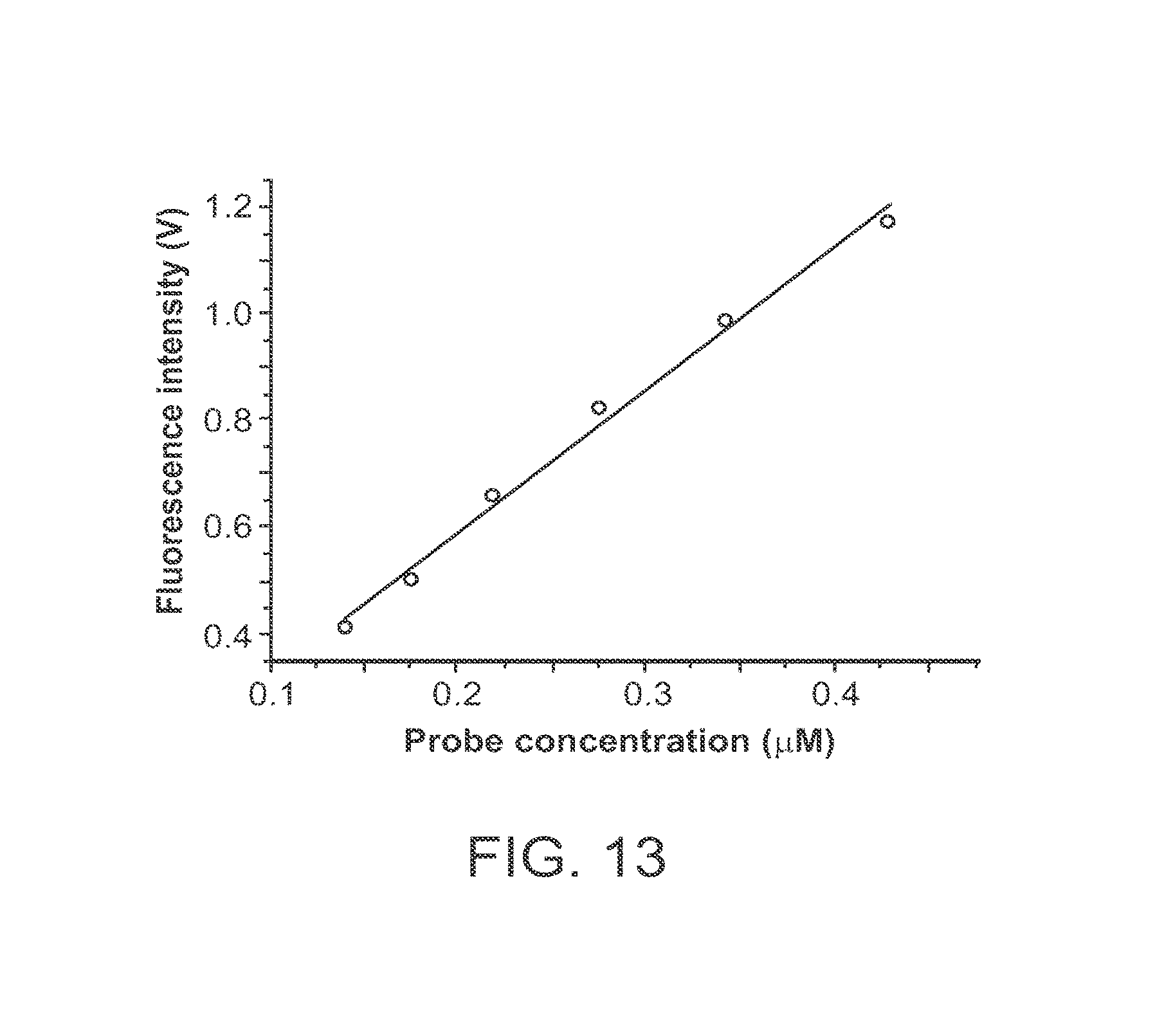
D00017

D00018
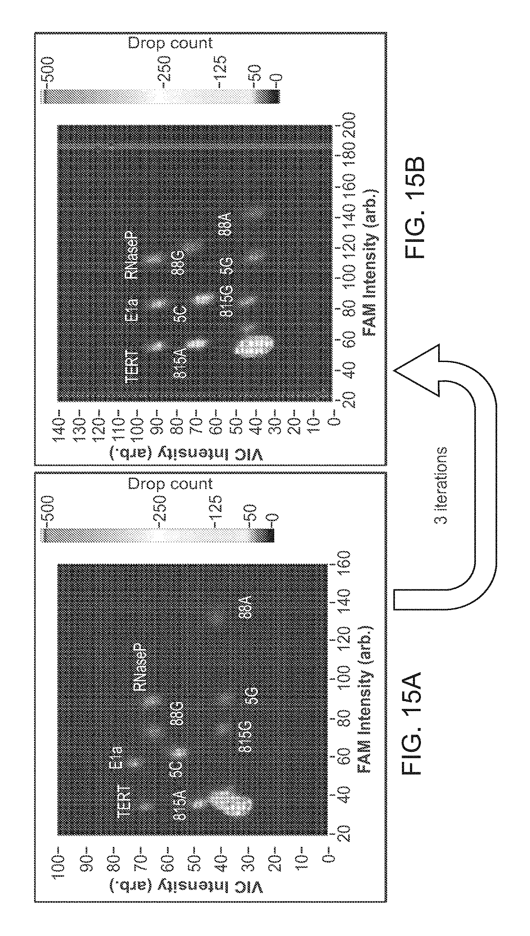
D00019

D00020

D00021

D00022
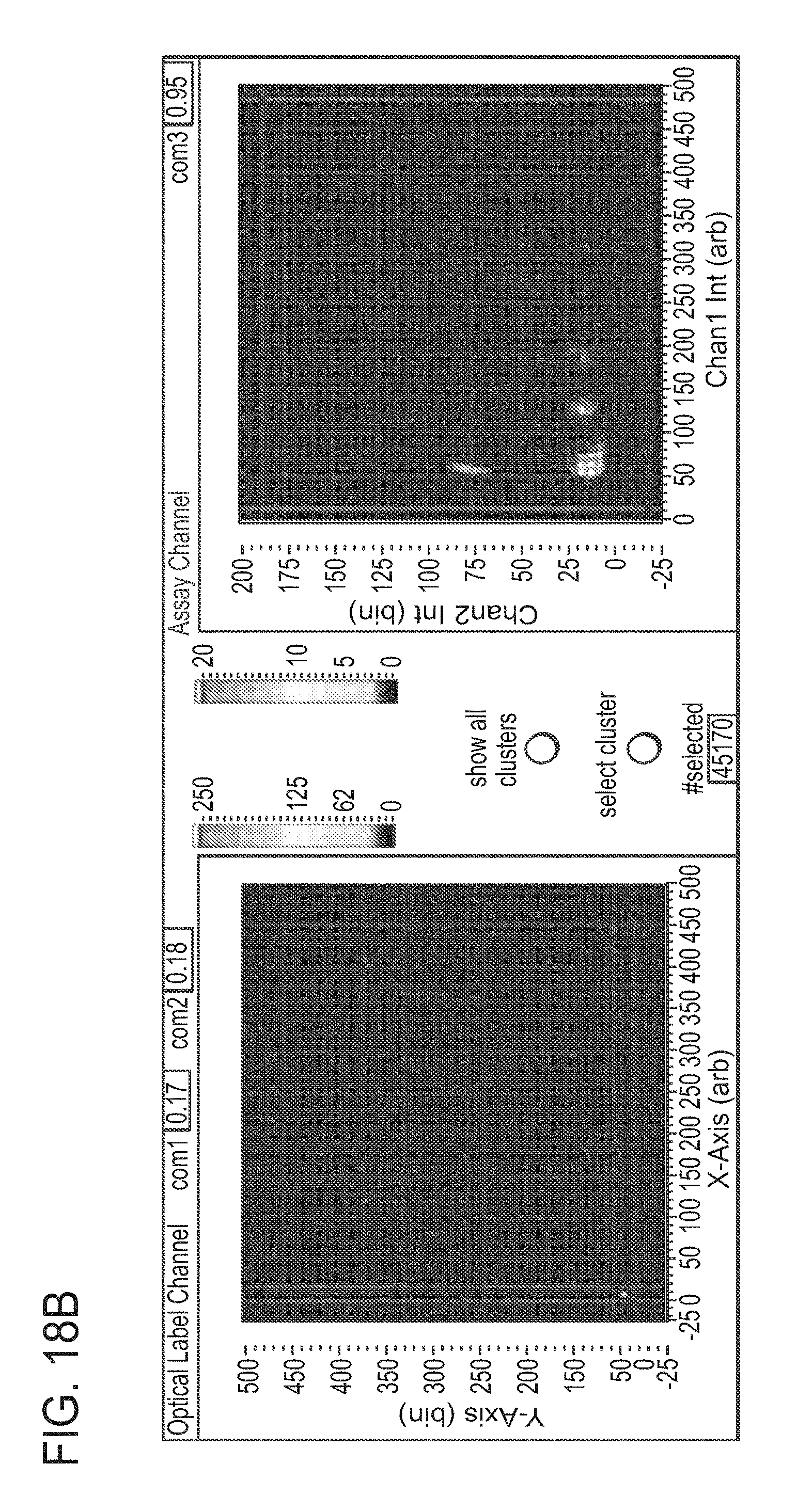
D00023

D00024

D00025

D00026

D00027

D00028

D00029

D00030

D00031

D00032

D00033

D00034

D00035

D00036
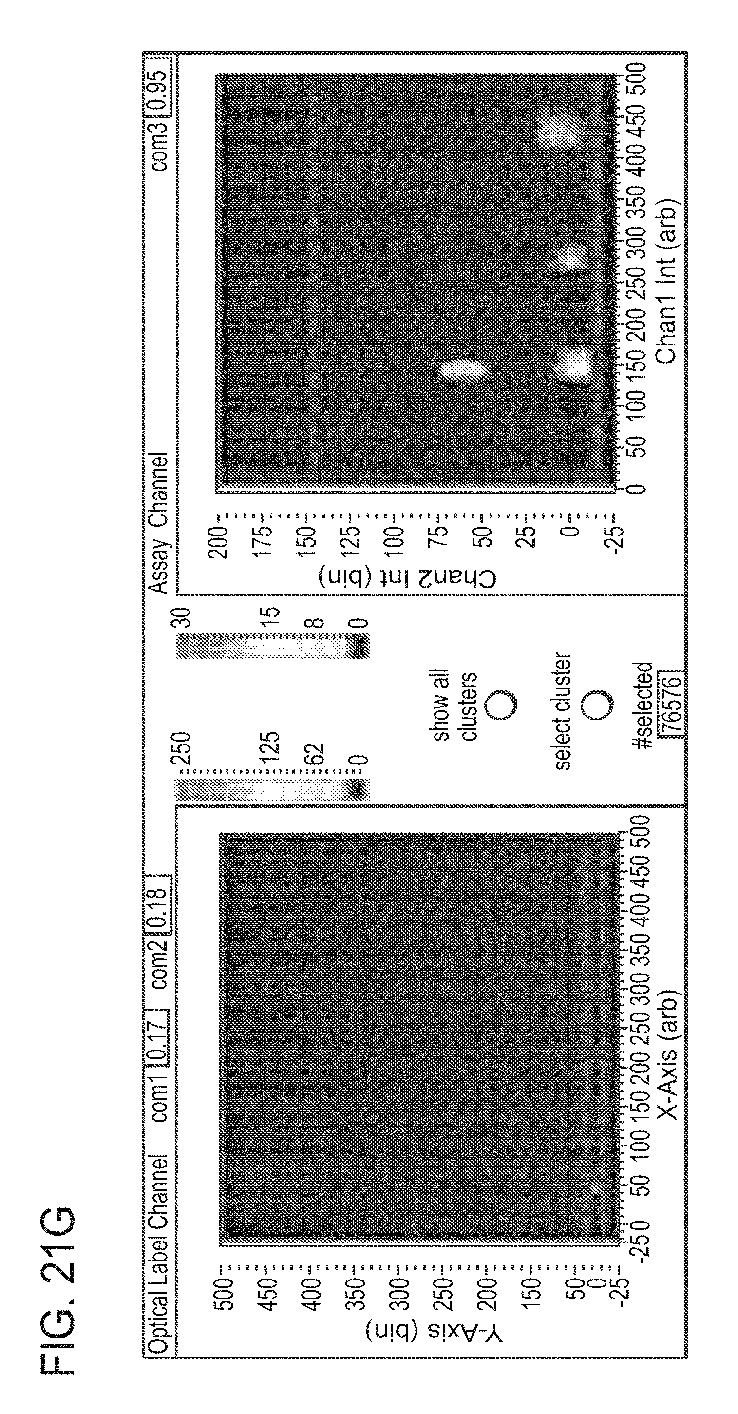
D00037

D00038

D00039
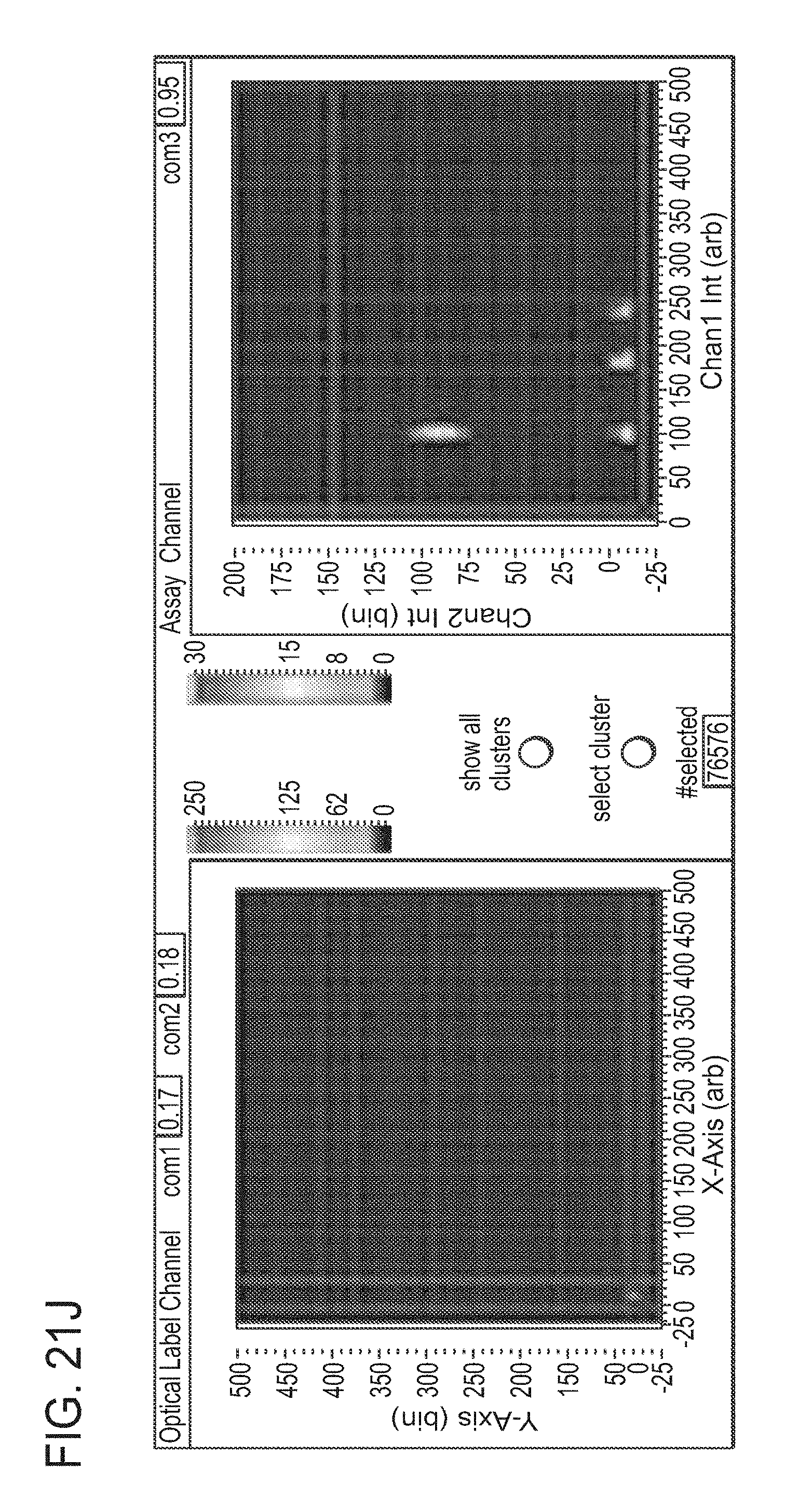
D00040
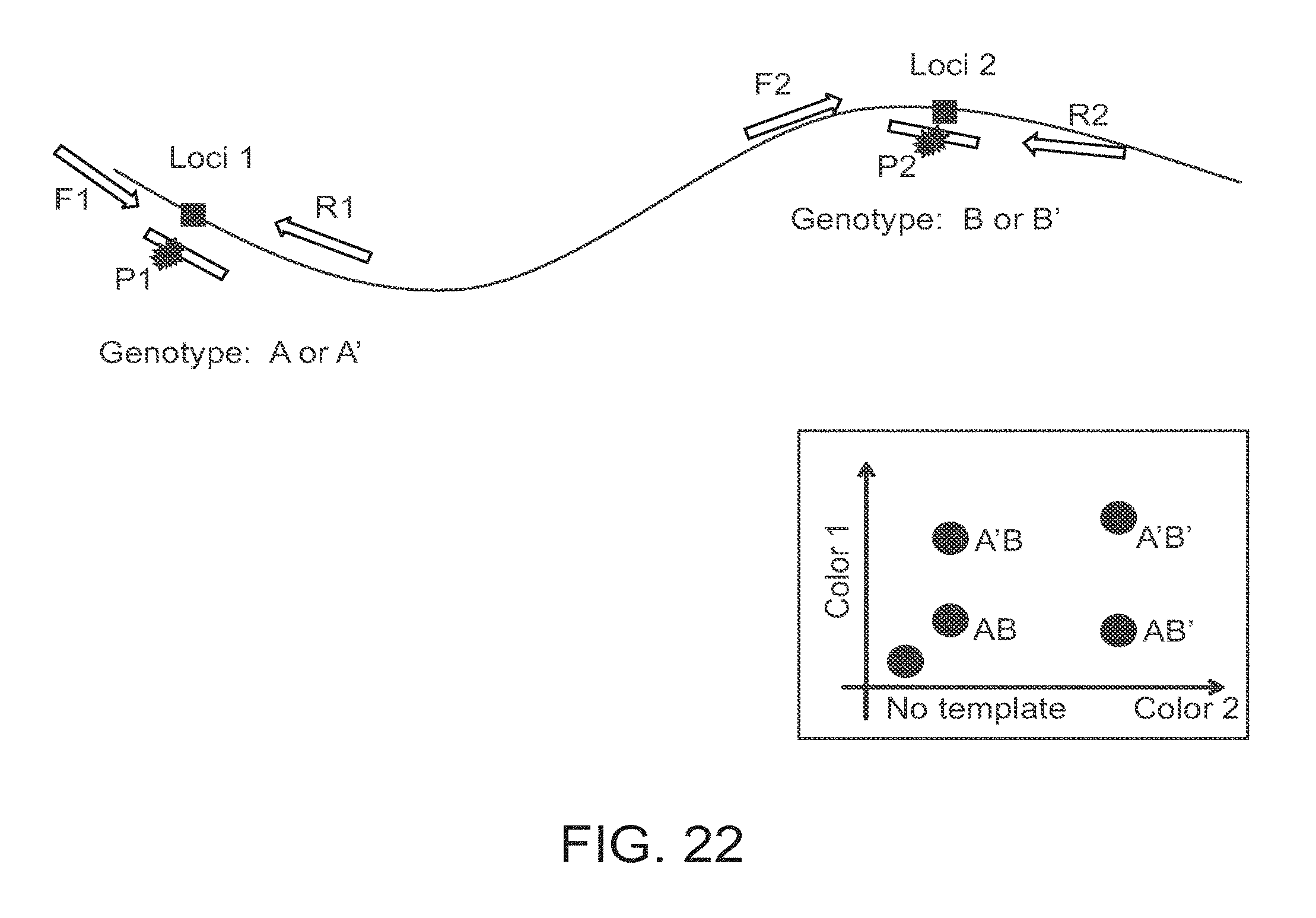
D00041

D00042

D00043
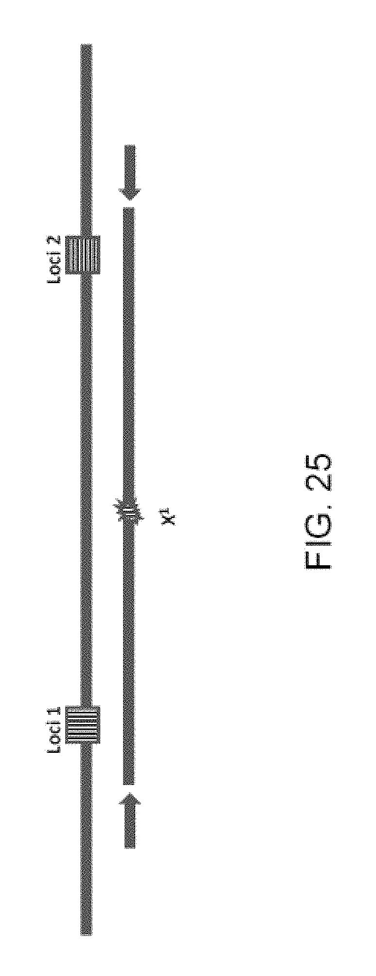
D00044
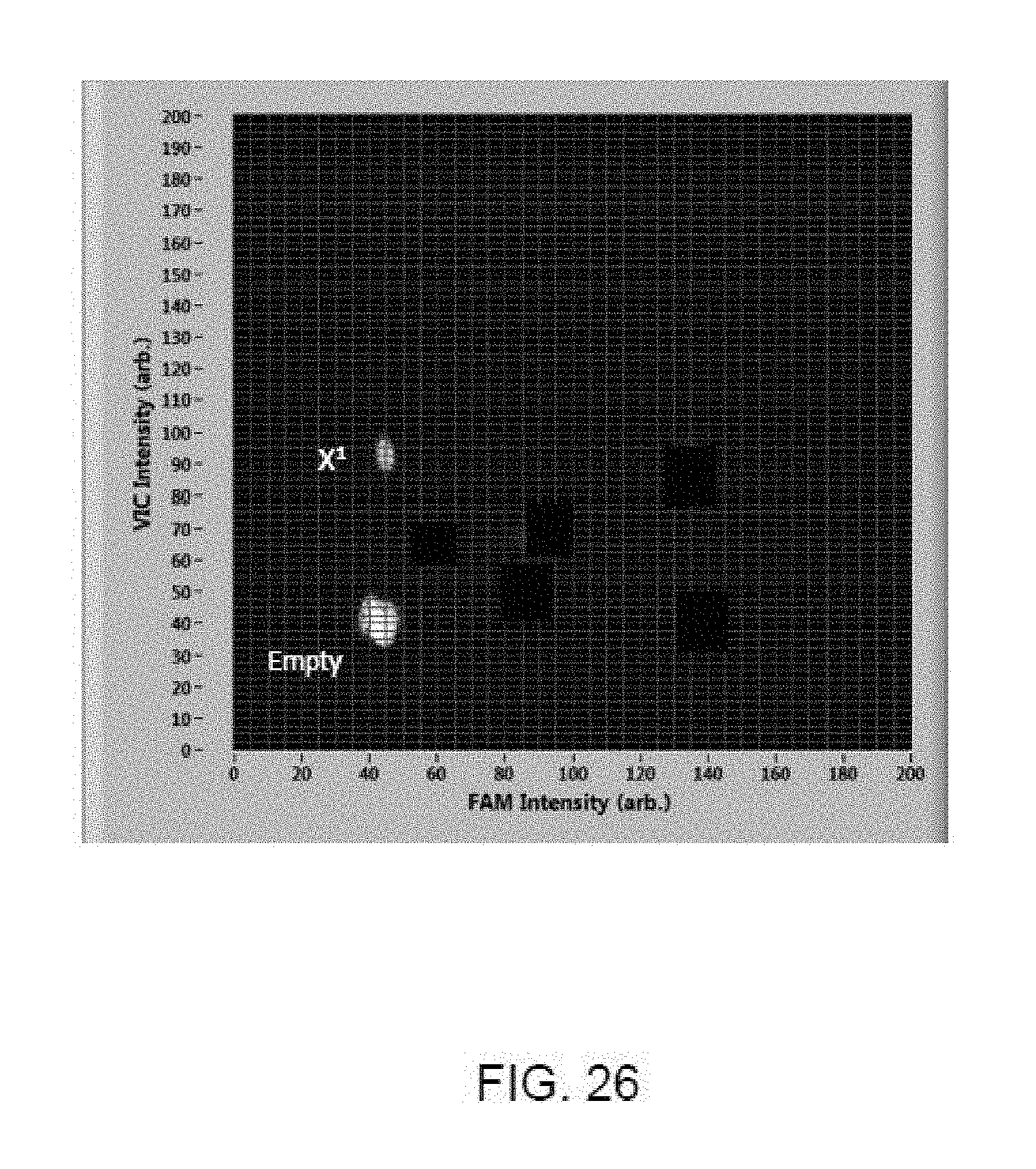
D00045

D00046
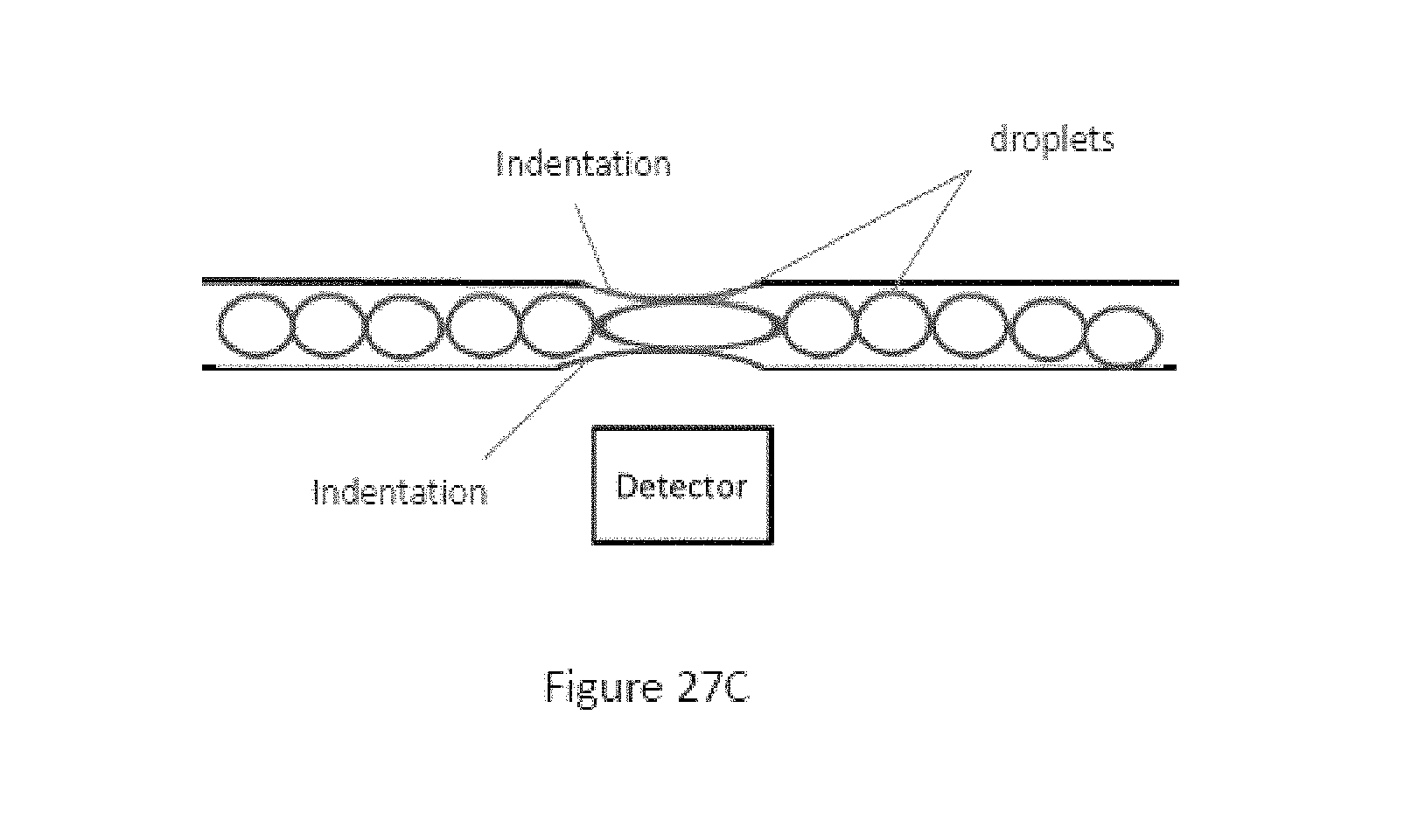
D00047

D00048

D00049
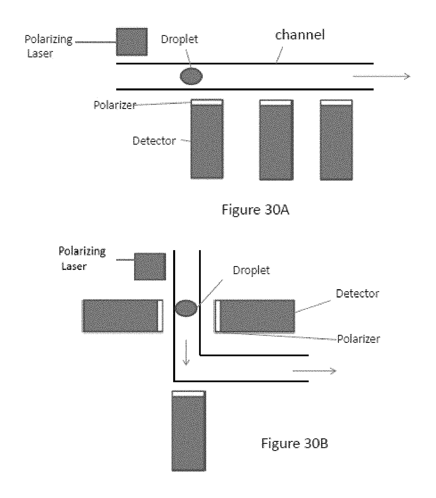
D00050

D00051

M00001

M00002

S00001
XML
uspto.report is an independent third-party trademark research tool that is not affiliated, endorsed, or sponsored by the United States Patent and Trademark Office (USPTO) or any other governmental organization. The information provided by uspto.report is based on publicly available data at the time of writing and is intended for informational purposes only.
While we strive to provide accurate and up-to-date information, we do not guarantee the accuracy, completeness, reliability, or suitability of the information displayed on this site. The use of this site is at your own risk. Any reliance you place on such information is therefore strictly at your own risk.
All official trademark data, including owner information, should be verified by visiting the official USPTO website at www.uspto.gov. This site is not intended to replace professional legal advice and should not be used as a substitute for consulting with a legal professional who is knowledgeable about trademark law.