Percutaneous clip for repairing a heart valve
Hernandez , et al. J
U.S. patent number 10,524,792 [Application Number 14/959,903] was granted by the patent office on 2020-01-07 for percutaneous clip for repairing a heart valve. This patent grant is currently assigned to Edwards Lifesciences Corporation. The grantee listed for this patent is Edwards Lifesciences Corporation. Invention is credited to Gregory Bak-Boychuk, Cristobal R. Hernandez, Emil Karapetian.


View All Diagrams
| United States Patent | 10,524,792 |
| Hernandez , et al. | January 7, 2020 |
Percutaneous clip for repairing a heart valve
Abstract
A leaflet clip has an elongated member including a proximal end portion and a distal end portion, and first and second clipping arms movable between an open position and a closed position. The clipping arms include respective proximal end portions coupled to the proximal end portion of the elongated member, and respective distal end portions extending distally and radially outward relative to the elongated member. The leaflet clip further includes a tubular member coaxially disposed about the elongated member. Axial motion of the tubular member with respect to the elongated member or axial motion of the elongated member with respect to the tubular member causes corresponding movement of the clipping arms between the open and closed positions.
| Inventors: | Hernandez; Cristobal R. (Santa Ana, CA), Bak-Boychuk; Gregory (San Clemente, CA), Karapetian; Emil (Huntington Beach, CA) | ||||||||||
|---|---|---|---|---|---|---|---|---|---|---|---|
| Applicant: |
|
||||||||||
| Assignee: | Edwards Lifesciences
Corporation (Irvine, CA) |
||||||||||
| Family ID: | 56092561 | ||||||||||
| Appl. No.: | 14/959,903 | ||||||||||
| Filed: | December 4, 2015 |
Prior Publication Data
| Document Identifier | Publication Date | |
|---|---|---|
| US 20160157862 A1 | Jun 9, 2016 | |
Related U.S. Patent Documents
| Application Number | Filing Date | Patent Number | Issue Date | ||
|---|---|---|---|---|---|
| 62087530 | Dec 4, 2014 | ||||
| Current U.S. Class: | 1/1 |
| Current CPC Class: | A61B 17/083 (20130101); A61F 2/24 (20130101); A61B 17/10 (20130101); A61B 17/1227 (20130101); A61F 2/2463 (20130101); A61B 17/1285 (20130101); A61B 2017/0649 (20130101); A61B 2017/00349 (20130101); A61B 2017/00243 (20130101) |
| Current International Class: | A61B 17/08 (20060101); A61B 17/00 (20060101); A61B 17/128 (20060101); A61F 2/24 (20060101); A61B 17/10 (20060101); A61B 17/122 (20060101); A61B 17/064 (20060101) |
References Cited [Referenced By]
U.S. Patent Documents
| 4340091 | July 1982 | Skelton et al. |
| 4506669 | March 1985 | Blake, III |
| 4590937 | May 1986 | Deniega |
| 4693248 | September 1987 | Failla |
| 4803983 | February 1989 | Siegel |
| 5125895 | June 1992 | Buchbinder et al. |
| 5171252 | December 1992 | Friedland |
| 5195962 | March 1993 | Martin et al. |
| 5327905 | July 1994 | Avitall |
| 5363861 | November 1994 | Edwards et al. |
| 5389077 | February 1995 | Melinyshyn et al. |
| 5411552 | May 1995 | Andersen et al. |
| 5450860 | September 1995 | O'Connor |
| 5456674 | October 1995 | Bos et al. |
| 5458583 | October 1995 | McNeely |
| 5478353 | December 1995 | Yoon |
| 5487746 | January 1996 | Yu et al. |
| 5565004 | October 1996 | Christoudias |
| 5607462 | March 1997 | Imran |
| 5609598 | March 1997 | Laufer et al. |
| 5611794 | March 1997 | Sauer et al. |
| 5626607 | May 1997 | Malecki et al. |
| 5695504 | December 1997 | Gifford, III et al. |
| 5716417 | February 1998 | Girard et al. |
| 5727569 | March 1998 | Benetti et al. |
| 5741297 | April 1998 | Simon |
| 5782746 | July 1998 | Wright |
| 5797960 | August 1998 | Stevens et al. |
| 5836311 | November 1998 | Borst et al. |
| 5855590 | January 1999 | Malecki et al. |
| 5885271 | March 1999 | Hamilton et al. |
| 5888247 | March 1999 | Benetti |
| 5891017 | April 1999 | Swindle et al. |
| 5891112 | April 1999 | Samson |
| 5894843 | April 1999 | Benetti et al. |
| 5921979 | July 1999 | Kovac et al. |
| 5944738 | August 1999 | Amplatz et al. |
| 5957835 | September 1999 | Anderson et al. |
| 5972020 | October 1999 | Carpentier et al. |
| 5980534 | November 1999 | Gimpelson |
| 6004329 | December 1999 | Myers et al. |
| 6010531 | January 2000 | Donlon et al. |
| 6086600 | July 2000 | Kortenbach |
| 6120496 | September 2000 | Whayne et al. |
| 6132370 | October 2000 | Furnish et al. |
| 6165183 | December 2000 | Kuehn et al. |
| 6182664 | February 2001 | Cosgrove |
| 6193732 | February 2001 | Frantzen et al. |
| 6193734 | February 2001 | Bolduc et al. |
| 6200315 | March 2001 | Gaiser et al. |
| 6241743 | June 2001 | Levin et al. |
| 6269819 | August 2001 | Oz et al. |
| 6269829 | August 2001 | Chen et al. |
| 6312447 | November 2001 | Grimes |
| 6461366 | October 2002 | Seguin |
| 6468285 | October 2002 | Hsu et al. |
| 6508806 | January 2003 | Hoste |
| 6508825 | January 2003 | Selmon et al. |
| 6537290 | March 2003 | Adams et al. |
| 6544215 | April 2003 | Bencini et al. |
| 6626930 | September 2003 | Allen et al. |
| 6629534 | October 2003 | St. Goar et al. |
| 6695866 | February 2004 | Kuehn et al. |
| 6719767 | April 2004 | Kimblad |
| 6752813 | June 2004 | Goldfarb et al. |
| 6764510 | July 2004 | Vidlund et al. |
| 6770083 | August 2004 | Seguin |
| 6837867 | January 2005 | Kortelling |
| 6855137 | February 2005 | Bon |
| 6875224 | April 2005 | Grimes |
| 6913614 | July 2005 | Marino et al. |
| 6939337 | September 2005 | Parker et al. |
| 6945956 | September 2005 | Waldhauser et al. |
| 7011669 | March 2006 | Kimblad |
| 7048754 | May 2006 | Martin et al. |
| 7101395 | September 2006 | Tremulis et al. |
| 7112207 | September 2006 | Allen et al. |
| 7125421 | October 2006 | Tremulis et al. |
| 7226467 | June 2007 | Lucatero et al. |
| 7288097 | October 2007 | Seguin |
| 7371210 | May 2008 | Brock et al. |
| 7464712 | December 2008 | Oz et al. |
| 7509959 | March 2009 | Oz et al. |
| 7563267 | July 2009 | Goldfarb et al. |
| 7563273 | July 2009 | Goldfarb et al. |
| 7569062 | August 2009 | Kuehn et al. |
| 7604646 | October 2009 | Goldfarb et al. |
| 7608091 | October 2009 | Goldfarb et al. |
| 7635329 | December 2009 | Goldfarb et al. |
| 7655015 | February 2010 | Goldfarb et al. |
| 7666204 | February 2010 | Thornton et al. |
| 7682319 | March 2010 | Martin et al. |
| 7682369 | March 2010 | Seguin |
| 7704269 | April 2010 | St. Goar et al. |
| 7731706 | June 2010 | Potter |
| 7736388 | June 2010 | Goldfarb et al. |
| 7744609 | June 2010 | Allen et al. |
| 7748389 | July 2010 | Salahieh et al. |
| 7753923 | July 2010 | St. Goar et al. |
| 7753932 | July 2010 | Gingrich et al. |
| 7758596 | July 2010 | Oz et al. |
| 7803185 | September 2010 | Gabbay |
| 7811296 | October 2010 | Goldfarb et al. |
| 7824443 | November 2010 | Salahieh et al. |
| 7981123 | July 2011 | Seguin |
| 7988724 | August 2011 | Salahieh et al. |
| 7998151 | August 2011 | St. Goar et al. |
| 8029518 | October 2011 | Goldfarb et al. |
| 8052592 | November 2011 | Goldfarb et al. |
| 8052749 | November 2011 | Salahieh et al. |
| 8052750 | November 2011 | Tuval et al. |
| 8057493 | November 2011 | Goldfarb et al. |
| 8062313 | November 2011 | Kimblad |
| 8070805 | December 2011 | Vidlund et al. |
| 8096985 | January 2012 | Legaspi et al. |
| 8123703 | February 2012 | Martin et al. |
| 8133239 | March 2012 | Oz et al. |
| 8187299 | May 2012 | Goldfarb et al. |
| 8206437 | June 2012 | Bonhoeffer et al. |
| 8216256 | July 2012 | Raschdorf, Jr. et al. |
| 8216301 | July 2012 | Bonhoeffer et al. |
| 8303608 | November 2012 | Goldfarb et al. |
| 8303653 | November 2012 | Bonhoeffer et al. |
| 8323334 | December 2012 | Deem et al. |
| 8343174 | January 2013 | Goldfarb et al. |
| 8348995 | January 2013 | Tuval et al. |
| 8348996 | January 2013 | Tuval et al. |
| 8409273 | April 2013 | Thornton et al. |
| 8414643 | April 2013 | Tuval et al. |
| 8416643 | April 2013 | Magee |
| 8449599 | May 2013 | Chau et al. |
| 8449606 | May 2013 | Eliasen et al. |
| 8480730 | July 2013 | Maurer et al. |
| 8500761 | August 2013 | Goldfarb et al. |
| 8540767 | September 2013 | Zhang |
| 8579965 | November 2013 | Bonhoeffer et al. |
| 8585756 | November 2013 | Bonhoeffer et al. |
| 8652202 | February 2014 | Alon et al. |
| 8668733 | March 2014 | Haug et al. |
| 8721665 | May 2014 | Oz et al. |
| 8734505 | May 2014 | Goldfarb et al. |
| 8740918 | June 2014 | Seguin |
| 8740920 | June 2014 | Goldfarb et al. |
| 8747460 | June 2014 | Tuval et al. |
| 8764774 | July 2014 | Sigmon, Jr. |
| 8771345 | July 2014 | Tuval et al. |
| 8771346 | July 2014 | Tuval et al. |
| 8771347 | July 2014 | DeBoer et al. |
| 8778017 | July 2014 | Eliasen et al. |
| 8834564 | September 2014 | Tuval et al. |
| 8840663 | September 2014 | Salahieh et al. |
| 8876894 | November 2014 | Tuval et al. |
| 8876895 | November 2014 | Tuval et al. |
| 8926691 | January 2015 | Chau et al. |
| 8945177 | February 2015 | Dell et al. |
| 9011468 | April 2015 | Ketai et al. |
| 9044246 | June 2015 | Goldfarb et al. |
| 9138312 | September 2015 | Tuval et al. |
| 9198757 | December 2015 | Schroeder et al. |
| 9259317 | February 2016 | Wilson et al. |
| 9301834 | April 2016 | Tuval et al. |
| 9387071 | July 2016 | Tuval et al. |
| 9414918 | August 2016 | Chau et al. |
| 9427237 | August 2016 | Oz et al. |
| 9427327 | August 2016 | Parrish |
| 9439763 | September 2016 | Geist et al. |
| 9510829 | December 2016 | Goldfarb et al. |
| 9510837 | December 2016 | Seguin |
| 9510946 | December 2016 | Chau et al. |
| 9642704 | May 2017 | Tuval et al. |
| 9700445 | July 2017 | Martin et al. |
| 9775963 | October 2017 | Miller |
| D809139 | January 2018 | Marsot et al. |
| 9889002 | February 2018 | Bonhoeffer et al. |
| 9949824 | April 2018 | Bonhoeffer et al. |
| 10022221 | July 2018 | Gainor et al. |
| 10076327 | September 2018 | Ellis et al. |
| 10076415 | September 2018 | Metchik et al. |
| 10105221 | October 2018 | Siegel |
| 10105222 | October 2018 | Metchik et al. |
| 10111751 | October 2018 | Metchik et al. |
| 10123873 | November 2018 | Metchik et al. |
| 10130475 | November 2018 | Metchik et al. |
| 10136993 | November 2018 | Metchik et al. |
| 10159570 | December 2018 | Metchik et al. |
| 10188392 | January 2019 | Wei |
| 10226309 | March 2019 | Ho et al. |
| 10231837 | March 2019 | Metchik et al. |
| 10238494 | March 2019 | McNiven et al. |
| 10238495 | March 2019 | Marsot et al. |
| 10299924 | May 2019 | Kizuka |
| 10376673 | August 2019 | Van Hoven et al. |
| 2001/0005787 | June 2001 | Oz et al. |
| 2002/0107531 | August 2002 | Schreck et al. |
| 2002/0173811 | November 2002 | Tu et al. |
| 2003/0069593 | April 2003 | Tremulis et al. |
| 2003/0120341 | June 2003 | Shennib |
| 2003/0130731 | July 2003 | Vidlund et al. |
| 2003/0167071 | September 2003 | Martin et al. |
| 2003/0208231 | November 2003 | Williamson et al. |
| 2004/0003819 | January 2004 | St. Goar et al. |
| 2004/0034635 | February 2004 | Lentz et al. |
| 2004/0039442 | February 2004 | St. Goar et al. |
| 2004/0044350 | March 2004 | Martin et al. |
| 2004/0049207 | March 2004 | Goldfarb et al. |
| 2004/0049211 | March 2004 | Tremulis et al. |
| 2004/0127981 | July 2004 | Rahdert et al. |
| 2004/0127982 | July 2004 | Machold et al. |
| 2004/0147943 | July 2004 | Kobayashi |
| 2004/0167539 | August 2004 | Kuehn et al. |
| 2004/0181206 | September 2004 | Chiu et al. |
| 2004/0210307 | October 2004 | Khairkhahan |
| 2004/0220593 | November 2004 | Greenhalgh |
| 2005/0033446 | February 2005 | Deem et al. |
| 2005/0049618 | March 2005 | Masuda et al. |
| 2005/0080440 | April 2005 | Durgin et al. |
| 2005/0137688 | June 2005 | Salahieh et al. |
| 2005/0137698 | June 2005 | Salahieh et al. |
| 2005/0143767 | June 2005 | Kimura et al. |
| 2005/0165429 | July 2005 | Douglas et al. |
| 2005/0251183 | November 2005 | Buckman et al. |
| 2005/0288786 | December 2005 | Chanduszko |
| 2006/0020275 | January 2006 | Goldfarb et al. |
| 2006/0058872 | March 2006 | Salahieh et al. |
| 2006/0064118 | March 2006 | Kimblad |
| 2006/0100649 | May 2006 | Hart |
| 2006/0122647 | June 2006 | Callaghan et al. |
| 2006/0173524 | August 2006 | Salahieh et al. |
| 2006/0224169 | October 2006 | Weisenburgh et al. |
| 2007/0010877 | January 2007 | Salahieh et al. |
| 2007/0021779 | January 2007 | Garvin et al. |
| 2007/0032807 | February 2007 | Ortiz et al. |
| 2007/0038293 | February 2007 | St.Goar et al. |
| 2007/0093857 | April 2007 | Rogers et al. |
| 2007/0100356 | May 2007 | Lucatero et al. |
| 2007/0129737 | June 2007 | Goldfarb et al. |
| 2007/0185571 | August 2007 | Kapadia et al. |
| 2007/0197858 | August 2007 | Goldfarb et al. |
| 2007/0198038 | August 2007 | Cohen et al. |
| 2007/0203503 | August 2007 | Salahieh et al. |
| 2007/0282414 | December 2007 | Soltis et al. |
| 2008/0039743 | February 2008 | Fox et al. |
| 2008/0039953 | February 2008 | Davis et al. |
| 2008/0065149 | March 2008 | Thielen et al. |
| 2008/0077144 | March 2008 | Crofford |
| 2008/0140089 | June 2008 | Kogiso et al. |
| 2008/0147093 | June 2008 | Roskopf et al. |
| 2008/0147112 | June 2008 | Sheets et al. |
| 2008/0167713 | July 2008 | Bolling |
| 2008/0167714 | July 2008 | St. Goar et al. |
| 2008/0177300 | July 2008 | Mas et al. |
| 2008/0255427 | October 2008 | Satake et al. |
| 2008/0288061 | November 2008 | Maurer et al. |
| 2008/0319455 | December 2008 | Harris et al. |
| 2009/0048668 | February 2009 | Wilson et al. |
| 2009/0076598 | March 2009 | Salahieh et al. |
| 2009/0163934 | June 2009 | Raschdorf, Jr. et al. |
| 2009/0275902 | November 2009 | Heeps et al. |
| 2009/0287304 | November 2009 | Dahlgren et al. |
| 2010/0022823 | January 2010 | Goldfarb et al. |
| 2010/0094317 | April 2010 | Goldfarb et al. |
| 2010/0121433 | May 2010 | Bolling et al. |
| 2010/0174363 | July 2010 | Castro |
| 2010/0217283 | August 2010 | St. Goar et al. |
| 2010/0324595 | December 2010 | Linder et al. |
| 2011/0082538 | April 2011 | Dahlgren et al. |
| 2011/0137397 | June 2011 | Chau et al. |
| 2011/0137410 | June 2011 | Hacohen |
| 2011/0245855 | October 2011 | Matsuoka et al. |
| 2011/0295281 | December 2011 | Mizumoto et al. |
| 2011/0307055 | December 2011 | Goldfarb et al. |
| 2012/0010461 | January 2012 | Goldfarb et al. |
| 2012/0035712 | February 2012 | Maisano et al. |
| 2012/0046741 | February 2012 | Tuval et al. |
| 2012/0109160 | May 2012 | Martinez et al. |
| 2012/0116419 | May 2012 | Sigmon, Jr. |
| 2012/0209318 | August 2012 | Qadeer |
| 2013/0066341 | March 2013 | Ketai et al. |
| 2013/0066342 | March 2013 | Dell et al. |
| 2013/0072945 | March 2013 | Terada |
| 2013/0073034 | March 2013 | Wilson et al. |
| 2013/0190861 | July 2013 | Chau et al. |
| 2013/0226199 | August 2013 | Harris et al. |
| 2013/0268069 | October 2013 | Zakai et al. |
| 2014/0046433 | February 2014 | Kovalsky |
| 2014/0058411 | February 2014 | Soutorine et al. |
| 2014/0066693 | March 2014 | Goldfarb et al. |
| 2014/0067048 | March 2014 | Chau et al. |
| 2014/0067052 | March 2014 | Chau et al. |
| 2014/0067054 | March 2014 | Chau et al. |
| 2014/0135685 | May 2014 | Kabe et al. |
| 2014/0222136 | August 2014 | Geist et al. |
| 2014/0236187 | August 2014 | Seguin |
| 2014/0236198 | August 2014 | Goldfarb et al. |
| 2014/0243968 | August 2014 | Padala |
| 2014/0296878 | October 2014 | Oz et al. |
| 2014/0316428 | October 2014 | Golan |
| 2015/0039084 | February 2015 | Levi et al. |
| 2015/0057704 | February 2015 | Takahashi |
| 2015/0105804 | April 2015 | Dell et al. |
| 2015/0105808 | April 2015 | Gordon et al. |
| 2015/0157268 | June 2015 | Winshtein et al. |
| 2015/0182223 | July 2015 | Ketai et al. |
| 2015/0196390 | July 2015 | Ma et al. |
| 2015/0223793 | August 2015 | Goldfarb et al. |
| 2015/0238313 | August 2015 | Spence et al. |
| 2015/0257883 | September 2015 | Basude et al. |
| 2015/0313592 | November 2015 | Coillard-Lavirotte et al. |
| 2016/0022970 | January 2016 | Forcucci et al. |
| 2016/0106539 | April 2016 | Buchbinder et al. |
| 2016/0113764 | April 2016 | Sheahan et al. |
| 2016/0113766 | April 2016 | Ganesan et al. |
| 2016/0155987 | June 2016 | Yoo et al. |
| 2016/0174979 | June 2016 | Wei |
| 2016/0174981 | June 2016 | Fago et al. |
| 2016/0242906 | August 2016 | Morriss et al. |
| 2016/0287387 | October 2016 | Wei |
| 2016/0317290 | November 2016 | Chau et al. |
| 2016/0324634 | November 2016 | Gabbay |
| 2016/0331523 | November 2016 | Chau et al. |
| 2016/0354082 | December 2016 | Oz et al. |
| 2017/0020521 | January 2017 | Krone et al. |
| 2017/0035561 | February 2017 | Rowe et al. |
| 2017/0035566 | February 2017 | Krone et al. |
| 2017/0042456 | February 2017 | Budiman |
| 2017/0049455 | February 2017 | Seguin |
| 2017/0100236 | April 2017 | Robertson et al. |
| 2017/0239048 | August 2017 | Goldfarb et al. |
| 2017/0281330 | October 2017 | Liljegren et al. |
| 2018/0000582 | January 2018 | Tuval et al. |
| 2018/0008311 | January 2018 | Shiroff et al. |
| 2018/0021044 | January 2018 | Miller et al. |
| 2018/0021134 | January 2018 | McNiven et al. |
| 2018/0078271 | March 2018 | Thrasher, III |
| 2018/0126124 | May 2018 | Winston et al. |
| 2018/0146964 | May 2018 | Garcia et al. |
| 2018/0146966 | May 2018 | Hernandez et al. |
| 2018/0153552 | June 2018 | King et al. |
| 2018/0161159 | June 2018 | Lee et al. |
| 2018/0221147 | August 2018 | Ganesan et al. |
| 2018/0235657 | August 2018 | Abunassar |
| 2018/0243086 | August 2018 | Barbarino et al. |
| 2018/0258665 | September 2018 | Reddy et al. |
| 2018/0296326 | October 2018 | Dixon et al. |
| 2018/0296327 | October 2018 | Dixon et al. |
| 2018/0296328 | October 2018 | Dixon et al. |
| 2018/0296329 | October 2018 | Dixon et al. |
| 2018/0296330 | October 2018 | Dixon et al. |
| 2018/0296331 | October 2018 | Dixon et al. |
| 2018/0296332 | October 2018 | Dixon et al. |
| 2018/0296333 | October 2018 | Dixon et al. |
| 2018/0296334 | October 2018 | Dixon et al. |
| 2018/0325671 | November 2018 | Abunassar et al. |
| 2019/0000613 | January 2019 | Delgado et al. |
| 2019/0000623 | January 2019 | Pan et al. |
| 2019/0008638 | January 2019 | Siegel et al. |
| 2019/0008642 | January 2019 | Delgado et al. |
| 2019/0008643 | January 2019 | Delgado et al. |
| 2019/0015199 | January 2019 | Delgado et al. |
| 2019/0015200 | January 2019 | Delgado et al. |
| 2019/0015207 | January 2019 | Delgado et al. |
| 2019/0015208 | January 2019 | Delgado et al. |
| 2019/0021851 | January 2019 | Delgado et al. |
| 2019/0021852 | January 2019 | Delgado et al. |
| 2019/0029810 | January 2019 | Delgado et al. |
| 2019/0029813 | January 2019 | Delgado et al. |
| 2019/0030285 | January 2019 | Prabhu et al. |
| 2019/0053803 | February 2019 | Ketai et al. |
| 2019/0060058 | February 2019 | Delgado et al. |
| 2019/0060059 | February 2019 | Delgado et al. |
| 2019/0060072 | February 2019 | Zeng |
| 2019/0060073 | February 2019 | Delgado et al. |
| 2019/0060074 | February 2019 | Delgado et al. |
| 2019/0060075 | February 2019 | Delgado et al. |
| 2019/0069991 | March 2019 | Metchik et al. |
| 2019/0069992 | March 2019 | Delgado et al. |
| 2019/0069993 | March 2019 | Delgado et al. |
| 2019/0167197 | June 2019 | Abunassar et al. |
| 2019/0261995 | August 2019 | Goldfarb et al. |
| 2019/0261996 | August 2019 | Goldfarb et al. |
| 2019/0261997 | August 2019 | Goldfarb et al. |
| 1316889 | Oct 2001 | CN | |||
| 102639179 | Aug 2012 | CN | |||
| 103889345 | Jun 2014 | CN | |||
| 0098100 | Jan 1984 | EP | |||
| 1281375 | Feb 2003 | EP | |||
| 0879069 | Aug 2003 | EP | |||
| 1301235 | Oct 2004 | EP | |||
| 1583577 | May 2007 | EP | |||
| 1408850 | Sep 2009 | EP | |||
| 0930845 | Oct 2009 | EP | |||
| 1624810 | Mar 2011 | EP | |||
| 1804686 | Sep 2015 | EP | |||
| 2428169 | Oct 2016 | EP | |||
| 2266503 | Jan 2017 | EP | |||
| 2266504 | Mar 2017 | EP | |||
| 2146050 | Feb 1973 | FR | |||
| 9711600 | Mar 1997 | FR | |||
| 2768324 | Mar 1999 | FR | |||
| 2014000417 | Jan 2014 | JP | |||
| 9802103 | Jan 1998 | WO | |||
| 9900059 | Jan 1999 | WO | |||
| 9913777 | Mar 1999 | WO | |||
| 0060995 | Oct 2000 | WO | |||
| 03001893 | Jan 2003 | WO | |||
| 2004103162 | Dec 2004 | WO | |||
| 2004103434 | Dec 2004 | WO | |||
| 2005112792 | Dec 2005 | WO | |||
| 2006116558 | Nov 2006 | WO | |||
| 2006127509 | Nov 2006 | WO | |||
| 06/138173 | Dec 2006 | WO | |||
| 2006047709 | Jul 2007 | WO | |||
| 08/147964 | Mar 2008 | WO | |||
| 08/150529 | Dec 2008 | WO | |||
| 09/024859 | Feb 2009 | WO | |||
| 2009053952 | Apr 2009 | WO | |||
| 2009091509 | Jul 2009 | WO | |||
| 09/116041 | Sep 2009 | WO | |||
| 2009108942 | Sep 2009 | WO | |||
| 2010098804 | Sep 2010 | WO | |||
| 2010128502 | Nov 2010 | WO | |||
| 2011034628 | Mar 2011 | WO | |||
| 2013059747 | Apr 2013 | WO | |||
| 2016110760 | Jul 2016 | WO | |||
Other References
|
Int'l. Search Report for PCT/US2015/064096, dated Mar. 22, 2016. cited by applicant . Al-Khaja, N., et al., "Eleven Years' Experience with Carpentier-Edwards Biological Valves in Relation to Survival and Complications," European Journal of Cardiothoracic Surgery 3:305-311, Jun. 30, 2009. cited by applicant . Almagor, M.D., Yaron, et al., "Balloon Expandable Stent Implantation in Stenotic Right Heart Valved Conduits," Journal of the American College of Cardiology, vol. 16, No. 6, pp. 1310-1314, Nov. 1, 1990; ISSN 0735-1097. cited by applicant . Al Zaibag, Muayed, et al., "Percutaneous Balloon Valvotomy in Tricuspid Stenosis," British Heart Journal, Jan. 1987, vol. 57, No. 1, pp. 51-53. cited by applicant . Andersen, et al., "Transluminal implantation of artificial heart valves. Description of a new expandable aortic valve and initial results with implantation by catheter technique in closed chest pigs." European Heart Journal (1992), 13, 704-708. cited by applicant . Andersen, Henning Rud, "History of Percutaneous Aortic Valve Prosthesis," Herz 34 2009 Nr. 5, Urban & Vogel, pp. 343-346, Skejby University Hospital Department of Cardiology, Aarhus, Denmark. cited by applicant . Benchimol, Alberto, et al., "Simultaneous Left Ventricular Echocardiography and Aortic Blood Velocity During Rapid Right Ventricular Pacing in Man," The American Journal of the Medical Sciences, Jan.-Feb. 1977 vol. 273, No. 1, pp. 55-62. cited by applicant . Dake, Michael. D., et al., Transluminal Placement of Endovascular Stent-Grafts for the Treatment of Descending Thoracic Aortic Aneurysms, New Engl.J.Med., 1994; 331:1729-34. cited by applicant . Dotter, M.D., Charles T., "Transluminal Treatment of Arteriosclerotic Obstruction," University of Oregon's Minthorn Memorial Laboratory for Cardiovascular Research through Radiology, Circulation, vol. XXX, Nov. 1964, pp. 654-670. cited by applicant . Kolata, Gina, "Device That Opens Clogged Arteries Gets a Failing Grade in a New Study," nytimes.com, <http://www.nytimes.com/1991/01/03/health/device-that-opens-clogged-ar- teries-gets-a-faili> . . . , Jul. 29, 2009, 2 pages. cited by applicant . Inoune, M.D., Kanji, et al., "Clinical Application of Transvenous Mitral Commissurotomy by a New Balloon Catheter," The Journal of Thoracic and Cardiovascular Surgery 87:394-402, 1984. cited by applicant . Lawrence, Jr., M.D., David D., "Percutaneous Endovascular Graft: Experimental Evaluation," Radiology 1897; 163: 357-360. cited by applicant . Pavcnik, M.D., Ph.D., Dusan, et al. "Development and Initial Experimental Evaluation of a Prosthetic Aortic Valve for Transcatheter Placement," Cardiovascular Radiology 1992; 183:151-154. cited by applicant . Porstmann, W., et al., "Der VerschluB des Ductus Arteriosus Persistens ohne Thorakotomie," Thoraxchirurgie Vaskulare Chirurgie, Band 15, Heft 2, Stuttgart, im Apr. 1967, pp. 199-203. cited by applicant . Rashkind, M.D., William J., "Creation of an Atrial Septal Defect Withoput Thoracotomy," the Journal of the American Medical Association, vol. 196, No. 11, Jun. 13, 1966, pp. 173-174. cited by applicant . Rashkind, M.D., William J., "Historical Aspects of Interventional Cardiology: Past, Present, Future," Texas Heart Institute Journal, Interventional Cardiology, pp. 363-367. cited by applicant . Rosch, M.D., Josef, "The Birth, Early Years and Future of Interventional Radiology," J Vase Interv Radiol 2003; 14:841-853. cited by applicant . Ross, F.R.C.S., D.N., "Aortic Valve Surgery," Guy's Hospital, London, pp. 192-197, approximately 1968. cited by applicant . Sabbah, Ph.D., Hani N., et al., "Mechanical Factors in the Degeneration of Porcine Bioprosthetic Valves: An Overview," Journal of Cardiac Surgery, vol. 4, No. 4, pp. 302-309, Dec. 1989; ISSN 0886-0440. cited by applicant . Selby, M.D., J. Bayne, "Experience with New Retrieval Forceps for Foreign Body Removal in the Vascular, Urinary, and Biliary Systems," Radiology 1990; 176:535-538. cited by applicant . Serruys, P.W., et al., "Stenting of Coronary Arteries. Are we the Sorcerer's Apprentice?," European Heart Journal (1989) 10, 774-782, pp. 37-45, Jun. 13, 1989. cited by applicant . Sigwart, Ulrich, "An Overview of Intravascular Stents: Old and New," Chapter 48, Textbook of Interventional Cardiology, 2nd Edition, W.B. Saunders Company, Philadelphia, PA, .COPYRGT. 1994, 1990, pp. 803-815. cited by applicant . Uchida, Barry T., et al., "Modifications of Gianturco Expandable Wire Stents," AJR:150, May 1988, Dec. 3, 1987, pp. 1185-1187. cited by applicant . Urban, M.D., Philip, "Coronary Artery Stenting," Editions Medecine et Hygiene, Geneve, 1991, pp. 5-47. cited by applicant . Watt, A.H., et al. "Intravenous Adenosine in the Treatment of Supraventricular Tachycardia; a Dose-Ranging Study and Interaction with Dipyridamole," British Journal of Clinical Pharmacology (1986), 21,227-230. cited by applicant . Wheatley, M.D., David J., "Valve Prostheses," Rob & Smith's Operative Surgery, Fourth Edition, pp. 415-424, Butterworths 1986. cited by applicant . CN Office Action issued in Application No. 201380057757, 8, dated Dec. 2, 2015. cited by applicant . Supplementary Search Report for European Application No. 13834484, dated Apr. 29, 2016. cited by applicant . Batista, M.D. et al., "Partial Left Ventriculectomy to Treat End-Stage Heart Disease," The Society of Thoracic Surgeons, 1997, pp. 634-638. cited by applicant . Beall et al., "Clinical experience with a dacron velour-covered teflon-disc mitral-valve prosthesis", Ann Thorac Surg., vol. 5, Issue 5, pp. 402-10, May 1968. cited by applicant . Fucci et alL,, "Improved results with mitral valve repair using new surgical techniques", Eur J Cardiothorac Surg. 1995;Issue 9, vol. 11, pp. 621-6. cited by applicant . Maisano et al., `The edge-to-edge technique: a simplified method to correct mitral insufficiency`, Eur J Cardiothorac Surg., vol. 13, Issue-3, pp. 240-5, Mar. 1998. cited by applicant . Reul RM et al., "Mitral valve reconstruction for mitral insufficiency", Prog Cardiovasc Dis., vol. 39, Issue-6, May-Jun. 1997. cited by applicant . Umana et al., ``Bow-tie` mitral valve repair: an adjuvant technique for ischemic mitral regurgitation`, Ann Thorac Surg., vol. 66, Issue-6, pp. 1640-6, Nov. 1998. cited by applicant. |
Primary Examiner: Fishback; Ashley L
Attorney, Agent or Firm: Richardson; Thomas C.
Parent Case Text
CROSS REFERENCE TO RELATED APPLICATION
This application claims the benefit of U.S. Provisional Patent Application No. 62/087,530, filed Dec. 4, 2014, which is hereby incorporated herein by reference in its entirety.
Claims
We claim:
1. A leaflet clip for implanting on first and second native leaflets of a heart valve, comprising: an elongated member including a first end portion and a second end portion; first and second clipping arms movable between an open position and a closed position, the clipping arms including respective first end portions coupled to the first end portion of the elongated member and respective second end portions extending axially and radially outward relative to the elongated member; a tubular member coaxially disposed about the elongated member; and a leaflet engagement mechanism disposed on the distal end portion of the elongated member, the leaflet engagement mechanism comprising a fabric covering configured to engage the first and second native leaflets as the clipping arms are moved from the open position to the closed position, the fabric covering comprising a natural or synthetic fibrous material that is woven, braided, or any combination thereof; wherein axial motion of the tubular member relative to the elongated member or axial motion of the elongated member relative to the tubular member causes corresponding movement of the clipping arms between the open and closed positions.
2. A method, comprising: positioning a leaflet clip adjacent a commissure of a heart valve such that a first clipping arm of the leaflet clip is adjacent a first leaflet of the heart valve and a second clipping arm of the leaflet clip is adjacent a second leaflet of the heart valve and an elongated member of the leaflet clip is positioned between the leaflets; and moving a sheath disposed coaxially over the elongated member axially with respect to the elongated member or moving the elongated member axially with respect to the sheath such that the sheath extends over the elongated member and the clipping arms, and causes the clipping arms to move from an open position to a closed position, thereby capturing the first leaflet between the first clipping arm and the elongated member and capturing the second leaflet between the second clipping arm and the elongated member.
3. The method of claim 2, wherein the heart valve is a mitral valve, an aortic valve, or a tricuspid valve.
4. The method of claim 2, further comprising positioning a leaflet clip adjacent each commissure of the heart valve.
5. A method, comprising: positioning a leaflet clip adjacent a commissure of a heart valve such that a first clipping arm of the leaflet clip is adjacent a first leaflet of the heart valve and a second clipping arm of the leaflet clip is adjacent a second leaflet of the heart valve and an elongated member of the leaflet clip is positioned between the leaflets; moving a sheath disposed coaxially over the elongated member axially with respect to the elongated member or moving the elongated member axially with respect to the sheath such that the sheath causes the clipping arms to move from an open position to a closed position, thereby capturing the first leaflet between the first clipping arm and the elongated member and capturing the second leaflet between the second clipping arm and the elongated member; and actuating a leaflet engagement mechanism disposed on the elongated member to engage the first and second leaflets between the respective first and second clipping arms and the elongated member.
6. The method of claim 5, wherein actuating the leaflet engagement mechanism comprises causing a covering of the elongated member to expand radially such that the covering engages the first and second leaflets.
7. The method of claim 5, wherein actuating the leaflet engagement mechanism comprises moving one or more protuberances disposed on the elongated member proximally with respect to the first and second clipping arms.
8. A leaflet clip for implanting on first and second native leaflets of a heart valve, comprising: an elongated member including a first end portion and a second end portion; first and second clipping arms movable between an open position and a closed position, the clipping arms including respective first end portions coupled to the first end portion of the elongated member and respective second end portions extendable axially and radially outward relative to the elongated member; a tubular member coaxially disposed about the elongated member; wherein axial motion of the tubular member relative to the elongated member or axial motion of the elongated member relative to the tubular member causes corresponding movement of the clipping arms between the open and closed positions; wherein when the leaflet clip is implanted, the tubular member is configured to extend over the elongated member and the clipping arms, and retain the clipping arms in the closed position on opposite sides of the first and second leaflets to anchor the leaflet clip in place; and a plurality of protuberances disposed on the second end portion of the elongated member and axially spaced apart from one another, the protuberances being configured to engage leaflets of a heart valve between the protuberances and the clipping arms as the clipping arms are moved from the open position to the closed position.
9. A leaflet clip for implanting on first and second native leaflets of a heart valve, comprising: an elongated member including a first end portion and a second end portion; first and second clipping arms movable between an open position and a closed position, the clipping arms including respective first end portions coupled to the first end portion of the elongated member and respective second end portions extendable axially and radially outward relative to the elongated member; and a tubular member coaxially disposed relative to the elongated member; wherein axial motion of the tubular member relative to the elongated member or axial motion of the elongated member relative to the tubular member causes corresponding movement of the clipping arms between the open and closed positions; wherein when the leaflet clip is implanted, the tubular member is configured to retain the clipping arms in the closed position on opposite sides of the first and second leaflets to anchor the leaflet clip in place; and wherein the is elongated member further comprises a polymeric or fabric covering configured to engage the native leaflets of a heart valve as the clipping arms are moved from the open position to the closed position.
10. The leaflet clip of claim 9, wherein the first end portion of the elongated member comprises a retaining member configured to engage the tubular member and retain the tubular member in an extended position relative to the elongated member in which the tubular member extends over and retains the clipping arms in the closed position.
11. The leaflet clip of claim 9, wherein the clipping arms are integrally formed with a collar disposed on the first end portion of the elongated member.
12. The leaflet clip of claim 9, wherein the tubular member comprises first and second extension portions configured to contact the first and second clipping arms, respectively, upon axial movement of the tubular member.
13. The leaflet clip of claim 9, wherein the elongated member defines a lumen configured to receive a guidewire.
14. The leaflet clip of claim 9, wherein the second end portion of the elongated member has a diameter that is greater than a diameter of the first end portion of the elongated member such that when the first and second clipping arms are in the closed position and the first leaflet is disposed between the first clipping arm and the elongated member and the second leaflet is disposed between the second clipping arm and the elongated member, the leaflets are pinched between the clipping arms and the second end portion of the elongated member.
15. A leaflet clip for implanting on first and second native leaflets of a heart valve, comprising: an elongated member including a first end portion and a second end portion; first and second clipping arms movable between an open position and a closed position, the clipping arms including respective first end portions coupled to the first end portion of the elongated member and respective second end portions extendable axially and radially outward relative to the elongated member; and a tubular member coaxially disposed relative to the elongated member; wherein axial motion of the tubular member relative to the elongated member or axial motion of the elongated member relative to the tubular member causes corresponding movement of the clipping arms between the open and closed positions; wherein when the leaflet clip is implanted, the tubular member is configured to extend over the elongated member and the clipping arms, and retain the clipping arms in the closed position on opposite sides of the first and second leaflets to anchor the leaflet clip in place; and wherein the first end portion of the elongated member comprises a retaining member configured to engage the tubular member and retain the tubular member in an extended position relative to the elongated member in which the tubular member extends over and retains the clipping arms in the closed position.
16. A leaflet clip for implanting on first and second native leaflets of a heart valve, comprising: an elongated member including a first end portion and a second end portion; first and second clipping arms movable between an open position and a closed position, the clipping arms including respective first end portions coupled to the first end portion of the elongated member and respective second end portions extendable axially and radially outward relative to the elongated member; and a tubular member coaxially disposed relative to the elongated member; wherein axial motion of the tubular member relative to the elongated member or axial motion of the elongated member relative to the tubular member causes corresponding movement of the clipping arms between the open and closed positions; wherein when the leaflet clip is implanted, the tubular member is configured to retain the clipping arms in the closed position on opposite sides of the first and second leaflets to anchor the leaflet clip in place; and wherein the clipping arms are integrally formed with a collar disposed on the first end portion of the elongated member.
17. The leaflet clip of claim 16, wherein the is elongated member further comprises a polymeric or fabric covering configured to engage the native leaflets of a heart valve as the clipping arms are moved from the open position to the closed position.
18. The leaflet clip of claim 17, wherein the covering is retained on the elongated member by a first retaining member fixedly disposed on the elongated member proximal of a second retaining member, the second retaining member being movable relative to the first retaining member such that proximal movement of the second retaining member relative to the first retaining member causes radial expansion of the covering.
19. A leaflet clip for implanting on first and second native leaflets of a heart valve, comprising: an elongated member including a first end portion and a second end portion; first and second clipping arms movable between an open position and a closed position, the clipping arms including respective first end portions coupled to the first end portion of the elongated member and respective second end portions extendable axially and radially outward relative to the elongated member; and a tubular member coaxially disposed about the elongated member; wherein axial motion of the tubular member relative to the elongated member or axial motion of the elongated member relative to the tubular member causes corresponding movement of the clipping arms between the open and closed positions; wherein when the leaflet clip is implanted, the tubular member is configured to extend over the elongated member and the clipping arms, and retain the clipping arms in the closed position on opposite sides of the first and second leaflets to anchor the leaflet clip in place; and wherein the tubular member comprises first and second extension portions configured to contact the first and second clipping arms, respectively, upon axial movement of the tubular member.
20. A leaflet clip for implanting on first and second native leaflets of a heart valve, comprising: an elongated member including a first end portion and a second end portion; first and second clipping arms movable between an open position and a closed position, the clipping arms including respective first end portions coupled to the first end portion of the elongated member and respective second end portions extendable axially and radially outward relative to the elongated member; and a tubular member coaxially disposed relative to the elongated member; wherein axial motion of the tubular member relative to the elongated member or axial motion of the elongated member relative to the tubular member causes corresponding movement of the clipping arms between the open and closed positions; wherein when the leaflet clip is implanted, the tubular member is configured to retain the clipping arms in the closed position on opposite sides of the first and second leaflets to anchor the leaflet clip in place; and wherein the elongated member defines a lumen configured to receive a guidewire.
21. A leaflet clip for implanting on first and second native leaflets of a heart valve, comprising: an elongated member including a first end portion and a second end portion; first and second clipping arms movable between an open position and a closed position, the clipping arms including respective first end portions coupled to the first end portion of the elongated member and respective second end portions extendable axially and radially outward relative to the elongated member; and a tubular member coaxially disposed relative to the elongated member; wherein axial motion of the tubular member relative to the elongated member or axial motion of the elongated member relative to the tubular member causes corresponding movement of the clipping arms between the open and closed positions; wherein when the leaflet clip is implanted, the tubular member is configured to retain the clipping arms in the closed position on opposite sides of the first and second leaflets to anchor the leaflet clip in place; and wherein the second end portion of the elongated member has a diameter that is greater than a diameter of the first end portion of the elongated member such that when the first and second clipping arms are in the closed position and the first leaflet is disposed between the first clipping arm and the elongated member and the second leaflet is disposed between the second clipping arm and the elongated member, the leaflets are pinched between the clipping arms and the second end portion of the elongated member.
Description
FIELD
This disclosure relates to devices and methods of treating heart valve insufficiency.
BACKGROUND
Heart valve insufficiency typically involves regurgitation of blood through a heart valve that is unable to close completely or properly, resulting in impaired cardiovascular function. Valvular insufficiency may affect, for example, the mitral valve, the aortic valve, or the tricuspid valve, and can be associated with calcified or prolapsed leaflets, and/or expansion or deformation of the valve annulus. One method of treating heart valve insufficiency is to employ one or more leaflet clips to improve coaptation of the native valve leaflets. However, conventional leaflet clips can be difficult to implant, can interfere with the function of or damage associated valve structures such as chordae, and are frequently limited to use with a single type of heart valve. Accordingly, improvements to devices and methods of treating heart valve insufficiency are desirable.
SUMMARY
Certain embodiments of the disclosure concern leaflet clips and devices and methods of introducing leaflet clips into a heart valve. In one representative embodiment, a leaflet clip comprises an elongated member including a proximal end portion and a distal end portion, and first and second clipping arms movable between an open position and a closed position. The clipping arms include respective proximal end portions coupled to the proximal end portion of the elongated member, and respective distal end portions extending distally and radially outward relative to the elongated member. The leaflet clip further comprises a tubular member coaxially disposed about the elongated member. Axial motion of the tubular member relative to the elongated member or axial motion of the elongated member relative to the tubular member causes corresponding movement of the clipping arms between the open and closed positions
In another representative embodiment, a method comprises positioning a leaflet clip adjacent a commissure of a heart valve such that a first clipping arm is adjacent a first leaflet of the heart valve and a second clipping arm is adjacent a second leaflet of the heart valve, and an elongated member is positioned between the leaflets. The method further comprises moving a sheath disposed coaxially over the elongated member distally with respect to the elongated member or moving the elongated member proximally with respect to the sheath such that the sheath causes the clipping arms to move from an open position to a closed position, thereby capturing the first leaflet between the first clipping arm and the elongated member and capturing the second leaflet between the second clipping arm and the elongated member.
The foregoing and other objects, features, and advantages of the disclosure will become more apparent from the following detailed description, which proceeds with reference to the accompanying figures.
BRIEF DESCRIPTION OF THE DRAWINGS
FIG. 1 illustrates a perspective view of a representative embodiment of a leaflet clip.
FIG. 2 illustrates a partially exploded view of the leaflet clip of FIG. 1 with the clipping arms in an open position.
FIG. 3 illustrates a perspective view of the leaflet clip of FIG. 1 with the clipping arms in a closed position.
FIG. 4 is a perspective view of a representative embodiment of a delivery system that can be used in combination with the leaflet clip of FIG. 1.
FIG. 5 is an exploded view of the delivery system of FIG. 4.
FIGS. 6 and 7 illustrate an embodiment of an actuator conduit.
FIG. 8 illustrates an embodiment of an outer conduit disposed coaxially about the actuator conduit of FIG. 6.
FIGS. 9 and 10 illustrate an embodiment of a tubular member disposed in the distal position about an elongated member.
FIG. 11 is a cross-sectional plan view of a heart valve with three leaflet clips implanted in the heart valve.
FIG. 12 is a schematic illustration of a heart valve illustrating a reduction in the diameter of the annulus.
FIG. 13 is a side elevation view of a leaflet clip disposed in a heart valve with the clipping arms in the open position.
FIG. 14 is a side elevation view of the leaflet clip of FIG. 8 with the clipping arms in the closed position.
FIG. 15 is a schematic plan view of a heart valve illustrating distances between reference points on the walls of the valve annulus.
FIG. 16 is a cross-sectional plan view of a heart valve illustrating distances between reference points on the walls of the valve annulus prior to implantation of leaflet clips.
FIG. 17 is a cross-sectional plan view of the heart valve of FIG. 11 illustrating a reduction in diameter of the valve annulus after implantation of leaflet clips.
FIG. 18 is a cross-sectional view of the ventricular side of the aortic valve illustrating implantation of a support ring on three leaflet clips.
FIG. 19 is a cross-sectional view of the aortic side of the valve of FIG. 13 illustrating a reduction in diameter of the valve annulus with leaflet clips and the support ring.
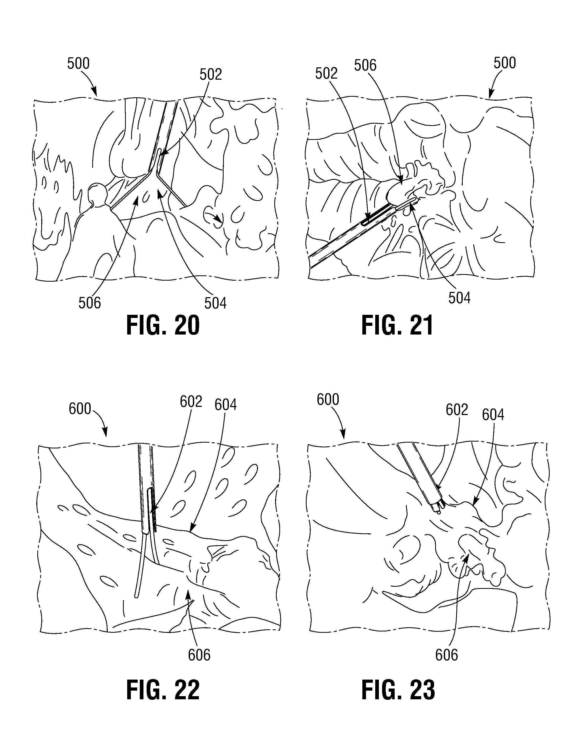
FIGS. 20 and 21 illustrate implantation of a leaflet clip into a mitral valve.
FIGS. 22 and 23 illustrate implantation of a leaflet clip into a tricuspid valve.
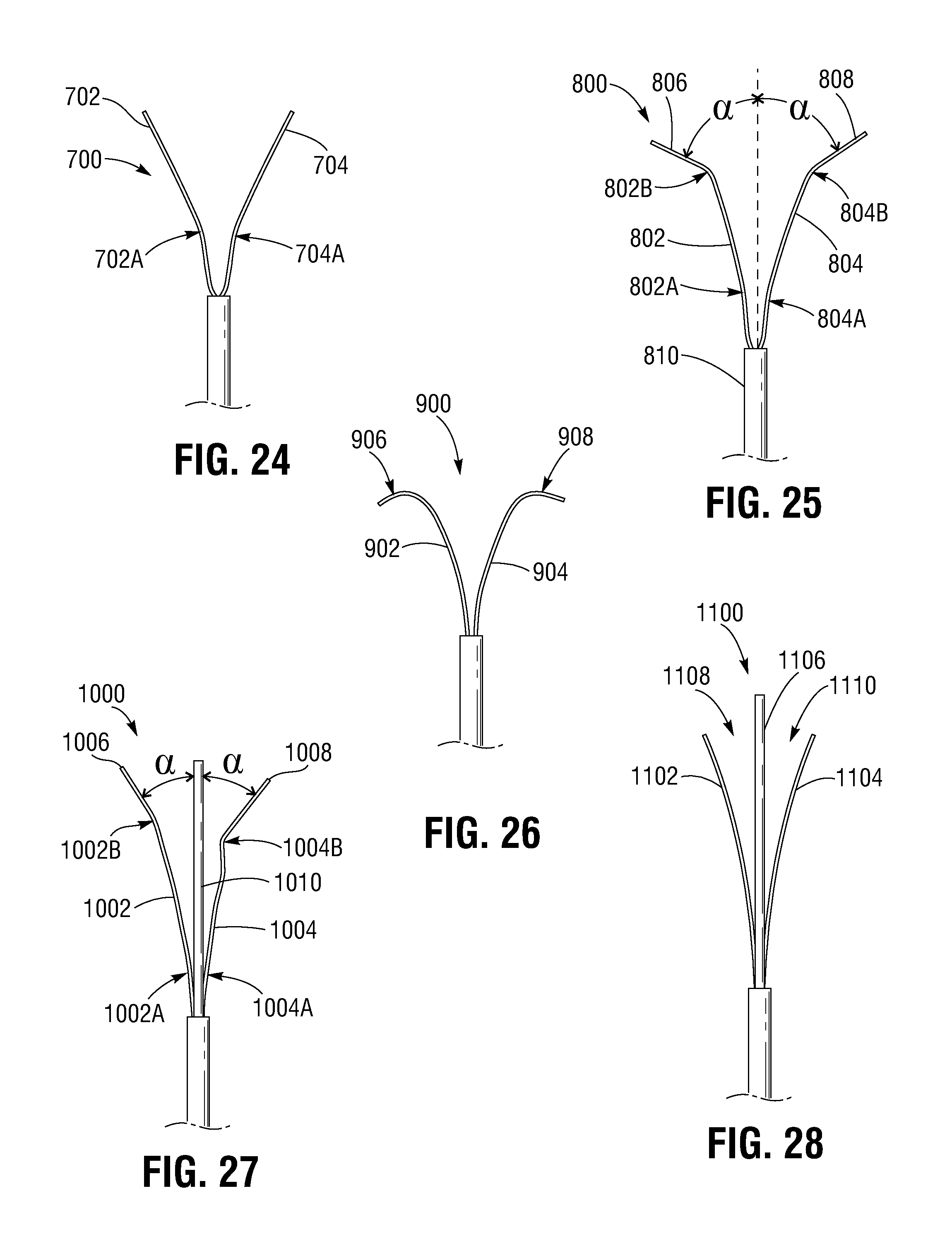
FIGS. 24-32 illustrate various representative configurations of clipping arms of leaflet clips.
FIG. 33 illustrates another embodiment of a leaflet clip including a passive leaflet engaging mechanism.
FIG. 34 illustrates movement of the leaflets of a heart valve with respect to the clipping arms of a leaflet clip as the clipping arms are moved from an open position to a closed position.
FIG. 35 is a side elevation view of another embodiment of a leaflet clip including a movable and expandable covering illustrated in a distal position.
FIG. 36 is a side elevation view the leaflet clip of FIG. 26 illustrating the active leaflet engaging mechanism in a proximal position in an expanded configuration.
FIGS. 37 and 38 are side elevation views of another embodiment of a leaflet clip including an active leaflet engaging mechanism including a plurality of protuberances.
FIG. 39 illustrates an embodiment of a leaflet clip coupled to a delivery system with the with the clipping arms of the leaflet clip extending distally relative to the delivery system.
FIG. 40 illustrates an embodiment of a leaflet clip coupled to a delivery system with the clipping arms of the leaflet clip extending proximally relative to the delivery system.
FIG. 41 illustrates another embodiment of a leaflet clip coupled to a delivery system with its clipping arms extending proximally with respect to the delivery system.
FIGS. 42 and 43 illustrate another embodiment of a leaflet clip including tissue gathering regions.
FIGS. 44 and 45 schematically illustrate the function of the tissue gathering regions of the leaflet clip of FIG. 42.
FIG. 46 illustrates the leaflet clip of FIGS. 42 and 43 with the clipping arms in the closed position and leaflets disposed in the tissue gathering regions.
FIG. 47 illustrates another embodiment of a leaflet clip including a helical member disposed within a tubular central member.
FIG. 48 illustrates another embodiment of a leaflet clip including a helical member disposed about the clipping arms.
FIG. 49 is a perspective view of another embodiment of a delivery system that can be used in combination with any of the leaflet clips described herein.
FIG. 50 is an enlarged view of the distal end portion of the delivery device of FIG. 49.
DETAILED DESCRIPTION
For purposes of this description, certain aspects, advantages, and novel features of the embodiments of this disclosure are described herein. The disclosed methods, apparatuses, and systems should not be construed as limiting in any way. Instead, the present disclosure is directed toward all novel and nonobvious features and aspects of the various disclosed embodiments, alone and in various combinations and sub-combinations with one another. The methods, apparatus, and systems are not limited to any specific aspect or feature or combination thereof, nor do the disclosed embodiments require that any one or more specific advantages be present or problems be solved.
Features, integers, characteristics, compounds, chemical moieties or groups described in conjunction with a particular aspect, embodiment or example are to be understood to be applicable to any other aspect, embodiment or example described herein unless incompatible therewith. All of the features disclosed in this specification (including any accompanying claims, abstract and drawings), and/or all of the steps of any method or process so disclosed, may be combined in any combination, except combinations where at least some of such features and/or steps are mutually exclusive. The invention is not restricted to the details of any disclosed embodiments. The invention extends to any novel one, or any novel combination, of the features disclosed in this specification (including any accompanying claims, abstract and drawings), or to any novel one, or any novel combination, of the steps of any method or process so disclosed.
Although the operations of some of the disclosed methods are described in a particular, sequential order for convenient presentation, it should be understood that this manner of description encompasses rearrangement, unless a particular ordering is required by specific language set forth below. For example, operations described sequentially may in some cases be rearranged or performed concurrently. Moreover, for the sake of simplicity, the attached figures may not show the various ways in which the disclosed methods can be used in conjunction with other methods. Additionally, the description sometimes uses terms like "provide" or "achieve" to describe the disclosed methods. These terms are high-level abstractions of the actual operations that are performed. The actual operations that correspond to these terms may vary depending on the particular implementation and are readily discernible by one of ordinary skill in the art.
As used in this application and in the claims, the singular forms "a," "an," and "the" include the plural forms unless the context clearly dictates otherwise. Additionally, the term "includes" means "comprises." Further, the terms "coupled" and "associated" generally mean electrically, electromagnetically, and/or physically (e.g., mechanically or chemically) coupled or linked and does not exclude the presence of intermediate elements between the coupled or associated items absent specific contrary language.
As used herein, the term "proximal" refers to a position, direction, or portion of a device that is closer to the user and further away from the implantation site. As used herein, the term "distal" refers to a position, direction, or portion of a device that is further away from the user and closer to the implantation site. Thus, for example, proximal motion of a device is motion of the device toward the user, while distal motion of the device is motion of the device away from the user. The terms "longitudinal" and "axial" refer to an axis extending in the proximal and distal directions, unless otherwise expressly defined.
As used herein, the terms "integrally formed" and "unitary construction" refer to a construction that does not include any welds, fasteners, or other means for securing separately formed pieces of material to each other.
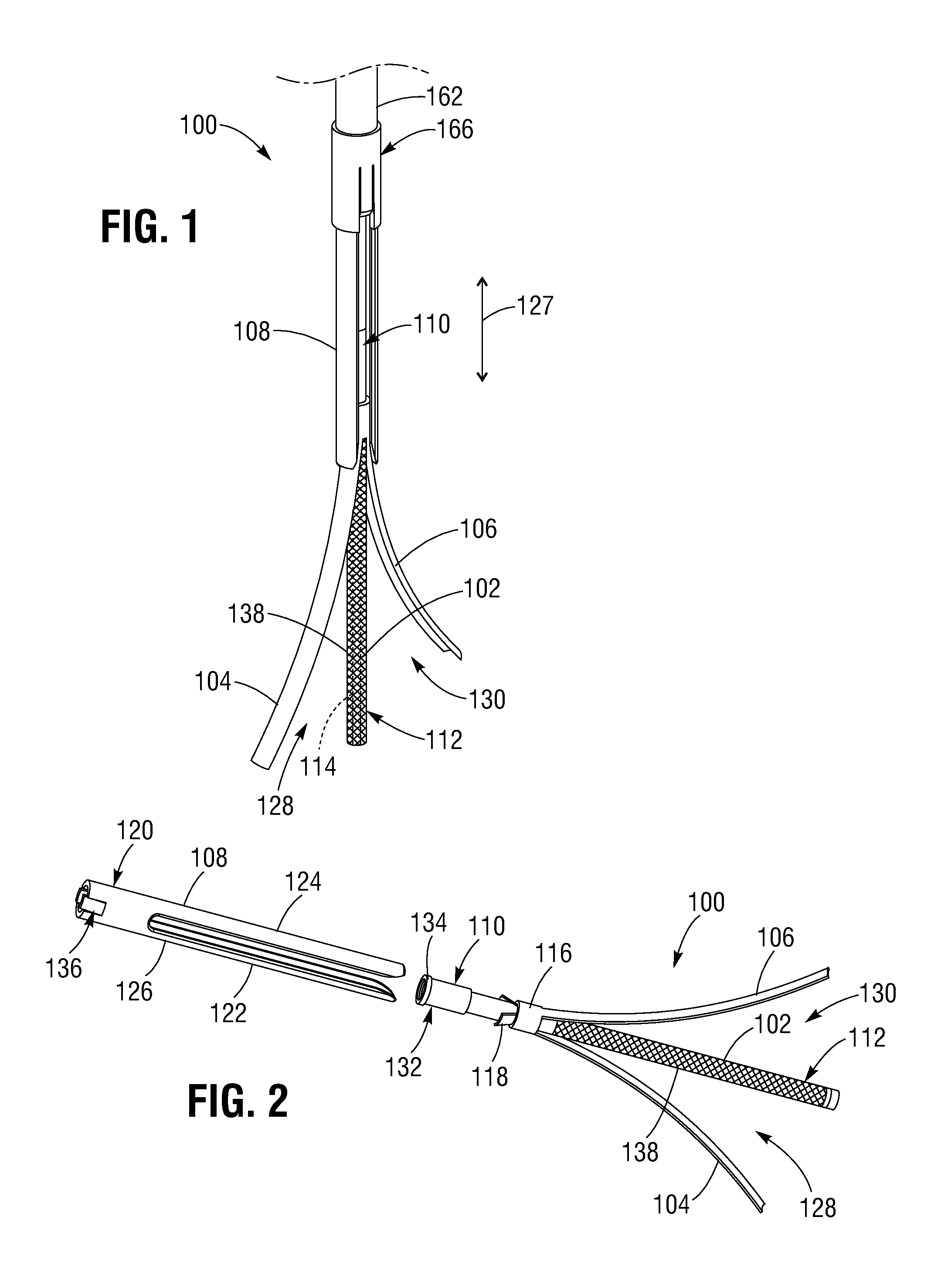
FIGS. 1-3 illustrate a representative embodiment of a leaflet clip 100 including a central elongated member, or shaft, 102, first and second clipping arms 104, 106, and a tubular member 108 (also referred to as a "cover" or "sheath") coaxially disposed over the elongated member 102. The elongated member 102 can have a proximal end portion 110 and a distal end portion 112, and can define a lumen 114 (indicated in phantom in FIG. 1) through which a guide wire can be inserted.
The clipping arms 104, 106 can be coupled to the proximal end portion 110 of the elongated member, and can be movable between an open position (FIGS. 1 and 2) and a closed position (FIG. 3). In the embodiment shown, the clipping arms 104, 106 extend distally from the proximal end portion 110 of the elongated member, and can be shape set such that they curve radially away from the elongated member 102 when in the open position. In some embodiments, the clipping arms 104, 106 can have a curved cross-sectional profile (in a plane perpendicular to the longitudinal axis of the elongated member 102) such that they lay substantially flush against the elongated member 102 when in the closed position.
As shown in FIG. 2, the clipping arms 104, 106 can extend from an annular collar 116 situated coaxially on the proximal end portion 110 of the elongated member 102. In some embodiments, the clipping arms 104, 106 can be integrally formed with the collar 116, or can be separately formed and attached to the collar 116, as desired. For example, in some embodiments, the clipping arms 104, 106 can be hinged. The annular collar 116 can also include one or more radially extending protrusions or tabs 118 extending from the collar 116 and configured to prevent rotation of the tubular member 108 (i.e., "index" the tubular member 108) with respect to the elongated member 102, as further described below. In some embodiments, the clipping arms 104, 106, and/or the collar 116, can be made from any biocompatible material such as, for example, titanium, nickel titanium or nitinol, plastic, stainless steel, etc.
The tubular member 108 can be coaxially disposed over the elongated member 102, and can be axially movable with respect to the elongated member 102 between a first position (FIG. 1) and a second position (FIG. 3), as indicated by arrow 127 of FIG. 1. Referring to FIG. 2, the tubular member 108 can include a proximal end portion 120, and first and second extension portions 122, 124 corresponding to the first and second clipping arms 104, 106. The tubular member 108 can also define slots 126 between the extension portions 122, 124, which can receive the tabs 118 of the collar 116 when the tubular member 108 is in the second position, as shown in FIG. 3. In this manner, as the tubular member 108 moves distally from the first position to the second position, the tabs 118 can travel in the respective slots 126, and the first and second extension portions 122, 124 can contact the first and second clipping arms 104, 106, respectively, and urge the clipping arms 104, 106 into the closed position.
The clipping arms 104, 106 and the elongated member 102 can define respective leaflet receiving regions configured to receive the leaflets of a heart valve when the clipping arms 104, 106 are in the open position. For example, the first clipping arm 104 and the elongated member 102 can define a first leaflet receiving region 128, and the second clipping arm 106 together with the elongated member 102 can define a second leaflet receiving region 130, as shown in FIGS. 1 and 2. During implantation, the leaflet clip 100 can be positioned adjacent a commissure of a heart valve such that one valve leaflet is received in the first leaflet receiving region 128 and a corresponding valve leaflet is received in the second leaflet receiving region 130, while the elongated member 102 extends between the leaflets. In this manner, when the clipping arms 104, 106 are moved from the open position to the closed position, the leaflets can be captured between the first and second clipping arms 104, 106, respectively, and the elongated member 102, as further described below.
In some embodiments, the proximal end portion 110 of the elongated member 102 can include a locking or retaining feature 132 to retain the tubular member 108 in the second position. In the embodiment shown, the retaining feature 132 can comprise a protuberance 134 located on the proximal end of the elongated member 102. The protuberance 134 can have a diameter greater than a diameter of the tubular member 108 such that if the proximal end portion 120 of the tubular member 108 is advanced over the protuberance 134, the protuberance 134 will cause the proximal end portion 120 to expand beyond its natural diameter. Once the proximal end portion 120 of the tubular member 108 is advanced distally of the protuberance 134, the proximal end portion 120 can return to its natural (non-deflected and non-expanded) diameter such that the protuberance 134 restrains proximal movement of the tubular member 108 past the protuberance 134, thereby locking the clipping arms 104, 106 in the closed position.
In this manner, when the leaflet clip 100 has been positioned at a desired location relative to the leaflets of a heart valve, the tubular member 108 can be advanced fully to the second position, locking the tubular member 108 distally of the protuberance 134, and thereby locking the clipping arms 104, 106 in the closed position such that the leaflet clip 100 is retained on the leaflets. In some embodiments, the proximal end portion 120 of the tubular member 108 can define one or more notches 136 to allow the walls of the proximal end portion 120 to flare radially outwardly as they pass over the protuberance 134, thereby allowing the proximal end portion 120 to pass more easily over the protuberance 134. The walls of the proximal end portion 120 can then return to their natural diameter distally of the protuberance 134.
In some embodiments, the leaflet clip 100 can include active or passive leaflet engaging mechanisms, which can be disposed on the elongated member 102, to aid in engaging and retaining the leaflets of a heart valve in the respective first and second leaflet receiving regions 128, 130 as the clipping arms 104, 106 are moved from the open position to the closed position. For example, the leaflet clip 100 can include a passive leaflet engaging mechanism configured as a covering 138 disposed on the distal end portion 112 of the elongated member 102. The covering 138 can have a textured surface such that it can frictionally engage the leaflets when the leaflets are received in the leaflet receiving regions 128, 130, and retain the leaflets in the respective leaflet receiving regions 128, 130 as the clipping arms 104, 106 are moved from the open position to the closed position. In some embodiments, the covering 138 can comprise a woven or braided fabric, or can be a polymeric tube or sleeve configured to be positioned on the distal end portion 112 of the elongated member 102. In some embodiments, the covering 136 can be made of any of various natural or synthetic materials, such as polyethylene terephthalate (PET), foam, silicone, or suture material.
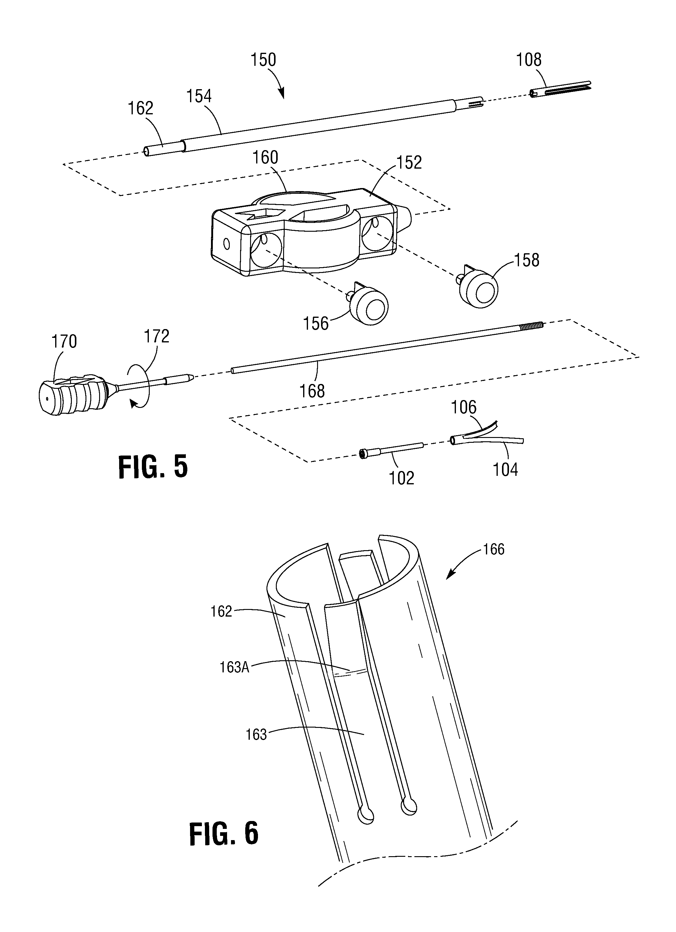
FIGS. 4 and 5 illustrate the leaflet clip 100 coupled to a representative embodiment of a delivery system 150 that can be used to deliver leaflet clips such as the leaflet clip 100 to a desired heart valve. The delivery system 150 can comprise a main handle body 152 including an outer conduit or shaft 154 movable between a proximal position and a distal position, control buttons 156, 158, and two grip portions 160. Referring to FIG. 5, the delivery system 150 can further include an actuator conduit, or shaft, 162 disposed coaxially within the outer conduit 154 and coupled to the tubular member 108, such that proximal or distal motion of the actuator conduit 162 in the directions indicated by arrow 164 causes corresponding proximal or distal motion of the tubular member 108 with respect to the elongated member 102 and the clipping arms 104, 106.
In some embodiments, the actuator conduit 162 can include a coupling device 166 to couple the actuator conduit 162 to the proximal end portion 120 of the tubular member 108, as shown in FIGS. 6-8. In some embodiments, the coupling device 166 can comprise a pair of angled tabs 163, 165 coupled to the actuator conduit 162 at their respective proximal ends and shape set such that respective angled portions 163A, 165A of the tabs 163, 165, extend radially beyond the diameter of the actuator conduit 162, as shown in FIG. 7. In this manner, the tabs 163, 165 can act as springs such that when the outer conduit 154 is disposed around the distal end of the actuator conduit 162 (i.e., in the distal position), the inner surface of the outer conduit 154 can contact the angled portions 163A, 165A of the tabs 163, 165 and deflect them radially inward with respect to the actuator conduit 162, as shown in FIG. 8. In this manner, the tabs 163, 165 can extend into respective openings defined in the proximal end portion 120 of the tubular member 108, such as opening 167 shown in FIGS. 9 and 10. This can allow the tabs 163, 165 of the actuator member 162 to releasably engage the tubular member 108 such that proximal and/or distal motion of the actuator conduit 162 causes proximal and/or distal motion of the tubular member 108 and, thereby, movement of the clipping arms 104, 106 between the open and closed positions. In alternative embodiments, the actuator conduit 162 can be coupled to the tubular member 108 by any suitable coupling mechanism, including, for example, threads, clips, a ball and detent system, etc.
Proximal motion of the actuator conduit 162 and, thereby, of the tubular member 108, can be limited by one or more of various controllable motion limiting mechanisms (e.g., cams, hard stops, pull tabs, etc.) to reduce the risk of locking the tubular member 108 in the distal position prior to successful positioning of the leaflet clip 100 (i.e., to prevent the proximal end portion 120 of the tubular member 108 from moving distally with respect to the protuberance 134 of the elongated member 102). The motion limiting mechanism(s) can establish a motion limit stop beyond which distal motion of the actuator conduit 162, outer conduit 154, and tubular member 108, as a coupled unit, are restrained relative to the inner shaft 168. In the embodiment shown, such motion limiting mechanisms can be controlled by the control button 156. For example, actuation of the control button 156 can allow the actuator conduit 162, outer conduit 154, and tubular member 108 to be moved past the limit stop such that the proximal end portion 120 of the tubular member 108 can move distally over the protuberance 134 of the elongated member 102, thereby locking the clipping arms 104, 106 in the closed position. Thus, after the leaflet clip 100 has been clipped to the leaflets of a heart valve, the control button 156 can be actuated to allow distal motion of the actuator conduit 162, outer conduit 154, and tubular member 108 past the limit stop to lock the clipping arms 104, 106 in the closed position.
Proximal motion of the outer conduit 154 can also be limited by one or more controllable motion limiting mechanisms to reduce the risk of accidental release of the tubular member 108 from the actuator conduit 162 during positioning of the leaflet clip 100. In the embodiment shown, such motion limiting mechanisms can be controlled by the control button 158. For example, actuation of the control button 158 can allow the outer conduit 154 to be moved proximally with respect to the actuator conduit 162, thereby uncovering the tabs 163, 165, allowing them to resume their natural non-deflected shape and disengage from the tubular member 108. This can release the tubular member 108 from the delivery system 150. Thus, after final positioning of the leaflet clip 100 in a heart valve, the control button 158 can be actuated to allow proximal motion of the outer conduit 154 and release of the actuator conduit 162 from the tubular member 108.
As for the embodiment shown in FIG. 5, the delivery system 150 can further include an inner member, or shaft, 168 disposed coaxially within the actuator conduit 162 and coupled at one end to the proximal end portion 110 of the elongated member 102, and at the opposite end to a handle member 170. In the embodiment shown, the inner member 168 can be coupled to the proximal end portion 110 of the elongated member 102 by threads such that rotation of the handle member 170 in the direction indicated by arrow 172 can uncouple the inner member 168 from the elongated member 102. For example, the distal end portion of the inner member 168 can have male threads that threadably engage female threads formed on an inner surface of the proximal end portion 110 of the elongated member 102. In this manner, after final positioning of the leaflet clip 100 in a heart valve, the actuator member 162 can be disengaged from the tubular member 108, the inner member 168 can be disengaged from the elongated member 102, and the delivery system 150 can be retracted, leaving the leaflet clip 100 in place.
In some embodiments, the diameter of the outer conduit 154 can be about ten French, and the system 150 can be configured for use with a guide wire (for example, threaded through the lumen 114 of the elongated member 102). In some embodiments, the respective conduits 154, 162 and the inner member 168 of the delivery system 150 can be flexible or rigid, as desired. In some embodiments, the delivery system 150 can include steering elements to aid in positioning and orienting the leaflet clip 100 with respect to a heart valve. Also, it should be understood that the respective conduits 154, 162 and the inner member 168 of the delivery system 150 are shown at reduced length for purposes of illustration, and can be any suitable length.
In use, the delivery system 150 can be introduced into a patient's vasculature (e.g., via the femoral artery or other suitable access point) and percutaneously advanced to the patient's heart with the clipping arms 104, 106 in the closed position (but not locked) using any of various delivery techniques. In a transfemoral procedure, the delivery device can be inserted through a femoral artery and the aorta to the heart in a retrograde direction (typically, but not exclusively used for deploying a clip on the leaflets of the aortic or mitral valves). Similarly, the delivery device can be inserted through a femoral vein and the vena cava to the right side of the heart in an antegrade direction (typically, but not exclusively used for deploying a clip on the leaflets of the pulmonary or tricuspid valves). In a transventricular procedure, the delivery device can be inserted through a surgical incision made in the chest and on the bare spot on the lower anterior ventricle wall (typically, but not exclusively used for deploying a clip on the leaflets of the aortic or mitral valves). Similarly, the delivery device can be inserted through a surgical incision on the wall of the right ventricle to access the pulmonary or tricuspid valves. In a transatrial procedure, the delivery device can be inserted through a surgical incision made in the wall of the left or right atrium to access the native valves on the left or right sides, respectively, of the heart. In a transaortic procedure, the delivery device can be inserted through a surgical incision made in the ascending aorta and advanced toward the heart (typically, but not exclusively used deploying a clip on the leaflets of the aortic or mitral valves). In a transeptal procedure, the delivery device can be advanced to the right atrium, such as via a femoral vein, and through the septum separating the right and left ventricles (typically, but not exclusively used for deploying a clip on the leaflets of the aortic or mitral valves). Further details of delivery techniques for accessing the native valves of the heart are disclosed in U.S. Patent Publication No. 2014/0067052, which is incorporated herein by reference.
Once located proximate the desired heart valve, the clipping arms 104, 106 are expanded by retracting the actuator conduit 162 to retract the tubular member 108 relative to the clipping arms 104, 106. The leaflet clip 100 can then be positioned with respect to a commissure of the valve, and can be distally advanced and/or retracted as needed to position the leaflet clip 100 such that one leaflet of the commissure is received in the first leaflet receiving region 128 and the second leaflet of the commissure is received in the second leaflet receiving region 130. When the leaflet clip 100 is suitably positioned, the actuator conduit 162 can be advanced such that the tubular member 108 moves distally with respect to the elongated member 102 and urges the clipping arms 104, 106 toward the closed position.
When the clipping arms are 104, 106 are in the closed position, the clipping strength of the leaflet clip 100 can be tested by pulling proximally on the delivery system 150. As used herein, the terms "clip retention force" and "clipping strength" refer to a force in the proximal direction that can be withstood by a leaflet clip without disengaging from the leaflets of a heart valve when the clipping arms are in the closed position. In some embodiments, the delivery system 150 can include a strain gauge or other device to measure the force applied to the leaflet clip 100. In some embodiments, the leaflet clip 100 can withstand a proximal force application of from between 1 N and about 10 N while remaining clipped to the valve leaflets. If, for example, the leaflet clip 100 is not suitably positioned, or the leaflet clip 100 does not exhibit suitable clipping strength when clipped to the leaflets, the tubular member 108 can be retracted by proximal motion of the actuator conduit 162, causing the clipping arms 104, 106 to reopen, and allowing the leaflet clip 100 to be repositioned. When the leaflet clip 100 is suitably positioned with respect to the valve leaflets, the actuator member 162 can advance the tubular member 108 distally of the protuberance 134, thereby locking the clipping arms 104, 106 in the closed position. The inner member 168 can then be disengaged from the elongated member 102, and the delivery system 150 can be retracted, leaving the leaflet clip 100 in place on the valve leaflets.
The leaflet clip 100, and any of the other leaflet clip embodiments described herein, can be used to treat valvular insufficiency or to remodel the annulus of a heart valve. For example, FIG. 11 illustrates three leaflet clips 202, 204, 206 similar to the leaflet clip 100 of FIG. 1 situated in a native aortic valve 200. The native aortic valve 200 can include three valve leaflets 210, 212, 214 attached to a valve annulus 216. The valve leaflets 210 and 212 can form a first commissure 218, the leaflets 212 and 214 can form a second commissure 220, and the leaflets 210 and 214 can form a third commissure 222. The leaflet clips 202, 204, 206 are shown situated at the first, second, and third commissures 218, 220, 222, respectively, such that the leaflet clip 202 engages the leaflets 210 and 212, the leaflet clip 204 engages the leaflets 212 and 214, and the leaflet clip 206 engages the leaflets 214 and 210. The leaflet clips 202, 204, 206 are also shown situated near the wall of the valve annulus 216. In this manner, the leaflet clips 202, 204, 206 can improve coaptation of the leaflets 210, 212, 214 at the respective commissures 218, 220, 222, thereby reducing regurgitation through the valve 200 due to valvular insufficiency. Additionally, although the leaflet clips 202, 204, 206 are shown clipped to the respective valve leaflets adjacent the annulus 216, the leaflet clips 202, 204, 206 can be clipped to the valve leaflets at any suitable location along the leaflets, including at the centers of the commissures, as desired.
The leaflet clips 202, 204, 206 can also remodel the annulus 216 of the valve 200 to reduce dilatation of the annulus 216 and/or to address abnormalities in the shape of the annulus 216. For example, FIG. 12 schematically illustrates remodeling of the annulus 216 to reduce its diameter from a dilated annulus diameter D.sub.1 to a target or reduced annulus diameter D.sub.2 using leaflet clips, such as the clips 202, 204, 206. Such remodeling or diameter reduction may be achieved by reducing the circumference of the annulus 216, as illustrated in FIGS. 13 and 14.
For example, FIG. 13 illustrates the leaflet clip 202 with its clip arms in the open position. Hash marks 230, 232 are shown on the tissue for purposes of indicating an initial distance between the clipping arms, as well as an initial reference for the circumference of the annulus. Dashed line 234 indicates the native contour of the valve cusp. FIG. 14 illustrates the leaflet clip 202 with its clip arms in the closed position such that the tissue of the annulus 216 adjacent the clip 202 is gathered and clipped together by the leaflet clip. This can reduce the circumference and, thereby, the diameter, of the annulus 216, as indicated by movement of the hash marks 230, 232 toward one another.
The effect of such a diameter reduction is further illustrated in FIGS. 15, 16, and 17, wherein the distances a, b, and c between three points 302, 304, 306 indicated schematically in FIG. 15, and illustrated on the annulus of a heart valve 300 in FIGS. 16 and 17, are reduced after application of three leaflet clips 308, 310, 312 to the commissures of the valve 300. In some embodiments, the diameter of the annulus can be reduced from a dilated diameter of about 29 mm to a target diameter of about 25 mm. In some embodiments, the diameter of the annulus can be reduced by from about 10% to about 50%. In some embodiments, the diameter of the annulus can be reduced by about 14%.
Any of the leaflet clips disclosed herein can also be used in combination with one or more support rings, as shown in FIGS. 18 and 19. FIG. 19 illustrates three leaflet clips 402, 404, 406 clipped to the commissures of an aortic valve 400 as seen from the aortic root. FIG. 18 illustrates the location of a support ring 408 on the ventricular side of the aortic valve 400, which can cooperate with the leaflet clips 402, 404, 406 to remodel and/or reduce the diameter of the aortic valve 400, as shown in FIG. 19. The extensions 410 of the central shaft of each of the leaflet clips within the left ventricle can extend through respective openings of the ring 408 as shown. Alternatively, the ring 408 can be placed around the extensions 410. The leaflet clips 402, 404, 406, together with the support ring 408, or independent of any supporting device or structure, can also be used to achieve folding or plication of the commissures, as shown in FIG. 19.
As stated above, any of the leaflet clips disclosed herein can also be used to treat valvular insufficiency or to remodel the annulus of the mitral valve and/or the tricuspid valve in addition to the aortic valve. FIGS. 20 and 21 illustrate placement of a leaflet clip 502 similar to the leaflet clip 100 of FIG. 1 onto the leaflets 504, 506 of a mitral valve 500. As illustrated in FIGS. 20 and 21, the relatively small width of the clip arms 508 of the leaflet clip 500 relative to the chordae of the mitral valve 500 can reduce interference by the clip arms 508 with the chordae both during implantation of the leaflet clip 502 and after implantation. In use, one or more leaflet clips 502 can be placed at or near the center of the leaflets 504, 506, or near the annulus, as desired.
FIGS. 22 and 23 illustrate placement of a leaflet clip 602 similar to the leaflet clip 100 of FIG. 1 onto leaflets 604, 606 of a tricuspid valve 600. The leaflet clip 602, alone, or in combination with one or more additional leaflet clips, can be used to reduce valvular insufficiency and/or remodel the valve annulus, as described above.
FIGS. 24-32 illustrate various configurations of clipping arms that may be used in combination with any of the leaflet clips disclosed herein. Additionally, although the leaflet clips of FIGS. 24-26 are illustrated without central elongated members, it should be understood that any of the leaflet clips described herein can include an elongated member (e.g., member 102) or not, as desired. FIG. 24 illustrates a leaflet clip 700 wherein the clipping arms 702, 704 exhibit bends indicated at 702A and 704A, and extend outwardly without exhibiting any significant further curvature (i.e., the clipping arms 702, 704 are relatively straight).
FIG. 25 illustrates a leaflet clip 800 wherein the clipping arms 802, 804 exhibit two bends, the first bends being indicated at 802A, 804A, and the second bends indicated at 802B, 804B, respectively, such that the clipping arms 802, 804 include respective distal portions 806, 808 angled away from the tubular member 810 at an angle .alpha. greater than about 45 degrees.
FIG. 26 illustrates a leaflet clip 900 wherein the clipping arms 902, 904 have curved distal end portions 906, 908.
FIG. 27 illustrates a leaflet clip 1000 similar to the leaflet clip 800 of FIG. 25 wherein the clipping arms 1002, 1004 exhibit first bends indicated at 1002A, 1004A and second bends indicated at 1002B, 1004B such that distal portions 1006, 1008 of the clipping arms 1002, 1004 are angled away from the tubular member 1010 at an angle .alpha. less than about 45 degrees.
FIG. 28 illustrates a leaflet clip 1100 wherein the clipping arms 1102, 1104 extend distally adjacent the elongated member 1106 before bending away from the elongated member 1106 such that the areas of the respective leaflet receiving regions 1108, 1110 are reduced.
FIGS. 29 and 30 illustrate an embodiment of a clipping arm 1600 including one or more openings 1602 defined in the clipping arm 1600. The clipping arm 1600 can include openings 1602 located at the distal end portion of the clipping arm 1600, as shown in FIG. 29, or spaced apart along the length of the clipping arm 1600, as shown in FIG. 30. The clipping arm 1600 can include a fibrous covering or sleeve 1604, which can be disposed around the distal end portion of the clipping arm 1600, as in FIG. 29, or along the entire length of the clipping arm 1600, as shown in FIG. 30. The covering 1604 can be retained on the clipping arm 1600 by suture threads 1606, which can be disposed about the covering 1604 and received or laced through the plurality of openings 1602 in order to retain the covering 1604 on the clipping arm 1600. In this manner, the covering 1604 can serve to frictionally engage the tissue of the leaflets of a heart valve while reducing the risk of damage to the leaflets. In alternative embodiments, the clipping arm 1600 can include any suitable number of openings 1602 located at any suitable location along the length of the clipping arm, and can include a covering extending along any suitable proportion of the clipping arm 1600, as desired. The covering 1604 can be made from a suitable biocompatible fabric, such as PET.
FIG. 31 illustrates another embodiment of a clipping arm 1700 including a proximal end portion 1702 and a distal end portion 1704. In the embodiment shown, clipping arm 1700 can be tapered such that the proximal end portion 1702 has a first diameter or width W.sub.1 and the distal end portion 1704 has a second diameter or width W.sub.2, the first diameter W.sub.1 being greater than the second diameter W.sub.2. This can allow the distal end portion 1704 a greater degree of flexibility relative to the proximal end portion 1702 in the directions indicated by arrow 1706, which can reduce the risk of damage to the tissue of native heart valve leaflets. The distal end portion 1704 can also include a protuberance 1708 having a relatively large diameter relative to the diameter W.sub.2 of the distal end portion 1704, which can reduce the risk of puncturing the native leaflets during or after implantation of the leaflet clip.
FIG. 32 illustrates another embodiment of a clipping arm 1800 including an inner member 1802 and an over-molded covering 1804 (i.e., an "insert molding"). The covering 1804 can be fabricated from any biocompatible polymer (e.g., silicone, ePTFE, etc.), and can include texturing, dimpling, or other surface features to aid in engaging and retaining the tissue of a native valve leaflet. The inner member 1802 can be fabricated from metal or polymer materials, as desired.
FIG. 33 illustrates another embodiment of a leaflet clip 1200 similar to the leaflet clip 100 of FIG. 1. The leaflet clip 1200 can include an elongated member 1202 and two clipping arms 1204, 1206. The leaflet clip 1200 is illustrated without a tubular member (sheath) for purposes of illustration. The leaflet clip 1200 can include a passive leaflet engaging mechanism configured as a tube or sleeve 1208 and disposed on a distal end portion 1210 of the elongated member 1202. The sleeve 1208 can be made from, for example, polyester, and can be configured to frictionally engage and retain the leaflets of a heart valve between the respective clipping arms 1204, 1206 and the elongated member 1202 as the clipping arms are moved from the open position to the closed position.
FIG. 34 schematically illustrates the relative positions of the native leaflets 1302, 1304 of a heart valve 1306 relative to the clipping arms 1308, 1310 of a leaflet clip 1300 before and after the clipping arms 1308, 1310 are moved from the open position to the closed position. In a manner similar to leaflet clip embodiments described above, distal motion of a tubular member 1316 from a first position (indicated in solid lines) to a second position (indicated in phantom) can cause the clipping arms 1308, 1310 to move from the open position to the closed position. When the leaflets 1302, 1304 are received in the respective leaflet receiving regions 1312, 1314, the ends of the clipping arms 1308, 1310 can contact the leaflets 1302, 1304 at respective points A along the respective leaflets 1302, 1304. In some embodiments, as the clipping arms 1308, 1310 are moved from the open position to the closed position, the leaflets 1302, 1304 can slip such that when the clipping arms 1308, 1310 are in the closed position, they contact the leaflets 1302, 1304 at respective points B. Depending upon the degree of gripping strength desired, it may be advantageous, in certain circumstances, to retain a greater proportion of the leaflets 1302, 1304 in the leaflet receiving regions 1312, 1314 as the clipping arms are moved from the open position to the closed position.
To such ends, FIGS. 35 and 36 illustrate another embodiment of a leaflet clip 1400 including an elongated member 1402 and two clipping arms 1404, 1406. The leaflet clip 1400 can include an active leaflet engaging mechanism 1408 configured as a woven or braided covering 1410 disposed on the elongated member 1402 between a stationary proximal retaining member 1412 and a distal retaining member 1414 movable between a distal position and a proximal position. When the leaflet clip 1400 is positioned such that native leaflets 1416, 1418 are received in the respective leaflet receiving regions 1420, 1422, the distal retaining member 1414 can be moved proximally in the direction indicated by arrow 1424 of FIG. 36 (e.g., by actuation of a member disposed within the elongated member 1402). Motion of the distal retaining member 1414 toward the proximal retaining member 1412 can cause the covering 1410 to bunch such that it expands or bulges radially away from the elongated member 1402 and engages the leaflets 1416, 1418 between the covering 1410 and the respective clipping arms 1404, 1406. When the distal retaining member 1414 is in the proximal position, the tubular member 1426 can be advanced distally such that the clipping arms 1404, 1406 are moved from the open position to the closed position. In this manner, retention of the leaflets 1416, 1418 in the respective leaflet receiving regions 1420, 1422 while the clipping arms 1404, 1406 are moved to the closed position can result in a greater proportion of the leaflets being retained against the elongated member 1402 by the leaflet clip 1400, resulting in a stronger grip on the leaflets and improved performance of the leaflet clip 1400. Additionally, the leaflet engaging mechanism 1408 can reduce slippage of the leaflets 1416, 1418 past the clipping arms 1404, 1406 as they moved to the closed position, helping to ensure that a greater proportion of the leaflets 1416, 1418 are retained in the leaflet receiving regions 1420, 1422 and increasing retention of the leaflet clip 1400 on the leaflets 1416, 1418.
FIGS. 37 and 38 illustrate another embodiment of a leaflet clip 1500 including an elongated member 1502, two clipping arms 1504, 1506, and a sheath 1526. The leaflet clip 1500 can include an active leaflet engaging mechanism 1508 configured as a sleeve 1510 including a plurality of protuberances 1512 axially spaced apart from one another along the sleeve 1510. The sleeve 1510 can be movable between a distal position and a proximal position relative to the clipping arms 1504, 1506, as indicated by arrow 1514 (FIG. 37). The protuberances 1512 can have respective edge portions 1516 configured to engage or grip native leaflets 1518, 1520 disposed in respective leaflet receiving regions 1522, 1524 when the sleeve 1510 is moved proximally, as depicted in FIG. 38. Thus, when the clipping arms 1504, 1506 are moved to the closed position (e.g., by advancing the sheath 1526 over the clipping arms), the edge portions 1516 of the protuberances 1512 can engage and retain the leaflets 1518, 1520 within the leaflet receiving regions 1522, 1524, thereby inhibiting the leaflets from slipping out of the leaflet receiving regions. This can result in a greater proportion of the leaflets 1518, 1520 being retained against the elongated member 1502 by the clipping arms 1504, 1506, resulting in a stronger grip on the leaflets and improved performance of the leaflet clip 1500.
In the embodiment shown, the sleeve 1510 includes three protuberances 1512. However, it should be understood that the sleeve 1510 can include any suitable number of protuberances having any suitable size and spacing relative to one another. In some embodiments, the sleeve 1510 can be disposed on the elongated member 1502, which can be movable proximally and/or distally. In alternative embodiments, the sleeve 1510 can be disposed on a shaft or conduit coaxially disposed within (or over the exterior) of the elongated member 1502 and independently actuated from outside the body. In some embodiments, the sleeve 1510 can be made from suitable natural or synthetic material, such as, for example, polyester. In some embodiments, the spaces between the protuberances 1512 can act as tissue gathering regions (see description regarding FIGS. 42-46), helping to increase the clipping strength of the leaflet clip 1500.
FIGS. 39 and 40 schematically illustrate alternative coupling orientations of leaflet clips with respect to delivery systems. In FIG. 39, a leaflet clip 1900 is shown coupled to a delivery system 1902 with clipping arms 1904, 1906 extending distally with respect to the delivery system 1902. FIG. 40 illustrates a leaflet clip 2000 coupled to a delivery system 2002 with the clipping arms 2004, 2006 extending proximally with respect to the delivery system 2002. It should be understood that these coupling orientations may be applied to any of the leaflet clips described herein, and can facilitate the various delivery techniques described above.
FIG. 41 illustrates another embodiment of a leaflet clip 2100 coupled to a delivery system 2102 such that clipping arms 2104, 2106 extend proximally with respect to the delivery system 2102, similar to the embodiment of FIG. 40. The leaflet clip 2100 can include a tubular member 2108 disposed coaxially about an elongated member 2110, and movable between a distal position and a proximal position such that the clipping arms 2104, 2106 are urged by the tubular member 2108 from an open position to a closed position, respectively. The tubular member 2108 can be coupled to an actuator member 2112 via a coupling device 2114, illustrated as a threaded connection between the distal end of the tubular member 2108 and the actuator member 2112. The actuator member 2112 can extend coaxially through the elongated member 2110, and can be movable with respect to the elongated member 2110 in the direction indicated by arrow 2113 such that proximal or distal motion of the actuator member 2112 causes corresponding proximal or distal motion of the tubular member 2108.
The elongated member 2110 can be coupled to an inner conduit 2116 by tabs 2118, 2120 received in respective openings 2122, 2124 of the elongated member 2110. The tabs 2118, 2120 can be retained in the openings 2122, 2124 by an adjacent outer conduit 2126 coaxially disposed about the inner conduit 2116 and configured such that an inner surface of the outer conduit 2126 contacts and urges or deflects the tabs 2118, 2120 into the openings 2122, 2124 of the elongated member 2110. In this manner, the leaflet clip 2100 can remain coupled to the delivery system 2102 while allowing proximal and distal motion of the actuator member 2112 and the tubular member 2108 to facilitate positioning and/or repositioning of the clipping arms 2104, 2106.
In the embodiment shown, the elongated member 2110 can include tabs or extension portions 2128, 2130 near the distal end of the elongated member 2110, which can be received in corresponding openings 2132, 2134 defined in the tubular member 2108. In this manner, after final positioning of the leaflet clip, the actuator member 2112 can be moved proximally with respect to the elongated member 2110 such that tubular member 2108 deflects the clipping arms 2104, 2106 into the closed position and the tabs 2128, 2130 are received in the openings 2132, 2134 of the tubular member 2108, thereby locking the tubular member 2108 in the proximal position and, thereby, locking the clipping arms 2104, 2106 in the closed position. The outer conduit 2126 can then be moved proximally in the direction of arrow 2136, allowing the tabs 2118, 2120 of the inner conduit 2116 to disengage from the elongated member 2110. The actuator member 2112 can then be rotated, for example, in the direction indicated by arrow 2138 such that the threaded coupling 2114 between the actuator member 2112 and the tubular member 2108 is disengaged. The delivery system 2102 may then be withdrawn, leaving the leaflet clip 2100 in place.
FIGS. 42 and 43 illustrate another embodiment of a leaflet clip 2200 including an elongated member 2202, first and second clipping arms 2204, 2206, and a tubular member 2208 disposed coaxially with respect to the elongated member 2202. The elongated member 2202 can have a proximal end portion 2210 and a distal end portion 2212, with the distal end portion 2212 having a diameter D.sub.1 that is greater than a diameter D.sub.2 of the proximal end portion 2210. In this manner, the clipping arms 2204, 2206 can define respective tissue gathering regions 2214, 2215 between the respective clipping arms 2204, 2206 and the elongated member 2202 when the clipping arms are in the closed position, as shown in FIG. 43. In the embodiment shown, the tissue gathering regions 2214, 2215 can have respective width dimensions X.sub.1 and X.sub.2, and height dimensions H.sub.1 and H.sub.2. In some embodiments, the width dimensions X.sub.1 and X.sub.2 can vary along the height of the respective tissue gathering regions due to, for example, the angle between the respective clipping arms 2204, 2206 and the elongated member 2202, and/or flexion in the clipping arms 2204, 2206 when they are in the closed position.
The clipping arms 2204, 2206 can also define respective tissue compression regions 2220, 2222 between the clipping arms 2204, 2206 and the elongated member 2202 having respective width dimensions W.sub.1 and W.sub.2. In some embodiments, the dimensions W.sub.1 and W.sub.2 can vary along the length of the respective tissue compression regions 2220, 2222 due to the angle between the respective clipping arms 2204, 2206 and the elongated member 2202, and/or flexion in the clipping arms 2204, 2206, as described above.
The tissue gathering regions 2214, 2215 can be located adjacent the proximal end portion of the elongated member 2202, and can be configured such that the end portions of the native leaflets of a heart valve extend into the tissue gathering regions 2214, 2215. In the tissue gathering regions 2214, 2215, the tissue of the free end portions of the leaflets can gather or bunch, while the lower portions of the leaflets can be simultaneously engaged or pinched between the clipping arms 2204, 2206 and the elongated member 2202 in the tissue compression regions 2220, 2222. This can result in a difference between the thickness of the leaflet tissue in the tissue gathering regions 2214, 2215, where the tissue is relatively unconfined and/or not compressed, and the thickness of the leaflet tissue disposed in the tissue compression regions 2220, 2222. This difference in thickness can significantly increase the clipping strength of the leaflet clip 2200, thereby increasing the leaflet clip's ability to remain clipped to the leaflets of a working heart valve.
For example, FIGS. 44 and 45 schematically illustrate the mechanics of the tissue gathering regions and the tissue compression regions. In FIG. 44, the free end portion 2304 of a leaflet 2302 is pinched between a clipping arm 2306 and an elongated member 2308 of a leaflet clip 2300. In an unconstrained state, the leaflet 2302 can have a thickness T, and the leaflet clip 2300 can be configured such that the clipping arm 2306 and the elongated member 2308 define a tissue compression region 2310 (corresponding to the tissue compression regions 2220, 2222 of FIGS. 42 and 43) having a dimension D when the clipping arm 2306 is in the closed position. In a typical example, the distance D between the elongated member 2308 and the clipping arm 2306 can be configured to compress the tissue of the leaflet 2302 such that the thickness T of the leaflet tissue is reduced by about 50% relative to the thickness of the unconstrained tissue. Thus, for a valve leaflet having a thickness T of about 1 mm, the tissue compression region 2310 can be configured to compress the tissue disposed in the region 2310 such that its thickness T is reduced to about 0.5 mm. This ratio of thickness reduction can generally result in greater retention of the leaflet tissue between the clipping arm 2306 and the elongated member 2308, while minimizing the risk of damage to the leaflet 2302.
As stated above, FIG. 44 illustrates the free end portion 2304 of the leaflet 2302 disposed in the tissue gathering region 2310. In a typical example, such an arrangement can withstand a proximal force application of approximately 1 N. However, by configuring the leaflet clip 2300 such that the free end portion 2304 of the leaflet 2302 is relatively unconstrained, such as when disposed in a tissue gathering region, while a mid-portion 2312 of the leaflet 2302 is compressed in the tissue compression region 2310, the clipping strength of the leaflet clip can be significantly increased. FIG. 45 schematically illustrates such an arrangement, wherein the mid-portion 2312 of the leaflet 2302 is pinched in the leaflet compression region 2310 such that its thickness is reduced to about 0.5 T, while the free end portion 2304 of the leaflet 2302 is disposed in a tissue gathering region 2314 and allowed to maintain its natural thickness T. In some embodiments, this arrangement can allow the leaflet clip 2300 to withstand a proximal force application of about 4 N, which can significantly improve the clipping strength of the leaflet clip. Use of features such as the tissue gathering regions 2214, 2215 can also promote long-term stability of the leaflet clip 2200 after implantation by fostering tissue growth in tissue gathering regions 2214, 2215 and around the clip 2200.
Returning to the leaflet clip 2200, FIG. 46 illustrates the leaflet clip 2200 with the clipping arms 2204, 2206 in the closed position with leaflets 2216, 2218 captured in the tissue compression regions 2220, 2222. The tissue compression regions 2220, 2222 can be configured such that the portions of the leaflets 2216, 2218 disposed in the regions 2220, 2222 can be compressed such that their respective thicknesses T are reduced to about 0.5 T, as described above. Meanwhile, the tissue gathering regions 2214, 2215 can be configured such that the free end portions 2224, 2226 of the leaflets 2216, 2218 are allowed to maintain to their natural thickness T, which can significantly increase the clipping strength of the leaflet clip 2200.
In some embodiments, the width dimensions X.sub.1, X.sub.2 and the height dimensions H.sub.1, H.sub.2 of the tissue gathering regions 2214, 2215, and/or the width dimensions W.sub.1, W.sub.2 of the tissue compression regions 2220, 2222 can be sized according to the thickness T of the valve leaflets 2218, 2220. For example, in some embodiments, the width dimensions X.sub.1, X.sub.2 of the tissue gathering regions 2214, 2215 can be from about 1 T to about 2 T. In some embodiments, the width dimensions X.sub.1, X.sub.2 of the tissue gathering regions 2214, 2215 can be about 1 T. In some embodiments, the height dimensions H.sub.1, H.sub.2 of the tissue gathering regions 2214, 2215 can be from about 1 T to about 2 T. In some embodiments, the width dimensions W.sub.1, W.sub.2 of the tissue compression regions 2220, 2222 can be about 0.5 T, as described above.
FIG. 47 illustrates another embodiment of a leaflet clip 2400 including a tubular central member 2402 having a proximal end portion 2404 and a distal end portion 2406. The distal end portion 2406 can include first and second clipping arms 2408, 2410 movable between an open position and a closed position. The clipping arms 2408, 2410 can define a leaflet receiving region 2411 therebetween, which can receive leaflets 2416, 2418 of a heart valve. The leaflet clip 2400 can also include a tubular exterior member 2412 disposed coaxially about the central m
References
D00000
D00001
D00002
D00003
D00004
D00005
D00006
D00007
D00008
D00009
D00010
D00011
D00012

D00013

D00014

D00015

D00016

D00017

D00018

D00019

XML
uspto.report is an independent third-party trademark research tool that is not affiliated, endorsed, or sponsored by the United States Patent and Trademark Office (USPTO) or any other governmental organization. The information provided by uspto.report is based on publicly available data at the time of writing and is intended for informational purposes only.
While we strive to provide accurate and up-to-date information, we do not guarantee the accuracy, completeness, reliability, or suitability of the information displayed on this site. The use of this site is at your own risk. Any reliance you place on such information is therefore strictly at your own risk.
All official trademark data, including owner information, should be verified by visiting the official USPTO website at www.uspto.gov. This site is not intended to replace professional legal advice and should not be used as a substitute for consulting with a legal professional who is knowledgeable about trademark law.