Subcutaneous administration of anti-CD74 antibody for systemic lupus erythematosus
Goldenberg , et al.
U.S. patent number 10,322,176 [Application Number 15/601,458] was granted by the patent office on 2019-06-18 for subcutaneous administration of anti-cd74 antibody for systemic lupus erythematosus. This patent grant is currently assigned to Immunomedics, Inc.. The grantee listed for this patent is Immunomedics, Inc.. Invention is credited to David M. Goldenberg, William A. Wegener.
| United States Patent | 10,322,176 |
| Goldenberg , et al. | June 18, 2019 |
Subcutaneous administration of anti-CD74 antibody for systemic lupus erythematosus
Abstract
Disclosed are methods, compositions and uses of concentrated formulations of anti-CD74 antibody, of use for treating autoimmune diseases. In a specific non-limiting embodiment, the autoimmune disease is systemic lupus erythematosus (SLE). In a preferred embodiment, the anti-CD74 antibody is milatuzumab (IMMU-115). The antibody is administered subcutaneously, preferably at a dosage of 250 mg once a week for four weeks. The subcutaneous administration of anti-CD74 antibody ameliorates the symptoms of autoimmune diseases, with only manageable side effects.
| Inventors: | Goldenberg; David M. (Mendham, NJ), Wegener; William A. (Broomall, PA) | ||||||||||
|---|---|---|---|---|---|---|---|---|---|---|---|
| Applicant: |
|
||||||||||
| Assignee: | Immunomedics, Inc. (Morris
Plains, NJ) |
||||||||||
| Family ID: | 56973855 | ||||||||||
| Appl. No.: | 15/601,458 | ||||||||||
| Filed: | May 22, 2017 |
Prior Publication Data
| Document Identifier | Publication Date | |
|---|---|---|
| US 20170266281 A1 | Sep 21, 2017 | |
Related U.S. Patent Documents
| Application Number | Filing Date | Patent Number | Issue Date | ||
|---|---|---|---|---|---|
| 15131655 | Apr 18, 2016 | ||||
| 14876200 | Oct 6, 2015 | 9683050 | |||
| 14163443 | Jan 24, 2014 | 9180205 | |||
| 14132549 | Dec 18, 2013 | 9468689 | |||
| 13461307 | May 1, 2012 | 8658773 | |||
| 15601458 | |||||
| 13114122 | May 24, 2011 | ||||
| 13047515 | Mar 14, 2011 | ||||
| 12794823 | Jun 7, 2010 | 7931903 | |||
| 11867775 | Oct 5, 2007 | 7772373 | |||
| 10377122 | Mar 3, 2003 | 7312318 | |||
| 61481489 | May 2, 2011 | ||||
| 61509850 | Jul 20, 2011 | ||||
| 60360259 | Mar 1, 2002 | ||||
| Current U.S. Class: | 1/1 |
| Current CPC Class: | C07K 16/2896 (20130101); A61K 9/0019 (20130101); B82Y 5/00 (20130101); C07K 16/30 (20130101); C07K 16/2803 (20130101); C07K 16/2833 (20130101); C07K 16/3069 (20130101); A61K 45/06 (20130101); C07K 16/2887 (20130101); A61K 9/08 (20130101); C07K 16/2851 (20130101); A61K 47/6897 (20170801); C07K 16/065 (20130101); A61K 39/39591 (20130101); A61K 39/3955 (20130101); A61K 39/39558 (20130101); A61K 47/6879 (20170801); A61K 51/088 (20130101); C07K 2317/94 (20130101); C07K 2317/515 (20130101); C07K 2317/24 (20130101); C07K 2317/76 (20130101); A61K 2039/505 (20130101); C07K 2317/52 (20130101); C07K 2317/51 (20130101); C07K 2317/56 (20130101); C07K 2317/31 (20130101) |
| Current International Class: | A61K 39/395 (20060101); A61K 45/06 (20060101); A61K 51/08 (20060101); B82Y 5/00 (20110101); C07K 16/30 (20060101); A61K 47/68 (20170101); A61K 9/00 (20060101); A61K 9/08 (20060101); C07K 16/28 (20060101); C07K 16/06 (20060101); A61K 39/00 (20060101) |
References Cited [Referenced By]
U.S. Patent Documents
| 4816567 | March 1989 | Cabilly et al. |
| 5258498 | November 1993 | Huston et al. |
| 5429746 | July 1995 | Shadle et al. |
| 5484892 | January 1996 | Tedder et al. |
| 5593676 | January 1997 | Bhat et al. |
| 5618920 | April 1997 | Robinson et al. |
| 5665595 | September 1997 | Petell et al. |
| 5686072 | November 1997 | Uhr et al. |
| 5693762 | December 1997 | Queen et al. |
| 5728369 | March 1998 | Griffiths et al. |
| 5736137 | April 1998 | Anderson et al. |
| 5776456 | July 1998 | Anderson et al. |
| 5789554 | August 1998 | Leung et al. |
| 5795967 | August 1998 | Aggarwal et al. |
| 5846534 | December 1998 | Waldmann et al. |
| 5876961 | March 1999 | Crowe et al. |
| 6051228 | April 2000 | Aruffo et al. |
| 6051230 | April 2000 | Thorpe et al. |
| 6171586 | January 2001 | Lam et al. |
| 6187287 | February 2001 | Leung et al. |
| 6252055 | June 2001 | Relton |
| 6306393 | October 2001 | Goldenberg et al. |
| 6605279 | August 2003 | Freeman et al. |
| 6645493 | November 2003 | Bucala et al. |
| 6653104 | November 2003 | Goldenberg et al. |
| 6676924 | January 2004 | Hansen et al. |
| 6870034 | March 2005 | Breece et al. |
| 6893639 | May 2005 | Levy et al. |
| 6991790 | January 2006 | Lam et al. |
| 7038017 | May 2006 | Rinderknecht et al. |
| 7074403 | July 2006 | Goldenberg et al. |
| 7109304 | September 2006 | Hansen et al. |
| 7138496 | November 2006 | Hua et al. |
| 7151164 | December 2006 | Hansen et al. |
| 7238785 | July 2007 | Govindan et al. |
| 7251164 | July 2007 | Okhonin et al. |
| 7282567 | October 2007 | Goldenberg et al. |
| 7300655 | November 2007 | Hansen et al. |
| 7312318 | December 2007 | Hansen et al. |
| 7385040 | June 2008 | Johansson et al. |
| 7387773 | June 2008 | Murray et al. |
| 7435803 | October 2008 | Hansen et al. |
| 7521056 | April 2009 | Chang et al. |
| 7521531 | April 2009 | Govindan |
| 7527787 | May 2009 | Chang et al. |
| 7534431 | May 2009 | McBride et al. |
| 7534866 | May 2009 | Chang et al. |
| 7541440 | June 2009 | Goldenberg et al. |
| 7550143 | June 2009 | Chang et al. |
| 7585491 | September 2009 | Govindan |
| 7592004 | September 2009 | Kaisheva et al. |
| 7612180 | November 2009 | Goldenberg et al. |
| 7625560 | December 2009 | Basi et al. |
| 7635473 | December 2009 | Warne et al. |
| 7666400 | February 2010 | Chang et al. |
| 7772373 | August 2010 | Hansen et al. |
| 7820161 | October 2010 | Curd et al. |
| 7829064 | November 2010 | Griffiths et al. |
| 7829525 | November 2010 | Frevert |
| 7847071 | December 2010 | Bonnerjea et al. |
| 7858070 | December 2010 | Chang et al. |
| 7863426 | January 2011 | Wan et al. |
| 7871622 | January 2011 | Chang et al. |
| 7892547 | February 2011 | McBride et al. |
| 7901680 | March 2011 | Chang et al. |
| 7906118 | March 2011 | Chang et al. |
| 7906121 | March 2011 | Chang et al. |
| 7919087 | April 2011 | Hansen et al. |
| 7931903 | April 2011 | Hansen et al. |
| 8067006 | November 2011 | Govindan et al. |
| 8097252 | January 2012 | McBride et al. |
| 8211440 | July 2012 | Chang et al. |
| 8246960 | August 2012 | Chang et al. |
| 8268317 | September 2012 | Govindan et al. |
| 8287864 | October 2012 | Goldenberg et al. |
| 8338140 | December 2012 | Govindan et al. |
| 8343460 | January 2013 | McBride et al. |
| 8343496 | January 2013 | Griffiths et al. |
| 8361464 | January 2013 | Griffiths et al. |
| 8383081 | February 2013 | Hansen et al. |
| 8481003 | July 2013 | Griffiths et al. |
| 8658773 | February 2014 | Zeng |
| 8865176 | October 2014 | Chang et al. |
| 9180205 | November 2015 | Zeng et al. |
| 9683050 | June 2017 | Zeng |
| 9963516 | May 2018 | Zeng |
| 2001/0014326 | August 2001 | Andya et al. |
| 2002/0018749 | February 2002 | Hudson et al. |
| 2002/0041847 | April 2002 | Goldenberg |
| 2003/0004094 | January 2003 | Ghose et al. |
| 2003/0007968 | January 2003 | Larsen et al. |
| 2003/0013122 | January 2003 | Bucala et al. |
| 2004/0033228 | February 2004 | Krause et al. |
| 2004/0033561 | February 2004 | O'Keefe et al. |
| 2004/0110226 | June 2004 | Lazar et al. |
| 2004/0208870 | October 2004 | Allan |
| 2005/0053666 | March 2005 | Tzannis et al. |
| 2005/0118167 | June 2005 | Okada et al. |
| 2005/0180975 | August 2005 | Hanna |
| 2006/0152846 | July 2006 | Krause et al. |
| 2006/0193850 | August 2006 | Warne et al. |
| 2007/0031402 | February 2007 | Zhang et al. |
| 2007/0184050 | August 2007 | Ishikawa et al. |
| 2007/0258981 | November 2007 | Hilbert et al. |
| 2008/0064856 | March 2008 | Warne et al. |
| 2008/0146507 | June 2008 | Bucala et al. |
| 2008/0152658 | June 2008 | Dagan et al. |
| 2008/0166342 | July 2008 | Hansen et al. |
| 2008/0306247 | December 2008 | Mizushima et al. |
| 2009/0068196 | March 2009 | Goldbach et al. |
| 2009/0104184 | April 2009 | Flemming et al. |
| 2009/0117111 | May 2009 | Aukerman et al. |
| 2009/0239259 | September 2009 | Hsieh |
| 2009/0269277 | October 2009 | Chang et al. |
| 2009/0269302 | October 2009 | Salfeld et al. |
| 2009/0280129 | November 2009 | Liu et al. |
| 2009/0291062 | November 2009 | Fraunhofer et al. |
| 2010/0034738 | February 2010 | Goldenberg et al. |
| 2010/0040541 | February 2010 | Goldenberg et al. |
| 2010/0074885 | March 2010 | Schiff et al. |
| 2010/0158899 | June 2010 | Andya et al. |
| 2010/0172862 | July 2010 | Correia et al. |
| 2010/0189721 | July 2010 | Brisbane et al. |
| 2010/0209434 | August 2010 | Bishop et al. |
| 2010/0221187 | September 2010 | Lieberburg et al. |
| 2010/0226884 | September 2010 | Chang et al. |
| 2010/0260766 | October 2010 | Srivastava et al. |
| 2010/0278822 | November 2010 | Fraunhofer et al. |
| 2010/0303827 | December 2010 | Sharma et al. |
| 2010/0325744 | December 2010 | Schuurman et al. |
| 2011/0020328 | January 2011 | Brisbane et al. |
| 2011/0070225 | March 2011 | Goldbach et al. |
| 2011/0070229 | March 2011 | Simard |
| 2011/0071054 | March 2011 | Simard |
| 2011/0071276 | March 2011 | Simard |
| 2011/0305631 | December 2011 | Govindan et al. |
| 2012/0039914 | February 2012 | Bucala et al. |
| 0332865 | Sep 1989 | EP | |||
| 0510949 | Oct 1992 | EP | |||
| 9113974 | Sep 1991 | WO | |||
| 9427638 | Dec 1994 | WO | |||
| 9505468 | Feb 1995 | WO | |||
| 9804281 | Feb 1998 | WO | |||
| 98/50435 | Nov 1998 | WO | |||
| 9954440 | Oct 1999 | WO | |||
| 00/67795 | Nov 2000 | WO | |||
| 0067796 | Nov 2000 | WO | |||
| 00/74718 | Dec 2000 | WO | |||
| 2007103469 | Sep 2007 | WO | |||
| 2008028946 | Mar 2008 | WO | |||
| 2008137915 | Nov 2008 | WO | |||
| 2009006301 | Jan 2009 | WO | |||
| 2009138484 | Nov 2009 | WO | |||
| 2010011697 | Jan 2010 | WO | |||
Other References
|
Frolich et al., Arthritis Research & Therapy 14: R54, pp. 1-12, 2012 (Year: 2012). cited by examiner . Abbott: Humira, US, Jan. 31, 2003, p. 1-13, Retrieved from the Internet: URL:http://www.fda.gov/ohrms/dockets/ac/03 /briefing/3930B1_02_B-Abbott-Humira Prescribing Info.pdf [retrieved on Mar. 20, 2014]. cited by applicant . CarMichael et al., "Peptide-mediated transdermal delivery of botulinum neurotoxin type A reduces neurogenic inflammation in the skin", Pain. May 2010;149(2):316-24. Epub Mar. 23, 2010. cited by applicant . Claesson et al., "cDNA clone for the human invariant gamma chain of class II histocompatibility antigens and its implications for the protein structure", Proc Natl Arad Sci U S A. Dec. 1983;80(24):7395-9. cited by applicant . Chen et al., "Transdermal protein delivery by a coadministered peptide identified via phage display", Nat Biotechnol. Apr. 2006;24(4):455-60. cited by applicant . Chen et al., "Enhancement and destruction of antibody function by somatic mutation: unequal occurrence is controlled by V gene combinatorial associations", EMBO J. Jun. 15, 1995;14(12):2784-94. cited by applicant . Ellebrecht et al., "Subcutaneous veltuzumab, a humanized anti-CD20 antibody, in the treatment of refractory pemphigus vulgaris", JAMA Dermatol. Dec. 2014;150(12):1331-5. cited by applicant . Goldenberg et al., "Properties and structure-function relationships of veltuzumab (hA20), a humanized anti-CD20 monoclonal antibody", Blood. Jan. 29, 2009;113(5):1062-70. cited by applicant . Golovanov et al., "A simple method for improving protein solubility and long-term stability", J Am Chem Soc. Jul. 28, 2004;126(29):8933-9. cited by applicant . Gracia-Tello et al., "The use of rituximab in newly diagnosed patients with systemic lupus erythematosus: long-term steroid saving capacity and clinical effectiveness", Lupus Sci Med. Feb. 2, 2017;4(1):e000182. cited by applicant . Haddad et al., "Home therapy with subcutaneous immunoglobulins for patients with primary immunodeficiency diseases", Transfus Apher Sci. Jun. 2012;46(3):315-21. cited by applicant . Hofmann et al., "Effects of rituximab-based B-cell depletion therapy on skin manifestations of lupus erythematosus--report of 17 cases and review of the literature", Lupus. Aug. 2013;22(9):932-9. cited by applicant . Jefferis et al., "Human immunoglobulin allotypes: possible implications for immunogenicity", MAbs. Jul.-Aug. 2009;1(4):1-7. cited by applicant . Jurynczyk et al., "Immune regulation of multiple sclerosis by transdermally applied myelin peptides", Ann Neurol. Nov. 2010;68(5):593-601. cited by applicant . Kavanaugh et al., "Golimumab, a new human tumor necrosis factor alpha antibody, administered every four weeks as a subcutaneous injection in psoriatic arthritis: Twenty-four-week efficacy and safety results of a randomized, placebo-controlled study", Arthritis Rheum. Apr. 2009;60(4):976-86. cited by applicant . Kheddo et al., "The effect of arginine glutamate on the stability of monoclonal antibodies in solution", Int J Pharm. Oct. 1, 2014;473(1-2):126-33. cited by applicant . Kim et al., "Biochemical enhancement of transdermal delivery with magainin peptide: modification of electrostatic interactions by changing pH", Int J Pharm. Oct. 1, 2008;362(1-2):20-8. cited by applicant . Kudo et al., "Structure of the human gene encoding the invariant gamma-chain of class II histocompatibility antigens", Nucleic Acids Res. Dec. 20, 1985;13(24):8827-41. cited by applicant . Kussie et al., "A single engineered amino acid substitution changes antibody fine specificity". cited by applicant . Liebman et al., "Low-dose anti-CD20 veltuzumab given intravenously or subcutaneously is active in relapsed immune thrombocytopenia: a phase I study", Br J Haematol. Sep. 2013;162(5):693-701. cited by applicant . Lundin et al., "Phase II trial of subcutaneous anti-CD52 monoclonal antibody alemtuzumab (Campath-1H) as first-line treatment for patients with B-cell chronic lymphocytic leukemia (B-CLL)", Blood. Aug. 1, 2002;100(3):768-73. cited by applicant . Magdelaine-Beuzelin et al., "IgG1 heavy chain-coding gene polymorphism (G1m allotypes) and development of antibodies-to-infliximab", Pharmacogenet Genomics. May 2009;19(5):383-7. cited by applicant . Morchhauser et al., "Humanized anti-CD20 antibody, veltuzumab, in refractory/recurrent non-Hodgkin's lymphoma: phase I/II results", J Clin Oncol. Jul. 10, 2009;27(20):3346-53. cited by applicant . Namboodiri et al., "Differential inhibition of trastuzumab- and cetuximab-induced cytotoxicity of cancer cells by immunoglobulin G1 expressing different GM allotypes", Clin Exp Immunol. Dec. 2011;166(3):361-5. cited by applicant . Negrea et al., "Subcutaneous injections of low-dose veltuzumab (humanized anti-CD20 antibody) are safe and active in patients with indolent non-Hodgkin's lymphoma", Haematologica. Apr. 2011;96(4):567-73. cited by applicant . Robak et al., "New anti-CD20 monoclonal antibodies for the treatment of B-cell lymphoid malignancies", BioDrugs. Feb. 1, 2011;25(1):13-25. cited by applicant . Stickler et al., "The human G1m1 allotype associates with CD4+ T-cell responsiveness to a highly conserved IgG1 constant region peptide and confers an asparaginyl endopeptidase cleavage site", Genes Immun. Apr. 2011;12(3):213-21. cited by applicant . Strubin et al., "The complete sequence of the mRNA for the HLA-DR-associated invariant chain reveals a polypeptide with an unusual transmembrane polarity", EMBO J. Apr. 1984;3(4):869-72. cited by applicant . Uchida et al., "Development of an efficient transdermal delivery system of small interfering RNA using functional peptides, Tat and AT-1002", Chem Pharm Bull (Tokyo). Feb. 2011;59(2):196-201. cited by applicant . Warne et al., "Development of high concentration protein biopharmaceuticals: the use of platform approaches in formulation development", Eur J Pharm Biopharm. Jun. 2011;78(2):208-12. cited by applicant . Wang et al., "Arginine-rich intracellular delivery peptides noncovalently transport protein into living cells", Biochem Biophys Res Commun. Aug. 4, 2006;346(3):758-67. cited by applicant . Banapour et al., "Characterization and epitope mapping of a human monoclonal antibody reactive with the envelope glycoprotein of human immunodeficiency virus", J Immunol. Dec. 15, 1987;139(12):4027-33. cited by applicant . Beers et al., The Merck Manual of Diagnosis and Therapy, Ch. 180, p. 1474-1476; 17th Ed., Whitehouse Station, NJ, Merck Research Labs (1999). cited by applicant . Berkova et al., "Milatuzumab--a promising new immunotherapeutic agent", Expert Opin Investig Drugs. Jan. 2010;19(1):141-9. cited by applicant . Bernhagen et al., "MIF is a noncognate ligand of CXC chemokine receptors in inflammatory and atherogenic cell recruitment", Nat Med. May 2007;13(5):587-96. cited by applicant . Beswick et al., "CD74 in antigen presentation, inflammation, and cancers of the gastrointestinal tract", World J Gastroenterol. Jun. 21, 2009;15(23):2855-61. cited by applicant . Chu et al., "Inconsistency of the immunophenotype of Reed-Sternberg cells in simultaneous and consecutive specimens from the same patients. A paraffin section evaluation in 56 patients", Am J Pathol. Jul. 1992;141(1):11-7. cited by applicant . Coleman et al., "Cooperative regulation of non-small cell lung carcinoma angiogenic potential by macrophage migration inhibitory factor and its homolog, D-dopachrome tautomerase", J Immunol. Aug. 15, 2008;181(4):2330-7. cited by applicant . Filip et al., "Ribosomal protein S19 interacts with macrophage migration inhibitory factor and attenuates its pro-inflammatory function", J Biol Chem. Mar. 20, 2009;284(12):7977-85. cited by applicant . Ghetei et al., "Evaluation of ricin A chain-containing immunotoxins directed against CD19 and CD22 antigens on normal and malignant human B-cells as potential reagents for in vivo therapy", Cancer Res. May 1, 1988;48(9):2610-7. cited by applicant . Gold et al., "Enhanced expression of CD74 in gastrointestinal cancers and benign tissues", Int J Clin Exp Pathol. Nov. 23, 2010;4(1):1-12. cited by applicant . Govindan et al., "Milatuzumab-SN-38 conjugates for the treatment of CD74+ cancers", Mol Cancer Ther. Jun. 2013;12(6):968-78. cited by applicant . Griffiths et al., "Cure of SCID mice bearing human B-lymphoma xenografts by an anti-CD74 antibody-anthracycline drug conjugate", Clin Cancer Res. Dec. 15, 2003;9(17):6567-71. cited by applicant . Hekman et al., "Initial experience with treatment of human B cell lymphoma with anti-CD19 monoclonal antibody", Cancer Immunol Immunother. 1991;32(6):364-72. cited by applicant . Hess et al., "Specificity of effector T lymphocytes in autologous graft-versus-host disease: role of the major histocompatibility complex class II invariant chain peptide", Blood. Mar. 15, 1997;89(6):2203-9. cited by applicant . Kaminski et al., "Radioimmunotherapy of B-cell lymphoma with [131I]anti-B1 (anti-CD20) antibody", N Engl J Med. Aug. 12, 1993;329(7):459-65. cited by applicant . Koide et al., "Establishment of perineural invasion models and analysis of gene expression revealed an invariant chain (CD74) as a possible molecule involved in perineural invasion in pancreatic cancer", Clin Cancer Res. Apr. 15, 2006;12(8):2419-26. cited by applicant . Lapter et al., "A role for the B-cell CD74/macrophage migration inhibitory factor pathway in the immunomodulation of systemic lupus erythematosus by a therapeutic tolerogenic peptide", Immunology. Jan. 2011;132(1):87-95. cited by applicant . Leng et al., "A small-molecule macrophage migration inhibitory factor antagonist protects against glomerulonephritis in lupus-prone NZB/NZW F1 and MRL/Ipr mice", J Immunol. Jan. 1, 2011;186(1):527-38. cited by applicant . Leng et al., "MIF signal transduction initiated by binding to CD74", J Exp Med. Jun. 2, 2003;197(11):1467-76. cited by applicant . Levine et al., "IgM antibody-related polyneuropathies: B-cell depletion chemotherapy using Rituximab", Neurology. May 12, 1999;52(8):1701-4. cited by applicant . Longo, DL., "Immunotherapy for non-Hodgkin's lymphoma", Curr Opin Oncol. Sep. 1996;8(5):353-9. cited by applicant . Liu et al., "Up-regulation of vascular endothelial growth factor-D expression in clear cell renal cell carcinoma by CD74: a critical role in cancer cell tumorigenesis", J Immunol. Nov. 1, 2008;181(9):6584-94. cited by applicant . Lue et al., "Macrophage migration inhibitory factor (MIF) promotes cell survival by activation of the Akt pathway and role for CSN5/JAB1 in the control of autocrine MIF activity", Oncogene. Aug. 2, 2007;26(35):5046-59. cited by applicant . Maharshak et al., "CD74 is a survival receptor on colon epithelial cells", World J Gastroenterol. Jul. 14, 2010;16(26):3258-66. cited by applicant . Maloney et al., "Phase I clinical trial using escalating single-dose infusion of chimeric anti-CD20 monoclonal antibody (IDEC-C2B8) in patients with recurrent B-cell lymphoma", Blood. Oct. 15, 1994;84(8):2457-66. cited by applicant . Mark et al., "Milatuzumab: a promising new agent for the treatment of lymphoid malignancies", Expert Opin Investig Drugs. Jan. 2009;18(1):99-104. cited by applicant . McClelland et al., "Expression of CD74, the receptor for macrophage migration inhibitory factor, in non-small cell lung cancer", Am J Pathol. Feb. 2009;174(2):638-46. cited by applicant . Meyer-Siegler et al., "Inhibition of macrophage migration inhibitory factor decreases proliferation and cytokine expression in bladder cancer cells", BMC Cancer. Jul. 12, 2004;4:34. cited by applicant . Meyer-Siegler et al., "Inhibition of macrophage migration inhibitory factor or its receptor (CD74) attenuates growth and invasion of DU-145 prostate cancer cells", J Immunol. Dec. 15, 2006;177(12):8730-9. cited by applicant . Morand et al., "Macrophage migration inhibitory factor in rheumatoid arthritis", Front Biosci. Jan. 1, 2005;10:12-22. cited by applicant . O'Connel et al., "The Fas counterattack: Fas-mediated T cell killing by colon cancer cells expressing Fas ligand", J Exp Med. Sep. 1, 1996;184(3):1075-82. cited by applicant . Ong et al., "Single-cell cytotoxicity with radiolabeled antibodies", Clin Cancer Res. Jan. 2001;7(1):192-201. cited by applicant . Perez-Soler et al., Use of Drug Carriers to Ameliorate the Therapeutic Index of Anthracycline Antibiotics, Chapter 19; ACS Symposium Series; American Chemical Society, Washington, DC 1994. cited by applicant . Poulaki et al., "Human retinoblastoma cells are resistant to apoptosis induced by death receptors: role of caspase-8 gene silencing", Invest Ophthalmol Vis Sci. Jan. 2005;46(1):358-66. cited by applicant . Press et al., "Radiolabeled-antibody therapy of B-cell lymphoma with autologous bone marrow support", N. Engl. J. Med. 329(17):1219-24 (1993). cited by applicant . Press et al., "Phase II trial of 131I-B1 (anti-CD20) antibody therapy with autologous stem cell transplantation for relapsed B cell lymphomas", Lancet 346:336-40 (1995). cited by applicant . Protheroe et al., "Remission of inflammatory arthropathy in association with anti-CD20 therapy for non-Hodgkin's lymphoma", Rheumatology (Oxford) 38(11):1150-2 (1999). cited by applicant . Qu et al., "Carbohydrates engineered at antibody constant domains can be used for site-specific conjugation of drugs and chelates", J Immunol Methods. Apr. 15, 1998;213(2):131-44. cited by applicant . Reed, JC., "Dysregulation of apoptosis in cancer", J Clin Oncol. Sep. 1999;17(9):2941-53. cited by applicant . Rowan et al., "Cross-linking of the CAMPATH-1 antigen (CD52) mediates growth inhibition in human B- and T-lymphoma cell lines, and subsequent emergence of CD52-deficient cells", Immunology. Nov. 1998;95(3):427-36. cited by applicant . Shih et al., "Internalization and intracellular processing of an anti-B-cell lymphoma monoclonal antibody, LL2", Int J Cancer 56(4):538-45 (1994). cited by applicant . Shachar et al., "The secret second life of an innocent chaperone: the story of CD74 and B cell/chronic lymphocytic leukemia cell survival", Leuk Lymphoma. Aug. 2011;52(8):1446-54. cited by applicant . Stein et al., "Epitope specificity of the anti-(B cell lymphoma) monoclonal antibody, LL2", Cancer Immunol. Immunother. 37(5):293-8 (1993). cited by applicant . Stein et al., Combining milatuzumab with bortezomib, doxorubicin, or dexamethasone improves responses in multiple myeloma cell lines, Clin Cancer Res. Apr. 15, 2009;15(8):2808-17. cited by applicant . Stein et al., "Antiproliferative activity of a humanized anti-CD74 monoclonal antibody, hLL1, on B-cell malignancies", Blood. Dec. 1, 2004;104(12):3705-11. cited by applicant . Theocharis et al., "Characterization of in vivo mutated T cell clones from patients with systemic lupus erythematosus", Clin Immunol Immunopathol. Feb. 1995;74(2):135-42. cited by applicant . Ungefroren et al., "Human pancreatic adenocarcinomas express Fas and Fas ligand yet are resistant to Fas-mediated apoptosis", Cancer Res. Apr. 15, 1998;58(8):1741-9. cited by applicant . Vera et al., "Intraluminal blockade of cell-surface CD74 and glucose regulated protein 78 prevents substance P-induced bladder inflammatory changes in the rat", PLoS One. Jun. 8, 2009;4(6):e5835. cited by applicant . Abdel-Raheem et al., "Severe Evans's syndrome secondary to interleukin-2 therapy: treatment with chimeric monoclonal anti-CD20 antibody", Ann Hematol. Sep. 2001;80(9):543-5. cited by applicant . Bendig, M., "Humanization of Rodent Monoclonal Antibodies by CDR Grafting", Academic Press Inc., New York, NY, vol. 8, (1995), pp. 83-93. cited by applicant . Burgess et al., "Possible dissociation of the heparin-binding and mitogenic activities of heparin-binding (acidic fibroblast) growth factor-1 from its receptor-binding activities by site-directed mutagenesis of a single lysine residue", J. Cell Biol. 111:2129-2138 (1990). cited by applicant . Colman, P., "Effects of amino acid sequence changes on antibody-antigen interactions", Res. Immunol. 1994, 145:33-36. cited by applicant . Datta et al., "Expression of MHC class II-associated invariant chain (Ii;CD74) in thymic epithelial neoplasms", Appl Immunohistochem Mol Morphol. Sep. 2000;8(3):210-215. cited by applicant . Ellis et al., "Engineered anti-CD38 monoclonal antibodies for immunotherapy of multiple myeloma", J Immunol. Jul. 15, 1995;155(2):925-37. cited by applicant . Gondo et al., "HLA class II antigen associated invariant chain gene expression in malignant lymphoma", Br. J. Haematol. Dec. 1987;67(4):413-7. cited by applicant . Hansen et al., "Internalization and catabolism of radiolabelled antibodies to the MHC class-II invariant chain by B-cell lymphomas", Biochem. J. 1996, 320:293-300. cited by applicant . Ibragimova et al., "Stability of the beta-sheet of the WW domain: A molecular dynamics simulation study", Biophys. J. Oct. 1999;77(4):2191-8. cited by applicant . Inukai et al., "Expression of HLA-DR and its enhancing molecules in muscle fibers in polymyositis", Muscle Nerve. Mar. 2000;23(3):385-92. cited by applicant . Ioachim et al., "Lymphoid monoclonal antibodies reactive with lung tumors. Diagnostic applications", Am J Surg Pathol. Jan. 1996;20(1):64-71. cited by applicant . Ishigami et al., "Invariant chain expression in gastric cancer", Cancer Lett. Jul. 10, 2001;168(1):87-91. cited by applicant . Kolata, G., "Clinical promise with new hormones", Science 236:517-519 (1987). cited by applicant . Lazar et al., "Transforming growth factor alpha: an aromatic side chain at position 38 is essential for biological activity", Mol. Cell. Biol. 8(3):1247-1252 (1988). cited by applicant . Lazova et al., "LN-2 (CD74). A marker to distinguish atypical fibroxanthoma from malignant fibrous histiocytoma", Cancer. Jun. 1, 1997;79(11):2115-24. cited by applicant . Leung et al., "Chimerization of LL2, a Rapidly Internalizing Antibody Specific for B Cell Lymphoma", Hybridoma 13(6):469-76 (1994). cited by applicant . Leung et al., "Construction and characterization of a humanized, internalizing, B-cell (CD22)-specific leukemia/lymphoma antibody, LL2", Mol. Immunol. 32(17/18):1413-1427 (1995). cited by applicant . Marks et al., "By-passing immunization. Human antibodies from V-gene libraries displayed on phage", J Mol Biol. Dec. 5, 1991;222(3):581-97. cited by applicant . Moller et al., "CD74", J. Biol. Regul. Homeost. Agents Oct.-Dec. 2000;14(4):299-301. cited by applicant . Ochakovskaya et al., "Therapy of Disseminated B-Cell Lymphoma Xenografts in Severe Combined Immunodeficient Mice with an Anti-CD74 Antibody Conjugated with (111)Indium, (67)Gallium, or (90)Yttrium", Clin. Cancer Res. 7(6):1505-1510 (2001). cited by applicant . Ong et al., "Cell surface expression and metabolism of major histocompatibility complex class II invariant chain (CD74) by diverse cell lines", Immunology. Oct. 1999;98(2):296-302. cited by applicant . Orlandi et al., "Cloning immunoglobulin variable domains for expression by the polymerase chain reaction", Proc. Natl. Acad. Sci. USA 86:3833-3837 (1989). cited by applicant . Oster et al., "Erythropoietin for the Treatment of Anemia of Malignancy Associated with Neoplastic Bone Marrow Infiltration", J. Clin. Oncol., 8(6):956-962 (1990). cited by applicant . Pawlak-Byczkowska et al. "Two new monoclonal antibodies, EPB-1 and EPB-2, reactive with human lymphoma", Cancer Res. Aug. 15, 1989;49(16):4568-77. cited by applicant . Qu et al., "Internalization and Cytotoxic Effects of a Humanized Anti-CD74 Antibody, LL1", Proc. Am. Assoc. Cancer Res 2002;43:255. cited by applicant . Roche et al., "Cell surface HLA-DR-invariant chain complexes are targeted to endosomes by rapid internalization", Proc Natl Acad Sci USA. Sep. 15, 1993;90(18):8581-5. cited by applicant . Rudikoff et al., "Single amino acid substitution altering antigen-binding specificity", Proc. Natl. Acad. Sci. USA 1982;79(6):1979-83. cited by applicant . Salopek et al., "Anti-CD20 Chimeric Monoclonal Antibody (Rituximab) for the Treatment of Recalcitrant, Life-Threatening Pemphigus Vulgaris: Implications for its Use in Other Autoimmune Antibody Mediated Diseases", J Investig Dermatol. 117(2):542, Abstract #916. cited by applicant . Shan et al., "Apoptosis of malignant human B cells by ligation of CD20 with monoclonal antibodies", Blood. Mar. 1, 1998;91(5):1644-52. cited by applicant . Shih et al., "Localization of an antibody to CD74 (MHC class II invariant chain) to human B cell lymphoma xenografts in nude mice", Cancer Immunol. Immunother. 49:208-216 (2000). cited by applicant . Tutt et al., "Monoclonal Antibody Therapy of B Cell Lymphoma: Signaling Activity on Tumor Cells Appears More Important Than Recruitment of Effectors", J. Immunol. 161(6):3176-85 (1998). cited by applicant . Wurflein et al., "Evaluating antibodies for their capacity to induce cell-mediated lysis of malignant B cells", Cancer Res. Jul. 15, 1998;58(14):3051-8. cited by applicant . Young et al., "Expression profiling of renal epithelial neoplasms: a method for tumor classification and discovery of diagnostic molecular markers", Am J Pathol. May 2001;158(5):1639-51. cited by applicant. |
Primary Examiner: Gambel; Phillip
Attorney, Agent or Firm: Nakashima; Richard A.
Government Interests
STATEMENT REGARDING FEDERALLY SPONSORED RESEARCH
This invention was supported in part by grant W81XWH-13-1-0392 from the U.S. Department of Defense. The U.S. Government has certain rights in this invention.
Parent Case Text
RELATED APPLICATIONS
This application is a continuation of U.S. patent application Ser. No. 15/131,655, filed Apr. 18, 2016, which was a continuation-in-part of U.S. patent application Ser. No. 14/876,200, filed Oct. 6, 2015, which was a continuation of U.S. patent application Ser. No. 14/163,443 (now U.S. Pat. No. 9,180,205), filed Jan. 24, 2014, which was a divisional of U.S. patent application Ser. No. 14/132,549 (now U.S. Pat. No. 9,468,689), filed Dec. 18, 2013, which was a divisional of U.S. patent application Ser. No. 13/461,307 (now U.S. Pat. No. 8,658,773), filed May 1, 2012, which claimed the benefit under 35 U.S.C. 119(e) of provisional U.S. Patent Application Ser. Nos. 61/481,489, filed May 2, 2011, and 61/509,850, filed Jul. 20, 2011. This application is a continuation-in-part of U.S. patent application Ser. No. 13/114,122, filed May 24, 2011, which was a divisional of U.S. patent application Ser. No. 13/047,515, filed Mar. 14, 2011, which was a divisional of U.S. patent application Ser. No. 12/794,823 (now issued U.S. Pat. No. 7,931,903), filed Jun. 7, 2010, which was a divisional of U.S. patent application Ser. No. 11/867,775 (now issued U.S. Pat. No. 7,772,373), filed Oct. 5, 2007, which was a continuation of U.S. patent application Ser. No. 10/377,122 (now issued U.S. Pat. No. 7,312,318), filed Mar. 3, 2003, which claimed the benefit under 35 U.S.C. .sctn. 119(e) of U.S. Provisional Application No. 60/360,259, filed Mar. 1, 2002, each of which is incorporated herein by reference in its entirety.
Claims
What is claimed is:
1. A method of treating autoimmune disease comprising subcutaneously administering to a human patient with systemic lupus erythematosus a humanized anti-CD74 antibody comprising the light chain CDR sequences CDR1 (RSSQSLVHRNGNTYLH; SEQ ID NO:1), CDR2 (TVSNRFS; SEQ ID NO:2), and CDR3 (SQSSHVPPT; SEQ ID NO:3) and the heavy chain variable region CDR sequences CDR1 (NYGVN; SEQ ID NO:4), CDR2 (WINPNTGEPTFDDDFKG; SEQ ID NO:5), and CDR3 (SRGKNEAWFAY; SEQ ID NO:6), wherein the antibody is administered at a concentration of at least 200 mg/ml.
2. The method of claim 1, wherein the antibody is administered at a dosage of 200, 250, 300 or 350 mg/week.
3. The method of claim 1, wherein the antibody is administered at a dosage of 250 mg/week.
4. The method of claim 3, wherein the antibody is administered once a week for four weeks.
5. The method of claim 1, wherein the volume of administration is 1 ml or less, 2 ml or less, or 3 ml or less.
6. The method of claim 1, wherein the antibody is administered in a high concentration formulation buffer at a pH of 5.2, comprising citrate, phosphate, sodium chloride, polysorbate 80 and mannitol.
7. The method of claim 6, wherein the high concentration formulation buffer comprises 6.2 mM citric acid monohydrate, 105 mM sodium chloride, 1.2 mM sodium citrate dihydrate, 8.7 mM sodium phosphate dibasic, 5.5 mM sodium phosphate monobasic, 0.1% polysorbate 80 and 66 mM mannitol.
8. The method of claim 6, wherein the high concentration formulation buffer further comprises arginine and glutamic acid.
9. The method of claim 1, wherein the antibody has a G1m3 heavy chain allotype.
10. The method of claim 1, wherein the antibody has a Km3 light chain allotype.
11. The method of claim 1, wherein the antibody is purified from cell culture medium by sequential column chromatography on a Protein A resin, an anion-exchange resin and a cation-exchange resin, before the antibody is concentrated.
12. The method of claim 1, wherein the antibody is selected from the group consisting of a monoclonal antibody, an antigen-binding fragment of a monoclonal antibody, a bispecific antibody, a multispecific antibody, an immunoconjugate and an antibody fusion protein.
13. The method of claim 1, wherein the antibody is a naked antibody.
14. The method of claim 1, wherein the antibody comprises human constant regions selected from the group consisting of IgG1, IgG2a, IgG3 and IgG4.
Description
SEQUENCE LISTING
The instant application contains a Sequence Listing which has been submitted in ASCII format via EFS-Web and is hereby incorporated by reference in its entirety. Said ASCII copy, created on Mar. 18, 2016, is named IMM332US5_SL.txt and is 17,209 bytes in size.
FIELD OF THE INVENTION
The present invention concerns compositions and methods of use of concentrated anti-CD74 antibody formulations, of use for subcutaneous administration in treating autoimmune diseases, such as systemic lupus erythematosus (SLE). Preferably, the antibody is a humanized IgG antibody, such as milatuzumab (IMMU-115). The anti-CD74 antibody targets antigen-presenting cells, such as B cells and dendritic cells, to inhibit B-cell proliferation, enhance spontaneous migration, alter adhesion molecule expression and chemotaxis important for lymphocyte recruitment and to reduce interferon-.alpha. production in stimulated PBMCs (peripheral blood mononuclear cells). In specific embodiments, subcutaneous administration of a preferred dosage of 250 mg/week for four weeks of anti-CD74 antibody to human SLE patients results in an improvement of symptoms and can result in complete response of the disease, with only manageable systemic toxicity. The person of ordinary skill will realize that these effects are not limited to SLE, but rather can be used to treat a wide variety of autoimmune diseases that are mediated by B-cell dysfunction. Other embodiments concern production and use of stable, highly concentrated formulations of antibodies, of at least 100 mg/ml, more preferably at least 150 mg/ml, more preferably at least 200 mg/ml, most preferably at least 250 mg/ml, in a slightly acidic aqueous buffer solution. Other components of the formulation may include buffers, such as citrate or phosphate, salts such as sodium chloride, surfactants such as polysorbate 80 and/or polyols such as mannitol. The highly concentrated formulations allow low-volume administration of antibodies for subcutaneous injection, such as 1 ml or less, 2 ml or less, or 3 ml or less of injection volume. The anti-CD74 antibody may be used alone or in combination with one or more therapeutic agents, such as antibodies against CD19, CD20, CD21, CD22, CD23, CD37, CD40, CD40L, CD52, CD80, IL-6, CXCR4 or HLA-DR, immunomodulators, cytotoxic agents, drugs, anti-angiogenic agents, or proapoptotic agents.
BACKGROUND
Administration of monoclonal antibodies or fragments thereof has been proposed for diagnosis and/or therapy of a wide variety of disease states, such as cancer, infectious diseases, autoimmune or immune dysfunction disease, neurological diseases, cardiovascular disease and metabolic disease. (See, e.g., Nadler et al., 1980, Cancer Res 40:3147-54; Ritz and Schlossman, 1982, Blood 59:1-11; Waldmann, 2003, Nature Med 9:269-77; Ibbotson et al., 2003, Am J Cardiovasc Drugs 3:381-86; Dorner et al., 2009, Nat Rev Rheumatol 5:433-41; Pul et al., 2011, Expert Opin Biol Ther 11:343-57). Human immunoglobulin mixtures are also used, particularly by subcutaneous injection, for the treatment of hepatitis, as well as various autoimmune diseases by intravenous infusion (see, e.g., Powell et al., 2006, Clin Transplant 20:524-25; Stiehm, 1997, Pediatr Infect Dis J 16:696-707; Zandman et al., Clin Rev Allergy Immunol [Epub ahead of print, Jul. 6, 2011]; Kaveri et al., 2011, Clin Exp Immunol 164:2-5).
While intravenous infusion has been the standard mode of antibody administration, infusion-related reactions such as rash, urticaria, erythema, pruritus, hypotension, bronchospasm or anaphylaxis may be severe and can significantly limit the rate of antibody infusion. (See, e.g., Kang and Saif, 2007, J Supportive Oncol 5:451-57; Vogel, 2010, Clin J Oncol Nursing 14:E10-21). In part to address the incidence of infusion-related reactions, subcutaneous administration of therapeutic antibodies has been proposed (Lundin et al., 2002, Blood 100:768-73; Kavanaugh et al., Arthritis Rheum, 2009, 60:976-86; Negrea et al. 2011, Haematologica 96:567-73). Intramuscular administration is also given, such as with IVIg (Marzano et al., 2010, Minerva Med 101:373-83; Pauwelyn et al., 2010, Transplant Proc 42:4399-402; Filipponi et al., 2010, Dig Liver Dis 42:509-14). Another alternative is transdermal administration (e.g., Burton et al., 2011, Pharm Res 28:31-40; Wendorf et al., 2011, Pharm Res 28:22-30; Koutsonanos et al., 2009, PLoS One 4:e4773). While infusion-site reactions may still occur, subcutaneous, intramuscular or transdermal administration would result in decreased health care costs by avoiding the need for lengthy intravenous administration and dedicated infusion suites and staff, and may also decrease the incidence of systemic infusion reactions (Lundin et al., 2002, Blood 100:768-73; Wasserman, 2008, Patient Preference and Adherence, 2:163-66; Negrea et al. 2011, Haematologica 96:567-73), as well as being more tolerable and convenient for the patient, including the possibility for self-administration. Because of the lower injection volume associated with subcutaneous, intramuscular or transdermal administration, a need exists for more concentrated antibody or immunoglobulin formulations that are stable for long periods of time and can be administered subcutaneously, intramuscularly or transdermally (or by other routes requiring small volumes of injectate).
SUMMARY
The present invention concerns compositions and methods of production and use of stable, highly concentrated formulations of anti-CD74 antibody, such as milatuzumab, for subcutaneous administration in autoimmune disease. In an exemplary embodiment, the autoimmune disease is systemic lupus erythematosus (SLE). However, other autoimmune diseases characterized by abnormal B-cell proliferation and/or function may also be treated with the subject methods and compositions.
Exemplary autoimmune diseases include, for example, acute immune thrombocytopenia, chronic immune thrombocytopenia, dermatomyositis, Sydenham's chorea, myasthenia gravis, systemic lupus erythematosus, lupus nephritis, rheumatic fever, polyglandular syndromes, bullous pemphigoid, pemphigus vulgaris, diabetes mellitus (e.g., juvenile diabetes), Henoch-Schonlein purpura, post-streptococcal nephritis, erythema nodosum, Takayasu's arteritis, Addison's disease, rheumatoid arthritis, multiple sclerosis, sarcoidosis, ulcerative colitis, erythema multiforme, IgA nephropathy, polyarteritis nodosa, ankylosing spondylitis, Goodpasture's syndrome, thromboangitis obliterans, Sjogren's syndrome, primary biliary cirrhosis, Hashimoto's thyroiditis, thyrotoxicosis, scleroderma, chronic active hepatitis, polymyositis/dermatomyositis, polychondritis, pemphigus vulgaris, Wegener's granulomatosis, membranous nephropathy, amyotrophic lateral sclerosis, tabes dorsalis, giant cell arteritis/polymyalgia, pernicious anemia, rapidly progressive glomerulonephritis, psoriasis, or fibrosing alveolitis.
Many examples of anti-CD74 antibodies are known in the art and any such known antibody or fragment thereof may be utilized. In a preferred embodiment, the anti-CD74 antibody is an hLL1 antibody (also known as milatuzumab or IMMU-115) that comprises the light chain complementarity-determining region (CDR) sequences CDR1 (RSSQSLVHRNGNTYLH; SEQ ID NO:1), CDR2 (TVSNRFS; SEQ ID NO:2), and CDR3 (SQSSHVPPT; SEQ ID NO:3) and the heavy chain variable region CDR sequences CDR1 (NYGVN; SEQ ID NO:4), CDR2 (WINPNTGEPTFDDDFKG; SEQ ID NO:5), and CDR3 (SRGKNEAWFAY; SEQ ID NO:6). A humanized LL1 (hLL1) anti-CD74 antibody suitable for use is disclosed in U.S. Pat. No. 7,312,318, incorporated herein by reference from Col. 35, line 1 through Col. 42, line 27 and FIG. 1 through FIG. 4. However, in alternative embodiments, other known and/or commercially available anti-CD74 antibodies may be utilized, such as LS-B1963, LS-B2594, LS-B1859, LS-B2598, LS-05525, LS-C44929, etc. (LSBio, Seattle, Wash.); LN2 (BIOLEGEND.RTM., San Diego, Calif.); PIN.1, SPM523, LN3, CerCLIP.1 (ABCAM.RTM., Cambridge, Mass.); At14/19, Bu45 (SEROTEC.RTM., Raleigh, N.C.); 1D1 (ABNOVA.RTM., Taipei City, Taiwan); 5-329 (EBIOSCIENCE.RTM., San Diego, Calif.); and any other antagonistic anti-CD74 antibody known in the art.
The anti-CD74 antibody may be selected such that it competes with or blocks binding to CD74 of an LL1 antibody comprising the light chain CDR sequences CDR1 (RSSQSLVHRNGNTYLH; SEQ ID NO:1), CDR2 (TVSNRFS; SEQ ID NO:2), and CDR3 (SQSSHVPPT; SEQ ID NO:3) and the heavy chain variable region CDR sequences CDR1 (NYGVN; SEQ ID NO:4), CDR2 (WINPNTGEPTFDDDFKG; SEQ ID NO:5), and CDR3 (SRGKNEAWFAY; SEQ ID NO:6). Alternatively, the anti-CD74 antibody may bind to the same epitope of CD74 as an LL1 antibody.
Preferably, the concentrated anti-CD74 antibody, suitable for subcutaneous administration, is prepared as disclosed in U.S. patent application Ser. Nos. 14/876,200, 14/163,443 (now U.S. Pat. No. 9,180,205), Ser. Nos. 14/132,549, and 13/461,307 (now U.S. Pat. No. 8,658,773). Although many methods of antibody production are known in the art and may be utilized, preferably an expression vector(s) encoding the antibody or fragment is transfected into a mammalian cell line such as SpEEE, SpESF or SpESF-X (see, e.g., U.S. Pat. Nos. 7,531,327; 7,537,930; 7,608,425; and 7,785,880; the Examples section of each of which is incorporated herein by reference). More preferably, both transfection and antibody expression occur in serum-free medium to decrease the expense of production and remove a source of contaminating proteins. The antibody is produced into the cell culture medium for further purification.
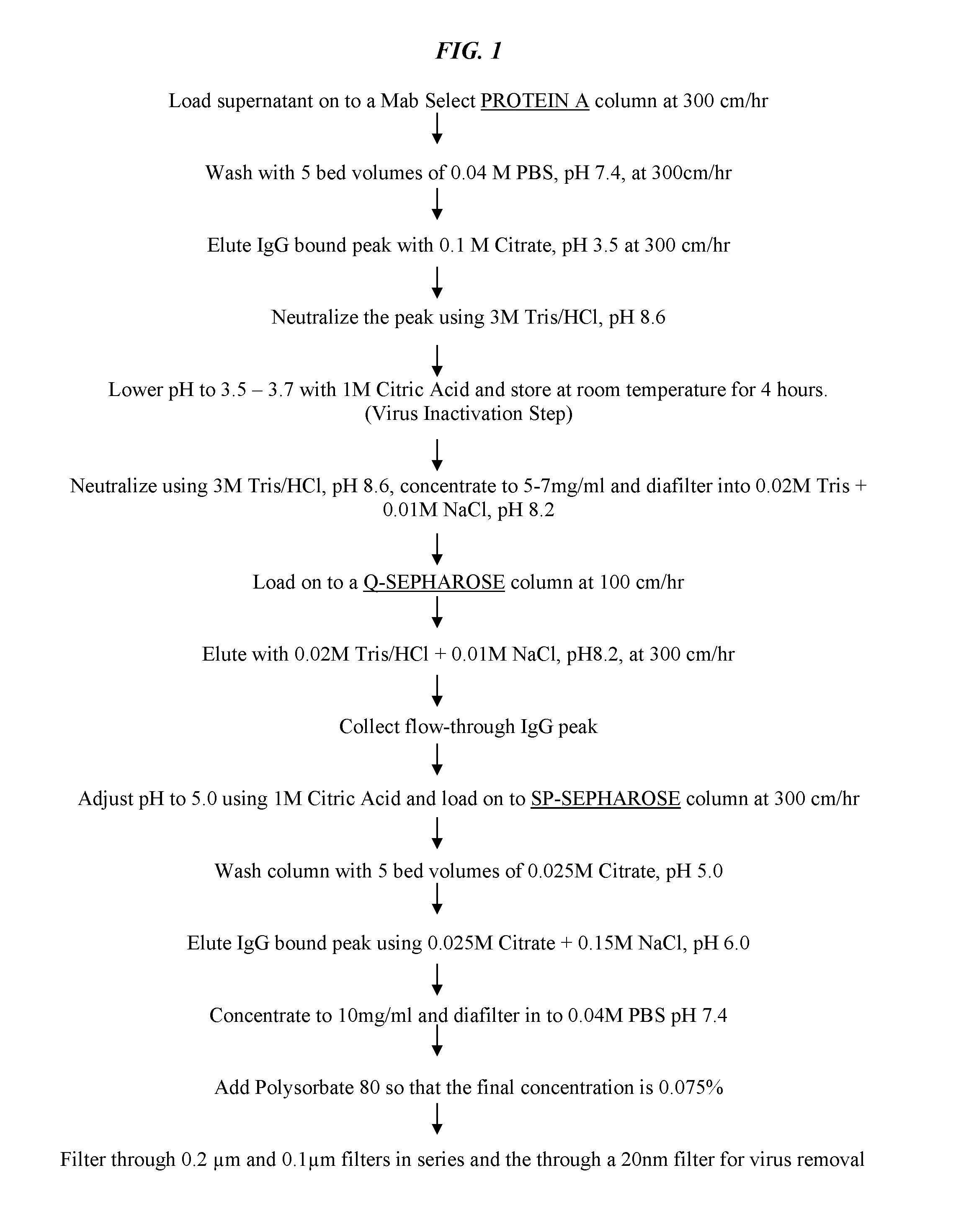
In other preferred embodiments, the antibody may be purified from cell culture medium by sequential chromatography, for example by affinity and ion exchange column chromatography. Non-limiting examples include affinity chromatography on Protein A, anion-exchange chromatography on Q-SEPHAROSE.RTM. and cation-exchange chromatography on SP-SEPHAROSE.RTM.. More preferably, the antibody is bound to the SP-SEPHAROSE.RTM. resin in pH 5 citrate buffer and eluted from the column with pH 6 citrate buffer in 0.15 M NaCl. The eluate from the SP-SEPHAROSE.RTM. column may be filtered through, for example, a 20 nm filter for virus removal. The purified antibody may then be diafiltered, for example using an AMICON.RTM. Ultrafiltration Cell with a 50 KD MW cut-off filter to exchange the medium with a high concentration formulation buffer (HCF buffer) and to concentrate the antibody for storage. In most preferred embodiments, the HCF buffer solution may comprise phosphate buffer (pH 5.2), sodium chloride, Polysorbate 80, citrate and mannitol. Polysorbate 80 serves to decrease protein aggregation, while mannitol stabilizes the antibody in aqueous medium. The diafiltration concentrates the antibody to preferably at least 100 mg/ml, more preferably at least 150 mg/ml, more preferably at least 200 mg/ml, most preferably at least 250 mg/ml final concentration. The concentrated antibody exhibits little or no aggregation and preferably is stable in liquid form at 2-8.degree. for at least 10 months. In even more preferred embodiments, the Polysorbate 80 is added to the concentrated antibody after the ultrafiltration step. The stable, highly concentrated antibody is of use for preparing medicaments for administration to subjects, preferably by subcutaneous, transdermal or intramuscular administration.
An antibody or antigen-binding fragment of use may be chimeric, humanized or human. The use of chimeric antibodies is preferred to the parent murine antibodies because they possess human antibody constant region sequences and therefore do not elicit as strong a human anti-mouse antibody (HAMA) response as murine antibodies. The use of humanized antibodies is even more preferred, in order to further reduce the possibility of inducing a HAMA reaction. Techniques for humanization of murine antibodies by replacing murine framework and constant region sequences with corresponding human antibody framework and constant region sequences are well known in the art and have been applied to numerous murine anti-cancer antibodies. Antibody humanization may also involve the substitution of one or more human framework amino acid residues with the corresponding residues from the parent murine framework region sequences. As discussed below, techniques for production of human antibodies are also well known.
The therapeutic formulation may comprise an antibody fragment, such as F(ab').sub.2, Fab, scFv, Fv, or a fusion protein utilizing part or all of the light and heavy chains of the F(ab').sub.2, Fab, scFv. The antibody may also be multivalent, or multivalent and multispecific. The antibody may include human constant regions of IgG1, IgG2a, IgG3, or IgG4.
In more preferred embodiments, the allotype of the antibody may be selected to minimize host immunogenic response to the administered antibody, as discussed in more detail below. A preferred allotype is a non-G1m1 allotype (nG1m1), such as G1m3, G1m3,1, G1m3,2 or G1m3,1,2. The non-G1m1 allotype is preferred for decreased antibody immunoreactivity. Surprisingly, repeated subcutaneous administration of concentrated nG1m1 antibody was not found to induce significant immune response, despite the enhanced immunogenicity of subcutaneous administration.
The anti-CD74 antibody may be administered as a naked antibody (not conjugated to any therapeutic agent) or as an immunoconjugate, attached to at least one therapeutic agent. Alternatively, naked anti-CD74 antibodies may be administered in combination with one or more therapeutic agents. Therapeutic agents may be selected from the group consisting of a radionuclide, a cytotoxin, a chemotherapeutic agent, a drug, a pro-drug, a toxin, an enzyme, an immunomodulator, an anti-angiogenic agent, a pro-apoptotic agent, a cytokine, a hormone, an oligonucleotide molecule (e.g., an antisense molecule or a gene) or a second antibody or fragment thereof.
The therapeutic agent may be selected from the group consisting of aplidin, azaribine, anastrozole, azacytidine, bleomycin, bortezomib, bryostatin-1, busulfan, calicheamycin, camptothecin, 10-hydroxycamptothecin, carmustine, celecoxib, chlorambucil, cisplatin, irinotecan (CPT-11), SN-38, carboplatin, cladribine, cyclophosphamide, cytarabine, dacarbazine, docetaxel, dactinomycin, daunomycin glucuronide, daunorubicin, dexamethasone, diethylstilbestrol, doxorubicin, doxorubicin glucuronide, epirubicin glucuronide, ethinyl estradiol, estramustine, etoposide, etoposide glucuronide, etoposide phosphate, floxuridine (FUdR), 3',5'-O-dioleoyl-FudR (FUdR-dO), fludarabine, flutamide, fluorouracil, fluoxymesterone, gemcitabine, hydroxyprogesterone caproate, hydroxyurea, idarubicin, ifosfamide, L-asparaginase, leucovorin, lomustine, mechlorethamine, medroprogesterone acetate, megestrol acetate, melphalan, mercaptopurine, 6-mercaptopurine, methotrexate, mitoxantrone, mithramycin, mitomycin, mitotane, phenyl butyrate, prednisone, procarbazine, paclitaxel, pentostatin, PSI-341, semustine streptozocin, tamoxifen, taxanes, testosterone propionate, thalidomide, thioguanine, thiotepa, teniposide, topotecan, uracil mustard, vinblastine, vinorelbine, vincristine, ricin, abrin, ribonuclease, onconase, rapLR1, DNase I, Staphylococcal enterotoxin-A, pokeweed antiviral protein, gelonin, diphtheria toxin, Pseudomonas exotoxin, and Pseudomonas endotoxin.
The therapeutic agent may comprise a radionuclide selected from the group consisting of .sup.103mRh, .sup.103Ru, .sup.105Rh, .sup.105Ru, .sup.107Hg, .sup.109Pd, .sup.109Pt, .sup.111Ag, .sup.111In, .sup.113mIn, .sup.119Sb, .sup.11C, .sup.121mTe, .sup.122mT, .sup.125I, .sup.125mTe, .sup.126I, .sup.131I, .sup.133I, .sup.13N, .sup.142Pr, .sup.143Pr, .sup.149Pm, .sup.152Dy, .sup.153Sm, .sup.15O, .sup.161Ho, .sup.161Tb, .sup.165Tm, .sup.166Dy, .sup.166Ho, .sup.167Tm, .sup.168Tm, .sup.169Er, .sup.169Yb, .sup.177Ln, .sup.186Re, .sup.188Re, .sup.189mOs, .sup.189Re, .sup.192Ir, .sup.194Ir, .sup.197Pt, .sup.198Au, .sup.199Au, .sup.201Tl, .sup.203Hg, .sup.211At, .sup.211Bi, .sup.211Pb, .sup.212Bi, .sup.212Pp, .sup.213Bi, .sup.215Po, .sup.217At, .sup.219Rn, .sup.221Fr, .sup.223Ra, .sup.224Ac, .sup.225Ac, .sup.225Fm, .sup.32P, .sup.33P, .sup.47Sc, .sup.51Cr, .sup.57Co, .sup.58Co, .sup.59Fe, .sup.62Cu, .sup.67Cu, .sup.57Ga, .sup.75Br, .sup.75Se, .sup.76Br, .sup.77As, .sup.77Br, .sup.80mBr, .sup.90Y, .sup.95Ru, .sup.97Ru, .sup.99Mo and .sup.99mTc.
The therapeutic agent may be an enzyme selected from the group consisting of malate dehydrogenase, staphylococcal nuclease, delta-V-steroid isomerase, yeast alcohol dehydrogenase, alpha-glycerophosphate dehydrogenase, triose phosphate isomerase, horseradish peroxidase, alkaline phosphatase, asparaginase, glucose oxidase, beta-galactosidase, ribonuclease, urease, catalase, glucose-6-phosphate dehydrogenase, glucoamylase and acetylcholinesterase.
An immunomodulator of use may be selected from the group consisting of a cytokine, a stem cell growth factor, a lymphotoxin, a hematopoietic factor, a colony stimulating factor (CSF), an interferon (IFN), erythropoietin, thrombopoietin and combinations thereof. Exemplary immunomodulators may include IL-1, IL-2, IL-3, IL-6, IL-10, IL-12, IL-18, IL-21, interferon-.alpha., interferon-.beta., interferon-.gamma., interferon-k, G-CSF, GM-CSF, and mixtures thereof.
Exemplary anti-angiogenic agents may include angiostatin, endostatin, baculostatin, canstatin, maspin, anti-VEGF binding molecules, anti-placental growth factor binding molecules, or anti-vascular growth factor binding molecules.
In certain embodiments, the antibody or fragment may comprise one or more chelating moieties, such as NOTA, DOTA, DTPA, TETA, Tscg-Cys, or Tsca-Cys. In certain embodiments, the chelating moiety may form a complex with a therapeutic or diagnostic cation, such as Group II, Group III, Group IV, Group V, transition, lanthanide or actinide metal cations, Tc, Re, Bi, Cu, As, Ag, Au, At, or Pb.
Exemplary known second antibodies of use include, but are not limited to, hR1 (anti-IGF-1R), hPAM4 (anti-mucin), hA20 (anti-CD20), hA19 (anti-CD19), hIMMU31 (anti-AFP), hLL1 (anti-CD74), hLL2 (anti-CD22), hMu-9 (anti-CSAp), hL243 (anti-HLA-DR), hMN-14 (anti-CEACAM5), hMN-15 (anti-CEACAM6), 29H2 (anti-CEACAM1, ABCAM.RTM.), hRS7 (anti-EGP-1--also known as Trop-2), elsilimomab (anti-IL-6), ALD518 (anti-IL-6), alemtuzumab (anti-CD52), daclizumab (anti-CD25), galiximab (anti-CD80), adalimumab (anti-TNF-.alpha.), infliximab (anti-TNF-.alpha.), lucatumumab (anti-CD40), ofatumumab (anti-CD20) and hMN-3 (anti-CEACAM6). Antibodies against antigens of use include anti-CXCR4 (e.g., U.S. Pat. Nos. 7,138,496; 7,682,611; 7,521,045; 7,892,546) and IL-6 (e.g., U.S. Pat. Nos. 7,919,095; 7,935,340; 7,955,597), the Examples section of each cited patent incorporated herein by reference.
Although the preferred method involves treatment of autoimmune disease, in alternative the disease or disorder may be a solid tumor that overexpresses CD74, a B-cell lymphoma or leukemia, an immune dysregulation disease, organ-graft rejection or graft-versus-host disease. Exemplary malignancies that may be treated using the claimed methods and compositions include, but are not limited to, glioblastoma, gastric cancer, bladder cancer, prostate cancer, thymic cancer, colorectal cancer, lung cancer, renal cancer, pancreatic cancer, breast cancer, indolent forms of B-cell lymphomas, aggressive forms of B-cell lymphomas, acute lymphocytic leukemia, chronic lymphocytic leukemia, Hodgkin's lymphoma, non-Hodgkin's lymphoma, mantle cell lymphoma, diffuse large B-cell lymphoma, follicular lymphoma, marginal zone lymphoma, Burkitt's lymphoma and multiple myeloma.
BRIEF DESCRIPTION OF THE DRAWINGS
The following drawings are provided to illustrate preferred embodiments of the invention. However, the claimed subject matter is in no way limited by the illustrative embodiments disclosed in the drawings.
FIG. 1. Exemplary protocol for column chromatography purification of antibody from cell culture medium.
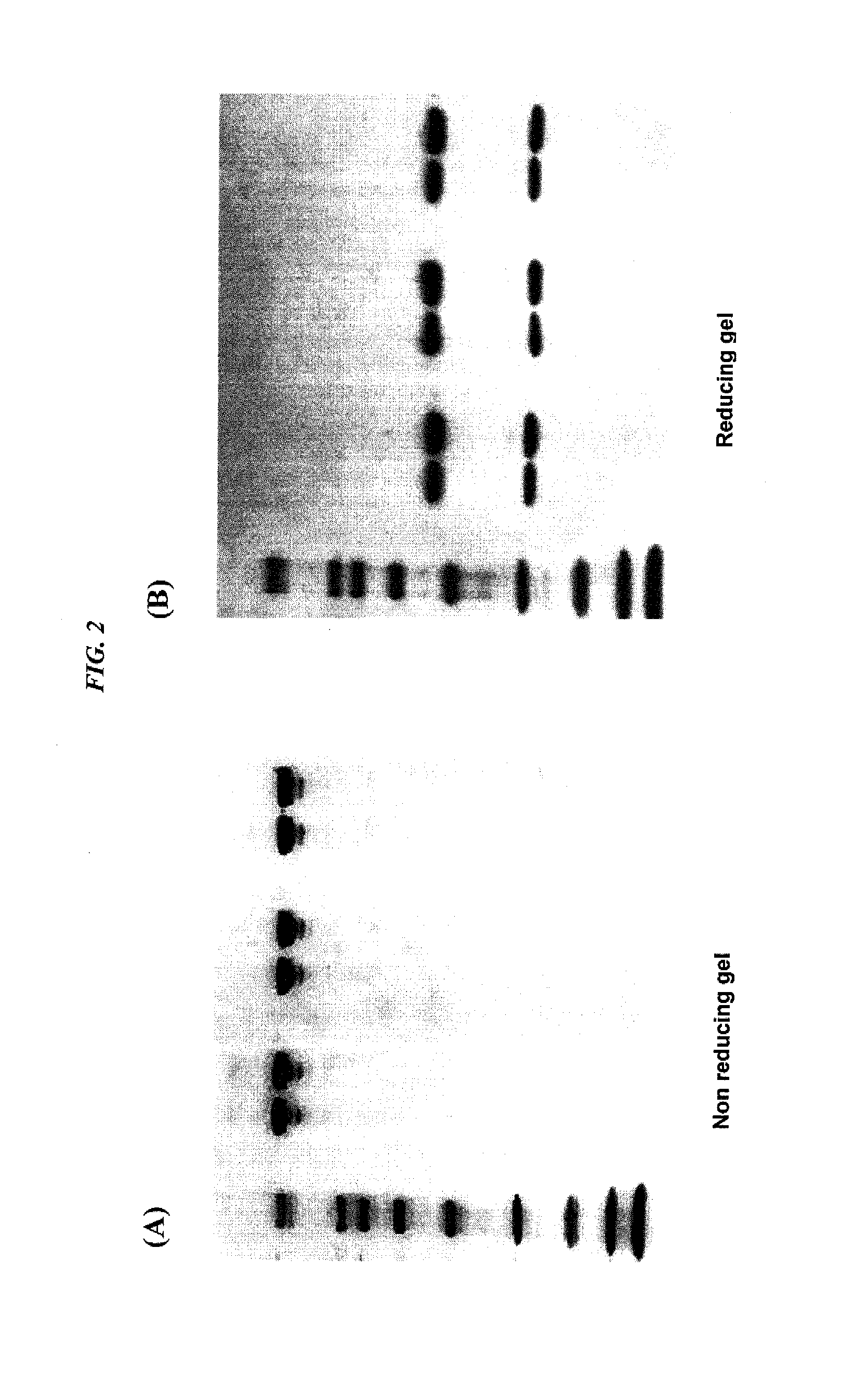
FIG. 2. SDS-polyacrylamide gel electrophoresis of ultrafiltration concentrated antibodies: (A) non-reducing gel, (B) reducing gel. Both gels show (lane 1) MW standards; (lane 2) hLL1 IgG, starting IgG solution (10 mg/mL); (lane 3) concentrated hLL1 IgG, after 2 month storage, (215 mg/mL); (lane 4) hA20 IgG, starting IgG solution (5.1 mg/mL); (lane 5) concentrated hA20 IgG, after 10 month storage, (162 mg/mL); (lane 6) hL243 IgG, starting IgG solution (8.9 mg/mL); (lane 7) concentrated hL243 IgG, after 10 month storage, (101 mg/mL). The MW standards used were respectively 6.5, 14, 21, 31, 45, 66, 97, 116 and 200 KD.
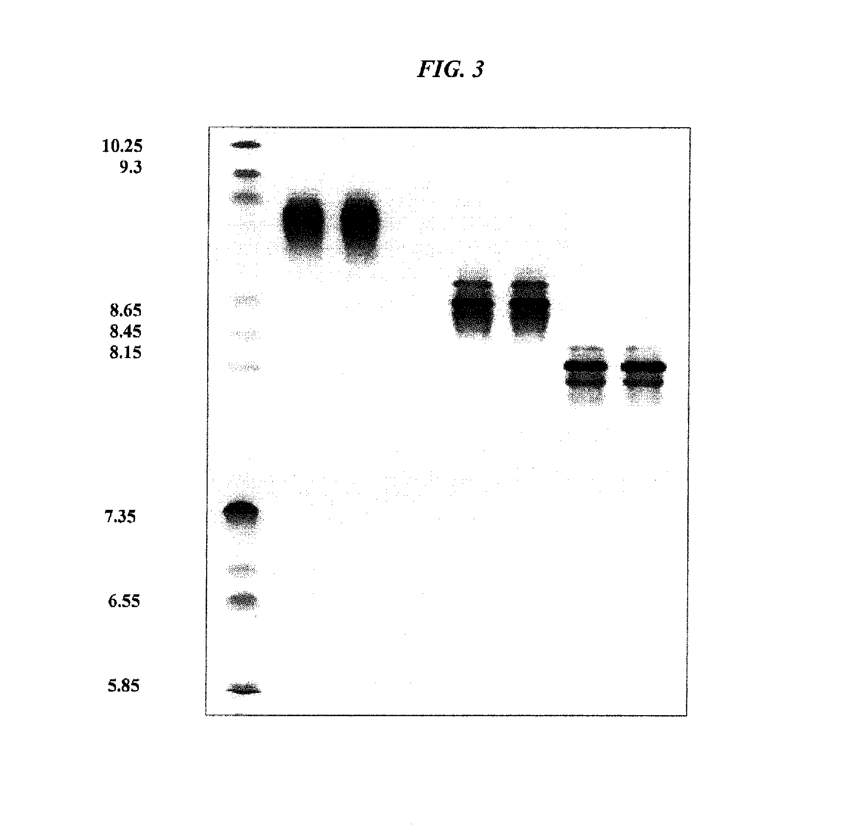
FIG. 3. Isoelectric focusing gel of ultrafiltration concentrated antibodies showing (lane 1) pI standards; (lane 2) hLL1 IgG, starting IgG solution (10 mg/mL); (lane 3) concentrated hLL1 IgG, after 2 month storage, (215 mg/mL); (lane 4) hA20 IgG, starting IgG solution (5.1 mg/mL); (lane 5) concentrated hA20 IgG, after 10 month storage, (162 mg/mL); (lane 6) hL243 IgG, starting IgG solution (8.9 mg/mL); (lane 7) concentrated hL243 IgG, after 10 month storage, (101 mg/mL). The MW standards used were respectively 6.5, 14, 21, 31, 45, 66, 97, 116 and 200 KD.
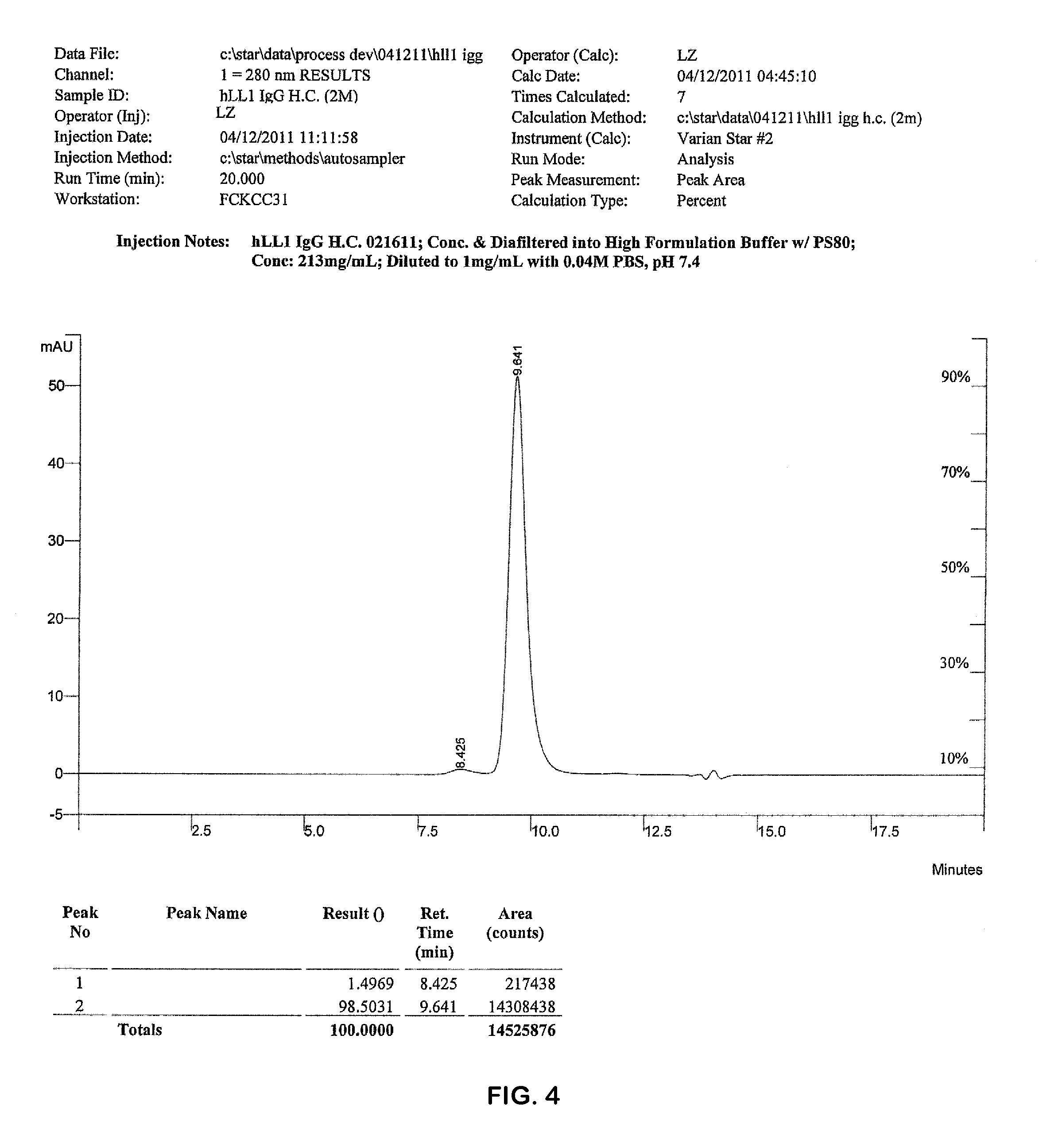
FIG. 4. Representative SE HPLC chromatogram of ultrafiltration concentrated hLL1 IgG solution (215 mg/mL) after 10 months of storage.
FIG. 5. Comparison of veltuzumab (SEQ ID NO:7) vs. rituximab (SEQ ID NO:8) heavy chain constant region sequences. Identical residues are indicated by asterisks. The two different allotype antibodies differ in heavy chain constant region sequence by only four amino acid residues. The light chain constant region sequences are identical between the two antibodies.
FIG. 6. Effect of anti-CD74 antibody (IMMU-115) administered subcutaneously to ten human SLE patients in a Phase I study. Antibody was administered at 250 mg once per week for four weeks. Results are reported as total BILAG scores and mean SELDAI.
DETAILED DESCRIPTION
Definitions
The following definitions are provided to facilitate understanding of the disclosure herein. Where a term is not specifically defined, it is used in accordance with its plain and ordinary meaning.
As used herein, the terms "a", "an" and "the" may refer to either the singular or plural, unless the context otherwise makes clear that only the singular is meant.
An "antibody" refers to a full-length (i.e., naturally occurring or formed by normal immunoglobulin gene fragment recombinatorial processes) immunoglobulin molecule (e.g., an IgG antibody) or an immunologically active (i.e., antigen-binding) portion of an immunoglobulin molecule, like an antibody fragment.
An "antibody fragment" is a portion of an antibody such as F(ab').sub.2, F(ab).sub.2, Fab', Fab, Fv, scFv, single domain antibodies (DABs or VHHs) and the like, including half-molecules of IgG4 (van der Neut Kolfschoten et al., 2007, Science 317:1554-1557). Regardless of structure, an antibody fragment binds with the same antigen that is recognized by the intact antibody. For example, an anti-CD74 antibody fragment binds with an epitope of CD74. The term "antibody fragment" also includes isolated fragments consisting of the variable regions, such as the "Fv" fragments consisting of the variable regions of the heavy and light chains, recombinant single chain polypeptide molecules in which light and heavy chain variable regions are connected by a peptide linker ("scFv proteins"), and minimal recognition units consisting of the amino acid residues that mimic the hypervariable region.
A "chimeric antibody" is a recombinant protein that contains the variable domains including the complementarity determining regions (CDRs) of an antibody derived from one species, preferably a rodent antibody, while the constant domains of the antibody molecule are derived from those of a human antibody. For veterinary applications, the constant domains of the chimeric antibody may be derived from that of other species, such as a cat or dog.
A "humanized antibody" is a recombinant protein in which the CDRs from an antibody from one species; e.g., a rodent antibody, are transferred from the heavy and light variable chains of the rodent antibody into human heavy and light variable domains, including human framework region (FR) sequences. The constant domains of the antibody molecule are derived from those of a human antibody.
A "human antibody" is an antibody obtained from transgenic mice that have been genetically engineered to produce specific human antibodies in response to antigenic challenge. In this technique, elements of the human heavy and light chain locus are introduced into strains of mice derived from embryonic stem cell lines that contain targeted disruptions of the endogenous heavy chain and light chain loci. The transgenic mice can synthesize human antibodies specific for human antigens, and the mice can be used to produce human antibody-secreting hybridomas. Methods for obtaining human antibodies from transgenic mice are described by Green et al., Nature Genet. 7:13 (1994), Lonberg et al., Nature 368:856 (1994), and Taylor et al., Int. Immun. 6:579 (1994). A fully human antibody also can be constructed by genetic or chromosomal transfection methods, as well as phage display technology, all of which are known in the art. (See, e.g., McCafferty et al., Nature 348:552-553 (1990) for the production of human antibodies and fragments thereof in vitro, from immunoglobulin variable domain gene repertoires from unimmunized donors). In this technique, antibody variable domain genes are cloned in-frame into either a major or minor coat protein gene of a filamentous bacteriophage, and displayed as functional antibody fragments on the surface of the phage particle. Because the filamentous particle contains a single-stranded DNA copy of the phage genome, selections based on the functional properties of the antibody also result in selection of the gene encoding the antibody exhibiting those properties. In this way, the phage mimics some of the properties of the B cell. Phage display can be performed in a variety of formats, for their review, see, e.g. Johnson and Chiswell, Current Opinion in Structural Biology 3:5564-571 (1993). Human antibodies may also be generated by in vitro activated B cells. (See, U.S. Pat. Nos. 5,567,610 and 5,229,275).
A "therapeutic agent" is an atom, molecule, or compound that is useful in the treatment of a disease. Examples of therapeutic agents include but are not limited to antibodies, antibody fragments, drugs, cytokine or chemokine inhibitors, proapoptotic agents, tyrosine kinase inhibitors, toxins, enzymes, nucleases, hormones, immunomodulators, antisense oligonucleotides, siRNA, RNAi, chelators, boron compounds, photoactive agents, dyes and radioisotopes.
A "diagnostic agent" is an atom, molecule, or compound that is useful in diagnosing a disease. Useful diagnostic agents include, but are not limited to, radioisotopes, dyes, contrast agents, fluorescent compounds or molecules and enhancing agents (e.g., paramagnetic ions). Preferably, the diagnostic agents are selected from the group consisting of radioisotopes, enhancing agents, and fluorescent compounds.
An "immunoconjugate" is a conjugate of an antibody with an atom, molecule, or a higher-ordered structure (e.g., with a liposome), a therapeutic agent, or a diagnostic agent. A "naked antibody" is an antibody that is not conjugated to any other agent.
A "naked antibody" is generally an entire antibody that is not conjugated to a therapeutic agent. This is so because the Fc portion of the antibody molecule provides effector functions, such as complement fixation and ADCC (antibody dependent cell cytotoxicity) that set mechanisms into action that may result in cell lysis. However, it is possible that the Fc portion is not required for therapeutic function, with other mechanisms, such as apoptosis, coming into play. Naked antibodies include both polyclonal and monoclonal antibodies, as well as certain recombinant antibodies, such as chimeric, humanized or human antibodies.
As used herein, the term "antibody fusion protein" is a recombinantly produced antigen-binding molecule in which an antibody or antibody fragment is linked to another protein or peptide, such as the same or different antibody or antibody fragment or a DDD or AD peptide. The fusion protein may comprise a single antibody component, a multivalent or multispecific combination of different antibody components or multiple copies of the same antibody component. The fusion protein may additionally comprise an antibody or an antibody fragment and a therapeutic agent. Examples of therapeutic agents suitable for such fusion proteins include immunomodulators and toxins. One preferred toxin comprises a ribonuclease (RNase), preferably a recombinant RNase.
A "multispecific antibody" is an antibody that can bind simultaneously to at least two targets that are of different structure, e.g., two different antigens, two different epitopes on the same antigen, or a hapten and/or an antigen or epitope. A "multivalent antibody" is an antibody that can bind simultaneously to at least two targets that are of the same or different structure. Valency indicates how many binding arms or sites the antibody has to a single antigen or epitope; i.e., monovalent, bivalent, trivalent or multivalent. The multivalency of the antibody means that it can take advantage of multiple interactions in binding to an antigen, thus increasing the avidity of binding to the antigen. Specificity indicates how many antigens or epitopes an antibody is able to bind; i.e., monospecific, bispecific, trispecific, multispecific. Using these definitions, a natural antibody, e.g., an IgG, is bivalent because it has two binding arms but is monospecific because it binds to one epitope. Multispecific, multivalent antibodies are constructs that have more than one binding site of different specificity.
A "bispecific antibody" is an antibody that can bind simultaneously to two targets which are of different structure. Bispecific antibodies (bsAb) and bispecific antibody fragments (bsFab) may have at least one arm that specifically binds to, for example, a B cell, T cell, myeloid-, plasma-, and mast-cell antigen or epitope and at least one other arm that specifically binds to a targetable conjugate that bears a therapeutic or diagnostic agent. A variety of bispecific antibodies can be produced using molecular engineering.
CD74
CD74 (also known as invariant chain or Ii) is a transmembrane glycoprotein that associates with MHC class II .alpha. and .beta. chains and directs transport of .alpha..beta.Ii complexes to endosomes and lysosomes. CD74 functions as a molecular chaperone in the processing of exogenous peptides for antigen presentation via MHC class II. More recently, CD74 has been identified as the endogenous receptor for MIF (macrophage migration inhibitory factor), a key regulatory molecule that promotes cell survival and inhibits apoptosis by activation of the Akt pathway (Lue et al., Oncogene 207, 26:5046-59). As such, the interaction of CD74 and MIF is thought to play a significant role in tumorigenesis and tumor progression (Id.)
CD74 is overexpressed in a variety of disease states, including many solid and hematopoietic tumors (Stein et al., Clin Cancer Res 2007, 13:5556s-63s; Gold et al., Int J Clin Exp Pathol 2011, 4:1-12). Milatuzumab (hLL1), a humanized anti-CD74 antibody, is rapidly internalized into CD74 expressing cells and has been used to target therapeutic agents to tumor cells, with excellent therapeutic effects (see, e.g., Griffiths et al., Clin Cancer Res 2003, 9:6567-71; Ochaskovskaya et al., Clin Cancer Res 2001, 7:1505-10). However, naked milatuzumab has also been shown to be cytotoxic in the presence of cross-linking antibodies (e.g., U.S. Pat. No. 7,312,318). Combinations of milatuzumab with other therapeutic agents show enhanced cytotoxicity and improved therapeutic response in multiple myeloma cell lines (Stein et al., Clin Cancer Res 2009, 15:2808-17).
Milatuzumab has been reported to be efficacious for a wide range of hematopoietic malignancies, including non-Hodgkin's lymphoma, Burkitt lymphoma, follicular lymphoma, multiple myeloma, chronic lymphocytic leukemia and mantle cell lymphoma (Stein et al., Clin Cancer Res 2007, 13:5556s-63s; Berkova et al., Expert Opin. Invest. Drugs 2010, 19:141-49). Since CD74 is also over-expressed in a number of solid tumors, use of milatuzumab or other anti-CD74 antibodies for therapy of colorectal carcinoma, pancreatic carcinoma, gastric carcinoma, non-small cell lung carcinoma, glioblastoma, thymic carcinoma, pancreatic cancer, breast cancer, bladder cancer and prostate cancer has also been suggested (Gold et al., Int J Clin Exp Pathol 2011, 4:1-12; Berkova et al., Expert Opin. Invest. Drugs 2010, 19:141-49). Therapy directed to CD74 has been indicated in autoimmune or immune dysfunction diseases, such as systemic lupus erythematosus and rheumatoid arthritis (Lapter et al., Immunology 2011, 1327-95; Morand and Leech, Front Biosci 2005, 10:12-22). Combination therapy, such as with anti-CD74/anti-CD20 antibodies, has been reported to show improved efficacy in mantle cell lymphoma (Alinari et al., Blood 2011, 117:4530-41).
The skilled artisan will realize that these therapeutic effects are not limited to milatuzumab, but may also be seen with other anti-CD74 antibodies, particularly those that compete with milatuzumab for binding or that bind to the same epitope of CD74 as milatuzumab.
Preparation of Monoclonal Antibodies
The compositions, formulations and methods described herein may include monoclonal antibodies. Rodent monoclonal antibodies to specific antigens may be obtained by methods known to those skilled in the art. (See, e.g., Kohler and Milstein, Nature 256: 495 (1975), and Coligan et al. (eds.), CURRENT PROTOCOLS IN IMMUNOLOGY, VOL. 1, pages 2.5.1-2.6.7 (John Wiley & Sons 1991)). General techniques for cloning murine immunoglobulin variable domains have been disclosed, for example, by the publication of Orlandi et al., Proc. Nat'l Acad. Sci. USA 86: 3833 (1989).
Chimeric Antibodies
A chimeric antibody is a recombinant protein that contains the variable domains including the CDRs derived from one species of animal, such as a rodent antibody, while the remainder of the antibody molecule; i.e., the constant domains, is derived from a human antibody. Techniques for constructing chimeric antibodies are well known to those of skill in the art. As an example, Leung et al., Hybridoma 13:469 (1994), disclose how they produced an LL2 chimera by combining DNA sequences encoding the V.sub.k and V.sub.H domains of LL2 monoclonal antibody, an anti-CD22 antibody, with respective human and IgG.sub.1 constant region domains. This publication also provides the nucleotide sequences of the LL2 light and heavy chain variable regions, V.sub.k and V.sub.H, respectively.
Humanized Antibodies
A chimeric monoclonal antibody can be humanized by replacing the sequences of the murine FR in the variable domains of the chimeric antibody with one or more different human FR. Specifically, mouse CDRs are transferred from heavy and light variable chains of the mouse immunoglobulin into the corresponding variable domains of a human antibody. As simply transferring mouse CDRs into human FRs often results in a reduction or even loss of antibody affinity, additional modification might be required in order to restore the original affinity of the murine antibody. This can be accomplished by the replacement of one or more some human residues in the FR regions with their murine counterparts to obtain an antibody that possesses good binding affinity to its epitope. (See, e.g., Tempest et al., Biotechnology 9:266 (1991) and Verhoeyen et al., Science 239: 1534 (1988)). Techniques for producing humanized antibodies are disclosed, for example, by Jones et al., Nature 321: 522 (1986), Riechmann et al., Nature 332: 323 (1988), Verhoeyen et al., Science 239: 1534 (1988), Carter et al., Proc. Nat'l Acad. Sci. USA 89: 4285 (1992), Sandhu, Crit. Rev. Biotech. 12: 437 (1992), and Singer et al., J. Immun. 150: 2844 (1993).
Human Antibodies
A fully human antibody can be obtained from a transgenic non-human animal. (See, e.g., Mendez et al., Nature Genetics, 15: 146-156, 1997; U.S. Pat. No. 5,633,425.) Methods for producing fully human antibodies using either combinatorial approaches or transgenic animals transformed with human immunoglobulin loci are known in the art (e.g., Mancini et al., 2004, New Microbiol. 27:315-28; Conrad and Scheller, 2005, Comb. Chem. High Throughput Screen. 8:117-26; Brekke and Loset, 2003, Curr. Opin. Pharmacol. 3:544-50; each incorporated herein by reference). Such fully human antibodies are expected to exhibit even fewer side effects than chimeric or humanized antibodies and to function in vivo as essentially endogenous human antibodies. In certain embodiments, the claimed methods and procedures may utilize human antibodies produced by such techniques.
In one alternative, the phage display technique may be used to generate human antibodies (e.g., Dantas-Barbosa et al., 2005, Genet. Mol. Res. 4:126-40, incorporated herein by reference). Human antibodies may be generated from normal humans or from humans that exhibit a particular disease state, such as cancer (Dantas-Barbosa et al., 2005). The advantage to constructing human antibodies from a diseased individual is that the circulating antibody repertoire may be biased towards antibodies against disease-associated antigens.
In one non-limiting example of this methodology, Dantas-Barbosa et al. (2005) constructed a phage display library of human Fab antibody fragments from osteosarcoma patients. Generally, total RNA was obtained from circulating blood lymphocytes (Id.) Recombinant Fab were cloned from the .mu., .gamma. and .kappa. chain antibody repertoires and inserted into a phage display library (Id.) RNAs were converted to cDNAs and used to make Fab cDNA libraries using specific primers against the heavy and light chain immunoglobulin sequences (Marks et al., 1991, J Mol. Biol. 222:581-97). Library construction was performed according to Andris-Widhopf et al. (2000, In: Phage Display Laboratory Manual, Barbas et al. (eds), 1.sup.st edition, Cold Spring Harbor Laboratory Press, Cold Spring Harbor, N.Y. pp. 9.1 to 9.22, incorporated herein by reference). The final Fab fragments were digested with restriction endonucleases and inserted into the bacteriophage genome to make the phage display library. Such libraries may be screened by standard phage display methods. The skilled artisan will realize that this technique is exemplary only and any known method for making and screening human antibodies or antibody fragments by phage display may be utilized.
In another alternative, transgenic animals that have been genetically engineered to produce human antibodies may be used to generate antibodies against essentially any immunogenic target, using standard immunization protocols as discussed above. Methods for obtaining human antibodies from transgenic mice are described by Green et al., Nature Genet. 7:13 (1994), Lonberg et al., Nature 368:856 (1994), and Taylor et al., Int. Immun. 6:579 (1994). A non-limiting example of such a system is the XENOMOUSE.RTM. (e.g., Green et al., 1999, J. Immunol. Methods 231:11-23, incorporated herein by reference) from Abgenix (Fremont, Calif.). In the XENOMOUSE.RTM. and similar animals, the mouse antibody genes have been inactivated and replaced by functional human antibody genes, while the remainder of the mouse immune system remains intact.
The XENOMOUSE.RTM. was transformed with germline-configured YACs (yeast artificial chromosomes) that contained portions of the human IgH and Ig kappa loci, including the majority of the variable region sequences, along accessory genes and regulatory sequences. The human variable region repertoire may be used to generate antibody producing B cells, which may be processed into hybridomas by known techniques. A XENOMOUSE.RTM. immunized with a target antigen will produce human antibodies by the normal immune response, which may be harvested and/or produced by standard techniques discussed above. A variety of strains of XENOMOUSE.RTM. are available, each of which is capable of producing a different class of antibody. Transgenically produced human antibodies have been shown to have therapeutic potential, while retaining the pharmacokinetic properties of normal human antibodies (Green et al., 1999). The skilled artisan will realize that the claimed compositions and methods are not limited to use of the XENOMOUSE.RTM. system but may utilize any transgenic animal that has been genetically engineered to produce human antibodies.
Antibody Cloning and Production
Various techniques, such as production of chimeric or humanized antibodies, may involve procedures of antibody cloning and construction. The antigen-binding V.kappa. (variable light chain) and V.sub.H (variable heavy chain) sequences for an antibody of interest may be obtained by a variety of molecular cloning procedures, such as RT-PCR, 5'-RACE, and cDNA library screening. The V genes of an antibody from a cell that expresses a murine antibody can be cloned by PCR amplification and sequenced. To confirm their authenticity, the cloned V.sub.L and V.sub.H genes can be expressed in cell culture as a chimeric Ab as described by Orlandi et al., (Proc. Natl. Acad. Sci., USA, 86: 3833 (1989)). Based on the V gene sequences, a humanized antibody can then be designed and constructed as described by Leung et al. (Mol. Immunol., 32: 1413 (1995)).
cDNA can be prepared from any known hybridoma line or transfected cell line producing a murine antibody by general molecular cloning techniques (Sambrook et al., Molecular Cloning, A laboratory manual, 2.sup.nd Ed (1989)). The V.kappa. sequence for the antibody may be amplified using the primers VK1BACK and VK1FOR (Orlandi et al., 1989) or the extended primer set described by Leung et al. (BioTechniques, 15: 286 (1993)). The V.sub.H sequences can be amplified using the primer pair VH1BACK/VH1FOR (Orlandi et al., 1989) or the primers annealing to the constant region of murine IgG described by Leung et al. (Hybridoma, 13:469 (1994)). Humanized V genes can be constructed by a combination of long oligonucleotide template syntheses and PCR amplification as described by Leung et al. (Mol. Immunol., 32: 1413 (1995)).
PCR products for V.kappa. can be subcloned into a staging vector, such as a pBR327-based staging vector, VKpBR, that contains an Ig promoter, a signal peptide sequence and convenient restriction sites. PCR products for V.sub.H can be subcloned into a similar staging vector, such as the pBluescript-based VHpBS. Expression cassettes containing the V.kappa. and V.sub.H sequences together with the promoter and signal peptide sequences can be excised from VKpBR and VHpBS and ligated into appropriate expression vectors, such as pKh and pG1g, respectively (Leung et al., Hybridoma, 13:469 (1994)). The expression vectors can be co-transfected into an appropriate cell and supernatant fluids monitored for production of a chimeric, humanized or human antibody. Alternatively, the V.kappa. and V.sub.H expression cassettes can be excised and subcloned into a single expression vector, such as pdHL2, as described by Gillies et al. (J. Immunol. Methods 125:191 (1989) and also shown in Losman et al., Cancer, 80:2660 (1997)).
In an alternative embodiment, expression vectors may be transfected into host cells that have been pre-adapted for transfection, growth and expression in serum-free medium. Exemplary cell lines that may be used include the Sp/EEE, Sp/ESF and Sp/ESF-X cell lines (see, e.g., U.S. Pat. Nos. 7,531,327; 7,537,930 and 7,608,425; the Examples section of each of which is incorporated herein by reference). These exemplary cell lines are based on the Sp2/0 myeloma cell line, transfected with a mutant Bcl-EEE gene, exposed to methotrexate to amplify transfected gene sequences and pre-adapted to serum-free cell line for protein expression.
Antibody Allotypes
Immunogenicity of therapeutic antibodies is associated with increased risk of infusion reactions and decreased duration of therapeutic response (Baert et al., 2003, N Engl J Med 348:602-08). The extent to which therapeutic antibodies induce an immune response in the host may be determined in part by the allotype of the antibody (Stickler et al., 2011, Genes and Immunity 12:213-21). Antibody allotype is related to amino acid sequence variations at specific locations in the constant region sequences of the antibody. The allotypes of IgG antibodies containing a heavy chain .gamma.-type constant region are designated as Gm allotypes (1976, J Immunol 117:1056-59).
For the common IgG1 human antibodies, the most prevalent allotype is G1m1 (Stickler et al., 2011, Genes and Immunity 12:213-21). However, the G1m3 allotype also occurs frequently in Caucasians (Id.). It has been reported that G1m1 antibodies contain allotypic sequences that tend to induce an immune response when administered to non-G1m1 (nG1m1) recipients, such as G1m3 patients (Id.). Non-G1m1 allotype antibodies are not as immunogenic when administered to G1m1 patients (Id.).
The human G1m1 allotype comprises the amino acids aspartic acid at Kabat position 356 and leucine at Kabat position 358 in the CH3 sequence of the heavy chain IgG1. The nG1m1 allotype comprises the amino acids glutamic acid at Kabat position 356 and methionine at Kabat position 358. Both G1m1 and nG1m1 allotypes comprise a glutamic acid residue at Kabat position 357 and the allotypes are sometimes referred to as DEL and EEM allotypes. A non-limiting example of the heavy chain constant region sequences for G1m1 and nG1m1 allotype antibodies is shown in FIG. 5 for the exemplary antibodies rituximab (SEQ ID NO:8) and veltuzumab (SEQ ID NO:7).
Jefferis and Lefranc (2009, mAbs 1:1-7) reviewed sequence variations characteristic of IgG allotypes and their effect on immunogenicity. They reported that the G1m3 allotype is characterized by an arginine residue at Kabat position 214, compared to a lysine residue at Kabat 214 in the G1m17 allotype. The nG1m1,2 allotype was characterized by glutamic acid at Kabat position 356, methionine at Kabat position 358 and alanine at Kabat position 431. The G1 m1,2 allotype was characterized by aspartic acid at Kabat position 356, leucine at Kabat position 358 and glycine at Kabat position 431. In addition to heavy chain constant region sequence variants, Jefferis and Lefranc (2009) reported allotypic variants in the kappa light chain constant region, with the Km1 allotype characterized by valine at Kabat position 153 and leucine at Kabat position 191, the Km1,2 allotype by alanine at Kabat position 153 and leucine at Kabat position 191, and the Km3 allotype characterized by alanine at Kabat position 153 and valine at Kabat position 191.
With regard to therapeutic antibodies, veltuzumab and rituximab are, respectively, humanized and chimeric IgG1 antibodies against CD20, of use for therapy of a wide variety of hematological malignancies and/or autoimmune diseases. Table 1 compares the allotype sequences of rituximab vs. veltuzumab. As shown in Table 1 and FIG. 5, rituximab (G1m17,1) is a DEL allotype IgG1, with an additional sequence variation at Kabat position 214 (heavy chain CH1) of lysine in rituximab vs. arginine in veltuzumab. It has been reported that veltuzumab is less immunogenic in subjects than rituximab (see, e.g., Morchhauser et al., 2009, J Clin Oncol 27:3346-53; Goldenberg et al., 2009, Blood 113:1062-70; Robak & Robak, 2011, BioDrugs 25:13-25), an effect that has been attributed to the difference between humanized and chimeric antibodies. However, the difference in allotypes between the EEM and DEL allotypes likely also accounts for the lower immunogenicity of veltuzumab.
TABLE-US-00001 TABLE 1 Allotypes of Rituximab vs. Veltuzumab Heavy chain position and associated allotypes Complete 214 356/358 431 allotype (allotype) (allotype) (allotype) Rituximab G1m17,1 K 17 D/L 1 A -- Veltuzumab G1m3 R 3 E/M -- A --
In order to reduce the immunogenicity of therapeutic antibodies in individuals of nGlm 1 genotype, it is desirable to select the allotype of the antibody to correspond to the G1m3 allotype, characterized by arginine at Kabat 214, and the nG1m1,2 null-allotype, characterized by glutamic acid at Kabat position 356, methionine at Kabat position 358 and alanine at Kabat position 431. Surprisingly, it was found that repeated subcutaneous administration of G1m3 antibodies over a long period of time did not result in a significant immune response. In alternative embodiments, the human IgG4 heavy chain in common with the G1m3 allotype has arginine at Kabat 214, glutamic acid at Kabat 356, methionine at Kabat 359 and alanine at Kabat 431. Since immunogenicity appears to relate at least in part to the residues at those locations, use of the human IgG4 heavy chain constant region sequence for therapeutic antibodies is also a preferred embodiment. Combinations of G1m3 IgG1 antibodies with IgG4 antibodies may also be of use for therapeutic administration.
Known Antibodies
In various embodiments, the claimed methods and compositions may utilize any of a variety of antibodies known in the art. Antibodies of use may be commercially obtained from a number of known sources. For example, a variety of antibody secreting hybridoma lines are available from the American Type Culture Collection (ATCC, Manassas, Va.). A large number of antibodies against various disease targets, including but not limited to tumor-associated antigens, have been deposited at the ATCC and/or have published variable region sequences and are available for use in the claimed methods and compositions. See, e.g., U.S. Pat. Nos. 7,312,318; 7,282,567; 7,151,164; 7,074,403; 7,060,802; 7,056,509; 7,049,060; 7,045,132; 7,041,803; 7,041,802; 7,041,293; 7,038,018; 7,037,498; 7,012,133; 7,001,598; 6,998,468; 6,994,976; 6,994,852; 6,989,241; 6,974,863; 6,965,018; 6,964,854; 6,962,981; 6,962,813; 6,956,107; 6,951,924; 6,949,244; 6,946,129; 6,943,020; 6,939,547; 6,921,645; 6,921,645; 6,921,533; 6,919,433; 6,919,078; 6,916,475; 6,905,681; 6,899,879; 6,893,625; 6,887,468; 6,887,466; 6,884,594; 6,881,405; 6,878,812; 6,875,580; 6,872,568; 6,867,006; 6,864,062; 6,861,511; 6,861,227; 6,861,226; 6,838,282; 6,835,549; 6,835,370; 6,824,780; 6,824,778; 6,812,206; 6,793,924; 6,783,758; 6,770,450; 6,767,711; 6,764,688; 6,764,681; 6,764,679; 6,743,898; 6,733,981; 6,730,307; 6,720,155; 6,716,966; 6,709,653; 6,693,176; 6,692,908; 6,689,607; 6,689,362; 6,689,355; 6,682,737; 6,682,736; 6,682,734; 6,673,344; 6,653,104; 6,652,852; 6,635,482; 6,630,144; 6,610,833; 6,610,294; 6,605,441; 6,605,279; 6,596,852; 6,592,868; 6,576,745; 6,572,856; 6,566,076; 6,562,618; 6,545,130; 6,544,749; 6,534,058; 6,528,625; 6,528,269; 6,521,227; 6,518,404; 6,511,665; 6,491,915; 6,488,930; 6,482,598; 6,482,408; 6,479,247; 6,468,531; 6,468,529; 6,465,173; 6,461,823; 6,458,356; 6,455,044; 6,455,040, 6,451,310; 6,444,206, 6,441,143; 6,432,404; 6,432,402; 6,419,928; 6,413,726; 6,406,694; 6,403,770; 6,403,091; 6,395,276; 6,395,274; 6,387,350; 6,383,759; 6,383,484; 6,376,654; 6,372,215; 6,359,126; 6,355,481; 6,355,444; 6,355,245; 6,355,244; 6,346,246; 6,344,198; 6,340,571; 6,340,459; 6,331,175; 6,306,393; 6,254,868; 6,187,287; 6,183,744; 6,129,914; 6,120,767; 6,096,289; 6,077,499; 5,922,302; 5,874,540; 5,814,440; 5,798,229; 5,789,554; 5,776,456; 5,736,119; 5,716,595; 5,677,136; 5,587,459; 5,443,953, 5,525,338, the Examples section of each of which is incorporated herein by reference. These are exemplary only and a wide variety of other antibodies and their hybridomas are known in the art. The skilled artisan will realize that antibody sequences or antibody-secreting hybridomas against almost any disease-associated antigen may be obtained by a simple search of the ATCC, NCBI and/or USPTO databases for antibodies against a selected disease-associated target of interest. The antigen binding domains of the cloned antibodies may be amplified, excised, ligated into an expression vector, transfected into an adapted host cell and used for protein production, using standard techniques well known in the art (see, e.g., U.S. Pat. Nos. 7,531,327; 7,537,930; 7,608,425 and 7,785,880, the Examples section of each of which is incorporated herein by reference).
Particular antibodies that may be of use for therapy of cancer within the scope of the claimed methods and compositions include, but are not limited to, LL1 (anti-CD74), LL2 and RFB4 (anti-CD22), RS7 (anti-epithelial glycoprotein-1 (EGP-1)), PAM4 and KC4 (both anti-mucin), MN-14 (anti-carcinoembryonic antigen (CEA, also known as CD66e), Mu-9 (anti-colon-specific antigen-p), Immu 31 (an anti-alpha-fetoprotein), TAG-72 (e.g., CC49), Tn, J591 or HuJ591 (anti-PSMA (prostate-specific membrane antigen)), AB-PG1-XG1-026 (anti-PSMA dimer), D2/B (anti-PSMA), G250 (anti-carbonic anhydrase IX), hL243 (anti-HLA-DR), alemtuzumab (anti-CD52), bevacizumab (anti-VEGF), cetuximab (anti-EGFR), gemtuzumab (anti-CD33), ibritumomab tiuxetan (anti-CD20); panitumumab (anti-EGFR); rituximab (anti-CD20); tositumomab (anti-CD20); GA101 (anti-CD20); and trastuzumab (anti-ErbB2). Such antibodies are known in the art (e.g., U.S. Pat. Nos. 5,686,072; 5,874,540; 6,107,090; 6,183,744; 6,306,393; 6,653,104; 6,730.300; 6,899,864; 6,926,893; 6,962,702; 7,074,403; 7,230,084; 7,238,785; 7,238,786; 7,256,004; 7,282,567; 7,300,655; 7,312,318; 7,585,491; 7,612,180; 7,642,239; and U.S. Patent Application Publ. No. 20040202666 (now abandoned); 20050271671; and 20060193865; the Examples section of each incorporated herein by reference.) Specific known antibodies of use include hPAM4 (U.S. Pat. No. 7,282,567), hA20 (U.S. Pat. No. 7,251,164), hA19 (U.S. Pat. No. 7,109,304), hIMMU31 (U.S. Pat. No. 7,300,655), hLL1 (U.S. Pat. No. 7,312,318), hLL2 (U.S. Pat. No. 7,074,403), hMu-9 (U.S. Pat. No. 7,387,773), hL243 (U.S. Pat. No. 7,612,180), hMN-14 (U.S. Pat. No. 6,676,924), hMN-15 (U.S. Pat. No. 7,541,440), hR1 (U.S. patent application Ser. No. 12/772,645), hRS7 (U.S. Pat. No. 7,238,785), hMN-3 (U.S. Pat. No. 7,541,440), AB-PG1-XG1-026 (U.S. patent application Ser. No. 11/983,372, deposited as ATCC PTA-4405 and PTA-4406) and D2/B (WO 2009/130575) the text of each recited patent or application is incorporated herein by reference with respect to the Figures and Examples sections.
Anti-TNF-.alpha. antibodies are known in the art and may be of use to treat immune diseases, such as autoimmune disease, immune dysfunction (e.g., graft-versus-host disease, organ transplant rejection) or diabetes. Known antibodies against TNF-.alpha. include the human antibody CDP571 (Ofei et al., 2011, Diabetes 45:881-85); murine antibodies MTNFAI, M2TNFAI, M3TNFAI, M3TNFABI, M302B and M303 (Thermo Scientific, Rockford, Ill.); infliximab (Centocor, Malvern, Pa.); certolizumab pegol (UCB, Brussels, Belgium); and adalimumab (Abbott, Abbott Park, Ill.). These and many other known anti-TNF-.alpha. antibodies may be used in the claimed methods and compositions. Other antibodies of use for therapy of immune dysregulatory or autoimmune disease include, but are not limited to, anti-B-cell antibodies such as veltuzumab, epratuzumab, milatuzumab or hL243; tocilizumab (anti-IL-6 receptor); basiliximab (anti-CD25); daclizumab (anti-CD25); efalizumab (anti-CD11a); muromonab-CD3 (anti-CD3 receptor); anti-CD40L (UCB, Brussels, Belgium); natalizumab (anti-a4 integrin) and omalizumab (anti-IgE).
Type-1 and Type-2 diabetes may be treated using known antibodies against B-cell antigens, such as CD22 (epratuzumab), CD74 (milatuzumab), CD19 (hA19), CD20 (veltuzumab) or HLA-DR (hL243) (see, e.g., Winer et al., 2011, Nature Med 17:610-18). Anti-CD3 antibodies also have been proposed for therapy of type 1 diabetes (Cernea et al., 2010, Diabetes Metab Rev 26:602-05).
The pharmaceutical composition of the present invention may be used to treat a subject having a metabolic disease, such amyloidosis, or a neurodegenerative disease, such as Alzheimer's disease. Bapineuzumab is in clinical trials for Alzheimer's disease therapy. Other antibodies proposed for therapy of Alzheimer's disease include Alz 50 (Ksiezak-Reding et al., 1987, J Biol Chem 263:7943-47), gantenerumab, and solanezumab. Infliximab, an anti-TNF-.alpha. antibody, has been reported to reduce amyloid plaques and improve cognition.
In a preferred embodiment, diseases that may be treated using the claimed compositions and methods include cardiovascular diseases, such as fibrin clots, atherosclerosis, myocardial ischemia and infarction. Antibodies to fibrin (e.g., scFv(59D8); T2G1s; MH1) are known and in clinical trials as imaging agents for disclosing said clots and pulmonary emboli, while anti-granulocyte antibodies, such as MN-3, MN-15, anti-NCA95, and anti-CD15 antibodies, can target myocardial infarcts and myocardial ischemia. (See, e.g., U.S. Pat. Nos. 5,487,892; 5,632,968; 6,294,173; 7,541,440, the Examples section of each incorporated herein by reference) Anti-macrophage, anti-low-density lipoprotein (LDL), anti-MIF (e.g., U.S. Pat. Nos. 6,645,493; 7,517,523, the Examples section of each incorporated herein by reference), and anti-CD74 (e.g., hLL1) antibodies can be used to target atherosclerotic plaques. Abciximab (anti-glycoprotein IIb/IIIa) has been approved for adjuvant use for prevention of restenosis in percutaneous coronary interventions and the treatment of unstable angina (Waldmann et al., 2000, Hematol 1:394-408). Anti-CD3 antibodies have been reported to reduce development and progression of atherosclerosis (Steffens et al., 2006, Circulation 114:1977-84). Antibodies against oxidized LDL induced a regression of established atherosclerosis in a mouse model (Ginsberg, 2007, J Am Coll Cardiol 52:2319-21). Anti-ICAM-1 antibody was shown to reduce ischemic cell damage after cerebral artery occlusion in rats (Zhang et al., 1994, Neurology 44:1747-51). Commercially available monoclonal antibodies to leukocyte antigens are represented by: OKT anti-T-cell monoclonal antibodies (available from Ortho Pharmaceutical Company) which bind to normal T-lymphocytes; the monoclonal antibodies produced by the hybridomas having the ATCC accession numbers HB44, HB55, HB12, HB78 and HB2; G7E11, W8E7, NKP15 and G022 (Becton Dickinson); NEN9.4 (New England Nuclear); and FMC11 (Sera Labs). A description of antibodies against fibrin and platelet antigens is contained in Knight, Semin. Nucl. Med., 20:52-67 (1990).
Antibody Fragments
Antibody fragments which recognize specific epitopes can be generated by known techniques. The antibody fragments are antigen binding portions of an antibody, such as F(ab).sub.2, Fab', Fab, Fv, scFv and the like. Other antibody fragments include, but are not limited to: the F(ab').sub.2 fragments which can be produced by pepsin digestion of the antibody molecule and the Fab' fragments, which can be generated by reducing disulfide bridges of the F(ab').sub.2 fragments. Alternatively, Fab' expression libraries can be constructed (Huse et al., 1989, Science, 246:1274-1281) to allow rapid and easy identification of monoclonal Fab' fragments with the desired specificity.
A single chain Fv molecule (scFv) comprises a VL domain and a VH domain. The VL and VH domains associate to form a target binding site. These two domains are further covalently linked by a peptide linker (L). Methods for making scFv molecules and designing suitable peptide linkers are disclosed in U.S. Pat. Nos. 4,704,692, 4,946,778, R. Raag and M. Whitlow, "Single Chain Fvs." FASEB Vol 9:73-80 (1995) and R. E. Bird and B. W. Walker, "Single Chain Antibody Variable Regions," TIBTECH, Vol 9: 132-137 (1991).
An antibody fragment can be prepared by known methods, for example, as disclosed by Goldenberg, U.S. Pat. Nos. 4,036,945 and 4,331,647 and references contained therein. Also, see Nisonoff et al., Arch Biochem. Biophys. 89: 230 (1960); Porter, Biochem. J. 73: 119 (1959), Edelman et al., in METHODS IN ENZYMOLOGY VOL. 1, page 422 (Academic Press 1967), and Coligan at pages 2.8.1-2.8.10 and 2.10.-2.10.4.
A single complementarity-determining region (CDR) is a segment of the variable region of an antibody that is complementary in structure to the epitope to which the antibody binds and is more variable than the rest of the variable region. Accordingly, a CDR is sometimes referred to as hypervariable region. A variable region comprises three CDRs. CDR peptides can be obtained by constructing genes encoding the CDR of an antibody of interest. Such genes are prepared, for example, by using the polymerase chain reaction to synthesize the variable region from RNA of antibody-producing cells. (See, e.g., Larrick et al., Methods: A Companion to Methods in Enzymology 2: 106 (1991); Courtenay-Luck, "Genetic Manipulation of Monoclonal Antibodies," in MONOCLONAL ANTIBODIES: PRODUCTION, ENGINEERING AND CLINICAL APPLICATION, Ritter et al. (eds.), pages 166-179 (Cambridge University Press 1995); and Ward et al., "Genetic Manipulation and Expression of Antibodies," in MONOCLONAL ANTIBODIES: PRINCIPLES AND APPLICATIONS, Birch et al., (eds.), pages 137-185 (Wiley-Liss, Inc. 1995).
Another form of an antibody fragment is a single-domain antibody (dAb), sometimes referred to as a single chain antibody. Techniques for producing single-domain antibodies are well known in the art (see, e.g., Cossins et al., Protein Expression and Purification, 2007, 51:253-59; Shuntao et al., Molec Immunol 2006, 43:1912-19; Tanha et al., J. Biol. Chem. 2001, 276:24774-780).
In certain embodiments, the sequences of antibodies, such as the Fc portions of antibodies, may be varied to optimize the physiological characteristics of the conjugates, such as the half-life in serum. Methods of substituting amino acid sequences in proteins are widely known in the art, such as by site-directed mutagenesis (e.g. Sambrook et al., Molecular Cloning, A laboratory manual, 2.sup.nd Ed, 1989). In preferred embodiments, the variation may involve the addition or removal of one or more glycosylation sites in the Fc sequence (e.g., U.S. Pat. No. 6,254,868, the Examples section of which is incorporated herein by reference). In other preferred embodiments, specific amino acid substitutions in the Fc sequence may be made (e.g., Hornick et al., 2000, J Nucl Med 41:355-62; Hinton et al., 2006, J Immunol 176:346-56; Petkova et al. 2006, Int Immunol 18:1759-69; U.S. Pat. No. 7,217,797; Hwang and Foote, 2005, Methods 36:3-10; Clark, 2000, Immunol Today 21:397-402; J Immunol 1976 117:1056-60; Ellison et al., 1982, Nucl Acids Res 13:4071-79; Stickler et al., 2011, Genes and Immunity 12:213-21).
Multispecific and Multivalent Antibodies
Methods for producing bispecific antibodies include engineered recombinant antibodies which have additional cysteine residues so that they crosslink more strongly than the more common immunoglobulin isotypes. (See, e.g., FitzGerald et al, Protein Eng. 10(10):1221-1225, (1997)). Another approach is to engineer recombinant fusion proteins linking two or more different single-chain antibody or antibody fragment segments with the needed dual specificities. (See, e.g., Coloma et al., Nature Biotech. 15:159-163, (1997)). A variety of bispecific antibodies can be produced using molecular engineering. In one form, the bispecific antibody may consist of, for example, an scFv with a single binding site for one antigen and a Fab fragment with a single binding site for a second antigen. In another form, the bispecific antibody may consist of, for example, an IgG with two binding sites for one antigen and two scFv with two binding sites for a second antigen. In alternative embodiments, multispecific and/or multivalent antibodies may be produced using the DOCK-AND-LOCK.RTM. (DNL) technique as described below.
Dock-and-Lock.RTM. (DNL.RTM.)
In preferred embodiments, bispecific or multispecific antibodies or other constructs may be produced using the DOCK-AND-LOCK.RTM. technology (see, e.g., U.S. Pat. Nos. 7,550,143; 7,521,056; 7,534,866; 7,527,787; 7,666,400; 7,858,070; 7,871,622; 7,906,121; 7,906,118 and 7,901,680, the Examples section of each incorporated herein by reference). The method exploits specific protein/protein interactions that occur between the regulatory (R) subunits of cAMP-dependent protein kinase (PKA) and the anchoring domain (AD) of A-kinase anchoring proteins (AKAPs) (Baillie et al., FEBS Letters. 2005; 579: 3264. Wong and Scott, Nat. Rev. Mol. Cell Biol. 2004; 5: 959). PKA, which plays a central role in one of the best studied signal transduction pathways triggered by the binding of the second messenger cAMP to the R subunits, was first isolated from rabbit skeletal muscle in 1968 (Walsh et al., J. Biol. Chem. 1968; 243:3763). The structure of the holoenzyme consists of two catalytic subunits held in an inactive form by the R subunits (Taylor, J. Biol. Chem. 1989; 264:8443). Isozymes of PKA are found with two types of R subunits (RI and RII), and each type has .alpha. and .beta. isoforms (Scott, Pharmacol. Ther. 1991; 50:123). Thus, the four types of PKA regulatory subunit are RI.alpha., RI.beta., RII.alpha. and RII.beta.. The R subunits have been isolated only as stable dimers and the dimerization domain has been shown to consist of the first 44 amino-terminal residues (Newlon et al., Nat. Struct. Biol. 1999; 6:222). Binding of cAMP to the R subunits leads to the release of active catalytic subunits for a broad spectrum of serine/threonine kinase activities, which are oriented toward selected substrates through the compartmentalization of PKA via its docking with AKAPs (Scott et al., J. Biol. Chem. 1990; 265; 21561)
Since the first AKAP, microtubule-associated protein-2, was characterized in 1984 (Lohmann et al., Proc. Natl. Acad. Sci USA. 1984; 81:6723), more than 50 AKAPs that localize to various sub-cellular sites, including plasma membrane, actin cytoskeleton, nucleus, mitochondria, and endoplasmic reticulum, have been identified with diverse structures in species ranging from yeast to humans (Wong and Scott, Nat. Rev. Mol. Cell Biol. 2004; 5:959). The AD of AKAPs for PKA is an amphipathic helix of 14-18 residues (Carr et al., J. Biol. Chem. 1991; 266:14188). The amino acid sequences of the AD are quite varied among individual AKAPs, with the binding affinities reported for RII dimers ranging from 2 to 90 nM (Alto et al., Proc. Natl. Acad. Sci. USA. 2003; 100:4445). AKAPs will only bind to dimeric R subunits. For human RII.alpha., the AD binds to a hydrophobic surface formed by the 23 amino-terminal residues (Colledge and Scott, Trends Cell Biol. 1999; 6:216). Thus, the dimerization domain and AKAP binding domain of human RII.alpha. are both located within the same N-terminal 44 amino acid sequence (Newlon et al., Nat. Struct. Biol. 1999; 6:222; Newlon et al., EMBO J. 2001; 20:1651), which is termed the DDD herein.
We have developed a platform technology to utilize the DDD of human RI.alpha., RI.beta., RII.alpha. or RII.beta. and the AD of AKAP as an excellent pair of linker modules for docking any two entities, referred to hereafter as A and B, into a noncovalent complex, which could be further locked into a stably tethered structure through the introduction of cysteine residues into both the DDD and AD at strategic positions to facilitate the formation of disulfide bonds. The general methodology of the DOCK-AND-LOCK.RTM. approach is as follows. Entity A is constructed by linking a DDD sequence to a precursor of A, resulting in a first component hereafter referred to as a. Because the DDD sequence would effect the spontaneous formation of a dimer, A would thus be composed of a.sub.2. Entity B is constructed by linking an AD sequence to a precursor of B, resulting in a second component hereafter referred to as b. The dimeric motif of DDD contained in a.sub.2 will create a docking site for binding to the AD sequence contained in b, thus facilitating a ready association of a.sub.2 and b to form a binary, trimeric complex composed of a.sub.2b. This binding event is made irreversible with a subsequent reaction to covalently secure the two entities via disulfide bridges, which occurs very efficiently based on the principle of effective local concentration because the initial binding interactions should bring the reactive thiol groups placed onto both the DDD and AD into proximity (Chimura et al., Proc. Natl. Acad. Sci. USA. 2001; 98:8480) to ligate site-specifically. Using various combinations of linkers, adaptor modules and precursors, a wide variety of DNL.RTM. constructs of different stoichiometry may be produced and used, including but not limited to dimeric, trimeric, tetrameric, pentameric and hexameric DNL.RTM. constructs (see, e.g., U.S. Pat. Nos. 7,550,143; 7,521,056; 7,534,866; 7,527,787; 7,666,400; 7,858,070; 7,871,622; 7,906,121; 7,906,118 and 7,901,680.)
By attaching the DDD and AD away from the functional groups of the two precursors, such site-specific ligations are also expected to preserve the original activities of the two precursors. This approach is modular in nature and potentially can be applied to link, site-specifically and covalently, a wide range of substances, including peptides, proteins, antibodies, antibody fragments, and other effector moieties with a wide range of activities. Utilizing the fusion protein method of constructing AD and DDD conjugated effectors described in the Examples below, virtually any protein or peptide may be incorporated into a DNL.RTM. construct. However, the technique is not limiting and other methods of conjugation may be utilized.
A variety of methods are known for making fusion proteins, including nucleic acid synthesis, hybridization and/or amplification to produce a synthetic double-stranded nucleic acid encoding a fusion protein of interest. Such double-stranded nucleic acids may be inserted into expression vectors for fusion protein production by standard molecular biology techniques (see, e.g. Sambrook et al., Molecular Cloning, A laboratory manual, 2.sup.nd Ed, 1989). In such preferred embodiments, the AD and/or DDD moiety may be attached to either the N-terminal or C-terminal end of an effector protein or peptide. However, the skilled artisan will realize that the site of attachment of an AD or DDD moiety to an effector moiety may vary, depending on the chemical nature of the effector moiety and the part(s) of the effector moiety involved in its physiological activity. Site-specific attachment of a variety of effector moieties may be performed using techniques known in the art, such as the use of bivalent cross-linking reagents and/or other chemical conjugation techniques.
Pre-Targeting
Bispecific or multispecific antibodies may be utilized in pre-targeting techniques. Pre-targeting is a multistep process originally developed to resolve the slow blood clearance of directly targeting antibodies, which contributes to undesirable toxicity to normal tissues such as bone marrow. With pre-targeting, a radionuclide or other therapeutic agent is attached to a small delivery molecule (targetable construct) that is cleared within minutes from the blood. A pre-targeting bispecific or multispecific antibody, which has binding sites for the targetable construct as well as a target antigen, is administered first, free antibody is allowed to clear from circulation and then the targetable construct is administered.
Pre-targeting methods are disclosed, for example, in Goodwin et al., U.S. Pat. No. 4,863,713; Goodwin et al., J. Nucl. Med. 29:226, 1988; Hnatowich et al., J. Nucl. Med. 28:1294, 1987; Oehr et al., J. Nucl. Med. 29:728, 1988; Klibanov et al., J. Nucl. Med. 29:1951, 1988; Sinitsyn et al., J. Nucl. Med. 30:66, 1989; Kalofonos et al., J. Nucl. Med. 31:1791, 1990; Schechter et al., Int. J. Cancer 48:167, 1991; Paganelli et al., Cancer Res. 51:5960, 1991; Paganelli et al., Nucl. Med. Commun. 12:211, 1991; U.S. Pat. No. 5,256,395; Stickney et al., Cancer Res. 51:6650, 1991; Yuan et al., Cancer Res. 51:3119, 1991; U.S. Pat. Nos. 6,077,499; 7,011,812; 7,300,644; 7,074,405; 6,962,702; 7,387,772; 7,052,872; 7,138,103; 6,090,381; 6,472,511; 6,962,702; and 6,962,702, each incorporated herein by reference.
A pre-targeting method of treating or diagnosing a disease or disorder in a subject may be provided by: (1) administering to the subject a bispecific antibody or antibody fragment; (2) optionally administering to the subject a clearing composition, and allowing the composition to clear the antibody from circulation; and (3) administering to the subject the targetable construct, containing one or more chelated or chemically bound therapeutic or diagnostic agents.
Targetable Constructs
In certain embodiments, targetable construct peptides labeled with one or more therapeutic or diagnostic agents for use in pre-targeting may be selected to bind to a bispecific antibody with one or more binding sites for a targetable construct peptide and one or more binding sites for a target antigen associated with a disease or condition. Bispecific antibodies may be used in a pretargeting technique wherein the antibody may be administered first to a subject. Sufficient time may be allowed for the bispecific antibody to bind to a target antigen and for unbound antibody to clear from circulation. Then a targetable construct, such as a labeled peptide, may be administered to the subject and allowed to bind to the bispecific antibody and localize at the diseased cell or tissue.
Such targetable constructs can be of diverse structure and are selected not only for the availability of an antibody or fragment that binds with high affinity to the targetable construct, but also for rapid in vivo clearance when used within the pre-targeting method and bispecific antibodies (bsAb) or multispecific antibodies. Hydrophobic agents are best at eliciting strong immune responses, whereas hydrophilic agents are preferred for rapid in vivo clearance. Thus, a balance between hydrophobic and hydrophilic character is established. This may be accomplished, in part, by using hydrophilic chelating agents to offset the inherent hydrophobicity of many organic moieties. Also, sub-units of the targetable construct may be chosen which have opposite solution properties, for example, peptides, which contain amino acids, some of which are hydrophobic and some of which are hydrophilic.
Peptides having as few as two amino acid residues, preferably two to ten residues, may be used and may also be coupled to other moieties, such as chelating agents. The linker should be a low molecular weight conjugate, preferably having a molecular weight of less than 50,000 daltons, and advantageously less than about 20,000 daltons, 10,000 daltons or 5,000 daltons. More usually, the targetable construct peptide will have four or more residues, such as the peptide DOTA-Phe-Lys(HSG)-Tyr-Lys(HSG)-NH.sub.2 (SEQ ID NO:9), wherein DOTA is 1,4,7,10-tetraazacyclododecane1,4,7,10-tetraacetic acid and HSG is the histamine succinyl glycyl group. Alternatively, DOTA may be replaced by NOTA (1,4,7-triaza-cyclononane-1,4,7-triacetic acid), TETA (p-bromoacetamido-benzyl-tetraethylaminetetraacetic acid), NETA ([2-(4,7-biscarboxymethyl[1,4,7]triazacyclononan-1-yl-ethyl]-2-carbonylme- thyl-amino]acetic acid) or other known chelating moieties. Chelating moieties may be used, for example, to bind to a therapeutic and or diagnostic radionuclide, paramagnetic ion or contrast agent.
The targetable construct may also comprise unnatural amino acids, e.g., D-amino acids, in the backbone structure to increase the stability of the peptide in vivo. In alternative embodiments, other backbone structures such as those constructed from non-natural amino acids or peptoids may be used.
The peptides used as targetable constructs are conveniently synthesized on an automated peptide synthesizer using a solid-phase support and standard techniques of repetitive orthogonal deprotection and coupling. Free amino groups in the peptide, that are to be used later for conjugation of chelating moieties or other agents, are advantageously blocked with standard protecting groups such as a Boc group, while N-terminal residues may be acetylated to increase serum stability. Such protecting groups are well known to the skilled artisan. See Greene and Wuts Protective Groups in Organic Synthesis, 1999 (John Wiley and Sons, N.Y.). When the peptides are prepared for later use within the bispecific antibody system, they are advantageously cleaved from the resins to generate the corresponding C-terminal amides, in order to inhibit in vivo carboxypeptidase activity. Exemplary methods of peptide synthesis are disclosed in the Examples below.
Where pretargeting with bispecific antibodies is used, the antibody will contain a first binding site for an antigen produced by or associated with a target tissue and a second binding site for a hapten on the targetable construct. Exemplary haptens include, but are not limited to, HSG and In-DTPA. Antibodies raised to the HSG hapten are known (e.g. 679 antibody) and can be easily incorporated into the appropriate bispecific antibody (see, e.g., U.S. Pat. Nos. 6,962,702; 7,138,103 and 7,300,644, incorporated herein by reference with respect to the Examples sections). However, other haptens and antibodies that bind to them are known in the art and may be used, such as In-DTPA and the 734 antibody (e.g., U.S. Pat. No. 7,534,431, the Examples section incorporated herein by reference).
Preparation of Immunoconjugates
In preferred embodiments, a therapeutic or diagnostic agent may be covalently attached to an antibody or antibody fragment to form an immunoconjugate. Where the immunoconjugate is to be administered in concentrated form by subcutaneous, intramuscular or transdermal delivery, the skilled artisan will realize that only non-cytotoxic agents may be conjugated to the antibody. Where a second antibody or fragment thereof is administered by a different route, such as intravenously, either before, simultaneously with or after the subcutaneous, intramuscular or transdermal delivery, then the type of diagnostic or therapeutic agent that may be conjugated to the second antibody or fragment thereof is not so limited, and may comprise any diagnostic or therapeutic agent known in the art, including cytotoxic agents.
In some embodiments, a diagnostic and/or therapeutic agent may be attached to an antibody or fragment thereof via a carrier moiety. Carrier moieties may be attached, for example to reduced SH groups and/or to carbohydrate side chains. A carrier moiety can be attached at the hinge region of a reduced antibody component via disulfide bond formation. Alternatively, such agents can be attached using a heterobifunctional cross-linker, such as N-succinyl 3-(2-pyridyldithio)propionate (SPDP). Yu et al., Int. J. Cancer 56: 244 (1994). General techniques for such conjugation are well-known in the art. See, for example, Wong, CHEMISTRY OF PROTEIN CONJUGATION AND CROSS-LINKING (CRC Press 1991); Upeslacis et al., "Modification of Antibodies by Chemical Methods," in MONOCLONAL ANTIBODIES: PRINCIPLES AND APPLICATIONS, Birch et al. (eds.), pages 187-230 (Wiley-Liss, Inc. 1995); Price, "Production and Characterization of Synthetic Peptide-Derived Antibodies," in MONOCLONAL ANTIBODIES: PRODUCTION, ENGINEERING AND CLINICAL APPLICATION, Ritter et al. (eds.), pages 60-84 (Cambridge University Press 1995). Alternatively, the carrier moiety can be conjugated via a carbohydrate moiety in the Fc region of the antibody.
Methods for conjugating functional groups to antibodies via an antibody carbohydrate moiety are well-known to those of skill in the art. See, for example, Shih et al., Int. J. Cancer 41: 832 (1988); Shih et al., Int. J. Cancer 46: 1101 (1990); and Shih et al., U.S. Pat. No. 5,057,313, the Examples section of which is incorporated herein by reference. The general method involves reacting an antibody having an oxidized carbohydrate portion with a carrier polymer that has at least one free amine function. This reaction results in an initial Schiff base (imine) linkage, which can be stabilized by reduction to a secondary amine to form the final conjugate.
The Fc region may be absent if the antibody component of the immunoconjugate is an antibody fragment. However, it is possible to introduce a carbohydrate moiety into the light chain variable region of a full length antibody or antibody fragment. See, for example, Leung et al., J. Immunol. 154: 5919 (1995); U.S. Pat. Nos. 5,443,953 and 6,254,868, the Examples section of which is incorporated herein by reference. The engineered carbohydrate moiety is used to attach the therapeutic or diagnostic agent.
An alternative method for attaching carrier moieties to a targeting molecule involves use of click chemistry reactions. The click chemistry approach was originally conceived as a method to rapidly generate complex substances by joining small subunits together in a modular fashion. (See, e.g., Kolb et al., 2004, Angew Chem Int Ed 40:3004-31; Evans, 2007, Aust J Chem 60:384-95.) Various forms of click chemistry reaction are known in the art, such as the Huisgen 1,3-dipolar cycloaddition copper catalyzed reaction (Tornoe et al., 2002, J Organic Chem 67:3057-64), which is often referred to as the "click reaction." Other alternatives include cycloaddition reactions such as the Diels-Alder, nucleophilic substitution reactions (especially to small strained rings like epoxy and aziridine compounds), carbonyl chemistry formation of urea compounds and reactions involving carbon-carbon double bonds, such as alkynes in thiol-yne reactions.
The azide alkyne Huisgen cycloaddition reaction uses a copper catalyst in the presence of a reducing agent to catalyze the reaction of a terminal alkyne group attached to a first molecule. In the presence of a second molecule comprising an azide moiety, the azide reacts with the activated alkyne to form a 1,4-disubstituted 1,2,3-triazole. The copper catalyzed reaction occurs at room temperature and is sufficiently specific that purification of the reaction product is often not required. (Rostovstev et al., 2002, Angew Chem Int Ed 41:2596; Tornoe et al., 2002, J Org Chem 67:3057.) The azide and alkyne functional groups are largely inert towards biomolecules in aqueous medium, allowing the reaction to occur in complex solutions. The triazole formed is chemically stable and is not subject to enzymatic cleavage, making the click chemistry product highly stable in biological systems. Although the copper catalyst is toxic to living cells, the copper-based click chemistry reaction may be used in vitro for immunoconjugate formation.
A copper-free click reaction has been proposed for covalent modification of biomolecules. (See, e.g., Agard et al., 2004, J Am Chem Soc 126:15046-47.) The copper-free reaction uses ring strain in place of the copper catalyst to promote a [3+2] azide-alkyne cycloaddition reaction (Id.) For example, cyclooctyne is an 8-carbon ring structure comprising an internal alkyne bond. The closed ring structure induces a substantial bond angle deformation of the acetylene, which is highly reactive with azide groups to form a triazole. Thus, cyclooctyne derivatives may be used for copper-free click reactions (Id.)
Another type of copper-free click reaction was reported by Ning et al. (2010, Angew Chem Int Ed 49:3065-68), involving strain-promoted alkyne-nitrone cycloaddition. To address the slow rate of the original cyclooctyne reaction, electron-withdrawing groups are attached adjacent to the triple bond (Id.) Examples of such substituted cyclooctynes include difluorinated cyclooctynes, 4-dibenzocyclooctynol and azacyclooctyne (Id.) An alternative copper-free reaction involved strain-promoted alkyne-nitrone cycloaddition to give N-alkylated isoxazolines (Id.) The reaction was reported to have exceptionally fast reaction kinetics and was used in a one-pot three-step protocol for site-specific modification of peptides and proteins (Id.) Nitrones were prepared by the condensation of appropriate aldehydes with N-methylhydroxylamine and the cycloaddition reaction took place in a mixture of acetonitrile and water (Id.) These and other known click chemistry reactions may be used to attach carrier moieties to antibodies in vitro.
Agard et al. (2004, J Am Chem Soc 126:15046-47) demonstrated that a recombinant glycoprotein expressed in CHO cells in the presence of peracetylated N-azidoacetylmannosamine resulted in the bioincorporation of the corresponding N-azidoacetyl sialic acid in the carbohydrates of the glycoprotein. The azido-derivatized glycoprotein reacted specifically with a biotinylated cyclooctyne to form a biotinylated glycoprotein, while control glycoprotein without the azido moiety remained unlabeled (Id.) Laughlin et al. (2008, Science 320:664-667) used a similar technique to metabolically label cell-surface glycans in zebrafish embryos incubated with peracetylated N-azidoacetylgalactosamine. The azido-derivatized glycans reacted with difluorinated cyclooctyne (DIFO) reagents to allow visualization of glycans in vivo.
The Diels-Alder reaction has also been used for in vivo labeling of molecules. Rossin et al. (2010, Angew Chem Int Ed 49:3375-78) reported a 52% yield in vivo between a tumor-localized anti-TAG72 (CC49) antibody carrying a trans-cyclooctene (TCO) reactive moiety and an .sup.111In-labeled tetrazine DOTA derivative. The TCO-labeled CC49 antibody was administered to mice bearing colon cancer xenografts, followed 1 day later by injection of .sup.111In-labeled tetrazine probe (Id.) The reaction of radiolabeled probe with tumor localized antibody resulted in pronounced radioactivity localization in the tumor, as demonstrated by SPECT imaging of live mice three hours after injection of radiolabeled probe, with a tumor-to-muscle ratio of 13:1 (Id.) The results confirmed the in vivo chemical reaction of the TCO and tetrazine-labeled molecules.
Antibody labeling techniques using biological incorporation of labeling moieties are further disclosed in U.S. Pat. No. 6,953,675 (the Examples section of which is incorporated herein by reference). Such "landscaped" antibodies were prepared to have reactive ketone groups on glycosylated sites. The method involved expressing cells transfected with an expression vector encoding an antibody with one or more N-glycosylation sites in the CH1 or V.kappa. domain in culture medium comprising a ketone derivative of a saccharide or saccharide precursor. Ketone-derivatized saccharides or precursors included N-levulinoyl mannosamine and N-levulinoyl fucose. The landscaped antibodies were subsequently reacted with agents comprising a ketone-reactive moiety, such as hydrazide, hydrazine, hydroxylamino or thiosemicarbazide groups, to form a labeled targeting molecule. Exemplary agents attached to the landscaped antibodies included chelating agents like DTPA, large drug molecules such as doxorubicin-dextran, and acyl-hydrazide containing peptides. The landscaping technique is not limited to producing antibodies comprising ketone moieties, but may be used instead to introduce a click chemistry reactive group, such as a nitrone, an azide or a cyclooctyne, onto an antibody or other biological molecule.
Modifications of click chemistry reactions are suitable for use in vitro or in vivo. Reactive targeting molecule may be formed either by either chemical conjugation or by biological incorporation. The targeting molecule, such as an antibody or antibody fragment, may be activated with an azido moiety, a substituted cyclooctyne or alkyne group, or a nitrone moiety. Where the targeting molecule comprises an azido or nitrone group, the corresponding targetable construct will comprise a substituted cyclooctyne or alkyne group, and vice versa. Such activated molecules may be made by metabolic incorporation in living cells, as discussed above.
Alternatively, methods of chemical conjugation of such moieties to biomolecules are well known in the art, and any such known method may be utilized. General methods of immunoconjugate formation are disclosed, for example, in U.S. Pat. Nos. 4,699,784; 4,824,659; 5,525,338; 5,677,427; 5,697,902; 5,716,595; 6,071,490; 6,187,284; 6,306,393; 6,548,275; 6,653,104; 6,962,702; 7,033,572; 7,147,856; and 7,259,240, the Examples section of each incorporated herein by reference.
Therapeutic and Diagnostic Agents
In certain embodiments, the antibodies or fragments thereof may be used in combination with one or more therapeutic and/or diagnostic agents. Where the agent is attached to an antibody or fragment thereof to be administered by subcutaneous, intramuscular or transdermal administration of a concentrated antibody formulation, then only non-cytotoxic agents are contemplated. Non-cytotoxic agents may include, without limitation, immunomodulators, cytokines (and their inhibitors), chemokines (and their inhibitors), tyrosine kinase inhibitors, growth factors, hormones and certain enzymes (i.e., those that do not induce local necrosis), or their inhibitors. Where the agent is co-administered either before, simultaneously with or after the subcutaneous, intramuscular or transdermal antibody formulation, then cytotoxic agents may be utilized. An agent may be administered as an immunoconjugate with a second antibody or fragment thereof, or may be administered as a free agent. The following discussion applies to both cytotoxic and non-cytotoxic agents.
Therapeutic agents may be selected from the group consisting of a radionuclide, an immunomodulator, an anti-angiogenic agent, a cytokine, a chemokine, a growth factor, a hormone, a drug, a prodrug, an enzyme, an oligonucleotide, a pro-apoptotic agent, an interference RNA, a photoactive therapeutic agent, a tyrosine kinase inhibitor, a sphingosine inhibitor, a cytotoxic agent, which may be a chemotherapeutic agent or a toxin, and a combination thereof. The drugs of use may possess a pharmaceutical property selected from the group consisting of antimitotic, antikinase, alkylating, antimetabolite, antibiotic, alkaloid, anti-angiogenic, pro-apoptotic agents, and combinations thereof.
Exemplary drugs may include, but are not limited to, 5-fluorouracil, aplidin, azaribine, anastrozole, anthracyclines, bendamustine, bleomycin, bortezomib, bryostatin-1, busulfan, calicheamycin, camptothecin, carboplatin, 10-hydroxycamptothecin, carmustine, celebrex, chlorambucil, cisplatin (CDDP), Cox-2 inhibitors, irinotecan (CPT-11), SN-38, carboplatin, cladribine, camptothecans, cyclophosphamide, cytarabine, dacarbazine, docetaxel, dactinomycin, daunorubicin, doxorubicin, 2-pyrrolinodoxorubicine (2P-DOX), cyano-morpholino doxorubicin, doxorubicin glucuronide, epirubicin glucuronide, estramustine, epipodophyllotoxin, estrogen receptor binding agents, etoposide (VP16), etoposide glucuronide, etoposide phosphate, floxuridine (FUdR), 3',5'-O-dioleoyl-FudR (FUdR-dO), fludarabine, flutamide, farnesyl-protein transferase inhibitors, gemcitabine, hydroxyurea, idarubicin, ifosfamide, L-asparaginase, lenolidamide, leucovorin, lomustine, mechlorethamine, melphalan, mercaptopurine, 6-mercaptopurine, methotrexate, mitoxantrone, mithramycin, mitomycin, mitotane, navelbine, nitrosourea, plicomycin, procarbazine, paclitaxel, pentostatin, PSI-341, raloxifene, semustine, streptozocin, tamoxifen, taxol, temazolomide (an aqueous form of DTIC), transplatinum, thalidomide, thioguanine, thiotepa, teniposide, topotecan, uracil mustard, vinorelbine, vinblastine, vincristine and vinca alkaloids.
Toxins may include ricin, abrin, alpha toxin, saporin, ribonuclease (RNase), e.g., onconase, DNase I, Staphylococcal enterotoxin-A, pokeweed antiviral protein, gelonin, diphtheria toxin, Pseudomonas exotoxin, and Pseudomonas endotoxin.
Immunomodulators may be selected from a cytokine, a stem cell growth factor, a lymphotoxin, a hematopoietic factor, a colony stimulating factor (CSF), an interferon (IFN), erythropoietin, thrombopoietin and a combination thereof. Specifically useful are lymphotoxins such as tumor necrosis factor (TNF), hematopoietic factors, such as interleukin (IL), colony stimulating factor, such as granulocyte-colony stimulating factor (G-CSF) or granulocyte macrophage-colony stimulating factor (GM-CSF), interferon, such as interferons-.alpha., -.beta. or -.gamma., and stem cell growth factor, such as that designated "S1 factor". Included among the cytokines are growth hormones such as human growth hormone, N-methionyl human growth hormone, and bovine growth hormone; parathyroid hormone; thyroxine; insulin; proinsulin; relaxin; prorelaxin; glycoprotein hormones such as follicle stimulating hormone (FSH), thyroid stimulating hormone (TSH), and luteinizing hormone (LH); hepatic growth factor; prostaglandin, fibroblast growth factor; prolactin; placental lactogen, OB protein; tumor necrosis factor-.alpha. and -.beta.; mullerian-inhibiting substance; mouse gonadotropin-associated peptide; inhibin; activin; vascular endothelial growth factor; integrin; thrombopoietin (TPO); nerve growth factors such as NGF-.beta.; platelet-growth factor; transforming growth factors (TGFs) such as TGF-.alpha. and TGF-.beta.; insulin-like growth factor-I and -II; erythropoietin (EPO); osteoinductive factors; interferons such as interferon-.alpha., -.beta., and -.gamma.; colony stimulating factors (CSFs) such as macrophage-CSF (M-CSF); interleukins (ILs) such as IL-1, IL-1.alpha., IL-2, IL-3, IL-4, IL-5, IL-6, IL-7, IL-8, IL-9, IL-10, IL-11, IL-12; IL-13, IL-14, IL-15, IL-16, IL-17, IL-18, IL-21, IL-23, IL-25, LIF, kit-ligand or FLT-3, angiostatin, thrombospondin, endostatin, tumor necrosis factor and LT.
Chemokines of use include RANTES, MCAF, MIP1-alpha, MIP1-Beta and IP-10.
Radioactive isotopes include, but are not limited to--.sup.111In, .sup.177Lu, .sup.212Bi, .sup.213Bi, .sup.211At, .sup.62Cu, .sup.67Cu, .sup.90Y, .sup.125I, .sup.131I, .sup.32P, .sup.33P, .sup.47Sc, .sup.111Ag, .sup.67Ga, .sup.142Pr, .sup.153Sm, .sup.161Tb, .sup.166Dy, .sup.166Ho, .sup.186Re, .sup.188Re, .sup.189Re, .sup.212Pb, .sup.223Ra, .sup.225Ac, .sup.59Fe, .sup.75Se, .sup.77As, .sup.89Sr, .sup.99Mo, .sup.105Rh, .sup.109Pd, .sup.143Pr, .sup.149Pm, .sup.169Er, .sup.194Ir, .sup.198Au, .sup.199Au, .sup.199Au, and .sup.211Pb. The therapeutic radionuclide preferably has a decay-energy in the range of 20 to 6,000 keV, preferably in the ranges 60 to 200 keV for an Auger emitter, 100-2,500 keV for a beta emitter, and 4,000-6,000 keV for an alpha emitter. Maximum decay energies of useful beta-particle-emitting nuclides are preferably 20-5,000 keV, more preferably 100-4,000 keV, and most preferably 500-2,500 keV. Also preferred are radionuclides that substantially decay with Auger-emitting particles. For example, Co-58, Ga-67, Br-80m, Tc-99m, Rh-103m, Pt-109, In-111, Sb-119, 1-125, Ho-161, Os-189m and Ir-192. Decay energies of useful beta-particle-emitting nuclides are preferably <1,000 keV, more preferably <100 keV, and most preferably <70 keV. Also preferred are radionuclides that substantially decay with generation of alpha-particles. Such radionuclides include, but are not limited to: Dy-152, At-211, Bi-212, Ra-223, Rn-219, Po-215, Bi-211, Ac-225, Fr-221, At-217, Bi-213 and Fm-255. Decay energies of useful alpha-particle-emitting radionuclides are preferably 2,000-10,000 keV, more preferably 3,000-8,000 keV, and most preferably 4,000-7,000 keV. Additional potential radioisotopes of use include .sup.11C, .sup.13N, .sup.15O, .sup.75Br, .sup.198Au, .sup.224Ac, .sup.126I, .sup.133I, .sup.77Br, .sup.113mIn, .sup.95Ru, .sup.97Ru, .sup.103Ru, .sup.105Ru, .sup.107Hg, .sup.203Hg, .sup.121mTe, .sup.122mTe, .sup.125mTe, .sup.165Tm, .sup.167Tm, .sup.168Tm, .sup.197Pt, .sup.109Pd, .sup.105Rh, .sup.142Pr, .sup.143Pr, .sup.161Tb, .sup.166Ho, .sup.199Au, .sup.57Co, .sup.58Co, .sup.51Cr, .sup.59Fe, .sup.75Se, .sup.201Tl, .sup.225Ac, .sup.76Br, .sup.169Yb, and the like.
Therapeutic agents may include a photoactive agent or dye. Fluorescent compositions, such as fluorochrome, and other chromogens, or dyes, such as porphyrins sensitive to visible light, have been used to detect and to treat lesions by directing the suitable light to the lesion. In therapy, this has been termed photoradiation, phototherapy, or photodynamic therapy. See Joni et al. (eds.), PHOTODYNAMIC THERAPY OF TUMORS AND OTHER DISEASES (Libreria Progetto 1985); van den Bergh, Chem. Britain (1986), 22:430. Moreover, monoclonal antibodies have been coupled with photoactivated dyes for achieving phototherapy. See Mew et al., J. Immunol. (1983), 130:1473; idem., Cancer Res. (1985), 45:4380; Oseroff et al., Proc. Natl. Acad. Sci. USA (1986), 83:8744; idem., Photochem. Photobiol. (1987), 46:83; Hasan et al., Prog. Clin. Biol. Res. (1989), 288:471; Tatsuta et al., Lasers Surg. Med. (1989), 9:422; Pelegrin et al., Cancer (1991), 67:2529.
Corticosteroid hormones can increase the effectiveness of other chemotherapy agents, and consequently, they are frequently used in combination treatments. Prednisone and dexamethasone are examples of corticosteroid hormones.
In certain embodiments, anti-angiogenic agents, such as angiostatin, baculostatin, canstatin, maspin, anti-placenta growth factor (PlGF) peptides and antibodies, anti-vascular growth factor antibodies (such as anti-VEGF and anti-PlGF), anti-Flk-1 antibodies, anti-Flt-1 antibodies and peptides, anti-Kras antibodies, anti-cMET antibodies, anti-MIF (macrophage migration-inhibitory factor) antibodies, laminin peptides, fibronectin peptides, plasminogen activator inhibitors, tissue metalloproteinase inhibitors, interferons, interleukin-12, IP-10, Gro-.beta., thrombospondin, 2-methoxyoestradiol, proliferin-related protein, carboxiamidotriazole, CM101, Marimastat, pentosan polysulphate, angiopoietin-2, interferon-alpha, herbimycin A, PNU145156E, 16K prolactin fragment, Linomide, thalidomide, pentoxifylline, genistein, TNP-470, endostatin, paclitaxel, accutin, angiostatin, cidofovir, vincristine, bleomycin, AGM-1470, platelet factor 4 or minocycline may be of use.
The therapeutic agent may comprise an oligonucleotide, such as a siRNA. The skilled artisan will realize that any siRNA or interference RNA species may be attached to an antibody or fragment thereof for delivery to a targeted tissue. Many siRNA species against a wide variety of targets are known in the art, and any such known siRNA may be utilized in the claimed methods and compositions.
Known siRNA species of potential use include those specific for IKK-gamma (U.S. Pat. No. 7,022,828); VEGF, Flt-1 and Flk-1/KDR (U.S. Pat. No. 7,148,342); Bc12 and EGFR (U.S. Pat. No. 7,541,453); CDC20 (U.S. Pat. No. 7,550,572); transducin (beta)-like 3 (U.S. Pat. No. 7,576,196); KRAS (U.S. Pat. No. 7,576,197); carbonic anhydrase II (U.S. Pat. No. 7,579,457); complement component 3 (U.S. Pat. No. 7,582,746); interleukin-1 receptor-associated kinase 4 (IRAK4) (U.S. Pat. No. 7,592,443); survivin (U.S. Pat. No. 7,608,707); superoxide dismutase 1 (U.S. Pat. No. 7,632,938); MET proto-oncogene (U.S. Pat. No. 7,632,939); amyloid beta precursor protein (APP) (U.S. Pat. No. 7,635,771); IGF-1R (U.S. Pat. No. 7,638,621); ICAM1 (U.S. Pat. No. 7,642,349); complement factor B (U.S. Pat. No. 7,696,344); p53 (U.S. Pat. No. 7,781,575), and apolipoprotein B (U.S. Pat. No. 7,795,421), the Examples section of each referenced patent incorporated herein by reference.
Additional siRNA species are available from known commercial sources, such as Sigma-Aldrich (St Louis, Mo.), Invitrogen (Carlsbad, Calif.), Santa Cruz Biotechnology (Santa Cruz, Calif.), Ambion (Austin, Tex.), Dharmacon (Thermo Scientific, Lafayette, Colo.), Promega (Madison, Wis.), Minis Bio (Madison, Wis.) and Qiagen (Valencia, Calif.), among many others. Other publicly available sources of siRNA species include the siRNAdb database at the Stockholm Bioinformatics Centre, the MIT/ICBP siRNA Database, the RNAi Consortium shRNA Library at the Broad Institute, and the Probe database at NCBI. For example, there are 30,852 siRNA species in the NCBI Probe database. The skilled artisan will realize that for any gene of interest, either a siRNA species has already been designed, or one may readily be designed using publicly available software tools. Any such siRNA species may be delivered using the subject DNL.RTM. complexes.
Exemplary siRNA species known in the art are listed in Table 2. Although siRNA is delivered as a double-stranded molecule, for simplicity only the sense strand sequences are shown in Table 2.
TABLE-US-00002 TABLE 2 Exemplary siRNA Sequences Target Sequence SEQ ID NO VEGF R2 AATGCGGCGGTGGTGACAGTA SEQ ID NO: 10 VEGF R2 AAGCTCAGCACACAGAAAGAC SEQ ID NO: 11 CXCR4 UAAAAUCUUCCUGCCCACCdTdT SEQ ID NO: 12 CXCR4 GGAAGCUGUUGGCUGAAAAdTdT SEQ ID NO: 13 PPARC1 AAGACCAGCCUCUUUGCCCAG SEQ ID NO: 14 Dynamin 2 GGACCAGGCAGAAAACGAG SEQ ID NO: 15 Catenin CUAUCAGGAUGACGCGG SEQ ID NO: 16 E1A binding UGACACAGGCAGGCUUGACUU SEQ ID NO: 17 protein Plasminogen GGTGAAGAAGGGCGTCCAA SEQ ID NO: 18 activator K-ras GATCCGTTGGAGCTGTTGGCGTAGTT SEQ ID NO: 19 CAAGAGACTCGCCAACAGCTCCAACT TTTGGAAA Sortilin 1 AGGTGGTGTTAACAGCAGAG SEQ ID NO: 20 Apolipoprotein E AAGGTGGAGCAAGCGGTGGAG SEQ ID NO: 21 Apolipoprotein E AAGGAGTTGAAGGCCGACAAA SEQ ID NO: 22 Bcl-X UAUGGAGCUGCAGAGGAUGdTdT SEQ ID NO: 23 Raf-1 TTTGAATATCTGTGCTGAGAACACA SEQ ID NO: 24 GTTCTCAGCACAGATATTCTTTTT Heat shock AATGAGAAAAGCAAAAGGTGCCCTGTCTC SEQ ID NO: 25 transcription factor 2 IGFBP3 AAUCAUCAUCAAGAAAGGGCA SEQ ID NO: 26 Thioredoxin AUGACUGUCAGGAUGUUGCdTdT SEQ ID NO: 27 CD44 GAACGAAUCCUGAAGACAUCU SEQ ID NO: 28 MMP14 AAGCCTGGCTACAGCAATATGCCTGTCTC SEQ ID NO: 29 MAPKAPK2 UGACCAUCACCGAGUUUAUdTdT SEQ ID NO: 30 FGFR1 AAGTCGGACGCAACAGAGAAA SEQ ID NO: 31 ERBB2 CUACCUUUCUACGGACGUGdTdT SEQ ID NO: 32 BCL2L1 CTGCCTAAGGCGGATTTGAAT SEQ ID NO: 33 ABL1 TTAUUCCUUCUUCGGGAAGUC SEQ ID NO: 34 CEACAM1 AACCTTCTGGAACCCGCCCAC SEQ ID NO: 35 CD9 GAGCATCTTCGAGCAAGAA SEQ ID NO: 36 CD151 CATGTGGCACCGTTTGCCT SEQ ID NO: 37 Caspase 8 AACTACCAGAAAGGTATACCT SEQ ID NO: 38 BRCA1 UCACAGUGUCCUUUAUGUAdTdT SEQ ID NO: 39 p53 GCAUGAACCGGAGGCCCAUTT SEQ ID NO: 40 CEACAM6 CCGGACAGTTCCATGTATA SEQ ID NO: 41
The skilled artisan will realize that Table 2 represents a very small sampling of the total number of siRNA species known in the art, and that any such known siRNA may be utilized in the claimed methods and compositions.
Diagnostic agents are preferably selected from the group consisting of a radionuclide, a radiological contrast agent, a paramagnetic ion, a metal, a fluorescent label, a chemiluminescent label, an ultrasound contrast agent and a photoactive agent. Such diagnostic agents are well known and any such known diagnostic agent may be used. Non-limiting examples of diagnostic agents may include a radionuclide such as .sup.18F, .sup.52Fe, .sup.110In, .sup.111In, .sup.177Lu, .sup.52Fe, .sup.62Cu, .sup.64Cu, .sup.67Cu, .sup.67Ga, .sup.68Ga, .sup.86Y, .sup.90Y, .sup.89Zr, .sup.94mTc, .sup.94Tc, .sup.99mTc, .sup.120I, .sup.123I, .sup.124I, .sup.125I, .sup.131I, .sup.154-158Gd, .sup.32P, .sup.11C, .sup.13N, .sup.15O, .sup.186Re, .sup.188Re, .sup.51Mn, .sup.52mMn, .sup.55Co, .sup.72As, .sup.75Br, .sup.76Br, .sup.82mRb, .sup.83Sr, or other gamma-, beta-, or positron-emitters.
Paramagnetic ions of use may include chromium (III), manganese (II), iron (III), iron (II), cobalt (II), nickel (II), copper (II), neodymium (III), samarium (III), ytterbium (III), gadolinium (III), vanadium (II), terbium (III), dysprosium (III), holmium (III) or erbium (III). Metal contrast agents may include lanthanum (III), gold (III), lead (II) or bismuth (III).
Ultrasound contrast agents may comprise liposomes, such as gas filled liposomes. Radiopaque diagnostic agents may be selected from compounds, barium compounds, gallium compounds, and thallium compounds. A wide variety of fluorescent labels are known in the art, including but not limited to fluorescein isothiocyanate, rhodamine, phycoerytherin, phycocyanin, allophycocyanin, o-phthaldehyde and fluorescamine. Chemiluminescent labels of use may include luminol, isoluminol, an aromatic acridinium ester, an imidazole, an acridinium salt or an oxalate ester.
Methods of Administration
The subject antibodies and immunoglobulins in general may be formulated to obtain compositions that include one or more pharmaceutically suitable excipients, surfactants, polyols, buffers, salts, amino acids, or additional ingredients, or some combination of these. This can be accomplished by known methods to prepare pharmaceutically useful dosages, whereby the active ingredients (i.e., the labeled molecules) are combined in a mixture with one or more pharmaceutically suitable excipients. Sterile phosphate-buffered saline is one example of a pharmaceutically suitable excipient. Other suitable excipients are well known to those in the art. See, e.g., Ansel et al., PHARMACEUTICAL DOSAGE FORMS AND DRUG DELIVERY SYSTEMS, 5th Edition (Lea & Febiger 1990), and Gennaro (ed.), REMINGTON'S PHARMACEUTICAL SCIENCES, 18th Edition (Mack Publishing Company 1990), and revised editions thereof.
The preferred route for administration of the compositions described herein is parenteral injection, more preferably by subcutaneous, intramuscular or transdermal delivery. Other forms of parenteral administration include intravenous, intraarterial, intralymphatic, intrathecal, intraocular, intracerebral, or intracavitary injection. In parenteral administration, the compositions will be formulated in a unit dosage injectable form such as a solution, suspension or emulsion, in association with a pharmaceutically acceptable excipient. Such excipients are inherently nontoxic and nontherapeutic. Examples of such excipients are saline, Ringer's solution, dextrose solution and Hanks' solution. Nonaqueous excipients such as fixed oils and ethyl oleate may also be used. An alternative excipient is 5% dextrose in saline. The excipient may contain minor amounts of additives such as substances that enhance isotonicity and chemical stability, including buffers and preservatives.
Formulated compositions comprising antibodies can be used for subcutaneous, intramuscular or transdermal administration. Compositions can be presented in unit dosage form, e.g., in ampoules or in multi-dose containers, with an added preservative. Compositions can also take such forms as suspensions, solutions or emulsions in oily or aqueous vehicles, and can contain formulatory agents such as suspending, stabilizing and/or dispersing agents. Alternatively, the compositions can be in powder form for constitution with a suitable vehicle, e.g., sterile pyrogen-free water, before use.
The compositions may be administered in solution. The formulation thereof should be in a solution having a suitable pharmaceutically acceptable buffer such as phosphate, TRIS (hydroxymethyl) aminomethane-HCl or citrate and the like. Buffer concentrations should be in the range of 1 to 100 mM. The formulated solution may also contain a salt, such as sodium chloride or potassium chloride in a concentration of 50 to 150 mM. An effective amount of a stabilizing agent such as mannitol, trehalose, sorbitol, glycerol, albumin, a globulin, a detergent, a gelatin, a protamine or a salt of protamine may also be included.
The dosage of an administered antibody for humans will vary depending upon such factors as the patient's age, weight, height, sex, general medical condition and previous medical history. Typically, it is desirable to provide the recipient with a dosage of antibody that is in the range of from about 1 mg to 600 mg as a single infusion, although a lower or higher dosage also may be administered. Typically, it is desirable to provide the recipient with a dosage that is in the range of from about 50 mg per square meter (m.sup.2) of body surface area or 70 to 85 mg of the antibody for the typical adult, although a lower or higher dosage also may be administered. Examples of dosages of antibodies that may be administered to a human subject are 1 to 1,000 mg, more preferably 1 to 70 mg, most preferably 1 to 20 mg, although higher or lower doses may be used. Dosages may be repeated as needed, for example, once per week for 4-10 weeks, preferably once per week for 8 weeks, and more preferably, once per week for 4 weeks. It may also be given less frequently, such as every other week for several months.
More recently, subcutaneous administration of veltuzumab has been given to NHL patients in 4 doses of 80, 160 or 320 mg, repeated every two weeks (Negrea et al., 2011, Haematologica 96:567-73). Only occasional, mild to moderate and transient injection reactions were observed, with no other safety issues (Id.). The objective response rate (CR+CRu+PR) was 47%, with a CR/CRu (complete response) rate of 24% (Id.). Interestingly, the 80 mg dosage group showed the highest percentage of objective response (2/3, 67%), with one of three patients showing a complete response (Id.). Four out of eight objective responses continued for 60 weeks (Id.). All serum samples evaluated for HAHA were negative (Id.). Although the low sample population reported in this study precludes any definitive conclusions on optimal dosing, it is apparent that therapeutic response was observed at the lowest dosage tested (80 mg).
In certain alternative embodiments, the antibody may be administered by transdermal delivery. Different methods of transdermal delivery are known in the art, such as by transdermal patches or by microneedle devices, and any such known method may be utilized. In an exemplary embodiment, transdermal delivery may utilize a delivery device such as the 3M hollow Microstructured Transdermal System (hMTS) for antibody based therapeutics. The hMTS device comprises a 1 cm.sup.2 microneedle array consisting of 18 hollow microneedles that are 950 microns in length, which penetrate approximately 600-700 microns into the dermal layer of the skin where there is a high density of lymphatic channels. A spring-loaded device forces the antibody composition from a fluid reservoir through the microneedles for delivery to the subject. Only transient erythema and edema at the injection site are observed (Burton et al., 2011, Pharm Res 28:31-40). The hMTS device is not perceived as a needle injector, resulting in improved patient compliance.
In alternative embodiments, transdermal delivery of peptides and proteins may be achieved by (1) coadminstering with a synthetic peptide comprising the amino acid sequence of ACSSSPSKHCG (SEQ ID NO:42) as reported by Chen et al. (Nat Biotechnol 2006; 24: 455-460) and Carmichael et al. (Pain 2010; 149:316-324); (2) coadministering with arginine-rich intracellular delivery peptides as reported by Wang et al. (BBRC 2006; 346: 758-767); (3) coadminstering with either AT1002 (FCIGRLCG, SEQ ID NO:43) or Tat (GRKKRRNRRRCG, SEQ ID NO:44) as reported by Uchida et al. (Chem Pharm Bull 2011; 59:196); or (4) using an adhesive transdermal patch as reported by Jurynczyk et al (Ann Neurol 2010; 68:593-601). In addition, transdermal delivery of negatively charged drugs may be facilitated by combining with the positively charged, pore-forming magainin peptide as reported by Kim et al. (Int J Pharm 2008; 362:20-28).
In preferred embodiments where the antibody is administered subcutaneously, intramuscularly or transdermally in a concentrated formulation, the volume of administration is preferably limited to 3 ml or less, more preferably 2 ml or less, more preferably 1 ml or less. The use of concentrated antibody formulations allowing low volume subcutaneous, intramuscular or transdermal administration is preferred to the use of more dilute antibody formulations that require specialized devices and ingredients (e.g., hyaluronidase) for subcutaneous administration of larger volumes of fluid, such as 10 ml or more. The subcutaneous, intramuscular or transdermal delivery may be administered as a single administration to one skin site or alternatively may be repeated one or more times, or even given to more than one skin site in one therapeutic dosing session. However, the more concentrated the formulation, the lower the volume injected and the fewer injections will be needed for each therapeutic dosing.
Methods of Use
In preferred embodiments, the concentrated antibodies are of use for therapy of cancer. Examples of cancers include, but are not limited to, carcinoma, lymphoma, blastoma, glioma, melanoma, sarcoma, and leukemia or lymphoid malignancies. More particular examples of such cancers are noted below and include: squamous cell cancer (e.g. epithelial squamous cell cancer), lung cancer including small-cell lung cancer, non-small cell lung cancer, adenocarcinoma of the lung and squamous carcinoma of the lung, cancer of the peritoneum, hepatocellular cancer, gastric or stomach cancer including gastrointestinal cancer, pancreatic cancer, glioblastoma, neuroblastoma, cervical cancer, ovarian cancer, liver cancer, bladder cancer, hepatoma, breast cancer, colon cancer, rectal cancer, endometrial cancer or uterine carcinoma, salivary gland carcinoma, kidney or renal cancer, prostate cancer, vulval cancer, thyroid cancer, anal carcinoma, penile carcinoma, as well as head and neck cancer. The term "cancer" includes primary malignant cells or tumors (e.g., those whose cells have not migrated to sites in the subject's body other than the site of the original malignancy or tumor) and secondary malignant cells or tumors (e.g., those arising from metastasis, the migration of malignant cells or tumor cells to secondary sites that are different from the site of the original tumor).
Other examples of cancers or malignancies include, but are not limited to: Acute Childhood Lymphoblastic Leukemia, Acute Lymphoblastic Leukemia, Acute Lymphocytic Leukemia, Acute Myeloid Leukemia, Adrenocortical Carcinoma, Adult (Primary) Hepatocellular Cancer, Adult (Primary) Liver Cancer, Adult Acute Lymphocytic Leukemia, Adult Acute Myeloid Leukemia, Adult Hodgkin's Disease, Adult Hodgkin's Lymphoma, Adult Lymphocytic Leukemia, Adult Non-Hodgkin's Lymphoma, Adult Primary Liver Cancer, Adult Soft Tissue Sarcoma, AIDS-Related Lymphoma, AIDS-Related Malignancies, Anal Cancer, Astrocytoma, Bile Duct Cancer, Bladder Cancer, Bone Cancer, Brain Stem Glioma, Brain Tumors, Breast Cancer, Cancer of the Renal Pelvis and Ureter, Central Nervous System (Primary) Lymphoma, Central Nervous System Lymphoma, Cerebellar Astrocytoma, Cerebral Astrocytoma, Cervical Cancer, Childhood (Primary) Hepatocellular Cancer, Childhood (Primary) Liver Cancer, Childhood Acute Lymphoblastic Leukemia, Childhood Acute Myeloid Leukemia, Childhood Brain Stem Glioma, Childhood Cerebellar Astrocytoma, Childhood Cerebral Astrocytoma, Childhood Extracranial Germ Cell Tumors, Childhood Hodgkin's Disease, Childhood Hodgkin's Lymphoma, Childhood Hypothalamic and Visual Pathway Glioma, Childhood Lymphoblastic Leukemia, Childhood Medulloblastoma, Childhood Non-Hodgkin's Lymphoma, Childhood Pineal and Supratentorial Primitive Neuroectodermal Tumors, Childhood Primary Liver Cancer, Childhood Rhabdomyosarcoma, Childhood Soft Tissue Sarcoma, Childhood Visual Pathway and Hypothalamic Glioma, Chronic Lymphocytic Leukemia, Chronic Myelogenous Leukemia, Colon Cancer, Cutaneous T-Cell Lymphoma, Endocrine Pancreas Islet Cell Carcinoma, Endometrial Cancer, Ependymoma, Epithelial Cancer, Esophageal Cancer, Ewing's Sarcoma and Related Tumors, Exocrine Pancreatic Cancer, Extracranial Germ Cell Tumor, Extragonadal Germ Cell Tumor, Extrahepatic Bile Duct Cancer, Eye Cancer, Female Breast Cancer, Gaucher's Disease, Gallbladder Cancer, Gastric Cancer, Gastrointestinal Carcinoid Tumor, Gastrointestinal Tumors, Germ Cell Tumors, Gestational Trophoblastic Tumor, Hairy Cell Leukemia, Head and Neck Cancer, Hepatocellular Cancer, Hodgkin's Disease, Hodgkin's Lymphoma, Hypergammaglobulinemia, Hypopharyngeal Cancer, Intestinal Cancers, Intraocular Melanoma, Islet Cell Carcinoma, Islet Cell Pancreatic Cancer, Kaposi's Sarcoma, Kidney Cancer, Laryngeal Cancer, Lip and Oral Cavity Cancer, Liver Cancer, Lung Cancer, Lymphoproliferative Disorders, Macroglobulinemia, Male Breast Cancer, Malignant Mesothelioma, Malignant Thymoma, Medulloblastoma, Melanoma, Mesothelioma, Metastatic Occult Primary Squamous Neck Cancer, Metastatic Primary Squamous Neck Cancer, Metastatic Squamous Neck Cancer, Multiple Myeloma, Multiple Myeloma/Plasma Cell Neoplasm, Myelodysplastic Syndrome, Myelogenous Leukemia, Myeloid Leukemia, Myeloproliferative Disorders, Nasal Cavity and Paranasal Sinus Cancer, Nasopharyngeal Cancer, Neuroblastoma, Non-Hodgkin's Lymphoma During Pregnancy, Nonmelanoma Skin Cancer, Non-Small Cell Lung Cancer, Occult Primary Metastatic Squamous Neck Cancer, Oropharyngeal Cancer, Osteo-/Malignant Fibrous Sarcoma, Osteosarcoma/Malignant Fibrous Histiocytoma, Osteosarcoma/Malignant Fibrous Histiocytoma of Bone, Ovarian Epithelial Cancer, Ovarian Germ Cell Tumor, Ovarian Low Malignant Potential Tumor, Pancreatic Cancer, Paraproteinemias, Purpura, Parathyroid Cancer, Penile Cancer, Pheochromocytoma, Pituitary Tumor, Plasma Cell Neoplasm/Multiple Myeloma, Primary Central Nervous System Lymphoma, Primary Liver Cancer, Prostate Cancer, Rectal Cancer, Renal Cell Cancer, Renal Pelvis and Ureter Cancer, Retinoblastoma, Rhabdomyosarcoma, Salivary Gland Cancer, Sarcoidosis Sarcomas, Sezary Syndrome, Skin Cancer, Small Cell Lung Cancer, Small Intestine Cancer, Soft Tissue Sarcoma, Squamous Neck Cancer, Stomach Cancer, Supratentorial Primitive Neuroectodermal and Pineal Tumors, T-Cell Lymphoma, Testicular Cancer, Thymoma, Thyroid Cancer, Transitional Cell Cancer of the Renal Pelvis and Ureter, Transitional Renal Pelvis and Ureter Cancer, Trophoblastic Tumors, Ureter and Renal Pelvis Cell Cancer, Urethral Cancer, Uterine Cancer, Uterine Sarcoma, Vaginal Cancer, Visual Pathway and Hypothalamic Glioma, Vulvar Cancer, Waldenstrom's Macroglobulinemia, Wilms' Tumor, and any other hyperproliferative disease, besides neoplasia, located in an organ system listed above.
The methods and compositions described and claimed herein may be used to detect or treat malignant or premalignant conditions. Such uses are indicated in conditions known or suspected of preceding progression to neoplasia or cancer, in particular, where non-neoplastic cell growth consisting of hyperplasia, metaplasia, or most particularly, dysplasia has occurred (for review of such abnormal growth conditions, see Robbins and Angell, Basic Pathology, 2d Ed., W. B. Saunders Co., Philadelphia, pp. 68-79 (1976)).
Dysplasia is frequently a forerunner of cancer, and is found mainly in the epithelia. It is the most disorderly form of non-neoplastic cell growth, involving a loss in individual cell uniformity and in the architectural orientation of cells. Dysplasia characteristically occurs where there exists chronic irritation or inflammation. Dysplastic disorders which can be detected include, but are not limited to, anhidrotic ectodermal dysplasia, anterofacial dysplasia, asphyxiating thoracic dysplasia, atriodigital dysplasia, bronchopulmonary dysplasia, cerebral dysplasia, cervical dysplasia, chondroectodermal dysplasia, cleidocranial dysplasia, congenital ectodermal dysplasia, craniodiaphysial dysplasia, craniocarpotarsal dysplasia, craniometaphysial dysplasia, dentin dysplasia, diaphysial dysplasia, ectodermal dysplasia, enamel dysplasia, encephalo-ophthalmic dysplasia, dysplasia epiphysialis hemimelia, dysplasia epiphysialis multiplex, dysplasia epiphysialis punctata, epithelial dysplasia, faciodigitogenital dysplasia, familial fibrous dysplasia of jaws, familial white folded dysplasia, fibromuscular dysplasia, fibrous dysplasia of bone, florid osseous dysplasia, hereditary renal-retinal dysplasia, hidrotic ectodermal dysplasia, hypohidrotic ectodermal dysplasia, lymphopenic thymic dysplasia, mammary dysplasia, mandibulofacial dysplasia, metaphysial dysplasia, Mondini dysplasia, monostotic fibrous dysplasia, mucoepithelial dysplasia, multiple epiphysial dysplasia, oculoauriculovertebral dysplasia, oculodentodigital dysplasia, oculovertebral dysplasia, odontogenic dysplasia, opthalmomandibulomelic dysplasia, periapical cemental dysplasia, polyostotic fibrous dysplasia, pseudoachondroplastic spondyloepiphysial dysplasia, retinal dysplasia, septo-optic dysplasia, spondyloepiphysial dysplasia, and ventriculoradial dysplasia.
Additional pre-neoplastic disorders which can be detected and/or treated include, but are not limited to, benign dysproliferative disorders (e.g., benign tumors, fibrocystic conditions, tissue hypertrophy, intestinal polyps, colon polyps, and esophageal dysplasia), leukoplakia, keratoses, Bowen's disease, Farmer's Skin, solar cheilitis, and solar keratosis.
Additional hyperproliferative diseases, disorders, and/or conditions include, but are not limited to, progression, and/or metastases of malignancies and related disorders such as leukemia (including acute leukemias (e.g., acute lymphocytic leukemia, acute myelocytic leukemia (including myeloblastic, promyelocytic, myelomonocytic, monocytic, and erythroleukemia)) and chronic leukemias (e.g., chronic myelocytic (granulocytic) leukemia and chronic lymphocytic leukemia)), polycythemia vera, lymphomas (e.g., Hodgkin's disease and non-Hodgkin's disease), multiple myeloma, Waldenstrom's macroglobulinemia, heavy chain disease, and solid tumors including, but not limited to, sarcomas and carcinomas such as fibrosarcoma, myxosarcoma, liposarcoma, chondrosarcoma, osteogenic sarcoma, chordoma, angiosarcoma, endotheliosarcoma, lymphangiosarcoma, lymphangioendotheliosarcoma, synovioma, mesothelioma, Ewing's tumor, leiomyosarcoma, rhabdomyosarcoma, colon carcinoma, pancreatic cancer, breast cancer, ovarian cancer, prostate cancer, squamous cell carcinoma, basal cell carcinoma, adenocarcinoma, sweat gland carcinoma, sebaceous gland carcinoma, papillary carcinoma, papillary adenocarcinomas, cystadenocarcinoma, medullary carcinoma, bronchogenic carcinoma, renal cell carcinoma, hepatoma, bile duct carcinoma, choriocarcinoma, seminoma, embryonal carcinoma, Wilm's tumor, cervical cancer, testicular tumor, lung carcinoma, small cell lung carcinoma, bladder carcinoma, epithelial carcinoma, glioma, astrocytoma, medulloblastoma, craniopharyngioma, ependymoma, pinealoma, emangioblastoma, acoustic neuroma, oligodendroglioma, menangioma, melanoma, neuroblastoma, and retinoblastoma.
The exemplary conditions listed above that may be treated are not limiting. The skilled artisan will be aware that antibodies or antibody fragments are known for a wide variety of conditions, such as autoimmune disease, graft-versus-host-disease, and organ transplant rejection.
Exemplary autoimmune diseases include acute idiopathic thrombocytopenic purpura, chronic immune thrombocytopenia, dermatomyositis, Sydenham's chorea, myasthenia gravis, systemic lupus erythematosus, lupus nephritis, rheumatic fever, polyglandular syndromes, bullous pemphigoid, pemphigus vulgaris, juvenile diabetes mellitus, Henoch-Schonlein purpura, post-streptococcal nephritis, erythema nodosum, Takayasu's arteritis, Addison's disease, rheumatoid arthritis, multiple sclerosis, sarcoidosis, ulcerative colitis, erythema multiforme, IgA nephropathy, polyarteritis nodosa, ankylosing spondylitis, Goodpasture's syndrome, thromboangitis obliterans, Sjogren's syndrome, primary biliary cirrhosis, Hashimoto's thyroiditis, thyrotoxicosis, scleroderma, chronic active hepatitis, polymyositis/dermatomyositis, polychondritis, pemphigus vulgaris, Wegener's granulomatosis, membranous nephropathy, amyotrophic lateral sclerosis, tabes dorsalis, giant cell arteritis/polymyalgia, pernicious anemia, rapidly progressive glomerulonephritis, psoriasis and fibrosing alveolitis.
Kits
Various embodiments may concern kits containing components suitable for treating diseased tissue in a patient. Exemplary kits may contain at least one concentrated antibody or fragment thereof as described herein. A device capable of delivering the kit components by injection, for example, a syringe for subcutaneous injection, may be included. Where transdermal administration is used, a delivery device such as hollow microneedle delivery device may be included in the kit. Exemplary transdermal delivery devices are known in the art, such as 3M's hollow Microstructured Transdermal System (hMTS), and any such known device may be used.
The kit components may be packaged together or separated into two or more containers. In some embodiments, the containers may be vials that contain sterile, lyophilized formulations of a composition that are suitable for reconstitution. A kit may also contain one or more buffers suitable for reconstitution and/or dilution of other reagents. Alternatively, the concentrated antibody may be delivered and stored as a liquid formulation. Other containers that may be used include, but are not limited to, a pouch, tray, box, tube, or the like. Kit components may be packaged and maintained sterilely within the containers. Another component that can be included is instructions to a person using a kit for its use.
EXAMPLES
Example 1. Purification of hLL2 Anti-CD22 Antibody
The hLL2 anti-CD22 antibody (epratuzumab) was designed, constructed, cloned and transfected into myeloma host cells as described in U.S. Pat. Nos. 5,789,554 and 6,187,287, the Examples section of each of which is incorporated herein by reference. Use of appropriate leader sequences results in secretion of the antibody into the serum-free cell culture medium. Cells may be removed by centrifugation and the antibody purified from culture medium as shown, for example, in FIG. 1.
Generally, the purification process for hLL2 IgG and other antibodies described in the following Examples features chromatography on three sequential columns of Protein A, Q-SEPHAROSE.RTM. and SP-SEPHAROSE.RTM.. Although SEPHAROSE.RTM. is used as an exemplary column chromatography resin, the skilled artisan will realize that alternative methods of chromatography and alternative chromatography resins are known in the art and may be used. Further, the anion and cation exchange steps are not limited to Q-SEPHAROSE.RTM. and SP-SEPHAROSE.RTM., but may also utilize other anion- and cation-exchange resins known in the art. The last step of the process utilizes a DV20 virus removal filtration, after which the product is tested for sterility.
The Protein A affinity resin used for the first column, MABSELECT.TM. (GE Healthcare, Piscataway, N.J.) has a binding capacity of 25-30 mg/mL. The resin was packed up to a 20 cm height in a 40 cm diameter column to a packed bed volume of 25 L, with a maximum loading capacity of 625 gm. Before the antibody containing culture medium was loaded, the packed column was sanitized with 0.1 M acetic acid in 20% ethanol and then re-generated with 0.04 M PBS, pH 7.4. After equilibration, the supernatant was loaded at a maximum flow rate of 300 cm/hr. The column was washed with 0.04 M PBS, pH 7.4, until the absorbance returned to baseline, followed by washing with another 5 bed volumes of 0.04 M PBS, pH 7.4 at 300 cm/hr.
The bound IgG was eluted with 0.1 M citrate, pH 3.5, at a maximum flow rate of 300 cm/hr. The elution profile was monitored by absorbance at 280 nm, using a flow through spectrophotometer. The collected product peak was neutralized to pH 7.0-8.0 using 3 M Tris/HCl, pH 8.6. As an additional virus removal step, the neutralized product peak was titrated to pH 3.5-3.7 using 1 M citric acid. This mixture was incubated at room temperature for four hours and at the end of the incubation, it was neutralized to pH 7.0-8.0 using 3 M Tris/HCl, pH 8.6.
The mixture was then concentrated to 5-7 mg/mL and diafiltered into 0.02 M Tris/HCl, 0.01M NaCl, pH 8.2, in preparation for the next purification step. The diafiltered Protein A purified hLL2 IgG was filtered through a 0.2 .mu.m filter and stored at 2-8.degree. C. until further purification.
The anion exchange resin used for the next column was Q-SEPHAROSE.RTM. fast flow resin (GE Healthcare, Piscataway, N.J.). The resin was packed up to a 20 cm height in a 40 cm diameter column, to a packed bed volume of 25 L with a maximum loading capacity of 625 gm. Before the Protein A purified IgG was loaded, the packed column was sanitized with 1 M sodium hydroxide and then regenerated with 0.02 M Tris/HCl, 1.0 M NaCl, pH 8.0. The resin was then equilibrated using 0.02 M Tris/HCl, 0.01M NaCl, pH 8.2. The diafiltered Protein A purified IgG was loaded at a flow rate of 100 cm/hr and the flow through peak was eluted with 0.02 M Tris/HCl, 0.01M NaCl, pH 8.2 at a maximum flow rate of 300 cm/hr. The contaminants eluted from the Protein A column bound to the Q-SEPHAROSE.RTM. resin. The Q-SEPHAROSE.RTM. purified IgG was filtered using a 0.2-.mu.m filter and stored at 2-8.degree. C. until further purification. Before loading onto the final column, the IgG was titrated to pH 5.0 using 1 M citric acid.
The cation exchange resin used for the last column was SP-SEPHAROSE.RTM. fast flow resin (GE Healthcare, Piscataway, N.J.). The resin was packed up to a 20 cm height in a 40 cm diameter column, with a maximum loading capacity of 625 gm. Before the Q-SEPHAROSE.RTM. purified hLL2 IgG was loaded, the packed column was sanitized with 1 M sodium hydroxide and then equilibrated with 0.025 M citrate, pH 5.0. The IgG was loaded at a maximum flow rate of 300 cm/hr and the column was washed with 5 bed volumes of 0.025 M citrate, pH 5.0, at 300 cm/hr. The bound IgG peak was then eluted with 0.025 M citrate, 0.15 M sodium chloride, pH 6.0, at a maximum flow rate of 300 cm/hr. The elution profile was monitored by absorbance at 280 nm.
The purified hLL2 IgG was filtered using a 0.2 .mu.m filter and stored at 2-8.degree. C. before DV.sub.20 filtration. The IgG was concentrated to 9.5-10.5 mg/mL and then diafiltered into 0.04 M PBS, 0.075% Polysorbate 80, pH 7.4. The IgG was then filtered through a 0.2 .mu.m filter into a sterile container, then filtered through a 0.1 .mu.m filter into a sterile pressure vessel, then filtered through a 20 nm filter for virus removal.
Example 2. Preparation and Purification of hLL1 Anti-CD74 Antibody
The hLL1 anti-CD74 antibody was prepared as described in U.S. Pat. No. 7,772,373 (incorporated by reference from Col. 3, line 54 to Col. 5, line 32 and Col. 34, line 15 to Col. 40, line 45, FIGS. 1A, 1B, 2A, 2B, 3A, 3B, 4A, 4B). The variable region sequences of the light and heavy chains of the hLL1 antibody are as described in U.S. Pat. No. 7,772,373 (e.g., FIG. 3 and FIG. 4).
A modified strategy as described by Leung et al. (1994, Hybridoma 13:469-76) was used to construct the VK and VH genes for hLL1 using a combination of long oligonucleotide synthesis and PCR. For the construction of the hLL1 VH domain, two long oligonucleotides, hLL1VHA (176 mer) and hLL1VHB (165-mer) (U.S. Pat. No. 7,772,373) were synthesized on an automated DNA synthesizer. The hLL1VHA sequence represented nt 20 to 195 of the hLL1VH domain. The hLL1 VHB sequence represented the minus strand of the hLL1 VH domain complementary to nt 173 to 337. The 3'-terminal sequences (22 nt residues) of hLL1VHA and B were complementary to each other. Under PCR condition, the 3'-ends of hLL1 VHA and B annealed to form a short double stranded DNA. Each annealed end served as a primer for the transcription of single stranded DNA, resulting in a double strand DNA composed of the nt 20 to 337 of hLL1VH. This DNA was further amplified in the presence of two short oligonucleotides, hLL1VHBACK and hLL1VHFOR (U.S. Pat. No. 7,772,373) to form the full-length hLL1VH. Double-stranded PCR-amplified product for hLL1VH was gel-purified, restriction-digested with PstI and BstEII and cloned into the complementary PstI/BstEII sites of the heavy chain staging vector, VHpBS2.
For constructing the full length DNA of the humanized VK sequence, hLL1VKA (159-mer) and hLL1VKB (169-mer) (U.S. Pat. No. 7,772,373) were synthesized. The hLL1 VKA sequence represented nt 16 to 174 of the hLL1VK domain. The hLL1VKB sequence represented the minus strand of the hLL1VK domain complementary to nt 153 to 321. hLL1VKA and B were amplified by two short oligonucleotides hLL1VKBACK and hLL1VKFOR (U.S. Pat. No. 7,772,373) to form double-stranded DNA. Further amplification produced the full length VK gene (U.S. Pat. No. 7,772,373). Gel-purified PCR products for hLL1VK were restriction-digested with PvuII and BglIII and cloned into the complementary PvuI/Bc1I sites of the light chain staging vector, VKpBR2.
The final expression vector hLL1pdHL2 was constructed by sequentially subcloning the XbaI-BamHI and XhoI/BamHI fragments of hLL1VK and VH, respectively, into pdHL2. The pdHL2 vector is known in the art (see, e.g., Gillies et al., 1989, J Immunol Methods 125:191). The pdHL2 vector provides expression of both IgG heavy and light chain genes that are independently controlled by two metallothionine promoters and IgH enhancers. Use of pdHL2 as an expression vector for antibody production has been disclosed, for example, in Losman et al., 1999, Clin Cancer Res 5:3101s-05s.
The fragment containing the VK sequence of hLL1, together with the signal peptide sequence, was excised from LL1VKpBR2 by double restriction digestion with XbaI and BamHI. The .about.550 bp VK fragment was then subcloned into the XbaI/BamHI site of a mammalian expression vector, pdHL2. The resulting vector was designated as hLL1VKpdHL2. Similarly, the .about.750 bp fragment encoding hLL1 VH, together with the signal peptide sequence, was excised from LL1VHpBS2 by XhoI and BamHI digestion and isolated by electrophoresis in an agarose gel. The fragment was subcloned into the XhoI and HindIII site of hLL1VKpdHL2 with the aid of linker comparable to both BamHI and HindIII ends, resulting in the final expression vector, designated as hLL1pdHL2.
Approximately 30 .mu.g of hLL1pdHL2 was linearized by digestion with Sal I and transfected into Sp2/0-Ag14 cells by electroporation. The transfected cells were plated into 96-well plate for 2 days and then selected for MTX resistance. Supernatants from colonies surviving selection were monitored for chimeric antibody secretion by ELISA assay. Positive cell clones were expanded and hLL1 was purified from cell culture supernatant.
The hLL1 antibody was purified by essentially the same protocol described in Example 1 above, with the following differences. The Protein A resin was packed to a 20 cm height in a 20 cm diameter column, providing a packed bed volume of 6.3 L. The maximum loading capacity of the Protein A column was 220 gm. The Q-SEPHAROSE.RTM. column was packed to a 20 cm height in a 30 cm diameter column to a packed bed volume of 14.1 L, with a maximum loading capacity of 300 gm. The SP-SEPHAROSE.RTM. column was packed to a 20 cm height in a 20 cm diameter column, with a packed bed volume of 6.3 L and a maximum loading capacity of 220 gm. The purified hLL1 IgG was concentrated to 10-11 mg/mL for DV.sub.20 filtration. After filtration, 75 mL of 0.04 M PBS, 1% Polysorbate 80, pH 7.4 was added to every liter of purified IgG and the mixture was filtered again through a 0.2 .mu.m filter before storage at 2.degree.-8.degree. C.
Example 3. Ultrafiltration Concentration of Humanized Antibodies in High Concentration Formulation Buffer
Using ultrafiltration, humanized IgG was concentrated to at least 200 mg/mL in High Concentration Formulation (HCF) buffer, with minimal or no aggregation. A series of analytical assays were performed to monitor any changes during the concentration process. No detectable changes in antibody quality or solution characteristics were observed. The liquid formulation was stable at 2-8.degree. C. for at least 12 months. The stability estimated at 12 months by SE-HPLC (which showed essentially a single peak on the absorbance trace, FIG. 4) was between 97 and 99% (Table 4). Reducing and non-reducing PAGE was consistent with the HPLC results (FIG. 2A-2B). The formulation is suitable for subcutaneous injection (SC). Exemplary antibodies tested include milatuzumab (hLL1, anti-CD74), epratuzumab (hLL2, anti-CD22), veltuzumab (hA20, anti-CD20) and hL243 (anti-HLA-DR; IMMU-114).
A High Concentration Formulation (HCF) buffer was developed that was demonstrated to be capable of stabilizing antibody solutions to at least 200 mg/mL concentration (Table 3). In addition to phosphate buffer and NaCl from IV formulation, this SC formulation contains mannitol which has been of use in protein formulations for maintaining stability and isotonicity, and Polysorbate 80 (PS-80) which protects antibodies against aggregation. Since the pI value of most humanized IgG1 antibodies is between 8.about.9.5, a citric acid/sodium citrate buffer system (buffering range 2.5.about.5.6) and a low pH (5.2) were used to ensure the protein is in charged form, and thus more stable in solution.
During ultrafiltration a 50 KD MW cut-off membrane was used, which retained and concentrated the 150 KD IgG molecules while allowing water and small molecules in the formulation buffer to pass through.
TABLE-US-00003 TABLE 3 High Concentration Formulation Compositions hL243 hLL1 hLL2 hA20 (anti- (Milatuzumab, (Epratuzumab, (Veltuzumab, HLA- Component anti-CD74) anti-CD22) anti-CD20) DR) IgG.sub.1 213 109 162 101 mg/mL mg/mL mg/m l mg/mL Na.sub.2HPO.sub.4.cndot.7H.sub.2O 2.30 g NaH.sub.2PO.sub.4.cndot.H.sub.2O 0.76 g Sodium 6.16 g Chloride Polysorbate 80 1.0 mL (polysobate-80 was added (w/v) at the end of the concentration step) Sodium Citrate 0.34 g Dihydrate Citric Acid 1.3 g Monohydrate Mannitol 12.0 g WFI (qs) 1 L pH (adjusted by 5.2 NaOH)
The solute concentrations of HCF buffer were 6.2 mM citric acid monohydrate, 105 mM sodium chloride, 1.2 mM sodium citrate dihydrate, 8.7 mM sodium phosphate dibasic, 5.5 mM sodium phosphate monobasic, 66 mM mannitol, pH 5.2, conductivity 11.0-14.0 mS/cm. An AMICON.RTM. Model 8050 Stirred Ultrafiltration Cell (from MILLIPORE.RTM., 50 mL max volume) was used with a 50 KD polyethersulfone filter NMWL (from MILLIPORE.RTM., diameter 44.5 mm) to concentrate the antibodies. Ultra pure argon gas was used to pressurize the system.
The UF-cell with a 50 KD membrane was assembled and connected to the argon gas supply. The cell was rinsed and filled with buffer. With the stirrer on, pressure was applied to run more than two volumes of HCF buffer through the membrane. From this point on, the membrane was maintained in a wet state.
After rinsing of the stirred cell chamber, the residual buffer was discarded and the cell was filled with IgG solution. The stir plate was then started and the pressure applied. The antibody solution was concentrated to approximately one half (1/2) the original volume, then diafiltered using HCF buffer (5.times. retentate volume). The process was repeated 3-4 times until the diafiltration was completed and checked to make sure that the pH and conductivity of filtrate was identical to the HCF buffer.
Post-concentration, Polysobate-80 was added so that the final concentration of Polysorbate was 0.1%. The IgG was then filtered through a 0.22-.mu.m filter, placed in clear glass vials, and stored at 2-8.degree. C. until analytical testing was performed.
Each sample was visually inspected against a dark background under light for any particulates and precipitates. IgG protein concentration was measured by UV (OD.sub.280) absorbance after serial dilutions. SDS-PAGE was performed using pre-cast 4-20% gradient gels. Ten .mu.L of .about.1 mg/mL sample was heated at 95.degree. C. for 3 minutes in the presence (reducing gel) or absence (non-reducing gel) of a 3% 2-mercaptoethanol solution. Gels were stained with 0.1% Coomassie Blue. Isoelectric Focusing (IEF) was performed by standard techniques, using pH 6-10.5 gradient gels. Samples were diluted to 2 mg/mL and applied at 5 .mu.L each along with pI markers and reference standard. Gels were stained with Coomassie Blue and scanned for quantification of pI range.
Size Exclusion HPLC (SE-HPLC) was carried out using a BECKMAN.RTM. HPLC system (Model 116), with a BIO-SIL.RTM. SEC 250 column. The sample was diluted to about 1 mg/mL and 60 .mu.L was injected. The elution buffer was composed of 0.05 M NaH.sub.2PO.sub.4, 0.05 M Na.sub.2HPO.sub.4 and 1 mM EDTA, pH 6.8. The elution was monitored by UV absorbance at 280 nm.
All analytical results are summarized in Table 4. The SDS-PAGE gel (FIG. 2A non-reducing and FIG. 2B, reducing), IEF gel (FIG. 3), and SE-HPLC chromatograms (FIG. 4) are shown. It can be seen that ultrafiltration concentration of the IgG in HCF buffer from 101 mg/mL to 213 mg/mL did not result in any detectable changes in the purified IgG.
TABLE-US-00004 TABLE 4 Analytical Results Antibody hLL1 hLL2 hA20 hL243 Concentration 213 mg/mL 109 mg/mL 102 mg/mL 101 mg/mL SE-HPLC 98.3% 98.5% 98.9% 99.3% (Area Percent) (0 month) (0 Month) (0 Month) (0 Month) 97.5% 97.3% 98.5% 98.8% (4 month) (12 Month) (12 Month) (12 Month) Visual Clear Clear Clear Clear inspection yellowish yellowish yellowish slight milk color color color color SDS-Page gel Reducing and Non-Reducing SDS-PAGE gels for all samples of concentrated MAb showed a band pattern similar to reference standard IEF gel IEF gel patterns for all samples of concentrated MAb showed a band pattern similar to reference standard
This study demonstrated that in the HCF buffer, IgG could be concentrated by ultrafiltration up to 213 mg/mL without any visible aggregation or precipitation. Other quality aspects of the antibody such as molecular integrity, charge variation and solution pH were also maintained.
Example 4. High-Protein Concentration Antibody Formulations for Subcutaneous or Intramuscular Injection
Alternative high concentration formulations for subcutaneous or intramuscular administration may comprise amino acids, such as arginine or glutamine. A comparison of the maximal protein concentration achievable without precipitation was determined for epratuzumab (humanized anti-CD22), using three different formulations comprising the sugar mannitol and/or the amino acids arginine and glutamic acid (Table 5).
Epratuzumab was applied to a 40 mL MABSELECT.RTM. (Protein A) affinity chromatography column, which was washed with phosphate-buffered saline and then diH.sub.2O, to remove polysorbate-80 from the original bulk material. The antibody was eluted with 80 mL of 0.05 M sodium citrate, pH 3.5. The eluate was neutralized by the addition of 132 mL of 0.1 M NaH.sub.2PO.sub.4 and formulated into CPREM buffer by the addition of 60 mL of a 1 M L-arginine monohydrochloride/1 M L-glutamic acid (monosodium salt) solution and 39.6 mL of 1 M mannitol, adjusted to pH 5.3 with HCl and diluted to 600 mL with deionized H.sub.2O. The final CPREM formulation contained 66 mM mannitol, 100 mM arginine, 100 mM glutamic acid, 144 mM Na, 100 mM Cl, 7.3 mM citrate, 22 mM phosphate, pH 5.3. A protein concentration of 2.56 mg/mL was measured by UV spectrophotometry at 280 nM (OD.sub.280).
The 600 mL solution was concentrated 120-fold using a stir-cell concentrator with a 50 kDa MWCO membrane. A protein concentration of 238 mg/mL in the 120.times. concentrate was measured by OD.sub.280. There was no evident precipitation by visual inspection and an SE-HPLC trace, which was indistinguishable from that of the pre-concentration material, showed no evidence of aggregation (data not shown). The 120-fold concentrate was separated into three aliquots.
An aliquot (0.5 mL) of the 120.times. concentrate (238 mg/mL) was maintained in the CPREM formulation and further concentrated to 170.times. (0.35 mL) and measured by OD.sub.280 at a protein concentration of 298 mg/mL without evident precipitation. SE-HPLC analysis resolved an identical trace to the pre-concentration material with no aggregation (data not shown). Further concentration of the 30% protein solution was not attempted due to high viscosity and limiting volumes.
A second aliquot was diafiltered into CPRE buffer (100 mM arginine, 100 mM glutamic acid, 144 mM Na, 100 mM Cl, 7.3 mM citrate, 22 mM phosphate, pH 5.3.), which is CPREM buffer without mannitol. The CPRE protein solution was concentrated until a precipitate was evident. At this point, concentration was terminated and the solution was filtered. The protein concentration in the filtered concentrate was measured at 99 mg/mL by OD.sub.280.
The third aliquot was diafiltered into CPM buffer (66 mM mannitol, 144 mM Na, 100 mM Cl, 7.3 mM citrate, 22 mM phosphate, pH 5.3.), which is CPREM without arginine and glutamic acid. The CPM protein solution was concentrated until a precipitate was evident. At this point, concentration was terminated and the solution was filtered. The protein concentration in the filtered concentrate was measured at 137 mg/mL by OD.sub.280.
These results suggest that addition of arginine and glutamic acid to the HCF buffer of Example 6 increased the maximum concentration of antibody that could be maintained without precipitation, up to at least 300 mg/ml. Further, since maximum concentration of the hLL1 antibody that could be obtained in HCF buffer was no higher than observed with the other tested antibodies, and substantially lower than observed with the hLL1 antibody in HCF buffer (Table 4), it is expected that comparable increases in stable antibody concentration without precipitation may be obtained for other highly concentrated antibodies.
TABLE-US-00005 TABLE 5 High-concentration epratuzumab formulations Arginine Glutamic Mannitol C.sub.max Formulation (mM) Acid (mM) (mM) (mg/L) CPREM 100 100 66 298.sup..dagger-dbl. CPRE 100 100 0 99* CPM 0 0 66 137* Each formulation contained 144 mM Na, 100 mM Cl, 7.3 mM citrate, 22 mM PO.sub.4, pH 5.3 C.sup.max, maximal achievable concentration at the point of protein precipitation.sup..dagger-dbl. or limiting viscosity*
Example 5. Subcutaneous Injection of Low-Dose Veltuzumab in Immune Thrombocytopenic Purpura (ITP)
Eleven adult chronic ITP patients with platelet counts below 30.times.10.sup.9 and who had failed at least one standard therapy received 2 doses of 80 or 120 mg veltuzumab administered two weeks apart, either intravenously (n=7) or subcutaneously (n=4). Of the 9 evaluable patients, the overall objective response rate was 67%, with 33% of patients having a complete response. For the subgroup of 6 patients who did not undergo surgical spleen removal prior to the study, the response rate was 100%, regardless of the route of administration and across the two doses tested. More importantly, 50% of the subgroup completely responded to veltuzumab and continued to maintain their platelet levels at 6 weeks, 6 months and 9 months post therapy. For the 3 patients who had undergone splenectomy, none responded to treatment. Both s.c. and i.v. veltuzumab resulted in B-cell depletion. One patient had an infusion reaction to i.v. veltuzumab and discontinued treatment. Two other patients had minor immunogenic responses to i.v. veltuzumab. No other safety issues were observed and no patients receiving s.c. veltuzumab exhibited an immunogenic response.
This study demonstrated the convenience, safety and efficacy of veltuzumab for ITP therapy and the superiority of s.c. veltuzumab for reducing immunogenic response to administration of the antibody.
Example 6. Subcutaneous Injection of Milatuzumab in Multiple Myeloma
Milatuzumab is prepared for subcutaneous administration as described in Examples 1 and 6 above. Patients with relapsed multiple myeloma who had failed at least two standard therapies receive 10 doses of 300 mg milatuzumab, injected s.c. at weekly intervals, of the naked antibody. Responses are classified by EBMT criteria, with PK and immunogenicity evaluated by serum milatuzumab levels and human anti-milatuzumab antibody (HAHA) titers, respectively. Only occasional mild to moderate transient injection-site reactions are seen with the s.c. injection and no other safety issues are observed. The s.c. milatuzumab exhibits a slow release pattern over several days. Objective responses are observed at this dose level of s.c. milatuzumab, as measured by decrease of serum light chains, IgM, circulating and marrow myeloma cells, and improvement in the patient's platelet, hemoglobin, and WBC levels due to improved bone marrow function. All serum samples evaluated for human anti-milatuzumab antibody (HAHA) are negative.
Combination therapy of s.c. naked milatuzumab with bortezomib, doxorubicin or dexamethasone is observed to improve response in multiple myeloma patients, as shown in preclinical models (Stein et al., 2009, Clin Cancer Res 15:2808-17). The combination therapy of milatuzumab with bortezomib, doxorubicin or dexamethasone produces a therapeutic effect that is greater than that observed with milatuzumab alone, drug alone, or the combined effect of antibody or drug administered alone. The combination results in a significant reduction in the optimal doses required of the drugs.
It is concluded that subcutaneous injections of milatuzumab are convenient, well-tolerated and capable of achieving sustained serum levels and durable objective responses in multiple myeloma.
Example 7. Subcutaneous Administration of Milatuzumab in SLE
Background:
IMMU-115 (milatuzumab) is a humanized antibody targeting the CD74 antigen present on antigen-presenting cells (APC), including B-cells and dendritic cells. IMMU-115 is being studied in hematological malignancies, but dysregulation of APCs may also occur in nonmalignant disorders. In preclinical studies, IMMU-115 inhibited B-cell proliferation, enhanced spontaneous migration, alterations of adhesion molecule expression and chemotaxis important for lymphocyte recruitment, and reduced interferon-a production in stimulated peripheral blood mononuclear cells isolated from SLE patients. The present study was performed to determine whether IMMU-115 could help control the underlying immune responses responsible for autoimmunity.
Materials and Methods:
The objective was to evaluate a weekly dosing regimen of SC IMMU-115 in patients with SLE and moderately active disease. A Phase I, open-label study was performed of adults with SLE (ACR revised criteria) and positive ANA (titer.gtoreq.1:80) who had moderate disease activity but not severe flares (at least 2 BILAG B's, but no A's) despite maintenance corticosteroids (at least 5 mg/day prednisone, or equivalent) and other standard SLE medications. Patients continued background lupus medications and received subcutaneous IMMU-115 administered for 4 consecutive weeks, with disease activity assessed by BILAG2004 and SELENA-SLEDAI every 4 weeks until week 24.
Results:
Ten patients (9F/1M; median age, 37; median 7 years from diagnosis) have now received 250 mg subcutaneous IMMU-115 injections once-weekly for 4 weeks. They were on prednisone (5-20 mg/day, n=10), antimalarials (n=7), and immunosupressives (n=2) with B-level activity in the musculoskeletal (MS, N=10), mucocutaneous (MC, N=9), cardiorespiratory (CR, N=1), and renal (N=1) body systems. All patients showed improvement in at least one body system, having eliminated most MS B's ( 9/10, 90%) and MC B's ( 7/9, 78%) at one or more evaluations by week 8, with the single CR B eliminated by week 20, and the renal B vacillating between B and C over the study. Four patients developed new B-level disease after treatment at weeks 8, 12 and 20 (all cardiorespiratory) and week 24 (neuropsychiatric). Overall, total BILAG scores (A=9, B=5, C=1, D/E=0) decreased from 11.8 at entry to 6.7 (43%) after treatment, while mean SLEDAI decreased from 9.6 to 4.4 (54%), with both measures still decreased at week 24 (FIG. 6). Nine patients had adverse events, all Grade 1-2 (mild-moderate) and predominantly injection site (N=7) or constitutional/flu-like (N=9) reactions managed with supportive medication (steroids, antihistamines, anti-pyretics). Routine safety and other laboratories (B and T cells, monocytes, dendritic cells, serum immunoglobulins, cytokines, ANA and other autoantibodies, CRP, C3) were unremarkable. One patient developed anti-IMMU-115 antibodies (HAHA) of uncertain clinical significance, resolving within 3 months. IMMU-115 serum levels with the current assay were not detectable (<0.5 .mu.g/mL).
CONCLUSIONS
SC IMMU-115 appeared safe with manageable toxicity in the first cohort of patients with SLE and moderately active disease, all of whom received 250 mg doses given weekly for 4 weeks. Already at this first planned dose level there was evidence of treatment efficacy with suppression of mucocutaneous and musculoskeletal disease activity extending 24 weeks in most patients. Patients are currently being randomized in a double-blinded placebo-controlled expansion phase in order to confirm the activity of SC IMMU-115 in this population.
All patents and other references cited in the specification are indicative of the level of skill of those skilled in the art to which the invention pertains, and are incorporated herein by reference, including any Tables and Figures, to the same extent as if each reference had been incorporated by reference individually.
One skilled in the art would readily appreciate that the present invention is well adapted to obtain the ends and advantages mentioned, as well as those inherent therein. The methods, variances, and compositions described herein as presently representative of preferred embodiments are exemplary and are not intended as limitations on the scope of the invention. Changes therein and other uses will occur to those skilled in the art, which are encompassed within the invention.
SEQUENCE LISTINGS
1
44116PRTArtificial SequenceDescription of Artificial Sequence Synthetic peptide 1Arg Ser Ser Gln Ser Leu Val His Arg Asn Gly Asn Thr Tyr Leu His1 5 10 1527PRTArtificial SequenceDescription of Artificial Sequence Synthetic peptide 2Thr Val Ser Asn Arg Phe Ser1 539PRTArtificial SequenceDescription of Artificial Sequence Synthetic peptide 3Ser Gln Ser Ser His Val Pro Pro Thr1 545PRTArtificial SequenceDescription of Artificial Sequence Synthetic peptide 4Asn Tyr Gly Val Asn1 5517PRTArtificial SequenceDescription of Artificial Sequence Synthetic peptide 5Trp Ile Asn Pro Asn Thr Gly Glu Pro Thr Phe Asp Asp Asp Phe Lys1 5 10 15Gly611PRTArtificial SequenceDescription of Artificial Sequence Synthetic peptide 6Ser Arg Gly Lys Asn Glu Ala Trp Phe Ala Tyr1 5 107330PRTArtificial SequenceDescription of Artificial Sequence Synthetic polypeptide 7Ala Ser Thr Lys Gly Pro Ser Val Phe Pro Leu Ala Pro Ser Ser Lys1 5 10 15Ser Thr Ser Gly Gly Thr Ala Ala Leu Gly Cys Leu Val Lys Asp Tyr 20 25 30Phe Pro Glu Pro Val Thr Val Ser Trp Asn Ser Gly Ala Leu Thr Ser 35 40 45Gly Val His Thr Phe Pro Ala Val Leu Gln Ser Ser Gly Leu Tyr Ser 50 55 60Leu Ser Ser Val Val Thr Val Pro Ser Ser Ser Leu Gly Thr Gln Thr65 70 75 80Tyr Ile Cys Asn Val Asn His Lys Pro Ser Asn Thr Lys Val Asp Lys 85 90 95Arg Val Glu Pro Lys Ser Cys Asp Lys Thr His Thr Cys Pro Pro Cys 100 105 110Pro Ala Pro Glu Leu Leu Gly Gly Pro Ser Val Phe Leu Phe Pro Pro 115 120 125Lys Pro Lys Asp Thr Leu Met Ile Ser Arg Thr Pro Glu Val Thr Cys 130 135 140Val Val Val Asp Val Ser His Glu Asp Pro Glu Val Lys Phe Asn Trp145 150 155 160Tyr Val Asp Gly Val Glu Val His Asn Ala Lys Thr Lys Pro Arg Glu 165 170 175Glu Gln Tyr Asn Ser Thr Tyr Arg Val Val Ser Val Leu Thr Val Leu 180 185 190His Gln Asp Trp Leu Asn Gly Lys Glu Tyr Lys Cys Lys Val Ser Asn 195 200 205Lys Ala Leu Pro Ala Pro Ile Glu Lys Thr Ile Ser Lys Ala Lys Gly 210 215 220Gln Pro Arg Glu Pro Gln Val Tyr Thr Leu Pro Pro Ser Arg Glu Glu225 230 235 240Met Thr Lys Asn Gln Val Ser Leu Thr Cys Leu Val Lys Gly Phe Tyr 245 250 255Pro Ser Asp Ile Ala Val Glu Trp Glu Ser Asn Gly Gln Pro Glu Asn 260 265 270Asn Tyr Lys Thr Thr Pro Pro Val Leu Asp Ser Asp Gly Ser Phe Phe 275 280 285Leu Tyr Ser Lys Leu Thr Val Asp Lys Ser Arg Trp Gln Gln Gly Asn 290 295 300Val Phe Ser Cys Ser Val Met His Glu Ala Leu His Asn His Tyr Thr305 310 315 320Gln Lys Ser Leu Ser Leu Ser Pro Gly Lys 325 3308330PRTArtificial SequenceDescription of Artificial Sequence Synthetic polypeptide 8Ala Ser Thr Lys Gly Pro Ser Val Phe Pro Leu Ala Pro Ser Ser Lys1 5 10 15Ser Thr Ser Gly Gly Thr Ala Ala Leu Gly Cys Leu Val Lys Asp Tyr 20 25 30Phe Pro Glu Pro Val Thr Val Ser Trp Asn Ser Gly Ala Leu Thr Ser 35 40 45Gly Val His Thr Phe Pro Ala Val Leu Gln Ser Ser Gly Leu Tyr Ser 50 55 60Leu Ser Ser Val Val Thr Val Pro Ser Ser Ser Leu Gly Thr Gln Thr65 70 75 80Tyr Ile Cys Asn Val Asn His Lys Pro Ser Asn Thr Lys Val Asp Lys 85 90 95Lys Ala Glu Pro Lys Ser Cys Asp Lys Thr His Thr Cys Pro Pro Cys 100 105 110Pro Ala Pro Glu Leu Leu Gly Gly Pro Ser Val Phe Leu Phe Pro Pro 115 120 125Lys Pro Lys Asp Thr Leu Met Ile Ser Arg Thr Pro Glu Val Thr Cys 130 135 140Val Val Val Asp Val Ser His Glu Asp Pro Glu Val Lys Phe Asn Trp145 150 155 160Tyr Val Asp Gly Val Glu Val His Asn Ala Lys Thr Lys Pro Arg Glu 165 170 175Glu Gln Tyr Asn Ser Thr Tyr Arg Val Val Ser Val Leu Thr Val Leu 180 185 190His Gln Asp Trp Leu Asn Gly Lys Glu Tyr Lys Cys Lys Val Ser Asn 195 200 205Lys Ala Leu Pro Ala Pro Ile Glu Lys Thr Ile Ser Lys Ala Lys Gly 210 215 220Gln Pro Arg Glu Pro Gln Val Tyr Thr Leu Pro Pro Ser Arg Asp Glu225 230 235 240Leu Thr Lys Asn Gln Val Ser Leu Thr Cys Leu Val Lys Gly Phe Tyr 245 250 255Pro Ser Asp Ile Ala Val Glu Trp Glu Ser Asn Gly Gln Pro Glu Asn 260 265 270Asn Tyr Lys Thr Thr Pro Pro Val Leu Asp Ser Asp Gly Ser Phe Phe 275 280 285Leu Tyr Ser Lys Leu Thr Val Asp Lys Ser Arg Trp Gln Gln Gly Asn 290 295 300Val Phe Ser Cys Ser Val Met His Glu Ala Leu His Asn His Tyr Thr305 310 315 320Gln Lys Ser Leu Ser Leu Ser Pro Gly Lys 325 33094PRTArtificial SequenceDescription of Artificial Sequence Synthetic peptideN-term DOTAMOD_RES(2)..(2)Lys(HSG)MOD_RES(4)..(4)Lys(HSG)C-term NH2 9Phe Lys Tyr Lys11021DNAArtificial SequenceDescription of Artificial Sequence Synthetic oligonucleotide 10aatgcggcgg tggtgacagt a 211121DNAArtificial SequenceDescription of Artificial Sequence Synthetic oligonucleotide 11aagctcagca cacagaaaga c 211221DNAArtificial SequenceDescription of Artificial Sequence Synthetic oligonucleotideDescription of Combined DNA/RNA Molecule Synthetic oligonucleotide 12uaaaaucuuc cugcccacct t 211321DNAArtificial SequenceDescription of Artificial Sequence Synthetic oligonucleotideDescription of Combined DNA/RNA Molecule Synthetic oligonucleotide 13ggaagcuguu ggcugaaaat t 211421RNAArtificial SequenceDescription of Artificial Sequence Synthetic oligonucleotide 14aagaccagcc ucuuugccca g 211519RNAArtificial SequenceDescription of Artificial Sequence Synthetic oligonucleotide 15ggaccaggca gaaaacgag 191617RNAArtificial SequenceDescription of Artificial Sequence Synthetic oligonucleotide 16cuaucaggau gacgcgg 171721RNAArtificial SequenceDescription of Artificial Sequence Synthetic oligonucleotide 17ugacacaggc aggcuugacu u 211819DNAArtificial SequenceDescription of Artificial Sequence Synthetic oligonucleotide 18ggtgaagaag ggcgtccaa 191960DNAArtificial SequenceDescription of Artificial Sequence Synthetic oligonucleotide 19gatccgttgg agctgttggc gtagttcaag agactcgcca acagctccaa cttttggaaa 602020DNAArtificial SequenceDescription of Artificial Sequence Synthetic oligonucleotide 20aggtggtgtt aacagcagag 202121DNAArtificial SequenceDescription of Artificial Sequence Synthetic oligonucleotide 21aaggtggagc aagcggtgga g 212221DNAArtificial SequenceDescription of Artificial Sequence Synthetic oligonucleotide 22aaggagttga aggccgacaa a 212321DNAArtificial SequenceDescription of Artificial Sequence Synthetic oligonucleotideDescription of Combined DNA/RNA Molecule Synthetic oligonucleotide 23uauggagcug cagaggaugt t 212449DNAArtificial SequenceDescription of Artificial Sequence Synthetic oligonucleotide 24tttgaatatc tgtgctgaga acacagttct cagcacagat attcttttt 492529DNAArtificial SequenceDescription of Artificial Sequence Synthetic oligonucleotide 25aatgagaaaa gcaaaaggtg ccctgtctc 292621RNAArtificial SequenceDescription of Artificial Sequence Synthetic oligonucleotide 26aaucaucauc aagaaagggc a 212721DNAArtificial SequenceDescription of Artificial Sequence Synthetic oligonucleotideDescription of Combined DNA/RNA Molecule Synthetic oligonucleotide 27augacuguca ggauguugct t 212821RNAArtificial SequenceDescription of Artificial Sequence Synthetic oligonucleotide 28gaacgaaucc ugaagacauc u 212929DNAArtificial SequenceDescription of Artificial Sequence Synthetic oligonucleotide 29aagcctggct acagcaatat gcctgtctc 293021DNAArtificial SequenceDescription of Artificial Sequence Synthetic oligonucleotideDescription of Combined DNA/RNA Molecule Synthetic oligonucleotide 30ugaccaucac cgaguuuaut t 213121DNAArtificial SequenceDescription of Artificial Sequence Synthetic oligonucleotide 31aagtcggacg caacagagaa a 213221DNAArtificial SequenceDescription of Artificial Sequence Synthetic oligonucleotideDescription of Combined DNA/RNA Molecule Synthetic oligonucleotide 32cuaccuuucu acggacgugt t 213321DNAArtificial SequenceDescription of Artificial Sequence Synthetic oligonucleotide 33ctgcctaagg cggatttgaa t 213421DNAArtificial SequenceDescription of Artificial Sequence Synthetic oligonucleotideDescription of Combined DNA/RNA Molecule Synthetic oligonucleotide 34ttauuccuuc uucgggaagu c 213521DNAArtificial SequenceDescription of Artificial Sequence Synthetic oligonucleotide 35aaccttctgg aacccgccca c 213619DNAArtificial SequenceDescription of Artificial Sequence Synthetic oligonucleotide 36gagcatcttc gagcaagaa 193719DNAArtificial SequenceDescription of Artificial Sequence Synthetic oligonucleotide 37catgtggcac cgtttgcct 193821DNAArtificial SequenceDescription of Artificial Sequence Synthetic oligonucleotide 38aactaccaga aaggtatacc t 213921DNAArtificial SequenceDescription of Artificial Sequence Synthetic oligonucleotideDescription of Combined DNA/RNA Molecule Synthetic oligonucleotide 39ucacaguguc cuuuauguat t 214021DNAArtificial SequenceDescription of Artificial Sequence Synthetic oligonucleotideDescription of Combined DNA/RNA Molecule Synthetic oligonucleotide 40gcaugaaccg gaggcccaut t 214119DNAArtificial SequenceDescription of Artificial Sequence Synthetic oligonucleotide 41ccggacagtt ccatgtata 194211PRTArtificial SequenceDescription of Artificial Sequence Synthetic peptide 42Ala Cys Ser Ser Ser Pro Ser Lys His Cys Gly1 5 10438PRTArtificial SequenceDescription of Artificial Sequence Synthetic peptide 43Phe Cys Ile Gly Arg Leu Cys Gly1 54412PRTHuman immunodeficiency virus 44Gly Arg Lys Lys Arg Arg Asn Arg Arg Arg Cys Gly1 5 10
* * * * *
References
D00001
D00002
D00003
D00004
D00005
D00006
S00001
XML
uspto.report is an independent third-party trademark research tool that is not affiliated, endorsed, or sponsored by the United States Patent and Trademark Office (USPTO) or any other governmental organization. The information provided by uspto.report is based on publicly available data at the time of writing and is intended for informational purposes only.
While we strive to provide accurate and up-to-date information, we do not guarantee the accuracy, completeness, reliability, or suitability of the information displayed on this site. The use of this site is at your own risk. Any reliance you place on such information is therefore strictly at your own risk.
All official trademark data, including owner information, should be verified by visiting the official USPTO website at www.uspto.gov. This site is not intended to replace professional legal advice and should not be used as a substitute for consulting with a legal professional who is knowledgeable about trademark law.