Multiplexed analysis of polymorphic loci by concurrent interrogation and enzyme-mediated detection
Li , et al. Sept
U.S. patent number 10,415,081 [Application Number 15/228,377] was granted by the patent office on 2019-09-17 for multiplexed analysis of polymorphic loci by concurrent interrogation and enzyme-mediated detection. This patent grant is currently assigned to BioArray Solutions Ltd.. The grantee listed for this patent is BioArray Solutions, Ltd.. Invention is credited to Ghazala Hashmi, Alice Xiang Li, Michael Seul.


View All Diagrams
| United States Patent | 10,415,081 |
| Li , et al. | September 17, 2019 |
Multiplexed analysis of polymorphic loci by concurrent interrogation and enzyme-mediated detection
Abstract
The invention provides methods and processes for the identification of polymorphisms at one ore more designated sites, without interference from non-designated sites located within proximity of such designated sites. Probes are provided capable of interrogation of such designated sites in order to determine the composition of each such designated site. By the methods of this invention, one ore more mutations within the CFTR gene and the HLA gene complex can be identified.
| Inventors: | Li; Alice Xiang (Ithaca, NY), Hashmi; Ghazala (Lomdel, NJ), Seul; Michael (Fanwood, NJ) | ||||||||||
|---|---|---|---|---|---|---|---|---|---|---|---|
| Applicant: |
|
||||||||||
| Assignee: | BioArray Solutions Ltd.
(Warren, NJ) |
||||||||||
| Family ID: | 27541093 | ||||||||||
| Appl. No.: | 15/228,377 | ||||||||||
| Filed: | August 4, 2016 |
Prior Publication Data
| Document Identifier | Publication Date | |
|---|---|---|
| US 20160340723 A1 | Nov 24, 2016 | |
Related U.S. Patent Documents
| Application Number | Filing Date | Patent Number | Issue Date | ||
|---|---|---|---|---|---|
| 13345175 | Jan 6, 2012 | ||||
| 11438741 | May 22, 2006 | ||||
| 10271602 | Oct 15, 2002 | ||||
| 60364416 | Mar 14, 2002 | ||||
| 60329620 | Oct 15, 2001 | ||||
| 60329428 | Oct 15, 2001 | ||||
| 60329427 | Oct 15, 2001 | ||||
| 60329619 | Oct 15, 2001 | ||||
| Current U.S. Class: | 1/1 |
| Current CPC Class: | A61P 9/00 (20180101); C12Q 1/6883 (20130101); C12Q 1/6827 (20130101); C12Q 1/6837 (20130101); C12Q 1/6858 (20130101); C12Q 1/6827 (20130101); C12Q 2565/537 (20130101); C12Q 2535/125 (20130101); C12Q 2527/107 (20130101); C12Q 1/6837 (20130101); C12Q 2535/125 (20130101); C12Q 2527/107 (20130101); C12Q 2600/156 (20130101); C12Q 2600/16 (20130101) |
| Current International Class: | C12Q 1/68 (20180101); C12Q 1/6827 (20180101); C12Q 1/6837 (20180101); C12Q 1/6858 (20180101); C12Q 1/6883 (20180101) |
References Cited [Referenced By]
U.S. Patent Documents
| 3329638 | July 1967 | Blyth |
| 3574614 | April 1971 | Carreira |
| 3790492 | February 1974 | Fulwyler |
| 3957741 | May 1976 | Rembaum et al. |
| 3982182 | September 1976 | Hogg |
| 3989775 | November 1976 | Jack et al. |
| 3998525 | December 1976 | Giglia |
| 4003713 | January 1977 | Bowser |
| 4046667 | September 1977 | Goetz |
| 4055799 | October 1977 | Coster et al. |
| 4075013 | February 1978 | Ward et al. |
| 4102990 | July 1978 | Uzgiris |
| 4140937 | February 1979 | Vecht et al. |
| 4143203 | March 1979 | Rigopulos et al. |
| 4199363 | April 1980 | Chen |
| 4258001 | March 1981 | Pierce et al. |
| 4267235 | May 1981 | Rembaum et al. |
| 4275053 | June 1981 | Rosenfield et al. |
| 4326008 | April 1982 | Rembaum |
| 4336173 | June 1982 | Ugelstad |
| 4339337 | July 1982 | Tricot et al. |
| 4358388 | November 1982 | Daniel et al. |
| 4383529 | May 1983 | Webster |
| 4421896 | December 1983 | Dorman |
| 4456513 | June 1984 | Kawai et al. |
| 4459378 | July 1984 | Ugelstad |
| 4487855 | December 1984 | Shih et al. |
| 4497208 | February 1985 | Oja et al. |
| 4499052 | February 1985 | Fulwyler |
| 4575407 | March 1986 | Diller |
| 4591550 | May 1986 | Hafeman et al. |
| 4602989 | July 1986 | Culkin |
| 4613559 | September 1986 | Ober et al. |
| 4647544 | March 1987 | Nicoli et al. |
| 4654267 | March 1987 | Ugelstad et al. |
| 4663408 | May 1987 | Schulz et al. |
| 4665020 | May 1987 | Saunders |
| 4672040 | June 1987 | Josephson |
| 4679439 | July 1987 | Culkin |
| 4680332 | July 1987 | Hair et al. |
| 4702598 | October 1987 | Bomer |
| 4717655 | January 1988 | Fulwyler |
| 4753775 | June 1988 | Ebersole et al. |
| 4774189 | September 1988 | Schwartz |
| 4774265 | September 1988 | Ugelstad et al. |
| 4791310 | December 1988 | Honig et al. |
| 4795698 | January 1989 | Owen et al. |
| 4806313 | February 1989 | Ebersole et al. |
| 4806776 | February 1989 | Kley |
| 4822746 | April 1989 | Walt |
| 4824941 | April 1989 | Gordon et al. |
| 4829101 | May 1989 | Kraemer et al. |
| 4832814 | May 1989 | Root |
| 4851331 | July 1989 | Vary et al. |
| 4873102 | October 1989 | Chang et al. |
| 4891324 | January 1990 | Pease et al. |
| 4911806 | March 1990 | Hofmann |
| 4920056 | April 1990 | Dasgupta |
| 4994373 | February 1991 | Stavrianopoulos et al. |
| 4996265 | February 1991 | Okubo et al. |
| 5002867 | March 1991 | Macevicz |
| 5015452 | May 1991 | Matijevic |
| 5028545 | July 1991 | Soini |
| 5073498 | December 1991 | Schwartz et al. |
| 5075217 | December 1991 | Weber |
| 5091206 | February 1992 | Wang et al. |
| 5105305 | April 1992 | Betzig et al. |
| 5114864 | May 1992 | Walt |
| 5126239 | June 1992 | Livak et al. |
| 5128006 | July 1992 | Mitchell et al. |
| 5132097 | July 1992 | Van Deusen et al. |
| 5132242 | July 1992 | Cheung |
| 5143853 | September 1992 | Walt |
| 5143854 | September 1992 | Pirrung et al. |
| 5147777 | September 1992 | Sutton et al. |
| 5155044 | October 1992 | Ledis et al. |
| 5173159 | December 1992 | Dutertre |
| 5185066 | February 1993 | Golias |
| 5187096 | February 1993 | Giaever et al. |
| 5194300 | March 1993 | Cheung |
| 5194393 | March 1993 | Hugl et al. |
| 5208111 | May 1993 | Decher et al. |
| 5221417 | June 1993 | Basavanhally |
| 5234809 | August 1993 | Boom et al. |
| 5241012 | August 1993 | Clark |
| 5244630 | September 1993 | Khalil et al. |
| 5244636 | September 1993 | Walt et al. |
| 5244813 | September 1993 | Walt et al. |
| 5250264 | October 1993 | Walt et al. |
| 5252494 | October 1993 | Walt |
| 5254477 | October 1993 | Walt |
| 5266238 | November 1993 | Haacke et al. |
| 5266427 | November 1993 | Iwase et al. |
| 5266497 | November 1993 | Imai et al. |
| 5281370 | January 1994 | Asher et al. |
| 5283079 | February 1994 | Wang et al. |
| 5288577 | February 1994 | Yamaguchi et al. |
| 5298741 | March 1994 | Walt et al. |
| 5301044 | April 1994 | Wright |
| 5306618 | April 1994 | Prober et al. |
| 5308586 | May 1994 | Fritsche et al. |
| 5308749 | May 1994 | Sutton et al. |
| 5320814 | June 1994 | Walt et al. |
| 5326691 | July 1994 | Hozier |
| 5326692 | July 1994 | Brinkley et al. |
| 5329461 | July 1994 | Allen et al. |
| 5348853 | September 1994 | Wang et al. |
| 5356713 | October 1994 | Charmot et al. |
| 5362653 | November 1994 | Carr et al. |
| 5364759 | November 1994 | Caskey et al. |
| 5382512 | January 1995 | Smethers et al. |
| 5382801 | January 1995 | Kanayama |
| 5389549 | February 1995 | Hamaguchi et al. |
| 5395688 | March 1995 | Wang et al. |
| 5405784 | April 1995 | Van Hoegaerden |
| 5412087 | May 1995 | McGall et al. |
| 5415835 | May 1995 | Brueck et al. |
| 5422246 | June 1995 | Koopal et al. |
| 5436327 | July 1995 | Southern et al. |
| 5442246 | August 1995 | Azegami et al. |
| 5444330 | August 1995 | Leventis et al. |
| 5447440 | September 1995 | Davis et al. |
| 5468649 | November 1995 | Shah et al. |
| 5470534 | November 1995 | Imai et al. |
| 5474796 | December 1995 | Brennan |
| 5474895 | December 1995 | Ishii et al. |
| 5480723 | January 1996 | Klainer et al. |
| 5488567 | January 1996 | Allen et al. |
| 5496699 | March 1996 | Sorenson |
| 5496997 | March 1996 | Pope |
| 5498392 | March 1996 | Wilding et al. |
| 5510270 | April 1996 | Fodor et al. |
| 5512157 | April 1996 | Guadagno et al. |
| 5512439 | April 1996 | Hornes et al. |
| 5512490 | April 1996 | Walt et al. |
| 5514785 | May 1996 | VanNess et al. |
| 5516635 | May 1996 | Ekins et al. |
| 5518883 | May 1996 | Soini |
| 5523231 | June 1996 | Reeve |
| 5527710 | June 1996 | Nacamulli et al. |
| 5528392 | June 1996 | Nakagawa et al. |
| 5532128 | July 1996 | Eggers et al. |
| 5536648 | July 1996 | Kemp et al. |
| 5545522 | August 1996 | Van Gelder et al. |
| 5545531 | August 1996 | Rava et al. |
| 5552086 | September 1996 | Siiman et al. |
| 5552270 | September 1996 | Khrapko et al. |
| 5556752 | September 1996 | Lockhart et al. |
| 5565324 | October 1996 | Still et al. |
| 5567304 | October 1996 | Datta et al. |
| 5567627 | October 1996 | Lehnen |
| 5573909 | November 1996 | Singer et al. |
| 5582988 | December 1996 | Backus et al. |
| 5585069 | December 1996 | Zanzucchi et al. |
| 5587128 | December 1996 | Wilding et al. |
| 5593838 | January 1997 | Zanzucchi et al. |
| 5593839 | January 1997 | Hubbell et al. |
| 5602042 | February 1997 | Farber |
| 5604097 | February 1997 | Brenner |
| 5604099 | February 1997 | Erlich et al. |
| 5610287 | March 1997 | Nikiforov et al. |
| 5627040 | May 1997 | Bierre et al. |
| 5632957 | May 1997 | Heller et al. |
| 5633724 | May 1997 | King et al. |
| 5633972 | May 1997 | Walt et al. |
| 5637508 | June 1997 | Kidwell et al. |
| 5639603 | June 1997 | Dower et al. |
| 5639606 | June 1997 | Wiley |
| 5643765 | July 1997 | Wiley |
| 5648124 | July 1997 | Sutor |
| 5650488 | July 1997 | O'Hare |
| 5650489 | July 1997 | Lam et al. |
| 5652059 | July 1997 | Margel |
| 5652107 | July 1997 | Lizardi et al. |
| 5653939 | August 1997 | Hollis et al. |
| 5660990 | August 1997 | Rao et al. |
| 5667667 | September 1997 | Southern |
| 5674679 | October 1997 | Fuller |
| 5674686 | October 1997 | Schumm et al. |
| 5674698 | October 1997 | Zarling et al. |
| 5679524 | October 1997 | Nikiforov et al. |
| 5690894 | November 1997 | Pinkel et al. |
| 5698271 | December 1997 | Liberti et al. |
| 5700637 | December 1997 | Southern |
| 5700897 | December 1997 | Klainer et al. |
| 5714340 | February 1998 | Sutton et al. |
| 5714521 | February 1998 | Kedem et al. |
| 5716852 | February 1998 | Yager et al. |
| 5722470 | March 1998 | Kedar et al. |
| 5723218 | March 1998 | Haugland et al. |
| 5723233 | March 1998 | Garza et al. |
| 5728529 | March 1998 | Metzker et al. |
| 5736349 | April 1998 | Sasaki et al. |
| 5744299 | April 1998 | Henrickson et al. |
| 5744305 | April 1998 | Fodor et al. |
| 5747349 | May 1998 | Van Den Engh et al. |
| 5751629 | May 1998 | Nova et al. |
| 5763175 | June 1998 | Brenner |
| 5763198 | June 1998 | Hirth et al. |
| 5763263 | June 1998 | Dehlinger |
| 5766711 | June 1998 | Barmakian |
| 5766963 | June 1998 | Baldwin et al. |
| 5770358 | June 1998 | Dower et al. |
| 5770367 | June 1998 | Southern et al. |
| 5770455 | June 1998 | Cargill et al. |
| 5770721 | June 1998 | Ershov et al. |
| 5773222 | June 1998 | Scott |
| 5776711 | July 1998 | Vyas et al. |
| 5779976 | July 1998 | Leland et al. |
| 5780233 | July 1998 | Guo |
| 5786219 | July 1998 | Zhang et al. |
| 5789147 | August 1998 | Rubinstein et al. |
| 5792430 | August 1998 | Hamper |
| 5800992 | September 1998 | Fodor et al. |
| 5807755 | September 1998 | Ekins |
| 5812272 | September 1998 | King et al. |
| 5814524 | September 1998 | Walt et al. |
| 5831045 | November 1998 | Stolowitz et al. |
| 5834590 | November 1998 | Vinik et al. |
| 5837501 | November 1998 | Beumer et al. |
| 5837551 | November 1998 | Ekins |
| 5837832 | November 1998 | Chee et al. |
| 5840485 | November 1998 | Lebl et al. |
| 5843660 | December 1998 | Schumm et al. |
| 5844304 | December 1998 | Kata et al. |
| 5846708 | December 1998 | Hollis et al. |
| 5855753 | January 1999 | Trau et al. |
| 5856092 | January 1999 | Dale et al. |
| 5858804 | January 1999 | Zanzucchi et al. |
| 5866099 | February 1999 | Owen et al. |
| 5866331 | February 1999 | Singer et al. |
| 5874219 | February 1999 | Rava et al. |
| 5876946 | March 1999 | Burbaum et al. |
| 5898071 | April 1999 | Hawkins |
| 5900481 | May 1999 | Lough et al. |
| 5922617 | July 1999 | Wang et al. |
| 5939021 | August 1999 | Hansen et al. |
| 5942388 | August 1999 | Willner et al. |
| 5945525 | August 1999 | Uematsu et al. |
| 5948621 | September 1999 | Turner et al. |
| 5948627 | September 1999 | Lee et al. |
| 5952131 | September 1999 | Kumacheva et al. |
| 5952174 | September 1999 | Nikiforoy et al. |
| 5959098 | September 1999 | Goldberg et al. |
| 5961923 | October 1999 | Nova et al. |
| 5965235 | October 1999 | McGuire et al. |
| 5965452 | October 1999 | Kovacs |
| 5968736 | October 1999 | Still et al. |
| 5981176 | November 1999 | Wallace |
| 5981180 | November 1999 | Chandler et al. |
| 5988432 | November 1999 | Sun |
| 5989835 | November 1999 | Dunlay et al. |
| 5993935 | November 1999 | Rasmussen et al. |
| 5994066 | November 1999 | Bergeron et al. |
| 6001614 | December 1999 | Akhavan-Tafti |
| 6004744 | December 1999 | Goelet et al. |
| 6007996 | December 1999 | McNamara et al. |
| 6013531 | January 2000 | Wang et al. |
| 6014451 | January 2000 | Berry et al. |
| 6015664 | January 2000 | Henrickson et al. |
| 6015666 | January 2000 | Springer et al. |
| 6017696 | January 2000 | Heller |
| 6018350 | January 2000 | Lee et al. |
| 6023540 | February 2000 | Walt et al. |
| 6023590 | February 2000 | Abe et al. |
| 6025905 | February 2000 | Sussman |
| 6027889 | February 2000 | Barany et al. |
| 6027945 | February 2000 | Smith et al. |
| 6033547 | March 2000 | Trau et al. |
| 6043354 | March 2000 | Hillebrand et al. |
| 6048690 | April 2000 | Heller |
| 6054270 | April 2000 | Southern |
| 6060243 | May 2000 | Tang et al. |
| 6063569 | May 2000 | Gildea et al. |
| 6068818 | May 2000 | Ackley et al. |
| 6075905 | June 2000 | Herman et al. |
| 6077669 | June 2000 | Little et al. |
| 6077674 | June 2000 | Schleifer et al. |
| 6080585 | June 2000 | Southern et al. |
| 6083699 | July 2000 | Leushner et al. |
| 6083763 | July 2000 | Balch |
| 6084991 | July 2000 | Sampas |
| 6086736 | July 2000 | Dasgupta et al. |
| 6090458 | July 2000 | Murakami |
| 6090545 | July 2000 | Wohlstadter et al. |
| 6090555 | July 2000 | Fiekowsky et al. |
| 6090912 | July 2000 | Lebl et al. |
| 6096368 | August 2000 | Sun |
| 6100030 | August 2000 | Feazel et al. |
| 6103379 | August 2000 | Margel et al. |
| 6106685 | August 2000 | McBride et al. |
| 6114123 | September 2000 | Murray et al. |
| 6120666 | September 2000 | Jacobson et al. |
| 6122599 | September 2000 | Mehta |
| 6123263 | September 2000 | Feng |
| 6124092 | September 2000 | O'Neill et al. |
| 6126731 | October 2000 | Kemeny et al. |
| 6130101 | October 2000 | Mao et al. |
| 6132685 | October 2000 | Kercso et al. |
| 6132997 | October 2000 | Shannon |
| 6133436 | October 2000 | Koster et al. |
| 6136171 | October 2000 | Frazier et al. |
| 6136468 | October 2000 | Mitchell, Jr. et al. |
| 6139831 | October 2000 | Shivashankar et al. |
| 6141046 | October 2000 | Roth et al. |
| 6143499 | November 2000 | Mirzabekov et al. |
| 6149789 | November 2000 | Benecke et al. |
| 6150095 | November 2000 | Southern et al. |
| 6150105 | November 2000 | Dahlhauser |
| 6151062 | November 2000 | Inoguchi et al. |
| 6153375 | November 2000 | Kobylecki et al. |
| 6153389 | November 2000 | Haarer et al. |
| 6156502 | December 2000 | Beattie |
| 6167910 | January 2001 | Chow |
| 6172218 | January 2001 | Brenner |
| 6180226 | January 2001 | McArdle et al. |
| 6183970 | February 2001 | Okano et al. |
| 6187540 | February 2001 | Staub et al. |
| 6193866 | February 2001 | Bader et al. |
| 6193951 | February 2001 | Ottoboni et al. |
| 6200737 | March 2001 | Walt et al. |
| 6200814 | March 2001 | Malmqvist et al. |
| 6203993 | March 2001 | Shuber et al. |
| 6207369 | March 2001 | Wohlstadter et al. |
| 6209589 | April 2001 | Hare et al. |
| 6218111 | April 2001 | Southern et al. |
| 6221598 | April 2001 | Schumm et al. |
| 6232066 | May 2001 | Felder et al. |
| 6235471 | May 2001 | Knapp et al. |
| 6238863 | May 2001 | Schumm et al. |
| 6245508 | May 2001 | Heller et al. |
| 6251592 | June 2001 | Tang et al. |
| 6251595 | June 2001 | Gordon et al. |
| 6251687 | June 2001 | Buechler et al. |
| 6251691 | June 2001 | Seul |
| 6254754 | July 2001 | Ross et al. |
| 6254827 | July 2001 | Ackley et al. |
| 6261430 | July 2001 | Yager et al. |
| 6261782 | July 2001 | Lizardi et al. |
| 6264815 | July 2001 | Pethig et al. |
| 6264825 | July 2001 | Blackburn et al. |
| 6266459 | July 2001 | Walt et al. |
| 6267858 | July 2001 | Parce et al. |
| 6268219 | July 2001 | Mcbride et al. |
| 6268222 | July 2001 | Chandler et al. |
| 6271856 | August 2001 | Krishnamurthy |
| 6277579 | August 2001 | Lazar et al. |
| 6280618 | August 2001 | Watkins et al. |
| 6287766 | September 2001 | Nolan et al. |
| 6287778 | September 2001 | Huang et al. |
| 6294063 | September 2001 | Becker et al. |
| 6297062 | October 2001 | Gombinski |
| 6303316 | October 2001 | Kiel et al. |
| 6306643 | October 2001 | Gentalen et al. |
| 6307039 | October 2001 | Southern et al. |
| 6309602 | October 2001 | Ackley et al. |
| 6312134 | November 2001 | Jain et al. |
| 6316186 | November 2001 | Ekins |
| 6318970 | November 2001 | Backhouse |
| 6319472 | November 2001 | Ackley et al. |
| 6319674 | November 2001 | Fulcrand et al. |
| 6321791 | November 2001 | Chow |
| 6327410 | December 2001 | Walt et al. |
| 6342355 | January 2002 | Hacia et al. |
| 6349144 | February 2002 | Shams |
| 6355419 | March 2002 | Alfenito |
| 6355431 | March 2002 | Chee et al. |
| 6355491 | March 2002 | Zhou et al. |
| 6358387 | March 2002 | Kopf-Sill et al. |
| 6361916 | March 2002 | Chen et al. |
| 6361945 | March 2002 | Becker et al. |
| 6365418 | April 2002 | Wagner et al. |
| 6368799 | April 2002 | Chee |
| 6387707 | May 2002 | Seul et al. |
| 6399328 | June 2002 | Vournakis et al. |
| 6403309 | June 2002 | Iris et al. |
| 6406921 | June 2002 | Wagner et al. |
| 6426615 | July 2002 | Mehta |
| 6429027 | August 2002 | Chee et al. |
| 6448012 | September 2002 | Schwartz |
| 6451191 | September 2002 | Bentsen et al. |
| 6458547 | October 2002 | Bryan et al. |
| 6468811 | October 2002 | Seul |
| 6480791 | November 2002 | Strathmann |
| 6488872 | December 2002 | Beebe et al. |
| 6489159 | December 2002 | Chenchik et al. |
| 6494924 | December 2002 | Auweter et al. |
| 6498863 | December 2002 | Gaidoukevitch et al. |
| 6500620 | December 2002 | Yu et al. |
| 6503680 | January 2003 | Chen et al. |
| 6506564 | January 2003 | Mirkin et al. |
| 6509158 | January 2003 | Schwartz |
| 6514688 | February 2003 | Muller-Schulte |
| 6514714 | February 2003 | Lee et al. |
| 6514771 | February 2003 | Seul |
| 6515649 | February 2003 | Albert et al. |
| 6521747 | February 2003 | Anastasio et al. |
| 6528264 | March 2003 | Pal et al. |
| 6531292 | March 2003 | Rine et al. |
| 6531323 | March 2003 | Shinoki et al. |
| 6534274 | March 2003 | Becker et al. |
| 6534293 | March 2003 | Barany et al. |
| 6540895 | April 2003 | Spence et al. |
| 6605453 | August 2003 | Ozkan et al. |
| 6605474 | August 2003 | Cole |
| 6610256 | August 2003 | Schwartz |
| 6620584 | September 2003 | Chee et al. |
| 6642062 | November 2003 | Kauver et al. |
| 6645432 | November 2003 | Anderson et al. |
| 6650703 | November 2003 | Schwarzmann et al. |
| 6670128 | December 2003 | Smith et al. |
| 6692914 | February 2004 | Klaerner et al. |
| 6703288 | March 2004 | Nagasawa et al. |
| 6706163 | March 2004 | Seul et al. |
| 6709816 | March 2004 | Huang |
| 6713309 | March 2004 | Anderson et al. |
| 6730515 | May 2004 | Kocher |
| 6743581 | June 2004 | Vo-Dinh |
| 6760157 | July 2004 | Allen et al. |
| 6779559 | August 2004 | Parce et al. |
| 6797524 | September 2004 | Seul |
| 6806050 | October 2004 | Zhou et al. |
| 6812005 | November 2004 | Fan et al. |
| 6838289 | January 2005 | Bell et al. |
| 6844156 | January 2005 | Rosen |
| 6869798 | March 2005 | Crews et al. |
| 6887701 | May 2005 | Anderson et al. |
| 6890741 | May 2005 | Fan et al. |
| 6897271 | May 2005 | Domschke et al. |
| 6905881 | June 2005 | Sammak et al. |
| 6908737 | June 2005 | Ravkin et al. |
| 6942968 | September 2005 | Dickinson et al. |
| 6955751 | October 2005 | Seul |
| 6955889 | October 2005 | Mercolino et al. |
| 6955902 | October 2005 | Chumakov et al. |
| 6958245 | October 2005 | Seul et al. |
| 6991941 | January 2006 | Seul |
| 6993156 | January 2006 | Szeliski et al. |
| 7015047 | March 2006 | Huang et al. |
| 7041453 | May 2006 | Yang |
| 7049077 | May 2006 | Yang |
| 7056746 | June 2006 | Seul et al. |
| 7060431 | June 2006 | Chee et al. |
| 7090759 | August 2006 | Seul |
| 7097974 | August 2006 | Stahler et al. |
| 7099777 | August 2006 | Ghandour |
| 7115884 | October 2006 | Walt et al. |
| 7132239 | November 2006 | Livak et al. |
| 7141217 | November 2006 | Karlsson et al. |
| 7144119 | December 2006 | Seul et al. |
| 7157228 | January 2007 | Hashmi et al. |
| 7195913 | March 2007 | Guire et al. |
| 7229840 | June 2007 | Wischerhoff |
| 7262016 | August 2007 | Huang et al. |
| 7291504 | November 2007 | Seul |
| 7306918 | December 2007 | Hashmi et al. |
| 7320864 | January 2008 | Yang |
| 7335153 | February 2008 | Seul et al. |
| 7344841 | March 2008 | Hashmi et al. |
| 7358097 | April 2008 | Seul et al. |
| 7390676 | June 2008 | Seul et al. |
| 7425416 | September 2008 | Hashmi et al. |
| 7427512 | September 2008 | Seul |
| 7501253 | March 2009 | Pourmand et al. |
| 7526114 | April 2009 | Xia et al. |
| 7582488 | September 2009 | Banerjee et al. |
| 7595279 | September 2009 | Wang et al. |
| 7615345 | November 2009 | Seul |
| 7732575 | June 2010 | Wang et al. |
| 7737088 | June 2010 | Stahler et al. |
| 7749774 | July 2010 | Seul |
| 7790380 | September 2010 | Yang |
| 7848889 | December 2010 | Xia et al. |
| 7940968 | May 2011 | Seul et al. |
| 2001/0034614 | October 2001 | Fletcher-Haynes et al. |
| 2001/0044531 | November 2001 | McGall et al. |
| 2001/0046602 | November 2001 | Chandler et al. |
| 2001/0046673 | November 2001 | French |
| 2001/0049095 | December 2001 | Webster |
| 2002/0006634 | January 2002 | Han et al. |
| 2002/0015952 | February 2002 | Anderson et al. |
| 2002/0022276 | February 2002 | Zhou et al. |
| 2002/0029235 | March 2002 | Lock et al. |
| 2002/0031841 | March 2002 | Asher et al. |
| 2002/0032252 | March 2002 | Ishizuka |
| 2002/0039728 | April 2002 | Kain et al. |
| 2002/0045169 | April 2002 | Shoemaker et al. |
| 2002/0081714 | June 2002 | Jain et al. |
| 2002/0102567 | August 2002 | Fodor et al. |
| 2002/0125138 | September 2002 | Medoro |
| 2002/0127603 | September 2002 | Basiji et al. |
| 2002/0137074 | September 2002 | Piunno et al. |
| 2002/0142318 | October 2002 | Cattell et al. |
| 2002/0150909 | October 2002 | Stuelpnagel et al. |
| 2002/0155481 | October 2002 | Hirota et al. |
| 2002/0166766 | November 2002 | Seul et al. |
| 2002/0182609 | December 2002 | Arcot |
| 2002/0187501 | December 2002 | Huang et al. |
| 2002/0197728 | December 2002 | Kaufman et al. |
| 2002/0198665 | December 2002 | Seul et al. |
| 2003/0003272 | January 2003 | Laguitton |
| 2003/0004594 | January 2003 | Liu et al. |
| 2003/0006143 | January 2003 | Banerjee et al. |
| 2003/0012693 | January 2003 | Otillar et al. |
| 2003/0012699 | January 2003 | Moore et al. |
| 2003/0022370 | January 2003 | Casagrande et al. |
| 2003/0022393 | January 2003 | Seul et al. |
| 2003/0031351 | February 2003 | Yim |
| 2003/0038812 | February 2003 | Bartell |
| 2003/0040129 | February 2003 | Shah |
| 2003/0062422 | April 2003 | Fateley et al. |
| 2003/0077607 | April 2003 | Hopfinger et al. |
| 2003/0082487 | May 2003 | Burgess |
| 2003/0082530 | May 2003 | Soderlund et al. |
| 2003/0082531 | May 2003 | Soderlund et al. |
| 2003/0082549 | May 2003 | Liu |
| 2003/0082587 | May 2003 | Seul et al. |
| 2003/0087228 | May 2003 | Bamdad et al. |
| 2003/0104372 | June 2003 | Ahmadian |
| 2003/0108913 | June 2003 | Schouten |
| 2003/0129296 | July 2003 | Kelso |
| 2003/0134326 | July 2003 | Hansen et al. |
| 2003/0138842 | July 2003 | Seul et al. |
| 2003/0148335 | August 2003 | Shen et al. |
| 2003/0152931 | August 2003 | Chiou et al. |
| 2003/0154108 | August 2003 | Fletcher-Haynes et al. |
| 2003/0177036 | September 2003 | Oka et al. |
| 2003/0182068 | September 2003 | Battersby et al. |
| 2003/0186220 | October 2003 | Zhou et al. |
| 2003/0228610 | December 2003 | Seul |
| 2004/0002073 | January 2004 | Li et al. |
| 2004/0009614 | January 2004 | Ahn et al. |
| 2004/0014073 | January 2004 | Trau et al. |
| 2004/0048259 | March 2004 | Hashmi et al. |
| 2004/0093238 | May 2004 | Deakter |
| 2004/0106121 | June 2004 | Ugolin et al. |
| 2004/0132122 | July 2004 | Banerjee et al. |
| 2004/0137641 | July 2004 | Holtlund et al. |
| 2004/0175734 | September 2004 | Stahler et al. |
| 2004/0219520 | November 2004 | Mirkin et al. |
| 2004/0229269 | November 2004 | Hashmi et al. |
| 2005/0048570 | March 2005 | Weber et al. |
| 2005/0112585 | May 2005 | Zichi et al. |
| 2005/0143928 | June 2005 | Moser et al. |
| 2005/0239098 | October 2005 | Hastings et al. |
| 2006/0024732 | February 2006 | Huang et al. |
| 2006/0035240 | February 2006 | Seul et al. |
| 2006/0275799 | December 2006 | Banerjee et al. |
| 2007/0031877 | February 2007 | Stahler et al. |
| 2007/0231810 | October 2007 | Todd et al. |
| 2007/0243534 | October 2007 | Seul et al. |
| 2007/0264641 | November 2007 | Li et al. |
| 2008/0020374 | January 2008 | Greene et al. |
| 2008/0123089 | May 2008 | Seul et al. |
| 2008/0138800 | June 2008 | Li et al. |
| 2008/0167195 | July 2008 | Li et al. |
| 2008/0200349 | August 2008 | Wu et al. |
| 2008/0214412 | September 2008 | Stahler et al. |
| 2008/0261205 | October 2008 | Denomme |
| 2010/0062518 | March 2010 | Banerjee |
| 2012/0214681 | August 2012 | Li et al. |
| 1248873 | Jan 1989 | CA | |||
| 4035714 | May 1992 | DE | |||
| 0126450 | Nov 1984 | EP | |||
| 179039 | Apr 1986 | EP | |||
| 246864 | Nov 1987 | EP | |||
| 269764 | Jun 1988 | EP | |||
| 472990 | Mar 1992 | EP | |||
| 478319 | Apr 1992 | EP | |||
| 0529775 | Mar 1993 | EP | |||
| 1394270 | Mar 2004 | EP | |||
| 1564306 | Feb 2005 | EP | |||
| 62265567 | Nov 1987 | JP | |||
| 03-236777 | Oct 1991 | JP | |||
| WO-8911101 | May 1989 | WO | |||
| WO-9109141 | Jun 1991 | WO | |||
| WO-9119023 | Dec 1991 | WO | |||
| WO-9210092 | Jun 1992 | WO | |||
| WO-9325563 | Jun 1992 | WO | |||
| WO-9302360 | Feb 1993 | WO | |||
| WO-93/06121 | Apr 1993 | WO | |||
| WO-93/25563 | Apr 1993 | WO | |||
| WO-9306121 | Apr 1993 | WO | |||
| WO-9324517 | Dec 1993 | WO | |||
| WO-94000810 | Jan 1994 | WO | |||
| WO-9428028 | Sep 1994 | WO | |||
| WO-95/09248 | Oct 1994 | WO | |||
| WO-9509248 | Apr 1995 | WO | |||
| WO-9512608 | May 1995 | WO | |||
| WO-9512808 | May 1995 | WO | |||
| WO-9600148 | Jan 1996 | WO | |||
| WO-9602558 | Feb 1996 | WO | |||
| WO-9603212 | Feb 1996 | WO | |||
| WO-9604547 | Feb 1996 | WO | |||
| WO-9607917 | Mar 1996 | WO | |||
| WO-9630392 | Oct 1996 | WO | |||
| WO-9641011 | Dec 1996 | WO | |||
| WO-9714028 | Apr 1997 | WO | |||
| WO-9722720 | Jun 1997 | WO | |||
| WO-9739151 | Oct 1997 | WO | |||
| WO-9740383 | Oct 1997 | WO | |||
| WO-9740385 | Oct 1997 | WO | |||
| WO-9745559 | Dec 1997 | WO | |||
| WO-9802752 | Jan 1998 | WO | |||
| WO-9804950 | Feb 1998 | WO | |||
| WO-9806007 | Feb 1998 | WO | |||
| WO-9820153 | May 1998 | WO | |||
| WO-9821593 | May 1998 | WO | |||
| WO-9838334 | Sep 1998 | WO | |||
| WO-9840726 | Sep 1998 | WO | |||
| WO-9853093 | Nov 1998 | WO | |||
| WO-9853093 | Nov 1998 | WO | |||
| WO-9909217 | Feb 1999 | WO | |||
| WO-9918434 | Apr 1999 | WO | |||
| WO-9919515 | Apr 1999 | WO | |||
| WO-9922030 | May 1999 | WO | |||
| WO-9924822 | May 1999 | WO | |||
| WO-9935499 | Jul 1999 | WO | |||
| WO-9936564 | Jul 1999 | WO | |||
| WO-9941273 | Aug 1999 | WO | |||
| WO-9951773 | Oct 1999 | WO | |||
| WO-9960170 | Nov 1999 | WO | |||
| WO-9967641 | Dec 1999 | WO | |||
| WO-0003004 | Jan 2000 | WO | |||
| WO-0004372 | Jan 2000 | WO | |||
| WO-0007019 | Feb 2000 | WO | |||
| WO-0013004 | Mar 2000 | WO | |||
| WO-0020593 | Apr 2000 | WO | |||
| WO-0022172 | Apr 2000 | WO | |||
| WO-0026920 | May 2000 | WO | |||
| WO-0031356 | Jun 2000 | WO | |||
| WO-0039587 | Jul 2000 | WO | |||
| WO-0046602 | Aug 2000 | WO | |||
| WO-0047766 | Aug 2000 | WO | |||
| WO-0051058 | Aug 2000 | WO | |||
| WO-00/63437 | Oct 2000 | WO | |||
| WO-0062048 | Oct 2000 | WO | |||
| WO-0073777 | Dec 2000 | WO | |||
| WO-0075373 | Dec 2000 | WO | |||
| WO-0101184 | Jan 2001 | WO | |||
| WO-0120179 | Mar 2001 | WO | |||
| WO-0136679 | May 2001 | WO | |||
| WO-0154813 | Aug 2001 | WO | |||
| WO-0156216 | Aug 2001 | WO | |||
| WO-0184150 | Nov 2001 | WO | |||
| WO-0188535 | Nov 2001 | WO | |||
| WO-0194947 | Dec 2001 | WO | |||
| WO-0198765 | Dec 2001 | WO | |||
| WO-0212888 | Feb 2002 | WO | |||
| WO-0214864 | Feb 2002 | WO | |||
| WO-0231182 | Apr 2002 | WO | |||
| WO-0233084 | Apr 2002 | WO | |||
| WO-0235441 | May 2002 | WO | |||
| WO-0237209 | May 2002 | WO | |||
| WO-02057496 | Jul 2002 | WO | |||
| WO-02058379 | Jul 2002 | WO | |||
| WO-02061121 | Aug 2002 | WO | |||
| WO-02079490 | Oct 2002 | WO | |||
| WO-02084285 | Oct 2002 | WO | |||
| WO-02096979 | Dec 2002 | WO | |||
| WO-03020968 | Mar 2003 | WO | |||
| WO-03025011 | Mar 2003 | WO | |||
| WO-03034029 | Apr 2003 | WO | |||
| WO-03058196 | Jul 2003 | WO | |||
| WO-03079401 | Sep 2003 | WO | |||
| WO-0392546 | Nov 2003 | WO | |||
| WO-04035426 | Apr 2004 | WO | |||
| WO-05000236 | Jan 2005 | WO | |||
| WO-05042763 | May 2005 | WO | |||
| WO-05045059 | May 2005 | WO | |||
| WO-05095650 | Oct 2005 | WO | |||
| WO-0840257 | Apr 2008 | WO | |||
| WO-0988893 | Jul 2009 | WO | |||
| WO-1025002 | Mar 2010 | WO | |||
| WO-1026038 | Mar 2010 | WO | |||
| WO-1098765 | Sep 2010 | WO | |||
| WO-10143678 | Dec 2010 | WO | |||
Other References
|
Beaudet et al. (Homogeneous Assays for Single-Nucleotide Polymorphism Typing Using AlphaScreen, Genome Res. Apr. 2001;11(4):600-8, Apr. 15, 2001). cited by examiner . Fulton et al. (Advanced multiplexed analysis with the FlowMetrix system, Clin Chem. Sep. 1997;43(9):1749-56). cited by examiner . Armstrong et al., "Suspension arrays for high throughput, multiplexed single nucleotide polymorphism genotyping" Cytometry. vol. 40:102-108 (2000). cited by applicant . Bortolin, S. et al. "Analytical validation of the tag-it high-throughput microsphere-based universal arrray genotyping platform: application to the multiplex detection of a panel of thrombophilia-associated single-nucleotide polymorphisms" Clinical Chemistry, vol. 50 (11), pp. 2028-2036 (Sep. 13, 2004). cited by applicant . B. -Y. Ha et al., "Counterion-Mediated Attraction between Two Like-Charged Rods," Physical Review Letters, Aug. 18, 1997, vol. 79, No. 7, pp. 1289-1292. cited by applicant . A. Hatch, et al., "Diffusion Immunoassay in Polyacrylamide Hydrogels". Micro Total Analysis Systems, pp. 571-572 (2001). cited by applicant . Aho et al., "Efficient String Matching: An Aid to Bibliographic Search". Communications of the ACM, vol. 18, No. 6, pp. 333-340 (Jun. 1975). cited by applicant . Albergo et al., "Solvent effects on the thermodynamics of double-helix formation in (dG-sC) 3". Biochemistry, vol. 20, No. 6: 1413-1418 (1981). cited by applicant . Albrecht et al, "Probing the role of multicellular organization in three-dimensional microenvironments". Nature Methods, vol. 3, No. 5, pp. 369-375 (May 2006). cited by applicant . Albrecht et al., "Photo and electropatterning of hydrogel-encapsulated living cell arrays", Lab on a Chip, vol. 5, Issue 1, pp. 111-118 (2004). cited by applicant . Al-Soud, W. A., "Purification and Characterization of PCR-Inhibitory Components in Blood Cells". Journal of Clinical Microbiology, vol. 39, No. 2, pp. 485-493 (Feb. 2001). cited by applicant . Al-Soud, W. A., et al., "Identification and characterization of immunoglobulin G in blood as a major inhibitor of diagnostic PCR". Journal of Clinical Microbiology, vol. 38, No. 1, pp. 345-350 (Jan. 2000). cited by applicant . Ambruso, D. R., et al., "Experience with donors matched for minor blood group antigens in patients with sickle cell anemia who are receiving chronic transfusion therapy", Transfusion, vol. 27, No. 1, 1987, pp. 94-98. cited by applicant . Zhang, Y., et al., "Reproducible and inexpensive probe preparation for oligonucleotide arrays". Nucleic Acids Research, vol. 29, No. 13, pp. E66-E6 (Jul. 1, 2001). cited by applicant . Arenko, et al., "Protein microchips: Use for immunoassay and enzymatic reactions". Analytical Biochemistry, vol. 278, pp. 123-131 (2000). cited by applicant . Assie et al., Correlation between low/high affinity ratios for 5-HT Receptors and Intrinsic Activity, European Journal of Pharmacology, vol. 386, pp. 97-103 (1999). cited by applicant . Bakewell et al., "Characterization of the dielectrophoretic movement of DNA in micro-fabricated structures", Institute of Physics Conference Series (1999) Electrostatics (1999). cited by applicant . Balass et al. "Recovery of high-affinity phage from a Nitrostretavidin matrix in phage-display technology". Analytical Biochemistry. vol. 243: 264-269 (1996). cited by applicant . Baldwin, et al., "Phosphorylation of gastrin-17 by epidermal growth factor-stimulated tyrosine kinase". Nature, vol. 44, pp. 2403-2404 (1998). cited by applicant . Bandeira-Melo, C., et al., "EliCell: A gel-phase dual antibody capture and detection assay to measure cytokine release from eosinophils". Journal of Immunological Methods, vol. 244, pp. 105-115 (2000). cited by applicant . Bao, Y. P., et al., "Detection of Protein Analytes via Nanoparticle-Based Bio Bar Code Technology". Anal. Chem., vol. 78, pp. 2055-2059 (2006). cited by applicant . Barany, Francis, "Genetic Disease Detection and DNA Amplification using Cloned Thermostable Ligase". Proceedings of the National Academy of Sciences of the United States of America, vol. 88, pp. 189-193 (Jan. 1991). cited by applicant . Barnard et al. "A fibre-optic chemical sensor with descrete sensing sites". Nature, vol. 353:338-340 (1991). cited by applicant . Basu, S., et al., "Synthesis and Characterization of a Peptide Nucleic Acid Conjugated to a D-Peptide Analog of Insulin-like Growth Factor 1 for Increased Cellular Uptake". Bioconjugate Chem, vol. 8, No. 4, pp. 481-488 (1997). cited by applicant . Battersby et al., "Toward Larger Chemical Libraries: Encoding with Fluorescent Colloids in Combinatorial Chemistry". J. Amer Chem Soc, vol. 122, pp. 2138-2139 (2000). cited by applicant . Baumgarth N. et al., A practical approach to multicolor flow cytometry for immunophenotyping, J. Immunological Methods, 2000, pp. 77-97, vol. 243. cited by applicant . Bavykin, S.G., et al., "Portable system for microbial sample preparation and oligonucleotide microarray analysis". Appl. Environmental Microbiol. 67(2), 922-928 (2001). cited by applicant . Beatty et al. "Probability of Finding HLA-mismatched Related or Unrelated Marrow or Cord Blood Donors", Human Immunology, 2001, vol. 61, pp. 834-840. cited by applicant . Beebe et al., "Functional Hydrogel structures for autonomous flow control inside microfluidic channels". Nature, vol. 404, No. 6778, pp. 588-590 (Apr. 6, 2000). cited by applicant . Beiboer, S. W., et al., "Rapid genotyping of blood group antigens by multiplex polymerase chain reaction and DNA microarray hybridization" 45 Transfusion 667-679 (2005). cited by applicant . Bennett, P. R., et al., "Prenatal Determination of Fetal RhD Type by DNA Amplification". The New England Journal of Medicine, vol. 329, No. 9, pp. 607-610 (Aug. 26, 1993). cited by applicant . Bernard, Philip S., "Homogenous Multiplex Genotyping of Hemochromatasis Mutations with Fluorescent Hybridization Probes". American Journal of Pthology, vol. 153, No. 4, pp. 1055-1061 (1998). cited by applicant . Bessetti, J., "An introduction to PCT Inhibitors". Profiles in DNA-PCR Inhibition, pp. 9-10 (Mar. 2007). cited by applicant . Bickel, P. J., "Discussion of the Evaluation of Forensic DNA Evidence". Proc. Natl. Acad. Sci., vol. 94, p. 5497 (May 1997). cited by applicant . Zhang, X., et al., "Strand invasion by mixed base PNAs and a PNA-peptide chimera". Nucleic Acids Research, vol. 28, No. 17, pp. 3332-3338 (2000). cited by applicant . Blaaderen, et al., "Synthesis and Characterization of Colloidal Dispersions of Fluorescent, Monodisperse Silica Spheres". Langmuir, vol. 8, No. 2, pp. 2921-2931 (1992). cited by applicant . Bonnet, G., et al., "Thermodynamic basis of the enhanced specificity of structured DNA probes," Proc. Natl. Acad. Science, USA, vol. 96, pp. 6171-6176, May 1999. cited by applicant . Bos et al., "Controlled release of pharmaceutical protein from hydrogels". Business Briefing: Pharmatech, pp. 184-187 (2002). cited by applicant . Boyce, et al. "Peptidosteroidal Receptors for Opioid Peptides. Sequence-Selective Binding Using a Synthetic Receptor Library". J. Am. Chem. Soc., vol. 116, No. 17, pp. 7955-7956 (1994). cited by applicant . Boyd et al., "Tosyl Chloride activation of a rayon/polyester cloth for protein immobilization", Biotechnology Techniques, Apr. 1993, vol. 7, 4:277-282. cited by applicant . Braga et al., "Hydrophobic Polymer Modification with Ionic Reagents: Polysterene Staining with Water-Soluble Dyes". Langmuir, vol. 19, No. 18, pp. 7580-7586 (2003). cited by applicant . Breslauer, K.J. et al., "Predicting DNA duplex stability from the base sequence". PNAS USA, vol. 83, pp. 3746-3750 (1986). cited by applicant . Brick, et al., "Formation of Colloidal Dispersions of Organic Materials in Aqueous Media by Solvent Shifting". Langmuir, vol. 19, No. 16, pp. 6367-6380 (Jan. 31, 2003). cited by applicant . Broude et al., "Multiplex allele-specific target amplification based on PCR suppression". PNAS. vol. 98, No. 1, pp. 206-211 (2001). cited by applicant . Brown, Patrick O., et al., "Exploring the new world of the genome with DNA microarrays". Nature Genetics Supplement, vol. 21, pp. 33-37 (Jan. 1999). cited by applicant . Buck et al., "Design Strategies and Performance of Custom DNA Sequence Primers". BioTechniques, vol. 27, pp. 528-536 (Sep. 1999). cited by applicant . Bunce et al., "Phototyping: Comprehensive DNA Typing for HLA-A, B, C, DRB1, DRB2, DRB3, DRB4, DRB5 & DQB1 by PCR with 144 primer mixes utilizing sequence-specific primers (PCR-SSP)". Tissue Antigens, vol. 46, No. 5, pp. 355-367 (Nov. 1995). cited by applicant . Bunce, M., et al., "Comprehensive serologically equivalent DNA typing for HLA-A by PCR using sequence specific primers (PCR_SSP)", Tissue Anitigens 45 : 81-90 (1995). cited by applicant . Burbulis, I, et al., "Using protein-DNA chimeras to detect and count small numbers of molecules". Nature Methods, vol. 2, No. 1, pp. 31-37 (Jan. 2005). cited by applicant . Cai et al., "Flow cytometry-based minisequencing: A new platform for high-throughput single-nucleotide polymorphism scoring", Genomics 66:135-143 (2000). cited by applicant . Campbell, C. J., et al., "Cell Interaction Microarray for Blood Phenotyping". Analytical Chemistry, vol. 78, pp. 1930-1938 (2006). cited by applicant . Campian et al. Colored and fluorescent solid supports. Innovation and Perspectives in Solid Phase Synthesis. Ed: E. Birmingham (Mayflower, London), pp. 469-474 (1994). cited by applicant . Cao et al., "High and intermediate resolution DNA typing systems for class I HLA-A, B, C genes by hybridization with sequence-specific oligonnucleotide probes (SSOP)", Rev Immunogenetics 1:177-208 (1999). cited by applicant . Cao et al., "Nanoparticles with Raman Spectroscopic Fingerprints for DNA and RNA Detection" , Science 197:1536-1539 (2002). cited by applicant . Caruso et al., "Magnetic Core-Shell Particles: Preparation of Magnetite Multilayers on Polymer Latex Microspheres". Advanced materials, vol. 11, No. 11, pp. 950-953 (1999). cited by applicant . Caruso, et al., "Magnetic Nanocomposite Particles and Hollow Spheres Constructed by a Sequential Layering Approach". Chem Mater, vol. 13, No. 1, pp. 109-116 (2001). cited by applicant . Caruso. "Nanoengineering of Particle Surfaces". Advanced Materials, vol. 12, No. 1, pp. 11-22 (2001). cited by applicant . Casnellie JE, et al., "Phosphorylation of synthetic peptides by a tyrosine protein kinase from the particulate fraction of a lymphoma cell line". Proc natl Sci USA, vol. 79, No. 2, pp. 282-286 (1982). cited by applicant . Chalmers, et al., "An instrument to determine the magnetophoretic mobility of labeled, biological cells and paramagnetic particles". Journal of Magnetism and Magnetic Materials, vol. 194, pp. 231-241 (1999). cited by applicant . Chan et al. The Bipohysics of DNA Hybridization with Immobilized Oligonucleotide Probes. Biophysical Journal 69: pp. 2243-2255 (1995). cited by applicant . Chang, et al., "New Approach to Produce monosized Polymer Microcapsules by the Solute Co-diffusion Method". Langmuir, vol. 17, No. 18, pp. 5435-5439 (2001). cited by applicant . Zhang et al., "Reconstruction of DNA sequencing by hybridization". Bioinformatics, vol. 19, No. 1, pp. 14-21 (2003). cited by applicant . Chaudhry et al., "Reactivity of human apurinic/apyrimidinic endonucleoase and Escheria coli exonucleonase III with bistranded abasic sites in DNA". The Journal of Biological Chemisty., vol. 272: 15650-15655 (1997). cited by applicant . Chee, M. et al., "Accessing genetic information with high-density DNA arrays". Science, vol. 274, pp. 610-613 (1996). cited by applicant . Chen et al., "A Microsphere-Based assay for multiplexed single nucleotide polymorphism analysis using single base chain extension", Genome Research, Cold Spring Harbor Laboratory Press 10:549-557 (2000). cited by applicant . Zhang et al., "Nuclear DNA analysis in genetic studies of populations; practice, problems and prospects" Molecular Ecology. vol. 12:563-584 (2003). cited by applicant . Chen, YX, et al., "Deletion of arginine codon 229 in the Rhce gene alters e and f but not c antigen expression". vol. 44, No. 3, pp. 391-398 (Mar. 2004). cited by applicant . Cheng, et al., "A Synthetic peptide derived from p34cdc2 is a Specific and Efficient Substrate of SRC-Family Tyrosine Kinases". J Biol Chem, pp. 9248-9256. vol. 267, No. 13 (1992). cited by applicant . Zborowski, et al., "Continuous cell separation using novel magnetic quadruple flow sorter". Journal of Magnetism and Magnetic Materials, vol. 194, pp. 224-230 (1999). cited by applicant . Cherepinsky, Vera, "On mathematical aspects of genomic analysis", Ph.D. Thesis, published Mar. 2004. cited by applicant . Cheung, V. G., et al., "Making and Reading Microarrays". vol. 21, pp. 15-19 (Jan. 1999). cited by applicant . Choi, et al., "An on-chip magnetic separator using spiral electromagnets with semi-encapsulated permalloy". Biosensors & Bioelectronics, vol. 16, pp. 409-416 (2001). cited by applicant . Yellen, B. B., et al., "Programmable Assembly of Colloidal Particles Using Magnetic Microwell Templates". Langmuir, p. est 6.5 (2004). cited by applicant . Clerc, P., et al., "Advanced deep reactive ion etching: a versatile tool for microelectromechanical systems". J. Micromech Microeng, vol. 8, No. 4, pp. 272-278 (Dec. 1998). cited by applicant . Coffer et al., "Characterization of Quanum-Confined CdS Nanocrystallites Stabilized by Deoxyribonucleic Acid (DNA)" Nanotechnology, 1992 3:69-75. cited by applicant . Yeh, S. R., et al., "Assembly of ordered colloidal aggregares by electric-field-induced fluid flow". Nature, Mar. 6, 1997; vol. 386, No. 6620, pp. 57-59. cited by applicant . Colombie, et al., "Role of Mixed Anionic-Nonionic Systems of Surfactants in the Emulsion Polymerization of Styrene: Effect on Particle Nucleation". Macromolocules, vol. 33, No. 20, pp. 7283-7291 (2000). cited by applicant . Cosgrove et al. "A Small-angle neutron scattering study of the structure of gelatin at the surface of polystyrene latex particles". Langmuir. vol. 14:5376-5382 (1998). cited by applicant . Coyne et al., "Assymetric PCR for ssDNA Production", Molecular Biology Techniques Manual. Third Edition. Jan. 1994, Feb. 2001; http://www.mcb.uct.ac.za/pcrcond.htm. cited by applicant . Crisp, M., et al., "Preparation of Nanoparticle Coatings on Surfaces of Complex Geometry". Nano Letters, vol. 3, No. 2, pp. 173-177 (2003). cited by applicant . Cronin M.T. et al., "Cystic Fibrosis Mutation Detection by Hybridization to Light-Generated DNA Probe Arrays," Human Mutation, John Wiley & Sons, Inc., US, vol. 7, No. 3, pp. 244-255 (Jan. 1996). cited by applicant . Cruse et al., "Illustrated Dictionary of Immunology". Boca Raton: CRC Press, p. 512 (2003). cited by applicant . Dai-Wu Seol, et al., "Signaling Events Triggered by Tumor Necrosis Factor-related Apoptosis-inducing Ligand (TRAIL): Caspase-8 is Required for TRAIL-Induced Apoptosis". Cancer Research, vol. 61, pp. 1138-1143 (2001). cited by applicant . Dasgupta, et al., "Flow of multiple fluids in a smallI dimension". Analytical Chemistry, vol. 74, No. 7, pp. 208-213 (2002). cited by applicant . De Farias, P., et al., Investigation of red blood cell antigens with highly fluorescent and stable semiconductor quantum dots, J. Bimedical Optics, 2005, pp. 1-4, vol. 10(4). cited by applicant . Decher, G., "Fuzzy Nanoassemblies: Towared Layered Polymeric Multicomposites". Science, vol. 277, pp. 1232-1237 (Aug. 29, 1997). cited by applicant . Denomme, G. A., et al., "High throughput multiplex single-nucloetide polymorphism analysis for red cell and platelet antigen genotypes". Transfusion, vol. 45, pp. 660-666 (May 2005). cited by applicant . Denkov et al. "Mechanism of Formation of Two-Dimensional Crystals from Latex Particles on Substrates," langmuir, 1992, pp. 3183-3190, vol. 8. cited by applicant . Ding et al., "Direct molecular haplotyping of long-range genomic DNA with M1-PCR", Jun. 2003, vol. 100, 13: 7449-7453. cited by applicant . Du et al., "Sensitivity and Specificity of Metal Surface-Immobilized," Molecular Beacon, Biosensors; JACS 2005, vol. 127, No. 21, pp. 7932-7940. cited by applicant . Duggan, David J., et al., "Expression profiling using cDNA microarrays". Nature Genetics Supplement, vol. 21, pp. 10-14 (Jan. 1999). cited by applicant . Dunbar SA et al. "Application of the luminex LabMAP in rapid screening for mutations in the cystic fibrosis transmembrane conductance regulator gene: A pilot study" Clin Chem Sep. 2000; 46(9): 1498-500. with Abstract data, pp. 1 and 2. cited by applicant . Duquesnoy HLA Matchmaker: A Molecularly Based Algorithm for Histocompatibility Determination. I. Description of the Algorithm. Human Immunology, vol. 63, pp. 339-352 (2002). cited by applicant . Dziennik, S. R., et al., "Nondiffusive mechanisms enhance protein uptake rates in ion exchange particles". PNAS, vol. 100, No. 2, pp. 420-425 (2003). cited by applicant . Easteal, S. "DNA Fingerprinting by PCR Amplification of HLA Genes". DNA and Criminal Justice; Human Genetics Group, John Curtin School of Medical Research, pp. 121-127 (1991). cited by applicant . Egner et al. "Tagging in combinatorial chemistry: the use of coloured and fluorescent beads". Chem. Commun. pp. 735-736 (1997). cited by applicant . Elaissari et al., "Hydrophilic and cationic latex particles for the specific extraction of nucleic acids". J. Biomater, Sci Polymer Edn, vol. 10, pp. 403-420 (1999). cited by applicant . Erdogan et al., "Detection of mitochondrial single nucleotide polymorphisms using a primer elongation reaction on oligonucleotide microarrays", Nucleic Acid Research, 29 : 1-7 (2001). cited by applicant . Ericsson, O., et al., "A dual-tag microarray platform for high-performance nucleic acid and protein analyses". Nucleic Acids Research, vol. 36, No. 8 e45, pp. 1-9 (2008). cited by applicant . Erlich, et al., "HLA DNA Typing and Transplantation", Immunity, 14: 347-356 (2001). cited by applicant . Fan et al., "Parallel Genotyping of Human SNPs Using Generic High-density Oligonucleotide Tag Arrays", Genome Research, vol. 10, pp. 853-860 (2000). cited by applicant . Fatin-Rouge, N., et al., "Diffusion and Partitioning of Solutes in Agarose Hydrogels: The Relative Influence of Electrostatic and Specific Interactions", J. Phys. Chem. B., vol. 107, pp. 12126-12137 (2003). cited by applicant . Ferguson et al., "High-Density Fiber-Optic DNA Random Microsphere Array". Anal. Chem, vol. 72, pp. 5618-5624 (2000). cited by applicant . Filipovich et al., "Impact of donor type on outcome of bone marrow transplantation for Wiskott-Aldrich syndrome: collaborative study of the International Bone Marrow Transplant Registry and the National Marrow Donor Program", Blood, vol. 97, No. 6, pp. 1598-1603 (2001). cited by applicant . Finkel, et al. "Barcoding the Microworld". Analytical Chemistry, pp. 353-359 (Oct. 1, 2004). cited by applicant . Fitch, J.P. et al., "Rapid Development of Nucleic Acid Diagnostics", Proceedings of the IEEE 90 (11): 1708-1720 (Nov. 2002). cited by applicant . Fluorescent Microspheres (Tech. Note #19). Bangs Laboratories (1997). cited by applicant . Fodor, S., et al., "Light-Directed, Spatially Addressable Parallel Chemical Synthesis". Research Article (Authors are at the Affymax Research Institute, 3180 Porter Drive, Palo Alto, CA 94304), pp. 767-773 (Feb. 15, 1991). cited by applicant . Fowke, Keith R., et al. "Genetic analysis of human DNA recovered from minute amounts of serum or plasma". Journal of Immunological Methods, vol. 80, pp. 45-51 (1995). cited by applicant . Frengen, Jomar, et al., "Demonstration and Minimization of Serum Interference in Flow Cytometric Two-Site Immunoassays". Clinical Chemistry, vol. 40, No. 3, pp. 420-425 (1994). cited by applicant . Fuh et al. Single Fibre Optic Fluorescence pH Probe. Analyst, 112:1159-1163 (1987). cited by applicant . Fuh et al., "A Method for Determination of Particle Magnetic Susceptibility with Analytical Magnetapheresis". Anal. Chem, vol. 72, pp. 3590-3595 (2000). cited by applicant . Fulton et al. "Advanced multiplexed analysis with the FlowMetrix system". Clinical Chemistry, vol. 43:9, pp. 1749-1756 (1997). cited by applicant . Gahan, P. B., "Circulating Nucleic Acid in Plasma and Serum: Diagnosis and Prognosis in Cancer". Oncology, vol. 32, No. 6, pp. 20-22 (Oct. 2008); Weekly news updates on www.cli-online.com. cited by applicant . Garber, K. "More SNPs on the Way". Science, vol. 281, No. 5384, pp. 1788-1790 (Sep. 18, 1998). cited by applicant . Gates, et al., "Photonic Crystals that can be Addressed with an External Magnetic Field". Adv Mater, vol. 13, No. 21, pp. 1605-1608 (2001). cited by applicant . Gelfi, C., et al., "Investigation of the Properties of Novel Acrylamido Monomers by Capilary Zone Electrophoresis", Journal of Chromatography, vol. 608, pp. 333-341 (1992). cited by applicant . Gerlach. Human Lymphocyte Antigen Molecular Typing. Archives of Pathology & Laboratory Medicine. vol. 126, pp. 281-284 (2002). cited by applicant . Ghazaly, et al., "Synthesis and Characterization of a Macromonomer Crosslinker". Journal of Applied Polymer Science, vol. 77, pp. 1362-1368 (2000). cited by applicant . Ghosh et al. "Covalent attachement of oligonucleotides to solid supports". Nucleic Acids Research. vol. 16, No. 13; pp. 5363-5371 (1987). cited by applicant . Ghosh, P., et al., "A Simple Lithographic Approach for Preparing Patterned, Micron-Scale Corrals for Controlling Cell Growth". Angew. Chem. Int. Ed., vol. 38, No. 11, pp. 1592-1595 (1999). cited by applicant . Giersig et al. Formation of ordered two-dimensional gold colloid lattices by electrophoretic deposition. J. Phys. Chem., vol. 97: 6334-6336 (Apr. 29, 1993). cited by applicant . Giorgi, R., et al., "Nanotechnologies for Conservation of Cultural Heritage: Paper and Canvas Deacidification". Langmuir, vol. 18, pp. 8198-8203 (2002). cited by applicant . Good, L., et al., "Bactericidal antisense effects of peptide-DNA conjugates". Nature Biotechnology, vol. 19, pp. 360-364 (2001). cited by applicant . Goodey et al., "Development of multianalyte sensor arrays composed of chemically derivatized polymeric microspheres localized in micromachined cavitites". Journal of American Chemical Society, vol. 123, pp. 2559-2570 (2001). cited by applicant . Graf et al., "A general method to coat colloidal particles with silica". Langmuir, vol. 19, pp. 6693-6700 (2003). cited by applicant . Grazia et al. In-vivo biomedical monitoring by fiber-optic system. Journal of Lightwave Technology. 13, 1396-1406 (1995). cited by applicant . Yellen, et al., "Statistical Analysis of Weakest Link in Chains of Magnetic Particle Carriers for Applications in Printing Biochemical Arrays". European Cells and Materials, vol. 3, pp. 88-91 (2002). cited by applicant . Grondahl, et al., "Encoding Combinatorial Libraries: A Novel Application of Fluorescent Silica Colloids". Langmuir, vol. 16, No. 25, pp. 9709-9715 (2000). cited by applicant . Gruttner, et al., "New types of silica-fortified magnetic nanoparticles as tools for molecular biology applications". Journal of Magnetism and Magnetic Materials, vol. 94, pp. 8-15 (1999). cited by applicant . Gubin et al., "Identification of the Dombrock blood group glycoprotein as a polymorphic member of the ADP-ribosyltransferase gene family", Blood, Oct. 1, 2000, vol. 96, No. 7, pp. 2621-2627. cited by applicant . Gullberg, M., et al., "Cytokine detection by antibody-based proximity ligation". PNAS, vol. 101, No. 22, pp. 8420-8424 (Jun. 2004). cited by applicant . Guo, Zhen et al. "Oligonucleotide arrays for high-throughput SNPs detection in the MHC class I genes: HLA-B as a model system". Genome Research; vol. 12, No. 3, pp. 447-457 (Mar. 2002). cited by applicant . Guo, Zhen, "Direct fluorescence analysis of genetic polymorphisms . . . oligonucleotide arrays on glass supports". Nucleic Acids Research, Jul. 1994, Oxford Univ Press, pp. 5456-5465. cited by applicant . Gupta et al. ("Hydrogels: from controlled release to pH-responsive drug delivery" Drug Discov Today. May 15, 2002;7(10):569-79. cited by applicant . Gustafsdottir, S. M., "In vitro analysis of DNA--protein interactions by proximity ligation". PNAS, vol. 104, No. 9, pp. 3067-3072 (Feb. 2007). cited by applicant . Haab et al. Single Molecule Fluorescence Burst Detection of DNA Fragments Separated by Capillary Electrophoresis. Analytical Chemistry, vol. 67 (No. 18) : 3253-3256 (1995). cited by applicant . Hacis et al., "Resequencing and mutational analysis using oligonucleotide microarrays", Nature America; 21 : 42-47 (1999). cited by applicant . Hakala, H., et al. "Simultaneous detection of several oligonucleotides by time-resolved fluorometry: the use of a mixture of categorized microparticles in a sandwich type mixed-phase hybridization assay". Nucleic Acids Research, vol. 26, pp. 5581-5585 (1998). cited by applicant . Hashimi et al., "A Flexible Array format for large-scale, rapid blood group DNA typing". Transfusion, Published Online Apr. 6, 2004, vol. 45, Issue 5, pp. 680-688 (May 2005). cited by applicant . Hashmi, G., et al, "Determination of 24 minor red blood cell antigens for more than 2000 blood donors by high-throughput DNA analysis". Transfusion, vol. 47, No. 4, pp. 736-747 (Apr. 2007). cited by applicant . Zaer, Farid, et al., "Antibody Screening by Enzyme-Linked Immunosorbent Assay Using Pooled Soluble HLA in Renal Transplant Candidates". Transplantation, vol. 63, No. 1, pp. 48-51 (Jan. 15, 1997). cited by applicant . Heinrich, et al., "Interleukin-6-type Cytokine Signaling through the gp 130/Jak/STAT pathway". Biochem J, vol. 334, pp. 297-314 (1998). cited by applicant . Helgesen, et al., "Aggregation of magnetic microspheres: experiements and simulations". Physical Review Letters, vol. 61, No. 15, pp. 1736-1739 (1998). cited by applicant . Helmuth, R., et al., "HLA-DQ Allele and Genotype Frequencies in Various Human Populations, Determined by Using Enzymatic Amplification and Oligonucleotide Probes". Am. J. Hum. Genet, vol. 47, pp. 515-523 (1990). cited by applicant . Hermanson, G. T., "Nucleic Acid and Oligonucleotide Modification and Conjugation". Bioconjugate Techniques, Academic Press, Chapter 17, pp. 639-671 (Jan. 15, 1996). cited by applicant . Yershov et al., "DNA analysis and diagnostics on oligonulceotide microchips". Proceedings of the National Academy of Sciences of the United States of America, vol. 93, No. 10, pp. 4913-4918 (May 14, 1996). cited by applicant . Hiller, J., et al., "Reversibly erasable nanoporous anti-reflection coatings from polyelectrolyte multilayers". Nature Materials, vol. 1, pp. 59-63 (Sep. 2002). cited by applicant . Hirata, H., et al., "Caspases Are Activated in a Branched Protease Cascade and Control Distinct Downstream Processes in Fas-induced Apoptosis". J. Exp. Med., vol. 187, No. 4, pp. 587-600 (1998). cited by applicant . Hizume, et al., "Tandem repeat DNA localizing on the proximal DAPl bands of chromosomes in Larix, pinaceae". Genome, vol. 45, pp. 777-783 (2002). cited by applicant . Holtz, J., et al., "Intelligent Polymerized Crystalline Colloidal Array: Novel Sensor Materials", Analytical Chemistry, vol. 70, No. 4, pp. 780-791 (1998). cited by applicant . Houghton. "General method for the rapid solid-phase synthesis of large numbers of peptides: specificity of anitgen-antibody interaction at the level of individual amino acids". Proc. Natl. Avad. Sci. USA. vol. 82:5131-5135 (1985). cited by applicant . Huff et al., "Technical Milestone: Development of the Logical Observation Identifier Names and Codes (LOINC) Vocabulary". JAIMA, vol. 5, pp. 276-292 (1998). cited by applicant . Iannone, Marie A., et al., "Multiplexed Single Nucelotide Polymorphism Genotyping by Oligonucleotide Ligation and Flow Cytometry". Cytometry, vol. 39, Issue 2, pp. 131-140 (Feb. 17, 2000). cited by applicant . Ide et al., "Synthesis and damage specificity of a novel probe for the detection of abasic sites in DNA". Biochemistry. vol. 32: 8276-8283 (1993). cited by applicant . Ito, Y., et al., "Patterned Immobilization of Thermoresponsive Polymer", Langmuir, vol. 13, pp. 2756-2759 (1997). cited by applicant . Iwayama, et al., "Optically Tunable Gelled Photonic Crystal Covering Almost the Entire Visible Light Wavelength Region", Langmuir (2002). cited by applicant . Jackman, R. J., et al., "Using Elastomeric Membranes as Dry Resists and for Dry Lift-Off", Langmuir, vol. 15, pp. 2973-2984 (1999). cited by applicant . Jeon, N. L., et al., "Patterned polymer growth on silicon surfaces using microcontact printing and surface-initiated polymerization", Applied Physics Letters, vol. 75, No. 26, pp. 4201-4203 (1999). cited by applicant . John C. Guatelli et al., "Isothermal, in vitro amplification of nucleic acids by a multienzyme reaction modeled after retroviral replication," Proc. Nat'l Academy of Science USA, vol. 87: pp. 1874-1878 (1990). cited by applicant . Johnson, K. L., et al., "Surface Energy and the Contact of Elastic Solids". Proceedings of the Royal Society of London, Series A, Mathematical and Physical Sciences, vol. 324, No. 1558, pp. 301-313 (Sep. 8, 1971). cited by applicant . Jones et al., "Constraint, Optimization, and Hierarchy: Reviewing Stereoscopic Correspondence of Complex Features". Computer Vision and Image Understanding, vol. 65, No. 1, pp. 57-78 (1997). cited by applicant . Jones et al., "Dielectrophoretic liquid actuation and nanodroplet formation", Journal of Applied Physics, vol. 89, No. 2, pp. 1441-1448 (Jan. 15, 2001). cited by applicant . Kakabakos et al. "Immobilization of Immunoglobulins onto Surface-treated and Untreated Polystyrene Beads for Radioimmunoassays" Clin. Chem. 36 (1990), 492-496. cited by applicant . Kalinina, O., et al., "A core-shell Approach to Producing 3D Polymer Nanocomposites", Macromolecules, vol. 32, pp. 4122-4129 (1999). cited by applicant . Kamholz, et al., "Optical measurement of transverse molecular diffusion in a microchannel". Biophysical Journal, vol. 80, pp. 1967-1972 (2001). cited by applicant . Kamm, R. C., et al. "Nucleic Acid Concentrations in Normal Human Plasma". Clinical Chemistry, vol. 18, pp. 519-522 (1972). cited by applicant . Kandimalla et al., "Cyclicons" as Hybridization-Based Fluorescent Primer-Probes: Bioorganic & Medicinal Chemistry 8 (2000) 1911 to 1916. cited by applicant . Kelly, J.J., et al., "Radical-generating coordination complexes as tools for rapid and effective fragmentation and fluorescent labeling of nucleic acids for microchip hybridization". Analytical Biochemisty, vol. 311, No. 2, pp. 103-118 (Dec. 15, 2002). cited by applicant . Klintschar, et al., "Genetic variation at the STR loci D12S391 and CSF1PO in four populations from Austria, Italy, Egypt and Yemen". Forensic Sci. Int. vol. 97:37-45 (1998). cited by applicant . Kim, E., et al., "Polymer microstructures formed by moulding in capillaries", Nature, vol. 376, pp. 581-584 (1995). cited by applicant . Knipper, et al., Accession No. AF221125.1.1 on Electronic Database at NCBI (Feb. 16, 2000). cited by applicant . Koch et al., "PNA-Peptide Chimerae". Tetrahedron Letters, vol. 36, pp. 6933-6936 (1995). cited by applicant . Koh, et al., "Molding of Hydrogel Microstructures to Create Multiphenotype Cell Microarrays". Analytical Chemistry (2003). cited by applicant . Koh, et al., "Poly(ethylene glycol) Hydrogel Microstructures Encapsulating Living Cells". Langmuir, vol. 18, pp. 2459-2462 (2002). cited by applicant . Kolch. "Meaningful Relationships: The Regulation of the Ras/Raf/MEK/ERK pathway by protein interactions". Biochem J, vol. 351, pp. 289-305 (2000). cited by applicant . Kotov, N., et al., "Layer-by-Layer Self-Assembly of Polyelectrolyte-Semicondictor Nanoparticle Composite Films". J. Phy Chem, vol. 99, pp. 13065-13069 (1995). cited by applicant . Krausa et al. "A Comprehensive PCR-ssP typing system for identification of HLA-A locus alleles", Tissue Antigens, 47 (3) : 237-244 (1996). cited by applicant . Krsko, P., et al., "Electron-Beam Surface Patterned Poly(ethylene glycol) Microhydrogels". Langmuir, vol. 19, pp. 5618-5625 (2003). cited by applicant . Krutzik P.O. et al., "Fluorescent cell barcoding in flow cytometry allows high-throughput drug screening and signal profiling". Nature Methods, vol. 3, No. 5, pp. 361-368 (2006). cited by applicant . Kubo et al., "A Novel Sensitive and specific assay for abasic sites, the most commonly produced DNA lesion". Biochemistry, vol. 13:3703-3708 (1992). cited by applicant . Kumacheva, E., et al., "Three-dimensional Arrays in Polymer Nanocompositites", Advanced Materials, vol. 11, No. 3, pp. 231-234 (1999). cited by applicant . Kurita-Ochiai, T., et al., "Butyric Acid-Induced T-Cell Apoptosis is Mediated by Caspase-8 and -9 Activation in a Fas-Independent Manner". Clinical and Diagnostic Laboratory Immunology, vol. 8, No. 2, pp. 325-332 (2001). cited by applicant . Vorlop, K. D., et al., "Entrapment of Microbial Cells within Polyurethane Hydrogel Beads with the Advantage of Low Toxicity", Biotechnology Techniques, vol. 6, No. 6, pp. 483-488 (1992). cited by applicant . Kwoh et al., "Transcription based amplification system and detection of amplified human immunodeficiency virus type 1 with a bead-based sandwich hybridization format". Proc. Natl. Acad. Sci, vol. 86, pp. 1173-1177 (Feb. 1989). cited by applicant . LaForge, K. S., et al., "Detection of Single Nucleotide Polymorphisms of the Human Mu Opioid Receptor Gene by Hybridization of Single Nucleotide Extension on Custom Oligonucleotide Gelpad Microchips: Potential in Studies of Addiction". American Journal of Medical Genetics (Neuropsychiatric Genetics), vol. 96, pp. 604-615 (2000). cited by applicant . Lagerholm et al., "Theory for Ligand Rebinding at Cell Membrane Surfaces," Biophysical Journal (1998), vol. 74, pp. 1215-1228. cited by applicant . Lamb, D. J., et al., "Modification of Natural and Artificial Polymer Colloids by Topology-Controlled Emulsion Polymerization". Biomacromolecules, vol. 2, No. 2, pp. 518-525 (2001). cited by applicant . Lander, E. S. "The New Genomics: Global Views of Biology". Sciences, vol. 274, No. 5287, pp. 536-539 (Oct. 25, 1996). cited by applicant . Lander, E. S., et al., "Array of Hope". Nature Genetics Supplement, Perspective, vol. 21, pp. 3-4, (Jan. 1999). cited by applicant . Latour, P., et al., "Polymorphic Short Tandem Repeats for Diagnosis of the Charot-Marie-Tooth IA Duplication". Clinical Chemistry, vol. 47, pp. 829-837 (2001). cited by applicant . Lau, F. Y., et al., "Provision of phenotype-matched blood units: no need for pre-transfusion antibody screening", Haematologica, vol. 86, No. 7, Jul. 2001, pp. 742-748. cited by applicant . Lee et al. "Quantitation of residual WBCs in filtered blood components by high-throughput, real time kinetic PCR", Blood Components, transfusion, vol. 42, pp. 87-93 (Jan. 2002). cited by applicant . Lee, et al., "Combination of Insulin-like Growth FActor (IGF)-1 and IGF-Binding Protein-1 Promotes Fibroblast-Embedded Collagen Gel Contraction". Endocrinology, vol. 137, pp. 5278-5283 (1996). cited by applicant . Lee, H. J., et al., "Fabricating RNA Microarrays with RNA-DNA Surface Ligation Chemistry". Analytical Chemistry, vol. 77, No. 23, pp. 7832-7837 (Dec. 1, 2005). cited by applicant . Lee, S., et al., "Control of Core-Shell Latex Morphology". Polymer Latexes, ACS Symposium, American Chemical Society, pp. 234-253 (1992). cited by applicant . Lemieux: "High throughput single nucleotide polymorphism genotyping technology" Current Genomics. vol. 1:301-311 (2000). cited by applicant . Lhomme et al. "Abasic DNA structure, reactivity and recognition". Biopolymers. vol. 52 : 65-83 (1999). cited by applicant . Li, A., et al., "Multiplexed analysis of polymorphisms in the HLA gene complex using bead array chips". Tissue Anitigens, vol. 63, pp. 518-528 (2004). cited by applicant . Liang L., et al., "Preparation of Composite-Crosslinked Poly(N-isopropylacrylamide) Gel Layer and Characteristics of Reverse Hydrophilic-Hydrophobic Surface", Journal of Applied Polymer Science, vol. 72, pp. 1-11 (1999). cited by applicant . Liang, L., et al., "Temperature-sensitive membranes prepared by UV photopolymerization of N-isoproprylacrylamide on a surface of porous hydrophilic polypropylene membranes", Journal of Membrane Science, vol. 162, pp. 235-246 (1999). cited by applicant . Liebert, M. R., et al., "Dynamics of the holes in human erythrocyte membrane ghosts". J. Biological Chemistry, vol. 257, No. 19, pp. 11660-11666 (1982). cited by applicant . Lin et al. "Raman Studies of Bovine Serum Albumin" . Biopolymers 15:203-218 (1976). cited by applicant . Lindahl et al., "Rate of depuriniation of native deoxyribonucleic acid". Biochemistry. vol. 11, No. 19: 3610-1617 (1972). cited by applicant . Lindahl et al., "Rate of chain breakage at apurinic sites in double-stranded deoxyribonclueic acid" Biochemistry, vol. 11, No. 19:3618-3623 (1972). cited by applicant . Lipshutz, R. J., et al., "High Density Synthetic Oligonucleotide Arrays". vol. 21, pp. 20-24 (Jan. 1999). cited by applicant . Liu, et al., "Development of a Carbon Dioxide-Base Microencapsulation Technique for Aqueous and Ethanol-Based Lateses". Langmuir (2002). cited by applicant . Liu, V, et al, "Three-Dimensional Photopatterning of Hydrogels Containing Living Cell". Biomedical Microdevices, vol. 4, No. 4, pp. 257-266 (2002). cited by applicant . Lofas, et al., "Methods for site controlled coupling to carboxymethyldextran surfaces in surface plasmon resonance sensors". Biosensors & Bioelectronics, vol. 10, pp. 813-822 (1995). cited by applicant . Loomans, E., et al., "Assessment of the functional affinity constant of monoclonal antibodies using an improved enzyme-linked immunosorbent assay". Journal of Immunological Methods, vol. 184, pp. 207-217 (1995). cited by applicant . Ye et al., "Fluorescent Microsphere-Based Readout Technology for Multiplexed Human Single Nucleotide Polymorphism Analysis and Bacterial Identification" Human Mutation, Apr. 17, 2001 (4); 305-16). cited by applicant . Lund et al. Assessment of Methods for Covalent Bonding of Nucleic Acids to Magnetic Beads, Bynabeads, and the Characteristics of the Bound Nucleic Acids in Hybridization Reactions, Nucleic Acids REsearch vol. 16, No. 22, 10861-10880 (1988). cited by applicant . Luo et al., "Emulsion Copolymerization of Butyl Acrylate with Cationic Monomer Using Interfacial Redox Initiator System". Journal of Polymer Science, vol. 39, pp. 2696-2709 (2001). cited by applicant . Lvov, Y, et al., "Alernate Assembly of Ordered Multilayers of SiO2 and Other Nanoparticles and Polyions". Langmuir, vol. 13, pp. 6195-6203 (1997). cited by applicant . MacBeath et al., "Printing proteins as microarrays for high-throughput function determination". Science, vol. 289; pp. 1760-1763 (Sep. 8, 2000). cited by applicant . Maldonado-Rodriguez et al., "Hybridization of glass-tethered oligonucleotide probes to . . . ", Molecular Biotechnology, vol. 11, No. 1, pp. 1-12 (1999). cited by applicant . Marras et al., Multiplex detection of single-nucleotide variations using molecular beacons: Genetic Analysis: Biomolecular Engineering 14 (1999) 151-156. cited by applicant . Marsh, S. G. E., et al., The HLA Facts Book, "HLA Typing at the DNA Level", Academic Press, Chapter 6, pp. 37-39 (2000). cited by applicant . Martin, M., et al. "A Method for Using Serum or Plasma as a Source of DNA for HLA Typing". Human Immunology, vol. 33, pp. 108-113 (1992). cited by applicant . Martinell, J. et al., "Three mouse models of human thalassemia", Proc. Natl. Acad. Sci, USA. Aug. 1981, vol. 78, No. 8, pp. 5056-5060 (see especially p. 5057, col. 1, last paragraph, Figure 4, and the legend to Figure 4. cited by applicant . Maskos, U. et al., "Parallel analysis of oligodeoxyribonucleotide (oligonucleotide) interactions. I. Analysis of factors influencing oligonucleotide duplex formation". Nucleic Acids Research, vol. 20, No. 7, pp. 1675-1678 (1992). cited by applicant . Maskos, U., et al., "Oligonucleotide hybridisations on glass supports: a novel linker for oligonucleoptide synthesis and hybridisation properties of oligonucleotides synthesized in situ". Nucleic Acids Research, vol. 20, No. 7, pp. 1679-1684 (1992). cited by applicant . Matthews et al., "Biochemistry: A Short Course". New York: John Wiley & Sons, Inc, p. 25 (1997). cited by applicant . Maxam et al., "A new method for sequencing DNA," Proc. Natl. Acad. Sci. USA. vol. 74, No. 2, pp. 560-564, Feb. 1977. cited by applicant . McCloskey, et al., "Magnetic Cell Separation: Characterization of Magnetophoretic Mobility". Anal. Chem., vol. 75, pp. 6868-6874 (2003). cited by applicant . McCloskey, et al., "Magnetophoretic Mobilities Correlate to Antibody Binidng Capacities". Cytometry, vol. 40, pp. 307-315 (2000). cited by applicant . Mei et al. "Genome-wide Detection of Allelic Imbalance Using Human SNPs and High-Density DNA Arrays". Genome Research. vol. 10, pp. 1126-1137 (2000). cited by applicant . Michael, et al., "Randomly ordered addressable high-density optical ssensor arrays". Anal. Chem, vol. 70, pp. 1242-1248 (1999). cited by applicant . Micheletto et al., "A simple method for the production of a two-dimensional ordered array of small latex particles". Langmuir, vol. 11, pp. 3333-3336 (1995). cited by applicant . Moller, E., et al., "The Use of Magnetic Beads Coated with Soluble HLA Class I or Class II Proteins in Antibody Screening and for Specificity Determination of Donor-Reactive Antibodies". Transplantation, vol. 61, No. 10, pp. 1539-1545 (May 27, 1996). cited by applicant . Moore, et al., "The use of magnetite-doped polymeric microspheres in calibrating cell tracking velocimetry". J. Biochem. Biophys. Methods, vol. 44, pp. 115-130 (2000). cited by applicant . Morag et al. "Immobilized nitro-avidin and nitro-streptavidin as reusable affinity matrices for application in avidin-biotin technology". Analytical Biochemistry. vol. 243: 257-263 (1996). cited by applicant . Mori, et al., Computer program to predict liklihood of finding an HLA-matched donor: Methodology, validation, and application. Biology of Blood and Marrow Transplantation, vol. 2, pp. 134-144 (1996). cited by applicant . Morishima et al., "Microflow system and transportation of DNA molecule by dielectrophoretic force utilizing the conformational transition in the higher order structure of DNA molecule". Proceedings--IEEE Annual International Workshop on Micro Electro Mechanical Systems: An investigation of micro structures, sensors, actuators, machines and robots. Nagoya, Jan. 26-30, 1997. cited by applicant . Muller et al., "Gene and Haplotype Frequencies for the Loci HLA-A, HLB-B, and HLA-DR Based on Over 13,000 German Blood Donors". Human Immunology, 2003, 64: 137-151. cited by applicant . Mullis et al. Specific Synthesis of DNA in Vitro via a Polymerase-Catalyzed Chain Reaction Methods in Enzymology, 1987; vol. 155, pp. 335-350. cited by applicant . Nagarajan et al., "Identifying Spots in Microarray Images", IEEE Transactions on Nanobioscience, vol. 1, No. 2, pp. 78-84 (Jun. 2002). cited by applicant . Nagayama et al., "Fabrication of two-dimensional colloidal arrays". Phase Transitions, vol. 45, 185-203 (1993). cited by applicant . Nam, J., et a., "Colorimetric Bio-Barcode Amplification Assay for Cytokines". Anal. Chem., vol. 77, pp. 6985-6988 (2005). cited by applicant . Nau et al., "A Command Processor for the Determination of Specificities fro Matrices of Reactions Between Blood Cells and Antisera". Computers and Biomedical Research, vol. 10, pp. 259-269 (1977). cited by applicant . Nazarenko et al. (2002) Multiplexed quantitiative PCR using self-quenched primers labeled with a single fluorophore. Nucleic Acids Research, 30 (9), e37. cited by applicant . Niemeyer et al., "DNA-directed Immobilization: Efficient, Reversible, and Site-Selective Surface Binding of Proteins by means of Covalent Stretavidin Conjugates". Analytical Biochemistry, vol. 268, pp. 54-63 (1999). cited by applicant . Niemeyer et al., "Oligonucleotide-directed self-assembly of proteins: semisynthetic DNA--streptavidin hybrid molecules as connectors for the generation of macroscopic arrays and the construction of supramolecular bioconjugates". Nucleic Acids Research, vol. 22, pp. 5530-5539 (1994). cited by applicant . Nygren, "Molecular Diagnostics of Infectious Diseases" Royal Institute of Technology Department of Biotechnology, Stockholm 2000, pp. 1-68. cited by applicant . Ohlmeyer, M. H. J. et al. "Complex Synthetic Chemical Libraries Indexed with Molecular Tags". Proceedings of the National Academy of Sciences, USA, National Academy of Science, Washington DC. vol. 90, Dec. 1, 1993, pp. 10922-10926. cited by applicant . Okubo, and Yamashita. "Thermodynamics for the preparation of micorn-sized, monodispersed highly monomer-`absorbed` polymer particles utilizing the dynamic swelling method." Colloids and Surfaces, 1999:153-159. cited by applicant . Okubo et al., "Preparation of micron-size monodisperse polymer particles by seeded polymerization utilizing the dynamic monomer swelling method". Colloid and Polymer Science, vol. 269, No. 3, pp. 222-226 (1991). cited by applicant . Olejnik et al., "Photocleavable biotin phosphoramidite for 5'-end-labeling, purification & phosphorylation of oligonucleotides", Nucleic Acids Research 1996, vol. 24, 2:361-366. cited by applicant . Oliver, D., et al, "Use of Single Nucleotide Polymorphisms (SNP) and Real-Time Polymerase Chain Reaction for Bone Marrow Engraftment Analysis". Journal of Molecular Diagnostics, vol. 2, No. 4, pp. 202-208 (Nov. 2000). cited by applicant . Olson et al. "A common langauage for physical mapping of the human genome". Science, vol. 245, pp. 1434-1435 (1989). cited by applicant . Otero, T. F., et al., "Electrochemically initiated acrylic acid/acrylamide copolymerization", J. Electroanal. Chem., vol. 256, pp. 433-439 (1998). cited by applicant . Otero, T. F., et al., "Electroinitiated polymerization of acrylamide in DMG: Attempts at an interfacial model", J. Electroanal. Chem., vol. 304, pp. 153-170 (1991). cited by applicant . Pastinen, et al., "A System for specific, high-throughput genotyping by allele-specific primer extension on microarrays". Genome Res., vol. 10, pp. 1031-1042 (2000). cited by applicant . Peter, C., et al., "Optical DNA-sensor chip for real-time detection of hybridization events". Fresenius J. Anal. Chem, vol. 371, pp. 120-127 (Jun. 2001); Published online Springer-Verlay 2001. cited by applicant . Wilson, M. R., et al., "A New Microsphere-based Immunofluorescence Assay for Antibodies to Membrane-associated Antigens". Journal of Immunological Methods, vol. 107, pp. 231-237 (1988). cited by applicant . Peterson, et al. "Fiber Optic pH probe for physiological use". Anal. Chem. vol. 52, 864-869 (1980). cited by applicant . Peterson, et al., "Fiber Optic Sensors for Biomedical Applications". Science, vol. 13; pp. 123-127 (1984). cited by applicant . Peytavi et al., "Correlation between microarray DNA hybridization efficiency and the position of short capture probe on the target nucleic acid". Biotechniques, vol. 39, No. 1, pp. 89-96 (2005). cited by applicant . Pooga, M., et al., "Cell-Penetrating constructs regulate galanin receptor levels and modify pain transmission in vivo" Nature Biotechnology, vol. 16, pp. 857-861 (1998). cited by applicant . Pope. "Fiber optic chemical microsensors employing optically active silica microspheres". SPIE, vol. 2388; pp. 245-256 (1995). cited by applicant . Prati D. et al., DNA Enzyme Immunoassay of the PCR-Amplified HLA-DQ Alpha Gene for Estimating Residual Leukocytes in Filtered Blood Clincial and Diagnostic Laboratory Immunology, Mar. 1995, p. 182-185. cited by applicant . Pregibon et al, "Magnetically and Biologically Active Bead-Patterned Hydrogels". Langmuir, vol. 22, pp. 5122-5128 (2006). cited by applicant . Preza, "Phase Estimation using rotational diversity for differential interference contrast microscopy". Dissertation presented to the Washington University, Server Institute of Technology, Department of Electrical Engineering; St. Louis, MO (Aug. 1998). cited by applicant . Proudinikov et al., "Chemical methods of DNA and RNA fluorescent labeling". Nucleic Acids Research. vol. 24, No. 22: 4535-4542 (1996). cited by applicant . Proudnikov , D., et al., "Immobilization of DNA in Polyacrimide Gel for the Manufacture of DNA and DNA-Oligonucleotide Microchips", Analytical Biochemistry, vol. 259, pp. 34-41 (1998). cited by applicant . Quon, R., et al., "Measurement of the Deformation and Adhesion of Rough Solids in Contact". J. Phys. Chem., vol. 103, pp. 5320-5327 (1999). cited by applicant . Rabbany et al., "Assessment of hetrogeneity in antibody displacement reactions". Anal Chem, vol. 69, pp. 175-182 (1997). cited by applicant . Radtchecnko et al., "Core-shell structures formed by the solvent-controlled precipitation of luminescent ScTe nanocrystals on latex spheres". Advanced Materials, vol. 13, No. 22, pp. 1684-1687 (2001). cited by applicant . Radtkey et al., "Rapid, high-fidelity analysis of simple sequence repeats on an electronically active DNA microchip". Nucleic Acids Research, vol. 28, No. 7, p. e17 (2000). cited by applicant . Ramsay, G., "DNA Chips: State-of-the-Art". Nature Biotechnology, vol. 16, pp. 40-44 (Jan. 1998). cited by applicant . Reddy et al., "Determination of the Magnetic Susceptibility of Labeled Particles by Video Imaging". Chemical Engineering Science, vol. 51, No. 6, pp. 947-956 (1996). cited by applicant . Reid M.E., et al., "Novel Dombrock blood group genetic variants . . . ", Blood (ASH Annual Meeting Abstract) 2004, 104: Abstract 383. cited by applicant . Relogio, A. et al., "Optimization of oligonucleotide-based DNA microarrays", Nucl. Acids Res., vol. 30, e51, pp. 1-10 (2002). cited by applicant . Richardson et al., "The use of coated paramagnetic particles as a physical label in a magneto-immunassay". Biosensors & Bioelectronics, vol. 16, pp. 989-993 (2001). cited by applicant . Richardson, et al., "A novel measuring system for the determination of paramagnetic particle lables for use in magneto-immunoassays". Biosensors & Bioelectronics, vol. 16, pp. 1127-1132 (2001). cited by applicant . Richetti et al., "Two-dimensional aggregations and crystallization of a colloidal suspension of latex spjeres", J. Physique Letter. vol. 45, pp. L-1137 to L-1143 (1984). cited by applicant . Righetti, P. G., et al., "Electrophoresis gel media: the state of the art", J. Chromatogr B., Biomed Sci Appl, vol. 699, No. 1-2, pp. 63-75 (Oct. 10, 1997). cited by applicant . Roberts et al. "Patterned magnetic bar array for high-thoughput DNA detection" IEEE Transaction on Magnetics. vol. 40, No. 4: 3006-3008 (2004). cited by applicant . Rubina et al, "Hydrogel drop microchips with immobilized DNA: properties and methods for large-scale production". Analytical Biochemistry, vol. 325, pp. 92-106 (2004). cited by applicant . Rudzinski, et al., "pH-sensitive acrylic-based copolymeric hydrogels for the controlled release of a pesticide and a micronutrient". Journal of Applied Polymer Science, vol. 87, pp. 394-403 (2003). cited by applicant . Sacchetti, et al. "Efficiency of Two Different Nine-Loci Short Tandem Repeat Systems for DNA Typing Purposes". Clinical Chemistry, vol. 45, No. 2, pp. 178-183 (1999). cited by applicant . Saito, K., et al., "Detection of Human Serum Tumor Necrosis Factor-alpha in Healthy Donors, Using a Highly Sensitive Immuno-PCR Assay". Clinical Chemistry, vol. 45, No. 5, pp. 665-669 (1999). cited by applicant . Sambrook et al., "Precipitation with Ethanol or Isopropanol", Concentrating Nucleic Aicds, Molecular Cloning vol. 3, pp. E3-E4 and E.10-E.15 (1989). cited by applicant . Sano, T, et al., "Immuno-PCR: Very Senisitive Antigen Detection by Means of Specific Antibody-DNA Conjugates". Science, vol. 258, pp. 120-122 (Oct. 2, 1992). cited by applicant . Santa Lucia, J. Jr., "A unified view of polymer, dumbbell, and oligonucleotide DNA nearest-neighbor thermodynamics". PNAS USA, vol. 95, pp. 1460-1465 (1998). cited by applicant . Schaid et al., "Score Tests for Association between traits and Haplotypes when Linkage Phase is Ambiguous", American Journal of Genetics. vol. 70, pp. 425-434 (2002). cited by applicant . Schena et al., "Quantitative Monitoring of Gene Expression Patterns with a Complementary DA Microarray". Science, vol. 270, pp. 467-470 (1995). cited by applicant . Schouten, Jan P., et al., "Relative Quantification of 40 Nucleic Acid Sequences by Multiplex Ligation-Dependent Probe Amplification". Nucleic Acids Research, vol. 30, No. 12, e57 (Jun. 15, 2002). cited by applicant . Schreiber, G. B., et al., "Increasing Blood Availability by changing Donation Patterns". Transfusion, vol. 43, pp. 591-597 (2003). cited by applicant . Schreuder et al., "The HLA Dictionary 1999: A Summary of HLA-A, B, C, DRB1/3/4/5, DOB1 alleles and their association with serologically defined HLA-A, B, C, DR and DQ antigens", Tissue Antigens 54 : 409-437 (1999). cited by applicant . Schumaker, et al., "Mutation Detection by solid phase primer extension", Human Mutation 7:346-354 (1996). cited by applicant . Wilson et al., "A generalized method for magnetite nanoparticle steric stabilization utilizing block copolymers containing carboxylic acids". European Cells and Materials, vol. 2, Suppl 2, pp. 202-209 (2002). cited by applicant . Schuster et al. "Allele-specific and asymetric polymerase chain reacton amplification in combination: a one step polymerase chain protocol for rapid diagnosis of familial defective apolipoprotein B-100", Anal Biochem. Jul. 1992; 204 (1):22-5. cited by applicant . Scillian, James J., et al., "Early Detection of Antibodies Against rDNA-Produced HIV Proteins with a Flow Cytometric Assay". Clinical Chemistry, vol. 40, No. 3, pp. 420-425 (1994). cited by applicant . Scott et al., "Properties of Fluorophores on solid phase resins; Implications for screening, encoding and reaction monitoring". Bioorganic & Medicinal Chemistry Letter, vol. 7, No. 12, pp. 1567-1572 (1997). cited by applicant . S. Dubiley et al., "Polymorphism Analysis and Gene Detection by minsequencing on an array of gel immobilized primers." Nucleic Acids Research, 1999;i-vi. vol. 27, No. 16. cited by applicant . S. Ebel et al. "Very Stable Mismatch Duplexes: Structural and Thermodynamic Studies on G-A Mismatches in DNA" Biochemistry 31:12083-86 (1992). cited by applicant . Seeman, P., et al., "Structure of Membrane Holes in Osmotic and Saponin Hemolysis"; The Journal of Cell Biology, vol. 56; pp. 519-527 (1973). cited by applicant . Sehgal et al. "A method for the high effieiency of water-soluble carbodiimide-mediated amidation". Analytical Biochemistry. vol. 218:87-91 (1994). cited by applicant . Seltsam, et al., Systematic analysis of the ABO gene diversity within exons 6 and 7 by PCR screening reveals new ABO alleles, Transfusion, vol. 43, pp. 428-439 (2003). cited by applicant . Sennerfors, T., et al., "Adsorption of Polyelectrolyte-Nanoparticle Systems on Silica: Influence of Ionic Strength". Journal of Colloid and Interface Science, vol. 254, pp. 222-226 (2002). cited by applicant . Serizawa, T., et al., "Electrostatic Adsorption of Polystyrene Nanospheres onto the Surface of an Ultrathin Polymer Film prepared by Using an Alternate Adsorption Technique". Langmuir, vol. 14, pp. 4088-4094 (1998). cited by applicant . Sethu, P; "Microfluidic diffusive filter for apheresis (leukopheresis)"; Lab Chip, vol. 6, No. 1, pp. 83-89 (Jan. 2006); Published electronically Nov. 11, 2005. cited by applicant . Seul et al., "Domain Shapes and Patterns: The Phenomenology of Modulated Phases". Science, vol. 267:476-483 (1995). cited by applicant . Seul et al., "Scale transformation of magnetic bubble arrays: coupling of topological disorder and polydispersity". Science, vol. 262: 558-560 (1993). cited by applicant . Sgaramella, V., et al., "Total Synthesis of the Structural Gene for an Alanine Transfer RNA from Yeast. Enzymic Joining of the Chemically Synthesized Polydeoxynucleotides to form the DNA Duplex Representing Nucleotide Sequence 1 to 20". J. Mol. Biology, vol. 72, pp. 427-444 (1972). cited by applicant . Sham , P. et al., "Haplotype Association of Discrete and Continuous Traits Using Mixture of Regression Models", Behavior Genetics, Mar. 2004, 34(2), pp. 207-214. cited by applicant . Shevkoplyas, S., et al., "Biomimetic autoseparation of leukocytes from whole blood in a microfluidic device"; American Chemical Society; vol. 77, No. 3, pp. 933-937 (Feb. 1, 2005). cited by applicant . Shon. "Application Note--New Best Practices for Biosample Management: Moving Beyond Freezers". American Biotechnology Laboratory, vol. 23, No. 2, pp. 10-13 (2005). cited by applicant . Shoyer, Terrie W., et al., "A Rapid Flow Cytometry Assay for HLA Antibody Detection Using a Pooled Cell Panel Convering 14 Serological Crossreacting Groups". Transplantation, vol. 59, No. 4, pp. 626-630 (1995). cited by applicant . Siegel, D., "Phage display-based molecular methods in immunohematology". Transfusion, vol. 47, pp. 89S-94S (Jul. 2007 Supplement). cited by applicant . Simon, R. "Application of optimization methods to the hematological support of patients with disseminated malignacies", Mathematical Biosciences, vol. 25, 1975, pp. 125-138. cited by applicant . Skalnik et al., "A Rapid Method for Characterizing transgenic Mice", S. Biotechniques 8:34 (1990). cited by applicant . Skolnick et al. "Simultaneous analysis of multiple polymorphic loci using amplified sequence polymorphisms (ASPs)". Genomics, vol. 2, pp. 273-279 (1988). cited by applicant . Smay, J., et al., "Colloidal Inks for Directed Assembly of 3-D Peridoic Structures". Langmuir, vol. 18, pp. 5429-5437 (2002). cited by applicant . Smith, J. W., et al., "Red: A Red-Cell Antibody Identification Expert Module". Journal of Medical Systems, vol. 9, No. 3, pp. 121-138 (1985). cited by applicant . Southern E. M., "DNA Fingerprinting by hybridisation to oligonucleotide arrays". Electrophoresis, vol. 16, No. 9, pp. 1539-1542 (1995). cited by applicant . Southern, E. M., et al., "Analyzing and comparing nucleic acid sequences by hybridization to arrays of oligonucleotides: evaluation using experimental models". vol. 13, No. 4, pp. 1008-1017 (Aug. 1992). cited by applicant . St. Louis, M, et al., "The Dombrock blood group system: A Review" , Transfusion 43: 1126-1132 (2003). cited by applicant . Steemers, F.J. (2000) Screening unlabeled DNA targets with randomly ordered fiber-optic gene arrays. Nat. Biotechnol., 18, 91-94. cited by applicant . Stemmer, C., et al., "Use of Magnetic Beads for Plasma Cell-free DNA Extraction: Toward Automation of Plasma DNA Analysis for Molecular Diagnostics". Clinical Chemistry, vol. 49, No. 11, pp. 1953-1955 (2003). cited by applicant . Stevens, P. W., et al. "Imaging and Analysis of Immobilized Particle Arrays". Analytical Chemistry. vol. 75, pp. 1147-1154 (2003). cited by applicant . Storry et al, "Genetic Basis of blood group diversity". British Journal of Haematology, vol. 126, pp. 759-771 (2004). cited by applicant . Strobel E., et al., "The molecular basis of Rhesus antigen E", Transfusion 44:407-409 (2004). cited by applicant . Sukhishvilli, S.A. et al. "Adsorption of human serum albumin: Dependence on molecular architecture of the oppositely charged surface" J. Chem. Phys. 110, 10153-10161 (1999). cited by applicant . Sun et al., "Continuous, Flow-Through Immunomagnetic Cell Sorting in a Quadrupole Field". Cytometry, vol. 33, pp. 469-475 (1998). cited by applicant . Suzawa et al., "Adsorption of Plasma Proteins onto Polymer Latices". Advances in Colloid and Interface Science, vol. 35, pp. 139-172 (1991). cited by applicant . Svitel, et al., "Combined Affinity and Rate Constant Distributions of Ligand Populations from Experimental Surface Binding Kinetics and Equilibria". Biophysical Journal, vol. 84, pp. 4062-4077 (Jun. 2003). cited by applicant . Syvanen, "From Gels to Chips: Minisequencing Primer Extensions for Analysis of Pont Mutations and Single Nucelotide Polymorphisms", Human Mutation 13:1-10 (1999). cited by applicant . Syvanen, A., et al., "Identification of Individuals by Analysis of Biallelic DNA Markers, Using PCR and Solid-Phase Minisequencing". Am. J. Hum. Genet, vol. 52, pp. 46-59 (1993). cited by applicant . Syvannen, A. "Toward genone-wide SNP genotyping". Nature Genetics Supplement. vol. 37: s5-s10 (2005). cited by applicant . Sze. MIS Diode and Charge-Coupled Device. The Physics of Semiconductors, Chapter 7, pp. 362-430 (2nd Edition) (1981). cited by applicant . Takeda et al. "Conformational Change of Bovine Serum Albumin by Heat Treatment", J. Protein Chemistry 8:653-659, No. 5 (1989). cited by applicant . Tanaka, T., et al., "Mechanical instability of gels at the phase transition", Nature, vol. 325, pp. 796-798 (1987). cited by applicant . Taniguchi et al. "Adsorption/desorption behavior and covalent grafting of an antibody onto cationic amino-functionalized poly(styrene-N-isoprapylacrylamide) core-shell latex particles". Colloids and Surfaces B: Biointerfaces. vol. 29: 53-65 (2003). cited by applicant . Tarnok et al., "Cytometric Bead Array to Measure Six Cytokines in Twenty-Five Microliters of Serum," Clinical Chemistry, (2003), vol. 49, No. 6, pp. 1000-1002. cited by applicant . Taylor et al., "Linked oligodeoxynucleotides show binding cooperativity and can selectively impair replication of deleted mitochondrial DNA templates", Nucleic Acids Research. vol. 29, No. 16, pp. 3404-3412 (2001). cited by applicant . Tobitani et al. "Heat-induced gelation of globular proteins. 1. Model for the effects of time and temperature onthe gelation time of BSA gels." Macromolecules. vol. 30:4845-4854 (1997). cited by applicant . Tokumasu F. et al., Development and application of quantum dots for immunocytochemistry of human erythrocytes, J. Microscopy, 2003, pp. 256-261, vol. 211, pt. 3. cited by applicant . Tonisson et al., "Arrayed primer extension on the DNA chip; Method and applications", Microarray Biochip Technology, Biotechniques Books, 247-262 (2000). cited by applicant . Tsuchihashi, Z. et al. "Progress in high throughput SNP genotyping methods", The Pharmacogenomics Journal 2:103-110 (Apr. 2002). cited by applicant . Trau et al., "Field-induced layering of colloidal crystal", Science, vol. 272; pp. 706-709 (1996). cited by applicant . Trang D.T.X. et al. "One step concentration of malarial parasite-infected red blood cells and removal of contaminating white blood cells" , Malaria Journal (2004) pp. 1-7 from http://www.malariajournal.com/content/3/1/7. cited by applicant . Trau et al., "Nanoencapsulated microcrystalline particles for superamplified biochemical assays". Anal. Chem, vol. 74, No. 21, pp. 5480-5486. Web Release Date: Sep. 25, 2002. cited by applicant . Turcanu et al, "Cell Identification and isolation on the basis of cytokine secretion: A novel tool for investigating immune responses". Nature Medicine, vol. 7, No. 3, pp. 373-376 (Mar. 2001). cited by applicant . Tyagi et al., Molecular Beacons: Probes that Flouresce upon Hybridization, Nature Biotechnology vol. 14, pp. 303-308 (1996). cited by applicant . Vainrub, A., et al., "Sensitive quantitative nucleic acid detection using oligonucleotide microarrays". Journal of the American Chemical Society, vol. 125, No. 26, pp. 7798-7799, (Jun. 2003). cited by applicant . Van Kempen, et al., "Mean and Variance of Ratio Estimators Used in Fluorescence Ratio Imaging". Cytometry, vol. 39, pp. 300-305 (2000). cited by applicant . Van Zoelen, "Receptor-ligan interaction: a new method for determing binding parameters without a priori assumptions on non-specific binding". Biochem J., vol. 262, pp. 549-556 (1989). cited by applicant . Vasiliskov, A. V., et al., "Fabrication of Microarray of Gel-Immobilized Compounds on a Chip by Copolymerization". BioTechniques, vol. 27, pp. 592-606 (Sep. 1999). cited by applicant . Vaynberg et al. "Structure and extent of absorded gelatin on acrylic latex and polystyrene collodial particles". Journal of Colloid and Interface Science. vol. 205:131-140 (1998). cited by applicant . Vet, J.A.M. (1999) Multiplex detection of four pathogenic retroviruses using molecular beacon. Proc. Natl. Acad. Sci. USA, 96, 6394-6399. cited by applicant . Vilain. "CYPs, SNPs, and Molecular Diagnosis in the Postgenomic Era". Clinical Chemistry, vol. 44, pp. 2403-2404 (1998). cited by applicant . Wahl et al., "Efficient transfer of large DNA fragments from agarose gels to diazobenzyloxymethyl-paper and rapid hybridization by using dextran sulfate". Proc. Natl. Acad. Sci. USA. vol. 76, No. 8: 3683-3687 (1979). cited by applicant . Wang, D., et al, "Large-Scale Identification, Mapping, and Genotyping of Single-Nucleotide Polymorphisms in the Human Genome". Science, vol. 280, No. 5366, pp. 1077-1082 (May 15, 1998). cited by applicant . Warren, J. A., "Selected Spacings During Directional Solidification of a Binary Alloy", Spatio-Temporal Patterns, Ed. P. E., Cladis and P. Palffy-Muhoray, SFI Studies in the Science of Complexity, Addison-Wesley, pp. 91-105 (1995). cited by applicant . Weinfeld et al., "Selective hydrolysis by exo- and endonucleases of phosphodiester bonds adjacent to an apurinic site". Nucleic Acids Research, vol. 17, No. 10: 3735-3744 (1989). cited by applicant . Weissenbach et al. "A Second generation linkage map of the human genome". Nature, vol. 359, pp. 794-801 (1992). cited by applicant . Wen, et al., "Planar Magnetic Colloidal Crystals". Physical Review Letters, vol. 85, No. 25, pp. 5464-5467 (2000). cited by applicant . Wiedmann, M., et al., Ligase Chain Reaction (LCR)--Overview and Applications, PCR Methods and Applications, Genome Research, vol. 3, pp. s51-s64 (1994). cited by applicant . Yeang et. al. Molecular classification of multiple tumor types. Bioinformatics vol. 17 Suppl. 1, pp. s316-s322 (2001). cited by applicant . J.F. Chapman et al., Working Party of the BCSH: "Guidelines for compatibility procedures in blood transfusion laboratories", Transfusion Medicine, vol. 14, pp. 59-73 (2004). cited by applicant . Yamashita et al., "Thermodynamics for the preparation of micron-sized, monodispersed highly monomer absorbed polymer particles utilizing the dynamic selling method". Colloids and Surfaces, vol. 153, pp. 153-159 (1999). cited by applicant . Yao et al., "Molecular-beacon-based array for sensitive DNA analysis". Analytical Biochemistry, vol. 331, pp. 216-223 (2004). cited by applicant . Fukuda et al., "Noncontact manipulation of DNA molecule 1. Transportation of DNA molecule by dielectric force". Nippon Kikai Gakkai Ronbunshu, vol. 62: 2765-2772 (1996). cited by applicant . Friedli, Interaction of SWP with Bovine Serum Albumin (BSA) and Soluble Wheat Protein (SWP) (7 pages) downloaded http://www.friedli.com/research/PhD/chapter5a.html. cited by applicant . Hermanson, Greg T., "Zero Length Cross-Linkers"; Bioconjugate Techniques; Academic Press, pp. 170-176 (1996). cited by applicant . Hermanson, Greg T., "Bioconjugate Techniques", Bioconjugate Techniques; Academic Press, San Diego, 430-33, (1996). cited by applicant . Tobitani et al. "Heat-induced gelation of globular proteins 2. Effect of environmental factors on single-component and mixed-protein gels," Macromolecules; vol. 30: 4855-4862 (1997). cited by applicant . Wittemann et al., "Interaction of Proteins with Spherical Polyelectrolyte Brushes" (Polyer Institute, University of Karlsruhe, Karlsruhe, Germany) Poster Oct. 2001. cited by applicant . Pastinen et al., (Genome Res. Jul. 10, 2000 (7): 1031-42) teaches microarray-based allele-specific primer extension. cited by applicant . Fan et al., (Genome Res. Jun. 2000:10 (6):853-60). cited by applicant . Chen et al., (Genome Res. 20000 Apr:10 (4):549-57); teaches a method of multiplex single nucleotide polymorphism (SNP) detection. cited by applicant . Shumaker et al.,"Mutation Detection by Solid Phase Primer Extension", Human Mutation 7: 346-354 (1996). cited by applicant . Kennedy et al., Genetic diversity of HLA; New Technique in HLA Typing; HLA Class I DNA typing using sequence specific olignuceotide probes (SSOP). cited by applicant . Hacia, "Resequencing and Mutational Analysis Using Oligonucleotide Microarrays", Nature America; 21:42-47 (1999). cited by applicant . Pastinen et al., "Minisequencing: A specific Tool for DNA Analysis and Diagonstics on Oligonucleotide Arrays", Genome Research, Cold Spring Harbor Laboratory Press, 7:606-14 (1997). cited by applicant . A.C. Syvanen, "From Gels to Chip: "Minisequencing" Primer Extension for Analysis of Pont Mutations and Single Nucleotide Polymorphisms", Human Mutation, 13:1-10 (1999). cited by applicant . Cai et al., "Flow Cytometry-Based Minisequencing: A New Platform for High-Throughout Single-Nucleotide Polymorphism Scoring", Genomics 66:135-143 (2000). cited by applicant . M. Bunce et al., "Phototyping: Comprehensive DNA typing for HLA-A,B,C, DRB3, DRB4, DRB5 & DQB1 by PCR with 144 primer mixes utilizing sequence-specific primers (PCR-SS)", Tissue Antigens 46:355-367 (1995). cited by applicant . M. Bunce et al., "Comprehensive, serologically equivalent DNA typing for HLA-B by PCR using sequence specific primers (PCR-SSP)", Tissue Antigens 45:81-90 (1995). cited by applicant . Herman, J.G. et al. (Sep. 3, 1996). "Methylation-specific PCR: a novel PCR assay for methylation status of CpG islands," PNAS USA 93(18):9821-9826. cited by applicant . Bernardo, M.C. et al. (Mar. 1998). "Allele-specific HLA-B*15 typing by PCR-SSP and its application to four distinct ethnic populations," Tissue Antigens 51(3)293-300. cited by applicant . European Search Report for EP13189483.4 dated Mar. 25, 2014. cited by applicant . Beaudet, L. et al. (Apr. 2001). "Homogeneous assays for single-nucleotide polymorphism typing using AlphaScreen," Genome Res 11(4):600-608. cited by applicant . J.D. Bignon et al., "HLA DNA class II typing by PCR-SSOP:12.sup.th International Histocompatibility Workshop experience", Allele and Haplotype Societies. cited by applicant . L.J. Kennedy, et al. "HLA Class I DNA typing sequence specific oligonucleotide probes (SSOP)." Genetic diversity of HLA; New Technologies in HLA typing. cited by applicant . Okimoto, R. et al. (Jul. 1996). "Improved PCR amplification of multiple specific alleles (PAMSA) using internally mismatched primers," Biotechniques 21(1):20-2, 24, 26. cited by applicant . Okano, K. et al. (Dec. 1998). "Characteristics of selective polymerase chain reaction (PCR) using two-base anchored primers and improvement of its specificity," Electrophoresis 19(18):3071-3078. cited by applicant . Bernard, P.S. (Sep. 10, 1999). "Color multiplexing hybridization probes using the apolipoprotein E locus as a model system for genotyping," Anal Biochem 273(2):221-228. cited by applicant. |
Primary Examiner: Priest; Aaron A
Attorney, Agent or Firm: Mintz. Levin, Cohn, Ferris, Glovsky and Popeo, P.C.
Parent Case Text
CROSS-REFERENCE TO RELATED APPLICATIONS
This application is a divisional of U.S. application Ser. No. 11/438,741, filed May 22, 2006, which claims priority to U.S. application Ser. No. 10/271,602, filed Oct. 15, 2002, now abandoned, which claims the benefit under 35 U.S.C. 119(e) to U.S. Provisional Application No. 60/364,416, filed Mar. 14, 2002; U.S. Provisional Application No. 60/329,620, filed Oct. 15, 2001; U.S. Provisional No. 60/329,428, filed Oct. 15, 2001; U.S. Provisional Application No. 60/329,427, filed Oct. 15, 2001; and U.S. Provisional Application No. 60/329,619, filed Oct. 15, 2001, the entire disclosures of which are incorporated herein by reference for all purposes.
Claims
We claim:
1. A method of concurrent determination of nucleotide composition at designated polymorphic sites located within one or more target nucleotide sequences, said method comprising the following steps: a. Providing one or more sets of probes, wherein each set of probes comprises two or more member probes, wherein each of the two or more member probes comprises a terminal elongation initiation (TEI) region and a duplex anchoring (DA) region, wherein the TEI region and the DA region are linked by a neutral linker, wherein the TEI region and the DA region align with separate regions of a target nucleic acid sequence and the neutral linker does not align with the target nucleic acid sequence, and wherein the TEI region aligns with a subsequence of a target nucleic acid sequence comprising a first designated polymorphic site; wherein the TEI region comprises each of the two or more member probes' three or four 3' terminal nucleotide positions and an interrogation site at its 3'-most terminal nucleotide position, wherein the interrogation site is perfectly complementary to the first designated polymorphic site, and wherein the two or more member probes differ in sequence in the TEI region in at least the interrogation site; wherein the difference in sequence in the TEI region in at least the interrogation site results in each set of probes comprising the two or more member probes required for perfect complementarity to variations in nucleotide sequence at the first polymorphic site; and wherein each of the two or more member probes is immobilized on an encoded microparticle, said encoded microparticle comprising a distinguishable characteristic that uniquely identifies its immobilized probe; b. Contacting the one or more sets of probes with one or more target nucleotide sequences in a single multiplexed reaction so as to permit formation of hybridization complexes by placing each of the two or more member probes' interrogation site in direct alignment with the first designated polymorphic site, wherein the TEI region initiates an elongation reaction to form an elongation product when sequence of the TEI region is complementary to its corresponding subsequence of the target nucleotide at the first designated polymorphic site; c. Subjecting the hybridization complexes to a polymerase-catalyzed elongation reaction, wherein for each hybridization complex, formation of the elongation product indicates a match or absence of the elongation product indicates a mismatch between the interrogation site and the first designated polymorphic site, and formation of the elongation product results in a change in the distinguishable characteristic; and d. Determining a change in the distinguishable characteristic for each encoded microparticle to determine the composition of the first designated polymorphic site.
2. The method of claim 1, wherein the polymerase-catalyzed elongation reaction comprises adding one or more deoxynucleotide triphosphates and a polymerase capable of extending or elongating probes, wherein the polymerase lacks 3' to 5' exonuclease activity.
3. The method of claim 2, wherein at least one of the deoxynucleotide triphosphates is labeled so as to generate an optically detectable signature associated with the elongation product.
4. The method of claim 1, wherein the one or more target nucleotide sequences are products of at least one polymerase chain reaction.
5. The method of claim 1, wherein the microparticles are encoded by staining with a fluorophore dye.
6. The method of claim 1, wherein the microparticles are encoded by staining with a chromophore dye.
7. The method of claim 1, wherein the microparticles are immobilized on a solid support.
8. The method of claim 1, further comprising determination of a second polymorphic site, wherein the first polymorphic site and the second polymorphic site are correlated, wherein the second polymorphic site is determined by: contacting the elongation product formed at (c) with a second probe designed to hybridize to the second polymorphic site; determining hybridization of the second probe to the second designated polymorphic site for each elongation product; and determining composition of the second designated polymorphic site.
9. The method of claim 1, wherein the subsequence of the target nucleic acid further comprises a non-designated polymorphic site, wherein the two or more member probes further differ in sequence in the TEI region at nucleotide position corresponding to a non-designated polymorphic site, and wherein each set of probes comprises the two or more member probes required for perfect complementarity to each nucleotide sequence combination at the non-designated site and the first polymorphic site.
10. The method of claim 1, wherein the one or more deoxynucleotide triphosphates are not labeled.
11. The method of claim 1, wherein the step of determining is performed in solution.
12. The method of claim 11, wherein the step of determining is performed by flow cytometry.
13. The method of claim 7, wherein the encoded microparticles are randomly immobilized on the solid support.
Description
FIELD OF THE INVENTION
The present invention generally relates to molecular diagnostics and genetic typing or profiling. The invention relates to methods, processes and probes for the multiplexed analysis of highly polymorphic genes. The invention also relates to the molecular typing and profiling of the Human Leukocyte Antigen (HLA) gene complex and the Cystic Fibrosis Conductance Transmembrane Regulator gene (CFTR) and to compositions, methods and designs relating thereto.
BACKGROUND OF THE INVENTION
The ability to efficiently, rapidly and unambiguously analyze polymorphisms in the nucleic acid sequences of a gene of interest plays an important role in the development of molecular diagnostic assays, the applications of which includes genetic testing, carrier screening, genotyping or genetic profiling, and identity testing. For example, it is the objective of genetic testing and carrier screening to determine whether mutations associated with a particular disease are present in a gene of interest. The analysis of polymorphic loci, whether or not these comprise mutations known to cause disease, generally provides clinical benefit, as for example in the context of pharmacogenomic genotyping or in the context of HLA molecular typing, in which the degree of allele matching in the HLA loci of transplant donor and prospective recipient is determined in context of allogeneic tissue and bone marrow transplantation.
The multiplexed analysis of polymorphisms while desirable in facilitating the analysis of a high volume of patient samples, faces a considerable level of complexity which will likely increase as new polymorphisms, genetic markers and mutations are identified and must be included in the analysis. The limitations of current methods to handle this complexity in a multiplexed format of analysis so as to ensure reliable assay performance while accommodating high sample volume, and the consequent need for novel methods of multiplexed analysis of polymorphisms and mutations is the subject of the present invention. By way of example, the genetic loci encoding the Cystic Fibrosis Transmembrane Conductance (CFTR) channel and Human Leukocyte Antigens (HLA) are analyzed by the methods of the invention. Cystic fibrosis (CF) is one of the most common recessive disorders in Caucasians with a rate of occurrence in the US of 1 in 2000 live births. About 4% of the population carry one of the CF mutations. The CFTR gene is highly variable: more than 900 mutations have been identified to date (see the website at found at server name genet.sickkids.on with domain name ca, at resource ID cftr, which is incorporated herein by reference). The characterization of the CFTR gene provides the key to the molecular diagnosis of CF by facilitating the development of sequence-specific probes (Roments et al., 1989; Riordan, et al., 1989; Kerem et al., 1989, each of which is incorporated herein by reference). The National Institutes of Health (NIH)--sponsored consensus development conference recommended carrier screening for CFTR mutations for adults with a positive family history of CF (NIH 1997). The committee on carrier screening of the American College of Medical Genetics (ACMG) has recommended for use in general population carrier screening a pan-ethnic mutation panel that includes a set of 25 disease-causing CF mutations with an allele frequency of >0.1% in the general population of United States (see the Federation of American Societies of Experimental Biology website (domain name .org) at resource ID genetics/acmg, which is incorporated herein by reference). The mutations in the ACMG panel also include the most common mutations in Ashkenazi Jewish and African-American populations.
Several methods have been described for the detection of CFTR mutations including the following: denaturing gradient gel electrophoresis (Devoto et al., 1991); single strand conformation polymorphism analysis (Plieth et al., 1992); RFLP (Friedman et al., 1991); amplification with allele-specific primers (ASPs) (Gremonesi et al., 1992), and probing with allele specific oligonucleotides (ASO) (Saiki et al., 1986). A widely used method involves PCR amplification followed by blotting of amplified target strands onto a membrane and probing of strands with oligonucleotides designed to match either the normal ("wild type") or mutant configuration. Specifically, multiplex PCR has been used in conjunction with ASO hybridization in this dot blot format to screen 12 CF mutations (Shuber et al., 1993). In several instances, arrays of substrate-immobilized oligonucleotide probes were used to facilitate the detection of known genomic DNA sequence variations (Saiki, R K et al., 1989) in a "reverse dot blot" format An array of short oligonucleotides synthesized in-situ by photolithographic processes was used to detect known mutations in the coding region of the CFTR gene (Cronin, M T., et al., 1996). Primer extension using reverse transcriptase has been reported as a method for detecting the A508 mutation in CFTR (Pastinen, T., 2000). This approach was described as early as 1989 (Wu, D. Y. et al, Proc. Natl. Acad. Sci. USA. 86:2757-2760 (1989), Newton, C. R. et al, Nucleic Acids Res. 17:2503-2506 (1989)). As further discussed herein below, while providing reasonable detection in a research laboratory setting, these methods require significant labor, provide only slow turnaround, offer only low sample throughput, and hence require a high cost per sample.
In connection with the spotted microarrays, several methods of spotting have been described, along with many substrate materials and methods of probe immobilization. However, the spotted arrays of current methods exhibit not only significant array-to-array variability but also significant spot-to-spot variability, an aspect that leads to limitations in assay reliability and sensitivity. In addition, spotted arrays are difficult to miniaturize beyond their current spot dimensions of typically 100 .mu.m diameter on 500 .mu.m centers, thereby increasing total sample volumes and contributing to slow assay kinetics limiting the performance of hybridization assays whose completion on spotted arrays may require as much as 18 hours. Further, use of spotted arrays involve readout via highly specialized confocal laser scanning apparatus. In an alternative approach, oligonucleotide arrays synthesized in-situ by a photolithographic process have been described. The complexity of array fabrication, however, limits routine customization and combines considerable expense with lack of flexibility for diagnostic applications.
The major histocompatibility complex (MHC) includes the human leukocyte antigen (HLA) gene complex, located on the short arm of human chromosome six. This region encodes cell-surface proteins which regulate the cell-cell interactions underlying immune response. The various HLA Class I loci encode 44,000 dalton polypeptides which associate with 13-2 microglobulin at the cell surface and mediate the recognition of target cells by cytotoxic T lymphocytes. HLA Class II loci encode cell surface heterodimers, composed of a 29,000 dalton and a 34,000 dalton polypeptide which mediate the recognition of target cells by helper T lymphocytes. HLA antigens, by presenting foreign pathogenic peptides to T-cells in the context of a "self" protein, mediate the initiation of an immune response. Consequently, a large repertoire of peptides is desirable because it increases the immune response potential of the host. On the other hand, the correspondingly high degree of immunogenetic polymorphism represents significant difficulties in allotransplantation, with a mismatch in HLA loci representing one of the main causes of allograft rejection. The degree of allele matching in the HLA loci of a donor and prospective recipient is a major factor in the success of allogeneic tissue and bone marrow transplantation.
The HLA-A, HLA-B, and HLA-C loci of the HLA Class I region as well as the HLA-DRB, HLA-DQB, HLA-DQA, HLA-DPB and HLA-DPA loci of the HLA Class II region exhibit an extremely high degree of polymorphism. To date, the WHO nomenclature committee for factors of the HLA system has designated 225 alleles of HLA A (HLA A*0101, A*0201, etc.), 444 alleles of HLA-B, and 111 alleles of HLA-C, 358 HLA-DRB alleles, 22 HLA-DQA alleles, 47 HLA-DQB alleles, 20 HLA-DPA alleles and 96 HLA-DPB alleles (See IMGT/HLA Sequence Database, found at server name ebi.ac with domain name uk80, at resource ID imgt/h1a/index.html and Schreuder, G. M. Th. et al, Tissue Antigens. 54:409-437 (1999)), both of which are hereby incorporated by reference.
HLA typing is a routine procedure that is used to determine the immunogenetic profile of transplant donors. The objective of HLA typing is the determination of the patient's allele configuration at the requisite level of resolution, based on the analysis of a set of designated polymorphisms within the genetic locus of interest. Increasingly, molecular typing of HLA is the method of choice over traditional serological typing, because it eliminates the requirement for viable cells, offers higher allelic resolution, and extends HLA typing to Class II for which serology has not been adequate (Erlich, H. A. et al, Immunity 14:347-356 (2001)).
One method currently applied to clinical HLA typing uses the polymerase chain reaction (PCR) in conjunction with sequence-specific oligonucleotide probes (SSO or SSOP), which are allowed to hybridize to amplified target sequences to produce a pattern as a basis for HLA typing.
The availability of sequence information for all available HLA alleles has permitted the design of sequence-specific oligonucleotides (SSO) and allele-specific oligonucleotides (ASO) for the characterization of known HLA polymorphisms as well as for sequencing by hybridization (Saiki, R. K. Nature 324:163-166 (1986), Cao, K. et al, Rev Immunogenetics, 1999: 1: 177-208).
In one embodiment of SSO analysis, also referred to as a "dot blot format", DNA samples are extracted from patients, amplified and blotted onto a set of nylon membranes in an 8.times.12 grid format. One radio-labeled oligonucleotide probe is added to each spot on each such membrane; following hybridization, spots are inspected by autoradiography and scored either positive (1) or negative (0). For each patient sample, the string of 1's and 0's constructed from the analysis of all membranes defines the allele configuration. A multiplexed format of SSO analysis in the "reverse dot blot format" employs sets of oligonucleotide probes immobilized on planar supports (Saiki, R. et al, Immunological Rev. 167: 193-199 (1989), Erlich, H. A. Eur. J. Immunogenet. 18: 33-55 (1991)).
Another method of HLA typing uses the polymerase-catalyzed elongation of sequence-specific primers (SSPs) to discriminate between alleles. The high specificity of DNA polymerase generally endows this method with superior specificity. In the SSP method, PCR amplification is performed with a specific primer pair for each polymorphic sequence motif or pair of motifs and a DNA polymerase lacking 3'.fwdarw.5' exonuclease activity so that elongation (and hence amplification) occurs only for that primer whose 3' terminus is perfectly complementary ("matched") to the template. The presence of the corresponding PCR product is ascertained by gel electrophoretic analysis. An example of a highly polymorphic locus is the 280 nt DNA fragment of the HLA class II DR gene which features a high incidence of polymorphisms
HLA typing based on the use of sequence-specific probes (SSP), also referred to as phototyping (Dupont, B. Tissue Antigen. 46: 353-354 (1995)), has been developed as a commercial technology that is in routine use for class I and class II typing (Bunce, M. et al, Tissue Antigens. 46:355-367 (1995), Krausa, P and Browning, M. J., Tissue Antigens. 47: 237-244 (1996), Bunce, M. et al, Tissue Antigens. 45:81-90 (1995)). However, the requirement of the SSP methods of the prior art for extensive gel electrophoretic analysis for individual detection of amplicons represents a significant impediment to the implementation of multiplexed assay formats that can achieve high throughput. This disadvantage is overcome by the methods of the present invention.
In the context of elongation reactions, highly polymorphic loci and the effect of non-designated polymorphic sites as interfering polymorphisms were not considered in previous applications, especially in multiplexed format. Thus, there is a need to provide for methods, compositions and processes for the multiplexed analysis of polymorphic loci that would enable the detection of designated while accommodating the presence of no-designated sites and without interference from such non-designated sites.
SUMMARY OF THE INVENTION
The present invention provides methods and processes for the concurrent interrogation of multiple designated polymorphic sites in the presence of non-designated polymorphic sites and without interference from such non-designated sites. Sets of probes are provided which facilitate such concurrent interrogation. The present invention also provides methods, processes, and probes for the identification of polymorphisms of the HLA gene complex and the CFTR gene.
The specificity of methods of detection using probe extension or elongation is intrinsically superior to that of methods using hybridization, particularly in a multiplexed format, because the discrimination of sequence configurations no longer depends on differential hybridization but on the fidelity of enzymatic recognition. To date, the overwhelming majority of applications of enzyme-mediated analysis use single base probe extension. However, probe elongation, in analogy to that used in the SSP method of HLA typing, offers several advantages for the multiplexed analysis of polymorphisms, as disclosed herein. Thus, single nucleotide as well as multi-nucleotide polymorphisms are readily accommodated. The method, as described herein, is generally practiced with only single label detection, accommodates concurrent as well as consecutive interrogation of polymorphic loci and incorporates complexity in the probe design.
One aspect of this invention provides a method of concurrent determination of nucleotide composition at designated polymorphic sites located within one or more target nucleotide sequences. This method comprises the following steps: (a) providing one or more sets of probes, each probe capable of annealing to a subsequence of the one or more target nucleotide sequences located within a range of proximity to a designated polymorphic site; (b) contacting the set of probes with the one or more target nucleotide sequences so as to permit formation of hybridization complexes by placing an interrogation site within a probe sequence in direct alignment with the designated polymorphic site; (c) for each hybridization complex, determining the presence of a match or a mismatch between the interrogation site and a designated polymorphic site; and (d) determining the composition of the designated polymorphic site.
Another aspect of this invention is to provide a method of sequence-specific amplification of assay signals produced in the analysis of a nucleic acid sequence of interest in a biological sample. This method comprises the following steps: (a) providing a set of immobilized probes capable of forming a hybridization complex with the sequence of interest; (b) contacting said set of immobilized probes with the biological sample containing the sequence of interest under conditions which permit the sequence of interest to anneal to at least one of the immobilized probes to form a hybridization complex; (c) contacting the hybridization complex with a polymerase to allow elongation or extension of the probes contained within the hybridization complex; (d) converting elongation or extension of the probes into an optical signal; and (e) recording the optical signal from the set of immobilized probes in real time.
Yet another aspect of this invention is to provide a method of forming a covering probe set for the concurrent interrogation of a designated polymorphic site located in one or more target nucleic acid sequences. This method comprises the steps of: (a) determining the sequence of an elongation probe capable of alignment of the interrogation site of the probe with a designated polymorphic site; (b) further determining a complete set of degenerate probes to accommodate all non-designated as well as non-selected designated polymorphic sites while maintaining alignment of the interrogation site of the probe with the designated polymorphic site; and (c) reducing the degree of degeneracy by removing all tolerated polymorphisms.
One aspect of this invention is to provide a method for identifying polymorphisms at one or more designated sites within a target polynucleotide sequence. This the method comprise the following steps: (a) providing one or more probes capable of interrogating said designated sites; (b) assigning a value to each such designated site while accommodating non-designated polymorphic sites located within a range of proximity to each such polymorphism.
Another aspect of this invention is to provide a method for determining a polymorphism at one or more designated sites in a target polynucleotide sequence. This method comprises providing a probe set for the designated sites and grouping the probe set in different probe subsets according to the terminal elongation initiation of each probe.
Another aspect of this invention is to provide a method for the concurrent interrogation of a multiplicity of polymorphic sites comprising the step of conducting a multiplexed elongation assay by applying one or more temperature cycles to achieve linear amplification of such target.
Yet another aspect of this invention is to provide a method for the concurrent interrogation of a multiplicity of polymorphic sites. This method comprises the step of conducting a multiplexed elongation assay by applying a combination of annealing and elongation steps under temperature-controlled conditions.
Another aspect of this invention is to provide a method of concurrent interrogation of nucleotide composition at S polymorphic sites, P.sub.s:={c.sub.p(s); 1.ltoreq.s.ltoreq.S} located within one or more contiguous target sequences, said method assigning to each c. one of a limited set of possible values by performing the following steps: (a) providing a set of designated immobilized oligonucleotide probes, also known as elongation probes, each probe capable of annealing in a preferred alignment to a subsequence of the target located proximal to a designated polymorphic site, the preferred alignment placing an interrogation site within the probe sequence in direct juxtaposition to the designated polymorphic site, the probes further containing a terminal elongation initiation (TEI) region capable of initiating an elongation or extension reaction; (b) permitting the one or more target sequences to anneal to the set of immobilized oligonucleotide probes so as form probe-target hybridization complexes; and (c) for each probe-target hybridization complex, calling a match or a mismatch in composition between interrogation site and corresponding designated polymorphic site.
Other objects, features and advantages of the invention will be more clearly understood when taken together with the following detailed description of an embodiment which will be understood as being illustrative only.
BRIEF DESCRIPTION OF THE D WINGS
FIG. 1a is an illustration of probe sets designed to interrogate designated sites in HLA-DR and an internal control.
FIG. 1b is an illustration of a staggered primer design.
FIG. 2 is an illustration of a modification of allele binding pattern based on tolerance effect.
FIG. 3 is an illustration of the use of linked primer structure to separate the anchoring sequence and polymorphism detection sequence.
FIG. 4 shows simulated ambiguity in allele identification due to allele combination.
FIG. 5 shows one method for decreasing the ambiguity in allele identification that arises from allele combination.
FIG. 6 is an illustration of a combination of hybridization and elongation.
FIG. 7 shows a model reaction using synthetic oligonucleotides as targets.
FIG. 8 shows results obtained using testing real patient sample in an eMAP format.
FIG. 9 shows results obtained from eMAP primer extension for DR locus.
FIG. 10 shows results obtained from eMAP for DR locus.
FIG. 11 shows results obtained from eMAP for A locus Exon 3.
FIG. 12 shows results obtained from eMAP SSP for A locus Exon 3 and is an example of tolerance for the non-designated polymorphism.
FIG. 13 is an illustration of bead immobilized probe elongation of variable mutant sites.
FIG. 14 is an illustration of PCR using primers immobilized on the surface of beads.
FIG. 15 is an illustration of elongation of multiple probes using combined PCR products.
FIG. 16 is an illustration of results for probe elongation of a multiplexed CF mutation.
FIG. 16a is an illustration of probe elongation using a synthetic target. FIG. 16b is an illustration of probe elongation using beads in a PCR reaction.
FIG. 17 is an illustration of one-step elongation with temperature-controlled cycling results.
FIG. 18 is an illustration of primer elongation with labeled dNTP and three other unlabeled dNTPs.
FIG. 19 is an illustration of primer elongation with labeled ddNTP and three other unlabeled dNTPs.
FIG. 20 is an illustration of primer elongation, where four unlabeled dNTPs are used for elongation and the product is detected by a labeled oligonucleotide probe which hybridizes to the extended unlabeled product.
FIG. 21 is an illustration of a primer extension in which a labeled target and four unlabeled dNTPs are added. This illustration which shows that only with the extended product can the labeled target be retained with the beads when high temperature is applied to the chip.
FIG. 22 is an illustration of linear amplification where sequence specific probes are immobilized.
FIG. 23 is an illustration of the utilization of hairpin probes.
FIG. 24 is an illustration of applying this invention to the analysis of cystic fibrosis and Ashkenazi Jewish disease mutations.
DETAILED DESCRIPTION OF THE INVENTION
This invention provides compositions, methods and designs for the multiplexed analysis of highly polymorphic loci; that is, loci featuring a high density of specific ("designated") polymorphic sites, as well as interfering non-designated polymorphic sites. The multiplexed analysis of such sites thus generally involves significant overlap in the sequences of probes directed to adjacent sites on the same target, such that probes designed for any specific or designated site generally also will cover neighboring polymorphic sites. The interference in the analysis of important genes including CFTR and HLA has not been addressed in the prior art. To exemplify the methods of the methods of the invention, the HLA gene complex and the CFTR gene are analyzed.
The present invention provides compositions and methods for the parallel or multiplexed analysis of polymorphisms ("MAP") in nucleic acid sequences displaying a high density of polymorphic sites. In a given nucleic acid sequence, each polymorphic site comprises a difference comprising one or more nucleotides.\
This invention provides methods and compositions for the concurrent interrogation of an entire set of designated polymorphisms within a nucleic acid sequence. This invention provides compositions, methods and designs to determine the composition at each such site and thereby provide the requisite information to select, from the set of possible configurations for the sequence of interest, the actual configuration in a given specific sample. The invention also serves to narrow the set of possible sequences in that sample. Accordingly, in certain embodiments, it will be useful or necessary to determine sequence composition by assigning to a designated site one of the possible values corresponding to nucleotide identity. In other embodiments, it will be sufficient to determine the site composition to be either matching or non-matching with respect to a known reference sequence, as in the assignment of "wild-type" or "mutation" in the context mutation analysis. The capability of sequence determination thereby afforded is referred to herein as confirmatory sequencing or resequencing. In a preferred embodiment, the present invention provides elongation-mediated multiplexed analysis of polymorphisms (eMAP) of the Cystic Fibrosis Transmembrane Conductance Regulator (CFTR) gene and for the Human Leukocyte Antigen (HLA) gene complex.
The methods and compositions of this invention are useful for improving the reliability and accuracy of polymorphism analysis of target regions which contain polymorphic sites in addition to the polymorphic sites designated for interrogation. These non-designated sites represent a source of interference in the analysis. Depending on the specific assay applications, one or more probes of differing composition may be designated for the same polymorphic site, as elaborated in several Examples provided herein. It is a specific objective of the present invention to provide compositions and methods for efficient, rapid and unambiguous analysis of polymorphisms in genes of interest. This analysis is useful in molecular diagnostic assays, such as those designed, for example, for genetic testing, carrier screening, genotyping or genetic profiling, identity testing, paternity testing and forensics.
Preparation of target sequences may be carried out using methods known in the art. In a non-limiting example, a sample of cells or tissue is obtained from a patient. The nucleic acid regions containing target sequences (e.g., Exons 2 and 3 of 1-ILA) are then amplified using standard techniques such as PCR (e.g., asymmetric PCR).
Probes for detecting polymorphic sites function as the point of initiation of a polymerase-catalyzed elongation reaction when the composition of a polymorphic site being analyzed is complementary ("matched") to that of the aligned site in the probe. Generally, the probes of the invention should be sufficiently long to avoid annealing to unrelated DNA target sequences. In certain embodiments, the length of the probe may be about 10 to 50 bases, more preferably about 15 to 25, and more preferably 18 to 20 bases. Probes may be immobilized on the solid supports via linker moieties using methods and compositions well known in the art.
As used herein, the term "nucleic acid" or "oligonucleotide" refers to deoxyribonucleic acid or ribonucleic acid in a single or double-stranded form. The term also covers nucleic-acid like structures with synthetic backbones. DNA backbone analogues include phosphodiester, phosphorothioate, phosphorodithioate, methylphosphonate, phosphoramidate, alkyl phosphotriester, sulfamate, 3'-thioacetal, methylene(methylimino), 3'-N-carbamate, morpholino carbamate, and peptide nucleic acids (PNAs). See Oligonucleotides and Analogues, A Practical Approach (Editor: F. Eckstein), IRL Press at Oxford University Press (1991); Antisense Strategies, Annals of the New York Academy of Sciences, vol. 600, Eds.; Baserga and Denhardt (NYAS 1992); Milligan, J. Med. Chem., vol. 36, pp. 1923-1937; Antisense Research and Applications (1993, CRC Press). PNAs contain non-ionic backbones, such as N-2(2-aminoethyl) glycine units. Phosphorothioate linkages are described in WO 97/03211; WO 96/39159; and Mata, Toxicol. Appl. Phaimacol. 144: 189-197 (1997). Other synthetic backbones encompassed by the term include methyl-phosphonate linkages or alternating methylphosphonate and phosphodiester linkages (Strauss-Soukup, Biochemistry, 36: 8692-8698 (1997), and benzylphosphonate linkages (Samstag, Antisense Nucleic Acid Drug Dev., 6: 153-156 (1996)). The term nucleic acid includes genes, cDNAs, and mRNAs.
As used herein, the term "hybridization" refers to the binding, duplexing, or hybridizing of a nucleic acid molecule preferentially to a particular nucleotide sequence under stringent conditions. The term "stringent conditions" refers to conditions under which a probe will hybridize preferentially to the corresponding target sequence, and to a lesser extent or not at all to other sequences. A "stringent hybridization" is sequence dependent, and is different under different conditions. An extensive guide to the hybridization of nucleic acids may be found in, e.g. Tijssen, Laboratory Techniques in Biochemistry and Molecular Biology, Elsevier, N.Y. (1993). Generally, highly stringent hybridization and wash conditions are selected to about 5.degree. C. lower than the thermal melting point (T.sub.m) for the specific sequence at a defined ionic strength and pH. The T.sub.m is the temperature (under defined ionic strength and pH) at which 50% of the target sequence hybridizes to a perfectly matched probe. Very stringent conditions are selected by conducting the assay at a temperature set to be equal to the T.sub.m for a particular probe. An example of highly stringent wash condition is 0.15 M NaCl at 72.degree. C. for about 15 minutes. An example of stringent wash conditions is a 0.2.times.SSC wash at 65.degree. C. for 15 minutes. See Sambrook, Molecular Cloning: A Laboratory Manual (2nd Ed), vol. 1-3 (1989).
As used herein, the term "designated site" is defined as a polymorphic site of interest (i.e., a polymorphic site that one intends to identify) on a given nucleic acid. The term "non-designated site" refers to any polymorphic site that co-exists with a designated site or sites on a given nucleic acid but is not of interest.
As used herein, the term "correlated designated sites" refers to polymorphic sites with correlated occurrences. Typically, each member of such a set of polymorphic sites must be identified in order to identify the allele to which the set belongs.
As used herein, the term "selected designated site" refers to a polymorphic site of interest on a given nucleic acid that also overlaps with the 3 `end of a probe sequence of this invention. A "non-selected designated site" refers to a polymorphic site of interest that does not overlap with a 3` end of a probe sequence of this invention.
As used herein, an "interfering non-designated site" refers to a non-designated polymorphic site that is within 1-5 bases from the 3' end of a probe sequence of this invention. A "non-interfering non-designated site" refers to a non-designated site that is greater than 5 bases from the 3' end of a probe sequence of this invention. The non-interfering non-designated site may be closer to the 5' end of the probe sequence than to the 3' end.
In certain embodiments, the probes of this invention comprise a "terminal elongation initiation" region (also referred to as a "TEI" region) and a Duplex Anchoring ("DA") region. The TEI region refers a section of the probe sequence, typically the three or four 3' terminal positions of the probe. The TEI region is designed to align with a portion of the target nucleic acid sequence at a designated polymorphic site so as to initiate the polymerase-catalyzed elongation of the probe. The DA region, typically comprises the remaining positions within the probe sequence and is preferably designed to align with a portion of the target sequence in a region located close (within 3-5 bases) to the designated polymorphism.
As used herein, the term a "close range of proximity" refers to a distance of between 1-5 bases along a given nucleic acid strand. A "range of proximity" refers to a distance within 1-10 bases along a given nucleic acid strand. The term "range of tolerance" refers to the total number of mismatches in the TEI region of a probe hybridized to a target sequence that still permits annealing and elongation of the probe. Typically, more than 2 mismatches in the TEI region of a hybridized probe is beyond the range of tolerance.
The terms "microspheres", "microparticles", "beads", and "particles" are herein used interchangeably. The composition of the beads includes, but is not limited to, plastics, ceramics, glass, polystyrene, methylstyrene, acrylic polymers, paramagnetic materials, thoria sol, carbon graphite, titanium dioxide, latex or cross-linked dextrans such as sepharose, cellulose, nylon, cross-linked micelles and Teflon. See "Microsphere Detection Guide" from Bangs Laboratories, Fishers Ind. The particles need not be spherical and may be porous. The bead sizes may range from nanometers (e.g., 100 nm) to millimeters (e.g., 1 mm), with beads from about 0.2 micron to about 200 microns being preferred, more preferably from about 0.5 to about 5 micron being particularly preferred.
This invention provides for the concurrent interrogation of a set of designated polymorphic sites within one or more target strands by first annealing a set of immobilized sequence specific oligonucleotide probes to target nucleic acid strands and by probing the configuration of designated polymorphic sites by way of polymerase-catalyzed elongation of the annealed set of immobilized sequence-specific oligonucleotide probes. An elongation probe is designed to interrogate a designated site by annealing to a sequence in a given target, thereby forming a hybridization complex ("duplex"). The probe's 3' terminus is placed at or near the designated site within the target and polymerase-catalyzed probe elongation is initiated if the 3' terminal probe composition matches (i.e., is complementary to) that of the target at the interrogation site. As described herein, the probe may be designed to anneal in a manner such that the designated site is within a range of proximity of the 3' terminus.
In one embodiment of the invention, two or more probes may be provided for interrogation of a specific designated site. The probes are designed to take into account the possibility of polymorphisms or mutations at the interrogation site and non-designated polymorphic sites within a certain range of proximity of the designated polymorphic site. In this context, the term "polymorphism" refers to any variation in a nucleic acid sequence, while the term "mutation" refers to a sequence variation in a gene that is associated or believed to be associated with a phenotype. In a preferred embodiment, this multiplicity of probe sequences contains at least one probe that matches the specific target sequence in all positions within the range of proximity to ensure elongation.
In certain embodiments, the invention discloses compositions and methods for the parallel interrogation of S polymorphic sites selected from a target sequence of length N by a set of L.gtoreq.S oligonucleotide primers.
In accordance with the requirements of specific assay applications, one or more probes of differing composition may be designated for the same polymorphic site, as elaborated in several Examples provided herein.
Each designated probe is composed of a nucleotide sequence of length M which contains an interrogation site (one that, upon hybridization, aligns with the polymorphic site being analyzed) at or near the 3' terminus. Although 3' end is preferred, those within 3-4 bases from the 3' end may be used. The primer is immobilized on a solid phase carrier (may be linked via a linker sequence or other linker moiety) and is identified by its association with that carrier. The probe sequence is designed to permit annealing of the primer with the target so as to form a hybridization complex between probe and target and to ensure the alignment of the interrogation site with the designated polymorphic site, the preferred configuration providing an interrogation site at the probe's 3' terminus and alignment of the 3' terminus with the designated polymorphic site. The step of interrogating the nucleotide composition of the designated polymorphic site with a designated probe of given interrogation site composition assigns to that site one of two values, namely matched, numerically represented by 1, or non-matched, numerically represented by 0. In HLA molecular typing, the resulting binary string of length L identifies an allele to a desired typing resolution.
In a preferred embodiment, the interrogation step uses the extension of the designated probe. This reaction, catalyzed by a polymerase, produces an extended hybridization complex by adding to the probe sequence one or more nucleoside triphosphates in the order reflecting the sequence of the target sequence in the existing hybridization complex. In order for this extension reaction to proceed, a designated primer of length M must contain a terminal extension initiation region of length M*.ltoreq.M, herein also referred to as terminal extension initiation sequence (or TEI sequence), which contains the interrogation site. Extension proceeds if the composition of the designated interrogation site matches that of the designated polymorphic site.
Methods of the prior art of detecting successful extension have been described which involve the use labeled deoxy nucleoside triphosphates (dNTPs) or dideoxy nucleoside triphosphates (ddNTPs). The present invention also discloses novel methods of providing optical signatures for detection of successful extension eliminating the need for labeled dNTPs or ddNTPs, an advantage arising from the reduction in the efficiency of available polymerases in accommodating labeled dNTPs or ddNTPs.
However, the density of polymorphic sites in highly polymorphic loci considered in connection with the present invention makes it likely that designated primers directed to selected polymorphic sites, when annealing to the target subsequence proximal to the designated polymorphic site, will overlap adjacent polymorphic sites.
That is, an oligonucleotide probe, designed to interrogate the configuration of the target at one of the selected polymorphic sites, and constructed with sufficient length to ensure specificity and thermal stability in annealing to the correct target subsequence, will align with other nearby polymorphic sites. These interfering polymorphic sites may include the non-designated sites as well as non-selected designated sites in the target sequence.
In a multiplexed SSP reaction carried out in solution, the partial overlap between designated probes directed to nearby selected polymorphisms may lead to mutual competition between probes for the same target. The present invention significantly reduces this complication by way of probe immobilization.
As with multiplexed differential hybridization generally, the mismatch in one or more positions between a designated probe and target may affect the thermal stability of the hybridization complex. That is, any set of annealing conditions applied to the entire reaction mixture may produce varying degrees of annealing between probe and target and may affect the outcome of the subsequent probe extension reaction, thereby introducing ambiguities in the assay which may require subsequent resequencing.
Non-designated polymorphic sites located in immediate proximity to the interrogation site near or at the 3' terminus of the designated probe are particularly deleterious to the effectiveness of the probe's TEI sequence in initiating the extension reaction.
The power of currently available polymerase enzymes catalyzing the extension reaction to discriminate between a match and a mismatch in composition between the interrogation site within the designated primer and the polymorphic site depends on the displacement of the interrogation site from the primer's 3' terminus, considering single nucleotide as well as multiple nucleotide polymorphisms.
In a preferred embodiment yielding optimal discriminating power, the interrogation site is provided at the probe's 3' terminus. Given a probe sequence of length M designated for a selected site s* in the representation P.sub.M(s*):={c.sub.p(m); 1.ltoreq.m.ltoreq.M}, the index m increasing in the primer's 5' to 3' direction, this configuration provides for alignment of the designated site s* with position M in the probe sequence; in the case of multiple nucleotide polymorphisms, positions M-1 (for a dinucleotide polymorphism) and M-2 (for a trinucleotide polymorphism), etc. also are implicated.
Under these circumstances as they are anticipated in the multiplexed analysis of highly polymorphic loci, the advantage of enhanced specificity afforded by the application of a polymerase-catalyzed extension reaction is greatly diminished or lost as a result of complications arising from "sub-optimal" annealing conditions closely related to those limiting the performance of SSO analysis.
In connection with the optimization of the design of multiple probe sequences sharing the same interrogation site composition for any given designated polymorphic site, it will be useful to consider the concept of tolerance of interfering polymorphisms. Considering without limitation of generality the example of the single nucleotide polymorphism, a shift in alignment of s* away from the 3' terminus to positions M-1, M-2, . . . , M-m* leads to a gradually diminished discriminatory power. That is, when the designated polymorphic site is aligned with an interior probe position, m*, the extension reaction no longer discriminates between match and mismatch. Conversely, in the preferred embodiment of placing the interrogation site at the probe's 3' terminus, the deleterious effect of nearby non-designated polymorphisms on the effectiveness of the extension reaction likewise decreases with distance from the 3' terminus. That is, non-designated polymorphisms aligned with position between 1 and m* will not affect the extension reaction.
The terminal sequence of length M-m*+1 within the probe is herein referred to as the TEI sequence of a given primer. In general, 1<m*<M, and the TEI sequence may comprise only small number of terminal probe positions; in certain cases, m*=1, so that the probe sequence encompasses the entire probe sequence.
The present invention accommodates the presence of interfering polymorphic sites within the length of a designated probe sequence by taking into account these known sequence variations in the design of multiple probes. In particular, the number of alternate probe sequence configurations to be provided for given probe length M is significantly reduced as a result of the existence of a TEI sequence of length M-m*+1. That is, in order to ensure effective discriminatory power of the extension reaction, it is sufficient to restrict the anticipatory alternate probe sequence configurations to the length of the TEI sequence. In a preferred embodiment, all possible alternative sequences are anticipated so that one of these alternate probe sequences will match the target in all of the positions m*, m*+1, . . . M-1, M.
Providing, for each selected polymorphic site, a multiplicity of designated probes with anticipatory sequences increases the complexity of coding if all of these probes are separately encoded by the unique association with coded solid phase carriers. However, this complexity is reduced by placing this set of probes on a common solid phase carrier. That is, only the interrogation site composition of any designated probes is encoded, a concept herein referred to as TEI sequence pooling or probe pooling. Complete probe sequence pooling reduces the coding complexity to that of the original design in which no anticipatory probe sequences were provided. Partial pooling also is possible.
In certain preferred embodiments, the polymerase used in probe elongation is a DNA polymerase that lacks 3' to 5' exonuclease activity. Examples of such polymerases include T7 DNA polymerase, T4 DNA polymerase, ThermoSequenase and Taq polymerase. When the target nucleic acid sequence is RNA, reverse transcriptase may be used. In addition to polymerase, nucleoside triphosphates are added, preferably all four bases. For example dNTPs, or analogues, may be added. In certain other embodiments, ddNTPs may be added. Labeled nucleotide analogues, such as Cyc3-dUTP may also be used to facilitate detection.
Prior art methods for detecting successful elongation have been described which use labeled deoxy nucleoside triphosphates (dNTPs) or dideoxy nucleoside triphosphates (ddNTPs). This invention discloses novel methods of providing optical signatures for detecting successful elongation, thus eliminating the need for labeled dNTPs or ddNTPs. This is advantageous because currently available polymerases are less efficient in accommodating labeled dNTPs or ddNTPs.
This invention provides methods and compositions for accurate polymorphism analysis of highly polymorphic target regions. As used herein, highly polymorphic sequences are those containing, within a portion of the sequence contacted by the probe, not only the designated or interrogated polymorphic site, but also non-designated polymorphic sites which represent a potential source of error in the analysis. Analogous considerations pertain to designs, compositions and methods of multiplexing PCR reactions. In a preferred embodiment, covering sets of PCR probes composed of priming and annealing subsequences are displayed on encoded microparticles to produce bead-displayed amplicons by probe elongation. Assemblies of beads may be formed on planar substrates, prior to or subsequent to amplification to facilitate decoding and imaging of probes.
In one embodiment, this invention provides probes that are designed to contain a 3' terminal "priming" subsequence, also referred to herein as a Terminal Elongation Initiation (TE 1) region, and an annealing subsequence, also referred to herein as a Duplex Anchoring (DA) region. The TEI region typically comprises the three or four 3' terminal positions of a probe sequence. The TEI region is designed to align with a portion of the target sequence at a designated polymorphic site so as to initiate the polymerase-catalyzed elongation of the probe. Probe elongation indicates a perfect match in composition of the entire TEI region and the corresponding portion of the target sequence. The DA region, comprising remaining positions within the probe sequence, is preferably designed to align with a portion of the target sequence in a region located close (within 3-5 bases) to the designated polymorphism. The duplex anchoring region is designed to ensure specific and strong annealing, and is not designed for polymorphism analysis. As described herein, the DA and TEI regions may be located immediately adjacent to one another within the probe or may be linked by a molecular tether. The latter approach permits flexibility in the placement of DA region so as to avoid non-designated polymorphisms located immediately adjacent to the designated site. The composition and length of the DA region are chosen to facilitate the formation of a stable sequence-specific hybridization complex ("duplex"), while accommodating (i.e., taking into account) the presence of one or more non-designated polymorphisms located in that region of the target. The length of the annealing subsequence is chosen to minimize cross-hybridization by minimizing sequence homologies between probe and non-selected subsequences of the target. The length of the annealing subsequence generally exceeds that of the priming subsequence so that failure to form a duplex generally implies failure to produce an elongation product.
The elongation reaction provides high specificity in detecting polymorphisms located within the TEI region. For non-designated polymorphisms in the DA region, the elongation reaction will proceed at a level either comparable to, or lower than that of the perfect match under certain conditions. This is referred to as the tolerance effect of the elongation reaction. Tolerance is utilized in the design of probes to analyze designated and non-designated polymorphisms as described in examples herein.
The density of polymorphic sites in the highly polymorphic loci considered in certain embodiments of this invention makes it likely that probes directed to designated polymorphic sites will overlap adjacent polythorphic sites, when annealing to a target subsequence proximal to the designated polymorphic site. That is, an oligonucleotide probe designed to interrogate the configuration of the target at a selected designated polymorphic site, and constructed with sufficient length to ensure specificity and thermal stability in annealing to the correct target subsequence will align with nearby polymorphic sites. These interfering polymorphic sites may include non-designated sites in the target sequence as well as designated but not selected polymorphic sites
Specifically, non-designated polymorphisms as contemplated in the present invention may interfere with duplex formation, thereby interfering with or completely inhibiting probe elongation. In one embodiment, the present invention provides designs of covering probe sets to accommodate such non-designated polymorphisms. A covering probe set contains probes for concurrently interrogating a given multiplicity of designated polymorphic sites within a nucleic acid sequence. A covering probe set comprises, for each site, at least one probe capable of annealing to the target so as to permit, on the basis of a subsequent elongation reaction, assignment of one of two possible values to that site: "matched" (elongation) or "unmatched", (no elongation).
The covering probe set associated with each designated site may contain two or more probes differing in one or more positions, also referred to herein as a degenerate set. In certain embodiments, the probe sequence may contain universal nucleotides capable of forming a base-pair with any of the nucleotides encountered in DNA. In certain embodiments, probes may be attached to encoded microparticles, and specifically, two or more of the probes in a covering set or degenerate set may be attached to the same type of microparticle. The process of attaching two or more probes to a microparticle or bead is referred to as "probe pooling".
The design of covering probe sets is described herein in connection with elongation-mediated multiplexed analysis of polymorphisms in two representative areas of genetic analysis: (1): the scoring of multiple uncorrelated designated polymorphisms and mutations, as in the case of mutation analysis for CF and Ashkenazi Jewish (AJ) disease carrier screening, and (2) the scoring of a correlated set of polymorphisms as in the case of HLA molecular typing. In the first instance, the covering set for the entire multiplicity of mutations contains multiple subsets, each subset being associated with one designated site. In such a case, two or more probes are provided to ascertain heterozygosity. For the purpose of general SNP identification and confirmatory sequencing, degenerate probe sets can be provided to contain up to four labeled (e.g., bead-displayed) probes per polymorphic site. In the second instance, the covering set contains subsets constructed to minimize the number of probes in the set, as elaborated herein. The set of designated probes is designed to identify allele-specific sequence configurations on the basis of the elongation pattern.
While this method of accommodating or identifying non-designated polymorphic sites is especially useful in connection with the multiplexed elongation of sequence specific probes, it also may be used in conjunction with single base extension of probes, also known as mini-sequencing (see e.g., Pastinen, et al. Genome Res. 7: 606-614 (1997), incorporated herein by reference).
The elongation-mediated method of analysis of the present invention, unlike the single-base probe extension method, may be used to detect not only SNPs, but also to detect other types of polymorphisms such as multiple (e.g., double, triple, etc.) nucleotide polymorphisms, as well as insertions and deletions commonly observed in the typing of highly polymorphic genetic loci such as HLA. In these complex systems, sequence-specific probe elongation in accordance with the methods of this invention, simplifies the detection step because two or more probes are provided for each polymorphic target location of interest and the detection step is performed only to determine which of the two or more probes was elongated, rather than to distinguish between two extended probes, as in the case of single-base probe extension Thus, although the methods of this invention accommodate the use of multiple fluorophore or chromophore labels in the detection step, a single universal label generally will suffice for the sequence specific probe elongation. This is in contrast to single-base extension methods whose application in a multiplexed format requires at least two fluorophore or chromophore labels.
DNA Methylation:
In certain embodiments, methods and compositions for determining the methylation status of DNA are provided. Cytosine methylation has long been recognized as an important factor in the silencing of genes in mammalian cells. Cytosine methylation at single CpG dinucleotides within the recognition sites of a number of transcription factors is enough to block binding and related to several diseases. eMAP can be used to determine the methylation status of genomic DNA for diagnostic and other purposes. The DNA is modified by sodium bisulfite treatment converting unmethylated Cytosines to Uracil. Following removal of bisulfite and completion of the chemical conversion, this modified DNA is used as a template for PCR. A pair of probes is designed, one specific for DNA that was originally methylated for the gene of interest, and one specific for unmethylated DNA. eMAP is performed with DNA polymerase and one labeled dNTP and unlabeled mixture of 3 dNTPs or ddNTPs. The elongated product on the specific bead surface can indicate the methylation status.
Selective Sequencing:
In certain other embodiments of this invention, selective sequencing (also referred to as "sequencing") is used for concurrent interrogation of an entire set of designated polymorphisms within a nucleic acid sequence in order to determine the composition at each such site. Selective sequencing can be used to provide the requisite information to select, from the set of possible configurations for the sequence of interest, the actual configuration in a given specific sample or to narrow the set of possible sequences in that sample. In selective sequencing, the length of probes used in an extension reaction determine the length of the sequences that can be determined. For longer DNA sequences, staggered probe designs can be used to link the sequences together. Thus, known sequence combinations can be confirmed, while unknown sequence combinations can be identified as new alleles.
Cystic Fibrosis Carrier Screening--
One practical application of this invention involves the analysis of a set of designated mutations within the context of a large set of non-designated mutations and polymorphisms in the Cystic Fibrosis Transmembrane Conductance (CFTR) gene. Each of the designated mutations in the set is associated with the disease and must be independently scored. In the simplest case of a point mutation, two encoded probes are provided to ensure alignment of their respective 3' termini with the designated site, with one probe anticipating the wild-type, and the other anticipating the altered ("mutated") target sequence.
However, to ensure elongation regardless of the specific target sequence configuration encountered near the designated site, additional probes are provided to match any of the possible or likely configurations, as described in several Example herein. In a preferred embodiment, the covering probe set is constructed to contain probes displaying TEI sequences corresponding to all known or likely variations of the corresponding target subsequence. This ensures elongation in the presence of otherwise elongation-inhibiting non-designated polymorphisms located within a range of proximity of the designated site.
In certain embodiments, the identification of the specific target configuration encountered in the non-designated sites is not necessary so long as one of the sequences provided in the covering probe set matches the target sequence sufficiently closely to ensure elongation, and thus matches the target sequence exactly within the TEI region. In this case, all or some of the covering probes sharing the same 3' terminus may be assigned the same code In a preferred embodiment, such probes may be associated with the same solid support ("probe pooling"). Probe pooling reduces the number of distinguishable solid supports required to represent the requisite number of TEI sequences. In one particularly preferred embodiment, solid supports are provided in the form of a set or array of distinguishable microparticles which may be decoded in-situ. Inclusion of additional probes in the covering probe set to identify additional polymorphisms in the target region is a useful method to elucidate haplotypes for various populations.
HLA--
Another application of this invention involves the genetic analysis of the Human Leukocyte Antigen (HLA) complex, allowing the identification of one or more alleles within regions of HLA encoding class I HLA antigens (preferably HLA-A, HLA-B, HLA-C or any combination thereof) and class II HLA antigens (preferably including HLA-DR, HLA-DQ, HLA-DP or any combination thereof). Class I and II gene loci also may be analyzed simultaneously.
In contrast to the independent scoring of multiple uncorrelated designated mutations, identification of alleles (or groups of alleles) relies on the scoring of an entire set of elongation reactions. Each of these reactions involves one or more probes directed to a member of a selected set of designated polymorphic sites. The set of these elongation reactions produces a characteristic elongation signal pattern. In a preferred embodiment, a binary pattern is produced, assigning a value of "1" to matching (and hence elongated) probes, and a value of "0" to non-elongated probes. The binary pattern ("string") of given length uniquely identifies an allele or a group of alleles.
The total number of probes required for HLA typing depends on the desired resolution. The term "resolution" is used here to indicate the degree of allelic discrimination. Preferably, the method of this invention allows typing of an HLA allele that is sufficient to distinguish different antigen groups. For example, A*01 and A*03 are different antigen groups that have to be distinguished in clinical applications. The National Marrow Donor Program (NMDP) recommended a panel for molecular typing of the donors. The low-to-medium resolution required by the NMDP panel means that different antigen groups should be distinguished at all times. Further, at least some of the alleles within one group should be distinguished, though not necessarily all alleles. In certain embodiments, the present invention allows typing of the HLA allele to a low to medium resolution, as defined by the NMDP standard (www.NMDPresearch.org), incorporated herein by reference.
With such resolution, A*01, A*03 etc., will always be identified. A*0101 and A*0102 may not be necessarily distinguishable. For the SSO method, the current NMDP panel contains 30 probes for HLA-A; 48 for HLA-B and 31 for HLA-DR-B. High resolution HLA typing refers to the situation when most of the alleles will be identified within each group. In this case, A*0101 and A*0102 will be distinguished. To reach such resolution, approximately 500 to 1000 probes will be required for both class I and class II typing. In certain embodiments, the method of the present invention provides high resolution HLA typing, at least to the degree described in Cao, et al., Rev. Immunogentics, 1: 177-208 (1999), incorporated herein by reference.
This invention also provides strategies for designating sites and for designing probe sets for such designated sites in order to produce unique allele assignments based on the elongation reaction signal patterns. The design of covering probes explicitly takes into account the distinct respective functions of TEI and DA regions of each probe.
A covering set of probes associated with a given designated site is constructed to contain subsets. Each subset in turn contains probes displaying identical TEI regions. A mismatch in a single position within the TEI region, or a mismatch in three or more positions within the DA region precludes elongation. Accordingly, the elongation of two probes displaying such differences in composition generally will produce distinct elongation patterns. All such probes can be multiplexed in a parallel elongation reaction as long as they are individually encoded. In a preferred embodiment, encoding is accomplished by attaching probes to color-encoded beads.
Probes displaying identical TEI subsequences and displaying DA subsequences differing in not more than two positions generally will produce elongation reactions at a yield (and hence signal intensity) either comparable to, or lower than that of a perfect match. In the first case which indicates tolerance of the mismatch, the set of alleles matched by the probe in question will be expanded to include alleles that display the tolerated mismatched sequence configurations within the DA region. In the second case, indicating only partial tolerance, three approaches are described herein to further elucidate the allele matching pattern. In the first approach, probes displaying one or two nucleotide polymorphisms in their respective DA regions are included in the covering set. Information regarding the target sequence is obtained by quantitatively comparing the signal intensities produced by the different probes within the covering set. In the second approach, probes comprising separate TEI and DA regions joined by a tether are used to place the DA region farther away from the TEI region in order to avoid target polymorphisms. In the third approach, probes are optionally pooled in such cases offering only a modest expansion of the set of matched alleles.
In certain embodiments of this invention probes preferably are designed to be complementary to certain target sequences that are known to correlate with allele combinations within the HLA gene locus. Known polymorphisms are those that have appeared in the literature or are available from a searchable database of sequences (e.g., www.NMDProcessing.org). In certain embodiments, the HLA gene of interest belongs to HLA class I group, (e.g., HLA-A, HLA-B or HLA-C or combination thereof). In certain other embodiments, the HLA gene of interest belongs to the HLA class II group, (e.g., DR, DQ, DP or combination thereof). The HLA class I and class II loci may be examined in combination and by way of concurrent interrogation.
Probes previously employed in the SSP/gel method also may be used in this invention. Preferably, the probes set forth in Bunce et al., Tissue Antigen, 46: 355-367 (1995) and/or Bunce et al., Tissue Antigen, 45:81-90 (1995), (each of which are hereby incorporated by reference) are used in preparing the probes for this invention. The probe sequences or HLA sequence information provided in WO 00/65088; European Application No. 98111696.5; WO 00/70006; and Erlich et al., Immunity, 14: 347-356 (2001), (each of which are hereby incorporated by reference) may be used in designing the probes for this invention.
The complexity of an encoded bead array is readily adjusted to accommodate the requisite typing resolution. For example, when 32 types of beads are used for each of four distinct subarrays, a total of 128 probes will be available to attain a medium level of resolution for HLA class I and class II typing in a multiplexed elongation reaction. Analogously, with 128 types of beads and four subarrays, or 64 types of beads and 8 subarrays, a total of 512 probes will be available to attain a high resolution of HLA class I and class II typing in a multiplexed elongation reaction.
The encoded bead array format is compatible with high throughput analysis. For example, certain embodiments of this invention provide a carrier that accommodates multiple samples in a format that is compatible with the dimensions of 96-well microplates, so that sample distribution may be handled by a standard robotic fluid handling apparatus. This format can accommodate multiple encoded bead arrays mounted on chips and permits the simultaneous completion of multiple typing reactions for each of multiple patient samples on a single multichip carrier in a 96-well carrier testing 128 types per patient, more than 10,000 genotypes can be determined at a rate of throughput that is not attainable by current SSP or SSO methodology.
In certain embodiments of this invention, the elongation reaction can be combined with a subsequent hybridization reaction to correlate subsequences on the same DNA target strand, a capability referred to herein as "phasing". Phasing resolves ambiguities in allele assignment arising from the possibility that a given elongation pattern is generated by different combinations of alleles. Similarly, phasing is useful in the context of haplotyping to assign polymorphisms to the same DNA strand or chromosome.
In certain embodiments of this invention, the annealing and elongation steps of the elongation reaction can be combined as a one-step reaction. Furthermore, means to create continuous or discrete temperature variations can be incorporated into the system to accommodate multiple optimal conditions for probes with different melting temperatures in a multiplexed reaction.
In certain embodiments of this invention, encoded bead arrays are formed on solid substrates. These solid substrates may comprise any suitable solid material, such as glass or semiconductor, that has sufficient mechanical strength and can be subjected to fabrication steps, if desired. In some embodiments, the solid substrates are divided into discrete units known as "chips". Chips comprising encoded bead arrays may be processed individually or in groups, if they are loaded into a multichip carrier. For example, standard methods of temperature control are readily applied to set the operating temperature of, or to apply a preprogrammed sequence of temperature changes to, single chips or to multichip carriers. Further, chips may be analyzed with the direct imaging capability of Random Encoded Array Detection ("READ"), as disclosed in PCT/US01/20179, the contents of which are incorporated herein by reference. Using READ, the multiplexed analysis of entire arrays of encoded beads on chips is possible. Furthermore, in the READ format, the application of preprogrammed temperature cycles provides real-time on-chip amplification of elongation products. Given genomic, mitochondrial or other DNA, linear on-chip amplification may obviate the need for pre-assay DNA amplification such as PCR, thereby dramatically shortening the time required to complete the entire typing assay. Time-sensitive applications such as cadaver typing are therefore possible. More importantly, this approach eliminates the complexities of PCR multiplexing, which is a limiting step in many genetic screening and polymorphism analyses. In a preferred embodiment, a fluidic cartridge provides for sample and reagent injection as well as temperature control.
In one embodiment, the invention provides a method for polymorphism analysis in which each target nucleic acid sequence is used as a template in multiple elongation reactions by applying one or more "annealing-extending-detecting-denaturing" temperature cycles. This method achieves linear amplification with in-situ detection of the elongation products. This additional capability obviates the need for a first step of sequence-specific amplification of a polynucleotide sample Integration of assay procedure and signal amplification by way of cycling not only simplifies and accelerates the completion of genetic analysis, but also eliminates the need to develop, test and implement multiplexed PCR procedures. The methods of this invention also provide a high-throughput format for the simultaneous genetic analysis of multiple patient samples.
Several embodiments of this invention are provided for the multiplexed elongation of sequence-specific probes to permit simultaneous evaluation of a number of different targets. In certain embodiments, oligonucleotide probes are immobilized on a solid support to create dense patterns of probes on a single surface, e.g., silicon or glass surface. In certain embodiments, presynthesized oligonucleotide probes are immobilized on a solid support, examples of which include silicon, chemically modified silicon, glass, chemically modified glass or plastic. These solid supports may be in the form of microscopic beads. The resolution of the oligonucicotide array is determined by both spatial resolution of the delivery system and the physical space requirements of the delivered nucleotide solution volume. [See Guo, et al., Nucleic Acids Res. 22: 5456-5465 (1994); Fahy, et al., Nucleic Acid Res. 21: 1819-1826 (1993); Wolf, et al., Nuc. Acids Res. 15: 2911-2926 (1987); and Ghosh, et al., Nuc. Acids Res. 15: 5353-5372 (1987).]
This invention provides methods for multiplexed assays. In certain embodiments, sets of elongation probes are immobilized on a solid phase in a way that preserves their identity, e.g., by spatially separating different probes and/or by chemically encoding the probe identities. One or more solution-borne targets are then allowed to contact a multiplicity of immobilized probes in the annealing and elongation reactions. This spatial separation of probes from one another by immobilization reduces ambiguities in identifying elongation products. Thus, this invention offers advantages over the existing PCR-SSP method, which is not adaptable to a high throughput format because of (i) its requirement for two probes for each PCR amplification; (ii) the competition between overlapping probes for the highly polymorphic genes, such as HLA, in a multiplexed homogeneous reaction; and (iii) the difficulty in distinguishing between specific products in such a multiplexed reaction.
In a preferred embodiment, probes are attached, via their respective 5' termini, to encoded microparticles ("beads") having a chemically or physically distinguishable characteristic that uniquely identifies the attached probe. Probes capture target sequences of interest contained in a solution that contacts the beads. Elongation of the probe displayed on a particular bead produces an optically detectable signature or a chemical signature that may be converted into an optically detectable signature. In a multiplexed elongation reaction, the optical signature of each participating bead uniquely corresponds to the probe displayed on that bead. Subsequent to the probe elongation step, one may determine the identity of the probes by way of particle identification and detection, e.g., by flow cytometry.
In certain embodiments, beads may be arranged in a planar array on a substrate before the elongation step. Beads also may be assembled on a planar substrate to facilitate imaging after the elongation step. The process and system described herein provide a high throughput assay format permitting the instant imaging of an entire array of beads and the simultaneous genetic analysis of multiple patient samples.
The array of beads may be a random encoded array, in which a chemically or physically distinguishable characteristic of the beads within the array indicates the identity of oligonucleotide probes attached to the beads. The array may be formed according to the READ format.
The bead array may be prepared by employing separate batch processes to produce application-specific substrates (e.g., a chip at the wafer scale). Beads that are encoded and attached to oligonucleotide probes (e.g., at the scale of about 10.sup.8 beads/100 .mu.l suspension) are combined with a substrate (e.g., silicon chip) and assembled to form dense arrays on a designated area of the substrate. In certain embodiments, the bead array contains 4000 beads of 3.2 .mu.m diameter and has a dimension of 300 .mu.m by 300 .mu.m. With beads of different size, the density will vary. Multiple bead arrays also can be formed simultaneously in discrete fluid compartments maintained on the same chip. Such methods are disclosed in U.S. application Ser. No. 10/192,351, filed Jul. 9, 2002, which is incorporated herein by reference in its entirety.
Bead arrays may be formed by the methods collectively referred to as "LEAPS", as described in U.S. Pat. No. 6,251,691 and PCT International Application No. PCT/US00/25466), both of which are incorporated herein by reference.
The substrate (e.g., a chip) used in this invention may be in the form of a planar electrode patterned in accordance with the interfacial patterning methods of LEAPS. For example, the substrate may be patterned with oxide or other dielectric materials to create a desired configuration of impedance gradients in the presence of an applied AC electric field. Patterns may be designed so as to produce a desired configuration of AC field-induced fluid flow and corresponding particle transport. Substrates may be patterned on a wafer scale by using semiconductor processing technology. In addition, substrates may be compartmentalized by depositing a thin film of a UV-patternable, optically transparent polymer to affix to the substrate a desired layout of fluidic conduits and compartments. These conduits and compartments confine fluid in one or several discrete compartments, thereby accommodating multiple samples on a given substrate.
Bead arrays may be prepared using LEAPS by providing a first planar electrode that is in substantially parallel to a second planar electrode ("sandwich" configuration) with the two electrodes being separated by a gap and containing a polarizable liquid medium, such as an electrolyte solution. The surface or the interior of the second planar electrode may be patterned with the interfacial patterning method. The beads are introduced into the gap. When an AC voltage is applied to the gap, the beads form a random encoded array on the second electrode (e.g., a "chip").
In another embodiment of LEAPS, an array of beads may be formed on a light-sensitive electrode (e.g., a "chip"). Preferably, the sandwich configuration described above is also used with a planar light sensitive electrode and another planar electrode. Once again, the two electrodes are separated by the a gap and contain an electrolyte solution. The functionalized and encoded beads are introduced into the gap. Upon application of an AC voltage in combination with light, the beads form an array on the light-sensitive electrode.
In certain embodiments of the present invention, beads may be associated with a chemically or physically distinguishable characteristic. This may be provided, for example, by staining beads with sets of optically distinguishable tags, such as those containing one or more fluorophore or chromophore dyes spectrally distinguishable by excitation wavelength, emission wavelength, excited-state lifetime or emission intensity. The optically distinguishable tags may be used to stain beads in specified ratios, as disclosed, for example, in Fulwyler, U.S. Pat. No. 4,717,655 (Jan. 5, 1988). Staining may also be accomplished by swelling of particles in accordance with methods known to those skilled in the art, (Molday, Dreyer, Rembaum & Yen, J. Mol Biol 64, 75-88 (1975); L. Bangs, "Uniform latex Particles, Seragen Diagnostics, 1984). For example, up to twelve types of beads were encoded by swelling and bulk staining with two colors, each individually in four intensity levels, and mixed in four nominal molar ratios. Alternatively, the methods of combinatorial color encoding described in International Application No. PCT/US 98/10719 (incorporated by reference in its entirety) can be used to endow the bead arrays with optically distinguishable tags. In addition to chemical encoding, beads may also be rendered magnetic by the processes described in PCT/USO/20179.
In addition to chemical encoding with dyes, beads having certain oligonucleotide primers may be spatially separated ("spatial encoding"), such that the location of the beads provides information as to the identity of the beads. Spatial encoding, for example, can be accomplished within a single fluid phase in the course of array assembly by using Light-controlled Electrokinetic Assembly of Particles near Surfaces (LEAPS). LEAPS can be used to assemble planar bead arrays in any desired configuration in response to alternating electric fields and/or in accordance with patterns of light projected onto the substrate.
LEAPS can be used to create lateral gradients in the impedance at the interface between a silicon chip and a solution to modulate the electrohydrodynamics forces that mediate array assembly. Electrical requirements are modest: low AC voltages of typically less than 10V.sub.pp are applied across a fluid gap between two planar electrodes that is typically 100 .mu.m. This assembly process is rapid and it is optically programmable: arrays containing thousands of beads are formed within seconds under an applied electric field. The formation of multiple subarrays can also occur in multiple fluid phases maintained on a compartmentalized chip surface.
Subsequent to the formation of an array, the array may be immobilized. For example, the bead arrays may be immobilized, for example, by application of a DC voltage to produce random encoded arrays. The DC voltage, set to typically 5-7 V (for beads in the range of 2-6 .mu.m and for a gap size of 100-150 .mu.m) and applied for <30 s in "reverse bias" configuration so that an n-doped silicon substrate would form the anode, causes the array to be compressed to an extent facilitating contact between adjacent beads within the array and simultaneously causes beads to be moved toward the region of high electric field in immediate proximity of the electrode surface. Once in sufficiently close proximity, beads are anchored by van der Waals forces mediating physical adsorption. This adsorption process is facilitated by providing on the bead surface a population of "tethers" extending from the bead surface; polylysine and streptavidin have been used for this purpose.
In certain embodiments, the particle arrays may be immobilized by chemical means, e.g., by forming a composite gel-particle film. In one exemplary method for forming such gel-composite particle films, a suspension of microparticles is provided which also contains monomer, crosslinker and initiator for in-situ gel formation. The particles are assembled into a planar assembly on a substrate by using LEAPS. AC voltages of 1-20 V.sub.pp in a frequency range from 100's of hertz to several kilohertz are applied between the electrodes across the fluid gap. In the presence of the applied AC voltage, polymerization of the fluid phase is triggered after array assembly by thermally heating the cell to .about.40-45.degree. C. using an infra-red (IR) lamp or photoinitiating the reaction using a mercury lamp source. The resultant gel effectively entraps the particle array. Gels may be composed of a mixture of acrylamide and bisacrylamide of varying monomer concentrations from 20% to 5% (acrylamide:bisacrylamide=37.5:1, molar ratio), but any other low viscosity water soluble monomer or monomer mixture may be used as well. Chemically immobilized functionalized microparticle arrays prepared by this process may be used for a variety of bioassays, e.g., ligand receptor binding assays.
In one example, thermal hydrogels are formed using azodiisobutyramidine dihydrochloride as a thermal initiator at a low concentration to ensure that the overall ionic strength of the polymerization mixture falls in the range of .about.0.1 mM to 1.0 mM. The initiator used for the UV polymerization is Irgacure 2959.RTM. (2-Hydroxy-4'-hydroxyethoxy-2-methylpropiophenone, Ciba Geigy, Tarrytown, N.Y.). The initiator is added to the monomer to give a 1.5% by weight solution.
In certain embodiments, the particle arrays may be immobilized by mechanical means. For example, an array of microwells may be produced by standard semiconductor processing methods in the low impedance regions of a silicon substrate. Particle arrays may be formed using such structures. In certain embodiments LEAPS mediated hydrodynamic and ponderomotive forces are utilized to transport and to accumulate particles on the hole arrays. The AC field is then switched off and particles are trapped into microwells and thus mechanically confined Excess beads are removed leaving behind a spatially ordered random bead array on the substrate surface.
Substrates (e.g., chips) can be placed in one or more enclosed compartments that permit samples and reagents to be transported in and out of the compartments through fluidic interconnection. Reactions can also be performed in an open compartment format such as a microtiter plate. Reagents may be pipetted on top of the chip by robotic liquid handling equipment, and multiple samples may be processed simultaneously. Such a format accommodates standard sample processing and liquid handling for the existing microtiter plate format and integrates sample processing and array detection.
In certain embodiments of this invention, encoded beads are assembled on the substrate surface, but not in an array. For example, by spotting bead suspensions into multiple regions of the substrate and allowing beads to settle under gravity, assemblies of beads can be formed on the substrate. In contrast to the bead arrays formed by LEAPS, these assemblies generally assume disordered configurations of low-density or non-planar configurations involving stacking or clumping of beads, thereby preventing imaging of affected beads. However, the combination of spatial and color encoding attained by spotting mixtures of chemically encoded beads into a multiplicity of discrete positions on the substrate still allows multiplexing.
In certain embodiments, a comparison of an image of an array after the assay with a decoded image of the array can be used to reveal chemically or physically distinguishable characteristics, as well as the elongation of probes. This comparison can be achieved by using, for example, an optical microscope with an imaging detector and computerized image capture and analysis equipment. The assay image of the array is taken to detect the optical signature that indicates the probe elongation. The decoded image is taken to determine the chemically and/or physically distinguishable characteristics that uniquely identify the probe displayed on the bead surface. In this way, the identity of the probe on each particle in the array may be identified by a distinguishable characteristic.
Image analysis algorithms may be used in analyzing the data obtained from the decoding and the assay images. These algorithms may be used to obtain quantitative data for each bead within an array. The analysis software automatically locates bead centers using a bright-field image of the array as a template, groups beads according to type, assigns quantitative intensities to individual beads, rejects "blemishes" such as those produced by "matrix" materials of irregular shape in serum samples, analyzes background intensity statistics and evaluates the background-corrected mean intensities for all bead types along with the corresponding variances. Examples of such algorithms are set forth in PCT/US01/120179.
Probe elongation may be indicated by a change in the optical signature, or a change in chemical signature which may be converted to a change in optical signature, originating from the beads displaying elongated probes, for example. Direct and indirect labeling methods well known in the art are available for this purpose. Direct labeling refers to a change in optical signature resulting from the elongation; indirect labeling refers to a change introduced by elongation which requires one or more additional steps to produce a detectable optical signature.
In certain embodiments, fluorophore or chromophore dyes may be attached to one of the nucleotides added as an ingredient of probe elongation, such that probe elongation changes the optical signature of beads by changing, for example, fluorescence intensities or by providing other changes in the optical signatures of beads displaying elongation products.
EXAMPLES
The present invention will be better understood from the Examples which follow. It should be understood that these examples are for illustrative purposes and are not to be construed as limiting this invention in any manner.
Example 1: Staggered Probe Design for Multiplexed SSP Analysis
Probes for each polymorphism are immobilized on a solid phase carrier to provide a format in which multiple concurrent annealing and extension reactions can proceed with minimal mutual interference. Specifically, this method provides a design which accommodates overlapping probes, as illustrated in FIG. 1. In this example, we consider three alleles: allele A, allele B and allele C. Probes 1 and 2 detect SNPs that are aligned with their respective 3' termini while probes 3 and 4 detect two-nucleotide polymorphisms that are aligned with their respective 3' termini. The polymorphic sites targeted by probes 1 and 2 are located five nucleotides upstream of those targeted by probes 3 and 4. This design permits each probe to bind its corresponding target and permits elongation to proceed when there is a perfect match at the designated polymorphic site. Thus, probes 1 and 3 match allele A, probe 2 and possibly probe 3 match allele B, and probes 1 and 4 match allele C
Example 2: Probe Design for HLA Typing
To design probes for the analysis of the polymorphic region ranging from base 106 to base 125 of the DRB gene, twenty-two different types of sequences for the 20 base long fragment were located in the DRB database. These are listed in the table below:
TABLE-US-00001 7 DRB1*0101 SEQ. ID NO.: 1 TTCTTGTGGCAGCTTAAGTT 104 DRB1*03011 SEQ. ID NO.: 2 TTCTTGGAGTACTCTACGTC 26 DRB1*04011 SEQ. ID NO.: 3 TTCTTGGAGCAGGTTAAACA 1 DRB1*0434 SEQ. ID NO.: 4 TTCTTGGAGCAGGTTAAACC 3 DRB1*07011 SEQ. ID NO.: 5 TTCCTGTGGCAGGGTAAGTA 1 DRB1*07012 SEQ. ID NO.: 6 TTCCTGTGGCAGGGTAAATA 28 DRB1*0801 SEQ. ID NO.: 7 TTCTTGGAGTACTCTACGGG 1 DRB1*0814 SEQ. ID NO.: 8 TTCTTGGAGTACTCTAGGGG 1 DRB1*0820 SEQ. ID NO.: 9 TTCTTGGAGTACTCTACGGC 1 DRB1*0821 SEQ. ID NO.: 10 TTCTTGGAGTACTCTATGGG 1 DRB1*09012 SEQ. ID NO.: 11 TTCTTGAAGCAGGATAAGTT 2 DRB1*10011 SEQ. ID NO.: 12 TTCTTGGAGGAGGTTAAGTT 1 DRB1*1122 SEQ. ID NO.: 13 TTCTTGGAGCAGGCTACACA 1 DRB1*1130 SEQ. ID NO.: 14 TTCTTGGAGTTCCTTAAGTC 18 DRB1*15011 SEQ. ID NO.: 15 TTCCTGTGGCAGCCTAAGAG 9 DRB3*01011 SEQ. ID NO.: 16 TTCTTGGAGCTGCGTAAGTC 1 DRB3*0102 SEQ. ID NO.: 17 TTCTTGGAGCTGTGTAAGTC 1 DRB3*0104 SEQ. ID NO.: 18 TTCTCGGAGCTGCGTAAGTC 16 DRB3*0201 SEQ. ID NO.: 19 TTCTTGGAGCTGCTTAAGTC 1 DRB3*0212 SEQ. ID NO.: 20 TTCTTGCAGCTGCTTAAGTC 6 DRB4*01011 SEQ. ID NO.: 21 TTCTTGGAGCAGGCTAAGTG 14 DRB5*01011 SEQ. ID NO.: 22 TTCTTGCAGCAGGATAAGTA
The first column contains the number of alleles sharing the sequence listed in third column, the second column contains one of the allele names We selected the last three bases of the 20-base fragment as the TEI region and sorted the set of sequences according to their TEI region to obtain the following groups:
TABLE-US-00002 1 104 DRB1* SEQ. ID NO.: 23 TTCTTGGAGTACTCTACGTC e1 03011 1 DRB1* SEQ. ID NO.: 24 TTCTTGGAGTgCctTAaGTC 1130 9 DRB3* SEQ. ID NO.: 25 TTCTTGGAGctgcgTAaGTC 01011 1 DRB3* SEQ. ID NO.: 26 TTCTTGGAGctgTgTAaGTC 0102 1 DRB3* SEQ. ID NO.: 27 TTCTcGGAGctgcgTAaGTC 0104 16 DRB3* SEQ. ID NO.: 28 TTCTTGGAGctgctTAaGTC e2 0201 1 DRB3* SEQ. ID NO.: 29 TTCTTGcAGctgctTAaGTC 0212 2 7 DRB1* SEQ. ID NO.: 30 TTCTTGTGGCAGCTTAAGTT 0101 1 DRB1* SEQ. ID NO.: 31 TTCTTGaaGCAGgaTAAGTT 09012 2 DRB1* SEQ. ID NO.: 32 TTCTTGgaGGAGgTTAAGTT 10011 3 26 DRB1* SEQ. ID NO.: 33 TTCTTGGAGCAGGTTAAACA 04011 1 DRB1* SEQ. ID NO.: 34 TTCTTGGAGCAGGcTAcACA 1122 4 1 DRB1* SEQ. ID NO.: 35 TTCTTGGAGCAGGTTAAACC 0434 5 3 DRB1* SEQ. ID NO.: 36 TTCCTGTGGCAGGGTAAGTA 07011 14 DRB5* SEQ. ID NO.: 37 TTCtTGcaGCAGGaTAAGTA 01011 6 1 DRB1* SEQ. ID NO.: 38 TTCCTGTGGCAGGGTAAATA 07012 7 28 DRB1* SEQ. ID NO.: 39 TTCTTGGAGTACTCTACGGG e3 0801 1 DRB1* SEQ. ID NO.: 40 TTCTTGGAGTACTCTAgGGG 0814 1 DRB1* SEQ. ID NO.: 41 TTCTTGGAGTACTCTAtGGG 0821 8 1 DRB1* SEQ. ID NO.: 42 TTCTTGGAGTACTCTACGGC 0820 9 18 DRB1* SEQ. ID NO.: 43 TTCCTGTGGCAGCCTAAGAG 15011 10 6 DRB4* SEQ. ID NO.: 44 TTCTTGGAGCAGGCTAAGTG 01011
For sequences in the same group, variations between the first sequence of the group and the rest are indicated in lower case. Three probe sequences are used to illustrate the application of our probe design rules. The first sequence in the first group is selected as probe e1; the 6th sequence in the first group is selected as probe e2; and the first group in the 7th sequence is selected as probe e3.
Due to requirement for perfect complementarity of the target and the probe's TEI region, sequences in group 2 to group 10 do not produce elongation products for e1 and e2.
Similarly, sequences in groups other than the 7th group do not produce elongation products for e3. Each group is distinctive from the others with respect to elongation reaction patterns.
For sequences in the same group, there are two types of situations. For example, e1 and e2 differ by one nucleotide in 6 positions within the annealing region. Thus, targets matching e1 and e2 will not produce elongation products for the other sequences, and e1 and e2 are also distinct probes.
Similarly, targets for the second to the 7th sequences in group 1 will not produce elongation products for probe e1.
Except for the target matching e1, the remaining 5 sequences only differ from e2 by one or two nucleotides as indicated below:
TABLE-US-00003 1, 2 . . . M 16 DRB3*0201 SEQ. ID NO.: 45 TTCTTGGAGCTGCTTAAGTC e2 1 DRB1*1130 SEQ. ID NO.: 46 TTCTTGGAGtTcCTTAAGTC a 9 DRB3*01011 SEQ. ID NO.: 47 TTCTTGGAGCTGCgTAAGTC b 1 DRB3*0102 SEQ. ID NO.: 48 TTCTTGGAGCTGtgTAAGTC c 1 DRB3*0104 SEQ. ID NO.: 49 TTCTcGGAGCTGCgTAAGTC d 1 DRB3*0212 SEQ. ID NO.: 50 TTCTTGcAGCTGCTTAAGTC E
These sequences are cross-reactive. When targets for sequences b and e, which differ from e2 by one base at respective positions M-7 and M-14 anneal to probe e2, the non-designated polymorphism(s) in the annealing region will be tolerated and the elongation reaction will proceed to substantially the same degree as for perfectly matched sequences. When targets for sequences a, c, and d, which differ from e2 by two nucleotides anneal to probe e2, the elongation reaction will exhibit only partial tolerance of the non-designated polymoprhism(s). One approach to improve on this situation is to provide separate probes for a, c, and d, then quantitatively analyze the yield of elongation products by analyzing signals intensities to identify the correct sequences. An alternative would be to bridge the non-designated polymorphisms in the annealing region altogether by adding a physical linker (e.g., a tether) to the e2 probe to be able to separate annealing and TEI regions.
For the sequences in the 7th group, the other two sequences will be partially tolerated by the e3 probe. These three sequences may be pooled. The e2 probe will yield elongation products for 30 alleles instead of 28 alleles.
Example 3: Utilizing Mismatch Tolerance to Modify Allele Binding Patterns
Probe DR-13e, SEQ. ID NO.: 51, GACATCCTGGAAGACGA, was used to target the bases 281-299 of the DRB gene. Thirty-four alleles, including allele DRB1*0103, are perfectly matched to this sequence. Thus, in the binding pattern, 13e is positive for theses 34 alleles (that is, 13e will yield elongation products with these 34 alleles). Several additional alleles display the same TEI region but display non-designated polymorphisms in their respective annealing regions. For example, five alleles, such as DRB1*0415, contain T in instead of A in position 4 while four alleles, such as DRB1*1136, contain C in the that position. Due to mismatch tolerance in the annealing region, target sequences complementary to these nine alleles will produce elongation reaction patterns similar to that of the perfectly matched sequence. The result is shown in FIG. 2. TO-3 and TO-4 are completely complementary sequences to allele *0415 and *1136, respectively.
TABLE-US-00004 DRB1*0103 SEQ. ID NO.: 51 GACATCCTGGAAGACGA 34 alleles DRB1*0415 SEQ. ID NO.: 52 GACTTCCTGGAAGACGA 5 alleles DRB1*1136 SEQ. ID NO.: 53 GACCTCCTGGAAGACGA 4 alleles
Example 4: Design of Linker Structure in the Probes to Bridge Non-Designated Polymorphisms
As illustrated in FIG. 3, an anchor sequence is derived from conserved sequence regions to ensure specific and strong annealing. It is not designed for polymorphism detection. For that purpose, a shorter sequence for polymorphism detection is attached to the anchoring sequence by way of a neutral chemical linker. The shorter length of the sequence designed for polymorphism detection will limit potential interference to non-designated polymorphisms in the immediate vicinity of the designated site and thus decreases the number of possible sequence combinations required to accommodate such interfering polymorphisms This approach avoids highly dense polymorphic sites in certain situations. For example, it would be possible to distinguish between the sequences listed in Example 3 using a probe which takes into account the additional polymorphism(s). Illustrative designs of the linker and the sequences are listed below:
TABLE-US-00005 linker (SEQ. ID NO.: 54) AGCCAGAAGGAC/Spacer 13-5 18/spacer 18/GGAAGACGA linker (SEQ. ID NO.: 54) AGCCAGAAGGAC/Spacer 13-8 18/spacer 18/AGACGA linker (SEQ. ID NO.: 54) AGCCAGAAGGAC/Spacer 13-11 18/spacer 18/CGA
Example 5: Phasing
The present invention also is useful in reducing ambiguities that arise when two or more allele combinations can produce the same reaction pattern. In a simulated situation shown in FIGS. 4 and 5, allele A which matches--and hence produces an elongation product with--Probe 1 and Probe 3, and allele B, which matches Probe 2 and Probe 4 when present in the same multiplexed reaction, generate the same total reaction pattern as does the combination of allele C which matches Probe 1 and 2, and allele D which matches Probe 3 and Probe 4. Such ambiguity can be reduced or eliminated by using the detection methods provided in this invention to analyze the elongation product of Probe 1 by hybridization using a labeled detection probe that is designed to target the same polymorphic site as Probe 3. If the result of the analysis is positive, only one allele combination, namely combination 1, is possible because Probe 1 and Probe 3 are associated with the same allele. The detection probe can be labeled by using any of the methods disclosed in this invention or methods known in the art. If this identification detection step is performed together with the multiplexed elongation reaction detection, different labels are used for the elongation detection and probe hybridization detection as shown in the FIG. 5.
In this method, the ambiguity is resolved by assigning two or more polymorphisms to the same "phase" using elongation in conjunction with hybridization. Phasing is rapidly emerging as an important concern for haplotype analysis in other genetic studies designed in the art. More probes can be included by reacting them with the target sequentially, or they can be arranged in the same reaction with different labels for detection.
The capability of combining probe elongation and hybridization reactions is demonstrated in experiments using a sample sequence from HLA-B exon 3. The result is shown in FIG. 6. A probe SB3P was elongated in the reaction and the elongated product was detected using a labeled DNA probe. For the two samples presented in FIGS. 6A and 6B, SB 127r and SB3P, and SB285r and SB3P are in the same phase, respectively.
Example 6: Model HLA Typing Reaction Using Random Encoded Probe Arrays
To illustrate the discrimination of polymorphisms, a model reaction was performed using a synthetic single strand as the target. Color encoded, tosyl-functionalized beads of 3.2 .mu.m diameter were used as solid phase carriers. A set of 32 distinguishable color codes was generated by staining particles using standard methods known in the art (Bangs. L. B., "Uniform Latex Particles", Seragen Diagnostics Inc., p.40) and using different combinations of blue dye (absorption/emission 419/466 nm) and green dye (absorption/emission 504/511). Stained beads were functionalized with Neutravidin (Pierce, Rockford, Ill.), a biotin binding protein, to mediate immobilization of biotinylated probes. In a typical small-scale coupling reaction, 200 .mu.l of suspension containing 1% beads were washed three times with 500 .mu.l of 100 mM phosphate buffer/pH 7.4 (buffer A) and resuspended in 500 .mu.l of that buffer. After applying 20 .mu.l of 5 mg/ml neutravidin to the bead suspension, the reaction was sealed and allowed to proceed overnight at 37.degree. C. Coupled beads were then washed once with 500 .mu.l of PBS/pH 7.4 with 10 mg/ml BSA (buffer B), resuspended in 500 .mu.l of that buffer and reacted for 1 hour at 37.degree. C. to block unreacted sites on bead surface. After blocking, beads were washed three times with buffer B and stored in 200 .mu.l of that buffer.
In the model reaction system, two pairs of probes were synthesized to contain SNPs at their respective 3' termini. The respective sequences were as follows:
TABLE-US-00006 SSP13: SEQ. ID NO.: 55 AAGGACATCCTGGAAGACG; SSP24: SEQ. ID NO.: 56 AAGGACATCCTGGAAGACA; SSP16: SEQ. ID NO.: 57 ATAACCAGGAGGAGTTCC; SSP36: SEQ. ID NO.: 58 ATAACCAGGAGGAGTTCG.
The probes were biotinylated at the 5' end; a 15-carbon triethylene glycol linker was inserted between biotin and the oligonueleotide to minimize disruptive effects of the surface immobilization on the subsequent reactions. For each probe, coupling to encoded beads was performed using 501.11 of bead suspension. Beads were washed once with 500 .mu.l of 20 mM Tris/pH 7.4, 0.5M NaCl (buffer C) and resuspended in 300 .mu.l of that buffer, 2.5 .mu.l of a 100 .mu.M solution of probe were added to the bead suspension and allowed to react for 30 mM at room temperature. Beads were then washed three times with 20 mM Tris/pH 7.4, 150 mM NaCl, 0.01% triton and stored in 20 mM Tris/pH 7.4, 150 mM NaCl.
The following synthetic targets of 33 bases in length were provided:
TABLE-US-00007 TA16: SEQ. ID NO.: 59 GTCGAAGCGCAGGAACTCCTCCTGGTTATGGAA TA36: SEQ. ID NO.: 60 GTCGAAGCGCACGAACTCCTCCTGGTTATAGAA TA13: SEQ. ID NO.: 61 GGCCCGCTCGTCTTCCAGGATGTCCTTCTGGCT TA24: SEQ. ID NO.: 62 GGCCCGCTTGTCTTCCAGGATGTCCTTCTGGCT
Targets were allowed to react with four probes (SSP13, SSP24, SSP16, SSP36) on the chip. An aliquot of 10 .mu.l of a 100 nM solution of the target in annealing buffer of 0.2 M NaCl, 0.1% Triton X-100, 10 mM Tris/pH 8.0, 0.1 mM EDTA was applied to the chip and allowed to react for 15 min at 30.degree. C. The chip was then washed once with the same buffer and was then covered with an extension reaction mixture including: 100 nM of TAMRA-ddCTP (absorption/emission: 550/580) (PerkinElmer Bioscience, Boston, Mass.), 10 .mu.M dATP-dGTP-dTTP, ThermoSequenase (Amersham, Piscataway, N.J.) in the associated buffer supplied by the manufacturer. The reaction was allowed to proceed for 5 min at 60.degree. C., and the chip was then washed in H.sub.2O. Decoding and assay images of the chip were acquired using a Nikon fluorescence E800 microscope with an automated filter changer containing hydroxy coumarin, HQ narrow band GFP and HQ Cy3 filters for blue, green decoding images and for the assay image, respectively. An Apogee CCD KX85 (Apogee Instruments, Auburn, Calif.) was used for image acquisition. In each reaction, only the perfectly matching target was extended producing, in the case of the SNPs tested here, discrimination between matching and non-matching targets in the range from 13-fold to 30-fold; this is illustrated in FIG. 7 for TA13.
Example 7: HLA-DR Typing of Patient Sample
A DNA sample extracted from a patient was processed using a standard PCR protocol. The following primers were used for general DR amplification:
TABLE-US-00008 forward SEQ. ID NO.: 63 GATCCTTCGTGTCCCCACAGCACG primer: reverse SEQ. ID NO.: 64 GCCGCTGCACTGTGAAGCTCTC. primer:
The PCR protocol was as follows: one cycle of 95.degree. C. for 7 min, 35 cycles of 95.degree. C. for 30 sec, 60.degree. C. for 30 sec and 72.degree. C. for 1 min and one cycle of 72.degree. C. for 7 min.
The PCR product, 287 bases in length and covering the DR locus, was denatured at 100.degree. C. for 5 min, chilled on ice and mixed with annealing buffer as described in Example 6 for the model reaction. An aliquot of 10 ul was applied to each chip and reacted at 40.degree. C. for 15 min. The elongation reaction and subsequent image acquisition proceeded as in the previous Example 6.
The multiplexed extension of sequence-specific probes using the PCR product produced from the patient sample produced results in accordance with the probe design. Of the four probes tested in parallel (SSP13, SSP16, SSP24, SSP36), SSP13 was elongated while the SNP probe SSP24 only showed background binding as did the unrelated SSP16 and SSP36 probes. As illustrated in FIG. 8, the multiplexed elongation of SSP significantly enhanced the discrimination between matching and non-matching SNPs from approximately two-fold for an analysis based on the hybridization of matching and non-matching sequence-specific oligonucleotide probes to at least 20-fold.
Example 8: Group-Specific Amplification
Primers for group-specific amplification (GSA) are most frequently used when multiplexed hybridization with SSOs yields ambiguous assignments of heterozygous allele combinations. In such a situation, GSA primers are selected to amplify selected sets of specific alleles so as to remove ambiguities, a labor-intensive additional assay step which delays the analysis. Using the methods of the present invention, preferably an embodiment of displaying probes on random encoded bead arrays, GSA primers may be incorporated as probes into the multiplexed reaction thereby eliminating an entire second step of analysis.
Example 9: Analysis of HLA-DR, -A and -B Loci Using Cell Lines
Probes for the elongation-mediated multiplexed analysis of HI,A-DR, HLA-A and HLA-B were designed and tested using standard cell lines. The probes were derived from SSP probes previously reported in the literature (Bunce, M. et al, Tissue Antigens. 46:355-367 (1995), Krausa, P and Browning, M. J., Tissue Antigens. 47: 237-244 (1996), Bunce, M. et al, Tissue Antigens. 45:81-90 (1995)).
The probes used for DR were:
TABLE-US-00009 SR2: SEQ. ID NO.: 65 ACGGAGCGGGTGCGGTTG SR3: SEQ. ID NO.: 66 GCTGTCGAAGCGCACGG SR11: SEQ. ID NO.: 67 CGCTGTCGAAGCGCACGTT SR19: SEQ. ID NO.: 68 GTTATGGAAGTATCTGTCCAGGT SR23: SEQ. ID NO.: 69 ACGTTTCTTGGAGCAGGTTAAAC SR32: SEQ. ID NO.: 70 CGTTTCCTGTGGCAGGGTAAGTATA SR33: SEQ. ID NO.: 71 TCGCTGTCGAAGCGCACGA SR36: SEQ. ID NO.: 72 CGTTTCTTGGAGTACTCTACGGG SR39: SEQ. ID NO.: 73 TCTGCAGTAGGTGTCCACCA SR45: SEQ. ID NO.: 74 CACGTTTCTTGGAGCTGCG SR46: SEQ. ID NO.: 75 GGAGTACCGGGCGGTGAG SR48: SEQ. ID NO.: 76 GTGTCTGCAGTAATTGTCCACCT SR52: SEQ. ID NO.: 77 CTGTTCCAGGACTCGGCGA SR57: SEQ. ID NO.: 78 CTCTCCACAACCCCGTAGTTGTA SR58: SEQ. ID NO.: 79 CGTTTCCTGTGGCAGCCTAAGA SR60: SEQ. ID NO.: 70 CACCGCGGCCCGCGC SR67: SEQ. ID NO.: 81 GCTGTCGAAGCGCAAGTC SR71: SEQ. ID NO.: 82 GCTGTCGAAGCGCACGTA NEG SEQ. ID NO.: 83 AAAAAAAAAAAAAAAAAA
Some of the probes have a SNP site at their respective 3' termini, for example: SR3 and SR33 (G and A, respectively); SR11, SR67 and SR71 (T, C, and A, respectively). In addition, probes SR3 and 33 are staggered at the 3'-end with respect to probes the group of SR11, 67 and 71 by one base.
TABLE-US-00010 SR3 SEQ. ID NO.: 84 GCTGTCGAAGCGCACGG SR33 SEQ. ID NO.: 85 TCGCTGTCGAAGCGCACGA SR11 SEQ. ID NO.: 86 CGCTGTCGAAGCGCACGTT SR67 SEQ. ID NO.: 87 GCTGTCGAAGCGCAAGTC SR71 SEQ. ID NO.: 88 GCTGTCGAAGCGCACGTA
Reaction conditions were as described in Example 7 except that the annealing temperature was 55.degree. C. instead of 40.degree. C., and the extension temperature was 70.degree. C. instead of 60.degree. C. Double-stranded DNA was used as in Example 7. Single-stranded DNA generated better results under current conditions. Single-stranded DNA was generated by re-amplifying the initial PCR product in the same PCR program with only one of the probes. Results for two cell lines, W51 and SP0010, are shown in FIG. 9 and FIG. 10. NEG, a negative control, was coupled to a selected type of bead. Signal intensity for other probes minus NEG was considered to be real signal for the probe and the values were plotted in the figures. The Y axis unit was the signal unit from the camera used in the experiment. The distinction between the positive and negative probes was unambiguous for each sample. In particular, and in contrast to the situation typically encountered in SSO analysis, it was not necessary to make comparisons to other samples to determine a reliable threshold for each probe.
The probes used for HLA-A were:
TABLE-US-00011 SAD SEQ. ID NO.: 89 CACTCCACGCACGTGCCA SAF SEQ. ID NO.: 90 GCGCAGGTCCTCGTTCAA SAQ SEQ. ID NO.: 91 CTCCAGGTAGGCTCTCAA SAR SEQ. ID NO.: 92 CTCCAGGTAGGCTCTCTG SAX SEQ. ID NO.: 93 GCCCGTCCACGCACCG SAZ SEQ. ID NO.: 94 GGTATCTGCGGAGCCCG SAAP SEQ. ID NO.: 95 CATCCAGGTAGGCTCTCAA SA8 SEQ. ID NO.: 96 GCCGGAGTATTGGGACGA SA13 SEQ. ID NO.: 97 TGGATAGAGCAGGAGGGT SA16 SEQ. ID NO.: 98 GACCAGGAGACACGGAATA
Results for A locus exon 3, shown in FIG. 11 and FIG. 12, also were unambiguous. FIG. 12 also shows an example of the mismatch tolerance for a non-designated polymorphism. That is, while allele 0201, displaying C instead of A at position M-18, is not perfectly matched to probe SAAP, the elongation reaction nonetheless proceeded because the polymerase detected a perfect match for the designated polymorphism at the probe's 3' end and tolerated the mismatch at position M-18.
The probes used for HLA-B were:
TABLE-US-00012 SB220 SEQ. ID NO.: 99 CCGCGCGCTCCAGCGTG SB246 SEQ. ID NO.: 100 CCACTCCATGAGGTATTTCC SB229 SEQ. ID NO.: 101 CTCCAACTTGCGCTGGGA SB272 SEQ. ID NO.: 102 CGCCACGAGTCCGAGGAA SB285 SEQ. ID NO.: 103 GTCGTAGGCGTCCTGGTC SB221 SEQ. ID NO.: 104 TACCAGCGCGCTCCAGCT SB197 SEQ. ID NO.: 105 AGCAGGAGGGGCCGGAA SB127 SEQ. ID NO.: 106 CGTCGCAGCCATACATCCA SB187 SEQ. ID NO.: 107 GCGCCGTGGATAGAGCAA SB188 SEQ. ID NO.: 108 GCCGCGAGTCCGAGGAC SB195 SEQ. ID NO.: 109 GACCGGAACACACAGATCTT
Experiments using these probes for typing HLA-B exon 2 were performed using reference cell lines. As with HLA-A, unambiguous results (not shown here) were obtained.
Example 10: CF Mutation Analysis--Probe and Array Design for Probe Elongation
This Example describes the design and application of a planar array of probes, displayed on color-encoded particles, these probes designed to display several--most frequently two selected base compositions at or near their respective 3' ends and designed to align with designated regions of interest within the CFTR target gene.
The CFTR gene sequence from Genebank (found at the website having server name ncbi.nlm.nih with domain name .gov) was used to design sixteen-mer probes for the multiplexed analysis of the 25 CFTR mutations in the ACMG-CF mutation panel. Probe sequences were designed using PROBE 3.0 (see website having server name genome.wi.mit with domain name .edu) and aligned with respective exon sequences (at website having server name searchlauncher.bcm.tmc with domain name .edu, and resource ID seq-search/alignment.html). Oligonucleotides were designed to comprise 15 to 21 nucleotides, with a 30-50% G+C rich base composition and synthesized to contain a 5' biotin TEG (Synthegen TX); to handle small deletions, the variable sequence of the TEI region was placed at or within 3-5 positions of the probe's 3' terminus. Probe compositions are listed in the table below.
A combination of 17 either pure blue or blue-green stained beads were used with CF mutation analysis. The 48 base long Human B-actin gene (Accession #X00351) was synthesized and used in each reaction as an internal positive control. Sixteen base long complementary probes were included on each array. The CFTR gene sequence from Genebank (at website having server name ncbi.nlm.nih with domain name .gov) was used for probe design for analysis of 25 CFTR mutations in the ACMG-CF mutation panel. The probe sequence was aligned with respective exon sequences (see website having server name searchlauncher.bcm.tmc with domain name.edu, and resource ID seq-search/alignment.html-Oligonucleotides were synthesized with a 5' biotin TEG (Synthegen TX) and coupled on the surface of beads in presence of 0.5 M NaCl. Beads were immobilized on the surface of a chip by LEAPS.
TABLE-US-00013 EXON MUTATIONS SEQUENCE 3 G85E SEQ. ID NO.: 110 CCC CTA AAT ATA AAA AGA TTC G85E-X SEQ. ID NO.: 111 CCC CTA AAT ATA AAA AGA TTT 4 1148 SEQ. ID NO.: 112 ATT CTC ATC TCC ATT CCA A 1148-X SEQ. ID NO.: 113 ATT CTC ATC TCC ATT CCA G 621 + 1G > T SEQ. ID NO.: 114 TGT GTG CAA GGA AGT ATT AC 621 + 1G > T-X SEQ. ID NO.: 115 TGT GTG CAA GGA AGT ATT AA R117H SEQ. ID NO.: 116 TAG ATA AAT CGC GAT AGA GC R117H-X SEQ. ID NO.: 117 TAG ATA AAT CGC GAT AGA GT 5 711 + 1G > T SEQ. ID NO.: 118 TAA ATC AAT AGG TAC ATA C SEQ. ID NO.: 119 TAA ATC AAT AGG TAC ATA A 7 R334W SEQ. ID NO.: 120 ATG GTG GTG AAT ATT TTC CG R334W-X SEQ. ID NO.: 121 ATG GTG GTG AAT ATT TTC CA R347P SEQ. ID NO.: 122 ATT GCC GAG TGA CCG CCA TGC R347P-X SEQ. ID NO.: 123 ATT GCC GAG TGA CCG CCA TGG 1078delT SEQ. ID NO.: 124 CAC AGA TAA AAA CAC CAC AAA 1078delT-X SEQ. ID NO.: 125 CAC AGA TAA AAA CAC CAC AA 1078delT-X-2 SEQ. ID NO.: 126 CAC AGA TAA AAA CAC CAC A 9 A455E SEQ. ID NO.: 127 TCC AGT GGA TCC AGC AAC CG A455E-X SEQ. ID NO.: 138 TCC AGT GGA TCC AGC AAC CT 10 508 SEQ. ID NO.: 129 CAT AGG AAA CAC CAA AGA T 1507 SEQ. ID NO.: 130 CAT AGG AAA CAC CAA A F508 SEQ. ID NO.: 131 CAT AGG AAA CAC CAA T 11 1717-1G > A SEQ. ID NO.: 132 CTG CAA ACT TGG AGA TGT CC 1717-1G > A SEQ. ID NO.: 133 CTG CAA ACT TGG AGA TGT CT 551D SEQ. ID NO.: 134 TTC TTG CTC GTT GAC 551D-X SEQ. ID NO.: 135 TTC TTG CTC GTT GAT R553 SEQ. ID NO.: 136 TAAAGAAATTCTTGCTCG R553X SEQ. ID NO.: 137 TAAAGAAATTCTTGCTCA R560 SEQ. ID NO.: 138 ACCAATAATTAGTTATTCACC R560X SEQ. ID NO.: 139 ACCAATAATTAGTTATTCACG G542 SEQ. ID NO.: 140 GTGTGATTCCACCTTCTC C G542X SEQ. ID NO.: 141 GTGTGATTCCACCTTCTC A INT-12 1898 SEQ. ID NO.: 142 AGG TAT TCA AAG AAC ATA C 1898-X SEQ. ID NO.: 143 AGG TAT TCA AAG AAC ATA T 13 2183deLA SEQ. ID NO.: 144 TGT CTG TTT AAA AGA TTG T 2183deLA-X SEQ. ID NO.: 145 TGT CTG TTT AAA AGA TTG C INT 14B 2789 SEQ. ID NO.: 146 CAA TAG GAC ATG GAA TAC 2789-X SEQ. ID NO.: 147 CAA TAG GAC ATG GAA TAC T INT 16 3120 SEQ. ID NO.: 148 ACT TAT TTT TAC ATA C 3120-X SEQ. ID NO.: 149 ACT TAT TTT TAC ATA T 18 D1152 SEQ. ID NO.: 150 ACT TAC CAA GCT ATC CAC ATC D1152 SEQ. ID NO.: 151 ACT TAC CAA GCT ATC CAC ATG INT 19 3849 + 10kbC > T- SEQ. ID NO.: 152 CCT TTC Agg GTG TCT TAC TCG WT1 3849 + 10kbC > T- SEQ. ID NO.: 153 CCT TTC Agg GTG TCT TAC TCA M1 19 R1162 SEQ. ID NO.: 154 AAT GAA CTT AAA GAC TCG R1162-X SEQ. ID NO.: 155 AAT GAA CTT AAA GAC TCA 3659delC-WT1 SEQ. ID NO.: 156 GTA TGG TTT GGT TGA CTT GG 3659delCX-M1 SEQ. ID NO.: 157 GTA TGG TTT GGT TGA CTT GTA 3659delC-WT2 SEQ. ID NO.: 158 GTA TGG TTT GGT TGA CTT GGT A 3659delCX-M2 SEQ. ID NO.: 159 GTA TGG TTT GGT TGA CTT GT A 20 W1282 SEQ. ID NO.: 160 ACTCCA AAG GCT TTC CTC W1282-X SEQ. ID NO.: 161 CT CCA AAG GCT TTC CTT 21 N1303K SEQ. ID NO.: 162 TGT TCA TAG GGA TCC AAG N1303K-X SEQ. ID NO.: 163 TGT TCA TAG GGA TCC AAC b .beta. Actin SEQ. ID NO.: 164 AGG ACT CCA TGC CCA G
Probes were attached, in the presence of 0.5 M NaC 1, to differentially encoded beads, stained either pure blue or blue-green Beads were immobilized on the surface of a chip using LEAPS. A synthetic 48 base Human 13-actin gene (Accession #X00351) was included in each reaction as an internal positive control.
Array Design--
In a preferred embodiment, the 25 CF mutations were divided into four different groups so as to minimize sequence homologies between members of each group. That is, mutations were sorted into separate groups so as to minimize overlap between probe sequences in any such group and thereby to minimize cross-hybridization under conditions of multiplexed analysis. Each group, displayed on color-encoded beads, was assembled into a separate array. (Results for this 4-chip array design are described in the following Example). Alternative robust array designs also are disclosed herein.
Example 11: Multiplexed CF Mutation Analysis by Probe Elongation Using READ
Genomic DNA, extracted from several patients, was amplified with corresponding probes in a multiplex PCR (mPCR) reaction using the method described in L. McCurdy, Thesis, Mount Sinai School of Medicine, 2000, which is incorporated by reference. This mPCR reaction uses chimeric primers tagged with a universal sequence at the 5' end. Antisense primers were phosphorylated at the 5' end (Synthegen, TX). Twenty eight amplification cycles were performed using a Perkin Elmer 9600 thermal cycler, each cycle comprising a 10 second denaturation step at 94.degree. C. with a 48 second ramp, a 10 second annealing step at 60.degree. C. with a 36 second ramp and a 40 second extension step at 72.degree. C. with a 38 second ramp, each reaction (50 .mu.l) containing 500 ng genomic DNA, 1.times.PCR buffer (10 mM Tris HCL, 50 mM KCL, 0.1% Triton X-100), 1.5 mM MgCl.sub.2, 200 .mu.M each of PCR grade dNTPs and 5 units Taq DNA polymerase. Optimal probe concentrations were determined for each probe pair. Following amplification, products were purified to remove all reagents using a commercially available kit (Qiagen). DNA concentration was determined by spectrophotometric analysis.
PCR products were amplified with antisense 5'-phosphorylated primers. To produce single-stranded DNA templates, PCR reaction products were incubated with 2.5 units of exonuclease in 1.times. buffer at 37.degree. C. for 20 min, followed by enzyme inactivation by heating to 75.degree. C. for 10 min. Under these conditions, the enzyme digests one strand of duplex DNA from the 5'-phosphorylated end and releases 5'-phosphomononucleotides (J. W. Little, et al., 1967). Single-stranded targets also can be produced by other methods known in the art.
Single or pooled PCR products (20 ng each) were added to an annealing mixture containing 10 mM Tris-HCL (pH 7.4) 1 mM EDTA, 0.2 M NaCl, 0.1% Triton X-100. The annealing mixture was placed in contact with the encoded array of bead-displayed CF probes (of Example 10) and incubated at 37-55.degree. C. for 20 minutes. The extension mixture--containing 3 U of Thermo Sequenase (Amersham Pharmacia Biotech NJ), 1.times. enzyme buffer with either Fluorescein-labeled or TAMRA-labeled deoxynucleotide (dNTP) analogs (NEN Life Sciences) and 1 .mu.mole of each type of unlabeled dNTP--was then added, and the elongation reaction was allowed to proceed for 3 minutes at 60.degree. C. The bead array was washed with deionized, sterilized water (dsH.sub.2O) for 5-15 minutes. An image containing the fluorescence signal from each bead within the array was recorded using a fluorescence microscope equipped with a CCD camera. Images were analyzed to determine the identity of each of the elongated probes. The results are shown in FIG. 15.
Example 12: Use of Covering Probes
Several SNPs have been identified within exon 10 of the CFTR gene. The polymorphisms in exon 10 are listed at the end of this Example. The following nine SNPs have been identified in the sequence of .DELTA.508, the most common mutation in the CFTR gene (see website having server name snp.csh1 with domain name .org): dbSNP213450 A/G dbSNP180001 C/T dbSNP1800093 G/T 1648 A/G dbSNP100092 C/G dbSNP1801178 A/G dbSNP1800094 A/G dbSNP1800095 G/A
Probes are designed to accommodate all possible SNPs are synthesized and coupled to color-encoded beads. The primers for target amplification (described in Example 11) are also modified to take into account all possible SNPs. The PCR-amplified target mediates the elongation of terminally matched probes. The information collected from the analysis is twofold: identification of mutations and SNPs.
Exon 10 Polymorphisms
TABLE-US-00014 SEQ. ID NO.: 165 1 cactgtagct gtactacctt ccatctcctc aacctattcc aactatctga atcatgtgcc 61 cttctctgtg aacctctatc ataatacttg tcacactgta ttgtaattgt ctcttttact 121 ttcccttgta tcttttgtgc atagcagagt acctgaaaca ggaagtattt taaatatttt 181 gaatcaaatg agttaataga atctttacaa ataagaatat acacttctgc ttaggatgat 241 aattggaggc aagtgaatcc tgagcgtgat ttgataatga cctaataatg atgggtttta 301 tttccagact tcaCttctaa tgAtgattat gggagaactg gagccttcag agggtaaaat 361 taagcacagt ggaagaattt cattctgttc tcagttttcc tggattatgc ctggcaccat 421 taaagaaaat AtCAtctTtg gtgtttccta tgatgaatat agatacagaa gcgtcatcaa 481 agcatgccaa ctagaAgagG taagaaacta tgtgaaaact ttttgattat gcatatgaac 541 ccttcacact acccaaatta tatatttggc tccatattca atcggttagt ctacatatat 601 ttatgtttcc tctatgggta agctactgtg aatggatcaa ttaataaaac acatgaccta 661 tgctttaaga agcttgcaaa cacatgaaat aaatgcaatt tattttttaa ataatgggtt 721 catttgatca caataaatgc attttatgaa atggtgagaa ttttgttcac tcattagtga 781 gacaaacgtc tcaatggtta tttatatggc atgcatatag tgatatgtgg t
Example 13: CF Mutation Analysis--On-Bead Probe Elongation with Model System
FIG. 13 provides an overview of detection of CF gene mutation R117H. The target was amplified by PCR as described in Example 11. Two 17-base probes variable at their 3' ends were immobilized on color coded beads. The target nucleic acid sequence was added along with TAMRA-labeled dCTP, unlabeled dNTPs and thermostable DNA polymerase.
Complementary 17-mer oligonucleotide probes variable at the 3' end were synthesized by a commercial vendor (Synthegen TX) to contain 5' biotin attached by way of a 12-C spacer (Biotin-TEG) and were purified by reverse phase HPLC. Probes were immobilized on color encoded beads. Probes were attached to color-encoded beads. A synthetic 48-mer oligonucleotide also was provided to contain either A,T,C or G at a designated variable site, corresponding to a cystic fibrosis gene mutation at exon 4 (R117H).
1 .mu.M of synthetic target was added to an annealing mixture containing 10 mM Tris-HCL (pH 7.4) 1 mM EDTA, 0.2 M NaCl, 0.1% Triton X-100. The annealing mixture was placed in contact with the encoded bead array and incubated at 37.degree. C. for 20 minutes. An elongation mixture containing 3 U of Thermo Sequenase (Amersham Pharmacia Biotech NJ), 1.times. enzyme buffer with TAMRA-labeled deoxynucleotide (dNTP) analogs (NEN Life Sciences) and 1 .mu.M of each type of unlabeled dNTP was then added, and the elongation reaction was allowed to proceed for 3 minutes at 60.degree. C. The bead array was then washed with dsH.sub.2O for 5-15 minutes and an image containing the fluorescence signal from each bead within the array was recorded using a fluorescence microscope equipped with a CCD camera. Images were analyzed to determine the identity of each of the elongated probes. The signal was analyzed by capturing the image by a CCD camera and comparing signal intensity between two probes that can be decoded by the bead color. The wild-type probe exactly matched the added target and therefore yielded an elongation product, whereas no elongation was observed for the mutant probe. The results are shown in FIG. 16a.
Example 14: CF Mutation Analysis--PCR with Bead-Tagged Primers and Integrated Detection
This example illustrates probe elongation on the surface of beads in suspension, followed by assembly of and immobilization of beads on the surface of a chip for image analysis. Oligonucleotides corresponding to CFTR gene mutation R117H were designed with variable 3' ends (FIG. 14) and were synthesized to contain a 5' biotin-TEG with a 12 C spacer (Synthegen, Texas). The probes were attached to blue stained beads as follows: 2 .mu.M of probe were added to a bead solution in 1.times.TE (100 mM Tris-HCl, 10 mM EDTA), 500 mM NaCl and reacted for 45 mM at room temperature. Beads were washed with 1.times.TE, 150 mM of NaCl for 3.times., and suspended in 50 .mu.l of the same solution. One .mu.l of each type of bead was added to PCR mix containing 1.times. buffer (100 mM Tris-HCl, pH, 9.0, 1.5 mM MgCl.sub.2 500 mM KCl), 40 .mu.M Cy5-labeled dCTP (Amersham Pharmacia Biotech NJ), and 80 .mu.M of the other three types of dNTPs, and 3 U of Taq DNA polymerase (Amersham Pharmacia Biotech NJ). Wild type complementary target (40 ng) was added to the PCR mix just before amplification. Eleven cycles of PCR amplification were performed in a Perkin Elmer 9600 thermal cycler, each cycle consisting of denaturation for 30 s at 90.degree. C., annealing for 30 s at 55.degree. C., and elongation at 72.degree. C. for 20 s After amplification, beads were washed four times by centrifugation in 1.times.TE buffer. and placed on the chip surface. Images were recorded as in previous Examples and analyzed using the software described in WO 01/98765. The results show specific amplification for beads coupled with the wild-type probe, but no amplification for beads coupled with the mutant probe. The results are shown in FIG. 16b.
This example demonstrates the integration of multiplexed PCR using bead-tagged probes with subsequent assembly of beads on planar surfaces for instant imaging analysis. In a preferred embodiment, a microfluidically connected multicompartment device may be used for template amplification as described here. For example, a plurality of compartments capable of permitting temperature cycling and housing, in each compartment, one mPCR reaction producing a subset of all desired amplicons may be used as follows: (1) perform PCR with different probe pairs in each of four compartments, using encoded bead-tagged primers as described in this Example; (2) following completion of all PCR reactions, pool the amplicon-displaying beads; (3) assemble random array; and (4) record image and analyze the data. Array assembly may be accomplished by one of several methods of the prior art including LEAPS.
Example 15: CF Mutation Analysis--One-Step Annealing and Elongation in Temperature-Controlled Reactor
Genomic DNA, extracted from several patients, was amplified with corresponding primers in a multiplexed PCR (mPCR) reaction, as described in Example 11. Following amplification, products were purified to remove all reagents using a commercially available kit (Qiagen). DNA concentration was determined by spectrophotometric analysis. Single or pooled PCR products (20 ng each) were added to an annealing mixture containing 10 mM Tris-HCL (pH 7.4) 1 mM EDTA, 0.2 M NaCl, 0.1% Triton X-100. The annealing mixture was mixed with elongation mixture containing 3 U of Thermo Sequenase (Amersham Pharmacia Biotech, NJ), 1.times. enzyme buffer with either fluorescein-labeled or TAMRA-labeled deoxynucleotide (dNTP) analogs (NEN Life Sciences) and 1-10 .mu.mole of each type of unlabeled dNTP and placed in contact with an array of oligonucleotide probes displayed on a color-encoded array. Oligonucleotides were designed and synthesized as in previous Examples. The annealing- and elongation reactions were allowed to proceed in a temperature controlled cycler. The temperature steps were as follows: three minutes each at 65.degree. C., 60.degree. C., 55.degree. C., 50.degree. C. and 45.degree. C., with a ramp between temperatures of less than 30 seconds. The bead array was then washed with dsH.sub.2O for 5 to 15 min. and an image containing the fluorescence signal from each bead within the array was recorded using a fluorescence microscope equipped with a CCD camera. Images were analyzed to determine the identity of each of the elongated probes. Typical results are shown in FIG. 17.
Example 16: Pooling of Covering Probes
To analyze designated polymorphisms, 20-mer oligonucleotide elongation probes of 30-50% G+C base composition were designed to contain a variable site (G/T) at the 3'end, to be aligned with the designated polymorphic site. Two non-designated polymorphic sites were anticipated at position 10 (C/A) and at 15 (T/G). A summary of the design follows:
Wild-Type Probe Sequence: Oligo 1: "G" at position 20, "C" at 10, and "T" at 15. Oligo 2: "G" at position 20, "C" at 10, and "G" at 15. Oligo 3: "G" at position 20, "A" at 10, and "T" at 15. Oligo 4: "G" at position 20, "A" at 10, and "G" at 15.
Mutant Probe Sequence: Oligo 1: "T" at position 20, "C" at 10, and "T" at 15. Oligo 2: "T" at position 20, "C" at 10, and "G" at 15. Oligo 3: "T" at position 20, "A" at 10, and "T" at 15. Oligo 4: "T" at position 20, "A" at 10, and "G" at 15.
All of the probes were pooled and attached to a single type of color-coded bead using protocols of previous Examples. When single-stranded target is added to these beads displaying pooled probes, one of the probes will yield elongation product as long as it is perfectly aligned with the designated polymorphism.
Example 17: Designated Polymorphisms in Heterozygous and Homozygous Configurations
To distinguish between heterozygous and homozygous configurations, the design of the previous Example is augmented to contain a second set of probes to permit the identification of the C/A designated polymorphism aligned with the probes' 3'ends, and to permit calling of heterozygous versus homozygous mutations.
As in the previous example, two non-designated polymorphic sites are anticipated at positions 10 (C/A) and 15 (T/G). A summary of the design follows:
Set #1: Oligo 1: "C" at position 20, "C" at 10, and "T" at 15. Oligo 2: "C" at position 20, "C" at 10, and "G" at 15. Oligo 3: "C" at position 20, "A" at 10, and "T" at 15. Oligo 4: "C" at position 20, "A" at 10, and "G" at 15.
Set #2: Oligo 5: "A" at position 20, "C" at 10, and "T" at 15. Oligo 6: "A" at position 20, "C" at 10, and "G" at 15. Oligo 7: "A" at position 20, "A" at 10, and "T" at 15. Oligo 8: "A" at position 20, "A" at 10, and "G" at 15.
Oligonucleotides from set #1 are pooled and attached to a single type of color (e.g. green) coded bead using protocols of previous Examples. Oligonucleotides from set #2 were pooled and attached to a second type of color (e.g. orange) coded bead using protocols of previous Examples. Beads were pooled and immobilized on the surface of chip as described earlier. Next, target was introduced, and on-chip reactions performed as described in previous Examples. If probes on green beads only are elongated, the individual has a normal (or wild-type) allele. If probes on orange beads only are elongated, the individual is homozygous for the mutation. If probes on green as well as origan beads are elongated, the individual is heterozygous for that allele. This design is useful for the identification of known and unknown mutations.
Example 18: Confirmatory Sequencing ("Resequencing")
The design of the present invention can be used for re-sequencing of a specific area. This test can be used when on-chip probe elongation reaction requires confirmation, as in the case of reflex tests for 1506V, 1507V, F508C and 7T in the CF mutation panel. The sequence in question, here 20 bases to 30 bases in length, is sequenced on-chip by multiplexed interrogation of all variable sites. This is accomplished by designing specific probes for ambiguous locations, and by probe-pooling as described in Examples 16 and 17.
Example 19: Elongation with One Labeled dNTP and Three Unlabeled dNTPs
By way of incorporating at least one labeled dNTP, all elongation products are detected in real-time and identified by their association with coded solid phase carriers. Using assay conditions described in connection with Examples 6 and 7, tetramethylrhodamine-6-dCTP and unlabeled dATP, dTTP and dGTP were provided in an elongation reaction to produce a fluorescently labeled elongation product as illustrated FIG. 18. Other dye labeling of dNTPs (as in BODIPY-labeled dUTP and Cy5-labeled dUTP) may be used. Similarly, any other labeled dNTP can be used. The length of the elongation product depends on the amount of labeled dNTP tolerated by the DNA polymerase. Available enzymes generally exhibit a higher tolerance for strand-modifying moieties such as biotin and digoxigenin which may then be reacted in a second step with labeled avidins or antibodies to accomplish indirect labeling of elongation products. When using these small molecules, elongation products measuring several hundred bases in length are produced.
Example 20: Extension with One Labeled ddNTP, Three Unlabeled dNTPs
TAMRA-labeled ddCTP may be incorporated to terminate the extension reaction, as illustrated in FIG. 19. On-chip reactions using TAMRA-labeled ddCTP were performed as described in Examples 6 and 7. In a reaction mixture containing TAMRA-ddCTP and unlabeled dTTP, dATP and dGTP, following annealing of the target to the matching probe, the extension reaction terminates when it completes the incorporation of the first ddCTP. This may occur with the very first base incorporated, producing a single base extension product, or it may occur after a number of unlabeled dNTPs have been incorporated.
Example 21: Elongation with Four Unlabeled dNTPs, Detection by Hybridization of Labeled Probe
Probes are elongated using a full set of four types of unlabeled dNTPs, producing, under these "native" conditions for the polymerase, elongation products measuring several hundred bases in length, limited only by the length of the annealed template and on-chip reaction conditions. The elongation product is detected, following denaturation at high temperature, in a second step by hybridization with a labeled oligonucleotide probe whose sequence is designed to be complementary to a portion of the elongation product. This process is illustrated in FIG. 20.
Example 22: Elongation with Four Unlabeled dNTPs, Detection Via Labeled Template
As with standard protocols in routine use in multiplexed hybridization assays, the DNA target to be analyzed can itself be labeled in the course of PCR by incorporation of labeled probes. Under conditions such as those described in Examples 6 and 7, a labeled target is annealed to probes. Matching probes are elongated using unlabeled dNTPs. Following completion of the elongation reaction, detection is performed by setting the temperature (T.sub.det) to a value above the melting temperature (T.sub.non-match) of the complex formed by target and non-matched probe, but below the melting temperature (T.sub.match) of the complex formed by target and matched, and hence elongated, probe. The latter complex, displaying a long stretch of duplex region, will be significantly more stable than the former so that (T.sub.non-match)<T<(T.sub.match). Typical values for T are in the range of 70.degree. C. to 80.degree. C. Under these conditions, only the complex formed by target and elongated probe will stable, while the complex formed by target and non-matching probe, and hence the fluorescence signal from the corresponding solid phase carrier, will be lost. That is, in contrast to other designs, it is the decrease of signal intensity associated with the non-matching probe which is detected, rather than the increase in intensity associated the matching probe. FIG. 21 illustrates the design which eliminates the need for labeled dNTPs or ddNTPs. This is useful in the preferred embodiments of this invention, where labeled dNTPs or ddNTPs can absorb non-specifically to encoded particles, thereby increasing the background of the signal and decreasing the discriminatory power of the assays. In addition, by using a labeled target, this protocol is directly compatible with methods of polymorphism analysis by hybridization of sequence-specific oligonucleotides.
Example 23: Real-Time On-Chip Signal Amplification
A standard temperature control apparatus used with a planar geometry such as that illustrated in FIG. 22 permits the application of programmed temperature profiles to a multiplexed extension of SSPs. Under conditions of Examples 6 and 7, a given template mediates the elongation of one probe in each of multiple repeated "denature-anneal-extend" cycles. In the first cycle, a target molecule binds to a probe and the probe is elongated or extended. In the next cycle, the target molecule disassociates from the first probe in the "denature" phase (at a typical temperature of 95.degree. C.), then anneals with another probe molecule in the "anneal" phase (at a typical temperature of 55.degree. C.) and mediates the extension of the probe in the "extend" phase (at a typical temperature of 72.degree. C.). In N cycles, each template mediates the extension of N probes, a protocol corresponding to linear amplification (FIG. 30). In a preferred embodiment of this invention, in which planar arrays of encoded beads are used to display probes in a multiplexed extension reaction, a series of temperature cycles is applied to the reaction mixture contained between two planar, parallel substrates. One substrate permits direct optical access and direct imaging of an entire array of encoded beads. The preferred embodiment provides for real-time amplification by permitting images of the entire bead array to be recorded instantly at the completion of each cycle.
Genomic, mitochondrial or other enriched DNA can be used for direct detection using on-chip linear amplification without sequence specific amplification. This is possible when an amount of DNA sufficient for detection is provided in the sample. In the bead array format, if 10.sup.4 fluorophores are required for detection of signal from each bead, 30 cycles of linear amplification will reduce the requisite number to .about.300. Assuming the use of 100 beads of the requisite type within the array, the requisite total number of fluorophores would be .about.10.sup.5, a number typically available in clinical samples. For example, typical PCR reactions for clinical molecular typing of HLA are performed with 0.1 to 1 .mu.g of genomic DNA. One .mu.g of human genomic DNA corresponds to approximately 10.sup.18 moles, thus, 6.times.10.sup.5 copies of the gene of interest. This small amount of sample required by the miniaturized bead array platform and on-chip amplification makes the direct use of pre-PCR samples possible. This not only simplifies sample preparation but, more importantly, eliminates the complexity of multiplexed PCR, frequently a rate limiting step in the development of multiplexed genetic analysis.
Example 24: Construction of a Probe Library for Designated and Unselected Polymorphisms for CF Mutation Analysis
To increase the specificity of elongation probes and avoid false positives, elongation probes were designed to accommodate all known polymorphisms present in a target sequence. In addition, PCR primers were designed taking into consideration designated and non-designated polymorphisms.
The G/C mutation at position 1172 of R347P on Exon 7 within the CFTR gene, one of 25 mutations within the standard population carrier screening panel for cystic fibrosis, was selected as a designated polymorphism. There are 3 CF mutations with Exon 7 included in the mutation panel for general population carrier screening (see Federation of American Societies of Experimental Biology website (domain name .org) at resource ID genetics/acmg). A polymorphism G/T/A at the same site has been reported (see website having server name genet.sickkids.on with domain name ca, at resource ID cftr), and in addition, non-designated polymorphisms have been reported at positions 1175, 1178, 1186, 1187 and 1189. All of these polymorphisms can interfere with desired probe elongation.
The construction of a set of degenerate probes for eMAP is illustrated below for R347P (indicated by the bold-faced G) which is surrounded by numerous non-designated polymorphisms, indicated by capital letters:
TABLE-US-00015 Normal Target 5' 3' Sequence for Gca Tgg Cgg tea SEQ. ID NO.: 166 Elongation: ctC GgC a Degenerate Ngt Ycc Ycc agt Elongation gaY RcY t Probe Set: 3' 5' where N = a, c, g or t; R (puRines) = a or g and Y (pYrimidines) = c or t, implying a degeneracy of 128 for the set.
Primer Pooling for Mutation Analysis--
The principal objective in the construction of a degenerate set is to provide at least one probe sequence to match the target sequence sufficiently closely to ensure probe annealing and elongation. While this is always attainable in principle by providing the entire set of possible probe sequences associated with the designated polymorphism, as in the preferred mode of constructing covering sets, the degree of degeneracy of that set, 128 in the example, would lead to a corresponding reduction in assay signal intensity by two orders of magnitude if all probes were to be placed onto a single bead type for complete probe pooling. Splitting pools would improve the situation by distributing the probe set over multiple bead types, but only at the expense of increasing array complexity.
First, the probe pool was split into a minimum of two or more pools, each pool providing the complementary composition, at probe position M (i.e., the probe's 3' terminus), for each of the possible compositions of the designated polymorphic site. In the example, four such pools are required for a positive identification of the designated target composition. Next, non-designated polymorphic sites were examined successively in the order of distance from the designated site. Among these, positions within the TEI region are of special importance to ensure elongation. That is, each pool is constructed to contain all possible probe compositions for those non-designated sites that fall within the TEI region. Finally, as with the construction of degenerate probes for cloning and sequencing of variable genes, the degeneracy of the set is minimized by placing neutral bases such as inosine into those probe positions which are located outside the TEI region provided these are known never to be juxtaposed to G in the target. In the example, non-designated polymorphisms in probe positions M-16 and M-18 qualify. That is, the minimal degeneracy of each of the four pools would increase to four, producing a corresponding reduction in signal intensity. As an empirical guideline, signal reduction preferably will be limited to a factor of eight.
In total, four pools, each uniquely assigned to one bead type and containing eight degenerate probe sequences, will cover the target sequence. These sequences are analogous to those shown below for pools variable at M:
TABLE-US-00016 Probe pool for CF mutation R347P R347P Cgt Acc Gcc agt gaG GgC 3' 5' POOL 1 Cgt Acc Gcc agt gaG IgI SEQ. ID NO.: 169 Cgt Acc Gcc agt gaC IgI SEQ. ID NO.: 170 Cgt Acc Ccc agt gaG IgI SEQ. ID NO.: 171 Cgt Acc Ccc agt gaC IgI SEQ. ID NO.: 172 Cgt Tcc Gcc agt gaG IgI SEQ. ID NO.: 173 Cgt Tcc Gcc agt gaC IgI SEQ. ID NO.: 174 Cgt Tcc Ccc agt gaG IgI SEQ. ID NO.: 175 Cgt Tcc Ccc agt gaC IgI SEQ. ID NO.: 176 POOL 2 Ggt Acc Gcc agt gaG IgI SEQ. ID NO.: 177 Ggt Acc Gcc agt gaC IgI SEQ. ID NO.: 178 Ggt Acc Ccc agt gaG IgI SEQ. ID NO.: 179 Ggt Acc Ccc agt gaC IgI SEQ. ID NO.: 170 Ggt Tcc Gcc agt gaG IgI SEQ. ID NO.: 181 Ggt Tcc Gcc agt gaC IgI SEQ. ID NO.: 182 Ggt Tcc Ccc agt gaG IgI SEQ. ID NO.: 183 Ggt Tcc Ccc agt gaC IgI SEQ. ID NO.: 184 POOL 3 Agt Acc Gcc agt gaG IgI SEQ. ID NO.: 185 Agt Acc Gcc agt gaC IgI SEQ. ID NO.: 186 Agt Acc Ccc agt gaG IgI SEQ. ID NO.: 187 Agt Acc Ccc agt gaC IgI SEQ. ID NO.: 188 Agt Tcc Gcc agt gaG IgI SEQ. ID NO.: 189 Agt Tcc Gcc agt gaC IgI SEQ. ID NO.: 190 Agt Tcc Ccc agt gaG IgI SEQ. ID NO.: 191 Agt Tcc Ccc agt gaC IgI SEQ. ID NO.: 192 POOL 4 Tgt Acc Gcc agt gaG IgI SEQ. ID NO.: 193 Tgt Acc Gcc agt gaC IgI SEQ. ID NO.: 194 Tgt Acc Ccc agt gaG IgI SEQ. ID NO.: 195 Tgt Acc Ccc agt gaC IgI SEQ. ID NO.: 196 Tgt Tcc Gcc agt gaG IgI SEQ. ID NO.: 197 Tgt Tcc Gcc agt gaC IgI SEQ. ID NO.: 198 Tgt Tcc Ccc agt gaG IgI SEQ. ID NO.: 199 Tgt Tcc Ccc agt gaC IgI SEQ. ID NO.: 200
In general, the type of non-designated polymorphisms on the antisense strand may differ from that on the sense strand, and it may then be advantageous to construct degenerate probe sets for the antisense strand. As with the construction of degenerate elongation probes, degenerate hybridization probe sets may be constructed by analogous rules to minimize the degeneracy.
Example 25: "Single Tube" CF Mutation Analysis by eMAP
This example is concerned with methods and compositions for performing an eMAP assay, wherein the annealing and elongation steps occur in the reactor. This embodiment is useful because it obviates the need for sample transfer between reactors as well as purification or extraction procedures, thus simplifying the assay and reducing the possibility of error. A non-limiting exemplary protocol follows.
Genomic DNA extracted from several patients was amplified with corresponding primers in a multiplex PCR (mPCR) reaction. The PCR conditions and reagent compositions were as follows.
PRIMER DESIGN: Sense primers were synthesized without any modification and antisense primers with "Phosphate" at the 5' end. Multiplex PCR was performed in two groups.
Group one amplification includes exon 5, 7, 9, 12, 13, 14B, 16, 18 and 19. Amplifications for group 2 includes primers for exon 3, 4, 10, 11, 20, 21 and intron 19. The 5' phosphate group modification on exon 5, 7, and 11 was included on forward primer to use antisense target for probe elongation. While sense target was used for all other amplicons by placing phosphate group on reverse primer.
PCR Master Mix Composition
For 10 ul reaction/sample:
TABLE-US-00017 Components Volume (.mu.l) 10X PCR buffer 1.0 25 mM MgCl.sub.2 0.7 dNTPs (2.5 mM) 2.0 Primer mix (Multiplex 10x) 1.5 Taq DNA polymerase 0.3 ddH2O 1.5 DNA 3.0 Total 10
PCR Cycling
TABLE-US-00018 94.degree. C. 5 min, 94.degree. C. 10 sec., 60.degree. C. 10 sec., 72.degree. C. 40 sec 72.degree. C. 5 min., Number of cycles: 28-35
The reaction volume can be adjusted according to experimental need. Amplifications are performed using a Perkin Elmer 9600 thermal cycler. Optimal primer concentrations were determined for each primer pair. Following amplifications, 5 ul of the product was removed for gel electrophoresis. Single stranded DNA targets were generated as follows: Two microliters of exonuclease was added to 50 of PCR product, incubated at 37.degree. C. for 15 minutes and enzyme was denatured at 80.degree. C. for 15 minutes. After denaturation, 1 .mu.l of 10.times. exonuclease buffer was added with 1 ml of .lamda. exonuclease (5 U/.mu.l) and incubated at 37.degree. C. for 20 minutes and the reaction was stopped by heating at 75.degree. C. for 10 minutes.
On Chip Elongation
Wild type and mutant probes for 26 CF mutations were coupled on the bead surface and assembled on the chip array. The probes were also divided into two groups. A third group was assembled for reflex test including 5T/7T/9T polymorphisms.
Elongation Group 1, total 31 groups on the chip surface.
TABLE-US-00019 Bead cluster # Mutation 1 G85E-WT 2 G85E-M 3 621+1G>T-WT 4 621+1G>T-M 5 R117H-WT 6 R117H-M 7 .beta. Actin 8 1148T-WT 9 1148T-M 10 508-WT 11 F508 12 1507 13 G542X-WT 14 G542X-M 15 G551D-WT 16 G551D-M 17 R553X-WT 18 R553X-M 19 BIOTIN 20 1717-1G>A-WT 21 1717-1G>A-M 22 R560T-WT 23 R560T-M 24 3849+10kbT-WT 25 3849+10kbT-M 26 W1282X-WT 27 W1282X-M 28 N1303K-WT 29 N1303K-M 30 OLIGO-C
Elongation Group 2, total 28 groups on the chip surface.
TABLE-US-00020 Cluster # Mutation 1 711+1G>T-WT 2 711+1G>T-M 3 R334W-WT 4 R334W-M 5 1078delT-WT 6 1078delT-M 7 .beta. Actin 8 R347P-WT 9 R347P-M 10 A455E-WT 11 A455E-M 12 1898+1G>A-WT 13 1898+1G>A-WT 14 2184delA-WJ 15 2184delA-M 16 2789+5G-WT 17 2789+5G-M 18 BIOTIN 19 3120+1G>A-WT 20 3120+1G>A-WT 21 R1162X-WT 22 R1162X-M 23 3659delC-WT 24 3659delC-M 25 D1152-WT 26 D1152-M 27 OLIGO-C
mPCR group 2: Elongation Group 3, total 6 groups
TABLE-US-00021 Cluster # Mutation 1 .beta. Actin 1 Oligo C 2 5T 3 7T 4 9T 5 Biotin
Elongation reaction buffer has been optimized for use in uniplex and/or multiplex target elongation assays and composed of, Tris-HCL (pH 8.5) 1.2 mM, EDTA 1 uM, DTT 10 KCl 1 .mu.M, MgCl.sub.2 13 .mu.M, 2-Mercaptoethanol 10 .mu.M, Glycerol 0.5%, Tween-20 0.05%, and Nonidet 0.05%. Ten microliters of elongation reaction mixture was added on each chip containing 1.times. Reaction buffer 0.1 .mu.M of Labeled dNTP, 1.0 .mu.M of dNTPs mix, 3 U of DNA polymerase and 5 .mu.l (.about.5 ng) of target DNA (patient sample). The reaction mix was added on the chip surface and incubated at 53.degree. C. for 15 min and then at 60.degree. C. for 3 min. The chip was washed with wash buffer containing 0.01% SDS, covered with a clean cover slip and analyzed using a Bioarray Solutions imaging system. Images are analyzed to determine the identity of each of the elongated probes.
Example 26: CF Mutation Analysis--Single Tube Single Chip-One Step Elongation
Probes for 26 CF mutations and controls were coupled on the surface of 51 types of beads. Probe coupled beads were assembled on the surface of a single chip. Genomic DNA was extracted from several patients and was amplified with corresponding primers in a multiplexed PCR (mPCR) reaction, as described in the previous example Following amplification, single stranded DNA products were produced using 2 exonuclease. Single or pooled PCR products (-5 ng) were added to a reaction mixture containing reaction buffer, deoxynucleotide (dNTP) analogs (NEN Life Sciences), each type of unlabeled dNTP, and DNA polymerase (Amersham Pharmacia Biotech, NJ). The annealing/elongation reaction was allowed to proceed in a temperature controlled cycler. The temperature steps were as follows: 20 minutes at 53.degree. C., and 3 minutes at 60.degree. C. The bead array was then washed with dsH.sub.2O containing 0.01% SDS for 5 to 15 minutes. An image containing the fluorescent signal form each bead within the array was recorded using a fluorescence microscope and a CCD camera. Images were analyzed to determine the identity of each of the elongated probes.
The composition of bead chip containing 26 CF mutations is provided below.
Elongation Group 4, total 51 groups
TABLE-US-00022 Cluster # Mutation 1 .beta. Actin 2 G85E-WT 3 G85E-M 4 621+1G>T-WT 5 621+1G>T-M 6 R117H-WT 7 R117H-M 8 I148T-WT 9 I148T-M 10 711+1G>T-WT 11 711+1G>T-M 12 A455E-WT 13 A455E-M 14 508-WT 15 F508 16 1507 17 R533-WT 18 R533-M 19 G542-WT 20 G542-M 21 G551D-WT 22 G551D-M 23 R560-WT 24 R560-M 25 1898+IG-WT 26 1898+1G-M 27 2184delA-WT 28 2184delA-M 29 2789+5G>A-WT 30 2789+5G>A-M 31 3120+1G-WT 32 3120+1G-WT 33 D1152-WT 34 D1152-M 35 R1162-WT 36 R1162-M 37 OLIGO-C 38 W1282X-WT 39 W1282-M 40 N1303K-WT 41 N1303-M 42 R334-WT 43 R334-M 44 1078delT-WT 45 1078delT-M 46 3849-10kb-WT 47 3849-10kb-M 49 1717-1G>A-WT 50 1717-1G>A-WT 51 Biotin
Example 27: Identification of Three or More Base Deletions and/or Insertions by eMAP
Elongation was used to analyze mutations with more than 3 base deletions or insertions. Probes were designed by placing mutant bases 3-5 base before 3' end. The wild type probes were designed to either include or exclude mutant bases (terminating before mutations). The following is an example of mutations caused by a deletion of ATCTC and/or insertion of AGGTA. The probe designs are as follows:
1. WT1-- ------------------ ATCTCgca
2. WT2-- ------------------
3. M1-- --------------------- gca (deletion only)
4. M2-- -------------------- AGGTAgca (deletion and insertion)
Wild type probes were either coupled on the surface of differentially encoded beads or pooled as described in this invention. Probes for mutation 1 (M1: deletion) and 2 (M2: insertion) were coupled on different beads. Both wild type probes provide similar information, while the mutant probes can show the type of mutation identified in a specific sample.
Example 28: Hairpin Probes
In certain embodiments of this invention, bead-displayed priming probes form hairpin structures. A hairpin structure may include a sequence fragment at the 5' end that is complementary to the TEI region and the DA sequence, as shown in FIG. 23. During a competitive hybridization reaction, the hairpin structure opens whenever the DA region preferentially hybridizes with the target sequence. Under this condition, the TEI region will align with the designated polymorphic site and the elongation reaction will occur. The competitive nature of the reaction can be used to control tolerance level of probes.
Example 29: Analysis of Cystic Fibrosis and Ashkenazi Jewish Disease Mutations by Multiplexed Elongation of Allele Specific Oligonucleotides Displayed on Custom Bead Arrays
A novel assay for the high throughput multiplexed analysis of mutations has been evaluated for ACMG+ panel of Cystic Fibrosis mutations. In addition, an Ashkenazi Jewish disease panel also has been developed to detect common mutations known to cause Tay-Sachs, Canavan, Gaucher, Niemann-Pick, Bloom Syndrome, Fancomi Anemia, Familial Dysautonomia, and mucolipodosis IV.
In elongated-mediated multiplexed analysis of polymorphisms (eMAP), allele specific oligonucleotides (ASO) containing variable 3' terminal sequences are attached to color-encoded beads which are in turn arrayed on silicon chips. Elongation products for normal and mutant sequences are simultaneously detected by instant imaging of fluorescence signals from the entire array.
In this example, several hundred clinical patient samples were used to evaluate ACMG CF bead chips. As shown in FIG. 24, the assay correctly scored all of the mutations identified by standard DNA analysis.
In summary, a multiplexed elongation assay comprising customized beads was used to study mutations corresponding to ACMG+ and Ashkenazi disease panels. The customized beads can be used for DNA and protein analysis. The use of these customized beads are advantageous for several reasons including (1) instant imaging--the turnaround time for the assay is within two hours (2) automated image acquisition and analysis (3) miniaturization, which means low reagent consumption, and (4) the beadchips are synthesized using wafer technology, so that millions of chips can be mass-produced, if desired.
SEQUENCE LISTINGS
1
208120DNAHomo sapiens 1ttcttgtggc agcttaagtt 20220DNAHomo sapiens 2ttcttggagt actctacgtc 20320DNAHomo sapiens 3ttcttggagc aggttaaaca 20420DNAHomo sapiens 4ttcttggagc aggttaaacc 20520DNAHomo sapiens 5ttcctgtggc agggtaagta 20620DNAHomo sapiens 6ttcctgtggc agggtaaata 20720DNAHomo sapiens 7ttcttggagt actctacggg 20820DNAHomo sapiens 8ttcttggagt actctagggg 20920DNAHomo sapiens 9ttcttggagt actctacggc 201020DNAHomo sapiens 10ttcttggagt actctatggg 201120DNAHomo sapiens 11ttcttgaagc aggataagtt 201220DNAHomo sapiens 12ttcttggagg aggttaagtt 201320DNAHomo sapiens 13ttcttggagc aggctacaca 201420DNAHomo sapiens 14ttcttggagt tccttaagtc 201520DNAHomo sapiens 15ttcctgtggc agcctaagag 201620DNAHomo sapiens 16ttcttggagc tgcgtaagtc 201720DNAHomo sapiens 17ttcttggagc tgtgtaagtc 201820DNAHomo sapiens 18ttctcggagc tgcgtaagtc 201920DNAHomo sapiens 19ttcttggagc tgcttaagtc 202020DNAHomo sapiens 20ttcttgcagc tgcttaagtc 202120DNAHomo sapiens 21ttcttggagc aggctaagtg 202220DNAHomo sapiens 22ttcttgcagc aggataagta 202320DNAHomo sapiens 23ttcttggagt actctacgtc 202420DNAHomo sapiens 24ttcttggagt gccttaagtc 202520DNAHomo sapiens 25ttcttggagc tgcgtaagtc 202620DNAHomo sapiens 26ttcttggagc tgtgtaagtc 202720DNAHomo sapiens 27ttctcggagc tgcgtaagtc 202820DNAHomo sapiens 28ttcttggagc tgcttaagtc 202920DNAHomo sapiens 29ttcttgcagc tgcttaagtc 203020DNAHomo sapiens 30ttcttgtggc agcttaagtt 203120DNAHomo sapiens 31ttcttgaagc aggataagtt 203220DNAHomo sapiens 32ttcttggagg aggttaagtt 203320DNAHomo sapiens 33ttcttggagc aggttaaaca 203420DNAHomo sapiens 34ttcttggagc aggctacaca 203520DNAHomo sapiens 35ttcttggagc aggttaaacc 203620DNAHomo sapiens 36ttcctgtggc agggtaagta 203720DNAHomo sapiens 37ttcttgcagc aggataagta 203820DNAHomo sapiens 38ttcctgtggc agggtaaata 203920DNAHomo sapiens 39ttcttggagt actctacggg 204020DNAHomo sapiens 40ttcttggagt actctagggg 204120DNAHomo sapiens 41ttcttggagt actctatggg 204220DNAHomo sapiens 42ttcttggagt actctacggc 204320DNAHomo sapiens 43ttcctgtggc agcctaagag 204420DNAHomo sapiens 44ttcttggagc aggctaagtg 204520DNAHomo sapiens 45ttcttggagc tgcttaagtc 204620DNAHomo sapiens 46ttcttggagt tccttaagtc 204720DNAHomo sapiens 47ttcttggagc tgcgtaagtc 204820DNAHomo sapiens 48ttcttggagc tgtgtaagtc 204920DNAHomo sapiens 49ttctcggagc tgcgtaagtc 205020DNAHomo sapiens 50ttcttgcagc tgcttaagtc 205117DNAHomo sapiens 51gacatcctgg aagacga 175217DNAHomo sapiens 52gacttcctgg aagacga 175317DNAHomo sapiens 53gacctcctgg aagacga 175412DNAHomo sapiens 54agccagaagg ac 125519DNAArtificial SequenceDescription of Artificial Sequence Synthetic probe 55aaggacatcc tggaagacg 195619DNAArtificial SequenceDescription of Artificial Sequence Synthetic probe 56aaggacatcc tggaagaca 195718DNAArtificial SequenceDescription of Artificial Sequence Synthetic probe 57ataaccagga ggagttcc 185818DNAArtificial SequenceDescription of Artificial Sequence Synthetic probe 58ataaccagga ggagttcg 185933DNAArtificial SequenceDescription of Artificial Sequence Synthetic probe 59gtcgaagcgc aggaactcct cctggttatg gaa 336033DNAArtificial SequenceDescription of Artificial Sequence Synthetic probe 60gtcgaagcgc acgaactcct cctggttata gaa 336133DNAArtificial SequenceDescription of Artificial Sequence Synthetic probe 61ggcccgctcg tcttccagga tgtccttctg gct 336233DNAArtificial SequenceDescription of Artificial Sequence Synthetic probe 62ggcccgcttg tcttccagga tgtccttctg gct 336324DNAArtificial SequenceDescription of Artificial Sequence Synthetic primer 63gatccttcgt gtccccacag cacg 246422DNAArtificial SequenceDescription of Artificial Sequence Synthetic primer 64gccgctgcac tgtgaagctc tc 226518DNAArtificial SequenceDescription of Artificial Sequence Synthetic probe 65acggagcggg tgcggttg 186617DNAArtificial SequenceDescription of Artificial Sequence Synthetic probe 66gctgtcgaag cgcacgg 176719DNAArtificial SequenceDescription of Artificial Sequence Synthetic probe 67cgctgtcgaa gcgcacgtt 196823DNAArtificial SequenceDescription of Artificial Sequence Synthetic probe 68gttatggaag tatctgtcca ggt 236923DNAArtificial SequenceDescription of Artificial Sequence Synthetic probe 69acgtttcttg gagcaggtta aac 237025DNAArtificial SequenceDescription of Artificial Sequence Synthetic probe 70cgtttcctgt ggcagggtaa gtata 257119DNAArtificial SequenceDescription of Artificial Sequence Synthetic probe 71tcgctgtcga agcgcacga 197223DNAArtificial SequenceDescription of Artificial Sequence Synthetic probe 72cgtttcttgg agtactctac ggg 237320DNAArtificial SequenceDescription of Artificial Sequence Synthetic probe 73tctgcagtag gtgtccacca 207419DNAArtificial SequenceDescription of Artificial Sequence Synthetic probe 74cacgtttctt ggagctgcg 197518DNAArtificial SequenceDescription of Artificial Sequence Synthetic probe 75ggagtaccgg gcggtgag 187623DNAArtificial SequenceDescription of Artificial Sequence Synthetic probe 76gtgtctgcag taattgtcca cct 237719DNAArtificial SequenceDescription of Artificial Sequence Synthetic probe 77ctgttccagg actcggcga 197823DNAArtificial SequenceDescription of Artificial Sequence Synthetic probe 78ctctccacaa ccccgtagtt gta 237922DNAArtificial SequenceDescription of Artificial Sequence Synthetic probe 79cgtttcctgt ggcagcctaa ga 228015DNAArtificial SequenceDescription of Artificial Sequence Synthetic probe 80caccgcggcc cgcgc 158118DNAArtificial SequenceDescription of Artificial Sequence Synthetic probe 81gctgtcgaag cgcaagtc 188218DNAArtificial SequenceDescription of Artificial Sequence Synthetic probe 82gctgtcgaag cgcacgta 188318DNAArtificial SequenceDescription of Artificial Sequence Synthetic probe 83aaaaaaaaaa aaaaaaaa 188417DNAArtificial SequenceDescription of Artificial Sequence Synthetic probe 84gctgtcgaag cgcacgg 178519DNAArtificial SequenceDescription of Artificial Sequence Synthetic probe 85tcgctgtcga agcgcacga 198619DNAArtificial SequenceDescription of Artificial Sequence Synthetic probe 86cgctgtcgaa gcgcacgtt 198718DNAArtificial SequenceDescription of Artificial Sequence Synthetic probe 87gctgtcgaag cgcaagtc 188818DNAArtificial SequenceDescription of Artificial Sequence Synthetic probe 88gctgtcgaag cgcacgta 188918DNAArtificial SequenceDescription of Artificial Sequence Synthetic probe 89cactccacgc acgtgcca 189018DNAArtificial SequenceDescription of Artificial Sequence Synthetic probe 90gcgcaggtcc tcgttcaa 189118DNAArtificial SequenceDescription of Artificial Sequence Synthetic probe 91ctccaggtag gctctcaa 189218DNAArtificial SequenceDescription of Artificial Sequence Synthetic probe 92ctccaggtag gctctctg 189316DNAArtificial SequenceDescription of Artificial Sequence Synthetic probe 93gcccgtccac gcaccg 169417DNAArtificial SequenceDescription of Artificial Sequence Synthetic probe 94ggtatctgcg gagcccg 179519DNAArtificial SequenceDescription of Artificial Sequence Synthetic probe 95catccaggta ggctctcaa 199618DNAArtificial SequenceDescription of Artificial Sequence Synthetic probe 96gccggagtat tgggacga 189718DNAArtificial SequenceDescription of Artificial Sequence Synthetic probe 97tggatagagc aggagggt 189819DNAArtificial SequenceDescription of Artificial Sequence Synthetic probe 98gaccaggaga cacggaata 199917DNAArtificial SequenceDescription of Artificial Sequence Synthetic probe 99ccgcgcgctc cagcgtg 1710020DNAArtificial SequenceDescription of Artificial Sequence Synthetic probe 100ccactccatg aggtatttcc 2010118DNAArtificial SequenceDescription of Artificial Sequence Synthetic probe 101ctccaacttg cgctggga 1810218DNAArtificial SequenceDescription of Artificial Sequence Synthetic probe 102cgccacgagt ccgaggaa 1810318DNAArtificial SequenceDescription of Artificial Sequence Synthetic probe 103gtcgtaggcg tcctggtc 1810418DNAArtificial SequenceDescription of Artificial Sequence Synthetic probe 104taccagcgcg ctccagct 1810517DNAArtificial SequenceDescription of Artificial Sequence Synthetic probe 105agcaggaggg gccggaa 1710619DNAArtificial SequenceDescription of Artificial Sequence Synthetic probe 106cgtcgcagcc atacatcca 1910718DNAArtificial SequenceDescription of Artificial Sequence Synthetic probe 107gcgccgtgga tagagcaa 1810817DNAArtificial SequenceDescription of Artificial Sequence Synthetic probe 108gccgcgagtc cgaggac 1710920DNAArtificial SequenceDescription of Artificial Sequence Synthetic probe 109gaccggaaca cacagatctt 2011021DNAHomo sapiens 110cccctaaata taaaaagatt c 2111121DNAHomo sapiens 111cccctaaata taaaaagatt t 2111219DNAHomo sapiens 112attctcatct ccattccaa 1911319DNAHomo sapiens 113attctcatct ccattccag 1911420DNAHomo sapiens 114tgtgtgcaag gaagtattac 2011520DNAHomo sapiens 115tgtgtgcaag gaagtattaa 2011620DNAHomo sapiens 116tagataaatc gcgatagagc 2011720DNAHomo sapiens 117tagataaatc gcgatagagt 2011819DNAHomo sapiens 118taaatcaata ggtacatac 1911919DNAHomo sapiens 119taaatcaata ggtacataa 1912020DNAHomo sapiens 120atggtggtga atattttccg 2012120DNAHomo sapiens 121atggtggtga atattttcca 2012221DNAHomo sapiens 122attgccgagt gaccgccatg c 2112321DNAHomo sapiens 123attgccgagt gaccgccatg g 2112421DNAHomo sapiens 124cacagataaa aacaccacaa a 2112520DNAHomo sapiens 125cacagataaa aacaccacaa 2012619DNAHomo sapiens 126cacagataaa aacaccaca 1912720DNAHomo sapiens 127tccagtggat ccagcaaccg 2012820DNAHomo sapiens 128tccagtggat ccagcaacct 2012919DNAHomo sapiens 129cataggaaac accaaagat 1913016DNAHomo sapiens 130cataggaaac accaaa 1613116DNAHomo sapiens 131cataggaaac accaat 1613220DNAHomo sapiens 132ctgcaaactt ggagatgtcc 2013320DNAHomo sapiens 133ctgcaaactt ggagatgtct 2013415DNAHomo sapiens 134ttcttgctcg ttgac 1513515DNAHomo sapiens 135ttcttgctcg ttgat 1513618DNAHomo sapiens 136taaagaaatt cttgctcg 1813718DNAHomo sapiens 137taaagaaatt cttgctca 1813821DNAHomo sapiens 138accaataatt agttattcac c 2113921DNAHomo sapiens 139accaataatt agttattcac g 2114019DNAHomo sapiens 140gtgtgattcc accttctcc 1914119DNAHomo sapiens 141gtgtgattcc accttctca 1914219DNAHomo sapiens 142aggtattcaa agaacatac 1914319DNAHomo sapiens 143aggtattcaa agaacatat 1914419DNAHomo sapiens 144tgtctgttta aaagattgt 1914519DNAHomo sapiens 145tgtctgttta aaagattgc 1914618DNAHomo sapiens 146caataggaca tggaatac 1814719DNAHomo sapiens 147caataggaca tggaatact 1914816DNAHomo sapiens 148acttattttt acatac 1614916DNAHomo sapiens 149acttattttt acatat 1615021DNAHomo sapiens 150acttaccaag ctatccacat c 2115121DNAHomo sapiens 151acttaccaag ctatccacat g 2115221DNAHomo sapiens 152cctttcaggg tgtcttactc g 2115321DNAHomo sapiens 153cctttcaggg tgtcttactc a
2115418DNAHomo sapiens 154aatgaactta aagactcg 1815518DNAHomo sapiens 155aatgaactta aagactca 1815620DNAHomo sapiens 156gtatggtttg gttgacttgg 2015721DNAHomo sapiens 157gtatggtttg gttgacttgt a 2115822DNAHomo sapiens 158gtatggtttg gttgacttgg ta 2215921DNAHomo sapiens 159gtatggtttg gttgacttgt a 2116018DNAHomo sapiens 160actccaaagg ctttcctc 1816117DNAHomo sapiens 161ctccaaaggc tttcctt 1716218DNAHomo sapiens 162tgttcatagg gatccaag 1816318DNAHomo sapiens 163tgttcatagg gatccaac 1816416DNAHomo sapiens 164aggactccat gcccag 16165831DNAHomo sapiens 165cactgtagct gtactacctt ccatctcctc aacctattcc aactatctga atcatgtgcc 60cttctctgtg aacctctatc ataatacttg tcacactgta ttgtaattgt ctcttttact 120ttcccttgta tcttttgtgc atagcagagt acctgaaaca ggaagtattt taaatatttt 180gaatcaaatg agttaataga atctttacaa ataagaatat acacttctgc ttaggatgat 240aattggaggc aagtgaatcc tgagcgtgat ttgataatga cctaataatg atgggtttta 300tttccagact tcacttctaa tgatgattat gggagaactg gagccttcag agggtaaaat 360taagcacagt ggaagaattt cattctgttc tcagttttcc tggattatgc ctggcaccat 420taaagaaaat atcatctttg gtgtttccta tgatgaatat agatacagaa gcgtcatcaa 480agcatgccaa ctagaagagg taagaaacta tgtgaaaact ttttgattat gcatatgaac 540ccttcacact acccaaatta tatatttggc tccatattca atcggttagt ctacatatat 600ttatgtttcc tctatgggta agctactgtg aatggatcaa ttaataaaac acatgaccta 660tgctttaaga agcttgcaaa cacatgaaat aaatgcaatt tattttttaa ataatgggtt 720catttgatca caataaatgc attttatgaa atggtgagaa ttttgttcac tcattagtga 780gacaaacgtc tcaatggtta tttatatggc atgcatatag tgatatgtgg t 83116619DNAHomo sapiens 166gcatggcggt cactcggca 1916719DNAArtificial SequenceDescription of Artificial Sequence Synthetic probemodified_base(19)a, c, g, t, unknown or other 167tycryagtga ccyccytgn 1916818DNAArtificial SequenceDescription of Artificial Sequence Synthetic probe 168cgggagtgac cgccatgc 1816918DNAArtificial SequenceDescription of Artificial Sequence Synthetic probemodified_base(16)inosinemodified_base(18)inosine 169cgtaccgcca gtgagngn 1817018DNAArtificial SequenceDescription of Artificial Sequence Synthetic probemodified_base(16)inosinemodified_base(18)inosine 170cgtaccgcca gtgacngn 1817118DNAArtificial SequenceDescription of Artificial Sequence Synthetic probemodified_base(16)inosinemodified_base(18)inosine 171cgtaccccca gtgagngn 1817218DNAArtificial SequenceDescription of Artificial Sequence Synthetic probemodified_base(16)inosinemodified_base(18)inosine 172cgtaccccca gtgacngn 1817318DNAArtificial SequenceDescription of Artificial Sequence Synthetic probemodified_base(16)inosinemodified_base(18)inosine 173cgttccgcca gtgagngn 1817418DNAArtificial SequenceDescription of Artificial Sequence Synthetic probemodified_base(16)inosinemodified_base(18)inosine 174cgttccgcca gtgacngn 1817518DNAArtificial SequenceDescription of Artificial Sequence Synthetic probemodified_base(16)inosinemodified_base(18)inosine 175cgttccccca gtgagngn 1817618DNAArtificial SequenceDescription of Artificial Sequence Synthetic probemodified_base(16)inosinemodified_base(18)inosine 176cgttccccca gtgacngn 1817718DNAArtificial SequenceDescription of Artificial Sequence Synthetic probemodified_base(16)inosinemodified_base(18)inosine 177ggtaccgcca gtgagngn 1817818DNAArtificial SequenceDescription of Artificial Sequence Synthetic probemodified_base(16)inosinemodified_base(18)inosine 178ggtaccgcca gtgacngn 1817918DNAArtificial SequenceDescription of Artificial Sequence Synthetic probemodified_base(16)inosinemodified_base(18)inosine 179ggtaccccca gtgagngn 1818018DNAArtificial SequenceDescription of Artificial Sequence Synthetic probemodified_base(16)inosinemodified_base(18)inosine 180ggtaccccca gtgacngn 1818118DNAArtificial SequenceDescription of Artificial Sequence Synthetic probemodified_base(16)inosinemodified_base(18)inosine 181ggttccgcca gtgagngn 1818218DNAArtificial SequenceDescription of Artificial Sequence Synthetic probemodified_base(16)inosinemodified_base(18)inosine 182ggttccgcca gtgacngn 1818318DNAArtificial SequenceDescription of Artificial Sequence Synthetic probemodified_base(16)inosinemodified_base(18)inosine 183ggttccccca gtgagngn 1818418DNAArtificial SequenceDescription of Artificial Sequence Synthetic probemodified_base(16)inosinemodified_base(18)inosine 184ggttccccca gtgacngn 1818518DNAArtificial SequenceDescription of Artificial Sequence Synthetic probemodified_base(16)inosinemodified_base(18)inosine 185agtaccgcca gtgagngn 1818618DNAArtificial SequenceDescription of Artificial Sequence Synthetic probemodified_base(16)inosinemodified_base(18)inosine 186agtaccgcca gtgacngn 1818718DNAArtificial SequenceDescription of Artificial Sequence Synthetic probemodified_base(16)inosinemodified_base(18)inosine 187agtaccccca gtgagngn 1818818DNAArtificial SequenceDescription of Artificial Sequence Synthetic probemodified_base(16)inosinemodified_base(18)inosine 188agtaccccca gtgacngn 1818918DNAArtificial SequenceDescription of Artificial Sequence Synthetic probemodified_base(16)inosinemodified_base(18)inosine 189agttccgcca gtgagngn 1819018DNAArtificial SequenceDescription of Artificial Sequence Synthetic probemodified_base(16)inosinemodified_base(18)inosine 190agttccgcca gtgacngn 1819118DNAArtificial SequenceDescription of Artificial Sequence Synthetic probemodified_base(16)inosinemodified_base(18)inosine 191agttccccca gtgagngn 1819218DNAArtificial SequenceDescription of Artificial Sequence Synthetic probemodified_base(16)inosinemodified_base(18)inosine 192agttccccca gtgacngn 1819318DNAArtificial SequenceDescription of Artificial Sequence Synthetic probemodified_base(16)inosinemodified_base(18)inosine 193tgtaccgcca gtgagngn 1819418DNAArtificial SequenceDescription of Artificial Sequence Synthetic probemodified_base(16)inosinemodified_base(18)inosine 194tgtaccgcca gtgacngn 1819518DNAArtificial SequenceDescription of Artificial Sequence Synthetic probemodified_base(16)inosinemodified_base(18)inosine 195tgtaccccca gtgagngn 1819618DNAArtificial SequenceDescription of Artificial Sequence Synthetic probemodified_base(16)inosinemodified_base(18)inosine 196tgtaccccca gtgacngn 1819718DNAArtificial SequenceDescription of Artificial Sequence Synthetic probemodified_base(16)inosinemodified_base(18)inosine 197tgttccgcca gtgagngn 1819818DNAArtificial SequenceDescription of Artificial Sequence Synthetic probemodified_base(16)inosinemodified_base(18)inosine 198tgttccgcca gtgacngn 1819918DNAArtificial SequenceDescription of Artificial Sequence Synthetic probemodified_base(16)inosinemodified_base(18)inosine 199tgttccccca gtgagngn 1820018DNAArtificial SequenceDescription of Artificial Sequence Synthetic probemodified_base(16)inosinemodified_base(18)inosine 200tgttccccca gtgacngn 1820136DNAArtificial SequenceDescription of Artificial Sequence Synthetic oligonucleotide 201ggaacactct atcgcgattt atctaggcat aggctt 3620219DNAArtificial SequenceDescription of Artificial Sequence Synthetic oligonucleotide 202tgagatagcg ctaaataga 1920336DNAArtificial SequenceDescription of Artificial Sequence Synthetic oligonucleotide 203ggaaccctct atcgcgattt atctaggcat aggctt 3620419DNAArtificial SequenceDescription of Artificial Sequence Synthetic oligonucleotide 204ggagatagcg ctaaataga 1920536DNAArtificial SequenceDescription of Artificial Sequence Synthetic oligonucleotide 205ggaacgctct atcgcgattt atctaggcat aggctt 3620619DNAArtificial SequenceDescription of Artificial Sequence Synthetic oligonucleotide 206cgagatagcg ctaaataga 1920736DNAArtificial SequenceDescription of Artificial Sequence Synthetic oligonucleotide 207ggaactctct atcgcgattt atctaggcat aggctt 3620819DNAArtificial SequenceDescription of Artificial Sequence Synthetic oligonucleotide 208agagatagcg ctaaataga 19
* * * * *
References
D00000
D00001
D00002
D00003
D00004
D00005
D00006
D00007
D00008
D00009
D00010
D00011
D00012

D00013
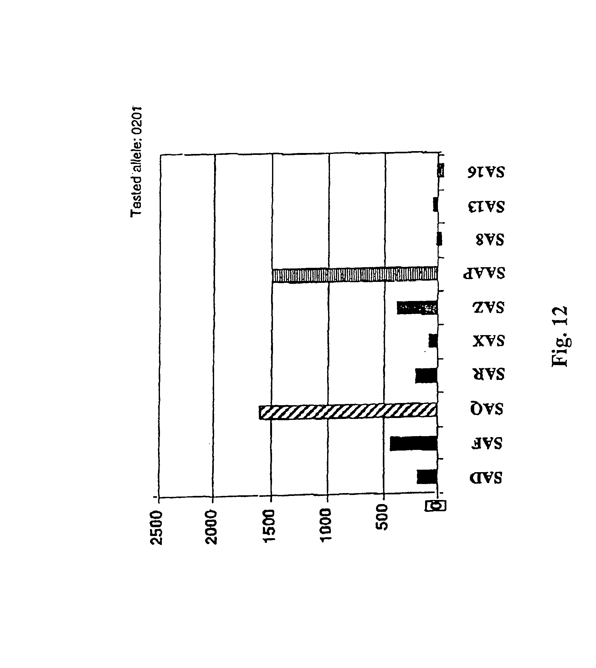
D00014

D00015

D00016

D00017

D00018

D00019

D00020

D00021

D00022

D00023

D00024

D00025

S00001
XML
uspto.report is an independent third-party trademark research tool that is not affiliated, endorsed, or sponsored by the United States Patent and Trademark Office (USPTO) or any other governmental organization. The information provided by uspto.report is based on publicly available data at the time of writing and is intended for informational purposes only.
While we strive to provide accurate and up-to-date information, we do not guarantee the accuracy, completeness, reliability, or suitability of the information displayed on this site. The use of this site is at your own risk. Any reliance you place on such information is therefore strictly at your own risk.
All official trademark data, including owner information, should be verified by visiting the official USPTO website at www.uspto.gov. This site is not intended to replace professional legal advice and should not be used as a substitute for consulting with a legal professional who is knowledgeable about trademark law.