Nuclease-mediated regulation of gene expression
Orkin , et al. June 1, 2
U.S. patent number 11,021,696 [Application Number 14/540,729] was granted by the patent office on 2021-06-01 for nuclease-mediated regulation of gene expression. This patent grant is currently assigned to Children's Medical Center Corporation, Sangamo Therapeutics, Inc.. The grantee listed for this patent is Children's Medical Center Corporation, Sangamo Therapeutics, Inc.. Invention is credited to Stuart H. Orkin, Andreas Reik, Fyodor Urnov.


View All Diagrams
| United States Patent | 11,021,696 |
| Orkin , et al. | June 1, 2021 |
Nuclease-mediated regulation of gene expression
Abstract
The present disclosure is in the field of genome engineering, particularly targeted modification of the genome of a hematopoietic cell.
| Inventors: | Orkin; Stuart H. (Boston, MA), Reik; Andreas (Brisbane, CA), Urnov; Fyodor (Brisbane, CA) | ||||||||||
|---|---|---|---|---|---|---|---|---|---|---|---|
| Applicant: |
|
||||||||||
| Assignee: | Children's Medical Center
Corporation (Boston, MA) Sangamo Therapeutics, Inc. (Brisbane, CA) |
||||||||||
| Family ID: | 53043976 | ||||||||||
| Appl. No.: | 14/540,729 | ||||||||||
| Filed: | November 13, 2014 |
Prior Publication Data
| Document Identifier | Publication Date | |
|---|---|---|
| US 20150132269 A1 | May 14, 2015 | |
Related U.S. Patent Documents
| Application Number | Filing Date | Patent Number | Issue Date | ||
|---|---|---|---|---|---|
| 62042075 | Aug 26, 2014 | ||||
| 61903823 | Nov 13, 2013 | ||||
| Current U.S. Class: | 1/1 |
| Current CPC Class: | A61P 7/00 (20180101); C12N 15/85 (20130101); C12N 15/113 (20130101); A61K 38/465 (20130101); C12N 9/22 (20130101); A61K 35/28 (20130101); C07K 14/4702 (20130101); A61K 35/12 (20130101); A61K 48/00 (20130101); C12N 2310/20 (20170501); C12N 2310/10 (20130101); C07K 2319/81 (20130101) |
| Current International Class: | A61K 48/00 (20060101); C12N 9/22 (20060101); C12N 15/85 (20060101); A61K 35/12 (20150101); C12N 15/113 (20100101); C12N 15/63 (20060101); C07H 21/04 (20060101); C07K 14/47 (20060101); A61K 35/28 (20150101); A61K 38/46 (20060101); A01K 67/00 (20060101) |
References Cited [Referenced By]
U.S. Patent Documents
| 5356802 | October 1994 | Chandrasegaran |
| 5420032 | May 1995 | Marshall et al. |
| 5436150 | July 1995 | Chandrasegaran |
| 5487994 | January 1996 | Chandrasegaran |
| 5789538 | August 1998 | Rebar et al. |
| 5925523 | July 1999 | Dove et al. |
| 6007988 | December 1999 | Choo et al. |
| 6013453 | January 2000 | Choo et al. |
| 6140081 | October 2000 | Barbas, III |
| 6140466 | October 2000 | Barbas, III et al. |
| 6180613 | January 2001 | Kaplitt et al. |
| 6200759 | March 2001 | Dove et al. |
| 6242568 | June 2001 | Barbas, III et al. |
| 6410248 | June 2002 | Greisman et al. |
| 6453242 | September 2002 | Eisenbarg et al. |
| 6479626 | November 2002 | Kim et al. |
| 6503717 | January 2003 | Case et al. |
| 6503888 | January 2003 | Kaplitt et al. |
| 6534261 | March 2003 | Cox et al. |
| 6599692 | June 2003 | Case et al. |
| 6607882 | August 2003 | Cox et al. |
| 6689558 | February 2004 | Case |
| 6794136 | September 2004 | Eisenberg et al. |
| 6824978 | November 2004 | Cox et al. |
| 6832252 | December 2004 | Cieslak et al. |
| 6903185 | June 2005 | Kim et al. |
| 6933113 | August 2005 | Case et al. |
| 6979539 | December 2005 | Cox et al. |
| 6998118 | February 2006 | Kaspar et al. |
| 7013219 | March 2006 | Case et al. |
| 7030215 | April 2006 | Liu et al. |
| 7067317 | June 2006 | Rebar et al. |
| 7070934 | July 2006 | Cox, III et al. |
| 7074596 | July 2006 | Darzynkiewicz et al. |
| 7101540 | October 2006 | Kaspar et al. |
| 7153949 | December 2006 | Kim et al. |
| 7163824 | January 2007 | Cox et al. |
| 7253273 | August 2007 | Collingwood |
| 7262054 | August 2007 | Jamieson et al. |
| 7361635 | April 2008 | Miller et al. |
| 7629326 | December 2009 | Choulika et al. |
| 7837668 | November 2010 | Gasmi et al. |
| 7888121 | February 2011 | Urnov et al. |
| 7914796 | March 2011 | Miller et al. |
| 7951925 | May 2011 | Ando et al. |
| 7972854 | July 2011 | Miller et al. |
| 8034598 | October 2011 | Miller et al. |
| 8092429 | January 2012 | Gasmi et al. |
| 8110379 | February 2012 | DeKelver et al. |
| 8153773 | April 2012 | Jemielity et al. |
| 8409861 | April 2013 | Gushin et al. |
| 8586526 | November 2013 | Gregory et al. |
| 8623618 | January 2014 | Doyon et al. |
| 8772453 | July 2014 | Paschon et al. |
| 8921332 | December 2014 | Choulika et al. |
| 2003/0104526 | June 2003 | Liu et al. |
| 2003/0232410 | December 2003 | Liljedahl et al. |
| 2005/0026157 | February 2005 | Baltimore et al. |
| 2005/0064474 | March 2005 | Urnov et al. |
| 2005/0208489 | September 2005 | Carroll et al. |
| 2005/0267061 | December 2005 | Martin |
| 2006/0063231 | March 2006 | Li et al. |
| 2006/0188987 | August 2006 | Guschin |
| 2006/0239966 | October 2006 | Tornoe et al. |
| 2007/0117128 | May 2007 | Smith et al. |
| 2007/0141038 | June 2007 | Choulika et al. |
| 2007/0218528 | September 2007 | Miller |
| 2008/0131962 | June 2008 | Miller |
| 2008/0159996 | July 2008 | Ando et al. |
| 2008/0299580 | December 2008 | DeKelver et al. |
| 2009/0068164 | March 2009 | Segal et al. |
| 2009/0111188 | April 2009 | Cai et al. |
| 2009/0117617 | May 2009 | Holmes et al. |
| 2009/0305419 | December 2009 | Miller |
| 2010/0047805 | February 2010 | Wang |
| 2010/0218264 | August 2010 | Cui et al. |
| 2011/0027235 | February 2011 | Gregory et al. |
| 2011/0182867 | July 2011 | Orkin et al. |
| 2011/0201055 | August 2011 | Doyon et al. |
| 2011/0207221 | August 2011 | Cost et al. |
| 2011/0265198 | October 2011 | Gregory et al. |
| 2011/0301073 | December 2011 | Gregory et al. |
| 2012/0017290 | January 2012 | Cui et al. |
| 2012/0195936 | August 2012 | Rudolph et al. |
| 2012/0222143 | August 2012 | Fahrenkrug et al. |
| 2012/0230971 | October 2012 | Choulika et al. |
| 2012/0294826 | November 2012 | Spitalnik |
| 2013/0122591 | May 2013 | Cost et al. |
| 2013/0137104 | May 2013 | Cost et al. |
| 2013/0177960 | July 2013 | Rebar |
| 2013/0177983 | July 2013 | Rebar |
| 2013/0326645 | December 2013 | Cost et al. |
| 2014/0093913 | April 2014 | Cost et al. |
| 2015/0056705 | February 2015 | Conway et al. |
| 2015/0064789 | March 2015 | Paschon et al. |
| 2338237 | Dec 1999 | GB | |||
| WO 95/19431 | Jul 1995 | WO | |||
| WO 96/06166 | Feb 1996 | WO | |||
| WO 98/37186 | Aug 1998 | WO | |||
| WO 98/53057 | Nov 1998 | WO | |||
| WO 98/53058 | Nov 1998 | WO | |||
| WO 98/53059 | Nov 1998 | WO | |||
| WO 98/53060 | Nov 1998 | WO | |||
| WO 98/54311 | Dec 1998 | WO | |||
| WO 00/46386 | Feb 2000 | WO | |||
| WO 00/27878 | May 2000 | WO | |||
| WO 01/60970 | Aug 2001 | WO | |||
| WO 01/88197 | Nov 2001 | WO | |||
| WO 02/016536 | Feb 2002 | WO | |||
| WO 02/077277 | Oct 2002 | WO | |||
| WO 02/099084 | Dec 2002 | WO | |||
| WO 07/014275 | Feb 2007 | WO | |||
| WO 03/016496 | Feb 2008 | WO | |||
| WO 10/079430 | Jul 2010 | WO | |||
| WO2013/055985 | Apr 2013 | WO | |||
| WO2013/126794 | Aug 2013 | WO | |||
| WO2014/036219 | Mar 2014 | WO | |||
| WO2014/085593 | Jun 2014 | WO | |||
| WO2016/183298 | Nov 2016 | WO | |||
Other References
|
Funnel et al , 2015, Blood 126:89-93. cited by examiner . Smith et al., 2016, Hum. Mol. Genet. 2016, 0:1-7. cited by examiner . Glarneau et al, 2010, Nature Genetics 42:1049-1051. cited by examiner . Uda et al., 2008, PNAS (USA) 105:1620-1625. cited by examiner . Funnell et al 2015, Blood 126:89-93. cited by examiner . USPTO-STIC Sequence Search Mar. 28, 2016,pp. 1-3. cited by examiner . Xiao et al 2013 Nucleic Acids Res. 41:1-11. cited by examiner . Osborn et al 2011, Human Gene Therapy 22:1155-1165. cited by examiner . Gaj et al 2013, Trends in Biotechnology 7:397-405. cited by examiner . Bauer et al. HbF-associated genetic variation marks an erythroid regulatory element essential for BCL11a transcription and subsequent stage-specific globin expression. Abstract 828 (1 page). Blood 120.21, American Society of Hematology, Nov. 16, 2012. cited by examiner . Gaj et al. ZFN, TALEN, and CRISPR/Cas-based methods for genome engineering. Trends in Biotechnology 31:397-405, (Year: 2013). cited by examiner . Niu et al. Applications of TALENs and CRISPR/Cas9 in human cells and their potentials for gene therapy. Mol. Biotechnol. 56:681-688, (Year: 2014). cited by examiner . Chandrakasan et al. Gene therapy for hemoglobinopathies: The state of the field and the future. Hematol. Oncol. Clin. North Am. 28:199-216, (Year: 2014). cited by examiner . Argast, et al., "I-PPOI and I-CREI Homing Site Sequence Degeneracy Determined by Random Mutagenesis and Sequential In Vitro Enrichment," J. Mol. Biol. 280:345-353 (1998). cited by applicant . Ashworth, et al., "Computational Redesign of Endonuclease DNA Binding and Cleavage Specificity," Nature 441:656-659 (2003). cited by applicant . Bauer, et al., "An Erythroid Enhancer of BCL11A Subject to Genetic Variation Determines Detal Homoglobin Level," Science 342(6155):253-257 (2013). cited by applicant . Beerli, et al., "Engineering Polydactyl Zinc-Finger Transcription Factors," Nature Biotechnol 20:135-141 (2002). cited by applicant . Belfort, et al., "Homing Endonucleases: Keeping the House in Order," Nucleic Acids Research 25:3379-3388 (1997). cited by applicant . Bitinaite, et al., "FOKI Dimerization Is Required for DNA Cleavage," PNAS USA 95:10570-10575 (1998). cited by applicant . Boch, et al., "Breaking the Code of DNA Binding Specificity of TAL-Type III Effectors," Science 326:1509-1512 (2009). cited by applicant . Bonas, et al., "Genetic and Structural Characterization of the Avirulence Gene AVRBS3 From Xanthomonas Campestris PV. Vesicatoria," Mol. Gen. Genet. 218:127-136 (1989). cited by applicant . Chevalier, et al., "Design, Activity, and Structure of a Highly Specific Artificial Endonuclease," Molecular Cell 10:895-905 (2002). cited by applicant . Choo, et al., "Advances in Zinc Finger Engineering," Curr. Opin. Struct. Biol. 10:411-416 (2000). cited by applicant . Christian, et al., "TAL Effector Nucleases Create Targeted DNA Double-Strand Breaks," Genetics epub 10.1534/genetics.110.120717 (2010). cited by applicant . Cong, et al., "Multiplex Genome Engineering Using CRISPR/CAS Systems," Sciencexpress 1/10.1126/science1231143 (2013). cited by applicant . Constantoulakis, et al., "Alpha-Amino-N-Butyric Acid Stimulates Fetal Hemoglobin in the Adult," Blood 72(6):1961-1967 (1988). cited by applicant . DeSimone, "5-Azacytidine Stimulates Fetal Hemoglobin Synthesis in Anemic Baboons," PNAS USA 79(14):4428-4431(1982). cited by applicant . Dujon, et al., "Mobile Introns: Definition of Terms and Recommended Nomenclature," Gene 82:115-118 (1989). cited by applicant . Epinat, et al., "A Novel Engineered Meganuclease Induces Homologous Recombination in Yeast and Mammalian Cells," Nucleic Acids Research 31(11):3252-2962 (2003) doi: 10.1093/nar/gkg375. cited by applicant . Fu, et al., "Improving CRISPR-CAS Nuclease Specificity Using Truncated Guide RNAS," Nature Biotechnology 32(3):279-284 (2014). cited by applicant . Fujiwara, et al., "Discovering Hematopoietic Mechanisms Through Genome-Wide Analysis of Gata Factor Chromatin Occupancy," Molecular Cell 36:667-681 (2009). cited by applicant . Giarratana, et al., "Proof of Principle for Transfusion of In Vitro--Generated Red Blood Cells," Blood 118(19):5071-5079 (2011). cited by applicant . Gimble, et al., "Substrate Recognition and Induced DNA Distortion by the PI-SCEI Endonuclease, An Enzyme Generated by Protein Splicing," J. Mol. Biol. 263:163-180 (1996). cited by applicant . Grossman, et al., "Successful Ex Vivo Gene Therapy Directed to Liver in a Patient With Familial Hypercholesterolaemia," Nature Genetics 6:335-341 (1994) doi:10.1038/ng0494-335. cited by applicant . Guo, et al., "Directed Evolution of an Enhanced and Highly Efficient FOKI Cleavage Domain for Zinc Finger Nucleases," J. Mol. Biol. 400(1):96-107 (2010) doi:10.1016/j.jmb.2010.04.060. cited by applicant . Guschin, et al., "A Rapid and General Assay for Monitoring Endogenous Gene Modification," Methods Mol. Biol. 649:247-256 (2010). cited by applicant . Haft, et al., "A Guild of 45 CRISPR-Associated (CAS) Protein Families and Multiple CRISPR/CAS Subtypes Exist in Prokaryotic Genomes," PLoS Comput Biol. 1:e60 (2005). cited by applicant . Heuer, et al., "Repeat Domain Diversity of AVRBS3-Like Genes in Ralstonia solanacearum Strains and Association With Host Preferences in the Field," Appl. and Envir. Micro. 73:4379-4384 (2007). cited by applicant . Ho, et al., "Pathogenic Infection of Macaca Nemestrina With a CCR5-Tropic Subtype-C Simian-Human Immunodeficiency Virus," Retrovirology 6:65 (2009). cited by applicant . Holt, et al., "Zinc Finger Nuclease-Mediated CCR5 Knockout Hematopoietic Stem Cell Transplantation Controls HIV-1 In Vivo," Nat. Biotech. 28(8):839-847 (2010) doi:10.1038/nbt.1663. cited by applicant . Isalan, et al., "A Rapid, Generally Applicable Method to Engineer Zinc Fingers Illustrated by Targeting the HIV-1 Promoter," Nature Biotechnology 19(7):656-660 (2001). cited by applicant . Jansen, et al., "Identification of Genes That Are Associated With DNA Repeats in Prokaryotes," Molecular Microbiology 43(6):1565-1575 (2002). cited by applicant . Jasin, et al., "Genetic Manipulation of Genomes With Rare-Cutting Endonucleases," Trends Genet. 12:224-228 (1996). cited by applicant . Kariko, et al., "Incorporation of Pseudouridine Into MRNA Yields Superior Nonimmunogenic Vector With Increased Translational Capacity and Biological Stability," Molecular Therapy 16(11):1833-1840 (2012). cited by applicant . Kay, et al., "A Bacterial Effector Acts as a Plant Transcription Factor and Induces a Cell Size Regulator," Science 318:648-651 (2007). cited by applicant . Kim, et al., "Chimeric Restriction Endonuclease," PNAS USA 91:883-887 (1994). cited by applicant . Kim, et al., "Insertion and Deletion Mutants of FOKI Restriction Endonuclease," J. Biol. Chem. 269:31978-31982 (1994). cited by applicant . Kim, et al., "Hybrid Restriction Enzymes: Zinc Finger Fusions to FOK I Cleavage Domain," PNAS USA 93(31:1156-1160 (1996). cited by applicant . Kormann, et al., "Expression of Therapeutic Proteins After Delivery of Chemically Modified MRNA in Mice," Nature Biotechnology 29(21:154-157 (2011). cited by applicant . Ley, et al., "5-Azacytidine Increases Gamma-Globin Synthesis and Reduces the Proportion of Dense Cells in Patients With Sickle Cell Anemia," Blood 62:370-380 (1982). cited by applicant . Ley, et al., "5-Azacytidine Selectively Increases .gamma.-Globin Synthesis in a Patent With B+ Thalassemia," N. Engl. J. Medicine 307:1469-1475 (1982). cited by applicant . Li, et al., "Alteration of the Cleavage Distance of FOK I Restriction Endonuclease by Insertion Mutagenesis," PNAS USA 90:2764-2768 (1993). cited by applicant . Li, et al., "Functional Domains in FOK I Restriction Endonuclease," PNAS USA 89:4275-4279 (1992). cited by applicant . Liu, et al., "Functional Studies of BCL11A: Characterization of the Conserved BCL11A-XL Splice Variant and Its Interaction With BCL6 in Nuclear Paraspeckles of Germinal Center B Cells," Molecular Cancer 5:18 (2006). cited by applicant . Makarova, et al., "A DNA Repair System Specific for Thermophilic Archaea and Bacteria Predicted by Genomic Context Analysis," Nucleic Acids Res. 30:482-496 (2002). cited by applicant . Makarova, et al., "A Putative RNA-Interference-Based Immune System in Prokaryotes: Computational Analysis of the Predicted Enzymatic Machinery, Functional Analogies With Eukaryotic RNAI, and Hypothetical Mechanisms of Action," Biol. Direct 1:7 (2006). cited by applicant . Mandal, et al., "Efficient Ablation of Genes in Human Hematopoietic Stem and Effector Cells Using CRISPR/CAS9," Cell Stem Cell 15:643-652 (2014). cited by applicant . Martin and Orkin, "Transcriptional Activation and DNA Binding by the Erythroid Factor GF-1/ NF-EL/ERYF 1," Genes Div. 4:1886-1898 (1990). cited by applicant . Maston, et al., "Transcriptional Regulatory Elements in the Human Genome," Ann. Rev. Genome Hum. Genet. 7:29-50 (2006). cited by applicant . May, et al., "DynamicAnalysisofGeneExpressionandGenome-Wide Transcription Factor Binding During Lineage Specification ofMultipotent Progenitors," Cell Stem Cell 13:1-15 (2013). cited by applicant . Miller, et al., "An Improved Zinc-Finger Nuclease Architecture for Highly Specific Genome Editing," Nat. Biotechnol. 25:778-785 (2007). cited by applicant . Miller, et al., "A Tale Nuclease Architecture for Efficient Genome Editing," Nat. Biotechnol. 1-8 (2011) doi:10.1038/nbt.1755. cited by applicant . Moscou, et al., "A Simple Cipher Governs DNA Recognition by TAL Effectors," Science 326:1501 (2009). cited by applicant . Olovnikov, et al., "Bacterial Argonaute Samples the Transcriptome to Identify Foreign DNA," Mol. Cell. 51:594 (2013). cited by applicant . Pabo, et al., "Design and Selection of Novel CYS2HIS2 Zinc Finger Proteins," Ann. Rev. Biochem. 70:313-340 (2001). cited by applicant . Paques, et al., "Meganucleases and DNA Double-Strand Break Induced Recombination: Persectives for Gene Therapy," Current Gene Therapy 7:49-66 (2007). cited by applicant . Perez, et al., "Establishment of HIV-1 Resistance in CD4+ T Cells by Genome Editing Using Zinc-Finger Nucleases," 26(7):808-816 (2008). cited by applicant . Perler, et al., "Protein Splicing Elements: Inteins and Exteins a Definition of Terms and Recommended Nomenclature," Nucleic Acids Research 22:1125-1127 (1994). cited by applicant . Peterson, et al., "Robust Suppression of ENV-SHIV Viremia in Macaca Nemestrina by 3-Drug Art Is Independent of Timing of Initiation During Chronic Infection," J. Med. Primatol. 42:237-246 (2013). cited by applicant . Sankaran, et al., "Human Fetal Hemoglobin Expression Is Regulated by the Developmental Stage-Specific Repressor BCL11A," Science 322:1839 (2008). cited by applicant . Schornack, et al., "Gene-For-Gene-Mediated Recognition of Nuclear-Targeted AVRBS3-Like Bacterial Effector Proteins," J. Plant Physiol. 163:256-272 (2006). cited by applicant . Segal, et al., "Custom DNA-Binding Proteins Come of Age: Polydactyl Zinc-Finger Proteins," Curr. Opin. Biotechnol. 12(6):632-637 (2001). cited by applicant . Sheng, et al., "Structure-Based Cleavage Mechanism of Thermus Thermophilus Argonaute DNA Guide Strand-Mediated DNA Target Cleavage," PNAS USA 11:652 (2014). cited by applicant . Swarts, et al., "Dna-Guided DNA Interference by a Prokaryotic Argonaute," Nature 507(7491):258-261 (2014). cited by applicant . Tebas, et al., "Gene Editing of CCR5 in Autologous CD4 T Cells of Persons Infected With HIV," New Engl. J. Med. 370(10):901 (2014). cited by applicant . Thein, et al., "Control of Fetal Hemoglobin: New Insights Emerging From Genomics and Clinical Implications," Hum. Mol. Genet. 18(R2):R216-R223 (2009). cited by applicant . Tijssen, et al., "Genome-Wide Analysis of Simultaneous GATA1/2, RUNX1, FLI1, and SCL Binding in Megakaryocytes Identifies Hematopoietic Regulators," Developmental Cell 20:597-609 (2011). cited by applicant . Tsai, et al., "Dimeric CRISPR RNA-Guided FOKI Nucleases for Highly Specific Genome Editing," Nature Biotechnology 32:569-576 (2014) doi: 10.1038/nbt.2908. cited by applicant . U.S. Appl. No. 60/118,669, filed Feb. 3, 1999. cited by applicant . Vogel, "Biochemistry. A Bacterial Seek-and-Destroy System for Foreign DNA," Science 344(6187):972-973 (2014) doi: 10.1126/science.1252962. cited by applicant . Yang, et al., "Purification, Cloning, and Characterization of the CEL I Nuclease," Biochemistry 39:3533-3541 (2000). cited by applicant . Yuan, et al., "Crystal Structure of a. Aeolicus Argonaute, a Site-Specific DNA-Guided Endoribonuclease, Provides Insights Into RISC-Mediated MRNA Cleavage," Mol. Cell. 19:405-419 (2005). cited by applicant . Bauer, "Supplementary Materials and Methods Supplementary Text, Figs. S1-S9, Tables S1-S6, References 39-55," 34 pages, www.ncbi.nlm.nih.gov/pmc/articles/PMC4018826/bin/NIHMS575314-supplement-S- upplementary_Material.pdf (Retrieved on 201-03-06). cited by applicant . Reik, et al., "Targeted Gene Modification in Hematopoietic Stem Cells: A Potential Treatment or Thalassemia and Sickle Cell Anemia," Blood, American Society of Hematology, US, vol. 122, No. 21, p. 434 (Nov. 15, 2013). cited by applicant . Xu et al., "Identification of BC L11A Structure-Function Domains for Fetal Hemoglobin Silencing," Blood, American Society of Hematology, US, vol. 122, No. 21, p. 435 (Nov. 15, 2013). cited by applicant. |
Primary Examiner: Nguyen; Quang
Attorney, Agent or Firm: Lathrap GPM LLP Velema, Esq.; James H.
Parent Case Text
CROSS-REFERENCE TO RELATED APPLICATIONS
The present application claims the benefit of U.S. Provisional Application No. 61/903,823, filed Nov. 13, 2013 and U.S. Provisional Application No. 62/042,075, filed Aug. 26, 2014, the disclosures of which are hereby incorporated by reference in their entireties.
Claims
What is claimed is:
1. An isolated genetically modified human hematopoietic stem or precursor cell comprising (i) an endogenous BCL11a enhancer sequence; and (ii) a pair of TALENs, each TALEN of the pair comprising a FokI cleavage half-domain and a DNA-binding domain comprising a plurality of TALE repeat units, each repeat unit comprising a hypervariable diresidue region (RVD), wherein the RVDs of the TALE repeats units are shown in a single row of as shown in Table 1, Table 2 or Table 4, and further wherein the pair of TALENs is selected from the group consisting of: 102740 and 102741; 102736 and 102737; 102756 and 102757; 102752 and 102753; 102750 and 102751; 102775 and 102774; 102795 and 102794; 102832 and 102833; 102834 and 102835; 102836 and 102837; 102838 and 102839; 102840 and 102841; 102842 and 102843; 102844 and 102845; 102846 and 102847; 102848 and 102849; 102850 and 102851; 102852 and 102853; 102854 and 102855; 102856 and 102857; 102858 and 102859; 102860 and 102861; 102862 and 102863; 102864 and 102865; 102866 and 102867; 102868 and 102869; 102870 and 102871; 102872 and 102873; 102874 and 102875; 102756 and 102757; 102752 and 102753; 102759 and 102751; 102876 and 102877; 102878 and 102879; 102880 and 102881; 102882 and 102883; 102884 and 102885; 102886 and 102887; 102888 and 102889; 102890 and 102891; 102892 and 102893; 102894 and 102895; 102896 and 102897; 102898 and 102899; 102902 and 102903; 102904 and 102905; 102906 and 102907; 102912 and 102913; 102914 and 102915; 102916 and 102917; 102918 and 102919; 102920 and 102921; or 102922 and 102923, wherein the pair of TALENs cleave and genetically modify the BCL11a enhancer sequence.
2. The cell of claim 1, wherein the hematopoietic stem cell is a CD34+ cell.
3. The cell of claim 1, wherein the TALEN pair is introduced into the cell as a polynucleotide.
4. A pharmaceutical composition comprising the cell of claim 1.
5. A method of treating a human patient in need of an increase in gamma-globin gene or protein expression, the method comprising administering to the patient the pharmaceutical composition of claim 4 in an amount sufficient to increase the gamma-globin gene or protein expression in the human patient.
6. The method of claim 5, wherein the human patient has .beta.-thalassemia.
Description
SEQUENCE LISTING
The instant application contains a Sequence Listing which has been submitted electronically in ASCII format and is hereby incorporated by reference in its entirety. Said ASCII copy, created on Dec. 17, 2014, is named 83280112_SL.txt and is 58,663 bytes in size.
TECHNICAL FIELD
The present disclosure is in the field of genome engineering, particularly targeted modification of the genome of a hematopoietic cell.
BACKGROUND
When one considers that genome sequencing efforts have revealed that the human genome contains between 20,000 and 25,000 genes, but fewer than 2000 transcriptional regulators, it becomes clear that a number of factors must interact to control gene expression in all its various temporal, developmental and tissue specific manifestations. Expression of genes is controlled by a highly complex mixture of general and specific transcriptional regulators and expression can also be controlled by cis-acting DNA elements. These DNA elements comprise both local DNA elements such as the core promoter and its associated transcription factor binding sites as well as distal elements such as enhancers, silencers, insulators and locus control regions (LCRs) (see Maston et al (2006) Ann Rev Genome Hum Genet 7: 29-50).
Enhancer elements were first identified in the SV40 viral genome, and then found in the human immunoglobulin heavy chain locus. Now known to play regulatory roles in the expression of many genes, enhancers appear to mainly influence temporal and spatial patterns of gene expression. It has also been found that enhancers function in a manner that is not dependent upon distance from the core promoter of a gene, and is not dependent on any specific sequence orientation with respect to the promoter. Enhancers can be located several hundred kilobases upstream or downstream of a core promoter region, where they can be located in an intron sequence, or even beyond the 3' end of a gene.
Various methods and compositions for targeted cleavage of genomic DNA have been described. Such targeted cleavage events can be used, for example, to induce targeted mutagenesis, induce targeted deletions of cellular DNA sequences, and facilitate targeted recombination at a predetermined chromosomal locus. See, e.g., U.S. Pat. Nos. 8,623,618; 8,034,598; 8,586,526; 6,534,261; 6,599,692; 6,503,717; 6,589,558; 7,067,317; 7,262,054; 7,888,121; 7,972,854; 7,914,796; 7,951,925; 8,110,379; 8,409,861; U.S. Patent Publications 20030232410; 20050208489; 20050026157; 20060063231; 20080159996; 201000218264; 20120017290; 20110265198; 20130137104; 20130122591; 20130177983; 20130177960 and 20150056705, the disclosures of which are incorporated by reference in their, entireties for all purposes. These methods often involve the use of engineered cleavage systems to induce a double strand break (DSB) or a nick in a target DNA sequence such that repair of the break by an error born process such as non-homologous end joining (NHEJ) or repair using a repair template (homology directed repair or HDR) can result in the knock out of a gene or the insertion of a sequence of interest (targeted integration). This technique can also be used to introduce site specific changes in the genome sequence through use of a donor oligonucleotide, including the introduction of specific deletions of genomic regions, or of specific point mutations or localized alterations (also known as gene correction). Cleavage can occur through the use of specific nucleases such as engineered zinc finger nucleases (ZFN), transcription-activator like effector nucleases (TALENs), or using the CRISPR/Cas system with an engineered crRNA/tracr RNA (`single guide RNA`) to guide specific cleavage. Further, targeted nucleases are being developed based on the Argonaute system (e.g., from T. thermophilus, known as `TtAgo`, see Swans et al (2014) Nature 507(7491): 258-261), which also may have the potential for uses in genome editing and gene therapy.
Red blood cells (RBCs), or erythrocytes, are the major cellular component of blood. In fact, RBCs account for one quarter of the cells in a human. Mature RBCs lack a nucleus and many other organelles in humans, and are full of hemoglobin, a metalloprotein that functions to carry oxygen to the tissues as well as carry carbon dioxide out of the tissues and back to the lungs for removal. This protein makes up approximately 97% of the dry weight of RBCs and it increases the oxygen carrying ability of blood by about seventy fold. Hemoglobin is a heterotetramer comprising two alpha (.alpha.)-like globin chains and two beta (.beta.)-like globin chains and 4 heme groups. In adults the .alpha.2.beta.2 tetramer is referred to as Hemoglobin A (HbA) or adult hemoglobin. Typically, the alpha and beta globin chains are synthesized in an approximate 1:1 ratio and this ratio seems to be critical in terms of hemoglobin and RBC stabilization. In a developing fetus, a different form of hemoglobin, fetal hemoglobin (HbF), is produced which has a higher binding affinity for oxygen than Hemoglobin A such that oxygen can be delivered to the baby's system via the mother's blood stream. Fetal hemoglobin also contains two .alpha. globin chains, but in place of the adult .beta.-globin chains, it has two fetal gamma (.gamma.)-globin chains (i.e., fetal hemoglobin is .alpha.2.gamma.2). At approximately 30 weeks of gestation, the synthesis of gamma globin in the fetus starts to drop while the production of beta globin increases. By approximately 10 months of age, the newborn's hemoglobin is nearly all .alpha.2.beta.2 although some HbF persists into adulthood (approximately 1-3% of total hemoglobin). The regulation of the switch from production of gamma- to beta-globin is quite complex, and primarily involves a down-regulation of gamma globin transcription with a simultaneous up-regulation of beta globin transcription.
Genetic defects in the sequences encoding the hemoglobin chains can be responsible for a number of diseases known as hemoglobinopathies, including sickle cell anemia and thalassemias. In the majority of patients with hemoglobinopathies, the genes encoding gamma globin remain present, but expression is relatively low due to normal gene repression occurring around parturition as described above.
It is estimated that 1 in 5000 people in the U.S. have sickle cell disease (SCD), mostly in people of sub-Saharan Africa descent. There appears to be a benefit for heterozygous carriers of the sickle cell mutation for protection against malaria, so this trait may have been positively selected over time, such that it is estimated that in sub-Saharan Africa, one third of the population has the sickle cell trait. Sickle cell disease is caused by a mutation in the .beta. globin gene as a consequence of which valine is substituted for glutamic acid at amino acid #6 (a GAG to GTG at the DNA level), where the resultant hemoglobin is referred to as "hemoglobinS" or "HbS." Under lower oxygen conditions, a conformational shift in the deoxy form of HbS exposes a hydrophobic patch on the protein between the E and F helices. The hydrophobic residues of the valine at position 6 of the beta chain in hemoglobin are able to associate with the hydrophobic patch, causing HbS molecules to aggregate and form fibrous precipitates. These aggregates in turn cause the abnormality or `sickling` of the RBCs, resulting in a loss of flexibility of the cells. The sickling RBCs are no longer able to squeeze into the capillary beds and can result in vaso-occlusive crisis in sickle cell patients. In addition, sickled RBCs are more fragile than normal RBCs, and tend towards hemolysis, eventually leading to anemia in the patient.
Treatment and management of sickle cell patients is a life-long proposition involving antibiotic treatment, pain management and transfusions during acute episodes. One approach is the use of hydroxyurea, which exerts its effects in part by increasing the production of gamma globin. Long term side effects of chronic hydroxyurea therapy are still unknown, however, and treatment gives unwanted side effects and can have variable efficacy from patient to patient. Despite an increase in the efficacy of sickle cell treatments, the life expectancy of patients is still only in the mid to late 50's and the associated morbidities of the disease have a profound impact on a patient's quality of life.
Thalassemias are also diseases relating to hemoglobin and typically involve a reduced expression of globin chains. This can occur through mutations in the regulatory regions of the genes or from a mutation in a globin coding sequence that results in reduced expression or reduced levels or functional globin protein. Alpha thalassemias are mainly associated with people of Western Africa and South Asian descent, and may confer malarial resistance. Beta thalassemia is mainly associated with people of Mediterranean descent, typically from Greece and the coastal areas of Turkey and Italy. Treatment of thalassemias usually involves blood transfusions and iron chelation therapy. Bone marrow transplants are also being used for treatment of people with severe thalassemias if an appropriate donor can be identified, but this procedure can have significant risks.
One approach that has been proposed for the treatment of both SCD and beta thalassemias is to increase the expression of gamma globin with the aim to have HbF functionally replace the aberrant adult hemoglobin. As mentioned above, treatment of SCD patients with hydroxyurea is thought to be successful in part due to its effect on increasing gamma globin expression. The first group of compounds discovered to affect gamma globin reactivation activity were cytotoxic drugs. The ability to cause de novo synthesis of gamma-globin by pharmacological manipulation was first shown using 5-azacytidine in experimental animals (DeSimone (1982) Proc Nat'l Acad Sci USA 79(14):4428-31). Subsequent studies confirmed the ability of 5-azacytidine to increase HbF in patients with .beta.-thalassemia and sickle cell disease (Ley, et al., (1982) N. Engl. J. Medicine, 307: 1469-1475, and Ley, et al., (1983) Blood 62: 370-380). In addition, short chain fatty acids (e.g. butyrate and derivatives) have been shown in experimental systems to increase HbF (Constantoulakis et al., (1988) Blood 72(6):1961-1967). Also, there is a segment of the human population with a condition known as `Hereditary Persistence of Fetal Hemoglobin` (HPFH) where elevated amounts of HbF persist in adulthood (10-40% in HPFH heterozygotes (see Thein et at (2009) Hum. Mol. Genet 18 (R2): R216-R223). This is a rare condition, but in the absence of any associated beta globin abnormalities, is not associated with any significant clinical manifestations, even when 100% of the individual's hemoglobin is HbF. When individuals that have a beta thalassemia also have co-incident HPFH, the expression of HbF can lessen the severity of the disease. Further, the severity of the natural course of sickle cell disease can vary significantly from patient to patient, and this variability, in part, can be traced to the fact that some individuals with milder disease express higher levels of HbF.
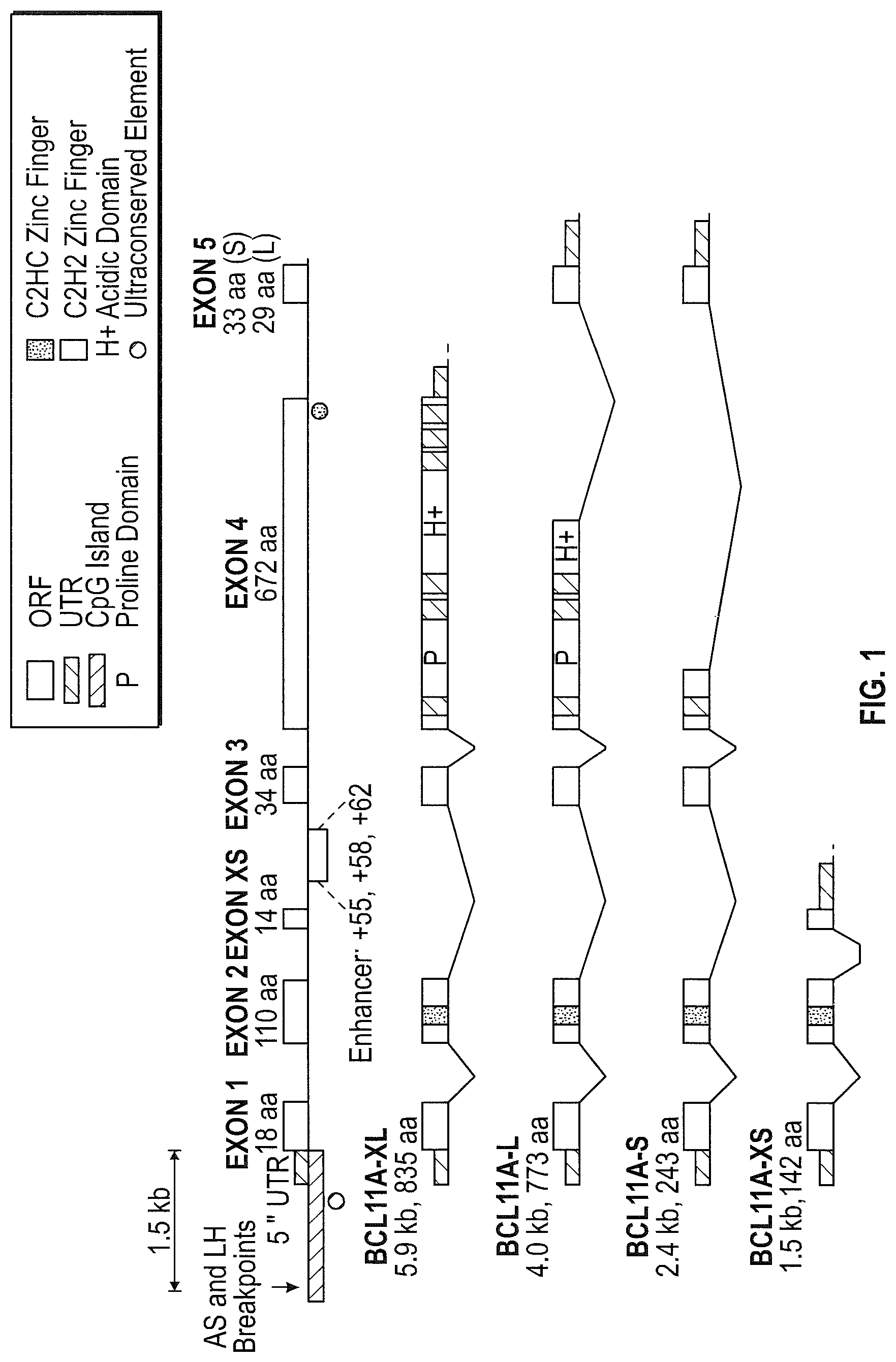
One approach to increase the expression of HbF involves identification of genes whose products play a role in the regulation of gamma globin expression. One such gene is BCL11A, first identified because of its role in lymphocyte development. BCL11A encodes a zinc finger protein that is thought to be involved in the developmental stage-specific regulation of gamma globin expression. BCL11A is expressed in adult erythroid precursor cells and down-regulation of its expression leads to an increase in gamma globin expression. In addition, it appears that the splicing of the BCL11A mRNA is developmentally regulated. In embryonic cells, it appears that the shorter BCL11A mRNA variants, known as BCL11A-S and BCL11A-XS are primary expressed, while in adult cells, the longer BCL11A-L and BCL11A-XL mRNA variants are predominantly expressed. See, Sankaran et at (2008) Science 322 p. 1839. The BCL11A protein appears to interact with the beta globin locus to alter its conformation and thus its expression at different developmental stages. Use of an inhibitory RNA targeted to the BCL11A gene has been proposed (see, e.g., U.S. Patent Publication 20110182867) but this technology has several potential drawbacks, namely that complete knock down may not be achieved, delivery of such RNAs may be problematic and the RNAs must be present continuously, requiring multiple treatments for life.
Targeting of BCL11A enhancer sequences may provide a mechanism for increasing HbF. For example, genome wide association studies have identified a set of genetic variations at BCL11A that are associated with increased HbF levels. These variations are a collection of SNPs found in non-coding regions of BCL11A that function as a stage-specific, lineage-restricted enhancer region. Further investigation revealed that this BCL11A enhancer is required in erythroid cells for BCL11A expression, but is not required for its expression in B cells (see Bauer et al, (2013) Science 343:253-257). The enhancer region was found within intron 2 of the BCL11A gene, and three areas of DNAseI hypersensitivity (often indicative of a chromatin state that is associated with regulatory potential) in intron 2 were identified. These three areas were identified as "+62", "+58" and "+55" in accordance with the distance in kilobases from the transcription start site of BCL11A. These enhancer regions are roughly 350 (+55); 550 (+58); and 350 (+62) nucleotides in length (Bauer 2013, ibid).
Thus, there remains a need for additional methods and compositions that can utilize these genome wide association studies for genome editing and the alteration of gene expression for example to treat hemoglobinopathies such as sickle cell disease and beta thalassemia.
SUMMARY
The present invention describes compositions and methods for use in gene therapy and genome engineering. Specifically, the methods and compositions described relate to inactivating (e.g., by completely or partially abolishing its expression) a gene, for example a gene that acts as regulator of one or more additional genes. In particular, the invention describes methods and compositions for interfering with enhancer function in a BCL11A gene to diminish or knock out its activity in specific cell lineages. Additionally, the invention provides methods and compositions for interfering with BCL11A enhancer functions wherein the enhancer sequences are not located within the BCL11A gene. The resulting down-regulation of the BCL11A gene in these circumstances in turn results in increased expression of gamma globin.
In some aspects, the invention comprises delivery of at least one nuclease (e.g., a nuclease that binds to a BCL11A enhancer sequence) to a human stem cell or precursor cell (HSC/PC) for the purpose of genome engineering. In certain embodiments, the nuclease recognizes a target sequence comprising at least 9 (e.g., 9, 10, 11, 12, 13, 14, 15, 16, 17, 18, 19, 20, 21 or even more) contiguous base pairs of SEQ ID NO:1, SEQ ID NO:2 or SEQ ID NO:3). Exemplary target sequences are shown in Tables 1, 2, 3, 4 and 6. In certain embodiments, the nuclease comprises a DNA-binding domain comprising A DNA-binding protein comprising a zinc finger protein comprising 4, 5 or 6 zinc finger domains comprising a recognition helix region, for example, the recognition helix regions in the order shown in a single row of Table 3 or Table 6. In other embodiments, the nuclease comprises a TALE protein comprising a plurality of TALE repeat units, each repeat unit comprising a hypervariable diresidue region (RVD), for example the RVDs of the TALE repeats units are shown in a single row of Table 1, Table 2 or Table 4. The nuclease(s) as described herein may further comprise a linker (e.g., between the DNA-binding domain and the cleavage domain), for example a linker as shown in FIGS. 14 and 17.
In some embodiments, the nuclease is delivered as a peptide, while in others it is delivered as a nucleic acid encoding the at least one nuclease. In some embodiments, more than one nuclease is used. In some preferred embodiments, the nucleic acid encoding the nuclease is an mRNA, and in some instances, the mRNA is protected. In some aspects, the mRNA may be chemically modified (See e.g. Kormann et al, (2011) Nature Biotechnology 29(2):154-157). In other aspects, the mRNA may comprise an ARCA cap (see U.S. Pat. Nos. 7,074,596 and 8,153,773). In further embodiments, the mRNA may comprise a mixture of unmodified and modified nucleotides (see U.S. Patent Publication 2012-0195936). The nuclease may comprise a zinc finger nuclease (ZFN), a TALE-nuclease (TALEN) or a CRISPR/Cas nuclease system or a combination thereof. In a preferred embodiment, the nucleic acid encoding the nuclease(s) is delivered to the HSC/PC via electroporation. In some embodiments, the nuclease cleaves at or near the binding site of transcription factor. In some aspects, the transcription factor is GATA-1.
In some embodiments comprising a nuclease system that utilizes a nucleic acid guide (e.g. CRISPR/Cas; TtAgo), the cell can be contacted with the nucleic acid guide at the same time as it is contacted with the nuclease, prior to contact with the nuclease, or after contact with the nuclease. The cell can be contacted where the nuclease is provided as a polypeptide, a mRNA or an vector (including a viral vector) capable of expression of the gene encoding the nuclease. The guide nucleic acid maybe provided as an oligonucleotide (for TtAgo) or RNA (CRISPR/Cas). Further, guide RNA may be provided via an expression system for expression of the guide RNA within the cell. In some aspects, more than one guide RNA is provided (see Mandal et al (2014) Cell Stem Cell 15:643). In some embodiments, two guide RNAs are provided, while in others, more than two (e.g. three, four, five, six, seven, eight, nine, ten or more than ten) are provided. In some aspects, truncated guide RNAs are used to increase specificity (Fu et al (2014) Nature Biotechnol 32(3): 279). Also see U.S. Patent Publication No. 20150056705.
In one aspect, the invention comprises mutated Cas nucleases specific for a BCL11A enhancer. In some embodiments, these mutant Cas nucleases are Cas9 nucleases, and have altered functionality. In some embodiments, the Cas9 protein is mutated in the HNH domain, rendering it unable to cleave the DNA strand that is complementary to the guide RNA. In other embodiments, the Cas9 is mutated in the Rvu domain, making it incapable of cleaving the non-complimentary DNA strand. These mutations can result in the creation of Cas9 nickases. In some embodiments, two Cas nickases are used with two separate guide RNAs to target a DNA, which results in two nicks in the target DNA at a specified distance apart. In other embodiments, both the HNH and Rvu endonuclease domains are altered to render a Cas9 protein which is unable to cleave a target BCl11A enhancer DNA.
In another aspect, the methods and compositions of the invention comprise truncations of the Cas9 protein. In one embodiment, the Cas9 protein is truncated such that one or more of the Cas9 functional domains are removed. In one embodiment, the removal of part or one of the nuclease domains renders the Cas nuclease a nickase. In one embodiment, the Cas9 comprises only the domain responsible for interaction with the crRNA or sgRNA and the target DNA.
In still further aspects, the methods and compositions of the invention also comprise fusion proteins wherein the Cas9 protein, or truncation thereof, is fused to a functional domain. In some aspects, the functional domain is an activation or a repression domain. In other aspects, the functional domain is a nuclease domain. In some embodiments, the nuclease domain is a FokI endonuclease domain (e.g. Tsai (2014) Nature Biotech doi:10.1038/nbt.2908). In some embodiments, the FokI domain comprises mutations in the dimerization domain.
In other aspects, the invention comprises a cell or cell line in which an endogenous BCL11A enhancer sequence is modified, for example as compared to the wild-type sequence of the cell. The cell or cell lines may be heterozygous or homozygous for the modification. The modifications may comprise insertions, deletions and/or combinations thereof. In some preferred embodiments, the insertions, deletions and/or combinations thereof result in the destruction of a transcription factor binding site. In certain embodiments, the BCL11A enhancer sequence is modified by a nuclease (e.g., ZFN, TALEN, CRISPR/Cas system, Ttago system, etc.). In certain embodiments, the BCL11A enhancer is modified anywhere between exon 2 and exon 3. In other embodiments, the BCL11A enhancer is modified in the regions shown in SEQ ID NO:1, SEQ ID NO:2 or SEQ ID NO:3 (FIG. 11). In certain embodiments, the modification is at or near the nuclease(s) binding and/or cleavage site(s), for example, within 1-300 (or any value therebetween) base pairs upstream or downstream of the site(s) of cleavage, more preferably within 1-100 base pairs (or any value therebetween) of either side of the binding and/or cleavage site(s), even more preferably within 1 to 50 base pairs (or any value therebetween) on either side of the binding and/or cleavage site(s). In certain embodiments, the modification is at or near the "+58" region of the BCL11A enhancer, for example, at or near a nuclease binding site shown in any of SEQ ID NOs:4 to 80 and 276. In other embodiments, the modification is at or near the "+55" region of the BCL11A enhancer, for example, at or near a nuclease site shown in any of SEQ ID NOs:143 to 184 and 232-251. In still further embodiments, the modification occurs at other BCL11A enhancer sequences. Any cell or cell line may be modified, for example a stem cell (hematopoietic stem cell). Partially or fully differentiated cells descended from the modified stem cells as described herein are also provided (e.g., RBCs or RBC precursor cells). Any of the modified cells or cell lines disclosed herein may show increased expression of gamma globin. Compositions such as pharmaceutical compositions comprising the genetically modified cells as described herein are also provided.
In other aspects, the invention comprises delivery of a donor nucleic acid to a target cell. The donor may be delivered prior to, after, or along with the nucleic acid encoding the nuclease(s). The donor nucleic acid may comprise an exogenous sequence (transgene) to be integrated into the genome of the cell, for example, an endogenous locus. In some embodiments, the donor may comprise a full length gene or fragment thereof flanked by regions of homology with the targeted cleavage site. In some embodiments, the donor lacks homologous regions and is integrated into a target locus through homology independent mechanism (i.e. NHEJ). The donor may comprise any nucleic acid sequence, for example a nucleic acid that, when used as a substrate for homology-directed repair of the nuclease-induced double-strand break, leads to a donor-specified deletion to be generated at the endogenous chromosomal locus (e.g., BCL11A enhancer region) or, alternatively (or in addition to), novel allelic forms of (e.g., point mutations that ablate a transcription factor binding site) the endogenous locus to be created. In some aspects, the donor nucleic acid is an oligonucleotide wherein integration leads to a gene correction event, or a targeted deletion.
In other aspects, the nuclease and/or donor is(are) delivered by viral and/or non-viral gene transfer methods. In preferred embodiments, the donor is delivered to the cell via an adeno-associated virus (AAV). In some instances, the AAV comprises LTRs that are of a heterologous serotype in comparison with the capsid serotype.
In some aspects, the methods and compositions of the invention comprise one or more nucleases (e.g., ZFNs and/or TALENs) targeted to specific regions in the BCL11A enhancer region. In some embodiments, the one or more pairs of nucleases target sequences that result in the modification of the enhancer region by deletion of it in its entirety, while in other embodiments, subsections of the enhancer are deleted. In some embodiments, the deletion comprises one or more of the +55, +58 and/or +62 DNAseI hypersensitivity regions of the enhancer region. In other embodiments, a subset (less than all) of the hypersensitive regions is deleted. In some embodiments, only the +55, only the +58 or only the +62 region is deleted. In other embodiments, two of the regions are deleted (e.g., +55 and +58; +58 and +62; or +55 and +62).
In some aspects, deletions comprising regions within the DNAseI hypersensitive regions of the enhancer are made. These deletions can comprise from about 1 nucleotide to about 551 nucleotides. Thus, the deletions can comprise, 1, 5, 10, 15, 20, 25, 30, 40, 50, 100, 150, 200, 250, 300, 350, 400, 450, 500, 550 nucleotides, or any value therebetween. In some embodiments, the deletions comprise binding regions for one or more transcription factors. In some preferred embodiments, the deletions comprise a GATA-1 binding site, or the binding site for GATA-1 in combination with other factors.
Some aspects of the invention relate to engineered (non-natural) DNA binding proteins that bind to the BCL11A enhancer sequence(s) but do not cleave it. In some embodiments, the Cas9 nuclease domain in a CRISPR/Cas system can be specifically engineered to lose DNA cleavage activity ("dCAS"), and fused to a functional domain capable of modulating gene expression (see Perez-Pimera (2013) Nat Method 10(10):973-976) to create a CRISPR/dCas-TF. In some instances, the engineered DNA binding domains block interaction of the transcription factors active in enhancer activity from binding to their cognate enhancer sequences.
In some embodiments, the DNA binding domains are fused to a functional domain. Some aspects include fusion of the DNA binding domains with domains capable of regulating the expression of a gene. In some embodiments, the fusion proteins comprise a DNA binding domain (zinc finger, TALE, CRISPR/dCas, TtaGo or other DNA binding domains that can be engineered for binding specificity) fused to a gene expression modulatory domain where the modulator represses gene expression.
In some embodiments, the HSC/PC cells are contacted with the nucleases and/or DNA binding proteins of the invention. In some embodiments, the nucleases and/or DNA binding proteins are delivered as nucleic acids and in other embodiments, they are delivered as proteins. In some embodiments, the nucleic acids are mRNAs encoding the nucleases and/or DNA binding proteins, and in further embodiments, the mRNAs may be protected. In some embodiments, the mRNA may be chemically modified, may comprise an ARCA cap and/or may comprise a mixture of unmodified and modified nucleotides.
In some aspects, the HSC/PC are contacted with the nucleases and/or DNA binding proteins of the inventions ex vivo, following apheresis of the HSC/PC from a subject, or purification from harvested bone marrow. In some embodiments, the nucleases cause modifications within the BCL11A enhancer regions. In further embodiments, the HSC/PC containing the BCL11A enhancer region modifications are introduced back into the subject. In some instances, the HSC/PC containing the BCL11A enhancer region modifications are expanded prior to introduction. In other aspects, the genetically modified HSC/PC are given to the subject in a bone marrow transplant wherein the HSC/PC engraft, differentiate and mature in vivo. In some embodiments, the HSC/PC are isolated from the subject following G-CSF- and/or plerixafor-induced mobilization, and in others, the cells are isolated from human bone marrow or human umbilical cords. In some aspects, the subject is treated to a mild myeloablative procedure prior to introduction of the graft comprising the modified HSC/PC, while in other aspects, the subject is treated with a vigorous myeloablative conditioning regimen. In some embodiments, the methods and compositions of the invention are used to treat or prevent a hemoglobinopathy. In some aspects, the hemoglobinopathy is a beta thalassemia, while in other aspects, the hemoglobinopathy is sickle cell disease.
In some embodiments, the HSC/PC are further contacted with a donor molecule. In some embodiments, the donor molecule is delivered by a viral vector. The donor molecule may comprise one or more sequences encoding a functional polypeptide (e.g., a cDNA or fragment thereof), with or without a promoter. Additional sequences (coding or non-coding sequences) may be included when a donor molecule is used for inactivation, including but not limited to, sequences encoding a 2A peptide, SA site, IRES, etc.
In one aspect, the methods and compositions of the invention comprise methods for contacting the HSC/PC in vivo. The nucleases and/or DNA binding proteins are delivered to HSC/PC in situ by methods known in the art. In some embodiments, the nucleases and/or DNA binding proteins of the invention comprise a viral particle that is administered to the subject in need, while in others, the nucleases and/or DNA binding proteins comprise a nanoparticle (e.g. liposome). In some embodiments, the viral particles and/or nanoparticles are delivered to the organ (e.g. bone marrow) wherein the HSC/PC reside.
In another aspect, described herein are methods of integrating a donor nucleic acid into the genome of a cell via homology-independent mechanisms. The methods comprise creating a double-stranded break (DSB) in the genome of a cell and cleaving the donor molecule using a nuclease, such that the donor nucleic acid is integrated at the site of the DSB. In certain embodiments, the donor nucleic acid is integrated via non-homology dependent methods (e.g., NHEJ). As noted above, upon in vivo cleavage the donor sequences can be integrated in a targeted manner into the genome of a cell at the location of a DSB. The donor sequence can include one or more of the same target sites for one or more of the nucleases used to create the DSB. Thus, the donor sequence may be cleaved by one or more of the same nucleases used to cleave the endogenous gene into which integration is desired. In certain embodiments, the donor sequence includes different nuclease target sites from the nucleases used to induce the DSB. DSBs in the genome of the target cell may be created by any mechanism. In certain embodiments, the DSB is created by one or more zinc-finger nucleases (ZFNs), fusion proteins comprising a zinc finger binding domain, which is engineered to bind a sequence within the region of interest, and a cleavage domain or a cleavage half-domain. In other embodiments, the DSB is created by one or more TALE DNA-binding domains (naturally occurring or non-naturally occurring) fused to a nuclease domain (TALEN). In yet further embodiments, the DSB is created using a CRISPR/Cas nuclease system where an engineered single guide RNA or its functional equivalent is used to guide the nuclease to a targeted site in a genome.
In one aspect, the donor may encode a regulatory protein of interest (e.g. ZFP TFs, TALE TFs or a CRISPR/Cas TF) that binds to and/or modulates expression of a gene of interest. In one embodiment, the regulatory proteins bind to a DNA sequence and prevent binding of other regulatory factors. In another embodiment, the binding of a the regulatory protein may modulate (i.e. induce or repress) expression of a target DNA.
In some embodiments, the transgenic HSC/PC cell and/or animal includes a transgene that encodes a human gene. In some instances, the transgenic animal comprises a knock out at the endogenous locus corresponding to exogenous transgene, thereby allowing the development of an in vivo system where the human protein may be studied in isolation. Such transgenic models may be used for screening purposes to identify small molecules or large biomolecules or other entities which may interact with or modify the human protein of interest. In some aspects, the transgene is integrated into the selected locus (e.g., safe-harbor) into a stem cell (e.g., an embryonic stem cell, an induced pluripotent stem cell, a hematopoietic stem cell, etc.) or animal embryo obtained by any of the methods described herein, and then the embryo is implanted such that a live animal is born. The animal is then raised to sexual maturity and allowed to produce offspring wherein at least some of the offspring comprise edited endogenous gene sequence or the integrated transgene.
In another aspect, provided herein is a method of altering gene expression (e.g., BCL11a and/or a globin gene) in a cell, the method comprising: introducing, into the cell, one or more nucleases as described herein, under conditions such that the one or more proteins are expressed and expression of the gene is altered. In certain embodiments, expression of a globin gene (e.g., gamma globin or beta globin) is altered (e.g., increased). Any of the methods described herein may further comprise integrating a donor sequence (e.g., transgene or fragment thereof under the control of an exogenous or endogenous promoter) into the genome of the cell, for example integrating a donor at or near the site of nuclease cleavage in the BCL11a gene. The donor sequence is introduced to the cell using a viral vector, as an oligonucleotide and/or on a plasmid. The cell in which gene expression is altered may be, for example, a red blood cell (RBC) precursor cell and/or a hematopoietic stem cell (e.g., CD34+ cell).
In other embodiments, provided herein is a method of producing a genetically modified cell comprising a genomic modification within an endogenous BCL11a enhancer sequence, the method comprising the steps of: a) contacting a cell with a polynucleotide (e.g. DNA or mRNA) encoding a zinc finger nuclease comprising 4, 5, or 6 zinc finger domains in which each of the zinc finger domains comprises a recognition helix region in the order shown in a single row of Table 3 or Table 6; b) subjecting the cell to conditions conducive to expressing the zinc finger protein from the polynucleotide; and c) modifying the endogenous BCL11A enhancer sequence with the expressed zinc finger protein sufficient to produce the genetically modified cell. In certain embodiments, the cells are stimulated with at least one cytokine (e.g., prior to step (a)). The polynucleotide may be contacted with the cell using any suitable method, including but not limited, via transfection, using a non-viral vector, using a viral vector, by chemical means or by exposure to an electric field (e.g., electroporation).
Also provided is a method of treating a patient in need of an increase in globin gene expression, the method comprising administering to the patient the pharmaceutical preparation as described herein in an amount sufficient to increase the globin gene expression in the patient. In certain embodiments, the patient is known to have, is suspected of having, or is at risk of developing a thalassemia or sickle cell disease.
A kit, comprising the nucleic acids, proteins and/or cells of the invention, is also provided. The kit may comprise nucleic acids encoding the nucleases, (e.g. RNA molecules or ZFN, TALEN or CRISPR/Cas system encoding genes contained in a suitable expression vector), or aliquots of the nuclease proteins, donor molecules, suitable stemness modifiers, cells, buffers, and/or instructions (e.g., for performing the methods of the invention) and the like.
The invention therefore includes, but is not limited to the following embodiments:
1. A genetically modified cell comprising a genomic modification made by a nuclease, wherein the genomic modification is within an endogenous BCL11a enhancer sequence, and further wherein the genomic modification is selected from the group consisting of insertions, deletions and combinations thereof.
2. The genetically modified cell of embodiment 1, wherein the genomic modification is within one or more of the sequences shown in SEQ ID NO:1, 2 or 3.
3. The genetically modified cell of embodiment 2, wherein the genomic modification is within at least 9 contiguous base pairs of SEQ ID NO:1, 2 or 3.
4. The genetically modified cell of embodiment 2, wherein the genomic modification is within the +55 BCL11A enhancer sequence (SEQ ID NO:1).
5. The genetically modified cell of embodiment 4, wherein the genomic modification is at or near any of the sequences shown as SEQ ID Nos. 143 to 184 and 232-251.
6. The genetically modified cell of embodiment 2, wherein the genomic modification is within the +58 BCL11A enhancer sequence (SEQ ID NO:2).
7. The genetically modified cell of embodiment 6, wherein the genomic modification is at or near any of the sequences shown as SEQ ID Nos. 4 to 80 and 276.
8. The genetically modified cell of embodiment 2, wherein the genomic modification is within the +62 BCL11A enhancer sequence (SEQ ID NO:3)
9. The genetically modified cell of any of embodiments 1 to 8, wherein the cell is a stem cell.
10. The genetically modified cell of embodiment 9, wherein the stem cell is a hematopoietic stem cell.
11. The genetically modified cell of embodiment 10, wherein the hematopoietic stem cell is a CD34+ cell.
12. A genetically modified differentiated cell descended from the stem cell of any of embodiments 1 to 11.
13. The genetically modified cell of embodiment 12, wherein the cell is a red blood cell (RBC).
14. The genetically modified cell of any of embodiments 1 to 13, wherein the nuclease comprises at least one zinc finger nuclease (ZFN) or TALEN.
15. The genetically modified cell of any of embodiments 1 to 14, wherein the nuclease is introduced into the cell as a polynucleotide.
16. The genetically modified cell of any of embodiments 1 to 15, wherein the insertion comprises integration of a donor polynucleotide encoding a transgene.
17. The genetically modified cell of any of embodiments 14 to 16, wherein the nuclease comprises a zinc finger nuclease, the zinc finger nuclease comprising 4, 5, or 6 zinc finger domains comprising a recognition helix and further wherein the zinc finger proteins comprise the recognition helix regions in the order shown in a single row of Table 3 or Table 6.
18. The genetically modified cell of any of embodiments 14 to 16, wherein the nuclease comprises a TALEN, the TALEN comprising a plurality of TALE repeat units, each repeat unit comprising a hypervariable diresidue region (RVD), wherein the RVDs of the TALE repeats units are shown in a single row of Table 1, Table 2 or Table 4.
19. A pharmaceutical composition comprising the genetically modified cell of any of embodiments 1 to 18.
20. A DNA-binding protein comprising a zinc finger protein or a TALE-effector protein (TALE), wherein (i) the zinc finger protein comprises 4, 5 or 6 zinc finger domains comprising a recognition helix region, wherein the zinc finger proteins comprise the recognition helix regions in the order shown in a single row of Table 3 or Table 6; and (ii) the TALE protein comprising a plurality of TALE repeat units, each repeat unit comprising a hypervariable diresidue region (RVD), wherein the RVDs of the TALE repeats units are shown in a single row of Table 1, Table 2 or Table 4.
21. A fusion protein comprising a zinc finger protein or TALE protein of embodiment 20 and a wild-type or engineered cleavage domain or cleavage half-domain.
22. A polynucleotide encoding one or more proteins of embodiment 20 or embodiment 21.
23. An isolated cell comprising one or more proteins according to embodiment 20 or embodiment 21.
24. An isolated cell comprising one or more polynucleotides according to embodiment 22.
25. The cell of embodiment 23 or embodiment 24, wherein the cell is a hematopoietic stem cell.
26. A kit comprising at least one of: i) a polynucleotide encoding the protein according to embodiment 20 or embodiment 21 or ii) a protein according to embodiment 20 or embodiment 21.
27. A method of altering globin gene expression in a cell, the method comprising: introducing, into the cell, one or more polynucleotides according to embodiment 22, under conditions such that the one or more proteins are expressed and expression of the globin gene is altered.
28. The method of embodiment 27, wherein expression of the globin gene is increased.
29. The method of embodiment 27 or embodiment 28, wherein the globin gene is a gamma globin or beta globin gene.
30. The method of any of embodiments 27 to 29, further comprising integrating a donor sequence into the genome of the cell.
31. The method of embodiment 30, wherein the donor sequence is introduced to the cell using a viral vector, as an oligonucleotide or on a plasmid.
32. The method of any of embodiments 27 to 31, wherein the cell is selected from the group consisting of a red blood cell (RBC) precursor cell and a hematopoietic stem cell.
33. The method of any of embodiments 30 to 32, wherein the donor sequence comprises a transgene under the control of an endogenous or exogenous promoter.
34. A method of producing a genetically modified cell comprising a genomic modification within an endogenous BCL11a enhancer sequence, the method comprising the steps of: a) contacting a cell with a polynucleotide encoding a fusion protein comprising a zinc finger nuclease comprising 4, 5, or 6 zinc finger domains in which each of the zinc finger domains comprises a recognition helix region in the order shown in a single row of Table 3 or Table 6, b) subjecting the cell to conditions conducive to expressing the fusion protein from the polynucleotide; and c) modifying the endogenous BCL11A enhancer sequence with the expressed fusion protein sufficient to produce the genetically modified cell.
35. The method of embodiment 34, wherein the method further comprises stimulating the cells with at least one cytokine.
36. The method of embodiment 34 or embodiment 35, wherein the method further comprises the step of delivering the polynucleotide inside the cell.
37. The method of embodiment 36, wherein the delivery step comprises use of at least one of a non-viral delivery system, a viral delivery system, and a delivery vehicle.
38. The method of any of embodiments 34 to 37, wherein the delivery step further comprises subjecting the cells to an electric field.
39. A kit for performing the method of any of embodiments 34 to 37, the kit comprising: a) at least one polynucleotide encoding a fusion protein comprising a zinc finger nuclease comprising 4, 5, or 6 zinc finger domains in which each of the zinc finger domains comprises a recognition helix region in the order shown in a single row of Table 3 or Table 6, b) at least one polynucleotide encoding a TALE protein comprising a plurality of TALE repeat units, each repeat unit comprising a hypervariable diresidue region (RVD), wherein the RVDs of the TALE repeats units are shown in a single row of Table 1, Table 2 or Table 4; and optionally, c) directions for using the kit.
40. A method of treating a patient in need of an increase in globin gene expression, the method comprising administering to the patient the pharmaceutical preparation of embodiment 19 in an amount sufficient to increase the globin gene expression in the patient.
41. The method of embodiment 40, wherein the patient is known to have, is suspected of having, or is at risk of developing a globinopathy.
42. The method of embodiment 41, wherein the globinopathy is a thalassemia or sickle cell disease.
43. The method of embodiment 42, wherein the thalassemia is .beta.-thalassemia.
These and other aspects will be readily apparent to the skilled artisan in light of disclosure as a whole.
BRIEF DESCRIPTION OF THE DRAWINGS
FIG. 1 depicts a diagram of the BCL11A coding region, indicating the position of the introns and of the enhancer regions. The derivations of the differing splicing products are also indicated (see Liu et al. (2006) Molecular Cancer 5:18).
FIG. 2 depicts the genomic region that encodes the various Bcl11a isoforms (University of California Santa Cruz genome browser, coordinates listed in the hg19 assembly of the human genome), the enhancer region in the BCL11A intron 2 (coordinates listed in the hg19 assembly of the human genome), and defines the three subregions of the enhancer region as represented by DNAse I hypersensitive sites (listed in kb by approximate distance from the transcription start site): +55, +58 and +62. Nuclease target locations within the three subregions are indicated as follows: nucleases were designed to cleave on the left end of each subregion (the `L` sites), in the middle of each (the `M` sites) and on the right end (the `R` sites). Cleavage of the locus in vivo with a pair of nucleases results in a deletion of the intervening region in a significant fraction of the cells.
FIG. 3 depicts the results of in-cell cleavage by the TALEN pair sets (indicated in the table in the upper panel of the figure) in the BCL11A enhancer region as gauged by a PCR-based assay for deletion of various regions of the enhancer. Pairs of TALENs used were designed to create deletions in human HSPCs either within the +55 region ("55L-R"), within the +62 region ("62L-R), or within the +58 region ("58L-R", "58M-R"). The gel shows PCR products produced by isolating genomic DNA from human HSPCs transfected with expression constructs encoding the indicated TALENs and amplifying the region surrounding the target region. These data demonstrate the generation of deletions (band indicated by the symbol .DELTA.) in the targeted region following cleavage by the TALEN pair sets indicated.
FIG. 4 is a graph showing results from a real-time RT-qPCR ("Taqman.RTM.") analysis designed to detect a change in expression of fetal gamma-globin mRNA following targeted editing of the BCL11A enhancer. Following electroporation of CD34 cells from healthy human volunteers with mRNAs encoding the designated nucleases (see FIG. 3), erythrocytes were generated in vitro, after which total RNA was harvested. The relative levels of alpha globin and gamma globin mRNA for each sample were determined in an RT-PCR Taqman.RTM. analysis, and the relative ratio of gamma globin mRNA/alpha globin mRNA was plotted. Thus, increasing gamma globin expression in the nuclease-treated samples leads to an increase in the normalized gamma/alpha ratio compared to the controls. The Figure displays the results from treating CD34 cells with single TALEN pairs and for the TALEN pair sets described in FIGS. 2 and 3. The level of gamma/alpha is increased for the 58L-R and 58M-R pair sets. Note that the ratio in the GFP transfection control was 3.4 in this experiment.
FIG. 5 is a graph showing results from a real-time RT-qPCR ("Taqman.RTM.") analysis designed to detect a change in expression of fetal gamma-globin mRNA following targeted editing of the BCL11A enhancer. Following electroporation of CD34 cells from healthy human volunteers with mRNAs encoding the designated nucleases (see FIG. 3), erythrocytes were generated in vitro, after which total RNA was harvested. The relative levels of beta globin and gamma globin mRNA for each sample were determined in an RT-PCR Taqman.RTM. analysis, and the relative ratio of gamma globin mRNA/beta globin mRNA was plotted. The results demonstrate that while the specific single TALEN pairs used in these experiments were not able to induce a change in gamma globin expression, creation of deletions by use of the pair sets caused an increase in relative gamma expression, corrected by adult beta globin expression in this instance. In particular, deletion of the DNA sequenced encompassed by the +55 or of the +58 DNAse I hypersensitive elevated gamma globin, while such elevation following a deletion of the sequence encompassed by the +62 DNAse I hypersensitive site was not detected in this experiment. Note that the ratio in the GFP transfection control was 0.5 in this experiment.
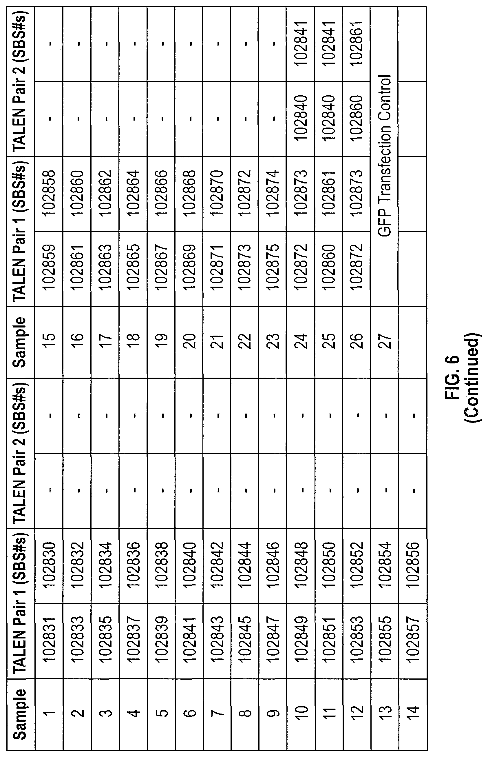
FIG. 6 shows the results from a Taqman.RTM. analysis for fetal globin levels as described in FIG. 4. In this set of experiments, the levels of beta globin and gamma globin mRNA were measured, and thus the data depicted is the ratio of gamma to beta and compared to the same gamma/beta ratio in control treated cells. A series of single TALEN pairs were used to "walk across" the +58 region of the BCL11A enhancer. In contrast to the earlier experiments where a deletion of 400-900 base pairs was required to see an increase in gamma expression, the results depicted in this experiment demonstrated a single site (indicated by an arrow) that when cleaved caused that relative increase. Results from the deletion pairs are included in this graph for comparison. See FIG. 7 for location of cleavage sites of the TALEN pairs across the region of interest.
FIG. 7 shows the results from a Taqman.RTM. analysis as described in FIG. 4 using ZFN pairs targeted to the +58 enhancer region. The levels of beta globin and gamma globin were characterized and used to express the ratio of gamma to beta-globin compared to the same ratio in control treated cells. The data demonstrates that ZFN-driven disruption of the same region identified in the TALEN screen (FIG. 6 and see FIG. 8 below)) resulted in increased gamma globin expression. Note that the ratio in the GFP transfection control was 2.6 in this experiment.
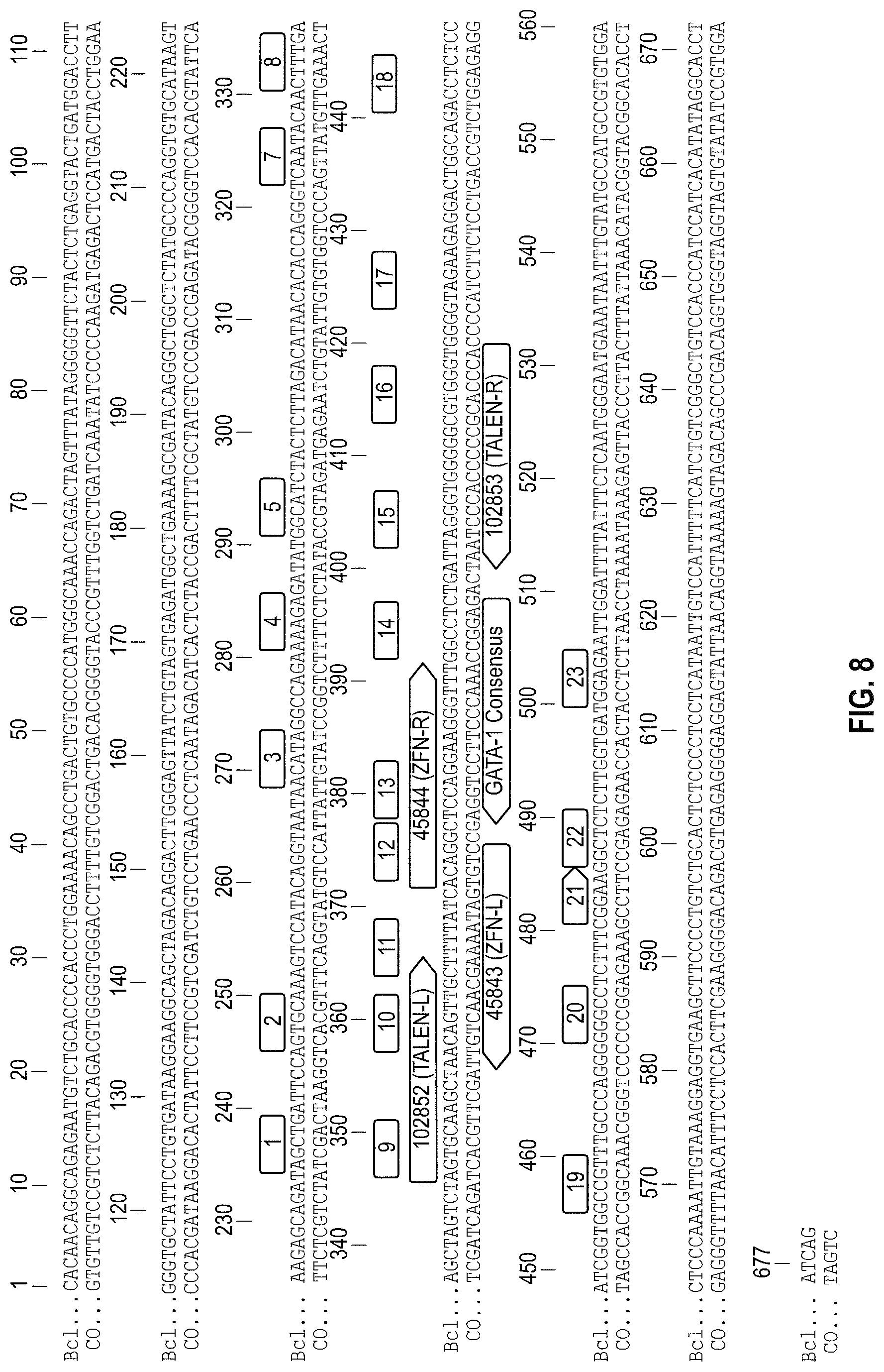
FIG. 8 shows a representation of the binding sites of the +58 enhancer region specific TALENs (102852 and 102853) and ZFNs (45843 and 45844), use of which in human HSPCs increases the relative expression of gamma globin following in vitro erythropoiesis. The sequence shown is the double stranded form of the DNA sequence encompassing the +58 region of the BCL11A enhancer (SEQ ID NO: 264), and the numbering system relates to the +58 itself. Also indicated in the figure is the location of a match to the binding site of the GATA-1-transcription factor (sequence of locus, gtGATAAag, consensus GATA-1 site--swGATAAvv). Additionally, the cleavage sites of the TALEN pairs used in the +58 "walk" are indicated where the numbers correspond to the samples used in the data sets presented in FIG. 6.
FIG. 9 shows the results from a Taqman.RTM. analysis as described in FIG. 4 where a series of TALENs were made to target the +55 region of the BCL11A enhancer. In this set of experiments, the levels of beta globin and gamma globin were characterized, and thus the data depicted is the ratio of gamma to beta and compared to the same ratio in control treated cells (which, in this experiment, was 0.8). The data confirm that mutations generated at specific positions within the +55 region can increase relative gamma globin expression (see arrows).
FIG. 10 is a representation of the cleavage sites of the +55 enhancer region specific TALENs as shown in FIG. 9. The sequence shown is the double stranded form of the DNA sequence encompassing the +55 region of the BCL11A enhancer (SEQ ID NO:254). The numbers that highlight short regions of nucleotides indicate the likely cleavage sites induced by the TALEN samples listed in FIG. 10. Also indicated in FIG. 10 are two matches to the consensus binding site for the GATA-1 transcription factor.
FIGS. 11A to 11C display the DNA sequence of the three DNAse I hypersensitive sites within the BCL11A enhancer sequence. Because their identification was performed by probing regions of accessible chromatin in cells (see Bauer et al, (2013) ibid), the exact boundaries of the regions are not known and approximate boundaries are shown. FIG. 11A shows the sequence of the +55 region (SEQ ID NO:1), FIG. 11B shows the sequence of the +58 region (SEQ ID NO:2) and FIG. 11C shows the sequence of the +62 region (SEQ ID NO:3).
FIGS. 12A to 12C demonstrate that ZFN-driven cleavage in cells closer to the core of the GATA-1 consensus elevates fetal globin levels to an even greater extent than cleavage closer to the 3' end of the motif. FIG. 12A displays a diagram depicting the binding sites of the Bcl11A-specific ZFN pairs in relation to the GATA-1 consensus sequence (FIG. 12A) and depicts a DNA sequence within the +58 region comprising the GATA-1 consensus sequence (SEQ ID NO:255). Bars above and below the DNA sequence indicate the binding sites of the ZFNs. FIG. 12B shows the relative expression of gamma globin and beta globin as measured by mRNA expression following of human HSPCs with mRNA encoding the indicated ZFNs (see FIG. 12A), followed by in vitro erythropoiesis and measurement of levels of fetal globin (see, FIG. 4). The ratio observed when a GFP expressing mRNA was transfected into the CD34+ cells was 0.97. FIG. 12C represents in "pie chart" form the allelic forms of the BCL11A enhancer (specifically, the region cleaved by the ZFNs shown in FIG. 12A) found in human HSPCs following electroporation with the indicated ZFNs. While comparable levels of unmodified (wild-type) chromatids are observed in the two samples, the sample treated with ZFNs that cut closer to the GATA-1 motif contain a greater number of chromatids that eliminate the GATA-1 consensus (e.g., the "-15" allele, which represents a deletion of 15 base pairs). The data demonstrates that cleavage by the two ZFN pairs that are closer to the center of the GATA-1 consensus sequence (pairs 46801/46880 and 46923/46999) is associated with increased gamma globin expression.
FIG. 13 demonstrates that altering the linker between the zinc finger and FokI moiety in ZFNs used for genome editing of the BCL11A enhancer affects fetal globin levels following in vitro erythropoiesis despite comparable levels disrupted chromatids. Human HSPCs were electroporated with the indicated ZFNs (the linker used in each ZFN monomer is indicated in parentheses), and immediately prior and following in vitro erythroid differentiation, the % of disrupted alleles was measured (shown below each sample in "X/Y" form, with the first number corresponding to % of non-wild-type indels following electroporation, and the second number show results following 14 days of in vitro erythroid differentiation). Whole mRNA was harvested and the levels of fetal globin (normalized to alpha globin) were measured.
FIG. 14 depicts the amino acid and DNA sequences for four linkers (L0 (SEQ ID NO:256 and 257), L7a (SEQ ID NO:258 and 259), L7c5 (SEQ ID NO:260 and 261) and L8c5 (SEQ ID NO:262 and 263) used in the ZFP designs. Sequences with a solid underline indicate the carboxy terminal region of the ZFP DNA binding domain, while sequences indicated with the dashed underline indicate the amino terminal region of the Fok I nuclease domain. Sequences in bold indicate the novel sequences added to the standard L0 linker.
FIGS. 15A and 15B depict the percent of cells of human origin, and targeted genetic modification at the nuclease target site in these human cells, respectively found in the peripheral blood of mice following edited human CD34+ cell transplant. FIG. 15A is a graph depicting the percent human cells in the mouse periphery following transplantation of human CD34+ cells that had been edited with two different sets of ZFN 4 weeks post-transplant. FIG. 15B depicts the level of indels detected in those human cells. Each symbol represents data obtained from an individual mouse.
FIGS. 16A through 16D, depict the percentage of indels induced by the nucleases in human cells that differentiated from the original transplanted CD34+ cells 16 weeks post transplantation. FIG. 16A shows the level of indel activity in pan-myeloid cells, identified by the presence of the CD33 marker. FIG. 16B shows the activity in CD19+ B cells. FIG. 16C shows the activity in glyA+ or erythroid cells, while FIG. 16D shows the activity in stem cells. Each symbol represents data obtained from an individual mouse.
FIG. 17 shows a series of linker sequences (SEQ ID NOS 265-275, respectively, in order of appearance). These linkers can serve to link the zinc finger DNA binding domain to the Fok1 nuclease domain.
DETAILED DESCRIPTION
Disclosed herein are compositions and methods for genome engineering for the modulation of BCL11A and/or gamma globin expression and for the treatment and/or prevention of hemoglobinopathies. In particular, nuclease-mediated (i.e. ZFN, TALEN or CRISPR/Cas or TtAgo system) targeted deletion of specific sites in a BCL11A enhancer region is efficiently achieved in HSC/PC and results in a change in relative gamma globin expression during subsequent erythropoiesis. This modulation of BCL11A and gamma globin expression is particularly useful for treatment of hemoglobinopathies (e.g., beta thalassemias, sickle cell disease) wherein there is insufficient beta globin expression or expression of a mutated form of beta-globin. Using the methods and compositions of the invention, the complications and disease related sequelae caused by the aberrant beta globin can be overcome by alteration of the expression of gamma globin in erythrocyte precursor cells.
General
Practice of the methods, as well as preparation and use of the compositions disclosed herein employ, unless otherwise indicated, conventional techniques in molecular biology, biochemistry, chromatin structure and analysis, computational chemistry, cell culture, recombinant DNA and related fields as are within the skill of the art. These techniques are fully explained in the literature. See, for example, Sambrook et al. MOLECULAR CLONING: A LABORATORY MANUAL, Second edition, Cold Spring Harbor Laboratory Press, 1989 and Third edition, 2001; Ausubel et al., CURRENT PROTOCOLS IN MOLECULAR BIOLOGY, John Wiley & Sons, New York, 1987 and periodic updates; the series METHODS IN ENZYMOLOGY, Academic Press, San Diego; Wolffe, CHROMATIN STRUCTURE AND FUNCTION, Third edition, Academic Press, San Diego, 1998; METHODS IN ENZYMOLOGY, Vol. 304, "Chromatin" (P.M. Wassarman and A. P. Wolffe, eds.), Academic Press, San Diego, 1999; and METHODS IN MOLECULAR BIOLOGY, Vol. 119, "Chromatin Protocols" (P. B. Becker, ed.) Humana Press, Totowa, 1999.
Definitions
The terms "nucleic acid," "polynucleotide," and "oligonucleotide" are used interchangeably and refer to a deoxyribonucleotide or ribonucleotide polymer, in linear or circular conformation, and in either single- or double-stranded form. For the purposes of the present disclosure, these terms are not to be construed as limiting with respect to the length of a polymer. The terms can encompass known analogues of natural nucleotides, as well as nucleotides that are modified in the base, sugar and/or phosphate moieties (e.g., phosphorothioate backbones). In general, an analogue of a particular nucleotide has the same base-pairing specificity; i.e., an analogue of A will base-pair with T.
The terms "polypeptide," "peptide" and "protein" are used interchangeably to refer to a polymer of amino acid residues. The term also applies to amino acid polymers in which one or more amino acids are chemical analogues or modified derivatives of a corresponding naturally-occurring amino acids.
"Binding" refers to a sequence-specific, non-covalent interaction between macromolecules (e.g., between a protein and a nucleic acid). Not all components of a binding interaction need be sequence-specific (e.g., contacts with phosphate residues in a DNA backbone), as long as the interaction as a whole is sequence-specific. Such interactions are generally characterized by a dissociation constant (K.sub.d) of 10.sup.-6 M.sup.-1 or lower. "Affinity" refers to the strength of binding: increased binding affinity being correlated with a lower K.sub.d.
A "binding protein" is a protein that is able to bind to another molecule. A binding protein can bind to, for example, a DNA molecule (a DNA-binding protein), an RNA molecule (an RNA-binding protein) and/or a protein molecule (a protein-binding protein). In the case of a protein-binding protein, it can bind to itself (to form homodimers, homotrimers, etc.) and/or it can bind to one or more molecules of a different protein or proteins. A binding protein can have more than one type of binding activity. For example, zinc finger proteins have DNA-binding, RNA-binding and protein-binding activity.
A "zinc finger DNA binding protein" (or binding domain) is a protein, or a domain within a larger protein, that binds DNA in a sequence-specific manner through one or more zinc fingers, which are regions of amino acid sequence within the binding domain whose structure is stabilized through coordination of a zinc ion. The term zinc finger DNA binding protein is often abbreviated as zinc finger protein or ZFP.
A "TALE DNA binding domain" or "TALE" is a polypeptide comprising one or more TALE repeat domains/units. The repeat domains are involved in binding of the TALE to its cognate target DNA sequence. A single "repeat unit" (also referred to as a "repeat") is typically 33-35 amino acids in length and exhibits at least some sequence homology with other TALE repeat sequences within a naturally occurring TALE protein.
Zinc finger and TALE binding domains can be "engineered" to bind to a predetermined nucleotide sequence, for example via engineering (altering one or more amino acids) of the recognition helix region of a naturally occurring zinc finger or TALE protein. Therefore, engineered DNA binding proteins (zinc fingers or TALEs) are proteins that are non-naturally occurring. Non-limiting examples of methods for engineering DNA-binding proteins are design and selection. A designed DNA binding protein is a protein not occurring in nature whose design/composition results principally from rational criteria. Rational criteria for design include application of substitution rules and computerized algorithms for processing information in a database storing information of existing ZFP and/or TALE designs and binding data. See, for example, U.S. Pat. Nos. 6,140,081; 6,453,242; 6,534,261 and 8,585,526; see also WO 98/53058; WO 98/53059; WO 98/53060; WO 02/016536 and WO 03/016496.
A "selected" zinc finger protein or TALE is a protein not found in nature whose production results primarily from an empirical process such as phage display, interaction trap or hybrid selection. See e.g., U.S. Pat. Nos. 5,789,538; 5,925,523; 6,007,988; 6,013,453; 6,200,759; 8,586,526; WO 95/19431; WO 96/06166; WO 98/53057; WO 98/54311; WO 00/27878; WO 01/60970 WO 01/88197, WO 02/099084.
"TtAgo" is a prokaryotic Argonaute protein thought to be involved in gene silencing. TtAgo is derived from the bacteria Thermus thermophilus. See, e.g., Swarts et al, ibid, G. Sheng et al., (2013) Proc. Natl. Acad. Sci. U.S.A. 111, 652). A "TtAgo system" is all the components required including, for example, guide DNAs for cleavage by a TtAgo enzyme.
"Recombination" refers to a process of exchange of genetic information between two polynucleotides, including but not limited to, donor capture by non-homologous end joining (NHEJ) and homologous recombination. For the purposes of this disclosure, "homologous recombination (HR)" refers to the specialized form of such exchange that takes place, for example, during repair of double-strand breaks in cells via homology-directed repair mechanisms. This process requires nucleotide sequence homology, uses a "donor" molecule to template repair of a "target" molecule (i.e., the one that experienced the double-strand break), and is variously known as "non-crossover gene conversion" or "short tract gene conversion," because it leads to the transfer of genetic information from the donor to the target. Without wishing to be bound by any particular theory, such transfer can involve mismatch correction of heteroduplex DNA that forms between the broken target and the donor, and/or "synthesis-dependent strand annealing," in which the donor is used to resynthesize genetic information that will become part of the target, and/or related processes. Such specialized HR often results in an alteration of the sequence of the target molecule such that part or all of the sequence of the donor polynucleotide is incorporated into the target polynucleotide.
In the methods of the disclosure, one or more targeted nucleases as described herein create a double-stranded break (DSB) in the target sequence (e.g., cellular chromatin) at a predetermined site. The DSB may result in deletions and/or insertions by homology-directed repair or by non-homology-directed repair mechanisms. Deletions may include any number of base pairs. Similarly, insertions may include any number of base pairs including, for example, integration of a "donor" polynucleotide, optionally having homology to the nucleotide sequence in the region of the break. The donor sequence may be physically integrated or, alternatively, the donor polynucleotide is used as a template for repair of the break via homologous recombination, resulting in the introduction of all or part of the nucleotide sequence as in the donor into the cellular chromatin. Thus, a first sequence in cellular chromatin can be altered and, in certain embodiments, can be converted into a sequence present in a donor polynucleotide. Thus, the use of the terms "replace" or "replacement" can be understood to represent replacement of one nucleotide sequence by another, (i.e., replacement of a sequence in the informational sense), and does not necessarily require physical or chemical replacement of one polynucleotide by another.
In any of the methods described herein, additional pairs of zinc-finger proteins or TALEN can be used for additional double-stranded cleavage of additional target sites within the cell.
Any of the methods described herein can be used for insertion of a donor of any size and/or partial or complete inactivation of one or more target sequences in a cell by targeted integration of donor sequence that disrupts expression of the gene(s) of interest. Cell lines with partially or completely inactivated genes are also provided.
In any of the methods described herein, the exogenous nucleotide sequence (the "donor sequence" or "transgene") can contain sequences that are homologous, but not identical, to genomic sequences in the region of interest, thereby stimulating homologous recombination to insert a non-identical sequence in the region of interest. Thus, in certain embodiments, portions of the donor sequence that are homologous to sequences in the region of interest exhibit between about 80 to 99% (or any integer therebetween) sequence identity to the genomic sequence that is replaced. In other embodiments, the homology between the donor and genomic sequence is higher than 99%, for example if only 1 nucleotide differs as between donor and genomic sequences of over 100 contiguous base pairs. In certain cases, a non-homologous portion of the donor sequence can contain sequences not present in the region of interest, such that new sequences are introduced into the region of interest. In these instances, the non-homologous sequence is generally flanked by sequences of 50-1,000 base pairs (or any integral value therebetween) or any number of base pairs greater than 1,000, that are homologous or identical to sequences in the region of interest. In other embodiments, the donor sequence is non-homologous to the first sequence, and is inserted into the genome by non-homologous recombination mechanisms.
"Cleavage" refers to the breakage of the covalent backbone of a DNA molecule. Cleavage can be initiated by a variety of methods including, but not limited to, enzymatic or chemical hydrolysis of a phosphodiester bond. Both single-stranded cleavage and double-stranded cleavage are possible, and double-stranded cleavage can occur as a result of two distinct single-stranded cleavage events. DNA cleavage can result in the production of either blunt ends or staggered ends. In certain embodiments, fusion polypeptides are used for targeted double-stranded DNA cleavage.
A "cleavage half-domain" is a polypeptide sequence which, in conjunction with a second polypeptide (either identical or different) forms a complex having cleavage activity (preferably double-strand cleavage activity). The terms "first and second cleavage half-domains;" "+ and - cleavage half-domains" and "right and left cleavage half-domains" are used interchangeably to refer to pairs of cleavage half-domains that dimerize.
An "engineered cleavage half-domain" is a cleavage half-domain that has been modified so as to form obligate heterodimers with another cleavage half-domain (e.g., another engineered cleavage half-domain). See, also, U.S. Patent Publication Nos. 2005/0064474, 20070218528, 20080131962 and 20110201055, incorporated herein by reference in their entireties.
The term "sequence" refers to a nucleotide sequence of any length, which can be DNA or RNA; can be linear, circular or branched and can be either single-stranded or double stranded. The term "donor sequence" refers to a nucleotide sequence that is inserted into a genome. A donor sequence can be of any length, for example between 2 and 100,000,000 nucleotides in length (or any integer value therebetween or thereabove), preferably between about 100 and 100,000 nucleotides in length (or any integer therebetween), more preferably between about 2000 and 20,000 nucleotides in length (or any value therebetween) and even more preferable, between about 5 and 15 kb (or any value therebetween).
"Chromatin" is the nucleoprotein structure comprising the cellular genome. Cellular chromatin comprises nucleic acid, primarily DNA, and protein, including histones and non-histone chromosomal proteins. The majority of eukaryotic cellular chromatin exists in the form of nucleosomes, wherein a nucleosome core comprises approximately 150 base pairs of DNA associated with an octamer comprising two each of histones H2A, H2B, H3 and H4; and linker DNA (of variable length depending on the organism) extends between nucleosome cores. A molecule of histone H1 is generally associated with the linker DNA. For the purposes of the present disclosure, the term "chromatin" is meant to encompass all types of cellular nucleoprotein, both prokaryotic and eukaryotic. Cellular chromatin includes both chromosomal and episomal chromatin.
A "chromosome," is a chromatin complex comprising all or a portion of the genome of a cell. The genome of a cell is often characterized by its karyotype, which is the collection of all the chromosomes that comprise the genome of the cell. The genome of a cell can comprise one or more chromosomes.
An "episome" is a replicating nucleic acid, nucleoprotein complex or other structure comprising a nucleic acid that is not part of the chromosomal karyotype of a cell. Examples of episomes include plasmids and certain viral genomes.
An "accessible region" is a site in cellular chromatin in which a target site present in the nucleic acid can be bound by an exogenous molecule which recognizes the target site. Without wishing to be bound by any particular theory, it is believed that an accessible region is one that is not packaged into a nucleosomal structure. The distinct structure of an accessible region can often be detected by its sensitivity to chemical and enzymatic probes, for example, nucleases.
A "target site" or "target sequence" is a nucleic acid sequence that defines a portion of a nucleic acid to which a binding molecule will bind, provided sufficient conditions for binding exist.
An "exogenous" molecule is a molecule that is not normally present in a cell, but can be introduced into a cell by one or more genetic, biochemical or other methods. "Normal presence in the cell" is determined with respect to the particular developmental stage and environmental conditions of the cell. Thus, for example, a molecule that is present only during embryonic development of muscle is an exogenous molecule with respect to an adult muscle cell. Similarly, a molecule induced by heat shock is an exogenous molecule with respect to a non-heat-shocked cell. An exogenous molecule can comprise, for example, a functioning version of a malfunctioning endogenous molecule or a malfunctioning version of a normally-functioning endogenous molecule.
An exogenous molecule can be, among other things, a small molecule, such as is generated by a combinatorial chemistry process, or a macromolecule such as a protein, nucleic acid, carbohydrate, lipid, glycoprotein, lipoprotein, polysaccharide, any modified derivative of the above molecules, or any complex comprising one or more of the above molecules. Nucleic acids include DNA and RNA, can be single- or double-stranded; can be linear, branched or circular; and can be of any length. Nucleic acids include those capable of forming duplexes, as well as triplex-forming nucleic acids. See, for example, U.S. Pat. Nos. 5,176,996 and 5,422,251. Proteins include, but are not limited to, DNA-binding proteins, transcription factors, chromatin remodeling factors, methylated DNA binding proteins, polymerases, methylases, demethylases, acetylases, deacetylases, kinases, phosphatases, integrases, recombinases, ligases, topoisomerases, gyrases and helicases.
An exogenous molecule can be the same type of molecule as an endogenous molecule, e.g., an exogenous protein or nucleic acid. For example, an exogenous nucleic acid can comprise an infecting viral genome, a plasmid or episome introduced into a cell, or a chromosome that is not normally present in the cell. Methods for the introduction of exogenous molecules into cells are known to those of skill in the art and include, but are not limited to, lipid-mediated transfer (i.e., liposomes, including neutral and cationic lipids), electroporation, direct injection, cell fusion, particle bombardment, calcium phosphate co-precipitation, DEAE-dextran-mediated transfer and viral vector-mediated transfer. An exogenous molecule can also be the same type of molecule as an endogenous molecule but derived from a different species than the cell is derived from. For example, a human nucleic acid sequence may be introduced into a cell line originally derived from a mouse or hamster. Methods for the introduction of exogenous molecules into plant cells are known to those of skill in the art and include, but are not limited to, protoplast transformation, silicon carbide (e.g., WHISKERS.TM.), Agrobacterium-mediated transformation, lipid-mediated transfer (i.e., liposomes, including neutral and cationic lipids), electroporation, direct injection, cell fusion, particle bombardment (e.g., using a "gene gun"), calcium phosphate co-precipitation, DEAE-dextran-mediated transfer and viral vector-mediated transfer.
By contrast, an "endogenous" molecule is one that is normally present in a particular cell at a particular developmental stage under particular environmental conditions. For example, an endogenous nucleic acid can comprise a chromosome, the genome of a mitochondrion, chloroplast or other organelle, or a naturally-occurring episomal nucleic acid. Additional endogenous molecules can include proteins, for example, transcription factors and enzymes.
As used herein, the term "product of an exogenous nucleic acid" includes both polynucleotide and polypeptide products, for example, transcription products (polynucleotides such as RNA) and translation products (polypeptides).
A "fusion" molecule is a molecule in which two or more subunit molecules are linked, preferably covalently. The subunit molecules can be the same chemical type of molecule, or can be different chemical types of molecules. Examples of the first type of fusion molecule include, but are not limited to, fusion proteins (for example, a fusion between a ZFP or TALE DNA-binding domain and one or more activation domains) and fusion nucleic acids (for example, a nucleic acid encoding the fusion protein described supra). Examples of the second type of fusion molecule include, but are not limited to, a fusion between a triplex-forming nucleic acid and a polypeptide, and a fusion between a minor groove binder and a nucleic acid.
Expression of a fusion protein in a cell can result from delivery of the fusion protein to the cell or by delivery of a polynucleotide encoding the fusion protein to a cell, wherein the polynucleotide is transcribed, and the transcript is translated, to generate the fusion protein. Trans-splicing, polypeptide cleavage and polypeptide ligation can also be involved in expression of a protein in a cell. Methods for polynucleotide and polypeptide delivery to cells are presented elsewhere in this disclosure.
A "gene," for the purposes of the present disclosure, includes a DNA region encoding a gene product (see infra), as well as all DNA regions which regulate the production of the gene product, whether or not such regulatory sequences are adjacent to coding and/or transcribed sequences. Accordingly, a gene includes, but is not necessarily limited to, promoter sequences, terminators, translational regulatory sequences such as ribosome binding sites and internal ribosome entry sites, enhancers, silencers, insulators, boundary elements, replication origins, matrix attachment sites and locus control regions.
"Gene expression" refers to the conversion of the information, contained in a gene, into a gene product. A gene product can be the direct transcriptional product of a gene (e.g., mRNA, tRNA, rRNA, antisense RNA, ribozyme, structural RNA or any other type of RNA) or a protein produced by translation of an mRNA. Gene products also include RNAs which are modified, by processes such as capping, polyadenylation, methylation, and editing, and proteins modified by, for example, methylation, acetylation, phosphorylation, ubiquitination, ADP-ribosylation, myristilation, and glycosylation.
"Modulation" of gene expression refers to a change in the activity of a gene. Modulation of expression can include, but is not limited to, gene activation and gene repression. Genome editing (e.g., cleavage, alteration, inactivation, random mutation) can be used to modulate expression. Gene inactivation refers to any reduction in gene expression as compared to a cell that does not include a ZFP, TALE or CRISPR/Cas system as described herein. Thus, gene inactivation may be partial or complete.
A "protected" mRNA is one in which the mRNA has been altered in some manner to increase the stability or translation of the mRNA. Examples of protections include the use of replacement of up to 25% of the cytodine and uridine residues with 2-thiouridine (s2U) and 5-methylcytidine (m5C). The resulting mRNA exhibits less immunogenicity and more stability as compared with its unmodified counterpart. (see Kariko et al. ((2012), Molecular Therapy, Vol. 16, No. 11, pages 1833-1844). Other changes include the addition of a so-called ARCA cap, which increases the translationability of the in vitro produced mRNA (see U.S. Pat. No. 7,074,596).
A "region of interest" is any region of cellular chromatin, such as, for example, a gene or a non-coding sequence within or adjacent to a gene, in which it is desirable to bind an exogenous molecule. Binding can be for the purposes of targeted DNA cleavage and/or targeted recombination. A region of interest can be present in a chromosome, an episome, an organellar genome (e.g., mitochondrial, chloroplast), or an infecting viral genome, for example. A region of interest can be within the coding region of a gene, within transcribed non-coding regions such as, for example, leader sequences, trailer sequences or introns, or within non-transcribed regions, either upstream or downstream of the coding region. A region of interest can be as small as a single nucleotide pair or up to 2,000 nucleotide pairs in length, or any integral value of nucleotide pairs.
"Eukaryotic" cells include, but are not limited to, fungal cells (such as yeast), plant cells, animal cells, mammalian cells and human cells (e.g., T-cells).
The terms "operative linkage" and "operatively linked" (or "operably linked") are used interchangeably with reference to a juxtaposition of two or more components (such as sequence elements), in which the components are arranged such that both components function normally and allow the possibility that at least one of the components can mediate a function that is exerted upon at least one of the other components. By way of illustration, a transcriptional regulatory sequence, such as a promoter, is operatively linked to a coding sequence if the transcriptional regulatory sequence controls the level of transcription of the coding sequence in response to the presence or absence of one or more transcriptional regulatory factors. A transcriptional regulatory sequence is generally operatively linked in cis with a coding sequence, but need not be directly adjacent to it. For example, an enhancer is a transcriptional regulatory sequence that is operatively linked to a coding sequence, even though they are not contiguous.
With respect to fusion polypeptides, the term "operatively linked" can refer to the fact that each of the components performs the same function in linkage to the other component as it would if it were not so linked. For example, with respect to a fusion polypeptide in which a ZFP, TALE or Cas DNA-binding domain is fused to an activation domain, the ZFP, TALE or Cas DNA-binding domain and the activation domain are in operative linkage if, in the fusion polypeptide, the ZFP, TALE of Cas DNA-binding domain portion is able to bind its target site and/or its binding site, while the activation domain is able to upregulate gene expression. When a fusion polypeptide in which a ZFP, TALE or Cas DNA-binding domain is fused to a cleavage domain, the ZFP, TALE or Cas DNA-binding domain and the cleavage domain are in operative linkage if, in the fusion polypeptide, the ZFP, TALE or Cas DNA-binding domain portion is able to bind its target site and/or its binding site, while the cleavage domain is able to cleave DNA in the vicinity of the target site.
A "functional fragment" of a protein, polypeptide or nucleic acid is a protein, polypeptide or nucleic acid whose sequence is not identical to the full-length protein, polypeptide or nucleic acid, yet retains the same function as the full-length protein, polypeptide or nucleic acid. A functional fragment can possess more, fewer, or the same number of residues as the corresponding native molecule, and/or can contain one or more amino acid or nucleotide substitutions. Methods for determining the function of a nucleic acid (e.g., coding function, ability to hybridize to another nucleic acid) are well-known in the art. Similarly, methods for determining protein function are well-known. For example, the DNA-binding function of a polypeptide can be determined, for example, by filter-binding, electrophoretic mobility-shift, or immunoprecipitation assays. DNA cleavage can be assayed by gel electrophoresis. See Ausubel et al., supra. The ability of a protein to interact with another protein can be determined, for example, by co-immunoprecipitation, two-hybrid assays or complementation, both genetic and biochemical. See, for example, Fields et al. (1989) Nature 340:245-246; U.S. Pat. No. 5,585,245 and PCT WO 98/44350.
A "vector" is capable of transferring gene sequences to target cells. Typically, "vector construct," "expression vector," and "gene transfer vector," mean any nucleic acid construct capable of directing the expression of a gene of interest and which can transfer gene sequences to target cells. Thus, the term includes cloning, and expression vehicles, as well as integrating vectors.
The terms "subject" and "patient" are used interchangeably and refer to mammals such as human patients and non-human primates, as well as experimental animals such as rabbits, dogs, cats, rats, mice, and other animals. Accordingly, the term "subject" or "patient" as used herein means any mammalian patient or subject to which the or stem cells of the invention can be administered. Subjects of the present invention include those that have been exposed to one or more chemical toxins, including, for example, a nerve toxin.
"Stemness" refers to the relative ability of any cell to act in a stem cell-like manner, i.e., the degree of toti-, pluri-, or oligopotentcy and expanded or indefinite self-renewal that any particular stem cell may have.
Nucleases
Described herein are compositions, particularly nucleases, that are useful for in vivo cleavage of a donor molecule carrying a transgene and nucleases for cleavage of the genome of a cell such that the transgene is integrated into the genome in a targeted manner. In certain embodiments, one or more of the nucleases are naturally occurring. In other embodiments, one or more of the nucleases are non-naturally occurring, i.e., engineered in the DNA-binding domain and/or cleavage domain. For example, the DNA-binding domain of a naturally-occurring nuclease may be altered to bind to a selected target site (e.g., a meganuclease that has been engineered to bind to site different than the cognate binding site). In other embodiments, the nuclease comprises heterologous DNA-binding and cleavage domains (e.g., zinc finger nucleases; TAL-effector domain DNA binding proteins; meganuclease DNA-binding domains with heterologous cleavage domains).
A. DNA-Binding Domains
In certain embodiments, the composition and methods described herein employ a meganuclease (homing endonuclease) DNA-binding domain for binding to the donor molecule and/or binding to the region of interest in the genome of the cell. Naturally-occurring meganucleases recognize 15-40 base-pair cleavage sites and are commonly grouped into four families: the LAGLIDADG (SEQ ID NO: 287) family, the GIY-YIG family, the His-Cyst box family and the HNH family. Exemplary homing endonucleases include I-SceI, I-CeuI, PI-PspI, PI-Sce, I-SceIV, I-CsmI, I-PanI, I-SceII, I-PpoI, I-SceIII, I-CreI,I-TevI, I-TevII and I-TevIII. Their recognition sequences are known. See also U.S. Pat. Nos. 5,420,032; 6,833,252; Belfort et al. (1997) Nucleic Acids Res. 25:3379-3388; Dujon et al. (1989) Gene 82:115-118; Perler et al. (1994) Nucleic Acids Res. 22, 1125-1127; Jasin (1996) Trends Genet. 12:224-228; Gimble et al. (1996) J. Mol. Biol. 263:163-180; Argast et al. (1998) J. Mol. Biol. 280:345-353 and the New England Biolabs catalogue.
In certain embodiments, the methods and compositions described herein make use of a nuclease that comprises an engineered (non-naturally occurring) homing endonuclease (meganuclease). The recognition sequences of homing endonucleases and meganucleases such as I-SceI, I-CeuI, PI-PspI, PI-Sce, I-SceIV, I-CsmI, I-PanI, I-SceII, I-PpoI, I-SceIII, I-CreI, I-TevI, I-TevII and I-TevIII are known. See also U.S. Pat. Nos. 5,420,032; 6,833,252; Belfort et al. (1997) Nucleic Acids Res. 25:3379-3388; Dujon et al. (1989) Gene 82:115-118; Perler et al. (1994) Nucleic Acids Res. 22, 1125-1127; Jasin (1996) Trends Genet. 12:224-228; Gimble et al. (1996) J. Mol. Biol. 263:163-180; Argast et al. (1998) J. Mol. Biol. 280:345-353 and the New England Biolabs catalogue. In addition, the DNA-binding specificity of homing endonucleases and meganucleases can be engineered to bind non-natural target sites. See, for example, Chevalier et al. (2002) Molec. Cell 10:895-905; Epinat et al. (2003) Nucleic Acids Res. 31:2952-2962; Ashworth et al. (2006) Nature 441:656-659; Paques et al. (2007) Current Gene Therapy 7:49-66; U.S. Patent Publication No. 20070117128. The DNA-binding domains of the homing endonucleases and meganucleases may be altered in the context of the nuclease as a whole (i.e., such that the nuclease includes the cognate cleavage domain) or may be fused to a heterologous cleavage domain.
In other embodiments, the DNA-binding domain of one or more of the nucleases used in the methods and compositions described herein comprises a naturally occurring or engineered (non-naturally occurring) TAL effector DNA binding domain. See, e.g., U.S. Pat. No. 8,586,526, incorporated by reference in its entirety herein. The plant pathogenic bacteria of the genus Xanthomonas are known to cause many diseases in important crop plants. Pathogenicity of Xanthomonas depends on a conserved type III secretion (T3S) system which injects more than 25 different effector proteins into the plant cell. Among these injected proteins are transcription activator-like (TAL) effectors which mimic plant transcriptional activators and manipulate the plant transcriptome (see Kay et at (2007) Science 318:648-651). These proteins contain a DNA binding domain and a transcriptional activation domain. One of the most well characterized TAL-effectors is AvrBs3 from Xanthomonas campestgris pv. Vesicatoria (see Bonas et at (1989) Mol Gen Genet 218: 127-136 and WO2010079430). TAL-effectors contain a centralized domain of tandem repeats, each repeat containing approximately 34 amino acids, which are key to the DNA binding specificity of these proteins. In addition, they contain a nuclear localization sequence and an acidic transcriptional activation domain (for a review see Schornack S, et at (2006) J Plant Physiol 163(3): 256-272). In addition, in the phytopathogenic bacteria Ralstonia solanacearum two genes, designated brg11 and hpx17 have been found that are homologous to the AvrBs3 family of Xanthomonas in the R. solanacearum biovar 1 strain GMI1000 and in the biovar 4 strain RS1000 (See Heuer et at (2007) Appl and Envir Micro 73(13): 4379-4384). These genes are 98.9% identical in nucleotide sequence to each other but differ by a deletion of 1,575 base pairs in the repeat domain of hpx17. However, both gene products have less than 40% sequence identity with AvrBs3 family proteins of Xanthomonas. See, e.g., U.S. Pat. No. 8,586,526, incorporated by reference in its entirety herein.
Specificity of these TAL effectors depends on the sequences found in the tandem repeats. The repeated sequence comprises approximately 102 base pairs (bp) and the repeats are typically 91-100% homologous with each other (Bonas et al, ibid). Polymorphism of the repeats is usually located at positions 12 and 13 and there appears to be a one-to-one correspondence between the identity of the hypervariable diresidues (RVD) at positions 12 and 13 with the identity of the contiguous nucleotides in the TAL-effector's target sequence (see Moscou and Bogdanove, (2009) Science 326:1501 and Boch et at (2009) Science 326:1509-1512). Experimentally, the natural code for DNA recognition of these TAL-effectors has been determined such that an HD sequence at positions 12 and 13 leads to a binding to cytosine (C), NG binds to T, NI to A, C, G or T, NN binds to A or G, and ING binds to T. These DNA binding repeats have been assembled into proteins with new combinations and numbers of repeats, to make artificial transcription factors that are able to interact with new sequences and activate the expression of a non-endogenous reporter gene in plant cells (Boch et al, ibid). Engineered TAL proteins have been linked to a FokI cleavage half domain to yield a TAL effector domain nuclease fusion (TALEN) exhibiting activity in a yeast reporter assay (plasmid based target). See, e.g., U.S. Pat. No. 8,586,526; Christian et at ((2010)<Genetics epub 10.1534/genetics. 110.120717). In certain embodiments, TALE domain comprises an N-cap and/or C-cap as described in U.S. Pat. No. 8,586,526.
In certain embodiments, the DNA binding domain of one or more of the nucleases used for in vivo cleavage and/or targeted cleavage of the genome of a cell comprises a zinc finger protein. Preferably, the zinc finger protein is non-naturally occurring in that it is engineered to bind to a target site of choice. See, for example, See, for example, Beerli et al. (2002) Nature Biotechnol. 20:135-141; Pabo et al. (2001) Ann. Rev. Biochem. 70:313-340; Isalan et al. (2001) Nature Biotechnol. 19:656-660; Segal et al. (2001) Curr. Opin. Biotechnol. 12:632-637; Choo et al. (2000) Curr. Opin. Struct. Biol. 10:411-416; U.S. Pat. Nos. 6,453,242; 6,534,261; 6,599,692; 6,503,717; 6,689,558; 7,030,215; 6,794,136; 7,067,317; 7,262,054; 7,070,934; 7,361,635; 7,253,273; and U.S. Patent Publication Nos. 2005/0064474; 2007/0218528; 2005/0267061, all incorporated herein by reference in their entireties.
An engineered zinc finger binding domain can have a novel binding specificity, compared to a naturally-occurring zinc finger protein. Engineering methods include, but are not limited to, rational design and various types of selection. Rational design includes, for example, using databases comprising triplet (or quadruplet) nucleotide sequences and individual zinc finger amino acid sequences, in which each triplet or quadruplet nucleotide sequence is associated with one or more amino acid sequences of zinc fingers which bind the particular triplet or quadruplet sequence. See, for example, co-owned U.S. Pat. Nos. 6,453,242 and 6,534,261, incorporated by reference herein in their entireties.
Exemplary selection methods, including phage display and two-hybrid systems, are disclosed in U.S. Pat. Nos. 5,789,538; 5,925,523; 6,007,988; 6,013,453; 6,410,248; 6,140,466; 6,200,759; and 6,242,568; as well as WO 98/37186; WO 98/53057; WO 00/27878; WO 01/88197 and GB 2,338,237. In addition, enhancement of binding specificity for zinc finger binding domains has been described, for example, in co-owned WO 02/077227.
In addition, as disclosed in these and other references, zinc finger domains and/or multi-fingered zinc finger proteins may be linked together using any suitable linker sequences, including for example, linkers of 5 or more amino acids in length. See, also, U.S. Pat. Nos. 6,479,626; 6,903,185; and 7,153,949 for exemplary linker sequences 6 or more amino acids in length. The proteins described herein may include any combination of suitable linkers between the individual zinc fingers of the protein.
Selection of target sites; ZFPs and methods for design and construction of fusion proteins (and polynucleotides encoding same) are known to those of skill in the art and described in detail in U.S. Pat. Nos. 6,140,081; 5,789,538; 6,453,242; 6,534,261; 5,925,523; 6,007,988; 6,013,453; 6,200,759; WO 95/19431; WO 96/06166; WO 98/53057; WO 98/54311; WO 00/27878; WO 01/60970 WO 01/88197; WO 02/099084; WO 98/53058; WO 98/53059; WO 98/53060; WO 02/016536 and WO 03/016496.
Nearly any linker (spacer) may be used between one or more of the components of the DNA-binding domain (e.g., zinc fingers), between one or more DNA binding domains and/or between the DNA-binding domain and the functional domain (e.g. nuclease). Non-limiting examples of suitable linker sequences include U.S. Pat. Nos. 8,772,453; 7,888,121; 6,479,626; 6,903,185; and 7,153,949; U.S. Publication Nos. 20090305419 and 20150064789. Thus, the proteins described herein may include any combination of suitable linkers between the individual DNA-binding components and/or between the DNA-binding domain and the functional domain of the compositions described herein.
The CRISPR (clustered regularly interspaced short palindromic repeats) locus, which encodes RNA components of the system, and the cas (CRISPR-associated) locus, which encodes proteins (Jansen et al., 2002. Mol. Microbiol. 43: 1565-1575; Makarova et al., 2002. Nucleic Acids Res. 30: 482-496; Makarova et al., 2006. Biol. Direct 1: 7; Haft et al., 2005. PLoS Comput. Biol. 1: e60) make up the gene sequences of the CRISPR/Cas nuclease system. CRISPR loci in microbial hosts contain a combination of CRISPR-associated (Cas) genes as well as non-coding RNA elements capable of programming the specificity of the CRISPR-mediated nucleic acid cleavage.
The Type II CRISPR is one of the most well characterized systems and carries out targeted DNA double-strand break in four sequential steps. First, two non-coding RNA, the pre-crRNA array and tracrRNA, are transcribed from the CRISPR locus. Second, tracrRNA hybridizes to the repeat regions of the pre-crRNA and mediates the processing of pre-crRNA into mature crRNAs containing individual spacer sequences. Third, the mature crRNA:tracrRNA complex directs Cas9 to the target DNA via Watson-Crick base-pairing between the spacer on the crRNA and the protospacer on the target DNA next to the protospacer adjacent motif (PAM), an additional requirement for target recognition. Finally, Cas9 mediates cleavage of target DNA to create a double-stranded break within the protospacer. Activity of the CRISPR/Cas system comprises of three steps: (i) insertion of alien DNA sequences into the CRISPR array to prevent future attacks, in a process called `adaptation`, (ii) expression of the relevant proteins, as well as expression and processing of the array, followed by (iii) RNA-mediated interference with the alien nucleic acid. Thus, in the bacterial cell, several of the so-called `Cas` proteins are involved with the natural function of the CRISPR/Cas system and serve roles in functions such as insertion of the alien DNA etc.
In certain embodiments, Cas protein may be a "functional derivative" of a naturally occurring Cas protein. A "functional derivative" of a native sequence polypeptide is a compound having a qualitative biological property in common with a native sequence polypeptide. "Functional derivatives" include, but are not limited to, fragments of a native sequence and derivatives of a native sequence polypeptide and its fragments, provided that they have a biological activity in common with a corresponding native sequence polypeptide. A biological activity contemplated herein is the ability of the functional derivative to hydrolyze a DNA substrate into fragments. The term "derivative" encompasses both amino acid sequence variants of polypeptide, covalent modifications, and fusions thereof. Suitable derivatives of a Cas polypeptide or a fragment thereof include but are not limited to mutants, fusions, covalent modifications of Cas protein or a fragment thereof. Cas protein, which includes Cas protein or a fragment thereof, as well as derivatives of Cas protein or a fragment thereof, may be obtainable from a cell or synthesized chemically or by a combination of these two procedures. The cell may be a cell that naturally produces Cas protein, or a cell that naturally produces Cas protein and is genetically engineered to produce the endogenous Cas protein at a higher expression level or to produce a Cas protein from an exogenously introduced nucleic acid, which nucleic acid encodes a Cas that is same or different from the endogenous Cas. In some case, the cell does not naturally produce Cas protein and is genetically engineered to produce a Cas protein.
In some embodiments, the DNA binding domain is part of a TtAgo system (see Swarts et al, ibid; Sheng et al, ibid). In eukaryotes, gene silencing is mediated by the Argonaute (Ago) family of proteins. In this paradigm, Ago is bound to small (19-31 nt) RNAs. This protein-RNA silencing complex recognizes target RNAs via Watson-Crick base pairing between the small RNA and the target and endonucleolytically cleaves the target RNA (Vogel (2014) Science 344:972-973). In contrast, prokaryotic Ago proteins bind to small single-stranded DNA fragments and likely function to detect and remove foreign (often viral) DNA (Yuan et al., (2005) Mol. Cell 19, 405; Olovnikov, et al. (2013) Mol. Cell 51, 594; Swarts et al., ibid). Exemplary prokaryotic Ago proteins include those from Aquifex aeolicus, Rhodobacter sphaeroides, and Thermus thermophilus.
One of the most well-characterized prokaryotic Ago protein is the one from T. thermophilus (TtAgo; Swarts et al. ibid). TtAgo associates with either 15 nt or 13-25 nt single-stranded DNA fragments with 5' phosphate groups. This "guide DNA" bound by TtAgo serves to direct the protein-DNA complex to bind a Watson-Crick complementary DNA sequence in a third-party molecule of DNA. Once the sequence information in these guide DNAs has allowed identification of the target DNA, the TtAgo-guide DNA complex cleaves the target DNA. Such a mechanism is also supported by the structure of the TtAgo-guide DNA complex while bound to its target DNA (G. Sheng et al., ibid). Ago from Rhodobacter sphaeroides (RsAgo) has similar properties (Olivnikov et al. ibid).
Exogenous guide DNAs of arbitrary DNA sequence can be loaded onto the TtAgo protein (Swarts et al. ibid.). Since the specificity of TtAgo cleavage is directed by the guide DNA, a TtAgo-DNA complex formed with an exogenous, investigator-specified guide DNA will therefore direct TtAgo target DNA cleavage to a complementary investigator-specified target DNA. In this way, one may create a targeted double-strand break in DNA. Use of the TtAgo-guide DNA system (or orthologous Ago-guide DNA systems from other organisms) allows for targeted cleavage of genomic DNA within cells. Such cleavage can be either single- or double-stranded. For cleavage of mammalian genomic DNA, it would be preferable to use of a version of TtAgo codon optimized for expression in mammalian cells. Further, it might be preferable to treat cells with a TtAgo-DNA complex formed in vitro where the TtAgo protein is fused to a cell-penetrating peptide. Further, it might be preferable to use a version of the TtAgo protein that has been altered via mutagenesis to have improved activity at 37 degrees Celsius. Ago-RNA-mediated DNA cleavage could be used to affect a panopoly of outcomes including gene knock-out, targeted gene addition, gene correction, targeted gene deletion using techniques standard in the art for exploitation of DNA breaks.
Thus, the nuclease comprises a DNA-binding domain in that specifically binds to a target site in any gene into which it is desired to insert a donor (transgene).
B. Cleavage Domains
Any suitable cleavage domain can be operatively linked to a DNA-binding domain to form a nuclease. For example, ZFP DNA-binding domains have been fused to nuclease domains to create ZFNs--a functional entity that is able to recognize its intended nucleic acid target through its engineered (ZFP) DNA binding domain and cause the DNA to be cut near the ZFP binding site via the nuclease activity. See, e.g., Kim et al. (1996) Proc Nat'l Acad Sci USA 93(3):1156-1160. More recently, ZFNs have been used for genome modification in a variety of organisms. See, for example, United States Patent Publications 20030232410; 20050208489; 20050026157; 20050064474; 20060188987; 20060063231; and International Publication WO 07/014275. Likewise, TALE DNA-binding domains have been fused to nuclease domains to create TALENs. See, e.g., U.S. Publication No. 20110301073.
As noted above, the cleavage domain may be heterologous to the DNA-binding domain, for example a zinc finger DNA-binding domain and a cleavage domain from a nuclease or a TALEN DNA-binding domain and a cleavage domain, or meganuclease DNA-binding domain and cleavage domain from a different nuclease. Heterologous cleavage domains can be obtained from any endonuclease or exonuclease. Exemplary endonucleases from which a cleavage domain can be derived include, but are not limited to, restriction endonucleases and homing endonucleases. See, for example, 2002-2003 Catalogue, New England Biolabs, Beverly, Mass.; and Belfort et al. (1997) Nucleic Acids Res. 25:3379-3388. Additional enzymes which cleave DNA are known (e.g., S1 Nuclease; mung bean nuclease; pancreatic DNase I; micrococcal nuclease; yeast HO endonuclease; see also Linn et al. (eds.) Nucleases, Cold Spring Harbor Laboratory Press, 1993). One or more of these enzymes (or functional fragments thereof) can be used as a source of cleavage domains and cleavage half-domains.
Similarly, a cleavage half-domain can be derived from any nuclease or portion thereof, as set forth above, that requires dimerization for cleavage activity. In general, two fusion proteins are required for cleavage if the fusion proteins comprise cleavage half-domains. Alternatively, a single protein comprising two cleavage half-domains can be used. The two cleavage half-domains can be derived from the same endonuclease (or functional fragments thereof), or each cleavage half-domain can be derived from a different endonuclease (or functional fragments thereof). In addition, the target sites for the two fusion proteins are preferably disposed, with respect to each other, such that binding of the two fusion proteins to their respective target sites places the cleavage half-domains in a spatial orientation to each other that allows the cleavage half-domains to form a functional cleavage domain, e.g., by dimerizing. Thus, in certain embodiments, the near edges of the target sites are separated by 5-8 nucleotides or by 15-18 nucleotides. However any integral number of nucleotides or nucleotide pairs can intervene between two target sites (e.g., from 2 to 50 nucleotide pairs or more). In general, the site of cleavage lies between the target sites.
Restriction endonucleases (restriction enzymes) are present in many species and are capable of sequence-specific binding to DNA (at a recognition site), and cleaving DNA at or near the site of binding. Certain restriction enzymes (e.g., Type IIS) cleave DNA at sites removed from the recognition site and have separable binding and cleavage domains. For example, the Type IIS enzyme Fok I catalyzes double-stranded cleavage of DNA, at 9 nucleotides from its recognition site on one strand and 13 nucleotides from its recognition site on the other. See, for example, U.S. Pat. Nos. 5,356,802; 5,436,150 and 5,487,994; as well as Li et al. (1992) Proc. Natl. Acad. Sci. USA 89:4275-4279; Li et al. (1993) Proc. Natl. Acad. Sci. USA 90:2764-2768; Kim et al. (1994a) Proc. Natl. Acad. Sci. USA 91:883-887; Kim et al. (1994b) J. Biol. Chem. 269:31,978-31,982. Thus, in one embodiment, fusion proteins comprise the cleavage domain (or cleavage half-domain) from at least one Type IIS restriction enzyme and one or more zinc finger binding domains, which may or may not be engineered.
An exemplary Type IIS restriction enzyme, whose cleavage domain is separable from the binding domain, is FokI. This particular enzyme is active as a dimer. Bitinaite et al. (1998) Proc. Natl. Acad. Sci. USA 95: 10,570-10,575. Accordingly, for the purposes of the present disclosure, the portion of the Fok I enzyme used in the disclosed fusion proteins is considered a cleavage half-domain. Thus, for targeted double-stranded cleavage and/or targeted replacement of cellular sequences using zinc finger-Fok I fusions, two fusion proteins, each comprising a FokI cleavage half-domain, can be used to reconstitute a catalytically active cleavage domain. Alternatively, a single polypeptide molecule containing a zinc finger binding domain and two Fok I cleavage half-domains can also be used. Parameters for targeted cleavage and targeted sequence alteration using zinc finger-Fok I fusions are provided elsewhere in this disclosure.
A cleavage domain or cleavage half-domain can be any portion of a protein that retains cleavage activity, or that retains the ability to multimerize (e.g., dimerize) to form a functional cleavage domain.
Exemplary Type IIS restriction enzymes are described in International Publication WO 07/014275, incorporated herein in its entirety. Additional restriction enzymes also contain separable binding and cleavage domains, and these are contemplated by the present disclosure. See, for example, Roberts et al. (2003) Nucleic Acids Res. 31:418-420.
In certain embodiments, the cleavage domain comprises one or more engineered cleavage half-domain (also referred to as dimerization domain mutants) that minimize or prevent homodimerization, as described, for example, in See, e.g., U.S. Pat. Nos. 7,914,796; 8,034,598 and 8,623,618, the disclosures of all of which are incorporated by reference in their entireties herein. Amino acid residues at positions 446, 447, 479, 483, 484, 486, 487, 490, 491, 496, 498, 499, 500, 531, 534, 537, and 538 of FokI are all targets for influencing dimerization of the FokI cleavage half-domains.
Exemplary engineered cleavage half-domains of FokI that form obligate heterodimers include a pair in which a first cleavage half-domain includes mutations at amino acid residues at positions 490 and 538 of FokI and a second cleavage half-domain includes mutations at amino acid residues 486 and 499.
Thus, in one embodiment, a mutation at 490 replaces Glu (E) with Lys (K); the mutation at 538 replaces Iso (I) with Lys (K); the mutation at 486 replaced Gln (Q) with Glu (E); and the mutation at position 499 replaces Iso (I) with Lys (K). Specifically, the engineered cleavage half-domains described herein were prepared by mutating positions 490 (E.fwdarw.K) and 538 (I.fwdarw.K) in one cleavage half-domain to produce an engineered cleavage half-domain designated "E490K:1538K" and by mutating positions 486 (Q.fwdarw.E) and 499 (I.fwdarw.L) in another cleavage half-domain to produce an engineered cleavage half-domain designated "Q486E:I499L". The engineered cleavage half-domains described herein are obligate heterodimer mutants in which aberrant cleavage is minimized or abolished. See, e.g., U.S. Patent Publication No. 2008/0131962, the disclosure of which is incorporated by reference in its entirety for all purposes. In certain embodiments, the engineered cleavage half-domain comprises mutations at positions 486, 499 and 496 (numbered relative to wild-type FokI), for instance mutations that replace the wild type Gln (Q) residue at position 486 with a Glu (E) residue, the wild type Iso (I) residue at position 499 with a Leu (L) residue and the wild-type Asn (N) residue at position 496 with an Asp (D) or Glu (E) residue (also referred to as a "ELD" and "ELE" domains, respectively). In other embodiments, the engineered cleavage half-domain comprises mutations at positions 490, 538 and 537 (numbered relative to wild-type FokI), for instance mutations that replace the wild type Glu (E) residue at position 490 with a Lys (K) residue, the wild type Iso (I) residue at position 538 with a Lys (K) residue, and the wild-type His (H) residue at position 537 with a Lys (K) residue or a Arg (R) residue (also referred to as "KKK" and "KKR" domains, respectively). In other embodiments, the engineered cleavage half-domain comprises mutations at positions 490 and 537 (numbered relative to wild-type FokI), for instance mutations that replace the wild type Glu (E) residue at position 490 with a Lys (K) residue and the wild-type His (H) residue at position 537 with a Lys (K) residue or a Arg (R) residue (also referred to as "KIK" and "KIR" domains, respectively. See, e.g., U.S. Pat. Nos. 7,914,796; 8,034,598 and 8,623,618. In other embodiments, the engineered cleavage half domain comprises the "Sharkey" and/or "Sharkey'" mutations (see Guo et al, (2010) J. Mol. Biol. 400(1):96-107).
Engineered cleavage half-domains described herein can be prepared using any suitable method, for example, by site-directed mutagenesis of wild-type cleavage half-domains (Fok I) as described in U.S. Patent Publication Nos. 20050064474; 20080131962; and 20110201055.
Alternatively, nucleases may be assembled in vivo at the nucleic acid target site using so-called "split-enzyme" technology (see, e.g. U.S. Patent Publication No. 20090068164). Components of such split enzymes may be expressed either on separate expression constructs, or can be linked in one open reading frame where the individual components are separated, for example, by a self-cleaving 2A peptide or IRES sequence. Components may be individual zinc finger binding domains or domains of a meganuclease nucleic acid binding domain.
Nucleases can be screened for activity prior to use, for example in a yeast-based chromosomal system as described in WO 2009/042163 and 20090068164.
Nuclease expression constructs can be readily designed using methods known in the art. See, e.g., United States Patent Publications 20030232410; 20050208489; 20050026157; 20050064474; 20060188987; 20060063231; and International Publication WO 07/014275. Expression of the nuclease may be under the control of a constitutive promoter or an inducible promoter, for example the galactokinase promoter which is activated (de-repressed) in the presence of raffinose and/or galactose and repressed in presence of glucose.
The Cas9 related CRISPR/Cas system comprises two RNA non-coding components: tracrRNA and a pre-crRNA array containing nuclease guide sequences (spacers) interspaced by identical direct repeats (DRs). To use a CRISPR/Cas system to accomplish genome engineering, both functions of these RNAs must be present (see Cong et al, (2013) Sciencexpress 1/10.1126/science 1231143). In some embodiments, the tracrRNA and pre-crRNAs are supplied via separate expression constructs or as separate RNAs. In other embodiments, a chimeric RNA is constructed where an engineered mature crRNA (conferring target specificity) is fused to a tracrRNA (supplying interaction with the Cas9) to create a chimeric cr-RNA-tracrRNA hybrid (also termed a single guide RNA). (see Jinek ibid and Cong, ibid).
Target Sites
As described in detail above, DNA domains can be engineered to bind to any sequence of choice. An engineered DNA-binding domain can have a novel binding specificity, compared to a naturally-occurring DNA-binding domain. In certain embodiments, the DNA-binding domains bind to a sequence within a BCL11A enhancer sequence, for example a target site (typically 9, 10, 11, 12, 13, 14, 15, 16, 17, 18, 19, 20, 21 or even more base pairs) is between exon 2 and exon 3 of BCL11A, including DNA-binding domains that bind to a sequence within a DNAseI hypersensitive site in the BCL11A enhancer sequence (e.g., +55, +58, +62; see FIG. 11). Engineering methods include, but are not limited to, rational design and various types of selection. Rational design includes, for example, using databases comprising triplet (or quadruplet) nucleotide sequences and individual zinc finger amino acid sequences, in which each triplet or quadruplet nucleotide sequence is associated with one or more amino acid sequences of zinc fingers which bind the particular triplet or quadruplet sequence. See, for example, co-owned U.S. Pat. Nos. 6,453,242 and 6,534,261, incorporated by reference herein in their entireties. Rational design of TAL-effector domains can also be performed. See, e.g., U.S. Publication No. 20110301073.
Exemplary selection methods applicable to DNA-binding domains, including phage display and two-hybrid systems, are disclosed in U.S. Pat. Nos. 5,789,538; 5,925,523; 6,007,988; 6,013,453; 6,410,248; 6,140,466; 6,200,759; and 6,242,568; as well as WO 98/37186; WO 98/53057; WO 00/27878; WO 01/88197 and GB 2,338,237. In addition, enhancement of binding specificity for zinc finger binding domains has been described, for example, in co-owned WO 02/077227.
Selection of target sites; nucleases and methods for design and construction of fusion proteins (and polynucleotides encoding same) are known to those of skill in the art and described in detail in U.S. Patent Application Publication Nos. 20050064474 and 20060188987, incorporated by reference in their entireties herein.
In addition, as disclosed in these and other references, DNA-binding domains (e.g., multi-fingered zinc finger proteins) and/or fusions of DNA-binding domain(s) and functional domain(s) may be linked, together using any suitable linker sequences, including for example, linkers of 5 or more amino acids. U.S. Pat. Nos. 8,772,453; 7,888,121 (e.g., "ZC" Linker); 6,479,626; 6,903,185; and 7,153,949; U.S. Publication Nos. 20090305419 and 20150064789. The proteins described herein may include any combination of suitable linkers between the individual DNA-binding domains of the protein. See, also, U.S. Pat. No. 8,586,526.
Donors
In certain embodiments, the present disclosure relates to nuclease-mediated targeted integration of an exogenous sequence into the genome of a cell using the BCL11A enhancer region-binding molecules described herein. As noted above, insertion of an exogenous sequence (also called a "donor sequence" or "donor" or "transgene"), for example for deletion of a specified region and/or correction of a mutant gene or for increased expression of a wild-type gene. It will be readily apparent that the donor sequence is typically not identical to the genomic sequence where it is placed. A donor sequence can contain a non-homologous sequence flanked by two regions of homology to allow for efficient HDR at the location of interest or can be integrated via non-homology directed repair mechanisms. Additionally, donor sequences can comprise a vector molecule containing sequences that are not homologous to the region of interest in cellular chromatin. A donor molecule can contain several, discontinuous regions of homology to cellular chromatin, and, for example, lead to a deletion of a Bcl11a enhancer region (or a fragment thereof) when used as a substrate for repair of a DBS induced by one of the nucleases described here. Further, for targeted insertion of sequences not normally present in a region of interest, said sequences can be present in a donor nucleic acid molecule and flanked by regions of homology to sequence in the region of interest.
Polynucleotides for insertion can also be referred to as "exogenous" polynucleotides, "donor" polynucleotides or molecules or "transgenes." The donor polynucleotide can be DNA or RNA, single-stranded and/or double-stranded and can be introduced into a cell in linear or circular form. See, e.g., U.S. Patent Publication Nos. 20100047805 and 20110207221. The donor sequence(s) are preferably contained within a DNA MC, which may be introduced into the cell in circular or linear form. If introduced in linear form, the ends of the donor sequence can be protected (e.g., from exonucleolytic degradation) by methods known to those of skill in the art. For example, one or more dideoxynucleotide residues are added to the 3' terminus of a linear molecule and/or self-complementary oligonucleotides are ligated to one or both ends. See, for example, Chang et al. (1987) Proc. Natl. Acad. Sci. USA 84:4959-4963; Nehls et al. (1996) Science 272:886-889. Additional methods for protecting exogenous polynucleotides from degradation include, but are not limited to, addition of terminal amino group(s) and the use of modified internucleotide linkages such as, for example, phosphorothioates, phosphoramidates, and O-methyl ribose or deoxyribose residues. If introduced in double-stranded form, the donor may include one or more nuclease target sites, for example, nuclease target sites flanking the transgene to be integrated into the cell's genome. See, e.g., U.S. Patent Publication No. 20130326645.
A polynucleotide can be introduced into a cell as part of a vector molecule having additional sequences such as, for example, replication origins, promoters and genes encoding antibiotic resistance. Moreover, donor polynucleotides can be introduced as naked nucleic acid, as nucleic acid complexed with an agent such as a liposome or poloxamer, or can be delivered by viruses (e.g., adenovirus, AAV, herpesvirus, retrovirus, lentivirus and integrase defective lentivirus (IDLV)).
In certain embodiments, the double-stranded donor includes sequences (e.g., coding sequences, also referred to as transgenes) greater than 1 kb in length, for example between 2 and 200 kb, between 2 and 10 kb (or any value therebetween). The double-stranded donor also includes at least one nuclease target site, for example. In certain embodiments, the donor includes at least 2 target sites, for example for a pair of ZFNs or TALENs. Typically, the nuclease target sites are outside the transgene sequences, for example, 5' and/or 3' to the transgene sequences, for cleavage of the transgene. The nuclease cleavage site(s) may be for any nuclease(s). In certain embodiments, the nuclease target site(s) contained in the double-stranded donor are for the same nuclease(s) used to cleave the endogenous target into which the cleaved donor is integrated via homology-independent methods.
The donor is generally inserted so that its expression is driven by the endogenous promoter at the integration site, namely the promoter that drives expression of the endogenous gene into which the donor is inserted (e.g., globin, AAVS1, etc.). However, it will be apparent that the donor may comprise a promoter and/or enhancer, for example a constitutive promoter or an inducible or tissue specific promoter.
The donor molecule may be inserted into an endogenous gene such that all, some or none of the endogenous gene is expressed. In other embodiments, the transgene (e.g., with or without globin encoding sequences) is integrated into any endogenous locus, for example a safe-harbor locus. See, e.g., US patent publications 20080299580; 20080159996 and 201000218264.
Furthermore, although not required for expression, exogenous sequences may also include transcriptional or translational regulatory sequences, for example, promoters, enhancers, insulators, internal ribosome entry sites, sequences encoding 2A peptides and/or polyadenylation signals.
The transgenes carried on the donor sequences described herein may be isolated from plasmids, cells or other sources using standard techniques known in the art such as PCR. Donors for use can include varying types of topology, including circular supercoiled, circular relaxed, linear and the like. Alternatively, they may be chemically synthesized using standard oligonucleotide synthesis techniques. In addition, donors may be methylated or lack methylation. Donors may be in the form of bacterial or yeast artificial chromosomes (BACs or YACs).
The double-stranded donor polynucleotides described herein may include one or more non-natural bases and/or backbones. In particular, insertion of a donor molecule with methylated cytosines may be carried out using the methods described herein to achieve a state of transcriptional quiescence in a region of interest.
The exogenous (donor) polynucleotide may comprise any sequence of interest (exogenous sequence). Exemplary exogenous sequences include, but are not limited to any polypeptide coding sequence (e.g., cDNAs), promoter sequences, enhancer sequences, epitope tags, marker genes, cleavage enzyme recognition sites and various types of expression constructs. Marker genes include, but are not limited to, sequences encoding proteins that mediate antibiotic resistance (e.g., ampicillin resistance, neomycin resistance, G418 resistance, puromycin resistance), sequences encoding colored or fluorescent or luminescent proteins (e.g., green fluorescent protein, enhanced green fluorescent protein, red fluorescent protein, luciferase), and proteins which mediate enhanced cell growth and/or gene amplification (e.g., dihydrofolate reductase). Epitope tags include, for example, one or more copies of FLAG, His, myc, Tap, HA or any detectable amino acid sequence.
In a preferred embodiment, the exogenous sequence (transgene) comprises a polynucleotide encoding any polypeptide of which expression in the cell is desired, including, but not limited to antibodies, antigens, enzymes, receptors (cell surface or nuclear), hormones, lymphokines, cytokines, reporter polypeptides, growth factors, and functional fragments of any of the above. The coding sequences may be, for example, cDNAs.
For example, the exogenous sequence may comprise a sequence encoding a polypeptide that is lacking or non-functional in the subject having a genetic disease, including but not limited to any of the following genetic diseases: achondroplasia, achromatopsia, acid maltase deficiency, adenosine deaminase deficiency (OMIM No. 102700), adrenoleukodystrophy, aicardi syndrome, alpha-1 antitrypsin deficiency, alpha-thalassemia, androgen insensitivity syndrome, apert syndrome, arrhythmogenic right ventricular, dysplasia, ataxia telangictasia, barth syndrome, beta-thalassemia, blue rubber bleb nevus syndrome, canavan disease, chronic granulomatous diseases (CGD), cri du chat syndrome, cystic fibrosis, dercum's disease, ectodermal dysplasia, fanconi anemia, fibrodysplasiaossificans progressive, fragile X syndrome, galactosemis, Gaucher's disease, generalized gangliosidoses (e.g., GM1), hemochromatosis, the hemoglobin C mutation in the 6.sup.th codon of beta-globin (HbC), hemophilia, Huntington's disease, Hurler Syndrome, hypophosphatasia, Klinefleter syndrome, Krabbes Disease, Langer-Giedion Syndrome, leukocyte adhesion deficiency (LAD, OMIM No. 116920), leukodystrophy, long QT syndrome, Marfan syndrome, Moebius syndrome, mucopolysaccharidosis (MPS), nail patella syndrome, nephrogenic diabetes insipdius, neurofibromatosis, Neimann-Pick disease, osteogenesis imperfecta, porphyria, Prader-Willi syndrome, progeria, Proteus syndrome, retinoblastoma, Rett syndrome, Rubinstein-Taybi syndrome, Sanfilippo syndrome, severe combined immunodeficiency (SCID), Shwachman syndrome, sickle cell disease (sickle cell anemia), Smith-Magenis syndrome, Stickler syndrome, Tay-Sachs disease, Thrombocytopenia Absent Radius (TAR) syndrome, Treacher Collins syndrome, trisomy, tuberous sclerosis, Turner's syndrome, urea cycle disorder, von Hippel-Landau disease, Waardenburg syndrome, Williams syndrome, Wilson's disease, Wiskott-Aldrich syndrome, X-linked lymphoproliferative syndrome (XLP, OMIM No. 308240).
Additional exemplary diseases that can be treated by targeted integration include acquired immunodeficiencies, lysosomal storage diseases (e.g., Gaucher's disease, GM1, Fabry disease and Tay-Sachs disease), mucopolysaccahidosis (e.g. Hunter's disease, Hurler's disease), hemoglobinopathies (e.g., sickle cell diseases, HbC, .alpha.-thalassemia, .beta.-thalassemia) and hemophilias.
In certain embodiments, the exogenous sequences can comprise a marker gene (described above), allowing selection of cells that have undergone targeted integration, and a linked sequence encoding an additional functionality. Non-limiting examples of marker genes include GFP, drug selection marker(s) and the like.
Additional gene sequences that can be inserted may include, for example, wild-type genes to replace mutated sequences. For example, a wild-type Factor IX gene sequence may be inserted into the genome of a stem cell in which the endogenous copy of the gene is mutated. The wild-type copy may be inserted at the endogenous locus, or may alternatively be targeted to a safe harbor locus.
Construction of such expression cassettes, following the teachings of the present specification, utilizes methodologies well known in the art of molecular biology (see, for example, Ausubel or Maniatis). Before use of the expression cassette to generate a transgenic animal, the responsiveness of the expression cassette to the stress-inducer associated with selected control elements can be tested by introducing the expression cassette into a suitable cell line (e.g., primary cells, transformed cells, or immortalized cell lines).
Furthermore, although not required for expression, exogenous sequences may also transcriptional or translational regulatory sequences, for example, promoters, enhancers, insulators, internal ribosome entry sites, sequences encoding 2A peptides and/or polyadenylation signals. Further, the control elements of the genes of interest can be operably linked to reporter genes to create chimeric genes (e.g., reporter expression cassettes).
Targeted insertion of non-coding nucleic acid sequence may also be achieved. Sequences encoding antisense RNAs, RNAi, shRNAs and micro RNAs (miRNAs) may also be used for targeted insertions.
In additional embodiments, the donor nucleic acid may comprise non-coding sequences that are specific target sites for additional nuclease designs. Subsequently, additional nucleases may be expressed in cells such that the original donor molecule is cleaved and modified by insertion of another donor molecule of interest. In this way, reiterative integrations of donor molecules may be generated allowing for trait stacking at a particular locus of interest or at a safe harbor locus.
Delivery
The nucleases, polynucleotides encoding these nucleases, donor polynucleotides and compositions comprising the proteins and/or polynucleotides described herein may be delivered in vivo or ex vivo by any suitable means into any cell type.
Suitable cells include eukaryotic (e.g., animal) and prokaryotic cells and/or cell lines. Non-limiting examples of such cells or cell lines generated from such cells include COS, CHO (e.g., CHO-S, CHO-K1, CHO-DG44, CHO-DUXB11, CHO-DUKX, CHOK1SV), VERO, MDCK, WI38, V79, B14AF28-G3, BHK, HaK, NS0, SP2/0-Ag14, HeLa, HEK293 (e.g., HEK293-F, HEK293-H, HEK293-T), and perC6 cells as well as insect cells such as Spodopterafugiperda (Sf), or fungal cells such as Saccharomyces, Pichia and Schizosaccharomyces. In certain embodiments, the cell line is a CHO, MDCK or HEK293 cell line. Suitable cells also include stem cells such as, by way of example, embryonic stem cells, induced pluripotent stem cells, hematopoietic stem cells, neuronal stem cells and mesenchymal stem cells.
Methods of delivering nucleases as described herein are described, for example, in U.S. Pat. Nos. 6,453,242; 6,503,717; 6,534,261; 6,599,692; 6,607,882; 6,689,558; 6,824,978; 6,933,113; 6,979,539; 7,013,219; and 7,163,824, the disclosures of all of which are incorporated by reference herein in their entireties.
Nucleases and/or donor constructs as described herein may also be delivered using vectors containing sequences encoding one or more of the ZFN(s), TALEN(s) or CRIPSR/Cas systems. Any vector systems may be used including, but not limited to, plasmid vectors, retroviral vectors, lentiviral vectors, adenovirus vectors, poxvirus vectors; herpesvirus vectors and adeno-associated virus vectors, etc. See, also, U.S. Pat. Nos. 6,534,261; 6,607,882; 6,824,978; 6,933,113; 6,979,539; 7,013,219; and 7,163,824, incorporated by reference herein in their entireties. Furthermore, it will be apparent that any of these vectors may comprise one or more of the sequences needed for treatment. Thus, when one or more nucleases and a donor construct are introduced into the cell, the nucleases and/or donor polynucleotide may be carried on the same vector or on different vectors (DNA MC(s)). When multiple vectors are used, each vector may comprise a sequence encoding one or multiple nucleases and/or donor constructs. Conventional viral and non-viral based gene transfer methods can be used to introduce nucleic acids encoding nucleases and/or donor constructs in cells (e.g., mammalian cells) and target tissues. Non-viral vector delivery systems include DNA or RNA plasmids, DNA MCs, naked nucleic acid, and nucleic acid complexed with a delivery vehicle such as a liposome or poloxamer. Suitable non-viral vectors include nanotaxis vectors, including vectors commercially available from InCellArt (France). Viral vector delivery systems include DNA and RNA viruses, which have either episomal or integrated genomes after delivery to the cell. For a review of in vivo delivery of engineered DNA-binding proteins and fusion proteins comprising these binding proteins, see, e.g., Rebar (2004) Expert Opinion Invest. Drugs 13(7):829-839; Rossi et al. (2007) Nature Biotech. 25(12):1444-1454 as well as general gene delivery references such as Anderson, Science 256:808-813 (1992); Nabel & Feigner, TIBTECH 11:211-217 (1993); Mitani & Caskey, TIBTECH 11:162-166 (1993); Dillon, TIBTECH 11:167-175 (1993); Miller, Nature 357:455-460 (1992); Van Brunt, Biotechnology 6(10):1149-1154 (1988); Vigne, Restorative Neurology and Neuroscience 8:35-36 (1995); Kremer & Perricaudet, British Medical Bulletin 51(1):31-44 (1995); Haddada et al., in Current Topics in Microbiology and Immunology Doerfler and Bohm (eds.) (1995); and Yu et al., Gene Therapy 1:13-26 (1994).
Methods of non-viral delivery of nucleic acids include electroporation, lipofection, microinjection, biolistics, virosomes, liposomes, immunoliposomes, polycation or lipid:nucleic acid conjugates, naked DNA, artificial virions, and agent-enhanced uptake of DNA. Sonoporation using, e.g., the Sonitron 2000 system (Rich-Mar) can also be used for delivery of nucleic acids.
Additional exemplary nucleic acid delivery systems include those provided by Amaxa Biosystems (Cologne, Germany), Maxcyte, Inc. (Rockville, Md.), BTX Molecular Delivery Systems (Holliston, Mass.) and Copernicus Therapeutics Inc., (see for example U.S. Pat. No. 6,008,336). Lipofection is described in e.g., U.S. Pat. Nos. 5,049,386; 4,946,787; and 4,897,355) and lipofection reagents are sold commercially (e.g., Transfectam.TM. and Lipofectin.TM.). Cationic and neutral lipids that are suitable for efficient receptor-recognition lipofection of polynucleotides include those of Feigner, WO 91/17424, WO 91/16024.
The preparation of lipid:nucleic acid complexes, including targeted liposomes such as immunolipid complexes, is well known to one of skill in the art (see, e.g., Crystal, Science 270:404-410 (1995); Blaese et al., Cancer Gene Ther. 2:291-297 (1995); Behr et al., Bioconjugate Chem. 5:382-389 (1994); Remy et al., Bioconjugate Chem. 5:647-654 (1994); Gao et al., Gene Therapy 2:710-722 (1995); Ahmad et al., Cancer Res. 52:4817-4820 (1992); U.S. Pat. Nos. 4,186,183, 4,217,344, 4,235,871, 4,261,975, 4,485,054, 4,501,728, 4,774,085, 4,837,028, and 4,946,787).
Additional methods of delivery include the use of packaging the nucleic acids to be delivered into EnGeneIC delivery vehicles (EDVs). These EDVs are specifically delivered to target tissues using bispecific antibodies where one arm of the antibody has specificity for the target tissue and the other has specificity for the EDV. The antibody brings the EDVs to the target cell surface and then the EDV is brought into the cell by endocytosis. Once in the cell, the contents are released (see MacDiarmid et at (2009) Nature Biotechnology 27(7):643).
The use of RNA or DNA viral based systems for the delivery of nucleic acids encoding engineered ZFPs, TALEs and/or CRISPR/Cas systems take advantage of highly evolved processes for targeting a virus to specific cells in the body and trafficking the viral payload to the nucleus. Viral vectors can be administered directly to patients (in vivo) or they can be used to treat cells in vitro and the modified cells are administered to patients (ex vivo). Conventional viral based systems for the delivery of ZFPs include, but are not limited to, retroviral, lentivirus, adenoviral, adeno-associated, vaccinia and herpes simplex virus vectors for gene transfer. Integration in the host genome is possible with the retrovirus, lentivirus, and adeno-associated virus gene transfer methods, often resulting in long term expression of the inserted transgene. Additionally, high transduction efficiencies have been observed in many different cell types and target tissues.
The tropism of a retrovirus can be altered by incorporating foreign envelope proteins, expanding the potential target population of target cells. Lentiviral vectors are retroviral vectors that are able to transduce or infect non-dividing cells and typically produce high viral titers. Selection of a retroviral gene transfer system depends on the target tissue. Retroviral vectors are comprised of cis-acting long terminal repeats with packaging capacity for up to 6-10 kb of foreign sequence. The minimum cis-acting LTRs are sufficient for replication and packaging of the vectors, which are then used to integrate the therapeutic gene into the target cell to provide permanent transgene expression. Widely used retroviral vectors include those based upon murine leukemia virus (MuLV), gibbon ape leukemia virus (GaLV), Simian Immunodeficiency virus (SIV), human immunodeficiency virus (HIV), and combinations thereof (see, e.g., Buchscher et al., J. Virol. 66:2731-2739 (1992); Johann et al., J. Virol. 66:1635-1640 (1992); Sommerfelt et al., Virol. 176:58-59 (1990); Wilson et al., J. Virol. 63:2374-2378 (1989); Miller et al., J. Virol. 65:2220-2224 (1991); PCT/US94/05700).
In applications in which transient expression is preferred, adenoviral based systems can be used. Adenoviral based vectors are capable of very high transduction efficiency in many cell types and do not require cell division. With such vectors, high titer and high levels of expression have been obtained. This vector can be produced in large quantities in a relatively simple system. Adeno-associated virus ("AAV") vectors are also used to transduce cells with target nucleic acids, e.g., in the in vitro production of nucleic acids and peptides, and for in vivo and ex vivo gene therapy procedures (see, e.g., West et al., Virology 160:38-47 (1987); U.S. Pat. No. 4,797,368; WO 93/24641; Kotin, Human Gene Therapy 5:793-801 (1994); Muzyczka, J. Clin. Invest. 94:1351 (1994). Construction of recombinant AAV vectors are described in a number of publications, including U.S. Pat. No. 5,173,414; Tratschin et al., Mol. Cell. Biol. 5:3251-3260 (1985); Tratschin, et al., Mol. Cell. Biol. 4:2072-2081 (1984); Hermonat & Muzyczka, PNAS 81:6466-6470 (1984); and Samulski et al., J. Virol. 63:03822-3828 (1989).
At least six viral vector approaches are currently available for gene transfer in clinical trials, which utilize approaches that involve complementation of defective vectors by genes inserted into helper cell lines to generate the transducing agent.
pLASN and MFG-S are examples of retroviral vectors that have been used in clinical trials (Dunbar et al., Blood 85:3048-305 (1995); Kohn et al., Nat. Med. 1:1017-102 (1995); Malech et al., PNAS 94:22 12133-12138 (1997)). PA317/pLASN was the first therapeutic vector used in a gene therapy trial. (Blaese et al., Science 270:475-480 (1995)). Transduction efficiencies of 50% or greater have been observed for MFG-S packaged vectors. (Ellem et al., Immunol Immunother. 44(1):10-20 (1997); Dranoff et al., Hum. Gene Ther. 1:111-2 (1997).
Recombinant adeno-associated virus vectors (rAAV) are a promising alternative gene delivery systems based on the defective and nonpathogenic parvovirus adeno-associated type 2 virus. All vectors are derived from a plasmid that retains only the AAV 145 bp inverted terminal repeats flanking the transgene expression cassette. Efficient gene transfer and stable transgene delivery due to integration into the genomes of the transduced cell are key features for this vector system. (Wagner et al., Lancet 351:9117 1702-3 (1998), Kearns et al., Gene Ther. 9:748-55 (1996)). Other AAV serotypes, including AAV1, AAV2, AAV3, AAV4, AAV5, AAV6, AAV7, AAV8, AAV9 and AAVrh.10 and any novel AAV serotype can also be used in accordance with the present invention.
Replication-deficient recombinant adenoviral vectors (Ad) can be produced at high titer and readily infect a number of different cell types. Most adenovirus vectors are engineered such that a transgene replaces the Ad E1a, E1b, and/or E3 genes; subsequently the replication defective vector is propagated in human 293 cells that supply deleted gene function in trans. Ad vectors can transduce multiple types of tissues in vivo, including nondividing, differentiated cells such as those found in liver, kidney and muscle. Conventional Ad vectors have a large carrying capacity. An example of the use of an Ad vector in a clinical trial involved polynucleotide therapy for antitumor immunization with intramuscular injection (Sterman et al., Hum. Gene Ther. 7:1083-9 (1998)). Additional examples of the use of adenovirus vectors for gene transfer in clinical trials include Rosenecker et al., Infection 24:1 5-10 (1996); Sterman et al., Hum. Gene Ther. 9:7 1083-1089 (1998); Welsh et al., Hum. Gene Ther. 2:205-18 (1995); Alvarez et al., Hum. Gene Ther. 5:597-613 (1997); Topf et al., Gene Ther. 5:507-513 (1998); Sterman et al., Hum. Gene Ther. 7:1083-1089 (1998).
Packaging cells are used to form virus particles that are capable of infecting a host cell. Such cells include 293 cells, which package adenovirus, and .psi.2 cells or PA317 cells, which package retrovirus. Viral vectors used in gene therapy are usually generated by a producer cell line that packages a nucleic acid vector into a viral particle. The vectors typically contain the minimal viral sequences required for packaging and subsequent integration into a host (if applicable), other viral sequences being replaced by an expression cassette encoding the protein to be expressed. The missing viral functions are supplied in trans by the packaging cell line. For example, AAV vectors used in gene therapy typically only possess inverted terminal repeat (ITR) sequences from the AAV genome which are required for packaging and integration into the host genome. Viral DNA is packaged in a cell line, which contains a helper plasmid encoding the other AAV genes, namely rep and cap, but lacking ITR sequences. The cell line is also infected with adenovirus as a helper. The helper virus promotes replication of the AAV vector and expression of AAV genes from the helper plasmid. The helper plasmid is not packaged in significant amounts due to a lack of ITR sequences. Contamination with adenovirus can be reduced by, e.g., heat treatment to which adenovirus is more sensitive than AAV.
In many gene therapy applications, it is desirable that the gene therapy vector be delivered with a high degree of specificity to a particular tissue type. Accordingly, a viral vector can be modified to have specificity for a given cell type by expressing a ligand as a fusion protein with a viral coat protein on the outer surface of the virus. The ligand is chosen to have affinity for a receptor known to be present on the cell type of interest. For example, Han et al., Proc. Natl. Acad. Sci. USA 92:9747-9751 (1995), reported that Moloney murine leukemia virus can be modified to express human heregulin fused to gp70, and the recombinant virus infects certain human breast cancer cells expressing human epidermal growth factor receptor. This principle can be extended to other virus-target cell pairs, in which the target cell expresses a receptor and the virus expresses a fusion protein comprising a ligand for the cell-surface receptor. For example, filamentous phage can be engineered to display antibody fragments (e.g., FAB or Fv) having specific binding affinity for virtually any chosen cellular receptor. Although the above description applies primarily to viral vectors, the same principles can be applied to nonviral vectors. Such vectors can be engineered to contain specific uptake sequences which favor uptake by specific target cells.
Gene therapy vectors can be delivered in vivo by administration to an individual patient, typically by systemic administration (e.g., intravenous, intraperitoneal, intramuscular, subdermal, or intracranial infusion) or topical application, as described below. Alternatively, vectors can be delivered to cells ex vivo, such as cells explanted from an individual patient (e.g., lymphocytes, bone marrow aspirates, tissue biopsy) or universal donor hematopoietic stem cells, followed by reimplantation of the cells into a patient, usually after selection for cells which have incorporated the vector.
Vectors (e.g., retroviruses, adenoviruses, liposomes, etc.) containing nucleases and/or donor constructs can also be administered directly to an organism for transduction of cells in vivo. Alternatively, naked DNA can be administered. Administration is by any of the routes normally used for introducing a molecule into ultimate contact with blood or tissue cells including, but not limited to, injection, infusion, topical application and electroporation. Suitable methods of administering such nucleic acids are available and well known to those of skill in the art, and, although more than one route can be used to administer a particular composition, a particular route can often provide a more immediate and more effective reaction than another route.
Vectors suitable for introduction of polynucleotides (e.g. nuclease-encoding and/or double-stranded donors) described herein include non-integrating lentivirus vectors (IDLV). See, for example, Ory et al. (1996) Proc. Natl. Acad. Sci. USA 93:11382-11388; Dull et al. (1998) J. Virol. 72:8463-8471; Zuffery et al. (1998) J. Virol. 72:9873-9880; Follenzi et al. (2000) Nature Genetics 25:217-222; U.S. Patent Publication No 2009/0117617.
Pharmaceutically acceptable carriers are determined in part by the particular composition being administered, as well as by the particular method used to administer the composition. Accordingly, there is a wide variety of suitable formulations of pharmaceutical compositions available, as described below (see, e.g., Remington's Pharmaceutical Sciences, 17th ed., 1989).
It will be apparent that the nuclease-encoding sequences and donor constructs can be delivered using the same or different systems. For example, the nucleases and donors can be carried by the same DNA MC. Alternatively, a donor polynucleotide can be carried by a MC, while the one or more nucleases can be carried by a standard plasmid or AAV vector. Furthermore, the different vectors can be administered by the same or different routes (intramuscular injection, tail vein injection, other intravenous injection, intraperitoneal administration and/or intramuscular injection. The vectors can be delivered simultaneously or in any sequential order.
Thus, the instant disclosure includes in vivo or ex vivo treatment of diseases and conditions that are amenable to insertion of a transgenes encoding a therapeutic protein. The compositions are administered to a human patient in an amount effective to obtain the desired concentration of the therapeutic polypeptide in the serum or the target organ or cells. Administration can be by any means in which the polynucleotides are delivered to the desired target cells. For example, both in vivo and ex vivo methods are contemplated. Intravenous injection to the portal vein is a preferred method of administration. Other in vivo administration modes include, for example, direct injection into the lobes of the liver or the biliary duct and intravenous injection distal to the liver, including through the hepatic artery, direct injection in to the liver parenchyma, injection via the hepatic artery, and/or retrograde injection through the biliary tree. Ex vivo modes of administration include transduction in vitro of resected hepatocytes or other cells of the liver, followed by infusion of the transduced, resected hepatocytes back into the portal vasculature, liver parenchyma or biliary tree of the human patient, see e.g., Grossman et al., (1994) Nature Genetics, 6:335-341.
The effective amount of nuclease(s) and donor to be administered will vary from patient to patient and according to the therapeutic polypeptide of interest. Accordingly, effective amounts are best determined by the physician administering the compositions and appropriate dosages can be determined readily by one of ordinary skill in the art. After allowing sufficient time for integration and expression (typically 4-15 days, for example), analysis of the serum or other tissue levels of the therapeutic polypeptide and comparison to the initial level prior to administration will determine whether the amount being administered is too low, within the right range or too high. Suitable regimes for initial and subsequent administrations are also variable, but are typified by an initial administration followed by subsequent administrations if necessary. Subsequent administrations may be administered at variable intervals, ranging from daily to annually to every several years. One of skill in the art will appreciate that appropriate immunosuppressive techniques may be recommended to avoid inhibition or blockage of transduction by immunosuppression of the delivery vectors, see e.g., Vilquin et al., (1995) Human Gene Ther., 6:1391-1401.
Formulations for both ex vivo and in vivo administrations include suspensions in liquid or emulsified liquids. The active ingredients often are mixed with excipients which are pharmaceutically acceptable and compatible with the active ingredient. Suitable excipients include, for example, water, saline, dextrose, glycerol, ethanol or the like, and combinations thereof. In addition, the composition may contain minor amounts of auxiliary substances, such as, wetting or emulsifying agents, pH buffering agents, stabilizing agents or other reagents that enhance the effectiveness of the pharmaceutical composition.
Cells
Also described herein are cells and/or cell lines in which an endogenous BCL11A enhancer sequence is modified. The modification may be, for example, as compared to the wild-type sequence of the cell. The cell or cell lines may be heterozygous or homozygous for the modification. The modifications to the BCL11A sequence may comprise insertions, deletions and/or combinations thereof.
The BCL11A enhancer sequence may be modified by a nuclease (e.g., ZFN, TALEN, CRISPR/Cas system, Ttago system, etc.), for example a nuclease as described herein. In certain embodiments, the BCL11A enhancer is modified anywhere between exon 2 and exon 3. In other embodiments, the BCL11A enhancer is modified in the regions shown in SEQ ID NO:1, SEQ ID NO:2 or SEQ ID NO:3 (FIG. 11). The modification is preferably at or near the nuclease(s) binding and/or cleavage site(s), for example, within 1-300 (or any value therebetween) base pairs upstream or downstream of the site(s) of cleavage, more preferably within 1-100 base pairs (or any value therebetween) of either side of the binding and/or cleavage site(s), even more preferably within 1 to 50 base pairs (or any value therebetween) on either side of the binding and/or cleavage site(s). In certain embodiments, the modification is at or near the "+58" region of the BCL11A enhancer, for example, at or near a nuclease binding site shown in any of SEQ ID NOs:4 to 80 and 276. In other embodiments, the modification is at or near the "+55" region of the BCL11A enhancer, for example, at or near a nuclease site shown in any of SEQ ID NOs:143 to 184 and 232-251.
Any cell or cell line may be modified, for example a stem cell, for example an embryonic stem cell, an induced pluripotent stem cell, a hematopoietic stem cell, a neuronal stem cell and a mesenchymal stem cell. Other non-limiting examples of cells as described herein include T-cells (e.g., CD4+, CD3+, CD8+, etc.); dendritic cells; B-cells. A descendent of a stem cell, including a partially or fully differentiated cell, is also provided (e.g., a RBC or RBC precursor cell). Non-limiting examples other cell lines including a modified BCL11A sequence include COS, CHO (e.g., CHO-S, CHO-K1, CHO-DG44, CHO-DUXB11, CHO-DUKX, CHOK1SV), VERO, MDCK, WI38, V79, B14AF28-G3, BHK, HaK, NS0, SP2/0-Ag14, HeLa, HEK293 (e.g., HEK293-F, HEK293-H, HEK293-T), and perC6 cells as well as insect cells such as Spodopterafugiperda (Sf), or fungal cells such as Saccharomyces, Pichia and Schizosaccharomyces.
The cells as described herein are useful in treating and/or preventing a disorder, for example, by ex vivo therapies. The nuclease-modified cells can be expanded and then reintroduced into the patient using standard techniques. See, e.g., Tebas et at (2014) New Eng J Med 370(10):901. In the case of stem cells, after infusion into the subject, in vivo differentiation of these precursors into cells expressing the functional transgene also occurs. Pharmaceutical compositions comprising the cells as described herein are also provided. In addition, the cells may be cryopreserved prior to administration to a patient.
Any of the modified cells or cell lines disclosed herein may show increased expression of gamma globin. Compositions such as pharmaceutical compositions comprising the genetically modified cells as described herein are also provided
Applications
The methods and compositions disclosed herein are for modifying expression of protein, or correcting an aberrant gene sequence that encodes a protein expressed in a genetic disease, such as a sickle cell disease or a thalassemia. Thus, the methods and compositions provide for the treatment and/or prevention of such genetic diseases. Genome editing, for example of stem cells, can be used to correct an aberrant gene, insert a wild type gene, or change the expression of an endogenous gene. By way of non-limiting example, a wild type gene, e.g. encoding at least one globin (e.g., .alpha. and/or .beta. globin), may be inserted into a cell (e.g., into an endogenous BCL11a enhancer sequence using one or more nucleases as described herein) to provide the globin proteins deficient and/or lacking in the cell and thereby treat a genetic disease, e.g., a hemoglobinopathy, caused by faulty globin expression. Alternatively or in addition, genomic editing with or without administration of the appropriate donor, can correct the faulty endogenous gene, e.g., correcting the point mutation in .alpha.- or .beta.-hemoglobin, to restore expression of the gene and/or treat a genetic disease, e.g. sickle cell disease and/or knock out or alteration (overexpression or repression) of any direct or indirect globin regulatory gene (e.g. inactivation of the .gamma. globin-regulating gene BCL11A or the BCL11A-regulator KLF1). Specifically, the methods and compositions of the invention have use in the treatment or prevention of hemoglobinopathies.
The nucleases of the invention are targeted to the BCL11A enhancer region, known to be required for the expression of BCL11A, and hence the down regulation of gamma globin expression. Modification of this enhancer region may result in erythrocytes with increased gamma globin expression, and thus may be helpful for the treatment or prevention of sickle cell disease or beta thalassemia.
The following Examples relate to exemplary embodiments of the present disclosure in which the nuclease comprises a zinc finger nuclease (ZFN) or TALEN. It will be appreciated that this is for purposes of exemplification only and that other nucleases can be used, for example TtAgo and CRISPR/Cas systems, homing endonucleases (meganucleases) with engineered DNA-binding domains and/or fusions of naturally occurring of engineered homing endonucleases (meganucleases) DNA-binding domains and heterologous cleavage domains and/or fusions of meganucleases and TALE proteins.
EXAMPLES
Example 1
Assembly of Zinc Finger Nucleases and TALEN Nucleases
ZFNs were assembled against the human BCL11A gene and were tested by CEL1 assays as described in Miller et all. (2007) Nat. Biotechnol. 25:778-785. TALENs were assembled as described in Miller et al (2011) Nature Biotechnology 29 (2): 143-151. Additionally, see co-owned U.S. Patent Publication No. 20140093913 and U.S. Pat. No. 8,586,526. The TALENs were assembled with the +63 architecture.
Example 2
Introduction of Deletions in the +55, +58 and +62 BCL11A Enhancer Regions
To test which regions of the BCL11A intron 2 (FIG. 1) enhancer region were required for repression of gamma globin in during erythropoiesis, a series of TALENs were made to target sections of these regions (FIG. 2). The TALEN pairs are shown below in Table 1. Nucleotides in the target site that are contacted by the nuclease are indicated in uppercase letters; non-contacted nucleotides indicated in lowercase.
TABLE-US-00001 TABLE 1 TALENs targeted to the BCL11A enhancer region # of SEQ Sample SBS # target 5'->3' RVDs ID NO: N->C RVD Sequence 55R 102740 ctACATAGAGGCCCTTCCTgc 17 276 NI-HD-NI-NG-NI-NN-NI-NN-NN-HD- HD-HD-NG-NG-HD-HD-NG 102741 gtGGAGGGGATAACTGGGTca 17 4 NN-NN-NI-NN-NN-NN-NN-NI-NG- NI-NI-HD-NG-NN-NN-NN-NG 55M 102736 ttGTGTGCTTGGTCGGCACtg 17 5 NN-NG-NN-NG-NN-HD-NG-NG-NN- NN-NG-HD-NN-NN-HD-NI-HD 102737 gtGCCGACAACTCCCTACCgc 17 6 NN-HD-HD-NN-NI-HD-NI-NI-HD- NG-HD-HD-HD-NG-NI-HD-HD 58R 102756 gtGCCGACAACTCCCTACCgc 17 6 HD-NG-NG-NN-NN-NG-NN-NI-NG- NN-NN-NI-NN-NI-NI-NG-NG 102757 atTATTTCATTCCCATTGAga 17 7 NG-NI-NG-NG-NG-HD-NI-NG-NG- HD-HD-HD-NI-NG-NG-NN-NI 58M 102752 atAGGCCAGAAAAGAGATAtg 17 8 NI-NN-NN-HD-HD-NI-NN-NI-NI-NI- NI-NN-NI-NN-NI-NG-NI 102753 ctGGTGTGTTATGTCTAAGag 17 9 NN-NN-NG-NN-NG-NN-NG-NG-NI- NG-NN-NG-HD-NG-NI-NI-NK 58L 102750 ctAGTTTATAGGGGGTTCTac 17 10 NI-NN-NG-NG-NG-NI-NG-NI-NN- NN-NN-NN-NN-NG-NG-HD-NG 102751 atAGCACCCAAGGTCCATCag 17 11 NI-NN-HD-NI-HD-HD-HD-NI-NI-NN- NN-NG-HD-HD-NI-NG-HD 62R 102775 atTCAACAAATAGCATATAaa 17 12 NG-HD-NI-NI-HD-NI-NI-NI-NG-NI- NN-HD-NI-NG-NI-NG-NI 102774 ctTCCCTTTTAGGAAGGTAaa 17 13 NG-HD-HD-HD-NG-NG-NG-NG-NI- NN-NN-NI-NI-NN-NN-NG-NI 62L 102795 atGCCAGAGGGCAGCAAACat 17 14 NN-HD-HD-NI-NN-NI-NN-NN-NN- HD-NI-NN-HD-NI-NI-NI-HD 102794 ctTAATAGCTGAAGGGGGCca 17 15 NG-NI-NI-NG-NI-NN-HD-NG-NN-NI- NI-NN-NN-NN-NN-NN-HD
Human K562 cells were cultured in DMEM supplemented with 10% FBS and 200,000 cells were transfected with 800 ng of plasmid DNA encoding the TALENs by Amaxa Nucleofector.RTM. following the manufacturer's instructions. The Cel-I assay (Surveyor.TM., Transgenomics) as described in Perez et al. (2008) Nat. Biotechnol. 26: 808-816 and Guschin et al. (2010) Methods Mol Biol. 649:247-56), was used to detect TALEN-induced modifications of the target gene. In this assay, PCR-amplification of the target site was followed by quantification of insertions and/or deletions (indels) using the mismatch detecting enzyme Cel-I (Yang et al. (2000) Biochemistry 39: 3533-3541) which provided a lower-limit estimate of DSB frequency. Deep sequencing on the Illumina platform ("miSEQ") was used according to the manufacturer's instructions to measure editing efficiency as well as nature of editing-generated alleles. To detect deletions following cell treatment with more than one nuclease pair, a PCR-based assay was used in which bulk genomic DNA is amplified with primers that flank the region to be deleted, and a gel is used to separate the PCR product derived from the wild-type allele and the one derived from the deletion-bearing allele. All designs shown in Table 1 were active.
Three days following transfection of the TALEN expression vector at standard conditions (37.degree. C.) genomic DNA was isolated from K562 cells using the DNeasy kit (Qiagen) or QuickExtract (Epicentre) and subject to PCR amplification.
The results from the Cel-I assay demonstrated that the TALENs were capable of inducing cleavage at their respective target sites.
To test the effect on relative gamma globin expression, the mRNAs encoding the TALEN pairs were introduced into CD34+ cells (obtained from healthy donor volunteers) by BTX nucleofection according to manufacturer's instructions. The cells were then differentiated into erythrocytes. Briefly, CD34.sup.+ cells were purified using Ficoll-Paque (GE Healthcare) and CD34.sup.+ microbeads (MiltenyiBiotec) according to the manufacturers' instructions. CD34.sup.+ cells were cultured in Iscove's MDM with BIT 95000 (StemCell Technologies) in the presence of growth factors. Cells were differentiated toward the erythroid lineage using a 3 step liquid culture model. During the first 6 days (first phase), CD34.sup.+ cells were expanded with SCF (100 ng/ml), Flt3-L (100 ng/ml), and IL-3 (20 ng/ml). Expanded cells were then committed and differentiated toward the erythroid lineage (second phase) with Epo (2 U/ml) and SCF (50 ng/ml). See, Giarratana et al. (2011) Blood 118(19):5071-9.
To analyze relative gamma globin expression, the ratios of mRNAs encoding gamma globin and beta globin following TALEN treatment were determined at 14 days following TALEN introduction by Taqman.RTM. analysis. The analysis was done by standard Taqman.RTM. analysis, following the protocol and using gene specific assays supplied by the manufacturer (Applied Biosystems) and the primer sets supplied. The relative levels of gamma globin were normalized by the level of alpha or beta globin expression where the ratio was compared to the alpha/beta or gamma/beta ratio in untreated cells. The results (FIGS. 3, 4 and 5) demonstrated that deletions of regions within the +58 and +55 BCL11A DNAseI hypersensitive site resulted in an increase in the relative levels of gamma globin expression in these experiments.
Example 3
TALEN "Walk Across" the +58 DNAse I Hypersensitive Site in BCL11A
To further define the area required for enhancer activity in the +58 region, a series of TALENs were made to create a series of DSBs and deletions across this stretch of DNA. The TALENs used in this experiment are shown below in Table 2.
TABLE-US-00002 TABLE 2 TALENs used in the +58 enhancer walk # SEQ of ID Sample SBS # target 5'->3' RVDs NO: N->C RVD Sequence 1 102830 gtGTGCATAAGTAAGAGCAga 17 16 NN-NG-NN-HD-NI-NG-NI-NI- NN-NG-NI-NI-NN-NI-NN-HD- NI 102831 ctGTATGGACTTTGCACTGga 17 17 NN-NG-NI-NG-NN-NN-NI-HD- NG-NG-NG-NN-HD-NI-HD-NG- NK 2 102832 gtAAGAGCAGATAGCTGATtc 17 18 NI-NI-NN-NI-NN-HD-NI-NN- NI-NG-NI-NN-HD-NG-NN-NI- NG 102833 atGTTATTACCTGTATGGAct 17 19 NN-NG-NG-NI-NG-NG-NI-HD- HD-NG-NN-NG-NI-NG-NN-NN- NI 3 102834 atAGCTGATTCCAGTGCAAag 17 20 NI-NN-HD-NG-NN-NI-NG-NG- HD-HD-NI-NN-NG-NN-HD-NI- NI 102835 ttTTCTGGCCTATGTTATTac 17 21 NG-NG-HD-NG-NN-NN-HD- HD-NG-NI-NG-NN-NG-NG-NI- NG-NG 4 102836 gtGCAAAGTCCATACAGGTaa 17 22 NN-HD-NI-NI-NI-NN-NG-HD- HD-NI-NG-NI-HD-NI-NN-NN- NG 102837 atGCCATATCTCTTTTCTGgc 17 23 NN-HD-HD-NI-NG-NI-NG-HD- NG-HD-NG-NG-NG-NG-HD- NG-NK 5 102838 atACAGGTAATAACATAGGcc 17 24 NI-HD-NI-NN-NN-NG-NI-NI- NG-NI-NI-HD-NI-NG-NI-NN-NK 102839 ctAAGAGTAGATGCCATATct 17 25 NI-NI-NN-NI-NN-NG-NI-NN- NI-NG-NN-HD-HD-NI-NG-NI- NG 6 102840 atAACATAGGCCAGAAAAGag 17 26 NI-NI-HD-NI-NG-NI-NN-NN- HD-HD-NI-NN-NI-NI-NI-NI-NK 102841 gtGTTATGTCTAAGAGTAGat 17 27 NN-NG-NG-NI-NG-NN-NG-HD- NG-NI-NI-NN-NI-NN-NG-NI- NK 7 102842 ctCTTAGACATAACACACCag 17 28 HD-NG-NG-NI-NN-NI-HD-NI- NG-NI-NI-HD-NI-HD-NI-HD-HD 102843 ctAGACTAGCTTCAAAGTTgt 17 29 NI-NN-NI-HD-NG-NI-NN-HD- NG-NG-HD-NI-NI-NI-NN-NG- NG 8 102844 atAACACACCAGGGTCAATac 17 30 NI-NI-HD-NI-HD-NI-HD-HD-NI- NN-NN-NN-NG-HD-NI-NI-NG 102845 gtTAGCTTGCACTAGACTAgc 17 31 NG-NI-NN-HD-NG-NG-NN-HD- NI-HD-NG-NI-NN-NI-HD-NG- NI 9 102846 gtCAATACAACTTTGAAGCta 17 32 HD-NI-NI-NG-NI-HD-NI-NI-HD- NG-NG-NG-NN-NI-NI-NN-HD 102847 atAAAAGCAACTGTTAGCtt 17 33 NI-NI-NI-NI-NN-HD-NI-NI-HD- NG-NN-NG-NG-NI-NN-HD 10 102848 ttGAAGCTAGTCTAGTGCAag 17 34 NN-NI-NI-NN-HD-NG-NI-NN- NG-HD-NG-NI-NN-NG-NN-HD- NI 102849 ctGGAGCCTGTGATAAAAGca 17 35 NN-NN-NI-NN-HD-HD-NG-NN- NG-NN-NI-NG-NI-NI-NI-NI-NK 11 102850 ctAGTCTAGTGCAAGCTAac 17 36 NI-NN-NG-HD-NG-NI-NN-NG- NN-HD-NI-NI-NN-HD-NG-NI 102851 ctTCCTGGAGCCTGTGATAaa 17 37 NG-HD-HD-NG-NN-NN-NI-NN- HD-HD-NG-NN-NG-NN-NI-NG- NI 12 102852 gtGCAAGCTAACAGTTGCTtt 17 38 NN-HD-NI-NI-NN-HD-NG-NI- NI-HD-NI-NN-NG-NG-NN-HD- NG 102853 atCAGAGGCCAAACCCTTCct 17 39 HD-NI-NN-NI-NN-NN-HD-HD- NI-NI-NI-HD-HD-HD-NG-NG- HD 13 102854 ctAACAGTTGCTTTTATCAca 17 40 NI-NI-HD-NI-NN-NG-NG-NN- HD-NG-NG-NG-NG-NI-NG-HD- NI 102855 ctAATCAGAGGCCAAACCCtt 17 41 NI-NI-NG-HD-NI-NN-NI-NN- NN-HD-HD-NI-NI-NI-HD-HD- HD 14 102856 atCACAGGCTCCAGGAAGGgt 17 42 HD-NI-HD-NI-NN-NN-HD-NG- HD-HD-NI-NN-NN-NI-NI-NN- NK 102857 ctACCCCACCCACGCCCCCac 17 43 NI-HD-HD-HD-HD-NI-HD-HD- HD-NI-HD-NN-HD-HD-HD-HD- HD 15 102858 ctCCAGGAAGGGTTTGGCCtc 17 44 HD-HD-NI-NN-NN-NI-NI-NN- NN-NN-NG-NG-NG-NN-NN- HD-HD 102859 ctACCCCACCCACGCCCCCac 17 45 NI-HD-HD-HD-HD-NI-HD-HD- HD-NI-HD-NN-HD-HD-HD-HD- HD 16 102860 ttGGCCTCTGATTAGGGTGgg 17 46 NN-NN-HD-HD-NG-HD-NG- NN-NI-NG-NG-NI-NN-NN-NN- NG-NK 102861 ctGCCAGTCCTCTTCTACCcc 17 47 NN-HD-HD-NI-NN-NG-HD-HD- NG-HD-NG-NG-HD-NG-NI-HD- HD 17 102862 atTAGGGTGGGGGCGTGGGtg 17 48 NG-NI-NN-NN-NN-NG-NN- NN-NN-NN-NN-HD-NN-NG- NN-NN-NK 102863 atGGAGAGGTCTGCCAGTCct 17 49 NN-NN-NI-NN-NI-NN-NN-NG- HD-NG-NN-HD-HD-NI-NN-NG- HD 18 102864 gtGGGGTAGAAGAGGACTGgc 17 50 NN-NN-NN-NN-NG-NI-NN-NI- NI-NN-NI-NN-NN-NI-HD-NG- NK 102865 ctGGGCAAACGGCCACCGAtg 17 51 NN-NN-NN-HD-NI-NI-NI-HD- NN-NN-HD-HD-NI-HD-HD-NN- NI 19 102866 ctGGCAGACCTCTCCATCGgt 17 52 NN-NN-HD-NI-NN-NI-HD-HD- NG-HD-NG-HD-HD-NI-NG-HD- NK 102867 ctTCCGAAAGAGGCCCCCCtg 17 53 NG-HD-HD-NN-NI-NI-NI-NN- NI-NN-NN-HD-HD-HD-HD-HD- HD 20 102868 atCGGTGGCCGTTTGCCCag 16 54 HD-NN-NN-NG-NN-NN-HD- HD-NN-NG-NG-NG-HD 102869 atCACCAAGAGAGCCTTCCga 17 55 HD-NI-HD-HD-NI-NI-NN-NI- NN-NI-NN-HD-HD-NG-NG-HD- HD 21 102870 gtTTGCCCAGGGGGGCCTCtt 17 56 NG-NG-NN-HD-HD-HD-NI-NN- NN-NN-NN-NN-NN-HD-HD- NG-HD 102871 atTCTCCATCACCAAGAGAgc 17 57 NG-HD-NG-HD-HD-NI-NG-HD- NI-HD-HD-NI-NI-NN-NI-NN-NI 22 102872 ttGCCCAGGGGGGCCTCTTtc 17 58 NN-HD-HD-HD-NI-NN-NN-NN- NN-NN-NN-HD-HD-NG-HD- NG-NG 102873 atAAAATCCAATTCTCCATca 17 59 NI-NI-NI-NI-NG-HD-HD-NI-NI- NG-NG-HD-NG-HD-HD-NI-NG 23 102874 ctTTCGGAAGGCTCTCTTGgt 17 60 NG-NG-HD-NN-NN-NI-NI-NN- NN-HD-NG-HD-NG-HD-NG- NG-NK 102875 atTGAGAAATAAAATCCAAtt 17 61 NG-NN-NI-NN-NI-NI-NI-NG- NI-NI-NI-NI-NG-HD-HD-NI-NI 58R 102756 ctCTTGGTGATGGAGAATTgg 17 62 HD-NG-NG-NN-NN-NG-NN-NI- NG-NN-NN-NI-NN-NI-NI-NG- NG 102757 atTATTTCATTCCCATTGAga 17 7 NG-NI-NG-NG-NG-HD-NI-NG- NG-HD-HD-HD-NI-NG-NG-NN- NI 58M 102752 atAGGCCAGAAAAGAGATAtg 17 8 NI-NN-NN-HD-HD-NI-NN-NI- NI-NI-NI-NN-NI-NN-NI-NG-NI 102753 ctGGTGTGTTATGTCTAAGag 17 9 NN-NN-NG-NN-NG-NN-NG- NG-NI-NG-NN-NG-HD-NG-NI- NI-NK 58L 102750 ctAGTTTATAGGGGGTTCTac 17 10 NI-NN-NG-NG-NG-NI-NG-NI- NN-NN-NN-NN-NN-NG-NG- HD-NG 102751 atAGCACCCAAGGTCCATCag 17 11 NI-NN-HD-NI-HD-HD-HD-NI- NI-NN-NN-NG-HD-HD-NI-NG- HD
In this table, `Sample` refers to the samples shown in FIG. 6. The results demonstrate that the TALEN pair 102853/102852 (indicated by the arrow in the figure) was able to increase relative gamma expression. Further, large deletions introduced by some pair sets of the TALENs (Sample 24: pair from Sample 22+pair from Sample 6; Sample 25: pair from Sample 16+pair from Sample 6; Sample 26: pair from Sample 22+pair from Sample 16) were also able to increase relative gamma expression. The TALENs were engineered in this study to probe throughout the +58 region (see FIG. 8 depicting the enhancer sequence and the TALEN cleavage sites). All designs shown in Table 2 were active.
Example 4
ZFNs Targeted to the +58 Enhancer Region of BCL11A
In parallel to the TALEN pairs described in Example 3, ZFN pairs were made to target the +58 region. The ZFNs used are shown below in Table 3. The nucleases are identified by their "SBS" number, a unique numeric identifier for each protein.
TABLE-US-00003 TABLE 3 ZFN pairs specific for +58 BCL11A enhancer region SBS # (target site, 5'- 3') Design F1 F2 F3 F4 F5 F6 45796 RSDNLSE TRSPLRN RSDDLTR QKSNLSS QSAHRKN DSSHRTR atGGCTGAAA (SEQ ID (SEQ ID (SEQ ID (SEQ ID (SEQ ID (SEQ ID AGCGATACAG NO: 81) NO: 82) NO: 83) NO: 84) NO: 85) NO: 86) ggctggct (SEQ ID NO: 63) 45795 DSSDRKK DRSNRTT TNSNRKR QSGDLTR LKDTLRR N/A tcACTACAGA (SEQ ID (SEQ ID (SEQ ID (SEQ ID (SEQ ID TAACTCCcaa NO: 87) NO: 88) NO: 89) NO: 90) NO: 91) gtcctgtc (SEQ ID NO:64) 45802 GYCCLRD TSGNLTR QSGDLTR QRTHLKA QSGALAR QSANRTK caTAAGTAAG (SEQ ID (SEQ ID (SEQ ID (SEQ ID (SEQ ID (SEQ ID AGCAGATAGC NO: 92) NO: 93) NO: 90) NO: 94) NO: 95) NO: 96) tgattcca (SEQ ID NO: 65) 45800 DSSDRKK QNAHRKT QSGDLTR RSDHLSR QQWDRKQ N/A caCCTGGGGC (SEQ ID (SEQ ID (SEQ ID (SEQ ID (SEQ ID AtAGAGCCag NO: 87) NO: 97) NO: 90) NO: 98) NO: 99) ccctgtat (SEQ ID NO: 66) 45812 RSDYLSK TSSVRTT TNQNLTV TSGHLSR RSADLTR TNQNRIT tcCATACAGG (SEQ ID (SEQ ID (SEQ ID (SEQ ID (SEQ ID (SEQ ID TAATaACATA NO: 100) NO: 101) NO: 102) NO: 103) NO: 104) NO: 105) Ggccagaa (SEQ ID NO: 67) 45813 QSGALAR RLDWLPM QSGDLTR HKWVLRQ N/A N/A acTTTGCACT (SEQ ID (SEQ ID (SEQ ID (SEQ ID GGAAtcagct NO: 95) NO: 106) NO: 90) NO: 107) atctgctc (SEQ ID NO: 68) 45816 GYCCLRD TSGNLTR QSGDLTR QRTHLKA QSGALAR N/A aaGTAAGAGC (SEQ ID (SEQ ID (SEQ ID (SEQ ID (SEQ ID AGATAGCtga NO: 92) NO: 93) NO: 90) NO: 94) NO: 95) ttccagtg (SEQ ID NO: 69) 45815 DSSDRKK QNAHRKT LKQNLDA RSAHLSR RSDVLST DTRNLRA caCACCTGGG (SEQ ID (SEQ ID (SEQ ID (SEQ ID (SEQ ID (SEQ ID GCATAGAGCC NO: 87) NO: 97) NO: 108) NO: 109) NO: 110) NO: 111) agccctgt (SEQ ID NO: 70) 45844 LRHHLTR RRDNLHS RSDHLSN DSRSRIN DRSHLTR QSGTRKT tcACAGGCTC (SEQ ID (SEQ ID (SEQ ID (SEQ ID (SEQ ID (SEQ ID CAGGAAGGGT NO: 112) NO: 113) NO: 114) NO: 115) NO: 116) NO: 117) ttggcctc (SEQ ID NO: 71) 45843 DQSNLRA RPYTLRL TGYNLTN TSGSLTR QHQVLVR QNATRTK aaGCAACTGT (SEQ ID (SEQ ID (SEQ ID (SEQ ID (SEQ ID (SEQ ID TAGCTTGCAC NO: 118) NO: 119) NO: 120) NO: 121) NO: 122) NO: 123) tagactag (SEQ ID NO: 72) 45849 TSGSLSR RSDHLTQ QSGHLAR QKGTLGE QSSDLSR RRDNLHS caCAGGCTCC (SEQ ID (SEQ ID (SEQ ID (SEQ ID (SEQ ID (SEQ ID AGGAAGGGTT NO: 124) NO: 125) NO: 126) NO: 127) NO: 128) NO: 113) tggcctct (SEQ ID NO: 73) 45848 DQSNLRA RPYTLRL TGYNLTN TSGSLTR DQSNLRA AQCCLFH aaAGCAACtG (SEQ ID (SEQ ID (SEQ ID (SEQ ID (SEQ ID (SEQ ID TTAGCTTGCA NO: 118) NO: 119) NO: 120) NO: 121) NO: 118) NO: 129) Ctagacta (SEQ ID NO: 74) 45872 QSGALAR RSDHLSR TSGHLSR RSDALAR DRSHLTR RSDHLSR gtGGGGGCGT (SEQ ID (SEQ ID (SEQ ID (SEQ ID (SEQ ID (SEQ ID GGGTGGGGTA NO: 95) NO: 98) NO: 103) NO: 130) NO: 116) NO: 98) gaagagga (SEQ ID NO: 75) 45871 QSNDLSN RSHHLKA RSDNLSE TSSNRKT N/A N/A ctAATCAGAG (SEQ ID (SEQ ID (SEQ ID (SEQ ID GCCAaaccct NO: 131) NO: 132) NO: 81) NO: 133) tcctggag (SEQ ID NO: 76) 45881 DRSHLTR RSDHLSR RSDNLSE ASKTRKN TSGSLSR QWKSRAR tgGCCGTTtG (SEQ ID (SEQ ID (SEQ ID (SEQ ID (SEQ ID (SEQ ID CCCAGGGGGG NO: 116) NO: 98) NO: 81) NO: 134) NO: 124) NO: 135) Cctctttc (SEQ ID NO: 77) 45880 DRSALSR QSGDLTR RSDVLSE TSGHLSR RSANLAR RSDALTQ cgATGGAGaG (SEQ ID (SEQ ID (SEQ ID (SEQ ID (SEQ ID (SEQ ID GTCTGCCAGT NO: 136) NO: 90) NO: 137) NO: 103) NO: 138) NO: 139) Cctcttct (SEQ ID NO: 78) 45889 DRSHLTR RSDHLSR RSDNLSE ASKTRKN N/A N/A ttGCCCAGGG (SEQ ID (SEQ ID (SEQ ID (SEQ ID GGGCctcttt NO: 116) NO: 98) NO: 81) NO: 134) cggaaggc (SEQ ID NO: 79) 45888 DRSALSR RSDNLTR QSGHLSR TSGNLTR DLTTLRK N/A ccACCGATGG (SEQ ID (SEQ ID (SEQ ID (SEQ ID (SEQ ID AGAGGTCtgc NO: 136) NO: 140) NO: 141) NO: 93) NO: 142) cagtcctc (SEQ ID NO: 80) 48117 DQSNLRA RPYTLRL SGYNLEN TSGSLTR DQSNLRA AQCCLFH aaAGCAACtG (SEQ ID (SEQ ID (SEQ ID (SEQ ID (SEQ ID (SEQ ID TTAGCTTGCA NO: 118) NO: 119) NO: 253) NO: 121) NO: 118) NO: 129) Ctagacta (SEQ ID NO: 74) 48037 DQSNLRA RPYTLRL SGYNLEN TSGSLTR DQSNLRA AQCCLFH aaAGCAACtG (SEQ ID (SEQ ID (SEQ ID (SEQ ID (SEQ ID (SEQ ID TTAGCTTGCA NO: 118) NO: 119) NO: 253) NO: 121) NO: 118) NO: 129) Ctagacta (SEQ ID NO: 74)
These ZFNs were used to cleave the BCL11A+58 enhancer region in CD34 cells and transfected cells were analyzed for relative gamma expression after erythrocyte differentiation. One ZFN pair, 45844/45843 was identified that caused an increase in relative gamma expression as compared to cells treated with a GFP transduction control, or cells treated with other ZFNs. FIG. 8 shows that the site targeted by this ZFN pair partially overlap that targeted by the most active TALEN pair described in Example 3. Closer inspection of the sequence that is cleaved reveals that it contains a "GATA-1" consensus site (A/T GATA A/G), known to be one of the sequences bound by the GATA1 and related transcription factors. See, e.g., Martin and Orkin (1990) Genes Dev 4:1886-1898; Fujiwara et al. (2009) Molecular Cell 36:667-681; Tijssen et al. (2011) Developmental Cell 20:597-609; May et al. (2013) Cell Stem Cell 13:1-15. All designs shown in Table 3 were active.
A region comprising the cleavage site was amplified by PCR, and following amplification, the PCR product was sequenced via MiSeq high throughput sequencing analysis according to manufacturer's instructions (Ilumina) for both the ZFN 45843/45844 and TALEN 102852/102853 pairs. The 5 most common genotypes are shown below in Tables 5a and 5b, show cleavage at or near the nuclease target sites and reveal a nuclease-mediated loss of the GATA-1 consensus sequence (in box) in both instances (Table 5a shows SEQ ID NOS 277-281 from top to bottom; Table 5b shows SEQ ID NOS 282-286 from top to bottom). The TALEN pair cleaves slightly downstream of the consensus sequence, potentially resulting in a lesser incidence of the knock out of this sequence.
TABLE-US-00004 TABLE 5a MiSeq analysis of deletion region for ZFN 45843/45844 pair Seq- LengthVs- % Count Length Amplicon overall Alignment 18669 123 NA 0 53.565 ACACACCAGGGTCAATACAACTTTGAAGCTAGTCTAGTG ##STR00001## 2187 108 shorter -15 6.275 ACACACCAGGGTCAATACAACTTTGAAGCTAGTCTAGTG AAAGCTAACAG---------------GCTCAAGGAAGG 1415 110 shorter -13 4.060 ACACACCAGGGTCAATACAACTTTGAAGCTAGTCTAGTG CAAGCTAACAGTTGCT-------------CCAGGAAGG 1374 122 shorter -1 3.942 ACACACCAGGGTCAATACAACTTTGAAGCTAGTCTAGTG CAAGCTAACAGTTGCTTT-ATCACAGGCTCCGGGAAGG 818 118 shorter -5 2.347 ACACACCAGGGTCAATACAACTTTGGAGCTAGTCTAGTG CAAGCTAACAGTTGCC-----CACAGGCTCCAGGAAGG
TABLE-US-00005 TABLE 5b MiSeq analysis of deletion region for TALEN 102852/102853 pair Seq- LengthVs- % Count Lengh Amplicon overall Alignment 19238 123 NA 0 68.511 ACACACCAGGGTCAATACAACTTTGAAGCTAGTCTAGT ##STR00002## 1265 115 shorter -8 4.505 ACACACCAGGGTCAATACAACTTTGAAGCTAGTCTAGTGC AAGCTACCAGTTGCTTTTATC--------CAGGAAGG 893 110 shorter -13 3.180 ACACACCAGGGTCAATACAACTTTGAAGCTAGTCTAGTGC AAGCTACCAGTTGCT-------------CCAGGAAGG 524 121 shorter -2 1.866 ACACACCAGGGTCAATACAACTTTGAAGCTAGTCTAGTGC AAGCTAACAGTTGCTTTCATCA--GGCTCCAGGAAGG 449 122 shorter -1 1.599 ACACACCAGGGTCAATACAACTTTGAAGCTAGTCTAGTGC AAGCTAACAGTTGCTTTTGTC-CAGGCTCCAGGAAGG
Example 5
TALEN Walk Across the +55 DNAseI Hypersensitive Site in the BCL11A Enhancer Region
To further refine the area required for enhancer activity in the +55 region, a series of TALEN pairs were made to create a series of mutations across this stretch of DNA. The TALEN pairs made in this experiment are shown below in Table 4.
TABLE-US-00006 TABLE 4 TALEN pairs that recognize the +55 BCL11A enhancer region SEQ # of ID Sample SBS # target 5'->3' RVDs NO: N->C RVD Sequence 1 102876 atAATGAATGTCCCAGGCCaa 17 143 NI-NI-NG-NN-NI-NI-NG- NN-NG-HD-HD-HD-NI- NN-NN-HD-HD 102877 ctGCCCCATACCCACTTCcc 16 144 NN-HD-HD-HD-HD-NI- NG-NI-HD-HD-HD- NI-HD-NG-NG-HD 2 102878 atTCTAGGAAGGGAAGTGGgt 17 145 NG-HD-NG-NI-NN- NN-NI-NI-NN-NN- NN-NI-NI-NN-NG- NN-NK 102879 gtACCAGGAAGGCAATGGGct 17 146 NI-HD-HD-NI-NN-NN- NI-NI-NN-NN-HD-NI- NI-NG-NN-NN-NK 3 102880 gtGGGTATGGGGCAGCCCAtt 17 147 NN-NN-NN-NG-NI- NG-NN-NN-NN-NN- HD-NI-NN-HD-HD- HD-NI 102881 atTGCATCATCCTGGTACca 16 148 NG-NN-HD-NI-NG- HD-NI-NG-HD-HD- NG-NN-HD 4 102882 ctTCCTGGTACCAGGATGAtg 17 149 NG-HD-HD-NG-NN-NN- NG-NI-HD-HD-NI-NN- NN-NI-NG-NN-NI 102883 gtGGGGAGCTCACAGCCTCca 17 150 NN-NN-NN-NN-NI- NN-HD-NG-HD-NI- HD-NI-NN-HD-HD- NG-HD 5 102884 atGATGCAATGCTTGGAGGct 17 151 NN-NI-NG-NN-HD- NI-NI-NG-NN-HD- NG-NG-NN-NN-NI- NN-NK 102885 gtGTGCCCTGAGAAGGTGGgg 17 152 NN-NG-NN-HD-HD- HD-NG-NN-NI-NN- NI-NI-NN-NN-NG- NN-NK 6 102886 atGCTTGGAGGCTGTGAGCtc 17 153 NN-HD-NG-NG-NN- NN-NI-NN-NN-HD- NG-NN-NG-NN-NI- NN-HD 102887 atCACAGGGTGTGCCCTGAga 17 154 HD-NI-HD-NI-NN-NN- NN-NG-NN-NG-NN- HD-HD-HD-NG-NN- NI 7 102888 ctCCCCACCTTCTCAGGGCac 17 155 HD-HD-HD-HD-NI- HD-HD-NG-NG-HD- NG-HD-NI-NN-NN- NN-HD 102889 ctGGACAGAGGGGTCCCACaa 17 156 NN-NN-NI-HD-NI- NN-NI-NN-NN-NN- NN-NG-HD-HD-HD- NI-HD 8 102890 ctTCTCAGGGCACACCCTGtg 17 157 NG-HD-NG-HD-NI- NN-NN-NN-HD-NI- HD-NI-HD-HD-HD- NG-NK 102891 ctGGGCTGGACAGAGGGGTcc 17 158 NN-NN-NN-HD-NG- NN-NN-NI-HD-NI- NN-NI-NN-NN-NN- NN-NG 9 102892 ctGTGATCTTGTGGGACCcc 16 159 NN-NG-NN-NI-NG- HD-NG-NG-NN-NG- NN-NN-NN-NI-HD- HD 102893 atGCACACCCAGGCTGGGct 16 160 NN-HD-NI-HD-NI-HD- HD-HD-NI-NN-NN- HD-NG-NN-NN-NK 10 102894 ctTGTGGGACCCCTCTGTCca 17 161 NG-NN-NG-NN-NN- NN-NI-HD-HD-HD- HD-NG-HD-NG-NN- NG-HD 102895 gtGCCGACCAAGCACACAAga 17 162 NN-HD-HD-NN-NI-HD- HD-NI-NI-NN-HD-NI- HD-NI-HD-NI-NI 11 102896 ctGTCCAGCCCAGCCTGGGtg 17 163 NN-NG-HD-HD-NI- NN-HD-HD-HD-NI- NN-HD-HD-NG-NN- NN-NK 102897 atCAGTGCCGACCAAGCACac 17 164 HD-NI-NN-NG-NN-HD- HD-NN-NI-HD-HD-NI- NI-NN-HD-NI- HD 12 102898 ctGGGTGTGCATCTTGTGTgc 17 165 NN-NN-NN-NG-NN- NG-NN-HD-NI-NG- HD-NG-NG-NN-NG- NN-NG 102899 ctACCGCGACCCCTATCAGtg 17 166 NI-HD-HD-NN-HD-NN-NI- HD-HD-HD-HD-NG- NI-NG-HD-NI-NK 13 102902 gtAGGGAGTTGTCGGCACAca 17 167 NI-NN-NN-NN-NI- NN-NG-NG-NN-NG- HD-NN-NN-HD-NI- HD-NI 102903 ttGGGGACCGCTCACAGGAca 17 168 NN-NN-NN-NN-NI-HD- HD-NN-HD-NG-HD-NI- HD-NI-NN-NN-NI 14 102904 ctGCTGCATGTCCTGTGAgc 16 169 NN-HD-NG-NN-HD- NI-NG-NN-NG-HD- HD-NG-NN-NG-NN- NI 102905 ctGAAGGCTGGGCACAGCCtt 17 170 NN-NI-NI-NN-NN-HD- NG-NN-NN-NN-HD-NI- HD-NI-NN-HD-HD 15 102906 gtCCCCAAGGCTGTGCCCAgc 17 171 HD-HD-HD-HD-NI-NI- NN-NN-HD-NG-NN- NG-NN-HD-HD-HD- NI 102907 ctGTCAGAAGAGGCCCTGGac 17 172 NN-NG-HD-NI-NN-NI- NI-NN-NI-NN-NN-HD- HD-HD-NG-NN-NK 16 102912 ttCTGACAGGCCCTGCTGGtt 17 173 HD-NG-NN-NI-HD-NI- NN-NN-HD-HD-HD- NG-NN-HD-NG-NN- NK 102913 gtGGTGCGTGGAGATAATGcc 17 174 NN-NN-NG-NN-HD-NN- NG-NN-NN-NI-NN-NI- NG-NI-NI-NG-NK 17 102914 ctGCTGGTTATCACTGTTGgc 17 175 NN-HD-NG-NN-NN-NG- NG-NI-NG-HD-NI-HD- NG-NN-NG-NG-NK 102915 ctGGGCACAGAAGTGGTGCgt 17 176 NN-NN-NN-HD-NI-HD- NI-NN-NI-NI-NN-NG- NN-NN-NG-NN-HD 18 102916 ttGGCATTATCTCCACGCAcc 17 177 NN-NN-HD-NI-NG- NG-NI-NG-HD-NG- HD-HD-NI-HD-NN- HD-NI 102917 gtGACCCAGCAGCCCTGGGca 17 178 NN-NI-HD-HD-HD-NI- NN-HD-NI-NN-HD-HD- HD-NG-NN-NN-NK 19 102918 atCTCCACGCACCACTTCTgt 17 179 HD-NG-HD-HD-NI-HD-NN- HD-NI-HD-HD-NI-HD- NG-NG-HD-NG 102919 ctCCTTAAGGTGACCCAGCag 17 180 HD-HD-NG-NG-NI-NI- NN-NN-NG-NN-NI- HD-HD-HD-NI-NN-HD 20 102920 gtGCCCAGGGCTGCTGGGTca 17 181 NN-HD-HD-HD-NI- NN-NN-NN-HD-NG- NN-HD-NG-NN-NN- NN-NG 102921 ctATGTAGACGGGTGTGTGgc 17 182 NI-NG-NN-NG-NI- NN-NI-HD-NN-NN- NN-NG-NN-NG-NN- NG-NK 21 102922 gtCACCTTAAGGAGCCACAca 17 183 HD-NI-HD-HD-NG-NG- NI-NI-NN-NN-NI-NN- HD-HD-NI-HD-NI 102923 gtCAGACCCCAAGCAGGAAgg 17 184 HD-NI-NN-NI-HD-HD- HD-HD-NI-NI-NN-HD- NI-NN-NN-NI-NI
The TALENs were introduced into CD34+ cells as described above and the cells were induced to differentiate into erythroid cells as described above. Taqman.RTM. analysis was performed as described and several sites identified that caused an increase in relative gamma expression (FIG. 9). The cleavage sites are displayed in FIG. 10. All designs shown in Table 4 were active. Interestingly, one of the TALEN pairs which drove an increase in gamma globin mRNAs cleaves at another GATA-1 consensus sequence (cleavage site 17 on FIG. 10).
Example 6
ZFNs Directed to the +55 DNAse I Hypersensitive Site in BCL11A
Similar to Example 4, a set of ZFNs were made to probe the +55 DNAse I hypersensitive region. The ZFNs are shown below in Table 6.
TABLE-US-00007 TABLE 6 +55 enhancer region specific ZFNs: designs and targets SBS # (target site, 5'- Design 3') F1 F2 F3 F4 F5 F6 46156 TSSNRKT AACNRNA WKCQLPI DRSNLTR RSDHLSQ DSSTRKK (tgGCCTGGG (SEQ ID (SEQ ID (SEQ ID (SEQ ID (SEQ ID (SEQ ID ACATTCATTA NO: 133) NO: 185) NO: 186) NO: 187) NO: 188) NO: 189) Tttagccac; SEQ ID NO: 232) 46158 QSGALAR RKYYLAK RSDNLSV RSAHLSR QSGNLAR ARWSLGK (tcTAGGAAG (SEQ ID (SEQ ID (SEQ ID (SEQ ID (SEQ ID (SEQ ID GGAAGTGGGT NO: 95) NO: 190) NO: 191) NO: 109) NO: 192) NO: 193) Atggggcag; SEQ ID NO: 233) 46163 WKCQLPI DRSNLTR RSDHLSQ DSSTRKK RPYTLRL QSGNLAR (taGAATTGG (SEQ ID (SEQ ID (SEQ ID (SEQ ID (SEQ ID (SEQ ID CCTGGGACAT NO: 186) NO: 187) NO: 188) NO: 189) NO: 119) NO: 192) Tcattattt; SEQ ID NO: 234) 46164 RSAHLSR RSDALTQ TSGHLSR RSDALAR QSGNLAR RQEHRVA (gaAGGGAAG (SEQ ID (SEQ ID (SEQ ID (SEQ ID (SEQ ID (SEQ ID TGGGTATGGG NO: 109) NO: 139) NO: 103) NO: 130) NO: 192) NO: 194) Gcagcccat; SEQ ID NO: 235 46180 DRSHLTR QSGNLAR RSDSLSA DNSNRIK RSDVLSE SPSSRRT (tcATCCTGg (SEQ ID (SEQ ID (SEQ ID (SEQ ID (SEQ ID (SEQ ID TACCAGGAAG NO: 116) NO: 192) NO: 195) NO: 196) NO: 137) NO: 197) GCaatgggc; SEQ ID NO: 236) 46181 RSDNLAR WQSSLIV DRSHLTR QSGHLSR QSSDLSR LKWNLRT (gcAATGCTt (SEQ ID (SEQ ID (SEQ ID (SEQ ID (SEQ ID (SEQ ID GGAGGCTGTG NO: 198) NO: 199) NO: 116) NO: 141) NO: 128) NO: 200) AGctcccca; SEQ ID NO: 237) 46188 PCRYRLD RSANLTR RSDHLSR TSGHLSR QSGNLAR QKPWRTP (ccTGAGAAG (SEQ ID (SEQ ID (SEQ ID (SEQ ID (SEQ ID (SEQ ID GTGGGGAGCT NO: 201) NO: 202) NO: 98) NO: 103) NO: 192) NO: 203) Cacagcctc; SEQ ID NO: 238) 46189 QSSHLTR RSDALAR YRSSLKE TSGNLTR RSDTLSA DKSTRTK (acACCCTGt (SEQ ID (SEQ ID (SEQ ID (SEQ ID (SEQ ID (SEQ ID GATCTTGTGG NO: 204) NO: 130) NO: 205) NO: 93) NO: 206) NO: 207) GAcccctct; SEQ ID NO: 239) 46208 DRSALAR RSDHLSR QGAHLGA QSSHLTR QSSDLTR N/A (ggGCTGGAc (SEQ ID (SEQ ID (SEQ ID (SEQ ID (SEQ ID AGAGGGGTCc NO: 208) NO: 98) NO: 209) NO: 204) NO: 210) cacaagatc; SEQ ID NO: 240) 46209 RSDSLLR SASARWW TQSNLRM RNASRTR DRSHLTR RLDWLPM (gcCTGGGTG (SEQ ID (SEQ ID (SEQ ID (SEQ ID (SEQ ID (SEQ ID TGCATCTTGT NO: 211) NO: 212) NO: 213) NO: 214) NO: 116) NO: 106) Gtgcttggt; SEQ ID NO: 241) 46216 RSDHLSR QGAHLGA QSSHLTR QSSDLTR RSDHLSQ DSSHRTR (caGGCTGGG (SEQ ID (SEQ ID (SEQ ID (SEQ ID (SEQ ID (SEQ ID CTGGAcAGAG NO: 98) NO: 209) NO: 204) NO: 210) NO: 188) NO: 86) GGgtcccac; SEQ ID NO: 242) 46217 RSDHLSQ RRSDLKR RSDSLLR SASARWW TQSNLRM RNASRTR (gtGTGCATC (SEQ ID (SEQ ID (SEQ ID (SEQ ID (SEQ ID (SEQ ID TTGTGtGCTT NO: 188) NO: 215) NO: 211) NO: 212) NO: 213) NO: 214) GGtcggcac; SEQ ID NO: 243) 46226 RSDNLST DNSNRIN QSGDLTR QSGNLHV DRSDLSR DSSTRRR (gtGCCGACC (SEQ ID (SEQ ID (SEQ ID (SEQ ID (SEQ ID (SEQ ID AAGCACACAA NO: 216) NO: 217) NO: 90) NO: 218) NO: 219) NO: 220) Gatgcacac; SEQ ID NO: 244) 46228 LKQNLDA RSAHLSR QSGALAR RSDDLTR LKQNLDA RSHHLKA (atAGGGGTc (SEQ ID (SEQ ID (SEQ ID (SEQ ID (SEQ ID (SEQ ID GCGGTAGGGA NO: 108) NO: 109) NO: 95) NO: 83) NO: 108) NO: 132) GTtgtcggc; SEQ ID NO: 245) 46229 QSGDLTR QSGNLHV DRSDLSR DSSTRRR RSDNLSE TSSNRKT (ccTATCAGt (SEQ ID (SEQ ID (SEQ ID (SEQ ID (SEQ ID (SEQ ID GCCGACCAAG NO: 90) NO: 218) NO: 219) NO: 220) NO: 81) NO: 133) CAcacaaga; SEQ ID NO: 246) 46230 DRSHLSR DRSALAR TSGSLSR QAGHLAK QSGALAR RSDDLTR (tcGCGGTAg (SEQ ID (SEQ ID (SEQ ID (SEQ ID (SEQ ID (SEQ ID GGAGTTGTCG NO: 221) NO: 208) NO: 124) NO: 222) NO: 95) NO: 83) GCacacact; SEQ ID NO: 247) 46240 RSDSLSV QSGDLTR QSGDLTR TSHNRNA RSDHLSQ DNSNRIN (ctCACAGGa (SEQ ID (SEQ ID (SEQ ID (SEQ ID (SEQ ID (SEQ ID CATGCAGCAG NO: 223) NO: 90) NO: 90) NO: 224) NO: 188) NO: 217) TGtgtgccg; SEQ ID NO: 248) 46241 DRSNLSS RSHSLLR QSSDLSR RSDNLSV DNRDRIK N/A (tcCCCAAGG (SEQ ID (SEQ ID (SEQ ID (SEQ ID (SEQ ID CTGTGCCCag NO: 225) NO: 226) NO: 128) NO: 191) NO: 227) ccttcagtg; SEQ ID NO: 249) 46246 ASKTRTN RNASRTR RSDNLSV YSSTRNS QSSDLSR RSDALAR (gtGTGGCTc (SEQ ID (SEQ ID (SEQ ID (SEQ ID (SEQ ID (SEQ ID CTTAAGGTGA NO: 228) NO: 214) NO: 191) NO: 229) NO: 128) NO: 130) CCcagcagc; SEQ ID NO: 250) 46247 RSDNLAR QSTPRNT WPDYLPT DRSALAR N/A N/A (ccGTCTACA (SEQ ID (SEQ ID (SEQ ID (SEQ ID TAGAGgccct NO: 198) NO: 230) NO: 231) NO: 208) tcctgcttg; SEQ ID NO: 251)
The +55 region specific ZFNs were tested for cleavage activity using the Cel-I assay and found to be active in K562 cells. All designs shown in Table 6 were active and the ZFN-induced gene modification, described as % NHEJ (non-homologous end joining) found for each pair is listed below in Table 7.
TABLE-US-00008 TABLE 7 Cleavage activity of BCL11A + 55 enhancer region specific ZFN in K562 cells ZFN 1 (SBS#) ZFN 2 (SBS#) % NHEJ 46158 46156 30.36 46164 46163 21.90 46181 46180 15.21 46189 46188 12.76 46209 46208 27.96 46217 46216 16.63 46228 46226 37.79 46230 46229 37.23 46241 46240 24.97 46247 46246 17.30 GFP Transduction control 0.00
The CD34+ cells were transfected with the ZFN pairs as described for Example 4, and then differentiated into erythrocytes as above. The ZFNs shown in 6 bound to and modified the BCL11A+55 enhancer region.
Example 7
Increasing Activity of +58 Specific ZFN Pairs
The ZFNs targeting the +58 region were further refined by shifting the target sequences, altering finger identity and using alternate linkers between the zinc finger DNA binding domain and the FokI cleavage domains.
ZFN pairs were made to target a sequence very close to the cleavage site of the 45843/45844 ZFN pair. The pairs are shown below in Table 8, and the location of their binding sites are shown in FIG. 12A.
TABLE-US-00009 TABLE 8 ZFNs targeting the +58 enhancer region SBS # (target site, 5'- 3') Design F1 F2 F3 F4 F5 F6 linker 46880 RSDHLTQ QSGHLAR QKGTLGE QSSDLSR RRDNLHS N/A L7a caCAGGCTC (SEQ ID (SEQ ID (SEQ ID (SEQ ID (SEQ ID CAGGAAGGg NO: 125) NO: 126) NO: 127) NO: 128) NO: 113) tttggcctct (SEQ ID NO: 73) 47923 RSDHLTQ QSGHLAR QKGTLGE QSSDLSR RRDNLHS N/A L0 caCAGGCTC (SEQ ID (SEQ ID (SEQ ID (SEQ ID (SEQ ID CAGGAAGGg NO: 125) NO: 126) NO: 127) NO: 128) NO: 113) tttggcctct (SEQ ID NO: 73) 50679 RSDHLTQ QSGHLAR QKGTLGE QSSDLSR RRDNLHS N/A L0[-1] caCAGGCTC (SEQ ID (SEQ ID (SEQ ID (SEQ ID (SEQ ID CAGGAAGGg NO: 125) NO: 126) NO: 127) NO: 128) NO: 113) tttggcctct (SEQ ID NO: 73) 50680 RSDHLTQ QSGHLAR QKGTLGE QSSDLSR RRDNLHS N/A L0[-2] caCAGGCTC (SEQ ID (SEQ ID (SEQ ID (SEQ ID (SEQ ID CAGGAAGGg NO: 125) NO: 126) NO: 127) NO: 128) NO: 113) tttggcctct (SEQ ID NO: 73) 46923 QKGTLGE QSGSLTR TGYNLTN TSGSLTR QHQVLVR QNATRTK L7e4 aaGCAACTG (SEQ ID (SEQ ID (SEQ ID (SEQ ID (SEQ ID (SEQ ID TTAGCttGC NO: 127) NO: 252) NO: 120) NO: 121) NO: 122) NO: 123) ACTAgactag (SEQ ID NO: 72) 46999 RSDHLTQ QSGHLAR QKGTLGE QSSDLSR RRDNLHS N/A L7c5 caCAGGCTC (SEQ ID (SEQ ID (SEQ ID (SEQ ID (SEQ ID CAGGAAGGg NO: 125) NO: 126) NO: 127) NO: 128) NO: 113) tttggcctct (SEQ ID NO: 73) 45844 LRHHLTR RRDNLHS RSDHLSN DSRSRIN DRSHLTR QSGTRKT L7a tcACAGGCT (SEQ ID (SEQ ID (SEQ ID (SEQ ID (SEQ ID (SEQ ID CCAGGAAGG NO: 112) NO: 113) NO: 114) NO: 115) NO: 116) NO: 117) GTttggcctc (SEQ ID NO: 71) 47021 LRHHLTR RRDNLHS RSDHLSN DSRSRIN DRSHLTR QSGTRKT L7c5 tcACAGGCT (SEQ ID (SEQ ID (SEQ ID (SEQ ID (SEQ ID (SEQ ID CCAGGAAGG NO: 112) NO: 113) NO: 114) NO: 115) NO: 116) NO: 117) GTttggcctc (SEQ ID NO: 71) 45843 DQSNLRA RPYTLRL TGYNLTN TSGSLTR QHQVLVR QNATRTK L7a aaGCAACTG (SEQ ID (SEQ ID (SEQ ID (SEQ ID (SEQ ID (SEQ ID TTAGCTTGC NO: 118) NO: 119) NO: 120) NO: 121) NO: 122) NO: 123) ACtagactag (SEQ ID NO: 72) 46801 DQSNLRA RPYTLRL SGYNLEN TSGSLTR DQSNLRA AQCCLFH L7a aaAGCAACt (SEQ ID (SEQ ID (SEQ ID (SEQ ID (SEQ ID (SEQ ID GTTAGCTTG NO: 118) NO: 119) NO: 253) NO: 121) NO: 118) NO: 129) CACtagacta (SEQ ID NO: 74) 46786 DQSNLRA RPYTLRL SGYNLEN TSGSLTR DQSNLRA AQCCLFH L0 aaAGCAACt (SEQ ID (SEQ ID (SEQ ID (SEQ ID (SEQ ID (SEQ ID GTTAGCTTG NO: 118) NO: 119) NO: 253) NO: 121) NO: 118) NO: 129) CACtagacta (SEQ ID NO: 74) 46934 DQSNLRA RPYTLRL SGYNLEN TSGSLTR DQSNLRA AQCCLFH L7c5 aaAGCAACt (SEQ ID (SEQ ID (SEQ ID (SEQ ID (SEQ ID (SEQ ID GTTAGCTTG NO: 118) NO: 119) NO: 253) NO: 121) NO: 118) NO: 129) CACtagacta (SEQ ID NO: 74) 46816 DQSNLRA RPYTLRL SGYNLEN TSGSLTR DQSNLRA AQCCLFH L8c4 aaAGCAACt (SEQ ID (SEQ ID (SEQ ID (SEQ ID (SEQ ID (SEQ ID GTTAGCTTG NO: 118) NO: 119) NO: 253) NO: 121) NO: 118) NO: 129) CACtagacta (SEQ ID NO: 74) 50670 DQSNLRA RPYTLRL SGYNLEN TSGSLTR DQSNLRA AQCCLFH L0[+9] aaAGCAACt (SEQ ID (SEQ ID (SEQ ID (SEQ ID (SEQ ID (SEQ ID GTTAGCTTG NO: 118) NO: 119) NO: 253) NO: 121) NO: 118) NO: 129) CACtagacta (SEQ ID NO: 74) 50671 DQSNLRA RPYTLRL SGYNLEN TSGSLTR DQSNLRA AQCCLFH L0[+7] aaAGCAACt (SEQ ID (SEQ ID (SEQ ID (SEQ ID (SEQ ID (SEQ ID GTTAGCTTG NO: 118) NO: 119) NO: 253) NO: 121) NO: 118) NO: 129) CACtagacta (SEQ ID NO: 74) 50672 DQSNLRA RPYTLRL SGYNLEN TSGSLTR DQSNLRA AQCCLFH L0[+5] aaAGCAACt (SEQ ID (SEQ ID (SEQ ID (SEQ ID (SEQ ID (SEQ ID GTTAGCTTG NO: 118) NO: 119) NO: 253) NO: 121) NO: 118) NO: 129) CACtagacta (SEQ ID NO: 74) 48117 DQSNLRA RPYTLRL SGYNLEN TSGSLTR DQSNLRA AQCCLFH L7c5 aaAGCAACt (SEQ ID (SEQ ID (SEQ ID (SEQ ID (SEQ ID (SEQ ID GTTAGCTTG NO: 118) NO: 119) NO: 253) NO: 121) NO: 118) NO: 129) CACtagacta (SEQ ID NO: 74) 50674 DQSNLRA RPYTLRL SGYNLEN TSGSLTR DQSNLRA AQCCLFH L0 aaAGCAACt (SEQ ID (SEQ ID (SEQ ID (SEQ ID (SEQ ID (SEQ ID [+11] GTTAGCTTG NO: 118) NO: 119) NO: 253) NO: 121) NO: 118) NO: 129) CACtagacta (SEQ ID NO: 74) 50676 DQSNLRA RPYTLRL SGYNLEN TSGSLTR DQSNLRA AQCCLFH L0[+7] aaAGCAACt (SEQ ID (SEQ ID (SEQ ID (SEQ ID (SEQ ID (SEQ ID GTTAGCTTG NO: 118) NO: 119) NO: 253) NO: 121) NO: 118) NO: 129) CACtagacta (SEQ ID NO: 74) 48037 DQSNLRA RPYTLRL SGYNLEN TSGSLTR DQSNLRA AQCCLFH L7a aaAGCAACt (SEQ ID (SEQ ID (SEQ ID (SEQ ID (SEQ ID (SEQ ID GTTAGCTTG NO: 118) NO: 119) NO: 253) NO: 121) NO: 118) NO: 129) CACtagacta (SEQ ID NO: 74)
As can be seen in FIG. 12, the binding site of the new pairs is located one base pair closer to the center of the GATA-1 consensus sequence. All pairs were active for binding and cleaving their targets in the genome as gauged by an initial screen in K562 cells. mRNAs encoding the ZFNs were electroporated into CD34+ cells and then the cells were differentiated into the erythroid lineage as described in Example 2. To analyze relative gamma globin expression, the ratios of mRNAs encoding gamma globin and beta globin following ZFN treatment were determined by Taqman.RTM. analysis at 14 days following ZFN introduction. The results (FIG. 12B) demonstrated that the ZFN pairs targeting the shifted binding sites had a greater influence on the expression of gamma globin.
Next, the proteins were made with an alternate linker types to test the effect on the Bcl11a proteins. Similar sets of ZFNs were made that comprised the same helices in the same fingers but where each contained different linkers between the ZFP DNA binding domain and the FokI nuclease. For example, ZFNs 46801, 46786, 46816 and 46934 have the same ZFP DNA binding domain, but are linked to the nuclease domain using the L7a, L0, L8c4 and L7c5 linkers respectively. Similarly, ZFNs 45844 and 47021 have the same DNA binding domain, but 45844 has the L7a linker while 47021 uses the L7c5 linker. In addition, 46880, 47923, 50679 and 50680 have the same DNA binding domains, but 46880 uses the L7a linker; 47923 has the L7c5 linker; 50679 uses L0[-1] and 50680 has L0[-3]. The linkers are shown in FIG. 14 and FIG. 17.
As shown in FIG. 13, the ZFNs were tested in various pairs where one set of pairs targeted the binding site typified by the one targeted by the 45843/45844 pair, and then a second set typified by the one targeted by 46801/46880. The pairs tested are shown below in Table 9 as follows along with the percent cleavage (NHEJ) as measured by sequence analysis at either Day 0 (D0) or Day 14 (D14). Each data point is a mean of three replicates.
TABLE-US-00010 TABLE 9 Effect of Linker on ZFN cleavage activity NHEJ, NHEJ, HGB/HBA Pair Linkers Target type D 0 D 14 ratio 45843/45844 L7a/L7a 45843/45844 78% 73% 7.65 45843/47021 L7a/L7c5 45843/45844 82% 72% 7.57 46801/46880 L7a/L7a 46801/46880 87% 64% 8.33 46801/47923 L7a/L0 46801/46880 82% 65% 12.07 46786/46880 L0/L7a 46801/46880 71% 40% 6.06 46934/47923 L7c5/L0 46801/46880 80% 58% 8.3 GFP 2.71
Additional tests were designed to measure the number of indel containing edits that destroyed the GATA site in the target. Shown in Table 10 below are combinations of ZFNs with various linkers and the effect the linkers have in increasing the percent of indels overall and the increase in indels that result in the loss of the GATA binding site.
TABLE-US-00011 TABLE 10 Exemplary linker activity Left ZFN Right ZFN % indel, % of no GATA/ SBS# Linker Type SBS# Linker Type no GATA left % total _indels total indel 50670 L0 [+9] 47923 L0 28.0 30.8 0.91 50671 L0 [+7] 24.5 26.7 0.92 50672 L0 [+5] 30.3 33.0 0.92 46801 L7a 27.6 30.4 0.91 46816 L8c4 27.7 29.9 0.93 46934 L7c5 43.8 47.8 0.92 50670 L0 [+9] 50679 L0 [-1] 30.1 32.6 0.92 50671 L0 [+7] 23.3 25.3 0.92 50672 L0 [+5] 31.1 34.2 0.91 46801 L7a 16.1 18.0 0.89 46816 L8c4 33.8 36.2 0.93 46934 L7c5 23.7 25.4 0.94 50670 L0 [+9] 50680 L0 [-2] 16.9 18.1 0.94 50671 L0 [+7] 16.9 17.9 0.94 50672 L0 [+5] 23.2 24.9 0.93 46801 L7a 24.2 26.0 0.93 46816 L8c4 21.3 23.2 0.92 46934 L7c5 25.1 27.0 0.93 46801 47923 16.5 19.1 0.86 GFP 1.4 0.8 1.64
As can be seen in the table above, refining of the original pair, 46801 (L7a)/47923 (L0), whose activity was measured in this experiment to be 19% overall indel formation, with 86% of those measured indels having a destroyed GATA binding site, can lead to an overall increased in cleavage (indel) activity, and an overall increase in the percent of indels that lead to destruction of GATA. See for example 46934(L7c5)/47923 (L0) where 47.8% total indels were observed and 92% of those indels had a destroyed GATA site. These and other linkers described (see FIG. 17) may be incorporated into the ZFNs to increase and/or refine activity.
Thus, alternate linkers may be used with the ZFN pairs described herein to cleave Bcl11a and increase in gamma hemoglobin relative to alpha hemoglobin.
Example 8
In Vivo Administration
Compositions including cells (e.g., HSCs and/or RBC precursor cells), proteins (e.g., nucleases) and/or polynucleotides (e.g., encoding nucleases) as described herein are administered to a subject, for example a subject with a hemoglobinopathy, essentially as described in U.S. Pat. Nos. 7,837,668; 8,092,429; U.S. Patent Publication No. 20060239966; U.S. Pat. Nos. 6,180,613; 6,503,888 and/or 6,998,118 and 7,101,540 to provide therapy for a subject in need thereof.
In addition, the cells are studied for use in large scale production of edited LT-HSC. Bulk CD34+ cells are pre-stimulated with cytokines comprising Stemspan.TM. CC110, Flt-3 ligand, SCF, and TPO and all combinations thereof in concentrations from 10 ng/mL to 1000 ng/mL. Pre-stimulation may require exposure times of 24, up to 48 and up to 72 hours. For clinical-scale HSPC transfection, any high capacity system may be used (e.g. Maxcyte GT Flow Transfection System).
For ex vivo therapies, edited cells (e.g., HSCs) are subjected to colony forming assays in methylcellulose medium to confirm the frequency of pluripotent cells and to verify that the colonies possess the desired genetic editing at the expected frequencies. The methylcellulose studies are carried out using methods known in the art (see for example Keller et at (1993) Mol Cell Bio 13(1):473).
To further ensure the engraftability of the BLC11a-edited cells (e.g., HSC), the cells are engrafted into a relevant mouse model and/or a non-human primate model. Engraftment in these animals is done according to methods known in the art. See, for example Holt, et al. (2010) Nat Biotech 28, 839-847, Ho et al (2009) Retrovirology 6:65 and Peterson et al (2013) J. Med Primatol 42: 237. Engraftment with (1020 cGy irradiation) or without (200 cGy irradiation) myeloablative preconditioning is used to investigate optimum engraftment and expansion conditions for stem cell transplantation.
Example 9
In Vivo Administration and Engraftment
As described above, CD34+ human cells were treated with mRNAs encoding the +55 enhancer specific ZFNs and then engrafted into NSG mice. CD34+ cells were obtained from healthy human volunteers. In some cases, CD34+ mobilization strategies were done, using either G-CSF (Neupogen.RTM.) or G-CSF+ Plerixafor (Mozobil.RTM.) prior to apheresis. The G-CSF was administered daily for the four days prior to apheresis according to manufacturer's instructions, and if Plerixafor was used, it was administered on the final evening prior to harvest, again according to manufacturer's instructions. The apheresis was performed by standard methods. CD34+ cells were enriched from the mobilized PBMC leukopaks using a Miltenyi CliniMACs system by standard methods and according to manufacturer's instructions.
Capped and poly-adenylated mRNAs encoding the ZFNs were synthesized using Ambion mMessage mMachine.RTM. T7 ultra kit as instructed by the manufacturer and then electroporated into the CD34+ cells using either a Maxcyte GT system or a BTX ECM830 electroporator, both according to manufacturer's instructions.
NOD.Cg-Prkdc.sup.scid Il2rg.sup.tm1Wjl/SzJ mice were used to receive the CD34+ transplant. One day (16-24 hours) prior to implantation, the mice were subject to sublethal irradiation (300 RAD). The ZFN-treated CD34+ cells from above were transplanted into the irradiated mice through a tail vein injection, where 1 million cells in 0.5 mL PBS-0.1% BSA were given per mouse.
For this experiment, CD34+ cells were electroporated with mRNAs encoding either the 45843/45844 pair (electroporation #1) or the 46801/46880 pair (electroporation #2). In both cases, GFP was used as a control. Following transplantion into the mice, samples were taken at either 4 or 16 weeks post-transplant to observe the level of marking in cells. At week 4, up to approximately 4% of the cells in the peripheral blood were human cells from both electroporations (see FIG. 15A). Genome editing (indels) in these cells was about 30-40% (FIG. 15B).
At week 16 post transplantation, the level of editing was measured in human pan-myeloid, B-cells, erythroid and stem cells in the mice. In these experiments, the cells from both electroporations were pooled. The data (FIG. 16) indicated that 40-50% gene editing was detected in all human cell populations analyzed. This experiment demonstrates that transplanted CD34+ cells are maintained and differentiate while maintaining the gene editing at the BCL11A locus.
All patents, patent applications and publications mentioned herein are hereby incorporated by reference in their entirety.
Although disclosure has been provided in some detail by way of illustration and example for the purposes of clarity of understanding, it will be apparent to those skilled in the art that various changes and modifications can be practiced without departing from the spirit or scope of the disclosure. Accordingly, the foregoing descriptions and examples should not be construed as limiting.
SEQUENCE LISTINGS
1
2871366DNAHomo sapiens 1caattctagg aagggaagtg ggtatggggc agcccattgc cttcctggta ccaggatgat 60gcaatgcttg gaggctgtga gctccccacc ttctcagggc acaccctgtg atcttgtggg 120acccctctgt ccagcccagc ctgggtgtgc atcttgtgtg cttggtcggc actgataggg 180gtcgcggtag ggagttgtcg gcacacactg ctgcatgtcc tgtgagcggt ccccaaggct 240gtgcccagcc ttcagtgtcc agggcctctt ctgacaggcc ctgctggtta tcactgttgg 300cattatctcc acgcaccact tctgtgccca gggctgctgg gtcaccttaa ggagccacac 360acccgt 3662553DNAHomo sapiens 2gaggtactga tggaccttgg gtgctattcc tgtgataagg aaggcagcta gacaggactt 60gggagttatc tgtagtgaga tggctgaaaa gcgatacagg gctggctcta tgccccaggt 120gtgcataagt aagagcagat agctgattcc agtgcaaagt ccatacaggt aataacatag 180gccagaaaag agatatggca tctactctta gacataacac accagggtca atacaacttt 240gaagctagtc tagtgcaagc taacagttgc ttttatcaca ggctccagga agggtttggc 300ctctgattag ggtgggggcg tgggtggggt agaagaggac tggcagacct ctccatcggt 360ggccgtttgc ccaggggggc ctctttcgga aggctctctt ggtgatggag aattggattt 420tatttctcaa tgggaatgaa ataatttgta tgccatgccg tgtggactcc caaaattgta 480aaggaggtga agcttcccct gtctgcactc tcccctcctc ataattgtcc atttttcatc 540tgtcgggctg tcc 5533352DNAHomo sapiens 3cgtttttaga acttagcttt ttgcattgag gatgcgcagg tggctgagac taacttcttt 60gcagatgacc atggttgaaa gtcagctata gagttgcaca accacgtagt tgggcttcac 120atatagaaga tgttgtcatt ttttggtaac tctgtcagac tttaccaacc tggcgcacag 180tctggttggc acataaactt cacatttgct cttctccagg gtgtggggtg gctgtttaaa 240gagggtggat attcatgcta atctttgtgt agcataacat gttactgcaa cttgcttttt 300tttttttatc tgaaagttca agtagatatc agaagggaaa tgtttgtggg tg 352421DNAHomo sapiens 4gtggagggga taactgggtc a 21521DNAHomo sapiens 5ttgtgtgctt ggtcggcact g 21621DNAHomo sapiens 6gtgccgacaa ctccctaccg c 21721DNAHomo sapiens 7attatttcat tcccattgag a 21821DNAHomo sapiens 8ataggccaga aaagagatat g 21921DNAHomo sapiens 9ctggtgtgtt atgtctaaga g 211021DNAHomo sapiens 10ctagtttata gggggttcta c 211121DNAHomo sapiens 11atagcaccca aggtccatca g 211221DNAHomo sapiens 12attcaacaaa tagcatataa a 211321DNAHomo sapiens 13cttccctttt aggaaggtaa a 211421DNAHomo sapiens 14atgccagagg gcagcaaaca t 211521DNAHomo sapiens 15cttaatagct gaagggggcc a 211621DNAHomo sapiens 16gtgtgcataa gtaagagcag a 211721DNAHomo sapiens 17ctgtatggac tttgcactgg a 211821DNAHomo sapiens 18gtaagagcag atagctgatt c 211921DNAHomo sapiens 19atgttattac ctgtatggac t 212021DNAHomo sapiens 20atagctgatt ccagtgcaaa g 212121DNAHomo sapiens 21ttttctggcc tatgttatta c 212221DNAHomo sapiens 22gtgcaaagtc catacaggta a 212321DNAHomo sapiens 23atgccatatc tcttttctgg c 212421DNAHomo sapiens 24atacaggtaa taacataggc c 212521DNAHomo sapiens 25ctaagagtag atgccatatc t 212621DNAHomo sapiens 26ataacatagg ccagaaaaga g 212721DNAHomo sapiens 27gtgttatgtc taagagtaga t 212821DNAHomo sapiens 28ctcttagaca taacacacca g 212921DNAHomo sapiens 29ctagactagc ttcaaagttg t 213021DNAHomo sapiens 30ataacacacc agggtcaata c 213121DNAHomo sapiens 31gttagcttgc actagactag c 213221DNAHomo sapiens 32gtcaatacaa ctttgaagct a 213320DNAHomo sapiens 33ataaaagcaa ctgttagctt 203421DNAHomo sapiens 34ttgaagctag tctagtgcaa g 213521DNAHomo sapiens 35ctggagcctg tgataaaagc a 213620DNAHomo sapiens 36ctagtctagt gcaagctaac 203721DNAHomo sapiens 37cttcctggag cctgtgataa a 213821DNAHomo sapiens 38gtgcaagcta acagttgctt t 213921DNAHomo sapiens 39atcagaggcc aaacccttcc t 214021DNAHomo sapiens 40ctaacagttg cttttatcac a 214121DNAHomo sapiens 41ctaatcagag gccaaaccct t 214221DNAHomo sapiens 42atcacaggct ccaggaaggg t 214321DNAHomo sapiens 43ctaccccacc cacgccccca c 214421DNAHomo sapiens 44ctccaggaag ggtttggcct c 214521DNAHomo sapiens 45ctaccccacc cacgccccca c 214621DNAHomo sapiens 46ttggcctctg attagggtgg g 214721DNAHomo sapiens 47ctgccagtcc tcttctaccc c 214821DNAHomo sapiens 48attagggtgg gggcgtgggt g 214921DNAHomo sapiens 49atggagaggt ctgccagtcc t 215021DNAHomo sapiens 50gtggggtaga agaggactgg c 215121DNAHomo sapiens 51ctgggcaaac ggccaccgat g 215221DNAHomo sapiens 52ctggcagacc tctccatcgg t 215321DNAHomo sapiens 53cttccgaaag aggcccccct g 215420DNAHomo sapiens 54atcggtggcc gtttgcccag 205521DNAHomo sapiens 55atcaccaaga gagccttccg a 215621DNAHomo sapiens 56gtttgcccag gggggcctct t 215721DNAHomo sapiens 57attctccatc accaagagag c 215821DNAHomo sapiens 58ttgcccaggg gggcctcttt c 215921DNAHomo sapiens 59ataaaatcca attctccatc a 216021DNAHomo sapiens 60ctttcggaag gctctcttgg t 216121DNAHomo sapiens 61attgagaaat aaaatccaat t 216221DNAHomo sapiens 62ctcttggtga tggagaattg g 216328DNAHomo sapiens 63atggctgaaa agcgatacag ggctggct 286428DNAHomo sapiens 64tcactacaga taactcccaa gtcctgtc 286528DNAHomo sapiens 65cataagtaag agcagatagc tgattcca 286628DNAHomo sapiens 66cacctggggc atagagccag ccctgtat 286728DNAHomo sapiens 67tccatacagg taataacata ggccagaa 286828DNAHomo sapiens 68actttgcact ggaatcagct atctgctc 286928DNAHomo sapiens 69aagtaagagc agatagctga ttccagtg 287028DNAHomo sapiens 70cacacctggg gcatagagcc agccctgt 287128DNAHomo sapiens 71tcacaggctc caggaagggt ttggcctc 287228DNAHomo sapiens 72aagcaactgt tagcttgcac tagactag 287328DNAHomo sapiens 73cacaggctcc aggaagggtt tggcctct 287428DNAHomo sapiens 74aaagcaactg ttagcttgca ctagacta 287528DNAHomo sapiens 75gtgggggcgt gggtggggta gaagagga 287628DNAHomo sapiens 76ctaatcagag gccaaaccct tcctggag 287728DNAHomo sapiens 77tggccgtttg cccagggggg cctctttc 287828DNAHomo sapiens 78cgatggagag gtctgccagt cctcttct 287928DNAHomo sapiens 79ttgcccaggg gggcctcttt cggaaggc 288028DNAHomo sapiens 80ccaccgatgg agaggtctgc cagtcctc 28817PRTArtificial SequenceDescription of Artificial Sequence Synthetic peptide 81Arg Ser Asp Asn Leu Ser Glu1 5827PRTArtificial SequenceDescription of Artificial Sequence Synthetic peptide 82Thr Arg Ser Pro Leu Arg Asn1 5837PRTArtificial SequenceDescription of Artificial Sequence Synthetic peptide 83Arg Ser Asp Asp Leu Thr Arg1 5847PRTArtificial SequenceDescription of Artificial Sequence Synthetic peptide 84Gln Lys Ser Asn Leu Ser Ser1 5857PRTArtificial SequenceDescription of Artificial Sequence Synthetic peptide 85Gln Ser Ala His Arg Lys Asn1 5867PRTArtificial SequenceDescription of Artificial Sequence Synthetic peptide 86Asp Ser Ser His Arg Thr Arg1 5877PRTArtificial SequenceDescription of Artificial Sequence Synthetic peptide 87Asp Ser Ser Asp Arg Lys Lys1 5887PRTArtificial SequenceDescription of Artificial Sequence Synthetic peptide 88Asp Arg Ser Asn Arg Thr Thr1 5897PRTArtificial SequenceDescription of Artificial Sequence Synthetic peptide 89Thr Asn Ser Asn Arg Lys Arg1 5907PRTArtificial SequenceDescription of Artificial Sequence Synthetic peptide 90Gln Ser Gly Asp Leu Thr Arg1 5917PRTArtificial SequenceDescription of Artificial Sequence Synthetic peptide 91Leu Lys Asp Thr Leu Arg Arg1 5927PRTArtificial SequenceDescription of Artificial Sequence Synthetic peptide 92Gly Tyr Cys Cys Leu Arg Asp1 5937PRTArtificial SequenceDescription of Artificial Sequence Synthetic peptide 93Thr Ser Gly Asn Leu Thr Arg1 5947PRTArtificial SequenceDescription of Artificial Sequence Synthetic peptide 94Gln Arg Thr His Leu Lys Ala1 5957PRTArtificial SequenceDescription of Artificial Sequence Synthetic peptide 95Gln Ser Gly Ala Leu Ala Arg1 5967PRTArtificial SequenceDescription of Artificial Sequence Synthetic peptide 96Gln Ser Ala Asn Arg Thr Lys1 5977PRTArtificial SequenceDescription of Artificial Sequence Synthetic peptide 97Gln Asn Ala His Arg Lys Thr1 5987PRTArtificial SequenceDescription of Artificial Sequence Synthetic peptide 98Arg Ser Asp His Leu Ser Arg1 5997PRTArtificial SequenceDescription of Artificial Sequence Synthetic peptide 99Gln Gln Trp Asp Arg Lys Gln1 51007PRTArtificial SequenceDescription of Artificial Sequence Synthetic peptide 100Arg Ser Asp Tyr Leu Ser Lys1 51017PRTArtificial SequenceDescription of Artificial Sequence Synthetic peptide 101Thr Ser Ser Val Arg Thr Thr1 51027PRTArtificial SequenceDescription of Artificial Sequence Synthetic peptide 102Thr Asn Gln Asn Leu Thr Val1 51037PRTArtificial SequenceDescription of Artificial Sequence Synthetic peptide 103Thr Ser Gly His Leu Ser Arg1 51047PRTArtificial SequenceDescription of Artificial Sequence Synthetic peptide 104Arg Ser Ala Asp Leu Thr Arg1 51057PRTArtificial SequenceDescription of Artificial Sequence Synthetic peptide 105Thr Asn Gln Asn Arg Ile Thr1 51067PRTArtificial SequenceDescription of Artificial Sequence Synthetic peptide 106Arg Leu Asp Trp Leu Pro Met1 51077PRTArtificial SequenceDescription of Artificial Sequence Synthetic peptide 107His Lys Trp Val Leu Arg Gln1 51087PRTArtificial SequenceDescription of Artificial Sequence Synthetic peptide 108Leu Lys Gln Asn Leu Asp Ala1 51097PRTArtificial SequenceDescription of Artificial Sequence Synthetic peptide 109Arg Ser Ala His Leu Ser Arg1 51107PRTArtificial SequenceDescription of Artificial Sequence Synthetic peptide 110Arg Ser Asp Val Leu Ser Thr1 51117PRTArtificial SequenceDescription of Artificial Sequence Synthetic peptide 111Asp Thr Arg Asn Leu Arg Ala1 51127PRTArtificial SequenceDescription of Artificial Sequence Synthetic peptide 112Leu Arg His His Leu Thr Arg1 51137PRTArtificial SequenceDescription of Artificial Sequence Synthetic peptide 113Arg Arg Asp Asn Leu His Ser1 51147PRTArtificial SequenceDescription of Artificial Sequence Synthetic peptide 114Arg Ser Asp His Leu Ser Asn1 51157PRTArtificial SequenceDescription of Artificial Sequence Synthetic peptide 115Asp Ser Arg Ser Arg Ile Asn1 51167PRTArtificial SequenceDescription of Artificial Sequence Synthetic peptide 116Asp Arg Ser His Leu Thr Arg1 51177PRTArtificial SequenceDescription of Artificial Sequence Synthetic peptide 117Gln Ser Gly Thr Arg Lys Thr1 51187PRTArtificial SequenceDescription of Artificial Sequence Synthetic peptide 118Asp Gln Ser Asn Leu Arg Ala1 51197PRTArtificial SequenceDescription of Artificial Sequence Synthetic peptide 119Arg Pro Tyr Thr Leu Arg Leu1 51207PRTArtificial SequenceDescription of Artificial Sequence Synthetic peptide 120Thr Gly Tyr Asn Leu Thr Asn1 51217PRTArtificial SequenceDescription of Artificial Sequence Synthetic peptide 121Thr Ser Gly Ser Leu Thr Arg1 51227PRTArtificial SequenceDescription of Artificial Sequence Synthetic peptide 122Gln His Gln Val Leu Val Arg1 51237PRTArtificial SequenceDescription of Artificial Sequence Synthetic peptide 123Gln Asn Ala Thr Arg Thr Lys1 51247PRTArtificial SequenceDescription of Artificial Sequence Synthetic peptide 124Thr Ser Gly Ser Leu Ser Arg1 51257PRTArtificial SequenceDescription of Artificial Sequence Synthetic peptide 125Arg Ser Asp His Leu Thr Gln1 51267PRTArtificial SequenceDescription of Artificial Sequence Synthetic peptide 126Gln Ser Gly His Leu Ala Arg1 51277PRTArtificial SequenceDescription of Artificial Sequence Synthetic peptide 127Gln Lys Gly Thr Leu Gly Glu1 51287PRTArtificial SequenceDescription of Artificial Sequence Synthetic peptide 128Gln Ser Ser Asp Leu Ser Arg1 51297PRTArtificial SequenceDescription of Artificial Sequence Synthetic peptide 129Ala Gln Cys Cys Leu Phe His1 51307PRTArtificial SequenceDescription of Artificial Sequence Synthetic peptide 130Arg Ser Asp Ala Leu Ala Arg1 51317PRTArtificial SequenceDescription of Artificial Sequence Synthetic peptide 131Gln Ser Asn Asp Leu Ser Asn1 51327PRTArtificial SequenceDescription of Artificial Sequence Synthetic peptide 132Arg Ser His His Leu Lys Ala1 51337PRTArtificial SequenceDescription of Artificial Sequence Synthetic peptide 133Thr Ser Ser Asn Arg Lys Thr1 51347PRTArtificial SequenceDescription of Artificial Sequence Synthetic peptide 134Ala Ser Lys Thr Arg Lys Asn1 51357PRTArtificial SequenceDescription of Artificial Sequence Synthetic peptide 135Gln Trp Lys Ser Arg Ala Arg1 51367PRTArtificial SequenceDescription of Artificial Sequence Synthetic peptide 136Asp Arg Ser Ala Leu Ser Arg1 51377PRTArtificial SequenceDescription of Artificial Sequence Synthetic peptide 137Arg Ser Asp Val Leu Ser Glu1 51387PRTArtificial SequenceDescription of Artificial Sequence Synthetic peptide 138Arg Ser Ala Asn Leu Ala Arg1 51397PRTArtificial SequenceDescription of Artificial Sequence Synthetic peptide 139Arg Ser Asp Ala Leu Thr Gln1 51407PRTArtificial SequenceDescription of Artificial Sequence Synthetic peptide 140Arg Ser Asp Asn Leu Thr Arg1 51417PRTArtificial SequenceDescription of Artificial Sequence Synthetic peptide 141Gln Ser Gly His Leu Ser Arg1 51427PRTArtificial SequenceDescription of Artificial Sequence Synthetic peptide 142Asp Leu Thr Thr Leu Arg Lys1 514321DNAHomo sapiens 143ataatgaatg tcccaggcca a
2114420DNAHomo sapiens 144ctgccccata cccacttccc 2014521DNAHomo sapiens 145attctaggaa gggaagtggg t 2114621DNAHomo sapiens 146gtaccaggaa ggcaatgggc t 2114721DNAHomo sapiens 147gtgggtatgg ggcagcccat t 2114820DNAHomo sapiens 148attgcatcat cctggtacca 2014921DNAHomo sapiens 149cttcctggta ccaggatgat g 2115021DNAHomo sapiens 150gtggggagct cacagcctcc a 2115121DNAHomo sapiens 151atgatgcaat gcttggaggc t 2115221DNAHomo sapiens 152gtgtgccctg agaaggtggg g 2115321DNAHomo sapiens 153atgcttggag gctgtgagct c 2115421DNAHomo sapiens 154atcacagggt gtgccctgag a 2115521DNAHomo sapiens 155ctccccacct tctcagggca c 2115621DNAHomo sapiens 156ctggacagag gggtcccaca a 2115721DNAHomo sapiens 157cttctcaggg cacaccctgt g 2115821DNAHomo sapiens 158ctgggctgga cagaggggtc c 2115920DNAHomo sapiens 159ctgtgatctt gtgggacccc 2016020DNAHomo sapiens 160atgcacaccc aggctgggct 2016121DNAHomo sapiens 161cttgtgggac ccctctgtcc a 2116221DNAHomo sapiens 162gtgccgacca agcacacaag a 2116321DNAHomo sapiens 163ctgtccagcc cagcctgggt g 2116421DNAHomo sapiens 164atcagtgccg accaagcaca c 2116521DNAHomo sapiens 165ctgggtgtgc atcttgtgtg c 2116621DNAHomo sapiens 166ctaccgcgac ccctatcagt g 2116721DNAHomo sapiens 167gtagggagtt gtcggcacac a 2116821DNAHomo sapiens 168ttggggaccg ctcacaggac a 2116920DNAHomo sapiens 169ctgctgcatg tcctgtgagc 2017021DNAHomo sapiens 170ctgaaggctg ggcacagcct t 2117121DNAHomo sapiens 171gtccccaagg ctgtgcccag c 2117221DNAHomo sapiens 172ctgtcagaag aggccctgga c 2117321DNAHomo sapiens 173ttctgacagg ccctgctggt t 2117421DNAHomo sapiens 174gtggtgcgtg gagataatgc c 2117521DNAHomo sapiens 175ctgctggtta tcactgttgg c 2117621DNAHomo sapiens 176ctgggcacag aagtggtgcg t 2117721DNAHomo sapiens 177ttggcattat ctccacgcac c 2117821DNAHomo sapiens 178gtgacccagc agccctgggc a 2117921DNAHomo sapiens 179atctccacgc accacttctg t 2118021DNAHomo sapiens 180ctccttaagg tgacccagca g 2118121DNAHomo sapiens 181gtgcccaggg ctgctgggtc a 2118221DNAHomo sapiens 182ctatgtagac gggtgtgtgg c 2118321DNAHomo sapiens 183gtcaccttaa ggagccacac a 2118421DNAHomo sapiens 184gtcagacccc aagcaggaag g 211857PRTArtificial SequenceDescription of Artificial Sequence Synthetic peptide 185Ala Ala Cys Asn Arg Asn Ala1 51867PRTArtificial SequenceDescription of Artificial Sequence Synthetic peptide 186Trp Lys Cys Gln Leu Pro Ile1 51877PRTArtificial SequenceDescription of Artificial Sequence Synthetic peptide 187Asp Arg Ser Asn Leu Thr Arg1 51887PRTArtificial SequenceDescription of Artificial Sequence Synthetic peptide 188Arg Ser Asp His Leu Ser Gln1 51897PRTArtificial SequenceDescription of Artificial Sequence Synthetic peptide 189Asp Ser Ser Thr Arg Lys Lys1 51907PRTArtificial SequenceDescription of Artificial Sequence Synthetic peptide 190Arg Lys Tyr Tyr Leu Ala Lys1 51917PRTArtificial SequenceDescription of Artificial Sequence Synthetic peptide 191Arg Ser Asp Asn Leu Ser Val1 51927PRTArtificial SequenceDescription of Artificial Sequence Synthetic peptide 192Gln Ser Gly Asn Leu Ala Arg1 51937PRTArtificial SequenceDescription of Artificial Sequence Synthetic peptide 193Ala Arg Trp Ser Leu Gly Lys1 51947PRTArtificial SequenceDescription of Artificial Sequence Synthetic peptide 194Arg Gln Glu His Arg Val Ala1 51957PRTArtificial SequenceDescription of Artificial Sequence Synthetic peptide 195Arg Ser Asp Ser Leu Ser Ala1 51967PRTArtificial SequenceDescription of Artificial Sequence Synthetic peptide 196Asp Asn Ser Asn Arg Ile Lys1 51977PRTArtificial SequenceDescription of Artificial Sequence Synthetic peptide 197Ser Pro Ser Ser Arg Arg Thr1 51987PRTArtificial SequenceDescription of Artificial Sequence Synthetic peptide 198Arg Ser Asp Asn Leu Ala Arg1 51997PRTArtificial SequenceDescription of Artificial Sequence Synthetic peptide 199Trp Gln Ser Ser Leu Ile Val1 52007PRTArtificial SequenceDescription of Artificial Sequence Synthetic peptide 200Leu Lys Trp Asn Leu Arg Thr1 52017PRTArtificial SequenceDescription of Artificial Sequence Synthetic peptide 201Pro Cys Arg Tyr Arg Leu Asp1 52027PRTArtificial SequenceDescription of Artificial Sequence Synthetic peptide 202Arg Ser Ala Asn Leu Thr Arg1 52037PRTArtificial SequenceDescription of Artificial Sequence Synthetic peptide 203Gln Lys Pro Trp Arg Thr Pro1 52047PRTArtificial SequenceDescription of Artificial Sequence Synthetic peptide 204Gln Ser Ser His Leu Thr Arg1 52057PRTArtificial SequenceDescription of Artificial Sequence Synthetic peptide 205Tyr Arg Ser Ser Leu Lys Glu1 52067PRTArtificial SequenceDescription of Artificial Sequence Synthetic peptide 206Arg Ser Asp Thr Leu Ser Ala1 52077PRTArtificial SequenceDescription of Artificial Sequence Synthetic peptide 207Asp Lys Ser Thr Arg Thr Lys1 52087PRTArtificial SequenceDescription of Artificial Sequence Synthetic peptide 208Asp Arg Ser Ala Leu Ala Arg1 52097PRTArtificial SequenceDescription of Artificial Sequence Synthetic peptide 209Gln Gly Ala His Leu Gly Ala1 52107PRTArtificial SequenceDescription of Artificial Sequence Synthetic peptide 210Gln Ser Ser Asp Leu Thr Arg1 52117PRTArtificial SequenceDescription of Artificial Sequence Synthetic peptide 211Arg Ser Asp Ser Leu Leu Arg1 52127PRTArtificial SequenceDescription of Artificial Sequence Synthetic peptide 212Ser Ala Ser Ala Arg Trp Trp1 52137PRTArtificial SequenceDescription of Artificial Sequence Synthetic peptide 213Thr Gln Ser Asn Leu Arg Met1 52147PRTArtificial SequenceDescription of Artificial Sequence Synthetic peptide 214Arg Asn Ala Ser Arg Thr Arg1 52157PRTArtificial SequenceDescription of Artificial Sequence Synthetic peptide 215Arg Arg Ser Asp Leu Lys Arg1 52167PRTArtificial SequenceDescription of Artificial Sequence Synthetic peptide 216Arg Ser Asp Asn Leu Ser Thr1 52177PRTArtificial SequenceDescription of Artificial Sequence Synthetic peptide 217Asp Asn Ser Asn Arg Ile Asn1 52187PRTArtificial SequenceDescription of Artificial Sequence Synthetic peptide 218Gln Ser Gly Asn Leu His Val1 52197PRTArtificial SequenceDescription of Artificial Sequence Synthetic peptide 219Asp Arg Ser Asp Leu Ser Arg1 52207PRTArtificial SequenceDescription of Artificial Sequence Synthetic peptide 220Asp Ser Ser Thr Arg Arg Arg1 52217PRTArtificial SequenceDescription of Artificial Sequence Synthetic peptide 221Asp Arg Ser His Leu Ser Arg1 52227PRTArtificial SequenceDescription of Artificial Sequence Synthetic peptide 222Gln Ala Gly His Leu Ala Lys1 52237PRTArtificial SequenceDescription of Artificial Sequence Synthetic peptide 223Arg Ser Asp Ser Leu Ser Val1 52247PRTArtificial SequenceDescription of Artificial Sequence Synthetic peptide 224Thr Ser His Asn Arg Asn Ala1 52257PRTArtificial SequenceDescription of Artificial Sequence Synthetic peptide 225Asp Arg Ser Asn Leu Ser Ser1 52267PRTArtificial SequenceDescription of Artificial Sequence Synthetic peptide 226Arg Ser His Ser Leu Leu Arg1 52277PRTArtificial SequenceDescription of Artificial Sequence Synthetic peptide 227Asp Asn Arg Asp Arg Ile Lys1 52287PRTArtificial SequenceDescription of Artificial Sequence Synthetic peptide 228Ala Ser Lys Thr Arg Thr Asn1 52297PRTArtificial SequenceDescription of Artificial Sequence Synthetic peptide 229Tyr Ser Ser Thr Arg Asn Ser1 52307PRTArtificial SequenceDescription of Artificial Sequence Synthetic peptide 230Gln Ser Thr Pro Arg Asn Thr1 52317PRTArtificial SequenceDescription of Artificial Sequence Synthetic peptide 231Trp Pro Asp Tyr Leu Pro Thr1 523228DNAHomo sapiens 232tggcctggga cattcattat ttagccac 2823328DNAHomo sapiens 233tctaggaagg gaagtgggta tggggcag 2823428DNAHomo sapiens 234tagaattggc ctgggacatt cattattt 2823528DNAHomo sapiens 235gaagggaagt gggtatgggg cagcccat 2823628DNAHomo sapiens 236tcatcctggt accaggaagg caatgggc 2823728DNAHomo sapiens 237gcaatgcttg gaggctgtga gctcccca 2823828DNAHomo sapiens 238cctgagaagg tggggagctc acagcctc 2823928DNAHomo sapiens 239acaccctgtg atcttgtggg acccctct 2824028DNAHomo sapiens 240gggctggaca gaggggtccc acaagatc 2824128DNAHomo sapiens 241gcctgggtgt gcatcttgtg tgcttggt 2824228DNAHomo sapiens 242caggctgggc tggacagagg ggtcccac 2824328DNAHomo sapiens 243gtgtgcatct tgtgtgcttg gtcggcac 2824428DNAHomo sapiens 244gtgccgacca agcacacaag atgcacac 2824528DNAHomo sapiens 245ataggggtcg cggtagggag ttgtcggc 2824628DNAHomo sapiens 246cctatcagtg ccgaccaagc acacaaga 2824728DNAHomo sapiens 247tcgcggtagg gagttgtcgg cacacact 2824828DNAHomo sapiens 248ctcacaggac atgcagcagt gtgtgccg 2824928DNAHomo sapiens 249tccccaaggc tgtgcccagc cttcagtg 2825028DNAHomo sapiens 250gtgtggctcc ttaaggtgac ccagcagc 2825128DNAHomo sapiens 251ccgtctacat agaggccctt cctgcttg 282527PRTArtificial SequenceDescription of Artificial Sequence Synthetic peptide 252Gln Ser Gly Ser Leu Thr Arg1 52537PRTArtificial SequenceDescription of Artificial Sequence Synthetic peptide 253Ser Gly Tyr Asn Leu Glu Asn1 5254494DNAHomo sapiens 254tatttgctat taaaatttga aaaacaaatg caggtgccag aggtggctaa ataatgaatg 60tcccaggcca attctaggaa gggaagtggg tatggggcag cccattgcct tcctggtacc 120aggatgatgc aatgcttgga ggctgtgagc tccccacctt ctcagggcac accctgtgat 180cttgtgggac ccctctgtcc agcccagcct gggtgtgcat cttgtgtgct tggtcggcac 240tgataggggt cgcggtaggg agttgtcggc acacactgct gcatgtcctg tgagcggtcc 300ccaaggctgt gcccagcctt cagtgtccag ggcctcttct gacaggccct gctggttatc 360actgttggca ttatctccac gcaccacttc tgtgcccagg gctgctgggt caccttaagg 420agccacacac ccgtctacat agaggccctt cctgcttggg gtctgaccca gttatcccct 480ccacacctcc attc 49425559DNAArtificial SequenceDescription of Artificial Sequence Synthetic oligonucleotide 255agtctagtgc aagctaacag ttgcttttat cacaggctcc aggaagggtt tggcctctg 5925612PRTArtificial SequenceDescription of Artificial Sequence Synthetic peptide 256Gly Ser Gln Leu Val Lys Ser Glu Leu Glu Glu Lys1 5 1025736DNAArtificial SequenceDescription of Artificial Sequence Synthetic oligonucleotideCDS(1)..(36) 257gga tcc cag ctg gtg aag agc gag ctg gag gag aag 36Gly Ser Gln Leu Val Lys Ser Glu Leu Glu Glu Lys1 5 1025819PRTArtificial SequenceDescription of Artificial Sequence Synthetic peptide 258Gly Ser Gln Leu Val Lys Ser Lys Ser Glu Ala Ala Ala Arg Glu Leu1 5 10 15Glu Glu Lys25957DNAArtificial SequenceDescription of Artificial Sequence Synthetic oligonucleotideCDS(1)..(57) 259gga tcc cag ctg gtg aag agc aag agc gag gcc gct gcc cgc gag ctg 48Gly Ser Gln Leu Val Lys Ser Lys Ser Glu Ala Ala Ala Arg Glu Leu1 5 10 15gag gag aag 57Glu Glu Lys26018PRTArtificial SequenceDescription of Artificial Sequence Synthetic peptide 260Gly Ser Ile Ser Arg Ala Arg Pro Leu Asn Pro His Pro Glu Leu Glu1 5 10 15Glu Lys26154DNAArtificial SequenceDescription of Artificial Sequence Synthetic oligonucleotideCDS(1)..(54) 261gga tcc atc agc aga gcc aga cca ctg aac ccg cac ccg gag ctg gag 48Gly Ser Ile Ser Arg Ala Arg Pro Leu Asn Pro His Pro Glu Leu Glu1 5 10 15gag aag 54Glu Lys26219PRTArtificial SequenceDescription of Artificial Sequence Synthetic peptide 262Gly Ser Tyr Ala Pro Met Pro Pro Leu Ala Leu Ala Ser Pro Glu Leu1 5 10 15Glu Glu Lys26357DNAArtificial SequenceDescription of Artificial Sequence Synthetic oligonucleotideCDS(1)..(57) 263gga tcc tac gct cca atg cca ccc ctg gct ctg gct tcc cca gag ctg 48Gly Ser Tyr Ala Pro Met Pro Pro Leu Ala Leu Ala Ser Pro Glu Leu1 5 10 15gag gag aag 57Glu Glu Lys264677DNAHomo sapiens 264cacaacaggc agagaatgtc tgcaccccac cctggaaaac agcctgactg tgccccatgg 60gcaaaccaga ctagtttata gggggttcta ctctgaggta ctgatggacc ttgggtgcta 120ttcctgtgat aaggaaggca gctagacagg acttgggagt tatctgtagt gagatggctg 180aaaagcgata cagggctggc tctatgcccc aggtgtgcat aagtaagagc agatagctga 240ttccagtgca aagtccatac aggtaataac ataggccaga aaagagatat ggcatctact 300cttagacata acacaccagg gtcaatacaa ctttgaagct agtctagtgc aagctaacag 360ttgcttttat cacaggctcc aggaagggtt tggcctctga ttagggtggg ggcgtgggtg 420gggtagaaga ggactggcag acctctccat cggtggccgt ttgcccaggg gggcctcttt 480cggaaggctc tcttggtgat ggagaattgg attttatttc tcaatgggaa tgaaataatt 540tgtatgccat gccgtgtgga ctcccaaaat tgtaaaggag gtgaagcttc ccctgtctgc 600actctcccct cctcataatt gtccattttt catctgtcgg gctgtccacc catccatcac 660atataggcac ctatcag 67726529PRTArtificial SequenceDescription of Artificial Sequence Synthetic peptide 265Leu Arg Gly Ser Gln Phe Val Ile Pro Asn Arg Gly Val Thr Lys Gln1 5 10 15Leu Val Lys Ser Glu Leu Glu Glu Lys Lys Ser Glu Leu 20 2526627PRTArtificial SequenceDescription of Artificial Sequence Synthetic peptide 266Leu Arg Gly Ser Val Ile Pro Asn Arg Gly Val Thr Lys Gln Leu Val1 5 10 15Lys Ser Glu Leu Glu Glu Lys Lys Ser Glu Leu 20 2526725PRTArtificial SequenceDescription of Artificial Sequence Synthetic peptide 267Leu Arg Gly Ser Pro Asn Arg Gly Val Thr Lys Gln Leu Val Lys Ser1 5 10 15Glu Leu Glu Glu Lys Lys Ser Glu Leu 20 2526823PRTArtificial SequenceDescription of Artificial Sequence Synthetic peptide 268Leu Arg Gly Ser Arg Gly Val Thr Lys Gln Leu Val Lys Ser Glu Leu1 5 10 15Glu Glu Lys Lys Ser Glu Leu 2026921PRTArtificial
SequenceDescription of Artificial Sequence Synthetic peptide 269Leu Arg Gly Ser Val Thr Lys Gln Leu Val Lys Ser Glu Leu Glu Glu1 5 10 15Lys Lys Ser Glu Leu 2027025PRTArtificial SequenceDescription of Artificial Sequence Synthetic peptide 270Leu Arg Gly Ser Gln Leu Val Lys Ser Lys Ser Glu Ala Ala Ala Arg1 5 10 15Glu Leu Glu Glu Lys Lys Ser Glu Leu 20 2527125PRTArtificial SequenceDescription of Artificial Sequence Synthetic peptide 271Leu Arg Gly Ser Tyr Ala Pro Met Pro Pro Leu Ala Leu Ala Ser Pro1 5 10 15Glu Leu Glu Glu Lys Lys Ser Glu Leu 20 2527224PRTArtificial SequenceDescription of Artificial Sequence Synthetic peptide 272Leu Arg Gly Ser Ile Ser Arg Ala Arg Pro Leu Asn Pro His Pro Glu1 5 10 15Leu Glu Glu Lys Lys Ser Glu Leu 2027318PRTArtificial SequenceDescription of Artificial Sequence Synthetic peptide 273Leu Arg Gly Ser Gln Leu Val Lys Ser Glu Leu Glu Glu Lys Lys Ser1 5 10 15Glu Leu27417PRTArtificial SequenceDescription of Artificial Sequence Synthetic peptide 274Leu Arg Gly Ser Leu Val Lys Ser Glu Leu Glu Glu Lys Lys Ser Glu1 5 10 15Leu27516PRTArtificial SequenceDescription of Artificial Sequence Synthetic peptide 275Leu Arg Gly Ser Val Lys Ser Glu Leu Glu Glu Lys Lys Ser Glu Leu1 5 10 1527621DNAHomo sapiens 276ctacatagag gcccttcctg c 2127778DNAArtificial SequenceDescription of Artificial Sequence Synthetic polynucleotide 277acacaccagg gtcaatacaa ctttgaagct agtctagtgc aagctaacag ttgcttttat 60cacaggctcc aggaaggg 7827862DNAArtificial SequenceDescription of Artificial Sequence Synthetic oligonucleotide 278acacaccagg gtcaatacaa ctttgaagct agtctagtga aagctaacag gctcaaggaa 60gg 6227964DNAArtificial SequenceDescription of Artificial Sequence Synthetic oligonucleotide 279acacaccagg gtcaatacaa ctttgaagct agtctagtgc aagctaacag ttgctccagg 60aagg 6428076DNAArtificial SequenceDescription of Artificial Sequence Synthetic oligonucleotide 280acacaccagg gtcaatacaa ctttgaagct agtctagtgc aagctaacag ttgctttatc 60acaggctccg ggaagg 7628172DNAArtificial SequenceDescription of Artificial Sequence Synthetic oligonucleotide 281acacaccagg gtcaatacaa ctttggagct agtctagtgc aagctaacag ttgcccacag 60gctccaggaa gg 7228277DNAArtificial SequenceDescription of Artificial Sequence Synthetic oligonucleotide 282acacaccagg gtcaatacaa ctttgaagct agtctagtgc aagctaacag ttgcttttat 60cacaggctcc aggaagg 7728369DNAArtificial SequenceDescription of Artificial Sequence Synthetic oligonucleotide 283acacaccagg gtcaatacaa ctttgaagct agtctagtgc aagctaccag ttgcttttat 60ccaggaagg 6928464DNAArtificial SequenceDescription of Artificial Sequence Synthetic oligonucleotide 284acacaccagg gtcaatacaa ctttgaagct agtctagtgc aagctaccag ttgctccagg 60aagg 6428575DNAArtificial SequenceDescription of Artificial Sequence Synthetic oligonucleotide 285acacaccagg gtcaatacaa ctttgaagct agtctagtgc aagctaacag ttgctttcat 60caggctccag gaagg 7528676DNAArtificial SequenceDescription of Artificial Sequence Synthetic oligonucleotide 286acacaccagg gtcaatacaa ctttgaagct agtctagtgc aagctaacag ttgcttttgt 60ccaggctcca ggaagg 762879PRTUnknownDescription of Unknown cleavage site 287Leu Ala Gly Leu Ile Asp Ala Asp Gly1 5
* * * * *
References
C00001
C00002
D00001
D00002
D00003
D00004
D00005
D00006
D00007
D00008
D00009
D00010
D00011

D00012

D00013
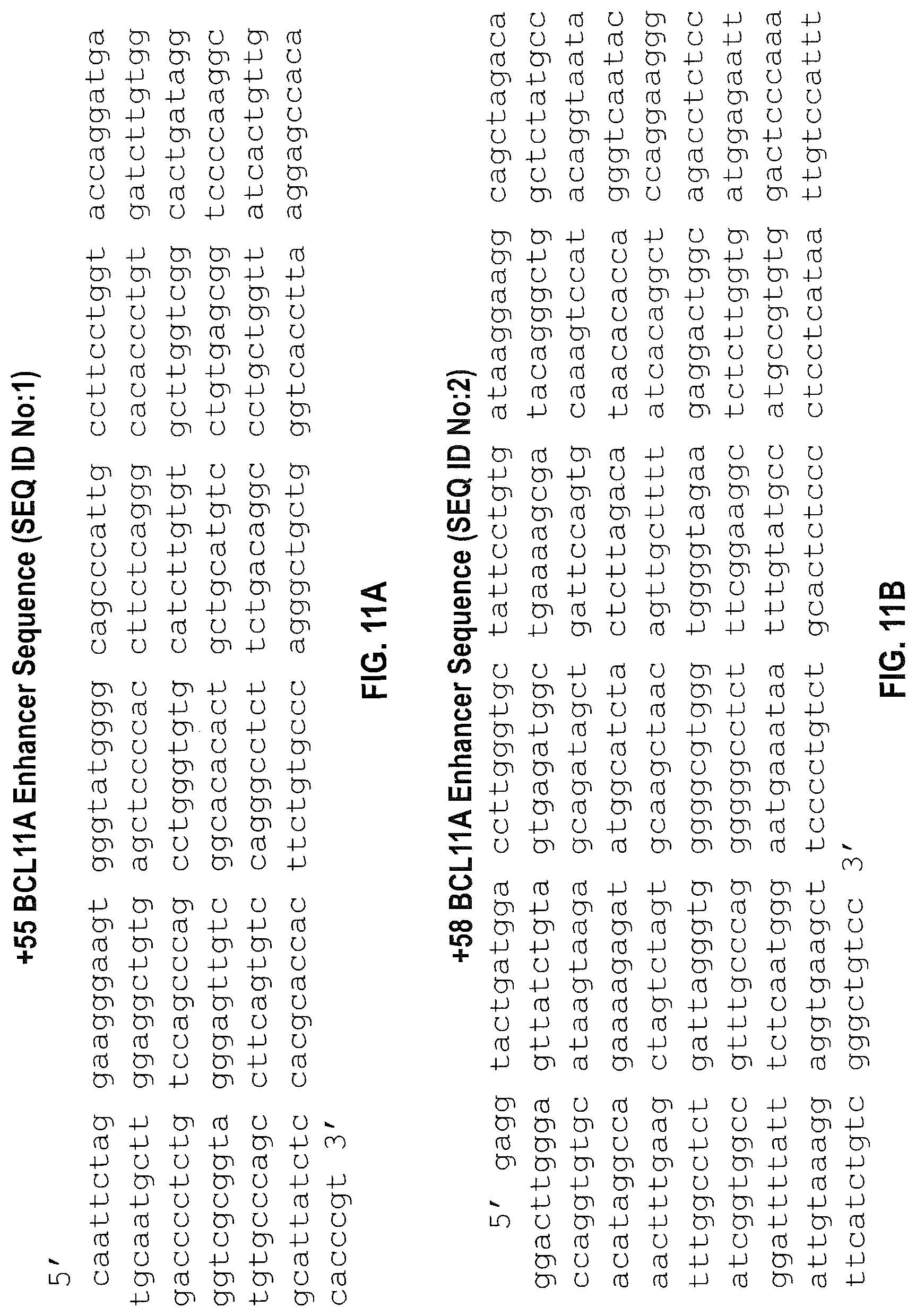
D00014

D00015

D00016

D00017

D00018
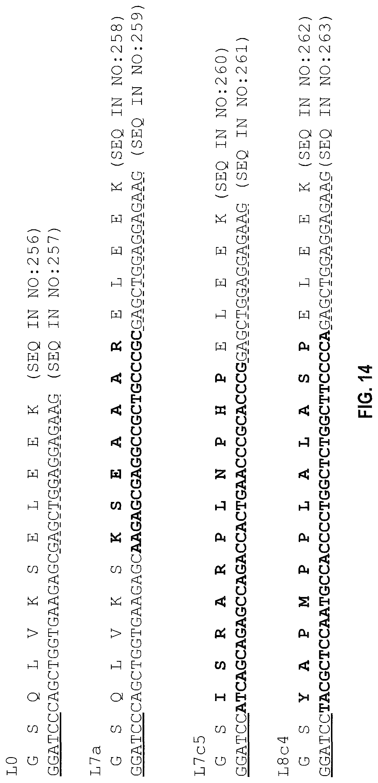
D00019

D00020
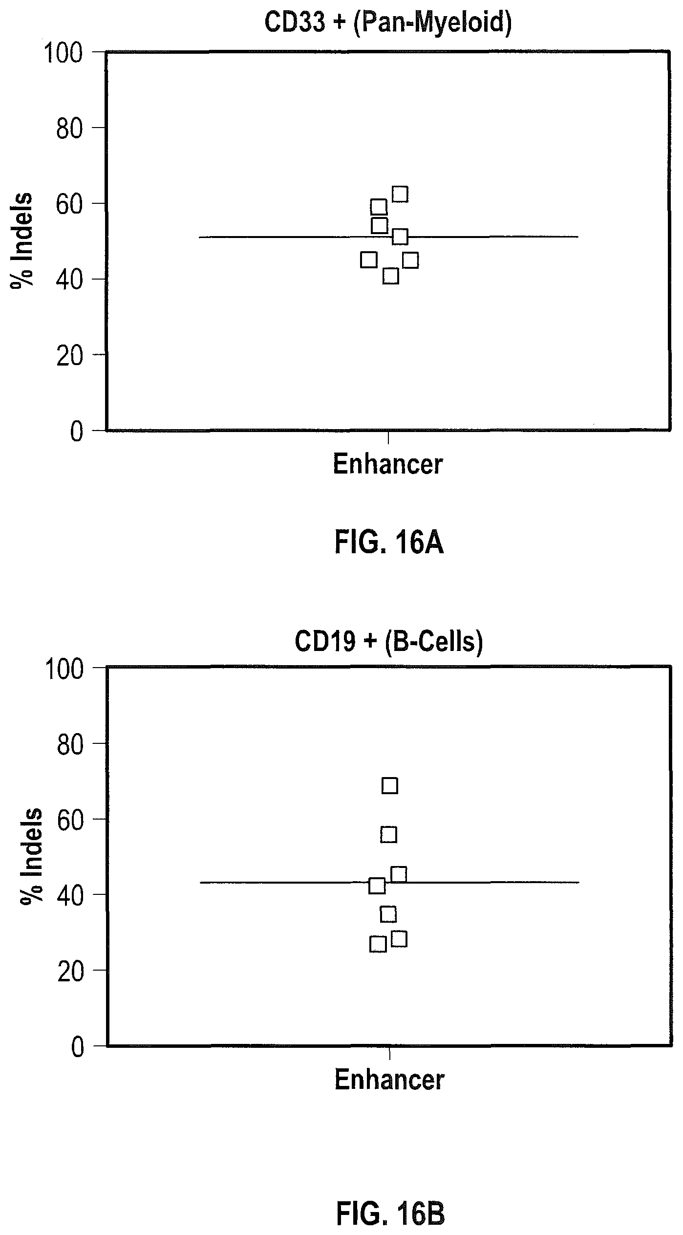
D00021
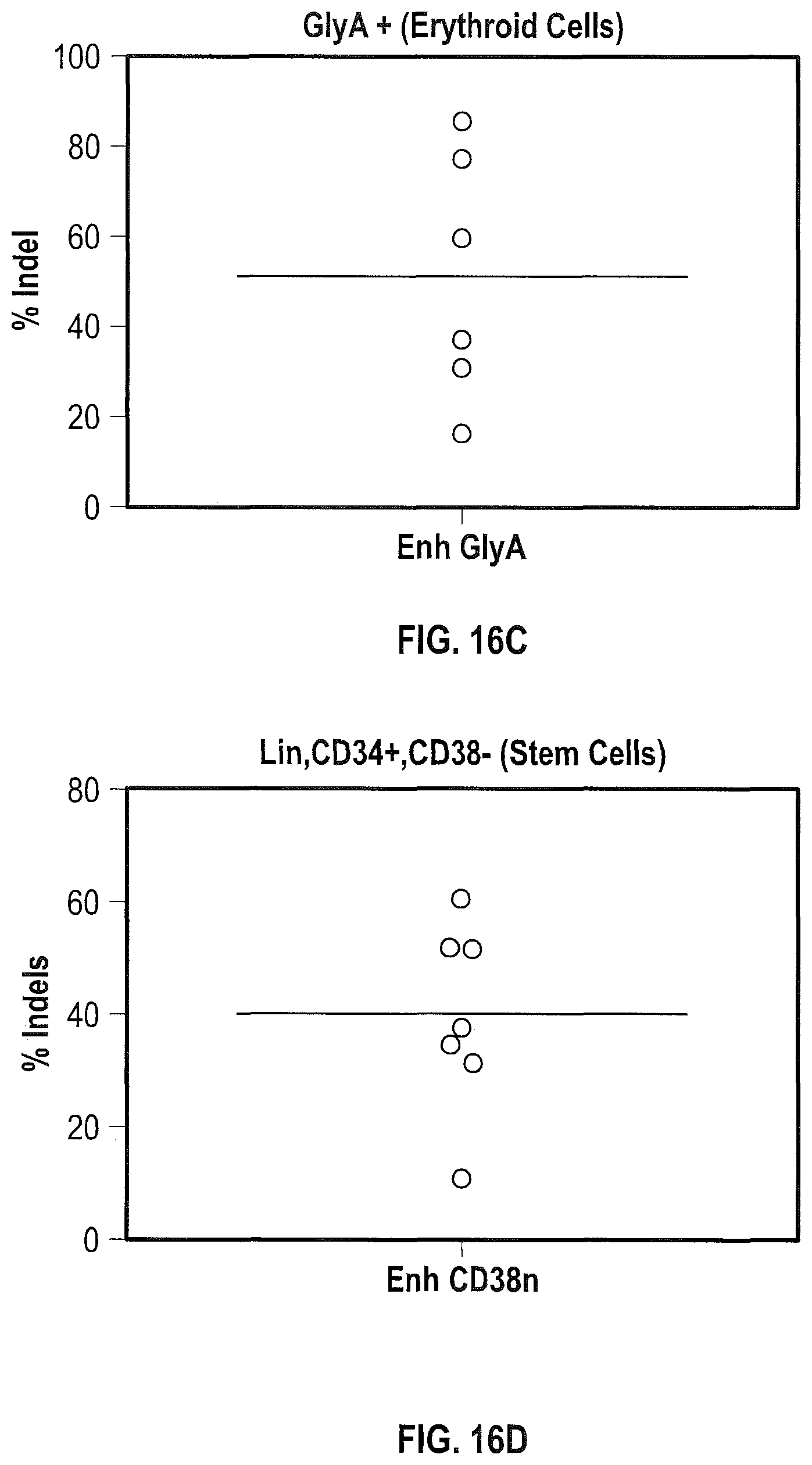
D00022

S00001
XML
uspto.report is an independent third-party trademark research tool that is not affiliated, endorsed, or sponsored by the United States Patent and Trademark Office (USPTO) or any other governmental organization. The information provided by uspto.report is based on publicly available data at the time of writing and is intended for informational purposes only.
While we strive to provide accurate and up-to-date information, we do not guarantee the accuracy, completeness, reliability, or suitability of the information displayed on this site. The use of this site is at your own risk. Any reliance you place on such information is therefore strictly at your own risk.
All official trademark data, including owner information, should be verified by visiting the official USPTO website at www.uspto.gov. This site is not intended to replace professional legal advice and should not be used as a substitute for consulting with a legal professional who is knowledgeable about trademark law.