Methods for identifying modulators of ion channels
Laing , et al. May 25, 2
U.S. patent number 11,016,100 [Application Number 14/849,203] was granted by the patent office on 2021-05-25 for methods for identifying modulators of ion channels. This patent grant is currently assigned to X-BODY, INC.. The grantee listed for this patent is X-Body, Inc.. Invention is credited to Rafael Fernandez, Lance G. Laing, Rick Wagner, Alexander Yuzhakov.


| United States Patent | 11,016,100 |
| Laing , et al. | May 25, 2021 |
Methods for identifying modulators of ion channels
Abstract
The invention provides methods for identifying modulators of ion channels without the use of recombinant cell lines over-expressing the ion channel proteins or the use of detection labels.
| Inventors: | Laing; Lance G. (Belmont, MA), Wagner; Rick (Cambridge, MA), Fernandez; Rafael (Jamaica Plain, MA), Yuzhakov; Alexander (West Roxbury, MA) | ||||||||||
|---|---|---|---|---|---|---|---|---|---|---|---|
| Applicant: |
|
||||||||||
| Assignee: | X-BODY, INC. (Waltham,
MA) |
||||||||||
| Family ID: | 1000005574971 | ||||||||||
| Appl. No.: | 14/849,203 | ||||||||||
| Filed: | September 9, 2015 |
Prior Publication Data
| Document Identifier | Publication Date | |
|---|---|---|
| US 20160069901 A1 | Mar 10, 2016 | |
Related U.S. Patent Documents
| Application Number | Filing Date | Patent Number | Issue Date | ||
|---|---|---|---|---|---|
| 12335433 | Sep 15, 2015 | 9134307 | |||
| 12171475 | Jul 11, 2008 | 9778267 | |||
| 60949142 | Jul 11, 2007 | ||||
| 61043478 | Apr 9, 2008 | ||||
| Current U.S. Class: | 1/1 |
| Current CPC Class: | C12Q 1/04 (20130101); G01N 33/54373 (20130101); G01N 21/78 (20130101); G01N 33/6872 (20130101) |
| Current International Class: | G01N 33/00 (20060101); G01N 33/543 (20060101); C12Q 1/04 (20060101); G01N 21/78 (20060101); G01N 33/68 (20060101) |
References Cited [Referenced By]
U.S. Patent Documents
| 3689346 | September 1972 | Rowland |
| 3810688 | May 1974 | Ballman et al. |
| 3856404 | December 1974 | Hershler et al. |
| 3916182 | October 1975 | Dabby et al. |
| 4009933 | March 1977 | Firester |
| 4050895 | September 1977 | Hardy et al. |
| 4240751 | December 1980 | Linnecke et al. |
| 4289371 | September 1981 | Kramer |
| 4344438 | August 1982 | Schultz |
| 4420502 | December 1983 | Conley |
| 4536608 | August 1985 | Sheng et al. |
| 4560246 | December 1985 | Cramp et al. |
| 4560248 | December 1985 | Cramp et al. |
| 4576850 | March 1986 | Martens |
| 4608344 | August 1986 | Carter et al. |
| 4650329 | March 1987 | Barrett et al. |
| 4652290 | March 1987 | Cho et al. |
| 4668558 | May 1987 | Barber |
| 4701008 | October 1987 | Richard et al. |
| 4810658 | March 1989 | Shanks et al. |
| 4815843 | March 1989 | Tiefenthaler et al. |
| 4818710 | April 1989 | Sutherland et al. |
| 4857273 | August 1989 | Stewart et al. |
| RE33064 | September 1989 | Carter |
| 4876208 | October 1989 | Gustafson et al. |
| 4882288 | November 1989 | North et al. |
| 4888260 | December 1989 | Cowan |
| 4931384 | June 1990 | Layton et al. |
| 4952056 | August 1990 | Tiefenthaler |
| 4958895 | September 1990 | Wells et al. |
| 4992385 | February 1991 | Godfrey |
| 4999234 | March 1991 | Cowen |
| 4999484 | March 1991 | Kaneko |
| 5071248 | December 1991 | Tiefenthaler et al. |
| 5118608 | June 1992 | Layton et al. |
| 5155785 | October 1992 | Holland et al. |
| 5156785 | October 1992 | Zdrahala |
| 5170448 | December 1992 | Ackley et al. |
| 5175030 | December 1992 | Lu et al. |
| 5210404 | May 1993 | Cush et al. |
| 5216680 | June 1993 | Magnusson et al. |
| 5229614 | July 1993 | Anderson et al. |
| 5242828 | September 1993 | Bergstrom et al. |
| 5268782 | December 1993 | Wenz et al. |
| 5310686 | May 1994 | Sawyers et al. |
| 5337183 | August 1994 | Rosenblatt |
| 5416884 | May 1995 | Koch et al. |
| 5442169 | August 1995 | Kunz |
| 5455178 | October 1995 | Fattinger |
| 5475780 | December 1995 | Mizrahi |
| 5478527 | December 1995 | Gustafson et al. |
| 5478756 | December 1995 | Gizeli et al. |
| 5492840 | February 1996 | Malmquist et al. |
| 5496701 | March 1996 | Pollard-Knight |
| 5559338 | September 1996 | Elliot et al. |
| 5598267 | January 1997 | Sambles et al. |
| 5598300 | January 1997 | Magnusson et al. |
| 5601997 | February 1997 | Tchao |
| 5606170 | February 1997 | Saaski et al. |
| 5615052 | March 1997 | Doggett |
| 5629214 | May 1997 | Crosby |
| 5631171 | May 1997 | Sandstrom et al. |
| 5666197 | September 1997 | Guerra |
| 5690894 | November 1997 | Pinkel et al. |
| 5691646 | November 1997 | Benson et al. |
| 5732173 | March 1998 | Bylander et al. |
| 5738825 | April 1998 | Rudigier et al. |
| 5768461 | June 1998 | Svetkoff et al. |
| 5771328 | June 1998 | Wortman et al. |
| 5792411 | August 1998 | Morris et al. |
| 5801390 | September 1998 | Shiraishi |
| 5811299 | September 1998 | Renner |
| 5814516 | September 1998 | Vo-Dinh |
| 5814524 | September 1998 | Walt et al. |
| 5821343 | October 1998 | Keogh |
| 5804453 | December 1998 | Simon |
| 5864641 | January 1999 | Murphy et al. |
| 5922550 | July 1999 | Everhart et al. |
| 5925878 | July 1999 | Challener |
| 5955335 | September 1999 | Thust et al. |
| 5955378 | September 1999 | Challener |
| 5955729 | September 1999 | Nelson |
| 5986762 | November 1999 | Challener |
| 5991480 | November 1999 | Kunz et al. |
| 5994150 | November 1999 | Challener et al. |
| 5998298 | December 1999 | Hetherington et al. |
| 6035089 | March 2000 | Grann et al. |
| 6042998 | March 2000 | Brueck et al. |
| 6052213 | April 2000 | Burt et al. |
| 6076248 | June 2000 | Hoopman et al. |
| 6088505 | July 2000 | Hobbs |
| 6100991 | August 2000 | Challener |
| 6128431 | October 2000 | Siminovitch |
| 6146593 | November 2000 | Pinkel et al. |
| 6174677 | January 2001 | Vo-Dinh |
| 6185019 | February 2001 | Hobbs et al. |
| 6200737 | March 2001 | Walt et al. |
| 6215928 | April 2001 | Friesem et al. |
| 6218194 | April 2001 | Lyndin et al. |
| 6235488 | May 2001 | Tom-Moy et al. |
| 6277653 | August 2001 | Challener |
| 6303179 | October 2001 | Koulik et al. |
| 6316153 | November 2001 | Goodman et al. |
| 6320991 | November 2001 | Challener et al. |
| RE37473 | December 2001 | Challener |
| 6332663 | December 2001 | Puzio et al. |
| 6338968 | January 2002 | Hefti |
| 6340598 | January 2002 | Herron et al. |
| 6346376 | February 2002 | Sigrist et al. |
| 6377721 | April 2002 | Walt et al. |
| 6395558 | May 2002 | Duveneck et al. |
| 6404554 | June 2002 | Lee et al. |
| 6449097 | September 2002 | Zhu et al. |
| 6558957 | May 2003 | Roinestad et al. |
| 6570657 | May 2003 | Hoppe et al. |
| 6579673 | June 2003 | McGrath et al. |
| 6587276 | July 2003 | Daniell |
| 6661952 | December 2003 | Simpson et al. |
| 6707561 | March 2004 | Budach et al. |
| 6748138 | June 2004 | Wang et al. |
| 6771376 | August 2004 | Budach et al. |
| 6867869 | March 2005 | Budach et al. |
| 6870624 | March 2005 | Hobbs et al. |
| 6870630 | March 2005 | Budach et al. |
| 6902703 | June 2005 | Marquiss et al. |
| 6951715 | October 2005 | Cunningham |
| 6982171 | January 2006 | Kim |
| 6989542 | January 2006 | Moses |
| 6990259 | January 2006 | Cunningham |
| 7018838 | March 2006 | Murphy |
| 7023544 | April 2006 | Cunningham |
| 7033819 | April 2006 | Kim |
| 7033821 | April 2006 | Kim |
| 7064844 | June 2006 | Budach et al. |
| 7070987 | July 2006 | Cunningham |
| 7074311 | July 2006 | Cunningham |
| 7094595 | August 2006 | Cunningham |
| 7101660 | September 2006 | Cunningham et al. |
| 7118710 | October 2006 | Cunningham |
| 7142296 | November 2006 | Cunningham et al. |
| 7148964 | December 2006 | Cunningham et al. |
| 7153702 | December 2006 | Lin |
| 7158230 | January 2007 | Cunningham et al. |
| 7162125 | January 2007 | Schulz |
| 7170599 | January 2007 | Cunningham et al. |
| 7175980 | February 2007 | Qiu et al. |
| 7197198 | March 2007 | Schulz et al. |
| 7202076 | April 2007 | Cunningham et al. |
| 7217574 | May 2007 | Pien et al. |
| 7264973 | September 2007 | Lin et al. |
| 7267993 | September 2007 | Pentreko |
| 7292336 | November 2007 | Cunningham et al. |
| 7298477 | November 2007 | Cunningham et al. |
| 7300803 | November 2007 | Lin et al. |
| 7301628 | November 2007 | Cunningham et al. |
| 7306827 | December 2007 | Li et al. |
| 7309614 | December 2007 | Baird |
| 7312090 | December 2007 | Lin et al. |
| 7327454 | February 2008 | Cunningham et al. |
| 7396675 | July 2008 | Pawlak et al. |
| 7400399 | July 2008 | Wawro et al. |
| 7479404 | January 2009 | Cunningham |
| 7483127 | January 2009 | Li |
| 7497992 | March 2009 | Cunningham |
| 7521769 | April 2009 | Cunningham |
| 7524625 | April 2009 | Madison |
| 7534578 | May 2009 | Baird |
| 7620276 | November 2009 | Schulz |
| 7628005 | December 2009 | Laing |
| 7678548 | March 2010 | Brown |
| 7742662 | June 2010 | Cunningham |
| 7756365 | July 2010 | Cunningham |
| 7790406 | September 2010 | Cunningham |
| 2002/0018610 | February 2002 | Challener et al. |
| 2002/0028045 | March 2002 | Yoshimura |
| 2002/0028480 | March 2002 | Maher |
| 2002/0076747 | June 2002 | Price |
| 2002/0123050 | September 2002 | Poponin |
| 2002/0127565 | September 2002 | Cunningham et al. |
| 2002/0135752 | September 2002 | Sokolov et al. |
| 2002/0171045 | November 2002 | Perraut |
| 2003/0003599 | January 2003 | Wagner et al. |
| 2003/0017580 | January 2003 | Cunningham |
| 2003/0017581 | January 2003 | Li |
| 2003/0026891 | February 2003 | Qiu |
| 2003/0027327 | February 2003 | Cunningham |
| 2003/0027328 | February 2003 | Cunningham et al. |
| 2003/0032039 | February 2003 | Cunningham |
| 2003/0059855 | March 2003 | Cunningham |
| 2003/0068657 | April 2003 | Lin |
| 2003/0077660 | April 2003 | Pien |
| 2003/0092075 | May 2003 | Pepper |
| 2003/0104479 | June 2003 | Bright |
| 2003/0108954 | June 2003 | Mutz |
| 2003/0113766 | June 2003 | Pepper |
| 2003/0148542 | August 2003 | Pawlak |
| 2003/0157571 | August 2003 | Karpen |
| 2003/0210396 | November 2003 | Hobbs et al. |
| 2003/0224369 | December 2003 | Surber et al. |
| 2004/0005540 | January 2004 | Pentreko |
| 2004/0011965 | January 2004 | Hodgkinson |
| 2004/0091397 | May 2004 | Picard |
| 2004/0132172 | July 2004 | Cunningham |
| 2004/0132214 | July 2004 | Lin |
| 2004/1332214 | July 2004 | Lin et al. |
| 2004/0151626 | August 2004 | Cunningham |
| 2004/0191757 | September 2004 | Maher |
| 2004/0204357 | October 2004 | Brautigam |
| 2004/0219619 | November 2004 | Fernandez-Salas |
| 2005/0058639 | March 2005 | Gudas |
| 2005/0074825 | April 2005 | Luo |
| 2005/0214803 | September 2005 | Wang |
| 2005/0221271 | October 2005 | Murphy |
| 2005/0227374 | October 2005 | Cunningham et al. |
| 2006/0003372 | January 2006 | Li |
| 2006/0030033 | February 2006 | Cunningham |
| 2006/0040376 | February 2006 | Cunningham et al. |
| 2006/0057707 | March 2006 | Lin et al. |
| 2006/0181705 | August 2006 | Cunningham et al. |
| 2006/0193550 | August 2006 | Wawro et al. |
| 2006/0275825 | December 2006 | Laing |
| 2006/0281077 | December 2006 | Lin |
| 2006/0286663 | December 2006 | Cunningham et al. |
| 2007/0015210 | January 2007 | Ezekiel |
| 2007/0041012 | February 2007 | Cunningham et al. |
| 2007/0054339 | March 2007 | Lin |
| 2007/0070355 | March 2007 | Cunningham et al. |
| 2007/0141231 | June 2007 | Cunningham et al. |
| 2007/0172894 | July 2007 | Genick et al. |
| 2007/0299029 | December 2007 | Zhou |
| 2008/0213910 | September 2008 | Jogikalmath |
| 2008/0219892 | September 2008 | Cunningham |
| 2008/0227819 | September 2008 | Struenker |
| 2008/0240543 | October 2008 | Budach |
| 2008/0299673 | December 2008 | Wagner |
| 2009/0017488 | January 2009 | Binder |
| 2009/0130703 | May 2009 | Wagner |
| 2009/0137422 | May 2009 | Laing |
| 2009/0148955 | June 2009 | Cunningham |
| 2009/0176658 | July 2009 | Madison |
| 2009/0179637 | July 2009 | Cunningham |
| 2009/0192049 | September 2009 | Baird |
| 2009/0227469 | September 2009 | Conklin |
| 2009/0264314 | October 2009 | Cunningham |
| 2009/0269244 | October 2009 | Cunningham |
| 2009/0282931 | November 2009 | Laing |
| 2009/0305304 | December 2009 | Laing |
| 2010/0003743 | January 2010 | Schulz |
| 2010/0008826 | January 2010 | Schulz |
| 2010/0015721 | January 2010 | Laing |
| 2010/0043571 | February 2010 | Laing |
| 2010/0143959 | June 2010 | Cunningham |
| 2010/0195099 | August 2010 | Rockney |
| 2010/0196925 | August 2010 | Genick |
| 2010/0202923 | August 2010 | Cunningham |
| 2010/0227769 | September 2010 | Schulz |
| 2010/0231907 | September 2010 | Pien |
| 2010/0291575 | November 2010 | Shamah |
| 2394966 | Aug 2001 | CA | |||
| 2395318 | Aug 2001 | CA | |||
| 669050 | Feb 1989 | CH | |||
| 670521 | Jun 1989 | CH | |||
| 0075353 | Mar 1983 | EP | |||
| 0112721 | Jul 1984 | EP | |||
| 0326219 | Jan 1989 | EP | |||
| 0517777 | May 1996 | EP | |||
| 0660924 | Sep 1999 | EP | |||
| 1031828 | Aug 2000 | EP | |||
| 1085315 | Mar 2001 | EP | |||
| 2801977 | Dec 1999 | FR | |||
| 2156970 | Oct 1985 | GB | |||
| 2227089 | Jul 1990 | GB | |||
| 1993228946 | Sep 1993 | JP | |||
| 81/00912 | Feb 1981 | WO | |||
| 84/02578 | Jul 1984 | WO | |||
| 86/07149 | Dec 1986 | WO | |||
| 90/08313 | Jul 1990 | WO | |||
| 91/13339 | Sep 1991 | WO | |||
| 92/04653 | Mar 1992 | WO | |||
| 92/21768 | Dec 1992 | WO | |||
| 93/17392 | Jul 1993 | WO | |||
| 95/03538 | Feb 1995 | WO | |||
| 96/38726 | Dec 1996 | WO | |||
| 97/29362 | Aug 1997 | WO | |||
| 98/10288 | Mar 1998 | WO | |||
| 98/57200 | Dec 1998 | WO | |||
| 99/09369 | Feb 1999 | WO | |||
| 99/09392 | Feb 1999 | WO | |||
| 99/54714 | Oct 1999 | WO | |||
| 99/66330 | Dec 1999 | WO | |||
| 00/23793 | Apr 2000 | WO | |||
| 00/29830 | May 2000 | WO | |||
| 01/02839 | Jan 2001 | WO | |||
| 01/04697 | Jan 2001 | WO | |||
| 01/79559 | Oct 2001 | WO | |||
| 01/92870 | Dec 2001 | WO | |||
| 02/061429 | Aug 2002 | WO | |||
| 2003074548 | Sep 2003 | WO | |||
| 2007056160 | May 2007 | WO | |||
| 2007064702 | Jun 2007 | WO | |||
| 2009009718 | Jan 2009 | WO | |||
| 2010005600 | Jan 2010 | WO | |||
| 2010075033 | Jul 2010 | WO | |||
Other References
|
Priest et al. (Role of hERG potassium channel assays in drug development, Channels (Austin). Mar.-Apr. 2008;2(2):87-93. Epub Mar. 5, 2008). cited by examiner . Dorn et al. (Evaluation of a high-throughput fluorescence assay method for HERG potassium channel inhibition, J Biomol Screen. Jun. 2005;10(4):339-47). cited by examiner . Tang et al (Development and evaluation of high throughput functional assay methods for HERG potassium channel, J Biomol Screen. Oct. 2001;6(5):325-31). cited by examiner . Brecht et al., "Optical probes and transducers", Biosensors & Bioelectronics, vol. 10, pp. 923-936 (1995)*. cited by applicant . Challener et al., "A multilayer grating-based evanescent wave sensing technique", Sensors and Actuators B, 71, pp. 42-46 (2000)*. cited by applicant . Cowan, "Aztec surface-relief volume diffractive structure", J. Opt. Soc. Am. vol. 7, No. 8, pp. 1529-1544 (1990)*. cited by applicant . Cowan, "Holographic honeycomb microlens", Optical Engineering, vol. 24, No. 5, pp. 796-802 (1985)*. cited by applicant . Cowan, "The Recording and Large Scale Replication of Crossed Holographic Grating Arrays using Multiple Beam Interferometry", SPIE, vol. 503, Application, Theory, and Fabrication of Periodic Structures, pp. 120-129 (1984)*. cited by applicant . Cowan et al., "The Recording and Replication of Holographic Micropatterns for the Ordering of Photographic Emulsion Grains in Film Systems", J. Imaging Sci., vol. 31, No. 3, pp. 100-107 (1987)*. cited by applicant . Introduction to Bioanalytical Sensors (Techniques in Analytical Chemistry) (Cunningham ed., 1988) pp. 260-291, "Optical Based Energy Transduction", Wiley Interscience, Hoboken, NJ*. cited by applicant . Hobbs et al., "Automated Interference Lithography Systems for Genereation of Sub-Micron Feature Size Patterns", SPIE, vol. 3879, pp. 124-135 (1999)*. cited by applicant . Huber et al., "Direct optical immunosensing (sensitivity and selectivity)", Sensors and Actuators B, 6, pp. 122-126 (1992)*. cited by applicant . Jenison et al., "Interference-based detection of nucleic acid targets on optically coated silicon", Nature Biotechnology, vol. 19, pp. 62-64 (2001)*. cited by applicant . Jin et al., "A biosensor concept based on imaging ellipsometry for visualization of biomolecular interactions", Analytical Biochemistry, vol. 232, pp. 69-72 (1995)*. cited by applicant . Jordan et al., "Surface Plasmon Resonance Imaging Measurements of Electrostatic Biopolymer Adsorption onto Chemically Modified Gold Surfaces", Analytical Chemistry, vol. 69, No. 7, pp. 1449-1456 (1997)*. cited by applicant . Lin et al., A Porous Silicon-Based Optical Interferometric Biosensor:, Science, vol. 278, pp. 840-843 (1997)*. cited by applicant . Magnusson et al., "New principle for optical filters", Appl. Phys. Lett., vol. 61, No. 9, pp. 1022-1024 (1992)*. cited by applicant . Magnusson et al., "Transmission bandpass guided-mode resonance filters", Applied. Optics, vol. 34, No. 35, pp. 8106-8109 (1995)*. cited by applicant . Morhard et al., "Immobilization of antibodies in micropatterns for cell detection by optical diffraction", Sensors and Actuators B, 70, pp. 232-242 (2000)*. cited by applicant . Pandey et al, "Proteomics to study genes and genomes", Natures 405(6788):837-46 (2000)*. cited by applicant . Patel et al., "Electrically tuned and polarization insensitive Fabry-Perot etalon with a liquid-crysatl film", App. Phys. Lett, vol. 58, No. 22, pp. 2491-2493 (1993)*. cited by applicant . Bertoni et al., "Frequency-Selective Reflection and Transmission by a Periodic Dielectric Layer", IEEE Transaction on Antennas and Propagation, vol. 37, No. 1, pp. 78-83 (1989)*. cited by applicant . Brundrett et al., "Normal-incidence guided-mode resonant grating filters: design and experimental demonstration", Optics Letters, vol. 23, No. 9, pp. 700-702 (1998)*. cited by applicant . Peng "Polarization-control Components and Narrow-band Filters Based on Subwavelength Grating Structures", 1996*. cited by applicant . Statement of Applicants dated May 10, 2004*. cited by applicant . Leanu, Torben, Material, Silicon Nitride, 1996, 97, 98*. cited by applicant . Cerac, Technical publications: Tantalum Oxide, Ta2O5 for Optical Coating, 2000, Cerac, Inc.*. cited by applicant . Neuschafer et al., Evanescent resonator chips: a universal platform with superior sensitivity for fluorescence-based microarrays, Biasensors & Bioelectronics, 18, pp. 489-497 (2003)*. cited by applicant . Budach et al., "Generation of transducers for fluorescence-based microarrays with enhanced sensitivity and their application for gene expression profiling", Analytical Chemistry, 1:75(11):2571-7 (2003)*. cited by applicant . Anderson et al., "Proteomics: applications in basic and applied biology", Current Opinion in Biotechnology, 11:408-412 (2000)*. cited by applicant . MacBeath et al., "Printing Proteins as Microarrays for High-Throughput Function Determination", Science, vol. 289, pp. 1760-1763 (2000)*. cited by applicant . DeWildt et al, "Antibody arrays for high-throughput screening of antibody-antigen interactions", Nature Biotechnology, vol. 18, pp. 989-994 (2000)*. cited by applicant . Cunningham et al., "A plastic calorimetric resonant optical biosensor for multiparallel detection of label-free biochemical interactions" Sensors and Actuators B, 85, pp. 219-226 (2002)*. cited by applicant . Caruso et al., "Quartz Crystal Microbalance Study of DNA Immobilization and Hybridization for Nucleic Acid Sensor Development", Analytical Chemistry, vol. 69, No. 11, pp. 2043-2049 (1997)*. cited by applicant . Hefti et al., "Sensitive detection method of dielectric dispersions in aqueous-based, surface-bound macromolecular structures using microwave spectroscopy", Applied Physics Letters, vol. 75, No. 12, pp. 1802-1084 (1999)*. cited by applicant . Wu et al., "Bioassay of prostate-specific antigen (PSA) using microcantilevers", Nature Biotechnology, vol. 19, pp. 856-860 (2001)*. cited by applicant . Wasserman et al., "Structure and Reactivity of Alkylsiloxane Monolayers Formed by Reaction of Alkyltrichlorosilanes on Silicone Substrates", Langmuir, 5, pp. 1074-1087 (1989)*. cited by applicant . Kallury et al., "X-ray Photoelectron Spectroscopy of Silica Surfaces Treated with Polyfunctional Silanes", Anal. Chem. 30, 169-172 (1988)*. cited by applicant . Cunningham et al., "Colorimetric resonant reflection as a direct biochemical assay technique", Sensors and Actuators B, 81 (2002) 316-328*. cited by applicant . Mullaney et al, "Epitope Mapping of Neutralizing Botulinum Neurotoxin A Antibodies by Phage Display", Infection and Immunity, vol. 69, No. 10, pp. 6511-6514 (2001)*. cited by applicant . Nellen et al., "Integrated Optical Input Grating Couplers as Biochemical Sensors", Sensors and Actuators, 15 (1988) 285-295*. cited by applicant . Lukosz and Tiefenthaler, "Embossing technique for fabricating integrated optical components in hard inorganic waveguiding materials", Optics Letters vol., 8, pp. 537-539 (1983)*. cited by applicant . Tiefenthaler and Lukosz, "Integrated optical switches and gas sensors", Optics Letters, vol. 10, pp. 137-139 (1984)*. cited by applicant . Chabay, "Optical Waveguides", Analytical Chemistry, vol. 54, pp. 1071A-1081A (1982)*. cited by applicant . Sutherland et al., "Optical Detection of Antibody-antigen Reactions at a Glass-Liquid Interface", Clin. Chem. vol. 30, pp. 1533-1538 (1984)*. cited by applicant . Holm and Palik, "Internal-reflection spectroscopy", Laser Focus, vol. 15, pp. 60-65 (1979)*. cited by applicant . Harrick and Loeb, "Multiple Internal Reflection Fluorescence Spectrometry", Analytical Chemistry, vol. 45, pp. 687-691 (1973)*. cited by applicant . Tien, "Light Waves in This Films and Integrated Optics", Applied Optics, vol. 10, pp. 2395-2413 (1971)*. cited by applicant . Dakss et al., "Grating Coupler for Efficient Excitation of Optical Guided Waves in Thin Films", Applied Physics Letters, vol. 16, pp. 523-525 (1970)*. cited by applicant . Sutherland et al., "Immunoassays at a Quartz-Liquid Interface: Theory, Instrumentation and Preliminary Application to Fluorescent Immunoassay of Human Immunoglobulin G", Journal of Immunological Methods, vol. 74, pp. 253-265 (1984)*. cited by applicant . English Translation of CH 670 521 A5 (Jun. 15, 1989), translation dated Oct. 29, 2003*. cited by applicant . English Translation of CH 669 050 A5 (Feb. 15, 1989), translation dated Oct. 29, 2003*. cited by applicant . Patel et al., "Multi-Wavelength Tunable Liquid-Crystal Etalon Filter", IEEE Photonics Technology Letters, vol. 3, No. 7, pp. 643-644 (1991)*. cited by applicant . Patterson, "Proteomics: the Industrialization of protein chemistry", Current Opinions in Biotechnology, 11(4):413-8 (2000)*. cited by applicant . Peng et al., "Experimental demonstration of resonant anomalies in diffraction from two-dimensional gratings", Optics Letters, vol. 21, No. 8, pp. 549-551 (1996)*. cited by applicant . Peng et al., "Resonant scattering from two-dimensional gratings", J. Opt. Soc. Am. A., vol. 13, No. 5, pp. 993-1005 (1996)*. cited by applicant . Raguin et al., "Structured Surfaces Mimic Coating Performance", Laser Focus World, pp. 113-117 (1997)*. cited by applicant . Sigal et al., "A Self-Assembled Monolayer for the Binding and Study of Histidine-Tagged Proteins by Surface Plasmon Resonance", Analytical Chemistry, vol. 68, No. 3, pp. 490-497 (1996)*. cited by applicant . Wang et al., "Design of waveguide-grating filters with symmetrical line shapes and low sidebands", Optical Society of America, vol. 19, No. 12, pp. 919-921 (1994)*. cited by applicant . Wang et al., "Guided-mode resonances in planar dielectric-layer diffraction gratings", J. Opt. Soc. Am., vol. 7, No. 8, pp. 1470-1474 (1990)*. cited by applicant . Wang et al., "Theory and applications of guided-mode resonance filter", Applied Optics, vol. 32, No. 14, pp. 2606-2613 (1993)*. cited by applicant . International Search Report for foreign counterpart application PCT/US01/50723, dated Sep. 17, 2002*. cited by applicant . International Search Report for foreign counterpart application PCT/US03/01175, dated Aug. 18, 2003*. cited by applicant . Invitation to Pay Additional Fees in foreign counterpart application PCT/US01/50723, dated Aug. 30, 2002*. cited by applicant . Haidner, "Zero-Order Gratings Used as an Artificial Distributed Index Medium", Optik, Wissenschaftliche Verlag GmbH, Stuttgart, DE, vol. 89, No. 3, pp. 107-112 (1992)*. cited by applicant . Wilson et al., "The Optical Properties 1-19 of `Moth Eye` Antireflection Surfaces", Optica Acta, vol. 29, No. 7, pp. 993-1009 (1982)*. cited by applicant . Bagnich et al., "Tunable Optical Filter", Derwent Publications, English Translation, Abstract Only,Derwent Publications Ltd. (Mar. 15, 1989)*. cited by applicant . Corning Inc. v. SRU Biosystems, Inc., Memorandum Opinion dated Nov. 15, 2005 in the United States District Court for the district of Delaware*. cited by applicant . Liu et al., "Development of an optical fiber lactate sensor", Mikrochimica Acta, 131(1-2), pp. 129-135 (1999)*. cited by applicant . U.S. Appl. No. 11/635,934, filed Dec. 8, 2006*. cited by applicant . U.S. Appl. No. 11/566,818, filed Dec. 5, 2006*. cited by applicant . U.S. Appl. No. 11/506,639, filed Aug. 18, 2007*. cited by applicant . U.S. Appl. No. 11/749,073, filed May 15, 2007*. cited by applicant . U.S. Appl. No. 11/828,073, filed Jul. 25, 2007*. cited by applicant . European Search Report for EP 07 11 8355 dated Feb. 5, 2008*. cited by applicant . Nelson, et al., "BIA/MS of Epitope-Tagged Peptides Directly from E. coli Lysate: Multiplex Detection and Protein Identification at Low-Femtomole to Subfemtomole Levels", Anal. Chem. 1999, 71, 2858-2865*. cited by applicant . Moffatt, "Optical probes May Hasten Shift of Diagnostics from Lab to Doc's Office", Genetic Engineering News, vol. 18, (1986), p. 18*. cited by applicant . Williams, et al., "The integration of SPR biosensors with mass spectrometry: possible applications for proteome analysis", Trends in Microbiology, Elsevier, vol. 18, No. 2 (2000) pp. 45-48*. cited by applicant . Cekaite et al., "Analysis of the humoral immune response to immunoselected phage-displayed peptides by a microarray-based method", Proteomics 2004, 4, 2572-2582*. cited by applicant . Sun et al., "Use of bioluminescent Salmonella for assessing the efficiency of constructed phage-based biosorbent", Journal of Industrial Microbiology & Biotechnology, 2000, 25, 273-275*. cited by applicant . Wan, et al., "Landscape phage-based magnetostrictive biosensor for detecting Bacillus anthracis spores", Proc. IEEE Sens., 2005, 1308-1311*. cited by applicant . Cunningham, et al., "Label-Free Assays on the BIND System", Journal of Biomolecular Screening, vol. 9, p. 481-490 (2004)*. cited by applicant . Cunningham, "Label-Free Detection with the BIND System", Presented at Screentech General, Mar. 24, 2003*. cited by applicant . Baird, "Beyond ELISA's: Label-free Detectionw ith BIND", Presented at Interphex Meeting in Europe, Mar. 16-18, 2004*. cited by applicant . Cunningham, et al., "Calorimetric Resonant Reflection as a Direct Biochemical Assay Technique", Presented at the Pittsburgh Conference and Exposition on Analytical Chemistry and Applied Spectroscopy, Morial Convention Center in New Orleans, LA Mar. 17-22, 2002*. cited by applicant . Broad et al., "Growth and adipose differentiation of sheep preadipocyte fibroblasts in serum-free medium", Eur. J. Biochem., 135, 33-39 (1983)*. cited by applicant . Castillo et al., "Characterization of proliferation and differentiation of EGF-response striatal and septal precursor cells", Int J. Devl. Neuroscience 21 (2003) 41-47*. cited by applicant . Chalazonitis, et al., "The a1 Subunit of Laminin-1 Promotes the Development of Neurons by Interacting with LBP110 Expressed by Neural Crest-Derived Cells Immunoselected from the Fetal Mouse Gut", J. Neurobiol. 33:118-138, 1997*. cited by applicant . Hao et al., "Fetal Human Hemotopoietic Stem Cells Can Differentiate Sequentially into Neural Stem Cells and then Astrocytes in Vitro", Journal of Hematotherapy & Stem Cell Research, 12:23-32 (2003)*. cited by applicant . Kano, et al., "Establishment of Hepatic Stem-like Cell Lines from Normal Adult Porcine Liver in a Poly-D-Lysine-Coated Dish with Nair-1 Medium", In Vitro Cell. Dev. Biol. Animal, 30-440-448 (2003)*. cited by applicant . Sung, et al., "Adhesiveness of Human Ligament Fibroblasts to Laminin", Journal of Orthopaedic Research, 13:166-173 (1995)*. cited by applicant . Zhou, et al., "Long-term nonpassaged EGF-responsive neural precursor cells are stem cells", Wound Repair and Regeneration, vol. 6, No. 4, pp. 337-348, 1998*. cited by applicant . Adamczyk, et al., "Application of Surface Plasmon Resonance toward Studies of Low-Molecular-Weight Antigen-Antibody Binding Interactions", Methods, 20, pp. 319-328 (2000)*. cited by applicant . Marquart, "Immobilization Techniques", SPR pp. [online] Jan. 2004, pp. 1-7*. cited by applicant . Zhang et al. "Use of surface plasmon resonance for the measurement of low affinity binding interactions between HSP72 and measles virus nucleocapsid protein", Biol. Proced. Online 2003;5(1):170-181*. cited by applicant . Gestwicki, et al., "Using Receptor Conformational Change to Detect Low Molecular Weight Analytes by Surface Plasmon Resonance", Anal. Chem., 2001, 4, 5732-5737*. cited by applicant . International Search Report dated Jul. 15, 2008, for PCT application serial No. PCT/US08/60951*. cited by applicant . English machine translation only of JP 1993-228946 Sep. 7, 1993*. cited by applicant . U.S. Appl. No. 12/171,475, filed Jul. 11, 2008*. cited by applicant . U.S. Appl. No. 12/335,393, filed Dec. 15, 2008*. cited by applicant . International Search Report for corresponding application No. PCT/US09/30412 dated Jan. 8, 2009*. cited by applicant . Wawro, et al., "Optical fiber endface biosensor based on resonances in dielectric waveguide gratings", Biomedical Diagnostic Guidance and Surgical-Assist Systems II, Vo-Dihn et al eds., Proceedings of SPIE, vol. 3911, p. 86-94 (2000)*. cited by applicant . Office action dated Apr. 2, 2007, for U.S. Appl. No. 11/506,639 (now U.S. Pat. No. 7,298,477)*. cited by applicant . Torbin, et al., "The use of polymerizing cements for making replicas of optical surfaces", Optical Technology, vol. 40, No. 3, p. 192-196 (1973)*. cited by applicant . Ramsden, et al., "Optical Method for Measurement of Number and Shape of Attached Cells in Real Time", Cytometry, vol. 19, pp. 97-102 (1995)*. cited by applicant . Li et al., "Measurement and Adhesion and Spreading Kinetics of Baby Hamster Kidney and Hybridoma Cells Using an Integrated Optical Method", Biotechnol. Prog., vol. 10, pp. 520-524 (1994)*. cited by applicant . International Search Report dated Jul. 2, 2010, for corresponding PCT application No. PCT/US2010/035152, filed May 17, 2010*. cited by applicant . Cunningham et al., "Advantages and application of label-free detection assays in drug screening", Expert Opin. Drug Discov., 3(7):891-901 (2008)*. cited by applicant . Palmer, "Diffraction Gratings, The Crucial Dispersive Component", Spectroscopy, 10(2), pp. 14-15 (1995)*. cited by applicant . Bandell et al., "Noxious Cold Ion Channel TRPA1 is Activated by Pungent Compounds and Bradykinin" Neuron, vol. 41, pp. 849-957 (2004)*. cited by applicant . Cunningham et al., "Label-Free Assays on the BIND System", Journal of Biomolecular Screening, 9:481 (2004)*. cited by applicant . IMT Applied Optics, "Resonant Grating Filters", 2008*. cited by applicant . Cunningham et al., "Advantages and application of label-free detection assays in drug screening", Expert Opin. Drug Discovery, 3:891-901 (2008)*. cited by applicant . Comley, "Label-Free Detection New biosensors facilitate boarder range of drug discovery applications", Drug Discovery World Winter, p. 63-74 (2004/5)*. cited by applicant . Cooper, "Current biosensor technologies in drug discovery", Drug Discovery World Summer, p. 68-82 (2006)*. cited by applicant . Cunningham et al., "Colormetric Resonant Reflection as a Direct Biochemical Assay Technique", IEEE, Annual International Conference on Micro Electro Mechanical Systems, MEMS, Las Vegas, NV, Jan. 20-24, 2002*. cited by applicant . Cunningham et al., "Label-Free Assays on the BIND System", Journal of Biomolecular Screening 9(6):481-490 (2004)*. cited by applicant . Magnusson et al., "Fiber Endface Bioprobes with high Sensitivity and Spatial Resolution", Grant Proposal dated Aug. 11, 1999*. cited by applicant . Norton, "Resonant Grating Structures: Theory, Design and Applications", Doctoral Thesis, The University of Rochester, 1997*. cited by applicant . Popov et al., "Theoretical study of the anomalies of coated dielectric gratings", Optica Acta, vol. 33, No. 5, pp. 607-619 (1986)*. cited by applicant . Shin et al., "Thin-film optical filters with diffractive elements and waveguides", Opt. Eng. 37(9):2634-2646 (1998)*. cited by applicant . Tibuleac et al., "Reflection and transmission guided-mode resonance filters", J. Opt. Soc. Am. A, vol. 14, No. 7, pp. 1617-1626 (1997)*. cited by applicant . Tibuleac et al., "Diffractive Narrow-Band Transmission Filters Based on Guided-Mode Resonance Effects in Thin-Film Multilayers", IEEE Photonics Technology Letters, vol. 9, No. 4, pp. 464-466 (1997)*. cited by applicant . Wawro, "Design, Fabrication and Testing of Waveguide Gratings for Spectral Filters, Photonic Antennas and Optical Fiber Sensors", Presentation, University of Texas at Arlington (1999)*. cited by applicant . Wawro et al., "Novel diffractive structures integrating waveguide-gratings on optical fiber endfaces", Presentation, Graduate Student Research Symposium (1999)*. cited by applicant . Tibuleac, "Guided-mode resonance reflection and transmission filters in the optical and microwave spectral ranges", Doctoral Dissertation Defense (1999)*. cited by applicant . Yariv, "Coupled-Mode Theory for Guided-Wave Optics", IEEE Journal of Quantum Electronics, vol. 9, No. 9, p. 919-933 (1973)*. cited by applicant . Wang et al., "Resonance of Asymmetric Dielectric Waveguides Containing a Diffraction Grating", IEEE (1990)*. cited by applicant . Hessel et al., "A New Theory of Wood's Anomalies on Optical Gratings", Applied Optics, vol. 4, No. 10, p. 1275-1299 (1965)*. cited by applicant . Lukosz et al., "Sensitivity of Integrated Optical Grating and Prism Couplers as (Bio)chemical Sensors", Sensors and Actuators, 15, p. 273-284 (1988)*. cited by applicant . Neviere et al., "About the Theory of Optical Grating Coupler-Waveguide Systems", Optics Communications, vol. 8, No. 2, p. 113-117 (1973)*. cited by applicant . Gaylord et al. "Analysis and Applications of Optical Diffraction by Gratings", IEEE, 73(5):894, p. 894-924 (1985)*. cited by applicant . Takada et al., "The integrins", Genome Biology, 8:215 (2007)*. cited by applicant . Sancho et al., "Binding kinetics of monomeric and aggregated IgG to Kupffer cells and hepatocytes of mice", Immunology, 53:283 (1984)*. cited by applicant . Chaplen et al., "Improvement of Bioactive Compound Classification through Integration of Orthogonal Cell-Based Biosensing Methods", Sensors, 7:38-51 (2007)*. cited by applicant . U.S. Appl. No. 13/073,233, filed Mar. 28, 2011*. cited by applicant . Besko et al., "A Novel encoded Particle Technology that Enables Simultaneous Interrogation of Multiple Cell Types", Journal of Biomolecular Screening, vol. 9, No. 3, p. 173-185 (2004). cited by applicant . Peng, et al., "Experimental Demonstration of Resonant Anomalies in Diffraction from two-Dimensional Gratings", Optics Letter, Optical Society of America, vol. 21, No. 9, pp. 549-551 (1996)*. cited by applicant . Office action dated Jun. 29, 2011, for corresponding U.S. Appl. No. 12/171,475*. cited by applicant . Reckless and Grainger, "Identification of oligopeptide sequences which inhibit migration induced by a wide range of chemokines", Biochem. J., 340:803-811 (1999)*. cited by applicant . Jackson, et al., "Pharmacologic Actions of the Second-Generation Leukotriene B4 Receptor Antagonist LY293111: In Vitro Studies", The Journal of Pharmacology and Experimental Therapeutics, vol. 288, No. 1, 288:286-294 (1999)*. cited by applicant . Taguchi et al., "Patterns for RANTES Secretion and Intercellular Adhesion Molecule 1 Expression Mediate Transepithelial T Cell Traffic Based on Analyses In Vitro and In Vivo", J. Exp. Med., vol. 187, No. 12, p. 1927-1940 (1998)*. cited by applicant . Dharmawardhane et al., "Localization of p21-Activated Kinase 1 (PAK1) to Pinocytic Vesicles and Cortical Actin Structures in Stimulated Cells", The Journal of Cell Biology, vol. 138, No. 6, p. 1265-1278 (1997)*. cited by applicant . Calderwood, "Integrin activation", Journal of Cell Science, 117:657-666 (2004)*. cited by applicant . Fleming et al., "PDE4-regulated cAMP degradation controls the assembly of integrin-dependent actin adhesion structures and REF52 cell migration", Journal of Cell Science, 117:2377-2388 (2004)*. cited by applicant . Mammoto et al., "Role of RhoA, mDia and ROCK in cell Shape-dependent Control of the Skp2-p27(kip1) Pathway and the G1/S Transition", The Journal of Biological Chemistry, 279:26323-26330 (2004)*. cited by applicant . Desire et al., "RAC1 Inhibition Targets Amyloid Precursor Protein Processing by y-Secretase and Decreases AB Production in Vitro and in Vivo", The Journal of Biological Chemistry, vol. 280, No. 45, p. 37516-37525 (2005)*. cited by applicant . Kim et al., "Chemokines: signal lamps for trafficking of T and B cells for development and effector function", Journal of Leukocyte Biology, 65:6-15 (1999)*. cited by applicant . Montresor et al., "Comparative Analysis of Normal versus CLL B-Lymphocytes Reveals Patient-Specific Variability in Signaling Mechanisms Controlling LFA-1 Activation by Chemokines", Cancer Res. 69(24):9281-9290 (2009)*. cited by applicant . Pelish et al., "Secramine inhibits Cdc42-deopendent functions in cells and Cdc42 activation in vitro", Nature Chemical Biology, 2:39-46 (2006)*. cited by applicant . Buckley et al., "Cel adhesion: more than just glue (Review)", Molecular Membrane Biology, 15:167-176 (1998)*. cited by applicant . Sokendai et al., Extended Abstracts "Characterization of Fibronectin-coated Silicon-Based Substrate in Planar Type Ion Channel Biosensors" (The 68th Autumn Meeting) The Japan Society of Applied Physics, No. 3, p. 1372 (2007)*. cited by applicant . U.S. Appl. No. 13/166,936, filed Jun. 23, 2011*. cited by applicant . U.S. Appl. No. 12/335,393, dated Dec. 15, 2008*. cited by applicant . Petrunkina et al., "Regulatory and necrotic volume increase in boar spermatozoa", J. Cell. Physiol., 204(2):508-21 (2005)*. cited by applicant . Bandell et al., "Noxious cold ion channel TRPA1 is activated by pungent compounds and bradykinin", Neuron., 41(6):849-57 (2004)*. cited by applicant . Camerino et al., "Ion channel pharmacology", Neurotherapeutics, 4(2):184-98 (2007)*. cited by applicant . Chen et al., "Modulation of ion channels and synaptic transmission by a human sensory neuron-specific G-protein-coupled receptor, SNSR4/mrgX1, heterologously expressed in cultured rat neurons", J. NNeurosci., 24(21):5044-53 (2004)*. cited by applicant . Gohar, "Ion Channel Modulation by G-Protein Coupled Receptors", Modulator, Fall 2006, No. 21, pp. 1-8*. cited by applicant. |
Primary Examiner: Priest; Aaron A
Attorney, Agent or Firm: DLA Piper LLP US
Parent Case Text
PRIORITY
This application is a divisional of U.S. Ser. No. 12/335,433, filed Dec. 15, 2008, now U.S. Pat. No. 9,134,307, which claims the benefit of U.S. Ser. No. 61/043,478, filed on Apr. 9, 2008. U.S. Ser. No. 12/335,433 is also a continuation-in-part of U.S. Ser. No. 12/171,475, filed on Jul. 11, 2008, which claims the benefit of U.S. Ser. No. 60/949,142, filed Jul. 11, 2007. These applications are incorporated by reference herein in their entirety. This application cross-references U.S. Ser. No. 12/335,393, entitled "Methods of Detection of Changes in Cells," filed Dec. 15, 2008, which is incorporated by reference herein in its entirety.
Claims
We claim:
1. A method of identifying a modulator of an ion channel comprising: (a) applying cells in a serum-free medium to a surface of a colorimetric resonant reflectance optical biosensor, wherein one or more extracellular matrix (ECM) ligands are immobilized to the surface of the biosensor; (b) detecting a colorimetric resonant reflectance optical first peak wavelength value (PWV) for the cells; (c) applying one or more test ion channel modulators to the surface of the biosensor; (d) effecting a change in the activity of one or more ion channels of the cells; (e) detecting a colorimetric resonant reflectance optical second PWV for the cells; and (f) determining if the one or more test ion channel modulators modulated the one or more ion channels of the cells.
2. The method of claim 1, further comprising the steps of: (g) washing the cells; (h) equilibrating the cells; (i) optionally repeating steps (a)-(f).
3. The method of claim 1, wherein the one or more ion channels of the cells are voltage-gated sodium channels, voltage-gated calcium channels, mechano-sensitive channels, potassium channels, inward-rectifier potassium channels, calcium-activated potassium channels, voltage-gated potassium channels, two pore domain potassium channels, transient receptor potential channels, cation channels of sperm, cyclic nucleotide gated channels, hyperpolaraization activated cyclic nucleotide gated channels, two pore channels, ligand gated channels, or light-gated channels.
4. The method of claim 1, wherein the surface of the colorimetric resonant reflectance optical biosensor is an internal surface of a vessel selected from the group consisting of a microtiter well, microtiter plate, test tube, Petri dish, microfluidic channel, and microarray.
5. The method of claim 1, wherein the first and second PWVs are detected using a scanner with a lens having a lower limit pixel size of about 2 micrometers to about 200 micrometers.
6. The method of claim 1, wherein the cells and test ion channel modulators do not comprise detection labels.
7. The method of claim 1, wherein the method is performed at a temperature of about 2, 10, 15, 25, 30, or 37 degrees Celsius.
Description
BACKGROUND OF THE INVENTION
Ion channels make up one of the largest classes of therapeutic targets in the pharmaceutical and biotechnology industry, especially in the areas of cardiac, pulmonary, and gastrointestinal health. The therapeutic targeting of proteins involved in regulating the flux of ions into and out of a cell have dramatic effects on patient health. Testing compounds for their ability to modulate ion channel targets can be difficult and time consuming. The patch-clamp assay is an extremely sensitive assay for the biological action of ion channel modulators. The patch-clamp method, however, is complicated and has a low throughput of test compounds.
Other methods of assaying ion channel activity require recombinant expression of the ion channel or portions of the ion channel in a cell and/or the use of fluorescent or radioactive labels. These approaches, while useful, limit access to drugs that target only a small portion of the channel functions. Furthermore, the recombinant ion channels may not function as they do in a native cell.
The methods of the current invention allow efficient high through put, label-free screening of parental (non-recombinant) cell lines without manipulation of the cells for specific response to test compounds that may be useful as drugs.
SUMMARY OF THE INVENTION
In one embodiment, the invention provides a method of identifying an antagonist or agonist of an ion channel. The method comprises applying cells to a first location and a second location on a surface of colorimetric resonant reflectance optical biosensor and applying a test reagent to the first location. A known ion channel antagonist or agonist of the cells is applied to the second location. A colorimetric resonant reflectance optical first peak wavelength value (PWV) for the first location is detected and a second PWV is detected for the second location. If the first and second PWVs are the same or similar, then the test reagent is an antagonist or agonist of an ion channel. If the first and second PWVs are different, then the test reagent is not an antagonist or agonist. The cells can be incubated for a period of time after their application to the first location; after the application of the test reagent to the first location; after the cells are applied to the second location, after the known ion channel antagonist or agonist is applied to the second location, or a combination thereof. The PWV can be detected using a scanner with a lens having a lower limit pixel size of about 2 micrometers to about 200 micrometers. The first location and second location on the surface of the colorimetric resonant reflectance optical biosensor can be an internal surface of a vessel selected from the group consisting of a microtiter well, microtiter plate, test tube, Petri dish, microfluidic channel, and microarray. The cells, test reagent, and ion channel antagonist or agonist may not comprise detection labels. The method can be performed at a temperature of about 25, 30, or 37 degrees Celsius.
Another embodiment of the invention provides a method of identifying a modulator of an ion channel. The method comprises applying cells to a first location on a surface of a colorimetric resonant reflectance optical biosensor and applying a test reagent and a known ion channel antagonist or agonist of the cells to the first location. A first PWV is determined for the first location. Cells are applied to a second location on a surface of a colorimetric resonant reflectance optical biosensor and a known ion channel antagonist or agonist of the cells is applied to the second location. A second PWV is determined for the second location. If the first and second PWVs are different, then the test reagent is a modulator of an ion channel. If the first and second PWVs are the same or similar then the test reagent is not a modulator of an ion channel. The cells can be incubated for a period of time after their application to the first location; after the application of the test reagent to the first location; after the cells are applied to the second location, after the known ion channel antagonist or agonist is applied to the second location, or a combination thereof. The PWV can be detected using a scanner with a lens having a lower limit pixel size of about 2 micrometers to about 200 micrometers. The first location and second location on the surface of the colorimetric resonant reflectance optical biosensor can be an internal surface of a vessel selected from the group consisting of a microtiter well, microtiter plate, test tube, Petri dish, microfluidic channel, and microarray. The cells, test reagent, and ion channel antagonist or agonist may not comprise detection labels. The method can be performed at a temperature of about 25, 30, or 37 degrees Celsius.
Even another embodiment of the invention provides a method for identifying a modulator of an ion channel. The method comprises applying cells to a first location on a surface of a colorimetric resonant reflectance optical biosensor and detecting a PWV for the first location. A test reagent is applied to the first location. A second PWV is detected for the first location. A first value is determined, wherein the first value is the difference between the first PWV and the second PWV. The first value is compared to a control test, wherein the control test comprises applying cells to a second location on a surface of a colorimetric resonant reflectance optical biosensor and detecting a third PWV for the second location. A known ion channel antagonist or agonist of the cells is applied to the second location. A fourth PWV for the second location is determined. A second value is determined, wherein the second value is the difference between the third PWV and the fourth PWV of the second location. If the first and second values are the same or similar, then the test reagent is a modulator of an ion channel. If the first and second values are different, then the test reagent is a not a modulator of an ion channel.
Yet another embodiment of the invention provides a method of identifying a modulator of an ion channel. The method comprises applying cells to a first location on a surface of a colorimetric resonant reflectance optical biosensor and applying a test reagent to the first location. A first PWV is determined for the first location. A known ion channel antagonist or agonist of the cells is applied to the first location. A second PWV is determined for the first location. A first value is determined, wherein the first value is the difference between the first PWV and the second PWV. Cells are applied to a second location on a surface of a colorimetric resonant reflectance optical biosensor and a third PWV for the second location is determined. A known ion channel antagonist or agonist of the cells is applied to the second location. A fourth PWV for the second location is determined. A second value is determined, wherein the second value is the difference between the third PWV and the fourth PWV. If the first and second values are different, then the test reagent is a modulator of an ion channel. If the first and second values are the same or similar, then the test reagent is not a modulator of an ion channel.
Still another embodiment of the invention provides a method of confirming that a test reagent is a modulator of an ion channel. The method comprises applying cells to a first location on a surface of a colorimetric resonant reflectance optical biosensor and detecting a first PWV for the first location. A known ion channel agonist or antagonist of the cells and a test reagent is applied to the first location. A second PWV is determined for the first location. A first value is determined, wherein the first value is the difference between the first PWV and the second PWV. Cells are applied to a second location on a surface of a colorimetric resonant reflectance optical biosensor and a third PWV is detected for the second location. A known ion channel agonist or antagonist of the cells is applied to the second location and a fourth PWV for the second location is determined. A second value is determined, wherein the second value is the difference between the third PWV and the fourth PWV. If the first and second values are different, then the test reagent is confirmed as a modulator of an ion channel. If the first and second values are the same or similar, then the test reagent is a not a modulator of an ion channel.
Yet another embodiment of the invention provides a method of confirming that a test reagent is a modulator of an ion channel. The method comprises applying cells to a first location on a surface of a colorimetric resonant reflectance optical biosensor and applying a known ion channel agonist or antagonist of the cells and the test reagent to the first location. A first PWV for the first location is determined. Cells are applied to a second location on a surface of a colorimetric resonant reflectance optical biosensor and a known ion channel agonist or antagonist of the cells is applied to the second location. A second PWV for the second location is determined. If the first and second PWVs are different, then the test reagent is confirmed as a modulator of an ion channel.
Another embodiment of the invention provides a method of determining ion channel modulating properties of a test reagent. The method comprises applying cells to a surface of a colorimetric resonant reflectance optical biosensor; substantially blocking the functional activity of one or more first types of ion channels, wherein the activity of one or more second types of ion channels is not blocked; detecting a colorimetric resonant reflectance optical first peak wavelength value (PWV) for the cells; applying a test reagent to the cells; detecting a colorimetric resonant reflectance optical second PWV for the cells; and determining ion channel modulation properties of the test reagent based on a comparison of the first PWV and second PWV. The functional activity of one or more types of ion channels can be substantially blocked by an antibody. The one or more first type and second type of ion channels can be voltage-gated sodium channels, voltage-gated calcium channels, potassium channels, inward-rectifier potassium channels, calcium-activated potassium channels, voltage-gated potassium channels, two pore domain potassium channels, transient receptor potential channels, cation channels of sperm, cyclic nucleotide gated channels, hyperpolaraization activated cyclic nucleotide gated channels, two pore channels, ligand gated channels, and light-gated channels. The cells can be incubated for a period of time after each step of the method. The surface of the colorimetric resonant reflectance optical biosensor can be an internal surface of a vessel selected from the group consisting of a microtiter well, microtiter plate, test tube, Petri dish, microfluidic channel, and microarray. The first and second PWVs can be detected using a scanner with a lens having a lower limit pixel size of about 2 micrometers to about 200 micrometers. The cells and test reagent may not comprise detection labels. The method can be performed at a temperature of about 2, 10, 15, 25, 30, or 37 degrees Celsius. The method can further comprising the steps of washing the cells; equilibrating the cells and optionally repeating the steps of the assay.
Still another embodiment of the invention provides a method of identifying a modulator of an ion channel. The method comprises applying cells in a serum-free medium to a surface of a colorimetric resonant reflectance optical biosensor, wherein one or more extracellular matrix (ECM) ligands are immobilized to the surface of the biosensor; detecting a colorimetric resonant reflectance optical first peak wavelength value (PWV) for the cells; applying one or more test ion channel modulators to the surface of the biosensor; effecting a change in the activity of one or more ion channels of the cells; detecting a colorimetric resonant reflectance optical second PWV for the cells; and determining if the one or more test ion channel modulators modulated the one or more ion channels of the cells. The method can further comprise the steps of: washing the cells; equilibrating the cells; and optionally repeating the steps of the assay.
Yet another embodiment of the invention provides a method of identifying a modulator of an ion channel. The method comprises applying cells in a serum-free medium to a surface of a colorimetric resonant reflectance optical biosensor, wherein one or more extracellular matrix (ECM) ligands are immobilized to the surface of the biosensor; applying one or more test ion channel modulators to the surface of the biosensor; effecting a change in one or more ion channels of the cells; detecting a colorimetric resonant reflectance optical first PWV for the cells; effecting a change in one or more ion channels of the cells; detecting a colorimetric resonant reflectance optical second PWV for the cells; and determining if the one or more test ion channel modulators modulated the one or more ion channels of the cells. The method can further comprising the steps of: washing the cells; equilibrating the cells; and optionally repeating the steps.
Therefore, the invention provides methods for identifying and confirming modulators of ion channels without the use of recombinant cell lines over-expressing the ion channel proteins or the use of detection labels.
BRIEF DESCRIPTION OF THE DRAWINGS
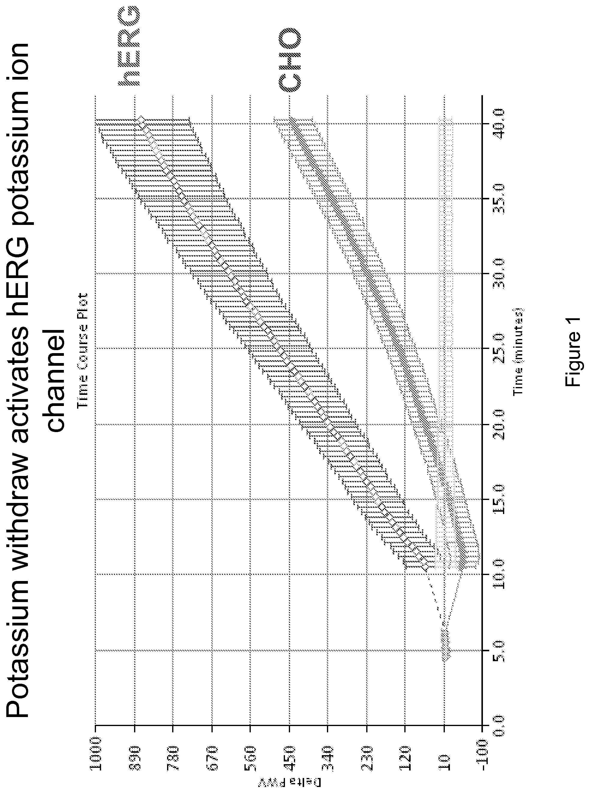
FIG. 1 demonstrates the effect of removing potassium from CHO cells transfected with the human ether-a-go-go (hERG) potassium channel and from parental CHO cells.
FIG. 2A demonstrates the effect of cisapride on CHO cells transfected with hERG. FIG. 2B demonstrates the effect of E4031 on CHO cells transfected with hERG. FIG. 2C shows that cisapride and E4031 are specific blocker for hERG potassium ion channels.
FIG. 3A demonstrates the effect of cisapride on parental CHO cells. FIG. 3B demonstrates the effect of E4041 on parental CHO cells.
DETAILED DESCRIPTION OF THE INVENTION
As used herein, the singular forms "a," "an", and "the" include plural referents unless the context clearly dictates otherwise.
Several types of ion channels are known including, e.g., voltage-gated sodium channels (that sense the transmembrane potential and open or close in response to depolarization or hyperpolarization), voltage-gated calcium channels, mechano-sensitive ion channels, potassium channels (such as, inward-rectifier potassium channels, calcium-activated potassium channels, voltage-gated potassium channels, and two pore domain potassium channels), transient receptor potential channels, cation channels of sperm, cyclic nucleotide gated channels (such as hyperpolaraization activated cyclic nucleotide gated channels)(that open in response to internal solutes and mediate cellular responses to second messengers), stretch activated channels (that open or close in response to mechanical force) two pore channels, ligand gated channels (which open in response to a specific ligand molecule on the external face of the membrane), G-protein gated channels (that open in response to G-protein-activation via its receptor), inward rectifier K channels (that allow potassium to flow into the cell in an inwardly rectifying manner), and light-gated channels. Some channels respond to multiple influences.
Ligand gated ion channels open in response to specific ligand molecules binding to the extracellular domain of the receptor protein. Ligand binding causes a conformational change in the structure of the channel protein that leads to the opening of the channel gate and subsequent ion flux across the plasma membrane. Examples of such channels include the G-protein coupled ion channels, anion-permeable .gamma.-aminobutyric acid-gated GABA.sub.A or C receptors, ionotropic glutamate-gated receptors, serotonin/5-HT.sub.3 receptor, ATP-gated P2X receptors, and cation-permeable "nicotinic" acetylcholine receptor.
G-protein coupled ion channels (GPC) are stimulated when a neurotransmitter binds to the G-protein coupled receptor (GCR). This activates G-proteins, which move to another ion channel. The G-Proteins allow the channel to open and ions are able to flow across the cell membrane. Because of the movement from the receptor to the ion, the speed of the channel opening is delayed, however the channel stays open for a longer time. GPC channels, such as the GABA (subtypes A & C) and NMDA (antagonists), are likely therapeutic sites for the function of anesthetics for blocking of sensation, temporarily taken away muscle activation, and behavior modification.
Binding of GABA molecules (neurotransmitter, gamma-aminobutyric acid) to their binding sites in the extracellular part of GABA.sub.A and GABA.sub.C receptors triggers opening of a chloride ion-selective pore. The increased chloride conductance drives the membrane potential towards the reversal potential of the Cl.sup.- ion in neurons, inhibiting the firing of new action potentials or nerve impulses.
Glutamate regulates ion channels and is the most abundant excitatory neurotransmitter in the mammalian nervous system. Nerve impulses trigger release of glutamate from the pre-synaptic cell. In the opposing post-synaptic cell, glutamate receptors, such as the NMDA (ligand gated ion channel) receptor, bind glutamate and are activated.
Glutamate transporters are found in neuronal and glial membranes. They rapidly remove glutamate from the extracellular space. In brain injury or disease, they can work in reverse and excess glutamate can accumulate outside cells. This process causes calcium ions to enter cells via NMDA receptor channels, leading to neuronal damage and eventual cell death.
Binding of the neurotransmitter 5-hydroxytryptamine (serotonin) to the 5-HT.sub.3 receptor opens the ligand gated ion channel which in turn leads to an excitatory response in neurons. When the receptor is activated to open the ion channel by agonists. 5-HT.sub.3 antagonists include ondansetron, granisetron, and tropisetron.
P2X receptors are cation-permeable ligand gated ion channels that open in response to the binding of extracellular adenosine 5'-triphosphate (ATP). ATP binds to the P2X receptor and cause a conformational change in the structure of the ion channel that results in the opening of the ion-permeable pore. This allows cations such as Na.sup.+ and Ca.sup.2+ to enter the cell, leading to depolarization of the cell membrane and the activation of various Ca.sup.2+-sensitive intracellular processes. The different protein parts of this channel have been found responsible for regulating ATP binding, ion permeation, pore dilation and desensitization.
Acetylcholine can open ligand gated sodium channels when it binds to acetylcholine receptors on skeletal muscle fibers.
Inward rectifying potassum (K.sub.ir) channels are found on multiple cell types. In cardiac myocytes K.sub.ir channels close upon depolarization, slowing membrane repolarization and helping maintain a more prolonged action potential. This type of inward-rectifier channel is distinct from delayed rectifier K.sup.+ channels, which help re-polarize nerve and muscle cells after action potentials; and potassium leak channels, which provide much of the basis for the resting membrane potential. In endothelial cells K.sub.ir channels are involved in regulation of nitric oxide synthase. In kidney cells K.sub.ir export surplus potassium into collecting tubules for removal in the urine, or alternatively may be involved in the reuptake of potassium back into the body. In neurons and heart cells G-protein activated IRKs (K.sub.ir3) are important regulators. In pancreatic beta cellsK.sub.ATP channels control insulin release.
Calcium voltage gated ion channels (VDCC) play an important role in both linking muscle excitation with contraction as well as neuronal excitation with transmitter release. Activation of VDCCs allows Ca.sup.2+ entry into the cell, which depending on the cell type, results in muscular contraction, excitation of neurons, up-regulation of gene expression, or release of hormones or neurotransmitters.
Voltage-gated sodium channels control and set action potentials across cell membranes. When voltage-gated sodium channels open there is a change in the cell's membrane potential, and a small but significant number of Na.sup.+ ions will move into the cell down their electrochemical gradient, thereby depolarizing the cell. Intracellular and extracellular blockers are known modulators of pharmacologic control of these ion channels. Alkaloid based toxins of plants and animals (puffer fish and blue-ringed octapus) act extracellularly on these channels and result in loss of neural activity. Intracellular blockage of these channels result in anesthetics, anti-arrhythmic and anti-convulsant agents. Agonists of these channels (such as the poison arrow frog toxin) directly affect the peripheral nervous system and lead to persistent activation (open) channels, and present as toxins leading to cardiac arrhythmia and respiratory paralysis.
Some transient receptor potential channels (TRP) channels can be constitutively open, while others are gated by voltage, intracellular Ca.sup.2+, pH, redox state, osmolarity, and mechanical stretch. These channels vary according to the ion(s) they pass, some being selective for Ca.sup.2+ while others are less selective, acting as cation channels. This family is subdivided into 6 subfamilies based on homology TRPC (canonical); TRPV (vanilloid); TRPA (ankyrin); TRPM (melastatin); TRPP (polycystin); TRPML (mucolipin); TRPN (NOMPC).
Certain chemicals and genetic disorders interfere with normal ion channel function and cause disease and illness. Chemicals that can disrupt ion channel function, include, e.g., lidocaine, novocaine, dedrotoxin, conotoxin, saxitoxin, iberiotoxin, heteropodatoxin, tetrodotoxin. Receptor tyrosine kinases, GPCRs, transient receptor potential channels, phopholipase C, signal transduction pathways (e.g. P13 kinases), cytokines, beta-arrestin pathway responses, cytoskeletal rearrangements, epigenetic signals can affect ion channel function.
Genetic diseases that are caused by mutations in ion channel subunits or the proteins that regulate them include, e.g., alternating hemiplegia of childhood, Bartter syndrome, Brugada syndrome, congenital hyperinsulinism, cystic fibrosis, episodic ataxia, erythromelalgia, generalized epilepsy with febrile seizures, hyperkalemic periodic paralysis, hypokalemic periodic paralysis, long QT syndrome, malignant hyperthermia, migraine, myasthenia gravis, myotonia congenita, neuromyotonia, familial hemiplegic migraine, spinocerebellar ataxia type 13, nonsyndromic deafness, paramyotonia congenita, potassium aggravated myotonias, periodic paralysis, retinitis pigmentosa, mucolipidosis type IV, Romano-Ward syndrome, short QT syndrome, and Timothy syndrome. Therefore, discovery of reagents that can modulate ion channels are of great interest to researchers.
One embodiment of the invention allows the direct detection of cell changes in response to ion channel regulators as they occur in real time with a colorimetric resonant reflectance biosensor and without the need to incorporate radiometric, colorimetric, or fluorescent labels. Changes in cells can be detected as the cells are probed with test reagents, agonists, and antagonists. The cellular changes can then be detected in real time using a high speed instruments such as the BIND Scanner.TM. (i.e., a colorimetric resonant reflectance biosensor system), and corresponding algorithms to quantify data. See, e.g., U.S. Pat. No. 6,951,715 and U.S. Pat. Publ. 2004/0151626. By combining this methodology, instrumentation and computational analyses, cellular changes can be expediently monitored in real time, in a label free manner.
Biosensors
Biosensors of the invention can be colorimetric resonant reflectance biosensors. See e.g., Cunningham et al., "Colorimetric resonant reflection as a direct biochemical assay technique," Sensors and Actuators B, Volume 81, p. 316-328, Jan. 5 2002; U.S. Pat. Publ. No. 2004/0091397. Colorimetric resonant biosensors are not surface plasmon resonant (SPR) biosensors. SPR biosensors have a thin metal layer, such as silver, gold, copper, aluminum, sodium, and indium. The metal must have conduction band electrons capable of resonating with light at a suitable wavelength. A SPR biosensor surface exposed to light must be pure metal. Oxides, sulfides and other films interfere with SPR. Colorimetric resonant biosensors do not have a metal layer, rather they have a dielectric coating of high refractive index material, such as TiO.sub.2.
Grating-based waveguide biosensors are described in, e.g., U.S. Pat. No. 5,738,825. A grating-based waveguide biosensor comprises a waveguiding film and a diffraction grating that incouples an incident light field into the waveguiding film to generate a diffracted light field. A change in the effective refractive index of the waveguiding film is detected. Devices where the wave must be transported a significant distance within the device, such as grating-based waveguide biosensors, lack the spatial resolution of the current invention.
A colorimetric resonant reflectance biosensor allows biochemical interactions to be measured on the biosensor's surface without the use of fluorescent tags, colorimetric labels or any other type of detection tag or detection label. A biosensor surface contains an optical structure that, when illuminated with collimated and/or white light, is designed to reflect only a narrow band of wavelengths ("a resonant grating effect"). The narrow wavelength band is described as a wavelength "peak." The "peak wavelength value" (PWV) changes when materials, such as biological materials, are deposited or removed from the biosensor surface. A readout instrument is used to illuminate distinct locations on a biosensor surface with collimated and/or white light, and to collect reflected light. The collected light is gathered into a wavelength spectrometer for determination of a PWV.
A biosensor can be incorporated into standard disposable laboratory items such as microtiter plates by bonding the structure (biosensor side up) into the bottom of a bottomless microtiter plate cartridge. Incorporation of a biosensor into common laboratory format cartridges is desirable for compatibility with existing microtiter plate handling equipment such as mixers, incubators, and liquid dispensing equipment. Colorimetric resonant reflectance biosensors can also be incorporated into, e.g., microfluidic, macrofluidic, or microarray devices (see, e.g., U.S. Pat. Nos. 7,033,819, 7,033,821). Colorimetric resonant reflectance biosensors can be used with well-know methodology in the art (see, e.g., Methods of Molecular Biology edited by Jun-Lin Guan, Vol. 294, Humana Press, Totowa, N.J.) to monitor cell behavioral changes or the lack of these changes upon exposure to one or more extracellular reagents.
Colorimetric resonant reflectance biosensors comprise subwavelength structured surfaces (SWS) and are an unconventional type of diffractive optic that can mimic the effect of thin-film coatings. (Peng & Morris, "Resonant scattering from two-dimensional gratings," J. Opt. Soc. Am. A, Vol. 13, No. 5, p. 993, May 1996; Magnusson, & Wang, "New principle for optical filters," Appl. Phys. Lett., 61, No. 9, p. 1022, August, 1992; Peng & Morris, "Experimental demonstration of resonant anomalies in diffraction from two-dimensional gratings," Optics Letters, Vol. 21, No. 8, p. 549, April, 1996). A SWS structure contains a one-dimensional, two-dimensional, or three dimensional grating in which the grating period is small compared to the wavelength of incident light so that no diffractive orders other than the reflected and transmitted zeroth orders are allowed to propagate. Propagation of guided modes in the lateral direction are not supported. Rather, the guided mode resonant effect occurs over a highly localized region of approximately 3 microns from the point that any photon enters the biosensor structure.
The reflected or transmitted light of a colorimetric resonant reflectance biosensor can be modulated by the addition of molecules such as specific binding substances or binding partners or both to the upper surface of the biosensor. The added molecules increase the optical path length of incident radiation through the structure, and thus modify the wavelength at which maximum reflectance or transmittance will occur.
In one embodiment, a colorimetric resonant reflectance biosensor, when illuminated with white and/or collimated light, is designed to reflect a single wavelength or a narrow band of wavelengths (a "resonant grating effect"). Light can illuminate the biosensor from either the top or the bottom. When mass is deposited on the surface of the biosensor, the reflected wavelength is shifted due to the change of the optical path of light that is shown on the biosensor.
A detection system consists of, for example, a light source that illuminates a small spot of a biosensor at normal incidence through, for example, a fiber optic probe, and a spectrometer that collects the reflected light through, for example, a second fiber optic probe also at normal incidence. Because no physical contact occurs between the excitation/detection system and the biosensor surface, no special coupling prisms are required and the biosensor can be easily adapted to any commonly used assay platform including, for example, microtiter plates. A single spectrometer reading can be performed in several milliseconds, thus it is possible to quickly measure a large number of molecular interactions taking place in parallel upon a biosensor surface, and to monitor reaction kinetics in real time.
A colorimetric resonant reflectance biosensor comprises, e.g., an optical grating comprised of a high refractive index material, a substrate layer that supports the grating, and optionally one or more specific binding substances or linkers immobilized on the surface of the grating opposite of the substrate layer. The high refractive index material has a higher refractive index than a substrate layer. See, e.g., U.S. Pat. No. 7,094,595; 7,070,987. Optionally, a cover layer covers the grating surface. In one embodiment, the refractive index of the optical grating can be less than the refractive index of the optional cover layer. An optical grating is coated with a high refractive index dielectric film which can be comprised of a material that includes, for example, zinc sulfide, titanium dioxide, tantalum oxide, silicon nitride, and silicon dioxide. A cross-sectional profile of a grating with optical features can comprise any periodically repeating function, for example, a "square-wave." An optical grating can also comprise a repeating pattern of shapes selected from the group consisting of lines (one-dimensional), squares, circles, ellipses, triangles, trapezoids, sinusoidal waves, ovals, rectangles, and hexagons. A colorimetric resonant reflectance biosensor of the invention can also comprise an optical grating comprised of, for example, plastic or epoxy, which is coated with a high refractive index material. Layer thicknesses (i.e. cover layer, biological material, or an optical grating) are selected to achieve resonant wavelength sensitivity to additional molecules on the top surface. The grating period is selected to achieve resonance at a desired wavelength.
Linear gratings (i.e., one dimensional gratings) have resonant characteristics where the illuminating light polarization is oriented perpendicular to the grating period. A colorimetric resonant reflection biosensor can also comprise, for example, a two-dimensional grating, e.g., a hexagonal array of holes or squares. Other shapes can be used as well. A linear grating has the same pitch (i.e. distance between regions of high and low refractive index), period, layer thicknesses, and material properties as a hexagonal array grating. However, light must be polarized perpendicular to the grating lines in order to be resonantly coupled into the optical structure. Therefore, a polarizing filter oriented with its polarization axis perpendicular to the linear grating must be inserted between the illumination source and the biosensor surface. Because only a small portion of the illuminating light source is correctly polarized, a longer integration time is required to collect an equivalent amount of resonantly reflected light compared to a hexagonal grating.
An optical grating can also comprise, for example, a "stepped" profile, in which high refractive index regions of a single, fixed height are embedded within a lower refractive index cover layer. The alternating regions of high and low refractive index provide an optical waveguide parallel to the top surface of the biosensor.
A colorimetric resonant reflectance biosensor of the invention can further comprise a cover layer on the surface of an optical grating opposite of a substrate layer. Where a cover layer is present, the one or more specific binding substances are immobilized on the surface of the cover layer opposite of the grating. Preferably, a cover layer comprises a material that has a lower refractive index than a material that comprises the grating. A cover layer can be comprised of, for example, glass (including spin-on glass (SOG)), epoxy, or plastic.
For example, various polymers that meet the refractive index requirement of a biosensor can be used for a cover layer. SOG can be used due to its favorable refractive index, ease of handling, and readiness of being activated with specific binding substances using the wealth of glass surface activation techniques. When the flatness of the biosensor surface is not an issue for a particular system setup, a grating structure of SiN/glass can directly be used as the sensing surface, the activation of which can be done using the same means as on a glass surface.
Resonant reflection can also be obtained without a planarizing cover layer over an optical grating. For example, a biosensor can contain only a substrate coated with a structured thin film layer of high refractive index material. Without the use of a planarizing cover layer, the surrounding medium (such as air or water) fills the grating. Therefore, specific binding substances are immobilized to the biosensor on all surfaces of an optical grating exposed to the specific binding substances, rather than only on an upper surface.
In general, a colorimetric resonant reflectance biosensor of the invention will be illuminated with white and/or collimated light that will contain light of every polarization angle. The orientation of the polarization angle with respect to repeating features in a biosensor grating will determine the resonance wavelength. For example, a "linear grating" (i.e., a one-dimensional grating) biosensor consisting of a set of repeating lines and spaces will have two optical polarizations that can generate separate resonant reflections. Light that is polarized perpendicularly to the lines is called "s-polarized," while light that is polarized parallel to the lines is called "p-polarized." Both the s and p components of incident light exist simultaneously in an unfiltered illumination beam, and each generates a separate resonant signal. A biosensor can generally be designed to optimize the properties of only one polarization (the s-polarization), and the non-optimized polarization is easily removed by a polarizing filter.
In order to remove the polarization dependence, so that every polarization angle generates the same resonant reflection spectra, an alternate biosensor structure can be used that consists of a set of concentric rings. In this structure, the difference between the inside diameter and the outside diameter of each concentric ring is equal to about one-half of a grating period. Each successive ring has an inside diameter that is about one grating period greater than the inside diameter of the previous ring. The concentric ring pattern extends to cover a single sensor location--such as an array spot or a microtiter plate well. Each separate microarray spot or microtiter plate well has a separate concentric ring pattern centered within it. All polarization directions of such a structure have the same cross-sectional profile. The concentric ring structure must be illuminated precisely on-center to preserve polarization independence. The grating period of a concentric ring structure is less than the wavelength of the resonantly reflected light. The grating period is about 0.01 micron to about 1 micron. The grating depth is about 0.01 to about 1 micron.
In another embodiment, an array of holes or posts are arranged to closely approximate the concentric circle structure described above without requiring the illumination beam to be centered upon any particular location of the grid. Such an array pattern is automatically generated by the optical interference of three laser beams incident on a surface from three directions at equal angles. In this pattern, the holes (or posts) are centered upon the corners of an array of closely packed hexagons. The holes or posts also occur in the center of each hexagon. Such a hexagonal grid of holes or posts has three polarization directions that "see" the same cross-sectional profile. The hexagonal grid structure, therefore, provides equivalent resonant reflection spectra using light of any polarization angle. Thus, no polarizing filter is required to remove unwanted reflected signal components. The period of the holes or posts can be about 0.01 microns to about 1 micron and the depth or height can be about 0.01 microns to about 1 micron.
A detection system can comprise a colorimetric resonant reflectance biosensor a light source that directs light to the colorimetric resonant reflectance biosensor, and a detector that detects light reflected from the biosensor. In one embodiment, it is possible to simplify the readout instrumentation by the application of a filter so that only positive results over a determined threshold trigger a detection.
By measuring the shift in resonant wavelength at each distinct location of a colorimetric resonant reflectance biosensor of the invention, it is possible to determine which distinct locations have, e.g., biological material deposited on them. The extent of the shift can be used to determine, e.g., the amount of binding partners in a test sample and the chemical affinity between one or more specific binding substances and the binding partners of the test sample.
A colorimetric resonant reflectance biosensor can be illuminated two or more times. A first measurement can determine the reflectance spectra of one or more distinct locations of a biosensor with, e.g., no biological material on the biosensor or with cells on the surface. A second, third, fourth or additional measurements can determine the reflectance spectra after, e.g., one or more cells, test reagents, ion channel agonists, or ion channel antagonists are applied to a biosensor or after an incubation period or wash step. The difference in peak wavelength between these two or more measurements can be used to, e.g., determine the presence or amount of cells on the biosensor, or the activity of a test reagent, ion channel agonist, or ion channel antagonist on the cells. Additionally, this method of illumination can control for small imperfections in a surface of a biosensor that can result in regions with slight variations in the peak resonant wavelength. This method can also control for varying concentrations or density of cell matter on a biosensor.
Surface of Biosensor
One or more cells can be immobilized on a biosensor by for example, physical adsorption or by chemical binding. The cells can be non-adherent cells or adherent cells. The cells can natively express one or more ion channels or one or more ion channel components. Alternately, the cells can recombinantly express one or more ion channels or ion channel components. The cells can be mammalian cells, such as human cells.
A cell can specifically bind to a biosensor surface via a specific binding substance such as a nucleic acid, peptide, protein solution, peptide solution, solutions containing compounds from a combinatorial chemical library, antigen, polyclonal antibody, monoclonal antibody, single chain antibody (scFv), F(ab) fragment, F(ab').sub.2 fragment, Fv fragment, small organic molecule, virus, polymer or biological sample, wherein the specific binding substance is immobilized to the surface of the biosensor and the binding partner is on the surface of the cell. Alternatively, cells can grow and optionally attach on the surface of the biosensor and not necessarily be immobilized directly to the surface of the biosensor, regardless if the cell type is normally adherent or non-adherent.
Cells can be arranged in an array of one or more distinct locations on the biosensor surface, said surface residing within one or more wells of a multiwell plate and comprising one or more surfaces of the multiwell plate or microarray. The array of cells comprises one or more cells on the biosensor surface within a microwell plate such that a surface contains one or more distinct locations, each with a different cell or with a different amount of cells. For example, an array can comprise 1, 10, 100, 1,000, 10,000 or 100,000 or greater distinct locations. Thus, each well of a multiwell plate or microarray can have within it an array of one or more distinct locations separate from the other wells of the multiwell plate, which allows multiple different samples to be processed on one multiwell plate. The array or arrays within any one well can be the same or different than the array or arrays found in any other microtiter wells of the same microtiter plate.
Immobilization of a cell to a biosensor surface can be also be affected via binding to, for example, the following functional linkers: a nickel group, an amine group, an aldehyde group, an acid group, an alkane group, an alkene group, an alkyne group, an aromatic group, an alcohol group, an ether group, a ketone group, an ester group, an amide group, an amino acid group, a nitro group, a nitrile group, a carbohydrate group, a thiol group, an organic phosphate group, a lipid group, a phospholipid group or a steroid group. Furthermore, a cell can be immobilized on the surface of a biosensor via physical adsorption, chemical binding, electrochemical binding, electrostatic binding, passive or active adhesion molecules, hydrophobic binding or hydrophilic binding, and immunocapture methods. Methods for coating surfaces to make them amenable to cell attachment and/or growth are well known by persons skilled in the art of cell culture and can be comprised of a coating for the biosensor with materials or their derivatives including but not limited to poly-D-lysine, fibronectin, actin, integrins, adherins, cadherins, collagen, human serum, fetal bovine serum, calf serum, laminin, or other materials.
In one embodiment of the invention a biosensor can be coated with a linker such as, e.g., a nickel group, an amine group, an aldehyde group, an acid group, an alkane group, an alkene group, an alkyne group, an aromatic group, an alcohol group, an ether group, a ketone group, an ester group, an amide group, an amino acid group, a nitro group, a nitrile group, a carbohydrate group, a thiol group, an organic phosphate group, a lipid group, a phospholipid group or a steroid group. For example, an amine surface can be used to attach several types of linker molecules while an aldehyde surface can be used to bind proteins directly, without an additional linker A nickel surface can be used to bind molecules that have an incorporated histidine ("his") tag. Detection of "his-tagged" molecules with a nickel-activated surface is well known in the art (Whitesides, Anal. Chem. 68, 490, (1996)).
Linkers and specific binding substances can be immobilized on the surface of a biosensor such that each well has the same linkers and/or specific binding substances immobilized therein. Alternatively, each well can contain a different combination of linkers and/or specific binding substances.
A cell can specifically or non-specifically bind to a linker or specific binding substance immobilized on the surface of a biosensor. Alternatively, the surface of the biosensor can have no linker or specific binding substance and a cell can bind to the biosensor surface non-specifically.
Immobilization of one or more specific binding substances or linker onto a biosensor is performed so that a specific binding substance or linker will not be washed away by rinsing procedures, and so that its binding to cells in a test sample is unimpeded by the biosensor surface. Several different types of surface chemistry strategies have been implemented for covalent attachment of specific binding substances to, for example, glass or polymers for use in various types of microarrays and biosensors. Surface preparation of a biosensor so that it contains the correct functional groups for binding one or more specific binding substances is an integral part of the biosensor manufacturing process.
One or more specific cells can be attached to a biosensor surface by physical adsorption (i.e., without the use of chemical linkers) or by chemical binding (i.e., with the use of chemical linkers) as well as electrochemical binding, electrostatic binding, hydrophobic binding and hydrophilic binding. Chemical binding can generate stronger attachment of specific binding substances on a biosensor surface and provide defined orientation and conformation of the surface-bound molecules.
Immobilization of specific binding substances to plastic, epoxy, or high refractive index material can be performed essentially as described for immobilization to glass. However, the acid wash step can be eliminated where such a treatment would damage the material to which the specific binding substances are immobilized.
Methods of Using Biosensors
One embodiment of the invention provides a method of identifying a modulator of an ion channel. A modulator of an ion channel has an effect on the functional activity of an ion channel. An effect on the functional activity includes blocking or inhibiting the activity of the ion channel. Modulators can block, inhibit, enhance, or increase the functional activity of an ion channel. The blocking, inhibition, enhancement or increase may take place in the presence of, or in response to, a molecule that blocks the ion channel. Modulators can, for example, act on ion channels to change gating/ligand ion selectivity, modify the chemical signals for channel function (pH, osmolarity), change the potential function for gating activity (altered polarizability or voltage dependence), cause a physical blockage, cause a physical widening (altering folding function of channel control protein), modify refractory period, modify the mechanical force function (mechanical stretch) or temperature required for channel function.
The methods of the invention can be used with cells that have not been recombinantly altered to express or over-express certain ion channels or recombinant cells, however, cells that recombinantly express or over-express ion channel proteins or subunits are not required by the methods of the invention. Cells can be applied to a first location on a surface of a colorimetric resonant reflectance optical biosensor. The cells can be immobilized to the biosensor or they can merely be grown on the surface of the biosensor. Optionally, the cells can be incubated on the surface of the biosensor at any step of the methods of the invention for about, 1, 5, 10, 30 or more minutes or about 1, 2, 5, 24, 48, 36, or more hours. The assays can be performed at a temperature of about 2, 5, 10, 25, 30, or 37 degrees Celsius (or any range of temperatures between about 2 and about 37 degrees Celsius).
Advantageously, once an assay of the invention is completed, the cells can be washed, equilibrated and used again to complete the same assay or a different assay. This is particularly advantageous where cells are expensive or difficult to handle.
In one embodiment of the invention, a method of identifying an antagonist or agonist of an ion channel is provided. The method comprises applying cells to a first location and a second location on a surface of colorimetric resonant reflectance optical biosensor. The cells can be applied to the second location at the same time the first set of cells are applied to the first location or at an earlier or later time. These cells can be the same type of cells or different cells as the set of cells applied to the first location. A test reagent is added to the first location before the cells are added to the first location, at the same time the cells are added to the first location, or after the cells are added to the first location. A known ion channel antagonist or agonist of the cells is added to the second location. The known ion channel agonist or antagonist is added to the second location before the cells are added to the second location, at the same time the cells are added to the second location, or after the cells are added to the second location. The use of known agonists or antagonists can determine the specificity of the ion channel. Antagonists and agonists, including, e.g., specific toxins and other small molecules with known functions or known target can be used to determine the specificity of the test reagent/stimuli/modulator response on an ion channel. Furthermore, use of compounds that act at sites upstream or downstream of an ion channel can be used to determine the specificity of the response to a particular channel as well. A colorimetric resonant reflectance optical first peak wavelength value (PWV) for the first location and detecting a colorimetric resonant reflectance optical second PWV for the second location, wherein if the first and second PWVs are the same or similar, then the test reagent is an antagonist or agonist of an ion channel. The first and second values are the same or similar if they are within about 1 nm or less of each other. If the first and second PWVs are substantially different (that is the first and second PWVs differ by greater than about 1 nm, 2 nm, 3 nm, 4 nm or more), then the test reagent is not an antagonist or agonist. Furthermore, one of more additional PWVs may be determined before or after any of the steps or additions of the method and compared to any other PWV.
In another embodiment of the invention, a method of identifying an antagonist or agonist of an ion channel is provided. The method comprises applying cells to a first location. A PWV can be determined for the first location. A test reagent is applied to the first location. The cells and test reagent can be incubated for period of time if desired. A second PWV for the first location can be determined. A first value can be calculated, wherein the first value is the difference between the first PWV and the second PWV. The first value can be compared to a control test. The control test can comprise applying cells to a second location on a surface of a colorimetric resonant reflectance optical biosensor. The cells can be incubated for a period of time if desired. These cells can be applied to the second location at the same time the first set of cells are applied to the first location or at an earlier or later time. These cells can be the same type of cells or different cells as the set of cells applied to the first location. A third PWV for the second location can be detected. A known ion channel antagonist or agonist of the cells is applied to the second location. The cells can be incubated for a period of time if desired. A fourth PWV for the second location can be detected. A second value can be determined, wherein the second value can be the difference between the third PWV and the fourth PWV of the second location. If the first and second values are the same or similar, then the test reagent is a modulator of an ion channel. The first and second values are the same or similar if they are within about 1 nm or less of each other. If the first and second PWVs are substantially different (that is the first and second PWVs differ by greater than about 1 nm, 2 nm, 3 nm, 4 nm, 5 nm or more), then the test reagent is not an antagonist or agonist. Because the label free biosensor method of interrogating the cells is not destructive to the cells, the cells may be treated more than one time to look for differences over a number of minutes, hours, or days.
There are multitudes of known ion channel antagonists or agonists in the art. A small sampling of examples include calcium channel blockers (such as nisoldipine, nifedipine, nicardipine, bepridil, isradipine, nimodipine, felodipine, amlodipine, diltiazem, and verapamil), modulators of potassium ion channels (see, e.g., US 20050267089), Na.sup.+ ion channel modulating compounds (see, e.g, U.S. Pat. No. 6,576,791), see also, U.S. Pat. No. 7,101,877; 7,057,053, US 20070004718; US 20060199848, US 20060135536, US 20050192208, US 20050026993, US 20040029937, US 20030008906.
The first location and second location on the surface of the colorimetric resonant reflectance optical biosensor can be an internal surface of a vessel selected from the group consisting of a microtiter well, microtiter plate, test tube, Petri dish, microfluidic channel, and microarray. The cells, test reagent, and ion channel antagonist or agonist do not have to comprise detection labels in the assays of the invention.
Another embodiment of the invention provides a method of identifying a modulator of an ion channel. Cells can be applied to a first location on a surface of a colorimetric resonant reflectance optical biosensor. A test reagent and a known ion channel antagonist or agonist of the cells is applied to the first location. The cells, test reagent and known ion channel antagonist or agonist of the cells can be added to the sensor surface in any order or at the same time. A first PWV for the first location can be determined. Cells can be applied to a second location on a surface of a colorimetric resonant reflectance optical biosensor. The cells can be applied to the second location at the same time the first set of cells are applied to the first location or at an earlier or later time. These cells can be the same type of cells or different cells as the set of cells applied to the first location. A known ion channel antagonist or agonist of the cells is applied to the second location. The cells and known ion channel antagonist or agonist of the cells can be applied to the biosensor surface in any order or at the same time. A second PWV can be determined for the second location. If the first and second PWVs are different, then the test reagent is a modulator of an ion channel. The first and second values are different if they are greater than about 1 nm, 2 nm, 3 nm, 4 nm, 5 nm, or more apart. If the first and second PWVs are the same or similar then the test reagent is not a modulator of an ion channel. The first and second values are the same or similar if they are within about 1 nm or less of each other.
In another embodiment of the invention, a method of identifying a modulator of an ion channel is provided. The method comprises applying cells to a first location on a surface of a colorimetric resonant reflectance optical biosensor, applying a test reagent to the first location, and detecting a first PWV for the first location. The cells and test reagent can be applied to the first location in any order or at the same time. A known ion channel antagonist or agonist of the cells is then applied to the first location and a second PWV for the first location can be detected. A first value is determined wherein the first value is the difference between the first PWV and the second PWV.
The method can alternatively comprise applying cells to a first location on a surface of a colorimetric resonant reflectance optical biosensor, applying a known ion channel antagonist or agonist of the cells to the first location, and detecting a first PWV for the first location. The cells and known ion channel antagonist or agonist of the cells can be applied to the first location in any order or at the same time. A test reagent is then applied to the first location and a second PWV for the first location can be detected. A first value is determined wherein the first value is the difference between the first PWV and the second PWV.
Cells are applied to a second location on a surface of a colorimetric resonant reflectance optical biosensor. These cells can be applied to the second location at the same time the first set of cells are applied to the first location or at an earlier or later time. These cells can be the same type of cells or different cells as the set of cells applied to the first location. A third PWV for the second location can be detected. A known ion channel antagonist or agonist of the cells is applied to the second location. A fourth PWV for the second location can be determined. A second value is determined, wherein the second value is the difference between the third PWV and the fourth PWV. If the first and second values are different, then the test reagent is a modulator of an ion channel. The first and second values are different if they are greater than about 1 nm, 2 nm, 3 nm, 4 nm, 5 nm, or more apart. If the first and second values are the same or similar, then the test reagent is not a modulator of an ion channel. The first and second values are the same or similar if they are within about 1 nm or less of each other.
The cells can be incubated for a period of time after their application to the first location, after the application of the test reagent to the first location, after the application of the known ion channel agonist or antagonist to the first location, after the application of the cells to the second location, after the application of the known ion channel antagonist or agonist to the second location, or at any other point during the assay, or a combination thereof.
Optionally, the cells are incubated for a period of time after their application to a surface of the colorimetric resonant reflectance optical biosensor; after the application of the test reagent; after the application of the ion channel agonist or antagonist, or any other point during the assay, or a combination thereof. Because the label free biosensor method of interrogating the cells is not destructive to the cells, the cells may also be treated more than one time to look for differences over a number of minutes, hours, or days.
Another embodiment of the invention provides a method of confirming that a test reagent is a modulator of an ion channel comprising applying cells to a first location on a surface of a colorimetric resonant reflectance optical biosensor, and optionally detecting a first PWV for the first location. A known ion channel agonist or antagonist of the cells is applied to the first location; and the test reagent is applied to the first location. The known ion channel agonist or antagonist and the test reagent can be applied to the first location at the same time or one may be applied to the first location before the other. A second PWV can be detected for the first location. A first value can be determined, wherein the first value is the difference between the first PWV and the second PWV. Cells are applied to a second location on a surface of a colorimetric resonant reflectance optical biosensor. These cells can be applied to the second location at the same time the first set of cells are applied to the first location or at an earlier or later time. These cells can be the same type of cells or different cells as the set of cells applied to the first location. A third PWV can be determined for the second location. A known ion channel agonist or antagonist of the cells is applied to the second location and a fourth PWV can be determined for the second location. The known ion channel agonist or antagonist can be the same as that used at the first location or a different known ion channel agonist or antagonist. A second value can be determined, wherein the second value is the difference between the third PWV and the fourth PWV. If the first and second values are different, then the test reagent is confirmed as a modulator of an ion channel. The first and second values are different if they are greater than about 1 nm, 2 nm, 3 nm, 4 nm, 5 nm, or more apart.
The cells can be incubated for a period of time after their application to the biosensor; after the application of the test reagent, after the application of the known ion channel agonist or antagonist, or at any other point in the assay, or a combination thereof.
It is also useful to screen compounds to ensure that they do not modulate ion channels in any manner. There are many unintended, undesirable effects of drugs on ion channels. Several drugs have been removed from the market due to these effects (e.g., Rezulin, Loronex, Propulsid, Redux, Pondimin, Hismanal, Posicor, and Seldane). Therefore, the methods of the invention are useful to identify non-modulators of ion channels as well as modulators of ion channels.
Agonists, antagonists and modulators can affect more than one ion channel. Therefore, it can be advantageous to inhibit the function of one or more first types of ion channels in order to determine the function of an added test reagent on one or more second types of ion channels. The first and second types of ion channels can be the same or different. In one embodiment of the invention, one or more antibodies, proteins, small molecules, siRNAs, anti-sense RNAs, or other agents are used to inhibit some or all of the function of one or more types of ion channels. For example, an antibody can be specific for one or more pore subunit proteins of an ion channel such that when the antibody specifically binds to the pore protein the ion channel ceases to function in part or substantially. Once that ion channel is inhibited, a test regent, such as an agonist, antagonist, ion channel modulator, or any other compound, can be added to the cells to determine the effect of the test reagent on the non-blocked types of ion channels.
One embodiment of the invention provides a method for screening a test reagent for modulation of ion channel activity. The method can comprise contacting cells that express one or more ion channels with an agent that can partially or substantially block one or more types of ion channels. The cells are contacted with a test reagent. Ion channel activity of one or more types of ion channels can be determined before and/or after contact between the cell and the test reagent and/or before and/or after contact between the cells and the blocking agent. An alteration in ion channel activity as determined by a shift in PWV indicates that the test reagent is capable of modulating the activity of one or more types of ion channels.
A test reagent can be any molecule that potentially modulates the activity of an ion channel or alters the expression of ion channel proteins within cells. A molecule can be, for example, a polypeptide, polynucleotide, antibody, small organic molecule, polysaccharide, fatty acid, steroid, purine, pyrimidine, or multivalent cation, among others. Small organic molecules can have a molecular weight of more than 100 and less than about 2,500 Daltons (D). Preferred small molecules are less than 2000, or less than 1500 or less than 1000 or less than 500 D.
In one embodiment of the invention, one or more ECM ligands are coated on one or more surfaces of biosensor. One of more types of cells in serum-free medium are added to the surface of the biosensor. A serum-free medium contains about 5, 4, 3, 2, 1, 0.5% or less serum. A serum-free medium can comprise about 0% serum or about 0% to about 1% serum. A PWV can be determined after the addition of the cells to the biosensor. One or more potential or known modulators of ion channels can be added to the biosensor surface. A PWV can be determined after the addition of the one or more modulators. A change can be effected in the activity of one or more ion channels of the cells. For example, the cells can be depolarized.
A change in the activity of one or more ion channels can be effected by depolarizing the cells. Depolarization is a decrease in the absolute value of a cell's membrane potential. Thus, changes where the membrane potential becomes less positive or less negative are both depolarizations. Depolarization can be caused by influx of cations, e.g. Na.sup.+ through Na.sup.+ channels, or Ca.sup.2+ through Ca.sup.2+ channels. Depolarization can be inhibited by outflux of K.sup.+ through K.sup.+ channels. Therefore, depolarization can be caused by manipulation of salt concentration, pressure and current. See, e.g., Boulton et al. (eds), The Neuronal Microenvironment, Springer, N.Y. (1988); Arbib, The Handbook of brain theory and neural networks 2.sup.nd Ed., MIT Press, Cambridge Mass. (2003).
Cell medium can be removed, diluted or replaced with medium with a reduced concentration of an ion, such as potassium in order to "open" ion channels. Alternatively, the cells can be exposed to an ion channel opener, e.g. scorpion venom toxin.
A PWV can be determined after the change in activity is effected in one or more ion channels (e.g., the addition of an ion channel opener). Optionally, at a pre-determined time, another change can be effected in the activity of one or more ion channels of the cells. For example, the ion concentration can be increased or voltage can be applied to effect a depolarization of the ion channel. A PWV is then determined. One of more of the PWVs can be compared to control cells, such as cells that did not have change in activity effected in one or more ion channels of the cells (e.g., no addition of an ion channel opener), cells lacking the ion channel altogether, cells receiving a channel opener, but no modulator. These responses can be measured on separate locations of the biosensor, e.g., in different wells of a microplate.
The PWVs can be compared to determine if the test ion channel modulators modulated the one or more ion channels of the cells. For example, where a test modulator of an ion channel is added to cells on a biosensor surface, a first PWV is taken, an ion channel opener is added to the cells, and a second PWV is taken, the first and second PWVs can be compared. Where the PWVs are substantially different, the test modulator has indeed modulated the ion channel. In general, resolution of differences of PWV as small as 5 pm can be detected; however, differences of greater than about 20 pm, 100 pm, 200 pm or more can also be detected. Alternatively, the PWVs can be compared to controls to determine if the test modulator has modulated the ion channels.
Where recombinant cell lines that overexpress one type of ion channel the effect of a modulator on that one type of ion channel can be determined.
In the assays of the invention before and/or after each step there can be an incubation for a period of time, a wash step, a PWV determination or a combination thereof. Optionally, a PWV reading can be taken constantly over the entire course or part of the assay.
With embodiments of the instant invention modulation of ion channels or lack thereof can be detected as it occurs, thus circumventing the need to incorporate radiometric, colorimetric, fluorescent labels or microscopy for evaluation. Changes in cells that occur due to modulation of ion channels can be expediently monitored in real time, in a label free manner. For cell changes to be detected in real time, the BIND Biosensor.TM., BIND Reader.TM., and BIND Scanner.TM. (e.g., a colorimetric resonant reflectance biosensor system) were designed and corresponding algorithms were created to quantify data. See, e.g., U.S. Pat. No. 6,951,715, U.S. Patent Appl. Publ. 2004/0151626.
Methods of the invention are advantageous because they do not require labeling of cells or reagents for microscopic or colorimetric/fluorimetric evaluation, they allow for continuous, multiple independent readings of the same population of cells in real time, they are quick, they require minimal reagent usage (both volume and type), they do not require recombinant cells lines, they do not require mechanical manipulation of cells, and they do not require flowing the cells through a counting device.
The label-free biosensor method is non-destructive to the cells so that a cell or a set of cells may be monitored continuously or discontinuously over a long period of time with or without treatment of known or test compounds. Methods of the invention allow for continuous monitoring or multiple independent readings of the same population of cells in real time over many days. Cellular changes can be quantified expediently and objectively over longer periods of time in a normal culturing environment (static with proper media). Methods of the invention can also be used synergistically with fluorescent labels to obtain additional, intracellular data from each cell or cell population.
Cell changes can be monitored by taking a PWV for one location over several time periods. Alternatively, scans of a receptacle holding the cells, e.g., a microtiter plate well, can be done over several time periods. In one embodiment of the invention a test reagent can be identified as a potential ion channel modulator by comparing its PWV pattern over time to a known ion channel modulator. When certain ion channel regulators are added to certain cell populations, the PWVs over time exhibit a particular pattern. For example, after an ion channel modulator is added to cells the PWV may increase for a particular period of time and then decrease. Test reagents that exhibit the same pattern of PWV values over time can be identified as a potential ion channel regulator or modulator.
One or more cells can be applied to a location, such as a microtiter well on a surface of a colorimetric resonant reflectance optical biosensor. A receptacle refers to one container and not a collection of containers, e.g., a multiwell plate. A colorimetric resonant reflectance optical peak wavelength value (PWV) for the location is detected. The one or more cells can be incubated for a period of time (e.g., 1 second, 30 seconds, 1, 2, 5, 10, 20, 30, 45 minutes, 1, 2, 5, 10 or more hours). Prior to the incubation, or after the incubation, or prior to the incubation and after the incubation one or more test reagents, ion channel agonists, and/or ion channel antagonists, can be applied to the one or more cells. The colorimetric resonant reflectance optical PWV for the location can be detected for a second time. If a change in the cells occurs then the reflected wavelength of light is shifted as compared to a situation where no change occurs. The first PWV can be compared to the second PWV. A change in the PWV can indicate a change in the cells. PWVs over several time periods can be determined and compared.
Cell changes at a biosensor location can be detected via the PWVs of the biosensor surface or monitored more generally using a microscope, digital camera, conventional camera, or other visualization apparatus, magnifying or non-magnifying, that utilizes lens-based optics or electronics-based charge coupled device (CCD) technology.
The changes in PWV can be determined using a BIND Reader.RTM., Scanner, or Cartridge Reader. In the case of the BIND Reader.RTM. and Cartridge Reader, assays can be completed in about 0.0005 to 8 hours.
Preferably, the resolution of the lens of the scanner determining the PWV has an about 2 to about 200, about 2 to about 50, or about 2 to about 15 micrometer pixel size. Assays of the invention can be completed in less than about 0.25, 0.5, 0.75, 1, 2, 3, 4, 5, 6, 7, or 8 hours. That is, cell changes in response to, for example, the added reagent can be determined in a time efficient manner.
All patents, patent applications, and other scientific or technical writings referred to anywhere herein are incorporated by reference in their entirety. The invention illustratively described herein suitably can be practiced in the absence of any element or elements, limitation or limitations that are not specifically disclosed herein. Thus, for example, in each instance herein any of the terms "comprising", "consisting essentially of", and "consisting of" may be replaced with either of the other two terms, while retaining their ordinary meanings. The terms and expressions which have been employed are used as terms of description and not of limitation, and there is no intention that in the use of such terms and expressions of excluding any equivalents of the features shown and described or portions thereof, but it is recognized that various modifications are possible within the scope of the invention claimed. Thus, it should be understood that although the present invention has been specifically disclosed by embodiments, optional features, modification and variation of the concepts herein disclosed may be resorted to by those skilled in the art, and that such modifications and variations are considered to be within the scope of this invention as defined by the description and the appended claims. In addition, where features or aspects of the invention are described in terms of Markush groups or other grouping of alternatives, those skilled in the art will recognize that the invention is also thereby described in terms of any individual member or subgroup of members of the Markush group or other group.
Example
Screening of Inhibitors of hERG Potassium Ion Channels
This protocol employs potassium free starvation buffer to depolarize cells and thereby open ion channels such that screening ion channel modulators is possible. Colorimetric resonant reflectance biosensor plates were washed with 1.times.PBS for hydration baseline for 3 to 5 minutes. A PWV was determined for each of the wells. This PWV can be useful to make sure that the biosensor is stable (not drifting) and is uniform in signal across the wells. The plates were coated with 25.0 .mu.l/well of fibronectin for 2 hrs at room temperature. The plates were blocked for 1 hour at room temperature or overnight at 4.degree. C. by addition of 25.0 .mu.l ovalbumin or BSA in PBS to the wells previously coated with fibronectin.
Starvation buffers were prepared with and without potassium. For 500.0 ml potassium-free starvation buffer the following reagents were used: 25 ml of 3M NaCl for 150 mM NaCl final; 10.0 ml for 20 mM HEPES; 1.0 ml for 2 mM calcium chloride; 0.5 ml for 2 mM magnesium chloride; 1.0 ml for 5 mM D-(+)-glucose; up to 500.0 ml with H.sub.2O HPLC. Potassium starvation buffer is prepared the same as above, but with 2 mM KCl added.
10 mM stocks of test compounds are diluted to 10 .mu.M in potassium-free buffer as first concentration in an 8-point dilution series with serial 1/3.sup.rd dilutions to make reagent plates: Cisapride monohydrate-10 mM=10 mg/2.06 ml DMSO for a final DMSO percentage of 0.1 to 0.5 within the cells; the titration for E4031 was started at a high concentration of 30 uM.
Cells (CHO cells transfected with the human ether-a-go-go (hERG) potassium channel or CHO parental cells) were harvested for 5-10 minutes with 3.0 mL versene/flask. The versene was deactivated with 8.0 mL full media with FBS per flask. The biosensor plates were equilibrated to room temperature and previously added PBS was removed. The cells were gently spun down for 5 minutes at 1K rpm. The media was aspirated and potassium-containing starvation buffer was added to the cell pellet. The cells were then diluted from their initial concentration to a concentration of 10.sup.6 cells per ml for 20,000 cells per 20.0 .mu.l). A baseline PWV for each well of the biosensor plate was taken as a quality control step to check for cell adhesion uniformity following a sufficient amount of time for the cells to settle and become attached.
20.0 .mu.l of potassium-containing starvation buffer was added to each well. A PWV was taken for each well. The average number of cells/well was determined. For seeding 20K cells in 20.0 .mu.l per well, cells were prepared at 10.sup.6 cells/ml. 20.0 .mu.l of cells were added to the well already containing 20.0 .mu.l media/buffer.
The seeded biosensor test plates were allowed to sit at room temperature for about 15 minutes, before being placing in the 37.degree. C. incubator for 120 minutes for settling, attachment, and cell health recovery. For transfected cells, the plates were placed in the 30.degree. C. incubator to aid protein refolding. Optionally, PWVs can be obtained for the freshly seeded biosensor plates during this time to monitor cell adherence.
In order to observe the effects of hERG channel inhibitors that prevent the hERG ion channel from opening, the cells are first be pre-incubated with inhibitor in media containing potassium. Then the cells are observed in ion channel opening conditions such as with removal of extracellular potassium or by the addition of a compound such as mallotoxin, specific for opening K+ channels. Those cells pre-incubated with inhibitor in the presence of potassium should not open under potassium-free conditions (that normally causes electrostatic potential leading to pore opening) or with lower amounts of mallotoxin. In striking comparison, those cells not exposed to inhibitor should open and a change in PWV should result indicative of normal channel function.
PWVs are taken for the biosensor plates for 3 to 5 minutes or until a stable read of less than 1-2 pm change/minute is attained. An appropriate volume of ligand or test reagent from the reagent plates was added to the biosensor plates. The cells were incubated for about 15-30 minutes. The entire volume of potassium-containing starvation buffer containing the inhibitor was replaced with the same volume of potassium-free starvation buffer containing the same final desired concentration of ligand or test reagent to the wells. PWVs were taken for each of the wells. FIG. 1 demonstrates that CHO cells transfected with hERG swell faster and more than the parental CHO cells because they have more ion channels and sodium and water are able to move into the cell quicker than in the parental CHO cells. FIGS. 2A-C demonstrate the effect of two potassium channel inhibitors (Cisapride monohydrate and E4031) on the cells. The greater the concentration of the inhibitor, the less the swelling of the cells in response to the lack of potassium. The parental CHO lines show little to no effect in the presence of the inhibitors due to the lack of the hERG ion channels. See FIG. 3A-C.
* * * * *
D00000
D00001
D00002
D00003
D00004
D00005
D00006
XML
uspto.report is an independent third-party trademark research tool that is not affiliated, endorsed, or sponsored by the United States Patent and Trademark Office (USPTO) or any other governmental organization. The information provided by uspto.report is based on publicly available data at the time of writing and is intended for informational purposes only.
While we strive to provide accurate and up-to-date information, we do not guarantee the accuracy, completeness, reliability, or suitability of the information displayed on this site. The use of this site is at your own risk. Any reliance you place on such information is therefore strictly at your own risk.
All official trademark data, including owner information, should be verified by visiting the official USPTO website at www.uspto.gov. This site is not intended to replace professional legal advice and should not be used as a substitute for consulting with a legal professional who is knowledgeable about trademark law.