Compositions and methods for treating cancer
Chinnaiyan , et al. May 25, 2
U.S. patent number 11,013,754 [Application Number 16/378,825] was granted by the patent office on 2021-05-25 for compositions and methods for treating cancer. This patent grant is currently assigned to The Regents of the University of Michigan. The grantee listed for this patent is The Regents of the University of Michigan. Invention is credited to Arul Chinnaiyan, Marcin Cieslik, Rohit Malik, Sethuramasundaram Pitchiaya, Yajia Zhang.


View All Diagrams
| United States Patent | 11,013,754 |
| Chinnaiyan , et al. | May 25, 2021 |
Compositions and methods for treating cancer
Abstract
Provided herein are compositions and methods for treating cancer. In particular, provided herein are compositions, methods, and uses of inhibitors of ARlnc1 for treating cancer.
| Inventors: | Chinnaiyan; Arul (Northville, MI), Malik; Rohit (Pennington, NJ), Zhang; Yajia (Ann Arbor, MI), Cieslik; Marcin (Ann Arbor, MI), Pitchiaya; Sethuramasundaram (Ypsilanti, MI) | ||||||||||
|---|---|---|---|---|---|---|---|---|---|---|---|
| Applicant: |
|
||||||||||
| Assignee: | The Regents of the University of
Michigan (Ann Arbor, MI) |
||||||||||
| Family ID: | 1000005572785 | ||||||||||
| Appl. No.: | 16/378,825 | ||||||||||
| Filed: | April 9, 2019 |
Prior Publication Data
| Document Identifier | Publication Date | |
|---|---|---|
| US 20190307787 A1 | Oct 10, 2019 | |
Related U.S. Patent Documents
| Application Number | Filing Date | Patent Number | Issue Date | ||
|---|---|---|---|---|---|
| 62655308 | Apr 10, 2018 | ||||
| Current U.S. Class: | 1/1 |
| Current CPC Class: | C12N 15/113 (20130101); G01N 33/57434 (20130101); A61K 31/7105 (20130101); C12N 2310/141 (20130101); C12N 2310/11 (20130101) |
| Current International Class: | A61K 31/7105 (20060101); C12N 15/113 (20100101); G01N 33/574 (20060101) |
References Cited [Referenced By]
U.S. Patent Documents
| 2014/0073525 | March 2014 | Chang et al. |
| 2016/0160295 | June 2016 | Chinnaiyan et al. |
| 2019/0153449 | May 2019 | Chinnaiyan et al. |
| 2019/0307787 | October 2019 | Chinnaiyan et al. |
| WO-2014187856 | Nov 2014 | WO | |||
| WO 2014/205555 | Dec 2014 | WO | |||
| WO 2016/094420 | Jun 2016 | WO | |||
| WO 2017/007941 | Jan 2017 | WO | |||
| WO 2018/006074 | Jan 2018 | WO | |||
| WO 2019/103967 | May 2019 | WO | |||
| WO 2019/199733 | Oct 2019 | WO | |||
Other References
|
Abate-Shen et al., "Molecular genetics of prostate cancer." Genes Dev. Oct. 1, 2000;14(19):2410-34. cited by applicant . Barretina et al. "The Cancer Cell Line Encyclopedia Enables Predictive Modelling of Anticancer Drug Sensitivity" Nature. Mar. 28, 2012;483(7391):603-7. cited by applicant . Bejerano et al., "Ultraconserved elements in the human genome." Science. May 28, 2004;304(5675):1321-5. cited by applicant . Bell et al. "Insulin-like Growth Factor 2 mRNA-binding Proteins (IGF2BPs): Post-Transcriptional Drivers of Cancer Progression?" Cell Mol Life Sci. Aug. 2013;70(15):2657-75. cited by applicant . Birney et al."Identification and Analysis of Functional Elements in 1% of the Human Genome by the ENCODE Pilot Project" Nature. Jun. 14, 2007;447(7146):799-816. cited by applicant . Bozgeyik et al., "OncoLncs: Long Non-Coding RNAs with Oncogenic Functions" Mol Biol 2016, 5:3, 1000162, p. 1-13. cited by applicant . Cabili et al., "Integrative annotation of human large intergenic noncoding RNAs reveals global properties and specific subclasses." Genes Dev. Sep. 15, 2011;25(18):1915-27. cited by applicant . Calin et al., "Ultraconserved Regions Encoding ncRNAs are Altered in Human Leukemias and Carcinomas" Cancer Cell. Sep. 2007;12(3):215-29. cited by applicant . Cancer Genome Atlas, "Comprehensive molecular portraits of human breast tumours."Nature. Oct. 4, 2012;490(7418):61-70. cited by applicant . Chen et al., "LIFR is a breast cancer metastasis suppressor upstream of the Hippo-YAP pathway and a prognostic marker." Nat Med. Oct. 2012;18(10):1511-7. cited by applicant . Cong et al., "Multiplex Genome Engineering Using CRISPR/Cas Systems" Science. Feb. 15, 2013;339(6121):819-23. cited by applicant . Consortium "The Genotype-Tissue Expression (GTEx) Project" Nat Genet. Jun. 2013;45(6):580-5. cited by applicant . Crea, Francesco et al. "Identification of a long non-coding RNA as a novel biomarker and potential therapeutic target for metastatic prostate cancer" Oncotarget, vol. 5, No. 3, Feb. 15, 2014, pages. cited by applicant . Curtis et al., "The genomic and transcriptomic architecture of 2,000 breast tumours reveals novel subgroups."Nature. Apr. 18, 2012;486(7403):346-52. cited by applicant . Derrien et al., "The Gencode v7 catalog of human long noncoding RNAs: analysis of their gene structure, evolution, and expression." Genome Res. Sep. 2012;22(9):1775-89. cited by applicant . Dimitrieva et al., "UCNEbase--a database of ultraconserved non-coding elements and genomic regulatory blocks." Nucleic Acids Res. Jan. 2013;41(Database issue):D101-9. cited by applicant . Dovey et al., "Oncogenic NRAS Cooperates With p53 Loss to Generate Melanoma in Zebrafish" Zebrafish. Dec. 2009;6(4):397-404. cited by applicant . El-Shewy et al., "The Insulin-Like Growth Factor Type 1 and Insulin-Like Growth Factor Type 2/mannose-6-phosphate Receptors Independently Regulate ERK1/2 Activity in HEK293 Cells" J Biol Chem. Sep. 7, 2007;282(36):26150-7. cited by applicant . Engreitz et al., "RNA-RNA Interactions Enable Specific Targeting of Noncoding RNAs to Nascent Pre-mRNAs and Chromatin Sites" Cell . Sep. 25, 2014;159(1):188-199. cited by applicant . EP Search Report, EP Patent Application No. 15867280.8, dated Jun. 19, 2018, 14 pages. cited by applicant . Epstein et al., "The pathological interpretation and significance of prostate needle biopsy findings: implications and current controversies." J Urol. Aug. 2001;166(2):402-10. cited by applicant . Etzioni et al., "Cancer surveillance series: interpreting trends in prostate cancer-part III: Quantifying the link between population prostate-specific antigen testing and recent declines in prostate cancer mortality." J Natl Cancer Inst. Jun. 16, 1999;91(12):1033-9. cited by applicant . Faghihi et al., "Expression of a Noncoding RNA is Elevated in Alzheimer's Disease and Drives Rapid Feed-Forward Regulation of Beta-Secretase" Nat Med. Jul. 2008;14(7):723-30. cited by applicant . Finn et al., "Pfam: the protein families database." Nucleic Acids Res. Jan. 2014;42(Database issue):D222-30. cited by applicant . GenBank Accession No. AL391244, retrieved Dec. 13, 2012, 16 pages. cited by applicant . Giraldez et al., "MicroRNAs Regulate Brain Morphogenesis in Zebrafish" Science. May 6, 2005;308(5723):833-8. cited by applicant . Gluck et al., "TP53 genomics predict higher clinical and pathologic tumor response in operable early-stage breast cancer treated with docetaxel-capecitabine .+-. trastuzumab." Breast Cancer Res Treat. Apr. 2012;132(3):781-91. cited by applicant . Gong et al., "IncRNAs Transactivate STAU1-mediated mRNA Decay by Duplexing With 3' UTRs via Alu Elements" Nature . Feb. 10, 20112;470(7333):284-8. cited by applicant . Grasso et al., "The mutational landscape of lethal castration-resistant prostate cancer." Nature. Jul. 12, 2012;487(7406):239-43. cited by applicant . Gupta et al., "Long Non-Coding RNA HOTAIR Reprograms Chromatin State to Promote Cancer Metastasis" Nature. Apr. 15, 2010;464(7291):1071-6. cited by applicant . Guttman et al., "Ab initio reconstruction of cell type-specific transcriptomes in mouse reveals the conserved multi-exonic structure of lincRNAs." Nat Biotechnol. May 2010;28(5):503-10. cited by applicant . Hafner et al., "Transcriptome-wide Identification of RNA-binding Protein and microRNA Target Sites by PAR-CLIP" Cell . Apr. 2, 2010;141(1):129-41. cited by applicant . Hammerle et al., "Posttranscriptional Destabilization of the Liver-Specific Long Noncoding RNA HULC by the IGF2 mRNA-binding Protein 1 (IGF2BP1)" Hepatology. Nov. 2013;58(5):1703-12. cited by applicant . Hofmann et al., "Genome-wide analysis of cancer/testis gene expression." Proc Natl Acad Sci U S A. Dec. 23, 2008;105(51):20422-7. 6 pages. cited by applicant . Hosono et al., "Oncogenic Role of Thor, a Conserved Cancer/Testis Long Non-coding RNA" Cell. Dec. 14, 2017;171(7):1559-1572.e20. cited by applicant . Hudson et al., "Transcription Signatures Encoded by Ultraconserved Genomic Regions in Human Prostate Cancer" Mol Cancer. Feb. 14, 2013;12:13. cited by applicant . Hwang et al., "Efficient Genome Editing in Zebrafish Using a CRISPR-Cas System" Nat Biotechnol. Mar. 2013;31(3):227-9. cited by applicant . International Search Report dated mailed May 6, 2016, PCT/US2015/064525, Filed Dec. 8, 2015. 17 Pages. cited by applicant . International Search Report of related PCT/US2018/061802, dated mailed Feb. 19, 2019, 18 pages. cited by applicant . International Search Report of related PCT/US2019/026466, dated mailed Jul. 2, 2019, , 12 pages. cited by applicant . Iyer et al., "The Landscape of Long Noncoding RNAs in the Human Transcriptome" Nat Genet. Mar. 2015;47(3):199-208. cited by applicant . Jacobsen et al., "Incidence of prostate cancer diagnosis in the eras before and after serum prostate-specific antigen testing." JAMA. Nov. 8, 1995;274(18):1445-9. cited by applicant . Kauffmann et al., "High Expression of DNA Repair Pathways is Associated With Metastasis in Melanoma Patients" Oncogene. Jan. 24, 2008;27(5):565-73. cited by applicant . Kim et al., "Widespread Transcription at Neuronal Activity-Regulated Enhancers" Nature. May 13, 2010;465(7295):182-7. cited by applicant . Kretz et al., "Control of Somatic Tissue Differentiation by the Long Non-Coding RNA TINCR" Nature. Jan. 10, 2013;493(7431):231-5. cited by applicant . Kwan et al., "The Tol2kit: A Multisite Gateway-Based Construction Kit for Tol2 Transposon Transgenesis Constructs" Dev Dyn. Nov. 2007;236(11):3088-99. cited by applicant . Langenau et al., "Co-injection Strategies to Modify Radiation Sensitivity and Tumor Initiation in Transgenic Zebrafish" Oncogene. Jul. 10, 2008;27(30):4242-8. cited by applicant . Lee et al., "EBV Noncoding RNA Binds Nascent RNA to Drive Host PAXS to Viral DNA" Cell. Feb. 12, 2015;160(4):607-618. cited by applicant . Lennox et al., "Cellular Localization of Long Non-Coding RNAs Affects Silencing by RNAi More Than by Antisense Oligonucleotides" Nucleic Acids Res. Jan. 29, 2016;44(2):863-77. cited by applicant . Li et al., "A combined analysis of genome-wide association studies in breast cancer." Breast Cancer Res Treat. Apr. 2011;126(3):717-27. cited by applicant . Lieschke et al., "Animal Models of Human Disease: Zebrafish Swim Into View"Nat Rev Genet. May 2007;8(5):353-67. cited by applicant . Livingstone "IGF2 and Cancer" Endocr Relat Cancer. Oct. 24, 2013;20(6):R321-39. cited by applicant . Luke et al., "TERRA: Telomeric Repeat-Containing RNA" EMBO J. Sep. 2, 2009;28(17):2503-10. cited by applicant . Maattanen et al., "European randomized study of prostate cancer screening: first-year results of the Finnish trial." Br J Cancer. Mar. 1999;79(7-8):1210-4. cited by applicant . Malik et al., "The IncRNA PCAT29 Inhibits Oncogenic Phenotypes in Prostate Cancer" Mol Cancer Res. Aug. 2014;12(8):1081-7. cited by applicant . Mattick et al. "Non-coding RNA" Hum Mol Genet . Apr. 15, 2006;15 Spec No. 1:R17-29. cited by applicant . Mehra "A Novel RNA in Situ Hybridization Assay for the Long Noncoding RNA SChLAP1 Predicts Poor Clinical Outcome After Radical Prostatectomy in Clinically Localized Prostate Cancer" Neoplasia . Dec. 2014;16(12):1121-7. cited by applicant . Mehra "Discovery and Characterization of PRCAT47: A Novel Prostate Lineage and Cancer-Specific Long Noncoding RNA" annual reward of W81XWH-16-1-0314, Jul. 1, 2017,p. 1-27, retrieved May 27, 2019 from the internet: https://apps.dtic.mil/dtic/tr/fulltext/u2/1050260.pdf. cited by applicant . Mele et al., "Human Genomics. The Human Transcriptome Across Tissues and Individuals" Science. May 8, 2015;348(6235):660-5. cited by applicant . Michailidou et al., "Large-scale genotyping identifies 41 new loci associated with breast cancer risk."Nat Genet. Apr. 2013;45(4):353-61. cited by applicant . Necsulea et al., "The evolution of IncRNA repertoires and expression patterns in tetrapods."Nature. Jan. 30, 2014;505(7485):635-40. cited by applicant . Nelson et al., "A Peptide Encoded by a Transcript Annotated as Long Noncoding RNA Enhances SERCA Activity in Muscle" Science. Jan. 15, 2016;351(6270):271-5. cited by applicant . Nielsen et al., "A Family of Insulin-Like Growth Factor II mRNA-binding Proteins Represses Translation in Late Development" Mol Cell Biol. Feb. 1999;19(2):1262-70. cited by applicant . Niknafs et al., "The IncRNA Landscape of Breast Cancer Reveals a Role for Dscam-AS1 in Breast Cancer Progression" Nat Commun . Sep. 26, 2016;7:12791. 13 pages. cited by applicant . Pauli et al., "Toddler: An Embryonic Signal That Promotes Cell Movement via Apelin Receptors" Science . Feb. 14, 2014;343(6172):1248636. cited by applicant . Petrylak et al., "Docetaxel and Estramustine Compared With Mitoxantrone and Prednisone for Advanced Refractory Prostate Cancer" N Engl J Med. Oct. 7, 2004;351(15):1513-20. cited by applicant . Pickard, M.R. et al. "Long non-coding RNA GAS5 regulates apoptosis in prostate cancer cell lines" Biochimica et Biophysica Acta Molecular Basis of Disease, vol. 1832, No. 10, Oct. 1, 2013, pp. 1613-1623. cited by applicant . Prensner et al. "The Hong noncoding RNA SChLAP1 promotes aggressive prostate cancer and antagonizes the SWI/SNF complex" Nature Genetics, vol. 45, No. 11, Sep. 29, 2013, pp. 1392-1398. cited by applicant . Prensner et al., "The emergence of IncRNAs in cancer biology" Cancer Discov. Oct. 2011; 1(5): 391-407. cited by applicant . Prensner et al., "Transcriptome sequencing across a prostate cancer cohort identifies PCAT-1, an unannotated lincRNA implicated in disease progression." Nat Biotechnol. Jul. 31, 2011;29(8):742-9. cited by applicant . Qin et al., "Systematic Identification of Long Non-Coding RNAs With Cancer-Testis Expression Patterns in 14 Cancer Types" Oncotarget . Oct. 19, 2017;8(55):94769-94779. cited by applicant . Rhodes et al., "Oncomine 3.0: genes, pathways, and networks in a collection of 18,000 cancer gene expression profiles." Neoplasia. Feb. 2007;9(2):166-80. cited by applicant . Rinn et al., "Functional Demarcation of Active and Silent Chromatin Domains in Human HOX Loci by Noncoding RNAs" Cell . Jun. 29, 2007;129(7):1311-23. cited by applicant . Rinn et al., "Genome Regulation by Long Noncoding RNAs" Annu Rev Biochem. 2012;81:145-66. cited by applicant . Ruijter et al., "Molecular genetics and epidemiology of prostate carcinoma." Endocr Rev. Feb. 1999;20(1):22-45. cited by applicant . Sahu et al., "Long Noncoding RNAs in Cancer: From Function to Translation" Trends Cancer. Oct. 1, 2015;1(2):93-109. cited by applicant . Salmena et al., "A ceRNA Hypothesis: The Rosetta Stone of a Hidden RNA Language?" Cell. Aug. 5, 2011;146(3):353-8. cited by applicant . Sanchez-Rivera et al., "Applications of the CRISPR-Cas9 System in Cancer Biology" Nat Rev Cancer. Jul. 2015;15(7):387-95. cited by applicant . Sauvageau et al., "Multiple Knockout Mouse Models Reveal lincRNAs are Required for Life and Brain Development" Elife. Dec. 31, 2013;2:e01749. cited by applicant . Schroder et al., "Evaluation of the digital rectal examination as a screening test for prostate cancer. Rotterdam section of the European Randomized Study of Screening for Prostate Cancer." J Natl Cancer Inst. Dec. 2, 1998;90(23):1817-23. cited by applicant . Shukla et al., "Identification and Validation of PCAT14 as Prognostic Biomarker in Prostate Cancer" Neoplasia. Aug. 2016;18(8):489-99. cited by applicant . Simpson et al. "Cancer/testis Antigens, Gametogenesis and Cancer"Nat Rev Cancer. Aug. 2005;5(8):615-25. cited by applicant . St. Laurent et al., "The Landscape of Long Noncoding RNA Classification" Trends Genet. May 2015;31(5):239-51. cited by applicant . Stacey et al., "Common variants on chromosomes 2q35 and 16q12 confer susceptibility to estrogen receptor-positive breast cancer." Nat Genet. Jul. 2007;39(7):865-9. cited by applicant . Steijger et al., "Assessment of transcript reconstruction methods for RNA-seq." Nat Methods. Dec. 2013;10(12):1177-84. cited by applicant . Subramanian et al., "Gene Set Enrichment Analysis: A Knowledge-Based Approach for Interpreting Genome-Wide Expression Profiles"Proc Natl Acad Sci U S A. Oct. 25, 2005;102(43):15545-50. cited by applicant . Takayama et al., "Androgen-responsive Long Noncoding RNA CTBP1-AS Promotes Prostate Cancer" EMBO J. Jun. 12, 2013;32(12):1665-80. cited by applicant . Tapparel et al., "The TPTE Gene Family: Cellular Expression, Subcellular Localization and Alternative Splicing" Gene. Dec. 24, 2003;323:189-99. cited by applicant . Taylor et al., "Integrative genomic profiling of human prostate cancer." Cancer Cell. Jul. 13, 2010;18(1):11-22. cited by applicant . Thomas et al., "multistage genome-wide association study in breast cancer identifies two new risk alleles at 1p11.2 and 14q24.1 (RAD51L1)." Nat Genet. May 2009;41(5):579-84. cited by applicant . Turnbull et al., "Genome-wide association study identifies five new breast cancer susceptibility loci." Nat Genet. Jun. 2010;42(6):504-7. cited by applicant . Ulitsky et al., "Conserved Function of lincRNAs in Vertebrate Embryonic Development Despite Rapid Sequence Evolution" Cell. Dec. 23, 2011;147(7):1537-50. cited by applicant . Ulitsky et al., "lincRNAs: Genomics, Evolution, and Mechanisms" Cell. Jul. 3, 2013;154(1):26-46. cited by applicant . Wang et al., "A Long Noncoding RNA Maintains Active Chromatin to Coordinate Homeotic Gene Expression" Nature . Apr. 7, 2011;472(7341):120-4. cited by applicant . Wang et al., "CPAT: Coding-Potential Assessment Tool using an alignment-free logistic regression model." Nucleic Acids Res. Apr. 1, 2013;41(6):e74. cited by applicant . Wang et al., "Molecular Mechanisms of Long Noncoding RNAs" Mol Cell. Sep. 16, 2011;43(6):904-14. cited by applicant . Weidensdorfer et al., "Control of C-Myc mRNA Stability by IGF2BP1-associated Cytoplasmic RNPs" RNA. Jan. 2009;15(1):104-15. cited by applicant . Welter et al., "The NHGRI GWAS Catalog, a curated resource of SNP-trait associations." Nucleic Acids Res. Jan. 2014;42(Database issue):D1001-6. cited by applicant . Winnepenninckx et al. "Gene Expression Profiling of Primary Cutaneous Melanoma and Clinical Outcome" J Natl Cancer Inst. Apr. 5, 2006;98(7):472-82. cited by applicant . Wright et al., "CopraRNA and IntaRNA: Predicting Small RNA Targets, Networks and Interaction Domains" Nucleic Acids Res . Jul. 2014;42(Web Server issue):W119-23. cited by applicant . Wutz et al., "Chromosomal Silencing and Localization Are Mediated by Different Domains of Xist RNA" Nat Genet. Feb. 2002;30(2):167-74. cited by applicant . Yu et al., "Gene expression alterations in prostate cancer predicting tumor aggression and preceding development of malignancy." J Clin Oncol. Jul. 15, 2004;22(14):2790-9. cited by applicant . Zhang et al., "Analysis of the Androgen Receptor-Regulated IncRNA Landscape Identifies a Role for ARLNC1 in Prostate Cancer Progression" Nat Genet. Jun. 2018;50(6):814-824. cited by applicant . Zhou Du et al. "Integrative genomic analyses reveal clinically relevant long noncoding RNAs in human cancer" Nature Structural & Molecular Biology, vol. 20, No. 7, Jun. 2, 2013, pp. 908-913. cited by applicant. |
Primary Examiner: Vivlemore; Tracy
Attorney, Agent or Firm: Casimir Jones, S.C. Arenson; Tanya A.
Government Interests
STATEMENT REGARDING FEDERALLY SPONSORED RESEARCH OR DEVELOPMENT
This invention was made with government support under grants CA186786 and CA214170 awarded by the National Institutes of Health and under W81XWH-13-1-0284 and W81XWH-16-1-0314 awarded by the U.S. Army, Medical Research and Materiel Command. The government has certain rights in the invention.
Parent Case Text
This application claims priority to U.S. provisional patent application Ser. No. 62/655,308, filed Apr. 10, 2018, which is incorporated herein by reference in its entirety.
Claims
We claim:
1. A method of treating cancer, comprising: administering an agent that blocks the expression or activity of ARlnc1 to a subject diagnosed with cancer under conditions such that a sign or symptom of said cancer is reduced, wherein said agent comprises a nucleic acid.
2. The method of claim 1, wherein said nucleic acid is selected from the group consisting of a siRNA, a shRNA, a miRNA, and an antisense oligonucleotide.
3. The method of claim 1, wherein said cancer is prostate cancer.
4. The method of claim 1, wherein said cancer expresses ARlnc1.
5. The method of claim 4, wherein ARlnc1 is overexpressed in said cancer relative to the level of expression in non-cancerous cells.
6. The method of claim 1, wherein said method further comprises the step of assaying a sample of said cancer for the level of expression of ARlnc1.
7. A method, comprising: a) assaying a sample from a subject diagnosed with cancer, wherein said sample comprises cancer tissue or cells for the level of expression of ARlnc1; and b) administering an agent that blocks the expression or activity of ARlnc1 when expression of ARlnc1 is present in said sample, wherein said agent comprises a nucleic acid.
8. The method of claim 7, wherein said nucleic acid is selected from the group consisting of a siRNA, a shRNA, a miRNA, and an antisense oligonucleotide.
9. The method of claim 7, wherein said cancer is prostate cancer.
Description
FIELD
Provided herein are compositions and methods for treating cancer. In particular, provided herein are compositions, methods, and uses of inhibitors of ARlnc1 for treating cancer.
BACKGROUND
27,000 Americans will die from prostate cancer (PCa) in 2017. PCa is the most common cancer in men and the number two killer overall. For patients with metastatic PCa that fail hormone therapy, the last line of defense are the taxane-derived chemotherapeutic agents docetaxel (Taxotere) or cabazitaxel (Jevtana). Response to taxane therapy is not durable. Progression-free survival on docetaxel treatment approaches 0% by 3 years (see, e.g., Petrylak D P, et al., New Engl J Med. 2004; 351(15):1513-20).
There is a need for additional diagnostic and treatment options, particularly treatments customized to a patient's tumor.
SUMMARY
Provided herein are compositions and methods for treating cancer. In particular, provided herein are compositions, methods, and uses of inhibitors of ARlnc1 for treating cancer.
For example, in some embodiments, provided herein is a method of treating cancer, comprising: administering an agent that blocks the expression or activity of ARlnc1 to a subject diagnosed with cancer under conditions such that a sign or symptom of the cancer is reduced. The present disclosure is not limited to particular agents. Examples include, but are not limited to, a nucleic acid (e.g., antisense, siRNA, miRNA, shRNA, etc.) that inhibits expression of ARlnc1. The present disclosure is not limited to a particular cancer. In some embodiments, the cancer is prostate cancer. In some embodiments, the cancer expresses ARlnc1. For example, in some embodiments, ARlnc1 is overexpressed in the cancer relative to the level of expression in non-cancerous cells. In some embodiments, the method further comprises the step of assaying a sample of the cancer for the level of expression of ARlnc1.
Further embodiments provide a method, comprising: a) assaying a sample from a subject diagnosed with cancer, wherein the sample comprises cancer tissue or cells, for the level of expression of ARlnc1; and b) administering an agent that blocks the expression or activity of ARlnc1 when expression or overexpression of ARlnc1 is present in the sample.
Additional embodiments provide the use of an agent that inhibits expression of ARlnc1 to treat cancer in a subject.
Certain embodiments provide a composition comprising an agent that inhibits expression of ARlnc1 for use in the treatment of cancer in a subject.
Also provided herein is a composition, comprising: a) an agent that inhibits expression of ARlnc1; and b) a pharmaceutically acceptable carrier.
Additional embodiments are described herein.
DESCRIPTION OF THE FIGURES
FIG. 1 shows identification of AR regulated genes in prostate cancer. a, The androgen-regulated transcriptome of prostate cancer cells. b, The landscape of transcriptomic alterations of prostate cancer progression. c, A heatmap representation of ranked gene expression levels in prostate tissues.
FIG. 2 shows nomination and in situ characterization of ARLNC1 in prostate cancer. a-b, Identification of androgen-regulated transcripts elevated in prostate cancer progression. Y-axis depicts log 2-fold change of gene expression upon DHT stimulation, and x-axis indicates log 2-gene expression level difference between benign (n=52 samples) and localized prostate cancer (n=500 samples) (a), or expression level differences between benign (n=52 samples) and metastatic prostate cancer (n=100 samples) (b). c, Nomination of prostate cancer- and lineage-associated lncRNAs based on expression levels. d, Relative expression (FPKM) of ARLNC1 across different cancer types in the TCGA cohort. Inset: relative expression (FPKM) of ARLNC1 across benign (n=52 samples), localized (n=500 samples), and metastatic (n=100 samples) prostate cancer. e, In situ hybridization of ARLNC1 in human prostate cancer tissue microarray.
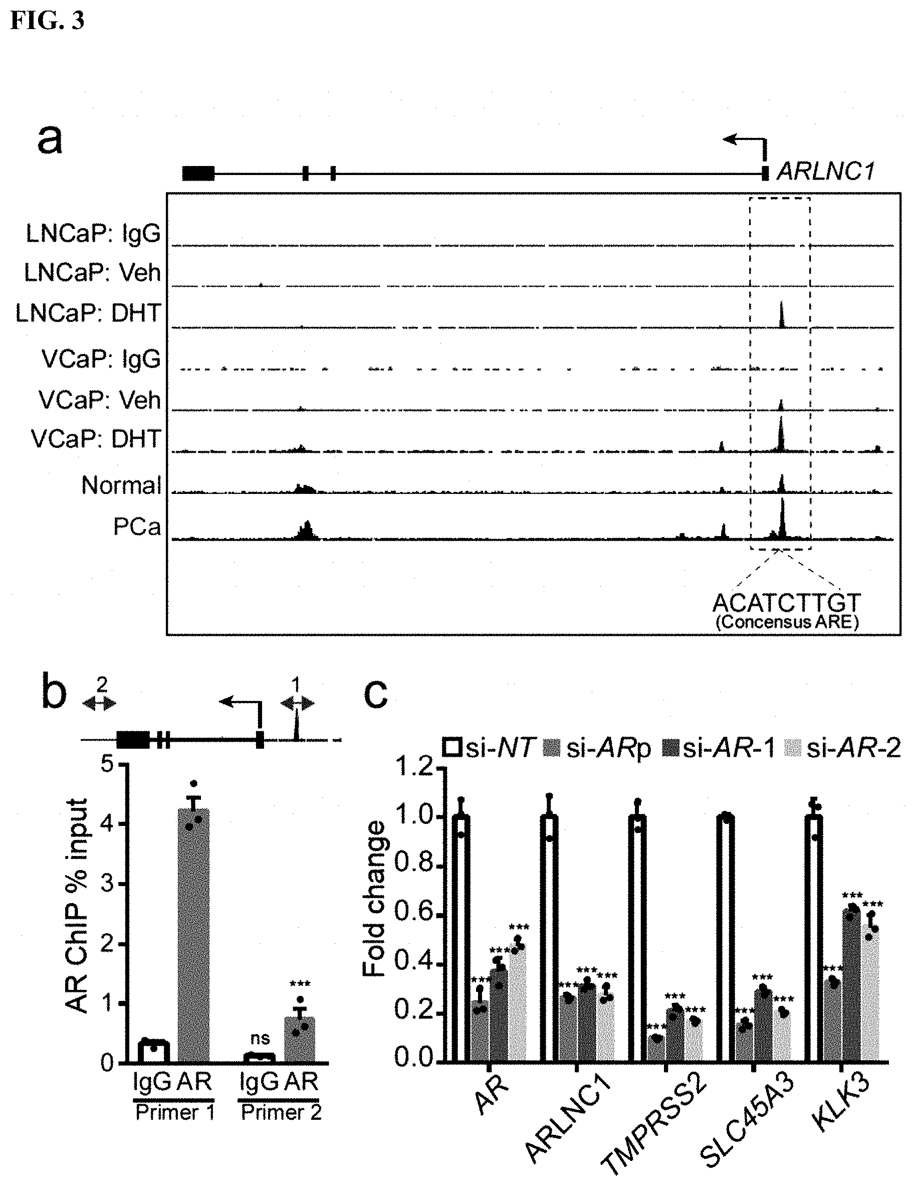
FIG. 3 shows that ARLNC1 is directly regulated by AR. a, AR ChIP-Seq in prostate cancer cell lines and tissues. Top, AR or control (IgG) ChIP-Seq results across the ARLNC1 locus in LNCaP and VCaP cells with vehicle (ethanol) treatment or DHT treatment. Bottom, AR ChIP-Seq in benign prostate and clinically-localized prostate cancer tissue. b, ChIP-qPCR in MDA-PCa-2b cells showing AR or IgG enrichment (ChIP/input) over ARLNC1 promoter region (Primer 1) or control region (Primer 2). Top: schematic of amplicon locations for ChIP-qPCR validation. c, AR and AR target gene (ARLNC1, TMPRSS2, SLC45A3, and KLK3) expression in MDA-PCa-2b cells transfected with control siRNA (si-NT) or siRNAs against AR (si-AR-pool, si-AR-1, si-AR-2).
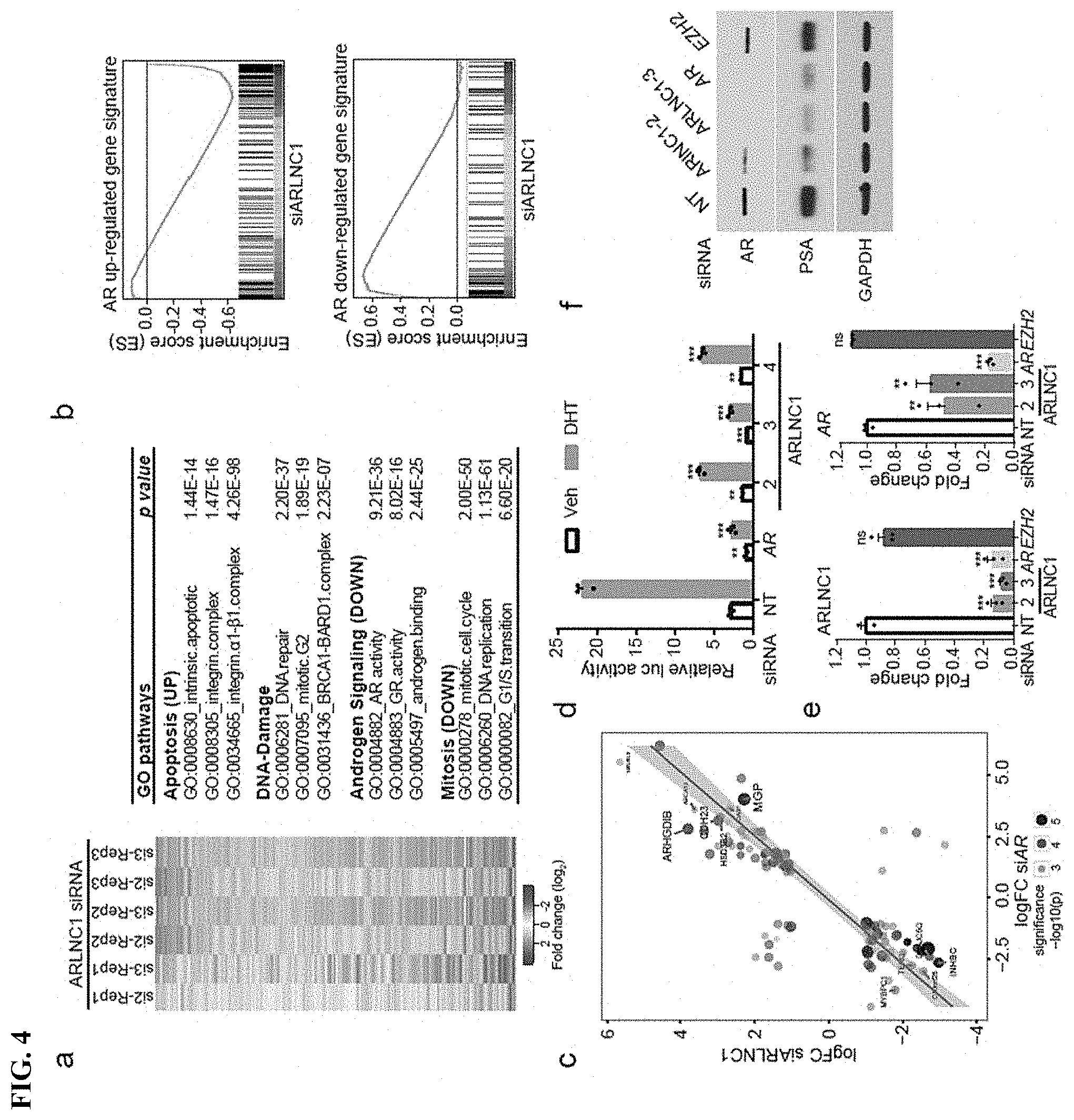
FIG. 4 shows that ARLNC1 loss attenuates AR signaling. a, Gene expression profiling for ARLNC1 knockdown in MDA-PCa-2b cells (n=3 biologically independent cell cultures for each siRNA). b, Gene Set Enrichment Analysis (GSEA) showing significant enrichment of ARLNC1-regulated gene set with respect to the AR target gene sets (n=3 independent gene expression profiles). c, Comparison of ARLNC1-regulated and AR target genes based on RNA-seq following knockdown of AR and ARLNC1. d, siRNA knockdown of ARLNC1 in MDA-PCa-2b cells impairs AR signaling by AR reporter gene assay. e, qRT-PCR analysis of ARLNC1 and AR in MDA-PCa-2b cells transfected with siRNAs against ARLNC1, AR, EZH2, or non-specific control (NT). f, Immunoblot of AR, PSA, and GAPDH in MDA-PCa-2b cells transfected with siRNAs against ARLNC1, AR, EZH2, or non-specific control (NT).
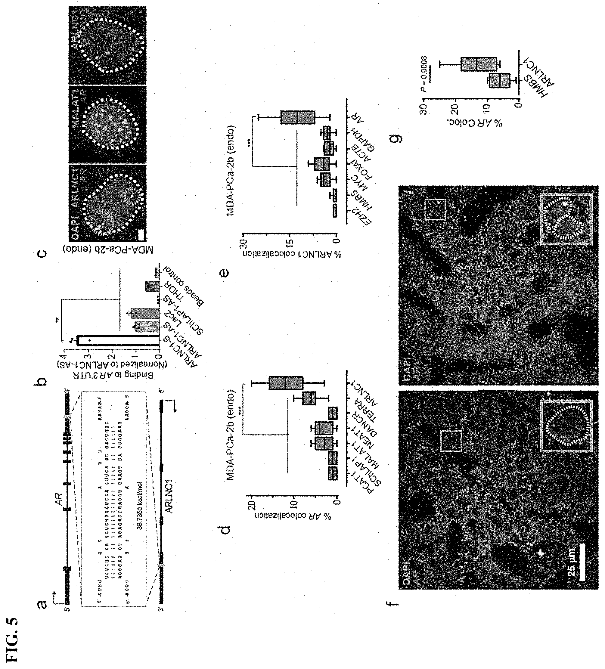
FIG. 5 shows In situ co-localization between AR mRNA and ARLNC1 in prostate cancer cells. a, Schematic of predicted RNA-RNA interaction between ARLNC1 and 3'UTR of AR. b, ARLNC1 interacts with AR 3'UTR in an in vitro RNA-RNA interaction assay. c-e, smFISH depiction of AR-ARLNC1 colocalization in situ. Representative pseudocolored images of MDA-PCa-2b cell nuclei (c) stained for the appropriate endogenous (endo) transcripts and DAPI (nucleus). Scale bar, 5 .mu.m. Quantification of the percentage of AR or ARLNC1 molecules co-localizing with a panel of lncRNAs (d) or mRNAs (e) respectively. Orange circles represent regions of colocalization. Center line and whiskers depict the median and range respectively and box extends from 25th to 75th percentiles (n=50 cells for each sample aggregated from 3 independent experiments). f-g, Representative pseudo-colored images of ARLNC1 positive prostate cancer tissues (f) stained with DAPI (nucleus) and AR, HMBS, or ARLNC1 transcripts (smFISH). Quantification of the percentage of AR molecules (g) colocalizing with HMBS or ARLNC1 is also depicted in box plot.
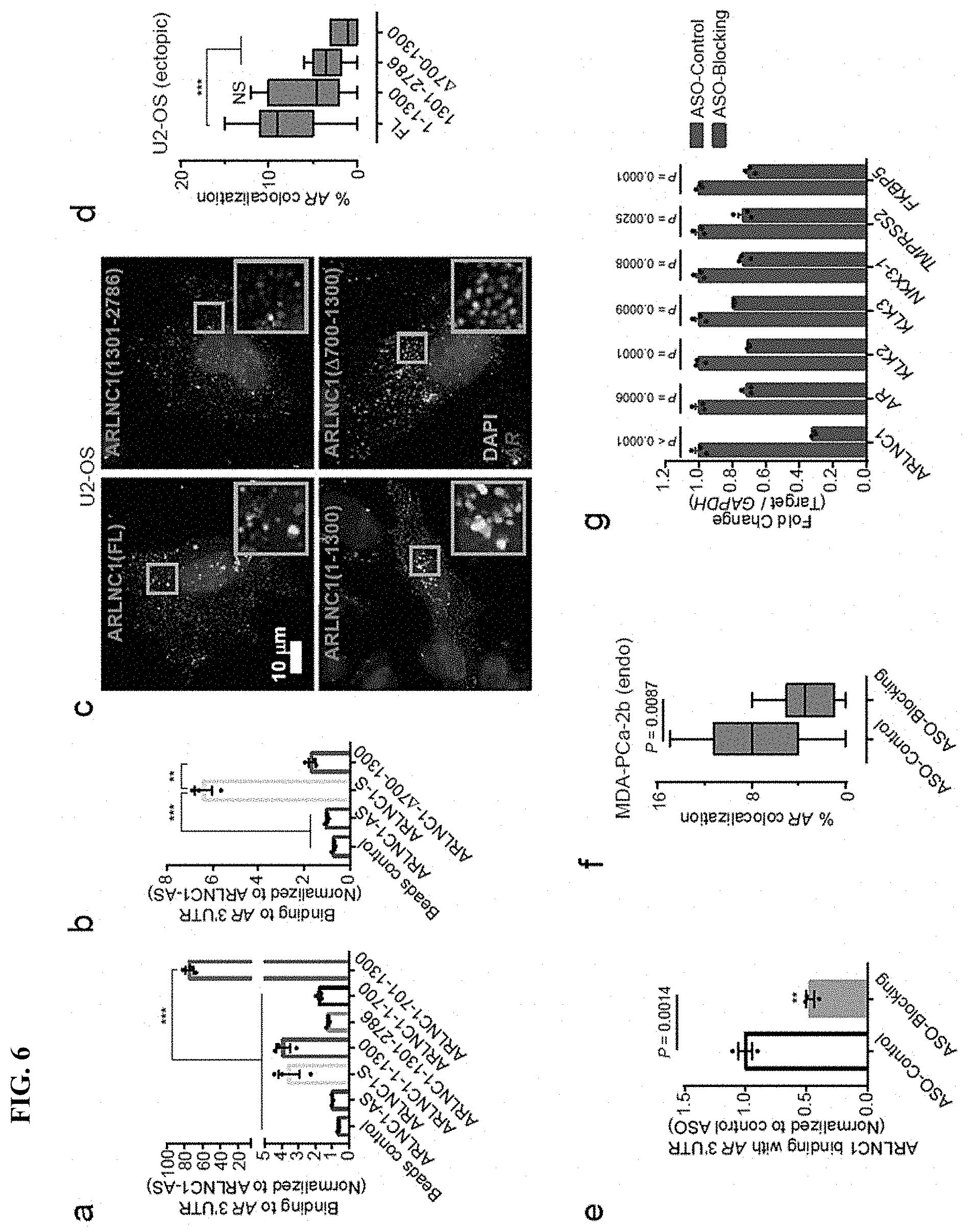
FIG. 6 shows identification of ARLNC1 fragment mediating RNA-RNA interaction with AR mRNA. a, In vitro RNA-RNA interaction assay identifies nucleotides 700-1300 on ARLNC1 as critical binding sites to AR 3'UTR-1-980. b, Deletion of nucleotides 700-1300 on ARLNC1 results in impaired binding to AR 3'UTR, as shown by in vitro RNA-RNA interaction assay. c, Representative pseudo-colored images of U2-OS cells stained for DAPI (nucleus), ARLNC1 and AR transcripts. d, Quantification of the percent of AR molecules colocalizing with various ARLNC1 fragments. e, Antisense oligos targeting sites 700-1300 on ARLNC1 transcript (Blocking ASO pool) inhibit ARLNC1 interaction with AR 3'UTR. f, smFISH shows that ASOs targeting 700-1300 nt on ARLNC1 transcript (ASO-Blocking) inhibit ARLNC1 colocalization with AR in situ.
FIG. 7 shows that ARLNC1 regulates cytoplasmic level of AR transcript. a, ARLNC1 regulates AR post-transcriptionally by specifically affecting cytoplasmic AR mRNA. b, Fractional column plots depicting the nucleo-cytoplasmic distribution of AR mRNA after various treatment conditions in (a), as computed using smFISH.
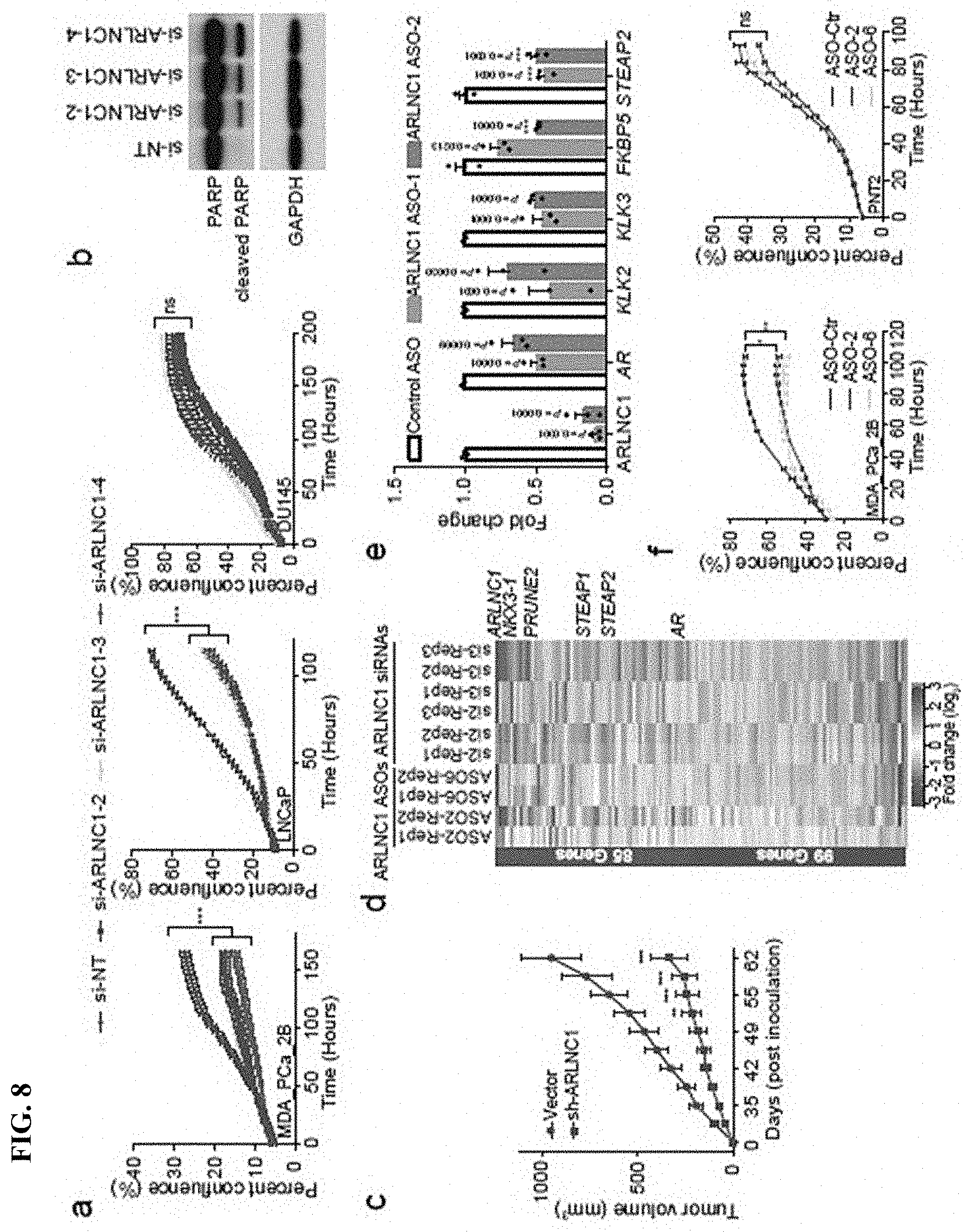
FIG. 8 shows ARLNC1 as a therapeutic target in AR-positive prostate cancer models. a, siRNA knockdown of ARLNC1 in vitro in AR-positive prostate cancer cell lines (MDA-PCa-2b and LNCaP) inhibits cell proliferation. b, ARLNC1 loss leads to increased apoptosis as shown by western blot analysis of PARP and cleaved PARP in LNCaP cells following ARLNC1 knockdown. c, Tumor growth of LNCaP-AR cells expressing shRNA targeting ARLNC1 or shRNA vector. d, Gene expression profiling for siRNA-mediated or ASO-mediated ARLNC1 knockdown in MDA-PCa-2b cells. e, qRT-PCR analysis of ARLNC1, AR, and AR targets (KLK2, KLK3, FKBP5, and STEAP2) in MDA-PCa-2b cells transfected with ASOs against ARLNC1. f, Transfection of ASOs targeting ARLNC1 in AR-positive MDA-PCa-2b cells inhibits cell proliferation. g-h, Effect of ASO treatment on the growth of MDA-PCa-2b xenografts in male NOD-SCID mice, with control ASO (n=15) or ARLNC1 ASO (n=13) treatment subcutaneously at 50 mg/kg, five times per week for three weeks. Tumors were measured by caliper bi-weekly (g) and tumor weights were measured at end point (h). i, Model depicting positive feedback loop between ARLNC1 and AR that is critical for prostate cancer growth.
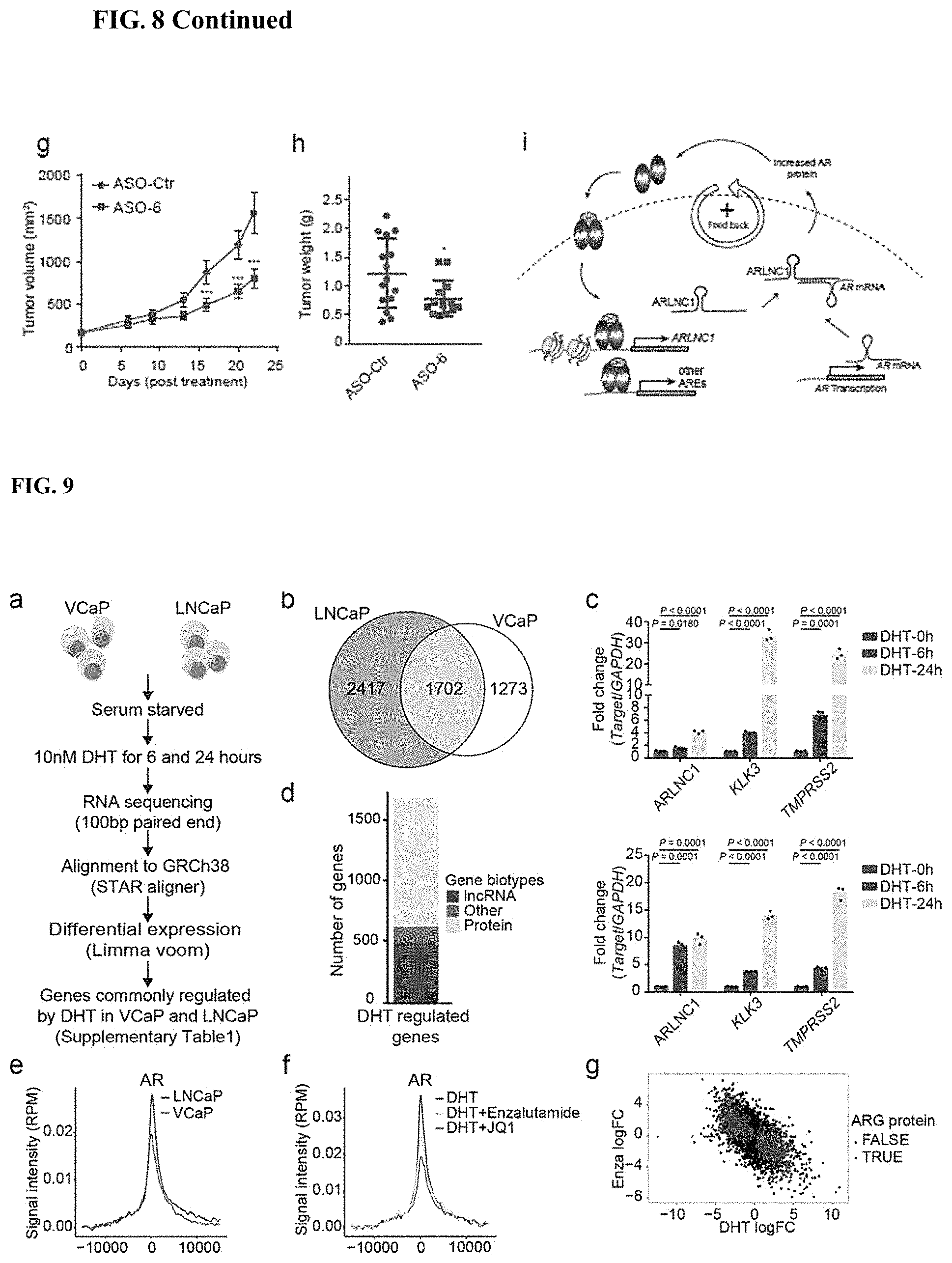
FIG. 9 shows landscape of AR-regulated transcriptome in prostate cancer. (a) A schematic illustration of the procedure used to discover AR-regulated genes (ARGs) in LNCaP and VCaP prostate cancer cell lines. (b) Venn diagram indicating the overlap between AR-regulated genes in LNCaP and VCaP cells. (c) qPCR analysis of ARLNC1 expression and AR signaling gene (KLK3, TMPRSS2) expression in LNCaP cells (top panel) and VCaP cells (bottom panel), following DHT treatment of 6 hours or 24 hours. (d) Bar plot depicting the distribution of gene biotypes (protein, lncRNA, and other) of all overlapped ARGs identified in both LNCaP and VCaP cells. (e) Aggregate ChIP-Seq enrichment profile depicting AR ChIP-Seq signaling density on ARG promoters in LNCaP and VCaP cells. (f) Aggregate ChIP-Seq enrichment profile illustrating AR ChIP-Seq signaling density on ARG promoters in LNCaP cells following DHT stimulation, AR antagonist (enzalutamide) treatment, or BRD4 inhibitor (JQ1) treatment. (g) Transcriptional response to DHT and enzalutamide treatment in VCaP cells, plotting AR regulated protein-coding genes (top panel), or AR regulated lncRNAs (bottom panel). (h) Motif discovery analysis of the top 250 AR ChIP-Seq peaks on AR promoters identifies a binding motif similar to the canonical AR response element. (i) Aggregated ChIPSeq enrichment profiles depicting ChIP-Seq signal density on direct ARG promoters for H3K27ac, H3K4me1, H3K4me3, H3K36me3, Pol II, and BRD4 in LNCaP cells. (j) Pie chart showing prostate cell line or tissue distribution of direct ARGs with AR binding at transcription start sites (TSS) in ChIP-Seq. (k-l) Cumulative distribution plots of distances between transcription start sites (TSS) of genes to nearest AR peak. (k) AR binding near ARGs in benign prostate, prostate cancer tissues (PCa), and prostate cell lines. (l) Comparison of distances between AR binding sites for ARGs and genes not regulated by AR.
FIG. 10 shows that ARLNC1 is prioritized as a lineage-specific, cancer-associated lncRNA in prostate cancer. (a) The top ten AR-regulated, localized prostate cancer-associated genes identified in FIG. 2a, after applying an expression filter of at least four fold change (log 2FC=2) upon DHT stimulation and at least 1 FPKM average expression in prostate cancer tissues. (b) The top ten AR-regulated, metastatic prostate cancer-associated genes identified in FIG. 2b, after applying an expression filter of at least four fold change (log 2FC=2) upon DHT stimulation and at least 1 FPKM average expression in prostate cancer tissues. (c) Schematic illustration of the procedure used to nominate prostate lineage-specific, cancer-associated lncRNAs in prostate cancer. (d) The top twelve prostate tissue-specific, prostate cancer-associated lncRNAs identified in FIG. 2c, after applying an expression filter of at least 10 FPKM in the prostate samples in the top 5% based on gene expression level (n=7,256 samples). (e) Relative expression (FPKM) of ARLNC1 across a panel of normal tissues in GTEx normal tissue RNA-seq cohort (n=9,435 samples) (f) Tissue and cancer-specific expression of ARLNC1 according to MiTranscriptome. (g) Oncomine concepts analysis of genes positively (top panel) or negatively (bottom panel) correlated with ARLNC1.
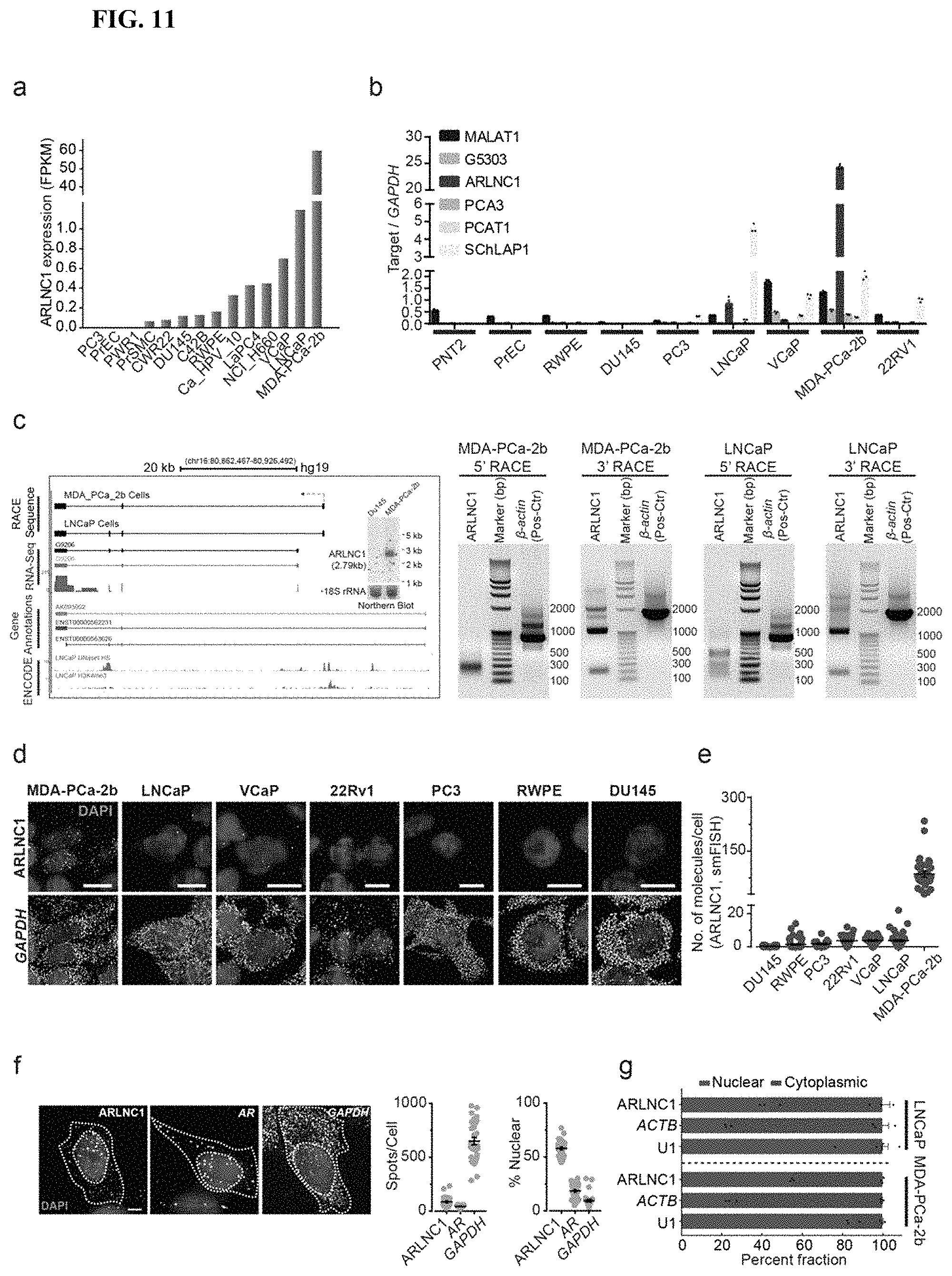
FIG. 11 shows characterization of ARLNC1 and its expression. (a) Relative expression of ARLNC1 (FPKM) across 14 prostate cancer cell lines. (b) qPCR analysis of ARLNC1 expression in nine prostate cancer cell lines. (c) Left: Representative image of ARLNC1 gene structure in MDA-PCa-2b and LNCaP cells, generated from RACE analysis. (d) smFISH images depicting localization of ARLNC1 transcripts in a panel of prostate cancer cell lines. (e) Scatter plot representing the average number of ARLNC1 transcripts per cell in a panel of prostate cancer cell lines, including MDA-PCa-2b, LNCaP, VCaP, 22Rv1, PC3, RWPE, and DU145. (f) Representative gray-scale images of MDA-PCa-2b cells stained for DAPI (nucleus) and ARLNC1, AR or GAPDH transcripts (smFISH). (g) Percentage of nuclear/cytoplasmic RNA levels of ARLNC1, ACTB, and U1, measured by qRT-PCR after subcellular fractionation of MDA-PCa-2b and LNCaP cells.
FIG. 12 shows that ARLNC1 expression is regulated by AR and FOXA1. (a) ChIP-seq peaks of H3K4me1, MED1, BRD4, FOXA1, and NKX3-1 from LNCaP cells at the ARLNC1 promoter region. (b) Top panel: qPCR analysis of ARLNC1 expression in LNCaP cells, following treatment with siRNAs targeting AR, FOXA1, NKX3-1, BRD4, EZH2, LSD1, IRF1, and POU1F1. (c) ChIP-PCR analysis in MDA-PCa-2b cells showing relative enrichment (ChIP/input) of AR, FOXA1, NKX3-1 or IgG over ARLNC1 promoter region or control region. (d) Relative expression (TPM) of AR (Left) and FOXA1 (Right) across a panel of normal tissues in GTEx normal tissue RNA-seq cohort (n=8,745 samples).
FIG. 13 shows a positive feedback loop between ARLNC1 and AR signaling. (a) Reproducibility of expression profiling following 10 nM DHT treatment in MDA-PCa-2b cells. (b) Overlap between genes differentially expressed upon AR knockdown and ARLNC1 knockdown in MDA-PCa-2B cells. (c) siRNA knockdown of ARLNC1 in LNCaP cells impaired AR signaling by AR reporter gene assay. (d) qRT-PCR analysis of KLK2, KLK3, and STEAP2, in MDA-PCa-2b cells transfected with siRNAs against ARLNC1, AR, EZH2, or non-specific control. (e) qPCR analysis of ARLNC1 and AR signaling genes in LNCaP cells (Left panel) and MDA-PCa-2b cells (Right panel) transfected with ARLNC1 expressing vector or control vector.
FIG. 14 shows post-transcriptional regulation of AR by ARLNC1. (a) In silico prediction of ARLNC1 RNA-binding partners, with y-axis representing log 2-absolute RNA binding energy between ARLNC1 and various RNA species, while x-axis depicting log 2-average expression level of these RNAs in prostate cancer. (b) Stoichiometry of ARLNC1:AR colocalization. (c) Representative pseudocolored images of U2-OS cells ectopically expressing ARLNC1 alone (green, left and right panels), or both ARLNC1 and AR (middle panel), and stained for the appropriate transcripts and DAPI. (d-e) Representative pseudo-colored images of MDA-PCa-2b cells or U2-OS cells stained for DAPI (nucleus) and ARLNC1 and AR transcripts, following treatment of blocking ASOs targeting the ARLNC1:AR 3'UTR interaction. Quantification of colocalization in U2-OS cells are depicted in (e) as a box plot, whereas quantifications of colocalization in MDA-PCa-2b cells are in FIG. 6f. (f) qPCR analysis of ARLNC1, AR transcript and AR signaling gene (KLK2, KLK3, NKX3-1, TMPRSS2, FKBP5) expression in LNCaP cells transfected with control ASO or blocking oligos targeting the interaction sites between ARLNC1 and AR 3'UTR. (g) Half-life of GAPDH, AR, ARLNC1, and MYC RNA transcripts in LNCaP cells. (h-i) Quantification of ARLNC1 levels, as measured by smFISH, after treatment of MDA-PCa-2b cells with siRNA against AR (siAR), siRNA against ARLNC1 (siARLNC1-3), ASO against ARLNC1 (ASO-ARLNC1-1) or blocking ASO against AR-ARLNC1 colocalizing segment (ASO-Blocking). Data were normalized to siNT (h) or ASO-Control (i). (j) Nucleo-cytoplasmic distribution of ARLNC1 after appropriate treatment of MDA-PCa-2b cells with siRNA against AR (siAR), siRNA against ARLNC1 (siARLNC1-3), ASO against ARLNC1 (ASO-ARLNC1-1) or blocking ASO against AR-ARLNC1 colocalizing segment (ASO-Blocking). (k-1) Quantification of AR levels, as measured by smFISH, after treatment of MDA-PCa-2b cells with siRNA against AR (si-AR), siRNA against ARLNC1 (si-ARLNC1-3), ASO against ARLNC1 (ASO-ARLNC1-1) or blocking ASO against AR-ARLNC1 colocalizing segment (ASO-Blocking). Data were normalized to siNT (k) or ASO-Control (l). Mean.+-.s.e.m. are shown, n=3 independent experiments and 60 cells analyzed for each sample. (m-n) BrU-seq alignment track (m) and BrUChase-seq alignment track (n) at AR gene locus.
FIG. 15 shows evaluation of the phenotypic effect of ARLNC1 in vitro. (a) Knockdown efficacy of three independent siRNAs targeting ARLNC1 in MDA-PCa-2b cells. (b) ARLNC1 siRNA transfection has no effect on cell proliferation in ARnegative prostate cancer cells, PC3. (c) Increased apoptosis observed in MDA-PCa-2b and LNCaP cells 48 hours after transfected with ARLNC1 siRNAs. (d) Positions of ARLNC1 antisense oligo (ASO)-targeting sites (1 to 6) is indicated on the schematic representation of the ARLNC1 transcript. (e) MDA-PCa-2b cells were transfected with six independent ASOs targeting ARLNC1. (f) Correlation analysis of siRNA-mediated knockdown and ASO-mediated knockdown of ARLNC1 among replicated microarray experiments in MDA-PCa-2b cells (n=2 biological replicates per ASO treatment group and n=3 biological replicates per siRNA treatment group). (g) Free-uptake efficacy of ARLNC1 ASOs was examined in MDA-PCa-2B cells 72 hours post ASO addition to the culture medium (10 .mu.M). (h) Free-uptake treatment of ASOs targeting ARLNC1 resulted in retarded growth of MDA-PCa-2b cells in vitro. (i-j) ARLNC1 ASOs inhibit MDA-PCa-2b cell proliferation in 3D-sphere models.
FIG. 16 shows that knockdown of ARLNC1 by ASOs inhibits tumor growth in vivo. (a) Representative image of in situ hybridization for ARLNC1 in MDA-PCa-2b cell line-derived xenograft. (b) qRT-PCR analysis of ARLNC1, NKX3-1 and AR transcripts in MDA-PCa-2b xenografts treated with control ASO (n=15) or ASO targeting ARLNC1 (n=13). (c) Left: Immunoblots of AR and GAPDH in MDA-PCa-2b xenografts treated with control ASO (n=15) or ASO targeting ARLNC1 (n=13). Right: Relative intensity of the bands was quantified using ImageJ. (d) Left: Immunohistochemistry staining for Ki67 in MDA-PCa-2b xenograft treated with control ASO or ASO against ARLNC1. Right: Summary of Ki67 tumor staining for control (n=15) or ARLNC1 ASO-treated tumors (n=13) shows significant difference in Ki67 staining intensity. (e) Percent change in mice body weight over the time of ASO treatment in MDA-PCa-2b xenografts treated with control ASO (n=15) or ASO targeting ARLNC1 (n=13). (f) ARLNC1 expression levels are not associated with Gleason score. (g) Curated pathway signature analysis between ARLNC1 high (top-quartile) and ARLNC1 low (bottom-quartile) mCRPC samples (n=100). (h) Signatures associated with prostate cancer and luminal differentiation were selected from the MSigDB and contrasted between ARLNC1 high (top-quartile) and ARLNC1 low (bottom-quartile) mCRPC samples (n=100). (i) Cancer hallmark signature analysis between ARLNC1 high expression (top-quartile) and ARLNC1 low expression (bottom-quartile) mCRPC samples (n=100 samples). (j) Tumor content estimated from whole-exome sequencing is compared between ARLNC1 high (top-quartile) and ARLNC1 low (bottom-quartile) expression in mCRPC samples (n=100).
DEFINITIONS
To facilitate an understanding of the present disclosure, a number of terms and phrases are defined below:
As used herein, the term "subject" refers to any animal (e.g., a mammal), including, but not limited to, humans, non-human primates, rodents, and the like, which is to be the recipient of a particular treatment. Typically, the terms "subject" and "patient" are used interchangeably herein in reference to a human subject.
As used herein, the term "subject suspected of having cancer" refers to a subject that presents one or more symptoms indicative of cancer. A subject suspected of having cancer may also have one or more risk factors. A subject suspected of having cancer has generally not been tested for cancer. However, a "subject suspected of having cancer" encompasses an individual who has received a preliminary diagnosis but for whom a confirmatory test has not been done or for whom the level or severity of cancer is not known.
As used herein, the term "subject diagnosed with cancer" refers to a subject who has been tested and found to have cancer. As used herein, the term "initial diagnosis" refers to a test result of initial disease that reveals the presence or absence of disease.
As used herein, the term "non-human animals" refers to all non-human animals including, but not limited to, vertebrates such as rodents, non-human primates, ovines, bovines, ruminants, lagomorphs, porcines, caprines, equines, canines, felines, ayes, etc.
As used herein, the term "cell culture" refers to any in vitro culture of cells. Included within this term are continuous cell lines (e.g., with an immortal phenotype), primary cell cultures, transformed cell lines, finite cell lines (e.g., non-transformed cells), and any other cell population maintained in vitro.
As used herein, the term "eukaryote" refers to organisms distinguishable from "prokaryotes." It is intended that the term encompass all organisms with cells that exhibit the usual characteristics of eukaryotes, such as the presence of a true nucleus bounded by a nuclear membrane, within which lie the chromosomes, the presence of membrane-bound organelles, and other characteristics commonly observed in eukaryotic organisms. Thus, the term includes, but is not limited to such organisms as fungi, protozoa, and animals (e.g., humans).
As used herein, the term "in vitro" refers to an artificial environment and to processes or reactions that occur within an artificial environment. In vitro environments can consist of, but are not limited to, test tubes and cell culture. The term "in vivo" refers to the natural environment (e.g., an animal or a cell) and to processes or reaction that occur within a natural environment.
The terms "test compound" and "candidate compound" refer to any chemical entity, pharmaceutical, drug, and the like that is a candidate for use to treat or prevent a disease, illness, sickness, or disorder of bodily function (e.g., cancer). Test compounds comprise both known and potential therapeutic compounds. A test compound can be determined to be therapeutic by screening using the screening methods of the present disclosure.
As used herein, the term "sample" is used in its broadest sense. In one sense, it is meant to include a specimen or culture obtained from any source, as well as biological and environmental samples. Biological samples may be obtained from animals (including humans) and encompass fluids, solids, tissues, and gases. Biological samples include blood products, such as plasma, serum and the like. Environmental samples include environmental material such as surface matter, soil, water, and industrial samples. Such examples are not however to be construed as limiting the sample types applicable to the present disclosure.
As used herein, the term "effective amount" refers to the amount of a compound (e.g., a compound described herein) sufficient to effect beneficial or desired results. An effective amount can be administered in one or more administrations, applications or dosages and is not limited to or intended to be limited to a particular formulation or administration route.
As used herein, the term "co-administration" refers to the administration of at least two agent(s) (e.g., ARlnc1 inhibitor described herein) or therapies to a subject. In some embodiments, the co-administration of two or more agents/therapies is concurrent. In other embodiments, a first agent/therapy is administered prior to a second agent/therapy. Those of skill in the art understand that the formulations and/or routes of administration of the various agents/therapies used may vary. The appropriate dosage for co-administration can be readily determined by one skilled in the art. In some embodiments, when agents/therapies are co-administered, the respective agents/therapies are administered at lower dosages than appropriate for their administration alone. Thus, co-administration is especially desirable in embodiments where the co-administration of the agents/therapies lowers the requisite dosage of a known potentially harmful (e.g., toxic) agent(s).
As used herein, the term "pharmaceutical composition" refers to the combination of an active agent with a carrier, inert or active, making the composition especially suitable for diagnostic or therapeutic use in vivo, or ex vivo.
As used herein, the term "toxic" refers to any detrimental or harmful effects on a cell or tissue as compared to the same cell or tissue prior to the administration of the toxicant.
"Amelioration" or "ameliorate" or "ameliorating" refers to a lessening of at least one indicator, sign, or symptom of an associated disease, disorder, or condition. The severity of indicators may be determined by subjective or objective measures, which are known to those skilled in the art.
"Antisense activity" means any detectable or measurable activity attributable to the hybridization of an antisense compound to its target nucleic acid. In certain embodiments, antisense activity is a decrease in the amount or expression of a target nucleic acid or protein encoded by such target nucleic acid.
"Antisense compound" means an oligomeric compound that is capable of undergoing hybridization to a target nucleic acid through hydrogen bonding. Examples of antisense compounds include, but are not limited to, single-stranded and double-stranded compounds, such as, antisense oligonucleotides, siRNAs and shRNAs.
"Antisense inhibition" means reduction of target nucleic acid levels or target protein levels in the presence of an antisense compound complementary to a target nucleic acid compared to target nucleic acid levels or target protein levels in the absence of the antisense compound.
"Antisense oligonucleotide" means a single-stranded oligonucleotide having a nucleobase sequence that permits hybridization to a corresponding region or segment of a target nucleic acid.
"Base complementarity" refers to the capacity for the precise base pairing of nucleobases of an antisense oligonucleotide with corresponding nucleobases in a target nucleic acid (i.e., hybridization), and is mediated by Watson-Crick, Hoogsteen or reversed Hoogsteen hydrogen binding between corresponding nucleobases. "Bicyclic sugar moiety" means a modified sugar moiety comprising a 4 to 7 membered ring (including but not limited to a furanosyl) comprising a bridge connecting two atoms of the 4 to 7 membered ring to form a second ring, resulting in a bicyclic structure. In certain embodiments, the 4 to 7 membered ring is a sugar ring. In certain embodiments the 4 to 7 membered ring is a furanosyl. In certain such embodiments, the bridge connects the 2'-carbon and the 4'-carbon of the furanosyl.
"Oligonucleotide" means a polymer of linked nucleosides each of which can be modified or unmodified, independent one from another.
DETAILED DESCRIPTION OF THE DISCLOSURE
Provided herein are compositions and methods for treating cancer. In particular, provided herein are compositions, methods, and uses of inhibitors of ARlnc1 for treating cancer.
Long non-coding RNAs (lncRNAs) are a class of transcripts with diverse and largely uncharacterized biological functions.sup.1-3. Through cross-talk with chromatin, DNA, RNA species, and proteins, lncRNAs function via chromatin remodeling, transcriptional and post-transcriptional regulation.sup.4-9. High-throughput RNA sequencing (RNA-Seq) has enabled the identification of lncRNAs with oncogenic and tumor suppressive roles, including involvement in the pathogenesis of prostate cancer (PCa).sup.7,10-12. Primary PCa is often hormone-dependent and relies on signaling through the androgen receptor (AR); therefore, the majority of patients are responsive to front-line treatment with androgen deprivation therapy (ADT).sup.13-15. However, approximately 20% of cases progress to an incurable stage of the disease known as castration-resistant prostate cancer (CRPC), which still critically relies on AR signaling.sup.16,17, as evidenced by the clinical benefit afforded through the use of enzalutamide.sup.18-21 or abiraterone.sup.22-24. While substantial efforts have been undertaken to identify mechanisms of sustained AR signaling in CRPC (e.g., AR mutations, AR splice variants, and alternative activation pathways).sup.25-31, few studies have investigated the role of AR-regulated lncRNAs. Therefore, described herein is a comprehensive RNA-Seq profiling investigation of AR-regulated, cancer-associated lncRNAs from prostate cancer cell lines and patient tissue samples. During such experiments, ARlnc1 was identified as a target in prostate cancer.
Accordingly, provided herein are compositions and methods for treating cancer by inhibiting the expression and/or function of ARlnc1.
I. Inhibitors
In some embodiments, the ARlnc1 inhibitor is selected from, for example, a nucleic acid (e.g., siRNA, shRNA, miRNA or an antisense nucleic acid), a small molecule, a peptide, or an antibody.
In some embodiments, the ARlnc1 inhibitor is a nucleic acid. Exemplary nucleic acids suitable for inhibiting ARlnc1 (e.g., by preventing expression of ARlnc1) include, but are not limited to, antisense nucleic acids and RNAi. In some embodiments, nucleic acid therapies are complementary to and hybridize to at least a portion (e.g., at least 5, 8, 10, 11, 12, 13, 14, 15, 16, 17, 18, 19, or 20 nucleotides) of ARlnc1.
In some embodiments, compositions comprising oligomeric antisense compounds, particularly oligonucleotides are used to modulate the function of nucleic acid molecules encoding ARlnc1, ultimately modulating the amount of ARlnc1 expressed. This is accomplished by providing antisense compounds that specifically hybridize with one or more nucleic acids encoding ARlnc1. The specific hybridization of an oligomeric compound with its target nucleic acid interferes with the normal function of the nucleic acid. This modulation of function of a target nucleic acid by compounds that specifically hybridize to it is generally referred to as "antisense." The functions of DNA to be interfered with include replication and transcription. The functions of RNA to be interfered with include all vital functions such as, for example, translocation of the RNA to the site of protein translation, translation of protein from the RNA, splicing of the RNA to yield one or more mRNA species, and catalytic activity that may be engaged in or facilitated by the RNA. The overall effect of such interference with target nucleic acid function is decreasing the amount of ARlnc1 proteins in the cell.
In certain embodiments, antisense compounds have chemically modified subunits arranged in patterns, or motifs, to confer to the antisense compounds properties such as enhanced inhibitory activity, increased binding affinity for a target nucleic acid, or resistance to degradation by in vivo nucleases. Chimeric antisense compounds typically contain at least one region modified so as to confer increased resistance to nuclease degradation, increased cellular uptake, increased binding affinity for the target nucleic acid, and/or increased inhibitory activity. A second region of a chimeric antisense compound may confer another desired property e.g., serve as a substrate for the cellular endonuclease RNase H, which cleaves the RNA strand of an RNA:DNA duplex.
Antisense activity may result from any mechanism involving the hybridization of the antisense compound (e.g., oligonucleotide) with a target nucleic acid, wherein the hybridization ultimately results in a biological effect. In certain embodiments, the amount and/or activity of the target nucleic acid is modulated. In certain embodiments, the amount and/or activity of the target nucleic acid is reduced. In certain embodiments, hybridization of the antisense compound to the target nucleic acid ultimately results in target nucleic acid degradation. In certain embodiments, hybridization of the antisense compound to the target nucleic acid does not result in target nucleic acid degradation. In certain such embodiments, the presence of the antisense compound hybridized with the target nucleic acid (occupancy) results in a modulation of antisense activity. In certain embodiments, antisense compounds having a particular chemical motif or pattern of chemical modifications are particularly suited to exploit one or more mechanisms. In certain embodiments, antisense compounds function through more than one mechanism and/or through mechanisms that have not been elucidated. Accordingly, the antisense compounds described herein are not limited by particular mechanism.
Antisense mechanisms include, without limitation, RNase H mediated antisense; RNAi mechanisms, which utilize the R.sub.1SC pathway and include, without limitation, siRNA, ssRNA and microRNA mechanisms; and occupancy based mechanisms. Certain antisense compounds may act through more than one such mechanism and/or through additional mechanisms.
In certain embodiments, antisense activity results at least in part from degradation of target RNA by RNase H. RNase H is a cellular endonuclease that cleaves the RNA strand of an RNA:DNA duplex. It is known in the art that single-stranded antisense compounds which are "DNA-like" elicit RNase H activity in mammalian cells. Accordingly, antisense compounds comprising at least a portion of DNA or DNA-like nucleosides may activate RNase H, resulting in cleavage of the target nucleic acid. In certain embodiments, antisense compounds that utilize RNase H comprise one or more modified nucleosides. In certain embodiments, such antisense compounds comprise at least one block of 1-8 modified nucleosides. In certain such embodiments, the modified nucleosides do not support RNase H activity. In certain embodiments, such antisense compounds are gapmers, as described herein. In certain such embodiments, the gap of the gapmer comprises DNA nucleosides. In certain such embodiments, the gap of the gapmer comprises DNA-like nucleosides. In certain such embodiments, the gap of the gapmer comprises DNA nucleosides and DNA-like nucleosides.
Certain antisense compounds having a gapmer motif are considered chimeric antisense compounds. In a gapmer an internal region having a plurality of nucleotides that supports RNaseH cleavage is positioned between external regions having a plurality of nucleotides that are chemically distinct from the nucleosides of the internal region. In the case of an antisense oligonucleotide having a gapmer motif, the gap segment generally serves as the substrate for endonuclease cleavage, while the wing segments comprise modified nucleosides. In certain embodiments, the regions of a gapmer are differentiated by the types of sugar moieties comprising each distinct region. The types of sugar moieties that are used to differentiate the regions of a gapmer may in some embodiments include .beta.-D-ribonucleosides, .beta.-D-deoxyribonucleosides, 2'-modified nucleosides (such 2'-modified nucleosides may include 2'-MOE and 2'-O--CH.sub.3, among others), and bicyclic sugar modified nucleosides (such bicyclic sugar modified nucleosides may include those having a constrained ethyl). In certain embodiments, nucleosides in the wings may include several modified sugar moieties, including, for example 2'-MOE and bicyclic sugar moieties such as constrained ethyl or LNA. In certain embodiments, wings may include several modified and unmodified sugar moieties. In certain embodiments, wings may include various combinations of 2'-MOE nucleosides, bicyclic sugar moieties such as constrained ethyl nucleosides or LNA nucleosides, and 2'-deoxynucleosides.
Each distinct region may comprise uniform sugar moieties, variant, or alternating sugar moieties. The wing-gap-wing motif is frequently described as "X--Y-Z", where "X" represents the length of the 5'-wing, "Y" represents the length of the gap, and "Z" represents the length of the 3'-wing. "X" and "Z" may comprise uniform, variant, or alternating sugar moieties. In certain embodiments, "X" and "Y" may include one or more 2'-deoxynucleosides. "Y" may comprise 2'-deoxynucleosides. As used herein, a gapmer described as "X--Y-Z" has a configuration such that the gap is positioned immediately adjacent to each of the 5'-wing and the 3' wing. Thus, no intervening nucleotides exist between the 5'-wing and gap, or the gap and the 3'-wing. Any of the antisense compounds described herein can have a gapmer motif. In certain embodiments, "X" and "Z" are the same; in other embodiments they are different. In certain embodiments, "Y" is between 8 and 15 nucleosides. X, Y, or Z can be any of 1, 2, 3, 4, 5, 6, 7, 8, 9, 10, 11, 12, 13, 14, 15, 16, 17, 18, 19, 20, 25, 30 or more nucleosides.
In certain embodiments, the antisense compound has a gapmer motif in which the gap consists of 6, 7, 8, 9, 10, 11, 12, 13, 14, 15, or 16 linked nucleosides.
In certain embodiments, antisense compounds including those particularly suited for use as single-stranded RNAi compounds (ssRNA) comprise a modified 5'-terminal end. In certain such embodiments, the 5'-terminal end comprises a modified phosphate moiety. In certain embodiments, such modified phosphate is stabilized (e.g., resistant to degradation/cleavage compared to unmodified 5'-phosphate). In certain embodiments, such 5'-terminal nucleosides stabilize the 5'-phosphorous moiety. Certain modified 5'-terminal nucleosides may be found in the art, for example in WO/2011/139702.
In certain embodiments, antisense compounds, including those particularly suitable for ssRNA comprise one or more type of modified sugar moieties and/or naturally occurring sugar moieties arranged along an oligonucleotide or region thereof in a defined pattern or sugar modification motif. Such motifs may include any of the sugar modifications discussed herein and/or other known sugar modifications.
In certain embodiments, the oligonucleotides comprise or consist of a region having uniform sugar modifications. In certain such embodiments, each nucleoside of the region comprises the same RNA-like sugar modification. In certain embodiments, each nucleoside of the region is a 2'-F nucleoside. In certain embodiments, each nucleoside of the region is a 2'-OMe nucleoside. In certain embodiments, each nucleoside of the region is a 2'-MOE nucleoside. In certain embodiments, each nucleoside of the region is a cEt nucleoside. In certain embodiments, each nucleoside of the region is an LNA nucleoside. In certain embodiments, the uniform region constitutes all or essentially all of the oligonucleotide. In certain embodiments, the region constitutes the entire oligonucleotide except for 1-4 terminal nucleosides.
In certain embodiments, oligonucleotides comprise one or more regions of alternating sugar modifications, wherein the nucleosides alternate between nucleotides having a sugar modification of a first type and nucleotides having a sugar modification of a second type. In certain embodiments, nucleosides of both types are RNA-like nucleosides. In certain embodiments the alternating nucleosides are selected from: 2'-OMe, 2'-F, 2'-MOE, LNA, and cEt. In certain embodiments, the alternating modifications are 2'-F and 2'-OMe. Such regions may be contiguous or may be interrupted by differently modified nucleosides or conjugated nucleosides.
In certain embodiments, the alternating region of alternating modifications each consist of a single nucleoside (i.e., the pattern is (AB).sub.xA.sub.y wherein A is a nucleoside having a sugar modification of a first type and B is a nucleoside having a sugar modification of a second type; x is 1-20 and y is 0 or 1). In certain embodiments, one or more alternating regions in an alternating motif includes more than a single nucleoside of a type.
In certain embodiments, oligonucleotides having such an alternating motif also comprise a modified 5' terminal nucleoside, such as those of formula IIc or IIe.
In certain embodiments, antisense compounds, including those particularly suited for use as ssRNA comprise modified internucleoside linkages arranged along the oligonucleotide or region thereof in a defined pattern or modified internucleoside linkage motif. In certain embodiments, oligonucleotides comprise a region having an alternating internucleoside linkage motif. In certain embodiments, oligonucleotides comprise a region of uniformly modified internucleoside linkages. In certain such embodiments, the oligonucleotide comprises a region that is uniformly linked by phosphorothioate internucleoside linkages. In certain embodiments, the oligonucleotide is uniformly linked by phosphorothioate internucleoside linkages. In certain embodiments, each internucleoside linkage of the oligonucleotide is selected from phosphodiester and phosphorothioate. In certain embodiments, each internucleoside linkage of the oligonucleotide is selected from phosphodiester and phosphorothioate and at least one internucleoside linkage is phosphorothioate.
In certain embodiments, the oligonucleotide comprises at least 6 phosphorothioate internucleoside linkages. In certain embodiments, the oligonucleotide comprises at least 8 phosphorothioate internucleoside linkages. In certain embodiments, the oligonucleotide comprises at least 10 phosphorothioate internucleoside linkages. In certain embodiments, the oligonucleotide comprises at least one block of at least 6 consecutive phosphorothioate internucleoside linkages. In certain embodiments, the oligonucleotide comprises at least one block of at least 8 consecutive phosphorothioate internucleoside linkages. In certain embodiments, the oligonucleotide comprises at least one block of at least 10 consecutive phosphorothioate internucleoside linkages. In certain embodiments, the oligonucleotide comprises at least one block of at least one 12 consecutive phosphorothioate internucleoside linkages. In certain such embodiments, at least one such block is located at the 3' end of the oligonucleotide. In certain such embodiments, at least one such block is located within 3 nucleosides of the 3' end of the oligonucleotide.
Additional modifications are described, for example, in U.S. Pat. No. 9,796,976, herein incorporated by reference in its entirety.
In some embodiments, nucleic acids are RNAi nucleic acids. "RNA interference (RNAi)" is the process of sequence-specific, post-transcriptional gene silencing initiated by a small interfering RNA (siRNA), shRNA, or microRNA (miRNA). During RNAi, the RNA induces degradation of target mRNA with consequent sequence-specific inhibition of gene expression.
In "RNA interference," or "RNAi," a "small interfering RNA" or "short interfering RNA" or "siRNA" or "short hairpin RNA" or "shRNA" molecule, or "miRNA" an RNAi (e.g., single strand, duplex, or hairpin) of nucleotides is targeted to a nucleic acid sequence of interest, for example, ARlnc1.
An "RNA duplex" refers to the structure formed by the complementary pairing between two regions of a RNA molecule. The RNA using in RNAi is "targeted" to a gene in that the nucleotide sequence of the duplex portion of the RNAi is complementary to a nucleotide sequence of the targeted gene. In certain embodiments, the RNAi is are targeted to the sequence encoding ARlnc1. In some embodiments, the length of the RNAi is less than 30 base pairs. In some embodiments, the RNA can be 32, 31, 30, 29, 28, 27, 26, 25, 24, 23, 22, 21, 20, 19, 18, 17, 16, 15, 14, 13, 12, 11 or 10 base pairs in length. In some embodiments, the length of the RNAi is 19 to 32 base pairs in length. In certain embodiment, the length of the RNAi is 19 or 21 base pairs in length.
In some embodiments, RNAi comprises a hairpin structure (e.g., shRNA). In addition to the duplex portion, the hairpin structure may contain a loop portion positioned between the two sequences that form the duplex. The loop can vary in length. In some embodiments the loop is 5, 6, 7, 8, 9, 10, 11, 12, 13, 14, 15, 16, 17, 18, 19, 20, 21, 22, 23, 24, 25, 26 or 27 nucleotides in length. In certain embodiments, the loop is 18 nucleotides in length. The hairpin structure can also contain 3' and/or 5' overhang portions. In some embodiments, the overhang is a 3' and/or a 5' overhang 0, 1, 2, 3, 4 or 5 nucleotides in length.
"miRNA" or "miR" means a non-coding RNA between 18 and 25 nucleobases in length which hybridizes to and regulates the expression of a coding RNA. In certain embodiments, a miRNA is the product of cleavage of a pre-miRNA by the enzyme Dicer. Examples of miRNAs are found in the miRNA database known as miRBase.
As used herein, Dicer-substrate RNAs (DsiRNAs) are chemically synthesized asymmetric 25-mer/27-mer duplex RNAs that have increased potency in RNA interference compared to traditional RNAi. Traditional 21-mer RNAi molecules are designed to mimic Dicer products and therefore bypass interaction with the enzyme Dicer. Dicer has been recently shown to be a component of RISC and involved with entry of the RNAi into RISC. Dicer-substrate RNAi molecules are designed to be optimally processed by Dicer and show increased potency by engaging this natural processing pathway. Using this approach, sustained knockdown has been regularly achieved using sub-nanomolar concentrations. (U.S. Pat. No. 8,084,599; Kim et al., Nature Biotechnology 23:222 2005; Rose et al., Nucleic Acids Res., 33:4140 2005).
The transcriptional unit of a "shRNA" is comprised of sense and antisense sequences connected by a loop of unpaired nucleotides. shRNAs are exported from the nucleus by Exportin-5, and once in the cytoplasm, are processed by Dicer to generate functional RNAi molecules. "miRNAs" stem-loops are comprised of sense and antisense sequences connected by a loop of unpaired nucleotides typically expressed as part of larger primary transcripts (pri-miRNAs), which are excised by the Drosha-DGCR8 complex generating intermediates known as pre-miRNAs, which are subsequently exported from the nucleus by Exportin-5, and once in the cytoplasm, are processed by Dicer to generate functional miRNAs or siRNAs.
"Artificial miRNA" or an "artificial miRNA shuttle vector", as used herein interchangeably, refers to a primary miRNA transcript that has had a region of the duplex stem loop (at least about 9-20 nucleotides) which is excised via Drosha and Dicer processing replaced with the siRNA sequences for the target gene while retaining the structural elements within the stem loop necessary for effective Drosha processing. The term "artificial" arises from the fact the flanking sequences (e.g., about 35 nucleotides upstream and about 40 nucleotides downstream) arise from restriction enzyme sites within the multiple cloning site of the RNAi. As used herein the term "miRNA" encompasses both the naturally occurring miRNA sequences as well as artificially generated miRNA shuttle vectors.
The RNAi can be encoded by a nucleic acid sequence, and the nucleic acid sequence can also include a promoter. The nucleic acid sequence can also include a polyadenylation signal. In some embodiments, the polyadenylation signal is a synthetic minimal polyad n certain embodiments, provided herein are compounds comprising a modified oligonucleotide consisting of 12 to 30 linked nucleosides and comprising a nucleobase sequence comprising a portion of at least 8, at least 10, at least 12, at least 14, at least 15, at least 16, at least 17, at least 18, at least 19, or at least 20 contiguous nucleobases complementary to an equal length portion of ARlnc1.
In some embodiments, hybridization occurs between an antisense compound disclosed herein and an ARlnc1 nucleic acid. The most common mechanism of hybridization involves hydrogen bonding (e.g., Watson-Crick, Hoogsteen or reversed Hoogsteen hydrogen bonding) between complementary nucleobases of the nucleic acid molecules.
Hybridization can occur under varying conditions. Stringent conditions are sequence-dependent and are determined by the nature and composition of the nucleic acid molecules to be hybridized.
An antisense compound and a target nucleic acid are complementary to each other when a sufficient number of nucleobases of the antisense compound can hydrogen bond with the corresponding nucleobases of the target nucleic acid, such that a desired effect will occur (e.g., antisense inhibition of a target nucleic acid, such as an ARlnc1 nucleic acid).
Non-complementary nucleobases between an antisense compound and an ARlnc1 nucleic acid may be tolerated provided that the antisense compound remains able to specifically hybridize to a target nucleic acid. Moreover, an antisense compound may hybridize over one or more segments of an ARLNC1 nucleic acid such that intervening or adjacent segments are not involved in the hybridization event (e.g., a loop structure, mismatch or hairpin structure).
In certain embodiments, the antisense compounds provided herein, or a specified portion thereof, are, or are at least, 70%, 80%, 85%, 86%, 87%, 88%, 89%, 90%, 91%, 92%, 93%, 94%, 95%, 96%, 97%, 98%, 99%, or 100% complementary to an ARlnc1 nucleic acid, a target region, target segment, or specified portion thereof. Percent complementarity of an antisense compound with a target nucleic acid can be determined using routine methods.
For example, an antisense compound in which 18 of 20 nucleobases of the antisense compound are complementary to a target region, and would therefore specifically hybridize, would represent 90 percent complementarity. In this example, the remaining noncomplementary nucleobases may be clustered or interspersed with complementary nucleobases and need not be contiguous to each other or to complementary nucleobases. As such, an antisense compound which is 18 nucleobases in length having 4 (four) noncomplementary nucleobases which are flanked by two regions of complete complementarity with the target nucleic acid would have 77.8% overall complementarity with the target nucleic acid and would thus fall within the scope of the present invention. Percent complementarity of an antisense compound with a region of a target nucleic acid can be determined routinely using BLAST programs (basic local alignment search tools) and PowerBLAST programs known in the art (Altschul et al., J. Mol. Biol., 1990, 215, 403 410; Zhang and Madden, Genome Res., 1997, 7, 649 656). Percent homology, sequence identity or complementarity, can be determined by, for example, the Gap program (Wisconsin Sequence Analysis Package, Version 8 for Unix, Genetics Computer Group, University Research Park, Madison Wis.), using default settings, which uses the algorithm of Smith and Waterman (Adv. Appl. Math., 1981, 2, 482 489).
In certain embodiments, the antisense compounds provided herein, or specified portions thereof, are fully complementary (i.e., 100% complementary) to a target nucleic acid, or specified portion thereof. For example, an antisense compound may be fully complementary to an Alnc1 nucleic acid, or a target region, or a target segment or target sequence thereof. As used herein, "fully complementary" means each nucleobase of an antisense compound is capable of precise base pairing with the corresponding nucleobases of a target nucleic acid. For example, a 20 nucleobase antisense compound is fully complementary to a target sequence that is 400 nucleobases long, so long as there is a corresponding 20 nucleobase portion of the target nucleic acid that is fully complementary to the antisense compound. Fully complementary can also be used in reference to a specified portion of the first and/or the second nucleic acid. For example, a 20 nucleobase portion of a 30 nucleobase antisense compound can be "fully complementary" to a target sequence that is 400 nucleobases long. The 20 nucleobase portion of the 30 nucleobase oligonucleotide is fully complementary to the target sequence if the target sequence has a corresponding 20 nucleobase portion wherein each nucleobase is complementary to the 20 nucleobase portion of the antisense compound. At the same time, the entire 30 nucleobase antisense compound may or may not be fully complementary to the target sequence, depending on whether the remaining 10 nucleobases of the antisense compound are also complementary to the target sequence.
The location of a non-complementary nucleobase may be at the 5' end or 3' end of the antisense compound. Alternatively, the non-complementary nucleobase or nucleobases may be at an internal position of the antisense compound. When two or more non-complementary nucleobases are present, they may be contiguous (i.e., linked) or non-contiguous. In one embodiment, a non-complementary nucleobase is located in the wing segment of a gapmer antisense oligonucleotide.
In certain embodiments, antisense compounds that are, or are up to 12, 13, 14, 15, 16, 17, 18, 19, or 20 nucleobases in length comprise no more than 4, no more than 3, no more than 2, or no more than 1 non-complementary nucleobase(s) relative to a target nucleic acid, such as an ARlnc1 nucleic acid, or specified portion thereof.
In certain embodiments, antisense compounds that are, or are up to 12, 13, 14, 15, 16, 17, 18, 19, 20, 21, 22, 23, 24, 25, 26, 27, 28, 29, or 30 nucleobases in length comprise no more than 6, no more than 5, no more than 4, no more than 3, no more than 2, or no more than 1 non-complementary nucleobase(s) relative to a target nucleic acid, such as an Alnc1 nucleic acid, or specified portion thereof.
The antisense compounds provided herein also include those which are complementary to a portion of a target nucleic acid. As used herein, "portion" refers to a defined number of contiguous (i.e. linked) nucleobases within a region or segment of a target nucleic acid. A "portion" can also refer to a defined number of contiguous nucleobases of an antisense compound. In certain embodiments, the antisense compounds, are complementary to at least an 8 nucleobase portion of a target segment. In certain embodiments, the antisense compounds are complementary to at least a 12 nucleobase portion of a target segment. In certain embodiments, the antisense compounds are complementary to at least a 15 nucleobase portion of a target segment. In certain embodiments, the antisense compounds are complementary to at least an 18 nucleobase portion of a target segment. Also contemplated are antisense compounds that are complementary to at least a 9, 10, 11, 12, 13, 14, 15, 16, 17, 18, 19, 20, or more nucleobase portion of a target segment, or a range defined by any two of these values.
The present disclosure contemplates the use of any genetic manipulation for use in modulating the expression of ARlnc1. Examples of genetic manipulation include, but are not limited to, gene knockout (e.g., removing the ARlnc1 gene from the chromosome using, for example, recombination), expression of antisense constructs with or without inducible promoters, and the like. Delivery of nucleic acid construct to cells in vitro or in vivo may be conducted using any suitable method. A suitable method is one that introduces the nucleic acid construct into the cell such that the desired event occurs (e.g., expression of an antisense construct).
Introduction of molecules carrying genetic information into cells is achieved by any of various methods including, but not limited to, directed injection of naked DNA constructs, bombardment with gold particles loaded with said constructs, and macromolecule mediated gene transfer using, for example, liposomes, biopolymers, and the like. Exemplary methods use gene delivery vehicles derived from viruses, including, but not limited to, adenoviruses, retroviruses, vaccinia viruses, and adeno-associated viruses. Because of the higher efficiency as compared to retroviruses, vectors derived from adenoviruses are the preferred gene delivery vehicles for transferring nucleic acid molecules into host cells in vivo. Adenoviral vectors have been shown to provide very efficient in vivo gene transfer into a variety of solid tumors in animal models and into human solid tumor xenografts in immune-deficient mice.
Examples of adenoviral vectors and methods for gene transfer are described in PCT publications WO 00/12738 and WO 00/09675 and U.S. Pat. Nos. 6,033,908, 6,019,978, 6,001,557, 5,994,132, 5,994,128, 5,994,106, 5,981,225, 5,885,808, 5,872,154, 5,830,730, and 5,824,544, each of which is herein incorporated by reference in its entirety. Vectors may be administered to subject in a variety of ways. For example, in some embodiments of the present disclosure, vectors are administered into tumors or tissue associated with tumors using direct injection. In other embodiments, administration is via the blood or lymphatic circulation (See e.g., PCT publication 1999/02685 herein incorporated by reference in its entirety). Exemplary dose levels of adenoviral vector are preferably 10.sup.8 to 10.sup.11 vector particles added to the perfusate.
In some embodiments, CRISPR/Cas9 systems are used to delete or knock out genes. Clustered regularly interspaced short palindromic repeats (CRISPR) are segments of prokaryotic DNA containing short, repetitive base sequences. These play a key role in a bacterial defence system, and form the basis of a genome editing technology known as CRISPR/Cas9 that allows permanent modification of genes within organisms.
In some embodiments, the present disclosure provides antibodies that inhibit ARlnc1. Any suitable antibody (e.g., monoclonal, polyclonal, or synthetic) may be utilized in the therapeutic methods disclosed herein. In some embodiments, the antibodies are humanized antibodies. Methods for humanizing antibodies are well known in the art (See e.g., U.S. Pat. Nos. 6,180,370, 5,585,089, 6,054,297, and 5,565,332; each of which is herein incorporated by reference).
In some embodiments, candidate ARlnc1 inhibitors are screened for activity (e.g., using the methods described herein or another suitable assay).
The present disclosure further provides pharmaceutical compositions (e.g., comprising the compounds described above). The pharmaceutical compositions of the present disclosure may be administered in a number of ways depending upon whether local or systemic treatment is desired and upon the area to be treated. Administration may be topical (including ophthalmic and to mucous membranes including vaginal and rectal delivery), pulmonary (e.g., by inhalation or insufflation of powders or aerosols, including by nebulizer; intratracheal, intranasal, epidermal and transdermal), oral or parenteral. Parenteral administration includes intravenous, intraarterial, subcutaneous, intraperitoneal or intramuscular injection or infusion; or intracranial, e.g., intrathecal or intraventricular, administration.
Pharmaceutical compositions and formulations for topical administration may include transdermal patches, ointments, lotions, creams, gels, drops, suppositories, sprays, liquids and powders. Conventional pharmaceutical carriers, aqueous, powder or oily bases, thickeners and the like may be necessary or desirable.
Compositions and formulations for oral administration include powders or granules, suspensions or solutions in water or non-aqueous media, capsules, sachets or tablets. Thickeners, flavoring agents, diluents, emulsifiers, dispersing aids or binders may be desirable.
Compositions and formulations for parenteral, intrathecal or intraventricular administration may include sterile aqueous solutions that may also contain buffers, diluents and other suitable additives such as, but not limited to, penetration enhancers, carrier compounds and other pharmaceutically acceptable carriers or excipients.
Pharmaceutical compositions of the present disclosure include, but are not limited to, solutions, emulsions, and liposome-containing formulations. These compositions may be generated from a variety of components that include, but are not limited to, preformed liquids, self-emulsifying solids and self-emulsifying semisolids.
The pharmaceutical formulations of the present disclosure, which may conveniently be presented in unit dosage form, may be prepared according to conventional techniques well known in the pharmaceutical industry. Such techniques include the step of bringing into association the active ingredients with the pharmaceutical carrier(s) or excipient(s). In general the formulations are prepared by uniformly and intimately bringing into association the active ingredients with liquid carriers or finely divided solid carriers or both, and then, if necessary, shaping the product.
The compositions of the present disclosure may be formulated into any of many possible dosage forms such as, but not limited to, tablets, capsules, liquid syrups, soft gels, suppositories, and enemas. The compositions of the present disclosure may also be formulated as suspensions in aqueous, non-aqueous or mixed media. Aqueous suspensions may further contain substances that increase the viscosity of the suspension including, for example, sodium carboxymethylcellulose, sorbitol and/or dextran. The suspension may also contain stabilizers.
Agents that enhance uptake of oligonucleotides at the cellular level may also be added to the pharmaceutical and other compositions of the present disclosure. For example, cationic lipids, such as lipofectin (U.S. Pat. No. 5,705,188), cationic glycerol derivatives, and polycationic molecules, such as polylysine (WO 97/30731), also enhance the cellular uptake of oligonucleotides.
The compositions of the present disclosure may additionally contain other adjunct components conventionally found in pharmaceutical compositions. Thus, for example, the compositions may contain additional, compatible, pharmaceutically-active materials such as, for example, antipruritics, astringents, local anesthetics or anti-inflammatory agents, or may contain additional materials useful in physically formulating various dosage forms of the compositions of the present disclosure, such as dyes, flavoring agents, preservatives, antioxidants, opacifiers, thickening agents and stabilizers. However, such materials, when added, should not unduly interfere with the biological activities of the components of the compositions of the present disclosure. The formulations can be sterilized and, if desired, mixed with auxiliary agents, e.g., lubricants, preservatives, stabilizers, wetting agents, emulsifiers, salts for influencing osmotic pressure, buffers, colorings, flavorings and/or aromatic substances and the like which do not deleteriously interact with the nucleic acid(s) of the formulation.
Dosing is dependent on severity and responsiveness of the disease state to be treated, with the course of treatment lasting from several days to several months, or until a cure is effected or a diminution of the disease state is achieved. Optimal dosing schedules can be calculated from measurements of drug accumulation in the body of the patient. The administering physician can easily determine optimum dosages, dosing methodologies and repetition rates. Optimum dosages may vary depending on the relative potency of individual oligonucleotides, and can generally be estimated based on EC50s found to be effective in in vitro and in vivo animal models or based on the examples described herein. In general, dosage is from 0.01 .mu.g to 100 g per kg of body weight, and may be given once or more daily, weekly, monthly or yearly. The treating physician can estimate repetition rates for dosing based on measured residence times and concentrations of the drug in bodily fluids or tissues. Following successful treatment, it may be desirable to have the subject undergo maintenance therapy to prevent the recurrence of the disease state, wherein the oligonucleotide is administered in maintenance doses, ranging from 0.01 .mu.g to 100 g per kg of body weight, once or more daily, to once every 20 years.
II. Methods of Treating Cancer
Provided herein are methods of treating cancer (e.g., prostate cancer). In some embodiments, a sample of tumor or cancerous tissue from the subject is first tested for expression of ARlnc1. In some embodiments, treatment is administered to individuals with expression of ARlnc1 and/or individuals with levels of expression of ARlnc1 greater than the levels in non-cancerous tissue. In some embodiments, samples of tumor or cancer tissue are tested during treatment in order to determine whether or not to continue treatment.
In some embodiments, the compounds and pharmaceutical compositions described herein are administered in combination with one or more additional agents, treatment, or interventions (e.g., agents, treatments, or interventions useful in the treatment of cancer).
In some embodiments, ARlnc1 inhibitors are co-administered with an anti-cancer agent (e.g., chemotherapeutic). The present disclosure is not limited by type of anti-cancer agent co-administered.
EXPERIMENTAL
The following examples are provided in order to demonstrate and further illustrate certain preferred embodiments and aspects of the present disclosure and are not to be construed as limiting the scope thereof.
Example 1
Methods
Cell Lines.
Cell lines were purchased from the American Type Culture Collection (ATCC) and maintained using standard media and conditions. VCaP cells were maintained in DMEM (Invitrogen) supplemented with 10% fetal bovine serum (FBS). LNCaP cells were maintained in RPMI 1640 (Invitrogen) supplemented with 10% FBS. MDA-PCa-2b cells were maintained in ATCC-formulated F-12K medium, supplemented with 20% FBS, 25 ng/ml cholera toxin, 10 ng/ml mouse epidermal growth factor, 100 .mu.g/ml hydrocortisone, 0.005 mM phosphoethanolamine, 45 nM selenous acid, and 0.005 mg/ml bovine insulin. All cell lines were grown at 37.degree. C. in 5% CO.sub.2 cell culture incubators, genotyped by DNA fingerprinting analysis, and tested for mycoplasma infection every two weeks. All cell lines used in this study were mycoplasma-negative.
DHT treatment was performed to identify AR-regulated genes. DHT was purchased from Sigma-Aldrich and used at a final concentration of 10 nM. VCaP and LNCaP cells were grown in charcoal-stripped serum containing media for 48 hours and then stimulated with 10 nM DHT for six or 24 hours.
RNA-Seq.
Total RNA was extracted from LNCaP and VCaP cells following DHT treatment using the miRNeasy kit (QIAGEN). RNA quality was assessed using the Agilent Bioanalyzer. Each sample was sequenced using the Illumina HiSeq 2000 (with a 100-nt read length) according to published protocols.sup.52.
RNA-Seq Data Analysis to Identify AR-Regulated Genes.
RNA-Seq data were analyzed as previously described.sup.53. Briefly, the strand-specific paired-end reads were inspected for sequencing and data quality (e.g. insert size, sequencing adapter contamination, rRNA content, sequencing error rate). Libraries passing QC were trimmed of sequencing adapters and aligned to the human reference genome, GRCh38. Expression was quantified at the gene level using the "intersection non-empty" mode.sup.54 as implemented in featureCounts.sup.55 using the Gencode v22.sup.56 and/or MiTranscriptome assemblies.sup.10. All pairwise differential expression analyses were carried out using the voom-limma approach.sup.57,58 with all default parameters. Relative expression levels (FPKMs, fragments per kilobase of transcript per million mapped reads) were normalized for differences in sequencing depth using scaling factors obtained from the calcNormFactors (default parameters) function from edgeR.sup.59.
AR-regulated genes (ARGs) were identified from expression data of VCaP and
LNCaP cells treated with DHT after six and 24 hours using three linear models: separate models for each of the cell lines treating the two time-points as biological replicates, and a merged model with all treated samples as replicates. ARGs were defined as genes that were significant (P value<0.1 and absolute log fold-change>2) in both separate models and/or the merged model.
Identification of Prostate Cancer Associated Protein-Coding Genes and lncRNAs.
Raw RNA-Seq data for primary and metastatic patients were obtained from the TCGA/PRAD and PCF/SU2C projects, respectively. External transcriptome samples were re-analyzed using in-house pipelines (see above) to facilitate direct comparisons of expression levels and identification of DEGs. Pan-cancer analyses based on the MiTranscriptome assembly.sup.10 were leveraged as FPKMs and enrichment scores (SSEA) computed as part of that project. To visualize data, fold changes were computed relative to median expression levels estimated across the combined (normal, primary, metastatic) cohorts and subjected to unsupervised hierarchical clustering separately within each cohort. Tissue lineage (prostate) and prostate cancer-specific genes were identified using the sample set enrichment analysis (SSEA) method as previously described.sup.10. Briefly, the SSEA test was used to determine whether each gene was significantly associated with a set of samples (e.g. prostate cancer), or cancer progression in a given lineage (e.g. prostate normal to prostate cancer). The genes were ranked according to their strength of association.
Oncomine Concept Analysis of the ARlnc1 Signature.
Genes with expression levels significantly correlated with ARlnc1 were separated into positively and negatively correlated gene lists. These two lists were then imported into Oncomine as custom concepts and queried for association (similarity) with other prostate cancer concepts housed in Oncomine. All the prostate cancer concepts with odds ratio>2.0 and p-value<1.times.10.sup.-4 were selected. For simplicity, top concepts (based on odds ratios) were selected for representation. We exported these results as the nodes and edges of a concept association network and visualized the network using Cytoscape version 3.3.0. Node positions were computed using the Edge-weighted force directed layout in Cytoscape using the odds ratio as the edge weight. Node positions were subtly altered manually to enable better visualization of Mode labels.sup.60.
Chromatin Immunoprecipitation (ChIP)-Seq Data Analysis.
ChIP-Seq data from published external and in-house data sets, GSE56288 and GSE55064, were reanalyzed using a standard pipeline. Briefly, groomed reads (vendor QC, adapter removal) were aligned to the GRCh38 reference genome using STAR settings that disable spliced alignment: outFilterMismatchNoverLmax: 0.05, outFilterMatchNmin: 16, outFilterScoreMinOverLread: 0, outFilterMatchNminOverLread: 0, alignIntronMax: 1. Improperly paired alignments and non-primary alignments were discarded. Peaks were called using MACS2 (callpeak--broad--qvalue 0.05--broad-cutoff 0.05 and callpeak--call-summits--qvalue 0.05).sup.62 and Q (-n 100000).sup.57. ChIP enrichment plots were computed from alignment coverage files (BigWig.sup.63) as trimmed (trim=0.05) smooth splines (spar=0.05). The baseline (non-specific) ChIP signal was estimated from genomic windows furthest from the center of the queried region (peak summit, transcription start site) and subtracted from each signal before plotting.
AR Binding Motif Search.
Unsupervised motif search was carried out using MEME.sup.59. For each AR ChIP-Seq dataset, the 10,000 most significant AR-binding sites were identified, pruned of likely artifacts, and reduced.sup.64 to a set of most significant, recurrent "uni" AR peaks. DNA sequences (GRch38) from the uni-peak regions overlapping promoters (5 kb upstream, 1 kb downstream of the assembled or known TSS) of ARGs were used as input to MEME (default parameters).
Chip-Qpcr Analysis.
AR ChIP was performed following a previous protocol.sup.32. Antibodies: AR, Millipore Cat #06-680; FOXA1, Thermo Fisher Cat #PAS-27157; NKX3-1, CST Cat #83700S.) Quantitative PCR (qPCR) analysis was performed using primers listed in Table 1. Primers targeting CYP2B7 promoter were purchased from CST, Cat #84846.
RNA In Situ Hybridization (RNA ISH) on Tissue Microarray.
In situ hybridization assays were performed on tissue microarray sections from Advanced Cell Diagnostics, Inc. as described previously.sup.7. In total, 133 tissue samples were included, including 11 from benign prostate, 85 from localized prostate cancer, and 37 from metastatic prostate cancer. ARlnc1 ISH signals were examined in morphologically-intact cells and scored manually by a study, using a previously described expression value scoring system.sup.65. For each tissue sample, the ARLNC1 product score was averaged across evaluable TMA tissue cores. Mean ARLNC1 product scores were plotted in FIG. 2e.
RACE.
5' and 3' RACE were performed to determine the transcriptional start and termination sites of ARlnc1, using the GeneRacer RLM-RACE kit (Invitrogen), according to the manufacturer's instructions.
Northern Blot Analysis.
To validate the presence of ARlnc1 in MDA-PCa-2b cells, Northern blotting was performed using the NorthernMax Kit (Ambion) following the manufacturer's protocol. Briefly, 20 .mu.g of total RNA was extracted from MDA-PCa-2b or DU145 cells, denatured with formaldehyde loading dye solution for 10 minutes at 70.degree. C., and separated on a 1% agarose formaldehyde gel. The RNA was then transferred to nylon membrane (Roche), cross-linked to the membrane (UV Stratalinker 1800; Stratagene), and the membrane was pre-hybridized in ultrahybridization solution at 68.degree. C. for 1 hour. Hybridization followed at 68.degree. C. overnight with the ARlnc1-specific biotinylated riboprobe added to the ultrahybridization solution. The membrane was washed, and the bound biotinylated probe was detected with the CDP-Star Chemiluminescent Substrate (Sigma-Aldrich). For the synthesis of ARlnc1-specific biotinylated riboprobe, biotin was randomly incorporated into the ARlnc1 antisense RNA upon transcription using ARlnc1 full-length PCR product as template (Roche). The primer sequences used for generating the probes are given in Table 1.
RNA Isolation and cDNA Synthesis.
Total RNA from cell lines was isolated using QIAzol Lysis reagent (QIAGEN) and miRNeasy kit (QIAGEN) with DNase I digestion according to manufacturer's instructions. cDNA was synthesized using Superscript III (Invitrogen) and random primers (Invitrogen).
qRT-PCR Analysis.
Relative RNA levels determined by qRT-PCR were measured on an Applied Biosystems 7900HT Real-Time PCR System, using Power SYBR Green MasterMix (Applied Biosystems). All of the primers were obtained from Integrated DNA Technologies (IDT), and gene-specific sequences are listed in Table 1. GAPDH, HMBS, or ACTB were used as internal controls for quantification of gene targets. The relative expression of RNAs was calculated using .DELTA..DELTA.Ct method.
Cytoplasmic and Nuclear RNA Purification
Cytoplasmic and nuclear fractionation was performed using the NE-PER nuclear extraction kit (Thermo Scientific) according to manufacturer's instructions. RNA was extracted using the previously mentioned protocol.
siRNA-Mediated Knockdown.
siRNA oligonucleotides targeting ARLNC1, AR, FOXA1, BRD4, NKX3-1, LSD1, IRF1, POU1F1, or EZH2 and a non-targeting siRNA were purchased from Dharmacon. (si-AR-pool, Cat #L-003400-00-0005; si-FOXA1, Cat #LU-010319-00-0005; si-BRD4, Cat #LU-004937-00-0002; si-NKX3-1, Cat #LU-015422-00-0005; si-LSD1, Cat #LU-009223-00-0002; si-IRF1, Cat #LU-011704-00-0005; si-POU1F1, Cat #LU-012546-00-0005; si-EZH2, Cat #L-004218-00-0005; si-NT, Cat #D-001810-01-05.) siRNA sequences for ARLNC1 knockdown are listed in Table 1. For AR knockdown, two more siRNAs were purchased from Life Technologies (#HSS179972, #HSS179973). Transfections with siRNA (50 nM) were performed with Lipofectamine RNAiMAX according to the manufacturer's instructions. RNA and protein were harvested for analysis 72 hours after transfection.
ASO-Mediated Knockdown.
Antisense oligos targeting ARlnc1 were obtained from Ionis Pharmaceuticals. Transfections with ASOs (50 nM) were performed with Lipofectamine RNAiMAX according to the manufacturer's instructions. RNA and protein were harvested for analysis 72 hours after transfection.
Gene Expression Profiling.
Total RNA was extracted following the aforementioned protocol. RNA integrity was assessed using the Agilent Bioanalyzer. Microarray analysis was carried out on the Agilent Whole Human Oligo Microarray platform, according to the manufacturer's protocol. siRNA-mediated knockdown experiments were run in technical triplicates, comparing knockdown samples treated with two independent ARlnc1 siRNAs to samples treated with non-targeting control siRNA. ASO-mediated knockdown experiments were run in technical replicates, comparing knockdown samples treated with two ARlnc1 ASOs to samples treated with non-targeting control. An AR signature was generated using MDA-PCa-2b cells treated with 10 nM DHT, in technical triplicates.
Analysis of Agilent 44k microarrays was carried out using limma and included background subtraction (bc.method="half", offset=100) and within-array normalization (method="loess"). Between array quantile normalization of average expression levels (but not log-fold changes) was performed using the function normalizeBetweenArrays (method="Aquantile"). Control probes and probes with missing values were excluded from further analyses. Probes were annotated to Gencode v22 genes using the mapping downloaded from Ensembl (efg_agilent_wholegenome_4x44k_v2). Probes originally annotated as AK093002 were used to detect ARlnc1. Differentially-expressed genes following ARlnc1 knock-down in MDA-PCA-2b cells were identified from triplicate biological repeats using adj.P.Val<0.1 and absolute log fold-change>0.6 cut-offs. Consensus targets of ARlnc1 knockdown using siRNA and ASOs were identified using a merged linear model (all 10 samples treated replicates) and a P value<0.001 cut-off.
Gene Set Enrichment Analysis.
Enrichment analyses for custom and experimentally-derived signatures (i.e. AR targets, genes up- and down-regulated following DHT treatment) were carried out using the nonparametric GSEA software with all default settings. For Gene Ontology (GO) term enrichment, we applied the parametric randomSet.sup.66 enrichment statistic to voom-limma estimated fold-changes (see above).
Overexpression of ARlnc1.
Full-length ARlnc1 was amplified from MDA-PCa-2b cells and cloned into the pCDH clone and expression vector (System Biosciences). Insert sequences were validated by Sanger sequencing at the University of Michigan Sequencing Core. Full-length ARlnc1 sequence is listed in Table 2.
Single Molecule Fluorescent In Situ Hybridization (smFISH).
smFISH and image analysis was performed as described.sup.67,68. Probe sequences targeting AR, ARlnc1, PCAT1, DANCR, EZH2 and FOXA1 were designed using the probe design software in https://www.biosearchtech.com/stellaris-designer and are listed in Table 3. TERRA probes were designed as described.sup.69. Other probes were purchased directly from the LGC-Biosearch. U2-OS cells were seeded in 6-well dishes and transfected with ARlnc1 alone or in combination with AR expression vector using Fugene-HD (Promega) according to the manufacturer's protocol. Cells were incubated for 24 hours, reseeded into 8-well chambered coverglasses, and formaldehyde-fixed for smFISH (as described above) after 24 hours. smFISH was carried out according to the above protocol. Number of molecules within large foci was calculated based on the scaled intensity of individual molecules of the appropriate RNA.
RNA In Vitro Transcription.
Linearized DNA templates for full-length ARlnc1, ARlnc1 fragments, ARlnc1 deletion, antisense ARlnc1, LacZ, SCHLAP1-AS, THOR, and AR-3'UTR-1-980 were synthesized using T7-containing primers. Sequences were confirmed by Sanger sequencing at the University of Michigan Sequencing Core. In vitro transcription assays were performed with T7 RNA polymerase (Promega) according to the manufacturer's instructions. For BrU-labeled RNA synthesis, 5-Bromo-UTP was added to the incubation system. At the end of transcription, DNA templates were removed by Turbo DNase (ThermoFisher), and RNA was recovered using RNA Clean and Concentrator Kit (Promega). RNA size and quality was further confirmed by the Agilent Bioanalyzer.
RNA-RNA In Vitro Interaction Assay.
For each interaction assay, 25 .mu.l of Protein A/G Magnetic Beads (Pierce) were washed twice with RIP Wash Buffer (Millipore, Cat #CS203177) before incubating with BrU antibody for one hour at room temperature. After antibody conjugation, beads were washed twice with RIP Wash Buffer and then resuspended in Incubation Buffer containing RIP Wash Buffer, 17.5 mM EDTA (Millipore, Cat #CS203175), and RNase Inhibitor (Millipore, Cat #CS203219). For validation of ARlnc1 and AR 3'UTR binding in vitro, equal amounts (5 pmol) of BrU-labeled RNAs (ARlnc1, ARlnc1-AS, LacZ, SCHLAP1-AS, THOR) were incubated with beads in Incubation Buffer for two hours at 4.degree. C. Following incubation, 2.5 pmol of AR 3'UTR-1-980 RNA fragment was added into individual tubes and incubated overnight at 4.degree. C. After incubation, beads were washed six times with RIP Wash Buffer. To recover RNA, beads were digested with proteinase K buffer containing RIP Wash Buffer, 1% SDS (Millipore, Cat #CS203174), and 1.2 .mu.g/.mu.L proteinase K (Millipore, Cat #CS203218) at 55.degree. C. for 30 minutes with shaking. After digestion, RNA was extracted from supernatant using the miRNeasy kit (QIAGEN), and reverse transcription was performed using the Superscript III system (Invitrogen). The amount of AR 3'UTR-1-980 recovered in each interaction assay was quantified by qPCR analysis. Data were normalized to ARlnc1-AS control, using .DELTA.Ct method.
To identify the sites in ARlnc1 that mediate interaction with AR 3'UTR, an RNA-RNA interaction assay was performed following the aforementioned protocol, using BrU-labeled RNAs: ARlnc1, ARlnc1-AS, ARlnc1 fragments (ARlnc1-1-1300, ARlnc1-1301-2786, ARlnc1-1-700, ARlnc1-701-1300), and ARlnc1 deletion (ARlnc1-del-701-1300).
To further validate the interaction, antisense oligos blocking the interaction sites (blocking ASO, Ionis Pharmaceuticals) were used. In-vitro interaction assays between ARlnc1 and AR 3'UTR were performed following the aforementioned protocol, with the addition of control ASO or blocking ASO pool. Data were normalized to the control ASO, using the .DELTA.Ct method.
RNA Stability Assay.
LNCaP cells were treated with 5 .mu.g/mL of actinomycin D for various times as indicated in the figure. RNA was extracted at different time points using QIAzol Lysis reagent (QIAGEN) and the miRNeasy kit (QIAGEN). Real-time RT-PCR was carried out as described above. RNA half-life (t.sub.1/2) was calculated by linear regression analysis (GraphPad Prism.RTM. software).
Cell Proliferation Assay.
To test the effects of knocking down ARlnc1 on cell proliferation, MDA-PCa-2b or LNCaP cells were seeded into 24-well plates in quadruplicate and allowed to attach. Cells were then transfected with siRNAs or ASOs using Lipofectamine RNAiMAX. Cell proliferation was determined by IncuCyte live-cell imaging system (Essen Biosciences).
Apoptosis Analysis.
MDA-PCa-2b, LNCa,P and PNT2 cells were grown in 6-well plates and transfected with nonspecific siRNA or siRNAs targeting ARlnc1. Apoptosis analysis was performed 48 hours after transfection, using Dead Cell Apoptosis Kit (Molecular Probes #V13241) according to manufacturer's instructions.
Immunoblot Analysis.
Cells were lysed in RIPA lysis and extraction buffer (Thermo Scientific #89900) supplemented with protease inhibitor cocktail (ROCHE #11836170001). Protein concentrations were quantified using the DC protein assay (BIO-RAD), and protein lysates were boiled in sample buffer. Protein extracts were then loaded and separated on SDS-PAGE gels. Blotting analysis was performed with standard protocols using polyvinylidene difluoride (PVDF) membrane (GE Healthcare). Membranes were blocked for 60 minutes in blocking buffer (5% milk in a solution of 0.1% Tween-20 in Tris-buffered saline (TBS-T)) and then incubated overnight at 4.degree. C. with primary antibody. After three washes with TBS-T, membranes were incubated with HRP-conjugated secondary antibody. Signals were visualized with an enhanced chemiluminescence system as described by the manufacturer (Thermo Scientific Pierce ECL Western Blotting Substrate). Primary antibodies used in this study were: Androgen Receptor (1:1000 dilution, Millipore, #06-680, rabbit), GAPDH (1:5000 dilution, Cell Signaling, #3683, rabbit), PSA (1:5000 dilution, Dako, #A0562, rabbit), and cleaved PARP (1:1000 dilution, Cell Signaling, #9542, rabbit)
Androgen Receptor Reporter Gene Assay.
Dual luciferase reporter assays were performed using Cignal Androgen Receptor Reporter Kit (Qiagen) according to the manufacturer's instructions. Briefly, MDA-PCa-2b cells and LNCaP cells were co-transfected with siRNAs (nonspecific, targeting AR or ARlnc1) and reporter vectors (negative control or AR reporter), using Lipofectamine 2000 transfection reagent (Thermo Fisher Scientific). 40 hours after transfection, DHT (or ethanol vehicle control) was added to induce AR signaling. The Dual Luciferase assay was conducted eight hours after DHT stimulation, using the Dual Luciferase Reporter Assay System from Promega (Cat #1910). Reporter activity was analyzed based on ratio of Firefly/Renilla to normalize for cell number and transfection efficiency.
In Vivo Experiments.
For tumor generation with shRNA-mediated knockdown, shRNA targeting ARLNC1 was cloned in pSIH1-H1-copGFP-T2A-Puro (System Biosciences). Lentiviral particles were generated at the University of Michigan Vector Core. LNCaP-AR cells were infected with lentivirus expressing ARLNC1 shRNA for 48 hours. Knockdown of ARLNC1 was confirmed by qPCR analysis. Male athymic nude mice were randomized into two groups at six to eight weeks of age. 5 million cells expressing sh-ARLNC1 or sh-vector were injected into bilateral flanks of mice. Caliper measurements were taken in two dimensions twice a week by an investigator blinded to the study objective and used to calculate tumor volume. The study was terminated when the tumor volume reached 1000 mm.sup.3. For ASO treatment in vivo, six to eight week old male athymic nude mice were inoculated subcutaneously with MDA-PCa-2b cells suspended in matrigel scaffold in the posterior dorsal flank region (5 million cells/site, two sites/animal). When the mean tumor volume reached approximately 150 mm.sup.3, mice were randomized into two groups, respectively treated with ARLNC1-specific or control ASO. ASOs, dosed 50 mg/kg, were subcutaneously injected between the scapulae once daily for three periods of five days on/two days off. Tumor size was measured twice per week using a digital caliper by a researcher blinded to the study design. Mouse body weights were monitored throughout the dosing period. When average tumor size in the control group reached 1500 mm.sup.3, mice were sacrificed and the primary tumors were excised for weight determination. One-third of the resected specimen was placed in 10% formalin buffer, and the remaining tissue was snap frozen.
BrU-Seq and BrUChase-Seq.
BrU-seq and BrUChase-seq assays were performed as previously described.sup.65,66 with MDA-PCa-2b cells treated with either siNT or siARlnc1 BrU-labeling was performed for 30 minutes, and chase experiments were performed for 6 h.
Statistical Analysis.
Statistical analysis was performed using Graphpad Prism 6 software. Data were presented as means.+-.s.e.m. All experimental assays were performed in triplicate unless otherwise specified. Statistical analyses shown in figures represent two-tailed t-tests, one-way ANOVA, two-way ANOVA or Kruskal-Wallis rank sum test, as indicated. p<0.05 were considered significant. Details regarding the statistical methods employed during microarray, RNA-Seq and ChIP-Seq data analysis were included in aforementioned methods for bioinformatic analyses.
Data Availability.
RNA-seq and Microarray data will be deposited into Gene Expression Omnibus upon manuscript acceptance.
Results
Analysis of Androgen Receptor-Regulated Transcriptome in Prostate Cancer
To nominate AR-regulated genes (ARGs), RNA-Seq was performed on AR-dependent VCaP and LNCaP prostate cancer cell lines, stimulated with an AR ligand, dihydrotestosterone (DHT), for six and 24 hours (FIG. 9a). 1702 genes were identified that were concordantly induced or repressed in VCaP and LNCaP at both time points (FIG. 1a, FIG. 9b-c), including over 500 lncRNAs (FIG. 1a, FIG. 9d); these data indicate that a large portion of the AR transcriptome remains uncharacterized, specifically considering that the molecular heterogeneity of prostate cancer cannot be fully reflected by a small number of cell lines.
To differentiate between direct and indirect ARGs, previously published AR chromatin immunoprecipitation (ChIP)-Seq data from LNCaP and VCaP cells were analyzed.sup.32. For direct AR targets, increased levels of AR binding at transcription start sites (TSS) in both LNCaP and VCaP cells was observed (FIG. 9e). The binding levels were decreased following treatment with an AR antagonist (enzalutamide) (FIG. 90, and the binding sites revealed a de novo motif identical to the canonical AR response element.sup.33 (FIG. 9h). A total of 987 genes were categorized as direct ARGs, including 341 lncRNAs (lncARG). Within these genes, an enrichment of chromatin marks associated with "open" chromatin (H3K27ac, H3K4me1), active promoters (H3K4me3), and transcription (H3K36me3) was observed, which together with Pol-II occupancy are recognized as manifestations of active gene expression (FIG. 9i). BET family proteins, such as BRD4, recognize acetylated histones and have been shown to promote AR transcriptional activity.sup.32. Consistently, the co-localization of BRD4 and AR at promoters of direct AR responsive genes and the loss of AR following treatment with a bromodomain inhibitor (JQ1) was observed (FIG. 9f, i). It was determined whether ARGs identified from cell lines were also targeted by AR in normal prostate tissues and primary tumors. The dataset from Pomerantz et. al was queried for the presence of AR peaks within ARG promoters.sup.34. Remarkably, the majority of ARG promoters were TSS-proximally bound by AR in both tissues and cell lines (FIG. 9j,k); conversely, AR-independent genes were distal to AR binding sites (FIG. 9l).
Finally, it was confirmed that the AR-regulated genes were also expressed in human prostate tissues. RNA-Seq data from normal prostate, clinically-localized PCa (The Cancer Genome Atlas, TCGA).sup.35, and metastatic CRPC (Stand Up to Cancer-Prostate Cancer Foundation, SU2C-PCF).sup.35 were interrogated (FIG. 1b). This revealed remarkable heterogeneity in the expression of ARGs during prostate cancer progression to metastatic disease. Compared to protein-coding genes, non-coding ARGs were detected at lower overall levels (FIG. 1c), although .about.10% of them showed robust expression of over 10 FPKM on average across prostate cancer samples.
ARLNC1 is a Prostate Lineage-Specific lncRNA with Elevated Expression in Cancer
It was hypothesized that lncRNAs associated with PCa progression and castration-resistance should either be upregulated if they enhance AR signaling or, conversely, downregulated if they attenuate AR signaling. Their expression is also expected to be AR-dependent and lineage-restricted if they are part of bona fide physiological feedback loops. Accordingly, a top-down strategy was developed in order to establish and prioritize clinically-relevant, prostate cancer and lineage-specific lncARGs. First, genes were identified that were both directly regulated by AR in VCaP/LNCaP cell lines and upregulated in primary (FIG. 2a) or metastatic cancer (FIG. 2b) compared to normal tissue. Canonical AR targets, including KLK3, KLK2, and TMPRSS2, were among the most differentially expressed protein coding genes. Importantly, this approach highlighted several novel lncARGs, including ARlnc1 (ENSG00000260896, PRCAT47.sup.10), and validated previously identified lncARGs, such as CTBP1-AS.sup.36 (FIG. 2a-b). ARlnc1 was found to be one of the most differentially expressed AR-regulated genes in both localized and metastatic PCa (FIG. 2a-b, FIG. 10a-b).
Next, the prostate lineage and cancer specificity of prostate cancer-associated lncRNAs was identified by leveraging the MiTranscriptome assembly.sup.10, an online resource to interrogate lncRNA expression across a multitude of tissue and tumor types, and Sample Set Enrichment Analysis (SSEA), which indicates the strength of cancer and lineage association.sup.10. After applying an expression level filter (10 FPKM at the 95th percentile), 12 of the most prostate lineage and prostate cancer-specific lncRNAs were identified (FIG. 2c, FIG. 10c-d); five of these lncRNAs were regulated by AR. Across these analyses, ARlnc1 was the top prioritized transcript, warranting further investigation.
Expression of ARlnc1 was interrogated across cancer and normal tissue RNA-Seq samples from TCGA and the Genotype-Tissue Expression (GTEX) project.sup.37,38, respectively. In the TCGA cohort, ARlnc1 exhibited a highly prostate cancer-specific expression pattern, with little to no expression in other tumor types (FIG. 2d). Similarly, in the GTEX normal tissue cohort, its expression was limited to the prostate (FIG. 10e). Within prostate tissue, ARlnc1 expression, as assessed by RNA-Seq and in situ hybridization was significantly higher in localized and metastatic prostate cancers compared to benign tissues (FIG. 2d inset, FIG. 2e). In an extensive differential expression analysis using MiTranscriptome, ARlnc1 was found to be among the top 1% of genes most unregulated in prostate cancer and specific to the prostate lineage, with no significant associations in other tissues (FIG. 100. Additionally, the protein-coding genes that were most correlated with ARlnc1 were found to be associated with prostate cancer progression in multiple ONCOMINE clinical datasets' (FIG. 10g). Together, these results confirm that ARlnc1 expression is restricted to prostate cancer and associated with AR signaling throughout prostate cancer progression. To functionally characterize ARlnc1, appropriate prostate cancer cell lines with moderate to high levels of ARlnc1 expression were identified using in house RNA-Seq data (FIG. 11a). Supporting the association of AR with ARlnc1, ARlnc1 expression was highly enriched in AR-positive cell lines, with the highest expression in MDA-PCa-2b and LNCaP cells. In addition, qPCR analysis for the ARLNC1 transcript also demonstrated that this gene was expressed highest in MDA-PCa-2b and LNCaP cell lines (FIG. 11b). According to existing annotations, ARlnc1 is located on chromosome 16 and has several isoforms that differ in exon and TSS usage. Random amplification of cDNA ends (RACE) was performed in MDA-PCa-2b and LNCaP cells to determine the exact structure of ARlnc1. A dominant TSS for ARlnc1 was found MDA-PCa-2b cells, and the 2.8 kb ARlnc1 isoform was further confirmed by northern blot analysis (FIG. 11c). Single molecule fluorescent in situ hybridization (smFISH) revealed approximately 100 molecules of ARlnc1 transcripts existed per MDA-PCa-2b cell (FIG. 11d-e). Using smFISH and qPCR, it was also found that ARLNC1 molecules were distributed equally between the nuclear and cytoplasmic cellular compartments (FIG. 11f-g).
ARLNC1 Transcription is Directly Regulated by AR
Since ARlnc1 was identified as an AR-regulated lncRNA, AR ChIP-Seq data from DHT-stimulated VCaP and LNCaP cells was interrogated for AR binding sites. An androgen-induced AR peak directly at the annotated promoter of ARlnc1 was identified in both VCaP and LNCaP cells (FIG. 3a). This AR binding site was also observed in prostate tissue samples and contained a canonical AR binding motif (ARE).sup.33 (FIG. 3a). These observations were corroborated in MDA-PCa-2b cells, which showed the highest level of ARlnc1 expression, by ChIP-qPCR (FIG. 3b). Considering the observation that ARlnc1 expression is prostate tissue-specific, while AR expression is not as much, other transcription factors and epigenetic modifiers that control ARlnc1 expression were identified (FIG. 12a). Motif analysis of ARlnc1 promoter region identifies several transcription factor binding motifs, including a FOXA1-responsive motif. To further validate ARLNC1 gene regulation by AR and FOXA1, ARLNC1 transcript levels were evaluated following AR or FOXA1 knockdown. AR or FOXA1 loss resulted in decreased expression of ARLNC1, along with other canonical AR target genes that served as positive controls (FIG. 3c, FIG. 12b). ChIP-seq and ChIP-PCR analysis additionally confirmed the putative FOXA1 binding motif on the ARLNC1 promoter (FIG. 12c). Together, these observations indicate that ARLNC1 is directly regulated by AR and modestly regulated by FOXA1, which, partially explains the tissue-specific expression pattern of ARLNC1, as expression of these two factors overlaps nearly exclusively in prostate tissue.sup.37,38 (FIG. 12d).
ARLNC1 Regulates AR Signaling
To determine the function of ARlnc1 in prostate cancer, gene expression profiling of MDA-PCa-2b cells transfected with siRNAs targeting ARlnc1 was performed (FIG. 4a). Enrichment analysis of the gene expression data revealed the deregulation of four main biological activities: apoptosis, cell proliferation, DNA damage response, and androgen signaling (FIG. 4a). A significant decrease in AR target gene expression was particularly interesting given the fact that ARlnc1 is regulated by AR, indicating a positive feedback loop between ARlnc1 and AR signaling. To confirm this observation, an AR target gene signature was generated from MDA-PCa-2b cells stimulated with DHT (FIG. 13a) and GSEA analysis was performed using this gene signature (FIG. 4b). Knockdown of ARlnc1 led to suppression of genes positively regulated by AR and upregulation of genes negatively regulated by AR in MDA-PCa-2b cells (FIG. 4b-c, FIG. 12b). This was further confirmed by AR reporter activity assay (FIG. 4d, FIG. 13c), as well as qPCR analysis of AR target genes (FIG. 13d). ARlnc1 knockdown also had a significant effect on the mRNA and protein levels of AR (FIG. 4e-f), indicating direct regulation of AR by ARlnc1. However, it was found that ARlnc1 overexpression did not have any effect on AR and its signaling cascade (FIG. 13e).
In Situ Co-Localization of ARLNC1 and AR Transcripts
Non-coding RNAs have been shown to target mRNAs via direct or indirect RNA-RNA interaction.sup.9, 40, 42. To identify target mRNAs that could interact with ARlnc1, an unbiased prediction of RNA-RNA interactions was performed using IntraRNA.sup.43, 44. The 3' UTR of the AR transcript was identified as a target of ARlnc1 (FIG. 5a, FIG. 14a). An in vitro RNA-RNA interaction assay between the 3'UTR of AR and full-length ARlnc1 confirmed this in silico prediction (FIG. 5b). To evaluate this interaction in the context of cellular environment, multiplexed smFISH for AR and ARLNC1 transcripts was performed in MDA-PCa-2b cells. Upon co-staining MDA-PCa-2b cells with AR and a panel of lncRNAs, or ARlnc1 and a panel of mRNAs, specific colocalization was observed between AR and ARlnc1 transcripts in the nucleus within foci that were typically larger than individual molecules (FIG. 5c-e). The extent of colocalization was much higher than that expected from co-incidental colocalization with an abundant transcript, such as MALAT1 or GAPDH (FIG. 5c-e). More specifically, colocalization typically occurred at a stoichiometry of 2:1 ARlnc1:AR, accounting for .about.10-20% of all AR and ARlnc1 transcripts in the cell (FIG. 14b). Furthermore, AR-ARlnc1 colocalization is observed in ARlnc1-positive prostate cancer tissues.
Using in vitro RNA-RNA binding assay, nucleotides (nt) 700-1300 of ARlnc1 were identified as critical for binding to the AR 3'UTR (FIG. 6a-b). To confirm this observation within the cellular context, different fragments of ARlnc1 were ectopically overexpressed together with AR in U2OS osteosarcoma cells, In this exogenous system, colocalization between AR and ARlnc1 was additionally demonstrated, wherein colocalization was dependent on the presence of 700-1300 nt of ARlnc1 (FIG. 6c-d, FIG. 14c). Furthermore, incubation with antisense oligos blocking the interaction site led to a significant reduction in ARlnc1-AR interaction in vitro and in situ (FIG. 6e-f, FIG. 14d-e). Decreased AR signaling was also observed following blocking of this interaction (FIG. 6g, FIG. 140.
ARLNC1 Regulates the Cytoplasmic Levels of AR Transcripts
The mechanism of ARlnc1-mediated AR regulation was investigated. The stability of these two transcripts was monitor and it was found that AR and ARlnc1 have similar half-lives of approximately 9 hours (FIG. 14g). As ARlnc1 depletion resulted in a striking reduction of AR protein levels, much more than that explained by AR transcript reduction, it was hypothesized that ARlnc1 affects AR post-transcriptionally. To test this hypothesis, sub-cellular localization of AR transcripts was performed using smFISH after depleting ARlnc1. Successful in situ knockdown of ARlnc1 was confirmed using siRNAs, antisense oligos (ASOs) and the blocking oligos targeting ARlnc1-AR interaction (Blocking-ASOs) in MDA-PCa-2b cells (FIG. 14h-i). Quantification of the sub-cellular distribution of ARlnc1 indicated that the nuclear fraction of ARlnc1 was enriched only in the si-ARlnc1 condition (FIG. 14j), as expected for siRNAs that are typically more functional in the cytosol.sup.45. ARlnc1 knockdown or blocking AR-ARlnc1 interaction mediated a drastic reduction in cytoplasmic levels of AR transcript, but did not affect nuclear AR transcript levels, thereby resulting in an increased nuclear AR fraction (FIG. 7a-b, FIG. 14k-l). This observation was further supported by BrU-seq and BrU-chase-seq, high-throughput tools that monitors transcript synthesis and stability. Upon ARlnc1 knockdown, while synthesis rate of AR transcript remains the same (FIG. 14m), while the stability of the transcript decreases, preferentially through the 3'UTR region (FIG. 14n). Taken together, the data indicate that ARlnc1 regulates the cytoplasmic levels of AR transcripts and that transcriptional coupling between AR and ARlnc1 transcripts is mediated by direct interactions which are encoded in their sequences.
Inhibition of ARLNC1 Delays Prostate Cancer Growth In Vitro and In Vivo
Having established a role for ARlnc1 in the regulation of AR signaling, the biological effects of ARlnc1 were further evaluated in prostate cancer cell lines. GO analysis of the knockdown microarray data showed that ARlnc1-regulated genes were involved in cell proliferation and apoptosis (FIG. 4a). Knockdown of ARlnc1 had a significant effect on the proliferation of AR-dependent MDA-PCa-2b and LNCaP cells, while no effect in AR-negative Du145 and PC3 cells (FIG. 8a, FIG. 15a-b). Knockdown of ARlnc1 also led to an increase in apoptosis in AR-positive prostate cancer cells (FIG. 8b, FIG. 15c). These results translated to effects in vivo, as cells expressing shRNA targeting ARlnc1 formed smaller tumors in mice compared to cells expressing non-targeting shRNA (FIG. 8c), indicating that ARlnc1 is an important survival factor for AR-dependent prostate cancer.
Since modulation of ARlnc1 levels resulted in a striking proliferation phenotype, it was contemplated that ARlnc1 inhibition finds use therapeutically for the treatment of prostate cancer. Antisense oligos (ASOs) have recently been shown to be effective in targeting RNA in vivo.sup.46-49, thus, ASOs targeting ARlnc1 (FIG. 15d) were generated. Transfection of ASOs exhibited strong knockdown efficiency (FIG. 15e), and ASO-mediated knockdown resulted in similar effects on gene expression profiling as siRNA (FIG. 8d-e, FIG. 150. Furthermore, AR-positive cells transfected with ARlnc1 ASOs exhibited retarded growth, similar to siRNA (FIG. 80. To evaluate the therapeutic use of ARlnc1 ASOs in vivo, the cellular free uptake efficiency of ARlnc1 ASOs, a prerequisite for ASO therapeutic use, was evaluated. Several ASOs significantly reduced ARlnc1 levels through free uptake (FIG. 15g). Free uptake of ARlnc1 ASOs led to a significant decrease in the proliferation capacity of MDA-PCa-2b cells in both normal cell culture and 3D sphere conditions (FIG. 15h-j). Treatment of mice bearing MDA-PCa-2b xenografts with ARlnc1-targeting ASO led to significant decreases in tumor growth compared to control ASO (FIG. 8g-h, FIG. 16a-e). Taken together, these data demonstrate that ARlnc1 plays a critical role in the proliferation of AR-dependent prostate cancer and can be effectively exploited as a therapeutic target, especially considering the association of this lncRNA with aggressive androgen signaling (FIG. 16f-j).
As AR signaling remains a significant driver of CRPC pathogenesis, it is imperative to generate novel strategies for targeting of the pathway. Even with the addition of enzalutamide or abiraterone to CRPC treatment regimens, progression invariably occurs. Exploiting players other than AR itself that are pivotal to maintaining the magnitude of the androgen response is an alternative approach. This example describes a comprehensive profiling of AR-regulated, prostate cancer-associated lncRNAs and functionally characterized the top-ranking candidate, ARlnc1. A positive feedback loop between ARlnc1 and AR that maintains the androgen transcriptional program in AR-positive prostate cancer cells, specifically through regulating the cellular levels of AR, was identified (FIG. 8i). As a non-coding regulator of AR signaling, ARlnc1 provides a mechanistic biomarker and a therapeutic target for prostate cancer; acting upstream of AR signaling also presents the possibility that targeting of ARlnc1 may afford an additional option to patients that have de novo or acquired resistance to therapies targeting AR itself (e.g., enzalutamide or abiraterone). Furthermore, specific antisense nucleotides targeting ARlnc1, which are shown to be only expressed in the prostate, can circumvent undesirable side effects that occur in other tissues with exposure to androgen synthesis inhibitors or antiandrogens.
TABLE-US-00001 TABLE 1 SEQ ID NO Primer ARLNC1-NB-F1 TCCTGAGCCGAAAATAAGGA 181 Primer ARLNC1-NB-T7-R1 GATCACTAATACGACTCACTATAGGGAGAGGA 1 GACCCTCATTTCCTTCCAGCTT Primer ARLNC1-5RACE-R1 AAGAGCCATGGAACCAGCACCTGAA 2 Primer ARLNC1-5RACE-R2 GGGAACGAGTTCCAGTGGACAAGGT 3 Primer ARLNC1-3RACE-F1 GGCTCTTCCTGAGCCGAAAATAAGG 4 Primer ARLNC1-3RACE-F2 CTCGTTCCCATCTACCCTCCACTCT 5 Primer GAPDH-F CCATCACCATCTTCCAGGAGCGA 6 Primer GAPDH-R GGTGGTGAAGACGCCAGTGGA 7 Primer ACTB-F CACCATTGGCAATGAGCGGTTC 8 Primer ACTB-R AGGTCTTTGCGGATGTCCACGT 9 Primer HMBS-F ACGGCTCAGATAGCATACAAGAG 10 Primer HMBS-R GTTACGAGCAGTGATGCCTACC 11 Primer AR-F CAGTGGATGGGCTGAAAAAT 12 Primer AR-R GGAGCTTGGTGAGCTGGTAG 13 Primer ARLNC1-F CCTTGTCCACTGGAACTCGT 14 Primer ARLNC1-R TATAACCTTGGGGGCCATGA 15 Primer TMPRSS2-F CAGGAGTGTACGGGAATGTGATGGT 16 Primer TMPRSS2-R GATTAGCCGTCTGCCCTCATTTGT 17 Primer MYC-F GCTCGTCTCAGAGAAGCTGG 18 Primer MYC-F GCTCAGATCCTGCAGGTACAA 19 Primer KLK2-F GGCTCTGGACAGGTGGTAAAGA 20 Primer KLK2-R CGGTAATGCACCACCTTGGTGT 21 Primer KLK3-F ACGCTGGACAGGGGGCAAAAG 22 Primer KLK3-R GGGCAGGGCACATGGTTCACT 23 Primer FKBP5-F GCGAAGGAGAAGACCACGACAT 24 Primer FKBP5-F TAGGCTTCCCTGCCTCTCCAAA 25 Primer SLC45A3-F TCGTGGGCGAGGGGCTGTA 26 Primer SLC45A3-R CATCCGAACGCCTTCATCATAGTGT 27 Primer ETV1-F GCAAGAAGGCTTCCTGGCTCAT 28 Primer ETV1-R CCTTCCCGATACATTCCTGGCT 29 Primer STEAP2-F AAAATTTTACATGCCCTGTAATGGA 30 Primer STEAP2-R TCTGTATGGAAAAGGATGGTAGCA 31 Primer NKX3.1-F CAGTCCCTACTGAGTACTCTTTCTCTC 32 Primer NKX3.1-R CACAGTGAAATGTGTAATCCTTGC 33 Primer POU1F1-F TCACAGTGCTGCCGAGTGTCTA 34 Primer POU1F1-R CCATAGGTTGATGGCTGGTTTCC 35 Primer IRF1-R TAGCATCTCGGCTGGACTTCGA 37 Primer FOX41-F GCAATACTCGCCTTACGGCTCT 38 Primer FOX41-R GGGTCTGGAATACACACCTTGG 39 Primer BRD4-F CGCTATGTCACCTCCTGTTTGC 40 Primer BRD4-R ACTCTGAGGACGAGAAGCCCTT 41 Primer EZH2-F GACCTCTGTCTTACTTGTGGAGC 42 Primer EZH2-R CGTCAGATGGTGCCAGCAATAG 43 Primer LSD1-F CTCTTCTGGAACCTCTATAAAGC 44 Primer LSD1-R CATTTCCAGATGATCCTGCAGCAA 45 Primer AR-3'UTR-F TTCCCATTGTGGCTCCTATC 46 Primer AR-3'UTR-R GTGGCTGGCACAGAGTAGTG 47 Primer AR-3'UTR1-980-T7-F GATCACTAATACGACTCACTATAGGGAGAGGA 48 GAAGCATTGGAAACCCTATTTCC Primer AR-3'UTR-1-980-R GAACTCGAGGCCAAGTTTTGGCTGAAGAG 49 Primer AR-3'UTR-1-1988-R GAGTTCATGGGTGGCAAAGTAT 50 Primer LacZ-T7-F GATCACTAATACGACTCACTATAGGGAGAGGA 51 GAGTCGTTTTACAACGTCGTGACTG Primer LacZ-R GTACGGGGTATACATGTCTGACA 52 Primer ARLNC1-S-T7-F GATCACTAATACGACTCACTATAGGGAGAGGA 53 GAAAAAGTACTGTAGCCTTCAGTAATTC Primer ARLNC1-S-R TTGGCACAGAAGCAGTACACAC 54 Primer ARLNC1-AS-F AAAAGTACTGTAGCCTTCAGTAATTC 55 Primer ARLNC1-S-T7-R GATCACTAATACGACTCACTATAGGGAGAGGA 56 GATTGGCACAGAAGCAGTACACAC Primer ARLNC1-de1700-1300-F GAAATAGTGCCTTTGTGTATATTCTGCCTA 57 Primer ARLNC1-de1700-1300-R TAGGCAGAATATACACAAAGGCACTATTTC 58 Primer ARLNC1-Frag-700-R CAAAGGCACTATTTCCAGAT 59 Primer ARLNC1-Frag-1300-R GAACACAGATTCACCTTTTC 60 Primer ARLNC1-Frag-T7-1301-F GATCACTAATACGACTCACTATAGGGAGAGGA 61 GATGTATATTCTGCCTAAGGCA Primer ARLNC1-Frag-T7-701-F GATCACTAATACGACTCACTATAGGGAGAGGA 62 GACGGATATAAGTTAGGTAAAA Primer ARLNC1-promoter- TTAACTCTGGTCTCCCCAACA 63 primer1-F Primer ARLNC1-promoter- GAGATTAAAGCTGAATTTGCTGGT 64 primer1-R Primer ARLNC1-promoter- TGTTGCAATGTCACCACCTT 65 primer2-F Primer ARLNC1-promoter- AGCAGGTAAAGCGACAGGAA 66 primer2-R Primer KLK3-promoter- GCCTGGATCTGAGAGAGATATCATC 67 primer-F Primer KLK3-promoter- ACACCTTTTTTTTTCTGGATTGTTG 68 primer-R shRNA sh-ARLNC1 GCAGUAAGUGGAAGAGUUC 69 siRNA si-ARLNC1-2 GCAGUAAGUGGAAGAGUUC 70 siRNA si-ARLNC1-3 CUUCCUGAGCCGAAAAUAA 71 siRNA si-ARLNC1-4 AGGAAACUCCAUAGACCUU 72
TABLE-US-00002 TABLE 2 (SEQ ID NO: 73) AAAAGTACTGTAGCCTTCAGTAATTCTTTCCATGAGTATTTGCTCGTGCT ATTTTTTTTTCAGTCTTTATTTTTCCCAAATTATCCTTCCCCTCTCTTAA TTTGAATGAATAGAAGGTTCTAGATTTATGGGTTTTTTTTTTTTTAATCT TTTTCTTTTTTGAGACAGACTCTCACTCTCGGGCTGGAGTGCAGCGGCAT GATCTCGGCTCACTGCAACCTCTGCCTCCCGGGTTCATGTAATTCTCATG CCTCAGCCTCCCAAGTAGCTGGGATTACAGGCACACGCCACCATGCCTGG CTGATTTAAGATTATTTTTTTCACAAAACTTTGAAGACATATCTTCTTAT CTTCTGGCTTCTGTTGTTGTTCATTTACAGGTTTTCAGGTGCTGGTTCCA TGGCTCTTCCTGAGCCGAAAATAAGGAAACTCCATAGACCTTGTCCACTG GAACTCGTTCCCATCTACCCTCCACTCTATCCAGCCCCATGAGGACAAGG AACATGATTGGTTTTGCTCACTGCCGTATCTTCCGTACCTAGTACGTAAC AGGACATCAATAAATATTAGTTGAATGGAAGATTAAATCAACAAAATGGG TGATGGATCTCTGCAGTAAGTGGAAGAGTTCTTCATGGCCCCCAAGGTTA TATCCATCTAGAACTTCAGCACGTAATTTCATCTGGAAATAGTGCCTTTG CGGATATAAGTTAGGTAAAACTGAAGATGAGATCATACTGGATTAGGATG GGATCTAAATCCAATGAAAATGTCTTCATAAAAAACAGGAAAGAACCCAT AGAAACACAAGGAAGAAGGTCATGTGAAGATGGAGGCAGAGATTGGAGGG ATGCAGCCACCGGCCCAGGAATGCCAGCAGCCACCCAGAAGCTGGAAGGA AATGAGGGATTCTCTCCTAGAACCTTTAGAGAGAACATGGTCCTGTGAAC AGCTTGATTTTGGACTTGCCCATAGCTTGTATACTCTTACTTTGGATACA ATTTTATCCAAACTTGGCTAAACAGTTTCTCAGCCTATGGAAAATTTAAA ATGGAGAAGATTCAACTCGATTCTTACAGATTCAAAGCAAGAAAATGATG GGAACATAGGAGGAGACCAAGAAAGCCTATAAAAAGCAAAAATATGAAGT GAACATTGTGGTAGCTTTAAGATGTTTAGTGTAGCTGCAGGCACCCTATA CACATGAAAACCCCCAAGGGGAATCCCCATATCACAGTGTAGTGTGATAT TTGACATTCGTGATCATCTAGAGATGTACAGAAAAGGTGAATCTGTGTTC TGTATATTCTGCCTAAGGCAAAGAAATGTTTAGCTCTCTTTAAAATAGTT CCATAATTTTTTCTAAAAAGCTTTGCTTGAAAACTGTAAGCTTCCCATAT CTGGAGCATTTCACTTTAAATATTTGGATAAATATGTTATCTTCTTACTT GGACATTTCATGTGTTTAGGGATTGTCTTCTAAATTCTTCCTAATTCATA TAGCTGCTAACACTTCCCGCAGAGCTAAACCATTACAGATATGAAATAAA GACCTATTGATTTGACTTACTTTTACTTGTAAAACCTTCTGAGTGTTATA ACCTCATTTAATCTTTCAGCATTTACAGTTTCAAGAGTTTGTGTCACAAT TAGAAGAATTCAGCTGCACCTCCAAGTGACAGCAGTTGCTCTGGTTGGTG GGTGATCTCAGGAGGCTTGAGAATTGCTTTGTCTGTGAGGGAAGTAAGAC ATTTTCAGAGCCCCACTTTAGAAGGTGTGAACTGGCTAGATAATGAACCC CAGGGCTAACGCTGCTATAGCAGTGGGAAGGAGGTGATGGGTTTTCAGTT TGGACCTCAAACATCAATACCCTCCTGGTACGGGGAGGAACAGAGTCCCT CCTTTACTTCTCCATTAGAAAGAATGAGATGGCAAGACAATGAAACAGGC AAAGTGAACAGAGATGCAAGACAAAATTCAGGTGAGAGAGCCAGAGCATC ACTCAGCCATTCCTGACATGTAAAACAGGCAACTAGAAATTTGCAGAAAG GAAGCGAAGTCTCCATAAAGATGTTTTTAAAGTGAGCTTGAAGTATTTGG AGACAATTCAGTGTTACATAAAATCTGCAAATCTCTGGATAAAGAAGCAG AGATCCCAGCATGGGACAAATGGAGCCTCAAAAGTGGGAAGAAGACAGAG AAGACCAGGGCAGAATGCATCTCTTCCTTTCTCTTGGCTTTCCTGGATAA GGACTGCATCATTCCTGTGGAAGGACAGGCCATCAGCTCCGAAACACTGT ATGTATTTTCCAGTATATACTGCTAGCTGTGTGATGTTGGGAAAATTTGT TACCCTGTCTAACCCCCACTTCCCTCATCTGTAAAATGGAAATAATGATA GTACCTACCTATCTCATAGGTGGCAACTACAAGGGGCAGCACACTCAGGG AATTAAGGAAGTTTCAGTGAACATCAACTTTATGAACACAGTGTTCATAA AGGCAGGTCAGTGCAGTGGTTTGGGAGCCAGGAGAAGCACGTGGGCCGGA GTGTGCCTGCAGGAGACAAGGTCAGAGATGTTGCTAATAATGGAGAATAA AGGATGCATTCTCATTACTGACCTTGGAGTCGTTCATTCATTCCCAGAAT GTGTTTTGTCAGCTTACTAAAACACTAAGTTAAATAATCCCAGGTTTTGT TTATTATAGATACTAGGACTTAGTGGACCCAAGGTTAATGACATCCAGAG TAGACAAACATCAAGTGTGTACTGCTTCTGTGCCAA
TABLE-US-00003 TABLE 3 SEQ Gene Sequence (5'-3') ID NO ARLNC1 (31 probes) ctagaaccttctattcattc 74 gaacaacaacagaagccaga 75 catggaaccagcacctgaaa 76 tggagtaccttattacgg 77 gaacgagttccagtggacaa 78 taaccttgggggccatgaag 79 gtgctgaagttctagatgga 80 agcagctatatgaattagga 81 gtttagctctgcgggaagtg 82 ggictaatacatatctgt 83 caaccagagcaactgctgtc 84 atgtcttacttccctcacag 85 cttctaaagtggggctctga 86 tcattatctagccagttcac 87 cactgctatagcagcgttag 88 aactgaaaacccatcacctc 89 gggtattgatgtttgaggtc 90 taaaggagggactctgttcc 91 tgccatctcattctttctaa 92 tcactttgcctgtttcattg 93 tcaggaatggctgagtgatg 94 ttctagttgcctgttttaca 95 gtctccaaatacttcaagct 96 caggaatgatgcagtcctta 97 tacatacagtgtttcggagc 98 aacttccttaattccctgag 99 gcctttatgaacactgtgtt 100 aacatctctgaccttgtctc 101 catcctttattctccattat 102 tgtttgtctactctggatgt 103 gcacagaagcagtacacact 104 AR (62 probes) aaagctcctcggtaggtctt 105 cacgctctggaacagattct 106 tgaaggttgctgttcctcat 107 cagcagggacaacgtggatg 108 tgcttaagccggggaaagtg 109 aggatgtctttaaggtcagc 110 aagtgccccctaagtaattg 111 ttggcgttgtcagaaatggt 112 cgacactgccttacacaact 113 aaaagtggggcgtacatgca 114 caatggggcacaaggagtgg 115 agcagagaacctttgcattc 116 cagtatcttcagtgctcttg 117 cccttgaaaggggaatactc 118 ttctagccctttggtgtaac 119 tagacggcagttcaagtgtc 120 ccggacttgtagagagacag 121 gtagtcgcgactctggtacg 122 ccagagccagtggaaagttg 123 ttgatgcgagcgtggggatg 124 aagagagtgtgccaggatga 125 catacaactggccttcttcg 126 acacatcaggtgcggtgaag 127 gactgggatagggcactctg 128 ccatttcgcttttgacacaa 129 taaggtccggagtagctatc 130 caatgggcaaaacatggtcc 131 cttctggggtggaaagtaat 132 catctccacagatcaggcag 133 tccatagtgacacccagaag 134 tcagcggctcttttgaagaa 135 caagtttcttcagcttccgg 136 tcctcctgtagtttcagatt 137 tgtgacactgtcagcttctg 138 ctgacattcatagccttcaa 139 gcttccaggacattcagaaa 140 acacactacacctggctcaa 141 caaggctgcaaaggagtcgg 142 ccagttcattgaggctagag 143 cacttgaccacgtgtacaag 144 cacgtgtaagttgcggaagc 145 aggagtactgaatgacagcc 146 catggcaaacaccatgagcc 147 cattggtgaaggatcgccag 148 aagtagagcatcctggagtt 149 attgaaaaccagatcagggg 150 ctgtacatccgggacttgtg 151 ttgagagaggtgcctcattc 152 tgatttggagccatccaaac 153 tcatgcacaggaattcctgg 154 atgctgaagagtagcagtgc 155 tttttgatttttcagcccat 156 ccttgatgtagttcattcga 157 gcatgcaatgatacgatcga 158 agcaggatgtgggatttttt 159 ttggtgagctggtagaagcg 160 aagtgaactgatgcagctct 161 ccatgtgtgacttgattagc 162 atcatttccggaaagtccac 163 cttgcacagagatgatctct 164 tttcccagaaaggatcttgg 165 ggtgtggaaatagatgggct 166 PCAT1 (29 probes) gcctatgcagatatccaata 167 aagggtacagatgctttctc 168 gacctgtgggaattcataca 169 agaggttcctttcttcatta 170 taggtagctctttgtactca 171 gctatgcatcttatatcctt 172 tctttaattgctcaggttcc 173 caagtgccagttaagtgtga 174 ggccttattaagatgggatc 175 cttatccattggtgtttctg 176 cacttagaggcacatgggaa 177 ttaaagtccagttaggttcc 178 tttctcttctcacttctagt 179 ttatcttgggaggttccaat 180 catggtcttatgtatctgcg 182 caaaggcgttggtgatgttg 183 gtctttgtcgacttccaatg 184 gcttcaatgattcctctcaa 185 atggtcaacattgcgttctt 186 cacccacttatcaagttttt 187 tttggatttttgctgagctc 188 ggttattgttgttgcgtaga 189 cccaaatccacttttcattg 190 attgctggttgccatatata 191 gctcattgatttgttgagca 192 aacttttgaagcgctgcttg 193 aactatgtagcccaatttgt 194 ttggaaagcgtgttctgcat 195 gtgcgtcaggattcgacaaa 196 DANCR (30 probes) cgggaagactctgggcaagg 197 cgggcgcacaaaccagagag 198 gcaactccagctgacaaaga 199 cgaaacccgctacatagtgg 200 aactcctggagctcaaggtc 201 acttccgcagacgtaagaga 202 tgcgctaagaactgaggcag 203 ataccagcaacaggacattc 204 gggatagttggcttaagtca 205 ggcactttcctattgtaact 206 cacgtggttgctacaagtta 207 cagcattgtcactgctctag 208 aacatgaagcacctgctaca 209 acagcgtgaaacttgtagag 210 gcttttgtaggttcatgact 211 gctgagcatcttcaaagatt 212 ggtcttggagaaatttcaga 213 gcatgatcctgttttgttca 214 tgcagcttgggtgtgtattc 215 gccaaaccaaaatagggcta 216 tgtggctgaagatctcatgg 217 ggaagatttttatctcctgc 218 acttgcagctgatgaaagct 219 tcggttactcaaatagcca 220 gtcacccacagaatccaatt 221 tgatgtgcaaagcggtcatc 222 gaaggtaaggatgataccca 223 tttgactggcacaaaaggtt 224 ggtgatgacatatcaagagc 225 agccaagacaagtggcaatt 226 EZH2 (34 probes) ccaacaaactggtcccttct 227 gtactctgattttacacgct 228 aacctcttgagctgtctcag 229 ctaacttcatcagctcgtc 230 cttcgctgtaccattcttg 231 aagtcactggtcaccgaaca 232 atgggatgacttgtgttgga 233 agcaactgcattcagagtct 234 ataaaattctgctgtagggg 235 ctgttcggtgagttctttat 236 gtacattcaggaggaagtgc 237 catcgcctacagaaaagcgt 238 gcacttacgatgtaggaagc 239 gttgggtgttgcatgaaaag 240 ctgtgttcttccgcttataa 241 aaggtagttgtctagagct 242 aatgctggtaacactgtggt 243 gtgagagcagcagcaaactc 244 tgctactgttattgggaagc 245 ttccagcacattaatggtgg 246 ttggtgtagacaccgagaa 247 ggactctaaacattgaggct 248 taacctagcaatggcacaga 249 ctgtctacatgttttggtcc 250 ctggagctatgatgctagat 251 ctgtatctttctgcagtgtg 252 aaacatggttagaggagccg 253 tggatgatcacagggttgat 254 atcacacaagggcacgaact 255 agatggtgccagcaatagat 256 tgataaaaatcccccagcct 257 tctccacagtattctgagat 258 gatggctctcttggcaaaaa 259 aaaaacagctcttcgccagt 260 FOXA1 (47 probes) tgcccaatacaaccatccag 261 catcttcacagttcctaaca 262 tgtctgcgtagtagctgttc 263 aggtgttcatggagttcatg 264 tcgtagtcatggtgttcatg 265 gttggcataggacatgttga 266 cgcagtcatgctgttcatgg 267 tagctgcgcttgaacgtctt 268 gagatgtacgagtagggcgg 269 ctggatggccatggtgatga 270 atgatccactggtagatctc 271 tgccggtaataggggaagag 272 agccgttctcgaacatgttg 273 tgcttctcgcacttgaagcg 274 ctggagtcttcaactccgag 275 aacgggtggttgaaggagta 276 ggacatgaggttgttgatgg 277 ttgaagtccagcttatgctg 278 tattgcagtgcctgttcgta 279 caacgtagagccgtaaggcg 280 atacacaccttggtagtacg 281 aagtgtttaggacgggtctg 282 ttgcactgggggaaaggttg 283 aaattggtttggggttgtct 284 ggatcattaaacttcgcagg 285 gtagggggtcaggtaaggag 286 attgccacagacctgtaaac 287 cttttaagagcctctagtgt 288 agcaaatggctctgatgttt 289 gcatgtgcataattaagtcc 290 acggaggatgtctacacatc 291 gcaactcttgagaatgtatc 292 ttggggtccttgtaactttc 293 attcctgaggaattgattcc 294 agaagcagagttcttgaggg 295 atgacatgaccatggcactc 296 ctctcctccaacattgtaat 297 aaatccagctccctataact 298 ttgaatcttggaccacgttt 299 atggccactatcaataggat 300 agcacacgatggcaatgatt 301 tccaactgtggaaagtgcat 302 gtctggctatactaacacca 303 gaacattttccacggcttaa 304 gtccttaactgcaaatgatc 305 aaacacagaaggcttaagcc 306 ttaattctatcagccacagc 307
REFERENCES
1. Mercer, T. R., Dinger, M. E. & Mattick, J. S. Long non-coding RNAs: insights into functions. Nat Rev Genet 10, 155-9 (2009). 2. Wang, K. C. & Chang, H. Y. Molecular mechanisms of long noncoding RNAs. Mol Cell 43, 904-14 (2011). 3. Rinn, J. L. & Chang, H. Y. Genome regulation by long noncoding RNAs. Annu Rev Biochem 81, 145-66 (2012). 4. Rinn, J. L. et al. Functional demarcation of active and silent chromatin domains in human HOX loci by noncoding RNAs. Cell 129, 1311-23 (2007). 5. Lee, N., Moss, W. N., Yario, T. A. & Steitz, J. A. EBV noncoding RNA binds nascent RNA to drive host PAXS to viral DNA. Cell 160, 607-18 (2015). 6. Wutz, A., Rasmussen, T. P. & Jaenisch, R. Chromosomal silencing and localization are mediated by different domains of Xist RNA. Nat Genet 30, 167-74 (2002). 7. Prensner, J. R. et al. The long noncoding RNA SChLAP1 promotes aggressive prostate cancer and antagonizes the SWI/SNF complex. Nat Genet 45, 1392-8 (2013). 8. Gupta, R. A. et al. Long non-coding RNA HOTAIR reprograms chromatin state to promote cancer metastasis. Nature 464, 1071-6 (2010). 9. Faghihi, M. A. et al. Expression of a noncoding RNA is elevated in Alzheimer's disease and drives rapid feed-forward regulation of beta-secretase. Nat Med 14, 723-30 (2008). 10. Iyer, M. K. et al. The landscape of long noncoding RNAs in the human transcriptome. Nat Genet 47, 199-208 (2015). 11. Malik, R. et al. The lncRNA PCAT29 inhibits oncogenic phenotypes in prostate cancer. Mol Cancer Res 12, 1081-7 (2014). 12. Shukla, S. et al. Identification and Validation of PCAT14 as Prognostic Biomarker in Prostate Cancer. Neoplasia 18, 489-99 (2016). 13. Lu-Yao, G. L. et al. Fifteen-year survival outcomes following primary androgen-deprivation therapy for localized prostate cancer. JAMA Intern Med 174, 1460-7 (2014). 14. Huggins, C. & Hodges, C. V. Studies on prostatic cancer. I. The effect of castration, of estrogen and of androgen injection on serum phosphatases in metastatic carcinoma of the prostate. 1941. J Urol 167, 948-51; discussion 952 (2002). 15. Treatment and survival of patients with cancer of the prostate. The Veterans Administration Co-operative Urological Research Group. Surg Gynecol Obstet 124, 1011-7 (1967). 16. Chen, Y., Sawyers, C. L. & Scher, H. I. Targeting the androgen receptor pathway in prostate cancer. Curr Opin Pharmacol 8, 440-8 (2008). 17. Wong, Y. N., Ferraldeschi, R., Attard, G. & de Bono, J. Evolution of androgen receptor targeted therapy for advanced prostate cancer. Nat Rev Clin Oncol 11, 365-76 (2014). 18. Mukherji, D., Pezaro, C. J. & De-Bono, J. S. MDV3100 for the treatment of prostate cancer. Expert Opin Investig Drugs 21, 227-33 (2012). 19. Scher, H. I. et al. Increased survival with enzalutamide in prostate cancer after chemotherapy. N Engl J Med 367, 1187-97 (2012). 20. Tran, C. et al. Development of a second-generation antiandrogen for treatment of advanced prostate cancer. Science 324, 787-90 (2009). 21. Scher, H. I. et al. Antitumour activity of MDV3100 in castration-resistant prostate cancer: a phase 1-2 study. Lancet 375, 1437-46 (2010). 22. Stein, M. N., Goodin, S. & Dipaola, R. S. Abiraterone in prostate cancer: a new angle to an old problem. Clin Cancer Res 18, 1848-54 (2012). 23. Reid, A. H. et al. Significant and sustained antitumor activity in post-docetaxel, castration-resistant prostate cancer with the CYP17 inhibitor abiraterone acetate. J Clin Oncol 28, 1489-95 (2010). 24. de Bono, J. S. et al. Abiraterone and increased survival in metastatic prostate cancer. N Engl J Med 364, 1995-2005 (2011). 25. Watson, P. A., Arora, V. K. & Sawyers, C. L. Emerging mechanisms of resistance to androgen receptor inhibitors in prostate cancer. Nat Rev Cancer 15, 701-11 (2015). 26. Antonarakis, E. S. et al. AR-V7 and resistance to enzalutamide and abiraterone in prostate cancer. N Engl J Med 371, 1028-38 (2014). 27. Attard, G., Richards, J. & de Bono, J. S. New strategies in metastatic prostate cancer: targeting the androgen receptor signaling pathway. Clin Cancer Res 17, 1649-57 (2011). 28. Hearn, J. W. et al. HSD3B1 and resistance to androgen-deprivation therapy in prostate cancer: a retrospective, multicohort study. Lancet Oncol 17, 1435-1444 (2016). 29. Chan, S. C., Li, Y. & Dehm, S. M. Androgen receptor splice variants activate androgen receptor target genes and support aberrant prostate cancer cell growth independent of canonical androgen receptor nuclear localization signal. J Biol Chem 287, 19736-49 (2012). 30. Robinson, D. et al. Integrative clinical genomics of advanced prostate cancer. Cell 161, 1215-28 (2015). 31. Visakorpi, T. et al. In vivo amplification of the androgen receptor gene and progression of human prostate cancer. Nat Genet 9, 401-6 (1995). 32. Asangani, I. A. et al. Therapeutic targeting of BET bromodomain proteins in castration-resistant prostate cancer. Nature 510, 278-82 (2014). 33. Roche, P. J., Hoare, S. A. & Parker, M. G. A consensus DNA-binding site for the androgen receptor. Mol Endocrinol 6, 2229-35 (1992). 34. Pomerantz, M. M. et al. The androgen receptor cistrome is extensively reprogrammed in human prostate tumorigenesis. Nat Genet 47, 1346-51 (2015). 35. Cancer Genome Atlas Research, N. The Molecular Taxonomy of Primary Prostate Cancer. Cell 163, 1011-25 (2015). 36. Takayama, K. et al. Androgen-responsive long noncoding RNA CTBP1-AS promotes prostate cancer. EMBO J 32, 1665-80 (2013). 37. Consortium, G. T. Human genomics. The Genotype-Tissue Expression (GTEx) pilot analysis: multitissue gene regulation in humans. Science 348, 648-60 (2015). 38. Mele, M. et al. Human genomics. The human transcriptome across tissues and individuals. Science 348, 660-5 (2015). 39. Rhodes, D. R. et al. Oncomine 3.0: genes, pathways, and networks in a collection of 18,000 cancer gene expression profiles. Neoplasia 9, 166-80 (2007). 40. Engreitz, J. M. et al. RNA-RNA interactions enable specific targeting of noncoding RNAs to nascent Pre-mRNAs and chromatin sites. Cell 159, 188-99 (2014). 41. Kretz, M. et al. Control of somatic tissue differentiation by the long non-coding RNA TINCR. Nature 493, 231-5 (2013). 42. Gong, C. & Maquat, L. E. lncRNAs transactivate STAU1-mediated mRNA decay by duplexing with 3' UTRs via Alu elements. Nature 470, 284-8 (2011). 43. Wright, P. R. et al. CopraRNA and IntaRNA: predicting small RNA targets, networks and interaction domains. Nucleic Acids Res 42, W119-23 (2014). 44. Mann, M., Wright, P. R. & Backofen, R. IntaRNA 2.0: enhanced and customizable prediction of RNA-RNA interactions. Nucleic Acids Res 45, W435-W439 (2017). 45. Lennox, K. A. & Behlke, M. A. Cellular localization of long non-coding RNAs affects silencing by RNAi more than by antisense oligonucleotides. Nucleic Acids Res 44, 863-77 (2016). 46. Meng, L. et al. Towards a therapy for Angelman syndrome by targeting a long non-coding RNA. Nature 518, 409-12 (2015). 47. Wheeler, T. M. et al. Targeting nuclear RNA for in vivo correction of myotonic dystrophy. Nature 488, 111-5 (2012). 48. Hua, Y. et al. Antisense correction of SMN2 splicing in the CNS rescues necrosis in a type III SMA mouse model. Genes Dev 24, 1634-44 (2010). 49. Evers, M. M., Toonen, L. J. & van Roon-Mom, W. M. Antisense oligonucleotides in therapy for neurodegenerative disorders. Adv Drug Deliv Rev 87, 90-103 (2015). 50. Yeap, B. B. et al. Novel binding of HuR and poly(C)-binding protein to a conserved UC-rich motif within the 3'-untranslated region of the androgen receptor messenger RNA. J Biol Chem 277, 27183-92 (2002). 51. Lebedeva, S. et al. Transcriptome-wide analysis of regulatory interactions of the RNA-binding protein HuR. Mol Cell 43, 340-52 (2011). 52. Prensner, J. R. et al. Transcriptome sequencing across a prostate cancer cohort identifies PCAT-1, an unannotated lincRNA implicated in disease progression. Nat Biotechnol 29, 742-9 (2011). 53. Cieslik, M. et al. The use of exome capture RNA-seq for highly degraded RNA with application to clinical cancer sequencing. Genome Res 25, 1372-81 (2015). 54. Anders, S., Pyl, P. T. & Huber, W. HTSeq--a Python framework to work with high-throughput sequencing data. Bioinformatics 31, 166-9 (2015). 55. Liao, Y., Smyth, G. K. & Shi, W. featureCounts: an efficient general purpose program for assigning sequence reads to genomic features. Bioinformatics 30, 923-30 (2014). 56. Harrow, J. et al. GENCODE: the reference human genome annotation for The ENCODE Project. Genome Res 22, 1760-74 (2012). 57. Law, C. W., Chen, Y., Shi, W. & Smyth, G. K. voom: Precision weights unlock linear model analysis tools for RNA-seq read counts. Genome Biol 15, R29 (2014). 58. Ritchie, M. E. et al. limma powers differential expression analyses for RNA-sequencing and microarray studies. Nucleic Acids Res 43, e47 (2015). 59. Robinson, M. D., McCarthy, D. J. & Smyth, G. K. edgeR: a Bioconductor package for differential expression analysis of digital gene expression data. Bioinformatics 26, 139-40 (2010). 60. Cline, M. S. et al. Integration of biological networks and gene expression data using Cytoscape. Nat Protoc 2, 2366-82 (2007). 61. Zhang, Y. et al. Model-based analysis of ChIP-Seq (MACS). Genome Biol 9, R137 (2008). 62. Hansen, P. et al. Saturation analysis of ChIP-seq data for reproducible identification of binding peaks. Genome Res 25, 1391-400 (2015). 63. Kent, W. J., Zweig, A. S., Barber, G., Hinrichs, A. S. & Karolchik, D. BigWig and BigBed: enabling browsing of large distributed datasets. Bioinformatics 26, 2204-7 (2010). 64. Bailey, T. L. et al. MEME SUITE: tools for motif discovery and searching. Nucleic Acids Res 37, W202-8 (2009). 65. Mehra, R. et al. A novel RNA in situ hybridization assay for the long noncoding RNA SChLAP1 predicts poor clinical outcome after radical prostatectomy in clinically localized prostate cancer. Neoplasia 16, 1121-7 (2014). 66. Newton, M. A., Quintana, F. A., Den Boon, J. A., Sengupta, S. & Ahlquist, P. Random-Set Methods Identify Distinct Aspects of the Enrichment Signal in Gene-Set Analysis. Annals of Applied Statistics 1, 85-106 (2007). 67. Raj, A., van den Bogaard, P., Rifkin, S. A., van Oudenaarden, A. & Tyagi, S. Imaging individual mRNA molecules using multiple singly labeled probes. Nat Methods 5, 877-9 (2008). 68. Niknafs, Y. S. et al. The lncRNA landscape of breast cancer reveals a role for DSCAM-AS1 in breast cancer progression. Nat Commun 7, 12791 (2016). 69. Rossiello, F. et al. DNA damage response inhibition at dysfunctional telomeres by modulation of telomeric DNA damage response RNAs. Nat Commun 8, 13980 (2017). 70. Paulsen, M. T. et al. Coordinated regulation of synthesis and stability of RNA during the acute TNF-induced proinflammatory response. Proc Natl Acad Sci USA 110, 2240-5 (2013). 71. Paulsen, M. T. et al. Use of Bru-Seq and BruChase-Seq for genome-wide assessment of the synthesis and stability of RNA. Methods 67, 45-54 (2014).
All publications and patents mentioned in the above specification are herein incorporated by reference. Various modifications and variations of the described method and system of the disclosure will be apparent to those skilled in the art without departing from the scope and spirit of the disclosure. Although the disclosure has been described in connection with specific preferred embodiments, it should be understood that the disclosure as claimed should not be unduly limited to such specific embodiments. Indeed, various modifications of the described modes for carrying out the disclosure that are obvious to those skilled relevant fields are intended to be within the scope of the following claims.
SEQUENCE LISTINGS
1
307154DNAArtificial SequenceSynthetic 1gatcactaat acgactcact atagggagag gagaccctca tttccttcca gctt 54225DNAArtificial SequenceSynthetic 2aagagccatg gaaccagcac ctgaa 25325DNAArtificial SequenceSynthetic 3gggaacgagt tccagtggac aaggt 25425DNAArtificial SequenceSynthetic 4ggctcttcct gagccgaaaa taagg 25525DNAArtificial SequenceSynthetic 5ctcgttccca tctaccctcc actct 25623DNAArtificial SequenceSynthetic 6ccatcaccat cttccaggag cga 23721DNAArtificial SequenceSynthetic 7ggtggtgaag acgccagtgg a 21822DNAArtificial SequenceSynthetic 8caccattggc aatgagcggt tc 22922DNAArtificial SequenceSynthetic 9aggtctttgc ggatgtccac gt 221023DNAArtificial SequenceSynthetic 10acggctcaga tagcatacaa gag 231122DNAArtificial SequenceSynthetic 11gttacgagca gtgatgccta cc 221220DNAArtificial SequenceSynthetic 12cagtggatgg gctgaaaaat 201320DNAArtificial SequenceSynthetic 13ggagcttggt gagctggtag 201420DNAArtificial SequenceSynthetic 14ccttgtccac tggaactcgt 201520DNAArtificial SequenceSynthetic 15tataaccttg ggggccatga 201625DNAArtificial SequenceSynthetic 16caggagtgta cgggaatgtg atggt 251724DNAArtificial SequenceSynthetic 17gattagccgt ctgccctcat ttgt 241820DNAArtificial SequenceSynthetic 18gctcgtctca gagaagctgg 201921DNAArtificial SequenceSynthetic 19gctcagatcc tgcaggtaca a 212022DNAArtificial SequenceSynthetic 20ggctctggac aggtggtaaa ga 222122DNAArtificial SequenceSynthetic 21cggtaatgca ccaccttggt gt 222221DNAArtificial SequenceSynthetic 22acgctggaca gggggcaaaa g 212321DNAArtificial SequenceSynthetic 23gggcagggca catggttcac t 212422DNAArtificial SequenceSynthetic 24gcgaaggaga agaccacgac at 222522DNAArtificial SequenceSynthetic 25taggcttccc tgcctctcca aa 222619DNAArtificial SequenceSynthetic 26tcgtgggcga ggggctgta 192725DNAArtificial SequenceSynthetic 27catccgaacg ccttcatcat agtgt 252822DNAArtificial SequenceSynthetic 28gcaagaaggc ttcctggctc at 222922DNAArtificial SequenceSynthetic 29ccttcccgat acattcctgg ct 223025DNAArtificial SequenceSynthetic 30aaaattttac atgccctgta atgga 253124DNAArtificial SequenceSynthetic 31tctgtatgga aaaggatggt agca 243227DNAArtificial SequenceSynthetic 32cagtccctac tgagtactct ttctctc 273324DNAArtificial SequenceSynthetic 33cacagtgaaa tgtgtaatcc ttgc 243422DNAArtificial SequenceSynthetic 34tcacagtgct gccgagtgtc ta 223523DNAArtificial SequenceSynthetic 35ccataggttg atggctggtt tcc 233622DNAArtificial SequenceSynthetic 36gaggaggtga aagaccagag ca 223722DNAArtificial SequenceSynthetic 37tagcatctcg gctggacttc ga 223822DNAArtificial SequenceSynthetic 38gcaatactcg ccttacggct ct 223922DNAArtificial SequenceSynthetic 39gggtctggaa tacacacctt gg 224022DNAArtificial SequenceSynthetic 40cgctatgtca cctcctgttt gc 224122DNAArtificial SequenceSynthetic 41actctgagga cgagaagccc tt 224223DNAArtificial SequenceSynthetic 42gacctctgtc ttacttgtgg agc 234322DNAArtificial SequenceSynthetic 43cgtcagatgg tgccagcaat ag 224423DNAArtificial SequenceSynthetic 44ctcttctgga acctctataa agc 234524DNAArtificial SequenceSynthetic 45catttccaga tgatcctgca gcaa 244620DNAArtificial SequenceSynthetic 46ttcccattgt ggctcctatc 204720DNAArtificial SequenceSynthetic 47gtggctggca cagagtagtg 204855DNAArtificial SequenceSynthetic 48gatcactaat acgactcact atagggagag gagaagcatt ggaaacccta tttcc 554929DNAArtificial SequenceSynthetic 49gaactcgagg ccaagttttg gctgaagag 295022DNAArtificial SequenceSynthetic 50gagttcatgg gtggcaaagt at 225157DNAArtificial SequenceSynthetic 51gatcactaat acgactcact atagggagag gagagtcgtt ttacaacgtc gtgactg 575223DNAArtificial SequenceSynthetic 52gtacggggta tacatgtctg aca 235360DNAArtificial SequenceSynthetic 53gatcactaat acgactcact atagggagag gagaaaaagt actgtagcct tcagtaattc 605422DNAArtificial SequenceSynthetic 54ttggcacaga agcagtacac ac 225526DNAArtificial SequenceSynthetic 55aaaagtactg tagccttcag taattc 265656DNAArtificial SequenceSynthetic 56gatcactaat acgactcact atagggagag gagattggca cagaagcagt acacac 565730DNAArtificial SequenceSynthetic 57gaaatagtgc ctttgtgtat attctgccta 305830DNAArtificial SequenceSynthetic 58taggcagaat atacacaaag gcactatttc 305920DNAArtificial SequenceSynthetic 59caaaggcact atttccagat 206020DNAArtificial SequenceSynthetic 60gaacacagat tcaccttttc 206154DNAArtificial SequenceSynthetic 61gatcactaat acgactcact atagggagag gagatgtata ttctgcctaa ggca 546254DNAArtificial SequenceSynthetic 62gatcactaat acgactcact atagggagag gagacggata taagttaggt aaaa 546321DNAArtificial SequenceSynthetic 63ttaactctgg tctccccaac a 216424DNAArtificial SequenceSynthetic 64gagattaaag ctgaatttgc tggt 246520DNAArtificial SequenceSynthetic 65tgttgcaatg tcaccacctt 206620DNAArtificial SequenceSynthetic 66agcaggtaaa gcgacaggaa 206725DNAArtificial SequenceSynthetic 67gcctggatct gagagagata tcatc 256825DNAArtificial SequenceSynthetic 68acaccttttt ttttctggat tgttg 256919RNAArtificial SequenceSynthetic 69gcaguaagug gaagaguuc 197019RNAArtificial SequenceSynthetic 70gcaguaagug gaagaguuc 197119RNAArtificial SequenceSynthetic 71cuuccugagc cgaaaauaa 197219RNAArtificial SequenceSynthetic 72aggaaacucc auagaccuu 19732786DNAArtificial SequenceSynthetic 73aaaagtactg tagccttcag taattctttc catgagtatt tgctcgtgct attttttttt 60cagtctttat ttttcccaaa ttatccttcc cctctcttaa tttgaatgaa tagaaggttc 120tagatttatg ggtttttttt tttttaatct ttttcttttt tgagacagac tctcactctc 180gggctggagt gcagcggcat gatctcggct cactgcaacc tctgcctccc gggttcatgt 240aattctcatg cctcagcctc ccaagtagct gggattacag gcacacgcca ccatgcctgg 300ctgatttaag attatttttt tcacaaaact ttgaagacat atcttcttat cttctggctt 360ctgttgttgt tcatttacag gttttcaggt gctggttcca tggctcttcc tgagccgaaa 420ataaggaaac tccatagacc ttgtccactg gaactcgttc ccatctaccc tccactctat 480ccagccccat gaggacaagg aacatgattg gttttgctca ctgccgtatc ttccgtacct 540agtacgtaac aggacatcaa taaatattag ttgaatggaa gattaaatca acaaaatggg 600tgatggatct ctgcagtaag tggaagagtt cttcatggcc cccaaggtta tatccatcta 660gaacttcagc acgtaatttc atctggaaat agtgcctttg cggatataag ttaggtaaaa 720ctgaagatga gatcatactg gattaggatg ggatctaaat ccaatgaaaa tgtcttcata 780aaaaacagga aagaacccat agaaacacaa ggaagaaggt catgtgaaga tggaggcaga 840gattggaggg atgcagccac cggcccagga atgccagcag ccacccagaa gctggaagga 900aatgagggat tctctcctag aacctttaga gagaacatgg tcctgtgaac agcttgattt 960tggacttgcc catagcttgt atactcttac tttggataca attttatcca aacttggcta 1020aacagtttct cagcctatgg aaaatttaaa atggagaaga ttcaactcga ttcttacaga 1080ttcaaagcaa gaaaatgatg ggaacatagg aggagaccaa gaaagcctat aaaaagcaaa 1140aatatgaagt gaacattgtg gtagctttaa gatgtttagt gtagctgcag gcaccctata 1200cacatgaaaa cccccaaggg gaatccccat atcacagtgt agtgtgatat ttgacattcg 1260tgatcatcta gagatgtaca gaaaaggtga atctgtgttc tgtatattct gcctaaggca 1320aagaaatgtt tagctctctt taaaatagtt ccataatttt ttctaaaaag ctttgcttga 1380aaactgtaag cttcccatat ctggagcatt tcactttaaa tatttggata aatatgttat 1440cttcttactt ggacatttca tgtgtttagg gattgtcttc taaattcttc ctaattcata 1500tagctgctaa cacttcccgc agagctaaac cattacagat atgaaataaa gacctattga 1560tttgacttac ttttacttgt aaaaccttct gagtgttata acctcattta atctttcagc 1620atttacagtt tcaagagttt gtgtcacaat tagaagaatt cagctgcacc tccaagtgac 1680agcagttgct ctggttggtg ggtgatctca ggaggcttga gaattgcttt gtctgtgagg 1740gaagtaagac attttcagag ccccacttta gaaggtgtga actggctaga taatgaaccc 1800cagggctaac gctgctatag cagtgggaag gaggtgatgg gttttcagtt tggacctcaa 1860acatcaatac cctcctggta cggggaggaa cagagtccct cctttacttc tccattagaa 1920agaatgagat ggcaagacaa tgaaacaggc aaagtgaaca gagatgcaag acaaaattca 1980ggtgagagag ccagagcatc actcagccat tcctgacatg taaaacaggc aactagaaat 2040ttgcagaaag gaagcgaagt ctccataaag atgtttttaa agtgagcttg aagtatttgg 2100agacaattca gtgttacata aaatctgcaa atctctggat aaagaagcag agatcccagc 2160atgggacaaa tggagcctca aaagtgggaa gaagacagag aagaccaggg cagaatgcat 2220ctcttccttt ctcttggctt tcctggataa ggactgcatc attcctgtgg aaggacaggc 2280catcagctcc gaaacactgt atgtattttc cagtatatac tgctagctgt gtgatgttgg 2340gaaaatttgt taccctgtct aacccccact tccctcatct gtaaaatgga aataatgata 2400gtacctacct atctcatagg tggcaactac aaggggcagc acactcaggg aattaaggaa 2460gtttcagtga acatcaactt tatgaacaca gtgttcataa aggcaggtca gtgcagtggt 2520ttgggagcca ggagaagcac gtgggccgga gtgtgcctgc aggagacaag gtcagagatg 2580ttgctaataa tggagaataa aggatgcatt ctcattactg accttggagt cgttcattca 2640ttcccagaat gtgttttgtc agcttactaa aacactaagt taaataatcc caggttttgt 2700ttattataga tactaggact tagtggaccc aaggttaatg acatccagag tagacaaaca 2760tcaagtgtgt actgcttctg tgccaa 27867420DNAArtificial SequenceSynthetic 74ctagaacctt ctattcattc 207520DNAArtificial SequenceSynthetic 75gaacaacaac agaagccaga 207620DNAArtificial SequenceSynthetic 76catggaacca gcacctgaaa 207720DNAArtificial SequenceSynthetic 77tggagtttcc ttattttcgg 207820DNAArtificial SequenceSynthetic 78gaacgagttc cagtggacaa 207920DNAArtificial SequenceSynthetic 79taaccttggg ggccatgaag 208020DNAArtificial SequenceSynthetic 80gtgctgaagt tctagatgga 208120DNAArtificial SequenceSynthetic 81agcagctata tgaattagga 208220DNAArtificial SequenceSynthetic 82gtttagctct gcgggaagtg 208320DNAArtificial SequenceSynthetic 83ggtctttatt tcatatctgt 208420DNAArtificial SequenceSynthetic 84caaccagagc aactgctgtc 208520DNAArtificial SequenceSynthetic 85atgtcttact tccctcacag 208620DNAArtificial SequenceSynthetic 86cttctaaagt ggggctctga 208720DNAArtificial SequenceSynthetic 87tcattatcta gccagttcac 208820DNAArtificial SequenceSynthetic 88cactgctata gcagcgttag 208920DNAArtificial SequenceSynthetic 89aactgaaaac ccatcacctc 209020DNAArtificial SequenceSynthetic 90gggtattgat gtttgaggtc 209120DNAArtificial SequenceSynthetic 91taaaggaggg actctgttcc 209220DNAArtificial SequenceSynthetic 92tgccatctca ttctttctaa 209320DNAArtificial SequenceSynthetic 93tcactttgcc tgtttcattg 209420DNAArtificial SequenceSynthetic 94tcaggaatgg ctgagtgatg 209520DNAArtificial SequenceSynthetic 95ttctagttgc ctgttttaca 209620DNAArtificial SequenceSynthetic 96gtctccaaat acttcaagct 209720DNAArtificial SequenceSynthetic 97caggaatgat gcagtcctta 209820DNAArtificial SequenceSynthetic 98tacatacagt gtttcggagc 209920DNAArtificial SequenceSynthetic 99aacttcctta attccctgag 2010020DNAArtificial SequenceSynthetic 100gcctttatga acactgtgtt 2010120DNAArtificial SequenceSynthetic 101aacatctctg accttgtctc 2010220DNAArtificial SequenceSynthetic 102catcctttat tctccattat 2010320DNAArtificial SequenceSynthetic 103tgtttgtcta ctctggatgt 2010420DNAArtificial SequenceSynthetic 104gcacagaagc agtacacact 2010520DNAArtificial SequenceSynthetic 105aaagctcctc ggtaggtctt 2010620DNAArtificial SequenceSynthetic 106cacgctctgg aacagattct 2010720DNAArtificial SequenceSynthetic 107tgaaggttgc tgttcctcat 2010820DNAArtificial SequenceSynthetic 108cagcagggac aacgtggatg 2010920DNAArtificial SequenceSynthetic 109tgcttaagcc ggggaaagtg 2011020DNAArtificial SequenceSynthetic 110aggatgtctt taaggtcagc 2011120DNAArtificial SequenceSynthetic 111aagtgccccc taagtaattg 2011220DNAArtificial SequenceSynthetic 112ttggcgttgt cagaaatggt 2011320DNAArtificial SequenceSynthetic 113cgacactgcc ttacacaact 2011420DNAArtificial SequenceSynthetic 114aaaagtgggg cgtacatgca 2011520DNAArtificial SequenceSynthetic 115caatggggca caaggagtgg 2011620DNAArtificial SequenceSynthetic 116agcagagaac ctttgcattc 2011720DNAArtificial SequenceSynthetic 117cagtatcttc agtgctcttg 2011820DNAArtificial SequenceSynthetic 118cccttgaaag gggaatactc 2011920DNAArtificial SequenceSynthetic 119ttctagccct ttggtgtaac 2012020DNAArtificial SequenceSynthetic 120tagacggcag ttcaagtgtc 2012120DNAArtificial SequenceSynthetic 121ccggacttgt agagagacag 2012220DNAArtificial SequenceSynthetic 122gtagtcgcga ctctggtacg 2012320DNAArtificial SequenceSynthetic 123ccagagccag tggaaagttg 2012420DNAArtificial SequenceSynthetic 124ttgatgcgag cgtggggatg 2012520DNAArtificial SequenceSynthetic 125aagagagtgt gccaggatga 2012620DNAArtificial SequenceSynthetic 126catacaactg gccttcttcg 2012720DNAArtificial SequenceSynthetic 127acacatcagg tgcggtgaag 2012820DNAArtificial SequenceSynthetic 128gactgggata gggcactctg 2012920DNAArtificial SequenceSynthetic 129ccatttcgct tttgacacaa 2013020DNAArtificial SequenceSynthetic 130taaggtccgg agtagctatc 2013120DNAArtificial SequenceSynthetic 131caatgggcaa aacatggtcc 2013220DNAArtificial SequenceSynthetic 132cttctggggt ggaaagtaat
2013320DNAArtificial SequenceSynthetic 133catctccaca gatcaggcag 2013420DNAArtificial SequenceSynthetic 134tccatagtga cacccagaag 2013520DNAArtificial SequenceSynthetic 135tcagcggctc ttttgaagaa 2013620DNAArtificial SequenceSynthetic 136caagtttctt cagcttccgg 2013720DNAArtificial SequenceSynthetic 137tcctcctgta gtttcagatt 2013820DNAArtificial SequenceSynthetic 138tgtgacactg tcagcttctg 2013920DNAArtificial SequenceSynthetic 139ctgacattca tagccttcaa 2014020DNAArtificial SequenceSynthetic 140gcttccagga cattcagaaa 2014120DNAArtificial SequenceSynthetic 141acacactaca cctggctcaa 2014220DNAArtificial SequenceSynthetic 142caaggctgca aaggagtcgg 2014320DNAArtificial SequenceSynthetic 143ccagttcatt gaggctagag 2014420DNAArtificial SequenceSynthetic 144cacttgacca cgtgtacaag 2014520DNAArtificial SequenceSynthetic 145cacgtgtaag ttgcggaagc 2014620DNAArtificial SequenceSynthetic 146aggagtactg aatgacagcc 2014720DNAArtificial SequenceSynthetic 147catggcaaac accatgagcc 2014820DNAArtificial SequenceSynthetic 148cattggtgaa ggatcgccag 2014920DNAArtificial SequenceSynthetic 149aagtagagca tcctggagtt 2015020DNAArtificial SequenceSynthetic 150attgaaaacc agatcagggg 2015120DNAArtificial SequenceSynthetic 151ctgtacatcc gggacttgtg 2015220DNAArtificial SequenceSynthetic 152ttgagagagg tgcctcattc 2015320DNAArtificial SequenceSynthetic 153tgatttggag ccatccaaac 2015420DNAArtificial SequenceSynthetic 154tcatgcacag gaattcctgg 2015520DNAArtificial SequenceSynthetic 155atgctgaaga gtagcagtgc 2015620DNAArtificial SequenceSynthetic 156tttttgattt ttcagcccat 2015720DNAArtificial SequenceSynthetic 157ccttgatgta gttcattcga 2015820DNAArtificial SequenceSynthetic 158gcatgcaatg atacgatcga 2015920DNAArtificial SequenceSynthetic 159agcaggatgt gggatttttt 2016020DNAArtificial SequenceSynthetic 160ttggtgagct ggtagaagcg 2016120DNAArtificial SequenceSynthetic 161aagtgaactg atgcagctct 2016220DNAArtificial SequenceSynthetic 162ccatgtgtga cttgattagc 2016320DNAArtificial SequenceSynthetic 163atcatttccg gaaagtccac 2016420DNAArtificial SequenceSynthetic 164cttgcacaga gatgatctct 2016520DNAArtificial SequenceSynthetic 165tttcccagaa aggatcttgg 2016620DNAArtificial SequenceSynthetic 166ggtgtggaaa tagatgggct 2016720DNAArtificial SequenceSynthetic 167gcctatgcag atatccaata 2016820DNAArtificial SequenceSynthetic 168aagggtacag atgctttctc 2016920DNAArtificial SequenceSynthetic 169gacctgtggg aattcataca 2017020DNAArtificial SequenceSynthetic 170agaggttcct ttcttcatta 2017120DNAArtificial SequenceSynthetic 171taggtagctc tttgtactca 2017220DNAArtificial SequenceSynthetic 172gctatgcatc ttatatcctt 2017320DNAArtificial SequenceSynthetic 173tctttaattg ctcaggttcc 2017420DNAArtificial SequenceSynthetic 174caagtgccag ttaagtgtga 2017520DNAArtificial SequenceSynthetic 175ggccttatta agatgggatc 2017620DNAArtificial SequenceSynthetic 176cttatccatt ggtgtttctg 2017720DNAArtificial SequenceSynthetic 177cacttagagg cacatgggaa 2017820DNAArtificial SequenceSynthetic 178ttaaagtcca gttaggttcc 2017920DNAArtificial SequenceSynthetic 179tttctcttct cacttctagt 2018020DNAArtificial SequenceSynthetic 180ttatcttggg aggttccaat 2018120DNAArtificial SequenceSynthetic 181tcctgagccg aaaataagga 2018220DNAArtificial SequenceSynthetic 182catggtctta tgtatctgcg 2018320DNAArtificial SequenceSynthetic 183caaaggcgtt ggtgatgttg 2018420DNAArtificial SequenceSynthetic 184gtctttgtcg acttccaatg 2018520DNAArtificial SequenceSynthetic 185gcttcaatga ttcctctcaa 2018620DNAArtificial SequenceSynthetic 186atggtcaaca ttgcgttctt 2018720DNAArtificial SequenceSynthetic 187cacccactta tcaagttttt 2018820DNAArtificial SequenceSynthetic 188tttggatttt tgctgagctc 2018920DNAArtificial SequenceSynthetic 189ggttattgtt gttgcgtaga 2019020DNAArtificial SequenceSynthetic 190cccaaatcca cttttcattg 2019120DNAArtificial SequenceSynthetic 191attgctggtt gccatatata 2019220DNAArtificial SequenceSynthetic 192gctcattgat ttgttgagca 2019320DNAArtificial SequenceSynthetic 193aacttttgaa gcgctgcttg 2019420DNAArtificial SequenceSynthetic 194aactatgtag cccaatttgt 2019520DNAArtificial SequenceSynthetic 195ttggaaagcg tgttctgcat 2019620DNAArtificial SequenceSynthetic 196gtgcgtcagg attcgacaaa 2019720DNAArtificial SequenceSynthetic 197cgggaagact ctgggcaagg 2019820DNAArtificial SequenceSynthetic 198cgggcgcaca aaccagagag 2019920DNAArtificial SequenceSynthetic 199gcaactccag ctgacaaaga 2020020DNAArtificial SequenceSynthetic 200cgaaacccgc tacatagtgg 2020120DNAArtificial SequenceSynthetic 201aactcctgga gctcaaggtc 2020220DNAArtificial SequenceSynthetic 202acttccgcag acgtaagaga 2020320DNAArtificial SequenceSynthetic 203tgcgctaaga actgaggcag 2020420DNAArtificial SequenceSynthetic 204ataccagcaa caggacattc 2020520DNAArtificial SequenceSynthetic 205gggatagttg gcttaagtca 2020620DNAArtificial SequenceSynthetic 206ggcactttcc tattgtaact 2020720DNAArtificial SequenceSynthetic 207cacgtggttg ctacaagtta 2020820DNAArtificial SequenceSynthetic 208cagcattgtc actgctctag 2020920DNAArtificial SequenceSynthetic 209aacatgaagc acctgctaca 2021020DNAArtificial SequenceSynthetic 210acagcgtgaa acttgtagag 2021120DNAArtificial SequenceSynthetic 211gcttttgtag gttcatgact 2021220DNAArtificial SequenceSynthetic 212gctgagcatc ttcaaagatt 2021320DNAArtificial SequenceSynthetic 213ggtcttggag aaatttcaga 2021420DNAArtificial SequenceSynthetic 214gcatgatcct gttttgttca 2021520DNAArtificial SequenceSynthetic 215tgcagcttgg gtgtgtattc 2021620DNAArtificial SequenceSynthetic 216gccaaaccaa aatagggcta 2021720DNAArtificial SequenceSynthetic 217tgtggctgaa gatctcatgg 2021820DNAArtificial SequenceSynthetic 218ggaagatttt tatctcctgc 2021920DNAArtificial SequenceSynthetic 219acttgcagct gatgaaagct 2022020DNAArtificial SequenceSynthetic 220tcggttttct caaatagcca 2022120DNAArtificial SequenceSynthetic 221gtcacccaca gaatccaatt 2022220DNAArtificial SequenceSynthetic 222tgatgtgcaa agcggtcatc 2022320DNAArtificial SequenceSynthetic 223gaaggtaagg atgataccca 2022420DNAArtificial SequenceSynthetic 224tttgactggc acaaaaggtt 2022520DNAArtificial SequenceSynthetic 225ggtgatgaca tatcaagagc 2022620DNAArtificial SequenceSynthetic 226agccaagaca agtggcaatt 2022720DNAArtificial SequenceSynthetic 227ccaacaaact ggtcccttct 2022820DNAArtificial SequenceSynthetic 228gtactctgat tttacacgct 2022920DNAArtificial SequenceSynthetic 229aacctcttga gctgtctcag 2023020DNAArtificial SequenceSynthetic 230ctttacttca tcagctcgtc 2023120DNAArtificial SequenceSynthetic 231cttcgctgtt tccattcttg 2023220DNAArtificial SequenceSynthetic 232aagtcactgg tcaccgaaca 2023320DNAArtificial SequenceSynthetic 233atgggatgac ttgtgttgga 2023420DNAArtificial SequenceSynthetic 234agcaactgca ttcagagtct 2023520DNAArtificial SequenceSynthetic 235ataaaattct gctgtagggg 2023620DNAArtificial SequenceSynthetic 236ctgttcggtg agttctttat 2023720DNAArtificial SequenceSynthetic 237gtacattcag gaggaagtgc 2023820DNAArtificial SequenceSynthetic 238catcgcctac agaaaagcgt 2023920DNAArtificial SequenceSynthetic 239gcacttacga tgtaggaagc 2024020DNAArtificial SequenceSynthetic 240gttgggtgtt gcatgaaaag 2024120DNAArtificial SequenceSynthetic 241ctgtgttctt ccgcttataa 2024220DNAArtificial SequenceSynthetic 242aaggtttgtt gtctagagct 2024320DNAArtificial SequenceSynthetic 243aatgctggta acactgtggt 2024420DNAArtificial SequenceSynthetic 244gtgagagcag cagcaaactc 2024520DNAArtificial SequenceSynthetic 245tgctactgtt attgggaagc 2024620DNAArtificial SequenceSynthetic 246ttccagcaca ttaatggtgg 2024720DNAArtificial SequenceSynthetic 247ttggtgtttg acaccgagaa 2024820DNAArtificial SequenceSynthetic 248ggactctaaa cattgaggct 2024920DNAArtificial SequenceSynthetic 249taacctagca atggcacaga 2025020DNAArtificial SequenceSynthetic 250ctgtctacat gttttggtcc 2025120DNAArtificial SequenceSynthetic 251ctggagctat gatgctagat 2025220DNAArtificial SequenceSynthetic 252ctgtatcttt ctgcagtgtg 2025320DNAArtificial SequenceSynthetic 253aaacatggtt agaggagccg 2025420DNAArtificial SequenceSynthetic 254tggatgatca cagggttgat 2025520DNAArtificial SequenceSynthetic 255atcacacaag ggcacgaact 2025620DNAArtificial SequenceSynthetic 256agatggtgcc agcaatagat 2025720DNAArtificial SequenceSynthetic 257tgataaaaat cccccagcct 2025820DNAArtificial SequenceSynthetic 258tctccacagt attctgagat 2025920DNAArtificial SequenceSynthetic 259gatggctctc ttggcaaaaa 2026020DNAArtificial SequenceSynthetic 260aaaaacagct cttcgccagt 2026120DNAArtificial SequenceSynthetic 261tgcccaatac aaccatccag 2026220DNAArtificial SequenceSynthetic 262catcttcaca gttcctaaca 2026320DNAArtificial SequenceSynthetic 263tgtctgcgta gtagctgttc 2026420DNAArtificial SequenceSynthetic 264aggtgttcat ggagttcatg 2026520DNAArtificial SequenceSynthetic 265tcgtagtcat ggtgttcatg 2026620DNAArtificial SequenceSynthetic 266gttggcatag gacatgttga 2026720DNAArtificial SequenceSynthetic 267cgcagtcatg ctgttcatgg 2026820DNAArtificial SequenceSynthetic 268tagctgcgct tgaacgtctt 2026920DNAArtificial SequenceSynthetic 269gagatgtacg agtagggcgg 2027020DNAArtificial SequenceSynthetic 270ctggatggcc atggtgatga 2027120DNAArtificial SequenceSynthetic 271atgatccact ggtagatctc 2027220DNAArtificial SequenceSynthetic 272tgccggtaat aggggaagag 2027320DNAArtificial SequenceSynthetic 273agccgttctc gaacatgttg 2027420DNAArtificial SequenceSynthetic 274tgcttctcgc acttgaagcg 2027520DNAArtificial SequenceSynthetic 275ctggagtctt caactccgag 2027620DNAArtificial SequenceSynthetic 276aacgggtggt tgaaggagta 2027720DNAArtificial SequenceSynthetic 277ggacatgagg ttgttgatgg 2027820DNAArtificial SequenceSynthetic 278ttgaagtcca gcttatgctg 2027920DNAArtificial SequenceSynthetic 279tattgcagtg cctgttcgta 2028020DNAArtificial SequenceSynthetic 280caacgtagag ccgtaaggcg 2028120DNAArtificial SequenceSynthetic 281atacacacct tggtagtacg 2028220DNAArtificial SequenceSynthetic 282aagtgtttag gacgggtctg 2028320DNAArtificial
SequenceSynthetic 283ttgcactggg ggaaaggttg 2028420DNAArtificial SequenceSynthetic 284aaattggttt ggggttgtct 2028520DNAArtificial SequenceSynthetic 285ggatcattaa acttcgcagg 2028620DNAArtificial SequenceSynthetic 286gtagggggtc aggtaaggag 2028720DNAArtificial SequenceSynthetic 287attgccacag acctgtaaac 2028820DNAArtificial SequenceSynthetic 288cttttaagag cctctagtgt 2028920DNAArtificial SequenceSynthetic 289agcaaatggc tctgatgttt 2029020DNAArtificial SequenceSynthetic 290gcatgtgcat aattaagtcc 2029120DNAArtificial SequenceSynthetic 291acggaggatg tctacacatc 2029220DNAArtificial SequenceSynthetic 292gcaactcttg agaatgtatc 2029320DNAArtificial SequenceSynthetic 293ttggggtcct tgtaactttc 2029420DNAArtificial SequenceSynthetic 294attcctgagg aattgattcc 2029520DNAArtificial SequenceSynthetic 295agaagcagag ttcttgaggg 2029620DNAArtificial SequenceSynthetic 296atgacatgac catggcactc 2029720DNAArtificial SequenceSynthetic 297ctctcctcca acattgtaat 2029820DNAArtificial SequenceSynthetic 298aaatccagct ccctataact 2029920DNAArtificial SequenceSynthetic 299ttgaatcttg gaccacgttt 2030020DNAArtificial SequenceSynthetic 300atggccacta tcaataggat 2030120DNAArtificial SequenceSynthetic 301agcacacgat ggcaatgatt 2030220DNAArtificial SequenceSynthetic 302tccaactgtg gaaagtgcat 2030320DNAArtificial SequenceSynthetic 303gtctggctat actaacacca 2030420DNAArtificial SequenceSynthetic 304gaacattttc cacggcttaa 2030520DNAArtificial SequenceSynthetic 305gtccttaact gcaaatgatc 2030620DNAArtificial SequenceSynthetic 306aaacacagaa ggcttaagcc 2030720DNAArtificial SequenceSynthetic 307ttaattctat cagccacagc 20
* * * * *
References
D00001
D00002
D00003
D00004
D00005
D00006
D00007
D00008
D00009
D00010
D00011
D00012
D00013

D00014

D00015

D00016

D00017
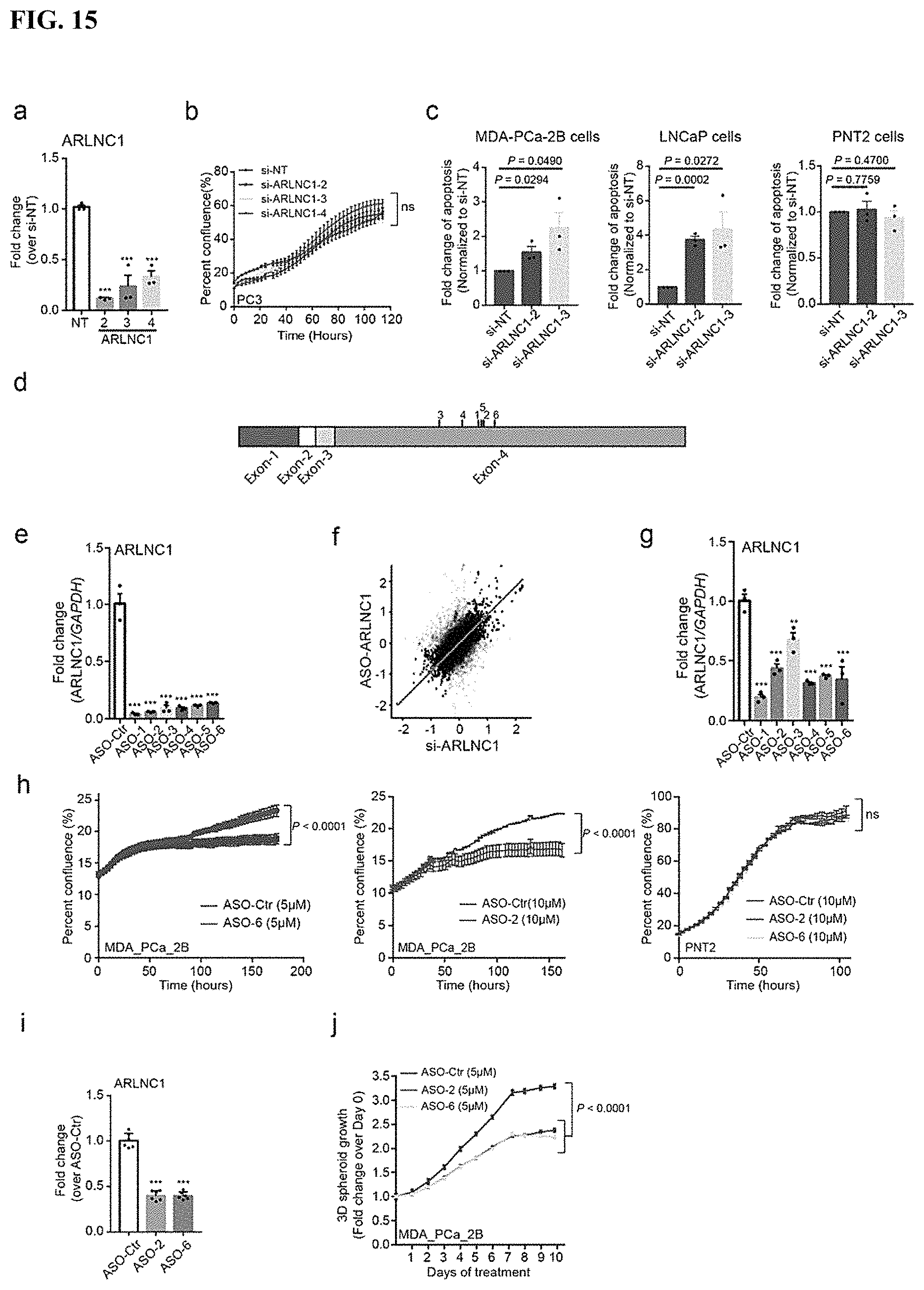
D00018
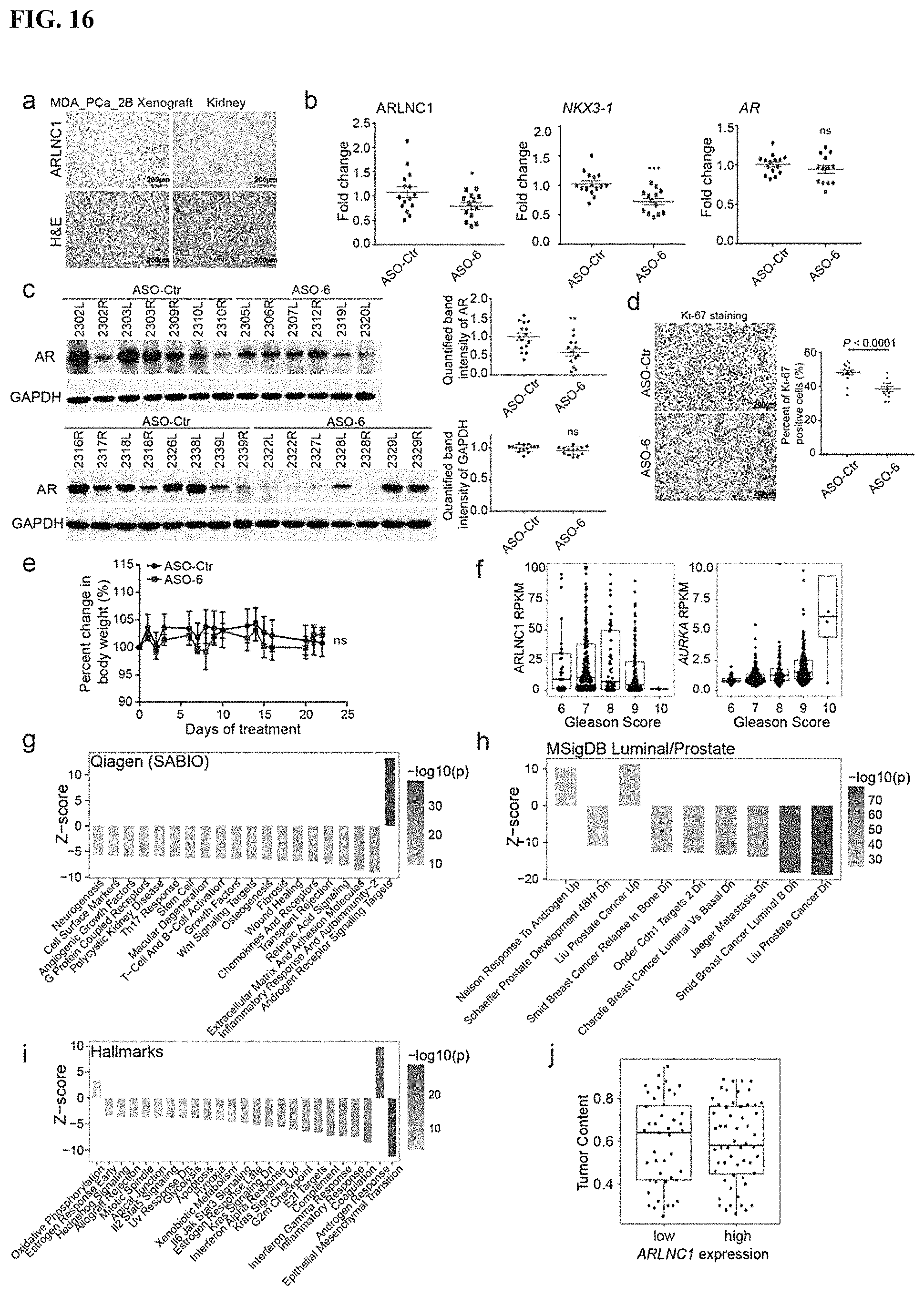
S00001
XML
uspto.report is an independent third-party trademark research tool that is not affiliated, endorsed, or sponsored by the United States Patent and Trademark Office (USPTO) or any other governmental organization. The information provided by uspto.report is based on publicly available data at the time of writing and is intended for informational purposes only.
While we strive to provide accurate and up-to-date information, we do not guarantee the accuracy, completeness, reliability, or suitability of the information displayed on this site. The use of this site is at your own risk. Any reliance you place on such information is therefore strictly at your own risk.
All official trademark data, including owner information, should be verified by visiting the official USPTO website at www.uspto.gov. This site is not intended to replace professional legal advice and should not be used as a substitute for consulting with a legal professional who is knowledgeable about trademark law.