Adeno-associated virus particle with mutated capsid and methods of use thereof
Wootton , et al. October 20, 2
U.S. patent number 10,806,802 [Application Number 16/251,119] was granted by the patent office on 2020-10-20 for adeno-associated virus particle with mutated capsid and methods of use thereof. This patent grant is currently assigned to Ottawa Hospital Research Institute, University of Guelph. The grantee listed for this patent is Ottawa Hospital Research Institute, University of Guelph. Invention is credited to Martin Hubert Kang, Bernard Claude Frank Thebaud, Laura van Lieshout, Sarah Wootton.


View All Diagrams
| United States Patent | 10,806,802 |
| Wootton , et al. | October 20, 2020 |
Adeno-associated virus particle with mutated capsid and methods of use thereof
Abstract
A recombinant adeno-associated virus (rAAV) particle with a mutated capsid protein is provided. In particular, the present disclosure provides methods of delivering a therapeutic agent to a muscle, airway, liver, central nervous system, retina or lung cell in a subject, and methods of treating or preventing infectious, acquired or genetic disease, with said rAAV particle.
| Inventors: | Wootton; Sarah (Guelph, CA), van Lieshout; Laura (Guelph, CA), Thebaud; Bernard Claude Frank (Ottawa, CA), Kang; Martin Hubert (Ottawa, CA) | ||||||||||
|---|---|---|---|---|---|---|---|---|---|---|---|
| Applicant: |
|
||||||||||
| Assignee: | University of Guelph (Guelph,
CA) Ottawa Hospital Research Institute (Ottawa, CA) |
||||||||||
| Family ID: | 1000005124445 | ||||||||||
| Appl. No.: | 16/251,119 | ||||||||||
| Filed: | January 18, 2019 |
Prior Publication Data
| Document Identifier | Publication Date | |
|---|---|---|
| US 20190216949 A1 | Jul 18, 2019 | |
Related U.S. Patent Documents
| Application Number | Filing Date | Patent Number | Issue Date | ||
|---|---|---|---|---|---|
| 62618810 | Jan 18, 2018 | ||||
Foreign Application Priority Data
| Nov 9, 2018 [CA] | 3023706 | |||
| Current U.S. Class: | 1/1 |
| Current CPC Class: | A61K 48/0008 (20130101); A61K 48/0058 (20130101); A61K 48/0075 (20130101); A61P 31/14 (20180101); A61K 48/0091 (20130101); A61K 9/12 (20130101); A61K 48/0066 (20130101); A61K 9/0043 (20130101); A61K 9/0073 (20130101); A61K 9/0019 (20130101) |
| Current International Class: | C12N 15/00 (20060101); C12N 15/86 (20060101); A61K 48/00 (20060101); A61K 9/12 (20060101); A61P 31/14 (20060101); A61K 9/00 (20060101) |
References Cited [Referenced By]
U.S. Patent Documents
| 8445267 | May 2013 | Zhong et al. |
| 10301648 | May 2019 | Vandenberghe et al. |
| 2014/0341852 | November 2014 | Srivastava |
| 2018/0030096 | February 2018 | Aslanidi et al. |
| 2018/0244727 | August 2018 | Zhong et al. |
| 2017201121 | Nov 2017 | WO | |||
Other References
|
Ng et al. Structural Characterization of the Dual Glycan Binding Adeno-Associated Virus Serotype 6. J. Virol. 2010, 84: 12945-12957. cited by examiner . Qiao et al. Adeno-Associated Virus Serotype 6 Capsid Tyrosine-to-Phenylalanine Mutations Improve Gene Transfer to Skeletal Muscle. Human Gene Therapy, 2010, 21:1343-1348. cited by examiner . McClain et al. Vector serotype screening for use in ovine perinatal lung gene therapy. Journal of Pediatric Surgery 51 (2016) 879-884. cited by examiner . Robert et al. Development of a Post-Exposure Treatment for Ebola Virus Infections Based on AAV Vectors and Zmapp Antibody Cocktail. Molecular Therapy vol. 24, Supplement 1, May 2016, S222. cited by examiner . Fuchs, Sebastian P., et al., "Recombinant AAV Vectors for Enhanced Expression of Authentic IgG." Plos One, vol. 11, No. 6, 2016, doi:10.1371/journal.pone.0158009. cited by applicant . Limberis, Maria P, et al. "Transduction Efficiencies of Novel AAV Vectors in Mouse Airway Epithelium In Vivo and Human Ciliated Airway Epithelium In Vitro." Molecular Therapy, vol. 17, No. 2, 2009, pp. 294-301., doi:10.1038/mt.2008.261. cited by applicant . Markusic, David M, et al., "High-Efficiency Transduction and Correction of Murine Hemophilia B Using AAV2 Vectors Devoid of Multiple Surface-Exposed Tyrosines." Molecular Therapy, vol. 18, No. 12, 2010, pp. 2048-2056., doi:10.1038/mt.2010.172. cited by applicant . Kang, M., et al., "AAV--Sftpb Gene Therapy Rescues Respiratory Distress and Improves Survival in a Mouse Model of Surfactant Protein B Deficiency." poster, presented at OHRI Research Day, Ottawa, Canada, Nov. 9, 2017. cited by applicant . Kang, M., et al., "AAV--Sftpb Gene Therapy rescues Respiratory Distress and Improves Survival in a Mouse Model of Surfactant Protein B Deficiency." abstract, published online Nov. 9, 2017. cited by applicant . Kang, M., et al., "AAV--Sftpb Gene Therapy Rescues Respiratory Distress and Improves Survival in a Mouse Model of Surfactant Protein B Deficiency." poster, presented at CHEO for Research Day, Ottawa, Canada, Mar. 22, 2018. cited by applicant . Zhong, L., et al., "Next Generation of Adeno-Associated Virus 2 Vectors: Point Mutations in Tyrosines Lead to High-Efficiency Transduction at Lower Doses." Proceedings of the National Academy of Sciences, vol. 105, No. 22, 2008, pp. 7827-7832., doi:10.1073/pnas.0802866105. cited by applicant . Van Lieshout, Laura P., et al., "Intramuscular Adeno-Associated Virus--Mediated Expression of Monoclonal Antibodies Provides 100% Protection Against Ebola Virus Infection in Mice." The Journal of Infectious Diseases, vol. 217, No. 6, 2018, pp. 916-925., doi:10.1093/infdis/jix644. cited by applicant . Wu, Z. et al., "Single Amino Acid Changes Can Influence Titer, Heparin Binding, and Tissue Tropism in Different Adeno-Associated Virus Serotypes." Journal of Virology, 2006, 80(22), 11393-11397. doi:10.1128/jvi.01288-06. cited by applicant . Aneja, M. K et al., "Targeted gene delivery to the lung. Expert Opinion on Drug Delivery", 2009, 6(6), 567-583. doi:10.1517/17425240902927841. cited by applicant . Limberis et al. Transduction Efficiencies of Novel AAV Vectors in Mouse Airway Epithelium In Vivo and Human Ciliated Airway Epithelium In Vitro, Molecular Therapy, vol. 17, No. 2, Feb. 2009 [online], [retrieved on Jan. 30, 2020]. Retrieved from the Internet: <URL: https://www.cell.com/action/doSearch?searchType=quick&searchText=Transduc- tion+Efficiencies+of+Novel+AAV+Vectors+in+Mouse+Airway+Epithelium+In +Vivo+and+Human+Ciliated+Airway+Epithelium+In+Vitro&searchScope=fullSite&- occurrences=all&code=cell-site>. cited by applicant . Markusic et al. High-efficiency Transduction and Correction of Murine Hemophilia B Using AAV2 Vectors Devoid of Multiple Surface-exposed Tyrosines, Molecular Therapy, vol. 18, No. 12, Dec. 2010 [online], [retrieved on Jan. 30, 2020]. Retrieved from the Internet: <URL: https://www.cell.com/action/doSearch?searchType=quick&searchText=High-eff- iciency+Transduction+and+Correction+of+Murine+Hemophilia+B+Using +AAV2+Vectors+Devoid+of+Multiple+Surface-expose+Tyrosines&searchScope=ful- lSite&occurrences=all&code=cell-. cited by applicant . Yan et al. Distinct transduction difference between adeno-associated virus type 1 and type 6 vectors in human polarized airway epithelia, Gene Therapy, vol. 20, No. 3, Jun. 14, 2014 [online], [retrieved on Jan. 30, 2020]. Retrieved from the Internet: <URL: https://www.ncbi.nlm.nih.gov/pmc/articles/PMC3443503/> <DOI: 10.1038/gt.2012.46>. cited by applicant. |
Primary Examiner: Zou; Nianxiang
Attorney, Agent or Firm: Szweras; Melanie Cheung; Herman Bereskin & Parr LLP/S.E.N.C.R.L., s.r.l.
Parent Case Text
RELATED APPLICATIONS
This application claims priority to U.S. Provisional Patent Application No. 62/618,810 filed on Jan. 18, 2018, and Canadian Patent Application No. 3,023,706 filed on Nov. 9, 2018, the content of which are hereby incorporated by reference in their entirety.
Claims
The invention claimed is:
1. A recombinant adeno-associated viral (rAAV) particle comprising a mutated capsid protein encapsidating a rAAV vector genome, wherein the mutated capsid protein comprises amino acid substitutions at amino acids 129, 445, and 731 of the AAV6 capsid protein sequence as set forth in SEQ ID NO:1, and wherein the rAAV vector genome comprises at least one heterologous nucleic acid segment flanked by AAV ITRs encoding a therapeutic agent operably linked to a promoter capable of expressing the segment in a host cell.
2. The rAAV particle of claim 1, wherein the mutated capsid protein has amino acid substitutions Phe129Leu, Tyr445Phe and Tyr731Phe, and wherein the mutated capsid protein is mutated AAV6 capsid protein having an amino acid sequence as shown in SEQ ID NO:2.
3. The rAAV particle of claim 1, wherein the therapeutic agent is a polypeptide, a therapeutic protein, an antigen, an antibody, or an antigen binding fragment, or a combination thereof.
4. A method of treating or preventing an infectious, acquired or genetic disease comprising administering at least one rAAV particle of claim 1 to a subject in need thereof.
5. The method of claim 4, wherein the infectious disease is selected from the group consisting of a viral disease, a bacterial disease, and a drug resistant parasitic disease, wherein the viral disease is selected from the group consisting of viral hemorrhagic fever, Ebola, Marburg virus disease, gastroenteritis, dengue fever, West Nile fever, yellow fever, influenza, respiratory syncytial virus disease, Lassa fever, rabies, smallpox, cowpox, horsepox, monkeypox, Hantavirus pulmonary syndrome, Hendra virus disease, human immunodeficiency virus disease and acquired immunodeficiency disease syndrome, Hepatitis, Zika fever, and wherein the bacterial disease is tuberculosis or methicillin-resistant Staphylococcus aureus infection, wherein the drug resistant parasitic disease is malaria, and wherein the acquired or genetic disease is selected from the group consisting of cancer, autoimmune disorders, vascular degeneration, neurodegenerative diseases such as Huntington's disease, cystic fibrosis, inflammatory bowel diseases such as Crohn's Disease, and surfactant protein B deficiency.
6. The method of claim 5, wherein the infectious disease is Ebola or Marburg virus disease, and wherein the therapeutic agent comprises an antibody, or a fragment thereof, or an antigen binding fragment, or a combination thereof, against Ebola or Marburg virus.
7. The method of claim 4, wherein the subject is human.
8. The method of claim 7, wherein the at least one rAAV particle is administered intravenously, intranasally, intratracheally, intramuscularly, intraperitoneally, or via aerosol.
9. The method of claim 4, wherein the at least one rAAV particle is delivered to lung cells or tissues.
10. The method of claim 6, wherein the antibody is a monoclonal antibody, wherein the monoclonal antibody is 1H3 which comprises a light chain comprising the amino acid sequence set forth in SEQ ID NO:3 and a heavy chain comprising the amino acid sequence set forth in SEQ ID NO:4, 2G4 which comprises a light chain comprising the amino acid sequence set forth in SEQ ID NO:5 and a heavy chain comprising the amino acid sequence set forth in SEQ ID NO:6, 4G7 which comprises a light chain comprising the amino acid sequence set forth in SEQ ID NO:7 and a heavy chain comprising the amino acid sequence set forth in SEQ ID NO:8, 5D2 which comprises a light chain comprising the amino acid sequence set forth in SEQ ID NO:9 and a heavy chain comprising the amino acid sequence set forth in SEQ ID NO:10, 7C9 which comprises a light chain comprising the amino acid sequence set forth in SEQ ID NO:11 and a heavy chain comprising the amino acid sequence set forth in SEQ ID NO:12, 100 which comprises a light chain comprising the amino acid sequence set forth in SEQ ID NO:13 and a heavy chain comprising the amino acid sequence set forth in SEQ ID NO:14, 114 which comprises a light chain comprising the amino acid sequence set forth in SEQ ID NO:15 and a heavy chain comprising the amino acid sequence set forth in SEQ ID NO:16, CA45 which comprises a light chain comprising the amino acid sequence set forth in SEQ ID NO:17 and a heavy chain comprising the amino acid sequence set forth in SEQ ID NO:18, ADI-15878 which comprises a light chain comprising the amino acid sequence set forth in SEQ ID NO:19 and a heavy chain comprising the amino acid sequence set forth in SEQ ID NO:20, FVM02p which comprises a light chain comprising the amino acid sequence set forth in SEQ ID NO:21 and a heavy chain comprising the amino acid sequence set forth in SEQ ID NO:22, FVM04 which comprises a light chain comprising the amino acid sequence set forth in SEQ ID NO:23 and a heavy chain comprising the amino acid sequence set forth in SEQ ID NO:24, BDBV223 which comprises a light chain comprising the amino acid sequence set forth in SEQ ID NO:25 and a heavy chain comprising the amino acid sequence set forth in SEQ ID NO:26, or a fragment thereof, or a combination cocktail thereof, against Ebola virus, or MR72 which comprises a light chain comprising the amino acid sequence set forth in SEQ ID NO:27 and a heavy chain comprising the amino acid sequence set forth in SEQ ID NO:28, MR82 which comprises a light chain comprising the amino acid sequence set forth in SEQ ID NO:29 and a heavy chain comprising the amino acid sequence set forth in SEQ ID NO:30, MR78 which comprises a light chain comprising the amino acid sequence set forth in SEQ ID NO:31 and a heavy chain comprising the amino acid sequence set forth in SEQ ID NO:32, MR191 which comprises a light chain comprising the amino acid sequence set forth in SEQ ID NO:33 and a heavy chain comprising the amino acid sequence set forth in SEQ ID NO:34, or a fragment thereof, or a combination thereof, against Marburg virus.
11. The method of claim 6, wherein the therapeutic agent remains in the serum of the subject for at least 2, 4, 8, 10, 12, 14, 16 or 18 weeks, and/or up to 26, 28, 30, 32, or 34 weeks.
12. The method of claim 6, wherein the subject is protected from Ebola from 3, 7 or 14 days post administration to at least 3 weeks, or 1, 2, 3, 4, or 5 months.
13. A mutated AAV capsid protein comprising (a) amino acid substitutions at amino acids 129, 445, and 731 of the AAV6 capsid protein sequence as set forth in SEQ ID NO:1, or (b) amino acid substitutions Phe129Leu, Tyr445Phe and Tyr731Phe, wherein the mutated capsid protein is mutated AAV6 capsid protein having an amino acid sequence as shown in SEQ ID NO:2.
14. A method of producing at least one protein in vivo in a subject, comprising introducing into the subject at least one rAAV particle of claim 1.
15. The method of claim 14, wherein the mutated capsid protein has amino acid substitutions Phe129Leu, Tyr445Phe and Tyr731Phe, and wherein the mutated capsid protein is mutated AAV6 capsid protein having an amino acid sequence as shown in SEQ ID NO:2.
16. The method of claim 14, wherein the at least one protein is a monoclonal antibody or a fragment thereof, and wherein the monoclonal antibody is 1H3 which comprises a light chain comprising the amino acid sequence set forth in SEQ ID NO:3 and a heavy chain comprising the amino acid sequence set forth in SEQ ID NO:4, 2G4 which comprises a light chain comprising the amino acid sequence set forth in SEQ ID NO:5 and a heavy chain comprising the amino acid sequence set forth in SEQ ID NO:6, 4G7 which comprises a light chain comprising the amino acid sequence set forth in SEQ ID NO:7 and a heavy chain comprising the amino acid sequence set forth in SEQ ID NO:8, 5D2 which comprises a light chain comprising the amino acid sequence set forth in SEQ ID NO:9 and a heavy chain comprising the amino acid sequence set forth in SEQ ID NO:10, 7C9 which comprises a light chain comprising the amino acid sequence set forth in SEQ ID NO:11 and a heavy chain comprising the amino acid sequence set forth in SEQ ID NO:12, 100 which comprises a light chain comprising the amino acid sequence set forth in SEQ ID NO:13 and a heavy chain comprising the amino acid sequence set forth in SEQ ID NO:14, 114 which comprises a light chain comprising the amino acid sequence set forth in SEQ ID NO:15 and a heavy chain comprising the amino acid sequence set forth in SEQ ID NO:16, CA45 which comprises a light chain comprising the amino acid sequence set forth in SEQ ID NO:17 and a heavy chain comprising the amino acid sequence set forth in SEQ ID NO:18, ADI-15878 which comprises a light chain comprising the amino acid sequence set forth in SEQ ID NO:19 and a heavy chain comprising the amino acid sequence set forth in SEQ ID NO:20, FVM02p which comprises a light chain comprising the amino acid sequence set forth in SEQ ID NO:21 and a heavy chain comprising the amino acid sequence set forth in SEQ ID NO:22, FVM04 which comprises a light chain comprising the amino acid sequence set forth in SEQ ID NO:23 and a heavy chain comprising the amino acid sequence set forth in SEQ ID NO:24, BDBV223 which comprises a light chain comprising the amino acid sequence set forth in SEQ ID NO:25 and a heavy chain comprising the amino acid sequence set forth in SEQ ID NO:26, or a fragment thereof, or a combination cocktail thereof, against Ebola virus, or MR72 which comprises a light chain comprising the amino acid sequence set forth in SEQ ID NO:27 and a heavy chain comprising the amino acid sequence set forth in SEQ ID NO:28, MR82 which comprises a light chain comprising the amino acid sequence set forth in SEQ ID NO:29 and a heavy chain comprising the amino acid sequence set forth in SEQ ID NO:30, MR78 which comprises a light chain comprising the amino acid sequence set forth in SEQ ID NO:31 and a heavy chain comprising the amino acid sequence set forth in SEQ ID NO:32, MR191 which comprises a light chain comprising the amino acid sequence set forth in SEQ ID NO:33 and a heavy chain comprising the amino acid sequence set forth in SEQ ID NO:34, or a fragment thereof, or a combination thereof, against Marburg virus.
17. The method of claim 16, wherein the at least one protein remains in the serum of the subject for at least 2, 4, 8, 10, 12, 14, 16 or 18 weeks, and/or up to 26, 28, 30, 32, or 34 weeks.
18. The method of claim 14, wherein the subject is human.
19. The method of claim 14, wherein the at least one rAAV particle is administered intravenously, intranasally, intratracheally, intramuscularly, or via aerosol.
20. The rAAV particle of claim 3, wherein the therapeutic protein is surfactant protein B (SPB).
21. The method of claim 4, wherein the genetic disease is surfactant protein B deficiency, and wherein the therapeutic agent is surfactant protein B (SPB).
22. The method of claim 9, wherein the at least one rAAV particle targets alveolar epithelial type 2 (AT2) cells.
23. The method of claim 22, wherein the infectious disease is influenza or respiratory syncytial virus disease, or wherein the genetic disease is cystic fibrosis.
Description
INCORPORATION OF SEQUENCE LISTING
A computer readable form of the Sequence Listing "6580-P55086US01_SequenceListing.txt" (40,096 bytes), submitted via EFS-WEB and created on Jan. 17, 2019, is herein incorporated by reference.
FIELD
The present disclosure provides a recombinant adeno-associated virus (rAAV) particle with mutated capsid protein, where the rAAV particle exhibits greater transduction of host cells compared to wild-type AAV. The present disclosure further provides methods of delivering a therapeutic agent to a muscle, airway, liver, central nervous system, retina or lung cell in a subject, and methods of treating or preventing an infectious, acquired or genetic disease, with said rAAV particle.
BACKGROUND
Adeno-associated virus (AAV) is widely regarded as a safe and effective method of gene transfer to a variety of tissues. The in vitro and in vivo transduction profiles have been well characterized for many AAV serotypes [1, 2]. Engineering of AAV capsids by rational design or directed evolution can produce capsid variants with desirable characteristics including altered tissue tropism, enhanced transgene expression in target cells or the introduction of binding domains to aid in purification to name a few. A prime example, AAV-DJ, is a product of AAV2 and AAV8 capsid shuffling, resulting in a hybrid capsid with beneficial properties of both capsids; heparin binding capacity and in vitro transduction capacities from AAV2 and potent in vivo liver transduction from AAV8 [3, 4].
Alternatively, single point mutations in an AAV capsid can also yield desirable modifications. AAV6.2, an AAV6 F129L point mutant, was demonstrated by Limberis et al. to be a 2-fold more efficient transducer of the mouse nose, airways and alveolar cells than AAV6 [5]. Similarly, when delivered intravenously to mice, AAV6.2 mediated 2-fold greater serum concentrations of human alpha-1 antitrypsin (hA1AT) than AAV6 [6]. Moreover, intramuscular administration of the same AAV6.2-hA1AT vector mediated higher serum levels of hA1AT than AAV6 or AAV9 [6]. Also, F129L is a naturally occurring singleton residue in the majority of over 100 known primate AAV capsid sequences, and AAV5 and AAV6 are the only serotypes that encode a phenylalanine instead of a leucine at this position [6].
AAV capsids are prone to phosphorylation of tyrosine residues by epidermal growth factor receptor protein tyrosine kinase (EGFR-PTK), leading to alternative cellular trafficking, ubiquitination and degradation [7, 8]. Mutation of various surface exposed tyrosine residues on AAV capsids has been shown to obstruct ubiquitin-mediated degradation of intracellular vector, thereby leading to more robust transgene expression [9]. Tyrosine to phenylalanine mutations introduced at positions 444 and 730 in the AAV2 capsid yielded 9- and 11-fold greater expression in vitro and 13- and 29-fold greater hepatocyte transduction in mice, respectively [9]. A double AAV2 Y444F+Y730F mutant generated significantly greater hepatocyte transduction in vivo than either of the singleton mutants [10]. Similar single tyrosine mutations have been introduced into corresponding positions in AAV6, AAV8 and AAV9 capsids with success in transducing various tissues.
SUMMARY
The present inventors engineered a triple mutant AAV6 capsid, termed AAV6.2FF, encoding F129L, Y445F and Y731F point mutations, which was demonstrated to be superior to the parental capsid in terms of muscle and airway transgene expression kinetics in a mouse model. Further, the present disclosure shows that AAV-mediated expression of non-neutralizing mAbs 5D2 or 7C9 encapsidated by AAV6.2FF confer 100% protection against Ebola virus infection when administered as monotherapies seven days prior to challenge, while neutralizing mAb 2G4 was 83% protective. In addition, the present disclosure shows a two-component cocktail of AAV-2G4 and AAV-5D2 provided complete protection when administered seven days prior to challenge and can protect a subject with as little as a three-day lead-time. Subjects were fully protected from Ebola challenge five months after receiving a single IM injection of AAV-2G4, AAV-5D2, or a double injection of the two. These findings demonstrate that AAV6.2FF-mediated expression of neutralizing or non-neutralizing mAbs when administered as early as seven days or as late as five months prior to exposure can prevent Ebola virus mortality in a mouse model.
Accordingly, the present disclosure provides a recombinant adeno-associated viral (rAAV) particle comprising a mutated capsid protein encapsidating a rAAV vector genome, wherein the mutated capsid protein comprises amino acid substitutions at amino acids 129, 445, and 731 of the AAV6 capsid protein sequence as set forth in SEQ ID NO:1.
In an embodiment, the mutated capsid protein has amino acid substitutions Phe129Leu, Tyr445Phe and Tyr731Phe, wherein the mutated capsid protein is mutated AAV6 capsid protein having an amino acid sequence as shown in SEQ ID NO:2. In another embodiment, the rAAV vector genome comprises at least one heterologous nucleic acid segment flanked by AAV ITRs encoding a therapeutic agent operably linked to a promoter capable of expressing the segment in a host cell. In another embodiment, the host cell is selected from the group consisting of a human, primate, murine, feline, canine, ovine, bovine, porcine, caprine, equine, lupine, and vulpine host cell. In a further embodiment, the promoter is capable of expressing the at least one heterologous nucleic acid segment encoding a therapeutic agent in muscle, airway, liver, central nervous system, retina or lung cells. In one embodiment, the therapeutic agent is a polypeptide, a therapeutic protein, an antigen, an antibody, or an antigen binding fragment, or a combination thereof. In another embodiment, the antibody comprises a monoclonal, polyclonal, chimeric, humanized antibody, a fragment thereof, or a combination thereof. In another embodiment, the antigen binding fragment is a Fab, Fab.theta., F(ab')2, scFv, dsFv, ds-scFv, dimer, minibody, diabody, or multimer thereof or bispecific antibody fragment, or a combination thereof. In a further embodiment, the monoclonal antibody is 1H3, 2G4, 4G7, 5D2, 7C9, 100, 114, CA45, ADI-15878, FVM02p, FVM04, BDBV223, or a fragment thereof, or a combination cocktail thereof, against Ebola virus, or MR72, MR82, MR78, or MR191, or a combination thereof, against Marburg virus. In a further embodiment, the monoclonal antibody is 100 or a fragment thereof against Ebola virus, or MR191 or a fragment thereof against Marburg virus. In a further embodiment, the monoclonal antibody is 100 or a fragment thereof against Ebola virus. In a further embodiment, the monoclonal antibody is MR191 or a fragment thereof against Marburg virus.
In an embodiment, the particle further comprises a nucleotide sequence encoding a marker, optionally luciferase. The skilled person can readily recognize that many markers known in the art can be used, for example, fluorescent proteins such as GFP and RFP, or alkaline phosphatase. In another embodiment, the rAAV particle is comprised in a pharmaceutical composition that includes a pharmaceutically acceptable diluent, buffer, carrier, or excipient.
The present disclosure also provides a method of treating or preventing an infectious, acquired or genetic disease in a subject in need thereof, involving administering at least one rAAV particle, wherein the rAAV particle comprises a mutated capsid protein encapsidating a rAAV vector genome, wherein the rAAV vector genome comprises at least one heterologous nucleic acid segment flanked by AAV ITRs encoding a therapeutic agent operably linked to a promoter capable of expressing the segment in a host cell, wherein the mutated capsid protein comprises amino acid substitutions at amino acids 129, 445, and 731 of the AAV6 capsid protein sequence as set forth in SEQ ID NO:1, and wherein the therapeutic agent treats or prevents the infectious, acquired or genetic disease in the subject in need thereof. In one embodiment, the mutated capsid protein has amino acid substitutions Phe129Leu, Tyr445Phe and Tyr731Phe, wherein the mutated capsid protein is mutated AAV6 capsid protein having an amino acid sequence as shown in SEQ ID NO:2. In another embodiment, the rAAV vector genome comprises at least one heterologous nucleic acid segment flanked by AAV ITRs encoding the therapeutic agent operably linked to a promoter capable of expressing the segment in a host cell. In another embodiment, the host cell is selected from the group consisting of a human, primate, murine, feline, canine, ovine, bovine, porcine, caprine, equine, lupine, and vulpine host cell. In a further embodiment, the promoter is capable of expressing the at least one heterologous nucleic acid segment encoding the therapeutic agent in muscle, airway, liver, central nervous system, retina or lung cells.
Also provided is use of at least one rAAV particle for treating or preventing an infectious, acquired or genetic disease in a subject in need thereof, wherein the rAAV particle comprises a mutated capsid protein encapsidating a rAAV vector genome, wherein the rAAV vector genome comprises at least one heterologous nucleic acid segment flanked by AAV ITRs encoding a therapeutic agent operably linked to a promoter capable of expressing the segment in a host cell, and wherein the mutated capsid protein comprises amino acid substitutions at amino acids 129, 445, and 731 of the AAV6 capsid protein sequence as set forth in SEQ ID NO:1. In an embodiment, the mutated capsid protein has amino acid substitutions Phe129Leu, Tyr445Phe and Tyr731Phe, wherein the mutated capsid protein is mutated AAV6 capsid protein having an amino acid sequence as shown in SEQ ID NO:2. In another embodiment, the rAAV vector genome comprises at least one heterologous nucleic acid segment flanked by AAV ITRs encoding the therapeutic agent operably linked to a promoter capable of expressing the segment in a host cell. In another embodiment, the host cell is selected from the group consisting of a human, primate, murine, feline, canine, ovine, bovine, porcine, caprine, equine, lupine, and vulpine host cell. In a further embodiment, the promoter is capable of expressing the at least one heterologous nucleic acid segment encoding the therapeutic agent in muscle, airway, liver, central nervous system, retina or lung cells.
Further provided is use of at least one rAAV particle in the manufacture of a medicament for treating or preventing an infectious, acquired or genetic disease in a subject in need thereof, wherein the rAAV particle comprises a mutated capsid protein encapsidating a rAAV vector genome, wherein the rAAV vector genome comprises at least one heterologous nucleic acid segment flanked by AAV ITRs encoding a therapeutic agent operably linked to a promoter capable of expressing the segment in a host cell, and wherein the mutated capsid protein comprises amino acid substitutions at amino acids 129, 445, and 731 of the AAV6 capsid protein sequence as set forth in SEQ ID NO:1. In an embodiment, the mutated capsid protein has amino acid substitutions Phe129Leu, Tyr445Phe and Tyr731Phe, wherein the mutated capsid protein is mutated AAV6 capsid protein having an amino acid sequence as shown in SEQ ID NO:2. In another embodiment, the rAAV vector genome comprises at least one heterologous nucleic acid segment flanked by AAV ITRs encoding the therapeutic agent operably linked to a promoter capable of expressing the segment in a host cell. In another embodiment, the host cell is selected from the group consisting of a human, primate, murine, feline, canine, ovine, bovine, porcine, caprine, equine, lupine, and vulpine host cell. In a further embodiment, the promoter is capable of expressing the at least one heterologous nucleic acid segment encoding the therapeutic agent in muscle, airway, liver, central nervous system, retina or lung cells.
Even further provided is at least one rAAV particle for use in treating or preventing an infectious, acquired or genetic disease in a subject in need thereof, wherein the rAAV particle comprises a mutated capsid protein encapsidating a rAAV vector genome, wherein the rAAV vector genome comprises at least one heterologous nucleic acid segment flanked by AAV ITRs encoding a therapeutic agent operably linked to a promoter capable of expressing the segment in a host cell, and wherein the mutated capsid protein comprises amino acid substitutions at amino acids 129, 445, and 731 of the AAV6 capsid protein sequence as set forth in SEQ ID NO:1. In an embodiment, the mutated capsid protein has amino acid substitutions Phe129Leu, Tyr445Phe and Tyr731Phe, wherein the mutated capsid protein is mutated AAV6 capsid protein having an amino acid sequence as shown in SEQ ID NO:2. In another embodiment, the rAAV vector genome comprises at least one heterologous nucleic acid segment flanked by AAV ITRs encoding the therapeutic agent operably linked to a promoter capable of expressing the segment in a host cell. In another embodiment, the host cell is selected from the group consisting of a human, primate, murine, feline, canine, ovine, bovine, porcine, caprine, equine, lupine, and vulpine host cell. In a further embodiment, the promoter is capable of expressing the at least one heterologous nucleic acid segment encoding the therapeutic agent in muscle, airway, liver, central nervous system, retina or lung cells.
In an embodiment, the infectious disease is selected from the group consisting of viral diseases such as viral hemorrhagic fevers, Ebola, Marburg virus disease, gastroenteritis, dengue fever, West Nile fever, yellow fever, influenza, respiratory syncytial virus disease, Lassa fever, rabies, smallpox, cowpox, horsepox, monkeypox, Hentavirus pulmonary syndrome, Hendra virus disease, human immunodeficiency virus disease and acquired immunodeficiency disease syndrome, Hepatitis, Zika fever, optionally Ebola or Marburg virus disease, and bacterial diseases including drug resistant bacterial diseases such as tuberculosis and methicillin-resistant Staphylococcus aureus infection, and drug resistant parasitic diseases such as malaria. In another embodiment, the subject is human. In another embodiment, the at least one rAAV particle is administered or co-administered intravenously, intranasally, intratracheally, intramuscularly, or via aerosol. In an embodiment, the at least one rAAV particle is delivered to lung cells or tissues.
In one embodiment, the therapeutic agent remains in the serum of the subject for at least 2, 4, 8, 10, 12, 14, 16 or 18 weeks, optionally at least 18 weeks, up to 26, 28, 30, 32, or 34 weeks. In another embodiment, the therapeutic agent remains in the serum of the subject for up to 34 weeks. In another embodiment, the subject is protected from Ebola from 3, 7 or 14 days post administration to at least 3 weeks, or 1, 2, 3, 4, or 5 months, optionally at least 5 months.
The present disclosure also provides a nucleic acid molecule comprising a nucleotide sequence encoding a mutated AAV capsid protein, wherein the mutated AAV capsid protein comprises amino acid substitutions at amino acids 129, 445, and 731 of the AAV6 capsid protein sequence as set forth in SEQ ID NO:1.
Further provided is a method of producing a protein in vivo in a subject, comprising delivering or introducing into the subject a rAAV particle comprising a mutated capsid protein encapsidating a rAAV vector genome, wherein the rAAV vector genome comprises at least one heterologous nucleic acid segment flanked by AAV ITRs encoding the protein operably linked to a promoter capable of expressing the segment in vivo in the subject, and wherein the mutated capsid protein comprises amino acid substitutions at amino acids 129, 445, and 731 of the AAV6 capsid protein sequence as set forth in SEQ ID NO:1.
Other features and advantages of the present disclosure will become apparent from the following detailed description. It should be understood, however, that the detailed description and the specific Examples while indicating preferred embodiments of the disclosure are given by way of illustration only, since various changes and modifications within the spirit and scope of the disclosure will become apparent to those skilled in the art from this detailed description.
BRIEF DESCRIPTION OF THE DRAWINGS
Embodiments are described below in relation to the drawings in which:
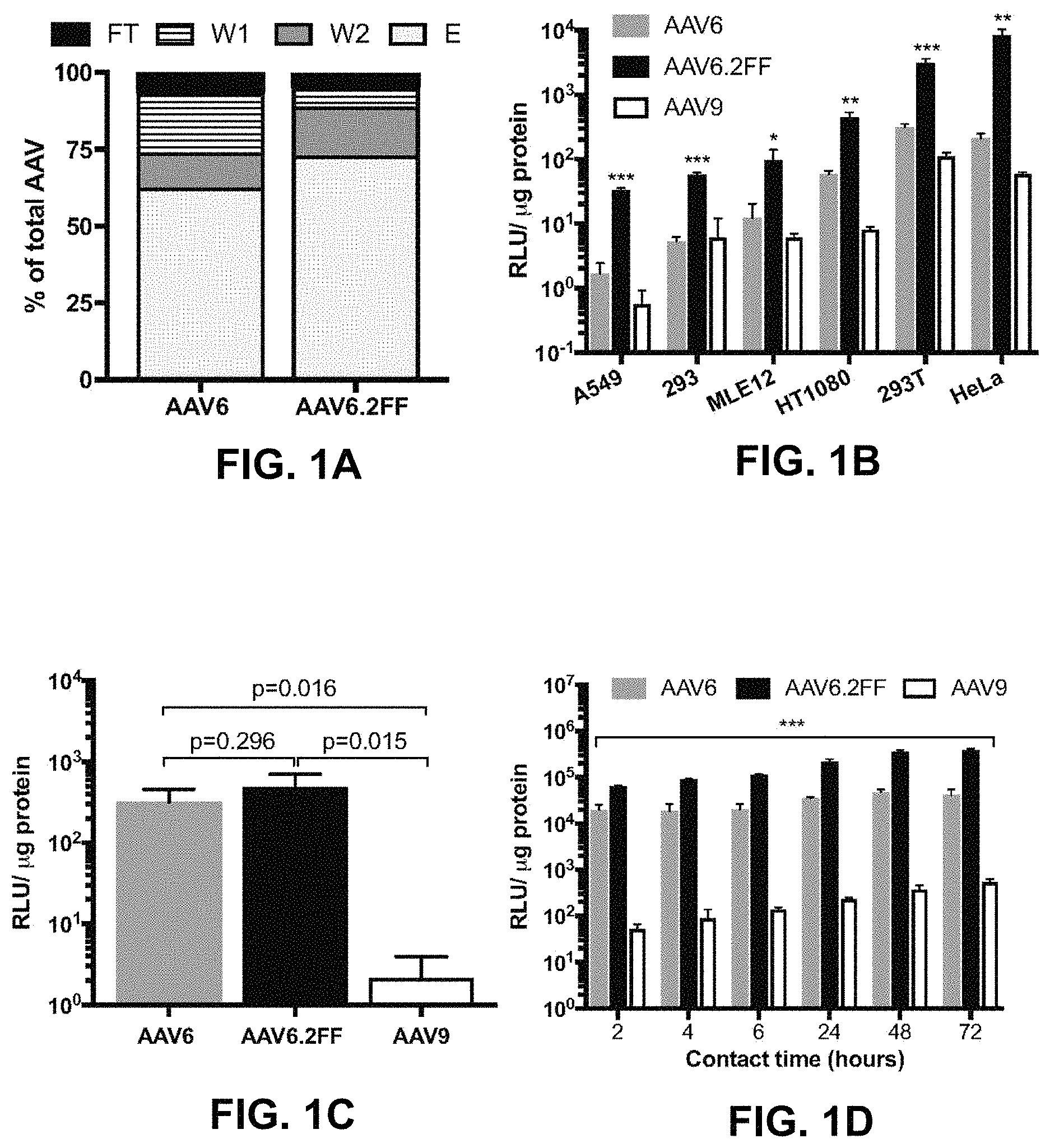
FIGS. 1A-1D show heparin binding and transduction profiles of AAV6 and AAV6.2FF in vitro. FIG. 1A shows the distribution of AAV vector genomes following heparin binding assay (n=3). FT--flow though, W1--wash 1, W2--wash 2, E-elution. FIG. 1B shows AAV6, AAV6.2FF or AAV9 encoding luciferase were added at an MOI of 2000 and incubated for 72 hours prior to luciferase quantification to determine transduction efficiency. FIG. 1C shows the AAV binding and FIG. 1D shows the internalization of AAV vectors over various contact times in HeLa cells. Multiple t-tests compared AAV6.2FF mediated luciferase expression to AAV6 and AAV9. *p<0.05, **p<0.01, ***p<0.001.
FIG. 2 shows intravenous immunoglobulin (IVIG) neutralization of AAV in vitro. AAV vector was incubated with 10-fold dilutions of IVIG for 1 hour at 37.degree. C. prior to adding to HeLa cells. 72 hours later luciferase expression was quantified and the data is expressed as the percent AAV neutralization as compared to control virus incubated with PBS only (n=6).
FIGS. 3A-3C show comparison of AAV6 and AAV6.2FF-mediated expression 24 hours post AAV delivery. Albino C57BL/6 mice (n=4 mice/group) received 1.times.10.sup.11 vector genomes (vg) by intramuscular injection or intranasal delivery. FIG. 3A shows the quantification of luciferase expression in the muscle and lung 24 hours following intramuscular or intranasal delivery, respectively. Representative images demonstrating pattern and intensity of luciferase expression in (FIG. 3B) the muscle or (FIG. 3C) the lungs.
FIGS. 4A-4C show intramuscular expression kinetics of AAV6 and AAV6.2FF. Albino C57BL/6 mice (n=4/group) were injected with 1.times.10.sup.1 vg of AAV6- or AAV6.2FF-Luciferase. FIG. 4A and FIG. 4C show the quantification of luciferase signal on days 1, 3, 7, 14, 21, 28, 56 and 112 post AAV delivery. Paired t-tests were used to calculate significance, ***p<0.001. FIG. 4B shows images demonstrating luciferase expression on day 3 and 14 post AAV delivery to the gastrocnemius muscle.
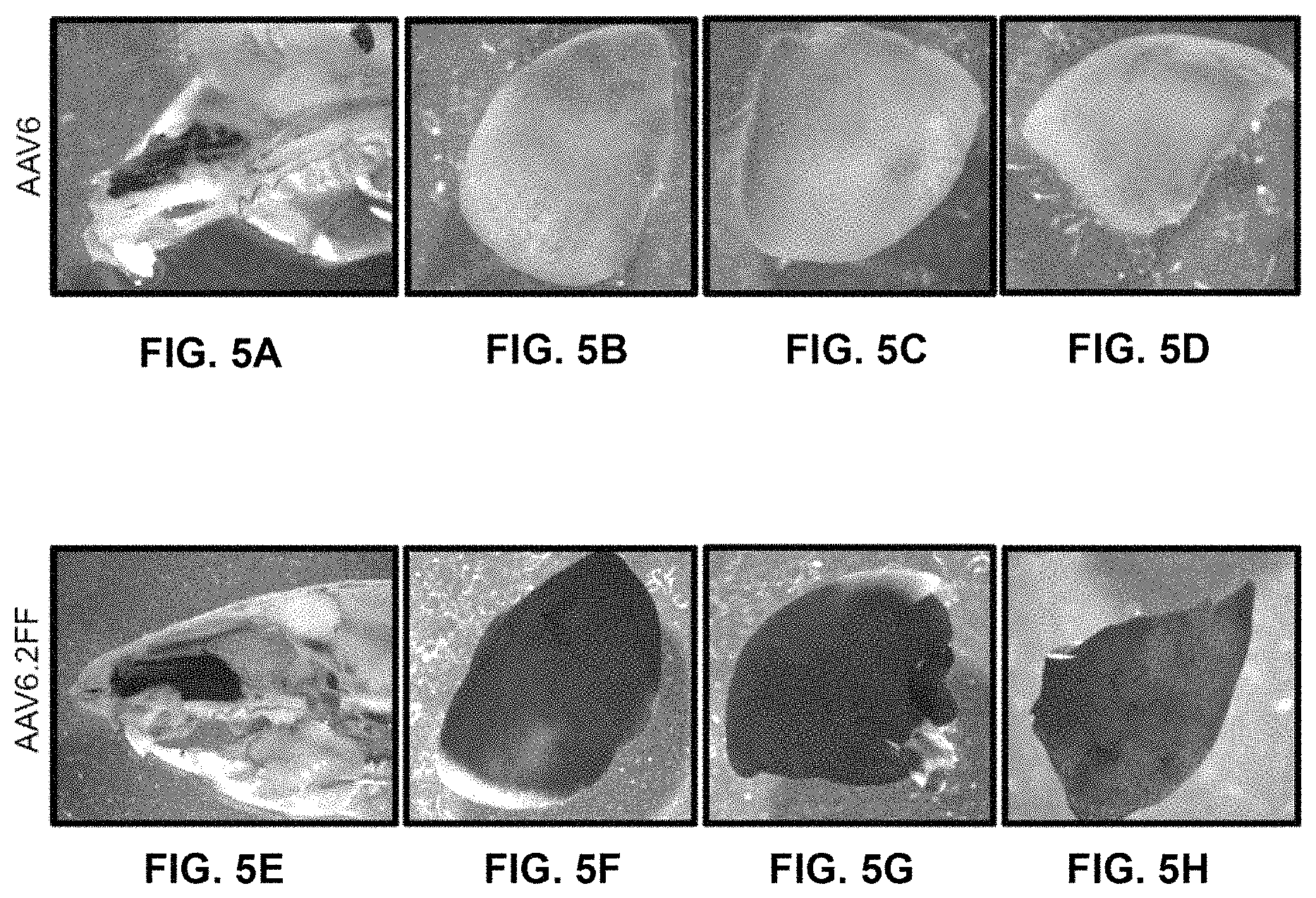
FIGS. 5A-5H show gross AP staining of AAV6 and AAV6.2FF transduced nose and lungs. 1.times.10.sup.11 vg of AAV6- or AAV6.2FF-Alkaline phosphatase (AP) was delivered intranasally to C57BL/6 mice (n=4/group) and tissues were harvested and stained after 3 weeks. Representative images of (FIG. 5A, FIG. 5E) the nose, and (FIGS. 5B-5D, FIGS. 5F-5H) lung lobes for both vectors are shown.
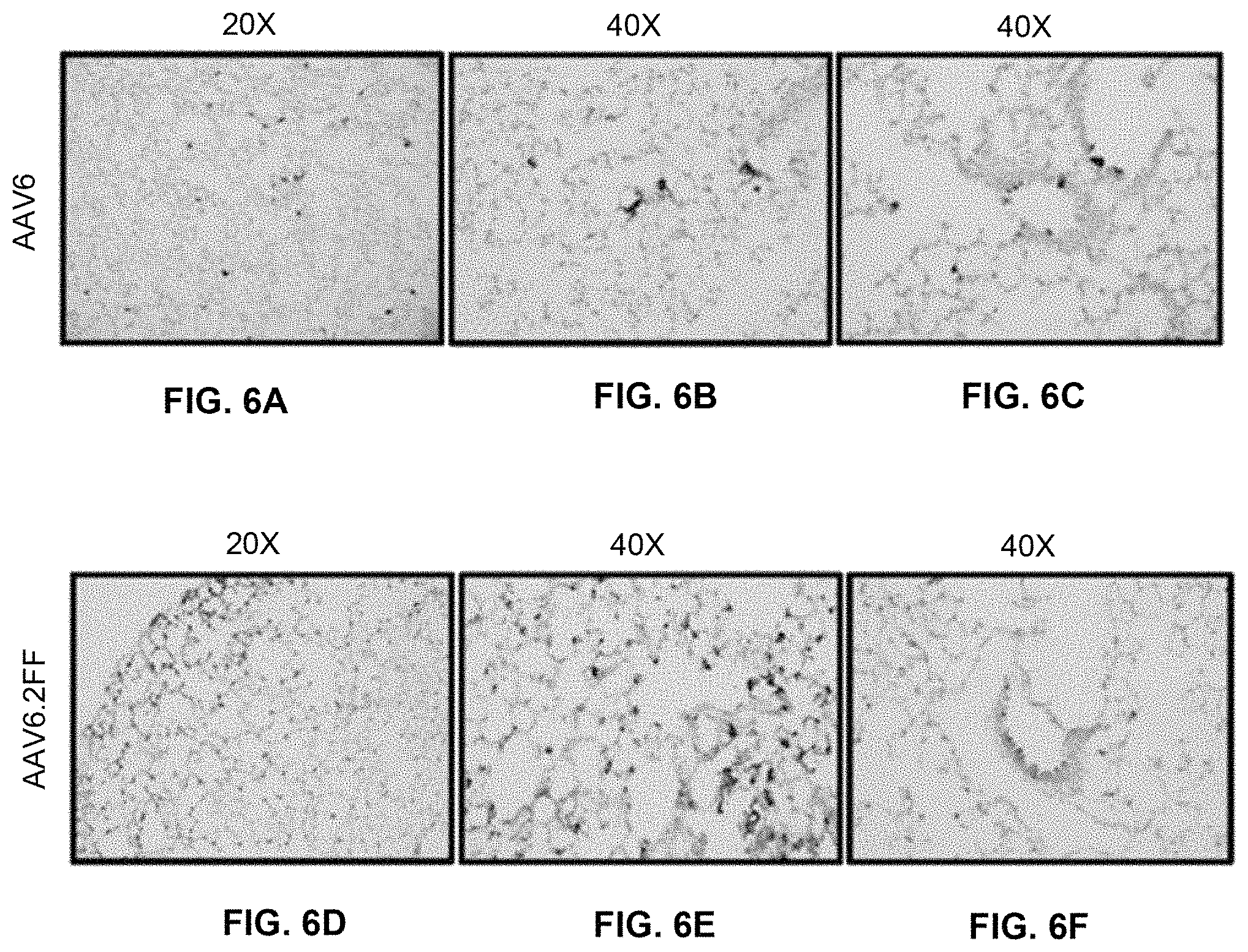
FIGS. 6A-6F show histological AP staining of lung tissue. Lung sections were counterstained with nuclear fast red and transduced cells expressing AP appear purple. Representative images of transduced lung are shown at (FIG. 6A, FIG. 6D) 20.times. and (FIG. 6B, FIG. 6E) 40.times. for AAV6 and AAV6.2FF, respectively. FIG. 6C and FIG. 6F show the demonstration of transduction of airway cells.
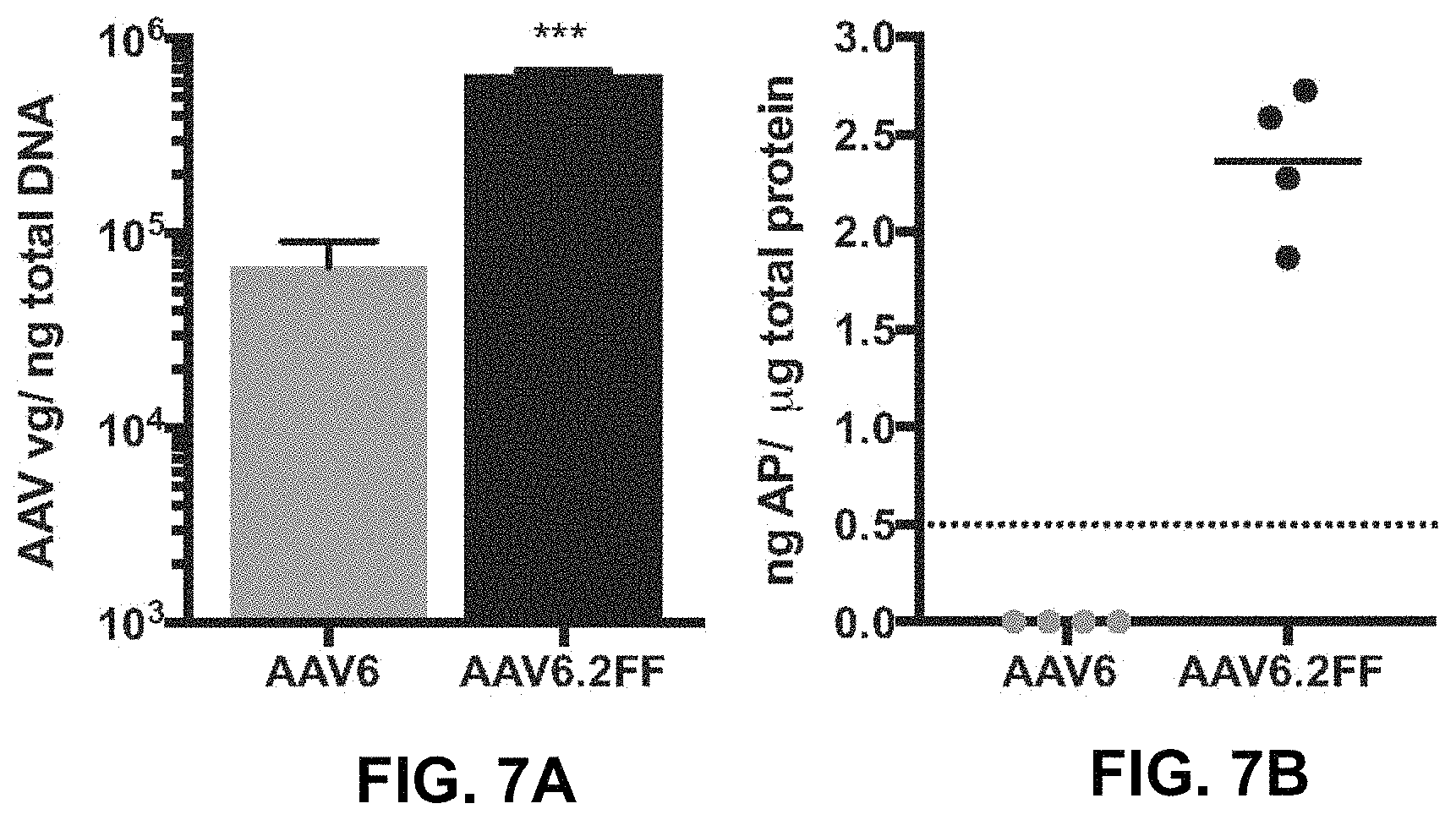
FIGS. 7A and 7B show quantification of transduced lung tissue. FIG. 7A shows the qPCR quantification of the AAV genome copy number per ng of total DNA extracted from paraffin embedded lung tissue. FIG. 7B shows the quantification of the AP transgene present in unfixed lung tissue. Dashed line indicates the threshold of detection for this assay.
FIGS. 8A and 8B show early transgene expression kinetics of five different AAV capsids. Albino C57BU6 mice (n=4/group) were injected intramuscularly with 1.times.10.sup.11 vg of an AAV vector expressing firefly luciferase (Luc) packaged with either AAV6, AAV6.2FF, AAV8, AAV9 or AAV-DJ capsid. FIG. 8A shows the in vivo luciferase images were obtained on 0, 1, 3, 7 and 14 days post AAV (dpa) administration. FIG. 8B shows the relative photon emission (p/s/cm.sup.2/sr) produced by luciferase from the muscle of each serotype of AAV was quantified at various time points from 1 to 56 dpi. Multiple t-tests were used to compare each time point. *Indicates AAV6.2FF is significant over all other capsids, p<0.05, and # indicates AAV6.2FF is significant over AAV8, AAV9 and AAV-DJ, p<0.05.
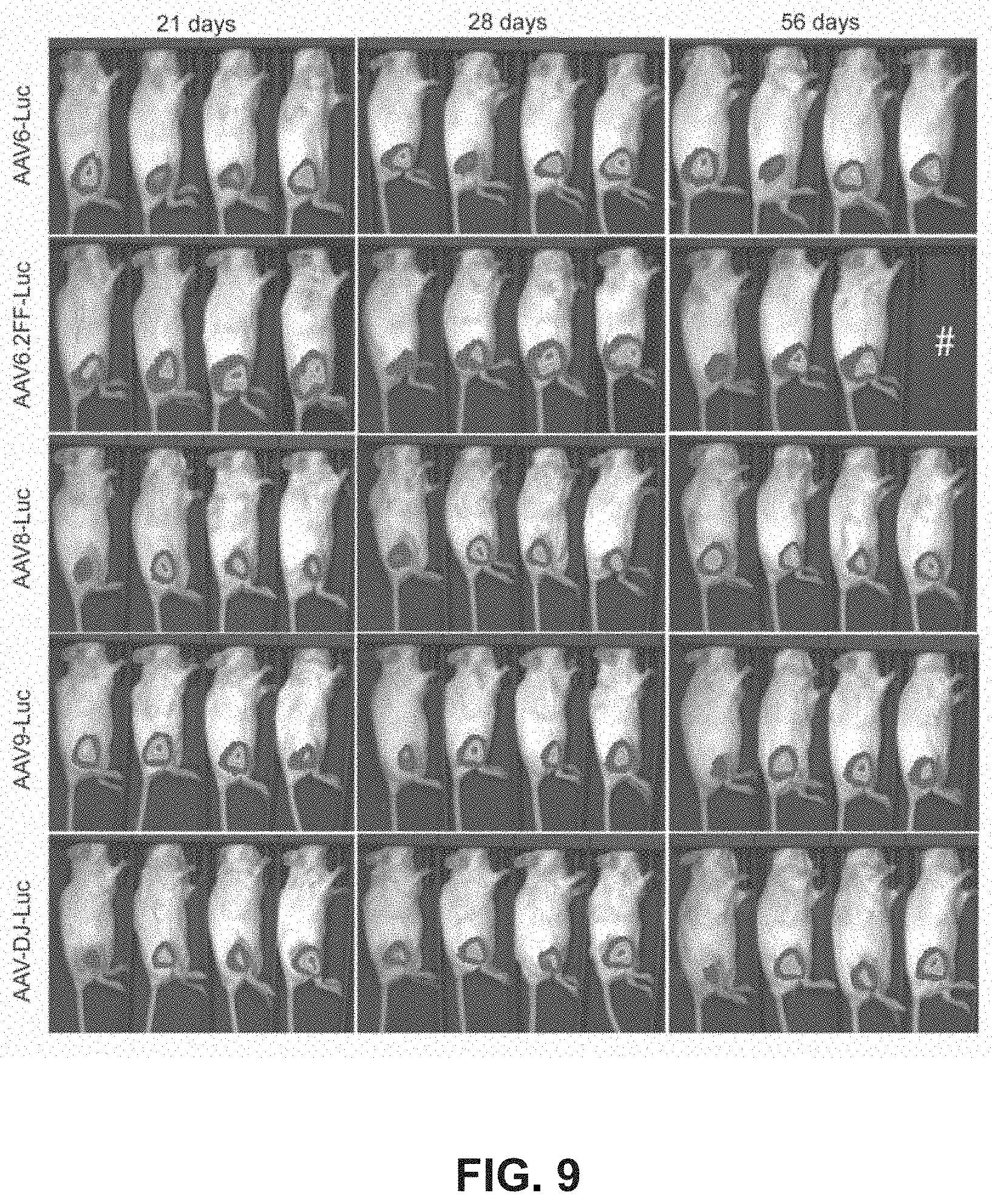
FIG. 9 shows in vivo imaging of AAV-mediated luciferase expression at later time points. Additional luciferase imaging of the mice described in FIGS. 8A-8B was conducted 21, 28 and 56 days after mice received a single IM injection of 1.times.10.sup.11 vg of AAV-Luc pseudotyped with five different AAV capsids. # Image was not obtained for this mouse.
FIGS. 10A-10D show substantial extramuscular transgene expression mediated by AAV8 and AAV9 vectors following intramuscular administration. Albino C57BL/6 mice (n=4/group) injected intramuscularly with 1.times.10.sup.11 vg of AAV8 (FIG. 10A) or AAV9 (FIG. 10B) vectors expressing firefly luciferase were imaged ventrally on days 1, 3, 7, 14, 21, 28 and 56 dpa. The relative photon emission (p/s/cm.sup.2/sr) produced by luciferase from AAV8 (FIG. 10C) and AAV9 (FIG. 10D) was quantified at various time points from 1 to 56 dpa and the percent extramuscular transgene expression relative to the total transgene expression graphed. Each line depicts the luciferase expression profile of an individual animal.
FIGS. 11A and 11B show region of interest (ROI) values for extramuscular transgene expression mediated by AAV8 and AAV9 vectors following intramuscular administration. Albino C57BL/6 mice (n=4/group) injected intramuscularly with 1.times.10.sup.11 vg of AAV8 (FIG. 11A) or AAV9 (FIG. 11B) vectors expressing firefly luciferase were imaged ventrally on days 1, 3, 7, 14, 21, 28 and 56 dpa and ROI values for areas where signal was quantified are shown.
FIGS. 12A and 12B show AAV6.2FF-mAb expression levels in mice following intramuscular administration. C57BL6 mice (n=4) were injected IM with 2.times.10.sup.11 vg of (FIG. 12A) AAV-2G4 or (FIG. 12B) AAV-5D2. Serum was collected from 1 to 126 dpa and analyzed at a 1:100 dilution for Ebola virus glycoprotein (EBOV GP) binding capacity by ELISA.
FIGS. 13A and 13B show AAV6.2FF-mediated mAb expression profiles in mice following intranasal administration. C57BL/6 mice (n=3/group) received 2.times.10.sup.11 vg of (FIG. 13A) AAV-2G4 or (FIG. 13B) AAV-5D2 by a modified intranasal instillation as described in Limbris et al [5]. Serum was collected and analyzed at a dilution of 1:100 for EBOV GP binding activity by ELISA.
FIGS. 14A and 14B show comparison of mAb expression levels in C57BL6 and BALB/c following intramuscular and intranasal administration of AAV-5D2. Groups of C57BL/6 and BALB/c mice (n=4) were injected IM (FIG. 14A) or IN (FIG. 14B) with 2.times.10.sup.11 vg of AAV-5D2. Serum was collected and analyzed at a dilution of 1:100 for EBOV GP binding capacity by ELISA. p=0.001. Serum mAb expression was compared using multiple t-tests, one per time point. ***Indicates p<0.001 and *indicates p<0.05.
FIG. 15 shows comparison of AAV6.2FF-mediated mAb cocktail (2G4+5D2) expression levels in mice following intravenous, single, or separate intramuscular injections. Serum mAb expression levels in C57BL/6 mice (n=4) following AAV6.2FF-2G4/AAV-5D2 cocktail (4.times.10.sup.1 vg total) administration either combined and administered intramuscularly (1.times.IM) or intravenously (IV) or separated and administered in two IM injections (2.times.IM) on either leg were evaluated by ELISA. No significant differences in mAb expression levels were observed when comparing routes of administration or combined vs. separate injections of AAV vectors by 2-way ANOVA.
FIGS. 16A-16D show AAV6.2FF-mediated expression of 5D2 and 7C9 provides complete protection against mouse-adapted Ebola virus (MA-EBOV) challenge. C57BL/6 mice (n=6/group) received an IM injection of 2.times.10.sup.11 vg of single AAV6.2FF-mAbs or a cocktail of 2.times.10.sup.11 vg of AAV6.2FF-2G4 and 2.times.10.sup.11 vg of AAV6.2FF-D2 for a total dose of 4.times.10.sup.11 vg. All AAV monotherapies were given 14 days prior to intraperitoneal challenge with 1000.times.LD.sub.50 MA-EBOV. FIG. 16A shows Kaplan-Meyer survival plots of AAV6.2FF-2G4, AAV6.2FF-5D2, AAV6.2FF-7C9 monotherapies and FIG. 16B shows the averaged mouse group weights. Survival of treated groups was compared to the mock group using the Mantel-Cox log rank test (p=0.0009 for 2G4, 0.0005 for 5D2 and 7C9). FIG. 16C shows Kaplan-Meyer survival plots of AAV6.2FF-2G4/AAV6.2FF-5D2 cocktail survival plots at various lead-times between AAV administration and MA-EBOV challenge and FIG. 16D shows the averaged mouse group weights. Survival of treated groups was compared to the mock group using the Mantel-Cox log rank test (p=0.005 for 14 and 7 days, 0.2801 for 3 days and >0.9999 for 1 day and same day).
FIGS. 17A and 17B show sustained AAV6.2FF-mediated mAb expression protects mice from MA-EBOV challenge five months after a single IM injection. C57BL/6 mice received an IM injection of 2.times.10.sup.11 vg of single AAV6.2FFmAbs (n=4/group) or a cocktail of 2.times.10.sup.11 vg of AAV6.2FF-2G4 and 2.times.10.sup.11 vg of AAV6.2FF-5D2 (n=8/group) for a total dose of 4.times.10.sup.11 vg. AAV vectors were administered 140 days prior to intraperitoneal challenge with 1000.times.LD.sub.50 MA-EBOV. FIG. 17A shows Kaplan-Meyer survival plots of AAV6.2FF-2G4, AAV6.2FF-5D2, and AAV6.2FF-2G4/AAV6.2FF-5D2 cocktail and FIG. 17B shows the averaged mouse group weights.
FIG. 18 shows biodistribution of AAV6.2FF, AAV8 and AAV9 following intramuscular delivery. BALB/c mice (n=4/group) were injected IM with 1.times.10.sup.11 vg of AAV6.2FF-, AAV8- or AAV9-Luciferase and were euthanized five days later for tissue harvest. Genomic DNA was extracted and analyzed by qPCR for AAV ITR copy number, which was normalized to input DNA concentration. Muscle samples were harvested from the site of AAV injection. Two-way ANOVA was used to determine statistical difference between the number of AAV genomes for 3 serotypes in each tissue. *p<0.0001.
FIGS. 19A and 19B show long-term expression of 5D2. 2.times.10.sup.11 vg of AAV6.2FF-5D2 (murine IgG2a) was administered by (FIG. 19A) intramuscular injection or (FIG. 19B) intranasal instillation to BALB/c mice. Serum samples were collected at weekly intervals, which were analyzed and quantified by EBOV GP ELISA at a 1:100 dilution. Each curve represents data derived from an individual mouse.
FIGS. 20A and 20B show effects of AAV6.2FF-2G4/AAV6.2FF-5D2 dose reduction. BALB/c mice (n=8/group) were administered various doses of the AAV6.2FF-2G4/AAV6.2FF-5D2 cocktail IM. 28 days following AAV administration, mice were challenged with 1000.times.LD.sub.50 MA-EBOV and monitored for (FIG. 20A) survival and (FIG. 20B) weight loss (plotted as group averages). ****p<0.0001.
FIGS. 21A-21D show EBOV GP reciprocal antibody titers pre and post challenge. Serum from the surviving mice in the AAV6.2FF-2G4/AAV6.2FF-5D2 dose reduction experiment of FIGS. 20A and 20B (1.times.10.sup.11 vg n=8, 5.times.10.sup.10 vg n=7, 1.times.10.sup.10 vg n=1) was analyzed by EBOV GP ELISA (FIG. 21A) immediately prior to challenge (28 days post AAV administration) and (FIG. 21B) 28 days post challenge. FIG. 21C shows the fold change in pre to post challenge reciprocal anti-GP titers at each dose. FIG. 21D shows the serum samples from 28 days post challenge were analyzed at a 1:100 dilution by EBOV VP40 ELISA.
FIGS. 22A-22D show endogenous humoral response to influenza A virus in the context of protective 2G4/5D2 antibody titers. FIG. 22A shows the schematic of experimental design. BALB/c mice were administered 1.times.10.sup.11 vg of AAV6.2FF-2G4/AAV6.2FF-5D2 IM 14 days prior to primary sub-lethal exposure to 600 HA units of influenza A virus (strain PR8) by IP injection. FIG. 22B shows the reciprocal EBOV GP titers from serum samples from AAV6.2FF-2G4/AAV6.2FF-5D2 or PBS treated groups. FIG. 22C shows the reciprocal HA titers following primary and secondary exposure to 600HA units of influenza A virus (strain PR8) in mice treated with AAV6.2FF-2G4/AAV6.2FF-5D2 or PBS. FIG. 22D shows the weight change in mice following primary influenza A virus (plotted as group averages).
FIGS. 23A-23D show AAV6.2FF-100 mediates complete protection from Ebola virus challenge. FIG. 23A shows the human IgG concentrations in the serum of BALB/c mice (n=4) that received 5.times.10.sup.9 vg of AAV6.2FF-100 IM. FIG. 23B shows the BALB/c mice (n=8/group) were administered various doses of AAV6.2FF-100 IM. 28 days post-AAV administration mice were challenged with 1000.times.LD.sub.50 MA-EBOV and monitored for (FIG. 23B) survival and (FIG. 23C) weight loss (resulted from individual mice plotted). ****p<0.0001, **p<0.01, *p<0.05. FIG. 23D shows the serum concentrations of human IgG were quantified immediately prior to challenge for the groups treated with doses of 1.times.10.sup.11 vg and 5.times.10.sup.10 vg. Mice with human IgG concentrations below the dotted line succumbed to MA-EBOV challenge.
FIGS. 24A and 24B show post-challenge protection mediated by AAV6.2FF-2G4/AAV6.2FF-5D2 and AAV6.2FF-100. BALB/c mice (n=8/group) were challenged with a reduced dose of 100.times.LD.sub.50 MA-EBOV IP and were subsequently injected IM with 1.times.10.sup.11 vg of the AAV6.2FF-2G4/AAV6.2FF-5D2 cocktail or 5.times.10.sup.10 vg of AAV6.2FF-100 and monitored for 28 days for (FIG. 24A) survival and (FIG. 24B) weight loss (results from individual mice plotted). **p<0.01, *p<0.05.
FIGS. 25A-25D show AAV6.2FF-MR191 mediates complete protection from Marburg virus challenge. FIG. 25A shows the serum human IgG concentrations in BALB/c mice (n=4) that received 6.times.10.sup.10 vg of AAV6.2FF-MR191 IM. FIGS. 25B-25D show BALB/c mice (n=8/group) were administered a low (1.times.10.sup.10 vg) or a high (1.times.10.sup.11 vg) dose of AAV6.2FF-MR191 IM. 28 days post-AAV administration mice were challenged with 1000.times.LD.sub.50 MA-MARV and monitored for (FIG. 25B) survival and (FIG. 25C) weight loss (results from individual mice plotted). ****p<0.0001. FIG. 25D shows the serum concentrations of human IgG were quantified immediately prior to challenge.
FIGS. 26A-26F show intramuscular administration of AAV6.2FF-Luciferase following prior exposure to heterologous or homologous vectors results in substantial transgene expression. FIG. 26A shows the schematic of experimental design. BALB/c mice (n=4/group) received an IM injection of 1.times.10.sup.11 vg of AAV6.2FF-, AAV6-, AAV8-, AAV9- or AAV-DJ-Luciferase in the left flank and were monitored for luciferase expression for 205 days. All mice were then injected in the right flank with 1.times.10.sup.11 vg of AAV6.2FF-Luciferase and transgene expression was quantified 28 days later. FIGS. 26B-26F show the quantification of luciferase expression in the left calf muscle 205 days post-primary vector injection as well as the transgene expression in both the left and right flank 28 day following secondary AAV6.2FF-Luciferase injection. Representative images of mice 28 days post-secondary AAV6.2FF-Luciferase administration demonstrate transgene expression in both flanks. Graphs show group means and error bars represent standard deviation. One-way ANOVA was used to determine statistical difference between the primary and secondary signal 28 days post-secondary AAV6.2FF-Luciferase injection; One-way ANOVA was used to determine statistical difference between the primary and secondary signal 28 days post-secondary AAV6.2FF-Luciferase injection; **p<0.01, ***p<0.001.
FIGS. 27A-27D show attempted re-administration of a second AAV6.2FF-mAb vector encoding a heterologous human IgG. FIG. 27A shows the schematic of experimental design. BALB/c mice (n=4) were injected with 6.times.10.sup.10 g of AAV6.2FF-MR191 IM (left flank) and serum human IgG concentrations were monitored for 70 days. On day 82, these mice were injected with 3.times.10.sup.10 vg of AAV6.2FF-100 in the right calf muscle and human IgG concentrations were evaluated until day 154. FIG. 27B shows the human IgG concentrations in treated mice. FIG. 27C shows MARV GP and EBOV GP concentrations determined by ELISA in select pre- and post-boost serum samples. FIG. 27D shows endogenous antibody response against the AAV6.2FF capsid and the mAb 100 antibody evaluated by ELISA. Graphs show group means, as well as individual data points for each mouse and error bars represent standard deviation.
FIGS. 28A-28I show AAV6.2FF capsid coat targets alveolar epithelial type 2 (AT2) cells in the lung tissue and expresses SPB protein in vitro. FIG. 28A shows the schematic of AAV6.2FF vectors expressing Luciferase or mCherry reporter genes. FIG. 28B shows the study design to determine lung tissue targeting by AAV6.2FF-Luciferase. FIG. 28C shows IVIS imaging of transgenic SPB mice either untreated or intratracheally injected with AAV6.2FF-Luciferase. FIG. 28D shows the quantification of IVIS images. FIG. 28E shows the mean weight measurements from all mice from (FIG. 28C). FIG. 28F shows the study design to determine whether AT2 cells were targeted by AAV6.2FF-mCherry. FIG. 28G shows epifluorescence image of Pro-SPC, mCherry and DAPI from lung sections of SPB deficient mice 5 weeks after intratracheal injection of AAV6.2FF-mCherry+Bovine Lipid Extract Surfactant (BLES). Arrows point to lung cells expressing Pro-SPC which indicated that they were AT2 cells which also expressed mCherry produced from AAV6.2FF. FIG. 28H shows the schematic of codon optimized murine SPB cDNA in expression plasmid. FIG. 28I shows the Western blot of HEK293 cells transduced with AAV6.2FF-SPB (MOI=20,000; lane 2, AAV-SPB; i.e. AAV-SPB-myc vector) or transiently transfected with the SPB expression plasmid (lane 3, SPB-myc; i.e. plasmid genome used to generate the AAV-SPB-myc vector). "Pre-SPB" denotes the non-processed form of myc-SPB (myc tag on the 3' end), prior to SPB being cleaved by multiple proteases into its mature form.
FIGS. 29A-29L show AAV6.2FF-SPB treatment increases SPB expression, maintains normal alveolar epithelial type 2 (AT2) cell structure, and improves lung function in a SPB deficient mouse model. FIG. 29A shows the study design to determine whether AAV6.2FF-SPB improved lung structure and function. "On Dox" denotes doxycycline feed was not removed during the course of the study. FIG. 29B shows the percentage change in body weight over 4 weeks following AAV injection. FIG. 29C shows the percentage change in body weight over 3 to 4 days following doxycycline removal. FIG. 29D shows the representative macroscopic lung images 3 to 4 days following doxycycline removal. FIG. 29E shows the representative Hematoxylin and Eosin (H&E; 20.times.) and Wright-Giemsa Jenner (WGJ; 40.times.) staining of paraffin embedded whole left lungs following doxycycline removal. FIG. 29F shows the representative epi-fluorescence images of Pro-SPC, SPB, and DAPI from OCT frozen right lung sections following doxycycline removal. Arrows indicate SPB staining. FIG. 29G shows the representative TEM images of two different fields of view of AT2 cells following doxycycline removal. White arrows indicate lamellar bodies and black arrows indicate mitochondria. Scale bars represent 2 .mu.m in 10,000.times. images and 500 nm in 20,000.times. image. N, nucleus. FIG. 29H shows the pressure volume curve following doxycycline removal corrected for body weight (in mug). FIG. 29I shows % V10 corrected for body weight. FIG. 29J shows the total lung capacity corrected for body weight (in mug). FIG. 29K shows the residual Volume corrected for body weight (in mUg). FIG. 29L shows the compliance corrected for body weight (in mL/cmH.sub.2O*g). All P values=ordinary one-way ANOVA with Tukey's multiple comparisons post hoc test; ns=not significant.
FIG. 30 shows improved median survival with Bovine Lipid Extract Surfactant (BLES) and endotracheal tube (intubation) delivery of 10.sup.11 vg/mouse (intermediate dose) AAV6.2FF-SPB into SPB deficient mice. Kaplan-Meier survival curve of 10.sup.11 vg/mouse (intermediate dose).+-.BLES treatment delivered into intubated mice. Survival curve P values=Log-rank, Mantel-Cox test; ns=not significant.
DETAILED DESCRIPTION
Unless otherwise indicated, the definitions and embodiments described in this and other sections are intended to be applicable to all embodiments and aspects of the present disclosure herein described for which they are suitable as would be understood by a person skilled in the art.
In understanding the scope of the present disclosure, the term "comprising" and its derivatives, as used herein, are intended to be open ended terms that specify the presence of the stated features, elements, components, groups, integers, and/or steps, but do not exclude the presence of other unstated features, elements, components, groups, integers and/or steps. The foregoing also applies to words having similar meanings such as the terms, "including", "having" and their derivatives. The term "consisting" and its derivatives, as used herein, are intended to be closed terms that specify the presence of the stated features, elements, components, groups, integers, and/or steps, but exclude the presence of other unstated features, elements, components, groups, integers and/or steps. The term "consisting essentially of", as used herein, is intended to specify the presence of the stated features, elements, components, groups, integers, and/or steps as well as those that do not materially affect the basic and novel characteristic(s) of features, elements, components, groups, integers, and/or steps.
As used herein, the singular forms "a", "an" and "the" include plural references unless the content clearly dictates otherwise.
Compositions
The term "adeno-associated virus" (AAV), as used herein, includes without limitation AAV type 1, AAV type 2, AAV type 3 (including types 3A and 3B), AAV type 4, AAV type 5, AAV type 6, AAV type 7, AAV type 8, AAV type 9, AAV type 10, AAV type 11, avian AAV, bovine AAV, canine AAV, equine AAV, and ovine AAV and any other AAV.
The genomic sequences of various AAV as well as the sequences of the ITRs, rep proteins, and capsid proteins are known in the art. Such sequences may be found in the literature or in public databases such as the GenBank database. See, e.g., GenBank Accession Numbers NC_002077, NC_001401, NC_001729, NC_001863, NC_001829, NC_001862, NC_000883, NC_001701, NC_001510, AF063497, U89790, AF043303, AF028705, AF028704, J02275, J01901, J02275, X01457, AF288061, AH009962, AY028226, AY028223, NC_001358, NC_001540, AF513851, AF513852, AY530579, AY631965, AY631966; the disclosures of which are incorporated herein in their entirety. For instance, the GenBank Accession Number for AAV6 is AF028704.1.
The present inventors have provided a rAAV particle with mutated capsid protein, where the AAV particle exhibits greater transduction of muscle, airway, liver, central nervous system, retina or lung cells compared to wild-type AAV. The present inventors have further provided methods of delivering a therapeutic agent to a muscle, airway, liver, central nervous system, retina or lung cell in a subject, and methods of treating or preventing an infectious, acquired or genetic disease, with said AAV particle.
Accordingly, herein provided is a recombinant adeno-associated viral (rAAV) particle comprising a mutated capsid protein encapsidating a rAAV vector genome, wherein the mutated capsid protein comprises amino acid substitutions at amino acids 129, 445, and 731 of the AAV6 capsid protein sequence as set forth in SEQ ID NO:1. In a specific embodiment, the mutated capsid protein has amino acid substitutions Phe129Leu, Tyr445Phe and Tyr731Phe, wherein the mutated capsid protein is mutated AAV6 capsid protein having an amino acid sequence as shown in SEQ ID NO:2.
Amino acid sequences described herein are set out in Table 1.
TABLE-US-00001 TABLE 1 Sequences SEQ ID NO: 1: amino MAADGYLPDWLEDNLSEGIREWWDLKPGAPKPKA acid sequence of NQQKQDDGRGLVLPGYKYLGPFNGLDKGEPVNAA parental AAV6 capsid DAAALEHDKAYDQQLKAGDNPYLRYNHADAEFQE protein RLQEDTSFGGNLGRAVFQAKKRVLEPFGLVEEGA KTAPGKKRPVEQSPQEPDSSSGIGKTGQQPAKKR LNFGQTGDSESVPDPQPLGEPPATPAAVGPTTMA SGGGAPMADNNEGADGVGNASGNWHCDSTWLG DRVITTSTRTWALPTYNNHLYKQISSASTGASNDN HYFGYSTPWGYFDFNRFHCHFSPRDWQRLINNN WGFRPKRLNFKLFNIQVKEVTTNDGVTTIANNLTST VQVFSDSEYQLPYVLGSAHQGCLPPFPADVFMIPQ YGYLTLNNGSQAVGRSSFYCLEYFPSQMLRTGNN FTFSYTFEDVPFHSSYAHSQSLDRLMNPLIDQYLYY LNRTQNQSGSAQNKDLLFSRGSPAGMSVQPKNW LPGPCYRQQRVSKTKTDNNNSNFTWTGASKYNLN GRESIINPGTAMASHKDDKDKFFPMSGVMIFGKES AGASNTALDNVMITDEEEIKATNPVATERFGTVAVN LQSSSTDPATGDVHVMGALPGMVWQDRDVYLQG PIWAKIPHTDGHFHPSPLMGGFGLKHPPPQILIKNT PVPANPPAEFSATKFASFITQYSTGQVSVEIEWELQ KENSKRWNPEVQYTSNYAKSANVDFTVDNNGLYT EPRPIGTRYLTRPL SEQ ID NO: 2: amino MAADGYLPDWLEDNLSEGIREWWDLKPGAPKPKA acid sequence of the NQQKQDDGRGLVLPGYKYLGPFNGLDKGEPVNAA DAAALEHDKAYDQQLKAGDNPYLRYNHADAEFQE mutated AAV6 capsid RLQEDTSFGGNLGRAVFQAKKRVLEPLGLVEEGA protein (AAV6.2FF) KTAPGKKRPVEQSPQEPDSSSGIGKTGQQPAKKR LNFGQTGDSESVPDPQPLGEPPATPAAVGPTTMA SGGGAPMADNNEGADGVGNASGNWHCDSTWLG DRVITTSTRTWALPTYNNHLYKQISSASTGASNDN HYFGYSTPWGYFDFNRFHCHFSPRDWQRLINNN WGFRPKRLNFKLFNIQVKEVTTNDGVTTIANNLTST VQVFSDSEYQLPYVLGSAHQGCLPPFPADVFMIPQ YGYLTLNNGSQAVGRSSFYCLEYFPSQMLRTGNN FTFSYTFEDVPFHSSYAHSQSLDRLMNPLIDQYLYF LNRTQNQSGSAQNKDLLFSRGSPAGMSVQPKNW LPGPCYRQQRVSKTKTDNNNSNFTWTGASKYNLN GRESIINPGTAMASHKDDKDKFFPMSGVMIFGKES AGASNTALDNVMITDEEEIKATNPVATERFGTVAVN LQSSSTDPATGDVHVMGALPGMVWQDRDVYLQG PIWAKIPHTDGHFHPSPLMGGFGLKHPPPQILIKNT PVPANPPAEFSATKFASFITQYSTGQVSVEIEWELQ KENSKRWNPEVQYTSNYAKSANVDFTVDNNGLYT EPRPIGTRFLTRPL SEQ ID NO: 3: nucleotide TGGGGCAGAGCTTGTGAAGCCAGGGGCCTCAGT sequence of monoclonal CAAGTTGTCCTGCACAGCTTCTGGCTTCAACATT antibody 1H3 heavy chain AAAGACACCTATATACATTGGGTGAAACAGGGGC CTGAACAGGGCCTGGAGTGGATTGGAAGGATTG ATCCTGCGAATGGTAATACTAAATATGACCCGAA GTTCCAGGGCAAGGCCACTATCACAGCAGACAC ATCCTCCAATACAGCCTACCTGCAGCTCAGCGG CCTGACATCTGAGGACACTGCCGTCTATTACTGT GCTAGGGAGTCGAGGATATCTACTATGCTTACGA CGGGGTACTTTGACTACTGGGGCCAAGGCACCA CTCTCACAGTCTCCTCAGCCAAAACAACAGCCCC ATCG SEQ ID NO: 4: nucleotide GCAATCATGTCTGCATCTCCAGGGGAGAAGGTC sequence of monoclonal ACCATGACCTGCAGTGCCAGCTCAAGTGTAAGTT antibody 1H3 light chain ACATGTACTGGTACCAGCAGAAGCCAGGATCCT CCCCCAGACTCCTGATTTATGACACATCCAACCT GGCTTCTGGAGTCCCTGTTCGCTTCAGTGGCAG TGGGTCTGGGACCTCTTACTCTCTCACAATCAGC CGAATGGAGGCTGAAGATGCTGCCACTTATTACT GCCAGCAGTGGAGTAGTTACCCGTACACGTTCG GAGGGGGGACCAAGCTGGAAATAAAACGGGCTGAT SEQ ID NO: 5: nucleotide TGGAGGAGGCTTGATGCAACCTGGAGGATCCAT sequence of monoclonal GAAACTCTCCTGTGTTGCCTCAGGATTCACTTTC antibody 2G4 heavy chain AGTAACTACTGGATGAACTGGGTCCGCCAGTCT CCAGAGAAGGGGCTTGAGTGGGTTGCTGAAATT AGATTGAAATCTAATAATTATGCAACACATTATGC GGAGTCTGTGAAAGGGAGGTTCACCATTTCAAG AGATGATTCCAAAAGGAGTGTCTACCTGCAAATG AATACCTTAAGAGCTGAAGACACTGGCATTTATT ACTGTACCCGGGGGAATGGTAACTACAGGGCTA TGGACTACTGGGGTCAAGGAACCTCAGTCACCG TCTCCTCAGCCAAAACAACACCCCCATCA SEQ ID NO: 6: GCCTCCCTATCTGTATCTGTGGGAGAAACTGTCT nucleotide sequence CCATCACATGTCGAGCAAGTGAGAATATTTACAG of monoclonal TAGTTTAGCATGGTATCAGCAGAAACAGGGAAAA antibody 2G4 light TCTCCTCAGCTCCTGGTCTATTCTGCAACAATCT chain TAGCAGATGGTGTGCCATCAAGGTTCAGTGGCA GTGGATCAGGCACTCAGTATTCCCTCAAGATCAA CAGCCTGCAGTCTGAAGATTTTGGGACTTATTAC TGTCAACATTTTTGGGGTACTCCGTACACGTTCG GAGGGGGGACCAAGCTGGAAATAAAACGGGCTG T SEQ ID NO: 7: TGGACCTGAGCTGGAGATGCCTGGCGCTTCAGT nucleotide sequence GAAGATATCCTGCAAGGCTTCTGGTTCCTCATTC of monoclonal ACTGGCTTCAGTATGAACTGGGTGAAGCAGAGC antibody 4G7 heavy AATGGAAAGAGCCTTGAGTGGATTGGAAATATTG chain ATACTTATTATGGTGGTACTACCTACAACCAGAA ATTCAAGGGCAAGGCCACATTGACTGTGGACAA ATCCTCCAGCACAGCCTACATGCAGCTCAAGAG CCTGACATCTGAGGACTCTGCAGTCTATTACTGT GCAAGATCGGCCTACTACGGTAGTACTTTTGCTT ACTGGGGCCAAGGGACTCTGGTCACTGTCTCTG CAGCCAAAACAACAGCCCCATCG SEQ ID NO: 8: GCCTCCCTATCTGCATCTGTGGGAGAAACTGTCA nucleotide sequence CCATCACATGTCGAGCAAGTGAGAATATTTACAG of monoclonal TTATTTAGCATGGTATCAGCAGAAACAGGGAAAA antibody 4G7 light TCTCCTCAGCTCCTGGTCTATAATGCCAAAACCT chain TAATAGAGGGTGTGCCATCAAGGTTCAGTGGCA GTGGATCAGGCACACAGTTTTCTCTGAAGATCAA CAGCCTGCAGCCTGAAGATTTTGGGAGTTATTTC TGTCAACATCATTTTGGTACTCCATTCACATTCGG CTCGGGGACAGAGTTGGAAATAAAACGGGCTGA T SEQ ID NO: 9: GGGACCTGGCCTGGTGAGACCTTCTCAGTCTCT nucleotide sequence GTCCCTCACCTGCACTGTCACTGGCTACTCAATC of monoclonal ACCAGTGATTATGCCTGGAACTGGATCCGGCAG antibody 5D2 heavy TTTCCAGGAAACAAACTGGAGTGGCTGGGCTATA chain TAACCAACACTGGTAGCACTGGCTTCAACCCATC TCTCAAAAGTCGAATCTCTATCACTCGAGACACA TCCAAGAACCAGTTCTTCCTGCAGTTGATTTCTG TGACTACTGAGGACACAGCCACATATCACTGTGC AAGGGGCCTTGCTTACTGGGGCCAAGGGACTCT GGTCACTGTCTCTGCAGCCAAAACAACAGCCCC ATCG SEQ ID NO: 10: CTCACTTTGTCGGTTACCATTGGACAACCAGCCT nucleotide sequence CCATCTCTTGCAAGTCAAGTCAGAGCCTCTTAGA of monoclonal TAGTGATGGAAAGACATATCTGAATTGGTTGTTA antibody 5D2 light CAGAGGCCAGGCCAGTCTCCAAAGCGCCTAATC chain TATCTGGTGTCTAAACTGGACTCTGGAGTCACTG ACAGGTTCACTGGCAGTGGATCAGGGACAGATT TCACACTGAAAATCAGCAGAGTGGAGGCTGAGG ATTTGGGAGTTTATTATTGTTGGCAAGGTACACA CTCTCCATTCACGTTCGGCTCGGGGACAAAGTT GGAAATAAAACGGGCTGAT SEQ ID NO: 11: TGGGGCAGAGCTTGTGAAGCCAGGGGCCTCAGT nucleotide sequence CAAGTTGTCCTGCACAGCTTCTGGCTTCAACATT of monoclonal AAAGACACCTATATGCACTGGGTGAAGGAGAGG antibody 7C9 heavy CCTGACAAGGGCCTGGAGTGGATTGGAAGGATT chain GATCCAGCGAATGGTAATACTAAATGTGACTCGA GGTTTCAGGGCAAGGCCACTATAACAGCAGACA CATCCTCCAACACAGCCTACCTGCAGCTCAGCA GCCTGACATCTGAGGACACTGCCGTCTATTACTG TGCTAGAAGGATCTACTTTGGTAAGGGCTTTGAC TTTTGGGGCCAAGGCACCACTCTCACAGTCTCCT CAGCCAAAACAACAGCCCCATCG SEQ ID NO: 12: TCCTCCCTGAGTGTGTCAGCAGGAGAGAAGGTC nucleotide sequence ACTATGAGCTGCAAGTCCAGTCAGAGTCTGTTTA of monoclonal ACAGTGGAGATCAAAAGAACTACTTGGCCTGGTA antibody 7C9 light CCAGCAGAAACCAGGGCAGCCTCCTAAACTGTT chain GATCTACGGGGCATCCACTAGGGAATCTGGGGT CCCTGATCGCTTCACAGGCAGTGGATCTGGAAC CGATTTCACTCTTACCATCAGCAGTGTGCAGGCT GAAGACCTGGCAGTTTATTACTGTCAGAATGATC AATTTTATCCTCCCACGTTCGGTGATGGGACCAA GCTGGACCTGAAACGGGCTGAT SEQ ID NO: 13: CAGGTGCAACTTCAGGAGTCAGGGCCTGGCCTC nucleotide sequence GTCAAACCAAGCGATACACTGAGTTTGACTTGCA of monoclonal CAGTGAGTGGGGGTAGTTTGTCTAGTTTCTATTG antibody 100 heavy GTCTTGGATTCGGCAACCCCCCGGCAAAGGTCT chain TGAGTGGATAGGATACATCTACTACTCAGGGTCC CCCAATTACTCACCTTCCCTGGAATCTAGGGTTA CTATGTCCGTGGACACAACCCGAAATCAAATATC CTTGAAGCTTGACTCCGTGACAGCCGCAGACAC CGCCGTTTACTACTGCGTCCGAGCATCCCGCTC CTATTATTGGGGTAGCTATCGACCAACTGCTTTT GATTCTTGGGGACAGGGGACACTTGTAACTGTCT CAAGC SEQ ID NO: 14: TCTTATGAACTCACTCAGCCACTTTCTGTCAGTG nucleotide sequence TCAGCCCAGGTCAGACCGCCATATTTACCTGCA of monoclonal GTGGCGATAACTTGGGCGACAAATACGTGTGTT antibody 100 light GGTTTCAGCAACGGCCCGGCCAGTCACCCATGC chain TCCTTATCTATCAAGACAACAAGCGACCTTCAGG CATCCCCGAGCGGTTTAGTGGGTCTAACTCTGG GAACACCGCTACATTGACTATTAGTGGAACTCAG TCAACCGATGAAGCCGACTATTACTGCCAAACTT GGGATTCCACCGTAGTTTTCGGCGGCGGAACTA AGTTGACAGTGTTG SEQ ID NO: 15: GAGGTGCAACTGGTCGAATCTGGTGGAGGACTT nucleotide sequence ATCCAGCCTGGTGGCAGCCTGAGACTTTCTTGC of monoclonal GCAGCTAGTGGATTTGCTTTGAGGATGTATGACA antibody 114 heavy TGCATTGGGTACGACAGACAATAGACAAACGGTT chain GGAATGGGTTTCTGCTGTAGGCCCTAGCGGAGA CACCTACTACGCAGACAGCGTGAAGGGTAGGTT TGCAGTTTCACGGGAGAACGCTAAGAACAGCCT CTCACTTCAAATGAATAGCCTCACCGCTGGCGAC ACAGCAATCTACTACTGTGTAAGAAGTGATAGGG GTGTTGCCGGGCTGTTTGACAGTTGGGGACAGG GTATTTTGGTAACCGTGAGCAGT SEQ ID NO: 16: GACATACAGATGACCCAAAGCCCTTCATCCCTCT nucleotide sequence CTGCTTCTGTAGGTGACAGGATTACAATCACCTG of monoclonal CCGCGCAAGTCAGGCTTTTGACAACTATGTGGC antibody 114 light ATGGTATCAGCAACGACCAGGGAAGGTCCCAAA chain ATTGCTGATCTCCGCTGCCTCCGCTCTTCACGCA GGAGTCCCTTCTAGGTTTTCTGGATCAGGGTCC GGTACTCACTTCACCCTCACTATATCAAGTCTCC AACCTGAAGACGTGGCCACCTACTACTGCCAGA ATTATAACAGTGCTCCACTTACTTTTGGTGGAGG AACAAAGGTAGAGATAAAA SEQ ID NO: 17: CAAGTTCAATTGCAAGAGTGGGGGGAGGGCCTG nucleotide sequence GTTAAGCCCAGCGAAACTTTGAGCTTGACATGTG of monoclonal CTGTGTATGGCGGCTCTATCAGTGGTTACTACCA antibody CA45 heavy CTGGAATTGGATAAGGCTCCCCCCCGGCAAAGG chain GCTCGAGTGGATCGGGAATATAGATGGTAACAG CGCAAGTACAAATTACAATCCTTCTCTGAAGACC CGAGTGACCATTAGCAAGGATACCAGCAAAAATC AAATTAGTTTGAAAGTACGATCCTTGACTGCCGC CGACACCGCCGTCTACTATTGCGCTAGGGACCC TGGATTCACTATATTTGGAGTAGTTATCACATCAT GGTCCGGCCTCGACTCTTGGGGTCAGGGGGCA GTGGTGACAGTTTCATCT SEQ ID NO: 18: GATATACAGATGACACAAAGTCCCTCATCTTTGT nucleotide sequence CAGCTTCTGTGGGGGATACCGTTACTATTACTTG of monoclonal TAGGGCATCCCAATCAATTTCTAATAATCTGGCA antibody CA45 light TGGTATCAACAGCGCCCTAGAAGAGCCCCACAA chain CTGCTGATCTACGCCGCCTCTAACCTTGCTTCAG GTGTGCCCTCCCGATTTTCAGGATCAGGTTCAG GGACAGATTTTACTCTCACAATTTCCTCTCTTCAA GCAGAGGACTTTGCTGCTTACTACTGCCAGCAG CATAATACTCTCCCTCTCACCTTTGGTGGTGGAA CAAAAGTTGAGATTAAG SEQ ID NO: 19: CAAGTCCAACTGGTCCAATCAGGAGTGACCCTT nucleotide sequence GTTCAACCTGGTGGGAGCCTTAGAGTTAGTTGTG of monoclonal CAGCCAGCGGTTTTACCTTTAGTAGCTATGCTAT antibody ADI-15878 GAGCTGGGTACGCCAAGCTCCTGGCAAGGGCCT heavy chain GGAGTGGGTAAGCGCTATCTCCGGTTTGGGGGG TTCTACATACTACGCAGATTCAGTTAAGGGAAGG TTCACTATTTCTCGGGATAACTCCAAAAACACACT TTATCTTCAGATGAACTCTCTTCGCGCAGAAGAC ACTGCTGTT SEQ ID NO: 20: GACATAGTGCTGACTCAGAGTCCTTCCACTCTTT nucleotide sequence CAGCTAGTGTAGGGGACCGCGTCACAATAACAT of monoclonal GCAGAGCTTCACAATCCATAAGCTCCTGGTTGGC
antibody ADI-15878 TTGGTACCAACAGAAGCCTGGGGAAGCTCCCAA light chain ACTCTTGATTAGCGACGCTTCAAGTTTGGAGTCA GGGGTACCCTCAAGGTTCTCTGGCAGTGGGTCC GGTACAGAATTTACCCTCACAATAAGCAGCTTGC AACCTGACGACTTCGCTACATACTATTGTCAGCA GTATTATAGTTCTCCAACCTTCGGAGGGGGTACC AAAGTGGAAATTAAA SEQ ID NO: 21: GAAGTTCAGTTGGTAGAGTCAGGCGGCGGATTG nucleotide sequence GTGCAACCTGGTGGGTCCCTCAGATTGAGTTGT of monoclonal GCCGCTTCCGGGTTTACAGGATTTACTTTTTCTG antibody FVM02p ACTACGCATTCTATTGGGTGAGACAGGCACCTG heavy chain GAAAAGGTTTGGAATGGGTAGGATTCATTAGGG GCAAGGCATACGGAGGTACAGCAGACTACGCCG CTTCTGTTAAAGGAAGGTTCACCATTTCTCGAGA TAATTCCAAAAACACTGCCTATTTGCAGATGAGC TCTTTGAAGACA SEQ ID NO: 22: GACATCGTTCTCACACAATCACCCCTCAGCTTGC nucleotide sequence CCGTCACACCCGGCGAGCCAGCTAGTATCAGTT of monoclonal GTAGGTCCTCTCAGAGTTTGCTGCACTCTGGGG antibody FVM02p light GTAAAACTTACCTCTATTGGTATCTTCAAAAGCCT chain GGTCAGTCCCCCCAGCTTCTTATTCATGAAGTAT CCAACAGAGCATCTGGAGTGCCTGATAGATTTTC TGGTAGTGGTTCTGGAACTGATTTCACCTTGAAG ATCAGCCGAGTGGAGGCCGAGGACGTGGGAGT ATATTACTGCATGCAGGGAATACAGTTGCCTCTG ACCTTTGGGGGAGGAACAAAAGTTGAGATAAAAC GAACTGTA SEQ ID NO: 23: GAAGTGCAACTTGTCCAATCTGGGGGCGGCCTG nucleotide sequence GTACAACCAGGCGGATCTATGCGGCTCTCATGC of monoclonal GAGGCATCAGGACTGTCTCTCAGTGATTATTTTA antibody FVM04 TGCATTGGGTCCGGCAGGCCCAGGGAAAAGGTT heavy chain TGGAGTGGATCGGTTTGATACAGACAAAGGCTTT CACCTATAAAACCGAATACCCTGCTGCTGTTAAG GGTCGCTTTACCATCTCACGGGACGATAGTAAGA ACACTCTGTATTTGCAAATGTCTTCACTTAAGCCA GAGGATACAGCATTGTACTACTGCATTGCCGTGA CCCCCGACTTTTATTACTGGGGTCAGGGAGTGTT GGTCACCGTATCTTCC SEQ ID NO: 24: GACGTTGTTATGACCCAGTCCCCAAGTTTCCTGT nucleotide sequence CTGCTAGTGTTGGCGATAGAGTAACTATCACCTG of monoclonal TAGGGCTAGTCAAGACATAACCATAAATCTCAAT antibody FVM04 light TGGTTTCAGCATAAGCCTGGAAAGGCCCCAAAG chain CGGCTGATCTACGTTGCATCCCGCTTGGAACGA GGGGTGCCCAGTCGGTTCTCAGGAAGCGGCAG CGGGACAGAATTTACTCTTACTATTTCAAGCCTT CAGCCTGAAGATTTTGCCACATATTACTGTCAAC AGTATAATAACTATCCCCTGACCTTTGGTCCTGG GACAAAACTCGATATAAAGCGAACCGTA SEQ ID NO: 25: GAAATTGTGATGACCCAGTCTCCAGCCATCATGT nucleotide sequence CTGTGTCTCCAGGGAAAAGAGCCACCCTCTCCT of monoclonal GCAGGGCCAGTCAGAGTGTCAGTAGCAACTTAG antibody BDBV223 CCTGGTACCAGCGGAAACCTGGCCAGGCTCCCA heavy chain GGCTCCTCATCTATGGTTCTTCCACCAGGGCCAC TGGTATCCCAGCCAGGTTCAGTGGCAGTGGGTC TGGGACAGAGTTCACTCTCACCATCAGCAGCCT GCAGTCTGAGGATTTTGCAGTTTATTACTGTCTG CAATATTATAACTGGCCTCGGACGTTCGGCCAAG GGACCAAGGTGGAAATCAAA SEQ ID NO: 26: CAGGTGCAGCTACAGCAGTGGGGCGCAGGACT nucleotide sequence GTTGAAGCCTTCGGAGACCCTGTCCCTCACCTG of monoclonal CGCTGTCTATGGTGGGTCCTTCACGACTACCTAC antibody BDBV223 TGGAATTGGATCCGCCAGCCCCCAGGGAAGGG light chain GCTGGAATGGATAGGGGAAGTCAATTATAGTGG AAACGCCAACTACAACCCGTCCCTCAAGGGTCG AGTCGCCATATCAGTGGACACATCCAAGAACCA GTTCTCCCTGAGGTTGAACTCTGTGACCGCCGC GGACACGGCTATATATTACTGTACGAGTCGCATA CGTTCGCACATTGCCTACTCGTGGAAGGGGGAC GTCTGGGGCAAAGGGACCACGGTCACCGTCTCC TCA SEQ ID NO: 27: CAGTTGCAACTTCAAGAGTCTGGTCCTGGCTTGG nucleotide sequence TCAAACCAAGCGAGACTCTCAGCCTGACTTGTAC of monoclonal TGTATCCGGTGACAGCATAAACAACACAAATTAT antibody MR72 heavy TACTGGGCTTGGATCAGGCAGCCACCAGGGAAG chain GGCCTTGAGTATATTGGTTCAATCTATTACTCTG GTAGTACATACTATAACCCTAGCTTGAAGAGTAG GGTAACTATGTCAGTGGATGCTAGTAAGAACCAG TTCTCACTGAGACTGTCCTCTGTCACTGCTGCTG ACACTGCTGTGTACTACTGTGCTACCCACCCCAC ACTCGGCGCTTTTGTATTGCTGTGGTTTGGTGCC AACTTCGATCACTGGGGTCAGGGTACTTTGGTGA CAGTGTCTAGC SEQ ID NO: 28: CAAGCCGTCGTCACACAACCCCCCTCAGTCAGC nucleotide sequence GGAGCACCTGGTCAGCGGGTCACTATTAGCTGT of monoclonal ACCGGCAGCAGTTCCAATATAGGCGCTAACTATG antibody MR72 light ATGTGCATTGGTATCAACAGCTCCCTGGGACTGC chain TCCTAAATTGCTGATGTATTCCAATACCAACAGA CCATCCGGAGTTCCCGATAGGTTTAGTGGGTCC AAGAGCGGAACCTCAGCTTCACTGGCAATTACC GGGCTGCAAGCAGAAGACGAAGCTGACTATTAC TGCCAAAGTTACGACAATAGTCTTAATAGCTGGG TTTTTGGAGGGGGAACACAACTGACCGTT SEQ ID NO: 29: CAGGTGCAACTTGTCCAGTCCGGAGCTGAAGTT nucleotide sequence AAGAAGCCTGGTGCCAGCGTCAAGGTGAGTTGC of monoclonal AAAGCATCCGGACATACATTTACAACATACGCCA antibody MR82 heavy TACATTGGGTTCGCCAAGCACCTGGACAAGGTC chain TTGAGTGGATGGGATGGATAAACCCAGATAATGA CAACACTGAATACTCCCAAAAATTTCAGGGAAGG GTAACCATAACACGGGACACATCAGCCTCTACTG CCTACATGGAGCTGTCAAGTCTGATCTCTGAAGA TACAGCAGTATTTTACTGTGCAAGTGCATCCTAT ACCTTTTGGTCCGGATATTATAGTGGGCTCGATT ATTGGGGACAGGGGACTCTGGTAACCGTAAGCT CC SEQ ID NO: 30: GAGATAGTATTGACTCAGTCTCCCGGTACATTGT nucleotide sequence CTCTCTCCCCAGGAGAAAGAGCTACACTCTCATG of monoclonal TCGAGCCTCTCAAAGCGTCTCCATCAATTATCTG antibody MR82 light GCCTGGTATCAACAGAAGCCTGGTCAAGCACCT chain AGGCTCCTTATCTACGGAGCAAGCTCACGGGCT ACTGGTATTCCCGATAGGTTCTCTGGCTCAGGTT CCGGCACCGATTTCACTCTCACAATTAGCCGATT GGAACCAGAAGATTTCGCCGTCTATTATTGTCAA CAATATGGTAGCTCTCCACCCTGGACATTCGGAC CTGGGACCAAGGTGGACATAAAA SEQ ID NO: 31: CAGCTCCAGCTGCAAGAATCTGGTCCCGGTCTT nucleotide sequence GTTAAACCTAGTGAAACACTTAGCCTGACTTGCA of monoclonal CTGTCTCAGGCGGGTCAATATCATCTTCCAGTTA antibody MR78 heavy TTACTGGGGCTGGATCAGGCAACCCCCTGGGAA chain AGGTCTCGAATGGATTGGCTCTGTTTATTATAGC GGAGGTGCCAGTTACAATCCTAGTCTCAAGTCAC GAGCCACTATTAGCGTTGATACCAGCAAAAACCA ATTCAGTTTGAATCTGGATTCAGTAAGCGCAGCC GACACAGCCATTTATTACTGTGCTTCCATTTATG GAAGTGGGACTTTCTATTATTACTTTTACATGGAC GTGTGGGGGAAGGGTTCAACAGTTACTGTAAGC TCC SEQ ID NO: 32: GACATTCAGATGACTCAATCTCCCAGTTCCCTGT nucleotide sequence CAGCTAGTGTTGGGGACCGGGTAACCATCACCT of monoclonal GTCAAGCAAGCCAGGTCATCAGTAACTACCTTAA antibody MR78 light TTGGTATCAGCAGAAACCTGGCAAGGCCCCAAA chain GCTGCTTATATATGATACAAGTAACCTCAAGACA GGGGTTCCTAGTCGGTTCTCTGGTAGCGGTAGC GGAACCGATTTCACCTTTACAATAAGTAGTCTGC AACCAGAGGATATAGCCACATACTATTGTCAACA GTACGAAAATCTTCAATTCACTTTCGGGCCTGGC ACCAAAGTAGATATCAAA SEQ ID NO: 33: CAACTCCAACTGCAAGAGAGCGGACCAGGGCTC nucleotide sequence GTAAAACCATCAGAAACACTGTCACTCTCCTGTA of monoclonal CCGTATCAGGTGTAAGCATCTCCGATAATTCTTA antibody MR191 TTACTGGGGTTGGATAAGACAGCCACCTGGGAA heavy chain GGGTTTGGAGTGGATTGGGACCATCTCATACTCA GGCAATACCTATTACAATCCTTCTCTTAAATCTAG GGTCAGTATATCTGGAGATACTTCCAAACACCAA CTTAGCTTGAAAGTTTCATCAGTTACAGCCGCTG ACACCGCAGTGTATTACTGTGCTCGCCAGCGGA TCGTAAGTGGGTTTGTAGAGTGGCTTAGCAAATT TGACTATTGGGGCCAGGGGACCCTTGTAACCGT ATCTAGT SEQ ID NO: 34: CAGTCAGTTTTGACTCAGCCCCCCTCAGTGAGTG nucleotide sequence GCGCTCCAGGTCAACGAGTCACCATCAGTTGCA of monoclonal CTGGATCATCTTCTAATATAGGCGCTGGGTTTGA antibody MR191 light CGTACACTGGTATCAGCAGTTGCCCGGCACTGC chain TCCTAAATTGCTGATTTATGACAATAATAACCGAC CTTCCGGGGTGCCTGATCGCTTTAGTGGTAGTAA GTCAGGTACATCCGCTAGCTTGGCTATCACTGG GCTTCAAGCAGAAGACGAGGCCGATTATTATTGC CAATCCTACGATACCAGCTTGTCCGGCCCCGTC GTATTTGGCGGCGGAACTAAACTCACCGTTCTC SEQ ID NO: 35: ATGGCCAAGTCGCACCTACTGCAGTGGCTACTG nucleotide sequence CTGCTTCCTACCCTCTGCTGCCCAGGTGCAGCT of murine surfactant ATCACGTCGGCCTCATCCCTGGAGTGTGCACAA protein B GGCCCTCAATTCTGGTGCCAAAGCCTGGAGCAT GCAGTGCAGTGCAGAGCCCTGGGGCACTGCCT GCAGGAAGTCTGGGGGCATGCAGGAGCTAATGA CCTGTGCCAAGAGTGTGAGGATATTGTCCACCTC CTCACAAAGATGACCAAGGAAGATGCTTTCCAGG AAGCAATCCGGAAGTTCCTGGAACAAGAATGTGA TATCCTTCCCTTGAAGCTGCTTGTGCCCCGGTGT CGCCAAGTGCTTGATGTCTACCTGCCCCTGGTTA TTGACTACTTCCAGAGCCAGATTAACCCCAAAGC CATCTGCAATCATGTGGGCCTGTGCCCACGTGG GCAGGCTAAGCCAGAACAGAATCCAGGGATGCC GGATGCCGTTCCAAACCCTCTGCTGGACAAGCT GGTCCTCCCTGTGCTGCCAGGAGCCCTCTTGGC AAGGCCTGGGCCTCACACTCAGGACTTCTCTGA GCAACAGCTCCCCATTCCCCTGCCCTTCTGCTG GCTTTGCAGAACTCTGATCAAGCGGGTTCAAGC CGTGATCCCCAAGGGTGTGCTGGCTGTGGCTGT GTCCCAGGTGTGCCACGTGGTACCCCTGGTGGT GGGTGGCATCTGCCAGTGCCTGGCTGAGCGCTA CACAGTTCTCCTGCTAGACGCACTGCTGGGCCG TGTGGTGCCCCAGCTAGTCTGTGGCCTTGTCCT CCGATGTTCCACTGAGGATGCCATGGGCCCTGC CCTCCCTGCTGTGGAGCCTCTGATAGAAGAATG GCCACTACAGGACACTGAGTGCCATTTCTGCAA GTCTGTGATCAACCAGGCCTGGAACACCAGTGA ACAGGCTATGCCACAGGCAATGCACCAGGCCTG CCTTCGCTTCTGGCTAGACAGGCAAAAGTGTGAA CAGTTTGTGGAACAGCACATGCCCCAGCTGCTG GCCCTGGTGCCTAGGAGCCAGGATGCCCACATC ACCTGCCAGGCCCTTGGCGTATGTGAGGCCCCG GCTAGCCCTCTGCAGTGCTTCCAAACCCCACAC CTCTGA SEQ ID NO: 36: ATGGCCAAGAGCCATCTGCTGCAGTGGTTGCTG nucleotide sequence CTGCTGCCCACCCTGTGTTGTCCTGGCGCCGCT of myc-tagged murine ATCACAAGCGCCAGCAGCCTGGAATGTGCCCAG surfactant protein B GGCCCTCAGTTCTGGTGCCAGTCTCTGGAACAC GCCGTGCAGTGTAGAGCCCTGGGCCACTGTCTG CAGGAAGTGTGGGGACACGCTGGCGCCAACGA CCTGTGTCAGGAATGCGAGGACATCGTGCATCT GCTGACCAAGATGACCAAAGAGGACGCCTTCCA GGAAGCTATCCGCAAGTTCCTGGAACAGGAATG TGACATCCTGCCCCTGAAGCTGCTGGTGCCTAG ATGCAGACAGGTGCTGGACGTGTACCTGCCTCT CGTGATCGACTACTTCCAGAGCCAGATCAACCCT AAGGCCATCTGCAACCACGTGGGCCTGTGCCCT AGAGGCCAGGCTAAGCCTGAGCAGAACCCCGG CATGCCTGACGCCGTGCCTAACCCTCTGCTGGA CAAGCTGGTGCTGCCTGTGCTGCCAGGCGCTCT GCTGGCTAGACCTGGACCTCACACCCAGGACTT CAGCGAGCAGCAGCTGCCCATCCCCCTGCCTTT CTGTTGGCTGTGCAGAACCCTGATCAAGAGGGT GCAGGCCGTGATCCCCAAGggtgtgctggctgtggctgtgt cccaggtgtgccacgtggtacccctggtggtgggtggcatctgccagT GCCTGGCCGAGAGATACACCGTGCTGCTGCTGG ATGCCCTGCTGGGCAGAGTGGTGCCTCAGCTCG TGTGTGGCCTGGTGCTGAGATGCTCTACCGAGG ACGCTATGGGCCCTGCCCTGCCTGCTGTGGAAC CCCTGATCGAGGAATGGCCCCTGCAGGATACCG AGTGCCACTTCTGCAAGAGCGTGATCAACCAGG CTTGGAACACCTCCGAGCAGGCCATGCCCCAGG CTATGCATCAGGCCTGCCTGAGATTCTGGCTGG ACAGACAGAAATGCGAGCAGTTTGTGGAACAGC ACATGCCACAGCTGCTGGCCCTGGTGCCAAGAT CTCAGGACGCCCACATCACCTGTCAGGCTCTGG GAGTGTGCGAGGCCCCTGCTAGTCCTCTGCAGT GCTTCCAGACCCCCCACCTGCTCGAGGAACAAA AACTCATCTCAGAAGAGGATCTGTGA SEQ ID NO: 37: ATGGCCAAGAGCCATCTGCTGCAGTGGTTGCTG nucleotide sequence CTGCTGCCCACCCTGTGTTGTCCTGGCGCCGCT of HA- and myc- ATCACAAGCGCCAGCAGCCTGGAATGTGCCCAG tagged murine GGCCCTCAGTTCTGGTGCCAGTCTCTGGAACAC surfactant protein B GCCGTGCAGTGTAGAGCCCTGGGCCACTGTCTG CAGGAAGTGTGGGGACACGCTGGCGCCAACGA
CCTGTGTCAGGAATGCGAGGACATCGTGCATCT GCTGACCAAGATGACCAAAGAGGACGCCTTCCA GGAAGCTATCCGCAAGTTCCTGGAACAGGAATG TGACATCCTGCCCCTGAAGCTGCTGGTGCCTAG ATGCAGACAGGTGCTGGACGTGTACCTGCCTCT CGTGATCGACTACTTCCAGAGCCAGATCAACCCT AAGGCCATCTGCAACCACGTGGGCCTGTGCCCT AGAGGCCAGGCTAAGCCTGAGCAGAACCCCGG CATGCCTGACGCCGTGCCTAACCCTCTGCTGGA CAAGCTGGTGCTGCCTGTGCTGCCAGGCGCTCT GCTGGCTAGACCTGGACCTCACACCCAGGACTT CAGCGAGCAGCAGCTGCCCATCCCCCTGCCTTT CTGTTGGCTGTGCAGAACCCTGATCAAGAGGGT GCAGGCCGTGATCCCCAAGggtgtgctggctgtggctgtgt cccaggtgtgccacgtggtacccctggtggtgggtggcatctgccagT GCCTGGCCGAGAGATACACCGTGCTGCTGCTGG ATGCCCTGCTGGGCAGAGTGGTGCCTCAGCTCG TGTGTGGCCTGGTGCTGTACCCATACGATGTTCC AGATTACGCTAGATGCTCTACCGAGGACGCTATG GGCCCTGCCCTGCCTGCTGTGGAACCCCTGATC GAGGAATGGCCCCTGCAGGATACCGAGTGCCAC TTCTGCAAGAGCGTGATCAACCAGGCTTGGAAC ACCTCCGAGCAGGCCATGCCCCAGGCTATGCAT CAGGCCTGCCTGAGATTCTGGCTGGACAGACAG AAATGCGAGCAGTTTGTGGAACAGCACATGCCA CAGCTGCTGGCCCTGGTGCCAAGATCTCAGGAC GCCCACATCACCTGTCAGGCTCTGGGAGTGTGC GAGGCCCCTGCTAGTCCTCTGCAGTGCTTCCAG ACCCCCCACCTGCTCGAGGAACAAAAACTCATCT CAGAAGAGGATCTGTGA SEQ ID NO: 38: ATGCACCAAGCAGGGTACCCAGGCTGCAGAGGT nucleotide sequence GCCATGGCTGAGTCACACCTGCTGCAGTGGCTG of human surfactant CTGCTGCTGCTGCCCACGCTCTGTGGCCCAGGC protein B ACTGCTGCCTGGACCACCTCATCCTTGGCCTGT GCCCAGGGCCCTGAGTTCTGGTGCCAAAGCCTG GAGCAAGCATTGCAGTGCAGAGCCCTAGGGCAT TGCCTACAGGAAGTCTGGGGACATGTGGGAGCC GATGACCTATGCCAAGAGTGTGAGGACATCGTC CACATCCTTAACAAGATGGCCAAGGAGGCCATTT TCCAGGACACGATGAGGAAGTTCCTGGAGCAGG AGTGCAACGTCCTCCCCTTGAAGCTGCTCATGC CCCAGTGCAACCAAGTGCTTGACGACTACTTCCC CCTGGTCATCGACTACTTCCAGAACCAGACTGAC TCAAACGGCATCTGTATGCACCTGGGCCTGTGC AAATCCCGGCAGCCAGAGCCAGAGCAGGAGCCA GGGATGTCAGACCCCCTGCCCAAACCTCTGCGG GACCCTCTGCCAGACCCTCTGCTGGACAAGCTC GTCCTCCCTGTGCTGCCCGGGGCCCTCCAGGC GAGGCCTGGGCCTCACACACAGGATCTCTCCGA GCAGCAATTCCCCATTCCTCTCCCCTATTGCTGG CTCTGCAGGGCTCTGATCAAGCGGATCCAAGCC ATGATTCCCAAGGGTGCGCTAGCTGTGGCAGTG GCCCAGGTGTGCCGCGTGGTACCTCTGGTGGC GGGCGGCATCTGCCAGTGCCTGGCTGAGCGCTA CTCCGTCATCCTGCTCGACACGCTGCTGGGCCG CATGCTGCCCCAGCTGGTCTGCCGCCTCGTCCT CCGGTGCTCCATGGATGACAGCGCTGGCCCAAG GTCGCCGACAGGAGAATGGCTGCCGCGAGACTC TGAGTGCCACCTCTGCATGTCCGTGACCACCCA GGCCGGGAACAGCAGCGAGCAGGCCATACCAC AGGCAATGCTCCAGGCCTGTGTTGGCTCCTGGC TGGACAGGGAAAAGTGCAAGCAATTTGTGGAGC AGCACACGCCCCAGCTGCTGACCCTGGTGCCCA GGGGCTGGGATGCCCACACCACCTGCCAGGCC CTCGGGGTGTGTGGGACCATGTCCAGCCCTCTC CAGTGTATCCACAGCCCCGACCTTTGA SEQ ID NO: 39: TACCCATACGATGTTCCAGATTACGCTAGATGCT Forward primer for CTACCGAGGACGC site-directed mutagenesis for adding C-terminus HA epitope tag SEQ ID NO: 40: AGCGTAATCTGGAACATCGTATGGGTACAGCAC Reverse primer for CAGGCCACACACGAGC site-directed mutagenesis for adding C-terminus HA epitope tag
As used herein, "transduction" of a cell by a virus particle (e.g., an AAV particle) means entry of the particle into the cell and transfer of genetic material into the cell by the incorporation of nucleic acid into the virus particle and subsequent transfer into the cell via the virus particle.
As used herein, the term "polypeptide" encompasses both peptides and proteins, unless indicated otherwise. In one embodiment, the therapeutic agent is a polypeptide, an antigen, an antibody, or an antigen binding fragment, or a combination thereof.
The term "nucleic acid molecule" or its derivatives, as used herein, is intended to include unmodified DNA or RNA or modified DNA or RNA. For example, the nucleic acid molecules of the disclosure can be composed of single- and double-stranded DNA, DNA that is a mixture of single- and double-stranded regions, single- and double-stranded RNA, and RNA that is a mixture of single- and double-stranded regions, hybrid molecules comprising DNA and RNA that may be single-stranded or, more typically double-stranded or a mixture of single- and double-stranded regions. In addition, the nucleic acid molecules can be composed of triple-stranded regions comprising RNA or DNA or both RNA and DNA. The nucleic acid molecules of the disclosure may also contain one or more modified bases or DNA or RNA backbones modified for stability or for other reasons. "Modified" bases include, for example, tritiated bases and unusual bases such as inosine. A variety of modifications can be made to DNA and RNA; thus "nucleic acid molecule" embraces chemically, enzymatically, or metabolically modified forms. The term "polynucleotide" shall have a corresponding meaning.
Accordingly, the present disclosure also provides a nucleic acid molecule comprising a nucleotide sequence encoding a mutated AAV capsid protein, wherein the mutated AAV capsid protein comprises amino acid substitutions at amino acids 129, 445, and 731 of the AAV6 capsid protein sequence as set forth in SEQ ID NO:1. In one embodiment, the mutated capsid protein has amino acid substitutions Phe129Leu, Tyr445Phe and Tyr731Phe, wherein the mutated capsid protein is mutated AAV6 capsid protein having an amino acid sequence as shown in SEQ ID NO:2. In another embodiment, the rAAV vector genome comprises at least one heterologous nucleic acid segment flanked by AAV ITRs encoding the therapeutic agent operably linked to a promoter capable of expressing the segment in a host cell. In another embodiment, the host cell is selected from the group consisting of a human, primate, murine, feline, canine, ovine, bovine, porcine, caprine, equine, lupine, and vulpine host cell. In a further embodiment, the promoter is capable of expressing the at least one heterologous nucleic acid segment encoding the therapeutic agent in muscle, airway, liver, central nervous system, retina or lung cells.
Furthermore, the present disclosure provides a mutated AAV capsid protein comprising amino acid substitutions at amino acids 129, 445, and 731 of the AAV6 capsid protein sequence as set forth in SEQ ID NO:1. In an embodiment, the mutated capsid protein has amino acid substitutions Phe129Leu, Tyr445Phe and Tyr731Phe, wherein the mutated capsid protein is mutated AAV6 capsid protein having an amino acid sequence as shown in SEQ ID NO:2.
As used herein, the term "viral particle", "vector", "viral vector", or "delivery vector", and their derivatives, refer to a particle that functions as a nucleic acid delivery vehicle, and which comprises the viral nucleic acid (i.e., the viral vector genome) packaged within the particle. Viral particles according to the present disclosure comprise a mutated AAV capsid and can package an AAV or rAAV vector genome or any other nucleic acid including viral or heterologous nucleic acids. A "heterologous nucleic acid" or "heterologous nucleotide sequence" is a sequence that is not naturally occurring in the virus. In general the heterologous nucleic acid or nucleotide sequence comprises an open reading frame that encodes a polypeptide and/or a non-translated RNA.
The term "recombinant adeno-associated viral particle" or "rAAV particle" comprises a rAAV vector genome packaged within an AAV capsid. The person skilled in the art readily recognizes that a rAAV particle comprises a capsid protein wherein the capsid protein encapsidates nucleic acids, often a promoter which drives expression of a gene of interest, i.e. a heterologous nucleic acid, between the AAV ITRs. The skilled person would readily recognize that a wild type AAV particle contains a rep and cap gene between ITRs while a rAAV particle contains any DNA sequence, optionally a promoter which drives expression of a transgene of interest. Both AAV and rAAV particles contain a capsid protein which encapsidates either the viral genome or heterologous nucleic acid, respectively. The skilled person can readily recognize when a helper virus construct or helper function is required, such as during production to generate rAAV particles.
A "recombinant adeno-associated viral vector genome" or "rAAV vector genome", as used herein, refers to one or more nucleic acid molecules comprising one or more heterologous nucleic acid segments (i.e., nucleic acid sequence not of AAV origin) that are flanked by at least one, optionally two, AAV inverted terminal repeat (ITRs) sequences. Such rAAV vector genomes can be replicated and packaged into viral particles when present in a producer cell that is expressing suitable helper functions or has been transfected or infected with a suitable helper in the form of plasmids, viruses or satellites such as virusoids, and is expressing AAV rep and cap gene products (i.e. AAV rep and capsid proteins). When a rAAV vector genome is incorporated into a larger nucleic acid molecule, for example, in a chromosome or in another vector such as a plasmid used for cloning or transfection, then the rAAV vector genome may be referred to as a "pro-vector" which can be "rescued" by replication and encapsidation in the presence of AAV packaging functions and suitable helper functions. A rAAV vector genome can be in any of a number of forms, including, but not limited to, plasmids, linear artificial chromosomes, complexed with liposomes, encapsulated within liposomes, and optionally, encapsidated in a viral particle, particularly rAAV particle. The term "inverted terminal repeat" or "ITR" sequence, as used herein, refers to relatively short sequences found at the termini of viral genomes which are in opposite orientations. An "AAV inverted terminal repeat (ITR)" sequence is a term readily recognized by the skilled person, referring to an approximately 145 nucleotide sequence present at both termini of the native single-stranded AAV genome. The outermost 125 nucleotides of the ITR can be present in either of two alternative orientations, providing heterogeneity among different AAV genomes and between the two ends of a single AAV genome. These outermost 125 nucleotides also contain several shorter regions of self-complementarity that allow intrastrand base-pairing to occur within this portion of the ITR. An ITR also includes any viral terminal repeat or synthetic sequence that forms a hairpin structure and functions to mediate the desired functions of an ITR such as replication, virus packaging, integration and/or pro-virus rescue. The ITR can be an AAV ITR or a non-AAV ITR. For example, a non-AAV ITR sequence such as those of canine parvovirus, mouse parvovirus, human parvovirus B-19, or the SV40 hairpin that serves as the origin of SV40 replication can be used as an ITR, which can further be modified by truncation, substitution, deletion, insertion and/or addition. Further, the ITR can be partially or completely synthetic, such as the "double-D sequence" as described in U.S. Pat. No. 5,478,745 to Samulski et al.
An "AAV inverted terminal repeat" or "AAV ITR" may be from any AAV, including but not limited to serotypes 1, 2, 3, 4, 5, 6, 7, 8, 9, 10 or 11 or any other AAV. An AAV ITR need not have the native ITR sequence (e.g. a native AAV ITR sequence may be altered by insertion, deletion, truncation and/or missense mutations), so long as the ITR mediates the desired functions, e.g., replication, virus packaging, integration, and/or pro-virus rescue.
The term "helper virus" for AAV, as used herein, refers to a virus that allows AAV to be replicated and packaged by a host cell. Such helper viruses include, but not limited to, adenoviruses, herpesviruses and poxviruses such as vaccinia. The adenoviruses encompass a number of different subgroups including the commonly used type 5 of subgroup C (Ad5). In the case of herpesviruses, this family includes, but not limited to, herpes simplex viruses (HSV), Epstein-Barr viruses (EBV), cytomegaloviruses (CMV) and pseudorabies viruses (PRV). Adenoviruses and herpesviruses of human, non-human mammalian and avian origin are known in the art and are available from depositories such as the American Type Culture Collection (ATCC).
The term "helper function", as used herein, refers to an activity that is required for replication and/or packaging of an AAV but is not encoded within that AAV. Helper function can be provided by a host cell that is expressing suitable helper functions or by, for example, a helper virus. Without wishing to be bound by theory, helper functions may also stimulate transcription of some AAV promoters, including p5, and may enhance processivity of replication in cells in which helper functions are expressed.
The term "promoter," as used herein, refers to a nucleotide sequence that directs the transcription of a gene or coding sequence to which it is operably linked.
The term "operably linked", as used herein, refers to an arrangement of two or more components, wherein the components so described are in a relationship permitting them to function in a coordinated manner. For example, a transcriptional regulatory sequence or a promoter is operably linked to a coding sequence if the transcriptional regulatory sequence or promoter facilitates aspects of the transcription of the coding sequence. The skilled person can readily recognize aspects of the transcription process, which include, but not limited to, initiation, elongation, attenuation and termination. In general, an operably linked transcriptional regulatory sequence joined in cis with the coding sequence, but it is not necessarily directly adjacent to it.
A "segment" of a nucleotide sequence is a sequence of contiguous nucleotides. A segment can be at least about 5, 10, 15, 20, 25, 30, 35, 40, 45, 50, 60, 75, 85, 100, 110, 120, 130, 145, 150, 160, 175, 200, 250, 300, 350, 400, 450, 500 or more contiguous nucleotides.
Accordingly, in an embodiment, the rAAV vector genome comprises at least one heterologous nucleic acid segment flanked by AAV ITRs encoding a therapeutic agent operably linked to a promoter capable of expressing the segment in a host cell. In another embodiment, the host cell is selected from the group consisting of a human, primate, murine, feline, canine, ovine, bovine, porcine, caprine, equine, lupine, and vulpine host cell. In a further embodiment, the promoter is capable of expressing the at least one heterologous nucleic acid segment encoding a therapeutic agent in muscle, airway, liver, central nervous system, retina or lung cells.
The presence of the rAAV vector genome can be tracked by a marker. In another embodiment, the particle further comprises a nucleotide sequence encoding a marker, optionally luciferase.
A "therapeutic agent" can be an agent that can alleviate or reduce symptoms that result from an absence or defect in a protein in a cell, tissue or subject. In addition, a "therapeutic agent" can be an agent that otherwise confers a benefit to a subject, e.g., anti-infectious, acquired or genetic disease effects or improvement in survivability upon exposure to a causative agent of an infectious, acquired or genetic disease. For example, the "therapeutic agent" can be an agent to supplement the absence or defect in a protein in a cell due to a genetic defect of the host cell. Further, a "therapeutic agent" can be a polypeptide, a therapeutic protein, an antigen, an antibody, or an antigen binding fragment. The antibody can be a monoclonal, polyclonal, chimeric, humanized antibody, or a fragment thereof, or a combination thereof. The antigen binding fragment is a Fab, Fab', F(ab')2, scFv, dsFv, ds-scFv, dimer, minibody, diabody, or multimer thereof or bispecific antibody fragment, or a combination thereof.
In regard to the infectious, acquired or genetic disease Ebola, monoclonal antibodies-based therapies are effective at reversing the progression of lethal Ebola virus infection in mouse, guinea pig and non-human primate (NHP) models [19-27]. The humoral immune response correlates with survival and plays an important role in protection [28, 29]. Some of the first monoclonal antibodies shown to protect against Ebola virus infection were 1H3, 2G4 and 4G7, which formed the cocktail ZMab, and subsequently comprised two of the three components of ZMapp, which was used to experimentally treat health care workers during the West Africa outbreak [30-32]. Most effective monoclonal antibodies neutralize Ebola virus by binding the viral glycoprotein (GP) that studs the exterior of the virion and impede viral entry through inhibition of GP fusion and/or interaction with its receptor, Niemann-Pick C1, as is the case for 2G4 [33]. However, not all effective Ebola virus monoclonal antibodies are neutralizing. For example, monoclonal antibodies 5D2 and 7C9 bind the mucin-like domain of the Ebola viral GP, providing no neutralizing activity but conferring complete protection in mice [34], suggesting that for some monoclonal antibodies, immunoglobulin effector functions are critical for protection against Ebola virus [35, 36]. Therefore, the monoclonal antibodies can be administered or used as monotherapies, or combination cocktails as combination therapies.
Accordingly, in one embodiment, the therapeutic agent is monoclonal antibody or a fragment thereof. In another embodiment, the monoclonal antibody is a neutralizing monoclonal antibody or a fragment thereof, or a non-neutralizing monoclonal antibody or a fragment thereof. In an embodiment, the monoclonal antibody is a neutralizing antibody, such as the specific monoclonal antibody 1H3, 2G4, 4G7, 100, 114, CA45, ADI-15878, FVM02p, FVM04, BDBV223, or a fragment thereof, or a combination thereof, against Ebola virus. In another embodiment, the monoclonal antibody is a non-neutralizing antibody, such as the specific monoclonal antibody 5D2, 7C9, or a fragment thereof, or combination thereof, against Ebola virus. In another embodiment, the therapeutic agent is a specific monoclonal antibody 1H3, 2G4, 4G7, 5D2, 7C9, 100, 114, CA45, ADI-15878, FVM02p, FVM04, BDBV223, or a fragment thereof, or a combination cocktail thereof, against Ebola virus, or monoclonal antibody MR72, MR82, MR78, MR191, or a fragment thereof, or a combination thereof, against Marburg virus. In one embodiment, the therapeutic agent is a combination cocktail comprising at least two specific monoclonal antibodies or fragments thereof selected from the group consisting of 1H3, 2G4, 4G7, 5D2, 7C9, 100, 114, CA45, ADI-15878, FVM02p, FVM04, and BDBV223, against Ebola virus, or MR72, MR82, MR78, and MR191, against Marburg virus. In an embodiment, the monoclonal antibody is 100 or a fragment thereof against Ebola virus, or MR191 or a fragment thereof against Marburg virus. In an embodiment, the monoclonal antibody is 100 or a fragment thereof against Ebola virus. In an embodiment, the monoclonal antibody is MR191 or a fragment thereof against Marburg virus. In an embodiment, the infectious, acquired or genetic disease is Ebola, and the therapeutic agent comprises an antibody, optionally a monoclonal antibody or a fragment thereof, or an antigen binding fragment, or a combination thereof, against Ebola virus. In a further embodiment, the infectious disease is Ebola, and the therapeutic agent comprises the monoclonal antibody 100 or a fragment thereof against Ebola Virus. In a further embodiment, the infectious disease is Marburg virus disease, and the therapeutic agent comprises the monoclonal antibody MR191 or a fragment thereof against Ebola Virus.
In an embodiment, the therapeutic agent is an antibody encoded by heavy chain nucleotide sequence as shown in SEQ ID NO:3 and light chain nucleotide sequence as shown in SEQ ID NO:4; encoded by heavy chain nucleotide sequence as shown in SEQ ID NO:5 and light chain nucleotide sequence as shown in SEQ ID NO:6; encoded by heavy chain nucleotide sequence as shown in SEQ ID NO:7 and light chain nucleotide sequence as shown in SEQ ID NO:8; encoded by heavy chain nucleotide sequence as shown in SEQ ID NO:9 and light chain nucleotide sequence as shown in SEQ ID NO:10; encoded by heavy chain nucleotide sequence as shown in SEQ ID NO:11 and light chain nucleotide sequence as shown in SEQ ID NO:12; encoded by heavy chain nucleotide sequence as shown in SEQ ID NO:13 and light chain nucleotide sequence as shown in SEQ ID NO:14; encoded by heavy chain nucleotide sequence as shown in SEQ ID NO:15 and light chain nucleotide sequence as shown in SEQ ID NO:16; encoded by heavy chain nucleotide sequence as shown in SEQ ID NO:17 and light chain nucleotide sequence as shown in SEQ ID NO:18; encoded by heavy chain nucleotide sequence as shown in SEQ ID NO:19 and light chain nucleotide sequence as shown in SEQ ID NO:20; encoded by heavy chain nucleotide sequence as shown in SEQ ID NO:21 and light chain nucleotide sequence as shown in SEQ ID NO:22; encoded by heavy chain nucleotide sequence as shown in SEQ ID NO:23 and light chain nucleotide sequence as shown in SEQ ID NO:24; or encoded by heavy chain nucleotide sequence as shown in SEQ ID NO:25 and light chain nucleotide sequence as shown in SEQ ID NO:26, or a combination cocktail thereof, against Ebola virus, or an antibody encoded by heavy chain nucleotide sequence as shown in SEQ ID NO:27 and light chain nucleotide sequence as shown in SEQ ID NO:28; encoded by heavy chain nucleotide sequence as shown in SEQ ID NO:29 and light chain nucleotide sequence as shown in SEQ ID NO:30; encoded by heavy chain nucleotide sequence as shown in SEQ ID NO:31 and light chain nucleotide sequence as shown in SEQ ID NO:32, or encoded by heavy chain nucleotide sequence as shown in SEQ ID NO:33 and light chain nucleotide sequence as shown in SEQ ID NO:34, or a combination thereof, against Marburg virus. In a further embodiment, the therapeutic agent is an antibody encoded by heavy chain nucleotide sequence as shown in SEQ ID NO:13 and light chain nucleotide sequence as shown in SEQ ID NO:14, against Ebola virus. In a further embodiment, the therapeutic agent is an antibody encoded by heavy chain nucleotide sequence as shown in SEQ ID NO:33 and light chain nucleotide sequence as shown in SEQ ID NO:34, against Marburg virus.
Methods and Uses
The term "concurrent administration", "simultaneous administration" or "co-administration", and their derivatives, as used herein, refer to two or more rAAV particles, vector genomes or therapeutic agents for administration to a subject body, using doses and time intervals such that the rAAV particles, vector genomes or therapeutic agents of administration are present together within the subject body, or at a site of action in the subject body, over a time interval in less than de minimus quantities. The time interval may be any suitable time interval, such as an appropriate interval of minutes, hours, days, or weeks, for example. The rAAV particles, vector genomes or therapeutic agents of administration may be administered together, such as parts of a single composition, for example, or separately as two, three or four compositions. The rAAV particles, vector genomes or therapeutic agents of administration may be administered substantially simultaneously, for example, within less than or equal to about 5, 4, 3, 2 or 1 minute, of one another, or within a short time of one another, for example, within less than or equal to about 60, 45, 30, 20, 15 or 10 minutes. The rAAV particles, vector genomes, or therapeutic agents of administration so administered may be considered to have been administered at substantially the same time. The skilled person can readily recognize the appropriate doses and time intervals for administration of rAAV particles, vector genomes or therapeutic agents of administration to a subject body or at a site of action in the subject body, so that same will be present at more than de minimus levels within the subject body or at a site of action in the subject body, and/or at effective concentrations within the subject body or at a site of action in the subject body. The co-administration can be by one or more routes of administration. In an embodiment, two or more rAAV particles, vector genomes or therapeutic agents are co-administered to the subject in need thereof.
The term "pharmaceutically acceptable" in referring to diluent, buffer, carrier, or excipient, as used herein, includes any and all solvents, dispersion media, coatings, antibacterial and antifungal agents, isotonic and absorption delaying agents, that are physiologically compatible. Pharmaceutically acceptable diluent, buffer, carrier, or excipient includes sterile aqueous solutions or dispersions and sterile powders for the extemporaneous preparation of sterile injectable solutions or dispersion. The skilled person can readily recognize the use of such media and agents for pharmaceutically active substances. In one embodiment, the rAAV particle is comprised in a pharmaceutical composition that includes a pharmaceutically acceptable diluent, buffer, carrier, or excipient.
The term "infectious disease", "transmissible disease" or "communicable disease", and their derivatives, as used herein, refer to or describe a disease or disorder of resulted from an infection, for example, caused by infectious agents including viruses, viroids, prions, bacteria, nematodes such as parasitic roundworms and pinworms, arthropods such as ticks, mites, fleas, and lice, fungi such as ringworm, and other macroparasites such as tapeworms and other helminths. Examples of infectious diseases include viral diseases such as viral hemorrhagic fevers such as Ebola and Marburg virus disease, gastroenteritis, dengue fever, West Nile fever, yellow fever, influenza, respiratory syncytial virus disease, Lassa fever, rabies, smallpox, cowpox, horsepox, monkeypox, Hentavirus pulmonary syndrome, Hendra virus disease, human immunodeficiency virus disease and acquired immunodeficiency disease syndrome, Hepatitis, and Zika fever, and bacterial diseases including drug resistant bacterial diseases such as tuberculosis and methicillin-resistant Staphylococcus aureus infection, and drug resistant parasitic diseases such as malaria.
The term "genetic disease", "genetic disorder", or "genetic condition", and their derivatives, as used herein, refer to a disease, disorder or condition caused by an abnormality in a subject's genome. The abnormality includes but not limited to single gene inheritance, multifactorial inheritance, mitochondrial inheritance and chromosome abnormalities. The genetic disease, disorder or condition can be an autosomal dominant disorder, an autosomal recessive disorder, an X-linked dominant disorder, an X-linked recessive disorder or a Y-linked disorder. The genetic disease, disorder or condition can be congenital such that it is present from birth, or it can develop during the subject's lifespan due to the abnormality.
The term "acquired disease", "acquired disorder", or "acquired condition", and their derivatives, as used herein, refer to a disease, disorder, or condition that is not congenital or hereditary, but developing after birth and not caused by an abnormality in a subject's genome. It is known in the art that a subject can be genetically predisposed to an acquired disease.
The term "treating" and its derivatives, as used herein, refers to improving the condition associated with an infectious, acquired or genetic disease, such as reducing or alleviating symptoms associated with the condition or improving the prognosis or survival of the subject. The term "preventing" and its derivatives, as used herein, refer to averting or delaying the onset of the infectious, acquired or genetic disease, such as inhibiting or avoiding the advent of the disease, or vaccinated against the disease, or the lessening of symptoms upon onset of the disease, in the subject. The term "prophylactic" shall have a corresponding meaning.
The term "subject" as used herein refers to any member of the animal kingdom, optionally a mammal, optionally a human. In an embodiment, the subject is human.
Accordingly, the present disclosure provides a method of treating or preventing an infectious, acquired or genetic disease in a subject in need thereof, involving administering at least one rAAV particle, wherein the rAAV particle comprises a mutated capsid protein encapsidating a rAAV vector genome, wherein the rAAV vector genome comprises at least one heterologous nucleic acid segment flanked by AAV ITRs encoding a therapeutic agent operably linked to a promoter capable of expressing the segment in a host cell, wherein the mutated capsid protein comprises amino acid substitutions at amino acids 129, 445, and 731 of the AAV6 capsid protein sequence as set forth in SEQ ID NO: 1, and wherein the therapeutic agent treats or prevents the infectious, acquired or genetic disease in the subject in need thereof. In one embodiment, the mutated capsid protein has amino acid substitutions Phe129Leu, Tyr445Phe and Tyr731Phe, wherein the mutated capsid protein is mutated AAV6 capsid protein having an amino acid sequence as shown in SEQ ID NO:2. In another embodiment, the rAAV vector genome comprises at least one heterologous nucleic acid segment flanked by AAV ITRs encoding the therapeutic agent operably linked to a promoter capable of expressing the segment in a host cell. In another embodiment, the host cell is selected from the group consisting of a human, primate, murine, feline, canine, ovine, bovine, porcine, caprine, equine, lupine, and vulpine host cell. In a further embodiment, the promoter is capable of expressing the at least one heterologous nucleic acid segment encoding the therapeutic agent in muscle, airway, liver, central nervous system, retina or lung cells. In an embodiment, the therapeutic agent is any therapeutic agent as described herein, and the infectious, acquired or genetic disease is any infectious, acquired or genetic disease as described herein.
Also provided is use of at least one rAAV particle for treating or preventing an infectious, acquired or genetic disease in a subject in need thereof, wherein the rAAV particle comprises a mutated capsid protein encapsidating a rAAV vector genome, wherein the rAAV vector genome comprises at least one heterologous nucleic acid segment flanked by AAV ITRs encoding a therapeutic agent operably linked to a promoter capable of expressing the segment in a host cell, and wherein the mutated capsid protein comprises amino acid substitutions at amino acids 129, 445, and 731 of the AAV6 capsid protein sequence as set forth in SEQ ID NO: 1. In an embodiment, the mutated capsid protein has amino acid substitutions Phe129Leu, Tyr445Phe and Tyr731Phe, wherein the mutated capsid protein is mutated AAV6 capsid protein having an amino acid sequence as shown in SEQ ID NO:2. In another embodiment, the rAAV vector genome comprises at least one heterologous nucleic acid segment flanked by AAV ITRs encoding the therapeutic agent operably linked to a promoter capable of expressing the segment in a host cell. In another embodiment, the host cell is selected from the group consisting of a human, primate, murine, feline, canine, ovine, bovine, porcine, caprine, equine, lupine, and vulpine host cell. In a further embodiment, the promoter is capable of expressing the at least one heterologous nucleic acid segment encoding the therapeutic agent in muscle, airway, liver, central nervous system, retina or lung cells. In an embodiment, the therapeutic agent is any therapeutic agent as described herein, and the infectious, acquired or genetic disease is any infectious, acquired or genetic disease as described herein.
Further provided is use of at least one rAAV particle in the manufacture of a medicament for treating or preventing an infectious, acquired or genetic disease in a subject in need thereof, wherein the rAAV particle comprises a mutated capsid protein encapsidating a rAAV vector genome, wherein the rAAV vector genome comprises at least one heterologous nucleic acid segment flanked by AAV ITRs encoding a therapeutic agent operably linked to a promoter capable of expressing the segment in a host cell, and wherein the mutated capsid protein comprises amino acid substitutions at amino acids 129, 445, and 731 of the AAV6 capsid protein sequence as set forth in SEQ ID NO:1. In an embodiment, the mutated capsid protein has amino acid substitutions Phe129Leu, Tyr445Phe and Tyr731Phe, wherein the mutated capsid protein is mutated AAV6 capsid protein having an amino acid sequence as shown in SEQ ID NO:2. In another embodiment, the rAAV vector genome comprises at least one heterologous nucleic acid segment flanked by AAV ITRs encoding the therapeutic agent operably linked to a promoter capable of expressing the segment in a host cell. In another embodiment, the host cell is selected from the group consisting of a human, primate, murine, feline, canine, ovine, bovine, porcine, caprine, equine, lupine, and vulpine host cell. In a further embodiment, the promoter is capable of expressing the at least one heterologous nucleic acid segment encoding the therapeutic agent in muscle, airway, liver, central nervous system, retina or lung cells. In an embodiment, the therapeutic agent is any therapeutic agent as described herein, and the infectious, acquired or genetic disease is any infectious, acquired or genetic disease as described herein.
Even further provided is at least one rAAV particle for use in treating or preventing an infectious, acquired or genetic disease in a subject in need thereof, wherein the rAAV particle comprises a mutated capsid protein encapsidating a rAAV vector genome, wherein the rAAV vector genome comprises at least one heterologous nucleic acid segment flanked by AAV ITRs encoding a therapeutic agent operably linked to a promoter capable of expressing the segment in a host cell, and wherein the mutated capsid protein comprises amino acid substitutions at amino acids 129, 445, and 731 of the AAV6 capsid protein sequence as set forth in SEQ ID NO:1. In an embodiment, the mutated capsid protein has amino acid substitutions Phe129Leu, Tyr445Phe and Tyr731Phe, wherein the mutated capsid protein is mutated AAV6 capsid protein having an amino acid sequence as shown in SEQ ID NO:2. In another embodiment, the rAAV vector genome comprises at least one heterologous nucleic acid segment flanked by AAV ITRs encoding the therapeutic agent operably linked to a promoter capable of expressing the segment in a host cell. In another embodiment, the host cell is selected from the group consisting of a human, primate, murine, feline, canine, ovine, bovine, porcine, caprine, equine, lupine, and vulpine host cell. In a further embodiment, the promoter is capable of expressing the at least one heterologous nucleic acid segment encoding the therapeutic agent in muscle, airway, liver, central nervous system, retina or lung cells. In an embodiment, the therapeutic agent is any therapeutic agent as described herein, and the infectious, acquired or genetic disease is any infectious, acquired or genetic disease as described herein.
The rAAV particle functions as a delivery vehicle that delivers heterologous nucleic acid segment ("payloads") encoding a therapeutic agent for treating or preventing an infectious, acquired or genetic disease. In one embodiment, the infectious disease is selected from the group consisting of viral diseases such as viral hemorrhagic fevers, Ebola, Marburg virus disease, gastroenteritis, dengue fever, West Nile fever, yellow fever, influenza, respiratory syncytial virus disease, Lassa fever, rabies, smallpox, cowpox, horsepox, monkeypox, Hentavirus pulmonary syndrome, Hendra virus disease, human immunodeficiency virus disease and acquired immunodeficiency disease syndrome, Hepatitis, Zika fever, optionally Ebola or Marburg virus disease, and bacterial diseases including drug resistant bacterial diseases such as tuberculosis and methicillin-resistant Staphylococcus aureus infection, and drug resistant parasitic diseases such as malaria. In another embodiment, the acquired or genetic disease is selected from the group consisting of cancer, autoimmune disorders, vascular degeneration, neurodegenerative diseases such as Huntington's disease, cystic fibrosis, inflammatory bowel diseases such as Crohn's Disease, and surfactant protein B deficiency. In a further embodiment, the genetic disease is surfactant protein B deficiency.
The rAAV particle disclosed herein shows tropism to lung tissue and muscle tissue. Accordingly, also provided is use of at least one rAAV particle for delivering a therapeutic agent to a tissue for treating or preventing an infectious, acquired or genetic disease in a subject in need thereof, wherein the tissue is lung tissue or muscle tissue. In an embodiment, the use of at least one rAAV particle for delivering a therapeutic agent to a tissue in a subject in need thereof comprises use of the rAAV particle comprising a mutated capsid protein encapsidating a rAAV vector genome, wherein the rAAV vector genome comprises at least one heterologous nucleic acid segment flanked by AAV ITRs encoding a therapeutic agent, wherein the mutated capsid protein comprises amino acid substitutions at amino acids 129, 445, and 731 of the AAV6 capsid protein sequence as set forth in SEQ ID NO:1, wherein the therapeutic agent treats or prevents the infectious, acquired or genetic disease in the subject in need thereof, and wherein the tissue is lung tissue. In an embodiment, the lung tissue comprises alveolar type 2 (AT2) cells. In an embodiment, the rAAV particle described herein delivers a therapeutic agent to AT2 cells. In an embodiment, the use of at least one rAAV particle for delivering a therapeutic agent to a tissue for treating or preventing an infectious, acquired or genetic disease in a subject in need thereof comprises use of the rAAV particle comprising a mutated capsid protein encapsidating a rAAV vector genome, wherein the rAAV vector genome comprises at least one heterologous nucleic acid segment flanked by AAV ITRs encoding a therapeutic agent, wherein the mutated capsid protein comprises amino acid substitutions at amino acids 129, 445, and 731 of the AAV6 capsid protein sequence as set forth in SEQ ID NO:1, wherein the therapeutic agent treats or prevents the infectious, acquired or genetic disease in the subject in need thereof, and wherein the tissue is muscle tissue. In an embodiment, the mutated capsid protein has amino acid substitutions Phe129Leu, Tyr445Phe and Tyr731Phe, wherein the mutated capsid protein is mutated AAV6 capsid protein having an amino acid sequence as shown in SEQ ID NO:2. In an embodiment, the subject is any subject as described herein. In an embodiment, the use or administration route is any use or administration route as described herein. In an embodiment, the therapeutic agent remains in the serum of the subject for any period of time as described herein. In an embodiment, the therapeutic agent is any therapeutic agent as described herein, and the infectious, acquired or genetic disease is any infectious, acquired or genetic disease as described herein. In an embodiment, the infectious disease is influenza. In an embodiment, the infectious disease is respiratory syncytial virus disease.
The present disclosure also describes a use or administration of a rAAV particle for providing or producing a constant supply or continuous infusion of therapeutic proteins or antibodies suitable in gene therapy for treating or preventing diseases. In an embodiment, the rAAV particle described herein can function as a delivery vehicle that delivers a "payload" encoding a therapeutic agent, such as a therapeutic protein or an antibody, for treating or preventing an infectious, acquired or genetic disease described herein. It is known in the art that therapeutic proteins such as monoclonal antibodies are useful in the treatment of cancer and autoimmune disorders. A number of monoclonal antibodies have been described for the treatment of cancer, for example, as reviewed in Neves and Kwok 2015 [14b], herein incorporated by reference. The use or administration of rAAV particle carrying a payload encoding therapeutic agents such as monoclonal antibodies or fragments thereof can therefore maintain an effective supply of the therapeutic agents for treating cancer in a subject in need. The use or administration of rAAV particle can also provide the accumulation of therapeutic agents at a tumor site, for example, the accumulation of a monoclonal antibody or a fragment thereof that is specific against a marker or a target expressed on tumor cells.
For genetic diseases, a constant supply of a therapeutic protein provided by the rAAV particle of the present disclosure allows for a flexible treatment approach by compensating the deficiency of an essential protein, for example, deficiency due to genetic defect or ineffective genes, for example, in lung tissue. In surfactant protein B (SPB) deficiency, patients are deficient for essential protein SPB. In an embodiment, the genetic defect or disease is surfactant protein B (SPB) deficiency. In an embodiment, the therapeutic protein is SPB. In an embodiment, the rAAV particle comprises the therapeutic protein SPB. In an embodiment, the genetic defect or disease is SPB deficiency, and the therapeutic protein is SPB. In an embodiment, the genetic defect or disease is SPB deficiency in the lung, and the therapeutic protein is SPB. In an embodiment, the rAAV particle comprises a mutated capsid protein encapsidating a rAAV vector genome, wherein the rAAV vector genome comprises nucleotide sequence encoding SPB. In an embodiment, the SPB is encoded by nucleotide sequence selected from the group consisting of SEQ ID NO:35-38. In an embodiment, the SPB is encoded by nucleotide sequence as shown in SEQ ID NO:35. In an embodiment, the SPB is encoded by nucleotide sequence as shown in SEQ ID NO:36. In an embodiment, the SPB is encoded by nucleotide sequence as shown in SEQ ID NO:37. In an embodiment, the SPB is encoded by nucleotide sequence as shown in SEQ ID NO:38.
The use or administration of a rAAV particle to a subject comprising ingestion, instillation such as intranasally, inhalation such as via aerosol, or injection. The route of injection includes but not limited to intradermal, subcutaneous, intramuscular, intravenous, intraosseous, intraperitoneal, intrathecal, epidural, intracardiac, intraarticular, intracavemous, intravitreal, intracerebral, intracerebroventricular, intratracheal or intraportal. In an embodiment, the at least one rAAV particle is administered or co-administered intravenously, intranasally, intratracheal, intramuscularly, or via aerosol. In another embodiment, two or more rAAV particles are co-administered intravenously, intranasally, intratracheally, intramuscularly, or via aerosol, to the subject in need thereof, and wherein the co-administration is by one or more routes. In an embodiment, the at least one rAAV particle is delivered to lung cells or tissues.
The therapeutic agent remains in the serum of the subject for a period of time, allowing it to protect the subject from an infectious, acquired or genetic disease, such as Ebola. In one embodiment, the therapeutic agent remains in the serum of the subject for at least 2, 4, 8, 10, 12, 14, 16 or 18 weeks, optionally at least 18 weeks, up to 26, 28, 30, 32, or 34 weeks, optionally up to 34 weeks. In another embodiment, the therapeutic agent remains in the serum of the subject for up to 34 weeks. In another embodiment, the subject is protected from Ebola from 3, 7 or 14 days post administration to at least 3 weeks, or 1, 2, 3, 4, or 5 months, optionally at least 5 months.
The present disclosure further provides a method of producing a protein in vivo in a subject, comprising delivering or introducing into the subject a rAAV particle comprising a mutated capsid protein encapsidating a rAAV vector genome, wherein the rAAV vector genome comprises at least one heterologous nucleic acid segment flanked by AAV ITRs encoding the protein operably linked to a promoter capable of expressing the segment in vivo in the subject, and wherein the mutated capsid protein comprises amino acid substitutions at amino acids 129, 445, and 731 of the AAV6 capsid protein sequence as set forth in SEQ ID NO:1. In an embodiment, the mutated capsid protein has amino acid substitutions Phe129Leu, Tyr445Phe and Tyr731Phe, wherein the mutated capsid protein is mutated AAV6 capsid protein having an amino acid sequence as shown in SEQ ID NO:2, the rAAV vector genome comprises at least one heterologous nucleic acid segment flanked by AAV ITRs encoding the therapeutic agent operably linked to a promoter capable of expressing the segment in a host cell. In another embodiment, the host cell is selected from the group consisting of a human, primate, murine, feline, canine, ovine, bovine, porcine, caprine, equine, lupine, and vulpine host cell. In a further embodiment, the promoter is capable of expressing the at least one heterologous nucleic acid segment encoding the therapeutic agent in muscle, airway, liver, central nervous system, retina or lung cells. In an embodiment, the delivering or introducing into the subject a rAAV particle comprises any mode of administration described herein.
In addition, the present disclosure provides a method of producing at least one protein in vitro in a host cell, comprising introducing into the host cell at least one recombinant adeno-associated viral (rAAV) particle comprising a mutated capsid protein encapsidating a rAAV vector genome, wherein the rAAV vector genome comprises at least one heterologous nucleic acid segment flanked by AAV ITRs encoding the protein operably linked to a promoter capable of expressing the segment in the host cell, and wherein the mutated capsid protein comprises amino acid substitutions at amino acids 129, 445, and 731 of the AAV6 capsid protein sequence as set forth in SEQ ID NO:1. In an embodiment, the protein is any protein described herein. The skilled person can readily recognize the suitable production or manufacturing methods for producing proteins such as therapeutic agents using the rAAV particle as described herein.
The following non-limiting Examples are illustrative of the present disclosure:
Example 1
Triple Mutant AAV6 Capsid Generates Rapid and Potent Expression in the Muscle and Airway of Mice
The present Example describes triple mutant AAV6 capsid termed AAV6.2FF which generates rapid and potent expression in the muscle and airway of mice.
Materials and Methods
Cell Culture and Plasmids
Cell lines were maintained in DMEM (HyClone) supplemented with L-glutamine (HyClone) and 7% fetal bovine serum (HyClone) at 37.degree. C. and 5% CO.sub.2. The pDGM6 plasmid contains the Rep and AAV6 Cap genes, which was modified to generate a pDGM6.2FF plasmid. The AAV genome plasmids used contained firefly luciferase (Luc) or alkaline phosphatase (AP) driven by the CASI promoter followed by an SV40 polyA signal and a WPRE.
Heparin Binding Assay
Crude transfected cell lysate from AAV6 and AAV6.2FF pseudotyped vectors were clarified and 0.22 .mu.m filtered prior to loading onto a 5 mL HiTrap heparin HP column (GE). The column was equilibrated with DMEM prior to loading the vector and subsequently washed first with 25 mL of HBSS without Mg.sup.2+/Ca.sup.2+ (HyClone), again with 25 mL of HBSS with Mg.sup.2+/Ca.sup.2+ (HyClone) and finally eluted with 10 mL of HBSS 300 mM NaCl. The proportion of AAV in each fraction was determined by taqman qPCR [15].
AAV Transduction, Binding & Internalization Assays
1.times.10.sup.5 cells were plated and allowed to adhere overnight in 24 well plates. AAV was added the next day at an MOI of 2000 in triplicate. 72 hours following the addition of the vector, cell lysate was harvested for quantification of luciferase expression (Promega) and Bradford analysis to determine the total protein content (Biorad). Transgene expression is reported as relative light units (RLU) per Lg of total protein.
Intravenous Immunoglobulin (IVIG) Neutralization Assay
Human IVIG (Privigen) was serially diluted in PBS and added to 1.times.10.sup.8 vector genomes (vg) of AAV in equal volume, for a total of 25 .mu.l, and incubated for 1 hour at 37.degree. C. then added to HeLa cells in triplicate. After 72 hours, cell lysate was harvested for luciferase quantification according to manufacturer's instructions.
In Vivo Imaging of Luciferase Expression
All animal experiments were approved by the Institutional Animal Care Committees of the Canadian Science Centre for Human and Animal Health and the University of Guelph and Cincinnati Children's Hospital Medical Center. Albino C57BL/6 mice (Jackson Labs) received 1.times.10.sup.11 vg of AAV6-Luc or AAV6.2FF-Luc in the gastrocnemius muscle in a 40 .mu.l injection. Luciferase expression was quantified on day 1, 3, 7, 14, 21, 28, 56 and 112 post AAV delivery using a Xenogen IVIS system.
Airway AP Expression
C57BL/6 mice (Jackson Labs) were administered 1.times.10.sup.11 vg of AAV6-AP or AAV6.2FF-AP by modified intranasal technique as previously described [16]. 3 weeks following AAV delivery, the lungs and nose were harvested and fixed in 2% paraformaldehyde for 2 or 16 hours, respectively. Tissues were washed 3 times in PBS and heat inactivated for 1 hour at 65.degree. C. prior to overnight incubation in AP staining buffer (100 mM Tris pH 8.5, 100 mM NaCl, and 50 mM MgCl.sub.2) with 100.times.X-PHOS (10 mg/mL 5-Bromo-4-chloro-3-indolyl phosphate, Sigma) and 100.times. nitro blue tetrazolium chloride (50 mg/mL, Invitrogen). Gross images were obtained prior to paraffin embedding and sectioning for histological staining. Transduction was quantified by analyzing the copy number of AAV vg present 100 ng of genomic DNA extracted from paraffin embedded tissue by qPCR as previously described [16]. Quantification of AP present in homogenized lung tissue was performed as previously reported [15].
Results
F129L, Y445F and Y731F Mutations do not Impede Heparin Sulfate Binding Capacity
The ability of AAV6 to bind heparin sulfate is a favorable characteristic that enables heparin chromatography purification methods, therefore it was determined if AAV6.2FF retains heparin-binding capacity. In a heparin-binding assay, 62.4% of the input AAV6 vector was found in the elution fraction compared to 72.9% for AAV6.2FF (FIG. 1A). Furthermore, less AAV6.2FF was located in the flow-through (FT) and wash (W) fractions than AAV6 (AAV6: FT-6.7%, W1-19.3%, W2-11.4%, AAV6.FF: FT-4.9%, W1-6.1%, W2-15.8%), indicating the AAV6.2FF mutations do not negatively impact heparin binding.
AAV6.2FF Mediates Enhanced Transduction In Vitro
AAV transduction of a panel of cell lines, including human and murine lung epithelial cells, resulted in consistently greater transgene expression from AAV6.2FF compared to AAV6 and AAV9 (FIG. 1B). While AAV9 is a relatively poor transducer in cell culture, AAV6 is one of the stronger serotypes for in vitro use [2], however AAV6.2FF produced between a 7- to 39-fold increase in transgene expression depending on the cell line.
AAV cell binding assays demonstrated similar binding profiles for both AAV6 and AAV6.2FF (FIG. 1C), however, internalization assays exposed variation in AAV6 and AAV6.2FF luciferase expression (FIG. 1D). With two hours contact time, there was a 3.2-fold increase in AAV6.2FF-mediated expression compared to AAV6 and this trend steadily increased with longer contact times to a 9.1-fold difference at 72 hours. These results indicate that the mechanism of improved AAV6.2FF transduction efficiency is not due to cell binding properties but rather occurs post internalization, potentially during trafficking to the nucleus.
Mutation of Surface Exposed Tyrosine Residues Reduces Intravenous Immunoglobulin (IVIG) Neutralization of AAV6.2FF
Unexpectedly, the three mutations that generate the AAV6.2FF capsid conferred a 10-fold increase in resistance to pooled IVIG neutralization. 1 mg/mL of IVIG was able to neutralize 97% of AAV6, however the same concentration was only able to neutralize 20% AAV6.2FF (FIG. 2). AAV6.2FF required a minimum of 10 mg/mL to neutralize 100% of the vector.
AAV6.2FF Displays Rapid Early Expression Kinetics in the Muscle and Luna of Mice
Most AAV capsids reach peak transgene expression between 14-28 days post delivery, however little is known about the early expression kinetics of various capsids. AAV6 and AAV6.2FF vectors expressing firefly luciferase delivered to the muscle or the lungs were compared 24 hours following AAV administration using an in vivo imaging system (IVIS). Remarkably, in both tissues, AAV6.2FF produced drastically more transgene expression than AAV6 after only 24 hours (FIG. 3A). In the muscle, AAV6.2FF yielded 101-fold greater radiance than AAV6 (FIG. 3B), while in the lung there was a 49-fold difference favoring AAV6.2FF (FIG. 3C).
Long-Term AAV6 and AAV6.2FF-Mediated Expression in the Muscle is Equivalent
In addition to the 24-hour time point, luciferase expression was also quantified from the muscle on days 3, 7, 14, 21, 28, 56, and 112 days post AAV delivery (FIGS. 4A and 4C). AAV6.2FF-mediated transgene expression was significantly stronger than AAV6 on the days 3, 7 and 14 time points with 18.9-fold, 3.5-fold and 3.0-fold greater signal, respectively (FIG. 4B). Beyond two weeks, there is an insignificant difference in the luciferase expression produced by the AAV6 and AAV6.2FF vectors, however the luciferase signal from both groups plateaued at a high magnitude and have not begun to decline 16 weeks post injection.
AAV6.2FF is Highly Efficient at Transducing the Airway of Mice
Intranasal delivery of AAV6 and AAV6.2FF vectors expressing a heat stable alkaline phosphatase (AP) demonstrated potent transduction of the mouse airway by AAV6.2FF. Although the overall AP staining distribution was consistent between the two vectors, there is a visible difference in the extent of transduction in the nasal cavity and lung lobes (FIGS. 5A-5H). Microscopic analysis of the lung tissue revealed both vectors predominantly transduce alveolar cells, however a greater proportion of cells were transduced with the AAV6.2FF vector compared to AAV6 (FIGS. 6A-6F). Both capsids also transduce airway cells in addition to alveolar cells (FIGS. 6C and 6F).
Quantification of the AAV vector genomes present in lung tissue 3 weeks following delivery demonstrates AAV6.2FF transduced the mouse lung 9.6-fold more efficiently than AAV6 (FIG. 7A). A secondary quantitative analysis of AP activity showed AAV6.2FF transduction yielded a mean of 2.36 ng of AP per .mu.g of total lung homogenate, while the lungs transduced by AAV6 contained concentrations of AP below the level of detection (FIG. 7B).
Discussion
Similar to AAV6.2, AAV6.2FF maintains the heparin sulfate binding properties of AAV6, which is ideal for large-scale production of AAV using heparin column chromatography, as ultracentrifugation is impractical [6, 11]. AAV6 heparin binding is conferred by a single lysine at position 531 and replacing this residue with a glutamate (K531E) eliminates heparin binding capacity [12], indicating that F129L, Y445F or Y731F mutation does not hamper K531 interaction with heparin.
AAV can be difficult to work with in cell culture, sometimes requiring large multiplicities of infection (MOI) to transduce a small proportion of a monolayer. AAV6 is one of the most amenable serotypes to in vitro transduction, however, the mutations introduced to produce AAV6.2FF further improve this capsid's ability to transduce monolayer cultures. AAV6.2FF mediated the strongest luciferase expression in all cell lines tested, making it a useful tool for cell culture experiments.
Pre-existing immunity to AAV capsids has had detrimental outcomes for some past gene therapy clinical trials [13]. Two separate studies reported the presence of anti-AAV6 neutralizing antibodies in 30-46% of healthy individuals sampled [12, 13]. The increased resistance to intravenous immunoglobulin (IVIG) neutralization mediated by AAV6.2FF allows the generation of more effective gene therapies by evading pre-existing immunity while retaining other beneficial properties of AAV6. However, the decreased IVIG neutralization against AAV6.2FF is surprising in view of only three amino acid differences between AAV6.2FF and the parental AAV6. F129L is not a surface exposed residue and only becomes externally available during endosomal trafficking, without wishing to be bound by theory, therefore its role in antibody neutralization may be minimal [14]. The surface exposed tyrosine residues at positions 445 and 731 were replaced with phenylalanine and although the chemical structures of these amino acids are similar the mutations resulted in a beneficial reduction in IVIG neutralization.
AAV6.2FF mediated drastically greater transgene expression than AAV6 in the lungs and nasal cavities of mice three weeks post AAV administration. AAV6.2 was previously reported to generate approximately a 2-fold increase in transduction of alveolar cells compared to AAV6 [5], whereas AAV6.2FF enhanced transduction 9.6 times over AAV6 in the lung. Conversely, AAV6.2FF did not yield a significant difference compared to AAV6 in the muscle as long-term expression of these vectors beyond two weeks produced equivalent levels of transgene. However, 24-hour expression was much stronger from AAV6.2FF in both the muscle and lungs with 101- and 49-fold greater expression, respectively. Therefore, the present disclosure provides an engineered AAV6 variant that enhances the magnitude of early transgene expression without negatively altering the long-term expression profile of AAV6. Without wishing to be bound by theory, this increase in 24-hour expression may be due to a reduction of ubiquitin-mediated degradation of AAV capsids due to the absence of tyrosine residues at positions 445 and 731. Consequently, AAV trafficking and entry to the nucleus is more efficient, resulting in greater transgene expression shortly after viral entry [9]. This quicker transgene expression could be beneficial as a feature of AAV6.2FF-based vaccines to protect subjects during rapidly spreading infectious disease outbreaks, i.e. ring vaccination strategies.
AAV6.2FF was developed as a dual-purpose tool for a vectored immunoprophylaxis (VIP) platform to enable intramuscular and intranasal expression using the same vector to standardize vector production both for research and future translation to clinical studies. This advancement shows implications in the use of VIP as a post-exposure treatment option during outbreaks of emerging infectious diseases.
Example 2
Intramuscular AAV-Mediated Expression of Monoclonal Antibodies Provides Complete Protection Against Ebola Virus Infection
Prior to 2012, Ebola virus (EBOV), a negative-sense single-stranded RNA virus in the Filoviridae family, was responsible for sporadic, well-contained outbreaks primarily localized to central Africa. The 2013-2016 West African epidemic was several magnitudes larger than any previously recorded Ebola outbreak [17]. Despite the overwhelming need for prophylactic and therapeutic options highlighted by the recent West Africa outbreak, there is still not a licensed vaccine or therapeutic available. The unusual scale of this outbreak demonstrates the potential for EBOV to cause widespread threat to human life and socioeconomic disruption (http://www.worldbank.org/en/topic/macroeconomics/publication/2014-2015-w- est-africa-ebola-crisis-impact-update), justifying further investigation into prevention and treatment strategies.
Monoclonal antibodies (mAbs) are a rapidly expanding set of tools for therapeutic intervention against infectious, acquired or genetic diseases that pose a significant threat to public health and for which the human population has no pre-existing or vaccine-induced immunity [18]. MAb-based therapies are effective at reversing the progression of lethal Ebola virus infection in mouse, guinea pig and non-human primate (NHP) models [19-27]. These studies demonstrate that the humoral immune response correlates with survival and plays an important role in protection [28, 29]. Some of the first mAbs shown to confer protection to EBOV were 1H3, 2G4 and 4G7, which formed the mAb cocktail ZMab, and subsequently comprised two of the three components of ZMapp, which was used to experimentally treat health care workers during the West Africa outbreak [30-32]. Most effective mAbs neutralize EBOV by binding the viral glycoprotein (GP) that studs the exterior of the virion and impede viral entry through inhibition of GP fusion and/or interaction with its receptor, Niemann-Pick C1, as is the case for 2G4 [33]. However, not all effective EBOV mAbs are neutralizing. For example, 5D2 and 7C9 bind the mucin-like domain of the EBOV GP, providing no neutralizing activity but conferring complete protection in mice [34], suggesting that for some mAbs, immunoglobulin effector functions are critical for protection against EBOV [35, 36].
Adeno-associated virus (AAV) vectors have been used extensively for gene therapy applications and are widely regarded as a safe and effective method of gene transfer [37, 38]. Functional mAbs can be produced directly in vivo through AAV-mediated expression in order to prevent viral infection [39-42]. Although this process has been coined as vectored immunoprophylaxis, suggesting utility as a "vaccine" administered prior to exposure, ideally this platform could be optimized for use in a post-exposure setting as well. A previous attempt to prevent EBOV infection by AAV9-mediated expression of mAbs yielded protection with a 14 day lead-time between intramuscular AAV administration and challenge; however, it required that one of the mAbs in the cocktail, 2G4, be humanized and that the mice be immune deficient to avoid anti-transgene B-cell responses that were thought to compromise the efficacy of the intramuscular AAV9 delivery [43]. The present disclosure shows the production of AAV vectors pseudotyped with a novel, rapidly expressing AAV capsid, AAV6.2FF, encoding murine mAbs (AAV-mAb) 2G4, 5D2 and 7C9 and evaluated the protective efficacy of these vectors as monotherapies when delivered intramuscularly. Furthermore, a two-component cocktail containing AAV-2G4 and AAV-5D2 was administered to the muscle with various lead-times prior to EBOV challenge to elucidate the minimum window required for protection.
Materials and Methods
Study Design
The overall objective of these studies was to identify a rapidly expressing AAV vector and to test the utility of AAV-mediated antibody gene transfer to protect against lethal Ebola virus challenge in mice as well as to determine the minimum therapeutic window for this application. Mouse studies involving luciferase imaging (n=4 mice/group) and Ebola virus challenge (n=6 mice/group) were conducted in the biosafety level 2 and 4 biocontainment laboratories, respectively, at the Public Health Agency of Canada, National Microbiology Laboratory (NML) and were approved by the Canadian Science Centre for Human and Animal Health Animal Care Committee. All other experiments involving mice took place at the University of Guelph Animal Isolation facility and were approved by the institutional Animal Care Committee. All mice were monitored daily and the animal care technicians were blinded to the treatment regimen.
AAV Vectors
Vector genome plasmids were engineered to contain the muscle optimized CASI promoter [39] followed by a firefly luciferase (Luc) reporter gene or a mouse IgG2a heavy chain linked to a kappa light chain by a self-cleaving 2A sequence, followed by a WPRE and a SV40 polyA signal between AAV2 inverted terminal repeats. AAV-mAb vector genomes (i.e. rAAV vector genomes) encoding the variable heavy and light chains of 2G4, 5D2, and 7C9 were codon and RNA optimized when synthesized by GeneArt. AAV genome and packaging plasmids were propagated in the SURE 2 (Agilent) strain of Escherichia coli. AAV vectors were produced by co-transfection of HEK 293 cells with genome and packaging plasmids as described previously [44]. Vectors pseudotyped with AAV8, AAV9 and AAV-DJ were purified by iodixanol gradient while AAV6 and AAV6.2FF vectors were purified by heparin column. AAV vector titers were determined by qPCR as described [45].
Mice
Mice were purchased from Charles River and allowed to acclimatize for 7-14 days prior to the start of an experiment. AAV vector administrations were performed on six-week-old mice. C57BL/6 mice were used in all experiments with the exception of the comparison of mAb expression in BALB/c mice. In the case of the in vivo luciferase imaging experiments, albino C57BL/6 mice were used since the lighter skin pigmentation is more conducive to imaging.
AAV Vector Administration
IM AAV administrations were performed in the gastrocnemius muscle using a 29-gauge needle and a 40 .mu.l injection volume. IV tail vein injections were conducted on slightly heated mice using a 100 .mu.l injection volume. Modified IN vector administrations were performed as previously described [16]. Single AAV-mAbs were administered IM or IN at a dose of 2.times.10.sup.11 vg per mouse whereas the AAV-2G4+AAV-5D2 cocktail was dosed at a total of 4.times.10.sup.11 vg (equal parts AAV-2G4/AAV-5D2). In the co-transduction route of administration experiments, AAV-2G4 and AAV-5D2 were co-administered either combined into one syringe for a single IM injection or as a dual instillation with each vector injected into a separate leg muscle, however the total dose was 4.times.10.sup.11 vg regardless of administration method.
In Vivo Luciferase Imaging
1.times.10.sup.11 vg of AAV6-Luc, AAV6.2FF-Luc, AAV8-Luc, AAV9-Luc or AAV-DJ-Luc were administered IM to 6-week old albino C57BL/6 mice in a 40 .mu.l volume. Bioluminescence imaging was performed on days 0, 1, 3, 7, 14, 21, 28 and 56 post vector administration using the IVIS Spectrum CT instrument (Perkin Elmer, Waltham, Mass.). Briefly, mice were anesthetized by isoflurane inhalation and injected intraperitoneally with 150 mg/kg D-luciferin (potassium salt, Perkin Elmer, Waltham, Mass.) using a 25-G needle. Approximately 10 min after D-luciferin injection, mice were anesthetized under oxygen containing 1-3% isoflurane and placed into the IVIS Spectrum CT IVIS instrument imaging chamber to acquire bioluminescence surface radiance images. Resultant data were analyzed and signal intensity quantified using Living Image software (Perkin Elmer, Waltham, Mass.).
AAV-mAb Expression Profiling in Mice
Saphenous vein blood draws were conducted on a weekly basis for one month and then periodically until 126 dpa. Serum levels of EBOV-specific antibody were determined by ELISA, as previously described [46].
EBOV Challenge Studies
The mice challenge studies were performed in the biological safety level-4 (BSL-4) facility at the Canadian Science Centre for Human and Animal Health (CSCHAH) in Winnipeg, Canada. The protocol was approved by the Animal Care Committee of the CSCHAH in accordance with guidelines from the Canadian Council on Animal Care. Groups of six mice were challenged intraperitoneally with 1000.times.LD.sub.50 of MA-EBOV strain Mayinga [47]. Naive control animals received DMEM only. Clinical signs of infection and body weight were monitored daily for two weeks after challenge and survivors were followed three times longer than the death of the last control animal.
Statistical Analysis
GraphPad Prism 7 software was used for statistical analyses. Multiple t-tests were used to compare differences in luciferase expression from each AAV capsid and to compare AAV-2G4 and AAV-5D2 mAb expression levels at each time point. 2-way ANOVA was used to analyze differences in mAb output following different routes of administration of the AAV-2G4/AAV-5D2 cocktail. Challenge survival of AAV-mAb treated groups was compared to the mock group using the Mantel-Cox log rank test.
Results
AAV Vectors Mediate Early Onset Transgene Expression Following Intramuscular Injection
In an effort to identify an AAV vector that promotes robust and rapid transgene expression following intramuscular (IM) injection, a panel of AAV capsids representing some of the most commonly used serotypes for muscle transduction, including AAV6, AAV8 and AAV9 were used to pseudotype an AAV-CASI-Firefly Luciferase (AAV-Luc) genome and monitored for early transgene expression kinetics. In addition to the above-mentioned capsids, a novel variant of AAV6, termed AAV6.2FF, as described above in Example 1, was engineered with three point mutations to enhance muscle transduction and AAV-DJ, which has not previously been evaluated for muscle transduction, were included in the panel. Images depicting firefly luciferase expression from each of the five vectors were captured on days 1, 3, 7, 14 (FIG. 8A), 21, 28 and 56 post AAV vector administration (FIG. 9). The flux (photons/second) generated at the site of IM injection by animals within each of the AAV vector groups was quantified over time (FIG. 8B). Luciferase transgene expression was detectable as little as 24 hours (hr) post vector-administration for all AAV capsids and continued to increase in intensity over the first 14-21 days before plateauing and remaining relatively stable for up to 56 days post-vector administration. Mice were continued to be monitored for luciferase expression on a bimonthly basis to determine when transgene expression begins to wane. At 24 hr post-vector administration, AAV6.2FF expressed luciferase at levels that were statistically significantly higher than any of the other capsids investigated, including the gold standards AAV8 and 9, with AAV6.2FF flux values that ranged from 10 to 1000 times greater than all other vectors tested. Since AAV6.2FF promoted the fastest and most robust transgene expression, particularly at early time points, and because that transgene expression remained localized to the site of injection; this capsid was selected for AAV-mediated antibody gene transfer experiments.
Substantial Extramuscular Luciferase Expression Detected Following IM Administration of AAV8 and AAV9 Vectors
IM administration of AAV8 and AAV9 vectors expressing firefly luciferase was found to mediate substantial transgene expression in tissues other than the muscle (FIGS. 10A and 10B, FIGS. 11A and 11B). This inadvertent transduction was particularly striking for AAV9, where luciferase expression in extramuscular tissues, particularly in the abdomen (liver), represented 70-90% of the total flux 24 hr post-vector administration (FIG. 10D). This value was somewhat lower and more variable for AAV8, with peak extramuscular transgene expression occurring three days post-vector administration and representing 0-85% of the total flux (FIG. 10C). In both cases, extramuscular transgene expression waned such that it was no longer detectable at 21 and 56 days post-AAV8 and AAV9 administration, respectively.
Sustained AAV6.2FF-Mediated mAb Expression Levels in the Serum More than 18 Weeks after IM Vector Administration
Antibody expression levels in mice transduced with AAV6.2FF pseudotyped AAV-2G4 and AAV-5D2 were monitored from 0-126 days following IM administration (FIGS. 12A and 12B). Serum 2G4 and 5D2 antibody levels rose steeply during the first 14 days and subsequently slowed, but have yet to decline at 126 days post AAV (dpa) administration. While serum containing 2G4 tended to yield somewhat higher ELISA OD values than 5D2, there was no statistically significant difference in serum EBOV GP binding capacity at any time point between the AAV-2G4 and AAV-5D2 groups. Moreover, AAV-2G4 and AAV-5D2 mAb expression levels were comparable following intranasal (IN) administration; however, this mode of vector administration resulted in lower overall serum antibody levels than IM administration (FIGS. 13A and 13B) [16]. Finally, differences in AAV-mediated antibody expression levels were detected in mice with different genetic backgrounds, such that mAb expression levels were considerably higher in BALB/c than in C57BUL6 mice regardless of route of administration, and this was statistically significant at 14 and 21 dpa for IM and IN routes of administration, respectively (FIGS. 14A and 14B). Taken together, these results indicate that IM delivery of EBOV mAbs using AAV6.2FF promotes robust and sustained serum mAb expression levels for more than four months and that AAV-mediated mAb expression differs depending on the genetic background of the mouse.
Optimizing AAV-Vector Mediated Delivery of mAb Cocktails
Since therapeutic antibodies against EBOV are often given in the form of a mAb cocktail, the feasibility of co-administering two different AAV-mAb vectors in the same dose while still producing functional antibodies was investigated. To address this question, an equal dose of the AAV-2G4/AAV-5D2 cocktail was administered in either a single intravenous (IV) tail vein injection or a single IM injection. A third group received two separate IM injections; AAV-2G4 in the left leg and AAV-5D2 in the right leg, to investigate whether heterodimer formation due to AAV co-transduction would impact production of functional mAb titers. No significant difference was found in mAb expression levels between IV, one or two IM injections at 7, 49 or 126 dpa; however, there was a trend toward slightly better serum mAb expression levels in the mice that received AAV-mAb vectors in two separate IM injections (FIG. 15). For this reason, the AAV-mAb cocktail was administered as two IM injections for the challenge studies.
AAV-Mediated mAb Expression Provides Complete Protection Against EBOV Challenge
Previous studies using AAV vectors to express mAbs in a prophylactic setting allowed between 14-28 days for antibody to accumulate in the serum before animals were challenged [43, 48]. Given the speed with which transgene expression was detectable after AAV6.2FF muscle transduction (FIGS. 8A and 8B), mice were initially challenged with mouse-adapted Ebola virus (MA-EBOV) 14 days after AAV-mAb administration. For this experiment, mice receiving AAV-5D2 or AAV-7C9 (expressing non-neutralizing mAbs) were completely protected from lethal MA-EBOV challenge, whereas the group that received AAV-2G4 had 83% survival (FIG. 16A). Moreover, mice that received the AAV-mAb monotherapies experienced negligible weight loss (FIG. 16B).
In addition to the use of AAV-mAb expression in a prophylactic setting, the application of this strategy was investigated in a post-exposure or therapeutic setting. Using a two-component mAb cocktail comprised of AAV-2G4, a neutralizing mAb, and AAV-5D2, a non-neutralizing mAb, the minimum lead-time required to confer protection against lethal MA-EBOV challenge in mice was characterized. Similar to the AAV-mAb monotherapies, the cocktail was able to provide 100% protection when administered 14 days prior to challenge (FIG. 16C). Full protection was also observed with seven days' lead-time without apparent morbidity (FIG. 16D). Remarkably, the cocktail conferred 16% survival with only three days' lead-time for mAb accumulation; however, this mouse experienced weight loss and displayed clinical signs of infection prior to recovery. No extension of life in the other mice in the three-day group was observed, and the sole mouse to succumb to infection in the AAV-2G4 monotherapy group died only one day after the control mice. MA-EBOV challenge the same day or 24 hr post AAV-mAb delivery did not extend survival and these mice died on day 6-post infection along with control mice.
Protection from EBOV Challenge Extends Five Months after a Single IM Injection of AAV-mAb
Investigated next was the protective efficacy of AAV-mediated mAb expression from a single administration given five months prior to challenge. Groups of mice received AAV-2G4, AAV-5D2 or an AAV-2G4/AAV-5D2 cocktail by IM injection (either in one injection or as two separate injections) and were subsequently monitored for serum mAb levels by ELISA (see FIGS. 12A and 12B and FIG. 15). At 126 dpa, mice were bled and their serum mAb levels evaluated by ELISA. As shown in Table 2, ELISA OD readings ranged from 0.25 to 3, with mice receiving one IM injection containing AAV-2G4+AAV-5D2 having somewhat lower serum antibody levels compared to mice receiving the cocktail as two separate IM injections. At 140 days post-AAV-mAb administration, mice were challenged intraperitoneally with 1000.times.LD.sub.50 MA-EBOV and monitored for clinical signs of disease and weight loss. Age matched control mice all died on day 7 and two of the mice from the AAV-2G4/AAV-5D2 single IM cocktail group (mouse #6 and 8, Table 2) died on days 8 and 9 post-challenge (FIGS. 17A and 17B). All other mice survived challenge despite having lower serum mAb levels than the mouse that died on day 9 (Table 2). In this second study in which older mice were challenged, the survivors lost weight for a longer period of time than six to eight-week-old mice challenged 7 to 14 days after AAV-mAb administration (FIGS. 16B and 16D). These results demonstrate that AAV VIP mediates stable, long-term mAb expression in the serum of mice and can confer protection against lethal MA-EBOV challenge more than five months after a single IM administration.
TABLE-US-00002 TABLE 2 Results of MA-EBOV challenge 140 days after mice received a single intramuscular injection of an AAV6.2FF vector expressing 2G4, 5D2 or a combination of 2G4 + 5D2. AAV Mouse ELISA OD reading Vector # prior to challenge.sup.# Outcome AAV-2G4 1 2.8 Survived 2 2.2 Survived 3 2.1 Survived 4 2.8 Survived AAV-5D2 1 1.5 Survived 2 2 Survived 3 2.4 Survived 4 2.3 Survived AAV-2G4 + 1 2.1 Survived AAV-5D2 2 2.5 Survived 3 2.9 Survived 4 0.8 Survived 5* 1 Survived 6* 3 Died 7* 1.6 Survived 8* 2.5 Died
# Serum was harvested from mice 14 days prior to intraperitoneal challenge with 1000.times.LD.sub.50 of MA-EBOV. *Mice received AAV-2G4+AAV-5D2 as a single IM injection. Discussion
Vectored immunoprophylaxis (VIP), in which AAV vectors are used to deliver pathogen specific mAb genes in vivo allowing for continuous and sustained secretion of antibodies into the circulation, offers a novel approach for pre- and post-exposure prophylaxis against pathogens of public health importance for which no effective vaccines or therapies are available. VIP has been shown to be highly effective at protecting mice, ferrets, and non-human primates (NHPs), from a variety of infectious agents including human immunodeficiency virus [39, 40], influenza virus [41, 42] and Plasmodium falciparum sporozoites [49]. Recently, AAV9-mediated delivery of two of the antibody components of the ZMapp cocktail protected mice against systemic and airway challenge with MA-EBOV when delivered 14 days prior to challenge [43]. The aim of this study was to investigate whether AAV-mediated antibody gene transfer of a single neutralizing or non-neutralizing antibody could protect mice from systemic EBOV challenge, and to determine the minimum therapeutic window between AAV-mediated antibody-transfer and challenge.
To investigate the potential utility of VIP in a post-exposure scenario, it was important to select an AAV capsid that promoted rapid transgene expression. While a number of studies have quantified luciferase reporter gene expression from various AAV capsids following different routes of administration [50-53], the focus has largely been on quantification of signal longevity as opposed to characterizing the kinetics of transgene expression at early time points. Unexpectedly, all of the AAV capsids evaluated in this study generated a robust luciferase signal that was detectable 24 hr after a single IM injection. AAV6.2FF outperformed AAV6 and all other capsids evaluated, without wishing to be bound by theory, this may be partly due to the removal of surface-exposed tyrosine residues, which is known to mitigate capsid ubiquitination and degradation, but this does not explain the entire beneficial effect [9]. Given the remarkable speed of transgene expression, especially from AAV6.2FF, this strongly shows that with the appropriate capsid, AAV-VIP could potentially be applied in a post-exposure scenario or during ring vacation strategies during rapidly spreading hot zones.
Due to their ability to mediate high-level transgene expression, AAV8 and AAV9 are popular choices for AAV-antibody gene transfer [42, 45]; however, migration of transgene expression from the site of IM administration has been reported for AAV8 [54], similar to what was observed in this Example. A large proportion of AAV9-mediated transgene expression (70-90%) is detected outside the muscle, in what appears to be the liver, within the first 24 hr post-vector administration. Exactly how AAV8/9 migrate to other tissues early after IM administration is not known, but without wishing to be bound by theory, it is plausible that these vectors transduce a population of short-lived immune cells that travel to the liver. Despite that systemic administration of AAV-DJ almost exclusively targets the liver [3], transgene expression from AAV-DJ remained intramuscular, unlike AAV8/9. Delivery of AAV-mAbs IM is more practical and safer than systemic administration; therefore, the present inventors sought to select a capsid that does not migrate away from the muscle.
The expression profiles of two distinct AAV-mAbs, one with neutralizing activity (2G4) and one without (5D2) and demonstrate that both are capable of high-level expression in mice were characterized. AAV-2G4 consistently produced higher ELISA values in both IM and IN treated groups. However, since the mAb transgenes were murine IgG2a molecules it was not possible to quantify the amount of mAb in the serum and higher ELISA OD values do not necessarily indicate higher expression.
Co-transfection severely impacted functional mAb output in vitro; however, co-administration of AAV-2G4 and AAV-5D2 did not significantly alter mAb expression in vivo. Without wishing to be bound by theory, it may be that a higher proportion of cells in vitro were co-transfected, which could lead to the production of improperly paired antibody chains and therefore poorly functional mAbs. A greater number of available cells in vivo may reduce the likelihood of co-transduction; therefore, although undesirable heterodimers may still be generated, they are not the primary product and do not seem to impede overall serum concentrations of functional mAbs.
The AAV-2G4/AAV-5D2 cocktail conferred 100% protection with only a seven-day lead-time before EBOV challenge. This is the shortest prophylactic window in which AAV-mAb expression has demonstrated full protection against EBOV challenge and the first time that a non-neutralizing mAb has been shown to protect against EBOV via VIP. Also observed is the minimal extension of life in groups with partial protection. Although there was a sole survivor in the three-day lead-time group, all of its cage-mates died the same day as the untreated controls and the one mouse that succumbed to infection in the AAV-2G4 monotherapy group died only one day after the controls. With minimal protection conferred by the AAV-2G4/AAV-5D2 cocktail at three dpa, it appears that the minimum lead-time required for full protection was missed, which would be between seven and three dpa. These challenge experiments were conducted using C57BL/6 mice prior to the observation that BALB/c mice displayed significantly greater 5D2 mAb levels, therefore, without wishing to be bound by theory, it may be that using mice on a BALB/c background could show better efficacy.
Both of the non-neutralizing AAV-mAb monotherapies provided 100% protection with 14 days lead-time, while the only neutralizing mAb we examined (2G4) was partially effective. These results are similar to what Qiu et al observed when mice were treated with these same mAbs intraperitoneally and challenged with MA-EBOV [34]. Since 5D2 and 7C9 were engineered as murine IgG2a antibodies with functional Fc domains, they may induce complement-dependent cytotoxicity or antibody-dependent cell-mediated cytotoxicity in place of viral neutralization.
Despite pre-clinical success in post-symptomatic treatment of non-human primates (NHPs) with ZMapp [55], a clinical trial treating humans with the mAb cocktail failed to significantly improve patient outcome [56]. Limited doses of ZMapp were available during the West Africa outbreak and it was not feasible to scale-up production quickly enough since the mAbs were manufactured in Nicotiana benthamiana. Large-scale recombinant mAb production is technically challenging due to specific glycosylation requirements making the manufacturing process slow and expensive [26]. AAV-mediated mAb production avoids these issues and additionally, vectors can be lyophilized and stockpiled [57]. Furthermore, a single dose of AAV-mAb provides circulating mAbs for much longer periods of time than passively administered mAbs. Since AAV-mAb expression is independent of the host immune response it can provide protection to seniors or the immunocompromised, which offers an advantage over traditional vaccines. Alternatively, in healthy individuals, AAV-mAb expression offers a stopgap, slowing viral replication to allow the immune response time to catch up to the infection and generate natural immunity [41]. The AAV-mAb platform can be applied to counter bioterrorism or to treat emerging viral diseases rapidly as the only constraint is a protective mAb sequence, which in many cases is already available. While post-exposure AAV-mAb therapy is not yet feasible, the mAbs used in ZMapp and in this study are of a first generation and recently countless, more potent, mAbs have been characterized that may facilitate this advancement [58-60].
Example 3
Vectorized Expression of Filovirus-Specific Monoclonal Antibodies are Protective at Low Concentrations
This Example further explores immunity conferred by the AAV6.2FF-mAb vector system, including the effect on the endogenous humoral immune system as well as the ability to express human IgG1 in immunocompetent mice using the platform describe herein.
Materials and Methods
AAV Vectors
All monoclonal antibody genes were expressed using the bicistronic expression cassette developed by Fang et al. [61] and optimized by Balazs et al. [39], including a CASI promoter, heavy and light chains separated by a furin-2A cleavage site, a woodchuck hepatitis virus post-transcriptional regulatory element (WPRE) and SV40 poly A signal between the AAV2 inverted terminal repeats (ITR). Murine mAbs were engineered using the murine IgG2a heavy chain and kappa light chain sequences while human mAb vectors utilized the human IgG1 heavy chain and lambda light chain. All vectors were produced using the AAV6.2FF capsid [62] following the methods described in Example 2.
Mice
Experiments involving animals were approved by the University of Guelph and the Canadian Science Centre for Human and Animal Health Animal Care Committees according to the guidelines outlines by the Canadian Council on Animal Care. All experiments were completed using six-week-old female BALB/c mice (Charles River). Vector administrated intramuscularly (IM) were diluted to a 40 .mu.l volume in PBS and administered to the gastrocnemius muscle using a 29-gauge needle. AAV cocktails were administered at equimolar vg quantities by separate injections to each hind leg to prevent co-transduction. Intranasal (IN) vector instillation was completed as described in Example 2.
Biodistribution Analysis
Genomic DNA was extracted from mouse tissues using the Qiagen DNeasy Blood & Tissue kit. AAV ITR copy number was quantified by Taqman qPCR [45] and normalized by nanodrop DNA concentrations.
Enzyme-Linked Immunosorbent Assays
Serum samples were collected by saphenous bleed in EDTA collection tubes and aliquoted for storage at -80.degree. C. Human IgG and murine IgG concentrations were quantified using commercially available kits (Abcam 195215 and 157719). Reciprocal antibody titers were determined by coating half area 96 well plates (Corning) with 1p g/mL recombinant EBOV GP (IBT Bioservices 0501-001), MARV GP (IBT Bioservices 0506-015), EBOV VP40 (IBT Bioservices 0564-001), Influenza A virus HA (SinoBiological 11684-V08H) or mAb 100 (purified by protein G column) protein overnight at 4.degree. C. Plates were washed four times with 0.2% PBS-Tween20 (PBS-T) and blocked with SuperBIock.TM. buffer (ThermoFisher 37515). Two-fold serial dilutions of serum were incubated at 37.degree. C. for one hour and then washed four times with PBS-T. Secondary antibody (Pierce P31430) was added and incubated at a 1:2,000 dilution for one hour at 37.degree. C. washed four times with PBS-T and then incubated with TMB substrate (Pierce P134021) for 15 minutes before acquiring absorbance values at 650 nm on a plate reader. Reciprocal titer was defined as the highest serial serum dilution that gave an OD.sub.650 value 2-fold greater than the mean of the negative control wells.
Challenge Models
Ebola and Marburg virus challenge studies were completed in the containment level 4 facility at the Canadian Science Centre for Human and Animal Health. Mice were challenged with 1,000 times the lethal dose for 50% of animals (LD.sub.50) of mouse-adapted Ebola virus (MA-EBOV; strain Mayinga) or mouse-adapted Marburg virus (MA-MARV; strain Angola) by intraperitoneal (IP) injection. Post-exposure Ebola virus challenges were completed using an IP dose of 100 times LD.sub.50. Non-lethal influenza virus (strain PR8; A/PR/8/34(H1N1)) challenge was completed by IP injection of 600 hemagglutinin (HA) units diluted in PBS to a 500 .mu.l volume for both primary and secondary exposures.
Statistics
Statistical analyses were performed using GraphPad Prism 7 software. Kaplan-Meyer survival plots were analyzed by Mantel-Cox log rank test for statistical significance compared to mock treated controls. Results were considered significant when p-values were s0.05. All error bars represent the standard deviation of the mean.
Results
AAV6.2FF-Mediated Expression of Murine IaG2a Antibodies Extends Beyond 400 Days
The 2G4 and 5D2 mAbs in "first-generation" vectors, AAV6.2FF-2G4 and AAV6.2FF-5D2, were engineered as murine IgG2a to promote optimal effector functions in mice as well as to avoid a potential immune response against a foreign immunoglobulin. AAV6.2FF had a stronger tropism for the muscle than AAV8 or AAV9, both popular serotypes for similar applications (FIG. 18).
Peak AAV6.2FF-mediated expression of mAbs was observed 28 days post-IM or IN vector administration and these high concentrations plateaued for more than 146 days (FIGS. 19A and 19B). At some point between 146 and 420 days, mAb concentrations decreased for both methods of vector delivery, however these terminal OD values are still greater than 0.8, which is protective mAb concentration (see Table 2). Despite IM injection yielding much more consistent serum mAb concentrations than IN administration of the same vector dose (2.times.10.sup.11 vg), both routes demonstrated very similar patterns of mAb expression kinetics over the 420-day experiment, regardless of the magnitude of peak expression.
AAV6.2FF-2G4/AAV6.2FF-5D2 Treatment Prevents Both Morbidity and Mortality
In an effort to determine the minimum therapeutic dose of AAV6.2FF-2G4/AAV6.2FF-5D2 vector cocktail, which at 4.times.10.sup.11 vg (equimolar vector concentrations) confers 100% survival with treatment only seven days prior to challenge, a dose reduction experiment was completed in which mice were treated 28 days prior to challenge at doses ranging from 4.times.10.sup.11 vg to 8.times.10.sup.9 vg. At doses of 4.times.10.sup.11, 1.times.10.sup.11, and 5.times.10.sup.10 vg, 100%, 100% and 88% survival were observed, respectively (FIG. 20A). 1.times.10.sup.10 vg resulted in a single survivor (12.5% survival) and the lowest dose, 8.times.10.sup.9 vg did not protect any animals. There was no morbidity associated with the survivors of the 4.times.10.sup.11 vg, 1.times.10.sup.11 vg and 5.times.10.sup.10 vg doses, however the mouse that survived at the 1.times.10.sup.10 vg dose did experience significant weight loss prior to recovery (FIG. 20B).
Prior to challenge, serum was collected from the mice in the dose reduction experiment to determine the reciprocal EBOV GP antibody titer (FIG. 21A). These titers represent the concentrations of AAV6.2FF-expressed 2G4 and 5D2 28 days post-vector administration. GP antibody titers were also determined 28 days post-EBOV challenge to investigate the potential increase in GP antibody concentrations due to the contribution of the endogenous humoral response against EBOV (FIG. 21B). The difference in pre- and post-challenge GP antibody titers ranged from a one- to eight-fold increase, however all of these mice survived. Therefore, the pre-challenge GP antibody titers were sufficiently protective (FIG. 21C). Serum from mice surviving 28 days post-EBOV challenge was also examined for antibodies against the EBOV matrix protein, VP40 (FIG. 21D).
VP40 antibodies were not detected above background in any mice. This result combined with minimal increase in GP antibody titers post-challenge and negligible weight loss indicate that AAV6.2FF-2G4/AAV6.2FF-502 treatment likely conferred sterilizing immunity against EBOV infection onto these mice.
Protective mAb Concentrations Mediated by AAV6.2FF-2G4/AAV6.2FF-5D2 Did not Impact the Endogenous Humoral Response to a Heterologous Challenge
To investigate the ability of the endogenous humoral immune system to respond appropriately to a heterologous infection in the context of AAV6.2FF-2G4/AAV6.2FF-5D2 expression, mice pre-treated with either AAV6.2FF-2G4/AAV6.2FF-5D2 or PBS were exposed to a non-lethal challenge of influenza A virus, followed by quantification of the antibody response against the influenza HA protein was compared in both groups (FIGS. 22A-22D). Reciprocal antibody titers against the EBOV GP reached protective concentrations in AAV6.2FF-2G4/AAV6.2FF-5D2 treated mice and were not above background in the PBS-treated group (FIG. 22B). Influenza virus HA antibody titers were indistinguishable between the treatment groups for both primary and secondary influenza virus exposures (FIG. 22C), indicating AAV6.2FF-mAb treatment did not hamper the primary or secondary B cell response in mice. Mice were also weighed throughout the first eight days following primary influenza virus infection and no statistical difference between treatment groups was observed (FIG. 22D).
Serum mAb 100 Concentrations >100 .mu.g/mL Confer a Survival Advantage Against Ebola Virus Challenge
Murine IgG2a antibodies were selected to vectonze in part to avoid an immune response against a human IgG. However, B cell mining of human survivors has resulted in exceptionally potent mAbs against filoviruses and expressing human antibodies in mice would streamline preclinical development. An AAV6.2FF vector was engineered to express mAb 100 as a human IgG1 (AAV6.2FF-100) and administered IM to mice at a dose of 5.times.10.sup.9 vg and the concentrations of human IgG were monitored over 17 weeks (FIGS. 23A-23D). The use of human antibodies allows precise quantification of the amount of transgene expression mediated by AAV, since endogenous Ab responses cannot confound the assay. Concentrations of mAb 100 steadily rose at each time point, indicating continuous expression and, without wishing to be bound by theory, possible recycling of the human IgG1 by murine FcRn [63]. Furthermore, mAb concentrations over 200 .mu.g/mL at 17 weeks indicates that an immune response against this human antibody was not an overtly limiting factor.
A 1.times.10.sup.11 vg dose of AAV6.2FF-100 conferred 100% survival, while 5.times.10.sup.10 vg and 5.times.10.sup.9 vg resulted in 75% and 50% survival respectively (FIG. 23C). Weight loss was not observed for any mice in the group that received 1>10.sup.11 vg and similarly in the 5.times.10.sup.10 vg group, none of the surviving mice experienced any weight loss, showing AAV6.2FF-100 was able to generate sterilizing immunity at these doses (FIG. 23D). At the lowest dose tested (5.times.10.sup.9 vg), four of eight mice survived the challenge and three of these survivors experienced weight loss, potentially demonstrating that the minimum efficacious dose was being approached. In the AAV6.2FF-2G4/AAV6.2FF-5D2 dose reduction experiment, the lowest dose of 8.times.10.sup.9 vg resulted in 0% protection, while an even lower dose of 5.times.10.sup.9 vg of AAV6.2FF-100 yielded 50% survival, highlighting the benefit of selecting high quality mAbs for expression.
Serum samples were collected immediately prior to challenge, allowing quantification of the mAb 100 concentration at this time to further understand the minimum mAb concentration required to confer protection. The 1.times.10.sup.11 vg dose yielded an average human IgG concentration of 277 .mu.g/mL, while half this dose of AAV6.FF-100, 5.times.10.sup.10 vg, generated an average concentration of 104 .mu.g/mL, demonstrating scaling of the vector dose to output mAb concentration is not linear. Two of eight mice in the group that received 5.times.10.sup.10 vg had very low concentrations of mAb 100 in the serum compared to the rest of the group (1.1 .mu.g/mL and 14.5 g/mL, versus a mean of 136 g/mL for the other six mice), which was likely due to poor administration of the vector to the gastrocnemius muscle and the EBOV challenge was lethal in these two mice. Of the mice that survived, the serum human IgG concentrations ranged from 105-196 g/mL and 155-392 .mu.g/mL for the groups that received 5.times.10.sup.10 vg and 1.times.10.sup.11 vg, respectively. Therefore, the minimum protective threshold was somewhere between 14.5-105 .mu.g/mL. This range was determined as such because pre-challenge blood samples were not collected from mice that received the dose of 5.times.10.sup.9 vg, which likely would have narrowed the range of the protective threshold for AAV6.2FF-100.
Post-Exposure Administration of AAV6.2FF-mAbs Results in Partial Survival
Vectorized mAb expression had clear advantages as an alternative to long-term passive antibody therapy. It was then investigated if AAV6.2FF-mAbs would be effective in a post-exposure setting since these vectors take time to generate protective mAb concentrations. A traditional challenge dose of 1,000.times.LD.sub.50 is designed to robustly test a vaccine's ability to generate immunity in the host, however this dose does not accurately recapitulate contact exposure to Ebola virus.
The mouse model of Ebola virus infection is rapid and stringent, with death usually occurring in five to seven days. In most cases of human infection there is an incubation period of typically four to nine days post-exposure to the virus prior to the presentation of clinical signs and symptoms. To more accurately model the course of clinical infection, the challenge dose of MA-EBOV was reduced to 100.times.LD.sub.50 to investigate whether post-exposure use of AAV6.2FF-mAbs could provide protection.
Groups of mice were treated IM with either 1.times.10.sup.11 vg of AAV6.2FF-2G4/AAV6.2FF-5D2 or 5.times.10.sup.11 vg of AAV6.2FF-100 immediately following IP injection of 100.times.LD.sub.50 MA-EBOV. Surprisingly, two of eight mice from each treated group survived despite 100% death in the mock group (FIG. 24A). Of the surviving mice, one from each treated group experienced weight loss, while the other did not, steadily gaining weight throughout the monitoring period (FIG. 24B). It is important to note that 20% survival was observed for both treatments, despite AAV6.2FF-100 being administered at half the dose of AAV6.2FF-2G4/AAV6.2FF-5D2, clearly demonstrating the superiority of mAb 100 compared to 2G4 and 5D2.
Vectorized Expression of MR191 Confers a Survival Advantage at Concentrations as Low as 24 .mu.g/mL
With the success of AAV6.2FF-mAb vectors for the prevention of Ebola virus infection, inventors sought to expand the platform described herein to Marburg virus, a closely related cousin of Ebola virus, to investigate whether AAV6.2FF-mAb vectors would be effective in a new infectious disease model. AAV6.2FF-MR191 was administered IM to mice at a dose of 6.times.10.sup.10 vg resulted in human IgG concentrations of over 300 .mu.g/mL by six to ten weeks post-injection (FIG. 25A). In seven days, serum MR191 concentrations reached 38-83 .mu.g/mL. Next, inventors investigated the protective efficacy of AAV6.2FF-MR191 at two doses in a lethal mouse model of Marburg virus infection. A high dose of 1.times.10.sup.11 vg and a low dose of 1.times.10.sup.10 vg both yielded 100% survival with vector administration 28 days prior to challenge (FIG. 25B). With the exception of one mouse in the low dose group, none of the animals experienced weight loss (FIG. 25C), showing these doses of AAV6.2FF-MR191 confer sterilizing immunity similar to the AAV6.2FF-2G4/AAV6.2FF-5D2 and AAV6.2FF-100 vectors. Serum samples were collected from these mice immediately prior to challenge and human IgG concentrations were quantified (FIG. 25D). The 1.times.10.sup.10 vg and 1.times.10.sup.11 vg doses of AAV6.2FF-MR191 generated antibody concentrations between 24-137 .mu.g/mL and 383-645 .mu.g/mL respectively. Each one of these MR191 concentrations resulted in survival, demonstrating the potency of MR191 and also highlighting that high vector doses were not required to confer protective serum mAb concentrations. The single mouse that lost weight did not have the lowest human IgG concentration prior to challenge. In fact, it had the second lowest pre-challenge titer (37.4 ug/mL). It is also worth noting that this mouse lost only 10% of its pre-challenge weight, which although significant, was not yet approaching lethality. The expansion of this AAV6.2FF-mAb expression system to Marburg virus demonstrates the flexibility of the platform beyond Ebola virus.
AAV6.2FF Vectors can be Effectively Re-Administered to Mice with Prior Exposure to the Capsid
In order to function as a practical platform, AAV6.2FF-mAb vectors should be able to be administered to individuals with pre-existing vector immunity from either natural exposure or previous AAV6.2FF-mAb vector administration. First, luciferase expressing AAV vectors were investigated by observing transgene expression from IM administration of AAV6.2FF-Luciferase to the right calf muscle 205 days after injection with either a homologous or a heterologous vector in the left flank (FIG. 26A). Secondary IM exposure of AAV6.2FF-Luciferase generated strong transgene expression; approaching approximately one log lower than the luciferase expression observed after the primary injection (FIG. 26B). This indicated either IM injection of this vector resulted in a minimal AAV-specific immune response or that IM injection was largely able to mostly circumvent any pre-existing capsid-specific immunity. Secondary AAV6.2FF injection in mice previously administered AAV6 yielded transgene expression as strong as what was observed for the primary vector (FIG. 26C). AAV6.2FF-Luciferase injected in mice previously administered AAV8, AAV9 or AAV-DJ generated stronger transgene expression than any of the primary vectors (FIGS. 26D-26F).
Second, re-administration of AAV6.2FF was investigated in the context of two distinct AAV6.2FF-mAb vectors: AAV6.2FF-MR191 and AAV6.2FF-100. Expression of MR191 was monitored following vector administration for 70 days (FIG. 27A). These mice were subsequently treated with AAV6.2FF-100 to determine if both MARV and EBOV GP antibodies could be detected at protective concentrations. Human IgG concentrations were monitored before and after treatment with AAV6.2FF-100 and appeared to have been impacted by prior exposure to the heterologous AAV6.2FF-mAb vector (FIG. 27B). There was a dip in human IgG concentrations observed after administration of AAV6.2FF-100 administration that subsequently stabilized in three out of four mice. The fourth mouse had significantly declining human IgG concentrations at endpoint. Only MARV GP mAbs were detected by ELISA at time points following administration of the second vector, and EBOV GP mAbs were not detected above background indicating re-administration of the same vector expressing a heterologous antibody was unsuccessful (FIG. 27C). The endogenous humoral response generated against the AAV6.2FF capsid and the human IgG backbone in pre- and post-secondary vector administration was evaluated by ELISA. The humoral immune response against the capsid and the antibody response against the human antibody backbone increased (FIG. 27D).
Discussion
The lack of significant weight loss in mice treated with AAV6.2FF-2G4/AAV6.2FF-5D2, animals combined with minimal increases in pre- to post-challenge GP antibody titers in some mice as well as negative VP40 antibody titers showed these AAV6.2FF-mAb vectors were able to induce sterilizing immunity, with minimally immunogenic vectors. VP40 is a much less dominant immunological epitope than GP, therefore the virus must be able to infect cells and replicate in order for VP40 to be displayed to the immune system. VP40 is not present on the surface of EBOV virions and a lack of VP40 antibodies is consistent with sterilizing immunity.
Although these 2G4/5D2 mAb concentrations were sufficient to prevent both morbidity and mortality, it was unclear if they would affect the endogenous humoral response, similarly to how colostrum prevents an infant from effectively generating a robust antibody response to a vaccine [64]. Nonetheless, the present inventors showed that naive mice or those treated with AAV6.2FF-2G4/AAV6.2FF-5D2 responded with equivalent influenza HA antibody titers, thus were able to respond normally to an infection while carrying protective concentrations of 2G4/5D2 mAb. This finding demonstrates AAV6.2FF-mAb vaccines could be regularly used without fear of suppressing the endogenous immune system and could also be used in combination with conventional vaccines.
Hyperglobulinemia is a related potential side effect of AAV6.2FF-mAb therapies that has been raised. It is difficult to find a clinical definition for IgG-related hyperglobulinemia as the condition is generally related to IgD and IgE [64, 65]. A normal IgG concentration for a mouse is 2-5 mg/mL [66]. AAV6.2FF-MR191 expression was protective at less than 100 ug/mL. Assuming, conservatively, 2 mg/mL of murine IgG in the blood of mice, AAV6.2FF-mediated MR191 expression accounted for less than 5% of the total IgG. ZMapp.TM. was dosed at 50 mg/kg over multi-day treatment courses in humans to maintain a therapeutic threshold, however lower but more consistent mAb levels mediated by AAV6.2FF, would be able to maintain therapeutic efficacy without the peak and trough pharmacokinetics associated with repeat recombinant mAb administration.
There is strong evidence of the prophylactic efficacy of AAV6.2FF-mAb therapies, however post exposure use extends the potential applications of this therapy. Potential exposure to Ebola virus in a lab accident or health care setting are realistic possibilities that would require post-exposure interventions. For therapeutic use, AAV6.2FF-mAbs could be combined with an initial bolus of recombinant antibodies to extend the therapeutic window while also providing immediate intervention. Ebola virus is an aggressive infection and even partial group survival at a 100.times.LD50 challenge dose is promising because there is very little time for antibody production. With respect to less virulent virus such as influenza or respiratory syncytial virus (RSV) that does not cause fatality quickly, the chances of post-exposure prophylaxis is much higher. Thus, therapeutic applications of AAV6.2FF-mAbs, include post-exposure AAV6.2FF-mAbs treatment, for less virulent pathogens such as influenza or RSV.
Expansion of the AAV6.2FF-mAb platform to express the Marburg virus mAb, MR191, demonstrates the flexibility of this technology for applications beyond Ebola virus. The development of vaccines and other therapeutics to combat Ebola virus has become much more popular since the 2014-2016 West Africa outbreak.
The potential to re-administer AAV6.2FF vectors intramuscularly makes this platform a viable option for routine use. Re-administration of AAV6.2FF was successfully carried out in the present disclosure using luciferase vectors, and this experiment matched primary and secondary doses and were completed over 200 days apart. The follow up experiment in which homologous AAV6.2FF vectors were used to express heterologous mAbs was not successful in generating mAbs against both EBOV and MARV GPs. However, the secondary vector in this experiment was given at a lower dose than the primary injection (3.times.10.sup.10 vg vs 6.times.10.sup.11 vg), without wishing to be bound by theory, the dose may be too low to overcome anti-AAV6.2FF capsid antibodies that may still be present since the secondary dose was administered only 70 days following primary vector, as opposed to 200 days in the AAV-luciferase re-administration experiment. Without wishing to be bound by theory, it is also possible that the secondary vector generated a boosted a primary response against either the AAV6.2FF capsid or the human IgG constant region, hindering expression of mAb 100. Without wishing to be bound by theory, the spike in antibody titers against mAb 100 may be the reason the re-administration experiment was not successful this time compared to the luciferase re-administration.
Antibodies against AAV6.2FF were detected in day 70 (pre-secondary injection) samples, however the reciprocal titers were greater following the secondary injection, without wishing to be bound by theory, suggesting a boosted response against the vector. The antibody response to the human IgG was low until day 70 and were drastically increased in post-secondary administration serum samples, without wishing to be bound by theory, which also suggests a boosted response against the human Fc domain of the IgG since the mAb variable regions were heterologous. Although a humoral response was detected against both the capsid and mAb 100, without wishing to be bound by theory, it is possible that the response directed against the human IgG was more damaging than the anti-capsid antibodies, which is consistent with the ability to re-administer the AAV6.2FF-luciferase vector IM.
In short, the boost dose shown in FIGS. 27A-27D was lower than the initial dose and a strong immune response against the Fc domain of the second human IgG might have obstructed the effects of re-administration. In contrast, as noted above, re-administration with luciferase was successful, and it was a less immunogenic transgene and the primary and secondary doses were matched (FIG. 26B), pointing to successful re-administration following certain matrix.
The low vector doses at which the inventors were able to confer 100% survival in mice demonstrates that relatively low yet sustained serum mAb concentrations are protective and this provides confidence in terms of scaling these AAV6.2FF-mAb vectors for use in larger animal models and eventually humans. Neutralizing antibody expression for over 400 days could realistically offer protection during a filovirus outbreak as they tend to be contained in less time but would offer locals and foreign health care workers an extra layer of protection in addition to a conventional vaccine.
Example 4
AAV-Sftpb Gene Therapy Rescues Respiratory Distress and Improves Survival in a Mouse Model of Surfactant Protein B Deficiency
The AAV6.2FF platform described herein is a useful platform for delivering a "payload" such as a protein for treating genetic diseases, for example, genetic diseases in which the lung tissue is the target site of treatment modality. Pulmonary surfactant is a mixture of phospholipids and proteins (surfactant protein A (SPA), surfactant protein B (SPB), surfactant protein C (SPC) and surfactant protein D (SPD)) that reduces surface tension in the lung and allows normal breathing to occur. Without surfactant, alveoli collapse following exhalation. Surfactant is produced by alveolar type 2 (AT2) cells in the lung. A number of genetic disorders affects surfactant production. Currently, a lack of treatment options exists to treat the most severe genetic causes of surfactant deficiency, i.e. SPB Deficiency (Surfactant metabolism dysfunction, pulmonary, 1; MIM 265120), an autosomal recessive disease. Mutations to the SFTPB gene leads to production of mutated SPB protein that causes respiratory insufficiency resulting in death within months of birth. Treatment with exogenous surfactant is short-lived and the only available cure is lung transplantation. Most common mutation of SFTPB (121 ins2) occurs in 1:1000 individuals. .about.10% of full term infants with unexplained respiratory failure have mutations in their SFTPB gene. The present inventors show AAV6.2FF-SPB rescues respiratory distress and increases survival in inducible SPB deficient (Sftpb-/-) mice.
Materials and Methods
AAV Plasmid Generation
All AAV genome plasmids were generated using a CASI promoter with a woodchuck hepatitis virus posttranscriptional regulatory element (WPRE) and SV40 polyA sequence downstream of the transgene with flanking AAV2 inverted terminal repeats (ITRs) [39] The murine SPB sequence was synthesized (GeneArt) after codon optimization for murine expression.
A second murine SPB construct was generated to express an additional HA epitope tag at the C-terminus of the mature peptide using the following primers to complete site-directed mutagenesis:
TABLE-US-00003 Forward: (SEQ ID NO: 39) TACCCATACGATGTTCCAGATTACGCTAGATGCTCTACCGAGGA CGC, Reverse: (SEQ ID NO: 40) AGCGTAATCTGGAACATCGTATGGGTACAGCACCAGGCCACAC ACGAGC.
AAV Vectors Generation
AAV6.2FF vectors were generated by the methods described herein. The AAV6.2FF-SPB vector used for the high dose survival experiments was manufactured at the Muscular Dystrophy Cooperative Research Center vector core and the AAV8-luciferase vector was generated by the University of Pennsylvania vector core. Vector genomes were quantified by taqman qPCR assay as previously described [45].
Transient Transfection of Expression Plasmids
Human embryonic kidney 293 cells (HEK293) (ATCC.RTM. CRL-1573) and murine lung epithelial 12 cells (MLE12) (ATCC.RTM. CRL-2110) were maintained in high glucose DMEM (HyClone SH30022.01) with 10% cosmic calf serum (HyClone SH30087), 2 mM L-glutamine (HyClone SH3003401), and 1% pen-strep (HyClone SV30010) and DMEM/F12 1:1 media including HEPES (HyClone SH30261) with the addition of 2% fetal bovine serum (HyClone SH30088), insulin (0.005 mg/mL)-transferrin (0.01 mg/mL)-selenium 30 nM (Gibco 41400045), 10 nM hydrocortisone (Sigma H6909), 10 nM Beta-estradiol (Sigma E2758), 2 mM L-glutamine (HyClone SH3003401), and 1% pen-strep (HyClone SV30010), respectively.
At 75% confluency, HEK293 cells were transfected with linear polyethylenimine MW 25,000, while lipofectamine was used for MLE12 cells. Cell lysates were harvested using RIPA buffer (50 mM Tris pH 7.5, 150 mM NaCl, 1% Triton X-100, 0.1% SDS, 10 mM EDTA, 1% sodium deoxycholate) containing Na3VO4 (1 mmol/L), NaF (50 mM) and protease inhibitors (Sigma), 48 hours post transfection and prepared for immunoblotting using a 4.times.SDS-PAGE reducing buffer. AAV-SPB was incubated with HEK293 cells at a multiplicity of infection (MOI) of 20,000 and cell lysates were harvested 72 hours later and similarly prepared for immunoblotting.
Immunoblotting
Cell lysates were separated on a 10% tris-glycine polyacrylamide gel and transferred to a PDVF membrane at 100V for 1 hour. The membrane was blocked in 5% bovine serum albumin with PBS plus 1% tween 20 (PBS-T) for 1 hour with rotation and washed three times with PBS-T. Primary antibodies were incubated overnight 4.degree. C. at a 1:2,000 dilution (Cell Signaling D84C12 and C29F4) and washed three times with PBS-T before addition of anti-rabbit HRP at a 1:4,000 dilution (Life Sciences 1571722). Membranes were washed twice with PBS-T, once with PBS and imaged using HRP substrate (Luminata WBLUF0100) in a BioRad Chemidock.
Animals
All procedures involving animals were approved by the Animal Care and Veterinary Service Committee (ACVS) at the University of Ottawa. The SPB-deficiency model has been described previously [67]. In this model, SPB expression was sustained by doxycycline, for instance, in animal feed, whereas SPB-deficiency (i.e. SPB knockout) was induced by the removal of doxycycline, i.e. when doxycycline supplemented feed was replaced with regular chow diet (see below under "Doxycycline Feed").
Intratracheal (IT) Injections
One hour before surgery, animals were injected subcutaneously (sc) with 0.05 mg/kg buprenorphine. For IT injections, animals were individually anesthetized with isofluorane and injected sc with 1 mL of sterile saline solution and the fur in the tracheal region was shaved to the skin. A tracheotomy was performed under constant isofluorane anesthesia, and mice received a single IT injection of AAV vector. Different titers of AAV vector (10.sup.10 viral genomes (vg); 10.sup.11 vg; or 5*10.sup.11 vg) was diluted to a total volume of 50-75 .mu.L with 1.times.PBS. Injections were carried out using a 3/10 mL insulin syringe 29 gauge.times.1/2'' (Covidien). Openings were sutured and topical bupivacaine was applied at the surgical site. Mice recovered in a 37.degree. C. incubator for 1 hr.
In Vivo Imaging System (IVIS) and Diffuse Tomography (DLIT)
Mice were intratracheally injected with 10.sup.11 vg of AAV6.2FF-Luciferase. One week post injection In Vivo Imaging System (IVIS) and/or Diffuse Tomography (DLIT) was carried out. D-luciferin was sterilely prepared within 24 hr of injection at a concentration of 15 mg/mL in 1.times.PBS. Uninjected and AAV6.2FF-Luciferase mice were injected with 150 mg/kg of D-Luciferin 15-20 min before IVIS imaging. Up to 4 mice at a time were IVIS imaged, while DLIT was performed on individual mice. For DLIT imaging briefly, surface topography was generated, followed by 3D reconstruction of bioluminescence as per the manufacturer's instructions (PerkinElmer). All DLIT images were performed within 30-45 min of D-Luciferin injection.
Doxycycline Feed
Mice were maintained on doxycycline supplemented feed (0.625 g/kg doxycycline hyclate; Teklad). For structure/function and survival studies doxycycline feed was replaced with regular chow diet. The removal of doxycycline induces Sftpb knockout. Typically, mice were maintained on doxycycline feed for 4 weeks post-AAV administration. Mice were weighed before and after doxycycline removal. For survival studies mice were monitored at least 2 times/day following doxycycline removal. If mice displayed signs of respiratory distress and weight loss occurred, monitoring took place every 2-3 hours. Withdrawal of doxycycline may lead to death within 3 days.
Lung Function Analysis
Mice were euthanized with an intraperitoneal (IP) injection of euthanyl. Immediately following euthanasia (within 10-15 min) pressure-volume curves were obtained using a small animal mechanical ventilator (flexiVent, Scireq). Briefly, euthanized animals had an 18-gauge cannula attached to the flexiVent secured to their trachea. The lungs were inflated with regular increasing intervals of pressure to a maximum of 30 cm H.sub.2O. Lungs were subsequently deflated with lung volumes recorded at each decreasing interval of pressure to obtain pressure-volume curves. All data was obtained using FlexiWare software. Pressure-volume curves were normalized to the body weight of each animal. % V10, total lung volume, residual volume and lung compliance were extracted from the pressure-volume curves.
Lung Histology
Macroscopic lung images were obtained with an iPhone 6S camera (Apple). The left lungs were perfused with 4% paraformaldehyde (4% PFA) and fixed for 2 days. On day 3, the 4% PFA was removed and replaced with 70% ethanol. The left lungs were embedded in paraffin and cut coronally to obtain longitudinal 4 .mu.m sections of the lung. Serial sections were stained with hematoxylin and eosin (H&E) or Wright-Giemsa Jenner (WGJ) stains. Scanned images were obtained at 20.times. with the Aperio CS2 digital brightfield scanner (Leica), and at 20.times. and 40.times. with the Leica DM4000 upright brightfield microscope.
Immunofluorescence
All immunofluorescence images were obtained from OCT-frozen right lung sections. All sections were cut into 6-8 .mu.m sections using the Leica CM1860 cryostat. Sections were air-dried for 3 h at room temperature (RT) and stored at -20.degree. C. Prior to fixing and staining, frozen sections were thawed at 37.degree. C. for 2-3 h. Briefly, sections were fixed in -20.degree. C. acetone for 15 min and washed with 0.1% Tween-20/1.times.PBS two times (5 min/wash) and 1.times.PBS once (5 min). Antigen retrieval using 10 mM sodium citrate pH6.0 solution with 0.05% Tween-20 heated to boiling was carried out three times (10 min/incubation). Slides were cooled to RT in 1.times.PBS. The sections were permeabilized in 0.1% Triton X-100/1.times.PBS for 10 min at RT and blocked in 10% FBS/1.times.PBS for 1 hr at RT. Sections were stained for Pro-SPC (rabbit Pro-SPC; Millipore) or SPB (rabbit anti-SPB; Seven Hills) for 2-3 days at 4.degree. C. in a light-resistant slide box. Washing was with 1.times.PBS three times (5 min/wash) and coverslips were mounted on sections with Fluoroshield with DAPI (Sigma). 20.times. images were obtained with an epi-fluorescence microscope (Zeiss Axio Imager.M2).
Transmission Electron Microscopy
Sections of the right lung lobe (2 mm.times.2 mm.times.2 mm) were immersed in 2.5% glutaraldehyde (pH 7.3) in 0.05% sodium cacodylate. Tissue sections were fixed in 2% osmium tetroxide and dehydrated in ethanol. Tissue sections were incubated in acetone before embedding in resin. Sections were stained with the double contrast method of uranyl acetate and lead citrate and digitally imaged with the Hitachi H7100 transmission electron microscope.
Intubation Injections
Mice were anesthetized with ketamine and xylazine and placed on a stand (Harvard Apparatus) with the upper incisors hooked onto a thread. A flexible optic fiber attached to a light source at one end was placed into a 22-gauge cannula. The mice were intubated using the light from the optic fiber cable as a guide to insert the cannula into the trachea. Mice received a single injection of AAV vector at 10.sup.11 vg/mouse diluted to a total volume of 50 .mu.L with 1.times.PBS. Injections were with a 1 mL Tuberculin Slip Tip syringe (BD). Mice recovered in a 37.degree. C. incubator for 1 hr.
Pulmonary Surfactant to Improve Distribution of AAV Vector
Bovine Lipid Extract Surfactant (BLES; BLES Biochemicals Inc) was administered by intratracheal injection at a concentration of 0.54 mg phospholipids in a volume of 65 .mu.L. BLES was given by intubation at a concentration of 1.21 mg phospholipids in a volume of 50 .mu.L.
Results and Discussion
The schematic of AAV6.2FF vectors expressing Luciferase or -mCherry reporter genes is shown in FIG. 28A. These AAV vectors were injected intratracheally into Stpb-/- mice (i.e. SPB deficient mice) and they are shown to target the lung (Luciferase; FIGS. 28B-28D), and particularly alveolar epithelial type 2 (AT2) cells in lung tissue (mCherry; FIGS. 28F-28G). As shown in FIG. 28E, both AAV-Luciferase treated mice and untreated mice gained weight consistently.
Inventors then generated AAV6.2FF-SPB by inserting codon optimized murine SPB cDNA (encoding a C-terminal myc-tag SPB protein) into expression vector of the present invention (FIG. 28H). Western blotting of HEK293 cells transduced with AAV6.2FF-SPB confirms expression of SPB protein (FIG. 28I).
Inventors then showed that AAV6.2FF-SPB treatment in SPB deficient mice increased SPB expression in lung tissue, maintained normal AT2 cell structure, and improved lung function, as compared to negative control (AAV6.2FF-Luciferase; FIGS. 29A-29L). Study design schematic is shown in FIG. 29A. There was no significant changes in body weight following AAV injection or following doxycycline removal (FIGS. 29B and 29C). Macroscopic lung images 3 to 4 days following doxycycline removal (i.e. induction of SPB deficiency) is shown in FIG. 29D. H&E staining of whole left lungs after doxycycline removal is shown in FIG. 29E. Epifluorescence images of Pro-SPC, DAPI and SPB from OCT frozen right lung sections following doxycycline removal is shown in FIG. 29F, where arrows indicate SPB staining. TEM images of two different fields of view of AT2 cells following doxycycline removal are shown in FIG. 29G, where white arrows indicate lamellar bodies and black arrows indicate mitochondria. Pressure Volume Curve following doxycycline removal corrected for body weight in shown in FIG. 29H. % V10, total lung volume, residual volume and lung compliance were extracted from the pressure-volume curves (FIGS. 29I-29L). These parameters all showed that AAV6.2FF-SPB treatment maintains lung functions in SPB deficient mice similar to mice that were kept on doxycycline feed (i.e. non-SPB deficient mice).
A comparison is made between the effects of intubated delivery of AAV-SPB alone and AAV-SPB with Bovine Lipid Extract Surfactant (BLES) in SPB deficient mice. FIG. 30 shows improved median survival with BLES and endotracheal tube (intubation) delivery of 10.sup.11 vg/mouse (intermediate dose) AAV6.2FF-SPB into SPB deficient mice, albeit the improvement is not significant.
Together, these results show that AAV6.2FF-SPB targets lung tissue and expresses SPB protein in the lung tissue, in particular AT2 cells in the lung tissue. As such, AAV6.2FF is a useful platform for treating genetic diseases, and specifically, AAV6.2FF-SPB gene therapy is a useful strategy in treating genetic disorders of SPB deficiency. Thus, AAV6.2FF may also be a platform for treating diseases involving lung tissues.
While the present disclosure has been described with reference to what are presently considered to be the preferred example, it is to be understood that the disclosure is not limited to the disclosed example. To the contrary, the disclosure is intended to cover various modifications and equivalent arrangements included within the spirit and scope of the appended claims.
All publications, patents and patent applications are herein incorporated by reference in their entirety to the same extent as if each individual publication, patent or patent application was specifically and individually indicated to be incorporated by reference in its entirety.
REFERENCES
1. Zincarelli, C., Soltys, S., Rengo, G., Rabinowitz, J. E. Analysis of AAV Serotypes 1-9 Mediated Gene Expression and Tropism in Mice After Systemic Injection. Mol Ther. 2008; 16(6):1073-1080. 2. Ellis, B. L., Hirsch, M. L., Barker, J. C., Connelly, J. P., Steininger, R. J., and Porteus, M. H. A survey of ex vivo/in vitro transduction efficiency of mammalian primary cells and cell lines with Nine natural adeno-associated virus (AAV1-9) and one engineered adeno-associated virus serotype. Virol J. 2013; 10(74):1-10. 3. Grimm, D., Lee, J. S., Wang, L., Desai, T., Akachem B., Storm, T. A. and Kay, M. A. In Vitro and In Vivo Gene Therapy Vector Evolution via Multispecies Interbreeding and Retargeting of Adeno-Associated Viruses. J Virol. 2008; 82(12):5887-5911. 4. Lerch, T. F., O'Donnell, J. K., Meyer, N. L., Xie, Q., Taylor, K. A., Stagg, S. M., and Chapman, M. S. Structure of AAV-D J, a Retargeted Gene Therapy Vector. Cryo-Electron Microscopy at 4.5 .ANG. resolution. Structure. 2012; 20(8):1310-1320. 5. Limberis, M. P., Vandenberghe, L. H., Zhang, L., Pickles, R. J., and Wilson, J. M. Transduction Efficiencies of Novel AAV Vectors in Mouse Airway Epithelium In Vivo and Human Ciliated Airway Epithelium In Vitro. Mol Ther. 2009; 17(2):294-301. 6. Vandenberghe, L. H., Breous, E., Nam, H., Goa, G., Xiao, R., Sandhu, A., Johnston, J., Debyser, Z., Agbandje-McJenna, M., and Wilson, J. Naturally occurring singleton residues in AAV capsid impact vector performance and illustrate structural constraints. Gene Ther. 2009; 16(12):1416-1428. 7. Yan, Z., Zak, R., Luxton, G. W. G., Ritchie, T. C., Bantel-Schaal, U., and Engelhardt, J. F. Ubiquitination of both Adeno-Associated Virus Type 2 and 5 Capsid Proteins Affects the Transduction Efficiency of Recombinant Vectors. J Virol. 2002; 76(5):2043-2053. 8. Zhong, L., Li, B., Jayandharana, G., Mah, C. S., Govindasamy, L., Agbandje-McKenna, M., Herzog, R. W., Weigel-Van Aken, K. A., Hobbs, J. A., Zolotukhin, S., Muzyczka, N., and Srivastava, A. Tyrosine phosphorylation of AAV2 vectors and its consequences on viral intracellular trafficking and transgene expression. Virology. 2008; 381(2):194-202. 9. Zhong, L., Li, B., Mah, C. S., Govindasamy, L., Agbandje-McKenna, M., Cooper, M., Herzog, R. W., Zolotukhin, I., Warrington, K. H. Jr., Weigel-Van Aken, K. A., Hobbs, J. A., Zolotukhin, S., Muzyczka, N., and Srivastava, A. Next generation of adeno-associated virus 2 vectors: Point mutations in tyrosines lead to high-efficiency transduction at lower doses. PNAS. 2008; 105(22): 7827-7832. 10. Markusic, D. M., Herzog, R. W., Aslanidi, G. V., Hoffman, B. E., Li, B., Li, M., Jayandharan, G. R., Ling, C., Zolotukhin, I., Ma, W., Zolotukhin, S., Srivastava, A., and Zhong, L. High-efficiency transduction and correction of murine hemophilia B using AAV2 vectors devoid of multiple surface-exposed tyrosines. Mol Ther. 2010; 18(12):2048-2056. 11. Qiao, C., Zhang, W., Yuan, Z., Shin, J. H., Li, J., Jayandharan, G. R., Zhong, L., Srivastava, A., Xiao, X., and Duan, D. Adeno-Associated Virus Serotype 6 Capsid Tyrosine-to-Phenylalanine Mutations Improve Gene Transfer to Skeletal Muscle. Hum Gene Ther. 2010; 21(10):1343-1348. 12. Wu, Z., Asokan, A., Grieger, J. C., Govindasamy, L., Agbandje-Mckenna, M., and Samulski, R. J. Single Amino Acid Changes Can Influence Titer, Heparin Binding, and Tissue Tropism in Different Adeno-Associated Virus Serotypes. J Virology. 2006; 80(22):11393-11397. 13. Mingozzi, F. and High, K. A. Immune responses to AAV vectors: overcoming barriers to successful gene therapy. Blood. 2013; 122(1):23-36. 14. Ng, R., Govindasamy, L., Gurda, B. L., McKenna, R., Kozyreva, O. G., Samulski, R. J., Parent, K. N., Baker, T. S., and Agbandje-McKenna, M. Structural Characterization of the Dual Glycan Binding Adeno-Associated Virus Serotype 6. J Virology. 2010; 84: 12945-12957. 14b. Neves, H. and Kowk, H. F. Recent advances in the field of anti-cancer immunotherapy. BBA clinical 2015; 3:280-288. 15. Yu, D. L., Linnerth-Petrik, N. M., Halbert, C. L., Walsh, S. R., Miller, A. D., and Wootton, S. K. Jaagsiekte Sheep Retrovirus and Enzootic Nasal Tumor Virus Promoters Drive Gene Expression in All Airway Epithelial Cells of Mice but Only Induce Tumors in the Alveolar Region of the Lungs. J Virol. 2011; 85(15):7535-7545. 16. Santry, L. A., Ingrao, J. C., Yu, D. L., de Jong, J. G., van Lieshout, L. P., Wood, G. A., and Wootton, S. K. AAV vector distribution in the mouse respiratory tract following four different methods of administration. BMC Biotech. 2017; 17(43):1-11. 17. Spengler, J. R., Ervin, E. D., Towner, J. S., Rollin, P. E., andNichol, S. T. Perspectives on West Africa Ebola Virus Disease Outbreak, 2013-2016. Emerg Infect Dis. 2016; 22, 956-963. 18. Sparrow, E., Friede, M., Sheikh, M., and Torvaldsen, S. Therapeutic antibodies for infectious diseases. Bull World Health Organ. 2017; 95(3), 235-237. 19. Qiu, X., and Kobinger, G. P. Antibody therapy for Ebola: Is the tide turning around? Hum Vaccin Immunother. 2014; 10, 964-967. 20. Mupapa, K., Massamba, M., Kibadi, K., Kuvula, K., Bwaka, A., Kipasa, M., Colebunders, R., and Muyembe-Tamfum, J. J. Treatment of Ebola hemorrhagic fever with blood transfusions from convalescent patients. International Scientific and Technical Committee. J Infect Dis. 1999; 179 Suppl 1, S18-23. 21. Qiu, X., Wong, G., Fernando, L., Audet, J., Bello, A., Strong, J., Alimonti, J. B., and Kobinger, G. P. mAbs and Ad-Vectored IFN-alpha therapy rescue ebola-infected nonhuman primates when administered after the detection of viremia and symptoms. Sci Transl Med. 2013; 5(207), 207ra143. 22. Dye, J. M., Herbert, A. S., Kuehne, A. I., Barth, J. F., Muhammad, M. A., Zak, S. E., Ortiz, R. A., Prugar, L. I., and Pratt, W. D. Postexposure antibody prophylaxis protects nonhuman primates from filovirus disease. Proc Natl Acad Sci USA. 2012; 109(13), 5034-5039. 23. Pettitt, J., Zeitlin, L., Kim, D. H., Working, C., Johnson, J. C., Bohorov, O., Bratcher, B., Hiatt, E., Hume, S. D., Johnson, A. K., Morton, J., Pauly, M. H., Whaley, K. J., Ingram, M. F., Zovanyi, A., Heinrich, M., Piper, A., Zelko, J., and Olinger, G. G. Therapeutic intervention of Ebola virus infection in rhesus macaques with the MB-003 monoclonal antibody cocktail. Sci Transl Med. 2013; 5(199), 199ra13. 24. Qiu, X., Audet, J., Wong, G., Pillet, S., Bello, A., Cabral, T., Strong, J. E., Plummer, F., Corbett, C. R., Alimonti, J. B., Kobinger, G. P. Successful treatment of ebola virus-infected cynomolgus macaques with monoclonal antibodies. Sci Transl Med. 2012; 4(138), 138ra81. 25. Marzi, A., Yoshida, R., Miyamoto, H., Ishijima, M., Suzuki, Y., Higuchi, M., Matsuyama, Y., Igarashi, M., Nakayama, E., Kuroda, M., Saijo, M., Feldmann, F., Brining, D., Feldmann, H., and Takada, A. Protective efficacy of neutralizing monoclonal antibodies in a nonhuman primate model of Ebola hemorrhagic fever. PLoS One. 2012; 7(4), e36192. 26. Olinger, G. G. Jr., Pettitt, J., Kim, D., Working, C., Bohorov, O., Bratcher, B., Hiatt, E., Hume, S. D., Johnson, A. K., Morton, J., Pauly, M., Whaley, K. J., Lear, C. M., Biggins, J. E., Scully, C., Hensley, L., and Zeitlin, L. Delayed treatment of Ebola virus infection with plant-derived monoclonal antibodies provides protection in rhesus macaques. Proc Natl Acad Sci USA. 2012; 109(44), 18030-18035. 27. Qiu, X., Wong, G., Fernando, L., Ennis, J., Turner, J. D., Alimonti, J. B., Yao, X., and Kobinger, G. P. Monoclonal antibodies combined with adenovirus-vectored interferon significantly extend the treatment window in Ebola virus-infected guinea pigs. J Virol. 2013; 87(13), 7754-7757. 28. Marzi, A., Engelmann, F., Feldmann, F., Haberthur, K., Shupert, W. L., Brining, D., Scott, D. P., Geisbert, T. W., Kawaoka, Y., Katze, M. G., Feldmann, H., and Messaoudi, I. Antibodies are necessary for rVSV/ZEBOV-GP-mediated protection against lethal Ebola virus challenge in nonhuman primates. Proc Natl Acad Sci USA. 2013; 110(5), 1893-1898. 29. Wong, G., Richardson, J. S., Pillet, S., Patel, A., Qiu, X., Alimonti, J., Hogan, J., Zhang, Y., Takada, A., Feldmann, H., and Kobinger, G. P. Immune parameters correlate with protection against ebola virus infection in rodents and nonhuman primates. Sci Transl Med. 2012; 4(158), 158ra146. 30. Qiu, X., Alimonti, J. B., Melito, P. L., Fernando, L., Strbher, U., and Jones, S. M. Characterization of Zaire ebolavirus glycoprotein-specific monoclonal antibodies. Clin Immunol. 2011; 141(2), 218-227. 31. Audet, J., Wong, G., Wang, H., Lu, G., Gao, G. F., Kobinger, G., and Qiu, X. Molecular characterization of the monoclonal antibodies composing ZMAb: a protective cocktail against Ebola virus. Sci Rep. 2014; 4, 6881. 32. Fauci, A. S. Ebola-underscoring the global disparities in health care resources. N Engl J Med. 2014; 371, 1084-1086. 33. Tran, E. E., Nelson, E. A., Bonagiri, P., Simmons, J. A., Shoemaker, C. J., Schmaljohn, C. S., Kobinger, G. P., Zeitlin, L., Subramaniam, S., and White, J. M. Mapping of Ebolavirus Neutralization by Monoclonal Antibodies in the ZMapp Cocktail Using Cryo-Electron Tomography and Studies of Cellular Entry. J Virol. 2016; 90, 7618-7627. 34. Qiu, X., Fernando, L., Melito, P. L., Audet, J., Feldmann, H., Kobinger, G., Alimonti, J. B., and Jones, S. M. Ebola G P-specific monoclonal antibodies protect mice and guinea pigs from lethal Ebola virus infection. PLoS Negl Trop Dis. 2012; 6, e1575. 35. Hessell, A. J., Hangartner, L., Hunter, M., Havenith, C. E., Beurskens, F. J., Bakker, J. M., Lanigan, C. M., Landucci, G., Forthal, D. N., Parren, P. W., Marx, P. A., and Burton, D. R. Fc receptor but not complement binding is important in antibody protection against HIV. Nature. 2007; 449(7158), 101-104. 36. Cadogan, M. and Dalgleish, A. G. HIV immunopathogenesis and strategies for intervention. Lancet Infect Dis. 2008; 8(11), 675-684. 37. Naso, M. F., Tomkowicz, B., Perry, W. L. 3.sup.1d., and Strohl, W. R. Adeno-Associated Virus (AAV) as a Vector for Gene Therapy. BioDrugs. 2017; 31(4): 317-334. 38. Mingozzi, F. and High, K. A. Therapeutic in vivo gene transfer for genetic disease using AAV: progress and challenges. Nat Rev Genet. 2011; 12(5), 341-355. 39. Balazs, A. B., Chen, J., Hong, C. M., Rao, D. S., Yang, L., and Baltimore, D. Antibody-based protection against HIV infection by vectored immunoprophylaxis. Nature. 2011; 481(7379), 81-84. 40. Balazs, A. B., Ouyang, Y., Hong, C. M., Chen, J., Nguyen, S. M., Rao, D. S., An, D. S. and Baltimore, D. Vectored immunoprophylaxis protects humanized mice from mucosal HIV transmission. Nat Med. 2014; 20(3), 296-300. 41. Balazs, A. B., Bloom, J. D., Hong, C. M., Rao, D. S., and Baltimore, D. Broad protection against influenza infection by vectored immunoprophylaxis in mice. Nat Biotechnol. 2013; 31(7), 647-652. 42. Limberis, M. P., Adam, V. S., Wong, G., Gren, J., Kobasa, D., Ross, T. M., Kobinger, G. P., Tretiakova, A., and Wilson, J. M. Intranasal antibody gene transfer in mice and ferrets elicits broad protection against pandemic influenza. Sci Transl Med. 2013; 5(187), 187ra72. 43. Limberis, M. P., Tretiakova, A., Nambiar, K., Wong, G., Racine, T., Crosariol, M., Xiangguo, Q., Kobinger, G., and Wilson, J. M. Adeno-Associated Virus Serotype 9-Expressed ZMapp in Mice Confers Protection Against Systemic and Airway-Acquired Ebola Virus Infection. J Infect Dis. 2016; 214(12), 1975-1979. 44. Halbert, C., Allen, J. M., and Miller, A. D. Efficient mouse airway transduction following recombination between AAV vectors carrying parts of a larger gene. Nat Biotechnol. 2002; 20(7), 697-701. 45. Aurnhammer, C., Haase, M., Muether, N., Hausl, M., Rauschhuber, C., IHuber, I., Nitschko, H., Busch, U., Sing, A., Ehrhardt, A., and Baiker, A. Universal real-time PCR for the detection and quantification of adeno-associated virus serotype 2-derived inverted terminal repeat sequences. Hum Gene Ther Methods. 2012; 23(1), 18-28. 46. Hevey, M., Negley, D., Geisbert, J., Jahrling, P., and Schmaljohn, A. Antigenicity and vaccine potential of Marburg virus glycoprotein expressed by baculovirus recombinants. Virology. 1997; 239(1), 206-216. 47. Bray, M., Davis, K., Geisbert, T., Schmaljohn, C., and Huggins, J. A mouse model for evaluation of prophylaxis and therapy of Ebola hemorrhagic fever. J Infect Dis. 1998; 178(3), 651-661. 48. Limberis, M. P., Racine, T., Kobasa, D., Li, Y., Gao, G. F., Kobinger, G., and Wilson, J. M. Vectored expression of the broadly neutralizing antibody F16 in mouse airway provides partial protection against a new avian influenza A virus, H7N9. Clin Vaccine Immunol. 2013; 20(12), 1836-1837. 49. Deal, C., Balazs, A. B., Espinosa, D. A., Zavala, F., Baltimore, D., and Ketner, G. Vectored antibody gene delivery protects against Plasmodium falciparum sporozoite challenge in mice. Proc Natl Acad Sci USA. 2014; 111(34), 12528-12532. 50. Prasad, K. M., Smith, R. S., Xu, Y., and French, B. A. A single direct injection into the left ventricular wall of an adeno-associated virus 9 (AAV9) vector expressing extracellular superoxide dismutase from the cardiac troponin-T promoter protects mice against myocardial infarction. J Gene Med. 2011; 13(6), 333-341. 51. Hu, C. and Lipshutz, G. S. AAV-based neonatal gene therapy for hemophilia A: long-term correction and avoidance of immune responses in mice. Gene Ther. 2012; 19(12), 1166-1176. 52. Kbrbelin, J., Dogbevia, G., Michelfelder, S., Ridder, D. A., Hunger, A., Wenzel, J., Seismann, H., Lampe, M., Bannach, J., Pasparakis, M., Kleinschmidt, J. A., Schwaninger, M., and Trepel, M. A brain microvasculature endothelial cell-specific viral vector with the potential to treat neurovascular and neurological diseases. EMBO Mol Med. 2016; 8(6), 609-625. 53. Michelfelder, S., Varadi, K., Raupp, C., Hunger, A., KOrbelin, J., Pahrmann, C., Schrepfer, S., Muller, O. J., Kleinschmidt, J. A., and Trepel, M. Peptide ligands incorporated into the threefold spike capsid domain to re-direct gene transduction of AAV8 and AAV9 in vivo. PLoS One. 2011; 6(8), e23101. 54. Greig, J. A., Peng, H., Ohlstein, J., Medina-Jaszek, C. A., Ahonkhai, O., Mentzinger, A., Grant, R. L., Roy, S., Chen, S. J., Bell, P., Tretiakova, A. P., and Wilson, J. M. Intramuscular injection of AAV8 in mice and macaques is associated with substantial hepatic targeting and transgene expression. PLoS One. 2014; 9(11), e112268. 55. Qiu, X., Wong, G., Audet, J., Bello, A., Fernando, L., Alimonti, J. B., Fausther-Bovendo, H., Wei, H., Aviles, J., Hiatt, E., Johnson, A., Morton, J., Swope, K., Bohorov, O., Bohorova, N., Goodman, C., Kim, D., Pauly, M. H., Velasco, J., Pettitt, J., Olinger, G. G., Whaley, K., Xu, B., Strong, J. E., Zeitlin, L., and Kobinger, G. P. Reversion of advanced Ebola virus disease in nonhuman primates with ZMapp. Nature. 2014; 514(7520), 47-53. 56. Davey, R. T., Dodd, L., Proschan, M. A., Neaton, J., Neuhaus Nordwall, J., Koopmeiners, J. S., Beigel, J., Tierney, J., Lane, H. C., Fauci, A. S., Massaquoi, M. B. F., Sahr, F., and Malvy, D. A Randomized, Controlled Trial of ZMapp for Ebola Virus Infection. N Engl J Med. 2016; 375, 1448-1456. 57. Croyle, M. A., Cheng, X., and Wilson, J. M. Development of formulations that enhance physical stability of viral vectors for gene therapy. Gene Ther. 2001; 8(17), 1281-1290. 58. Flyak, A. I., Shen, X., Murin, C. D., Turner, H. L., David, J. A., Fusco, M. L., Lampley, R., Kose, N., Ilinykh, P. A., Kuzmina, N., Branchizio, A., King, H., Brown, L., Bryan, C., Davidson, E., Doranz, B. J., Slaughter, J. C., Sapparapu, G., Klages, C., Ksiazek, T. G., Saphire, E. O., Ward, A. B., Bukreyev, A., and Crowe, J. E., Jr. Cross-Reactive and Potent Neutralizing Antibody Responses in Human Survivors of Natural Ebolavirus Infection. Cell. 2016; 164(3), 392-405. 59. Zhang, Q., Gui, M., Niu, X., He, S., Wang, R., Feng, Y., Kroeker, A., Zuo, Y., Wang, H., Wang, Y., Li, J., Li, C., Shi, Y., Shi, X., Gao, G. F., Xiang, Y., Qiu, X., Chen, L., and Zhang, L. Potent neutralizing monoclonal antibodies against Ebola virus infection. Sci Rep. 2016; 6, 25856. 60. Corti, D., Misasi, J., Mulangu, S., Stanley, D. A., Kanekiyo, M., Wollen, S., Ploquin, A., Doria-Rose, N. A., Staupe, R. P., Bailey, M., Shi, W., Choe, M., Marcus, H., Thompson, E. A., Cagigi, A., Silacci, C., Fernandez-Rodriguez, B., Perez, L., Sallusto, F., Vanzetta, F., Agatic, G., Cameroni, E., Kisalu, N., Gordon, I., Ledgerwood, J. E., Mascola, J. R., Graham, B. S., Muyembe-Tamfun, J. J., Trefry, J. C., Lanzavecchia, A., and Sullivan, N. J. Protective monotherapy against lethal Ebola virus infection by a potently neutralizing antibody. Science. 2016; 351(6279), 1339-1342. 61. Fang, Qian, Harding, Tu, VanRoey J. Stable antibody expression at therapeutic levels using the 2A peptide. Nat Biotech. 2005; 23(5):584-90. 62. Van Lieshout L P, Domm J M, Rindler T N, et al. A Novel Triple-Mutant AAV6 Capsid Induces Rapid and Potent Transgene Expression in the Muscle and Respiratory Tract of Mice. Mol Ther--Methods Clin Dev. 2018; 9:323-329. 63. Andersen J T, Daba M B, Michaelsen T E, Sandlie I. Cross-species Binding Analyses of Mouse and Human Neonatal Fc Receptor Show Dramatic Differences in Immunoglobulin G and Albumin Binding. J Biol Chem. 2010; 285(7):4826-4836. 64. Edwards K M. Maternal antibodies and infant immune responses to vaccines. Vaccine. 2015; 33(47):6469-6472. 65. Hashemi H, Mohebbi M, Mehravaran S, Mazloumi M, Ardakani H J, Abtahi S H. Hyperimmunoglobulin e syndrome: Genetics, immunopathogenesis, clinical findings, and treatment modalities. J Res Med Sci. 2017; 22(1). 66. Klein-Schneegans A S, Kuntz L, Fonteneau P, Loor F. Serum concentrations of IgM, IgG1, IgG2b, IgG3 and IgA in C57BL 6 mice and their congenics at
the Ipr (lymphoproliferation) locus. J Autoimmun. 1989; 2(6):869-875. 67. Melton K R, Nesslein L L, Ikegami M, Tichelaar J W, Clark J C, Whitsett J A, Weaver T E. SP-B deficiency causes respiratory failure in adult mice. American Journal of Physiology-Lung Cellular and Molecular Physiology. 2003. 285(3):L543-L549. U.S. Pat. No. 5,478,745
SEQUENCE LISTINGS
1
401736PRTAdeno-associated dependo parvovirus A 1Met Ala Ala Asp Gly Tyr Leu Pro Asp Trp Leu Glu Asp Asn Leu Ser1 5 10 15Glu Gly Ile Arg Glu Trp Trp Asp Leu Lys Pro Gly Ala Pro Lys Pro 20 25 30Lys Ala Asn Gln Gln Lys Gln Asp Asp Gly Arg Gly Leu Val Leu Pro 35 40 45Gly Tyr Lys Tyr Leu Gly Pro Phe Asn Gly Leu Asp Lys Gly Glu Pro 50 55 60Val Asn Ala Ala Asp Ala Ala Ala Leu Glu His Asp Lys Ala Tyr Asp65 70 75 80Gln Gln Leu Lys Ala Gly Asp Asn Pro Tyr Leu Arg Tyr Asn His Ala 85 90 95Asp Ala Glu Phe Gln Glu Arg Leu Gln Glu Asp Thr Ser Phe Gly Gly 100 105 110Asn Leu Gly Arg Ala Val Phe Gln Ala Lys Lys Arg Val Leu Glu Pro 115 120 125Phe Gly Leu Val Glu Glu Gly Ala Lys Thr Ala Pro Gly Lys Lys Arg 130 135 140Pro Val Glu Gln Ser Pro Gln Glu Pro Asp Ser Ser Ser Gly Ile Gly145 150 155 160Lys Thr Gly Gln Gln Pro Ala Lys Lys Arg Leu Asn Phe Gly Gln Thr 165 170 175Gly Asp Ser Glu Ser Val Pro Asp Pro Gln Pro Leu Gly Glu Pro Pro 180 185 190Ala Thr Pro Ala Ala Val Gly Pro Thr Thr Met Ala Ser Gly Gly Gly 195 200 205Ala Pro Met Ala Asp Asn Asn Glu Gly Ala Asp Gly Val Gly Asn Ala 210 215 220Ser Gly Asn Trp His Cys Asp Ser Thr Trp Leu Gly Asp Arg Val Ile225 230 235 240Thr Thr Ser Thr Arg Thr Trp Ala Leu Pro Thr Tyr Asn Asn His Leu 245 250 255Tyr Lys Gln Ile Ser Ser Ala Ser Thr Gly Ala Ser Asn Asp Asn His 260 265 270Tyr Phe Gly Tyr Ser Thr Pro Trp Gly Tyr Phe Asp Phe Asn Arg Phe 275 280 285His Cys His Phe Ser Pro Arg Asp Trp Gln Arg Leu Ile Asn Asn Asn 290 295 300Trp Gly Phe Arg Pro Lys Arg Leu Asn Phe Lys Leu Phe Asn Ile Gln305 310 315 320Val Lys Glu Val Thr Thr Asn Asp Gly Val Thr Thr Ile Ala Asn Asn 325 330 335Leu Thr Ser Thr Val Gln Val Phe Ser Asp Ser Glu Tyr Gln Leu Pro 340 345 350Tyr Val Leu Gly Ser Ala His Gln Gly Cys Leu Pro Pro Phe Pro Ala 355 360 365Asp Val Phe Met Ile Pro Gln Tyr Gly Tyr Leu Thr Leu Asn Asn Gly 370 375 380Ser Gln Ala Val Gly Arg Ser Ser Phe Tyr Cys Leu Glu Tyr Phe Pro385 390 395 400Ser Gln Met Leu Arg Thr Gly Asn Asn Phe Thr Phe Ser Tyr Thr Phe 405 410 415Glu Asp Val Pro Phe His Ser Ser Tyr Ala His Ser Gln Ser Leu Asp 420 425 430Arg Leu Met Asn Pro Leu Ile Asp Gln Tyr Leu Tyr Tyr Leu Asn Arg 435 440 445Thr Gln Asn Gln Ser Gly Ser Ala Gln Asn Lys Asp Leu Leu Phe Ser 450 455 460Arg Gly Ser Pro Ala Gly Met Ser Val Gln Pro Lys Asn Trp Leu Pro465 470 475 480Gly Pro Cys Tyr Arg Gln Gln Arg Val Ser Lys Thr Lys Thr Asp Asn 485 490 495Asn Asn Ser Asn Phe Thr Trp Thr Gly Ala Ser Lys Tyr Asn Leu Asn 500 505 510Gly Arg Glu Ser Ile Ile Asn Pro Gly Thr Ala Met Ala Ser His Lys 515 520 525Asp Asp Lys Asp Lys Phe Phe Pro Met Ser Gly Val Met Ile Phe Gly 530 535 540Lys Glu Ser Ala Gly Ala Ser Asn Thr Ala Leu Asp Asn Val Met Ile545 550 555 560Thr Asp Glu Glu Glu Ile Lys Ala Thr Asn Pro Val Ala Thr Glu Arg 565 570 575Phe Gly Thr Val Ala Val Asn Leu Gln Ser Ser Ser Thr Asp Pro Ala 580 585 590Thr Gly Asp Val His Val Met Gly Ala Leu Pro Gly Met Val Trp Gln 595 600 605Asp Arg Asp Val Tyr Leu Gln Gly Pro Ile Trp Ala Lys Ile Pro His 610 615 620Thr Asp Gly His Phe His Pro Ser Pro Leu Met Gly Gly Phe Gly Leu625 630 635 640Lys His Pro Pro Pro Gln Ile Leu Ile Lys Asn Thr Pro Val Pro Ala 645 650 655Asn Pro Pro Ala Glu Phe Ser Ala Thr Lys Phe Ala Ser Phe Ile Thr 660 665 670Gln Tyr Ser Thr Gly Gln Val Ser Val Glu Ile Glu Trp Glu Leu Gln 675 680 685Lys Glu Asn Ser Lys Arg Trp Asn Pro Glu Val Gln Tyr Thr Ser Asn 690 695 700Tyr Ala Lys Ser Ala Asn Val Asp Phe Thr Val Asp Asn Asn Gly Leu705 710 715 720Tyr Thr Glu Pro Arg Pro Ile Gly Thr Arg Tyr Leu Thr Arg Pro Leu 725 730 7352736PRTArtificial SequenceSynthetic construct 2Met Ala Ala Asp Gly Tyr Leu Pro Asp Trp Leu Glu Asp Asn Leu Ser1 5 10 15Glu Gly Ile Arg Glu Trp Trp Asp Leu Lys Pro Gly Ala Pro Lys Pro 20 25 30Lys Ala Asn Gln Gln Lys Gln Asp Asp Gly Arg Gly Leu Val Leu Pro 35 40 45Gly Tyr Lys Tyr Leu Gly Pro Phe Asn Gly Leu Asp Lys Gly Glu Pro 50 55 60Val Asn Ala Ala Asp Ala Ala Ala Leu Glu His Asp Lys Ala Tyr Asp65 70 75 80Gln Gln Leu Lys Ala Gly Asp Asn Pro Tyr Leu Arg Tyr Asn His Ala 85 90 95Asp Ala Glu Phe Gln Glu Arg Leu Gln Glu Asp Thr Ser Phe Gly Gly 100 105 110Asn Leu Gly Arg Ala Val Phe Gln Ala Lys Lys Arg Val Leu Glu Pro 115 120 125Leu Gly Leu Val Glu Glu Gly Ala Lys Thr Ala Pro Gly Lys Lys Arg 130 135 140Pro Val Glu Gln Ser Pro Gln Glu Pro Asp Ser Ser Ser Gly Ile Gly145 150 155 160Lys Thr Gly Gln Gln Pro Ala Lys Lys Arg Leu Asn Phe Gly Gln Thr 165 170 175Gly Asp Ser Glu Ser Val Pro Asp Pro Gln Pro Leu Gly Glu Pro Pro 180 185 190Ala Thr Pro Ala Ala Val Gly Pro Thr Thr Met Ala Ser Gly Gly Gly 195 200 205Ala Pro Met Ala Asp Asn Asn Glu Gly Ala Asp Gly Val Gly Asn Ala 210 215 220Ser Gly Asn Trp His Cys Asp Ser Thr Trp Leu Gly Asp Arg Val Ile225 230 235 240Thr Thr Ser Thr Arg Thr Trp Ala Leu Pro Thr Tyr Asn Asn His Leu 245 250 255Tyr Lys Gln Ile Ser Ser Ala Ser Thr Gly Ala Ser Asn Asp Asn His 260 265 270Tyr Phe Gly Tyr Ser Thr Pro Trp Gly Tyr Phe Asp Phe Asn Arg Phe 275 280 285His Cys His Phe Ser Pro Arg Asp Trp Gln Arg Leu Ile Asn Asn Asn 290 295 300Trp Gly Phe Arg Pro Lys Arg Leu Asn Phe Lys Leu Phe Asn Ile Gln305 310 315 320Val Lys Glu Val Thr Thr Asn Asp Gly Val Thr Thr Ile Ala Asn Asn 325 330 335Leu Thr Ser Thr Val Gln Val Phe Ser Asp Ser Glu Tyr Gln Leu Pro 340 345 350Tyr Val Leu Gly Ser Ala His Gln Gly Cys Leu Pro Pro Phe Pro Ala 355 360 365Asp Val Phe Met Ile Pro Gln Tyr Gly Tyr Leu Thr Leu Asn Asn Gly 370 375 380Ser Gln Ala Val Gly Arg Ser Ser Phe Tyr Cys Leu Glu Tyr Phe Pro385 390 395 400Ser Gln Met Leu Arg Thr Gly Asn Asn Phe Thr Phe Ser Tyr Thr Phe 405 410 415Glu Asp Val Pro Phe His Ser Ser Tyr Ala His Ser Gln Ser Leu Asp 420 425 430Arg Leu Met Asn Pro Leu Ile Asp Gln Tyr Leu Tyr Phe Leu Asn Arg 435 440 445Thr Gln Asn Gln Ser Gly Ser Ala Gln Asn Lys Asp Leu Leu Phe Ser 450 455 460Arg Gly Ser Pro Ala Gly Met Ser Val Gln Pro Lys Asn Trp Leu Pro465 470 475 480Gly Pro Cys Tyr Arg Gln Gln Arg Val Ser Lys Thr Lys Thr Asp Asn 485 490 495Asn Asn Ser Asn Phe Thr Trp Thr Gly Ala Ser Lys Tyr Asn Leu Asn 500 505 510Gly Arg Glu Ser Ile Ile Asn Pro Gly Thr Ala Met Ala Ser His Lys 515 520 525Asp Asp Lys Asp Lys Phe Phe Pro Met Ser Gly Val Met Ile Phe Gly 530 535 540Lys Glu Ser Ala Gly Ala Ser Asn Thr Ala Leu Asp Asn Val Met Ile545 550 555 560Thr Asp Glu Glu Glu Ile Lys Ala Thr Asn Pro Val Ala Thr Glu Arg 565 570 575Phe Gly Thr Val Ala Val Asn Leu Gln Ser Ser Ser Thr Asp Pro Ala 580 585 590Thr Gly Asp Val His Val Met Gly Ala Leu Pro Gly Met Val Trp Gln 595 600 605Asp Arg Asp Val Tyr Leu Gln Gly Pro Ile Trp Ala Lys Ile Pro His 610 615 620Thr Asp Gly His Phe His Pro Ser Pro Leu Met Gly Gly Phe Gly Leu625 630 635 640Lys His Pro Pro Pro Gln Ile Leu Ile Lys Asn Thr Pro Val Pro Ala 645 650 655Asn Pro Pro Ala Glu Phe Ser Ala Thr Lys Phe Ala Ser Phe Ile Thr 660 665 670Gln Tyr Ser Thr Gly Gln Val Ser Val Glu Ile Glu Trp Glu Leu Gln 675 680 685Lys Glu Asn Ser Lys Arg Trp Asn Pro Glu Val Gln Tyr Thr Ser Asn 690 695 700Tyr Ala Lys Ser Ala Asn Val Asp Phe Thr Val Asp Asn Asn Gly Leu705 710 715 720Tyr Thr Glu Pro Arg Pro Ile Gly Thr Arg Phe Leu Thr Arg Pro Leu 725 730 7353373DNAMus Musculus 3tggggcagag cttgtgaagc caggggcctc agtcaagttg tcctgcacag cttctggctt 60caacattaaa gacacctata tacattgggt gaaacagggg cctgaacagg gcctggagtg 120gattggaagg attgatcctg cgaatggtaa tactaaatat gacccgaagt tccagggcaa 180ggccactatc acagcagaca catcctccaa tacagcctac ctgcagctca gcggcctgac 240atctgaggac actgccgtct attactgtgc tagggagtcg aggatatcta ctatgcttac 300gacggggtac tttgactact ggggccaagg caccactctc acagtctcct cagccaaaac 360aacagcccca tcg 3734303DNAMus Musculus 4gcaatcatgt ctgcatctcc aggggagaag gtcaccatga cctgcagtgc cagctcaagt 60gtaagttaca tgtactggta ccagcagaag ccaggatcct cccccagact cctgatttat 120gacacatcca acctggcttc tggagtccct gttcgcttca gtggcagtgg gtctgggacc 180tcttactctc tcacaatcag ccgaatggag gctgaagatg ctgccactta ttactgccag 240cagtggagta gttacccgta cacgttcgga ggggggacca agctggaaat aaaacgggct 300gat 3035364DNAMus Musculus 5tggaggaggc ttgatgcaac ctggaggatc catgaaactc tcctgtgttg cctcaggatt 60cactttcagt aactactgga tgaactgggt ccgccagtct ccagagaagg ggcttgagtg 120ggttgctgaa attagattga aatctaataa ttatgcaaca cattatgcgg agtctgtgaa 180agggaggttc accatttcaa gagatgattc caaaaggagt gtctacctgc aaatgaatac 240cttaagagct gaagacactg gcatttatta ctgtacccgg gggaatggta actacagggc 300tatggactac tggggtcaag gaacctcagt caccgtctcc tcagccaaaa caacaccccc 360atca 3646306DNAMus Musculus 6gcctccctat ctgtatctgt gggagaaact gtctccatca catgtcgagc aagtgagaat 60atttacagta gtttagcatg gtatcagcag aaacagggaa aatctcctca gctcctggtc 120tattctgcaa caatcttagc agatggtgtg ccatcaaggt tcagtggcag tggatcaggc 180actcagtatt ccctcaagat caacagcctg cagtctgaag attttgggac ttattactgt 240caacattttt ggggtactcc gtacacgttc ggagggggga ccaagctgga aataaaacgg 300gctgat 3067358DNAMus Musculus 7tggacctgag ctggagatgc ctggcgcttc agtgaagata tcctgcaagg cttctggttc 60ctcattcact ggcttcagta tgaactgggt gaagcagagc aatggaaaga gccttgagtg 120gattggaaat attgatactt attatggtgg tactacctac aaccagaaat tcaagggcaa 180ggccacattg actgtggaca aatcctccag cacagcctac atgcagctca agagcctgac 240atctgaggac tctgcagtct attactgtgc aagatcggcc tactacggta gtacttttgc 300ttactggggc caagggactc tggtcactgt ctctgcagcc aaaacaacag ccccatcg 3588306DNAMus Musculus 8gcctccctat ctgcatctgt gggagaaact gtcaccatca catgtcgagc aagtgagaat 60atttacagtt atttagcatg gtatcagcag aaacagggaa aatctcctca gctcctggtc 120tataatgcca aaaccttaat agagggtgtg ccatcaaggt tcagtggcag tggatcaggc 180acacagtttt ctctgaagat caacagcctg cagcctgaag attttgggag ttatttctgt 240caacatcatt ttggtactcc attcacattc ggctcgggga cagagttgga aataaaacgg 300gctgat 3069340DNAMus Musculus 9gggacctggc ctggtgagac cttctcagtc tctgtccctc acctgcactg tcactggcta 60ctcaatcacc agtgattatg cctggaactg gatccggcag tttccaggaa acaaactgga 120gtggctgggc tatataacca acactggtag cactggcttc aacccatctc tcaaaagtcg 180aatctctatc actcgagaca catccaagaa ccagttcttc ctgcagttga tttctgtgac 240tactgaggac acagccacat atcactgtgc aaggggcctt gcttactggg gccaagggac 300tctggtcact gtctctgcag ccaaaacaac agccccatcg 34010321DNAMus Musculus 10ctcactttgt cggttaccat tggacaacca gcctccatct cttgcaagtc aagtcagagc 60ctcttagata gtgatggaaa gacatatctg aattggttgt tacagaggcc aggccagtct 120ccaaagcgcc taatctatct ggtgtctaaa ctggactctg gagtcactga caggttcact 180ggcagtggat cagggacaga tttcacactg aaaatcagca gagtggaggc tgaggatttg 240ggagtttatt attgttggca aggtacacac tctccattca cgttcggctc ggggacaaag 300ttggaaataa aacgggctga t 32111358DNAMus Musculus 11tggggcagag cttgtgaagc caggggcctc agtcaagttg tcctgcacag cttctggctt 60caacattaaa gacacctata tgcactgggt gaaggagagg cctgacaagg gcctggagtg 120gattggaagg attgatccag cgaatggtaa tactaaatgt gactcgaggt ttcagggcaa 180ggccactata acagcagaca catcctccaa cacagcctac ctgcagctca gcagcctgac 240atctgaggac actgccgtct attactgtgc tagaaggatc tactttggta agggctttga 300cttttggggc caaggcacca ctctcacagt ctcctcagcc aaaacaacag ccccatcg 35812324DNAMus Musculus 12tcctccctga gtgtgtcagc aggagagaag gtcactatga gctgcaagtc cagtcagagt 60ctgtttaaca gtggagatca aaagaactac ttggcctggt accagcagaa accagggcag 120cctcctaaac tgttgatcta cggggcatcc actagggaat ctggggtccc tgatcgcttc 180acaggcagtg gatctggaac cgatttcact cttaccatca gcagtgtgca ggctgaagac 240ctggcagttt attactgtca gaatgatcaa ttttatcctc ccacgttcgg tgatgggacc 300aagctggacc tgaaacgggc tgat 32413375DNAHomo Sapiens 13caggtgcaac ttcaggagtc agggcctggc ctcgtcaaac caagcgatac actgagtttg 60acttgcacag tgagtggggg tagtttgtct agtttctatt ggtcttggat tcggcaaccc 120cccggcaaag gtcttgagtg gataggatac atctactact cagggtcccc caattactca 180ccttccctgg aatctagggt tactatgtcc gtggacacaa cccgaaatca aatatccttg 240aagcttgact ccgtgacagc cgcagacacc gccgtttact actgcgtccg agcatcccgc 300tcctattatt ggggtagcta tcgaccaact gcttttgatt cttggggaca ggggacactt 360gtaactgtct caagc 37514315DNAHomo Sapiens 14tcttatgaac tcactcagcc actttctgtc agtgtcagcc caggtcagac cgccatattt 60acctgcagtg gcgataactt gggcgacaaa tacgtgtgtt ggtttcagca acggcccggc 120cagtcaccca tgctccttat ctatcaagac aacaagcgac cttcaggcat ccccgagcgg 180tttagtgggt ctaactctgg gaacaccgct acattgacta ttagtggaac tcagtcaacc 240gatgaagccg actattactg ccaaacttgg gattccaccg tagttttcgg cggcggaact 300aagttgacag tgttg 31515357DNAHomo Sapiens 15gaggtgcaac tggtcgaatc tggtggagga cttatccagc ctggtggcag cctgagactt 60tcttgcgcag ctagtggatt tgctttgagg atgtatgaca tgcattgggt acgacagaca 120atagacaaac ggttggaatg ggtttctgct gtaggcccta gcggagacac ctactacgca 180gacagcgtga agggtaggtt tgcagtttca cgggagaacg ctaagaacag cctctcactt 240caaatgaata gcctcaccgc tggcgacaca gcaatctact actgtgtaag aagtgatagg 300ggtgttgccg ggctgtttga cagttgggga cagggtattt tggtaaccgt gagcagt 35716321DNAHomo Sapiens 16gacatacaga tgacccaaag cccttcatcc ctctctgctt ctgtaggtga caggattaca 60atcacctgcc gcgcaagtca ggcttttgac aactatgtgg catggtatca gcaacgacca 120gggaaggtcc caaaattgct gatctccgct gcctccgctc ttcacgcagg agtcccttct 180aggttttctg gatcagggtc cggtactcac ttcaccctca ctatatcaag tctccaacct 240gaagacgtgg ccacctacta ctgccagaat tataacagtg ctccacttac ttttggtgga 300ggaacaaagg tagagataaa a 32117387DNAHomo Sapiens 17caagttcaat tgcaagagtg gggggagggc ctggttaagc ccagcgaaac tttgagcttg 60acatgtgctg tgtatggcgg ctctatcagt ggttactacc actggaattg gataaggctc 120ccccccggca aagggctcga gtggatcggg aatatagatg gtaacagcgc aagtacaaat 180tacaatcctt ctctgaagac ccgagtgacc attagcaagg ataccagcaa aaatcaaatt 240agtttgaaag tacgatcctt gactgccgcc gacaccgccg tctactattg cgctagggac 300cctggattca ctatatttgg agtagttatc acatcatggt ccggcctcga ctcttggggt 360cagggggcag tggtgacagt ttcatct 38718321DNAHomo Sapiens 18gatatacaga tgacacaaag tccctcatct ttgtcagctt ctgtggggga
taccgttact 60attacttgta gggcatccca atcaatttct aataatctgg catggtatca acagcgccct 120agaagagccc cacaactgct gatctacgcc gcctctaacc ttgcttcagg tgtgccctcc 180cgattttcag gatcaggttc agggacagat tttactctca caatttcctc tcttcaagca 240gaggactttg ctgcttacta ctgccagcag cataatactc tccctctcac ctttggtggt 300ggaacaaaag ttgagattaa g 32119279DNAHomo Sapiens 19caagtccaac tggtccaatc aggagtgacc cttgttcaac ctggtgggag ccttagagtt 60agttgtgcag ccagcggttt tacctttagt agctatgcta tgagctgggt acgccaagct 120cctggcaagg gcctggagtg ggtaagcgct atctccggtt tggggggttc tacatactac 180gcagattcag ttaagggaag gttcactatt tctcgggata actccaaaaa cacactttat 240cttcagatga actctcttcg cgcagaagac actgctgtt 27920318DNAHomo Sapiens 20gacatagtgc tgactcagag tccttccact ctttcagcta gtgtagggga ccgcgtcaca 60ataacatgca gagcttcaca atccataagc tcctggttgg cttggtacca acagaagcct 120ggggaagctc ccaaactctt gattagcgac gcttcaagtt tggagtcagg ggtaccctca 180aggttctctg gcagtgggtc cggtacagaa tttaccctca caataagcag cttgcaacct 240gacgacttcg ctacatacta ttgtcagcag tattatagtt ctccaacctt cggagggggt 300accaaagtgg aaattaaa 31821279DNAMacaca fascicularis 21gaagttcagt tggtagagtc aggcggcgga ttggtgcaac ctggtgggtc cctcagattg 60agttgtgccg cttccgggtt tacaggattt actttttctg actacgcatt ctattgggtg 120agacaggcac ctggaaaagg tttggaatgg gtaggattca ttaggggcaa ggcatacgga 180ggtacagcag actacgccgc ttctgttaaa ggaaggttca ccatttctcg agataattcc 240aaaaacactg cctatttgca gatgagctct ttgaagaca 27922345DNAMacaca fascicularis 22gacatcgttc tcacacaatc acccctcagc ttgcccgtca cacccggcga gccagctagt 60atcagttgta ggtcctctca gagtttgctg cactctgggg gtaaaactta cctctattgg 120tatcttcaaa agcctggtca gtccccccag cttcttattc atgaagtatc caacagagca 180tctggagtgc ctgatagatt ttctggtagt ggttctggaa ctgatttcac cttgaagatc 240agccgagtgg aggccgagga cgtgggagta tattactgca tgcagggaat acagttgcct 300ctgacctttg ggggaggaac aaaagttgag ataaaacgaa ctgta 34523354DNAMacaca fascicularis 23gaagtgcaac ttgtccaatc tgggggcggc ctggtacaac caggcggatc tatgcggctc 60tcatgcgagg catcaggact gtctctcagt gattatttta tgcattgggt ccggcaggcc 120cagggaaaag gtttggagtg gatcggtttg atacagacaa aggctttcac ctataaaacc 180gaataccctg ctgctgttaa gggtcgcttt accatctcac gggacgatag taagaacact 240ctgtatttgc aaatgtcttc acttaagcca gaggatacag cattgtacta ctgcattgcc 300gtgacccccg acttttatta ctggggtcag ggagtgttgg tcaccgtatc ttcc 35424330DNAMacaca fascicularis 24gacgttgtta tgacccagtc cccaagtttc ctgtctgcta gtgttggcga tagagtaact 60atcacctgta gggctagtca agacataacc ataaatctca attggtttca gcataagcct 120ggaaaggccc caaagcggct gatctacgtt gcatcccgct tggaacgagg ggtgcccagt 180cggttctcag gaagcggcag cgggacagaa tttactctta ctatttcaag ccttcagcct 240gaagattttg ccacatatta ctgtcaacag tataataact atcccctgac ctttggtcct 300gggacaaaac tcgatataaa gcgaaccgta 33025321DNAHomo Sapiens 25gaaattgtga tgacccagtc tccagccatc atgtctgtgt ctccagggaa aagagccacc 60ctctcctgca gggccagtca gagtgtcagt agcaacttag cctggtacca gcggaaacct 120ggccaggctc ccaggctcct catctatggt tcttccacca gggccactgg tatcccagcc 180aggttcagtg gcagtgggtc tgggacagag ttcactctca ccatcagcag cctgcagtct 240gaggattttg cagtttatta ctgtctgcaa tattataact ggcctcggac gttcggccaa 300gggaccaagg tggaaatcaa a 32126366DNAHomo Sapiens 26caggtgcagc tacagcagtg gggcgcagga ctgttgaagc cttcggagac cctgtccctc 60acctgcgctg tctatggtgg gtccttcacg actacctact ggaattggat ccgccagccc 120ccagggaagg ggctggaatg gataggggaa gtcaattata gtggaaacgc caactacaac 180ccgtccctca agggtcgagt cgccatatca gtggacacat ccaagaacca gttctccctg 240aggttgaact ctgtgaccgc cgcggacacg gctatatatt actgtacgag tcgcatacgt 300tcgcacattg cctactcgtg gaagggggac gtctggggca aagggaccac ggtcaccgtc 360tcctca 36627384DNAHomo Sapiens 27cagttgcaac ttcaagagtc tggtcctggc ttggtcaaac caagcgagac tctcagcctg 60acttgtactg tatccggtga cagcataaac aacacaaatt attactgggc ttggatcagg 120cagccaccag ggaagggcct tgagtatatt ggttcaatct attactctgg tagtacatac 180tataacccta gcttgaagag tagggtaact atgtcagtgg atgctagtaa gaaccagttc 240tcactgagac tgtcctctgt cactgctgct gacactgctg tgtactactg tgctacccac 300cccacactcg gcgcttttgt attgctgtgg tttggtgcca acttcgatca ctggggtcag 360ggtactttgg tgacagtgtc tagc 38428330DNAHomo Sapiens 28caagccgtcg tcacacaacc cccctcagtc agcggagcac ctggtcagcg ggtcactatt 60agctgtaccg gcagcagttc caatataggc gctaactatg atgtgcattg gtatcaacag 120ctccctggga ctgctcctaa attgctgatg tattccaata ccaacagacc atccggagtt 180cccgataggt ttagtgggtc caagagcgga acctcagctt cactggcaat taccgggctg 240caagcagaag acgaagctga ctattactgc caaagttacg acaatagtct taatagctgg 300gtttttggag ggggaacaca actgaccgtt 33029372DNAHomo Sapiens 29caggtgcaac ttgtccagtc cggagctgaa gttaagaagc ctggtgccag cgtcaaggtg 60agttgcaaag catccggaca tacatttaca acatacgcca tacattgggt tcgccaagca 120cctggacaag gtcttgagtg gatgggatgg ataaacccag ataatgacaa cactgaatac 180tcccaaaaat ttcagggaag ggtaaccata acacgggaca catcagcctc tactgcctac 240atggagctgt caagtctgat ctctgaagat acagcagtat tttactgtgc aagtgcatcc 300tatacctttt ggtccggata ttatagtggg ctcgattatt ggggacaggg gactctggta 360accgtaagct cc 37230327DNAHomo Sapiens 30gagatagtat tgactcagtc tcccggtaca ttgtctctct ccccaggaga aagagctaca 60ctctcatgtc gagcctctca aagcgtctcc atcaattatc tggcctggta tcaacagaag 120cctggtcaag cacctaggct ccttatctac ggagcaagct cacgggctac tggtattccc 180gataggttct ctggctcagg ttccggcacc gatttcactc tcacaattag ccgattggaa 240ccagaagatt tcgccgtcta ttattgtcaa caatatggta gctctccacc ctggacattc 300ggacctggga ccaaggtgga cataaaa 32731375DNAHomo Sapiens 31cagctccagc tgcaagaatc tggtcccggt cttgttaaac ctagtgaaac acttagcctg 60acttgcactg tctcaggcgg gtcaatatca tcttccagtt attactgggg ctggatcagg 120caaccccctg ggaaaggtct cgaatggatt ggctctgttt attatagcgg aggtgccagt 180tacaatccta gtctcaagtc acgagccact attagcgttg ataccagcaa aaaccaattc 240agtttgaatc tggattcagt aagcgcagcc gacacagcca tttattactg tgcttccatt 300tatggaagtg ggactttcta ttattacttt tacatggacg tgtgggggaa gggttcaaca 360gttactgtaa gctcc 37532321DNAHomo Sapiens 32gacattcaga tgactcaatc tcccagttcc ctgtcagcta gtgttgggga ccgggtaacc 60atcacctgtc aagcaagcca ggtcatcagt aactacctta attggtatca gcagaaacct 120ggcaaggccc caaagctgct tatatatgat acaagtaacc tcaagacagg ggttcctagt 180cggttctctg gtagcggtag cggaaccgat ttcaccttta caataagtag tctgcaacca 240gaggatatag ccacatacta ttgtcaacag tacgaaaatc ttcaattcac tttcgggcct 300ggcaccaaag tagatatcaa a 32133378DNAHomo Sapiens 33caactccaac tgcaagagag cggaccaggg ctcgtaaaac catcagaaac actgtcactc 60tcctgtaccg tatcaggtgt aagcatctcc gataattctt attactgggg ttggataaga 120cagccacctg ggaagggttt ggagtggatt gggaccatct catactcagg caatacctat 180tacaatcctt ctcttaaatc tagggtcagt atatctggag atacttccaa acaccaactt 240agcttgaaag tttcatcagt tacagccgct gacaccgcag tgtattactg tgctcgccag 300cggatcgtaa gtgggtttgt agagtggctt agcaaatttg actattgggg ccaggggacc 360cttgtaaccg tatctagt 37834336DNAHomo Sapiens 34cagtcagttt tgactcagcc cccctcagtg agtggcgctc caggtcaacg agtcaccatc 60agttgcactg gatcatcttc taatataggc gctgggtttg acgtacactg gtatcagcag 120ttgcccggca ctgctcctaa attgctgatt tatgacaata ataaccgacc ttccggggtg 180cctgatcgct ttagtggtag taagtcaggt acatccgcta gcttggctat cactgggctt 240caagcagaag acgaggccga ttattattgc caatcctacg ataccagctt gtccggcccc 300gtcgtatttg gcggcggaac taaactcacc gttctc 336351134DNAMus Musculus 35atggccaagt cgcacctact gcagtggcta ctgctgcttc ctaccctctg ctgcccaggt 60gcagctatca cgtcggcctc atccctggag tgtgcacaag gccctcaatt ctggtgccaa 120agcctggagc atgcagtgca gtgcagagcc ctggggcact gcctgcagga agtctggggg 180catgcaggag ctaatgacct gtgccaagag tgtgaggata ttgtccacct cctcacaaag 240atgaccaagg aagatgcttt ccaggaagca atccggaagt tcctggaaca agaatgtgat 300atccttccct tgaagctgct tgtgccccgg tgtcgccaag tgcttgatgt ctacctgccc 360ctggttattg actacttcca gagccagatt aaccccaaag ccatctgcaa tcatgtgggc 420ctgtgcccac gtgggcaggc taagccagaa cagaatccag ggatgccgga tgccgttcca 480aaccctctgc tggacaagct ggtcctccct gtgctgccag gagccctctt ggcaaggcct 540gggcctcaca ctcaggactt ctctgagcaa cagctcccca ttcccctgcc cttctgctgg 600ctttgcagaa ctctgatcaa gcgggttcaa gccgtgatcc ccaagggtgt gctggctgtg 660gctgtgtccc aggtgtgcca cgtggtaccc ctggtggtgg gtggcatctg ccagtgcctg 720gctgagcgct acacagttct cctgctagac gcactgctgg gccgtgtggt gccccagcta 780gtctgtggcc ttgtcctccg atgttccact gaggatgcca tgggccctgc cctccctgct 840gtggagcctc tgatagaaga atggccacta caggacactg agtgccattt ctgcaagtct 900gtgatcaacc aggcctggaa caccagtgaa caggctatgc cacaggcaat gcaccaggcc 960tgccttcgct tctggctaga caggcaaaag tgtgaacagt ttgtggaaca gcacatgccc 1020cagctgctgg ccctggtgcc taggagccag gatgcccaca tcacctgcca ggcccttggc 1080gtatgtgagg ccccggctag ccctctgcag tgcttccaaa ccccacacct ctga 1134361170DNAArtificial SequenceSynthetic Construct 36atggccaaga gccatctgct gcagtggttg ctgctgctgc ccaccctgtg ttgtcctggc 60gccgctatca caagcgccag cagcctggaa tgtgcccagg gccctcagtt ctggtgccag 120tctctggaac acgccgtgca gtgtagagcc ctgggccact gtctgcagga agtgtgggga 180cacgctggcg ccaacgacct gtgtcaggaa tgcgaggaca tcgtgcatct gctgaccaag 240atgaccaaag aggacgcctt ccaggaagct atccgcaagt tcctggaaca ggaatgtgac 300atcctgcccc tgaagctgct ggtgcctaga tgcagacagg tgctggacgt gtacctgcct 360ctcgtgatcg actacttcca gagccagatc aaccctaagg ccatctgcaa ccacgtgggc 420ctgtgcccta gaggccaggc taagcctgag cagaaccccg gcatgcctga cgccgtgcct 480aaccctctgc tggacaagct ggtgctgcct gtgctgccag gcgctctgct ggctagacct 540ggacctcaca cccaggactt cagcgagcag cagctgccca tccccctgcc tttctgttgg 600ctgtgcagaa ccctgatcaa gagggtgcag gccgtgatcc ccaagggtgt gctggctgtg 660gctgtgtccc aggtgtgcca cgtggtaccc ctggtggtgg gtggcatctg ccagtgcctg 720gccgagagat acaccgtgct gctgctggat gccctgctgg gcagagtggt gcctcagctc 780gtgtgtggcc tggtgctgag atgctctacc gaggacgcta tgggccctgc cctgcctgct 840gtggaacccc tgatcgagga atggcccctg caggataccg agtgccactt ctgcaagagc 900gtgatcaacc aggcttggaa cacctccgag caggccatgc cccaggctat gcatcaggcc 960tgcctgagat tctggctgga cagacagaaa tgcgagcagt ttgtggaaca gcacatgcca 1020cagctgctgg ccctggtgcc aagatctcag gacgcccaca tcacctgtca ggctctggga 1080gtgtgcgagg cccctgctag tcctctgcag tgcttccaga ccccccacct gctcgaggaa 1140caaaaactca tctcagaaga ggatctgtga 1170371197DNAArtificial SequenceSynthetic Construct 37atggccaaga gccatctgct gcagtggttg ctgctgctgc ccaccctgtg ttgtcctggc 60gccgctatca caagcgccag cagcctggaa tgtgcccagg gccctcagtt ctggtgccag 120tctctggaac acgccgtgca gtgtagagcc ctgggccact gtctgcagga agtgtgggga 180cacgctggcg ccaacgacct gtgtcaggaa tgcgaggaca tcgtgcatct gctgaccaag 240atgaccaaag aggacgcctt ccaggaagct atccgcaagt tcctggaaca ggaatgtgac 300atcctgcccc tgaagctgct ggtgcctaga tgcagacagg tgctggacgt gtacctgcct 360ctcgtgatcg actacttcca gagccagatc aaccctaagg ccatctgcaa ccacgtgggc 420ctgtgcccta gaggccaggc taagcctgag cagaaccccg gcatgcctga cgccgtgcct 480aaccctctgc tggacaagct ggtgctgcct gtgctgccag gcgctctgct ggctagacct 540ggacctcaca cccaggactt cagcgagcag cagctgccca tccccctgcc tttctgttgg 600ctgtgcagaa ccctgatcaa gagggtgcag gccgtgatcc ccaagggtgt gctggctgtg 660gctgtgtccc aggtgtgcca cgtggtaccc ctggtggtgg gtggcatctg ccagtgcctg 720gccgagagat acaccgtgct gctgctggat gccctgctgg gcagagtggt gcctcagctc 780gtgtgtggcc tggtgctgta cccatacgat gttccagatt acgctagatg ctctaccgag 840gacgctatgg gccctgccct gcctgctgtg gaacccctga tcgaggaatg gcccctgcag 900gataccgagt gccacttctg caagagcgtg atcaaccagg cttggaacac ctccgagcag 960gccatgcccc aggctatgca tcaggcctgc ctgagattct ggctggacag acagaaatgc 1020gagcagtttg tggaacagca catgccacag ctgctggccc tggtgccaag atctcaggac 1080gcccacatca cctgtcaggc tctgggagtg tgcgaggccc ctgctagtcc tctgcagtgc 1140ttccagaccc cccacctgct cgaggaacaa aaactcatct cagaagagga tctgtga 1197381182DNAHomo Sapiens 38atgcaccaag cagggtaccc aggctgcaga ggtgccatgg ctgagtcaca cctgctgcag 60tggctgctgc tgctgctgcc cacgctctgt ggcccaggca ctgctgcctg gaccacctca 120tccttggcct gtgcccaggg ccctgagttc tggtgccaaa gcctggagca agcattgcag 180tgcagagccc tagggcattg cctacaggaa gtctggggac atgtgggagc cgatgaccta 240tgccaagagt gtgaggacat cgtccacatc cttaacaaga tggccaagga ggccattttc 300caggacacga tgaggaagtt cctggagcag gagtgcaacg tcctcccctt gaagctgctc 360atgccccagt gcaaccaagt gcttgacgac tacttccccc tggtcatcga ctacttccag 420aaccagactg actcaaacgg catctgtatg cacctgggcc tgtgcaaatc ccggcagcca 480gagccagagc aggagccagg gatgtcagac cccctgccca aacctctgcg ggaccctctg 540ccagaccctc tgctggacaa gctcgtcctc cctgtgctgc ccggggccct ccaggcgagg 600cctgggcctc acacacagga tctctccgag cagcaattcc ccattcctct cccctattgc 660tggctctgca gggctctgat caagcggatc caagccatga ttcccaaggg tgcgctagct 720gtggcagtgg cccaggtgtg ccgcgtggta cctctggtgg cgggcggcat ctgccagtgc 780ctggctgagc gctactccgt catcctgctc gacacgctgc tgggccgcat gctgccccag 840ctggtctgcc gcctcgtcct ccggtgctcc atggatgaca gcgctggccc aaggtcgccg 900acaggagaat ggctgccgcg agactctgag tgccacctct gcatgtccgt gaccacccag 960gccgggaaca gcagcgagca ggccatacca caggcaatgc tccaggcctg tgttggctcc 1020tggctggaca gggaaaagtg caagcaattt gtggagcagc acacgcccca gctgctgacc 1080ctggtgccca ggggctggga tgcccacacc acctgccagg ccctcggggt gtgtgggacc 1140atgtccagcc ctctccagtg tatccacagc cccgaccttt ga 11823947DNAArtificial SequenceSynthetic Construct 39tacccatacg atgttccaga ttacgctaga tgctctaccg aggacgc 474049DNAArtificial SequenceSynthetic Construct 40agcgtaatct ggaacatcgt atgggtacag caccaggcca cacacgagc 49
* * * * *
References
-
cell.com/action/doSearch?searchType=quick&searchText=Transduction+Efficiencies+of+Novel+AAV+Vectors+in+Mouse+Airway+Epithelium+In+Vivo+and+Human+Ciliated+Airway+Epithelium+In+Vitro&searchScope=fullSite&occurrences=all&code=cell-site
-
-
ncbi.nlm.nih.gov/pmc/articles/PMC3443503
-
worldbank.org/en/topic/macroeconomics/publication/2014-2015-west-africa-ebola-crisis-impact-update
D00001
D00002
D00003
D00004
D00005
D00006
D00007
D00008
D00009
D00010
D00011
D00012
D00013

D00014

D00015

D00016

D00017

D00018

D00019

D00020

D00021

D00022

D00023
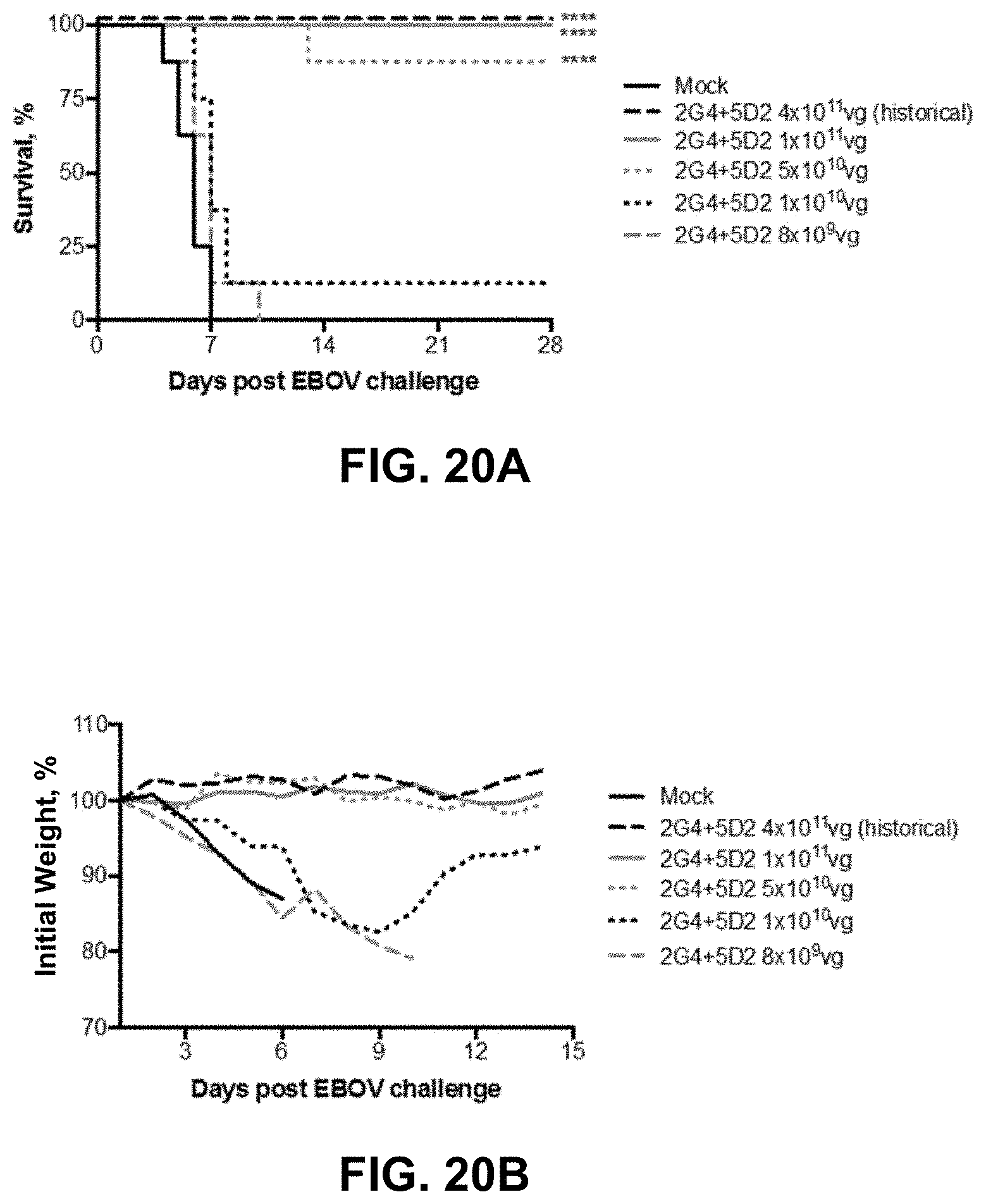
D00024

D00025

D00026
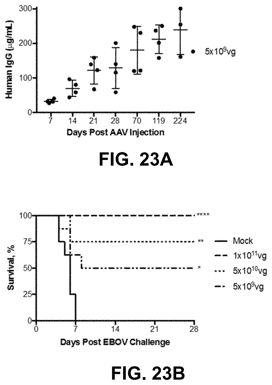
D00027
D00028

D00029

D00030

D00031

D00032

D00033

D00034

D00035

D00036

D00037

D00038

D00039
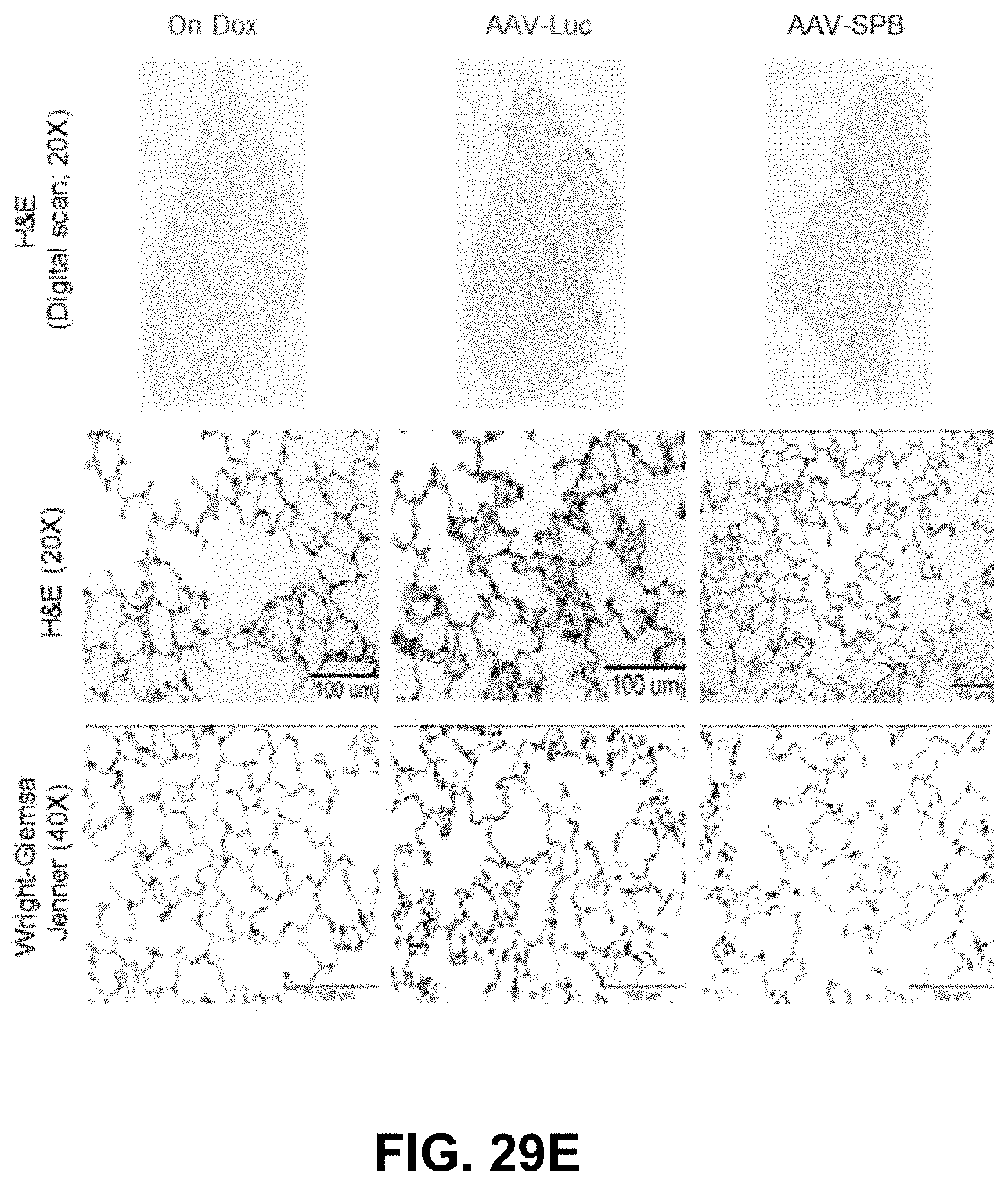
D00040

D00041
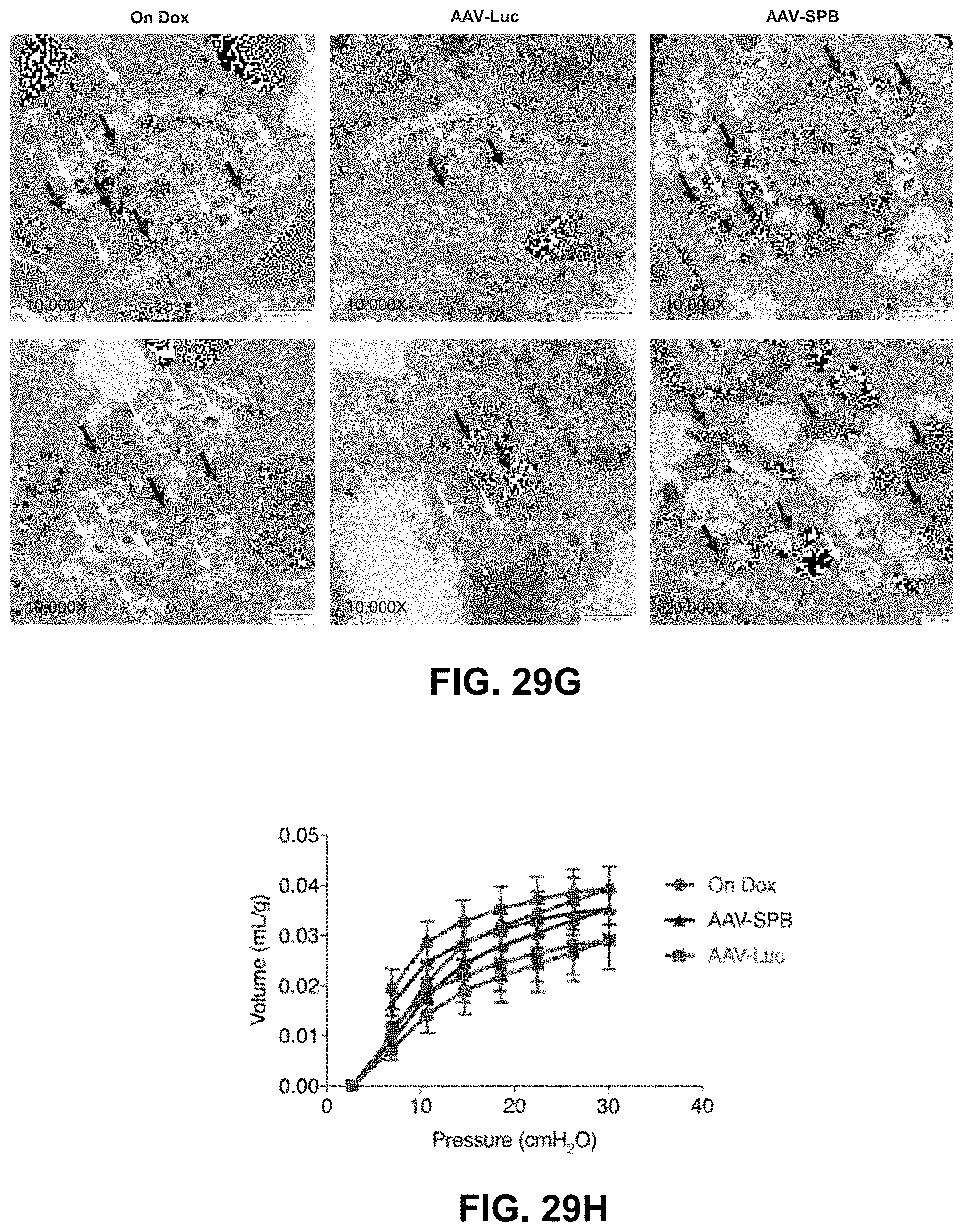
D00042

D00043

S00001
XML
uspto.report is an independent third-party trademark research tool that is not affiliated, endorsed, or sponsored by the United States Patent and Trademark Office (USPTO) or any other governmental organization. The information provided by uspto.report is based on publicly available data at the time of writing and is intended for informational purposes only.
While we strive to provide accurate and up-to-date information, we do not guarantee the accuracy, completeness, reliability, or suitability of the information displayed on this site. The use of this site is at your own risk. Any reliance you place on such information is therefore strictly at your own risk.
All official trademark data, including owner information, should be verified by visiting the official USPTO website at www.uspto.gov. This site is not intended to replace professional legal advice and should not be used as a substitute for consulting with a legal professional who is knowledgeable about trademark law.