Anti-PD-1 checkpoint inhibitor antibodies that block binding of PD-L1 to PD-1
Chang , et al.
U.S. patent number 10,669,338 [Application Number 15/803,193] was granted by the patent office on 2020-06-02 for anti-pd-1 checkpoint inhibitor antibodies that block binding of pd-l1 to pd-1. This patent grant is currently assigned to Immunomedics, Inc.. The grantee listed for this patent is Immunomedics, Inc.. Invention is credited to Chien-Hsing Chang, David M. Goldenberg.


View All Diagrams
| United States Patent | 10,669,338 |
| Chang , et al. | June 2, 2020 |
Anti-PD-1 checkpoint inhibitor antibodies that block binding of PD-L1 to PD-1
Abstract
The present invention concerns compositions and methods of use of anti-PD-1 antibodies comprising CDR sequences corresponding to SEQ ID NO:1 to SEQ ID NO:6. Preferably the antibody is a humanized antibody comprising the variable region amino acid sequences of SEQ ID NO: 9 and SEQ ID NO:10. The antibodies are of use to treat cancer and may be administered alone or with another standard anti-cancer therapy. The methods may comprise administering the anti-PD-1 antibody or antigen-binding fragment thereof in combination with one or more therapeutic agents such as antibody-drug conjugates, interferons (preferably interferon-.alpha.), and/or other checkpoint inhibitor antibodies.
| Inventors: | Chang; Chien-Hsing (Downingtown, PA), Goldenberg; David M. (Mendham, NJ) | ||||||||||
|---|---|---|---|---|---|---|---|---|---|---|---|
| Applicant: |
|
||||||||||
| Assignee: | Immunomedics, Inc. (Morris
Plains, NJ) |
||||||||||
| Family ID: | 61191283 | ||||||||||
| Appl. No.: | 15/803,193 | ||||||||||
| Filed: | November 3, 2017 |
Prior Publication Data
| Document Identifier | Publication Date | |
|---|---|---|
| US 20180051085 A1 | Feb 22, 2018 | |
Related U.S. Patent Documents
| Application Number | Filing Date | Patent Number | Issue Date | ||
|---|---|---|---|---|---|
| 15604153 | May 24, 2017 | 10131712 | |||
| 62351646 | Jun 17, 2016 | ||||
| Current U.S. Class: | 1/1 |
| Current CPC Class: | C07K 16/2818 (20130101); A61K 47/6863 (20170801); C07K 16/2887 (20130101); A61K 47/60 (20170801); A61K 45/06 (20130101); C07K 16/44 (20130101); A61K 47/6803 (20170801); C07K 16/2809 (20130101); A61P 35/04 (20180101); A61K 47/6857 (20170801); A61P 35/00 (20180101); C07K 16/2833 (20130101); A61K 31/4745 (20130101); A61K 38/212 (20130101); A61K 38/21 (20130101); C07K 16/3007 (20130101); C07K 16/2863 (20130101); C07K 16/30 (20130101); A61K 39/39558 (20130101); A61K 47/6851 (20170801); C07K 16/2803 (20130101); A61K 47/6859 (20170801); A61K 39/39558 (20130101); A61K 2300/00 (20130101); A61K 38/21 (20130101); A61K 2300/00 (20130101); A61K 31/4745 (20130101); A61K 2300/00 (20130101); C07K 2317/64 (20130101); C07K 2317/54 (20130101); C07K 2317/24 (20130101); C07K 2317/567 (20130101); C07K 2317/565 (20130101); C07K 2317/73 (20130101); C07K 2317/76 (20130101); A61K 2039/505 (20130101); A61K 2039/545 (20130101); A61K 2039/507 (20130101); C07K 2317/622 (20130101); C07K 2317/92 (20130101); C07K 2317/31 (20130101); C07K 2317/55 (20130101); C07K 2317/77 (20130101) |
| Current International Class: | C07K 16/28 (20060101); A61K 47/60 (20170101); A61P 35/00 (20060101); A61K 38/21 (20060101); A61K 45/06 (20060101); A61K 39/395 (20060101); C07K 16/30 (20060101); C07K 16/44 (20060101); A61K 39/00 (20060101); A61K 31/4745 (20060101); A61K 47/68 (20170101); A61P 35/04 (20060101) |
References Cited [Referenced By]
U.S. Patent Documents
| 7521056 | April 2009 | Chang et al. |
| 7527787 | May 2009 | Chang et al. |
| 7534866 | May 2009 | Chang et al. |
| 7550143 | June 2009 | Chang et al. |
| 7575923 | August 2009 | Dorken et al. |
| 7666400 | February 2010 | Chang et al. |
| 7901680 | March 2011 | Chang et al. |
| 7906118 | March 2011 | Chang et al. |
| 8003111 | August 2011 | Chang et al. |
| 8034352 | October 2011 | Chang et al. |
| 8877202 | November 2014 | Govindan et al. |
| 2010/0330080 | December 2010 | Dreier et al. |
| 2011/0008369 | January 2011 | Finnefrock et al. |
| 2014/0099254 | April 2014 | Chang et al. |
| 2015/0132217 | May 2015 | Chang et al. |
| 2016/0145355 | May 2016 | Saha et al. |
| 2017/0275375 | September 2017 | Rossi et al. |
| 2018/0051085 | February 2018 | Chang et al. |
| 2017071625 | May 2017 | WO | |||
Other References
|
Holliger and Hudson, Nature Biotechnology, 2005, vol. 23, pp. 1126-1136 (Year: 2005). cited by examiner . Ozcan et al, Advanced Drug Delivery Reviews, 2015, vol. 87, pp. 108-119 (Year: 2015). cited by examiner . Abstract of Bedikian et al, Melanoma Research, 2014, vol. 24 pp. 237-243 (Year: 2014). cited by examiner . Beer et al, The Lancet Oncology, 2017, vol. 18, pp. 1532-1542 (Year: 2017). cited by examiner . Dosio et al, Toxins, 2011, vol. 3, pp. 848-883 (Year: 2011). cited by examiner . Adair et al, Expert Opinion in Biological Therapy, 2012, vol. 12, pp. 1191-1206 (Year: 2012). cited by examiner . Kim et al (Immune Network, 2015, vol. 15, pp. 58-65). (Year: 2015). cited by examiner . Salfeld (Nature Biotechnology, 2007, vol. 25, pp. 1369-1372) (Year: 2007). cited by examiner . Jefferis and LeFranc (mAbs, 2009, vol. 1, pp. 332-338) (Year: 2009). cited by examiner . Johnston et al (Oncoimmunology, 2015, vol. 4, e1036214) (Year: 2015). cited by examiner . Amancha et al., "In vivo blockade of the programmed cell death-1 pathway using soluble recombinant PD-1-Fc enhances CD4+ and CD8+ T cell responses but has limited clinical benefit", J Immunol. Dec. 15, 2013;191(12):6060-70. cited by applicant . Awad et al., "Durable Responses With PD-1 Inhibition in Lung and Kidney Cancer and the Ongoing Search for Predictive Biomarkers", J Clin Oncol. Jun. 20, 2015;33(18):1993-4. cited by applicant . Bald et al., "Immune cell-poor melanomas benefit from PD-1 blockade after targeted type I IFN activation", Cancer Discov. Jun. 2014;4(6):674-87. cited by applicant . "Bristol-Myers Squibb's Opdivo.RTM. (nivolumab) Receives FDA Approval for the Treatment of Hepatocellular Carcinoma Patients Previously Treated with Sorafenib", Press Release, Sep. 22, 2017, Retrieved from: https://news.bms.com/press-release/corporatefinancial-news/bristol-myers-- squibbs-opdivo-nivolumab-receives-fda-approval-t. cited by applicant . Cameron et al., "Ipilimumab: first global approval", Drugs. May 28, 2011;71(8):1093-104. cited by applicant . Chang, C-H., "Combination Therapy with PD-1 Blockade Enhances the Antitumor Potency of T Cells Redirected by Novel Bispecific Antibodies Made with the Dock-and-Lock (DNL) Method", Presented at CHI's 5th Annual Immunomodulatory Therapeutic Antibodies for Cancer Conference, Aug. 29, 2017. cited by applicant . Chang et al., "Combination Therapy with Bispecific Antibodies and PD-1 Blockade Enhances the Antitumor Potency of T Cells", Cancer Res. Oct. 1, 2017;77(19):5384-5394. cited by applicant . Chen et al., "Development of a sandwich ELISA for evaluating soluble PD-L1 (CD274) in human sera of different ages as well as supernatants of PD-L1+ cell lines", Cytokine. Nov. 2011;56(2):231-8. cited by applicant . Chesney et al., "Randomized, Open-Label Phase II Study Evaluating the Efficacy and Safety of Talimogene Laherparepvec in Combination With Ipilimumab Versus Ipilimumab Alone in Patients With Advanced, Unresectable Melanoma", J Clin Oncol. Oct. 5, 2017:JCO2017737379, [Epub ahead of print]. cited by applicant . Deppisch et al., "Potent CD4+ T cell-associated antitumor memory responses induced by trifunctional bispecific antibodies in combination with immune checkpoint inhibition", Oncotarget. Jan. 17, 2017;8(3):4520-4529. cited by applicant . Gettinger et al., "Overall Survival and Long-Term Safety of Nivolumab (Anti-Programmed Death 1 Antibody, BMS-936558, ONO-4538) in Patients With Previously Treated Advanced Non-Small-Cell Lung Cancer", J Clin Oncol. Jun. 20, 2015;33(18)2004-12. cited by applicant . Grosso et al., "Programmed death-ligand 1 (PD-L1) expression in various tumor types", J Immunother Cancer. 2013; 1(Suppl 1): p. 53. cited by applicant . Hamid et al., "Safety and tumor responses with lambrolizumab (anti-PD-1) in melanoma", N. Engl J Med. Jul. 11, 2013;369(2):134-44. cited by applicant . He et al., "Blocking programmed death-1 ligand-PD-1 interactions by local gene therapy results in enhancement of antitumor effect of secondary lymphoid tissue chemokine", J Immunol. Oct. 15, 2004;173(8):4919-28. cited by applicant . He et al., "Blockade of B7-H1 with sPD-1 improves immunity against murine hepatocarcinoma", Anticancer Res. Sep.-Oct. 2005;25(5):3309-13. cited by applicant . He et al., "Checkpoint-based immunotherapy for autoimmune diseases--Opportunities and challenges", J Autoimmun. May 2017;79:1-3. cited by applicant . Kamimura et al., "Possible involvement of soluble B7-H4 in T cell-mediated inflammatory immune responses", Biochem Biophys Res Commun. Nov. 13, 2009;389(2):349-53. cited by applicant . Kasamon et al., "FDA Approval Summary: Nivolumab for the Treatment of Relapsed or Progressive Classical Hodgkin Lymphoma", Oncologist May 2017;22(5):585-591. cited by applicant . Kohnke et al., "Increase of PD-L1 expressing B-precursor ALL cells in a patient resistant to the CD19/CD3-bispecific T cell engager antibody blinatumomab", J Hematol Oncol. Oct. 8, 2015;8:111. cited by applicant . Larkins et al., "FDA Approval Summary: Pembrolizumab for the Treatment of Recurrent or Metastatic Head and Neck Squamous Cell Carcinoma with Disease Progression on or After Platinum-Containing Chemotherapy", Oncologist. Jul. 2017;22(7):873-878. cited by applicant . Mavilio et al., "Inhibiting the inhibitors: Checkpoints blockade in solid tumors", Oncoimmunology. Sep. 1, 2013;2(9):e26535. cited by applicant . Mittendorf et al., "PD-L1 expression in triple-negative breast cancer", Cancer Immunol Res. Apr. 2014;2(4):361-70. cited by applicant . Muenst et al., Expression of programmed death ligand 1 (PD-L1) is associated with poor prognosis in human breast cancer, Breast Cancer Res Treat Jul. 2014;146(1):15-24. cited by applicant . Ning et al., FDA Approval Summary: Atezolizumab for the Treatment of Patients with Progressive Advanced Urothelial Carcinoma after Platinum-Containing Chemotherapy, Oncologist. Jun. 2017;22(6):743-749. cited by applicant . Onlamoon et al., "Soluble PD-1 rescues the proliferative response of simian immunodeficiency virus-specific CD4 and CD8 T cells during chronic infection", Immunology. Jun. 2008;124(2):277-93. cited by applicant . Ott et al., "Combination immunotherapy: a road map", J Immunother Cancer Feb. 21, 2017;5:16. cited by applicant . Pan et al., "Synergistic effects of soluble PD-1 and IL-21 on antitumor immunity against H22 murine hepatocellular carcinoma", Oncol Lett. Jan. 2013;5(1):90-96. cited by applicant . Poon et al., "The MEK inhibitor selumetinib complements CTLA-4 blockade by reprogramming the tumor immune microenvironment", J Immunother Cancer Aug. 15, 2017;5(1):63. cited by applicant . Rosewell Shaw et al., "Adenovirotherapy Delivering Cytokine and Checkpoint Inhibitor Augments CAR T Cells against Metastatic Head and Neck Cancer", Mol Ther Nov. 1, 2017;25(11):2440-2451. cited by applicant . Sagiv-Barfi et al., "Therapeutic antitumor immunity by checkpoint blockade is enhanced by ibrutinib, an inhibitor of both BTK and ITK", Proc Natl Aced Sci U S A. Mar. 3, 2015;112(9):E966-72. cited by applicant . Soliman et al., "Therapeutic antitumor immunity by checkpoint blockade is enhanced by ibrutinib, an inhibitor of both BTK and ITK", Proc Natl Aced Sci U S A. Mar. 3, 2015:112(9):E966-72. cited by applicant . Velcheti et al., "Programmed death ligand-1 expression in non-small cell lung cancer", Lab Invest. Jan. 2014;94(1):107-16. cited by applicant . Wehler et al., "A randomized, phase 2 evaluation of the CHK1 inhibitor, LY2603618, administered in combination with pemetrexed and cisplatin in patients with advanced nonsquamous non-small cell lung cancer", Lung Cancer. Jun. 2017;108:212-216. cited by applicant . Weinstock et al., "U.S. Food and Drug Administration Approval Summary: Atezolizumab for Metastatic Non-Small Cell Lung Cancer", Clin Cancer Res. Aug. 15, 2017;23(16):4534-4539. cited by applicant . Wu et al., "The clinical value of combination of immune checkpoint inhibitors in cancer patients: A meta-analysis of efficacy and safety", Int J Cancer. Dec. 15, 2017;141(12):2562-2570. cited by applicant . Amann et al., "Antitumor activity of an EpCAM/CD3-bispecific BiTE antibody during long-term treatment of mice in the absence of T-cell anergy and sustained cytokine release", J Immunother. Jun. 2009;32(5):452-64. cited by applicant . Ank et al., "Lambda interferon (IFN-lambda), a type III IFN, is induced by viruses and IFNs and displays potent antiviral activity against select virus infections in vivo", J Virol. May 2006;80(9):4501-9. cited by applicant . Baeuerle et al., "Bispecific T-cell engaging antibodies for cancer therapy", Cancer Res. Jun. 15, 2009;69(12):4941-4. cited by applicant . Bargou et al., "Tumor regression in cancer patients by very low doses of a T cell-engaging antibody", Science. Aug. 15, 2008;321(5891):974-7. cited by applicant . Bassan et al., "Toward victory in adult ALL: blinatumomab joins in", Blood. Dec. 20, 2012;120(26):5094-5. cited by applicant . Beck et al., "The next generation of antibody-drug conjugates comes of age", Discov Med. Oct. 10, 2010;10(53):329-39. cited by applicant . Belardelli et al., "International Meeting on Cancer Vaccines: How Can We Enhance Efficacy of Therapeutic Vaccines?" Cancer Res. 64:6827-6830 (2004). cited by applicant . Belardelli et al., "The neglected role of type I interferon in the T-cell response: implications for its clinical use" Immunol. Today 17(8):369-72 (1996). cited by applicant . Benson et al., "A phase 1 trial of the anti-KIR antibody IPH2101 in patients with relapsed/refractory multiple myeloma", Blood. Nov. 22, 2012;120(22):4324-33. cited by applicant . Berger et al., "Phase I safety and pharmacokinetic study of CT-011, a humanized antibody interacting with PD-1, in patients with advanced hematologic malignancies", Clin Cancer Res. May 15, 2008;14(10):3044-51. cited by applicant . Billiau, A., "Interferon: the pathways of discovery I. Molecular and cellular aspects", Cytokine Growth Factor Rev. Oct. 2006;17(5):381-409. cited by applicant . Biron et al., "Natural killer cells in antiviral defense: function and regulation by innate cytokines" Annu. Rev. Immunol. 17:189-220 (1999). cited by applicant . Blank et al., "Contribution of the PD-L1/PD-1 pathway to T-cell exhaustion: an update on implications for chronic infections and tumor evasion", Cancer Immunol Immunother. May 2007;56(5):739-45. cited by applicant . Brahmer et al., "Safety and activity of anti-PD-L1 antibody in patients with advanced cancer", N Engl J Med. Jun. 28, 2012;366(26):2455-65. cited by applicant . Bross et al., "Approval summary: gemtuzumab ozogamicin in relapsed acute myeloid leukemia", Clin Cancer Res. Jun. 2001;7(6):1490-6. cited by applicant . Brunda et al., "Modulation of Murine Natural Killer Cell Activity in Vitro and in Vivo by Recombinant Human Interferons" Cancer Res. 44:597-601 (1984). cited by applicant . Carrero et al., "Lymphocytes are detrimental during the early innate immune response against Listeria monocytogenes" J. Exp. Med. 203(4):933-940 (2006). cited by applicant . Chames et al., "Bispecific antibodies for cancer therapy: the light at the end of the tunnel?", MAbs. Nov.-Dec. 2009;1(6):539-47. cited by applicant . Chang et al., "Directing NK cells to Trop-2-expressing breast and other cancers, with chimeric antigen receptors", [abstract]. In: Proceedings of the 2016 San Antonio Breast Cancer Symposium; Dec. 6-10, 2016; San Antonio, TX. Philadelphia (PA): AACR; Cancer Res 2017;77(4 Suppl):Abstract nr P2-04-17. cited by applicant . Chang et al., "Directing NK cells to Trop-2-expressing breast and other cancers, with chimeric antigen receptors", [poster], Presented at 2016 San Antonio Breast Cancer Symposium; Dec. 6-10, 206; San Antonio, TX. cited by applicant . Dickensheets et al., "Interferon-lambda (IFN-.lamda.) induces signal transduction and gene expression in human hepatocytes, but not in lymphocytes or monocytes", J Leukoc Biol. Mar. 2013;93(3):377-85. cited by applicant . Ferrantini et al., "IFN-.alpha.1 Gene Expression into a Metastatic Murine Adenocarcinoma (TS/A) Results in CD8-+ T Cell-Mediated Tumor Rejection and Development of Antitumor Immunity" J. Immunol. 153:4604-15 (1994). cited by applicant . Finger et al., "The human PD-1 gene: complete cDNA, genomic organization, and developmentally regulated expression in B cell progenitors", Gene. Sep. 15, 1997;197(1-2):177-87. cited by applicant . Firer et al., "Targeted drug delivery for cancer therapy: the other side of antibodies", J Hematol Oncol. Nov. 9, 2012;5:70. cited by applicant . Drancisco et al., "cAC10-vcMMAE, an anti-CD30-monomethyl auristatin E conjugate with potent and selective antitumor activity", Blood. Aug. 15, 2003;102(4):1458-65. cited by applicant . Gildener-Leapman et al., "Promising systemic immunotherapies in head and neck squamous cell carcinoma", Oral Oncol. Dec. 2013;49(12):1089-96. cited by applicant . Gleason et al., "Bispecific and trispecific killer cell engagers directly activate human NK cells through CD16 signaling and induce cytotoxicity and cytokine production", Mol Cancer Ther. Dec. 2012;11(12):2674-84. cited by applicant . Grimley et al., "Prolonged STAT1 Activation Related to the Growth Arrest of Malignant Lymphoma Cells by Interferon-.alpha." Blood 91(8):3017-27 (1998). cited by applicant . Gutterman et al., "Leukocyte Interferon-Induced Tumor Regression in Human Metastatic Breast Cancer, Multiple Myeloma, and Malignant Lymphoma" Ann. Intern. Med. 93(3):399-406 (1980). cited by applicant . Gutterman et al., "Cytokine therapeutics: Lessons from interferon .alpha." Proc. Natl. Acad. Sci. USA 91:1198-205 (1994). cited by applicant . Ikeda et al., "Interferon beta prevents recurrence of hepatocellular carcinoma after complete resection or ablation of the primary tumor--A prospective randomized study of hepatitis C virus-related liver cancer", Hepatology. Aug. 2000;32(2):228-32. cited by applicant . Jablonska et al., "Neutrophils responsive to endogenous IFN-beta regulate tumor angiogenesis and growth in a mouse tumor model", J Clin Invest. Apr. 2010;120(4):1151-64. cited by applicant . Kohrt et al., "Anti-KIR antibody enhancement of anti-lymphoma activity of natural killer cells as monotherapy and in aombination with anti-CD20 antibodies", Blood. Jan. 30, 2014;123(5):678-86. cited by applicant . Kotenko et al., "IFN-lambdas mediate antiviral protection through a distinct class II cytokine receptor complex", Nat Immunol. Jan. 2003;4(1):69-77. cited by applicant . Le Bon et al., "Type i interferons potently enhance humoral immunity and can promote isotype switching by stimulating dendritic cells in vivo", Immunity. Apr. 2001;14(4):461-70. cited by applicant . Li et al., "Monoclonal antibody-related drugs for cancer therapy", Drug Discov Ther. Oct. 2013;7(5):178-84. cited by applicant . Lipson et al., "Durable cancer regression off-treatment and effective reinduction therapy with an anti-PD-1 antibody", Clin Cancer Res. Jan. 15, 2013;19(2):462-8. cited by applicant . Luft et al., "Type I IFNs Enhance the Terminal Differentiation of Dendritic Cells" J. Immunol. 161:1947-1953 (1998). cited by applicant . Matarrese et al., "Type I Interferon Gene Transfer Sensitizes Melanoma Cells to Apoptosis via a Target Activity on Mitochondrial Function" Am. J. Pathol. 2002, 160(4):1507-1520. cited by applicant . Mecchia et al., "Type I consensus interferon (CIFN) gene transfer into human melanoma cells up-regulates p53 and enhances cisplatin-induced apoptosis: implications for new therapeutic strategies with IFN-alpha" Gene Ther. (2000) 7, 167-179. cited by applicant . Menzies et al., "New combinations and immunotherapies for melanoma: latest evidence and clinical utility", Ther Adv Med Oncol. Sep. 2013;5(5):278-85. cited by applicant . Miller et al., "Clinical Use of Interferon-gamma", Ann N Y Acad Sci. Dec. 2009;1182:69-79. cited by applicant . Miller, JC., "Therapeutic applications: natural killer cells in the clinic", Hematology Am Soc Hematol Educ Program. 2013;2013:247-53. cited by applicant . Moore et al., "Application of dual affinity retargeting molecules to achieve optimal redirected T-cell killing of B-cell lymphoma", Blood. Apr. 28, 2011;117(17):4542-51. cited by applicant . Mullard et al., "Maturing antibody-drug conjugate pipeline hits 30", Nat Rev Drug Discov. May 2013;12(5):329-32. cited by applicant . Nagorsen et al., "Immunotherapy of lymphoma and leukemia with T-cell engaging BiTE antibody blinatumomab", Leuk Lymphoma. Jun. 2009;50(6):886-91. cited by applicant . Nirschl et al., "Molecular pathways: coexpression of immune checkpoint molecules: signaling pathways and mplications for cancer immunotherapy", Clin Cancer Res. Sep. 15, 2013;19(18):4917-24. cited by applicant . Nitta et al., "Preliminary trial of specific targeting therapy against malignant glioma", Lancet. Feb. 17, 1990;335(8686):368-71. cited by applicant . Ott et al., "Impact of MAPK Pathway Activation in BRAF(V600) Melanoma on T Cell and Dendritic Cell Function", Front Immunol. Oct. 28, 2013;4:346. cited by applicant . Ott et al., "CTLA-4 and PD-1/PD-L1 blockade: new immunotherapeutic modalities with durable clinical benefit in melanoma patients", Clin Cancer Res. Oct. 1, 2013;19(19):5300-9. cited by applicant . Paquette et al., "Interferon-.alpha. and granulocyte-macrophage colony-stimulating factor differentiate peripheral blood monocytes into potent antigen-presenting cells" J. Leukoc. Biol. 64:358-367; 1998. cited by applicant . Pardoll, DM., "The blockade of immune checkpoints in cancer immunotherapy", Nat Rev Cancer. Mar. 22, 2012;12(4):252-64. cited by applicant . Pestka et al., "Interferons, interferon-like cytokines, and their receptors", Immunol Rev. Dec. 2004;202:8-32. cited by applicant . Pestka, S., "The interferons: 50 years after their discovery, there is much more to learn", J Biol Chem. Jul. 13, 2007;282(28):20047-51. cited by applicant . Pilling et al., "Interferon-beta mediates stromal cell rescue of T cells from apoptosis", Eur J Immunol. Mar. 1999;29(3)1041-50. cited by applicant . Portell et al., "Clinical and pharmacologic aspects of blinatumomab in the treatment of B-cell acute lymphoblastic leukemia", Clin Pharmacol. Apr. 12, 2013;5(Suppl 1):5-11. cited by applicant . Portner et al., "T and NK cells of B cell NHL patients exert cytotoxicity against lymphoma cells following binding of bispecific tetravalent antibody CD19 .times. CD3 or CD19 .times. CD16", Cancer Immunol Immunother. Oct. 2012;61(10):1869-75. cited by applicant . Radvanyi et al., "Antagonist antibodies to PD-1 and B7-H1 (PD-L1) in the treatment of advanced human cancer--letter", Clin Cancer Res. Oct. 1, 2013;19(19):5541. cited by applicant . Raefsky et al., "Studies of interferon as a regulator of hematopoietic cell proliferation", J Immunol. Oct. 1985;135(4):2507-12. cited by applicant . Robek et al., "Lambda interferon inhibits hepatitis B and C virus replication", J Virol. Mar. 2005;79(6):3851-4. cited by applicant . Robert et al., "What is the role of cytotoxic T lymphocyte-associated antigen 4 blockade in patients with metastatic melanoma?", Oncologist Aug. 2009;14(8):848-61. cited by applicant . Rossi et al., "Preclinical studies on targeted delivery of multiple IFN.alpha.2b to HLA-DR in diverse hematologic cancers", Blood. Aug. 18, 2011;118(7):1877-84. cited by applicant . Rossi et al., "Enhanced efficacy of redirected T-cell therapy of TNBC with a Trop-2/CD3 bispecific antibody in combination with a checkpoint inhibitor", [poster]. Presented at 2016 San Antonio Breast Cancer Symposium; Dec. 6-10, 2016; San Antonio, TX. cited by applicant . Rossi et al., "Enhanced efficacy of redirected T-cell therapy of TNBC with a Trop-2/CD3 bispecific antibody in combination with a checkpoint inhibitor", [abstract]. In: Proceedings of the 2016 San Antonio Breast Cancer Symposium; Dec. 6-10, 2016; San Antonio, TX. Philadelphia (PA): AACR; Cancer Res 2017;77(4 Suppl):Abstract nr P6-14-01. cited by applicant . Sabaawy et al., "Enhancement of 5-fluorouracil cytotoxicity on human colon cancer cells by retrovirus-mediated nterferon-alpha gene transfer", Int J Oncol. Jun. 1999;14(6):1143-51. cited by applicant . Santini et al., "Type I interferon as a powerful adjuvant for monocyte-derived dendritic cell development and activity in vitro and in Hu-PBL-SCID mice", J Exp Med. May 15, 2000;191(10):1777-88. cited by applicant . Schubert et al., "A recombinant triplebody with specificity for CD19 and HLA-DR mediates preferential binding to antigen double-positive cells by dual-targeting", MAbs. Jan.-Feb. 2012;4(1):45-56. cited by applicant . Sheppard et al., "IL-28, IL-29 and their class II cytokine receptor IL-28R", Nat Immunol. Jan. 2003;4(1):63-8. cited by applicant . Shinohara et al., "Structure and chromosomal localization of the human PD-1 gene (PDCD1)", Genomics. Oct. 1994;23(3):704-6. cited by applicant . Sidky et al., "Inhibition of angiogenesis by interferons: effects on tumor- and lymphocyte-induced vascular responses", Cancer Res. Oct. 1, 1987;47(19):5155-61. cited by applicant . Takaoka et al., "Integration of interferon-alpha/beta signalling to p53 responses in tumour suppression and antiviral defence", Nature. Jul. 31, 2003;424(6948):516-23. cited by applicant . Topalian et al., "Safety, activity, and immune correlates of anti-PD-1 antibody in cancer", N Engl J Med. Jun. 28, 2012;366(26):2443-54. cited by applicant . Topp et al., "Long-term follow-up of hematologic relapse-free survival in a phase 2 study of blinatumomab in patients with MRD in B-lineage ALL", Blood. Dec. 20, 2012;120(26):5185-7. cited by applicant . Traunecker et al., "Bispecific single chain molecules (Janusins) target cytotoxic lymphocytes on HIV infected cells", EMBO J. Dec. 1991;10(12):3655-9. cited by applicant . Veri et al., "Therapeutic control of B cell activation via recruitment of Fcgamma receptor IIb (CD32B) inhibitory function with a novel bispecific antibody scaffold", Arthritis Rheum. Jul. 2010;62(7):1933-43. cited by applicant . Verma et al., "Trastuzumab emtansine for HER2-positive advanced breast cancer", N Engl J Med. Nov. 8, 2012;367(19):1783-91. cited by applicant . Vilcek, J., "Fifty years of interferon research: aiming at a moving target", Immunity. Sep. 2006;25(3):343-8. cited by applicant . Wada et al., "Sequencing CTLA-4 blockade with cell-based immunotherapy for prostate cancer", J Transl Med. Apr. 4, 2013;11:89. cited by applicant . Weber, J., "Review: anti-CTLA-4 antibody ipilimumab: case studies of clinical response and immune-related adverse avents", Oncologist. Jul. 2007;12(7):864-72. cited by applicant . Wei et al., "Disulfide-stabilized diabody antiCD19/antiCD3 exceeds its parental antibody in tumor-targeting activity", Cell Oncol (Dordr). Dec. 2012;35(6):423-34. cited by applicant . Wiernik et al., "Targeting natural killer cells to acute myeloid leukemia in vitro with a CD16 .times. 33 bispecific killer cell angager and ADAM17 inhibition", Clin Cancer Res. Jul. 15, 2013;19(14):3844-55. cited by applicant . Witte et al., "Despite IFN-lambda receptor expression, blood immune cells, but not keratinocytes or melanocytes, have an impaired response to type III interferons: implications for therapeutic applications of these cytokines", Genes Immun. Dec. 2009;10(8):702-14. cited by applicant . Witte et al., "IL-28A, IL-28B, and IL-29: promising cytokines with type I interferon-like properties", Cytokine Growth Factor Rev. Aug. 2010;21(4):237-51. cited by applicant . Yoshida et al., "Interferon-beta gene therapy for cancer: basic research to clinical application", Cancer Sci. Nov. 2004;95(11):858-65. cited by applicant . Zhou et al., "Type III interferon (IFN) induces a type I IFN-like response in a restricted subset of cells through signaling pathways involving both the Jak-STAT pathway and the mitogen-activated protein kinases", J Virol. Jul. 2007;81(14):7749-58. cited by applicant . Zhou et al., "A fully human CD19/CD3 bi-specific antibody triggers potent and specific cytotoxicity by unstimulated T lymphocytes against non-Hodgkin's lymphoma", Biotechnol Lett. Jul. 2012;34(7):1183-91. cited by applicant. |
Primary Examiner: Canella; Karen A.
Attorney, Agent or Firm: Nakashima; Richard A.
Parent Case Text
CROSS REFERENCE TO RELATED APPLICATIONS
This application is a continuation-in-part of U.S. patent application Ser. No. 15/604,153, filed May 24, 2017, which claimed the benefit under 35 U.S.C. 119(e) of provisional U.S. Patent Application 62/351,646, filed Jun. 17, 2016. The text of each priority application incorporated herein by reference in its entirety.
Claims
What is claimed is:
1. A method of treating a human cancer comprising administering to a human subject with cancer an anti-PD-1 antibody or antigen-binding fragment thereof comprising the heavy chain CDR sequences GFAFSSNDMS (SEQ ID NO:1), TISGGGINTYYPDSVKG (SEQ ID NO:2) and RSNYAWFAY (SEQ ID NO:3) and the light chain CDR sequences RASESVDTYGISFMN (SEQ ID NO:4), PNQGS (SEQ ID NO:5) and QQSKEVPWT (SEQ ID NO:6).
2. The method of claim 1, wherein the anti-PD-1 antibody is a chimeric antibody comprising the light chain amino acid sequence SEQ ID NO:7 and the heavy chain amino acid sequence SEQ ID NO:8.
3. The method of claim 1, wherein the anti-PD-1 antibody is a humanized antibody comprising the light chain amino acid sequence SEQ ID NO:9 and the heavy chain amino acid sequence SEQ ID NO:10.
4. The method of claim 1, wherein the antibody or antigen-binding fragment thereof is a naked antibody or antigen-binding fragment thereof.
5. The method of claim 4, further comprising administering to the subject a therapeutic agent selected from the group consisting of a second antibody, a second antigen-binding antibody fragment, an antibody-drug conjugate (ADC), a drug, a toxin, a cytotoxic agent, an anti-angiogenic agent, a hormone, an immunomodulator, a cytokine, a chemokine, an interferon, and a radioisotope.
6. The method of claim 5, wherein the drug is selected from the group consisting of 5-fluorouracil, afatinib, aplidin, azaribine, anastrozole, axitinib, AVL-101, AVL-291, bendamustine, bleomycin, bortezomib, bosutinib, bryostatin-1, busulfan, calicheamycin, camptothecin, carboplatin, 10-hydroxycamptothecin, carmustine, celecoxib, chlorambucil, cisplatin, Cox-2 inhibitors, irinotecan (CPT-11), SN-38, cladribine, crizotinib, cyclophosphamide, cytarabine, dacarbazine, dasatinib, dinaciclib, docetaxel, dactinomycin, daunorubicin, doxorubicin, 2-pyrrolinodoxorubicine (2P-DOX), cyano-morpholino doxorubicin, doxorubicin glucuronide, epirubicin glucuronide, erlotinib, estramustine, epipodophyllotoxin, erlotinib, entinostat, etoposide, etoposide glucuronide, etoposide phosphate, exemestane, fingolimod, floxuridine, 3',5'-O-dioleoyl-FudR, fludarabine, flutamide, farnesyl-protein transferase inhibitors, flavopiridol, fostamatinib, ganetespib, GDC-0834, GS-1101, gefitinib, gemcitabine, hydroxyurea, ibrutinib, idarubicin, idelalisib, ifosfamide, imatinib, L-asparaginase, lapatinib, lenolidamide, leucovorin, LFM-A13, lomustine, mechlorethamine, melphalan, mercaptopurine, 6-mercaptopurine, methotrexate, mitoxantrone, mithramycin, mitomycin, mitotane, navelbine, neratinib, nilotinib, nitrosurea, olaparib, plicomycin, procarbazine, paclitaxel, PCI-32765, pentostatin, PSI-341, raloxifene, semustine, sorafenib, streptozocin, SU11248, sunitinib, tamoxifen, temazolomide, transplatin, thalidomide, thioguanine, thiotepa, teniposide, topotecan, uracil mustard, vatalanib, vinorelbine, vinblastine, vincristine, and ZD1839.
7. The method of claim 5, wherein the chemokine is selected from the group consisting of RANTES, MCAF, MIP1-alpha, MIP1-Beta and IP-10.
8. The method of claim 5, wherein the immunomodulator is selected from the group consisting of a stem cell growth factor, a lymphotoxin, a hematopoietic factor, a colony stimulating factor (CSF), and an interferon (IFN).
9. The method of claim 5, wherein the cytokine is selected from the group consisting of hepatic growth factor, prostaglandin, fibroblast growth factor, prolactin, placental lactogen, OB protein, tumor necrosis factor-.alpha., tumor necrosis factor-.beta., mullerian-inhibiting substance, mouse gonadotropin-associated peptide, inhibin, activin, vascular endothelial growth factor, integrin, thrombopoietin (TPO), a nerve growth factor (NGF), platelet-growth factor, TGF-.alpha., TGF-.beta., insulin-like growth factor-I, insulin-like growth factor-II, erythropoietin (EPO), an osteoinductive factor, interferon-.alpha., interferon-.beta., interferon-.lamda., macrophage-CSF (M-CSF), granulocyte-macrophage-CSF (GM-CSF), granulocyte-CSF (G-CSF), interleukin-1 (IL-1), IL-la, IL-2, IL-3, IL-4, IL-5, IL-6, IL-7, IL-8, IL-9, IL-10, IL-11, IL-12, IL-13, IL-14, IL-15, IL-16, IL-17, IL-18, IL-21, LIF, kit-ligand, FLT-3, angiostatin, thrombospondin, endostatin, tumor necrosis factor and LT (lymphotoxin).
10. The method of claim 5, wherein the second antibody is a checkpoint inhibitor antibody selected from the group consisting of pembrolizumab (MK-3475), nivolumab (BMS-936558), pidilizumab (CT-011), AMP-224, MDX-1105, MEDI4736, atezolizumab (MPDL3280A), BMS-936559, ipilimumab, lirilumab, IPH2101, durvalumab and tremelimumab.
11. The method of claim 5, wherein the second antibody binds to an antigen selected from the group consisting of CTLA-4, PD-1, PD-L1, LAG3, B7-H3, B7-H4, KIR and TIM3.
12. The method of claim 5, wherein the second antibody is selected from the group consisting of hA19, hR1, hPAM4, hA20, hIMMU31, hLL1, hLL2, hMu09, hL243, hMN-14, hMN-15, hMN-3, and hRS7.
13. The method of claim 5, wherein the ADC is hRS7-SN-38 (IMMU-132), hMN-14-SN-38 (IMMU-130) or hL243-SN-38 (IMMU-140).
14. The method of claim 5, wherein an ADC is administered prior to any other agent.
15. The method of claim 5, wherein the interferon is selected from the group consisting of interferon-.alpha., interferon-.beta., interferon-.lamda.1, interferon-.lamda.2 and interferon-.lamda.3.
16. The method of claim 5, wherein the interferon is interferon-a.
17. The method of claim 5, wherein the interferon is administered as free interferon, PEGylated interferon, an interferon fusion protein or interferon conjugated to an antibody.
18. The method of claim 1, wherein the cancer expresses PD-L1.
19. The method of claim 1, wherein the cancer is selected from the group consisting of acute lymphoblastic leukemia, acute myelogenous leukemia, biliary cancer, B-cell leukemia, B-cell lymphoma, biliary cancer, bone cancer, brain cancer, breast cancer, cervical cancer, chronic lymphocytic leukemia, chronic myelogenous leukemia, colorectal cancer, endometrial cancer, esophageal cancer, gastric cancer, hairy cell leukemia, head and neck cancer, Hodgkin's lymphoma, liver cancer, lung cancer, medullary thyroid cancer, multiple myeloma, ovarian cancer, non-Hodgkin's lymphoma, pancreatic cancer, prostate cancer, renal cancer, sarcoma, skin cancer, testicular cancer, and urothelial cancer.
20. A murine, chimeric or humanized anti-PD-1 antibody or antigen-binding fragment thereof comprising the heavy chain CDR sequences GFAFSSNDMS (SEQ ID NO:1), TISGGGINTYYPDSVKG (SEQ ID NO:2) and RSNYAWFAY (SEQ ID NO:3) and the light chain CDR sequences RASESVDTYGISFMN (SEQ ID NO:4), PNQGS (SEQ ID NO:5) and QQSKEVPWT (SEQ ID NO:6).
21. The anti-PD-1 antibody of claim 20, wherein the anti-PD-1 antibody is a chimeric antibody comprising the light chain amino acid sequence SEQ ID NO:7 and the heavy chain amino acid sequence SEQ ID NO:8.
22. The anti-PD-1 antibody of claim 20, wherein the anti-PD-1 antibody is a humanized antibody comprising the light chain amino acid sequence SEQ ID NO:9 and the heavy chain amino acid sequence SEQ ID NO:10.
23. The anti-PD-1 antibody of claim 20, wherein the antibody is an IgG1, IgG2, IgG3 or IgG4 antibody.
24. The anti-PD-1 antibody of claim 20, wherein the antibody has a Glm3 heavy chain and Km3 light chain allotype.
25. The anti-PD-1 antibody of claim 20, wherein the antibody fragment is selected from the group consisting of F(ab').sub.2, F(ab).sub.2, Fab', Fab, Fv, and scFv antibody fragments.
26. The anti-PD-1 antibody of claim 20, wherein the antibody or antigen-binding fragment thereof is a naked antibody or antigen-binding fragment thereof.
27. A composition comprising an anti-PD-1 antibody according to claim 20.
28. A kit comprising: a) an anti-PD-1 antibody according to claim 20; and b) at least one container.
Description
SEQUENCE LISTING
The instant application contains a Sequence Listing which has been submitted in ASCII format via EFS-Web and is hereby incorporated by reference in its entirety. Said ASCII copy, created on Oct. 24, 2017, is named IMM374US1 SL.txt and is 13,425 bytes in size.
FIELD
In certain embodiments, the present invention concerns compositions and methods of use of novel antibodies against PD-1 (programmed cell death protein 1, aka CD279). Preferably, the anti-PD1 antibodies block binding of PD-1 to its ligand, PD-L1. More preferably, the anti-PD1 antibody is a murine, chimeric or humanized antibody comprising the heavy chain CDR sequences GFAFSSNDMS (SEQ ID NO:1), TISGGGINTYYPDSVKG (SEQ ID NO:2) and RSNYAWFAY (SEQ ID NO:3) and the light chain CDR sequences RASESVDTYGISFMN (SEQ ID NO:4), PNQGS (SEQ ID NO:5) and QQSKEVPWT (SEQ ID NO:6). Most preferably, the anti-PD1 antibody is a chimeric antibody comprising the light chain variable region amino sequence of SEQ ID NO:7 and the heavy chain variable region amino sequence of SEQ ID NO:8, or a humanized antibody comprising the light chain variable region amino sequence of SEQ ID NO:9 and the heavy chain variable region amino sequence of SEQ ID NO:10. In certain preferred embodiments, the anti-PD-1 antibodies are of use to treat cancers that express PD-L1, although such methods are not limiting and various forms of cancer may be treated with the subject antibodies, alone or in combination with one or more other therapeutic agents. Exemplary cancers that may be treated include, but are not limited to, non-small cell lung cancer (NSCLC), SCLC, mesothelioma, melanoma, breast cancer, ovarian cancer, colon cancer, prostate cancer, gastric cancer, renal-cell cancer, urothelial cancer, squamous cell carcinoma, head and neck cancer, non-Hodgkin lymphoma and Hodgkin lymphoma. The anti-PD-1 antibody may be administered as a naked (unconjugated) antibody. In certain embodiments, the anti-PD-1 antibody may be administered in combination with at least one other antibody or immunoconjugate, such as an anti-TAA (tumor-associated antigen) antibody or immunoconjugate. In alternative embodiments, the anti-PD-1 antibody may be administered in combination with a T-cell redirecting bispecific antibody, such as an anti-CD19.times.anti-CD3, anti-Trop-2.times.anti-CD3, anti-CEACAM5.times.anti-CD3, or any other known T-cell redirecting bsAb. In preferred embodiments, the anti-PD1 antibody may be administered in combination with an interferon, another checkpoint inhibitor antibody (e.g., anti-CTLA-4), an antibody-drug conjugate (ADC) or other anti-cancer therapy. The anti-PD-1 therapy may exhibit enhanced or even synergistic activity when combined with another therapeutic agent, and may be efficacious to treat tumors that are resistant to or relapsed from standard cancer therapies.
BACKGROUND
A promising approach to immunotherapy concerns use of antagonistic antibodies against immune checkpoint proteins (e.g., Pardoll, 2012, Nature Reviews Cancer 12:252-64). Immune checkpoints function as endogenous inhibitory pathways for the immune system to maintain self-tolerance and to modulate the duration and extent of immune response to antigenic stimulation (Pardoll, 2012). In their normal function, activity of checkpoint proteins modulates the immune response to prevent development of autoimmune disease (e.g., He et al., 2017, J Autoimmun 79:1-3). However, it appears that tumor tissues may co-opt the checkpoint system to reduce the effectiveness of the host immune response, resulting in tumor growth (see, e.g., Pardoll, 2012, Nature Reviews Cancer 12:252-64; Nirschl & Drake, 2013, Clin Cancer Res 19:4917-24). Checkpoint molecules include CTLA-4 (cytotoxic T lymphocyte antigen-4), PD-1 (programmed cell death protein 1), PD-L1 (programmed cell death ligand 1), LAG-3 (lymphocyte activation gene-3), TIM-3 (T cell immunoglobulin and mucin protein-3) and several others (Pardoll, 2012, Nature Reviews Cancer 12:252-64; Nirschl & Drake, 2013, Clin Cancer Res 19:4917-24).
Antibodies against several of the checkpoint proteins (CTLA-4, PD-1, PD-L1) are in clinical trials and have shown unexpected efficacy against tumors that were resistant to standard treatments. Use in human cancer therapy has been approved by the FDAfor ipilimumab (anti-CTLA-4, Bristol-Myers Squibb) in malignant melanoma (Cameron et al, 2011, Drugs 71:1093-104); pembrolizumab (anti-PD-1, Merck & Co.) in melanoma, head and neck cancer, Hodgkin lymphoma, urothelial cancer, gastric cancer and metastatic NSCLC that expresses PD-1 (Press Release, Merck & Co., dated Sep. 22, 2017, "FDA Approves Merck's KEYTRUDA.RTM. for Previously Treated Patients with Recurrent Locally Advanced or Metastatic Gastric or Gastroesophageal Junction Cancer Whose Tumors Express PD-1 (CPS Greater Than or Equal to 1)"); nivolumab (anti-PD-1, Bristol-Myers Squibb) in melanoma, lung cancer, kidney cancer, bladder cancer, head and neck cancer and Hodgkin lymphoma (Larkins et al., 2017, The Oncologist 22:873-78; Kasamon et al., 2017, Oncologist 22:585-91), and five anti-PD-1 antibodies, for example, atezolizumab (anti-PD-L1, Roche) in bladder cancer and metastatic NSCLC (Ning et al., 2017, Oncologist 22:743-49; Weinstock et al., 2017, Clin Cancer Res 23:4534-39).
Studies with checkpoint inhibitor antibodies for cancer therapy have generated unprecedented response rates in cancers previously thought to be resistant to cancer treatment (see, e.g., Ott & Bhardwaj, 2013, Frontiers in Immunology 4:346; Menzies & Long, 2013, Ther Adv Med Oncol 5:278-85; Pardoll, 2012, Nature Reviews 12:252-264; Mavilio & Lugli, 2013, Oncoimmunology 2:e26535). Therapy with antagonistic checkpoint blocking antibodies against CTLA-4, PD-1 and PD-1 is one of the most promising new avenues of immunotherapy for cancer and other diseases. However, a need continues to exist for more effective checkpoint inhibitor antibodies, preferably with the ability to block binding of PD-1 to PD-1.
SUMMARY
In certain embodiments, the present invention relates to murine, chimeric or humanized antibodies against PD-1, preferably comprising the heavy chain CDR sequences GFAFSSNDMS (SEQ ID NO:1), TISGGGINTYYPDSVKG (SEQ ID NO:2) and RSNYAWFAY (SEQ ID NO:3) and the light chain CDR sequences RASESVDTYGISFMN (SEQ ID NO:4), PNQGS (SEQ ID NO:5) and QQSKEVPWT (SEQ ID NO:6). Alternative embodiments relate to antibodies that bind to the same epitope as, or compete for binding to PD-1 with, an antibody having the amino acid sequences of SEQ ID NO:1 to SEQ ID NO:6. The antibody may be naked (unconjugated) or may be conjugated to one or more therapeutic or diagnostic agents, as discussed below. Other alternative embodiments relate to antigen-binding fragments of the subject anti-PD-1 antibodies, such as F(ab').sub.2, F(ab).sub.2, F(ab'), F(ab), or scFv antibody fragments.
In preferred methods, the anti-PD-1 antibodies are of use to treat cancers, including but not limited to non-small cell lung cancer (NSCLC), SCLC, mesothelioma, melanoma, breast cancer, ovarian cancer, colon cancer, prostate cancer, gastric cancer, renal-cell cancer, urothelial cancer, squamous cell carcinoma, head and neck cancer, non-Hodgkin lymphoma and Hodgkin lymphoma. More preferably, the cancers to be to be treated express PD-1 and the subject methods may comprise assaying for PD-1 expression prior to therapy.
In certain embodiments, methods of use of anti-PD-1 antibodies or fragments thereof may involve combination therapy with other therapeutic treatments (Ott et al., 2017, J Immunother Cancer 5:16). Exemplary treatments that may be used in combination therapy with the subject anti-PD-1 antibodies or fragments thereof include use of interferons (e.g., interferon-.alpha.), other cytokines (e.g., GM-CSF, IL-2, IL-12, IL-15, IL-18, IL-21), other checkpoint inhibitor antibodies (e.g., anti-CTLA-4 antibodies or anti-PD-1 antibodies), antibody-drug conjugates (ADCs, e.g., IMMU-132, IMMU-130, IMMU-140), radiation therapy, chemotherapy, talimogene laherparepvec (Chesney et al., J Clin Oncol, Oct. 5, 2017, [Epub ahead of print]), CAR-T cells (Rosewell et al., Sep. 14, 2017, [Epub ahead of print]), T-cell redirecting bispecific antibodies (Chang et al., 2017, Cancer Res 77:5384-94), ibrutinib (Sagiv-Barfi et al., 2015, Proc Natl Acad Sci USA 112(9):E966-72), dacarbazine, paclitaxel, carboplatin, sargramostim (Wu et al., Int J Cancer, Aug. 22, 2017, [Epub ahead of print]), selumetinib (Poon et al., 2017, J Immunother Cancer 5:63), pemetrexed and cisplatin (Wehler et al., 2017, Lung Cancer 108:212-26). These and other known anti-cancer therapies may be used in combination with checkpoint inhibitors to reduce tumor burden and enhance overall efficacy of treatment.
Certain embodiments concern combination with one or more additional checkpoint inhibitor antibodies. Such antibodies will be antagonistic for checkpoint inhibitor function. Many such antibodies are known in the art, such as pembrolizumab (MK-3475, Merck), nivolumab (BMS-936558, Bristol-Myers Squibb), pidilizumab (CT-011, CureTech Ltd.), AMP-224 (Merck), MDX-1105 (Medarex), MEDI4736 (MedImmune), atezolizumab (MPDL3280A) (Genentech), BMS-936559 (Bristol-Myers Squibb), ipilimumab (Bristol-Myers Squibb), durvalumab (Astrazeneca) and tremelimumab (Pfizer). Anti-KIR antibodies such as lirlumab (Innate Pharma) and IPH2101 (Innate Pharma) may perform similar functions in NK cells.
In alternative embodiments, the subject anti-PD-1 antibodies or fragments thereof may be used in combination with an antibody-drug conjugate (ADC). ADCs are particularly effective for reducing tumor burden without significant systemic toxicity and may act to improve the effectiveness of the immune response induced by checkpoint inhibitor antibodies. Exemplary ADCs approved for therapeutic use include gemtuzumab ozogamicin for AML (subsequently withdrawn from the market), brentuximab vedotin for ALCL and Hodgkin lymphoma, inotuzumab ozogamicin for relapsed/refractory ALL and trastuzumab emtansine for HER2-positive metastatic breast cancer (Verma et al., 2012, N Engl J Med 367:1783-91; Bross et al., 2001, Clin Cancer Res 7:1490-96; Francisco et al., 2003, Blood 102:1458-65; Lamb, Drugs, Aug. 17, 2017, [Epub ahead of print]). Numerous other candidate ADCs are currently in clinical testing, such as glembatumomab vedotin (Celldex Therapeutics), SAR3419 (Sanofi-Aventis), SAR56658 (Sanofi-Aventis), AMG-172 (Amgen), AMG-595 (Amgen), BAY-94-9343 (Bayer), BIIB015 (Biogen Idec), BT062 (Biotest), SGN-75 (Seattle Genetics), SGN-CD19A (Seattle Genetics), vorsetuzumab mafodotin (Seattle Genetics), ABT-414 (AbbVie), ASG-5ME (Agensys), ASG-22ME (Agensys), ASG-16M8F (Agensys), IMGN-529 (ImmunoGen), IMGN-853 (ImmunoGen), MDX-1203 (Medarex), MLN-0264 (Millenium), RG-7450 (Roche/Genentech), RG-7458 (Roche/Genentech), RG-7593 (Roche/Genentech), RG-7596 (Roche/Genentech), RG-7598 (Roche/Genentech), RG-7599 (Roche/Genentech), RG-7600 (Roche/Genentech), RG-7636 (Roche/Genentech), anti-PSMA ADC (Progenics), lorvotuzumab mertansine (ImmunoGen), milatuzumab-doxorubicin (Immunomedics), IMMU-130 (Immunomedics) and IMMU-132 (Immunomedics). (See, e.g., Li et al., 2013, Drug Disc Ther 7:178-84; Firer & Gellerman, J Hematol Oncol 5:70; Beck et al., 2010, Discov Med 10:329-39; Mullard, 2013, Nature Rev Drug Discovery 12:329.) Preferably, where an ADC is used in combination with a checkpoint inhibitor, the ADC is administered prior to the checkpoint inhibitor.
Tumor-associated antigens that may be targeted by ADCs of use in combination therapy include, but are not limited to, alpha-fetoprotein (AFP), .alpha.4 integrin, B7, carbonic anhydrase IX, complement factors C1q, C1r, C1s, C2a, C2b, C3, C3a, C3b, C4, C4a, C4b, C5a, C5aR, C5b, C5, C6, C7, C8, C9n, CCL19, CCL21, CD1, CD1a, CD2, CD3R, CD4, CDS, CD8, CD11A, CD14, CD15, CD16, CD18, CD19, CD20, CD21, CD22, CD23, CD25, CD29, CD30, CD32b, CD33, CD37, CD38, CD40, CD40L, CD44, CD45, CD46, CD52, CD54, CD55, CD59, CD64, CD66a-e, CD67, CD70, CD74, CD79a, CD79b, CD80, CD83, CD86, CD95, CD126, CD133, CD138, CD147, CD154, CEACAM-5, CEACAM-6, CSAp, DLL3, DLL4, ED-B, fibronectin, EGFR, EGP-1 (Trop-2), EGP-2, ErbB2, Factor H, FHL-1, fibrin, Flt-3, folate receptor, glycoprotein IIb/IIIa, gp41, gp120, GRO-.beta., HLA-DR, HM1.24, HM1.24, HMGB-1, hypoxia inducible factor (HIF), Ia, ICAM-1, IFN-.alpha., IFN-.beta., IFN-.gamma., IFN-.lamda., IgE, IGF-1R, IL-1, IL-1Ra, IL-2, IL-4R, IL-6, IL-6R, IL-8, IL-13R, IL-15R, IL-15, IL-17, IL-17R, IL-18, IL-18R, IL-6, IL-8, IL-12, IL-15, IL-17, IL-18, IL-25, insulin-like growth factor-1 (ILGF-1), IP-10, KIR, Le(y), lipopolysaccharide (LPS), MAGE, MCP-1, mCRP, mesothelin, MIF, MIP-1A, MIP-1B, MUC1, MUC2, MUC3, MUC4, MUC5ac, NCA-90, NCA-95, NF-.kappa.B, P1GF, PSMA, RANTES, T101, TAC, TAG-72, tenascin, Thomson-Friedenreich antigens, thrombin, tissue factor, Tn antigen, TNF-.alpha., TRAIL receptor (R1 and R2), tumor necrosis antigens, VEGF, VEGFR and an oncogene product (see, e.g., Sensi et al., Clin Cancer Res 2006, 12:5023-32; Parmiani et al., J Immunol 2007, 178:1975-79; Novellino et al. Cancer Immunol Immunother 2005, 54:187-207).
Exemplary antibodies that may be used for cancer therapy include, but are not limited to, hA19 (anti-CD19, U.S. Pat. No. 7,109,304), hR1 (anti-IGF-1R, U.S. Pat. No. 9,441,043), hPAM4 (anti-MUC5ac, U.S. Pat. No. 7,282,567), hA20 (anti-CD20, U.S. Pat. No. 7,151,164), hIMMU31 (anti-AFP, U.S. Pat. No. 7,300,655), hLL1 (anti-CD74, U.S. Pat. No. 7,312,318), hLL2 (anti-CD22, U.S. Pat. No. 5,789,554), hMu-9 (anti-CSAp, U.S. Pat. No. 7,387,772), hL243 (anti-HLA-DR, U.S. Pat. No. 7,612,180), hMN-14 (anti-CEACAMS, U.S. Pat. No. 6,676,924), hMN-15 (anti-CEACAM6, U.S. Pat. No. 8,287,865), hRS7 (anti-EGP-1, U.S. Pat. No. 7,238,785), hMN-3 (anti-CEACAM6, U.S. Pat. No. 7,541,440), Ab124 and Ab125 (anti-CXCR4, U.S. Pat. No. 7,138,496), the Examples section of each cited patent or application incorporated herein by reference.
The antibodies of use can be of various isotypes, preferably human IgG1, IgG2, IgG3 or IgG4, more preferably comprising human IgG1 hinge and constant region sequences. The antibodies or fragments thereof can be chimeric human-mouse, humanized (human framework and murine hypervariable (CDR) regions), or fully human, as well as variations thereof, such as half-IgG4 antibodies (referred to as "unibodies"), as described by van der Neut Kolfschoten et al. (Science 2007; 317:1554-1557). More preferably, the antibodies or fragments thereof may be designed or selected to comprise human constant region sequences that belong to specific allotypes, which may result in reduced immunogenicity when administered to a human subject. Preferred allotypes for administration include a non-G1m1 allotype (nG1m1), such as G1m3, G1m3,1, G1m3,2 or G1m3,1,2. More preferably, the allotype is selected from the group consisting of the nG1m1, G1m3, nG1m1,2 and Km3 allotypes.
Combination therapy with immunostimulatory antibodies has been reported to enhance efficacy, for example against tumor cells. Morales-Kastresana et al. (2013, Clin Cancer Res 19:6151-62) showed that the combination of anti-PD-1 (10B5) antibody with anti-CD137 (1D8) and anti-OX40 (OX86) antibodies provided enhanced efficacy in a transgenic mouse model of hepatocellular carcinoma. Combination of anti-CTLA-4 and anti-PD-1 antibodies has also been reported to be highly efficacious (Wolchok et al., 2013, N Engl J Med 369:122-33). Combination of rituximab with anti-KIR antibody, such as lirlumab (Innate Pharma) or IPH2101 (Innate Pharma), was also more efficacious against hematopoietic tumors (Kohrt et al., 2012). The person of ordinary skill will realize that the subject combination therapy may include combinations with multiple antibodies that are immunostimulatory and/or anti-tumor agents.
Alternative antibodies that may be used for treatment of various disease states include, but are not limited to, abciximab (anti-glycoprotein IIb/IIIa), alemtuzumab (anti-CD52), bevacizumab (anti-VEGF), cetuximab (anti-EGFR), gemtuzumab (anti-CD33), ibritumomab (anti-CD20), panitumumab (anti-EGFR), rituximab (anti-CD20), tositumomab (anti-CD20), trastuzumab (anti-ErbB2), lambrolizumab (anti-PD-1 receptor), nivolumab (anti-PD-1 receptor), ipilimumab (anti-CTLA-4), abagovomab (anti-CA-125), adecatumumab (anti-EpCAM), atlizumab (anti-IL-6 receptor), benralizumab (anti-CD125), obinutuzumab (GA101, anti-CD20), CC49 (anti-TAG-72), AB-PG1-XG1-026 (anti-PSMA, U.S. patent application Ser. No. 11/983,372, deposited as ATCC PTA-4405 and PTA-4406), D2/B (anti-PSMA, WO 2009/130575), tocilizumab (anti-IL-6 receptor), basiliximab (anti-CD25), daclizumab (anti-CD25), efalizumab (anti-CD11a), GA101 (anti-CD20; Glycart Roche), atalizumab (anti-.alpha.4 integrin), omalizumab (anti-IgE); anti-TNF-.alpha. antibodies such as CDP571 (Ofei et al., 2011, Diabetes 45:881-85), MTNFAI, M2TNFAI, M3TNFAI, M3TNFABI, M302B, M303 (Thermo Scientific, Rockford, Ill.), infliximab (Centocor, Malvern, Pa.), certolizumab pegol (UCB, Brussels, Belgium), anti-CD40L (UCB, Brussels, Belgium), adalimumab (Abbott, Abbott Park, Ill.), BENLYSTA.RTM. (Human Genome Sciences); anti-CD38 antibodies such as MOR03087 (MorphoSys AG), MOR202 (Celgene), HuMax-CD38 (Genmab) or daratumumab (Johnson & Johnson).
The subject checkpoint inhibitors may be administered in combination with one or more other immunomodulators to enhance the immune response. Immunomodulators may include, but are not limited to, a cytokine, a chemokine, a stem cell growth factor, a lymphotoxin, an hematopoietic factor, a colony stimulating factor (CSF), erythropoietin, thrombopoietin, tumor necrosis factor-.alpha. (TNF), TNF-.beta., granulocyte-colony stimulating factor (G-CSF), granulocyte macrophage-colony stimulating factor (GM-CSF), interferon-.alpha., interferon-.beta., interferon-.gamma., interferon-.lamda., stem cell growth factor designated "S1 factor", human growth hormone, N-methionyl human growth hormone, bovine growth hormone, parathyroid hormone, thyroxine, insulin, proinsulin, relaxin, prorelaxin, follicle stimulating hormone (FSH), thyroid stimulating hormone (TSH), luteinizing hormone (LH), hepatic growth factor, prostaglandin, fibroblast growth factor, prolactin, placental lactogen, OB protein, mullerian-inhibiting substance, mouse gonadotropin-associated peptide, inhibin, activin, vascular endothelial growth factor, integrin, NGF-.beta., platelet-growth factor, TGF-.alpha., TGF-.beta., insulin-like growth factor-I, insulin-like growth factor-II, macrophage-CSF (M-CSF), IL-1, IL-1.alpha., IL-2, IL-3, IL-4, IL-5, IL-6, IL-7, IL-8, IL-9, IL-10, IL-11, IL-12, IL-13, IL-14, IL-15, IL-16, IL-17, IL-18, IL-21, IL-25, LIF, FLT-3, angiostatin, thrombospondin, endostatin, or lymphotoxin. In certain embodiments, an anti-PD-1 antibody or antibody fragment may be attached to an immunomodulator, such as a cytokine. Cytokine complexes are disclosed, for example, in U.S. Pat. Nos. 7,906,118 and 8,034,3522, the Examples section of each incorporated herein by reference.
BRIEF DESCRIPTION OF THE DRAWINGS
The following drawings form part of the present specification and are included to further demonstrate certain embodiments of the present invention. The embodiments may be better understood by reference to one or more of these drawings in combination with the detailed description of specific embodiments presented herein.
FIG. 1. Binding of the murine parental 5G9.G1.B11 to PD-1 expressed on activated Jurkat cells. Jurkat T cells (5 mL at 5.times.10.sup.5 cells/mL) were not stimulated or were stimulated with PHA (1 .mu.g/mL), PMA (50 ng/mL), or both PHA (1 .mu.g/mL) and PMA (50 ng/mL) for 48 h and analyzed for the expression of CD69 by flow cytometry. Expression of CD69 is a marker for activated T cells. EH12 is a positive control for anti-PD-1
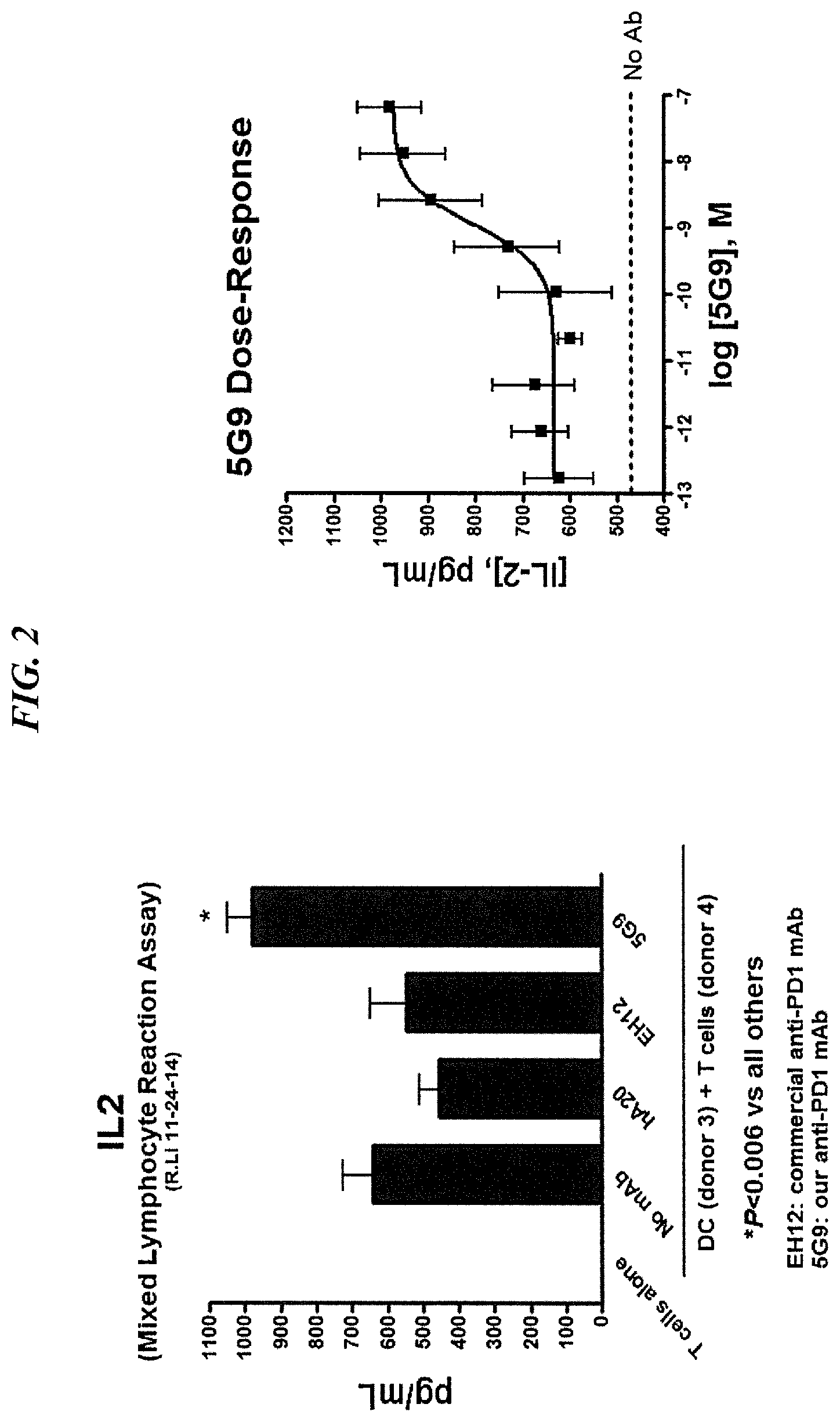
FIG. 2. Quantitation of IL-2 by ELISA. A notable increase in IL-2 produced from T cells in a mixed lymphocyte assay was observed for 5G9.G1.B11 dose-independently.
FIG. 3A. The amino acid sequence determined for the VK (SEQ ID NO:7) of 5G9.G1.B11, with the 3 CDRs underlined.
FIG. 3B. The amino acid sequence determined for the VH (SEQ ID NO:8) of 5G9.G1.B11, with the 3 CDRs underlined.
FIG. 3C. CDR sequences of 5G9.G1.B11 were, for the heavy chain, GFAFSSNDMS (SEQ ID NO:1), TISGGGINTYYPDSVKG (SEQ ID NO:2) and RSNYAWFAY (SEQ ID NO:3) and for the light chain, RASESVDTYGISFMN (SEQ ID NO:4), PNQGS (SEQ ID NO:5) and QQSKEVPWT (SEQ ID NO:6).
FIG. 4A. Binding of the chimeric 2G9 to recombinant PD-1-Fc by ELISA.
FIG. 4B. Binding of 2G9 to SpEFX-2D1, but not SpESX cells, by flow cytometry. The SpESX cell line, which does not express PD-1, was transfected with human PD-1 to obtain SpESX-2D1, which overexpresses PD-1.
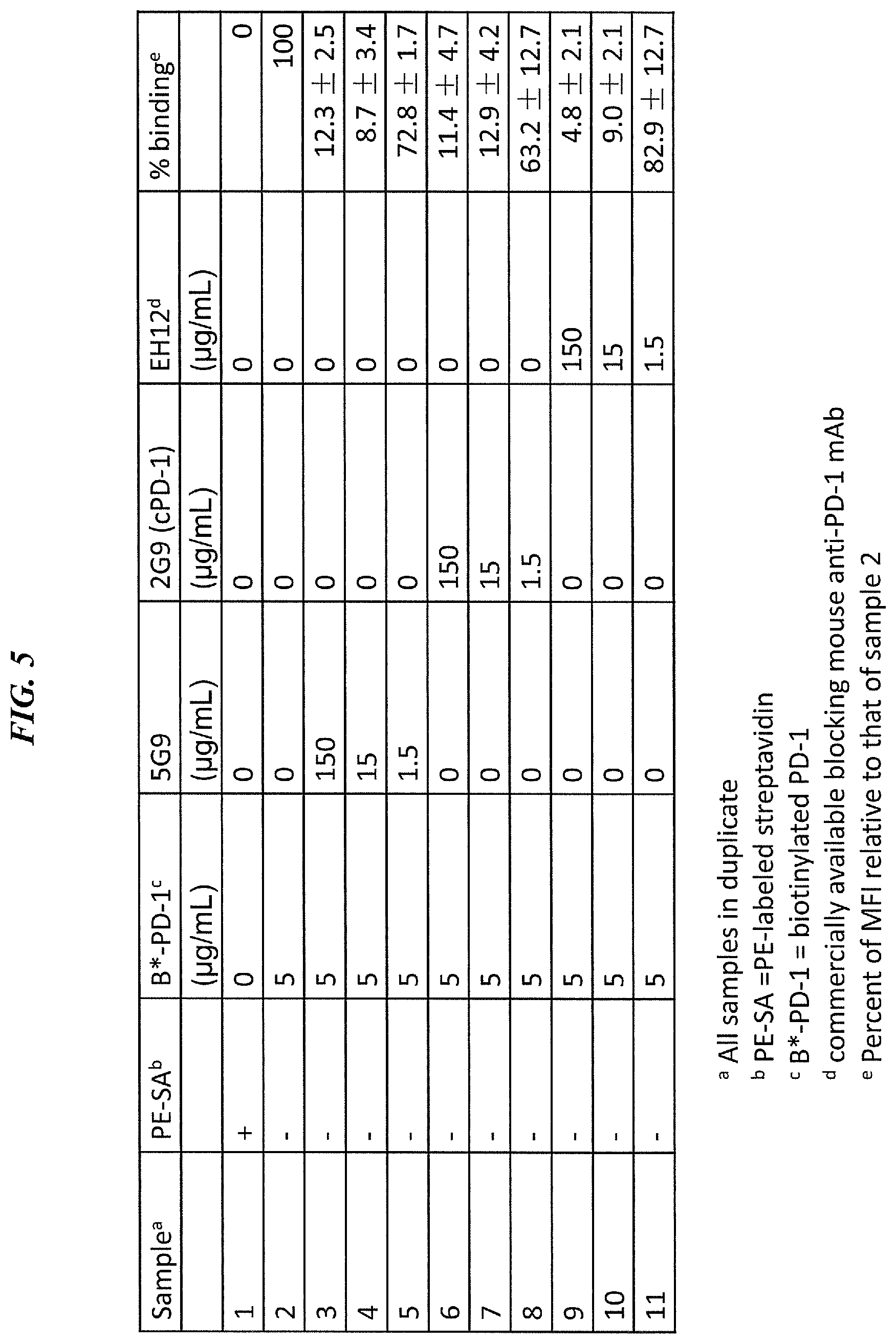
FIG. 5. Blockade of biotinlyated PD-1 binding to PD-1 on MDA-MB-231 by 2G9.
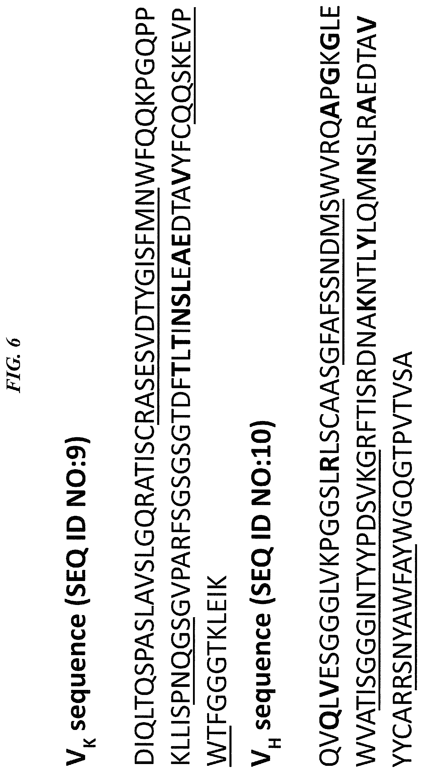
FIG. 6. Amino acid sequence of light (SEQ ID NO:9) and heavy (SEQ ID NO:10) chains of humanized anti-PD-1 antibody (hPD-1). CDR sequences are underlined. Framework residues (FRs) where the parental murine amino acid residue is substituted with the corresponding human amino acid residue are highlighted in bold font.
FIG. 7. DNA sequence encoding light chain (SEQ ID NO:11) and heavy chain (SEQ ID NO:12) of humanized anti-PD-1 antibody.
FIG. 8. Binding of humanized vs. chimeric anti-PD-1 to recombinant human PD-1-His.
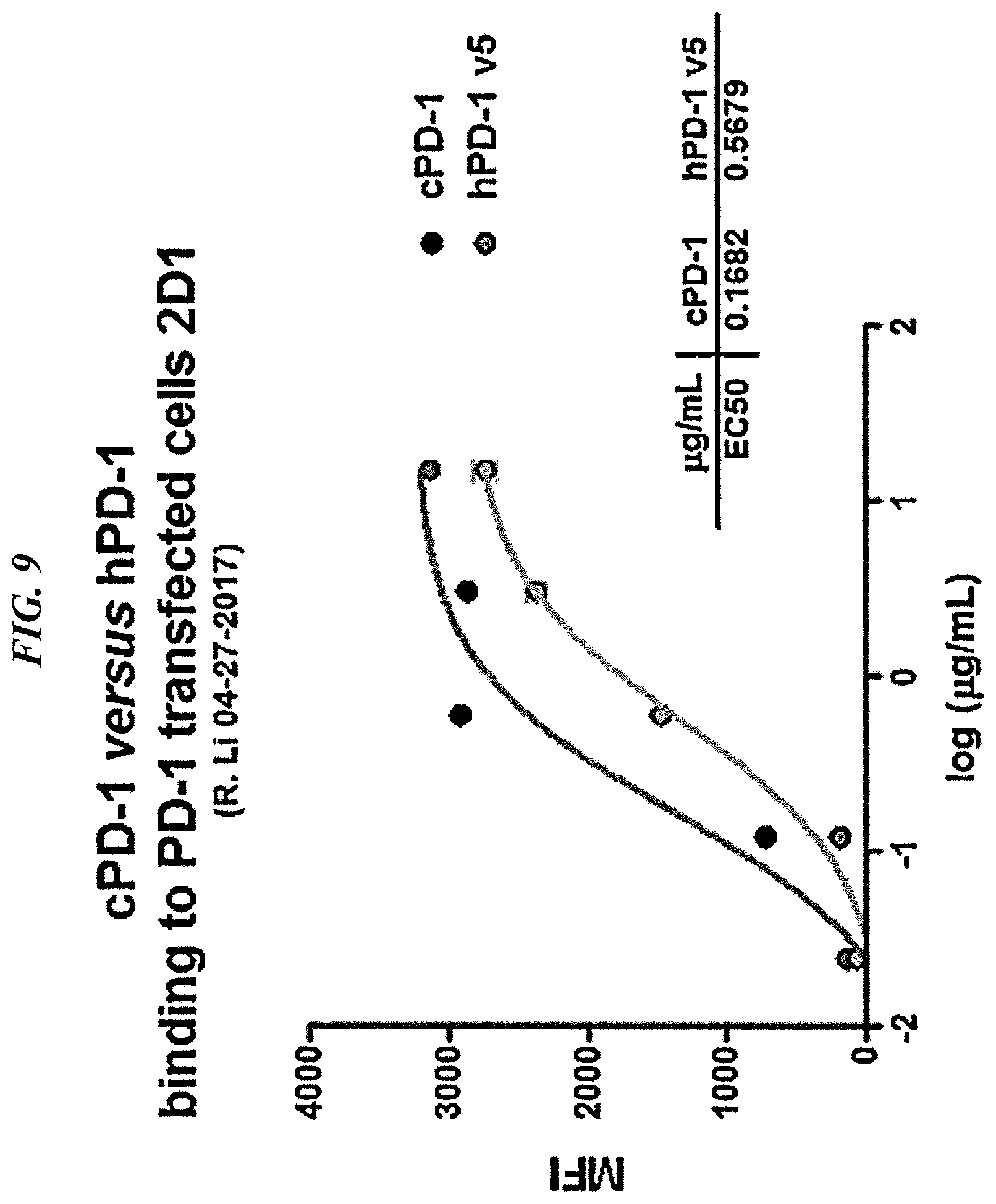
FIG. 9. Binding of humanized vs. chimeric anti-PD-1 to cells transfected with human PD-1 (2D1 cells).
FIG. 10. Combination therapy with anti-Trop-2.times.anti-CD3 bsAb and humanized or chimeric anti-PD-1 antibody.
DETAILED DESCRIPTION
Definitions
Unless otherwise specified, "a" or "an" means "one or more".
As used herein, the terms "and" and "or" may be used to mean either the conjunctive or disjunctive. That is, both terms should be understood as equivalent to "and/or" unless otherwise stated.
A "therapeutic agent" is an atom, molecule, or compound that is useful in the treatment of a disease. Examples of therapeutic agents include antibodies, antibody fragments, peptides, drugs, toxins, enzymes, nucleases, hormones, immunomodulators, antisense oligonucleotides, small interfering RNA (siRNA), chelators, boron compounds, photoactive agents, dyes, and radioisotopes.
An "antibody" as used herein refers to a full-length (i.e., naturally occurring or formed by normal immunoglobulin gene fragment recombinatorial processes) immunoglobulin molecule (e.g., an IgG antibody) or an immunologically active (i.e., specifically binding) portion of an immunoglobulin molecule, like an antibody fragment. An "antibody" includes monoclonal, polyclonal, bispecific, multispecific, murine, chimeric, humanized and human antibodies.
A "naked antibody" is an antibody or antigen binding fragment thereof that is not attached to a therapeutic or diagnostic agent. The Fc portion of an intact naked antibody can provide effector functions, such as complement fixation and ADCC (see, e.g., Markrides, Pharmacol Rev 50:59-87, 1998). Other mechanisms by which naked antibodies induce cell death may include apoptosis. (Vaswani and Hamilton, Ann Allergy Asthma Immunol 81: 105-119, 1998.)
An "antibody fragment" is a portion of an intact antibody such as F(ab').sub.2, F(ab).sub.2, Fab', Fab, Fv, scFv, dAb and the like. Regardless of structure, an antibody fragment binds with the same antigen that is recognized by the full-length antibody. For example, antibody fragments include isolated fragments consisting of the variable regions, such as the "Fv" fragments consisting of the variable regions of the heavy and light chains or recombinant single chain polypeptide molecules in which light and heavy variable regions are connected by a peptide linker ("scFv proteins"). "Single-chain antibodies", often abbreviated as "scFv" consist of a polypeptide chain that comprises both a V.sub.H and a V.sub.L domain which interact to form an antigen-binding site. The V.sub.H and V.sub.L domains are usually linked by a peptide of 1 to 25 amino acid residues. Antibody fragments also include diabodies, triabodies and single domain antibodies (dAb).
A "chimeric antibody" is a recombinant protein that contains the variable domains including the complementarity determining regions (CDRs) of an antibody derived from one species, preferably a rodent antibody, while the constant domains of the antibody molecule are derived from those of a human antibody. For veterinary applications, the constant domains of the chimeric antibody may be derived from that of other species, such as a cat or dog.
A "humanized antibody" is a recombinant protein in which the CDRs from an antibody from one species; e.g., a rodent antibody, are transferred from the heavy and light variable chains of the rodent antibody into human heavy and light variable domains, including human framework region (FR) sequences. The constant domains of the antibody molecule are derived from those of a human antibody. To maintain binding activity, a limited number of FR amino acid residues from the parent (e.g., murine) antibody may be substituted for the corresponding human FR residues.
A "human antibody" is an antibody obtained from transgenic mice that have been genetically engineered to produce specific human antibodies in response to antigenic challenge. In this technique, elements of the human heavy and light chain locus are introduced into strains of mice derived from embryonic stem cell lines that contain targeted disruptions of the endogenous heavy chain and light chain loci. The transgenic mice can synthesize human antibodies specific for human antigens, and the mice can be used to produce human antibody-secreting hybridomas. Methods for obtaining human antibodies from transgenic mice are described by Green et al., Nature Genet. 7:13 (1994), Lonberg et al., Nature 368:856 (1994), and Taylor et al., Int. Immun. 6:579 (1994). A human antibody also can be constructed by genetic or chromosomal transfection methods, as well as phage display technology, all of which are known in the art. (See, e.g., McCafferty et al., 1990, Nature 348:552-553 for the production of human antibodies and fragments thereof in vitro, from immunoglobulin variable domain gene repertoires from unimmunized donors). In this technique, antibody variable domain genes are cloned in-frame into either a major or minor coat protein gene of a filamentous bacteriophage, and displayed as functional antibody fragments on the surface of the phage particle. Because the filamentous particle contains a single-stranded DNA copy of the phage genome, selections based on the functional properties of the antibody also result in selection of the gene encoding the antibody exhibiting those properties. In this way, the phage mimics some of the properties of the B cell. Phage display can be performed in a variety of formats, for their review, see, e.g. Johnson and Chiswell, Current Opinion in Structural Biology 3:5564-571 (1993). Human antibodies may also be generated by in vitro activated B cells. (See, U.S. Pat. Nos. 5,567,610 and 5,229,275).
As used herein, the term "antibody fusion protein" is a recombinantly produced antigen-binding molecule in which an antibody or antibody fragment is linked to another protein or peptide, such as the same or different antibody or antibody fragment. The fusion protein may comprise a single antibody component, a multivalent or multispecific combination of different antibody components or multiple copies of the same antibody component. The fusion protein may additionally comprise an antibody or an antibody fragment and a therapeutic agent. Examples of therapeutic agents suitable for such fusion proteins include immunomodulators. A preferred immunomodulator might be an interferon, such as interferon-.alpha., interferon-.beta. or interferon-.lamda..
A "multispecific antibody" is an antibody that can bind simultaneously to at least two targets that are of different structure, e.g., two different antigens, two different epitopes on the same antigen, or a hapten and/or an antigen or epitope. A "multivalent antibody" is an antibody that can bind simultaneously to at least two targets that are of the same or different structure. Valency indicates how many binding arms or sites the antibody has to a single antigen or epitope; i.e., monovalent, bivalent, trivalent or multivalent. The multivalency of the antibody means that it can take advantage of multiple interactions in binding to an antigen, thus increasing the avidity of binding to the antigen. Specificity indicates how many antigens or epitopes an antibody is able to bind; i.e., monospecific, bispecific, trispecific, multispecific. Using these definitions, a natural antibody, e.g., an IgG, is bivalent because it has two binding arms but is monospecific because it binds to one epitope. Multispecific, multivalent antibodies are constructs that have more than one binding site of different specificity.
An antibody preparation, or a composition described herein, is said to be administered in a "therapeutically effective amount" if the amount administered is physiologically significant. An agent is physiologically significant if its presence results in a detectable change in the physiology of a recipient subject. In particular embodiments, an antibody preparation is physiologically significant if its presence invokes an antitumor response. A physiologically significant effect could also be the evocation of a humoral and/or cellular immune response in the recipient subject leading to growth inhibition or death of target cells.
Checkpoint Inhibitors
In various embodiments, the subject anti-PD-1 antibody may be administered in combination with one or more other checkpoint inhibitor antibodies. Various such antibodies are known and/or commercially available, primary targeted to PD-1, PD-1 or CTLA-4.
Programmed cell death protein 1 (PD-1, also known as CD279) encodes a cell surface membrane protein of the immunoglobulin superfamily, which is expressed in B cells and NK cells (Shinohara et al., 1995, Genomics 23:704-6; Blank et al., 2007, Cancer Immunol Immunother 56:739-45; Finger et al., 1997, Gene 197:177-87; Pardoll, 2012, Nature Reviews 12:252-264). Anti-PD-1 antibodies have been used for treatment of melanoma, non-small-cell lung cancer, bladder cancer, prostate cancer, colorectal cancer, head and neck cancer, triple-negative breast cancer, leukemia, lymphoma and renal cell cancer (Topalian et al., 2012, N Engl J Med 366:2443-54; Lipson et al., 2013, Clin Cancer Res 19:462-8; Berger et al., 2008, Clin Cancer Res 14:3044-51; Gildener-Leapman et al., 2013, Oral Oncol 49:1089-96; Menzies & Long, 2013, Ther Adv Med Oncol 5:278-85).
Exemplary anti-PD-1 antibodies include lambrolizumab (MK-3475, MERCK), nivolumab (BMS-936558, BRISTOL-MYERS SQUIBB), and pidilizumab (CT-011, CURETECH LTD.). Anti-PD-1 antibodies are commercially available, for example from ABCAM.RTM. (AB137132), BIOLEGEND.RTM. (EH12.2H7, RMP1-14) and AFFYMETRIX EBIOSCIENCE (J105, J116, MIH4).
A particular anti-PD-1 antibody of use, disclosed in the Examples below, is defined by the heavy chain CDR sequences GFAFSSNDMS (SEQ ID NO:1), TISGGGINTYYPDSVKG (SEQ ID NO:2) and RSNYAWFAY (SEQ ID NO:3) and the light chain CDR sequences RASESVDTYGISFMN (SEQ ID NO:4), PNQGS (SEQ ID NO:5) and QQSKEVPWT (SEQ ID NO:6). The antibody may be used in chimeric, humanized, or fully human form, as discussed below.
Programmed cell death 1 ligand 1 (PD-L1, also known as CD274) is a ligand for PD-1, found on activated T cells, B cells, myeloid cells and macrophages. The complex of PD-1 and PD-1 inhibits proliferation of CD8+ T cells and reduces the immune response (Topalian et al., 2012, N Engl J Med 366:2443-54; Brahmer et al., 2012, N Eng J Med 366:2455-65). Anti-PDL1 antibodies have been used for treatment of non-small cell lung cancer, melanoma, colorectal cancer, renal-cell cancer, pancreatic cancer, gastric cancer, ovarian cancer, breast cancer, and hematologic malignancies (Brahmer et al., N Eng J Med 366:2455-65; Ott et al., 2013, Clin Cancer Res 19:5300-9; Radvanyi et al., 2013, Clin Cancer Res 19:5541; Menzies & Long, 2013, Ther Adv Med Oncol 5:278-85; Berger et al., 2008, Clin Cancer Res 14:13044-51).
Exemplary anti-PDL1 antibodies include MDX-1105 (MEDAREX), MEDI4736 (MEDIMMUNE) MPDL3280A (GENENTECH) and BMS-936559 (BRISTOL-MYERS SQUIBB). Anti-PDL1 antibodies are also commercially available, for example from AFFYMETRIX EBIOSCIENCE (MIH1).
Cytotoxic T-lymphocyte antigen 4 (CTLA-4, also known as CD152) is also a member of the immunoglobulin superfamily that is expressed exclusively on T-cells. CTLA-4 acts to inhibit T cell activation and is reported to inhibit helper T cell activity and enhance regulatory T cell immunosuppressive activity (Pardoll, 2012, Nature Reviews 12:252-264). Anti-CTL4A antibodies have been used in clinical trials for treatment of melanoma, prostate cancer, small cell lung cancer, non-small cell lung cancer (Robert & Ghiringhelli, 2009, Oncologist 14:848-61; Ott et al., 2013, Clin Cancer Res 19:5300; Weber, 2007, Oncologist 12:864-72; Wada et al., 2013, J Transl Med 11:89).
Exemplary anti-CTLA-4 antibodies include ipilimumab (Bristol-Myers Squibb) and tremelimumab (PFIZER). Anti-PD-1 antibodies are commercially available, for example from ABCAM.RTM. (AB134090), SINO BIOLOGICAL INC. (11159-H03H, 11159-H08H), and THERMO SCIENTIFIC PIERCE (PA5-29572, PA5-23967, PA5-26465, MA1-12205, MA1-35914). Ipilimumab has recently received FDA approval for treatment of metastatic melanoma (Wada et al., 2013, J Transl Med 11:89).
These and other known checkpoint inhibitor antibodies may be used alone or in combination other anti-cancer therapies as discussed herein. The person of ordinary skill will realize that methods of determining optimal dosages of checkpoint inhibitor antibodies to administer to a patient in need thereof, either alone or in combination with one or more other agents, may be determined by standard dose-response and toxicity studies that are well known in the art. In an exemplary embodiment, an immune checkpoint inhibitor antibody may preferably be administered at about 0.3-10 mg/kg, or the maximum tolerated dose, administered about every three weeks or about every six weeks. Alternatively, the checkpoint inhibitor antibody may be administered by an escalating dosage regimen including administering a first dosage at about 3 mg/kg, a second dosage at about 5 mg/kg, and a third dosage at about 9 mg/kg. Alternatively, the escalating dosage regimen includes administering a first dosage of checkpoint inhibitor antibody at about 5 mg/kg and a second dosage at about 9 mg/kg. Another stepwise escalating dosage regimen may include administering a first dosage of checkpoint inhibitor antibody about 3 mg/kg, a second dosage of about 3 mg/kg, a third dosage of about 5 mg/kg, a fourth dosage of about 5 mg/kg, and a fifth dosage of about 9 mg/kg. In another aspect, a stepwise escalating dosage regimen may include administering a first dosage of 5 mg/kg, a second dosage of 5 mg/kg, and a third dosage of 9 mg/kg. Exemplary reported dosages of checkpoint inhibitor mAbs include 3 mg/kg ipilimumab administered every three weeks for four doses; 10 mg/kg ipilimumab every three weeks for eight cycles; 10 mg/kg every three weeks for four cycles then every 12 weeks for a total of three years; 10 mg/kg MK-3475 every two or every three weeks; 2 mg/kg MK-3475 every three weeks; 15 mg/kg tremilimumab every three months; 0.1, 0.3, 1, 3 or 10 mg/kg nivolumab every two weeks for up to 96 weeks; 0.3, 1, 3, or 10 mg/kg BMS-936559 every two weeks for up to 96 weeks (Kyi & Postow, Oct. 23, 2013, FEBS Lett [Epub ahead of print]; Callahan & Wolchok, 2013, J Leukoc Biol 94:41-53).
Interferon Therapy
In other embodiments, the subject checkpoint inhibitors may be administered in combination with interferon. Interferons are critical role players in the antitumor and antimicrobial host defense, and have been extensively explored as therapeutic agents for cancer (Billiau et al., 2006, Cytokine Growth Factor Rev 17:381-409; Pestka et al., 2004, Immunol Rev 202:8-32). Despite considerable efforts with type I and II interferons (IFN-.alpha./.beta. and .gamma.), their use in clinic settings have been limited because of the short circulation half-life, systemic toxicity, and suboptimal responses in patients (Pestka et al., 2004, Immunol Rev 202:8-32; Miller et al., 2009, Ann NY Acad Sci 1182:69-79). The discovery of the IFN-.lamda. family in early 2003 brought an exciting new opportunity to develop alternative IFN agents for these unmet clinical indications (Kotenko et al., 2003, Nat Immunol 4:69-77; Sheppard et al., 2003, Nat Immunol 4:63-8).
The therapeutic effectiveness of IFNs has been validated to date by the approval of IFN-.alpha.2 for treating hairy cell leukemia, chronic myelogenous leukemia, malignant melanoma, follicular lymphoma, condylomata acuminata, AIDs-related Kaposi sarcoma, and chronic hepatitis B and C; IFN-.beta. for treating multiple sclerosis; and IFN-.gamma. for treating chronic granulomatous disease and malignant osteopetrosis. Despite a vast literature on this group of autocrine and paracrine cytokines, their functions in health and disease are still being elucidated, including more effective and novel forms being introduced clinically (Pestka, 2007, J. Biol. Chem 282:20047-51; Vilcek, 2006, Immunity 25:343-48). The effects of combination of various interferons with antibody-based therapies also remain under investigation.
In various embodiments, checkpoint inhibitor antibodies may be used in combination with one or more interferons, such as interferon-.alpha., interferon-.beta. or interferon-.lamda.. Human interferons are well known in the art and the amino acid sequences of human interferons may be readily obtained from public databases (e.g., GenBank Accession Nos. AAA52716.1; AAA52724; AAC41702.1; EAW56871.1; EAW56870.1; EAW56869.1). Human interferons may also be commercially obtained from a variety of vendors (e.g., Cell Signaling Technology, Inc., Danvers, Mass.; Genentech, South San Francisco, Calif.; EMD Millipore, Billerica, Mass.).
Interferon-.alpha. (IFN.alpha.) has been reported to have anti-tumor activity in animal models of cancer (Ferrantini et al., 1994, J Immunol 153:4604-15) and human cancer patients (Gutterman et al., 1980, Ann Intern Med 93:399-406). IFN.alpha. can exert a variety of direct anti-tumor effects, including down-regulation of oncogenes, up-regulation of tumor suppressors, enhancement of immune recognition via increased expression of tumor surface MHC class I proteins, potentiation of apoptosis, and sensitization to chemotherapeutic agents (Gutterman et al., 1994, PNAS USA 91:1198-205; Matarrese et al., 2002, Am J Pathol 160:1507-20; Mecchia et al., 2000, Gene Ther 7:167-79; Sabaawy et al., 1999, Int J Oncol 14:1143-51; Takaoka et al, 2003, Nature 424:516-23). For some tumors, IFN.alpha. can have a direct and potent anti-proliferative effect through activation of STAT1 (Grimley et al., 1998 Blood 91:3017-27). Interferon-.alpha.2b has been conjugated to anti-tumor antibodies, such as the hL243 anti-HLA-DR antibody and depletes lymphoma and myeloma cells in vitro and in vivo (Rossi et al., 2011, Blood 118:1877-84).
Indirectly, IFN.alpha. can inhibit angiogenesis (Sidky and Borden, 1987, Cancer Res 47:5155-61) and stimulate host immune cells, which may be vital to the overall antitumor response but has been largely under-appreciated (Belardelli et al., 1996, Immunol Today 17:369-72). IFN.alpha. has a pleiotropic influence on immune responses through effects on myeloid cells (Raefsky et al, 1985, J Immunol 135:2507-12; Luft et al, 1998, J Immunol 161:1947-53), T-cells (Carrero et al, 2006, J Exp Med 203:933-40; Pilling et al., 1999, Eur J Immunol 29:1041-50), and B-cells (Le et al, 2001, Immunity 14:461-70). As an important modulator of the innate immune system, IFN.alpha. induces the rapid differentiation and activation of dendritic cells (Belardelli et al, 2004, Cancer Res 64:6827-30; Paquette et al., 1998, J Leukoc Biol 64:358-67; Santini et al., 2000, J Exp Med 191:1777-88) and enhances the cytotoxicity, migration, cytokine production and antibody-dependent cellular cytotoxicity (ADCC) of NK cells (Biron et al., 1999, Ann Rev Immunol 17:189-220; Brunda et al. 1984, Cancer Res 44:597-601).
Interferon-.beta. has been reported to be efficacious for therapy of a variety of solid tumors. Patients treated with 6 million units of IFN-.beta. twice a week for 36 months showed a decreased recurrence of hepatocellular carcinoma after complete resection or ablation of the primary tumor in patients with HCV-related liver cancer (Ikeda et al., 2000, Hepatology 32:228-32). Gene therapy with interferon-.beta. induced apoptosis of glioma, melanoma and renal cell carcinoma (Yoshida et al., 2004, Cancer Sci 95:858-65). Endogenous IFN-.beta. has been observed to inhibit tumor growth by inhibiting angiogenesis in vivo (Jablonska et al., 2010, J Clin Invest. 120:1151-64.)
IFN-.lamda.s, designated as type III interferons, are a newly described group of cytokines that consist of IFN-.lamda.1, 2, 3 (also referred to as interleukin-29, 28A, and 28B, respectively), that are genetically encoded by three different genes located on chromosome 19 (Kotenko et al., 2003, Nat Immunol 4:69-77; Sheppard et al., 2003, Nat Immunol 4:63-8). IFN-.lamda.s activate signal transduction via the JAK/STAT pathway similar to that induced by type I IFN, including the activation of JAK1 and TYK2 kinases, the phosphorylation of STAT proteins, and the activation of the transcription complex of IFN-stimulated gene factor 3 (ISGF3) (Witte et al., 2010, Cytokine Growth Factor Rev 21:237-51; Zhou et al., 2007, J Virol 81:7749-58).
A major difference between type III and type I IFN systems is the distribution of their respective receptor complexes. IFN-.alpha./.beta. signals through two extensively expressed type I interferon receptors, and the resulting systemic toxicity associated with IFN-.alpha./.beta. administration has limited their use as therapeutic agents (Pestka et al., 2007, J Biol Chem 282:20047-51). In contrast, IFN.alpha.s signal through a heterodimeric receptor complex consisting of unique IFN-.lamda. receptor 1 (IFN-.lamda.R1) and IL-10 receptor 2 (IL-10R2). As previously reported (Witte et al., 2009, Genes Immun 10:702-14), IFN-.lamda.R1 has a very restricted expression pattern with the highest levels in epithelial cells, melanocytes, and hepatocytes, and the lowest level in primary central nervous system (CNS) cells. Blood immune system cells express high levels of a short IFN-.lamda. receptor splice variant (sIFN-.lamda.R1) that inhibits IFN-.lamda. action. The limited responsiveness of neuronal cells and immune cells implies that the severe toxicity frequently associated with IFN-.alpha. therapy may be absent or significantly reduced with IFN.lamda.s (Witte et al., 2009, Genes Immun 10:702-14; Witte et al., 2010, Cytokine Growth Factor Rev 21:237-51). A recent publication reported that while IFN-.alpha. and IFN-.lamda. induce expression of a common set of ISGs (interferon-stimulated genes) in hepatocytes, unlike IFN-.alpha., administration of IFN-.lamda. did not induce STAT activation or ISG expression in purified lymphocytes or monocytes (Dickensheets et al., 2013, J Leukoc Biol. 93, published online Dec. 20, 2012). It was suggested that IFN-.lamda. may be superior to IFN-.alpha. for treatment of chronic HCV infection, as it is less likely to induce leukopenias that are often associated with IFN-.alpha. therapy (Dickensheets et al., 2013).
IFN.lamda.s display structural features similar to IL-10-related cytokines, but functionally possess type I IFN-like anti-viral and anti-proliferative activity (Witte et al., 2009, Genes Immun 10:702-14; Ank et al., 2006, J Virol 80:4501-9; Robek et al., 2005, J Virol 79:3851-4). The anti-proliferative activity of IFN.lamda.s has been established in several human cancer cell lines, including neuroendocrine carcinoma BON1 (Zitzmann et al., 2006, Biochem Biophys Res Commun 344:1334-41), glioblastoma LN319 (Meager et al., 2005, Cytokine 31:109-18), immortalized keratinocyte HaCaT (Maher et al., 2008, Cancer Biol Ther 7:1109-15), melanoma F01 (Guenterberg et al., 2010, Mol Cancer Ther 9:510-20), and esophageal carcinoma TE-11 (Li et al., 2010, Eur J Cancer 46:180-90). In animal models, IFN-.lamda.s induce both tumor apoptosis and destruction through innate and adaptive immune responses, suggesting that local delivery of IFN-.lamda. might be a useful adjunctive strategy in the treatment of human malignancies (Numasaki et al., 2007, J Immunol 178:5086-98). A Fab-linked interferon-.lamda. was demonstrated to have potent anti-tumor and anti-viral activity in targeted cells (Liu et al., 2013, PLoS One 8:e63940). In clinical settings, PEGylated IFN-.lamda.1 (PEG-IFN-.lamda.1) has been provisionally used for patients with chronic hepatitis C virus infection. In a phase Ib study (n=56), antiviral activity was observed at all dose levels (0.5-3.0 .mu.g/kg), and viral load reduced 2.3 to 4.0 logs when PEG-IFN-.lamda.1 was administrated to genotype 1 HCV patients who relapsed after IFN-.alpha. therapy (Muir et al., 2010, Hepatology 52:822-32). At the same time, rates of adverse events commonly associated with type I interferon treatment were lower with PEG-IFN-.lamda.1 than with PEG-IFN-.alpha.. Neutropenia and thrombocytopenia were infrequently observed and the rates of flu-like symptoms, anemia, and musculoskeletal symptoms decreased to about 1/3 of that seen with PEG-IFN-.alpha. treatment. However, rates of serious adverse events, depression and other common adverse events (.gtoreq.10%) were similar between PEG-IFN-.lamda.1 and PEG-IFN-.alpha.. Higher rates of hepatotoxicity were seen in the highest-dose PEG-IFN-.lamda.1 compared with PEG-IFN-.alpha. ("Investigational Compound PEG-Interferon Lambda Achieved Higher Response Rates with Fewer Flu-like and Musculoskeletal Symptoms and Cytopenias Than PEG-Interferon Alfa in Phase IIb Study of 526 Treatment-Naive Hepatitis C Patients," Apr. 2, 2011, Press Release from Bristol-Myers Squibb).
In various embodiments, the subject checkpoint inhibitor mAbs may be used in combination with one or more interferons, such as interferon-.alpha., interferon-.beta., interferon-.lamda.1, interferon-.lamda.2, or interferon-.lamda.3. When used with other agents, the interferon may be administered prior to, concurrently with, or after the other agent. When administered concurrently, the interferon may be either conjugated to or separate from the other agent.
Antibody-Drug Conjugates
Antibody-drug conjugates (ADCs) are a potent class of therapeutic constructs that allow targeted delivery of cytotoxic agents to target cells, such as cancer cells. Because of the targeting function, these compounds show a much higher therapeutic index compared to the same systemically delivered agents. ADCs have been developed as intact antibodies or antibody fragments, such as scFvs. The antibody or fragment is linked to one or more copies of drug via a linker that is stable under physiological conditions, but that may be cleaved once inside the target cell. ADCs approved for therapeutic use include gemtuzumab ozogamicin for AML (subsequently withdrawn from the market), inotuzumab ozogamicin for relapsed/refractory ALL, brentuximab vedotin for ALCL and Hodgkin lymphoma, and trastuzumab emtansine for HER2-positive metastatic breast cancer (Verma et al., 2012, N Engl J Med 367:1783-91; Bross et al., 2001, Clin Cancer Res 7:1490-96; Francisco et al., 2003, Blood 102:1458-65). Numerous other candidate ADCs are currently in clinical testing, such as glembatumomab vedotin (Celldex Therapeutics), SAR3419 (Sanofi-Aventis), SAR56658 (Sanofi-Aventis), AMG-172 (Amgen), AMG-595 (Amgen), BAY-94-9343 (Bayer), BIIB015 (Biogen Idec), BT062 (Biotest), SGN-75 (Seattle Genetics), SGN-CD19A (Seattle Genetics), vorsetuzumab mafodotin (Seattle Genetics), ABT-414 (AbbVie), ASG-5ME (Agensys), ASG-22ME (Agensys), ASG-16M8F (Agensys), IMGN-529 (ImmunoGen), IMGN-853 (ImmunoGen), MDX-1203 (Medarex), MLN-0264 (Millenium), RG-7450 (Roche/Genentech), RG-7458 (Roche/Genentech), RG-7593 (Roche/Genentech), RG-7596 (Roche/Genentech), RG-7598 (Roche/Genentech), RG-7599 (Roche/Genentech), RG-7600 (Roche/Genentech), RG-7636 (Roche/Genentech), anti-PSMA ADC (Progenics), lorvotuzumab mertansine (ImmunoGen), milatuzumab-doxorubicin (Immunomedics), IMMU-130 (Immunomedics), IMMU-132 (Immunomedics), IMMU-140 (Immunomedics) and antibody conjugates of pro-2-pyrrolinodoxorubicin. (See, e.g., Li et al., 2013, Drug Disc Ther 7:178-84; Firer & Gellerman, J Hematol Oncol 5:70; Beck et al., 2010, Discov Med 10:329-39; Mullard, 2013, Nature Rev Drug Discovery 12:329, U.S. Pat. No. 8,877,202.) Because of the potential of ADCs to act as potent anti-cancer agents with reduced systemic toxicity, they may be used either alone or as an adjunct therapy to reduce tumor burden.
These and other known agents that stimulate immune response to tumors and/or pathogens may be used in combination with checkpoint inhibitors. Other known co-stimulatory pathway modulators that may be used in combination include, but are not limited to, agatolimod, belatacept, blinatumomab, CD40 ligand, anti-B7-1 antibody, anti-B7-2 antibody, anti-B7-H4 antibody, AG4263, eritoran, anti-OX40 antibody, ISF-154, and SGN-70; B7-1, B7-2, ICAM-1, ICAM-2, ICAM-3, CD48, LFA-3, CD30 ligand, CD40 ligand, heat stable antigen, B7h, OX40 ligand, LIGHT, CD70 and CD24.
General Antibody Techniques
Techniques for preparing monoclonal antibodies against virtually any target antigen are well known in the art. See, for example, Kohler and Milstein, Nature 256: 495 (1975), and Coligan et al. (eds.), CURRENT PROTOCOLS IN IMMUNOLOGY, VOL. 1, pages 2.5.1-2.6.7 (John Wiley & Sons 1991). Briefly, monoclonal antibodies can be obtained by injecting mice with a composition comprising an antigen, removing the spleen to obtain B-lymphocytes, fusing the B-lymphocytes with myeloma cells to produce hybridomas, cloning the hybridomas, selecting positive clones which produce antibodies to the antigen, culturing the clones that produce antibodies to the antigen, and isolating the antibodies from the hybridoma cultures.
MAbs can be isolated and purified from hybridoma cultures by a variety of well-established techniques. Such isolation techniques include affinity chromatography with Protein-A Sepharose, size-exclusion chromatography, and ion-exchange chromatography. See, for example, Coligan at pages 2.7.1-2.7.12 and pages 2.9.1-2.9.3. Also, see Baines et al., "Purification of Immunoglobulin G (IgG)," in METHODS IN MOLECULAR BIOLOGY, VOL. 10, pages 79-104 (The Humana Press, Inc. 1992).
After the initial raising of antibodies to the immunogen, the antibodies can be sequenced and subsequently prepared by recombinant techniques. Humanization and chimerization of murine antibodies and antibody fragments are well known to those skilled in the art. The use of antibody components derived from humanized, chimeric or human antibodies obviates potential problems associated with the immunogenicity of murine constant regions.
Chimeric Antibodies
A chimeric antibody is a recombinant protein in which the variable regions of a human antibody have been replaced by the variable regions of, for example, a mouse antibody, including the complementarity-determining regions (CDRs) of the mouse antibody. Chimeric antibodies exhibit decreased immunogenicity and increased stability when administered to a subject. General techniques for cloning murine immunoglobulin variable domains are disclosed, for example, in Orlandi et al., Proc. Nat'l Acad. Sci. USA 86: 3833 (1989). Techniques for constructing chimeric antibodies are well known to those of skill in the art. As an example, Leung et al., Hybridoma 13:469 (1994), produced an LL2 chimera by combining DNA sequences encoding the V.sub..kappa. and V.sub.H domains of murine LL2, an anti-CD22 monoclonal antibody, with respective human .kappa. and IgG.sub.1 constant region domains.
Humanized Antibodies
Techniques for producing humanized MAbs are well known in the art (see, e.g., Jones et al., Nature 321: 522 (1986), Riechmann et al., Nature 332: 323 (1988), Verhoeyen et al., Science 239: 1534 (1988), Carter et al., Proc. Nat'l Acad. Sci. USA 89: 4285 (1992), Sandhu, Crit. Rev. Biotech. 12: 437 (1992), and Singer et al., J. Immun. 150: 2844 (1993)). A chimeric or murine monoclonal antibody may be humanized by transferring the mouse CDRs from the heavy and light variable chains of the mouse immunoglobulin into the corresponding variable domains of a human antibody. The mouse framework regions (FR) in the chimeric monoclonal antibody are also replaced with human FR sequences. As simply transferring mouse CDRs into human FRs often results in a reduction or even loss of antibody affinity, additional modification might be required in order to restore the original affinity of the murine antibody. This can be accomplished by the replacement of one or more human residues in the FR regions with their murine counterparts to obtain an antibody that possesses good binding affinity to its epitope. See, for example, Tempest et al., Biotechnology 9:266 (1991) and Verhoeyen et al., Science 239: 1534 (1988). Generally, those human FR amino acid residues that differ from their murine counterparts and are located close to or touching one or more CDR amino acid residues would be candidates for substitution.
Human Antibodies
Methods for producing fully human antibodies using either combinatorial approaches or transgenic animals transformed with human immunoglobulin loci are known in the art (e.g., Mancini et al., 2004, New Microbiol. 27:315-28; Conrad and Scheller, 2005, Comb. Chem. High Throughput Screen. 8:117-26; Brekke and Loset, 2003, Curr. Opin. Phamacol. 3:544-50). A fully human antibody also can be constructed by genetic or chromosomal transfection methods, as well as phage display technology, all of which are known in the art. See for example, McCafferty et al., Nature 348:552-553 (1990). Such fully human antibodies are expected to exhibit even fewer side effects than chimeric or humanized antibodies and to function in vivo as essentially endogenous human antibodies. In certain embodiments, the claimed methods and procedures may utilize human antibodies produced by such techniques.
In one alternative, the phage display technique may be used to generate human antibodies (e.g., Dantas-Barbosa et al., 2005, Genet. Mol. Res. 4:126-40). Human antibodies may be generated from normal humans or from humans that exhibit a particular disease state, such as cancer (Dantas-Barbosa et al., 2005). The advantage to constructing human antibodies from a diseased individual is that the circulating antibody repertoire may be biased towards antibodies against disease-associated antigens.
In one non-limiting example of this methodology, Dantas-Barbosa et al. (2005) constructed a phage display library of human Fab antibody fragments from osteosarcoma patients. Generally, total RNA was obtained from circulating blood lymphocytes (Id.). Recombinant Fab were cloned from the .mu., .gamma. and .kappa. chain antibody repertoires and inserted into a phage display library (Id.). RNAs were converted to cDNAs and used to make Fab cDNA libraries using specific primers against the heavy and light chain immunoglobulin sequences (Marks et al., 1991, J. Mol. Biol. 222:581-97). Library construction was performed according to Andris-Widhopf et al. (2000, In: PHAGE DISPLAY LABORATORY MANUAL, Barbas et al. (eds), 1.sup.st edition, Cold Spring Harbor Laboratory Press, Cold Spring Harbor, N.Y. pp. 9.1 to 9.22). The final Fab fragments were digested with restriction endonucleases and inserted into the bacteriophage genome to make the phage display library. Such libraries may be screened by standard phage display methods, as known in the art (see, e.g., Pasqualini and Ruoslahti, 1996, Nature 380:364-366; Pasqualini, 1999, The Quart. J. Nucl. Med. 43:159-162).
Phage display can be performed in a variety of formats, for their review, see e.g. Johnson and Chiswell, Current Opinion in Structural Biology 3:5564-571 (1993). Human antibodies may also be generated by in vitro activated B cells. See U.S. Pat. Nos. 5,567,610 and 5,229,275, incorporated herein by reference in their entirety. The skilled artisan will realize that these techniques are exemplary and any known method for making and screening human antibodies or antibody fragments may be utilized.
In another alternative, transgenic animals that have been genetically engineered to produce human antibodies may be used to generate antibodies against essentially any immunogenic target, using standard immunization protocols. Methods for obtaining human antibodies from transgenic mice are disclosed by Green et al., Nature Genet. 7:13 (1994), Lonberg et al., Nature 368:856 (1994), and Taylor et al., Int. Immun. 6:579 (1994). A non-limiting example of such a system is the XENOMOUSE.RTM. (e.g., Green et al., 1999, J. Immunol. Methods 231:11-23) from Abgenix (Fremont, Calif.). In the XENOMOUSE.RTM. and similar animals, the mouse antibody genes have been inactivated and replaced by functional human antibody genes, while the remainder of the mouse immune system remains intact.
The XENOMOUSE.RTM. was transformed with germline-configured YACs (yeast artificial chromosomes) that contained portions of the human IgH and Igkappa loci, including the majority of the variable region sequences, along accessory genes and regulatory sequences. The human variable region repertoire may be used to generate antibody producing B cells, which may be processed into hybridomas by known techniques. A XENOMOUSE.RTM. immunized with a target antigen will produce human antibodies by the normal immune response, which may be harvested and/or produced by standard techniques discussed above. A variety of strains of XENOMOUSE.RTM. are available, each of which is capable of producing a different class of antibody. Transgenically produced human antibodies have been shown to have therapeutic potential, while retaining the pharmacokinetic properties of normal human antibodies (Green et al., 1999). The skilled artisan will realize that the claimed compositions and methods are not limited to use of the XENOMOUSE.RTM. system but may utilize any transgenic animal that has been genetically engineered to produce human antibodies.
Antibody Cloning and Production
Various techniques, such as production of chimeric or humanized antibodies, may involve procedures of antibody cloning and construction. The antigen-binding V.sub..kappa. (variable light chain) and V.sub.H (variable heavy chain) sequences for an antibody of interest may be obtained by a variety of molecular cloning procedures, such as RT-PCR, 5'-RACE, and cDNA library screening. The V genes of an antibody from a cell that expresses a murine antibody can be cloned by PCR amplification and sequenced. To confirm their authenticity, the cloned V.sub.L and V.sub.H genes can be expressed in cell culture as a chimeric Ab as described by Orlandi et al., (Proc. Natl. Acad. Sci. USA, 86: 3833 (1989)). Based on the V gene sequences, a humanized antibody can then be designed and constructed as described by Leung et al. (Mol. Immunol., 32: 1413 (1995)).
cDNA can be prepared from any known hybridoma line or transfected cell line producing a murine antibody by general molecular cloning techniques (Sambrook et al., Molecular Cloning, A laboratory manual, 2.sup.nd Ed (1989)). The V.sub..kappa. sequence for the antibody may be amplified using the primers VK1BACK and VK1FOR (Orlandi et al., 1989) or the extended primer set described by Leung et al. (BioTechniques, 15: 286 (1993)). The V.sub.H sequences can be amplified using the primer pair VH1BACK/VH1FOR (Orlandi et al., 1989) or the primers annealing to the constant region of murine IgG described by Leung et al. (Hybridoma, 13:469 (1994)). Humanized V genes can be constructed by a combination of long oligonucleotide template syntheses and PCR amplification as described by Leung et al. (Mol. Immunol., 32: 1413 (1995)).
PCR products for V.sub..kappa. can be subcloned into a staging vector, such as a pBR327-based staging vector, VKpBR, that contains an Ig promoter, a signal peptide sequence and convenient restriction sites. PCR products for V.sub.H can be subcloned into a similar staging vector, such as the pBluescript-based VHpBS. Expression cassettes containing the V.sub..kappa. and V.sub.H sequences together with the promoter and signal peptide sequences can be excised from VKpBR and VHpBS and ligated into appropriate expression vectors, such as pKh and pG1g, respectively (Leung et al., Hybridoma, 13:469 (1994)). The expression vectors can be co-transfected into an appropriate cell and supernatant fluids monitored for production of a chimeric, humanized or human antibody. Alternatively, the V.sub..kappa. and V.sub.H expression cassettes can be excised and subcloned into a single expression vector, such as pdHL2, as described by Gillies et al. (J. Immunol. Methods 125:191 (1989) and also shown in Losman et al., Cancer, 80:2660 (1997)).
In an alternative embodiment, expression vectors may be transfected into host cells that have been pre-adapted for transfection, growth and expression in serum-free medium. Exemplary cell lines that may be used include the Sp/EEE, Sp/ESF and Sp/ESF-X cell lines (see, e.g., U.S. Pat. Nos. 7,531,327; 7,537,930 and 7,608,425; the Examples section of each of which is incorporated herein by reference). These exemplary cell lines are based on the Sp2/0 myeloma cell line, transfected with a mutant Bcl-EEE gene, exposed to methotrexate to amplify transfected gene sequences and pre-adapted to serum-free cell line for protein expression.
Antibody Fragments
Antibody fragments which recognize specific epitopes can be generated by known techniques. Antibody fragments are antigen binding portions of an antibody, such as F(ab').sub.2, Fab', F(ab).sub.2, Fab, Fv, scFv and the like. F(ab').sub.2 fragments can be produced by pepsin digestion of the antibody molecule and Fab' fragments can be generated by reducing disulfide bridges of the F(ab').sub.2 fragments. Alternatively, Fab' expression libraries can be constructed (Huse et al., 1989, Science, 246:1274-1281) to allow rapid and easy identification of monoclonal Fab' fragments with the desired specificity. F(ab).sub.2 fragments may be generated by papain digestion of an antibody.
A single chain Fv molecule (scFv) comprises a VL domain and a VH domain. The VL and VH domains associate to form a target binding site. These two domains are further covalently linked by a peptide linker (L). Methods for making scFv molecules and designing suitable peptide linkers are described in U.S. Pat. Nos. 4,704,692; 4,946,778; Raag and Whitlow, FASEB 9:73-80 (1995) and Bird and Walker, TIBTECH, 9: 132-137 (1991).
Techniques for producing single domain antibodies (DABs or VHH) are also known in the art, as disclosed for example in Cossins et al. (2006, Prot Express Purif 51:253-259), incorporated herein by reference. Single domain antibodies may be obtained, for example, from camels, alpacas or llamas by standard immunization techniques. (See, e.g., Muyldermans et al., TIBS 26:230-235, 2001; Yau et al., J Immunol Methods 281:161-75, 2003; Maass et al., J Immunol Methods 324:13-25, 2007). The VHH may have potent antigen-binding capacity and can interact with novel epitopes that are inacessible to conventional VH-VL pairs. (Muyldermans et al., 2001). Alpaca serum IgG contains about 50% camelid heavy chain only IgG antibodies (HCAbs) (Maass et al., 2007). Alpacas may be immunized with known antigens, such as TNF-.alpha., and VHHs can be isolated that bind to and neutralize the target antigen (Maass et al., 2007). PCR primers that amplify virtually all alpaca VHH coding sequences have been identified and may be used to construct alpaca VHH phage display libraries, which can be used for antibody fragment isolation by standard biopanning techniques well known in the art (Maass et al., 2007). In certain embodiments, anti-pancreatic cancer VHH antibody fragments may be utilized in the claimed compositions and methods.
An antibody fragment can be prepared by proteolytic hydrolysis of the full length antibody or by expression in E. coli or another host of the DNA coding for the fragment. An antibody fragment can be obtained by pepsin or papain digestion of full length antibodies by conventional methods. These methods are described, for example, by Goldenberg, U.S. Pat. Nos. 4,036,945 and 4,331,647 and references contained therein. Also, see Nisonoff et al., Arch Biochem. Biophys. 89: 230 (1960); Porter, Biochem. J. 73: 119 (1959), Edelman et al., in METHODS IN ENZYMOLOGY VOL. 1, page 422 (Academic Press 1967), and Coligan at pages 2.8.1-2.8.10 and 2.10.-2.10.4.
Antibody Allotypes
Immunogenicity of therapeutic antibodies is associated with increased risk of infusion reactions and decreased duration of therapeutic response (Baert et al., 2003, N Engl J Med 348:602-08). The extent to which therapeutic antibodies induce an immune response in the host may be determined in part by the allotype of the antibody (Stickler et al., 2011, Genes and Immunity 12:213-21). Antibody allotype is related to amino acid sequence variations at specific locations in the constant region sequences of the antibody. The allotypes of IgG antibodies containing a heavy chain .gamma.-type constant region are designated as Gm allotypes (1976, J Immunol 117:1056-59).
For the common IgG1 human antibodies, the most prevalent allotype is G1m1 (Stickler et al., 2011, Genes and Immunity 12:213-21). However, the G1m3 allotype also occurs frequently in Caucasians (Stickler et al., 2011). It has been reported that G1m1 antibodies contain allotypic sequences that tend to induce an immune response when administered to non-G1m1 (nG1m1) recipients, such as G1m3 patients (Stickler et al., 2011). Non-G1m1 allotype antibodies are not as immunogenic when administered to G1m1 patients (Stickler et al., 2011).
The human G1m1 allotype comprises the amino acids aspartic acid at Kabat position 356 and leucine at Kabat position 358 in the CH3 sequence of the heavy chain IgG1. The nG1m1 allotype comprises the amino acids glutamic acid at Kabat position 356 and methionine at Kabat position 358. Both G1m1 and nG1m1 allotypes comprise a glutamic acid residue at Kabat position 357 and the allotypes are sometimes referred to as DEL and EEM allotypes. A non-limiting example of the heavy chain constant region sequences for G1m1 and nG1m1 allotype antibodies is shown for the exemplary antibodies rituximab (SEQ ID NO:13) and veltuzumab (SEQ ID NO:14).
TABLE-US-00001 Rituximab heavy chain variable region sequence (SEQ ID NO: 13) ASTKGPSVFPLAPSSKSTSGGTAALGCLVKDYFPEPVTVSWNSGALTSGV HTFPAVLQSSGLYSLSSVVTVPSSSLGTQTYICNVNHKPSNTKVDKKAEP KSCDKTHTCPPCPAPELLGGPSVFLFPPKPKDTLMISRTPEVTCVVVDVS HEDPEVKFNWYVDGVEVHNAKTKPREEQYNSTYRVVSVLTVLHQDWLNGK EYKCKVSNKALPAPIEKTISKAKGQPREPQVYTLPPSRDELTKNQVSLTC LVKGFYPSDIAVEWESNGQPENNYKTTPPVLDSDGSFFLYSKLTVDKSRW QQGNVFSCSVMHEALHNHYTQKSLSLSPGK Veltuzumab heavy chain variable region (SEQ ID NO: 14) ASTKGPSVFPLAPSSKSTSGGTAALGCLVKDYFPEPVTVSWNSGALTSGV HTFPAVLQSSGLYSLSSVVTVPSSSLGTQTYICNVNHKPSNTKVDKRVEP KSCDKTHTCPPCPAPELLGGPSVFLFPPKPKDTLMISRTPEVTCVVVDVS HEDPEVKFNWYVDGVEVHNAKTKPREEQYNSTYRVVSVLTVLHQDWLNGK EYKCKVSNKALPAPIEKTISKAKGQPREPQVYTLPPSREEMTKNQVSLTC LVKGFYPSDIAVEWESNGQPENNYKTTPPVLDSDGSFELYSKLTVDKSRW QQGNVFSCSVMHEALHNHYTQKSLSLSPGK
Jefferis and Lefranc (2009, mAbs 1:1-7) reviewed sequence variations characteristic of IgG allotypes and their effect on immunogenicity. They reported that the G1m3 allotype is characterized by an arginine residue at Kabat position 214, compared to a lysine residue at Kabat 214 in the G1m17 allotype. The nG1m1,2 allotype was characterized by glutamic acid at Kabat position 356, methionine at Kabat position 358 and alanine at Kabat position 431. The G1m1,2 allotype was characterized by aspartic acid at Kabat position 356, leucine at Kabat position 358 and glycine at Kabat position 431. In addition to heavy chain constant region sequence variants, Jefferis and Lefranc (2009) reported allotypic variants in the kappa light chain constant region, with the Km1 allotype characterized by valine at Kabat position 153 and leucine at Kabat position 191, the Km1,2 allotype by alanine at Kabat position 153 and leucine at Kabat position 191, and the Km3 allotype characterized by alanine at Kabat position 153 and valine at Kabat position 191.
With regard to therapeutic antibodies, veltuzumab and rituximab are, respectively, humanized and chimeric IgG1 antibodies against CD20, of use for therapy of a wide variety of hematological malignancies and/or autoimmune diseases. Table 1 compares the allotype sequences of rituximab vs. veltuzumab. As shown in Table 1, rituximab (G1m17,1) is a DEL allotype IgG1, with an additional sequence variation at Kabat position 214 (heavy chain CH1) of lysine in rituximab vs. arginine in veltuzumab. It has been reported that veltuzumab is less immunogenic in subjects than rituximab (see, e.g., Morchhauser et al., 2009, J Clin Oncol 27:3346-53; Goldenberg et al., 2009, Blood 113:1062-70; Robak & Robak, 2011, BioDrugs 25:13-25), an effect that has been attributed to the difference between humanized and chimeric antibodies. However, the difference in allotypes between the EEM and DEL allotypes likely also accounts for the lower immunogenicity of veltuzumab.
TABLE-US-00002 TABLE 1 Allotypes of Rituximab vs. Veltuzumab Heavy chain position and associated allotypes Complete 214 356/358 431 allotype (allotype) (allotype) (allotype) Rituximab G1m17,1 K 17 D/L.sup. 1 A -- Veltuzumab G1m3 R 3 E/M -- A --
In order to reduce the immunogenicity of therapeutic antibodies in individuals of nGlm 1 genotype, it is desirable to select the allotype of the antibody to correspond to the G1m3 allotype, characterized by arginine at Kabat 214, and the nG1m1,2 null-allotype, characterized by glutamic acid at Kabat position 356, methionine at Kabat position 358 and alanine at Kabat position 431. Surprisingly, it was found that repeated subcutaneous administration of G1m3 antibodies over a long period of time did not result in a significant immune response. In alternative embodiments, the human IgG4 heavy chain in common with the G1m3 allotype has arginine at Kabat 214, glutamic acid at Kabat 356, methionine at Kabat 359 and alanine at Kabat 431. Since immunogenicity appears to relate at least in part to the residues at those locations, use of the human IgG4 heavy chain constant region sequence for therapeutic antibodies is also a preferred embodiment. Combinations of G1m3 IgG1 antibodies with IgG4 antibodies may also be of use for therapeutic administration.
Known Antibodies
Target Antigens and Exemplary Antibodies
In a preferred embodiment, combination therapy with anti-PD-1 may involve antibodies that recognize and/or bind to antigens that are expressed at high levels on target cells and that are expressed predominantly or exclusively on diseased cells versus normal tissues. Exemplary antibodies of use for therapy of, for example, cancer include but are not limited to LL1 (anti-CD74), LL2 or RFB4 (anti-CD22), veltuzumab (hA20, anti-CD20), rituxumab (anti-CD20), obinutuzumab (GA101, anti-CD20), lambrolizumab (anti-PD-1), nivolumab (anti-PD-1),MK-3475 (anti-PD-1), AMP-224 (anti-PD-1), pidilizumab (anti-PD-1), MDX-1105 (anti-PD-L1), MEDI4736 (anti-PD-L1), MPDL3280A (anti-PD-L1), BMS-936559 (anti-PD-L1), ipilimumab (anti-CTLA-4), trevilizumab (anti-CTL4A), RS7 (anti-epithelial glycoprotein-1 (EGP-1, also known as Trop-2)), PAM4 or KC4 (both anti-mucin), MN-14 (anti-carcinoembryonic antigen (CEA, also known as CD66e or CEACAMS), MN-15 or MN-3 (anti-CEACAM6), Mu-9 (anti-colon-specific antigen-p), Immu 31 (an anti-alpha-fetoprotein), R1 (anti-IGF-1R), A19 (anti-CD19), TAG-72 (e.g., CC49), Tn, J591 or HuJ591 (anti-PSMA (prostate-specific membrane antigen)), AB-PG1-XG1-026 (anti-PSMA dimer), D2/B (anti-PSMA), G250 (an anti-carbonic anhydrase IX MAb), L243 (anti-HLA-DR) alemtuzumab (anti-CD52), bevacizumab (anti-VEGF), cetuximab (anti-EGFR), gemtuzumab (anti-CD33), ibritumomab tiuxetan (anti-CD20); panitumumab (anti-EGFR); tositumomab (anti-CD20); PAM4 (aka clivatuzumab, anti-mucin), BWA-3 (anti-histone H2A/H4), LG2-1 (anti-histone H3), MRA12 (anti-histone H1), PR1-1 (anti-histone H2B), LG11-2 (anti-histone H2B), LG2-2 (anti-histone H2B), and trastuzumab (anti-ErbB2).
Such antibodies are known in the art (e.g., U.S. Pat. Nos. 5,686,072; 5,874,540; 6,107,090; 6,183,744; 6,306,393; 6,653,104; 6,730.300; 6,899,864; 6,926,893; 6,962,702; 7,074,403; 7,230,084; 7,238,785; 7,238,786; 7,256,004; 7,282,567; 7,300,655; 7,312,318; 7,585,491; 7,612,180; 7,642,239; and U.S. Patent Application Publ. No. 20050271671; 20060193865; 20060210475; 20070087001; the Examples section of each incorporated herein by reference.) Specific known antibodies of use include hPAM4 (U.S. Pat. No. 7,282,567), hA20 (U.S. Pat. No. 7,151,164), hA19 (U.S. Pat. No. 7,109,304), hIMMU-31 (U.S. Pat. No. 7,300,655), hLL1 (U.S. Pat. No. 7,312,318,), hLL2 (U.S. Pat. No. 5,789,554), hMu-9 (U.S. Pat. No. 7,387,772), hL243 (U.S. Pat. No. 7,612,180), hMN-14 (U.S. Pat. No. 6,676,924), hMN-15 (U.S. Pat. No. 8,287,865), hR1 (U.S. Pat. No. 9,441,043), hRS7 (U.S. Pat. No. 7,238,785), hMN-3 (U.S. Pat. No. 7,541,440), AB-PG1-XG1-026 (U.S. patent application Ser. No. 11/983,372, deposited as ATCC PTA-4405 and PTA-4406) and D2/B (WO 2009/130575) the text of each recited patent or application is incorporated herein by reference with respect to the Figures and Examples sections.
Other useful antigens that may be targeted include alpha-fetoprotein (AFP), carbonic anhydrase IX, B7, CCL19, CCL21, CSAp, HER-2/neu, BrE3, CD1, CD1a, CD2, CD3, CD4, CD5, CD8, CD11A, CD14, CD15, CD16, CD18, CD19, CD20 (e.g., C2B8, hA20, 1F5 MAbs), CD21, CD22, CD23, CD25, CD29, CD30, CD32b, CD33, CD37, CD38, CD40, CD40L, CD44, CD45, CD46, CD52, CD54, CD55, CD59, CD64, CD67, CD70, CD74, CD79a, CD80, CD83, CD95, CD126, CD133, CD138, CD147, CD154, CEACAMS, CEACAM6, CTLA-4, DLL3, DLL4, VEGF (e.g., AVASTIN.RTM., fibronectin splice variant), ED-B fibronectin (e.g., L19), EGP-1 (Trop-2), EGP-2 (e.g., 17-1A), EGF receptor (ErbB1) (e.g., ERBITUX.RTM.), ErbB2, ErbB3, Factor H, FHL-1, Flt-3, folate receptor, Ga 733,GRO-.beta., HMGB-1, hypoxia inducible factor (HIF), HM1.24, HER-2/neu, insulin-like growth factor (ILGF), IFN-.gamma., IFN-.alpha., IFN-.beta., IFN-.lamda., IL-2R, IL-4R, IL-6R, IL-13R, IL-15R, IL-17R, IL-18R, IL-2, IL-6, IL-8, IL-12, IL-15, IL-17, IL-18, IL-25, IP-10, IGF-1R, Ia, HM1.24, gangliosides, HCG, the HLA-DR antigen to which L243 binds, CD66 antigens, i.e., CD66a-d or a combination thereof, MAGE, mCRP, MCP-1, mesothelin, MIP-1A, MIP-1B, macrophage migration-inhibitory factor (MIF), MUC1, MUC2, MUC3, MUC4, MUC5ac, placental growth factor (P1GF), PSA (prostate-specific antigen), PSMA, PAM4 antigen, PD-1 receptor, NCA-95, NCA-90, A3, A33, Ep-CAM, KS-1, Le(y), mesothelin, S100, tenascin, TAC, Tn antigen, Thomas-Friedenreich antigens, tumor necrosis antigens, tumor angiogenesis antigens, TNF-.alpha., TRAIL receptor (R1 and R2), Trop-2, VEGFR, RANTES, T101, as well as cancer stem cell antigens, complement factors C3, C3a, C3b, C5a, C5, and an oncogene product.
A comprehensive analysis of suitable antigen (Cluster Designation, or CD) targets on hematopoietic malignant cells, as shown by flow cytometry and which can be a guide to selecting suitable antibodies for immunotherapy, is Craig and Foon, Blood prepublished online Jan. 15, 2008; DOL 10.1182/blood-2007-11-120535.
The CD66 antigens consist of five different glycoproteins with similar structures, CD66a-e, encoded by the carcinoembryonic antigen (CEA) gene family members, BCG, CGM6, NCA, CGM1 and CEA, respectively. These CD66 antigens (e.g., CEACAM6) are expressed mainly in granulocytes, normal epithelial cells of the digestive tract and tumor cells of various tissues. Also included as suitable targets for cancers are cancer testis antigens, such as NY-ESO-1 (Theurillat et al., Int. J. Cancer 2007; 120(11):2411-7), as well as CD79a in myeloid leukemia (Kozlov et al., Cancer Genet. Cytogenet. 2005; 163(1):62-7) and also B-cell diseases, and CD79b for non-Hodgkin's lymphoma (Poison et al., Blood 110(2):616-623). A number of the aforementioned antigens are disclosed in U.S. Provisional Application Ser. No. 60/426,379, entitled "Use of Multi-specific, Non-covalent Complexes for Targeted Delivery of Therapeutics," filed Nov. 15, 2002. Cancer stem cells, which are ascribed to be more therapy-resistant precursor malignant cell populations (Hill and Perris, J. Natl. Cancer Inst. 2007; 99:1435-40), have antigens that can be targeted in certain cancer types, such as CD133 in prostate cancer (Maitland et al., Ernst Schering Found. Sympos. Proc. 2006; 5:155-79), non-small-cell lung cancer (Donnenberg et al., J. Control Release 2007; 122(3):385-91), and glioblastoma (Beier et al., Cancer Res. 2007; 67(9):4010-5), and CD44 in colorectal cancer (Dalerba er al., Proc. Natl. Acad. Sci. USA 2007; 104(24)10158-63), pancreatic cancer (Li et al., Cancer Res. 2007; 67(3):1030-7), and in head and neck squamous cell carcinoma (Prince et al., Proc. Natl. Acad. Sci. USA 2007; 104(3)973-8).
Anti-cancer antibodies have been demonstrated to bind to histones in some case. Kato et al. (1991, Hum Antibodies Hybridomas 2:94-101) reported tha the lung cancer-specific human monoclonal antibody HB4C5 binds to histone H2B. Garzelli et al. (1994, Immunol Lett 39:277-82) observed that Epstein-Barr virus-transformed human B lymphocytes produce natural antibodies to histones. In certain embodiments, antibodies against histones may be of use in the subject combinations. Known anti-histone antibodies include, but are not limited to, BWA-3 (anti-histone H2A/H4), LG2-1 (anti-histone H3), MRA12 (anti-histone H1), PR1-1 (anti-histone H2B), LG11-2 (anti-histone H2B), and LG2-2 (anti-histone H2B) (see, e.g., Monestier et al., 1991, Eur J Immunol 21:1725-31; Monestier et al., 1993, Molec Immunol 30:1069-75).
For multiple myeloma therapy, suitable targeting antibodies have been described against, for example, CD38 and CD138 (Stevenson, Mol Med 2006; 12(11-12):345-346; Tassone et al., Blood 2004; 104(12):3688-96), CD74 (Stein et al., ibid.), CS1 (Tai et al., Blood 2008; 112(4):1329-37, and CD40 (Tai et al., 2005; Cancer Res. 65(13):5898-5906).
Macrophage migration inhibitory factor (MIF) is an important regulator of innate and adaptive immunity and apoptosis. It has been reported that CD74 is the endogenous receptor for MIF (Leng et al., 2003, J Exp Med 197:1467-76). The therapeutic effect of antagonistic anti-CD74 antibodies on MIF-mediated intracellular pathways may be of use for treatment of a broad range of disease states, such as cancers of the bladder, prostate, breast, lung, colon and chronic lymphocytic leukemia (e.g., Meyer-Siegler et al., 2004, BMC Cancer 12:34; Shachar & Haran, 2011, Leuk Lymphoma 52:1446-54). Milatuzumab (hLL1) is an exemplary anti-CD74 antibody of therapeutic use for treatment of MIF-mediated diseases.
An example of a most-preferred antibody/antigen pair is LL1, an anti-CD74 MAb (invariant chain, class II-specific chaperone, Ii) (see, e.g., U.S. Pat. Nos. 6,653,104; 7,312,318; the Examples section of each incorporated herein by reference). The CD74 antigen is highly expressed on B-cell lymphomas (including multiple myeloma) and leukemias, certain T-cell lymphomas, melanomas, colonic, lung, and renal cancers, glioblastomas, and certain other cancers (Ong et al., Immunology 98:296-302 (1999)). A review of the use of CD74 antibodies in cancer is contained in Stein et al., Clin Cancer Res. 2007 Sep. 15; 13(18 Pt 2):55565-5563s, incorporated herein by reference. The diseases that are preferably treated with anti-CD74 antibodies include, but are not limited to, non-Hodgkin's lymphoma, Hodgkin's disease, melanoma, lung, renal, colonic cancers, glioblastome multiforme, histiocytomas, myeloid leukemias, and multiple myeloma.
The skilled artisan will realize that antibody sequences or antibody-secreting hybridomas against almost any disease-associated antigen may be obtained by a simple search of the ATCC, NCBI and/or USPTO databases for antibodies against a selected disease-associated target of interest. The antigen binding domains of the cloned antibodies may be amplified, excised, ligated into an expression vector, transfected into an adapted host cell and used for protein production, using standard techniques well known in the art (see, e.g., U.S. Pat. Nos. 7,531,327; 7,537,930; 7,608,425 and 7,785,880, the Examples section of each of which is incorporated herein by reference).
Immunoconjugates
In certain embodiments, antibodies or fragments thereof of use in combination therapy may be conjugated to one or more therapeutic or diagnostic agents. The therapeutic agents do not need to be the same but can be different, e.g. a drug and a radioisotope. For example, .sup.131I can be incorporated into a tyrosine of an antibody or fusion protein and a drug attached to an epsilon amino group of a lysine residue. Therapeutic and diagnostic agents also can be attached, for example to reduced SH groups and/or to carbohydrate side chains. Many methods for making covalent or non-covalent conjugates of therapeutic or diagnostic agents with antibodies or fusion proteins are known in the art and any such known method may be utilized.
A therapeutic or diagnostic agent can be attached at the hinge region of a reduced antibody component via disulfide bond formation. Alternatively, such agents can be attached using a heterobifunctional cross-linker, such as N-succinyl 3-(2-pyridyldithio)propionate (SPDP). Yu et al., Int. J. Cancer 56: 244 (1994). General techniques for such conjugation are well-known in the art. See, for example, Wong, CHEMISTRY OF PROTEIN CONJUGATION AND CROSS-LINKING (CRC Press 1991); Upeslacis et al., "Modification of Antibodies by Chemical Methods," in MONOCLONAL ANTIBODIES: PRINCIPLES AND APPLICATIONS, Birch et al. (eds.), pages 187-230 (Wiley-Liss, Inc. 1995); Price, "Production and Characterization of Synthetic Peptide-Derived Antibodies," in MONOCLONAL ANTIBODIES: PRODUCTION, ENGINEERING AND CLINICAL APPLICATION, Ritter et al. (eds.), pages 60-84 (Cambridge University Press 1995). Alternatively, the therapeutic or diagnostic agent can be conjugated via a carbohydrate moiety in the Fc region of the antibody. The carbohydrate group can be used to increase the loading of the same agent that is bound to a thiol group, or the carbohydrate moiety can be used to bind a different therapeutic or diagnostic agent.
Methods for conjugating peptides to antibody components via an antibody carbohydrate moiety are well-known to those of skill in the art. See, for example, Shih et al., Int. J. Cancer 41: 832 (1988); Shih et al., Int. J. Cancer 46: 1101 (1990); and Shih et al., U.S. Pat. No. 5,057,313, incorporated herein in their entirety by reference. The general method involves reacting an antibody component having an oxidized carbohydrate portion with a carrier polymer that has at least one free amine function. This reaction results in an initial Schiff base (imine) linkage, which can be stabilized by reduction to a secondary amine to form the final conjugate.
The Fc region may be absent if the antibody used as the antibody component of the immunoconjugate is an antibody fragment. However, it is possible to introduce a carbohydrate moiety into the light chain variable region of a full length antibody or antibody fragment. See, for example, Leung et al., J. Immunol. 154: 5919 (1995); Hansen et al., U.S. Pat. No. 5,443,953 (1995), Leung et al., U.S. Pat. No. 6,254,868, incorporated herein by reference in their entirety. The engineered carbohydrate moiety is used to attach the therapeutic or diagnostic agent.
In some embodiments, a chelating agent may be attached to an antibody, antibody fragment or fusion protein and used to chelate a therapeutic or diagnostic agent, such as a radionuclide. Exemplary chelators include but are not limited to DTPA (such as Mx-DTPA), DOTA, TETA, NETA or NOTA. Methods of conjugation and use of chelating agents to attach metals or other ligands to proteins are well known in the art (see, e.g., U.S. Pat. No. 7,563,433, the Examples section of which is incorporated herein by reference).
In certain embodiments, radioactive metals or paramagnetic ions may be attached to proteins or peptides by reaction with a reagent having a long tail, to which may be attached a multiplicity of chelating groups for binding ions. Such a tail can be a polymer such as a polylysine, polysaccharide, or other derivatized or derivatizable chains having pendant groups to which can be bound chelating groups such as, e.g., ethylenediaminetetraacetic acid (EDTA), diethylenetriaminepentaacetic acid (DTPA), porphyrins, polyamines, crown ethers, bis-thiosemicarbazones, polyoximes, and like groups known to be useful for this purpose.
Chelates may be directly linked to antibodies or peptides, for example as disclosed in U.S. Pat. No. 4,824,659, incorporated herein in its entirety by reference. Particularly useful metal-chelate combinations include 2-benzyl-DTPA and its monomethyl and cyclohexyl analogs, used with diagnostic isotopes in the general energy range of 60 to 4,000 keV, such as .sup.125I, .sup.131I, .sup.123I, .sup.124I, .sup.62Cu, .sup.64Cu, .sup.18F, .sup.111In, .sup.67Ga, .sup.68Ga, .sup.99mTc, .sup.94mTc, .sup.11C, .sup.13N, .sup.15O, .sup.76Br, for radioimaging. The same chelates, when complexed with non-radioactive metals, such as manganese, iron and gadolinium are useful for MM. Macrocyclic chelates such as NOTA, DOTA, and TETA are of use with a variety of metals and radiometals, most particularly with radionuclides of gallium, yttrium and copper, respectively. Such metal-chelate complexes can be made very stable by tailoring the ring size to the metal of interest. Other ring-type chelates such as macrocyclic polyethers, which are of interest for stably binding nuclides, such as .sup.223Ra for RAIT are encompassed.
More recently, methods of .sup.18F-labeling of use in PET scanning techniques have been disclosed, for example by reaction of F-18 with a metal or other atom, such as aluminum. The .sup.18F--Al conjugate may be complexed with chelating groups, such as DOTA, NOTA or NETA that are attached directly to antibodies or used to label targetable constructs in pre-targeting methods. Such F-18 labeling techniques are disclosed in U.S. Pat. No. 7,563,433, the Examples section of which is incorporated herein by reference.
In certain preferred embodiments, the immunoconjugate may comprise a camptothecin drug, such as SN-38 (see, e.g., U.S. Pat. Nos. 7,999,083; 8,999,344; 9,028,833). Camptothecin (CPT) and its derivatives are a class of potent antitumor agents. Irinotecan (also referred to as CPT-11) and topotecan are CPT analogs that are approved cancer therapeutics (Iyer and Ratain, Cancer Chemother. Phamacol. 42: S31-S43 (1998)). CPTs act by inhibiting topoisomerase I enzyme by stabilizing topoisomerase I-DNA complex (Liu, et al. in The Camptothecins: Unfolding Their Anticancer Potential, Liehr J. G., Giovanella, B. C. and Verschraegen (eds), NY Acad Sci., NY 922:1-10 (2000)).
Preferred optimal dosing of immunoconjugates may include a dosage of between 3 mg/kg and 20 mg/kg, more preferably between 4 mg/kg and 18 mg/kg, more preferably between 6 mg/kg and 16 mg/kg, most preferably between 8 mg/kg and 12 mg/kg, preferably given either weekly, twice weekly or every other week. The optimal dosing schedule may include treatment cycles of two consecutive weeks of therapy followed by one, two, three or four weeks of rest, or alternating weeks of therapy and rest, or one week of therapy followed by two, three or four weeks of rest, or three weeks of therapy followed by one, two, three or four weeks of rest, or four weeks of therapy followed by one, two, three or four weeks of rest, or five weeks of therapy followed by one, two, three, four or five weeks of rest, or administration once every two weeks, once every three weeks or once a month. Treatment may be extended for any number of cycles, preferably at least 2, at least 4, at least 6, at least 8, at least 10, at least 12, at least 14, or at least 16 cycles. The dosage may be up to 24 mg/kg. Exemplary dosages of use may include 1 mg/kg, 2 mg/kg, 3 mg/kg, 4 mg/kg, 5 mg/kg, 6 mg/kg, 7 mg/kg, 8 mg/kg, 9 mg/kg, 10 mg/kg, 11 mg/kg, 12 mg/kg, 13 mg/kg, 14 mg/kg, 15 mg/kg, 16 mg/kg, 17 mg/kg, 18 mg/kg, 19 mg/kg, 20 mg/kg, 22 mg/kg and 24 mg/kg. Preferred dosages are 4, 6, 8, 9, 10, 12, 14, 16 or 18 mg/kg. The person of ordinary skill will realize that a variety of factors, such as age, general health, specific organ function or weight, as well as effects of prior therapy on specific organ systems (e.g., bone marrow) may be considered in selecting an optimal dosage of immunoconjugate, and that the dosage and/or frequency of administration may be increased or decreased during the course of therapy. The dosage may be repeated as needed, with evidence of tumor shrinkage observed after as few as 4 to 8 doses. The optimized dosages and schedules of administration disclosed herein show unexpected superior efficacy and reduced toxicity in human subjects, which could not have been predicted from animal model studies. Surprisingly, the superior efficacy allows treatment of tumors that were previously found to be resistant to one or more standard anti-cancer therapies, including the parental compound, CPT-11, from which SN-38 is derived in vivo.
Therapeutic Agents
In certain alternative embodiments involving combination therapy, therapeutic agents may be administered as conjugates (for example, ADCs) or in unconjugated form. Exemplary therapeutic agents of use include cytotoxic agents, anti-angiogenic agents, pro-apoptotic agents, antibiotics, hormones, hormone antagonists, chemokines, drugs, prodrugs, toxins, enzymes or other agents. Drugs of use may possess a pharmaceutical property selected from the group consisting of antimitotic, antikinase, alkylating, antimetabolite, antibiotic, alkaloid, anti-angiogenic, pro-apoptotic agents and combinations thereof.
Exemplary drugs of use may include, but are not limited to, 5-fluorouracil, afatinib, aplidin, azaribine, anastrozole, anthracyclines, axitinib, AVL-101, AVL-291, bendamustine, bleomycin, bortezomib, bosutinib, bryostatin-1, busulfan, calicheamycin, camptothecin, carboplatin, 10-hydroxycamptothecin, carmustine, celecoxib, chlorambucil, cisplatin (CDDP), Cox-2 inhibitors, irinotecan (CPT-11), SN-38, carboplatin, cladribine, camptothecans, crizotinib, cyclophosphamide, cytarabine, dacarbazine, dasatinib, dinaciclib, docetaxel, dactinomycin, daunorubicin, doxorubicin, 2-pyrrolinodoxorubicine (2P-DOX), cyano-morpholino doxorubicin, doxorubicin glucuronide, epirubicin glucuronide, erlotinib, estramustine, epipodophyllotoxin, erlotinib, entinostat, estrogen receptor binding agents, etoposide (VP16), etoposide glucuronide, etoposide phosphate, exemestane, fingolimod, floxuridine (FUdR), 3',5'-O-dioleoyl-FudR (FUdR-dO), fludarabine, flutamide, farnesyl-protein transferase inhibitors, flavopiridol, fostamatinib, ganetespib, GDC-0834, GS-1101, gefitinib, gemcitabine, hydroxyurea, ibrutinib, idarubicin, idelalisib, ifosfamide, imatinib, L-asparaginase, lapatinib, lenolidamide, leucovorin, LFM-A13, lomustine, mechlorethamine, melphalan, mercaptopurine, 6-mercaptopurine, methotrexate, mitoxantrone, mithramycin, mitomycin, mitotane, navelbine, neratinib, nilotinib, nitrosurea, olaparib, plicomycin, procarbazine, paclitaxel, PCI-32765, pentostatin, PSI-341, raloxifene, semustine, sorafenib, streptozocin, SU11248, sunitinib, tamoxifen, temazolomide, transplatinum, thalidomide, thioguanine, thiotepa, teniposide, topotecan, uracil mustard, vatalanib, vinorelbine, vinblastine, vincristine, vinca alkaloids and ZD1839.
Toxins of use may include ricin, abrin, alpha toxin, saporin, ribonuclease (RNase), e.g., onconase, DNase I, Staphylococcal enterotoxin-A, pokeweed antiviral protein, gelonin, diphtheria toxin, Pseudomonas exotoxin, and Pseudomonas endotoxin.
Chemokines of use may include RANTES, MCAF, MIP1-alpha, MIP1-Beta and IP-10.
In certain embodiments, anti-angiogenic agents, such as angiostatin, baculostatin, canstatin, maspin, anti-VEGF antibodies, anti-P1GF peptides and antibodies, anti-vascular growth factor antibodies, anti-Flk-1 antibodies, anti-Flt-1 antibodies and peptides, anti-Kras antibodies, anti-cMET antibodies, anti-MIF (macrophage migration-inhibitory factor) antibodies, laminin peptides, fibronectin peptides, plasminogen activator inhibitors, tissue metalloproteinase inhibitors, interferons, interleukin-12, IP-10, Gro-.beta., thrombospondin, 2-methoxyoestradiol, proliferin-related protein, carboxiamidotriazole, CM101, Marimastat, pentosan polysulphate, angiopoietin-2, interferon-alpha, herbimycin A, PNU145156E, 16K prolactin fragment, Linomide (roquinimex), thalidomide, pentoxifylline, genistein, TNP-470, endostatin, paclitaxel, accutin, angiostatin, cidofovir, vincristine, bleomycin, AGM-1470, platelet factor 4 or minocycline may be of use.
Immunomodulators of use may be selected from a cytokine, a stem cell growth factor, a lymphotoxin, a hematopoietic factor, a colony stimulating factor (CSF), an interferon (IFN), erythropoietin, thrombopoietin and a combination thereof. Specifically useful are lymphotoxins such as tumor necrosis factor (TNF), hematopoietic factors, such as interleukin (IL), colony stimulating factor, such as granulocyte-colony stimulating factor (G-CSF) or granulocyte macrophage-colony stimulating factor (GM-CSF), interferon, such as interferons-.alpha., -.beta. or -.lamda., and stem cell growth factor, such as that designated "S1 factor". Included among the cytokines are growth hormones such as human growth hormone, N-methionyl human growth hormone, and bovine growth hormone; parathyroid hormone; thyroxine; insulin; proinsulin; relaxin; prorelaxin; glycoprotein hormones such as follicle stimulating hormone (FSH), thyroid stimulating hormone (TSH), and luteinizing hormone (LH); hepatic growth factor; prostaglandin, fibroblast growth factor; prolactin; placental lactogen, OB protein; tumor necrosis factor-.alpha. and -.beta.; mullerian-inhibiting substance; mouse gonadotropin-associated peptide; inhibin; activin; vascular endothelial growth factor; integrin; thrombopoietin (TPO); nerve growth factors such as NGF-.beta.; platelet-growth factor; transforming growth factors (TGFs) such as TGF-.alpha. and TGF-.beta.; insulin-like growth factor-I and -II; erythropoietin (EPO); osteoinductive factors; interferons such as interferon-.alpha., -.beta., and -.gamma.; colony stimulating factors (CSFs) such as macrophage-CSF (M-CSF); interleukins (ILs) such as IL-1, IL-1.alpha., IL-2, IL-3, IL-4, IL-5, IL-6, IL-7, IL-8, IL-9, IL-10, IL-11, IL-12; IL-13, IL-14, IL-15, IL-16, IL-17, IL-18, IL-21, IL-25, LIF, kit-ligand or FLT-3, angiostatin, thrombospondin, endostatin, tumor necrosis factor and LT.
Radionuclides of use include, but are not limited to--.sup.111In, .sup.177Lu, .sup.212Bi, .sup.213Bi, .sup.211At, .sup.62Cu, .sup.67Cu, .sup.90Y, .sup.125I, .sup.131I, .sup.32P, .sup.33P, .sup.47Sc, .sup.111Ag, .sup.67Ga, .sup.142Pr, .sup.153Sm, .sup.161Tb, .sup.166Dy, .sup.166Ho, .sup.186Re, .sup.188Re, .sup.189Re, .sup.212Pb, .sup.223Ra, .sup.225Ac, .sup.59Fe, .sup.75Se, .sup.77As, .sup.89Sr, .sup.99Mo, .sup.105Rh, .sup.109Pd, .sup.143Pr, .sup.149Pm, .sup.169Er, .sup.194Ir, .sup.198Au, .sup.199Au, .sup.211Pb, and .sup.227Th. The therapeutic radionuclide preferably has a decay-energy in the range of 20 to 6,000 keV, preferably in the ranges 60 to 200 keV for an Auger emitter, 100-2,500 keV for a beta emitter, and 4,000-6,000 keV for an alpha emitter. Maximum decay energies of useful beta-particle-emitting nuclides are preferably 20-5,000 keV, more preferably 100-4,000 keV, and most preferably 500-2,500 keV. Also preferred are radionuclides that substantially decay with Auger-emitting particles. For example, Co-58, Ga-67, Br-80m, Tc-99m, Rh-103m, Pt-109, In-111, Sb-119, 1-125, Ho-161, Os-189m and Ir-192. Decay energies of useful beta-particle-emitting nuclides are preferably <1,000 keV, more preferably <100 keV, and most preferably <70 keV. Also preferred are radionuclides that substantially decay with generation of alpha-particles. Such radionuclides include, but are not limited to: Dy-152, At-211, Bi-212, Ra-223, Rn-219, Po-215, Bi-211, Ac-225, Fr-221, At-217, Bi-213, Th-227 and Fm-255. Decay energies of useful alpha-particle-emitting radionuclides are preferably 2,000-10,000 keV, more preferably 3,000-8,000 keV, and most preferably 4,000-7,000 keV. Additional potential radioisotopes of use include .sup.11C, .sup.13N, .sup.15O, .sup.75Br, .sup.198Au, .sup.224Ac, .sup.126I, .sup.133I, .sup.77Br, .sup.113mIn, .sup.95Ru, .sup.97Ru, .sup.103Ru, .sup.105Ru, .sup.107Hg, .sup.203Hg, .sup.121mTe, .sup.122mTe, .sup.125mTe, .sup.165Tm, .sup.167Tm, .sup.168Tm, .sup.197Pt, .sup.109Pd, .sup.105Rh, .sup.142Pr, .sup.143Pr, .sup.161Tb, .sup.166Ho, .sup.199Au, .sup.57Co, .sup.58Co, .sup.51Cr, .sup.59Fe, .sup.75Se, .sup.201Tl, .sup.225Ac, .sup.76Br, .sup.169b, and the like. Some useful diagnostic nuclides may include .sup.18F, .sup.52Fe, .sup.62Cu, .sup.64Cu, .sup.67Cu, .sup.67Ga, .sup.68Ga, .sup.86Y, .sup.89Zr, .sup.94Tc, .sup.94mTc, .sup.99mTc, or, .sup.111In.
Therapeutic agents may include a photoactive agent or dye. Fluorescent compositions, such as fluorochrome, and other chromogens, or dyes, such as porphyrins sensitive to visible light, have been used to detect and to treat lesions by directing the suitable light to the lesion. In therapy, this has been termed photoradiation, phototherapy, or photodynamic therapy. See Joni et al. (eds.), PHOTODYNAMIC THERAPY OF TUMORS AND OTHER DISEASES (Libreria Progetto 1985); van den Bergh, Chem. Britain (1986), 22:430. Moreover, monoclonal antibodies have been coupled with photoactivated dyes for achieving phototherapy. See Mew et al., J. Immunol. (1983),130:1473; idem., Cancer Res. (1985), 45:4380; Oseroff et al., Proc. Natl. Acad. Sci. USA (1986), 83:8744; idem., Photochem. Photobiol. (1987), 46:83; Hasan et al., Prog. Clin. Biol. Res. (1989), 288:471; Tatsuta et al., Lasers Surg. Med. (1989), 9:422; Pelegrin et al., Cancer (1991), 67:2529.
Other useful therapeutic agents may comprise oligonucleotides, especially antisense oligonucleotides that preferably are directed against oncogenes and oncogene products, such as bc1-2 or p53. A preferred form of therapeutic oligonucleotide is siRNA. The skilled artisan will realize that any siRNA or interference RNA species may be attached to an antibody or fragment thereof for delivery to a targeted tissue. Many siRNA species against a wide variety of targets are known in the art, and any such known siRNA may be utilized in the claimed methods and compositions.
Known siRNA species of potential use include those specific for IKK-gamma (U.S. Pat. No. 7,022,828); VEGF, Flt-1 and Flk-1/KDR (U.S. Pat. No. 7,148,342); Bc12 and EGFR (U.S. Pat. No. 7,541,453); CDC20 (U.S. Pat. No. 7,550,572); transducin (beta)-like 3 (U.S. Pat. No. 7,576,196); KRAS (U.S. Pat. No. 7,576,197); carbonic anhydrase II (U.S. Pat. No. 7,579,457); complement component 3 (U.S. Pat. No. 7,582,746); interleukin-1 receptor-associated kinase 4 (IRAK4) (U.S. Pat. No. 7,592,443); survivin (U.S. Pat. No. 7,608,7070); superoxide dismutase 1 (U.S. Pat. No. 7,632,938); MET proto-oncogene (U.S. Pat. No. 7,632,939); amyloid beta precursor protein (APP) (U.S. Pat. No. 7,635,771); IGF-1R (U.S. Pat. No. 7,638,621); ICAM1 (U.S. Pat. No. 7,642,349); complement factor B (U.S. Pat. No. 7,696,344); p53 (U.S. Pat. No. 7,781,575), and apolipoprotein B (U.S. Pat. No. 7,795,421), the Examples section of each referenced patent incorporated herein by reference.
Additional siRNA species are available from known commercial sources, such as Sigma-Aldrich (St Louis, Mo.), Invitrogen (Carlsbad, Calif.), Santa Cruz Biotechnology (Santa Cruz, Calif.), Ambion (Austin, Tex.), Dharmacon (Thermo Scientific, Lafayette, Colo.), Promega (Madison, Wis.), Minis Bio (Madison, Wis.) and Qiagen (Valencia, Calif.), among many others. Other publicly available sources of siRNA species include the siRNAdb database at the Stockholm Bioinformatics Centre, the MIT/ICBP siRNA Database, the RNAi Consortium shRNA Library at the Broad Institute, and the Probe database at NCBI. For example, there are 30,852 siRNA species in the NCBI Probe database. The skilled artisan will realize that for any gene of interest, either a siRNA species has already been designed, or one may readily be designed using publicly available software tools.
Methods of Therapeutic Treatment
Various embodiments concern methods of treating a cancer in a subject, such as a mammal, including humans, domestic or companion pets, such as dogs and cats. Methods of cancer treatment may involve use of anti-PD-1 antibodies alone, or in combination with one or more other therapeutic modalities.
The administration of anti-PD-1 can be supplemented by administering concurrently or sequentially a therapeutically effective amount of another antibody that binds to or is reactive with an antigen on the surface of the target cell, such as a tumor-associated antigen (TAA). Preferred additional MAbs comprise at least one humanized, chimeric or human MAb selected from the group consisting of a MAb reactive with alpha-fetoprotein (AFP), .alpha.4 integrin, B7, carbonic anhydrase IX, complement factors C1q, C1r, C1s, C2a, C2b, C3, C3a, C3b, C4, C4a, C4b, C5a, C5aR, C5b, C5, C6, C7, C8, C9n, CCL19, CCL21, CD1, CD1a, CD2, CD3R, CD4, CDS, CD8, CD11A, CD14, CD15, CD16, CD18, CD19, CD20, CD21, CD22, CD23, CD25, CD29, CD30, CD32b, CD33, CD37, CD38, CD40, CD40L, CD44, CD45, CD46, CD52, CD54, CD55, CD59, CD64, CD66a-e, CD67, CD70, CD74, CD79a, CD79b, CD80, CD83, CD86, CD95, CD126, CD133, CD138, CD147, CD154, CEACAM-5, CEACAM-6, CSAp, DLL3, DLL4, ED-B, fibronectin, EGFR, EGP-1 (Trop-2), EGP-2, ErbB2, Factor H, FHL-1, fibrin, Flt-3, folate receptor, glycoprotein IIb/IIIa, gp41, gp120, GRO-.beta., HLA-DR, HM1.24, HM1.24, HMGB-1, hypoxia inducible factor (HIF), Ia, ICAM-1, IFN-.alpha., IFN-.beta., IFN-.gamma., IFN-.lamda., IgE, IGF-1R, IL-1, IL-1Ra, IL-2, IL-4R, IL-6, IL-6R, IL-8, IL-13R, IL-15R, IL-15, IL-17, IL-17R, IL-18, IL-18R, IL-6, IL-8, IL-12, IL-15, IL-17, IL-18, IL-25, insulin-like growth factor-1 (ILGF-1), IP-10, KIR, Le(y), lipopolysaccharide (LPS), MAGE, MCP-1, mCRP, mesothelin, MIF, MIP-1A, MIP-1B, MUC1, MUC2, MUC3, MUC4, MUC5ac, NCA-90, NCA-95, NF-.kappa.B, P1GF, PSMA, RANTES, T101, TAC, TAG-72, tenascin, Thomson-Friedenreich antigens, thrombin, tissue factor, Tn antigen, TNF-.alpha., TRAIL receptor (R1 and R2), tumor necrosis antigens, VEGF, VEGFR or an oncogene product. Various antibodies of use, such as anti-CD19, anti-CD20, and anti-CD22 antibodies, are known to those of skill in the art. See, for example, Ghetie et al., Cancer Res. 48:2610 (1988); Hekman et al., Cancer Immunol. Immunother. 32:364 (1991); Longo, Curr. Opin. Oncol. 8:353 (1996), U.S. Pat. Nos. 5,798,554; 6,187,287; 6,306,393; 6,676,924; 7,109,304; 7,151,164; 7,230,084; 7,230,085; 7,238,785; 7,238,786; 7,282,567; 7,300,655; 7,312,318; 7,501,498; 7,612,180; 7,670,804; and U.S. Patent Application Publ. Nos. 20080131363; 20070172920; 20060193865; and 20080138333, the Examples section of each incorporated herein by reference.
The combination therapy can be further supplemented with the administration, either concurrently or sequentially, of at least one therapeutic agent. For example, "CVB" (1.5 g/m.sup.2 cyclophosphamide, 200-400 mg/m.sup.2 etoposide, and 150-200 mg/m.sup.2 carmustine) is a regimen used to treat non-Hodgkin's lymphoma. Patti et al., Eur. J. Haematol. 51: 18 (1993). Other suitable combination chemotherapeutic regimens are well-known to those of skill in the art. See, for example, Freedman et al., "Non-Hodgkin's Lymphomas," in CANCER MEDICINE, VOLUME 2, 3rd Edition, Holland et al. (eds.), pages 2028-2068 (Lea & Febiger 1993). As an illustration, first generation chemotherapeutic regimens for treatment of intermediate-grade non-Hodgkin's lymphoma (NHL) include C-MOPP (cyclophosphamide, vincristine, procarbazine and prednisone) and CHOP (cyclophosphamide, doxorubicin, vincristine, and prednisone). A useful second generation chemotherapeutic regimen is m-BACOD (methotrexate, bleomycin, doxorubicin, cyclophosphamide, vincristine, dexamethasone and leucovorin), while a suitable third generation regimen is MACOP-B (methotrexate, doxorubicin, cyclophosphamide, vincristine, prednisone, bleomycin and leucovorin). Additional useful drugs include phenyl butyrate, bendamustine, and bryostatin-1.
The combinations of therapeutic agents can be formulated according to known methods to prepare pharmaceutically useful compositions, whereby the checkpoint inhibitor antibody is combined in a mixture with a pharmaceutically suitable excipient. Sterile phosphate-buffered saline is one example of a pharmaceutically suitable excipient. Other suitable excipients are well-known to those in the art. See, for example, Ansel et al., PHARMACEUTICAL DOSAGE FORMS AND DRUG DELIVERY SYSTEMS, 5th Edition (Lea & Febiger 1990), and Gennaro (ed.), REMINGTON'S PHARMACEUTICAL SCIENCES, 18th Edition (Mack Publishing Company 1990), and revised editions thereof.
The subject anti-PD-1 antibodies can be formulated for intravenous administration via, for example, bolus injection or continuous infusion. Preferably, the antibody is infused over a period of less than about 4 hours, and more preferably, over a period of less than about 3 hours. For example, the first bolus could be infused within 30 minutes, preferably even 15 min, and the remainder infused over the next 2-3 hrs. Formulations for injection can be presented in unit dosage form, e.g., in ampoules or in multi-dose containers, with an added preservative. The compositions can take such forms as suspensions, solutions or emulsions in oily or aqueous vehicles, and can contain formulatory agents such as suspending, stabilizing and/or dispersing agents. Alternatively, the active ingredient can be in powder form for constitution with a suitable vehicle, e.g., sterile pyrogen-free water, before use.
Additional pharmaceutical methods may be employed to control the duration of action of the therapeutic combinations. Control release preparations can be prepared through the use of polymers to complex or adsorb the agents to be administered. For example, biocompatible polymers include matrices of poly(ethylene-co-vinyl acetate) and matrices of a polyanhydride copolymer of a stearic acid dimer and sebacic acid. Sherwood et al., Bio/Technology 10: 1446 (1992). The rate of release from such a matrix depends upon the molecular weight of the therapeutic agent, the amount of agent within the matrix, and the size of dispersed particles. Saltzman et al., Biophys. 1 55: 163 (1989); Sherwood et al., supra. Other solid dosage forms are described in Ansel et al., PHARMACEUTICAL DOSAGE FORMS AND DRUG DELIVERY SYSTEMS, 5th Edition (Lea & Febiger 1990), and Gennaro (ed.), REMINGTON'S PHARMACEUTICAL SCIENCES, 18th Edition (Mack Publishing Company 1990), and revised editions thereof.
The checkpoint inhibitor antibodies may be administered to a mammal subcutaneously or even by other parenteral routes, such as intravenously, intramuscularly, intraperitoneally or intravascularly. ADCs may be administered intravenously, intraperitoneally or intravascularly. Moreover, the administration may be by continuous infusion or by single or multiple boluses. Preferably, the checkpoint inhibitor antibody is infused over a period of less than about 4 hours, and more preferably, over a period of less than about 3 hours.
More generally, the dosage of an administered checkpoint inhibitor antibody for humans will vary depending upon such factors as the patient's age, weight, height, sex, general medical condition and previous medical history and treatments. It may be desirable to provide the recipient with a dosage of anti-PD-1 antibody that is in the range of from about 1 mg/kg to 25 mg/kg as a single intravenous infusion, although a lower or higher dosage also may be administered as circumstances dictate. A dosage of 1-20 mg/kg for a 70 kg patient, for example, is 70-1,400 mg, or 41-824 mg/m.sup.2 for a 1.7-m patient. The dosage may be repeated as needed, for example, once per week for 4-10 weeks, once per week for 8 weeks, or once per week for 4 weeks. It may also be given less frequently, such as every other week for several months, or monthly or quarterly for many months, as needed in a maintenance therapy.
Alternatively, a checkpoint inhibitor antibody may be administered as one dosage every 2 or 3 weeks, repeated for a total of at least 3 dosages. Or, a combination may be administered twice per week for 4-6 weeks. If the dosage is lowered to approximately 200-300 mg/m.sup.2 (340 mg per dosage for a 1.7-m patient, or 4.9 mg/kg for a 70 kg patient), it may be administered once or even twice weekly for 4 to 10 weeks. Alternatively, the dosage schedule may be decreased, namely every 2 or 3 weeks for 2-3 months. It has been determined, however, that even higher doses, such as 20 mg/kg once weekly or once every 2-3 weeks can be administered by slow i.v. infusion, for repeated dosing cycles. The dosing schedule can optionally be repeated at other intervals and dosage may be given through various parenteral routes, with appropriate adjustment of the dose and schedule.
The person of ordinary skill will realize that while the dosage schedules discussed above are relevant for anti-PD-1, the interferon agents should be administered at substantially lower dosages to avoid systemic toxicity. Dosages of interferons (such as PEGINTERFERON) for humans are more typically in the microgram range, for example 180 .mu.g s.c. once per week, or 100 to 180 .mu.g, or 135 .mu.g, or 135 .mu.g/1.73 m.sup.2, or 90 .mu.g/1.73 m.sup.2, or 250 .mu.g s.c. every other day may be of use, depending on the type of interferon.
While the checkpoint inhibitor antibodies may be administered as a periodic bolus injection, in alternative embodiments the checkpoint inhibitor antibodies may be administered by continuous infusion. In order to increase the Cmax and extend the PK of the therapeutic agents in the blood, a continuous infusion may be administered for example by indwelling catheter. Such devices are known in the art, such as HICKMAN.RTM., BROVIAC.RTM. or PORT-A-CATH.RTM. catheters (see, e.g., Skolnik et al., Ther Drug Monit 32:741-48, 2010) and any such known indwelling catheter may be used. A variety of continuous infusion pumps are also known in the art and any such known infusion pump may be used. The dosage range for continuous infusion may be between 0.1 and 3.0 mg/kg per day. More preferably, the checkpoint inhibitor antibodies can be administered by intravenous infusions over relatively short periods of 2 to 5 hours, more preferably 2-3 hours.
In preferred embodiments, the anti-PD-1 and/or combination is of use for therapy of cancer. Examples of cancers include, but are not limited to, carcinoma, lymphoma, glioblastoma, melanoma, sarcoma, and leukemia, myeloma, or lymphoid malignancies. More particular examples of such cancers are noted below and include: squamous cell cancer (e.g., epithelial squamous cell cancer), Ewing sarcoma, Wilms tumor, astrocytomas, lung cancer including small-cell lung cancer, non-small cell lung cancer, adenocarcinoma of the lung and squamous carcinoma of the lung, cancer of the peritoneum, hepatocellular cancer, gastric or stomach cancer including gastrointestinal cancer, pancreatic cancer, glioblastoma multiforme, cervical cancer, ovarian cancer, liver cancer, bladder cancer, hepatoma, hepatocellular carcinoma, neuroendocrine tumors, medullary thyroid cancer, differentiated thyroid carcinoma, breast cancer, ovarian cancer, colon cancer, rectal cancer, endometrial cancer or uterine carcinoma, salivary gland carcinoma, kidney or renal cancer, prostate cancer, vulvar cancer, anal carcinoma, penile carcinoma, as well as head-and-neck cancer. The term "cancer" includes primary malignant cells or tumors (e.g., those whose cells have not migrated to sites in the subject's body other than the site of the original malignancy or tumor) and secondary malignant cells or tumors (e.g., those arising and spreading as metastasis, the migration of malignant cells or tumor cells to secondary sites that are different from the site of the original tumor). Cancers conducive to treatment methods of the present invention involves cells which express, over-express, or abnormally express IGF-1R.
Other examples of cancers or malignancies include, but are not limited to: Acute Childhood Lymphoblastic Leukemia, Acute Lymphoblastic Leukemia, Acute Lymphocytic Leukemia, Acute Myeloid Leukemia, Adrenocortical Carcinoma, Adult (Primary) Hepatocellular Cancer, Adult (Primary) Liver Cancer, Adult Acute Lymphocytic Leukemia, Adult Acute Myeloid Leukemia, Adult Hodgkin's Lymphoma, Adult Lymphocytic Leukemia, Adult Non-Hodgkin's Lymphoma, Adult Primary Liver Cancer, Adult Soft Tissue Sarcoma, AIDS-Related Lymphoma, AIDS-Related Malignancies, Anal Cancer, Astrocytoma, Bile Duct Cancer, Bladder Cancer, Bone Cancer, Brain Stem Glioma, Brain Tumors, Breast Cancer, Cancer of the Renal Pelvis and Ureter, Central Nervous System (Primary) Lymphoma, Central Nervous System Lymphoma, Cerebellar Astrocytoma, Cerebral Astrocytoma, Cervical Cancer, Childhood (Primary) Hepatocellular Cancer, Childhood (Primary) Liver Cancer, Childhood Acute Lymphoblastic Leukemia, Childhood Acute Myeloid Leukemia, Childhood Brain Stem Glioma, Childhood Cerebellar Astrocytoma, Childhood Cerebral Astrocytoma, Childhood Extracranial Germ Cell Tumors, Childhood Hodgkin's Disease, Childhood Hodgkin's Lymphoma, Childhood Hypothalamic and Visual Pathway Glioma, Childhood Lymphoblastic Leukemia, Childhood Medulloblastoma, Childhood Non-Hodgkin's Lymphoma, Childhood Pineal and Supratentorial Primitive Neuroectodermal Tumors, Childhood Primary Liver Cancer, Childhood Rhabdomyosarcoma, Childhood Soft Tissue Sarcoma, Childhood Visual Pathway and Hypothalamic Glioma, Chronic Lymphocytic Leukemia, Chronic Myelogenous Leukemia, Colon Cancer, Cutaneous T-Cell Lymphoma, Endocrine Pancreas Islet Cell Carcinoma, Endometrial Cancer, Ependymoma, Epithelial Cancer, Esophageal Cancer, Ewing's Sarcoma and Related Tumors, Exocrine Pancreatic Cancer, Extracranial Germ Cell Tumor, Extragonadal Germ Cell Tumor, Extrahepatic Bile Duct Cancer, Eye Cancer, Female Breast Cancer, Gaucher's Disease, Gallbladder Cancer, Gastric Cancer, Gastrointestinal Carcinoid Tumor, Gastrointestinal Tumors, Germ Cell Tumors, Gestational TROPhoblastic Tumor, Hairy Cell Leukemia, Head and Neck Cancer, Hepatocellular Cancer, Hodgkin's Lymphoma, Hypergammaglobulinemia, Hypopharyngeal Cancer, Intestinal Cancers, Intraocular Melanoma, Islet Cell Carcinoma, Islet Cell Pancreatic Cancer, Kaposi's Sarcoma, Kidney Cancer, Laryngeal Cancer, Lip and Oral Cavity Cancer, Liver Cancer, Lung Cancer, Lymphoproliferative Disorders, Macroglobulinemia, Male Breast Cancer, Malignant Mesothelioma, Malignant Thymoma, Medulloblastoma, Melanoma, Mesothelioma, Metastatic Occult Primary Squamous Neck Cancer, Metastatic Primary Squamous Neck Cancer, Metastatic Squamous Neck Cancer, Multiple Myeloma, Multiple Myeloma/Plasma Cell Neoplasm, Myelodysplastic Syndrome, Myelogenous Leukemia, Myeloid Leukemia, Myeloproliferative Disorders, Nasal Cavity and Paranasal Sinus Cancer, Nasopharyngeal Cancer, Neuroblastoma, Non-Hodgkin's Lymphoma, Nonmelanoma Skin Cancer, Non-Small Cell Lung Cancer, Occult Primary Metastatic Squamous Neck Cancer, Oropharyngeal Cancer, Osteo-/Malignant Fibrous Sarcoma, Osteosarcoma/Malignant Fibrous Histiocytoma, Osteosarcoma/Malignant Fibrous Histiocytoma of Bone, Ovarian Epithelial Cancer, Ovarian Germ Cell Tumor, Ovarian Low Malignant Potential Tumor, Pancreatic Cancer, Paraproteinemias, Polycythemia vera, Parathyroid Cancer, Penile Cancer, Pheochromocytoma, Pituitary Tumor, Primary Central Nervous System Lymphoma, Primary Liver Cancer, Prostate Cancer, Rectal Cancer, Renal Cell Cancer, Renal Pelvis and Ureter Cancer, Retinoblastoma, Rhabdomyosarcoma, Salivary Gland Cancer, Sarcoidosis Sarcomas, Sezary Syndrome, Skin Cancer, Small Cell Lung Cancer, Small Intestine Cancer, Soft Tissue Sarcoma, Squamous Neck Cancer, Stomach Cancer, Supratentorial Primitive Neuroectodermal and Pineal Tumors, T-Cell Lymphoma, Testicular Cancer, Thymoma, Thyroid Cancer, Transitional Cell Cancer of the Renal Pelvis and Ureter, Transitional Renal Pelvis and Ureter Cancer, Trophoblastic Tumors, Ureter and Renal Pelvis Cell Cancer, Urethral Cancer, Uterine Cancer, Uterine Sarcoma, Vaginal Cancer, Visual Pathway and Hypothalamic Glioma, Vulvar Cancer, Waldenstrom's Macroglobulinemia, Wilms' Tumor, and any other hyperproliferative disease, besides neoplasia, located in an organ system listed above.
The methods and compositions described and claimed herein may be used to treat malignant or premalignant conditions and to prevent progression to a neoplastic or malignant state, including but not limited to those disorders described above. Such uses are indicated in conditions known or suspected of preceding progression to neoplasia or cancer, in particular, where non-neoplastic cell growth consisting of hyperplasia, metaplasia, or most particularly, dysplasia has occurred (for review of such abnormal growth conditions, see Robbins and Angell, BASIC PATHOLOGY, 2d Ed., W. B. Saunders Co., Philadelphia, pp. 68-79 (1976)).
Dysplasia is frequently a forerunner of cancer, and is found mainly in the epithelia. It is the most disorderly form of non-neoplastic cell growth, involving a loss in individual cell uniformity and in the architectural orientation of cells. Dysplasia characteristically occurs where there exists chronic irritation or inflammation. Dysplastic disorders which can be treated include, but are not limited to, anhidrotic ectodermal dysplasia, anterofacial dysplasia, asphyxiating thoracic dysplasia, atriodigital dysplasia, bronchopulmonary dysplasia, cerebral dysplasia, cervical dysplasia, chondroectodermal dysplasia, cleidocranial dysplasia, congenital ectodermal dysplasia, craniodiaphysial dysplasia, craniocarpotarsal dysplasia, craniometaphysial dysplasia, dentin dysplasia, diaphysial dysplasia, ectodermal dysplasia, enamel dysplasia, encephalo-ophthalmic dysplasia, dysplasia epiphysialis hemimelia, dysplasia epiphysialis multiplex, dysplasia epiphysialis punctata, epithelial dysplasia, faciodigitogenital dysplasia, familial fibrous dysplasia of jaws, familial white folded dysplasia, fibromuscular dysplasia, fibrous dysplasia of bone, florid osseous dysplasia, hereditary renal-retinal dysplasia, hidrotic ectodermal dysplasia, hypohidrotic ectodermal dysplasia, lymphopenic thymic dysplasia, mammary dysplasia, mandibulofacial dysplasia, metaphysial dysplasia, Mondini dysplasia, monostotic fibrous dysplasia, mucoepithelial dysplasia, multiple epiphysial dysplasia, oculoauriculovertebral dysplasia, oculodentodigital dysplasia, oculovertebral dysplasia, odontogenic dysplasia, opthalmomandibulomelic dysplasia, periapical cemental dysplasia, polyostotic fibrous dysplasia, pseudoachondroplastic spondyloepiphysial dysplasia, retinal dysplasia, septo-optic dysplasia, spondyloepiphysial dysplasia, and ventriculoradial dysplasia.
Additional pre-neoplastic disorders which can be treated include, but are not limited to, benign dysproliferative disorders (e.g., benign tumors, fibrocystic conditions, tissue hypertrophy, intestinal polyps or adenomas, and esophageal dysplasia), leukoplakia, keratoses, Bowen's disease, Farmer's Skin, solar cheilitis, and solar keratosis.
In preferred embodiments, the method of the invention is used to inhibit growth, progression, and/or metastasis of cancers, in particular those listed above.
Additional hyperproliferative diseases, disorders, and/or conditions include, but are not limited to, progression, and/or metastases of malignancies and related disorders such as leukemia (including acute leukemias (e.g., acute lymphocytic leukemia, acute myelocytic leukemia (including myeloblastic, promyelocytic, myelomonocytic, monocytic, and erythroleukemia)) and chronic leukemias (e.g., chronic myelocytic (granulocytic) leukemia and chronic lymphocytic leukemia)), polycythemia vera, lymphomas (e.g., Hodgkin's disease and non-Hodgkin's disease), multiple myeloma, Waldenstrom's macroglobulinemia, heavy chain disease, and solid tumors including, but not limited to, sarcomas and carcinomas such as fibrosarcoma, myxosarcoma, liposarcoma, chondrosarcoma, osteogenic sarcoma, chordoma, angiosarcoma, endotheliosarcoma, lymphangiosarcoma, lymphangioendotheliosarcoma, synovioma, mesothelioma, Ewing's tumor, leiomyosarcoma, rhabdomyosarcoma, colon carcinoma, pancreatic cancer, breast cancer, ovarian cancer, prostate cancer, squamous cell carcinoma, basal cell carcinoma, adenocarcinoma, sweat gland carcinoma, sebaceous gland carcinoma, papillary carcinoma, papillary adenocarcinomas, cystadenocarcinoma, medullary carcinoma, bronchogenic carcinoma, renal cell carcinoma, hepatoma, bile duct carcinoma, choriocarcinoma, seminoma, embryonal carcinoma, Wilm's tumor, cervical cancer, testicular tumor, lung carcinoma, small cell lung carcinoma, bladder carcinoma, epithelial carcinoma, glioma, astrocytoma, medulloblastoma, craniopharyngioma, ependymoma, pinealoma, emangioblastoma, acoustic neuroma, oligodendroglioma, meningioma, melanoma, neuroblastoma, and retinoblastoma.
Expression Vectors
Still other embodiments may concern DNA sequences comprising a nucleic acid encoding an anti-PD-1 antibody, antibody fragment, or fusion protein. Various embodiments relate to expression vectors comprising the coding DNA sequences. The vectors may contain sequences encoding the light and heavy chain constant regions and the hinge region of a human immunoglobulin to which may be attached chimeric, humanized or human variable region sequences. The vectors may additionally contain promoters that express the encoded protein(s) in a selected host cell, enhancers and signal or leader sequences. Vectors that are particularly useful are pdHL2 or GS. More preferably, the light and heavy chain constant regions and hinge region may be from a human EU myeloma immunoglobulin, where optionally at least one of the amino acid in the allotype positions is changed to that found in a different IgG1 allotype, and wherein optionally amino acid 253 of the heavy chain of EU based on the EU number system may be replaced with alanine. See Edelman et al., Proc. Natl. Acad. Sci USA 63: 78-85 (1969). In other embodiments, an IgG1 sequence may be converted to an IgG4 sequence.
The skilled artisan will realize that methods of genetically engineering expression constructs and insertion into host cells to express engineered proteins are well known in the art and a matter of routine experimentation. Host cells and methods of expression of cloned antibodies or fragments have been described, for example, in U.S. Pat. Nos. 7,531,327, 7,537,930, 7,785,880, 8,076,410, 8,153,433 and 8,372,603, the Examples section of each incorporated herein by reference.
Kits
Various embodiments may concern kits containing components suitable for treating or diagnosing diseased tissue in a patient. Exemplary kits may contain one or more checkpoint inhibitor antibodies and/or other therapeutic agents as described herein. If the composition containing components for administration is not formulated for delivery via the alimentary canal, such as by oral delivery, a device capable of delivering the kit components through some other route may be included. One type of device, for applications such as parenteral delivery, is a syringe that is used to inject the composition into the body of a subject. Inhalation devices may also be used. In certain embodiments, a therapeutic agent may be provided in the form of a prefilled syringe or autoinjection pen containing a sterile, liquid formulation or lyophilized preparation.
The kit components may be packaged together or separated into two or more containers. In some embodiments, the containers may be vials that contain sterile, lyophilized formulations of a composition that are suitable for reconstitution. A kit may also contain one or more buffers suitable for reconstitution and/or dilution of other reagents. Other containers that may be used include, but are not limited to, a pouch, tray, box, tube, or the like. Kit components may be packaged and maintained sterilely within the containers. Another component that can be included is instructions to a person using a kit for its use.
EXAMPLES
The following examples are provided to illustrate, but not to limit, the claims of the present invention.
Example 1. Generation and Use of Chimeric anti-PD-1 Antibody (IMMU-cPD-1)
The blocking (antagonistic) anti-PD-1 monoclonal antibody 5G9.G1.B11 and its chimeric counterpart 2G9 (IMMU-cPD-1) were generated as follows. BALB/c mice were immunized with recombinant human PD-1-Fc fusion protein (AB Biosciences), resulting in the isolation of a positive clone (5G9) by hybridoma technology. To ensure monoclonality, 5G9 was subcloned twice, yielding 5G9.G1.B11 (5G9 for short). The 5G9 mAb was purified to homogeneity, as shown by SE-HPLC and SDS-PAGE analyses (data not shown). The reactivity of 5G9 for PD-1 was confirmed by SE-HPLC with its binding to recombinant PD-1-His (not shown), and by flow cytometry with its binding to PD-1-expressed on activated Jurkat T cells (FIG. 1). Importantly, the blocking activity of 5G9 was demonstrated by a dose-dependent, notable increase of IL-2 secreted by T cells in a mixed lymphocyte assay (FIG. 2). The amino acid sequences pertaining to the V.sub.K and V.sub.H of 5G9 were provided in FIG. 3A and FIG. 3B, respectively. The CDRs of 5G9 are further delineated in FIG. 3C.
A chimeric version of 5G9, comprising the V.sub.H and V.sub.K of 5G9 and human Fc of IgG1, was generated, and the mAb from the lead clone (2G9) was purified and shown to be homogeneous by SDS-PAGE (not shown). The binding of 2G9 to recombinant PD-1-Fc with a high affinity of 70 pM was demonstrated by ELISA (FIG. 4A), and further confirmed by flow cytometry (FIG. 4B) using a subline of SpESFX transfected to overexpress PD-1 (SpESFX-2D1). The blocking activity of 2G9 was similar to that of 5G9 or EH12, as demonstrated by inhibiting the binding of biotinylated PD-1 to the endogenous PD-1 expressed on MDA-MB-231 (FIG. 5).
Example 2. Humanized Anti-PD-1 Antibody
A humanized anti-PD-1 antibody was constructed as follows. The V genes of the anti-PD-1 antibody from Example 1 above were identified by PCR amplification and DNA sequencing. cDNA was prepared from a transfected cell line producing a murine anti-PD-1 antibody by general molecular cloning techniques (Sambrook et al., Molecular Cloning, A laboratory manual, 2nd Ed (1989)). The Vk sequence for the MAb was amplified using the extended primer set described by Leung et al. (BioTechniques, 15: 286 (1993)). The V.sub.H sequence was amplified using the primers annealing to the constant region of murine IgG described by Leung et al. (Hybridoma, 13:469 (1994)).
PCR reaction mixtures containing 10 .mu.l of the first strand cDNA product, 10 .mu.l of 10.times.PCR buffer [500 mM KCl, 100 mM Tris-HCl (pH 8.3), 15 mM MgCl.sub.2, and 0.01% (w/v) gelatin] (Perkin Elmer Cetus, Norwalk, Conn.), 250 .mu.M of each dNTP, 200 nM of the primers, and 5 units of Taq DNA polymerase (Perkin Elmer Cetus) were subjected to 30 cycles of PCR. Each PCR cycle consisted of denaturation at 94.degree. C. for 1 min, annealing at 50.degree. C. for 1.5 min, and polymerization at 72.degree. C. for 1.5 min. Amplified Vk and V.sub.H fragments were purified on 2% agarose (BioRad, Richmond, Calif.).
To confirm their authenticity, the cloned Vk and V.sub.H genes were expressed in cell culture as a chimeric Ab as described by Orlandi et al., (Proc. Natl. Acad. Sci., USA, 86: 3833 (1989)) and shown in Example 1. The sequences of the Vk and V.sub.H variable regions were determined by Sanger dideoxy sequencing.
Based on the V gene sequences, a humanized anti-PD-1 antibody was designed and constructed by a combination of long oligonucleotide template syntheses and PCR amplification as described by Leung et al. (Mol. Immunol., 32: 1413 (1995)). PCR products for Vk were subcloned into a staging vector that contains an Ig promoter, a signal peptide sequence and convenient restriction sites to facilitate in-frame ligation of the Vk. PCR products. PCR products for V.sub.H were subcloned into a similar staging vector. Individual clones containing the respective PCR products were sequenced by the method of Sanger et al. (Proc. Natl. Acad. Sci., USA, 74: 5463 (1977)).
For both V.sub.H and V.sub..kappa. chains, the amino acid sequences of the humanized anti-hPD-1 antibody, designated hPD-1-v5, were constructed as follows. The CDR sequences of the murine or chimeric anti-PD-1 were retained. The amino acid residues of the murine frameworks were each queried against the cognate one in the human germline data base; if a residue was the same as the one in the human framework, it was retained; otherwise, it was replaced with a human residue found with the highest frequency at that position. Finally, amino acid residues adjacent to the CDRs were generally retained. A series of humanized PD-1 antibodies were prepared as discussed above. The version 4 (V4) antibody contained a putative N-glycosylation site on the light chain, which was removed to produce the version 5 (V5) antibody.
FIG. 6 shows the resulting amino acid sequences of the light (SEQ ID NO:9) and heavy (SEQ ID NO:10) chains of hPD-1-v5. The CDR sequences are underlined. Framework region (FR) residues where the murine FR residue was replaced with the corresponding human residue from the germline are indicated in bold. For the light chain the substitutions were concentrated in FR3, while for the heavy chain they were distributed throughout the humanized antibody. The corresponding DNA sequences encoding the light (SEQ ID NO:11) and heavy (SEQ ID NO:12) chains of hPD-1-v5 are shown in FIG. 7. They were synthesized, assembled with their respective human kappa and human IgG1 constant domains into a pdHL2-based vector, and expressed in SpESF-X10 to obtain hPD-1-v5.
Vk and V.sub.H expression cassettes were assembled in the modified staging vectors, VKpBR2 and VHpBS2, excised as XbaI/BamHI and XhoI/HindIII fragments, respectively, and subcloned into a single expression vector, pdHL2, as described by Gilles et al. (J. Immunol. Methods 125:191 (1989) and also shown in Losman et al., Cancer, 80:2660 (1997)).
Co-transfection and assay for antibody secreting clones by ELISA were carried out as follows. About 10 .mu.g of VKpKh (light chain expression vector) and 20 .mu.g of VHpG1g (heavy chain expression vector) were used for the transfection of 5.times.10.sup.6 SP2/0 myeloma cells by electroporation (BioRad, Richmond, Calif.) according to Co et al., J. Immunol., 148: 1149 (1992). Following transfection, cells were grown in 96-well microtiter plates in complete HSFM medium (Life Technologies, Inc., Grand Island, N.Y.) at 37.degree. C., 5% CO.sub.2. The selection process was initiated after two days by the addition of hygromycin selection medium (Calbiochem, San Diego, Calif.) at a final concentration of 500 units/ml of hygromycin. Colonies typically emerged 2-3 weeks post-electroporation. The cultures were then expanded for further analysis. Transfectoma clones that are positive for the secretion of chimeric, humanized or human heavy chain were identified by ELISA assay.
Protein A-purified hPD-1-v5 and an earlier version (hPD-1-v4), which contained a putative N-glycosylation site in the V.sub.K FR3, were compared to chimeric anti-PD1 in vitro, by binding to recombinant human PD-1-His. As shown in FIG. 8, both humanized anti-hPD-1 antibodies exhibited somewhat lower affinities for recombinant PD-1-His, with EC.sub.50 values of 148 and 190 pM vs. 43 pM for cPD-1. The slightly lower affinity of hPD-1-v5 for the target antigen was also observed in 2D1 cells transfected with human PD-1 antigen, with EC.sub.50 values of 0.168 .mu.g/mL (1.1 nM) for cPD1 vs. 0.568 .mu.g/mL (3.8 nM) for hPD-1-v5 (FIG. 9).
Significantly, as described in Example 3 below, both cPD-1 and hPD-1-v5 enhanced the efficacy of an (E1)-3s bsAb, targeting Trop-2 and CD3. Anti-tumor activity was assessed by MTS assay against MDA-MB-231 human breast cancer cells. In the case of cPD1, the anti-hPD-1 antibody resulted in a decrease in EC.sub.50 from 4.7 pM with (E1)-35 alone to 1.1 pM with (E1)-3s plus cPD-1. For the humanized antibody, the addition of hPD-1-v5 resulted in a decrease from 4.7 pM to 2.1 pM, i.e. over a two-fold decrease in EC.sub.50. These results shown that the chimeric and humanized anti-hPD1 antibody is capable of enhancing the anti-tumor efficacy of T-cell redirecting bsAbs.
Example 3. Combination Therapy with T-Cell Redirecting bsAb and Anti-PD-1 Checkpoint Inhibitor Antibody
Bispecific antibodies (bsAbs) for redirecting T cells to cancers have shown promise in both pre-clinical and clinical studies. However, clinical success has been minimal for solid cancers to date. Previously, we reported highly effective T-cell redirected therapy of pancreatic and gastric tumors in xenograft models using a trivalent bsAb, designated (E1)-3s, which comprises an anti-CD3 scFv covalently conjugated to a stabilized dimer of a Trop-2-targeting Fab (Rossi et al., 2014, Mol Cancer Ther 13:2341-51). Trop-2 is highly expressed in diverse epithelial cancers, including breast, lung, gastric, colorectal, pancreatic, bladder, ovarian, uterine and prostate carcinomas, with limited presence on normal human tissues. Compared to first generation bsAbs (e.g. BiTE), which induce high-level cytokine production that can lead to serious side effects, efficient T-cell killing is mediated by (E1)-35 with minimal cytokine release (Rossi et al., 2014).
Herein, we report the potential utility of (E1)-3s for therapy of breast cancers, including TNBC. Additionally, we show for the first time that addition of a checkpoint inhibitor can enhance bsAb-redirected T-cell therapy. IMMU-cPD-1 is a chimeric mAb that binds with high affinity to human PD-1 and efficiently blocks binding to its ligand, PD-1. The MDA-MB-231 human TNBC cell line has relatively low levels of surface Trop-2 (36,000/cell) and expresses PD-1 constitutively. In ex-vivo assays, where MDA-MB-231 cells were mixed with purified human T cells, (E1)-3s mediated potent T-cell redirected killing (IC.sub.50<10 pM). Addition of IMMU-cPD-1 enhanced (E1)-3s-mediated T-cell killing.
The advantage of combining the checkpoint inhibitor with redirected T-cell therapy was supported with in-vivo xenograft studies. NOD-SCID mice were co-injected with purified human T cells (2.5.times.10.sup.6) and MDA-MB-231 (5.times.10.sup.6). Groups were administered: 1) five daily injections of (E1)-3s; 2) cPD-1 twice weekly for 4 weeks; or 3) a combination of (E1)-3s and cPD-1 treatments. An untreated control group, comprising T cells and tumor cells, reached the endpoint (tumors >1 cm.sup.3) on day 35, at which point mice treated with (E1)-35 had smaller tumors (P=0.0023, AUC). Treatment with (E1)-3s improved median survival to 42 days (P=0.0019, log-rank).The group treated with the combination of (E1)-3s and IMMU-cPD-1 had significantly smaller tumor volumes (P=0.0121, AUC, Day 42) and longer median survival (49 days, P=0.008, log-rank), compared to those treated with (E1)-3s alone. Treatment with IMMU-cPD-1 alone did not retard tumor growth or improve survival, compared to the untreated control group. In conclusion, (E1)-3s is an attractive candidate for T-cell redirected therapy of breast cancer due to its potent activity with potentially reduced side effects and the prevalence of Trop-2 expression associated with this disease. Tumor micro arrays representing 117 breast cancer patients showed >85% positivity for Trop-2. Our immunohistochemical analysis of more than 50 individual TNBC patient specimens demonstrated 92% positivity with 80% having moderate to strong Trop-2 staining. Combining checkpoint inhibitors with redirected T cell therapy may represent a new paradigm for the management of solid cancers, including breast cancer.
Example 4. ADC Therapy with IMMU-132 for Metastatic Solid Cancers
IMMU-132 is an ADC comprising the active metabolite of CPT-11, SN-38, conjugated by a pH-sensitive linker (average drug-antibody ratio=7.6) to the hRS7 anti-Trop-2 humanized monoclonal antibody, which exhibits rapid internalization when bound to Trop-2. IMMU-132 targets Trop-2, a type I transmembrane protein expressed in high prevalence and specificity by many carcinomas. This Example reports a Phase I clinical trial of 25 patients with different metastatic cancers (pancreatic, 7; triple-negative breast [TNBC], 4; colorectal [CRC], 3; gastric, 3, esophageal, prostatic, ovarian, non-small-cell lung, small-cell lung [SCLC], renal, tonsillar, urinary bladder, 1 each) after failing a median of 3 prior treatments (some including topoisomerase-I and -II inhibiting drugs).
IMMU-132 was administered in repeated 21-day cycles, with each treatment given on days 1 and 8. Dosing started at 8 mg/kg/dose (i.e., 16 mg/kg/cycle), and escalated to 18 mg/kg before encountering dose-limiting neutropenia, in a 3+3 trial design. Fatigue, alopecia, and occasional mild to moderate diarrhea were some of the more common non-hematological toxicities, with 2 patients also reporting a rash. Over 80% of 24 assessable patients had stable disease or tumor shrinkage (SD and PR) among the various metastatic cancers as best response by CT. Three patients (CRC, TNBC, SCLC) have PRs by RECIST 1.1; median TTP for all patients, excluding those with pancreatic cancer, is >18 weeks. Neutropenia has been controlled by dose reduction to 8-10 mg/kg/dose (16-20 mg/kg/cycle).
Immunohistochemistry showed strong expression of Trop-2 in most archived patient tumors, but it is not detected in serum. Corresponding reductions in blood tumor marker titers (e.g., CEA, CA19-9) reflected tumor responses. No anti-antibody or anti-SN-38 antibodies have been detected despite repeated dosing. Peak and trough assessments of IMMU-132 concentrations in the serum show that the conjugate clears completely within 7 days, an expected finding based on in vitro studies showing 50% of the SN-38 is released in the serum every day. These results indicate that this novel ADC, given in doses ranging from 16-24 mg/kg per cycle, shows a high therapeutic index in diverse metastatic solid cancers.
Example 5. IMMU-130, an SN-38 ADC That Targets CEACAMS, is Therapeutically Active in Metastatic Colorectal Cancer (mCRC)
IMMU-130, an ADC of SN-38 conjugated by a pH-sensitive linker (7.6 average drug-antibody ratio) to the humanized anti-CEACAMS antibody (labetuzumab), is completing two Phase I trials. In both, eligible patients with advanced mCRC were required to have failed/relapsed standard treatments, one being the topoisomerase-I inhibiting drug, CPT-11 (irinotecan), and an elevated plasma CEA (>5 ng/mL).
IMMU-130 was administered every 14 days (EOW) at doses starting from 2.0 mg/kg in the first protocol (IMMU-130-01). Febrile neutropenia occurred in 2 of 3 patients at 24 mg/kg; otherwise at .ltoreq.16 mg/kg, neutropenia (.gtoreq.Grade 2) was observed in 7 patients, with one also experiencing thrombocytopenia. One patient [of 8 who received .gtoreq.4 doses (2 cycles)] showed a 40.6% decrease in liver (starting at 7 cm) and lung target lesions (PR by RECIST) for 4.7 months, with no major toxicity, tolerating a total of 18 doses at 16 mg/kg. The study continues at 12 mg/kg EOW.
Since SN-38 is most effective in S-phase cells, a more protracted exposure could improve efficacy. Thus, in a second Phase I trial (IMMU-130-02), dosing was intensified to twice-weekly, starting at 6 mg/kg/dose for 2 weeks (4 doses) with 1 week off, as a treatment cycle, in a 3+3 trial design. Neutropenia and manageable diarrhea were the major side effects, until dose reduction to 4.0 mg/kg twice-weekly, with early results indicating multiple cycles are well-tolerated. Currently, tumor shrinkage occurred in 3 patients, with 1 in continuing PR (-46%) by RECIST, among 6 patients who completed .gtoreq.4 doses (1 cycle). In both trials, CEA blood titers correlated with tumor response, and high levels did not interfere with therapy. There have been no anti-antibody or anti-SN-38 antibody reactions, based on ELISA tests. In each study, the ADC was cleared by 50% within the first 24 h, which is much longer exposure than with typical doses of the parental molecule, CPT-11. These results indicate that this novel ADC, given in different regimens averaging .about.16-24 mg/kg/cycle, shows a high therapeutic index in advanced mCRC patients. Since CEACAM5 has elevated expression in breast and lung cancers, as well as other epithelial tumors, it may be a useful target in other cancers as well.
Example 6. Antitumor Activity of Checkpoint Inhibitor Antibody Alone or Combined with T-Cell Redirecting bsAb, IFN-.alpha. or ADC
To determine if the antitumor activity of an exemplary checkpoint inhibitor antibody, hPD-1-v5 (anti-PD-1), is synergistic with or inhibited by the addition of other therapeutic agents, hPD-1-v5 mAb is evaluated alone or in combination with the exemplary T-cell redirecting bsAb (E1)-3s, with interferon-.alpha. (PEGINTERFERON.RTM.), or with the exemplary ADC hRS7-SN-38 (IMMU-132) in murine tumor models. M109 lung carcinoma, SA1N fibrosarcoma, and CT26 colon carcinoma models are chosen based on different sensitivity to the various agents and PD-1 blockade. Human T cells are co-administered with the antibodies.
All compounds are tested at their optimal dose and schedule. When used in combination, hPD-1-v5 mAb is initiated one day after the first dose of IMMU-132, (E1)-3s or interferon-.alpha.. Percent tumor growth inhibition and number of days to reach target tumor size are used to evaluate efficacy. Antitumor activity is scored as: complete regression (CR; non-palpable tumor) or partial regression (PR; 50% reduction in tumor volume). Synergy is defined as antitumor activity significantly superior (p<0.05) to the activity of monotherapy with each agent.
In the SA1N fibrosarcoma tumor model, which is sensitive to PD-1 blockade and modestly sensitive to (E1)-3s, interferon-.alpha., and IMMU-132, borderline synergy is evident with the combination of hPD-1-v5 mAb and (E1)-3s, whereas no effect is observed with interferon-.alpha.. IMMU-132 monotherapy does not pr
References
D00001
D00002
D00003
D00004
D00005
D00006
D00007
D00008
D00009
D00010
D00011
D00012
D00013

S00001
XML
uspto.report is an independent third-party trademark research tool that is not affiliated, endorsed, or sponsored by the United States Patent and Trademark Office (USPTO) or any other governmental organization. The information provided by uspto.report is based on publicly available data at the time of writing and is intended for informational purposes only.
While we strive to provide accurate and up-to-date information, we do not guarantee the accuracy, completeness, reliability, or suitability of the information displayed on this site. The use of this site is at your own risk. Any reliance you place on such information is therefore strictly at your own risk.
All official trademark data, including owner information, should be verified by visiting the official USPTO website at www.uspto.gov. This site is not intended to replace professional legal advice and should not be used as a substitute for consulting with a legal professional who is knowledgeable about trademark law.