IL15/IL15R.alpha. heterodimeric Fc-fusion proteins
Bernett , et al. Dec
U.S. patent number 10,501,543 [Application Number 15/785,401] was granted by the patent office on 2019-12-10 for il15/il15r.alpha. heterodimeric fc-fusion proteins. This patent grant is currently assigned to Xencor, Inc.. The grantee listed for this patent is Xencor, Inc.. Invention is credited to Matthew Bernett, Christine Bonzon, John Desjarlais, Rumana Rashid, Rajat Varma.


View All Diagrams
| United States Patent | 10,501,543 |
| Bernett , et al. | December 10, 2019 |
IL15/IL15R.alpha. heterodimeric Fc-fusion proteins
Abstract
The present invention is directed to novel IL15/IL15R.alpha. heterodimeric Fc fusion proteins.
| Inventors: | Bernett; Matthew (Monrovia, CA), Rashid; Rumana (Azusa, CA), Desjarlais; John (Pasadena, CA), Varma; Rajat (Monrovia, CA), Bonzon; Christine (Los Angeles, CA) | ||||||||||
|---|---|---|---|---|---|---|---|---|---|---|---|
| Applicant: |
|
||||||||||
| Assignee: | Xencor, Inc. (Monrovia,
CA) |
||||||||||
| Family ID: | 60452718 | ||||||||||
| Appl. No.: | 15/785,401 | ||||||||||
| Filed: | October 16, 2017 |
Prior Publication Data
| Document Identifier | Publication Date | |
|---|---|---|
| US 20180118805 A1 | May 3, 2018 | |
Related U.S. Patent Documents
| Application Number | Filing Date | Patent Number | Issue Date | ||
|---|---|---|---|---|---|
| 62408655 | Oct 14, 2016 | ||||
| 62443465 | Jan 6, 2017 | ||||
| 62477926 | Mar 28, 2017 | ||||
| 62416087 | Nov 1, 2016 | ||||
| Current U.S. Class: | 1/1 |
| Current CPC Class: | C07K 14/5443 (20130101); C07K 16/2818 (20130101); A61P 35/00 (20180101); C07K 14/7155 (20130101); C12N 15/63 (20130101); C07K 2317/94 (20130101); C07K 2319/32 (20130101); C07K 2317/31 (20130101); C07K 2317/24 (20130101); C07K 2317/565 (20130101); C07K 2317/526 (20130101); C07K 2317/55 (20130101); C07K 2317/74 (20130101); C07K 2319/30 (20130101); C07K 2317/73 (20130101); C07K 2317/92 (20130101); A61K 2039/505 (20130101); C07K 2317/35 (20130101); C07K 2317/53 (20130101); A61K 38/00 (20130101); C07K 2317/622 (20130101); C07K 2317/524 (20130101) |
| Current International Class: | A61P 35/00 (20060101); C12N 15/63 (20060101); C07K 14/54 (20060101); C07K 14/715 (20060101); C07K 16/28 (20060101); A61K 38/00 (20060101); A61K 39/00 (20060101) |
References Cited [Referenced By]
U.S. Patent Documents
| 3773919 | November 1973 | Boswell et al. |
| 4169888 | October 1979 | Hanka et al. |
| 4179337 | December 1979 | Davis et al. |
| 4256746 | March 1981 | Miyashita et al. |
| 4294757 | October 1981 | Asai |
| 4301144 | November 1981 | Iwashita et al. |
| 4307016 | December 1981 | Asai et al. |
| 4313946 | February 1982 | Powell et al. |
| 4315929 | February 1982 | Freedman et al. |
| 4322348 | March 1982 | Asai et al. |
| 4331598 | May 1982 | Hasegawa et al. |
| 4361650 | May 1982 | Asai et al. |
| 4362663 | December 1982 | Kida et al. |
| 4364866 | December 1982 | Asai et al. |
| 4364935 | December 1982 | Kung et al. |
| 4371533 | February 1983 | Akimoto et al. |
| 4424219 | January 1984 | Hashimoto et al. |
| 4450254 | May 1984 | Isley et al. |
| 4496689 | January 1985 | Mitra |
| 4640835 | February 1987 | Shimizu et al. |
| 4670417 | June 1987 | Iwasaki et al. |
| 4791192 | December 1988 | Nakagawa et al. |
| 4880935 | November 1989 | Thorpe |
| 4923990 | May 1990 | Nakano et al. |
| 4943533 | July 1990 | Mendelsohn et al. |
| 4970198 | November 1990 | Lee et al. |
| 5053394 | October 1991 | Ellestad et al. |
| 5070092 | December 1991 | Kanda et al. |
| 5084468 | January 1992 | Saito et al. |
| 5101038 | March 1992 | Nakano et al. |
| 5122368 | June 1992 | Greenfield et al. |
| 5187186 | February 1993 | Kanda et al. |
| 5208020 | May 1993 | Chari et al. |
| 5264586 | November 1993 | Nicolaou et al. |
| 5384412 | January 1995 | Nicolaou et al. |
| 5416064 | May 1995 | Chari et al. |
| 5475092 | December 1995 | Chari et al. |
| 5500362 | March 1996 | Robinson et al. |
| 5530101 | June 1996 | Queen et al. |
| 5541087 | July 1996 | Lo et al. |
| 5550246 | August 1996 | Nicolaou et al. |
| 5558864 | September 1996 | Bendig et al. |
| 5585089 | December 1996 | Queen et al. |
| 5585097 | December 1996 | Bolt et al. |
| 5585499 | December 1996 | Chari et al. |
| 5622929 | April 1997 | Willner et al. |
| 5635483 | June 1997 | Pettit et al. |
| 5641780 | June 1997 | Amishiro et al. |
| 5663149 | September 1997 | Pettit et al. |
| 5677171 | October 1997 | Hudziak et al. |
| 5693761 | December 1997 | Queen et al. |
| 5693762 | December 1997 | Queen et al. |
| 5703080 | December 1997 | Nakakura et al. |
| 5712374 | January 1998 | Kuntsmann et al. |
| 5714586 | February 1998 | Kuntsmann et al. |
| 5726044 | March 1998 | Lo et al. |
| 5731168 | March 1998 | Carter et al. |
| 5736137 | April 1998 | Anderson et al. |
| 5739116 | April 1998 | Hamann et al. |
| 5767237 | June 1998 | Sakakibara et al. |
| 5767285 | June 1998 | Hamann et al. |
| 5770701 | June 1998 | McGahren et al. |
| 5770710 | June 1998 | McGahren et al. |
| 5773001 | June 1998 | Hamann et al. |
| 5780588 | July 1998 | Pettit et al. |
| 5807706 | September 1998 | Carter et al. |
| 5821333 | October 1998 | Carter et al. |
| 5821337 | October 1998 | Carter et al. |
| 5824805 | October 1998 | King et al. |
| 5846545 | December 1998 | Chari et al. |
| 5859205 | January 1999 | Adair et al. |
| 5877291 | March 1999 | Mezes et al. |
| 5877296 | March 1999 | Hamann et al. |
| 5891996 | April 1999 | Mateo de Acosta del Rio et al. |
| 5892020 | April 1999 | Mezes et al. |
| 5945311 | August 1999 | Lindhofer et al. |
| 5968509 | October 1999 | Gorman et al. |
| 6054297 | April 2000 | Carter et al. |
| 6071515 | June 2000 | Mezes et al. |
| 6124431 | September 2000 | Sakakibara et al. |
| 6180370 | January 2001 | Queen et al. |
| 6214345 | April 2001 | Firestone et al. |
| 6235883 | May 2001 | Jakobovits et al. |
| 6329507 | December 2001 | Mezes et al. |
| 6407213 | June 2002 | Carter et al. |
| 6441163 | August 2002 | Chari et al. |
| 6455677 | September 2002 | Park et al. |
| 6506883 | January 2003 | Mateo de Acosta del Rio et al. |
| 6602684 | August 2003 | Umana et al. |
| 6632927 | October 2003 | Adair et al. |
| 6706265 | March 2004 | Bolt et al. |
| 6716410 | April 2004 | Witztum |
| 6723538 | April 2004 | MacK et al. |
| 6737056 | May 2004 | Presta |
| 6884869 | April 2005 | Senter et al. |
| 6989452 | January 2006 | Ng et al. |
| 7087600 | August 2006 | Ng et al. |
| 7112324 | September 2006 | Dorken et al. |
| 7129261 | October 2006 | Ng et al. |
| 7276497 | October 2007 | Chari et al. |
| 7276585 | October 2007 | Lazar et al. |
| 7303749 | December 2007 | Chari |
| 7355008 | April 2008 | Stavenhagen et al. |
| 7368565 | May 2008 | Chari et al. |
| 7498302 | March 2009 | Ng et al. |
| 7507420 | March 2009 | Ng et al. |
| 7517903 | April 2009 | Chen et al. |
| 7601354 | October 2009 | Chari |
| 7632497 | December 2009 | Stavenhagen |
| 7642228 | January 2010 | Carter et al. |
| 7691962 | April 2010 | Boyd et al. |
| 7695936 | April 2010 | Carter et al. |
| 7696338 | April 2010 | Neville, Jr. et al. |
| 7728114 | June 2010 | Mach et al. |
| 7834152 | November 2010 | Strom et al. |
| 7858081 | December 2010 | Bernard et al. |
| 7960512 | June 2011 | Stavenhagen et al. |
| 8003774 | August 2011 | Stavenhagen et al. |
| 8063187 | November 2011 | Chu et al. |
| 8114967 | February 2012 | Bhatt et al. |
| 8124084 | February 2012 | LeFrancois et al. |
| 8192737 | June 2012 | Stavenhagen et al. |
| 8216574 | July 2012 | Stavenhagen |
| 8216805 | July 2012 | Carter et al. |
| 8236308 | August 2012 | Kischel et al. |
| 8309690 | November 2012 | Allan et al. |
| 8367805 | February 2013 | Chamberlain et al. |
| 8409568 | April 2013 | Gao et al. |
| 8592562 | November 2013 | Kannan et al. |
| 8629245 | January 2014 | Georgiou et al. |
| 8637641 | January 2014 | Dahiyat et al. |
| 8679493 | March 2014 | Georgiou et al. |
| 8742074 | June 2014 | Behrens et al. |
| 8871912 | October 2014 | Davis et al. |
| 8940288 | January 2015 | LeFrancois et al. |
| 8940289 | January 2015 | Wong et al. |
| 8946387 | February 2015 | Koenig et al. |
| 8951517 | February 2015 | Stavenhagen et al. |
| 9028815 | May 2015 | Stavenhagen et al. |
| 9150663 | October 2015 | Labrijn et al. |
| 9308258 | April 2016 | Kannan et al. |
| RE45992 | May 2016 | Behrens et al. |
| 9365630 | June 2016 | LeFrancois et al. |
| 9371368 | June 2016 | LeFrancois et al. |
| 9464127 | October 2016 | Wong et al. |
| 9493533 | November 2016 | Bernard et al. |
| 9505848 | November 2016 | Davis et al. |
| 9527926 | December 2016 | Ho et al. |
| 9562109 | February 2017 | Von Kreudenstein et al. |
| 9683052 | June 2017 | Blein et al. |
| 9683053 | June 2017 | Blein et al. |
| 9763705 | September 2017 | Faulhaber |
| 9822181 | November 2017 | Bonvini et al. |
| 9856327 | January 2018 | Bernett et al. |
| 9931377 | April 2018 | Pavlakis et al. |
| 9932387 | April 2018 | LeFrancois et al. |
| 9969790 | May 2018 | LeFrancois et al. |
| 2001/0035606 | November 2001 | Schoen |
| 2002/0076406 | June 2002 | Leung |
| 2002/0103345 | August 2002 | Zhu |
| 2002/0131968 | September 2002 | Waldmann et al. |
| 2003/0003097 | January 2003 | Reff et al. |
| 2003/0017979 | January 2003 | Mack et al. |
| 2003/0091561 | May 2003 | Van de Winekl |
| 2003/0157108 | August 2003 | Presta |
| 2003/0223999 | December 2003 | Lindhofer |
| 2004/0018191 | January 2004 | Wang |
| 2004/0071696 | April 2004 | Adams et al. |
| 2004/0162411 | August 2004 | Lanzavecchia |
| 2004/0170626 | September 2004 | Schuurman |
| 2004/0242851 | December 2004 | Zhu |
| 2005/0114037 | May 2005 | Desjarlais et al. |
| 2005/0136050 | June 2005 | Kufer et al. |
| 2005/0142133 | June 2005 | Lazar et al. |
| 2005/0176028 | August 2005 | Hofmeiser et al. |
| 2005/0191702 | September 2005 | Mack et al. |
| 2005/0238648 | October 2005 | Jacobs |
| 2005/0238649 | October 2005 | Doronina |
| 2006/0008883 | January 2006 | Lazar |
| 2006/0018897 | January 2006 | Lee et al. |
| 2006/0024298 | February 2006 | Lazar et al. |
| 2006/0024317 | February 2006 | Boyd |
| 2006/0073142 | April 2006 | Chan et al. |
| 2006/0074008 | April 2006 | Senter |
| 2006/0115481 | June 2006 | Lindhofer et al. |
| 2006/0121032 | June 2006 | Dahiyat et al. |
| 2006/0134105 | June 2006 | Lazar et al. |
| 2006/0235208 | October 2006 | Lazar |
| 2007/0071675 | March 2007 | Wu et al. |
| 2007/0105199 | May 2007 | Yan et al. |
| 2007/0123479 | May 2007 | Kufer et al. |
| 2007/0148170 | June 2007 | Desjarlais |
| 2007/0287170 | December 2007 | Davis et al. |
| 2008/0044413 | February 2008 | Hammond et al. |
| 2008/0050370 | February 2008 | Glaser et al. |
| 2008/0138335 | June 2008 | Takahashi et al. |
| 2008/0213273 | September 2008 | Burge |
| 2008/0219974 | September 2008 | Bernett et al. |
| 2008/0242845 | October 2008 | Lazar et al. |
| 2009/0082213 | March 2009 | Horowitz et al. |
| 2009/0163699 | June 2009 | Desjarlais |
| 2009/0214539 | August 2009 | Grosmaire et al. |
| 2009/0238791 | September 2009 | Jacques et al. |
| 2009/0252683 | October 2009 | Kischel et al. |
| 2009/0252729 | October 2009 | Farrington et al. |
| 2009/0274692 | November 2009 | Tan et al. |
| 2009/0311253 | December 2009 | Ghayur et al. |
| 2009/0317869 | December 2009 | Alley et al. |
| 2010/0004431 | January 2010 | Bernett et al. |
| 2010/0015133 | January 2010 | Igawa et al. |
| 2010/0080814 | April 2010 | Desjarlais et al. |
| 2010/0150918 | June 2010 | Kufer et al. |
| 2010/0174053 | July 2010 | Johnson et al. |
| 2010/0178298 | July 2010 | Lindhofer |
| 2010/0183554 | July 2010 | Mach et al. |
| 2010/0226925 | September 2010 | Dillon et al. |
| 2010/0239567 | September 2010 | Esue |
| 2010/0239582 | September 2010 | Humphreys et al. |
| 2010/0256339 | October 2010 | Bossenmaier et al. |
| 2010/0256340 | October 2010 | Brinkmann et al. |
| 2010/0267934 | October 2010 | Van de Winkel et al. |
| 2010/0298542 | November 2010 | Igawa et al. |
| 2010/0322933 | December 2010 | Lindhofer et al. |
| 2010/0330089 | December 2010 | Damle et al. |
| 2010/0331527 | December 2010 | Davis et al. |
| 2011/0054151 | March 2011 | Lazar et al. |
| 2011/0076275 | March 2011 | Igawa et al. |
| 2011/0177500 | July 2011 | Winther et al. |
| 2011/0189178 | August 2011 | Desjarlais et al. |
| 2011/0189209 | August 2011 | Neville, Jr. et al. |
| 2011/0201032 | August 2011 | Zeng et al. |
| 2011/0217302 | September 2011 | Odegard et al. |
| 2011/0262439 | October 2011 | Kufer et al. |
| 2011/0275787 | November 2011 | Kufer et al. |
| 2011/0293619 | December 2011 | Kufer et al. |
| 2012/0028304 | February 2012 | Dahiyat et al. |
| 2012/0034228 | February 2012 | Kufer et al. |
| 2012/0149876 | June 2012 | Von Kreudenstein et al. |
| 2012/0156207 | June 2012 | Chu et al. |
| 2012/0251531 | October 2012 | Baehner et al. |
| 2012/0251541 | October 2012 | Baurin et al. |
| 2013/0039913 | February 2013 | Labrijn et al. |
| 2013/0089541 | April 2013 | D'Angelo et al. |
| 2013/0095097 | April 2013 | Blakenship et al. |
| 2013/0101586 | April 2013 | Riegler et al. |
| 2013/0115208 | May 2013 | Ho et al. |
| 2013/0129723 | May 2013 | Blakenship et al. |
| 2013/0131319 | May 2013 | Igawa et al. |
| 2013/0142793 | June 2013 | Ledbetter et al. |
| 2013/0171095 | July 2013 | Bernett et al. |
| 2013/0178605 | July 2013 | Blein et al. |
| 2013/0195849 | August 2013 | Von Kreudenstein et al. |
| 2013/0209355 | August 2013 | De Weers et al. |
| 2013/0267686 | October 2013 | Brinkmann |
| 2013/0336981 | December 2013 | de Kruif et al. |
| 2014/0024111 | January 2014 | Kannan et al. |
| 2014/0072581 | March 2014 | Dixit et al. |
| 2014/0086916 | March 2014 | Zha |
| 2014/0187753 | July 2014 | Blein et al. |
| 2014/0199294 | July 2014 | Mimoto et al. |
| 2014/0212435 | July 2014 | Moore et al. |
| 2014/0212436 | July 2014 | Moore et al. |
| 2014/0249297 | September 2014 | Lazar et al. |
| 2014/0288275 | September 2014 | Moore et al. |
| 2014/0294759 | October 2014 | Chu et al. |
| 2014/0294823 | October 2014 | Moore et al. |
| 2014/0294833 | October 2014 | Desjarlais et al. |
| 2014/0294835 | October 2014 | Moore et al. |
| 2014/0294836 | October 2014 | Chu et al. |
| 2014/0302064 | October 2014 | Moore |
| 2014/0322217 | October 2014 | Moore et al. |
| 2014/0335089 | November 2014 | Igawa et al. |
| 2014/0356381 | December 2014 | Moore et al. |
| 2014/0363426 | December 2014 | Moore et al. |
| 2014/0370013 | December 2014 | Desjarlais et al. |
| 2014/0370020 | December 2014 | Kuramochi et al. |
| 2014/0377269 | December 2014 | Mabry et al. |
| 2014/0377270 | December 2014 | Moore et al. |
| 2015/0071948 | March 2015 | Lazar et al. |
| 2015/0307629 | October 2015 | Bernett et al. |
| 2015/0351275 | December 2015 | Imbimbo et al. |
| 2016/0060360 | March 2016 | Moore et al. |
| 2016/0068584 | March 2016 | Bechard et al. |
| 2016/0068588 | March 2016 | Bernett et al. |
| 2016/0157951 | June 2016 | Schoenig et al. |
| 2016/0175459 | June 2016 | Gey et al. |
| 2016/0176969 | June 2016 | Bernett et al. |
| 2016/0184399 | June 2016 | Bechard et al. |
| 2016/0215063 | July 2016 | Bernett et al. |
| 2016/0229924 | August 2016 | Bernett et al. |
| 2016/0318986 | November 2016 | Morisseau et al. |
| 2016/0333067 | November 2016 | LeFrancois et al. |
| 2016/0347818 | December 2016 | LeFrancois et al. |
| 2016/0367635 | December 2016 | Wong et al. |
| 2017/0020963 | January 2017 | Qu et al. |
| 2017/0056874 | March 2017 | Bechard et al. |
| 2017/0088597 | March 2017 | Wong et al. |
| 2017/0145078 | May 2017 | Davis et al. |
| 2017/0151310 | June 2017 | Felber et al. |
| 2017/0233497 | August 2017 | Labrijn et al. |
| 2018/0094077 | April 2018 | Blein et al. |
| 2018/0118805 | May 2018 | Bernett et al. |
| 2018/0118828 | May 2018 | Bernett et al. |
| 2018/0127501 | May 2018 | Bernett et al. |
| 2018/0194860 | July 2018 | Von Kreudenstein et al. |
| 2018/0200366 | July 2018 | Wong et al. |
| 2018/0298079 | October 2018 | LeFrancois et al. |
| 2018/0312560 | November 2018 | Morisseau et al. |
| 0425235 | Sep 1996 | EP | |||
| 0927254 | Jun 2005 | EP | |||
| 1752471 | Feb 2007 | EP | |||
| 1829895 | May 2007 | EP | |||
| 3263581 | Jan 2008 | EP | |||
| 2006381 | Dec 2008 | EP | |||
| 2009101 | Dec 2008 | EP | |||
| 2194066 | Jun 2010 | EP | |||
| 2202245 | Jun 2010 | EP | |||
| 2522724 | Jun 2011 | EP | |||
| 1718670 | Jul 2011 | EP | |||
| 1934353 | Oct 2011 | EP | |||
| 2155788 | Feb 2014 | EP | |||
| 2388266 | Apr 2014 | EP | |||
| 3093295 | Nov 2016 | EP | |||
| 2769984 | Aug 2017 | EP | |||
| 3235830 | Oct 2017 | EP | |||
| 3252078 | Dec 2017 | EP | |||
| 3030575 | Jul 2018 | EP | |||
| 2724728 | Oct 2018 | EP | |||
| WO8705330 | Sep 1987 | WO | |||
| WO9211018 | Jul 1992 | WO | |||
| WO9321232 | Oct 1993 | WO | |||
| WO9413804 | May 1994 | WO | |||
| WO9520045 | Jan 1995 | WO | |||
| WO9640210 | Jun 1996 | WO | |||
| WO96027011 | Sep 1996 | WO | |||
| WO1997041232 | Nov 1997 | WO | |||
| WO98050431 | Nov 1998 | WO | |||
| WO199937791 | Jul 1999 | WO | |||
| WO99054440 | Oct 1999 | WO | |||
| WO99066951 | Dec 1999 | WO | |||
| WO200061739 | Oct 2000 | WO | |||
| WO200124763 | Apr 2001 | WO | |||
| WO200129246 | Apr 2001 | WO | |||
| WO200162931 | Aug 2001 | WO | |||
| WO200188138 | Nov 2001 | WO | |||
| WO2001083525 | Nov 2001 | WO | |||
| WO2001090192 | Nov 2001 | WO | |||
| WO200216368 | Feb 2002 | WO | |||
| WO200230954 | Apr 2002 | WO | |||
| WO200231140 | Apr 2002 | WO | |||
| WO2002088172 | Jul 2002 | WO | |||
| WO2002062850 | Aug 2002 | WO | |||
| WO2002083180 | Oct 2002 | WO | |||
| WO2002098883 | Dec 2002 | WO | |||
| WO2004010957 | Feb 2004 | WO | |||
| WO2004043493 | May 2004 | WO | |||
| WO2004103272 | Dec 2004 | WO | |||
| WO2004106383 | Dec 2004 | WO | |||
| WO2005063816 | Jul 2005 | WO | |||
| WO2005085282 | Sep 2005 | WO | |||
| WO2005112919 | Dec 2005 | WO | |||
| WO2005118635 | Dec 2005 | WO | |||
| WO2006020258 | Feb 2006 | WO | |||
| WO2006034488 | Mar 2006 | WO | |||
| WO2006036834 | Apr 2006 | WO | |||
| WO2006072620 | Jul 2006 | WO | |||
| WO2006110476 | Oct 2006 | WO | |||
| WO2006106905 | Dec 2006 | WO | |||
| WO2007001677 | Jan 2007 | WO | |||
| WO2007005612 | Jan 2007 | WO | |||
| WO2007018431 | Feb 2007 | WO | |||
| WO2007033230 | Mar 2007 | WO | |||
| WO2007042261 | Apr 2007 | WO | |||
| WO2007046006 | Apr 2007 | WO | |||
| WO2007047829 | Apr 2007 | WO | |||
| WO2007059404 | May 2007 | WO | |||
| WO2007062037 | May 2007 | WO | |||
| WO2007084342 | Jul 2007 | WO | |||
| WO2007089149 | Aug 2007 | WO | |||
| WO2007093630 | Aug 2007 | WO | |||
| WO2007098934 | Sep 2007 | WO | |||
| WO2007110205 | Oct 2007 | WO | |||
| WO2007113648 | Oct 2007 | WO | |||
| WO20070147901 | Dec 2007 | WO | |||
| WO2008003103 | Jan 2008 | WO | |||
| WO2008003115 | Jan 2008 | WO | |||
| WO2008003116 | Jan 2008 | WO | |||
| WO2008119096 | Oct 2008 | WO | |||
| WO2008119566 | Oct 2008 | WO | |||
| WO2008124858 | Oct 2008 | WO | |||
| WO2008143794 | Nov 2008 | WO | |||
| WO2008145142 | Dec 2008 | WO | |||
| WO2008150494 | Dec 2008 | WO | |||
| WO2009000006 | Dec 2008 | WO | |||
| WO2009002562 | Dec 2008 | WO | |||
| WO2009017394 | Feb 2009 | WO | |||
| WO2009017823 | Feb 2009 | WO | |||
| WO2009030734 | Mar 2009 | WO | |||
| WO2009032782 | Mar 2009 | WO | |||
| WO2009086320 | Jul 2009 | WO | |||
| WO2009089004 | Jul 2009 | WO | |||
| WO2009106096 | Sep 2009 | WO | |||
| WO2009106321 | Sep 2009 | WO | |||
| WO2010028796 | Mar 2010 | WO | |||
| WO2010033736 | Mar 2010 | WO | |||
| WO2010034441 | Apr 2010 | WO | |||
| WO2010037835 | Apr 2010 | WO | |||
| WO2010042904 | Apr 2010 | WO | |||
| WO2010062171 | Jun 2010 | WO | |||
| WO2010085682 | Jul 2010 | WO | |||
| WO2010106180 | Sep 2010 | WO | |||
| WO2010115551 | Oct 2010 | WO | |||
| WO2010115552 | Oct 2010 | WO | |||
| WO2010115553 | Oct 2010 | WO | |||
| WO2010115589 | Oct 2010 | WO | |||
| WO2010119119 | Oct 2010 | WO | |||
| WO20100112193 | Oct 2010 | WO | |||
| WO2010136172 | Dec 2010 | WO | |||
| WO2010151792 | Dec 2010 | WO | |||
| WO2010151808 | Dec 2010 | WO | |||
| WO2011005621 | Jan 2011 | WO | |||
| WO2011028952 | Mar 2011 | WO | |||
| WO2011036183 | Mar 2011 | WO | |||
| WO2011066342 | Mar 2011 | WO | |||
| WO2011063348 | May 2011 | WO | |||
| WO2011066501 | Jun 2011 | WO | |||
| WO2011131746 | Oct 2011 | WO | |||
| WO2011133886 | Oct 2011 | WO | |||
| WO2011143545 | Nov 2011 | WO | |||
| WO2011159877 | Dec 2011 | WO | |||
| WO2012016227 | Feb 2012 | WO | |||
| WO2012018687 | Feb 2012 | WO | |||
| WO2012032080 | Mar 2012 | WO | |||
| WO2012058768 | May 2012 | WO | |||
| WO2012062596 | May 2012 | WO | |||
| WO2012107417 | Aug 2012 | WO | |||
| WO2012116453 | Sep 2012 | WO | |||
| WO2012125495 | Sep 2012 | WO | |||
| WO2012125850 | Sep 2012 | WO | |||
| WO2012131555 | Oct 2012 | WO | |||
| WO2012146394 | Nov 2012 | WO | |||
| WO2012146628 | Nov 2012 | WO | |||
| WO2012162067 | Nov 2012 | WO | |||
| WO2012175222 | Dec 2012 | WO | |||
| WO2013006544 | Jan 2013 | WO | |||
| WO2013016714 | Jan 2013 | WO | |||
| WO2013022855 | Feb 2013 | WO | |||
| WO2013026833 | Feb 2013 | WO | |||
| WO2013033008 | Mar 2013 | WO | |||
| WO2013047748 | Apr 2013 | WO | |||
| WO2013055809 | Apr 2013 | WO | |||
| WO2013063702 | May 2013 | WO | |||
| WO2013096828 | Jun 2013 | WO | |||
| WO2013125667 | Aug 2013 | WO | |||
| WO2013180201 | Dec 2013 | WO | |||
| WO2014004586 | Jan 2014 | WO | |||
| WO2014012085 | Jan 2014 | WO | |||
| WO2014047231 | Mar 2014 | WO | |||
| WO2014056783 | Apr 2014 | WO | |||
| WO2014079000 | May 2014 | WO | |||
| WO2014110601 | Jul 2014 | WO | |||
| WO2014113510 | Jul 2014 | WO | |||
| WO2014145806 | Sep 2014 | WO | |||
| WO2014145907 | Sep 2014 | WO | |||
| WO2014164553 | Oct 2014 | WO | |||
| WO2014170032 | Oct 2014 | WO | |||
| WO2014209804 | Dec 2014 | WO | |||
| WO2015018528 | Feb 2015 | WO | |||
| WO2015018529 | Feb 2015 | WO | |||
| WO2015026892 | Feb 2015 | WO | |||
| WO2015063339 | May 2015 | WO | |||
| WO2015095392 | Jun 2015 | WO | |||
| WO2015095410 | Jun 2015 | WO | |||
| WO2015103072 | Jul 2015 | WO | |||
| WO2015103928 | Jul 2015 | WO | |||
| WO2015131994 | Sep 2015 | WO | |||
| WO2015143079 | Sep 2015 | WO | |||
| WO2015149077 | Oct 2015 | WO | |||
| WO2015168379 | Nov 2015 | WO | |||
| WO2015184207 | Dec 2015 | WO | |||
| WO2016014984 | Jan 2016 | WO | |||
| WO2016028672 | Feb 2016 | WO | |||
| WO2016028896 | Feb 2016 | WO | |||
| WO2016079050 | May 2016 | WO | |||
| WO2016086186 | Jun 2016 | WO | |||
| WO2016086189 | Jun 2016 | WO | |||
| WO2016086196 | Jun 2016 | WO | |||
| WO2016095642 | Jun 2016 | WO | |||
| WO2016105450 | Jun 2016 | WO | |||
| WO2016141387 | Sep 2016 | WO | |||
| WO2016182751 | Nov 2016 | WO | |||
| WO2017019846 | Feb 2017 | WO | |||
| WO2017112775 | Jun 2017 | WO | |||
| WO2017210443 | Dec 2017 | WO | |||
| WO2017210485 | Dec 2017 | WO | |||
| WO2017214092 | Dec 2017 | WO | |||
| WO2018071918 | Apr 2018 | WO | |||
| WO2018071919 | Apr 2018 | WO | |||
Other References
|
US. Appl. No. 12/631/508, filed Dec. 4, 2009, Chari et al. cited by applicant . (No Author Name) "A method for making multispecific antibodies having heteromultimeric and common components", Expert Opinion on Therapeutic Patents, Genentech, Inc. (1999) 9(6): 785-790, pp. 785-790. cited by applicant . "Polythene Glycol and Derivatives for Advanced PEGylation", Catalog 2005- 2006, Nektar Therapeutics. cited by applicant . "Xencor Provides Data Updates on XmaB Bispecific Antibody Program and Announces Presentations at Upcoming American Society of Hematology 2014 Annual Meeting", Nov. 6, 2014, XP055255549, retrieved from the internet: http://files.shareholder.com/downloads/AMDA-2B2V8N/0x0x792404/77590b72-83- 7a-4085-bc55-78fa500638dc/XNCR_News_2014_11_6_General_Releases.pdf. cited by applicant . Abbott Laboratories, Strategies and Current Approaches for Improving Drug-Like-Properties During Biologics Drug Candidate Selection, AAPS Webinar--Nov. 10, 2011. cited by applicant . Adams, et al., Avidity-Mediated Enhancement of In vivo Tumor Targeting by Single-Chain Fv Dimers, Clin Cancer Res, 2006, vol. 12(5), pp. 1599-1605, doi:10.1158/1078-0432.CCR-05-2217. cited by applicant . Alberola-Ila et al., Stimulation Through the TCR/CD3 Complex Up-Regulates the CD2 Srface Expression on Human T Lymphocytes, Feb. 15, 1991. cited by applicant . Alibaud et al., A New Monoclonal Anti-CD3? Antibody Reactive on Paraffin Sections, Journal of Histochemistry & Cytochemistry, 2000, vol. 48, p. 1609. cited by applicant . An, et al., IgG2m4, an engineered antibody isotype with reduced Fc function, mAbs, 2009, vol. 1, Issue 6, pp. 572-579, www.landesbioscience.com/journals/mabs/article/10185. cited by applicant . Aplin et al., Preparation, properties, and applications of carbohydrate conjugates of proteins and lipids, 1981, CRC Crit. Rev. Biochem., pp. 259-306. cited by applicant . Arnett, et al., Crystal structure of a human CD3-.epsilon./.delta. dimer in complex with a UCHT1 single-chain antibody fragment, PNAS, 2004, vol. 101, No. 46, pp. 16268-16273. cited by applicant . Asano, et al., Cytotoxic enhancement of a bispecific diabody (Db) by format conversion to tandem single-chain variable fragment (taFv): The Case of the hEx3 Diabody, JBC Papers in Press, 2010, http://www.jbc.org/cgi/doi/10.1074/jbc.M110.172957. cited by applicant . Asano, et al., Highly Effective Recombinant Format of a Humanized IgG-like Bispecific Antibody for Cancer Immunotherapy with Retargeting of Lymphocytes to Tumor Cells, The Journal of Biological Chemistry, 2007, vol. 282, No. 38, pp. 27659-27665. cited by applicant . Atwell, et al., Stable Heterodimers from Remodeling the Domain Interface of a Homodimer using a Phage Display Library, J. Mol. Biol., 1997, vol. 270, pp. 26-35. cited by applicant . Baca et al., Antibody humanization using monovalent phage display, 1997, J. Biol. Chem. 272(16):10678-10684. cited by applicant . Baeuerle, et al., Response to Letter, "Correct TandAb protein," Molecular Immunology, 2007, vol. 44, p. 3084. cited by applicant . Baeuerle, et al., Review--Bispecific T-Cell Engaging Antibodies for Cancer Therapy, Cancer Res, 2009, vol. 69: (12), pp. 4941-4944. cited by applicant . Barbas, et al. In vitro evolution of a neutralizing human antibody to human immunodeficiency virus type 1 to enhance affinity and broaden strain cross-reactivity, 1994, Proc. Nat. Acad. Sci, USA 91:3809-3813. cited by applicant . Bargou et al., Tumor Regression in Cancer Patients by Very Low Doses of a T Cell-Engaging Antibody, Science, 2008, vol. 321, pp. 974-977. cited by applicant . Bernett et al., Multiple Bispecific Checkpoint Combinations Promote T cell activation., Nov. 11, 2016, retrieved from the internet: http://files.shareholder.com/downloads/AMDA-2B2V8N/0x0x916283/67AE1A8B-40- E8-4316-9F79-384D06B2C395/XNCR_SITC_2016_PD1xCTLA4_Poster126_12Nov2016.pdf- . cited by applicant . Bhatt, Sea Lane--DDD presentation, "Surrobodies.TM.--A Novel Approach to Bispecifics . . . ," Aug. 8, 2012. cited by applicant . Bibollet-Ruche et al., The Quality of Chimpanzee T-Cell Activation and Simian Immunodeficiency Virus/Human Immunodeficiency Virus Susceptibility Achieved via Antibody-Mediated T-Cell Receptor/CD3 Stimulation Is a Function of the Anti-CD3 Antibody Isotype, Jul. 30, 2008. cited by applicant . Biochemica, Your apoptosis specialist, 1999, No. 2, pp. 34-37 (Roche Molecular Biochemicals). cited by applicant . Bird et al., Single-chain antigen-binding proteins, 1988, Science 242:423-426. cited by applicant . Bluemel, et al., Epitope distance to the target cell membrane and antigen size determine the potency of T cell-mediated lysis by BiTE antibodies specific for a large melanoma surface antigen, Cancer Immunol Immunother, 2010, vol. 59(8), pp. 1197-1209. cited by applicant . Borras, et al., Generic Approach for the Generation of Stable Humanized Single-chain Fv Fragments from Rabbit Monoclonal Antibodies, The Journal of Biological Chemistry, 2010, vol. 285, No. 12, pp. 9054-9066. cited by applicant . Bortoletto, Nicola et al., "Optomizing anti-CD3 affinity for effective T cell targeting against tumor cells.", Eur J Immunol. Nov. 2002;32(11):3102-7. cited by applicant . Boswell et al., Effects of Charge on Antibody Tissue Distribution and Pharmacokinetics, 2010, Bioconjugate Chem, 21(21):2153-2163. cited by applicant . Brandl, et al., Bispecific antibody fragments with CD20 3 CD28 specificity allow effective autologous and allogeneic T-cell activation against malignant cells in peripheral blood and bone marrow cultures from patients with B-cell lineage leukemia and lymphoma, Experimental Hematology, 1999, vol. 27, pp. 1264-1270. cited by applicant . Brinkmann, et al., A recombinant immunotoxin containing a disulfide-stabilized Fv fragment, Proc. Natl. Acad. Sci. USA, 1993, vol. 90, pp. 7538-7542. cited by applicant . Cao, et al., Oligomerization is required for the activity of recombinant soluble LOX-1., FEBS J. Sep. 2009;276(17):4909-20. doi: 10.1111/j.1742-4658.2009.07190.x. Epub Jul. 31, 2009. cited by applicant . Carpenter, et al., Non-Fc Receptor-Binding Humanized Anti-CD3 Antibodies Induce Apoptosis of Activated Human T Cells, J. Immunol., 2000, vol. 165, No. 11, pp. 6205-6213. cited by applicant . Carter et al., Antibody-drug conjugates for cancer therapy, 2008, Cancer J. 14(3):154-169. cited by applicant . Carter et al., Humanization of an anti-p185HER2 antibody for human cancer therapy, 1992, Proc Natl Acad Sci USA 89:4285-9. cited by applicant . Castoldi, et al., Molecular characterization of novel trispecific ErbB-cMet-IGF1R antibodies and their antigen-binding properties, Protein Engineering, Design & Selection, 2012, vol. 25, No. 10, pp. 551-559. cited by applicant . Cemerski, et al., Suppression of mast cell degranulation through a dual-targeting tandem IgE-IgG Fc domain biologic engineered to bind with high affinity to Fc.gamma.RIIb., Immunol Lett. Mar. 30, 2012;143(1):34-43. doi: 10.1016/j.imlet.2012.01.008. Epub Jan. 25, 2012. cited by applicant . Chames et al., Bispecific antibodies for cancer therapy--The light at the end of the tunnel?, mAbs, 2009, vol. 1, Issue 6, pp. 1-9. cited by applicant . Chang, et al., Monoclonal antibodies against oxidized low-density lipoprotein bind to apoptotic cells and inhibit their phagocytosis by elicited macrophages: evidence that oxidation-specific epitopes mediate macrophage recognition., Proc Natl Acad Sci U S A. May 25, 1999;96(11):6353-8. cited by applicant . Chari et al., Immunoconjugates containing novel maytansinoids: promising anticancer drugs, 1992, Cancer Research 52: 127-131. cited by applicant . Chatal, 1989, Monoclonal Antibodies in Immunoscintigraphy, CRC Press (Book Abstract). cited by applicant . Chelius, et al., Structural and functional characterization of the trifunctional antibody catumaxomab, mAbs, 2010, vol. 2, Issue 3, pp. 309-319. cited by applicant . Chichili et al., A CD3xCD123 bispecific DART for redirecting host T cells to myelogenous leukemia: preclinical activity and safety in nonhuman primates., Sci Transl Med. May 27, 2015;7(289):289ra82. doi: 10.1126/scitranslmed.aaa5693. cited by applicant . Chichili et al., Co-targeting of PD-1 and CTLA-4 Inhibitory Pathways with Bispecific DART.RTM. and TRIDENT.TM. Molecules., Apr. 4, 2017, retrieved from the internet: http://files.shareholder.com/downloads/AMDA-278VRP/0x0x935572/8CC86417-40- BA-41C0-935D-EF1B7DB0B5BB/AACR_2017_-_Co-targeting_PD-1_and_CTLA-4_Inhibit- ory_Pathways_with_DART_and_TRIDENT_Molecules.pdf. cited by applicant . Chothia et al., Canonical structures for the hypervariable regions of immunoglobulins, 1987, J. Mol. Biol. 196:901-917. cited by applicant . Chothia, et al., Structural Determinants in the Sequences of Immunoglobulin Variable Domain, J. Mol. Biol., 1998, vol. 278, pp. 457-479. cited by applicant . Chu et al., Immunotherapy with Long-Lived Anti-CD123 x Anti-CD3 Bispecific Antibodies Stimulates Potent T Cell Mediated Killing of Human AML Cell Lines and of CD123+ Cells in Monkeys: A Potential Therapy for Acute Myelogenous Leukemia, Blood 2014, 124:2316. cited by applicant . Chu et al., Inhibition of B cell receptor-mediated activation of primary human B cells by coengagement of CD19 and FcgammaRIIb with Fc-engineered antibodies., Mol Immunol. Sep. 2008;45(15):3926-33. doi: 10.1016/j.molimm.2008.06.027. Epub Aug. 8, 2008. cited by applicant . Chu et al., Reduction of total IgE by targeted coengagement of IgE B-cell receptor and Fc.gamma.RIIb with Fc-engineered antibody., J Allergy Clin Immunol. Apr. 2012;129(4):1102-15. doi: 10.1016/j.jaci.2011.11.029. Epub Jan. 16, 2012. cited by applicant . Conrad, et al., TCR and CD3 Antibody Cross-Reactivity in 44 Species, Cytometry Part A, 2007, vol. 71A, pp. 925-933. cited by applicant . Conrath, et al., Antigen Binding and Solubility Effects upon the Veneering of a Camel VHH in Framework-2 to Mimic a VH, J. Mol. Biol. , 2005, vol. 350, pp. 112-125. cited by applicant . Counterman et al., "Volumes of Individual Amino Acid Residues in Gas-Phase Peptide Ions.", J. Am. Chem. Soc., 1999, 121 (16), pp. 4031-4039. cited by applicant . Cuesta, et al., Multivalent antibodies: when design surpasses evolution, Trends in Biotechnology, 2010, vol. 28, No. 7, pp. 355-362, doi:10.1016/j.tibtech.2010.03.007. cited by applicant . D'Argouges, et al., Combination of rituximab with blinatumomab (MT103/MEDI-538), a T cell-engaging CD19-/CD3-bispecific antibody, for highly efficient lysis of human B lymphoma cells, Leukemia Research, 2009, vol. 33, pp. 465-473. cited by applicant . Davies et al., Expression of GnTIII in recombinant anti-CD20 CHO production cell line: expression of antibodies with altered glycoforms leads to an increase in ADCC through higher affinity for FC.gamma.RIII, 2001, Biotechnol Bioeng 74:288-294. cited by applicant . Davila, et al., Efficacy and Toxicity Management of 19-28z CAR T Cell Therapy in B Cell Acute Lymphoblastic Leukemia, Sci. Transl. Med., 2014, vol. 6, Issue 224, pp. 1-10, 224ra25. cited by applicant . Davis, et al., SEEDbodies: fusion proteins based on strand-exchange engineered domain (SEED) C.sub.H3 heterodimers in an Fc analogue platform for asymmetric binders or immunofusions and bispecific antibodies, Protein Engineering, Design & Selection, 2010, vol. 23, No. 4 pp. 195-202. cited by applicant . De Groot et al., De-Immunization of Therapeutic Proteins by T-Cell Epitope Modification, 2005, Dev. in Biologicals, 2005, 122:171-194. cited by applicant . De Pascalis et al., Grafting of "abbreviated" complementarity-determining regions containing specificity-determining residues essential for ligand contact to engineer a less immunogenic humanized monoclonal antibody, 2002, J. Immunol. 169:3076-3084. cited by applicant . Del Nagro et al., A critical role for complement C3d and the B cell coreceptor (CD19/CD21) complex in the initiation of inflammatory arthritis., J Immunol. Oct. 15, 2005;175(8):5379-89. cited by applicant . Demarest et al., Antibody therapeutics, antibody engineering, and the merits of protein stability, Current Opinin in Drug Discovery & Development, 2008 11(5): 675-587, Sep. 11, 2008. cited by applicant . Deyev, et al., Multivalency: the hallmark of antibodies used for optimization of tumor targeting by design, BioEssays, 2008, vol. 30, pp. 904-918. cited by applicant . DiGiammarino et al., Ligand association rates to the inner-variable-domain of a dual-variable-domain immunoglobulin are significantly impacted by linker design, mAbs3:5, 1-8; Sep.-Oct.; 3(5):487-94, Landes Bioscience, Sep. 1, 2011. cited by applicant . DiGiandomenico et al., A multifunctional bispecific antibody protects against Pseudomonas aeruginosa., Sci Transl Med. Nov. 12, 2014;6(262):262ra155.doi: 10.1126/scitranslmed.3009655. cited by applicant . Dixon, et al., Activation of Human T Lymphocytes by Crosslinking of Anti-CD3 Monoclonal Antibodies, Journal of Leukocyte Biology, 1989, vol. 46, pp. 214-220. cited by applicant . Dong et al., A stable IgG-like bispecific antibody targeting the epidermal growth factor receptor and the type I insulin-like growth factor receptor demonstrates superior anti-tumor activity, mAbs 3:3, May-Jun. 2011: 273-288, May 1, 2011. cited by applicant . Doronina , Development of potent monoclonal antibody auristatin conjugates for cancer therapy, 2003, Nat Biotechnol 21(7):778-784. cited by applicant . Dreier, et al., Extremely Potent, Rapid and Costimulation-Independent Cytotoxic T-cell Response Against Lymphoma Cells Catalyzed by a Single-Chain Bispecific Antibody, Int. J. Cancer, 2002, vol. 100, pp. 690-697. cited by applicant . Dreier, et al., T Cell Costimulus-Independent and Very Efficacious Inhibition of Tumor Growth in Mice Bearing Subcutaneous or Leukemic Human B Cell Lymphoma Xenografts by a CD19-/CD3-Bispecific Single-Chain Antibody Construct, The Journal of Immunology, 2003, vol. 170, pp. 4397-4402. cited by applicant . Dubowchik et al., Receptor-mediated and enzyme-dependent targeting of cytotoxic anticancer drugs, 1999, Pharm. Therapeutics 83:67-123. cited by applicant . Ducry et al., Antibody-drug conjugates: linking cytotoxic payloads to monoclonal antibodies, 2010, Bioconjugate Chem. 21:5-13. cited by applicant . Dudgeon, et al., General strategy for the generation of human antibody variable domains with increased aggregation resistance, PNAS Early Edition, 2012, pp. 10879-10884, www.pnas.org/cgi/doi/10.1073/pnas.1202866109 & Supporting Information. cited by applicant . Duke, et al., Measurement of apoptosis and other forms of cell death, 2004, Curr protocols immunol. 3.17.1-3.17.16. cited by applicant . DukSin et al., Relationship of the structure and biological activity of the natural homologues of tunicamycin, 1982, J. Biol. Chem. 257:3105. cited by applicant . Duval, et al., A Bispecific Antibody Composed of a Nonneutralizing Antibody to the gp41 Immunodominant Region and an Anti-CD89 Antibody Directs Broad Human Immunodeficiency Virus Destruction by Neutrophils, Journal of Virology, 2008, pp. 4671-4674, doi:10.1128/JVI.02499-07. cited by applicant . Edge et al., Deglycosylation of glycoproteins by trifluoromethanesulfonic acid, 1981, Anal. Biochem. 118:131. cited by applicant . Elliott, et al., Antiparallel Conformation of Knob and Hole Aglycosylated Half-Antibody Homodimers Is Mediated by a CH2--CH3 Hydrophobic Interaction, Journal of Molecular Biology, 2014, vol. 426, Issue 9, pp. 1947-1957. cited by applicant . Feldmann et al., Novel Humanized and Highly Efficient Bispecific Antibodies Mediate Killing of Prostate Stem Cell Antigen-Expressing Tumor Cells by CD8+ and CD4+ T cells, Aug. 8, 2012. cited by applicant . Feldmann et al., Retargeting of T Cells to Prostate Stem Cell Antigen Expressing Tumor Cells: Comparison of Different Antibody Formats, Dec. 28, 2010. cited by applicant . Fernandes, et al., T Cell Receptors are Structures Capable of Initiating Signaling in the Absence of Large Conformational Rearrangements, The Journal of Biological Chemistry, 2012, vol. 287, No. 16, pp. 13324-13335. cited by applicant . Fischer, Nicolas et al., "Bispecifc antibodies: molecules that enable novel therapeutic strategies", 2007, vol. 74, pp. 3-14. cited by applicant . Foreman, et al., ErbB3 Inhibitory Surrobodies Inhibit Tumor Cell Proliferation In Vitro and In Vivo, Mol Cancer Ther, 2012, vol. 11(7) , pp. 1411-1420. cited by applicant . Foreman, et al., PEGS poster, "ErbB3 Inhibitory Surrobodies Inhibit Tumor Cell Proliferation In Vitro and In Vivo," 2012. cited by applicant . Fraker et al., Crystal structure of peptide cyclo-(D-VAL-L-PRO-L-VAL-D-PRO).sub.3, 1978, Biochem. Biophys. Res. Commun. 80(4):849-57. cited by applicant . Francois, et al., Construction of a Bispecific Antibody Reacting with the .alpha.-and .beta.-Chains of the Human IL-2 Receptor, The Journal of Immunology, May 15, 1993, vol. 150, No. 10, pp. 4610-4619. cited by applicant . F-star Modular Antibodies Fact Sheet, Apr. 2008, "Modular Antibody Technology" (w/ reference to Ruker WO 2006/072620 A1). cited by applicant . F-star Modular Antibodies Press Release, Mar. 28, 2008, "Antibody Engineering Company F-Star Buys Back Royalty Obligations. TVM Capital Joins Investor Syndicate." cited by applicant . Fudenberg, et al., Serologic Demonstration of Dual Specificity of Rabbit Bivalent Hybrid Antibody, The Journal of Experimental Medicine, 1964, vol. 119(1), pp. 151-166. cited by applicant . Ganesan, et al., Fc.gamma.RIIb on Liver Sinusoidal Endothelium Clears Small Immune Complexes, The Journal of Immunology, Nov. 15, 2012, vol. 189 No. 10, pp. 4981-4988. cited by applicant . GenBank AAA38124.1, immunoglobulin heavy-chain VJ region [Mus musculus] Protein/NCBI, 1993. cited by applicant . GenBank AAA39180.1, immunoglobulin light-chain VJ region [Mus musculus] Protein/NCBI, 1993. cited by applicant . Ghendler et al., One of the CD3.epsilon. Subunits within a T Cell Receptor Complex Lies in Close Proximity to the C.beta. FG Loop, J. Exp. Med., 1998, vol. 187, No. 9. pp. 1529-1536. cited by applicant . Ghetie et al., Multiple roles for the major histocompatibility complex Class I-related receptor FcRn, 2000, Annu Rev Immunol 18:739-766. cited by applicant . Gilliland, et al., Universal bispecific antibody for targeting tumor cells for destruction by cytotoxic T cells, Proc. Natl. Acad. Sci. USA, 1988, vol. 85, pp. 7719-7723. cited by applicant . Gorman et al., Reshaping a therapeutic CD4 antibody, Proc. Natl. Acad. Sci. USA 88:4181-4185, 1991. cited by applicant . Grodzki & Bernstein, "Antibody Purification: Ion-Exchange Chromatography.", Methods Mol Biol 2010 ;588:27-32. cited by applicant . Gunasekaran et al., Enhancing Antibody Fc Heterodimer Formation through Electrostatic Steering Effects, Journal of Biological Cheminstry, vol. 285, No. 25, pp. 19637-10946, Apr. 16, 2010 & Supplementary Tables. cited by applicant . Haagen, et al., The Efficacy of CD3 x CD19 Bispecific Monoclonal Antibody (BsAb) in a Clonogenic Assay: The Effect of Repeated Addition of BsAb, and Interleukin-2, Blood, 1995, vol. 85, No. 11, pp. 3208-3212. cited by applicant . Hakimuddin et al., A chemical method for the deglycosylation of proteins, 1987, Arch. Biochem. Biophys. 259:52. cited by applicant . Hamel, et al., The Role of the V.sub.L- and V.sub.H-Segments in the Preferential Reassociation of Immunoglobulin Subunits, Molecular Immunology, 1986, vol. 23, No. 5, pp. 503-510. cited by applicant . HAwkins et al, Selection of phage antibodies by binding affinity mimicking affinity maturation, 1992, J. Mol. Biol. 226:889-896. cited by applicant . Hayden-Ledbetter, et al., CD20-Directed Small Modular Immunopharmaceutical, TRU-015, Depletes Normal and Malignant B Cells, Clin Cancer Res, 2009, vol. 15(8), pp. 2739-2746. cited by applicant . He et al., Humanization and pharmacokinetics of a monoclonal antibody with specificity for both E- and P-selectin, 1998, J. Immunol. 160:1029-1035. cited by applicant . Hedvat et al., Dual Blockade of PD-1 and CTLA-4 with Bispecific Antibodies Promotes Human T cell Activation and Proliferation., Nov. 11, 2016, retrieved from the internet: http://files.shareholder.com/downloads/AMDA-2B2V8N/0x0x916284/D8084990-61- EC-4DFE-8B76-60CF58B8C06F/CPI_bispecifics.pdf. cited by applicant . Hennecke et al., "Non-repetitive single-chain Fv linkers selected by selectively infective phage (SIP) technology.", Protein Eng. May 1998;11(5):405-10. cited by applicant . Hernandez-Caselles, et al., A study of CD33 (SIGLEC-3) antigen expression and function on activated human T and NK cells: two isoforms of CD33 are generated by alternative splicing, J. Leukoc. Biol., 2006, vol. 79, pp. 46-58. cited by applicant . Hexham, et al., Influence of relative binding affinity on efficacy in a panel of anti-CD3 scFv immunotoxins, Molecular Immunology, 2001, vol. 38, pp. 397-408. cited by applicant . Hinman et al., Preparation and characterization of monoclonal antibody conjugates of the calicheamicins: a novel and potent family of antitumor antibodies, 1993 Cancer Res. 53:3336-3342. cited by applicant . Hoffmann, et al., Serial killing of tumor cells by cytotoxic T cells redirected with a CD19-/CD3-bispecific single-chain antibody construct, Int. J. Cancer, 2005, vol. 115, pp. 98-104. cited by applicant . Holliger et al., "Diabodies": Small Bivalent and bispecific antibody fragments, 1993, Proc. Natl. Acad. Sci. U.S.A. 90:6444-6448. cited by applicant . Holliger et al., Engineering bispecific antibodies, 1993, Current Opinion Biotechnol. 4:446-449. cited by applicant . Houtenbos, et al., The novel bispecific diabody .alpha.D40/.alpha.CD28 strengthens leukaemic dendritic cell-induced T-cell reactivity, British Journal of Haematology, 2008, vol. 142, pp. 273-283. cited by applicant . Hu et al., Minibody: A novel engineered anti-carcinoembryonic antigen antibody fragment (single-chain Fv-C.sub.H3) which exhibits rapid, high-level targeting of xenografts, 1996, Cancer Res. 56:3055-3061. cited by applicant . Huston et al., Protein engineering antibody binding sites: recovery of specific activity in an anti-digoxin single-chain Fv analogue produced in Escherichia coli, 1988, Proc. Natl. Acad. Sci. U.S.A. 85:5879-5883. cited by applicant . Igawa et al., Reduced elimination of IgG antibodies by engineering the variable region, 2010, PEDS. 23(5): 385-392. cited by applicant . Igawa, V.sub.HV.sub.L interface engineering to promote selective expression and inhibit conformational isomerization of thrombopoietin receptor agonist single-chain diabody, Protein Engineering, Design & Selection, 2010, vol. 23, No. 8, pp. 667-677. cited by applicant . Ishigaki et al., Impact of Plasma Oxidized Low-Density Lipoprotein Removal on Atherosclerosis., Circulation 118: 75-83, 2008. cited by applicant . Jackson et al., In vitro antibody maturation, 1995, J. Immunol. 154(7):3310-9. cited by applicant . Jager, et al., The Trifunctional Antibody Ertumaxomab Destroys Tumor Cells That Express Low Levels of Human Epidermal Growth Factor Receptor 2, Cancer Res, 2009, vol. 69(10), pp. 4270-4276. cited by applicant . Jefferis et al., Interaction sites on human IgG-Fc for Fc.gamma.R: current models, 2002, Immunol Lett 82:57-65. cited by applicant . Jespers, et al., Crystal Structure of HEL4, a Soluble, Refoldable Human V.sub.H Single Domain with a Germ-line Scaffold, J. Mol. Biol., 2004, vol. 337, pp. 893-903. cited by applicant . Jimenez, et al., A recombinant, fully human, bispecific antibody neutralizes the biological activities mediated by both vascular endothelial growth factor receptors 2 and 3, Mol Cancer Ther, 2005, vol. 4(3), pp. 427-434. cited by applicant . Jin et al. The Design and Engineering of IgG-Like Bispecific Antibodies., Chapter 9, Bispecific Antibodies, pp. 151-169, 2011. cited by applicant . Jin, et al., MetMAb, the One-Armed 5D5 Anti-c-Met Antibody, Inhibits Orthotopic Pancreatic Tumor Growth and Improves Survival, Cancer Res 2008, vol. 68, pp. 4360-4368. cited by applicant . Johnson et al., Anti-tumor activity of CC49-doxorubicin immunoconguates, 1995, Anticancer Res. 15:1387-93. cited by applicant . Johnson, et al., Effector Cell Recruitment with Novel Fv-based Dual-affinity Re-targeting Protein Leads to Potent Tumor Cytolysis and in Vivo B-cell Depletion, J. Mol. Biol., 2010, vol. 399, pp. 436-449. cited by applicant . Jones et al., Replacing the complementarity-determining regions in a human antibody with those from a mouse, 1986, Nature 321:522-525. cited by applicant . Jordan et al., Structural understanding of stabilization patterns in engineered bispecific Ig-like antibody molecules, Proteins 2009; 77:832-841, Jun. 19, 2009. cited by applicant . Jung, et al., Design of interchain disulfide bonds in the framework region of the Fv fragment of the monoclonal antibody B3, Proteins, 1994, vol. 19(1), pp. 35-47. cited by applicant . Jung, et al., Target Cell-restricted Triggering of the CD95 (APO-1/Fas) Death Receptor with Bispecific Antibody Fragments, Cancer Research, 2001, vol. 61, pp. 1846-1848. cited by applicant . Jungbluth et al., A monoclonal antibody recognizing human cancers with amplification/overexpression of the human epidermal growth factor receptor, 2003, Proc Natl Acad Sci U S A. 100(2):639-44. cited by applicant . Kabat et al., 1991, Sequences of proteins of immunological interest, Department of Health and Human Services, Bethesda, vol. 1, 5.sup.th Ed. cited by applicant . Kakutani et al., Accumulation of LOX-1 ligand in plasma and atherosclerotic lesions of Watanabe heritable hyperlipidemic rabbits: identification by a novel enzyme immunoassay.,Biochem Biophys Res Commun. Mar. 23, 2001;282(1):180-5. cited by applicant . Kanakaraj, et al., Simultaneous targeting of TNF and Ang2 with a novel bispecific antibody enhances efficacy in an in vivo model of arthritis, mAbs, 2012, vol. 4, Issue 5, pp. 600-613, http://dx.doi.org/10.4161/mabs.21227 & Supplemental Data. cited by applicant . Kettleborough et al., Humanization of a mouse monoclonal antibody by CDR-grafting: the importance of framework residues on loop conformation, 1991, Protein Eng. 4(7):773-83. cited by applicant . Keyna, et al., Surrogate Light Chain-Dependent Selection of Ig Heavy Chain V Regions, J. Immunol., 1995, vol. 155, pp. 5536-5542. cited by applicant . Kharmate et al., Inhibition of tumor promoting signals by activation of SSTR2 and opioid receptors in human breast cancer cells., Cancer Cell Int. Sep. 23, 2013;13(1):93. doi: 10.1186/1475-2867-13-93. cited by applicant . Kiewe, et al., Phase I Trial of the Trifunctional Anti-HER2 x Anti-CD3 Antibody Ertumaxomab in Metastatic Breast Cancer, Clin Cancer Res., 2006, vol. 12(10), pp. 3085-3091. cited by applicant . Kim et al., "Localization of the site of murine IgG1 molecule that is involved in binding the murine intestinal Fc receptor," Eur. J. Immunol., 24:2429-2434, 1994. cited by applicant . Kim et al., Mutational approaches to improve the biophysical properties of human single-domain antibodies., Biochim Biophys Acta. Nov. 2014;1844(11):1983-2001. doi: 10.1016/j.bbapap.2014.07.008. Epub Jul. 24, 2014. cited by applicant . Kipriyanov, et al., Bispecific CD3 x CD19 Diabody for T Cell-Mediated Lysis of Malignant Human B Cells, Int. J. Cancer, 1998. vol. 77, pp. 763-772. cited by applicant . Kipriyanov, et al., Bispecific Tandem Diabody for Tumor Therapy with Improved Antigen Binding and Pharmacokinetics, J. Mol. Biol., 1999, vol. 293, pp. 41-56. cited by applicant . Kipriyanov, et al., Effect of Domain Order on the Activity of Bacterially Produced Bispecific Single-chain Fv Antibodies, J. Mol. Biol., 2003, vol. 330, pp. 99-111. cited by applicant . Kipriyanov, et al., Two amino acid mutations in an anti-human CD3 single chain Fv antibody fragment that affect the yield on bacterial secretion but not the affinity, Protein Engineering, 1997, vol. 10, No. 4, pp. 445-453. cited by applicant . Klein et al., Progression of metastatic human prostate cancer to androgen independence in immunodeficient SDIC mice, 1997, Nature Medicine 3: 402-408. cited by applicant . Klein, et al., Progress in overcoming the chain association issue in bispecific heterodimeric IgG antibodies, mAbs, Nov.-Dec. 2012, vol. 4, issue 6, pp. 653-663, doi: 10.4161/mabs.21379, Epub Aug. 27, 2012. cited by applicant . Klinger, et al., Immunopharmacologic response of patients with B-lineage acute lymphoblastic leukemia to continuous infusion of T cell-engaging CD19/CD3-bispecific BiTE antibody blinatumomab, Blood, 2012, vol. 119, No. 26, pp. 6226-6233. cited by applicant . Koristka, et al., Retargeting of Human Regulatory T Cells by Single-Chain Bispecific Antibodies, The Journal of Immunology, 2012, vol. 188, pp. 1551-1558, www.jimmunol.org/cgi/doi/10.4049/jimmunol.1101760. cited by applicant . Kostelny, et al., Formation of a Bispecific Antibody by the Use of Leucine Zippers, The Journal of Immunology 1992, vol. 148, pp. 1547-1553. cited by applicant . Krah et al., "Single-domain antibodies for biomedical applications.", Immunopharmacol Immunotoxicol. 2016;38(1):21-8. doi: 10.3109/08923973.2015.1102934. Epub Nov. 9, 2015. cited by applicant . Krauss et al., Specificity grafting of human antibody frameworks selected from a phage display library: generation of a highly stable humanized anti-CD22 single-chain Fv fragment, 2003, Protein Engineering 16(10):753-759. cited by applicant . Krupka, et al., CD33 target validation and sustained depletion of AML blasts in long-term cultures by the bispecific T-cell-engaging antibody AMG 330, Blood, 2014, vol. 123, No. 3, pp. 356-365, Prepublished online Dec. 3, 2013; doi:10.1182/blood-2013-08-523548 & Data Supplement. cited by applicant . Kung, et al., Monoclonal Antibodies Defining Distinctive Human T Cell Surface Antigens, Science, 1979, vol. 206, pp. 347-349. cited by applicant . Kuppen, peter et al., The development and purification of a bispecific antibody for lymphokine-activated killer cell targeting against the rat colon carcinoma CC531., Cancer Immunol Immunother. Jun. 1993;36(6):403-8. cited by applicant . Labrijn, et al., Efficient generation of stable bispecific IgG1 by controlled Fab-arm exchange, www.pnas.org/cgi/doi/10.1073/pnas.1220145110 & Supporting Information; PNAS, 2013, vol. 110, No. 13, pp. 5145-5150. cited by applicant . Laszlo et al., Cellular determinants for preclinical activity of a novel CD33/CD3 bispecific T-cell engager (BiTE) antibody, AMG 330, against human AML, blood 2014 123: 554-561, Dec. 5, 2013. cited by applicant . Lau et al., Conjugation of Doxorubicin to monoclonal anti-carcinoembryonic antigen antibody via novel thiol-directed cross-linking regents, 1995, Bioorg-Med-Chem. 3(10):1299-1304. cited by applicant . Lau et al., Novel doxorubicin-monoclonal anti-carcinoembryonic antigen antibody immunoconjugate activity in vitro, 1995, Bioorg-Med-Chem. 3(10):1305-12. cited by applicant . Lazar Declaration, Dec. 27, 2010, pp. 1-4. cited by applicant . Lewis, et al., Generation of bispecific IgG antibodies by structure-based design of an orthogonal Fab interface, Nature Biotechnology, 2014, doi:10.1038/nbt.2797 & Supplemental Information. cited by applicant . Li, et al., Construction and characterization of a humanized anti-human CD3 monoclonal antibody 12F6 with effective immunoregulation functions, Immunology, 2005, vol. 116, pp. 487-498. cited by applicant . Lindhofer, et al., Preferential Species-Restricted Heavy/Light Chain Pairing in Rat/Mouse Quadromas: Implications for a Single-Step Purification of Bispecific Antibodies, The Journal of Immunology, 1995, vol. 155, pp. 219-225. cited by applicant . Ling, et al., Interspecies Scaling of Therapeutic Monoclonal Antibodies: Initial Look, J Clin Pharmacol, 2009, vol. 49, pp. 1382-1402, doi: 10.1177/0091270009337134. cited by applicant . Link, et al., Production and Characterization of a Bispecific IgG Capable of Inducing T-Cell-Mediated Lysis of Malignant B Cells, Blood, 1993, vol. 81, No. 12, pp. 3343-3349. cited by applicant . Linke, et al., Catumaxomab, Clinical development and future directions, mAbs, 2010, vol. 2, Issue 2, pp. 129-136. cited by applicant . Little, et al., Letter to the Editor, "Flawed TandAb production," Molecular Immunology, 2007, vol. 44, p. 3083. cited by applicant . Liu et al, Asymmetrical Fc Engineering Greatly Enhances Antibody-dependent Cellular Cytotoxicity (ADCC) Effector Function and Stability of the Modified Antibodies, J. Biol. Chem. 2014, 289: 3571-3590, Dec. 5, 2013. cited by applicant . Liu et al., Eradication of large colon tumor xenografts by targeted delivery of maytansinoids, 1996 Proc. Natl. Acad. Sci. USA 93:8618-8623. cited by applicant . Liu, et al., Crystallization of a Deglycosylated T Cell Receptor (TCR) Complexed with an Anti-TCR Fab Fragment, The Journal of Biological Chemistry, 1996, vol. 271, No. 52, pp. 33639-33646. cited by applicant . Lode et al., Targeted therapy with a novel enediyene antibiotic calicheamicins o.sup.l.sub.1 effectively suppress growth and dissemination of liver metastases in a syngeneic model of murine neuroblastoma, 1998, Cancer Res. 58:2928. cited by applicant . Loffler, et al., A recombinant bispecific single-chain antibody, CD19 x CD3, induces rapid, and high lymphoma-directed cytotoxicity by unstimulated T lymphocytes, Blood, 2000, vol. 95, No. 6, pp. 2098-2103. cited by applicant . Lu, et al., A Fully Human Recombinant IgG-like Bispecific Antibody to Both the Epidermal Growth Factor Receptor and the Insulin-like Growth Factor Receptor for Enhanced Antitumor Activity, The Journal of Biological Chemistry, 2005, vol. 280, No. 20, pp. 19665-19672. cited by applicant . Lu, et al., Di-diabody: a novel tetravalent bispecific antibody molecule by design, Journal of Immunological Methods, 2003, vol. 279, pp. 219-232. cited by applicant . Lu, et al., Fab-scFv fusion protein: an efficient approach to production of bispecific antibody fragments, Journal of Immunological Methods, 2002, vol. 267, pp. 213-226. cited by applicant . Lu, et al., The effect of variable domain orientation and arrangement on the antigen-binding activity of a recombinant human bispecific diabody, Biochemical and Biophysical Research Communications, 2004, vol. 318, pp. 507-513. cited by applicant . Lum, et al., The new face of bispecific antibodies: targeting cancer and much more, Experimental Hematology, 2006, vol. 34, pp. 1-6. cited by applicant . Lutterbuese, et al., AACR Poster, "Conversion of Cetuximab, Panitumumab, Trastuzumab and Omalizumab into T Cell-engaging BiTE Antibodies Creates Novel Drug Candidates of High Potency," 2008. cited by applicant . Lutterbuese, et al., T cell-engaging BiTE antibodies specific for EGFR potently eliminate KRAS- and BRAF-mutated colorectal cancer cells, PNAS Early Edition, 2010, www.pnas.org/cgi/doi/10.1073/pnas.1000976107 & Supporting Information. cited by applicant . Ma, et al., Expression and Characterization of a Divalent Chimeric Anti-Human CD3 Single Chain Antibody, Scand.J.Immunol, 1996, vol. 43, pp. 134-139. cited by applicant . Mabry, et al., A dual-targeting PDGFR.beta./VEGF-A molecule assembled from stable antibody fragments demonstrates anti-angiogenic activity in vitro and in vivo, mAbs, 2010, vol. 2, Issue 1, pp. 20-34; www.landesbioscience.com/journals/mabs/article/10498 & Supplemental Information. cited by applicant . Mabry, et al., Engineering of stable bispecific antibodies targeting 1L-17A and 1L-23, Protein Engineering, Design & Selection, 2009, vol. 23, No. 3, pp. 115-127; doi:10.1093/protein/gzp073 & Supplementary Figures 1-8. cited by applicant . Mack, et al., A small bispecific antibody construct expressed as a functional single-chain molecule with high tumor cell cytotoxicity, Proc. Natl. Acad. Sci. USA, 1995, vol. 92, pp. 7021-7025. cited by applicant . Mack, et al., Biologic Properties of a Bispecific Single-Chain Antibody Directed Against 17-1A (EpCAM) and CD3--Tumor Cell-Dependent T Cell Stimulation and Cytotoxic Activity, The Journal of Immunology, 1997, vol. 158, pp. 3965-3970. cited by applicant . MacroGenics Factsheet, Dual Affinity Re-Targeting ("DART") Platform, 2010. cited by applicant . Mandler et al., Immunoconjugates of geldanamycin and anti-HER2 Monoclonal antibodies: antiproliferative activity on human breast carcinoma cell lines, 2000, J. Nat. Cancer Inst. 92(19):1573-1581. cited by applicant . Mandler et al., Modifications in synthesis strategy improve the yield and efficacy of geldanamycin-herceptin immunoconjugates, 2002, Bioconjugate Chem. 13:786-791). cited by applicant . Mandler et al., Synthesis and evaluation of antiproliferative activity of a geldanaymcin-herceptin.TM. immunoconjugates, 2000, Bioorganic & Med. Chem. Letters 10:1025-1028. cited by applicant . Mandy, et al., Effect of Reduction of Several Disulfide Bonds on the Properties and Recombination of Univalent Fragments of Rabbit Antibody, The Journal of Biological Chemistry, 1963, vol. 238, No. 1, pp. 206-213. cited by applicant . Mandy, et al., Recombination of Univalent Subunits Derived from Rabbit Antibody, The Journal of Biological Chemistry, 1961, vol. 236, No. 12, pp. 3221-3226. cited by applicant . Marks et al., By-passing immunization: building high affinity human antibodies by chain shuffling, 1992, Biotechnology 10:779-783. cited by applicant . Martin, et al., Generation of the Germline Peripheral B Cell Repertoire: VH81X-.lamda. B Cells Are Unable to Complete All Developmental Programs, J. Immunol., 1998, vol. 160, pp. 3748-3758. cited by applicant . Martinez, et al., Characterization of a novel modification on IgG2 light chain: Evidence for the presence of O-linked mannosylation, J. Chromatogr. A, 2007, vol. 1156 pp. 183-187. cited by applicant . Marvin, Bispecific antibodies for dual-modality cancer therapy: killing two signaling cascades with one stone, Curr Opin Drug Discov Devel, 2006, vol. 9(2), pp. 184-193. cited by applicant . Marvin, et al., Recombinant approaches to IgG-like bispecific antibodies, Acta Pharmacologica Sinica, 2005, vol. 26 (6), pp. 649-658. cited by applicant . Mateo et al, Humanization of a mouse nonoclonal antibody that blocks the epidermal growth factor receptor: recovery of antagonistic activity, 1997, Immunotechnology, 3(1):71-81. cited by applicant . McPhee, Engineering human immunodeficiency virus 1 protease heterodimers as macromolecular inhibitors of viral maturation, Proc. Natl. Acad. Sci. USA, 1996, vol. 93, pp. 11477-11481. cited by applicant . Meijer, et al., Isolation of Human Antibody Repertoires with Preservation of the Natural Heavy and Light Chain Pairing, J. Mol. Biol., 2006, vol. 358, pp. 764-772. cited by applicant . Merchant, et al., An efficient route to human bispecific IgG, Nature Biotechnology, 1998, vol. 16, pp. 677-681. cited by applicant . Mertens, Nico, "Tribodies: Fab-scFv fusion proteins as a platform to create multi-functional pharmaceuticals.", SpringerLink 2011, 135-149. cited by applicant . Metz, et al., Bispecific antibody derivatives with restricted binding functionalities that are activated by proteolytic processing, Protein Engineering, Design & Selection, 2012, vol. 25, No. 10, pp. 571-580. cited by applicant . Metz, et al., Bispecific digoxigenin-binding antibodies for targeted payload delivery, PNAS, 2011, vol. 108, No. 20, pp. 8194-8199. cited by applicant . Michaelson et al., Anti-tumor activity of stability-engineered IgG-like bispecific antibodies targeting Trail-R2 and LTbetaR, [mAbs 1:2, 128-141; Mar./Apr. 2009]; Mar. 11, 2009. cited by applicant . Michalk et al., Characterization of a novel single-chain bispecific antibody for retargeting of T cells to tumor cells via the TCR co-receptor CD8., PLoS One. Apr. 21, 2014;9(4):e95517. doi: 10.1371/journal.pone.0095517. cited by applicant . Miller et al., Stability engineering of scFvs for the development of bispecific and multivalent antibodies, PEDS, 2010, vol. 23, No. 7, pp. 549-557 & Supplementary Data. cited by applicant . Miller, biogen idec Stability Engineering and Production of IgG-like Bispecifc Antibodies, AAPS National Biotechnology Conference, Jun. 24 to Jun. 27, 2007. cited by applicant . Milutinovic, et al., Sanford Burnham Medical Research Institute / AACR Poster, #4318, "Development of a novel dual agonist Surrobody.TM. that simultaneously activates both death receptors DR4 and DR5 and induces cancer cell death with high potency", 2013. cited by applicant . Mimoto et al., Engineered antibody Fc variant with selectively enhanced Fc.gamma.RIIb binding over both Fc.gamma.RIIa(R131) and Fc.gamma.RIIa(H131)., Protein Eng Des Sel. Oct. 2013;26(10):589-98. doi: 10.1093/protein/gzt022. Epub Jun. 5, 2013. cited by applicant . Mimoto, et al., Novel asymmetrically engineered antibody Fc variant with superior Fc.gamma.R binding affinity and specificity compared with afucosylated Fc variant, mAbs, 2013, vol. 5, Issue 2, pp. 229-236. cited by applicant . Modjtahedi et al, Phase I trial and tumour localization of the anti-EGFR monoclonal antibody ICR62 in head and neck or lung cancer, 1996, Br J Cancer, 73(2):228-35. cited by applicant . Modjtahedi et al, Targeting of cells expressing wild-type EGFR and type-III mutant EGFR (EGFRVIII) by anti-EGFR MaB ICR62: a two-pronged attack for tumor therapy, 2003, Int J Cancer, 105(2):273-80. cited by applicant . Modjtahedi et al., Antitumor activity of combinations of antibodies directed against different epitopes on the extracellular domain of the human EGF receptor, 1993, J. Cell Biophys. 1993, 22(1-3):129-46. cited by applicant . Modjtahedi et al., The human EGF receptor as a target for cancer therapy: six new rat mAbs against the receptor on the breast carcinoma MDA-MB 468, 1993, Br J Cancer. 1993, 67(2):247-53. cited by applicant . Molhoj, et al., CD19-/CD3-bispecific antibody of the BiTE class is far superior to tandem diabody with respect to redirected tumor cell lysis, Molecular Immunology 2007, vol. 44 , pp. 1935-1943. cited by applicant . Moore et al., Tuning T Cell Affinity Improves Efficacy and Safety of Anti-CD38 x Anti-CD3 Bispecific Antibodies in Monkeys--a Potential Therapy for Multiple Myeloma., 57th ASH Annual Meeting and Exposition (Dec. 5-8, 2015), American Society of Hematology, Orlando, Florida. cited by applicant . Moore, et al., A novel bispecific antibody format enables simultaneous bivalent and monovalent co-engagement of distinct target antigens., MAbs. Nov.-Dec. 2011; 3(6): 546-557; Published online Nov. 1, 2011. doi: 10.4161/mabs.3.6.18123. cited by applicant . Moore, et al., Application of dual affinity retargeting molecules to achieve optimal redirected T-cell killing of B-cell lymphoma, Blood, 2011, vol. 117, No. 17, pp. 4542-4551. cited by applicant . Moretti et al., BEAT.RTM. the bispecific challenge: a novel and efficient platform for the expression of bispecific IgGs. BMC Proceedings 2013 7(Suppl 6):O9. cited by applicant . Morrison, et al., News and Views: Two heads are better than one, Nature Biotechnology, 2007, vol. 25, No. 11, pp. 1233-1234. cited by applicant . Mosmann, 1983, Rapid colorimetric assay for cellular growth and survival: application to proliferation and cytotoxicity assays, J. Immunol. Methods 65:55-63. cited by applicant . Muda, et al., Therapeutic assessment of SEED: a new engineered antibody platform designed to generate mono and bispecific antibodies, Protein Engineering, Design & Selection, 2011, vol. 24, No, 5, pp. 447-454. cited by applicant . Muramatsu et al., Production and characterization of an active single-chain variable fragment antibody recognizing CD25., Cancer Lett. Jul. 28, 2005;225(2):225-36. Epub Jan. 23, 2005. cited by applicant . Murthy et al., Binding of an antagonistic monoclonal antibody to an intact and fragmented EGF-receptor polypeptide, 1987, Arch Biochem Biophys. 252(2):549-60. cited by applicant . Nagorsen, et al., Blinatumomab: A historical perspective, Pharmacology & Therapeutics, 2012, vol. 136, pp. 334-342, http://dx.doi.org/10.1016/j.pharmthera.2012.07.013. cited by applicant . Nelson, et al., Point of View: Antibody fragments--Hope and hype, mAbs, 2010, vol. 2, Issue 1, pp. 77-83. cited by applicant . Neville et al., Enhancement of immunotoxin efficacy by acid-cleavable cross-ling agents utilizing diphtheria toxin and toxin mutants, 1989, Biol. Chem. 264:14653-14661. cited by applicant . Nielsen, et al., Human T cells resistant to complement lysis by bivalent antibody can be efficiently lysed by dimers of monovalent antibody, Blood, 2002, vol. 100, No. 12, pp. 4067-4073. cited by applicant . Nisonoff, et al., Letters to the Editors: Recombination of a Mixture of Univalent Antibody Fragments of Different Specificity, Arch. Biochem. Biophys., 1961, pp. 460-462. cited by applicant . Nisonoff, et al., Quantitative Estimation of the Hybridization of Rabbit Antibodies, Nature, 1962, vol. 194, No. 4826, pp. 355-359. cited by applicant . North, et al., A New Clustering of Antibody CDR Loop Conformations, J. Mol. Biol., 2011, vol. 406, pp. 228-256, doi:10.1016/j.jmb.2010.10.030. cited by applicant . O'Connor et al., Humanization of an antibody against human protein C and calcium-dependence involving framework residues, 1998, Protein Eng 11:321-8. cited by applicant . Olafsen, et al., Covalent disulfide-linked anti-CEA diabody allows site-specific conjugation and radiolabeling for tumor targeting applications, Protein Engineering, Design & Selection, 2004, vol. 17, No. 1, pp. 21-27. cited by applicant . Ott et al., CTLA-4 and PD-1/PD-L1 blockade: new immunotherapeutic modalities with durable clinical benefit in melanoma patients., Clin Cancer Res. Oct. 1, 2013;19(19):5300-9. doi: 10.1158/1078-0432.CCR-13-0143. cited by applicant . Page et al., 1993, Intermantional. Journal of Oncology 3:473-476. cited by applicant . Panke, et al., Quantification of cell surface proteins with bispecific antibodies, Protein Engineering, Design & Selection, 2013, vol. 26, No. 10, pp. 645-654. cited by applicant . Pessano, et al., The T3/T cell receptor complex: antigenic distinction between the two 20-kd T3 (T3-.delta. and T3-.epsilon.) subunits, The EMBO Journal, 1985, vol. 4, No. 2, pp. 337-344. cited by applicant . Pettit et al., Antineoplastic agents 365. Dolastatin 10 SAR probes, 1998, Anti-Cancer Drug Design 13:243-277. cited by applicant . Pettit et al., Dolastatins 24. Synthesis of (-)-dolastatin 10.I X-ray molecular structure of N,N-dimethylvalyl-valyl-dolaisoleuine tert-butyl ester, 1996, J. Chem. Soc. Perkin Trans. 1 5:859-863. cited by applicant . Pettit et al., Specific activities of dolastatin 10 and peptide derivatives against Cryptococcus neoformans, 1998, Antimicrob. Agents Chemother. 42(11):2961-2965. cited by applicant . Pettit et al., Structure-activity studies with chiral isomers and with segments of the antimitotic marine peptide dolastation 10, 1989, J. Am. Chem. Soc. 111:5463-5465. cited by applicant . Pettit, et al., The dolastatins; 18: Sterospecific synthesis of dolaproine1, 1996, Synthesis 719-725. cited by applicant . Pichler et al., Differences of T-Cell Activation by the Anti-CD3 Antibodies Leu4 and BMA030, Mar. 30, 1987. cited by applicant . Potapov et al., Protein--Protein Recognition: Juxtaposition of Domain and Interface Cores in Immunoglobulins and Other Sandwich-like Proteins, J. Mol. Biol., 2004, vol. 342, pp. 665-679. cited by applicant . Presta et al., Humanization of an anti-vascular endothelial growth factor monoclonal antibody for the therapy of solid tumors and other disorders, 1997, Cancer Res.57(20):4593-9. cited by applicant . Queen et al., A humanized antibody that binds to the interleukin 2 receptor, 1989, Proc Natl Acad Sci, USA 86:10029-33. cited by applicant . Rader et al., A phage display approach for rapid antibody humanization: designed combinatorial V gene libraries, 1998, Proc. Natl. Acad. Sci. USA 95: 8910-8915. cited by applicant . Raghavan et al., Fc receptors and their interactios with immunoglobulins, 1996, Annu Rev Cell Dev Biol 12:181-220. cited by applicant . Rattel, et al., AACR Poster, "Validation of Cynomolgus Monkeys as Relevant Species for Safety Assessment of a Novel Human BiTE Antibody Platform for Cancer Therapy," 2010. cited by applicant . Reddy et al., Elimination of Fc receptor-dependent effector functions of a modified IgG4 monoclonal antibody to human CD4., J Immunol. Feb. 15, 2000;164(4):1925-33. cited by applicant . Reiter et al., Disulfide stabilization of antibody Fv: computer predictions and experimental evaluation, Protein Eng., 1995, vol. 8(12), pp. 1323-1331. cited by applicant . Reiter et al., Engineering interchain disulfide bonds into conserved framework regions of Fv fragments: improved biochemical characteristics of recombinant immunotoxins containing disulfide-stabilized Fv, Protein Eng., 1994, vol. 7(5), pp. 697-704. cited by applicant . Remington's Pharmaceutical Sciences 16th edition, Osol, A. Ed. (1980) (Book Abstract). cited by applicant . Repp, et al., Combined Fc-protein- and Fc-glyco-engineering of scFv-Fc fusion proteins synergistically enhances CD16a binding but does not further enhance NK-cell mediated ADCC, Journal of Immunological Methods, 2011, vol. 373, Issues 1-2, pp. 67-78. cited by applicant . Ridgway, et al., `Knobs-into-holes` engineering of antibody CH3 domains for heavy chain heterodimerization, Protein Engineering,1996, vol. 9, No. 7, pp. 617-621. cited by applicant . Riechmann et al., Reshaping human antibodies for therapy, 1988, Nature 332:323-329. cited by applicant . Riethmuller, Symmetry breaking: bispecific antibodies, the beginnings, and 50 years on, Cancer Immunity, 2012, vol. 12, p. 12, pp. 1-7. cited by applicant . Rodeck et al., Interactions between growth factor receptors and corresponding monoclonal antibodies in human tumors, 1987, J Cell Biochem. 35(4):315-20. cited by applicant . Roguska et al., Humanization of murine monoclonal antibodies through variable domain resurfacing, 1994, Proc. Natl. Acad. Sci. USA 91:969-973. cited by applicant . Roosnek, et al., Triggering T Cells by Otherwise Inert Hybrid Anti-CD3/Antitumor Antibodies Requires Encounter with the Specific Target Cell, J . Exp. Med., 1989, vol. 170, pp. 297-302. cited by applicant . Roque et al., Antibodies and genetically engineered related molecules: production and purification, 2004, Biotechnol. Prog. 20:639-654. cited by applicant . Rose, et al., Mutation of Y407 in the CH3 domain dramatically alters glycosylation and structure of human IgG, mAbs, 2013, vol. 5, Issue 2, pp. 219-228. cited by applicant . Rose, et al., Quantitative Analysis of the Interaction Strength and Dynamics of Human IgG4 Half Molecules by Native Mass Spectrometry, Structure , 2011, vol. 19, pp. 1274-1282. cited by applicant . Rosok et al., A combinatorial library strategy for the rapid humanization of anticarcinoma BR 96 Fab, 1996, J. Biol. Chem. 271(37): 22611-22618. cited by applicant . Rossi, et al., A new class of bispecific antibodies to redirect T cells for cancer immunotherapy, mAbs 2014, vol. 6, Issue 2, pp. 381-391. cited by applicant . Roux, et al., Structural analysis of the nurse shark (new) antigen receptor (NAR): Molecular convergence of NAR and unusual mammalian immunoglobulins, Proc. Natl. Acad. Sci. USA, 1998, vol. 95, pp. 11804-11809. cited by applicant . Rudnick, et al., Affinity and Avidity in Antibody-Based Tumor Targeting, Cancer Biotherapy and Radiopharmaceuticals, 2009, vol. 24, No. 2, pp. 155-161, doi: 10.1089/cbr.2009.0627. cited by applicant . Rothlisberger, et al., Domain Interactions in the Fab Fragment: A Comparative Evaluation of the Single-chain Fv and Fab Format Engineered with Variable Domains of Different Stability, J. Mol. Biol. , 2005, vol. 347, pp. 773-789. cited by applicant . Salmeron et al., A conformational epitope expressed upon association of CD3-epsilon with either CD3-delta or CD3-gamma is the main target for recognition by anti-CD3 monoclonal antibodies, Nov. 1, 1991. cited by applicant . Sancho et al., CD3-Surface Expression Is Required for CD4-p56ick-mediated Up-regulation of T Cell Antigen Receptor-CD3 Signaling in T Cells, Apr. 16, 1992. cited by applicant . Schaefer, et al., A Two-in-One Antibody against HER3 and EGFR Has Superior Inhibitory Activity Compared with Monospecific Antibodies, Cancer Cell, 2011, vol. 20, pp. 472-486 & Supplemental Information, pp. 1-21. cited by applicant . Schaefer, et al., Immunoglobulin domain crossover as a generic approach for the production of bispecific IgG antibodies, PNAS, 2011, vol. 108, No. 27, pp. 11187-11192. cited by applicant . Schlapschy, et al., Functional humanization of an anti-CD16 Fab fragment: obstacles of switching from murine .lamda. to human .lamda. or .kappa. light chains, Protein Engineering, Design & Selection, 2009, vol. 22, No. 3, pp. 175-188, doi:10.1093/protein/gzn066. cited by applicant . Schlereth, et al., Eradication of Tumors from a Human Colon Cancer Cell Line and from Ovarian Cancer Metastases in Immunodeficient Mice by a Single-Chain Ep-CAM-/CD3-Bispecific Antibody Construct, Cancer Res 2005, vol. 65(7), pp. 2882-2889. cited by applicant . Schlereth, et al., T-cell activation and B-cell depletion in chimpanzees treated with a bispecific anti-CD19/anti-CD3 single-chain antibody construct, Cancer Immunol Immunother, 2006, vol. 55, pp. 503-514, doi:10.1007/s00262-005-0001-1. cited by applicant . Schoonjans, et al., Fab Chains as an Efficient Heterodimerization Scaffold for the Production of Recombinant Bispecific and Trispecific Antibody Derivatives, The Journal of Immunology, 2000, vol. 165, pp. 7050-7057. cited by applicant . Schroder et al., The Peptides, vol. pp. 76-136, 1965, Academic Press. cited by applicant . Senter et al, Proceedings of the American Association for Cancer Research, 2004, vol. 45, Abstract No. 623. cited by applicant . Senter, Potent antibody drug conjugates for cancer therapy, 2009, Current Opin. Chem. Biol. 13:235. cited by applicant . Sforzini et al., Targeting of saporin to Hodgkin's lymphoma cells by anti-CD30 and anti-CD25 bispecific antibodies., Br J Haematol. Sep. 1998;102(4):1061-8. cited by applicant . Shalaby, et al., Development of Humanized Bispecific Antibodies Reactive with Cytotoxic Lymphocytes and Tumor Cells Overexpressing the HER2 Protooncogene, J.Exp.Med., 1992, vol. 175, pp. 217-225. cited by applicant . Shan, et al., Characterization of scFv-Ig Constructs Generated from the Anti-CD20 mAb 1F5 Using Linker Peptides of Varying Lengths, J Immunol, 1999, vol. 162, pp. 6589-6595. cited by applicant . Shearman, et al., Construction, Expression and Characterization of Humanized Antibodies Directed Against the Human .alpha./.beta. T Cell Receptor, The Journal of Immunology, 1991, vol. 147, No. 12, pp. 4366-4373. cited by applicant . Shen, et al., Catumaxomab, a rat/murine hybrid trifunctional bispecific monoclonal antibody for the treatment of cancer, Curr Opin Mol Ther, 2008, vol. 10(3), pp. 273-284. cited by applicant . Shen, et al., Single Variable Domain-IgG Fusion: A Novel Recombinant Approach to Fc Domain-Containing Bispecific Antibodies, The Journal of Biological Chemistry, 2006, vol. 281, No. 16, pp. 10706-10714. cited by applicant . Shields et al., Lack of fucose on human IgG1 N-linked oligosaccharide improves binding to human Fc.gamma.RIII and antibody-dependent cellular toxicity, 2002, J Biol Chem 277:26733-26740. cited by applicant . Shier et al., Identification of functional and structural amino-acid residues by parsimonious mutagenesis, 1995, Gene 169:147-155. cited by applicant . Shinkawa et al., The absence of fucose but not the presence of galactose or bisecting N-acetylglucosamine of human IgG1 complex-type oligosaccharides shows the critical role of enhancing antibody-dependent cellular cytotoxicity, 2003, J Biol Chem 278:3466-3473. cited by applicant . Skehan et al., Identification of functional and structural amino-acid residues by parsimonious mutagenesis, 1990, J. Natl. Cancer Inst. 82(13):1107-12. cited by applicant . Smith et al., Mouse model recapitulating human Fc.gamma. receptor structural and functional diversity., Proc Natl Acad Sci U S A. Apr. 17, 2012;109(16):6181-6. doi: 10.1073/pnas.1203954109. Epub Apr. 2, 2012. cited by applicant . Soumyarani et al, Oxidatively modified high density lipoprotein promotes inflammatory response in human monocytes-macrophages by enhanced production of ROS, TNF-.alpha., MMP-9, and MMP-2., Mol Cell Biochem. Jul. 2012;366(1-2):277-85. doi: 10.1007/s11010-012-1306-y. Epub Apr. 17, 2012. cited by applicant . Spies et al., Alternative molecular formats and therapeutic applications for bispecific antibodies., Mol Immunol. Jan. 27, 2015. pii: S0161-5890(15)00005-X. doi: 10.1016/j.molimm.2015.01.003. cited by applicant . Spiess, et al., Bispecific antibodies with natural architecture produced by co-culture of bacteria expressing two distinct half-antibodies, Nature Biotechnology, 2013, doi:10.1038/nbt.2621 & Supplemental Information. cited by applicant . Spranger et al., Mechanism of tumor rejection with doublets of CTLA-4, PD-1/PD-L1, or IDO blockade involves restored IL-2 production and proliferation of CD8(+) T cells directly within the tumor microenvironment., J Immunother Cancer. Feb. 18, 2014;2:3. doi: 10.1186/2051-1426-2-3. eCollection 2014. cited by applicant . Stamova, Unexpected recombinations in single chain bispecific anti-CD3-anti-CD33 antibodies can be avoided by a novel linker module, Oct. 29, 2011. cited by applicant . Stanfield, et al. Maturation of Shark Single-domain (IgNAR) Antibodies: Evidence for Induced-fit Binding, J. Mol. Biol., 2007, vol. 367, pp. 358-372. cited by applicant . Stewart, et al., Recombinant CD36 inhibits oxLDL-induced ICAM-1-dependent monocyte adhesion., Mol Immunol. Feb. 2006;43(3):255-67. cited by applicant . Strop, P. et al., Generating Bispecific Human IgG1 and IgG2 Antibodies from Any Antibody Pair, J. Mol. Biol., 2012, doi:10.1016/j.jmb.2012.04.020. cited by applicant . Szymkowski et al., Creating the next generation of protein therapeutics through rational drug design, Current opinion in drug discovery & development, Sep. 1, 2005, p. 590, XP055354917, England. cited by applicant . Tabrizi et al., Biodistribution mechanisms of therapeutic monoclonal antibodies in health and disease., AAPS J. Mar. 2010;12(1):33-43. doi: 10.1208/s12248-009-9157-5. Epub Nov. 19, 2009. cited by applicant . Tan et al., "Superhumanized" antibodies: reduction of immunogenic potential by complementarity-determining region grafting with human germline sequences: application to an anti-CD28, 2002, J. Immunol. 169:1119-1125. cited by applicant . Tan, Philip, Presentation at PepTalk, Jan. 25, 2013, "Bi-specific ADAPTIR Molecule Targeting CD86 and Delivering Monomeric IL10 to Inhibit Antigen Presenting Cells". cited by applicant . Tang et al., Selection of linkers for a catalytic single-chain antibody using phage display technology., Journal of Biological Chemistry, American Society for Biochemistry and Molecular Biology, US, vol. 271, No. 26, Jan. 1, 1996, pp. 15682-19258. cited by applicant . Tarcsa et al, Chapter 10 Dual-Variable Domain Immunoglobulin (DVD-Ig.TM.) Technology: A Versatile, Novel Format for the Next Generation of Dual-Targeting Biologics, Bispecific Antibodies 2011, pp. 171-185, 2011. cited by applicant . Teachey, et al., Cytokine release syndrome after blinatumomab treatment related to abnormal macrophage activation and ameliorated with cytokine-directed therapy, Blood, 2013, vol. 121, No. 26, pp. 5154-5157. cited by applicant . Tedgui, et al., Cytokines in atherosclerosis: pathogenic and regulatory pathways., Physiol Rev. Apr. 2006;86(2):515-81. cited by applicant . Terry M., "FDA Places Clinical Hold on AML Drug Co-Developed by Johnson & Johnson (JNJ) and Genmab A/S (Gen Co.)", Biospace 2016, Retrieved from the internet: https://www.biospace.com/article/fda-places-clinical-hold-on-aml-drug-co-- developed-by-johnson-and-johnson-and-genmab-a-s-/. cited by applicant . Thompson, et al., An Anti-CD3 Single-chain Immunotoxin with a Truncated Diphtheria Toxin Avoids Inhibition by Pre-existing Antibodies in Human Blood, J.Biol.Chem., 1995, vol. 270, No. 47, pp. 28037-28041. cited by applicant . Thompson, et al., Improved binding of a bivalent single-chain immunotoxin results in increased efficacy for in vivo T-cell depletion, Protein Engineering, 2001, vol. 14, No. 12, pp. 1035-1041. cited by applicant . Thorne, et al., CD36 is a receptor for oxidized high density lipoprotein: implications for the development of atherosclerosis., FEBS Lett. Mar. 20, 2007;581(6):1227-32. Epub Feb. 28, 2007. cited by applicant . Thorpe et al., New coupling agents for the synthesis of immunotoxins containing a hindered disulfide bond with improved stability in Vivo, 1987, Cancer Res. 47:5924-5931. cited by applicant . Thotakura et al., Enzymatic deglycosylating of glycoproteins, 1987, Meth. Enzymol. 138:350. cited by applicant . Thurman et al., Detection of complement activation using monoclonal antibodies against C3d., J Clin Invest. May 2013;123(5):2218-30. doi: 10.1172/JCI65861. Epub Apr. 24, 2013. cited by applicant . Tomlinson et. al., Methods for generating multivalent and bispecific antibody fragments, 2000, Methods Enzymol. 326:461-479. cited by applicant . Topp, et al., Targeted Therapy With the T-Cell-Engaging Antibody Blinatumomab of Chemotherapy-Refractory Minimal Residual Disease in B-Lineage Acute Lymphoblastic Leukemia Patients Results in High Response Rate and Prolonged Leukemia-Free Survival, J Clin Oncol vol. 29, No. 18, pp. 2493-2498, 2011. cited by applicant . Traunecker, et al., Bispecific single chain molecules (Janusins) target cytotoxic lymphocytes on HIV infected cells, The EMBO Journal, 1991, vol. 1, No. 12, pp. 3655-3659. cited by applicant . Tsurushita et al., Humanization of monoclonal antibodies, 2004, Molecular Biology of B Cells 533-545. cited by applicant . Umana et al., Engineered glycoforms of an antineuro-blastoma IgG1 with optimized antibody-dependent cellular cytotoxic activity, 1999, Nat Biotechnol 17:176-180. cited by applicant . Valliere-Douglass, et al., O-Fucosylation of an antibody light chain: Characterization of a modification occurring on an IgG1 molecule, Glycobiology, 2009, vol. 19, No. 2, pp. 144-152, doi:10.1093/glycob/cwn116. cited by applicant . Van Boxel, et al., Some lessons from the systematic production and structural analysis of soluble .alpha..beta. T-cell receptors, Journal of Immunological Methods, 2009, vol. 350, pp. 14-21. cited by applicant . Van Wauwe, et al., OKT3: A Monoclonal Anti-Human T Lymphoctye Antibody with Potent Mitogenic Properties, The Journal of Immunology, 1980, vol. 124, No. 6, pp. 2708-2713. cited by applicant . Verdier, et al., Determination of lymphocyte subsets and cytokine levels in Cynomolgus monkeys, Toxicology, 1995, vol. 105, pp. 81-90. cited by applicant . Verhoeyen et al., Reshaping human antibodies: grafting an antilysozyme activity, 1988, Science, 239:1534-1536. cited by applicant . Veri, et al., Therapeutic Control of B Cell Activation via Recruitment of Fc.gamma. Receptor IIb (CD32B) Inhibitory Function With a Novel Bispecific Antibody Scaffold, Arthritis & Rheumatism, 2010, vol. 62, No. 7, pp. 1933-1943. cited by applicant . Vettermann, et al., Powered by pairing: The surrogate light chain amplifies immunoglobulin heavy chain signaling and pre-selects the antibody repertoire, Seminars in Immunology 18, 2006, pp. 44-55. cited by applicant . Von Kreudenstein, et al., Improving biophysical properties of a bispecific antibody scaffold to aid developability: Quality by molecular design, mAbs, 2013, vol. 5, Issue 5, pp. 1-9, http://dx.doi.org/10.4161/mabs.25632 & Supplemental Material. cited by applicant . Wang et al., Conserved amino acid networks involved in antibody variable domain interactions, Proteins, 2009, vol. 76, pp. 99-114. cited by applicant . Wang et al., Expression and characterization of recombinant soluble monkey CD3 molecules: mapping the FN18 polymorphic epitope, Molecular Immunology, 2004, vol. 40, pp. 1179-1188. cited by applicant . Wang, et al., A block in both early T lymphocyte and natural killer cell development in transgenic mice with high-copy numbers of the human CD3E gene, Proc. Natl. Acad. Sci. USA, 1994, vol. 91, pp. 9402-9406. cited by applicant . Ward, et al., Protein Engineering of Homodimeric Tyrosyl-tRNA Synthetase to Produce Active Heterodimers, The Journal of Biological Chemistry, 1986, vol. 261, No. 21, pp. 9576-9578. cited by applicant . Wawrzynczak et al., Methods for preparing immunotoxins: Effect of the linkage on activity and stability. In Immunoconjugates. Antibody Conjugates in Radio imaging and Therapy of Cancer. (C.-W. Vogel, editor). New York, Oxford University Press, pp. 28-55, 1988. cited by applicant . Weatherill, et al., Towards a universal disulphide stabilised single chain Fv format: importance of interchain disulphide bond location and vL-vH orientation, Protein Engineering, Design & Selection, 2012, vol. 25, No. 7, pp. 321-329. cited by applicant . Weiner, et al., The Role of T Cell Activation Bispecific Antibody Therapy in Anti-CD3 X Antitumor, Journal of Immunology, 1994, vol. 152, pp. 2385-2392. cited by applicant . Wesolowski, et al., Single domain antibodies: promising experimental and therapeutic tools in infection and immunity, Med Microbiol Immunol, 2009, vol. 198, pp. 157-174. cited by applicant . Whitlow, et al., An improved linker for single-chain Fv with reduced aggregation and enhanced proteolytic stability, Protein Engineering, 1993, vol. 6 , No. 8, pp. 989-995. cited by applicant . Wigginton et al., An immunoglobulin E-reactive chimeric human immunoglobulin G1 anti-idiotype inhibits basophil degranulation through cross-linking of Fc.epsilon.RI with Fc.gamma.RIIb., Clinical & Experimental Allergy, 38: 313-319, 2007. cited by applicant . Wong, et al., The Mechanism of Anti-CD3 Monoclonal Antibodies, Transplantation, 1990, vol. 50, No. 4, pp. 683-689. cited by applicant . Woods, et al., LC-MS characterization and purity assessment of a prototype bispecific antibody, mAbs, 2013, vol. 5, Issue 5, pp. 711-722, http://dx.doi.org/10.4161/mabs.25488. cited by applicant . Woyke et al., In vitro activities and postantifungal effects of the potent dolastation 10 derivative auristatin PHE, 2001, Antimicrob. Agents and Chemother. 45(12):3580-3584. cited by applicant . Wu et al, Molectular construction and optimization of anti-human IL-11.alpha./.beta. dual variable domain immunoglobulin (DVD-Ig.TM.) molecules, [mAbs 1:4, 339-347; Jul./Aug. 2009]; Landes Bioscience, Apr. 10, 2009. cited by applicant . Wu et al, Simultaneous targeting of multiple disease mediators by a dual-variable-domain immunoglobulin, (DVD-Ig.TM.) molecules, Jul.-Aug. 2009; 339-347, Oct. 14, 2007. cited by applicant . Wu et al., Humanization of a murine monoclonal antibody by simultaneous optimization of framework and CDR residues, 1999, J. Mol. Biol. 294:151-162. cited by applicant . Wu, et al., Multimerization of a chimeric anti-CD20 single-chain Fv-Fc fusion protein is mediated through variable domain exchange, Protein Engineering, 2001, vol. 14, No. 12, pp. 1025-1033. cited by applicant . Wucherpfennig, et al., Structural Biology of the T-cell Receptor: Insights into Receptor Assembly, Ligand Recognition, and Initiation of Signaling, Cold Spring Harb Perspect Biol 2010;2:a005140. cited by applicant . Xie, et al., A new format of bispecific antibody: highly efficient heterodimerization, expression and tumor cell lysis, Journal of Immunological Methods, 2005, vol. 296 , pp. 95-101, doi:10.1016/j.jim.2004.11.005. cited by applicant . Xu, et al., Combinatorial surrobody libraries, PNAS, 2008, vol. 105, No. 31, pp. 10756-10761. cited by applicant . Xu, et al., Rapid optimization and prototyping for therapeutic antibody-like molecules, mAbs, 2013, vol. 5, Issue 2, pp. 237-254. cited by applicant . Xu, et al., Surrobodies with Functional Tails, J. Mol. Biol., 2010, vol. 397, pp. 352-360. cited by applicant . Yang et al., Differential in vitro activation of CD8-CD4+ and CD4-CD8+ T lymphocytes by combinations of anti-CD2 and anti-CD3 antibodies, Apr. 1, 1988. cited by applicant . Yelton et al., Affinity maturation of the BR96 anti-carcinoma antibody by codon-based mutagenesis, 1995, J. Immunol. 155:1994-2004. cited by applicant . Yeung, et al., Engineering human IgG1 affinity to human neonatal Fc receptor: impact of affinity improvement on pharmacokinetics in primates, J Immunol. Jun. 15, 2009;182(12):7663-71. doi: 10.4049/jimmuno1.0804182. cited by applicant . Yoshino et al., Upgrading of flow cytometric analysis for absolute counts, cytokines and other antigenic molecules of cynomolgus monkeys (Macaca fascicularis) by using anti-human cross-reactive antibodies, Exp. Anim., 2000, vol. 49(2), pp. 97-100. cited by applicant . Yu et al., The biosynthetic gene cluster of the maytansinoids antitumor agent ansamitocin from actinosynnema pretiosum, 2002, PNAS 99:7968-7973. cited by applicant . Zalevsky et al. "Enhanced antibody half-life improves in vivo activity." Nature Biotechnology, vol. 28, No. 2, Feb. 1, 2010, pp. 157-159. cited by applicant . Zamyatnin AA., Amino acid, peptide, and protein volume in solution., Annu Rev Biophys Bioeng. 1984;13:145-65. cited by applicant . Zeidler, et al., The Fc-region of a new class of intact bispecific antibody mediates activation of accessory cells and NK cells and induces direct phagocytosis of tumour cells, Br J Cancer, 2000, vol. 83(2), pp. 261-266. cited by applicant . Zhu, et al., Identification of Heavy Chain Residues in a Humanized Anti-CD3 Antibody Important for Efficient Antigen Binding and T Cell Activation, The Journal of Immunology, 1995, vol. 155, pp. 1903-1910. cited by applicant . Zhu, et al., Remodeling domain interfaces to enhance heterodimer formation, Protein Science, 1997, vol. 6, pp. 781-788. cited by applicant . Ziebig et al., Effect of the oxLDL Binding Protein Fc-CD68 on Plaque Extension and Vulnerability in Atherosclerosis., Circulation Research 108: 695-703, 2011. cited by applicant . Zuo, et al., An efficient route to the production of an IgG-like bispecific antibody, Protein Engineering, 2000, vol. 13, No. 5, pp. 361-367. cited by applicant . Chu et al., Immunotherapy with Long-Lived Anti-CD123 x Anti-CD3 Bispecific Antibodies Stimulates Potent T Cell-Mediated Killing of Human B Cell Lines and of Circulating and Lymphoid B Cells in Monkeys: A Potential Therapy for B Cell Lymphomas and Leukemias, Blood 2014, 124:3111. cited by applicant . Sun et al., Anti-CD20/CD3 T cell-dependent bispecific antibody for the treatment of B cell malignancies., Science Translational Medicine May 13, 2015: vol. 7, Issue 287, pp. 287ra70 DOI: 10.1126/scitranslmed.aaa480. cited by applicant . Capizzi et al., Curative chemotherapy for acute myeloid leukemia: the development of high-dose ara-C from the laboratory to bedside., Invest New Drugs. 1996;14(3):249-56. cited by applicant . Giles et al., Intravenous corticosteroids to reduce gemtuzumab ozogamicin infusion reactions. Ann Pharmacother. Sep. 2003;37(9):1182-5. cited by applicant . Duong et al., Targeted treatment of acute myeloid leukemia in older adults: role of gemtuzumab ozogamicin., Clin Interv Aging. 2009;4:197-205. Epub May 14, 2009. cited by applicant . Sun et al. , Preclinical Characterization of Combinability and Potential Synergy of Anti-CD20/CD3 T-Cell Dependent Bispecific Antibody with Chemotherapy and PD-1/PD-L1 Blockade., Blood 2016 128:4168. cited by applicant . Gantke et al., Trispecific antibodies for CD16A-directed NK cell engagement and dual-targeting of tumor cells., Protein Eng Des Sel. Sep. 1, 2017;30(9):673-684. doi: 10.1093/protein/gzx043. cited by applicant . Zhang et al., The development of bispecific antibodies and their applications in tumor immune escape., Experimental Hematology & Oncology20176:12. cited by applicant . Krupka et al.,Blockade of the PD-1/PD-L1 axis augments lysis of AML cells by the CD33/CD3 BiTE antibody construct AMG 330: reversing a T-cell-induced immune escape mechanism., Leukemia. Feb. 2016;30(2):484-91. doi: 10.1038/leu.2015.214. Epub Aug. 4, 2015. cited by applicant . Osada et al., CEA/CD3-bispecific T cell-engaging (BiTE) antibody-mediated T lymphocyte cytotoxicity maximized by inhibition of both PD1 and PD-L1., Cancer Immunol Immunother. Jun. 2015;64(6):677-88. doi: 10.1007/s00262-015-1671-y. Epub Mar. 6, 2015. cited by applicant . Masarova et al., Immune Checkpoint Approaches in AML and MDS: A Next Frontier?, The Journal of Targeted Therapies in Cancer, Mar. 6, 2017 (Mar. 6, 2017), XP002784099. cited by applicant . Scott et al., Antibody therapy of cancer., Nat Rev Cancer. Mar. 22, 2012;12(4):278-87. doi: 10.1038/nrc3236. cited by applicant . Chappel et al., "Identification of a Secondary Fc.gamma. RI Binding Site within a Genetically Engineered Human IgG Actibody," J. Biol. Chem., 268(33):25124-25131 (Nov. 1993). cited by applicant . Chappel et al., "Identification of the Fc.gamma. receptor class I binding site in human IgG through the use of recombinant IgG1/IgG2 hybrid and point-mutated antibodies," PNAS, USA, 88:9036-9040 (Oct. 1991). cited by applicant . Miranda-Carus et al., IL-15 and the initiation of cell contact-dependent synovial fibroblast-T lymphocyte cross-talk in rheumatoid arthritis: effect of methotrexate., 2004 J. Immunol. 13:1463-1476. cited by applicant . Koka et al, Cutting edge: murine dendritic cells require IL-15R alpha to prime NK cells., 2004 J. Immunol. 173:3594-3598. cited by applicant . Matsumoto et al., On-column refolding and characterization of soluble human interleukin-15 receptor alpha-chain produced in Escherichia coli., Protein Purification and Expression, 2003 64-71. cited by applicant . Schluns et al., Distinct cell types control lymphoid subset development by means of IL-15 and IL-15 receptor alpha expression., PNAS 101(5):5616-5621. cited by applicant . Wei et al., The Sushi domain of soluble IL-15 receptor alpha is essential for binding IL-15 and inhibiting inflammatory and allogenic responses in vitro and in vivo., J. Immunol. 167:277-282. cited by applicant. |
Primary Examiner: Bunner; Bridget E
Attorney, Agent or Firm: Silva; Robin M. Ng; Jennifer K. Morgan, Lewis & Bockius LLP
Parent Case Text
PRIORITY CLAIM
This application claims priority to U.S. Ser. No. 62/408,655, filed Oct. 14, 2016, U.S. Ser. No. 62/416,087, filed Nov. 1, 2016, U.S. Ser. No. 62/443,465, filed Jan. 6, 2017, and U.S. Ser. No. 62/477,926, filed Mar. 28, 2017, which are expressly incorporated herein by reference in their entirety, with particular reference to the figures, legends and claims therein.
Claims
What is claimed is:
1. A heterodimeric protein comprising: a) a first monomer comprising SEQ ID NO:1014; and b) a second monomer comprising SEQ ID NO:1017.
2. A nucleic acid composition comprising: a) a first nucleic acid sequence encoding SEQ ID NO:1014; and b) a second nucleic acid sequence encoding SEQ ID NO:1017.
3. An expression vector composition comprising: a) a first expression vector comprising a first nucleic acid sequence encoding SEQ ID NO:1014; and b) a second expression vector comprising a second nucleic acid sequence encoding SEQ ID NO:1017.
4. An expression vector composition comprising an expression vector comprising a first nucleic acid sequence encoding SEQ ID NO:1014; and a second nucleic acid sequence encoding SEQ ID NO:1017.
5. A host cell comprising the expression vector composition of claim 3 or 4.
6. A method of producing a heterodimeric protein comprising culturing the host cell of claim 5 under suitable conditions wherein said heterodimeric protein is expressed, and recovering said protein.
Description
SEQUENCE LISTING
The instant application contains a Sequence Listing which has been submitted electronically in ASCII format and is hereby incorporated by reference in its entirety. Said ASCII copy, created on Jan. 16, 2018, is named 067461-5201-WO SL.txt and is 2,193,975 bytes in size.
BACKGROUND OF THE INVENTION
IL-2 and IL-15 function in aiding the proliferation and differentiation of B cells, T cells, and NK cells. IL-2 is also essential for regulatory T cell (Treg) function and survival. Both cytokines exert their cell signaling function through binding to a trimeric complex consisting of two shared receptors, the common gamma chain (.gamma.c; CD132) and IL-2 receptor B-chain (IL-2R.beta.; CD122), as well as an alpha chain receptor unique to each cytokine: IL-2 receptor alpha (IL-2R.alpha.; CD25) or IL-15 receptor alpha (IL-15R.alpha.; CD215). Both cytokines are considered as potentially valuable therapeutics in oncology and IL-2 has been approved for use in patients with metastatic renal-cell carcinoma and malignant melanoma. Currently there are no approved uses of recombinant IL-15, although several clinical trials are ongoing.
IL-2 presents several challenges as a therapeutic agent. First, it preferentially activates T cells that express the high affinity receptor complex, which depends on CD25 expression. Because Treg cells constitutively express CD25, they compete for IL-2 supplies with effector T cells, whose activation is preferred for oncology treatment. This imbalance has led to the concept of high dose IL-2. However, this approach creates additional problems because of IL-2-mediated toxicities such as vascular leak syndrome.
IL-2 is secreted primarily by activated T cells, while its receptors are located on activated T cells, Tregs, NK cells, and B cells. In contrast, IL-15 is produced on monocytes and dendritic cells and is primarily presented as a membrane-bound heterodimeric complex with IL-15R.alpha. present on the same cells. Its effects are realized through trans-presentation of the IL-15/IL-15R.alpha. complex to NK cells and CD8+ T cells expressing IL-2R.beta. and the common gamma chain.
As potential drugs, both cytokines suffer from a very fast clearance, with half-lives measured in minutes. In addition, IL-15 by itself is less stable due to its preference for the IL-15R.alpha.-associated complex. It has also been shown that recombinantly produced IL15/IL15R.alpha. heterodimer can potently activate T cells. Nevertheless, a short half-life hinders favorable dosing. The present invention solves this problem by providing novel IL15/IL15R.alpha. heterodimer Fc fusion proteins.
BRIEF SUMMARY OF THE INVENTION
Accordingly, in one aspect the present invention provides a heterodimeric protein comprising a) a first fusion protein comprising a first protein domain and a first Fc domain, wherein the first protein domain is covalently attached to the N-terminus of the first Fc domain using a first domain linker; b) a second fusion protein comprising a second protein domain and a second Fc domain, wherein the second protein domain is covalently attached to the N-terminus of the Fc domain using a second domain linker; wherein the first and the second Fc domains have a set of amino acid substitutions selected from the group consisting of S267K/L368D/K370S:S267K/LS364K/E357Q; S364K/E357Q:L368D/K370S; L368D/K370S:S364K; L368E/K370S:S364K; T411T/E360E/Q362E:D401K; L368D/K370S:S364K/E357L and K370S:S364K/E357Q, according to EU numbering and wherein the first protein domain comprises an IL15 protein and the second protein domain comprises an IL15R.alpha. protein. In some embodiments, the first protein domain is covalently attached to the N-terminus of the first Fc domain directly and without using the first domain linker and/or the second protein domain is covalently attached to the N-terminus of the second Fc domain directly and without using the second domain linker.
In some embodiments, the heterodimeric protein comprises: (i) the first fusion protein having a polypeptide sequence of SEQ ID NOS 52, 412, 418, 430 and 436 (15902) and the second fusion protein having a polypeptide sequence of SEQ ID NOS 55, 76, 231, 237, 243, 249, 415, 427, 644, 732, 738, 744, 750, 756, 762, 768, 774, 780, 786, 792, 798, 804, 810, 816, 822, 828, 834, 840, 846, 884, 890, 896 and 902 (15908), (ii) the first fusion protein having a polypeptide sequence of SEQ ID NOS 52, 412, 418, 430 and 436 (15902) and the second fusion protein having a polypeptide sequence of SEQ ID NOS 421, 445 and 463 (15909), (iii) the first fusion protein having a polypeptide sequence of SEQ ID NOS 58, 424, 442 and 448 (16479) and the second fusion protein having a polypeptide sequence of SEQ ID NOS 55, 76, 231, 237, 243, 249, 415, 427, 644, 732, 738, 744, 750, 756, 762, 768, 774, 780, 786, 792, 798, 804, 810, 816, 822, 828, 834, 840, 846, 884, 890, 896 and 902 (15908), (iv) the first fusion protein having a polypeptide sequence of SEQ ID NOS 52, 412, 418, 430 and 436 (15902) and the second fusion protein having a polypeptide sequence of SEQ ID NOS 61, 71, 267, 433, 451, 473, 952 and 958 (16481), (v) the first fusion protein having a polypeptide sequence of SEQ ID NOS 52, 412, 418, 430 and 436 (15902) and the second fusion protein having a polypeptide sequence of SEQ ID NO: 439 (16483), (vi) the first fusion protein having a polypeptide sequence of SEQ ID NOS 58, 424, 442 and 448 (16479) and the second fusion protein having a polypeptide sequence of SEQ ID NOS 421, 445 and 463 (15909), (vii) the first fusion protein having a polypeptide sequence of SEQ ID NOS 58, 424, 442 and 448 (16479) and the second fusion protein having a polypeptide sequence of SEQ ID NOS 61, 71, 267, 433, 451, 473, 952 and 958 (16481), (viii) the first fusion protein having a polypeptide sequence of SEQ ID NOS 454 and 460 (16480) and the second fusion protein having a polypeptide sequence of SEQ ID NO: 457 (16482), (ix) the first fusion protein having a polypeptide sequence of SEQ ID NOS 454 and 460 (16480) and the second fusion protein h having as a polypeptide sequence of SEQ ID NOS 421, 445 and 463 (15909), (x) the first fusion protein having a polypeptide sequence of SEQ ID NO:XX (17064) and the second fusion protein having a polypeptide sequence of SEQ ID NO: 599 (17038), (xi) the first fusion protein having a polypeptide sequence of SEQ ID NO:XX (17064) and the second fusion protein having a polypeptide sequence of SEQ ID NOS 167 and 609 (17040), (xii) the first fusion protein having a polypeptide sequence of SEQ ID NO:XX (17062) and the second fusion protein having a polypeptide sequence of SEQ ID NOS 172, 614 and 619 (17044), (xiii) the first fusion protein having a polypeptide sequence of SEQ ID NO: 729 (17686) and the second fusion protein having a polypeptide sequence of SEQ ID NOS 55, 76, 231, 237, 243, 249, 415, 427, 644, 732, 738, 744, 750, 756, 762, 768, 774, 780, 786, 792, 798, 804, 810, 816, 822, 828, 834, 840, 846, 884, 890, 896 and 902 (15908), (xiv) the first fusion protein having a polypeptide sequence of SEQ ID NO: 735 (17687) and the second fusion protein having a polypeptide sequence of SEQ ID NOS 55, 76, 231, 237, 243, 249, 415, 427, 644, 732, 738, 744, 750, 756, 762, 768, 774, 780, 786, 792, 798, 804, 810, 816, 822, 828, 834, 840, 846, 884, 890, 896 and 902 (15908), (xv) the first fusion protein having a polypeptide sequence of SEQ ID NO: 741 (17688) and the second fusion protein having a polypeptide sequence of SEQ ID NOS 55, 76, 231, 237, 243, 249, 415, 427, 644, 732, 738, 744, 750, 756, 762, 768, 774, 780, 786, 792, 798, 804, 810, 816, 822, 828, 834, 840, 846, 884, 890, 896 and 902 (15908), (xvi) the first fusion protein having a polypeptide sequence of SEQ ID NO: 747 (17689) and the second fusion protein having a polypeptide sequence of SEQ ID NOS 55, 76, 231, 237, 243, 249, 415, 427, 644, 732, 738, 744, 750, 756, 762, 768, 774, 780, 786, 792, 798, 804, 810, 816, 822, 828, 834, 840, 846, 884, 890, 896 and 902 (15908), (xvii) the first fusion protein having a polypeptide sequence of SEQ ID NO: 753 (17690) and the second fusion protein having a polypeptide sequence of SEQ ID NOS 55, 76, 231, 237, 243, 249, 415, 427, 644, 732, 738, 744, 750, 756, 762, 768, 774, 780, 786, 792, 798, 804, 810, 816, 822, 828, 834, 840, 846, 884, 890, 896 and 902 (15908), (xviii) the first fusion protein having a polypeptide sequence of SEQ ID NO: 759 (17691) and the second fusion protein having a polypeptide sequence of SEQ ID NOS 55, 76, 231, 237, 243, 249, 415, 427, 644, 732, 738, 744, 750, 756, 762, 768, 774, 780, 786, 792, 798, 804, 810, 816, 822, 828, 834, 840, 846, 884, 890, 896 and 902 (15908), (xix) the first fusion protein having a polypeptide sequence of SEQ ID NOS 228 and 765 (17692) and the second fusion protein having a polypeptide sequence of SEQ ID NOS 55, 76, 231, 237, 243, 249, 415, 427, 644, 732, 738, 744, 750, 756, 762, 768, 774, 780, 786, 792, 798, 804, 810, 816, 822, 828, 834, 840, 846, 884, 890, 896 and 902 (15908), (xx) the first fusion protein having a polypeptide sequence of SEQ ID NOS 234 and 771 (17693) and the second fusion protein having a polypeptide sequence of SEQ ID NOS 55, 76, 231, 237, 243, 249, 415, 427, 644, 732, 738, 744, 750, 756, 762, 768, 774, 780, 786, 792, 798, 804, 810, 816, 822, 828, 834, 840, 846, 884, 890, 896 and 902 (15908), (xxi) the first fusion protein having a polypeptide sequence of SEQ ID NO: 777 (17694) and the second fusion protein having a polypeptide sequence of SEQ ID NOS 55, 76, 231, 237, 243, 249, 415, 427, 644, 732, 738, 744, 750, 756, 762, 768, 774, 780, 786, 792, 798, 804, 810, 816, 822, 828, 834, 840, 846, 884, 890, 896 and 902 (15908), (xxii) the first fusion protein having a polypeptide sequence of SEQ ID NO: 783 (17695) and the second fusion protein having a polypeptide sequence of SEQ ID NOS 55, 76, 231, 237, 243, 249, 415, 427, 644, 732, 738, 744, 750, 756, 762, 768, 774, 780, 786, 792, 798, 804, 810, 816, 822, 828, 834, 840, 846, 884, 890, 896 and 902 (15908), (xxiii) the first fusion protein having a polypeptide sequence of SEQ ID NO: 789 (17696) and the second fusion protein having a polypeptide sequence of SEQ ID NOS 55, 76, 231, 237, 243, 249, 415, 427, 644, 732, 738, 744, 750, 756, 762, 768, 774, 780, 786, 792, 798, 804, 810, 816, 822, 828, 834, 840, 846, 884, 890, 896 and 902 (15908), (xxiv) the first fusion protein having a polypeptide sequence of SEQ ID NO: 795 (17697) and the second fusion protein having a polypeptide sequence of SEQ ID NOS 55, 76, 231, 237, 243, 249, 415, 427, 644, 732, 738, 744, 750, 756, 762, 768, 774, 780, 786, 792, 798, 804, 810, 816, 822, 828, 834, 840, 846, 884, 890, 896 and 902 (15908), (xxv) the first fusion protein having a polypeptide sequence of SEQ ID NO: 801 (17698) and the second fusion protein having a polypeptide sequence of SEQ ID NOS 55, 76, 231, 237, 243, 249, 415, 427, 644, 732, 738, 744, 750, 756, 762, 768, 774, 780, 786, 792, 798, 804, 810, 816, 822, 828, 834, 840, 846, 884, 890, 896 and 902 (15908), (xxvi) the first fusion protein having a polypeptide sequence of SEQ ID NO: 807 (17699) and the second fusion protein having a polypeptide sequence of SEQ ID NOS 55, 76, 231, 237, 243, 249, 415, 427, 644, 732, 738, 744, 750, 756, 762, 768, 774, 780, 786, 792, 798, 804, 810, 816, 822, 828, 834, 840, 846, 884, 890, 896 and 902 (15908), (xxvii) the first fusion protein having a polypeptide sequence of SEQ ID NO: 819 (17701) and the second fusion protein having a polypeptide sequence of SEQ ID NOS 55, 76, 231, 237, 243, 249, 415, 427, 644, 732, 738, 744, 750, 756, 762, 768, 774, 780, 786, 792, 798, 804, 810, 816, 822, 828, 834, 840, 846, 884, 890, 896 and 902 (15908), (xxviii) the first fusion protein having a polypeptide sequence of SEQ ID NO: 759 (17691) and the second fusion protein having a polypeptide sequence of SEQ ID NOS 55, 76, 231, 237, 243, 249, 415, 427, 644, 732, 738, 744, 750, 756, 762, 768, 774, 780, 786, 792, 798, 804, 810, 816, 822, 828, 834, 840, 846, 884, 890, 896 and 902 (15908), (xxix) the first fusion protein having a polypeptide sequence of SEQ ID NO: 825 (17702) and the second fusion protein having a polypeptide sequence of SEQ ID NOS 55, 76, 231, 237, 243, 249, 415, 427, 644, 732, 738, 744, 750, 756, 762, 768, 774, 780, 786, 792, 798, 804, 810, 816, 822, 828, 834, 840, 846, 884, 890, 896 and 902 (15908), (xxx) the first fusion protein having a polypeptide sequence of SEQ ID NO: 831 (17703) and the second fusion protein having a polypeptide sequence of SEQ ID NOS 55, 76, 231, 237, 243, 249, 415, 427, 644, 732, 738, 744, 750, 756, 762, 768, 774, 780, 786, 792, 798, 804, 810, 816, 822, 828, 834, 840, 846, 884, 890, 896 and 902 (15908), (xxxi) the first fusion protein having a polypeptide sequence of SEQ ID NO: 837 (17704) and the second fusion protein having a polypeptide sequence of SEQ ID NOS 55, 76, 231, 237, 243, 249, 415, 427, 644, 732, 738, 744, 750, 756, 762, 768, 774, 780, 786, 792, 798, 804, 810, 816, 822, 828, 834, 840, 846, 884, 890, 896 and 902 (15908), (xxxii) the first fusion protein having a polypeptide sequence of SEQ ID NO: 843 (17705) and the second fusion protein having a polypeptide sequence of SEQ ID NOS 55, 76, 231, 237, 243, 249, 415, 427, 644, 732, 738, 744, 750, 756, 762, 768, 774, 780, 786, 792, 798, 804, 810, 816, 822, 828, 834, 840, 846, 884, 890, 896 and 902 (15908), (xxxiii) said first fusion protein having a polypeptide sequence of SEQ ID NOS 358 and 859 (18295) and said second fusion protein having a polypeptide sequence of SEQ ID NOS 361 and 862 (17761), (xxxiv) said first fusion protein having a polypeptide sequence of SEQ ID NOS 240 and 881 (18783) and said second fusion protein having a polypeptide sequence of SEQ ID NOS 55, 76, 231, 237, 243, 249, 415, 427, 644, 732, 738, 744, 750, 756, 762, 768, 774, 780, 786, 792, 798, 804, 810, 816, 822, 828, 834, 840, 846, 884, 890, 896 and 902 (15908), (xxxv) said first fusion protein having a polypeptide sequence of SEQ ID NO: 887 (18784) and said second fusion protein having a polypeptide sequence of SEQ ID NOS 55, 76, 231, 237, 243, 249, 415, 427, 644, 732, 738, 744, 750, 756, 762, 768, 774, 780, 786, 792, 798, 804, 810, 816, 822, 828, 834, 840, 846, 884, 890, 896 and 902 (15908), (xxxvi) said first fusion protein having a polypeptide sequence of SEQ ID NOS 246 and 893 (18786) and said second fusion protein having a polypeptide sequence of SEQ ID NOS 55, 76, 231, 237, 243, 249, 415, 427, 644, 732, 738, 744, 750, 756, 762, 768, 774, 780, 786, 792, 798, 804, 810, 816, 822, 828, 834, 840, 846, 884, 890, 896 and 902 (15908), (xxxvii) said first fusion protein having a polypeptide sequence of SEQ ID NO: 899 (18788) and said second fusion protein having a polypeptide sequence of SEQ ID NOS 55, 76, 231, 237, 243, 249, 415, 427, 644, 732, 738, 744, 750, 756, 762, 768, 774, 780, 786, 792, 798, 804, 810, 816, 822, 828, 834, 840, 846, 884, 890, 896 and 902 (15908), (xxxviii) said first fusion protein having a polypeptide sequence of SEQ ID NOS 264 and 949 (19242) and said second fusion protein having a polypeptide sequence of SEQ ID NOS 61, 71, 267, 433, 451, 473, 952 and 958 (16481), or (xxxix) said first fusion protein having a polypeptide sequence of SEQ ID NO: 955 (19243) and said second fusion protein having a polypeptide sequence of SEQ ID NOS 61, 71, 267, 433, 451, 473, 952 and 958 (16481).
In some instances, the heterodimeric protein is selected from the group consisting of XENP20818, XENP20819, XENP21471, XENP21472, XENP21473, XENP21474, XENP21475, XENP21476, XENP21477, XENP22013, XENP22815, XENP22816, XENP22817, XENP22818, XENP22819, XENP22820, XENP22821, XENP22822, XENP22823, XENP22824, XENP22825, XENP22826, XENP22827, XENP22828, XENP22829, XENP22830, XENP22831, XENP22832, XENP22833, XENP22834, XENP23343, XENP23554, XENP23555, XENP23557, XENP23559, XENP24019, and XENP24020.
In a further aspect, the invention provides a heterodimeric protein comprising: a) a fusion protein comprising a first protein domain, a second protein domain, and a first Fc domain, wherein the first protein domain is covalently attached to the N-terminus of the second protein domain using a first domain linker, and wherein the second protein domain is covalently attached to the N-terminus of the first Fc domain using a second domain linker; b) a second Fc domain; wherein the first and the second Fc domains have a set of amino acid substitutions selected from the group consisting of S267K/L368D/K370S:S267K/LS364K/E357Q; S364K/E357Q:L368D/K370S; L368D/K370S:S364K; L368E/K370S:S364K; T411T/E360E/Q362E:D401K; L368D/K370S:S364K/E357L and K370S:S364K/E357Q, according to EU numbering and wherein the first protein domain comprises an IL15R.alpha. protein and the second protein domain comprises an IL15 protein.
In some embodiments, the first fusion protein has a polypeptide sequence of SEQ ID NOS 64 and 466 (16478) and the Fc domain has a polypeptide sequence of SEQ ID NOS 68 and 470 (8924). The heterodimeric protein can be XENP21478.
In another aspect, the invention provides a heterodimeric protein comprising: a) a fusion protein comprising a first protein domain and a first Fc domain, wherein the first protein domain is covalently attached to the N-terminus of the first Fc domain using a domain linker; b) a second Fc domain; and c) a second protein domain noncovalently attached to the first protein domain; wherein the first and the second Fc domains have a set of amino acid substitutions selected from the group consisting of S267K/L368D/K370S:S267K/LS364K/E357Q; S364K/E357Q:L368D/K370S; L368D/K370S:S364K; L368E/K370S:S364K; T411T/E360E/Q362E:D401K; L368D/K370S:S364K/E357L and K370S:S364K/E357Q, according to EU numbering and wherein the first protein domain comprises an IL15R.alpha. and the second protein domain comprises an IL15 protein.
In some embodiments, the heterodimer protein comprises: (i) the fusion protein having a polypeptide sequence of SEQ ID NOS 61, 71, 267, 433, 451, 473, 952 and 958 (16481), the second Fc domain having a polypeptide sequence of SEQ ID NOS 70, 75, 161, 166, 171, 472, 583, 588, 593, 598, 603, 608, 613, 618, 623, 628, 633, 638 and 643 (8793), and a second protein domain having a polypeptide sequence of SEQ ID NOS 69, 74, 87, 96, 121, 471, 479, 529, 531, 533, 535, 582, 587, 592, 642 and 667 (16484); (ii) the fusion protein having a polypeptide sequence of SEQ ID NO: 584 (17034), the second Fc domain having a polypeptide sequence of SEQ ID NOS 70, 75, 161, 166, 171, 472, 583, 588, 593, 598, 603, 608, 613, 618, 623, 628, 633, 638 and 643 (8793), and a second protein domain having a polypeptide sequence of SEQ ID NOS 69, 74, 87, 96, 121, 471, 479, 529, 531, 533, 535, 582, 587, 592, 642 and 667 (16484); (iii) the fusion protein having a polypeptide sequence of SEQ ID NO: 599 (17038), the second Fc domain having a polypeptide sequence of SEQ ID NOS 70, 75, 161, 166, 171, 472, 583, 588, 593, 598, 603, 608, 613, 618, 623, 628, 633, 638 and 643 (8793), and a second protein domain having a polypeptide sequence of SEQ ID NOS 69, 74, 87, 96, 121, 471, 479, 529, 531, 533, 535, 582, 587, 592, 642 and 667 (16484); (iv) the fusion protein having a polypeptide sequence of SEQ ID NO: 594 (17036), the second Fc domain having a polypeptide sequence of SEQ ID NOS 70, 75, 161, 166, 171, 472, 583, 588, 593, 598, 603, 608, 613, 618, 623, 628, 633, 638 and 643 (8793), and a second protein domain having a polypeptide sequence of SEQ ID NOS 69, 74, 87, 96, 121, 471, 479, 529, 531, 533, 535, 582, 587, 592, 642 and 667 (16484); (v) the fusion protein having a polypeptide sequence of SEQ ID NO: 599 (17038), the second Fc domain having a polypeptide sequence of SEQ ID NOS 70, 75, 161, 166, 171, 472, 583, 588, 593, 598, 603, 608, 613, 618, 623, 628, 633, 638 and 643 (8793), and a second protein domain having a polypeptide sequence of SEQ ID NOS 123, 125, 127, 160, 165, 182, 186, 195, 537, 539, 541, 597, 602, 607, 654, 658 and 677 (17074); (vi) the fusion protein having a polypeptide sequence of SEQ ID NOS 162 and 604 (17039), the second Fc domain having a polypeptide sequence of SEQ ID NOS 70, 75, 161, 166, 171, 472, 583, 588, 593, 598, 603, 608, 613, 618, 623, 628, 633, 638 and 643 (8793), and a second protein domain having a polypeptide sequence of SEQ ID NOS 123, 125, 127, 160, 165, 182, 186, 195, 537, 539, 541, 597, 602, 607, 654, 658 and 677 (17074); (vii) the fusion protein having a polypeptide sequence of SEQ ID NOS 167 and 609 (17040), the second Fc domain having a polypeptide sequence of SEQ ID NOS 70, 75, 161, 166, 171, 472, 583, 588, 593, 598, 603, 608, 613, 618, 623, 628, 633, 638 and 643 (8793), and a second protein domain having a polypeptide sequence of SEQ ID NOS 123, 125, 127, 160, 165, 182, 186, 195, 537, 539, 541, 597, 602, 607, 654, 658 and 677 (17074); (viii) the fusion protein having a polypeptide sequence of SEQ ID NOS 172, 614 and 619 (17044), the second Fc domain having a polypeptide sequence of SEQ ID NOS 70, 75, 161, 166, 171, 472, 583, 588, 593, 598, 603, 608, 613, 618, 623, 628, 633, 638 and 643 (8793), and a second protein domain having a polypeptide sequence of SEQ ID NOS 543 and 612 (17071); (ix) the fusion protein having a polypeptide sequence of SEQ ID NOS 172, 614 and 619 (17044), the second Fc domain having a polypeptide sequence of SEQ ID NOS 70, 75, 161, 166, 171, 472, 583, 588, 593, 598, 603, 608, 613, 618, 623, 628, 633, 638 and 643 (8793), and a second protein domain having a polypeptide sequence of SEQ ID NOS 170, 190, 545, 617 and 662 (17072); (x) the fusion protein having a polypeptide sequence of SEQ ID NOS 547 and 622 (17075), the second Fc domain having a polypeptide sequence of SEQ ID NOS 70, 75, 161, 166, 171, 472, 583, 588, 593, 598, 603, 608, 613, 618, 623, 628, 633, 638 and 643 (8793), and a second protein domain having a polypeptide sequence of SEQ ID NO: 624 (17041); (xi) the fusion protein having a polypeptide sequence of SEQ ID NO: 629 (17043), the second Fc domain having a polypeptide sequence of SEQ ID NOS 70, 75, 161, 166, 171, 472, 583, 588, 593, 598, 603, 608, 613, 618, 623, 628, 633, 638 and 643 (8793), and a second protein domain having a polypeptide sequence of SEQ ID NOS 549 and 627 (17070); (xii) the fusion protein having a polypeptide sequence of SEQ ID NO: 634 (17045), the second Fc domain having a polypeptide sequence of SEQ ID NOS 70, 75, 161, 166, 171, 472, 583, 588, 593, 598, 603, 608, 613, 618, 623, 628, 633, 638 and 643 (8793), and a second protein domain having a polypeptide sequence of SEQ ID NOS 551 and 632 (17073); (xiii) the fusion protein having a polypeptide sequence of SEQ ID NO: 639 (17042), the second Fc domain having a polypeptide sequence of SEQ ID NOS 70, 75, 161, 166, 171, 472, 583, 588, 593, 598, 603, 608, 613, 618, 623, 628, 633, 638 and 643 (8793), and a second protein domain having a polypeptide sequence of SEQ ID NOS 553 and 637 (17083); or (xiv) the fusion protein having a polypeptide sequence of SEQ ID NOS 55, 76, 231, 237, 243, 249, 415, 427, 644, 732, 738, 744, 750, 756, 762, 768, 774, 780, 786, 792, 798, 804, 810, 816, 822, 828, 834, 840, 846, 884, 890, 896 and 902 (15908), the second Fc domain having a polypeptide sequence of SEQ ID NOS 70, 75, 161, 166, 171, 472, 583, 588, 593, 598, 603, 608, 613, 618, 623, 628, 633, 638 and 643 (8793), and a second protein domain having a polypeptide sequence of SEQ ID NOS 69, 74, 87, 96, 121, 471, 479, 529, 531, 533, 535, 582, 587, 592, 642 and 667 (16484). The heterodimer protein can be selected from the group consisting of XENP21479, XENP22357, XENP22354, XENP22355, XENP22356, XENP22357, XENP22358, XENP22359, XENP22360, XENP22361, XENP22362, XENP22363, XENP22364, XENP22365, XENP22366, and XENP22637.
In an additional aspect, the invention provides a heterodimeric protein comprising: a) a first fusion protein comprising a first protein domain and a first Fc domain, wherein the first protein domain is covalently attached to the N-terminus of said first Fc domain using a domain linker; b) a second fusion protein comprising a second heavy chain comprising a second protein domain and a first second heavy chain comprising a second Fc domain, wherein the second protein domain is covalently attached to the C-terminus of the second Fc domain using a domain linker; c) a third protein domain noncovalently attached to the first protein domain of the first fusion protein; and d) a fourth protein domain noncovalently attached to the second protein domain of the second fusion protein, wherein the first and the second Fc domains have a set of amino acid substitutions selected from the group consisting of S267K/L368D/K370S:S267K/LS364K/E357Q; S364K/E357Q:L368D/K370S; L368D/K370S:S364K; L368E/K370S:S364K; T411T/E360E/Q362E:D401K; L368D/K370S:S364K/E357L and K370S:S364K/E357Q, according to EU numbering and wherein the first protein domain and the second protein domain comprise an IL15R.alpha. protein, and wherein the third protein domain and the fourth protein domain comprises an IL15 protein.
In some embodiments, the heterodimer protein comprises (i) the first fusion protein has a polypeptide sequence of SEQ ID NOS 84, 476 and 480 (17023) the second fusion protein has a polypeptide sequence of SEQ ID NOS 84, 476 and 480 (17023), the third protein domain has a polypeptide sequence of SEQ ID NOS 69, 74, 87, 96, 121, 471, 479, 529, 531, 533, 535, 582, 587, 592, 642 and 667 (16484), and the fourth protein domain has a polypeptide sequence of SEQ ID NOS 69, 74, 87, 96, 121, 471, 479, 529, 531, 533, 535, 582, 587, 592, 642 and 667 (16484) or (ii) the first fusion protein has a polypeptide sequence of SEQ ID NOS 179 and 651 (17581), the second fusion protein has a polypeptide sequence of SEQ ID NOS 179 and 651 (17581), the third protein domain has a polypeptide sequence of SEQ ID NOS 123, 125, 127, 160, 165, 182, 186, 195, 537, 539, 541, 597, 602, 607, 654, 658 and 677 (17074), and the fourth protein domain has a polypeptide sequence of SEQ ID NOS 123, 125, 127, 160, 165, 182, 186, 195, 537, 539, 541, 597, 602, 607, 654, 658 and 677 (17074). The heterodimer protein can be XENP21978 or XENP22634.
In an additional aspect, the invention provides a heterodimeric protein comprising: a) a first fusion protein comprising a first Fc domain and a first protein domain, wherein the first Fc domain is covalently attached to the N-terminus of the first protein domain using a domain linker; b) a second Fc domain, and c) a second protein domain noncovalently attached to the first protein domain of the first fusion protein; wherein the first and the second Fc domains have a set of amino acid substitutions selected from the group consisting of S267K/L368D/K370S:S267K/LS364K/E357Q; S364K/E357Q:L368D/K370S; L368D/K370S:S364K; L368E/K370S:S364K; T411T/E360E/Q362E:D401K; L368D/K370S: S364K/E357L and K370S: S364K/E357Q, according to EU numbering and wherein the first protein domain comprises an IL15R.alpha. protein and the second protein domain comprises an IL15 protein.
In some embodiments, the heterodimer protein comprises (i) the first fusion protein having a polypeptide sequence of SEQ ID NOS 92 and 663 (17603), the second Fc domain having a polypeptide sequence of SEQ ID NOS 95, 101, 194, 666, 676 and 1113 (8927), and the second protein domain having a polypeptide sequence of SEQ ID NOS 69, 74, 87, 96, 121, 471, 479, 529, 531, 533, 535, 582, 587, 592, 642 and 667 (16484); or ii) the first fusion protein having a polypeptide sequence of SEQ ID NOS 191 and 673 (17605), the second Fc domain having a polypeptide sequence of SEQ ID NOS 95, 101, 194, 666, 676 and 1113 (8927), and the second protein domain having a polypeptide sequence of SEQ ID NOS 123, 125, 127, 160, 165, 182, 186, 195, 537, 539, 541, 597, 602, 607, 654, 658 and 677 (17074).
In any of the embodiments of the present invention, the first and/or the second Fc domains can have an additional set of amino acid substitutions comprising Q295E/N384D/Q418E/N421D, according to EU numbering. In some cases, the first and/or the second Fc domains have an additional set of amino acid substitutions consisting of G236R/L328R, E233P/L234V/L235A/G236del/S239K, E233P/L234V/L235A/G236del/S267K, E233P/L234V/L235A/G236del/S239K/A327G, E233P/L234V/L235A/G236del/S267K/A327G and E233P/L234V/L235A/G236del, according to EU numbering.
In any of the embodiments of the present invention, the IL15 protein has a polypeptide sequence selected from the group consisting of SEQ ID NO:1 (full-length human IL15) and SEQ ID NO:2 (truncated human IL15), and the IL15R.alpha. protein has a polypeptide sequence selected from the group consisting of SEQ ID NO:3 (full-length human IL15R.alpha.) and SEQ ID NO:4 (sushi domain of human IL15R.alpha.). In some cases, the IL15 protein and the IL15R.alpha. protein have a set of amino acid substitutions or additions selected from the group consisting of E87C:D96/P97/C98; E87C:D96/C97/A98; V49C:S40C; L52C:S40C; E89C:K34C; Q48C:G38C; E53C:L42C; C42S:A37C; and L45C:A37C, respectively.
In an additional aspect, the present invention provides a heterodimeric protein selected from the group consisting of XENP20818, XENP20819, XENP21471, XENP21472, XENP21473, XENP21474, XENP21475, XENP21476, XENP21477, XENP21478, XENP21479, XENP21978, XENP22013, XENP22015, XENP22017, XENP22354, XENP22355, XENP22356, XENP22357, XENP22358, XENP22359, XENP22360, XENP22361, XENP22362, XENP22363, XENP22364, XENP22365, XENP22366, XENP22637, and XENP22639. In some aspects, the present invention provides a heterodimeric protein selected from the group consisting of XENP20818, XENP20819, XENP21471, XENP21472, XENP21473, XENP21474, XENP21475, XENP21476, XENP21477, XENP22013, XENP22815, XENP22816, XENP22817, XENP22818, XENP22819, XENP22820, XENP22821, XENP22822, XENP22823, XENP22824, XENP22825, XENP22826, XENP22827, XENP22828, XENP22829, XENP22830, XENP22831, XENP22832, XENP22833, XENP22834, XENP23343, XENP23554, XENP23555, XENP23557, XENP23559, XENP24019, and XENP24020. Nucleic acids, expression vectors and host cells are all provided as well, in addition to methods of making these proteins and treating patients with them.
BRIEF DESCRIPTION OF THE DRAWINGS
FIG. 1 depicts the structure of IL-15 in complex with its receptors IL-15R.alpha. (CD215), IL-15R.beta. (CD122), and the common gamma chain (CD132).
FIGS. 2A-2B depict the sequences for IL-15 and its receptors. FIG. 2A shows the sequences for human IL-15, human IL-15R.alpha. and human IL-15R.beta.. FIG. 2A shows the sequences for the human common gamma receptor.
FIGS. 3A-3E depict useful pairs of Fc heterodimerization variant sets (including skew and pI variants). On FIGS. 3D and 3E, there are variants for which there are no corresponding "monomer 2" variants; these are pI variants which can be used alone on either monomer.
FIG. 4 depict a list of isosteric variant antibody constant regions and their respective substitutions. pI_(-) indicates lower pI variants, while pI_(+) indicates higher pI variants. These can be optionally and independently combined with other heterodimerization variants of the inventions (and other variant types as well, as outlined herein).
FIG. 5 depict useful ablation variants that ablate Fc.gamma.R binding (sometimes referred to as "knock outs" or "KO" variants). Generally, ablation variants are found on both monomers, although in some cases they may be on only one monomer.
FIGS. 6A-6E show a particularly useful embodiments of "non-cytokine" components of the invention.
FIG. 7 depicts a number of exemplary variable length linkers. In some embodiments, these linkers find use linking the C-terminus of IL-15 and/or IL-15R.alpha.(sushi) to the N-terminus of the Fc region. In some embodiments, these linkers find use fusing IL-15 to the IL-15R.alpha.(sushi).
FIGS. 8A-8E show the sequences of several useful IL-15/R.alpha.-Fc format backbones based on human IgG1, without the cytokine sequences (e.g., the Il-15 and/or IL-15R.alpha.(sushi)). Backbone 1 is based on human IgG1 (356E/358M allotype), and includes C220S on both chain, the S364K/E357Q: L368D/K370S skew variants, the Q295E/N384D/Q418E/N421D pI variants on the chain with L368D/K370S skew variants and the E233P/L234V/L235A/G236del/S267K ablation variants on both chains. Backbone 2 is based on human IgG1 (356E/358M allotype), and includes C220S on both chain, the S364K: L368D/K370S skew variants, the Q295E/N384D/Q418E/N421D pI variants on the chain with L368D/K370S skew variants and the E233P/L234V/L235A/G236del/S267K ablation variants on both chains. Backbone 3 is based on human IgG1 (356E/358M allotype), and includes C220S on both chain, the S364K: L368E/K370S skew variants, the Q295E/N384D/Q418E/N421D pI variants on the chain with L368E/K370S skew variants and the E233P/L234V/L235A/G236del/S267K ablation variants on both chains. Backbone 4 is based on human IgG1 (356E/358M allotype), and includes C220S on both chain, the D401K: K360E/Q362E/T411E skew variants, the Q295E/N384D/Q418E/N421D pI variants on the chain with K360E/Q362E/T411E skew variants and the E233P/L234V/L235A/G236del/S267K ablation variants on both chains. Backbone 5 is based on human IgG1 (356D/358L allotype), and includes C220S on both chain, the S364K/E357Q: L368D/K370S skew variants, the Q295E/N384D/Q418E/N421D pI variants on the chain with L368D/K370S skew variants and the E233P/L234V/L235A/G236del/S267K ablation variants on both chains. Backbone 6 is based on human IgG1 (356E/358M allotype), and includes C220S on both chain, the S364K/E357Q: L368D/K370S skew variants, Q295E/N384D/Q418E/N421D pI variants on the chain with L368D/K370S skew variants and the E233P/L234V/L235A/G236del/S267K ablation variants on both chains, as well as an N297A variant on both chains. Backbone 7 is identical to 6 except the mutation is N297S. Alternative formats for backbones 6 and 7 can exclude the ablation variants E233P/L234V/L235A/G236del/S267K in both chains. Backbone 8 is based on human IgG4, and includes the S364K/E357Q: L368D/K370S skew variants, the Q295E/N384D/Q418E/N421D pI variants on the chain with L368D/K370S skew variants, as well as a S228P (EU numbering, this is S241P in Kabat) variant on both chains that ablates Fab arm exchange as is known in the art. Backbone 9 is based on human IgG2, and includes the S364K/E357Q: L368D/K370S skew variants, the Q295E/N384D/Q418E/N421D pI variants on the chain with L368D/K370S skew variants. Backbone 10 is based on human IgG2, and includes the S364K/E357Q: L368D/K370S skew variants, the Q295E/N384D/Q418E/N421D pI variants on the chain with L368D/K370S skew variants as well as a S267K variant on both chains. Backbone 11 is identical to backbone 1, except it includes M428L/N434S Xtend mutations. Backbone 12 is based on human IgG1 (356E/358M allotype), and includes C220S on both identical chain, the E233P/L234V/L235A/G236del/S267K ablation variants on both identical chains. Backbone 13 is based on human IgG1 (356E/358M allotype), and includes C220S on both chain, the S364K/E357Q: L368D/K370S skew variants, the P217R/P229R/N276K pI variants on the chain with S364K/E357Q skew variants and the E233P/L234V/L235A/G236del/S267K ablation variants on both chains.
As will be appreciated by those in the art and outlined below, these sequences can be used with any IL-15 and IL-15R.alpha.(sushi) pairs outlined herein, including but not limited to IL-15/R.alpha.-heteroFc, ncIL-15/R.alpha., scIL-15/R.alpha., and dsIL-15/R.alpha. as schematically depicted in FIGS. 9A-9G, and FIGS. 39A-39D. Additionally, any IL-15 and/or IL-15R.alpha.(sushi) variants can be incorporated into these FIGS. 8A-8E backbones in any combination.
Included within each of these backbones are sequences that are 90%, 95%, 98% and 99% identical (as defined herein) to the recited sequences, and/or contain from 1, 2, 3, 4, 5, 6, 7, 8, 9 or 10 additional amino acid substitutions (as compared to the "parent" of the Figure, which, as will be appreciated by those in the art, already contain a number of amino acid modifications as compared to the parental human IgG1 (or IgG2 or IgG4, depending on the backbone). That is, the recited backbones may contain additional amino acid modifications (generally amino acid substitutions) in addition to the skew, pI and ablation variants contained within the backbones of this figure (FIG. 8).
FIGS. 9A-9G depict several formats for the IL-15/R.alpha.-Fc fusion proteins of the present invention. IL-15R.alpha. Heterodimeric Fc fusion or "IL-15/R.alpha.-heteroFc" (FIG. 9A) comprises IL-15 recombinantly fused to one side of a heterodimeric Fc and IL-15R.alpha.(sushi) recombinantly fused to the other side of a heterodimeric Fc. The IL-15 and IL-15R.alpha.(sushi) may have a variable length Gly-Ser linker between the C-terminus and the N-terminus of the Fc region. Single-chain IL-15/R.alpha.-Fc fusion or "scIL-15/R.alpha.-Fc" (FIG. 9B) comprises IL-15R.alpha.(sushi) fused to IL-15 by a variable length linker (termed a "single-chain" IL-15/IL-15R.alpha.(sushi) complex or "scIL-15/R.alpha.") which is then fused to the N-terminus of a heterodimeric Fc-region, with the other side of the molecule being "Fc-only" or "empty Fc". Non-covalent IL-15/R.alpha.-Fc or "ncIL-15/R.alpha.-Fc" (FIG. 9C) comprises IL-15R.alpha.(sushi) fused to a heterodimeric Fc region, while IL-15 is transfected separatedly so that a non-covalent IL-15/R.alpha. complex is formed, with the other side of the molecule being "Fc-only" or "empty Fc". Bivalent non-covalent IL-15/R.alpha.-Fc fusion or "bivalent ncIL-15/R.alpha.-Fc" (FIG. 9D) comprises IL-15R.alpha.(sushi) fused to the N-terminus of a homodimeric Fc region, while IL-15 is transfected separately so that a non-covalent IL-15/R.alpha. complex is formed. Bivalent single-chain IL-15/R.alpha.-Fc fusion or "bivalent scIL-15/R.alpha.-Fc" (FIG. 9E) comprises IL-15 fused to IL-15R.alpha.(sushi) by a variable length linker (termed a "single-chain" IL-15/IL-15R.alpha.(sushi) complex or "scIL-15/R.alpha.") which is then fused to the N-terminus of a homodimeric Fc-region. Fc-non-covalent IL-15/R.alpha. fusion or "Fc-ncIL-15/R.alpha." (FIG. 9F) comprises IL-15R.alpha.(sushi) fused to the C-terminus of a heterodimeric Fc region, while IL-15 is transfected separately so that a non-covalent IL-15/R.alpha. complex is formed, with the other side of the molecule being "Fc-only" or "empty Fc". Fc-single-chain IL-15/R.alpha. fusion or "Fc-scIL-15/R.alpha." (FIG. 9G) comprises IL-15 fused to IL-15R.alpha.(sushi) by a variable length linker (termed a "single-chain" IL-15/IL-15R.alpha.(sushi) complex or "scIL-15/R.alpha.") which is then fused to the C-terminus of a heterodimeric Fc region, with the other side of the molecule being "Fc-only" or "empty Fc".
FIG. 10 depicts sequences of XENP20818 and XENP21475, illustrative IL-15/R.alpha.-Fc fusion proteins of the "IL-15/R.alpha.-heteroFc" format, with additional sequences being listed as XENPs 20819, 21471, 21472, 21473, 21474, 21476, and 21477 in the sequence listing. IL-15 and IL-15R.alpha.(sushi) are underlined, linkers are double underlined (although as will be appreciated by those in the art, the linkers can be replaced by other linkers, some of which are depicted in FIG. 7), and slashes (/) indicate the border(s) between IL-15, IL-15R.alpha., linkers, and Fc regions.
FIG. 11 depicts sequences of XENP21478, an illustrative IL-15/R.alpha.-Fc fusion protein of the "scIL-15/R.alpha.-Fc" format, with additional sequences being listed as XENPs 21993, 21994, 21995, 23174, 23175, 24477, and 24480 in the sequence listing. IL-15 and IL-15R.alpha.(sushi) are underlined, linkers are double underlined (although as will be appreciated by those in the art, the linkers can be replaced by other linkers, some of which are depicted in FIG. 7), and slashes (/) indicate the border(s) between IL-15, IL-15R.alpha., linkers, and Fc regions.
FIGS. 12A-12B depict sequences of XENP21479, XENP22366 and XENP24348, illustrative IL-15/R.alpha.-Fc fusion proteins of the "ncIL-15/R.alpha.-Fc" format. IL-15 and IL-15R.alpha.(sushi) are underlined, linkers are double underlined (although as will be appreciated by those in the art, the linkers can be replaced by other linkers, some of which are depicted in FIG. 7), and slashes (/) indicate the border(s) between IL-15, IL-15R.alpha., linkers, and Fc regions.
FIG. 13 depicts sequences of XENP21978, an illustrative IL-15/R.alpha.-Fc fusion protein of the "bivalent ncIL-15/R.alpha.-Fc" format, with additional sequences being listed as XENP21979 in the sequence listing. IL-15 and IL-15R.alpha.(sushi) are underlined, linkers are double underlined (although as will be appreciated by those in the art, the linkers can be replaced by other linkers, some of which are depicted in FIG. 7), and slashes (/) indicate the border(s) between IL-15, IL-15R.alpha., linkers, and Fc regions.
FIG. 14 depicts sequences of an illustrative IL-15/R.alpha.-Fc fusion protein of the "bivalent scIL-15/R.alpha.-Fc" format. IL-15 and IL-15R.alpha.(sushi) are underlined, linkers are double underlined (although as will be appreciated by those in the art, the linkers can be replaced by other linkers, some of which are depicted in FIG. 7), and slashes (/) indicate the border(s) between IL-15, IL-15R.alpha., linkers, and Fc regions.
FIG. 15 depicts sequences of XENP22637, an illustrative IL-15/R.alpha.-Fc fusion protein of the "Fc-ncIL-15/R.alpha." format, with additional sequences being listed as XENP22638 in the sequence listing. IL-15 and IL-15R.alpha.(sushi) are underlined, linkers are double underlined (although as will be appreciated by those in the art, the linkers can be replaced by other linkers, some of which are depicted in FIG. 7), and slashes (/) indicate the border(s) between IL-15, IL-15R.alpha., linkers, and Fc regions.
FIG. 16 depicts sequences of an illustrative IL-15/R.alpha.-Fc fusion protein of the "Fc-scIL-15/R.alpha." format. IL-15 and IL-15R.alpha.(sushi) are underlined, linkers are double underlined (although as will be appreciated by those in the art, the linkers can be replaced by other linkers, some of which are depicted in FIG. 7), and slashes (/) indicate the border(s) between IL-15, IL-15R.alpha., linkers, and Fc regions.
FIGS. 17A-17E depict A) the IL-15/R.alpha.-Fc fusion protein format for XENP20818, the purity and homogeneity of XENP20818 as determined by B) SEC and C) CEF, D) the affinity of XENP20818 for IL-2R.beta. as determined by Octet, and E) the stability of XENP20818 as determined by DSF
FIGS. 18A-18E depict A) the IL-15/R.alpha.-Fc fusion protein format for XENP21478, the purity and homogeneity of XENP21478 as determined by B) SEC and C) CEF, D) the affinity of XENP21478 for IL-2R.beta. as determined by Octet, and E) the stability of XENP21478 as determined by DSF.
FIGS. 19A-19E depicts A) the IL-15/R.alpha.-Fc fusion protein format for XENP21479, the purity and homogeneity of XENP21479 as determined by B) SEC and C) CEF, D) the affinity of XENP21479 for IL-2R.beta. as determined by Octet, and E) the stability of XENP21479 as determined by DSF.
FIGS. 20A-20C depict the induction of A) NK (CD56+/CD16+) cells, B) CD4+ T cells, and C) CD8+ T cells proliferation by illustrative IL-15/R.alpha.-Fc fusion proteins of the IL-15/R.alpha.-heteroFc format with different linker lengths based on Ki67 expression as measured by FACS.
FIGS. 21A-21C depict the induction of A) NK (CD56+/CD16+) cells, B) CD4+ T cells, and C) CD8+ T cells proliferation by illustrative IL-15/R.alpha.-Fc fusion proteins of the scIL-15/R.alpha.-Fc format (XENP21478) and the ncIL-15/R.alpha.-Fc format (XENP21479) based on Ki67 expression as measured by FACS.
FIG. 22 depicts enhancement of IL-2 secretion by illustrative IL-15/R.alpha.-Fc fusion proteins, an isotype control, and a bivalent anti-PD-1 antibody over PBS control in an SEB-stimulated PBMC assay.
FIG. 23 depicts the survival curve for PBMC-engrafted NSG mice following treatment with XENP20818 and recombinant IL-15.
FIG. 24 depicts the concentration of IFN.gamma. in serum of NSG mice on Day 7 after engraftment with human PBMCs and treatment with XENP20818 at the indicated concentrations.
FIGS. 25A-25C depict A) CD4+ T cell, B) CD8+ T cell, and C) CD45+ cell counts in whole blood of human PBMC-engrafted NSG mice 7 days after treatment with XENP20818 at the indicated concentrations.
FIG. 26 depicts a structural model of the IL-15/R.alpha. heterodimer showing locations of engineered disulfide bond pairs.
FIG. 27 depicts sequences for illustrative IL-15R.alpha.(sushi) variants engineered with additional residues at the C-terminus to serve as a scaffold for engineering cysteine residues.
FIG. 28 depicts sequences for illustrative IL-15 variants engineered with cysteines in order to form covalent disulfide bonds with IL-15R.alpha.(sushi) variants engineered with cysteines.
FIG. 29 depicts sequences for illustrative IL-15R.alpha.(sushi) variants engineered with cysteines in order to form covalent disulfide bonds with IL-15 variants engineered with cysteines.
FIGS. 30A-30C depict IL-15/R.alpha. heterodimers with and without engineered disulfide bonds between IL-15 and IL-15R.alpha.(sushi). Non-covalent IL-15/R.alpha. heterodimer or "ncIL-15/R.alpha. heterodimer" (FIG. 30A) comprises IL-15R.alpha.(sushi) and IL-15 transfected separately and non-covalently linked. Disulfide-bonded IL-15/R.alpha. heterodimer or "dsIL-15/R.alpha. heterodimer" (FIG. 30B) comprises IL-15R.alpha.(sushi) and IL-15 transfected separately and covalently linked as a result of engineered cysteines. Single-chain IL-15/R.alpha. heterodimer or "scIL-15/R.alpha. Heterodimer" (FIG. 30C) comprises IL-15R.alpha.(sushi) fused to IL-15 by a variable length Gly-Ser linker.
FIG. 31 depicts sequences of XENP21996, an illustrative ncIL-15/R.alpha. heterodimer. It is important to note that these sequences were generated using polyhistidine (His.times.6 or HHHHHH (SEQ ID NO: 1198)) C-terminal tags at the C-terminus of IL-15R.alpha.(sushi).
FIG. 32 depicts sequences of XENP22004, XENP22005, XENP22006, XENP22008, and XENP22494, illustrative dsIL-15/R.alpha. heterodimers, with additional sequences depicted as XENPs 22007, 22009, 22010, 22011, 22012, and 22493 in the sequence listing. It is important to note that these sequences were generated using polyhistidine (His.times.6 or HHHHHH (SEQ ID NO: 1198)) C-terminal tags at the C-terminus of IL-15R.alpha.(sushi).
FIG. 33 depicts the sequence for XENP22049, an illustrative scIL-15/R.alpha. Heterodimer. It is important to note that these sequences were generated using polyhistidine (His.times.6 or HHHHHH (SEQ ID NO: 1198)) C-terminal tags at the C-terminus of IL-15. IL-15 and IL-15R.alpha.(sushi) are underlined, linkers are double underlined (although as will be appreciated by those in the art, the linkers can be replaced by other linkers, some of which are depicted in FIG. 7), and slashes (/) indicate the border(s) between IL-15, IL-15R.alpha., and linker
FIG. 34 depicts the purity and homogeneity of illustrative IL-15/R.alpha. heterodimers with and without engineered disulfide bonds as determined by CEF.
FIG. 35 depicts the purity and homogeneity of illustrative IL-15/R.alpha. heterodimers with and without engineered disulfide bonds as determined by CEF.
FIG. 36 depicts the stability and melting temperatures of illustrative IL-15/R.alpha. heterodimers with and without engineered disulfide bonds as indicated by melting curves from DSF.
FIG. 37 depicts the stability and melting temperatures of illustrative IL-15/R.alpha. heterodimers with and without engineered disulfide bonds as indicated by melting curves from DSF.
FIG. 38 depicts the expression yield, molecular weight, predicted change in affinity between IL-15 and IL-15R.alpha.(sushi) as calculated by MOE software, melting temperature, and affinity for IL-2R for IL-15/R.alpha. heterodimers with and without engineered disulfide bonds. Mutations are indicated in parentheses after the relevant monomer.
FIGS. 39A-39D depict additional formats for the IL-15/R.alpha.-Fc fusion proteins of the present invention with engineered disulfide bonds. Disulfide-bonded IL-15/R.alpha. heterodimeric Fc fusion or "dsIL-15/R.alpha.-heteroFc" (FIG. 39A) is the same as "IL-15/R.alpha.-heteroFc", but wherein IL-15R.alpha.(sushi) and IL-15 are further covalently linked as a result of engineered cysteines. Disulfide-bonded IL-15/R.alpha. Fc fusion or "dsIL-15/R.alpha.-Fc" (FIG. 39B) is the same as "ncIL-15/R.alpha.-Fc", but wherein IL-15R.alpha.(sushi) and IL-15 are further covalently linked as a result of engineered cysteines. Bivalent disulfide-bonded IL-15/R.alpha.-Fc or "bivalent dsIL-15/R.alpha.-Fc" (FIG. 39C) is the same as "bivalent ncIL-15/R.alpha.-Fc", but wherein IL-15R.alpha.(sushi) and IL-15 are further covalently linked as a result of engineered cysteines. Fc-disulfide-bonded IL-15/R.alpha. fusion or "Fc-dsIL-15/R.alpha." (FIG. 39D) is the same as "Fc-ncIL-15/R.alpha.", but wherein IL-15R.alpha.(sushi) and IL-15 are further covalently linked as a result of engineered cysteines.
FIGS. 40A-40B depicts sequences of XENP22013, XENP22014, XENP22015, and XENP22017, illustrative IL-15/R.alpha.-Fc fusion protein of the "dsIL-15/R.alpha.-heteroFc" format. IL-15 and IL-15R.alpha.(sushi) are underlined, linkers are double underlined (although as will be appreciated by those in the art, the linkers can be replaced by other linkers, some of which are depicted in FIG. 7), and slashes (/) indicate the border(s) between IL-15, IL-15R.alpha., linkers, and Fc regions.
FIGS. 41A-41B depict sequences of XENP22357, XENP22358, XENP22359, XENP22684, and XENP22361, illustrative IL-15/R.alpha.-Fc fusion proteins of the "dsIL-15/R.alpha.-Fc" format. Additional sequences are depicted as XENPs 22360, 22362, 22363, 22364, 22365, and 22366 in the sequence listing. IL-15 and IL-15R.alpha.(sushi) are underlined, linkers are double underlined (although as will be appreciated by those in the art, the linkers can be replaced by other linkers, some of which are depicted in FIG. 7), and slashes (/) indicate the border(s) between IL-15, IL-15R.alpha., linkers, and Fc regions.
FIG. 42 depicts sequences of XENP22634, XENP22635, and XENP22636, illustrative IL-15/R.alpha.-Fc fusion proteins of the "bivalent dsIL-15/R.alpha.-Fc" format. Additional sequences are depicted as XENP22687 in the sequence listing. IL-15 and IL-15R.alpha.(sushi) are underlined, linkers are double underlined (although as will be appreciated by those in the art, the linkers can be replaced by other linkers, some of which are depicted in FIG. 7), and slashes (/) indicate the border(s) between IL-15, IL-15R.alpha., linkers, and Fc regions.
FIG. 43 depicts sequences of XENP22639 and XENP22640, illustrative IL-15/R.alpha.-Fc fusion proteins of the "Fc-dsIL-15/R.alpha." format. IL-15 and IL-15R.alpha.(sushi) are underlined, linkers are double underlined (although as will be appreciated by those in the art, the linkers can be replaced by other linkers, some of which are depicted in FIG. 7), and slashes (/) indicate the border(s) between IL-15, IL-15R.alpha., linkers, and Fc regions.
FIG. 44 depicts the purity and homogeneity of illustrative IL-15/R.alpha.-Fc fusion proteins with and without engineered disulfide bonds as determined by CEF.
FIGS. 45A-45C depict the induction of A) NK (CD56+/CD16+) cell, B) CD8+ T cell, and C) CD4+ T cell proliferation by illustrative IL-15/R.alpha.-Fc fusion proteins with and without engineered disulfide bonds based on Ki67 expression as measured by FACS.
FIG. 46 depicts the structure of IL-15 complexed with IL-15R.alpha., IL-2R , and common gamma chain. Locations of substitutions designed to reduce potency are shown.
FIGS. 47A-47C depict sequences for illustrative IL-15 variants engineered for reduced potency. Included within each of these variant IL-15 sequences are sequences that are 90%, 95%, 98%, and 99% identical (as defined herein) to the recited sequences, and/or contain from 1, 2, 3, 4, 5, 6, 7, 8, 9 or 10 additional amino acid substitutions. In a non-limiting example, the recited sequences may contain additional amino acid modifications such as those contributing to formation of covalent disulfide bonds as described in Example 2.
FIGS. 48A-48D depict sequences of XENP22821, XENP22822, XENP23554, XENP23557, XENP23561, XENP24018, XENP24019, XENP24045, XENP24051, and XENP24052, illustrative IL-15/R.alpha.-Fc fusion proteins of the "IL-15/R.alpha.-heteroFc" format engineered for lower potency. Additional sequences are depicted as XENPs 22815, 22816, 22817, 22818, 22819, 22820, 22823, 22824, 22825, 22826, 22827, 22828, 22829, 22830, 22831, 22832, 22833, 22834, 23555, 23559, 23560, 24017, 24020, 24043, and 24048 in the sequence listing. IL-15 and IL-15R.alpha.(sushi) are underlined, linkers are double underlined (although as will be appreciated by those in the art, the linkers can be replaced by other linkers, some of which are depicted in FIG. 7), and slashes (/) indicate the border(s) between IL-15, IL-15R.alpha., linkers, and Fc regions.
FIGS. 49A-49C depict sequences of XENP24015, XENP24050, XENP24475, XENP24476, XENP24478, XENP24479, and XENP24481, illustrative IL-15/R.alpha.-Fc fusion proteins of the "scIL-15/R.alpha.-Fc" format engineered for lower potency. Additional sequences are depicted as XENPs 24013, 24014, and 24016 in the sequence listing. IL-15 and IL-15R.alpha.(sushi) are underlined, linkers are double underlined (although as will be appreciated by those in the art, the linkers can be replaced by other linkers, some of which are depicted in FIG. 7), and slashes (/) indicate the border(s) between IL-15, IL-15R.alpha., linkers, and Fc regions.
FIGS. 50A-50B depict sequences of XENP24349, XENP24890, and XENP25138, illustrative IL-15/R.alpha.-Fc fusion proteins of the "ncIL-15/R.alpha.-Fc" format engineered for lower potency. IL-15 and IL-15R.alpha.(sushi) are underlined, linkers are double underlined (although as will be appreciated by those in the art, the linkers can be replaced by other linkers, some of which are depicted in FIG. 7), and slashes (/) indicate the border(s) between IL-15, IL-15R.alpha., linkers, and Fc regions.
FIG. 51 depicts sequences of XENP22801 and XENP22802, illustrative ncIL-15/R.alpha. heterodimers engineered for lower potency. Additional sequences are depicted as XENPs 22791, 22792, 22793, 22794, 22795, 22796, 22803, 22804, 22805, 22806, 22807, 22808, 22809, 22810, 22811, 22812, 22813, and 22814 in the sequence listing. It is important to note that these sequences were generated using polyhistidine (His.times.6 or HHHHHH (SEQ ID NO: 1198)) C-terminal tags at the C-terminus of IL-15R.alpha.(sushi).
FIG. 52 depicts sequences of XENP24342, an illustrative IL-15/R.alpha.-Fc fusion protein of the "bivalent ncIL-15/R.alpha.-Fc" format engineered for lower potency. IL-15 and IL-15R.alpha.(sushi) are underlined, linkers are double underlined (although as will be appreciated by those in the art, the linkers can be replaced by other linkers, some of which are depicted in FIG. 7), and slashes (/) indicate the border(s) between IL-15, IL-15R.alpha., linkers, and Fc regions.
FIG. 53 depicts sequences of XENP23472 and XENP23473, illustrative IL-15/R.alpha.-Fc fusion proteins of the "dsIL-15/R.alpha.-Fc" format engineered for lower potency. IL-15 and IL-15R.alpha.(sushi) are underlined, linkers are double underlined (although as will be appreciated by those in the art, the linkers can be replaced by other linkers, some of which are depicted in FIG. 7), and slashes (/) indicate the border(s) between IL-15, IL-15R.alpha., linkers, and Fc regions.
FIGS. 54A-54C depict the induction of (A) NK cell, (B) CD8+ (CD45RA-) T cell, and (C) CD4+ (CD45RA-) T cell proliferation by variant IL-15/R.alpha.-Fc fusion proteins based on Ki67 expression as measured by FACS.
FIG. 55 depicts EC50 for induction of NK and CD8+ T cells proliferation by variant IL-15/R.alpha.-Fc fusion proteins, and fold reduction in EC50 relative to XENP20818.
FIGS. 56A-56C depict the gating of lymphocytes and subpopulations for the experiments depicted in FIGS. 59A-59D. FIG. 56A shows the gated lymphocyte population. FIG. 56B shows the CD3-negative and CD3-positive subpopulations. FIG. 56C shows the CD16=negative and CD16-positive subpopulations of the CD3-negative cells.
FIGS. 57A-57C depict the gating of CD3+ lymphocyte subpopulations for the experiments depicted in FIGS. 59A-59D. FIG. 57A shows the CD4+, CD8+ and .gamma..delta. T cell subpopulations of the CD3+ T cells. FIG. 57B shows the CD45RA(-) and CD45RA(+) subpopulations of the CD4+ T cells. FIG. 57C shows the CD45RA(-) and CD45RA(+) subpopulation s of the CD8+ T cells.
FIGS. 58A-58B depict CD69 and CD25 expression before (FIG. 58A) and after (FIG. 58B) incubation of human PBMCs with XENP22821.
FIGS. 59A-59D depict cell proliferation in human PBMCs incubated for four days with the indicated variant IL-15/R.alpha.-Fc fusion proteins. FIGS. 59A-C show the percentage of proliferating NK cells (CD3-CD16+) (FIG. 59A), CD8+ T cells (CD3+CD8+CD45RA-) (FIG. 59B) and CD4+ T cells (CD3+CD4+CD45RA-) (FIG. 59C). FIG. 59D shows the fold change in EC50 of various IL15/IL15R.alpha. Fc heterodimers relative to control (XENP20818).
FIGS. 60A-60D depict cell proliferation in human PBMCs incubated for three days with the indicated variant IL-15/R.alpha.-Fc fusion proteins. FIGS. 60A-C show the percentage of proliferating CD8+ (CD45RA-) T cells (Figure A), CD4+ (CD45RA-) T cells (FIG. 60B), .gamma..delta. T cells (FIG. 60C), and NK cells (FIG. 60D).
FIGS. 61A-61C depict the percentage of Ki67 expression on (A) CD8+ T cells, (B) CD4+ T cells, and (C) NK cells following treatment with additional IL-15/R.alpha. variants.
FIGS. 62A-62E depict the percentage of Ki67 expression on (A) CD8+ (CD45RA-) T cells, (B) CD4+ (CD45RA-) T cells, (C) .gamma..delta. T cells, (D) NK (CD16+CD8.alpha.-) cells, and (E) NK (CD56+CD8a-) cells following treatment with IL-15/R.alpha. variants.
FIGS. 63A-63E depict the percentage of Ki67 expression on (A) CD8+ (CD45RA-) T cells, (B) CD4+ (CD45RA-) T cells, (C) .gamma..delta. T cells, (D) NK (CD16+CD8a-) cells, and (E) NK (CD56+CD8a-) cells following treatment with IL-15/R.alpha. variants.
FIGS. 64A-64D depict the percentage of Ki67 expression on (A) CD8+ T cells, (B) CD4+ T cells, (C) .gamma..delta. T cells and (D) NK (CD16+) cells following treatment with additional IL-15/R.alpha. variants engineered for decreased potency with different linker lengths.
FIGS. 65A-65D depict the percentage of Ki67 expression on (A) CD8+ T cells, (B) CD4+ T cells, (C) .gamma..delta. T cells and (D) NK (CD16+) cells following treatment with additional IL-15/R.alpha. variants.
FIGS. 66A-66D depict gating of lymphocytes and subpopulations thereof for the experiments depicted in FIG. 67. FIG. 66A shows gating of the lymphocyte population. FIG. 66B shows CD4+ and CD8+ T cells. FIG. 66C shows the CD45RA and CD27 expressing subpopulations of CD4+ T cells. FIG. 66D shows the CD45RA and CD27 expressing subpopulations of CD8+ T cells.
FIGS. 67A-67C depict STAT5 phosphorylation on A) CD8+ T cells (CD45RA-CD27-) and B) CD4+ T cells (CD45RA-CD27-) following incubation of PBMCs for 4 days with the indicated variant IL15/IL15R.alpha.-Fc fusion proteins at the indicated concentrations. FIG. 67C shows the fold change in EC50 of various IL15/IL15R.alpha. Fc heterodimers relative to control (XENP20818).
FIG. 68 depicts IV-TV Dose PK of various IL-15/R.alpha.-Fc fusion proteins or controls in C57BL/6 mice at 0.1 mg/kg single dose.
FIG. 69 depicts the correlation of half-life vs NK cell potency.
FIG. 70 shows that CD45+ cell levels are predictive of disease.
FIGS. 71A-71B depict the enhancement of engraftment by variant IL-15/R.alpha.-Fc fusion proteins as indicated by CD45+ cell counts on Days A) 4 and B) 8.
FIGS. 72A-72C depict IFN.gamma. levels on Days (A) 4, (B) 7 and (C) 11 after treatment of NSG mice engrafted with human PBMCs with the indicated variant IL15/R.alpha.-Fc fusion proteins or control.
FIGS. 73A-73C depict CD45+ lymphocyte cell counts on Days (A) 4, (B) 7, and (C) 11 after treatment of NSG mice engrafted with human PBMCs with the indicated variant IL15/R.alpha.-Fc fusion proteins or control.
FIGS. 74A-74C depict NK cell (CD16+CD56+CD45RA+) counts on Days A) 4, B) 7 and C) 11 after treatment of NSG mice engrafted with human PBMCs with the indicated IL15/R.alpha.-Fc fusion proteins or control.
FIGS. 75A-75B depict CD8+ T cell (CD8+CD45RA+) counts on Days (A) 7 and (B) 11 after treatment of NSG mice engrafted with human PBMCs with the indicated IL15/R.alpha.-Fc fusion proteins or control.
FIGS. 76A-76B depict CD4+ T cell (CD4+CD45RA+) counts on Days A) 7 and B) 11 after treatment of NSG mice engrafted with human PBMCs with the indicated IL15/R.alpha.-Fc fusion proteins or control.
FIG. 77 depicts IFN.gamma. level on Days 4, 7, and 11 in serum of huPBMC engrafted mice following treatment with additional variant IL-15/R.alpha.-Fc fusion proteins.
FIGS. 78A-78C depict CD8+ T cell count on Days (A) 4, (B) 7, and (C) 11 in whole blood of huPBMC engrafted mice following treatment with additional variant IL-15/R.alpha.-Fc fusion proteins.
FIGS. 79A-79C depict CD4+ T cell count on Days (A) 4, (B) 7, and (C) 11 in whole blood of huPBMC engrafted mice following treatment with additional variant IL-15/R.alpha.-Fc fusion proteins.
FIGS. 80A-80C depict CD45+ cell count on Days (A) 4, (B) 7, and (C) 11 in whole blood of huPBMC engrafted mice following treatment with additional variant IL-15/R.alpha.-Fc fusion proteins.
FIGS. 81A-81C depict the body weight as a percentage of initial body weight of huPBMC engrafted mice on Days (A) 4, (B) 7, and (C) 11 following treatment with additional IL-15/R.alpha. variants. Each point represents a single NSG mouse. Mice whose body weights dropped below 70% initial body weight were euthanized. Dead mice are represented as 70%.
FIGS. 82A-82E depict lymphocyte counts after dosing cynomolgus monkeys with XENP20818. FIGS. 82A-E respectively show the fold change in absolute count of CD56+ NK cells (FIG. 82A), CD16+ NK cells (FIG. 82B), .gamma..delta. T cells (CD45RA+CD3+CD4-CD8-) (FIG. 82C), CD8+ T cells (FIG. 82D), and CD4+ T cells (FIG. 82E).
FIGS. 83A-83E depict proliferation of CD56+ NK cells (FIG. 83A), CD16+ NK cells (FIG. 83B), CD8+ T cells (CD45RA+) (FIG. 83C), CD8+ T cells (CD45RA-) (FIG. 83D), and CD4+ T cells (CD45RA-) (FIG. 83E) after dosing cynomolgus monkeys with XENP20818.
FIGS. 84A-84E depict lymphocyte counts after dosing cynomolgus monkeys with XENP22819. FIGS. 84A-E respectively show the fold change in absolute count of CD56+ NK cells (FIG. 84A), CD16+ NK cells (FIG. 84B), .gamma..delta. T cells (CD45RA+CD3+CD4-CD8-) (FIG. 84C), CD8+ T cells (FIG. 84D), and CD4+ T cells (FIG. 84E).
FIGS. 85A-85E depict proliferation of CD56+ NK cells (FIG. 85A), CD16+ NK cells (FIG. 85B), CD8+ T cells (CD45RA+) (FIG. 85C), CD8+ T cells (CD45RA-) (FIG. 85D), and CD4+ T cells (CD45RA-) (FIG. 85E) after dosing cynomolgus monkeys with XENP22819.
FIGS. 86A-86E depict lymphocyte counts after dosing cynomolgus monkeys with XENP22821. FIGS. 86A-E respectively show the fold change in absolute count of CD56+ NK cells (FIG. 86A), CD16+ NK cells (FIG. 86B), .gamma..delta. T cells (CD45RA+CD3+CD4-CD8-) (FIG. 86C), CD8+ T cells (FIG. 86D), and CD4+ T cells (FIG. 86E).
FIGS. 87A-87E depict proliferation of CD56+ NK cells (FIG. 87A), CD16+ NK cells (FIG. 87B), CD8+ T cells (CD45RA+) (FIG. 87C), CD8+ T cells (CD45RA-) (FIG. 87D), and CD4+ T cells (CD45RA-) (FIG. 87E) after dosing cynomolgus monkeys with XENP22821.
FIGS. 88A-88E depict lymphocyte counts after dosing cynomolgus monkeys with XENP22822. FIGS. 88A-E respectively show the fold change in absolute count of CD56+ NK cells (FIG. 88A), CD16+ NK cells (FIG. 88B), .gamma..delta. T cells (CD45RA+CD3+CD4-CD8-) (FIG. 88C), CD8+ T cells (FIG. 88D), and CD4+ T cells (FIG. 88E).
FIGS. 89A-89E depict proliferation of CD56+ NK cells (FIG. 89A), CD16+ NK cells (FIG. 89B), CD8+ T cells (CD45RA+) (FIG. 89C), CD8+ T cells (CD45RA-) (FIG. 89D), and CD4+ T cells (CD45RA-) (FIG. 89E) after dosing cynomolgus monkeys with XENP22822.
FIGS. 90A-90E depict lymphocyte counts after dosing cynomolgus monkeys with XENP22834. FIGS. 90A-E respectively show the fold change in absolute count of CD56+ NK cells (FIG. 90A), CD16+ NK cells (FIG. 90B), .gamma..delta. T cells (CD45RA+CD3+CD4-CD8-) (FIG. 90C), CD8+ T cells (FIG. 90D), and CD4+ T cells (FIG. 90E).
FIGS. 91A-91E depict proliferation of CD56+ NK cells (FIG. 91A), CD16+ NK cells (FIG. 91B), CD8+ T cells (CD45RA+) (FIG. 91C), CD8+ T cells (CD45RA-) (FIG. 91D), and CD4+ T cells (CD45RA-) (FIG. 91E) after dosing cynomolgus monkeys with XENP22834.
FIGS. 92A-92E depict lymphocyte counts after dosing cynomolgus monkeys with XENP23343. FIGS. 92A-E respectively show the fold change in absolute count of CD56+ NK cells (FIG. 92A), CD16+ NK cells (FIG. 92B), .gamma..delta. T cells (CD45RA+CD3+CD4-CD8-) (FIG. 92C), CD8+ T cells (FIG. 92D), and CD4+ T cells (FIG. 92E).
FIGS. 93A-93E depict proliferation of CD56+ NK cells (FIG. 93A), CD16+ NK cells (FIG. 93B), CD8+ T cells (CD45RA+) (FIG. 93C), CD8+ T cells (CD45RA-) (FIG. 93D), and CD4+ T cells (CD45RA-) (FIG. 93E) after dosing cynomolgus monkeys with XENP23343.
FIGS. 94A-94D depict sequences of XENP23343, XENP23504, XENP24113, XENP24301, XENP24306, and XENP24341, illustrative IL-15/R.alpha.-Fc fusion proteins of the "IL-15/R.alpha.-heteroFc" format with M428L/N434S substitutions. IL-15 and IL-15R.alpha.(sushi) are underlined, linkers are double underlined (although as will be appreciated by those in the art, the linkers can be replaced by other linkers, some of which are depicted in FIG. 7), and slashes (/) indicate the border(s) between IL-15, IL-15R.alpha., linkers, and Fc regions. FIG. 94D depicts sequences of XENP25938, an illustrative IL-15/R.alpha.-Fc fusion protein of the "scIL-15/R.alpha.-Fc" format with M428L/N434S substitutions.
FIG. 95 depicts sequences of XENP24383, an illustrative IL-15/R.alpha.-Fc fusion protein of the "ncIL-15/R.alpha.-Fc" format with M428L/N434S substitutions. IL-15 and IL-15R.alpha.(sushi) are underlined, linkers are double underlined (although as will be appreciated by those in the art, the linkers can be replaced by other linkers, some of which are depicted in FIG. 7), and slashes (/) indicate the border(s) between IL-15, IL-15R.alpha., linkers, and Fc regions.
FIG. 96 depicts sequences of XENP24346 and XENP24351, illustrative IL-15/R.alpha.-Fc fusion proteins of the "bivalent ncIL-15/R.alpha.-Fc" format with M428L/N434S substitutions. IL-15 and IL-15R.alpha.(sushi) are underlined, linkers are double underlined (although as will be appreciated by those in the art, the linkers can be replaced by other linkers, some of which are depicted in FIG. 7), and slashes (/) indicate the border(s) between IL-15, IL-15R.alpha., linkers, and Fc regions.
FIGS. 97A-97C depict the percentage of Ki67 expression on (A) human CD8+ T cells, (B) human CD4+ T cells and (C) human NK cells following treatment with IL-15/R.alpha. variants with M428L/N434S Fc mutations.
FIGS. 98A-98C depict the percentage of Ki67 expression on (A) cyno CD8+ T cells, (B) cyno CD4+ T cells and (C) cyno NK cells following treatment with IL-15/R.alpha. variants with M428L/N434S Fc mutations.
FIGS. 99A-99C depict CD4+ T cell count on (A) Day 4 and (B) Day 7 in whole blood and (C) Day 8 in spleen of huPBMC engrafted mice following treatment with additional variant IL-15/R.alpha.-Fc fusion proteins.
FIGS. 100A-100C depict CD8+ T cell count on (A) Day 4 and (B) Day 7 in whole blood and (C) Day 8 in spleen of huPBMC engrafted mice following treatment with additional variant IL-15/R.alpha.-Fc fusion proteins.
FIGS. 101A-101C depicts CD8+ T cell count on (A) Day 4 and (B) Day 7 in whole blood and (C) Day 8 in spleen of huPBMC engrafted mice following treatment with additional variant IL-15/R.alpha.-Fc fusion proteins.
FIGS. 102A-102F depict the body weight as a percentage of initial body weight of huPBMC engrafted mice on Days (A) -2, (B) 1, (C) 5, (D) 8, and (E) 11 following treatment with additional IL-15/R.alpha. variants. Each point represents a single NSG mouse.
FIG. 102F depicts a time-course of body weight in huPBMC engrafted mice following treatment with the IL-15/R.alpha. variants.
FIGS. 103A-103C depict (A) CD8+ T cell, (B) CD4+ T cell, and (C) NK cell counts in cynomolgus monkeys after treatment with IL-15/R.alpha. variants on Day 1.
FIGS. 104A-104Z, 104AA-104AZ, and 104BA-104BL depicts sequences of the invention. The CDRs are in bold, IL-15 and IL15-R.alpha.(sushi) are underlined, linkers are double underlined, and slashes (/) are between IL-15, IL15-R.alpha.(sushi), linkers, and Fc domains.
FIG. 105 depicts some preferred embodiments of the invention. "Xtend" versions contain the 428L/434S variants in the Fc domains of each monomer.
FIG. 106 depicts a list of engineered heterodimer-skewing (e.g. "steric heterodimerization") Fc variants with heterodimer yields (determined by HPLC-CIEX) and thermal stabilities (determined by DSC). Not determined thermal stability is denoted by "n. d.".
DETAILED DESCRIPTION OF THE INVENTION
I. Definitions
In order that the application may be more completely understood, several definitions are set forth below. Such definitions are meant to encompass grammatical equivalents.
By "ablation" herein is meant a decrease or removal of activity. Thus for example, "ablating Fc.gamma.R binding" means the Fc region amino acid variant has less than 50% starting binding as compared to an Fc region not containing the specific variant, with less than 70-80-90-95-98% loss of activity being preferred, and in general, with the activity being below the level of detectable binding in a Biacore assay. Of particular use in the ablation of Fc.gamma.R binding are those shown in FIG. 86. However, unless otherwise noted, the Fc monomers of the invention retain binding to the FcRn receptor.
By "ADCC" or "antibody dependent cell-mediated cytotoxicity" as used herein is meant the cell-mediated reaction wherein nonspecific cytotoxic cells that express Fc.gamma.Rs recognize bound antibody on a target cell and subsequently cause lysis of the target cell. ADCC is correlated with binding to Fc.gamma.RIIIa; increased binding to Fc.gamma.RIIIa leads to an increase in ADCC activity. As is discussed herein, many embodiments of the invention ablate ADCC activity entirely.
By "ADCP" or antibody dependent cell-mediated phagocytosis as used herein is meant the cell-mediated reaction wherein nonspecific cytotoxic cells that express Fc.gamma.Rs recognize bound antibody on a target cell and subsequently cause phagocytosis of the target cell.
By "modification" herein is meant an amino acid substitution, insertion, and/or deletion in a polypeptide sequence or an alteration to a moiety chemically linked to a protein. For example, a modification may be an altered carbohydrate or PEG structure attached to a protein. By "amino acid modification" herein is meant an amino acid substitution, insertion, and/or deletion in a polypeptide sequence. For clarity, unless otherwise noted, the amino acid modification is always to an amino acid coded for by DNA, e.g., the 20 amino acids that have codons in DNA and RNA.
By "amino acid substitution" or "substitution" herein is meant the replacement of an amino acid at a particular position in a parent polypeptide sequence with a different amino acid. In particular, in some embodiments, the substitution is to an amino acid that is not naturally occurring at the particular position, either not naturally occurring within the organism or in any organism. For example, the substitution E272Y refers to a variant polypeptide, in this case an Fc variant, in which the glutamic acid at position 272 is replaced with tyrosine. For clarity, a protein which has been engineered to change the nucleic acid coding sequence but not change the starting amino acid (for example exchanging CGG (encoding arginine) to CGA (still encoding arginine) to increase host organism expression levels) is not an "amino acid substitution"; that is, despite the creation of a new gene encoding the same protein, if the protein has the same amino acid at the particular position that it started with, it is not an amino acid substitution.
By "amino acid insertion" or "insertion" as used herein is meant the addition of an amino acid sequence at a particular position in a parent polypeptide sequence. For example, -233E or 233E designates an insertion of glutamic acid after position 233 and before position 234. Additionally, -233ADE or A233ADE designates an insertion of AlaAspGlu after position 233 and before position 234.
By "amino acid deletion" or "deletion" as used herein is meant the removal of an amino acid sequence at a particular position in a parent polypeptide sequence. For example, E233- or E233#, E233( ) or E233del designates a deletion of glutamic acid at position 233. Additionally, EDA233- or EDA233# designates a deletion of the sequence GluAspAla that begins at position 233.
By "variant protein" or "protein variant", or "variant" as used herein is meant a protein that differs from that of a parent protein by virtue of at least one amino acid modification. Protein variant may refer to the protein itself, a composition comprising the protein, or the amino sequence that encodes it. Preferably, the protein variant has at least one amino acid modification compared to the parent protein, e.g. from about one to about seventy amino acid modifications, and preferably from about one to about five amino acid modifications compared to the parent. As described below, in some embodiments the parent polypeptide, for example an Fc parent polypeptide, is a human wild type sequence, such as the Fc region from IgG1, IgG2, IgG3 or IgG4, although human sequences with variants can also serve as "parent polypeptides", for example the IgG1/2 hybrid can be included. The protein variant sequence herein will preferably possess at least about 80% identity with a parent protein sequence, and most preferably at least about 90% identity, more preferably at least about 95-98-99% identity. Variant protein can refer to the variant protein itself, compositions comprising the protein variant, or the DNA sequence that encodes it.
Accordingly, by "antibody variant" or "variant antibody" as used herein is meant an antibody that differs from a parent antibody by virtue of at least one amino acid modification, "IgG variant" or "variant IgG" as used herein is meant an antibody that differs from a parent IgG (again, in many cases, from a human IgG sequence) by virtue of at least one amino acid modification, and "immunoglobulin variant" or "variant immunoglobulin" as used herein is meant an immunoglobulin sequence that differs from that of a parent immunoglobulin sequence by virtue of at least one amino acid modification. "Fc variant" or "variant Fc" as used herein is meant a protein comprising an amino acid modification in an Fc domain. The Fc variants of the present invention are defined according to the amino acid modifications that compose them. Thus, for example, N434S or 434S is an Fc variant with the substitution serine at position 434 relative to the parent Fc polypeptide, wherein the numbering is according to the EU index. Likewise, M428L/N434S defines an Fc variant with the substitutions M428L and N434S relative to the parent Fc polypeptide. The identity of the WT amino acid may be unspecified, in which case the aforementioned variant is referred to as 428L/434S. It is noted that the order in which substitutions are provided is arbitrary, that is to say that, for example, 428L/434S is the same Fc variant as M428L/N434S, and so on. For all positions discussed in the present invention that relate to antibodies, unless otherwise noted, amino acid position numbering is according to the EU index. The EU index or EU index as in Kabat or EU numbering scheme refers to the numbering of the EU antibody (Edelman et al., 1969, Proc Natl Acad Sci USA 63:78-85, hereby entirely incorporated by reference). The modification can be an addition, deletion, or substitution. Substitutions can include naturally occurring amino acids and, in some cases, synthetic amino acids. Examples include U.S. Pat. No. 6,586,207; WO 98/48032; WO 03/073238; US2004-0214988A1; WO 05/35727A2; WO 05/74524A2; J. W. Chin et al., (2002), Journal of the American Chemical Society 124:9026-9027; J. W. Chin, & P. G. Schultz, (2002), ChemBioChem 11:1135-1137; J. W. Chin, et al., (2002), PICAS United States of America 99:11020-11024; and, L. Wang, & P. G. Schultz, (2002), Chem. 1-10, all entirely incorporated by reference.
As used herein, "protein" herein is meant at least two covalently attached amino acids, which includes proteins, polypeptides, oligopeptides and peptides. The peptidyl group may comprise naturally occurring amino acids and peptide bonds, or synthetic peptidomimetic structures, i.e. "analogs", such as peptoids (see Simon et al., PNAS USA 89(20):9367 (1992), entirely incorporated by reference). The amino acids may either be naturally occurring or synthetic (e.g. not an amino acid that is coded for by DNA); as will be appreciated by those in the art. For example, homo-phenylalanine, citrulline, ornithine and noreleucine are considered synthetic amino acids for the purposes of the invention, and both D- and L-(R or S) configured amino acids may be utilized. The variants of the present invention may comprise modifications that include the use of synthetic amino acids incorporated using, for example, the technologies developed by Schultz and colleagues, including but not limited to methods described by Cropp & Shultz, 2004, Trends Genet. 20(12):625-30, Anderson et al., 2004, Proc Natl Acad Sci USA 101 (2):7566-71, Zhang et al., 2003, 303(5656):371-3, and Chin et al., 2003, Science 301(5635):964-7, all entirely incorporated by reference. In addition, polypeptides may include synthetic derivatization of one or more side chains or termini, glycosylation, PEGylation, circular permutation, cyclization, linkers to other molecules, fusion to proteins or protein domains, and addition of peptide tags or labels.
By "residue" as used herein is meant a position in a protein and its associated amino acid identity. For example, Asparagine 297 (also referred to as Asn297 or N297) is a residue at position 297 in the human antibody IgG1.
By "IgG subclass modification" or "isotype modification" as used herein is meant an amino acid modification that converts one amino acid of one IgG isotype to the corresponding amino acid in a different, aligned IgG isotype. For example, because IgG1 comprises a tyrosine and IgG2 a phenylalanine at EU position 296, a F296Y substitution in IgG2 is considered an IgG subclass modification.
By "non-naturally occurring modification" as used herein is meant an amino acid modification that is not isotypic. For example, because none of the IgGs comprise a serine at position 434, the substitution 434S in IgG1, IgG2, IgG3, or IgG4 (or hybrids thereof) is considered a non-naturally occurring modification.
By "amino acid" and "amino acid identity" as used herein is meant one of the 20 naturally occurring amino acids that are coded for by DNA and RNA.
By "effector function" as used herein is meant a biochemical event that results from the interaction of an antibody Fc region with an Fc receptor or ligand. Effector functions include but are not limited to ADCC, ADCP, and CDC.
By "IgG Fc ligand" as used herein is meant a molecule, preferably a polypeptide, from any organism that binds to the Fc region of an IgG antibody to form an Fc/Fc ligand complex. Fc ligands include but are not limited to Fc.gamma.RIs, Fc.gamma.RIIs, Fc.gamma.RIIIs, FcRn, C1q, C3, mannan binding lectin, mannose receptor, staphylococcal protein A, streptococcal protein G, and viral Fc.gamma.R. Fc ligands also include Fc receptor homologs (FcRH), which are a family of Fc receptors that are homologous to the Fc.gamma.Rs (Davis et al., 2002, Immunological Reviews 190:123-136, entirely incorporated by reference). Fc ligands may include undiscovered molecules that bind Fc. Particular IgG Fc ligands are FcRn and Fc gamma receptors. By "Fc ligand" as used herein is meant a molecule, preferably a polypeptide, from any organism that binds to the Fc region of an antibody to form an Fc/Fc ligand complex.
By "Fc gamma receptor", "Fc.gamma.R" or "FcgammaR" as used herein is meant any member of the family of proteins that bind the IgG antibody Fc region and is encoded by an Fc.gamma.R gene. In humans this family includes but is not limited to Fc.gamma.RI (CD64), including isoforms Fc.gamma.RIa, Fc.gamma.RIb, and Fc.gamma.RIc; Fc.gamma.RII (CD32), including isoforms Fc.gamma.RIIa (including allotypes H131 and R131), Fc.gamma.RIIb (including Fc.gamma.RIIb-1 and Fc.gamma.RIIb-2), and Fc.gamma.RIIc; and Fc.gamma.RIII (CD16), including isoforms Fc.gamma.RIIIa (including allotypes V158 and F158) and Fc.gamma.RIIIb (including allotypes Fc.gamma.RIIb-NA1 and Fc.gamma.RIIb-NA2) (Jefferis et al., 2002, Immunol Lett 82:57-65, entirely incorporated by reference), as well as any undiscovered human Fc.gamma.Rs or Fc.gamma.R isoforms or allotypes. An Fc.gamma.R may be from any organism, including but not limited to humans, mice, rats, rabbits, and monkeys. Mouse Fc.gamma.Rs include but are not limited to Fc.gamma.RI (CD64), Fc.gamma.RII (CD32), Fc.gamma.RIII (CD16), and Fc.gamma.RIII-2 (CD16-2), as well as any undiscovered mouse Fc.gamma.Rs or Fc.gamma.R isoforms or allotypes.
By "FcRn" or "neonatal Fc Receptor" as used herein is meant a protein that binds the IgG antibody Fc region and is encoded at least in part by an FcRn gene. The FcRn may be from any organism, including but not limited to humans, mice, rats, rabbits, and monkeys. As is known in the art, the functional FcRn protein comprises two polypeptides, often referred to as the heavy chain and light chain. The light chain is beta-2-microglobulin and the heavy chain is encoded by the FcRn gene. Unless otherwise noted herein, FcRn or an FcRn protein refers to the complex of FcRn heavy chain with beta-2-microglobulin. A variety of FcRn variants can be used to increase binding to the FcRn receptor, and in some cases, to increase serum half-life. In general, unless otherwise noted, the Fc monomers of the invention retain binding to the FcRn receptor (and, as noted below, can include amino acid variants to increase binding to the FcRn receptor).
By "parent polypeptide" as used herein is meant a starting polypeptide that is subsequently modified to generate a variant. The parent polypeptide may be a naturally occurring polypeptide, or a variant or engineered version of a naturally occurring polypeptide. Parent polypeptide may refer to the polypeptide itself, compositions that comprise the parent polypeptide, or the amino acid sequence that encodes it. Accordingly, by "parent immunoglobulin" as used herein is meant an unmodified immunoglobulin polypeptide that is modified to generate a variant, and by "parent antibody" as used herein is meant an unmodified antibody that is modified to generate a variant antibody. It should be noted that "parent antibody" includes known commercial, recombinantly produced antibodies as outlined below.
By "Fc" or "Fc region" or "Fc domain" as used herein is meant the polypeptide comprising the constant region of an antibody excluding, in some instances, the first constant region immunoglobulin domain (e.g., CH1) or a portion thereof, and in some cases, part of the hinge. Thus, an Fc can refer to the last two constant region immunoglobulin domains (e.g., CH2 and CH3) of IgA, IgD, and IgG, the last three constant region immunoglobulin domains of IgE and IgM, and the flexible hinge N-terminal to these domains. For IgA and IgM, Fc may include the J chain. For IgG, the Fc domain comprises immunoglobulin domains C.gamma.2 and C.gamma.3 (C.gamma.2 and C.gamma.3) and the lower hinge region between C.gamma.1 (C.gamma.1) and C.gamma.2 (C.gamma.2). In some embodiments, an Fc refers to a truncated CH1 domain, and CH2 and CH3 of an immunoglobulin. Although the boundaries of the Fc region may vary, the human IgG heavy chain Fc region is usually defined to include residues E216 or C226 or P230 to its carboxyl-terminus, wherein the numbering is according to the EU index as in Kabat. In some embodiments, as is more fully described below, amino acid modifications are made to the Fc region, for example to alter binding to one or more Fc.gamma.R receptors or to the FcRn receptor.
By "Fc fusion protein" or "immunoadhesin" herein is meant a protein comprising an Fc region, generally linked (optionally through a linker moiety, as described herein) to a different protein, such as to IL-15 and/or IL-15R, as described herein. In some instances, two Fc fusion proteins can form a homodimeric Fc fusion protein or a heterodimeric Fc fusion protein with the latter being preferred. In some cases, one monomer of the heterodimeric Fc fusion protein comprises an Fc domain alone (e.g., an empty Fc domain) and the other monomer is an Fc fusion, comprising a variant Fc domain and a protein domain, such as a receptor, ligand or other binding partner.
By "position" as used herein is meant a location in the sequence of a protein. Positions may be numbered sequentially, or according to an established format, for example the EU index for antibody numbering.
By "strandedness" in the context of the monomers of the heterodimeric antibodies of the invention herein is meant that, similar to the two strands of DNA that "match", heterodimerization variants are incorporated into each monomer so as to preserve the ability to "match" to form heterodimers. For example, if some pI variants are engineered into monomer A (e.g. making the pI higher) then steric variants that are "charge pairs" that can be utilized as well do not interfere with the pI variants, e.g. the charge variants that make a pI higher are put on the same "strand" or "monomer" to preserve both functionalities. Similarly, for "skew" variants that come in pairs of a set as more fully outlined below, the skilled artisan will consider pI in deciding into which strand or monomer that incorporates one set of the pair will go, such that pI separation is maximized using the pI of the skews as well.
By "wild type or WT" herein is meant an amino acid sequence or a nucleotide sequence that is found in nature, including allelic variations. A WT protein has an amino acid sequence or a nucleotide sequence that has not been intentionally modified.
The heterodimeric proteins of the present invention are generally isolated or recombinant. "Isolated," when used to describe the various polypeptides disclosed herein, means a polypeptide that has been identified and separated and/or recovered from a cell or cell culture from which it was expressed. Ordinarily, an isolated polypeptide will be prepared by at least one purification step. An "isolated protein," refers to aa protein which is substantially free of other antibodies having different antigenic specificities. "Recombinant" means the proteins are generated using recombinant nucleic acid techniques in exogeneous host cells.
"Percent (%) amino acid sequence identity" with respect to a protein sequence is defined as the percentage of amino acid residues in a candidate sequence that are identical with the amino acid residues in the specific (parental) sequence, after aligning the sequences and introducing gaps, if necessary, to achieve the maximum percent sequence identity, and not considering any conservative substitutions as part of the sequence identity. Alignment for purposes of determining percent amino acid sequence identity can be achieved in various ways that are within the skill in the art, for instance, using publicly available computer software such as BLAST, BLAST-2, ALIGN or Megalign (DNASTAR) software. Those skilled in the art can determine appropriate parameters for measuring alignment, including any algorithms needed to achieve maximal alignment over the full length of the sequences being compared. One particular program is the ALIGN-2 program outlined at paragraphs [0279] to [0280] of US Pub. No. 20160244525, hereby incorporated by reference.
The degree of identity between an amino acid sequence of the present invention ("invention sequence") and the parental amino acid sequence is calculated as the number of exact matches in an alignment of the two sequences, divided by the length of the "invention sequence," or the length of the parental sequence, whichever is the shortest. The result is expressed in percent identity.
In some embodiments, two or more amino acid sequences are at least 50%, 60%, 70%, 80%, or 90% identical. In some embodiments, two or more amino acid sequences are at least 95%, 97%, 98%, 99%, or even 100% identical.
"Specific binding" or "specifically binds to" or is "specific for" a particular antigen or an epitope means binding that is measurably different from a non-specific interaction. Specific binding can be measured, for example, by determining binding of a molecule compared to binding of a control molecule, which generally is a molecule of similar structure that does not have binding activity. For example, specific binding can be determined by competition with a control molecule that is similar to the target.
Before the invention is further described, it is to be understood that this invention is not limited to particular embodiments described, as such may, of course, vary. It is also to be understood that the terminology used herein is for the purpose of describing particular embodiments only, and is not intended to be limiting, since the scope of the present invention will be limited only by the appended claims.
II. Heterodimeric Fc Fusion Proteins
The present invention relates to heterodimeric Fc fusion proteins that include IL-15 and IL-15 receptor alpha (IL-15R.alpha.) protein domains in different orientations. The Fc domains can be derived from IgG Fc domains, e.g., IgG1, IgG2, IgG3 or IgG4 Fc domains, with IgG1 Fc domains finding particular use in the invention.
The carboxy-terminal portion of each chain defines a constant region primarily responsible for effector function. Kabat et al. collected numerous primary sequences of the variable regions of heavy chains and light chains. Based on the degree of conservation of the sequences, they classified individual primary sequences into the CDR and the framework and made a list thereof (see SEQUENCES OF IMMUNOLOGICAL INTEREST, 5th edition, NIH publication, No. 91-3242, E. A. Kabat et al., entirely incorporated by reference). Throughout the present specification, the Kabat numbering system is generally used when referring to a residue in the variable domain (approximately, residues 1-107 of the light chain variable region and residues 1-113 of the heavy chain variable region) and the EU numbering system for Fc regions (e.g., Kabat et al., supra (1991)).
In the IgG subclass of immunoglobulins, there are several immunoglobulin domains in the heavy chain. By "immunoglobulin (Ig) domain" herein is meant a region of an immunoglobulin having a distinct tertiary structure. Of interest in the present invention are the heavy chain domains, including, the constant heavy (CH) domains and the hinge domains. In the context of IgG antibodies, the IgG isotypes each have three CH regions. Accordingly, "CH" domains in the context of IgG are as follows: "CH1" refers to positions 118-220 according to the EU index as in Kabat. "CH2" refers to positions 237-340 according to the EU index as in Kabat, and "CH3" refers to positions 341-447 according to the EU index as in Kabat. As shown herein and described below, the pI variants can be in one or more of the CH regions, as well as the hinge region, discussed below.
Another type of Ig domain of the heavy chain is the hinge region. By "hinge" or "hinge region" or "antibody hinge region" or "immunoglobulin hinge region" herein is meant the flexible polypeptide comprising the amino acids between the first and second constant domains of an antibody. Structurally, the IgG CH1 domain ends at EU position 220, and the IgG CH2 domain begins at residue EU position 237. Thus for IgG the antibody hinge is herein defined to include positions 221 (D221 in IgG1) to 236 (G236 in IgG1), wherein the numbering is according to the EU index as in Kabat. In some embodiments, for example in the context of an Fc region, the lower hinge is included, with the "lower hinge" generally referring to positions 226 or 230. As noted herein, pI variants can be made in the hinge region as well.
Thus, the present invention provides different antibody domains. As described herein and known in the art, the heterodimeric proteins of the invention comprise different domains, which can be overlapping as well. These domains include, but are not limited to, the Fc domain, the CH1 domain, the CH2 domain, the CH3 domain, the hinge domain, and the heavy constant domain (CH1-hinge-Fc domain or CH1-hinge-CH2-CH3).
Thus, the "Fc domain" includes the --CH2-CH3 domain, and optionally a hinge domain. In some embodiments, the Fc domain also includes a truncated CH1 domain. In the embodiments herein, when a protein fragment, e.g., IL-15 or IL-15R.alpha. is attached to an Fc domain, it is the C-terminus of the IL-15 or IL-15R.alpha. construct that is attached to all or part of the hinge of the Fc domain; for example, it is generally attached to the sequence EPKSS (SEQ ID NO: 1204) which is the beginning of the hinge. In other embodiments, when a protein fragment, e.g., IL-15 or IL-15R.alpha., is attached to an Fc domain, it is the C-terminus of the IL-15 or IL15R.alpha. construct that is attached to the CH1 domain of the Fc domain.
In some of the constructs and sequences outlined herein of an Fc domain protein, the C-terminus of the IL-15 or IL-15R.alpha. protein fragment is attached to the N-terminus of a domain linker, the C-terminus of which is attached to the N-terminus of a constant Fc domain (N-IL-15 or IL-15R.alpha. protein fragment-linker-Fc domain-C) although that can be switched (N-Fc domain-linker-IL-15 or IL-15R.alpha. protein fragment-C). In other constructs and sequence outlined herein, C-terminus of a first protein fragment is attached to the N-terminus of a second protein fragment, optionally via a domain linker, the C-terminus of the second protein fragment is attached to the N-terminus of a constant Fc domain, optionally via a domain linker. In yet other constructs and sequences outlined herein, a constant Fc domain that is not attached to a first protein fragment or a second protein fragment is provided. A heterodimer Fc fusion protein can contain two or more of the exemplary monomeric Fc domain proteins described herein.
In some embodiments, the linker is a "domain linker", used to link any two domains as outlined herein together, some of which are depicted in FIG. 87. While any suitable linker can be used, many embodiments utilize a glycine-serine polymer, including for example (GS)n (SEQ ID NO: 1199), (GSGGS)n (SEQ ID NO: 1200), (GGGGS)n (SEQ ID NO: 1203), and (GGGS)n (SEQ ID NO: 1201), where n is an integer of at least one (and generally from 0 to 1 to 2 to 3 to 4 to 5) as well as any peptide sequence that allows for recombinant attachment of the two domains with sufficient length and flexibility to allow each domain to retain its biological function. In some cases, and with attention being paid to "strandedness", as outlined below, charged domain linkers.
In one embodiment, heterodimeric Fc fusion proteins contain at least two constant domains which can be engineered to produce heterodimers, such as pI engineering. Other Fc domains that can be used include fragments that contain one or more of the CH1, CH2, CH3, and hinge domains of the invention that have been pI engineered. In particular, the formats depicted in FIGS. 9A-9G, and 39A-39D are heterodimeric Fc fusion proteins, meaning that the protein has two associated Fc sequences self-assembled into a heterodimeric Fc domain and at least one protein fragment (e.g., 1, 2 or more protein fragments). In some cases, a first protein fragment is linked to a first Fc sequence and a second protein fragment is linked to a second Fc sequence. In other cases, a first protein fragment is linked to a first Fc sequence, and the first protein fragment is non-covalently attached to a second protein fragment that is not linked to an Fc sequence. In some cases, the heterodimeric Fc fusion protein contains a first protein fragment linked to a second protein fragment which is linked a first Fc sequence, and a second Fc sequence that is not linked to either the first or second protein fragments.
Accordingly, in some embodiments the present invention provides heterodimeric Fc fusion proteins that rely on the use of two different heavy chain variant Fc sequences, that will self-assemble to form a heterodimeric Fc domain fusion polypeptide.
The present invention is directed to novel constructs to provide heterodimeric Fc fusion proteins that allow binding to one or more binding partners, ligands or receptors. The heterodimeric Fc fusion constructs are based on the self-assembling nature of the two Fc domains of the heavy chains of antibodies, e.g., two "monomers" that assemble into a "dimer". Heterodimeric Fc fusions are made by altering the amino acid sequence of each monomer as more fully discussed below. Thus, the present invention is generally directed to the creation of heterodimeric Fc fusion proteins which can co-engage binding partner(s) or ligand(s) or receptor(s) in several ways, relying on amino acid variants in the constant regions that are different on each chain to promote heterodimeric formation and/or allow for ease of purification of heterodimers over the homodimers.
There are a number of mechanisms that can be used to generate the heterodimers of the present invention. In addition, as will be appreciated by those in the art, these mechanisms can be combined to ensure high heterodimerization. Thus, amino acid variants that lead to the production of heterodimers are referred to as "heterodimerization variants". As discussed below, heterodimerization variants can include steric variants (e.g. the "knobs and holes" or "skew" variants described below and the "charge pairs" variants described below) as well as "pI variants", which allows purification of homodimers away from heterodimers. As is generally described in WO2014/145806, hereby incorporated by reference in its entirety and specifically as below for the discussion of "heterodimerization variants", useful mechanisms for heterodimerization include "knobs and holes" ("KIH"; sometimes herein as "skew" variants (see discussion in WO2014/145806), "electrostatic steering" or "charge pairs" as described in WO2014/145806, pI variants as described in WO2014/145806, and general additional Fc variants as outlined in WO2014/145806 and below.
In the present invention, there are several basic mechanisms that can lead to ease of purifying heterodimeric antibodies; one relies on the use of pI variants, such that each monomer has a different pI, thus allowing the isoelectric purification of A-A, A-B and B-B dimeric proteins. Alternatively, some formats also allow separation on the basis of size. As is further outlined below, it is also possible to "skew" the formation of heterodimers over homodimers. Thus, a combination of steric heterodimerization variants and pI or charge pair variants find particular use in the invention.
In general, embodiments of particular use in the present invention rely on sets of variants that include skew variants, that encourage heterodimerization formation over homodimerization formation, coupled with pI variants, which increase the pI difference between the two monomers.
Additionally, as more fully outlined below, depending on the format of the heterodimer Fc fusion protein, pI variants can be either contained within the constant and/or Fc domains of a monomer, or domain linkers can be used. That is, the invention provides pI variants that are on one or both of the monomers, and/or charged domain linkers as well. In addition, additional amino acid engineering for alternative functionalities may also confer pI changes, such as Fc, FcRn and KO variants.
In the present invention that utilizes pI as a separation mechanism to allow the purification of heterodimeric proteins, amino acid variants can be introduced into one or both of the monomer polypeptides; that is, the pI of one of the monomers (referred to herein for simplicity as "monomer A") can be engineered away from monomer B, or both monomer A and B change be changed, with the pI of monomer A increasing and the pI of monomer B decreasing. As discussed, the pI changes of either or both monomers can be done by removing or adding a charged residue (e.g., a neutral amino acid is replaced by a positively or negatively charged amino acid residue, e.g., glycine to glutamic acid), changing a charged residue from positive or negative to the opposite charge (e.g. aspartic acid to lysine) or changing a charged residue to a neutral residue (e.g., loss of a charge; lysine to serine.). A number of these variants are shown in the Figures.
Accordingly, this embodiment of the present invention provides for creating a sufficient change in pI in at least one of the monomers such that heterodimers can be separated from homodimers. As will be appreciated by those in the art, and as discussed further below, this can be done by using a "wild type" heavy chain constant region and a variant region that has been engineered to either increase or decrease its pI (wt A-+B or wt A--B), or by increasing one region and decreasing the other region (A+-B- or A-B+).
Thus, in general, a component of some embodiments of the present invention are amino acid variants in the constant regions that are directed to altering the isoelectric point (pI) of at least one, if not both, of the monomers of a dimeric protein by incorporating amino acid substitutions ("pI variants" or "pI substitutions") into one or both of the monomers. As shown herein, the separation of the heterodimers from the two homodimers can be accomplished if the pIs of the two monomers differ by as little as 0.1 pH unit, with 0.2, 0.3, 0.4 and 0.5 or greater all finding use in the present invention.
As will be appreciated by those in the art, the number of pI variants to be included on each or both monomer(s) to get good separation will depend in part on the starting pI of the components. As is known in the art, different Fcs will have different starting pIs which are exploited in the present invention. In general, as outlined herein, the pIs are engineered to result in a total pI difference of each monomer of at least about 0.1 logs, with 0.2 to 0.5 being preferred as outlined herein.
As will be appreciated by those in the art, the number of pI variants to be included on each or both monomer(s) to get good separation will depend in part on the starting pI of the components. That is, to determine which monomer to engineer or in which "direction" (e.g., more positive or more negative), the sequences of the Fc domains, and in some cases, the protein domain(s) linked to the Fc domain are calculated and a decision is made from there. As is known in the art, different Fc domains and/or protein domains will have different starting pIs which are exploited in the present invention. In general, as outlined herein, the pIs are engineered to result in a total pI difference of each monomer of at least about 0.1 logs, with 0.2 to 0.5 being preferred as outlined herein.
Furthermore, as will be appreciated by those in the art and outlined herein, in some embodiments, heterodimers can be separated from homodimers on the basis of size. As shown in the Figures, for example, several of the formats allow separation of heterodimers and homodimers on the basis of size.
In the case where pI variants are used to achieve heterodimerization, by using the constant region(s) of Fc domains(s), a more modular approach to designing and purifying heterodimeric Fc fusion proteins is provided. Thus, in some embodiments, heterodimerization variants (including skew and purification heterodimerization variants) must be engineered. In addition, in some embodiments, the possibility of immunogenicity resulting from the pI variants is significantly reduced by importing pI variants from different IgG isotypes such that pI is changed without introducing significant immunogenicity. Thus, an additional problem to be solved is the elucidation of low pI constant domains with high human sequence content, e.g. the minimization or avoidance of non-human residues at any particular position.
A side benefit that can occur with this pI engineering is also the extension of serum half-life and increased FcRn binding. That is, as described in U.S. Ser. No. 13/194,904 (incorporated by reference in its entirety), lowering the pI of antibody constant domains (including those found in antibodies and Fc fusions) can lead to longer serum retention in vivo. These pI variants for increased serum half life also facilitate pI changes for purification.
In addition, it should be noted that the pI variants of the heterodimerization variants give an additional benefit for the analytics and quality control process of Fc fusion proteins, as the ability to either eliminate, minimize and distinguish when homodimers are present is significant. Similarly, the ability to reliably test the reproducibility of the heterodimeric Fc fusion protein production is important.
A. Heterodimerization Variants
The present invention provides heterodimeric proteins, including heterodimeric Fc fusion proteins in a variety of formats, which utilize heterodimeric variants to allow for heterodimeric formation and/or purification away from homodimers. The heterodimeric fusion constructs are based on the self-assembling nature of the two Fc domains, e.g., two "monomers" that assemble into a "dimer".
There are a number of suitable pairs of sets of heterodimerization skew variants. These variants come in "pairs" of "sets". That is, one set of the pair is incorporated into the first monomer and the other set of the pair is incorporated into the second monomer. It should be noted that these sets do not necessarily behave as "knobs in holes" variants, with a one-to-one correspondence between a residue on one monomer and a residue on the other; that is, these pairs of sets form an interface between the two monomers that encourages heterodimer formation and discourages homodimer formation, allowing the percentage of heterodimers that spontaneously form under biological conditions to be over 90%, rather than the expected 50% (25% homodimer A/A:50% heterodimer A/B:25% homodimer B/B).
B. Steric Variants
In some embodiments, the formation of heterodimers can be facilitated by the addition of steric variants. That is, by changing amino acids in each heavy chain, different heavy chains are more likely to associate to form the heterodimeric structure than to form homodimers with the same Fc amino acid sequences. Suitable steric variants are included in in the FIG. 29 of U.S. Ser. No. 15/141,350, all of which is hereby incorporated by reference in its entirety, as well as in FIG. 84.
One mechanism is generally referred to in the art as "knobs and holes", referring to amino acid engineering that creates steric influences to favor heterodimeric formation and disfavor homodimeric formation can also optionally be used; this is sometimes referred to as "knobs and holes", as described in U.S. Ser. No. 61/596,846, Ridgway et al., Protein Engineering 9(7):617 (1996); Atwell et al., J. Mol. Biol. 1997 270:26; U.S. Pat. No. 8,216,805, all of which are hereby incorporated by reference in their entirety. The Figures identify a number of "monomer A-monomer B" pairs that rely on "knobs and holes". In addition, as described in Merchant et al., Nature Biotech. 16:677 (1998), these "knobs and hole" mutations can be combined with disulfide bonds to skew formation to heterodimerization.
An additional mechanism that finds use in the generation of heterodimers is sometimes referred to as "electrostatic steering" as described in Gunasekaran et al., J. Biol. Chem. 285(25):19637 (2010), hereby incorporated by reference in its entirety. This is sometimes referred to herein as "charge pairs". In this embodiment, electrostatics are used to skew the formation towards heterodimerization. As those in the art will appreciate, these may also have an effect on pI, and thus on purification, and thus could in some cases also be considered pI variants. However, as these were generated to force heterodimerization and were not used as purification tools, they are classified as "steric variants". These include, but are not limited to, D221E/P228E/L368E paired with D221R/P228R/K409R (e.g., these are "monomer corresponding sets) and C220E/P228E/368E paired with C220R/E224R/P228R/K409R.
Additional monomer A and monomer B variants that can be combined with other variants, optionally and independently in any amount, such as pI variants outlined herein or other steric variants that are shown in FIG. 37 of US 2012/0149876, all of which are incorporated expressly by reference herein.
In some embodiments, the steric variants outlined herein can be optionally and independently incorporated with any pI variant (or other variants such as Fc variants, FcRn variants, etc.) into one or both monomers, and can be independently and optionally included or excluded from the proteins of the invention.
A list of suitable skew variants is found in FIG. 84. Of particular use in many embodiments are the pairs of sets including, but not limited to, S364K/E357Q: L368D/K370S; L368D/K370S: S364K; L368E/K370S: S364K; T411T/E360E/Q362E: D401K; L368D/K370S: S364K/E357L, K370S: S364K/E357Q and T366S/L368A/Y407V: T366W (optionally including a bridging disulfide, T366S/L368A/Y407V/Y349C: T366W/S354C). In terms of nomenclature, the pair "S364K/E357Q: L368D/K370S" means that one of the monomers has the double variant set S364K/E357Q and the other has the double variant set L368D/K370S; as above, the "strandedness" of these pairs depends on the starting pI.
C. pI (Isoelectric Point) Variants for Heterodimers
In general, as will be appreciated by those in the art, there are two general categories of pI variants: those that increase the pI of the protein (basic changes) and those that decrease the pI of the protein (acidic changes). As described herein, all combinations of these variants can be done: one monomer may be wild type, or a variant that does not display a significantly different pI from wild-type, and the other can be either more basic or more acidic. Alternatively, each monomer is changed, one to more basic and one to more acidic.
Preferred combinations of pI variants are shown in FIG. 30 of U.S. Ser. No. 15/141,350, all of which are herein incorporated by reference in its entirety. As outlined herein and shown in the figures, these changes are shown relative to IgG1, but all isotypes can be altered this way, as well as isotype hybrids. In the case where the heavy chain constant domain is from IgG2-4, R133E and R133Q can also be used.
In one embodiment, a preferred combination of pI variants has one monomer comprising 208D/295E/384D/418E/421D variants (N208D/Q295E/N384D/Q418E/N421D when relative to human IgG1) if one of the Fc monomers includes a CH1 domain. In some instances, the second monomer comprising a positively charged domain linker, including (GKPGS).sub.4 (SEQ ID NO: 1202). In some cases, the first monomer includes a CH1 domain, including position 208. Accordingly, in constructs that do not include a CH1 domain (for example for heterodimeric Fc fusion proteins that do not utilize a CH1 domain on one of the domains), a preferred negative pI variant Fc set includes 295E/384D/418E/421D variants (Q295E/N384D/Q418E/N421D when relative to human IgG1).
In some embodiments, mutations are made in the hinge domain of the Fc domain, including positions 221, 222, 223, 224, 225, 233, 234, 235 and 236. It should be noted that changes in 233-236 can be made to increase effector function (along with 327A) in the IgG2 backbone. Thus, pI mutations and particularly substitutions can be made in one or more of positions 221-225, with 1, 2, 3, 4 or 5 mutations finding use in the present invention. Again, all possible combinations are contemplated, alone or with other pI variants in other domains.
Specific substitutions that find use in lowering the pI of hinge domains include, but are not limited to, a deletion at position 221, a non-native valine or threonine at position 222, a deletion at position 223, a non-native glutamic acid at position 224, a deletion at position 225, a deletion at position 235 and a deletion or a non-native alanine at position 236. In some cases, only pI substitutions are done in the hinge domain, and in others, these substitution(s) are added to other pI variants in other domains in any combination.
In some embodiments, mutations can be made in the CH2 region, including positions 274, 296, 300, 309, 320, 322, 326, 327, 334 and 339. Again, all possible combinations of these 10 positions can be made; e.g., a pI antibody may have 1, 2, 3, 4, 5, 6, 7, 8, 9 or 10 CH2 pI substitutions.
Specific substitutions that find use in lowering the pI of CH2 domains include, but are not limited to, a non-native glutamine or glutamic acid at position 274, a non-native phenylalanine at position 296, a non native phenylalanine at position 300, a non-native valine at position 309, a non-native glutamic acid at position 320, a non-native glutamic acid at position 322, a non-native glutamic acid at position 326, a non-native glycine at position 327, a non-native glutamic acid at position 334, a non native threonine at position 339, and all possible combinations within CH2 and with other domains.
In this embodiment, the mutations can be independently and optionally selected from position 355, 359, 362, 384, 389, 392, 397, 418, 419, 444 and 447. Specific substitutions that find use in lowering the pI of CH3 domains include, but are not limited to, a non native glutamine or glutamic acid at position 355, a non-native serine at position 384, a non-native asparagine or glutamic acid at position 392, a non-native methionine at position 397, a non native glutamic acid at position 419, a non native glutamic acid at position 359, a non native glutamic acid at position 362, a non native glutamic acid at position 389, a non native glutamic acid at position 418, a non native glutamic acid at position 444, and a deletion or non-native aspartic acid at position 447.
D. Isotypic Variants
In addition, many embodiments of the invention rely on the "importation" of pI amino acids at particular positions from one IgG isotype into another, thus reducing or eliminating the possibility of unwanted immunogenicity being introduced into the variants. A number of these are shown in FIG. 21 of US Publ. App. No. 2014/0370013, hereby incorporated by reference. That is, IgG1 is a common isotype for therapeutic antibodies for a variety of reasons, including high effector function. However, the heavy constant region of IgG1 has a higher pI than that of IgG2 (8.10 versus 7.31). By introducing IgG2 residues at particular positions into the IgG1 backbone, the pI of the resulting monomer is lowered (or increased) and additionally exhibits longer serum half-life. For example, IgG1 has a glycine (pI 5.97) at position 137, and IgG2 has a glutamic acid (pI 3.22); importing the glutamic acid will affect the pI of the resulting protein. As is described below, a number of amino acid substitutions are generally required to significant affect the pI of the variant Fc fusion protein. However, it should be noted as discussed below that even changes in IgG2 molecules allow for increased serum half-life.
In other embodiments, non-isotypic amino acid changes are made, either to reduce the overall charge state of the resulting protein (e.g., by changing a higher pI amino acid to a lower pI amino acid), or to allow accommodations in structure for stability, etc. as is more further described below.
In addition, by pI engineering both the heavy and light constant domains, significant changes in each monomer of the heterodimer can be seen. As discussed herein, having the pIs of the two monomers differ by at least 0.5 can allow separation by ion exchange chromatography or isoelectric focusing, or other methods sensitive to isoelectric point.
E. Calculating pI
The pI of each monomer can depend on the pI of the variant heavy chain constant domain and the pI of the total monomer, including the variant heavy chain constant domain and the fusion partner. Thus, in some embodiments, the change in pI is calculated on the basis of the variant heavy chain constant domain, using the chart in the FIG. 19 of US Publ. App. No. 2014/0370013. As discussed herein, which monomer to engineer is generally decided by the inherent pI of each monomer.
F. pI Variants that Also Confer Better FcRn In Vivo Binding
In the case where the pI variant decreases the pI of the monomer, they can have the added benefit of improving serum retention in vivo.
Although still under examination, Fc regions are believed to have longer half-lives in vivo, because binding to FcRn at pH 6 in an endosome sequesters the Fc (Ghetie and Ward, 1997 Immunol Today. 18(12): 592-598, entirely incorporated by reference). The endosomal compartment then recycles the Fc to the cell surface. Once the compartment opens to the extracellular space, the higher pH, .about.7.4, induces the release of Fc back into the blood. In mice, Dall'Acqua et al. showed that Fc mutants with increased FcRn binding at pH 6 and pH 7.4 actually had reduced serum concentrations and the same half-life as wild-type Fc (Dall' Acqua et al. 2002, J. Immunol. 169:5171-5180, entirely incorporated by reference). The increased affinity of Fc for FcRn at pH 7.4 is thought to forbid the release of the Fc back into the blood. Therefore, the Fc mutations that will increase Fc's half-life in vivo will ideally increase FcRn binding at the lower pH while still allowing release of Fc at higher pH. The amino acid histidine changes its charge state in the pH range of 6.0 to 7.4. Therefore, it is not surprising to find His residues at important positions in the Fc/FcRn complex.
G. Additional Fc Variants for Additional Functionality
In addition to pI amino acid variants, there are a number of useful Fc amino acid modification that can be made for a variety of reasons, including, but not limited to, altering binding to one or more Fc.gamma.R receptors, altered binding to FcRn receptors, etc.
Accordingly, the proteins of the invention can include amino acid modifications, including the heterodimerization variants outlined herein, which includes the pI variants and steric variants. Each set of variants can be independently and optionally included or excluded from any particular heterodimeric protein.
H. Fc.gamma.R Variants
Accordingly, there are a number of useful Fc substitutions that can be made to alter binding to one or more of the Fc.gamma.R receptors. Substitutions that result in increased binding as well as decreased binding can be useful. For example, it is known that increased binding to Fc.gamma.RIIIa results in increased ADCC (antibody dependent cell-mediated cytotoxicity; the cell-mediated reaction wherein nonspecific cytotoxic cells that express Fc.gamma.Rs recognize bound antibody on a target cell and subsequently cause lysis of the target cell). Similarly, decreased binding to Fc.gamma.RIIb (an inhibitory receptor) can be beneficial as well in some circumstances. Amino acid substitutions that find use in the present invention include those listed in U.S. Ser. No. 11/124,620 (particularly FIG. 41), Ser. Nos. 11/174,287, 11/396,495, 11/538,406, all of which are expressly incorporated herein by reference in their entirety and specifically for the variants disclosed therein. Particular variants that find use include, but are not limited to, 236A, 239D, 239E, 332E, 332D, 239D/332E, 267D, 267E, 328F, 267E/328F, 236A/332E, 239D/332E/330Y, 239D, 332E/330L, 243A, 243L, 264A, 264V and 299T.
In addition, amino acid substitutions that increase affinity for Fc.gamma.RIIc can also be included in the Fc domain variants outlined herein. The substitutions described in, for example, U.S. Ser. Nos. 11/124,620 and 14/578,305 are useful.
In addition, there are additional Fc substitutions that find use in increased binding to the FcRn receptor and increased serum half-life, as specifically disclosed in U.S. Ser. No. 12/341,769, hereby incorporated by reference in its entirety, including, but not limited to, 434S, 434A, 428L, 308F, 259I, 428L/434S, 259I/308F, 436I/428L, 436I or V/434S, 436V/428L and 259I/308F/428L.
I. Ablation Variants
Similarly, another category of functional variants are "Fc.gamma.R ablation variants" or "Fc knock out (FcKO or KO)" variants. In these embodiments, for some therapeutic applications, it is desirable to reduce or remove the normal binding of the Fc domain to one or more or all of the Fc.gamma. receptors (e.g., Fc.gamma.R1, Fc.gamma.RIIa, Fc.gamma.RIIb, Fc.gamma.RIIIa, etc.) to avoid additional mechanisms of action. That is, for example, in many embodiments, particularly in the use of bispecific immunomodulatory antibodies desirable to ablate Fc.gamma.RIIIa binding to eliminate or significantly reduce ADCC activity such that one of the Fc domains comprises one or more Fc.gamma. receptor ablation variants. These ablation variants are depicted in FIG. 31 of U.S. Ser. No. 15/141,350, all of which are herein incorporated by reference in its entirety, and each can be independently and optionally included or excluded, with preferred aspects utilizing ablation variants selected from the group consisting of G236R/L328R, E233P/L234V/L235A/G236del/S239K, E233P/L234V/L235A/G236del/S267K, E233P/L234V/L235A/G236del/S239K/A327G, E233P/L234V/L235A/G236del/S267K/A327G and E233P/L234V/L235A/G236del, according to the EU index. It should be noted that the ablation variants referenced herein ablate Fc.gamma.R binding but generally not FcRn binding.
J. Combination of Heterodimeric and Fc Variants
As will be appreciated by those in the art, all of the recited heterodimerization variants (including skew and/or pI variants) can be optionally and independently combined in any way, as long as they retain their "strandedness" or "monomer partition". In addition, all of these variants can be combined into any of the heterodimerization formats.
In the case of pI variants, while embodiments finding particular use are shown in the Figures, other combinations can be generated, following the basic rule of altering the pI difference between two monomers to facilitate purification.
In addition, any of the heterodimerization variants, skew and pI, are also independently and optionally combined with Fc ablation variants, Fc variants, FcRn variants, as generally outlined herein.
In addition, a monomeric Fc domain can comprise a set of amino acid substitutions that includes C220S/S267K/L368D/K370S or C220S/S267K/S364K/E357Q.
In addition, the heterodimeric Fc fusion proteins can comprise skew variants (e.g., a set of amino acid substitutions as shown in FIGS. 1A-1C of U.S. Ser. No. 15/141,350, all of which are herein incorporated by reference in its entirety), with particularly useful skew variants being selected from the group consisting of S364K/E357Q:L368D/K370S; L368D/K370S:S364K; L368E/K370S:S364K; T411T/E360E/Q362E:D401K; L368D/K370S: S364K/E357L, K370S: S364K/E357Q, T366S/L368A/Y407V: T366W and T366S/L368A/Y407V/Y349C: T366W/S354C, optionally ablation variants, optionally charged domain linkers and the heavy chain comprises pI variants.
In some embodiments, the Fc domain comprising an amino acid substitution selected from the group consisting of: 236R, 239D, 239E, 243L, M252Y, V259I, 267D, 267E, 298A, V308F, 328F, 328R, 330L, 332D, 332E, M428L, N434A, N434S, 236R/328R, 239D/332E, M428L, 236R/328F, V259I/V308F, 267E/328F, M428L/N434S, Y436I/M428L, Y436V/M428L, Y436I/N434S, Y436V/N434S, 239D/332E/330L, M252Y/S254T/T256E, V259I/V308F/M428L, E233P/L234V/L235A/G236del/S267K, G236R/L328R and PVA/S267K. In some cases, the Fc domain comprises the amino acid substitution 239D/332E. In other cases, the Fc domain comprises the amino acid substitution G236R/L328R or PVA/S267K.
In one embodiment, a particular combination of skew and pI variants that finds use in the present invention is T366S/L368A/Y407V:T366W (optionally including a bridging disulfide, T366S/L368A/Y407V/Y349C: T366W/S354C) with one monomer comprises Q295E/N384D/Q418E/N481D and the other a positively charged domain linker. As will be appreciated in the art, the "knobs in holes" variants do not change pI, and thus can be used on either monomer.
III. IL-15 and IL15R.alpha. Protein Domains
The present invention provides heterodimeric Fc fusion proteins containing IL-15 and IL-15R.alpha. proteins. As shown in the figures, the IL-15 complex can take several forms. As stated above, the IL-15 protein on its own is less stable than when complexed with the IL-15R.alpha. protein. As is known in the art, the IL-15R.alpha. protein contains a "sushi domain", which is the shortest region of the receptor that retains IL-15 binding activity. Thus, while heterodimeric fusion proteins comprising the entire IL-15R.alpha. protein can be made, preferred embodiments herein include complexes that just use the sushi domain, the sequence of which is shown in the figures.
Accordingly, the IL-15 complexes generally comprises the IL-15 protein and the sushi domain of IL IL-15R.alpha. (unless otherwise noted that the full length sequence is used, "IL-15R.alpha.", "IL-15R.alpha.(sushi)" and "sushi" are used interchangeably throughout). This complex can be used in three different formats. As shown in FIG. 9A, the IL-15 protein and the IL-15R.alpha.(sushi) are not covalently attached, but rather are self-assembled through regular ligand-ligand interactions. As is more fully described herein, it can be either the IL-15 domain or the sushi domain that is covalently linked to the Fc domain (generally using an optional domain linker). Alternatively, they can be covalently attached using a domain linker as generally shown in FIGS. 9B, 9E, 9G FIG. 9B depicts the sushi domain as the N-terminal domain, although this can be reversed. Finally, each of the IL-15 and sushi domains can be engineered to contain a cysteine amino acid, that forms a disulfide bond to form the complex as is generally shown in FIGS. 39A-39D, again, with either the IL-15 domain or the sushi domain being covalently attached (using an optional domain linker) to the Fc domain.
In some embodiments, the human IL-15 protein has the amino acid sequence set forth in NCBI Ref. Seq. No. NP_000576.1 or SEQ ID NO:1. In some cases, the coding sequence of human IL-15 is set forth in NCBI Ref. Seq. No. NM_000585. An exemplary IL-15 protein of the Fc fusion heterodimeric protein outlined herein can have the amino acid sequence of SEQ ID NO:2 or amino acids 49-162 of SEQ ID NO:1. In some embodiments, the IL-15 protein has at least 90%, e.g., 90%, 91%, 92%, 93%, 94%, 95%, 96%, 97%, 98%, 99%, or more sequence identity to SEQ ID NO:2. In some embodiments, the IL-15 protein has the amino acid sequence set forth in SEQ ID NO:2 and the amino acid substitution N72D. In other embodiments, the IL-15 protein has the amino acid sequence of SEQ ID NO:2 and one or more amino acid substitutions selected from the group consisting of C42S, L45C, Q48C, V49C, L52C, E53C, E87C, and E89C. Optionally, the IL-15 protein also has an N72D substitution. The IL-15 protein of the Fc fusion protein can have 1, 2, 3, 4, 5, 6, 7, 8 or 9 amino acid substitutions.
The amino acid substitution(s) may be isosteric substitutions at the IL-15:IL-2.beta. and IL-15:common gamma chain interface. In some embodiments, the human IL-15 protein has one or more amino acid substitutions selected from the group consisting of N1D, N4D, D8N, D30N, D61N, E64Q, N65D, Q108E, and any combination thereof. In some embodiments, the IL-15 protein has the amino acid substitution Q108E. In some cases, the IL-15 protein has 1, 2, 3, 4, 5, 6, 7, 8, or more amino acid substitutions. The IL-15 protein can have a N1D, N4D, D8N, D30N, D61N, E64Q, N65D, or Q108E substitution. In some embodiments, the amino acid substitution can include N1D/D61N, N1D/E64Q, N4D/D61N, N4D/E64Q, D8N/D61N, D8N/E64Q, D61N/E64Q, E64Q/Q108E, N1D/N4D/D8N, D61N/E64Q/N65D, N1D/D61N/E64Q, N1D/D61N/E64Q/Q108E, or N4D/D61N/E64Q/Q108E. In some embodiments, the IL-15 protein has the amino acid substitutions D30N/E64Q/N65D.
In some embodiments, the human IL-15 receptor alpha (IL-15R.alpha.) protein has the amino acid sequence set forth in NCBI Ref. Seq. No. NP_002180.1 or SEQ ID NO:3. In some cases, the coding sequence of human IL-15R.alpha. is set forth in NCBI Ref. Seq. No. NM_002189.3. An exemplary the IL-15R.alpha. protein of the Fc fusion heterodimeric protein outlined herein can comprise or consist of the sushi domain of SEQ ID NO:3 (e.g., amino acids 31-95 of SEQ ID NO:3), or in other words, the amino acid sequence of SEQ ID NO:4. In some embodiments, the IL-15R.alpha. protein has the amino acid sequence of SEQ ID NO:4 and an amino acid insertion selected from the group consisting of D96, P97, A98, D96/P97, D96/C97, D96/P97/A98, D96/P97/C98, and D96/C97/A98, wherein the amino acid position is relative to full-length human IL-15R.alpha. protein or SEQ ID NO:3. For instance, amino acid(s) such as D (e.g., Asp), P (e.g., Pro), A (e.g., Ala), DP (e.g., Asp-Pro), DC (e.g., Asp-Cys), DPA (e.g., Asp-Pro-Ala), DPC (e.g., Asp-Pro-Cys), or DCA (e.g., Asp-Cys-Ala) can be added to the C-terminus of the IL-15R.alpha. protein of SEQ ID NO:4. In some embodiments, the IL-15R.alpha. protein has the amino acid sequence of SEQ ID NO:4 and one or more amino acid substitutions selected from the group consisting of K34C, A37C, G38C, S40C, and L42C, wherein the amino acid position is relative to SEQ ID NO:4. The IL-15R.alpha. protein can have 1, 2, 3, 4, 5, 6, 7, 8 or more amino acid mutations (e.g., substitutions, insertions and/or deletions).
IV. Domain Linkers
In some embodiments, the IL-15 protein and IL-15R.alpha. protein are attached together via a linker. Optionally, the proteins are not attached via a linker. In other embodiments, the IL-15 protein and IL-15R.alpha. protein are noncovalently attached. In some embodiments, the IL-15 protein is attached to an Fc domain via a linker. In certain embodiments, the IL-15 protein is attached to an Fc domain directly, such as without a linker. In other embodiments, the IL-15R.alpha. protein is attached to an Fc domain via a linker. In other embodiments, the IL-15R.alpha. protein is attached to an Fc domain directly. In some cases, a linker is not used to attach the IL-15 protein or IL-15R.alpha. protein to an Fc domain.
In some embodiments, the linker is a "domain linker", used to link any two domains as outlined herein together. While any suitable linker can be used, many embodiments utilize a glycine-serine polymer, including for example (GS)n (SEQ ID NO: 1199), (GSGGS)n (SEQ ID NO: 1200), (GGGGS)n (SEQ ID NO: 1203), and (GGGS)n (SEQ ID NO: 1201), where n is an integer of at least 0 (and generally from 0 to 1 to 2 to 3 to 4 to 5) as well as any peptide sequence that allows for recombinant attachment of the two domains with sufficient length and flexibility to allow each domain to retain its biological function. In certain cases, useful linkers include (GGGGS).sub.0 or (GGGGS).sub.1 (SEQ ID NO: 14) or (GGGGS).sub.2 (SEQ ID NO: 15). In some cases, and with attention being paid to "strandedness", as outlined below, charged domain linkers can be used as discussed herein and shown in FIG. 7.
V. Useful Formats of the Invention
As shown in FIGS. 9A-9G and 39A-39D there are a number of useful formats of the bispecific heterodimeric fusion proteins of the invention. In general, the heterodimeric fusion proteins of the invention have two functional components: an IL-15/IL-15R.alpha.(sushi) component and an Fc component, both of which can take different forms as outlined herein and both of which can be combined with the other component in any configuration.
The first and the second Fc domains can have a set of amino acid substitutions selected from the group consisting of a) S267K/L368D/K370S:S267K/LS364K/E357Q; b) S364K/E357Q:L368D/K370S; c) L368D/K370S:S364K; d) L368E/K370S:S364K; e) T411T/E360E/Q362E:D401K; f) L368D/K370S:S364K/E357L and g) K370S:S364K/E357Q, according to EU numbering.
In some embodiments, the first and/or the second Fc domains have an additional set of amino acid substitutions comprising Q295E/N384D/Q418E/N421D, according to EU numbering.
Optionally, the first and/or the second Fc domains have an additional set of amino acid substitutions consisting of G236R/L328R, E233P/L234V/L235A/G236del/S239K, E233P/L234V/L235A/G236del/S267K, E233P/L234V/L235A/G236del/S239K/A327G, E233P/L234V/L235A/G236del/S267K/A327G and E233P/L234V/L235A/G236del, according to EU numbering.
Optionally, the first and/or second Fc domains have 428L/434S variants for half life extension.
A. IL-15/R.alpha.-heteroFc Format
In this embodiment, as shown in FIG. 9A, the heterodimeric fusion protein comprises two monomers. The first monomer comprises (from N- to C-terminus) IL-15-optional domain linker-CH2-CH3, where the domain linker sometimes comprises all or part of the hinge. The second monomer comprises the IL-15/R.alpha.(sushi) domain-optional domain linker-CH2-CH3, where the domain linker sometimes comprises all or part of the hinge.
In the IL-15/R.alpha.-heteroFc format, a preferred embodiment utilizes the skew variant pair S364K/E357Q: L368D/K370S.
In the IL-15/R.alpha.-heteroFc format, a preferred embodiment utilizes the IL-15 variant Q108E.
In the IL-15/R.alpha.-heteroFc format, a preferred embodiment utilizes the IL-15 variant Q108E and the skew variant pair S364K/E357Q: L368D/K370S.
In the IL-15/R.alpha.-heteroFc format, a preferred embodiment utilizes the IL-15 variant Q108E and the skew variant pair S364K/E357Q: L368D/K370S and the 428L/434S variants on both monomers.
In the IL-15/R.alpha.-heteroFc format, a preferred embodiment utilizes the IL-15 D30N/E64Q/N65D variants.
In the IL-15/R.alpha.-heteroFc format, a preferred embodiment utilizes the IL-15 D30N/E64Q/N65D variants and the skew variant pair S364K/E357Q: L368D/K370S.
In the IL-15/R.alpha.-heteroFc format, a preferred embodiment utilizes the IL-15 D30N/E64Q/N65D variants, the skew variant pair S364K/E357Q: L368D/K370S and the 428L/434S variants on each Fc monomer.
In the IL-15/R.alpha.-heteroFc format, a preferred embodiment utilizes the IL-15 N65D variant.
In the IL-15/R.alpha.-heteroFc format, a preferred embodiment utilizes the IL-15 N65D variant, and the skew variant pair S364K/E357Q: L368D/K370S
In the IL-15/R.alpha.-heteroFc format, a preferred embodiment utilizes the IL-15 N65D variant, the skew variant pair S364K/E357Q: L368D/K370S and the 428L/434S variants on each Fc monomer.
In the IL-15/R.alpha.-heteroFc format, a preferred embodiment utilizes the IL-15 N4D/N65D variant.
In the IL-15/R.alpha.-heteroFc format, a preferred embodiment utilizes the IL-15 N4D/N65D variant and the skew variant pair S364K/E357Q: L368D/K370S.
In the IL-15/R.alpha.-heteroFc format, a preferred embodiment utilizes the IL-15 N4D/N65D variant, the skew variant pair S364K/E357Q: L368D/K370S and the 428L/434S variants on each Fc monomer.
In the IL-15/R.alpha.-heteroFc format, a preferred embodiment utilizes the IL-15 N1D/N65D variant.
In the IL-15/R.alpha.-heteroFc format, a preferred embodiment utilizes the IL-15 N1D/N65D variant and the skew variant pair S364K/E357Q: L368D/K370S.
In the IL-15/R.alpha.-heteroFc format, a preferred embodiment utilizes the IL-15 N1D/N65D variant, the skew variant pair S364K/E357Q: L368D/K370S and the 428L/434S variants on each Fc monomer.
In the IL-15/R.alpha.-heteroFc format, preferred embodiments are shown in FIG. 48A (XENP22822 including chain 1 (17693) and chain 2 (15908)), FIG. 94A (XENP23504 including chain 1 and chain 2), FIG. 104AO (XENP24045 including chain 1 and chain 2), FIG. 104AQ (XENP24306 including chain 1 and chain 2), FIG. 48A (XENP22821 including chain 1 and chain 2), FIG. 94A (XENP23343 including chain 1 and chain 2), FIG. 104AJ (XENP23557 including chain 1 and chain 2), FIG. 104AP (XENP24113 including chain 1 and chain 2), FIG. 104AP (XENP24051 including chain 1 and chain 2), FIG. 104AR (XENP24341 including chain 1 and chain 2), FIG. 104AP (XENP24052 including chain 1 and chain 2), and FIG. 104AP (XENP24301 including chain 1 and chain 2).
B. scIL-15-R.alpha.-Fc
In this embodiment, as shown in FIG. 9B, the heterodimeric fusion protein comprises two monomers. The first monomer comprises (from N- to C-terminus) IL-15/R.alpha.(sushi)-domain linker-IL-15-optional domain linker-CH2-CH3, where the domain linker sometimes comprises all or part of the hinge. The second monomer comprises and "empty" Fc, comprising all or part of the hinge-CH2-CH3. This is referred to as "scIL-15/R.alpha.-Fc" with the "sc" standing for "single chain" (e.g. of the IL-15/sushi complex).
In the scIL-15/R.alpha.-Fc format, a preferred embodiment utilizes the skew variant pair S364K/E357Q: L368D/K370S.
In the scIL-15/R.alpha.-Fc format, a preferred embodiment utilizes the IL-15 variant Q108E.
In the scIL-15/R.alpha.-Fc format, a preferred embodiment utilizes the IL-15 variant Q108E and the skew variant pair S364K/E357Q: L368D/K370S.
In the scIL-15/R.alpha.-Fc format, a preferred embodiment utilizes the IL-15 variant Q108E and the skew variant pair S364K/E357Q: L368D/K370S and the 428L/434S variants on both monomers.
In the scIL-15/R.alpha.-Fc format, a preferred embodiment utilizes the IL-15 D30N/E64Q/N65D variants.
In the scIL-15/R.alpha.-Fc format, a preferred embodiment utilizes the IL-15 D30N/E64Q/N65D variants and the skew variant pair S364K/E357Q: L368D/K370S.
In the scIL-15/R.alpha.-Fc format, a preferred embodiment utilizes the IL-15 D30N/E64Q/N65D variants, the skew variant pair S364K/E357Q: L368D/K370S and the 428L/434S variants on each Fc monomer.
In the scIL-15/R.alpha.-Fc format, a preferred embodiment utilizes the IL-15 N65D variant.
In the scIL-15/R.alpha.-Fc format, a preferred embodiment utilizes the IL-15 N65D variant, and the skew variant pair S364K/E357Q: L368D/K370S
In the scIL-15/R.alpha.-Fc format, a preferred embodiment utilizes the IL-15 N65D variant, the skew variant pair S364K/E357Q: L368D/K370S and the 428L/434S variants on each Fc monomer.
In the scIL-15/R.alpha.-Fc format, a preferred embodiment utilizes the IL-15 N4D/N65D variant.
In the scIL-15/R.alpha.-Fc format, a preferred embodiment utilizes the IL-15 N4D/N65D variant and the skew variant pair S364K/E357Q: L368D/K370S.
In the scIL-15/R.alpha.-Fc format, a preferred embodiment utilizes the IL-15 N4D/N65D variant, the skew variant pair S364K/E357Q: L368D/K370S and the 428L/434S variants on each Fc monomer.
In the scIL-15/R.alpha.-Fc format, a preferred embodiment utilizes the IL-15 N1D/N65D variant.
In the scIL-15/R.alpha.-Fc format, a preferred embodiment utilizes the IL-15 N1D/N65D variant and the skew variant pair S364K/E357Q: L368D/K370S.
In the IL-15/R.alpha.-heteroFc format, a preferred embodiment utilizes the IL-15 N1D/N65D variant, the skew variant pair S364K/E357Q: L368D/K370S and the 428L/434S variants on each Fc monomer.
C. ncIL-15/R.alpha.-Fc
In this embodiment, as shown in FIG. 9C, the heterodimeric fusion protein comprises three monomers. The first monomer comprises (from N- to C-terminus) IL-15/R.alpha.(sushi)-domain linker-CH2-CH3, where the domain linker sometimes comprises all or part of the hinge. The second monomer comprises and "empty" Fc, comprising all or part of the hinge-CH2-CH3. The third monomer is IL-15. This is referred to as "ncIL-15/R.alpha.-Fc" with the "nc" standing for "non-covalent").
In the ncIL-15/R.alpha.-Fc format, a preferred embodiment utilizes the skew variant pair S364K/E357Q: L368D/K370S.
In the ncIL-15/R.alpha.-Fc format, a preferred embodiment utilizes the IL-15 variant Q108E.
In the ncIL-15/R.alpha.-Fc format, a preferred embodiment utilizes the IL-15 variant Q108E and the skew variant pair S364K/E357Q: L368D/K370S.
In the ncIL-15/R.alpha.-Fc format, a preferred embodiment utilizes the IL-15 variant Q108E and the skew variant pair S364K/E357Q: L368D/K370S and the 428L/434S variants on both monomers.
In the ncIL-15/R.alpha.-Fc format, a preferred embodiment utilizes the IL-15 D30N/E64Q/N65D variants.
In the ncIL-15/R.alpha.-Fc format, a preferred embodiment utilizes the IL-15 D30N/E64Q/N65D variants and the skew variant pair S364K/E357Q: L368D/K370S.
In the ncIL-15/R.alpha.-Fc format, a preferred embodiment utilizes the IL-15 D30N/E64Q/N65D variants, the skew variant pair S364K/E357Q: L368D/K370S and the 428L/434S variants on each Fc monomer.
In the ncIL-15/R.alpha.-Fc format, a preferred embodiment utilizes the IL-15 N65D variant.
In the ncIL-15/R.alpha.-Fc format, a preferred embodiment utilizes the IL-15 N65D variant, and the skew variant pair S364K/E357Q: L368D/K370S
In the ncIL-15/R.alpha.-Fc format, a preferred embodiment utilizes the IL-15 N65D variant, the skew variant pair S364K/E357Q: L368D/K370S and the 428L/434S variants on each Fc monomer.
In the ncIL-15/R.alpha.-Fc format, a preferred embodiment utilizes the IL-15 N4D/N65D variant.
In the ncIL-15/R.alpha.-Fc format, a preferred embodiment utilizes the IL-15 N4D/N65D variant and the skew variant pair S364K/E357Q: L368D/K370S.
In the ncIL-15/R.alpha.-Fc format, a preferred embodiment utilizes the IL-15 N4D/N65D variant, the skew variant pair S364K/E357Q: L368D/K370S and the 428L/434S variants on each Fc monomer.
In the ncIL-15/R.alpha.-Fc format, a preferred embodiment utilizes the IL-15 N1D/N65D variant.
In the ncIL-15/R.alpha.-Fc format, a preferred embodiment utilizes the IL-15 N1D/N65D variant and the skew variant pair S364K/E357Q: L368D/K370S.
In the ncIL-15/R.alpha.-heteroFc format, a preferred embodiment utilizes the IL-15 N1D/N65D variant, the skew variant pair S364K/E357Q: L368D/K370S and the 428L/434S variants on each Fc monomer.
In the ncIL-15/R.alpha.-heteroFc format, preferred embodiments are shown in FIG. 104AS (XENP24349 including chain 1 and chain 2) and FIG. 104AT (XENP24383 including chain 1 and chain 2).
D. Bivalent ncIL-15/R.alpha.-Fc
In this embodiment, as shown in FIG. 9D, the heterodimeric fusion protein comprises four monomers. The first and second monomers comprise (from N- to C-terminus) IL-15/R.alpha.(sushi)-domain linker-CH2-CH3, where the domain linker sometimes comprises all or part of the hinge. The third and fourth monomers comprise IL-15. This is referred to as "bivalent ncIL-15/R.alpha.-Fc" with the "nc" standing for "non-covalent").
In the bivalent ncIL-15/R.alpha.-Fc format, a preferred embodiment utilizes the skew variant pair S364K/E357Q: L368D/K370S.
In the bivalent ncIL-15/R.alpha.-Fc format, a preferred embodiment utilizes the IL-15 variant Q108E.
In the bivalent ncIL-15/R.alpha.-Fc format, a preferred embodiment utilizes the IL-15 variant Q108E and the skew variant pair S364K/E357Q: L368D/K370S.
In the bivalent ncIL-15/R.alpha.-Fc format, a preferred embodiment utilizes the IL-15 variant Q108E and the skew variant pair S364K/E357Q: L368D/K370S and the 428L/434S variants on both monomers.
In the bivalent ncIL-15/R.alpha.-Fc format, a preferred embodiment utilizes the IL-15 D30N/E64Q/N65D variants.
In the bivalent ncIL-15/R.alpha.-Fc format, a preferred embodiment utilizes the IL-15 D30N/E64Q/N65D variants and the skew variant pair S364K/E357Q: L368D/K370S.
In the bivalent ncIL-15/R.alpha.-Fc format, a preferred embodiment utilizes the IL-15 D30N/E64Q/N65D variants, the skew variant pair S364K/E357Q: L368D/K370S and the 428L/434S variants on each Fc monomer.
In the bivalent ncIL-15/R.alpha.-Fc format, a preferred embodiment utilizes the IL-15 N65D variant.
In the bivalent ncIL-15/R.alpha.-Fc format, a preferred embodiment utilizes the IL-15 N65D variant, and the skew variant pair S364K/E357Q: L368D/K370S
In the bivalent ncIL-15/R.alpha.-Fc format, a preferred embodiment utilizes the IL-15 N65D variant, the skew variant pair S364K/E357Q: L368D/K370S and the 428L/434S variants on each Fc monomer.
In the bivalent ncIL-15/R.alpha.-Fc format, a preferred embodiment utilizes the IL-15 N4D/N65D variant.
In the bivalent ncIL-15/R.alpha.-Fc format, a preferred embodiment utilizes the IL-15 N4D/N65D variant and the skew variant pair S364K/E357Q: L368D/K370S.
In the bivalent ncIL-15/R.alpha.-Fc format, a preferred embodiment utilizes the IL-15 N4D/N65D variant, the skew variant pair S364K/E357Q: L368D/K370S and the 428L/434S variants on each Fc monomer.
In the bivalent ncIL-15/R.alpha.-Fc format, a preferred embodiment utilizes the IL-15 N1D/N65D variant.
In the bivalent ncIL-15/R.alpha.-Fc format, a preferred embodiment utilizes the IL-15 N1D/N65D variant and the skew variant pair S364K/E357Q: L368D/K370S.
In the bivalent ncIL-15/R.alpha.-Fc format, a preferred embodiment utilizes the IL-15 N1D/N65D variant, the skew variant pair S364K/E357Q: L368D/K370S and the 428L/434S variants on each Fc monomer.
In the bivalent ncIL-15/R.alpha.-Fc format, preferred embodiments are shown in FIG. 104AR (XENP24342 including chain 1 and chain 2) and (XENP24346 including chain 1 and chain 2).
VI. Useful Embodiments of the Invention
As will be appreciated by those in the art and discussed more fully below, the heterodimeric fusion proteins of the present invention can take on a wide variety of configurations, as are generally depicted in FIGS. 9A-9G and FIGS. 39A-39D. The amino acid sequences of exemplary fusion proteins are provided in 8A-8E, 10, 11, 12A, 12B, 13-15, 40A, 40B, 41A, 41B, 42, 43, 48A-48D, 49A-49C, 50A, 50B, 51, 52, 53, and 94A-94D.
Many of the embodiments outlined herein rely in general on the format comprising a first monomer (first fusion protein) comprising an IL-15 protein domain covalently attached using a first domain linker to the N-terminus of a first Fc domain, and a second monomer (second fusion protein) comprising an IL-15R.alpha. protein domain covalently attached using a second domain linker to the N-terminus of a second Fc domain. Exemplary embodiments of this format ("IL-15/R.alpha. hetero Fc" and "dsIL-15/R.alpha. hetero Fc") include, but are not limited to, XENP20818, XENP20819, XENP21471, XENP21472, XENP21473, XENP21474, XENP21475, XENP21476, XENP21477, XENP22013, XENP22815, XENP22816, XENP22817, XENP22818, XENP22819, XENP22820, XENP22821, XENP22822, XENP22823, XENP22824, XENP22825, XENP22826, XENP22827, XENP22828, XENP22829, XENP22830, XENP22831, XENP22832, XENP22833, XENP22834, XENP22815, XENP22816, XENP22817, XENP22818, XENP22819, XENP22820, XENP22821, XENP23343, XENP23554, XENP23555, XENP23557, XENP23559, XENP23561, XENP24018, XENP24019, XENP24020, XENP24051, XENP24052, XENP23504, XENP24306, XENP24306, XENP23343, XENO24113, XENP24341, and XENP24301.
A useful format of a heterodimer Fc fusion protein comprises a fusion protein comprising a first protein domain covalently attached to the N-terminus of a second protein domain via a first domain linker that is covalently attached to the N-terminus of a first Fc domain via a second domain linker, and a second Fc domain (e.g., an empty Fc domain). In some cases, the first protein domain is an IL-15R.alpha. protein domain and the second protein domain is an IL-15 protein domain. An exemplary embodiment of this format ("scIL-15/R.alpha.-Fc") includes, but is not limited to, XENP21478.
Yet another useful of a heterodimer Fc fusion protein outlined herein comprises a fusion protein comprising a first protein domain covalently attached to the N-terminus a first Fc domain via a domain linker, a second Fc domain (e.g., an empty Fc domain), and a second protein domain that is noncovalently attached to the first protein domain. In some cases, the first protein domain is an IL-15 protein domain and the second protein domain is an IL-15R.alpha. protein domain. An exemplary embodiment of this format ("ncIL-15/R.alpha.-Fc" or "dsIL-15/R.alpha.-Fc") includes, but is not limited to, XENP21479, XENP22357, XENP22354, XENP22355, XENP22356, XENP22357, XENP22358, XENP22359, XENP22360, XENP22361, XENP22362, XENP22363, XENP22364, XENP22365, XENP22366, XENP22637, XENP24348, XENP24349, and XENP24383.
Another useful format of a heterodimer Fc fusion protein outlined herein comprises a first fusion protein comprising a first protein domain covalently attached to the N-terminus of said first Fc domain via a first domain linker, a second fusion protein comprising a second protein domain covalently attached to the N-terminus of said second Fc domain via a second domain linker, a third protein domain noncovalently attached to said first protein domain of said first fusion protein, and a fourth protein domain noncovalently attached to said second protein domain of said second fusion protein. In some cases, the first and second protein domains are IL-15 R.alpha. protein domains, and the third and fourth protein domains are IL-15 protein domains. An exemplary embodiment of this format ("bivalent ncIL-15/R.alpha.-Fc" or "bivalent dsIL-15/R.alpha.-Fc") includes, but is not limited to, XENP21978, XENP22634, XENP24342, and XENP24346.
Another useful format ("bivalent scIL-15/R.alpha.-Fc") is outlined herein in FIG. 14.
Yet another useful format of a heterodimer Fc fusion protein outlined herein comprises a first fusion protein comprising a first Fc domain covalently attached to the N-terminus of a first protein domain using a domain linker, a second Fc domain (e.g., an empty Fc domain), and a second protein domain noncovalently attached to said first protein domain. An exemplary embodiment of this format ("Fc-ncIL-15/R.alpha." or "Fc-dsIL-15/R.alpha.") includes, but is not limited to, XENP22637 and XENP22639, and those depicted in FIG. 16. In some embodiments, the first protein and the second protein are attached via a linker (FIG. 9G).
For any of the heterodimer Fc fusion proteins outlined herein, the first domain linker and the second domain linker can be the same or different. In addition, the first Fc domain and the second Fc domain of the heterodimeric protein can have different amino acid sequences.
The Fc domains of the present invention comprise IgG Fc domains, e.g., IgG1 Fc domains. In some embodiments, the first and second Fc domains comprising a set of amino acid substitutions selected from the group consisting of: L368D/K370S and S364K; L368D/K370S and S364K/E357L; L368D/K370S and S364K/E357Q; T411E/K360E/Q362E and D401K; L368E/K370S and S364K; K370S and S364K/E357Q; K370S and S364K/E357Q; S267K/L368D/K370S and S267K/S364K/E357Q, according to EU numbering. In some instances, the first and/or the second Fc domains of any of the heterodimeric Fc fusion formats outlined herein can have an additional set of amino acid substitutions comprising Q295E/N384D/Q418E/N421D, according to EU numbering. In some embodiments, the first and/or the second Fc domains have an additional set of amino acid substitutions consisting of G236R/L328R, E233P/L234V/L235A/G236del/S239K, E233P/L234V/L235A/G236del/S267K, E233P/L234V/L235A/G236del/S239K/A327G, E233P/L234V/L235A/G236del/S267K/A327G and E233P/L234V/L235A/G236del, according to EU numbering.
Additional heterodimerization variants can be independently and optionally included and selected from variants outlined in the figures. These compositions can further comprise ablation variants, pI variants, charged variants, isotypic variants, etc.
VII. Nucleic Acids of the Invention
The invention further provides nucleic acid compositions encoding the heterodimeric Fc fusion protein of the invention (or, in the case of a monomer Fc domain protein, nucleic acids encoding those as well).
As will be appreciated by those in the art, the nucleic acid compositions will depend on the format of the heterodimeric protein. Thus, for example, when the format requires three amino acid sequences, three nucleic acid sequences can be incorporated into one or more expression vectors for expression. Similarly, some formats only two nucleic acids are needed; again, they can be put into one or two expression vectors.
As is known in the art, the nucleic acids encoding the components of the invention can be incorporated into expression vectors as is known in the art, and depending on the host cells used to produce the heterodimeric Fc fusion proteins of the invention. Generally the nucleic acids are operably linked to any number of regulatory elements (promoters, origin of replication, selectable markers, ribosomal binding sites, inducers, etc.). The expression vectors can be extra-chromosomal or integrating vectors.
The nucleic acids and/or expression vectors of the invention are then transformed into any number of different types of host cells as is well known in the art, including mammalian, bacterial, yeast, insect and/or fungal cells, with mammalian cells (e.g. CHO cells), finding use in many embodiments.
In some embodiments, nucleic acids encoding each monomer, as applicable depending on the format, are each contained within a single expression vector, generally under different or the same promoter controls. In embodiments of particular use in the present invention, each of these two or three nucleic acids are contained on a different expression vector.
The heterodimeric Fc fusion protein of the invention are made by culturing host cells comprising the expression vector(s) as is well known in the art. Once produced, traditional fusion protein or antibody purification steps are done, including an ion exchange chromotography step. As discussed herein, having the pIs of the two monomers differ by at least 0.5 can allow separation by ion exchange chromatography or isoelectric focusing, or other methods sensitive to isoelectric point. That is, the inclusion of pI substitutions that alter the isoelectric point (pI) of each monomer so that such that each monomer has a different pI and the heterodimer also has a distinct pI, thus facilitating isoelectric purification of the heterodimer (e.g., anionic exchange columns, cationic exchange columns). These substitutions also aid in the determination and monitoring of any contaminating homodimers post-purification (e.g., IEF gels, cIEF, and analytical IEX columns).
VIII. Biological and Biochemical Functionality of IL-15/IL15R.alpha. Heterodimeric Immunomodulatory Fc Fusion Proteins
Generally the heterodimeric Fc fusion proteins of the invention are administered to patients with cancer, and efficacy is assessed, in a number of ways as described herein. Thus, while standard assays of efficacy can be run, such as cancer load, size of tumor, evaluation of presence or extent of metastasis, etc., immuno-oncology treatments can be assessed on the basis of immune status evaluations as well. This can be done in a number of ways, including both in vitro and in vivo assays. For example, evaluation of changes in immune status (e.g., presence of ICOS+ CD4+ T cells following ipi treatment) along with "old fashioned" measurements such as tumor burden, size, invasiveness, LN involvement, metastasis, etc. can be done. Thus, any or all of the following can be evaluated: the inhibitory effects of PVRIG on CD4.sup.+ T cell activation or proliferation, CD8.sup.+ T (CTL) cell activation or proliferation, CD8.sup.+ T cell-mediated cytotoxic activity and/or CTL mediated cell depletion, NK cell activity and NK mediated cell depletion, the potentiating effects of PVRIG on Treg cell differentiation and proliferation and Treg- or myeloid derived suppressor cell (MDSC)-mediated immunosuppression or immune tolerance, and/or the effects of PVRIG on proinflammatory cytokine production by immune cells, e.g., IL-2, IFN-.gamma. or TNF-.alpha. production by T or other immune cells.
In some embodiments, assessment of treatment is done by evaluating immune cell proliferation, using for example, CFSE dilution method, Ki67 intracellular staining of immune effector cells, and .sup.3H-thymidine incorporation method,
In some embodiments, assessment of treatment is done by evaluating the increase in gene expression or increased protein levels of activation-associated markers, including one or more of: CD25, CD69, CD137, ICOS, PD1, GITR, OX40, and cell degranulation measured by surface expression of CD107A.
In general, gene expression assays are done as is known in the art.
In general, protein expression measurements are also similarly done as is known in the art.
In some embodiments, assessment of treatment is done by assessing cytotoxic activity measured by target cell viability detection via estimating numerous cell parameters such as enzyme activity (including protease activity), cell membrane permeability, cell adherence, ATP production, co-enzyme production, and nucleotide uptake activity. Specific examples of these assays include, but are not limited to, Trypan Blue or PI staining, .sup.51Cr or .sup.35S release method, LDH activity, MTT and/or WST assays, Calcein-AM assay, Luminescent based assay, and others.
In some embodiments, assessment of treatment is done by assessing T cell activity measured by cytokine production, measure either intracellularly in culture supernatant using cytokines including, but not limited to, IFN.gamma., TNF.alpha., GM-CSF, IL2, IL6, IL4, IL5, IL10, IL13 using well known techniques.
Accordingly, assessment of treatment can be done using assays that evaluate one or more of the following: (i) increases in immune response, (ii) increases in activation of and/or .gamma..delta. T cells, (iii) increases in cytotoxic T cell activity, (iv) increases in NK and/or NKT cell activity, (v) alleviation of and/or .gamma..delta. T-cell suppression, (vi) increases in pro-inflammatory cytokine secretion, (vii) increases in IL-2 secretion; (viii) increases in interferon-.gamma. production, (ix) increases in Th1 response, (x) decreases in Th2 response, (xi) decreases or eliminates cell number and/or activity of at least one of regulatory T cells (Tregs).
A. Assays to Measure Efficacy
In some embodiments, T cell activation is assessed using a Mixed Lymphocyte Reaction (MLR) assay as is known in the art. An increase in activity indicates immunostimulatory activity. Appropriate increases in activity are outlined below.
In one embodiment, the signaling pathway assay measures increases or decreases in immune response as measured for an example by phosphorylation or de-phosphorylation of different factors, or by measuring other post translational modifications. An increase in activity indicates immunostimulatory activity. Appropriate increases in activity are outlined below.
In one embodiment, the signaling pathway assay measures increases or decreases in activation of .alpha..beta. and/or .gamma..delta. T cells as measured for an example by cytokine secretion or by proliferation or by changes in expression of activation markers like for an example CD137, CD107a, PD1, etc. An increase in activity indicates immunostimulatory activity. Appropriate increases in activity are outlined below.
In one embodiment, the signaling pathway assay measures increases or decreases in cytotoxic T cell activity as measured for an example by direct killing of target cells like for an example cancer cells or by cytokine secretion or by proliferation or by changes in expression of activation markers like for an example CD137, CD107a, PD1, etc. An increase in activity indicates immunostimulatory activity. Appropriate increases in activity are outlined below.
In one embodiment, the signaling pathway assay measures increases or decreases in NK and/or NKT cell activity as measured for an example by direct killing of target cells like for an example cancer cells or by cytokine secretion or by changes in expression of activation markers like for an example CD107a, etc. An increase in activity indicates immunostimulatory activity. Appropriate increases in activity are outlined below.
In one embodiment, the signaling pathway assay measures increases or decreases in .alpha..beta. and/or .gamma..delta. T-cell suppression, as measured for an example by cytokine secretion or by proliferation or by changes in expression of activation markers like for an example CD137, CD107a, PD1, etc. An increase in activity indicates immunostimulatory activity. Appropriate increases in activity are outlined below.
In one embodiment, the signaling pathway assay measures increases or decreases in pro-inflammatory cytokine secretion as measured for example by ELISA or by Luminex or by Multiplex bead based methods or by intracellular staining and FACS analysis or by Alispot etc. An increase in activity indicates immunostimulatory activity. Appropriate increases in activity are outlined below.
In one embodiment, the signaling pathway assay measures increases or decreases in IL-2 secretion as measured for example by ELISA or by Luminex or by Multiplex bead based methods or by intracellular staining and FACS analysis or by Alispot etc. An increase in activity indicates immunostimulatory activity. Appropriate increases in activity are outlined below.
In one embodiment, the signaling pathway assay measures increases or decreases in interferon-.gamma. production as measured for example by ELISA or by Luminex or by Multiplex bead based methods or by intracellular staining and FACS analysis or by Alispot etc. An increase in activity indicates immunostimulatory activity. Appropriate increases in activity are outlined below.
In one embodiment, the signaling pathway assay measures increases or decreases in Th1 response as measured for an example by cytokine secretion or by changes in expression of activation markers. An increase in activity indicates immunostimulatory activity. Appropriate increases in activity are outlined below.
In one embodiment, the signaling pathway assay measures increases or decreases in Th2 response as measured for an example by cytokine secretion or by changes in expression of activation markers. An increase in activity indicates immunostimulatory activity. Appropriate increases in activity are outlined below.
In one embodiment, the signaling pathway assay measures increases or decreases cell number and/or activity of at least one of regulatory T cells (Tregs), as measured for example by flow cytometry or by IHC. A decrease in response indicates immunostimulatory activity. Appropriate decreases are the same as for increases, outlined below.
In one embodiment, the signaling pathway assay measures increases or decreases in M2 macrophages cell numbers, as measured for example by flow cytometry or by IHC. A decrease in response indicates immunostimulatory activity. Appropriate decreases are the same as for increases, outlined below.
In one embodiment, the signaling pathway assay measures increases or decreases in M2 macrophage pro-tumorigenic activity, as measured for an example by cytokine secretion or by changes in expression of activation markers. A decrease in response indicates immunostimulatory activity. Appropriate decreases are the same as for increases, outlined below.
In one embodiment, the signaling pathway assay measures increases or decreases in N2 neutrophils increase, as measured for example by flow cytometry or by IHC. A decrease in response indicates immunostimulatory activity. Appropriate decreases are the same as for increases, outlined below.
In one embodiment, the signaling pathway assay measures increases or decreases in N2 neutrophils pro-tumorigenic activity, as measured for an example by cytokine secretion or by changes in expression of activation markers. A decrease in response indicates immunostimulatory activity. Appropriate decreases are the same as for increases, outlined below.
In one embodiment, the signaling pathway assay measures increases or decreases in inhibition of T cell activation, as measured for an example by cytokine secretion or by proliferation or by changes in expression of activation markers like for an example CD137, CD107a, PD1, etc. An increase in activity indicates immunostimulatory activity. Appropriate increases in activity are outlined below.
In one embodiment, the signaling pathway assay measures increases or decreases in inhibition of CTL activation as measured for an example by direct killing of target cells like for an example cancer cells or by cytokine secretion or by proliferation or by changes in expression of activation markers like for an example CD137, CD107a, PD1, etc. An increase in activity indicates immunostimulatory activity. Appropriate increases in activity are outlined below.
In one embodiment, the signaling pathway assay measures increases or decreases in .alpha..beta. and/or .gamma..delta. T cell exhaustion as measured for an example by changes in expression of activation markers. A decrease in response indicates immunostimulatory activity. Appropriate decreases are the same as for increases, outlined below.
In one embodiment, the signaling pathway assay measures increases or decreases .alpha..beta. and/or .gamma..delta. T cell response as measured for an example by cytokine secretion or by proliferation or by changes in expression of activation markers like for an example CD137, CD107a, PD1, etc. An increase in activity indicates immunostimulatory activity. Appropriate increases in activity are outlined below.
In one embodiment, the signaling pathway assay measures increases or decreases in stimulation of antigen-specific memory responses as measured for an example by cytokine secretion or by proliferation or by changes in expression of activation markers like for an example CD45RA, CCR7 etc. An increase in activity indicates immunostimulatory activity. Appropriate increases in activity are outlined below.
In one embodiment, the signaling pathway assay measures increases or decreases in apoptosis or lysis of cancer cells as measured for an example by cytotoxicity assays such as for an example MTT, Cr release, Calcine AM, or by flow cytometry based assays like for an example CFSE dilution or propidium iodide staining etc. An increase in activity indicates immunostimulatory activity. Appropriate increases in activity are outlined below.
In one embodiment, the signaling pathway assay measures increases or decreases in stimulation of cytotoxic or cytostatic effect on cancer cells, as measured for an example by cytotoxicity assays such as for an example MTT, Cr release, Calcine AM, or by flow cytometry based assays like for an example CFSE dilution or propidium iodide staining etc. An increase in activity indicates immunostimulatory activity. Appropriate increases in activity are outlined below.
In one embodiment, the signaling pathway assay measures increases or decreases direct killing of cancer cells as measured for an example by cytotoxicity assays such as for an example MTT, Cr release, Calcine AM, or by flow cytometry based assays like for an example CFSE dilution or propidium iodide staining etc. An increase in activity indicates immunostimulatory activity. Appropriate increases in activity are outlined below.
In one embodiment, the signaling pathway assay measures increases or decreases Th17 activity as measured for an example by cytokine secretion or by proliferation or by changes in expression of activation markers. An increase in activity indicates immunostimulatory activity. Appropriate increases in activity are outlined below.
In one embodiment, the signaling pathway assay measures increases or decreases in induction of complement dependent cytotoxicity and/or antibody dependent cell-mediated cytotoxicity, as measured for an example by cytotoxicity assays such as for an example MTT, Cr release, Calcine AM, or by flow cytometry based assays like for an example CFSE dilution or propidium iodide staining etc. An increase in activity indicates immunostimulatory activity. Appropriate increases in activity are outlined below.
In one embodiment, T cell activation is measured for an example by direct killing of target cells like for an example cancer cells or by cytokine secretion or by proliferation or by changes in expression of activation markers like for an example CD137, CD107a, PD1, etc. For T-cells, increases in proliferation, cell surface markers of activation (e.g., CD25, CD69, CD137, PD1), cytotoxicity (ability to kill target cells), and cytokine production (e.g., IL-2, IL-4, IL-6, IFN.gamma., TNF-a, IL-10, IL-17A) would be indicative of immune modulation that would be consistent with enhanced killing of cancer cells.
In one embodiment, NK cell activation is measured for example by direct killing of target cells like for an example cancer cells or by cytokine secretion or by changes in expression of activation markers like for an example CD107a, etc. For NK cells, increases in proliferation, cytotoxicity (ability to kill target cells and increases CD107a, granzyme, and perforin expression), cytokine production (e.g., IFN.gamma. and TNF), and cell surface receptor expression (e.g. CD25) would be indicative of immune modulation that would be consistent with enhanced killing of cancer cells.
In one embodiment, .gamma..delta. T cell activation is measured for example by cytokine secretion or by proliferation or by changes in expression of activation markers.
In one embodiment, Th1 cell activation is measured for example by cytokine secretion or by changes in expression of activation markers.
Appropriate increases in activity or response (or decreases, as appropriate as outlined above), are increases of 10%, 20%, 30%, 40%, 50%, 60%, 70%, 80%, 90%, 95% or 98 to 99% percent over the signal in either a reference sample or in control samples, for example test samples that do not contain an anti-PVRIG antibody of the invention. Similarly, increases of at least one-, two-, three-, four- or five-fold as compared to reference or control samples show efficacy.
IX. Treatments
Once made, the compositions of the invention find use in a number of oncology applications, by treating cancer, generally by promoting T cell activation (e.g., T cells are no longer suppressed) with the binding of the heterodimeric Fc fusion proteins of the invention.
Accordingly, the heterodimeric compositions of the invention find use in the treatment of these cancers.
A. Heterodimeric Protein Compositions for In Vivo Administration
Formulations of the antibodies used in accordance with the present invention are prepared for storage by mixing an antibody having the desired degree of purity with optional pharmaceutically acceptable carriers, excipients or stabilizers (as generally outlined in Remington's Pharmaceutical Sciences 16th edition, Osol, A. Ed. [1980]), in the form of lyophilized formulations or aqueous solutions. Acceptable carriers, buffers, excipients, or stabilizers are nontoxic to recipients at the dosages and concentrations employed, and include buffers such as phosphate, citrate, and other organic acids; antioxidants including ascorbic acid and methionine; preservatives (such as octadecyldimethylbenzyl ammonium chloride; hexamethonium chloride; benzalkonium chloride, benzethonium chloride; phenol, butyl or benzyl alcohol; alkyl parabens such as methyl or propyl paraben; catechol; resorcinol; cyclohexanol; 3-pentanol; and m-cresol); low molecular weight (less than about 10 residues) polypeptides; proteins, such as serum albumin, gelatin, or immunoglobulins; hydrophilic polymers such as polyvinylpyrrolidone; amino acids such as glycine, glutamine, asparagine, histidine, arginine, or lysine; monosaccharides, disaccharides, and other carbohydrates including glucose, mannose, or dextrins; chelating agents such as EDTA; sugars such as sucrose, mannitol, trehalose or sorbitol; salt-forming counter-ions such as sodium; metal complexes (e.g. Zn-protein complexes); and/or non-ionic surfactants such as TWEEN.TM., PLURONICS.TM. or polyethylene glycol (PEG).
B. Administrative Modalities
The heterodimeric proteins and chemotherapeutic agents of the invention are administered to a subject, in accord with known methods, such as intravenous administration as a bolus or by continuous infusion over a period of time.
C. Treatment Modalities
In the methods of the invention, therapy is used to provide a positive therapeutic response with respect to a disease or condition. By "positive therapeutic response" is intended an improvement in the disease or condition, and/or an improvement in the symptoms associated with the disease or condition. For example, a positive therapeutic response would refer to one or more of the following improvements in the disease: (1) a reduction in the number of neoplastic cells; (2) an increase in neoplastic cell death; (3) inhibition of neoplastic cell survival; (5) inhibition (i.e., slowing to some extent, preferably halting) of tumor growth; (6) an increased patient survival rate; and (7) some relief from one or more symptoms associated with the disease or condition.
Positive therapeutic responses in any given disease or condition can be determined by standardized response criteria specific to that disease or condition. Tumor response can be assessed for changes in tumor morphology (i.e., overall tumor burden, tumor size, and the like) using screening techniques such as magnetic resonance imaging (MRI) scan, x-radiographic imaging, computed tomographic (CT) scan, bone scan imaging, endoscopy, and tumor biopsy sampling including bone marrow aspiration (BMA) and counting of tumor cells in the circulation.
In addition to these positive therapeutic responses, the subject undergoing therapy may experience the beneficial effect of an improvement in the symptoms associated with the disease.
Treatment according to the present invention includes a "therapeutically effective amount" of the medicaments used. A "therapeutically effective amount" refers to an amount effective, at dosages and for periods of time necessary, to achieve a desired therapeutic result.
A therapeutically effective amount may vary according to factors such as the disease state, age, sex, and weight of the individual, and the ability of the medicaments to elicit a desired response in the individual. A therapeutically effective amount is also one in which any toxic or detrimental effects of the antibody or antibody portion are outweighed by the therapeutically beneficial effects.
A "therapeutically effective amount" for tumor therapy may also be measured by its ability to stabilize the progression of disease. The ability of a compound to inhibit cancer may be evaluated in an animal model system predictive of efficacy in human tumors.
Alternatively, this property of a composition may be evaluated by examining the ability of the compound to inhibit cell growth or to induce apoptosis by in vitro assays known to the skilled practitioner. A therapeutically effective amount of a therapeutic compound may decrease tumor size, or otherwise ameliorate symptoms in a subject. One of ordinary skill in the art would be able to determine such amounts based on such factors as the subject's size, the severity of the subject's symptoms, and the particular composition or route of administration selected.
Dosage regimens are adjusted to provide the optimum desired response (e.g., a therapeutic response). For example, a single bolus may be administered, several divided doses may be administered over time or the dose may be proportionally reduced or increased as indicated by the exigencies of the therapeutic situation. Parenteral compositions may be formulated in dosage unit form for ease of administration and uniformity of dosage. Dosage unit form as used herein refers to physically discrete units suited as unitary dosages for the subjects to be treated; each unit contains a predetermined quantity of active compound calculated to produce the desired therapeutic effect in association with the required pharmaceutical carrier.
The specification for the dosage unit forms of the present invention are dictated by and directly dependent on (a) the unique characteristics of the active compound and the particular therapeutic effect to be achieved, and (b) the limitations inherent in the art of compounding such an active compound for the treatment of sensitivity in individuals.
The efficient dosages and the dosage regimens for the bispecific antibodies used in the present invention depend on the disease or condition to be treated and may be determined by the persons skilled in the art.
An exemplary, non-limiting range for a therapeutically effective amount of an bispecific antibody used in the present invention is about 0.1-100 mg/kg.
All cited references are herein expressly incorporated by reference in their entirety.
Whereas particular embodiments of the invention have been described above for purposes of illustration, it will be appreciated by those skilled in the art that numerous variations of the details may be made without departing from the invention as described in the appended claims.
EXAMPLES
Examples are provided below to illustrate the present invention. These examples are not meant to constrain the present invention to any particular application or theory of operation. For all constant region positions discussed in the present invention, numbering is according to the EU index as in Kabat (Kabat et al., 1991, Sequences of Proteins of Immunological Interest, 5th Ed., United States Public Health Service, National Institutes of Health, Bethesda, entirely incorporated by reference). Those skilled in the art of antibodies will appreciate that this convention consists of nonsequential numbering in specific regions of an immunoglobulin sequence, enabling a normalized reference to conserved positions in immunoglobulin families. Accordingly, the positions of any given immunoglobulin as defined by the EU index will not necessarily correspond to its sequential sequence.
General and specific scientific techniques are outlined in US Publications 2015/0307629, 2014/0288275 and WO2014/145806, all of which are expressly incorporated by reference in their entirety and particularly for the techniques outlined therein.
X. Example 1: IL-15/IL-15R.alpha.(Sushi) Fe Fusion Proteins
In order to address the short half-life of IL-15/IL-15R.alpha. heterodimers, we generated the IL-15/IL-15R.alpha.(sushi) complex as a Fc fusion (hereon referred to as IL-15/R.alpha.-Fc fusion proteins) with the goal of facilitating production and promoting FcRn-mediated recycling of the complex and prolonging half-life.
A. Example 1A: Engineering IL-15/R.alpha.-Fc Fusion Proteins
Plasmids coding for IL-15 or IL-15R.alpha. sushi domain were constructed by standard gene synthesis, followed by subcloning into a pTT5 expression vector containing Fc fusion partners (e.g., constant regions as depicted in FIG. 8). Cartoon schematics of illustrative IL-15/R.alpha.-Fc fusion protein formats are depicted in FIGS. 9A-G.
The IL-15R.alpha. heterodimeric Fc fusion or "IL-15/R.alpha.-heteroFc" format comprises IL-15 recombinantly fused to one side of a heterodimeric Fc and IL-15R.alpha. sushi domain recombinantly fused to the other side of the heterodimeric Fc (FIG. 9A). The IL-15 and IL-15R.alpha. may have a variable length linker (see FIG. 7) between their respective C-terminus and the N-terminus of the Fc region. Illustrative proteins of this format include XENP20818 and XENP21475, sequences for which are depicted in FIG. 10 (see also Table 1). Sequences for additional proteins of this format are listed as XENPs 20819, 21471, 21472, 21473, 21474, 21476, and 21477 in the figures and in the sequence listing.
TABLE-US-00001 TABLE 1 IL-15- IL-15R.alpha.(sushi)- XENP Fc Linker Fc Linker 20818 (GGGGS).sub.1 (GGGGS).sub.1 (SEQ ID NO: 14) (SEQ ID NO: 14) 20819 (GGGGS).sub.1 (GGGGS).sub.4 (SEQ ID NO: 14) (SEQ ID NO: 17) 21471 NONE (GGGGS).sub.1 (SEQ ID NO: 14) 21472 (GGGGS).sub.1 NONE (SEQ ID NO: 14) 21473 (GGGGS).sub.1 (GGGGS).sub.3 (SEQ ID NO: 14) (SEQ ID NO: 16) 21474 NONE (GGGGS).sub.4 (SEQ ID NO: 17) 21475 NONE NONE 21476 (GGGGS).sub.2 (GGGGS).sub.2 (SEQ ID NO: 15) (SEQ ID NO: 15) 21477 (GGGGS).sub.2 (GGGGS).sub.4 (SEQ ID NO: 15) (SEQ ID NO: 17)
The single-chain IL-15/R.alpha.-Fc fusion or "scIL-15/R.alpha.-Fc" format comprises IL-15R.alpha. sushi domain fused to IL-15 by a variable length linker (termed a "single-chain" IL-15/IL-15R.alpha. complex or "scIL-15/R.alpha.") which is then fused to the N-terminus of a heterodimeric Fc-region, with the other side of the molecule being a "Fc-only" or "empty-Fc" heterodimeric Fc (FIG. 9B). Sequences for illustrative linkers are depicted in FIG. 7. An illustrative protein of this format is XENP21478, sequences for which are depicted in FIG. 11 (also see Table 2). Sequences for additional proteins of this format are listed as XENPs 21993, 21994, 21995, 23174, 23175, 24477, and 24480 in the figures and the sequence listing.
TABLE-US-00002 TABLE 2 Linker between IL-15 XENP and IL-15R.alpha. 21478 (GGGGS).sub.6 (SEQ ID NO: 19) 21993 (GGGGS).sub.5 (SEQ ID NO: 18) 21994 (GGGGS).sub.4 (SEQ ID NO: 17) 21995 (GGGGS).sub.3 (SEQ ID NO: 16) 23174 (GKPGS).sub.6 (SEQ ID NO: 24) 23175 (GKPGS).sub.5 (SEQ ID NO: 23) 24477 (GGGGS).sub.7 (SEQ ID NO: 20) 24480 30AA-linker
The non-covalent IL-15/R.alpha.-Fc fusion or "ncIL-15/R.alpha.-Fc" format comprises IL-15R.alpha. sushi domain fused to a heterodimeric Fc region, while IL-15 is transfected separately so that a non-covalent IL-15/IL-15R.alpha. complex is formed, with the other side of the molecule being a "Fc-only" or "empty-Fc" heterodimeric Fc (FIG. 9C). Illustrative proteins of this format include XENP21479, XENP22366 and XENP24348, sequences for which are depicted in FIG. 12.
The bivalent non-covalent IL-15/R.alpha.-Fc fusion or "bivalent ncIL-15/R.alpha.-Fc" format (FIG. 9D) comprises IL-15R.alpha.(sushi) fused to the N-terminus of a homodimeric Fc region, while IL-15 is transfected separately so that a non-covalent IL-15/R.alpha. complex is formed. An illustrative protein of this format is XENP21978, sequences for which are depicted in FIG. 13. Sequences for additional proteins of this format are listed as XENP21979 in the figures and in the sequence listing.
The bivalent single-chain IL-15/R.alpha.-Fc fusion or "bivalent scIL-15/R.alpha.-Fc" format (FIG. 9E) comprises IL-15 fused to IL-15R.alpha.(sushi) by a variable length linker (termed a "single-chain" IL-15/IL-15R.alpha.(sushi) complex or "scIL-15/R.alpha.") which is then fused to the N-terminus of a homodimeric Fc-region. Sequences for illustrative linkers are depicted in FIG. 7. Sequences for an illustrative protein of this format are depicted in FIG. 14.
The Fc-non-covalent IL-15/R.alpha. fusion or "Fc-ncIL-15/R.alpha." format (FIG. 9E) comprises IL-15R.alpha.(sushi) fused to the C-terminus of a heterodimeric Fc region, while IL-15 is transfected separately so that a non-covalent IL-15/R.alpha. complex is formed, with the other side of the molecule being "Fc-only" or "empty Fc". An illustrative protein of this format is XENP22637, sequences for which are depicted in FIG. 15. Sequences for additional proteins of this format are listed XENP22638 in the figures and the sequence listing.
The Fc-single-chain IL-15/R.alpha. fusion or "Fc-scIL-15/R.alpha." format (FIG. 9G) comprises IL-15 fused to IL-15R.alpha.(sushi) by a variable length linker ("scIL-15/R.alpha.") which is then fused to the C-terminus of a heterodimeric Fc region, with the other side of the molecule being "Fc-only" or "empty Fc". Sequences for illustrative linkers are depicted in FIG. 7. Sequences for an illustrative protein of this format are depicted in FIG. 16.
Proteins were produced by transient transfection in HEK293E cells and were purified by a two-step purification process comprising protein A chromatography (GE Healthcare) and anion exchange chromatography (HiTrapQ 5 mL column with a 5-40% gradient of 50 mM Tris pH 8.5 and 50 mM Tris pH 8.5 with 1 M NaCl).
B. Example 1B: Engineering IL-15/R.alpha.-Fc Fusion Proteins
IL-15/R.alpha.-Fc fusion proteins produced in several of the formats as described above were characterized by size-exclusion chromatography (SEC) and capillary isoelectric focusing (CEF) for purity and homogeneity as generally described below.
The proteins were analyzed using SEC to measure their size (i.e. hydrodynamic volume) and determine the native-like behavior of the purified samples. The analysis was performed on an Agilent 1200 high-performance liquid chromatography (HPLC) system. Samples were injected onto a Superdex.TM. 200 10/300 GL column (GE Healthcare Life Sciences) at 1.0 mL/min using 1.times.PBS, pH 7.4 as the mobile phase at 4.degree. C. for 25 minutes with UV detection wavelength at 280 nM. Analysis was performed using Agilent OpenLab Chromatography Data System (CDS) ChemStation Edition AIC version C.01.07. Chromatograms for selected IL-15/R.alpha.-Fc fusion proteins are shown in FIGS. 17B, 18B, and 19B.
The proteins were analyzed electrophoretically via CEF using LabChip GXII Touch HT (PerkinElmer, Waltham, Mass.) using Protein Express Assay LabChip and Protein Express Assay Reagent Kit carried out using the manufacturer's instructions. Samples were run in duplicate, one under reducing (with dithiothreitol) and the other under non-reducing conditions. Gel images for selected IL-15/R.alpha.-Fc fusion proteins are shown in FIGS. 17C, 18C, and 19C.
The symmetry of the peaks and the relatively low populations of other species for each of the fusion proteins indicate that the various formats were robust.
C. Example 1C: Characterization of IL-15/R.alpha.-Fc Fusion Proteins for Affinity and Stability
Affinity screens of IL-15/R.alpha.-Fc fusion proteins were performed using Octet, a BioLayer Interferometry (BLI)-based method. Experimental steps for Octet generally included the following: Immobilization (capture of ligand or test article onto a biosensor); Association (dipping of ligand- or test article-coated biosensors into wells containing serial dilutions of the corresponding test article or ligand); and Dissociation (returning of biosensors to well containing buffer) in order to determine the affinity of the test articles. A reference well containing buffer alone was also included in the method for background correction during data processing. In particular, anti-human Fc (AHC) biosensors were used to capture the test articles and then dipped into multiple concentration of IL-2R.beta. (R&D Systems, Minneapolis, Minn.) for KD determination. The affinity results and corresponding sensorgrams are depicted in FIGS. 17D, 18D, and 19D. Each of the three constructs showed high affinity binding (3-8 nM) for IL-1R.beta..
Stability of IL-15/R.alpha.-Fc fusion proteins were evaluated using Differential Scanning Fluorimetry (DSF). DSF experiments were performed using a Bio-Rad CFX Connect Real-Time PCR Detection System. Proteins were mixed with SYPRO Orange fluorescent dye and diluted to 0.2 mg/mL in PBS. The final concentration of SYPRO Orange was 10.times.. After an initial 10 minute incubation period at 25.degree. C., proteins were heated from 25 to 95.degree. C. using a heating rate of 1.degree. C./min. A fluorescence measurement was taken every 30 sec. Melting temperatures (Tm) were calculated using the instrument software. The stability results and corresponding melting curves are depicted in FIGS. 17E, 18E, and 19E. Each of the constructs showed favorable overall stability with Tm .about.68.degree. C.
D. Example 1D: Activity of IL-15/R.alpha.-Fc Fusion Proteins in Cell Proliferation Assays
IL-15/R.alpha.-Fc fusion proteins in the various formats as described above were tested in a cell proliferation assay. Human PBMCs were treated with the test articles at the indicated concentrations. 4 days after treatment, the PBMCs were stained with anti-CD8-FITC (RPA-T8), anti-CD4-PerCP/Cy5.5 (OKT4), anti-CD27-PE (M-T271), anti-CD56-BV421 (5.1H11), anti-CD16-BV421 (3G8), and anti-CD45RA-BV605 (Hi100) to gate for the following cell types: CD4+ T cells, CD8+ T cells, and NK cells (CD56+/CD16+). Ki67 is a protein strictly associated with cell proliferation, and staining for intracellular Ki67 was performed using anti-Ki67-APC (Ki-67) and Foxp3/Transcription Factor Staining Buffer Set (Thermo Fisher Scientific, Waltham, Mass.). The percentage of Ki67 on the above cell types was measured using FACS (depicted in FIGS. 20A-20C and 21A-21C).
The various IL-15/R.alpha.-Fc fusion proteins induced strong proliferation of CD8+ T cells and NK cells. Notably, differences in proliferative activity were dependent on the linker length on the IL-15-Fc side. In particular, constructs having no linker (hinge only), including XENP21471, XENP21474, and XENP21475, demonstrated weaker proliferative activity.
E. Example 1E: Activity of IL-15/R.alpha.-Fc Fusion Proteins in an SEB-Stimulated PBMC Assay
As described above, IL-15/R.alpha. heterodimers can potently activate T cells. IL-15/R.alpha.-Fc fusion proteins in the various formats as described above were tested in an SEB-stimulated PBMC assay. Staphylococcal Enterotoxin B (SEB) is a superantigen that causes T cell activation and proliferation in a manner similar to that achieved by activation via the T cell receptor (TCR). Stimulating human PBMC with SEB is a common method for assaying T cell activation and proliferation.
Human PBMCs from multiple donors were stimulated with 10 ng/mL of SEB for 72 hours in combination with 20 .mu.g/mL of various IL-15/R.alpha.-Fc fusion proteins or controls (PBS, an isotype control, and a bivalent anti-PD-1 antibody). After treatment, supernatant was collected and assayed for IL-2, data for which is depicted in FIG. 22. The data clearly show that the IL-15/R.alpha.-Fc fusion proteins enhanced IL-2 secretion more than PBS and isotype control. Notably, a number of the IL-15/R.alpha.-Fc fusion proteins have activity equivalent to or better than that of the anti-PD-1 antibody.
F. Example 1F: IL-15/R.alpha.-Fc Fusion Proteins Enhance Engraftment and Disease Activity in Human PBMC-Engrafted NSG Mice
IL-15/R.alpha.-Fc fusion protein XENP20818 was evaluated in a Graft-versus-Host Disease (GVHD) model conducted in female NSG (NOD-SCID-gamma) immunodeficient mice. When the NSG mice were injected with human PBMCs, the human PBMCs developed an autoimmune response against mouse cells. Treatment of NSG mice injected with human PBMCs followed with IL-15/R.alpha.-Fc fusion proteins enhances proliferation of the engrafted T cells.
10 million human PBMCs were engrafted into NSG mice via IV-OSP on Day 0 followed by dosing of XENP20818 (1 mg/kg on Day 1 and then weekly thereafter) and recombinant IL-15 (Biolegend; 0.17 mg/kg on Day 1 and then weekly thereafter). The survival curve is shown in FIG. 23. The data show that mice receiving the IL-15/R.alpha.-Fc fusion protein demonstrated rapid morbidity and mortality (all dead by Day 10) compared with mice receiving recombinant IL-15 (all alive by Day 14). This is likely due to the expected longer half-life of the IL-15/R.alpha.-Fc fusion protein.
In another experiment, 10 million human PBMCs were engrafted in NSG mice via IV-OSP on Day 0 followed by dosing of XENP20818 (1 mg/kg, 0.3 mg/kg, 0.1 mg/kg, or 0.03 mg/kg on Day 1 and then weekly thereafter) or PBS. Control groups in which mice were not engrafted with PBMCs were included to investigate any effect of XENP20818 on wild-type NSG mice. Blood was collected on Day 7 to measure IFN.gamma., data for which is depicted in FIG. 24, and to measure CD4+ T cell, CD8+ T cell, and CD45+ cell counts, data for which are depicted in FIG. 25. The data shows a clear dose response for XENP20818.
XI. Example 2: IL-15/R.alpha.-Fc Heterodimeric Fusion Proteins with Engineered Disulfide Bonds
To further improve stability and prolong the half-life of IL-15/R.alpha.-Fc fusion proteins, we engineered disulfide bonds into the IL-15/R.alpha. interface.
A. Example 2A: Engineering and Characterization of IL-15/R.alpha. Heterodimers with Engineered Disulfide Bonds
By examining the crystal structure of the IL-15/R.alpha. complex, as well as by modeling using Molecular Operating Environment (MOE; Chemical Computing Group, Montreal, Quebec, Canada) software, we predicted residues at the IL-15/R.alpha. interface that may be substituted with cysteine in order to form covalent disulfide bonds, as depicted in FIG. 26.
Plasmids coding for IL-15 or IL-15R.alpha.(sushi) were constructed by standard gene synthesis, followed by subcloning into a pTT5 expression vector. The IL-15R.alpha.(sushi) chain included a C-terminal polyhistidine tag. Residues identified as described above were substituted with cysteines by standard mutagenesis techniques. Additionally, up to three amino acids following the sushi domain in IL-15R.alpha. were added to the C-terminus of IL-15R.alpha.(sushi) as a scaffold for engineering cysteines (illustrative sequences for which are depicted in FIG. 27). Sequences for illustrative IL-15 and IL-15R.alpha.(sushi) variants engineered with cysteines are respectively depicted in FIGS. 28 and 29.
Cartoon schematics of IL-15/R.alpha. heterodimers with and without engineered disulfides are depicted in FIGS. 30A-C. Sequences for an illustrative ncIL-15/R.alpha. heterodimer XENP21996 is depicted in FIG. 31. Sequences for illustrative dsIL-15/R.alpha. heterodimers XENP22004, XENP22005, XENP22006, XENP22008, and XENP22494 are depicted in FIG. 32. Sequences for an illustrative scIL-15/R.alpha. heterodimer are depicted in FIG. 33. "Wild-type" IL-15/R.alpha. heterodimers, with additional residues at the C-terminus but without engineered cysteines, were generated as controls. Sequences for these control IL-15/R.alpha. heterodimers are listed as XENPs 22001, 22002, and 22003 in the figures and the sequence listing. Proteins were produced by transient transfection in HEK293E cells and purified by Ni-NTA chromatography.
After the proteins were purified, they were characterized by capillary isoelectric focusing (CEF) for purity and homogeneity as generally described in Example 1B, gel images for which are depicted in FIGS. 34-35. The proteins were then screened for stability using DSF as generally described in Example 1C, data for which are depicted in FIGS. 36-38. Finally, the proteins were screened for binding to IL-2R.beta. by Octet as generally described in Example 1C, data for which is depicted in FIG. 38.
Many of the disulfide bonds were correctly formed as indicated by denaturing non-reducing CEF, where the larger molecular weight of the covalent complex can be seen when compared to the controls without engineered disulfide bonds (FIGS. 34-35). The disulfide bonded IL-15/R.alpha. heterodimers had increased thermostability of up to +13.degree. C. (FIG. 38). Binding to IL-2R.beta. was not affected by the inclusion of engineered disulfide bonds (FIG. 38). Favorite disulfide bonded pairs were XENP22005, XENP22006, XENP22008, and XENP22494 and were constructed as Fc fusion proteins as described below.
B. Example 2B: Characterization of IL-15/R.alpha.-Fc Fusion Proteins with Engineered Disulfide Bonds
Plasmids coding for IL-15 or IL-15R.alpha. sushi domain with the above-described mutations were subcloned into a pTT5 expression vector containing Fc fusion partners (e.g., constant regions as depicted in FIG. 8). Cartoon schematics of IL-15/R.alpha.-Fc fusion proteins with engineered disulfide bonds are depicted in FIGS. 39A-D.
Disulfide-bonded IL-15/R.alpha. heterodimeric Fc fusion or "dsIL-15/R.alpha.-heteroFc" (FIG. 39A) is the same as "IL-15/R.alpha.-heteroFc", but wherein IL-15R.alpha.(sushi) and IL-15 are further covalently linked as a result of engineered cysteines. Illustrative proteins of this format include XENP22013, XENP22014, XENP22015, and XENP22017, sequences for which are depicted in FIG. 40.
Disulfide-bonded IL-15/R.alpha. Fc fusion or "dsIL-15/R.alpha.-Fc" (FIG. 39B) is the same as "ncIL-15/R.alpha.-Fc", but wherein IL-15R.alpha.(sushi) and IL-15 are further covalently linked as a result of engineered cysteines. Illustrative proteins of this format include XENP22357, XENP22358, XENP22359, XENP22684, and XENP22361, sequences for which are depicted in FIG. 41. Sequences for additional proteins of this format are listed as XENPs 22360, 22362, 22363, 22364, 22365, and 22366 in the figures and the sequence listing.
Bivalent disulfide-bonded IL-15/R.alpha.-Fc or "bivalent dsIL-15/R.alpha.-Fc" (FIG. 39C) is the same as "bivalent ncIL-15/R.alpha.-Fc", but wherein IL-15R.alpha.(sushi) and IL-15 are further covalently linked as a result of engineered cysteines. Illustrative proteins of this format include XENP22634, XENP22635, and XENP22636, sequences for which are depicted in FIG. 42. Sequences for additional proteins of this format are listed as XENP22687 in the figures and the sequence listing.
Fc-disulfide-bonded IL-15/R.alpha. fusion or "Fc-dsIL-15/R.alpha." (FIG. 39D) is the same as "Fc-ncIL-15/R.alpha.", but wherein IL-15R.alpha.(sushi) and IL-15 are further covalently linked as a result of engineered cysteines. Illustrative proteins of this format include XENP22639 and XENP22640, sequences for which are depicted in FIG. 43.
"Wild-type" IL-15/R.alpha.-Fc fusion proteins, with additional residues at the C-terminus but without engineered cysteines, were generated as controls. Sequences for these control IL-15/R.alpha.-Fc fusion proteins are listed as XENPs 21988, 21989, 21990, 21991, 21992, 22354, 22355, and 22356 in the figures and the sequence listing.
Proteins were produced by transient transfection in HEK293E cells and were purified by a two-step purification process comprising protein A chromatography (GE Healthcare) and anion exchange chromatography (HiTrapQ 5 mL column with a 5-40% gradient of 50 mM Tris pH 8.5 and 50 mM Tris pH 8.5 with 1 M NaCl).
After the proteins were purified, they were characterized by capillary isoelectric focusing (CEF) for purity and homogeneity as generally described in Example 1B. As above, many of the disulfide bonds were correctly formed as indicated by denaturing non-reducing CEF, where the larger molecular weight of the covalent complex can be seen when compared to the controls without engineered disulfide bonds (FIG. 44).
The proteins were then tested in a cell proliferation assay. IL-15/R.alpha.-Fc fusion proteins (with or without engineered disulfide bonds) or controls were incubated with PBMCs for 4 days. Following incubation, PBMCs were stained with anti-CD4-PerCP/Cy5.5 (RPA-T4), anti-CD8-FITC (RPA-T8), anti-CD45RA-BV510 (HI100), anti-CD16-BV421 (3G8), anti-CD56-BV421 (HCD56), anti-CD27-PE (0323), and anti-Ki67-APC (Ki-67) to mark various cell populations and analyzed by FACS as generally described in Example 1D. Proliferation of NK cells, CD4+ T cells, and CD8+ T cells as indicated by Ki67 expression are depicted in FIGS. 45A-C. Each of the IL-15/R.alpha.-Fc fusion proteins and the IL-15 control induced strong proliferation of NK cells, CD8+ T cells, and CD4+ T cells.
XII. Example 3: IL-15/R.alpha.-Fc Fusion Proteins Engineered for Lower Potency and Increased PK and Half-Life
In order to further improve PK and prolong half-life, we reasoned that decreasing the potency of IL-15 would decrease the antigen sink, and thus, increase the half-life.
A. Example 3A: Engineering and Production of Variant IL-15/R.alpha.-Fc Fusion Proteins
By examining the crystal structure of the IL-15:IL-2R and IL-15:common gamma chain interfaces, as well as by modeling using MOE software, we predicted residues at these interfaces that may be substituted in order to reduce potency. FIG. 46 depicts a structural model of the IL-15:receptor complexes showing locations of the predicted residues where we engineered isosteric substitutions (in order to reduce the risk of immunogenicity). Sequences for illustrative IL-15 variants engineered for reduced potency are depicted in FIG. 47.
Plasmids coding for IL-15 or IL-15R.alpha.(sushi) were constructed by standard gene synthesis, followed by subcloning into a pTT5 expression vector containing Fc fusion partners (e.g., constant regions as depicted in FIG. 8). Substitutions identified as described above were incorporated by standard mutagenesis techniques. Sequences for illustrative IL-15/R.alpha.-Fc fusion proteins of the "IL-15/R.alpha.-heteroFc" format engineered for reduced potency are depicted in FIG. 48, with additional sequences listed as XENPs 22815, 22816, 22817, 22818, 22819, 22820, 22823, 22824, 22825, 22826, 22827, 22828, 22829, 22830, 22831, 22832, 22833, 22834, 23555, 23559, 23560, 24017, 24020, 24043, and 24048 in the figures and the sequence listing.
Sequences for illustrative IL-15/R.alpha.-Fc fusion proteins of the "scIL-15/R.alpha.-Fc" format engineered for lower potency are depicted in FIG. 49, with additional sequences listed as XENPs 24013, 24014, and 24016 in the figures and the sequence listing. Sequences for illustrative IL-15/R.alpha.-Fc fusion proteins of the "ncIL-15/R.alpha.-Fc" format engineered for lower potency are depicted in FIG. 50. Sequences for illustrative ncIL-15/R.alpha. heterodimers engineered for lower potency are depicted in FIG. 51, with additional sequences listed as XENPs 22791, 22792, 22793, 22794, 22795, 22796, 22803, 22804, 22805, 22806, 22807, 22808, 22809, 22810, 22811, 22812, 22813, and 22814 in the figures and the sequence listing. Sequences for an illustrative IL-15/R.alpha.-Fc fusion protein of the "bivalent ncIL-15/R.alpha.-Fc" format engineered for lower potency are depicted in FIG. 52. Sequences for illustrative IL-15/R.alpha.-Fc fusion proteins of the "dsIL-15/R.alpha.-Fc" format engineered for lower potency are depicted in FIG. 53.
Proteins were produced by transient transfection in HEK293E cells and were purified by a two-step purification process comprising protein A chromatography (GE Healthcare) and anion exchange chromatography (HiTrapQ 5 mL column with a 5-40% gradient of 50 mM Tris pH 8.5 and 50 mM Tris pH 8.5 with 1 M NaCl).
B. Example 3B: In Vitro Activity of Variant IL-15/R.alpha.-heteroFc and scIL-15/R.alpha.-Fc Fusion Proteins Engineered for Decreased Potency
The variant IL-15/R.alpha.-Fc fusion proteins were tested in a number of cell proliferation assays.
In a first cell proliferation assay, IL-15/R.alpha.-Fc fusion proteins (with or without engineered substitutions) or control were incubated with PBMCs for 4 days. Following incubation, PBMCs were stained with anti-CD4-Evolve605 (SK-3), anti-CD8-PerCP/Cy5.5 (RPA-T8), anti-CD45RA-APC/Cy7 (HI100), anti-CD16-eFluor450 (CB16), anti-CD56-eFluor450 (TULY56), anti-CD3-FITC (OKT3), and anti-Ki67-APC (Ki-67) to mark various cell populations and analyzed by FACS as generally described in Example 1D. Proliferation of NK cells, CD8+ T cells, and CD4+ T cells as indicated by Ki67 expression are depicted in FIGS. 54-55. Most of the IL-15/R.alpha.-Fc fusion proteins induced proliferation of each cell population; however, activity varied depending on the particular engineered substitutions.
In a second cell proliferation assay, IL-15/R.alpha.-Fc fusion proteins (with or without engineered substitutions) were incubated with PBMCs for 3 days. Following incubation, PBMCs were stained with anti-CD3-FITC (OKT3), anti-CD4-Evolve604 (SK-3), anti-CD8-PerCP/Cy5.5 (RPA-T8), anti-CD16-eFluor450 (CB16), anti-CD56-eFluor450 (TULY56), anti-CD27-PE (0323), anti-CD45RA-APC/Cy7 (HI100) and anti-Ki67-APC (20Raj1) antibodies to mark various cell populations. FIGS. 56-57 depict selection of various cell populations following incubation with XENP22821 by FACS. Lymphocytes were first gated on the basis of side scatter (SSC) and forward scatter (FSC) (FIG. 56A). Lymphocytes were then gated based on CD3 expression (FIG. 56B). Cells negative for CD3 expression were further gated based on CD16 expression to identify NK cells (CD16+) (FIG. 56C). CD3+ T cells were further gated based on CD4 and CD8 expression to identify CD4+ T cells, CD8+ T cells, and .gamma..delta. T cells (CD3+CD4-CD8-) (FIG. 57A). The CD4+ and CD8+ T cells were gated for CD45RA expression as shown respectively in FIGS. 57B-C. Finally, the proliferation of the various cell populations were determined based on percentage Ki67 expression, and the data are shown in FIGS. 59A-D. NK and CD8+ T cells are more sensitive than CD4+ T cells to IL-15/R.alpha.-Fc fusion proteins, and as above, proliferative activity varied depending on the particular engineered substitutions. FIG. 59D shows the fold change in EC50 of various IL-15/R.alpha.-Fc fusion proteins relative to control XENP20818. FIGS. 58A and B further depict the activation of lymphocytes following treatment with IL-15/R.alpha.-Fc fusion proteins by gating for the expression of CD69 and CD25 (T cell activation markers) before and after incubation of PBMCs with XENP22821.
In a third experiment, additional variant IL-15/R.alpha.-Fc fusion proteins were incubated with human PBMCs for 3 days at 37.degree. C. Following incubation, PBMCs were stained with anti-CD3-FITC (OKT3), anti-CD4-SB600 (SK-3), anti-CD8-PerCP/Cy5.5 (RPA-T8), anti-CD45RA-APC/Cy7 (HI100), anti-CD16-eFluor450 (CB16), anti-CD25-PE (M-A251), and anti-Ki67-APC (Ki-67) to mark various cell populations and analyzed by FACS as generally described in Example 1D. Proliferation of CD8+ (CD45RA-) T cells, CD4+ (CD45RA-) T cells, .gamma..delta. T cells, and NK cells as indicated by Ki67 expression are depicted in FIGS. 60A-D.
In a fourth experiment, human PBMCs were incubated with the additional IL-15/R.alpha.-Fc variants at the indicated concentrations for 3 days. Following incubation, PBMCs were stained with anti-CD3-FITC (OKT3), anti-CD4 (SB600), anti-CD8-PerCP/Cy5.5 (RPA-T8), anti-CD16-eFluor450 (CB16), anti-CD25-PE (M-A251), anti-CD45RA-APC/Cy7 (HI100), and anti-Ki67-APC (Ki67) and analyzed by FACS as generally described in Example 1D. Percentage of Ki67 on CD8+ T cells, CD4+ T cells and NK cells following treatment are depicted in FIG. 61.
In a fifth experiment, variant IL-15/R.alpha.-Fc fusion proteins were incubated with human PBMCs for 3 days at 37.degree. C. Following incubation, cells were stained with anti-CD3-PE (OKT3), anti-CD4-FITC (RPA-T4), anti-CD8.alpha.-BV510 (SK1), anti-CD8 -APC (2ST8.5H7), anti-CD16-BV421 (3G8), anti-CD25-PerCP/Cy5.5 (M-A251), anti-CD45RA-APC/Cy7 (HI100), anti-CD56-BV605 (NCAM16.2), and anti-Ki67-PE/Cy7 (Ki-67) and analyzed by FACS as generally described in Example 1D. Percentage of Ki67 on CD8+ T cells, CD4+ T cells, .gamma..delta. T cells, and NK cells are depicted in FIGS. 62A-E.
In a sixth experiment, variant IL-15/R.alpha.-Fc fusion proteins were incubated with human PBMCs for 3 days at 37.degree. C. Following incubation, cells were stained with anti-CD3-PE (OKT3), anti-CD4-FITC (RPA-T4), anti-CD8.alpha.-BV510 (SK1), anti-CD8 -APC (SIDI8BEE), anti-CD16-BV421 (3G8), anti-CD25-PerCP/Cy5.5 (M-A251), anti-CD45RA-APC/Cy7 (HI100), anti-CD56-BV605 (NCAM16.2), and anti-Ki67-PE/Cy7 (Ki-67) and analyzed by FACS as generally described in Example 1D. Percentage of Ki67 on CD8+ T cells, CD4+ T cells, .gamma..delta. T cells, and NK cells are depicted in FIGS. 63A-E.
C. Example 3C: In Vitro Activity of Variant scIL-15/R.alpha.-Fc Fusion Proteins Engineered for Decreased Potency with Different Linker Lengths Between IL-15 and IL-15R.alpha.
IL-15/R.alpha.-Fc fusion proteins with some of the substitutions described above, further with different lengths linkers between IL-15 and IL-15R.alpha. (as depicted in Table 3) were incubated with human PBMCs at the indicated concentrations for 3 days at 37.degree. C. Following incubation, PBMCs were stained with anti-CD3-PE (OKT3), anti-CD4-FITC (RPA-T4), anti-CD8-APC (RPA-T8), anti-CD16-BV605 (3G8), anti-CD25-PerCP/Cy5.5 (M-A251), anti-CD45RA-APC/Fire750 (HI100) and anti-Ki67-PE/Cy7 (Ki-67) and analyzed by FACS as generally described in Example 1D. Percentage Ki67 on CD8+ T cells, CD4+ T cells, .gamma..delta. T cells and NK (CD16+) cells are depicted in FIGS. 64A-D. The data show that the ncIL-15/R.alpha.-Fc fusion protein XENP21479 is the most potent inducer of CD8+ T cell, CD4+ T cell, NK (CD16+) cell, and .gamma..delta. T cell proliferation. Each of the scIL-15/R.alpha.-Fc fusion proteins were less potent than XENP21479 in inducing proliferation, but differences were dependent on both the linker length, as well as the particular engineered substitutions.
TABLE-US-00003 TABLE 3 Linker between XENP Format IL-15 and IL-15R.alpha. Mutation 24013 scIL-15/R.alpha.-Fc (GGGGS).sub.5 D61N (SEQ ID NO: 18) 21014 scIL-15/R.alpha.-Fc (GGGGS).sub.5 N65D (SEQ ID NO: 18) 24015 scIL-15/R.alpha.-Fc (GGGGS).sub.5 Q108E (SEQ ID NO: 18) 24475 scIL-15/R.alpha.-Fc (GGGGS).sub.6 Q108E (SEQ ID NO: 19) 24476 scIL-15/R.alpha.-Fc (GGGGS).sub.6 N4D/N65D (SEQ ID NO: 19) 24478 scIL-15/R.alpha.-Fc (GGGGS).sub.7 Q108E (SEQ ID NO: 20) 24479 scIL-15/R.alpha.-Fc (GGGGS).sub.7 N4D/N65D (SEQ ID NO: 20) 24481 scIL-15/R.alpha.-Fc 30AA-linker Q108E
D. Example 3D: In Vitro Activity of Variant IL-15/R.alpha.-Fc Fusion Proteins Engineered for Decreased Potency in Additional Formats
Variant IL-15/R.alpha.-Fc fusion proteins in different formats (as depicted in Table 4) were incubated with human PBMCs at the indicated concentrations for 3 days at 37.degree. C. Following incubation, PBMCs were stained with anti-CD3-PE (OKT3), anti-CD4-FITC (RPA-T4), anti-CD8-APC (RPA-T8), anti-CD16-BV605 (3G8), anti-CD25-PerCP/Cy5.5 (M-A251), anti-CD45RA-APC/Fire750 (HI100) and anti-Ki67-PE/Cy7 (Ki-67) and analyzed by FACS as generally described in Example 1D. Percentage Ki67 on CD8+ T cells, CD4+ T cells, .gamma..delta. T cells and NK (CD16+) cells are respectively depicted in FIGS. 65A-D. As above, the data show that the ncIL-15/R.alpha.-Fc fusion protein XENP21479 is the most potent inducer of CD8+ T cell, CD4+ T cell, NK (CD16+) cell, and .gamma..delta. T cell proliferation. Notably, introduction of Q108E substitution into the ncIL-15/R.alpha.-Fc format (XENP24349) drastically reduces its proliferative activity in comparison to wildtype (XENP21479).
TABLE-US-00004 TABLE 4 XENP Format Mutation 24351 Bivalent IL-15/R.alpha.-Fc N4D/N65D 21479 ncIL-15/R.alpha.-Fc WT 23472 dsIL-15/R.alpha.-Fc N65D 23557 IL-15/R.alpha.-heteroFc N4D/N65D 24349 ncIL-15/R.alpha.-Fc Q108E
E. Example 3E: STAT5 Phosphorylation by Variant IL-15/R.alpha.-Fc Fusion Proteins
Transpresentation of IL-15 and IL-15R.alpha. drives phosphorylation of STAT5 and subsequent proliferation of NK and T cells (CD4+ and CD8+). Accordingly, CD8+ and CD4+ T cells were analyzed for STAT5 phosphorylation following 15 minutes incubation with the indicated IL-15/R.alpha.-Fc test articles. PBMCs were stained with anti-CD4-BV421 (RPA-T4) and anti-CD8-A700 (SK1) for 30-45 minutes at room temperature. Cells were washed and incubated with pre-chilled (-20.degree. C.) 90% methanol for 20-60 minutes. After incubation with methanol, cells were washed again and stained with anti-CD45RA-BV510 (HI100), anti-CD27-BV605 (L128), anti-CD25-PE (M-A251), anti-pSTAT5-Alexa647 (pY687), and anti-FoxP3-Alexa488 (259D) to mark various cell populations and STAT5 phosphorylation. FIGS. 66A-D depict selection of various cell populations following incubation with XENP22821. Lymphocytes were first gated on the basis of SSC and FSC (FIG. 66A). The lymphocytes were then gated based on CD4 and CD8 expression to identify CD4+ and CD8+ T cells (FIG. 66B). The CD4+ and CD8+ T cells were then further gated based on CD45RA and CD27 expression to identify further subpopulations depicted respectively in FIGS. 66C-D. Finally, the phosphorylation of STAT5 in the various cell populations was determined, and the data are shown in FIGS. 67A-C. STAT5 phosphorylation on T cells was induced in a dose dependent manner and also varied depending on the particular engineered substitutions. FIG. 67C shows the fold change in EC50 for STAT5 phosphorylation of the variant IL-15/R.alpha.-Fc fusion proteins relative to control.
F. Example 3F: PK of Variant IL-15/R.alpha.-Fc Fusion Proteins Engineered for Lower Potency
In order to investigate if IL-15/R.alpha.-Fc fusion proteins engineered for reduced potency had improved half-life and PK, we examined these variants in a PK study in C57BL/6 mice. Two cohorts of mice (5 mice per test article per cohort) were dosed with 0.1 mg/kg of the indicated test articles via IV-TV on Day 0. Serum was collected 60 minutes after dosing and then on Days 2, 4, and 7 for Cohort 1 and Days 1, 3, and 8 for Cohort 2. Serum levels of IL-15/R.alpha.-Fc fusion proteins were determined using anti-IL-15 and anti-IL-15R.alpha. antibodies in a sandwich ELISA. The results are depicted in FIG. 68. FIG. 69 depicts the correlation between potency and half-life of the test articles.
As predicted, variants with reduced potency demonstrated substantially longer half-life. Notably, half-life was improved up to almost 9 days (see XENP22821 and XENP22822), as compared to 0.5 days for the wild-type control XENP20818.
G. Example 3G: Variant IL-15/R.alpha.-Fc Fusion Proteins Enhance Engraftment and Disease Activity in Human PBMC-Engrafted NSG Mice
The variant IL-15/R.alpha.-Fc fusion proteins were evaluated in a GVHD models conducted in female NSG immunodeficient mice as generally described in Example 1F.
In a first study, 10 million human PBMCs were engrafted into NSG mice via IV-OSP on Day 0 followed by dosing of IL-15/R.alpha.-Fc fusion proteins at the indicated concentrations on Day 1. CD45+ proliferation correlates with decreased body weight (as shown in FIG. 70), and so CD45+ cells were measured on Days 4 and 8 as an indicator of disease activity in this study (FIG. 71A-B). The data show that each of the IL-15/R.alpha.-Fc fusion proteins enhance proliferation of CD45+ cells in human PBMC-engrafted NSG mice as compared to control (PBS).
In another study, 10 million human PBMCs were engrafted into NSG mice via IV-OSP on Day 0 followed by dosing with IL-15/R.alpha.-Fc fusion proteins at the indicated concentrations on Day 1. IFN.gamma. levels and human NK cell, CD45+ lymphocytes, CD8+ T cell and CD4+ T cell counts were measured at days 4, 7, and 11 (FIGS. 72-76). The data show that the variant IL-15/R.alpha.-Fc fusion proteins enhance IFN.gamma. secretion and proliferation of human NK cell and T cells in a dose dependent manner. Notably, the observed activity is correlated to the in vitro potency of each variant.
In yet another study, 10 million human PBMCs were engrafted into NSG mice via IV-OSP on Day -8 followed by dosing with the indicated test articles at the indicated concentrations on Day 0. IFN.gamma. levels and human NK cell, CD45+ lymphocytes, CD8+ T cell and CD4+ T cell counts were measured at Days 4, 7, and 11. FIG. 77 depicts IFN.gamma. levels in mice serum on Days 4, 7, and 11. FIGS. 78A-C respectively depict CD8+ T cell counts on Days 4, 7, and 11. FIGS. 79A-C respectively depict CD4+ T cell counts on Days 4, 7, and 11. FIGS. 80A-C respectively depict CD45+ cell counts on Days 4, 7, and 11. Body weight of the mice were also measured on Days 4, 7, and 11 and depicted as percentage of initial body weight in FIG. 81.
H. Example 3H: IL-15/R.alpha.-Fc Fusion Proteins are Active in Cynomolgus Monkeys
Cynomolgus monkeys were administered a single intravenous (i.v.) dose of XENP20818 (n=3), XENP22819 (n=1), XENP22821 (n=3), XENP22822 (n=3), XENP22834 (n=3), and XENP23343 (n=3). Lymphocyte counts (FIGS. 82, 84, 86, 88, 90, and 92) and proliferation (FIGS. 83, 85, 87, 89, 91, and 93) were assessed over time. The data show significant changes in CD56+ NK cells (FIG. 86A), CD16+ NK cells (FIG. 86B), .gamma..delta. T cells (FIG. 86C), CD8+ T cells (CD45RA+) (FIG. 86D), CD8+ T cells (CD45RA-) (FIG. 86E), and CD4+ T cells (FIG. 86F) following treatment with XENP22821 peaking at Day 6 with subsequent recovery and normalizing. Finally, the Figures show significant expression of Ki67 on CD56+ NK cells (FIG. 87A), CD16+ NK cells (FIG. 87B), CD8+ T cells (CD45RA+) (FIG. 87C), CD8+ T cells (CD45RA-) (FIG. 87D), and CD4+ T cells (FIG. 87E) indicating proliferative activity following treatment with XENP22821. Similar proliferative activity was observed following treatment with XENP20818, XENP22819, XENP22822, and XENP23343, demonstrating that most of the IL-15/R.alpha.-Fc fusion proteins of the invention are active in cynomolgus monkeys.
XIII. Example 4: IL-15/R.alpha.-Fc Fusion Proteins Engineered with Xtend Fe
IL-15/R.alpha.-Fc variants engineered for decreased potency as described above were further engineered with Xtend Fc (hereon referred to as "IL-15/R.alpha.-XtendFc" fusion proteins) to further increase half-life by subcloning plasmids coding for IL-15 and/or IL-15R.alpha.(sushi) into a pTT5 expression vector containing Fc fusion partners with M428L/N434S substitutions (see FIG. 8, Backbone 11). Sequences for illustrative IL-15/R.alpha.-XtendFc are depicted in FIGS. 94-96 (see also Table 5).
TABLE-US-00005 TABLE 5 XENP Format Mutation 24306 IL-15/R.alpha.-heteroFc D30N/E64Q/N65D 24341 IL-15/R.alpha.-heteroFc N1D/N65D 24301 IL-15/R.alpha.-heteroFc N4D/N65D 24383 ncIL-15/R.alpha.-Fc Q108E 24346 Bivalent IL-15/R.alpha.-Fc Q108E
A. Example 4A: In Vitro Activity of Additional IL-15/R.alpha.-Fc Variants
Human PBMCs were incubated with the IL-15/R.alpha.-XtendFc variants at the indicated concentrations for 3 days. Following incubation, PBMCs were stained with anti-CD3-FITC (OKT3), anti-CD4-PE (RPA-T4), anti-CD8-eFluor450 (SK-1), anti-CD45RA-PE/Cy7 (HI100), anti-CD16-PerCP/Cy5.5 (3G8), anti-CD25-APC/Fire750 (M-A251), and anti-Ki67-APC (Ki-67) to mark various cell populations and analyzed by FACS as generally described in Example 1D. Proliferation of CD8+ T cells, CD4+ T cells and NK cells following treatment as indicated by Ki67 expression are depicted in FIG. 97.
As the Xtend variants were selected for investigating activity in cynomolgus monkeys, their ability to proliferate cynomolgus T cells was investigated. Cyno PBMCs were incubated with selected test articles at the indicated concentrations for 3 days. Following incubation, PBMCs were stained with anti-CD3-FITC (SP34), anti-CD4-PE/Cy7 (OKT4), anti-CD8-APC (RPA-T8), anti-CD45RA-APC/Fire750 (HI100), anti-CD16-BV605 (3G8), anti-CD25-BV421 (M-A251), and anti-Ki67-PerCP/Cy5.5 (Ki-67) to mark various cell populations and analyzed by FACS as generally described in Example 1D. Proliferation of CD8+ T cells, CD4+ T cells and NK cells following treatment as indicated by Ki67 expression are depicted in FIG. 98.
B. Example 4B: In Vivo Activity of IL-15/R.alpha.-XtendFc Variants in a GVHD Model
10 million human PBMCs were engrafted into NSG mice via IV-OSP on Day -7 followed by dosing with the indicated test articles (0.3 mg/kg) on Day 0. Whole blood was collected on Day 4 and 7, and mice were sacrificed on Days 5-8 or 11 for their spleens to measure CD4+ T cell, CD8+ T cell, and CD45+ cell counts using FACS. FIGS. 99A-C respectively depict CD4+ T cell counts on Days 4 and 7 in whole blood and Day 8 in spleen. FIGS. 100A-C, respectively depict CD8+ T cell counts on Days 4 and 7 in whole blood and Day 8 in spleen. FIGS. 101A-C respectively depict CD4+ T cell counts on Days 4 and 7 in whole blood and Day 8 in spleen. Body weight of the mice were also measured on Day -8, -2, 1, 5, 8 and 11 as depicted in FIGS. 102A-102F. Each point represents one female NSG mouse.
C. Example 4C: In Vivo Activity of Variant IL-15/R.alpha.-XtendFc Fusion Proteins in Cynomolgus Monkeys
Monkeys (n=3) were administered a single intravenous (i.v.) dose of indicated test articles (Day 1) and blood was collected daily. CD8+ T cell, CD4+ T cell and NK cell counts in blood were assessed over time as depicted respectively in FIGS. 103A-C. Each point is an average of 3 cynomolgus monkeys. The data show that each of the variants were active in proliferating immune cells indicating that the IL-15/R.alpha.-Fc fusion proteins of the invention could be useful as therapeutics for cancer in humans.
The examples set forth above are provided to give those of ordinary skill in the art a complete disclosure and description of how to make and use the embodiments of the compositions, systems and methods of the invention, and are not intended to limit the scope of what the inventors regard as their invention. Modifications of the above-described modes for carrying out the invention that are obvious to persons of skill in the art are intended to be within the scope of the following claims. All patents and publications mentioned in the specification are indicative of the levels of skill of those skilled in the art to which the invention pertains. All references cited in this disclosure are incorporated by reference to the same extent as if each reference had been incorporated by reference in its entirety individually.
All headings and section designations are used for clarity and reference purposes only and are not to be considered limiting in any way. For example, those of skill in the art will appreciate the usefulness of combining various aspects from different headings and sections as appropriate according to the spirit and scope of the invention described herein.
All references cited herein are hereby incorporated by reference herein in their entireties and for all purposes to the same extent as if each individual publication or patent or patent application was specifically and individually indicated to be incorporated by reference in its entirety for all purposes.
Many modifications and variations of this application can be made without departing from its spirit and scope, as will be apparent to those skilled in the art. The specific embodiments and examples described herein are offered by way of example only, and the application is to be limited only by the terms of the appended claims, along with the full scope of equivalents to which the claims are entitled.
SEQUENCE LISTINGS
0 SQTB SEQUENCE LISTING The patent contains a lengthy "Sequence Listing" section. A copy of the "Sequence Listing" is available in electronic form from the USPTO web site (http://seqdata.uspto.gov/?pageRequest=docDetail&DocID=US10501543B2)- . An electronic copy of the "Sequence Listing" will also be available from the USPTO upon request and payment of the fee set forth in 37 CFR 1.19(b)(3).
* * * * *
References
-
files.shareholder.com/downloads/AMDA-2B2V8N/0x0x792404/77590b72-837a-4085-bc55-78fa500638dc/XNCR_News_2014_11_6_General_Releases.pdf
-
landesbioscience.com/journals/mabs/article/10185
-
jbc.org/cgi/doi/10.1074/jbc.M110.172957
-
-
-
pnas.org/cgi/doi/10.1073/pnas.1202866109&SupportingInformation
-
-
dx.doi.org/10.4161/mabs.21227&SupplementalData
-
jimmunol.org/cgi/doi/10.4049/jimmunol.1101760
-
-
-
-
-
biospace.com/article/fda-places-clinical-hold-on-aml-drug-co-developed-by-johnson-and-johnson-and-genmab-a-s-
-
-
-
seqdata.uspto.gov/?pageRequest=docDetail&DocID=US10501543B2
D00001
D00002
D00003
D00004
D00005
D00006
D00007
D00008
D00009
D00010
D00011
D00012
D00013

D00014
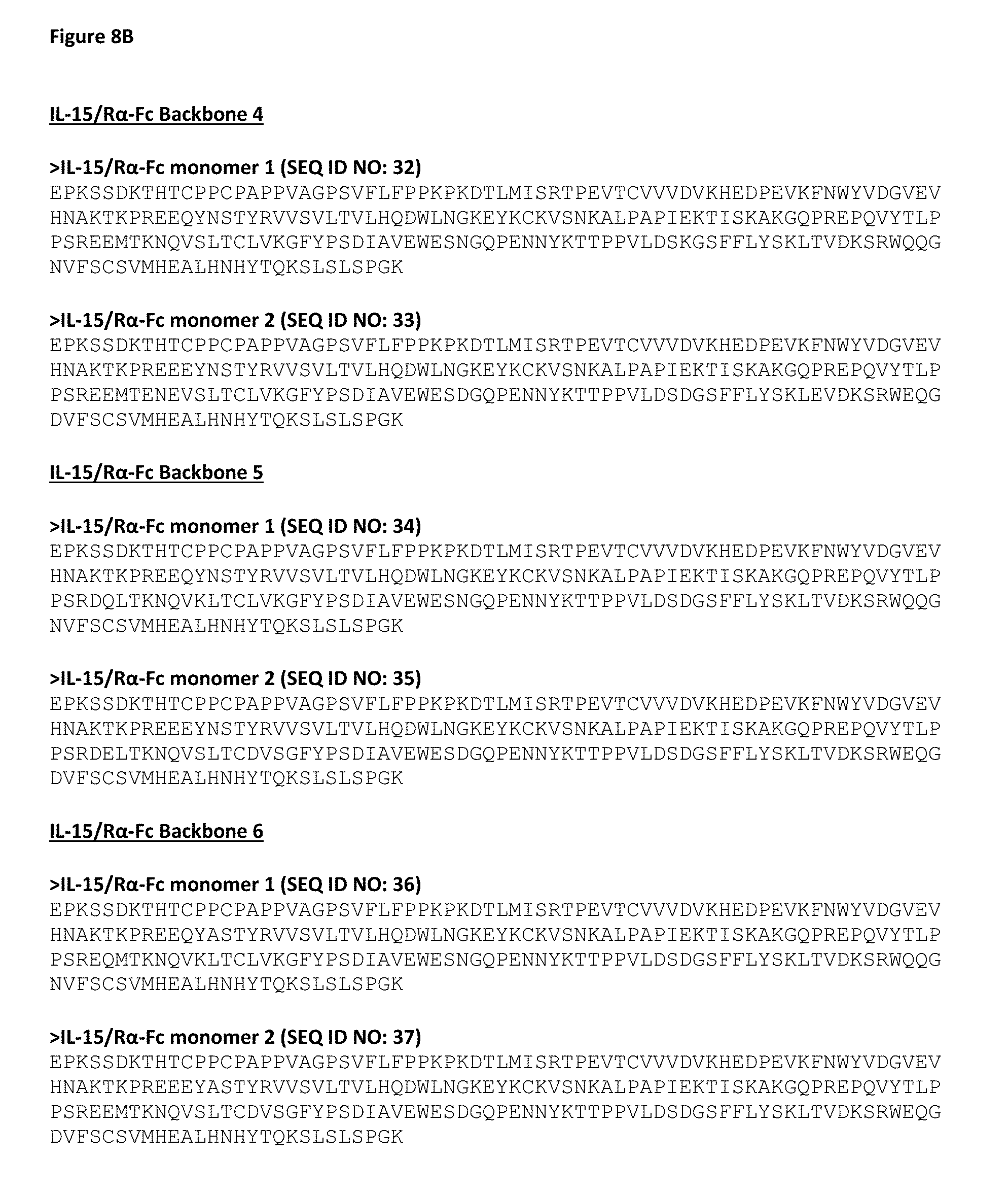
D00015
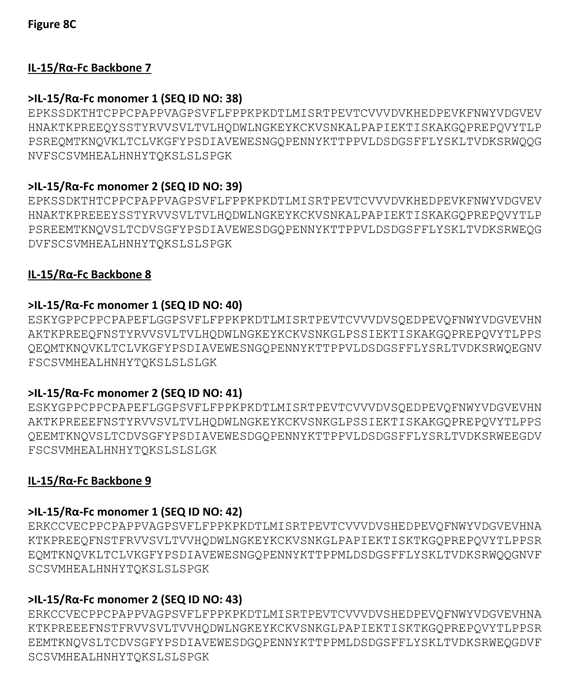
D00016

D00017
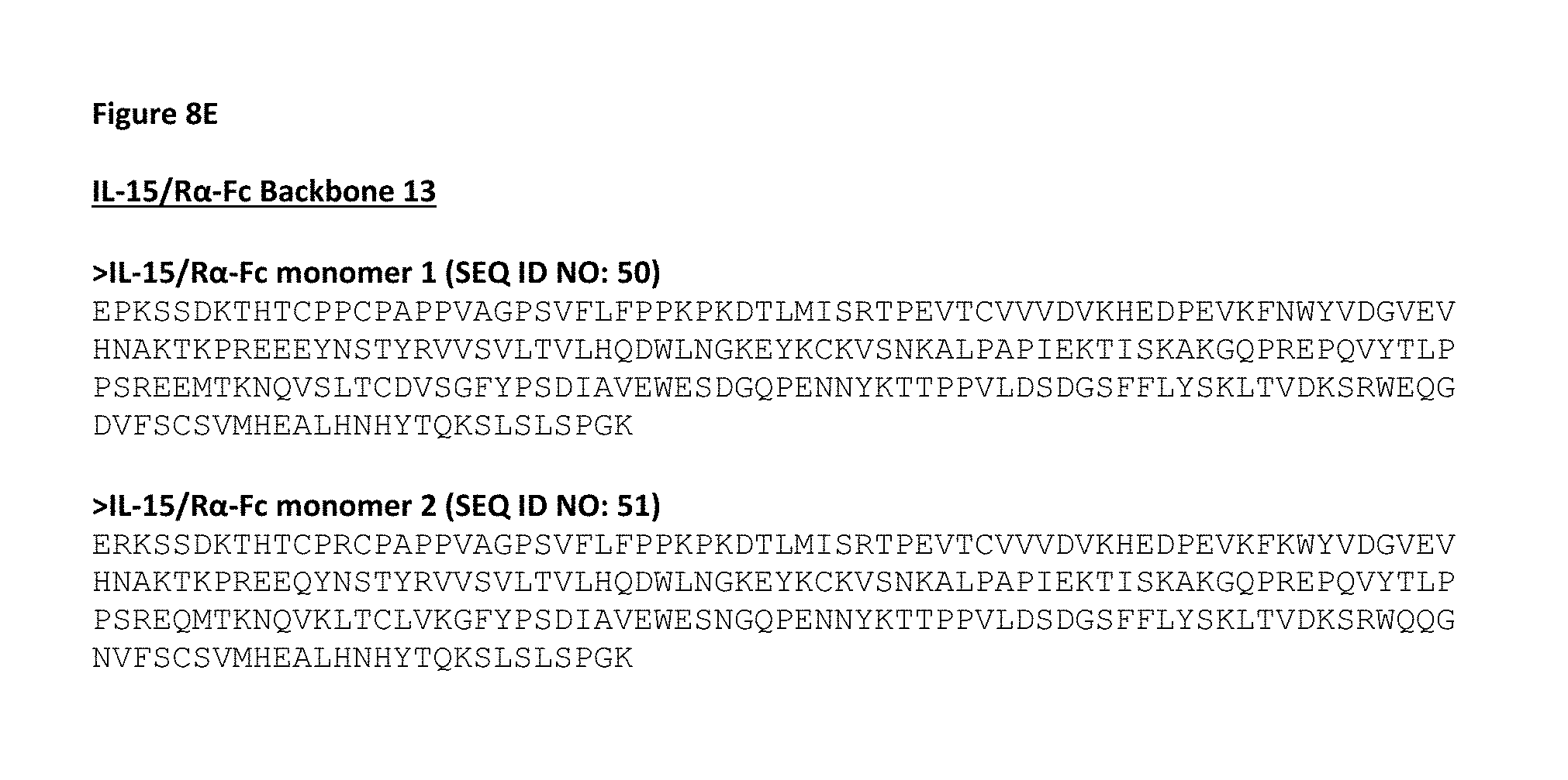
D00018
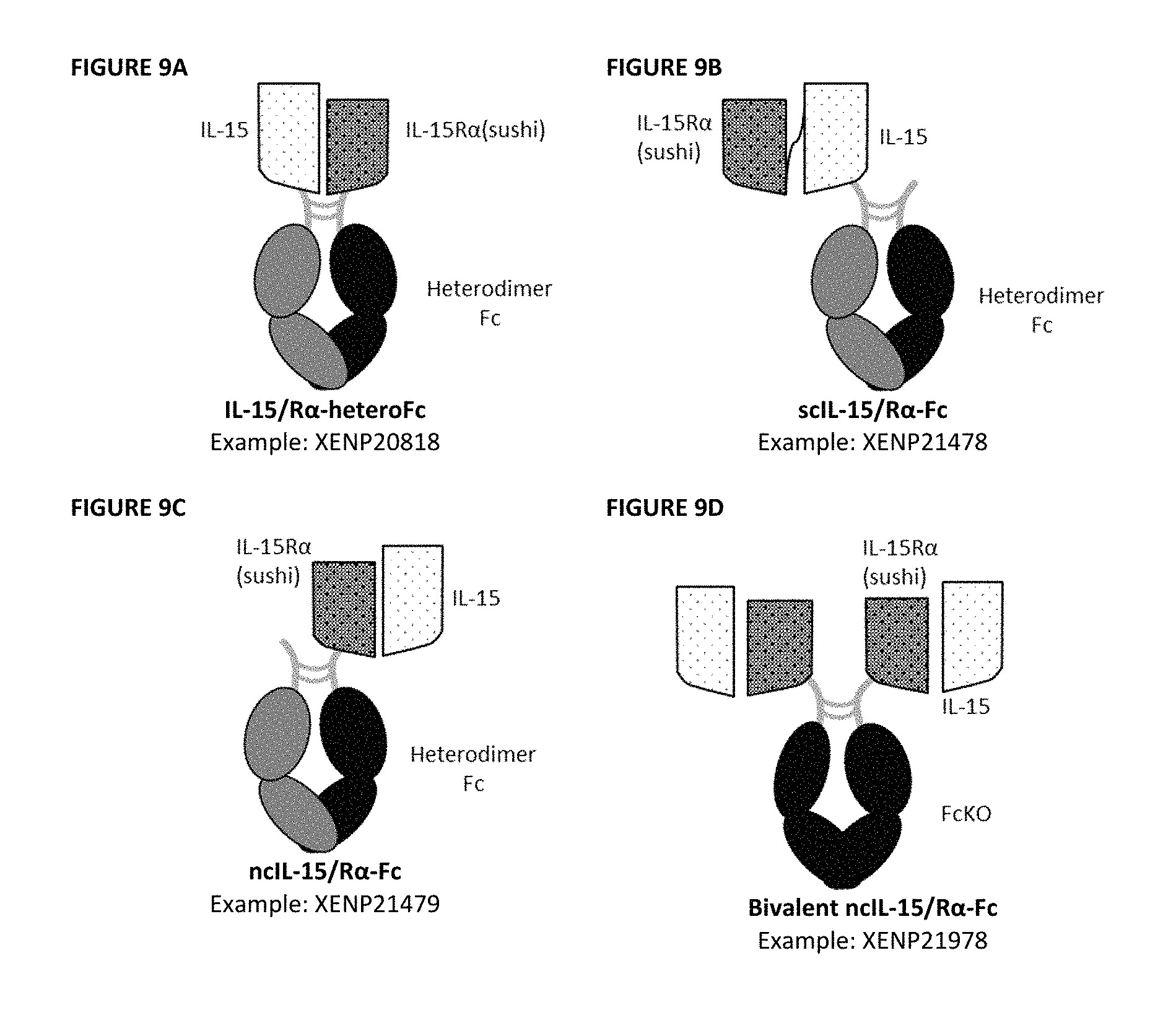
D00019

D00020
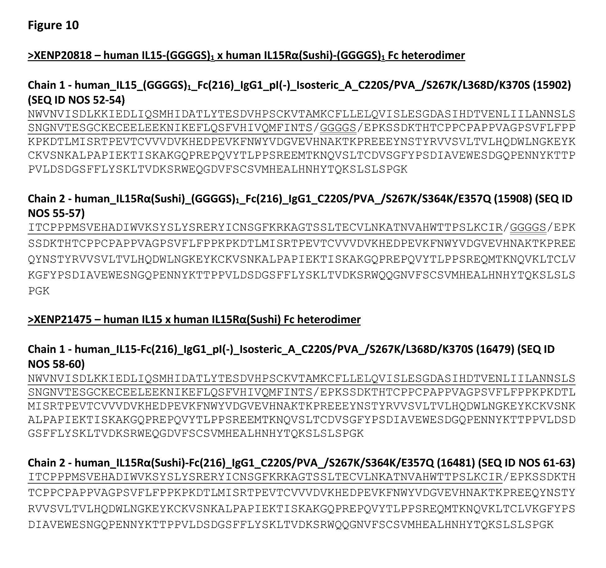
D00021

D00022
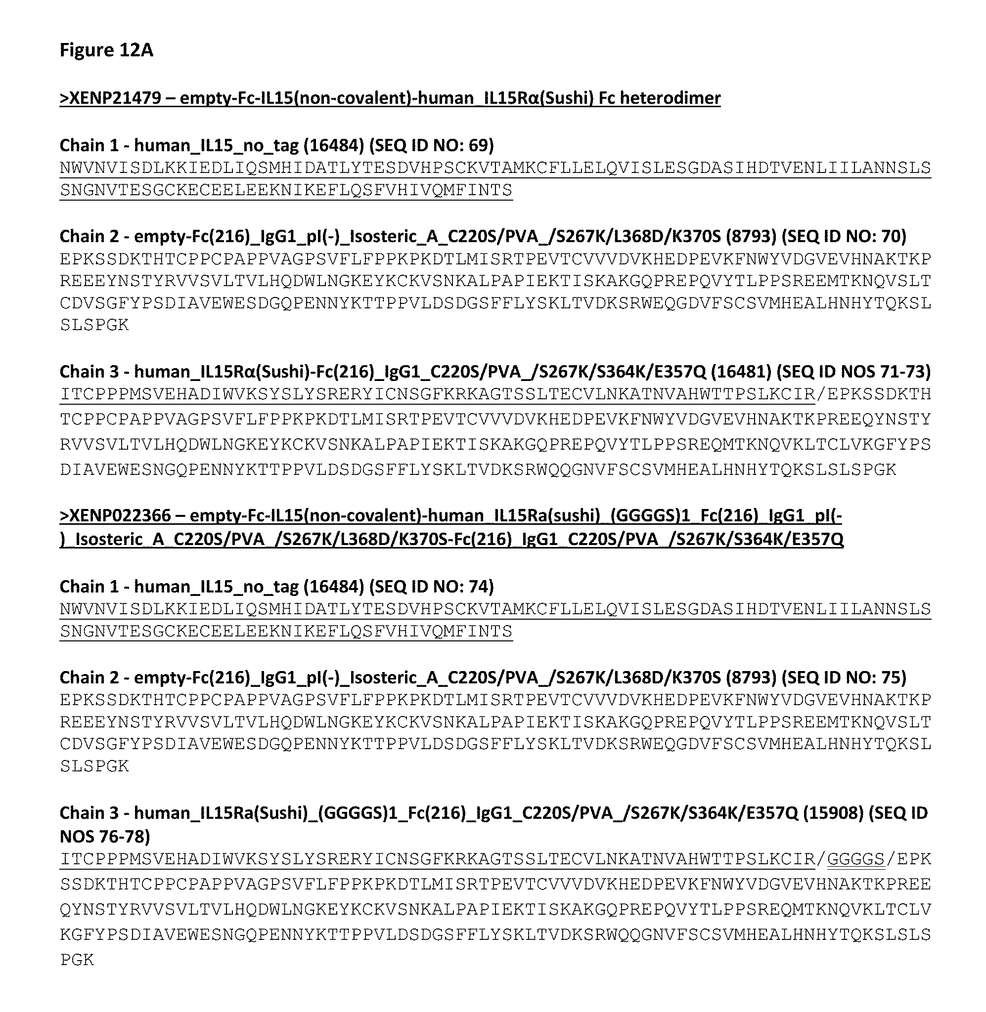
D00023
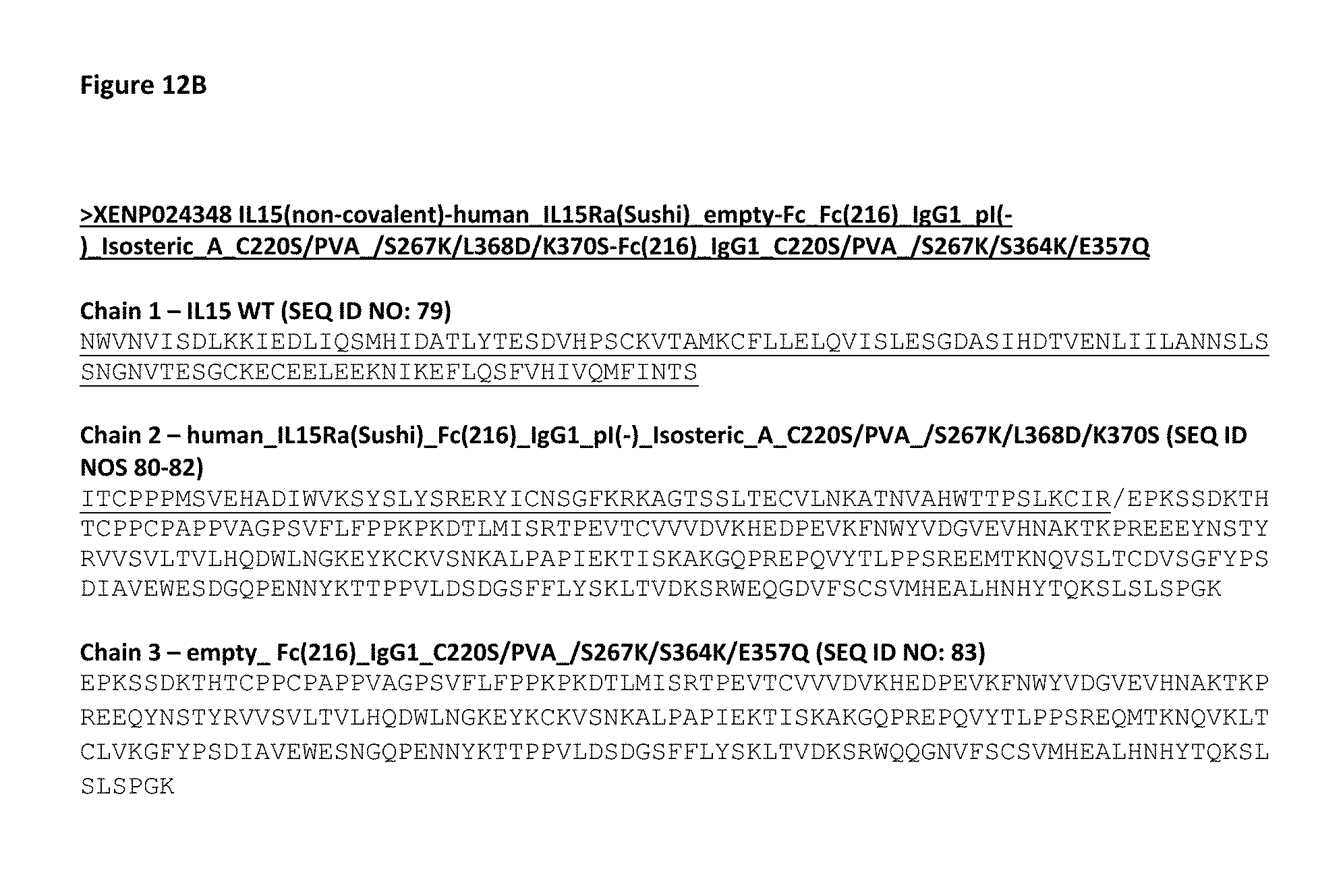
D00024

D00025

D00026
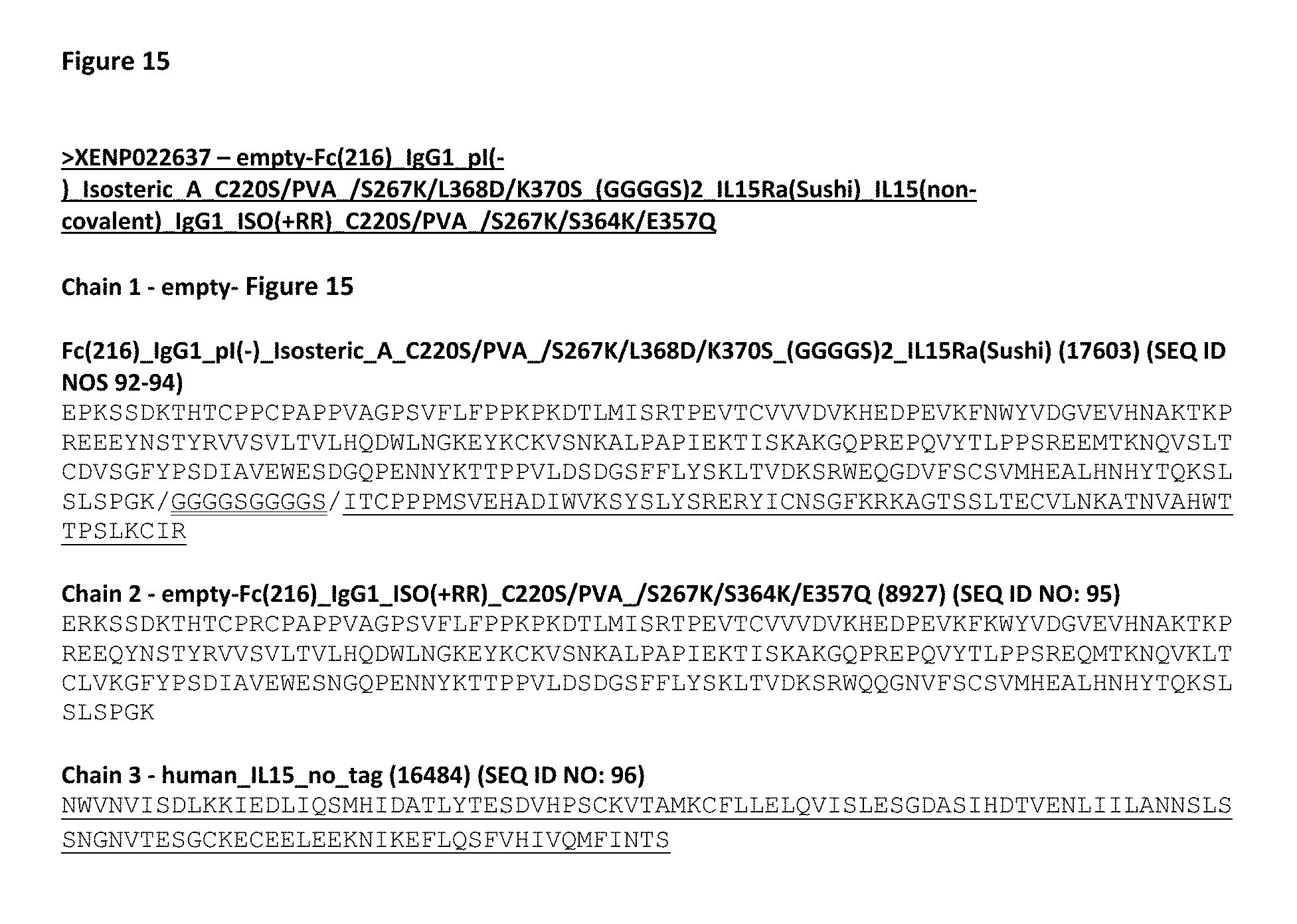
D00027

D00028

D00029
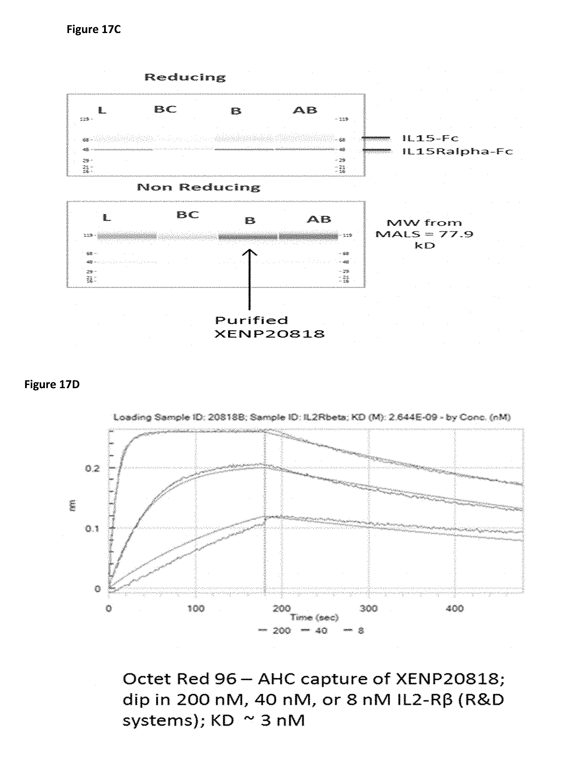
D00030
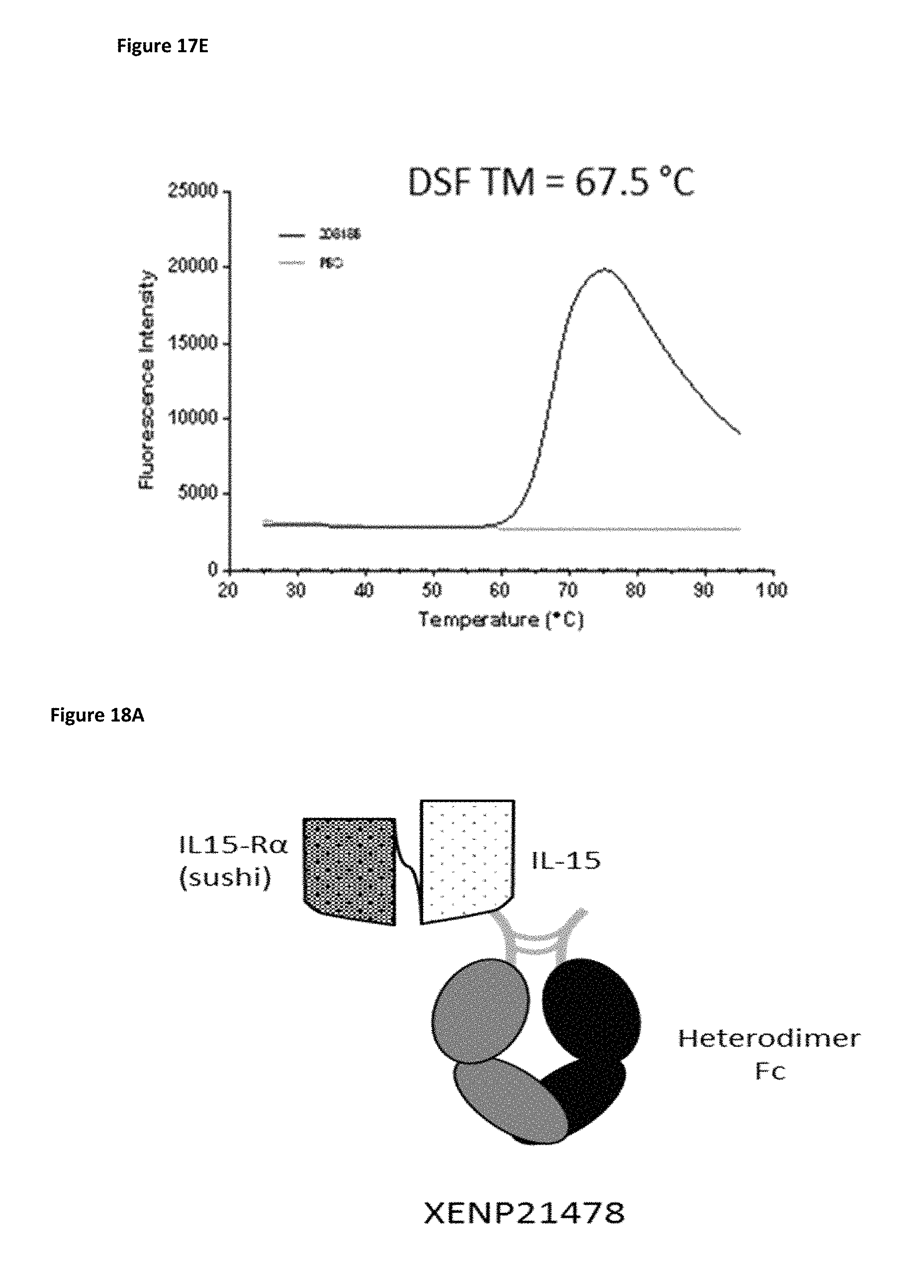
D00031

D00032

D00033
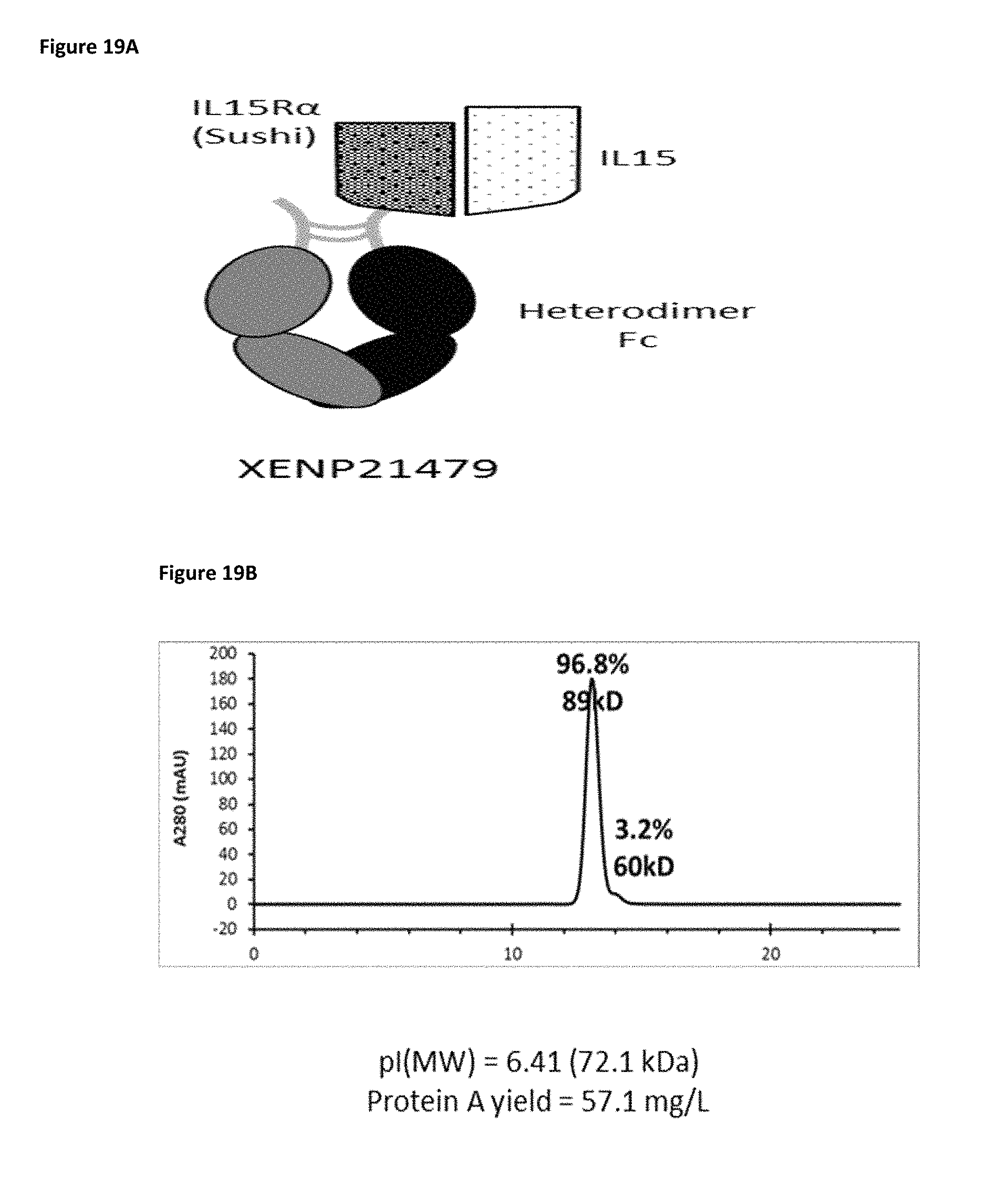
D00034

D00035
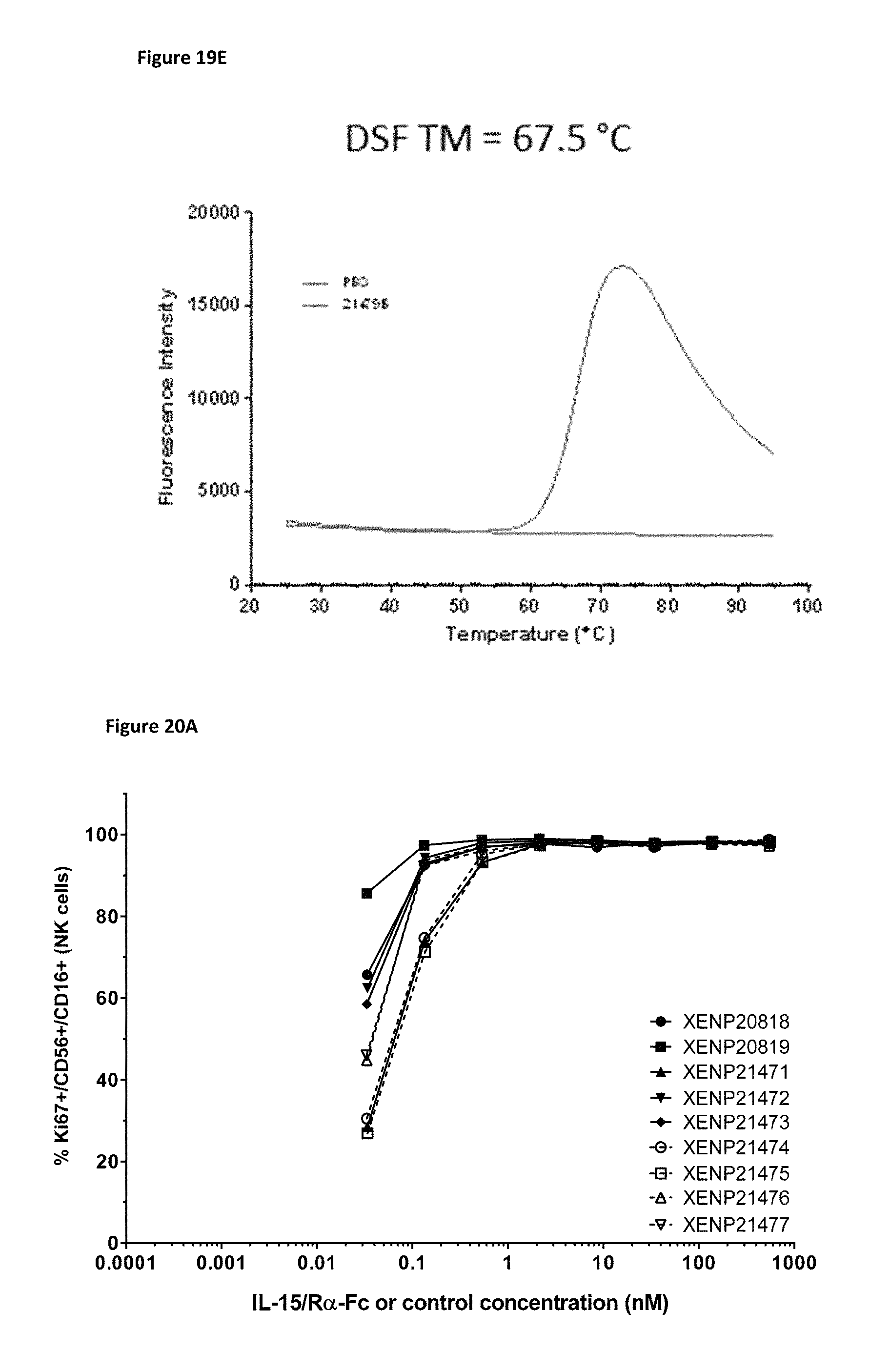
D00036
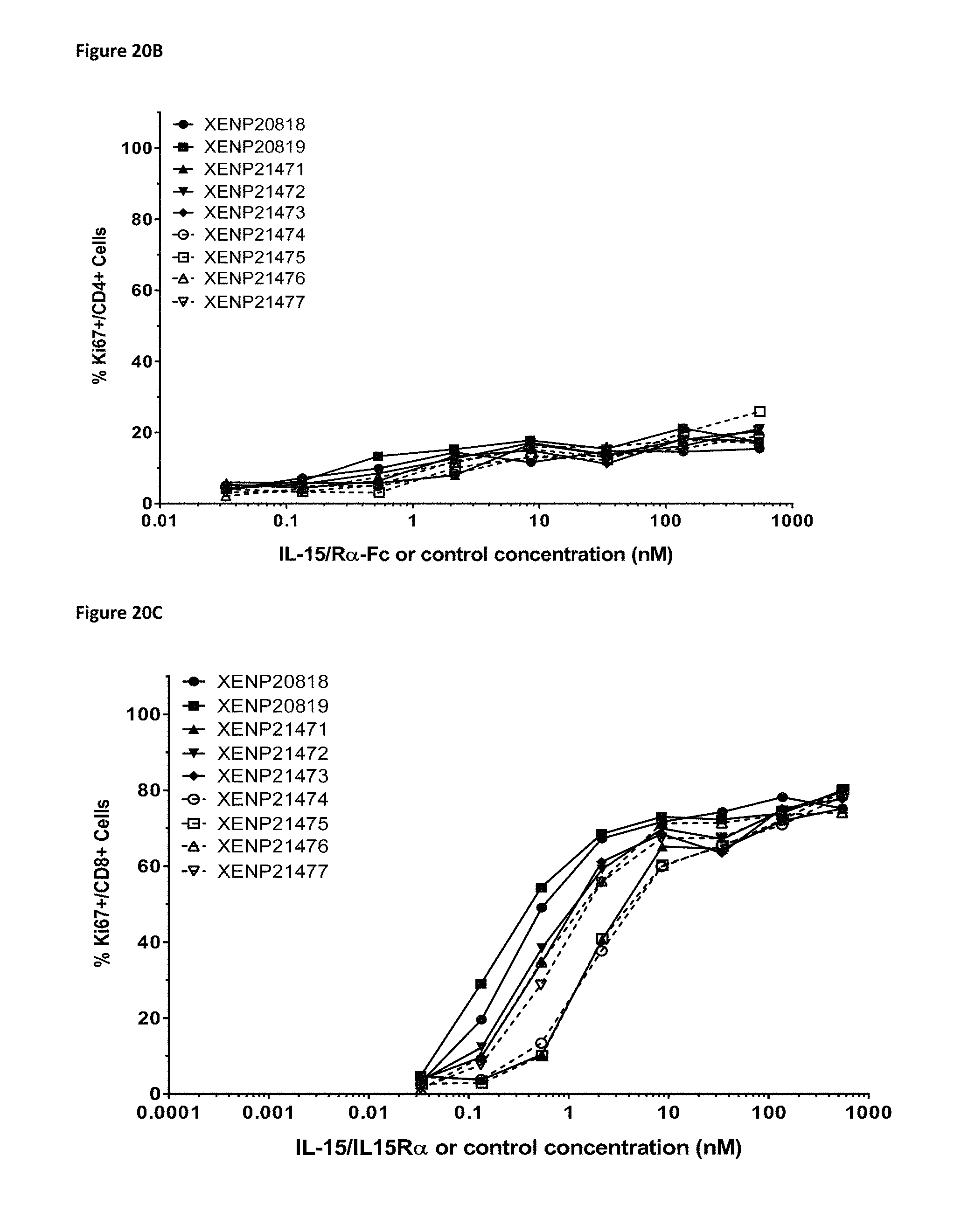
D00037
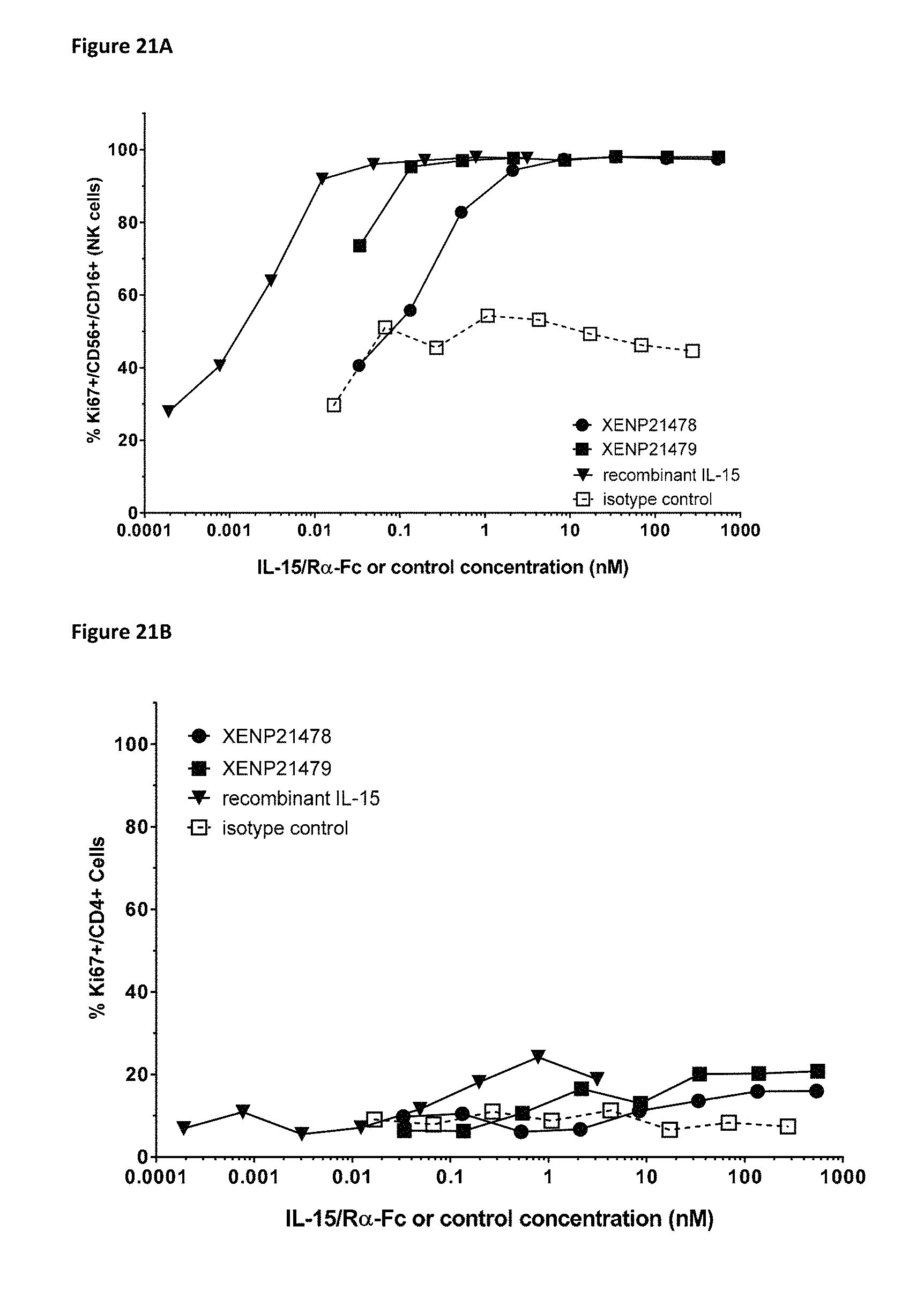
D00038

D00039
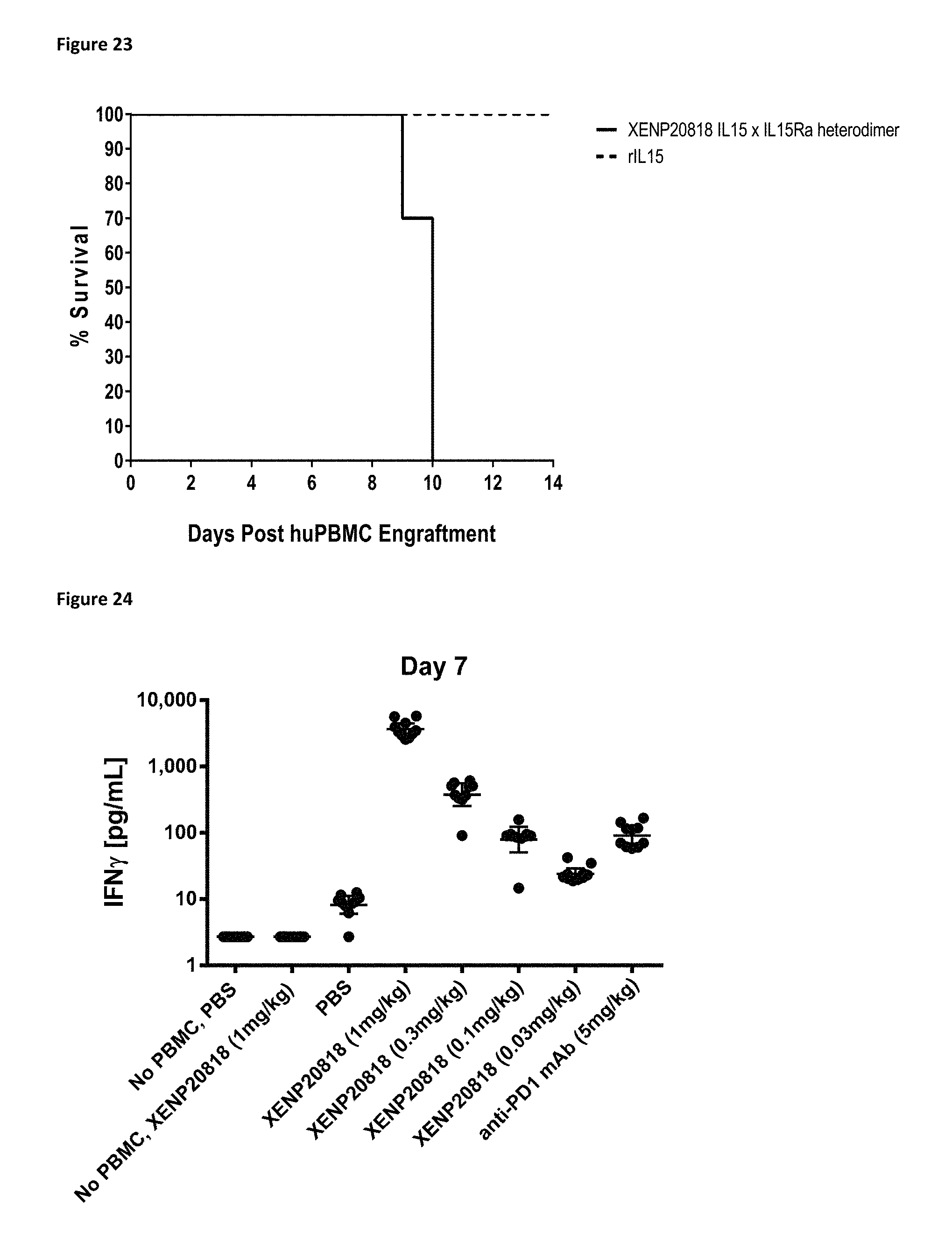
D00040
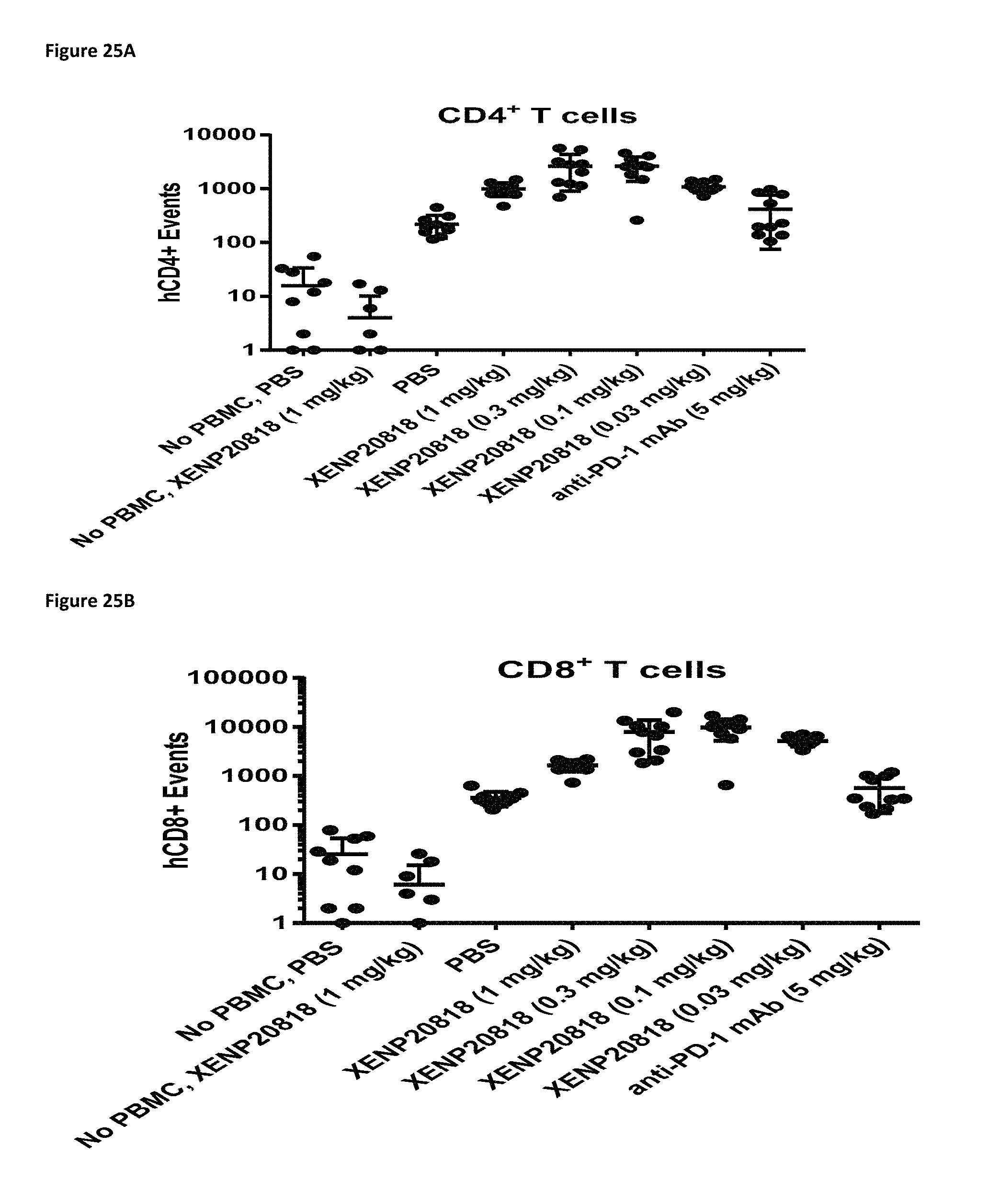
D00041

D00042
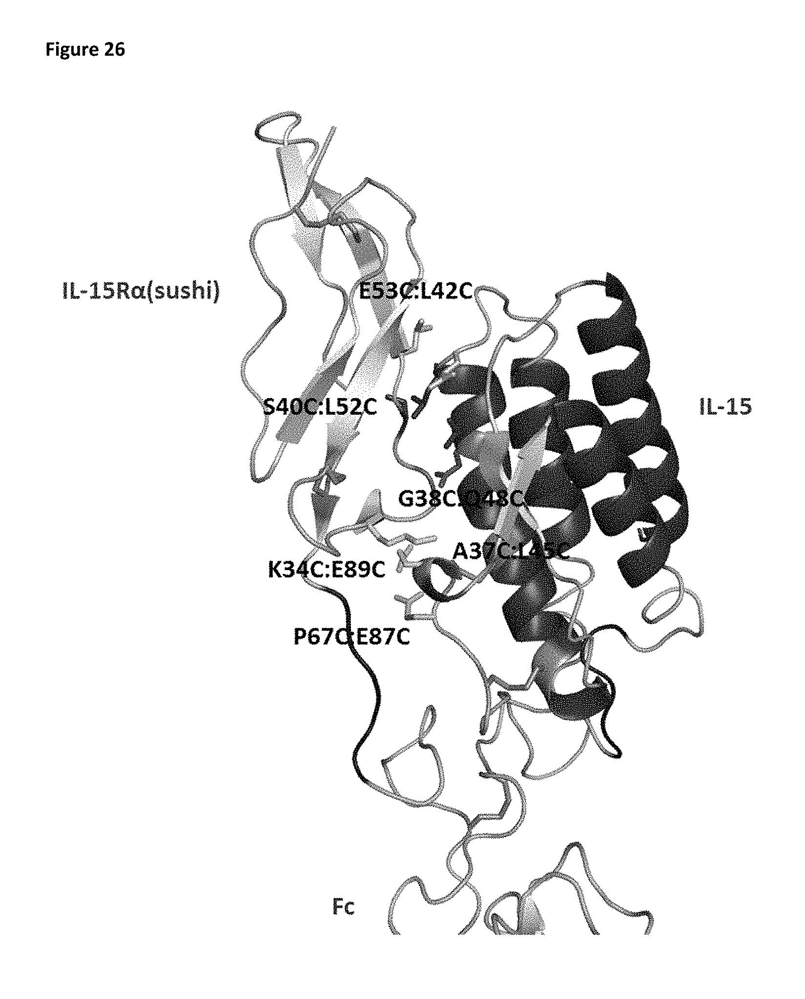
D00043
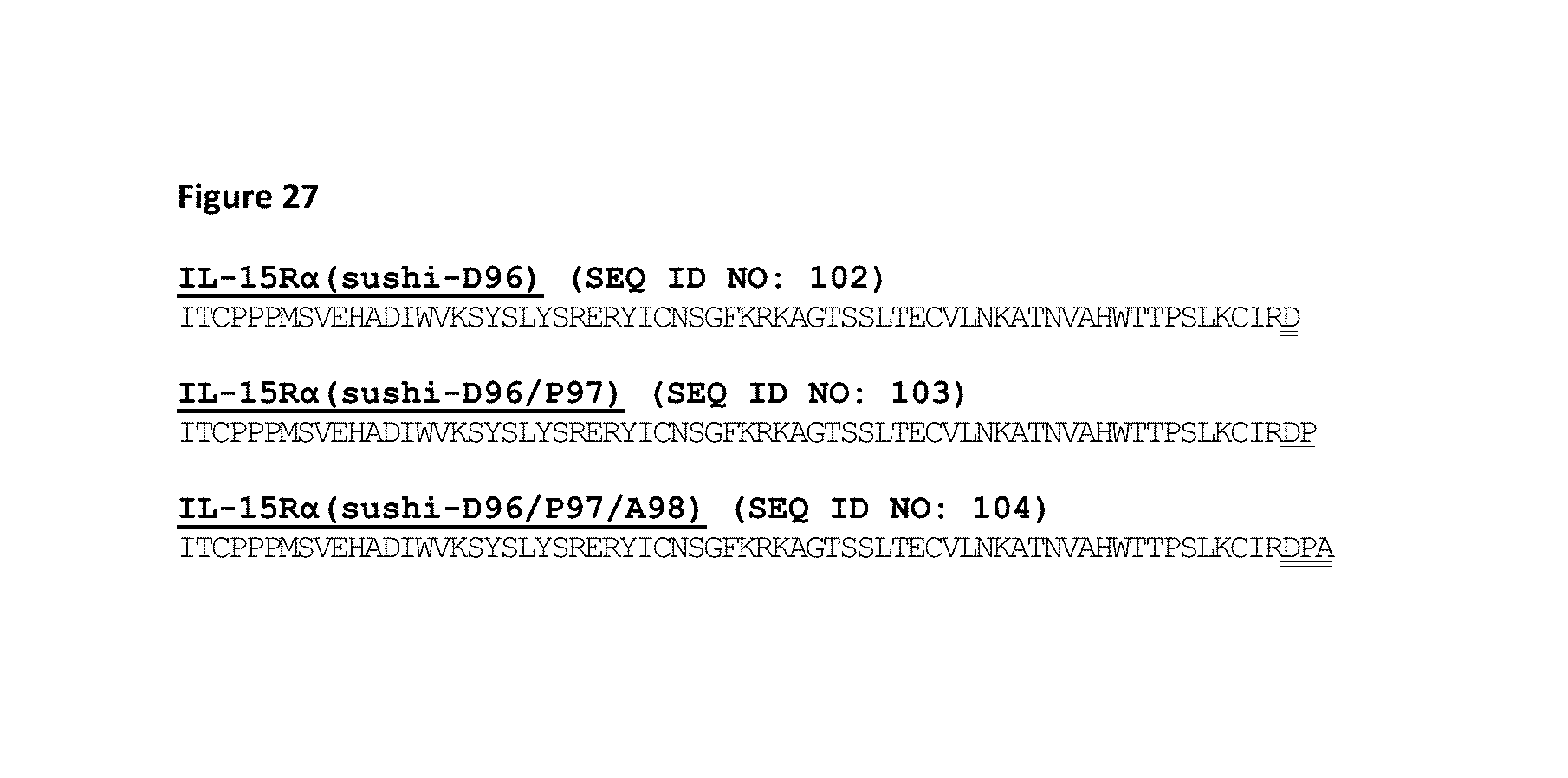
D00044

D00045

D00046

D00047

D00048

D00049
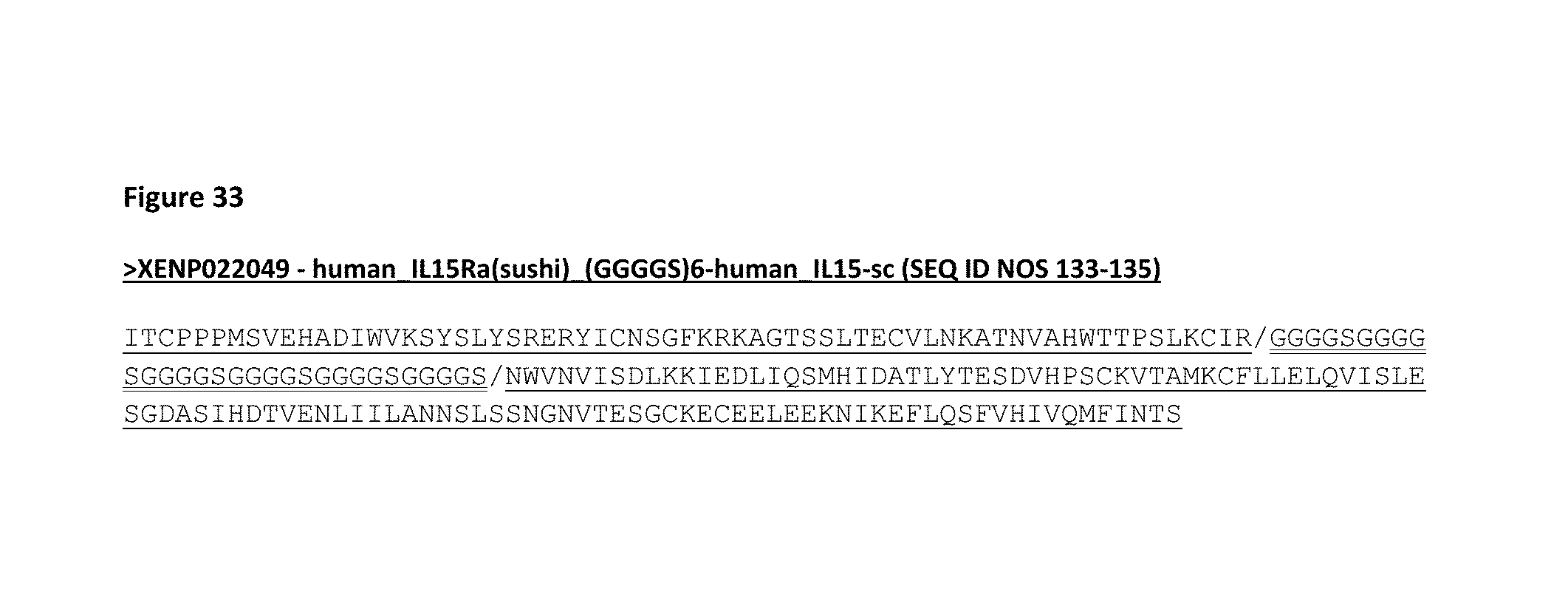
D00050

D00051
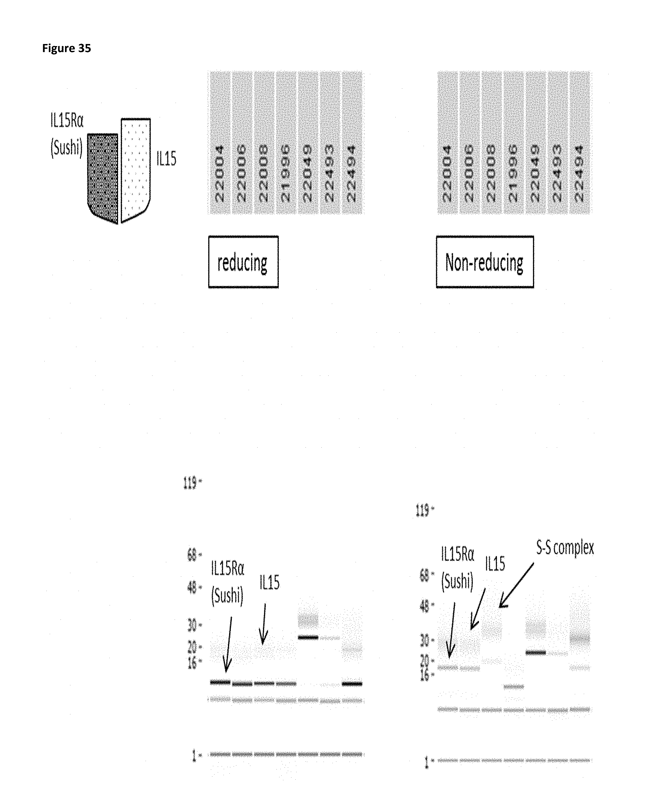
D00052

D00053

D00054
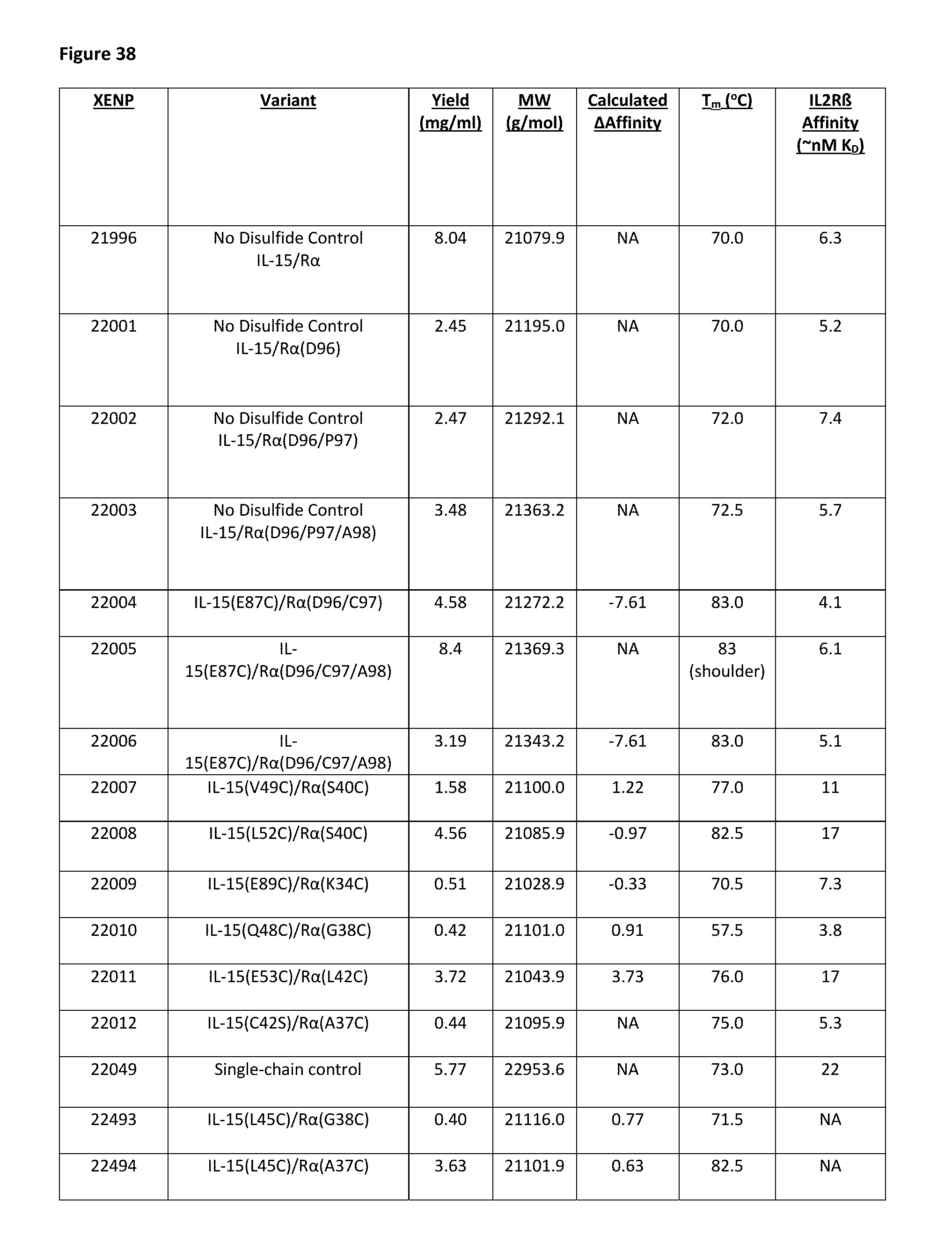
D00055
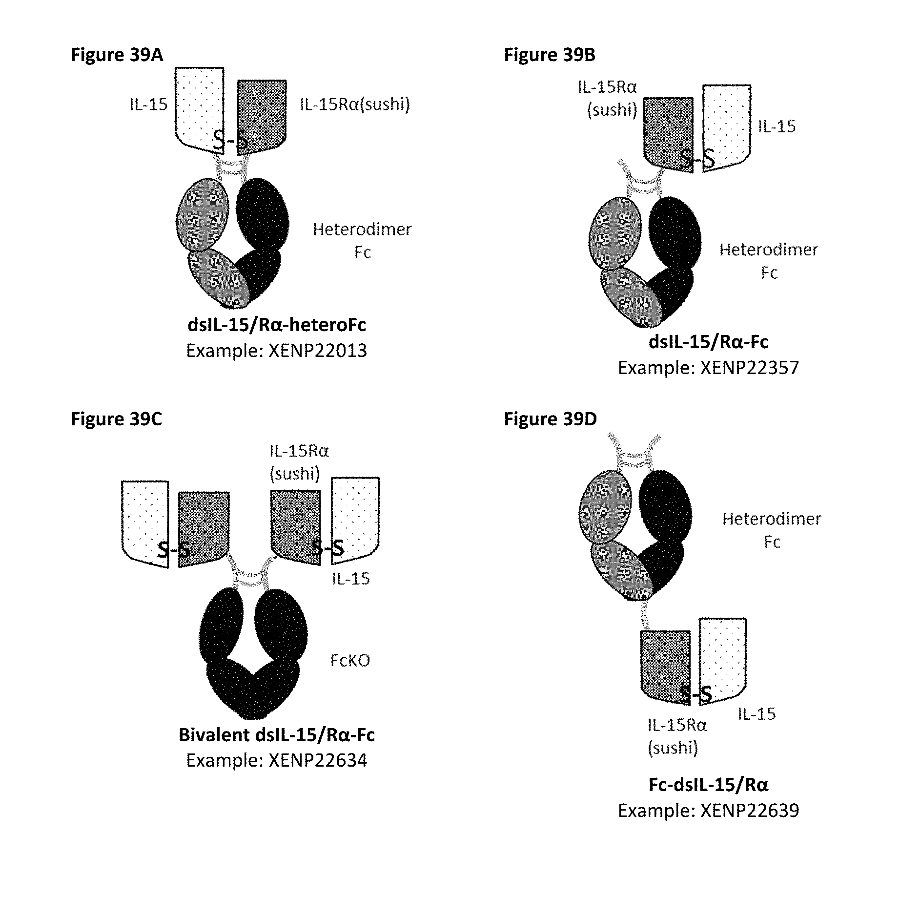
D00056

D00057

D00058
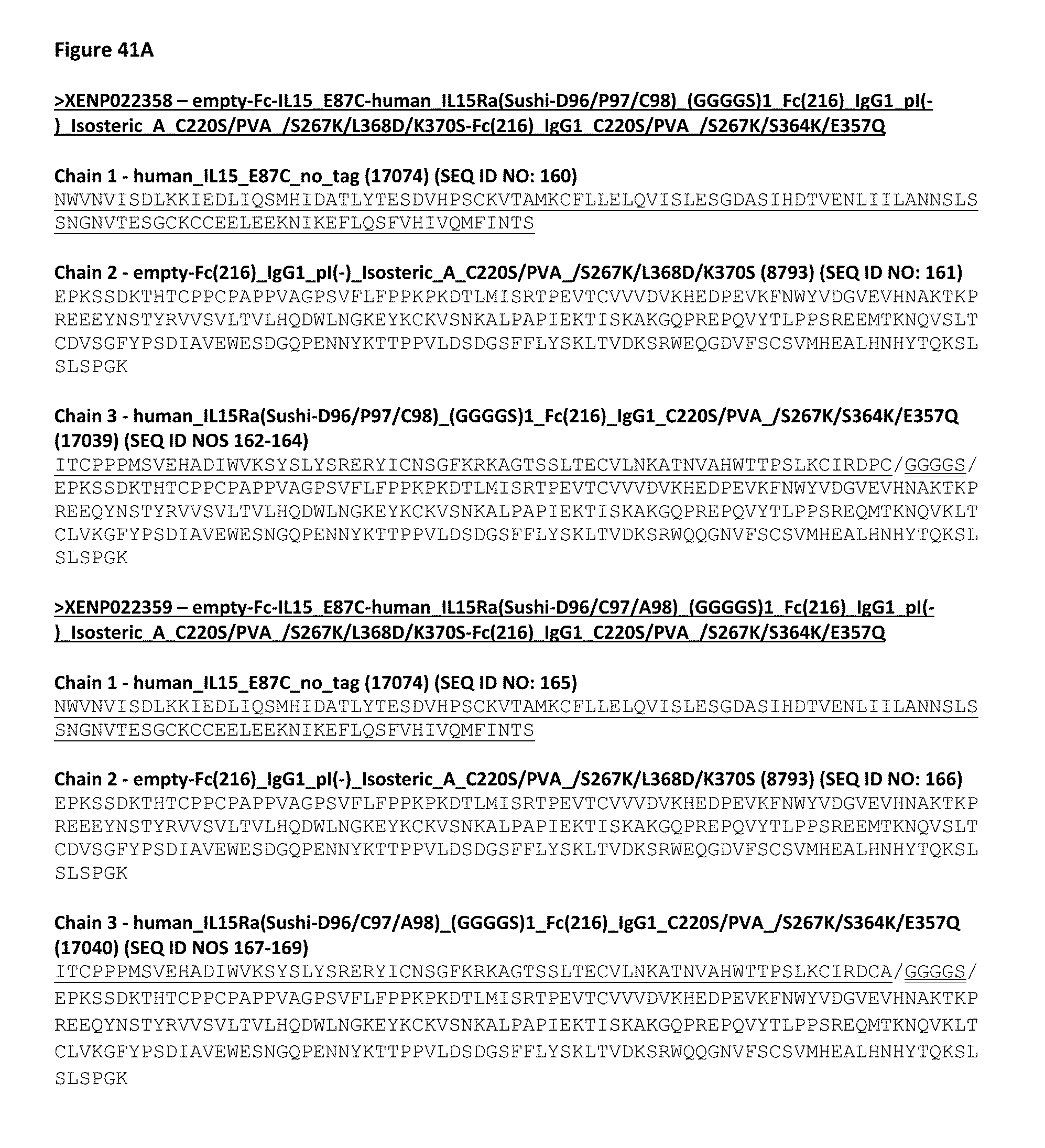
D00059

D00060

D00061

D00062

D00063
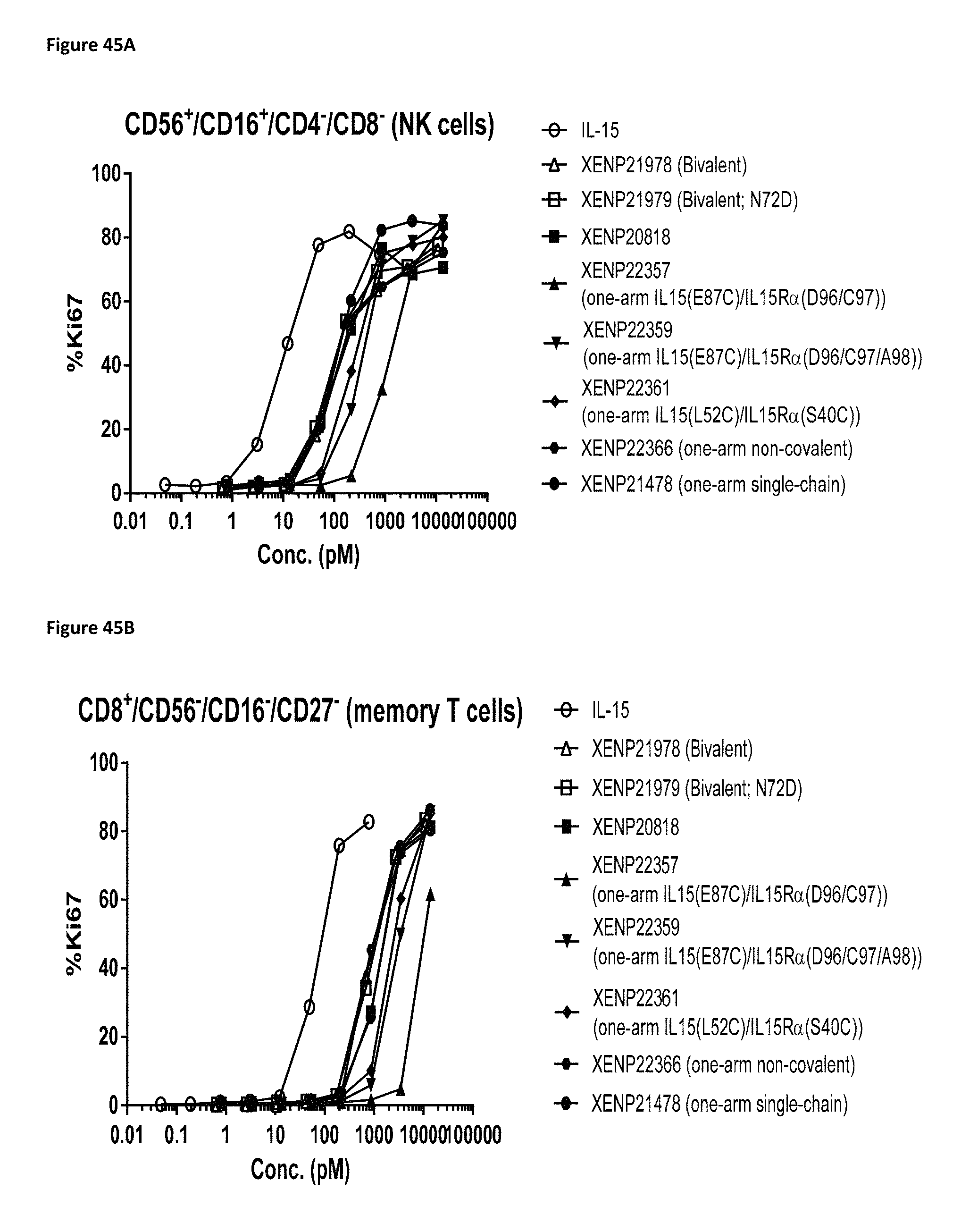
D00064

D00065

D00066
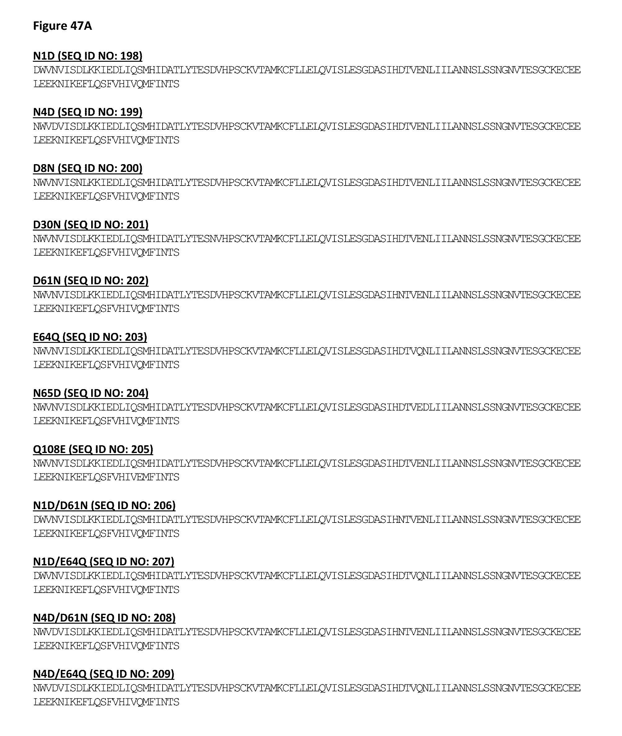
D00067

D00068

D00069
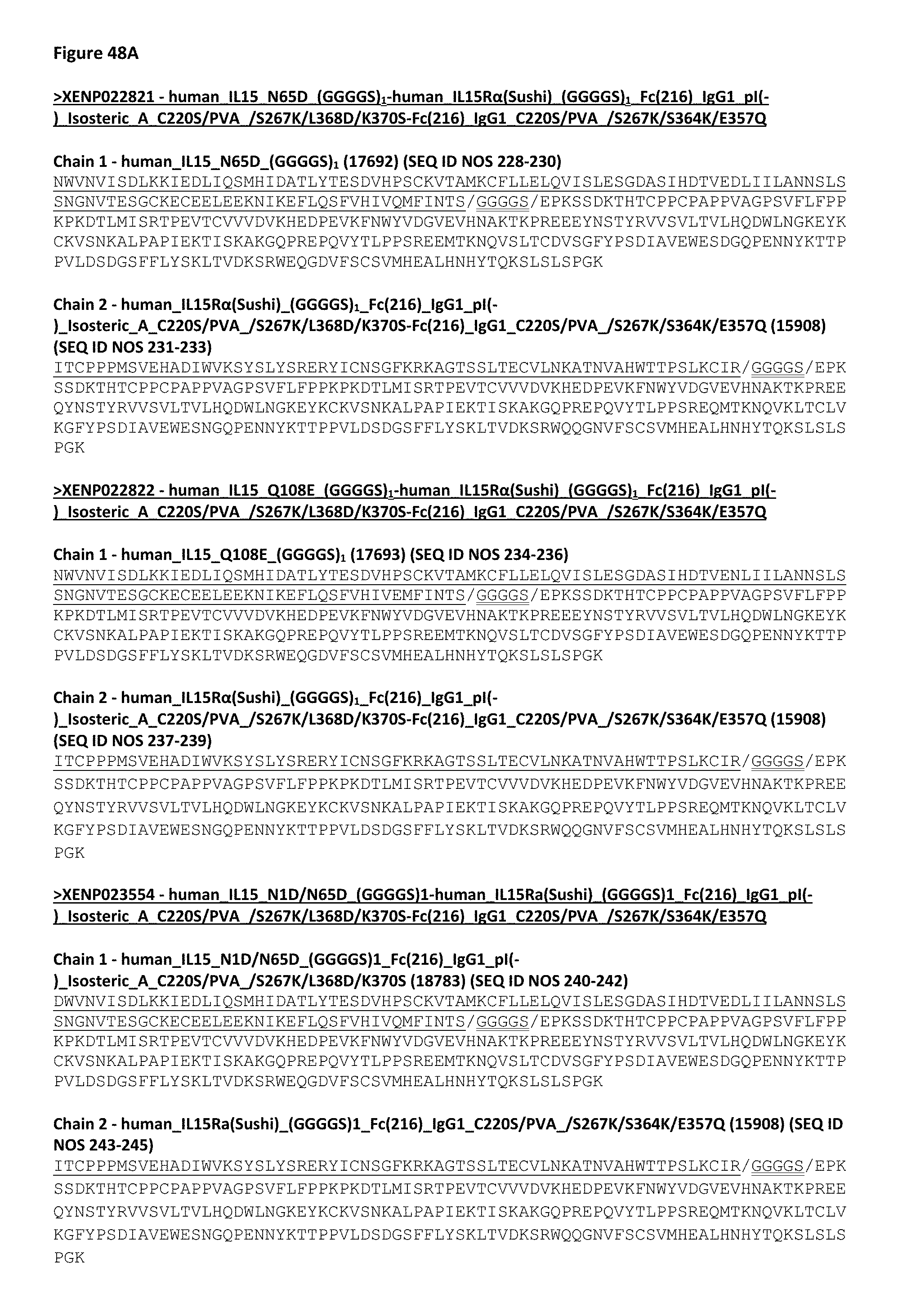
D00070

D00071

D00072

D00073
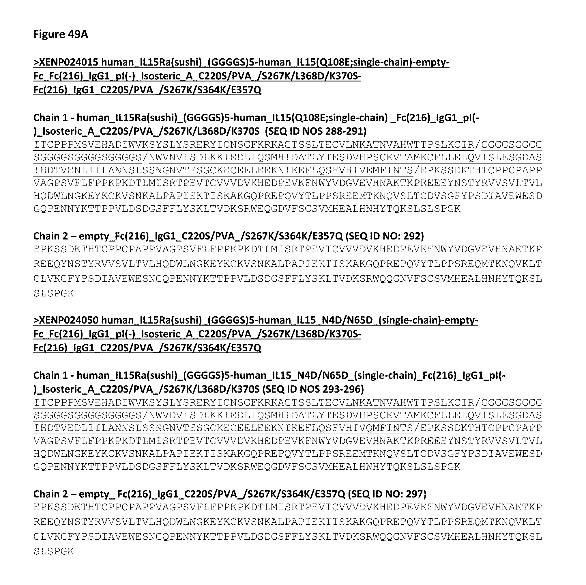
D00074
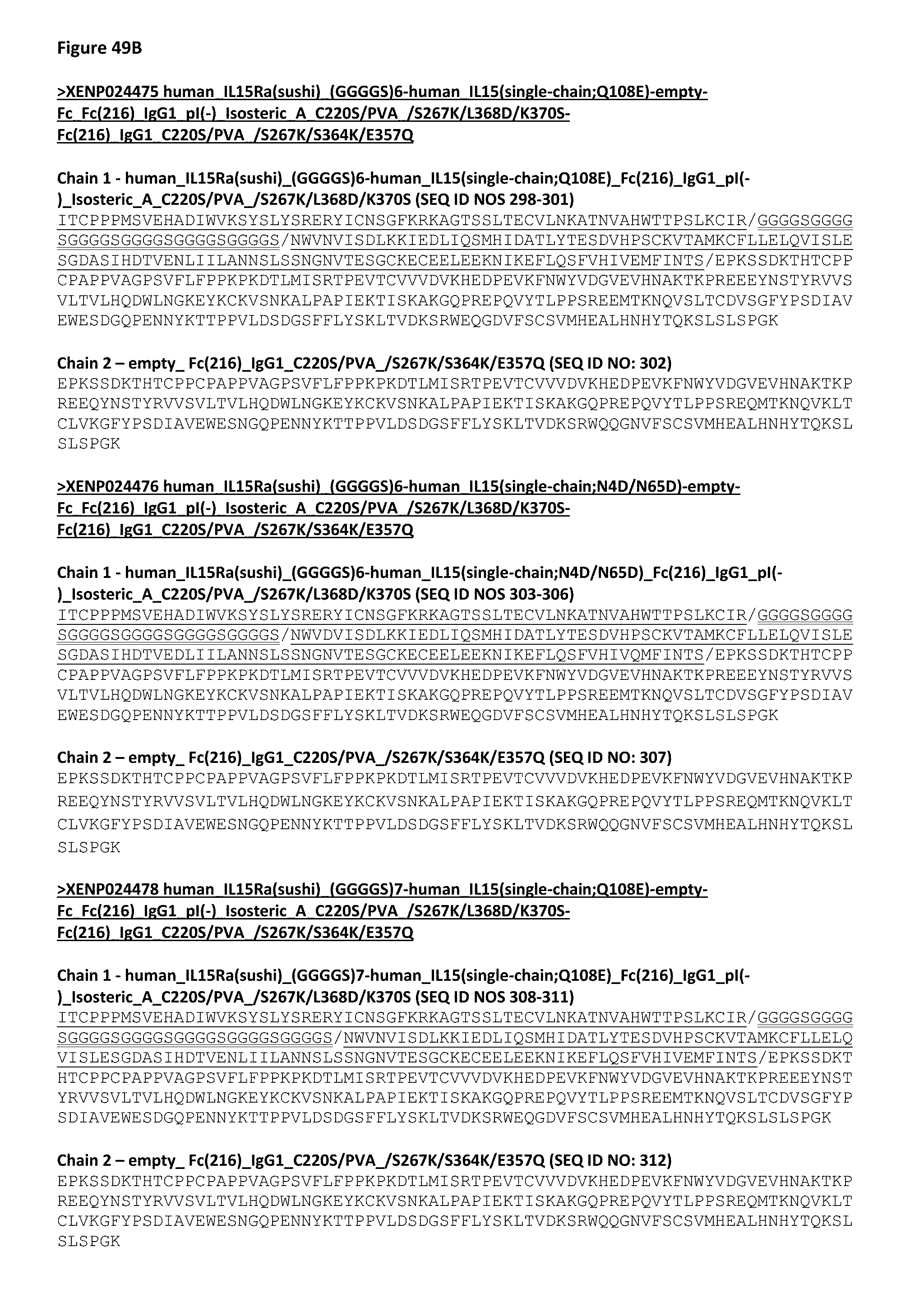
D00075
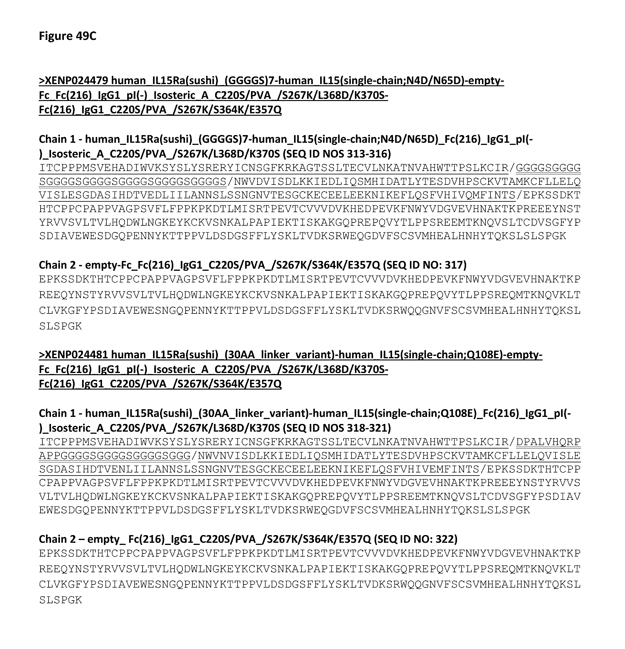
D00076

D00077
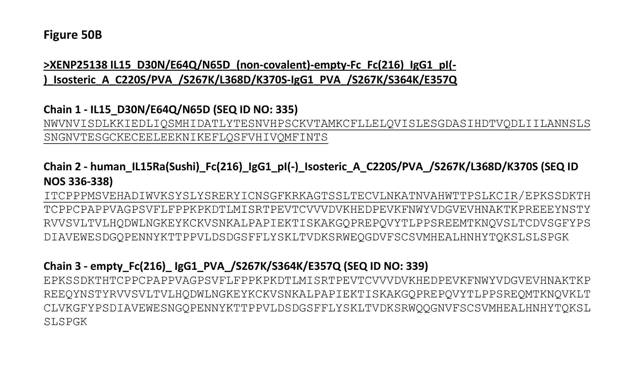
D00078

D00079
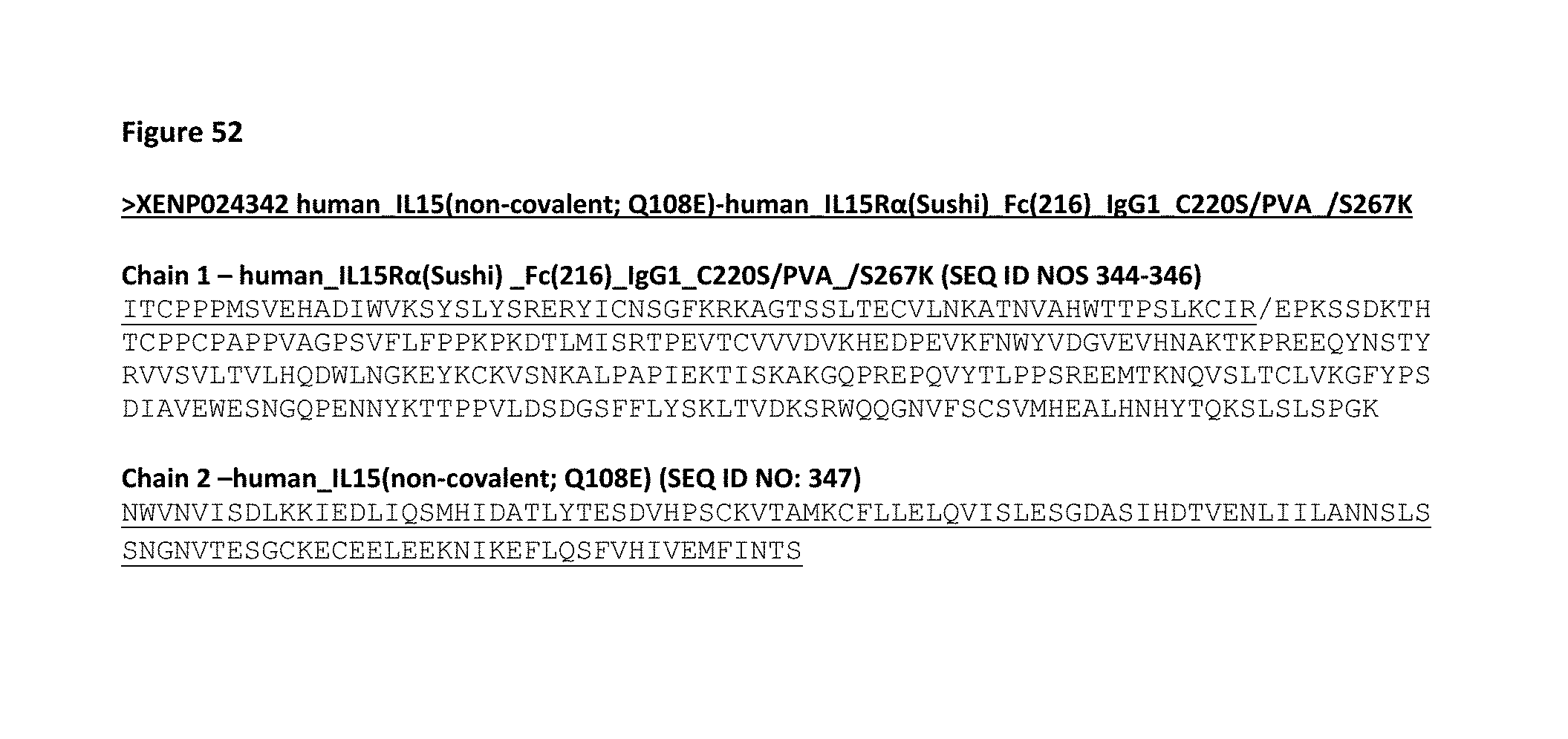
D00080
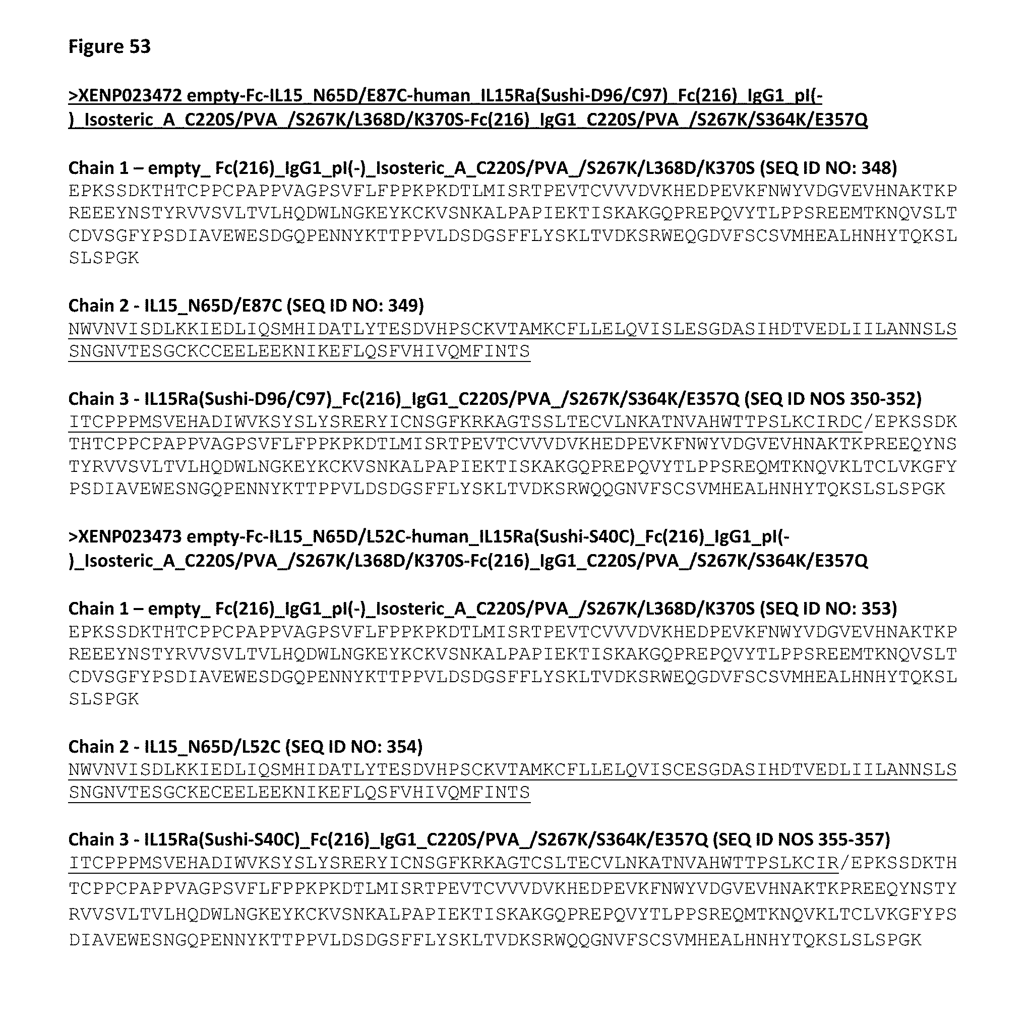
D00081
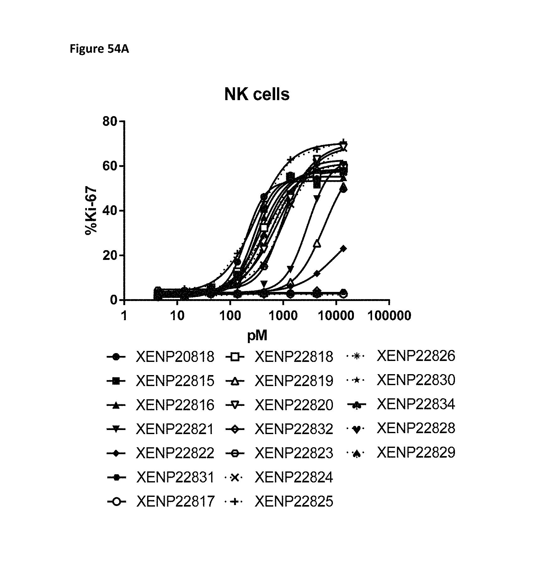
D00082

D00083
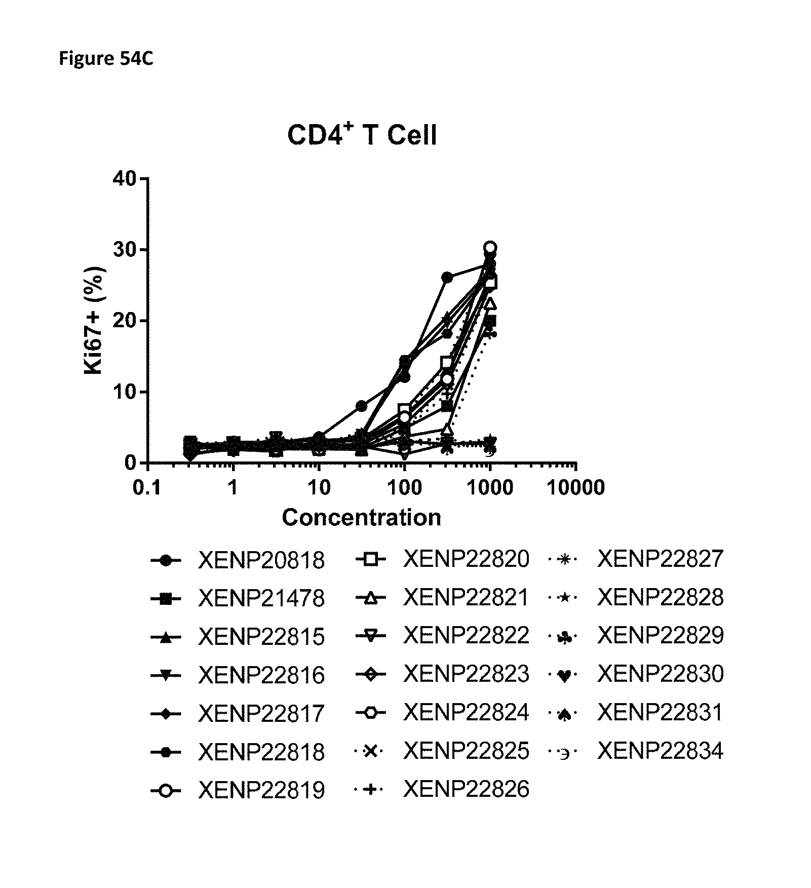
D00084
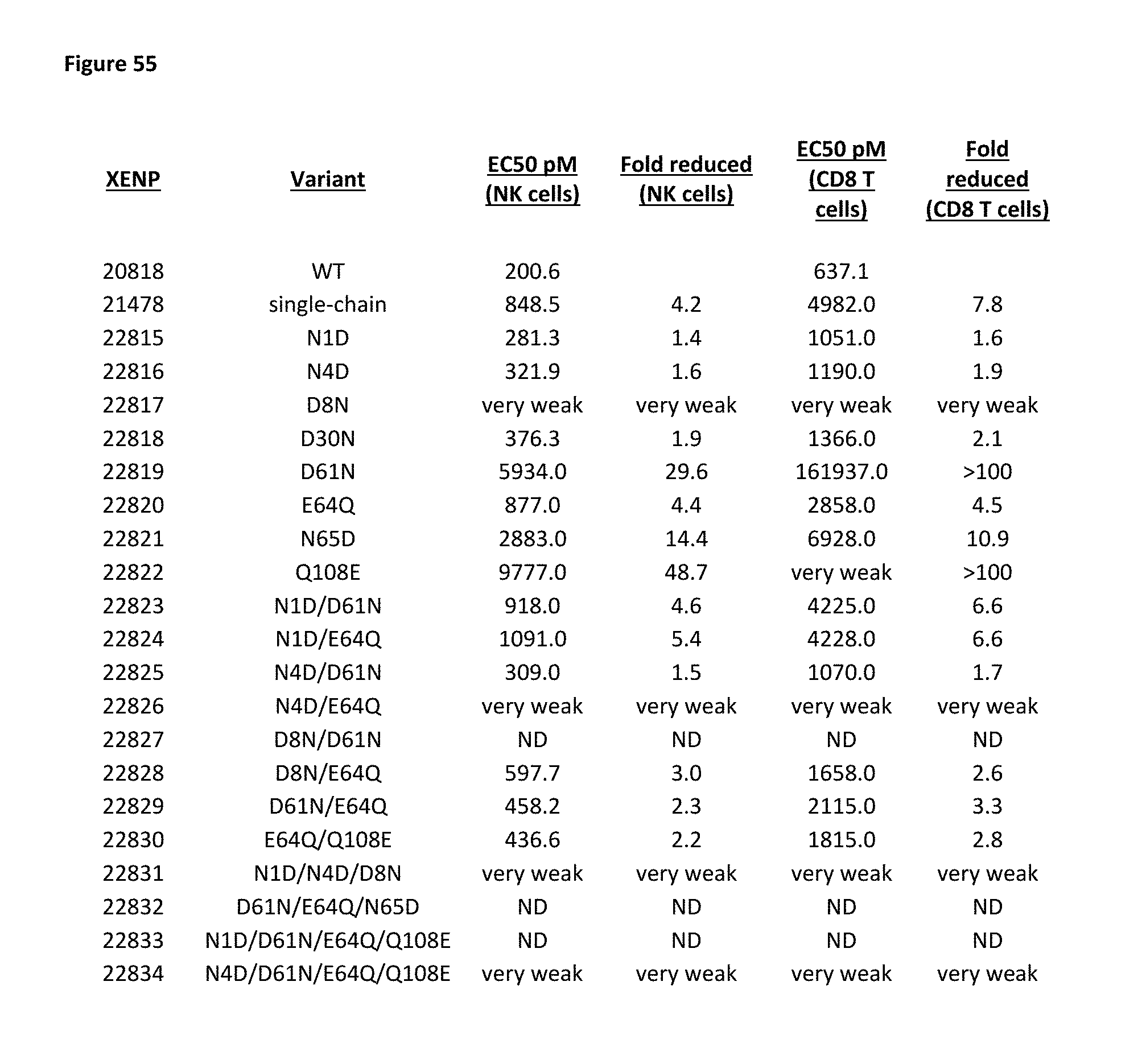
D00085
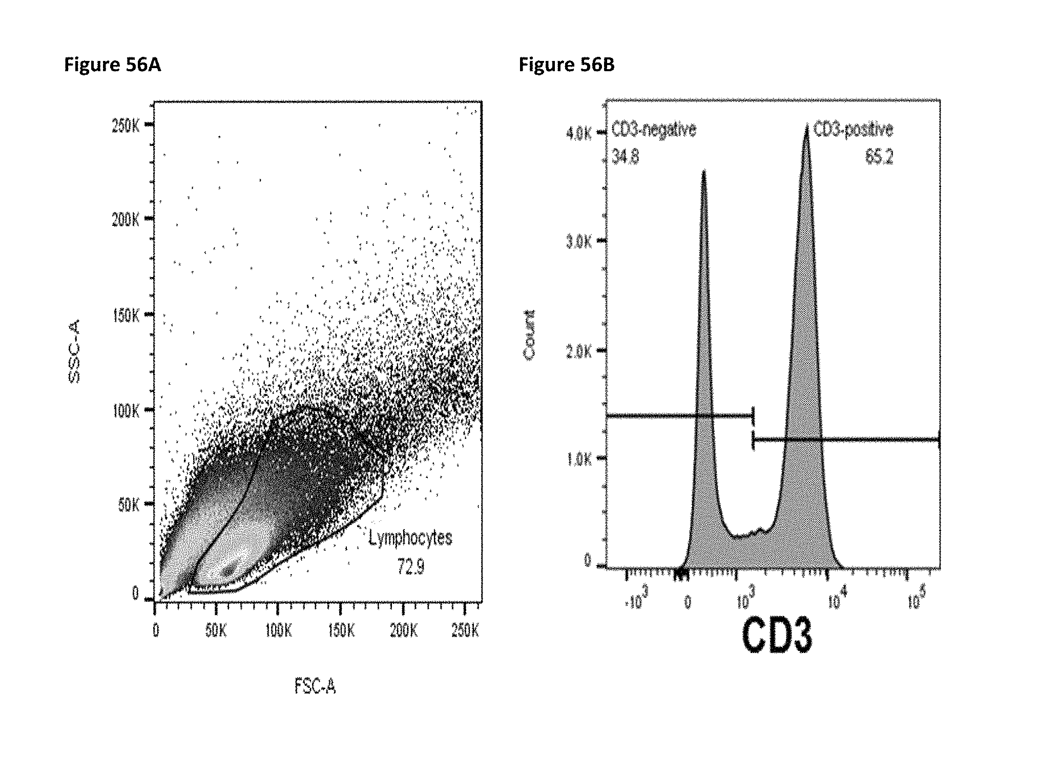
D00086

D00087
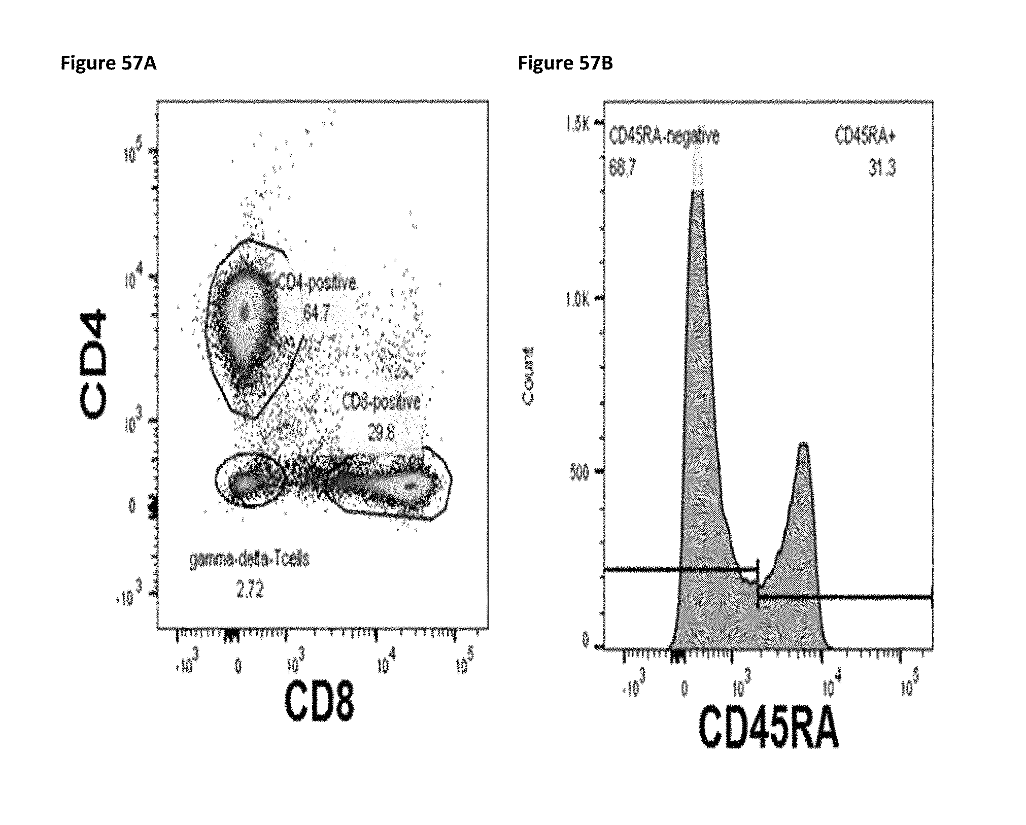
D00088

D00089

D00090
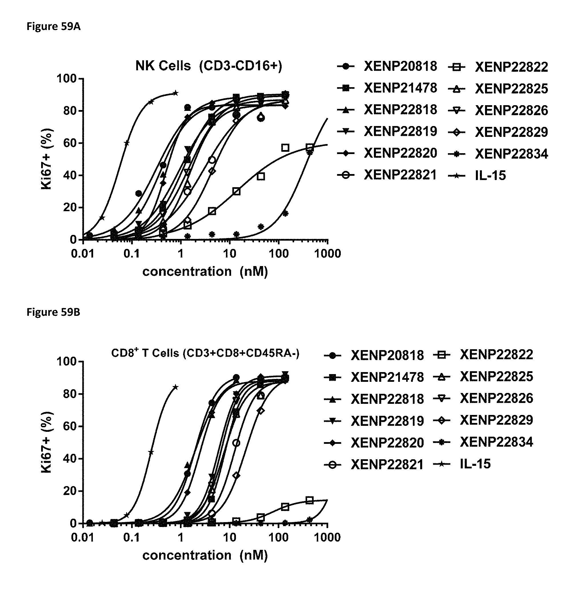
D00091

D00092

D00093

D00094
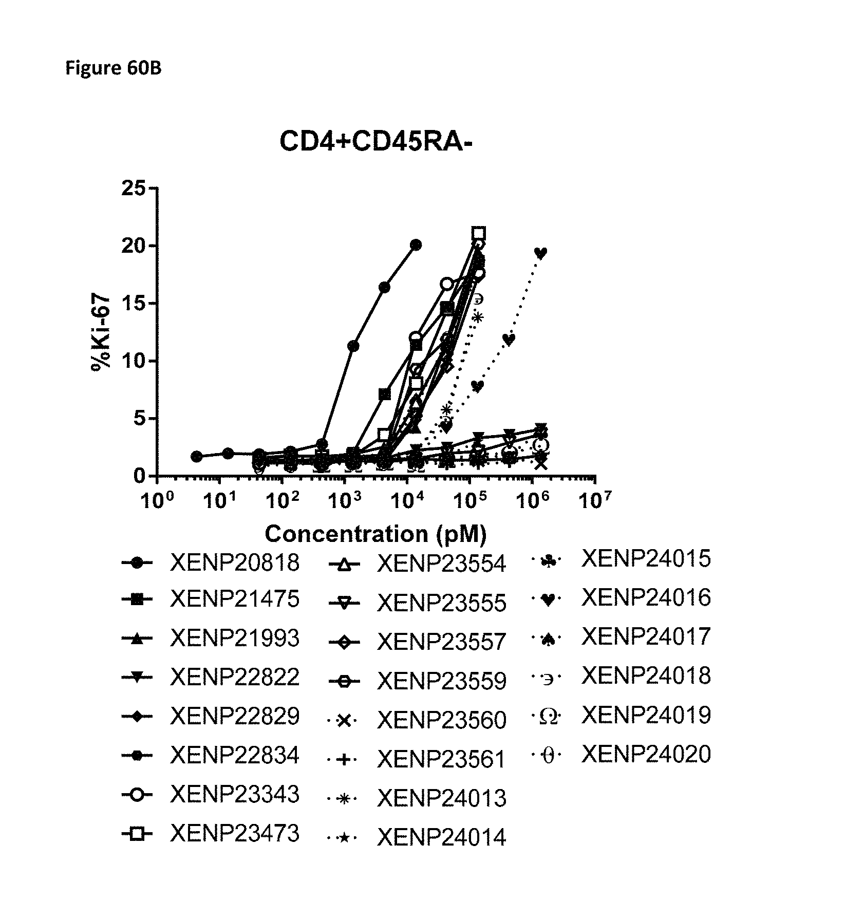
D00095

D00096
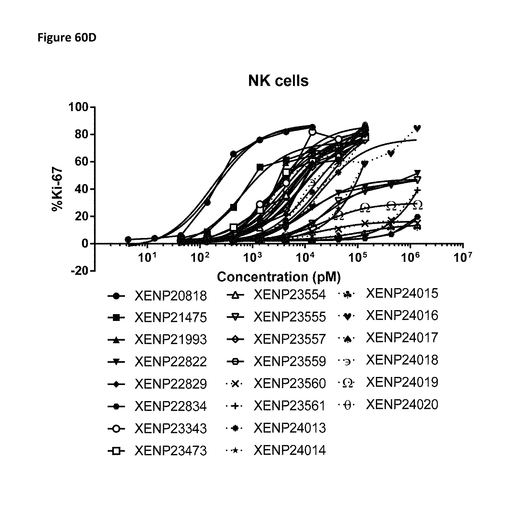
D00097
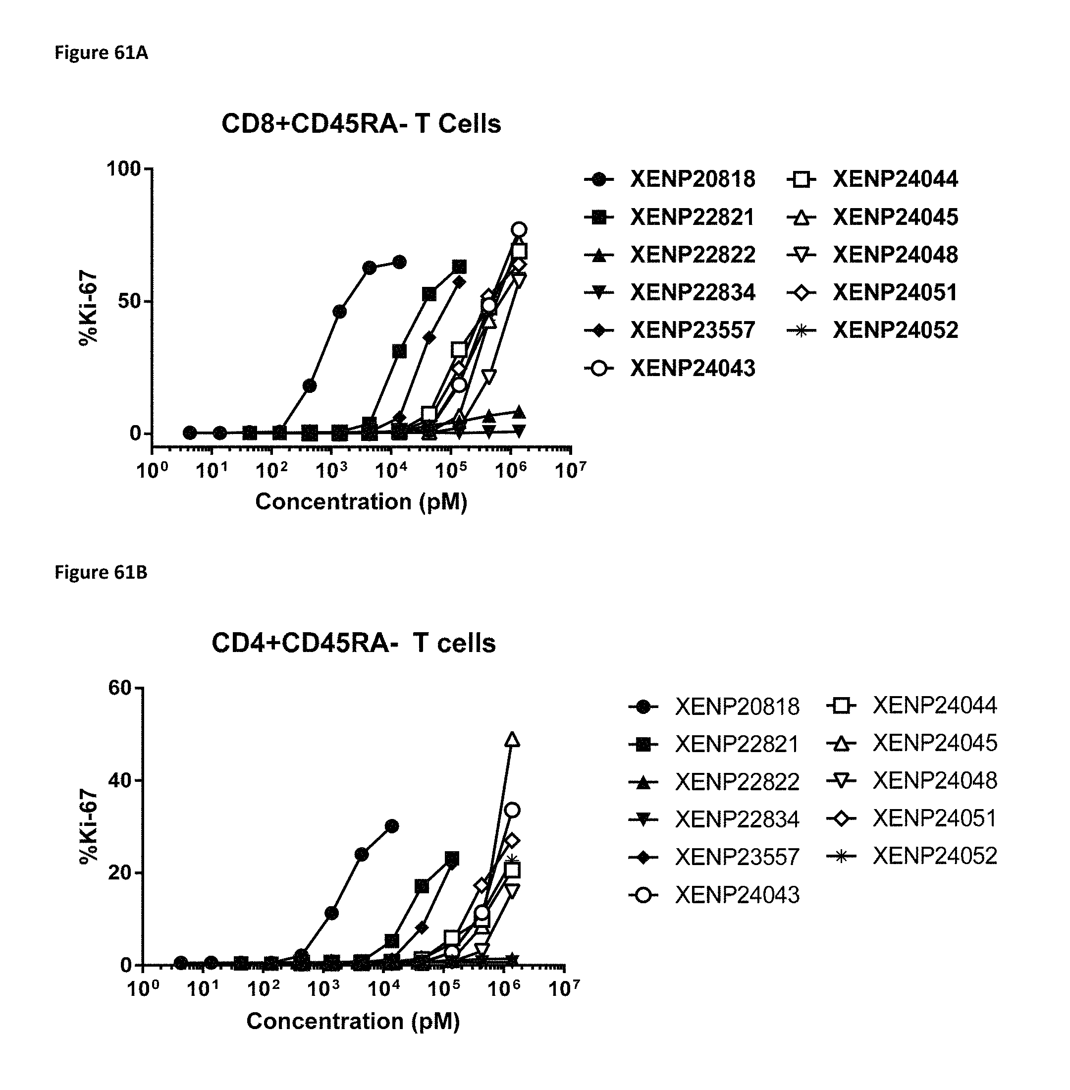
D00098
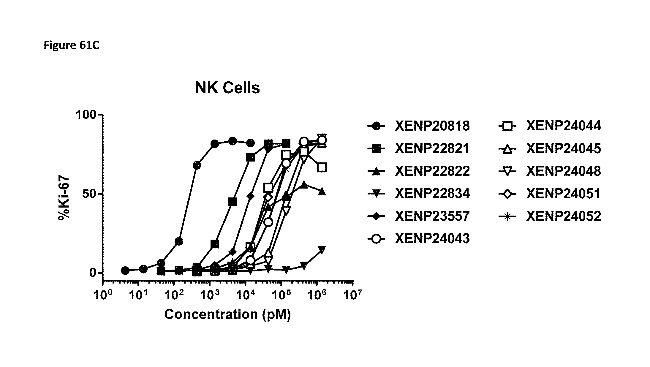
D00099
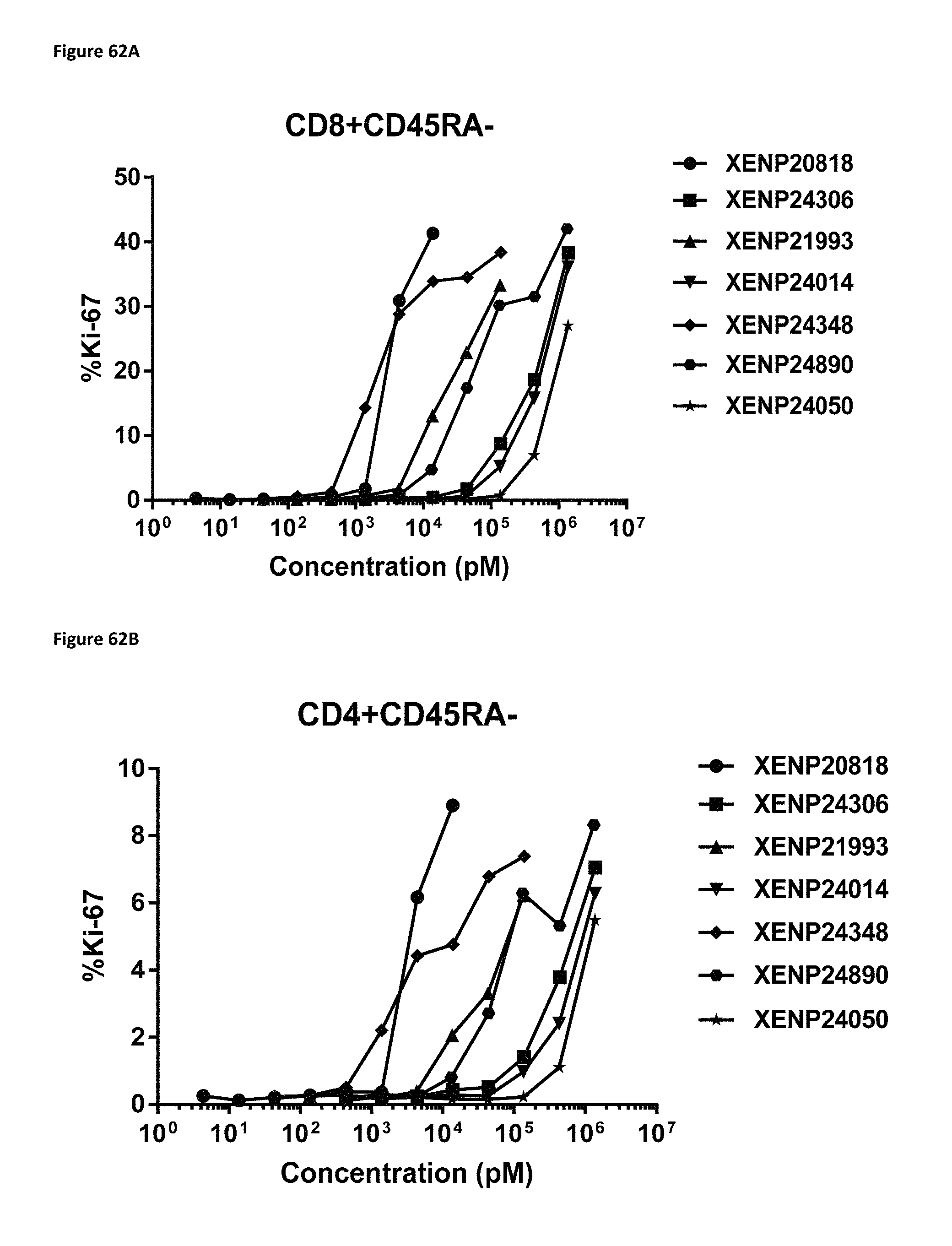
D00100
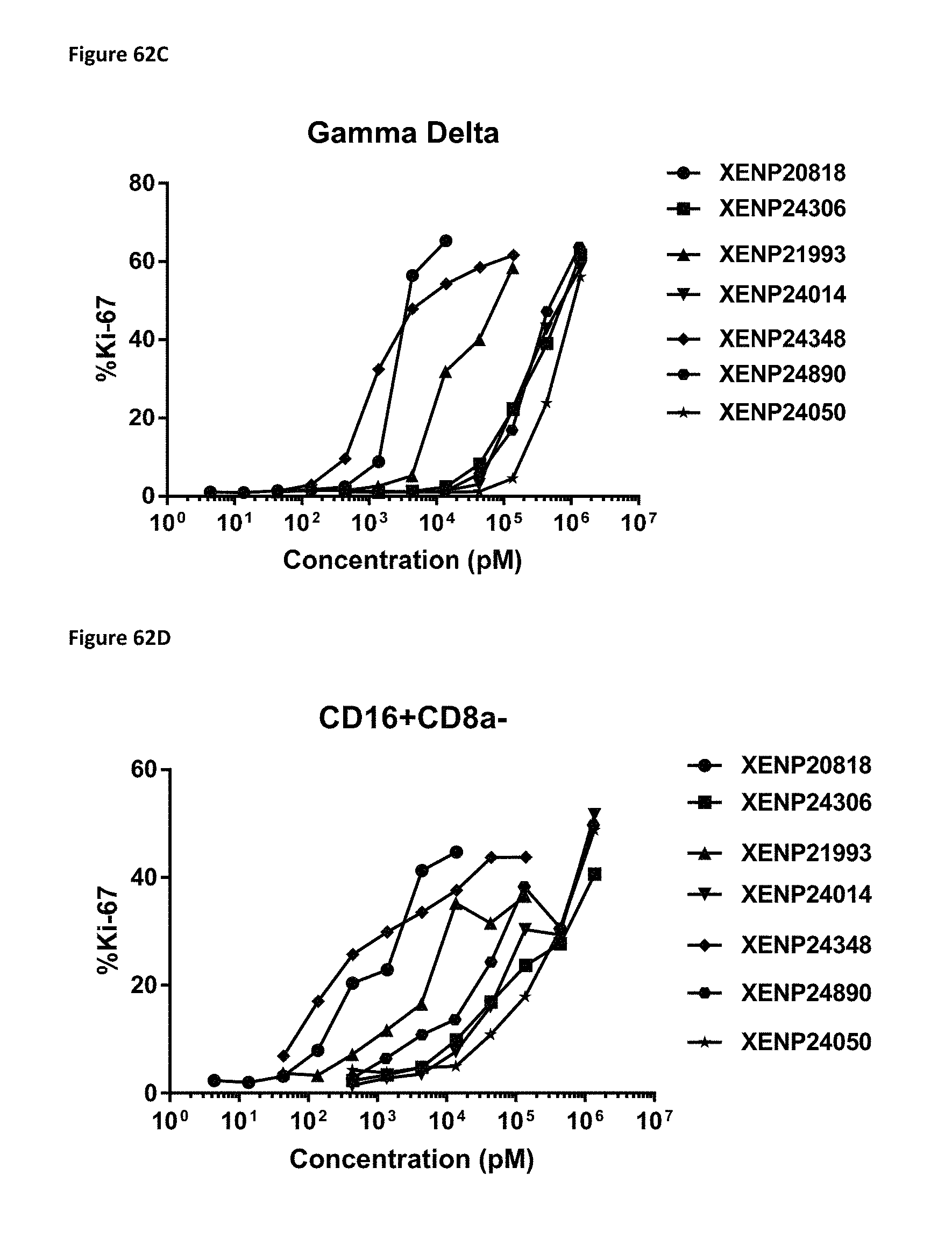
D00101
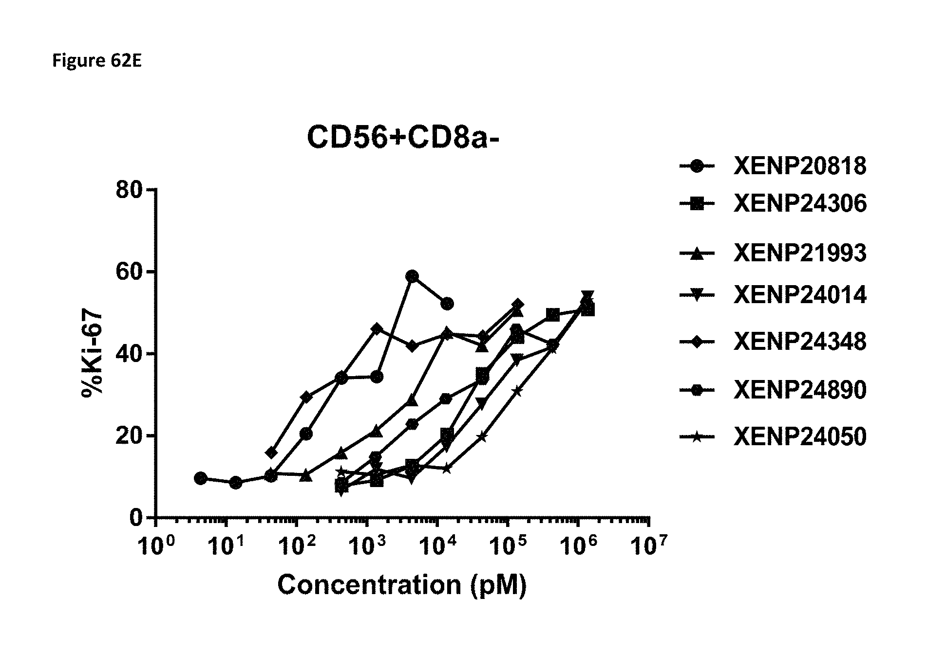
D00102

D00103
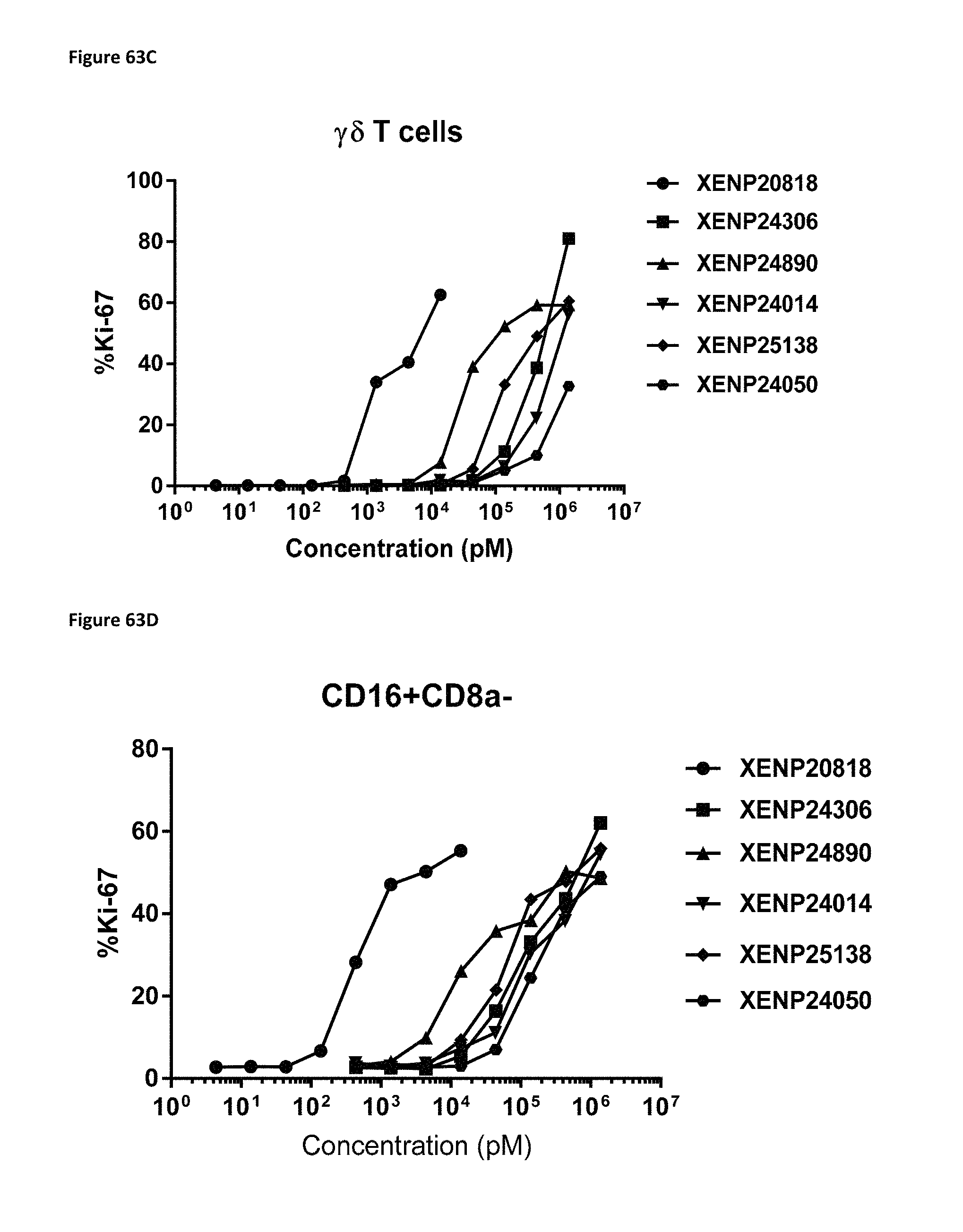
D00104

D00105
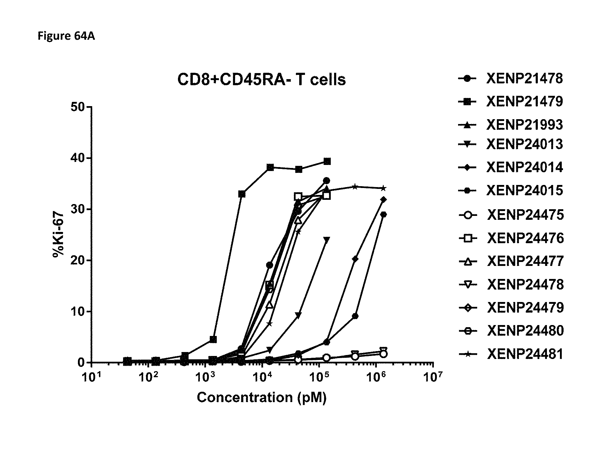
D00106
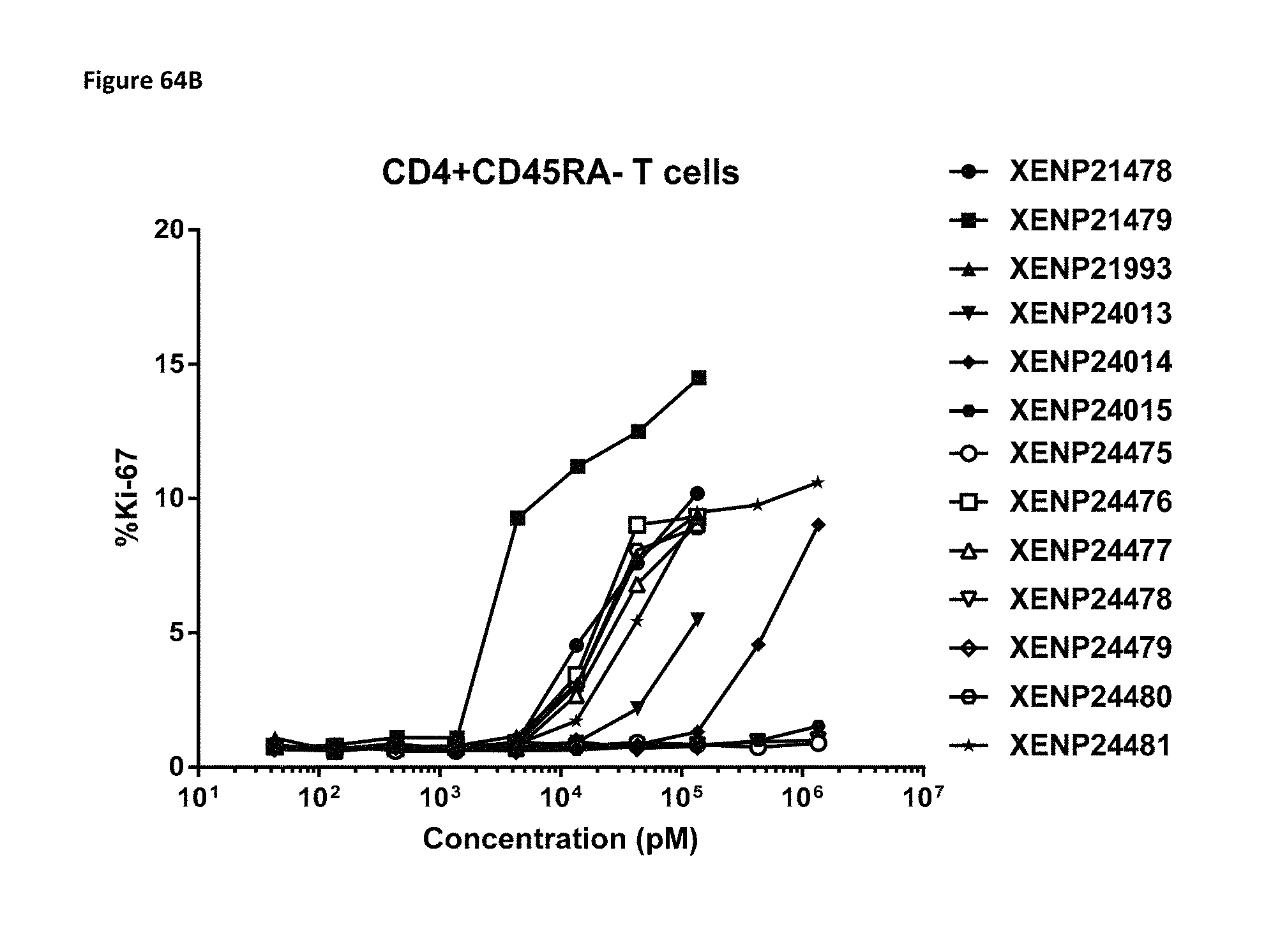
D00107

D00108
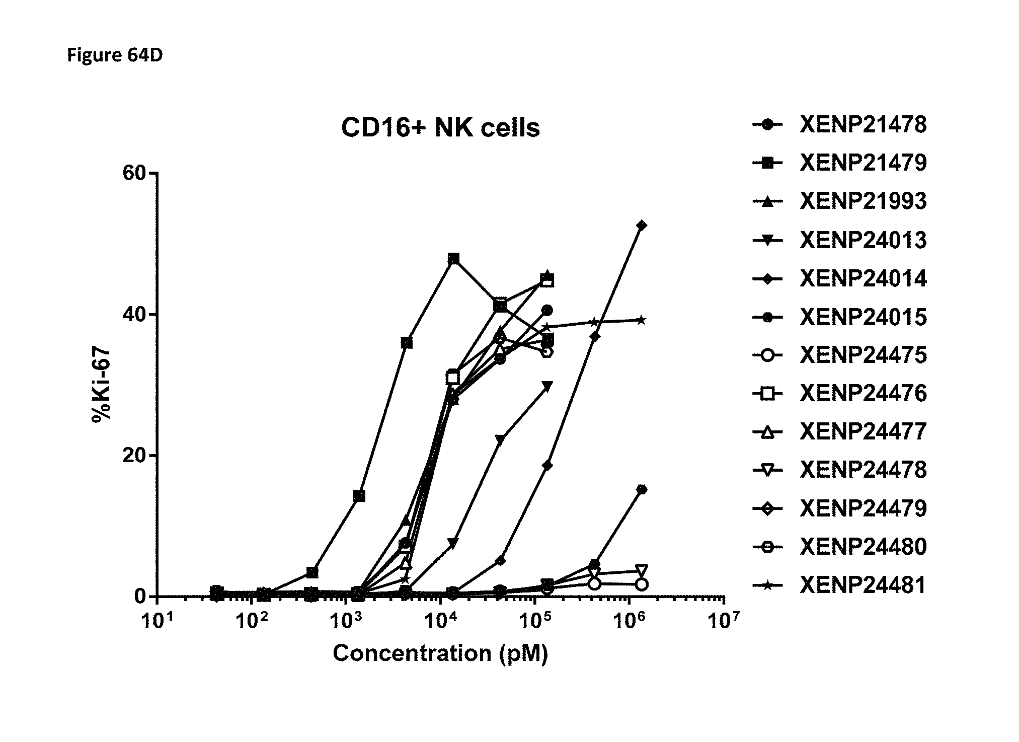
D00109

D00110

D00111
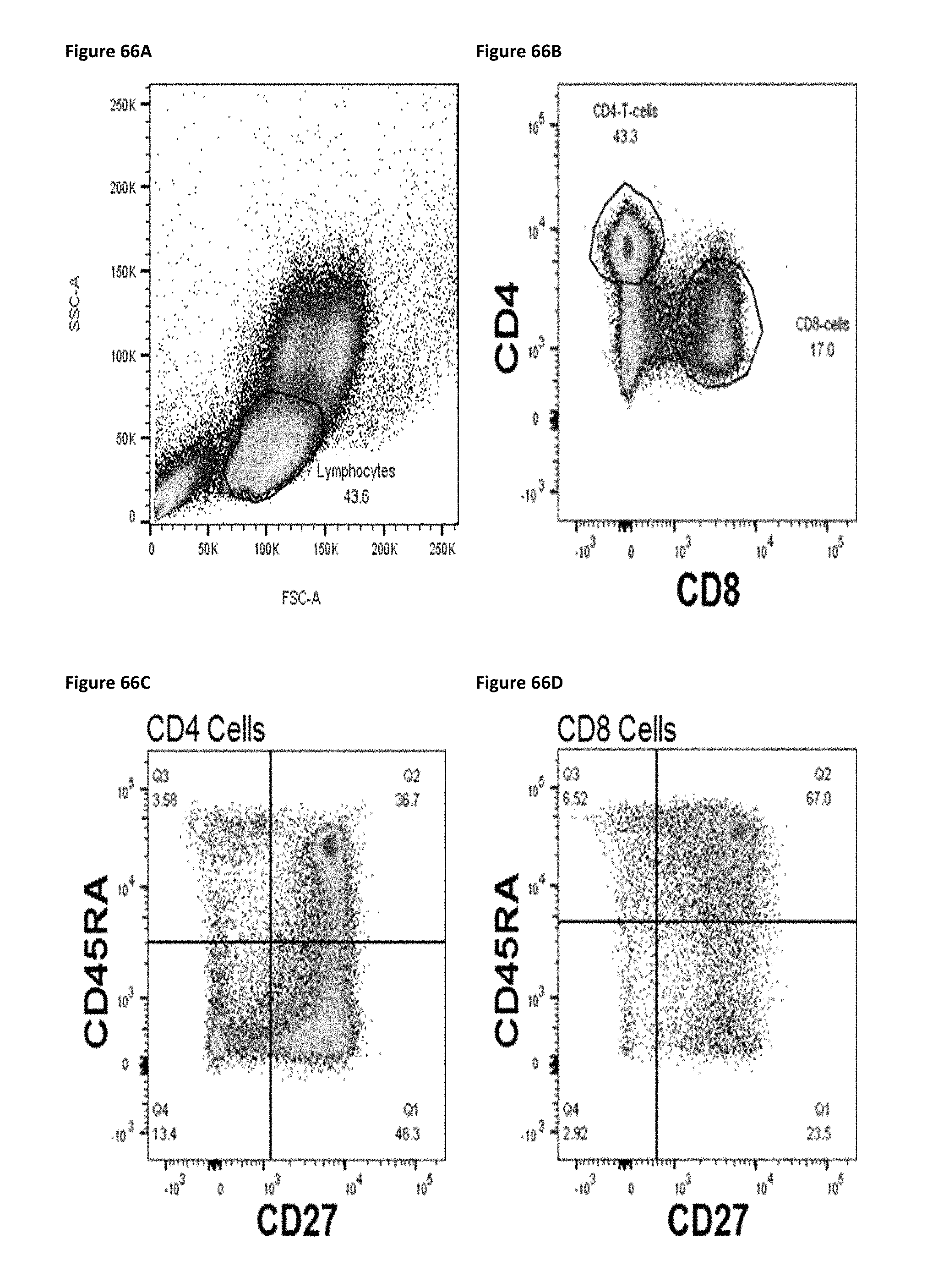
D00112

D00113

D00114

D00115
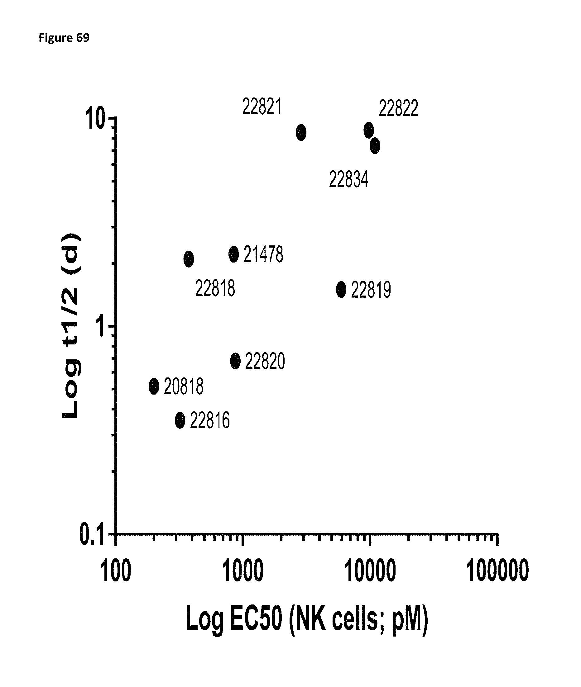
D00116

D00117

D00118

D00119

D00120

D00121
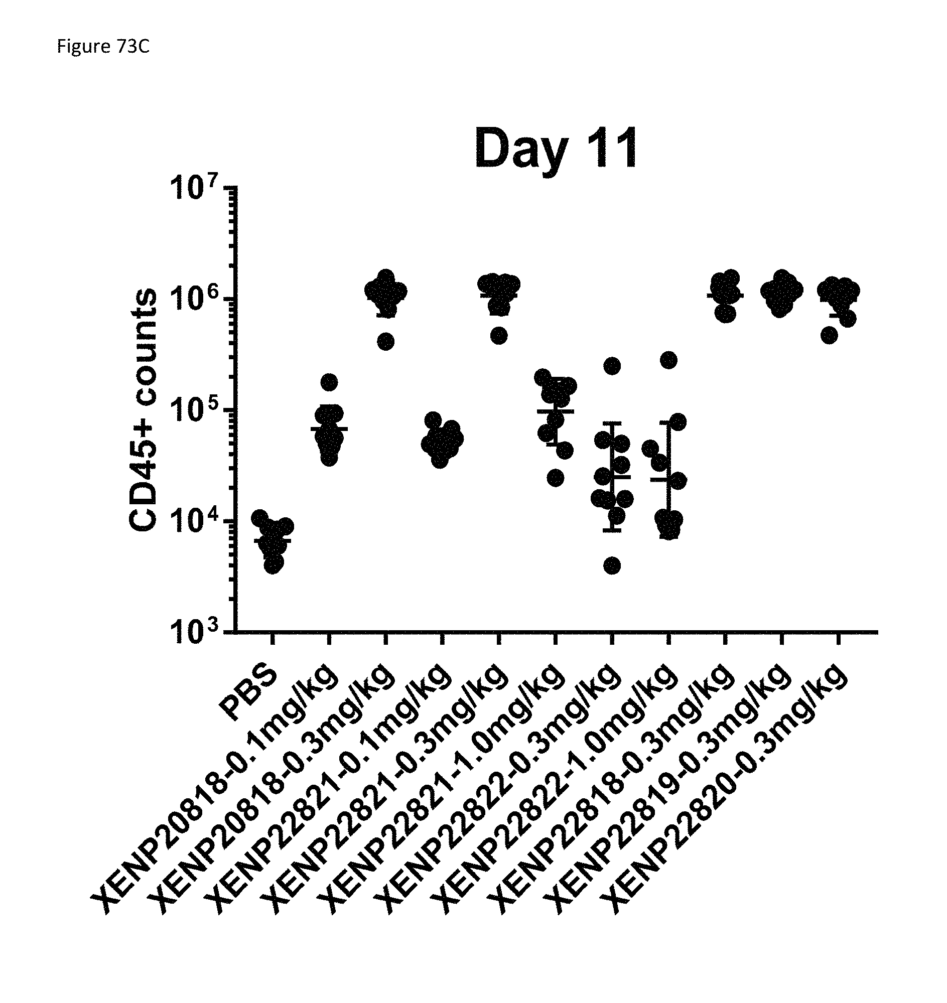
D00122

D00123

D00124
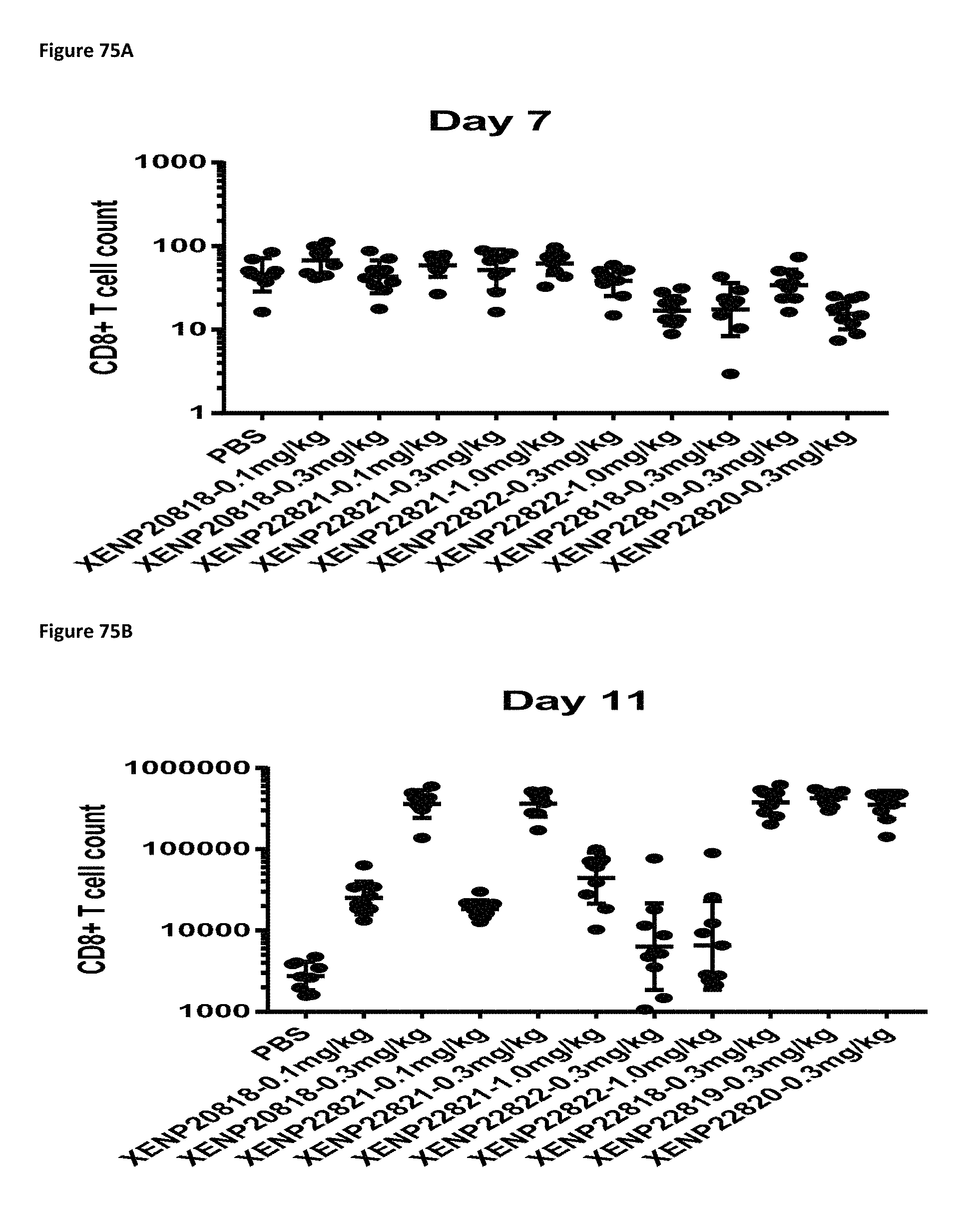
D00125

D00126
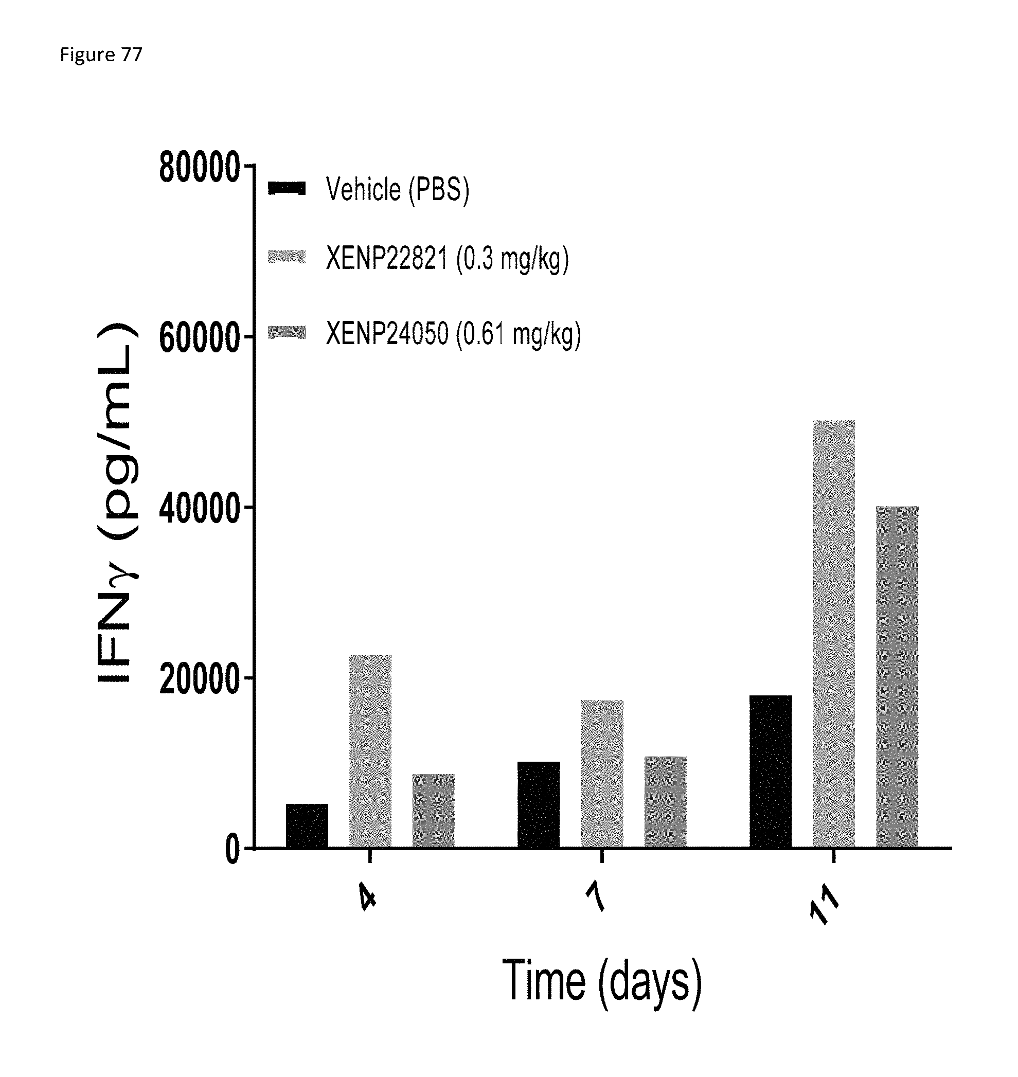
D00127
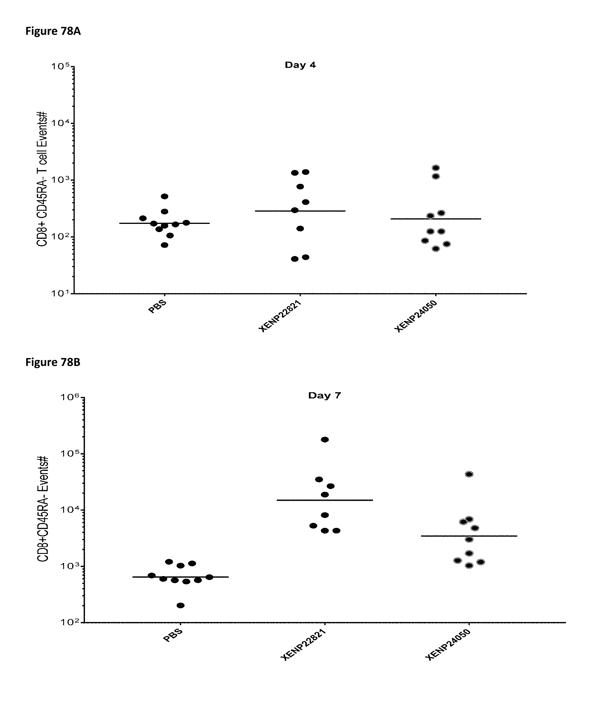
D00128

D00129
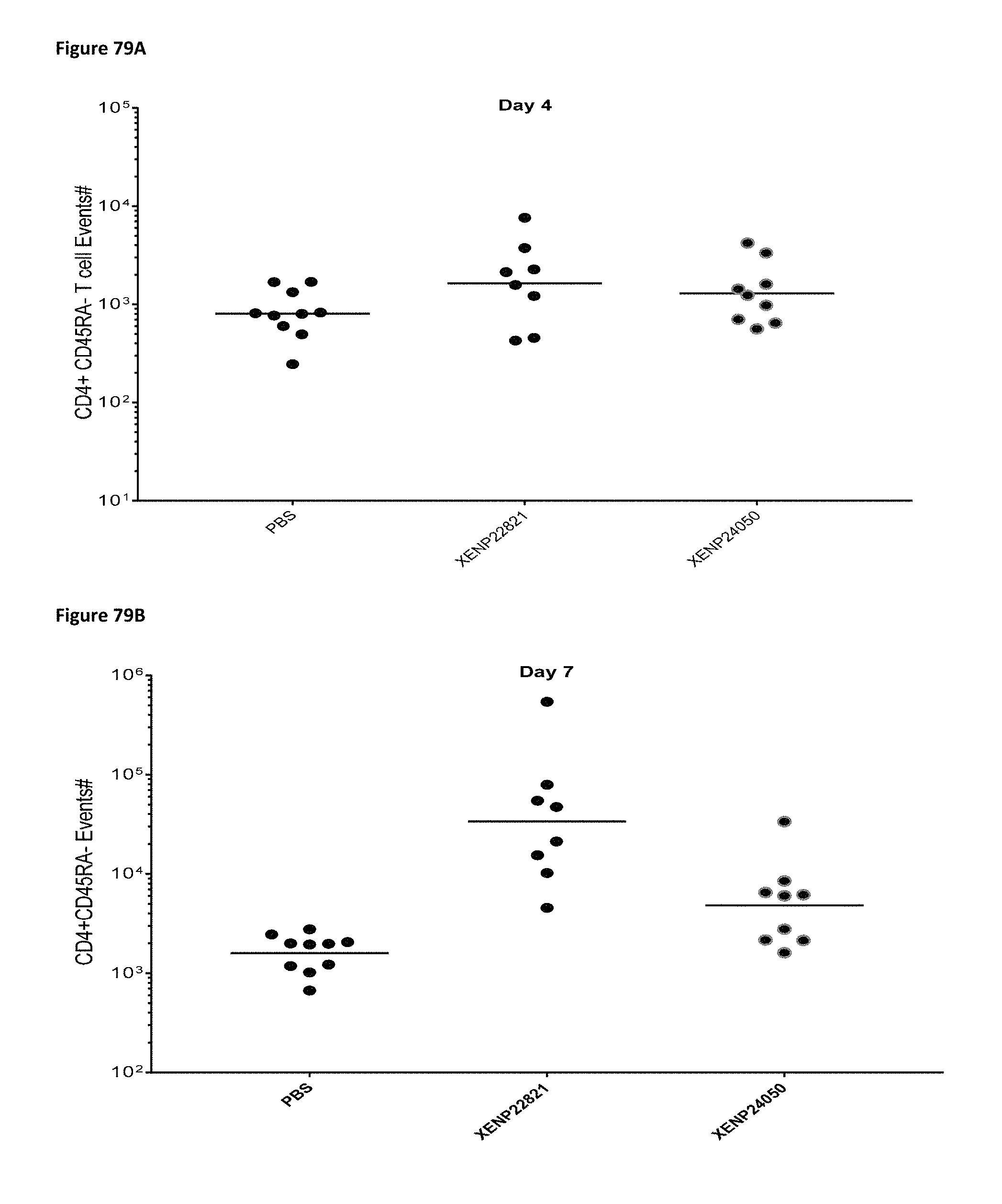
D00130
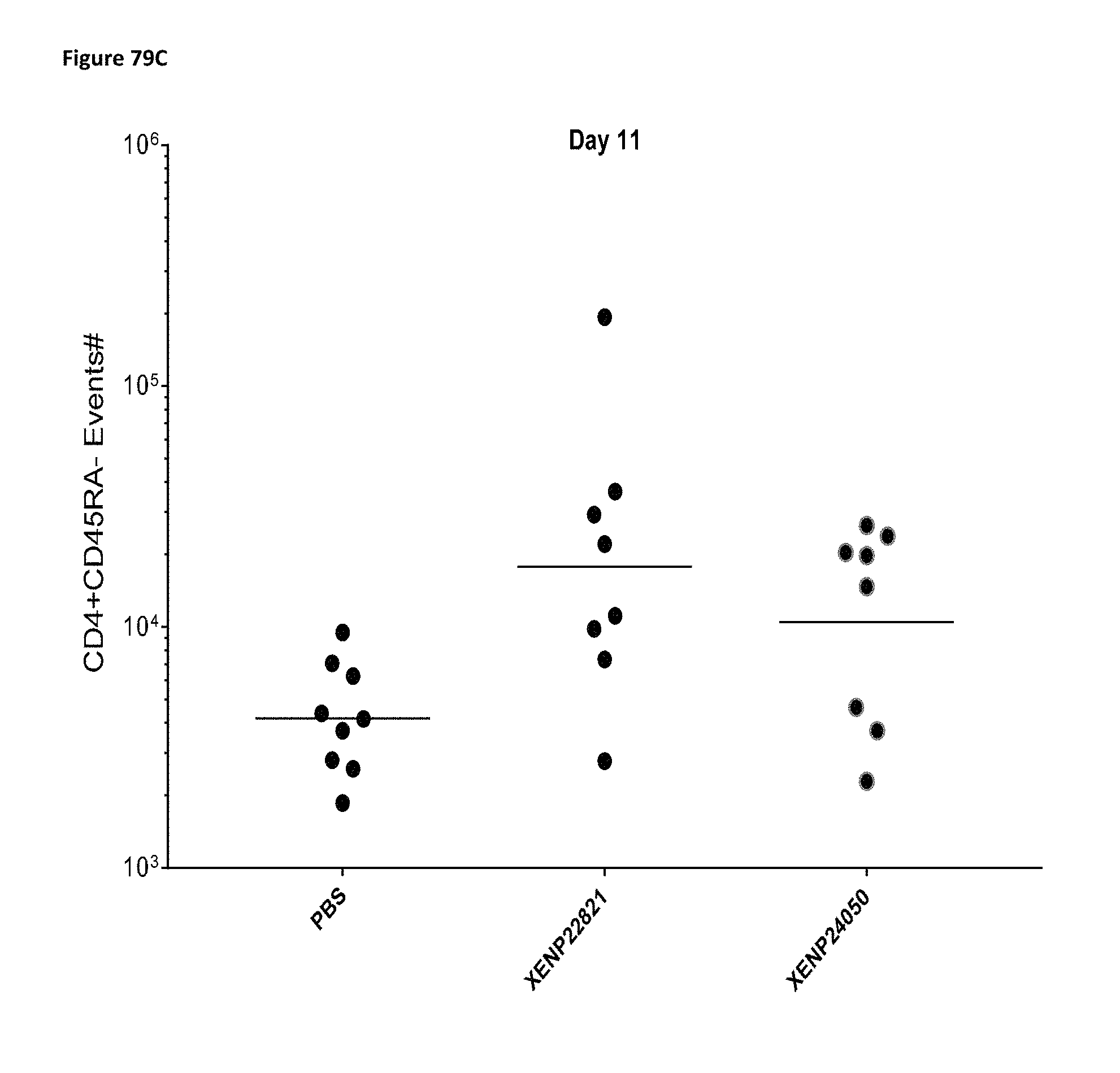
D00131

D00132
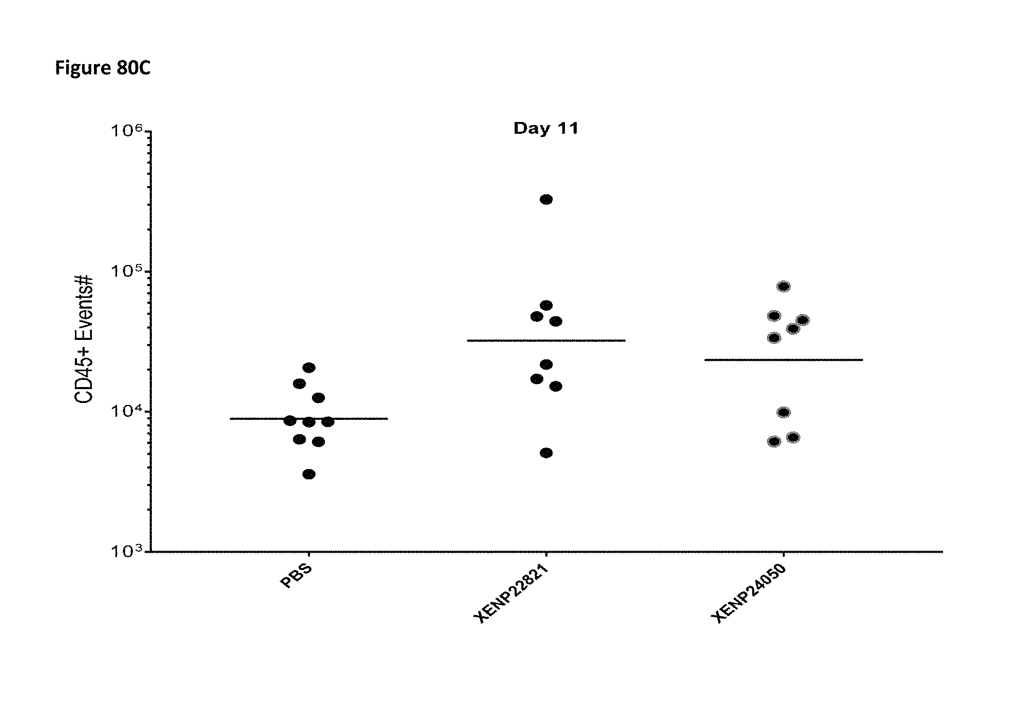
D00133
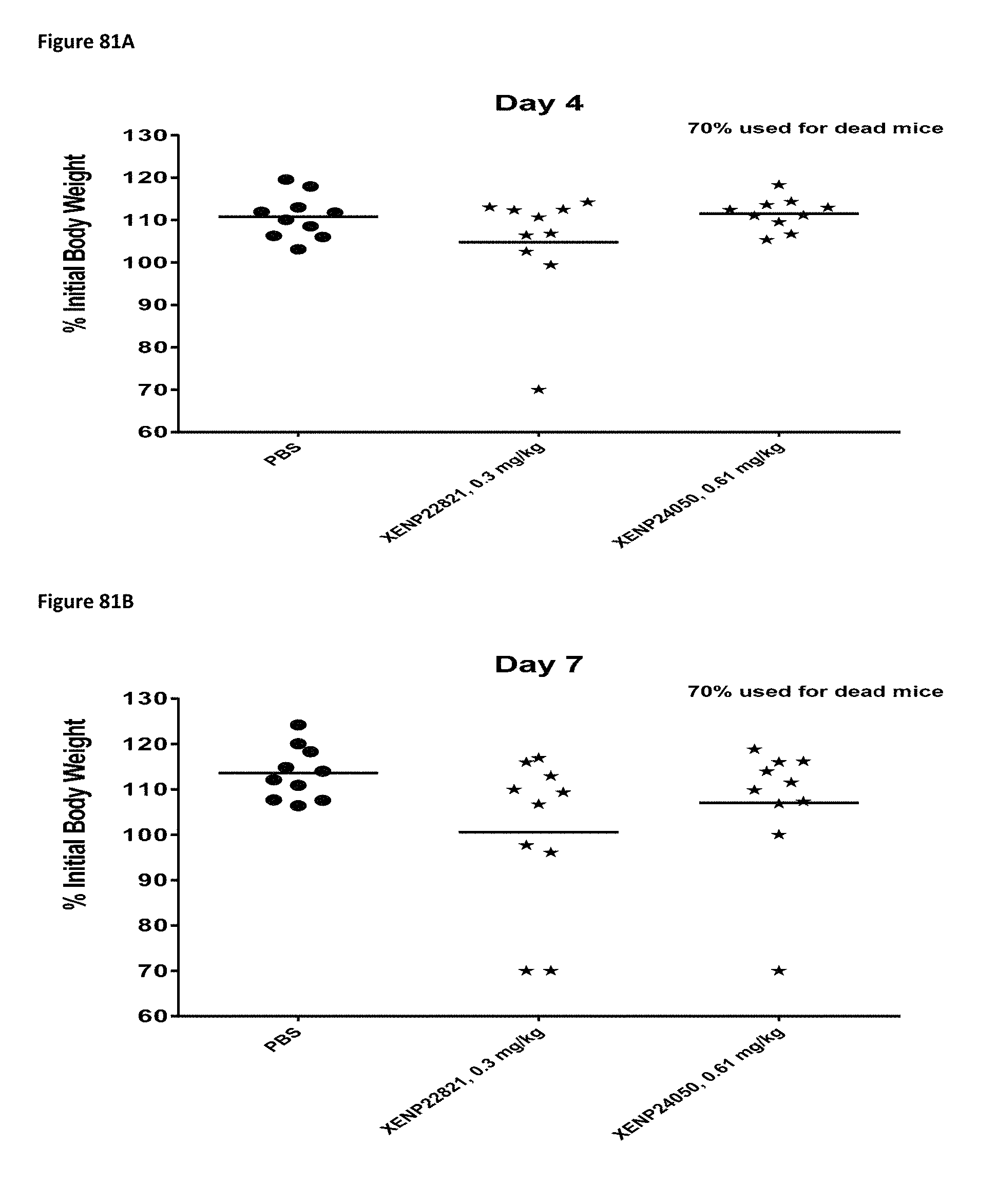
D00134
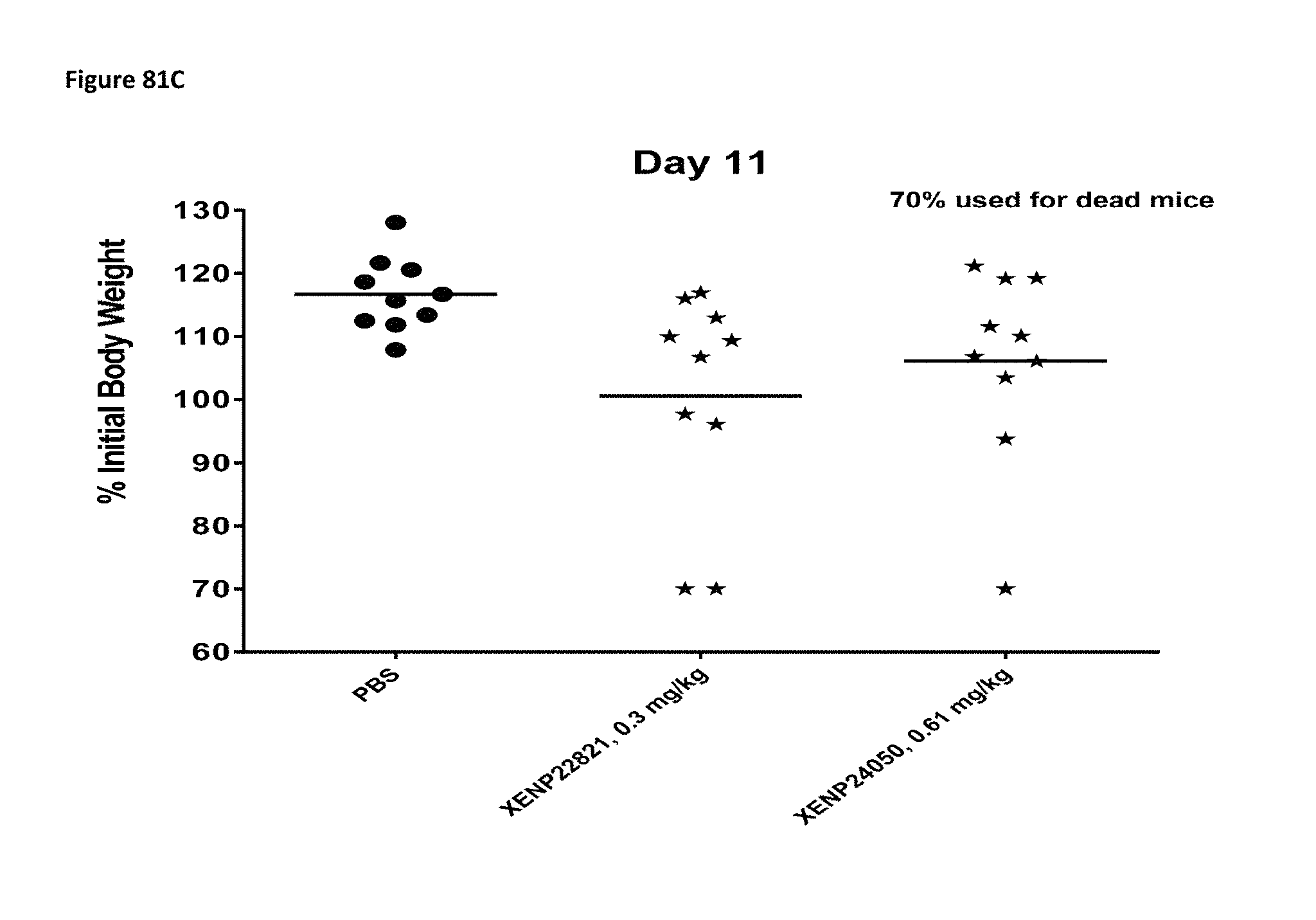
D00135

D00136

D00137
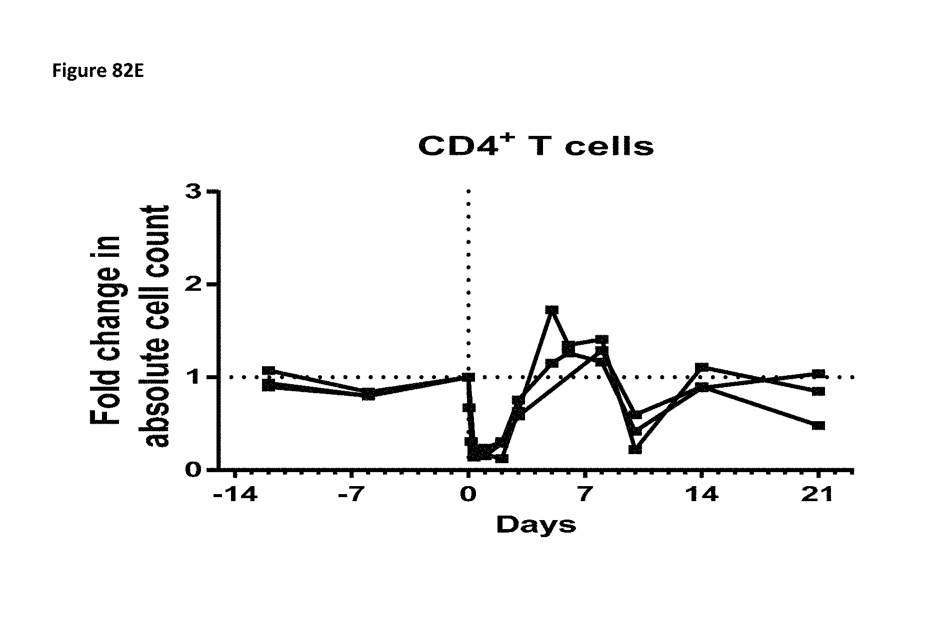
D00138

D00139

D00140
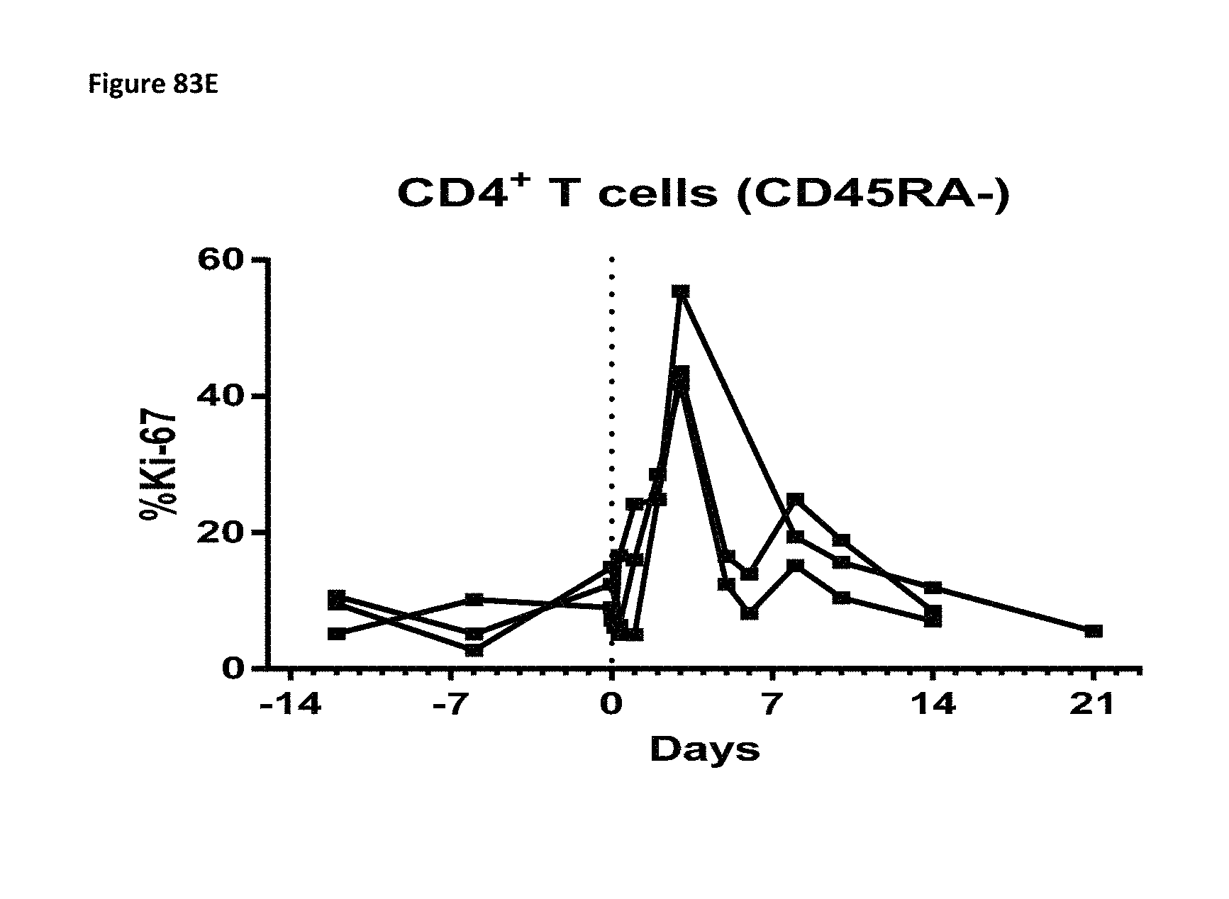
D00141
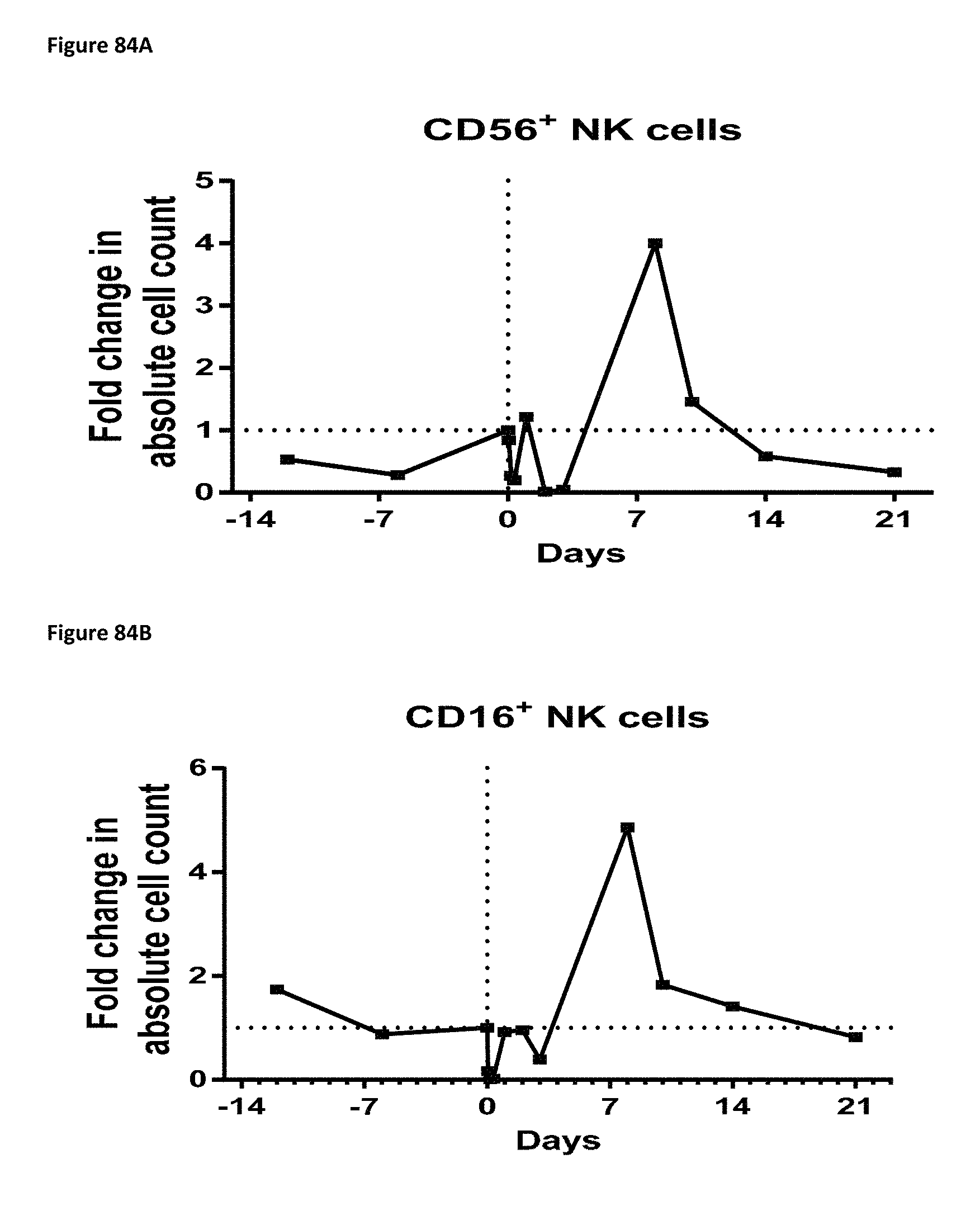
D00142
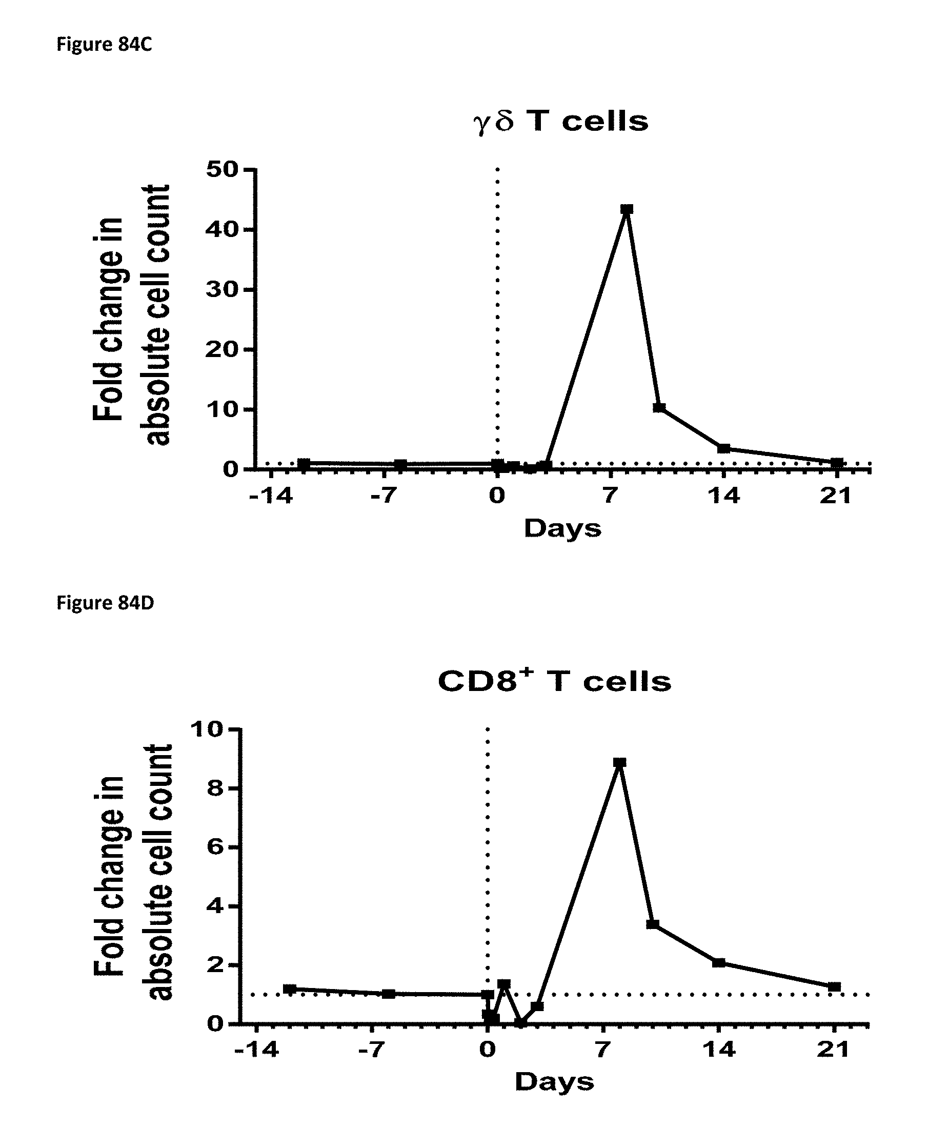
D00143

D00144

D00145
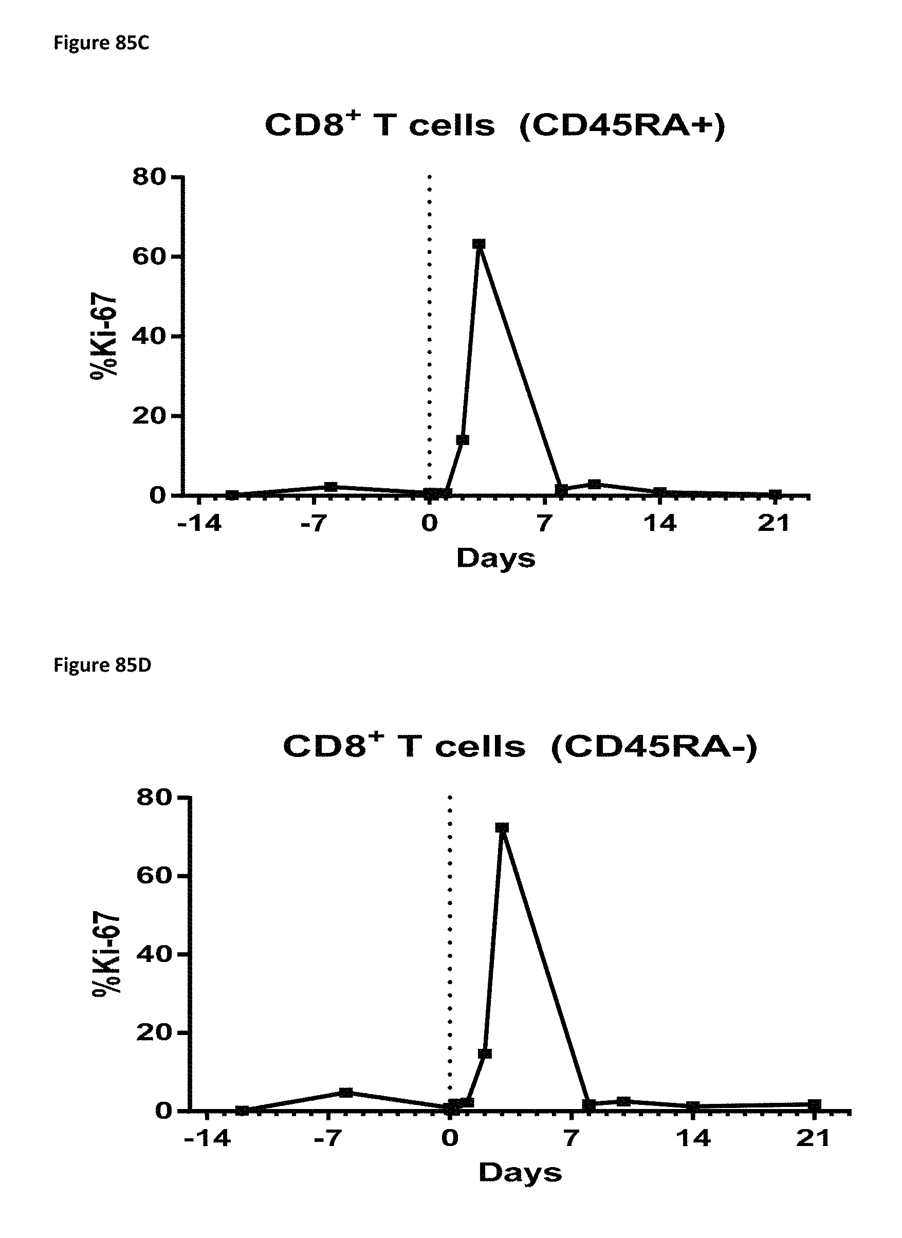
D00146

D00147
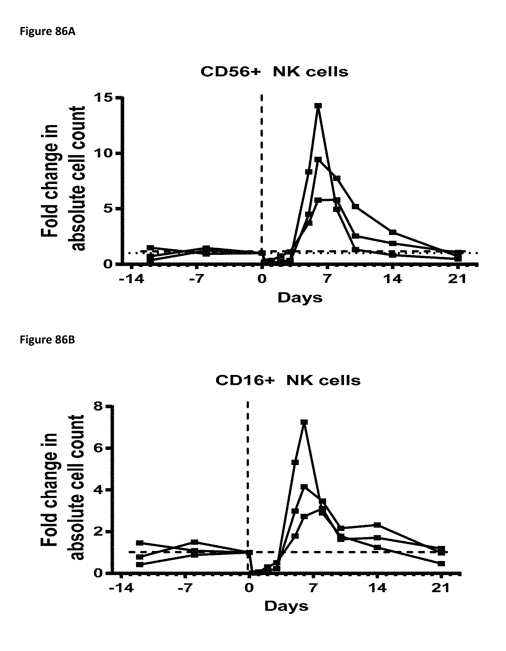
D00148
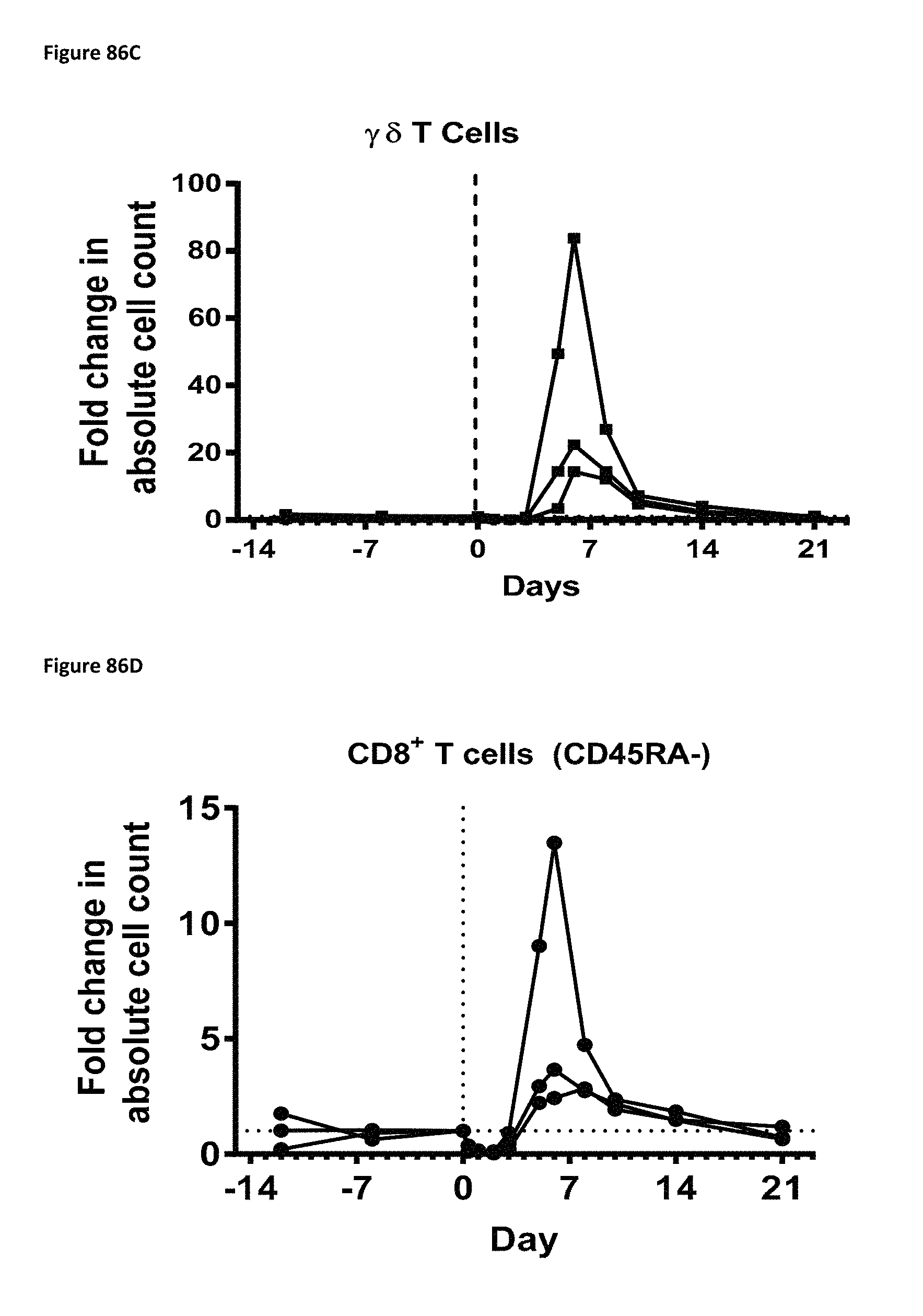
D00149

D00150
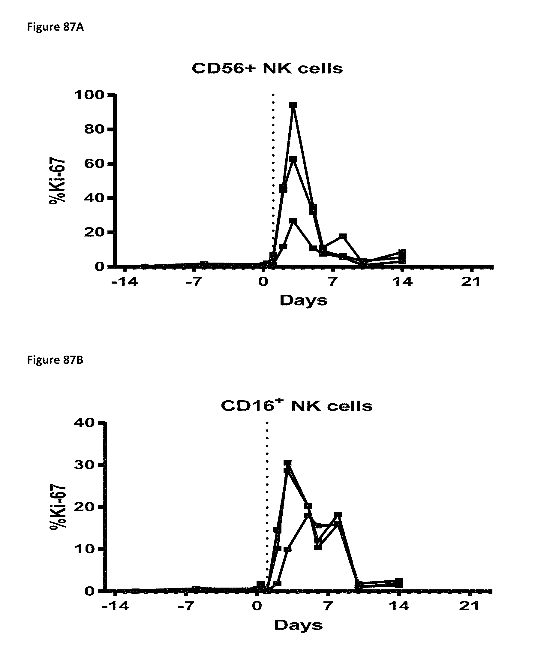
D00151

D00152

D00153
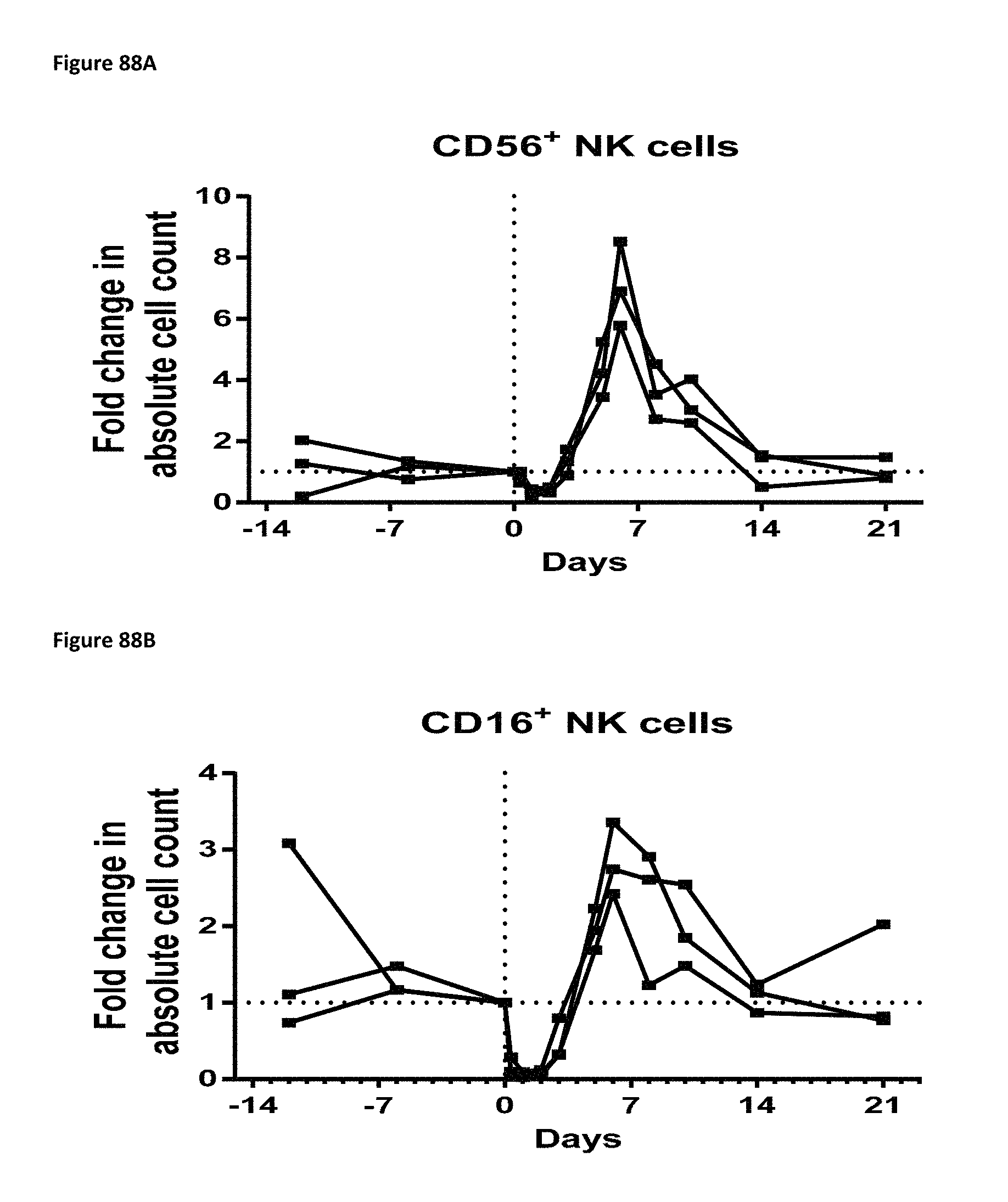
D00154
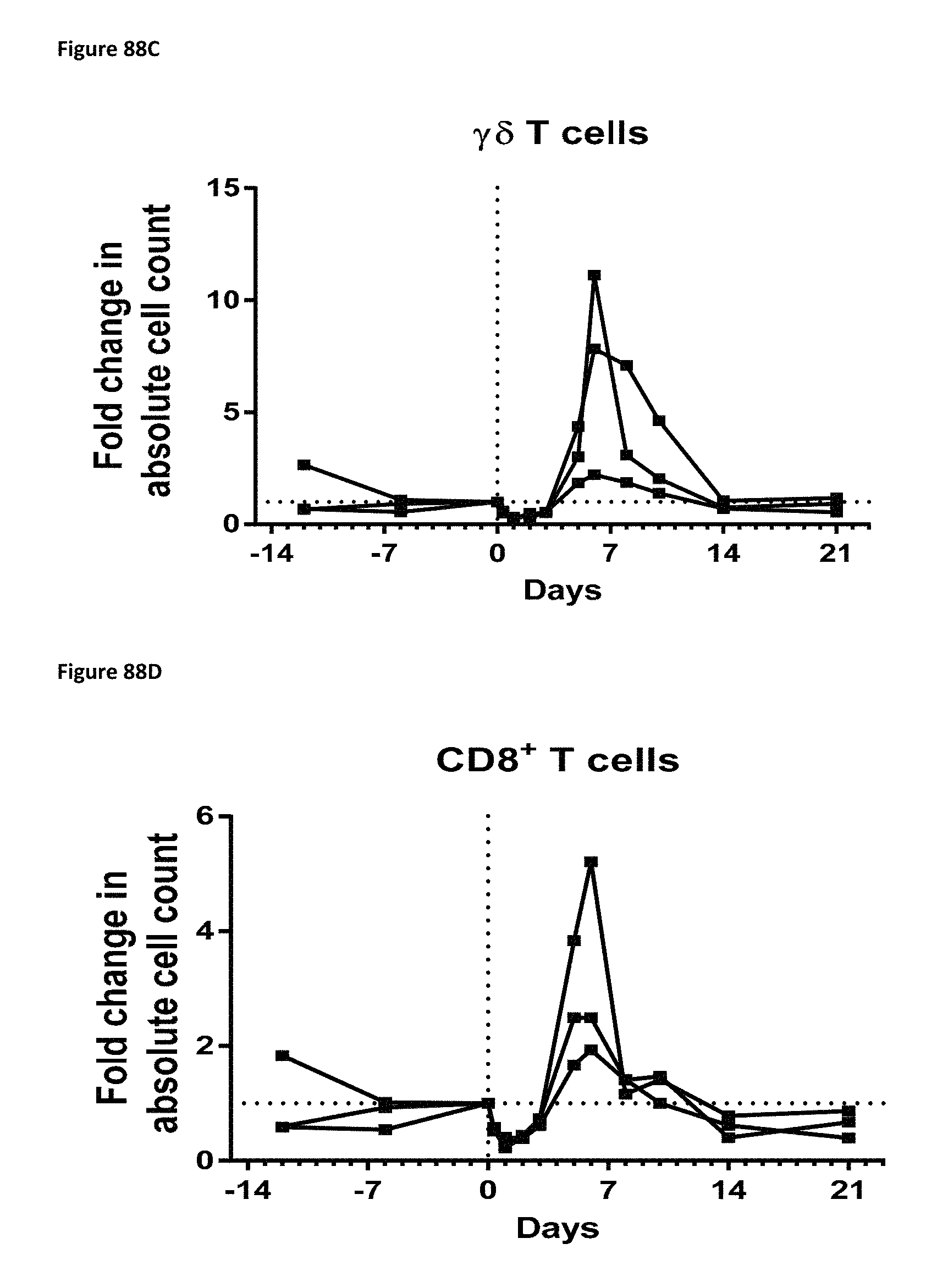
D00155

D00156
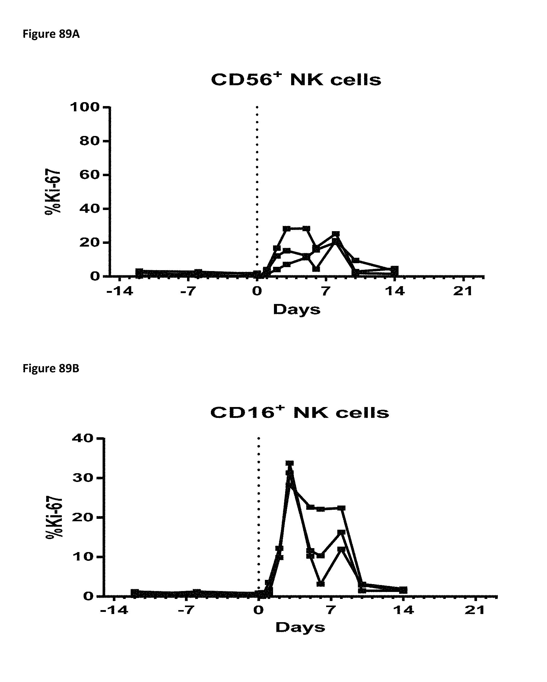
D00157
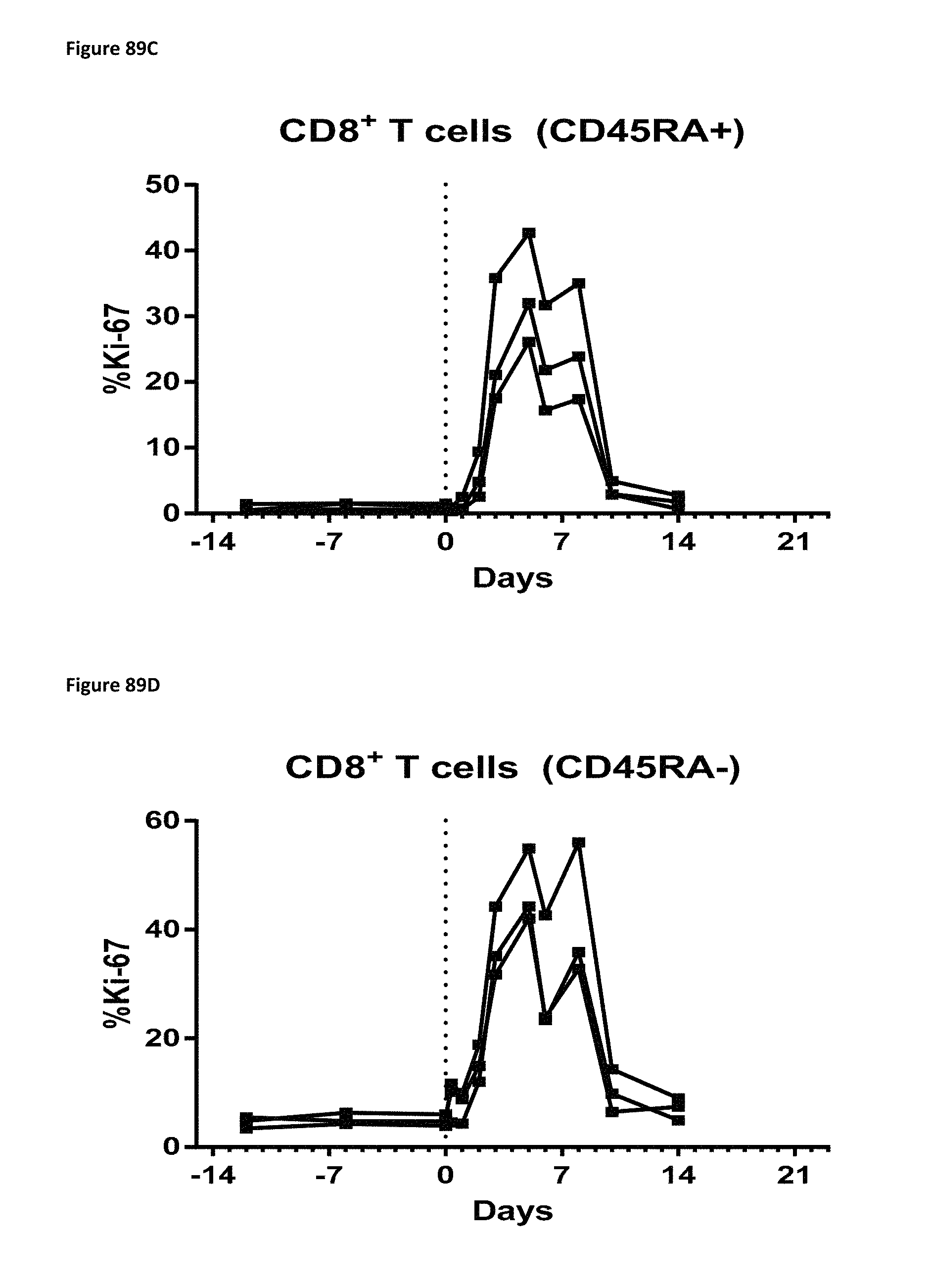
D00158

D00159
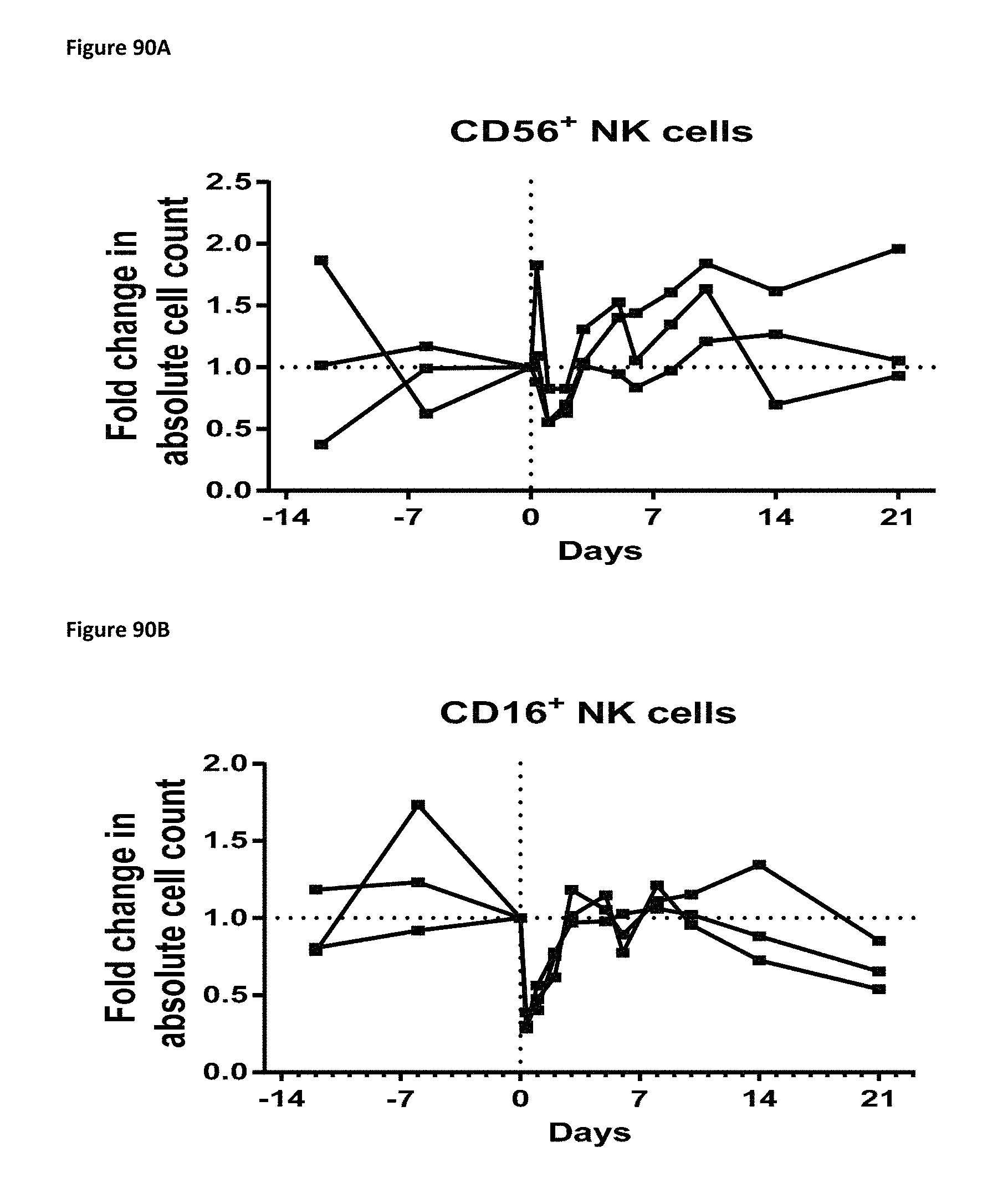
D00160

D00161
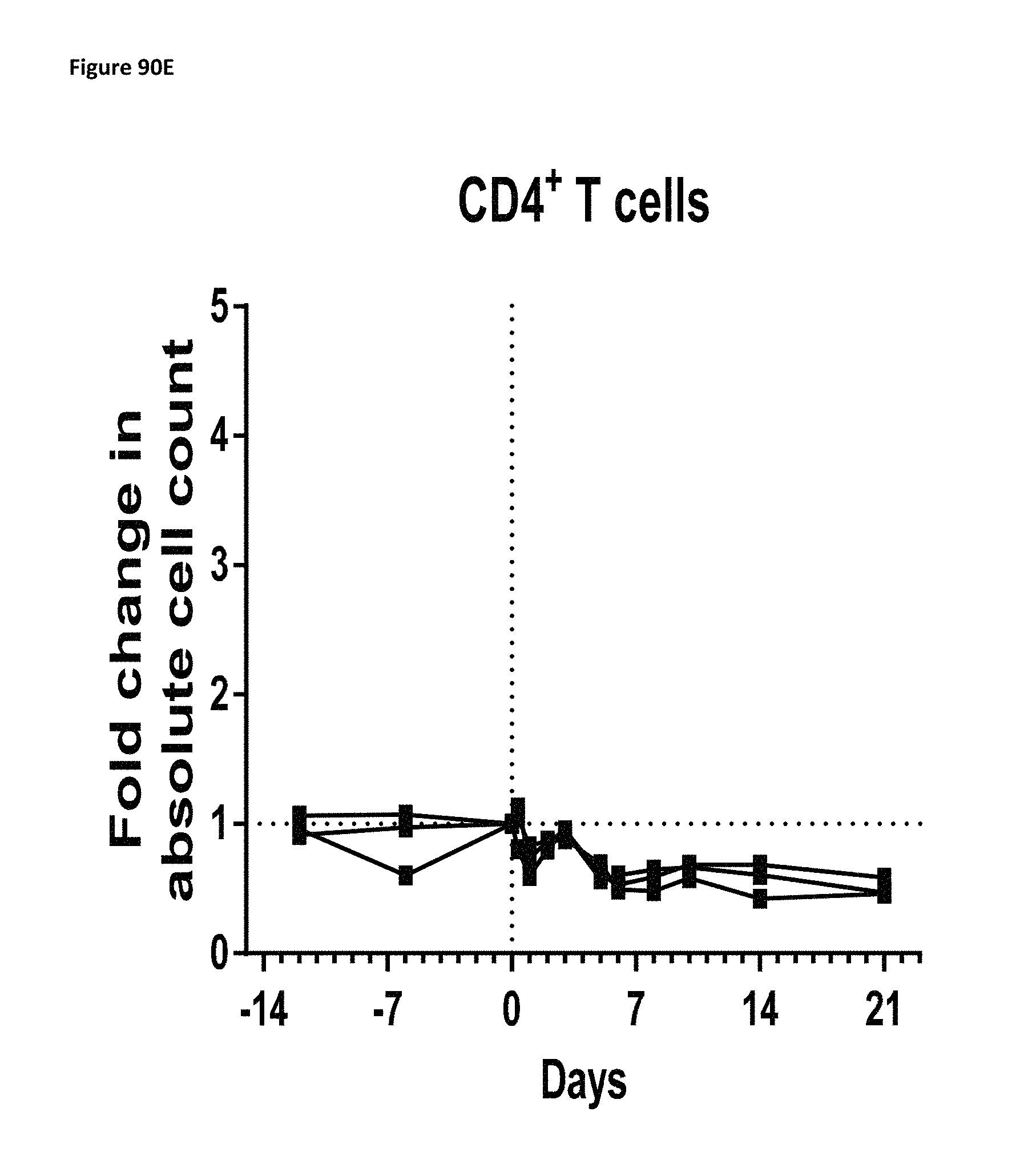
D00162

D00163

D00164
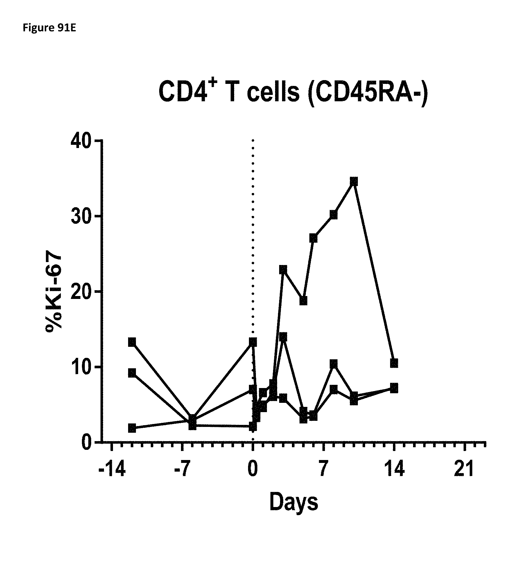
D00165
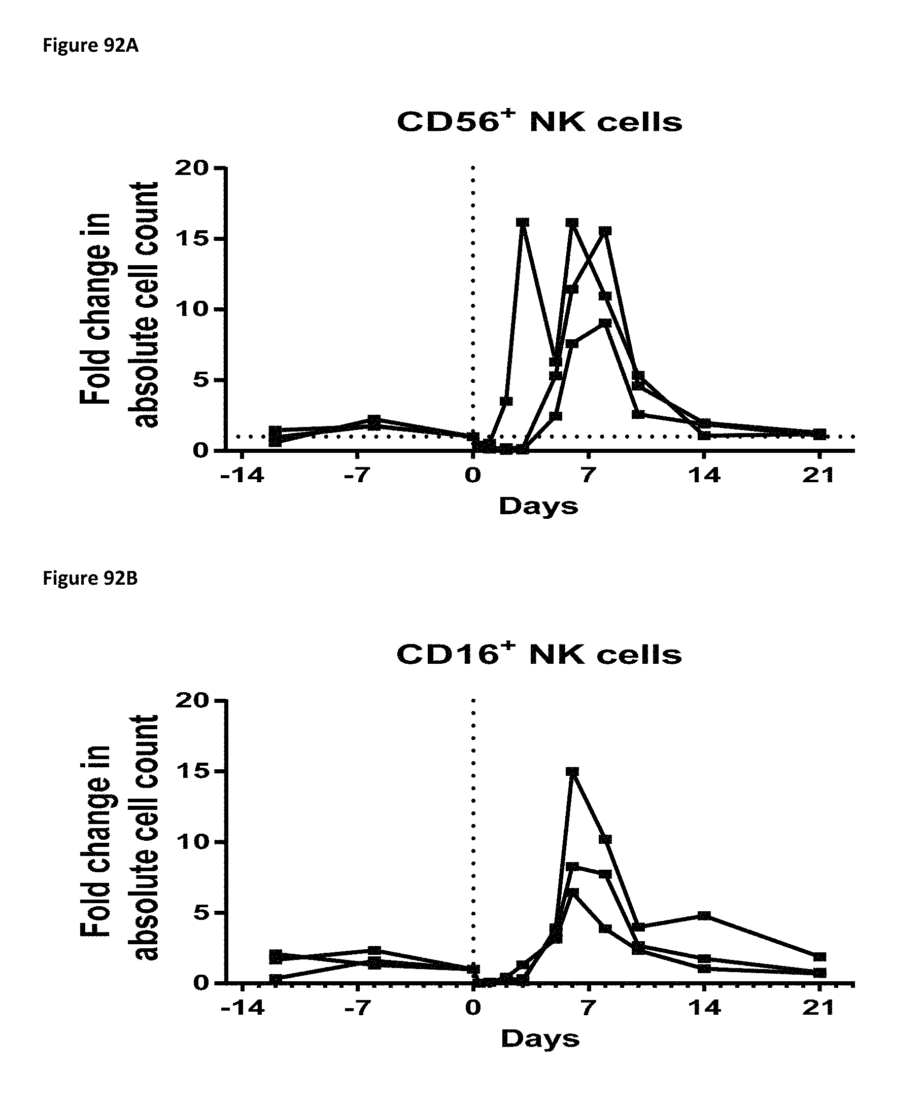
D00166
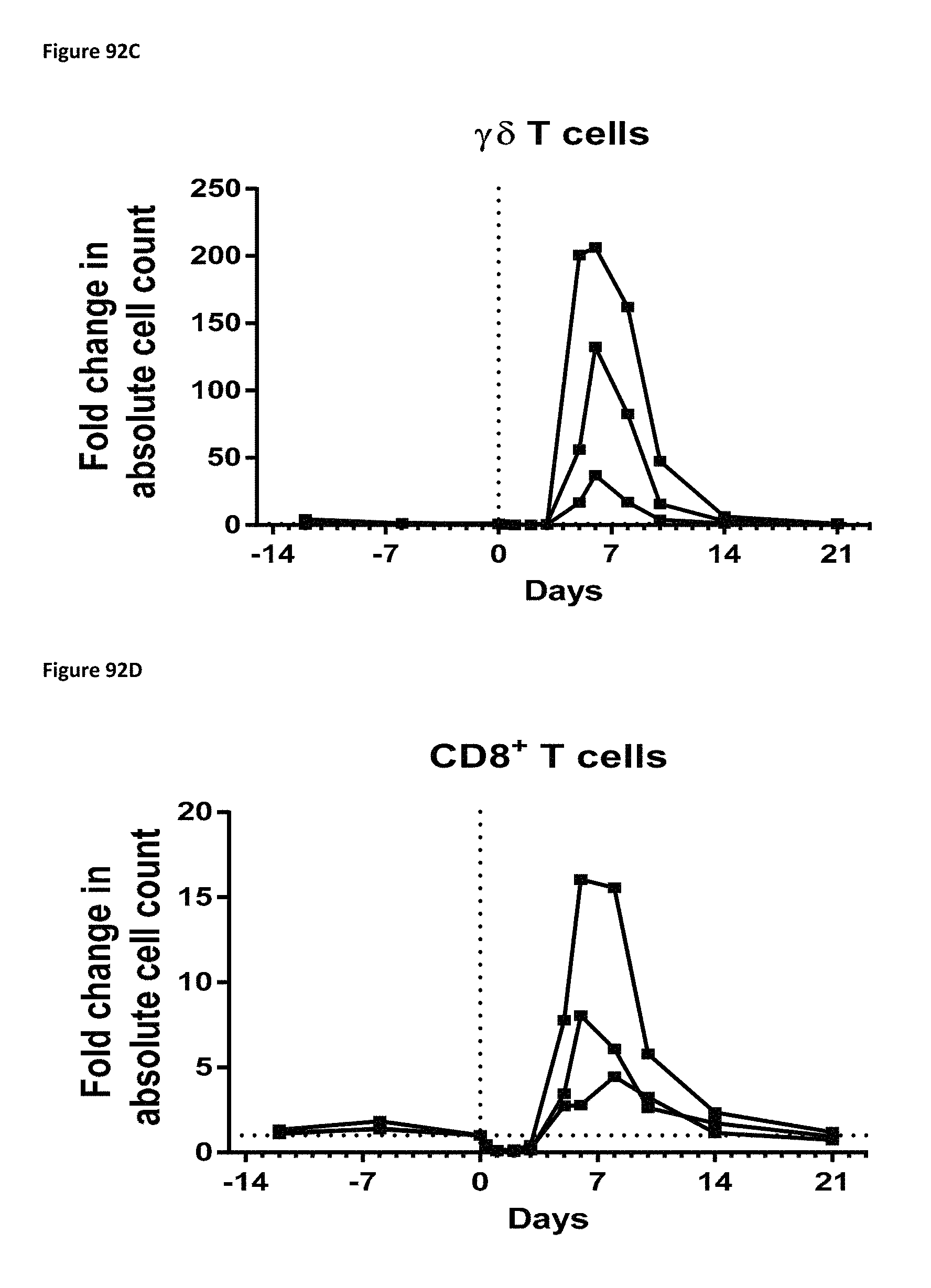
D00167
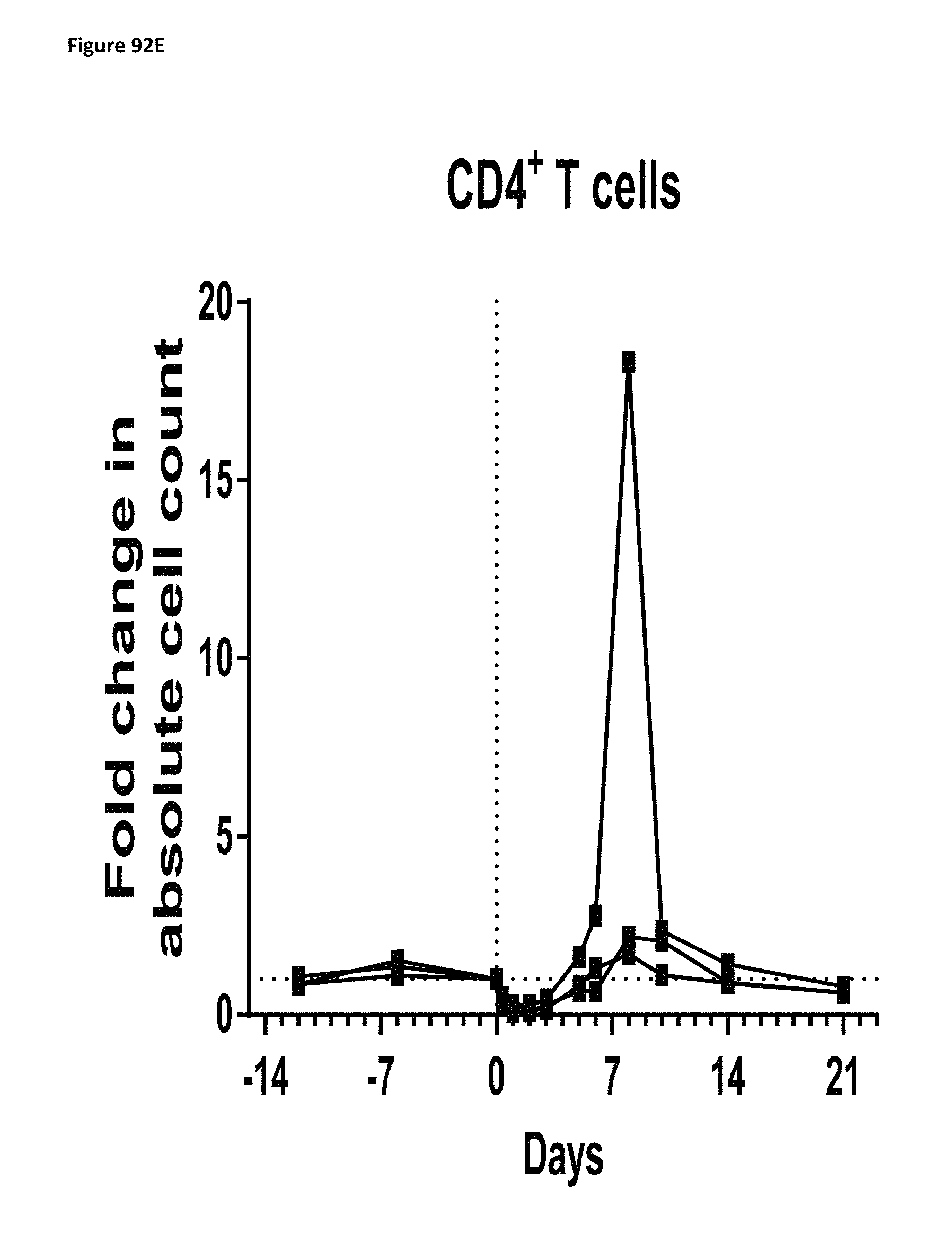
D00168

D00169

D00170

D00171

D00172
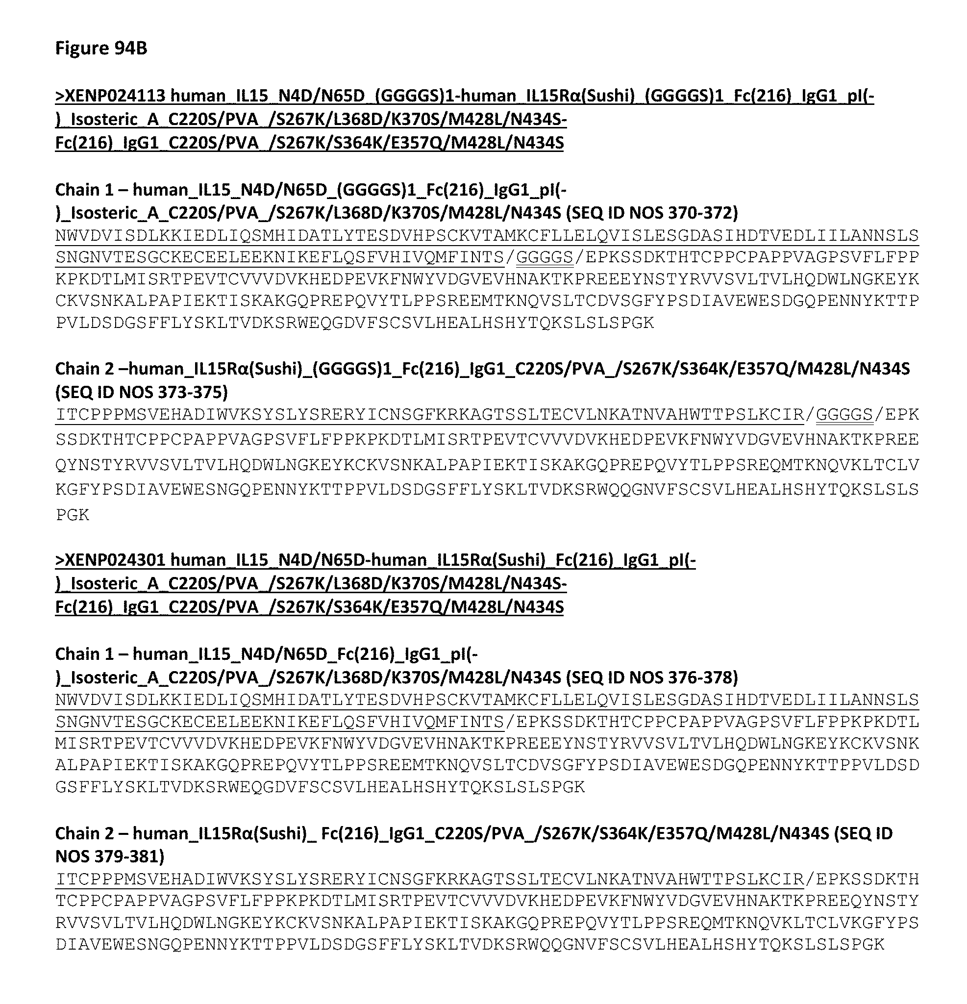
D00173

D00174

D00175

D00176

D00177
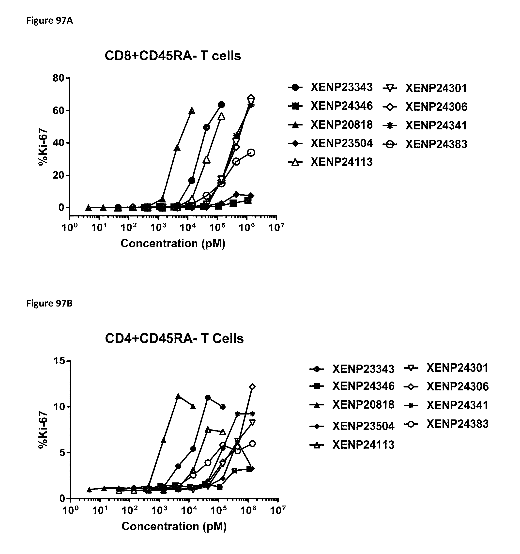
D00178

D00179

D00180

D00181
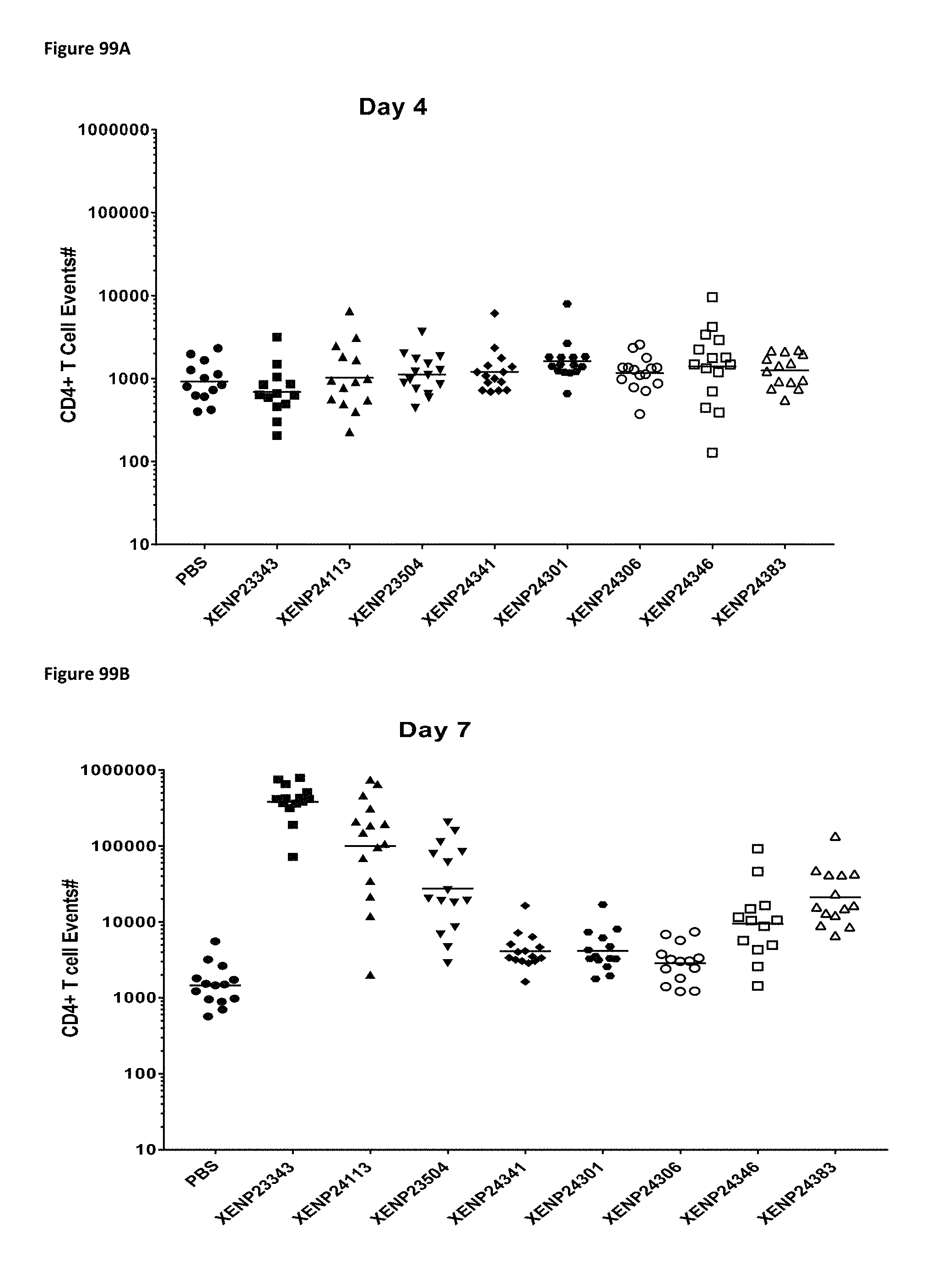
D00182

D00183

D00184
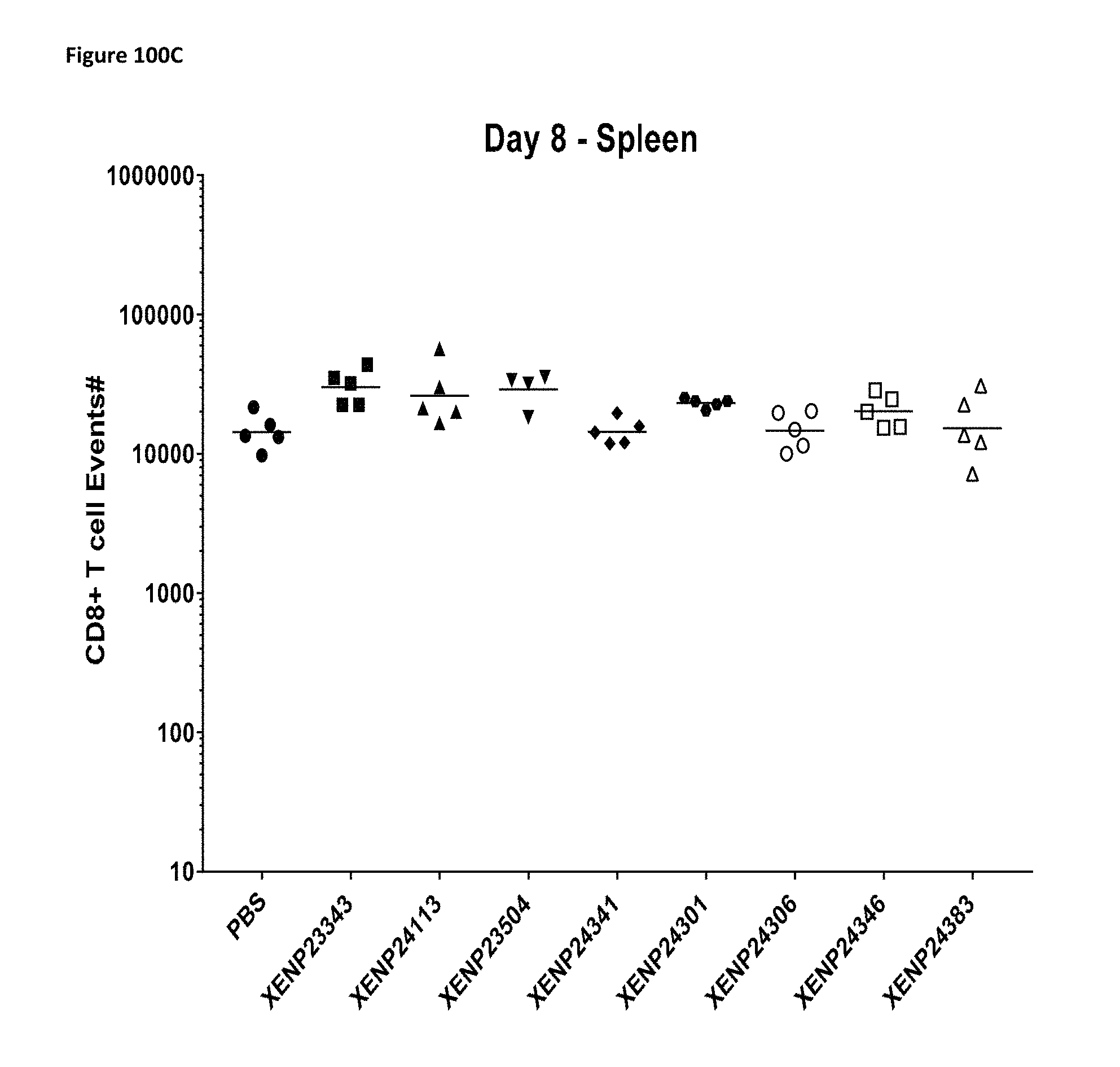
D00185
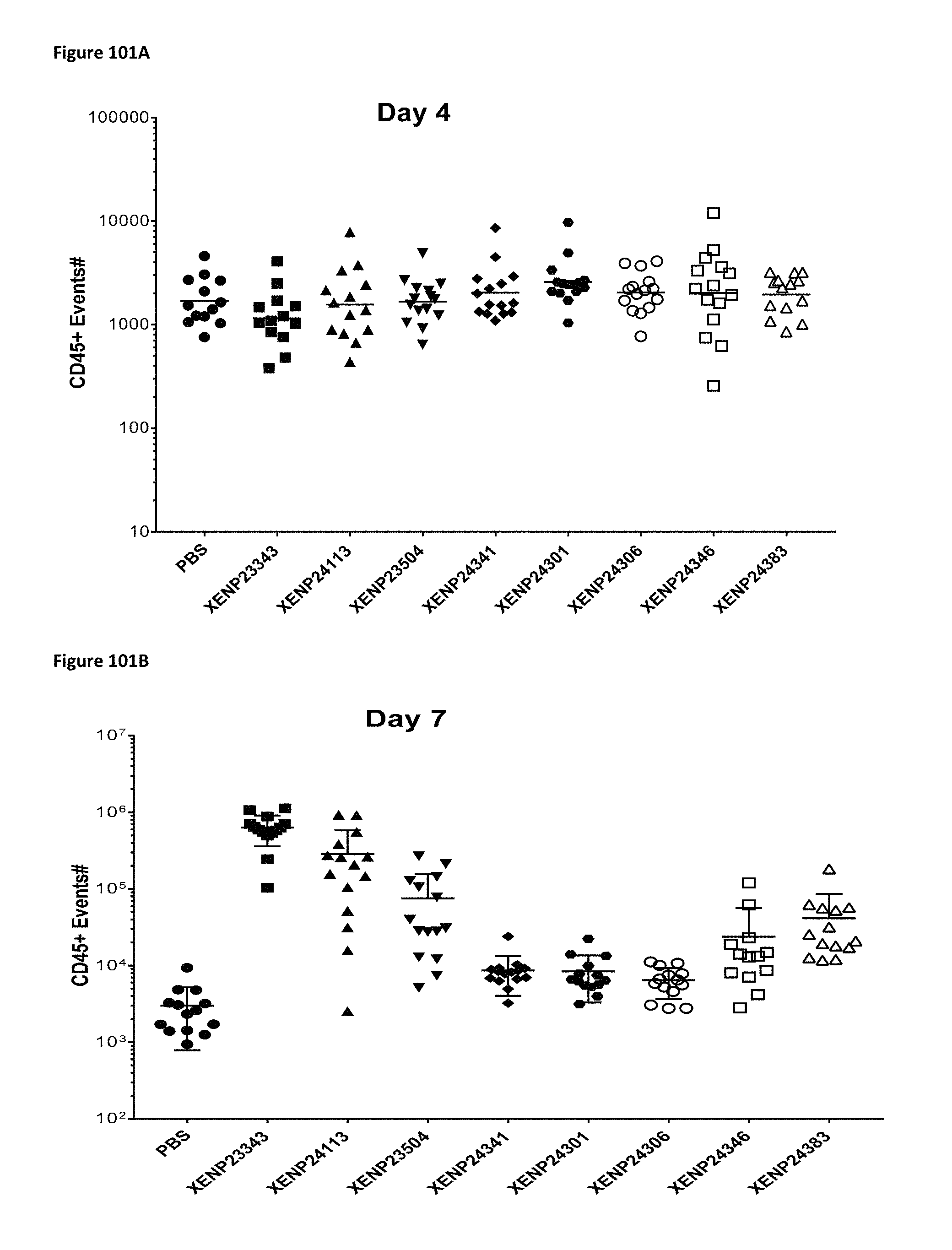
D00186

D00187
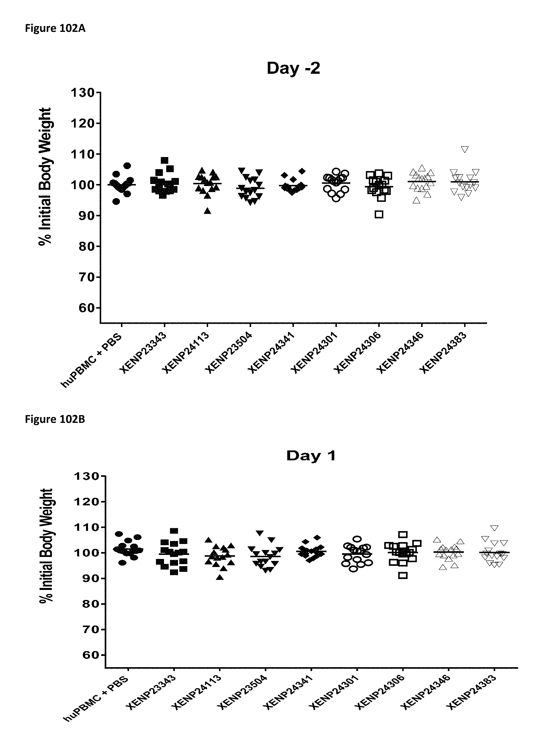
D00188

D00189
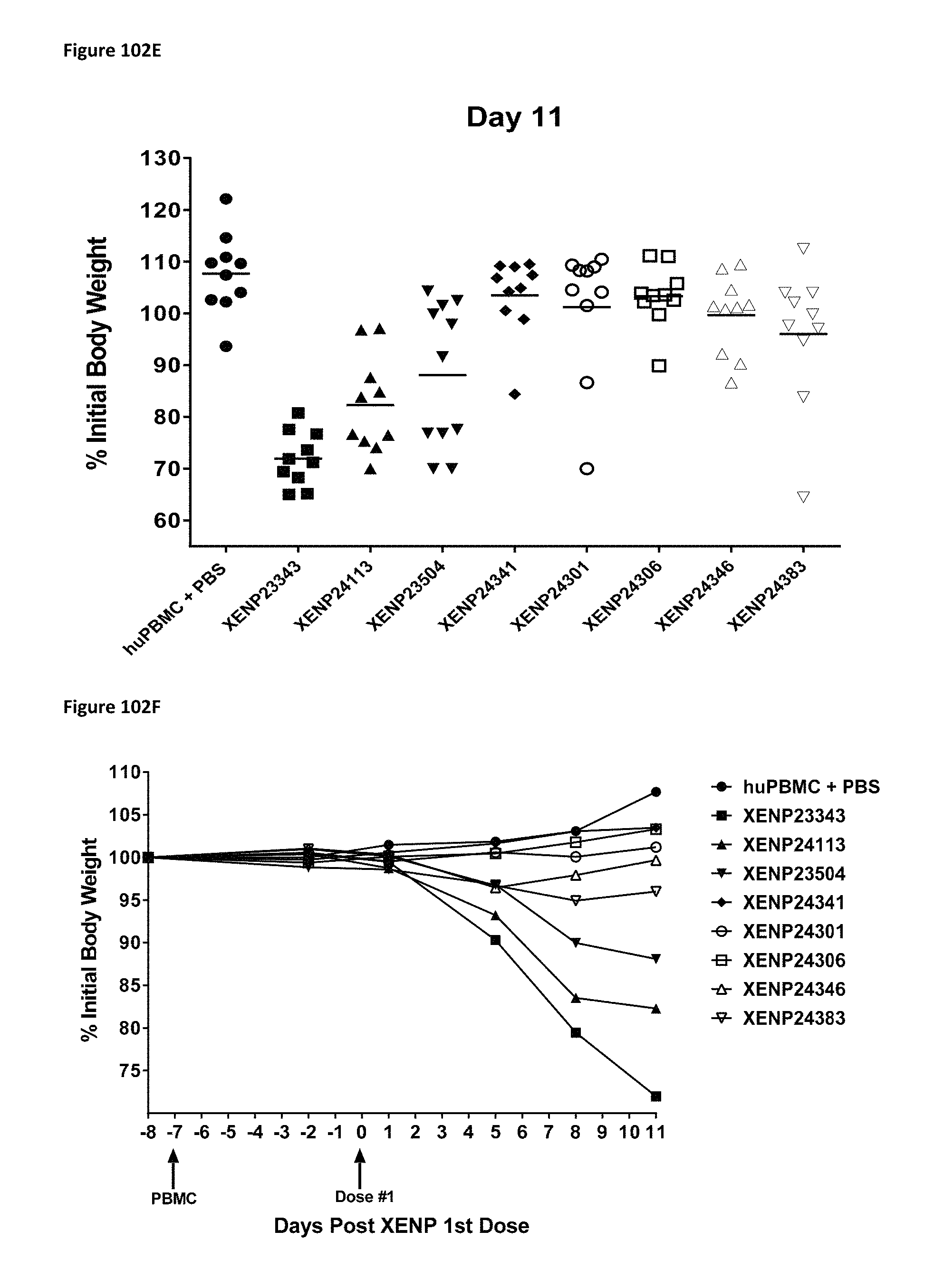
D00190
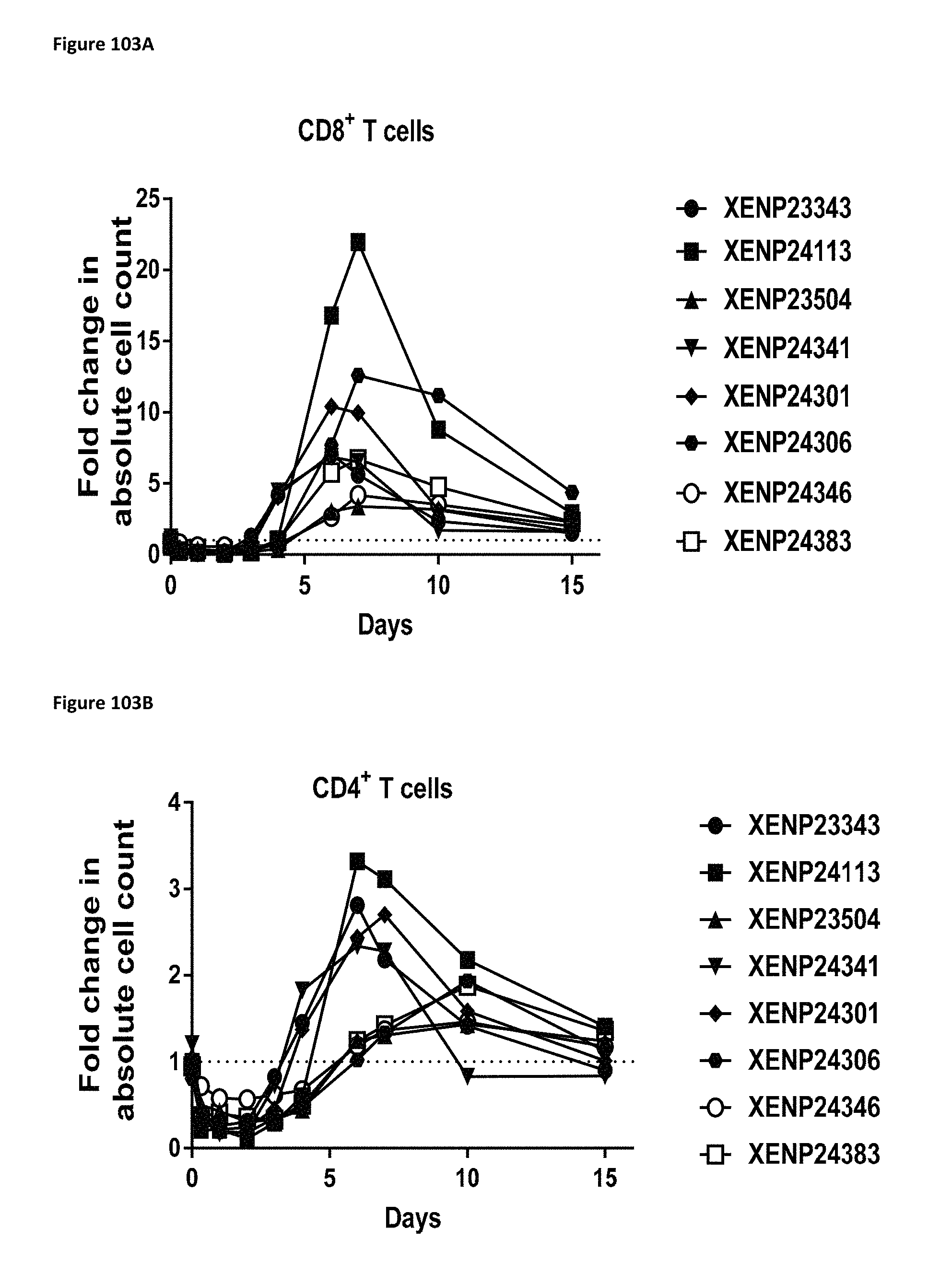
D00191

D00192
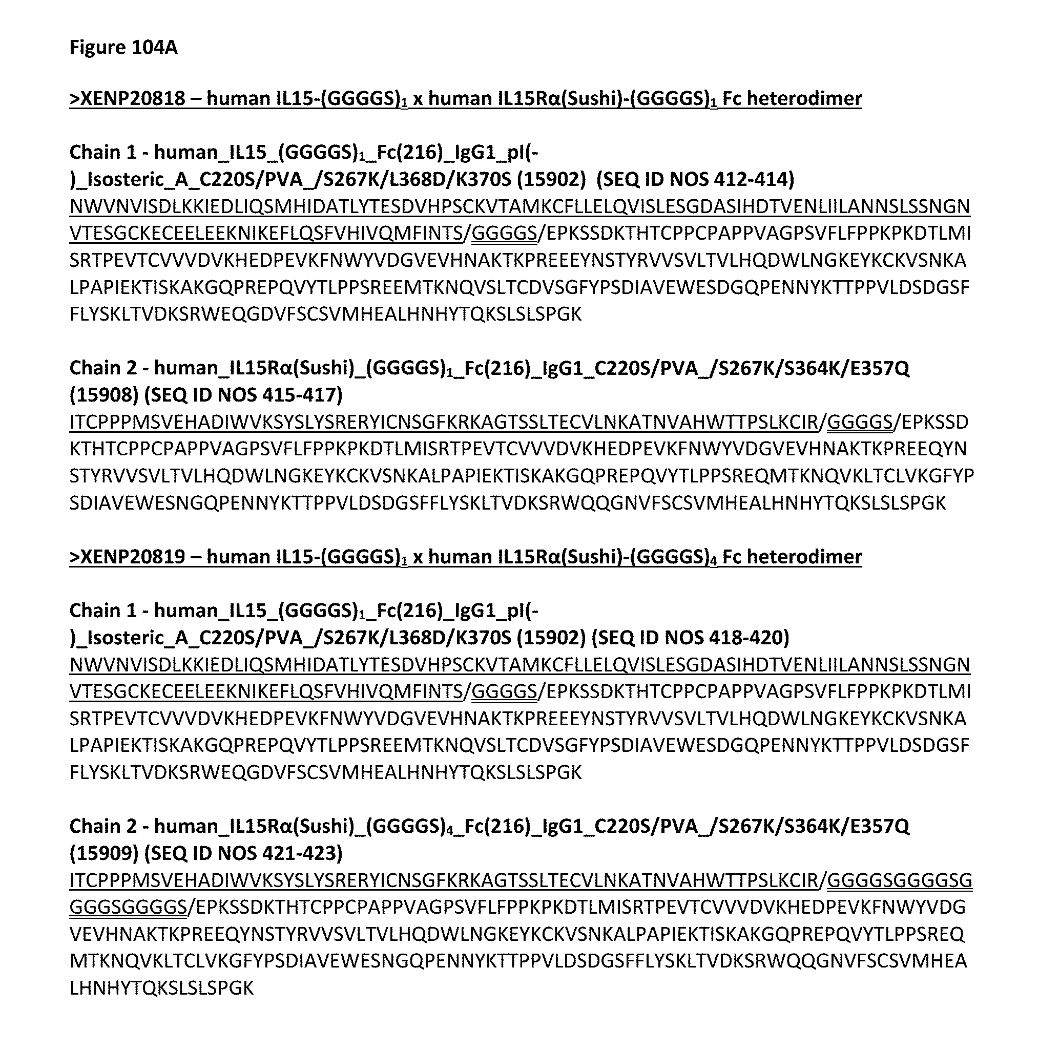
D00193

D00194

D00195
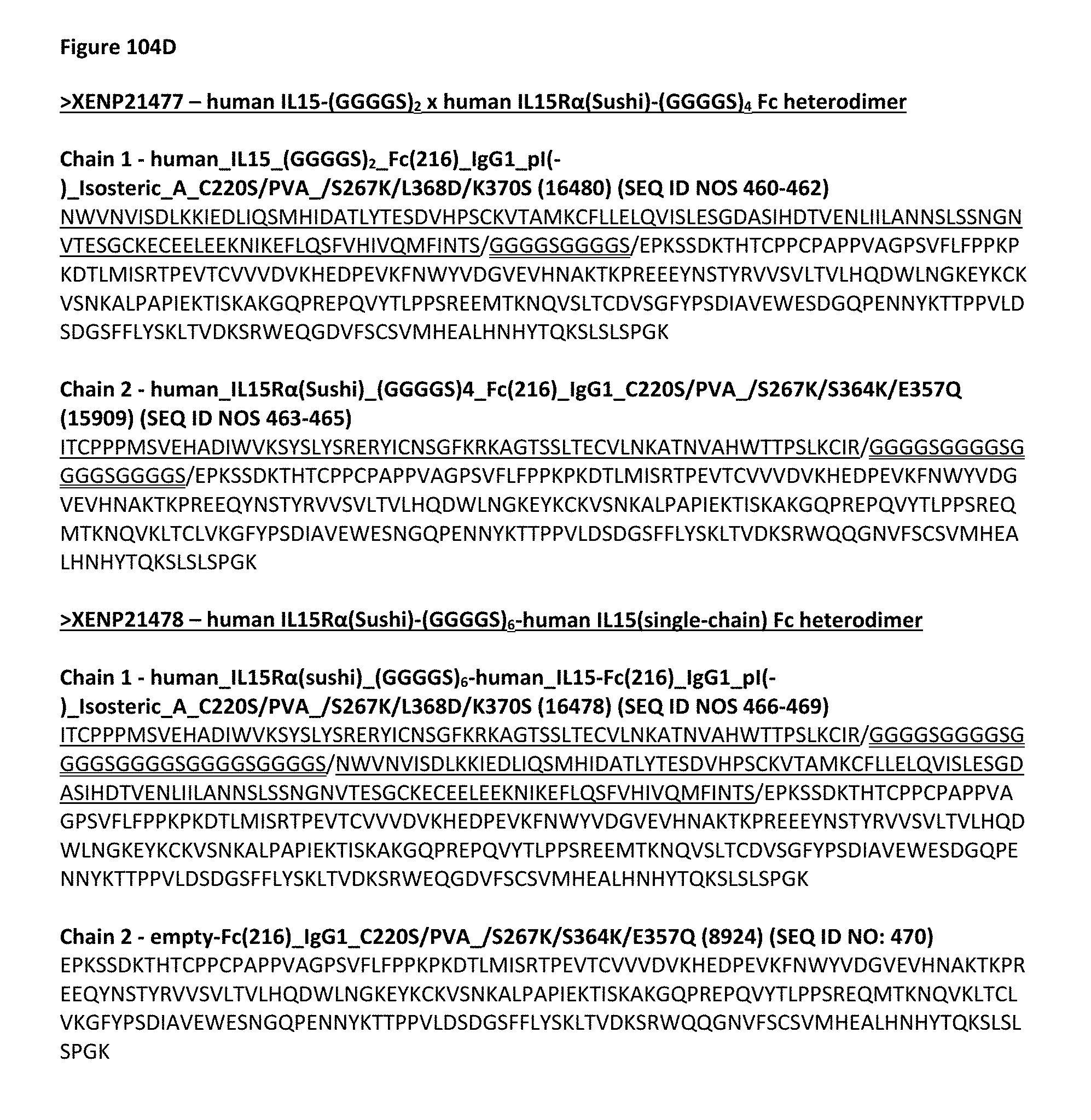
D00196

D00197

D00198

D00199

D00200

D00201

D00202

D00203

D00204
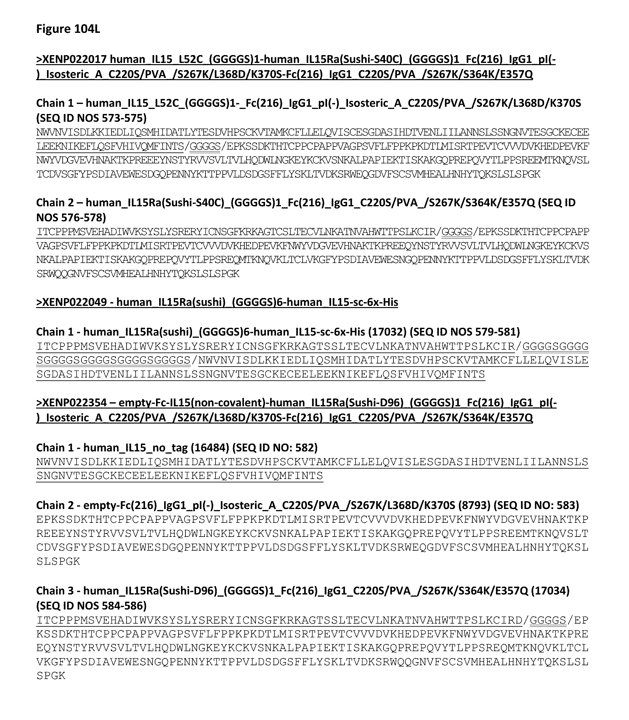
D00205

D00206
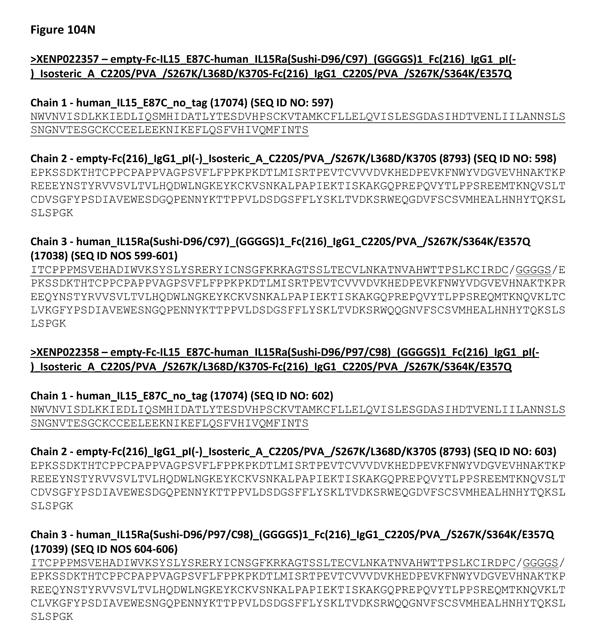
D00207

D00208

D00209
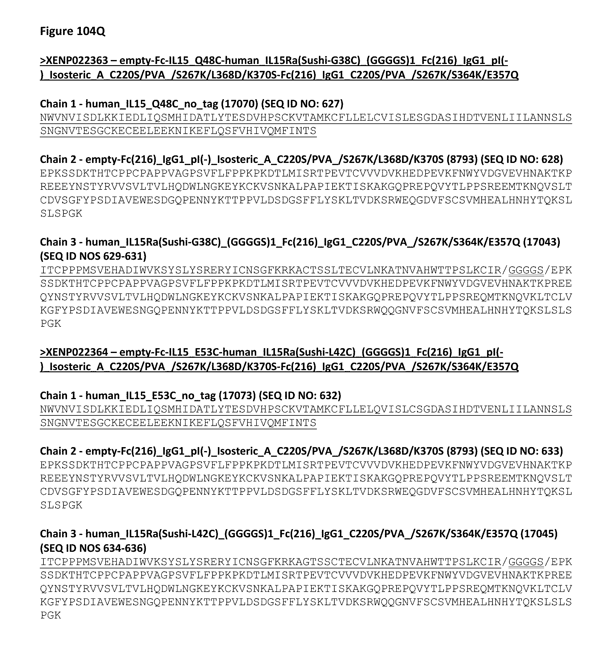
D00210

D00211
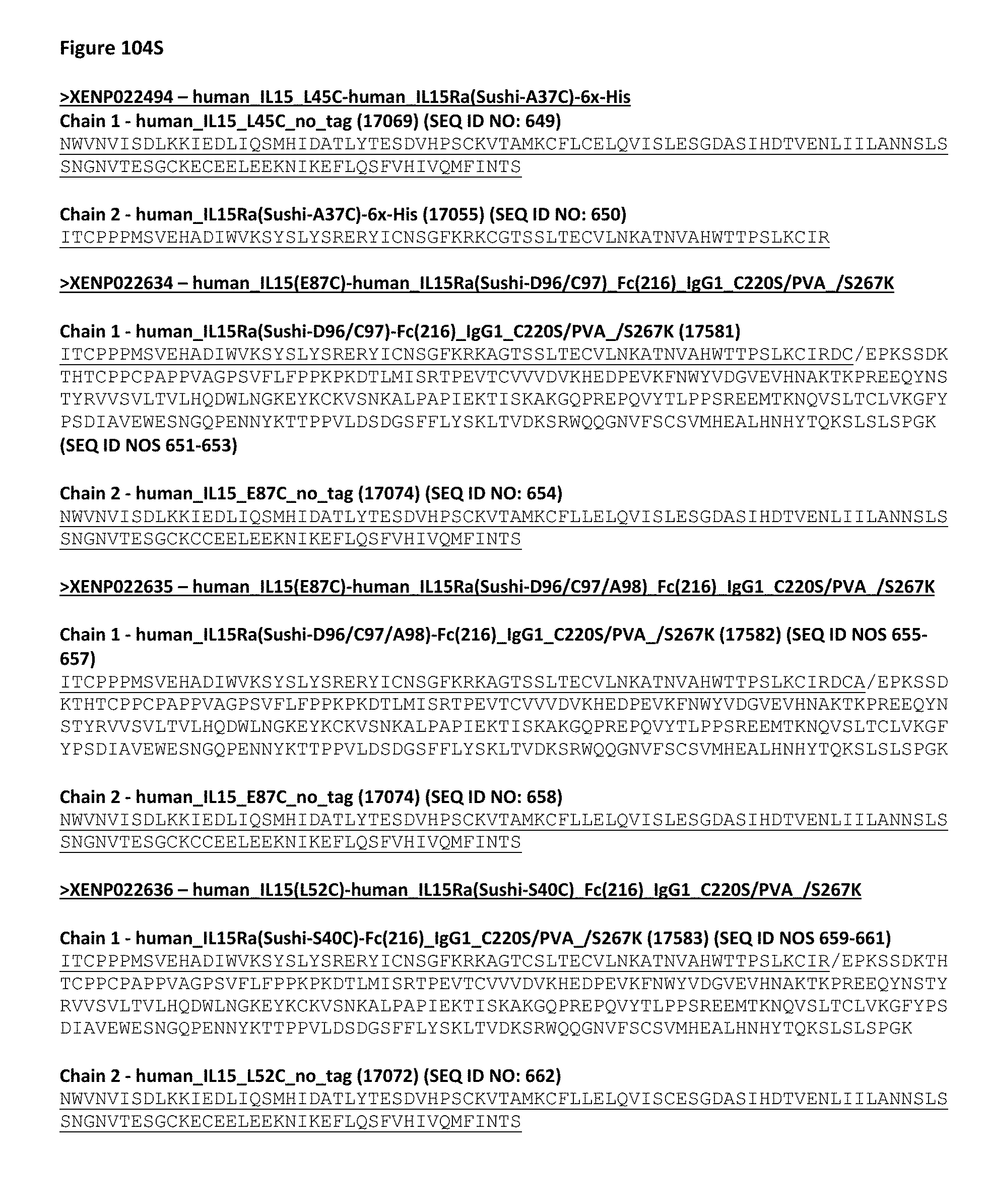
D00212

D00213
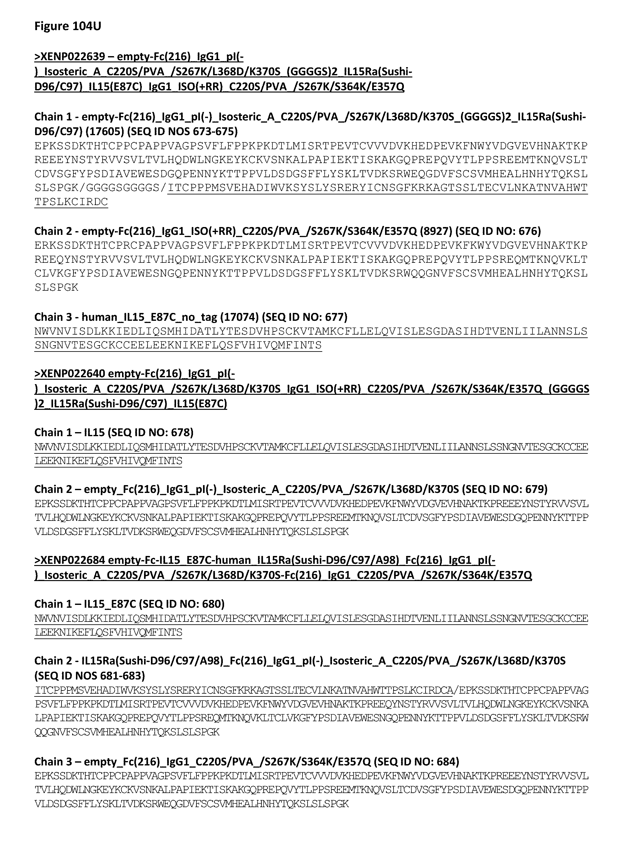
D00214

D00215
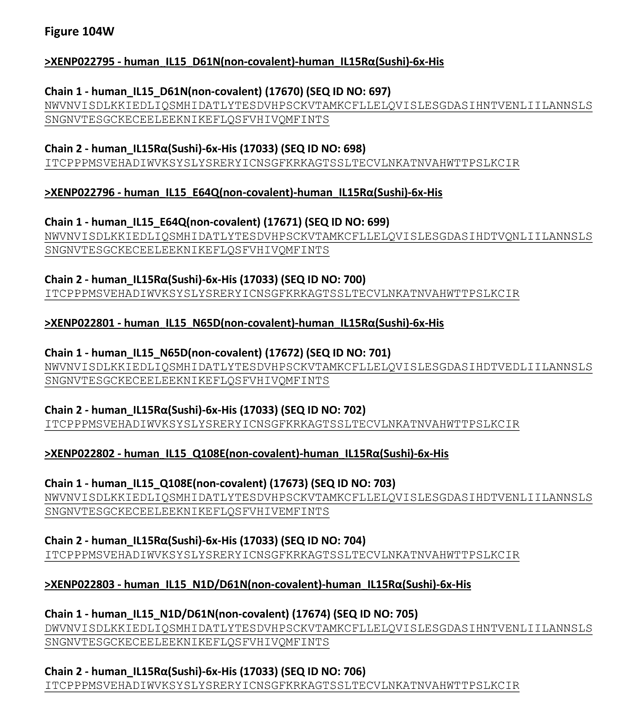
D00216
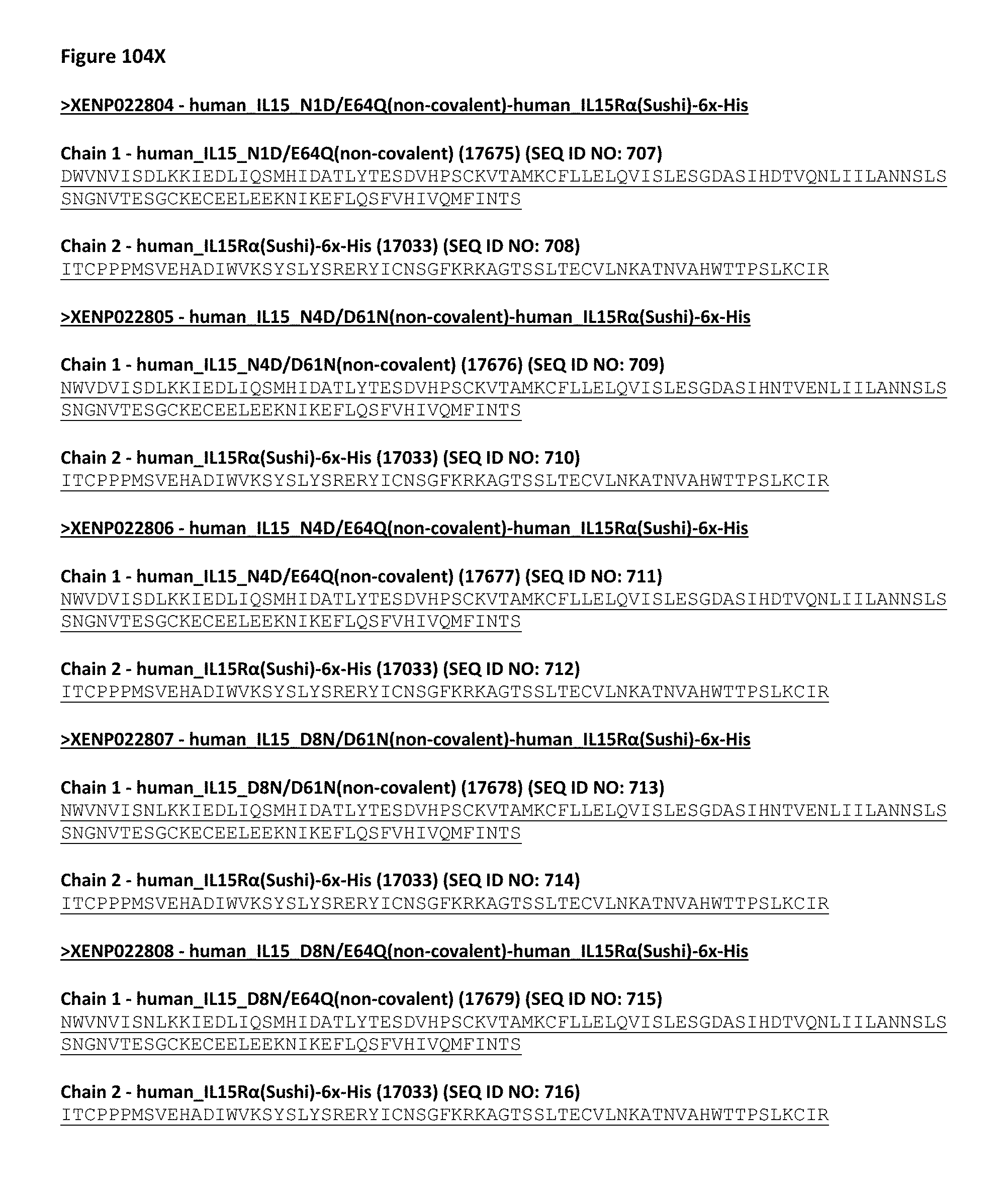
D00217

D00218
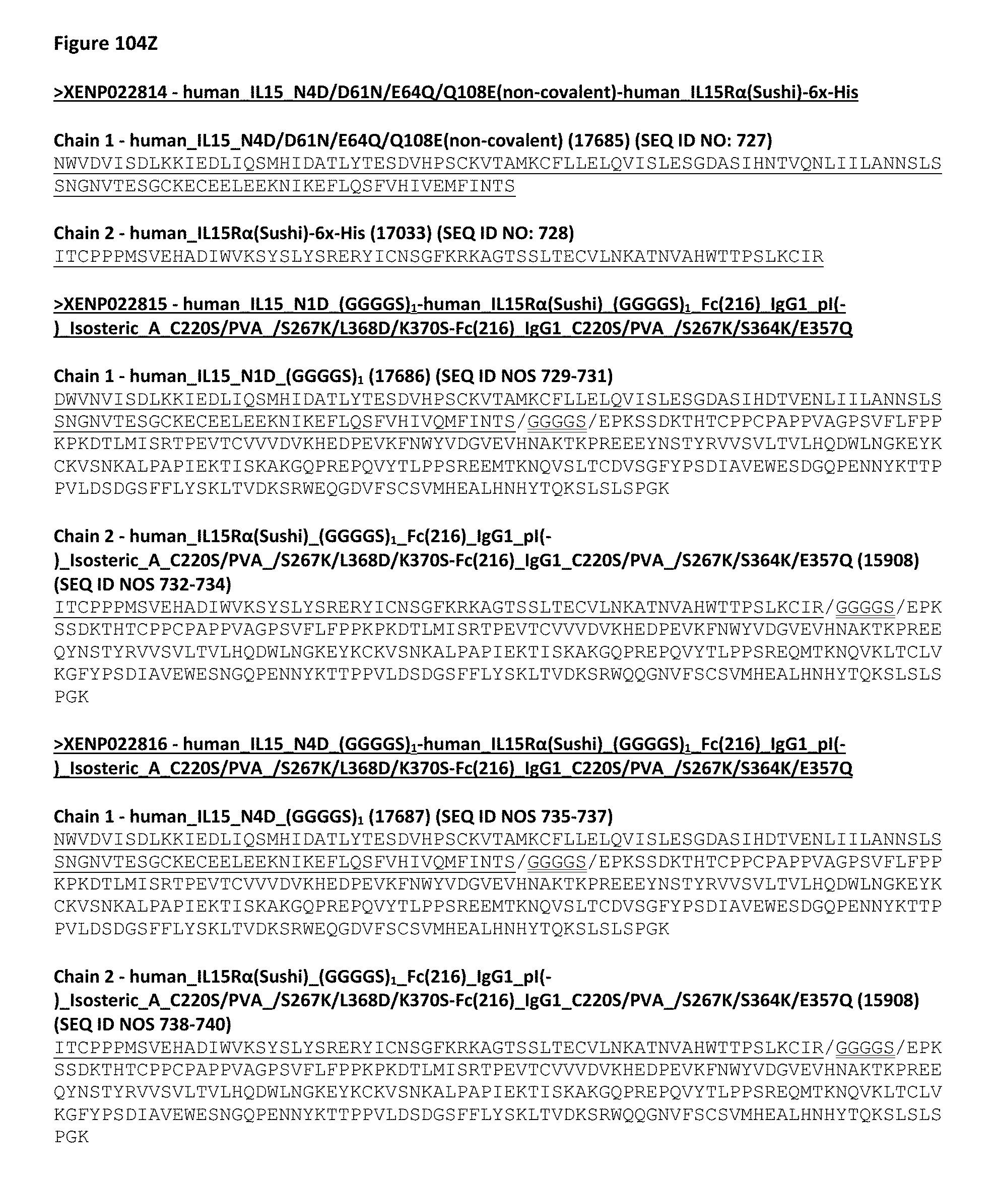
D00219
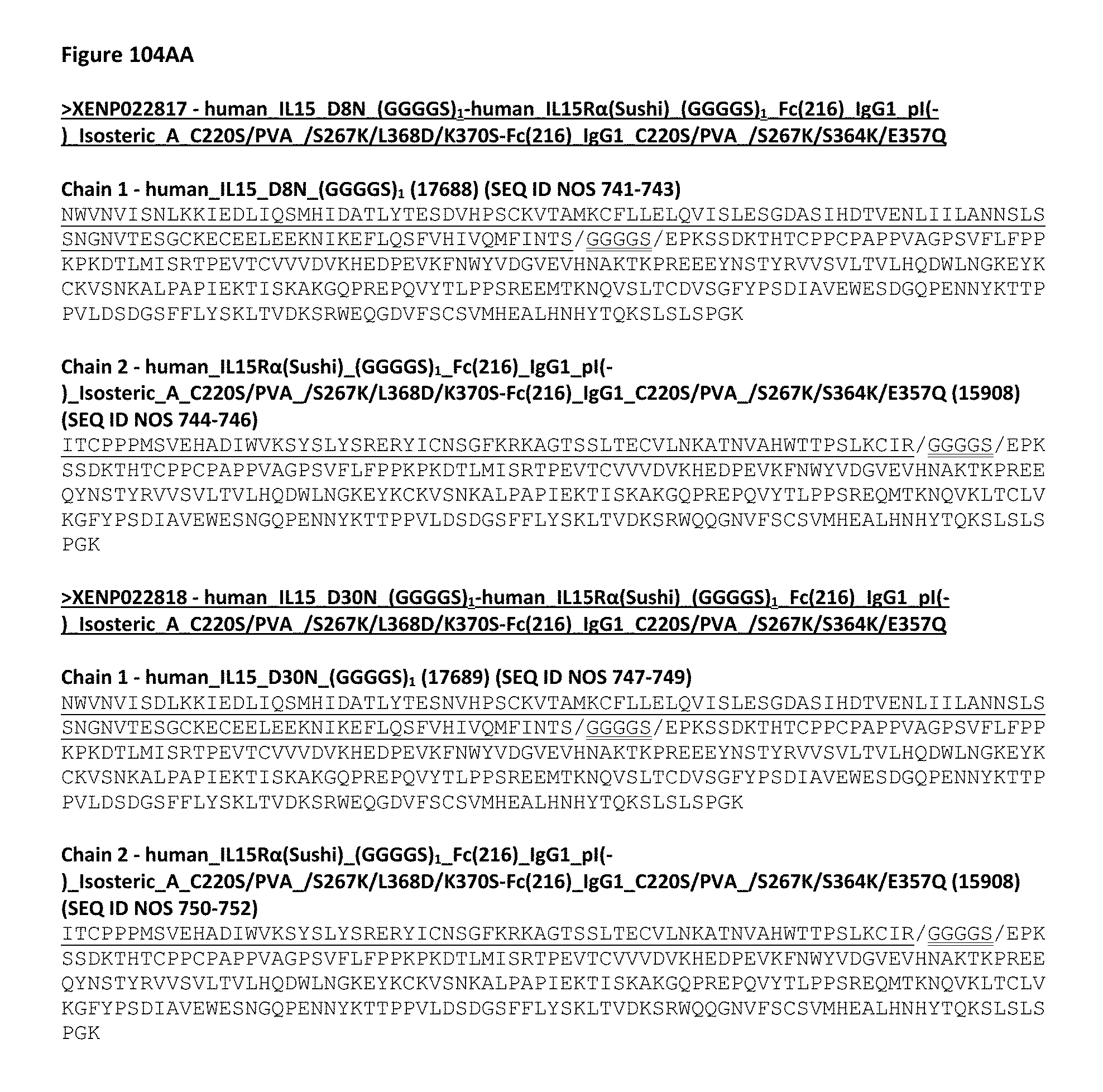
D00220
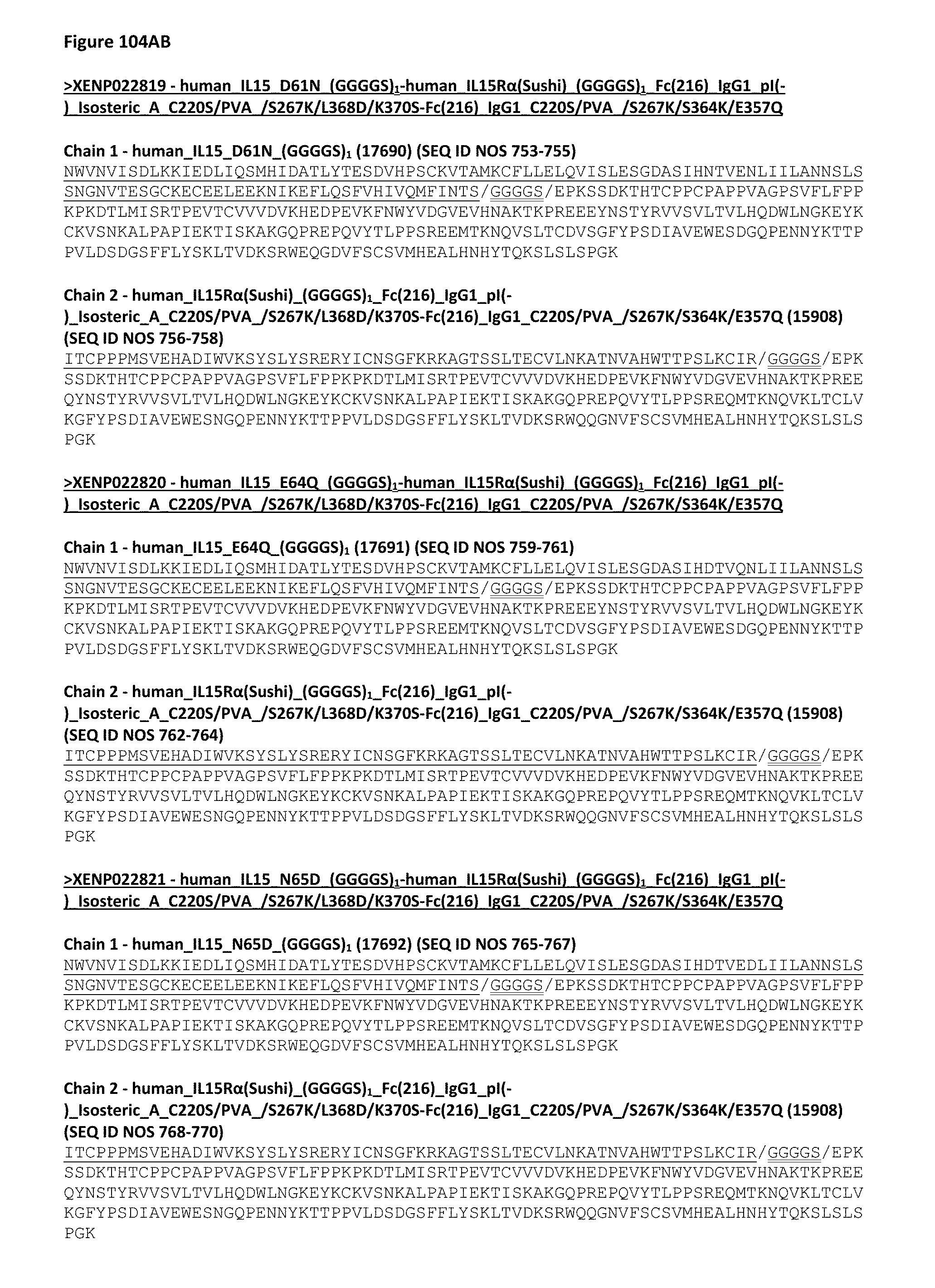
D00221

D00222
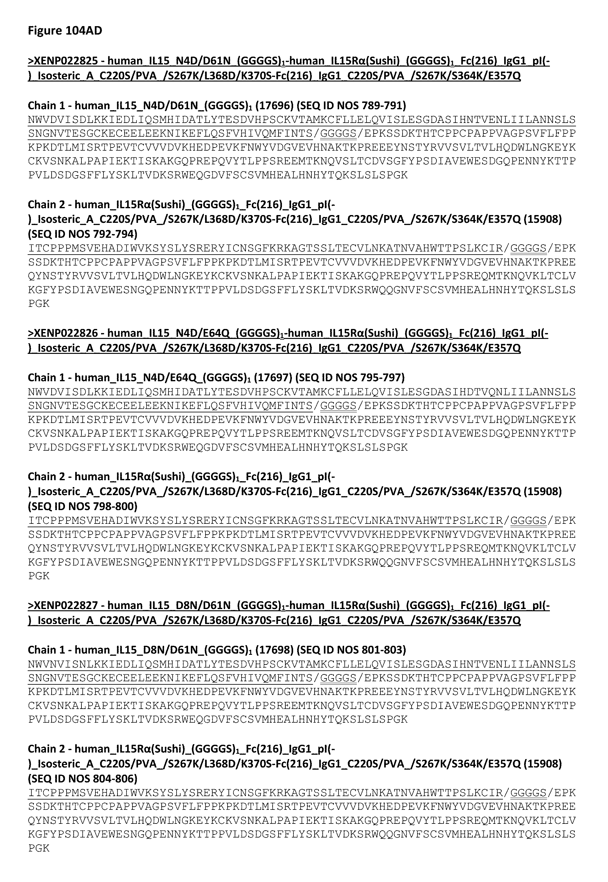
D00223

D00224

D00225
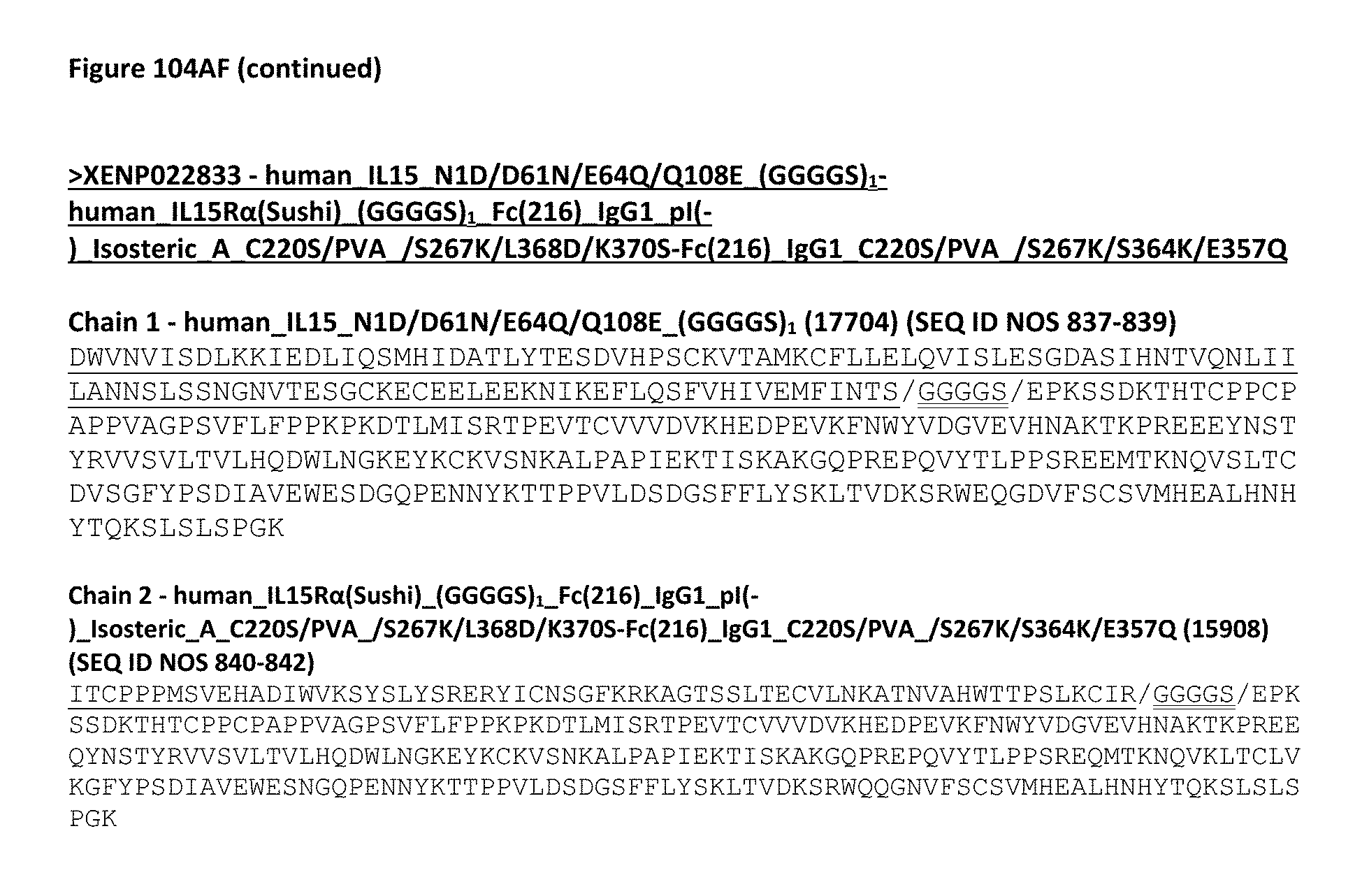
D00226
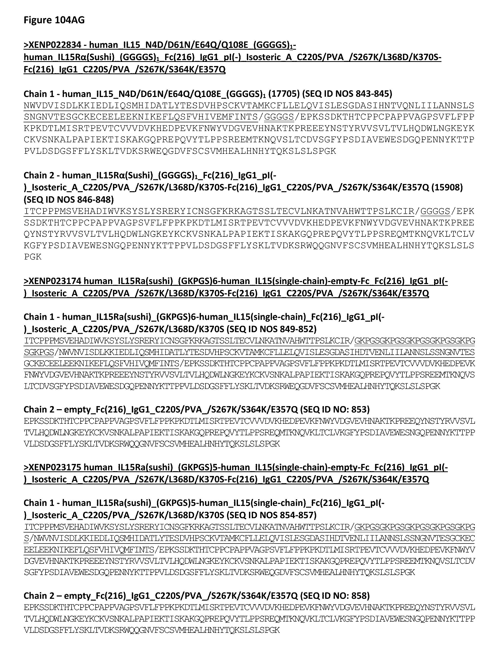
D00227
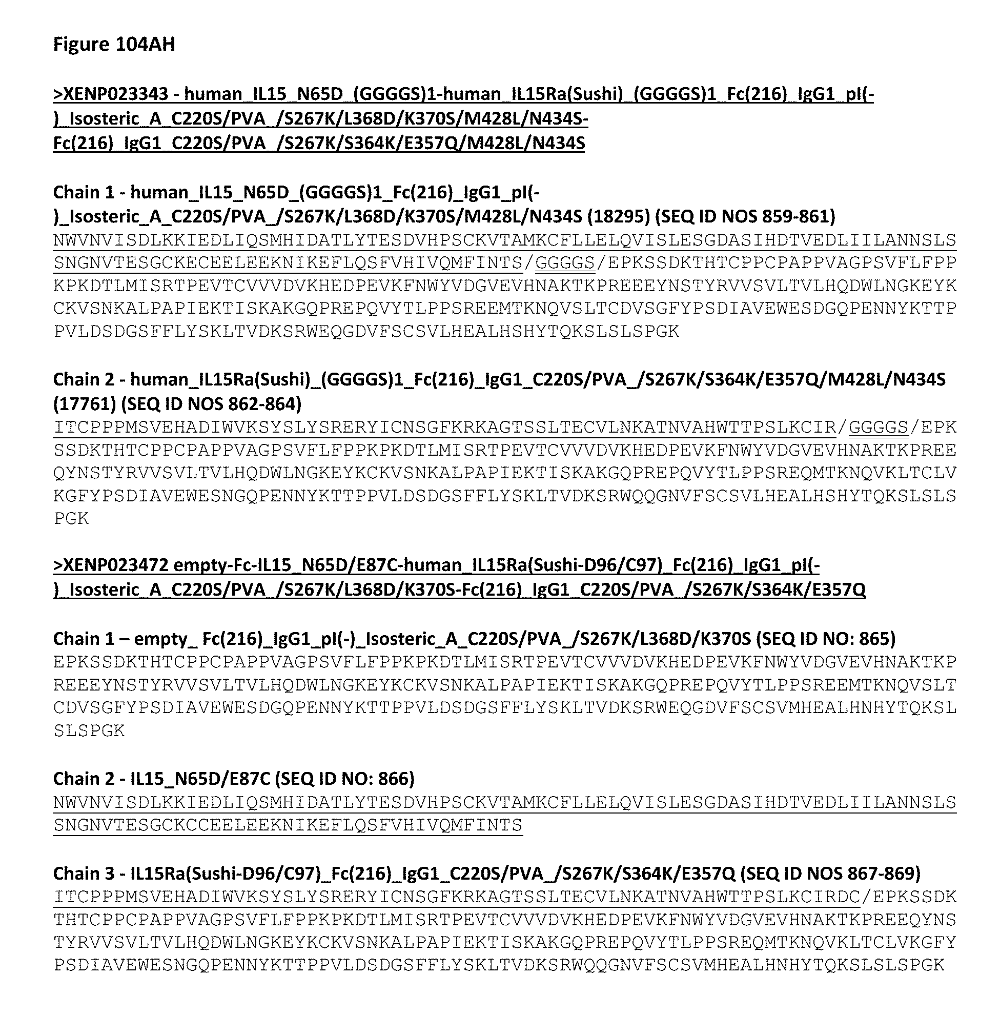
D00228
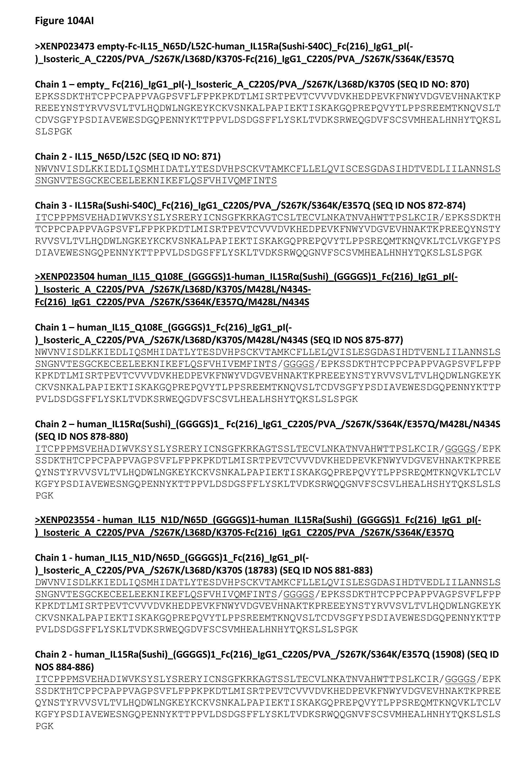
D00229

D00230
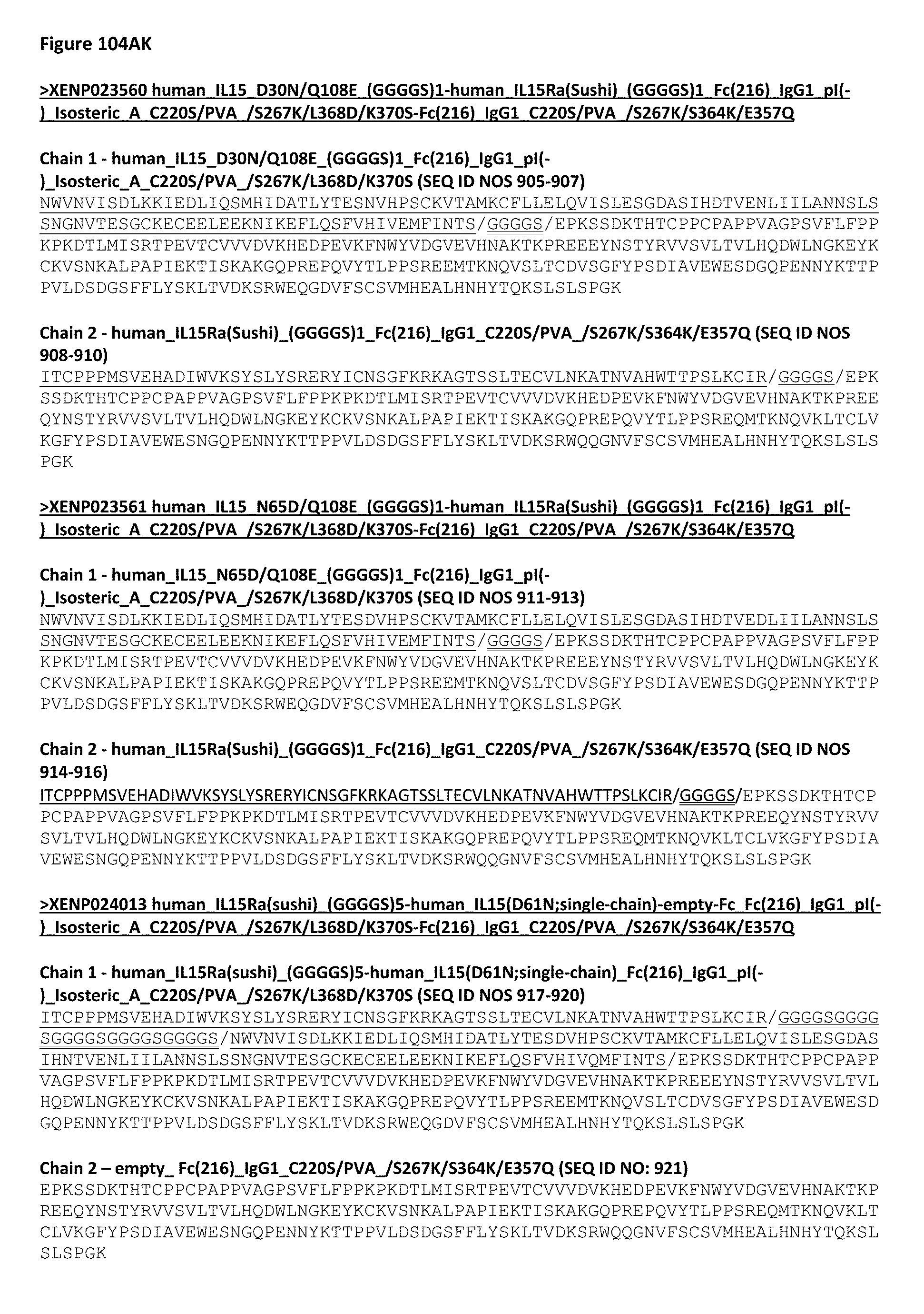
D00231
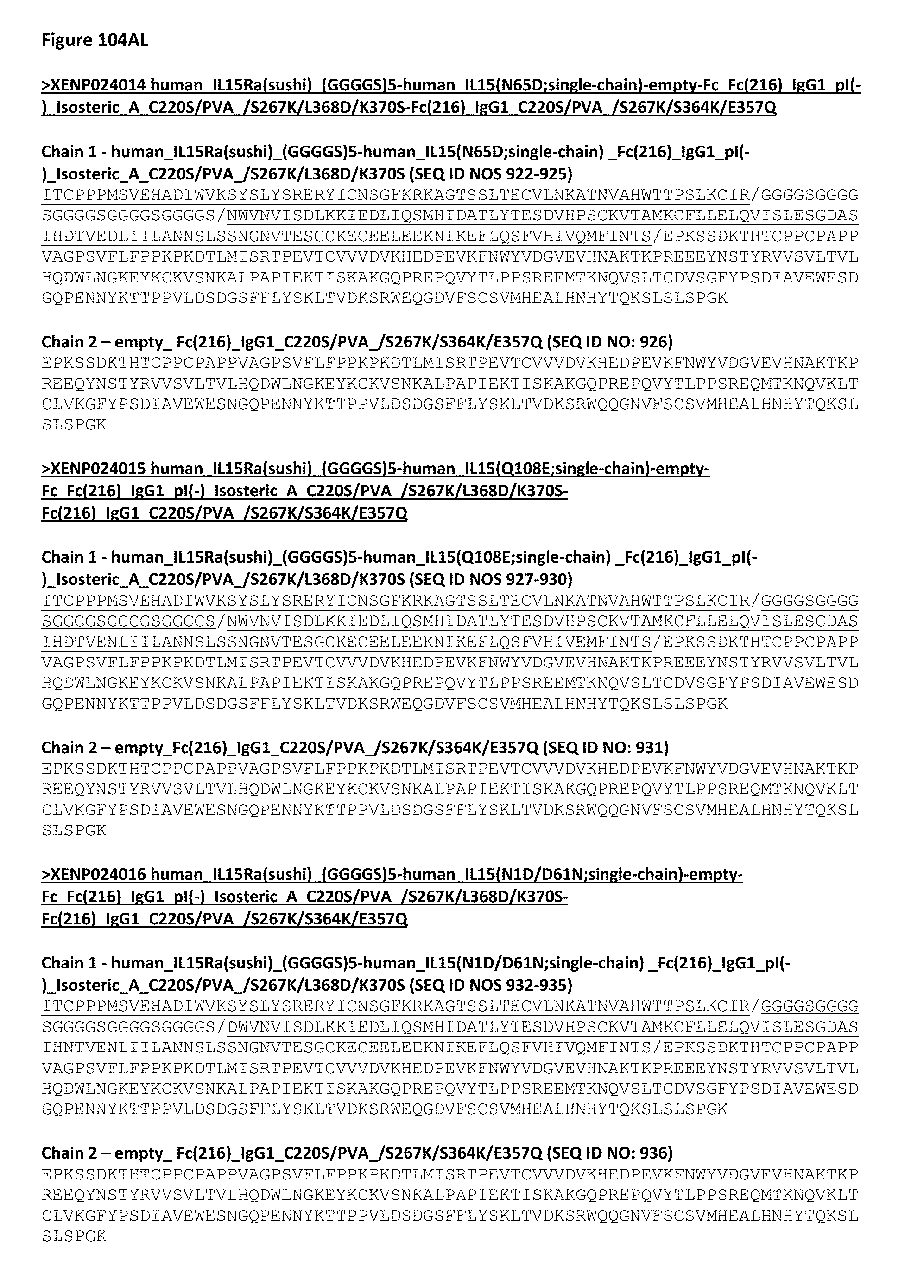
D00232

D00233
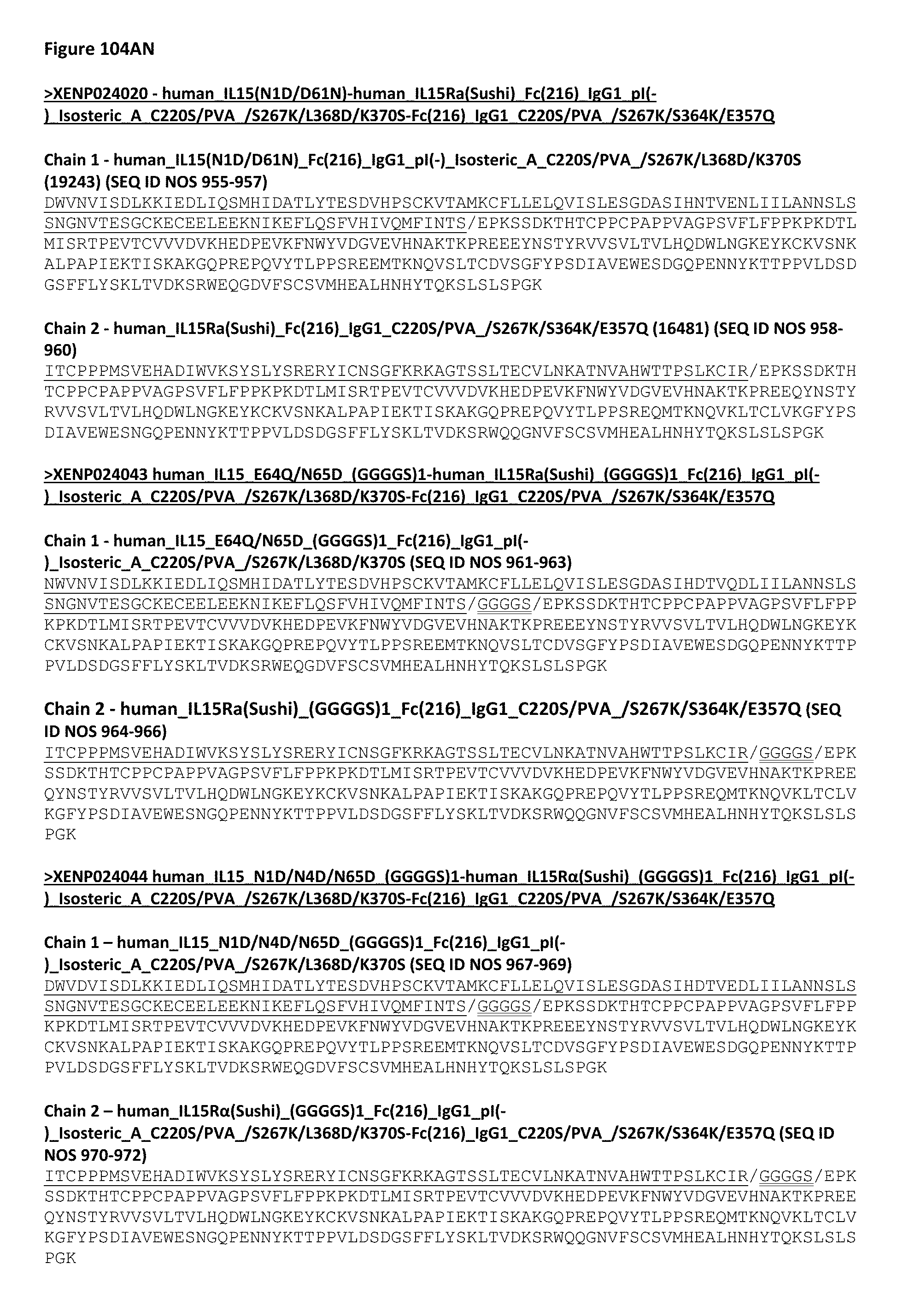
D00234
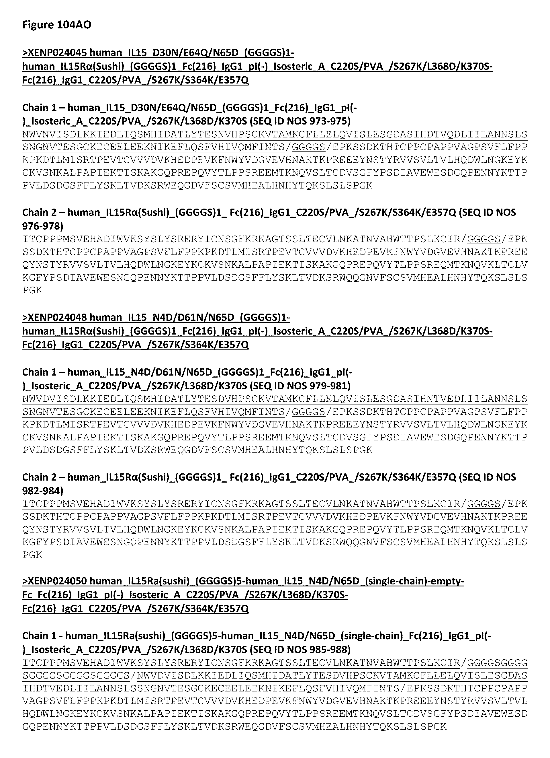
D00235
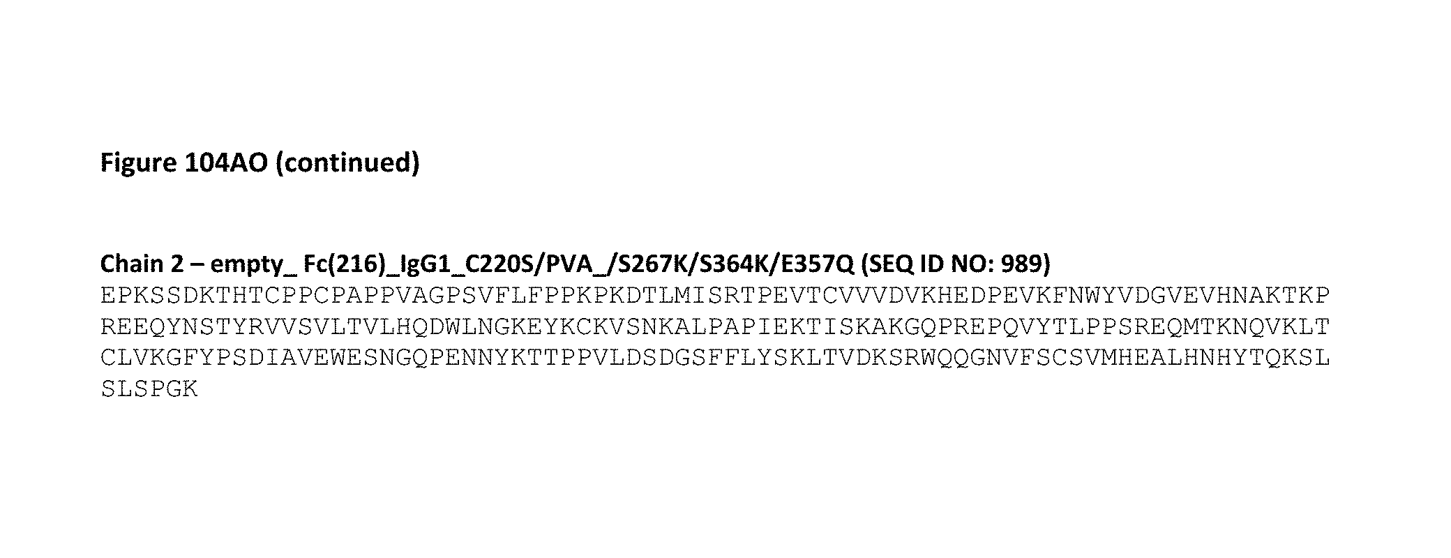
D00236

D00237
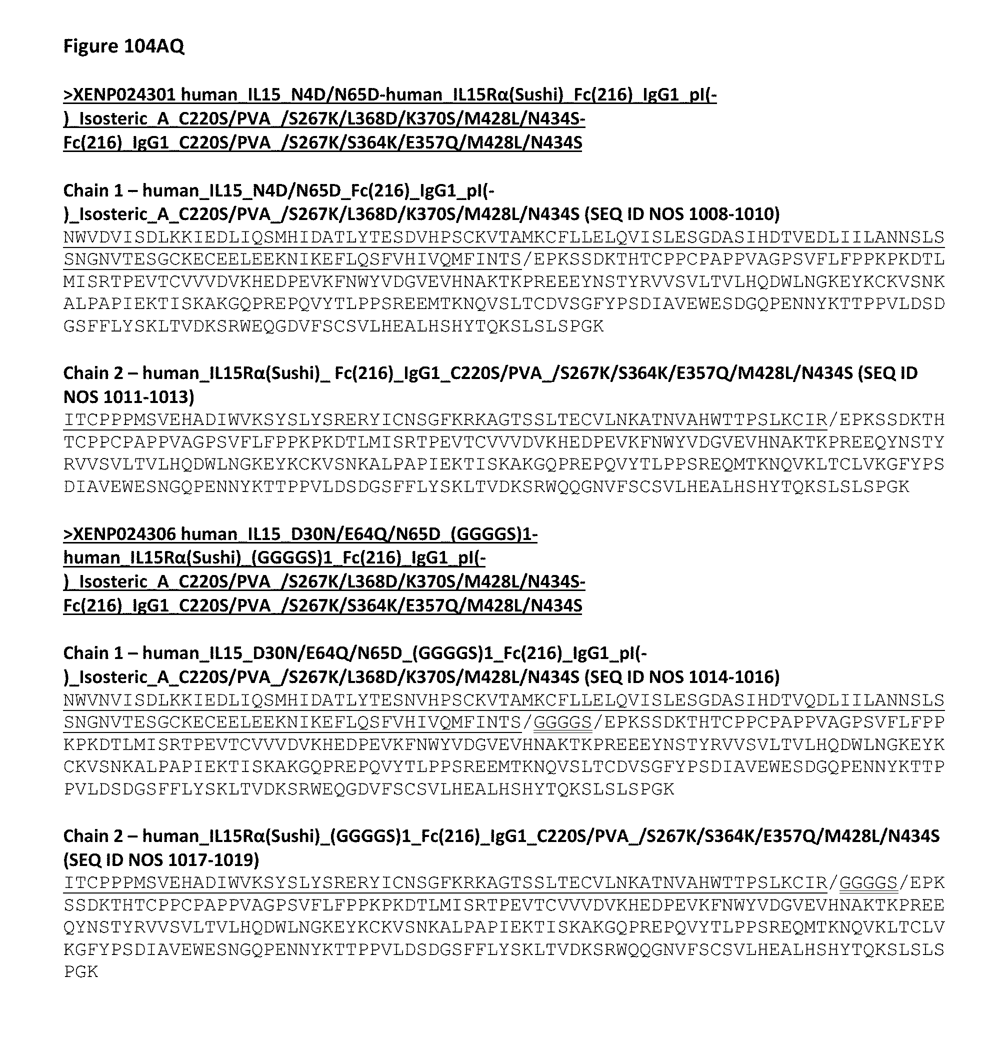
D00238

D00239

D00240
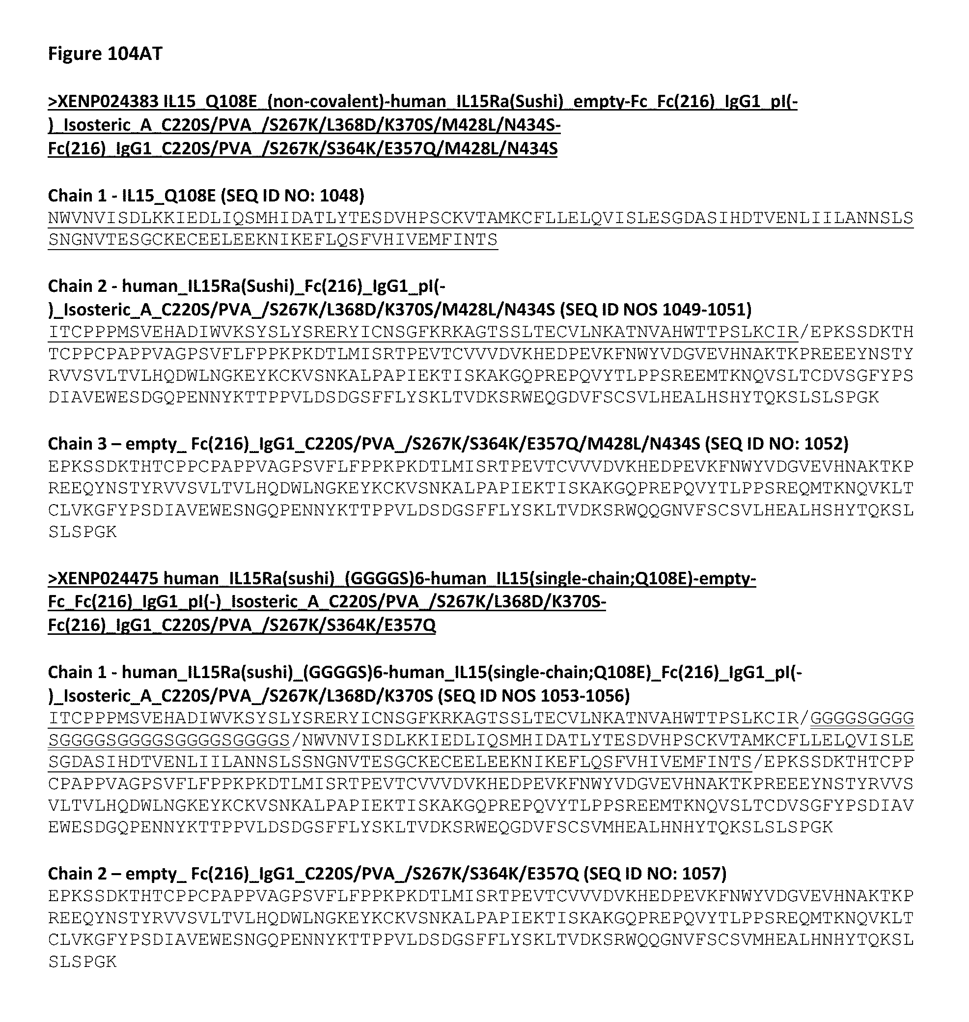
D00241
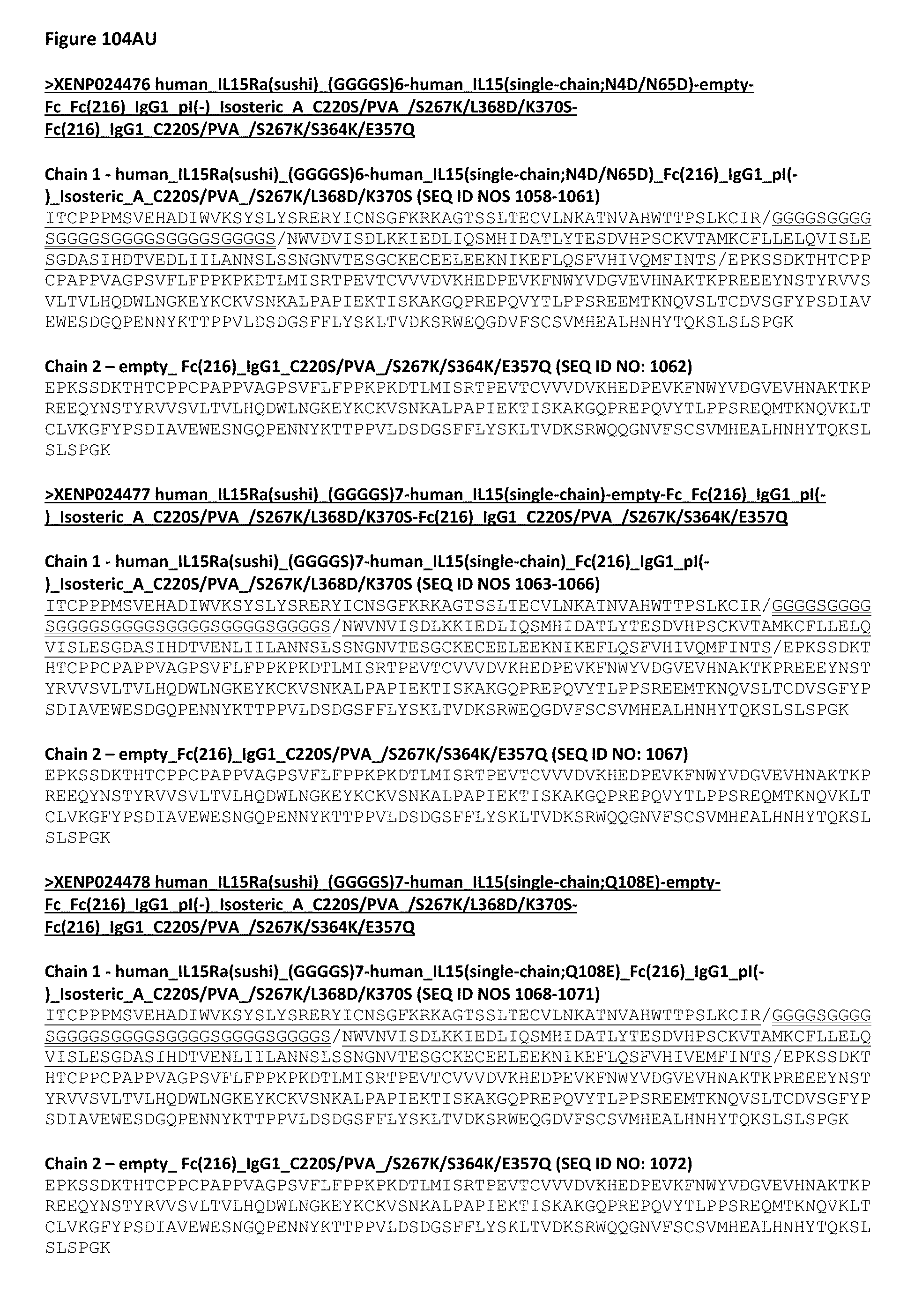
D00242

D00243
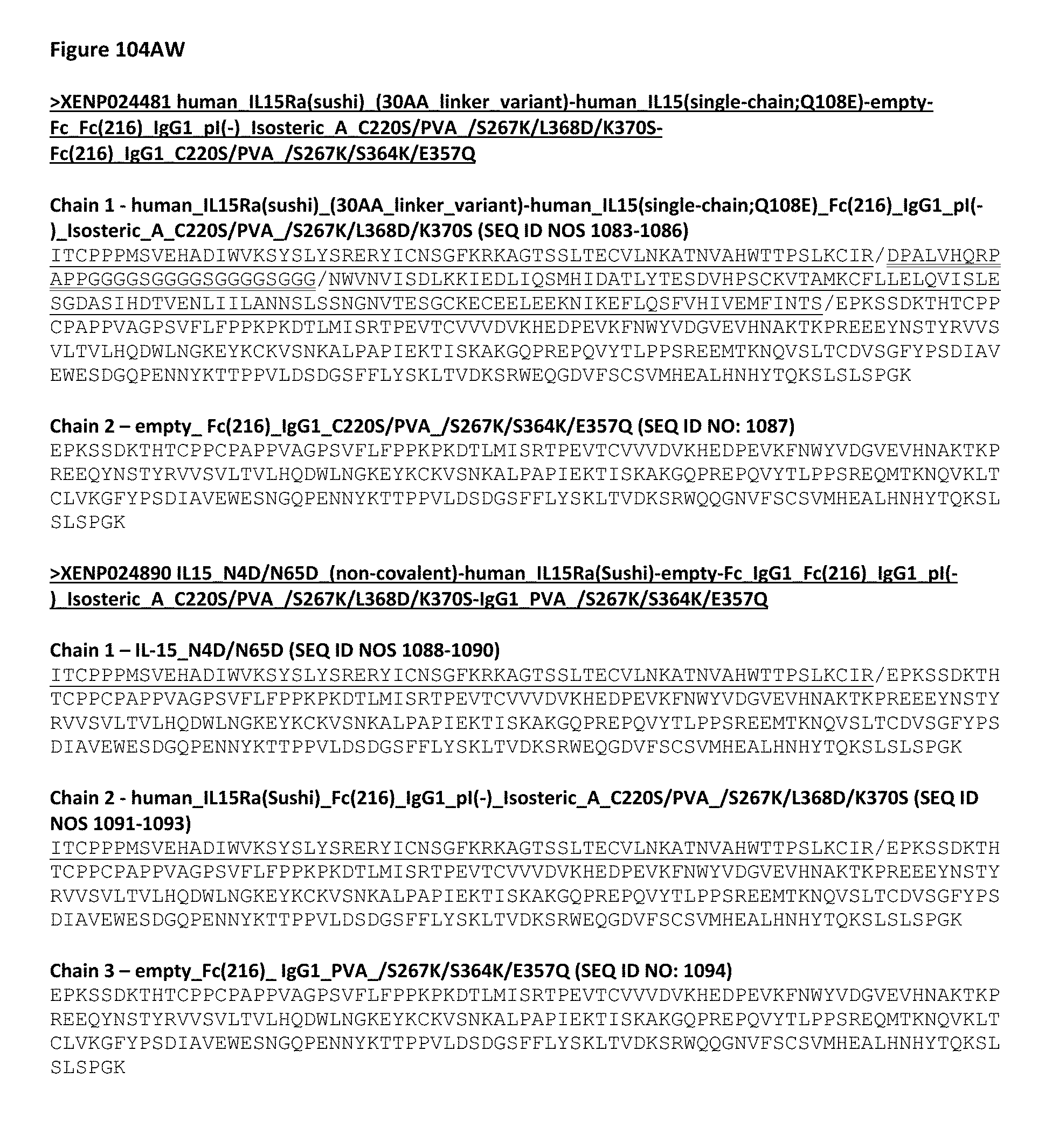
D00244

D00245
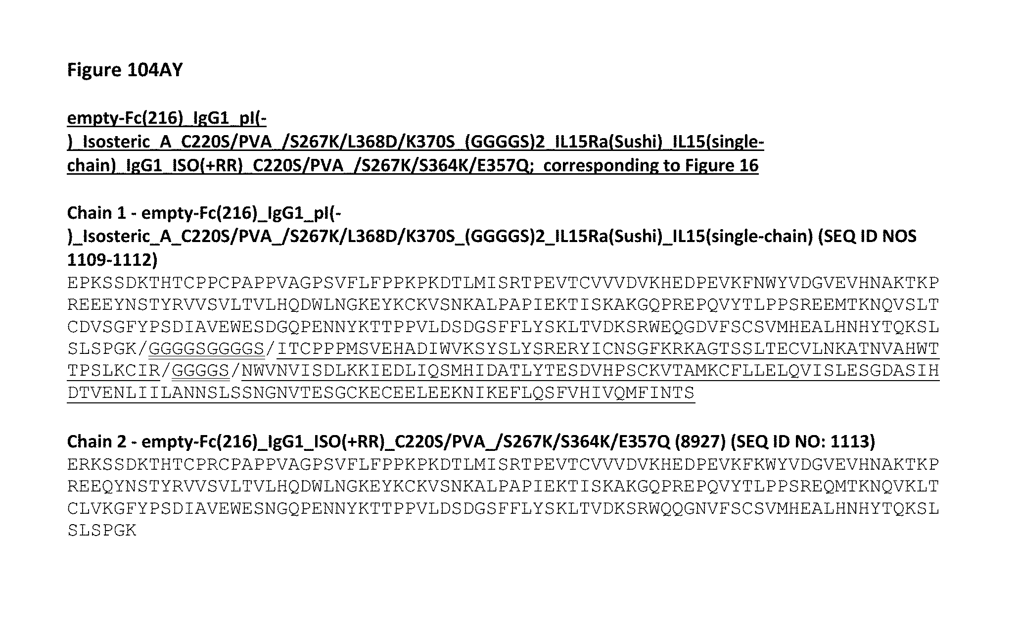
D00246

D00247
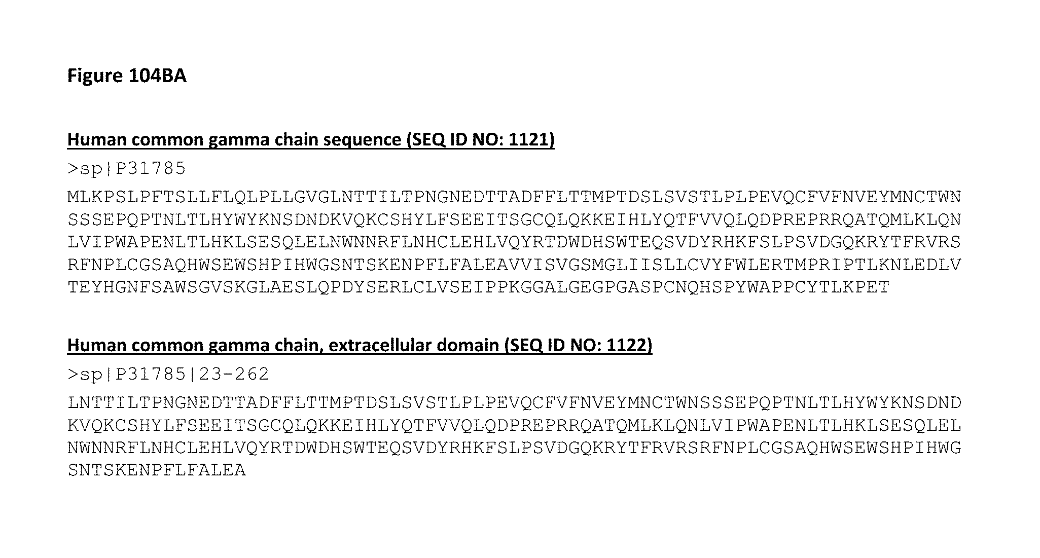
D00248
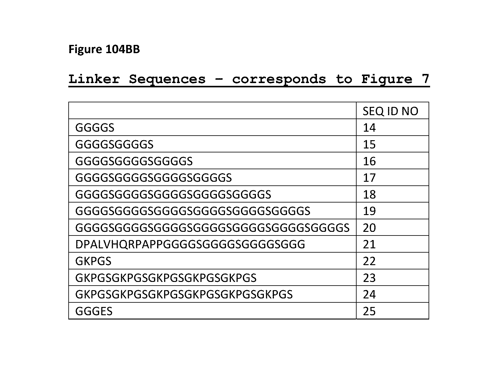
D00249
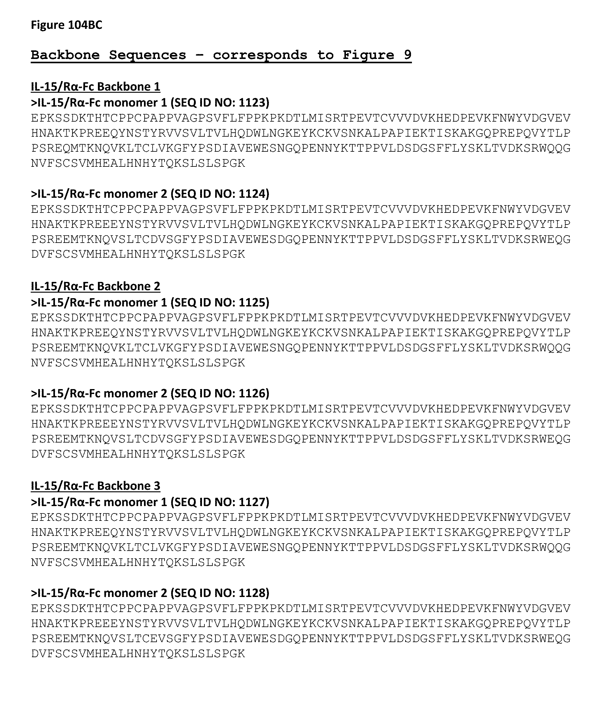
D00250

D00251
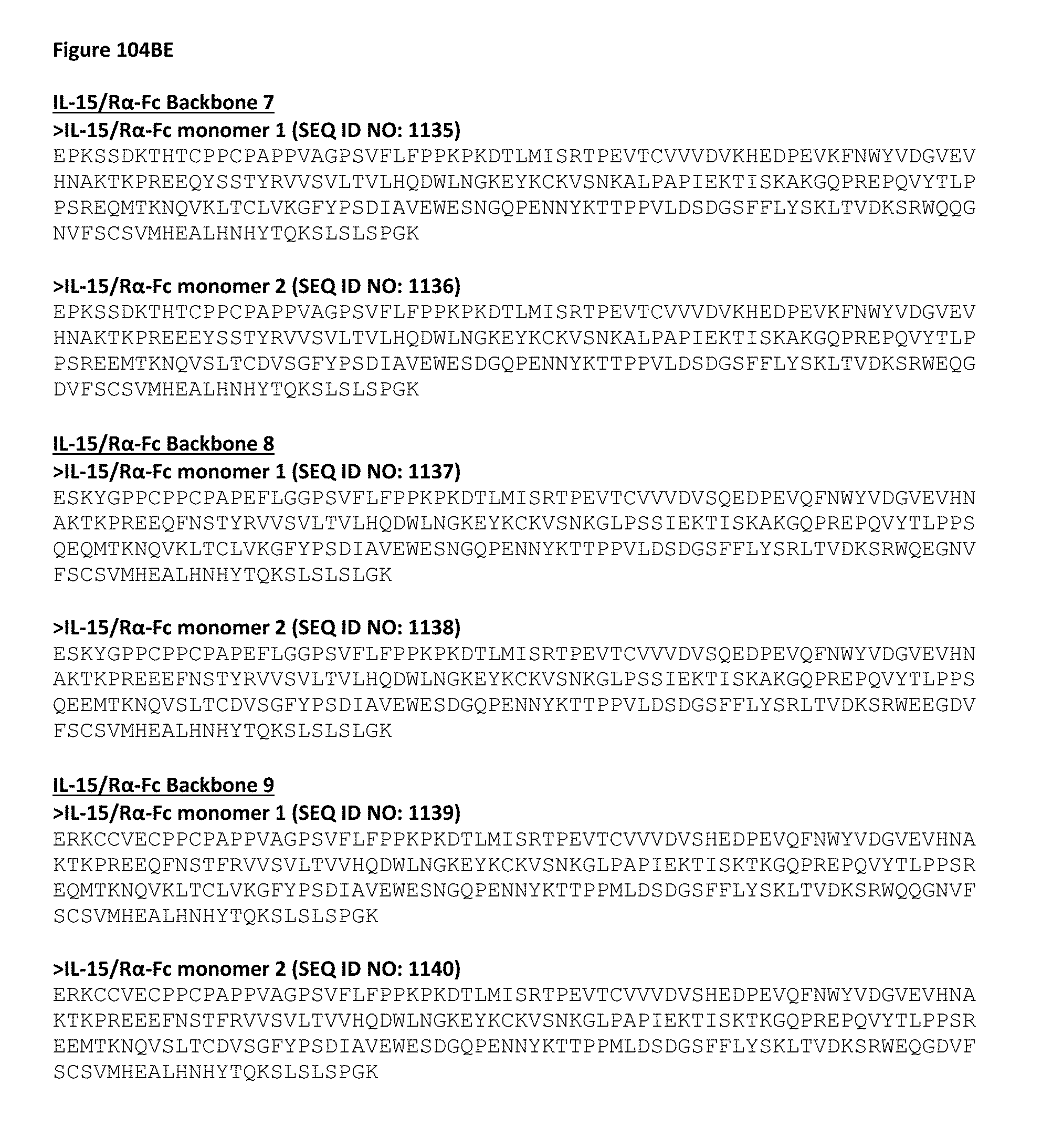
D00252

D00253

D00254

D00255

D00256

D00257

D00258
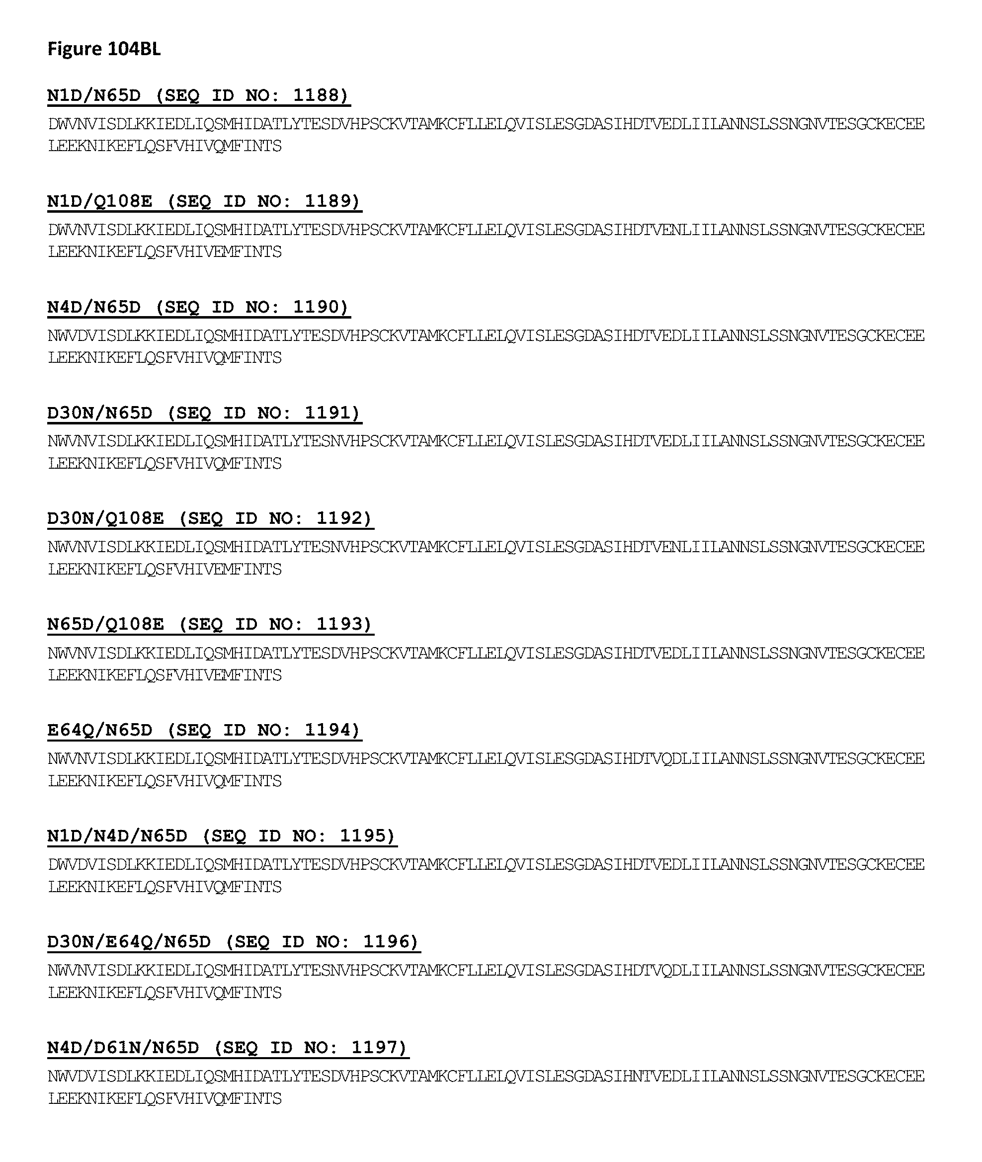
D00259

S00001
XML
uspto.report is an independent third-party trademark research tool that is not affiliated, endorsed, or sponsored by the United States Patent and Trademark Office (USPTO) or any other governmental organization. The information provided by uspto.report is based on publicly available data at the time of writing and is intended for informational purposes only.
While we strive to provide accurate and up-to-date information, we do not guarantee the accuracy, completeness, reliability, or suitability of the information displayed on this site. The use of this site is at your own risk. Any reliance you place on such information is therefore strictly at your own risk.
All official trademark data, including owner information, should be verified by visiting the official USPTO website at www.uspto.gov. This site is not intended to replace professional legal advice and should not be used as a substitute for consulting with a legal professional who is knowledgeable about trademark law.