Patient-specific femoroacetabular impingement instruments and methods
Meridew , et al. Oc
U.S. patent number 10,456,205 [Application Number 15/179,218] was granted by the patent office on 2019-10-29 for patient-specific femoroacetabular impingement instruments and methods. This patent grant is currently assigned to Biomet Manufacturing, LLC. The grantee listed for this patent is Biomet Manufacturing, LLC. Invention is credited to Kristoff Corten, Jason D. Meridew, Paige Scott.


View All Diagrams
| United States Patent | 10,456,205 |
| Meridew , et al. | October 29, 2019 |
| **Please see images for: ( Certificate of Correction ) ** |
Patient-specific femoroacetabular impingement instruments and methods
Abstract
A device for a patient-specific acetabular and/or femoral guide. The guides can be used in a selected resection of at least one of a femur and an acetabulum to increase a range of motion of the femur relative to the acetabulum. Generally, a natural acetabulum and femoral head are maintained.
| Inventors: | Meridew; Jason D. (Warsaw, IN), Scott; Paige (West Lafayette, IN), Corten; Kristoff (Herselt, BE) | ||||||||||
|---|---|---|---|---|---|---|---|---|---|---|---|
| Applicant: |
|
||||||||||
| Assignee: | Biomet Manufacturing, LLC
(Warsaw, IN) |
||||||||||
| Family ID: | 47993307 | ||||||||||
| Appl. No.: | 15/179,218 | ||||||||||
| Filed: | June 10, 2016 |
Prior Publication Data
| Document Identifier | Publication Date | |
|---|---|---|
| US 20160278793 A1 | Sep 29, 2016 | |
Related U.S. Patent Documents
| Application Number | Filing Date | Patent Number | Issue Date | ||
|---|---|---|---|---|---|
| 13627626 | Sep 26, 2012 | 9386993 | |||
| 61540857 | Sep 29, 2011 | ||||
| Current U.S. Class: | 1/1 |
| Current CPC Class: | A61B 34/10 (20160201); A61B 17/1668 (20130101); A61B 17/175 (20130101); A61B 17/15 (20130101); A61B 17/1666 (20130101); A61B 17/1746 (20130101); A61B 34/00 (20160201); A61B 2034/105 (20160201); A61B 2034/102 (20160201); A61B 2017/568 (20130101); A61B 2034/108 (20160201); B33Y 80/00 (20141201) |
| Current International Class: | A61B 34/10 (20160101); A61B 17/15 (20060101); A61B 34/00 (20160101); A61B 17/17 (20060101); A61B 17/16 (20060101); B33Y 80/00 (20150101); A61B 17/56 (20060101) |
References Cited [Referenced By]
U.S. Patent Documents
| 1480285 | January 1924 | Moore |
| 2181746 | November 1939 | Siebrandt |
| 2407845 | September 1946 | Nemeyer |
| 2416228 | February 1947 | Sheppard |
| 2618913 | November 1952 | Plancon et al. |
| 2910978 | November 1959 | Urist |
| 3330611 | July 1967 | Heifetz |
| 3840904 | October 1974 | Tronzo |
| 3975858 | August 1976 | Much |
| 4246895 | January 1981 | Rehder |
| 4306866 | December 1981 | Weissman |
| 4324006 | April 1982 | Charnley |
| 4421112 | December 1983 | Mains et al. |
| 4436684 | March 1984 | White |
| 4457306 | July 1984 | Borzone |
| 4475549 | October 1984 | Oh |
| 4506393 | March 1985 | Murphy |
| 4524766 | June 1985 | Petersen |
| 4565191 | January 1986 | Slocum |
| 4619658 | October 1986 | Pappas et al. |
| 4621630 | November 1986 | Kenna |
| 4632111 | December 1986 | Roche |
| 4633862 | January 1987 | Petersen |
| 4663720 | May 1987 | Duret et al. |
| 4689984 | September 1987 | Kellner |
| 4695283 | September 1987 | Aldinger |
| 4696292 | September 1987 | Heiple |
| 4703751 | November 1987 | Pohl |
| 4704686 | November 1987 | Aldinger |
| 4706660 | November 1987 | Petersen |
| 4719907 | January 1988 | Banko et al. |
| 4721104 | January 1988 | Kaufman et al. |
| 4722330 | February 1988 | Russell et al. |
| 4759350 | July 1988 | Dunn et al. |
| 4778474 | October 1988 | Homsy |
| 4800874 | January 1989 | David et al. |
| 4821213 | April 1989 | Cline et al. |
| 4822365 | April 1989 | Walker et al. |
| 4841975 | June 1989 | Woolson |
| 4846161 | July 1989 | Roger |
| 4871975 | October 1989 | Nawata et al. |
| 4892545 | January 1990 | Day et al. |
| 4893619 | January 1990 | Dale et al. |
| 4896663 | January 1990 | Vandewalls |
| 4907577 | March 1990 | Wu |
| 4927422 | May 1990 | Engelhardt |
| 4936862 | June 1990 | Walker et al. |
| 4952213 | August 1990 | Bowman et al. |
| 4959066 | September 1990 | Dunn et al. |
| 4976737 | December 1990 | Leake |
| 4979949 | December 1990 | Matsen, III et al. |
| 4985037 | January 1991 | Petersen |
| 5002579 | March 1991 | Copf et al. |
| 5006121 | April 1991 | Hafeli |
| 5007936 | April 1991 | Woolson |
| 5019105 | May 1991 | Wiley |
| 5030219 | July 1991 | Matsen et al. |
| 5030221 | July 1991 | Buechel et al. |
| 5041117 | August 1991 | Engelhardt |
| 5053037 | October 1991 | Lackey et al. |
| 5053039 | October 1991 | Hofmann et al. |
| 5056351 | October 1991 | Stiver et al. |
| 5086401 | February 1992 | Glassman et al. |
| 5098383 | March 1992 | Hemmy et al. |
| 5098436 | March 1992 | Ferrante et al. |
| 5108425 | April 1992 | Hwang |
| 5122144 | June 1992 | Bert et al. |
| 5123927 | June 1992 | Duncan et al. |
| 5129908 | July 1992 | Petersen |
| 5129909 | July 1992 | Sutherland |
| 5133760 | July 1992 | Petersen et al. |
| 5140777 | August 1992 | Ushiyama et al. |
| 5150304 | September 1992 | Berchem et al. |
| 5176684 | January 1993 | Ferrante et al. |
| 5176711 | January 1993 | Grimes |
| 5194066 | March 1993 | Van Zile |
| 5234433 | August 1993 | Bert et al. |
| 5246444 | September 1993 | Schreiber |
| 5253506 | October 1993 | Davis et al. |
| 5258032 | November 1993 | Bertin |
| 5274565 | December 1993 | Reuben |
| 5282802 | February 1994 | Mahony, III |
| 5299288 | March 1994 | Glassman et al. |
| 5300077 | April 1994 | Howell |
| 5320529 | June 1994 | Pompa |
| 5320625 | June 1994 | Bertin |
| 5323697 | June 1994 | Schrock |
| 5342366 | August 1994 | Whiteside et al. |
| 5344423 | September 1994 | Dietz et al. |
| 5360446 | November 1994 | Kennedy |
| 5364402 | November 1994 | Mumme et al. |
| 5368858 | November 1994 | Hunziker |
| 5370692 | December 1994 | Fink et al. |
| 5370699 | December 1994 | Hood et al. |
| 5405395 | April 1995 | Coates |
| 5408409 | April 1995 | Glassman et al. |
| 5411521 | May 1995 | Putnam et al. |
| 5415662 | May 1995 | Ferrante et al. |
| 5417694 | May 1995 | Marik et al. |
| 5438263 | August 1995 | Dworkin et al. |
| 5440496 | August 1995 | Andersson et al. |
| 5448489 | September 1995 | Reuben |
| 5449360 | September 1995 | Schreiber |
| 5452407 | September 1995 | Crook |
| 5454816 | October 1995 | Ashby |
| 5462550 | October 1995 | Dietz et al. |
| 5472415 | December 1995 | King et al. |
| 5474559 | December 1995 | Bertin et al. |
| 5490854 | February 1996 | Fisher et al. |
| 5496324 | March 1996 | Barnes |
| 5507833 | April 1996 | Bohn |
| 5514519 | May 1996 | Neckers |
| 5520695 | May 1996 | Luckman |
| 5527317 | June 1996 | Ashby et al. |
| 5539649 | July 1996 | Walsh et al. |
| 5540695 | July 1996 | Levy |
| 5545222 | August 1996 | Bonutti |
| 5549688 | August 1996 | Ries et al. |
| 5554190 | September 1996 | Draenert |
| 5560096 | October 1996 | Stephens |
| 5571110 | November 1996 | Matsen, III et al. |
| 5578037 | November 1996 | Sanders et al. |
| 5593411 | January 1997 | Stalcup et al. |
| 5595703 | January 1997 | Swaelens et al. |
| 5601565 | February 1997 | Huebner |
| 5607431 | March 1997 | Dudasik et al. |
| 5611802 | March 1997 | Samuelson et al. |
| 5613969 | March 1997 | Jenkins, Jr. |
| 5620448 | April 1997 | Puddu |
| 5634927 | June 1997 | Houston et al. |
| 5641323 | June 1997 | Caldarise |
| 5653714 | August 1997 | Dietz et al. |
| 5658294 | August 1997 | Sederholm |
| 5662656 | September 1997 | White |
| 5662710 | September 1997 | Bonutti |
| 5671018 | September 1997 | Ohara et al. |
| 5676668 | October 1997 | McCue et al. |
| 5677107 | October 1997 | Neckers |
| 5681354 | October 1997 | Eckhoff |
| 5682886 | November 1997 | Delp et al. |
| 5683469 | November 1997 | Johnson et al. |
| 5690635 | November 1997 | Matsen, III et al. |
| 5697933 | December 1997 | Gundlapalli et al. |
| 5702460 | December 1997 | Carls et al. |
| 5702464 | December 1997 | Lackey et al. |
| 5704941 | January 1998 | Jacober et al. |
| 5709689 | January 1998 | Ferrante et al. |
| 5720752 | February 1998 | Elliot et al. |
| 5722978 | March 1998 | Jenkins, Jr. |
| 5725376 | March 1998 | Poirier |
| 5725593 | March 1998 | Caracciolo |
| 5735277 | April 1998 | Schuster |
| 5745834 | April 1998 | Bampton et al. |
| 5748767 | May 1998 | Raab |
| 5749875 | May 1998 | Puddu |
| 5749876 | May 1998 | Duvillier et al. |
| 5762125 | June 1998 | Mastrorio |
| 5766251 | June 1998 | Koshino |
| 5768134 | June 1998 | Swaelens et al. |
| 5769092 | June 1998 | Williamson, Jr. |
| 5776200 | July 1998 | Johnson et al. |
| 5786217 | July 1998 | Tubo et al. |
| 5792143 | August 1998 | Samuelson et al. |
| 5798924 | August 1998 | Eufinger et al. |
| 5799055 | August 1998 | Peshkin et al. |
| 5835619 | November 1998 | Morimoto et al. |
| 5860981 | January 1999 | Bertin et al. |
| 5871018 | February 1999 | Delp et al. |
| 5876456 | March 1999 | Sederholm et al. |
| 5879398 | March 1999 | Swarts et al. |
| 5879402 | March 1999 | Lawes et al. |
| 5879404 | March 1999 | Bateman et al. |
| 5880976 | March 1999 | DiGioia, III et al. |
| 5885297 | March 1999 | Matsen |
| 5885298 | March 1999 | Herrington et al. |
| 5888219 | March 1999 | Bonutti |
| 5895389 | April 1999 | Schenk et al. |
| 5899907 | May 1999 | Johnson |
| 5901060 | May 1999 | Schall et al. |
| 5911724 | June 1999 | Wehrli |
| 5925049 | July 1999 | Gustilo et al. |
| 5925077 | July 1999 | Williamson et al. |
| 5942370 | August 1999 | Neckers |
| 5967777 | October 1999 | Klein et al. |
| 5976149 | November 1999 | Masini |
| 5980526 | November 1999 | Johnson et al. |
| 6008433 | December 1999 | Stone |
| 6019767 | February 2000 | Howell |
| 6033415 | March 2000 | Mittelstadt et al. |
| 6042612 | March 2000 | Voydeville |
| 6059789 | May 2000 | Dinger |
| 6059833 | May 2000 | Doets |
| 6066175 | May 2000 | Henderson et al. |
| 6086593 | July 2000 | Bonutti |
| 6120510 | September 2000 | Albrektsson et al. |
| 6120544 | September 2000 | Grundei et al. |
| 6126690 | October 2000 | Ateshian et al. |
| 6126692 | October 2000 | Robie et al. |
| 6132469 | October 2000 | Schroeder |
| 6136033 | October 2000 | Suemer |
| 6156069 | December 2000 | Amstutz |
| 6159217 | December 2000 | Robie et al. |
| 6161080 | December 2000 | Aouni-Ateshian et al. |
| 6162257 | December 2000 | Gustilo et al. |
| 6187010 | February 2001 | Masini |
| 6195615 | February 2001 | Lysen |
| 6203546 | March 2001 | Macmahon |
| 6205411 | March 2001 | Digioia, III et al. |
| 6206927 | March 2001 | Fell et al. |
| 6210445 | April 2001 | Zawadzki |
| 6238435 | May 2001 | Meulink et al. |
| 6254604 | July 2001 | Howell |
| 6258097 | July 2001 | Cook et al. |
| 6264698 | July 2001 | Lawes et al. |
| 6270529 | August 2001 | Terrill-Grisoni et al. |
| 6273891 | August 2001 | Masini |
| 6290727 | September 2001 | Otto et al. |
| 6293971 | September 2001 | Nelson et al. |
| 6302913 | October 2001 | Ripamonti et al. |
| 6310269 | October 2001 | Friese et al. |
| 6312258 | November 2001 | Ashman |
| 6312473 | November 2001 | Oshida |
| 6319285 | November 2001 | Chamier et al. |
| 6322728 | November 2001 | Brodkin et al. |
| 6325829 | December 2001 | Schmotzer |
| 6327491 | December 2001 | Franklin et al. |
| 6343987 | February 2002 | Hayama et al. |
| 6354011 | March 2002 | Albrecht |
| 6361563 | March 2002 | Terrill-Grisoni et al. |
| 6379299 | April 2002 | Borodulin et al. |
| 6379388 | April 2002 | Ensign et al. |
| 6383228 | May 2002 | Schmotzer |
| 6391251 | May 2002 | Keicher et al. |
| 6395005 | May 2002 | Lovell |
| 6416553 | July 2002 | White et al. |
| 6424332 | July 2002 | Powell |
| 6427698 | August 2002 | Yoon et al. |
| 6459948 | October 2002 | Ateshian et al. |
| 6463351 | October 2002 | Clynch |
| 6475243 | November 2002 | Sheldon et al. |
| 6482236 | November 2002 | Habecker |
| 6488715 | December 2002 | Pope et al. |
| 6503255 | January 2003 | Albrektsson et al. |
| 6508980 | January 2003 | Sachs et al. |
| 6510334 | January 2003 | Schuster et al. |
| 6514259 | February 2003 | Picard et al. |
| 6517583 | February 2003 | Pope et al. |
| 6519998 | February 2003 | Ertl et al. |
| 6520964 | February 2003 | Tallarida et al. |
| 6533737 | March 2003 | Brosseau et al. |
| 6547823 | April 2003 | Scarborough et al. |
| 6551325 | April 2003 | Neubauer et al. |
| 6554837 | April 2003 | Hauri et al. |
| 6556008 | April 2003 | Thesen |
| 6558391 | May 2003 | Axelson, Jr. et al. |
| 6558428 | May 2003 | Park |
| 6562073 | May 2003 | Foley |
| 6564085 | May 2003 | Meaney et al. |
| 6567681 | May 2003 | Lindequist |
| 6575980 | June 2003 | Robie et al. |
| 6575982 | June 2003 | Bonutti |
| 6591581 | July 2003 | Schmieding |
| 6605293 | August 2003 | Giordano et al. |
| 6610067 | August 2003 | Tallarida et al. |
| 6622567 | September 2003 | Hamel et al. |
| 6629999 | October 2003 | Serafin, Jr. |
| 6641617 | November 2003 | Merrill et al. |
| 6676892 | January 2004 | Das et al. |
| 6682566 | January 2004 | Draenert et al. |
| 6682567 | January 2004 | Schroeder |
| 6696073 | February 2004 | Boyce et al. |
| 6697664 | February 2004 | Kienzle, III et al. |
| 6701174 | March 2004 | Krause et al. |
| 6709462 | March 2004 | Hanssen |
| 6711431 | March 2004 | Sarin et al. |
| 6711432 | March 2004 | Krause et al. |
| 6712856 | March 2004 | Carignan et al. |
| 6716249 | April 2004 | Hyde |
| 6725077 | April 2004 | Balloni et al. |
| 6738657 | May 2004 | Franklin et al. |
| 6740092 | May 2004 | Lombardo et al. |
| 6749638 | June 2004 | Saladiono |
| 6750653 | June 2004 | Zou et al. |
| 6772026 | August 2004 | Bradbury et al. |
| 6780190 | August 2004 | Maroney |
| 6786930 | September 2004 | Biscup |
| 6799066 | September 2004 | Steines et al. |
| 6823871 | November 2004 | Schmieding |
| 6827723 | December 2004 | Carson |
| 6887247 | May 2005 | Couture et al. |
| 6905514 | June 2005 | Carignan et al. |
| 6916324 | July 2005 | Sanford |
| 6923817 | August 2005 | Carson et al. |
| 6923831 | August 2005 | Fell et al. |
| 6932842 | August 2005 | Litschko et al. |
| 6942475 | September 2005 | Ensign et al. |
| 6944518 | September 2005 | Roose |
| 6945976 | September 2005 | Ball et al. |
| 6953480 | October 2005 | Mears et al. |
| 6960216 | November 2005 | Kolb et al. |
| 6966932 | November 2005 | Schroeder |
| 6975755 | December 2005 | Baumberg |
| 6990220 | January 2006 | Ellis et al. |
| 6993406 | January 2006 | Cesarano, III et al. |
| 7001385 | February 2006 | Bonutti |
| 7029479 | April 2006 | Tallarida et al. |
| 7042222 | May 2006 | Zheng et al. |
| 7048741 | May 2006 | Swanson |
| 7050877 | May 2006 | Iseki et al. |
| 7060074 | June 2006 | Rosa et al. |
| 7074241 | July 2006 | Mckinnon |
| RE39301 | September 2006 | Bertin |
| 7104995 | September 2006 | Crofford |
| 7104997 | September 2006 | Lionberger et al. |
| 7105026 | September 2006 | Johnson et al. |
| 7115131 | October 2006 | Engh et al. |
| 7121832 | October 2006 | Hsieh et al. |
| 7141053 | November 2006 | Rosa et al. |
| D533664 | December 2006 | Buttler et al. |
| 7169185 | January 2007 | Sidebotham |
| 7174282 | February 2007 | Hollister et al. |
| 7176466 | February 2007 | Rousso et al. |
| 7184814 | February 2007 | Lang et al. |
| 7198628 | April 2007 | Ondrla et al. |
| 7218232 | May 2007 | Disilvestro et al. |
| 7220264 | May 2007 | Hershberger |
| 7239908 | July 2007 | Alexander et al. |
| 7241315 | July 2007 | Evans |
| 7255702 | August 2007 | Serra et al. |
| 7258701 | August 2007 | Aram et al. |
| 7275218 | September 2007 | Petrella et al. |
| 7282054 | October 2007 | Steffensmeier et al. |
| 7294133 | November 2007 | Zink et al. |
| 7297164 | November 2007 | Johnson et al. |
| 7309339 | December 2007 | Cusick et al. |
| 7333013 | February 2008 | Berger |
| 7335206 | February 2008 | Steffensmeier et al. |
| 7335207 | February 2008 | Smith |
| 7335231 | February 2008 | Mclean |
| 7371260 | May 2008 | Malinin |
| 7383164 | June 2008 | Aram et al. |
| 7385498 | June 2008 | Dobosz |
| 7388972 | June 2008 | Kitson |
| 7390327 | June 2008 | Collazo et al. |
| 7392076 | June 2008 | Moctezuma De La Barrera |
| 7427200 | September 2008 | Noble et al. |
| 7427272 | September 2008 | Richard et al. |
| 7468075 | December 2008 | Lang et al. |
| 7474223 | January 2009 | Nycz et al. |
| 7488325 | February 2009 | Qian et al. |
| 7517365 | April 2009 | Carignan et al. |
| 7519540 | April 2009 | Mayaud |
| 7527631 | May 2009 | Maroney et al. |
| 7534263 | May 2009 | Burdulis, Jr. et al. |
| 7537664 | May 2009 | O'neill et al. |
| 7542791 | June 2009 | Mire et al. |
| 7559931 | July 2009 | Stone |
| 7575602 | August 2009 | Amirouche et al. |
| 7578851 | August 2009 | Dong et al. |
| 7582091 | September 2009 | Duncan et al. |
| 7591821 | September 2009 | Kleman |
| 7601155 | October 2009 | Petersen |
| 7603192 | October 2009 | Martin et al. |
| 7604639 | October 2009 | Swanson |
| 7611516 | November 2009 | Maroney |
| 7618451 | November 2009 | Berez et al. |
| 7621915 | November 2009 | Frederick et al. |
| 7625409 | December 2009 | Saltzman et al. |
| 7646161 | January 2010 | Albu-schaffer et al. |
| 7651501 | January 2010 | Penenberg et al. |
| 7670345 | March 2010 | Plassky et al. |
| 7682398 | March 2010 | Croxton et al. |
| 7695476 | April 2010 | Nevelos et al. |
| 7695477 | April 2010 | Creger et al. |
| 7695521 | April 2010 | Ely et al. |
| 7699847 | April 2010 | Sheldon et al. |
| 7704253 | April 2010 | Bastian et al. |
| 7714927 | May 2010 | Terashima |
| 7723395 | May 2010 | Ringeisen et al. |
| 7747305 | June 2010 | Dean et al. |
| D622854 | August 2010 | Otto et al. |
| 7780672 | August 2010 | Metzger et al. |
| 7780740 | August 2010 | Steinberg |
| 7789885 | September 2010 | Metzger |
| 7794466 | September 2010 | Merchant et al. |
| 7794467 | September 2010 | McGinley et al. |
| 7794504 | September 2010 | Case |
| 7806896 | October 2010 | Bonutti |
| 7809184 | October 2010 | Neubauer et al. |
| 7819925 | October 2010 | King et al. |
| 7828806 | November 2010 | Graf et al. |
| 7833245 | November 2010 | Kaes et al. |
| 7837690 | November 2010 | Metzger |
| 7850698 | December 2010 | Straszheim-Morley et al. |
| 7879109 | February 2011 | Borden et al. |
| 7892261 | February 2011 | Bonutti |
| 7896921 | March 2011 | Smith et al. |
| 7926363 | April 2011 | Miller et al. |
| 7935119 | May 2011 | Ammann et al. |
| 7935150 | May 2011 | Carignan et al. |
| 7938861 | May 2011 | King et al. |
| 7959637 | June 2011 | Fox et al. |
| 7962196 | June 2011 | Tuma |
| 7963968 | June 2011 | Dees, Jr. |
| 7967823 | June 2011 | Ammann et al. |
| 7967868 | June 2011 | White et al. |
| 7974677 | July 2011 | Mire et al. |
| 7981158 | July 2011 | Fitz et al. |
| 7988736 | August 2011 | May et al. |
| 7993353 | August 2011 | Ro ner et al. |
| 8062301 | November 2011 | Ammann et al. |
| 8066708 | November 2011 | Lang et al. |
| 8070752 | December 2011 | Metzger et al. |
| 8083745 | December 2011 | Lang et al. |
| 8083746 | December 2011 | Novak |
| 8083749 | December 2011 | Taber |
| 8086336 | December 2011 | Christensen |
| 8092465 | January 2012 | Metzger et al. |
| 8105330 | January 2012 | Fitz et al. |
| 8122582 | February 2012 | Burdulis, Jr. et al. |
| 8133230 | March 2012 | Stevens et al. |
| 8133234 | March 2012 | Meridew et al. |
| 8137406 | March 2012 | Novak et al. |
| 8147861 | April 2012 | Jones et al. |
| 8160345 | April 2012 | Pavlovskaia et al. |
| 8167951 | May 2012 | Ammann et al. |
| 8170641 | May 2012 | Belcher |
| 8182489 | May 2012 | Horacek |
| 8192441 | June 2012 | Collazo |
| 8192495 | June 2012 | Simpson et al. |
| 8200355 | June 2012 | Lee et al. |
| 8211112 | July 2012 | Novak et al. |
| 8221430 | July 2012 | Park et al. |
| 8241292 | August 2012 | Collazo |
| 8241293 | August 2012 | Stone et al. |
| 8246680 | August 2012 | Betz et al. |
| 8260589 | September 2012 | Kumar |
| 8265790 | September 2012 | Amiot et al. |
| 8268099 | September 2012 | O'neill et al. |
| 8268100 | September 2012 | O'neill et al. |
| D669176 | October 2012 | Frey |
| 8282646 | October 2012 | Schoenfeld et al. |
| 8298237 | October 2012 | Schoenfeld et al. |
| 8303596 | November 2012 | Pla ky et al. |
| 8313491 | November 2012 | Green, II et al. |
| D672038 | December 2012 | Frey |
| 8333772 | December 2012 | Fox et al. |
| 8337503 | December 2012 | Lian |
| 8355773 | January 2013 | Leitner et al. |
| 8372078 | February 2013 | Collazo |
| 8377066 | February 2013 | Katrana et al. |
| 8388690 | March 2013 | Singhatat et al. |
| 8398646 | March 2013 | Metzger et al. |
| 8407067 | March 2013 | Uthgenannt et al. |
| 8414594 | April 2013 | Berger et al. |
| 8419741 | April 2013 | Carignan et al. |
| 8425522 | April 2013 | Bonutti |
| 8430882 | April 2013 | Lowry et al. |
| 8430931 | April 2013 | Acker et al. |
| 8439675 | May 2013 | De Moyer |
| 8439925 | May 2013 | Marino et al. |
| 8444564 | May 2013 | Mahfouz et al. |
| 8444651 | May 2013 | Kunz et al. |
| 8457930 | June 2013 | Schroeder |
| 8460302 | June 2013 | Park et al. |
| 8469961 | June 2013 | Alleyne et al. |
| 8473305 | June 2013 | Belcher et al. |
| 8486150 | July 2013 | White et al. |
| 8500740 | August 2013 | Bojarski et al. |
| 8532361 | September 2013 | Pavlovskaia et al. |
| 8532806 | September 2013 | Masson |
| 8532807 | September 2013 | Metzger |
| 8535387 | September 2013 | Meridew et al. |
| 8543234 | September 2013 | Gao |
| 8545508 | October 2013 | Collazo |
| 8568487 | October 2013 | Witt et al. |
| 8591516 | November 2013 | Metzger et al. |
| 8597365 | December 2013 | Meridew |
| 8603180 | December 2013 | White et al. |
| 8608748 | December 2013 | Metzger et al. |
| 8608749 | December 2013 | Meridew et al. |
| 8617170 | December 2013 | Ashby et al. |
| 8617174 | December 2013 | Axelson, Jr. et al. |
| 8617175 | December 2013 | Park et al. |
| 8632547 | January 2014 | Maxson et al. |
| 8652142 | February 2014 | Geissler |
| 8668700 | March 2014 | Catanzarite et al. |
| 8702712 | April 2014 | Jordan et al. |
| 8702715 | April 2014 | Ammann et al. |
| 8706285 | April 2014 | Narainasamy et al. |
| 8715289 | May 2014 | Smith |
| 8728387 | May 2014 | Jones et al. |
| 8735773 | May 2014 | Lang |
| 8764760 | July 2014 | Metzger et al. |
| 8775133 | July 2014 | Schroeder |
| 8777875 | July 2014 | Park |
| 8828016 | September 2014 | Major et al. |
| 8828087 | September 2014 | Stone et al. |
| 8828089 | September 2014 | Perez et al. |
| 8834568 | September 2014 | Shapiro |
| 8858561 | October 2014 | White et al. |
| 8864769 | October 2014 | Stone et al. |
| 8900244 | December 2014 | Meridew et al. |
| 8903530 | December 2014 | Metzger |
| 8956364 | February 2015 | Catanzarite |
| 8979936 | March 2015 | White et al. |
| 9005297 | April 2015 | Katrana et al. |
| 9386993 | July 2016 | Meridew et al. |
| 2001/0005797 | June 2001 | Barlow et al. |
| 2001/0011190 | August 2001 | Park |
| 2001/0021876 | September 2001 | Terril-Grisoni et al. |
| 2001/0054478 | December 2001 | Watanabe et al. |
| 2002/0007294 | January 2002 | Bradbury et al. |
| 2002/0029045 | March 2002 | Bonutti |
| 2002/0052606 | May 2002 | Bonutti |
| 2002/0059049 | May 2002 | Bradbury et al. |
| 2002/0082741 | June 2002 | Mazumder et al. |
| 2002/0087274 | July 2002 | Alexander et al. |
| 2002/0107522 | August 2002 | Picard et al. |
| 2002/0111690 | August 2002 | Hyde |
| 2002/0120342 | August 2002 | Gibbs |
| 2002/0128872 | September 2002 | Giammattei |
| 2002/0147415 | October 2002 | Martelli |
| 2002/0186818 | December 2002 | Arnaud et al. |
| 2002/0193797 | December 2002 | Johnson et al. |
| 2002/0198528 | December 2002 | Engh et al. |
| 2002/0198531 | December 2002 | Millard et al. |
| 2003/0009171 | January 2003 | Tornier |
| 2003/0009234 | January 2003 | Treacy et al. |
| 2003/0011624 | January 2003 | Ellis |
| 2003/0018338 | January 2003 | Stuart, Jr. et al. |
| 2003/0039676 | February 2003 | Boyce et al. |
| 2003/0055502 | March 2003 | Lang et al. |
| 2003/0105526 | June 2003 | Bryant et al. |
| 2003/0109784 | June 2003 | Loh et al. |
| 2003/0120276 | June 2003 | Tallarida et al. |
| 2003/0130741 | July 2003 | McMinn |
| 2003/0139817 | July 2003 | Tuke et al. |
| 2003/0158606 | August 2003 | Coon et al. |
| 2003/0171757 | September 2003 | Coon et al. |
| 2003/0212459 | November 2003 | Gibbs |
| 2003/0216669 | November 2003 | Lang et al. |
| 2004/0018144 | January 2004 | Briscoe |
| 2004/0030245 | February 2004 | Noble et al. |
| 2004/0054372 | March 2004 | Corden et al. |
| 2004/0054416 | March 2004 | Wyss et al. |
| 2004/0068187 | April 2004 | Krause et al. |
| 2004/0092932 | May 2004 | Aubin et al. |
| 2004/0098133 | May 2004 | Carignan et al. |
| 2004/0102852 | May 2004 | Johnson et al. |
| 2004/0102866 | May 2004 | Harris et al. |
| 2004/0106926 | June 2004 | Leitner et al. |
| 2004/0115586 | June 2004 | Andreiko et al. |
| 2004/0122436 | June 2004 | Grimm |
| 2004/0122439 | June 2004 | Dwyer et al. |
| 2004/0128026 | July 2004 | Harris et al. |
| 2004/0133276 | July 2004 | Lang et al. |
| 2004/0138754 | July 2004 | Lang et al. |
| 2004/0143336 | July 2004 | Burkinshaw |
| 2004/0147927 | July 2004 | Tsougarakis et al. |
| 2004/0148026 | July 2004 | Bonutti |
| 2004/0153079 | August 2004 | Tsougarakis et al. |
| 2004/0153087 | August 2004 | Sanford et al. |
| 2004/0158254 | August 2004 | Eisermann |
| 2004/0162619 | August 2004 | Blaylock et al. |
| 2004/0167390 | August 2004 | Alexander et al. |
| 2004/0171924 | September 2004 | Mire et al. |
| 2004/0172137 | September 2004 | Blaylock et al. |
| 2004/0181144 | September 2004 | Cinquin et al. |
| 2004/0193169 | September 2004 | Schon et al. |
| 2004/0204644 | October 2004 | Tsougarakis et al. |
| 2004/0204760 | October 2004 | Fitz et al. |
| 2004/0212586 | October 2004 | Trueman, III |
| 2004/0220583 | November 2004 | Pieczynski, II et al. |
| 2004/0225369 | November 2004 | Lakin et al. |
| 2004/0236341 | November 2004 | Petersen |
| 2004/0236424 | November 2004 | Berez et al. |
| 2004/0243481 | December 2004 | Bradbury et al. |
| 2004/0254584 | December 2004 | Sarin et al. |
| 2004/0260301 | December 2004 | Lionberger et al. |
| 2005/0008887 | January 2005 | Haymann et al. |
| 2005/0010227 | January 2005 | Paul |
| 2005/0010300 | January 2005 | Disilvestro et al. |
| 2005/0015022 | January 2005 | Richard et al. |
| 2005/0019664 | January 2005 | Matsumoto |
| 2005/0021148 | January 2005 | Gibbs |
| 2005/0027303 | February 2005 | Lionberger et al. |
| 2005/0027361 | February 2005 | Reiley |
| 2005/0043806 | February 2005 | Cook et al. |
| 2005/0043837 | February 2005 | Rubbert et al. |
| 2005/0049524 | March 2005 | Lefevre et al. |
| 2005/0049603 | March 2005 | Calton et al. |
| 2005/0059873 | March 2005 | Glozman et al. |
| 2005/0060040 | March 2005 | Auxepaules et al. |
| 2005/0065628 | March 2005 | Roose |
| 2005/0070897 | March 2005 | Petersen |
| 2005/0071015 | March 2005 | Sekel |
| 2005/0075641 | April 2005 | Singhatat et al. |
| 2005/0096535 | May 2005 | De La Barrera |
| 2005/0113841 | May 2005 | Sheldon et al. |
| 2005/0113846 | May 2005 | Carson |
| 2005/0119664 | June 2005 | Carignan et al. |
| 2005/0131662 | June 2005 | Ascenzi et al. |
| 2005/0137708 | June 2005 | Clark |
| 2005/0143746 | June 2005 | Steffensmeier et al. |
| 2005/0148843 | July 2005 | Roose |
| 2005/0149042 | July 2005 | Metzger |
| 2005/0171545 | August 2005 | Walsh et al. |
| 2005/0177245 | August 2005 | Leatherbury et al. |
| 2005/0203536 | September 2005 | Laffargue et al. |
| 2005/0203540 | September 2005 | Broyles |
| 2005/0209605 | September 2005 | Grimm et al. |
| 2005/0216305 | September 2005 | Funderud |
| 2005/0222571 | October 2005 | Ryan |
| 2005/0222573 | October 2005 | Branch et al. |
| 2005/0228393 | October 2005 | Williams, III et al. |
| 2005/0234461 | October 2005 | Burdulis, Jr. et al. |
| 2005/0234465 | October 2005 | Mccombs et al. |
| 2005/0234468 | October 2005 | Carson |
| 2005/0240195 | October 2005 | Axelson, Jr. et al. |
| 2005/0240267 | October 2005 | Randall et al. |
| 2005/0244239 | November 2005 | Shimp |
| 2005/0245934 | November 2005 | Tuke et al. |
| 2005/0245936 | November 2005 | Tuke et al. |
| 2005/0251147 | November 2005 | Novak |
| 2005/0267353 | December 2005 | Marquart et al. |
| 2005/0267485 | December 2005 | Cordes et al. |
| 2005/0267584 | December 2005 | Burdulis, Jr. et al. |
| 2005/0273114 | December 2005 | Novak |
| 2005/0283252 | December 2005 | Coon et al. |
| 2005/0283253 | December 2005 | Coon et al. |
| 2006/0004284 | January 2006 | Grunschlager et al. |
| 2006/0015120 | January 2006 | Richard et al. |
| 2006/0030853 | February 2006 | Haines |
| 2006/0038520 | February 2006 | Negoro |
| 2006/0052725 | March 2006 | Santilli |
| 2006/0058803 | March 2006 | Cuckler et al. |
| 2006/0058884 | March 2006 | Aram et al. |
| 2006/0058886 | March 2006 | Wozencroft |
| 2006/0089621 | April 2006 | Fard |
| 2006/0093988 | May 2006 | Swaelens et al. |
| 2006/0094951 | May 2006 | Dean et al. |
| 2006/0095044 | May 2006 | Grady, Jr. et al. |
| 2006/0100832 | May 2006 | Bowman |
| 2006/0105011 | May 2006 | Sun et al. |
| 2006/0111722 | May 2006 | Bouadi |
| 2006/0122616 | June 2006 | Bennett et al. |
| 2006/0122618 | June 2006 | Claypool et al. |
| 2006/0136058 | June 2006 | Pietrzak |
| 2006/0142657 | June 2006 | Quaid et al. |
| 2006/0147332 | July 2006 | Jones et al. |
| 2006/0149283 | July 2006 | May et al. |
| 2006/0155380 | July 2006 | Clemow et al. |
| 2006/0161165 | July 2006 | Swanson et al. |
| 2006/0161167 | July 2006 | Myers et al. |
| 2006/0172263 | August 2006 | Quadling et al. |
| 2006/0178497 | August 2006 | Gevaert et al. |
| 2006/0184177 | August 2006 | Echeverri |
| 2006/0184250 | August 2006 | Bandoh et al. |
| 2006/0190086 | August 2006 | Clemow et al. |
| 2006/0195111 | August 2006 | Couture |
| 2006/0195194 | August 2006 | Gunther |
| 2006/0195198 | August 2006 | James |
| 2006/0198943 | September 2006 | Kumar |
| 2006/0200158 | September 2006 | Farling et al. |
| 2006/0204932 | September 2006 | Haymann et al. |
| 2006/0210644 | September 2006 | Levin |
| 2006/0217808 | September 2006 | Novak |
| 2006/0235421 | October 2006 | Rosa et al. |
| 2006/0241635 | October 2006 | Stumpo et al. |
| 2006/0241636 | October 2006 | Novak et al. |
| 2006/0271058 | November 2006 | Ashton et al. |
| 2006/0276796 | December 2006 | Creger et al. |
| 2006/0276797 | December 2006 | Botimer |
| 2006/0287733 | December 2006 | Bonutti |
| 2006/0287891 | December 2006 | Grasso et al. |
| 2006/0293681 | December 2006 | Claypool et al. |
| 2007/0015995 | January 2007 | Lang et al. |
| 2007/0016008 | January 2007 | Schoenefeld |
| 2007/0016209 | January 2007 | Ammann et al. |
| 2007/0027680 | February 2007 | Ashley et al. |
| 2007/0039205 | February 2007 | Erb et al. |
| 2007/0043582 | February 2007 | Peveto et al. |
| 2007/0066917 | March 2007 | Hodorek et al. |
| 2007/0073133 | March 2007 | Schoenefeld |
| 2007/0073136 | March 2007 | Metzger |
| 2007/0073137 | March 2007 | Schoenefeld |
| 2007/0083214 | April 2007 | Duncan et al. |
| 2007/0083266 | April 2007 | Lang |
| 2007/0100258 | May 2007 | Shoham et al. |
| 2007/0100450 | May 2007 | Hodorek |
| 2007/0100462 | May 2007 | Lang et al. |
| 2007/0106299 | May 2007 | Manspeizer |
| 2007/0106391 | May 2007 | Ronk |
| 2007/0118055 | May 2007 | Mccombs |
| 2007/0118138 | May 2007 | Seo et al. |
| 2007/0118243 | May 2007 | Schroeder et al. |
| 2007/0129809 | June 2007 | Meridew et al. |
| 2007/0142914 | June 2007 | Jones et al. |
| 2007/0150068 | June 2007 | Dong et al. |
| 2007/0156066 | July 2007 | Mcginley et al. |
| 2007/0156171 | July 2007 | Lang et al. |
| 2007/0162038 | July 2007 | Tuke |
| 2007/0162039 | July 2007 | Wozencroft |
| 2007/0173946 | July 2007 | Bonutti |
| 2007/0173948 | July 2007 | Meridew et al. |
| 2007/0185498 | August 2007 | Lavallee |
| 2007/0191962 | August 2007 | Jones |
| 2007/0198022 | August 2007 | Lang et al. |
| 2007/0203430 | August 2007 | Lang et al. |
| 2007/0203583 | August 2007 | Slone |
| 2007/0203605 | August 2007 | Melton et al. |
| 2007/0219562 | September 2007 | Slone et al. |
| 2007/0219639 | September 2007 | Otto et al. |
| 2007/0219640 | September 2007 | Steinberg |
| 2007/0224238 | September 2007 | Mansmann et al. |
| 2007/0226986 | October 2007 | Park et al. |
| 2007/0233121 | October 2007 | Carson et al. |
| 2007/0233136 | October 2007 | Wozencroft |
| 2007/0233140 | October 2007 | Metzger et al. |
| 2007/0233141 | October 2007 | Park et al. |
| 2007/0233269 | October 2007 | Steines et al. |
| 2007/0233272 | October 2007 | Boyce et al. |
| 2007/0238069 | October 2007 | Lovald et al. |
| 2007/0239282 | October 2007 | Caylor, III et al. |
| 2007/0239481 | October 2007 | Disilvestro et al. |
| 2007/0244487 | October 2007 | Ammann et al. |
| 2007/0250169 | October 2007 | Lang |
| 2007/0250175 | October 2007 | Meridew et al. |
| 2007/0253617 | November 2007 | Arata et al. |
| 2007/0255288 | November 2007 | Mahfouz et al. |
| 2007/0255412 | November 2007 | Hajaj et al. |
| 2007/0262867 | November 2007 | Westrick et al. |
| 2007/0272747 | November 2007 | Woods et al. |
| 2007/0276224 | November 2007 | Lang et al. |
| 2007/0276400 | November 2007 | Moore et al. |
| 2007/0276501 | November 2007 | Betz et al. |
| 2007/0288029 | December 2007 | Justin et al. |
| 2007/0288030 | December 2007 | Metzger et al. |
| 2008/0009874 | January 2008 | Meridew et al. |
| 2008/0009952 | January 2008 | Hodge |
| 2008/0015599 | January 2008 | D'alessio et al. |
| 2008/0015603 | January 2008 | Collazo |
| 2008/0015604 | January 2008 | Collazo |
| 2008/0015605 | January 2008 | Collazo |
| 2008/0021299 | January 2008 | Meulink |
| 2008/0021494 | January 2008 | Schmelzeisen-redeker et al. |
| 2008/0021567 | January 2008 | Meulink et al. |
| 2008/0027563 | January 2008 | Johnson et al. |
| 2008/0033442 | February 2008 | Amiot et al. |
| 2008/0039850 | February 2008 | Rowley et al. |
| 2008/0051799 | February 2008 | Bonutti et al. |
| 2008/0051910 | February 2008 | Kammerzell et al. |
| 2008/0058945 | March 2008 | Hajaj et al. |
| 2008/0058947 | March 2008 | Earl et al. |
| 2008/0062183 | March 2008 | Swaelens |
| 2008/0065225 | March 2008 | Wasielewski et al. |
| 2008/0094396 | April 2008 | Sabczynsdi et al. |
| 2008/0097451 | April 2008 | Chen et al. |
| 2008/0112996 | May 2008 | Harlow et al. |
| 2008/0114370 | May 2008 | Schoenefeld |
| 2008/0133022 | June 2008 | Caylor |
| 2008/0140081 | June 2008 | Heavener et al. |
| 2008/0140209 | June 2008 | Iannotti et al. |
| 2008/0140213 | June 2008 | Ammann et al. |
| 2008/0146969 | June 2008 | Kurtz |
| 2008/0147072 | June 2008 | Park et al. |
| 2008/0147073 | June 2008 | Ammann et al. |
| 2008/0147074 | June 2008 | Ammann et al. |
| 2008/0161815 | July 2008 | Schoenefeld et al. |
| 2008/0161816 | July 2008 | Stevens et al. |
| 2008/0172125 | July 2008 | Ek |
| 2008/0195099 | August 2008 | Minas |
| 2008/0195107 | August 2008 | Cuckler et al. |
| 2008/0195108 | August 2008 | Bhatnagar et al. |
| 2008/0195109 | August 2008 | Hunter et al. |
| 2008/0195216 | August 2008 | Philipp |
| 2008/0200926 | August 2008 | Verard et al. |
| 2008/0208200 | August 2008 | Crofford |
| 2008/0208353 | August 2008 | Kumar et al. |
| 2008/0215059 | September 2008 | Carignan et al. |
| 2008/0221699 | September 2008 | Meridew et al. |
| 2008/0230422 | September 2008 | Pleil et al. |
| 2008/0234664 | September 2008 | May et al. |
| 2008/0234683 | September 2008 | May |
| 2008/0234685 | September 2008 | Gjerde |
| 2008/0234833 | September 2008 | Bandoh et al. |
| 2008/0243127 | October 2008 | Lang et al. |
| 2008/0255674 | October 2008 | Rahaman et al. |
| 2008/0257363 | October 2008 | Schoenefeld et al. |
| 2008/0262500 | October 2008 | Collazo |
| 2008/0262624 | October 2008 | White et al. |
| 2008/0269596 | October 2008 | Revie et al. |
| 2008/0269906 | October 2008 | Iannotti et al. |
| 2008/0275452 | November 2008 | Lang et al. |
| 2008/0281328 | November 2008 | Lang et al. |
| 2008/0281329 | November 2008 | Fitz et al. |
| 2008/0281426 | November 2008 | Fitz et al. |
| 2008/0287926 | November 2008 | Abou El Kheir |
| 2008/0287954 | November 2008 | Kunz et al. |
| 2008/0294170 | November 2008 | O'brien |
| 2008/0294266 | November 2008 | Steinberg |
| 2008/0300600 | December 2008 | Guelat et al. |
| 2008/0306485 | December 2008 | Coon et al. |
| 2008/0306558 | December 2008 | Hakki |
| 2008/0312659 | December 2008 | Metzger et al. |
| 2008/0319448 | December 2008 | Lavallee et al. |
| 2009/0012526 | January 2009 | Fletcher |
| 2009/0018546 | January 2009 | Daley |
| 2009/0018666 | January 2009 | Grundei et al. |
| 2009/0024131 | January 2009 | Metzger et al. |
| 2009/0024169 | January 2009 | Triplett et al. |
| 2009/0043556 | February 2009 | Axelson et al. |
| 2009/0048618 | February 2009 | Harrison et al. |
| 2009/0076371 | March 2009 | Lang et al. |
| 2009/0076512 | March 2009 | Ammann et al. |
| 2009/0076520 | March 2009 | Choi |
| 2009/0076555 | March 2009 | Lowry et al. |
| 2009/0082770 | March 2009 | Worner et al. |
| 2009/0082774 | March 2009 | Oti et al. |
| 2009/0087276 | April 2009 | Rose |
| 2009/0088674 | April 2009 | Caillouette et al. |
| 2009/0088753 | April 2009 | Aram et al. |
| 2009/0088754 | April 2009 | Aker et al. |
| 2009/0088755 | April 2009 | Aker et al. |
| 2009/0088758 | April 2009 | Bennett |
| 2009/0088759 | April 2009 | Aram et al. |
| 2009/0088760 | April 2009 | Aram et al. |
| 2009/0088761 | April 2009 | Roose et al. |
| 2009/0088763 | April 2009 | Aram et al. |
| 2009/0088865 | April 2009 | Brehm |
| 2009/0088866 | April 2009 | Case |
| 2009/0089034 | April 2009 | Penney et al. |
| 2009/0089081 | April 2009 | Haddad |
| 2009/0093815 | April 2009 | Fletcher et al. |
| 2009/0093816 | April 2009 | Roose et al. |
| 2009/0096613 | April 2009 | Westrick |
| 2009/0099567 | April 2009 | Zajac |
| 2009/0105837 | April 2009 | Lafosse et al. |
| 2009/0116621 | May 2009 | Yuan et al. |
| 2009/0118736 | May 2009 | Kreuzer |
| 2009/0118769 | May 2009 | Sixto, Jr. et al. |
| 2009/0131941 | May 2009 | Park et al. |
| 2009/0131942 | May 2009 | Aker et al. |
| 2009/0138020 | May 2009 | Park et al. |
| 2009/0149965 | June 2009 | Quaid |
| 2009/0149977 | June 2009 | Schendel |
| 2009/0151736 | June 2009 | Belcher et al. |
| 2009/0157083 | June 2009 | Park et al. |
| 2009/0163922 | June 2009 | Meridew et al. |
| 2009/0163923 | June 2009 | Flett et al. |
| 2009/0164024 | June 2009 | Rudan et al. |
| 2009/0177282 | July 2009 | Bureau et al. |
| 2009/0187193 | July 2009 | Maroney et al. |
| 2009/0204225 | August 2009 | Meridew et al. |
| 2009/0209884 | August 2009 | Van Vorhis et al. |
| 2009/0209961 | August 2009 | Ferrante et al. |
| 2009/0210067 | August 2009 | Meridew |
| 2009/0222014 | September 2009 | Bojarski et al. |
| 2009/0222015 | September 2009 | Park et al. |
| 2009/0222016 | September 2009 | Park et al. |
| 2009/0222103 | September 2009 | Fitz et al. |
| 2009/0226068 | September 2009 | Fitz et al. |
| 2009/0228016 | September 2009 | Alvarez |
| 2009/0234360 | September 2009 | Alexander |
| 2009/0248044 | October 2009 | Amiot et al. |
| 2009/0250413 | October 2009 | Hoeppner |
| 2009/0254093 | October 2009 | White et al. |
| 2009/0254367 | October 2009 | Belcher et al. |
| 2009/0259312 | October 2009 | Shterling et al. |
| 2009/0270868 | October 2009 | Park et al. |
| 2009/0274350 | November 2009 | Pavlovskaia et al. |
| 2009/0287217 | November 2009 | Ammann et al. |
| 2009/0287309 | November 2009 | Walch et al. |
| 2009/0306676 | December 2009 | Lang et al. |
| 2009/0307893 | December 2009 | Burdulis, Jr. et al. |
| 2009/0318836 | December 2009 | Stone et al. |
| 2009/0318921 | December 2009 | White et al. |
| 2010/0010493 | January 2010 | Dower |
| 2010/0016984 | January 2010 | Trabish |
| 2010/0016986 | January 2010 | Trabish |
| 2010/0023015 | January 2010 | Park |
| 2010/0030231 | February 2010 | Revie et al. |
| 2010/0036404 | February 2010 | Yi et al. |
| 2010/0042105 | February 2010 | Park et al. |
| 2010/0049195 | February 2010 | Park et al. |
| 2010/0049327 | February 2010 | Isch et al. |
| 2010/0057088 | March 2010 | Shah |
| 2010/0076439 | March 2010 | Hatch |
| 2010/0076505 | March 2010 | Borja |
| 2010/0076563 | March 2010 | Otto et al. |
| 2010/0076571 | March 2010 | Hatch |
| 2010/0082034 | April 2010 | Remia |
| 2010/0082035 | April 2010 | Keefer |
| 2010/0082067 | April 2010 | Kondrashov |
| 2010/0087829 | April 2010 | Metzger |
| 2010/0094295 | April 2010 | Schnieders et al. |
| 2010/0105011 | April 2010 | Karkar et al. |
| 2010/0114101 | May 2010 | Crofford |
| 2010/0121334 | May 2010 | Couture et al. |
| 2010/0121335 | May 2010 | Penenberg et al. |
| 2010/0131073 | May 2010 | Meridew et al. |
| 2010/0136214 | June 2010 | Kumar |
| 2010/0137869 | June 2010 | Borja et al. |
| 2010/0137924 | June 2010 | Tuke et al. |
| 2010/0145343 | June 2010 | Johnson et al. |
| 2010/0145344 | June 2010 | Jordan et al. |
| 2010/0145466 | June 2010 | Slone |
| 2010/0152742 | June 2010 | Nevelos et al. |
| 2010/0152782 | June 2010 | Stone et al. |
| 2010/0160917 | June 2010 | Fitz et al. |
| 2010/0160919 | June 2010 | Axelson, Jr. et al. |
| 2010/0168752 | July 2010 | Edwards |
| 2010/0168754 | July 2010 | Fitz et al. |
| 2010/0168857 | July 2010 | Hatch |
| 2010/0168866 | July 2010 | Shih |
| 2010/0179663 | July 2010 | Steinberg |
| 2010/0185202 | July 2010 | Lester et al. |
| 2010/0191244 | July 2010 | White et al. |
| 2010/0198067 | August 2010 | Mahfouz et al. |
| 2010/0198224 | August 2010 | Metzger |
| 2010/0212138 | August 2010 | Carroll et al. |
| 2010/0217109 | August 2010 | Belcher |
| 2010/0217270 | August 2010 | Polinski et al. |
| 2010/0217336 | August 2010 | Crawford et al. |
| 2010/0217338 | August 2010 | Carroll et al. |
| 2010/0217399 | August 2010 | Groh |
| 2010/0228257 | September 2010 | Bonutti |
| 2010/0249657 | September 2010 | Nycz et al. |
| 2010/0249796 | September 2010 | Nycz |
| 2010/0256649 | October 2010 | Capsal et al. |
| 2010/0262150 | October 2010 | Lian |
| 2010/0274253 | October 2010 | Ure |
| 2010/0281678 | November 2010 | Burdulis, Jr. et al. |
| 2010/0286700 | November 2010 | Snider et al. |
| 2010/0286789 | November 2010 | Meridew |
| 2010/0291401 | November 2010 | Medina et al. |
| 2010/0292743 | November 2010 | Singhal et al. |
| 2010/0298894 | November 2010 | Bojarski et al. |
| 2010/0305574 | December 2010 | Fitz et al. |
| 2010/0318088 | December 2010 | Warne et al. |
| 2010/0324692 | December 2010 | Uthgenannt et al. |
| 2011/0004317 | January 2011 | Hacking et al. |
| 2011/0008754 | January 2011 | Bassett et al. |
| 2011/0009869 | January 2011 | Marino et al. |
| 2011/0014081 | January 2011 | Jones et al. |
| 2011/0015636 | January 2011 | Katrana et al. |
| 2011/0015639 | January 2011 | Metzger et al. |
| 2011/0015752 | January 2011 | Meridew |
| 2011/0015753 | January 2011 | Meridew |
| 2011/0016690 | January 2011 | Narainasamy et al. |
| 2011/0022049 | January 2011 | Huebner et al. |
| 2011/0022174 | January 2011 | Holdstein et al. |
| 2011/0029088 | February 2011 | Rauscher et al. |
| 2011/0029091 | February 2011 | Bojarski et al. |
| 2011/0029116 | February 2011 | Jordan et al. |
| 2011/0035012 | February 2011 | Linares |
| 2011/0040303 | February 2011 | Iannotti et al. |
| 2011/0040334 | February 2011 | Kaes et al. |
| 2011/0046735 | February 2011 | Metzger et al. |
| 2011/0054478 | March 2011 | Vanasse et al. |
| 2011/0066193 | March 2011 | Lang et al. |
| 2011/0066245 | March 2011 | Lang et al. |
| 2011/0071528 | March 2011 | Carson |
| 2011/0071529 | March 2011 | Carson |
| 2011/0071530 | March 2011 | Carson |
| 2011/0071532 | March 2011 | Carson |
| 2011/0071533 | March 2011 | Metzger et al. |
| 2011/0071581 | March 2011 | Lang et al. |
| 2011/0071802 | March 2011 | Bojarski et al. |
| 2011/0087332 | April 2011 | Bojarski et al. |
| 2011/0092804 | April 2011 | Schoenefeld et al. |
| 2011/0093086 | April 2011 | Witt et al. |
| 2011/0106093 | May 2011 | Romano et al. |
| 2011/0106254 | May 2011 | Abel et al. |
| 2011/0125264 | May 2011 | Bagga et al. |
| 2011/0125284 | May 2011 | Gabbrielli et al. |
| 2011/0130795 | June 2011 | Ball |
| 2011/0151027 | June 2011 | Clineff et al. |
| 2011/0151259 | June 2011 | Jarman-smith et al. |
| 2011/0153025 | June 2011 | Mcminn |
| 2011/0160736 | June 2011 | Meridew et al. |
| 2011/0160867 | June 2011 | Meridew et al. |
| 2011/0166578 | July 2011 | Stone et al. |
| 2011/0172672 | July 2011 | Dubeau et al. |
| 2011/0177590 | July 2011 | Clyne et al. |
| 2011/0184419 | July 2011 | Meridew et al. |
| 2011/0184424 | July 2011 | Isch et al. |
| 2011/0184526 | July 2011 | White et al. |
| 2011/0190899 | August 2011 | Pierce et al. |
| 2011/0190901 | August 2011 | Weissberg et al. |
| 2011/0213376 | September 2011 | Maxson et al. |
| 2011/0214279 | September 2011 | Park et al. |
| 2011/0218545 | September 2011 | Catanzarite et al. |
| 2011/0224674 | September 2011 | White et al. |
| 2011/0238071 | September 2011 | Fernandez-scoma |
| 2011/0245835 | October 2011 | Dodds et al. |
| 2011/0251617 | October 2011 | Ammann et al. |
| 2011/0257657 | October 2011 | Turner et al. |
| 2011/0269100 | November 2011 | Furrer et al. |
| 2011/0275032 | November 2011 | Tardieu et al. |
| 2011/0276145 | November 2011 | Carignan et al. |
| 2011/0282473 | November 2011 | Pavlovskaia et al. |
| 2011/0295887 | December 2011 | Palmese et al. |
| 2011/0313424 | December 2011 | Bono et al. |
| 2011/0319745 | December 2011 | Frey |
| 2012/0010619 | January 2012 | Barsoum |
| 2012/0010710 | January 2012 | Frigg |
| 2012/0010711 | January 2012 | Antonyshyn et al. |
| 2012/0029345 | February 2012 | Mahfouz et al. |
| 2012/0029520 | February 2012 | Lang et al. |
| 2012/0041445 | February 2012 | Roose et al. |
| 2012/0041564 | February 2012 | Landon |
| 2012/0046526 | February 2012 | Boettner et al. |
| 2012/0065640 | March 2012 | Metzger et al. |
| 2012/0078254 | March 2012 | Ashby et al. |
| 2012/0078258 | March 2012 | Lo et al. |
| 2012/0078259 | March 2012 | Meridew |
| 2012/0089595 | April 2012 | Jaecksch et al. |
| 2012/0101586 | April 2012 | Carson |
| 2012/0109137 | May 2012 | Iannotti et al. |
| 2012/0109138 | May 2012 | Meridew et al. |
| 2012/0109226 | May 2012 | Iannotti et al. |
| 2012/0116203 | May 2012 | Vancraen et al. |
| 2012/0123422 | May 2012 | Agnihotri et al. |
| 2012/0123423 | May 2012 | Fryman |
| 2012/0130382 | May 2012 | Iannotti et al. |
| 2012/0136365 | May 2012 | Iannotti et al. |
| 2012/0141034 | June 2012 | Iannotti et al. |
| 2012/0143197 | June 2012 | Lang et al. |
| 2012/0143267 | June 2012 | Iannotti et al. |
| 2012/0150242 | June 2012 | Mannion |
| 2012/0158002 | June 2012 | Carignan et al. |
| 2012/0165954 | June 2012 | Nimal |
| 2012/0192401 | August 2012 | Pavlovskaia et al. |
| 2012/0209276 | August 2012 | Schuster |
| 2012/0215225 | August 2012 | Philippon et al. |
| 2012/0215310 | August 2012 | Sharp et al. |
| 2012/0221017 | August 2012 | Bonutti |
| 2012/0226283 | September 2012 | Meridew et al. |
| 2012/0232596 | September 2012 | Ribeiro |
| 2012/0245587 | September 2012 | Fang et al. |
| 2012/0259335 | October 2012 | Scifert et al. |
| 2012/0265208 | October 2012 | Smith |
| 2012/0271131 | October 2012 | Kling et al. |
| 2012/0271314 | October 2012 | Stemniski et al. |
| 2012/0271366 | October 2012 | Katrana et al. |
| 2012/0277751 | November 2012 | Catanzarite et al. |
| 2012/0289965 | November 2012 | Gelaude et al. |
| 2012/0296339 | November 2012 | Iannotti et al. |
| 2012/0303004 | November 2012 | Uthgenannt et al. |
| 2012/0303033 | November 2012 | Weiner et al. |
| 2012/0310364 | December 2012 | Li et al. |
| 2012/0310399 | December 2012 | Metzger |
| 2012/0316564 | December 2012 | Serbousek et al. |
| 2012/0323246 | December 2012 | Catanzarite et al. |
| 2012/0323282 | December 2012 | Brianza et al. |
| 2012/0323323 | December 2012 | Vargas et al. |
| 2013/0001121 | January 2013 | Metzger |
| 2013/0006250 | January 2013 | Metzger et al. |
| 2013/0018483 | January 2013 | Li et al. |
| 2013/0035766 | February 2013 | Meridew |
| 2013/0046310 | February 2013 | Ranawat et al. |
| 2013/0053854 | February 2013 | Schoenefeld et al. |
| 2013/0056912 | March 2013 | O'neill et al. |
| 2013/0060253 | March 2013 | Couture et al. |
| 2013/0072940 | March 2013 | Dawood et al. |
| 2013/0085500 | April 2013 | Meridew et al. |
| 2013/0085590 | April 2013 | Bryan et al. |
| 2013/0123850 | May 2013 | Schoenefeld et al. |
| 2013/0131681 | May 2013 | Katrana et al. |
| 2013/0144392 | June 2013 | Hughes |
| 2013/0197528 | August 2013 | Zakaria et al. |
| 2013/0197529 | August 2013 | Metzger et al. |
| 2013/0197687 | August 2013 | Pavlovskaia et al. |
| 2013/0218163 | August 2013 | Frey |
| 2013/0245631 | September 2013 | Bettenga |
| 2013/0245801 | September 2013 | Schroeder |
| 2013/0261503 | October 2013 | Sherman |
| 2013/0264749 | October 2013 | Jones et al. |
| 2013/0268085 | October 2013 | Dong |
| 2013/0289730 | October 2013 | Gabriel et al. |
| 2013/0317511 | November 2013 | Bojarski et al. |
| 2013/0326878 | December 2013 | Boehm et al. |
| 2013/0338673 | December 2013 | Keppler |
| 2014/0005672 | January 2014 | Edwards et al. |
| 2014/0012266 | January 2014 | Bonin, Jr. et al. |
| 2014/0052270 | February 2014 | Witt et al. |
| 2014/0066937 | March 2014 | Wiebe, III et al. |
| 2014/0081659 | March 2014 | Nawana et al. |
| 2014/0088724 | March 2014 | Meridew |
| 2014/0094816 | April 2014 | White et al. |
| 2014/0100578 | April 2014 | Metzger et al. |
| 2014/0107651 | April 2014 | Meridew et al. |
| 2014/0107654 | April 2014 | Kehres et al. |
| 2014/0107715 | April 2014 | Heilman et al. |
| 2014/0081275 | May 2014 | Metzger et al. |
| 2014/0127211 | May 2014 | Geles et al. |
| 2014/0135775 | May 2014 | Maxson |
| 2014/0163564 | June 2014 | Bollinger |
| 2014/0163565 | June 2014 | Bollinger |
| 2014/0172116 | June 2014 | Maxson et al. |
| 2014/0188119 | July 2014 | Catanzarite et al. |
| 2014/0222157 | August 2014 | Al Hares et al. |
| 2014/0243833 | August 2014 | Smith |
| 2014/0257304 | September 2014 | Eash |
| 2014/0257508 | September 2014 | Bojarski et al. |
| 2014/0276854 | September 2014 | Schoenefeld et al. |
| 2014/0276856 | September 2014 | Schoenefeld |
| 2014/0276870 | September 2014 | Eash |
| 2014/0276873 | September 2014 | Meridew et al. |
| 2014/0303938 | October 2014 | Schoenefeld et al. |
| 2014/0303990 | October 2014 | Schoenefeld et al. |
| 2014/0309644 | October 2014 | Metzger et al. |
| 2014/0324058 | October 2014 | Metzger et al. |
| 2014/0378979 | December 2014 | Stone et al. |
| 2015/0088293 | March 2015 | Metzger |
| 2447694 | Dec 2002 | CA | |||
| 2501041 | Apr 2004 | CA | |||
| 2505371 | May 2004 | CA | |||
| 2505419 | Jun 2004 | CA | |||
| 2506849 | Jun 2004 | CA | |||
| 2546958 | Jun 2005 | CA | |||
| 2546965 | Jun 2005 | CA | |||
| 2588907 | Jun 2006 | CA | |||
| 2590534 | Jun 2006 | CA | |||
| 1630495 | Jun 2005 | CN | |||
| 1728976 | Feb 2006 | CN | |||
| 1729483 | Feb 2006 | CN | |||
| 1729484 | Feb 2006 | CN | |||
| 1913844 | Feb 2007 | CN | |||
| 101111197 | Jan 2008 | CN | |||
| 102038553 | May 2011 | CN | |||
| 102335742 | Feb 2012 | CN | |||
| 3447365 | Jul 1986 | DE | |||
| 04219939 | Dec 1993 | DE | |||
| 4421153 | Dec 1995 | DE | |||
| 10341187 | Mar 2005 | DE | |||
| 102009028503 | Feb 2011 | DE | |||
| 102011082902 | Mar 2012 | DE | |||
| 102012205820 | Oct 2012 | DE | |||
| 112010003901 | Nov 2012 | DE | |||
| 0114505 | Aug 1984 | EP | |||
| 0255797 | Feb 1988 | EP | |||
| 0326768 | Aug 1989 | EP | |||
| 0579868 | Jan 1994 | EP | |||
| 0591985 | Apr 1994 | EP | |||
| 0645984 | Apr 1995 | EP | |||
| 0650706 | May 1995 | EP | |||
| 0916324 | May 1999 | EP | |||
| 1321107 | Jun 2003 | EP | |||
| 1327424 | Jul 2003 | EP | |||
| 1437102 | Jul 2004 | EP | |||
| 01486900 | Dec 2004 | EP | |||
| 1634551 | Mar 2006 | EP | |||
| 1832239 | Sep 2007 | EP | |||
| 1852072 | Nov 2007 | EP | |||
| 2168507 | Mar 2010 | EP | |||
| 2502582 | Sep 2012 | EP | |||
| 2709568 | Mar 2014 | EP | |||
| 2659226 | Sep 1991 | FR | |||
| 2721195 | Dec 1995 | FR | |||
| 2768916 | Apr 1999 | FR | |||
| 2979817 | Mar 2013 | FR | |||
| 2094590 | Sep 1982 | GB | |||
| 2197790 | Jun 1988 | GB | |||
| 2442441 | Apr 2008 | GB | |||
| 2447702 | Sep 2008 | GB | |||
| 59157715 | Sep 1984 | JP | |||
| 60231208 | Nov 1985 | JP | |||
| 6233790 | Aug 1994 | JP | |||
| 2000245758 | Sep 2000 | JP | |||
| 2005218861 | Aug 2005 | JP | |||
| 2009514612 | Apr 2009 | JP | |||
| 2011505080 | Feb 2011 | JP | |||
| 2011527885 | Nov 2011 | JP | |||
| 20050072500 | Jul 2005 | KR | |||
| 20050084024 | Aug 2005 | KR | |||
| 2083179 | Jul 1997 | RU | |||
| 2113182 | Jun 1998 | RU | |||
| 2125835 | Feb 1999 | RU | |||
| 2138223 | Sep 1999 | RU | |||
| 2175534 | Nov 2001 | RU | |||
| 2187975 | Aug 2002 | RU | |||
| 2218242 | Dec 2003 | RU | |||
| 231755 | May 2005 | TW | |||
| WO-8807840 | Oct 1988 | WO | |||
| WO-9107139 | May 1991 | WO | |||
| WO-9325157 | Dec 1993 | WO | |||
| WO-9528688 | Oct 1995 | WO | |||
| WO-9952473 | Oct 1999 | WO | |||
| WO-9959106 | Nov 1999 | WO | |||
| WO-0170142 | Sep 2001 | WO | |||
| WO-0184479 | Nov 2001 | WO | |||
| WO-0217821 | Mar 2002 | WO | |||
| WO-2002026145 | Apr 2002 | WO | |||
| WO-0236024 | May 2002 | WO | |||
| WO-02096268 | Dec 2002 | WO | |||
| WO-03051210 | Jun 2003 | WO | |||
| WO-03051211 | Jun 2003 | WO | |||
| WO-2004032806 | Apr 2004 | WO | |||
| WO-2004049981 | Jun 2004 | WO | |||
| WO-2004051301 | Jun 2004 | WO | |||
| WO-2004078069 | Sep 2004 | WO | |||
| WO-2005051233 | Jun 2005 | WO | |||
| WO-2005051239 | Jun 2005 | WO | |||
| WO-2005051240 | Jun 2005 | WO | |||
| WO-2005077039 | Aug 2005 | WO | |||
| WO-06060795 | Jun 2006 | WO | |||
| WO-2006058057 | Jun 2006 | WO | |||
| WO-2006092600 | Sep 2006 | WO | |||
| WO-2006127486 | Nov 2006 | WO | |||
| WO-2006134345 | Dec 2006 | WO | |||
| WO-2006136955 | Dec 2006 | WO | |||
| WO-07041375 | Apr 2007 | WO | |||
| WO-2007053572 | May 2007 | WO | |||
| WO-2007062079 | May 2007 | WO | |||
| WO-2007092841 | Aug 2007 | WO | |||
| WO-2007137327 | Dec 2007 | WO | |||
| WO-2007145937 | Dec 2007 | WO | |||
| WO-2008014618 | Feb 2008 | WO | |||
| WO-2008021494 | Feb 2008 | WO | |||
| WO-2008040961 | Apr 2008 | WO | |||
| WO-2008044055 | Apr 2008 | WO | |||
| WO-2008091358 | Jul 2008 | WO | |||
| WO-2008101090 | Aug 2008 | WO | |||
| WO-2008109751 | Sep 2008 | WO | |||
| WO-2008112996 | Sep 2008 | WO | |||
| WO-2008140748 | Nov 2008 | WO | |||
| WO-2009001083 | Dec 2008 | WO | |||
| WO-2009001109 | Dec 2008 | WO | |||
| WO-2009025783 | Feb 2009 | WO | |||
| WO-2009073781 | Jun 2009 | WO | |||
| WO-2009129063 | Oct 2009 | WO | |||
| WO-2009129067 | Oct 2009 | WO | |||
| WO-2010033431 | Mar 2010 | WO | |||
| WO-2010093902 | Aug 2010 | WO | |||
| WO-2010096553 | Aug 2010 | WO | |||
| WO-2010096557 | Aug 2010 | WO | |||
| WO-2010124164 | Oct 2010 | WO | |||
| WO-2010129870 | Nov 2010 | WO | |||
| WO-2010144705 | Dec 2010 | WO | |||
| WO-2010148103 | Dec 2010 | WO | |||
| WO-2010150223 | Dec 2010 | WO | |||
| WO-2011018458 | Feb 2011 | WO | |||
| WO-2011019797 | Feb 2011 | WO | |||
| WO-2011041398 | Apr 2011 | WO | |||
| WO-2011060536 | May 2011 | WO | |||
| WO-2011080260 | Jul 2011 | WO | |||
| WO-2011106711 | Sep 2011 | WO | |||
| WO-2011109260 | Sep 2011 | WO | |||
| WO-2011110374 | Sep 2011 | WO | |||
| WO-2011117644 | Sep 2011 | WO | |||
| WO-2012006444 | Jan 2012 | WO | |||
| WO-2012033821 | Mar 2012 | WO | |||
| WO-2012058344 | May 2012 | WO | |||
| WO-2012058349 | May 2012 | WO | |||
| WO-2012058353 | May 2012 | WO | |||
| WO-2012058355 | May 2012 | WO | |||
| WO-2012061042 | May 2012 | WO | |||
| WO-2012116206 | Aug 2012 | WO | |||
| WO-2012158917 | Nov 2012 | WO | |||
| WO-2012173929 | Dec 2012 | WO | |||
| WO-2012174008 | Dec 2012 | WO | |||
| WO-2013170872 | Nov 2013 | WO | |||
| WO-2014019712 | Feb 2014 | WO | |||
Other References
|
US. Appl. No. 13/627,626, filed Sep. 26, 2012, Patient-Specific Femoroacetabular Impingement Instruments and Methods. cited by applicant . "3D-Implantatplanung und StereolithographieImplantatbohrschablonen", Stomatologie 101.3, (2004), 55-59. cited by applicant . "Amazing Precision. Beautiful Results. The next evolution of MAKOplasty.RTM. is here", MAKO Surgical Corp., (Feb. 2009), 6 pgs. cited by applicant . "U.S. Appl. No. 13/627,626, Non Final Office Action dated Aug. 5, 2015", 9 pgs. cited by applicant . "U.S. Appl. No. 13/627,626, Notice of Allowance dated Mar. 10, 2016", 6 pgs. cited by applicant . "U.S. Appl. No. 13/627,626, Response filed Feb. 24, 2015 to Restriction Requirement dated Dec. 24, 2014", 18 pgs. cited by applicant . "U.S. Appl. No. 13/627,626, Response filed Nov. 3, 2015 to Non Final Office Action dated Aug. 5, 2015", 17 pgs. cited by applicant . "U.S. Appl. No. 13/627,626, Restriction Requirement dated Dec. 24, 2014", 10 pgs. cited by applicant . "Ascent Total Knee System", Biomet, Inc., (Oct. 31, 1999), 16 pgs. cited by applicant . "Australian Application Serial No. 2013222609, First Examiner Report dated Feb. 16, 2015", 5 pgs. cited by applicant . "Comprehensive.RTM. Reverse Shoulder System", Biomet Orthopedics brochure, (2009), 8 pgs. cited by applicant . "Comprehensive.RTM. Reverse Shoulder System Surgical Technique", Biomet Orthopedics, (2009-2012), 48 pgs. cited by applicant . "Comprehensive.RTM. Reverse Shoulder System Technical Design Features", Biomet Orthopedics, (2009), 3 pgs. cited by applicant . "Comprehensive.RTM. Shoulder System Surgical Technique", Biomet Orthopedics brochure, (2007), 1-53. cited by applicant . "Comprehensive.RTM. Total Shoulder System", Biomet Orthopedics brochure, (2011), 4 pgs. cited by applicant . "Customized Patient Instruments, Patient specific instruments for patient specific needs", DePuy Orthopaedics, Inc., (2008), 14 pgs. cited by applicant . "Customized Patient Instruments, Primary Cruciate Retaining Surgical Technique for use with the Sigma.RTM. Knee System Utilizing Specialist.RTM. 2 Instrumentation", DePuy Orthopaedics, Inc., (2008), 1-23. cited by applicant . "Discovery.RTM. Elbow System", Biomet Orthopedics, Inc., (Nov. 30, 2007), 3 pgs. cited by applicant . "Discovery.RTM. Elbow System Surgical Technique", Biomet Orthopedics, Inc., (Dec. 31, 2008), 1-25. cited by applicant . "European Application Serial No. 07809326.7, Examination Notification Art. 94(3) dated Jan. 22, 2015", 6 pgs. cited by applicant . "European Application Serial No. 07809326.7, Extended European Search Report dated Nov. 15, 2011", 6 pgs. cited by applicant . "European Application Serial No. 09731923.0, Examination Notification Art. 94(3) dated Feb. 10, 2015", 7 pgs. cited by applicant . "European Application Serial No. 10705064.3, Examination Notification Art. 94(3) dated Feb. 4, 2015", 6 pgs. cited by applicant . "European Application Serial No. 12724475.4, Examination Notification Art. 94(3) dated Nov. 24, 2014", 7 pgs. cited by applicant . "Great Britain Application Serial No. 1116054.6, Search Report dated Dec. 21, 2011", 4 pgs. cited by applicant . "Hipsextant Instructions of Use", Surgical Planning Associates, Inc., (2011), 19 pgs. cited by applicant . "International Application Serial No. PCT/EP2010/061630, International Search Report dated Nov. 30, 2010", 3 pgs. cited by applicant . "International Application Serial No. PCT/US2007/013223, International Preliminary Report on Patentability dated Dec. 24, 2008", 5 pgs. cited by applicant . "International Application Serial No. PCT/US2007/013223, International Search Report dated Nov. 26, 2007", 3 pgs. cited by applicant . "International Application Serial No. PCT/US2007/013223, Written Opinion dated Nov. 26, 2007", 4 pgs. cited by applicant . "International Application Serial No. PCT/US2009/039507, International Preliminary Report on Patentability dated Oct. 28, 2010", 9 pgs. cited by applicant . "International Application Serial No. PCT/US2009/039507, International Search Report dated Jul. 14, 2009", 6 pgs. cited by applicant . "International Application Serial No. PCT/US2009/039507, Written Opinion dated Jul. 14, 2009", 7 pgs. cited by applicant . "International Application Serial No. PCT/US2009/039578, International Preliminary Report on Patentability dated Oct. 28, 2010", 9 pgs. cited by applicant . "International Application Serial No. PCT/US2009/039578, International Search Report dated Jul. 31, 2009", 6 pgs. cited by applicant . "International Application Serial No. PCT/US2009/039578, Written Opinion dated Jul. 31, 2009", 7 pgs. cited by applicant . "International Application Serial No. PCT/US2009/056670, International Preliminary Report on Patentability dated Mar. 31, 2011", 12 pgs. cited by applicant . "International Application Serial No. PCT/US2009/056670, International Search Report dated Mar. 2, 2010", 7 pgs. cited by applicant . "International Application Serial No. PCT/US2009/056670, Invitation to Pay Additional Fees and Partial Search Report dated Nov. 26, 2009". cited by applicant . "International Application Serial No. PCT/US2009/056670, Written Opinion dated Mar. 2, 2010", 10 pgs. cited by applicant . "International Application Serial No. PCT/US2010/024073, International Preliminary Report on Patentability dated Aug. 25, 2011", 8 pgs. cited by applicant . "International Application Serial No. PCT/US2010/024073, International Search Report dated Jun. 4, 2010", 4 pgs. cited by applicant . "International Application Serial No. PCT/US2010/024073, Written Opinion dated Jun. 4, 2010". cited by applicant . "International Application Serial No. PCT/US2010/024579, International Preliminary Report on Patentability dated Sep. 1, 2011", 7 pgs. cited by applicant . "International Application Serial No. PCT/US2010/024579, International Search Report dated Apr. 22, 2010", 3 pgs. cited by applicant . "International Application Serial No. PCT/US2010/024579, Written Opinion dated Apr. 22, 2010", 5 pgs. cited by applicant . "International Application Serial No. PCT/US2010/024584, International Preliminary Report on Patentability dated Sep. 1, 2011", 8 pgs. cited by applicant . "International Application Serial No. PCT/US2010/024584, International Search Report dated Aug. 19, 2010", 6 pgs. cited by applicant . "International Application Serial No. PCT/US2010/024584, Written Opinion dated Aug. 19, 2010", 6 pgs. cited by applicant . "International Application Serial No. PCT/US2010/038177, International Preliminary Report on Patentability dated Dec. 22, 2011", 10 pgs. cited by applicant . "International Application Serial No. PCT/US2010/038177, International Search Report dated Aug. 24, 2010", 4 pgs. cited by applicant . "International Application Serial No. PCT/US2010/038177, Written Opinion dated Aug. 24, 2010", 8 pgs. cited by applicant . "International Application Serial No. PCT/US2010/038845, International Preliminary Report on Patentability dated Jan. 5, 2012", 9 pgs. cited by applicant . "International Application Serial No. PCT/US2010/038845, International Search Report dated Oct. 5, 2010", 4 pgs. cited by applicant . "International Application Serial No. PCT/US2010/038845, Written Opinion dated Oct. 5, 2010", 7 pgs. cited by applicant . "International Application Serial No. PCT/US2010/050701, International Preliminary Report on Patentability dated Apr. 12, 2012", 10 pgs. cited by applicant . "International Application Serial No. PCT/US2010/050701, International Search Report dated Dec. 7, 2010", 6 pgs. cited by applicant . "International Application Serial No. PCT/US2010/050701, Written Opinion dated Dec. 7, 2010", 8 pgs. cited by applicant . "International Application Serial No. PCT/US2011/026333, International Preliminary Report on Patentability dated Sep. 7, 2012", 10 pgs. cited by applicant . "International Application Serial No. PCT/US2011/026333, International Search Report dated Aug. 9, 2011", 6 pgs. cited by applicant . "International Application Serial No. PCT/US2011/026333, Invitation to Pay Additional Fees dated May 3, 2011". cited by applicant . "International Application Serial No. PCT/US2011/026333, Written Opinion dated Aug. 9, 2011", 8 pgs. cited by applicant . "International Application Serial No. PCT/US2011/026412, International Search Report dated May 9, 2011", 5 pgs. cited by applicant . "International Application Serial No. PCT/US2011/026412, Written Opinion dated May 9, 2011", 6 pgs. cited by applicant . "International Application Serial No. PCT/US2011/057300, International Search Report dated Mar. 5, 2012", 7 pgs. cited by applicant . "International Application Serial No. PCT/US2011/057300, Written Opinion dated Mar. 5, 2012", 9 pgs. cited by applicant . "International Application Serial No. PCT/US2012/026356, International Preliminary Report on Patentability dated Sep. 6, 2013", 8 pgs. cited by applicant . "International Application Serial No. PCT/US2012/026356, International Search Report dated May 8, 2012", 5 pgs. cited by applicant . "International Application Serial No. PCT/US2012/026356, Written Opinion dated May 8, 2012", 6 pgs. cited by applicant . "International Application Serial No. PCT/US2012/038351, International Preliminary Report on Patentability dated Nov. 28, 2013", 9 pgs. cited by applicant . "International Application Serial No. PCT/US2012/038351, Written Opinion dated Jul. 6, 2012", 8 pgs. cited by applicant . "International Application Serial No. PCT/US2012/041893, International Search Report dated Oct. 23, 2012", 5 pgs. cited by applicant . "International Application Serial No. PCT/US2012/042081, International Preliminary Report on Patentability dated Jan. 3, 2014", 7 pgs. cited by applicant . "International Application Serial No. PCT/US2012/042081, Written Opinion dated Sep. 5, 2012", 6 pgs. cited by applicant . "International Application Serial No. PCT/US2012/052853, International Preliminary Report on Patentability dated Mar. 13, 2014", 14 pgs. cited by applicant . "International Application Serial No. PCT/US2012/052853, International Search Report dated Nov. 15, 2012", 6 pgs. cited by applicant . "International Application Serial No. PCT/US2012/052853, Written Opinion dated Nov. 15, 2012", 12 pgs. cited by applicant . "International Application Serial No. PCT/US2012/059189, International Preliminary Report on Patentability dated Apr. 24, 2014", 10 pgs. cited by applicant . "International Application Serial No. PCT/US2012/059189, International Search Report dated Dec. 18, 2012", 6 pgs. cited by applicant . "International Application Serial No. PCT/US2012/059189, Written Opinion dated Dec. 18, 2012", 9 pgs. cited by applicant . "International Application Serial No. PCT/US2012/060842, International Preliminary Report on Patentability dated May 8, 2014", 7 pgs. cited by applicant . "International Application Serial No. PCT/US2012/060842, International Search Report dated Feb. 6, 2013", 4 pgs. cited by applicant . "International Application Serial No. PCT/US2012/060842, Written Opinion dated Feb. 6, 2013", 5 pgs. cited by applicant . "International Application Serial No. PCT/US2012/060848, International Preliminary Report on Patentability dated May 8, 2014", 11 pgs. cited by applicant . "International Application Serial No. PCT/US2012/060848, Invitation to Pay Additional Fees dated Feb. 6, 2013". cited by applicant . "International Application Serial No. PCT/US2012/060848, Written Opinion dated Apr. 12, 2013", 9 pgs. cited by applicant . "International Application Serial No. PCT/US2012/060853, International Preliminary Report on Patentability dated May 8, 2014", 11 pgs. cited by applicant . "International Application Serial No. PCT/US2012/060853, Written Opinion dated Apr. 9, 2013", 9 pgs. cited by applicant . "International Application Serial No. PCT/US2012/060854, International Preliminary Report on Patentability dated May 8, 2014", 8 pgs. cited by applicant . "International Application Serial No. PCT/US2012/060854, International Search Report dated Feb. 6, 2013", 4 pgs. cited by applicant . "International Application Serial No. PCT/US2012/060854, Written Opinion dated Feb. 6, 2013", 6 pgs. cited by applicant . "International Application Serial No. PCT/US2013/026875, International Preliminary Report on Patentability dated Sep. 4, 2014", 9 pgs. cited by applicant . "International Application Serial No. PCT/US2013/026875, International Search Report dated Jun. 7, 2013", 5 pgs. cited by applicant . "International Application Serial No. PCT/US2013/026875, Written Opinion dated Jun. 7, 2013", 8 pgs. cited by applicant . "International Application Serial No. PCT/US2013/057097, International Preliminary Report on Patentability dated Mar. 12, 2015", 10 pgs. cited by applicant . "International Application Serial No. PCT/US2013/057097, International Search Report dated Oct. 14, 2013", 6 pgs. cited by applicant . "International Application Serial No. PCT/US2013/057097, Written Opinion dated Oct. 14, 2013", 9 pgs. cited by applicant . "International Application Serial No. PCT/US2013/067505, International Search Report dated Apr. 14, 2014", 5 pgs. cited by applicant . "International Application Serial No. PCT/US2013/067505, Invitation to Pay Additional Fees dated Feb. 6, 2014", 6 pgs. cited by applicant . "International Application Serial No. PCT/US2013/067505, Written Opinion dated Apr. 14, 2014", 11 pgs. cited by applicant . "International Application Serial No. PCT/US2013/074288, International Search Report dated May 23, 2014", 7 pgs. cited by applicant . "International Application Serial No. PCT/US2013/074288, Written Opinion dated May 23, 2014", 11 pgs. cited by applicant . "International Application Serial No. PCT/US2014/022000, International Search Report dated Jun. 24, 2014", 3 pgs. cited by applicant . "International Application Serial No. PCT/US2014/022000, Written Opinion dated Jun. 24, 2014", 5 pgs. cited by applicant . "International Application Serial No. PCT/US2014/023655, International Search Report dated Jul. 10, 2014", 4 pgs. cited by applicant . "International Application Serial No. PCT/US2014/023655, Written Opinion dated Jul. 10, 2014", 6 pgs. cited by applicant . "Is Subchondroplasty.RTM. Right for Me?", [Online] retrieved from the internet: <http://www.subchondroplast}'...com/about subchondroplast}'./is subchondroplasty right for >, (Jul. 1, 2013), 1 pg. cited by applicant . "Japanese Application Serial No. 2014-511538, Office Action dated Apr. 7, 2015", (W/ English Translation), 5 pgs. cited by applicant . "Knee tensor combined with laser femoral head locator", Research Disclosure, No. 507, (Jul. 2006), 903. cited by applicant . "MAX-Ti.TM. Modular Protrusio Cage", Surgical Technique brochure. Biomet Orthopedics, Inc., (2003), 10 pgs. cited by applicant . "MAX-Ti.TM. Modular Protrusio Cage", Surgical Technique brochure. Biomet Orthopedics, Inc., (2006), 12 pgs. cited by applicant . "Method for constructing an allograft sleeve", Research Disclosure, No. 476, (Dec. 2003), 1294. cited by applicant . "OSS.TM. Orthopaedic Salvage System, Femoral/Tibial Augmentation", Biomet Orthopedics, Inc.,, (Mar. 31, 2004), 1-8. cited by applicant . "Oxford.RTM. Partial Knee", Biomet, (Feb. 2011), 8 pgs. cited by applicant . "Oxford.RTM. Partial Knee Microplasty.RTM. Instrumentation Surgical Technique", Biomet, (May 2011), 1-54. cited by applicant . "PAR 5.TM. Protrusio Acetabular Reconstruction System", Biomet Orthopedics, Inc., (2006), 12 pgs. cited by applicant . "Patient Matched PMI Implants, C.A.M.R.A. 3-D Imaging", Brochure, Biomet, Inc., Form Y-BMI-191/013191, (1991), 6 pgs. cited by applicant . "Regenerex.RTM. Tibial Cone Augment, Surgical Technique Addendum to the Vanguard.RTM. SSK Revision System", brochure. Biomet.RTM. Orthopedics., (Mar. 31, 2010), 1-8. cited by applicant . "Signature.TM. Hip Technology Personalized Patient Care brochure", Biomet.RTM. Orthopedics., (2013), 8 pgs. cited by applicant . "Signature.TM. Personalized Patient Care", Surgical Technique Acetabular Guide System brochure, Biomet.RTM. Orthopedics, (2013), 1-13. cited by applicant . "Signature.TM. Personalized Patient Care, Surgical Technique Addendum to the Vanguard Knee System", brochure. Biomet.RTM. Orthopedics, Inc., (May 15, 2009), 1-8. cited by applicant . "Signature.TM. Personalized Patient Care, Surgical Technique Addendum Vanguard.RTM. Complete Knee System", Biomet.RTM. Orthopedics Brochure, (2011), 1-32. cited by applicant . "Subchondroplasty", [Online] retrieved from the internet: <http://www.subchondroplasty.com/>, (Jul. 1, 2013), 1 pg. cited by applicant . "The Oxford.RTM. Partial Knee Surgical Technique", Biomet, (Feb. 2010), 1-38. cited by applicant . "TruMatch.TM. Personalized knee replacement solutions", SIGMA.RTM. DePuy Orthopaedics, Inc, (2009), 2 pgs. cited by applicant . "Vanguard.RTM. PFR Partial Knee Patellofemoral Replacement System", Surgical Technique, Biomet Orthopaedics,, (Aug. 31, 2010), 1-25. cited by applicant . "What is Subchondroplasty", [Online]. Retrieved from the Internet: <http://www.subchondroplasty.com/about subchondroplasty/what is subchondroplasty.>, (Jul. 1, 2013), 2 pgs. cited by applicant . "Zimmer.RTM. UniSpacer.RTM. Knee System", Zimmer, Inc., (2005), 4 pgs. cited by applicant . Beaule, P E, et al., "The Young Adult With Hip Impingement: Deciding on The Optimal Intervention", J Bone Joint Surg Am, (2009), 210-221. cited by applicant . Birnbaum, Klaus M. D, "Computer-Assisted Orthopedic Surgery With Individual Templates and Comparison to Conventional Method", Spine vol. 26, No. 4, Lippincott Williams & Wilkins, Inc., (2001), 365-370. cited by applicant . Botha, Charl P, "Technical Report: DeVIDE--The Delft Visualisation and Image processing Development Environment", (May 31, 2006), 1-49. cited by applicant . Cohen, Zohara A, et al., "Knee cartilage topography, thickness, and contact areas from MRI: in-vitro calibration and in-vivo measurements", Journal of the OsteoArthritis Research Society International. Osteoarthritis and Cartilage, vol. 7; No. 1, (1999), 95-109. cited by applicant . Deakon, "Posterior Cruciate Ligament Reconstruction Technique Using the Modular ACL/PCL Guide Rationale and Surgical Technique", Arthrotek.RTM., a Biomet Company, (2003), 6 pgs. cited by applicant . Eckhoff, Donald G, et al., "Three-Dimensional Mechanics, Kinematics, and Morphology of the Knee Viewed in Virtual Reality", The Journal of Bone & Joint Surgery, vol. 81, (Dec. 4, 2005), 71-80. cited by applicant . Farr, J, et al., "Anteromedial Tibial Tubercle Osteotomy (Fulkerson Osteotomy)", Re-print from V. Sanchis-Alfonso (ed), Anterior Knee Pain and patellar Instability, DOI: 10.1007/978-0-85729-507-1_ 40, .COPYRGT. Springer-Verlag London Limited, (2011), 9 pgs. cited by applicant . Farr, J, et al., "Surgical Technique for Anteromedialization of the Tibial Tubercle with the Tracker.TM. AMZ Guide System", Sports Medicine and Arthroscopy Review, vol. 2, No. 3, (1994), 12 pgs. cited by applicant . Fortin, Thomas, et al., "Precise Dental Implant Placement in Bone Using Surgical Guides in Conjunction with Medical Imaging Techniques", Journal of Oral Implantology, Clinical, vol. 26, No. 4, (2000), 300-303. cited by applicant . Friedman, R J, et al., "The Use of Computerized Tomography in the Measurement of Glenoid Version", Journal of Bone & Joint Surgery Am. (JBJS) 1992;74, (Aug. 1992), 1032-1037. cited by applicant . Haaker, R G, et al., "Minimal-invasive navigiert implantierte unikondylare Knieendoprothese", Orthopade 2006 35: Spinger Medizin Verlag, (Sep. 13, 2006), 1073-1079. cited by applicant . Hafez, M A, et al., "Computer-assisted Total Knee Arthroplasty Using Patient-specific Templating", Clinical Orthopaedics and Related Research, No. 444 Lippincott Williams & Wilkins, (2006), 184-192. cited by applicant . Hazan, Eric J, "Computer-Assisted Orthopaedic Surgery, A New Paradigm", Techniques in Orthopaedics.RTM. vol. 18, No. 2,, (2003), 221-229. cited by applicant . Hutmacher, Dietmar W, "Scaffolds in tissue engineering bone and cartilage", Biomaterials, 2000 Elsevier Science Ltd., (2000), 2529-2543. cited by applicant . Kaus, Michael R, "Automated Segmentation of MR Images of Brain Tumors", Radiology, vol. 218, No. 2,, (2001), 586-591. cited by applicant . Kelly, Todd C, "Role of Navigation in Total Hip Arthroplasty", The Journal of Bone & Joint Surgery(2009) vol. 91-A, Supplement 1, (2009), 153-8. cited by applicant . Klein, M, "Robot assisted insertion of craniofacial implants--clinical experience", CARS 2001, Elsevier Science B.V., (2001), 133-138. cited by applicant . Lombardi, Adolph, et al., "Patient-Specific Approach in Total Knee Arthroplasty", Knee Orthopedics, ORTHOSuperSite, [Online]. Retrieved from the Internet: <http://www.orthosupersite.com/view.aspx?rid=31419,>, (Sep. 1, 2008), 5 pgs. cited by applicant . Lynch, John A, et al., "Cartilage segmentation of 3D MRI scans of the osteoarthritic knee combining user knowledge and active contours", Medical Imaging 2000: Image Processing SPIE vol. 3979, (2000), 925-935. cited by applicant . Murphy, S B, et al., "The Hip Sextant: Navigation of Acetabular Component Orientation Using a Mechanical Instrument", (2009), 1 pg. cited by applicant . Nicholls, Paul M. D, "Trauma Grand Rounds PMI (Patient-Matched Implants)", Biomet Orthopedics, Inc.,, (Feb. 29, 2000), 1 pg. cited by applicant . Overhoff, H M, et al., "Total Knee Arthroplasty: Coordinate System Definition and Planning based on 3-D Ultrasound Image Volumes", CARS 2001, Elsevier Science B.V., (2001), 283-288. cited by applicant . Portheine, F, "CT-basierte Planung und DISOS-Schablonennavigation in der Kniegelenkendoprothetik", Navigation und Robotic in der Gelenk--und Wirbelsaulenchiruqie, Kapitel 32, Springer Verlag, (2003), 262-269. cited by applicant . Portheine, F, et al., "Entwicklung eines klinischen Demonstrators fur die computerunterstutzte Orthopadische Chirurgie mit CT-Bildbasierten Individualschablonen, Bildverarbeitung fur die Medizin", English version: FIP ID 752773, (1998), 5 pgs. cited by applicant . Portheine, K, "Development of a clinical demonstrator for computer assisted orthopedic surgery with CT-image based individual templates", Computer Assisted Radiology and Surgery Elsevier Science B.V., English Version of FIP ID 752770, (1997), 944-949. cited by applicant . Radermacher, K, et al., "Computer Integrated Orthopaedic Surgery: Connection of Planning and Execution in Surgical Intervention", Computer-integrated surgery: technology and clinical applications, (1996), 451-463. cited by applicant . Radermacher, K, et al., "CT Image-Based Planning and Execution of Interventions in Orthopedic Surgery Using Individual Templates, Experimental Results and Aspects of Clinical Applications", Computer Assisted Orthopedic Surgery (CAOS), Hogrefe & Huber Publishers, (1995), 42-52. cited by applicant . Radermacher, K, et al., "Image Guided Orthopedic Surgery Using Individual Templates", Springer Berlin/Heidelberg, CVRMed-MRCAS'97, vol. 1205, (1997), 606-615. cited by applicant . Radermacher, K, et al., "Technique for Better Execution of CT Scan Planned Orthopedic Surgery on Bone Structures", British Library--"The world's knowledge" 2nd Congress of ISCAS Conference, (Jun. 1995), 933-938. cited by applicant . Radermacher, Klaus, et al., "Computer Assisted Orthopaedic Individual Templates", Clinical Orthopaedics and Related Research No. 354, (Sep. 1998), 28-38. cited by applicant . Radermacher, Klaus, et al., "Computer Assisted Orthopaedic Surgery with Image Based Individual Templates", Clinical Orthopaedics and Related Research No. 354, Lippincott Williams & Wilkins, (Sep. 1998), 28-38. cited by applicant . Sharp, Michael S, "Patient-Specific, Resurfacing Bi-Compartmental Arthuroplasty Futuretech", Orthopaedic Product News, (Apr. 2008), 12-15. cited by applicant . Sisto, Domenick J, et al., "Custom Patellofemoral Arthroplasty of the Knee Surgical Technique", Journal of Bone and Joint Surgery, vol. 89-A, (2006), 214-225. cited by applicant . Slamin, John, et al., "Do You Have This Implant in My Size?", MDT Medical Design Technology, [Online]. Retrieved from the Internet: <http://www.mdtmag.com/scripts/ShowPR.asp?PUBCODE=046&ACCT=0007796& ISSUE . . . >, (Jul. 31, 2008), 3 pgs. cited by applicant . Steinwachs, Matthias Reinhard, "Cartilage Repair--Autologous Chondrocyte Transplantation and Autologous Matrix-induced Chondrogenesis", European Musculoskeletal Review, (2006), 65-68. cited by applicant . Thoma, W, et al., "Endoprothetischen Versorgung des Kniegelenks auf der Basis eines 3D-computertomographischen Subtraktionversfahrens", Topic: Computer-aided orthopedic surgery. Orthopedist 2000 29: Sprinqer Verlag W/ Original German Document, (2000), 641-644. cited by applicant. |
Primary Examiner: Yang; Andrew
Attorney, Agent or Firm: Schwegman Lundberg & Woessner, P.A.
Parent Case Text
CROSS-REFERENCE TO RELATED APPLICATIONS
This application is a continuation of U.S. patent application Ser. No. 13/627,626, filed on Sep. 26, 2012, now issued as U.S. Pat. No. 9,386,993 which claims the benefit of U.S. Provisional Application No. 61/540,857, filed on Sep. 29, 2011. The entire disclosure(s) of (each of) the above application(s) is (are) incorporated herein by reference.
Claims
The invention claimed is:
1. A method of resecting a selected portion of an anatomy to improve range of motion of a femur relative to a pelvis in a specific patient, comprising: accessing from a first non-transitory storage medium image data of the patient including at least a portion of the pelvis and a portion of the femur at least including a femoral head of the femur and an acetabulum of the pelvis; generating and displaying on a computer display a three dimensional (3D) model of at least a portion of the femur and the acetabulum based on the accessed image data; determining using an interactive surgical planning software program in communication with the first non-transitory storage medium and computer display an appropriate resection of a defect portion of a femoral neck positioned to impinge on the acetabulum to obtain a selected range of motion of the femur relative to the pelvis after a resection; designing a patient specific guide instrument configured to guide the resection relative to the femoral neck, the patient specific guide instrument including a portion having an inner surface configured to specifically engage the femur of the patient based on the generated 3D model of at least the femur; transferring to a second non-transitory storage medium a surgical plan including the appropriate resection and the patient specific guide instrument; manufacturing the designed patient specific guide instrument based on the surgical plan; and sending the manufactured patient specific guide instrument to a user to perform the resection.
2. The method of claim 1, further comprising preparing a surgical plan from the 3D model to assist in designing the patient specific guide instrument, the surgical plan including anatomic data of the femur derived from the 3D model.
3. The method of claim 2, wherein the anatomic data includes a center edge angle between a first line that extends from a center of the femoral head to a superior rim of the acetabulum and a second line that extends from the center of the femoral head substantially, parallel with a long axis of the femur.
4. The method of claim 2, wherein the anatomic data includes an alpha angle between a first line extending from a center of the femoral head down a center of a long axis of the femoral neck and a second line extending from the center of the femoral head to intersect a circle defined by the center of the femoral head having a radius from the center to an edge of the spherical portion of the femoral head and where the circle intersects a non-spherical portion of the femoral head by the femoral neck.
5. The method of claim 2, wherein the anatomic data includes a head-to-neck offset defining a difference between a diameter of the femoral head and a diameter of the neck of the femur.
6. The method of claim 2, wherein the patient specific guide instrument is sized using the anatomic data.
7. The method of claim 2, wherein the surgical plan includes a graphical representation of a range of motion for the femoral head or an illustrated depiction of a planned removal region of the femur.
8. The method of claim 1, wherein designing the patient specific guide instrument comprises designing a patient specific tissue removal template to guide the resection relative to the femoral neck to determine when a selected resection of the femoral neck including the defect portion is complete, wherein the patient specific tissue removal template includes a template portion having the inner surface.
9. The method of claim 8, wherein the patient specific removal template is designed to comprise: the template portion having the inner surface, wherein the inner surfaces is shaped to mate with the selected resection of the femoral neck; a handle portion extending from the template portion; and femoral contacting tangs extending from the template portion to contact a greater trochanter and the femoral head, respectively.
10. The method of claim 9, further comprising: engaging a first of the contacting tangs with the greater trochanter of the specific patient during a surgical procedure; engaging a second of the contacting tangs with the femoral head of the specific patient during the surgical procedure; and verifying that the inner surface of the template portion engages flush with a resected surface resulting from the selected resection.
11. The method of claim 1, wherein designing the patient specific guide instrument comprises designing a patient specific resection guide to guide a resection instrument relative to the femoral neck to perform the resection, wherein the patient specific resection guide includes a guide portion having the inner surface and a guide surface to guide a guide arm to move relative to the guide portion based on the generated 3D model of at least the femur.
12. The method of claim 1, further comprising selecting a range of motion after a selected resection, wherein the appropriate resection is determined based on selecting the range of motion.
13. The method of claim 12, wherein designing the patient specific guide instrument is based on a finalized plan of the surgical plan, wherein the finalized plan is at least partially determined using at least one of the selected range of motion or the determined appropriate resection.
14. The method of claim 13, further comprising generating an initial plan that includes at least one of the selected range of motion or the determined appropriate resection and wherein the finalized plan is a revision of the initial plan.
15. A method of resecting a selected portion of an anatomy to improve range of motion of a femur relative to a pelvis in a specific patient, comprising: using an interactive surgical planning software program, analyzing a three dimensional (3D) model of at least a portion of the femur and the acetabulum based on image data stored on a first non-transitory storage medium; manipulating images of implant components on the 3D model using the interactive surgical planning software program to determine an appropriate resection of at least one of a femoral neck relative to another portion of the femur or the acetabulum to obtain a selected range of motion of the femur relative to the pelvis after the resection based on the 3D model; designing a patient specific guide instrument configured to guide a resection relative to the femoral neck using the interactive surgical planning software program to remove a defect portion of the femoral neck while leaving the femoral head intact, the patient specific guide instrument including a portion having an inner surface configured to specifically engage the femur of the patient based on the 3D model; outputting a manipulatable surgical plan from the 3D model to a second non-transitory storage medium to assist in designing the patient specific guide instrument, the surgical plan including anatomic data of the femur derived from the 3D model; and resecting the femur of the specific patient during a surgical procedure conducted according to the surgical plan.
16. The method of claim 15, wherein the anatomic data includes a center edge angle of the femoral head.
17. The method of claim 15, wherein the anatomic data includes an alpha angle of the femoral head.
18. The method of claim 15, wherein the surgical plan includes a graphical representation of a range of motion for the femoral head.
19. The method of claim 15, wherein the surgical plan includes an illustrated depiction of a planned removal region of the femur.
20. The method of claim 15, further comprising receiving a manufactured patient specific guide instrument manufactured according to the surgical plan.
21. The method of claim 15, wherein the manipulatable surgical plan is an initial plan recorded in a computer file that can be altered by a surgeon into a final plan.
22. The method of claim 1, wherein the defect portion comprises a bone abnormality located on the femoral neck between the femoral head and a greater trochanter.
23. The method of claim 22, wherein the resection is positioned to remove only the defect portion of the femoral neck and to maintain attachment of the femoral head.
Description
INTRODUCTION
The present teachings provide patient-specific devices, which can include at least guides and implants, and methods for preparing one or both of a femur and an acetabulum for a selected range of motion, and particularly to increasing a range of motion of a natural femur within an acetabulum.
SUMMARY
This section provides a general summary of the disclosure, and is not a comprehensive disclosure of its full scope or all of its features.
A system is disclosed that can be used to design instruments for and perform a procedure on a specific patient. A patient-specific device can be a device that substantially matches a patient's anatomy, as discussed further herein, to perform a selected procedure on the patient. The patient-specific device is operable to conform to the anatomy of a single patient for performing the selected procedure. In other words, as discussed further herein, the patient specific device includes at least a surface that is designed to specifically engage the anatomy of a single patient in substantially a single position and orientation based on obtained information about a geometry of the single patient. This can include the contacting surface formed as a negative of a modeled positive geometry of the single patient
The devices can include those that are operable to guide a mill or reamer instrument to remove a defect or abnormality from a selected portion of the anatomy. For example, a femur's articulation with an acetabulum (also referred to as femoroacetabular impingement). In femoroacetabular impingement, a femur, or a portion thereof, can have a bone abnormality that impinges on a portion of the acetabulum defined by the pelvis to limit a range of motion of the femur relative to the pelvis. Accordingly, a patient-specific device can be designed to assist in guiding or removal of a portion of at least one of the acetabulum or the femur of the patient to assist in increasing a range of motion of the femur relative to the pelvis.
Further areas of applicability will become apparent from the description provided herein. The description and specific examples in this summary are intended for purposes of illustration only and are not intended to limit the scope of the present disclosure.
DRAWINGS
The drawings described herein are for illustrative purposes only of selected embodiments and not all possible implementations, and are not intended to limit the scope of the present disclosure.
FIG. 1 is a detail environmental view of a patient with an identified femoral articulation range limitation;
FIG. 2 is a flowchart illustrating a method of preparing a selected patient-specific guide system, according to various embodiments;
FIG. 3A is a surgeon input screen, according to various embodiments;
FIG. 3B is a view of a display device illustrating image data of a patient with a region to be removed;
FIG. 4A is a perspective exploded view of a reamer guide, according to various embodiments;
FIG. 4B is an interior perspective view of the reamer guide;
FIG. 5 is an environmental view of the reamer guide;
FIG. 5A' is an environmental view of a lateral femoral neck reamer guide;
FIG. 5B' is a bone contacting surface plan view of the lateral femoral neck reamer guide;
FIG. 6 is an environmental view of a template relative to a bone;
FIG. 7 is an environmental view of a template contacting a bone portion;
FIG. 8 is an environmental view of a second contact of the template to the bone portion after further resection as compared to FIG. 7;
FIG. 9 is a perspective view of a greater trochanter resection guide, according to various embodiments;
FIG. 10 is a second perspective view of the greater trochanter resection guide;
FIG. 11 is a bottom plan view of an acetabular resection guide, according to various embodiments;
FIG. 12 is a top environmental perspective view of the acetabular reamer; and
FIG. 13 is a top view of a kit of instruments and/or implants, according to various embodiments.
Corresponding reference numerals indicate corresponding parts throughout the several views of the drawings.
DETAILED DESCRIPTION
Exemplary embodiments will now be described more fully with reference to the accompanying drawings.
The present teachings generally provide a patient-specific (custom) guide and method for a selected arthroplasty of a patient, including partial acetabular socket resection, partial femoral resection and/or replacement or other similar procedure. More specifically, the present teachings provide a patient-specific guide for the acetabulum of the patient, when the acetabulum includes a defect that can be corrected by a partial socket resection or a partial implant. The present teachings also provide a patient-specific guide for the femur, including the femoral head and/or femoral neck of the patient, when the femoral head and/or neck includes a defect that can be corrected by a partial resection or a partial implant. Generally, patient-specific guides are those guides that are designed and manufactured based upon the specific anatomy geometry and configuration of a single selected patient. The specific geometry and configuration of the anatomy of the single patient is generally determined as discussed below (e.g. via modeling) and also generally based upon specific instructions from an intended user and/or implanter of the patient-specific devices.
Generally, patient-specific devices can be designed preoperatively using computer-assisted image methods based on three- or two-dimensional image data of the patient's anatomy reconstructed from magnetic resonance imaging (MRI), computed tomography (CT), ultrasound, X-ray, or other three- or two-dimensional medical scans of the patient's anatomy. In some cases patient-specific device design can be complemented with digital photography methods and/or anthropometry databases. Various CAD programs and/or software can be utilized for three-dimensional image reconstruction, such as, for example, software commercially available by Materialise USA, Plymouth, Mich.
Various alignment guides and pre-operative planning procedures are disclosed in commonly assigned and co-pending U.S. patent application Ser. No. 11/756,057, filed on May 31, 2007, U.S. patent application Ser. No. 12/211,407, filed Sep. 16, 2008; U.S. patent application Ser. No. 11/971,390, filed on Jan. 9, 2008, U.S. patent application Ser. No. 11/363,548, filed on Feb. 27, 2006; U.S. patent application Ser. No. 12/025,414, filed Feb. 4, 2008, U.S. patent application Ser. No. 12/571,969, filed Oct. 1, 2009, and U.S. patent application Ser. No. 12/955,361, filed Nov. 29, 2010. The disclosures of the above applications are incorporated herein by reference.
In the preoperative planning stage for a femoroacetabular resection procedure, imaging data of the relevant anatomy of a single specific patient can be obtained at a medical facility or doctor's office, using one of medical imaging methods described above. The imaging data can include, for example, various medical scans of a relevant joint portion or other relevant portion of the single specific patient's anatomy, as needed for modeling, including three-dimensional (3D) modeling, further including 3D joint modeling. The image data can also be used, optionally, for implant alignment axis determination or for other alignment purposes. The imaging data thus obtained and other associated information can be used to construct a three-dimensional computer image of the joint or other portion of the anatomy of the single specific patient.
According to the present teachings, the patient-specific guides and implants are generally configured to match the anatomy of the single specific patient and are generally formed using computer modeling based on the single specific patient's reconstructed three-dimensional anatomic image. The patient-specific guides have an engagement surface that is made to conformingly contact and match a three-dimensional image/model of a bone surface of the single specific patient's (with or without cartilage or other soft tissue), by the computer methods discussed above. That is, a bone surface contacting surface of the patient-specific guide is intended to be a negative or similar to a mirror image of the bone surface of the single specific patient for which the patient-specific device is designed and manufactured. In this respect, a patient-specific guide can nestingly mate with the corresponding bone surface (with or without articular cartilage) of the single specific patient in only one position.
According to the present teachings, the patient-specific guide can include a custom-made (patient-specific) guiding formation, such as, for example, a mill guide for guiding a joint preparation tool, such as a reamer, cutter, broach, mill, drill or other cutting tool, according to the pre-operative plan for the patient. In some embodiments, the guiding formation can have a patient-specific size and shape configured during preoperative planning for the single specific patient to guide a milling tool, a reamer, a saw or other cutting tool, as discussed below. The preoperative plan can include planning for bone or joint preparation, including extent and area for defect removal, by milling, reaming, broaching or other cutting method, as well as implant selection and fitting. Also, a final or ideal range of motion can be selected.
The patient-specific guide described herein can be manufactured by various stereolithography methods, selective laser sintering, fused deposition modeling or other rapid prototyping methods. The patient-specific guide can be made of any biocompatible material, including metal, metal alloys or plastic. Generally, the patient-specific guide is made of lightweight materials, including polymers. The patient-specific implant can, however, also include or be formed entirely of any biocompatible materials, including metals and alloys. The patient-specific guide, implant and associated tools can be sterilized and shipped to the surgeon or medical facility in a kit for a specific patient and surgeon for use during the surgical procedure. The patient-specific guides may then be disposed after the procedure for which the patient-specific devices were designed and planned. Thus, the patient-specific devices as disclosed herein can be disposable or single procedure devices.
With reference to FIG. 1, an anatomy of a single specific patient (SSP) can include a pelvis 20 that defines an acetabulum 22. A femur 24 can articulate within the acetabulum 22. Generally the femur 24 can include a femoral head 26 that defines an articular surface that articulates with the acetabulum 22. A greater trochanter 26 extends from the femur 24 and generally allows for soft tissue attachment to the femur 24. Extending between the greater trochanter 28 and the head 26 is a femoral neck 30. The neck 30 allows the head 26 to be positioned within the acetabulum 22 while the greater trochanter 28 and the body of the femur 24 is positioned away from the pelvis 20 to allow for a range of movement of the femur 24 relative to the pelvis 20. The femoral neck 30, however, may include a defect or bone abnormality 32. The bone abnormality 32 can be congenital or develop over time due to disease or injury. Regardless, the defect or abnormality 32 can impinge upon the acetabulum, such as near a rim or edge (e.g. a high portion 400 in FIG. 9) of the acetabulum 22, to limit a range of motion of the femur 24 relative to the pelvis 20. The range of motion of the femur 24 relative to the pelvis 20 can be one that substantially limits motion of the femur 24 relative to the pelvis 20 or one that causes discomfort in a range of motion of the femur 24 relative to the pelvis 20.
Regardless, the bone abnormality 32 can be selected to be removed. In a procedure, a surgeon can obtain access to the patient's anatomy, such as through an incision, and through the surgeon's skill and experience, selectively remove a portion of the bone abnormality 32. Exemplary procedures are disclosed in Beaule, Paule et al., The Young Adult With Hip Impingement: Deciding on The Optimal Intervention, The Journal of Bone and Joint Surgery, Volume 91-A, #1, page 210-221, January 2009, incorporated herein by reference. As disclosed herein a femoral articular impingement can be cured or relieved by removing a portion of the bone defect 32 as planned by a surgeon and with a patient-specific device regarding a single specific patient (SSP). The patient specific device can assist in removing a selected portion of the anatomy to achieve a selected or proposed result on the SSP. As described further herein, as illustrated in FIG. 2, a procedure 50 can be followed to obtain specific information of the SSP, plan a procedure, design patient-specific devices for achieving the procedure, and selecting an appropriate result. Accordingly, the procedure 50 illustrated in FIG. 2 can be used to generate patient-specific devices to achieve a patient-specific result as selected by a user.
Referring to FIG. 2, in preoperative planning, imaging data can be obtained of a selected portion of the SSP including an entire leg, further including a joint to be reconstructed at a medical facility or doctor's office, at block 60. The imaging data can include a detailed scan of a hip, knee and ankle. The imaging data can be obtained using MRI, CT, X-Ray, ultrasound or any other imaging system. In some cases, the scan may be performed with the SSP wearing an unloader brace to stress the ligaments. The scan data obtained can be sent to a manufacturer, at block 64. The scan data can be used by the manufacturer to construct a three-dimensional (3D) image of the selected joint. The 3D image or 3D image data can be used to generate a 3D model that can be used to design and manufacture patient specific devices, as discussed herein. Generally, and depending on the procedure, an initial fitting, guiding, and alignment protocol detailing the fit of any implant components and/or various alignment, milling, reaming and cutting instruments can be prepared. The fitting and alignment protocol can be stored in any computer storage medium (including a local or networked hard disk drive or other stable or temporary storage medium), in a computer file form or any other computer or digital representation. The initial fitting and alignment protocol can be obtained using standard alignment methods or using alignment methods provided by or based on the preferences of individual surgeons.
As discussed above, in the preoperative planning stage of a surgical procedure, multiple image scans of portions of the SSP's anatomy related to the procedure are obtained. Image markers visible in the scan can be placed on the SSP's anatomy to allow image scaling and orientation. The obtained scans of the desired anatomy can be correlated to one another to reconstruct an image of the patient's specific anatomy in three-dimensions.
The outcome of the initial fitting is an initial surgical plan created at block 68 that can be printed or represented in electronic form with corresponding viewing software. The initial surgical plan can be surgeon-specific, when using surgeon-specific alignment protocols. The initial surgical plan, in a computer file form associated with interactive software, can be sent to the surgeon, or other medical practitioner, for review, at 72 (and viewable as a computer display 80 in FIG. 3). Using the interactive software, the surgeon can manipulate the position of images of various implant components (when used) and/or alignment/milling/reaming guides or other instruments relative to an image of the joint. Other modifications can include range of movement selections or general sizes and interactions of anatomical portions of the SSP. The surgeon can modify the plan and send it to the manufacturer with recommendations or changes. The interactive review process can be repeated until a final, approved plan is sent to the manufacturer, at 110.
Once the imaging scan has been used to collect scan data, including imaging scan data, in block 60 and, as discussed above, the initial surgical plan is sent to the surgeon in block 72. The initial surgical plan can be viewed as illustrated in FIG. 3A. In FIG. 3A, a screen image 80 is illustrated. The screen image 80 can include selected information such as a surgeon's name, patient name, and other surgeon and patient-specific information. A portion of the screen 80 can include a first block or screen block 82 that describes a proposed femoral head diameter. A second screen block 84 can illustrate a proposed minimal femoral neck diameter. A third screen block 86 can describe a reamer or reamer size to be used.
The reamer to be used can include a radius size of the reaming portion or head of the reamer. The reamer size can be equal to a portion of the Head-to-Neck offset. The Head-to-Neck offset can be the difference between the diameter of the head and the diameter of the neck of the femur to be resected. The size of the reamer can be about one third to about two thirds, including about one half of the value of the difference. As an example, the diameter of the head may be 40 mm and the diameter of the neck may be 24 mm, therefore the difference is 16 mm. The reamer size can be about one half the difference, or about 8 mm as illustrated in block 86.
The computer screen 80 can also include a fourth screen block 88 that illustrates an .alpha. angle 90. The .alpha. angle 90 can be an angle between a line 90a from a center of the femoral head 26 down a center of the femoral neck 30 (e.g. the long axis of the femoral neck) to a line 90b. The line 90b can be defined between the center of the femoral head 26 and an intersection of a circle defined by the center of the femoral head 26 having a radius from the center to an edge of the spherical portion of the femoral head and where the circle intersects a non-spherical portion of the femoral head 26 by the femoral neck. The .alpha. angle 90 can be used to illustrate a range of motion of the SSP after the initially proposed plan in block 72 and illustrated on screen 80.
In a fifth screen block 94, a center edge angle 96 is illustrated. The center edge angle 96 is an angle between a first line 96a that extends from a center of the femoral head 26 to a superior rim of the acetabulum 22 and a second line 96b extending from the center of the femoral head 26 substantially parallel with the long axis of the femur 24 or a mechanical axis of the anatomy. The center edge angle 96 can also be used to assist in determining or designing various guide members, as discussed herein.
Finally, a sixth screen block 98 can illustrate a range of motion, from various perspectives, for viewing by the surgeon based upon a modification of the SSP with the initially proposed plan or procedure and the instruments designed therefrom. Accordingly, the screen 80 is a representation of the initial surgical plan sent to the surgeon in block 72. The initial surgical plan, as discussed above, is based upon the scan data sent to the manufacturer in block 64.
The six screen blocks 82-98 can be used to describe information received based upon the imaging scan data sent to the manufacturer in block 64 and information based upon the suggested or initially suggested sizes to achieve a selected range of motion within the SSP after the selected procedure. With continuing reference to FIGS. 2 and 3, the surgeon can finalize, including changing and altering the initial plan, and sending the plan to the manufacturer in block 110 of the procedure 50. The surgeon can alter the plan by changing the .alpha. angle 90 in screen block 88 and the center edge angle in screen block 94. The two angles can be altered by inputs from the surgeon directly into a computer file that is included or that generates the screen 80 or sent separately for consideration by the manufacturer. Additionally, the specific size or positioning of various portions of the instruments, such as discussed herein including a reamer guide, can be altered by the surgeon when finalizing the initial plan. The surgeon can then send the finalized plan to the manufacturer in block 110.
According to various embodiments the screen image 80 of a display device can also illustrate the image data of the patient with a planned removal region 32' illustrated, for example, with shading or varying colors as shown in FIG. 3B. The planned removal region 32' can relate to the defect 32 or any appropriate region to be removed based upon the finalized plan. It is also understood that the planned removal region 32' may be shown to change based upon variations in the planning. The planned removal region 32' can be used to design and manufacture any appropriate device, as discussed further herein.
Various methods of sending the initial and final surgeon-approved surgical plans can be used. The surgical plans can be, for example, transferred to an electronic storage medium, such as CD, DVD, flash memory, which can then be mailed using regular posting methods. In various embodiments, the surgical plan can be e-mailed in electronic form or transmitted through the internet or other web-based service.
Based upon the finalized plan in block 110, patient-specific devices are then designed in block 120. The patient specific devices can include patient-specific alignment/milling/reaming or other guides. The patient specific devices for the SSP's joint can be developed using a CAD program or other three-dimensional modeling software, such as the software provided by Materialise, for example, according to the surgical plan, at 120.
Patient-specific guides can then be manufactured and sterilized at 124. The guides can be manufactured by various stereolithography methods, selective laser sintering, fused deposition modeling or other rapid prototyping methods. In some embodiments, computer instructions of tool paths for machining the patient-specific guides can be generated and stored in a tool path data file. The tool path can be provided as input to a CNC mill or other automated machining system, and the alignment guides can be machined from polymer, ceramic, metal or other suitable material. Patient-specific devices are defined as those constructed by a surgical plan, such as the finalized pan, approved by the doctor using three-dimensional images (including 3D models based on the image data) of the SSP's anatomy and made to closely conform and mate substantially as a negative mold to corresponding portions of the patient's anatomy, including bone surfaces with or without associated soft tissue, such as articular cartilage, for example.
Images of the hip joint anatomy of the joint surface of the proximal femur with or without the associated soft tissues, such as articular cartilage, on the respective bone surfaces can be used in the alignment procedure. The alignment procedure can include, for example, the selection of an anteversion angle, a femoral neck angle and other orientations for positioning a femoral implant, such as a resurfacing component, without notching or impinging on the femoral neck. Multiple alignment procedures can be provided to accommodate the experience and preference of individual surgeons. For example, the alignment procedure can be based on the anatomic and mechanical axes. Further, the alignment procedure can be deformity-specific in relation, for example, to various deformities and/or malformations of the hip joint anatomy, articulation and orientation.
The sterilized devices can be shipped to the surgeon or medical facility, at 126 for use during the surgical procedure. The sterilized devices, therefore, can be included in a single kit 450 (FIG. 10) for delivery to a user, such as a surgeon, as a single unit. The sterilized devices also need not be kept in inventory at a place of use, but can be manufactured and shipped on demand for a particular procedure on the SSP. It is also understood, that the patient specific devices need not be sterilized prior to shipping, but that prior sterilization can decrease later processing at a facility of use of the patient specific devices.
One of the patient specific devices to achieve the finalized plan from block 110 (including alignment and/or range of motion) can include a femoral neck reamer guide 200, as illustrated in FIGS. 4A, 4B, and 5. The femoral reamer guide 200 can be one of a plurality or the only patient-specific device designed, manufactured, and shipped (blocks 120, 124, and 126) based upon the plan 50. Nevertheless, the portions of the neck reamer guide 200 are discussed herein.
Initially, the neck reamer guide 200 can also be referred to as a milling guide. Generally, the neck reamer guide 200 includes femur contacting portion. The femur contacting portion can include a reamer cap or dome portion 210 that includes a reamer interior or bone contacting surface 212 that substantially mates with an articular portion of the femoral head 26 based upon the scan data sent to the manufacturer in block 64 of the procedure 50. The interior surface 212 of the reamer guide 200 can include contours and geometries that substantially mirror or form a negative relative to the shape of the articular surface of the femoral head 26. Accordingly, the interior surface 212 can mate substantially tightly or nest substantially tightly and in substantially only one configuration with the femoral head 26 of the SSP. In other words, the reamer interior surface 212 can be designed and manufactured to engage the femoral head of substantially only the SSP in a manner appropriate for guiding a reamer, as discussed herein.
Various portions can extend from the dome portion 210. A neck extension finger 220 that extends along the neck 30 of the femur 24 and can also extend down a portion of the shaft of the femur 24, if selected, can extend fro the dome 210. The extension finger 220 can assist in further conformational holding of the reamer guide 200 relative to the femur 24. Thus, the finger 220 can include an anatomy contacting surface that mirrors or is a negative of a selected portion of the femur 24.
To assist in holding the reamer guide 200 relative to the femur 24, pins or screws can engage the femur 24. For example, a first screw 230 can pass through a first passage 232 in the extension finger 220 and engage the femur 24. The screw 230 can extend at an angle that would pass towards the greater trochanter 28 of the femur 24. Additionally, a second screw 236 can pass through a second passage 238 of the done 210 and engage a fovea capitis of the femoral head 26. Generally, the fovea capitis can be engaged with a screw without substantially damaging the articular region of the femoral head 26 due to the tissue attachment of the ligament teres between the femur 24 and the pelvis 20 within the acetabulum 22.
Accordingly, at least two screws 230 and 236 can be used to further fix the reamer guide 200 to the femur 24 of the SSP. Nevertheless, the femoral reamer guide 200 will substantially mate with the femur 24 of the SSP in substantially only one configuration or position due to the patient-specific device nature of the reamer guide 200, which was designed and manufactured based upon the scan data sent to the manufacturer in block 64. Thus, the guide 200 that is designed to assist in performing or carrying out the finalized plan procedure sent to the manufacturer in block 110 can ensure the completion of the procedure based upon the finalized plan due to the patient-specific device nature of the guide 200 because the guide 200 will engage the femur 24 of the SSP in substantially one configuration to guide a mill or reamer instrument 250 in substantially a single manner or path, as discussed herein.
The reamer instrument 250 that is guided with the guide body 200 is positioned with an arm or member 260 that can include a first portion 262 that extends from a pin or post 264. The pin 264 can extend from the dome portion 210 of the reamer guide 200 in any appropriate position. The first arm portion 262 can extend from the pin 264 in any appropriate manner to allow for positioning of a second arm portion 266 relative to the defect or bone abnormality 32 of the femur 24 such that the reamer instrument 250 can be guided with the guide 200 relative to the defect 32, generally in the direction of arrows 251 and 261, as discussed herein. The second arm portion 266 can be moved relative to the first arm portion 262, such as via an articulation or pin connection 270. The first arm portion 262 can be formed to be substantially rigid as can the second arm portion 266.
The articulating connection 270, therefore, can allow the second arm portion 266 to include a guide engaging surface 272 to contact a guide surface 274. The guide surface 274 can be formed with the reamer guide 200 to allow the second arm portion 266 to glide or move along the guide surface 274 to allow positioning of the reamer instrument 250 relative to the bone defect 32 in a manner according to the finalized plan sent to the manufacture in block 110. The guide surface can further include at least a first stop surface 276. The second guide arm portion 266, therefore, can move along the guide surface 274 until it engages the first stop 276. The guide 200 can further include a second stop portion 278.
Accordingly, the reamer guide 200 can define an arc, shown as arrow 261 that defines a range of movement of the second arm portion 266 relative to the pin 264. Thus, the guide arm 260 can guide the reamer instrument 250 along the arc 261 to remove the bone defect 32 in a selected manner. The reamer can also move in the direction of arrow 251 generally axially within the arm 260. An angle, length, and other geometric features of the arc 261 and the length of position of the arrow 251 relative to the defect 32 and the other portions the femur 24 can be designed based upon the finalized plan sent to the manufacturer in block 110, which is initially based upon the scanned data sent to the manufacturer in block 64. Accordingly, the reaming configuration that is defined by the guide surface 274, the two stops 276 and 278, the position of the pin 264, the configuration of the first arm portion 262 relative to the pin 264 and the articulation connection 270 and the surface 272 of the second arm portion 266 all cooperate to ensure an appropriative configuration of movement of the reaming or milling instrument 250 relative to the femur 24. As discussed above, the guide 200 is positioned relative to the femur 24 in substantially a single configuration or position due to the patient-specific nature of the reamer guide 200. Thus, the reaming of the defect 32 of the femur 24 is also substantially patient-specific to the SSP to ensure a selected result based upon the scanning data.
The reamer instrument 250 can be powered by any appropriate instrument, such as a drill motor 290. The drill motor 290 can be powered or operated by a surgeon to power the milling instrument 250 during the milling or reaming procedure. The movement of the reamer instrument 250, however, relative to the guide 200, can be based upon a force provided by the surgeon by moving the guide arm 260 along the surface 274 and between the two stops 276 and 278.
According to various embodiments, a lateral femoral neck reamer guide 200' can also be provided or be provided as an alternative to the femoral neck reamer guide 200. The lateral femoral neck reamer guide 200', as exemplary illustrated in FIGS. 5A' and 5B' can include portions that are similar to the femoral neck reamer guide 200 and these portions can include the same reference numerals augmented by a prime. Generally, the lateral femoral neck reamer guide 200' can include a guide portion 210' that is able or formed to contact or be positioned near a lateral portion of the femur 24, such as near the greater trochanter (which is show resected in FIG. 5A' as discussed further herein) and/or a shaft 24a of the femur 24.
The lateral femoral neck guide 200' can also be the defect reamer guide, as discussed above and herein. The lateral guide 200', however, can be affixed to a lateral surface of a portion of the femur 24. For example, the lateral guide 200' can include a first extension portion 201' that can extend along a portion of a shaft 24a of the femur 24. A guide or guiding portion 210' can also be positioned relative to a lateral portion of the femur 24, and contact a portion of the shaft 24a or a portion of the greater trochanter 28 or resected greater trochanter, as illustrated in FIG. 5A'. Nevertheless, the lateral reamer guide 200' can include portions similar to the reamer guide 200, as discussed above, and be designed to resect a portion of the femoral neck 30, including the defect 32.
The lateral guide 200' can include also include a bone contacting surface 212' that is an interior surface, as illustrated in FIG. 5B'. The bone contacting surface 212' is formed to include contours that substantially match at least a portion of the femur 24, such as a lateral surface of the femur 24a and the greater trochanter 28. The bone contacting surface 212' can extend along the shaft portion 201' and in the trochanter contacting portion 210'. The contours can substantially match or form a negative or mirror image of the femur 24 to assist in positioning the lateral reamer guide 200' relative to the femur 24 for resecting the femur 24, according to the final plan. Accordingly, the lateral guide 200' can be positioned relative to the femur 24 to position a reamer for reaming the defect 32 according to a process similar for the guide 200, as discussed above.
The lateral reamer guide 200' can further include holding portions or fixing portions. A first screw 230' can pass through a first passage 232' and a second screw 236' to pass through a second passage 238'. The screws or other fixation portions can assist in fixing the lateral guide 200 relative to the femur 24 during guiding of the reamer 250 relative to the femur 24.
Additionally, extending from the dome or guiding portion 210' can be a pin 264' that can engage a portion of a guide arm 260' to allow the guide arm 260' to generally move in at least a portion of an arc 261' defined by the guide 200'. Similar to the guide 200, discussed above, the guide 200' can include a guide surface 274' and a second guide arm portion 266' can include a guide surface to engage the guide surface 274' of the guide 200'. A first arm portion 262' can extend an engage the second arm portion 266' at a hinge or flexing portion that can include a connection pin 270'.
The second arm portion 266' can also include a holding or guiding portion to hold the reamer 250 and also allow the reamer 250 to move axially generally in the direction of arrow 251' relative to the second guide arm portion 266' and the guide 200'. Accordingly, the guide 200' including or in operating the guide arm 260 can move or guide the reamer 250 for reaming a selected portion of the femur 24, such as the defect 32. It will be understood that the reamer 250 can be powered with a drill motor 290, as discussed above.
Additionally, the position of the guide surface 274' can be developed based upon the final plan as discussed above. The guide surface 274' can also be stopped or limited by first and second stop portions 276' and 278'. The stop portions 276' and 278' limit movement of the second guide arm 266' along the guide surface 274' so that the guide arm 260' moves relative to the femur 24 based upon the design of the lateral guide 200' to resect the defect 32 or selected portion of the femur 24 based upon the final plan.
Accordingly, it will be understood, that the reamer 250 can be guided relative to the femur 24 with a selected patient specific guide or defect guide that can be positioned relative to the femur 24 in an appropriate manner. For example, the guide 200 can be positioned over the femoral head to guide the guide arm 260 relative to the femur 24. In addition, or alternatively thereto, the lateral guide 200' can be positioned in a lateral portion of the femur 24 to guide the reamer 250 relative to the femur 24. It will be further understood that based upon a selected resection, two resection guides may be selected to achieve a selected amount of resection femur 24. For example, if a plurality of cuts or a complex angle is required or selected to resect the femur 24 in a selected manner, a plurality of the guides can be positioned on the femur 24 in a plurality of positions to achieve the selected resection.
As an alternative to the reamer guides 200, 200' discussed above, a patient specific bone removal template 299, as illustrated in FIGS. 6-8, can be designed and manufactured to instruct a surgeon on the bone to be removed from the femur 24. As discussed above, the femur 24 can include the defect 32 that can be modeled and/or determined in the image data as the region to be removed 32'. The region to be removed 32' can be used to generate a 3D model and further used to instruct (e.g. via a CAD model for manufacturing) a milling or 3D manufacturing machine to manufacture the template 299 based on the determined region to be removed 32'
The template 299 can include a handle portion 301 to allow for grasping by a user, such as a surgeon. The handle 301 can interconnect with a template region 302 that has been specifically designed to match a patient and to indicate tissue to be removed. As discussed above, the bone removal region 32' can be based upon the defect 32 of the femur 24. According to various embodiments, the template region 302 of the template 299 can be an inverse of the bone removal region 32' such that when the bone removal region 32' has been removed from the femur 24 that the template region 302 will contact the remaining femur portion 24 to substantially mimic the region to be removed 32'. To assist in reference or determining an appropriate amount of removal, reference fingers or tangs 304a-304d, can also be provided to engage regions of the femur 24 that are not intended to be removed. For example, the tangs 304a and 304b can contact regions of the neck 30 or the head 26 while the fingers 304c and 304d can contact regions around the greater trochanter 28. The template region 302, therefore, can be positioned to contact the femur 24 during portions of the bone removal procedure to assist the surgeon in determining whether an appropriate amount of bone has been removed.
With continuing reference to FIG. 6 and additional reference to FIGS. 7 and 8, a reamer 306, can be powered by a drill motor 308 that is operated by the surgeon to remove the bone defect 32. The reamer 306 can be operated to remove the bone defect 32 in incremental portions, such as about 1 or 2 mm layers per pass, such that the template 299 can be contacted to a bone resection region 32a after each pass or at a selected time to ensure that the selected amount of bone has been removed. It is understood, however, according to various embodiments, that a surgeon can determine that additional or less bone can be removed intraoperatively and uses the template 299 as a guide. As illustrated in FIG. 7, after a selected period of time, the template 299 can be contacted near the bone resection region 32a.
As illustrated in FIG. 7, if the template 299, including the template region 302 does not rest appropriately, such as flushly, with the surrounding bone surface around the bone resection region 32a, then resection can continue with the reamer 306 by the surgeon. After an additional portion of resection, the template 299 can be placed adjacent the bone resection region 32a again, as illustrated in FIG. 8, to confirm that the template region 302 of the template 299 rests substantially adjacent and/or in contact with the surrounding bone. Again, the positioning tangs 304a-d can be used to assist in confirming positioning of the template 299 relative to the adjacent portions of bone to ensure that the template region 302 is in the selected and appropriate position.
The template region 302 is based upon the determination of the region to be removed 32' determined in the plan 50, discussed above. Accordingly, the device designed in block 120 can be the template 299 that is based upon the final surgeon plan in block 110. The template region 302 can be based upon the determination of the volume and position of bone that needs to be removed or is determined to be removed to achieve the selected movement of the femur 24. Accordingly, the template 299 can contact the bone at the bone resection region 32a to assist in confirming and determining an appropriate amount of bone to be removed.
It is understood that an opening or incision in the patient can allow for access to the femur 24 with the template 299 and that the same or different opening can be used to allow access of the reamer 306 to the femur 24 for resection. Accordingly, the bone can be resected at an appropriate rate and time until the template region 302 of the template 299 contacts the bone appropriately, such as flushily or with a determined clearance or contact amount. It is also understood that the template 299 can be used in conjunction with either of the guides 200 or 200' discussed above to ensure an appropriate amount of the bone has been removed. Accordingly, once the selected guide 200 or 200' has been used to resect an appropriate or selected portion of the bone, the template 299 can be used to confirm or fine tune any selected resection.
A second type of a patient specific device can include a greater trochanter guide 370 as illustrated in FIGS. 9 and 10. Accordingly, the trochanter guide 370 is a patient-specific device. The trochanter guide 370 can be designed and manufacturer based upon the scanned data sent to the manufacture in block 64 and the finalized plan sent to the manufacturer in block 110. The trochanter guide 370, therefore, can substantially mate with and contact a specific anatomy, including the greater trochanter 28 of the femur 24 of the SSP. Illustrated in FIGS. 9 and 10 is the femur 24 with no soft tissue surrounding the femur 24. It is understood by one skilled in the art, however, that soft tissue can be connected to the greater trochanter 28 as illustrated in FIG. 1. To allow for positioning of the reamer guide 200 on the femoral head 26, the greater trochanter 28 can be resected from the remaining portions of the femur 24 prior to dislocating the femur 24 from the acetabulum 22. The soft tissue connected to the greater trochanter 28, therefore, need not be stretched or moved substantially relative to the pelvis 20 and the acetabulum 22 when dislocating the femur 24 from the acetabulum 22 to perform the resection on the femur 24 and a resection to the acetabulum 22, as discuss further herein. Additionally, the greater trochanter guide 370 can be used to guide fixation pins or screw holes into the greater trochanter 28 relative to the femur 24 for reattachment of the greater trochanter 28 to the femur 24 after resecting the bone defect 32.
The trochanter guide 370 can include any appropriate size or area to contact the femur 24. Generally, the trochanter 370 can include a bone contacting surface 372 to contact the femur 24 in a substantially patient-specific manner. The contact surface 372 of the trochanter 370 can include a geometry that is substantially a mirror image or a negative of a portion of the femur 24. Accordingly, the trochanter guide 370 that is a patient-specific device can contact the SSP in substantially a single configuration or position to allow for resecting the greater trochanter 28 in a single selected manner. Generally, the mating and nesting of the greater trochanter 370 relative to the femur 24 is enough to maintain positioning of the trochanter 370 relative to the femur 24 for the resection and drilling procedures.
Once the access is made to the femur 24, the greater trochanter guide 370 can be positioned on the femur 24, as exemplarily illustrated in FIGS. 9 and 10. A plurality of bores, as exemplarily illustrated as three bores, can be drilled through three drill guide passages 374, 376 and 378 in a drill guide section 380 of the trochanter guide 370. The drill guide 380380 can allow for the drilling of passages 382, 384, and 386 through the greater trochanter 28 and through the remaining portion of the femur 24. Generally, the passages 382, 384, and 386 can be substantially through the femur 24 to allow for a purchase of a screw or a bolt into cortical bone that is substantially opposite the beginning or entry of the passages into the greater trochanter 28. The passages 382-386 can generally extend from the greater trochanter 28 to within the region 388 of the femur 24 generally near, such as slightly superior, the lesser trochanter 390.
After resection of the greater trochanter and resection of the bone defect 32, the greater trochanter 28 can then be reattached to the femur 24 with appropriately length screws or bolts. The passages 382-386 can be used to substantially precisely reattached the greater trochanter 28 to the femur 24. The lengths of the screws can also be determined prior to the procedure based upon the scan data sent to the manufacture in block 64. In determining the length of the screw prior to the procedure only an appropriate length screw can be provided as a portion of the patient-specific kit. Also, intra-operative measuring may not be necessary to ensure appropriate fixation of the screws through the greater trochanter 28 and into cortical bone substantially opposite the greater trochanter 28. Also, the number of screws may be limited in the kit as only one length can be provided, thus reducing cost per kit or cleaning of unused screws after a sterile seal is broken.
Once the screw passages 382-386 are drilled into the femur 24, a resection of the greater trochanter 28 can be performed at a substantially preplanned and precise position by using a cut guide slot 392 formed into the greater trochanter guide 370. The trochanter cut guide slot 392 can be positioned relative to the femur 24 based upon the patient-specific nature of the greater trochanter guide 370 to ensure an appropriate resection of the greater trochanter 28. An appropriate resection can include substantially no resection or limited disruption of any soft tissue connected to the greater trochanter 28, while ensuring an appropriate amount of bone in the resection to allow for an appropriate reattachment of the greater trochanter 28 to the femur 24. An appropriate instrument can be used to perform the resection such as a saw blade, including a reciprocating saw generally understood by one skilled in the art. An appropriate saw can include a reciprocating saw blade generally used for bone resection as generally understood by one skilled in the art. Once the resection of the greater trochanter 28 is completed, the trochanter guide 370 can be removed from the femur 24. Additionally, once the greater trochanter 28 is resected, the femur 24 can be dislocated from the acetabulum 22. Once the femur 24 is dislocated from the acetabulum 22, the defect milling guide 200 and/or 200' can be connected to the femur 24 and/or femoral head 26 to allow for a resection or removal of the selected bone defect 32.
With reference to FIGS. 11 and 12, the acetabulum 22 can also be a portion of the diagnosis of the femoral acetabulum impingement on the femur, such as on the defect 32, can be determined. For example, a rim or upper portion or exterior portion of the acetabulum 22 can include portions that are substantially high relative to an internal portion of the acetabulum. A high rim portion 400 can extend beyond a low rim portion 402, relative to each other, and relative to a deep or internal portion of the acetabulum 22. The high rim portion 400 can be a distance 401 higher than an upper rim portion 420 of the acetabulum guide 410. Accordingly, an acetabulum cut guide 410 can be patient specific device as designed and manufactured to be seated or positioned within the acetabulum 22.
The acetabulum cut guide 410 can include the upper rim portion 420 that is connected to a central deep or low portion 422 with spokes or extension arms 424. It is understood that the acetabulum cut guide 410 can also be substantially solid rather than including a plurality of connection spokes 424, but the connection spokes that define the open areas 426 can assist in minimizing material, weight, and impingement on articulation portions of the acetabulum 22.
The acetabulum cut guide 410 can include a bone contacting surface defined at least by an exterior surface 430 of the extending spokes, an exterior surface 432 the rim 420, and an exterior surface 430 of the central portion 422. The bone contacting surfaces 430, 432, and 434 allow the acetabulum cut guide 410 to be positioned within the acetabulum 22 of the SSP in a substantially single configuration and position. Again, the geometry or configuration of the acetabulum 22 can allow for the design and manufacturer of the cut guide 410 in a substantially single configuration to engage the acetabulum 22 in a substantially single manner. Thus, the acetabulum cut guide 410 can be formed in a patient-specific manner to engage the acetabulum 22 of the SSP in a substantially single configuration.
Once the patient-specific acetabulum cut guide 410 is positioned within the acetabulum 22 of the SSP, an instrument can be used to engage an upper or cut guide surface or guide surface 440 of the rim 420. As the cutting or reaming instrument engages the upper surface 440 of the rim 420, the high regions 400 from around the acetabulum 22 can be removed. Thus, the upper edge or rim of the acetabulum 22 can be substantially minimized to achieve an appropriate range of motion of the femur 24 relative to the pelvis 20.
Accordingly, the reamer guide 200 and the acetabulum cut guide 410 can be formed in a substantially patient-specific manner including various configurations and materials as discussed above. The defect reamer guide 200 and the acetabulum cut guide 410 can be used individually or together to achieve the planned range of motion in the SSP. It will be understood that in selected patients, only the bone defect 32 reduces or minimizes the range of motion of the femur 24 relative to the pelvis 20. Accordingly, in various patients the finalized plan sent to the manufacture in block 110 need not include information that requires resection of a portion of the acetabulum 22. For example, if the Center Edge angle 96 is determined to be within an appropriate range, such as greater than about 40 degrees, an acetabular reamer guide 410 may not be necessary and therefore, not designed or provided. Nevertheless, the finalized plan sent to the manufacturer in block 110 can include information for forming or designing the defect reamer guide 200 and the acetabulum resection guide 410 to achieve a selected range of motion of the femur 24 relative to the pelvis 20. Additionally, the greater trochanter guide 370 can be designed in the patient-specific manner as discussed above, to allow resection of the greater trochanter to minimize or reduce any stretching of soft tissue connected to the trochanter during dislocation of the femur 24 to position the mill guide 200 relative to the femoral head 26.
As discussed above, all of the guides, including the reamer guide 200, the template 299, the greater trochanter guide 370, and the acetabular resection guide 410 can be formed of materials that are relatively inexpensive, and can be used for a single procedure on the SSP. Generally, the guide patient-specific devices can be used on the SSP in performing the planned procedure and then discarded. In discarding the patient-specific devices, it can be ensured that the patient-specific devices are specific to the SSP to achieve a planned result. Additionally, chances of cross contamination from one patient to another due to reuse of the instrument can be substantially eliminated. Additionally, a requirement of maintaining a selection and inventory of instruments and guides and labor and instrumentation for cleaning instruments and guides is not required by a facility. Generally, the patient-specific devices can be formed for each procedure based upon the data provided to the manufacture that is based on the SSP and then the patient-specific devices can then be discarded.
It is also understood that all of the guides 200, 200', 370, and 410 along with the template 299 can be provided to together as a single kit 450 to the surgeon, along with the ancillary portions including the reamer 250 and pins 230 and 236. Also, the kit 450 can include any implants, such as screws 452 to fit in the passages 382-386. Thus, the entire kit 450 can be provided to a surgeon as illustrated in FIG. 13. The kit 450 can be sterilized and sent to the surgeon in block 126. It will be further understood, however, that the kit 450 need not include all portions. For example, the kit 450 may only include one of the femoral defect guides 200, 200', or 299. In an alternative the kit 450 may include at least two or three of the guides for selection by the surgeon intraoperatively of the guide and/or template to be used. Also, the kit 450 may not include the acetabular guide 410. IN addition, the kit need not include the greater trochanter cut guide 370. For example, the surgeon may free hand cut the greater trochanter 28. If the greater trochanter guide 370 is not included, the screws 452 can be included for use in fixation of the resected greater trochanter 28 even if resected without the guide 370. Thus, some portions are optional, but may also be included in the kit 450.
It will be apparent to those skilled in the art that specific details need not be employed, that example embodiments may be embodied in many different forms and that neither should be construed to limit the scope of the disclosure. In some example embodiments, well-known processes, well-known device structures, and well-known technologies are not described in detail. Accordingly, individual elements or features of a particular embodiment are generally not limited to that particular embodiment, but, where applicable, are interchangeable and can be used in a selected embodiment, even if not specifically shown or described. The same may also be varied in many ways. Such variations are not to be regarded as a departure from the disclosure, and all such modifications are intended to be included within the scope of the disclosure.
* * * * *
References
D00000
D00001
D00002
D00003
D00004
D00005
D00006
D00007
D00008
D00009
D00010
D00011
D00012
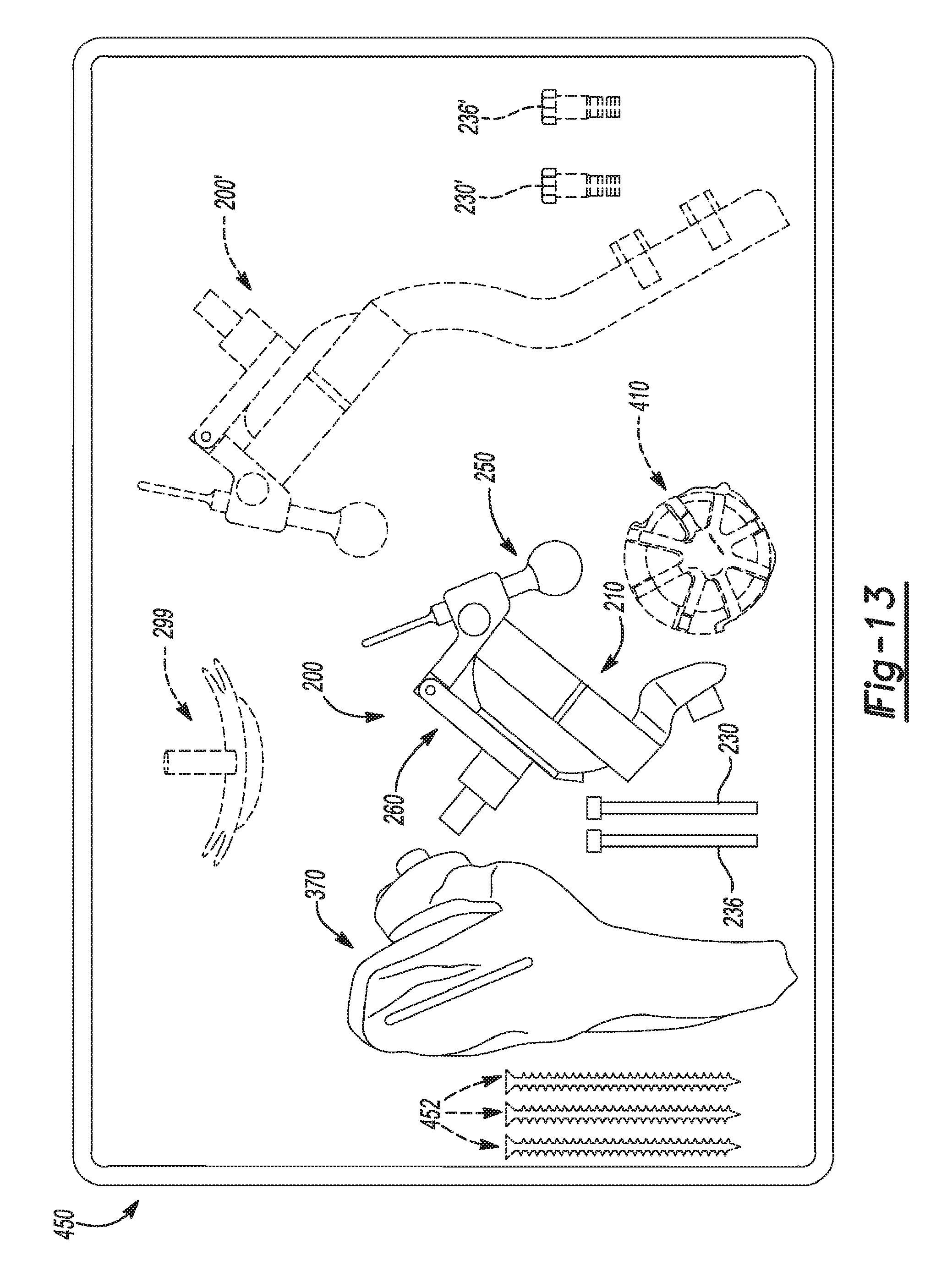
XML
uspto.report is an independent third-party trademark research tool that is not affiliated, endorsed, or sponsored by the United States Patent and Trademark Office (USPTO) or any other governmental organization. The information provided by uspto.report is based on publicly available data at the time of writing and is intended for informational purposes only.
While we strive to provide accurate and up-to-date information, we do not guarantee the accuracy, completeness, reliability, or suitability of the information displayed on this site. The use of this site is at your own risk. Any reliance you place on such information is therefore strictly at your own risk.
All official trademark data, including owner information, should be verified by visiting the official USPTO website at www.uspto.gov. This site is not intended to replace professional legal advice and should not be used as a substitute for consulting with a legal professional who is knowledgeable about trademark law.