Drug eluting medical device
Morgan , et al. December 29, 2
U.S. patent number 10,874,768 [Application Number 15/837,498] was granted by the patent office on 2020-12-29 for drug eluting medical device. This patent grant is currently assigned to COVIDIEN LP. The grantee listed for this patent is Covidien LP. Invention is credited to Brian Graham, Gerald Hodgkinson, Terry Morgan, Daniel Schulz-Jander, Beth Sersen, Carol Sullivan, Joseph Traina.


| United States Patent | 10,874,768 |
| Morgan , et al. | December 29, 2020 |
Drug eluting medical device
Abstract
The present disclosure relates to medical devices, and methods for producing and using the devices. In embodiments, the medical device may be a buttress including a porous substrate possessing a therapeutic layer of a chemotherapeutic agent and optional excipient(s) thereon. By varying the form of chemotherapeutic agents and excipients, the medical devices may be used to treat both the area to which the medical device is attached as well as tissue at a distance therefrom.
| Inventors: | Morgan; Terry (Santa Rosa, CA), Sersen; Beth (Santa Rosa, CA), Schulz-Jander; Daniel (Oakland, CA), Traina; Joseph (Napa, CA), Graham; Brian (Santa Rosa, CA), Sullivan; Carol (Santa Rosa, CA), Hodgkinson; Gerald (Guilford, CT) | ||||||||||
|---|---|---|---|---|---|---|---|---|---|---|---|
| Applicant: |
|
||||||||||
| Assignee: | COVIDIEN LP (Mansfield,
MA) |
||||||||||
| Family ID: | 1000005267059 | ||||||||||
| Appl. No.: | 15/837,498 | ||||||||||
| Filed: | December 11, 2017 |
Prior Publication Data
| Document Identifier | Publication Date | |
|---|---|---|
| US 20180207321 A1 | Jul 26, 2018 | |
Related U.S. Patent Documents
| Application Number | Filing Date | Patent Number | Issue Date | ||
|---|---|---|---|---|---|
| 62448509 | Jan 20, 2017 | ||||
| Current U.S. Class: | 1/1 |
| Current CPC Class: | A61L 31/08 (20130101); A61L 31/06 (20130101); A61L 27/34 (20130101); A61B 17/32 (20130101); A61L 27/54 (20130101); A61P 31/00 (20180101); A61L 27/56 (20130101); A61L 27/26 (20130101); A61L 31/16 (20130101); A61L 27/28 (20130101); A61B 17/1155 (20130101); A61L 31/10 (20130101); A61L 27/18 (20130101); A61L 31/146 (20130101); A61L 31/041 (20130101); A61L 2300/602 (20130101); A61L 2300/606 (20130101); A61L 2300/802 (20130101); A61L 2300/416 (20130101) |
| Current International Class: | A61F 2/01 (20060101); A61L 31/14 (20060101); A61L 31/10 (20060101); A61L 31/08 (20060101); A61L 31/06 (20060101); A61L 31/04 (20060101); A61L 27/56 (20060101); A61L 27/34 (20060101); A61L 27/26 (20060101); A61L 27/28 (20060101); A61L 27/18 (20060101); A61B 17/32 (20060101); A61B 17/115 (20060101); A61P 31/00 (20060101); A61L 27/54 (20060101); A61L 31/16 (20060101) |
References Cited [Referenced By]
U.S. Patent Documents
| 3054406 | September 1962 | Usher |
| 3079606 | March 1963 | Bobrov et al. |
| 3124136 | March 1964 | Usher |
| 3364200 | January 1968 | Ashton et al. |
| 3490675 | January 1970 | Green et al. |
| 3499591 | March 1970 | Green |
| 3797494 | March 1974 | Zaffaroni |
| 3939068 | February 1976 | Wendt et al. |
| 3948666 | April 1976 | Kitanishi et al. |
| 4064062 | December 1977 | Yurko |
| 4166800 | September 1979 | Fong |
| 4282236 | August 1981 | Broom |
| 4347847 | September 1982 | Usher |
| 4354628 | October 1982 | Green |
| 4416698 | November 1983 | McCorsley, III |
| 4429695 | February 1984 | Green |
| 4452245 | June 1984 | Usher |
| 4605730 | August 1986 | Shalaby et al. |
| 4626253 | December 1986 | Broadnax, Jr. |
| 4655221 | April 1987 | Devereux |
| 4834090 | May 1989 | Moore |
| 4838884 | June 1989 | Dumican et al. |
| 4927640 | May 1990 | Dahlinder et al. |
| 4930674 | June 1990 | Barak |
| 5002551 | March 1991 | Linsky et al. |
| 5014899 | May 1991 | Presty et al. |
| 5040715 | August 1991 | Green et al. |
| 5057334 | October 1991 | Vail |
| 5065929 | November 1991 | Schulze et al. |
| 5112496 | May 1992 | Dhawan et al. |
| 5162430 | November 1992 | Rhee et al. |
| 5205459 | April 1993 | Brinkerhoff et al. |
| 5263629 | November 1993 | Trumbull et al. |
| 5281197 | January 1994 | Arias et al. |
| 5307976 | May 1994 | Olson et al. |
| 5312023 | May 1994 | Green et al. |
| 5314471 | May 1994 | Brauker et al. |
| 5318221 | June 1994 | Green et al. |
| 5324775 | June 1994 | Rhee et al. |
| 5326013 | July 1994 | Green et al. |
| 5332142 | July 1994 | Robinson et al. |
| 5344454 | September 1994 | Clarke et al. |
| 5392979 | February 1995 | Green et al. |
| 5397324 | March 1995 | Carroll et al. |
| 5405072 | April 1995 | Zlock et al. |
| 5410016 | April 1995 | Hubbell et al. |
| 5425745 | June 1995 | Green et al. |
| 5441193 | August 1995 | Gravener |
| 5441507 | August 1995 | Wilk |
| 5443198 | August 1995 | Viola et al. |
| 5468253 | November 1995 | Bezwada et al. |
| 5484913 | January 1996 | Stilwell et al. |
| 5503638 | April 1996 | Cooper et al. |
| 5514379 | May 1996 | Weissleder et al. |
| 5542594 | August 1996 | McKean et al. |
| 5543441 | August 1996 | Rhee et al. |
| 5549628 | August 1996 | Cooper et al. |
| 5550187 | August 1996 | Rhee et al. |
| 5575803 | November 1996 | Cooper et al. |
| 5645915 | July 1997 | Kranzler et al. |
| 5653756 | August 1997 | Clarke et al. |
| 5683809 | November 1997 | Freeman et al. |
| 5690675 | November 1997 | Sawyer et al. |
| 5702409 | December 1997 | Rayburn et al. |
| 5752965 | May 1998 | Francis et al. |
| 5752974 | May 1998 | Rhee et al. |
| 5762256 | June 1998 | Mastri et al. |
| 5766188 | June 1998 | Igaki |
| 5769892 | June 1998 | Kingwell |
| 5782396 | July 1998 | Mastri et al. |
| 5799857 | September 1998 | Robertson et al. |
| 5810855 | September 1998 | Rayburn et al. |
| 5814057 | September 1998 | Oi et al. |
| 5819350 | October 1998 | Wang |
| 5833695 | November 1998 | Yoon |
| 5843096 | December 1998 | Igaki et al. |
| 5871135 | February 1999 | Williamson, IV et al. |
| 5874500 | February 1999 | Rhee et al. |
| 5895412 | April 1999 | Tucker |
| 5895415 | April 1999 | Chow et al. |
| 5902312 | May 1999 | Frater et al. |
| 5908427 | June 1999 | McKean et al. |
| 5915616 | June 1999 | Viola et al. |
| 5931847 | August 1999 | Bittner et al. |
| 5957363 | September 1999 | Heck |
| 5964774 | October 1999 | McKean et al. |
| 5997895 | December 1999 | Narotam et al. |
| 6019791 | February 2000 | Wood |
| 6030392 | February 2000 | Dakov |
| 6032849 | March 2000 | Mastri et al. |
| 6045560 | April 2000 | McKean et al. |
| 6063097 | May 2000 | Oi et al. |
| 6080169 | June 2000 | Turtel |
| 6093557 | July 2000 | Pui et al. |
| 6099551 | August 2000 | Gabbay |
| 6142933 | November 2000 | Longo et al. |
| 6149667 | November 2000 | Hovland et al. |
| 6152943 | November 2000 | Sawhney |
| 6155265 | December 2000 | Hammerslag |
| 6156677 | December 2000 | Brown Reed et al. |
| 6165201 | December 2000 | Sawhney et al. |
| 6179862 | January 2001 | Sawhney |
| 6210439 | April 2001 | Firmin et al. |
| 6214020 | April 2001 | Mulhauser et al. |
| 6241139 | June 2001 | Milliman et al. |
| 6258107 | July 2001 | Balazs et al. |
| 6267772 | July 2001 | Mulhauser et al. |
| 6270530 | August 2001 | Eldridge et al. |
| 6273897 | August 2001 | Dalessandro et al. |
| 6280453 | August 2001 | Kugel et al. |
| 6299631 | October 2001 | Shalaby |
| 6309569 | October 2001 | Farrar et al. |
| 6312457 | November 2001 | DiMatteo et al. |
| 6312474 | November 2001 | Francis et al. |
| 6325810 | December 2001 | Hamilton et al. |
| 6399362 | June 2002 | Pui et al. |
| 6436030 | August 2002 | Rehil |
| 6454780 | September 2002 | Wallace |
| 6461368 | October 2002 | Fogarty et al. |
| 6500777 | December 2002 | Wiseman et al. |
| 6503257 | January 2003 | Grant et al. |
| 6514283 | February 2003 | DiMatteo et al. |
| 6514534 | February 2003 | Sawhney |
| 6517566 | February 2003 | Hovland et al. |
| 6551356 | April 2003 | Rousseau |
| 6566406 | May 2003 | Pathak et al. |
| 6568398 | May 2003 | Cohen |
| 6590095 | July 2003 | Schleicher et al. |
| 6592597 | July 2003 | Grant et al. |
| 6605294 | August 2003 | Sawhney |
| 6610006 | August 2003 | Amid et al. |
| 6627749 | September 2003 | Kumar |
| 6638285 | October 2003 | Gabbay |
| 6652594 | November 2003 | Francis et al. |
| 6656193 | December 2003 | Grant et al. |
| 6656200 | December 2003 | Li et al. |
| 6669735 | December 2003 | Pelissier |
| 6673093 | January 2004 | Sawhney |
| 6677258 | January 2004 | Carroll et al. |
| 6685714 | February 2004 | Rousseau |
| 6702828 | March 2004 | Whayne |
| 6703047 | March 2004 | Sawhney et al. |
| 6704210 | March 2004 | Myers |
| 6723114 | April 2004 | Shalaby |
| 6726706 | April 2004 | Dominguez |
| 6736823 | May 2004 | Darois et al. |
| 6736854 | May 2004 | Vadurro et al. |
| 6746458 | June 2004 | Cloud |
| 6746869 | June 2004 | Pui et al. |
| 6764720 | July 2004 | Pui et al. |
| 6773458 | August 2004 | Brauker et al. |
| 6818018 | November 2004 | Sawhney |
| 6843252 | January 2005 | Harrison et al. |
| 6896684 | May 2005 | Monassevitch et al. |
| 6927315 | August 2005 | Heinecke et al. |
| 6939358 | September 2005 | Palacios et al. |
| 6946196 | September 2005 | Foss |
| 6953139 | October 2005 | Milliman et al. |
| 6959851 | November 2005 | Heinrich |
| 7009034 | March 2006 | Pathak et al. |
| 7025772 | April 2006 | Gellman et al. |
| 7060087 | June 2006 | DiMatteo et al. |
| 7087065 | August 2006 | Ulmsten et al. |
| 7108701 | September 2006 | Evens et al. |
| 7128253 | October 2006 | Mastri et al. |
| 7128748 | October 2006 | Mooradian et al. |
| 7134438 | November 2006 | Makower et al. |
| 7141055 | November 2006 | Abrams et al. |
| 7147138 | December 2006 | Shelton, IV |
| 7160299 | January 2007 | Baily |
| 7179268 | February 2007 | Roy et al. |
| 7210810 | May 2007 | Iversen et al. |
| 7214727 | May 2007 | Kwon et al. |
| 7232449 | June 2007 | Sharkawy et al. |
| 7241300 | July 2007 | Sharkawy et al. |
| 7247338 | July 2007 | Pui et al. |
| 7279322 | October 2007 | Pui et al. |
| 7307031 | December 2007 | Carroll et al. |
| 7308998 | December 2007 | Mastri et al. |
| 7311720 | December 2007 | Mueller et al. |
| 7328829 | February 2008 | Arad et al. |
| 7347850 | March 2008 | Sawhney |
| 7377928 | May 2008 | Zubik et al. |
| 7434717 | October 2008 | Shelton, IV et al. |
| 7438209 | October 2008 | Hess et al. |
| 7464849 | December 2008 | Shelton, IV et al. |
| 7498063 | March 2009 | Pui et al. |
| 7547312 | June 2009 | Bauman et al. |
| 7559937 | July 2009 | de la Torre et al. |
| 7571845 | August 2009 | Viola |
| 7592418 | September 2009 | Pathak et al. |
| 7594921 | September 2009 | Browning |
| 7595392 | September 2009 | Kumar et al. |
| 7604151 | October 2009 | Hess et al. |
| 7611494 | November 2009 | Campbell et al. |
| 7635073 | December 2009 | Heinrich |
| 7645874 | January 2010 | Saferstein et al. |
| 7649089 | January 2010 | Kumar et al. |
| 7655288 | February 2010 | Bauman et al. |
| 7662409 | February 2010 | Masters |
| 7662801 | February 2010 | Kumar et al. |
| 7665646 | February 2010 | Prommersberger |
| 7666198 | February 2010 | Suyker et al. |
| 7669747 | March 2010 | Weisenburgh, II et al. |
| 7673782 | March 2010 | Hess et al. |
| 7708180 | May 2010 | Murray et al. |
| 7709631 | May 2010 | Harris et al. |
| 7717313 | May 2010 | Criscuolo et al. |
| 7722642 | May 2010 | Williamson, IV et al. |
| 7735703 | June 2010 | Morgan et al. |
| 7744627 | June 2010 | Orban, III et al. |
| 7754002 | July 2010 | Maase et al. |
| 7776060 | August 2010 | Mooradian et al. |
| 7789889 | September 2010 | Zubik et al. |
| 7793813 | September 2010 | Bettuchi |
| 7799026 | September 2010 | Schechter et al. |
| 7823592 | November 2010 | Bettuchi et al. |
| 7824420 | November 2010 | Eldridge et al. |
| 7845533 | December 2010 | Marczyk et al. |
| 7845536 | December 2010 | Viola et al. |
| 7846149 | December 2010 | Jankowski |
| 7892247 | February 2011 | Conston et al. |
| 7909224 | March 2011 | Prommersberger |
| 7909837 | March 2011 | Crews et al. |
| 7938307 | May 2011 | Bettuchi |
| 7942890 | May 2011 | D'Agostino et al. |
| 7950561 | May 2011 | Aranyi |
| 7951166 | May 2011 | Orban, III et al. |
| 7951248 | May 2011 | Fallis et al. |
| 7967179 | June 2011 | Olson et al. |
| 7988027 | August 2011 | Olson et al. |
| 8011550 | September 2011 | Aranyi et al. |
| 8011555 | September 2011 | Tarinelli et al. |
| 8016177 | September 2011 | Bettuchi et al. |
| 8016178 | September 2011 | Olson et al. |
| 8025199 | September 2011 | Whitman et al. |
| 8028883 | October 2011 | Stopek |
| 8033483 | October 2011 | Fortier et al. |
| 8033983 | October 2011 | Chu et al. |
| 8038045 | October 2011 | Bettuchi et al. |
| 8062330 | November 2011 | Prommersberger et al. |
| 8062673 | November 2011 | Figuly et al. |
| 8083119 | December 2011 | Prommersberger |
| 8091756 | January 2012 | Viola |
| 8123766 | February 2012 | Bauman et al. |
| 8123767 | February 2012 | Bauman et al. |
| 8127975 | March 2012 | Olson et al. |
| 8133336 | March 2012 | Kettlewell et al. |
| 8133559 | March 2012 | Lee et al. |
| 8146791 | April 2012 | Bettuchi et al. |
| 8152777 | April 2012 | Campbell et al. |
| 8157149 | April 2012 | Olson et al. |
| 8157151 | April 2012 | Ingmanson et al. |
| 8167895 | May 2012 | D'Agostino et al. |
| 8177797 | May 2012 | Shimoji et al. |
| 8178746 | May 2012 | Hildeberg et al. |
| 8192460 | June 2012 | Orban, III et al. |
| 8201720 | June 2012 | Hessler |
| 8210414 | July 2012 | Bettuchi et al. |
| 8210453 | July 2012 | Hull et al. |
| 8225799 | July 2012 | Bettuchi |
| 8225981 | July 2012 | Criscuolo et al. |
| 8231043 | July 2012 | Tarinelli et al. |
| 8235273 | August 2012 | Olson et al. |
| 8245901 | August 2012 | Stopek |
| 8252339 | August 2012 | Figuly et al. |
| 8252921 | August 2012 | Vignon et al. |
| 8256654 | September 2012 | Bettuchi et al. |
| 8257391 | September 2012 | Orban, III et al. |
| 8276800 | October 2012 | Bettuchi |
| 8286849 | October 2012 | Bettuchi |
| 8308042 | November 2012 | Aranyi |
| 8308045 | November 2012 | Bettuchi et al. |
| 8308046 | November 2012 | Prommersberger |
| 8312885 | November 2012 | Bettuchi et al. |
| 8313014 | November 2012 | Bettuchi |
| 8317790 | November 2012 | Bell et al. |
| 8322590 | December 2012 | Patel et al. |
| 8348126 | January 2013 | Olson et al. |
| 8348130 | January 2013 | Shah et al. |
| 8365972 | February 2013 | Aranyi et al. |
| 8367089 | February 2013 | Wan et al. |
| 8371491 | February 2013 | Huitema et al. |
| 8371492 | February 2013 | Aranyi et al. |
| 8371493 | February 2013 | Aranyi et al. |
| 8372094 | February 2013 | Bettuchi et al. |
| 8393514 | March 2013 | Shelton, IV et al. |
| 8393517 | March 2013 | Milo |
| 8408440 | April 2013 | Olson et al. |
| 8408480 | April 2013 | Hull et al. |
| 8413869 | April 2013 | Heinrich |
| 8413871 | April 2013 | Racenet et al. |
| 8418909 | April 2013 | Kostrzewski |
| 8424742 | April 2013 | Bettuchi |
| 8449603 | May 2013 | Weber |
| 8453652 | June 2013 | Stopek |
| 8453904 | June 2013 | Eskaros et al. |
| 8453909 | June 2013 | Olson et al. |
| 8453910 | June 2013 | Bettuchi et al. |
| 8464925 | June 2013 | Hull et al. |
| 8470360 | June 2013 | McKay |
| 8474677 | July 2013 | Woodard, Jr. et al. |
| 8479968 | July 2013 | Hodgkinson et al. |
| 8485414 | July 2013 | Criscuolo et al. |
| 8496683 | July 2013 | Prommersberger et al. |
| 8511533 | August 2013 | Viola et al. |
| 8512402 | August 2013 | Marczyk et al. |
| 8518440 | August 2013 | Blaskovich et al. |
| 8529600 | September 2013 | Woodard, Jr. et al. |
| 8540128 | September 2013 | Shelton, IV et al. |
| 8540131 | September 2013 | Swayze |
| 8551138 | October 2013 | Orban, III et al. |
| 8556918 | October 2013 | Bauman et al. |
| 8561873 | October 2013 | Ingmanson et al. |
| 8579990 | November 2013 | Priewe |
| 8584920 | November 2013 | Hodgkinson |
| 8590762 | November 2013 | Hess et al. |
| 8616430 | December 2013 | (Prommersberger) Stopek et al. |
| 8617132 | December 2013 | Golzarian et al. |
| 8631989 | January 2014 | Aranyi et al. |
| 8646674 | February 2014 | Schulte et al. |
| 8668129 | March 2014 | Olson |
| 8678263 | March 2014 | Viola |
| 8679137 | March 2014 | Bauman et al. |
| 8684250 | April 2014 | Bettuchi et al. |
| 8701958 | April 2014 | Shelton, IV et al. |
| 8721703 | May 2014 | Fowler |
| 8727197 | May 2014 | Hess et al. |
| 8757466 | June 2014 | Olson et al. |
| 8789737 | July 2014 | Hodgkinson et al. |
| 8814888 | August 2014 | Sgro |
| 8820606 | September 2014 | Hodgkinson |
| 8827133 | September 2014 | Shelton, IV et al. |
| 8857694 | October 2014 | Shelton, IV et al. |
| 8864009 | October 2014 | Shelton, IV et al. |
| 8870050 | October 2014 | Hodgkinson |
| 8900616 | December 2014 | Belcheva et al. |
| 8920443 | December 2014 | Hiles et al. |
| 8920444 | December 2014 | Hiles et al. |
| 8939344 | January 2015 | Olson et al. |
| 8956390 | February 2015 | Shah et al. |
| 8967448 | March 2015 | Carter et al. |
| 9005243 | April 2015 | Stopek et al. |
| 9010606 | April 2015 | Aranyi et al. |
| 9010608 | April 2015 | Casasanta, Jr. et al. |
| 9010609 | April 2015 | Carter et al. |
| 9010610 | April 2015 | Hodgkinson |
| 9010612 | April 2015 | Stevenson et al. |
| 9016543 | April 2015 | (Prommersberger) Stopek et al. |
| 9016544 | April 2015 | Hodgkinson et al. |
| 9027817 | May 2015 | Milliman et al. |
| 9044227 | June 2015 | Shelton, IV et al. |
| 9055944 | June 2015 | Hodgkinson et al. |
| 9084602 | July 2015 | Gleiman |
| 9107665 | August 2015 | Hodgkinson et al. |
| 9107667 | August 2015 | Hodgkinson |
| 9113871 | August 2015 | Milliman et al. |
| 9113873 | August 2015 | Marczyk et al. |
| 9113885 | August 2015 | Hodgkinson et al. |
| 9113893 | August 2015 | Sorrentino et al. |
| 9161753 | October 2015 | Prior |
| 9161757 | October 2015 | Bettuchi |
| 9186140 | November 2015 | Hiles et al. |
| 9186144 | November 2015 | Stevenson et al. |
| 9192378 | November 2015 | Aranyi et al. |
| 9192379 | November 2015 | Aranyi et al. |
| 9192380 | November 2015 | (Tarinelli) Racenet et al. |
| 9192383 | November 2015 | Milliman |
| 9192384 | November 2015 | Bettuchi |
| 9198660 | December 2015 | Hodgkinson |
| 9198663 | December 2015 | Marczyk et al. |
| 9204881 | December 2015 | Penna |
| 9220504 | December 2015 | Viola et al. |
| 9226754 | January 2016 | D'Agostino et al. |
| 9237892 | January 2016 | Hodgkinson |
| 9237893 | January 2016 | Carter et al. |
| 9277922 | March 2016 | Carter et al. |
| 9295466 | March 2016 | Hodgkinson et al. |
| 9326768 | May 2016 | Shelton, IV |
| 9326773 | May 2016 | Casasanta, Jr. et al. |
| 9328111 | May 2016 | Zhou et al. |
| 9345479 | May 2016 | (Tarinelli) Racenet et al. |
| 9351729 | May 2016 | Orban, III et al. |
| 9351731 | May 2016 | Carter et al. |
| 9351732 | May 2016 | Hodgkinson |
| 9358005 | June 2016 | Shelton, IV et al. |
| 9364229 | June 2016 | D'Agostino et al. |
| 9364234 | June 2016 | (Prommersberger) Stopek et al. |
| 9386988 | July 2016 | Baxter, III et al. |
| 9402627 | August 2016 | Stevenson et al. |
| 9414839 | August 2016 | Penna |
| 9433412 | September 2016 | Bettuchi et al. |
| 9433413 | September 2016 | Stopek |
| 9433420 | September 2016 | Hodgkinson |
| 9445812 | September 2016 | Olson et al. |
| 9445817 | September 2016 | Bettuchi |
| 9463260 | October 2016 | Stopek |
| 9486215 | November 2016 | Olson et al. |
| 9492170 | November 2016 | Bear et al. |
| 9504470 | November 2016 | Milliman |
| 9517164 | December 2016 | Vitaris et al. |
| 9572576 | February 2017 | Hodgkinson et al. |
| 9585657 | March 2017 | Shelton, IV et al. |
| 9597077 | March 2017 | Hodgkinson |
| 9610080 | April 2017 | Whitfield et al. |
| 9622745 | April 2017 | Ingmanson et al. |
| 9629626 | April 2017 | Soltz et al. |
| 9636850 | May 2017 | Stopek (nee Prommersberger) et al. |
| 9655620 | May 2017 | Prescott et al. |
| 9675351 | June 2017 | Hodgkinson et al. |
| 9681936 | June 2017 | Hodgkinson et al. |
| 9687262 | June 2017 | Rousseau et al. |
| 9693772 | July 2017 | Ingmanson et al. |
| 9708184 | July 2017 | Chan et al. |
| 9770245 | September 2017 | Swayze et al. |
| 9775617 | October 2017 | Carter et al. |
| 9782173 | October 2017 | Mozdzierz |
| 2002/0091397 | July 2002 | Chen |
| 2002/0151911 | October 2002 | Gabbay |
| 2003/0065345 | April 2003 | Weadock |
| 2003/0078209 | April 2003 | Schmidt |
| 2003/0083676 | May 2003 | Wallace |
| 2003/0125676 | July 2003 | Swenson et al. |
| 2003/0181927 | September 2003 | Wallace |
| 2003/0208231 | November 2003 | Williamson et al. |
| 2004/0092912 | May 2004 | Jinno et al. |
| 2004/0107006 | June 2004 | Francis et al. |
| 2004/0254590 | December 2004 | Hoffman et al. |
| 2004/0260272 | December 2004 | Friedman |
| 2004/0260315 | December 2004 | Dell et al. |
| 2005/0002981 | January 2005 | Lahtinen et al. |
| 2005/0021085 | January 2005 | Abrams et al. |
| 2005/0059996 | March 2005 | Bauman et al. |
| 2005/0059997 | March 2005 | Bauman et al. |
| 2005/0070929 | March 2005 | Dalessandro et al. |
| 2005/0118435 | June 2005 | DeLucia et al. |
| 2005/0149073 | July 2005 | Arani et al. |
| 2005/0283256 | December 2005 | Sommerich et al. |
| 2006/0008505 | January 2006 | Brandon |
| 2006/0121266 | June 2006 | Fandel et al. |
| 2006/0173470 | August 2006 | Oray et al. |
| 2006/0190027 | August 2006 | Downey |
| 2007/0034669 | February 2007 | de la Torre et al. |
| 2007/0203510 | August 2007 | Bettuchi |
| 2007/0212394 | September 2007 | Reyes |
| 2007/0224235 | September 2007 | Tenney |
| 2007/0243227 | October 2007 | Gertner |
| 2007/0246505 | October 2007 | Pace-Floridia et al. |
| 2008/0009811 | January 2008 | Cantor |
| 2008/0029570 | February 2008 | Shelton et al. |
| 2008/0082126 | April 2008 | Murray et al. |
| 2008/0140115 | June 2008 | Stopek |
| 2008/0169328 | July 2008 | Shelton |
| 2008/0169332 | July 2008 | Shelton et al. |
| 2008/0169333 | July 2008 | Shelton et al. |
| 2008/0216855 | September 2008 | Nasca |
| 2008/0220047 | September 2008 | Sawhney et al. |
| 2008/0286325 | November 2008 | Reyes et al. |
| 2008/0290134 | November 2008 | Bettuchi et al. |
| 2009/0001121 | January 2009 | Hess et al. |
| 2009/0001130 | January 2009 | Hess et al. |
| 2009/0031842 | February 2009 | Kawai et al. |
| 2009/0206125 | August 2009 | Huitema et al. |
| 2009/0206126 | August 2009 | Huitema et al. |
| 2009/0206139 | August 2009 | Hall et al. |
| 2009/0206141 | August 2009 | Huitema et al. |
| 2009/0206142 | August 2009 | Huitema et al. |
| 2009/0218384 | September 2009 | Aranyi |
| 2009/0277944 | November 2009 | Dalessandro et al. |
| 2010/0016855 | January 2010 | Ramstein et al. |
| 2010/0016888 | January 2010 | Calabrese et al. |
| 2010/0076489 | March 2010 | Stopek et al. |
| 2010/0087840 | April 2010 | Ebersole et al. |
| 2010/0147921 | June 2010 | Olson |
| 2010/0147922 | June 2010 | Olson |
| 2010/0174253 | July 2010 | Cline et al. |
| 2010/0203151 | August 2010 | Hiraoka |
| 2010/0243707 | September 2010 | Olson et al. |
| 2010/0331859 | December 2010 | Omori |
| 2011/0034910 | February 2011 | Ross et al. |
| 2011/0089220 | April 2011 | Ingmanson et al. |
| 2011/0125138 | May 2011 | Malinouskas et al. |
| 2011/0166673 | July 2011 | Patel et al. |
| 2011/0293690 | December 2011 | Griffin et al. |
| 2011/0295200 | December 2011 | Speck et al. |
| 2012/0080336 | April 2012 | Shelton, IV et al. |
| 2012/0171383 | July 2012 | Christensen et al. |
| 2012/0197272 | August 2012 | Oray et al. |
| 2012/0241491 | September 2012 | Aldridge et al. |
| 2012/0241493 | September 2012 | Baxter, III et al. |
| 2012/0253298 | October 2012 | Henderson et al. |
| 2012/0316633 | December 2012 | Flanagan |
| 2013/0153636 | June 2013 | Shelton, IV et al. |
| 2013/0153641 | June 2013 | Shelton, IV et al. |
| 2013/0209659 | August 2013 | Racenet et al. |
| 2013/0256380 | October 2013 | Schmid et al. |
| 2014/0048580 | February 2014 | Merchant et al. |
| 2014/0131418 | May 2014 | Kostrzewski |
| 2014/0239047 | August 2014 | Hodgkinson et al. |
| 2015/0041347 | February 2015 | Hodgkinson |
| 2015/0133995 | May 2015 | Shelton, IV et al. |
| 2015/0209045 | July 2015 | Hodgkinson et al. |
| 2015/0305743 | October 2015 | Casasanta et al. |
| 2015/0327864 | November 2015 | Hodgkinson et al. |
| 2016/0022268 | January 2016 | Prior |
| 2016/0045200 | February 2016 | Milliman |
| 2016/0100834 | April 2016 | Viola et al. |
| 2016/0106430 | April 2016 | Carter et al. |
| 2016/0157857 | June 2016 | Hodgkinson et al. |
| 2016/0174988 | June 2016 | D'Agostino et al. |
| 2016/0206315 | July 2016 | Olson |
| 2016/0220257 | August 2016 | Casasanta et al. |
| 2016/0249923 | September 2016 | Hodgkinson et al. |
| 2016/0256166 | September 2016 | (Prommersberger) Stopek et al. |
| 2016/0270793 | September 2016 | Carter et al. |
| 2016/0310143 | October 2016 | Bettuchi |
| 2016/0317720 | November 2016 | Ostapoff et al. |
| 2016/0338704 | November 2016 | Penna |
| 2016/0367252 | December 2016 | Olson et al. |
| 2016/0367253 | December 2016 | Hodgkinson |
| 2016/0367257 | December 2016 | Stevenson et al. |
| 2017/0042540 | February 2017 | Olson et al. |
| 2017/0049452 | February 2017 | Milliman |
| 2017/0150967 | June 2017 | Hodgkinson et al. |
| 2282761 | Sep 1998 | CA | |||
| 1602563 | Mar 1950 | DE | |||
| 19924311 | Nov 2000 | DE | |||
| 0 327 022 | Aug 1989 | EP | |||
| 0 594 148 | Apr 1994 | EP | |||
| 3087931 | Nov 2016 | EP | |||
| 3087931 | Nov 2016 | EP | |||
| 2000-166933 | Jun 2000 | JP | |||
| 2002-202213 | Jul 2002 | JP | |||
| 2007-124166 | May 2007 | JP | |||
| 2010-214132 | Sep 2010 | JP | |||
| 90/05489 | May 1990 | WO | |||
| 95/16221 | Jun 1995 | WO | |||
| 98/38923 | Sep 1998 | WO | |||
| 99/26826 | Jun 1999 | WO | |||
| 00/10456 | Mar 2000 | WO | |||
| 00/16684 | Mar 2000 | WO | |||
| 2010021757 | Feb 2010 | WO | |||
| 2010/075298 | Jul 2010 | WO | |||
| 2015137962 | Sep 2015 | WO | |||
| 2016205652 | Dec 2016 | WO | |||
| 2017046193 | Mar 2017 | WO | |||
Other References
|
Extended European Search Report corresponding to EP 14 16 9739.1, completed Aug. 19, 2014 and dated Aug. 29, 2014; (7 pp). cited by applicant . Extended European Search Report corresponding to EP 14 15 7997.9, completed Sep. 9, 2014 and dated Sep. 17, 2014; (8 pp). cited by applicant . Extended European Search Report corresponding to EP 14 16 8904.2, completed Sep. 10, 2014 and dated Sep. 18, 2014; (8 pp). cited by applicant . Extended European Search Report corresponding to EP 13 19 4995.0, completed Jun. 5, 2014 and dated Oct. 13, 2014; (10 pp). cited by applicant . Extended European Search Report corresponding to EP 13 15 4571.7, completed Oct. 10, 2014 and dated Oct. 20, 2014; (8 pp). cited by applicant . Extended European Search Report corresponding to EP 14 18 1125.7, completed Oct. 16, 2014 and dated Oct. 24, 2014; (7 pp). cited by applicant . Extended European Search Report corresponding to EP 14 18 1127.3, completed Oct. 16, 2014 and dated Nov. 10, 2014; (8 pp). cited by applicant . Extended European Search Report corresponding to EP 14 19 0419.3, completed Mar. 24, 2015 and dated Mar. 30, 2015; (6 pp). cited by applicant . European Office Action corresponding to counterpart Int'l Appln No. EP 12 198 776.2 dated Apr. 7, 2015. cited by applicant . European Office Action corresponding to counterpart Int'l Appln No. EP 13 156 297.7 dated Apr. 10, 2015. cited by applicant . Australian Examination Report No. 1 corresponding to counterpart Int'l Appln No. AU 2011250822 dated May 18, 2015. cited by applicant . European Office Action corresponding to counterpart Int'l Appln No. EP 12 186 175.1 dated Jun. 1, 2015. cited by applicant . Chinese Office Action corresponding to counterpart Int'l Appln No. CN 201010517292.8 dated Jun. 2, 2015. cited by applicant . Extended European Search Report corresponding to counterpart Int'l Appln No. EP 14 17 48145 dated Jun. 9, 2015. cited by applicant . Australian Examination Report No. 1 corresponding to counterpart Int'l Appln No. AU 2014200584 dated Jun. 15, 2015. cited by applicant . European Office Action corresponding to counterpart Int'l Appln No. EP 13 180 881.8 dated Jun. 19, 2015. cited by applicant . European Office Action corresponding to counterpart Int'l Appln No. EP 14 157 195.0 dated Jul. 2, 2015. cited by applicant . Extended European Search Report corresponding to counterpart Int'l Appln No. EP 12 19 6902.6 dated Aug. 6, 2015. cited by applicant . Extended European Search Report corresponding to counterpart Int'l Appln No. EP 14 15 2060.1 dated Aug. 14, 2015. cited by applicant . Chinese Office Action corresponding to counterpart Int'l Appln No. CN 201210129787.2 dated Aug. 24, 2015. cited by applicant . Canadian Office Action corresponding to counterpart Int'l Appln. No. CA 2,665,206 dated Nov. 19, 2013. cited by applicant . Chinese Notification of Reexamination corresponding to counterpart Int'l Appln. No. CN 201010517292.8 dated Jun. 2, 2015. cited by applicant . Japanese Office Action corresponding to counterpart Int'l Appln. No. JP 2014-216989 dated Sep. 11, 2015. cited by applicant . Canadian First Office Action corresponding to counterpart Int'l Appln. No. CA 2,686,105 dated Sep. 17, 2015. cited by applicant . Japanese Office Action corresponding to counterpart Int'l Appln. No. JP 2012-040188 dated Oct. 21, 2015. cited by applicant . European Communication corresponding to counterpart Int'l Appln. No. EP 13 17 6895.4 dated Nov. 5, 2015. cited by applicant . Chinese First Office Action corresponding to counterpart Int'l Appln. No. CN 201210544552 dated Nov. 23, 2015. cited by applicant . Chinese First Office Action corresponding to counterpart Int'l Appln. No. CN 201210545228 dated Nov. 30, 2015. cited by applicant . Extended European Search Report corresponding to counterpart Int'l Appln. No. EP 15 18 0491.1 dated Dec. 9, 2015. cited by applicant . Extended European Search Report corresponding to counterpart Int'l Appln. No. EP 15 18 3819.0 dated Dec. 11, 2015. cited by applicant . Canadian Office Action corresponding to counterpart Int'l Appln. No. CA 2,697,819 dated Jan. 6, 2016. cited by applicant . Canadian Office Action corresponding to counterpart Int'l Appln. No. CA 2,696,419 dated Jan. 14, 2016. cited by applicant . European Office Action corresponding to counterpart Int'l Appln. No. EP 12 19 8776.2 dated Jan. 19, 2016. cited by applicant . Extended European Search Report corresponding to counterpart Int'l Appln. No. EP 15 17 4146.9 dated Jan. 20, 2016. cited by applicant . Chinese First Office Action corresponding to counterpart Int'l Appln. No. CN 201310353628.5 dated Jan. 25, 2016. cited by applicant . Extended European Search Report corresponding to counterpart Int'l Appln. No. EP 12 19 6912.5 dated Feb. 1, 2016. cited by applicant . Japanese Office Action corresponding to counterpart Int'l Appln. No. JP 2012-098903 dated Feb. 22, 2016. cited by applicant . Extended European Search Report corresponding to counterpart Int'l Appln. No. EP 12 19 8753.1 dated Feb. 24, 2016. cited by applicant . Chinese First Office Action corresponding to counterpart Int'l Appln. No. CN 201410449019.4 dated Mar. 30, 2016. cited by applicant . Extended European Search Report corresponding to counterpart Int'l Appln. No. EP 16 15 0232.3 dated Apr. 12, 2016. cited by applicant . European Office Action corresponding to counterpart Int'l Appln. No. EP 11 18 3256.4 dated Apr. 20, 2016. cited by applicant . Australian Examination Report No. 1 corresponding to counterpart Int'l Appln. No. AU 2012244169 dated May 10, 2016. cited by applicant . European Office Action corresponding to counterpart Int'l Appln. No. EP 10 25 0715.9 dated May 12, 2016. cited by applicant . Chinese First Office Action corresponding to counterpart Int'l Appln. No. CN 201410778512.0 dated May 13, 2016. cited by applicant . Australian Examination Report No. 1 corresponding to counterpart Int'l Appln. No. AU 2012227358 dated May 16, 2016. cited by applicant . Japanese Office Action corresponding to counterpart Int'l Appln. No. JP 2012-040188 dated May 17, 2016. cited by applicant . Australian Examination Report No. 1 corresponding to counterpart Int'l Appln. No. AU 2012244380 dated May 20, 2016. cited by applicant . Australian Examination Report No. 1 corresponding to counterpart Int'l Appln. No. AU 2014227480 dated May 21, 2016. cited by applicant . Australian Examination Report No. 1 corresponding to counterpart Int'l Appln. No. AU 2012254977 dated May 30, 2016. cited by applicant . European Office Action corresponding to counterpart Int'l Appln. No. EP 14 17 2681.0 dated May 13, 2016. cited by applicant . Extended European Search Report corresponding to counterpart Int'l Appln. No. EP 16 15 3647.9 dated Jun. 3, 2016. cited by applicant . Chinese Office Action corresponding to counterpart Int'l Appln. No. CN 201210545228 dated Jun. 29, 2016. cited by applicant . Japanese Office Action corresponding to counterpart Int'l Appln. No. JP 2012-250058 dated Jun. 29, 2016. cited by applicant . European Office Action corresponding to counterpart Int'l Appln. No. EP 14 15 7997.9 dated Jun. 29, 2016. cited by applicant . Canadian Office Action corresponding to counterpart Int'l Appln. No. CA 2,712,617 dated Jun. 30, 2016. cited by applicant . Chinese First Office Action corresponding to counterpart Int'l Appln. No. CN 2013103036903 dated Jun. 30, 2016. cited by applicant . Australian Patent Examination Report No. 1 corresponding to counterpart Int'l Appln. No. AU 2012250278 dated Jul. 10, 2016. cited by applicant . Australian Patent Examination Report No. 1 corresponding to counterpart Int'l Appln. No. AU 2012244382 dated Jul. 10, 2016. cited by applicant . Japanese Office Action corresponding to counterpart Int'l Appln. No. JP 2012-255242 dated Jul. 26, 2016. cited by applicant . Japanese Office Action corresponding to counterpart Int'l Appln. No. JP 2012-268668 dated Jul. 27, 2016. cited by applicant . European Office Action corresponding to counterpart Int'l Appln. No. EP 14 15 2060.1 dated Aug. 4, 2016. cited by applicant . European Office Action corresponding to counterpart Int'l Appln. No. EP 12 16 5609.4 dated Aug. 5, 2016. cited by applicant . European Office Action corresponding to counterpart Int'l Appln. No. EP 15 15 2392.5 dated Aug. 8, 2016. cited by applicant . Japanese Office Action corresponding to counterpart Int'l Appln. No. JP 2013-003624 dated Aug. 25, 2016. cited by applicant . Australian Patent Examination Report No. 1 corresponding to counterpart Int'l Appln. No. AU 2012261752 dated Sep. 6, 2016. cited by applicant . Japanese Office Action corresponding to counterpart Int'l Appln. No. JP 2014-252703 dated Sep. 26, 2016. cited by applicant . European Office Action corresponding to counterpart Int'l Appln. No. EP 12 19 8776.2 dated Sep. 12, 2016. cited by applicant . Japanese Office Action corresponding to counterpart Int'l Appln. No. JP 2013-000321 dated Sep. 13, 2016. cited by applicant . Chinese Second Office Action corresponding to counterpart Int'l Appln. No. CN 201310353628.5 dated Sep. 26, 2016. cited by applicant . European Office Action corresponding to counterpart Int'l Appln. No. EP 12 15 2541.4 dated Sep. 27, 2016. cited by applicant . Australian Patent Examination Report No. 1 corresponding to counterpart Int'l Appln. No. AU 2012268923 dated Sep. 28, 2016. cited by applicant . Chinese First Office Action corresponding to counterpart Int'l Appln. No. CN 2013107068710 dated Dec. 16, 2016. cited by applicant . Chinese First Office Action corresponding to counterpart Int'l Appln. No. CN 201310646606.8 dated Dec. 23, 2016. cited by applicant . Japanese Office Action corresponding to counterpart Int'l Appln. No. JP 2013-000321 dated Jan. 4, 2017. cited by applicant . Extended European Search Report corresponding to counterpart Int'l Appln. No. EP 16 16 6367.9 dated Jan. 16, 2017. cited by applicant . Australian Examination Report No. 1 corresponding to counterpart Int'l Appln. No. AU 2013206777 dated Feb. 1, 2017. cited by applicant . Chinese Second Office Action corresponding to counterpart Int'l Appln. No. CN 2013103036903 dated Feb. 23, 2017. cited by applicant . Japanese Office Action corresponding to counterpart Int'l Appln. No. JP 2013-175379 dated Mar. 1, 2017. cited by applicant . Chinese First Office Action corresponding to counterpart Int'l Appln. No. CN 201410028462.4 dated Mar. 2, 2017. cited by applicant . Chinese First Office Action corresponding to counterpart Int'l Appln. No. CN 201410084070 dated Mar. 13, 2017. cited by applicant . Extended European Search Report corresponding to counterpart Int'l Appln. No. EP 16 19 6549.6 dated Mar. 17, 2017. cited by applicant . Japanese Office Action corresponding to counterpart Int'l Appln. No. JP 2013-147701 dated Mar. 21, 2017. cited by applicant . Australian Examination Report No. 1 corresponding to counterpart Int'l Appln. No. AU 2013206804 dated Mar. 21, 2017. cited by applicant . Australian Examination Report No. 1 corresponding to counterpart Int'l Appln. No. AU 2013211499 dated May 4, 2017. cited by applicant . Australian Examination Report No. 1 corresponding to counterpart Int'l Appln. No. AU 2014201008 dated May 23, 2017. cited by applicant . European Search Report corresponding to EP 06 00 4598, completed Jun. 22, 2006; (2 pp). cited by applicant . European Search Report corresponding to EP 06 01 6962.0, completed Jan. 3, 2007 and dated Jan. 11, 2007; (10 pp). cited by applicant . International Search Report corresponding to International Application No. PCT/US2005/036740, completed Feb. 20, 2007 and dated Mar. 23, 2007; (8 pp). cited by applicant . International Search Report corresponding to International Application No. PCT/US2007/022713, completed Apr. 21, 2008 and dated May 15, 2008; (1 p). cited by applicant . International Search Report corresponding to International Application No. PCT/US2008/002981, completed Jun. 9, 2008 and dated Jun. 26, 2008; (2 pp). cited by applicant . European Search Report corresponding to EP 08 25 1779, completed Jul. 14, 2008 and dated Jul. 23, 2008; (5 pp). cited by applicant . European Search Report corresponding to EP 08 25 1989.3, completed Mar. 11, 2010 and dated Mar. 24, 2010; (6 pp). cited by applicant . European Search Report corresponding to EP 10 25 0639.1, completed Jun. 17, 2010 and dated Jun. 28, 2010; (7 pp). cited by applicant . European Search Report corresponding to EP 10 25 0715.9, completed Jun. 30, 2010 and dated Jul. 20, 2010; (3 pp). cited by applicant . European Search Report corresponding to EP 05 80 4382.9, completed Oct. 5, 2010 and dated Oct. 12, 2010; (3 pp). cited by applicant . European Search Report corresponding to EP 09 25 2897.5, completed Feb. 7, 2011 and dated Feb. 15, 2011; (3 pp). cited by applicant . European Search Report corresponding to EP 10 25 0642.5, completed Mar. 25, 2011 and dated Apr. 4, 2011; (4 pp). cited by applicant . European Search Report corresponding to EP 12 15 2229.6, completed Feb. 23, 2012 and dated Mar. 1, 2012; (4 pp). cited by applicant . European Search Report corresponding to EP 12 15 0511.9, completed Apr. 16, 2012 and dated Apr. 24, 2012; (7 pp). cited by applicant . European Search Report corresponding to EP 12 15 2541.4, completed Apr. 23, 2012 and dated May 3, 2012; (10 pp). cited by applicant . European Search Report corresponding to EP 12 16 5609.4, completed Jul. 5, 2012 and dated Jul. 13, 2012; (8 pp). cited by applicant . European Search Report corresponding to EP 12 15 8861.0, completed Jul. 17, 2012 and dated Jul. 24, 2012; (9 pp). cited by applicant . European Search Report corresponding to EP 12 16 5878.5, completed Jul. 24, 2012 and dated Aug. 6, 2012; (8 pp). cited by applicant . Extended European Search Report corresponding to EP 12 19 1035.0, completed Jan. 11, 2013 and dated Jan. 18, 2013; (7 pp). cited by applicant . Extended European Search Report corresponding to EP 12 18 6175.1, completed Jan. 15, 2013 and dated Jan. 23, 2013; (7 pp). cited by applicant . Extended European Search Report corresponding to EP 12 19 1114.3, completed Jan. 23, 2013 and dated Jan. 31, 2013; (10 pp). cited by applicant . Extended European Search Report corresponding to EP 12 19 2224.9, completed Mar. 14, 2013 and dated Mar. 26, 2013; (8 pp). cited by applicant . Extended European Search Report corresponding to EP 12 19 6904.2, completed Mar. 28, 2013 and dated Jul. 26, 2013; (8 pp). cited by applicant . Extended European Search Report corresponding to EP 12 19 6911.7, completed Apr. 18, 2013 and dated Apr. 24, 2013; (8 pp). cited by applicant . Extended European Search Report corresponding to EP 07 00 5842.5, completed May 13, 2013 and dated May 29, 2013; (7 pp). cited by applicant . Extended European Search Report corresponding to EP 12 19 8776.2, completed May 16, 2013 and dated May 27, 2013; (8 pp). cited by applicant . Extended European Search Report corresponding to EP 12 19 8749.9, completed May 21, 2013 and dated May 31, 2013; (8 pp). cited by applicant . Extended European Search Report corresponding to EP 13 15 6297.7, completed Jun. 4, 2013 and dated Jun. 13, 2013I; (7 pp). cited by applicant . Extended European Search Report corresponding to EP 13 17 3985.6, completed Aug. 19, 2013 and dated Aug. 28, 2013; (6 pp). cited by applicant . Extended European Search Report corresponding to EP 13 17 3986.4, completed Aug. 20, 2013 and dated Aug. 29, 2013; (6 pp). cited by applicant . Extended European Search Report corresponding to EP 13 17 7437.4, completed Sep. 11, 2013 and dated Sep. 19, 2013; 6 pages. cited by applicant . Extended European Search Report corresponding to EP 13 17 7441.6, completed Sep. 11, 2013 and dated Sep. 19, 2013; (6 pp). cited by applicant . Extended European Search Report corresponding to EP 07 86 1534.1, completed Sep. 20, 2013 and dated Sep. 30, 2013; (5 pp). cited by applicant . Extended European Search Report corresponding to EP 13 18 3876.5, completed Oct. 14, 2013 and dated Oct. 24, 2013; (5 pp). cited by applicant . Extended European Search Report corresponding to EP 13 17 1856.1, completed Oct. 29, 2013 and dated Nov. 7, 2013; (8 pp). cited by applicant . Extended European Search Report corresponding to EP 13 18 0373.6, completed Oct. 31, 2013 and dated Nov. 13, 2013; (7 pp). cited by applicant . Extended European Search Report corresponding to EP 13 18 0881.8, completed Nov. 5, 2013 and dated Nov. 14, 2013; (6 pp). cited by applicant . Extended European Search Report corresponding to EP 13 17 6895.4, completed Nov. 29, 2013 and dated Dec. 12, 2013; (5 pp). cited by applicant . Extended European Search Report corresponding to EP 13 18 2911.1, completed Dec. 2, 2013 and dated Dec. 16, 2013; (8 pp). cited by applicant . Extended European Search Report corresponding to EP 10 25 1795.0, completed Dec. 11, 2013 and dated Dec. 20, 2013; (6 pp). cited by applicant . Extended European Search Report corresponding to EP 13 18 7911.6, completed Jan. 22, 2014 and dated Jan. 31, 2014; (8 pp). cited by applicant . Extended European Search Report corresponding to EP 13 19 2111.6, completed Feb. 13, 2014 and dated Feb. 27, 2014; (10 pp). cited by applicant . Extended European Search Report corresponding to EP 13 19 5919.9, completed Feb. 10, 2014 and dated Mar. 3, 2014; (7 pp). cited by applicant . Extended European Search Report corresponding to EP 08 72 6500.5, completed Feb. 20, 2014 and dated Mar. 3, 2014; (7 pp). cited by applicant . Extended European Search Report corresponding to EP 13 19 5019.8, completed Mar. 14, 2014 and dated Mar. 24, 2014; (7 pp). cited by applicant . Extended European Search Report corresponding to EP 13 19 6816.6, completed Mar. 28, 2014 and dated Apr. 9, 2014; (9 pp). cited by applicant . Extended European Search Report corresponding to EP 13 19 7958.5, completed Apr. 4, 2014 and dated Apr. 15, 2014; (8 pp). cited by applicant . Extended European Search Report corresponding to EP 13 19 4995.0, completed Jun. 5, 2014 and dated Jun. 16, 2014; (5 pp). cited by applicant . Extended European Search Report corresponding to EP 14 15 7195.0, completed Jun. 5, 2014 and dated Jun. 18, 2014; (9 pp). cited by applicant . Extended European Search Report corresponding to EP 14 15 6342.9, completed Jul. 22, 2014 and dated Jul. 29, 2014; (8 pp). cited by applicant . Extended European Search Report issued in Appl. No. EP 18152491.9 dated Jun. 6, 2018 (12 pages). cited by applicant . Extended European Search Report issued in Appl. No. EP 18183850.9-1109 dated Dec. 20, 2018 (8 pages). cited by applicant . European Examination Report issued in corresponding Appl. No. EP 18152491.9 dated Jun. 17, 2020 (9 pages). cited by applicant. |
Primary Examiner: Young; Micah Paul
Parent Case Text
CROSS-REFERENCE TO RELATED APPLICATION
This application claims the benefit of and priority to U.S. Provisional Patent Application No. 62/448,509 filed Jan. 20, 2017, the entire disclosure of which is incorporated by reference herein.
Claims
What is claimed is:
1. A medical device comprising: a porous buttress; and a therapeutic layer on at least a portion of the porous buttress, the therapeutic layer including a chemotherapeutic agent in combination with an excipient selected from the group consisting of 2-hydroxypropyl-beta-cyclodextrin, methyl-.beta.-cyclodextrin, sodium dodecyl sulfate, octylglucoside, sorbitan monooleate, sorbitan monolaurate, polyethoxylated fatty acid esters of sorbitan, oleic acid, citric acid, ascorbic acid, butylated hydroxytoluene, D-sorbitol, and combinations thereof, wherein the therapeutic layer has a surface to volume ratio from about 500 mm.sup.-1 to about 90,000 mm.sup.-1.
2. The medical device of claim 1, wherein the chemotherapeutic agent is selected from the group consisting of paclitaxel and derivatives thereof, docetaxel and derivatives thereof, abraxane, tamoxifen, cyclophosphamide, actinomycin, bleomycin, dactinomycin, daunorubicin, doxorubicin, doxorubicin hydrochloride, epirubicin, mitomycin, methotrexate, fluorouracil, gemcitabine, gemcitabine hydrochloride, carboplatin, carmustine, methyl-CCNU, cisplatin, etoposide, camptothecin and derivatives thereof, phenesterine, vinblastine, vincristine, goserelin, leuprolide, interferon alfa, retinoic acid, nitrogen mustard alkylating agents, piposulfan, vinorelbine, irinotecan, irinotecan hydrochloride, vinblastine, pemetrexed, sorafenib tosylate, everolimus, erlotinib hydrochloride, sunitinib malate, capecitabine oxaliplatin, leucovorin calcium, bevacizumab, cetuximab, ramucirumab, trastuzumab, and combinations thereof.
3. The medical device of claim 1, wherein the chemotherapeutic agent includes a polymorph of paclitaxel.
4. The medical device of claim 3, wherein the polymorph of paclitaxel is selected from the group consisting of amorphous paclitaxel, crystalline paclitaxel dihydrate, anhydrous paclitaxel, and combinations thereof.
5. The medical device of claim 3, wherein the paclitaxel is a combination of amorphous paclitaxel and crystalline paclitaxel dihydrate.
6. The medical device of claim 5, wherein the amorphous paclitaxel is released from the medical device over a period of time from about 24 hours to about 168 hours, and the crystalline paclitaxel dihydrate is released from the medical device over a period of time from about 1 week to about 6 weeks.
7. The medical device of claim 1, wherein the excipient is selected from the group consisting of methyl-.beta.-cyclodextrin, oleic acid, polysorbate 80, D-sorbitol, octylglucoside, and combinations thereof.
8. The medical device of claim 1, wherein the porous buttress has a pore volume from about 65% to about 85%.
9. A method for treating tissue comprising applying the medical device of claim 1 to tissue.
10. The method of claim 9, wherein applying the medical device to tissue occurs with a fixation device selected from the group consisting of staples, tacks, clips, sutures, adhesives, and combinations thereof.
11. A medical device comprising: a porous buttress; and a therapeutic layer on at least a portion of the porous buttress, the therapeutic layer including amorphous paclitaxel and crystalline paclitaxel dihydrate in combination with an excipient selected from the group consisting of methyl-.beta.-cyclodextrin, oleic acid, polysorbate 80, D-sorbitol, octylglucoside, and combinations thereof, wherein the therapeutic layer has a surface to volume ratio from about 500 mm.sup.-1 to about 90,000 mm.sup.-1.
12. The medical device of claim 11, wherein the amorphous paclitaxel is released from the medical device over a period of time from about 24 hours to about 168 hours, and the crystalline paclitaxel dihydrate is released from the medical device over a period of time from about 1 week to about 6 weeks.
13. The medical device of claim 11, wherein the excipient is present in an amount from about 0.014% to about 14% by weight of the coated buttress.
14. The medical device of claim 11, wherein the amorphous paclitaxel and crystalline paclitaxel dihydrate are present in an amount from about 0.1% to about 50% by weight of the coated buttress.
15. The medical device of claim 11, wherein the medical device has a pore volume from about 65% to about 85%.
16. A method for treating tissue comprising applying the medical device of claim 11 to tissue.
17. The method of claim 16, wherein applying the medical device to tissue occurs with a fixation device selected from the group consisting of staples, tacks, clips, adhesives, sutures, and combinations thereof.
18. A method of treating cancer, comprising: introducing to a patient a surgical stapler having the medical device of claim 1 thereon; and using the stapler to remove an undesired portion of an organ and emplace the medical device in a remaining portion of the organ, including stapling the medical device to tissue and cutting the tissue.
19. The method according to claim 18, wherein the stapler is used on the lung, and/or the medical device is made from a non-woven material.
Description
BACKGROUND
The present disclosure relates to medical devices, including surgical devices such as buttresses, for use with wound closure devices. Medical devices formed of the materials of the present disclosure are capable of delivering drugs to a patient.
Surgical stapling instruments are employed by surgeons to sequentially or simultaneously apply one or more rows of fasteners, e.g., staples or two-part fasteners, to body tissue for the purpose of joining segments of body tissue together. Such instruments generally include a pair of jaws or finger-like structures between which the body tissue to be joined is placed. When the stapling instrument is actuated, or "fired", longitudinally moving firing bars contact staple drive members in one of the jaws. The staple drive members push the surgical staples through the body tissue and into an anvil in the opposite jaw, which forms the staples. If tissue is to be removed or separated, a knife blade can be provided in the jaws of the device to cut the tissue between the lines of staples.
When stapling certain tissue, such as lung, esophageal, intestinal, duodenal, and vascular tissues, or relatively thin or fragile tissues, it may be desirable to seal the staple line against air or fluid leakage. Preventing or reducing air or fluid leakage can significantly decrease post-operative recovery time. Additionally, it may be desirable to reinforce the staple line against the tissue to prevent tears in the tissue or pulling of the staples through the tissue. One method of preventing these tears involves the placement of a biocompatible fabric reinforcing material, sometimes referred to herein, in embodiments, as a "buttress" material, between the staple and the underlying tissue.
For some surgical procedures, it may also be desirable to introduce therapeutic agents at the site of treatment. For example, low dose radioisotope brachytherapy seeds can be implanted into a patient to treat micrometastatic cancer cells that may be present in tissue near the site of tumor transection in lung, bowel, or other organs.
Improved surgical repair materials, capable of use as buttresses for sealing and/or reinforcing staple lines against tissue, and improved methods for introducing therapeutic agents to a patient, remain desirable.
SUMMARY
The present disclosure relates to medical devices, including surgical buttresses, which can be used with tissue fixation devices, and methods of using the same. Other medical devices not used with tissue fixation devices are contemplated as well, such as tissue supports or other structures.
In embodiments, a medical device of the present disclosure includes a porous substrate and a therapeutic layer on at least a portion of the porous substrate. The therapeutic layer includes a chemotherapeutic agent alone or in combination with an excipient such as 2-hydroxypropyl-beta-cyclodextrin, methyl-.beta.-cyclodextrin, sodium dodecyl sulfate, octyl glucoside, sorbitan monooleate, sorbitan monolaurate, polyethoxylated fatty acid esters of sorbitan, sodium chloride, urea, oleic acid, citric acid, ascorbic acid, butylated hydroxytoluene, D-sorbitol, and combinations thereof, wherein the therapeutic layer has a surface to volume ratio from about 500 mm.sup.-1 to about 90,000 mm.sup.-1. A very high surface to volume ration for the therapeutic layer, providing a very high surface area for eluding the chemotherapeutic agent, while maintaining a low percentage of the weight of the coated buttress has been achieved. In any of the embodiments disclosed herein, the therapeutic layer can have the chemotherapeutic agent without an excipient.
In some embodiments, the chemotherapeutic agent may be paclitaxel and derivatives thereof, docetaxel and derivatives thereof, abraxane, tamoxifen, cyclophosphamide, actinomycin, bleomycin, dactinomycin, daunorubicin, doxorubicin, doxorubicin hydrochloride, epirubicin, mitomycin, methotrexate, fluorouracil, gemcitabine, gemcitabine hydrochloride, carboplatin, carmustine, methyl-CCNU, cisplatin, etoposide, camptothecin and derivatives thereof, phenesterine, vinblastine, vincristine, goserelin, leuprolide, interferon alfa, retinoic acid, nitrogen mustard alkylating agents, piposulfan, vinorelbine, irinotecan, irinotecan hydrochloride, vinblastine, pemetrexed, sorafenib tosylate, everolimus, erlotinib hydrochloride, sunitinib malate, capecitabine oxaliplatin, leucovorin calcium, bevacizumab, cetuximab, ramucirumab, trastuzumab, and combinations thereof.
In certain embodiments, the chemotherapeutic agent includes a polymorph of paclitaxel. Suitable polymorphs of paclitaxel include amorphous paclitaxel, crystalline paclitaxel dihydrate, anhydrous paclitaxel, and combinations thereof.
In some embodiments, the paclitaxel is a combination of amorphous paclitaxel and crystalline paclitaxel dihydrate. In embodiments, the amorphous paclitaxel is released from the medical device over a period of time from about 24 hours to about 168 hours, and the crystalline paclitaxel dihydrate is released from the medical device over a period of time from about 1 week to about 6 weeks.
In embodiments, the excipient includes urea, methyl-.beta.-cyclodextrin, oleic acid, polysorbate 80, D-sorbitol, octylglucoside, and combinations thereof. In any of the embodiments disclosed herein, the therapeutic layer includes a chemotherapeutic agent without an excipient.
In certain embodiments, the medical device includes surgical buttresses, hernia patches, staples, tacks, stents, and tissue scaffolds.
Other medical devices of the present disclosure include a porous substrate and a therapeutic layer on at least a portion of the porous substrate, the therapeutic layer including amorphous paclitaxel and crystalline paclitaxel dihydrate alone or in combination with an excipient such as urea, methyl-.beta.-cyclodextrin, oleic acid, polysorbate 80, D-sorbitol, octylglucoside, and combinations thereof. The therapeutic layer has a surface to volume ratio from about 500 mm.sup.-1 to about 90,000 mm.sup.-1.
In embodiments, the amorphous paclitaxel is released from the medical device over a period of time from about 24 hours to about 168 hours, and the crystalline paclitaxel dihydrate is released from the medical device over a period of time from about 1 week to about 6 weeks.
In some embodiments, the excipient is present in an amount from about 0.014% to about 14% by weight of the coated buttress.
In certain embodiments, the amorphous paclitaxel and crystalline paclitaxel dihydrate are present in an amount from about 0.1% to about 50% by weight of the coated buttress.
In embodiments, the medical device has a pore volume from about 65% to about 85%.
Methods for treating tissue with these medical devices are also provided. Where the medical device is a buttress, the method includes applying the medical device to tissue with a fixation device such as staples, tacks, clips, sutures, adhesives, and combinations thereof.
Methods for treating cancer with these devices are also provided. In embodiments, a method of treating cancer, in accordance with the present disclosure includes introducing to a patient a surgical stapler having a buttress thereon, the buttress including a coating of a drug; and using the stapler to remove an undesired portion of an organ and emplace the buttress in a remaining portion of the organ, including stapling the buttress to tissue and cutting the tissue.
In embodiments, the stapler is used on the lung.
In some embodiments, the buttress used in the method is made from a non-woven material coated with a chemotherapy drug.
In certain embodiments, the chemotherapy drug used in the method includes paclitaxel and derivatives thereof, docetaxel and derivatives thereof, abraxane, tamoxifen, cyclophosphamide, actinomycin, bleomycin, dactinomycin, daunorubicin, doxorubicin, doxorubicin hydrochloride, epirubicin, mitomycin, methotrexate, fluorouracil, gemcitabine, gemcitabine hydrochloride, carboplatin, carmustine, methyl-CCNU, cisplatin, etoposide, camptothecin and derivatives thereof, phenesterine, vinblastine, vincristine, goserelin, leuprolide, interferon alfa, retinoic acid, nitrogen mustard alkylating agents, piposulfan, vinorelbine, irinotecan, irinotecan hydrochloride, vinblastine, pemetrexed, sorafenib tosylate, everolimus, erlotinib hydrochloride, sunitinib malate, capecitabine oxaliplatin, leucovorin calcium, bevacizumab, cetuximab, ramucirumab, trastuzumab, and combinations thereof.
In embodiments, the coating on the buttress used in the method does not include an excipient.
In embodiments, the buttress used in the method is a non-woven surgical buttress formed from fibers of polyglycolic acid, polylactic acid, or glycolide trimethylene carbonate. In some embodiments, the non-woven material is porous.
In certain embodiments, the thickness of the buttress used in the method is from about 0.05 mm to about 0.5 mm.
In embodiments, the drug used in the method is paclitaxel. In some embodiments, the paclitaxel is amorphous. In other embodiments, the drug includes amorphous paclitaxel and crystalline paclitaxel.
In embodiments, medical devices of the present disclosure, such as a buttress, include a porous substrate and a therapeutic layer on at least a portion of the porous substrate, the therapeutic layer including a chemotherapeutic agent, the therapeutic layer having a surface to volume ratio from about 1,100 mm.sup.-1 to about 87,000 mm.sup.-1, wherein the therapeutic agent is present in amounts from about 1% to about 10% by weight of the coated buttress. In some embodiments, the therapeutic layer does not include any additional excipients.
BRIEF DESCRIPTION OF THE DRAWINGS
Embodiments of the presently disclosed specimen retrieval device are described herein with reference to the drawings wherein:
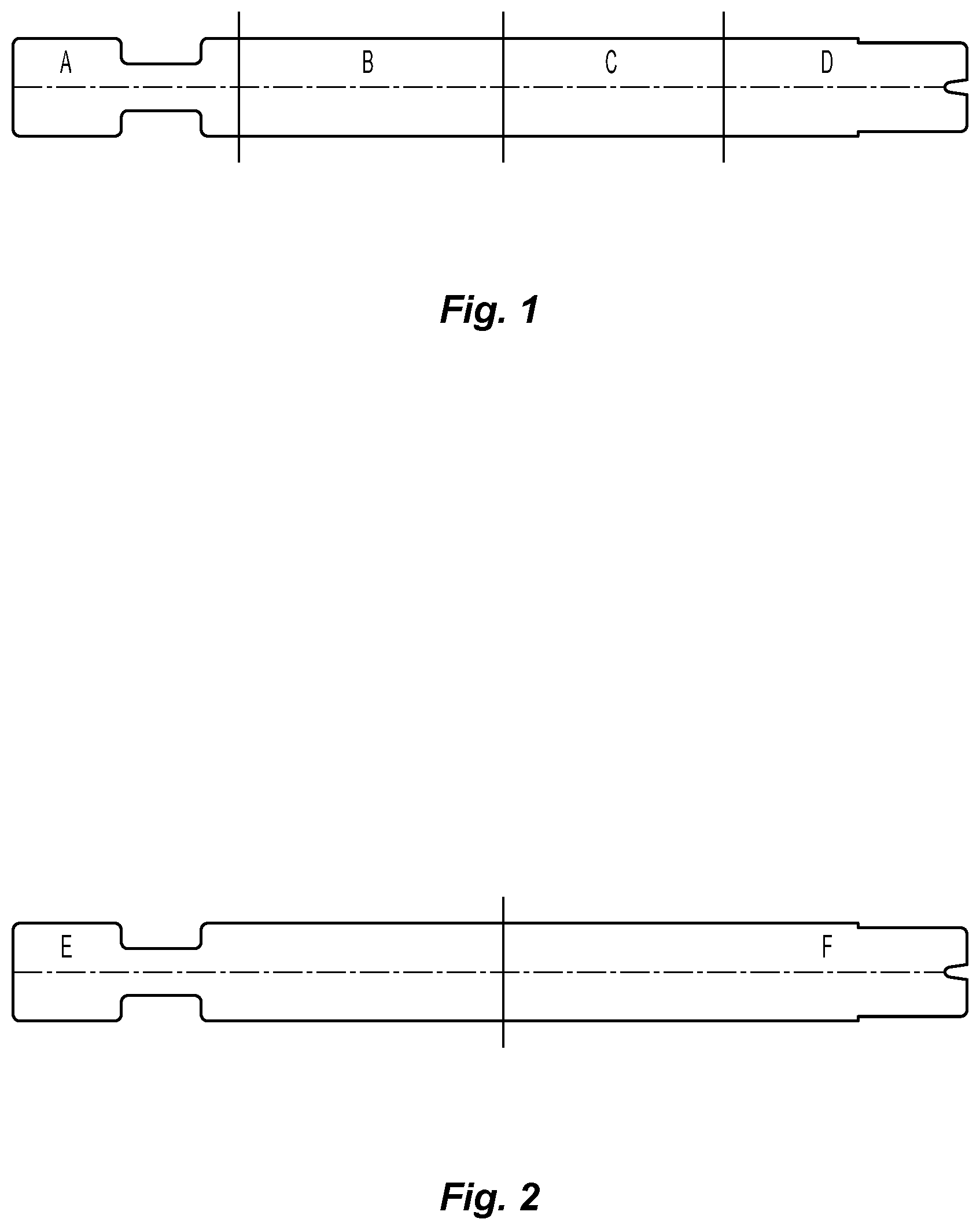
FIG. 1 is a view of a buttress that has been treated in accordance with an embodiment of the present disclosure, showing how the buttress was cut for testing;
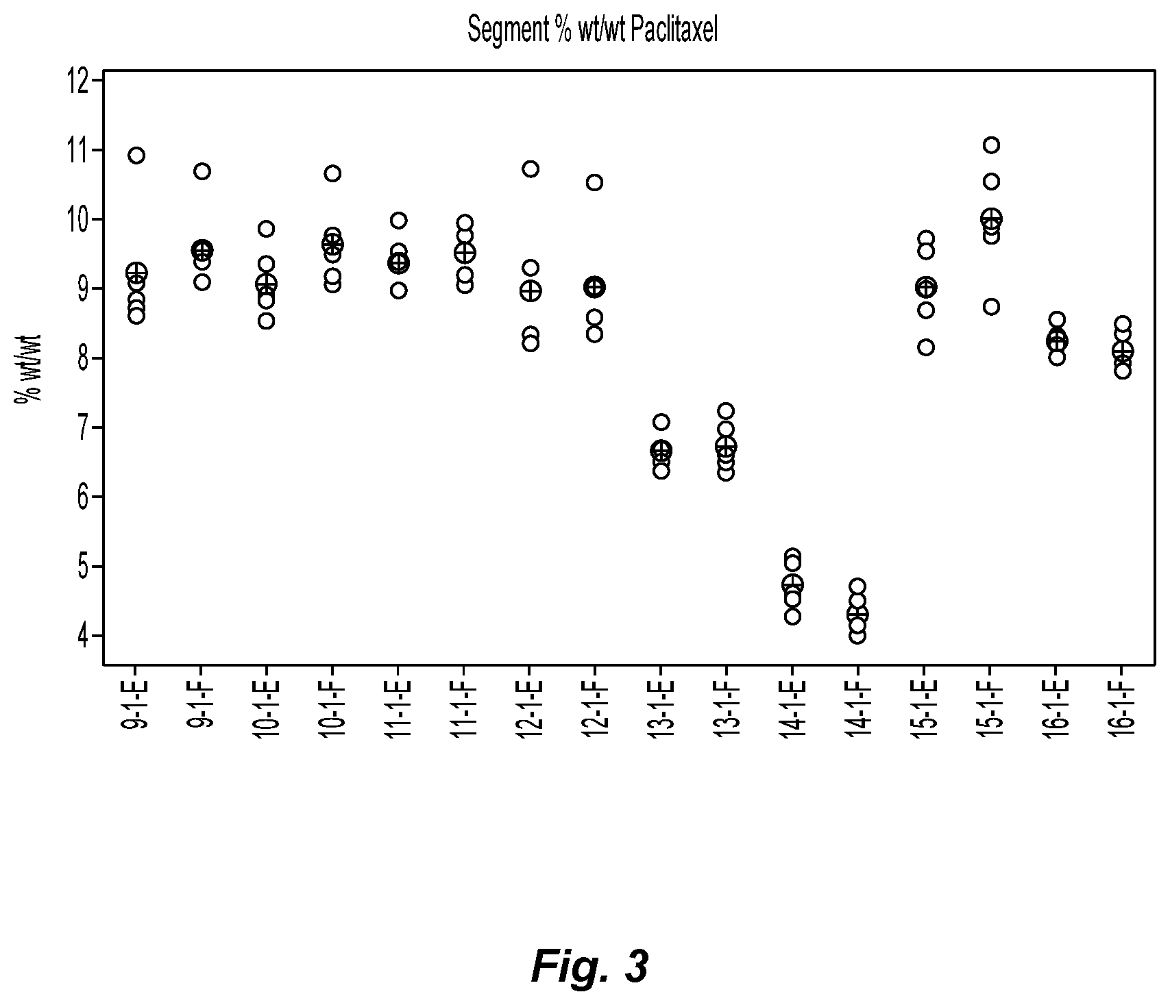
FIG. 2 is an alternate view of a buttress that has been treated in accordance with an embodiment of the present disclosure, showing a different pattern for cutting the buttress for testing;
FIG. 3 is a graph showing the % weight/weight paclitaxel found on the individual segments of buttress as depicted in FIG. 2 after application of paclitaxel thereto;
FIG. 4 is a graph showing the average cumulative paclitaxel eluted from buttresses with various coatings;
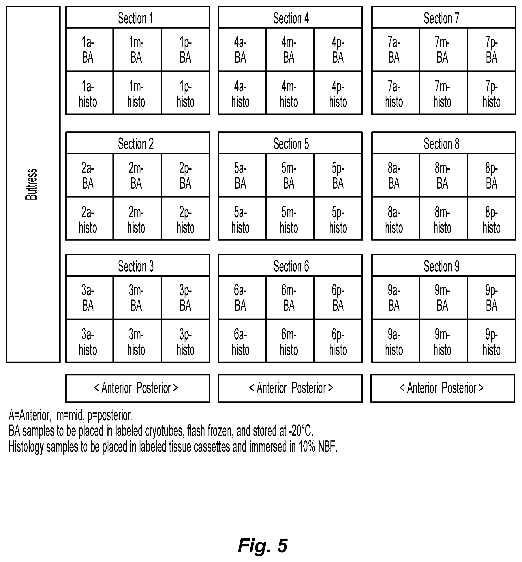
FIG. 5 is a depiction of a lung sectioning scheme for sampling tissue adjacent a buttress of the present disclosure after its placement in a dog;
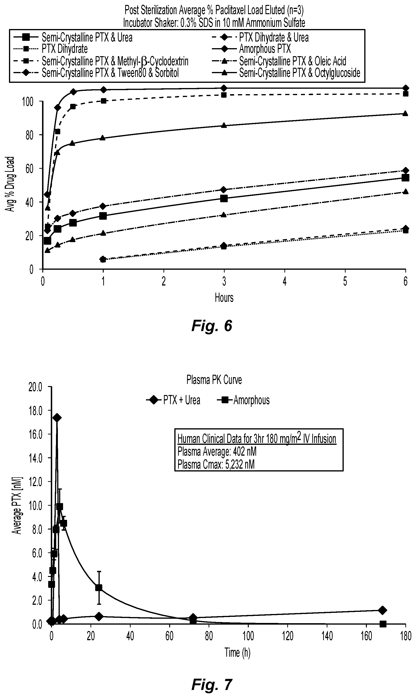
FIG. 6 is a graph depicting the elution curves for formulations 9-16 of the present disclosure.
FIG. 7 is a graph depicting plasma levels of paclitaxel after placement of two buttresses of the present disclosure in a dog;
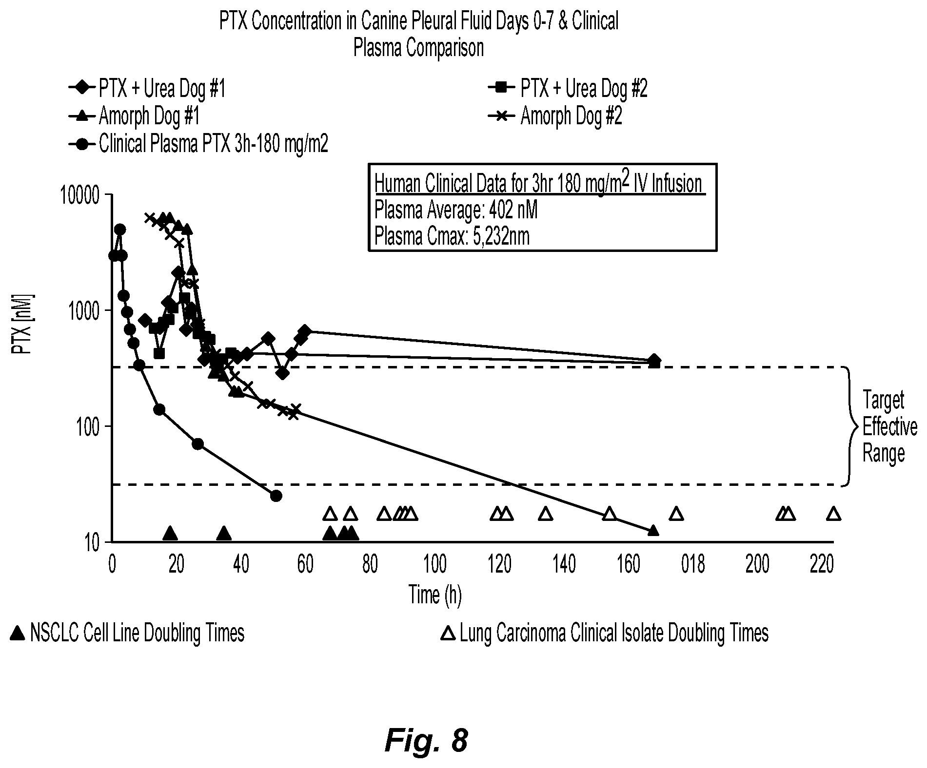
FIG. 8 is a graph summarizing the paclitaxel concentration in canine pleural fluid for days 0-7 after implantation, compared with observed clinical plasma levels;
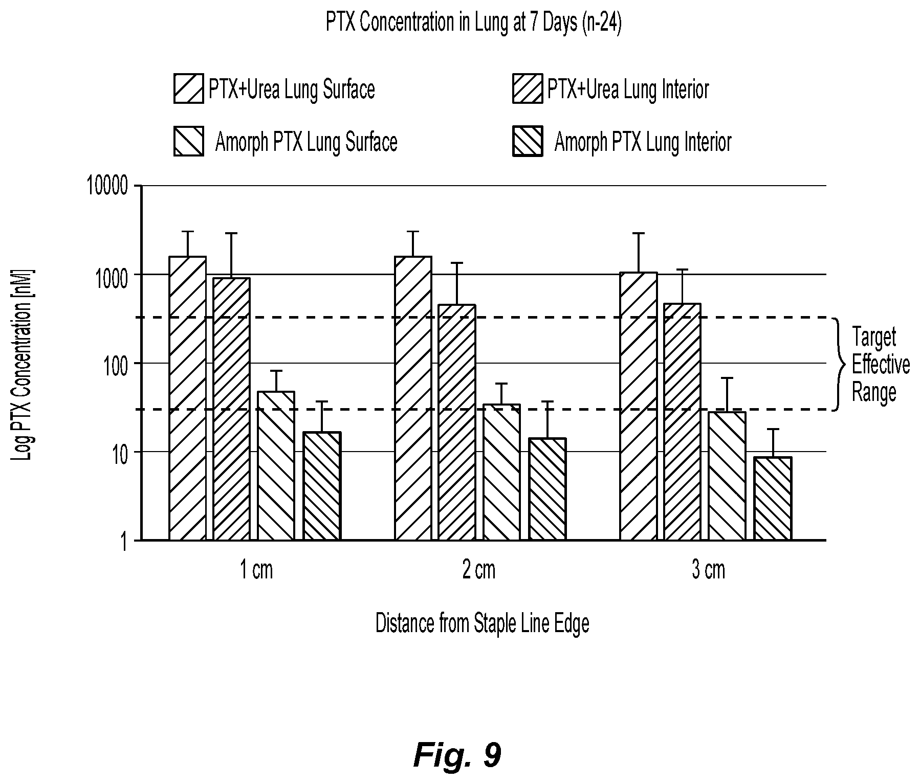
FIG. 9 is a graph showing the concentration of various paclitaxel formulations of the present disclosure in the dog lung after 7 days, with varying distances from the staple line; and
FIG. 10 is a graph showing paclitaxel concentrations of paclitaxel formulations in other tissues (mediastinum, chest wall, pericardium, diaphragm, mediastinal lymph node, bronchus, esophagus and heart) after 7 days.
DETAILED DESCRIPTION
Various exemplary embodiments of the present disclosure are discussed herein below in terms of buttresses for use with tissue fixation devices, in embodiments surgical staples. While the below disclosure discusses in detail the use of these buttresses with staples, it will be appreciated that medical devices of the present disclosure include a range of buttressing materials and film-based medical devices that are used to mechanically support tissues, reinforce tissues along staple or suture lines, and decrease the incidence of fluid leakage and/or bleeding of tissues. For example, other suitable medical devices include hernia patches, staples, tacks, stents, and tissue scaffolds.
Medical devices of the present disclosure may be used with any fixation device utilized to close any wound, defect, and/or opening in tissue. Thus, while surgical buttresses are discussed in conjunction with a surgical stapling apparatus, it is envisioned that other fixation devices, such as tacks, sutures, clips, adhesives and the like, may be utilized in conjunction with medical devices of the present disclosure to affix the medical devices to tissue. Medical devices that are not used with a tissue fixation device, or other tissue support devices, are contemplated.
In embodiments, a buttress of the present disclosure may have a therapeutic layer or coating thereon which includes therapeutic agents for further treatment of tissue at or near the site where the surgical buttress of the present disclosure is placed. Thus, the present disclosure describes surgical buttresses, and methods and mechanisms for using the same, for the targeted delivery of therapeutic agents to a patient.
In the following discussion, the terms "proximal" and "trailing" may be employed interchangeably, and should be understood as referring to the portion of a structure that is closer to a clinician during proper use. The terms "distal" and "leading" may also be employed interchangeably, and should be understood as referring to the portion of a structure that is further from the clinician during proper use. As used herein, the term "patient" should be understood as referring to a human subject or other animal, and the term "clinician" should be understood as referring to a doctor, nurse or other care provider and may include support personnel.
Medical devices of the present disclosure, including surgical buttresses, may be fabricated from a biocompatible substrate material which is a bioabsorbable, non-absorbable, natural, or synthetic material. The medical device may also be formed of materials that are porous or non-porous. It should of course be understood that any combination of porous, non-porous, natural, synthetic, bioabsorbable, and/or non-bioabsorbable materials may be used to form a medical device of the present disclosure.
In embodiments, the medical devices of the present disclosure, such as a surgical buttress, may be biodegradable, so that the device does not have to be retrieved from the body. The term "biodegradable" as used herein is defined to include both bioabsorbable and bioresorbable materials. By biodegradable, it is meant that the medical device decomposes or loses structural integrity under body conditions (e.g., enzymatic degradation or hydrolysis), or is broken down (physically or chemically) under physiologic conditions in the body such that the degradation products are excretable or absorbable by the body.
Non-limiting examples of materials which may be used in forming a medical device of the present disclosure, for example a surgical buttress, include, but are not limited to, poly(lactic acid), poly(glycolic acid), poly(trimethylene carbonate), poly(dioxanone), poly(hydroxybutyrate), poly(phosphazine), polyethylene terephthalate, polyethylene glycols, polyethylene oxides, polyacrylamides, polyhydroxyethylmethylacrylate, polyvinylpyrrolidone, polyvinyl alcohols, polyacrylic acid, polyacetate, polycaprolactone, polypropylene, aliphatic polyesters, glycerols, poly(amino acids), copoly(ether-esters), polyalkylene oxalates, polyamides, poly(iminocarbonates), polyalkylene oxalates, polyoxaesters, polyorthoesters, polyphosphazenes, and copolymers, block copolymers, homopolymers, blends and combinations thereof.
In embodiments, natural biological polymers may be used in forming a medical device of the present disclosure. Suitable natural biological polymers include, but are not limited to, collagen, gelatin, fibrin, fibrinogen, elastin, keratin, albumin, cellulose, oxidized cellulose, hydroxyethyl cellulose, hydroxypropyl cellulose, carboxyethyl cellulose, carboxymethyl cellulose, chitin, chitosan, and combinations thereof. In addition, natural biological polymers may be combined with any of the other polymeric materials described herein to produce a medical device of the present disclosure.
In embodiments, a medical device of the present disclosure, such as a surgical buttress, may be formed of porous material(s). Any porous portion of a medical device of the present disclosure may have openings or pores over at least a part of a surface thereof. Suitable porous materials include, but are not limited to, fibrous structures (e.g., knitted structures, woven structures, non-woven structures, etc.) and/or foams (e.g., open or closed cell foams).
In embodiments, the pores may be in sufficient number and size so as to interconnect across the entire thickness of the medical device. Woven fabrics, knitted fabrics and open cell foams are illustrative examples of structures in which the pores can be in sufficient number and size so as to interconnect across the entire thickness of the medical device.
In other embodiments, the pores may not interconnect across the entire thickness of the medical device. Closed cell foams or fused non-woven materials are illustrative examples of structures in which the pores may not interconnect across the entire thickness of the medical device. In some embodiments, pores may be located on a portion of the medical device, with other portions of the medical device having a non-porous texture. Those skilled in the art may envision a variety of pore distribution patterns and configurations for a porous medical device of the present disclosure.
Where the medical device of the present disclosure is porous and includes fibrous materials, the medical device may be formed using any suitable method including, but not limited to, knitting, weaving, non-woven techniques (including melt blowing), wet-spinning, electro-spinning, extrusion, co-extrusion, and the like. In embodiments, the medical device is a surgical buttress possessing a three dimensional structure, such as the textiles described in U.S. Pat. Nos. 7,021,086 and 6,443,964, the entire disclosures of each of which are incorporated by reference herein.
The porosity of the fabric used to form the substrate may allow for the infiltration of biological fluids and/or cellular components which, in turn, may accelerate the release kinetics of any therapeutic agent from the medical device of the present disclosure, thus increasing the rate of release of therapeutic agent(s) from the medical device into the surrounding tissue and fluids.
Substrates used to form medical devices of the present disclosure, such as surgical buttresses, may have a thickness from about 0.05 mm to about 0.5 mm, in embodiments from about 0.1 mm to about 0.2 mm.
Where the substrate used to form the medical device is porous, the medical device of the present disclosure may have a pore volume from about 65% to about 85%, in embodiments from about 70% to about 80%.
As noted above, in embodiments the medical devices of the present disclosure also include therapeutic agent(s) in a therapeutic layer or coating thereon. Therapeutic agents which may be added to a medical device of the present disclosure include, but are not limited to, drugs, amino acids, peptides, polypeptides, proteins, polysaccharides, muteins, immunoglobulins, antibodies, cytokines (e.g., lymphokines, monokines, chemokines), blood clotting factors, hemopoietic factors, interleukins (1 through 18), interferons (.beta.-IFN, .alpha.-IFN and .gamma.-IFN), erythropoietin, nucleases, tumor necrosis factor, colony stimulating factors (e.g., GCSF, GM-CSF, MCSF), insulin, anti-tumor agents and tumor suppressors, blood proteins, fibrin, thrombin, fibrinogen, synthetic thrombin, synthetic fibrin, synthetic fibrinogen, gonadotropins (e.g., FSH, LH, CG, etc.), hormones and hormone analogs (e.g., growth hormone, luteinizing hormone releasing factor), vaccines (e.g., tumoral, bacterial and viral antigens), somatostatin, antigens, blood coagulation factors, growth factors (e.g., nerve growth factor, insulin-like growth factor), bone morphogenic proteins, TGF-B, protein inhibitors, protein antagonists, protein agonists, nucleic acids, such as antisense molecules, DNA, RNA, RNAi, oligonucleotides, polynucleotides, cells, viruses, and ribozymes.
In embodiments, the therapeutic agent applied to a medical device of the present disclosure may include an anti-tumor agent and/or tumor suppressor, referred to, in embodiments, as a "chemotherapeutic agent" and/or an "antineoplastic agent." Suitable chemotherapeutic agents include, for example, paclitaxel and derivatives thereof, docetaxel and derivatives thereof, abraxane, tamoxifen, cyclophosphamide, actinomycin, bleomycin, dactinomycin, daunorubicin, doxorubicin, doxorubicin hydrochloride, epirubicin, mitomycin, methotrexate, fluorouracil, gemcitabine, gemcitabine hydrochloride, carboplatin, carmustine (BCNU), methyl-CCNU, cisplatin, etoposide, camptothecin and derivatives thereof, phenesterine, vinblastine, vincristine, goserelin, leuprolide, interferon alfa, retinoic acid (ATRA), nitrogen mustard alkylating agents, piposulfan, vinorelbine, irinotecan, irinotecan hydrochloride, vinblastine, pemetrexed, sorafenib tosylate, everolimus, erlotinib hydrochloride, sunitinib malate, capecitabine oxaliplatin, leucovorin calcium, bevacizumab, cetuximab, ramucirumab, trastuzumab, combinations thereof, and the like.
In embodiments, paclitaxel and/or paclitaxel derivatives may be used as the therapeutic agent. Paclitaxel may have various forms, referred to herein as "polymorphs," including amorphous paclitaxel, crystalline paclitaxel, sometimes referred to as crystalline paclitaxel dihydrate, and/or anhydrous paclitaxel, or mixtures thereof.
In accordance with the present disclosure, the polymorph form of paclitaxel utilized in forming the therapeutic layer may be varied by the aqueous composition, the solvent polarity and the composition of protic and aprotic solvents utilized in the solvent system to form the solution for applying the therapeutic layer. For example, paclitaxel dissolved and then dried from 10% v/v water in methanol will yield a predominantly crystalline paclitaxel dihydrate layer, while the same paclitaxel dissolved and then dried from non-polar solvent dichloromethane will yield a predominantly amorphous layer.
The crystallinity of the paclitaxel will impact its solubility in aqueous systems. Accordingly, the polymorph form of paclitaxel in the therapeutic layer may be adjusted and selected to provide a tailored release of therapeutic agent from the implant of the present disclosure. Although the drug in any form is hydrophobic, as amorphous paclitaxel it is more soluble in aqueous environments, and crystalline paclitaxel is less soluble in aqueous environments, more than one polymorphic form of paclitaxel may be used, in embodiments, to provide implants that have multiple release profiles of paclitaxel. For example, medical devices of the present disclosure having both amorphous paclitaxel and crystalline paclitaxel dihydrate thereon may release a bolus of therapeutic agent upon implantation (in the form of the amorphous paclitaxel), while also slowly releasing the therapeutic agent (in the form of the crystalline paclitaxel dihydrate).
In embodiments with no excipient, the amount of amorphous paclitaxel in the therapeutic layer on the medical device may be from 0% to about 100% by weight of the therapeutic layer, in embodiments from about 10% to about 90% by weight of the therapeutic layer, with the crystalline paclitaxel dihydrate being present in amounts from about 0 to about 100% by weight of the therapeutic layer, in embodiments from about 90% to about 10% by weight of the therapeutic layer.
Medical devices of the present disclosure may release amorphous paclitaxel over a period of time from about 24 hours to about 168 hours, in embodiments from about 48 hours to about 96 hours, and release the crystalline paclitaxel dihydrate over a period of time from about 1 week to about 6 weeks, in embodiments from about 2 weeks to about 4 weeks.
In other embodiments, the therapeutic agent may be applied as part of a coating, including polymeric materials or other carrier components within the purview of those skilled in the art. In embodiments, such coatings may include, for example, degradable coatings such as those prepared from monomers such as glycolide, lactide, trimethylene carbonate, p-dioxanone, epsilon-caprolactone, and combinations thereof. If a coating is utilized, the buttress possessing such a coating should remain supple both during and after implantation.
In other embodiments, regardless of whether the therapeutic agent is applied with or without some additional polymeric material to form a coating, in addition to the therapeutic agents described above, therapeutic layers applied to the substrate material in forming a medical device of the present disclosure may also include excipients to enhance both the ability of the therapeutic agent to adhere to the medical device, in embodiments a surgical buttress, as well as to modify the elution of the therapeutic agent from the medical device.
In embodiments, suitable excipients which may be combined with a therapeutic agent to form the therapeutic layer on the medical device include surfactants such as, but not limited to, cyclodextrins such as 2-hydroxypropyl-beta-cyclodextrin and methyl-.beta.-cyclodextrin, sodium dodecyl sulfate, octyl glucoside, and sorbitan fatty acid esters such as sorbitan monooleate, sorbitan monolaurate and polyethoxylated fatty acid esters of sorbitan, sometimes referred to herein as polysorbates, including those sold under the name TWEEN.TM.. Examples of such polysorbates include polysorbate 80 (TWEEN.TM. 80), polysorbate 20 (TWEEN.TM. 20), polysorbate 60 (TWEEN.TM. 60), polysorbate 65 (TWEEN.TM. 65), polysorbate 85 (TWEEN.TM. 85), combinations thereof, and the like. In embodiments, low molecular weight poly(ethylene glycol)s may be added as an excipient, either alone or in any combination with any of the other above excipients.
In other embodiments, suitable excipients may include salts such as sodium chloride and/or other materials such as urea, oleic acid, citric acid, and ascorbic acid. In yet other embodiments, the excipient may be a stabilizer such as butylated hydroxytoluene (BHT).
Still other suitable excipients include polyhydric alcohols such as D-sorbitol, mannitol, combinations thereof, and the like.
In certain embodiments, suitable excipients include urea, methyl-.beta.-cyclodextrin, oleic acid, polysorbate 80, D-sorbitol, octylglucoside, combinations thereof, and the like.
In some embodiments, excipients which are hydrotropes may be included in the therapeutic layers of the present disclosure. These materials attract water into the therapeutic layer, which may enhance its degradation and resulting release of the therapeutic agent from the therapeutic layer. However, in view of the high surface to volume ratios for the therapeutic agents in the therapeutic layers of the present disclosure, such excipients are not, in fact, required. Embodiments of the present disclosure include a therapeutic layer having a chemotherapeutic agent without an excipient.
The therapeutic agent(s) and any excipient may be applied to a medical device of the present disclosure by any method within the purview of those skilled in the art. As noted above, in embodiments the therapeutic agent is in a solution, which is then applied to a medical device of the present disclosure, such as a buttress. The solution possessing the therapeutic agent, along with any excipient, may be applied to the medical device by any method within the purview of those skilled in the art, including spraying, dipping, solution casting, combinations thereof, and the like. After application, the solvent may be driven off by methods within the purview of those skilled in the art, including heating, the application of a vacuum, combinations thereof, and the like. Driving off the solvent leaves the therapeutic agent and any excipient behind to form the therapeutic layer on the medical device.
After formation, medical devices of the present disclosure may possess the therapeutic agent in the coated buttress thereon in amounts from about 0.1% to about 50% by weight of the coated buttress, in embodiments from about 1% to about 10% by weight of the coated buttress. While excipients are not required, where present, non-polymeric excipients may be present in an amount from about 0.01% to about 80% by weight of the coated buttress, in embodiments from about 1% to about 11% by weight of the coated buttress. In other embodiments, where present, polymeric excipients may be present in an amount from about 0.014% to about 14% by weight of the coated buttress, in embodiments from about 5% to about 15% by weight of the coated buttress.
After formation, medical devices of the present disclosure may possess the therapeutic agent in the therapeutic layer thereon in amounts from about 0.01% to about 100% by weight of the therapeutic layer, in embodiments from about 1% to about 75% by weight of the therapeutic layer. While excipients are not required, where present, non-polymeric excipients may be present in an amount from about 1% to about 99% by weight of the therapeutic layer, in embodiments from about 8.5% to about 79.4% by weight of the therapeutic layer, and most preferably in embodiments from 9.5% to about 15%. In embodiments, where present, polymeric excipients may be present in an amount from about 1% to about 99% by weight of the therapeutic layer, in embodiments from about 5% to about 15% by weight of the therapeutic layer.
A therapeutic layer having both a therapeutic agent and non-polymeric excipients may have a thickness from about 13 nm to about 2.9 .mu.m, in embodiments from about 25 nm to about 100 nm.
A therapeutic layer having both a therapeutic agent and polymeric excipients may have a thickness from about 2 nm to about 1.1 .mu.m, in embodiments from about 30 nm to about 100 nm.
In other embodiments, the therapeutic layers may include little or no excipients, so very thin therapeutic layers may be applied to the substrate. This will maintain the porosity of the substrate. Such therapeutic layers may have a thickness from about 11 nm to about 218 nm, in embodiments from about 25 nm to about 75 nm.
In embodiments where the substrate is porous, the therapeutic layer may be present on surfaces throughout the substrate, including within the pores itself. Such a device with non-polymeric excipients or no excipients may have the therapeutic layer at a surface to volume ratio from about 500 mm.sup.-1 to about 90,000 mm.sup.-1. Such a device with polymeric excipients may have the therapeutic layer at a surface to volume ratio from about 1,100 mm.sup.-1 to about 87,000 mm.sup.-1. This high surface to volume ratio enables relatively fast elution of therapeutic agents from the therapeutic layer, especially hydrophobic drugs such as paclitaxel that have low aqueous solubility. A very high surface to area ratio, providing a high surface area for eluding the chemotherapeutic agent, with a low percentage of the weight of the buttress or other device, has been achieved.
In embodiments, the therapeutic layers of the present disclosure may fragment upon affixation of the medical device possessing the therapeutic layer to tissue. This may result in migration of the therapeutic agent to locations distant from the site of implantation, for example in cases where the buttress is attached to the periphery of a lung lobe, therapeutic agent may migrate into mediastinal lymph nodes, while therapeutic agent(s) remaining on the implant may diffuse directly into tissue adjacent the site of implantation.
As described in greater detail in the Examples below, it has surprisingly been found in a dog model that a medical device having paclitaxel in a therapeutic layer of the present disclosure may release paclitaxel throughout the pleural cavity and reach therapeutic levels in other distant sites in the chest cavity, including the chest wall, diaphragm, esophagus, mediastinum, and pericardium. These are all sites of possible local recurrence of cancer after surgical resection. This widespread distribution at therapeutic levels of a poorly soluble drug such as paclitaxel is surprising. Additionally, very low levels of paclitaxel were observed in the blood, meaning toxicity associated with traditional intravenous therapy may be avoided. Utilizing the implants of the present disclosure, a local regional therapy for treating both the lungs and chest is now possible.
Without wishing to be bound by any theory, it is believed multiple mechanisms are responsible for these surprising results. As noted above, the buttress morphology provides a large surface area, giving more opportunity for the paclitaxel to diffuse away from the buttress. Additionally, some of the therapeutic layer flakes off during firing of staples through the medical device, and migrates into the pleural fluid. Once there, the flakes dissolve and deliver paclitaxel wherever the pleural fluid travels. This could explain the distant migration of the paclitaxel to sites like the chest wall, diaphragm, esophagus, and pericardium.
As noted above, the medical device of the present disclosure may be used with any fixation device to further assist in sealing tissue. For example, medical devices of the present disclosure may be used in conjunction with staples, tacks, clips, sutures, adhesives, combinations thereof, and the like.
In embodiments, medical devices of the present disclosure may be used with staples. For example, a surgical buttress formed of a medical device of the present disclosure is provided to reinforce and seal the lines of staples applied to tissue by a surgical stapling apparatus. The buttress may be configured into any shape, size, or dimension suitable to fit any surgical stapling, fastening, or firing apparatus.
In embodiments, the buttresses described herein may be used in sealing a wound by approximating the edges of wound tissue between a staple cartridge and an anvil of a surgical stapling apparatus which contains the buttress. Firing of the surgical stapling apparatus forces the legs of at least one staple to pass through the opening on the staple cartridge and the buttress, the tissue, and the openings on the anvil to secure the buttress to the tissue, to secure the adjoining tissue to one another, and to seal the tissue.
Where the medical device of the present disclosure is used to form a surgical buttress, upon application to a site of bleeding tissue, the buttress may affect hemostasis of said tissue. As used herein, the term "hemostasis" means the arrest of bleeding.
In addition to providing hemostasis at the site of application of the buttress, the medical devices of the present disclosure may also provide for treatment of tissue with the therapeutic agent at both the site of implantation and elsewhere in the body.
In some embodiments, the present disclosure provides methods of treating cancer. These methods include, in embodiments, introducing to a patient needing treatment a surgical stapler having a buttress thereon, the buttress including a coating of a drug, such as a chemotherapeutic agent, and using the stapler to remove an undesired portion of an organ and to place the buttress in a remaining portion of the organ, including stapling the buttress to tissue and cutting the tissue. Where the tissue to be removed is within a patient's body, the method includes introducing the stapler and buttress into the patient's body.
For example, in embodiments, it has been found that for applications such as lung resection in the treatment of lung cancer, the application of medical devices of the present disclosure, in embodiments surgical buttresses, will treat the site of application of the surgical buttress with a chemotherapeutic agent, such as paclitaxel or its derivatives. Moreover, it has been found that the devices of the present disclosure, depending upon the form of the chemotherapeutic agent, excipients, combinations thereof, and the like, may also elute the chemotherapeutic agent therefrom. The chemotherapeutic agent may be physically removed from the surgical buttress by mechanical/physical forces imparted to the buttress upon firing of a staple therethrough. The chemotherapeutic agent may also dissolve into the pleural fluid within the pleural space and travel throughout the space.
In embodiments, the use of the medical devices of the present disclosure, such as surgical buttresses, may be utilized to maintain therapeutic levels of chemotherapeutics such as paclitaxel, thereby continuing to treat a patient and prevent recurrence of non-small cell lung cancer.
Benefits of the introduction of the chemotherapeutic with the medical devices of the present disclosure include, for example: Eliminates systemic toxicity typically associated with intravenous chemotherapy; Reduce drug payload to .about.10% of conventional intravenous chemotherapy infusion; and Provides prolonged exposure and in the case of paclitaxel provides greater potency at lower drug concentrations.
Several embodiments of the disclosure are described below with reference to the following non-limiting Examples. The Examples are intended to be illustrative only and are not intended to limit the scope of the present disclosure. As used herein, "room temperature" refers to a temperature of from about 20.degree. C. to about 30.degree. C. Also, parts and percentages, such as solution percentages, are by weight unless otherwise indicated.
Example 1
The feasibility of applying paclitaxel to lung staple buttresses was tested as follows. Staple buttresses made of polyglycolic acid were utilized for the tests. Sections of the material were soaked in tetrahydrofuran, chloroform, toluene, dichloromethane or methanol or combinations thereof as potential formulation solvents. The buttresses visually appeared to be compatible with each solvent, with no deformation or tackiness after several days.
The paclitaxel formulation used to coat the buttress material was a solution of 50 mg/mL paclitaxel and 7 mg/mL urea, in 10:90 v/v water:THF (commercially available as FREEPAC.TM. paclitaxel eluting formulation). The dried formulation was expected to contain a mixture of amorphous paclitaxel and paclitaxel dihydrate). This formulation was used to coat three buttresses.
Roughly 5 mL of the paclitaxel solution described above was placed in three (3) small vials and a buttress (40 mm in length) was delivered to each vial and allowed to soak in the paclitaxel solution for less than 30 seconds. Each buttress was removed with tweezers and allowed to dry. The buttresses appeared visually dry in 15 to 30 seconds after removal from the paclitaxel solution. Each buttress was then placed on a glass plate and allowed to fully dry for about ten minutes.
No real change in the visual appearance of the buttresses was observed. Light handling did not produce any dusting or particulate from the buttresses. Treated and untreated buttresses were photographed at 50.times. magnification, with little to no difference in the surface appearance observed.
Each buttress and an untreated buttress were analyzed for paclitaxel as follows. Coupons were extracted with 0.5% v/v acetic acid in methanol under sonication for about 30 minutes. The extract was analyzed using an ultrahigh performance liquid chromatograph with UV detection at 229 nm for residual paclitaxel against a standard of known concentration.
Recovery of paclitaxel was observed with a normal related compound profile. No interfering peaks were observed in the untreated buttress. The results are summarized in Table 1 below.
TABLE-US-00001 TABLE 1 Recovered paclitaxel Paclitaxel % Related Sample Material (.mu.g) Substances 1 40 mm 5362 0.16 buttress 2 40 mm 4795 0.15 buttress 3 40 mm 5018 0.16 buttress
In view of the above data in Table 1, dip coating appeared to be effective for applying the paclitaxel solution to a buttress.
Example 2
The production of staple buttresses with varying forms of paclitaxel was tested as follows. Paclitaxel solutions were prepared using crystalline paclitaxel dihydrate, amorphous paclitaxel, and a combination of the two (as described above in Example 1). Two of the samples included urea as an excipient.
The formulations prepared, including the amounts and various forms of paclitaxel (PTX), excipient, if any, and the solvents used to make the paclitaxel solutions, are summarized below in Table 2.
TABLE-US-00002 TABLE 2 PTX Urea Sample (mg/mL) (mg/mL) Solvent PTX polymorph 4 50.9 7.0 90:10 mixed THF:water dihydrate:amorphous 5 24.9 3.5 90:10 dihydrate methanol:water 6 25.6 0.0 methylene amorphous chloride
Sample 4 included urea as an excipient. For consistency, urea was included at the same ratio in the dihydrate material (sample 5). No urea was included in the amorphous formulation (sample 6).
90.times.10 mm buttress profiles (8 cm.sup.2 one-sided fabric area) were dip coated in each formulation and dried. As with the results described above in Example 1, there was no visible change to the surface of the buttress with any of the formulations. The drug showed excellent affinity to the polymer fabric and a buttress from each formulation was aggressively handled, shaken, and hit against a glass plate with no visible shedding of drug. The coated buttresses were set aside for elution analysis.
Example 3
A formulation of paclitaxel in 10:90 v/v toluene:THF was prepared at a concentration of 51.1 mg/mL, and designated Sample 8. This formulation produces an amorphous paclitaxel layer, which was used to coat five 90.times.10 mm buttress profiles (8 cm.sup.2 one-sided fabric area)(referred to as Samples 8.1, 8.2, 8.3, 8.4 and 8.5), each of which was weighed prior to application. After application, each buttress was cut into four pieces, A, B, C, and D, as depicted in FIG. 1. Each section was weighed and then tested per a developmental drug content method. Buttresses were extracted with 0.5% v/v acetic acid in methanol under sonication for 15 minutes. The sample extracts were analyzed using a UPLC with UV detection at 229 nm for both paclitaxel and related compounds against a standard of known paclitaxel concentration using a water and acetonitrile gradient on an Agilent Zorbax RRHD Eclipse PlusC18, 2.1.times.100 mm, 1.8-.mu.m particle size column.
Weights, drug mass, and % weight/weight observed on each segment of the buttress is summarized below in Tables 3, 4 and 5.
TABLE-US-00003 TABLE 3 Uncoated Coated Weights (mg) % Weight by segment Delta weight Sample (mg) A B C D Total (mg) gain 8.1 41.18 14.06 9.57 10.99 10.5 45.12 3.94 9.57 8.2 42.71 12.5 13.04 9.77 11.33 46.64 3.93 9.20 8.3 43.53 13.42 12.38 9.11 12.72 47.63 4.10 9.42 8.4 43.32 11.44 13.11 11.33 11.05 46.93 3.61 8.33 8.5 42.22 12.22 13.4 10.98 9.82 46.42 4.20 9.95
TABLE-US-00004 TABLE 4 Recovered paclitaxel (mg) per segment A B C D Total 8.1 1.35 0.90 1.02 0.82 4.08 8.2 1.14 1.10 0.84 0.90 3.97 8.3 1.33 1.15 0.79 1.05 4.33 8.4 1.07 1.13 0.95 0.85 4.00 8.5 1.14 1.14 0.91 0.84 4.03
TABLE-US-00005 TABLE 5 Paclitaxel, % weight/weight per segment Sample A B C D Avg 8.1 9.57 9.41 9.25 7.84 9.02 8.2 9.10 8.43 8.62 7.91 8.51 8.3 9.94 9.31 8.64 8.28 9.04 8.4 9.36 8.65 8.35 7.65 8.50 8.5 9.32 8.53 8.28 8.56 8.67
As can be seen from the data summarized in Table 3 above, the buttress material gained 9.3% weight on average, or about 4 mg of drug. Results of recovered paclitaxel (Table 4) and paclitaxel observed as % weight/weight per segment (Table 5) were fairly consistent.
Example 4
Sheets of polyglycolic acid fabric were punched to form 90.times.10 mm buttress profiles, and then paclitaxel formulations were applied thereto. The formulations and testing are described below.
Paclitaxel formulations were prepared at concentrations of 25 mg/mL with various excipients. Crystallinity of the paclitaxel in samples 10, 11 and 12 was controlled for, with the paclitaxel being either completely crystalline paclitaxel dihydrate or a fully amorphous form. The composition of the remaining samples (9, 13, 14, 15 and 16), based on the 10:90 water:THF solvent system described above in Example 1, is expected to contain a mixture of amorphous paclitaxel and paclitaxel dihydrate. The various formulations are summarized below in Table 6.
TABLE-US-00006 TABLE 6 Paclitaxel (PTX) (PTX) Excipient Exc PTX:Exc Sample polymorph Solvent (mg/mL) (Exc) (mg/mL) (mol:mol) 9 PTX semi 10% water 24.99 Urea 3.50 1:2 crystalline in THF w/ Urea 10 PTX 10% water 24.83 Urea 3.50 1:2 Dihydrate in 65:35 w/ Urea v:v MeOH:acetone 11 PTX 10% water 25.17 NA NA NA Dihydrate in 65:35 v:v MeOH:acetone 12 Amorphous 10% 25.00 NA NA NA PTX toluene in THF 13 PTX semi 10% water 24.86 Methyl-.beta.- 76.59 1:2 crystalline in THF Cyclodextrin 14 PTX semi THF w/ 25.13 Oleic Acid/Na 3.1 1:0.37 crystalline 1.3% Oleate water, 2.9% EtOH 15 PTX semi 10% water 24.89 Tween 80/ 0.375/ 1:0.01/1:4 crystalline in THF Sorbitol 21.62 16 PTX semi 10% water 25.16 Octylglucoside 34.37 1:4 crystalline in THF
Five milliliters of each of the above formulations was prepared and poured into the reservoir of a coating fixture (the reservoir volume was 25 mL). A sheet of twelve buttress profiles was passed through the reservoir by hand, coating the material, and was held with tweezers to dry. All solvent systems dried very quickly, within less than 30 seconds.
As in earlier Examples 1 and 2 above, little to no change was visible on the material to the naked eye for all formulations, with the exception of sample 10 (crystalline paclitaxel dihydrate with urea). For that sample, some white streaking and non-uniform coating was observed. Buttress profiles were removed from the sheet with tweezers.
The buttresses were then tested for drug potency/uniformity. Five buttresses from each formulation (designated sample 9-1, 9-2, 9-3, 9-4, 9-5, 10-1, 10-2, 10-3, etc.) were extracted and tested per a developmental drug content method. Buttresses were extracted with 0.5% v/v acetic acid in methanol under sonication for 15 minutes. The sample extracts were analyzed using a UPLC with UV detection at 229 nm for both paclitaxel and related compounds against a standard of known paclitaxel concentration using a water and acetonitrile gradient on an Agilent Zorbax RRHD Eclipse PlusC18, 2.1.times.100 mm, 1.8-.mu.m particle size column.
Each buttress was cut into two segments, E and F, as depicted in FIG. 2. The segments, representing roughly half of the buttress profile, were weighed and tested individually.
The segment and total values are shown in FIG. 3 (FIG. 3 has % weight/weight paclitaxel for the individual segments) and Table 7 below (the letter for each sample corresponding with the segment tested as depicted in FIG. 2), and the averages for each formulation are shown below in Table 8 below.
TABLE-US-00007 TABLE 7 Individual segments % weight/weight paclitaxel Weight Paclitaxel Paclitaxel % Sample (mg) (.mu.g) weight/weight 9-1-E 20.93 1808 8.64 9-1-F 17.70 1611 9.10 9-2-E 21.83 1985 9.09 9-2-F 23.06 2096 9.09 9-3-E 18.29 1999 10.93 9-3-F 21.73 2327 10.71 9-4-E 20.23 1772 8.76 9-4-F 22.81 2180 9.56 9-5-E 18.40 1628 8.85 9-5-F 18.68 1757 9.41 10-1-E 18.33 1624 8.86 10-1-F 19.97 1896 9.49 10-2-E 21.95 1872 8.53 10-2-F 21.65 1963 9.07 10-3-E 17.69 1657 9.37 10-3-F 20.13 1970 9.79 10-4-E 17.46 1724 9.87 10-4-F 20.11 2148 10.68 10-5-E 21.56 1923 8.92 10-5-F 21.35 1963 9.19 11-1-E 16.26 1460 8.98 11-1-F 19.60 1777 9.07 11-2-E 20.71 1976 9.54 11-2-F 22.35 2185 9.78 11-3-E 19.78 1858 9.39 11-3-F 21.59 2073 9.60 11-4-E 20.35 1834 9.01 11-4-F 17.63 1623 9.21 11-5-E 20.09 2008 10.00 11-5-F 19.01 1895 9.97 12-1-E 16.16 1504 9.31 12-1-F 18.41 1584 8.60 12-2-E 20.04 2150 10.73 12-2-F 17.88 1886 10.55 12-3-E 15.74 1293 8.21 12-3-F 18.89 1622 8.59 12-4-E 17.15 1422 8.29 12-4-F 20.48 1718 8.39 12-5-E 22.15 1851 8.36 12-5-F 20.51 1857 9.05 13-1-E 24.37 1592 6.53 13-1-F 26.71 1768 6.62 13-2-E 22.95 1626 7.08 13-2-F 21.79 1583 7.26 13-3-E 19.45 1290 6.63 13-3-F 26.46 1685 6.37 13-4-E 21.00 1344 6.40 13-4-F 22.63 1468 6.49 13-5-E 25.79 1732 6.72 13-5-F 19.90 1393 7.00 14-1-E 20.31 872 4.29 14-1-F 25.54 1020 3.99 14-2-E 22.78 1031 4.53 14-2-F 24.94 1027 4.12 14-3-E 22.51 1038 4.61 14-3-F 25.35 1065 4.20 14-4-E 20.30 1030 5.07 14-4-F 27.83 1258 4.52 14-5-E 26.34 1360 5.16 14-5-F 25.18 1184 4.70 15-1-E 20.67 1797 8.69 15-1-F 17.99 1899 10.56 15-2-E 17.93 1745 9.73 15-2-F 20.18 1770 8.77 15-3-E 19.59 1766 9.01 15-3-F 16.89 1672 9.90 15-4-E 22.66 1852 8.17 15-4-F 19.91 1946 9.77 15-5-E 18.43 1763 9.57 15-5-F 20.86 2314 11.09 16-1-E 20.37 1639 8.05 16-1-F 18.93 1609 8.50 16-2-E 20.23 1644 8.13 16-2-F 20.31 1587 7.81 16-3-E 17.59 1464 8.32 16-3-F 22.21 1758 7.92 16-4-E 18.30 1507 8.23 16-4-F 20.06 1579 7.87 16-5-E 19.11 1639 8.58 16-5-F 23.03 1923 8.35
TABLE-US-00008 TABLE 8 Average potency values .+-. SD Weight Paclitaxel Paclitaxel % Formulation (mg) (.mu.g) weight/weight 9 40.73 .+-. 3.20 3833 .+-. 416 9.41 .+-. 0.79 10 40.04 .+-. 2.96 3748 .+-. 165 9.39 .+-. 0.59 11 39.47 .+-. 2.82 3738 .+-. 379 9.46 .+-. 0.40 12 37.48 .+-. 3.30 3377 .+-. 474 9.01 .+-. 0.94 13 46.21 .+-. 2.87 3096 .+-. 211 6.70 .+-. 0.30 14 48.22 .+-. 2.06 2177 .+-. 249 4.50 .+-. 0.33 15 39.02 .+-. 2.24 3705 .+-. 252 9.50 .+-. 0.55 16 40.03 .+-. 1.42 3270 .+-. 176 8.17 .+-. 0.19
As can be seen from the data in Tables 7 and 8, there was a fair amount of variation in the individual buttress weights, but the % weight/weight of paclitaxel in the therapeutic layers was fairly consistent within each group. Most formulations had between 8 and 10% drug by weight. Formulations 13 (methyl-.beta.-cyclodextrin excipient) and 14 (oleic acid/Na oleate excipient) had less drug in the therapeutic layer.
Drug elution from the buttresses was then examined as follows. Three buttresses from each formulation were mounted on mandrels and introduced to 37.degree. C. 0.3% SDS in 10 mM ammonium acetate and placed in a 37.degree. C. incubator shaker at 100 rpm. At each timepoint, the mandrel and buttress were removed and transferred to a fresh vial of media. Timepoints varied per formulation. An aliquot of each formulation was filtered through a 0.2 .mu.m nylon filter and analyzed by a developmental HPLC method for similar sample types. A water and acetonitrile gradient on a Luna 3 .mu.m PFP(2) 100 .ANG., 4.6.times.100 column was used for separation with UV detection at 229 nm against a standard of known paclitaxel concentration. FIG. 4 is a graph showing the average cumulative drug eluted from the buttresses. Tables 9, 10, 11, and 12 below summarize the cumulative drug eluted for the buttresses of Formulations 9, 10, 11 and 12, respectively.
TABLE-US-00009 TABLE 9 Formulation 9 cumulative drug eluted (.mu.g) Time (min) 60 180 360 1440 1800 2880 Buttress 1 843 1202 1585 2358 2511 2530 Buttress 2 834 1168 1510 2290 2505 2639 Buttress 3 1012 1369 1722 2495 2790 2958 Avg 896 1246 1606 2381 2602 2709 SD 100 108 108 104 163 222 % RSD 11 9 7 4 6 8
TABLE-US-00010 TABLE 10 Formulation 10 cumulative drug eluted (.mu.g) Time (min) 60 180 360 1440 1800 2880 Buttress 1 699 1044 1324 1916 2165 2408 Buttress 2 673 1023 1357 1979 2297 2627 Buttress 3 661 928 1195 1765 2009 2416 Avg 678 998 1292 1887 2157 2484 SD 20 62 86 110 144 124 % RSD 3 6 7 6 7 5
TABLE-US-00011 TABLE 11 Formulation 11 cumulative drug eluted (.mu.g) Time (min) 60 180 360 1440 1800 2880 Buttress 1 288 459 652 1254 1569 2143 Buttress 2 322 527 757 1343 1706 2282 Buttress 3 288 482 710 1332 1712 2290 Avg 299 489 707 1310 1662 2239 SD 20 35 53 48 81 83 % RSD 7 7 7 4 5 4
TABLE-US-00012 TABLE 12 Formulation 12 cumulative drug eluted (.mu.g) Time (min) 5 10 15 30 120 240 1440 Buttress 1 1096 1668 1979 2317 2531 2582 2707 Buttress 2 1356 2401 3053 3657 4082 4218 4542 Buttress 3 1225 1988 2437 2925 3252 3345 3559 Avg 1225 2019 2489 2966 3289 3382 3603 SD 130 367 539 671 776 818 919 % RSD 11 18 22 23 24 24 25
As can be seen from the data set forth in Tables 9-12, by varying the excipients and forms of paclitaxel applied to a buttress, as well as the solvents used to form the solutions used to apply the paclitaxel and excipient to the buttress, both the amounts of paclitaxel released from the buttresses and the release profiles of the paclitaxel (e.g., bolus vs. extended release) could be adjusted.
FIG. 6 is a graph depicting the elution curves for formulations 9-16.
The surface of each buttress was imaged by a scanning electron microscope (SEM). Results were generally as expected, with crystalline formulations showing needle formations, and amorphous formulations showing tightly coated fibers and some webbing between fibers. Formulation 16 differed slightly in that it appeared to more thoroughly coat the material than the other semi-amorphous formulations.
Example 5
A study was conducted where buttresses of the present disclosure were implanted in the pleural cavity of dogs, and the elution and migration of drug on the buttress was measured.
Briefly, two formulations were implanted in a total of four dogs (one formulation in two dogs and the other formulation in the other two dogs). Bilateral thoracotomies were created and 60 mm staplers having two of the buttresses with paclitaxel thereon were utilized. The first buttress included the formulation of Example 5, sample 9 (semi-crystalline paclitaxel and urea) and the second buttress was the amorphous paclitaxel described above in Example 5, sample 12. The two dogs having the first buttress were referred to as FREEPAC #1 (or PTX+urea dog #1) and FREEPAC #2 (or PTX+urea dog #1), and the two dogs having the second buttress were referred to as Amorph dog #1 and Amorph dog #2.
Each buttress was fired across the tips of five or six lung lobes for each animal. A mediastinum fenestration was placed to allow liquid and air communication between hemi-thoraces. A chest drain was placed and both thoracotomies were closed. The chest drains were removed within about 36 to about 48 hours after surgery. The four animals were euthanized 7 days after surgery and tissues of interest were collected for paclitaxel analysis. The tissues collected included: 1) tissue at the buttress staple line; 2) tissue adjacent the staple line; and 3) various locations away from the buttress, including the thoracic wall, mediastinum, heart, pericardium, mediastinal lymph nodes, remnant lobes, esophagus, bronchus, and diaphragm.
The plasma levels of paclitaxel, and the paclitaxel levels in the chest drain fluid compared with implant time, were also tracked for each animal. Plasma was collected intra-operatively, and then post-operatively at 30 minutes, 1 hour, 2 hours, 4 hours, 6 hours, 24 hours, 72 hours, and 168 hours.
At necropsy, each of the tissues being sampled was divided into a 3.times.3 cm grid and then further sliced into three layers to produce 27 samples for paclitaxel drug analysis. The lung sectioning scheme for the tissue adjacent the buttress is outlined in FIG. 5.
Graphs summarizing the results obtained are set forth as FIGS. 7-10. As depicted in FIG. 7, the plasma levels of both the buttress with amorphous paclitaxel and the buttress with the combination of amorphous paclitaxel and paclitaxel dihydrate with urea subsided over time, with low plasma levels. (The inset box in FIG. 7 is taken from human clinical data (Ohtsu et al.) for a 180 mg/m.sup.2 infusion administered over a 3-hour period. The reported average Cmax value for 3 patients was 5,232.+-.151 nM, while the average plasma value for 3 patients was 402.+-.3 nM, assuming a clearance period of 48 hours to reach paclitaxel plasma levels below therapeutic levels of 30 nM. Local delivery of paclitaxel at the site of lung tissue resection resulted in minimal delivery of drug to the plasma. In fact, both formulations did not cross the therapeutic threshold, with peak levels more than two orders of magnitude lower than normally experienced after a clinical intravenous (IV) dose of paclitaxel. It should also be noted that none of the four dogs experienced any signs of drug toxicity typically experienced with IV paclitaxel delivery, including no significant change in bloodstream neutrophil cell count.)
FIG. 8 summarizes the paclitaxel concentration in canine pleural fluid for days 0-7 after implantation, compared with observed clinical plasma levels. As set forth in FIG. 8, paclitaxel (PTX) pleural fluid concentration in canine study was compared with overlaid clinical plasma paclitaxel levels (Filled circles) and overlaid NSCLC cell line doubling times (Filled triangles) and lung carcinoma clinical isolate doubling times (Open triangles). Target paclitaxel effective range is highlighted in area bordered by dashed lines. The target therapeutic range was determined by adjusting experimental in vitro NSCLC cell line IC90 values with a factor to account for the impact of the in vivo tumor environment. Two paclitaxel containing formulations (semicrystalline paclitaxel+urea vs. amorphous paclitaxel) were each implanted into the lungs of canines for seven days. Plasma and pleural fluid paclitaxel levels were monitored at several time points and paclitaxel tissue levels were measured at seven days post-surgery. The semicrystalline paclitaxel+urea formulation sustained therapeutic levels of paclitaxel in the pleural fluid up to at least the 7 day study termination time point, whereas the amorphous formulation maintained therapeutic levels up to at least 40-60 hours, at which point chest drains were removed. Both locally delivered formulations sustained therapeutic levels of paclitaxel in the pleural fluid space longer than achieved in plasma after intravenous injection in humans. Additionally, both local formulations sustained therapeutic levels beyond the cell doubling times of many lung carcinoma clinical isolates, indicating improved chance of efficacy compared to intravenous paclitaxel administration.
The inset box in FIG. 8 is taken from human clinical data (Ohtsu et al.) for a 180 mg/m.sup.2 infusion administered over a 3-hour period. The reported average Cmax value for 3 patients was 5,232.+-.151 nM, while the average plasma value for 3 patients was 402.+-.3 nM, assuming a clearance period of 48 hours to reach paclitaxel plasma levels below 30 nM. The clinical paclitaxel plasma levels reported by Ohtsu for a 180 mg/m.sup.2 infusion administered over a 3-hour period is plotted in comparison to the paclitaxel pleural fluid levels found in the 7-day canine study. It has been demonstrated in multiple preclinical models that paclitaxel levels in tissues follow plasma levels after IV injection (See, Eiseman, et al. Cancer Chemother. Pharmacol. 1994; 34(6):465-71; Soma, et al. J. Surg. Res. 2009 July; 155(1):142-6; Schrump, et al. J. Thorac. Cardiovasc. Surg. 2002 April; 123(4):686-94.)
Paclitaxel levels in lung after IV injection in mice, rabbits, and sheep are within 0.6-4.3 times the levels found in plasma on a drug mass/tissue mass basis up until plasma levels begin to approach sub-therapeutic paclitaxel levels. Importantly, when paclitaxel is cleared from the bloodstream it is also rapidly washed out of lung and other tissues. From these preclinical observations it can be inferred that IV paclitaxel administered to humans remains at therapeutic concentrations in lung tissue not much longer than 48 hours after initiation of treatment.
It has also been demonstrated that paclitaxel potency increases with exposure time and is more effective at lower concentrations against faster dividing cell types. This effect of increasing potency with exposure duration can be attributed to paclitaxel's mechanism of action; namely paclitaxel must be at sufficiently high concentrations during cell division to disrupt microtubule polymerization and thus cause cell death. Because of this effect, paclitaxel is most effective as a chemotherapeutic agent against slowly dividing cancer cells when maintained over a long period of time at therapeutic levels in the tissue of interest. For example, it has been demonstrated that primary lung carcinoma tumors collected as clinical isolates from 15 affected patients had doubling times ranging from approximately 68 to 296 hours. (Baguley, et al., "Inhibition of growth of primary human tumour cell cultures by a 4-anilinoquinazoline inhibitor of the epidermal growth factor receptor family of tyrosine kinases," Eur. J. Cancer. 1998 June; 34(7):1086-90.)
Because sustained localized delivery of paclitaxel enables therapeutic levels for much longer periods than intravenous therapy this mode of delivery should provide superior efficacy against NSCLC.
FIG. 9 is a graph summarizing the concentration of various paclitaxel formulations in the lung after 7 days, with varying distances from the staple line. Therapeutic levels of paclitaxel were found in canine lung both on the surface and in the lung interior up to 3 cm away from the staple line buttress edge at 7 days post-surgery for the semicrystalline paclitaxel+urea formulation. The amorphous paclitaxel formulation produced therapeutic levels at the surface of lung but not in the lung interior.
FIG. 10 is a graph summarizing paclitaxel concentrations of various paclitaxel formulations in other tissues (mediastinum, chest wall, pericardium, diaphragm, lymph node, bronchus, esophagus and heart) after 7 days. At 7 days post-surgery the semicrystalline paclitaxel+urea formulation produced therapeutic levels of paclitaxel in all regional tissues sampled in the ipsilateral chest except for the heart, whereas the amorphous paclitaxel formulation produced therapeutic levels in the mediastinum and mediastinal lymph node. The distant delivery of therapeutic levels of paclitaxel to the mediastinum and mediastinal lymph nodes is significant as these are sites of typical local and regional recurrence after lobectomies and sublobar resections respectively. In theory, paclitaxel delivery to these structures should reduce the risk of locoregional recurrence after surgery for early stage NSCLC. Additionally, delivery of therapeutic levels of paclitaxel to the chest wall, diaphragm, bronchus and esophagus should reduce the risk of recurrence in these structures as well.
In addition, therapeutic levels of paclitaxel were found in mediastinal lymph nodes after implantation of the implants of the present disclosure. The nodes in which the paclitaxel was found were several centimeters away from the buttressed staple lines. The concentrations of paclitaxel in the lymph node were comparable to the concentration of paclitaxel found within the first 3 cm adjacent to the buttressed staple lines. These results suggest some active transport of paclitaxel to these sites, most likely through the lymphatic drainage system, which is also most often cited as the pathway for metastasis.
It will be understood that various modifications may be made to the embodiments disclosed herein. Therefore, the above description should not be construed as limiting, but merely as an exemplification of preferred embodiments. Those skilled in the art will envision other modifications within the scope and spirit of the present disclosure. Such modifications and variations are intended to come within the scope of the following claims.
* * * * *
D00001
D00002
D00003
D00004
D00005
D00006
D00007
D00008
XML
uspto.report is an independent third-party trademark research tool that is not affiliated, endorsed, or sponsored by the United States Patent and Trademark Office (USPTO) or any other governmental organization. The information provided by uspto.report is based on publicly available data at the time of writing and is intended for informational purposes only.
While we strive to provide accurate and up-to-date information, we do not guarantee the accuracy, completeness, reliability, or suitability of the information displayed on this site. The use of this site is at your own risk. Any reliance you place on such information is therefore strictly at your own risk.
All official trademark data, including owner information, should be verified by visiting the official USPTO website at www.uspto.gov. This site is not intended to replace professional legal advice and should not be used as a substitute for consulting with a legal professional who is knowledgeable about trademark law.