Bispecific immunomodulatory antibodies that bind costimulatory and checkpoint receptors
Bernett , et al. October 6, 2
U.S. patent number 10,793,632 [Application Number 15/691,665] was granted by the patent office on 2020-10-06 for bispecific immunomodulatory antibodies that bind costimulatory and checkpoint receptors. This patent grant is currently assigned to Xencor, Inc.. The grantee listed for this patent is Xencor, Inc.. Invention is credited to Matthew Bernett, Christine Bonzon, John Desjarlais, Michael Hedvat, Gregory Moore.


View All Diagrams
| United States Patent | 10,793,632 |
| Bernett , et al. | October 6, 2020 |
Bispecific immunomodulatory antibodies that bind costimulatory and checkpoint receptors
Abstract
The present invention is directed to bispecific, heterodimeric immunomodulatory antibodies.
| Inventors: | Bernett; Matthew (Monrovia, CA), Moore; Gregory (Azusa, CA), Desjarlais; John (Pasadena, CA), Hedvat; Michael (Encino, CA), Bonzon; Christine (Los Angeles, CA) | ||||||||||
|---|---|---|---|---|---|---|---|---|---|---|---|
| Applicant: |
|
||||||||||
| Assignee: | Xencor, Inc. (Monrovia,
CA) |
||||||||||
| Family ID: | 1000005095799 | ||||||||||
| Appl. No.: | 15/691,665 | ||||||||||
| Filed: | August 30, 2017 |
Prior Publication Data
| Document Identifier | Publication Date | |
|---|---|---|
| US 20180127501 A1 | May 10, 2018 | |
Related U.S. Patent Documents
| Application Number | Filing Date | Patent Number | Issue Date | ||
|---|---|---|---|---|---|
| 62381239 | Aug 30, 2016 | ||||
| 62456033 | Feb 7, 2017 | ||||
| 62479723 | Mar 31, 2017 | ||||
| Current U.S. Class: | 1/1 |
| Current CPC Class: | C07K 16/2803 (20130101); C07K 16/2818 (20130101); C07K 16/2896 (20130101); C07K 16/28 (20130101); C07K 2317/622 (20130101); A61K 2039/505 (20130101); C07K 2317/55 (20130101); C07K 2317/31 (20130101) |
| Current International Class: | C07K 16/28 (20060101); A61K 39/00 (20060101) |
| Field of Search: | ;424/133.1,136.1,178.1 |
References Cited [Referenced By]
U.S. Patent Documents
| 3773919 | November 1973 | Boswell et al. |
| 4169888 | November 1979 | Hanka et al. |
| 4179337 | December 1979 | Davis et al. |
| 4256746 | March 1981 | Miyashita et al. |
| 4294757 | October 1981 | Asai |
| 4301144 | November 1981 | Iwashita et al. |
| 4307016 | December 1981 | Asai et al. |
| 4313946 | February 1982 | Powell et al. |
| 4315929 | February 1982 | Freedman et al. |
| 4364935 | February 1982 | Kung et al. |
| 4322348 | March 1982 | Asai et al. |
| 4331598 | May 1982 | Hasegawa et al. |
| 4361650 | May 1982 | Hasegawa et al. |
| 4362663 | December 1982 | Kida et al. |
| 4364866 | December 1982 | Asai et al. |
| 4371533 | February 1983 | Akimoto et al. |
| 4424219 | January 1984 | Hashimoto et al. |
| 4450254 | May 1984 | Isley et al. |
| 4496689 | January 1985 | Mitra |
| 4640835 | February 1987 | Shimizu et al. |
| 4670417 | June 1987 | Iwasaki et al. |
| 4791192 | December 1988 | Nakagawa et al. |
| 4880935 | November 1989 | Thorpe |
| 4923990 | May 1990 | Nakano et al. |
| 4943533 | July 1990 | Mendelsohn et al. |
| 4970198 | November 1990 | Lee et al. |
| 5053394 | October 1991 | Ellestad et al. |
| 5070092 | December 1991 | Kanda et al. |
| 5084468 | January 1992 | Saito et al. |
| 5101038 | March 1992 | Nakano et al. |
| 5122368 | June 1992 | Greenfield et al. |
| 5187186 | February 1993 | Kanda et al. |
| 5208020 | May 1993 | Chari et al. |
| 5264586 | November 1993 | Nicolaou et al. |
| 5384412 | January 1995 | Nicolaou et al. |
| 5416064 | May 1995 | Chari et al. |
| 5475092 | December 1995 | Chari et al. |
| 5500362 | March 1996 | Robinson et al. |
| 5530101 | June 1996 | Queen et al. |
| 5541087 | July 1996 | Lo et al. |
| 5550246 | August 1996 | Nicolaou et al. |
| 5558864 | September 1996 | Bendig et al. |
| 5585089 | December 1996 | Queen et al. |
| 5585097 | December 1996 | Bolt et al. |
| 5585499 | December 1996 | Chari et al. |
| 5622929 | April 1997 | Willner et al. |
| 5635483 | June 1997 | Pettit et al. |
| 5641780 | June 1997 | Amishiro et al. |
| 5663149 | September 1997 | Pettit et al. |
| 5677171 | October 1997 | Hudziak et al. |
| 5693761 | December 1997 | Queen et al. |
| 5693762 | December 1997 | Queen et al. |
| 5703080 | December 1997 | Nakakura et al. |
| 5712374 | January 1998 | Kuntsmann et al. |
| 5714586 | February 1998 | Kuntsmann et al. |
| 5726044 | March 1998 | Lo et al. |
| 5731168 | March 1998 | Carter et al. |
| 5736137 | April 1998 | Anderson et al. |
| 5739116 | April 1998 | Hamann et al. |
| 5767237 | June 1998 | Sakakibara et al. |
| 5767285 | June 1998 | Hamann et al. |
| 5770701 | June 1998 | McGahren et al. |
| 5770710 | June 1998 | McGahren et al. |
| 5773001 | June 1998 | Hamann et al. |
| 5780588 | July 1998 | Pettit et al. |
| 5807706 | September 1998 | Carter et al. |
| 5892020 | April 1999 | Mezes et al. |
| 5945311 | August 1999 | Lindhofer et al. |
| 5968509 | October 1999 | Gorman et al. |
| 6054297 | April 2000 | Carter et al. |
| 6071515 | June 2000 | Mezes et al. |
| 6124431 | September 2000 | Sakakibara et al. |
| 6180370 | January 2001 | Queen et al. |
| 6214345 | April 2001 | Firestone et al. |
| 6235883 | May 2001 | Jakobovits et al. |
| 6329507 | December 2001 | Mezes et al. |
| 6407213 | June 2002 | Carter et al. |
| 6441163 | August 2002 | Chari et al. |
| 6455677 | September 2002 | Park et al. |
| 6506883 | April 2003 | Mateo de Acosta del Rio et al. |
| 6602684 | August 2003 | Umana et al. |
| 6632927 | October 2003 | Adair et al. |
| 6706265 | March 2004 | Bolt et al. |
| 6716410 | April 2004 | Witztum |
| 6723538 | April 2004 | Mack et al. |
| 6803039 | October 2004 | Tsuji et al. |
| 6884869 | April 2005 | Senter et al. |
| 6989452 | January 2006 | Ng et al. |
| 7087600 | August 2006 | Ng et al. |
| 7112324 | September 2006 | Dorken et al. |
| 7129261 | October 2006 | Ng et al. |
| 7166283 | January 2007 | Tsuji et al. |
| 7276497 | October 2007 | Chari et al. |
| 7303749 | December 2007 | Chari |
| 7368565 | May 2008 | Chari et al. |
| 7498302 | March 2009 | Ng et al. |
| 7507420 | March 2009 | Ng et al. |
| 7517903 | April 2009 | Chen et al. |
| 7601354 | October 2009 | Chari |
| 7642228 | January 2010 | Carter et al. |
| 7691962 | April 2010 | Boyd et al. |
| 7695936 | April 2010 | Carter et al. |
| 7696338 | April 2010 | Neville, Jr. et al. |
| 7728114 | June 2010 | Mach et al. |
| 7988965 | August 2011 | Tsuji et al. |
| 8063187 | November 2011 | Chu et al. |
| 8114967 | February 2012 | Bhatt et al. |
| 8216805 | July 2012 | Carter et al. |
| 8236308 | August 2012 | Kischel et al. |
| 8309690 | November 2012 | Allan et al. |
| 8367805 | February 2013 | Chamberlain et al. |
| 8409568 | April 2013 | Gao et al. |
| 8592562 | November 2013 | Kannan et al. |
| 8637641 | January 2014 | Dahiyat et al. |
| 8946387 | February 2015 | Koenig et al. |
| 10131710 | November 2018 | Moore |
| 10227411 | March 2019 | Bernett |
| 10259887 | April 2019 | Bernett |
| 10316088 | June 2019 | Moore |
| 2001/0035606 | November 2001 | Schoen |
| 2002/0076406 | June 2002 | Leung |
| 2002/0103345 | August 2002 | Zhu |
| 2002/0131968 | September 2002 | Waldmann et al. |
| 2003/0003097 | January 2003 | Reff et al. |
| 2003/0017979 | January 2003 | Mack et al. |
| 2003/0091561 | May 2003 | Van de Winekl |
| 2003/0157108 | August 2003 | Presta |
| 2003/0223999 | December 2003 | Lindhofer |
| 2004/0018191 | January 2004 | Wang |
| 2004/0071696 | April 2004 | Adams et al. |
| 2004/0162411 | August 2004 | Lanzavecchia |
| 2004/0170626 | September 2004 | Schuurman |
| 2004/0242851 | December 2004 | Zhu |
| 2005/0114037 | May 2005 | Desjarlais et al. |
| 2005/0136050 | June 2005 | Kufer et al. |
| 2005/0142133 | June 2005 | Lazar et al. |
| 2005/0176028 | August 2005 | Hofmeiser et al. |
| 2005/0191702 | September 2005 | Mack et al. |
| 2005/0238648 | October 2005 | Jacobs |
| 2005/0238649 | October 2005 | Doronina |
| 2006/0008883 | January 2006 | Lazar |
| 2006/0018897 | January 2006 | Lee et al. |
| 2006/0024298 | February 2006 | Lazar et al. |
| 2006/0024317 | February 2006 | Boyd |
| 2006/0073142 | April 2006 | Chan et al. |
| 2006/0074008 | April 2006 | Senter |
| 2006/0115481 | June 2006 | Lindhofer et al. |
| 2006/0121032 | June 2006 | Dahiyat et al. |
| 2006/0134105 | June 2006 | Lazar et al. |
| 2006/0235208 | October 2006 | Lazar |
| 2007/0071675 | March 2007 | Wu et al. |
| 2007/0105199 | May 2007 | Yan et al. |
| 2007/0123479 | May 2007 | Kufer et al. |
| 2007/0148170 | June 2007 | Desjarlais |
| 2007/0287170 | December 2007 | Davis et al. |
| 2008/0044413 | February 2008 | Hammond et al. |
| 2008/0050370 | February 2008 | Glaser et al. |
| 2008/0138335 | June 2008 | Takahashi et al. |
| 2008/0213273 | September 2008 | Burge |
| 2008/0219974 | September 2008 | Bernett et al. |
| 2008/0242845 | October 2008 | Lazar et al. |
| 2008/0279851 | November 2008 | Coyle et al. |
| 2009/0082213 | March 2009 | Horowitz et al. |
| 2009/0136485 | May 2009 | Chu et al. |
| 2009/0163699 | June 2009 | Chamberlain et al. |
| 2009/0214539 | August 2009 | Grosmaire et al. |
| 2009/0252683 | October 2009 | Kischel et al. |
| 2009/0252729 | October 2009 | Farrington et al. |
| 2009/0274692 | November 2009 | Tan et al. |
| 2009/0311253 | December 2009 | Ghayur et al. |
| 2009/0317869 | December 2009 | Senter |
| 2010/0015133 | January 2010 | Igawa et al. |
| 2010/0080814 | April 2010 | Desjarlais et al. |
| 2010/0150918 | June 2010 | Kufer et al. |
| 2010/0174053 | July 2010 | Johnson et al. |
| 2010/0178298 | July 2010 | Lindhofer |
| 2010/0183554 | July 2010 | Mach et al. |
| 2010/0226925 | September 2010 | Dillon et al. |
| 2010/0239567 | September 2010 | Esue |
| 2010/0239582 | September 2010 | Humphreys et al. |
| 2010/0256339 | October 2010 | Bossenmaier et al. |
| 2010/0256340 | October 2010 | Brinkmann et al. |
| 2010/0298542 | November 2010 | Igawa et al. |
| 2010/0322933 | December 2010 | Lindhofer et al. |
| 2010/0330089 | December 2010 | Damle et al. |
| 2010/0331527 | December 2010 | Davis et al. |
| 2011/0054151 | March 2011 | Lazar et al. |
| 2011/0076275 | March 2011 | Senter |
| 2011/0177500 | July 2011 | Winther et al. |
| 2011/0189178 | August 2011 | Desjarlais et al. |
| 2011/0189209 | August 2011 | Neville, Jr. et al. |
| 2011/0201032 | August 2011 | Zeng et al. |
| 2011/0217302 | September 2011 | Odegard et al. |
| 2011/0243929 | October 2011 | Coyle et al. |
| 2011/0262439 | October 2011 | Kufer et al. |
| 2011/0275787 | November 2011 | Kufer et al. |
| 2011/0293605 | December 2011 | Sathish et al. |
| 2011/0293619 | December 2011 | Kufer et al. |
| 2012/0028304 | February 2012 | Dahiyat et al. |
| 2012/0034228 | February 2012 | Kufer et al. |
| 2012/0121597 | May 2012 | Ho et al. |
| 2012/0149876 | June 2012 | Von Kreudenstein et al. |
| 2012/0156207 | June 2012 | Chu et al. |
| 2012/0251531 | October 2012 | Baehner et al. |
| 2012/0251541 | October 2012 | Baurin et al. |
| 2013/0089541 | April 2013 | D'Angelo et al. |
| 2013/0095097 | April 2013 | Blakenship et al. |
| 2013/0101586 | April 2013 | Riegler et al. |
| 2013/0115208 | May 2013 | Ho et al. |
| 2013/0129723 | May 2013 | Blakenship et al. |
| 2013/0142783 | June 2013 | Coyle et al. |
| 2013/0142793 | June 2013 | Ledbetter et al. |
| 2013/0171095 | July 2013 | Bernett et al. |
| 2013/0195849 | August 2013 | Von Kreudenstein et al. |
| 2013/0209355 | August 2013 | De Weers et al. |
| 2013/0267686 | October 2013 | Brinkmann |
| 2013/0336981 | December 2013 | de Kruif et al. |
| 2014/0024111 | January 2014 | Kannan et al. |
| 2014/0056879 | February 2014 | Lazar |
| 2014/0072581 | March 2014 | Dixit et al. |
| 2014/0086916 | March 2014 | Zha |
| 2014/0212435 | July 2014 | Moore et al. |
| 2014/0212436 | July 2014 | Moore et al. |
| 2014/0249297 | September 2014 | Lazar et al. |
| 2014/0288275 | September 2014 | Moore et al. |
| 2014/0294759 | October 2014 | Chu |
| 2014/0294823 | October 2014 | Moore et al. |
| 2014/0294833 | October 2014 | Desjarlais et al. |
| 2014/0294835 | October 2014 | Moore et al. |
| 2014/0294836 | October 2014 | Chu et al. |
| 2014/0302064 | October 2014 | Moore |
| 2014/0322217 | October 2014 | Moore et al. |
| 2014/0356381 | December 2014 | Moore et al. |
| 2014/0363426 | December 2014 | Moore et al. |
| 2014/0370013 | December 2014 | Desjarlais et al. |
| 2014/0370020 | December 2014 | Kuramochi et al. |
| 2014/0377269 | December 2014 | Mabry et al. |
| 2014/0377270 | December 2014 | Moore et al. |
| 2015/0071948 | March 2015 | Lazar et al. |
| 2015/0307629 | October 2015 | Bernett et al. |
| 2016/0060360 | March 2016 | Moore et al. |
| 2016/0068588 | March 2016 | Bernett et al. |
| 2016/0176969 | June 2016 | Bernett et al. |
| 2016/0215063 | July 2016 | Bernett et al. |
| 2016/0229924 | August 2016 | Bernett et al. |
| 2017/0020963 | January 2017 | Qu et al. |
| 2017/0037131 | February 2017 | Bernett |
| 2018/0118828 | May 2018 | Bernett et al. |
| 2018/0118836 | May 2018 | Bernett |
| 2018/0127501 | May 2018 | Bernett et al. |
| 0425235 | Sep 1996 | EP | |||
| 1752471 | Feb 2007 | EP | |||
| 1829895 | May 2007 | EP | |||
| 2006381 | Dec 2008 | EP | |||
| 2009101 | Dec 2008 | EP | |||
| 2194066 | Jun 2010 | EP | |||
| 2202245 | Jun 2010 | EP | |||
| 2522724 | Jun 2011 | EP | |||
| 2155788 | Feb 2014 | EP | |||
| 3252078 | Dec 2017 | EP | |||
| WO8705330 | Sep 1987 | WO | |||
| WO9211018 | Jul 1992 | WO | |||
| WO9321232 | Oct 1993 | WO | |||
| WO9413804 | May 1994 | WO | |||
| WO9520045 | Jan 1995 | WO | |||
| WO9640210 | Jun 1996 | WO | |||
| WO96027011 | Sep 1996 | WO | |||
| WO98050431 | Nov 1998 | WO | |||
| WO199937791 | Jul 1999 | WO | |||
| WO99054440 | Oct 1999 | WO | |||
| WO99066951 | Dec 1999 | WO | |||
| WO200061739 | Oct 2000 | WO | |||
| WO2011005621 | Jan 2001 | WO | |||
| WO200124763 | Apr 2001 | WO | |||
| WO200129246 | Apr 2001 | WO | |||
| WO200162931 | Aug 2001 | WO | |||
| WO200188138 | Nov 2001 | WO | |||
| WO2001083525 | Nov 2001 | WO | |||
| WO2001087981 | Nov 2001 | WO | |||
| WO2001090192 | Nov 2001 | WO | |||
| WO200216368 | Feb 2002 | WO | |||
| WO200230954 | Apr 2002 | WO | |||
| WO200231140 | Apr 2002 | WO | |||
| WO2002088172 | Jul 2002 | WO | |||
| WO2002062850 | Aug 2002 | WO | |||
| WO2002083180 | Oct 2002 | WO | |||
| WO2002098883 | Dec 2002 | WO | |||
| WO2004010957 | Feb 2004 | WO | |||
| WO2004043493 | May 2004 | WO | |||
| WO2004103272 | Dec 2004 | WO | |||
| WO2004106383 | Dec 2004 | WO | |||
| WO2005063816 | Jul 2005 | WO | |||
| WO2005112919 | Dec 2005 | WO | |||
| WO2005118635 | Dec 2005 | WO | |||
| WO2006020258 | Feb 2006 | WO | |||
| WO2006034488 | Mar 2006 | WO | |||
| WO2006036834 | Apr 2006 | WO | |||
| WO2006072620 | Jul 2006 | WO | |||
| WO2006110476 | Oct 2006 | WO | |||
| WO2006106905 | Dec 2006 | WO | |||
| WO2007005612 | Jan 2007 | WO | |||
| WO2007018431 | Feb 2007 | WO | |||
| WO2007033230 | Mar 2007 | WO | |||
| WO2007042261 | Apr 2007 | WO | |||
| WO2007046006 | Apr 2007 | WO | |||
| WO2007047829 | Apr 2007 | WO | |||
| WO2007059404 | May 2007 | WO | |||
| WO2007062037 | May 2007 | WO | |||
| WO2007084342 | Jul 2007 | WO | |||
| WO2007089149 | Aug 2007 | WO | |||
| WO2007093630 | Aug 2007 | WO | |||
| WO2007098934 | Sep 2007 | WO | |||
| WO2007110205 | Oct 2007 | WO | |||
| WO2007113648 | Oct 2007 | WO | |||
| WO2007147901 | Dec 2007 | WO | |||
| WO20070147901 | Dec 2007 | WO | |||
| WO2008003103 | Jan 2008 | WO | |||
| WO2008003115 | Jan 2008 | WO | |||
| WO2008003116 | Jan 2008 | WO | |||
| WO2008119096 | Oct 2008 | WO | |||
| WO2008119566 | Oct 2008 | WO | |||
| WO2008124858 | Oct 2008 | WO | |||
| WO2008137915 | Nov 2008 | WO | |||
| WO2008145142 | Dec 2008 | WO | |||
| WO2008150494 | Dec 2008 | WO | |||
| WO2009000006 | Dec 2008 | WO | |||
| WO2009017394 | Feb 2009 | WO | |||
| WO2009017823 | Feb 2009 | WO | |||
| WO2009030734 | Mar 2009 | WO | |||
| WO2009032782 | Mar 2009 | WO | |||
| WO2013180201 | Jun 2009 | WO | |||
| WO2009086320 | Jul 2009 | WO | |||
| WO2009089004 | Jul 2009 | WO | |||
| WO2009106096 | Sep 2009 | WO | |||
| WO2009106321 | Sep 2009 | WO | |||
| WO2010028796 | Mar 2010 | WO | |||
| WO2010033736 | Mar 2010 | WO | |||
| WO2010034441 | Apr 2010 | WO | |||
| WO2010037835 | Apr 2010 | WO | |||
| WO2010042904 | Apr 2010 | WO | |||
| WO2010056804 | May 2010 | WO | |||
| WO2010062171 | Jun 2010 | WO | |||
| WO2010085682 | Jul 2010 | WO | |||
| WO2010106180 | Sep 2010 | WO | |||
| WO2010115551 | Oct 2010 | WO | |||
| WO2010115552 | Oct 2010 | WO | |||
| WO2010115553 | Oct 2010 | WO | |||
| WO2010115589 | Oct 2010 | WO | |||
| WO2010119119 | Oct 2010 | WO | |||
| WO2011133886 | Oct 2010 | WO | |||
| WO20100112193 | Oct 2010 | WO | |||
| WO2010136172 | Dec 2010 | WO | |||
| WO2010151792 | Dec 2010 | WO | |||
| WO2010151808 | Dec 2010 | WO | |||
| WO2011036183 | Mar 2011 | WO | |||
| WO2011066342 | Mar 2011 | WO | |||
| WO2011063348 | May 2011 | WO | |||
| WO2011066501 | Jun 2011 | WO | |||
| WO2011131746 | Oct 2011 | WO | |||
| WO2011143545 | Nov 2011 | WO | |||
| WO2011159877 | Dec 2011 | WO | |||
| WO2012016227 | Feb 2012 | WO | |||
| WO2012018687 | Feb 2012 | WO | |||
| WO2012032080 | Mar 2012 | WO | |||
| WO2012058768 | May 2012 | WO | |||
| WO2012062596 | May 2012 | WO | |||
| WO2012107417 | Aug 2012 | WO | |||
| WO2012116453 | Sep 2012 | WO | |||
| WO2012125495 | Sep 2012 | WO | |||
| WO2012125850 | Sep 2012 | WO | |||
| WO2012131555 | Oct 2012 | WO | |||
| WO2012146394 | Nov 2012 | WO | |||
| WO2012146628 | Nov 2012 | WO | |||
| WO2012162067 | Nov 2012 | WO | |||
| WO2013006544 | Jan 2013 | WO | |||
| WO2013016714 | Jan 2013 | WO | |||
| WO2013026833 | Feb 2013 | WO | |||
| WO2013033008 | Mar 2013 | WO | |||
| WO2013047748 | Apr 2013 | WO | |||
| WO2013055809 | Apr 2013 | WO | |||
| WO2013063702 | May 2013 | WO | |||
| WO2013096828 | Jun 2013 | WO | |||
| WO2013125667 | Aug 2013 | WO | |||
| WO2013164694 | Nov 2013 | WO | |||
| WO2014004586 | Jan 2014 | WO | |||
| WO2014012085 | Jan 2014 | WO | |||
| WO2014047231 | Mar 2014 | WO | |||
| WO2014056783 | Apr 2014 | WO | |||
| WO2014079000 | May 2014 | WO | |||
| WO2014110601 | Jul 2014 | WO | |||
| WO2014113510 | Jul 2014 | WO | |||
| WO2014145806 | Sep 2014 | WO | |||
| WO2014145907 | Sep 2014 | WO | |||
| WO2014164553 | Oct 2014 | WO | |||
| WO 2014207064 | Dec 2014 | WO | |||
| WO2014209804 | Dec 2014 | WO | |||
| WO2015018528 | Feb 2015 | WO | |||
| WO2015026892 | Feb 2015 | WO | |||
| WO2015063339 | May 2015 | WO | |||
| WO2015095392 | Jun 2015 | WO | |||
| WO2015095410 | Jun 2015 | WO | |||
| WO 2015095423 | Jun 2015 | WO | |||
| WO2015103072 | Jul 2015 | WO | |||
| WO2015143079 | Sep 2015 | WO | |||
| WO2015149077 | Oct 2015 | WO | |||
| WO2015168379 | Nov 2015 | WO | |||
| WO2015184207 | Dec 2015 | WO | |||
| WO2016014984 | Jan 2016 | WO | |||
| WO2016028672 | Feb 2016 | WO | |||
| WO2016028896 | Feb 2016 | WO | |||
| WO2016079050 | May 2016 | WO | |||
| WO2016086186 | Jun 2016 | WO | |||
| WO2016086189 | Jun 2016 | WO | |||
| WO2016086196 | Jun 2016 | WO | |||
| WO2016105450 | Jun 2016 | WO | |||
| WO2016110584 | Jul 2016 | WO | |||
| WO 2016115274 | Jul 2016 | WO | |||
| WO2016120789 | Aug 2016 | WO | |||
| WO2016141387 | Sep 2016 | WO | |||
| WO2016182751 | Nov 2016 | WO | |||
| WO2017019846 | Feb 2017 | WO | |||
| WO2017112775 | Jun 2017 | WO | |||
| WO2017210443 | Dec 2017 | WO | |||
| WO2017210485 | Dec 2017 | WO | |||
| WO2017214092 | Dec 2017 | WO | |||
Other References
|
Clynes et al. (Annu. Rev. Med. 70:437-450 (2019); first published as a Review in Advance on Oct. 31, 2018). cited by examiner . U.S. Appl. No. 15/691,665 Notice of Allowance (dated Dec. 17, 2019) pp. 1-8. cited by examiner . U.S. Appl. No. 12/631,508, filed Dec. 4, 2009, Chari et al. cited by applicant . (No Author Name) "A method for making multispecific antibodies having heteromultimeric and common components", Expert Opinion on Therapeutic Patents, Genentech, Inc. (1999) 9(6): 785-790, pp. 785-790. cited by applicant . "Polythene Glycol and Derivatives for Advanced PEGylation", Catalog 2005-2006, Nektar Therapeutics. cited by applicant . "Xencor Provides Data Updates on XmaB Bispecific Antibody Program and Announces Presentations at Upcoming American Society of Hematology 2014 Annual Meeting", Nov. 6, 2014, XP055255549, retrieved from the internet: http://files.shareholder.com/downloads/AMDA-2B2V8N/0x0x792404/77590b72-83- 7a-4085-bc55-78fa500638dc/XNCR_News_2014_11_6_General_Releases.pdf. cited by applicant . Abbott Laboratories, Strategies and Current Approaches for Improving Drug-Like-Properties During Biologics Drug Candidate Selection, AAPS Webinar--Nov. 10, 2011. cited by applicant . Adams, et al., Avidity-Mediated Enhancement of In vivo Tumor Targeting by Single-Chain Fv Dimers, Clin Cancer Res, 2006, vol. 12(5), pp. 1599-1605, doi:10.1158/1078-0432.CCR-05-2217. cited by applicant . Alberola-Ila et al., Stimulation Through the TCR/CD3 Complex Up-Regulates the CD2 Srface Expression on Human T Lymphocytes, Feb. 15, 1991. cited by applicant . Alibaud et al., A New Monoclonal Anti-CD3? Antibody Reactive on Paraffin Sections, Journal of Histochemistry & Cytochemistry, 2000, vol. 48, p. 1609. cited by applicant . An, et al., IgG2m4, an engineered antibody isotype with reduced Fc function, mAbs, 2009, vol. 1, Issue 6, pp. 572-579, www.landesbioscience.com/journals/mabs/article/10185. cited by applicant . Aplin et al., , Preparation, properties, and applications of carbohydrate conjugates of proteins and lipids, 1981, CRC Crit. Rev. Biochem., pp. 259-306. cited by applicant . Arnett, et al., Crystal structure of a human CD3-.epsilon./.delta. dimer in complex with a UCHT1 single-chain antibody fragment, PNAS, 2004, vol. 101, No. 46, pp. 16268-16273. cited by applicant . Asano, et al., Cytotoxic enhancement of a bispecific diabody (Db) by format conversion to tandem single-chain variable fragment (taFv): The Case of the hEx3 Diabody, JBC Papers in Press, 2010, http://www.jbc.org/cgi/doi/10.1074/jbc.M110.172957. cited by applicant . Asano, et al., Highly Effective Recombinant Format of a Humanized IgG-like Bispecific Antibody for Cancer Immunotherapy with Retargeting of Lymphocytes to Tumor Cells, The Journal of Biological Chemistry, 2007, vol. 282, No. 38, pp. 27659-27665. cited by applicant . Atwell, et al., Stable Heterodimers from Remodeling the Domain Interface of a Homodimer using a Phage Display Library, J. Mol. Biol., 1997, vol. 270, pp. 26-35. cited by applicant . Baca et al., Antibody humanization using monovalent phage display, 1997, J. Biol. Chem. 272(16):10678-10684. cited by applicant . Baeuerle, et al., Response to Letter, "Correct TandAb protein," Molecular Immunology, 2007, vol. 44, p. 3084. cited by applicant . Baeuerle, et al., Review--Bispecific T-Cell Engaging Antibodies for Cancer Therapy, Cancer Res, 2009, vol. 69: (12), pp. 4941-4944. cited by applicant . Barbas, et al. In vitro evolution of a neutralizing human antibody to human immunodeficiency virus type 1 to enhance affinity and broaden strain cross-reactivity, 1994, Proc. Nat. Acad. Sci, USA 91:3809-3813. cited by applicant . Bargou et al., Tumor Regression in Cancer Patients by Very Low Doses of a T Cell-Engaging Antibody, Science, 2008, vol. 321, pp. 974-977. cited by applicant . Bhatt, Sea Lane--DDD presentation, "Surrobodies.TM.--A Novel Approach to Bispecifics . . . ," Aug. 8, 2012. cited by applicant . Bibollet-Ruche et al., The Quality of Chimpanzee T-Cell Activation and Simian Immunodeficiency Virus/Human Immunodeficiency Virus Susceptibility Achieved via Antibody-Mediated T-Cell Receptor/CD3 Stimulation Is a Function of the Anti-CD3 Antibody Isotype, Jul. 30, 2008. cited by applicant . Biochemica, Your apoptosis specialist, 1999, No. 2, pp. 34-37 (Roche Molecular Biochemicals). cited by applicant . Bird et al., Single-chain antigen-binding proteins, 1988, Science 242:423-426. cited by applicant . Bluemel, et al., Epitope distance to the target cell membrane and antigen size determine the potency of T cell-mediated lysis by BiTE antibodies specific for a large melanoma surface antigen, Cancer Immunol Immunother, 2010, vol. 59(8), pp. 1197-1209. cited by applicant . Borras, et al., Generic Approach for the Generation of Stable Humanized Single-chain Fv Fragments from Rabbit Monoclonal Antibodies, The Journal of Biological Chemistry, 2010, vol. 285, No. 12, pp. 9054-9066. cited by applicant . Bortoletto, Nicola et al., "Optomizing anti-CD3 affinity for effective T cell targeting against tumor cells.", Eur J Immunol. Nov. 2002;32(11):3102-7. cited by applicant . Boswell et al., Effects of Charge on Antibody Tissue Distribution and Pharmacokinetics, 2010, Bioconjugate Chem, 21(21):2153-2163. cited by applicant . Brandl, et al., Bispecific antibody fragments with CD20 3 CD28 specificity allow effective autologous and allogeneic T-cell activation against malignant cells in peripheral blood and bone marrow cultures from patients with B-cell lineage leukemia and lymphoma, Experimental Hematology, 1999, vol. 27, pp. 1264-1270. cited by applicant . Brinkmann , et al., presentation slideshow--"Roche Penzberg & Roche Glycart, Schlieren: Centers of Excellence for Recombinant Proteins". cited by applicant . Brinkmann, et al., A recombinant immunotoxin containing a disulfide-stabilized Fv fragment, Proc. Natl. Acad. Sci. USA, 1993, vol. 90, pp. 7538-7542. cited by applicant . Cao, et al., Oligomerization is required for the activity of recombinant soluble LOX-1., FEBS J. Sep. 2009;276(17):4909-20. doi: 10.1111/j.1742-4658.2009.07190.x. Epub Jul. 31, 2009. cited by applicant . Carpenter, et al., Non-Fc Receptor-Binding Humanized Anti-CD3 Antibodies Induce Apoptosis of Activated Human T Cells, J. Immunol., 2000, vol. 165, No. 11, pp. 6205-6213. cited by applicant . Carter et al., Antibody-drug conjugates for cancer therapy, 2008, Cancer J. 14(3):154-169. cited by applicant . Carter et al., Humanization of an anti-p185HER2 antibody for human cancer therapy, 1992, Proc Natl Acad Sci USA 89:4285-9. cited by applicant . Castoldi, et al., Molecular characterization of novel trispecific ErbB-cMet-IGF1R antibodies and their antigen-binding properties, Protein Engineering, Design & Selection, 2012, vol. 25, No. 10, pp. 551-559. cited by applicant . Cemerski, et al., Suppression of mast cell degranulation through a dual-targeting tandem IgE-IgG Fc domain biologic engineered to bind with high affinity to Fc.gamma.RIIb., Immunol Lett. Mar. 30, 2012;143(1):34-43. doi: 10.1016/j.imlet.2012.01.008. Epub Jan. 25, 2012. cited by applicant . Chames et al., Bispecific antibodies for cancer therapy--The light at the end of the tunnel?, mAbs, 2009, vol. 1, Issue 6, pp. 1-9. cited by applicant . Chang, et al., Monoclonal antibodies against oxidized low-density lipoprotein bind to apoptotic cells and inhibit their phagocytosis by elicited macrophages: evidence that oxidation-specific epitopes mediate macrophage recognition., Proc Natl Acad Sci U S A. May 25, 1999;96(11):6353-8. cited by applicant . Chari et al., Immunoconjugates containing novel maytansinoids: promising anticancer drugs, 1992, Cancer Research 52: 127-131. cited by applicant . Chatal, 1989, Monoclonal Antibodies in Immunoscintigraphy, CRC Press (Book Abstract). cited by applicant . Chelius, et al., Structural and functional characterization of the trifunctional antibody catumaxomab, mAbs, 2010, vol. 2, Issue 3, pp. 309-319. cited by applicant . Chothia et al., Canonical structures for the hypervariable regions of immunoglobulins, 1987, J. Mol. Biol. 196:901-917. cited by applicant . Chothia, et al., Structural Determinants in the Sequences of Immunoglobulin Variable Domain, J. Mol. Biol., 1998, vol. 278, pp. 457-479. cited by applicant . Chu et al., Reduction of total IgE by targeted coengagement of IgE B-cell receptor and Fc.gamma.RIIb with Fc-engineered antibody., J Allergy Clin Immunol. Apr. 2012;129(4):1102-15. doi: 10.1016/j.jaci.2011.11.029. Epub Jan. 16, 2012. cited by applicant . Conrad, et al., TCR and CD3 Antibody Cross-Reactivity in 44 Species, Cytometry Part A, 2007, vol. 71A, pp. 925-933. cited by applicant . Conrath, et al., Antigen Binding and Solubility Effects upon the Veneering of a Camel VHH in Framework-2 to Mimic a VH, J. Mol. Biol. , 2005, vol. 350, pp. 112-125. cited by applicant . Counterman et al., "Volumes of Individual Amino Acid Residues in Gas-Phase Peptide Ions.", J. Am. Chem. Soc., 1999, 121 (16), pp. 4031-4039. cited by applicant . Cuesta, et al., Multivalent antibodies: when design surpasses evolution, Trends in Biotechnology, 2010, vol. 28, No. 7, pp. 355-362, doi:10.1016/j.tibtech.2010.03.007. cited by applicant . D'Argouges, et al., Combination of rituximab with blinatumomab (MT103/MEDI-538), a T cell-engaging CD19-/CD3-bispecific antibody, for highly efficient lysis of human B lymphoma cells, Leukemia Research, 2009, vol. 33, pp. 465-473. cited by applicant . Davies et al., Expression of GnTIII in recombinant anti-CD20 CHO production cell line: expression of antibodies with altered glycoforms leads to an increase in ADCC through higher affinity for FC.gamma.RIII, 2001, Biotechnol Bioeng 74:288-294. cited by applicant . Davila, et al., Efficacy and Toxicity Management of 19-28z CAR T Cell Therapy in B Cell Acute Lymphoblastic Leukemia, Sci. Transl. Med., 2014, vol. 6, Issue 224, pp. 1-10, 224ra25. cited by applicant . Davis, et al., SEEDbodies: fusion proteins based on strand-exchange engineered domain (SEED) C.sub.H3 heterodimers in an Fc analogue platform for asymmetric binders or immunofusions and bispecific antibodies, Protein Engineering, Design & Selection, 2010, vol. 23, No. 4 pp. 195-202. cited by applicant . De Groot et al., De-Immunization of Therapeutic Proteins by T-Cell Epitope Modification, 2005, Dev. in Biologicals, 2005, 122:171-194. cited by applicant . De Pascalis et al., Grafting of "abbreviated" complementarity-determining regions containing specificity-determining residues essential for ligand contact to engineer a less immunogenic humanized monoclonal antibody, 2002, J. Immunol. 169:3076-3084. cited by applicant . Del Nagro et al., A critical role for complement C3d and the B cell coreceptor (CD19/CD21) complex in the initiation of inflammatory arthritis., J Immunol. Oct. 15, 2005;175(8):5379-89. cited by applicant . Demarest et al., Antibody therapeutics, antibody engineering, and the merits of protein stability, Current Opinin in Drug Discovery & Development, 2008 11(5): 675-587, Sep. 11, 2008. cited by applicant . Deyev, et al., Multivalency: the hallmark of antibodies used for optimization of tumor targeting by design, BioEssays, 2008, vol. 30, pp. 904-918. cited by applicant . DiGiammarino et al., Ligand association rates to the inner-variable-domain of a dual-variable-domain immunoglobulin are significantly impacted by linker design, mAbs3:5, 1-8; Sep-Oct; 3(5):487-94, Landes Bioscience, Sep. 1, 2011. cited by applicant . Dixon, et al., Activation of Human T Lymphocytes by Crosslinking of Anti-CD3 Monoclonal Antibodies, Journal of Leukocyte Biology, 1989, vol. 46, pp. 214-220. cited by applicant . Dong et al., A stable IgG-like bispecific antibody targeting the epidermal growth factor receptor and the type I insulin-like growth factor receptor demonstrates superior anti-tumor activity, mAbs 3:3, May-Jun. 2011: 273-288, May 1, 2011. cited by applicant . Doronina , Development of potent monoclonal antibody auristatin conjugates for cancer therapy, 2003, Nat Biotechnol 21(7):778-784. cited by applicant . Dreier, et al., Extremely Potent, Rapid and Costimulation-Independent Cytotoxic T-cell Response Against Lymphoma Cells Catalyzed by a Single-Chain Bispecific Antibody, Int. J. Cancer, 2002, vol. 100, pp. 690-697. cited by applicant . Dreier, et al., T Cell Costimulus-Independent and Very Efficacious Inhibition of Tumor Growth in Mice Bearing Subcutaneous or Leukemic Human B Cell Lymphoma Xenografts by a CD19-/CD3-Bispecific Single-Chain Antibody Construct, The Journal of Immunology, 2003, vol. 170, pp. 4397-4402. cited by applicant . Dubowchik et al., Receptor-mediated and enzyme-dependent targeting of cytotoxic anticancer drugs, 1999, Pharm. Therapeutics 83:67-123. cited by applicant . Ducry et al., Antibody-drug conjugates: linking cytotoxic payloads to monoclonal antibodies, 2010, Bioconjugate Chem. 21:5-13. cited by applicant . Dudgeon, et al., General strategy for the generation of human antibody variable domains with increased aggregation resistance, PNAS Early Edition, 2012, pp. 10879-10884, www.pnas.org/cgi/doi/10.1073/pnas.1202866109 & Supporting Information. cited by applicant . Duke, et al., Measurement of apoptosis and other forms of cell death, 2004, Curr protocols immunol. 3.17.1-3.17.16. cited by applicant . DukSin et al., Relationship of the structure and biological activity of the natural homologues of tunicamycin, 1982, J. Biol. Chem. 257:3105. cited by applicant . Duval, et al., A Bispecific Antibody Composed of a Nonneutralizing Antibody to the gp41 Immunodominant Region and an Anti-CD89 Antibody Directs Broad Human Immunodeficiency Virus Destruction by Neutrophils, Journal of Virology, 2008, pp. 4671-4674, doi:10.1128/JVI.02499-07. cited by applicant . Edge et al., Deglycosylation of glycoproteins by trifluoromethanesulfonic acid, 1981, Anal. Biochem. 118:131. cited by applicant . Elliott, et al., Antiparallel Conformation of Knob and Hole Aglycosylated Half-Antibody Homodimers Is Mediated by a CH2--CH3 Hydrophobic Interaction, Journal of Molecular Biology, 2014, vol. 426, Issue 9, pp. 1947-1957. cited by applicant . Feldmann et al., Novel Humanized and Highly Efficient Bispecific Antibodies Mediate Killing of Prostate Stem Cell Antigen-Expressing Tumor Cells by CD8+ and CD4+ T cells, Aug. 8, 2012. cited by applicant . Feldmann et al., Retargeting of T Cells to Prostate Stem Cell Antigen Expressing Tumor Cells: Comparison of Different Antibody Formats, Dec. 28, 2010. cited by applicant . Fernandes, et al., T Cell Receptors are Structures Capable of Initiating Signaling in the Absence of Large Conformational Rearrangements, The Journal of Biological Chemistry, 2012, vol. 287, No. 16, pp. 13324-13335. cited by applicant . Fischer, Nicolas et al., "Bispecifc antibodies: molecules that enable novel therapeutic strategies", 2007, vol. 74, pp. 3-14. cited by applicant . Foreman, et al., ErbB3 Inhibitory Surrobodies Inhibit Tumor Cell Proliferation In Vitro and In Vivo, Mol Cancer Ther, 2012, vol. 11(7) , pp. 1411-1420. cited by applicant . Foreman, et al., PEGS poster, "ErbB3 Inhibitory Surrobodies Inhibit Tumor Cell Proliferation In Vitro and In Vivo," 2012. cited by applicant . Fraker et al., Crystal structure of peptide cyclo-(D-Val-L-Pro-L-Val-D-Pro).sub.3, 1978, Biochem. Biophys. Res. Commun. 80(4):849-57. cited by applicant . Francois, et al., Construction of a Bispecific Antibody Reacting with the .alpha.- and .beta.-Chains of the Human IL-2 Receptor, The Journal of Immunology, May 15, 1993, vol. 150, No. 10, pp. 4610-4619. cited by applicant . F-star Modular Antibodies Fact Sheet, Apr. 2008, "Modular Antibody Technology" (w/ reference to Ruker WO 2006/072620 A1). cited by applicant . F-star Modular Antibodies Press Release, Mar. 28, 2008, "Antibody Engineering Company F-Star Buys Back Royalty Obligations. TVM Capital Joins Investor Syndicate." cited by applicant . Fudenberg, et al., Serologic Demonstration of Dual Specificity of Rabbit Bivalent Hybrid Antibody, The Journal of Experimental Medicine, 1964, vol. 119(1), pp. 151-166. cited by applicant . Ganesan, et al., Fc.gamma.RIIb on Liver Sinusoidal Endothelium Clears Small Immune Complexes, The Journal of Immunology, Nov. 15, 2012, vol. 189 No. 10, pp. 4981-4988. cited by applicant . GenBank AAA38124.1, immunoglobulin heavy-chain VJ region [Mus musculus] Protein/NCBI. cited by applicant . GenBank AAA39180.1, immunoglobulin light-chain VJ region [Mus musculus] Protein/NCBI. cited by applicant . Ghendler et al., One of the CD3.epsilon. Subunits within a T Cell Receptor Complex Lies in Close Proximity to the C.beta. FG Loop, J. Exp. Med., 1998, vol. 187, No. 9. pp. 1529-1536. cited by applicant . Ghetie et al., Multiple roles for the major histocompatibility complex Class I-related receptor FcRn, 2000, Annu Rev Immunol 18:739-766. cited by applicant . Gilliland, et al., Universal bispecific antibody for targeting tumor cells for destruction by cytotoxic T cells, Proc. Natl. Acad. Sci. USA, 1988, vol. 85, pp. 7719-7723. cited by applicant . Gorman et al., Reshaping a therapeutic CD4 antibody, Proc. Natl. Acad. Sci. USA 88:4181-4185. cited by applicant . Grodzki & Bernstein, "Antibody Purification: Ion-Exchange Chromatography.", Methods Mol Biol 2010 ;588:27-32. cited by applicant . Gunasekaran et al., Enhancing Antibody Fc Heterodimer Formation through Electrostatic Steering Effects, Journal of Biological Cheminstry, vol. 285, No. 25, pp. 19637-10946, Apr. 16, 2010 & Supplementary Tables. cited by applicant . Haagen, et al., The Efficacy of CD3 .times. CD19 Bispecific Monoclonal Antibody (BsAb) in a Clonogenic Assay: The Effect of Repeated Addition of BsAb, and Interleukin-2, Blood, 1995, vol. 85, No. 11, pp. 3208-3212. cited by applicant . Hakimuddin et al., A chemical method for the deglycosylation of proteins, 1987, Arch. Biochem. Biophys. 259:52. cited by applicant . Hamel, et al., The Role of the V.sub.L- and V.sub.H-Segments in the Preferential Reassociation of Immunoglobulin Subunits, Molecular Immunology, 1986, vol. 23, No. 5, pp. 503-510. cited by applicant . HAwkins et al, Selection of phage antibodies by binding affinity mimicking affinity maturation, 1992, J. Mol. Biol. 226:889-896. cited by applicant . Hayden-Ledbetter, et al., CD20-Directed Small Modular Immunopharmaceutical, TRU-015, Depletes Normal and Malignant B Cells, Clin Cancer Res, 2009, vol. 15(8), pp. 2739-2746. cited by applicant . He et al., Humanization and pharmacokinetics of a monoclonal antibody with specificity for both E- and P-selectin, 1998, J. Immunol. 160:1029-1035. cited by applicant . Hennecke et al., "Non-repetitive single-chain Fv linkers selected by selectively infective phage (SIP) technology.", Protein Eng. May 1998;11(5):405-10. cited by applicant . Hernandez-Caselles, et al., a study of CD33 (SIGLEC-3) antigen expression and function on activated human T and NK cells: two isoforms of CD33 are generated by alternative splicing, J. Leukoc. Biol., 2006, vol. 79, pp. 46-58. cited by applicant . Hexham, et al., Influence of relative binding affinity on efficacy in a panel of anti-CD3 scFv immunotoxins, Molecular Immunology, 2001, vol. 38, pp. 397-408. cited by applicant . Hinman et al., Preparation and characterization of monoclonal antibody conjugates of the calicheamicins: a novel and potent family of antitumor antibodies, 1993 Cancer Res. 53:3336-3342. cited by applicant . Hoffmann, et al., Serial killing of tumor cells by cytotoxic T cells redirected with a CD19-/CD3-bispecific single-chain antibody construct, Int. J. Cancer, 2005, vol. 115, pp. 98-104. cited by applicant . Holliger et al., "Diabodies": Small Bivalent and bispecific antibody fragments, 1993, Proc. Natl. Acad. Sci. U.S.A. 90:6444-6448. cited by applicant . Holliger et al., Engineering bispecific antibodies, 1993, Current Opinion Biotechnol. 4:446-449. cited by applicant . Houtenbos, et al., The novel bispecific diabody .alpha.CD40/.alpha.CD28 strengthens leukaemic dendritic cell-induced T-cell reactivity, British Journal of Haematology, 2008, vol. 142, pp. 273-283. cited by applicant . Hu et al., Minibody: A novel engineered anti-carcinoembryonic antigen antibody fragment (single-chain Fv-C.sub.H3) which exhibits rapid, high-level targeting of xenografts, 1996, Cancer Res. 56:3055-3061. cited by applicant . Huston et al., Protein engineering antibody binding sites: recovery of specific activity in an anti-digoxin single-chain Fv analogue produced in Escherichia coli, 1988, Proc. Natl. Acad. Sci. U.S.A. 85:5879-5883. cited by applicant . Igawa et al., Reduced elimination of IgG antibodies by engineering the variable region, 2010, PEDS. 23(5): 385-392. cited by applicant . Igawa, V.sub.H/V.sub.L interface engineering to promote selective expression and inhibit conformational isomerization of thrombopoietin receptor agonist single-chain diabody, Protein Engineering, Design & Selection, 2010, vol. 23, No. 8, pp. 667-677. cited by applicant . Jackson et al., In vitro antibody maturation, 1995, J. Immunol. 154(7):3310-9. cited by applicant . Jager, et al., The Trifunctional Antibody Ertumaxomab Destroys Tumor Cells That Express Low Levels of Human Epidermal Growth Factor Receptor 2, Cancer Res, 2009, vol. 69(10), pp. 4270-4276. cited by applicant . Jefferis et al., Interaction sites on human IgG-Fc for Fc.gamma.R: current models, 2002, Immunol Lett 82:57-65. cited by applicant . Jespers, et al., Crystal Structure of HEL4, a Soluble, Refoldable Human V.sub.H Single Domain with a Germ-line Scaffold, J. Mol. Biol., 2004, vol. 337, pp. 893-903. cited by applicant . Jimenez, et al., A recombinant, fully human, bispecific antibody neutralizes the biological activities mediated by both vascular endothelial growth factor receptors 2 and 3, Mol Cancer Ther, 2005, vol. 4(3), pp. 427-434. cited by applicant . Jin, et al., MetMAb, the One-Armed 5D5 Anti-c-Met Antibody, Inhibits Orthotopic Pancreatic Tumor Growth and Improves Survival, Cancer Res 2008, vol. 68, pp. 4360-4368. cited by applicant . Johnson et al., Anti-tumor activity of CC49-doxorubicin immunoconguates, 1995, Anticancer Res. 15:1387-93. cited by applicant . Johnson, et al., Effector Cell Recruitment with Novel Fv-based Dual-affinity Re-targeting Protein Leads to Potent Tumor Cytolysis and in Vivo B-cell Depletion, J. Mol. Biol., 2010, vol. 399, pp. 436-449. cited by applicant . Jones et al., Replacing the complementarity-determining regions in a human antibody with those from a mouse, 1986, Nature 321:522-525. cited by applicant . Jordan et al., Structural understanding of stabilization patterns in engineered bispecific Ig-like antibody molecules, Proteins 2009; 77:832-841, Jun. 19, 2009. cited by applicant . Jung, et al., Design of interchain disulfide bonds in the framework region of the Fv fragment of the monoclonal antibody B3, Proteins, 1994, vol. 19(1), pp. 35-47. cited by applicant . Jung, et al., Target Cell-restricted Triggering of the CD95 (APO-1/Fas) Death Receptor with Bispecific Antibody Fragments, Cancer Research, 2001, vol. 61, pp. 1846-1848. cited by applicant . Jungbluth et al., A monoclonal antibody recognizing human cancers with amplification/overexpression of the human epidermal growth factor receptor, 2003, Proc Natl Acad Sci U S A. 100(2):639-44. cited by applicant . Kabat et al., 1991, Sequences of proteins of immunological interest, Department of Health and Human Services, Bethesda, vol. 1, 5.sup.th Ed. cited by applicant . Kakutani et al., Accumulation of LOX-1 ligand in plasma and atherosclerotic lesions of Watanabe heritable hyperlipidemic rabbits: identification by a novel enzyme immunoassay.,Biochem Biophys Res Commun. Mar. 23, 2001;282(1):180-5. cited by applicant . Kanakaraj, et al., Simultaneous targeting of TNF and Ang2 with a novel bispecific antibody enhances efficacy in an in vivo model of arthritis, mAbs, 2012, vol. 4, Issue 5, pp. 600-613, http://dx.doi.org/10.4161/mabs.21227 & Supplemental Data. cited by applicant . Kettleborough et al., Humanization of a mouse monoclonal antibody by CDR-grafting: the importance of framework residues on loop conformation, 1991, Protein Eng. 4(7):773-83. cited by applicant . Keyna, et al., Surrogate Light Chain-Dependent Selection of Ig Heavy Chain V Regions, J. Immunol., 1995, vol. 155, pp. 5536-5542. cited by applicant . Kiewe, et al., Phase I Trial of the Trifunctional Anti-HER2 .times. Anti-CD3 Antibody Ertumaxomab in Metastatic Breast Cancer, Clin Cancer Res., 2006, vol. 12(10), pp. 3085-3091. cited by applicant . Kim et al., "Localization of the site of murine IgG1 molecule that is involved in binding the murine intestinal Fc receptor," Eur. J. Immunol., 24:2429-2434, 1994. cited by applicant . Kipriyanov, et al., Bispecific CD3 .times. CD19 Diabody for T Cell-Mediated Lysis of Malignant Human B Cells, Int. J. Cancer, 1998. vol. 77, pp. 763-772. cited by applicant . Kipriyanov, et al., Bispecific Tandem Diabody for Tumor Therapy with Improved Antigen Binding and Pharmacokinetics, J. Mol. Biol., 1999, vol. 293, pp. 41-56. cited by applicant . Kipriyanov, et al., Effect of Domain Order on the Activity of Bacterially Produced Bispecific Single-chain Fv Antibodies, J. Mol. Biol., 2003, vol. 330, pp. 99-111. cited by applicant . Kipriyanov, et al., Two amino acid mutations in an anti-human CD3 single chain Fv antibody fragment that affect the yield on bacterial secretion but not the affinity, Protein Engineering, 1997, vol. 10, No. 4, pp. 445-453. cited by applicant . Klein et al., Progression of metastatic human prostate cancer to androgen independence in immunodeficient SDIC mice, 1997, Nature Medicine 3: 402-408. cited by applicant . Klein, et al., Progress in overcoming the chain association issue in bispecific heterodimeric IgG antibodies, mAbs, Nov.-Dec. 2012, vol. 4, issue 6, pp. 653-663, doi: 10.4161/mabs.21379, Epub Aug. 27, 2012. cited by applicant . Klinger, et al., Immunopharmacologic response of patients with B-lineage acute lymphoblastic leukemia to continuous infusion of T cell-engaging CD19/CD3-bispecific BiTE antibody blinatumomab, Blood, 2012, vol. 119, No. 26, pp. 6226-6233. cited by applicant . Koristka, et al., Retargeting of Human Regulatory T Cells by Single-Chain Bispecific Antibodies, The Journal of Immunology, 2012, vol. 188, pp. 1551-1558, www.jimmunol.org/cgi/doi/10.4049/jimmunol.1101760. cited by applicant . Kostelny, et al., Formation of a Bispecific Antibody by the Use of Leucine Zippers, The Journal of Immunology 1992, vol. 148, pp. 1547-1553. cited by applicant . Krauss et al., Specificity grafting of human antibody frameworks selected from a phage display library: generation of a highly stable humanized anti-CD22 single-chain Fv fragment, 2003, Protein Engineering 16(10):753-759. cited by applicant . Krupka, et al., CD33 target validation and sustained depletion of AML blasts in long-term cultures by the bispecific T-cell-engaging antibody AMG 330, Blood, 2014, vol. 123, No. 3, pp. 356-365, Prepublished online Dec. 3, 2013; doi:10.1182/blood-2013-08-523548 & Data Supplement. cited by applicant . Kung, et al., Monoclonal Antibodies Defining Distinctive Human T Cell Surface Antigens, Science, 1979, vol. 206, pp. 347-349. cited by applicant . Kuppen, peter et al., The development and purification of a bispecific antibody for lymphokine-activated killer cell targeting against the rat colon carcinoma CC531., Cancer Immunol Immunother. Jun. 1993;36(6):403-8. cited by applicant . Labrijn, et al., Efficient generation of stable bispecific IgG1 by controlled Fab-arm exchange, www.pnas.org/cgi/doi/10.1073/pnas.1220145110 & Supporting Information. cited by applicant . Laszlo et al., Cellular determinants for preclinical activity of a novel CD33/CD3 bispecific T-cell engager (BiTE) antibody, AMG 330, against human AML, blood 2014 123: 554-561, Dec. 5, 2013. cited by applicant . Lau et al., Conjugation of Doxorubicin to monoclonal anti-carcinoembryonic antigen antibody via novel thiol-directed cross-linking regents, 1995, Bioorg-Med-Chem. 3(10):1299-1304. cited by applicant . Lau et al., Novel doxorubicin-monoclonal anti-carcinoembryonic antigen antibody immunoconjugate activity in vitro, 1995, Bioorg-Med-Chem. 3(10):1305-12. cited by applicant . Lazar Declaration, Dec. 27, 2010, pp. 1-4. cited by applicant . Lewis, et al., Generation of bispecific IgG antibodies by structure-based design of an orthogonal Fab interface, Nature Biotechnology, 2014, doi:10.1038/nbt.2797 & Supplemental Information. cited by applicant . Li, et al., Construction and characterization of a humanized anti-human CD3 monoclonal antibody 12F6 with effective immunoregulation functions, Immunology, 2005, vol. 116, pp. 487-498. cited by applicant . Lindhofer, et al., Preferential Species-Restricted Heavy/Light Chain Pairing in Rat/Mouse Quadromas: Implications for a Single-Step Purification of Bispecific Antibodies, The Journal of Immunology, 1995, vol. 155, pp. 219-225. cited by applicant . Ling, et al., Interspecies Scaling of Therapeutic Monoclonal Antibodies: Initial Look, J Clin Pharmacol, 2009, vol. 49, pp. 1382-1402, doi: 10.1177/0091270009337134. cited by applicant . Link, et al., Production and Characterization of a Bispecific IgG Capable of Inducing T-Cell-Mediated Lysis of Malignant B Cells, Blood, 1993, vol. 81, No. 12, pp. 3343-3349. cited by applicant . Linke, et al., Catumaxomab, Clinical development and future directions, mAbs, 2010, vol. 2, Issue 2, pp. 129-136. cited by applicant . Little, et al., Letter to the Editor, "Flawed TandAb production," Molecular Immunology, 2007, vol. 44, p. 3083. cited by applicant . Liu et al, Asymmetrical Fc Engineering Greatly Enhances Antibody-dependent Cellular Cytotoxicity (ADCC) Effector Function and Stability of the Modified Antibodies, J. Biol. Chem. 2014, 289: 3571-3590, Dec. 5, 2013. cited by applicant . Liu et al., Eradication of large colon tumor xenografts by targeted delivery of maytansinoids, 1996 Proc. Natl. Acad. Sci. USA 93:8618-8623. cited by applicant . Liu, et al., Crystallization of a Deglycosylated T Cell Receptor (TCR) Complexed with an Anti-TCR Fab Fragment, The Journal of Biological Chemistry, 1996, vol. 271, No. 52, pp. 33639-33646. cited by applicant . Lode et al., Targeted therapy with a novel enediyene antibiotic calicheamicins o.sup.I.sub.1 effectively suppress growth and dissemination of liver metastases in a syngeneic model of murine neuroblastoma, 1998, Cancer Res. 58:2928. cited by applicant . Loffler, et al., A recombinant bispecific single-chain antibody, CD19 .times. CD3, induces rapid, and high lymphoma-directed cytotoxicity by unstimulated T lymphocytes, Blood, 2000, vol. 95, No. 6, pp. 2098-2103. cited by applicant . Lu, et al., A Fully Human Recombinant IgG-like Bispecific Antibody to Both the Epidermal Growth Factor Receptor and the Insulin-like Growth Factor Receptor for Enhanced Antitumor Activity, The Journal of Biological Chemistry, 2005, vol. 280, No. 20, pp. 19665-19672. cited by applicant . Lu, et al., Di-diabody: a novel tetravalent bispecific antibody molecule by design, Journal of Immunological Methods, 2003, vol. 279, pp. 219-232. cited by applicant . Lu, et al., Fab-scFv fusion protein: an efficient approach to production of bispecific antibody fragments, Journal of Immunological Methods, 2002, vol. 267, pp. 213-226. cited by applicant . Lu, et al., The effect of variable domain orientation and arrangement on the antigen-binding activity of a recombinant human bispecific diabody, Biochemical and Biophysical Research Communications, 2004, vol. 318, pp. 507-513. cited by applicant . Lum, et al., The new face of bispecific antibodies: targeting cancer and much more, Experimental Hematology, 2006, vol. 34, pp. 1-6. cited by applicant . Lutterbuese, et al., AACR Poster, "Conversion of Cetuximab, Panitumumab, Trastuzumab and Omalizumab into T Cell-engaging BiTE Antibodies Creates Novel Drug Candidates of High Potency," 2008. cited by applicant . Lutterbuese, et al., T cell-engaging BiTE antibodies specific for EGFR potently eliminate KRAS- and BRAF-mutated colorectal cancer cells, PNAS Early Edition, 2010, www.pnas.org/cgi/doi/10.1073/pnas.1000976107 & Supporting Information. cited by applicant . Ma, et al., Expression and Characterization of a Divalent Chimeric Anti-Human CD3 Single Chain Antibody, Scand.J.Immunol, 1996, vol. 43, pp. 134-139. cited by applicant . Mabry, et al., A dual-targeting PDGFR.beta./VEGF-A molecule assembled from stable antibody fragments demonstrates anti-angiogenic activity in vitro and in vivo, mAbs, 2010, vol. 2, Issue 1, pp. 20-34; www.landesbioscience.com/journals/mabs/article/10498 & Supplemental Information. cited by applicant . Mabry, et al., Engineering of stable bispecific antibodies targeting IL-17A and IL-23, Protein Engineering, Design & Selection, 2009, vol. 23, No. 3, pp. 115-127; doi:10.1093/protein/gzp073 & Supplementary Figures 1-8. cited by applicant . Mack, et al., A small bispecific antibody construct expressed as a functional single-chain molecule with high tumor cell cytotoxicity, Proc. Natl. Acad. Sci. USA, 1995, vol. 92, pp. 7021-7025. cited by applicant . Mack, et al., Biologic Properties of a Bispecific Single-Chain Antibody Directed Against 17-1A (EpCAM) and CD3-Tumor Cell-Dependent T Cell Stimulation and Cytotoxic Activity, The Journal of Immunology, 1997, vol. 158, pp. 3965-3970. cited by applicant . MacroGenics Factsheet, Dual Affinity Re-Targeting ("DART") Platform, 2010. cited by applicant . Mandler et al., Immunoconjugates of geldanamycin and anti-HER2 Monoclonal antibodies: antiproliferative activity on human breast carcinoma cell lines, 2000, J. Nat. Cancer Inst. 92(19):1573-1581. cited by applicant . Mandler et al., Modifications in synthesis strategy improve the yield and efficacy of geldanamycin-herceptin immunoconjugates, 2002, Bioconjugate Chem. 13:786-791). cited by applicant . Mandler et al., Synthesis and evaluation of antiproliferative activity of a geldanaymcin-herceptin.TM. immunoconjugates, 2000, Bioorganic & Med. Chem. Letters 10:1025-1028. cited by applicant . Mandy, et al., Effect of Reduction of Several Disulfide Bonds on the Properties and Recombination of Univalent Fragments of Rabbit Antibody, The Journal of Biological Chemistry, 1963, vol. 238, No. 1, pp. 206-213. cited by applicant . Mandy, et al., Recombination of Univalent Subunits Derived from Rabbit Antibody, The Journal of Biological Chemistry, 1961, vol. 236, No. 12, pp. 3221-3226. cited by applicant . Marks et al., By-passing immunization: building high affinity human antibodies by chain shuffling, 1992, Biotechnology 10:779-783. cited by applicant . Martin, et al., Generation of the Germline Peripheral B Cell Repertoire: VH81X-.lamda. B Cells Are Unable to Complete All Developmental Programs, J. Immunol., 1998, vol. 160, pp. 3748-3758. cited by applicant . Martinez, et al., Characterization of a novel modification on IgG2 light chain: Evidence for the presence of 0-linked mannosylation, J. Chromatogr. A, 2007, vol. 1156 pp. 183-187. cited by applicant . Marvin, Bispecific antibodies for dual-modality cancer therapy: killing two signaling cascades with one stone, Curr Opin Drug Discov Devel, 2006, vol. 9(2), pp. 184-193. cited by applicant . Marvin, et al., Recombinant approaches to IgG-like bispecific antibodies, Acta Pharmacologica Sinica, 2005, vol. 26 (6), pp. 649-658. cited by applicant . Mateo et al, Humanization of a mouse nonoclonal antibody that blocks the epidermal growth factor receptor: recovery of antagonistic activity, 1997, Immunotechnology, 3(1):71-81. cited by applicant . McPhee, Engineering human immunodeficiency virus 1 protease heterodimers as macromolecular inhibitors of viral maturation, Proc. Natl. Acad. Sci. USA, 1996, vol. 93, pp. 11477-11481. cited by applicant . Meijer, et al., Isolation of Human Antibody Repertoires with Preservation of the Natural Heavy and Light Chain Pairing, J. Mol. Biol., 2006, vol. 358, pp. 764-772. cited by applicant . Merchant, et al., An efficient route to human bispecific IgG, Nature Biotechnology, 1998, vol. 16, pp. 677-681. cited by applicant . Mertens, Nico, "Tribodies: Fab-scFv fusion proteins as a platform to create multi-functional pharmaceuticals.", SpringerLink 2011, 135-149. cited by applicant . Metz, et al., Bispecific antibody derivatives with restricted binding functionalities that are activated by proteolytic processing, Protein Engineering, Design & Selection, 2012, vol. 25, No. 10, pp. 571-580. cited by applicant . Metz, et al., Bispecific digoxigenin-binding antibodies for targeted payload delivery, PNAS, 2011, vol. 108, No. 20, pp. 8194-8199. cited by applicant . Michaelson et al., Anti-tumor activity of stability-engineered IgG-like bispecific antibodies targeting TRAIL-R2 and LTbetaR, [mAbs 1:2, 128-141; Mar./Apr. 2009]; Mar. 11, 2009. cited by applicant . Michalk et al., Characterization of a novel single-chain bispecific antibody for retargeting of T cells to tumor cells via the TCR co-receptor CD8., PLoS One. Apr. 21, 2014;9(4):e95517. doi: 10.1371/journal.pone.0095517. cited by applicant . Miller et al., Stability engineering of scFvs for the development of bispecific and multivalent antibodies, PEDS, 2010, vol. 23, No. 7, pp. 549-557 & Supplementary Data. cited by applicant . Miller, biogen idec Stability Engineering and Production of IgG-like Bispecifc Antibodies, AAPS National Biotechnology Conference, Jun. 24 to Jun. 27, 2007. cited by applicant . Milutinovic, et al., Sanford Burnham Medical Research Institute / AACR Poster, #4318, "Development of a novel dual agonist Surrobody.TM. that simultaneously activates both death receptors DR4 and DR5 and induces cancer cell death with high potency". cited by applicant . Mimoto et al., Engineered antibody Fc variant with selectively enhanced Fc.gamma.RIIb binding over both Fc.gamma.RIIa(R131) and Fc.gamma.RIIa(H131)., Protein Eng Des Sel. Oct. 2013;26(10):589-98. doi: 10.1093/protein/gzt022. Epub Jun. 5, 2013. cited by applicant . Mimoto, et al., Novel asymmetrically engineered antibody Fc variant with superior Fc.gamma.R binding affinity and specificity compared with afucosylated Fc variant, mAbs, 2013, vol. 5, Issue 2, pp. 229-236. cited by applicant . Modjtahedi et al, Phase I trial and tumour localization of the anti-EGFR monoclonal antibody ICR62 in head and neck or lung cancer, 1996, Br J Cancer, 73(2):228-35. cited by applicant . Modjtahedi et al, Targeting of cells expressing wild-type EGFR and type-III mutant EGFR (EGFRVIII) by anti-EGFR MaB ICR62: a two-pronged attack for tumor therapy, 2003, Int J Cancer, 105(2):273-80. cited by applicant . Modjtahedi et al., Antitumor activity of combinations of antibodies directed against different epitopes on the extracellular domain of the human EGF receptor, 1993, J. Cell Biophys. 1993, 22(1-3):129-46. cited by applicant . Modjtahedi et al., The human EGF receptor as a target for cancer therapy: six new rat mAbs against the receptor on the breast carcinoma MDA-MB 468, 1993, Br J Cancer. 1993, 67(2):247-53. cited by applicant . Molhoj, et al., CD19-/CD3-bispecific antibody of the BiTE class is far superior to tandem diabody with respect to redirected tumor cell lysis, Molecular Immunology 2007, vol. 44 , pp. 1935-1943. cited by applicant . Moore et al., Tuning T Cell Affinity Improves Efficacy and Safety of Anti-CD38 .times. Anti-CD3 Bispecific Antibodies in Monkeys--a Potential Therapy for Multiple Myeloma., 57th ASH Annual Meeting and Exposition (Dec. 5-8, 2015), American Society of Hematology, Orlando, Florida. cited by applicant . Moore, et al., A novel bispecific antibody format enables simultaneous bivalent and monovalent co-engagement of distinct target antigens., MAbs. Nov.-Dec. 2011; 3(6): 546-557; Published online Nov. 1, 2011. doi: 10.4161/mabs.3.6.18123. cited by applicant . Moore, et al., Application of dual affinity retargeting molecules to achieve optimal redirected T-cell killing of B-cell lymphoma, Blood, 2011, vol. 117, No. 17, pp. 4542-4551. cited by applicant . Moretti et al., BEAT.RTM. the bispecific challenge: a novel and efficient platform for the expression of bispecific IgGs. BMC Proceedings 2013 7(Suppl 6):O9. cited by applicant . Morrison, et al., News and Views: Two heads are better than one, Nature Biotechnology, 2007, vol. 25, No. 11, pp. 1233-1234. cited by applicant . Mosmann, 1983, Rapid colorimetric assay for cellular growth and survival: application to proliferation and cytotoxicity assays, J. Immunol. Methods 65:55-63. cited by applicant . Muda, et al., Therapeutic assessment of SEED: a new engineered antibody platform designed to generate mono and bispecific antibodies, Protein Engineering, Design & Selection, 2011, vol. 24, No. 5, pp. 447-454. cited by applicant . Murthy et al., Binding of an antagonistic monoclonal antibody to an intact and fragmented EGF-receptor polypeptide, 1987, Arch Biochem Biophys. 252(2):549-60. cited by applicant . Nagorsen, et al., Blinatumomab: A historical perspective, Pharmacology & Therapeutics, 2012, vol. 136, pp. 334-342, http://dx.doi.org/10.1016/j.pharmthera.2012.07.013. cited by applicant . Nelson, et al., Point of View: Antibody fragments--Hope and hype, mAbs, 2010, vol. 2, Issue 1, pp. 77-83. cited by applicant . Neville et al., Enhancement of immunotoxin efficacy by acid-cleavable cross-ling agents utilizing diphtheria toxin and toxin mutants, 1989, Biol. Chem. 264:14653-14661. cited by applicant . Nielsen, et al., Human T cells resistant to complement lysis by bivalent antibody can be efficiently lysed by dimers of monovalent antibody, Blood, 2002, vol. 100, No. 12, pp. 4067-4073. cited by applicant . Nisonoff, et al., Letters to the Editors: Recombination of a Mixture of Univalent Antibody Fragments of Different Specificity, Arch. Biochem. Biophys., 1961, pp. 460-462. cited by applicant . Nisonoff, et al., Quantitative Estimation of the Hybridization of Rabbit Antibodies, Nature, 1962, vol. 194, No. 4826, pp. 355-359. cited by applicant . North, et al., A New Clustering of Antibody CDR Loop Conformations, J. Mol. Biol., 2011, vol. 406, pp. 228-256, doi:10.1016/j.jmb.2010.10.030. cited by applicant . O'Connor et al., Humanization of an antibody against human protein C and calcium-dependence involving framework residues, 1998, Protein Eng 11:321-8. cited by applicant . Olafsen, et al., Covalent disulfide-linked anti-CEA diabody allows site-specific conjugation and radiolabeling for tumor targeting applications, Protein Engineering, Design & Selection, 2004, vol. 17, No. 1, pp. 21-27. cited by applicant . Page et al., 1993, Intermantional. Journal of Oncology 3:473-476. cited by applicant . Panke, et al., Quantification of cell surface proteins with bispecific antibodies, Protein Engineering, Design & Selection, 2013, vol. 26, No. 10, pp. 645-654. cited by applicant . Pessano, et al., The T3/T cell receptor complex: antigenic distinction between the two 20-kd T3 (T3-.delta. and T3-.epsilon.) subunits, The EMBO Journal, 1985, vol. 4, No. 2, pp. 337-344. cited by applicant . Pettit et al., Antineoplastic agents 365. Dolastatin 10 SAR probes, 1998, Anti-Cancer Drug Design 13:243-277. cited by applicant . Pettit et al., Dolastatins 24. Synthesis of (-)-dolastatin 10.I X-ray molecular structure of N,N-dimethylvalyl-valyl-dolaisoleuine tert-butyl ester, 1996, J. Chem. Soc. Perkin Trans. 1 5:859-863. cited by applicant . Pettit et al., Specific activities of dolastatin 10 and peptide derivatives against Cryptococcus neoformans, 1998, Antimicrob. Agents Chemother. 42(11):2961-2965. cited by applicant . Pettit et al., Structure-activity studies with chiral isomers and with segments of the antimitotic marine peptide dolastation 10, 1989, J. Am. Chem. Soc. 111:5463-5465. cited by applicant . Pettit, et al., The dolastatins; 18: Sterospecific synthesis of dolaproine1, 1996, Synthesis 719-725. cited by applicant . Pichler et al., Differences of T-Cell Activation by the Anti-CD3 Antibodies Leu4 and BMA030, Mar. 30, 1987. cited by applicant . Potapov et al., Protein-Protein Recognition: Juxtaposition of Domain and Interface Cores in Immunoglobulins and Other Sandwich-like Proteins, J. Mol. Biol., 2004, vol. 342, pp. 665-679. cited by applicant . Presta et al., Humanization of an anti-vascular endothelial growth factor monoclonal antibody for the therapy of solid tumors and other disorders, 1997, Cancer Res.57(20):4593-9. cited by applicant . Queen et al., A humanized antibody that binds to the interleukin 2 receptor, 1989, Proc Natl Acad Sci, USA 86:10029-33. cited by applicant . Rader et al., A phage display approach for rapid antibody humanization: designed combinatorial V gene libraries, 1998, Proc. Natl. Acad. Sci. USA 95:8910-8915. cited by applicant . Raghavan et al., Fc receptors and their interactios with immunoglobulins, 1996, Annu Rev Cell Dev Biol 12:181-220. cited by applicant . Rattel, et al., AACR Poster, "Validation of Cynomolgus Monkeys as Relevant Species for Safety Assessment of a Novel Human BiTE Antibody Platform for Cancer Therapy," 2010. cited by applicant . Reddy et al., Elimination of Fc receptor-dependent effector functions of a modified IgG4 monoclonal antibody to human CD4., J Immunol. Feb. 15, 2000;164(4):1925-33. cited by applicant . Reiter et al., Disulfide stabilization of antibody Fv: computer predictions and experimental evaluation, Protein Eng., 1995, vol. 8(12), pp. 1323-1331. cited by applicant . Reiter et al., Engineering interchain disulfide bonds into conserved framework regions of Fv fragments: improved biochemical characteristics of recombinant immunotoxins containing disulfide-stabilized Fv, Protein Eng., 1994, vol. 7(5), pp. 697-704. cited by applicant . Remington's Pharmaceutical Sciences 16th edition, Osol, A. Ed. (1980) (Book Abstract). cited by applicant . Repp, et al., Combined Fc-protein- and Fc-glyco-engineering of scFv-Fc fusion proteins synergistically enhances CD16a binding but does not further enhance NK-cell mediated ADCC, Journal of Immunological Methods, 2011, vol. 373, Issues 1-2, pp. 67-78. cited by applicant . Ridgeway, et al., `Knobs-into-holes` engineering of antibody CH3 domains for heavy chain heterodimerization, Protein Engineering,1996, vol. 9, No. 7, pp. 617-621. cited by applicant . Riechmann et al., Reshaping human antibodies for therapy, 1988, Nature 332:323-329. cited by applicant . Riethmuller, Symmetry breaking: bispecific antibodies, the beginnings, and 50 years on, Cancer Immunity, 2012, vol. 12, p. 12, pp. 1-7. cited by applicant . Rodeck et al., Interactions between growth factor receptors and corresponding monoclonal antibodies in human tumors, 1987, J Cell Biochem. 35(4):315-20. cited by applicant . Roguska et al., Humanization of murine monoclonal antibodies through variable domain resurfacing, 1994, Proc. Natl. Acad. Sci. USA 91:969-973. cited by applicant . Roosnek, et al., Triggering T Cells by Otherwise Inert Hybrid Anti-CD3/Antitumor Antibodies Requires Encounter with the Specific Target Cell, J. Exp. Med., 1989, vol. 170, pp. 297-302. cited by applicant . Roque et al., Antibodies and genetically engineered related molecules: production and purification, 2004, Biotechnol. Prog. 20:639-654. cited by applicant . Rose, et al., Mutation of Y407 in the CH3 domain dramatically alters glycosylation and structure of human IgG, mAbs, 2013, vol. 5, Issue 2, pp. 219-228. cited by applicant . Rose, et al., Quantitative Analysis of the Interaction Strength and Dynamics of Human IgG4 Half Molecules by Native Mass Spectrometry, Structure , 2011, vol. 19, pp. 1274-1282. cited by applicant . Rosok et al., A combinatorial library strategy for the rapid humanization of anticarcinoma BR 96 Fab, 1996, J. Biol. Chem. 271(37): 22611-22618. cited by applicant . Rossi, et al., A new class of bispecific antibodies to redirect T cells for cancer immunotherapy, mAbs 2014, vol. 6, Issue 2, pp. 381-391. cited by applicant . Roux, et al., Structural analysis of the nurse shark (new) antigen receptor (NAR): Molecular convergence of NAR and unusual mammalian immunoglobulins, Proc. Natl. Acad. Sci. USA, 1998, vol. 95, pp. 11804-11809. cited by applicant . Rudnick, et al., Affinity and Avidity in Antibody-Based Tumor Targeting, Cancer Biotherapy and Radiopharmaceuticals, 2009, vol. 24, No. 2, pp. 155-161, doi: 10.1089/cbr.2009.0627. cited by applicant . Rothlisberger, et al., Domain Interactions in the Fab Fragment: A Comparative Evaluation of the Single-chain Fv and Fab Format Engineered with Variable Domains of Different Stability, J. Mol. Biol. , 2005, vol. 347, pp. 773-789. cited by applicant . Salmeron et al., A conformational epitope expressed upon association of CD3-epsilon with either CD3-delta or CD3-gamma is the main target for recognition by anti-CD3 monoclonal antibodies, Nov. 1, 1991. cited by applicant . Sancho et al., CD3-Surface Expression Is Required for CD4-p56ick-mediated Up-regulation of T Cell Antigen Receptor-CD3 Signaling in T Cells, Apr. 16, 1992. cited by applicant . Schaefer, et al., A Two-in-One Antibody against HER3 and EGFR Has Superior Inhibitory Activity Compared with Monospecific Antibodies, Cancer Cell, 2011, vol. 20, pp. 472-486 & Supplemental Information, pp. 1-21. cited by applicant . Schaefer, et al., Immunoglobulin domain crossover as a generic approach for the production of bispecific IgG antibodies, PNAS, 2011, vol. 108, No. 27, pp. 11187-11192. cited by applicant . Schlapschy, et al., Functional humanization of an anti-CD16 Fab fragment: obstacles of switching from murine .lamda. to human .lamda. or .kappa. light chains, Protein Engineering, Design & Selection, 2009, vol. 22, No. 3, pp. 175-188, doi:10.1093/protein/gzn066. cited by applicant . Schlereth, et al., Eradication of Tumors from a Human Colon Cancer Cell Line and from Ovarian Cancer Metastases in Immunodeficient Mice by a Single-Chain Ep-CAM-/CD3-Bispecific Antibody Construct, Cancer Res 2005, vol. 65(7), pp. 2882-2889. cited by applicant . Schlereth, et al., T-cell activation and B-cell depletion in chimpanzees treated with a bispecific anti-CD19/anti-CD3 single-chain antibody construct, Cancer Immunol Immunother, 2006, vol. 55, pp. 503-514, doi:10.1007/s00262-005-0001-1. cited by applicant . Schoonjans, et al., Fab Chains as an Efficient Heterodimerization Scaffold for the Production of Recombinant Bispecific and Trispecific Antibody Derivatives, The Journal of Immunology, 2000, vol. 165, pp. 7050-7057. cited by applicant . Schroder et al., The Peptides, vol. pp. 76-136, 1965, Academic Press. cited by applicant . Senter et al, Proceedings of the American Association for Cancer Research, 2004, vol. 45, Abstract No. 623. cited by applicant . Senter, Potent antibody drug conjugates for cancer therapy, 2009, Current Opin. Chem. Biol. 13:235. cited by applicant . Shalaby, et al., Development of Humanized Bispecific Antibodies Reactive with Cytotoxic Lymphocytes and Tumor Cells Overexpressing the HER2 Protooncogene, J.Exp.Med., 1992, vol. 175, pp. 217-225. cited by applicant . Shan, et al., Characterization of scFv-Ig Constructs Generated from the Anti-CD20 mAb 1F5 Using Linker Peptides of Varying Lengths, J Immunol, 1999, vol. 162, pp. 6589-6595. cited by applicant . Shearman, et al., Construction, Expression and Characterization of Humanized Antibodies Directed Against the Human .alpha./.beta. T Cell Receptor, The Journal of Immunology, 1991, vol. 147, No. 12, pp. 4366-4373. cited by applicant . Shen, et al., Catumaxomab, a rat/murine hybrid trifunctional bispecific monoclonal antibody for the treatment of cancer, Curr Opin Mol Ther, 2008, vol. 10(3), pp. 273-284. cited by applicant . Shen, et al., Single Variable Domain-IgG Fusion: A Novel Recombinant Approach to Fc Domain-Containing Bispecific Antibodies, The Journal of Biological Chemistry, 2006, vol. 281, No. 16, pp. 10706-10714. cited by applicant . Shields et al., Lack of fucose on human IgG1 N-linked oligosaccharide improves binding to human Fc.gamma.RIII and antibody-dependent cellular toxicity, 2002, J Biol Chem 277:26733-26740. cited by applicant . Shier et al., Identification of functional and structural amino-acid residues by parsimonious mutagenesis, 1995, Gene 169:147-155. cited by applicant . Shinkawa et al., The absence of fucose but not the presence of galactose or bisecting N-acetylglucosamine of human IgG1 complex-type oligosaccharides shows the critical role of enhancing antibody-dependent cellular cytotoxicity, 2003, J Biol Chem 278:3466-3473. cited by applicant . Skehan et al., Identification of functional and structural amino-acid residues by parsimonious mutagenesis, 1990, J. Natl. Cancer Inst. 82(13):1107-12. cited by applicant . Soumyarani et al, Oxidatively modified high density lipoprotein promotes inflammatory response in human monocytes-macrophages by enhanced production of ROS, TNF-.alpha., MMP-9, and MMP-2., Mol Cell Biochem. Jul. 2012;366(1-2):277-85. doi: 10.1007/s11010-012-1306-y. Epub Apr. 17, 2012. cited by applicant . Spies et al., Alternative molecular formats and therapeutic applications for bispecific antibodies., Mol Immunol. Jan. 27, 2015. pii: S0161-5890(15)00005-X. doi: 10.1016/j.molimm.2015.01.003. cited by applicant . Spiess, et al., Bispecific antibodies with natural architecture produced by co-culture of bacteria expressing two distinct half-antibodies, Nature Biotechnology, 2013, doi:10.1038/nbt.2621 & Supplemental Information. cited by applicant . Stamova, Unexpected recombinations in single chain bispecific anti-CD3-anti-CD33 antibodies can be avoided by a novel linker module, Oct. 29, 2011. cited by applicant . Stanfield, et al. Maturation of Shark Single-domain (IgNAR) Antibodies: Evidence for Induced-fit Binding, J. Mol. Biol., 2007, vol. 367, pp. 358-372. cited by applicant . Stewart, et al., Recombinant CD36 inhibits oxLDL-induced ICAM-1-dependent monocyte adhesion., Mol Immunol. Feb. 2006;43(3):255-67. cited by applicant . Strop, P. et al., Generating Bispecific Human IgG1 and IgG2 Antibodies from Any Antibody Pair, J. Mol. Biol., 2012, doi:10.1016/j.jmb.2012.04.020. cited by applicant . Tan et al., "Superhumanized" antibodies: reduction of immunogenic potential by complementarity-determining region grafting with human germline sequences: application to an anti-CD28, 2002, J. Immunol. 169:1119-1125. cited by applicant . Tan, Philip, Presentation at PepTalk, Jan. 25, 2013, "Bi-specific ADAPTIR Molecule Targeting CD86 and Delivering Monomeric IL10 to Inhibit Antigen Presenting Cells". cited by applicant . Tarcsa et al, Chapter 10 Dual-Variable Domain Immunoglobulin (DVD-Ig.TM.) Technology: A Versatile, Novel Format for the Next Generation of Dual-Targeting Biologics, Bispecific Antibodies 2011, pp. 171-185, 2011. cited by applicant . Teachey, et al., Cytokine release syndrome after blinatumomab treatment related to abnormal macrophage activation and ameliorated with cytokine-directed therapy, Blood, 2013, vol. 121, No. 26, pp. 5154-5157. cited by applicant . Tedgui, et al., Cytokines in atherosclerosis: pathogenic and regulatory pathways., Physiol Rev. Apr. 2006;86(2):515-81. cited by applicant . Thompson, et al., An Anti-CD3 Single-chain Immunotoxin with a Truncated Diphtheria Toxin Avoids Inhibition by Pre-existing Antibodies in Human Blood, J.Biol.Chem., 1995, vol. 270, No. 47, pp. 28037-28041. cited by applicant . Thompson, et al., Improved binding of a bivalent single-chain immunotoxin results in increased efficacy for in vivo T-cell depletion, Protein Engineering, 2001, vol. 14, No. 12, pp. 1035-1041. cited by applicant . Thorne, et al., CD36 is a receptor for oxidized high density lipoprotein: implications for the development of atherosclerosis., FEBS Lett. Mar. 20, 2007;581(6):1227-32. Epub Feb. 28, 2007. cited by applicant . Thorpe et al., New coupling agents for the synthesis of immunotoxins containing a hindered disulfide bond with improved stability in Vivo, 1987, Cancer Res. 47:5924-5931. cited by applicant . Thotakura et al., Enzymatic deglycosylating of glycoproteins, 1987, Meth. Enzymol. 138:350. cited by applicant . Thurman et al., Detection of complement activation using monoclonal antibodies against C3d., J Clin Invest. May 2013;123(5):2218-30. doi: 10.1172/JCI65861. Epub Apr. 24, 2013. cited by applicant . Tomlinson et. al., Methods for generating multivalent and bispecific antibody fragments, 2000, Methods Enzymol. 326:461-479. cited by applicant . Topp, et al., Targeted Therapy With the T-Cell--Engaging Antibody Blinatumomab of Chemotherapy-Refractory Minimal Residual Disease in B-Lineage Acute Lymphoblastic Leukemia Patients Results in High Response Rate and Prolonged Leukemia-Free Survival, J Clin Oncol vol. 29, No. 18, pp. 2493-2498. cited by applicant . Traunecker, et al., Bispecific single chain molecules (Janusins) target cytotoxic lymphocytes on HIV infected cells, The EMBO Journal, 1991, vol. 1, No. 12, pp. 3655-3659. cited by applicant . Tsurushita et al., Humanization of monoclonal antibodies, 2004, Molecular Biology of B Cells 533-545. cited by applicant . Umana et al., Engineered glycoforms of an antineuro-blastoma IgG1 with optimized antibody-dependent cellular cytotoxic activity, 1999, Nat Biotechnol 17:176-180. cited by applicant . Valliere-Douglass, et al., O-Fucosylation of an antibody light chain: Characterization of a modification occurring on an IgG1 molecule, Glycobiology, 2009, vol. 19, No. 2, pp. 144-152, doi:10.1093/glycob/cwn116. cited by applicant . Van Boxel, et al., Some lessons from the systematic production and structural analysis of soluble .alpha..beta. T-cell receptors, Journal of Immunological Methods, 2009, vol. 350, pp. 14-21. cited by applicant . Van Wauwe, et al., OKT3: A Monoclonal Anti-Human T Lymphoctye Antibody with Potent Mitogenic Properties, The Journal of Immunology, 1980, vol. 124, No. 6, pp. 2708-2713. cited by applicant . Verdier, et al., Determination of lymphocyte subsets and cytokine levels in Cynomolgus monkeys, Toxicology, 1995, vol. 105, pp. 81-90. cited by applicant . Verhoeyen et al., Reshaping human antibodies: grafting an antilysozyme activity, 1988, Science, 239:1534-1536. cited by applicant . Veri, et al., Therapeutic Control of B Cell Activation via Recruitment of Fc.gamma. Receptor IIb (CD32B) Inhibitory Function With a Novel Bispecific Antibody Scaffold, Arthritis & Rheumatism, 2010, vol. 62, No. 7, pp. 1933-1943. cited by applicant . Vettermann, et al., Powered by pairing: The surrogate light chain amplifies immunoglobulin heavy chain signaling and pre-selects the antibody repertoire, Seminars in Immunology 18, 2006, pp. 44-55. cited by applicant . Von Kreudenstein, et al., Improving biophysical properties of a bispecific antibody scaffold to aid developability: Quality by molecular design, mAbs, 2013, vol. 5, Issue 5, pp. 1-9, http://dx.doi.org/10.4161/mabs.25632 & Supplemental Material. cited by applicant . Wang et al., Conserved amino acid networks involved in antibody variable domain interactions, Proteins, 2009, vol. 76, pp. 99-114. cited by applicant . Wang et al., Expression and characterization of recombinant soluble monkey CD3 molecules: mapping the FN18 polymorphic epitope, Molecular Immunology, 2004, vol. 40, pp. 1179-1188. cited by applicant . Wang, et al., A block in both early T lymphocyte and natural killer cell development in transgenic mice with high-copy numbers of the human CD3E gene, Proc. Natl. Acad. Sci. USA, 1994, vol. 91, pp. 9402-9406. cited by applicant . Ward, et al., Protein Engineering of Homodimeric Tyrosyl-tRNA Synthetase to Produce Active Heterodimers, The Journal of Biological Chemistry, 1986, vol. 261, No. 21, pp. 9576-9578. cited by applicant . Wawrzynczak et al., Methods for preparing immunotoxins: Effect of the linkage on activity and stability. In Immunoconjugates. Antibody Conjugates in Radio imaging and Therapy of Cancer. (C.-W. Vogel, editor). New York, Oxford University Press, pp. 28-55. cited by applicant . Weatherill, et al., Towards a universal disulphide stabilised single chain Fv format: importance of interchain disulphide bond location and vL-vH orientation, Protein Engineering, Design & Selection, 2012, vol. 25, No. 7, pp. 321-329. cited by applicant . Weiner, et al., The Role of T Cell Activation Bispecific Antibody Therapy in Anti-CD3 X Antitumor, Journal of Immunology, 1994, vol. 152, pp. 2385-2392. cited by applicant . Wesolowski, et al., Single domain antibodies: promising experimental and therapeutic tools in infection and immunity, Med Microbiol Immunol, 2009, vol. 198, pp. 157-174. cited by applicant . Whitlow, et al., An improved linker for single-chain Fv with reduced aggregation and enhanced proteolytic stability, Protein Engineering, 1993, vol. 6 , No. 8, pp. 989-995. cited by applicant . Wong, et al., The Mechanism of Anti-CD3 Monoclonal Antibodies, Transplantation, 1990, vol. 50, No. 4, pp. 683-689. cited by applicant . Woods, et al., LC-MS characterization and purity assessment of a prototype bispecific antibody, mAbs, 2013, vol. 5, Issue 5, pp. 711-722, http://dx.doi.org/10.4161/mabs.25488. cited by applicant . Woyke et al., In vitro activities and postantifungal effects of the potent dolastation 10 derivative auristatin PHE, 2001, Antimicrob. Agents and Chemother. 45(12):3580-3584. cited by applicant . Wu et al, Molectular construction and optimization of anti-human IL-11.alpha./.beta. dual variable domain immunoglobulin (DVD-Ig.TM.) molecules, [mAbs 1:4, 339-347; Jul./Aug. 2009]; Landes Bioscience, Apr. 10, 2009. cited by applicant . Wu et al, Simultaneous targeting of multiple disease mediators by a dual-variable-domain immunoglobulin, (DVD-Ig.TM.) molecules, Jul.-Aug. 2009; 339-347, Oct. 14, 2007. cited by applicant . Wu et al., Humanization of a murine monoclonal antibody by simultaneous optimization of framework and CDR residues, 1999, J. Mol. Biol. 294:151-162. cited by applicant . Wu, et al., Multimerization of a chimeric anti-CD20 single-chain Fv-Fc fusion protein is mediated through variable domain exchange, Protein Engineering, 2001, vol. 14, No. 12, pp. 1025-1033. cited by applicant . Wucherpfennig, et al., Structural Biology of the T-cell Receptor: Insights into Receptor Assembly, Ligand Recognition, and Initiation of Signaling, Cold Spring Harb Perspect Biol 2010;2:a005140. cited by applicant . Xie, et al., A new format of bispecific antibody: highly efficient heterodimerization, expression and tumor cell lysis, Journal of Immunological Methods, 2005, vol. 296 , pp. 95-101, doi:10.1016/j.jim.2004.11.005. cited by applicant . Xu, et al., Combinatorial surrobody libraries, PNAS, 2008, vol. 105, No. 31, pp. 10756-10761. cited by applicant . Xu, et al., Rapid optimization and prototyping for therapeutic antibody-like molecules, mAbs, 2013, vol. 5, Issue 2, pp. 237-254. cited by applicant . Xu, et al. Surrobodies with Functional Tails, J. Mol. Biol., 2010, vol. 397, pp. 352-360. cited by applicant . Yang et al., Differential in vitro activation of CD8-CD4+ and CD4-CD8+ T lymphocytes by combinations of anti-CD2 and anti-CD3 antibodies, Apr. 1, 1988. cited by applicant . Yelton et al., Affinity maturation of the BR96 anti-carcinoma antibody by codon-based mutagenesis, 1995, J. Immunol. 155:1994-2004. cited by applicant . Yeung, et al., Engineering human IgG1 affinity to human neonatal Fc receptor: impact of affinity improvement on pharmacokinetics in primates, J Immunol. Jun. 15, 2009;182(12):7663-71. doi: 10.4049/jimmunol.0804182. cited by applicant . Yoshino et al., Upgrading of flow cytometric analysis for absolute counts, cytokines and other antigenic molecules of cynomolgus monkeys (Macaca fascicularis) by using anti-human cross-reactive antibodies, Exp. Anim., 2000, vol. 49(2), pp. 97-100. cited by applicant . Yu et al., The biosynthetic gene cluster of the maytansinoids antitumor agent ansamitocin from actinosynnema pretiosum, 2002, PNAS 99:7968-7973. cited by applicant . Zeidler, et al., The Fc-region of a new class of intact bispecific antibody mediates activation of accessory cells and NK cells and induces direct phagocytosis of tumour cells, Br J Cancer, 2000, vol. 83(2), pp. 261-266. cited by applicant . Zhu, et al., Identification of Heavy Chain Residues in a Humanized Anti-CD3 Antibody Important for Efficient Antigen Binding and T Cell Activation, The Journal of Immunology, 1995, vol. 155, pp. 1903-1910. cited by applicant . Zhu, et al., Remodeling domain interfaces to enhance heterodimer formation, Protein Science, 1997, vol. 6, pp. 781-788. cited by applicant . Zuo, et al., An efficient route to the production of an IgG-like bispecific antibody, Protein Engineering, 2000, vol. 13, No. 5, pp. 361-367. cited by applicant . Kharmate et al., Inhibition of tumor promoting signals by activation of SSTR2 and opioid receptors in human breast cancer cells., Cancer Cell Int. Sep. 23, 2013;13(1):93. doi: 10.1186/1475-2867-13-93. cited by applicant . Ott et al., CTLA-4 and PD-1/PD-L1 blockade: new immunotherapeutic modalities with durable clinical benefit in melanoma patients., Clin Cancer Res. Oct. 1, 2013;19(19):5300-9. doi: 10.1158/1078-0432.CCR-13-0143. cited by applicant . Spranger et al., Mechanism of tumor rejection with doublets of CTLA-4, PD-1/PD-L1, or IDO blockade involves restored IL-2 production and proliferation of CD8(+) T cells directly within the tumor microenvironment., J Immunother Cancer. Feb. 18, 2014;2:3. doi: 10.1186/2051-1426-2-3. eCollection 2014. cited by applicant . Bernett et al., Multiple Bispecific Checkpoint Combinations Promote T cell activation., Nov. 11, 2016, retrieved from the internet: http://files.shareholder.com/downloads/AMDA-2B2V8N/0x0x916283/67AE1A8B-40- E8-4316-9F79-384D06B2C395/XNCR_SITC_2016_PD1xCTLA4_Poster126_12Nov2016.pdf- . cited by applicant . Hedvat et al., Dual Blockade of PD-1 and CTLA-4 with Bispecific Antibodies Promotes Human T cell Activation and Proliferation., Nov. 11, 2016, retrieved from the internet: http://files.shareholder.com/downloads/AMDA-2B2V8N/0x0x916284/D8084990-61- EC-4DFE-8B76-60CF58B8C06F/CPI_bispecifics.pdf. cited by applicant . Chichili et al., Co-targeting of PD-1 and CTLA-4 Inhibitory Pathways with Bispecific DART.RTM. and TRIDENT.TM. Molecules., Apr. 4, 2017, retrieved from the internet: http://files.shareholder.com/downloads/AMDA-278VRP/0x0x935572/8CC86417-40- BA-41C0-935D-EF1B7DB0B5BB/AACR_2017_-_Co-targeting_PD-1_and_CTLA-4_Inhibit- ory_Pathways_with_DART_and_TRIDENT_Molecules.pdf. cited by applicant . Jin et al. The Design and Engineering of IgG-Like Bispecific Antibodies., Chapter 9, Bispecific Antibodies, pp. 151-169. cited by applicant . Zamyatnin AA., Amino Acid, Peptide, and Protein Volume in Solution., Annu Rev Biophys Bioeng. 1984;13:145-65. cited by applicant . Zalevsky et al. "Enhanced antibody half-life improves in vivo activity." Nature Biotechnology, vol. 28, No. 2, Feb. 1, 2010, pp. 157-159. cited by applicant . Ziebig et al., Effect of the oxLDL Binding Protein Fc-CD68 on Plaque Extension and Vulnerability in Atherosclerosis., Circulation Research 108: 695-703, 2011. cited by applicant . Ishigaki et al., Impact of Plasma Oxidized Low-Density Lipoprotein Removal on Atherosclerosis., Circulation 118: 75-83, 2008. cited by applicant . Wigginton et al., An immunoglobulin E-reactive chimeric human immunoglobulin G1 anti-idiotype inhibits basophil degranulation through cross-linking of Fc.epsilon.RI with Fc.gamma.RIIb., Clinical & Experimental Allergy, 38: 313-319. doi:10.1111/j.1365-2222.2007.02896.x. cited by applicant . Chu et al., Inhibition of B cell receptor-mediated activation of primary human B cells by coengagement of CD19 and FcgammaRIIb with Fc-engineered antibodies., Mol Immunol. Sep. 2008;45(15):3926-33. doi: 10.1016/j.molimm.2008.06.027. Epub Aug. 8, 2008. cited by applicant . Smith et al., Mouse model recapitulating human Fc.gamma. receptor structural and functional diversity., Proc Natl Acad Sci U S A. Apr. 17, 2012;109(16):6181-6. doi: 10.1073/pnas.1203954109. Epub Apr. 2, 2012. cited by applicant . Tabrizi et al., Biodistribution mechanisms of therapeutic monoclonal antibodies in health and disease., AAPS J. Mar. 2010;12(1):33-43. doi: 10.1208/s12248-009-9157-5. Epub Nov. 19, 2009. cited by applicant . DiGiandomenico et al., A multifunctional bispecific antibody protects against Pseudomonas aeruginosa., Sci Transl Med. Nov. 12, 2014;6(262):262ra155. doi: 10.1126/scitranslmed.3009655. cited by applicant . Sun et al., Anti-CD20/CD3 T cell-dependent bispecific antibody for the treatment of B cell malignancies., Science Translational Medicine May 13, 2015: vol. 7, Issue 287, pp. 287ra70 DOI: 10.1126/scitranslmed.aaa480. cited by applicant . Capizzi et al., Curative chemotherapy for acute myeloid leukemia: the development of high-dose ara-C from the laboratory to bedside., Invest New Drugs. 1996;14(3):249-56. cited by applicant . Giles et al., Intravenous corticosteroids to reduce gemtuzumab ozogamicin infusion reactions. Ann Pharmacother. Sep. 2003;37(9):1182-5. cited by applicant . Duong et al., Targeted treatment of acute myeloid leukemia in older adults: role of gemtuzumab ozogamicin., Clin Interv Aging. 2009;4:197-205. Epub May 14, 2009. cited by applicant . Sun et al. , Preclinical Characterization of Combinability and Potential Synergy of Anti-CD20/CD3 T-Cell Dependent Bispecific Antibody with Chemotherapy and PD-1/PD-L1 Blockade., Blood 2016 128:4168. cited by applicant . Gantke et al., Trispecific antibodies for CD16A-directed NK cell engagement and dual-targeting of tumor cells., Protein Eng Des Sel. Sep. 1, 2017;30(9):673-684. doi: 10.1093/protein/gzx043. cited by applicant . Zhang et al., The development of bispecific antibodies and their applications in tumor immune escape., Experimental Hematology & Oncology20176:12. cited by applicant . Krupka et al.,Blockade of the PD-1/PD-L1 axis augments lysis of AML cells by the CD33/CD3 BiTE antibody construct AMG 330: reversing a T-cell-induced immune escape mechanism., Leukemia. Feb. 2016;30(2):484-91. doi: 10.1038/Ieu.2015.214. Epub Aug. 4, 2015. cited by applicant . Osada et al., CEA/CD3-bispecific T cell-engaging (BiTE) antibody-mediated T lymphocyte cytotoxicity maximized by inhibition of both PD1 and PD-L1., Cancer Immunol Immunother. Jun. 2015;64(6):677-88. doi: 10.1007/s00262-015-1671-y. Epub Mar. 6, 2015. cited by applicant . Masarova et al., Immune Checkpoint Approaches in AML and MDS: A Next Frontier?, The Journal of Targeted Therapies in Cancer, Mar. 6, 2017 (Mar. 6, 2017), XP002784099. cited by applicant . Zheng et al: "IL-2 receptor-targeted cytolytic IL-2/Fc fusion protein treatment blocks diabetogenic autoimmunity in nonobese diabetic mice", The Journal of Immunology, The American Association of Immunologists, US, vol. 163, No. 7, Oct. 1, 1999 (Oct. 1, 1999), pp. 4041-4048, XP002230805. cited by applicant . Kunzendorf et al: "Suppression of cell-mediated and humoral immune responses by an interleukin-2-immunoglobulin fusion protein in mice", Journal of Clinical Investigation, vol. 97, No. 5, Mar. 1, 1996 (Mar. 1, 1996), pp. 1204-1210, XP055521333. cited by applicant . Vie et al: "Human fusion proteins between interleukin 2 and IgM heavy chain are cytotoxic for cells expressing the interleukin 2 receptor", Proceedings of the National Academy of Sciences of the United States of America, vol. 89, No. 23, Jan. 1, 1992 (Jan. 1, 1992), pp. 11337-11341, XP002315803. cited by applicant . Scott et al., Antibody therapy of cancer., Nat Rev Cancer. Mar. 22, 2012;12(4):278-87. doi: 10.1038/nrc3236. cited by applicant . U.S. Appl. No. 12/875,015, 2011-0054151, U.S. Pat. No. 9,493,578, filed Sep. 2, 2010, Mar. 3, 2011, Nov. 15, 2016. cited by applicant . U.S. Appl. No. 15/279,266, 2017-0058053, filed Sep. 28, 2016, Mar. 2, 2017. cited by applicant . U.S. Appl. No. 13/648,951, 2013-0171095, filed Oct. 10, 2012, Jul. 4, 2013. cited by applicant . U.S. Appl. No. 13/194,904, 2012-0028304, U.S. Pat. No. 8,637,641, filed Jul. 29, 2011, Feb. 2, 2012, Jan. 28, 2014. cited by applicant . U.S. Appl. No. 14/165,487, 2014-0249297, U.S. Pat. No. 9,605,061, filed Jan. 27, 2014, Sep. 4, 2014, Mar. 28, 2017. cited by applicant . U.S. Appl. No. 15/444,087, 2017-0174757, filed Feb. 27, 2017, Jun. 22, 2017. cited by applicant . U.S. Appl. No. 13/568,028, filed Aug. 6, 2012. cited by applicant . U.S. Appl. No. 14/853,622, 2016-0068588, filed Sep. 14, 2015, Mar. 10, 2016. cited by applicant . U.S. Appl. No. 13/887,234, filed May 3, 2013. cited by applicant . U.S. Appl. No. 14/156,431, 2014-0212435, filed Jan. 15, 2014, Jul. 31, 2014. cited by applicant . U.S. Appl. No. 14/156,432, 2014-0212436, filed Jan. 15, 2014, Jul. 31, 2014. cited by applicant . U.S. Appl. No. 14/808,826, 2016-0060360, U.S. Pat. No. 9,738,722, filed Jul. 24, 2015, Mar. 3, 2016, Aug. 22, 2017. cited by applicant . U.S. Appl. No. 15/682,380, filed Aug. 21, 2017. cited by applicant . U.S. Appl. No. 14/155,248, 2014-0322217, filed Jan. 14, 2014, Oct. 30, 2014. cited by applicant . U.S. Appl. No. 14/155,334, 2014-0370013, filed Jan. 14, 2014, Dec. 18, 2014. cited by applicant . U.S. Appl. No. 14/155,344, 2014-0294833, U.S. Pat. No. 9,701,759, filed Jan. 14, 2014, Oct. 2, 2014, Jul. 11, 2017. cited by applicant . U.S. Appl. No. 14/205,227, 2014-0294835, filed Mar. 11, 2014, Oct. 2, 2014. cited by applicant . U.S. Appl. No. 14/205,248, 2014-0288275, U.S. Pat. No. 9,650,446, filed Mar. 11, 2014, Sep. 25, 2014, May 16, 2017. cited by applicant . U.S. Appl. No. 15/589,908, filed May 8, 2017. cited by applicant . U.S. Appl. No. 14/214,418, 2014-0356381, filed Mar. 14, 2014, Dec. 4, 2014. cited by applicant . U.S. Appl. No. 14/214,475, 2014-0294836, filed Mar. 14, 2014, Oct. 2, 2014. cited by applicant . U.S. Appl. No. 14/217,166, 2014-0294759, filed Mar. 17, 2014, Oct. 2, 2014. cited by applicant . U.S. Appl. No. 14/200,652, 2014-0302064, filed Mar. 7, 2014, Oct. 9, 2014. cited by applicant . U.S. Appl. No. 14/207,489, 2014-0377270, filed Mar. 12, 2014, Dec. 25, 2014. cited by applicant . U.S. Appl. No. 14/200,821, 2014-0294823, U.S. Pat. No. 9,605,084, filed Mar. 7, 2014, Oct. 2, 2014, Mar. 28, 2017. cited by applicant . U.S. Appl. No. 14/216,705, 2014-0363426, filed Mar. 17, 2014, Dec. 11, 2014. cited by applicant . U.S. Appl. No. 15/444,026, 2018-0037668, filed Feb. 27, 2017, Feb. 8, 2018. cited by applicant . U.S. Appl. No. 14/673,695, 2015-0307629, filed Mar. 30, 2015, Oct. 29, 2015. cited by applicant . U.S. Appl. No. 15/786,252, 2018-0094079, filed Oct. 17, 2017, Apr. 5, 2018. cited by applicant . U.S. Appl. No. 14/952,705, 2016-0176969, filed Nov. 25, 2015, Jun. 23, 2016. cited by applicant . U.S. Appl. No. 14/952,714, 2016-0229924, filed Nov. 25, 2015, Aug. 11, 2016. cited by applicant . U.S. Appl. No. 15/141,350, 2016-0355608, filed Apr. 28, 2016, Dec. 8, 2016. cited by applicant . U.S. Appl. No. 14/952,786, 2016-0215063, filed Nov. 25, 2015, Jul. 28, 2016. cited by applicant . U.S. Appl. No. 14/757,809, 2016-0355600, filed Dec. 22, 2015, Dec. 8, 2016. cited by applicant . U.S. Appl. No. 15/063,441, 2017-0037131, filed Mar. 7, 2016, Feb. 9, 2017. cited by applicant . U.S. Appl. No. 15/372,360, 2017-0320947, filed Dec. 7, 2016, Nov. 9, 2017. cited by applicant . U.S. Appl. No. 15/623,314, 2018-0118836, filed Jun. 14, 2017, May 3, 2018. cited by applicant . U.S. Appl. No. 15/611,361, 2017-0349660, filed Jun. 1, 2017, Dec. 7, 2017. cited by applicant . U.S. Appl. No. 15/611,683, 2017-0349657, filed Jun. 1, 2017, Dec. 7, 2017. cited by applicant . U.S. Appl. No. 15/636,590, 2018-0118827, filed Jun. 28, 2017, May 3, 2018. cited by applicant . U.S. Appl. No. 15/185,958, 2017-0081420, U.S. Pat. No. 9,850,320, filed Jun. 17, 2016, Mar. 23, 2017, Dec. 26, 2017. cited by applicant . U.S. Appl. No. 15/186,167, 2017-0081424, U.S. Pat. No. 9,856,327, filed Jun. 17, 2016, Mar. 23, 2017, Jan. 2, 2018. cited by applicant . U.S. Appl. No. 15/785,401, 2018-0118805, filed Oct. 16, 2017, May 3, 2018. cited by applicant . U.S. Appl. No. 15/785,393, 2018-0118828, filed Oct. 16, 2017, May 3, 2018. cited by applicant . Merchant et al., Monovalent antibody design and mechanism of action of onartuzumab, a MET antagonist with anti-tumor activity as a therapeutic agent., Proc Natl Acad Sci USA. Aug. 6, 2013;110(32):E2987-96. doi: 10.1073/pnas.1302725110. Epub Jul. 23, 2013. cited by applicant . Fos et al., ICOS ligation recruits the p50alpha PI3K regulatory subunit to the immunological synapse., J Immunol. Aug. 1, 2008;181(3):1969-77. cited by applicant . Sanmamed et al., Agonists of Co-stimulation in Cancer Immunotherapy Directed Against CD137, OX40, GITR, CD27, CD28, and ICOS., Semin Oncol. Aug. 2015;42(4):640-55. doi: 10.1053/j.seminoncol.2015.05.014. Epub Jun. 11, 2015. cited by applicant . Vieira et al., ICOS-mediated signaling regulates cytokine production by human T cells and provides a unique signal to selectively control the clonal expansion of Th2 helper cells., Eur J Immunol. May 2004;34(5):1282-90. cited by applicant . Ross Stewart et al: "The role of Fc gamma receptors in the activity of immunomodulatory antibodies for cancer", Journal for Immunotherapy of Cancer, Biomed Central, London, UK, vol. 2, No. 1, Aug. 19, 2014 (Aug. 19, 2014), p. 29, XP021193931, ISSN: 2051 1426, DOI: 10.1186/S40425-014-0029-X. cited by applicant . N. Poirier et al: "CD28-Specific Immunomodulating Antibodies: What Can Be Learned From Experimental Models?: CD28-Specific Immunomodulating Antibodies", American Journal of Transplantation, vol. 12, No. 7, Jul. 1, 2012 (Jul. 1, 2012), pp. 1682-1690, XP055590905, DK ISSN: 1600-6135, DOI: 10.1111 /j.1600-6143.2012.04032.x. cited by applicant . Van Bilsen Ketal: "The neonatal Fc receptor is expressed by human lymphocytes", Journal of Translational Medicine, Biomed Central, vol. 8, no. Suppl 1, Nov. 25, 2010 (Nov. 25, 2010), p. P1, XP021078948, ISSN: 1479-5876, DOI: 10.1186/1479-5876-8-51 -P1. cited by applicant . Marsh CB et al: "Monocyte IL-8 release is induced by two independent Fe gamma R-mediated pathways", The Journal of Immunology vol. 157, No. 6, Sep. 15, 1996 (Sep. 15, 1996), pp. 2632-2637, XP002335639, ISSN: 0022-1767. cited by applicant . Madrenas et al., Conversion of CTLA-4 from inhibitor to activator of T cells with a bispecific tandem single-chain Fv ligand., J Immunol. May 15, 2004;172(10):5948-56. cited by applicant . Yokosuka et al., Spatiotemporal basis of CTLA-4 costimulatory molecule-mediated negative regulation of T cell activation., Immunity. Sep. 24, 2010;33(3):326-39. doi: 10.1016/j.immuni.2010.09.006. cited by applicant . Carpenter et al., Activation of human B cells by the agonist CD40 antibody CP-870,893 and augmentation with simultaneous toll-like receptor 9 stimulation., J Transl Med. Nov. 11, 2009;7:93. doi: 10.1186/1479-5876-7-93. cited by applicant . Fan et al., Engagement of the ICOS pathway markedly enhances efficacy of CTLA-4 blockade in cancer immunotherapy., J Exp Med. Apr. 7, 2014;211(4):715-25. doi: 10.1084/jem.20130590. Epub Mar. 31, 2014. cited by applicant . Gilboa et al., Use of oligonucleotide aptamer ligands to modulate the function of immune receptors., Clin Cancer Res. Mar. 1, 2013;19(5):1054-62. doi: 10.1158/1078-0432.CCR-12-2067. cited by applicant. |
Primary Examiner: Bristol; Lynn A
Attorney, Agent or Firm: Silva; Robin M. Nguyen; Louis T. Morgan, Lewis & Bockius, LLP
Parent Case Text
CROSS REFERENCE TO RELATED APPLICATIONS
This application claims priority to U.S. Provisional Patent Application No. 62/381,239, filed Aug. 30, 2016, U.S. Provisional Patent Application No. 62/456,033, filed Feb. 7, 2017, U.S. Provisional Patent Application No. 62/479,723, filed Mar. 31, 2017, the contents of which are expressly fully incorporated by reference in their entirety.
Claims
The invention claimed is:
1. A heterodimeric anti-PD1 x anti-ICOS bispecific antibody comprising: a) a first monomer comprising, from N- to C-terminus, a scFv-linker-CH2-CH3 having the amino acid sequence of SEQ ID NO: 26362; b) a second monomer comprising, from N- to C-terminus, a VH-CH1-hinge-CH2-CH3 having the amino acid sequence of SEQ ID NO: 26367; and c) a third monomer comprising, from N- to C-terminus, a VL-CL having the amino acid sequence of SEQ ID NO: 26377, wherein scFv is an anti-PD1 scFv and VH and VL form an anti-ICOS binding domain.
2. A nucleic acid composition comprising: a) a first nucleic acid encoding the first monomer according to claim 1; b) a second nucleic acid encoding the second monomer according to claim 1; and c) a third nucleic acid encoding the third monomer to claim 1.
3. An expression vector composition comprising: a) a first expression vector comprising the first nucleic acid of claim 2; b) a second expression vector comprising the second nucleic acid of claim 2; and c) a third expression vector comprising the third nucleic acid of claim 2.
4. An isolated host cell comprising the expression vector composition of claim 3.
5. A method of making a heterodimeric antibody comprising culturing a host cell of claim 4 under conditions wherein said heterodimeric antibody is expressed, and recovering said antibody.
Description
SEQUENCE LISTING
The instant application contains a Sequence Listing which has been submitted electronically in ASCII format and is hereby incorporated by reference in its entirety. Said ASCII copy, created on Aug. 29, 2017 and amended on Apr. 18, 2019, is named 067461-5198_SL and is 27,219,028 kilobytes in size.
BACKGROUND OF THE INVENTION
Tumor-reactive T cells lose their cytotoxic ability over time due to up-regulation of inhibitory immune checkpoints such as PD-1 and CTLA-4. Two parallel therapeutic strategies are being pursued for de-repressing tumor-reactive T cells so that they can continue to kill tumor cells.
The first approach is immune immunomodulatory blockade by treating with antagonistic monoclonal antibodies that bind to either the immunomodulatory itself (PD-1, CTLA-4, etc.) or its ligand (PD-L1, PD-L2, CD80, CD86, etc.), thus removing the inhibitory signals holding back tumor-reactive T cells from tumor cell killing. Immunomodulatory receptors such as CTLA-1, PD-1 (programmed cell death 1), TIM-3 (T cell immunoglobulin and mucin domain 3), LAG-3 (lymphocyte-activation gene 3), TIGIT (T cell immunoreceptor with Ig and ITIM domains), and others, inhibit the activation, proliferation, and/or effector activities of T cells and other cell types. Guided by the hypothesis that immunomodulatory receptors suppress the endogenous T cell response against tumor cells, preclinical and clinical studies of anti-CTLA4 and anti-PD1 antibodies, including nivolumab, pembrolizumab, ipilimumab, and tremelimumab, have indeed demonstrated that immunomodulatory blockade results in impressive anti-tumor responses, stimulating endogenous T cells to attack tumor cells, leading to long-term cancer remissions in a fraction of patients with a variety of malignancies. Unfortunately, only a subset of patients responds these therapies, with response rates generally ranging from 10 to 30% and sometimes higher for each monotherapy, depending on the indication and other factors.
The second approach for de-repressing tumor-reactive T cells is T cell costimulation by treating with agonistic antibodies that bind to costimulatory proteins such as ICOS, thus adding a positive signal to overcome the negative signaling of the immune checkpoints.
Accordingly, the invention is directed to bispecific antibodies that bind to costimulatory receptors (e.g. ICOS, GITR, OX40, 4-1BB) as well as checkpoint receptors (e.g. PD-1, PD-L1, CTLA-4, LAG-3, TIM-3, BTLA and TIGIT.
SUMMARY OF THE INVENTION
Accordingly, in one aspect, the invention provides bispecific antibodies that monovalently binds a human costimulatory receptor and monovalently binds a human checkpoint receptor for use in activating T cells for the treatment of cancer.
In some aspectis, the costimulatory receptor is selected from the group consisting of ICOS, GITR, OX40 and 4-1BB.
In additional aspects, the checkpoint receptor is selected from the group consisting of PD-1, PD-L1, CTLA-4, LAG-3, TIGIT and TIM-3.
In further aspects, the antibody binds an antigen pair selected from: ICOS and PD-1, ICOS and CTLA-4, ICOS and LAG-3, ICOS and TIM-3, ICOS and PD-L1, ICOS and BTLA, ICOS and TIGIT, GITR and TIGIT, GITR and PD-1, GITR and CTLA-4, GITR and LAG-3, GITR and TIM-3, GITR and PD-L1, GITR and BTLA, OX40 and PD-1, OX40 and TIGIT, OX40 and CTLA-4, IC OX40 OS and LAG-3, OX40 and TIM-3, OX40 and PD-L1, OX40 and BTLA, 4-1BB and PD-1, 4-1BB and CTLA-4, 4-1BB and LAG-3, 4-1BB and TIM-3, 4-1BB and PD-L1, TIGIT and 4-1BB and 4-1BB and BTLA.
In additional aspects, the bispecific antibody has a format selected from those outlined in FIG. 2A-2N.
In further aspects, the invention provides heterodimeric antibodies comprising: a) a first heavy chain comprising a first Fc domain, an optional domain linker and a first antigen binding domain comprising an scFv that binds a first antigen; b) a second heavy chain comprising a heavy chain comprising a heavy chain constant domain comprising a second Fc domain, a hinge domain, a CH1 domain and a variable heavy domain; and c) a light chain comprising a variable light domain and a light chain constant domain; wherein said variable heavy domain and said variable light domain form a second antigen binding domain that binds a second antigen, wherein one of said first and second antigen binding domains binds human ICOS and the other binds human PD-1.
In an additional aspect, the invention provides heterodimeric bispecific antibodies comprising: a) a first heavy chain comprising: i) a first variant Fc domain; and ii) a single chain Fv region (scFv) that binds a first antigen, wherein said scFv region comprises a first variable heavy chain, a variable light chain and a charged scFv linker, wherein said charged scFv linker covalently attaches said first variable heavy chain and said variable light chain; and b) a second heavy chain comprising a VH-CH1-hinge-CH2-CH3 monomer, wherein VH is a second variable heavy chain and CH2-CH3 is a second variant Fc domain; and c) a light chain; wherein said second variant Fc domain comprises amino acid substitutions N208D/Q295E/N384D/Q418E/N241D, wherein said first and second variant Fc domains each comprise amino acid substitutions E233P/L234V/L235A/G236del/S267K; and wherein said first variant Fc domain comprises amino acid substitutions S364K/E357Q and second variant Fc domain comprises amino acid substitutions L368D/K370S, wherein one of said first and second antigen binding domains binds human ICOS and the other binds human PD-1, and wherein numbering is according to the EU index as in Kabat.
In some aspects the heterodimeric antibodies have first and second variant Fc domains that each comprise M428L/N434S.
In an additional aspect, the invention provides heterodimeric antibodies comprising: a) a first heavy chain comprising: i) a first variant Fc domain; and ii) a single chain Fv region (scFv) that binds a first antigen, wherein said scFv region comprises a first variable heavy chain, a variable light chain and a charged scFv linker, wherein said charged scFv linker covalently attaches said first variable heavy chain and said variable light chain; and b) a second heavy chain comprising a VH-CH1-hinge-CH2-CH3 monomer, wherein VH is a second variable heavy chain and CH2-CH3 is a second variant Fc domain; and c) a light chain; wherein said first and second variant Fc domains comprises a set of heterodimerization variants selected from the group consisting of L368D/K370S:S364K/E357Q; L368D/K370S:S364K; L368E/K370S:S364K; T411E/K360E/Q362E:D401K; and T366S/L368A/Y407V:T366W, and wherein one of said first and second antigen binding domains binds human ICOS and the other binds human PD-1, and wherein numbering is according to the EU index as in Kabat.
In a further aspect, the invention provides nucleic acid compositions comprising nucleic acids that encode the heterodimeric antibodies of the invention, expression vector compositions comprising the nucleic acids, and host cells comprising the expression vector compositions.
In an additional aspect, the invention provides heterodimeric antibodies for use in the activation of T cells for the treatment of cancer.
BRIEF DESCRIPTION OF THE DRAWINGS
FIG. 1 presents expression data (RNAseq V2 RSEM) of PD-1 and T cell costimulatory receptors for bladder, breast, colon, head & neck, kidney, lung-adeno, lung squamous, ovarian, pancreatic, prostate, and melanoma cancer compiled from The Cancer Genome Atlas (TCGA). The square of the Pearson correlation coefficient was calculated for PD-1 against T cell costimulatory receptors.
FIG. 2A to 2N depict several formats for the bispecific antibodies of the present invention. The first is the "bottle opener" format, with a first and a second anti-antigen binding domain. Additionally, mAb-Fv, mAb-scFv, Central-scFv, Central-Fv, one armed central-scFv, one scFv-mAb, scFv-mAb dual scFv format are all shown. FIG. 2J depicts the "central-scFv2" format, with two Fab-scFv arms, wherein the Fabs bind a first antigen and the scFvs bind a second antigen. FIG. 2K depicts the bispecific mAb format, with a first Fab arm binding a first antigen and a second Fab arm binding a second antigen. FIG. 2L depicts the DVD-IgG format (see, e.g., U.S. Pat. No. 7,612,181, hereby expressly incorporated by reference and as discussed below). FIG. 2M depicts the Trident format (see, e.g., WO 2015/184203, hereby expressly incorporated by reference and as discussed below). For all of the scFv domains depicted, they can be either N- to C-terminus variable heavy-(optional linker)-variable light, or the opposite. In addition, for the one armed scFv-mAb, the scFv can be attached either to the N-terminus of a heavy chain monomer or to the N-terminus of the light chain.
FIG. 3A-3B depicts the sequences of XENP23104, a bottle opener format with the ICOS as the Fab side ([ICOS]_H0.66_L0) and the PD-1 as the scFv (1G6 L1.94_H1.279), and includes the M428L/434S variant to extend serum half life. The CDRs are underlined, the scFv linker is double underlined (in the sequences, the scFv linker is a positively charged scFv (GKPGS).sub.4 linker, although as will be appreciated by those in the art, this linker can be replaced by other linkers, including uncharged or negatively charged linkers, some of which are depicted in FIG. 8), and the slashes indicate the border(s) of the variable domains. In addition, the naming convention illustrates the orientation of the scFv from N- to C-terminus; some of the sequences herein are oriented as V.sub.H-scFv linker-V.sub.L (from N- to C-terminus), while some are oriented as V.sub.L-scFv linker-V.sub.H (from N- to C-terminus), although as will be appreciated by those in the art, these sequences may also be used in the opposition orientation from their depiction herein. As noted herein and is true for every sequence herein containing CDRs, the exact identification of the CDR locations may be slightly different depending on the numbering used as is shown in Table 1, and thus included herein are not only the CDRs that are underlined but also CDRs included within the V.sub.H and V.sub.L domains using other numbering systems.
FIG. 4A-4F depict useful pairs of heterodimerization variant sets (including skew and pI variants). On FIGS. 4C and F, there are variants for which there are no corresponding "monomer 2" variants; these are pI variants which can be used alone on either monomer, or included on the Fab side of a bottle opener, for example, and an appropriate charged scFv linker can be used on the second monomer that utilizes a scFv as the second antigen binding domain. Suitable charged linkers are shown in FIG. 8.
FIG. 5 depict a list of isosteric variant antibody constant regions and their respective substitutions. pI_(-) indicates lower pI variants, while pI_(+) indicates higher pI variants. These can be optionally and independently combined with other heterodimerization variants of the invention (and other variant types as well, as outlined herein).
FIG. 6 depict useful ablation variants that ablate Fc.gamma.R binding (sometimes referred to as "knock outs" or "KO" variants). Generally, ablation variants are found on both monomers, although in some cases they may be on only one monomer.
FIG. 7A-7B show two particularly useful embodiments of the invention. The "non-Fv" components of this embodiment is shown in FIG. 9A, although the other formats of FIG. 9 can be used as well.
FIG. 8A-8B depict a number of charged scFv linkers that find use in increasing or decreasing the pI of heterodimeric antibodies that utilize one or more scFv as a component. The (+H) positive linker finds particular use herein. A single prior art scFv linker with single charge is references as "Whitlow", from Whitlow et al., Protein Engineering 6(8):989-995 (1993). It should be noted that this linker was used for reducing aggregation and enhancing proteolytic stability in scFvs.
FIG. 9A-9D show the sequences of several useful bottle opener format backbones based on human IgG1, without the Fv sequences (e.g. the scFv and the vh and vl for the Fab side). Bottle opener backbone 1 is based on human IgG1 (356E/358M allotype), and includes the S364K/E357Q:L368D/K370S skew variants, the N208D/Q295E/N384D/Q418E/N421D pI variants on the Fab side and the E233P/L234V/L235A/G236del/S267K ablation variants on both chains. Bottle opener backbone 2 is based on human IgG1 (356E/358M allotype), and includes different skew variants, the N208D/Q295E/N384D/Q418E/N421D pI variants on the Fab side and the E233P/L234V/L235A/G236del/S267K ablation variants on both chains. Bottle opener backbone 3 is based on human IgG1 (356E/358M allotype), and includes different skew variants, the N208D/Q295E/N384D/Q418E/N421D pI variants on the Fab side and the E233P/L234V/L235A/G236del/S267K ablation variants on both chains. Bottle opener backbone 4 is based on human IgG1 (356E/358M allotype), and includes different skew variants, the N208D/Q295E/N384D/Q418E/N421D pI variants on the Fab side and the E233P/L234V/L235A/G236del/S267K ablation variants on both chains. Bottle opener backbone 5 is based on human IgG1 (356D/358L allotype), and includes the S364K/E357Q:L368D/K370S skew variants, the N208D/Q295E/N384D/Q418E/N421D pI variants on the Fab side and the E233P/L234V/L235A/G236del/S267K ablation variants on both chains. Bottle opener backbone 6 is based on human IgG1 (356E/358M allotype), and includes the S364K/E357Q:L368D/K370S skew variants, N208D/Q295E/N384D/Q418E/N421D pI variants on the Fab side and the E233P/L234V/L235A/G236del/S267K ablation variants on both chains, as well as an N297A variant on both chains. Bottle opener backbone 7 is identical to 6 except the mutation is N297S. Alternative formats for bottle opener backbones 6 and 7 can exclude the ablation variants E233P/L234V/L235A/G236del/S267K in both chains. Backbone 8 is based on human IgG4, and includes the S364K/E357Q:L368D/K370S skew variants, the N208D/Q295E/N384D/Q418E/N421D pI variants on the Fab side and the E233P/L234V/L235A/G236del/S267K ablation variants on both chains, as well as a S228P (EU numbering, this is S241P in Kabat) variant on both chains that ablates Fab arm exchange as is known in the art. Alternative formats for bottle opener backbone 8 can exclude the ablation variants E233P/L234V/L235A/G236del/S267K in both chains Backbone 9 is based on human IgG2, and includes the S364K/E357Q:L368D/K370S skew variants, the N208D/Q295E/N384D/Q418E/N421D pI variants on the Fab side. Backbone 10 is based on human IgG2, and includes the S364K/E357Q:L368D/K370S skew variants, the N208D/Q295E/N384D/Q418E/N421D pI variants on the Fab side as well as a S267K variant on both chains.
As will be appreciated by those in the art and outlined below, these sequences can be used with any vh and vl pairs outlined herein, with one monomer including a scFv (optionally including a charged scFv linker) and the other monomer including the Fab sequences (e.g. a vh attached to the "Fab side heavy chain" and a vl attached to the "constant light chain"). That is, any Fv sequences outlined herein for anti-CTLA-4, anti-PD-1, anti-LAG-3, anti-TIM-3, anti-TIGIT and anti-BTLA, whether as scFv (again, optionally with charged scFv linkers) or as Fabs, can be incorporated into these FIG. 37 backbones in any combination. The constant light chain depicted in FIG. 9A can be used for all of the constructs in the figure, although the kappa constant light chain can also be substituted.
It should be noted that these bottle opener backbones find use in the Central-scFv format of FIG. 1F, where an additional, second Fab (vh-CH1 and vl-constant light) with the same antigen binding as the first Fab is added to the N-terminus of the scFv on the "bottle opener side".
Included within each of these backbones are sequences that are 90, 95, 98 and 99% identical (as defined herein) to the recited sequences, and/or contain from 1, 2, 3, 4, 5, 6, 7, 8, 9 or 10 additional amino acid substitutions (as compared to the "parent" of the Figure, which, as will be appreciated by those in the art, already contain a number of amino acid modifications as compared to the parental human IgG1 (or IgG2 or IgG4, depending on the backbone). That is, the recited backbones may contain additional amino acid modifications (generally amino acid substitutions) in addition to the skew, pI and ablation variants contained within the backbones of this figure.
FIG. 10A-10E depicts the sequences for a select number of anti-PD-1 antibodies. It is important to note that these sequences were generated based on human IgG1, with an ablation variant (E233P/L234V/L235A/G236del/S267K, "IgG1_PVA_/S267K") which is depicted in FIG. 6A. The CDRs are underlined. As noted herein and is true for every sequence herein containing CDRs, the exact identification of the CDR locations may be slightly different depending on the numbering used as is shown in Table 1, and thus included herein are not only the CDRs that are underlined but also CDRs included within the VH and VL domains using other numbering systems.
FIG. 11A-11E depict a select number of PD-1 ABDs, with additional anti-PD-1 ABDs being listed as SEQ 1-2392, 3125-3144, 4697-7594 and 4697-21810. The CDRs are underlined, the scFv linker is double underlines (in the sequences, the scFv linker is a positively charged scFv (GKPGS).sub.4 linker although as will be appreciated by those in the art, this linker can be replaced by other linkers, including uncharged or negatively charged linkers, some of which are depicted in FIG. 8), and the slashes indicate the border(s) of the variable domains. In addition, the naming convention illustrates the orientation of the scFv from N- to C-terminus; some of the sequences in this Figure are oriented as VH-scFv linker-VL (from N- to C-terminus), while some are oriented as VL-scFv linker-VH (from N- to C-terminus), although as will be appreciated by those in the art, these sequences may also be used in the opposition orientation from their depiction herein. As noted herein and is true for every sequence herein containing CDRs, the exact identification of the CDR locations may be slightly different depending on the numbering used as is shown in Table 1, and thus included herein are not only the CDRs that are underlined but also CDRs included within the VH and VL domains using other numbering systems. Furthermore, as for all the sequences in the Figures, these VH and VL sequences can be used either in a scFv format or in a Fab format.
FIG. 12A-12PP depict a select number of CTLA-4 ABDs, with additional anti-CTLA-4 ABDs being listed as SEQ ID NO:2393-2414 and 3737-3816. The CDRs are underlined, the scFv linker is double underlines (in the sequences, the scFv linker is a positively charged scFv (GKPGS).sub.4 linker although as will be appreciated by those in the art, this linker can be replaced by other linkers, including uncharged or negatively charged linkers, some of which are depicted in FIG. 8), and the slashes indicate the border(s) of the variable domains. In addition, the naming convention illustrates the orientation of the scFv from N- to C-terminus; some of the sequences in this Figure are oriented as VH-scFv linker-VL (from N- to C-terminus), while some are oriented as VL-scFv linker-VH (from N- to C-terminus), although as will be appreciated by those in the art, these sequences may also be used in the opposition orientation from their depiction herein. As noted herein and is true for every sequence herein containing CDRs, the exact identification of the CDR locations may be slightly different depending on the numbering used as is shown in Table 1, and thus included herein are not only the CDRs that are underlined but also CDRs included within the VH and VL domains using other numbering systems. Furthermore, as for all the sequences in the Figures, these VH and VL sequences can be used either in a scFv format or in a Fab format.
FIG. 13A-13N depict a select number of LAG-3 ABDs, with additional anti-LAG-3 ABDs being listed as SEQ ID NO:2415-2604 and 3817-3960. The CDRs are underlined, the scFv linker is double underlines (in the sequences, the scFv linker is a positively charged scFv (GKPGS).sub.4 linker, although as will be appreciated by those in the art, this linker can be replaced by other linkers, including uncharged or negatively charged linkers, some of which are depicted in FIG. 8), and the slashes indicate the border(s) of the variable domains> In addition, the naming convention illustrates the orientation of the scFv from N- to C-terminus; some of the sequences in this Figure are oriented as VH-scFv linker-VL (from N- to C-terminus), while some are oriented as VL-scFv linker-VH (from N- to C-terminus), although as will be appreciated by those in the art, these sequences may also be used in the opposition orientation from their depiction herein. As noted herein and is true for every sequence herein containing CDRs, the exact identification of the CDR locations may be slightly different depending on the numbering used as is shown in Table 1, and thus included herein are not only the CDRs that are underlined but also CDRs included within the VH and VL domains using other numbering systems. Furthermore, as for all the sequences in the Figures, these VH and VL sequences can be used either in a scFv format or in a Fab format.
FIG. 14A-14I depicts the sequences for a select number of anti-TIM-3 antibodies. It is important to note that these sequences were generated based on human IgG1 backbone, with an ablation variant (E233P/L234V/L235A/G236del/S267K, "IgG1_PVA_/S267K") although other formats can be used as well. The CDRs are underlined. As noted herein and is true for every sequence herein containing CDRs, the exact identification of the CDR locations may be slightly different depending on the numbering used as is shown in Table 1, and thus included herein are not only the CDRs that are underlined but also CDRs included within the V.sub.H and V.sub.L domains using other numbering systems.
FIG. 15A-15C depicts the sequences for a select number of anti-PD-L1 antibodies. It is important to note that these sequences were generated based on human IgG1 backbone, with an ablation variant (E233P/L234V/L235A/G236del/S267K, "IgG1_PVA_/S267K") as outlined herein, although other formats can be used as well. The CDRs are underlined. As noted herein and is true for every sequence herein containing CDRs, the exact identification of the CDR locations may be slightly different depending on the numbering used as is shown in Table 1, and thus included herein are not only the CDRs that are underlined but also CDRs included within the VH and VL domains using other numbering systems.
FIG. 16 depicts the sequences for a prototype anti-4-1BB antibody. It is important to note that these sequences were generated based on human IgG1 backbone, with an ablation variant (E233P/L234V/L235A/G236del/S267K, "IgG1_PVA_/S267K"), although the other formats can be used as well. The CDRs are underlined. As noted herein and is true for every sequence herein containing CDRs, the exact identification of the CDR locations may be slightly different depending on the numbering used as is shown in Table 1, and thus included herein are not only the CDRs that are underlined but also CDRs included within the VH and VL domains using other numbering systems.
FIG. 17 depicts the sequences for a prototype anti-OX40 antibody. It is important to note that these sequences were generated based on human IgG1 backbone, with an ablation variant (E233P/L234V/L235A/G236del/S267K, "IgG1_PVA_/S267K"), although other formats can be used as well. The CDRs are underlined. As noted herein and is true for every sequence herein containing CDRs, the exact identification of the CDR locations may be slightly different depending on the numbering used as is shown in Table 1, and thus included herein are not only the CDRs that are underlined but also CDRs included within the VH and VL domains using other numbering systems.
FIG. 18 depicts the sequences for a prototype anti-GITR antibody. It is important to note that these sequences were generated based on human IgG1 backbone, with an ablation variant (E233P/L234V/L235A/G236del/S267K, "IgG1_PVA_/S267K"), although other formats can be used as well. The CDRs are underlined. As noted herein and is true for every sequence herein containing CDRs, the exact identification of the CDR locations may be slightly different depending on the numbering used as is shown in Table 1, and thus included herein are not only the CDRs that are underlined but also CDRs included within the VH and VL domains using other numbering systems.
FIG. 19A-19G depicts the sequences for prototype anti-ICOS antibodies. It is important to note that these sequences were generated based on human IgG1 backbone, with an ablation variant (E233P/L234V/L235A/G236del/S267K, "IgG1_PVA_/S267K"), although other formats can be used as well. The CDRs are underlined. As noted herein and is true for every sequence herein containing CDRs, the exact identification of the CDR locations may be slightly different depending on the numbering used as is shown in Table X, and thus included herein are not only the CDRs that are underlined but also CDRs included within the VH and VL domains using other numbering systems.
FIG. 20A-20G depicts sequences for exemplary anti-ICOS Fabs. The CDRs are underlined and slashes (/) indicate the border(s) of the variable regions. As noted herein and is true for every sequence herein containing CDRs, the exact identification of the CDR locations may be slightly different depending on the numbering used as is shown in Table X, and thus included herein are not only the CDRs that are underlined but also CDRs included within the VH and VL domains using other numbering systems. Furthermore, as for all the sequences in the Figures, these VH and VL sequences can be used either in a scFv format or in a Fab format. It is important to note that these sequences were generated using six-histidine (His6 or HHHHHH)(SEQ ID NO: 28666) C-terminal tags at the C-terminus of the heavy chains, which have been removed.
FIG. 21A-21B depicts melting temperatures (Tm) and changes in melting temperature from the parental Fab (XENP22050) as determined by DSF of variant anti-ICOS Fabs engineered for stability.
FIG. 22A-22C depicts equilibrium dissociation constants (KD), association rates (ka), and dissociation rates (kd) of variant anti-ICOS Fabs for murine Fc fusions of human ICOS captured on AMC biosensors as determined by Octet.
FIG. 23 depicts equilibrium dissociation constants (KD), association rates (ka), and dissociation rates (kd) of variant anti-ICOS Fabs for biotinylated IgG1 Fc fusions of human ICOS captured on SA biosensors as determined by Octet.
FIG. 24A-24M depicts sequences for exemplary anti-ICOS scFvs. The CDRs are underlined, the scFv linker is double underline (in the sequences, the scFv linker is a positively charged scFv (GKPGS).sub.4 linker (SEQ ID NO: 28849), although as will be appreciated by those in the art, this linker can be replaced by other linkers, including uncharged or negatively charged linkers, some of which are depicted in Figure X), and slashes (/) indicate the border(s) of the variable regions. The naming convention illustrates the orientation of the scFv from N- to C-terminus; some of the sequences in this Figure are oriented as VH-scFv linker-VL (from N- to C-terminus, see FIG. 24), while some are oriented as VL-scFv linker-VH (from N- to C-terminus, see FIG. 24B), although as will be appreciated by those in the art, these sequences may also be used in the opposition orientation from their depiction herein. As noted herein and is true for every sequence herein containing CDRs, the exact identification of the CDR locations may be slightly different depending on the numbering used as is shown in Table X, and thus included herein are not only the CDRs that are underlined but also CDRs included within the VH and VL domains using other numbering systems. Furthermore, as for all the sequences in the Figures, these VH and VL sequences can be used either in a scFv format or in a Fab format. It is important to note that these sequences were generated using polyhistidine (His6 or HHHHHH) (SEQ ID NO: 28666) C-terminal tags at the C-terminus of the heavy chains, which have been removed.
FIG. 25 depicts melting temperatures (Tm) and changes in melting temperature from the parental scFv (XENP24352; oriented as VH-scFv linker-VL from N- to C-terminus) as determined by DSF of variant anti-ICOS scFvs engineered for stability.
FIG. 26A-26D depicts the amino acid sequences of prototype anti-costim x anti-checkpoint antibodies in the bottle-opener format (Fab-scFv-Fc). The antibodies are named using the Fab variable region first and the scFv variable region second, separated by a dash, followed by the chain designation (Fab-Fc heavy chain, scFv-Fc heavy chain or light chain). CDRs are underlined and slashes indicate the border(s) of the variable regions. The scFv domain has different orientations (N- to C-terminus) of either V.sub.H-scFv linker-V.sub.L or V.sub.L-scFv linker-V.sub.H as indicated, although this can be reversed. In addition, each sequence outlined herein can include or exclude the M428L/N434S variants in one or preferably both Fc domains, which results in longer half-life in serum.
FIG. 27 depicts induction of cytokine secretion by prototype costim/checkpoint bottle-openers in an SEB-stimulated PBMC assay.
FIG. 28 depicts induction of IL-2 secretion in naive (non-SEB stimulated) and SEB-stimulated PBMCs following treatment with the indicated test articles.
FIG. 29 depicts a schematic associated with the benefit of a costim x checkpoint blockade bispecific antibody, showing that because tumor TILs co-express immune checkpoint receptors and costimulatory receptors, a bispecific antibody increases specificity, enhancing anti-tumor activity and avoiding peripheral toxicity.
FIG. 30 depicts that double-positive cells are selectively occupied by exemplary anti-ICOS x anti-PD-1 antibody (XENP20896) as compared to one-arm anti-PD-1 antibody (XENP20111) and one-arm anti-ICOS antibody (XENP20266).
FIG. 31A-31B shows the receptor occupancy of anti-ICOS x anti-PD-1 bispecific antibody (XENP20896), one-arm anti-ICOS antibody (XENP20266) and one-arm anti-PD-1 antibody (XENP20111) on A) PD-1 and ICOS double-positive T cells and B) PD-1 and ICOS double-negative T cells after SEB stimulation of human PBMCs.
FIG. 32A-32D depicts the amino acid sequences of exemplary anti-ICOS x anti-PD-1 antibodies in the bottle-opener format (Fab-scFv-Fc). The antibodies are named using the Fab variable region first and the scFv variable region second, separated by a dash, followed by the chain designation (Fab-Fc heavy chain, scFv-Fc heavy chain or light chain). CDRs are underlined and slashes indicate the border(s) of the variable regions. The scFv domain has different orientations (N- to C-terminus) of either V.sub.H-scFv linker-V.sub.L or V.sub.L-scFv linker-V.sub.H as indicated, although this can be reversed. In addition, each sequence outlined herein can include or exclude the M428L/N434S variants in one or preferably both Fc domains, which results in longer half-life in serum.
FIG. 33A-33C depicts the amino acid sequences of exemplary anti-ICOS x anti-PD-1 antibodies in the bottle-opener format (Fab-scFv-Fc) which include the M428L/N434S variants in one or preferably both Fc domains, which results in longer half-life in serum. The antibodies are named using the Fab variable region first and the scFv variable region second, separated by a dash, followed by the chain designation (Fab-Fc heavy chain, scFv-Fc heavy chain or light chain). CDRs are underlined and slashes indicate the border(s) of the variable regions. The scFv domain has different orientations (N- to C-terminus) of either V.sub.H-scFv linker-V.sub.L or V.sub.L-scFv linker-V.sub.H as indicated, although this can be reversed.
FIG. 34A-34C depicts equilibrium dissociation constants (KD), association rates (ka), and dissociation rates (kd) of variant anti-ICOS x anti-PD-1 bispecific antibodies for murine Fc fusion of human ICOS captured on AMC biosensors as determined by Octet.
FIG. 35 depicts equilibrium dissociation constants (KD), association rates (ka), and dissociation rates (kd) of variant anti-ICOS x anti-PD-1 bispecific antibodies for biotinylated IgG1 Fc fusions of human and ICOS captured on SA/SAX biosensors as determined by Octet.
FIG. 36A-36B depicts cell surface binding of variant anti-ICOS x anti-PD-1 bispecific to human T cells in SEB-stimulated PBMC assays in two separate experiments depicted in A) and B).
FIG. 37 shows the receptor occupancy of variant anti-ICOS x anti-PD-1 bispecific antibodies, one-arm anti-ICOS antibodies and one-arm anti-PD-1 antibody (XENP20111) on PD-1 and ICOS double-positive T cells after SEB stimulation of human PBMCs.
FIG. 38A-38B show that variant anti-ICOS x anti-PD-1 bispecific antibodies promote A) IL-2 and B) IFN.gamma. secretion from SEB stimulated PBMCs.
FIG. 39A-39B show that variant anti-ICOS x anti-PD-1 bispecific antibodies promote A) IL-2 and B) IFN.gamma. secretion from SEB-stimulated PBMCs.
FIG. 40A-40B depicts the concentration of IFN.gamma. in mice on Day A) 7 and B) 11 after engraftment with human PBMCs and treatment with the indicated test articles.
FIG. 41A-41B depicts CD45+ cell counts in mice as determined by flow cytometry on Day A) 11 and B) 14 after engraftment with human PBMCs and treatment with the indicated test articles.
FIG. 42A-42B depicts A) CD8+ T cell and B) CD4+ T cell counts in mice as determined by flow cytometry on Day 14 after engraftment with human PBMCs and treatment with the indicated test articles (**p<0.01).
FIG. 43 depicts the change in body weight in mice by Day 14 after engraftment with human PBMCs and treatment with the indicated test articles (**p<0.01).
FIG. 44A-44B depicts the concentration of IFN.gamma. in mice on Day A) 7 and B) 14 after engraftment with human PBMCs and treatment with the indicated test articles.
FIG. 45 depicts CD45+ cell counts in mice as determined by flow cytometry on Day 14 after engraftment with human PBMCs and treatment with the indicated test articles.
FIG. 46A-46C depicts A) CD8+ T cell and B) CD4+ T cell counts in mice as determined by flow cytometry on Day 14 after engraftment with human PBMCs and treatment with the indicated test articles."
FIG. 47A-47B depicts the change in body weight in mice by Day 12 and 15 after engraftment with human PBMCs and treatment with the indicated test articles.
FIG. 48A-48D depicts the amino acid sequences of exemplary anti-ICOS x anti-PD-1 antibodies in the bottle-opener format (Fab-scFv-Fc) with alternative ICOS ABDs. The antibodies are named using the Fab variable region first and the scFv variable region second, separated by a dash, followed by the chain designation (Fab-Fc heavy chain, scFv-Fc heavy chain or light chain). CDRs are underlined and slashes indicate the border(s) of the variable regions. The scFv domain has different orientations (N- to C-terminus) of either VH-scFv linker-VL or VL-scFv linker-VH as indicated, although this can be reversed. In addition, each sequence outlined herein can include or exclude the M428L/N434S variants in one or preferably both Fc domains, which results in longer half-life in serum.
FIG. 49 depict cytokine release assay for IL-2 after SEB-stimulation of human PBMCs and treatment with alternative anti-ICOS x anti-PD-1 bispecific antibodies.
FIG. 50 depict cytokine release assay for IL-2 (as fold induction over bivalent anti-RSV mAb) after SEB-stimulation of human PBMCs and treatment with alternative anti-ICOS x anti-PD-1 bispecific antibodies.
FIG. 51 depicts AKT phosphorylation in SEB-stimulated purified CD3+ T cells after treatment with anti-ICOS x anti-PD-1 bispecific antibodies.
FIG. 52 depicts fold induction of A) IL-17A, B) IL17F, C) IL-22, D) IL-10, E) IL-9, and F) IFN.gamma. gene expression by the indicated test articles over induction by bivalent anti-RSV as determined by NanoString.
FIG. 53A-53F depict mean fold induction in expression of selected immune response genes by indicated test articles over treatment with bivalent anti-RSV mAb as determined by NanoString. The shading intensity corresponds to the magnitude of the fold change.
FIG. 54 depicts CD45+ cell counts in mice as determined by flow cytometry on Day 14 after engraftment with human PBMCs and treatment with the indicated test articles.
FIG. 55A-55D, similar to FIG. 9 and FIG. 75, depicts the sequences of the "backbone" portion (e.g. without the Fvs) of a number of additional formats, including the Central scFv of FIG. 2F, the Central-scFv2 format of FIG. 2J, the bispecific mAb of FIG. 2K, the DVD-Ig of FIG. 2L and the Trident format of FIG. 2M. In FIG. 2L, the DVD-Ig.RTM. linkers are shown with double underlining, with other linkers found in WO2007/024715, hereby incorporated by reference in its entirety and in particular for those sequences. In the Trident format, other Trident linkers and coil-coil sequences are shown in WO 2015/184203, hereby incorporated by reference in its entirety and in particular for those sequences. As will be appreciated by those in the art, bolded domains (e.g. "VH1", VH2-scFv linker-VL2", etc.) are separated with slashes "/", and may include optional domain linkers as needed. All of these backbones utilize the kappa constant region for the light chain, although the lambda chain can also be used. As for FIG. 9 and FIG. 75, these backbones can be combined with any vh and vl domains as outlined herein.
FIG. 56A-56B depicts the amino acid sequence of illustrative anti-PD-1 x anti-ICOS antibodies in the bottle-opener format (Fab-scFv-Fc). The antibodies are named using the Fab variable region first and the scFv variable region second, separated by a dash, followed by the chain designation (Fab-Fc heavy chain, scFv-Fc heavy chain or light chain). CDRs are underlined and slashes indicate the border(s) of the variable regions. The scFv domain has different orientations (N- to C-terminus) of either VH-scFv linker-VL or VL-scFv linker-VH as indicated, although this can be reversed. In addition, each sequence outlined herein can include or exclude the M428L/N434S variants in one or preferably both Fc domains, which results in longer half-life in serum.
FIG. 57A-57C depicts the amino acid sequence of illustrative anti-PD-1 x anti-ICOS antibodies in the central-scFv format. The antibodies are named using the first Fab-Fc variable region first and the Fab-scFv-Fc variable region second, separated by a dash, followed by the chain designation (Fab-Fc heavy chain, Fab-scFv-Fc heavy chain or light chain). CDRs are underlined and slashes indicate the border(s) of the variable regions. The scFv domain has different orientations (N- to C-terminus) of either VH-scFv linker-VL or VL-scFv linker-VH as indicated, although this can be reversed. In addition, each sequence outlined herein can include or exclude the M428L/N434S variants in one or preferably both Fc domains, which results in longer half-life in serum.
FIG. 58 depicts the amino acid sequence of illustrative anti-PD-1 x anti-ICOS antibodies in the central-scFv2 format. The antibodies are named using the Fab variable region first and the scFv variable region second, followed by the chain designation (heavy chain or light chain). CDRs are underlined and slashes indicate the border(s) of the variable regions. The scFv domain has different orientations (N- to C-terminus) of either VH-scFv linker-VL or VL-scFv linker-VH as indicated, although this can be reversed. In addition, each sequence outlined herein can include or exclude the M428L/N434S variants in one or preferably both Fc domains, which results in longer half-life in serum.
FIG. 59 depicts the amino acid sequence of an illustrative anti-PD-1 x anti-ICOS antibody in the bispecific mAb format. The antibodies are named using the first Fab variable region for a first antigen and the second Fab variable region for a second antigen, separated by a dash, followed by the chain designation (Heavy Chain 1 or Light Chain 1 for the first antigen and Heavy Chain 2 or Light Chain 2 for the second antigen). CDRs are underlined and slashes indicate the border(s) of the variable regions. Each sequence outlined herein can include or exclude the M428L/N434S variants in one or preferably both Fc domains, which results in longer half-life in serum.
FIG. 60 depicts the amino acid sequence of an illustrative anti-PD-1 x anti-ICOS antibody in the DVD-IgG format. The antibodies are named using the first variable region for a first antigen and the second Fab variable region for a second antigen, followed by the chain designation (Heavy Chain or Light Chain). CDRs are underlined and slashes indicate the border(s) of the variable regions. Each sequence outlined herein can include or exclude the M428L/N434S variants in one or preferably both Fc domains, which results in longer half-life in serum.
FIG. 61A-61B depicts the amino acid sequence of an illustrative anti-PD-1 x anti-ICOS antibody in the Trident format. The antibodies are named using the VL and VH of a first antigen which comprises a DART and the Fab variable region for a second antigen, separated by a dash, followed by the chain designation (Heavy Chain or Light Chain). CDRs are underlined and slashes indicate the border(s) of the variable regions. Each sequence outlined herein can include or exclude the M428L/N434S variants in one or preferably both Fc domains, which results in longer half-life in serum.
FIG. 62 depicts induction of cytokine secretion (IL-2) by alternative format costim x checkpoint blockade bispecific antibodies in an SEB-stimulated PBMC assay.
FIG. 63 depicts the amino acid sequences of an illustrative anti-ICOS x anti-CTLA-4 antibody in the bottle-opener format (Fab-scFv-Fc). The antibodies are named using the Fab variable region first and the scFv variable region second, separated by a dash, followed by the chain designation (Fab-Fc heavy chain, scFv-Fc heavy chain or light chain). CDRs are underlined and slashes indicate the border(s) of the variable regions. The scFv domain has different orientations (N- to C-terminus) of either VH-scFv linker-VL or VL-scFv linker-VH as indicated, although this can be reversed. In addition, each sequence outlined herein can include or exclude the M428L/N434S variants in one or preferably both Fc domains, which results in longer half-life in serum.
FIG. 64A-64B depicts the amino acid sequence of illustrative anti-LAG-3 x anti-ICOS antibodies in the bispecific mAb format. The antibodies are named using the first Fab variable region for a first antigen and the second Fab variable region for a second antigen, separated by a dash, followed by the chain designation (Heavy Chain 1 or Light Chain 1 for the first antigen and Heavy Chain 2 or Light Chain 2 for the second antigen). CDRs are underlined and slashes indicate the border(s) of the variable regions. Each sequence outlined herein can include or exclude the M428L/N434S variants in one or preferably both Fc domains, which results in longer half-life in serum.
FIG. 65 depicts the amino acid sequence of an illustrative anti-TIM-3 x anti-ICOS antibody in the bispecific mAb format. The antibodies are named using the first Fab variable region for a first antigen and the second Fab variable region for a second antigen, separated by a dash, followed by the chain designation (Heavy Chain 1 or Light Chain 1 for the first antigen and Heavy Chain 2 or Light Chain 2 for the second antigen). CDRs are underlined and slashes indicate the border(s) of the variable regions. Each sequence outlined herein can include or exclude the M428L/N434S variants in one or preferably both Fc domains, which results in longer half-life in serum.
FIG. 66A-66C depicts the amino acid sequences of anti-ICOS x anti-PD-L1 antibodies in the bottle-opener format (Fab-scFv-Fc) and central-scFv2 format. The bottle-openers are named using the Fab variable region first and the scFv variable region second, separated by a dash, followed by the chain designation (Fab-Fc heavy chain, scFv-Fc heavy chain or light chain). Central-scFv2s are named using the Fab variable region first and the scFv variable region second, followed by the chain designation (heavy chain or light chain). CDRs are underlined and slashes indicate the border(s) of the variable regions. CDRs are underlined and slashes indicate the border(s) of the variable regions. The scFv domain has different orientations (N- to C-terminus) of either VH-scFv linker-VL or VL-scFv linker-VH as indicated, although this can be reversed. In addition, each sequence outlined herein can include or exclude the M428L/N434S variants in one or preferably both Fc domains, which results in longer half-life in serum.
FIG. 67 depicts induction of cytokine secretion (IL-2) by additional costim x checkpoint blockade bispecific antibodies in an SEB-stimulated PBMC assay.
FIG. 68A-68G depict amino acid sequences for exemplary one-arm anti-ICOS Fab-Fc antibodies. CDRs are underlined and slashes indicate the border(s) of variable regions. These are referred to as "one-arm" or "one armed" formats as one amino acid chain is only an Fc domain, with the other side being an anti-ICOS Fab side. The Fc domain contains the S364K/E357Q skew variants, as well as the pI(-)_Isosteric_A variants depicted in Figure X. The Fab Fc domain contains the L368D/K370S skew variants as well as the pI ISO(+RR) variants depicted in Figure X. Both Fc domains include the ablation variants (E233P/L234V/L235A/G236del/S267K).
FIG. 69 depicts equilibrium dissociation constants (KD), association rates (ka), and dissociation rates (kd) of variant one-arm anti-ICOS Fab-Fc antibodies for murine Fc fusions of human ICOS captured on AMC biosensors as determined by Octet.
FIG. 70 depicts AKT phosphorylation in SEB-stimulated purified CD3+ T cells after treatment with bivalent and monovalent anti-PD-1 antibodies and anti-ICOS x anti-PD-1 bispecific antibodies.
FIG. 71 depicts AKT phosphorylation in purified CD3+ T cells after treatment with monovalent anti-ICOS Fab-Fc antibodies with alternative anti-ICOS ABDs.
FIG. 72A-72C depict some prototype bispecific antibodies (OX40 X PD-1, GITR X PD-1, 4-1BB X PD-1, CTLA-4 X ICOS).
FIG. 73A-73H depict some prototype mAbs (4-1BB, OX40, GITR, ICOS, PD-L1 and PD-1), the Fvs of which can be used in combination with the other Fvs of the invention and in any format (bottle opener, mAb-Fv, mAb-scFv, central-scFv, bispecific mAb, central-Fv, one armed central-scFv, one armed scFv-mAb, dual scFv, DVD-Ig or Trident). Some additional ICOS X PD-L1 bottle opener sequences are shown as well.
FIG. 74A-74F depict additional PD-1 X ICOS bottle openers, in some cases with the PD-1 Fv being in the Fab format and the ICOS Fv in a scFv format and in other cases reversed.
FIG. 75A-75D shows the sequences of a mAb-scFv backbone of use in the invention, to which the Fv sequences of the invention are added. mAb-scFv backbone 1 is based on human IgG1 (356E/358M allotype), and includes the S364K/E357Q:L368D/K370S skew variants, the N208D/Q295E/N384D/Q418E/N421D pI variants on the Fab side and the E233P/L234V/L235A/G236del/S267K ablation variants on both chains. Backbone 2 is based on human IgG1 (356D/358L allotype), and includes the S364K/E357Q:L368D/K370S skew variants, the N208D/Q295E/N384D/Q418E/N421D pI variants on the Fab side and the E233P/L234V/L235A/G236del/S267K ablation variants on both chains. Backbone 3 is based on human IgG1 (356E/358M allotype), and includes the S364K/E357Q:L368D/K370S skew variants, N208D/Q295E/N384D/Q418E/N421D pI variants on the Fab side and the E233P/L234V/L235A/G236del/S267K ablation variants on both chains, as well as an N297A variant on both chains. Backbone 4 is identical to 3 except the mutation is N297S. Alternative formats for mAb-scFv backbones 3 and 4 can exclude the ablation variants E233P/L234V/L235A/G236del/S267K in both chains. Backbone 5 is based on human IgG4, and includes the S364K/E357Q:L368D/K370S skew variants, the N208D/Q295E/N384D/Q418E/N421D pI variants on the Fab side and the E233P/L234V/L235A/G236del/S267K ablation variants on both chains, as well as a S228P (EU numbering, this is S241P in Kabat) variant on both chains that ablates Fab arm exchange as is known in the art Backbone 6 is based on human IgG2, and includes the S364K/E357Q:L368D/K370S skew variants, the N208D/Q295E/N384D/Q418E/N421D pI variants on the Fab side. Backbone 7 is based on human IgG2, and includes the S364K/E357Q:L368D/K370S skew variants, the N208D/Q295E/N384D/Q418E/N421D pI variants on the Fab side as well as a S267K variant on both chains.
As will be appreciated by those in the art and outlined below, these sequences can be used with any vh and vl pairs outlined herein, with one monomer including both a Fab and an scFv (optionally including a charged scFv linker) and the other monomer including the Fab sequence (e.g. a vh attached to the "Fab side heavy chain" and a vl attached to the "constant light chain"). That is, any Fv sequences outlined herein for anti-CTLA-4, anti-PD-1, anti-LAG-3, anti-TIM-3, anti-TIGIT, anti-BTLA, anti-ICOS, anti-GITR, anti-OX40 and anti-4-1BB, whether as scFv (again, optionally with charged scFv linkers) or as Fabs, can be incorporated into this FIG. 75A-75D backbone in any combination. The monomer 1 side is the Fab-scFv pI negative side, and includes the heterodimerization variants L368D/K370S, the isosteric pI variants N208D/Q295E/N384D/Q418E/N421D, the ablation variants E233P/L234V/L235A/G236del/S267K, (all relative to IgG1). The monomer 2 side is the scFv pI positive side, and includes the heterodimerization variants 364K/E357Q. However, other skew variant pairs can be substituted, particularly [S364K/E357Q:L368D/K370S]; [L368D/K370S:S364K]; [L368E/K370S:S364K]; [T411T/E360E/Q362E:D401K]; [L368D/K370S:S364K/E357L], [K370S:S364K/E357Q], [T366S/L368A/Y407V:T366W] and [T366S/L368A/Y407V/Y394C:T366W/S354C].
The constant light chain depicted in FIG. 75A can be used for all of the constructs in the figure, although the kappa constant light chain can also be substituted.
It should be noted that these mAb-scFv backbones find use in the both the mAb-Fv format of FIG. 1H (where one monomer comprises a vl at the C-terminus and the other a vh at the C-terminus) as well as the scFv-mAb format (with a scFv domain added to the C-terminus of one of the monomers).
Included within each of these backbones are sequences that are 90, 95, 98 and 99% identical (as defined herein) to the recited sequences, and/or contain from 1, 2, 3, 4, 5, 6, 7, 8, 9 or 10 additional amino acid substitutions (as compared to the "parent" of the Figure, which, as will be appreciated by those in the art, already contain a number of amino acid modifications as compared to the parental human IgG1 (or IgG2 or IgG4, depending on the backbone). That is, the recited backbones may contain additional amino acid modifications (generally amino acid substitutions) in addition to the skew, pI and ablation variants contained within the backbones of this figure.
FIG. 76A-76F depict a number of prior art sequences for Fvs that bind human PD-1 as vh and vl sequences. As will be appreciated by those in the art, any of these Fvs can be combined with an Fv that binds a costimulatory receptor (e.g. ICOS, GITR, OX40 or 4-1BB, including the Fv sequences contained herein) and in any format (bottle opener, mAb-Fv, mAb-scFv, central-scFv, bispecific mAb, central-Fv, one armed central-scFv, one armed scFv-mAb, dual scFv, DVD-Ig or Trident). In particular they can be combined with ICOS]_H0L0 and [ICOS]H0.66_L0 ABDs.
FIG. 77A-77B depict a number of prior art sequences for Fvs that bind human ICOS as vh and vl sequences. As will be appreciated by those in the art, any of these Fvs can be combined with an Fv that binds a checkpoint receptor (e.g. PD-1, PD-L1, CTLA-4, TIM-3, LAG-3, TIGIT and BTLA, including the Fv sequences contained herein) and in any format (bottle opener, mAb-Fv, mAb-scFv, central-scFv, bispecific mAb, central-Fv, one armed central-scFv, one armed scFv-mAb, dual scFv, DVD-Ig or Trident). In particular they can be combined with PD-1 ABDs having the identifiers 1G6_H1.279_L1.194; 1G6_H1.280_L1.224; 1G6_L1.194_H1.279; 1G6 L1.210_H1.288; and 2E9_H1L1.
FIG. 78A-78J depict a number of prior art sequences for Fvs that bind human PD-L1 as vh and vl sequences. As will be appreciated by those in the art, any of these Fvs can be combined with an Fv that binds a costimulatory receptor (e.g. ICOS, GITR, OX40 or 4-1BB, including the Fv sequences contained herein) and in any format (bottle opener, mAb-Fv, mAb-scFv, central-scFv, bispecific mAb, central-Fv, one armed central-scFv, one armed scFv-mAb, dual scFv, DVD-Ig or Trident). In particular they can be combined with ICOS]_H0L0 and [ICOS]H0.66_L0 ABDs.
FIG. 79A-79B depict a number of prior art sequences for Fvs that bind human CTLA-4 as vh and vl sequences. As will be appreciated by those in the art, any of these Fvs can be combined with an Fv that binds a costimulatory receptor (e.g. ICOS, GITR, OX40 or 4-1BB, including the Fv sequences contained herein) and in any format (bottle opener, mAb-Fv, mAb-scFv, central-scFv, bispecific mAb, central-Fv, one armed central-scFv, one armed scFv-mAb, dual scFv, DVD-Ig or Trident). In particular they can be combined with ICOS]_H0L0 and [ICOS]H0.66_L0 ABDs.
FIG. 80A-80C depict a number of prior art sequences for Fvs that bind human LAG-3 as vh and vl sequences. As will be appreciated by those in the art, any of these Fvs can be combined with an Fv that binds a costimulatory receptor (e.g. ICOS, GITR, OX40 or 4-1BB, including the Fv sequences contained herein) and in any format (bottle opener, mAb-Fv, mAb-scFv, central-scFv, bispecific mAb, central-Fv, one armed central-scFv, one armed scFv-mAb, dual scFv, DVD-Ig or Trident). In particular they can be combined with ICOS]_H0L0 and [ICOS]H0.66_L0 ABDs.
FIG. 81A-81C depict a number of prior art sequences for Fvs that bind human TIM-3 as vh and vl sequences. As will be appreciated by those in the art, any of these Fvs can be combined with an Fv that binds a costimulatory receptor (e.g. ICOS, GITR, OX40 or 4-1BB, including the Fv sequences contained herein) and in any format (bottle opener, mAb-Fv, mAb-scFv, central-scFv, bispecific mAb, central-Fv, one armed central-scFv, one armed scFv-mAb, dual scFv, DVD-Ig or Trident). In particular they can be combined with ICOS]_H0L0 and [ICOS]H0.66_L0 ABDs.
FIG. 82 depict a number of prior art sequences for Fvs that bind human BTLA as vh and vl sequences. As will be appreciated by those in the art, any of these Fvs can be combined with an Fv that binds a costimulatory receptor (e.g. ICOS, GITR, OX40 or 4-1BB, including the Fv sequences contained herein) and in any format (bottle opener, mAb-Fv, mAb-scFv, central-scFv, bispecific mAb, central-Fv, one armed central-scFv, one armed scFv-mAb, dual scFv, DVD-Ig or Trident). In particular they can be combined with ICOS]_H0L0 and [ICOS]H0.66_L0 ABDs.
FIG. 83A-83D depict a number of prior art sequences for Fvs that bind human TIGIT as vh and vl sequences. As will be appreciated by those in the art, any of these Fvs can be combined with an Fv that binds a costimulatory receptor (e.g. ICOS, GITR, OX40 or 4-1BB, including the Fv sequences contained herein) and in any format (bottle opener, mAb-Fv, mAb-scFv, central-scFv, bispecific mAb, central-Fv, one armed central-scFv, one armed scFv-mAb, dual scFv, DVD-Ig or Trident). In particular they can be combined with ICOS]_H0L0 and [ICOS]H0.66_L0 ABDs.
FIG. 84A-84C depict a number of BTLA ABDs, with additional anti-BTLA ABDs being listed as SEQ ID NO: 3705-3736. The CDRs are underlined, the scFv linker is double underlined (in the sequences, the scFv linker is a positively charged scFv (GKPGS).sub.4 (SEQ ID NO: 26209) linker, although as will be appreciated by those in the art, this linker can be replaced by other linkers, including uncharged or negatively charged linkers, some of which are depicted in FIG. 8A-B), and the slashes indicate the border(s) of the variable domains. As above, the naming convention illustrates the orientation of the scFv from N- to C-terminus; in the sequences listed in this figure, they are all oriented as vh-scFv linker-vl (from N- to C-terminus), although these sequences may also be used in the opposite orientation, (from N- to C-terminus) vl-linker-vh. As noted herein and is true for every sequence herein containing CDRs, the exact identification of the CDR locations may be slightly different depending on the numbering used as is shown in Table 1, and thus included herein are not only the CDRs that are underlined but also CDRs included within the vh and vl domains using other numbering systems. Furthermore, as for all the sequences in the Figures, these vh and vl sequences can be used either in a scFv format or in a Fab format.
FIG. 85 is a matrix of possible combinations of the costim and checkpoint ABDs, with all possible combinations possible. An "A" in the box means that the PD-1 ABD is 1G6_L1.194_H1.279. A "B" in the box means that the ICOS ABD is [ICOS]H.066_L0. A "C" in the box means that the PD-1 is the scFv in the pair. A "D" in the box means the CTLA-4 ABD is a Fab and is [CTLA-4]_H3_L0.22. An "E" in the box means that the CTLA-4 ABD is a scFv and is [CTLA-4]_H3.23_L0.22. An "F" in the box means that the LAG-3 ABD is 7G8_H3.30_L1.34. A "G" in the box means that the BTLA ABD is 9C6_H1.1_L1. An "H" in the box means that combination is a bottle opener. An "I" in the box means the combination is Central-scFv format.
FIG. 86 depicts two more ICOS X PD-1 bottle openers.
DETAILED DESCRIPTION OF THE INVENTION
I. Nomenclature
The bispecific antibodies of the invention are listed in several different formats. Each polypeptide is given a unique "XENP" number, although as will be appreciated in the art, a longer sequence might contain a shorter one. For example, the heavy chain of the scFv side monomer of a bottle opener format for a given sequence will have a first XENP number, while the scFv domain will have a different XENP number. Some molecules have three polypeptides, so the XENP number, with the components, is used as a name. Thus, the molecule XENP, which is in bottle opener format, comprises three sequences, generally referred to as "XENP23104-HC-Fab", XENP23104 HC-scFv" and "XENP23104 LC" or equivalents, although one of skill in the art would be able to identify these easily through sequence alignment. These XENP numbers are in the sequence listing as well as identifiers, and used in the Figures. In addition, one molecule, comprising the three components, gives rise to multiple sequence identifiers. For example, the listing of the Fab monomer has the full length sequence, the variable heavy sequence and the three CDRs of the variable heavy sequence; the light chain has a full length sequence, a variable light sequence and the three CDRs of the variable light sequence; and the scFv-Fc domain has a full length sequence, an scFv sequence, a variable light sequence, 3 light CDRs, a scFv linker, a variable heavy sequence and 3 heavy CDRs; note that all molecules herein with a scFv domain use a single charged scFv linker (+H), although others can be used. In addition, the naming nomenclature of particular variable domains uses a "Hx.xx_Ly.yy" type of format, with the numbers being unique identifiers to particular variable chain sequences. Thus, the variable domain of the scFv side of XENP23104 (which binds PD-1) is "1G6_L1.194_H1.279", which indicates that the variable heavy domain H1.279 was combined with the light domain L1.194. In the case that these sequences are used as scFvs, the designation "1G6 L1.194_H1.279", indicates that the variable heavy domain H1.279 was combined with the light domain L1.194 and is in vl-linker-vh orientation, from N- to C-terminus. This molecule with the identical sequences of the heavy and light variable domains but in the reverse order would be named "1G6_H1.279_L1.194". Similarly, different constructs may "mix and match" the heavy and light chains as will be evident from the sequence listing and the Figures.
II. Incorporation of Materials
A. Figures and Legends
Specifically incorporated by reference are the Figures, Legends and Sequences from U.S. Ser. No. 62/479,723 and the Figures, Legends and Sequences from U.S. Ser. No. 15/623,314. In addition, the claims from U.S. Ser. No. 62/479,723 are additionally specifically incorporated.
B. Sequences
Target Antigens:
The sequence of human PD-1 (sp|Q15116) is SEQ ID NO: 26226. The sequence of human PD-1, extracellular domain (sp|Q15116|21-170) is SEQ ID NO: 26227. The sequence of Macaca fascicularis PD-1 (tr|B0LAJ3) is SEQ ID NO: 26228. The sequence of Macaca fascicularis PD-1, extracellular domain (predicted) (tr|B0LAJ3|21-170) is SEQ ID NO: 26229. The sequence of human CTLA-4 (sp|P16410) is SEQ ID NO: 26230. The sequence of human CTLA-4, extracellular domain (sp|P16410|36-161) is SEQ ID NO: 26231. The sequence of Macaca fascicularis CTLA-4 (tr|G7PL88) is SEQ ID NO: 26232. The sequence of Macaca fascicularis CTLA-4, extracellular domain (predicted) (tr|G7PL88) is SEQ ID NO: 26233. The sequence of human LAG-3 (sp|P18627) is SEQ ID NO: 26234. The sequence of human LAG-3, extracellular domain (sp|P18627|29-450) is SEQ ID NO: 26235. The sequence of Macaca fascicularis LAG-3 (predicted) (gi|544467815|ref|XP_005570011.1) is SEQ ID NO: 26236. The sequence of Macaca fascicularis LAG-3, extracellular domain (predicted) (gi|544467815|ref|XP_0055705570011.1|29-450) is SEQ ID NO: 26237. The sequence of human TIM-3 (sp|Q8TDQ0) is SEQ ID NO: 26238. The sequence of human TIM-3, extracellular domain (sp|Q8TDQ0|22-202) is SEQ ID NO: 26239. The sequence of Macaca fascicularis TIM-3 (predicted) (gi|355750365|gb|EHH54703.1) is SEQ ID NO: 26240. The sequence of Macaca fascicularis TIM-3, extracellular domain (predicted) (gi|355750365|gb|EHH54703.1|22-203) is SEQ ID NO: 26241. The sequence of human PD-L1 (sp|Q9NZQ7) is SEQ ID NO: 26242. The sequence of human PD-L1, extracellular domain (sp|Q9NZQ7|19-238) is SEQ ID NO: 26243. The sequence of Macaca fascicularis PD-L1 (predicted) (gb|XP_005581836.1) is SEQ ID NO: 26244. The sequence of Macaca fascicularis PD-L1, extracellular domain (predicted) (gb|XP_005581836.1|19-238) is SEQ ID NO: 26245. The sequence of human ICOS (sp|Q9Y6W8) is SEQ ID NO: 26246. The sequence of human ICOS, extracellular domain (sp|Q9Y6W8|21-140) is SEQ ID NO: 26247. The sequence of Macaca fascicularis ICOS (gi|544477053|ref|XP_005574075.1) is SEQ ID NO: 26248. The sequence of Macaca fascicularis ICOS, extracellular domain (predicted) (gi|544477053|ref|XP_005574075.1|21-140) is SEQ ID NO: 26249. The sequence of human GITR (sp|Q9Y5U5) is SEQ ID NO: 26250. The sequence of human GITR, extracellular domain (sp|Q9Y5U5|26-162) is SEQ ID NO: 26251. The sequence of Macaca fascicularis GITR (predicted) (ref|XP_005545180.1) is SEQ ID NO: 26252. The sequence of Macaca fascicularis GITR, extracellular domain (predicted) (ref|XP_005545180.1126-162) is SEQ ID NO: 26253. The sequence of human OX40 (sp|P43489) is SEQ ID NO: 26254. The sequence of human OX40, extracellular domain (sp|P43489|29-214) is SEQ ID NO: 26255. The sequence of Macaca fascicularis OX40 (predicted) (ref|XP_005545179.1) is SEQ ID NO: 26256. The sequence of Macaca fascicularis OX40, extracellular domain (predicted) (ref|XP_005545179.1|29-214) is SEQ ID NO: 26257. The sequence of human 4-1BB (sp|Q07011) is SEQ ID NO: 26258. The sequence of human 4-1BB, extracellular domain (sp|Q07011|24-186) is SEQ ID NO: 26259. The sequence of Macaca fascicularis 4-1BB (predicted) (ref|XP_005544945.1) is SEQ ID NO: 26260. The sequence of Macaca fascicularis 4-1BB, extracellular domain (predicted) (ref|XP_005544945.1|24-186) is SEQ ID NO: 26261.
ICOS Binding Domains:
In addition the sequences shown in FIG. 19, FIG. 20A-20G and FIG. 24, SEQ ID NO:27869-28086 contain a number of ICOS Fab sequences (heavy chain VH1-CH1 and light chain VL1-CL) as indicated in the naming nomenclature. Reference for the CDRs and for the junction between the variable junctions is shown in FIG. 40 of U.S. Ser. No. 62/479,723 (hereby incorporated by reference as well as the Legend), although from the SEQ listing one of skill in the art will be able to ascertain the CDRs (see Table 1 for numbering and/or through sequence alignment) as well as for the junctions (e.g. heavy chain CH1 generally starts with the sequence "ASTK . . . " and light chain constant domain generally starts with "RTVA . . . ". SEQ ID NO:28087-28269 show the three sequences for "one armed mAb" (FIG. 2N; Fab-Fc, Fc only and light chain) as shown in the naming nomenclature. Reference for the CDRs and for the junction between the variable junctions is shown in FIG. 41 of U.S. Ser. No. 62/479,723 (hereby incorporated by reference as well as the Legend), although from the SEQ listing one of skill in the art will be able to ascertain the CDRs (see Table 1 for numbering and/or through sequence alignment) as well as for the junctions (e.g. heavy chain CH1 generally starts with the sequence "ASTK . . . " and light chain constant domain generally starts with "RTVA . . . ". Additional one armed ICOS molecules are shown in FIG. 68A-68G. SEQ ID NO:28549-28556 show some control antibodies (HC and LC) from which the Fvs can be used as ICOS ABDs as well; reference for the CDRs and for the junction between the variable junctions is shown in FIG. 44 of U.S. Ser. No. 62/479,723 (hereby incorporated by reference as well as the Legend), although from the SEQ listing one of skill in the art will be able to ascertain the CDRs (see Table 1 for numbering and/or through sequence alignment) as well as for the junctions (e.g. heavy chain CH1 generally starts with the sequence "ASTK . . . " and light chain constant domain generally starts with "RTVA . . . ". SEQ ID NO:28557-28665 show some ICOS scFvs that find use in combination in the invention; reference for the CDRs and for the junction between the variable junctions is shown in FIG. 45 of U.S. Ser. No. 62/479,723 (hereby incorporated by reference as well as the Legend), although from the SEQ listing one of skill in the art will be able to ascertain the CDRs (see Table 1 for numbering and/or through sequence alignment) as well as for the junctions, as the scFvs utilize the charged linker (GKPGS).sub.4 between the vh and vl domains. Thus, suitable ICOS ABDs for use in combination with ABDs for checkpoint receptors are shown in FIG. 19, FIG. 20A-20G, FIG. 24, FIG. 68A-68G and FIG. 77A-77BA-77B as well as SEQ ID NO: 27869-28086, 28087-28269, 27193-27335, 28549-28556 and 28557-28665, and [ICOS]_H0.66_L0 and [ICOS]_H0_L0.
In addition, suitable ICOS X PD-1 bottle opener sequences include those in FIG. 3 as well as those in SEQ ID NO:28270-28548, which correspond to the sequences depicted in FIGS. 42 and 43 of U.S. Ser. No. 62/479,723 (both of which are hereby incorporated by reference as well as the Legends and sequences therein), again using these figures to show CDRs, junctions, etc.
III. Overview
Therapeutic antibodies directed against immune immunomodulatory inhibitors such as PD-1 are showing great promise in limited circumstances in the clinic for the treatment of cancer. Cancer can be considered as an inability of the patient to recognize and eliminate cancerous cells. In many instances, these transformed (e.g. cancerous) cells counteract immunosurveillance. There are natural control mechanisms that limit T-cell activation in the body to prevent unrestrained T-cell activity, which can be exploited by cancerous cells to evade or suppress the immune response. Restoring the capacity of immune effector cells-especially T cells-to recognize and eliminate cancer is the goal of immunotherapy. The field of immuno-oncology, sometimes referred to as "immunotherapy" is rapidly evolving, with several recent approvals of T cell checkpoint inhibitory antibodies such as YERVOY.RTM., KEYTRUDA.RTM. and OPDIVO.RTM.. These antibodies are generally referred to as "checkpoint inhibitors" because they block normally negative regulators of T cell immunity. It is generally understood that a variety of immunomodulatory signals, both costimulatory and coinhibitory, can be used to orchestrate an optimal antigen-specific immune response.
Checkpoint inhibitor monoclonal antibodies bind to immunomodulatory inhibitor proteins such as PD-1, which under normal circumstances prevent or suppress activation of cytotoxic T cells (CTLs). By inhibiting the immunomodulatory protein, for example through the use of antibodies that bind these proteins, an increased T cell response against tumors can be achieved. That is, these cancer immunomodulatory proteins suppress the immune response; when the proteins are blocked, for example using antibodies to the immunomodulatory protein, the immune system is activated, leading to immune stimulation, resulting in treatment of conditions such as cancer and infectious disease. Antibodies to either the PD-1 protein or its binding partner, PD-L1, leads to T cell activation.
Another area of current interest for harnessing the patient's immune system to fight disease involves the co-stimulation of T cells using agonistic antibodies that bind to co-stimulatory proteins such as ICOS (Inducible T cell Co-Stimulator, also referred to as CD278) which adds a positive signal to overcome the negative signaling of the immune checkpoint proteins on the T-cells. ICOS is a type I transmembrane protein comprising an extracellular (Ig) V-like domain, and serves as the receptor for the B7h co-stimulatory molecule.
Recent work shows that some tumor infiltrating lymphocytes (TILs) co-express PD-1 and ICOS (see Gros, J. Clinical Invest. 124(5):2246 (2014)).
Bispecific antibodies, which can bind two different targets simultaneously, offer the potential to improve the selectivity of targeting TILs vs peripheral T cells, while also reducing cost of therapy. The bivalent interaction of an antibody with two targets on a cell surface should--in some cases--lead to a higher binding avidity relative to a monovalent interaction with one target at a time. Because of this, normal bivalent antibodies tend to have high avidity for their target on a cell surface. With bispecific antibodies, the potential exists to create higher selectivity for cells that simultaneously express two different targets, utilizing the higher avidity afforded by simultaneous binding to both targets.
Accordingly, the present invention provides bispecific immunomodulatory antibodies, that bind to cells expressing the two antigens and methods of activating T cells and/or NK cells to treat diseases such as cancer and infectious diseases, and other conditions where increased immune activity results in treatment.
Thus, the invention is directed, in some instances, to solving the issue of toxicity and expense of administering multiple antibodies by providing bispecific antibodies that bind to two different immunomodulatory molecules (one a checkpoint receptor and the other a costimulatory receptor) on a single cell and advantageously requiring administration of only one therapeutic substance.
Bispecific antibodies offer the opportunity to combine immune immunomodulatory blockade with costimulation in one molecule. However, it is not obvious what combination of immune immunomodulatory plus costimulatory protein or what binding stoichiometry (monovalent+monovalent, monovalent+bivalent, etc.) would be efficacious. Here we identify bispecific antibodies that binding monovalently to a costimulatory protein (such as ICOS) and monovalent binding to a checkpoint receptor (such as PD-1) that are capable of inducing robust T cell activation.
Surprisingly, while conventional wisdom states that monovalent antibodies do not result in agonism, the present work shows the unexpected results of agonism of the ICOS receptor with the monovalent bispecific antibodies of the invention. See Merchant et al., PNAS Jul. 23 2013 E2987-E2996, "[w]hile initial screening of bivalent antibodies produced agonists of MET, engineering them into monovalent antibodies produces antagonists instead." This positive result with only monovalent binding to ICOS is unexpected because it is thought that at least bivalent binding to a costimulatory protein is necessary to provide the required level of receptor clustering on the cell surface for triggering signaling. As shown by Fos et al., J. Immunol. 2008:1969-1977, ICOS ligation induces AKT phosphorylation. The studies here in use AKT phosphorylation as an indicator of ICOS agonism, and this effect is seen for both "one armed ICOS" (see Example 5A(a)) and for bispecific antibodies that bind ICOS monovalently. As shown in Example 7 and in FIG. 70, the one-arm XENP20266 that only binds ICOS monovalently promotes more AKT phosphorylation than XENP16435, which binds ICOS bivalently (e.g. as a traditional mAb).
Accordingly, the present invention is directed to novel constructs to provide heterodimeric, bispecific antibodies that allow binding to a checkpoint receptor as well as human ICOS.
Note that generally these bispecific antibodies are named "anti-PD-1 X anti-ICOS", or generally simplistically or for ease (and thus interchangeably) as "PD-1 X ICOS", etc. for each pair.
The heterodimeric bispecific immunomodulatory antibodies of the invention are useful to treat a variety of types of cancers. As will be appreciated by those in the art, in contrast to traditional monoclonal antibodies that bind to tumor antigens, or to the newer classes of bispecific antibodies that bind, for example, CD3 and tumor antigens (such as described in U.S. Ser. No. 15/141,350, for example), immunomodulatory antibodies are used to increase the immune response but are not generally tumor specific in their action. That is, the bispecific immunomodulatory antibodies of the invention inhibit the suppression of the immune system, generally leading to T cell activation, which in turn leads to greater immune response to cancerous cells and thus treatment.
As discussed below, there are a variety of ways that T cell activation can be measured. Functional effects of the bispecific immunomodulatory antibodies on NK and T-cells can be assessed in vitro (and in some cases in vivo, as described more fully below) by measuring changes in the following parameters: proliferation, cytokine release and cell-surface makers. For NK cells, increases in cell proliferation, cytotoxicity (ability to kill target cells as measured by increases in CD107a, granzyme, and perforin expression, or by directly measuring target cells killing), cytokine production (e.g. IFN-.gamma. and TNF), and cell surface receptor expression (e.g. CD25) is indicative of immune modulation, e.g. enhanced killing of cancer cells. For T-cells, increases in proliferation, increases in expression of cell surface markers of activation (e.g. CD25, CD69, CD137, and PD1), cytotoxicity (ability to kill target cells), and cytokine production (e.g. IL-2, IL-4, IL-6, IFN, TNF-a, IL-10, IL-17A) are indicative of immune modulation, e.g. enhanced killing of cancer cells. Accordingly, assessment of treatment can be done using assays that evaluate one or more of the following: (i) increases in immune response, (ii) increases in activation of .alpha..beta. and/or .gamma..delta. T cells, (iii) increases in cytotoxic T cell activity, (iv) increases in NK and/or NKT cell activity, (v) alleviation of .alpha..beta. and/or .gamma..delta. T-cell suppression, (vi) increases in pro-inflammatory cytokine secretion, (vii) increases in IL-2 secretion; (viii) increases in interferon-.gamma. production, (ix) increases in Th1 response, (x) decreases in Th2 response, (xi) decreases or eliminates cell number and/or activity of at least one of regulatory T cells, (xii) increases in IL-2 secretion.
Thus, in some embodiments the invention provides the use of bispecific immunomodulatory antibodies to perform one or more of the following in a subject in need thereof: (a) upregulating pro-inflammatory cytokines; (b) increasing T-cell proliferation and/or expansion; (c) increasing interferon-.gamma. or TNF-.alpha. production by T-cells; (d) increasing IL-2 secretion; (e) stimulating antibody responses; (f) inhibiting cancer cell growth; (g) promoting antigenic specific T cell immunity; (h) promoting CD4+ and/or CD8+ T cell activation; (i) alleviating T-cell suppression; (j) promoting NK cell activity; (k) promoting apoptosis or lysis of cancer cells; and/or (1) cytotoxic or cytostatic effect on cancer cells.
Accordingly, the present invention provides bispecific immunomodulatory antibodies. There are a number of formats that can be used in the present invention, as generally shown in FIG. 2, many of which are heterodimeric (although not all, as DVD-Ig, for example).
The heterodimeric antibodies constructs are based on the self-assembling nature of the two Fc domains of the heavy chains of antibodies, e.g. two "monomers" that assemble into a "dimer". Heterodimeric antibodies are made by altering the amino acid sequence of each monomer as more fully discussed below. Thus, the present invention is generally directed to the creation of heterodimeric antibodies, which can co-engage two antigens in several ways, relying on amino acid variants in the constant regions that are different on each chain to promote heterodimeric formation and/or allow for ease of purification of heterodimers over the homodimers.
Thus, the present invention provides bispecific immunomodulatory antibodies. An ongoing problem in antibody technologies is the desire for "bispecific" antibodies that bind to two (or more) different antigens simultaneously, in general thus allowing the different antigens to be brought into proximity and resulting in new functionalities and new therapies. In general, these antibodies are made by including genes for each heavy and light chain into the host cells (generally, in the present invention, genes for two heavy chain monomers and a light chain as outlined herein). This generally results in the formation of the desired heterodimer (A-B), as well as the two homodimers (A-A and B-B). However, a major obstacle in the formation of bispecific antibodies is the difficulty in purifying the heterodimeric antibodies away from the homodimeric antibodies and/or biasing the formation of the heterodimer over the formation of the homodimers.
To solve this issue, there are a number of mechanisms that can be used to generate the heterodimers of the present invention. In addition, as will be appreciated by those in the art, these mechanisms can be combined to ensure high heterodimerization. Thus, amino acid variants that lead to the production of heterodimeric antibodies are referred to as "heterodimerization variants". As discussed below, heterodimerization variants can include steric variants (e.g. the "knobs and holes" or "skew" variants described below and the "charge pairs" variants described below) as well as "pI variants", which allows purification of homodimers away from heterodimers.
One mechanism is generally referred to in the art as "knobs and holes" ("KIH") or sometimes herein as "skew" variants, referring to amino acid engineering that creates steric influences to favor heterodimeric formation and disfavor homodimeric formation can also optionally be used; this is sometimes referred to as "knobs and holes"; as described in Ridgway et al., Protein Engineering 9(7):617 (1996); Atwell et al., J. Mol. Biol. 1997 270:26; U.S. Pat. No. 8,216,805, US 2012/0149876, all of which are hereby incorporated by reference in their entirety. The Figures identify a number of "monomer A-monomer B" pairs that include "knobs and holes" amino acid substitutions. In addition, as described in Merchant et al., Nature Biotech. 16:677 (1998), these "knobs and hole" mutations can be combined with disulfide bonds to skew formation to heterodimerization. Of use in the present invention are T366S/L368A/Y407V paired with T366W, as well as this variant with a bridging disulfide, T366S/L368A/Y407V/Y349C paired with T366W/S354C, particularly in combination with other heterodimerization variants including pI variants as outlined below.
An additional mechanism that finds use in the generation of heterodimeric antibodies is sometimes referred to as "electrostatic steering" or "charge pairs" as described in Gunasekaran et al., J. Biol. Chem. 285(25): 19637 (2010), hereby incorporated by reference in its entirety. This is sometimes referred to herein as "charge pairs". In this embodiment, electrostatics are used to skew the formation towards heterodimerization. As those in the art will appreciate, these may also have an effect on pI, and thus on purification, and thus could in some cases also be considered pI variants. However, as these were generated to force heterodimerization and were not used as purification tools, they are classified as "steric variants". These include, but are not limited to, D221E/P228E/L368E paired with D221R/P228R/K409R (e.g. these are "monomer corresponding sets) and C220E/P228E/368E paired with C220R/E224R/P228R/K409R and others shown in the Figures.
In the present invention, in some embodiments, pI variants are used to alter the pI of one or both of the monomers and thus allowing the isoelectric purification of A-A, A-B and B-B dimeric proteins.
In the present invention, there are several basic mechanisms that can lead to ease of purifying heterodimeric proteins; one relies on the use of pI variants, such that each monomer has a different pI, thus allowing the isoelectric purification of A-A, A-B and B-B dimeric proteins. Alternatively, some scaffold formats, such as the "triple F" or "bottle opener" format, also allows separation on the basis of size. As is further outlined below, it is also possible to "skew" the formation of heterodimers over homodimers. Thus, a combination of steric heterodimerization variants and pI or charge pair variants find particular use in the invention. Additionally, as more fully outlined below, scaffolds that utilize scFv(s) such as the Triple F format can include charged scFv linkers (either positive or negative), that give a further pI boost for purification purposes. As will be appreciated by those in the art, some Triple F formats are useful with just charged scFv linkers and no additional pI adjustments, although the invention does provide the use of skew variants with charged scFv linkers as well (and combinations of Fc, FcRn and KO variants discussed herein).
In the present invention that utilizes pI as a separation mechanism to allow the purification of heterodimeric proteins, amino acid variants can be introduced into one or both of the monomer polypeptides; that is, the pI of one of the monomers (referred to herein for simplicity as "monomer A") can be engineered away from monomer B, or both monomer A and B change be changed, with the pI of monomer A increasing and the pI of monomer B decreasing. As is outlined more fully below, the pI changes of either or both monomers can be done by removing or adding a charged residue (e.g. a neutral amino acid is replaced by a positively or negatively charged amino acid residue, e.g. glycine to glutamic acid), changing a charged residue from positive or negative to the opposite charge (aspartic acid to lysine) or changing a charged residue to a neutral residue (e.g. loss of a charge; lysine to serine). A number of these variants are shown in the Figures. In addition, suitable pI variants for use in the creation of heterodimeric antibodies herein are those that are isotypic, e.g. importing pI variants from different IgG isotypes such that pI is changed without introducing significant immunogenicity; see FIG. 29 from US Publication No. 20140288275, hereby incorporated by reference in its entirety.
Accordingly, in this embodiment of the present invention provides for creating a sufficient change in pI in at least one of the monomers such that heterodimers can be separated from homodimers. As will be appreciated by those in the art, and as discussed further below, this can be done by using a "wild type" heavy chain constant region and a variant region that has been engineered to either increase or decrease its pI (wt A-+B or wt A--B), or by increasing one region and decreasing the other region (A+-B- or A-B+).
Thus, in general, a component of some embodiments of the present invention are amino acid variants in the constant regions of antibodies that are directed to altering the isoelectric point (pI) of at least one, if not both, of the monomers of a dimeric protein to form "pI heterodimers" (when the protein is an antibody, these are referred to as "pI antibodies") by incorporating amino acid substitutions ("pI variants" or "pI substitutions") into one or both of the monomers. As shown herein, the separation of the heterodimers from the two homodimers can be accomplished if the pIs of the two monomers differ by as little as 0.1 pH unit, with 0.2, 0.3, 0.4 and 0.5 or greater all finding use in the present invention.
As will be appreciated by those in the art, the number of pI variants to be included on each or both monomer(s) to get good separation will depend in part on the starting pI of the scFv and Fab of interest. That is, to determine which monomer to engineer or in which "direction" (e.g. more positive or more negative), the Fv sequences of the two target antigens are calculated and a decision is made from there. As is known in the art, different Fvs will have different starting pIs which are exploited in the present invention. In general, as outlined herein, the pIs are engineered to result in a total pI difference of each monomer of at least about 0.1 logs, with 0.2 to 0.5 being preferred as outlined herein. Furthermore, as will be appreciated by those in the art and outlined herein, in some cases (depending on the format) heterodimers can be separated from homodimers on the basis of size (e.g. Molecular weight). For example, as shown in some embodiments of FIG. 2, some formats result in homodimers and heterodimers with different sizes (e.g. for bottle openers, one homodimer is a "dual scFv" format, one homodimer is a standard antibody, and the heterodimer has one Fab and one scFv).
In the case where pI variants are used to achieve purified heterodimers over homodimers, by using the constant region(s) of the heavy chain(s), a more modular approach to designing and purifying multispecific proteins, including antibodies, is provided. Thus, in some embodiments, heterodimerization variants (including skew and purification heterodimerization variants) are not included in the variable regions, such that each individual antibody must be engineered. In addition, in some embodiments, the possibility of immunogenicity resulting from the pI variants is significantly reduced by importing pI variants from different IgG isotypes such that pI is changed without introducing significant immunogenicity. Thus, an additional problem to be solved is the elucidation of low pI constant domains with high human sequence content, e.g. the minimization or avoidance of non-human residues at any particular position.
A side benefit that can occur with this pI engineering is also the extension of serum half-life and increased FcRn binding. That is, as described in U.S. Ser. No. 13/194,904 (incorporated by reference in its entirety), lowering the pI of antibody constant domains (including those found in antibodies and Fc fusions) can lead to longer serum retention in vivo. These pI variants for increased serum half life also facilitate pI changes for purification.
In addition, it should be noted that the pI variants of the heterodimerization variants give an additional benefit for the analytics and quality control process of bispecific antibodies, as the ability to either eliminate, minimize and distinguish when homodimers are present is significant. Similarly, the ability to reliably test the reproducibility of the heterodimeric protein production is important.
First and second antigens of the invention are herein referred to as antigen-1 and antigen-2 respectively, with one being a costimulatory receptor and one being a checkpoint receptor. One heterodimeric scaffold that finds particular use in the present invention is the "triple f" or "bottle opener" scaffold format. In this embodiment, one heavy chain of the antibody contains an single chain fv ("scfv", as defined below) and the other heavy chain is a "regular" fab format, comprising a variable heavy chain and a light chain. This structure is sometimes referred to herein as "triple f" format (scfv-fab-fc) or the "bottle-opener" format, due to a rough visual similarity to a bottle-opener (see FIG. 2). The two chains are brought together by the use of amino acid variants in the constant regions (e.g. the Fc domain and/or the hinge region) that promote the formation of heterodimeric antibodies as is described more fully below.
There are several distinct advantages to the present "triple F" format. As is known in the art, antibody analogs relying on two scFv constructs often have stability and aggregation problems, which can be alleviated in the present invention by the addition of a "regular" heavy and light chain pairing. In addition, as opposed to formats that rely on two heavy chains and two light chains, there is no issue with the incorrect pairing of heavy and light chains (e.g. heavy 1 pairing with light 2, etc.)
Furthermore, as outlined herein, additional amino acid variants may be introduced into the bispecific antibodies of the invention, to add additional functionalities. For example, amino acid changes within the Fc region can be added (either to one monomer or both) to facilitate increased ADCC or CDC (e.g. altered binding to Fc.gamma. receptors) as well as to increase binding to FcRn and/or increase serum half-life of the resulting molecules. As is further described herein and as will be appreciated by those in the art, any and all of the variants outlined herein can be optionally and independently combined with other variants.
Similarly, another category of functional variants are "Fc.gamma. ablation variants" or "Fc knock out (FcKO or KO) variants. In these embodiments, for some therapeutic applications, it is desirable to reduce or remove the normal binding of the Fc domain to one or more or all of the Fc.gamma. receptors (e.g. Fc.gamma.R1, Fc.gamma.RIIa, Fc.gamma.RIIb, Fc.gamma.RIIIa, etc.) to avoid additional mechanisms of action. That is, for example, it is generally desirable to ablate Fc.gamma.RIIIa binding to eliminate or significantly reduce ADCC activity. Suitable ablation variants are shown in FIG. 6.
IV. Definitions
In order that the application may be more completely understood, several definitions are set forth below. Such definitions are meant to encompass grammatical equivalents.
By "ablation" herein is meant a decrease or removal of activity. Thus for example, "ablating Fc.gamma.R binding" means the Fc region amino acid variant has less than 50% starting binding as compared to an Fc region not containing the specific variant, with less than 70-80-90-95-98% loss of activity being preferred, and in general, with the activity being below the level of detectable binding in a BIACORE.RTM. assay. Of particular use in the ablation of Fc.gamma.R binding are those shown in FIG. 6.
By "ADCC" or "antibody dependent cell-mediated cytotoxicity" as used herein is meant the cell-mediated reaction wherein nonspecific cytotoxic cells that express Fc.gamma.Rs recognize bound antibody on a target cell and subsequently cause lysis of the target cell. ADCC is correlated with binding to Fc.gamma.RIIIa; increased binding to Fc.gamma.RIIIa leads to an increase in ADCC activity. As is discussed herein, many embodiments of the invention ablate ADCC activity entirely.
By "ADCP" or antibody dependent cell-mediated phagocytosis as used herein is meant the cell-mediated reaction wherein nonspecific cytotoxic cells that express Fc.gamma.Rs recognize bound antibody on a target cell and subsequently cause phagocytosis of the target cell.
By "antigen binding domain" or "ABD" herein is meant a set of six Complementary Determining Regions (CDRs) that, when present as part of a polypeptide sequence, specifically binds a target antigen as discussed herein. Thus, a "immunomodulatoryantigen binding domain" binds a target immunomodulatoryantigen as outlined herein. As is known in the art, these CDRs are generally present as a first set of variable heavy CDRs (vhCDRs or VHCDRs) and a second set of variable light CDRs (vlCDRs or VLCDRs), each comprising three CDRs: vhCDR1, vhCDR2, vhCDR3 for the heavy chain and vlCDR1, vlCDR2 and vlCDR3 for the light. The CDRs are present in the variable heavy and variable light domains, respectively, and together form an Fv region. Thus, in some cases, the six CDRs of the antigen binding domain are contributed by a variable heavy and variable light chain. In a "Fab" format, the set of 6 CDRs are contributed by two different polypeptide sequences, the variable heavy domain (vh or V.sub.H; containing the vhCDR1, vhCDR2 and vhCDR3) and the variable light domain (vl or V.sub.L; containing the vlCDR1, vlCDR2 and vlCDR3), with the C-terminus of the vh domain being attached to the N-terminus of the CH1 domain of the heavy chain and the C-terminus of the vl domain being attached to the N-terminus of the constant light domain (and thus forming the light chain). In a scFv format, the vh and vl domains are covalently attached, generally through the use of a linker as outlined herein, into a single polypeptide sequence, which can be either (starting from the N-terminus) vh-linker-vl or vl-linker-vh, with the former being generally preferred (including optional domain linkers on each side, depending on the format used.
By "modification" herein is meant an amino acid substitution, insertion, and/or deletion in a polypeptide sequence or an alteration to a moiety chemically linked to a protein. For example, a modification may be an altered carbohydrate or PEG structure attached to a protein. By "amino acid modification" herein is meant an amino acid substitution, insertion, and/or deletion in a polypeptide sequence. For clarity, unless otherwise noted, the amino acid modification is always to an amino acid coded for by DNA, e.g. the 20 amino acids that have codons in DNA and RNA.
By "amino acid substitution" or "substitution" herein is meant the replacement of an amino acid at a particular position in a parent polypeptide sequence with a different amino acid. In particular, in some embodiments, the substitution is to an amino acid that is not naturally occurring at the particular position, either not naturally occurring within the organism or in any organism. For example, the substitution E272Y refers to a variant polypeptide, in this case an Fc variant, in which the glutamic acid at position 272 is replaced with tyrosine. For clarity, a protein which has been engineered to change the nucleic acid coding sequence but not change the starting amino acid (for example exchanging CGG (encoding arginine) to CGA (still encoding arginine) to increase host organism expression levels) is not an "amino acid substitution"; that is, despite the creation of a new gene encoding the same protein, if the protein has the same amino acid at the particular position that it started with, it is not an amino acid substitution.
By "amino acid insertion" or "insertion" as used herein is meant the addition of an amino acid sequence at a particular position in a parent polypeptide sequence. For example, -233E or 233E designates an insertion of glutamic acid after position 233 and before position 234. Additionally, -233ADE or A233ADE designates an insertion of AlaAspGlu after position 233 and before position 234.
By "amino acid deletion" or "deletion" as used herein is meant the removal of an amino acid sequence at a particular position in a parent polypeptide sequence. For example, E233- or E233#, E233( ) or E233del designates a deletion of glutamic acid at position 233. Additionally, EDA233- or EDA233# designates a deletion of the sequence GluAspAla that begins at position 233.
By "variant protein" or "protein variant", or "variant" as used herein is meant a protein that differs from that of a parent protein by virtue of at least one amino acid modification. Protein variant may refer to the protein itself, a composition comprising the protein, or the amino sequence that encodes it. Preferably, the protein variant has at least one amino acid modification compared to the parent protein, e.g. from about one to about seventy amino acid modifications, and preferably from about one to about five amino acid modifications compared to the parent. As described below, in some embodiments the parent polypeptide, for example an Fc parent polypeptide, is a human wild type sequence, such as the Fc region from IgG1, IgG2, IgG3 or IgG4, although human sequences with variants can also serve as "parent polypeptides", for example the IgG1/2 hybrid can be included. The protein variant sequence herein will preferably possess at least about 80% identity with a parent protein sequence, and most preferably at least about 90% identity, more preferably at least about 95-98-99% identity. Variant protein can refer to the variant protein itself, compositions comprising the protein variant, or the DNA sequence that encodes it.
Accordingly, by "antibody variant" or "variant antibody" as used herein is meant an antibody that differs from a parent antibody by virtue of at least one amino acid modification, "IgG variant" or "variant IgG" as used herein is meant an antibody that differs from a parent IgG (again, in many cases, from a human IgG sequence) by virtue of at least one amino acid modification, and "immunoglobulin variant" or "variant immunoglobulin" as used herein is meant an immunoglobulin sequence that differs from that of a parent immunoglobulin sequence by virtue of at least one amino acid modification. "Fc variant" or "variant Fc" as used herein is meant a protein comprising an amino acid modification in an Fc domain. The Fc variants of the present invention are defined according to the amino acid modifications that compose them. Thus, for example, N434S or 434S is an Fc variant with the substitution serine at position 434 relative to the parent Fc polypeptide, wherein the numbering is according to the EU index. Likewise, M428L/N434S defines an Fc variant with the substitutions M428L and N434S relative to the parent Fc polypeptide. The identity of the WT amino acid may be unspecified, in which case the aforementioned variant is referred to as 428L/434S. It is noted that the order in which substitutions are provided is arbitrary, that is to say that, for example, 428L/434S is the same Fc variant as M428L/N434S, and so on. For all positions discussed in the present invention that relate to antibodies, unless otherwise noted, amino acid position numbering is according to the EU index. The EU index or EU index as in Kabat or EU numbering scheme refers to the numbering of the EU antibody (Edelman et al., 1969, Proc Natl Acad Sci USA 63:78-85, hereby entirely incorporated by reference.) The modification can be an addition, deletion, or substitution. Substitutions can include naturally occurring amino acids and, in some cases, synthetic amino acids. Examples include U.S. Pat. No. 6,586,207; WO 98/48032; WO 03/073238; US2004-0214988A1; WO 05/35727A2; WO 05/74524A2; J. W. Chin et al., (2002), Journal of the American Chemical Society 124:9026-9027; J. W. Chin, & P. G. Schultz, (2002), ChemBioChem 11:1135-1137; J. W. Chin, et al., (2002), PICAS United States of America 99:11020-11024; and, L. Wang, & P. G. Schultz, (2002), Chem. 1-10, all entirely incorporated by reference.
As used herein, "protein" herein is meant at least two covalently attached amino acids, which includes proteins, polypeptides, oligopeptides and peptides. The peptidyl group may comprise naturally occurring amino acids and peptide bonds, or synthetic peptidomimetic structures, i.e. "analogs", such as peptoids (see Simon et al., PNAS USA 89(20):9367 (1992), entirely incorporated by reference). The amino acids may either be naturally occurring or synthetic (e.g. not an amino acid that is coded for by DNA); as will be appreciated by those in the art. For example, homo-phenylalanine, citrulline, ornithine and noreleucine are considered synthetic amino acids for the purposes of the invention, and both D- and L-(R or S) configured amino acids may be utilized. The variants of the present invention may comprise modifications that include the use of synthetic amino acids incorporated using, for example, the technologies developed by Schultz and colleagues, including but not limited to methods described by Cropp & Shultz, 2004, Trends Genet. 20(12):625-30, Anderson et al., 2004, Proc Natl Acad Sci USA 101 (2):7566-71, Zhang et al., 2003, 303(5656):371-3, and Chin et al., 2003, Science 301(5635):964-7, all entirely incorporated by reference. In addition, polypeptides may include synthetic derivatization of one or more side chains or termini, glycosylation, PEGylation, circular permutation, cyclization, linkers to other molecules, fusion to proteins or protein domains, and addition of peptide tags or labels.
By "residue" as used herein is meant a position in a protein and its associated amino acid identity. For example, Asparagine 297 (also referred to as Asn297 or N297) is a residue at position 297 in the human antibody IgG1.
By "Fab" or "Fab region" as used herein is meant the polypeptide that comprises the VH, CH1, VL, and CL immunoglobulin domains. Fab may refer to this region in isolation, or this region in the context of a full length antibody, antibody fragment or Fab fusion protein.
By "Fv" or "Fv fragment" or "Fv region" as used herein is meant a polypeptide that comprises the VL and VH domains of a single ABD. As will be appreciated by those in the art, these generally are made up of two chains, or can be combined (generally with a linker as discussed herein) to form an scFv. In some cases, for example in the "central-Fv" and "DVD-Ig" formats, an "extra" vh and vl domain is added that serves as a scFv but where the vh and vl domains are not linked using a scFv linker between them.
By "single chain Fv" or "scFv" herein is meant a variable heavy domain covalently attached to a variable light domain, generally using a scFv linker as discussed herein, to form a scFv or scFv domain. A scFv domain can be in either orientation from N- to C-terminus (vh-linker-vl or vl-linker-vh).
By "IgG subclass modification" or "isotype modification" as used herein is meant an amino acid modification that converts one amino acid of one IgG isotype to the corresponding amino acid in a different, aligned IgG isotype. For example, because IgG1 comprises a tyrosine and IgG2 a phenylalanine at EU position 296, a F296Y substitution in IgG2 is considered an IgG subclass modification.
By "non-naturally occurring modification" as used herein is meant an amino acid modification that is not isotypic. For example, because none of the IgGs comprise a serine at position 434, the substitution 434S in IgG1, IgG2, IgG3, or IgG4 (or hybrids thereof) is considered a non-naturally occurring modification.
By "amino acid" and "amino acid identity" as used herein is meant one of the 20 naturally occurring amino acids that are coded for by DNA and RNA.
By "effector function" as used herein is meant a biochemical event that results from the interaction of an antibody Fc region with an Fc receptor or ligand. Effector functions include but are not limited to ADCC, ADCP, and CDC.
By "IgG Fc ligand" as used herein is meant a molecule, preferably a polypeptide, from any organism that binds to the Fc region of an IgG antibody to form an Fc/Fc ligand complex. Fc ligands include but are not limited to Fc.gamma.RIs, Fc.gamma.RIIs, Fc.gamma.RIIIs, FcRn, C1q, C3, mannan binding lectin, mannose receptor, staphylococcal protein A, streptococcal protein G, and viral Fc.gamma.R. Fc ligands also include Fc receptor homologs (FcRH), which are a family of Fc receptors that are homologous to the Fc.gamma.Rs (Davis et al., 2002, Immunological Reviews 190:123-136, entirely incorporated by reference). Fc ligands may include undiscovered molecules that bind Fc. Particular IgG Fc ligands are FcRn and Fc gamma receptors. By "Fc ligand" as used herein is meant a molecule, preferably a polypeptide, from any organism that binds to the Fc region of an antibody to form an Fc/Fc ligand complex.
By "Fc gamma receptor", "Fc.gamma.R" or "FcgammaR" as used herein is meant any member of the family of proteins that bind the IgG antibody Fc region and is encoded by an Fc.gamma.R gene. In humans this family includes but is not limited to Fc.gamma.RI (CD64), including isoforms Fc.gamma.RIa, Fc.gamma.RIb, and Fc.gamma.RIc; Fc.gamma.RII (CD32), including isoforms Fc.gamma.RIIa (including allotypes H131 and R131), Fc.gamma.RIIb (including Fc.gamma.RIIb-1 and Fc.gamma.RIIb-2), and Fc.gamma.RIIc; and Fc.gamma.RIII (CD16), including isoforms Fc.gamma.RIIIa (including allotypes V158 and F158) and Fc.gamma.RIIIb (including allotypes Fc.gamma.RIIb-NA1 and Fc.gamma.RIIb-NA2) (Jefferis et al., 2002, Immunol Lett 82:57-65, entirely incorporated by reference), as well as any undiscovered human Fc.gamma.Rs or Fc.gamma.R isoforms or allotypes. An Fc.gamma.R may be from any organism, including but not limited to humans, mice, rats, rabbits, and monkeys. Mouse Fc.gamma.Rs include but are not limited to Fc.gamma.RI (CD64), Fc.gamma.RII (CD32), Fc.gamma.RIII (CD16), and Fc.gamma.RIII-2 (CD16-2), as well as any undiscovered mouse Fc.gamma.Rs or Fc.gamma.R isoforms or allotypes.
By "FcRn" or "neonatal Fc Receptor" as used herein is meant a protein that binds the IgG antibody Fc region and is encoded at least in part by an FcRn gene. The FcRn may be from any organism, including but not limited to humans, mice, rats, rabbits, and monkeys. As is known in the art, the functional FcRn protein comprises two polypeptides, often referred to as the heavy chain and light chain. The light chain is beta-2-microglobulin and the heavy chain is encoded by the FcRn gene. Unless otherwise noted herein, FcRn or an FcRn protein refers to the complex of FcRn heavy chain with beta-2-microglobulin. A variety of FcRn variants used to increase binding to the FcRn receptor, and in some cases, to increase serum half-life, are shown in the Figure Legend of FIG. 83.
By "parent polypeptide" as used herein is meant a starting polypeptide that is subsequently modified to generate a variant. The parent polypeptide may be a naturally occurring polypeptide, or a variant or engineered version of a naturally occurring polypeptide. Parent polypeptide may refer to the polypeptide itself, compositions that comprise the parent polypeptide, or the amino acid sequence that encodes it. Accordingly, by "parent immunoglobulin" as used herein is meant an unmodified immunoglobulin polypeptide that is modified to generate a variant, and by "parent antibody" as used herein is meant an unmodified antibody that is modified to generate a variant antibody. It should be noted that "parent antibody" includes known commercial, recombinantly produced antibodies as outlined below.
By "Fc" or "Fc region" or "Fc domain" as used herein is meant the polypeptide comprising the constant region of an antibody excluding the first constant region immunoglobulin domain and in some cases, part of the hinge. Thus Fc refers to the last two constant region immunoglobulin domains of IgA, IgD, and IgG, the last three constant region immunoglobulin domains of IgE and IgM, and the flexible hinge N-terminal to these domains. For IgA and IgM, Fc may include the J chain. For IgG, the Fc domain comprises immunoglobulin domains C.gamma.2 and C.gamma.3 (C.gamma.2 and C.gamma.3) and the lower hinge region between C.gamma.1 (C.gamma.1) and C.gamma.2 (C.gamma.2). Although the boundaries of the Fc region may vary, the human IgG heavy chain Fc region is usually defined to include residues C226 or P230 to its carboxyl-terminus, wherein the numbering is according to the EU index as in Kabat. In some embodiments, as is more fully described below, amino acid modifications are made to the Fc region, for example to alter binding to one or more Fc.gamma.R receptors or to the FcRn receptor.
By "heavy constant region" herein is meant the CH1-hinge-CH2-CH3 portion of an antibody.
By "Fc fusion protein" or "immunoadhesin" herein is meant a protein comprising an Fc region, generally linked (optionally through a linker moiety, as described herein) to a different protein, such as a binding moiety to a target protein, as described herein. In some cases, one monomer of the heterodimeric antibody comprises an antibody heavy chain (either including an scFv or further including a light chain) and the other monomer is a Fc fusion, comprising a variant Fc domain and a ligand. In some embodiments, these "half antibody-half fusion proteins" are referred to as "Fusionbodies".
By "position" as used herein is meant a location in the sequence of a protein. Positions may be numbered sequentially, or according to an established format, for example the EU index for antibody numbering.
By "target antigen" as used herein is meant the molecule that is bound specifically by the variable region of a given antibody. A target antigen may be a protein, carbohydrate, lipid, or other chemical compound. Suitable target antigens are described below.
By "strandedness" in the context of the monomers of the heterodimeric antibodies of the invention herein is meant that, similar to the two strands of DNA that "match", heterodimerization variants are incorporated into each monomer so as to preserve the ability to "match" to form heterodimers. For example, if some pI variants are engineered into monomer A (e.g. making the pI higher) then steric variants that are "charge pairs" that can be utilized as well do not interfere with the pI variants, e.g. the charge variants that make a pI higher are put on the same "strand" or "monomer" to preserve both functionalities. Similarly, for "skew" variants that come in pairs of a set as more fully outlined below, the skilled artisan will consider pI in deciding into which strand or monomer that incorporates one set of the pair will go, such that pI separation is maximized using the pI of the skews as well.
By "target cell" as used herein is meant a cell that expresses a target antigen.
By "variable region" as used herein is meant the region of an immunoglobulin that comprises one or more Ig domains substantially encoded by any of the V.kappa., V.lamda., and/or VH genes that make up the kappa, lambda, and heavy chain immunoglobulin genetic loci respectively.
By "wild type or WT" herein is meant an amino acid sequence or a nucleotide sequence that is found in nature, including allelic variations. A WT protein has an amino acid sequence or a nucleotide sequence that has not been intentionally modified.
The antibodies of the present invention are generally isolated or recombinant. "Isolated," when used to describe the various polypeptides disclosed herein, means a polypeptide that has been identified and separated and/or recovered from a cell or cell culture from which it was expressed. Ordinarily, an isolated polypeptide will be prepared by at least one purification step. An "isolated antibody," refers to an antibody which is substantially free of other antibodies having different antigenic specificities. "Recombinant" means the antibodies are generated using recombinant nucleic acid techniques in exogeneous host cells.
"Percent (%) amino acid sequence identity" with respect to a protein sequence is defined as the percentage of amino acid residues in a candidate sequence that are identical with the amino acid residues in the specific (parental) sequence, after aligning the sequences and introducing gaps, if necessary, to achieve the maximum percent sequence identity, and not considering any conservative substitutions as part of the sequence identity. Alignment for purposes of determining percent amino acid sequence identity can be achieved in various ways that are within the skill in the art, for instance, using publicly available computer software such as BLAST, BLAST-2, ALIGN or Megalign (DNASTAR) software. Those skilled in the art can determine appropriate parameters for measuring alignment, including any algorithms needed to achieve maximal alignment over the full length of the sequences being compared. One particular program is the ALIGN-2 program outlined at paragraphs [0279] to [0280] of US Pub. No. 20160244525, hereby incorporated by reference.
The degree of identity between an amino acid sequence of the present invention ("invention sequence") and the parental amino acid sequence is calculated as the number of exact matches in an alignment of the two sequences, divided by the length of the "invention sequence," or the length of the parental sequence, whichever is the shortest. The result is expressed in percent identity.
In some embodiments, two or more amino acid sequences are at least 50%, 60%, 70%, 80%, or 90% identical. In some embodiments, two or more amino acid sequences are at least 95%, 97%, 98%, 99%, or even 100% identical.
"Specific binding" or "specifically binds to" or is "specific for" a particular antigen or an epitope means binding that is measurably different from a non-specific interaction. Specific binding can be measured, for example, by determining binding of a molecule compared to binding of a control molecule, which generally is a molecule of similar structure that does not have binding activity. For example, specific binding can be determined by competition with a control molecule that is similar to the target.
Specific binding for a particular antigen or an epitope can be exhibited, for example, by an antibody having a KD for an antigen or epitope of at least about 10.sup.-4 M, at least about 10.sup.-5 M, at least about 10.sup.-6 M, at least about 10.sup.-7 M, at least about 10.sup.-8 M, at least about 10.sup.-9 M, alternatively at least about 10.sup.-10 M, at least about 10.sup.-11 M, at least about 10.sup.-12M, or greater, where KD refers to a dissociation rate of a particular antibody-antigen interaction. Typically, an antibody that specifically binds an antigen will have a KD that is 20-, 50-, 100-, 500-, 1000-, 5,000-, 10,000- or more times greater for a control molecule relative to the antigen or epitope.
Also, specific binding for a particular antigen or an epitope can be exhibited, for example, by an antibody having a KA or Ka for an antigen or epitope of at least 20-, 50-, 100-, 500-, 1000-, 5,000-, 10,000- or more times greater for the epitope relative to a control, where KA or Ka refers to an association rate of a particular antibody-antigen interaction. Binding affinity is generally measured using a BIACORE.RTM. assay.
V. Antibodies
The present invention relates to the generation of bispecific immunomodulatory antibodies that bind two different immunomodulatory antigens as discussed herein. As is discussed below, the term "antibody" is used generally. Antibodies that find use in the present invention can take on a number of formats as described herein.
Traditional antibody structural units typically comprise a tetramer. Each tetramer is typically composed of two identical pairs of polypeptide chains, each pair having one "light" (typically having a molecular weight of about 25 kDa) and one "heavy" chain (typically having a molecular weight of about 50-70 kDa). Human light chains are classified as kappa and lambda light chains. The present invention is directed to bispecific antibodies that generally are based on the IgG class, which has several subclasses, including, but not limited to IgG1, IgG2, IgG3, and IgG4. In general, IgG1, IgG2 and IgG4 are used more frequently than IgG3. It should be noted that IgG1 has different allotypes with polymorphisms at 356 (D or E) and 358 (L or M). The sequences depicted herein use the 356D/358M allotype, however the other allotype is included herein. That is, any sequence inclusive of an IgG1 Fc domain included herein can have 356E/358L replacing the 356D/358M allotype.
In addition, many of the sequences herein have at least one of the cysteines at position 220 replaced by a serine; generally, this is the on the "scFv monomer" side for most of the sequences depicted herein, although it can also be on the "Fab monomer" side, or both, to reduce disulfide formation. Specifically included within the sequences herein are one or both of these cysteines replaced (C220S).
Thus, "isotype" as used herein is meant any of the subclasses of immunoglobulins defined by the chemical and antigenic characteristics of their constant regions. It should be understood that therapeutic antibodies can also comprise hybrids of isotypes and/or subclasses. For example, as shown in US Publication 2009/0163699, incorporated by reference, the present invention covers pI engineering of IgG1/G2 hybrids.
The amino-terminal portion of each chain includes a variable region of about 100 to 110 or more amino acids primarily responsible for antigen recognition, generally referred to in the art and herein as the "Fv domain" or "Fv region". In the variable region, three loops are gathered for each of the V domains of the heavy chain and light chain to form an antigen-binding site. Each of the loops is referred to as a complementarity-determining region (hereinafter referred to as a "CDR"), in which the variation in the amino acid sequence is most significant. "Variable" refers to the fact that certain segments of the variable region differ extensively in sequence among antibodies. Variability within the variable region is not evenly distributed. Instead, the V regions consist of relatively invariant stretches called framework regions (FRs) of 15-30 amino acids separated by shorter regions of extreme variability called "hypervariable regions" that are each 9-15 amino acids long or longer.
Each VH and VL is composed of three hypervariable regions ("complementary determining regions," "CDRs") and four FRs, arranged from amino-terminus to carboxy-terminus in the following order: FR1-CDR1-FR2-CDR2-FR3-CDR3-FR4.
The hypervariable region generally encompasses amino acid residues from about amino acid residues 24-34 (LCDR1; "L" denotes light chain), 50-56 (LCDR2) and 89-97 (LCDR3) in the light chain variable region and around about 31-35B (HCDR1; "H" denotes heavy chain), 50-65 (HCDR2), and 95-102 (HCDR3) in the heavy chain variable region; Kabat et al., SEQUENCES OF PROTEINS OF IMMUNOLOGICAL INTEREST, 5th Ed. Public Health Service, National Institutes of Health, Bethesda, Md. (1991) and/or those residues forming a hypervariable loop (e.g. residues 26-32 (LCDR1), 50-52 (LCDR2) and 91-96 (LCDR3) in the light chain variable region and 26-32 (HCDR1), 53-55 (HCDR2) and 96-101 (HCDR3) in the heavy chain variable region; Chothia and Lesk (1987) J. Mol. Biol. 196:901-917. Specific CDRs of the invention are described below.
As will be appreciated by those in the art, the exact numbering and placement of the CDRs can be different among different numbering systems. However, it should be understood that the disclosure of a variable heavy and/or variable light sequence includes the disclosure of the associated (inherent) CDRs. Accordingly, the disclosure of each variable heavy region is a disclosure of the vhCDRs (e.g. vhCDR1, vhCDR2 and vhCDR3) and the disclosure of each variable light region is a disclosure of the vlCDRs (e.g. vlCDR1, vlCDR2 and vlCDR3).
A useful comparison of CDR numbering is as below, see Lafranc et al., Dev. Comp. Immunol. 27(1):55-77 (2003):
TABLE-US-00001 TABLE 1 Kabat + Chothia IMGT Kabat AbM Chothia Contact Xencor vhCDR1 26-35 27-38 31-35 26-35 26-32 30-35 27-35 vhCDR2 50-65 56-65 50-65 50-58 52-56 47-58 54-61 vhCDR3 95-102 105-117 95-102 95-102 95-102 93-101 103-116 vlCDR1 24-34 27-38 24-34 24-34 24-34 30-36 27-38 vlCDR2 50-56 56-65 50-56 50-56 50-56 46-55 56-62 vlCDR3 89-97 105-117 89-97 89-97 89-97 89-96 97-105
Throughout the present specification, the Kabat numbering system is generally used when referring to a residue in the variable domain (approximately, residues 1-107 of the light chain variable region and residues 1-113 of the heavy chain variable region) and the EU numbering system for Fc regions (e.g, Kabat et al., supra (1991)).
The present invention provides a large number of different CDR sets. In this case, a "full CDR set" comprises the three variable light and three variable heavy CDRs, e.g. a vlCDR1, vlCDR2, vlCDR3, vhCDR1, vhCDR2 and vhCDR3. These can be part of a larger variable light or variable heavy domain, respectfully. In addition, as more fully outlined herein, the variable heavy and variable light domains can be on separate polypeptide chains, when a heavy and light chain is used (for example when Fabs are used), or on a single polypeptide chain in the case of scFv sequences.
The CDRs contribute to the formation of the antigen-binding, or more specifically, epitope binding site of antibodies. "Epitope" refers to a determinant that interacts with a specific antigen binding site in the variable region of an antibody molecule known as a paratope. Epitopes are groupings of molecules such as amino acids or sugar side chains and usually have specific structural characteristics, as well as specific charge characteristics. A single antigen may have more than one epitope.
The epitope may comprise amino acid residues directly involved in the binding (also called immunodominant component of the epitope) and other amino acid residues, which are not directly involved in the binding, such as amino acid residues which are effectively blocked by the specifically antigen binding peptide; in other words, the amino acid residue is within the footprint of the specifically antigen binding peptide.
Epitopes may be either conformational or linear. A conformational epitope is produced by spatially juxtaposed amino acids from different segments of the linear polypeptide chain. A linear epitope is one produced by adjacent amino acid residues in a polypeptide chain. Conformational and nonconformational epitopes may be distinguished in that the binding to the former but not the latter is lost in the presence of denaturing solvents.
An epitope typically includes at least 3, and more usually, at least 5 or 8-10 amino acids in a unique spatial conformation. Antibodies that recognize the same epitope can be verified in a simple immunoassay showing the ability of one antibody to block the binding of another antibody to a target antigen, for example "binning." As outlined below, the invention not only includes the enumerated antigen binding domains and antibodies herein, but those that compete for binding with the epitopes bound by the enumerated antigen binding domains.
The carboxy-terminal portion of each chain defines a constant region primarily responsible for effector function. Kabat et al. collected numerous primary sequences of the variable regions of heavy chains and light chains. Based on the degree of conservation of the sequences, they classified individual primary sequences into the CDR and the framework and made a list thereof (see SEQUENCES OF IMMUNOLOGICAL INTEREST, 5th edition, NIH publication, No. 91-3242, E. A. Kabat et al., entirely incorporated by reference).
In the IgG subclass of immunoglobulins, there are several immunoglobulin domains in the heavy chain. By "immunoglobulin (Ig) domain" herein is meant a region of an immunoglobulin having a distinct tertiary structure. Of interest in the present invention are the heavy chain domains, including, the constant heavy (CH) domains and the hinge domains. In the context of IgG antibodies, the IgG isotypes each have three CH regions. Accordingly, "CH" domains in the context of IgG are as follows: "CH1" refers to positions 118-220 according to the EU index as in Kabat. "CH2" refers to positions 237-340 according to the EU index as in Kabat, and "CH3" refers to positions 341-447 according to the EU index as in Kabat. As shown herein and described below, the pI variants can be in one or more of the CH regions, as well as the hinge region, discussed below.
Another type of Ig domain of the heavy chain is the hinge region. By "hinge" or "hinge region" or "antibody hinge region" or "immunoglobulin hinge region" herein is meant the flexible polypeptide comprising the amino acids between the first and second constant domains of an antibody. Structurally, the IgG CH1 domain ends at EU position 220, and the IgG CH2 domain begins at residue EU position 237. Thus for IgG the antibody hinge is herein defined to include positions 221 (D221 in IgG1) to 236 (G236 in IgG1), wherein the numbering is according to the EU index as in Kabat. In some embodiments, for example in the context of an Fc region, the lower hinge is included, with the "lower hinge" generally referring to positions 226 or 230. As noted herein, pI variants can be made in the hinge region as well.
The light chain generally comprises two domains, the variable light domain (containing the light chain CDRs and together with the variable heavy domains forming the Fv region), and a constant light chain region (often referred to as CL or C.kappa.).
Another region of interest for additional substitutions, outlined below, is the Fc region.
Thus, the present invention provides different antibody domains. As described herein and known in the art, the heterodimeric antibodies of the invention comprise different domains within the heavy and light chains, which can be overlapping as well. These domains include, but are not limited to, the Fc domain, the CH1 domain, the CH2 domain, the CH3 domain, the hinge domain, the heavy constant domain (CH1-hinge-Fc domain or CH1-hinge-CH2-CH3), the variable heavy domain, the variable light domain, the light constant domain, Fab domains and scFv domains.
Thus, the "Fc domain" includes the --CH2-CH3 domain, and optionally a hinge domain. In the embodiments herein, when a scFv is attached to an Fc domain, it is the C-terminus of the scFv construct that is attached to all or part of the hinge of the Fc domain; for example, it is generally attached to the sequence EPKS which is the beginning of the hinge. The heavy chain comprises a variable heavy domain and a constant domain, which includes a CH1-optional hinge-Fc domain comprising a CH2-CH3. The light chain comprises a variable light chain and the light constant domain. A scFv comprises a variable heavy chain, an scFv linker, and a variable light domain. In most of the constructs and sequences outlined herein, C-terminus of the variable light chain is attached to the N-terminus of the scFv linker, the C-terminus of which is attached to the N-terminus of a variable heavy chain (N-vh-linker-vl-C) although that can be switched (N-vl-linker-vh-C). Some embodiments of the invention comprise at least one scFv domain, which, while not naturally occurring, generally includes a variable heavy domain and a variable light domain, linked together by a scFv linker. As outlined herein, while the scFv domain is generally from N- to C-terminus oriented as vh-scFv linker-vl, this can be reversed for any of the scFv domains (or those constructed using vh and vl sequences from Fabs), to vl-scFv linker-vh, with optional linkers at one or both ends depending on the format (see generally FIG. 2).
As shown herein, there are a number of suitable scFv linkers that can be used, including traditional peptide bonds, generated by recombinant techniques. The linker peptide may predominantly include the following amino acid residues: Gly, Ser, Ala, or Thr. The linker peptide should have a length that is adequate to link two molecules in such a way that they assume the correct conformation relative to one another so that they retain the desired activity. In one embodiment, the linker is from about 1 to 50 amino acids in length, preferably about 1 to 30 amino acids in length. In one embodiment, linkers of 1 to 20 amino acids in length may be used, with from about 5 to about 10 amino acids finding use in some embodiments. Useful linkers include glycine-serine polymers, including for example (GS)n, (GSGGS)n, (GGGGS)n, and (GGGS)n, where n is an integer of at least one (and generally from 3 to 4), glycine-alanine polymers, alanine-serine polymers, and other flexible linkers. Alternatively, a variety of nonproteinaceous polymers, including but not limited to polyethylene glycol (PEG), polypropylene glycol, polyoxyalkylenes, or copolymers of polyethylene glycol and polypropylene glycol, may find use as linkers, that is may find use as linkers.
Other linker sequences may include any sequence of any length of CL/CH1 domain but not all residues of CL/CH1 domain; for example the first 5-12 amino acid residues of the CL/CH1 domains. Linkers can be derived from immunoglobulin light chain, for example C.kappa. or C.lamda.. Linkers can be derived from immunoglobulin heavy chains of any isotype, including for example C.gamma.1, C.gamma.2, C.gamma.3, C.gamma.4, C.alpha.1, C.alpha.2, C6, Cc, and Ct. Linker sequences may also be derived from other proteins such as Ig-like proteins (e.g. TCR, FcR, KIR), hinge region-derived sequences, and other natural sequences from other proteins.
In some embodiments, the linker is a "domain linker", used to link any two domains as outlined herein together. While any suitable linker can be used, many embodiments utilize a glycine-serine polymer, including for example (GS)n, (GSGGS)n, (GGGGS)n, and (GGGS)n, where n is an integer of at least one (and generally from 3 to 4 to 5) as well as any peptide sequence that allows for recombinant attachment of the two domains with sufficient length and flexibility to allow each domain to retain its biological function. In some cases, and with attention being paid to "strandedness", as outlined below, charged domain linkers, as used in some embodiments of scFv linkers can be used.
In some embodiments, the scFv linker is a charged scFv linker, a number of which are shown in FIG. 8. Accordingly, the present invention further provides charged scFv linkers, to facilitate the separation in pI between a first and a second monomer. That is, by incorporating a charged scFv linker, either positive or negative (or both, in the case of scaffolds that use scFvs on different monomers), this allows the monomer comprising the charged linker to alter the pI without making further changes in the Fc domains. These charged linkers can be substituted into any scFv containing standard linkers. Again, as will be appreciated by those in the art, charged scFv linkers are used on the correct "strand" or monomer, according to the desired changes in pI. For example, as discussed herein, to make triple F format heterodimeric antibody, the original pI of the Fv region for each of the desired antigen binding domains are calculated, and one is chosen to make an scFv, and depending on the pI, either positive or negative linkers are chosen.
Charged domain linkers can also be used to increase the pI separation of the monomers of the invention as well, and thus those included in FIG. 8 can be used in any embodiment herein where a linker is utilized.
In one embodiment, the antibody is an antibody fragment, as long as it contains at least one constant domain which can be engineered to produce heterodimers, such as pI engineering. Other antibody fragments that can be used include fragments that contain one or more of the CH1, CH2, CH3, hinge and CL domains of the invention that have been pI engineered. In particular, the formats depicted in FIG. 1 are antibodies, usually referred to as "heterodimeric antibodies", meaning that the protein has at least two associated Fc sequences self-assembled into a heterodimeric Fc domain and at least two Fv regions, whether as Fabs or as scFvs.
A. Chimeric and Humanized Antibodies
In some embodiments, the antibodies herein can be derived from a mixture from different species, e.g. a chimeric antibody and/or a humanized antibody. In general, both "chimeric antibodies" and "humanized antibodies" refer to antibodies that combine regions from more than one species. For example, "chimeric antibodies" traditionally comprise variable region(s) from a mouse (or rat, in some cases) and the constant region(s) from a human. "Humanized antibodies" generally refer to non-human antibodies that have had the variable-domain framework regions swapped for sequences found in human antibodies. Generally, in a humanized antibody, the entire antibody, except the CDRs, is encoded by a polynucleotide of human origin or is identical to such an antibody except within its CDRs. The CDRs, some or all of which are encoded by nucleic acids originating in a non-human organism, are grafted into the beta-sheet framework of a human antibody variable region to create an antibody, the specificity of which is determined by the engrafted CDRs. The creation of such antibodies is described in, e.g., WO 92/11018, Jones, 1986, Nature 321:522-525, Verhoeyen et al., 1988, Science 239:1534-1536, all entirely incorporated by reference. "Backmutation" of selected acceptor framework residues to the corresponding donor residues is often required to regain affinity that is lost in the initial grafted construct (U.S. Pat. Nos. 5,530,101; 5,585,089; 5,693,761; 5,693,762; 6,180,370; 5,859,205; 5,821,337; 6,054,297; 6,407,213, all entirely incorporated by reference). The humanized antibody optimally also will comprise at least a portion of an immunoglobulin constant region, typically that of a human immunoglobulin, and thus will typically comprise a human Fc region. Humanized antibodies can also be generated using mice with a genetically engineered immune system. Roque et al., 2004, Biotechnol. Prog. 20:639-654, entirely incorporated by reference. A variety of techniques and methods for humanizing and reshaping non-human antibodies are well known in the art (See Tsurushita & Vasquez, 2004, Humanization of Monoclonal Antibodies, Molecular Biology of B Cells, 533-545, Elsevier Science (USA), and references cited therein, all entirely incorporated by reference). Humanization methods include but are not limited to methods described in Jones et al., 1986, Nature 321:522-525; Riechmann et al., 1988; Nature 332:323-329; Verhoeyen et al., 1988, Science, 239:1534-1536; Queen et al., 1989, Proc Natl Acad Sci, USA 86:10029-33; He et al., 1998, J. Immunol. 160: 1029-1035; Carter et al., 1992, Proc Natl Acad Sci USA 89:4285-9, Presta et al., 1997, Cancer Res. 57(20):4593-9; Gorman et al., 1991, Proc. Natl. Acad. Sci. USA 88:4181-4185; O'Connor et al., 1998, Protein Eng 11:321-8, all entirely incorporated by reference. Humanization or other methods of reducing the immunogenicity of nonhuman antibody variable regions may include resurfacing methods, as described for example in Roguska et al., 1994, Proc. Natl. Acad. Sci. USA 91:969-973, entirely incorporated by reference.
In certain embodiments, the antibodies of the invention comprise a heavy chain variable region from a particular germline heavy chain immunoglobulin gene and/or a light chain variable region from a particular germline light chain immunoglobulin gene. For example, such antibodies may comprise or consist of a human antibody comprising heavy or light chain variable regions that are "the product of" or "derived from" a particular germline sequence. A human antibody that is "the product of" or "derived from" a human germline immunoglobulin sequence can be identified as such by comparing the amino acid sequence of the human antibody to the amino acid sequences of human germline immunoglobulins and selecting the human germline immunoglobulin sequence that is closest in sequence (i.e., greatest % identity) to the sequence of the human antibody. A human antibody that is "the product of" or "derived from" a particular human germline immunoglobulin sequence may contain amino acid differences as compared to the germline sequence, due to, for example, naturally-occurring somatic mutations or intentional introduction of site-directed mutation. However, a humanized antibody typically is at least 90% identical in amino acids sequence to an amino acid sequence encoded by a human germline immunoglobulin gene and contains amino acid residues that identify the antibody as being derived from human sequences when compared to the germline immunoglobulin amino acid sequences of other species (e.g., murine germline sequences). In certain cases, a humanized antibody may be at least 95, 96, 97, 98 or 99%, or even at least 96%, 97%, 98%, or 99% identical in amino acid sequence to the amino acid sequence encoded by the germline immunoglobulin gene. Typically, a humanized antibody derived from a particular human germline sequence will display no more than 10-20 amino acid differences from the amino acid sequence encoded by the human germline immunoglobulin gene (prior to the introduction of any skew, pI and ablation variants herein; that is, the number of variants is generally low, prior to the introduction of the variants of the invention). In certain cases, the humanized antibody may display no more than 5, or even no more than 4, 3, 2, or 1 amino acid difference from the amino acid sequence encoded by the germline immunoglobulin gene (again, prior to the introduction of any skew, pI and ablation variants herein; that is, the number of variants is generally low, prior to the introduction of the variants of the invention).
In one embodiment, the parent antibody has been affinity matured, as is known in the art. Structure-based methods may be employed for humanization and affinity maturation, for example as described in U.S. Ser. No. 11/004,590. Selection based methods may be employed to humanize and/or affinity mature antibody variable regions, including but not limited to methods described in Wu et al., 1999, J. Mol. Biol. 294:151-162; Baca et al., 1997, J. Biol. Chem. 272(16):10678-10684; Rosok et al., 1996, J. Biol. Chem. 271(37): 22611-22618; Rader et al., 1998, Proc. Natl. Acad. Sci. USA 95: 8910-8915; Krauss et al., 2003, Protein Engineering 16(10):753-759, all entirely incorporated by reference. Other humanization methods may involve the grafting of only parts of the CDRs, including but not limited to methods described in U.S. Ser. No. 09/810,510; Tan et al., 2002, J. Immunol. 169:1119-1125; De Pascalis et al., 2002, J. Immunol. 169:3076-3084, all entirely incorporated by reference.
VI. Heterodimeric Antibodies
Accordingly, in some embodiments the present invention provides heterodimeric immunomodulatory antibodies that rely on the use of two different heavy chain variant Fc sequences, that will self-assemble to form heterodimeric Fc domains and heterodimeric antibodies.
The present invention is directed to novel constructs to provide heterodimeric antibodies that allow binding to more than one immunomodulatory antigen or ligand, e.g. to allow for bispecific binding. The heterodimeric antibody constructs are based on the self-assembling nature of the two Fc domains of the heavy chains of antibodies, e.g. two "monomers" that assemble into a "dimer". Heterodimeric antibodies are made by altering the amino acid sequence of each monomer as more fully discussed below. Thus, the present invention is generally directed to the creation of heterodimeric immunomodulatory antibodies which can co-engage antigens in several ways, relying on amino acid variants in the constant regions that are different on each chain to promote heterodimeric formation and/or allow for ease of purification of heterodimers over the homodimers.
Thus, the present invention provides bispecific antibodies. An ongoing problem in antibody technologies is the desire for "bispecific" antibodies that bind to two different antigens simultaneously, in general thus allowing the different antigens to be brought into proximity and resulting in new functionalities and new therapies. In general, these antibodies are made by including genes for each heavy and light chain into the host cells. This generally results in the formation of the desired heterodimer (A-B), as well as the two homodimers (A-A and B-B (not including the light chain heterodimeric issues)). However, a major obstacle in the formation of bispecific antibodies is the difficulty in purifying the heterodimeric antibodies away from the homodimeric antibodies and/or biasing the formation of the heterodimer over the formation of the homodimers.
There are a number of mechanisms that can be used to generate the heterodimers of the present invention. In addition, as will be appreciated by those in the art, these mechanisms can be combined to ensure high heterodimerization. Thus, amino acid variants that lead to the production of heterodimers are referred to as "heterodimerization variants". As discussed below, heterodimerization variants can include steric variants (e.g. the "knobs and holes" or "skew" variants described below and the "charge pairs" variants described below) as well as "pI variants", which allows purification of homodimers away from heterodimers. As is generally described in WO2014/145806, hereby incorporated by reference in its entirety and specifically as below for the discussion of "heterodimerization variants", useful mechanisms for heterodimerization include "knobs and holes" ("KIH"; sometimes herein as "skew" variants (see discussion in WO2014/145806), "electrostatic steering" or "charge pairs" as described in WO2014/145806, pI variants as described in WO2014/145806, and general additional Fc variants as outlined in WO2014/145806 and below.
In the present invention, there are several basic mechanisms that can lead to ease of purifying heterodimeric antibodies; one relies on the use of pI variants, such that each monomer has a different pI, thus allowing the isoelectric purification of A-A, A-B and B-B dimeric proteins. Alternatively, some scaffold formats, such as the "triple F" format, also allows separation on the basis of size. As is further outlined below, it is also possible to "skew" the formation of heterodimers over homodimers. Thus, a combination of steric heterodimerization variants and pI or charge pair variants find particular use in the invention.
In general, embodiments of particular use in the present invention rely on sets of variants that include skew variants, that encourage heterodimerization formation over homodimerization formation, coupled with pI variants, which increase the pI difference between the two monomers.
Additionally, as more fully outlined below, depending on the format of the heterodimer antibody, pI variants can be either contained within the constant and/or Fc domains of a monomer, or charged linkers, either domain linkers or scFv linkers, can be used. That is, scaffolds that utilize scFv(s) such as the Triple F format can include charged scFv linkers (either positive or negative), that give a further pI boost for purification purposes. As will be appreciated by those in the art, some Triple F formats are useful with just charged scFv linkers and no additional pI adjustments, although the invention does provide pI variants that are on one or both of the monomers, and/or charged domain linkers as well. In addition, additional amino acid engineering for alternative functionalities may also confer pI changes, such as Fc, FcRn and KO variants.
In the present invention that utilizes pI as a separation mechanism to allow the purification of heterodimeric proteins, amino acid variants can be introduced into one or both of the monomer polypeptides; that is, the pI of one of the monomers (referred to herein for simplicity as "monomer A") can be engineered away from monomer B, or both monomer A and B change be changed, with the pI of monomer A increasing and the pI of monomer B decreasing. As discussed, the pI changes of either or both monomers can be done by removing or adding a charged residue (e.g. a neutral amino acid is replaced by a positively or negatively charged amino acid residue, e.g. glycine to glutamic acid), changing a charged residue from positive or negative to the opposite charge (e.g. aspartic acid to lysine) or changing a charged residue to a neutral residue (e.g. loss of a charge; lysine to serine). A number of these variants are shown in the Figures.
Accordingly, this embodiment of the present invention provides for creating a sufficient change in pI in at least one of the monomers such that heterodimers can be separated from homodimers. As will be appreciated by those in the art, and as discussed further below, this can be done by using a "wild type" heavy chain constant region and a variant region that has been engineered to either increase or decrease its pI (wt A-+B or wt A--B), or by increasing one region and decreasing the other region (A+-B- or A-B+).
Thus, in general, a component of some embodiments of the present invention are amino acid variants in the constant regions of antibodies that are directed to altering the isoelectric point (pI) of at least one, if not both, of the monomers of a dimeric protein to form "pI antibodies" by incorporating amino acid substitutions ("pI variants" or "pI substitutions") into one or both of the monomers. As shown herein, the separation of the heterodimers from the two homodimers can be accomplished if the pIs of the two monomers differ by as little as 0.1 pH unit, with 0.2, 0.3, 0.4 and 0.5 or greater all finding use in the present invention. As will be appreciated by those in the art, the number of pI variants to be included on each or both monomer(s) to get good separation will depend in part on the starting pI of the components, for example in the triple F format, the starting pI of the scFv and Fab of interest. That is, to determine which monomer to engineer or in which "direction" (e.g. more positive or more negative), the Fv sequences of the two target antigens are calculated and a decision is made from there. As is known in the art, different Fvs will have different starting pIs which are exploited in the present invention. In general, as outlined herein, the pIs are engineered to result in a total pI difference of each monomer of at least about 0.1 logs, with 0.2 to 0.5 being preferred as outlined herein. Furthermore, as will be appreciated by those in the art and outlined herein, in some embodiments, heterodimers can be separated from homodimers on the basis of size. As shown in FIG. 2, for example, several of the formats allow separation of heterodimers and homodimers on the basis of size.
A. Heterodimerization Variants
The present invention provides heterodimeric proteins, including heterodimeric antibodies in a variety of formats, which utilize heterodimeric variants to allow for heterodimeric formation and/or purification away from homodimers. A number of heterodimerization variants are shown in FIG. 4.
There are a number of suitable pairs of sets of heterodimerization skew variants. These variants come in "pairs" of "sets". That is, one set of the pair is incorporated into the first monomer and the other set of the pair is incorporated into the second monomer. It should be noted that these sets do not necessarily behave as "knobs in holes" variants, with a one-to-one correspondence between a residue on one monomer and a residue on the other; that is, these pairs of sets form an interface between the two monomers that encourages heterodimer formation and discourages homodimer formation, allowing the percentage of heterodimers that spontaneously form under biological conditions to be over 90%, rather than the expected 50% (25% homodimer A/A:50% heterodimer A/B:25% homodimer B/B).
B. Steric Variants
In some embodiments, the formation of heterodimers can be facilitated by the addition of steric variants. That is, by changing amino acids in each heavy chain, different heavy chains are more likely to associate to form the heterodimeric structure than to form homodimers with the same Fc amino acid sequences. Suitable steric variants are included in in the Figures.
One mechanism is generally referred to in the art as "knobs and holes", referring to amino acid engineering that creates steric influences to favor heterodimeric formation and disfavor homodimeric formation can also optionally be used; this is sometimes referred to as "knobs and holes", as described in U.S. Ser. No. 61/596,846, Ridgway et al., Protein Engineering 9(7):617 (1996); Atwell et al., J. Mol. Biol. 1997 270:26; U.S. Pat. No. 8,216,805, all of which are hereby incorporated by reference in their entirety. The Figures identify a number of "monomer A-monomer B" pairs that rely on "knobs and holes". In addition, as described in Merchant et al., Nature Biotech. 16:677 (1998), these "knobs and hole" mutations can be combined with disulfide bonds to skew formation to heterodimerization.
An additional mechanism that finds use in the generation of heterodimers is sometimes referred to as "electrostatic steering" as described in Gunasekaran et al., J. Biol. Chem. 285(25):19637 (2010), hereby incorporated by reference in its entirety. This is sometimes referred to herein as "charge pairs". In this embodiment, electrostatics are used to skew the formation towards heterodimerization. As those in the art will appreciate, these may also have an effect on pI, and thus on purification, and thus could in some cases also be considered pI variants. However, as these were generated to force heterodimerization and were not used as purification tools, they are classified as "steric variants". These include, but are not limited to, D221E/P228E/L368E paired with D221R/P228R/K409R (e.g. these are "monomer corresponding sets) and C220E/P228E/368E paired with C220R/E224R/P228R/K409R.
Additional monomer A and monomer B variants that can be combined with other variants, optionally and independently in any amount, such as pI variants outlined herein or other steric variants that are shown in FIG. 37 of US 2012/0149876, the figure and legend and SEQ ID NOs of which are incorporated expressly by reference herein.
In some embodiments, the steric variants outlined herein can be optionally and independently incorporated with any pI variant (or other variants such as Fc variants, FcRn variants, etc.) into one or both monomers, and can be independently and optionally included or excluded from the proteins of the invention.
A list of suitable skew variants is found in FIG. 4 showing some pairs of particular utility in many embodiments. Of particular use in many embodiments are the pairs of sets including, but not limited to, S364K/E357Q:L368D/K370S; L368D/K370S:S364K; L368E/K370S:S364K; T411T/E360E/Q362E:D401K; L368D/K370S:S364K/E357L, K370S:S364K/E357Q and T366S/L368A/Y407V:T366W (optionally including a bridging disulfide, T366S/L368A/Y407V/Y349C:T366W/S354C). In terms of nomenclature, the pair "S364K/E357Q:L368D/K370S" means that one of the monomers has the double variant set S364K/E357Q and the other has the double variant set L368D/K370S; as above, the "strandedness" of these pairs depends on the starting pI.
C. pI (Isoelectric point) Variants for Heterodimers
In general, as will be appreciated by those in the art, there are two general categories of pI variants: those that increase the pI of the protein (basic changes) and those that decrease the pI of the protein (acidic changes). As described herein, all combinations of these variants can be done: one monomer may be wild type, or a variant that does not display a significantly different pI from wild-type, and the other can be either more basic or more acidic. Alternatively, each monomer is changed, one to more basic and one to more acidic.
Preferred combinations of pI variants are shown in FIG. 5. As outlined herein and shown in the figures, these changes are shown relative to IgG1, but all isotypes can be altered this way, as well as isotype hybrids. In the case where the heavy chain constant domain is from IgG2-4, R133E and R133Q can also be used.
In one embodiment, for example in the bottle opener format, a preferred combination of pI variants has one monomer (the negative Fab side) comprising 208D/295E/384D/418E/421D variants (N208D/Q295E/N384D/Q418E/N421D when relative to human IgG1) and a second monomer (the positive scFv side) comprising a positively charged scFv linker, including (GKPGS).sub.4. However, as will be appreciated by those in the art, the first monomer includes a CH1 domain, including position 208. Accordingly, in constructs that do not include a CH1 domain (for example for heterodimeric Fc fusion proteins that do not utilize a CH1 domain on one of the domains, for example in a dual scFv format), a preferred negative pI variant Fc set includes 295E/384D/418E/421D variants (Q295E/N384D/Q418E/N421D when relative to human IgG1).
Accordingly, in some embodiments, one monomer has a set of substitutions from FIG. 5 and the other monomer has a charged linker (either in the form of a charged scFv linker because that monomer comprises an scFv or a charged domain linker, as the format dictates).
1. Isotypic Variants
In addition, many embodiments of the invention rely on the "importation" of pI amino acids at particular positions from one IgG isotype into another, thus reducing or eliminating the possibility of unwanted immunogenicity being introduced into the variants. A number of these are shown in FIG. 21 of US Publ. 2014/0370013, hereby incorporated by reference. That is, IgG1 is a common isotype for therapeutic antibodies for a variety of reasons, including high effector function. However, the heavy constant region of IgG1 has a higher pI than that of IgG2 (8.10 versus 7.31). By introducing IgG2 residues at particular positions into the IgG1 backbone, the pI of the resulting monomer is lowered (or increased) and additionally exhibits longer serum half-life. For example, IgG1 has a glycine (pI 5.97) at position 137, and IgG2 has a glutamic acid (pI 3.22); importing the glutamic acid will affect the pI of the resulting protein. As is described below, a number of amino acid substitutions are generally required to significant affect the pI of the variant antibody. However, it should be noted as discussed below that even changes in IgG2 molecules allow for increased serum half-life.
In other embodiments, non-isotypic amino acid changes are made, either to reduce the overall charge state of the resulting protein (e.g. by changing a higher pI amino acid to a lower pI amino acid), or to allow accommodations in structure for stability, etc. as is more further described below.
In addition, by pI engineering both the heavy and light constant domains, significant changes in each monomer of the heterodimer can be seen. As discussed herein, having the pIs of the two monomers differ by at least 0.5 can allow separation by ion exchange chromatography or isoelectric focusing, or other methods sensitive to isoelectric point.
D. Calculating pI
The pI of each monomer can depend on the pI of the variant heavy chain constant domain and the pI of the total monomer, including the variant heavy chain constant domain and the fusion partner. Thus, in some embodiments, the change in pI is calculated on the basis of the variant heavy chain constant domain, using the chart in the FIG. 19 of US Pub. 2014/0370013. As discussed herein, which monomer to engineer is generally decided by the inherent pI of the Fv and scaffold regions. Alternatively, the pI of each monomer can be compared.
E. pI Variants that Also Confer Better FcRn In Vivo Binding
In the case where the pI variant decreases the pI of the monomer, they can have the added benefit of improving serum retention in vivo.
Although still under examination, Fc regions are believed to have longer half-lives in vivo, because binding to FcRn at pH 6 in an endosome sequesters the Fc (Ghetie and Ward, 1997 Immunol Today. 18(12): 592-598, entirely incorporated by reference). The endosomal compartment then recycles the Fc to the cell surface. Once the compartment opens to the extracellular space, the higher pH, .about.7.4, induces the release of Fc back into the blood. In mice, Dall' Acqua et al. showed that Fc mutants with increased FcRn binding at pH 6 and pH 7.4 actually had reduced serum concentrations and the same half life as wild-type Fc (Dall' Acqua et al. 2002, J. Immunol. 169:5171-5180, entirely incorporated by reference). The increased affinity of Fc for FcRn at pH 7.4 is thought to forbid the release of the Fc back into the blood. Therefore, the Fc mutations that will increase Fc's half-life in vivo will ideally increase FcRn binding at the lower pH while still allowing release of Fc at higher pH. The amino acid histidine changes its charge state in the pH range of 6.0 to 7.4. Therefore, it is not surprising to find His residues at important positions in the Fc/FcRn complex.
Recently it has been suggested that antibodies with variable regions that have lower isoelectric points may also have longer serum half-lives (Igawa et al., 2010 PEDS. 23(5): 385-392, entirely incorporated by reference). However, the mechanism of this is still poorly understood. Moreover, variable regions differ from antibody to antibody. Constant region variants with reduced pI and extended half-life would provide a more modular approach to improving the pharmacokinetic properties of antibodies, as described herein.
F. Additional Fc Variants for Additional Functionality
In addition to pI amino acid variants, there are a number of useful Fc amino acid modification that can be made for a variety of reasons, including, but not limited to, altering binding to one or more Fc.gamma.R receptors, altered binding to FcRn receptors, etc.
Accordingly, the proteins of the invention can include amino acid modifications, including the heterodimerization variants outlined herein, which includes the pI variants and steric variants. Each set of variants can be independently and optionally included or excluded from any particular heterodimeric protein.
G. Fc.gamma.R Variants
Accordingly, there are a number of useful Fc substitutions that can be made to alter binding to one or more of the Fc.gamma.R receptors. Substitutions that result in increased binding as well as decreased binding can be useful. For example, it is known that increased binding to Fc.gamma.RIIIa results in increased ADCC (antibody dependent cell-mediated cytotoxicity; the cell-mediated reaction wherein nonspecific cytotoxic cells that express Fc.gamma.Rs recognize bound antibody on a target cell and subsequently cause lysis of the target cell). Similarly, decreased binding to Fc.gamma.RIIb (an inhibitory receptor) can be beneficial as well in some circumstances. Amino acid substitutions that find use in the present invention include those listed in U.S. Ser. Nos. 11/124,620 (particularly FIG. 41), 11/174,287, 11/396,495, 11/538,406, all of which are expressly incorporated herein by reference in their entirety and specifically for the variants disclosed therein. Particular variants that find use include, but are not limited to, 236A, 239D, 239E, 332E, 332D, 239D/332E, 267D, 267E, 328F, 267E/328F, 236A/332E, 239D/332E/330Y, 239D, 332E/330L, 243A, 243L, 264A, 264V and 299T.
In addition, there are additional Fc substitutions that find use in increased binding to the FcRn receptor and increased serum half life, as specifically disclosed in U.S. Ser. No. 12/341,769, hereby incorporated by reference in its entirety, including, but not limited to, 434S, 434A, 428L, 308F, 2591, 428L/434S, 2591/308F, 4361/428L, 4361 or V/434S, 436V/428L and 2591/308F/428L.
H. Ablation Variants
Similarly, another category of functional variants are "Fc.gamma.R ablation variants" or "Fc knock out (FcKO or KO)" variants. In these embodiments, for some therapeutic applications, it is desirable to reduce or remove the normal binding of the Fc domain to one or more or all of the Fc.gamma. receptors (e.g. Fc.gamma.R1, Fc.gamma.RIIa, Fc.gamma.RIIb, Fc.gamma.RIIIa, etc.) to avoid additional mechanisms of action. That is, for example, in many embodiments, particularly in the use of bispecific immunomodulatory antibodies desirable to ablate Fc.gamma.RIIIa binding to eliminate or significantly reduce ADCC activity such that one of the Fc domains comprises one or more Fc.gamma. receptor ablation variants. These ablation variants are depicted in FIG. 6, and each can be independently and optionally included or excluded, with preferred aspects utilizing ablation variants selected from the group consisting of G236R/L328R, E233P/L234V/L235A/G236del/S239K, E233P/L234V/L235A/G236del/S267K, E233P/L234V/L235A/G236del/S239K/A327G, E233P/L234V/L235A/G236del/S267K/A327G and E233P/L234V/L235A/G236del. It should be noted that the ablation variants referenced herein ablate Fc.gamma.R binding but generally not FcRn binding.
I. Combination of Heterodimeric and Fc Variants
As will be appreciated by those in the art, all of the recited heterodimerization variants (including skew and/or pI variants) can be optionally and independently combined in any way, as long as they retain their "strandedness" or "monomer partition". In addition, all of these variants can be combined into any of the heterodimerization formats.
In the case of pI variants, while embodiments finding particular use are shown in the Figures, other combinations can be generated, following the basic rule of altering the pI difference between two monomers to facilitate purification.
In addition, any of the heterodimerization variants, skew and pI, are also independently and optionally combined with Fc ablation variants, Fc variants, FcRn variants, as generally outlined herein.
VII. Useful Formats of the Invention
As will be appreciated by those in the art and discussed more fully below, the bispecific heterodimeric antibodies of the present invention can take on a wide variety of configurations, as are generally depicted in FIG. 2. Some figures depict "single ended" configurations, where there is one type of specificity on one "arm" of the molecule and a different specificity on the other "arm". Other figures depict "dual ended" configurations, where there is at least one type of specificity at the "top" of the molecule and one or more different specificities at the "bottom" of the molecule. Thus, the present invention is directed to novel immunoglobulin compositions that co-engage a different first and a second antigen.
As will be appreciated by those in the art, the heterodimeric formats (see FIG. 2) of the invention can have different valencies as well as be bispecific. That is, antibodies of the invention can be bivalent and bispecific, wherein a checkpoint target is bound by one ABD and the costimulatory target is bound by a second ABD (see for example the bottle opener format which is heterodimeric) or the bispecific mAb which is homodimeric). The heterodimeric antibodies can also be trivalent and bispecific, wherein the first antigen is bound by two ABDs and the second antigen by a second ABD (see for example the Central-scFv format and the trident format). The heterodimeric antibodies can also be bispecific and tetravalent (such as the Central scFv2 format and the DVD-Ig format).
Again, with the exception of the DVD-Ig format and the central-scFv2 format, the antibodies are generally formatted such that the co-stimulatory target is bound monovalently.
A. Bottle Opener Format
One heterodimeric scaffold that finds particular use in the present invention is the "triple F" or "bottle opener" scaffold format. In this embodiment, one heavy chain of the antibody contains a single chain Fv ("scFv", as defined below) and the other heavy chain is a "regular" Fab format, comprising a variable heavy chain and a light chain. This structure is sometimes referred to herein as "triple F" format (scFv-Fab-Fc) or the "bottle-opener" format, due to a rough visual similarity to a bottle-opener. The two chains are brought together by the use of amino acid variants in the constant regions (e.g. the Fc domain, the CH1 domain and/or the hinge region) that promote the formation of heterodimeric antibodies as is described more fully below.
There are several distinct advantages to the present "triple F" format. As is known in the art, antibody analogs relying on two scFv constructs often have stability and aggregation problems, which can be alleviated in the present invention by the addition of a "regular" heavy and light chain pairing. In addition, as opposed to formats that rely on two heavy chains and two light chains, there is no issue with the incorrect pairing of heavy and light chains (e.g. heavy 1 pairing with light 2, etc.).
Many of the embodiments outlined herein rely in general on the bottle opener format that comprises a first monomer comprising an scFv, comprising a variable heavy and a variable light domain, covalently attached using an scFv linker (charged, in many but not all instances), where the scFv is covalently attached to the N-terminus of a first Fc domain usually through a domain linker (which, as outlined herein can either be un-charged or charged and can be exogeneous or endogeneous (e.g. all or part of the native hinge domain). The second monomer of the bottle opener format is a heavy chain, and the composition further comprises a light chain.
In addition, the Fc domains of the bottle opener format generally comprise skew variants (e.g. a set of amino acid substitutions as shown in FIG. 4, with particularly useful skew variants being selected from the group consisting of S364K/E357Q:L368D/K370S; L368D/K370S:S364K; L368E/K370S:S364K; T411T/E360E/Q362E:D401K; L368D/K370S:S364K/E357L, K370S:S364K/E357Q, T366S/L368A/Y407V:T366W and T366S/L368A/Y407V/Y349C:T366W/S354C), optionally ablation variants (including those shown in FIG. 6), optionally charged scFv linkers (including those shown in FIG. 8) and the heavy chain comprises pI variants (including those shown in FIG. 5).
In some embodiments, the bottle opener format includes skew variants, pI variants, and ablation variants. Accordingly, some embodiments include bottle opener formats that comprise: a) a first monomer (the "scFv monomer") that comprises a charged scFv linker (with the +H sequence of FIG. 8 being preferred in some embodiments), the skew variants S364K/E357Q, the ablation variants E233P/L234V/L235A/G236del/S267K, and an Fv that binds to one target as outlined herein; b) a second monomer (the "Fab monomer") that comprises the skew variants L368D/K370S, the pI variants N208D/Q295E/N384D/Q418E/N421D, the ablation variants E233P/L234V/L235A/G236del/S267K, and a variable heavy domain that, with the variable light domain, makes up an Fv that binds to a second target as outlined herein; and c) a light chain. In this particular embodiment, suitable monomer Fv pairs include (Fabs listed first, scFvs second) ICOS X PD-1, ICOS X PD-L1, ICOS X CTLA-4, ICOS X LAG-3, ICOS X TIM-3, ICOS X BTLA, ICOS X TIGIT, TIGIT X ICOS, PD-1 X ICOS, PD-L1 X ICOS, CTLA-4 X ICOS, LAG-3 X ICOS, TIM-3 X ICOS, BTLA X ICOS, OX40 X TIGIT, OX40 X PD-1, OX40 X PD-L1, OX40 X CTLA-4, OX40 X LAG-3, OX40 X TIM-3, OX40 X BTLA, TIGIT X OX40, PD-1 X OX40, PD-L1 X OX40, CTLA-4 X OX40, LAG-3 X OX40, TIM-3 X OX40, BTLA X OX40, GITR X TIGIT, GITR X PD-1, GITR X PD-L1, GITR X CTLA-4, GITR X LAG-3, GITR X TIM-3, GITR X BTLA, TIGIT x GITR, PD-1 X GITR, PD-L1 X GITR, CTLA-4 X GITR, LAG-3 X GITR, TIM-3 X GITR, BTLA X GITR, 4-1BB X TIGIT, 4-1BB X PD-1, 4-1BB X PD-L1, 4-1BB X CTLA-4, 4-1BB X LAG-3, 4-1BB X TIM-3, 4-1BB X BTLA, TIGIT X 4-1BB, PD-1 X 4-1BB, PD-L1 X 4-1BB, CTLA-4 X 4-1BB, LAG-3 X 4-1BB, TIM-3 X 4-1BB and BTLA X 4-1BB.
Accordingly, some embodiments include bottle opener formats that comprise: a) a first monomer (the "scFv monomer") that comprises a charged scFv linker (with the +H sequence of FIG. 8 being preferred in some embodiments), the skew variants S364K/E357Q, the ablation variants E233P/L234V/L235A/G236del/S267K, and an Fv that binds to one target as outlined herein; b) a second monomer (the "Fab monomer") that comprises the skew variants L368D/K370S, the pI variants N208D/Q295E/N384D/Q418E/N421D, the ablation variants E233P/L234V/L235A/G236del/S267K, and a variable heavy domain that, with the variable light domain, makes up an Fv that binds to a second target as outlined herein; and c) a light chain. In some particular embodiments with these variants in this format:
(1) the format comprises a Fab ABD binds to ICOS that has the ABD of [ICOS]H0.66_L0;
(2) the format comprises an ICOS ABD of H0.66_L0 combined with the scFv ABD comprising the ABD 1G6_L1.194_H1.279 that binds to PD-1;
(3) the format comprises an ICOS ABD of H0.66_L0 combined with the scFv comprising the ABD H3.23_L0.129 that binds to CTLA-4;
(4) the format comprises an ICOS ABD of H0.66_L0 is combined with the ABD 7G8_H.303_L1.34 that binds to LAG-3;
(5) the format comprises a scFv comprising the ABD 1G6_L1.194_H1.279 that binds to PD-1 and the Fab binds to GITR;
(6) the format comprises a scFv comprising the ABD 1G6_L1.194_H1.279 that binds to PD-1 and the Fab binds to OX40;
(7) the format comprises a scFv comprising the ABD 1G6_L1.194_H1.279 that binds to PD-1 and the Fab binds to ICOS;
8) the format comprises a scFv comprising the ABD 1G6_L1.194_H1.279 that binds to PD-1 and the Fab binds to 4-1BB; and
(9) the format comprises the ABD of H0.66_L0 that binds to ICOS and an ABD that binds to PD-L1.
In some embodiments, the bottle opener format includes skew variants, pI variants, ablation variants and FcRn variants. Accordingly, some embodiments include bottle opener formats that comprise: a) a first monomer (the "scFv monomer") that comprises a charged scFv linker (with the +H sequence of FIG. 8 being preferred in some embodiments), the skew variants S364K/E357Q, the ablation variants E233P/L234V/L235A/G236del/S267K, the FcRn variants M428L/N434S and an Fv that binds to a first receptor (either a costimulatory or checkpoint receptor) as outlined herein; b) a second monomer (the "Fab monomer") that comprises the skew variants L368D/K370S, the pI variants N208D/Q295E/N384D/Q418E/N421D, the ablation variants E233P/L234V/L235A/G236del/S267K, the FcRn variants M428L/N434S and a variable heavy domain that, with the variable light domain, makes up an Fv that binds to a second receptor as outlined herein (the other of the costimulatory or checkpoint receptor); and c) a light chain. In some particular embodiments with these variants in this format:
(1) the format comprises a Fab ABD binds to ICOS that has the ABD of [ICOS]H0.66_L0;
(2) the format comprises an ICOS ABD of H0.66_L0 combined with the scFv ABD comprising the ABD 1G6_L1.194_H1.279 that binds to PD-1;
(3) the format comprises an ICOS ABD of H0.66_L0 combined with the scFv comprising the ABD H3.23_L0.129 that binds to CTLA-4;
(4) the format comprises an ICOS ABD of H0.66_L0 is combined with the ABD 7G8_H.303_L1.34 that binds to LAG-3;
(5) the format comprises a scFv comprising the ABD 1G6_L1.194_H1.279 that binds to PD-1 and the Fab binds to GITR;
(6) the format comprises a scFv comprising the ABD 1G6_L1.194_H1.279 that binds to PD-1 and the Fab binds to OX40;
(7) the format comprises a scFv comprising the ABD 1G6_L1.194_H1.279 that binds to PD-1 and the Fab binds to ICOS;
8) the format comprises a scFv comprising the ABD 1G6_L1.194_H1.279 that binds to PD-1 and the Fab binds to 4-1BB; and
(9) the format comprises the ABD of H0.66_L0 that binds to ICOS and an ABD that binds to PD-L1.
Specifically, FIG. 9 shows some bottle opener "backbone" sequences that are missing the Fv sequences that can be used in the present invention. That is, Fv sequences for the scFv portion and the Fab portion can be used from any combination of ICOS and PD-1, ICOS and CTLA-4, ICOS and LAG-3, ICOS and TIM-3, ICOS and PD-L1, ICOS and BTLA, ICOS and TIGIT, GITR and TIGIT, GITR and PD-1, GITR and CTLA-4, GITR and LAG-3, GITR and TIM-3, GITR and PD-L1, GITR and BTLA, OX40 and PD-1, OX40 and TIGIT, OX40 and CTLA-4, IC OX40 OS and LAG-3, OX40 and TIM-3, OX40 and PD-L1, OX40 and BTLA, 4-1BB and PD-1, 4-1BB and CTLA-4, 4-1BB and LAG-3, 4-1BB and TIM-3, 4-1BB and PD-L1, TIGIT and 4-1BB and 4-1BB and BTLA, in combination with any or all of backbones 1 to 10, with backbone 1 of particular use in these combinations.
For bottle opener backbone 1 from FIG. 9 (optionally including the 428L/434S variants) specific Fv combinations of use in the present invention include ICOS and PD-1, ICOS and PD-L1 and ICOS and CTLA-4.
For bottle opener backbone 1 from FIG. 9 (optionally including the 428L/434S variants) specific ABDs that bind human ICOS include, but are not limited to, [ICOS]_H0L0 and [ICOS]H0.66_L0 and those shown in FIG. 19, FIG. 20A-20G, FIG. 24, FIG. 68A-68G and FIG. 77A-77BA-77B as well as SEQ ID NO: 27869-28086, 28087-28269, 27193-27335, 28549-28556 and 28557-28665.
For bottle opener backbone 1 from FIG. 9 (optionally including the 428L/434S variants), specific ABDs that bind human GITR include, but are not limited to, those in FIG. 18, FIG. 72 and FIG. 73 and those listed in SEQ ID NO:26282-26290.
For bottle opener backbone 1 from FIG. 9 (optionally including the 428L/434S variants), specific ABDs that bind OX40 include, but are not limited to, FIG. 17, FIG. 72 and FIG. 73 and those listed in SEQ ID NO: 26272-26281.
For bottle opener backbone 1 from FIG. 9 (optionally including the 428L/434S variants), specific ABDs that bind human 4-1BB include, but are not limited to, FIG. 16, FIG. 72 and FIG. 73 and SEQ ID NO: 26262-2671.
For bottle opener backbone 1 from FIG. 9 (optionally including the 428L/434S variants), specific ABDs that bind human PD-L1 include, but are not limited to, FIG. 15A-15C, FIG. 73 and FIG. 78 and SEQ ID NO: 3961-4432.
Specific bottle opener embodiments are outlined below.
B. mAb-Fv format
One heterodimeric scaffold that finds particular use in the present invention is the mAb-Fv format. In this embodiment, the format relies on the use of a C-terminal attachment of an "extra" variable heavy domain to one monomer and the C-terminal attachment of an "extra" variable light domain to the other monomer, thus forming a third antigen binding domain (i.e. an "extra" Fv domain), wherein the Fab portions of the two monomers bind one checkpoint target and the "extra" Fv domain binds a costimulatory target.
In this embodiment, the first monomer comprises a first heavy chain, comprising a first variable heavy domain and a first constant heavy domain comprising a first Fc domain, with a first variable light domain covalently attached to the C-terminus of the first Fc domain using a domain linker (vh1-CH1-hinge-CH2-CH3-[optional linker]-vl2). The second monomer comprises a second variable heavy domain, a second constant heavy domain comprising a second Fc domain, and a third variable heavy domain covalently attached to the C-terminus of the second Fc domain using a domain linker (vh1-CH1-hinge-CH2-CH3-[optional linker]-vh2. This embodiment further utilizes a common light chain comprising a variable light domain and a constant light domain, which associates with the heavy chains to form two identical Fabs that include two identical Fvs. The two C-terminally attached variable domains make up the "extra" third Fv. As for many of the embodiments herein, these constructs include skew variants, pI variants, ablation variants, additional Fc variants, etc. as desired and described herein. In this embodiment, suitable Fv pairs include (Fabs listed first, "extra" Fv listed second) ICOS X PD-1, ICOS X PD-L1, ICOS X CTLA-4, ICOS X LAG-3, ICOS X TIM-3, ICOS X BTLA, ICOS X TIGIT, TIGIT X ICOS, PD-1 X ICOS, PD-L1 X ICOS, CTLA-4 X ICOS, LAG-3 X ICOS, TIM-3 X ICOS, BTLA X ICOS, OX40 X TIGIT, OX40 X PD-1, OX40 X PD-L1, OX40 X CTLA-4, OX40 X LAG-3, OX40 X TIM-3, OX40 X BTLA, TIGIT X OX40, PD-1 X OX40, PD-L1 X OX40, CTLA-4 X OX40, LAG-3 X OX40, TIM-3 X OX40, BTLA X OX40, GITR X TIGIT, GITR X PD-1, GITR X PD-L1, GITR X CTLA-4, GITR X LAG-3, GITR X TIM-3, GITR X BTLA, TIGIT x GITR, PD-1 X GITR, PD-L1 X GITR, CTLA-4 X GITR, LAG-3 X GITR, TIM-3 X GITR, BTLA X GITR, 4-1BB X TIGIT, 4-1BB X PD-1, 4-1BB X PD-L1, 4-1BB X CTLA-4, 4-1BB X LAG-3, 4-1BB X TIM-3, 4-1BB X BTLA, TIGIT X 4-1BB, PD-1 X 4-1BB, PD-L1 X 4-1BB, CTLA-4 X 4-1BB, LAG-3 X 4-1BB, TIM-3 X 4-1BB and BTLA X 4-1BB.
The ABD sequences for these combinations can be as disclosed in the sequence listing or as shown in the Figures.
In addition, the Fc domains of the mAb-Fv format comprise skew variants (e.g. a set of amino acid substitutions as shown in FIG. 4, with particularly useful skew variants being selected from the group consisting of S364K/E357Q:L368D/K370S; L368D/K370S:S364K; L368E/K370S:S364K; T411T/E360E/Q362E:D401K; L368D/K370S:S364K/E357L, K370S:S364K/E357Q, T366S/L368A/Y407V:T366W and T366S/L368A/Y407V/Y349C:T366W/S354C), optionally ablation variants (including those shown in FIG. 6), optionally charged scFv linkers (including those shown in FIG. 8) and the heavy chain comprises pI variants (including those shown in FIG. 5).
In some embodiments, the mAb-Fv format includes skew variants, pI variants, and ablation variants. Accordingly, some embodiments include mAb-Fv formats that comprise: a) a first monomer that comprises the skew variants S364K/E357Q, the ablation variants E233P/L234V/L235A/G236del/S267K, and a first variable heavy domain that, with the first variable light domain of the light chain, makes up an Fv that binds to a first checkpoint inhibitor, and a second variable heavy domain; b) a second monomer that comprises the skew variants L368D/K370S, the pI variants N208D/Q295E/N384D/Q418E/N421D, the ablation variants E233P/L234V/L235A/G236del/S267K, and a first variable heavy domain that, with the first variable light domain, makes up the Fv that binds to the first receptor (either a costimulatory receptor or a checkpoint receptor) as outlined herein, and a second variable light chain, that together with the second variable heavy chain forms an Fv (ABD) that binds a second receptor (e.g. the other of the costimulatory or checkpoint receptor; and c) a light chain comprising a first variable light domain and a constant light domain. Of particular use in some embodiments in this format, are (Fab-scFv order) ICOS X PD-1, PD-1 X ICOS, PD-L1 X ICOS, ICOS x PD-L1, GITR X PD-1, OX40 X PD-1 and 4-1BB X PD-1. In some particular embodiments with these variants in this format:
(1) the format comprises a Fab ABD binds to ICOS that has the ABD of [ICOS] H0.66_L0;
(2) the format comprises an ICOS ABD of H0.66_L0 combined with the scFv ABD comprising the ABD 1G6_L1.194_H1.279 that binds to PD-1;
(3) the format comprises an ICOS ABD of H0.66_L0 combined with the scFv comprising the ABD H3.23_L0.129 that binds to CTLA-4;
(4) the format comprises an ICOS ABD of H0.66_L0 is combined with the ABD 7G8_H.303_L1.34 that binds to LAG-3;
(5) the format comprises a scFv comprising the ABD 1G6_L1.194_H1.279 that binds to PD-1 and the Fab binds to GITR;
(6) the format comprises a scFv comprising the ABD 1G6_L1.194_H1.279 that binds to PD-1 and the Fab binds to OX40;
(7) the format comprises a scFv comprising the ABD 1G6_L1.194_H1.279 that binds to PD-1 and the Fab binds to ICOS;
8) the format comprises a scFv comprising the ABD 1G6_L1.194_H1.279 that binds to PD-1 and the Fab binds to 4-1BB; and
(9) the format comprises the ABD of H0.66_L0 that binds to ICOS and an ABD that binds to PD-L1.
In some embodiments, the mAb-Fv format includes skew variants, pI variants, ablation variants and FcRn variants. Accordingly, some embodiments include mAb-Fv formats that comprise: a) a first monomer that comprises the skew variants S364K/E357Q, the ablation variants E233P/L234V/L235A/G236del/S267K, the FcRn variants M428L/N434S and a first variable heavy domain that, with the first variable light domain of the light chain, makes up an Fv that binds to a first checkpoint inhibitor, and a second variable heavy domain; b) a second monomer that comprises the skew variants L368D/K370S, the pI variants N208D/Q295E/N384D/Q418E/N421D, the ablation variants E233P/L234V/L235A/G236del/S267K, the FcRn variants M428L/N434S and a first variable heavy domain that, with the first variable light domain, makes up the Fv that binds to the first checkpoint inhibitor as outlined herein, and a second variable light chain, that together with the second variable heavy chain forms an Fv (ABD) that binds a second checkpoint inhibitors; and c) a light chain comprising a first variable light domain and a constant light domain. Of particular use in some embodiments in this format, are (Fab-scFv order) ICOS X PD-1, ICOS x PD-L1, GITR X PD-1, OX40 X PD-1 and 4-1BB X PD-1.
For mAb-Fv sequences that are similar to the mAb-scFv backbone 1 (optionally including M428L/N434S) from FIG. 75, specific ABDs that bind human ICOS are [ICOS]_H0L0 and [ICOS]_H0.66_L0, as well as those shown in FIG. 19, FIG. 20A-20G, FIG. 24, FIG. 68A-68G and FIG. 77A-77B as well as SEQ ID NO: 27869-28086, 28087-28269, 27193-27335, 28549-28556 and 28557-28665.
For mAb-Fv sequences that are similar to the mAb-scFv backbone 1 (optionally including M428L/N434S) from FIG. 75, specific ABDs that bind human PD-L1 are shown in FIG. 15A-15C, FIG. 73 and FIG. 78 and SEQ ID NO: 3961-4432.
For mAb-Fv sequences that are similar to the mAb-scFv backbone 1 (optionally including M428L/N434S) from FIG. 75, specific ABDs that bind human GITR are those in FIG. 18, FIG. 72 and FIG. 73 and those listed in SEQ ID NO:26282-26290.
For mAb-Fv sequences that are similar to the mAb-scFv backbone 1 (optionally including M428L/N434S) from FIG. 75, specific ABDs that bind human 4-1BB are those in FIG. 16, FIG. 72 and FIG. 73 and SEQ ID NO: 26262-2671.
For mAb-Fv sequences that are similar to the mAb-scFv backbone 1 (optionally including M428L/N434S) from FIG. 75, specific ABDs that bind human OX40 are those in FIG. 17, FIG. 72 and FIG. 73 and those listed in SEQ ID NO: 26272-26281.
For mAb-Fv sequences that are similar to the mAb-scFv backbone 1 (optionally including M428L/N434S) from FIG. 75, specific ABDs that bind human PD-L1 from FIG. 15A-15C, FIG. 73 and FIG. 78 and SEQ ID NO: 3961-4432.
C. mAb-scFv
One heterodimeric scaffold that finds particular use in the present invention is the mAb-scFv format. In this embodiment, the format relies on the use of a C-terminal attachment of an scFv to one of the monomers, thus forming a third antigen binding domain, wherein the Fab portions of the two monomers bind one receptor target and the "extra" scFv domain binds the other receptor target (generally the monovalently bound costimulatory receptor).
In this embodiment, the first monomer comprises a first heavy chain (comprising a variable heavy domain and a constant domain), with a C-terminally covalently attached scFv comprising a scFv variable light domain, an scFv linker and a scFv variable heavy domain in either orientation (vh1-CH1-hinge-CH2-CH3-[optional linker]-vh2-scFv linker-vl2 or vh1-CH1-hinge-CH2-CH3-[optional linker]-vl2-scFv linker-vh2). This embodiment further utilizes a common light chain comprising a variable light domain and a constant light domain, which associates with the heavy chains to form two identical Fabs that bind one of the target receptors. As for many of the embodiments herein, these constructs include skew variants, pI variants, ablation variants, additional Fc variants, etc. as desired and described herein. In this embodiment, suitable Fv pairs include (Fabs listed first, scFvs second) ICOS X PD-1, ICOS X PD-L1, ICOS X CTLA-4, ICOS X LAG-3, ICOS X TIM-3, ICOS X BTLA, ICOS X TIGIT, TIGIT X ICOS, PD-1 X ICOS, PD-L1 X ICOS, CTLA-4 X ICOS, LAG-3 X ICOS, TIM-3 X ICOS, BTLA X ICOS, OX40 X TIGIT, OX40 X PD-1, OX40 X PD-L1, OX40 X CTLA-4, OX40 X LAG-3, OX40 X TIM-3, OX40 X BTLA, TIGIT X OX40, PD-1 X OX40, PD-L1 X OX40, CTLA-4 X OX40, LAG-3 X OX40, TIM-3 X OX40, BTLA X OX40, GITR X TIGIT, GITR X PD-1, GITR X PD-L1, GITR X CTLA-4, GITR X LAG-3, GITR X TIM-3, GITR X BTLA, TIGIT x GITR, PD-1 X GITR, PD-L1 X GITR, CTLA-4 X GITR, LAG-3 X GITR, TIM-3 X GITR, BTLA X GITR, 4-1BB X TIGIT, 4-1BB X PD-1, 4-1BB X PD-L1, 4-1BB X CTLA-4, 4-1BB X LAG-3, 4-1BB X TIM-3, 4-1BB X BTLA, TIGIT X 4-1BB, PD-1 X 4-1BB, PD-L1 X 4-1BB, CTLA-4 X 4-1BB, LAG-3 X 4-1BB, TIM-3 X 4-1BB and BTLA X 4-1BB.
The ABD sequences for these combinations can be as disclosed in the sequence listing or as shown in the Figures.
In addition, the Fc domains of the mAb-scFv format generally comprise skew variants (e.g. a set of amino acid substitutions as shown in FIG. 4, with particularly useful skew variants being selected from the group consisting of S364K/E357Q:L368D/K370S; L368D/K370S:S364K; L368E/K370S:S364K; T411T/E360E/Q362E:D401K; L368D/K370S:S364K/E357L, K370S:S364K/E357Q, T366S/L368A/Y407V:T366W and T366S/L368A/Y407V/Y349C:T366W/S354C), optionally ablation variants (including those shown in FIG. 6), optionally charged scFv linkers (including those shown in FIG. 8) and the heavy chain comprises pI variants (including those shown in FIG. 5).
In some embodiments, the mAb-scFv format includes skew variants, pI variants, and ablation variants. Accordingly, some embodiments include bottle opener formats that comprise: a) a first monomer that comprises the skew variants S364K/E357Q, the ablation variants E233P/L234V/L235A/G236del/S267K, and a first variable heavy domain that, with the first variable light domain of the light chain, makes up an Fv that binds to a first receptor, and a scFv that binds to the second receptor; b) a second monomer that comprises the skew variants L368D/K370S, the pI variants N208D/Q295E/N384D/Q418E/N421D, the ablation variants E233P/L234V/L235A/G236del/S267K, and a first variable heavy domain that, with the first variable light domain, makes up the Fv that binds to the first receptor as outlined herein; and c) a light chain comprising a first variable light domain and a constant light domain. In some particular embodiments with these variants in this format:
(1) the format comprises a Fab ABD binds to ICOS that has the ABD of [ICOS] H0.66_L0;
(2) the format comprises an ICOS ABD of H0.66_L0 combined with the scFv ABD comprising the ABD 1G6_L1.194_H1.279 that binds to PD-1;
(3) the format comprises an ICOS ABD of H0.66_L0 combined with the scFv comprising the ABD H3.23_L0.129 that binds to CTLA-4;
(4) the format comprises an ICOS ABD of H0.66_L0 is combined with the ABD 7G8_H.303_L1.34 that binds to LAG-3;
(5) the format comprises a scFv comprising the ABD 1G6_L1.194_H1.279 that binds to PD-1 and the Fab binds to GITR;
(6) the format comprises a scFv comprising the ABD 1G6_L1.194_H1.279 that binds to PD-1 and the Fab binds to OX40;
(7) the format comprises a scFv comprising the ABD 1G6_L1.194_H1.279 that binds to PD-1 and the Fab binds to ICOS;
8) the format comprises a scFv comprising the ABD 1G6_L1.194_H1.279 that binds to PD-1 and the Fab binds to 4-1BB; and
(9) the format comprises the ABD of H0.66_L0 that binds to ICOS and an ABD that binds to PD-L1.
In some embodiments, the mAb-scFv format includes skew variants, pI variants, ablation variants and FcRn variants. Accordingly, some embodiments include mAb-scFv formats that comprise: a) a first monomer that comprises the skew variants S364K/E357Q, the ablation variants E233P/L234V/L235A/G236del/S267K, the FcRn variants M428L/N434S and a first variable heavy domain that, with the first variable light domain of the light chain, makes up an Fv that binds to a first receptor, and a scFv that binds to the second receptor; b) a second monomer that comprises the skew variants L368D/K370S, the pI variants N208D/Q295E/N384D/Q418E/N421D, the ablation variants E233P/L234V/L235A/G236del/S267K, the FcRn variants M428L/N434S and a first variable heavy domain that, with the first variable light domain, makes up the Fv that binds to the first checkpoint inhibitor as outlined herein, and c) a light chain comprising a first variable light domain and a constant light domain.
In mAb-scFv backbone 1 (optionally including M428L/N434S) from FIG. 75, specific ABDs that bind human ICOS are shown in FIG. 19, FIG. 20A-20G, FIG. 24, FIG. 68
A-68G and FIG. 77A-77B as well as SEQ ID NO: 27869-28086, 28087-28269, 27193-27335, 28549-28556 and 28557-28665.
In mAb-scFv backbone 1 (optionally including M428L/N434S) from FIG. 75, specific ABDs that bind human GITR are those in FIG. 18, FIG. 72 and FIG. 73 and those listed in SEQ ID NO:26282-26290.
In mAb-scFv backbone 1 (optionally including M428L/N434S) from FIG. 75, specific ABDs that bind human 4-1BB include those in FIG. 16, FIG. 72 and FIG. 73 and SEQ ID NO: 26262-2671.
In mAb-scFv backbone 1 (optionally including M428L/N434S) from FIG. 75, specific ABDs that bind human OX-40 FIG. 17, FIG. 72 and FIG. 73 and those listed in SEQ ID NO: 26272-26281.
In mAb-scFv backbone 1 (optionally including M428L/N434S) from FIG. 75, specific ABDs that bind human PD-L1 include FIG. 15A-15C, FIG. 73 and FIG. 78 and SEQ ID NO: 3961-4432.
D. Central scFv
One heterodimeric scaffold that finds particular use in the present invention is the Central-scFv format. In this embodiment, the format relies on the use of an inserted scFv domain thus forming a third antigen binding domain, wherein the Fab portions of the two monomers bind one receptor target and the "extra" scFv domain binds another (again, generally the costimulatory receptor is bound monovalently). The scFv domain is inserted between the Fc domain and the CH1-Fv region of one of the monomers, thus providing a third antigen binding domain.
In this embodiment, one monomer comprises a first heavy chain comprising a first variable heavy domain, a CH1 domain (and optional hinge) and Fc domain, with a scFv comprising a scFv variable light domain, an scFv linker and a scFv variable heavy domain. The scFv is covalently attached between the C-terminus of the CH1 domain of the heavy constant domain and the N-terminus of the first Fc domain using optional domain linkers (vh1-CH1-[optional linker]-vh2-scFv linker-vl2-[optional linker including the hinge]-CH2-CH3, or the opposite orientation for the scFv, vh1-CH1-[optional linker]-vl2-scFv linker-vh2-[optional linker including the hinge]-CH2-CH3). In some embodiments, the optional linker is a hinge or fragment thereof. The other monomer is a standard Fab side (e.g. vh1-CH1-hinge-CH2-CH3). This embodiment further utilizes a common light chain comprising a variable light domain and a constant light domain, which associates with the heavy chains to form two identical Fabs that bind a checkpoint inhibitor. As for many of the embodiments herein, these constructs include skew variants, pI variants, ablation variants, additional Fc variants, etc. as desired and described herein. In this embodiment, suitable Fv pairs include (Fabs listed first, scFvs second) ICOS X PD-1, ICOS X PD-L1, ICOS X CTLA-4, ICOS X LAG-3, ICOS X TIM-3, ICOS X BTLA, ICOS X TIGIT, TIGIT X ICOS, PD-1 X ICOS, PD-L1 X ICOS, CTLA-4 X ICOS, LAG-3 X ICOS, TIM-3 X ICOS, BTLA X ICOS, OX40 X TIGIT, OX40 X PD-1, OX40 X PD-L1, OX40 X CTLA-4, OX40 X LAG-3, OX40 X TIM-3, OX40 X BTLA, TIGIT X OX40, PD-1 X OX40, PD-L1 X OX40, CTLA-4 X OX40, LAG-3 X OX40, TIM-3 X OX40, BTLA X OX40, GITR X TIGIT, GITR X PD-1, GITR X PD-L1, GITR X CTLA-4, GITR X LAG-3, GITR X TIM-3, GITR X BTLA, TIGIT x GITR, PD-1 X GITR, PD-L1 X GITR, CTLA-4 X GITR, LAG-3 X GITR, TIM-3 X GITR, BTLA X GITR, 4-1BB X TIGIT, 4-1BB X PD-1, 4-1BB X PD-L1, 4-1BB X CTLA-4, 4-1BB X LAG-3, 4-1BB X TIM-3, 4-1BB X BTLA, TIGIT X 4-1BB, PD-1 X 4-1BB, PD-L1 X 4-1BB, CTLA-4 X 4-1BB, LAG-3 X 4-1BB, TIM-3 X 4-1BB and BTLA X 4-1BB.
The ABD sequences for these combinations can be as disclosed in the sequence listing or as shown in the Figures.
In addition, the Fc domains of the central scFv format generally comprise skew variants (e.g. a set of amino acid substitutions as shown in FIG. 4, with particularly useful skew variants being selected from the group consisting of S364K/E357Q:L368D/K370S; L368D/K370S:S364K; L368E/K370S:S364K; T411T/E360E/Q362E:D401K; L368D/K370S:S364K/E357L, K370S:S364K/E357Q, T366S/L368A/Y407V:T366W and T366S/L368A/Y407V/Y349C:T366W/S354C), optionally ablation variants (including those shown in FIG. 6), optionally charged scFv linkers (including those shown in FIG. 8) and the heavy chain comprises pI variants (including those shown in FIG. 5).
In some embodiments, the central scFv format includes skew variants, pI variants, and ablation variants. Accordingly, some embodiments include central scFv formats that comprise: a) a first monomer that comprises the skew variants S364K/E357Q, the ablation variants E233P/L234V/L235A/G236del/S267K, and a first variable heavy domain that, with the first variable light domain of the light chain, makes up an Fv that binds to a first receptor; b) a second monomer that comprises the skew variants L368D/K370S, the pI variants N208D/Q295E/N384D/Q418E/N421D, the ablation variants E233P/L234V/L235A/G236del/S267K, and a first variable heavy domain that, with the first variable light domain, makes up the Fv that binds to the second receptor as outlined herein; and c) a light chain comprising a first variable light domain and a constant light domain. In some particular embodiments with these variants in this format:
(1) the format comprises a Fab ABD binds to ICOS that has the ABD of [ICOS]H0.66_L0;
(2) the format comprises an ICOS ABD of H0.66_L0 combined with the scFv ABD comprising the ABD 1G6_L1.194_H1.279 that binds to PD-1;
(3) the format comprises an ICOS ABD of H0.66_L0 combined with the scFv comprising the ABD H3.23_L0.129 that binds to CTLA-4;
(4) the format comprises an ICOS ABD of H0.66_L0 is combined with the ABD 7G8_H.303_L1.34 that binds to LAG-3;
(5) the format comprises a scFv comprising the ABD 1G6_L1.194_H1.279 that binds to PD-1 and the Fab binds to GITR;
(6) the format comprises a scFv comprising the ABD 1G6_L1.194_H1.279 that binds to PD-1 and the Fab binds to OX40;
(7) the format comprises a scFv comprising the ABD 1G6_L1.194_H1.279 that binds to PD-1 and the Fab binds to ICOS;
8) the format comprises a scFv comprising the ABD 1G6_L1.194_H1.279 that binds to PD-1 and the Fab binds to 4-1BB; and
(9) the format comprises the ABD of H0.66_L0 that binds to ICOS and an ABD that binds to PD-L1.
In some embodiments, the central scFv format includes skew variants, pI variants, ablation variants and FcRn variants. Accordingly, some embodiments include central-scFv formats that comprise: a) a first monomer that comprises the skew variants S364K/E357Q, the ablation variants E233P/L234V/L235A/G236del/S267K, the FcRn variants M428L/N434S and a first variable heavy domain that, with the first variable light domain of the light chain, makes up an Fv that binds to a first receptor and an scFv domain that binds to a second receptor; b) a second monomer that comprises the skew variants L368D/K370S, the pI variants N208D/Q295E/N384D/Q418E/N421D, the ablation variants E233P/L234V/L235A/G236del/S267K, the FcRn variants M428L/N434S and a first variable heavy domain that, with the first variable light domain, makes up the Fv that binds to the first checkpoint inhibitor as outlined herein; and c) a light chain comprising a first variable light domain and a constant light domain. In this embodiment, suitable Fv pairs include (Fabs listed first, scFvs second) ICOS X PD-1, PD-1 X ICOS, ICOS X PD-L1, PD-L1 X ICOS, ICOS X CTLA-4 and CTLA-4 X ICOS.
For central-scFv sequences that utilize the central-scFv sequences of FIG. 55 suitable Fvs that bind ICOS include, but are not limited to, shown in FIG. 19, FIG. 20A-20G, FIG. 24, FIG. 68A-68G and FIG. 77A-77B as well as SEQ ID NO: 27869-28086, 28087-28269, 27193-27335, 28549-28556 and 28557-28665.
For central-scFv sequences that utilize the central-scFv sequences of FIG. 55 suitable Fvs that bind PD-L1 include, but are not limited to, those shown in FIG. 15A-15C, FIG. 73 and FIG. 78 and SEQ ID NO: 3961-4432.
For central-scFv sequences that utilize the central-scFv sequences of FIG. 55 suitable Fvs that bind GITR include, but are not limited to, those in FIG. 18, FIG. 72 and FIG. 73 and those listed in SEQ ID NO:26282-26290.
For central-scFv sequences that utilize the central-scFv sequences of FIG. 55 suitable Fvs that bind OX40 include, but are not limited to, FIG. 17, FIG. 72 and FIG. 73 and those listed in SEQ ID NO: 26272-26281.
For central-scFv sequences that utilize the central-scFv sequences of FIG. 55 suitable Fvs that bind 4-1BB include, but are not limited to, FIG. 16, FIG. 72 and FIG. 73 and SEQ ID NO: 26262-2671.
E. Central-scFv2
One heterodimeric scaffold that finds particular use in the present invention is the Central-scFv2 format, which is bispecific and tetravalent. In this embodiment, the format relies on the use of two inserted scFv domains thus forming third and fourth antigen binding domains, wherein the Fab portions of the two monomers bind one receptor target and the "extra" scFv domains bind another. The scFv domain is inserted between the Fc domain and the CH1-Fv region of the monomers.
In this embodiment, both monomers comprise a first heavy chain comprising a first variable heavy domain, a CH1 domain (and optional hinge) and Fc domain, with a scFv comprising a scFv variable light domain, an scFv linker and a scFv variable heavy domain. The scFv is covalently attached between the C-terminus of the CH1 domain of the heavy constant domain and the N-terminus of the first Fc domain using optional domain linkers (vh1-CH1-[optional linker]-vh2-scFv linker-vl2-[optional linker including the hinge]-CH2-CH3, or the opposite orientation for the scFv, vh1-CH1-[optional linker]-vl2-scFv linker-vh2-[optional linker including the hinge]-CH2-CH3). In some embodiments, the optional linker is a hinge or fragment thereof. This embodiment further utilizes a common light chain comprising a variable light domain and a constant light domain, which associates with the heavy chains to form two identical Fabs that bind a receptor. As for many of the embodiments herein, these constructs include skew variants, pI variants, ablation variants, additional Fc variants, etc. as desired and described herein. In this embodiment, suitable Fv pairs include (Fabs listed first, scFvs second) ICOS X PD-1, ICOS X PD-L1, ICOS X CTLA-4, ICOS X LAG-3, ICOS X TIM-3, ICOS X BTLA, ICOS X TIGIT, TIGIT X ICOS, PD-1 X ICOS, PD-L1 X ICOS, CTLA-4 X ICOS, LAG-3 X ICOS, TIM-3 X ICOS, BTLA X ICOS, OX40 X TIGIT, OX40 X PD-1, OX40 X PD-L1, OX40 X CTLA-4, OX40 X LAG-3, OX40 X TIM-3, OX40 X BTLA, TIGIT X OX40, PD-1 X OX40, PD-L1 X OX40, CTLA-4 X OX40, LAG-3 X OX40, TIM-3 X OX40, BTLA X OX40, GITR X TIGIT, GITR X PD-1, GITR X PD-L1, GITR X CTLA-4, GITR X LAG-3, GITR X TIM-3, GITR X BTLA, TIGIT x GITR, PD-1 X GITR, PD-L1 X GITR, CTLA-4 X GITR, LAG-3 X GITR, TIM-3 X GITR, BTLA X GITR, 4-1BB X TIGIT, 4-1BB X PD-1, 4-1BB X PD-L1, 4-1BB X CTLA-4, 4-1BB X LAG-3, 4-1BB X TIM-3, 4-1BB X BTLA, TIGIT X 4-1BB, PD-1 X 4-1BB, PD-L1 X 4-1BB, CTLA-4 X 4-1BB, LAG-3 X 4-1BB, TIM-3 X 4-1BB and BTLA X 4-1BB.
The ABD sequences for these combinations can be as disclosed in the sequence listing or as shown in the Figures.
In addition, the Fc domains of the central scFv2 format generally comprise skew variants (e.g. a set of amino acid substitutions as shown in FIG. 4, with particularly useful skew variants being selected from the group consisting of S364K/E357Q:L368D/K370S; L368D/K370S:S364K; L368E/K370S:S364K; T411T/E360E/Q362E:D401K; L368D/K370S:S364K/E357L, K370S:S364K/E357Q, T366S/L368A/Y407V:T366W and T366S/L368A/Y407V/Y349C:T366W/S354C), optionally ablation variants (including those shown in FIG. 6), optionally charged scFv linkers (including those shown in FIG. 8) and the heavy chain comprises pI variants (including those shown in FIG. 5).
In some embodiments, the central scFv2 format includes skew variants, pI variants, and ablation variants. Accordingly, some embodiments include central scFv formats that comprise: a) a first monomer that comprises the skew variants S364K/E357Q, the ablation variants E233P/L234V/L235A/G236del/S267K, and a first variable heavy domain that, with the first variable light domain of the light chain, makes up an Fv that binds to a first receptor; b) a second monomer that comprises the skew variants L368D/K370S, the pI variants N208D/Q295E/N384D/Q418E/N421D, the ablation variants E233P/L234V/L235A/G236del/S267K, and a first variable heavy domain that, with the first variable light domain, makes up the Fv that binds to the second receptor as outlined herein; and c) a light chain comprising a first variable light domain and a constant light domain. In some particular embodiments with these variants in this format:
(1) the format comprises a Fab ABD binds to ICOS that has the ABD of [ICOS] H0.66_L0;
(2) the format comprises an ICOS ABD of H0.66_L0 combined with the scFv ABD comprising the ABD 1G6_L1.194_H1.279 that binds to PD-1;
(3) the format comprises an ICOS ABD of H0.66_L0 combined with the scFv comprising the ABD H3.23_L0.129 that binds to CTLA-4;
(4) the format comprises an ICOS ABD of H0.66_L0 is combined with the ABD 7G8_H.303_L1.34 that binds to LAG-3;
(5) the format comprises a scFv comprising the ABD 1G6_L1.194_H1.279 that binds to PD-1 and the Fab binds to GITR;
(6) the format comprises a scFv comprising the ABD 1G6_L1.194_H1.279 that binds to PD-1 and the Fab binds to OX40;
(7) the format comprises a scFv comprising the ABD 1G6_L1.194_H1.279 that binds to PD-1 and the Fab binds to ICOS;
8) the format comprises a scFv comprising the ABD 1G6_L1.194_H1.279 that binds to PD-1 and the Fab binds to 4-1BB; and
(9) the format comprises the ABD of H0.66_L0 that binds to ICOS and an ABD that binds to PD-L1.
In some embodiments, the central scFv2 format includes skew variants, pI variants, ablation variants and FcRn variants. Accordingly, some embodiments include central-scFv formats that comprise: a) a first monomer that comprises the skew variants S364K/E357Q, the ablation variants E233P/L234V/L235A/G236del/S267K, the FcRn variants M428L/N434S and a first variable heavy domain that, with the first variable light domain of the light chain, makes up an Fv that binds to a first receptor and an scFv domain that binds to a second receptor; b) a second monomer that comprises the skew variants L368D/K370S, the pI variants N208D/Q295E/N384D/Q418E/N421D, the ablation variants E233P/L234V/L235A/G236del/S267K, the FcRn variants M428L/N434S and a first variable heavy domain that, with the first variable light domain, makes up the Fv that binds to the first checkpoint inhibitor as outlined herein; and c) a light chain comprising a first variable light domain and a constant light domain. In this embodiment, suitable Fv pairs include (Fabs listed first, scFvs second) ICOS X PD-1, PD-1 X ICOS, ICOS X PD-L1, PD-L1 X ICOS, ICOS X CTLA-4 and CTLA-4 X ICOS.
For central-scFv2 sequences that utilize the central-scFv2 sequences of FIG. 55 suitable Fvs that bind ICOS include, but are not limited to, shown in FIG. 19, FIG. 20A-20G, FIG. 24, FIG. 68A-68G and FIG. 77A-77B as well as SEQ ID NO: 27869-28086, 28087-28269, 27193-27335, 28549-28556 and 28557-28665.
For central-scFv2 sequences that utilize the central-scFv2 sequences of FIG. 55 suitable Fvs that bind PD-L1 include, but are not limited to, those shown in FIG. 15A-15C, FIG. 73 and FIG. 78 and SEQ ID NO: 3961-4432.
For central-scFv2 sequences that utilize the central-scFv sequences of FIG. 55 suitable Fvs that bind GITR include, but are not limited to, those in FIG. 18, FIG. 72 and FIG. 73 and those listed in SEQ ID NO:26282-26290.
For central-scFv2 sequences that utilize the central-scFv sequences of FIG. 55 suitable Fvs that bind OX40 include, but are not limited to, FIG. 17, FIG. 72 and FIG. 73 and those listed in SEQ ID NO: 26272-26281.
For central-scFv2 sequences that utilize the central-scFv sequences of FIG. 55 suitable Fvs that bind 4-1BB include, but are not limited to, FIG. 16, FIG. 72 and FIG. 73 and SEQ ID NO: 26262-2671.
F. One Armed mAb
As noted above, surprisingly and unexpectedly, monovalent costimulatory antibodies comprising a single ABD to the target show efficacy in activating T cells.
Accordingly, in some embodiments, the invention provides monovalent, monospecific antibodies as shown in Figure FIG. 2N that comprise a heterodimeric Fc domain (for stability). In this embodiment, in this embodiment, one monomer comprises just an Fc domain, while the other monomer is a HC (VH1-CH1-hinge-CH2-CH3). This embodiment further utilizes a light chain comprising a variable light domain and a constant light domain, that associates with the heavy chain to form a Fab. As for many of the embodiments herein, these constructs include skew variants, pI variants, ablation variants, additional Fc variants, etc. as desired and described herein. In this embodiment, suitable ABDs bind a costimulatory receptor such as ICOS, GITR, OX40 or 4-1BB.
The ABD sequences for these combinations can be as disclosed in the sequence listing or as shown in the Figures.
In addition, the Fc domains of the comprise skew variants (e.g. a set of amino acid substitutions as shown in FIG. 4, with particularly useful skew variants being selected from the group consisting of S364K/E357Q:L368D/K370S; L368D/K370S:S364K; L368E/K370S:S364K; T411T/E360E/Q362E:D401K; L368D/K370S:S364K/E357L, K370S:S364K/E357Q, T366S/L368A/Y407V:T366W and T366S/L368A/Y407V/Y349C:T366W/S354C), optionally ablation variants (including those shown in FIG. 6), and the heavy chain comprises pI variants (including those shown in FIG. 5).
In some embodiments, the one armed scFv-mAb format includes skew variants, pI variants, and ablation variants. Accordingly, some embodiments include formats that comprise: a) a first (Fc) monomer that comprises the skew variants S364K/E357Q, the ablation variants E233P/L234V/L235A/G236del/S267K b) a second monomer that comprises the skew variants L368D/K370S, the pI variants Q295E/N384D/Q418E/N421D, the ablation variants E233P/L234V/L235A/G236del/S267K, and a first variable heavy domain that, with the first variable light domain, makes up the Fv that binds to the costimulatory receptor as outlined herein
In this format, specific ABDs that bind human ICOS include, but are not limited to, [ICOS]_H0L0 and [ICOS]H0.66_L0 and those shown in FIG. 19, FIG. 20A-20G, FIG. 24, FIG. 68A-68G and FIG. 77A-77B as well as SEQ ID NO: 27869-28086, 28087-28269, 27193-27335, 28549-28556 and 28557-28665.
In this format, specific ABDs that bind human GITR include, but are not limited to, those in FIG. 18, FIG. 72 and FIG. 73 and those listed in SEQ ID NO:26282-26290.
In this format, specific ABDs that bind OX40 include, but are not limited to, FIG. 17, FIG. 72 and FIG. 73 and those listed in SEQ ID NO: 26272-26281.
In this format, specific ABDs that bind human 4-1BB include, but are not limited to, FIG. 16, FIG. 72 and FIG. 73 and SEQ ID NO: 26262-2671.
In this format, specific ABDs that bind human PD-L1 include, but are not limited to, FIG. 15A-15C, FIG. 73 and FIG. 78 and SEQ ID NO: 3961-4432.
Specific "one armed mAbs" are shown in the Figures and sequence listing.
G. Bispecific mAb
One heterodimeric scaffold that finds particular use in the present invention is the bispecific mAb format, which is bispecific and tetravalent. In this embodiment, the format relies on the generation of separate homodimeric antibodies which are then recombined. In this format, there is one HC-LC pair (VH1-CH1-hinge-CH2-CH3 and VL1-LC) and a second Hc-LC pair (VH2-CH1-hinge-CH2-CH3 and VL2-LC), e.g. two different heavy chains and two different light chains. Reference is made to Example 5I(d).
In this format, suitable pairs include ICOS and PD-1, ICOS and CTLA-4, ICOS and LAG-3, ICOS and TIM-3, ICOS and PD-L1, ICOS and BTLA, ICOS and TIGIT, GITR and TIGIT, GITR and PD-1, GITR and CTLA-4, GITR and LAG-3, GITR and TIM-3, GITR and PD-L1, GITR and BTLA, OX40 and PD-1, OX40 and TIGIT, OX40 and CTLA-4, IC OX40 OS and LAG-3, OX40 and TIM-3, OX40 and PD-L1, OX40 and BTLA, 4-1BB and PD-1, 4-1BB and CTLA-4, 4-1BB and LAG-3, 4-1BB and TIM-3, 4-1BB and PD-L1, TIGIT and 4-1BB and 4-1BB and BTLA.
In some particular embodiments with these variants in this format:
(1) the format comprises a Fab ABD binds to ICOS that has the ABD of [ICOS] H0.66_L0;
(2) the format comprises an ICOS ABD of H0.66_L0 combined with the ABD comprising the ABD 1G6_L1.194_H1.279 that binds to PD-1;
(3) the format comprises an ICOS ABD of H0.66_L0 combined with the ABD H3.23_L0.129 that binds to CTLA-4;
(4) the format comprises an ICOS ABD of H0.66_L0 is combined with the ABD 7G8_H.303_L1.34 that binds to LAG-3;
(5) the format comprises a scFv comprising the ABD 1G6_L1.194_H1.279 that binds to PD-1 and the Fab binds to GITR;
(6) the format comprises a scFv comprising the ABD 1G6_L1.194_H1.279 that binds to PD-1 and the Fab binds to OX40;
(7) the format comprises a scFv comprising the ABD 1G6_L1.194_H1.279 that binds to PD-1 and the Fab binds to ICOS;
8) the format comprises a scFv comprising the ABD 1G6_L1.194_H1.279 that binds to PD-1 and the Fab binds to 4-1BB; and
(9) the format comprises the ABD of H0.66_L0 that binds to ICOS and an ABD that binds to PD-L1.
In this format, specific ABDs that bind human ICOS include, but are not limited to, [ICOS]_H0L0 and [ICOS]H0.66_L0 and those shown in FIG. 19, FIG. 20A-20G, FIG. 24, FIG. 68A-68G and FIG. 77A-77B as well as SEQ ID NO: 27869-28086, 28087-28269, 27193-27335, 28549-28556 and 28557-28665.
In this format, specific ABDs that bind human GITR include, but are not limited to, those in FIG. 18, FIG. 72 and FIG. 73 and those listed in SEQ ID NO:26282-26290.
In this format, specific ABDs that bind OX40 include, but are not limited to, FIG. 17, FIG. 72 and FIG. 73 and those listed in SEQ ID NO: 26272-26281.
In this format, specific ABDs that bind human 4-1BB include, but are not limited to, FIG. 16, FIG. 72 and FIG. 73 and SEQ ID NO: 26262-2671.
In this format, specific ABDs that bind human PD-L1 include, but are not limited to, FIG. 15A-15C, FIG. 73 and FIG. 78 and SEQ ID NO: 3961-4432.
H. Central-Fv Format
One heterodimeric scaffold that finds particular use in the present invention is the Central-Fv format shown in FIG. 2. In this embodiment, the format relies on the use of an inserted Fv domain thus forming an "extra" third antigen binding domain, wherein the Fab portions of the two monomers bind one receptor and the "extra" central-Fv domain binds another (generally the costimulatory receptor). The Fv domain is inserted between the Fc domain and the CH1-Fv region of the monomers, thus providing a third antigen binding domain, wherein each monomer contains a component of the Fv (e.g. one monomer comprises a variable heavy domain and the other a variable light domain of the "extra" central Fv domain).
In this embodiment, one monomer comprises a first heavy chain comprising a first variable heavy domain, a CH1 domain, and Fc domain and an additional variable light domain. The additional variable light domain is covalently attached between the C-terminus of the CH1 domain of the heavy constant domain and the N-terminus of the first Fc domain using domain linkers (vh1-CH1-[optional linker]-vl2-hinge-CH2-CH3). The other monomer comprises a first heavy chain comprising a first variable heavy domain, a CH1 domain and Fc domain and an additional variable heavy domain (vh1-CH1-[optional linker]-vh2-hinge-CH2-CH3). The additional variable heavy domain is covalently attached between the C-terminus of the CH1 domain of the heavy constant domain and the N-terminus of the first Fc domain using domain linkers. This embodiment utilizes a common light chain comprising a variable light domain and a constant light domain, that associates with the heavy chains to form two identical Fabs that each bind a receptor. The additional variable heavy domain and additional variable light domain form an "extra" central Fv that binds a second receptor. As for many of the embodiments herein, these constructs include skew variants, pI variants, ablation variants, additional Fc variants, etc. as desired and described herein. In this embodiment, suitable Fv pairs include (Fabs listed first, "extra" central Fv second) ICOS X PD-1, ICOS X PD-L1, ICOS X CTLA-4, ICOS X LAG-3, ICOS X TIM-3, ICOS X BTLA, ICOS X TIGIT, TIGIT X ICOS, PD-1 X ICOS, PD-L1 X ICOS, CTLA-4 X ICOS, LAG-3 X ICOS, TIM-3 X ICOS, BTLA X ICOS, OX40 X TIGIT, OX40 X PD-1, OX40 X PD-L1, OX40 X CTLA-4, OX40 X LAG-3, OX40 X TIM-3, OX40 X BTLA, TIGIT X OX40, PD-1 X OX40, PD-L1 X OX40, CTLA-4 X OX40, LAG-3 X OX40, TIM-3 X OX40, BTLA X OX40, GITR X TIGIT, GITR X PD-1, GITR X PD-L1, GITR X CTLA-4, GITR X LAG-3, GITR X TIM-3, GITR X BTLA, TIGIT x GITR, PD-1 X GITR, PD-L1 X GITR, CTLA-4 X GITR, LAG-3 X GITR, TIM-3 X GITR, BTLA X GITR, 4-1BB X TIGIT, 4-1BB X PD-1, 4-1BB X PD-L1, 4-1BB X CTLA-4, 4-1BB X LAG-3, 4-1BB X TIM-3, 4-1BB X BTLA, TIGIT X 4-1BB, PD-1 X 4-1BB, PD-L1 X 4-1BB, CTLA-4 X 4-1BB, LAG-3 X 4-1BB, TIM-3 X 4-1BB and BTLA X 4-1BB.
The ABD sequences for these combinations can be as disclosed in the sequence listing or as shown in the Figures.
In addition, the Fc domains of the central-Fv format generally comprise skew variants (e.g. a set of amino acid substitutions as shown in FIG. 4, with particularly useful skew variants being selected from the group consisting of S364K/E357Q:L368D/K370S; L368D/K370S:S364K; L368E/K370S:S364K; T411T/E360E/Q362E:D401K; L368D/K370S:S364K/E357L, K370S:S364K/E357Q, T366S/L368A/Y407V:T366W and T366S/L368A/Y407V/Y349C:T366W/S354C), optionally ablation variants (including those shown in FIG. 6), optionally charged scFv linkers (including those shown in FIG. 8) and the heavy chain comprises pI variants (including those shown in FIG. 5).
In some embodiments, the central-Fv format includes skew variants, pI variants, and ablation variants. Accordingly, some embodiments include central scFv formats that comprise: a) a first monomer that comprises the skew variants S364K/E357Q, the ablation variants E233P/L234V/L235A/G236del/S267K, and a first variable heavy domain that, with the first variable light domain of the light chain, makes up an Fv that binds to a first receptor; b) a second monomer that comprises the skew variants L368D/K370S, the pI variants N208D/Q295E/N384D/Q418E/N421D, the ablation variants E233P/L234V/L235A/G236del/S267K, and a first variable heavy domain that, with the first variable light domain, makes up the Fv that binds to the second receptor as outlined herein; and c) a light chain comprising a first variable light domain and a constant light domain. In some particular embodiments with these variants in this format:
(1) the format comprises a Fab ABD binds to ICOS that has the ABD of [ICOS] H0.66_L0;
(2) the format comprises an ICOS ABD of H0.66_L0 combined with the scFv ABD comprising the ABD 1G6_L1.194_H1.279 that binds to PD-1;
(3) the format comprises an ICOS ABD of H0.66_L0 combined with the scFv comprising the ABD H3.23_L0.129 that binds to CTLA-4;
(4) the format comprises an ICOS ABD of H0.66_L0 is combined with the ABD 7G8_H.303_L1.34 that binds to LAG-3;
(5) the format comprises a scFv comprising the ABD 1G6_L1.194_H1.279 that binds to PD-1 and the Fab binds to GITR;
(6) the format comprises a scFv comprising the ABD 1G6_L1.194_H1.279 that binds to PD-1 and the Fab binds to OX40;
(7) the format comprises a scFv comprising the ABD 1G6_L1.194_H1.279 that binds to PD-1 and the Fab binds to ICOS;
8) the format comprises a scFv comprising the ABD 1G6_L1.194_H1.279 that binds to PD-1 and the Fab binds to 4-1BB; and
(9) the format comprises the ABD of H0.66_L0 that binds to ICOS and an ABD that binds to PD-L1.
In some embodiments, the central-Fv format includes skew variants, pI variants, ablation variants and FcRn variants. Accordingly, some embodiments include central-scFv formats that comprise: a) a first monomer that comprises the skew variants S364K/E357Q, the ablation variants E233P/L234V/L235A/G236del/S267K, the FcRn variants M428L/N434S and a first variable heavy domain that, with the first variable light domain of the light chain, makes up an Fv that binds to a first receptor and an scFv domain that binds to a second receptor; b) a second monomer that comprises the skew variants L368D/K370S, the pI variants N208D/Q295E/N384D/Q418E/N421D, the ablation variants E233P/L234V/L235A/G236del/S267K, the FcRn variants M428L/N434S and a first variable heavy domain that, with the first variable light domain, makes up the Fv that binds to the first checkpoint inhibitor as outlined herein; and c) a light chain comprising a first variable light domain and a constant light domain. In this embodiment, suitable Fv pairs include (Fabs listed first, scFvs second) ICOS X PD-1, PD-1 X ICOS, ICOS X PD-L1, PD-L1 X ICOS, ICOS X CTLA-4 and CTLA-4 X ICOS.
For central-Fv formats suitable Fvs that bind ICOS include, but are not limited to, shown in FIG. 19, FIG. 20A-20G, FIG. 24, FIG. 68A-68G and FIG. 77A-77B as well as SEQ ID NO: 27869-28086, 28087-28269, 27193-27335, 28549-28556 and 28557-28665.
For central-Fv formats suitable Fvs that bind PD-L1 include, but are not limited to, those shown in FIG. 15A-15C, FIG. 73 and FIG. 78 and SEQ ID NO: 3961-4432.
For central-Fv formats suitable Fvs that bind GITR include, but are not limited to, those in FIG. 18, FIG. 72 and FIG. 73 and those listed in SEQ ID NO:26282-26290.
For central-Fv formats suitable Fvs that bind OX40 include, but are not limited to, FIG. 17, FIG. 72 and FIG. 73 and those listed in SEQ ID NO: 26272-26281.
For central-Fv formats suitable Fvs that bind 4-1BB include, but are not limited to, FIG. 16, FIG. 72 and FIG. 73 and SEQ ID NO: 26262-2671.
For central-FV formats suitable Fvs that bind PD-L1 include, but are not limited to, those of FIG. 15A-15C, FIG. 73 and FIG. 78 and SEQ ID NO: 3961-4432.
I. One Armed Central-scFv
One heterodimeric scaffold that finds particular use in the present invention is the one armed central-scFv format shown in FIG. 1C. In this embodiment, one monomer comprises just an Fc domain, while the other monomer includes a Fab domain (a first antigen binding domain), a scFv domain (a second antigen binding domain) and an Fc domain, where the scFv domain is inserted between the Fc domain and the Fc domain. In this format, the Fab portion binds one receptor target and the scFv binds another.
In this embodiment, one monomer comprises a first heavy chain comprising a first variable heavy domain, a CH1 domain and Fc domain, with a scFv comprising a scFv variable light domain, an scFv linker and a scFv variable heavy domain. The scFv is covalently attached between the C-terminus of the CH1 domain of the heavy constant domain and the N-terminus of the first Fc domain using domain linkers, in either orientation, VH1-CH1-[optional domain linker]-VH2-scFv linker-VL2-[optional domain linker]-CH2-CH3 or VH1-CH1-[optional domain linker]-VL2-scFv linker-VH2-[optional domain linker]-CH2-CH3. The second monomer comprises an Fc domain (CH2-CH3). This embodiment further utilizes a light chain comprising a variable light domain and a constant light domain, that associates with the heavy chain to form a Fab. As for many of the embodiments herein, these constructs include skew variants, pI variants, ablation variants, additional Fc variants, etc. as desired and described herein. In this embodiment, suitable Fv pairs include (Fabs listed first, scFvs second) ICOS X PD-1, ICOS X PD-L1, ICOS X CTLA-4, ICOS X LAG-3, ICOS X TIM-3, ICOS X BTLA, ICOS X TIGIT, TIGIT X ICOS, PD-1 X ICOS, PD-L1 X ICOS, CTLA-4 X ICOS, LAG-3 X ICOS, TIM-3 X ICOS, BTLA X ICOS, OX40 X TIGIT, OX40 X PD-1, OX40 X PD-L1, OX40 X CTLA-4, OX40 X LAG-3, OX40 X TIM-3, OX40 X BTLA, TIGIT X OX40, PD-1 X OX40, PD-L1 X OX40, CTLA-4 X OX40, LAG-3 X OX40, TIM-3 X OX40, BTLA X OX40, GITR X TIGIT, GITR X PD-1, GITR X PD-L1, GITR X CTLA-4, GITR X LAG-3, GITR X TIM-3, GITR X BTLA, TIGIT x GITR, PD-1 X GITR, PD-L1 X GITR, CTLA-4 X GITR, LAG-3 X GITR, TIM-3 X GITR, BTLA X GITR, 4-1BB X TIGIT, 4-1BB X PD-1, 4-1BB X PD-L1, 4-1BB X CTLA-4, 4-1BB X LAG-3, 4-1BB X TIM-3, 4-1BB X BTLA, TIGIT X 4-1BB, PD-1 X 4-1BB, PD-L1 X 4-1BB, CTLA-4 X 4-1BB, LAG-3 X 4-1BB, TIM-3 X 4-1BB and BTLA X 4-1BB.
The ABD sequences for these combinations can be as disclosed in the sequence listing or as shown in the Figure.
In addition, the Fc domains of the one armed central-scFv format generally comprise skew variants (e.g. a set of amino acid substitutions as shown in FIG. 4, with particularly useful skew variants being selected from the group consisting of S364K/E357Q:L368D/K370S; L368D/K370S:S364K; L368E/K370S:S364K; T411T/E360E/Q362E:D401K; L368D/K370S:S364K/E357L, K370S:S364K/E357Q, T366S/L368A/Y407V:T366W and T366S/L368A/Y407V/Y349C:T366W/S354C), optionally ablation variants (including those shown in FIG. 6), optionally charged scFv linkers (including those shown in FIG. 8) and the heavy chain comprises pI variants (including those shown in FIG. 5).
In some embodiments, the one armed central-scFv format includes skew variants, pI variants, and ablation variants. Accordingly, some embodiments include formats that comprise: a) a first monomer that comprises the skew variants S364K/E357Q, the ablation variants E233P/L234V/L235A/G236del/S267K, and a variable heavy domain that, with the variable light domain of the light chain, makes up an Fv that binds to a first receptor, and a scFv that binds to the other receptor; b) a second monomer that comprises the skew variants L368D/K370S, the pI variants Q295E/N384D/Q418E/N421D, the ablation variants E233P/L234V/L235A/G236del/S267K, and c) a light chain comprising a first variable light domain and a constant light domain. In some particular embodiments with these variants in this format:
(1) the format comprises a Fab ABD binds to ICOS that has the ABD of [ICOS] H0.66_L0;
(2) the format comprises an ICOS ABD of H0.66_L0 combined with the scFv ABD comprising the ABD 1G6_L1.194_H1.279 that binds to PD-1;
(3) the format comprises an ICOS ABD of H0.66_L0 combined with the scFv comprising the ABD H3.23_L0.129 that binds to CTLA-4;
(4) the format comprises an ICOS ABD of H0.66_L0 is combined with the ABD 7G8_H.303_L1.34 that binds to LAG-3;
(5) the format comprises a scFv comprising the ABD 1G6_L1.194_H1.279 that binds to PD-1 and the Fab binds to GITR;
(6) the format comprises a scFv comprising the ABD 1G6_L1.194_H1.279 that binds to PD-1 and the Fab binds to OX40;
(7) the format comprises a scFv comprising the ABD 1G6_L1.194_H1.279 that binds to PD-1 and the Fab binds to ICOS;
8) the format comprises a scFv comprising the ABD 1G6_L1.194_H1.279 that binds to PD-1 and the Fab binds to 4-1BB; and
(9) the format comprises the ABD of H0.66_L0 that binds to ICOS and an ABD that binds to PD-L1.
In some embodiments, the one armed central-scFv format includes skew variants, pI variants, ablation variants and FcRn variants. Accordingly, some embodiments include formats that comprise: a) a first monomer that comprises the skew variants S364K/E357Q, the ablation variants E233P/L234V/L235A/G236del/S267K, the FcRn variants M428L/N434S and a first variable heavy domain that, with the first variable light domain of the light chain, makes up an Fv that binds to a first receptor and a scFv domain that binds to a second receptor; b) a second monomer that comprises the skew variants L368D/K370S, the pI variants Q295E/N384D/Q418E/N421D, the ablation variants E233P/L234V/L235A/G236del/S267K, the FcRn variants M428L/N434S. and c) a light chain comprising a first variable light domain and a constant light domain.
In this format, specific ABDs that bind human ICOS include, but are not limited to, [ICOS]_H0L0 and [ICOS]H0.66_L0 and those shown in FIG. 19, FIG. 20A-20G, FIG. 24, FIG. 68A-68G and FIG. 77A-77B as well as SEQ ID NO: 27869-28086, 28087-28269, 27193-27335, 28549-28556 and 28557-28665.
In this format, specific ABDs that bind human GITR include, but are not limited to, those in FIG. 18, FIG. 72 and FIG. 73 and those listed in SEQ ID NO:26282-26290.
In this format, specific ABDs that bind OX40 include, but are not limited to, FIG. 17, FIG. 72 and FIG. 73 and those listed in SEQ ID NO: 26272-26281.
In this format, specific ABDs that bind human 4-1BB include, but are not limited to, FIG. 16, FIG. 72 and FIG. 73 and SEQ ID NO: 26262-2671.
In this format, specific ABDs that bind human PD-L1 include, but are not limited to, FIG. 15A-15C, FIG. 73 and FIG. 78 and SEQ ID NO: 3961-4432.
J. One Armed scFv-mAb
One heterodimeric scaffold that finds particular use in the present invention is the one armed scFv-mAb format shown in FIG. 2D. In this embodiment, one monomer comprises just an Fc domain, while the other monomer uses a scFv domain attached at the N-terminus of the heavy chain, generally through the use of a linker: vh1-scFv linker-vl1-[optional domain linker]-VH2-CH1-hinge-CH2-CH3 or (in the opposite orientation) vl1-scFv linker-vh1-[optional domain linker]-VH2-CH1-hinge-CH2-CH3. In this format, either the Fab portion binds one receptor target and the scFv binds another. This embodiment further utilizes a light chain comprising a variable light domain and a constant light domain, that associates with the heavy chain to form a Fab. As for many of the embodiments herein, these constructs include skew variants, pI variants, ablation variants, additional Fc variants, etc. as desired and described herein. In this embodiment, suitable Fv pairs include (Fabs listed first, scFvs second) ICOS X PD-1, ICOS X PD-L1, ICOS X CTLA-4, ICOS X LAG-3, ICOS X TIM-3, ICOS X BTLA, ICOS X TIGIT, TIGIT X ICOS, PD-1 X ICOS, PD-L1 X ICOS, CTLA-4 X ICOS, LAG-3 X ICOS, TIM-3 X ICOS, BTLA X ICOS, OX40 X TIGIT, OX40 X PD-1, OX40 X PD-L1, OX40 X CTLA-4, OX40 X LAG-3, OX40 X TIM-3, OX40 X BTLA, TIGIT X OX40, PD-1 X OX40, PD-L1 X OX40, CTLA-4 X OX40, LAG-3 X OX40, TIM-3 X OX40, BTLA X OX40, GITR X TIGIT, GITR X PD-1, GITR X PD-L1, GITR X CTLA-4, GITR X LAG-3, GITR X TIM-3, GITR X BTLA, TIGIT x GITR, PD-1 X GITR, PD-L1 X GITR, CTLA-4 X GITR, LAG-3 X GITR, TIM-3 X GITR, BTLA X GITR, 4-1BB X TIGIT, 4-1BB X PD-1, 4-1BB X PD-L1, 4-1BB X CTLA-4, 4-1BB X LAG-3, 4-1BB X TIM-3, 4-1BB X BTLA, TIGIT X 4-1BB, PD-1 X 4-1BB, PD-L1 X 4-1BB, CTLA-4 X 4-1BB, LAG-3 X 4-1BB, TIM-3 X 4-1BB and BTLA X 4-1BB.
The ABD sequences for these combinations can be as disclosed in the sequence listing or as shown in the Figures.
In addition, the Fc domains of the comprise skew variants (e.g. a set of amino acid substitutions as shown in FIG. 4, with particularly useful skew variants being selected from the group consisting of S364K/E357Q:L368D/K370S; L368D/K370S:S364K; L368E/K370S:S364K; T411T/E360E/Q362E:D401K; L368D/K370S:S364K/E357L, K370S:S364K/E357Q, T366S/L368A/Y407V:T366W and T366S/L368A/Y407V/Y349C:T366W/S354C), optionally ablation variants (including those shown in FIG. 6), optionally charged scFv linkers (including those shown in FIG. 8) and the heavy chain comprises pI variants (including those shown in FIG. 5).
In some embodiments, the one armed scFv-mAb format includes skew variants, pI variants, and ablation variants. Accordingly, some embodiments include formats that comprise: a) a first monomer that comprises the skew variants S364K/E357Q, the ablation variants E233P/L234V/L235A/G236del/S267K, and a first variable heavy domain that, with the first variable light domain of the light chain, makes up an Fv that binds to a first checkpoint inhibitor, and a second variable heavy domain; b) a second monomer that comprises the skew variants L368D/K370S, the pI variants N208D/Q295E/N384D/Q418E/N421D, the ablation variants E233P/L234V/L235A/G236del/S267K, and a first variable heavy domain that, with the first variable light domain, makes up the Fv that binds to the first checkpoint inhibitor as outlined herein, and a second variable light chain, that together with the second variable heavy chain forms an Fv (ABD) that binds a second checkpoint inhibitors; and c) a light chain comprising a first variable light domain and a constant light domain. In some particular embodiments with these variants in this format:
(1) the format comprises a Fab ABD binds to ICOS that has the ABD of [ICOS] H0.66_L0;
(2) the format comprises an ICOS ABD of H0.66_L0 combined with the scFv ABD comprising the ABD 1G6_L1.194_H1.279 that binds to PD-1;
(3) the format comprises an ICOS ABD of H0.66_L0 combined with the scFv comprising the ABD H3.23_L0.129 that binds to CTLA-4;
(4) the format comprises an ICOS ABD of H0.66_L0 is combined with the ABD 7G8_H.303_L1.34 that binds to LAG-3;
(5) the format comprises a scFv comprising the ABD 1G6_L1.194_H1.279 that binds to PD-1 and the Fab binds to GITR;
(6) the format comprises a scFv comprising the ABD 1G6_L1.194_H1.279 that binds to PD-1 and the Fab binds to OX40;
(7) the format comprises a scFv comprising the ABD 1G6_L1.194_H1.279 that binds to PD-1 and the Fab binds to ICOS;
8) the format comprises a scFv comprising the ABD 1G6_L1.194_H1.279 that binds to PD-1 and the Fab binds to 4-1BB; and
(9) the format comprises the ABD of H0.66_L0 that binds to ICOS and an ABD that binds to PD-L1.
In some embodiments, the one armed scFv-mAb format includes skew variants, pI variants, ablation variants and FcRn variants. Accordingly, some embodiments include bottle opener formats that comprise: a) a first monomer that comprises the skew variants S364K/E357Q, the ablation variants E233P/L234V/L235A/G236del/S267K, the FcRn variants M428L/N434S and a first variable heavy domain that, with the first variable light domain of the light chain, makes up an Fv that binds to a first checkpoint inhibitor, and a second variable heavy domain; b) a second monomer that comprises the skew variants L368D/K370S, the pI variants N208D/Q295E/N384D/Q418E/N421D, the ablation variants E233P/L234V/L235A/G236del/S267K, the FcRn variants M428L/N434S and a first variable heavy domain that, with the first variable light domain, makes up the Fv that binds to the first checkpoint inhibitor as outlined herein, and a second variable light chain, that together with the second variable heavy chain forms an Fv (ABD) that binds a second checkpoint inhibitors; and c) a light chain comprising a first variable light domain and a constant light domain.
In this format, specific ABDs that bind human ICOS include, but are not limited to, [ICOS]_H0L0 and [ICOS]H0.66_L0 and those shown in FIG. 19, FIG. 20A-20G, FIG. 24, FIG. 68A-68G and FIG. 77A-77B as well as SEQ ID NO: 27869-28086, 28087-28269, 27193-27335, 28549-28556 and 28557-28665.
In this format, specific ABDs that bind human GITR include, but are not limited to, those in FIG. 18, FIG. 72 and FIG. 73 and those listed in SEQ ID NO:26282-26290.
In this format, specific ABDs that bind OX40 include, but are not limited to, FIG. 17, FIG. 72 and FIG. 73 and those listed in SEQ ID NO: 26272-26281.
In this format, specific ABDs that bind human 4-1BB include, but are not limited to, FIG. 16, FIG. 72 and FIG. 73 and SEQ ID NO: 26262-2671.
In this format, specific ABDs that bind human PD-L1 include, but are not limited to, FIG. 15A-15C, FIG. 73 and FIG. 78 and SEQ ID NO: 3961-4432.
K. scFv-mAb Format
One heterodimeric scaffold that finds particular use in the present invention is the mAb-scFv format shown in FIG. 2E. In this embodiment, the format relies on the use of a N-terminal attachment of a scFv to one of the monomers, thus forming a third antigen binding domain, wherein the Fab portions of the two monomers each bind one target and the "extra" scFv domain binds a different target.
In this embodiment, the first monomer comprises a first heavy chain (comprising a variable heavy domain and a constant domain), with a N-terminally covalently attached scFv comprising a scFv variable light domain, an scFv linker and a scFv variable heavy domain in either orientation ((vh1-scFv linker-vl1-[optional domain linker]-vh2-CH1-hinge-CH2-CH3) or (with the scFv in the opposite orientation) ((vl1-scFv linker-vh1-[optional domain linker]-vh2-CH1-hinge-CH2-CH3)). The second monomer comprises a heavy chain VH20CH1-hinge-CH2-CH3. This embodiment further utilizes a common light chain comprising a variable light domain and a constant light domain, that associates with the heavy chains to form two identical Fabs that bind one of the target antigens. As for many of the embodiments herein, these constructs include skew variants, pI variants, ablation variants, additional Fc variants, etc. as desired and described herein. In this embodiment, suitable Fv pairs include (Fabs listed first, scFvs second) ICOS X PD-1, ICOS X PD-L1, ICOS X CTLA-4, ICOS X LAG-3, ICOS X TIM-3, ICOS X BTLA, ICOS X TIGIT, TIGIT X ICOS, PD-1 X ICOS, PD-L1 X ICOS, CTLA-4 X ICOS, LAG-3 X ICOS, TIM-3 X ICOS, BTLA X ICOS, OX40 X TIGIT, OX40 X PD-1, OX40 X PD-L1, OX40 X CTLA-4, OX40 X LAG-3, OX40 X TIM-3, OX40 X BTLA, TIGIT X OX40, PD-1 X OX40, PD-L1 X OX40, CTLA-4 X OX40, LAG-3 X OX40, TIM-3 X OX40, BTLA X OX40, GITR X TIGIT, GITR X PD-1, GITR X PD-L1, GITR X CTLA-4, GITR X LAG-3, GITR X TIM-3, GITR X BTLA, TIGIT x GITR, PD-1 X GITR, PD-L1 X GITR, CTLA-4 X GITR, LAG-3 X GITR, TIM-3 X GITR, BTLA X GITR, 4-1BB X TIGIT, 4-1BB X PD-1, 4-1BB X PD-L1, 4-1BB X CTLA-4, 4-1BB X LAG-3, 4-1BB X TIM-3, 4-1BB X BTLA, TIGIT X 4-1BB, PD-1 X 4-1BB, PD-L1 X 4-1BB, CTLA-4 X 4-1BB, LAG-3 X 4-1BB, TIM-3 X 4-1BB and BTLA X 4-1BB.
The ABD sequences for these combinations can be as disclosed in the sequence listing or in the Figures.
In addition, the Fc domains of the scFv-mAb format comprise skew variants (e.g. a set of amino acid substitutions as shown in FIG. 4, with particularly useful skew variants being selected from the group consisting of S364K/E357Q:L368D/K370S; L368D/K370S:S364K; L368E/K370S:S364K; T411T/E360E/Q362E:D401K; L368D/K370S:S364K/E357L, K370S:S364K/E357Q, T366S/L368A/Y407V:T366W and T366S/L368A/Y407V/Y349C:T366W/S354C), optionally ablation variants (including those shown in FIG. 6), optionally charged scFv linkers (including those shown in FIG. 8) and the heavy chain comprises pI variants (including those shown in FIG. 5).
In some embodiments, the mAb-scFv format includes skew variants, pI variants, and ablation variants. Accordingly, some embodiments include bottle opener formats that comprise: a) a first monomer that comprises the skew variants S364K/E357Q, the ablation variants E233P/L234V/L235A/G236del/S267K, and a first variable heavy domain that, with the first variable light domain of the light chain, makes up an Fv that binds to a first checkpoint inhibitor, and a second variable heavy domain; b) a second monomer that comprises the skew variants L368D/K370S, the pI variants N208D/Q295E/N384D/Q418E/N421D, the ablation variants E233P/L234V/L235A/G236del/S267K, and a first variable heavy domain that, with the first variable light domain, makes up the Fv that binds to the first checkpoint inhibitor as outlined herein, and a second variable light chain, that together with the second variable heavy chain forms an Fv (ABD) that binds a second checkpoint inhibitors; and c) a light chain comprising a first variable light domain and a constant light domain. In some particular embodiments with these variants in this format:
(1) the format comprises a Fab ABD binds to ICOS that has the ABD of [ICOS] H0.66_L0;
(2) the format comprises an ICOS ABD of H0.66_L0 combined with the scFv ABD comprising the ABD 1G6_L1.194_H1.279 that binds to PD-1;
(3) the format comprises an ICOS ABD of H0.66_L0 combined with the scFv comprising the ABD H3.23_L0.129 that binds to CTLA-4;
(4) the format comprises an ICOS ABD of H0.66_L0 is combined with the ABD 7G8_H.303_L1.34 that binds to LAG-3;
(5) the format comprises a scFv comprising the ABD 1G6_L1.194_H1.279 that binds to PD-1 and the Fab binds to GITR;
(6) the format comprises a scFv comprising the ABD 1G6_L1.194_H1.279 that binds to PD-1 and the Fab binds to OX40;
(7) the format comprises a scFv comprising the ABD 1G6_L1.194_H1.279 that binds to PD-1 and the Fab binds to ICOS;
8) the format comprises a scFv comprising the ABD 1G6_L1.194_H1.279 that binds to PD-1 and the Fab binds to 4-1BB; and
(9) the format comprises the ABD of H0.66_L0 that binds to ICOS and an ABD that binds to PD-L1.
In some embodiments, the mAb-scFv format includes skew variants, pI variants, ablation variants and FcRn variants. Accordingly, some embodiments include bottle opener formats that comprise: a) a first monomer that comprises the skew variants S364K/E357Q, the ablation variants E233P/L234V/L235A/G236del/S267K, the FcRn variants M428L/N434S and a first variable heavy domain that, with the first variable light domain of the light chain, makes up an Fv that binds to a first checkpoint inhibitor, and a second variable heavy domain; b) a second monomer that comprises the skew variants L368D/K370S, the pI variants N208D/Q295E/N384D/Q418E/N421D, the ablation variants E233P/L234V/L235A/G236del/S267K, the FcRn variants M428L/N434S and a first variable heavy domain that, with the first variable light domain, makes up the Fv that binds to the first checkpoint inhibitor as outlined herein, and a second variable light chain, that together with the second variable heavy chain forms an Fv (ABD) that binds a second checkpoint inhibitors; and c) a light chain comprising a first variable light domain and a constant light domain.
In this format, specific ABDs that bind human ICOS include, but are not limited to, [ICOS]_H0L0 and [ICOS]H0.66_L0 and those shown in FIG. 19, FIG. 20A-20G, FIG. 24, FIG. 68A-68G and FIG. 77A-77B as well as SEQ ID NO: 27869-28086, 28087-28269, 27193-27335, 28549-28556 and 28557-28665.
In this format, specific ABDs that bind human GITR include, but are not limited to, those in FIG. 18, FIG. 72 and FIG. 73 and those listed in SEQ ID NO:26282-26290.
In this format, specific ABDs that bind OX40 include, but are not limited to, FIG. 17, FIG. 72 and FIG. 73 and those listed in SEQ ID NO: 26272-26281.
In this format, specific ABDs that bind human 4-1BB include, but are not limited to, FIG. 16, FIG. 72 and FIG. 73 and SEQ ID NO: 26262-2671.
In this format, specific ABDs that bind human PD-L1 include, but are not limited to, FIG. 15A-15C, FIG. 73 and FIG. 78 and SEQ ID NO: 3961-4432.
L. Dual scFv Formats
The present invention also provides dual scFv formats as are known in the art and shown in FIG. 2B. In this embodiment, the heterodimeric bispecific antibody is made up of two scFv-Fc monomers (both in either (vh-scFv linker-vl-[optional domain linker]-CH2-CH3) format or (vl-scFv linker-vh-[optional domain linker]-CH2-CH3) format, or with one monomer in one orientation and the other in the other orientation.
In this case, all ABDs are in the scFv format, with any combination of ICOS and PD-1, ICOS and CTLA-4, ICOS and LAG-3, ICOS and TIM-3, ICOS and PD-L1, ICOS and BTLA, GITR and PD-1, GITR and CTLA-4, GITR and LAG-3, GITR and TIM-3, GITR and PD-L1, GITR and BTLA, OX40 and PD-1, OX40 and CTLA-4, OX40 and LAG-3, OX40 and TIM-3, OX40 and PD-L1, OX40 and BTLA, 4-1BB and PD-1, 4-1BB and CTLA-4, 4-1BB and LAG-3, 4-1BB and TIM-3, 4-1BB and PD-L1 and 4-1BB and BTLA being useful. The ABD sequences for these combinations can be as disclosed in the sequence listing or as shown in the Figures.
In addition, the Fc domains of the dual scFv format comprise skew variants (e.g. a set of amino acid substitutions as shown in FIG. 4, with particularly useful skew variants being selected from the group consisting of S364K/E357Q:L368D/K370S; L368D/K370S:S364K; L368E/K370S:S364K; T411T/E360E/Q362E:D401K; L368D/K370S:S364K/E357L, K370S:S364K/E357Q, T366S/L368A/Y407V:T366W and T366S/L368A/Y407V/Y349C:T366W/S354C), optionally ablation variants (including those shown in FIG. 6), optionally charged scFv linkers (including those shown in FIG. 8) and the heavy chain comprises pI variants (including those shown in FIG. 5).
In some embodiments, the dual scFv format includes skew variants, pI variants, and ablation variants. Accordingly, some embodiments include formats that comprise: a) a first monomer that comprises the skew variants S364K/E357Q, the ablation variants E233P/L234V/L235A/G236del/S267K, and a scFv that binds a first receptor (VH1-scFv linker-VL1-[optional domain linker]-CH2-CH3 or VL1-scFv linker-VH1-[optional domain linker]-CH2-CH3) and b) a first monomer that comprises the skew variants L368D/K370S, the ablation variants E233P/L234V/L235A/G236del/S267K, and a scFv that binds a first receptor (VH1-scFv linker-VL1-[optional domain linker]-CH2-CH3 or VL1-scFv linker-VH1-[optional domain linker]-CH2-CH3). pI variants can be as outlined herein, but most common will be charged scFv linkers of opposite charge for each monomer. FcRn variants, particularly 428L/434S, can optionally be included. In some particular embodiments with these variants in this format:
(1) the format comprises a ABD binds to ICOS that has the ABD of [ICOS]H0.66_L0;
(2) the format comprises an ICOS ABD of H0.66_L0 combined with the ABD comprising the ABD 1G6_L1.194_H1.279 that binds to PD-1;
(3) the format comprises an ICOS ABD of H0.66_L0 combined with the ABD H3.23_L0.129 that binds to CTLA-4;
(4) the format comprises an ICOS ABD of H0.66_L0 is combined with the ABD 7G8_H.303_L1.34 that binds to LAG-3;
(5) the format comprises the ABD 1G6_L1.194_H1.279 that binds to PD-1 and an Fv binds to GITR;
(6) the format comprises the ABD 1G6_L1.194_H1.279 that binds to PD-1 and the Fv binds to OX40;
(7) the format comprises the ABD 1G6_L1.194_H1.279 that binds to PD-1 and the ABD binds to ICOS;
8) the format comprises the ABD 1G6_L1.194_H1.279 that binds to PD-1 and an ABD binds to 4-1BB; and
(9) the format comprises the ABD of H0.66_L0 that binds to ICOS and an ABD that binds to PD-L1.
In this format, specific ABDs that bind human ICOS include, but are not limited to, [ICOS]_H0L0 and [ICOS]H0.66_L0 and those shown in FIG. 19, FIG. 20A-20G, FIG. 24, FIG. 68A-68G and FIG. 77A-77B as well as SEQ ID NO: 27869-28086, 28087-28269, 27193-27335, 28549-28556 and 28557-28665.
In this format, specific ABDs that bind human GITR include, but are not limited to, those in FIG. 18, FIG. 72 and FIG. 73 and those listed in SEQ ID NO:26282-26290.
In this format, specific ABDs that bind OX40 include, but are not limited to, FIG. 17, FIG. 72 and FIG. 73 and those listed in SEQ ID NO: 26272-26281.
In this format, specific ABDs that bind human 4-1BB include, but are not limited to, FIG. 16, FIG. 72 and FIG. 73 and SEQ ID NO: 26262-2671.
In this format, specific ABDs that bind human PD-L1 include, but are not limited to, FIG. 15A-15C, FIG. 73 and FIG. 78 and SEQ ID NO: 3961-4432.
M. Non-Heterodimeric Bispecific Antibodies
As will be appreciated by those in the art, the Fv sequences outlined herein can also be used in both monospecific antibodies (e.g. "traditional monoclonal antibodies") or non-heterodimeric bispecific formats (see FIGS. 2J, K and L).
In this format, specific ABDs that bind human ICOS include, but are not limited to, [ICOS]_H0L0 and [ICOS]H0.66_L0 and those shown in FIG. 19, FIG. 20A-20G, FIG. 24, FIG. 68A-68G and FIG. 77A-77B as well as SEQ ID NO: 27869-28086, 28087-28269, 27193-27335, 28549-28556 and 28557-28665.
In this format, specific ABDs that bind human GITR include, but are not limited to, those in FIG. 18, FIG. 72 and FIG. 73 and those listed in SEQ ID NO:26282-26290.
In this format, specific ABDs that bind OX40 include, but are not limited to, FIG. 17, FIG. 72 and FIG. 73 and those listed in SEQ ID NO: 26272-26281.
In this format, specific ABDs that bind human 4-1BB include, but are not limited to, FIG. 16, FIG. 72 and FIG. 73 and SEQ ID NO: 26262-2671.
In this format, specific ABDs that bind human PD-L1 include, but are not limited to, FIG. 15A-15C, FIG. 73 and FIG. 78 and SEQ ID NO: 3961-4432.
Suitable non-heterodimeric bispecific formats are known in the art, and include a number of different formats as generally depicted in Spiess et al., Molecular Immunology (67):95-106 (2015) and Kontermann, mAbs 4:2, 182-197 (2012), both of which are expressly incorporated by reference and in particular for the figures, legends and citations to the formats therein.
N. DVD-Ig Format
In some embodiments, the bispecific antibody is in a "Dual Variable Domain-Ig" or "DVD-Ig.TM." format (see FIG. 2L) such as is generally described in U.S. Pat. No. 7,612,181, hereby expressly incorporated by reference in its entirety, and in particular for the Figures and Legends therein. In the DVD-Ig format, the antibody is tetravalent and bispecific, and comprises 4 chains: two homodimeric heavy chains and two identical light chains. The heavy chains each have a VH1-(optional linker)-VH2-CH1-hinge-CH2-CH3 structure and the two light chains each have a VL1-optional linker-VL2-CL structure, with VH1 and VL1 forming a first ABD and the VH2 and VL2 forming a second ABD, where the first and second ABDs bind a costimulatory and a checkpoint receptor. In this embodiment, suitable combinations include ICOS and PD-1, ICOS and CTLA-4, ICOS and LAG-3, ICOS and TIM-3, ICOS and PD-L1, ICOS and BTLA, GITR and PD-1, GITR and CTLA-4, GITR and LAG-3, GITR and TIM-3, GITR and PD-L1, GITR and BTLA, OX40 and PD-1, OX40 and CTLA-4, OX40 and LAG-3, OX40 and TIM-3, OX40 and PD-L1, OX40 and BTLA, 4-1BB and PD-1, 4-1BB and CTLA-4, 4-1BB and LAG-3, 4-1BB and TIM-3, 4-1BB and PD-L1 and 4-1BB and BTLA.
The DVD-Ig.TM. and Central-scFv2 are two formats that are bispecific and tetravalent, and thus do not bind a costimulatory receptor in a monovalent fashion. Exemplary DVD-Ig.TM. constructs are shown in FIG. 61A-61B.
In some particular embodiments with these variants in this format:
(1) the format comprises the ABD of [ICOS] H0.66_L0;
(2) the format comprises an ICOS ABD of H0.66_L0 combined with the ABD comprising the ABD 1G6_L1.194_H1.279 that binds to PD-1;
(3) the format comprises an ICOS ABD of H0.66_L0 combined with the ABD H3.23_L0.129 that binds to CTLA-4;
(4) the format comprises an ICOS ABD of H0.66_L0 is combined with the ABD 7G8_H.303_L1.34 that binds to LAG-3;
(5) the format comprises the ABD 1G6_L1.194_H1.279 that binds to PD-1 and an ABD that binds to GITR;
(6) the format comprises the ABD 1G6_L1.194_H1.279 that binds to PD-1 and ABD that binds to OX40;
(7) the format comprises the ABD 1G6_L1.194_H1.279 that binds to PD-1 and an ABD that binds to ICOS;
8) the format comprises the ABD 1G6_L1.194_H1.279 that binds to PD-1 and an ABD that binds to 4-1BB; and
(9) the format comprises the ABD of H0.66_L0 that binds to ICOS and an ABD that binds to PD-L1.
In this format, specific ABDs that bind human ICOS include, but are not limited to, [ICOS]_H0L0 and [ICOS]H0.66_L0 and those shown in FIG. 19, FIG. 20A-20G, FIG. 24, FIG. 68A-68G and FIG. 77A-77B as well as SEQ ID NO: 27869-28086, 28087-28269, 27193-27335, 28549-28556 and 28557-28665.
In this format, specific ABDs that bind human GITR include, but are not limited to, those in FIG. 18, FIG. 72 and FIG. 73 and those listed in SEQ ID NO:26282-26290.
In this format, specific ABDs that bind OX40 include, but are not limited to, FIG. 17, FIG. 72 and FIG. 73 and those listed in SEQ ID NO: 26272-26281.
In this format, specific ABDs that bind human 4-1BB include, but are not limited to, FIG. 16, FIG. 72 and FIG. 73 and SEQ ID NO: 26262-2671.
In this format, specific ABDs that bind human PD-L1 include, but are not limited to, FIG. 15A-15C, FIG. 73 and FIG. 78 and SEQ ID NO: 3961-4432.
O. Trident Format
In some embodiments, the bispecific antibodies of the invention are in the "Trident" format as generally described in WO2015/184203, hereby expressly incorporated by reference in its entirety and in particular for the Figures, Legends, definitions and sequences of "Heterodimer-Promoting Domains" or "HPDs", including "K-coil" and "E-coil" sequences. Tridents rely on using two different HPDs that associate to form a heterodimeric structure as a component of the structure, see FIG. 2M. In this embodiment, the Trident format include a "traditional" heavy and light chain (e.g. VH1-CH1-hinge-CH2-CH3 and VL1-CL), a third chain comprising a first "diabody-type binding domain" or "DART.RTM.", VH2-(linker)-VL3-HPD1 and a fourth chain comprising a second DART.RTM., VH3-(linker)-(linker)-VL2-HPD2. The VH1 and VL1 form a first ABD, the VH2 and VL2 form a second ABD, and the VH3 and VL3 form a third ABD. IN some cases, as is shown in FIG. 2M, the second and third ABDs bind the same antigen, in this instance generally the checkpoint receptor, e.g. bivalently, with the first ABD binding a costimulatory receptor monovalently. In this embodiment, suitable combinations include ICOS and PD-1, ICOS and CTLA-4, ICOS and LAG-3, ICOS and TIM-3, ICOS and PD-L1, ICOS and BTLA, GITR and PD-1, GITR and CTLA-4, GITR and LAG-3, GITR and TIM-3, GITR and PD-L1, GITR and BTLA, OX40 and PD-1, OX40 and CTLA-4, OX40 and LAG-3, OX40 and TIM-3, OX40 and PD-L1, OX40 and BTLA, 4-1BB and PD-1, 4-1BB and CTLA-4, 4-1BB and LAG-3, 4-1BB and TIM-3, 4-1BB and PD-L1 and 4-1BB and BTLA.
In some particular embodiments with these variants in this format:
(1) the format comprises a Fab ABD binds to ICOS that has the ABD of [ICOS] H0.66_L0;
(2) the format comprises an ICOS ABD of H0.66_L0 combined with the scFv ABD comprising the ABD 1G6_L1.194_H1.279 that binds to PD-1;
(3) the format comprises an ICOS ABD of H0.66_L0 combined with the scFv comprising the ABD H3.23_L0.129 that binds to CTLA-4;
(4) the format comprises an ICOS ABD of H0.66_L0 is combined with the ABD 7G8_H.303_L1.34 that binds to LAG-3;
(5) the format comprises a scFv comprising the ABD 1G6_L1.194_H1.279 that binds to PD-1 and the Fab binds to GITR;
(6) the format comprises a scFv comprising the ABD 1G6_L1.194_H1.279 that binds to PD-1 and the Fab binds to OX40;
(7) the format comprises a scFv comprising the ABD 1G6_L1.194_H1.279 that binds to PD-1 and the Fab binds to ICOS;
8) the format comprises a scFv comprising the ABD 1G6_L1.194_H1.279 that binds to PD-1 and the Fab binds to 4-1BB; and
(9) the format comprises the ABD of H0.66_L0 that binds to ICOS and an ABD that binds to PD-L1.
In this format, specific ABDs that bind human ICOS include, but are not limited to, [ICOS]_H0L0 and [ICOS]H0.66_L0 and those shown in FIG. 19, FIG. 20A-20G, FIG. 24, FIG. 68A-68G and FIG. 77A-77B as well as SEQ ID NO: 27869-28086, 28087-28269, 27193-27335, 28549-28556 and 28557-28665.
In this format, specific ABDs that bind human GITR include, but are not limited to, those in FIG. 18, FIG. 72 and FIG. 73 and those listed in SEQ ID NO:26282-26290.
In this format, specific ABDs that bind OX40 include, but are not limited to, FIG. 17, FIG. 72 and FIG. 73 and those listed in SEQ ID NO: 26272-26281.
In this format, specific ABDs that bind human 4-1BB include, but are not limited to, FIG. 16, FIG. 72 and FIG. 73 and SEQ ID NO: 26262-2671.
In this format, specific ABDs that bind human PD-L1 include, but are not limited to, FIG. 15A-15C, FIG. 73 and FIG. 78 and SEQ ID NO: 3961-4432.
P. Monospecific, Monoclonal Antibodies
As will be appreciated by those in the art, the novel Fv sequences outlined herein can also be used in both monospecific antibodies (e.g. "traditional monoclonal antibodies") or non-heterodimeric bispecific formats. Accordingly, the present invention provides monoclonal (monospecific) antibodies comprising the 6 CDRs and/or the vh and vl sequences from the figures, generally with IgG1, IgG2, IgG3 or IgG4 constant regions, with IgG1, IgG2 and IgG4 (including IgG4 constant regions comprising a S228P amino acid substitution) finding particular use in some embodiments. That is, any sequence herein with a "H_L" designation can be linked to the constant region of a human IgG1 antibody.
VIII. Antigen Binding Domains (ABDs) to Target Antigens
The bispecific antibodies of the invention have two different antigen binding domains (ABDs) that bind to two different target receptor antigens ("target pairs"), in either bivalent, bispecific formats or trivalent, bispecific formats as generally shown in FIG. 2.
The bispecific antibodies bind to a first target antigen comprising a checkpoint receptor and a second target antigen comprising a costimulatory receptor. Suitable checkpoint receptors as outlined herein include PD-1, PD-L1, LAG-3, TIM-3, CTLA-4, BTLA and TIGIT. Suitable costimulatory receptors as outlined herein include ICOS, GITR, OX40 and 4-1BB. As outlined
Suitable target checkpoint antigens include human (and sometimes cyno) PD-1, CTLA-4, TIM-3, LAG-3, TIGIT and BTLA (sequences in the sequence listing). Accordingly, suitable bispecific antibodies bind ICOS and PD-1, ICOS and CTLA-4, ICOS and LAG-3, ICOS and TIM-3, ICOS and PD-L1, ICOS and BTLA, ICOS and TIGIT, GITR and TIGIT, GITR and PD-1, GITR and CTLA-4, GITR and LAG-3, GITR and TIM-3, GITR and PD-L1, GITR and BTLA, OX40 and PD-1, OX40 and TIGIT, OX40 and CTLA-4, IC OX40 OS and LAG-3, OX40 and TIM-3, OX40 and PD-L1, OX40 and BTLA, 4-1BB and PD-1, 4-1BB and CTLA-4, 4-1BB and LAG-3, 4-1BB and TIM-3, 4-1BB and PD-L1, TIGIT and 4-1BB and 4-1BB and BTLA.
Note that generally these bispecific antibodies are named "anti-PD-1 X anti-CTLA-4", or generally simplistically or for ease (and thus interchangeably) as "PD-1 X CTLA-4", etc. for each pair. Note that unless specified herein, the order of the antigen list in the name does not confer structure; that is a PD-1 X CTLA-4 bottle opener antibody can have the scFv bind to PD-1 or CTLA-4, although in some cases, the order specifies structure as indicated.
As is more fully outlined herein, these combinations of ABDs can be in a variety of formats, as outlined below, generally in combinations where one ABD is in a Fab format and the other is in an scFv format. As discussed herein and shown in FIG. 2, some formats use a single Fab and a single scFv (A, C and D), and some formats use two Fabs and a single scFv (E, F, G, H and I).
A. Antigen Binding Domains
As discussed herein, the bispecific checkpoint heterodimeric antibodies of the invention include two antigen binding domains (ABDs), each of which bind to a different checkpoint protein. As outlined herein, these heterodimeric antibodies can be bispecific and bivalent (each antigen is bound by a single ABD, for example, in the format depicted in FIG. 2 A), bispecific and trivalent (one antigen is bound by a single ABD and the other is bound by two ABDs, for example as depicted in FIG. 2F, G, H, I or M), or bispecific and tetravalent (both antigens are bound by two ABDs, for example as depicted in FIGS. 2J and L).
In addition, in general, one of the ABDs comprises a scFv as outlined herein, in an orientation from N- to C-terminus of vh-scFv linker-vl or vl-scFv linker-vh. One or both of the other ABDs, according to the format, generally is a Fab, comprising a vh domain on one protein chain (generally as a component of a heavy chain) and a vl on another protein chain (generally as a component of a light chain). Note that the "trident" format uses DART.RTM.s, which are similar to scFvs except the orientation is different and in general the linkers can be slightly longer.
The invention provides a number of ABDs that bind to a number of different receptor proteins, as outlined below. As will be appreciated by those in the art, any set of 6 CDRs or vh and vl domains can be in the scFv format or in the Fab format, which is then added to the heavy and light constant domains, where the heavy constant domains comprise variants (including within the CH1 domain as well as the Fc domain). The scFv sequences contained in the sequence listing utilize a particular charged linker, but as outlined herein, uncharged or other charged linkers can be used, including those depicted in FIG. 8.
In addition, as discussed above, the numbering used in the Sequence Listing for the identification of the CDRs is Kabat, however, different numbering can be used, which will change the amino acid sequences of the CDRs as shown in Table 1.
For all of the variable heavy and light domains listed herein, further variants can be made. As outlined herein, in some embodiments the set of 6 CDRs can have from 0, 1, 2, 3, 4 or 5 amino acid modifications (with amino acid substitutions finding particular use), as well as changes in the framework regions of the variable heavy and light domains, as long as the frameworks (excluding the CDRs) retain at least about 80, 85 or 90% identity to a human germline sequence selected from those listed in FIG. 1 of U.S. Pat. No. 7,657,380, which Figure and Legend is incorporated by reference in its entirety herein. Thus, for example, the identical CDRs as described herein can be combined with different framework sequences from human germline sequences, as long as the framework regions retain at least 80, 85 or 90% identity to a human germline sequence selected from those listed in FIG. 1 of U.S. Pat. No. 7,657,380. Alternatively, the CDRs can have amino acid modifications (e.g. from 1, 2, 3, 4 or 5 amino acid modifications in the set of CDRs (that is, the CDRs can be modified as long as the total number of changes in the set of 6 CDRs is less than 6 amino acid modifications, with any combination of CDRs being changed; e.g. there may be one change in vlCDR1, two in vhCDR2, none in vhCDR3, etc.)), as well as having framework region changes, as long as the framework regions retain at least 80, 85 or 90% identity to a human germline sequence selected from those listed in FIG. 1 of U.S. Pat. No. 7,657,380.
B. PD-1 Antigen Binding Domains
In some embodiments, one of the ABDs binds PD-1. Suitable sets of 6 CDRs and/or vh and vl domains, as well as scFv sequences, are depicted in FIG. 3, FIG. 10, FIG. 11, FIG. 72, FIG. 73, FIG. 74, FIG. 76 and SEQ ID NO:1-2392, 3125-3144, 4697-7594 and 4697-21810, and include those sequences in the sequence listing with the identifiers 1G6_H1.279_L1.194; 1G6_H1.280_L1.224; 1G6 L1.194_H1.279; 1G6_L1.210_H1.288; and 2E9_H1L1.
As will be appreciated by those in the art, suitable anti-PD-1 ABDs can comprise a set of 6 CDRs as depicted in these sequences and Figures, either as they are underlined or, in the case where a different numbering scheme is used as described herein and as shown in Table 1, as the CDRs that are identified using other alignments within the vh and vl sequences of these sequences. Suitable ABDs can also include the entire vh and vl sequences as depicted in these sequences and Figures, used as scFvs or as Fabs. In many of the embodiments herein that contain an Fv to PD-1, it is the scFv monomer that binds PD-1. As discussed herein, the other of the target pair when PD-1 is one of the antigens is selected from ICOS (suitable sequences are shown in FIG. 19, FIG. 20A-20G, FIG. 24, FIG. 68A-68G and FIG. 77A-77B as well as SEQ ID NO: 27869-28086, 28087-28269, 27193-27335, 28549-28556 and 28557-28665 (which can be scFv sequences, CDR sequence sets or vh and vl sequences)), GITR, OX40 and 4-1BB.
Particularly useful ABDs that bind human PD-1 include, but are not limited to, 1G6_H1.279_L1.194, 1G6_H1.280_L1.224; 1G6_L1.194_H1.279, 1G6_L1.210_H1.288 and 2E9_H1L1.
Additionally useful vh and vl sequences that bind human PD-1 are shown in FIG. 76.
In addition to the parental CDR sets disclosed in the sequence listing that form an ABD to PD-1, the invention provides variant CDR sets. In one embodiment, a set of 6 CDRs can have 1, 2, 3, 4 or 5 amino acid changes from the parental CDRs, as long as the ABD is still able to bind to the target antigen, as measured by at least one of a BIACORE.RTM., surface plasmon resonance (SPR) and/or BLI (biolayer interferometry, e.g. OCTET.RTM. assay) assay, with the latter finding particular use in many embodiments.
In addition to the parental variable heavy and variable light domains disclosed herein that form an ABD to PD-1, the invention provides variant vh and vl domains. In one embodiment, the variant vh and vl domains each can have from 1, 2, 3, 4, 5, 6, 7, 8, 9 or 10 amino acid changes from the parental vh and vl domain, as long as the ABD is still able to bind to the target antigen, as measured at least one of a BIACORE.RTM., surface plasmon resonance (SPR) and/or BLI (biolayer interferometry, e.g. OCTET.RTM. assay) assay, with the latter finding particular use in many embodiments. In another embodiment, the variant vh and vl are at least 90, 95, 97, 98 or 99% identical to the respective parental vh or vl, as long as the ABD is still able to bind to the target antigen, as measured by at least one of a BIACORE.RTM., surface plasmon resonance (SPR) and/or BLI (biolayer interferometry, e.g. OCTET.RTM. assay) assay, with the latter finding particular use in many embodiments.
Specific preferred embodiments include the 1G6_L1.194_H1.279 anti-PD-1 Fv, in a scFv format, included within any of the bottle opener format backbones of FIG. 9.
Specific preferred embodiments include the 1G6_L1.194_H1.279 anti-PD-1 Fv, in a scFv format, included within any of the format backbones of FIG. 55.
Specific preferred embodiments include the 1G6_L1.194_H1.279 anti-PD-1 Fv, in a scFv format, included within any of the mAb-scFv format backbones of FIG. 75.
Other embodiments utilize any of the anti-PD-1 vh and vl domain pairs (either as scFvs or Fabs) as shown in FIG. 76 in any format shown in FIG. 2.
C. CTLA-4 Antigen Binding Domains
In some embodiments, one of the ABDs binds CTLA-4. Suitable sets of 6 CDRs and/or vh and vl domains, as well as scFv sequences, are depicted in SEQ ID NOs: 2393-2414 and 3737-3816, as well as sequences of particular interest in some embodiments are shown in FIG. 12 and FIG. 79 and also include those sequences in the sequence listing with the identifiers [CTLA-4]_H0.25_L0; [CTLA-4]_H0.26_L0; [CTLA-4]_H0.27_L0; [CTLA-4]_H0.29_L0; [CTLA-4]_H0.38_L0; [CTLA-4]_H0.39L0; 0[CTLA-4]_H0.40_L0; [CTLA-4]_H0.70_L0; [CTLA-4]_H0_L0.22; [CTLA-4]_H2_L0; [CTLA-4]_H3.21_L0.124; [CTLA-4]_H3.21_L0.129; [CTLA-4]_H3.21_L0.132; [CTLA-4]_H3.23_L0.124; [CTLA-4]_H3.23_L0.129; [CTLA-4]_H3.23_L0.132; [CTLA-4]_H3.25_L0.124; [CTLA-4]_H3.25_L0.129; [CTLA-4]_H3.25_L0.132; [CTLA-4]_H3.4_L0.118; [CTLA-4]_H3.4_L0.119; [CTLA-4] H3.4_L0.12; [CTLA-4]_H3.4_L0.121; [CTLA-4]_H3.4_L0.122; [CTLA-4]_H3.4_L0.123; [CTLA-4]_H3.4_L0.124; [CTLA-4]_H3.4_L0.125; [CTLA-4]_H3.4_L0.126; [CTLA-4]_H3.4_L0.127; [CTLA-4]_H3.4_L0.128; [CTLA-4]_H3.4_L0.129; [CTLA-4]_H3.4_L0.130; [CTLA-4]_H3.4_L0.131; [CTLA-4] H3.4_L0.132; [CTLA-4]_H3.5_L2.1; [CTLA-4]_H3.5_L2.2; [CTLA-4]_H3.5_L2.3; [CTLA-4]_H3_L0; [CTLA-4]_H3_L0.22; [CTLA-4]_H3_L0.44; [CTLA-4]_H3_L0.67; and [CTLA-4]_H3_L0.74.
As will be appreciated by those in the art, suitable anti-CTLA-4 ABDs can comprise a set of 6 CDRs as depicted in these sequences and Figures, either as they are underlined or, in the case where a different numbering scheme is used as described herein and as shown in Table 1, as the CDRs that are identified using other alignments within the vh and vl sequences outlined herein. Suitable ABDs can also include the entire vh and vl sequences as depicted in these sequences and Figures, used as scFvs or as Fabs. In many of the embodiments herein that contain an Fv to CTLA-4, it is the scFv monomer that binds CTLA-4.
As discussed herein, the other of the target pair when CTLA-4 is one of the antigens is selected from ICOS (suitable sequences are shown in FIG. 19, FIG. 20A-20G, FIG. 24, FIG. 68 and FIG. 77A-77B as well as SEQ ID NO: 27869-28086, 28087-28269, 27193-27335, 28549-28556 and 28557-28665, and those with the identifiers [ICOS]_H0.66_L0 and [ICOS]_H0L0), GITR, OX40 and 4-1BB.
In addition to the parental CDR sets disclosed in the sequence listing that form an ABD to CTLA-4, the invention provides variant CDR sets. In one embodiment, a set of 6 CDRs can have 1, 2, 3, 4 or 5 amino acid changes from the parental CDRs, as long as the ABD is still able to bind to the target antigen, as measured by at least one of a BIACORE.RTM., surface plasmon resonance (SPR) and/or BLI (biolayer interferometry, e.g. OCTET.RTM. assay) assay, with the latter finding particular use in many embodiments.
In addition to the parental variable heavy and variable light domains disclosed herein that form an ABD to CTLA-4, the invention provides variant vh and vl domains. In one embodiment, the variant vh and vl domains each can have from 1, 2, 3, 4, 5, 6, 7, 8, 9 or 10 amino acid changes from the parental vh and vl domain, as long as the ABD is still able to bind to the target antigen, as measured by at least one of a BIACORE.RTM., surface plasmon resonance (SPR) and/or BLI (biolayer interferometry, e.g. OCTET.RTM. assay) assay, with the latter finding particular use in many embodiments. In another embodiment, the variant vh and vl are at least 90, 95, 97, 98 or 99% identical to the respective parental vh or vl, as long as the ABD is still able to bind to the target antigen, as measured by at least one of a BIACORE.RTM., surface plasmon resonance (SPR) and/or BLI (biolayer interferometry, e.g. OCTET.RTM. assay) assay, with the latter finding particular use in many embodiments.
D. TIM-3 Antigen Binding Domains
In some embodiments, one of the ABDs binds TIM-3. Suitable sets of 6 CDRs and/or vh and vl domains, as well as scFv sequences, are depicted in FIG. 14 and FIG. 81A-81C and SEQ ID NO: 3345-3704, 4585-4696. ABD sequences of particular interest in some embodiments include those sequences in the sequence listing with the identifiers 1D10_H0L0; 1D12_H0L0; 3H3_H1_L2.1; 6C8_H0L0; 6D9_H0_1D12_L0; 7A9_H0L0; 7B11_H0L0; 7B11var_H0L0; and 7C2_H0L0.
As will be appreciated by those in the art, suitable anti-TIM-3 ABDs can comprise a set of 6 CDRs as depicted in these sequences and Figures, either as they are underlined or, in the case where a different numbering scheme is used as described herein and as shown in Table 1, as the CDRs that are identified using other alignments within the vh and vl sequences of those depicted herein. Suitable ABDs can also include the entire vh and vl sequences as depicted in these sequences and Figures, used as scFvs or as Fabs. In many of the embodiments herein that contain an Fv to TIM-3, it is the Fab monomer that binds TIM-3. As discussed herein, the other of the target pair when TIM-3 is one of the antigens is selected ICOS (suitable sequences are shown in FIG. 19, FIG. 20A-20G, FIG. 24, FIG. 68A-68G and FIG. 77A-77B as well as SEQ ID NO: 27869-28086, 28087-28269, 27193-27335, 28549-28556 and 28557-28665, and those with the identifiers [ICOS]_H0.66_L0 and [ICOS]_H0L0), GITR, OX40 and 4-1BB.
In addition to the parental CDR sets disclosed in the sequence listing that form an ABD to TIM-3, the invention provides variant CDR sets. In one embodiment, a set of 6 CDRs can have 1, 2, 3, 4 or 5 amino acid changes from the parental CDRs, as long as the ABD is still able to bind to the target antigen, as measured by at least one of a BIACORE.RTM., surface plasmon resonance (SPR) and/or BLI (biolayer interferometry, e.g. OCTET.RTM. assay) assay, with the latter finding particular use in many embodiments.
In addition to the parental variable heavy and variable light domains disclosed herein that form an ABD to TIM-3, the invention provides variant vh and vl domains. In one embodiment, the variant vh and vl domains each can have from 1, 2, 3, 4, 5, 6, 7, 8, 9 or 10 amino acid changes from the parental vh and vl domain, as long as the ABD is still able to bind to the target antigen, as measured at least one of a BIACORE.RTM., surface plasmon resonance (SPR) and/or BLI (biolayer interferometry, e.g. OCTET.RTM. assay) assay, with the latter finding particular use in many embodiments. In another embodiment, the variant vh and vl are at least 90, 95, 97, 98 or 99% identical to the respective parental vh or vl, as long as the ABD is still able to bind to the target antigen, as measured by at least one of a BIACORE.RTM., surface plasmon resonance (SPR) and/or BLI (biolayer interferometry, e.g. OCTET.RTM. assay) assay, with the latter finding particular use in many embodiments.
LAG-3 Antigen Binding Domains
In some embodiments, one of the ABDs binds LAG-3. Suitable sets of 6 CDRs and/or vh and vl domains, as well as scFv sequences, are depicted in FIG. 13, FIG. 80 and in SEQ ID NO: 2415-2604, 3817-3960 also include those sequences in the sequence listing with the identifiers 2A11_H0L0; 2A11_H1.125_L2.113; 2A11_H1.144_L2.142; 2A11_H1_L2.122; 2A11_H1_L2.123; 2A11_H1 L2.124; 2A11_H1 L2.25; 2A11_H1_L2.47; 2A11_H1 L2.50; 2A11_H1 L2.91; 2A11_H1 L2.93; 2A11_H1 L2.97; 2A11_H1L1; 2A11_H1L2; 2A11_H2L2; 2A11_H3L1; 2A11_H3L2; 2A11_H4L1; 2A11_H4L2; 7G8_H0L0; 7G8_H1L1; 7G8_H3.18 L1.11; 7G8_H3.23 L1.11; 7G8_H3.28_L1; 7G8_H3.28_L1.11; 7G8_H3.28_L1.13; 7G8_H3.30_L1.34; 7G8_H3.30_L1.34; and 7G8_H3L1.
As will be appreciated by those in the art, suitable anti-LAG-3 ABDs can comprise a set of 6 CDRs as depicted in these sequences and Figures, either as they are underlined or, in the case where a different numbering scheme is used as described herein and as shown in Table 1, as the CDRs that are identified using other alignments within the vh and vl sequences of the sequences herein. Suitable ABDs can also include the entire vh and vl sequences as depicted in these sequences and Figures, used as scFvs or as Fabs. In many of the embodiments herein that contain an Fv to LAG-3, it is the Fab monomer that binds LAG-3. As discussed herein, the other of the target pair when LAG-3 is one of the antigens is selected from ICOS (suitable sequences are shown in FIG. 19, FIG. 20A-20G, FIG. 24, FIG. 68A-68G and FIG. 77A-77B as well as SEQ ID NO: 27869-28086, 28087-28269, 27193-27335, 28549-28556 and 28557-28665, and those with the identifiers [ICOS]_H0.66_L0 and [ICOS]_H0L0), GITR, OX40 and 4-1BB.
In addition to the parental CDR sets disclosed in the sequence listing that form an ABD to LAG-3, the invention provides variant CDR sets. In one embodiment, a set of 6 CDRs can have 1, 2, 3, 4 or 5 amino acid changes from the parental CDRs, as long as the ABD is still able to bind to the target antigen, as measured by at least one of a BIACORE.RTM., surface plasmon resonance (SPR) and/or BLI (biolayer interferometry, e.g. OCTET.RTM. assay) assay, with the latter finding particular use in many embodiments.
In addition to the parental variable heavy and variable light domains disclosed herein that form an ABD to LAG-3, the invention provides variant vh and vl domains. In one embodiment, the variant vh and vl domains each can have from 1, 2, 3, 4, 5, 6, 7, 8, 9 or 10 amino acid changes from the parental vh and vl domain, as long as the ABD is still able to bind to the target antigen, as measured by at least one of a BIACORE.RTM., surface plasmon resonance (SPR) and/or BLI (biolayer interferometry, e.g. OCTET.RTM. assay) assay, with the latter finding particular use in many embodiments. In another embodiment, the variant vh and vl are at least 90, 95, 97, 98 or 99% identical to the respective parental vh or vl, as long as the ABD is still able to bind to the target antigen, as measured by at least one of a BIACORE.RTM., surface plasmon resonance (SPR) and/or BLI (biolayer interferometry, e.g. OCTET.RTM. assay) assay, with the latter finding particular use in many embodiments.
E. BTLA Antigen Binding Domains
In some embodiments, one of the ABDs binds BTLA. Suitable sets of 6 CDRs and/or vh and vl domains, as well as scFv sequences, are depicted in FIG. 82, FIG. 84A-84C and SEQ ID NO:3705-3736, and also include those sequences in the sequence listing with the identifiers 9C6_H0L0; 9C6_H1.1_L1; and 9C6_H1.11_L1.
As will be appreciated by those in the art, suitable anti-BTLA ABDs can comprise a set of 6 CDRs as depicted in these sequences and Figures, either as they are underlined or, in the case where a different numbering scheme is used as described herein and as shown in Table 1, as the CDRs that are identified using other alignments within the vh and vl sequences depicted herein. Suitable ABDs can also include the entire vh and vl sequences as depicted in these sequences and Figures, used as scFvs or as Fabs. In many of the embodiments herein that contain an Fv to BTLA, it is the Fab monomer that binds BTLA. As discussed herein, the other of the target pair when BTLA is one of the antigens is selected from ICOS (suitable sequences are shown in FIG. 19, FIG. 20A-20G, FIG. 24, FIG. 68A-68G and FIG. 77A-77B as well as SEQ ID NO: 27869-28086, 28087-28269, 27193-27335, 28549-28556 and 28557-28665, and those with the identifiers [ICOS]_H0.66_L0 and [ICOS]_H0L0), GITR, OX40 and 4-1BB.
In addition to the parental CDR sets disclosed in the sequence listing that form an ABD to BTLA, the invention provides variant CDR sets. In one embodiment, a set of 6 CDRs can have 1, 2, 3, 4 or 5 amino acid changes from the parental CDRs, as long as the ABD is still able to bind to the target antigen, as measured at least one of a BIACORE.RTM., surface plasmon resonance (SPR) and/or BLI (biolayer interferometry, e.g. OCTET.RTM. assay) assay, with the latter finding particular use in many embodiments.
In addition to the parental variable heavy and variable light domains disclosed herein that form an ABD to BTLA, the invention provides variant vh and vl domains. In one embodiment, the variant vh and vl domains each can have from 1, 2, 3, 4, 5, 6, 7, 8, 9 or 10 amino acid changes from the parental vh and vl domain, as long as the ABD is still able to bind to the target antigen, as measured by at least one of a BIACORE.RTM., surface plasmon resonance (SPR) and/or BLI (biolayer interferometry, e.g. OCTET.RTM. assay) assay, with the latter finding particular use in many embodiments. In another embodiment, the variant vh and vl are at least 90, 95, 97, 98 or 99% identical to the respective parental vh or vl, as long as the ABD is still able to bind to the target antigen, as measured by at least one of a BIACORE.RTM., surface plasmon resonance (SPR) and/or BLI (biolayer interferometry, e.g. OCTET.RTM. assay) assay, with the latter finding particular use in many embodiments.
F. TIGIT Antigen Binding Domains
In some embodiments, one of the ABDs binds TIGIT. Suitable sets of 6 CDRs and/or vh and vl domains, as well as scFv sequences, are depicted in FIG. 8? And in SEQ ID NO:4433-4585.
As will be appreciated by those in the art, suitable anti-TIGIT ABDs can comprise a set of 6 CDRs as depicted in these sequences and Figures, either as they are underlined or, in the case where a different numbering scheme is used as described herein and as shown in Table 1, as the CDRs that are identified using other alignments within the vh and vl sequences depicted herein. Suitable ABDs can also include the entire vh and vl sequences as depicted in these sequences and Figures, used as scFvs or as Fabs. In many of the embodiments herein that contain an Fv to TIGIT, it is the Fab monomer that binds TIGIT. As discussed herein, the other of the target pair when TIGIT is one of the antigens is selected ICOS (suitable sequences are shown in FIG. 19, FIG. 20A-20G, FIG. 24, FIG. 68A-68G and FIG. 77A-77B as well as SEQ ID NO: 27869-28086, 28087-28269, 27193-27335, 28549-28556 and 28557-28665, and those with the identifiers [ICOS]_H0.66_L0 and [ICOS]_H0L0), GITR, OX40 and 4-1BB.
G. PD-L1 Antigen Binding Domains
In some embodiments, one of the ABDs binds PD-L1. Suitable sets of 6 CDRs and/or vh and vl domains, as well as scFv sequences, are depicted in FIG. 15A-15C, FIG. 73 and FIG. 78, and in SEQ ID NO:3961-4432.
As will be appreciated by those in the art, suitable anti-TIGIT ABDs can comprise a set of 6 CDRs as depicted in these sequences and Figures, either as they are underlined or, in the case where a different numbering scheme is used as described herein and as shown in Table 1, as the CDRs that are identified using other alignments within the vh and vl sequences depicted herein. Suitable ABDs can also include the entire vh and vl sequences as depicted in these sequences and Figures, used as scFvs or as Fabs. In many of the embodiments herein that contain an Fv to TIGIT, it is the Fab monomer that binds TIGIT. As discussed herein, the other of the target pair when TIGIT is one of the antigens is selected ICOS (suitable sequences are shown in FIG. 19, FIG. 20A-20G, FIG. 24, FIG. 68A-68G and FIG. 77A-77B as well as SEQ ID NO: 27869-28086, 28087-28269, 27193-27335, 28549-28556 and 28557-28665, and those with the identifiers [ICOS]_H0.66_L0 and [ICOS]_H0L0), GITR, OX40 and 4-1BB.
H. ICOS Antigen Binding Domains
In some embodiments, one of the ABDs binds ICOS. Suitable sets of 6 CDRs and/or vh and vl domains, as well as scFv sequences ICOS (suitable sequences are shown in FIG. 19, FIG. 20A-20G, FIG. 24, FIG. 68A-68G and FIG. 77A-77B as well as SEQ ID NO: 27869-28086, 28087-28269, 27193-27335, 28549-28556 and 28557-28665, and those with the identifiers [ICOS]_H0.66_L0 and [ICOS]_H0L0.
As will be appreciated by those in the art, suitable anti-ICOS ABDs can comprise a set of 6 CDRs as depicted in these sequences and Figures, either as they are underlined or, in the case where a different numbering scheme is used as described herein and as shown in Table 1, as the CDRs that are identified using other alignments within the sequences disclosed herein. Suitable ABDs can also include the entire vh and vl sequences as depicted in these sequences and Figures, used as scFvs or as Fabs. In many of the embodiments herein that contain an Fv to ICOS, it is the Fab monomer that binds ICOS. As discussed herein, the other of the target pair when ICOS is one of the antigens is selected from PD-1 are depicted in FIG. 3, FIG. 10, FIG. 11, FIG. 72, FIG. 73, FIG. 74, FIG. 76 and SEQ ID NO:1-2392, 3125-3144, 4697-7594 and 4697-21810, and include those sequences in the sequence listing with the identifiers 1G6_H1.279_L1.194; 1G6_H1.280_L1.224; 1G6_L1.194_H1.279; 1G6_L1.210_H1.288; and 2E9_H1L1 (which can be scFv sequences, CDR sequence sets or vh and vl sequences)), CTLA-4 (suitable sequences are depicted in FIGS. 12, 72 and 79, as well as SEQ ID NO:2393-2414 and 3737-3816 (which can be scFv sequences, CDR sequence sets or vh and vl sequences)), TIM-3 (suitable sequences are depicted in FIG. 14, FIG. 81A-81C and SEQ ID NO: 3345-3704, 4585-4696 (which can be scFv sequences, CDR sequence sets or vh and vl sequences)), LAG-3 (suitable sequences are depicted in FIG. 13, FIG. 80 and SEQ ID NO:2415-2604, 3817-3960 (which can be scFv sequences, CDR sequence sets or vh and vl sequences)), BTLA (suitable sequences are depicted in FIGS. 82 and 84 and SEQ ID NO?3705-3736 (which can be scFv sequences, CDR sequence sets or vh and vl sequences)), and TIGIT (suitable sequences are depicted in Figure XX and SEQ ID NO:4433-4585 (which can be scFv sequences, CDR sequence sets or vh and vl sequences)).
In addition to the parental CDR sets disclosed in the sequence listing that form an ABD to ICOS, the invention provides variant CDR sets. In one embodiment, a set of 6 CDRs can have 1, 2, 3, 4 or 5 amino acid changes from the parental CDRs, as long as the ABD is still able to bind to the target antigen, as measured at least one of a BIACORE.RTM., surface plasmon resonance (SPR) and/or BLI (biolayer interferometry, e.g. OCTET.RTM. assay) assay, with the latter finding particular use in many embodiments.
In addition to the parental variable heavy and variable light domains disclosed herein that form an ABD to ICOS, the invention provides variant vh and vl domains. In one embodiment, the variant vh and vl domains each can have from 1, 2, 3, 4, 5, 6, 7, 8, 9 or 10 amino acid changes from the parental vh and vl domain, as long as the ABD is still able to bind to the target antigen, as measured by at least one of a BIACORE.RTM., surface plasmon resonance (SPR) and/or BLI (biolayer interferometry, e.g. OCTET.RTM. assay) assay, with the latter finding particular use in many embodiments. In another embodiment, the variant vh and vl are at least 90, 95, 97, 98 or 99% identical to the respective parental vh or vl, as long as the ABD is still able to bind to the target antigen, as measured by at least one of a BIACORE.RTM., surface plasmon resonance (SPR) and/or BLI (biolayer interferometry, e.g. OCTET.RTM. assay) assay, with the latter finding particular use in many embodiments.
Specific preferred embodiments include the [ICOS]_H0.66_L0 anti-ICOS Fv, in a Fab format, included within any of the bottle opener format backbones of FIG. 9.
Specific preferred embodiments include the [ICOS]_H0_L0 anti-ICOS Fv, in a scFv format, included within any of the bottle opener format backbones of FIG. 9.
Specific preferred embodiments include the [ICOS]_H0.66_L0 anti-ICOS Fv, in a scFv format, included within any of the mAb-scFv format backbones of FIG. 75.
Specific preferred embodiments include the [ICOS]_H0.66_L0 anti-ICOS Fv, in a Fab format, included within any of the mAb-scFv format backbones of FIG. 75.
Specific preferred embodiments include the [ICOS]_H0.66_L0 anti-ICOS Fv, in a Fab format, included within any of the format backbones of FIG. 55.
I. GITR Antigen Binding Domains
In some embodiments, one of the ABDs binds GITR. Suitable sets of 6 CDRs and/or vh and vl domains, as well as scFv sequences FITR (suitable sequences are shown in FIG. 18, FIG. 72 and FIG. 73, and in SEQ ID NO: 26282-26290.
As will be appreciated by those in the art, suitable anti-GITR ABDs can comprise a set of 6 CDRs as depicted in these sequences and Figures, either as they are underlined or, in the case where a different numbering scheme is used as described herein and as shown in Table 1, as the CDRs that are identified using other alignments within the sequences disclosed herein. Suitable ABDs can also include the entire vh and vl sequences as depicted in these sequences and Figures, used as scFvs or as Fabs. In many of the embodiments herein that contain an Fv to GITR, it is the Fab monomer that binds GITR. As discussed herein, the other of the target pair when GITR is one of the antigens is selected from PD-1 are depicted in FIG. 3, FIG. 10, FIG. 11, FIG. 72, FIG. 73, FIG. 74, FIG. 76 and SEQ ID NO:1-2392, 3125-3144, 4697-7594 and 4697-21810, and include those sequences in the sequence listing with the identifiers 1G6_H1.279_L1.194; 1G6_H1.280_L1.224; 1G6_L1.194_H1.279; 1G6 L1.210_H1.288; and 2E9_H1L1 (which can be scFv sequences, CDR sequence sets or vh and vl sequences)), CTLA-4 (suitable sequences are depicted in FIGS. 12, 72 and 79, as well as SEQ ID NO:2393-2414 and 3737-3816 (which can be scFv sequences, CDR sequence sets or vh and vl sequences)), TIM-3 (suitable sequences are depicted in FIG. 14, FIG. 81A-81C and SEQ ID NO: 3345-3704, 4585-4696 (which can be scFv sequences, CDR sequence sets or vh and vl sequences)), LAG-3 (suitable sequences are depicted in FIG. 13, FIG. 80 and SEQ ID NO:2415-2604, 3817-3960 (which can be scFv sequences, CDR sequence sets or vh and vl sequences)), BTLA (suitable sequences are depicted in FIGS. 82 and 84 and SEQ ID NO?3705-3736 (which can be scFv sequences, CDR sequence sets or vh and vl sequences)), and TIGIT (suitable sequences are depicted in Figure XX and SEQ ID NO:4433-4585 (which can be scFv sequences, CDR sequence sets or vh and vl sequences)).
In addition to the parental CDR sets disclosed in the sequence listing that form an ABD to GITR, the invention provides variant CDR sets. In one embodiment, a set of 6 CDRs can have 1, 2, 3, 4 or 5 amino acid changes from the parental CDRs, as long as the ABD is still able to bind to the target antigen, as measured at least one of a BIACORE.RTM., surface plasmon resonance (SPR) and/or BLI (biolayer interferometry, e.g. OCTET.RTM. assay) assay, with the latter finding particular use in many embodiments.
In addition to the parental variable heavy and variable light domains disclosed herein that form an ABD to GITR, the invention provides variant vh and vl domains. In one embodiment, the variant vh and vl domains each can have from 1, 2, 3, 4, 5, 6, 7, 8, 9 or 10 amino acid changes from the parental vh and vl domain, as long as the ABD is still able to bind to the target antigen, as measured by at least one of a BIACORE.RTM., surface plasmon resonance (SPR) and/or BLI (biolayer interferometry, e.g. OCTET.RTM. assay) assay, with the latter finding particular use in many embodiments. In another embodiment, the variant vh and vl are at least 90, 95, 97, 98 or 99% identical to the respective parental vh or vl, as long as the ABD is still able to bind to the target antigen, as measured by at least one of a BIACORE.RTM., surface plasmon resonance (SPR) and/or BLI (biolayer interferometry, e.g. OCTET.RTM. assay) assay, with the latter finding particular use in many embodiments.
J. OX40 Antigen Binding Domains
In some embodiments, one of the ABDs binds OX40. Suitable sets of 6 CDRs and/or vh and vl domains, as well as scFv sequences for OX40 are provided (suitable sequences are shown in FIG. 17, FIG. 72 and FIG. 73, and in SEQ ID NO: 26272-26281.
As will be appreciated by those in the art, suitable anti-OX40 ABDs can comprise a set of 6 CDRs as depicted in these sequences and Figures, either as they are underlined or, in the case where a different numbering scheme is used as described herein and as shown in Table 1, as the CDRs that are identified using other alignments within the sequences disclosed herein. Suitable ABDs can also include the entire vh and vl sequences as depicted in these sequences and Figures, used as scFvs or as Fabs. In many of the embodiments herein that contain an Fv to OX40, it is the Fab monomer that binds GITR. As discussed herein, the other of the target pair when OX40 is one of the antigens is selected from PD-1 are depicted in FIG. 3, FIG. 10, FIG. 11, FIG. 72, FIG. 73, FIG. 74, FIG. 76 and SEQ ID NO:1-2392, 3125-3144, 4697-7594 and 4697-21810, and include those sequences in the sequence listing with the identifiers 1G6_H1.279_L1.194; 1G6_H1.280_L1.224; 1G6_L1.194_H1.279; 1G6_L1.210_H1.288; and 2E9_H1L1 (which can be scFv sequences, CDR sequence sets or vh and vl sequences)), CTLA-4 (suitable sequences are depicted in FIGS. 12, 72 and 79, as well as SEQ ID NO:2393-2414 and 3737-3816 (which can be scFv sequences, CDR sequence sets or vh and vl sequences)), TIM-3 (suitable sequences are depicted in FIG. 14, FIG. 81A-81C and SEQ ID NO: 3345-3704, 4585-4696 (which can be scFv sequences, CDR sequence sets or vh and vl sequences)), LAG-3 (suitable sequences are depicted in FIG. 13, FIG. 80 and SEQ ID NO:2415-2604, 3817-3960 (which can be scFv sequences, CDR sequence sets or vh and vl sequences)), BTLA (suitable sequences are depicted in FIGS. 82 and 84 and SEQ ID NO?3705-3736 (which can be scFv sequences, CDR sequence sets or vh and vl sequences)), and TIGIT (suitable sequences are depicted in Figure XX and SEQ ID NO:4433-4585 (which can be scFv sequences, CDR sequence sets or vh and vl sequences)).
In addition to the parental CDR sets disclosed in the sequence listing that form an ABD to OX40, the invention provides variant CDR sets. In one embodiment, a set of 6 CDRs can have 1, 2, 3, 4 or 5 amino acid changes from the parental CDRs, as long as the ABD is still able to bind to the target antigen, as measured at least one of a BIACORE.RTM., surface plasmon resonance (SPR) and/or BLI (biolayer interferometry, e.g. OCTET.RTM. assay) assay, with the latter finding particular use in many embodiments.
In addition to the parental variable heavy and variable light domains disclosed herein that form an ABD to OX40, the invention provides variant vh and vl domains. In one embodiment, the variant vh and vl domains each can have from 1, 2, 3, 4, 5, 6, 7, 8, 9 or 10 amino acid changes from the parental vh and vl domain, as long as the ABD is still able to bind to the target antigen, as measured by at least one of a BIACORE.RTM., surface plasmon resonance (SPR) and/or BLI (biolayer interferometry, e.g. OCTET.RTM. assay) assay, with the latter finding particular use in many embodiments. In another embodiment, the variant vh and vl are at least 90, 95, 97, 98 or 99% identical to the respective parental vh or vl, as long as the ABD is still able to bind to the target antigen, as measured by at least one of a BIACORE.RTM., surface plasmon resonance (SPR) and/or BLI (biolayer interferometry, e.g. OCTET.RTM. assay) assay, with the latter finding particular use in many embodiments.
K. 4-1BB Antigen Binding Domains
In some embodiments, one of the ABDs binds 4-1BB. Suitable sets of 6 CDRs and/or vh and vl domains, as well as scFv sequences 4-1BB (suitable sequences are shown in FIG. 16, FIG. 72 and FIG. 73, and in SEQ ID NO:26262-2671.
As will be appreciated by those in the art, suitable anti-4-1BB ABDs can comprise a set of 6 CDRs as depicted in these sequences and Figures, either as they are underlined or, in the case where a different numbering scheme is used as described herein and as shown in Table 1, as the CDRs that are identified using other alignments within the sequences disclosed herein. Suitable ABDs can also include the entire vh and vl sequences as depicted in these sequences and Figures, used as scFvs or as Fabs. In many of the embodiments herein that contain an Fv to 4-1BB, it is the Fab monomer that binds 4-1BB. As discussed herein, the other of the target pair when ICOS is one of the antigens is selected from PD-1 are depicted in FIG. 3, FIG. 10, FIG. 11, FIG. 72, FIG. 73, FIG. 74, FIG. 76 and SEQ ID NO:1-2392, 3125-3144, 4697-7594 and 4697-21810, and include those sequences in the sequence listing with the identifiers 1G6_H1.279_L1.194; 1G6_H1.280_L1.224; 1G6_L1.194_H1.279; 1G6_L1.210_H1.288; and 2E9_H1L1 (which can be scFv sequences, CDR sequence sets or vh and vl sequences)), CTLA-4 (suitable sequences are depicted in FIGS. 12, 72 and 79, as well as SEQ ID NO:2393-2414 and 3737-3816 (which can be scFv sequences, CDR sequence sets or vh and vl sequences)), TIM-3 (suitable sequences are depicted in FIG. 14, FIG. 81A-81C and SEQ ID NO: 3345-3704, 4585-4696 (which can be scFv sequences, CDR sequence sets or vh and vl sequences)), LAG-3 (suitable sequences are depicted in FIG. 13, FIG. 80 and SEQ ID NO:2415-2604, 3817-3960 (which can be scFv sequences, CDR sequence sets or vh and vl sequences)), BTLA (suitable sequences are depicted in FIGS. 82 and 84 and SEQ ID NO?3705-3736 (which can be scFv sequences, CDR sequence sets or vh and vl sequences)), and TIGIT (suitable sequences are depicted in Figure XX and SEQ ID NO:4433-4585 (which can be scFv sequences, CDR sequence sets or vh and vl sequences)).
In addition to the parental CDR sets disclosed in the sequence listing that form an ABD to 4-1BB, the invention provides variant CDR sets. In one embodiment, a set of 6 CDRs can have 1, 2, 3, 4 or 5 amino acid changes from the parental CDRs, as long as the ABD is still able to bind to the target antigen, as measured at least one of a BIACORE.RTM., surface plasmon resonance (SPR) and/or BLI (biolayer interferometry, e.g. OCTET.RTM. assay) assay, with the latter finding particular use in many embodiments.
In addition to the parental variable heavy and variable light domains disclosed herein that form an ABD to 4-1BB, the invention provides variant vh and vl domains. In one embodiment, the variant vh and vl domains each can have from 1, 2, 3, 4, 5, 6, 7, 8, 9 or 10 amino acid changes from the parental vh and vl domain, as long as the ABD is still able to bind to the target antigen, as measured by at least one of a BIACORE.RTM., surface plasmon resonance (SPR) and/or BLI (biolayer interferometry, e.g. OCTET.RTM. assay) assay, with the latter finding particular use in many embodiments. In another embodiment, the variant vh and vl are at least 90, 95, 97, 98 or 99% identical to the respective parental vh or vl, as long as the ABD is still able to bind to the target antigen, as measured by at least one of a BIACORE.RTM., surface plasmon resonance (SPR) and/or BLI (biolayer interferometry, e.g. OCTET.RTM. assay) assay, with the latter finding particular use in many embodiments.
L. Specific Bispecific Embodiments
The invention provides a number of particular bispecific antibodies as outlined below.
1. ICOS X PD-1
The invention provides bispecific heterodimeric antibodies that bind ICOS and PD-1 each monovalently, and in some cases as outlined herein, both bivalently.
In one embodiment, the PD-1 ABD is 1G6_L1.194_H1.279 and the ICOS ABD is selected from sequences shown in FIG. 19, FIG. 20A-20G, FIG. 24, FIG. 68A-68G and FIG. 77A-77B as well as SEQ ID NO: 27869-28086, 28087-28269, 27193-27335, 28549-28556 and 28557-28665, and those with the identifiers [ICOS]_H0.66_L0 and [ICOS]_H0L0.
In one embodiment, the ICOS X PD-1 bispecific antibody is selected from XENP numbers 20261, 20730, 20896, 22432-22438, 22731-22748, 22878-22894, 22931-22932, 22950-22961, 23090-23093, 23295-23296, 23301, 23405, 23408, 23410 (all without 428L/434S, although they can have those substitutions); XENP numbers 22730, 22917-22928, 22935-22937, 22974-22979, 22995-22996, 23001, 23103, 23104 (all with 428L/434S, although they can not have those); XENP numbers 23411, 21828, 21829, 21830, 21831, 22348, 23059 (using prior art ICOS sequences); XENP numbers 18920, 24125, 24130 (additional bottle openers); XENP 23406, 23407, 24128 (ICOS x PD-1 central-scFv), XENP24123 (ICOS x PD-1 central-scFv2); XENP24134 (ICOS x PD-1 bispecific mAb) 24122 (ICOS x PD-1 DVD-Ig), XENP 24132, 24133 (ICOS x PD-1 Trident).
2. ICOS X PD-L1
In this embodiment, the ICOS ABD is selected from sequences shown in FIG. 19, FIG. 20A-20G, FIG. 24, FIG. 68A-68G and FIG. 77A-77B as well as SEQ ID NO: 27869-28086, 28087-28269, 27193-27335, 28549-28556 and 28557-28665, and those with the identifiers [ICOS]_H0.66_L0 and [ICOS]_H0L0.
3. ICOS X CTLA-4
In this embodiment, the ICOS ABD is selected from sequences shown in FIG. 19, FIG. 20A-20G, FIG. 24, FIG. 68A-68G and FIG. 77A-77B as well as SEQ ID NO: 27869-28086, 28087-28269, 27193-27335, 28549-28556 and 28557-28665, and those with the identifiers [ICOS]_H0.66_L0 and [ICOS]_H0L0.
In one embodiment, a bottle opener format with a Fab ICOS ABD of [ICOS]H0.66_L0 is paired with a scFv CTLA-4 ABD of [CTLA-4]_H3.32_L0.129, particularly in bottle opener backbone 1 from FIG. 9.
4. ICOS X LAG-3
In this embodiment, the ICOS ABD is selected from sequences shown in FIG. 19, FIG. 20A-20G, FIG. 24, FIG. 68A-68G and FIG. 77A-77B as well as SEQ ID NO: 27869-28086, 28087-28269, 27193-27335, 28549-28556 and 28557-28665, and those with the identifiers [ICOS]_H0.66_L0 and [ICOS]_H0L0.
5. ICOS X TIM-3
I this embodiment, the ICOS ABD is selected from sequences shown in FIG. 19, FIG. 20A-20G, FIG. 24, FIG. 68A-68G and FIG. 77A-77B as well as SEQ ID NO: 27869-28086, 28087-28269, 27193-27335, 28549-28556 and 28557-28665, and those with the identifiers [ICOS]_H0.66_L0 and [ICOS]_H0L0.
M. Homologous Antibodies
The invention further provides antibodies that share amino acid sequence identity with the antibodies outlined herein.
In one embodiment, bispecific antibodies are made that have amino acid variants in one or more of the CDRs of the Vh and Vl sequences outlined herein and in the Figures. In one embodiment, antibodies are provided that have 1, 2, 3, 4 or 5 amino acid differences in one or more of the CDRs of the vh and vl chains outlined herein. These amino acid variants can be in one CDR or spread out between more than one CDR. These amino acid variants can also be in one or both of the Fvs of the bispecific antibody; e.g. there can be 2 amino acid variants in CDRs on the ICOS Fv side (generally a Fab but can be a scFv as outlined herein) and one on the PD-1 side, etc.
Similarly, the invention provides for antibodies that have at least 95, 96, 97 98 or 99% amino acid identity to the sequences outlined herein, and particularly in the variable heavy and/or variable light domains. This sequence identity can also be on one Fv or both Fv of the bispecific antibodies. That is, bispecific antibodies are provided that are 95-99% identical to the variable heavy and/or variable light domains outlined in the figures.
IX. Nucleic Acids of the Invention
The invention further provides nucleic acid compositions encoding the bispecific antibodies of the invention (or, in the case of "monospecific" antibodies, nucleic acids encoding those as well).
As will be appreciated by those in the art, the nucleic acid compositions will depend on the format and scaffold of the heterodimeric protein. Thus, for example, when the format requires three amino acid sequences, three nucleic acid sequences can be incorporated into one or more expression vectors for expression. Similarly, some formats (e.g. dual scFv formats such as disclosed in FIG. 2) only two nucleic acids are needed; again, they can be put into one or two expression vectors. Some formats need 4 amino acids (bispecific mAbs).
As is known in the art, the nucleic acids encoding the components of the invention can be incorporated into expression vectors as is known in the art, and depending on the host cells used to produce the heterodimeric antibodies of the invention. Generally, the nucleic acids are operably linked to any number of regulatory elements (promoters, origin of replication, selectable markers, ribosomal binding sites, inducers, etc.). The expression vectors can be extra-chromosomal or integrating vectors.
The nucleic acids and/or expression vectors of the invention are then transformed into any number of different types of host cells as is well known in the art, including mammalian, bacterial, yeast, insect and/or fungal cells, with mammalian cells (e.g. CHO cells), finding use in many embodiments.
In some embodiments, nucleic acids encoding each monomer and the optional nucleic acid encoding a light chain, as applicable depending on the format, are each contained within a single expression vector, generally under different or the same promoter controls. In embodiments of particular use in the present invention, each of these two or three nucleic acids are contained on a different expression vector. As shown herein and in 62/025,931, hereby incorporated by reference, different vector ratios can be used to drive heterodimer formation. That is, surprisingly, while the proteins comprise first monomer:second monomer:light chains (in the case of many of the embodiments herein that have three polypeptides comprising the heterodimeric antibody) in a 1:1:2 ratio, these are not the ratios that give the best results.
The heterodimeric antibodies of the invention are made by culturing host cells comprising the expression vector(s) as is well known in the art. Once produced, traditional antibody purification steps are done, including an ion exchange chromotography step. As discussed herein, having the pIs of the two monomers differ by at least 0.5 can allow separation by ion exchange chromatography or isoelectric focusing, or other methods sensitive to isoelectric point. That is, the inclusion of pI substitutions that alter the isoelectric point (pI) of each monomer so that such that each monomer has a different pI and the heterodimer also has a distinct pI, thus facilitating isoelectric purification of the "triple F" heterodimer (e.g., anionic exchange columns, cationic exchange columns). These substitutions also aid in the determination and monitoring of any contaminating dual scFv-Fc and mAb homodimers post-purification (e.g., IEF gels, cIEF, and analytical IEX columns).
X. Biological and Biochemical Functionality of the Heterodimeric Immunomodulatory Antibodies
Generally the bispecific immunomodulatory antibodies of the invention are administered to patients with cancer, and efficacy is assessed, in a number of ways as described herein. Thus, while standard assays of efficacy can be run, such as cancer load, size of tumor, evaluation of presence or extent of metastasis, etc., immuno-oncology treatments can be assessed on the basis of immune status evaluations as well. This can be done in a number of ways, including both in vitro and in vivo assays. For example, evaluation of changes in immune status (e.g. presence of ICOS+ CD4+ T cells following ipi treatment) along with "old fashioned" measurements such as tumor burden, size, invasiveness, LN involvement, metastasis, etc. can be done. Thus, any or all of the following can be evaluated: the inhibitory effects of PVRIG on CD4.sup.+ T cell activation or proliferation, CD8.sup.+ T (CTL) cell activation or proliferation, CD8.sup.+ T cell-mediated cytotoxic activity and/or CTL mediated cell depletion, NK cell activity and NK mediated cell depletion, the potentiating effects of PVRIG on Treg cell differentiation and proliferation and Treg- or myeloid derived suppressor cell (MDSC)-mediated immunosuppression or immune tolerance, and/or the effects of PVRIG on proinflammatory cytokine production by immune cells, e.g., IL-2, IFN-.gamma. or TNF-.alpha. production by T or other immune cells.
In some embodiments, assessment of treatment is done by evaluating immune cell proliferation, using for example, CFSE dilution method, Ki67 intracellular staining of immune effector cells, and 3H-Thymidine incorporation method,
In some embodiments, assessment of treatment is done by evaluating the increase in gene expression or increased protein levels of activation-associated markers, including one or more of: CD25, CD69, CD137, ICOS, PD1, GITR, OX40, and cell degranulation measured by surface expression of CD107A.
In general, gene expression assays are done as is known in the art.
In general, protein expression measurements are also similarly done as is known in the art.
In some embodiments, assessment of treatment is done by assessing cytotoxic activity measured by target cell viability detection via estimating numerous cell parameters such as enzyme activity (including protease activity), cell membrane permeability, cell adherence, ATP production, co-enzyme production, and nucleotide uptake activity. Specific examples of these assays include, but are not limited to, Trypan Blue or PI staining, .sup.51Cr or .sup.35S release method, LDH activity, MTT and/or WST assays, Calcein-AM assay, Luminescent based assay, and others.
In some embodiments, assessment of treatment is done by assessing T cell activity measured by cytokine production, measure either intracellularly in culture supernatant using cytokines including, but not limited to, IFN.gamma., TNF.alpha., GM-CSF, IL2, IL6, IL4, IL5, IL10, IL13 using well known techniques.
Accordingly, assessment of treatment can be done using assays that evaluate one or more of the following: (i) increases in immune response, (ii) increases in activation of .alpha..beta. and/or .gamma..delta. T cells, (iii) increases in cytotoxic T cell activity, (iv) increases in NK and/or NKT cell activity, (v) alleviation of .alpha..beta. and/or .gamma..delta. T-cell suppression, (vi) increases in pro-inflammatory cytokine secretion, (vii) increases in IL-2 secretion; (viii) increases in interferon-.gamma. production, (ix) increases in Th1 response, (x) decreases in Th2 response, (xi) decreases or eliminates cell number and/or activity of at least one of regulatory T cells (Tregs.
Assays to Measure Efficacy
In some embodiments, T cell activation is assessed using a Mixed Lymphocyte Reaction (MLR) assay as is known in the art. An increase in activity indicates immunostimulatory activity. Appropriate increases in activity are outlined below.
In one embodiment, the signaling pathway assay measures increases or decreases in immune response as measured for an example by phosphorylation or de-phosphorylation of different factors, or by measuring other post translational modifications. An increase in activity indicates immunostimulatory activity. Appropriate increases in activity are outlined below.
In one embodiment, the signaling pathway assay measures increases or decreases in activation of .alpha..beta. and/or .gamma..delta. T cells as measured for an example by cytokine secretion or by proliferation or by changes in expression of activation markers like for an example CD137, CD107a, PD1, etc. An increase in activity indicates immunostimulatory activity. Appropriate increases in activity are outlined below.
In one embodiment, the signaling pathway assay measures increases or decreases in cytotoxic T cell activity as measured for an example by direct killing of target cells like for an example cancer cells or by cytokine secretion or by proliferation or by changes in expression of activation markers like for an example CD137, CD107a, PD1, etc. An increase in activity indicates immunostimulatory activity. Appropriate increases in activity are outlined below.
In one embodiment, the signaling pathway assay measures increases or decreases in NK and/or NKT cell activity as measured for an example by direct killing of target cells like for an example cancer cells or by cytokine secretion or by changes in expression of activation markers like for an example CD107a, etc. An increase in activity indicates immunostimulatory activity. Appropriate increases in activity are outlined below.
In one embodiment, the signaling pathway assay measures increases or decreases in .alpha..beta. and/or .gamma..delta. T-cell suppression, as measured for an example by cytokine secretion or by proliferation or by changes in expression of activation markers like for an example CD137, CD107a, PD1, etc. An increase in activity indicates immunostimulatory activity. Appropriate increases in activity are outlined below.
In one embodiment, the signaling pathway assay measures increases or decreases in pro-inflammatory cytokine secretion as measured for example by ELISA or by Luminex or by Multiplex bead based methods or by intracellular staining and FACS analysis or by Alispot etc. An increase in activity indicates immunostimulatory activity. Appropriate increases in activity are outlined below.
In one embodiment, the signaling pathway assay measures increases or decreases in IL-2 secretion as measured for example by ELISA or by Luminex or by Multiplex bead based methods or by intracellular staining and FACS analysis or by Alispot etc. An increase in activity indicates immunostimulatory activity. Appropriate increases in activity are outlined below.
In one embodiment, the signaling pathway assay measures increases or decreases in interferon-.gamma. production as measured for example by ELISA or by Luminex or by Multiplex bead based methods or by intracellular staining and FACS analysis or by Alispot etc. An increase in activity indicates immunostimulatory activity. Appropriate increases in activity are outlined below.
In one embodiment, the signaling pathway assay measures increases or decreases in Th1 response as measured for an example by cytokine secretion or by changes in expression of activation markers. An increase in activity indicates immunostimulatory activity. Appropriate increases in activity are outlined below.
In one embodiment, the signaling pathway assay measures increases or decreases in Th2 response as measured for an example by cytokine secretion or by changes in expression of activation markers. An increase in activity indicates immunostimulatory activity. Appropriate increases in activity are outlined below.
In one embodiment, the signaling pathway assay measures increases or decreases cell number and/or activity of at least one of regulatory T cells (Tregs), as measured for example by flow cytometry or by IHC. A decrease in response indicates immunostimulatory activity. Appropriate decreases are the same as for increases, outlined below.
In one embodiment, the signaling pathway assay measures increases or decreases in M2 macrophages cell numbers, as measured for example by flow cytometry or by IHC. A decrease in response indicates immunostimulatory activity. Appropriate decreases are the same as for increases, outlined below.
In one embodiment, the signaling pathway assay measures increases or decreases in M2 macrophage pro-tumorigenic activity, as measured for an example by cytokine secretion or by changes in expression of activation markers. A decrease in response indicates immunostimulatory activity. Appropriate decreases are the same as for increases, outlined below.
In one embodiment, the signaling pathway assay measures increases or decreases in N2 neutrophils increase, as measured for example by flow cytometry or by IHC. A decrease in response indicates immunostimulatory activity. Appropriate decreases are the same as for increases, outlined below.
In one embodiment, the signaling pathway assay measures increases or decreases in N2 neutrophils pro-tumorigenic activity, as measured for an example by cytokine secretion or by changes in expression of activation markers. A decrease in response indicates immunostimulatory activity. Appropriate decreases are the same as for increases, outlined below.
In one embodiment, the signaling pathway assay measures increases or decreases in inhibition of T cell activation, as measured for an example by cytokine secretion or by proliferation or by changes in expression of activation markers like for an example CD137, CD107a, PD1, etc. An increase in activity indicates immunostimulatory activity. Appropriate increases in activity are outlined below.
In one embodiment, the signaling pathway assay measures increases or decreases in inhibition of CTL activation as measured for an example by direct killing of target cells like for an example cancer cells or by cytokine secretion or by proliferation or by changes in expression of activation markers like for an example CD137, CD107a, PD1, etc. An increase in activity indicates immunostimulatory activity. Appropriate increases in activity are outlined below.
In one embodiment, the signaling pathway assay measures increases or decreases in .alpha..beta. and/or .gamma..delta. T cell exhaustion as measured for an example by changes in expression of activation markers. A decrease in response indicates immunostimulatory activity. Appropriate decreases are the same as for increases, outlined below.
In one embodiment, the signaling pathway assay measures increases or decreases .alpha..beta. and/or .gamma..delta. T cell response as measured for an example by cytokine secretion or by proliferation or by changes in expression of activation markers like for an example CD137, CD107a, PD1, etc. An increase in activity indicates immunostimulatory activity. Appropriate increases in activity are outlined below.
In one embodiment, the signaling pathway assay measures increases or decreases in stimulation of antigen-specific memory responses as measured for an example by cytokine secretion or by proliferation or by changes in expression of activation markers like for an example CD45RA, CCR7 etc. An increase in activity indicates immunostimulatory activity. Appropriate increases in activity are outlined below.
In one embodiment, the signaling pathway assay measures increases or decreases in apoptosis or lysis of cancer cells as measured for an example by cytotoxicity assays such as for an example MTT, Cr release, Calcine AM, or by flow cytometry based assays like for an example CFSE dilution or propidium iodide staining etc. An increase in activity indicates immunostimulatory activity. Appropriate increases in activity are outlined below.
In one embodiment, the signaling pathway assay measures increases or decreases in stimulation of cytotoxic or cytostatic effect on cancer cells. as measured for an example by cytotoxicity assays such as for an example MTT, Cr release, Calcine AM, or by flow cytometry based assays like for an example CFSE dilution or propidium iodide staining etc. An increase in activity indicates immunostimulatory activity. Appropriate increases in activity are outlined below.
In one embodiment, the signaling pathway assay measures increases or decreases direct killing of cancer cells as measured for an example by cytotoxicity assays such as for an example MTT, Cr release, Calcine AM, or by flow cytometry based assays like for an example CFSE dilution or propidium iodide staining etc. An increase in activity indicates immunostimulatory activity. Appropriate increases in activity are outlined below.
In one embodiment, the signaling pathway assay measures increases or decreases Th17 activity as measured for an example by cytokine secretion or by proliferation or by changes in expression of activation markers. An increase in activity indicates immunostimulatory activity. Appropriate increases in activity are outlined below.
In one embodiment, the signaling pathway assay measures increases or decreases in induction of complement dependent cytotoxicity and/or antibody dependent cell-mediated cytotoxicity, as measured for an example by cytotoxicity assays such as for an example MTT, Cr release, Calcine AM, or by flow cytometry based assays like for an example CFSE dilution or propidium iodide staining etc. An increase in activity indicates immunostimulatory activity. Appropriate increases in activity are outlined below.
In one embodiment, T cell activation is measured for an example by direct killing of target cells like for an example cancer cells or by cytokine secretion or by proliferation or by changes in expression of activation markers like for an example CD137, CD107a, PD1, etc. For T-cells, increases in proliferation, cell surface markers of activation (e.g. CD25, CD69, CD137, PD1), cytotoxicity (ability to kill target cells), and cytokine production (e.g. IL-2, IL-4, IL-6, IFN.gamma., TNF-a, IL-10, IL-17A) would be indicative of immune modulation that would be consistent with enhanced killing of cancer cells.
In one embodiment, NK cell activation is measured for example by direct killing of target cells like for an example cancer cells or by cytokine secretion or by changes in expression of activation markers like for an example CD107a, etc. For NK cells, increases in proliferation, cytotoxicity (ability to kill target cells and increases CD107a, granzyme, and perforin expression), cytokine production (e.g. IFN.gamma. and TNF), and cell surface receptor expression (e.g. CD25) would be indicative of immune modulation that would be consistent with enhanced killing of cancer cells.
In one embodiment, .gamma..delta. T cell activation is measured for example by cytokine secretion or by proliferation or by changes in expression of activation markers.
In one embodiment, Th1 cell activation is measured for example by cytokine secretion or by changes in expression of activation markers.
Appropriate increases in activity or response (or decreases, as appropriate as outlined above), are increases of 10%, 20%, 30%, 40%, 50%, 60%, 70%, 80%, 90%, 95% or 98 to 99% percent over the signal in either a reference sample or in control samples, for example test samples that do not contain an anti-PVRIG antibody of the invention. Similarly, increases of at least one-, two-, three-, four- or five-fold as compared to reference or control samples show efficacy.
XI. Treatments
Once made, the compositions of the invention find use in a number of oncology applications, by treating cancer, generally by inhibiting the suppression of T cell activation (e.g. T cells are no longer suppressed) with the binding of the bispecific immunomodulatory antibodies of the invention.
Accordingly, the heterodimeric compositions of the invention find use in the treatment of these cancers.
XII. Antibody Compositions for In Vivo Administration
Formulations of the antibodies used in accordance with the present invention are prepared for storage by mixing an antibody having the desired degree of purity with optional pharmaceutically acceptable carriers, excipients or stabilizers (as generally outlined in Remington's Pharmaceutical Sciences 16th edition, Osol, A. Ed. [1980]), in the form of lyophilized formulations or aqueous solutions. Acceptable carriers, buffers, excipients, or stabilizers are nontoxic to recipients at the dosages and concentrations employed, and include buffers such as phosphate, citrate, and other organic acids; antioxidants including ascorbic acid and methionine; preservatives (such as octadecyldimethylbenzyl ammonium chloride; hexamethonium chloride; benzalkonium chloride, benzethonium chloride; phenol, butyl or benzyl alcohol; alkyl parabens such as methyl or propyl paraben; catechol; resorcinol; cyclohexanol; 3-pentanol; and m-cresol); low molecular weight (less than about 10 residues) polypeptides; proteins, such as serum albumin, gelatin, or immunoglobulins; hydrophilic polymers such as polyvinylpyrrolidone; amino acids such as glycine, glutamine, asparagine, histidine, arginine, or lysine; monosaccharides, disaccharides, and other carbohydrates including glucose, mannose, or dextrins; chelating agents such as EDTA; sugars such as sucrose, mannitol, trehalose or sorbitol; salt-forming counter-ions such as sodium; metal complexes (e.g. Zn-protein complexes); and/or non-ionic surfactants such as TWEEN.TM., PLURONICS.TM. or polyethylene glycol (PEG).
Administrative Modalities
The antibodies and chemotherapeutic agents of the invention are administered to a subject, in accord with known methods, such as intravenous administration as a bolus or by continuous infusion over a period of time.
Treatment Modalities
In the methods of the invention, therapy is used to provide a positive therapeutic response with respect to a disease or condition. By "positive therapeutic response" is intended an improvement in the disease or condition, and/or an improvement in the symptoms associated with the disease or condition. For example, a positive therapeutic response would refer to one or more of the following improvements in the disease: (1) a reduction in the number of neoplastic cells; (2) an increase in neoplastic cell death; (3) inhibition of neoplastic cell survival; (5) inhibition (i.e., slowing to some extent, preferably halting) of tumor growth; (6) an increased patient survival rate; and (7) some relief from one or more symptoms associated with the disease or condition.
Positive therapeutic responses in any given disease or condition can be determined by standardized response criteria specific to that disease or condition. Tumor response can be assessed for changes in tumor morphology (i.e., overall tumor burden, tumor size, and the like) using screening techniques such as magnetic resonance imaging (MRI) scan, x-radiographic imaging, computed tomographic (CT) scan, bone scan imaging, endoscopy, and tumor biopsy sampling including bone marrow aspiration (BMA) and counting of tumor cells in the circulation.
In addition to these positive therapeutic responses, the subject undergoing therapy may experience the beneficial effect of an improvement in the symptoms associated with the disease.
Treatment according to the present invention includes a "therapeutically effective amount" of the medicaments used. A "therapeutically effective amount" refers to an amount effective, at dosages and for periods of time necessary, to achieve a desired therapeutic result.
A therapeutically effective amount may vary according to factors such as the disease state, age, sex, and weight of the individual, and the ability of the medicaments to elicit a desired response in the individual. A therapeutically effective amount is also one in which any toxic or detrimental effects of the antibody or antibody portion are outweighed by the therapeutically beneficial effects.
A "therapeutically effective amount" for tumor therapy may also be measured by its ability to stabilize the progression of disease. The ability of a compound to inhibit cancer may be evaluated in an animal model system predictive of efficacy in human tumors.
Alternatively, this property of a composition may be evaluated by examining the ability of the compound to inhibit cell growth or to induce apoptosis by in vitro assays known to the skilled practitioner. A therapeutically effective amount of a therapeutic compound may decrease tumor size, or otherwise ameliorate symptoms in a subject. One of ordinary skill in the art would be able to determine such amounts based on such factors as the subject's size, the severity of the subject's symptoms, and the particular composition or route of administration selected.
Dosage regimens are adjusted to provide the optimum desired response (e.g., a therapeutic response). For example, a single bolus may be administered, several divided doses may be administered over time or the dose may be proportionally reduced or increased as indicated by the exigencies of the therapeutic situation. Parenteral compositions may be formulated in dosage unit form for ease of administration and uniformity of dosage. Dosage unit form as used herein refers to physically discrete units suited as unitary dosages for the subjects to be treated; each unit contains a predetermined quantity of active compound calculated to produce the desired therapeutic effect in association with the required pharmaceutical carrier.
The specification for the dosage unit forms of the present invention are dictated by and directly dependent on (a) the unique characteristics of the active compound and the particular therapeutic effect to be achieved, and (b) the limitations inherent in the art of compounding such an active compound for the treatment of sensitivity in individuals.
The efficient dosages and the dosage regimens for the bispecific antibodies used in the present invention depend on the disease or condition to be treated and may be determined by the persons skilled in the art.
An exemplary, non-limiting range for a therapeutically effective amount of an bispecific antibody used in the present invention is about 0.1-100 mg/kg.
All cited references are herein expressly incorporated by reference in their entirety.
Whereas particular embodiments of the invention have been described above for purposes of illustration, it will be appreciated by those skilled in the art that numerous variations of the details may be made without departing from the invention as described in the appended claims.
XIII. Examples
Examples are provided below to illustrate the present invention. These examples are not meant to constrain the present invention to any particular application or theory of operation. For all constant region positions discussed in the present invention, numbering is according to the EU index as in Kabat (Kabat et al., 1991, Sequences of Proteins of Immunological Interest, 5th Ed., United States Public Health Service, National Institutes of Health, Bethesda, entirely incorporated by reference). Those skilled in the art of antibodies will appreciate that this convention consists of nonsequential numbering in specific regions of an immunoglobulin sequence, enabling a normalized reference to conserved positions in immunoglobulin families. Accordingly, the positions of any given immunoglobulin as defined by the EU index will not necessarily correspond to its sequential sequence.
General and specific scientific techniques are outlined in US Publications 2015/0307629, 2014/0288275 and WO2014/145806, all of which are expressly incorporated by reference in their entirety and particularly for the techniques outlined therein.
A. Example 1: TILs from Multiple Cancer Types Co-Express PD-1 and T Cell Costimulatory Receptors
To investigate potential associations between PD-1 and various T cell costimulatory receptors, RNA sequencing data from The Cancer Genome Atlas project (TCGA) were used for analysis. V2 RSEM data were downloaded from FireBrowse (http://firebrowse.org/). Analysis was performed using R with custom routines. The correlation between the expression of PD-1 and eight costimulatory receptors is depicted in FIG. 1, along with calculated R2 values (square of the Pearson correlation coefficient). The data show that PD-1 and several costimulatory receptors were co-expressed in cancers including bladder, breast, colon, head & neck, kidney, lung-adeno, lung squamous, ovarian, pancreatic, prostate, and melanoma cancer. Notably, expression of ICOS on TILs correlates better with that of PD-1 than several other costims.
B. Example 2: Immune Checkpoint Antigen Binding Domains
1. 2A: Anti-PD-1 ABDs
Examples of antibodies which bind PD-1 were generated in bivalent IgG1 format with E233P/L234V/L235A/G236del/S267K substitutions, illustrative sequences for which are depicted in FIG. 10. DNA encoding the variable regions was generated by gene synthesis and was inserted into the mammalian expression vector pTT5 by the Gibson Assembly method. Heavy chain VH genes were inserted via Gibson Assembly into pTT5 encoding the human IgG1 constant region with the substitutions mentioned above. Light chains VL genes were inserted into pTT5 encoding the human C? constant region. DNA was transfected into HEK293E cells for expression. Additional PD-1 ABDs (including those derived from the above antibodies) were formatted as Fabs and scFvs for use in costim/checkpoint bispecific antibodies, illustrative sequences for which are depicted respectively in FIG. 11 and in the sequence listing.
2. 2B: Anti-CTLA-4 ABDs
Antibodies which bind CTLA-4 were generated in bivalent IgG1 format with E233P/L234V/L235A/G236del/S267K substitutions. DNA encoding the variable regions was generated by gene synthesis and was inserted into the mammalian expression vector pTT5 by the Gibson Assembly method. Heavy chain VH genes were inserted via Gibson Assembly into pTT5 encoding the human IgG1 constant region with the substitutions mentioned above. Light chains VL genes were inserted into pTT5 encoding the human C? constant region. DNA was transfected into HEK293E cells for expression. Additional CTLA-4 ABDs (including those derived from the above antibodies) were formatted as scFvs for use in costim/checkpoint bispecific antibodies, illustrative sequences for which are depicted in FIG. 12 and in the sequence listing.
3. 2C: Anti-LAG-3 ABDs
Examples of antibodies which bind LAG-3 were generated in bivalent IgG1 format with E233P/L234V/L235A/G236del/S267K substitutions. DNA encoding the variable regions was generated by gene synthesis and was inserted into the mammalian expression vector pTT5 by the Gibson Assembly method. Heavy chain VH genes were inserted via Gibson Assembly into pTT5 encoding the human IgG1 constant region with the substitutions mentioned above. Light chains VL genes were inserted into pTT5 encoding the human C? constant region. DNA was transfected into HEK293E cells for expression. Additional LAG-3 ABDs (including those derived from the above antibodies) were formatted as Fabs for use in costim/checkpoint bispecific antibodies, illustrative sequences for which are depicted in FIG. 13 and in the sequence listing.
4. 2D: Anti-TIM-3 ABDs
Examples of antibodies which bind TIM-3 were generated in bivalent IgG1 format with E233P/L234V/L235A/G236del/S267K substitutions, exemplary sequences for which are depicted in FIG. 14. DNA encoding the variable regions was generated by gene synthesis and was inserted into the mammalian expression vector pTT5 by the Gibson Assembly method. Heavy chain VH genes were inserted via GIBSON ASSEMBLY.RTM. into pTT5 encoding the human IgG1 constant region with the substitutions mentioned above. Light chains VL genes were inserted into pTT5 encoding the human C? constant region. DNA was transfected into HEK293E cells for expression. The above antibodies were formatted as Fabs for use in costim/checkpoint bispecific antibodies.
5. 2E: Anti-PD-L1 ABDs
Prototype antibodies which bind PD-L1 were generated in bivalent IgG1 format with E233P/L234V/L235A/G236del/S267K substitutions, exemplary sequences for which are depicted in FIG. 15A-15C. DNA encoding the variable regions was generated by gene synthesis and was inserted into the mammalian expression vector pTT5 by the Gibson Assembly method. Heavy chain VH genes were inserted via GIBSON ASSEMBLY.RTM. into pTT5 encoding the human IgG1 constant region with the substitutions mentioned above. Light chains VL genes were inserted into pTT5 encoding the human C? constant region. DNA was transfected into HEK293E cells for expression. The above antibodies were formatted as Fabs and scFvs for use in costim/checkpoint bispecific antibodies.
C. Example 3: Costimulatory Receptor Antigen Binding Domains
Prototype costimulatory receptor antibodies which bind ICOS, GITR, OX40, and 4-1BB were generated in bivalent IgG1 format with E233P/L234V/L235A/G236del/S267K substitutions, sequences for which are depicted in FIGS. 16-19. DNA encoding the variable regions was generated by gene synthesis and was inserted into the mammalian expression vector pTT5 by the Gibson Assembly method. Heavy chain VH genes were inserted via GIBSON ASSEMBLY.RTM. into pTT5 encoding the human IgG1 constant region with the substitutions mentioned above. Light chains VL genes were inserted into pTT5 encoding the human C? constant region. DNA was transfected into HEK293E cells for expression. The above antibodies were formatted as Fabs or scFvs for use in costim/checkpoint bispecific antibodies.
D. Example 4: Engineering Anti-ICOS ABD for Stability and Affinity
The parental variable region of an anti-ICOS antibody (XENP16435; depicted in FIG. 19) was engineered for use as a component in an ICOS x checkpoint bispecific antibody. A library of Fv variants engineered to have optimal affinity and stability was constructed by site-directed mutagenesis (QUIKCHANGE.RTM. Stratagene, Cedar Creek, Tx.) or additional gene synthesis and subcloning in Fab-His and scFv-His formats and produced as described below.
1. 4A: Anti-ICOS Fabs
Amino acid sequences for variant anti-ICOS Fabs are listed in FIG. 20A-20G (the polyhistidine (His6) tags have been removed from the C-terminal of the Fab heavy chains). DNA encoding the two chains needed for Fab expression were generated by gene synthesis and were subcloned using standard molecular biology techniques into the expression vector pTT5. The Fab heavy chain included a C-terminal polyhistidine tag. DNA was transfected into HEK293E cells for expression and resulting proteins were purified from the supernatant using Ni-NTA chromatography. The resultant anti-ICOS Fabs were characterized for stability and affinity.
Differential Scanning Fluorimetry (DSF) experiments were performed using a Bio-Rad CFX CONNECT.TM. Real-Time PCR Detection System. Proteins were mixed with SYPRO Orange fluorescent dye and diluted to 0.2 mg/mL in PBS. The final concentration of SYPRO Orange was 10.times.. After an initial 10 minute incubation period at 25.degree. C., proteins were heated from 25 to 95.degree. C. using a heating rate of 1.degree. C./min. A fluorescence measurement was taken every 30 seconds. Melting temperatures (Tm) were calculated using the instrument software. The results are shown in FIG. 21.
A series of affinity screens of the anti-ICOS Fabs to human ICOS were performed using OCTET.RTM., a BioLayer Interferometry (BLI)-based method. Experimental steps for OCTET.RTM. generally included the following: Immobilization (capture of ligand or test article onto a biosensor); Association (dipping of ligand- or test article-coated biosensors into wells containing serial dilutions of the corresponding test article or ligand); and Dissociation (returning of biosensors to well containing buffer) in order to determine the monovalent affinity of the test articles. Specifically, anti-mouse Fc (AMC) biosensors were used to capture mouse IgG2a Fc fusion of ICOS and dipped into multiple concentrations of the test articles. The resulting equilibrium dissociation constants (KD), association rates (ka), and dissociation rates (kd) are presented in FIGS. 22-23. Binding affinities and kinetic rate constants were obtained by analyzing the processed data using a 1:1 binding model using ForteBio OCTET.RTM. Data Analysis software (ForteBio). The data from a two separate experiments are depicted in FIG. 22A-22C. In a further experiment, streptavidin (SA) biosensors were used to capture ICOS-TEV-Fc-Avi and dipped into the test articles. The data are depicted in FIG. 23. A number of variant anti-ICOS Fabs including XENP22780, XENP22782, XENP22783, and XENP22784 had improved stability while maintaining affinity characteristics similar to a Fab comprising the parental variable regions (XENP22050).
2. 4B: Anti-ICOS scFvs
Amino acid sequences for anti-ICOS scFvs are listed in FIG. 24 (the polyhistidine (His6) tags have been removed from the C-terminal of the scFvs). DNA encoding the scFv was generated by gene synthesis and were subcloned using standard molecular biology techniques into the expression vector pTT5. The scFv included a C-terminal polyhistidine tag. DNA was transfected into HEK293E cells for expression and resulting proteins were purified from the supernatant using Ni-NTA chromatography. The resultant anti-ICOS scFvs were characterized for stability in DSF experiments as described above. The data is depicted in FIG. 25.
E. Example 5: Anti-ICOS x Anti-PD-1 Bispecific Antibodies
1. 5A: Prototype Costim/Checkpoint Bottle-Openers
Schematics for costim/checkpoint bispecific antibody in the bottle-opener format are depicted as FIG. 2A. Prototype bottle-openers with costimulatory receptor binding Fab arms based on prototype anti-GITR mAb (XENP16438), anti-OX40 mAb (XENP16437), anti-4-1BB mAb (XENP14410) and anti-ICOS mAb (XENP16435) and exemplary anti-PD-1 scFv (XENP19692) arm were produced to investigate their effect on cytokine secretion in an SEB-stimulated PBMC assay. Sequences are depicted in FIG. 26A-26D. DNA encoding the three chains needed for bottle-opener expression were generated by gene synthesis and were subcloned using standard molecular biology techniques into the expression vector pTT5. DNA transfected into HEK293E cells for expression and resulting proteins were purified using standard techniques.
a. 5A(a): Prototype Anti-ICOS x Anti-PD-1 Bottle-Opener Enhances Cytokine Secretion
Staphylococcal Enterotoxin B (SEB) is a superantigen that causes T cell activation and proliferation in a manner similar to that achieved by activation via the T cell receptor (TCR). Stimulating human PBMC with SEB is a common method for assaying T cell activation and proliferation. PBMCs were simulated with 100 ng/mL SEB for 2 days. Cells were washed twice and restimulated with 100 ng/mL SEB in combination with 20 .mu.g/mL of the indicated test articles. A first bivalent anti-PD-1 antibody based on nivolumab (XENP16432), a second bivalent anti-PD-1 mAb (XENP19686), and a bivalent anti-RSV mAb (XENP15074) were used as controls. 24 hours after treatment, supernatants were assayed for IL-2. The data depicted in FIG. 27 show that each of the costim/checkpoint bottle-openers enhanced cytokine secretion in comparison to the bivalent anti-RSV antibody. Surprisingly, induction of cytokine secretion by XENP22730 is vastly superior to cytokine secretion by bivalent anti-PD-1 antibody alone as well as the other prototype costim/checkpoint bottle-openers, indicating that addition of ICOS binding enhances cytokine production and that ICOS is a better PD-1 partner than other costimulatory receptors for a bispecific antibody.
In another experiment, PBMCs were stimulated with 0.01 .mu.g/mL SEB for 3 days with 20 .mu.g/mL of indicated test articles. As control, test articles were also incubated with naive (non-SEB stimulated) PBMCs. 3 days after treatment, supernatant was assessed for IL-2 secretion as an indicator of T cell activation (depicted in FIG. 28). The data show that neither the anti-PD-1 bivalent antibody nor the anti-ICOS x anti-PD-1 bottle-opener stimulate IL-2 secretion in naive cells. Further, the data show again that XENP20896 enhances IL-2 secretion more than the bivalent anti-PD-1 antibody alone does and that XENP20896 enhances IL-2 secretion more than combination of bivalents (XENP16432 and XENP16435) as well as combination of one-arms (XENP20111 and XENP20266) do.
Costimulatory receptors such as ICOS have previously been found to induce cytokine production only following crosslinking by bivalent antibodies or multimerized ligands (Viera et al. 2004; Sanmamed et al. 2015). In view of the crosslinking mechanisms, it is surprising that monovalent ICOS binding by a single arm in a costim x checkpoint blockade bispecific antibody was able to enhance cytokine production. Further, it is notable that the bispecific antibody was able to enhance cytokine secretion more than bivalent anti-PD-1 mAb in combination with bivalent anti-ICOS mAb (XENP16432+XENP16435).
b. 5A(b): PD-1 and ICOS Double-Positive Cells are Selectively Occupied by Prototype Anti-ICOS x Anti-PD-1 Bottle-Opener
Selective targeting of tumor-reactive TILs co-expressing immune checkpoint receptors (e.g. PD-1) and costimulatory receptors as shown in Example 1 over non-tumor reactive T cells expressing immune checkpoint receptors or costimulatory receptors alone could enhance anti-tumor activity while avoiding peripheral toxicity (as depicted in FIG. 29).
An SEB-stimulated PBMC assay was used to investigate binding of anti-ICOS x anti-PD-1 bottle-opener to T cells. PBMCs were stimulated with 100 ng/mL SEB (staphylococcal enterotoxin B) for 3 days, after which the PBMCs were treated with the indicated test articles for 30 minutes at 4.degree. C. PBMCs were then incubated with APC-labeled one-arm anti-ICOS antibody and FITC-labeled one-arm anti-PD-1 antibody for 30 minutes at 4.degree. C. FIGS. 30 and 31 shows receptor occupancy of a prototype anti-ICOS x anti-PD-1 bottle-opener (XENP20896), one-arm anti-ICOS antibody (XENP20266) and one-arm anti-PD-1 antibody (XENP20111).
The data show that double-positive cells (expressing both PD-1 and ICOS) are selectively occupied by the anti-ICOS x anti-PD-1 bottle-opener (XENP20896) as depicted in FIG. 30, indicating that monovalent ICOS and PD-1 binding is useful for selective targeting. Further, anti-ICOS x anti-PD-1 bottle-opener (e.g. XENP20896) binds more potently to double-positive cells than monovalent, monospecific one-arm anti-PD-1 and anti-ICOS as shown in FIG. 31A.
c. 5A(c): Prototype Anti-ICOS x Anti-PD-1 Bottle-Opener Enhance Engraftment in a GVHD Mouse Study
The prototype anti-ICOS x anti-PD-1 bottle-opener was evaluated in a Graft-versus-Host Disease (GVHD) model conducted in NSG (NOD-SCID-gamma) immunodeficient mice. The mice were engrafted with human PBMCs. When NSG mice are injected with human PBMCs, they develop an autoimmune response against the human PBMCs. Treatment of NSG mice injected with human PBMCs with immune checkpoint antibodies (e.g. anti-PD-1) enhance engraftment. Thus, increased engraftment shows efficacy of the antibodies.
10 million human PBMCs were engrafted into NSG mice via IV-OSP on Day 0 followed by dosing with the indicated test articles on Day 1, 8, 15, and 22. Human CD45+ cell counts were measured on Day 14 as an indicator of disease.
The data depicted in Figure A show that the anti-ICOS x anti-PD-1 bottle-opener enhance proliferation of CD45+ cells in human PBMC-engrafted NSG mice as compared to controls (PBS and PBS+PBMC). Further, enhancement is greater using antibodies of the invention than that seen with bivalent anti-PD-1 antibody.
2. 5B: Production of Variant Anti-ICOS x Anti-PD-1 Bottle-Openers with Optimized Anti-ICOS Fab Arms
Variant anti-ICOS x anti-PD-1 bottle-openers comprising anti-ICOS Fabs engineered as described in Example 4A were produced as generally described above. Amino acid sequences for variant anti-ICOS x anti-PD-1 bottle-openers are listed in FIG. 32. Amino acid sequences for variant anti-ICOS x anti-PD-1 with FcRn pH 6.0 affinity enhancing substitutions are listed in FIG. 33.
The resultant anti-ICOS x anti-PD-1 bispecific antibodies were characterized for affinity to human and cynomolgus ICOS using Octet as generally described above. In a first set of experiments, AMC biosensors were used to capture mouse IgG2a Fc fusion of ICOS and dipped into multiple concentrations of the test articles (data depicted in FIG. 34A-3). In further experiments, streptavidin (SA or SAX) biosensors were used to capture biotinylated human and cynomolgus IgG1 Fc fusions of human and cynomolgus ICOS and dipped into multiple concentrations of the test articles (data depicted in FIG. 35).
3. 5C: T Cell Surface Binding of Variant Anti-ICOS x Anti-PD-1 Bottle-Openers
Binding of anti-ICOS x anti-PD-1 bispecifics to T cells was measured in an SEB-stimulated PBMC assay. Human PBMCs were stimulated with 100 ng/mL SEB for 3 days. PBMCs were then treated with the indicated test articles and incubated at 4.degree. C. for 30 minutes. After treatment, cells were incubated with FITC-labeled anti-CD3 antibody and APC-labeled anti-human IgG Fc secondary antibody. MFI on CD3+ cells are depicted in FIG. 36A-36B.
4. 5D: Receptor Occupancy of Variant Anti-ICOS x Anti-PD-1 Bottle-Openers on T Cells
Receptor occupancy of variant anti-ICOS x anti-PD-1 bottle-openers on T cells was measured in an SEB-stimulated PBMC assay. PBMCs were stimulated with 100 ng/mL SEB (staphylococcal enterotoxin B) for 3 days, after which the PBMCs were treated with the indicated test articles for 30 minutes at 4.degree. C. PBMCs were then incubated with APC-labeled one-arm anti-ICOS antibody and FITC-labeled one-arm anti-PD-1 antibody for 30 minutes at 4.degree. C. FIG. 37 depicts the receptor occupancy of variant anti-ICOS x anti-PD-1 bispecific antibodies, corresponding one-arm anti-ICOS antibodies and one-arm anti-PD-1 antibody (XENP20111) on PD-1 and ICOS double-positive T cells. Consistent with the prototype antibodies investigated in Example 1A, each of the bottle-openers binds more potently to double-positive cells than monovalent, monospecific one-arm anti-PD-1 and one-arm anti-ICOS antibodies.
5. 5E: In Vitro Activity of Variant Anti-ICOS x Anti-PD-1 Bottle-Openers in a Cytokine Release Assay
Human PBMCs were stimulated with 100 ng/mL SEB for 2 days. Cells were washed and stimulated again with 100 ng/mL SEB in combination with 20 rig/mL of indicated test articles. 24 hours after treatment, cells were assayed for IL-2 (FIG. 38A) and IFN? (FIG. 38B). The data show that anti-ICOS x anti-PD-1 bottle-openers stimulated significantly more cytokine release than bivalent anti-PD-1 antibody (XENP16432) alone, bivalent anti-ICOS antibody (XENP16435) alone, or bivalent anti-PD-1 antibody plus bivalent anti-ICOS antibody in combination.
In a further experiment, human PBMCs were stimulated with 100 ng/mL SEB for 2 days. Cells were washed and stimulated again with 100 ng/mL SEB in combination with 20 .mu.g/mL of indicated test articles. 24 hours after treatment, cells were assayed for IL-2 (FIG. 39A) and IFN? (FIG. 39B).
6. 5F: In Vivo Activity of Variant Anti-ICOS x Anti-PD-1 Bottle Openers in a GVHD Mouse Study
In a first study, 10 million human PBMCs were engrafted into NSG mice via IV-OSP on Day 0 followed by dosing with the indicated test articles on Day 1. IFN? levels and human CD45+, CD8+ T cell and CD4+ T cell counts were measured on Day 7, 11 and 14. FIGS. 40A-B respectively depicts IFN? levels on Day 7 and 11. FIGS. 41A-B respectively depict CD45+ cell counts on Day 11 and 14. FIGS. 42A-B respectively depict CD8+ T cell and CD4+ T cell counts on Day 14. FIG. 43 depicts the change in body weight in the mice by Day 14 resulting from exacerbation of GVHD due to T cell expansion and IFN? production.
In a further study with additional variant anti-ICOS x anti-PD-1 bottle-openers, 10 million human PBMCs were engrafted into NSG mice via IV-OSP on Day 0 followed by dosing with the indicated test articles at indicated concentrations on Day 1. IFN.gamma. levels and human CD45+, CD8+ T cell and CD4+ T cell counts were measured on Day 7 and 14. FIG. 44A-44B respectively depicts IFN? levels on Day 7 and 14. FIG. 45 depicts CD45+ cell count on Day 14. FIG. 46A-C respectively depict CD8+ T cell count, CD4+ T cell counts and CD8+/CD4+ ratio on Day 14. Body weight of mice were also measured on Day 12 and 15 and depicted respectively in FIG. 47A-B as percentage of initial body weight.
The Figures show that the anti-ICOS x anti-PD-1 bottle-openers enhance engraftment (as indicated by the proliferation of CD45+ cells, CD8+ T cells and CD4+ T cells and decrease in body weight of mice). The observed activity is correlated to the in vitro potency of each variant. Further, a number of the bottle-openers including XENP20896, XENP22744, XENP23092, XENP22730, XENP23104, XENP22974, and XENP23411 enhance engraftment much more than the control bivalent anti-PD-1 antibody (XENP16432) does.
7. 5G: Additional Anti-ICOS ABDs Work in Anti-ICOS x Anti-PD-1 Bottle Opener
Additional anti-ICOS x anti-PD-1 bottle-openers were produced comprising anti-ICOS Fabs based on other anti-ICOS ABDs described in Example 3 as generally described above. Sequences for the additional bottle-openers are depicted in FIG. 48A-48D.
In a first experiment, PBMCs were stimulated with 100 ng/mL SEB for 2 days. Cells were washed twice and restimulated with 100 ng/mL SEB in combination with 20 g/mL of indicated test articles (PBS as control). 24 hours after treatment, supernatants were assayed for IL-2 concentration as depicted in FIG. 49. In a second experiment, PBMCs were stimulated with 10 ng/mL SEB and treated with 20 .mu.g/mL of indicated test articles for 3 days (bivalent anti-PD-1 XENP16432 as control). Supernatants were collected and assays for IL-2. FIG. 50 depicts the fold induction in IL-2 over bivalent anti-RSV mAb.
Consistent with the data in Example 5A(a), XENP20896 stimulated secretion of IL-2. Notably, the additional bottle-openers comprising alternative anti-ICOS ABDs were also able to stimulate secretion of IL-2 demonstrating that the enhancement of cytokine secretion by an anti-ICOS x anti-PD-1 bottle-opener is not unique to ABDs derived from the parental anti-ICOS ABD described in Example 4.
8. 5H: Clear ICOS Signature is Exhibited by Anti-ICOS x Anti-PD-1 Bottle-Openers
a. 5H(a): Anti-ICOS x Anti-PD-1 Bispecific Antibodies Induce AKT Phosphorylation
ICOS ligation induces AKT phosphorylation in activated T cells (Fos, C et al. 2008), and as such, AKT phosphorylation would be an indicator of ICOS agonism by anti-ICOS x anti-PD-1 bispecific antibodies of the invention.
Human PBMCs were stimulated with 100 ng/mL SEB for 2 days. Following stimulation, CD3+ cells were isolated by negative selection using EasySep.TM. Human T Cell Enrichment Kit (STEMCELL Technologies, Vancouver, Canada) and then treated with indicated test articles in combination with plate bound anti-CD3 antibody (OKT3; 500 ng/mL). Cells were lysed 30 minutes after treatment and assayed for total AKT and phosphorylated AKT (Ser473) by a multiplexed phosphoprotein assay on MULTI-SPOT 384-Well Spot plates (Meso Scale Discovery, Rockville, Md.). The data are depicted in FIG. 51 as percentage of AKT phosphorylated following treatment.
The data shows no increase in AKT phosphorylation following treatment with negative control bivalent anti-PD-1 mAb (XENP16432). Both bivalent anti-ICOS mAb alone (XENP16435) and XENP16435 in combination with XENP16432 increase AKT phosphorylation demonstrating ICOS ligation and agonism by XENP16435. Surprisingly, despite only monovalent engagement of ICOS, treatment with the anti-ICOS x anti-PD-1 bispecific antibodies induces more AKT phosphorylation than treatment with XENP16435 alone and XENP16435 in combination with XENP16432. The positive AKT phosphorylation data demonstrate a clear signature of ICOS activity with the bispecific antibodies despite monovalent engagement of ICOS.
b. 5H(b): Activated T Helper Cell-Associated Genes Upregulated by Anti-ICOS x Anti-PD-1 Bottle-Openers
Guedan et al. (2014) describes gene expression profiles for activated T helper cells (i.e. Th1, Th2, and Th17) and regulatory T cells (Tregs) following activation of ICOS signaling domain-based CAR-Ts. They found that genes related to activated T helper cells such as IL-17A, IL22, IFN?, TNF, and IL-13 were upregulated, while Treg-related genes such as TGF 1, SMAD3 and FOXP3 were either unchanged or downregulated.
To investigate if engagement of T cells with anti-ICOS x anti-PD-1 bispecific antibodies of the invention led to a similar signature, we used Nanostring technology. PBMCs were stimulated with 100 ng/mL SEB for 2 days. Cells were washed 2 times and restimulated with 100 ng/mL SEB and treated with indicated test articles for 24 hours. RNA was extracted from cells and assayed by nCounter.RTM. PanCancer Immune Profiling Panel (NanoString Technologies, Seattle, Wash.) which assays 770 target genes covering immune response. FIG. 52 depicts mean fold induction in expression of a number of Th-related and Treg-related genes over bivalent anti-RSV antibody. FIGS. 53A-53F respectively depict fold induction of IL-17A, IL-17F, IL-22, IL-10, IL-9 and IFN? gene expression by the indicated test articles over induction by bivalent anti-RSV mAb.
As depicted in FIGS. 52-53 and consistent with the observation by Gueden et al., Th-related genes associated with ICOS signaling such as IL-17A, IL-17F, IL-22, IL-9, and IFN? are upregulated following treatment with bivalent anti-ICOS mAb and combination of anti-ICOS and anti-PD-1 mAbs. Notably, expression of Th-related genes associated with ICOS signaling are further upregulated following treatment with an anti-ICOS x anti-PD-1 antibody, again indicating a clear ICOS costimulatory signature and dramatic synergy of anti-ICOS and anti-PD-1 engagement by a bispecific antibody.
9. 5I: Alternative Format Anti-ICOS x Anti-PD-1 Bispecific Antibodies
Alternative format costim/checkpoint bispecific antibodies were produced to investigate whether the effect of anti-ICOS x anti-PD-1 bottle-openers was unique to the bottle-opener format or broadly applicable to anti-ICOS x anti-PD-1 bispecific antibodies.
a. 5I(a): Anti-PD-1 x Anti-ICOS Bottle-Opener
Anti-PD-1 x anti-ICOS bottle-openers with an anti-PD-1 Fab generated using DNA encoding anti-PD-1 mAbs (e.g. XENP16432 and XENP29120) as described in Example 2A and anti-ICOS scFvs as described in Example 4B. Bottle-openers were produced as generally described in Example 5A. Sequences for exemplary anti-PD-1 x anti-ICOS bottle-openers are depicted in FIG. 56.
b. 5I(b): Central-scFv
Schematics for the central-scFv format are depicted as FIG. 55A-B. DNA encoding anti-ICOS Fab-Fc heavy chains was generated using DNA encoding anti-ICOS Fabs described in Example 4A by standard subcloning into the expression vector pTTS. DNA encoding anti-ICOS-anti-PD-1 Fab-scFv-Fc heavy chains was generated using DNA encoding anti-ICOS Fabs described in Example 4A and anti-PD-1 scFv described in Example 2A by a combination of gene synthesis and standard subcloning into the expression vector pTTS. DNA was transfected into HEK293E cells for expression. Sequences for exemplary anti-ICOS x anti-PD-1 central-scFv antibodies are depicted in FIG. 57A-57C.
c. 5I(c): Central-scFv2
A schematic for the central-scFv2 format is depicted as FIG. 55C. DNA encoding anti-ICOS-anti-PD-1 Fab-scFv-Fc heavy chains was generated using DNA encoding anti-ICOS Fabs described in Example 4A and anti-PD-1 scFv described in Example 2A by a combination of gene synthesis and standard subcloning into the expression vector pTT5. DNA was transfected into HEK293E cells for expression. Sequences for exemplary anti-ICOS x anti-PD-1 central-scFv2 antibodies are depicted in FIG. 58.
d. 5I(d): Bispecific mAb
A schematic for the bispecific mAb format is depicted as FIG. 2K. DNA encoding the anti-ICOS heavy and light chains were generated based on the DNA encoding the anti-ICOS Fabs described in Example, and DNA encoding anti-PD-1 heavy and light chains were generated based on the DNA encoding the anti-PD-1 mAbs described in Example 2A. Heavy chain VH genes were inserted via Gibson assembly into pTT5 encoding the human IgG1 constant region with E233P/L234V/L235A/G236del/S267K substitutions. Light chain VL genes were inserted into pTT5 encoding the human C? constant region.
DNA was transfected into HEK293E cells for expression as 2 separate antibodies which are separately expressed and purified by Protein A affinity (GE Healthcare). These antibodies contain heterodimerization-skewing substitutions in the CH3:CH3 interface. 2-Mercaptoethylamine-HCl (2-MEA) was used to induce controlled reduction of interchain disulfide bonds in the two parental IgGs. 2-MEA was then removed allowing the reoxidation of interchain disulfide bonds to occur enabling recombination of the HC-LC pairs (driven by the heterodimerization mutations). Finally, the trispecific antibodies were purified by cation exchange chromatography. Such methods are common in the art (see, for instance, Labrijn (2013) PNAS 110(13):5145-50 or Strop et al. (2012) J Mol Biol 420(3):204-19). Other methods of design and purification known in the art may be used facilitate bispecific mAb production, for instance, common light chain antibodies (Merchant (1998) Nat Biotechnol 16(7):677-81) or heterodimeric Fab domains (Lewis et al. (2014) Nat Biotechnol 32(2):191-8). Sequence for an exemplary anti-PD-1 x anti-ICOS bispecific mAb is depicted in FIG. 59.
e. 5I(e): DVD-IgG
A schematic for the DVD-IgG format is depicted as FIG. 55E. DNA encoding anti-PD-1-anti-ICOS VH-VH-CH1-Fc heavy chains and VL-VL-C? light chains was generated using DNA encoding anti-ICOS Fabs described in Example 4A and anti-PD-1 scFv described in Example 2A by a combination of gene synthesis and standard subcloning into the expression vector pTT5. DNA was transfected into HEK293E cells for expression. Sequences for exemplary anti-ICOS x anti-PD-1 DVD-IgGs are depicted in FIG. 60.
f. 5I(f): Trident
A schematic for the Trident format is depicted as FIG. 55F. DNA encoding anti-PD-1 VL-VH-Fc heavy chains and VL-VH light chains was generated using DNA encoding anti-ICOS Fabs described in Example 4A and anti-PD-1 scFv described in Example 2A by a combination of gene synthesis and standard subcloning into the expression vector pTTS. DNA was transfected into HEK293E cells for expression. Sequences for exemplary anti-ICOS x anti-PD-1 Tridents are depicted in FIG. 61A-61B.
g. 5I(g): Alternative Format Anti-ICOS x Anti-PD-1 Bispecific Antibodies Enhance Cytokine Secretion
Human PBMCs were stimulated with 200 ng/mL SEB for 2 days. Cells were washed twice then re-stimulated with 100 ng/mL SEB and treated with the indicated concentrations of the indicated test articles. 24 hours after treatment, supernatants were assayed for IL-2. The data are depicted in FIG. 62 and show that each of the alternative format bispecific antibodies enhances IL-2 secretion in comparison to bivalent anti-RSV (XENP15074) control. Notably, the majority of these alternative format antibodies enhance IL-2 secretion in comparison to a bivalent anti-PD-1 antibody.
F. Example 6: Costim/Checkpoint Bispecific Antibodies Targeting Different Immune Checkpoint Antigens
1. 6A: Anti-CTLA-4 x Anti-ICOS
Anti-CTLA-4 x anti-ICOS bottle-openers were generated with an anti-ICOS Fab derived from XENP16435 and anti-CTLA-4 scFvs derived from ABDs described in Example 2B. Sequence for an exemplary anti-ICOS x anti-CTLA-4 bottle-opener is depicted in FIG. 63. Bispecific mAbs were produced as generally described in Example 5I(c).
2. 6B: Anti-LAG-3 x anti-ICOS
Anti-LAG-3 x anti-ICOS bispecific antibodies were generated with anti-LAG-3 Fabs derived from ABDs described in Example 2C and an anti-ICOS Fab derived from XENP16435. Sequences for exemplary anti-LAG-3 x anti-ICOS bispecific antibodies are depicted in FIG. 64A-64B. Bispecific mAbs were produced as generally described in Example 5I(c).
3. 6C: Anti-TIM-3 x anti-ICOS
Anti-TIM-3 x anti-ICOS bispecific antibodies were generated with anti-TIM-3 Fabs derived from ABDs described in Example 2D and an anti-ICOS Fab derived from XENP16435. Sequence for an exemplary anti-TIM-3 x anti-ICOS bispecific antibody is depicted in FIG. 65. Bispecific mAbs were produced as generally described in Example 5I(c).
4. 6D: Anti-PD-L1 x anti-ICOS
Anti-PD-L1 x anti-ICOS bottle-openers were generated with anti-PD-L1 Fabs derived from ABDs described in Example 2E and anti-ICOS scFvs as described in Example 4B. Sequences for exemplary anti-PD-L1 x anti-ICOS bispecific antibodies are depicted in FIG. 66A-66C. The bottle-openers were produced as generally described in Example 5A.
5. 6E: Additional Costim x Checkpoint Blockade Bispecific Antibodies Function to Enhance Cytokine Secretion
Human PBMCs were stimulated with 200 ng/mL SEB for 2 days. Cells were washed twice then restimulated with 100 ng/mL SEB and treated with the indicated concentrations of the indicated test articles (as described above). 24 hours after treatment, supernatants were assayed for IL-2. The data are depicted in FIG. 67 and show that each of the additional costim x checkpoint blockade bispecific antibodies enhance IL-2 secretion in comparison to bivalent anti-RSV (XENP15074) control. Notably, the majority of these alternative checkpoint blockade antibodies (but not all, e.g. TIM-3 blockade) enhance IL-2 secretion in comparison to a bivalent anti-PD-1 antibody (XENP16432).
G. Example 7: Monovalent Ligation of ICOS is Superior to Bivalent Crosslinking
In Example 5A(a), it was surprisingly found that anti-ICOS x anti-PD-1 antibody worked to enhance cytokine secretion despite monovalent binding of ICOS which is contrary to the crosslinking of costimulatory receptors such as ICOS which was thought to be necessary for stimulation of cytokine production. In Example 5H(a), it was surprisingly found that anti-ICOS x anti-PD-1 antibodies enhanced AKT phosphorylation (a signature of ICOS agonism) over bivalent anti-ICOS mAbs. In this section, we further examine this trend in which monovalent binding of ICOS appears to be superior to bivalent binding of ICOS.
1. 7A: Production of One-Arm Anti-ICOS Fab-Fc Antibodies
Amino acid sequences for illustrative one-arm anti-ICOS Fab-Fc antibodies are listed in FIG. 68A-68G. DNA encoding the three chains needed for antibody expression were generated by gene synthesis and were subcloned using standard molecular biology techniques into the expression vector pTT5. DNA was transfected into HEK293E cells for expression and resulting proteins were purified using standard techniques.
The resultant one-arm anti-ICOS Fab-Fc antibodies were characterized for affinity to human ICOS using Octet. Anti-mouse Fc (AMC) biosensors were used to capture mouse IgG2a Fc fusion of ICOS and dipped into multiple concentrations of the test articles. The resulting equilibrium dissociation constants (KD), association rates (ka), and dissociation rates (kd) are presented in FIG. 69. Binding affinities and kinetic rate constants were obtained by analyzing the processed data using a 1:1 binding model using ForteBio Octet Data Analysis software (ForteBio).
2. 7B: Monovalent One-Arm Anti-ICOS Fab-Fc Antibodies Promote Greater AKT Activation in PBMCs than Bivalent Anti-ICOS Antibody
PBMCs were stimulated with 100 ng/mL SEB for 2 days. Following stimulation, CD3+ T cells were isolated by negative selection using EasySep.TM. Human T Cell Enrichment Kit (STEMCELL Technologies, Vancouver, Canada) and then treated with indicated test articles in combination with plate bound anti-CD3 antibody (OKT3; 500 ng/mL). Cells were lysed 30 minutes after treatment and assayed for total AKT and phosphorylated AKT (Ser473) by a multiplexed phosphoprotein assay on MULTI-SPOT 384-Well Spot plates (Meso Scale Discovery, Rockville, Md.). The data are depicted in FIG. 70 as percentage of AKT phosphorylated following treatment.
Consistent with Example X, the anti-ICOS x anti-PD-1 bispecific antibodies promoted significantly greater AKT activation than bivalent anti-ICOS antibody. Surprisingly, monovalent one-arm anti-ICOS Fab-Fc antibodies enhanced also promoted significantly greater AKT activation than bivalent anti-ICOS antibody to a level comparable to the bispecific antibodies.
3. 7C: Monovalent Agonism of ICOS Works with Multiple Anti-ICOS ABDs
CD3+ T cells were isolated by negative selection using EasySep.TM. Human T Cell Enrichment Kit (STEMCELL Technologies, Vancouver, Canada) and then treated with indicated test articles in combination with plate bound anti-CD3 antibody (OKT3; 500 ng/mL). Cells were lysed 30 minutes after treatment and assayed for total AKT and phosphorylated AKT (Ser473) by a multiplexed phosphoprotein assay on MULTI-SPOT.TM. 384-Well Spot plates (Meso Scale Discovery, Rockville, Md.). The data is depicted in FIG. 74 and show that monovalent anti-ICOS Fab-Fc antibodies comprising various anti-ICOS ABDs were able to induce AKT phosphorylation.
SEQUENCE LISTINGS
0 SQTB SEQUENCE LISTING The patent contains a lengthy "Sequence Listing" section. A copy of the "Sequence Listing" is available in electronic form from the USPTO web site (https://seqdata.uspto.gov/?pageRequest=docDetail&DocID=US10793632B2- ). An electronic copy of the "Sequence Listing" will also be available from the USPTO upon request and payment of the fee set forth in 37 CFR 1.19(b)(3).
* * * * *
References
-
files.shareholder.com/downloads/AMDA-2B2V8N/0x0x792404/77590b72-837a-4085-bc55-78fa500638dc/XNCR_News_2014_11_6_General_Releases.pdf
-
landesbioscience.com/journals/mabs/article/10185
-
jbc.org/cgi/doi/10.1074/jbc.M110.172957
-
pnas.org/cgi/doi/10.1073/pnas.1202866109&SupportingInformation
-
dx.doi.org/10.4161/mabs.21227&SupplementalData
-
jimmunol.org/cgi/doi/10.4049/jimmunol.1101760
-
-
-
-
-
-
-
-
-
-
firebrowse.org
-
seqdata.uspto.gov/?pageRequest=docDetail&DocID=US10793632B2
D00001
D00002
D00003
D00004
D00005
D00006
D00007
D00008
D00009
D00010
D00011
D00012
D00013
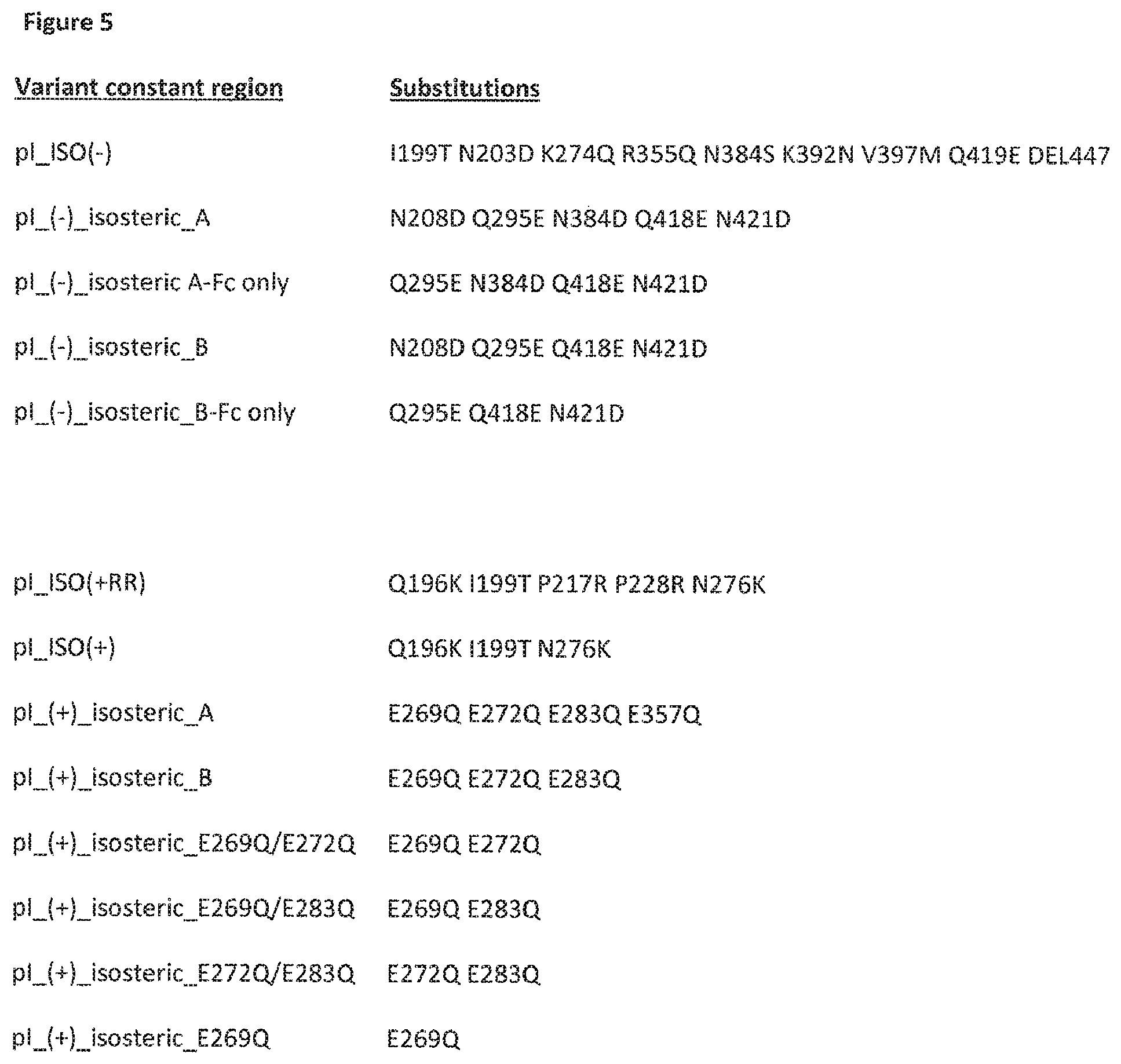
D00014
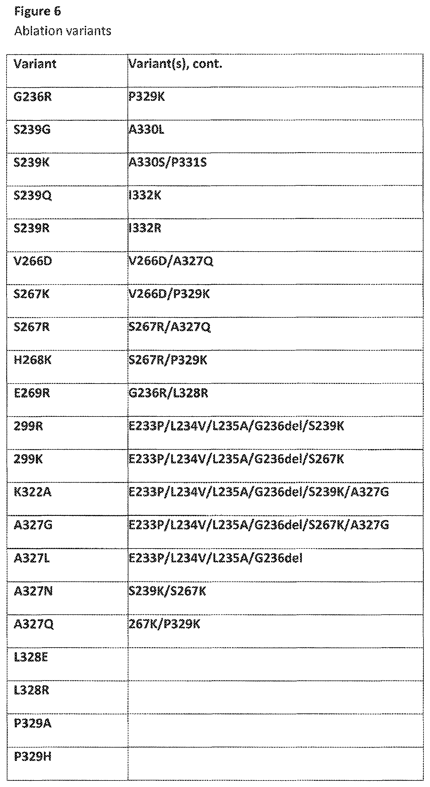
D00015
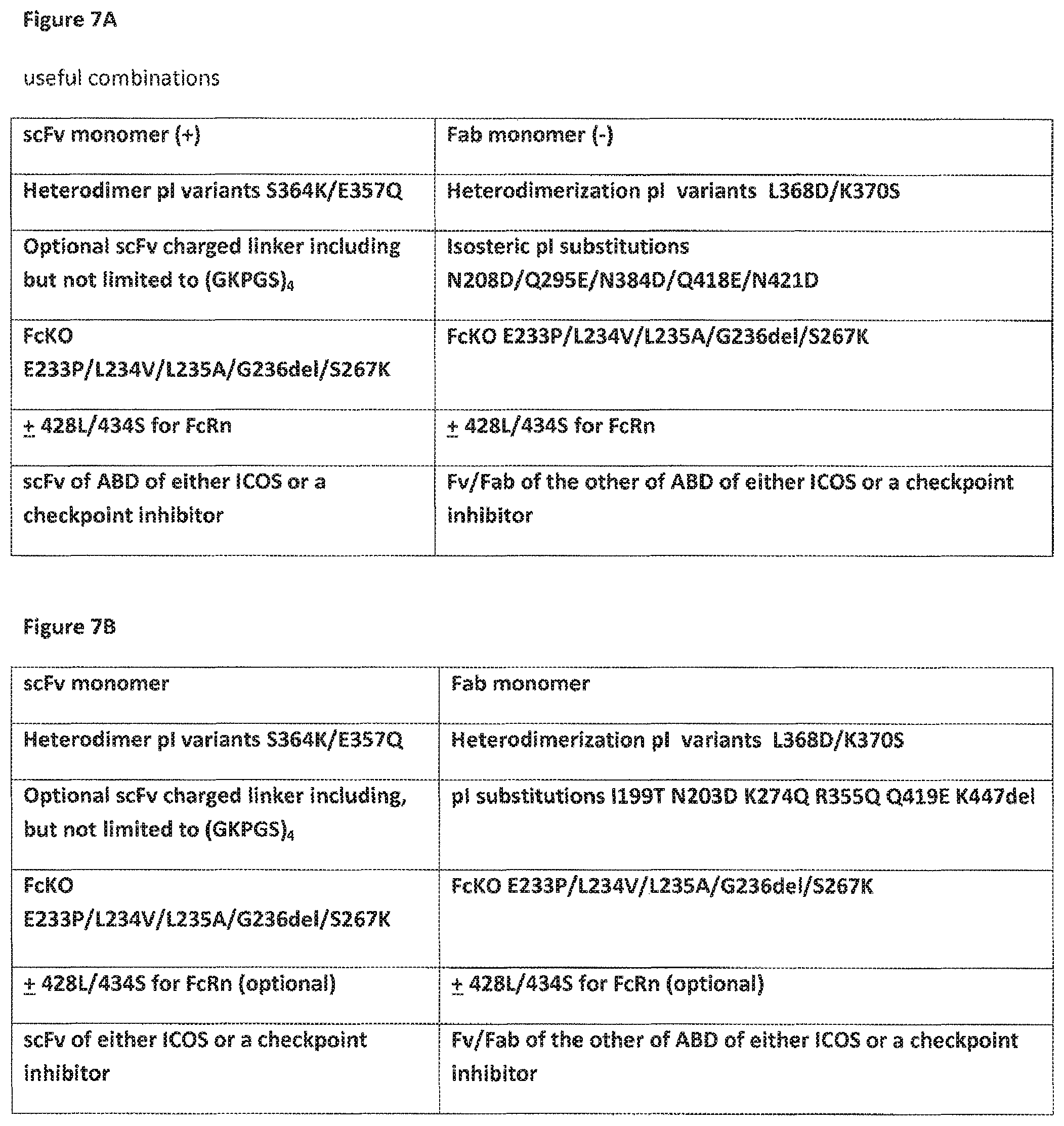
D00016
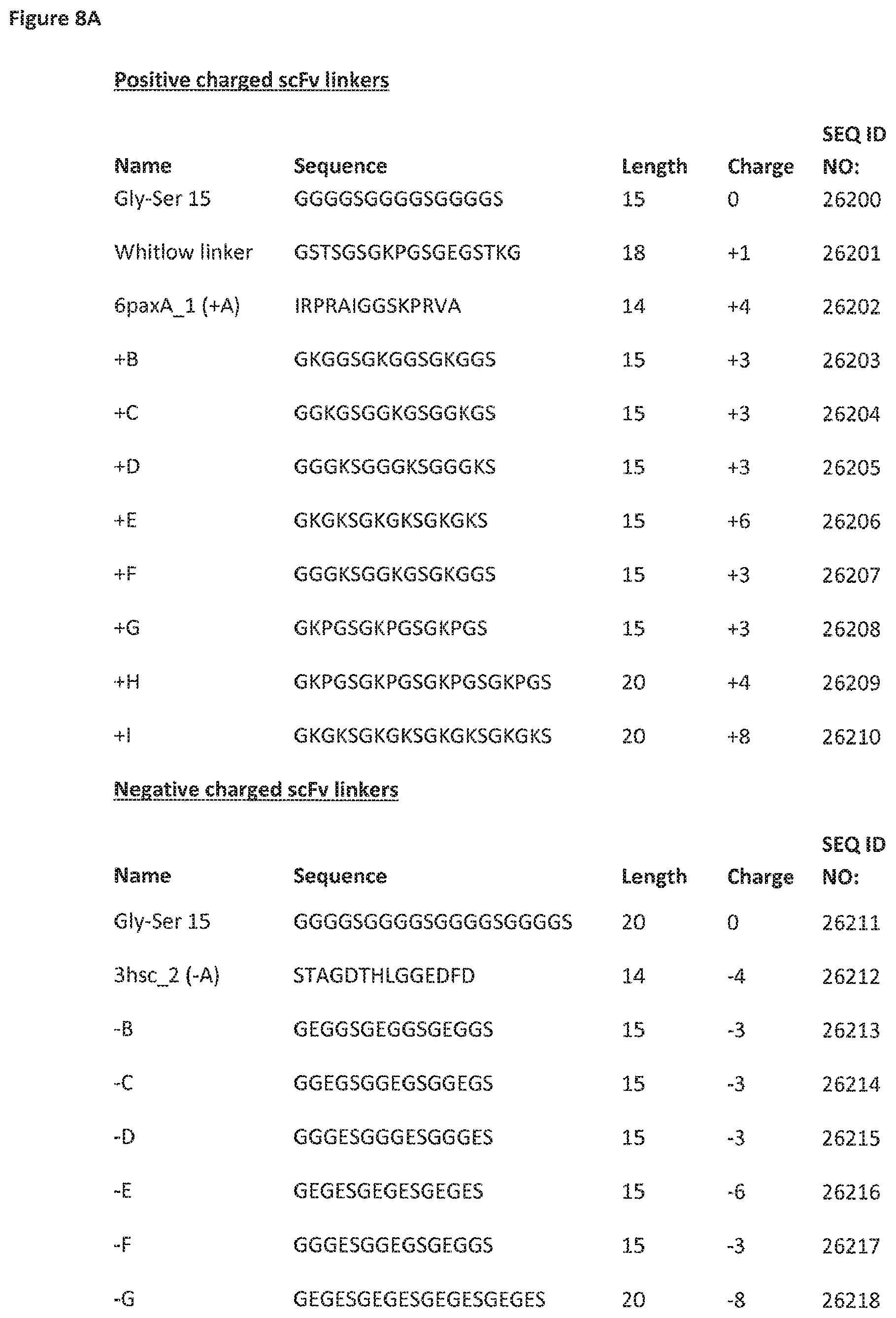
D00017
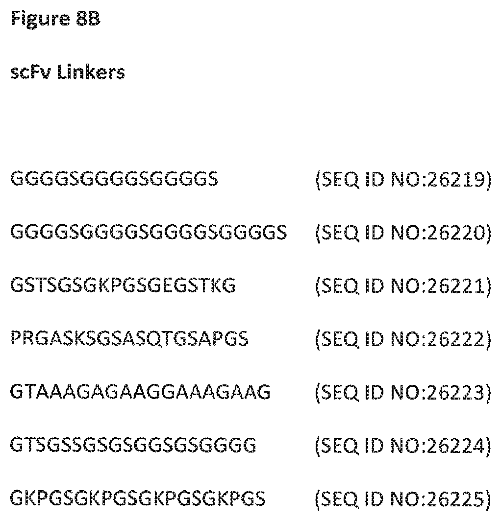
D00018
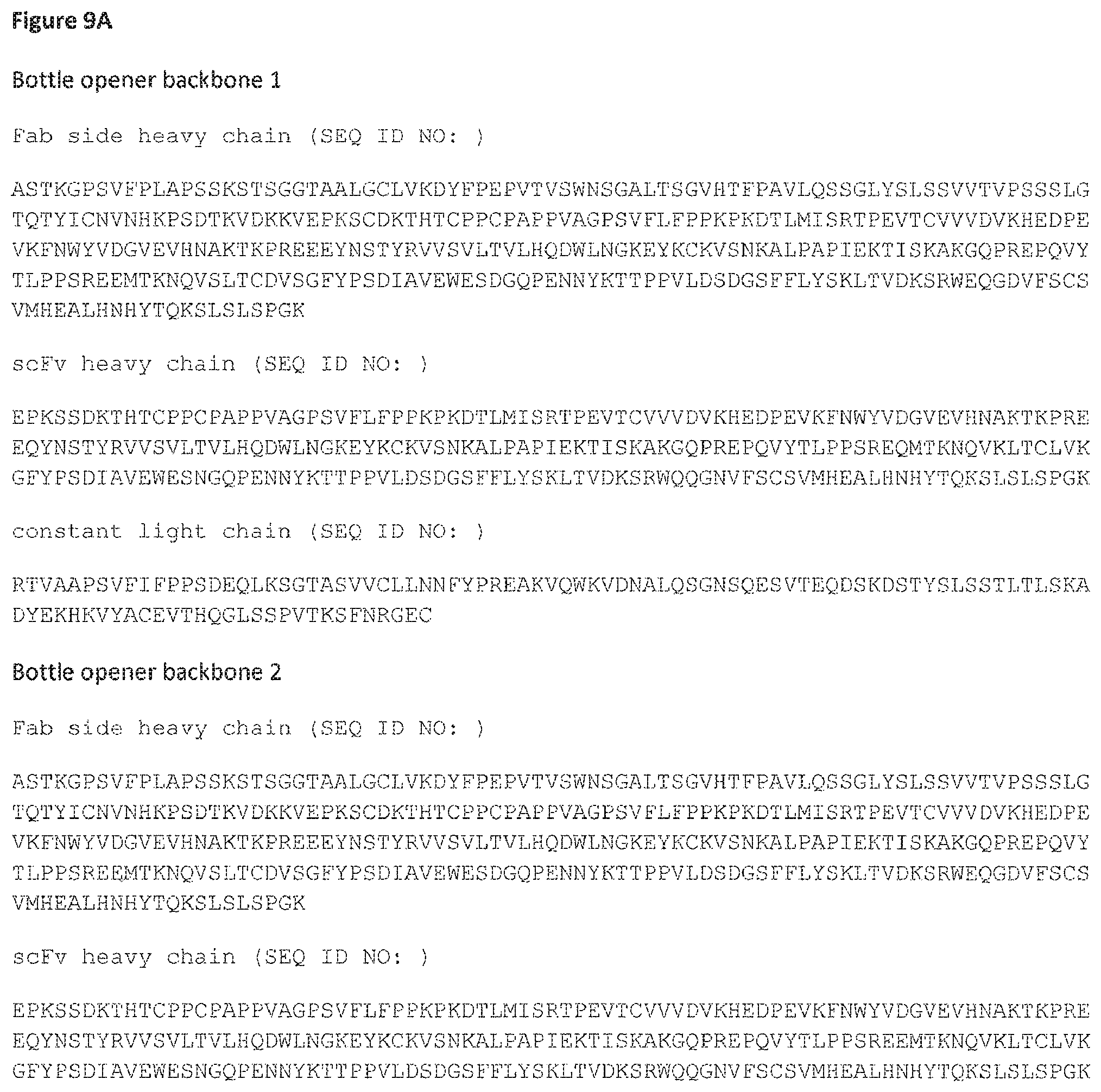
D00019
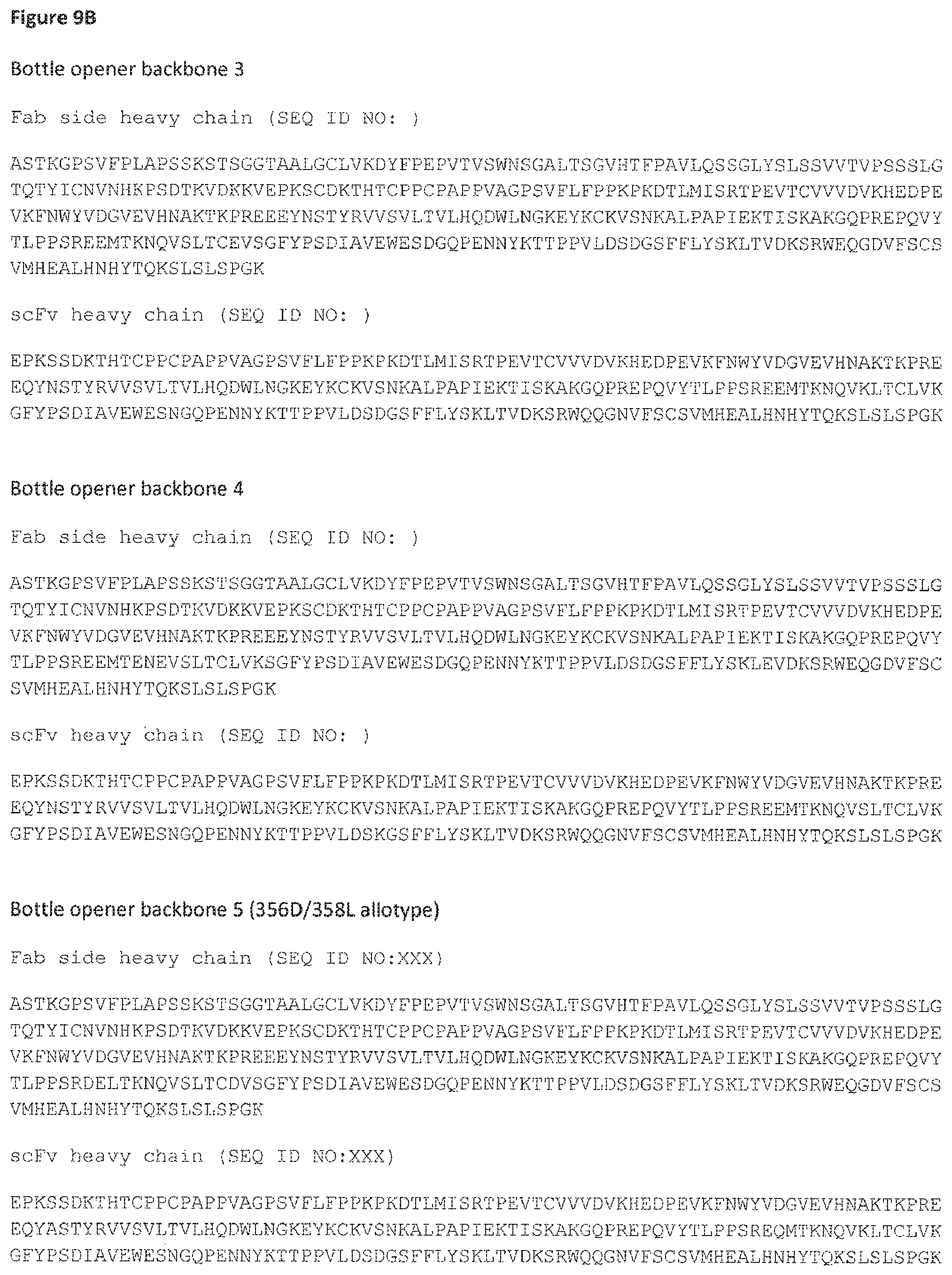
D00020
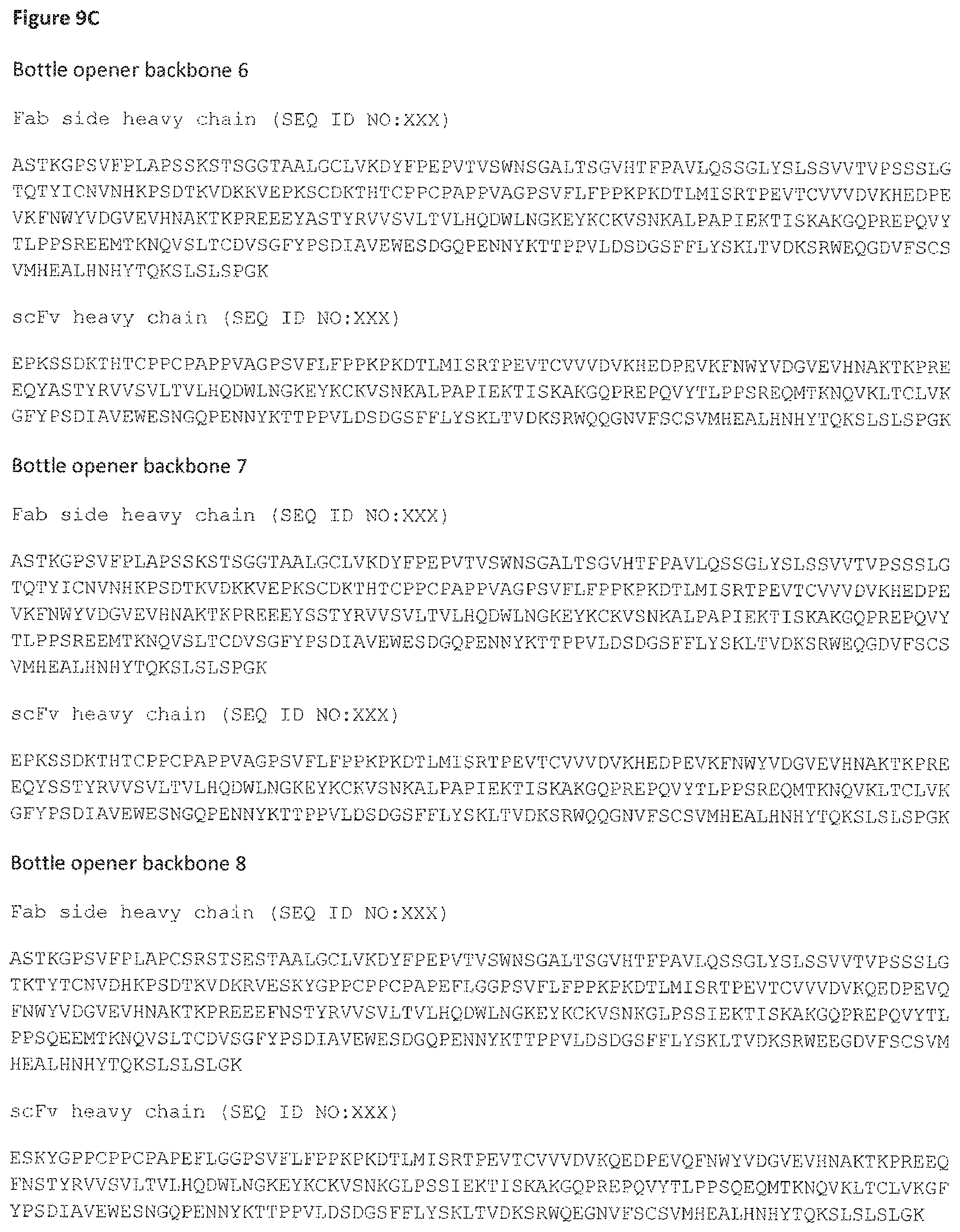
D00021

D00022

D00023

D00024

D00025

D00026
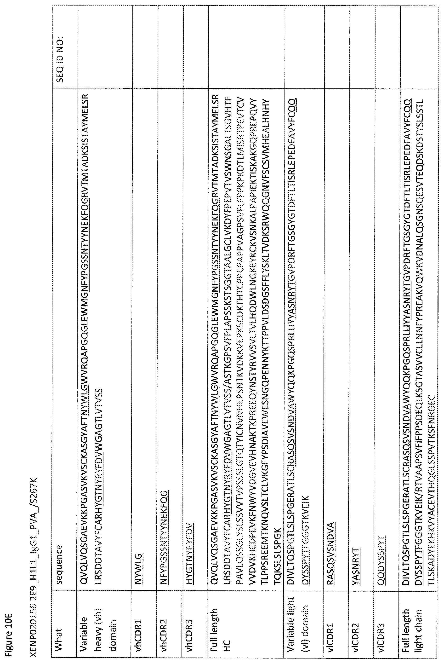
D00027
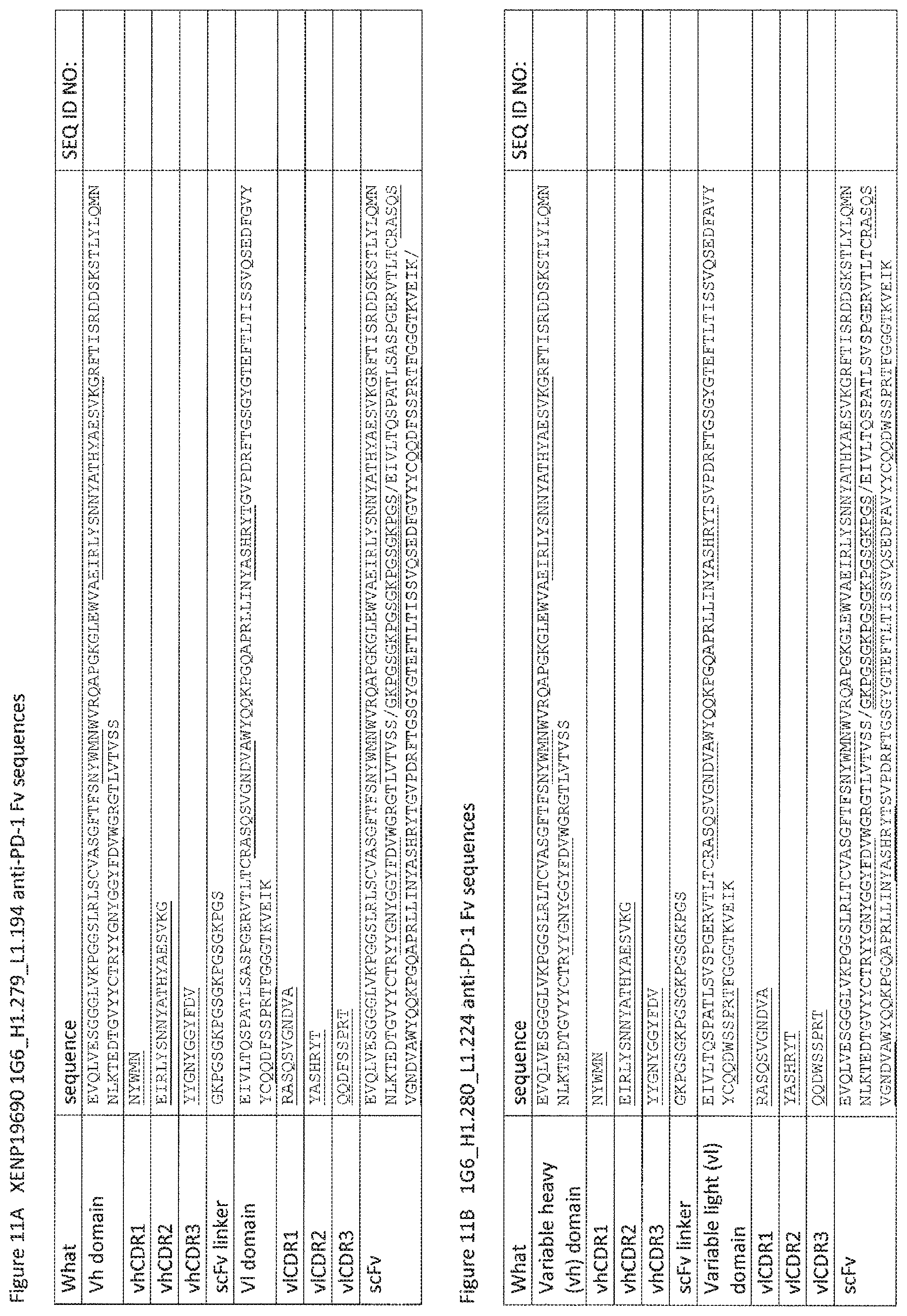
D00028
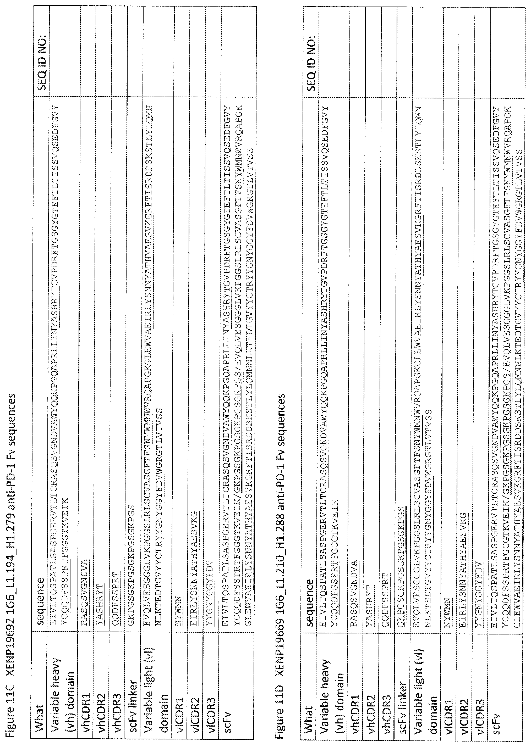
D00029
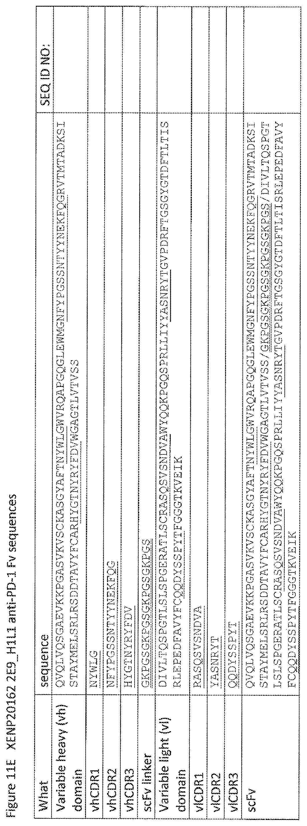
D00030
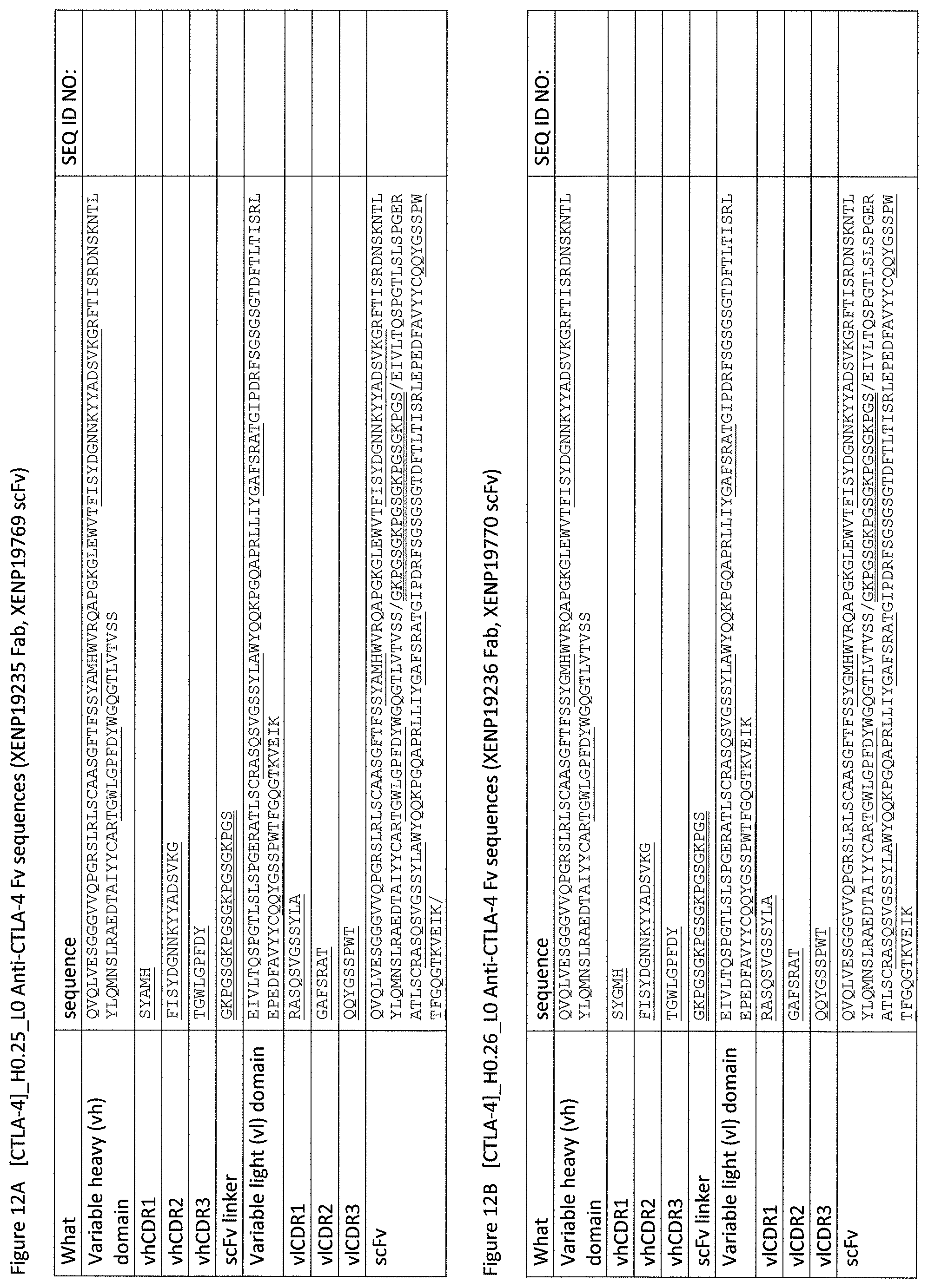
D00031

D00032

D00033
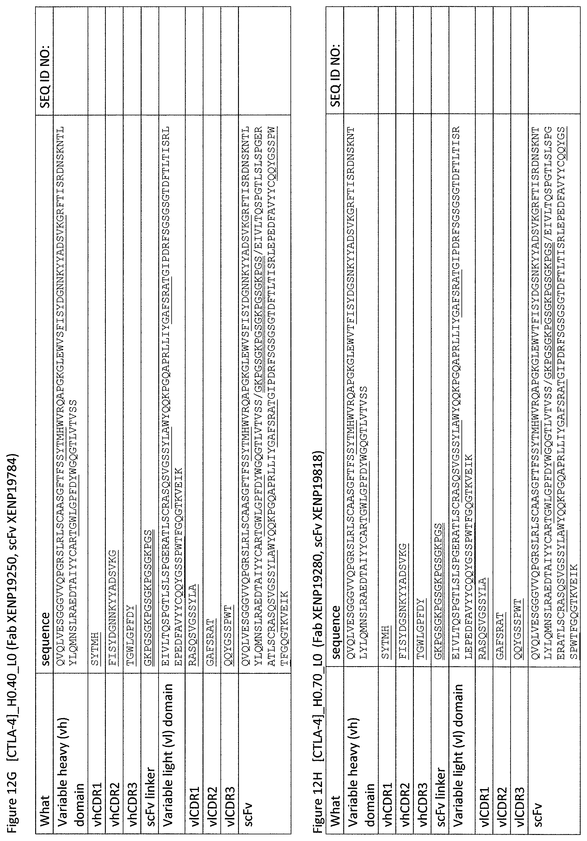
D00034
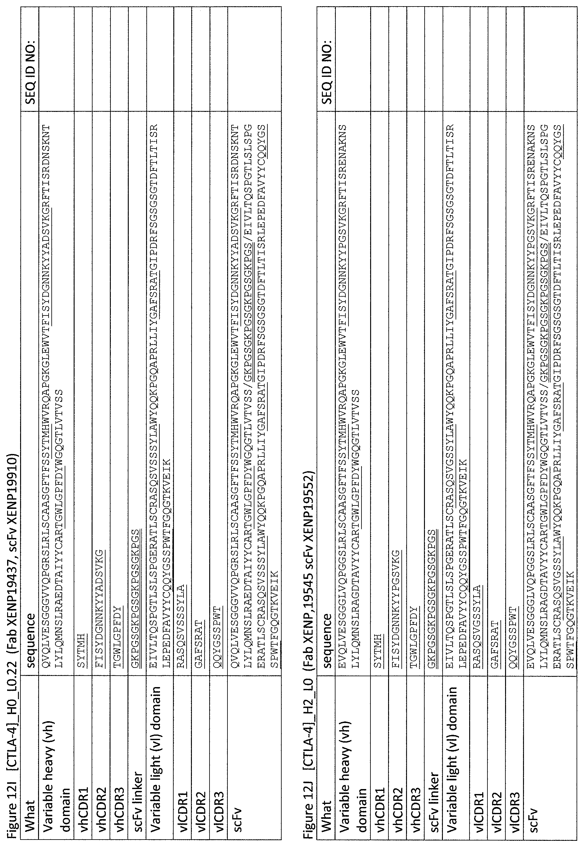
D00035

D00036

D00037

D00038
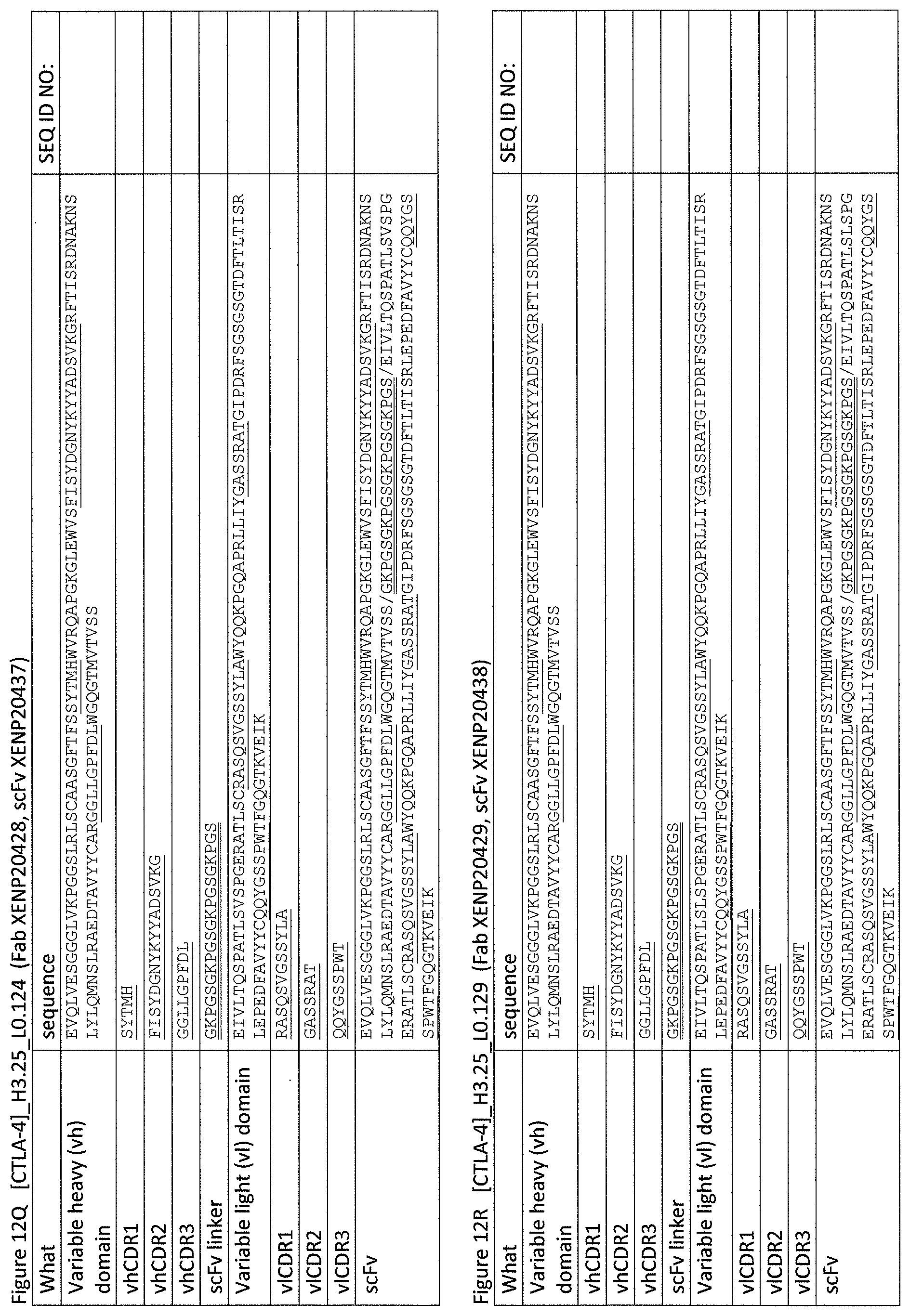
D00039

D00040
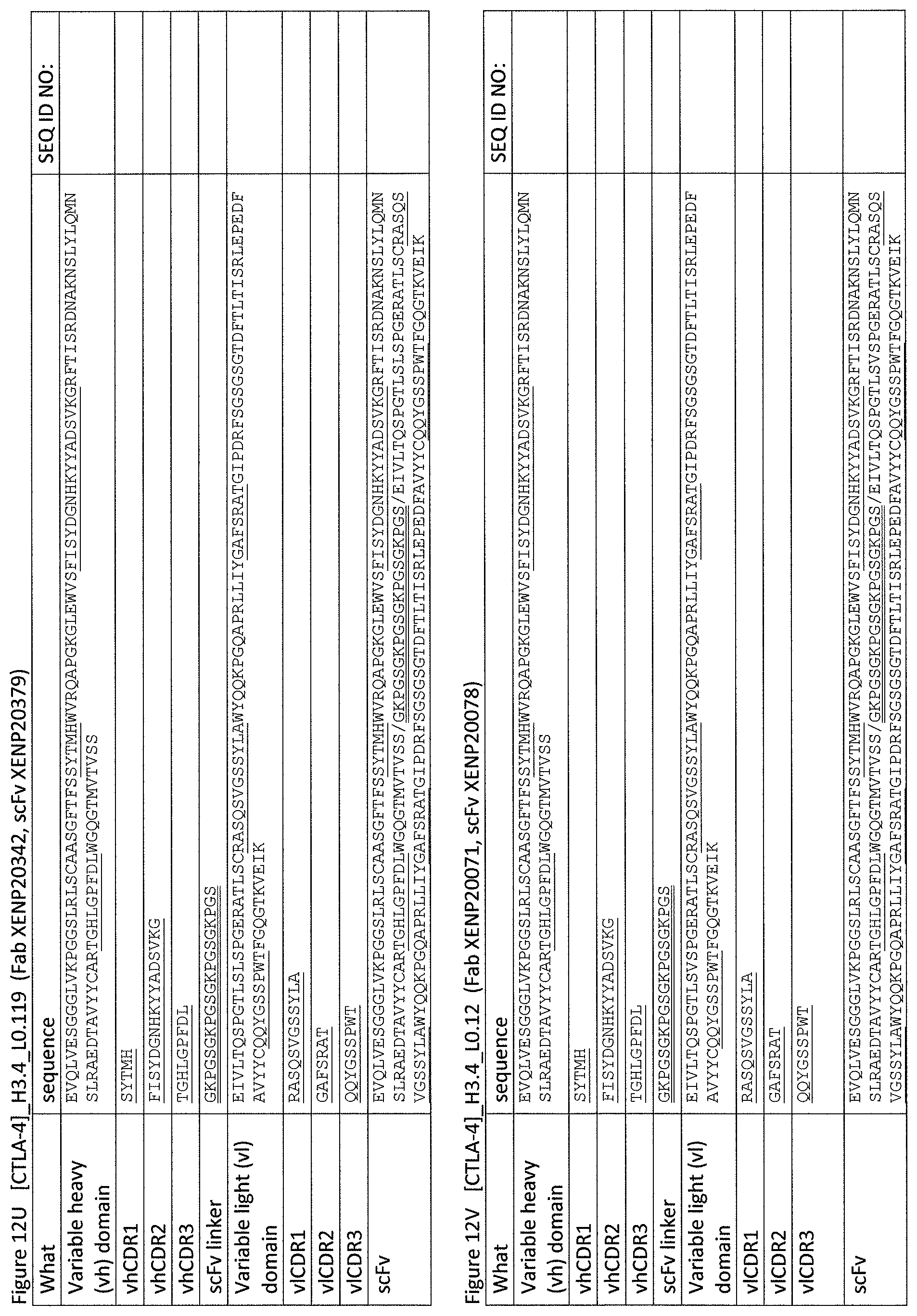
D00041

D00042
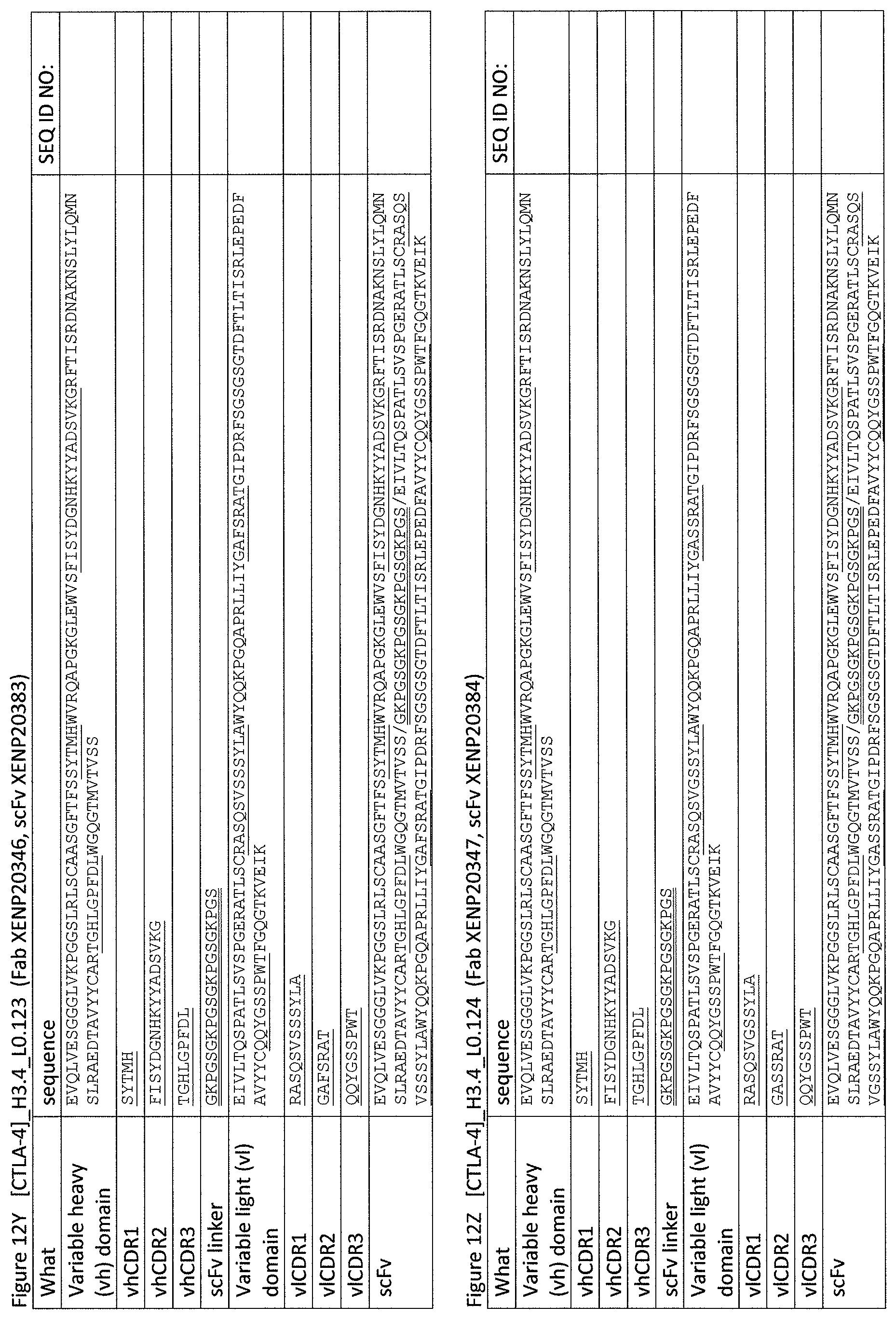
D00043

D00044
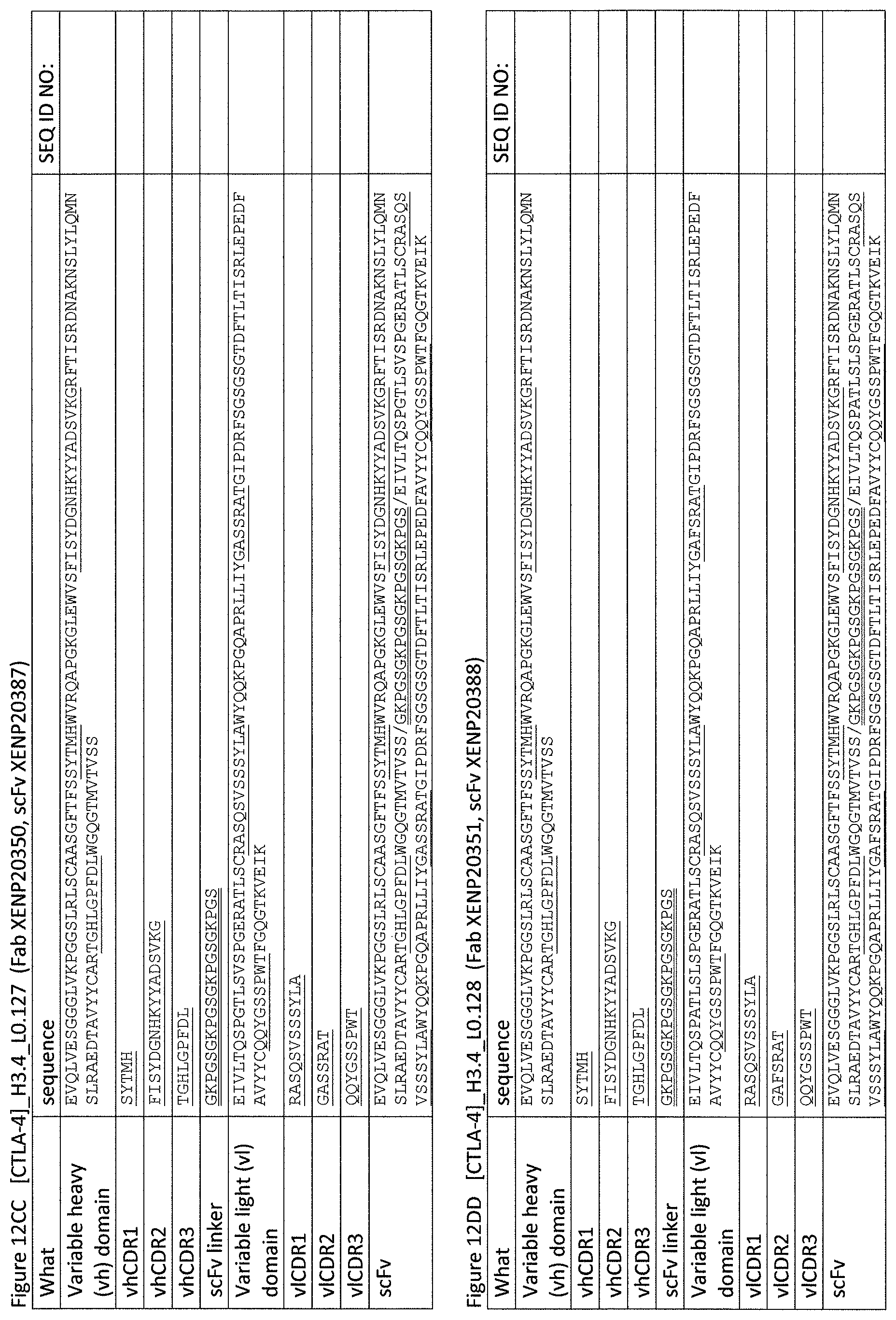
D00045

D00046

D00047

D00048
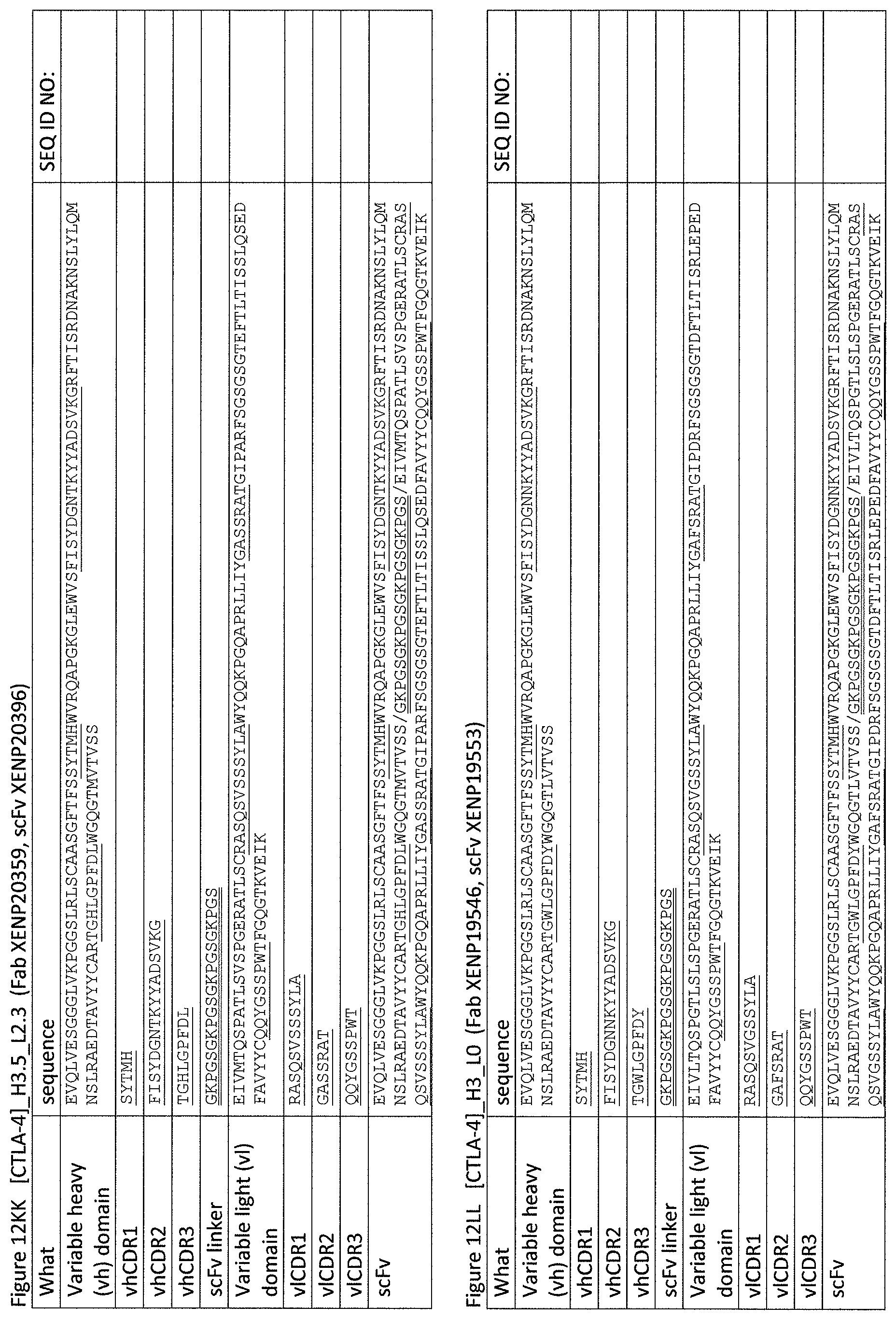
D00049
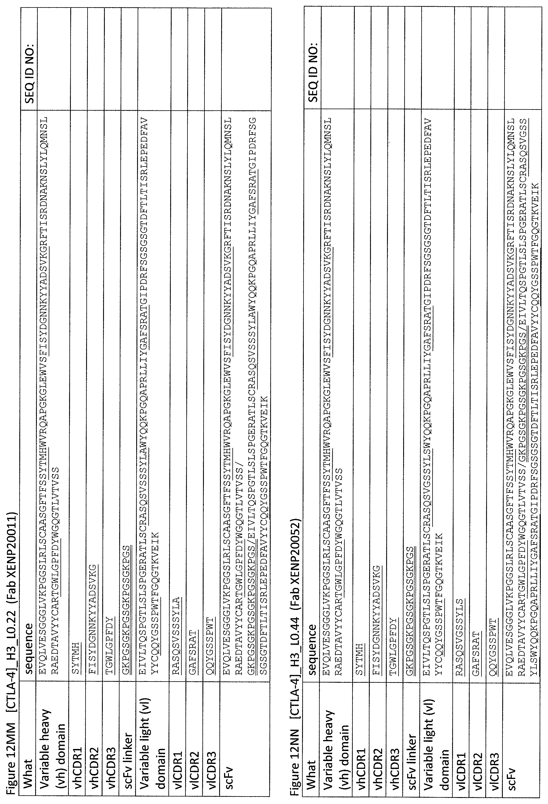
D00050
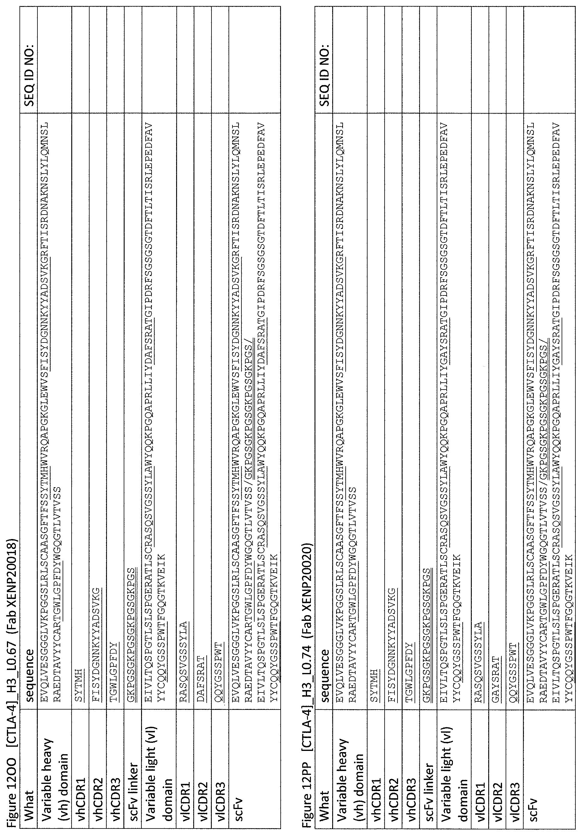
D00051
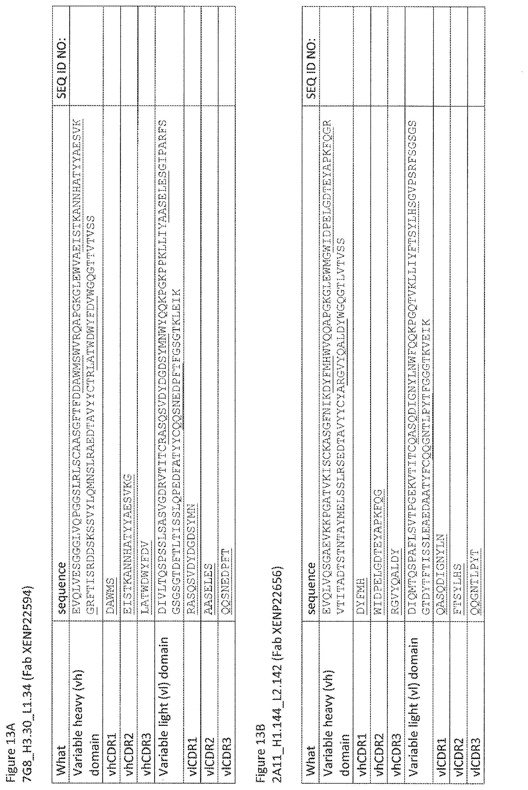
D00052
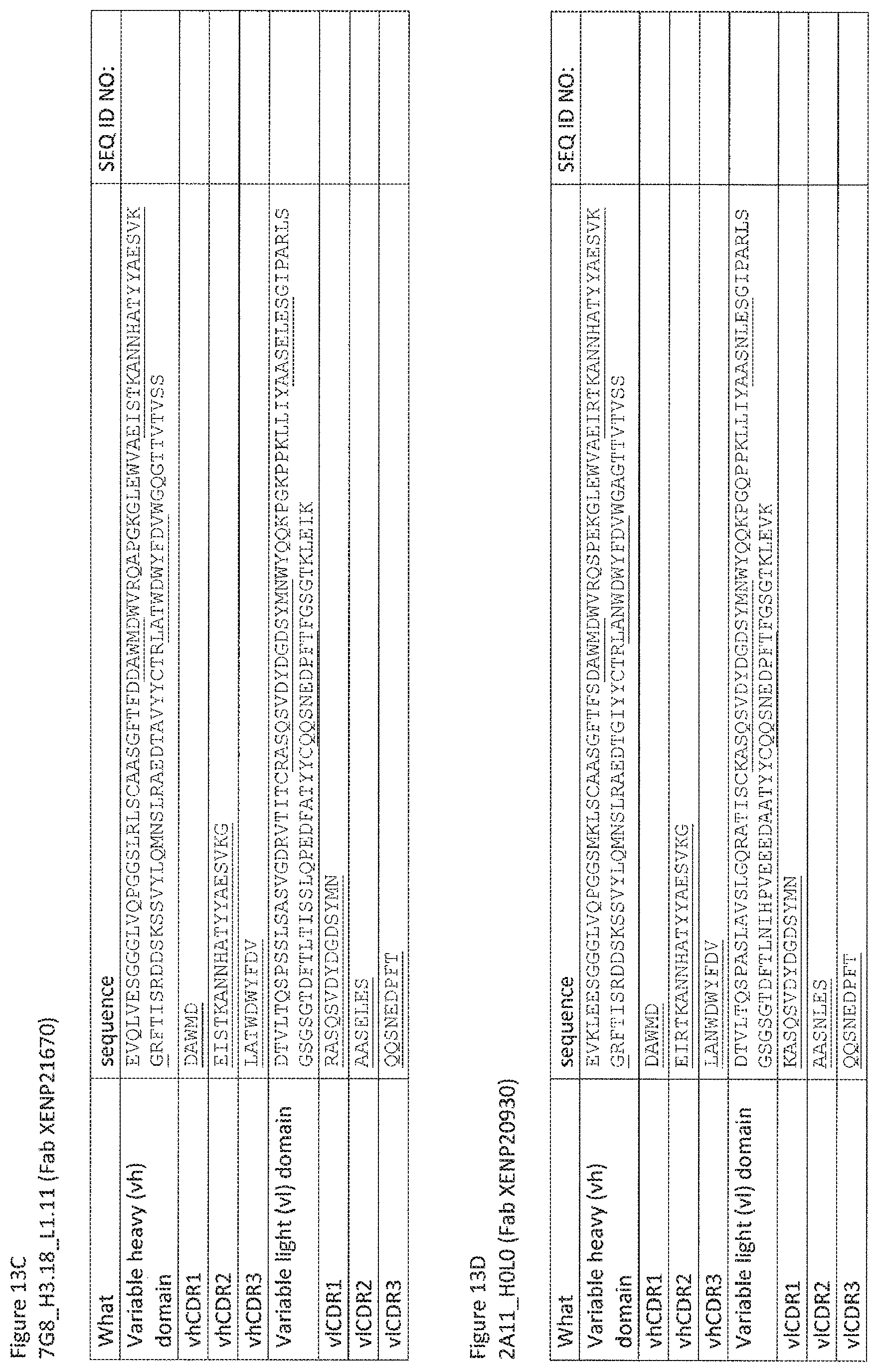
D00053
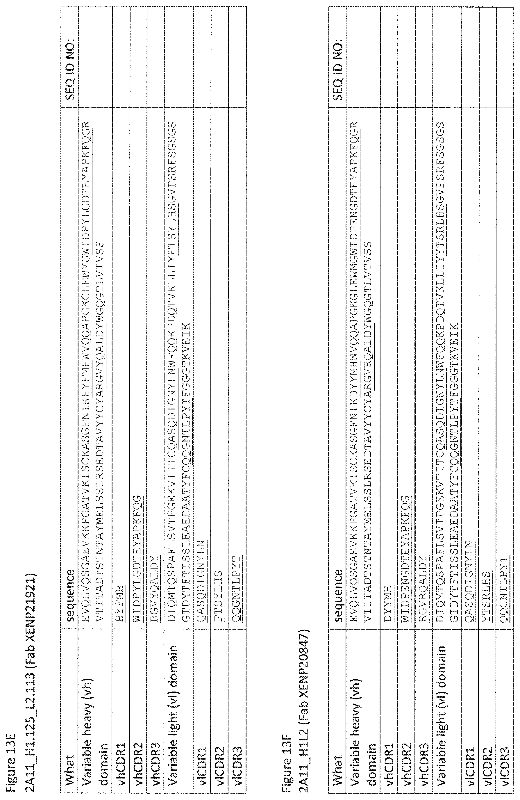
D00054

D00055

D00056

D00057
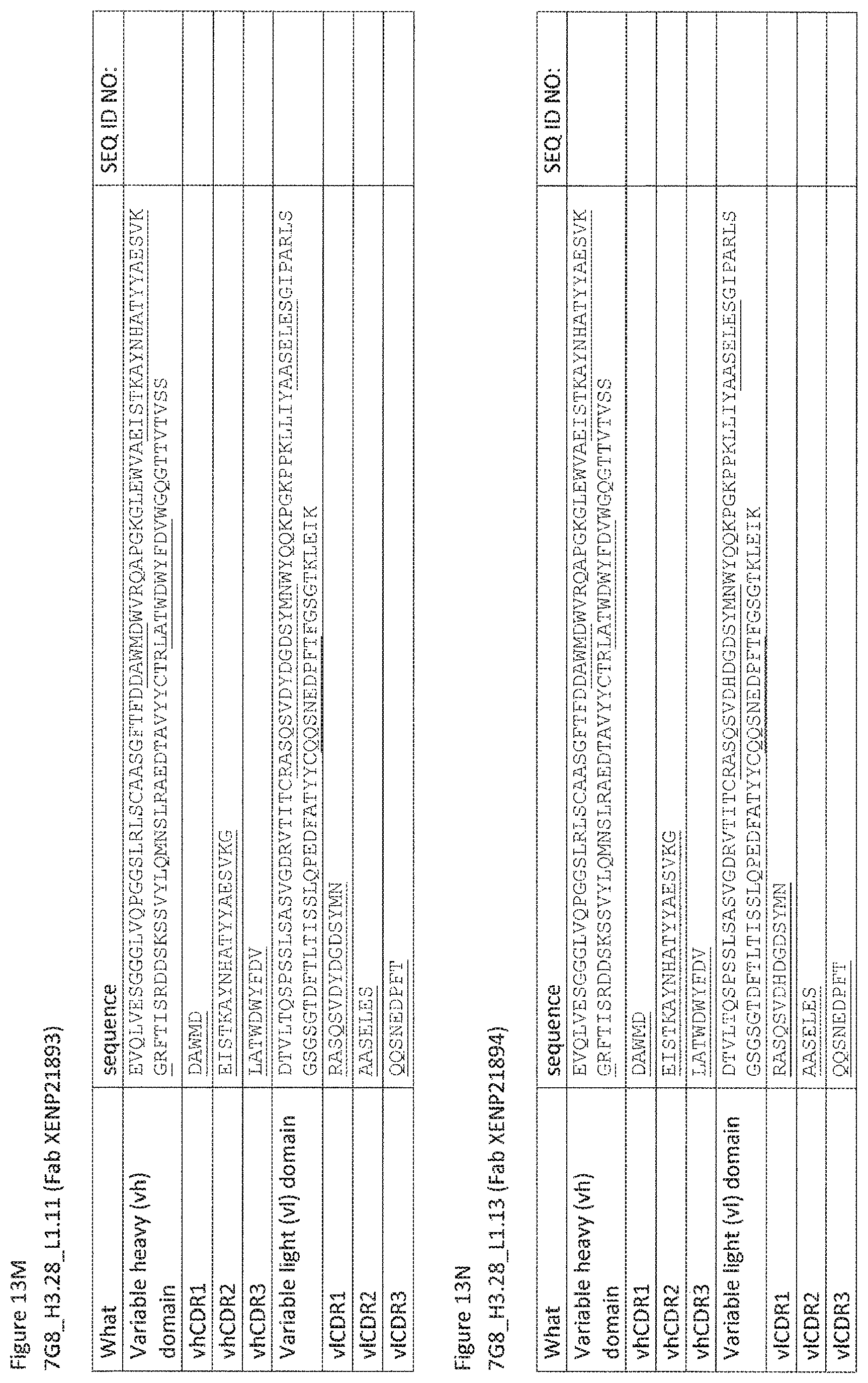
D00058
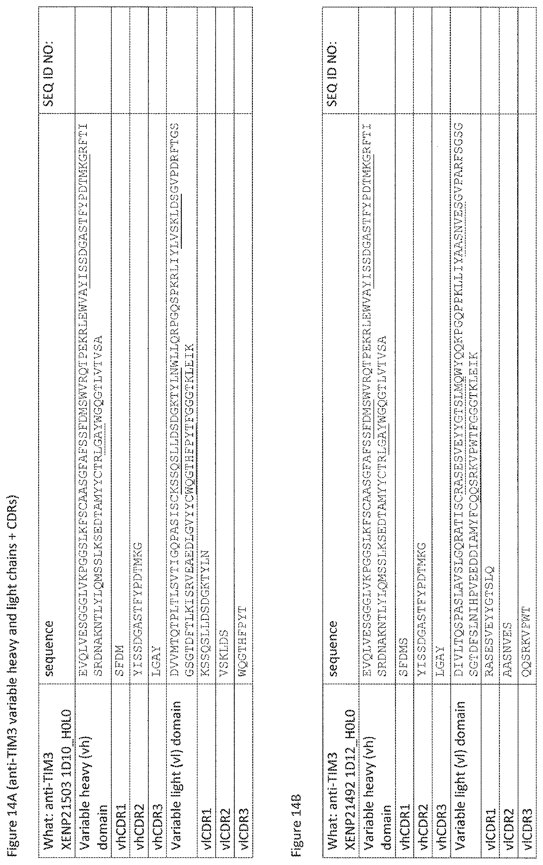
D00059
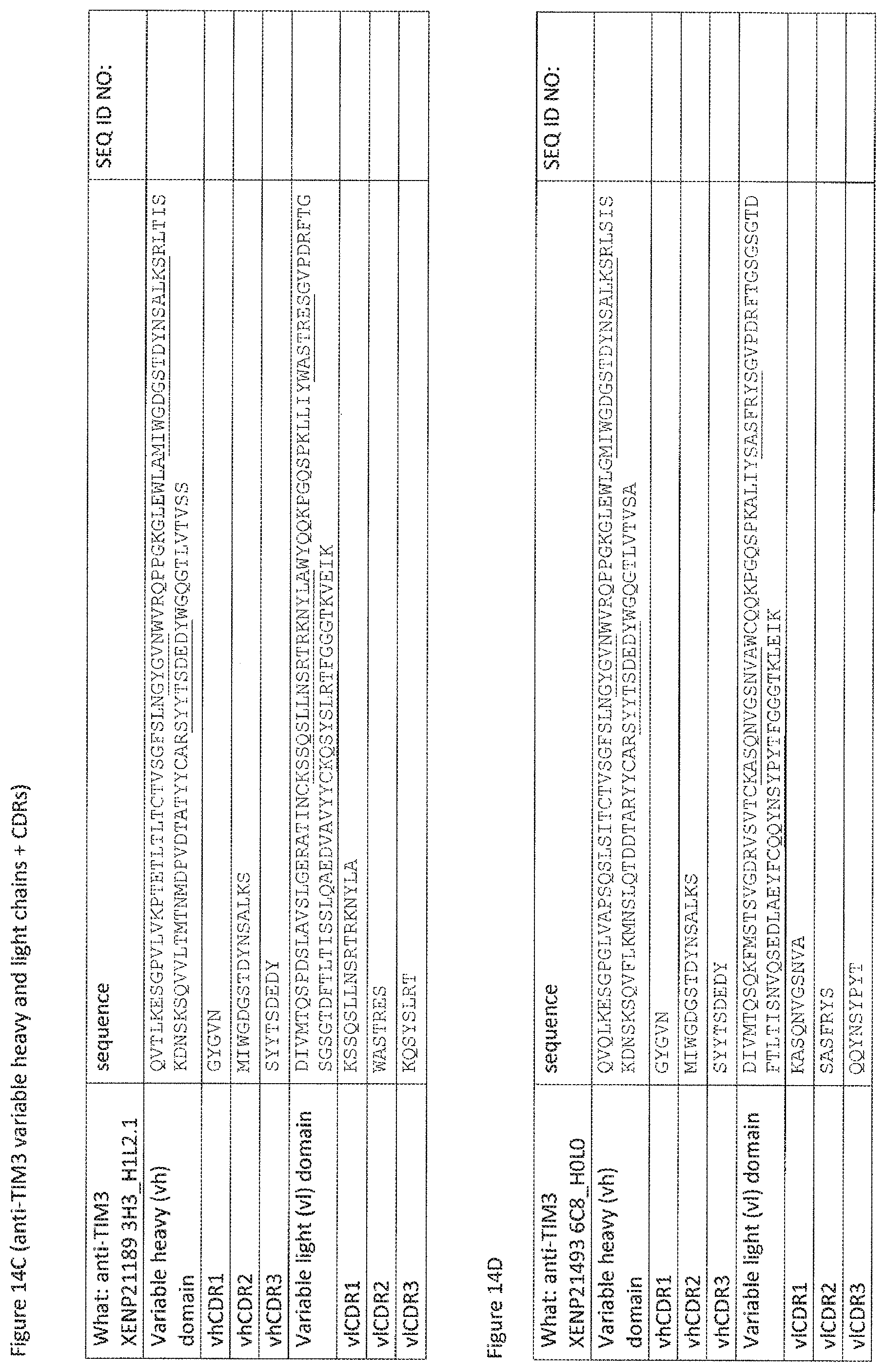
D00060
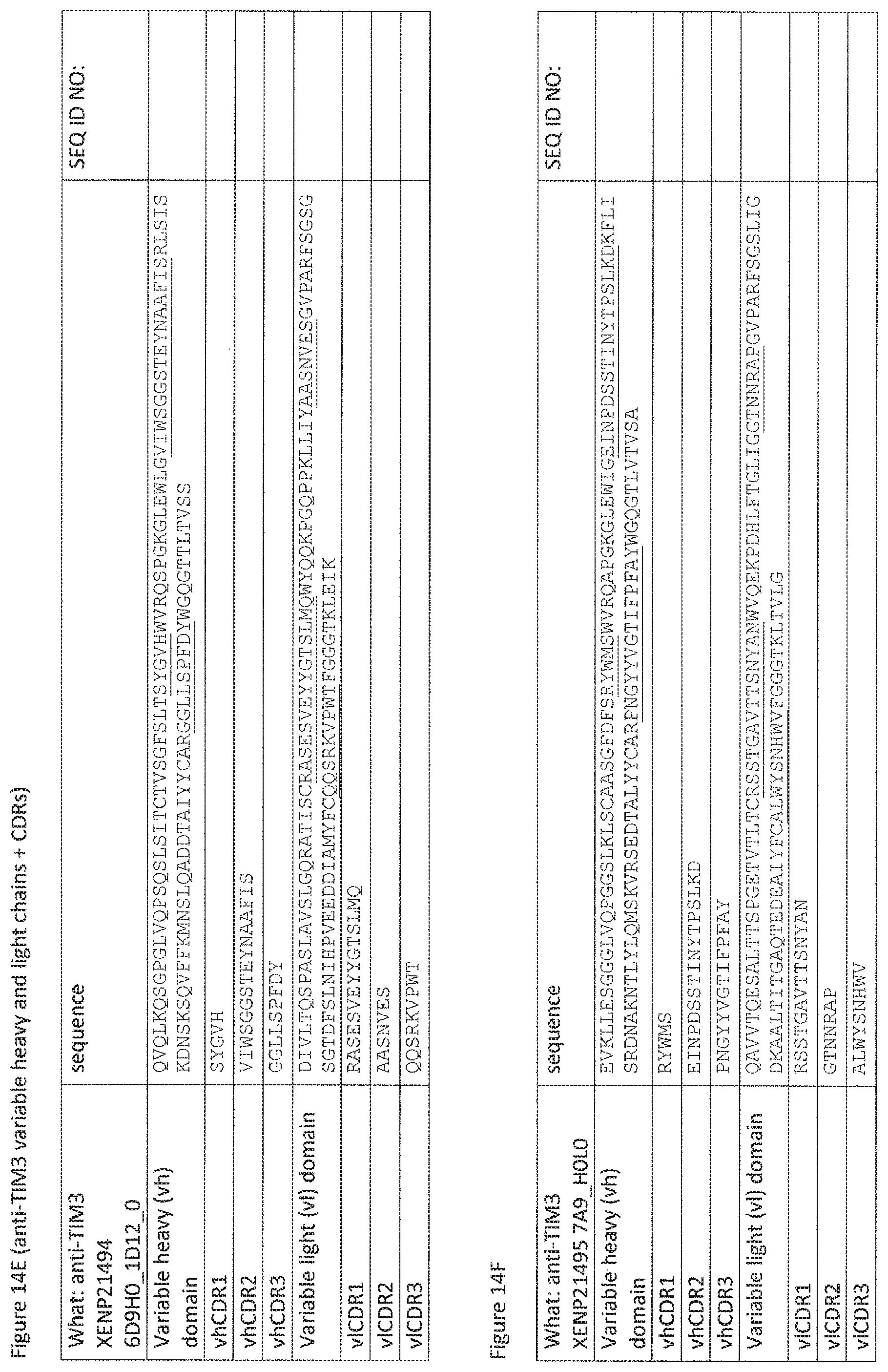
D00061
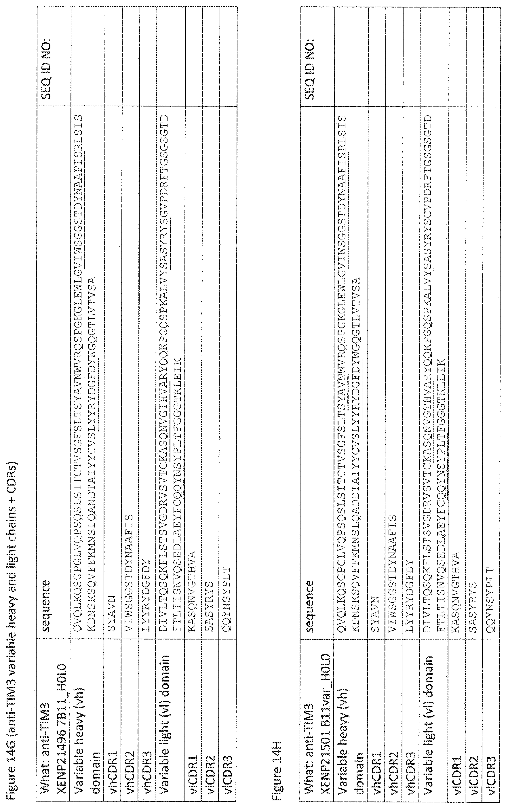
D00062

D00063

D00064
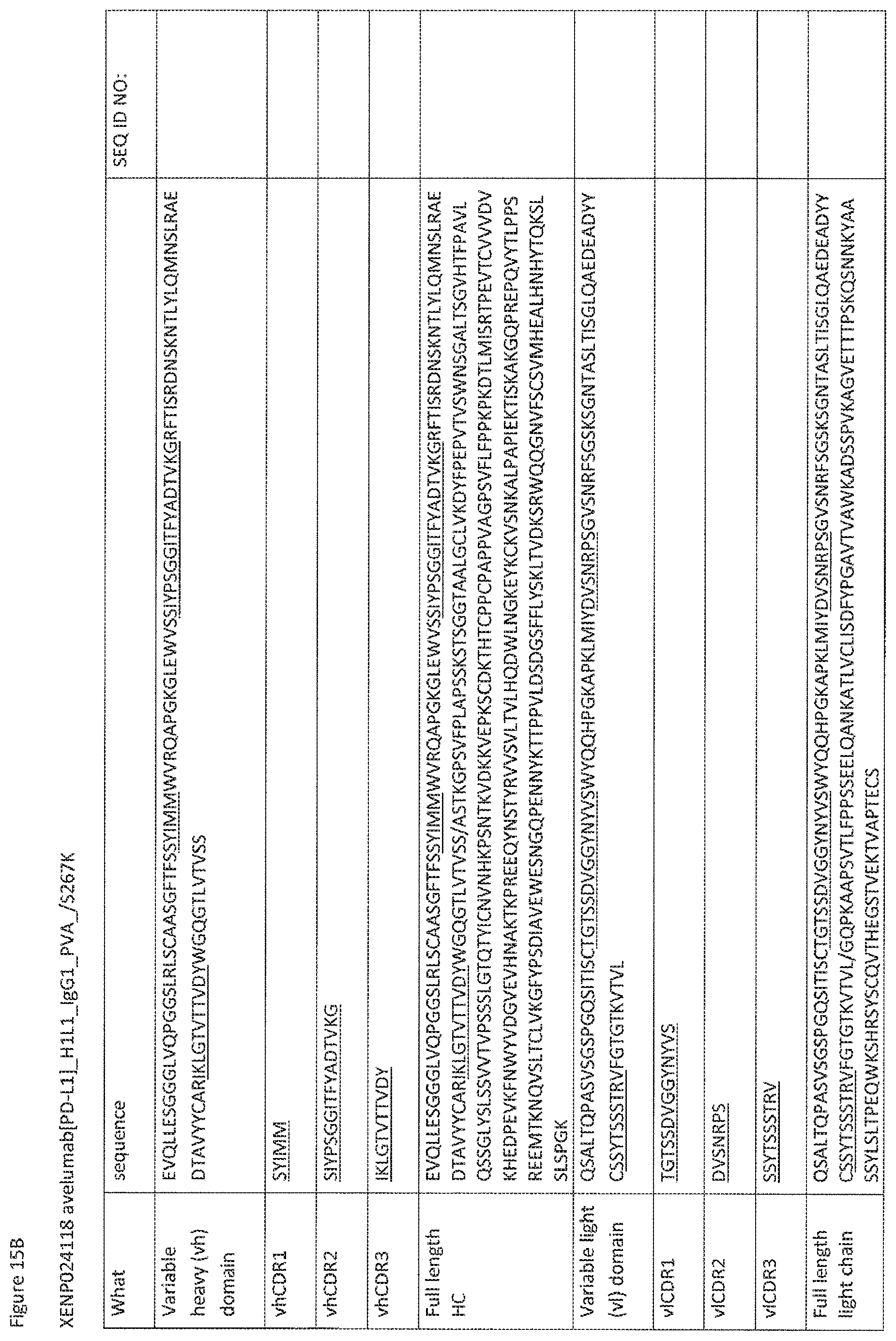
D00065
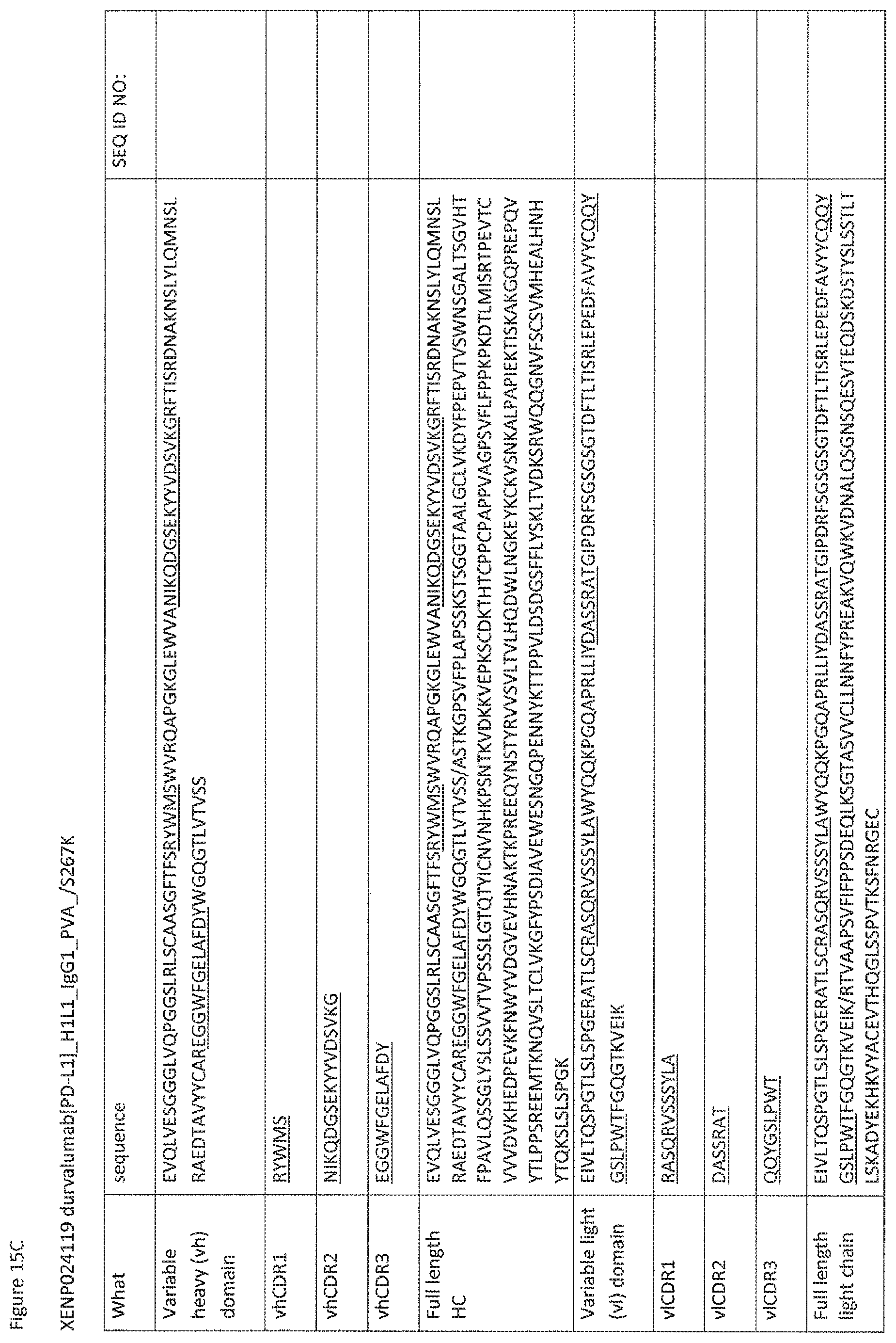
D00066
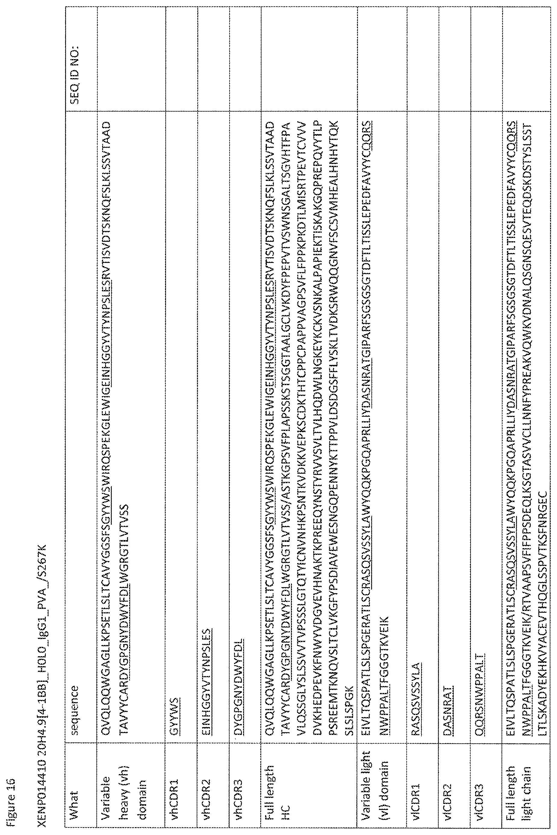
D00067

D00068
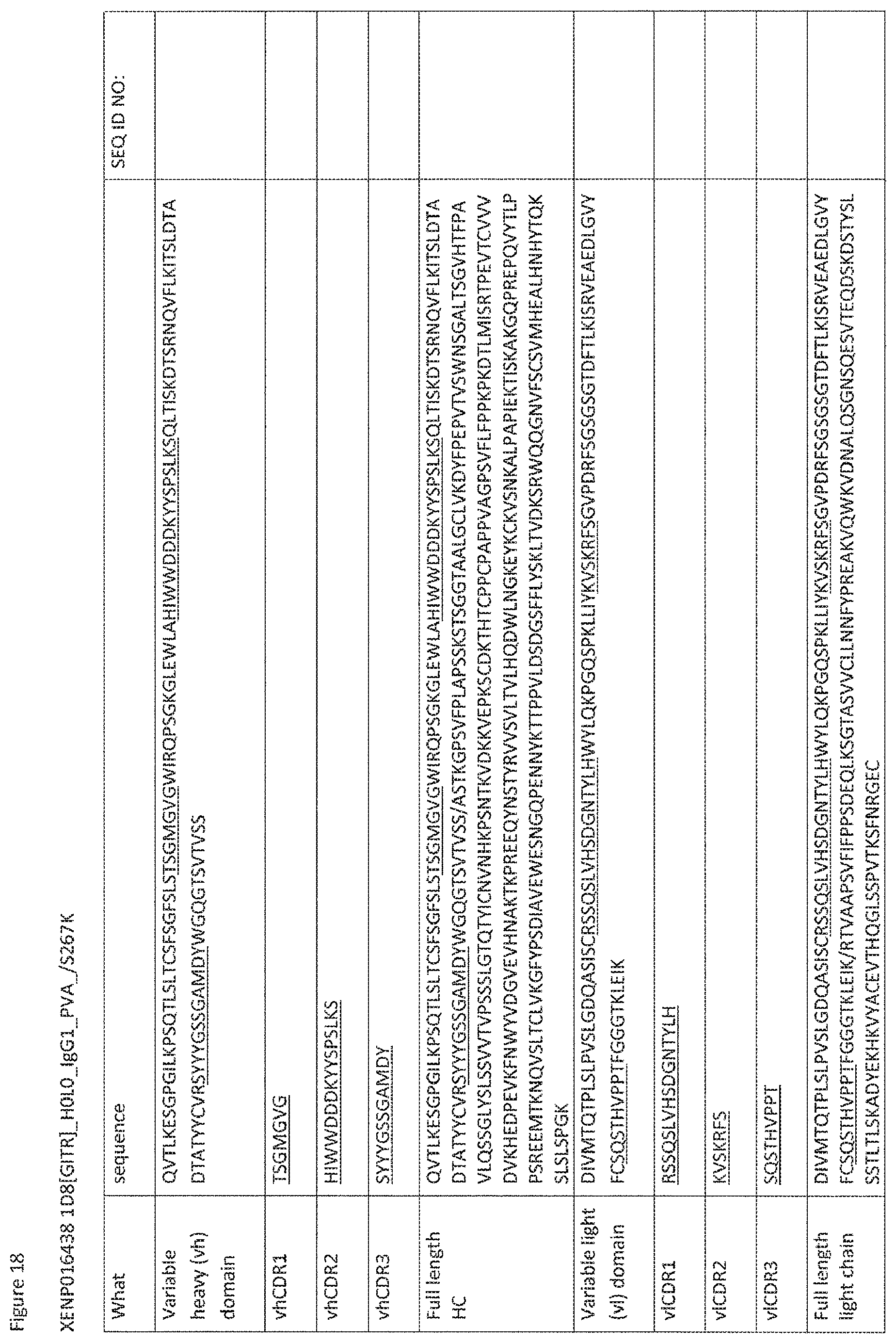
D00069
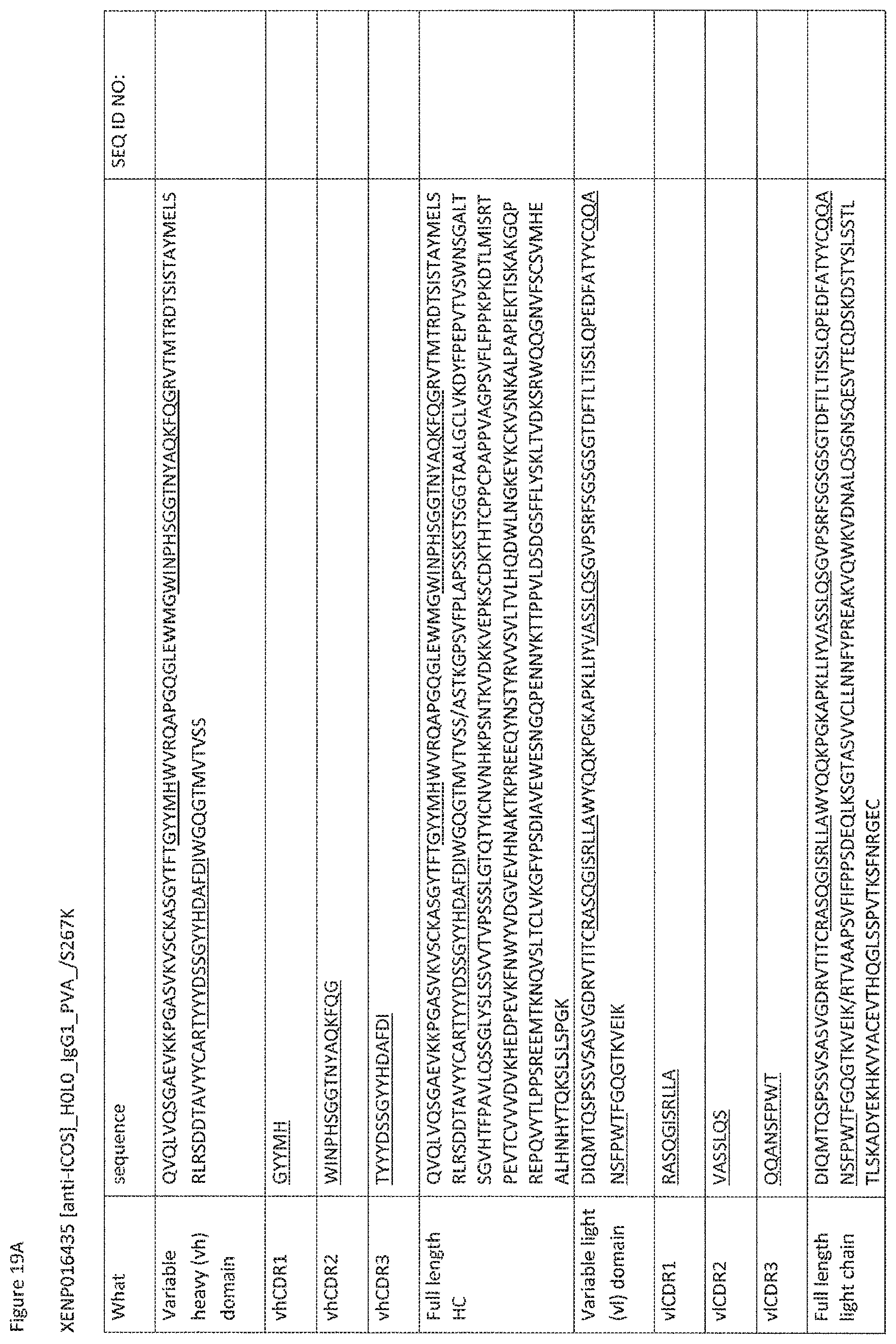
D00070
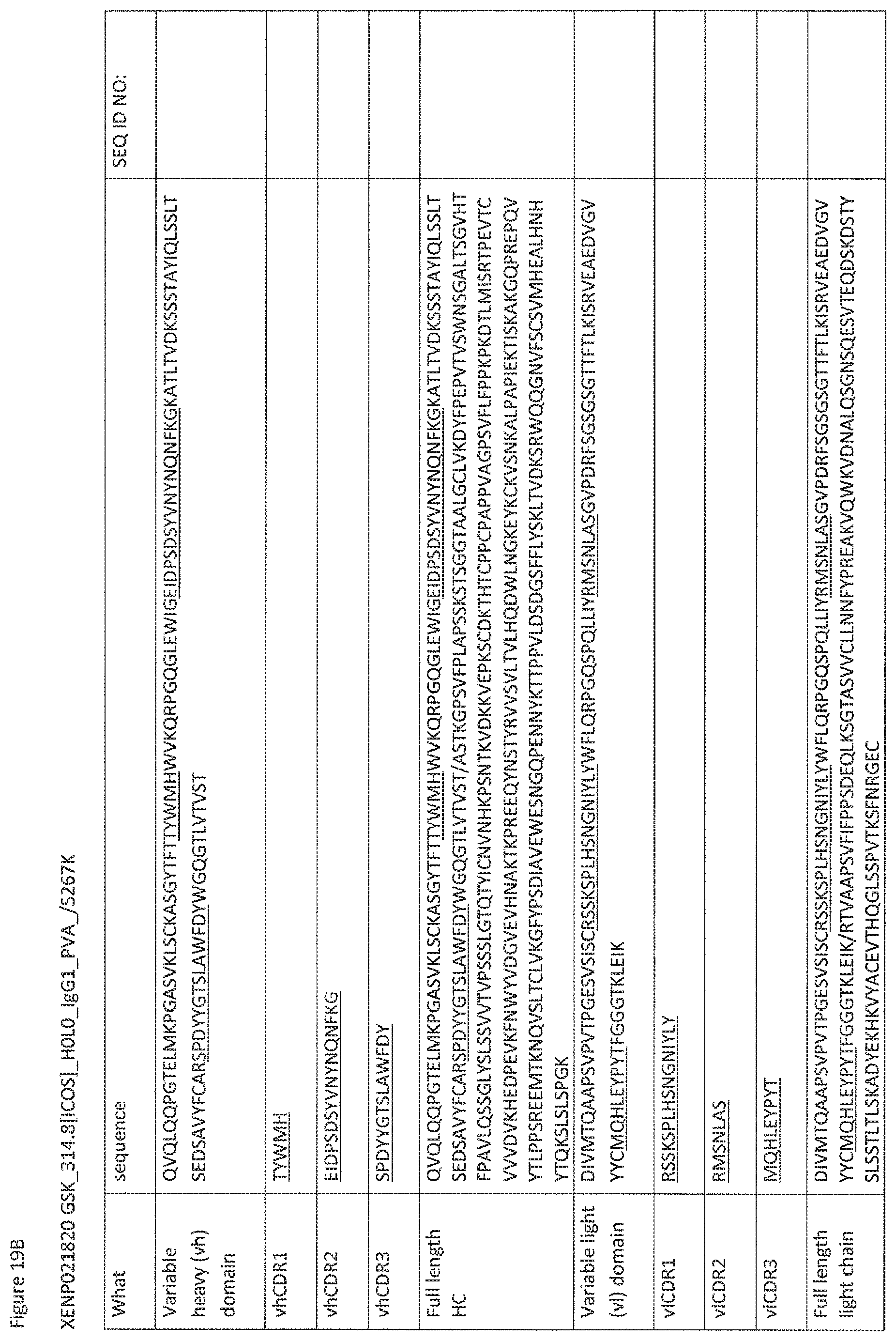
D00071
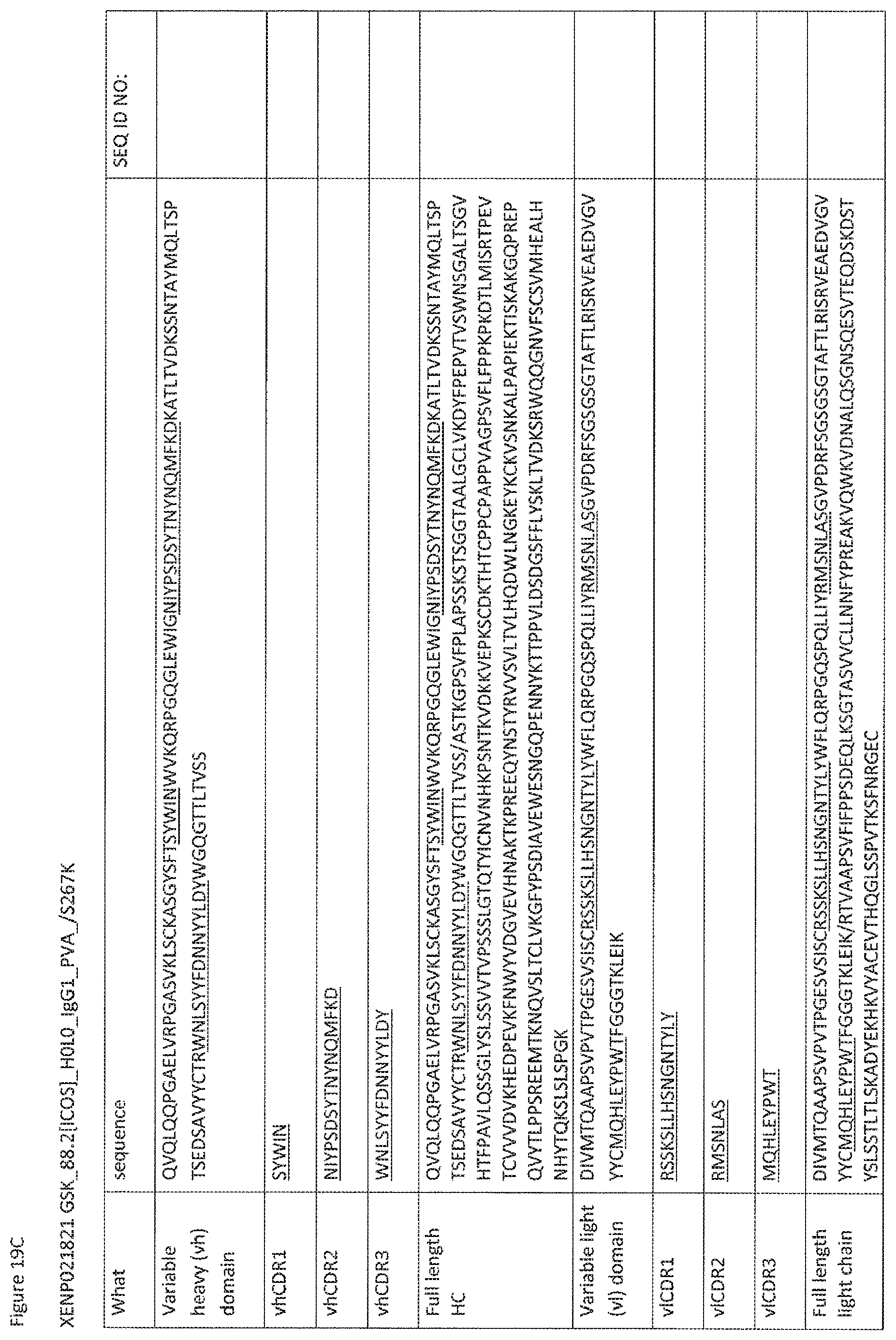
D00072

D00073
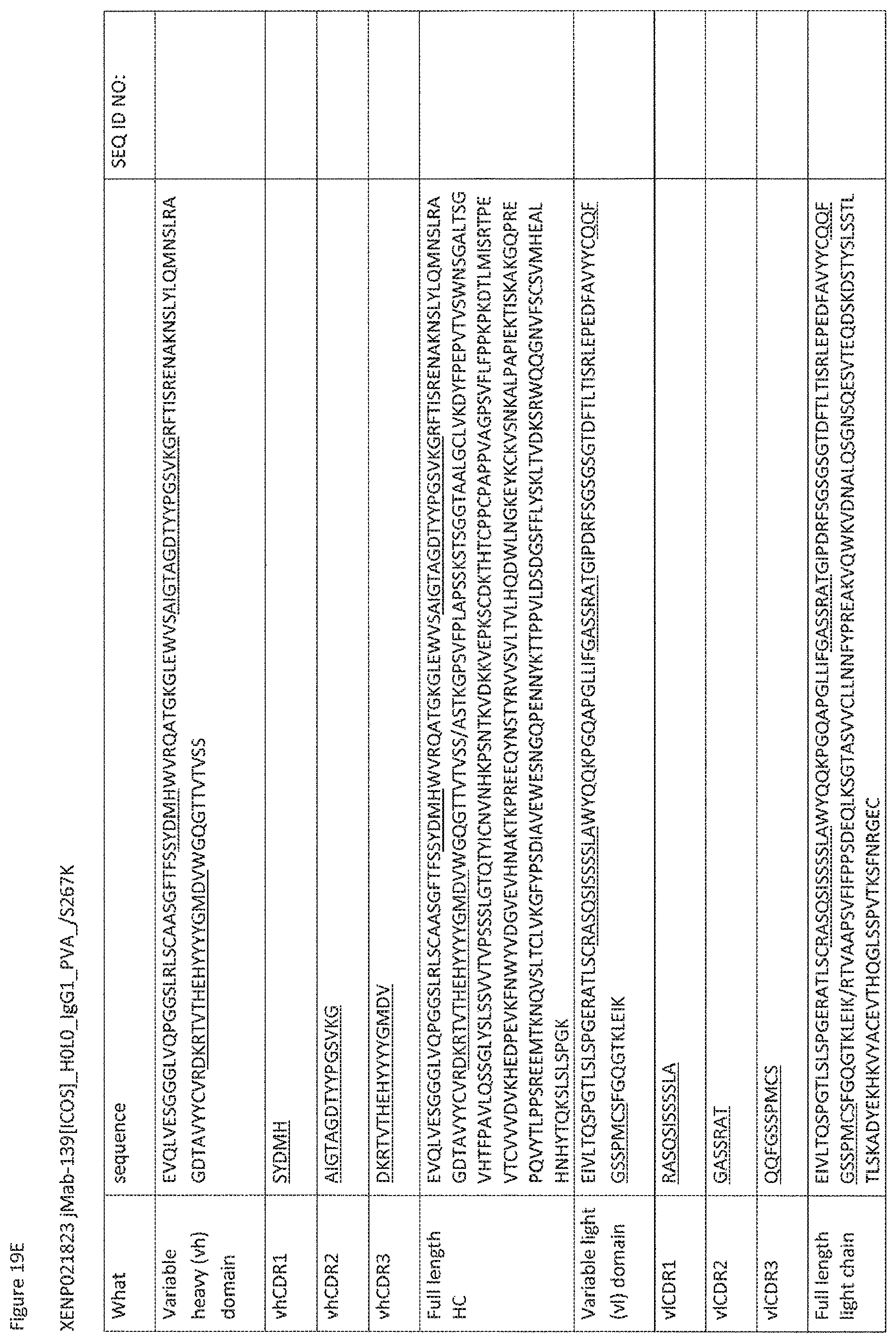
D00074
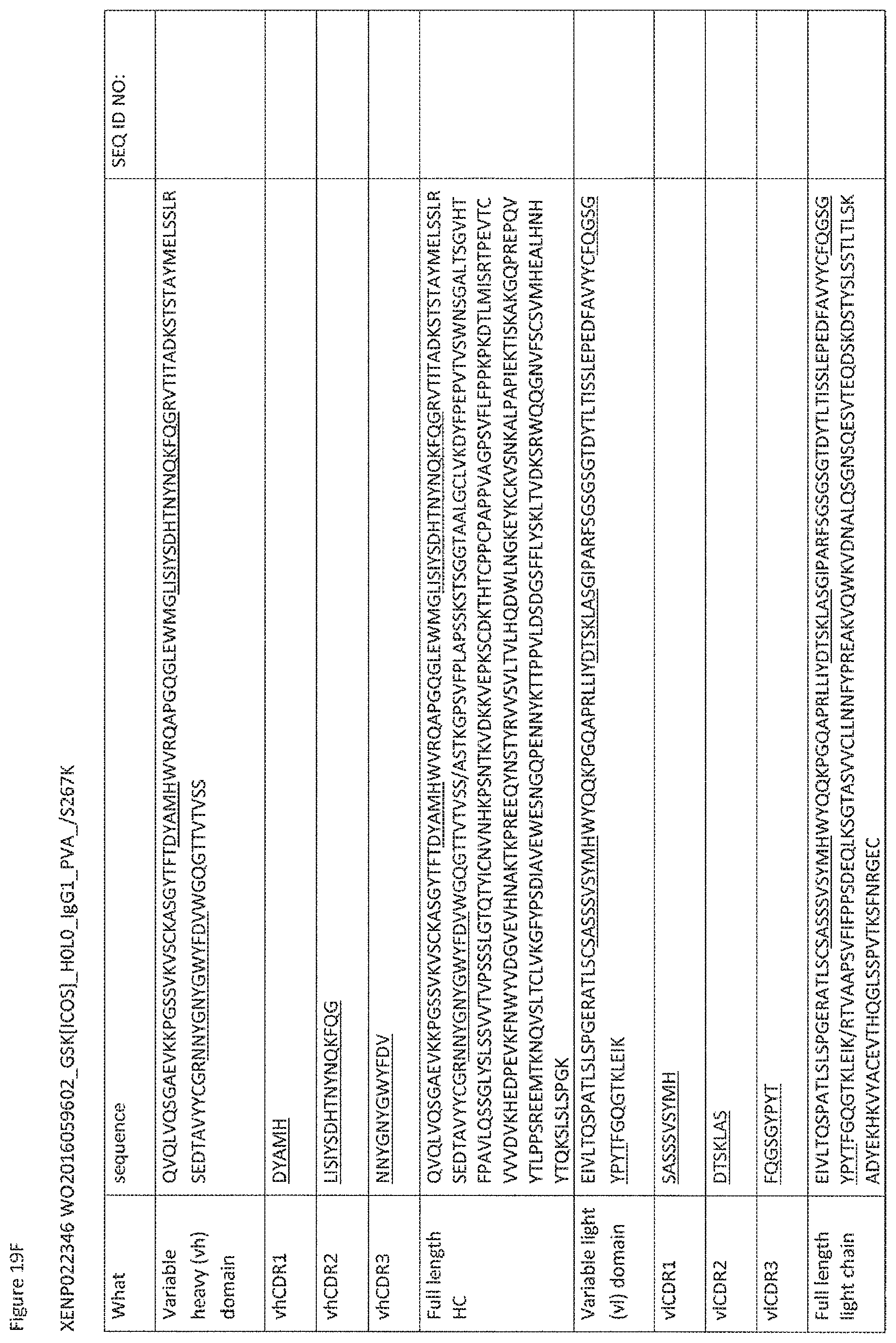
D00075
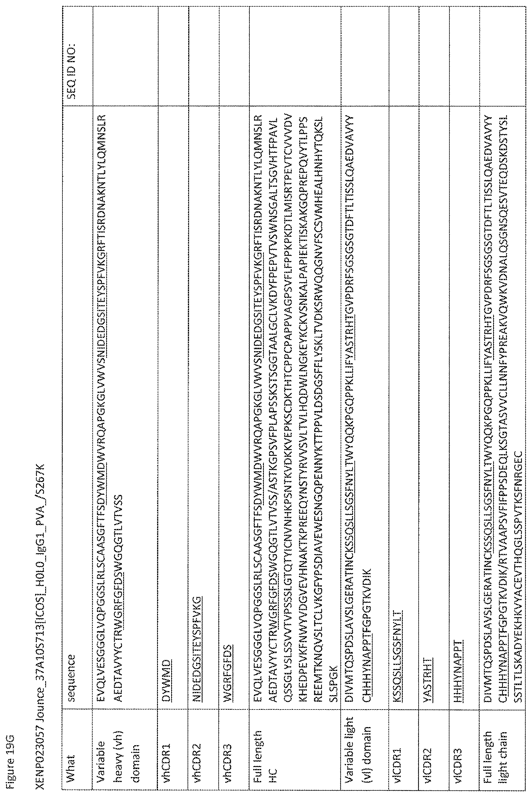
D00076

D00077
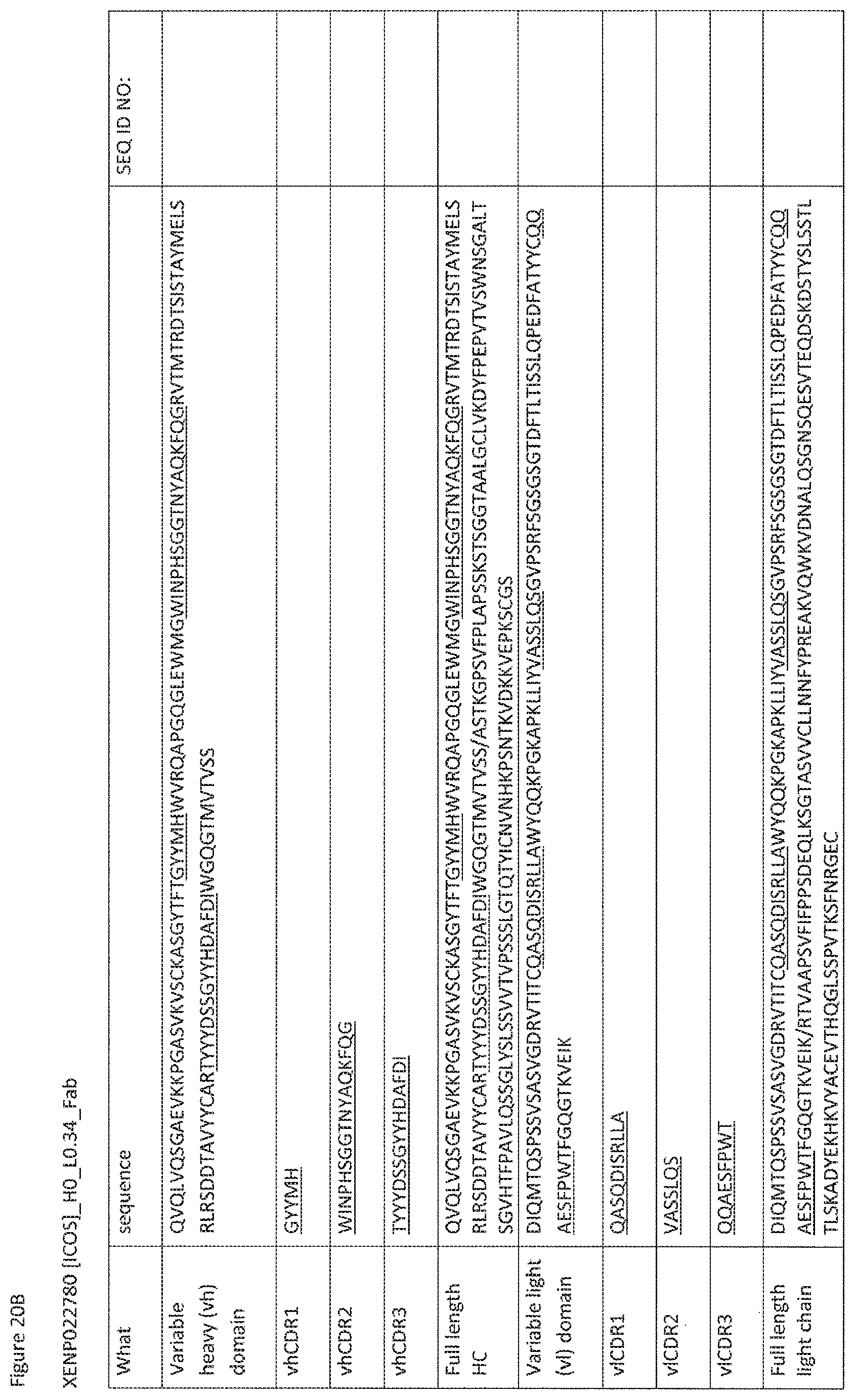
D00078
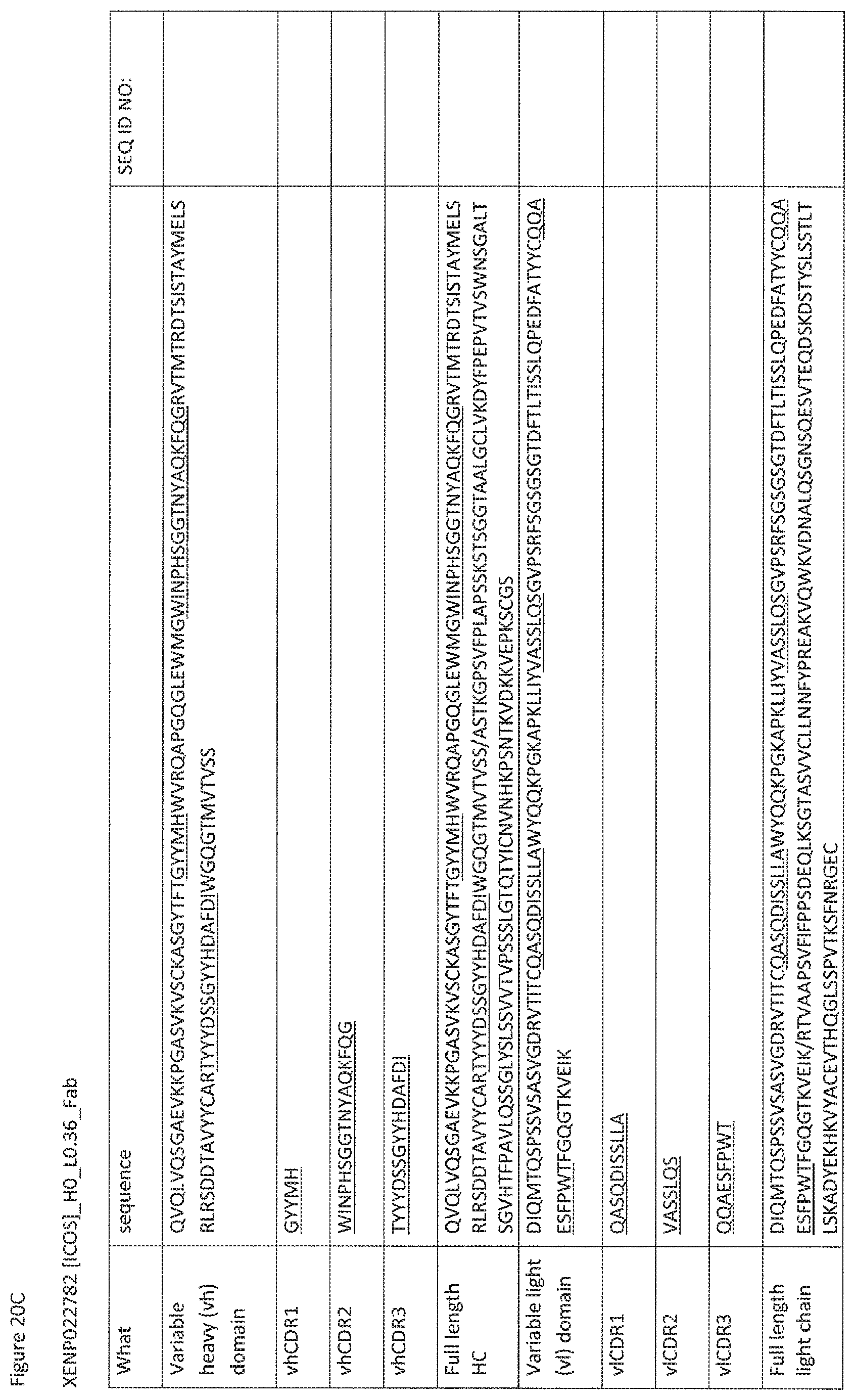
D00079

D00080

D00081
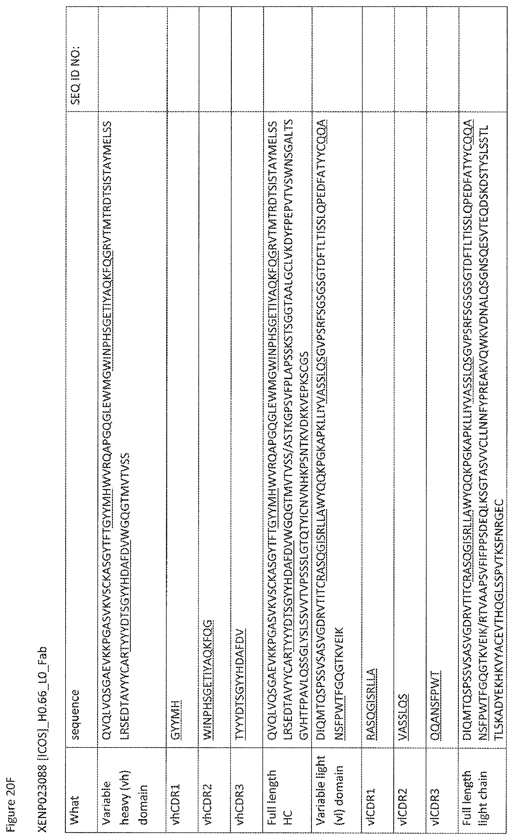
D00082
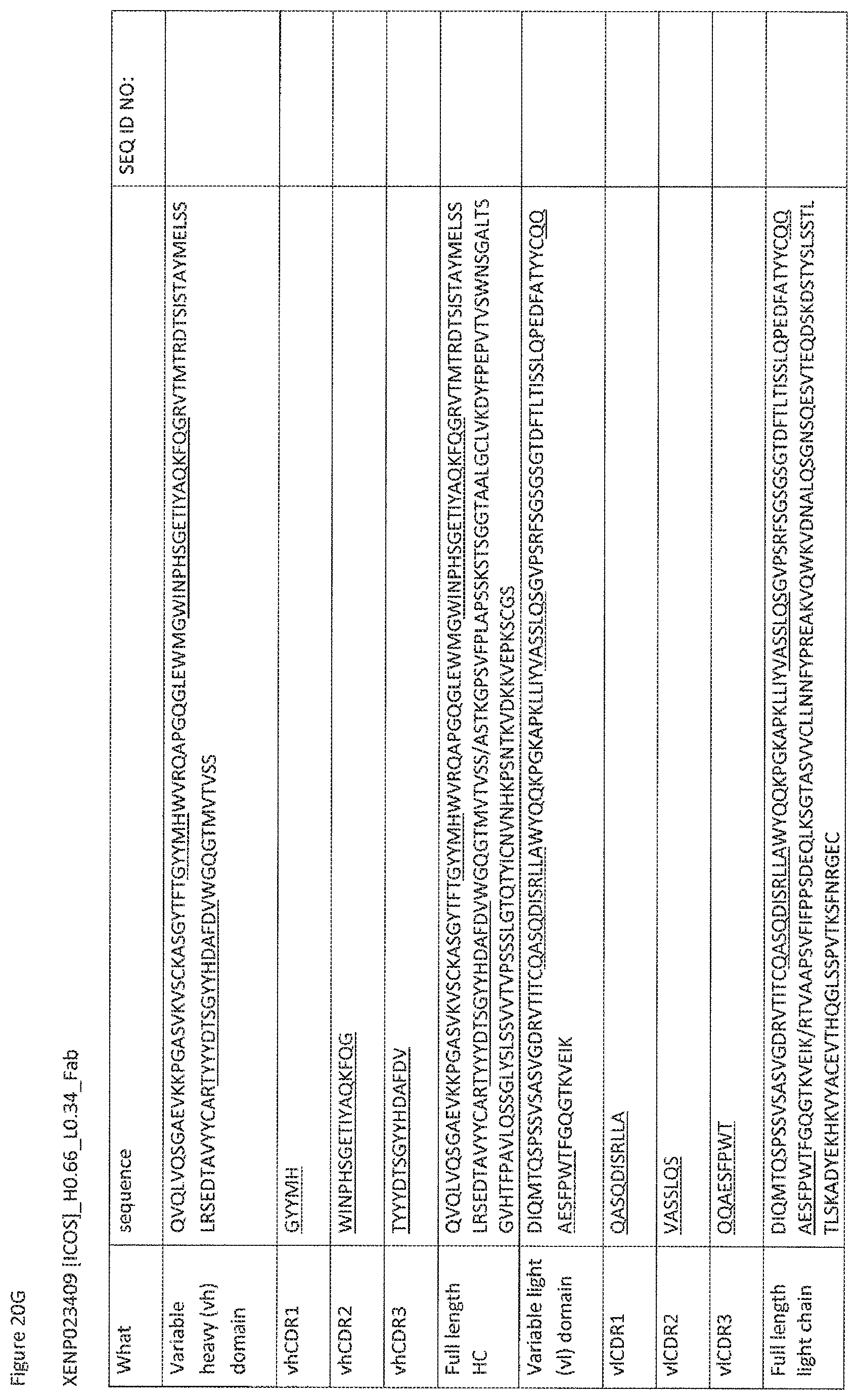
D00083
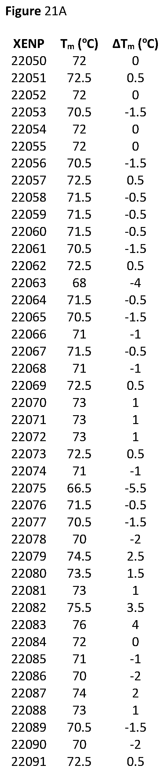
D00084

D00085
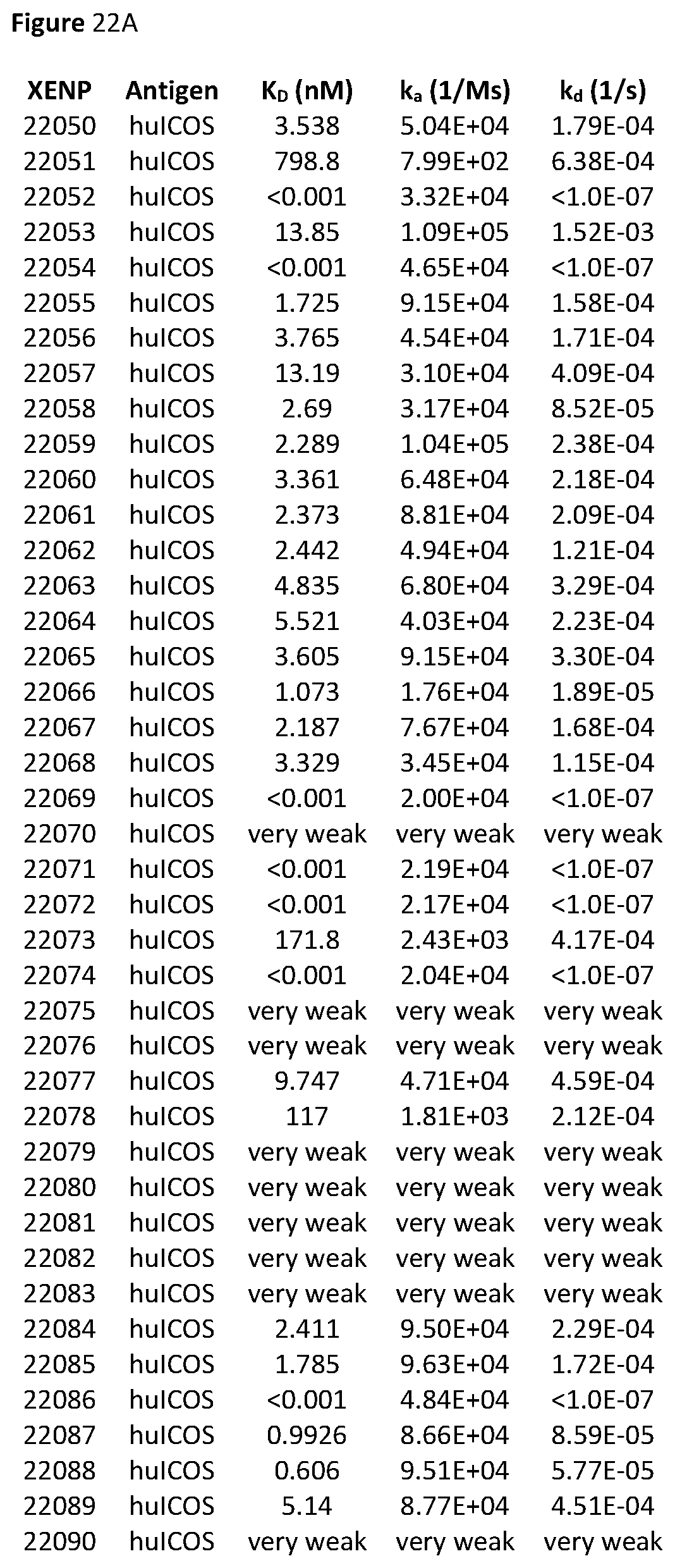
D00086
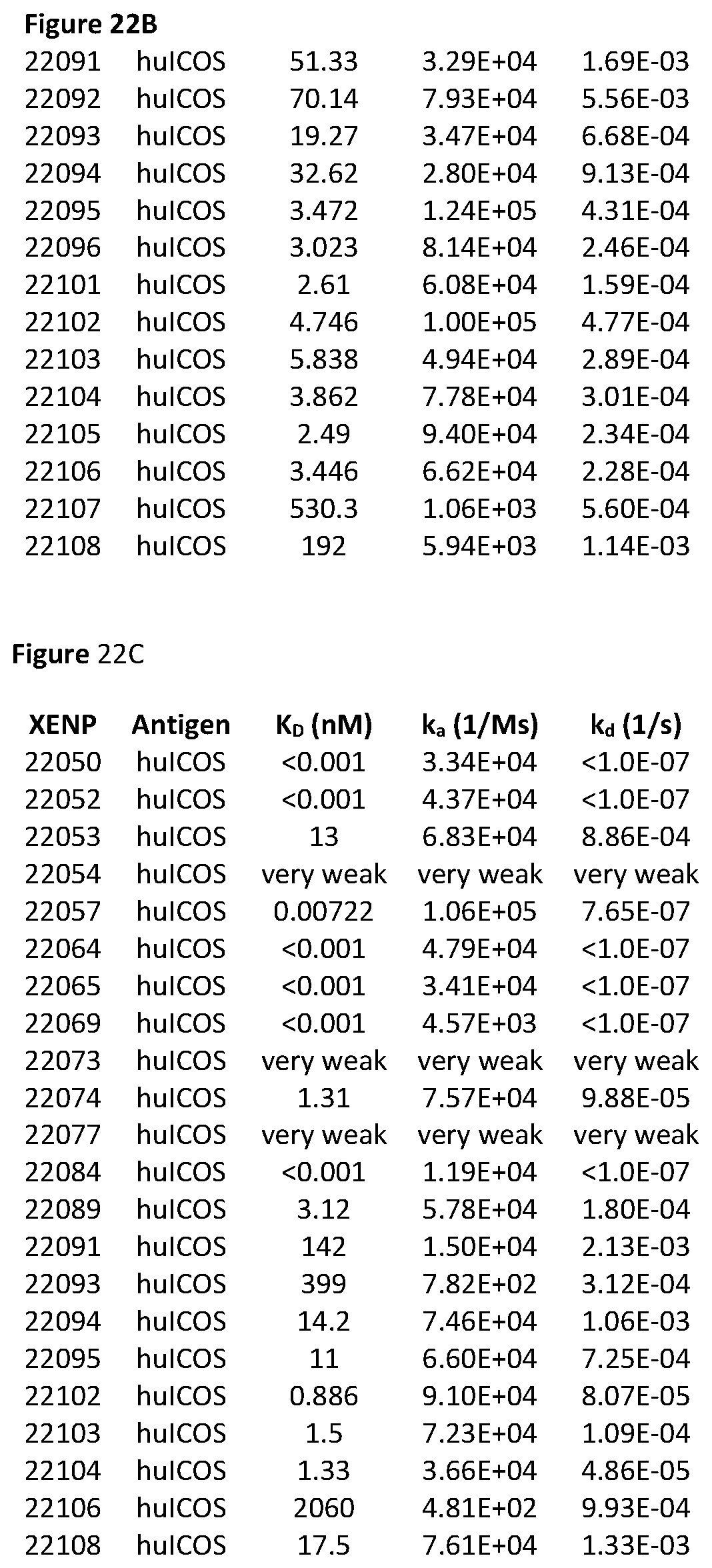
D00087

D00088

D00089
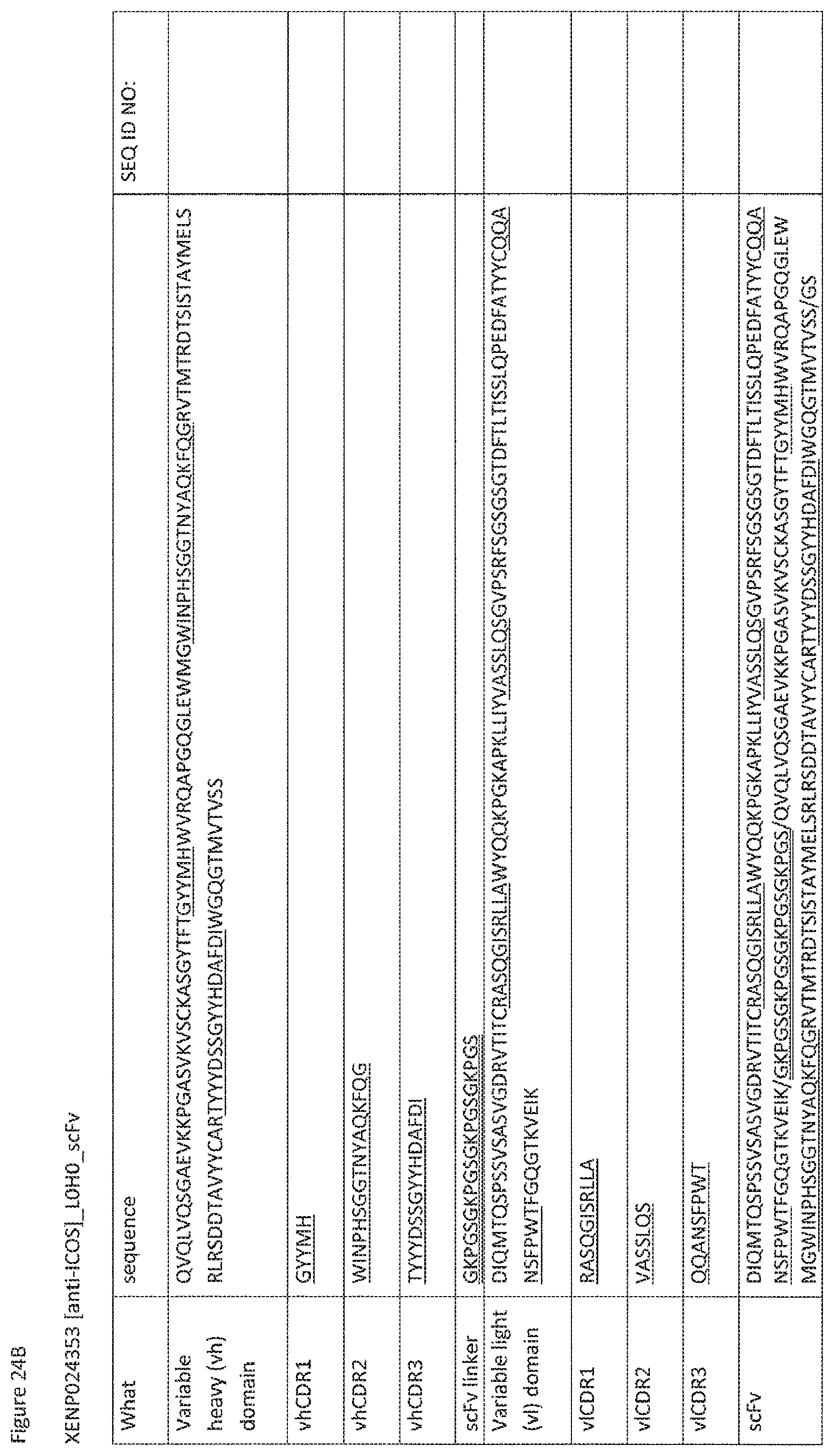
D00090
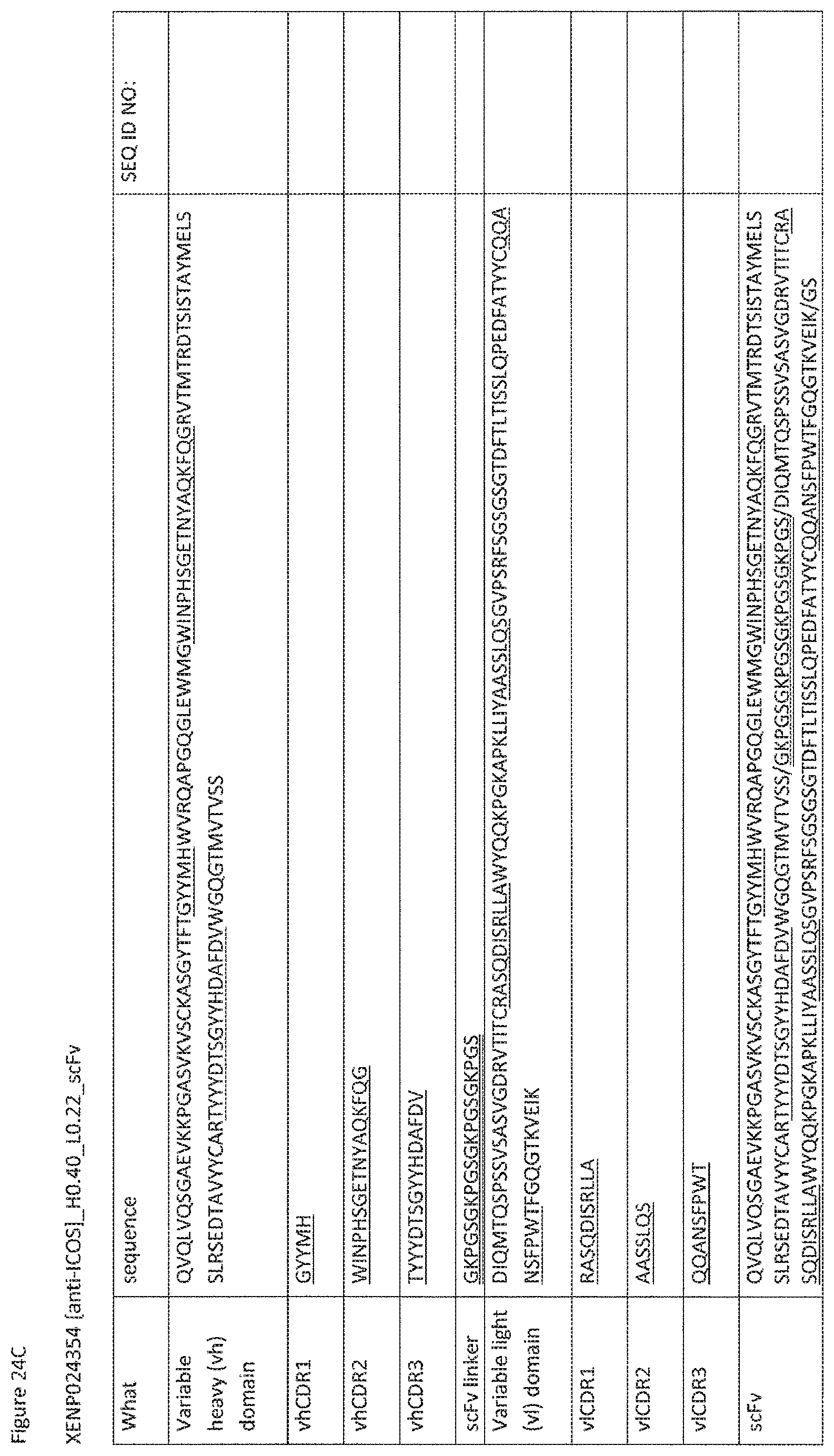
D00091

D00092

D00093
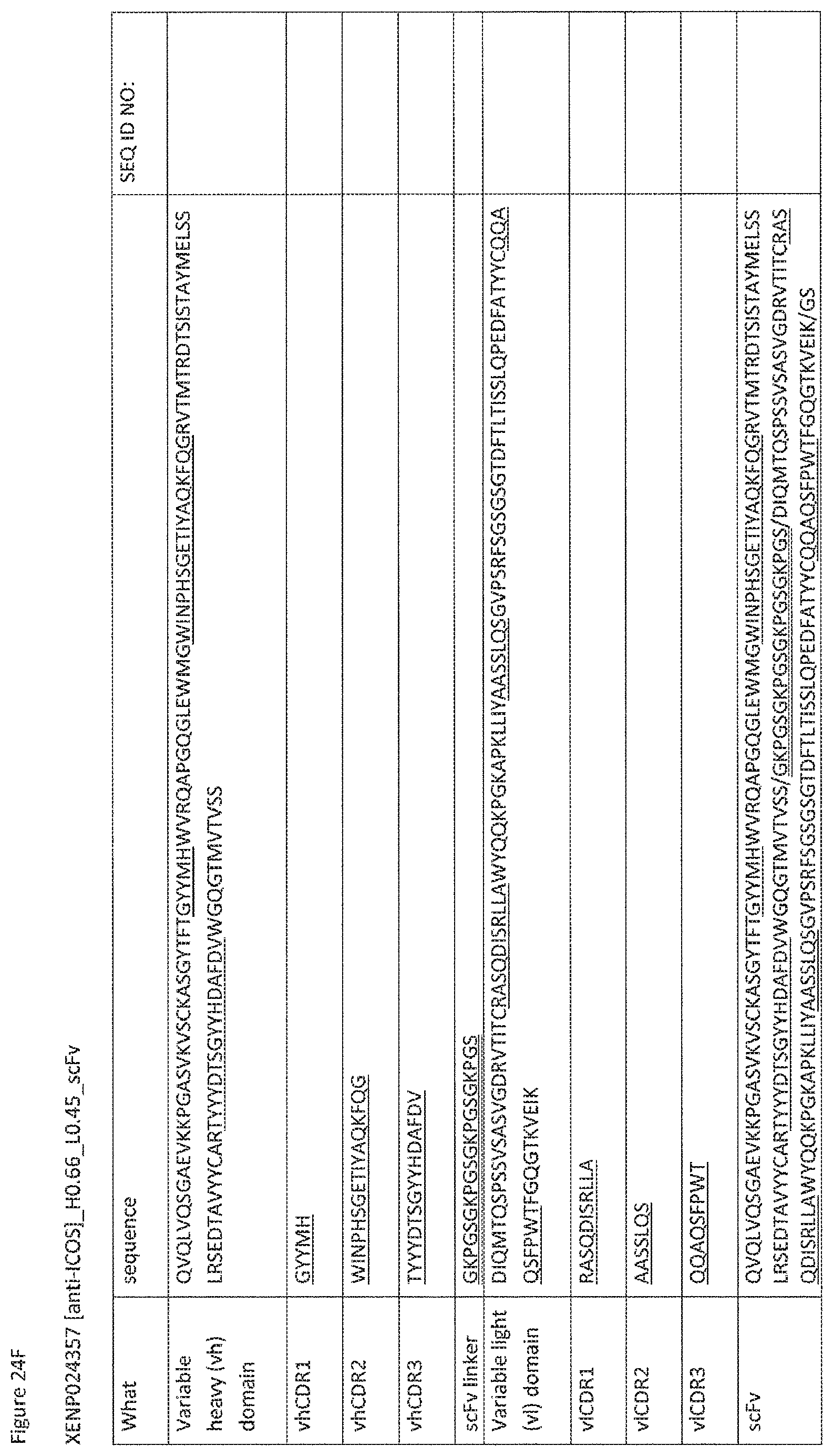
D00094
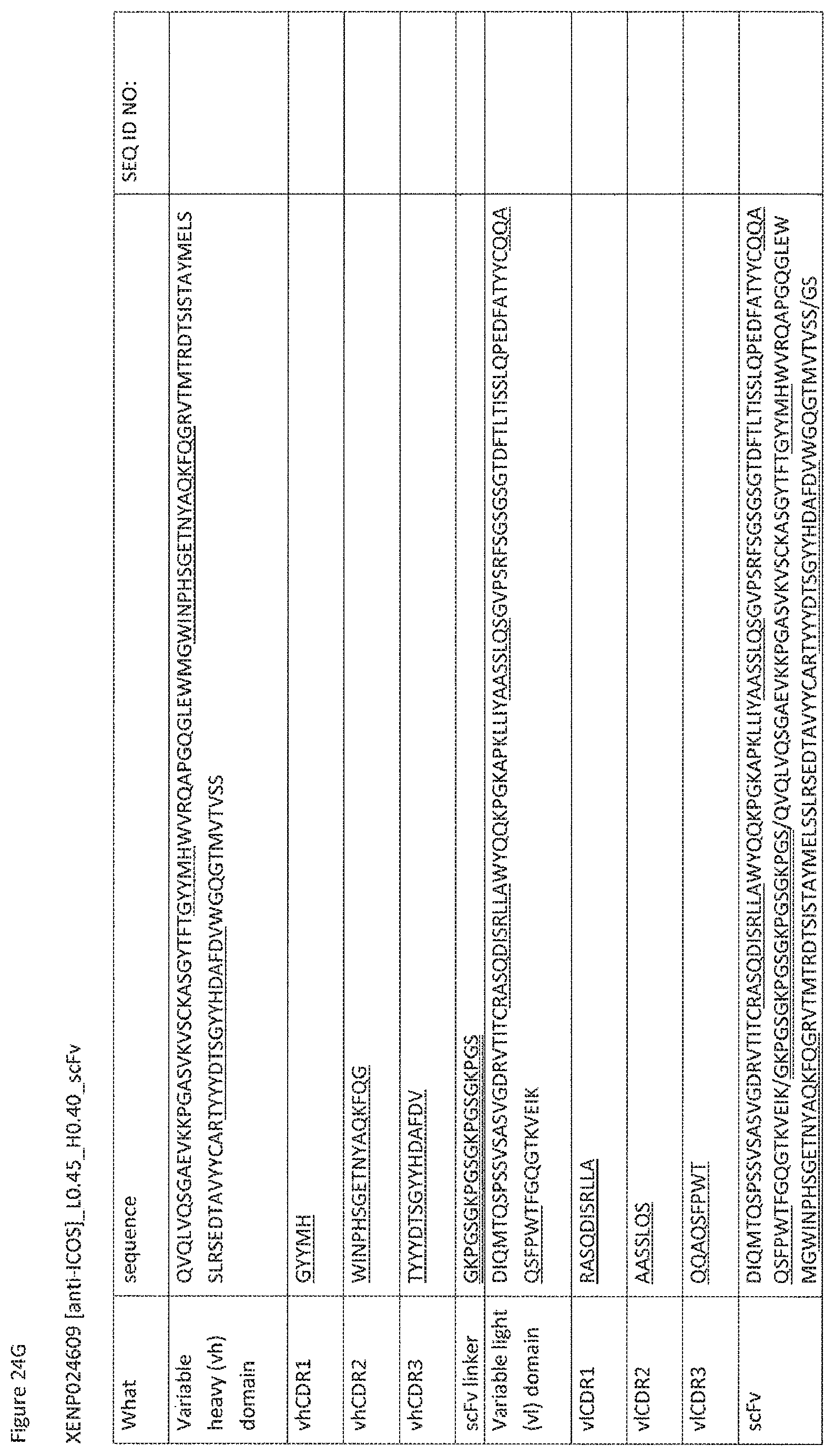
D00095

D00096
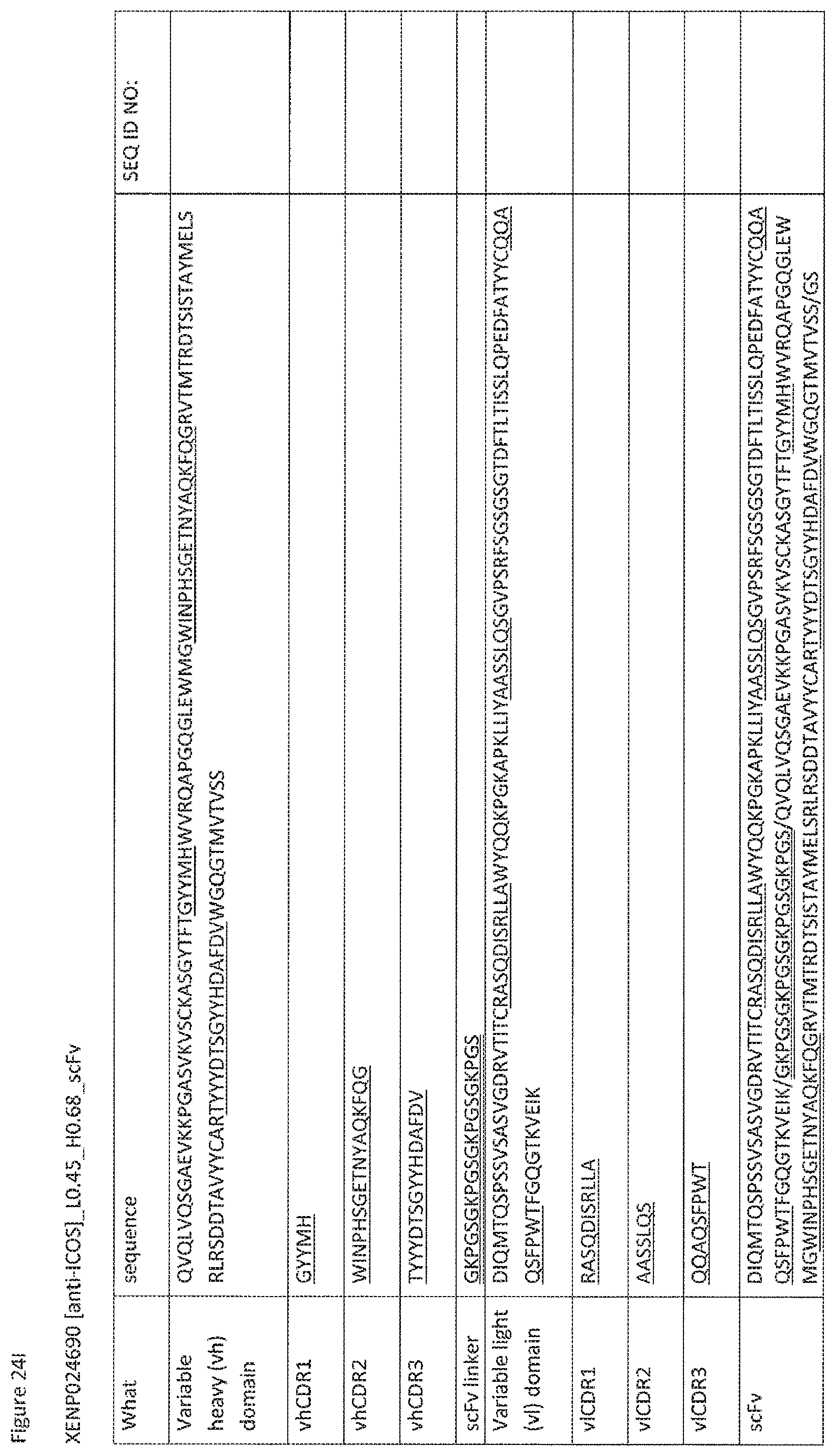
D00097
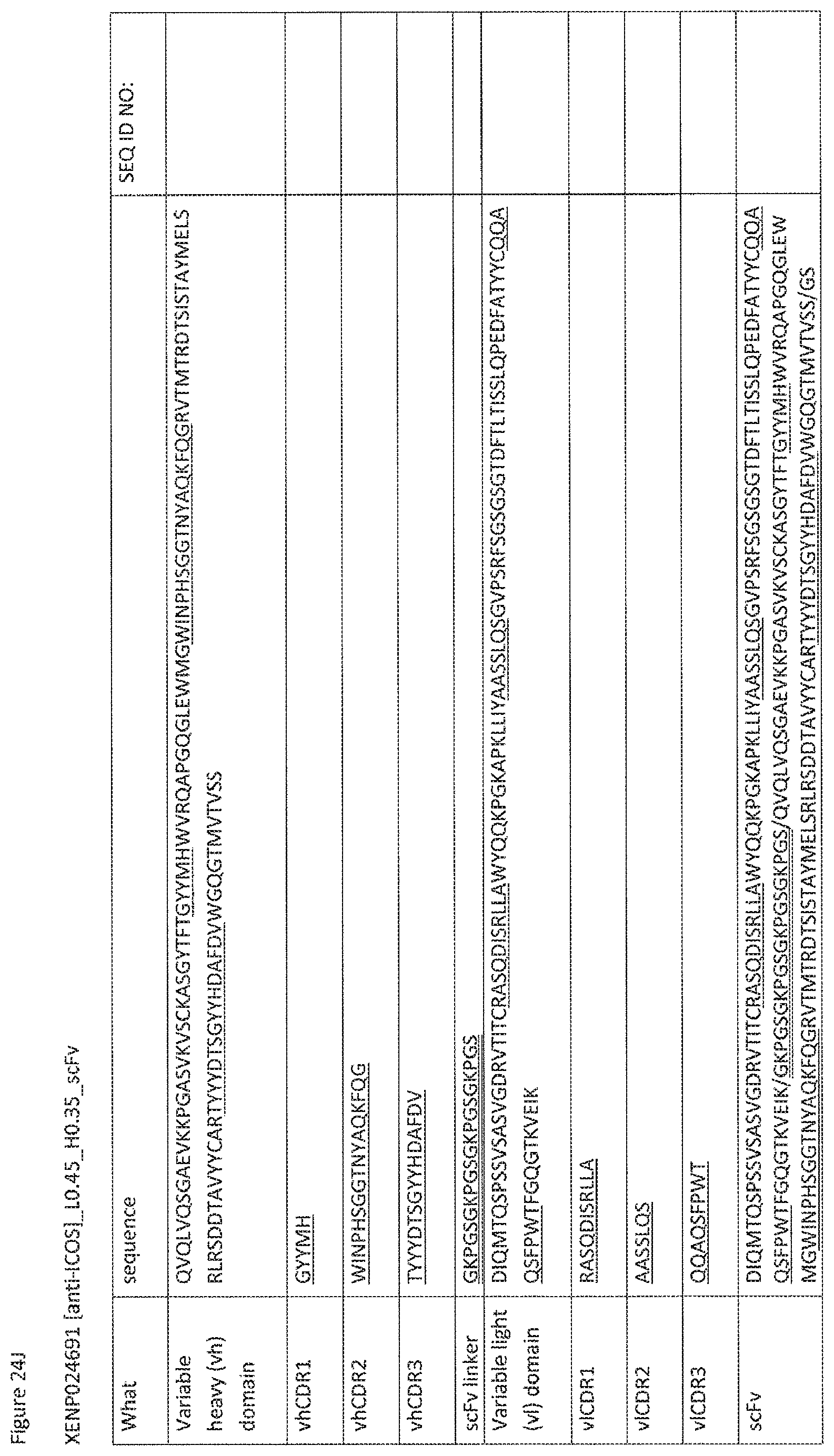
D00098
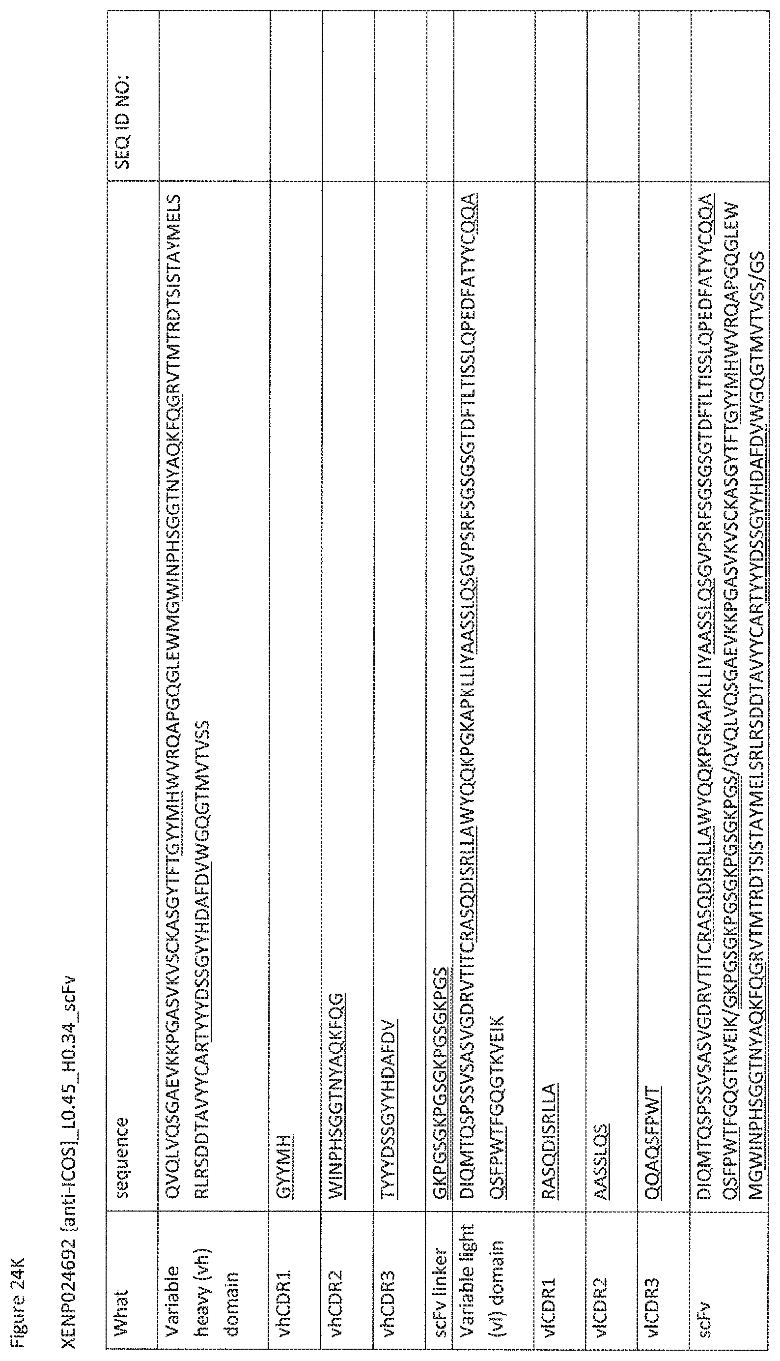
D00099
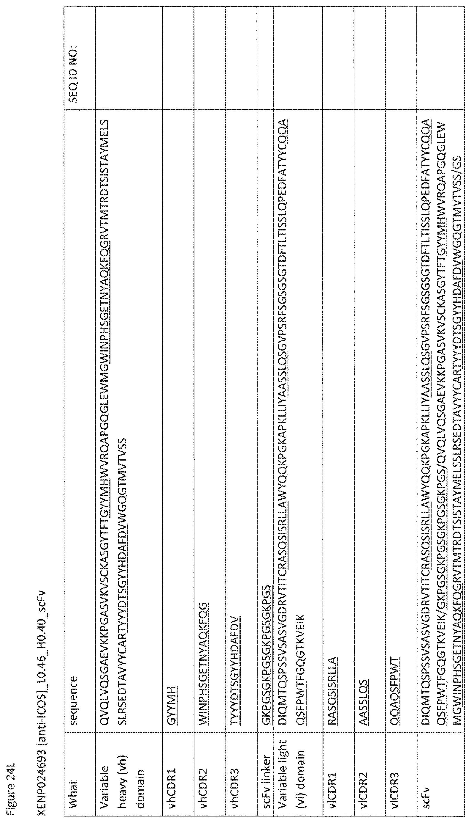
D00100

D00101
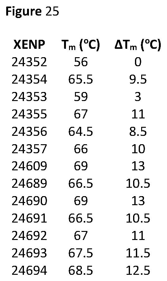
D00102

D00103
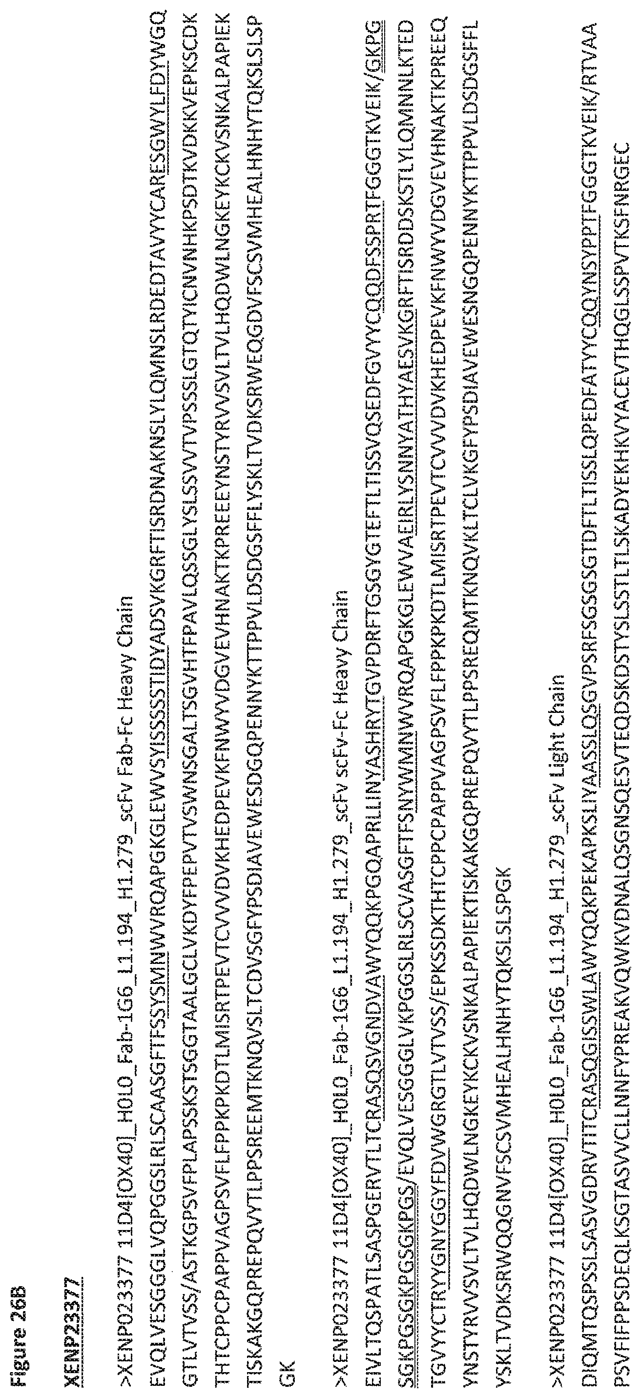
D00104
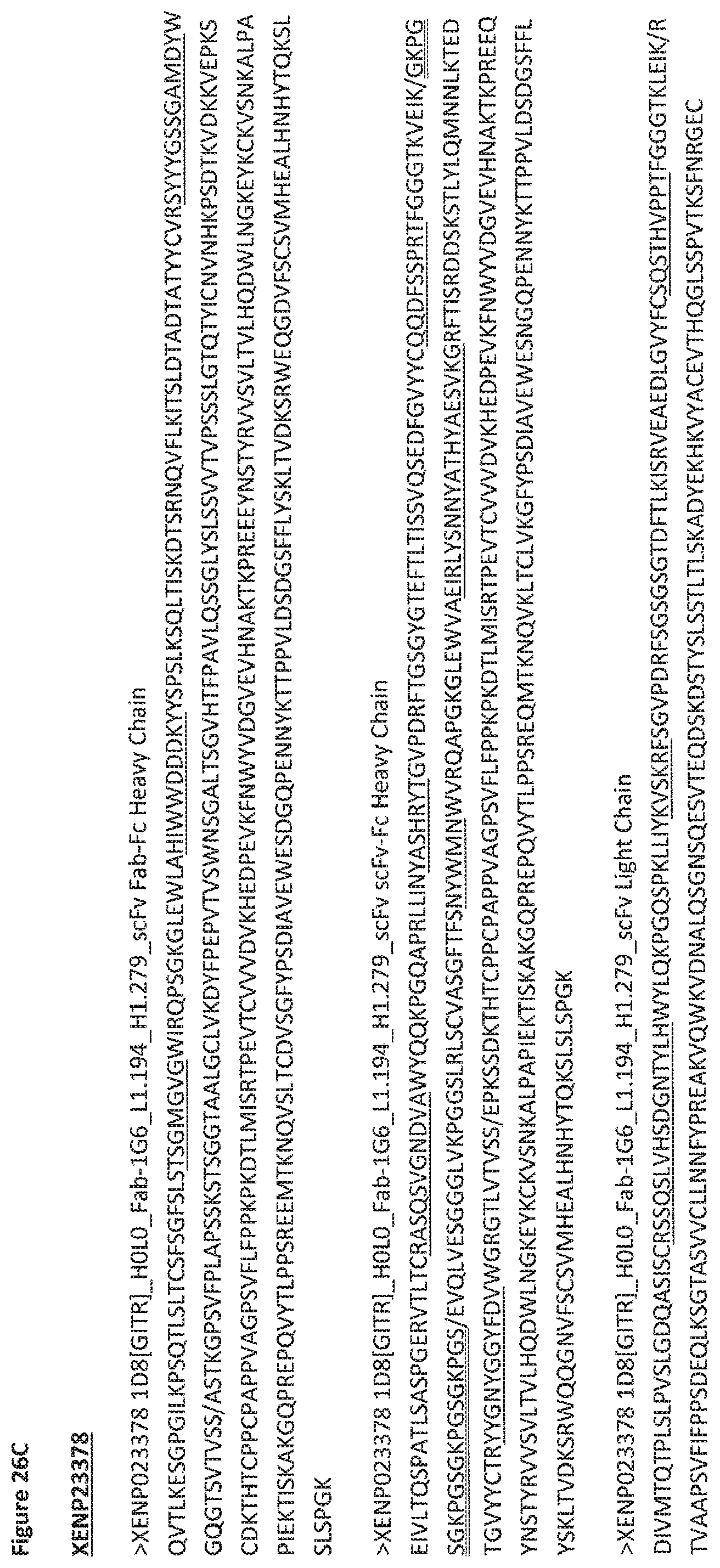
D00105
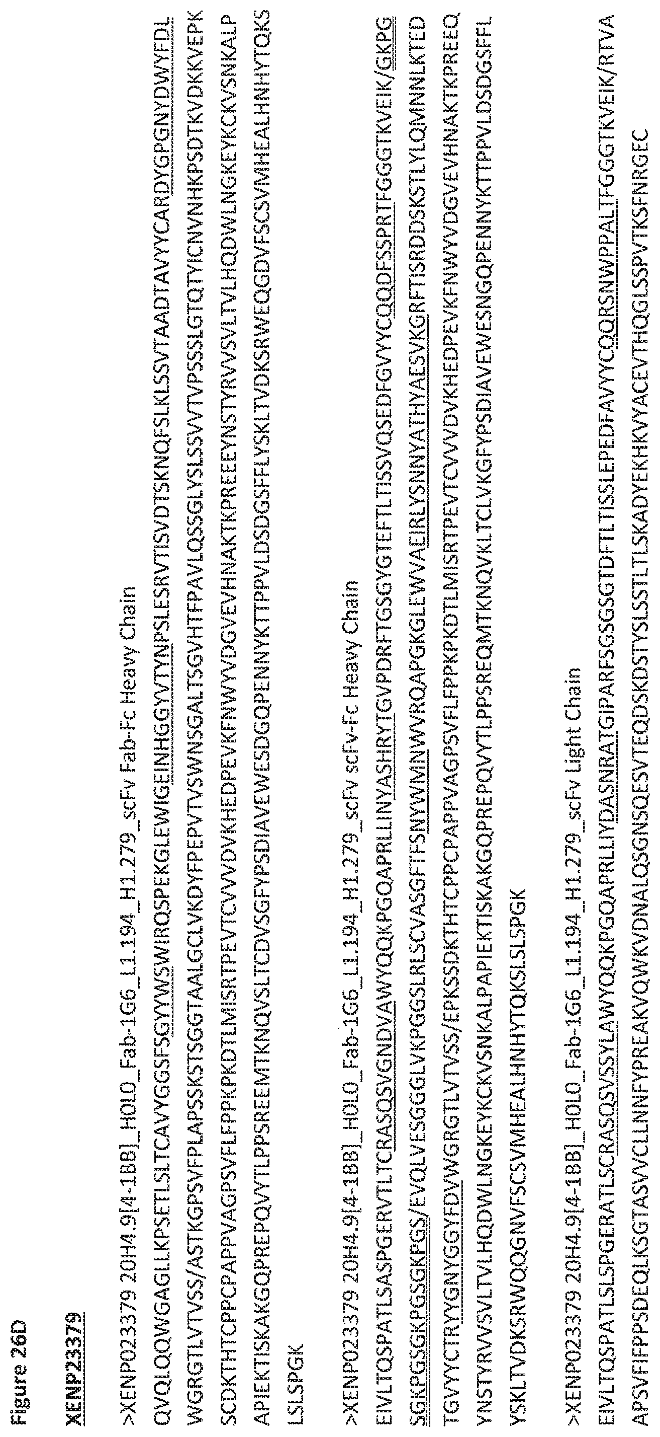
D00106
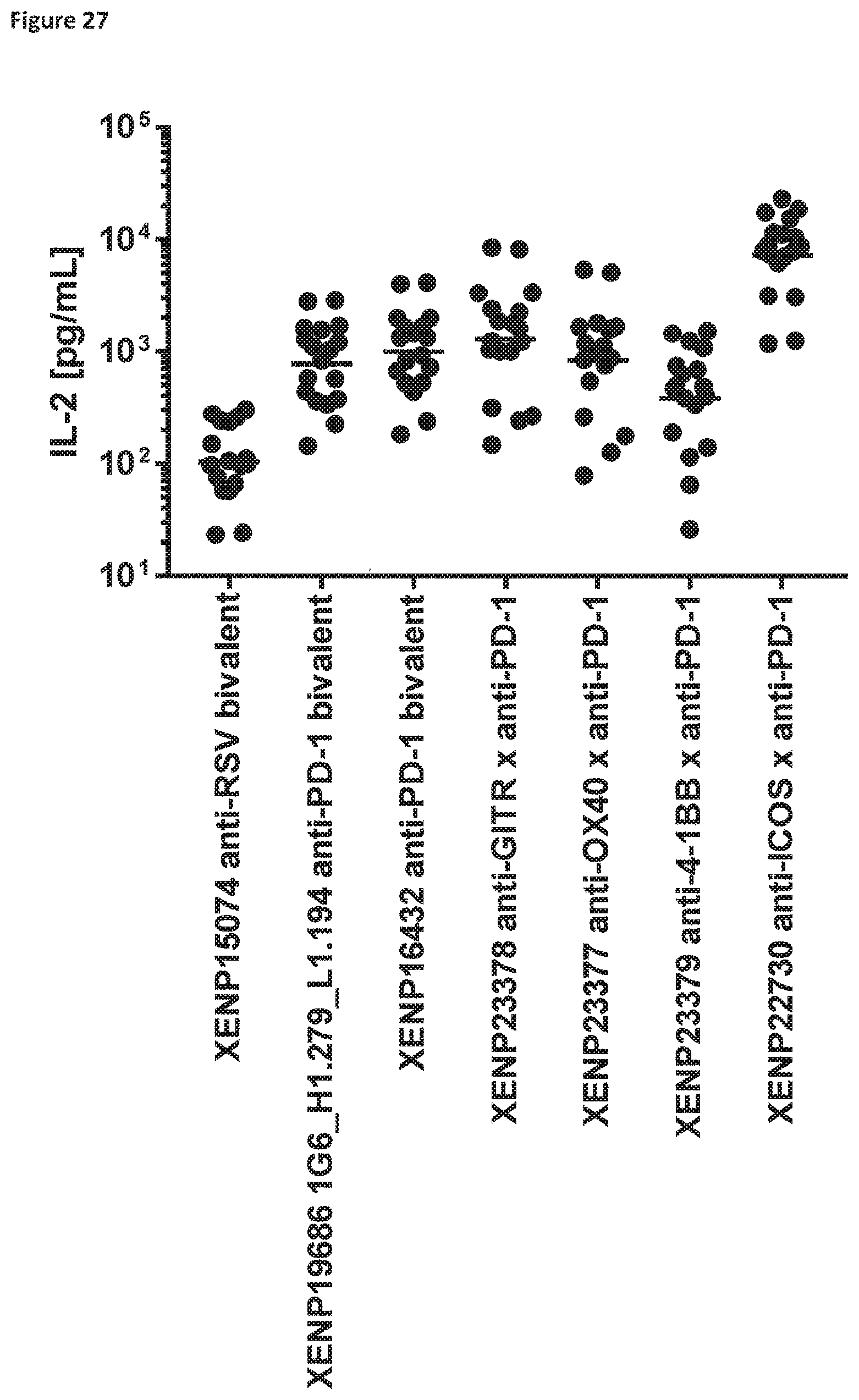
D00107

D00108
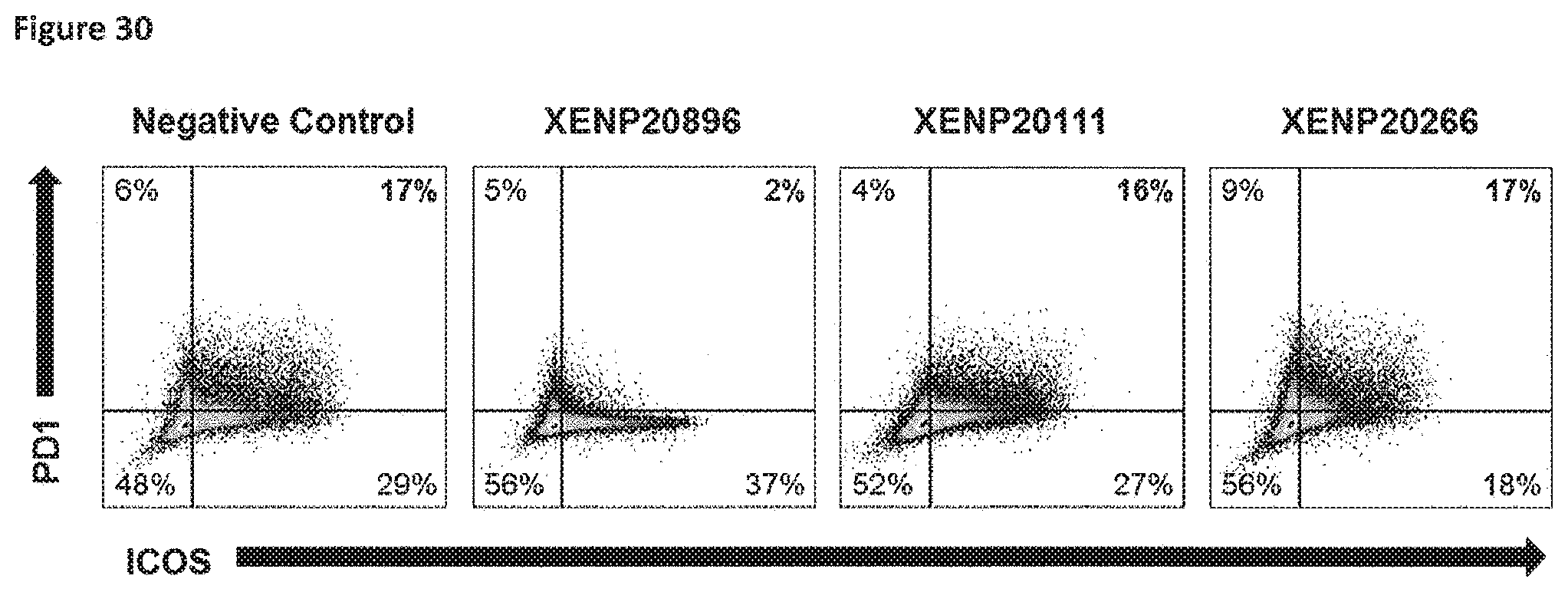
D00109
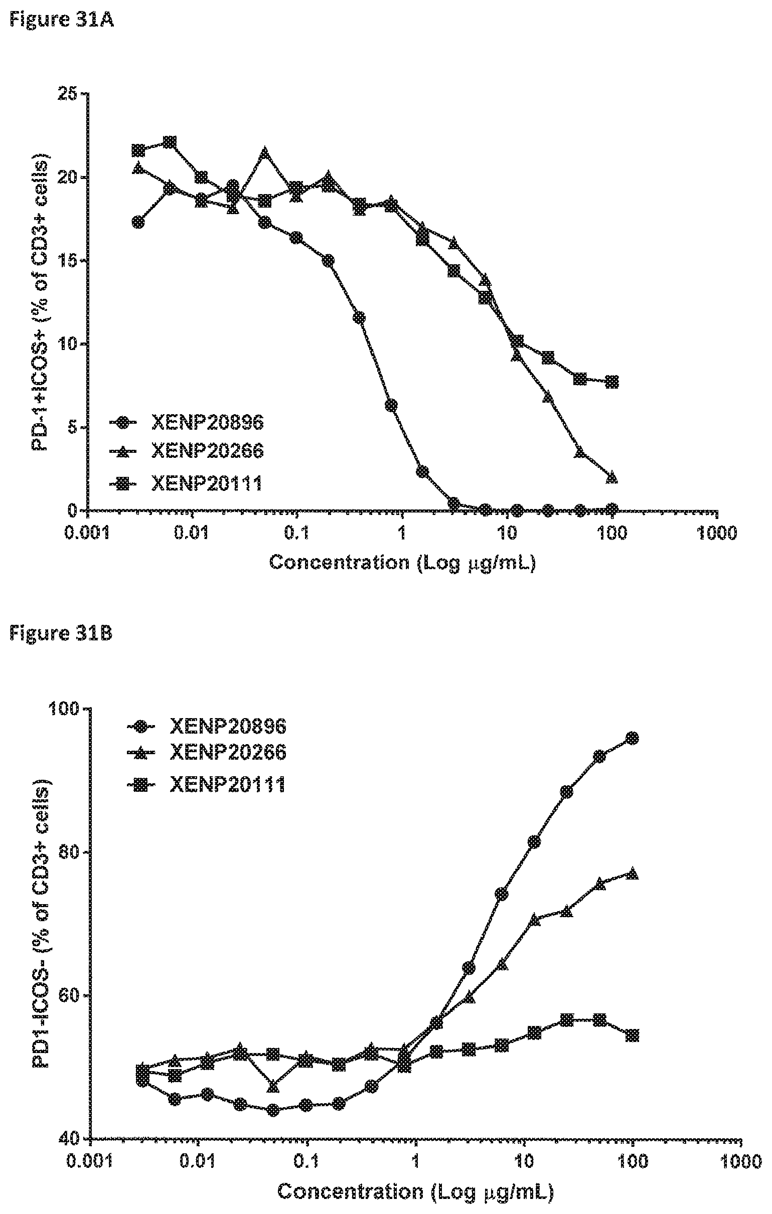
D00110
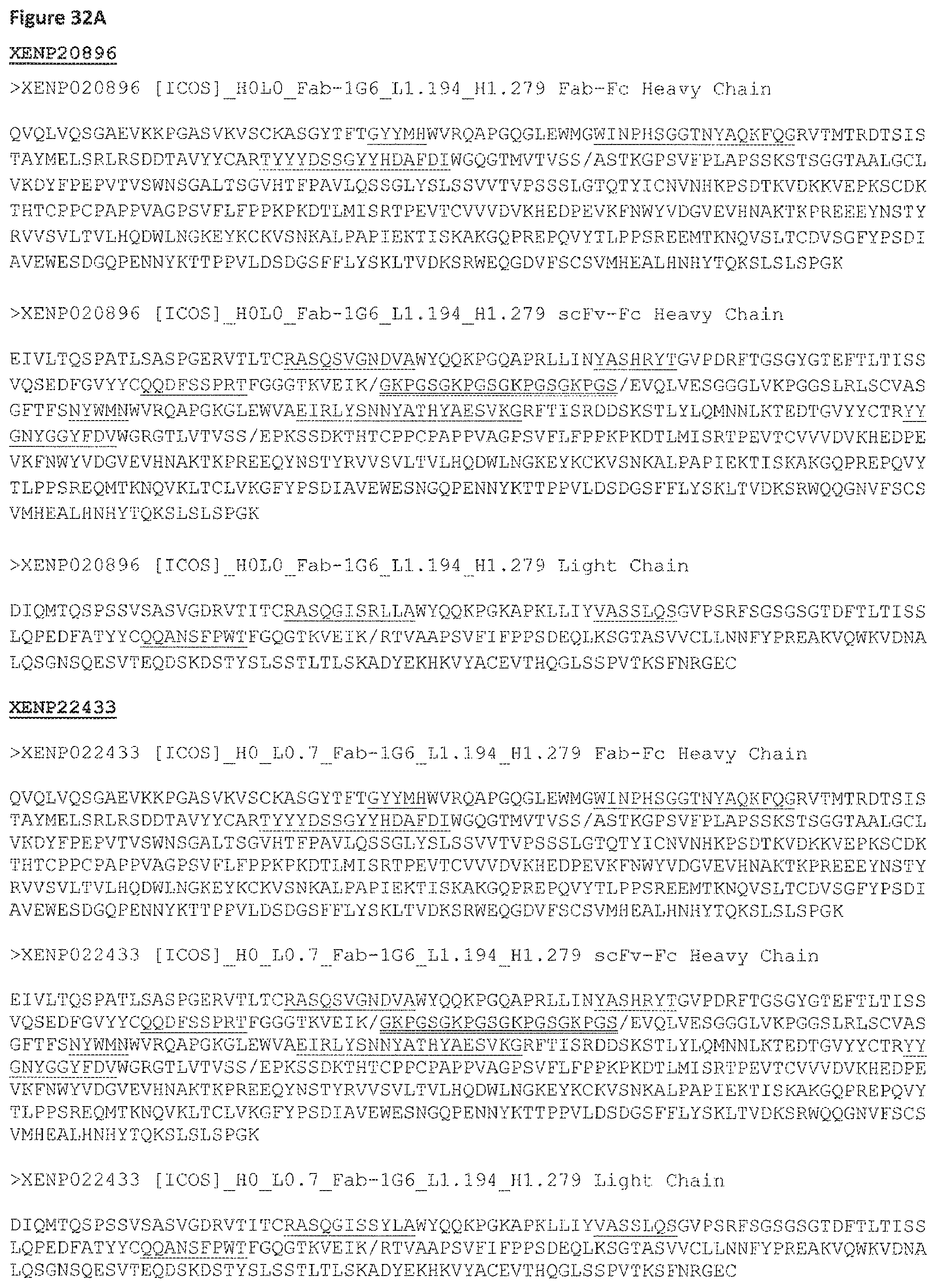
D00111
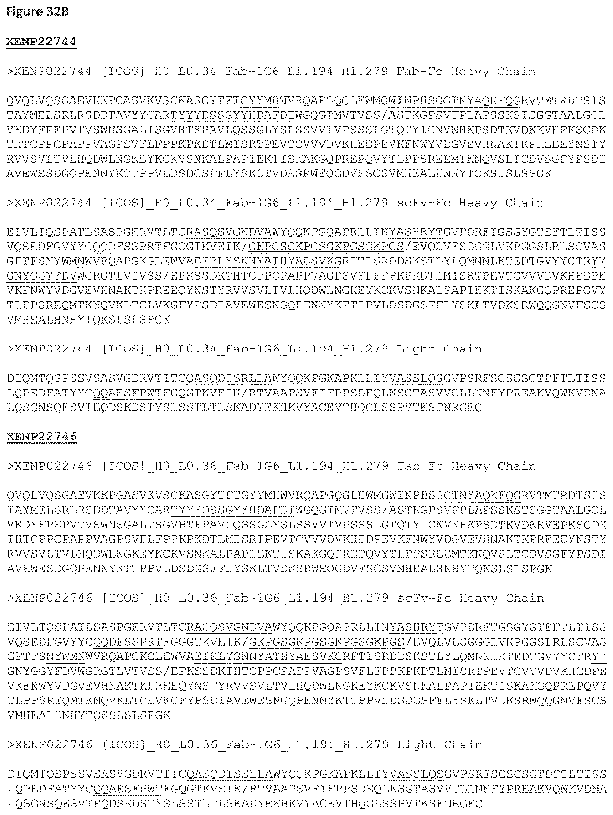
D00112
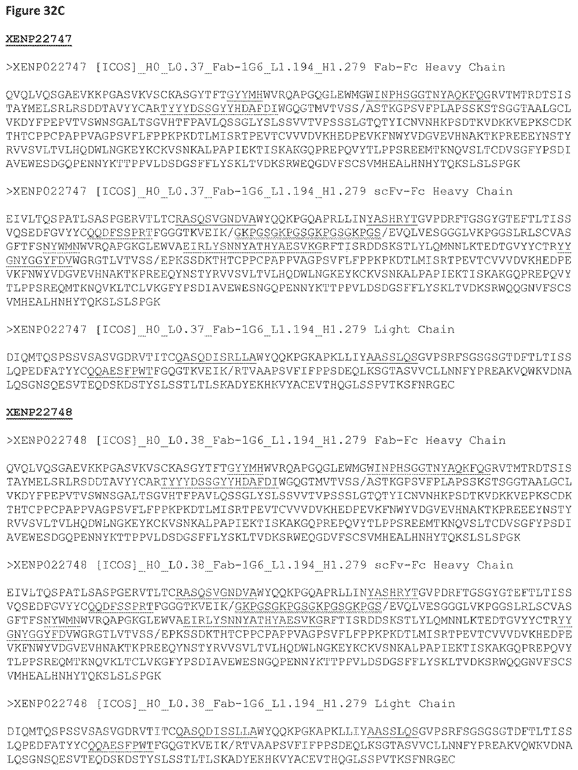
D00113
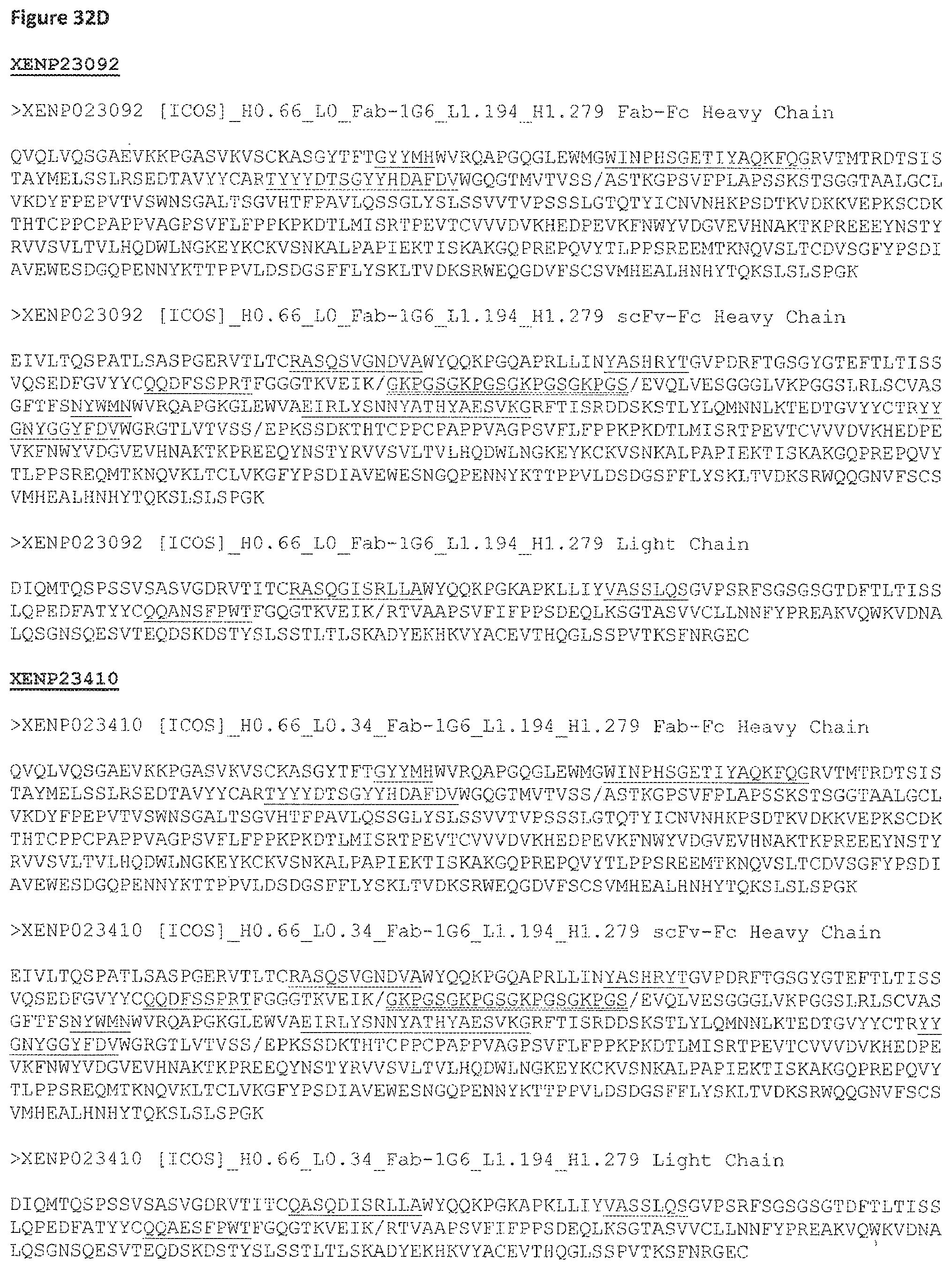
D00114
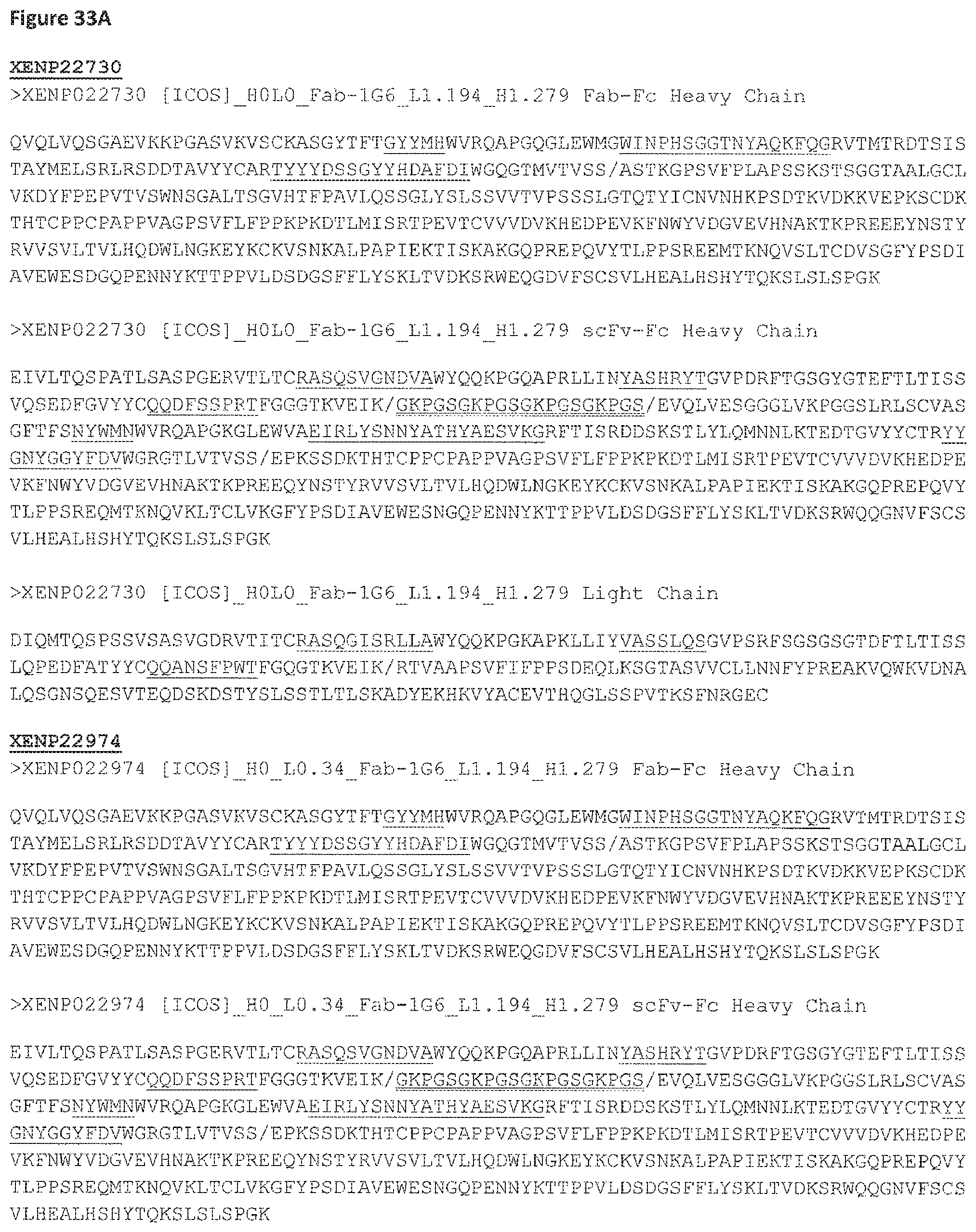
D00115
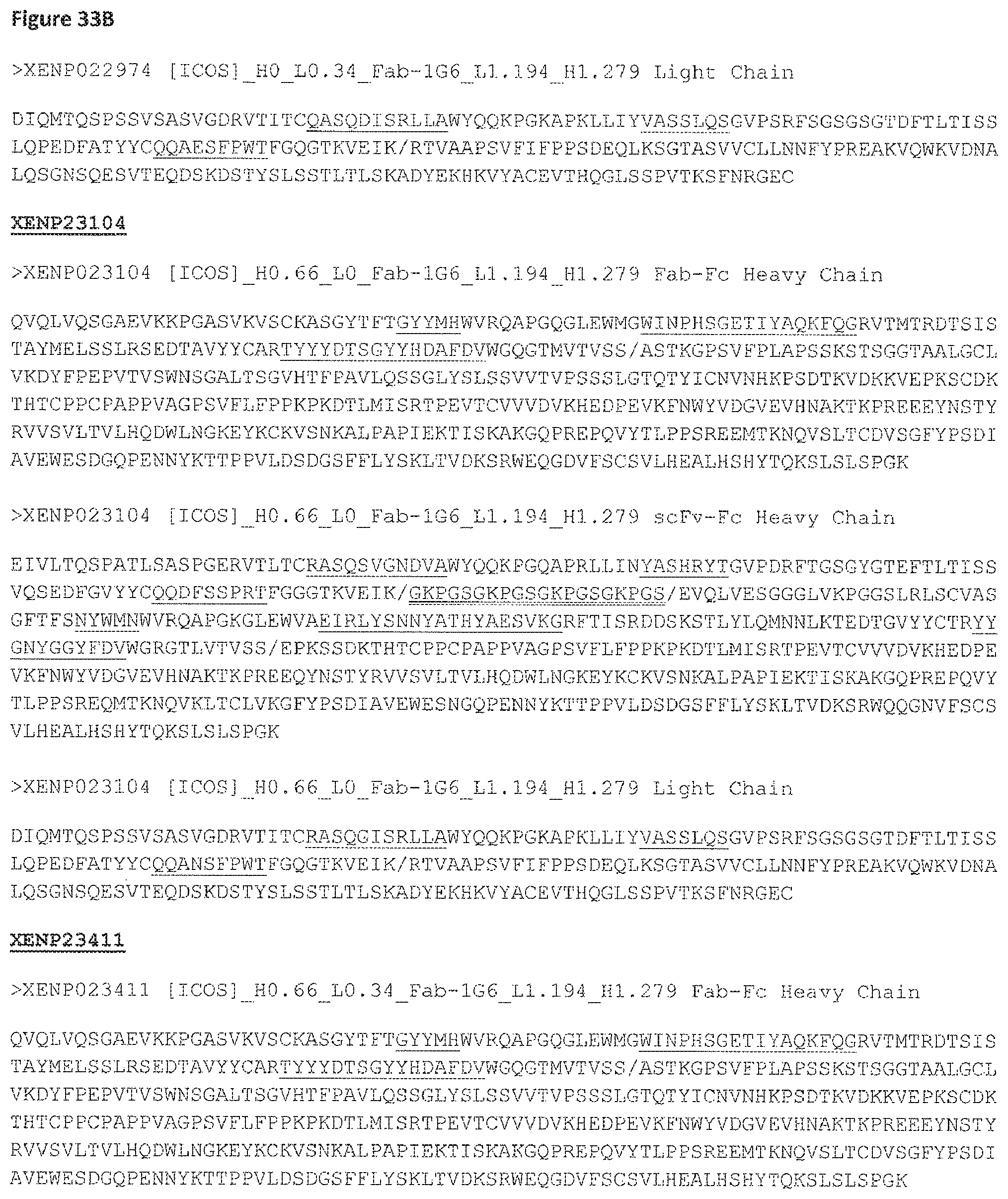
D00116

D00117
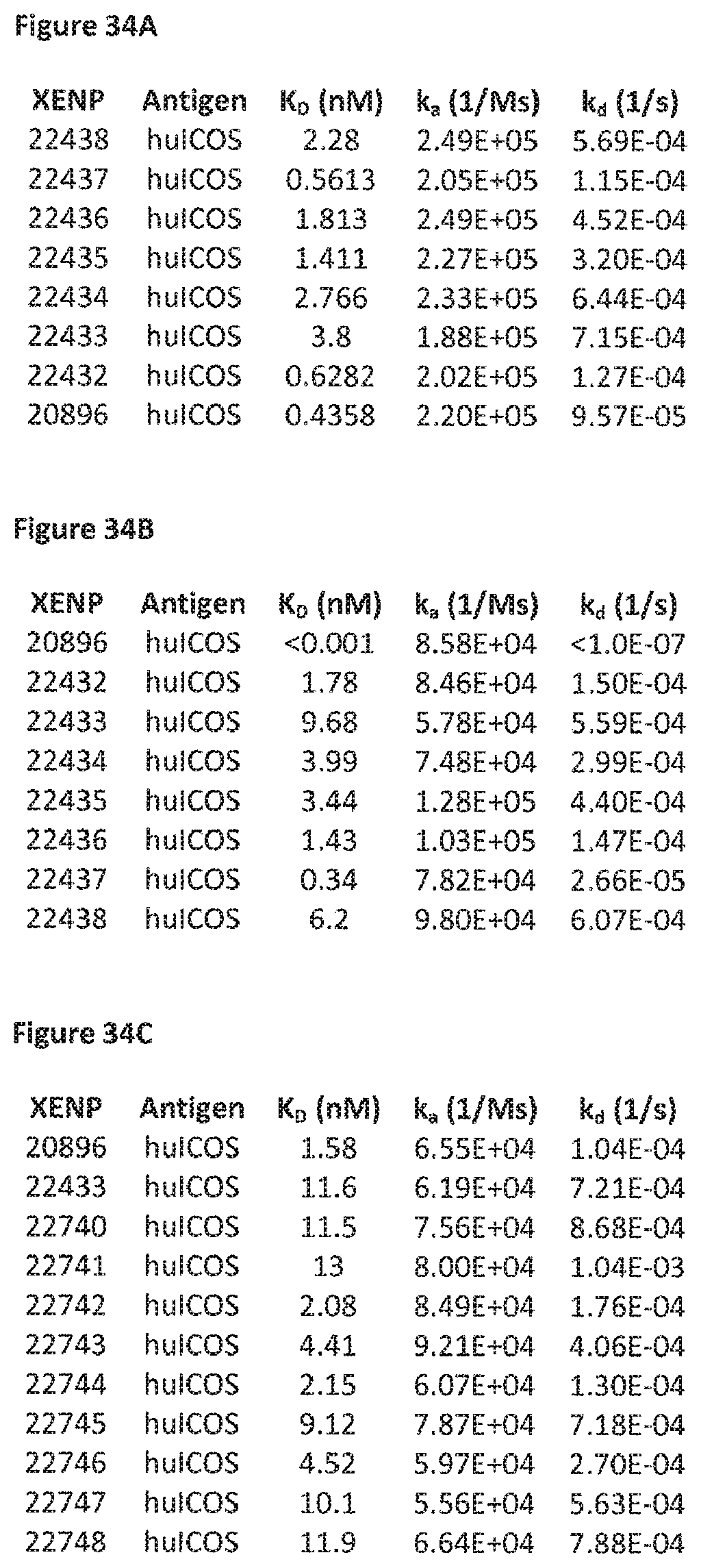
D00118
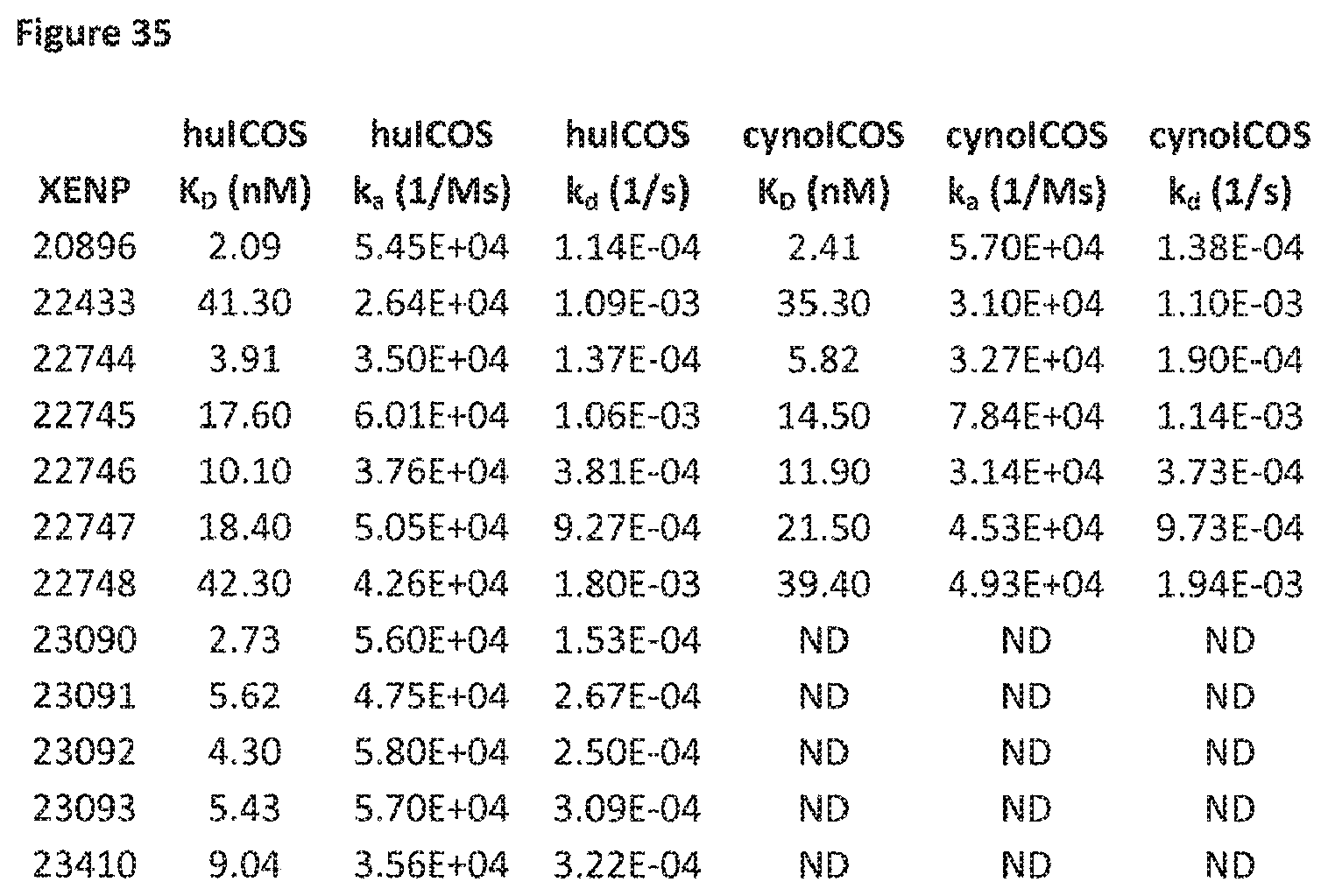
D00119
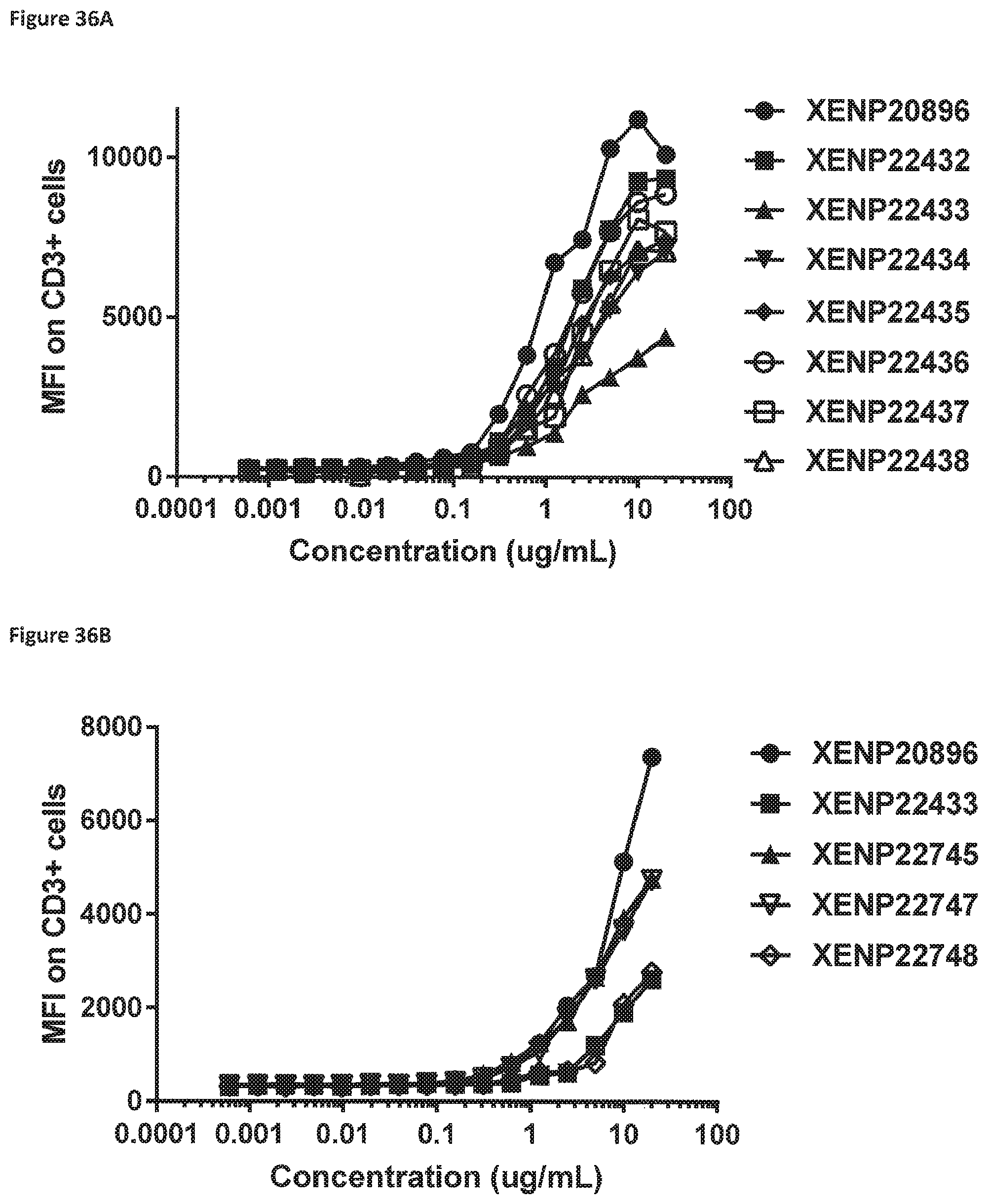
D00120

D00121

D00122
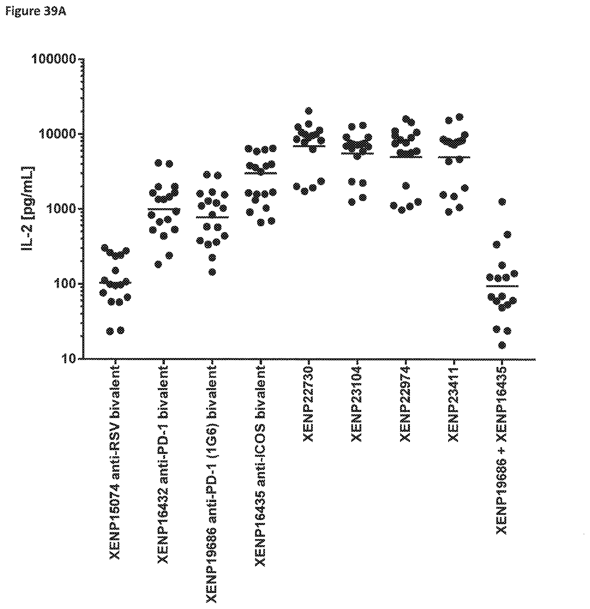
D00123

D00124

D00125
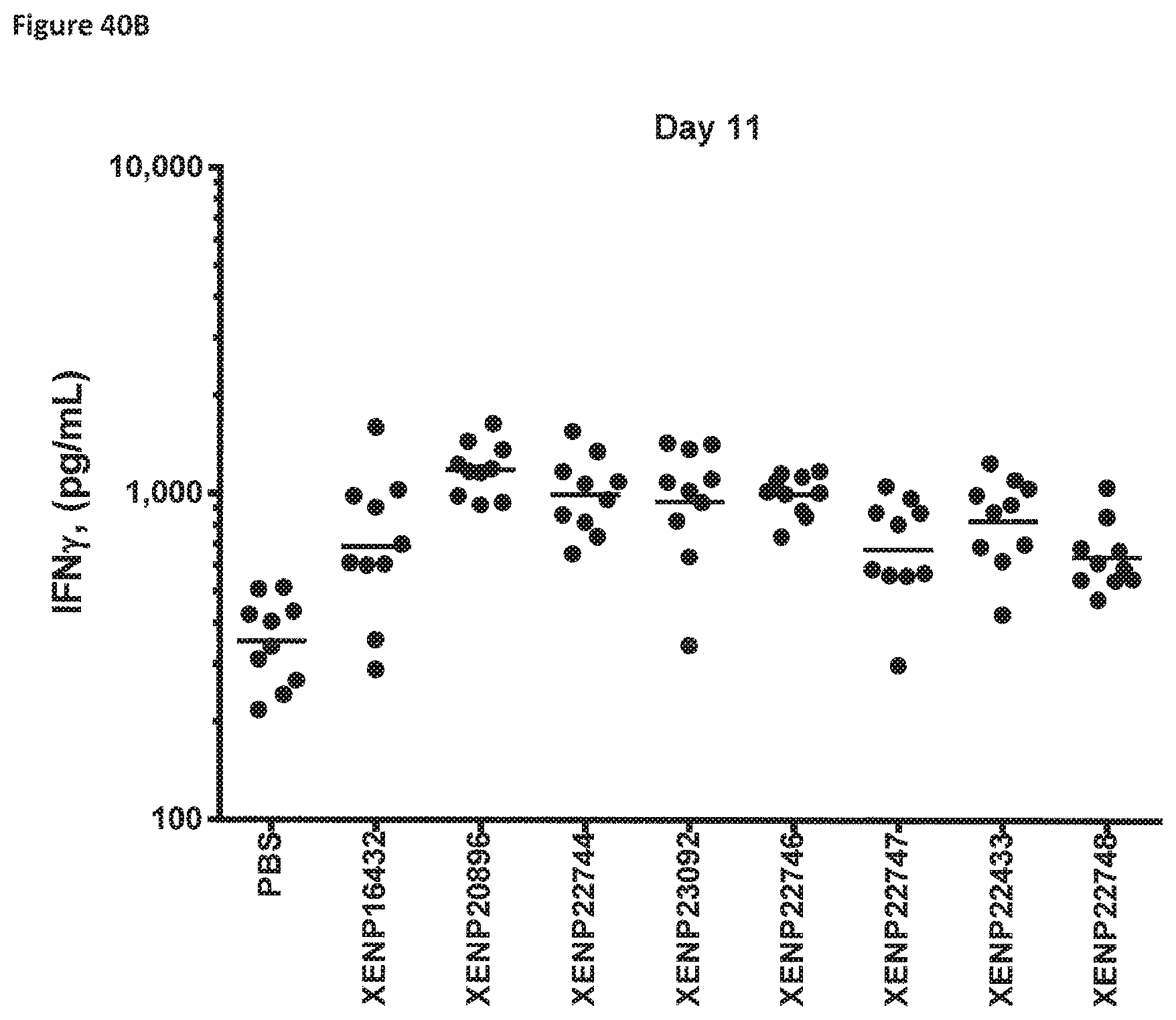
D00126
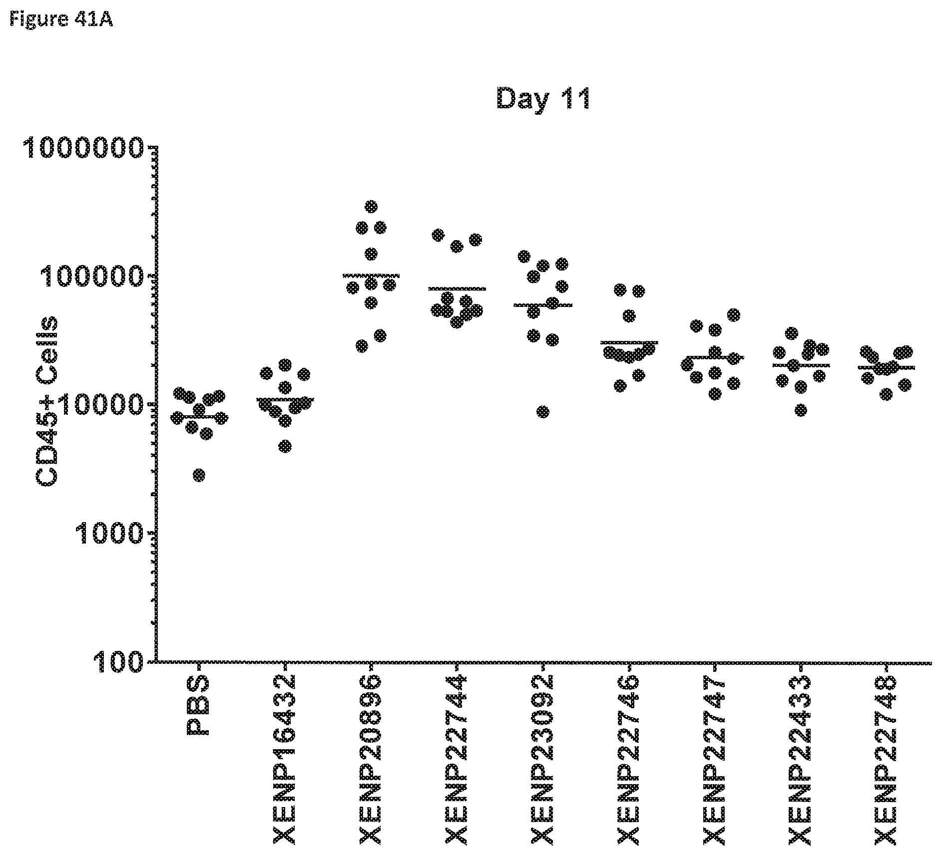
D00127

D00128

D00129

D00130
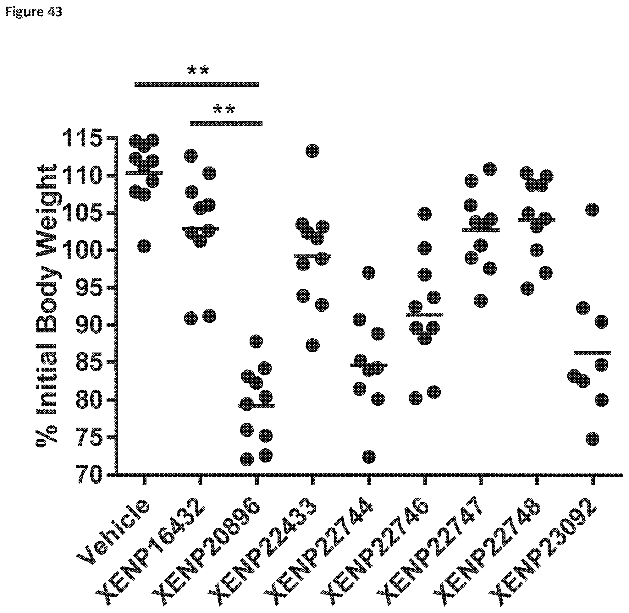
D00131
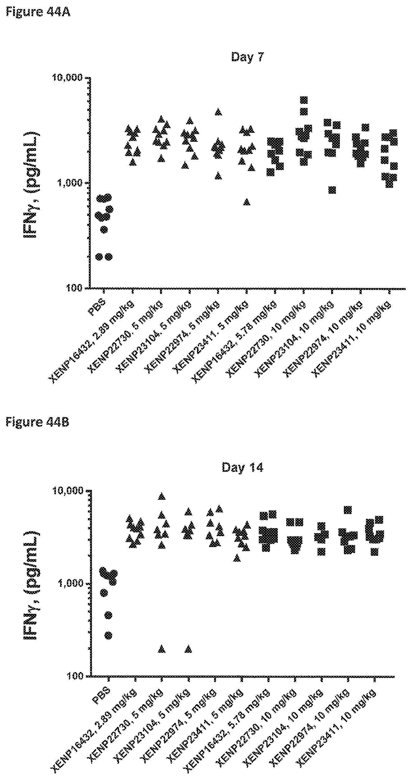
D00132

D00133

D00134

D00135
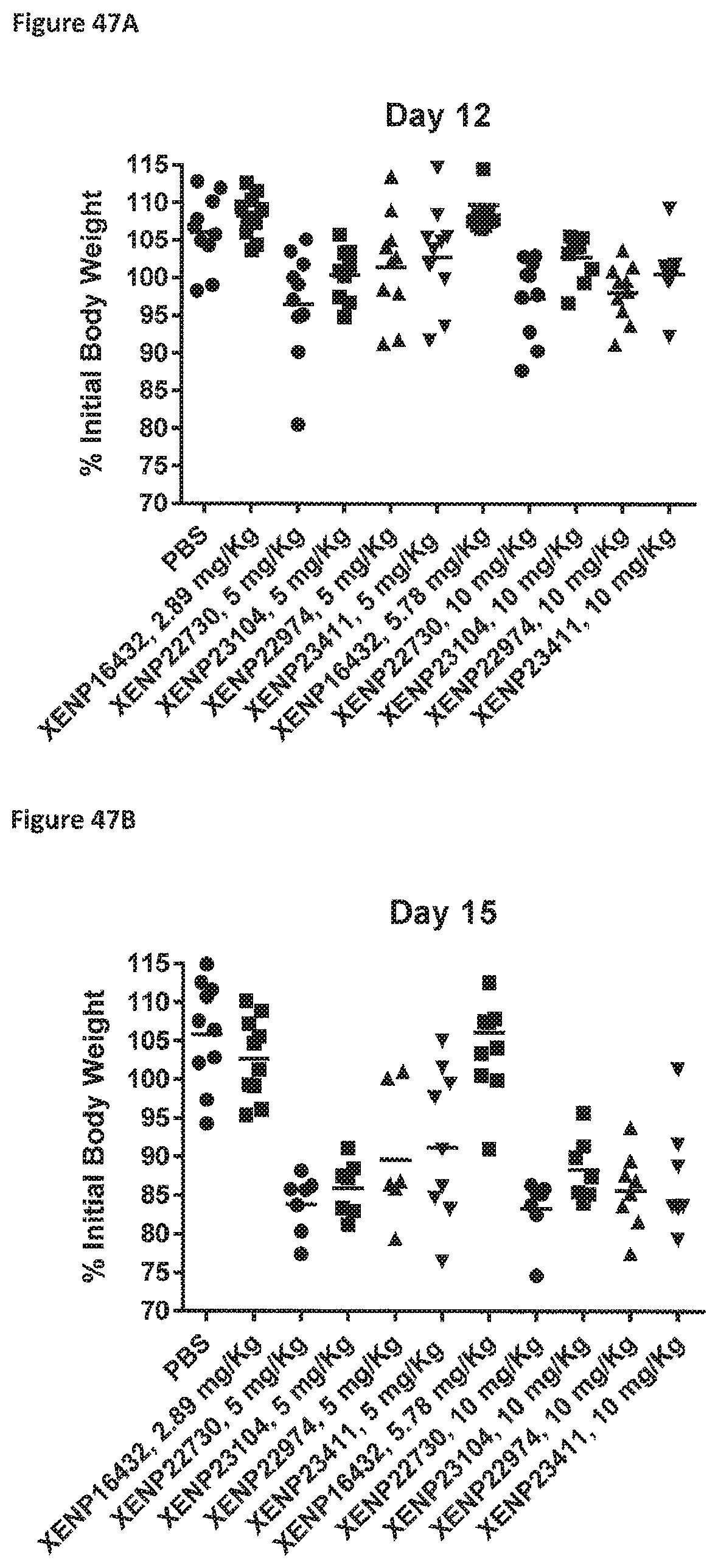
D00136

D00137

D00138

D00139

D00140

D00141

D00142
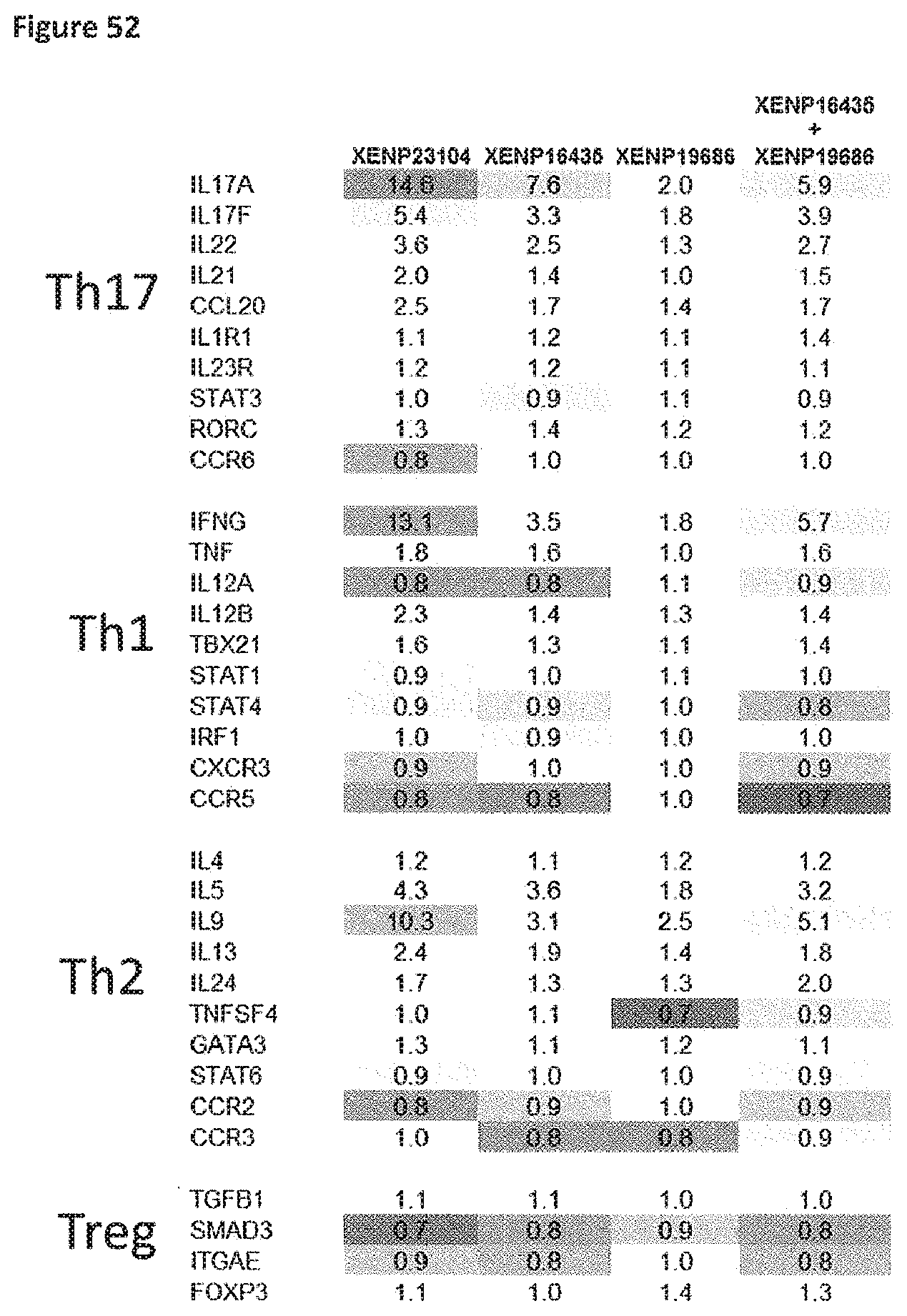
D00143

D00144

D00145
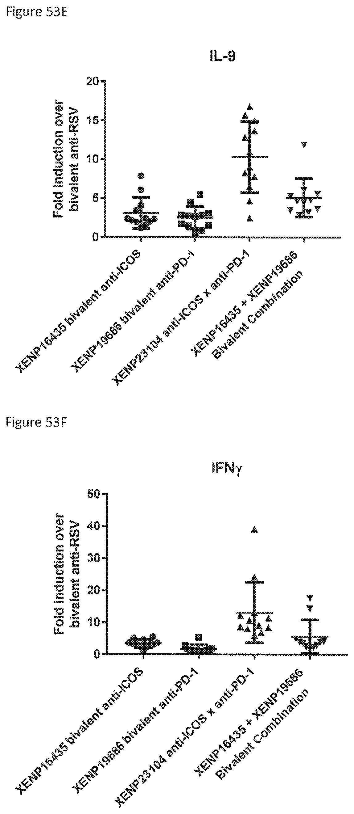
D00146
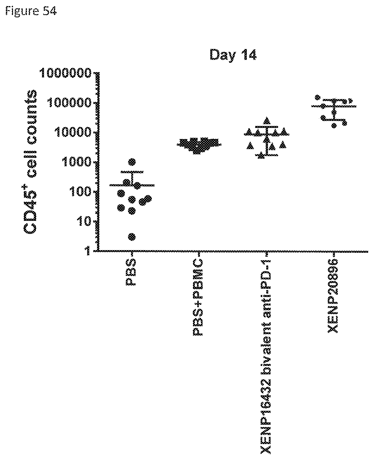
D00147

D00148

D00149
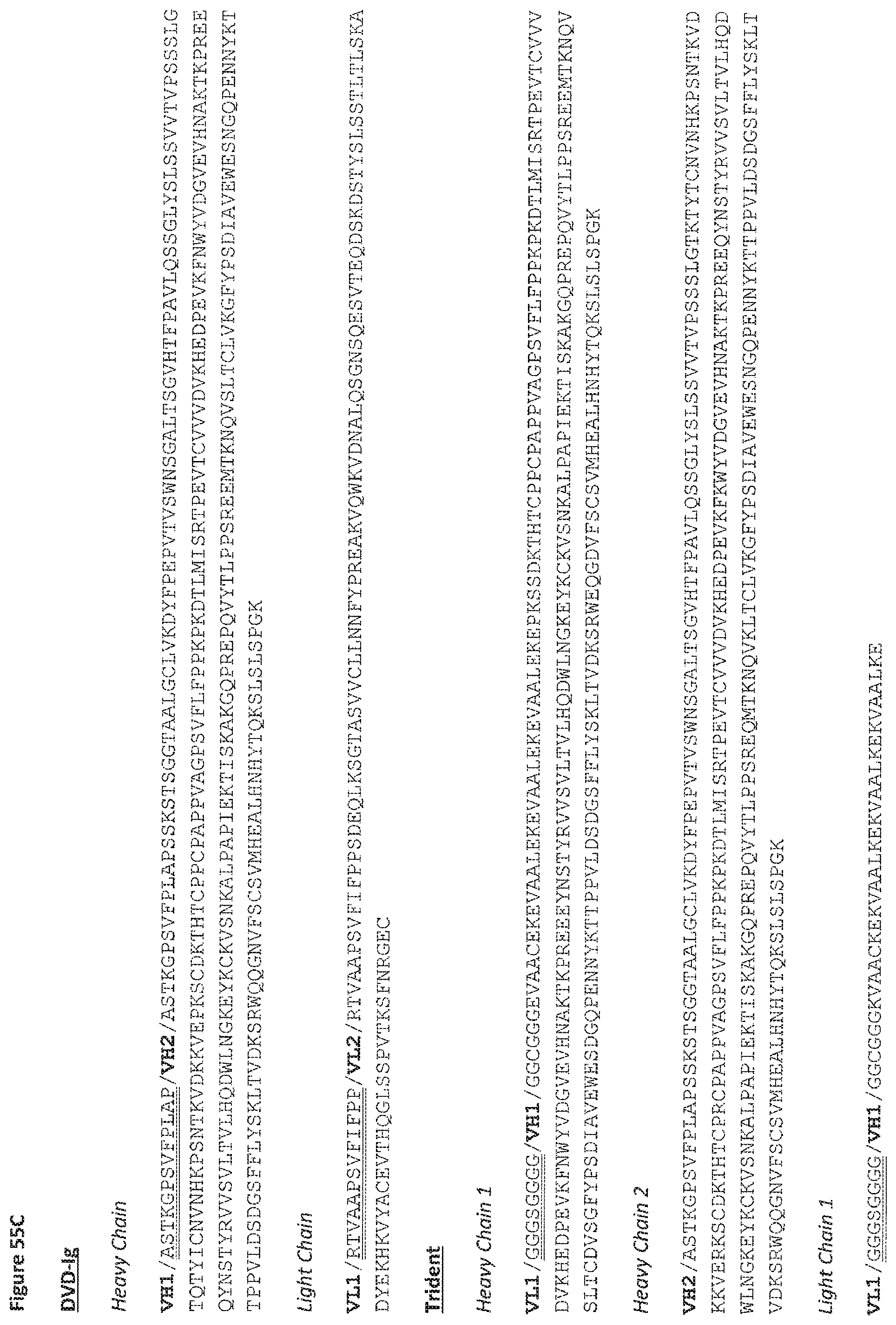
D00150
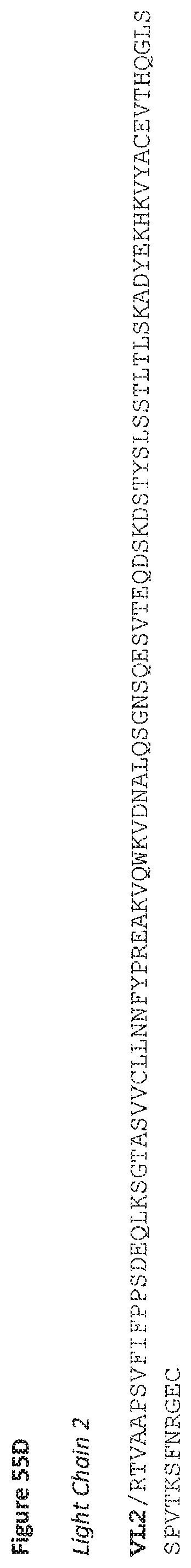
D00151
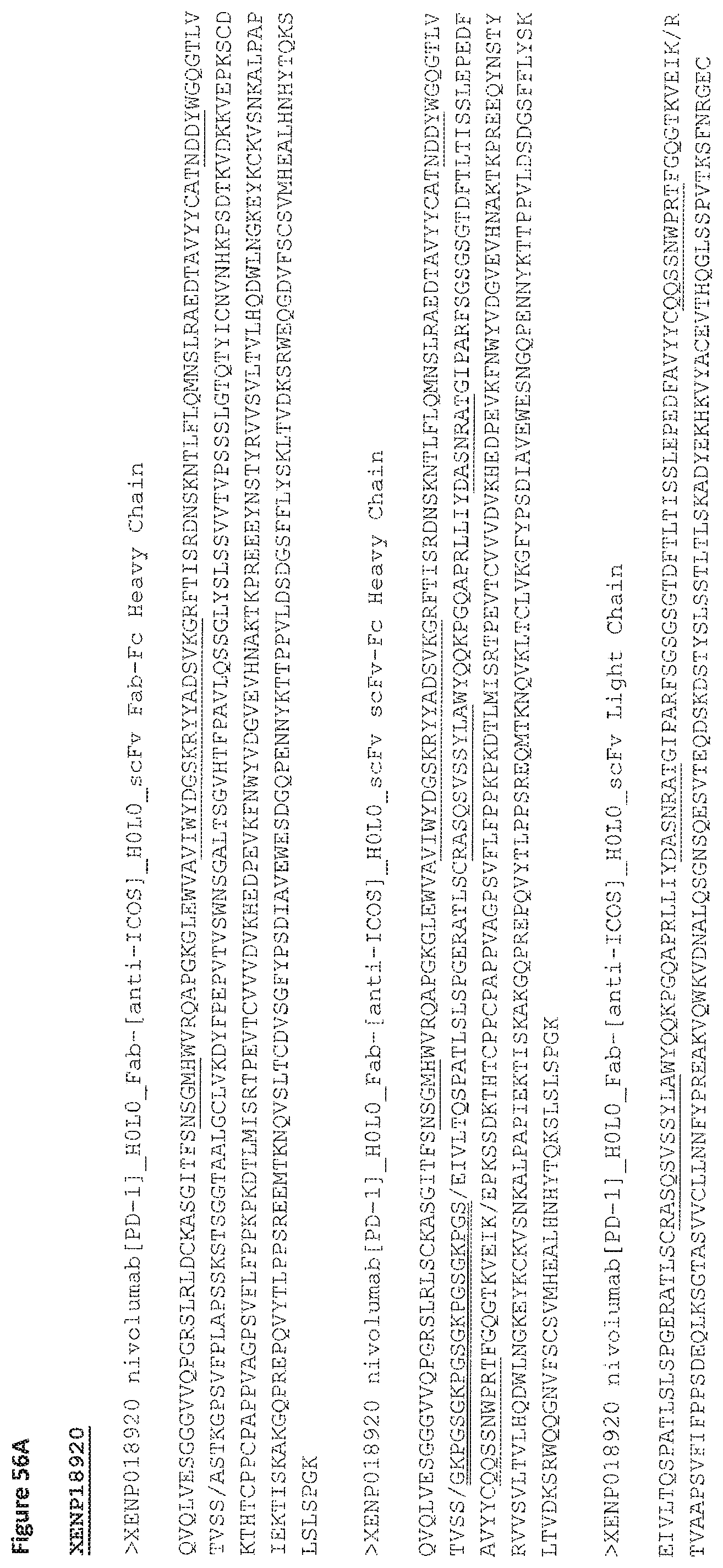
D00152
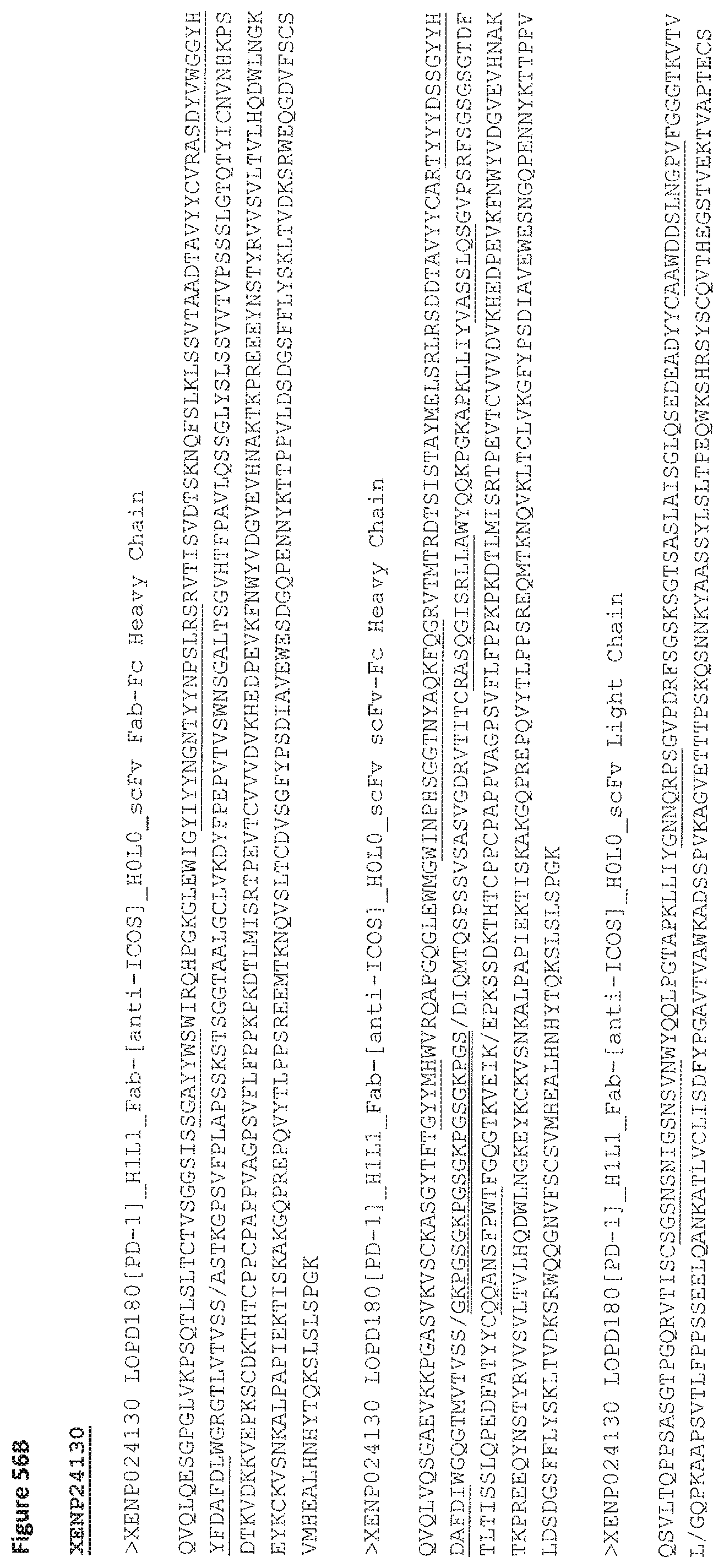
D00153

D00154
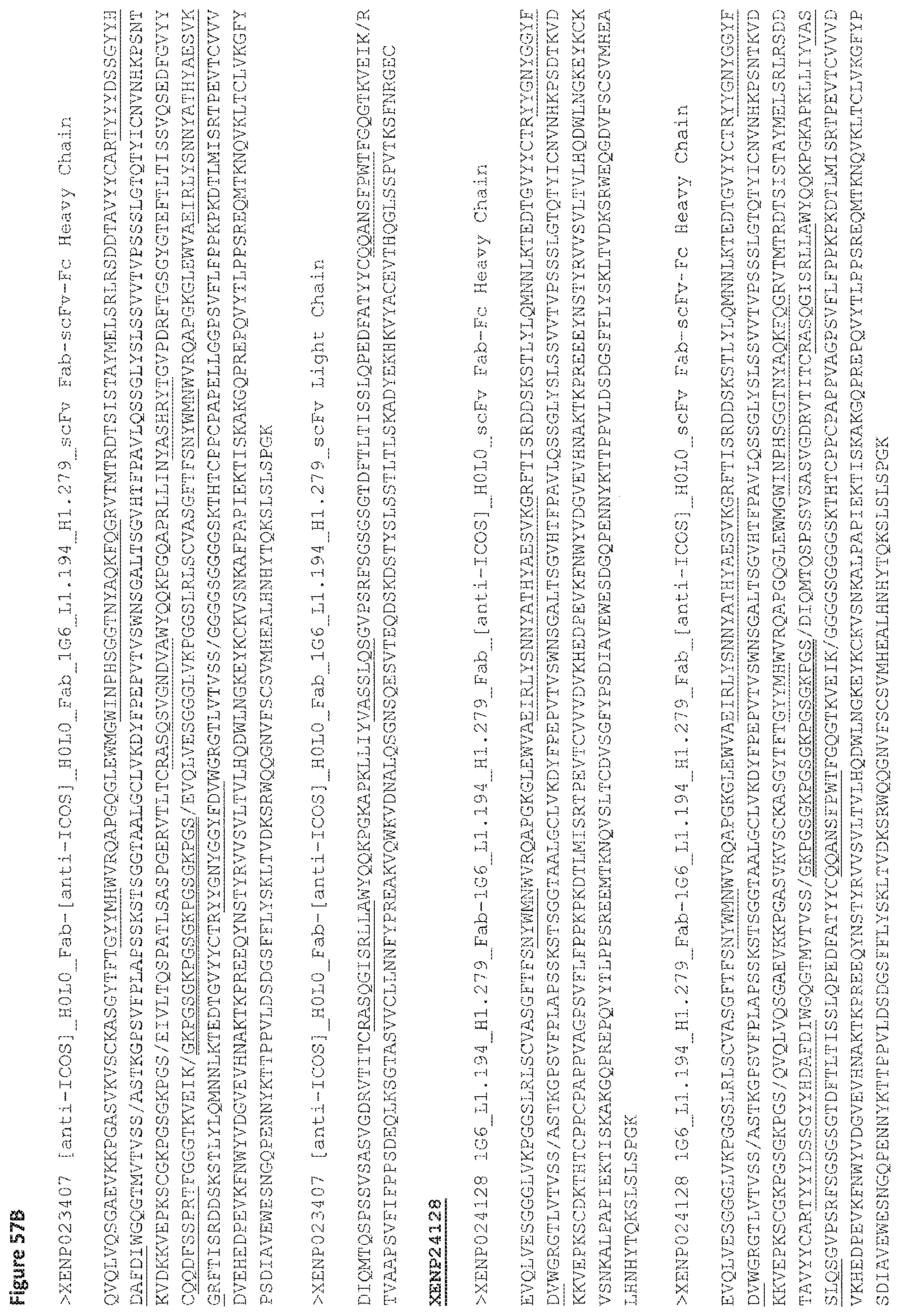
D00155
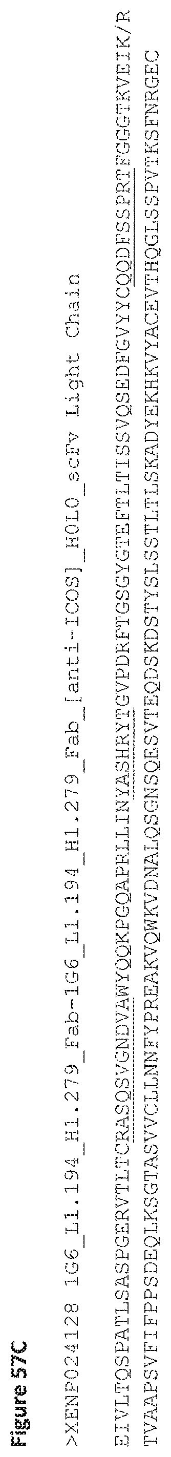
D00156

D00157
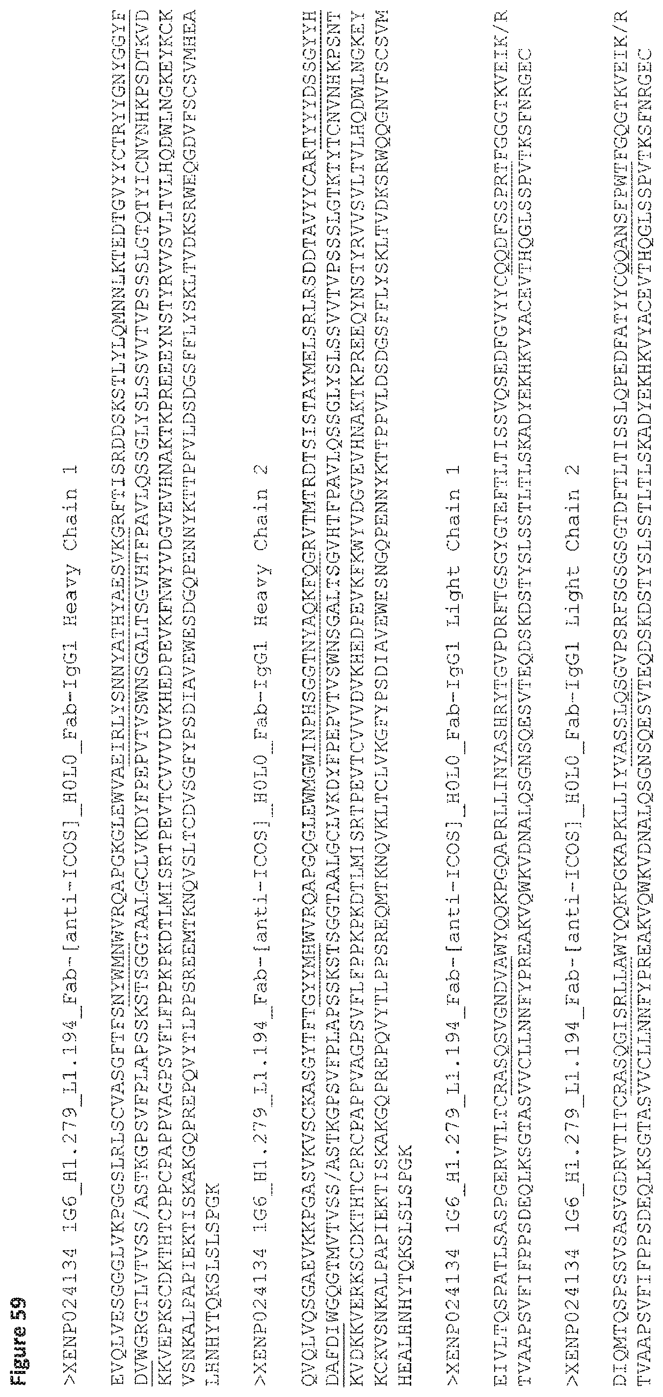
D00158
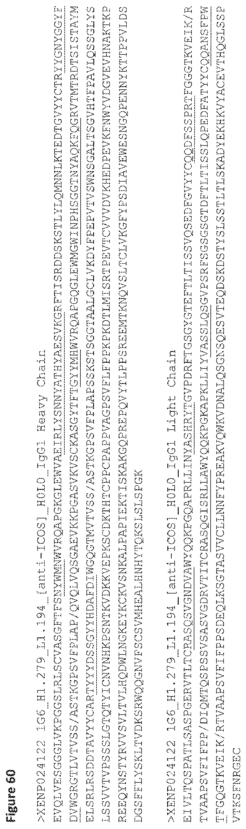
D00159
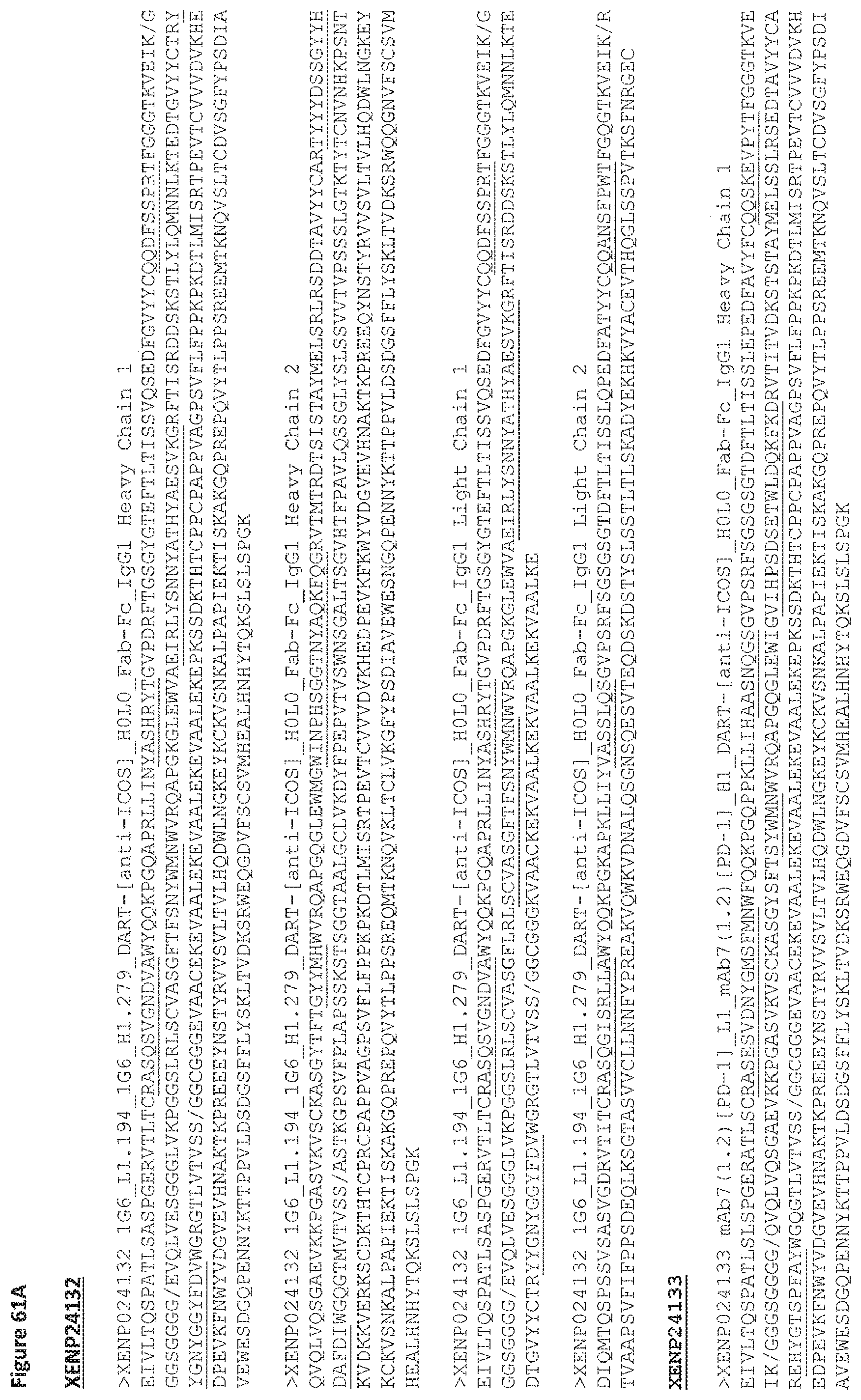
D00160

D00161

D00162

D00163
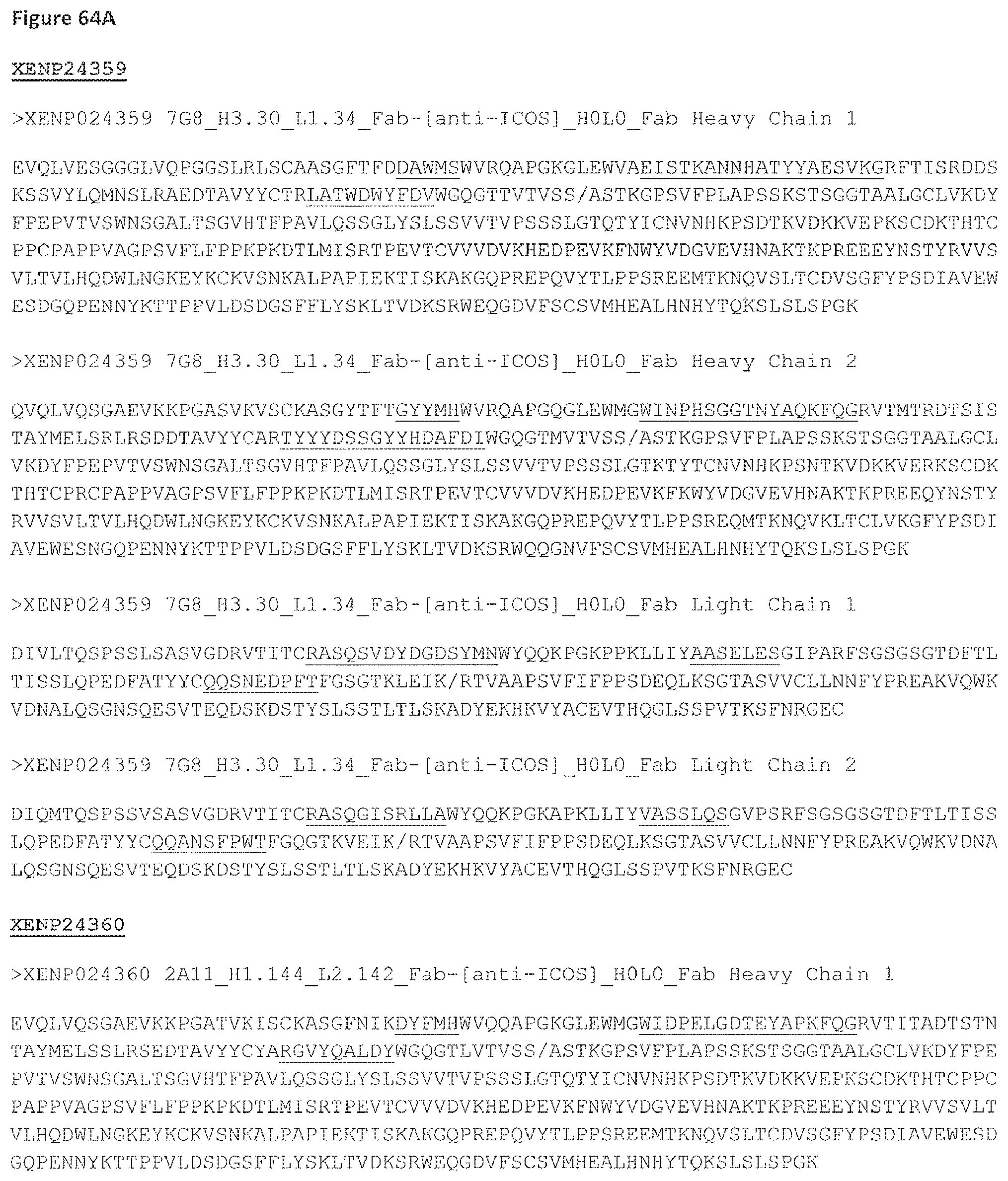
D00164
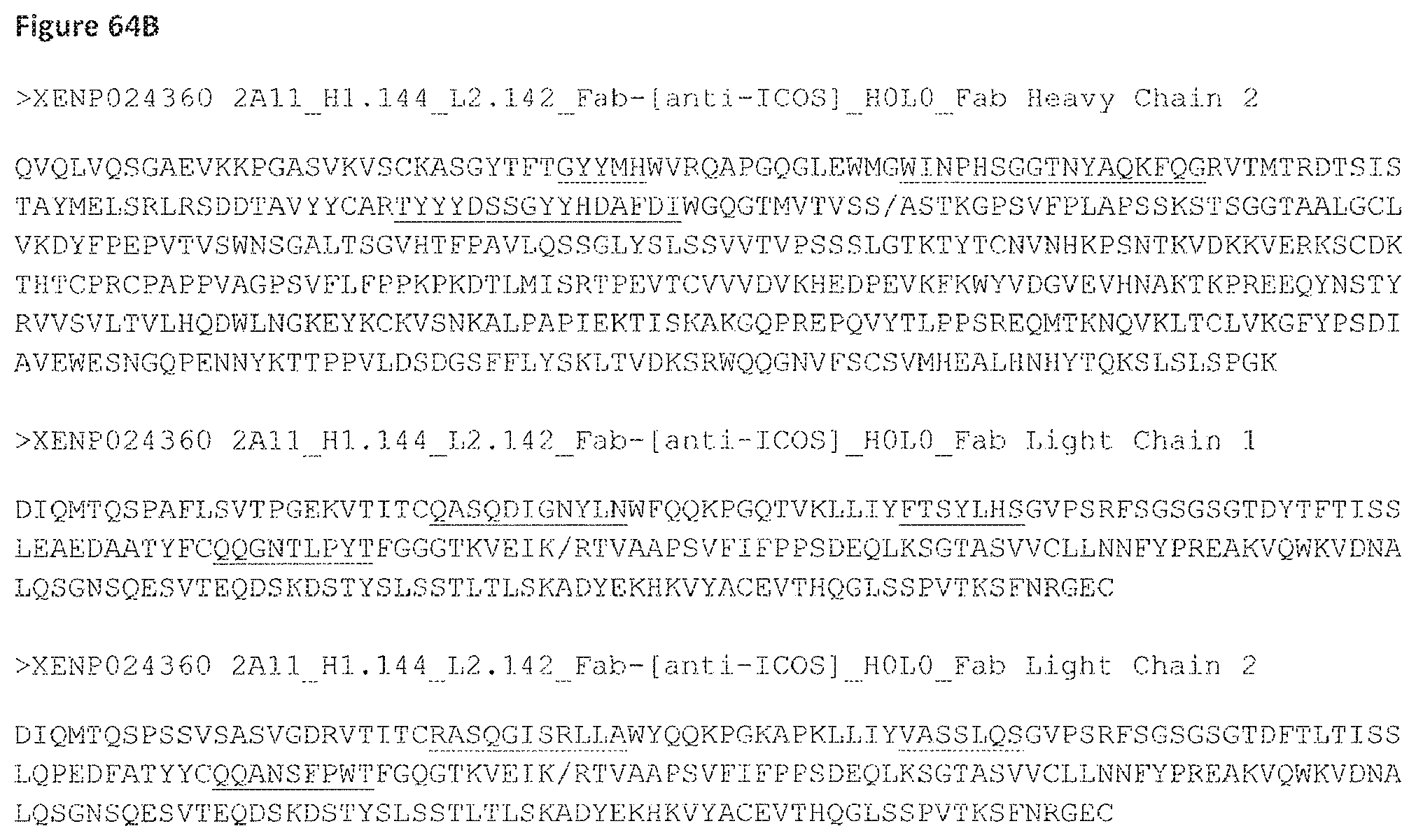
D00165

D00166
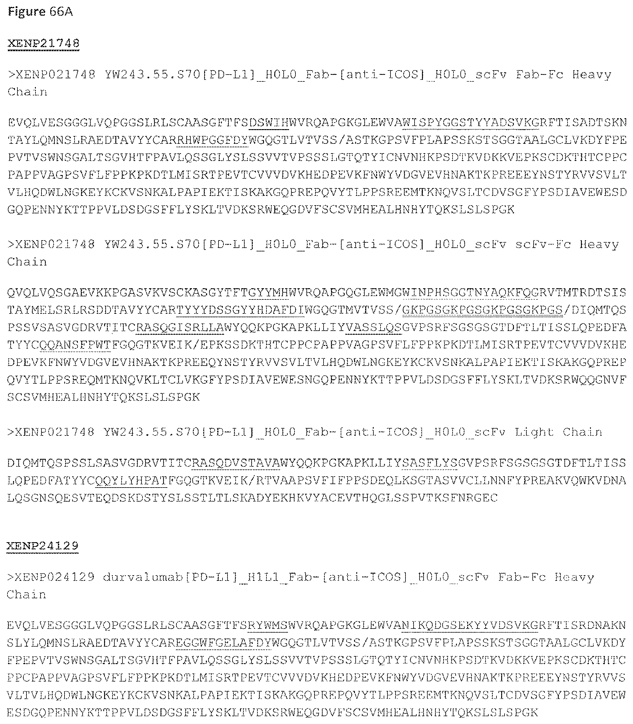
D00167

D00168
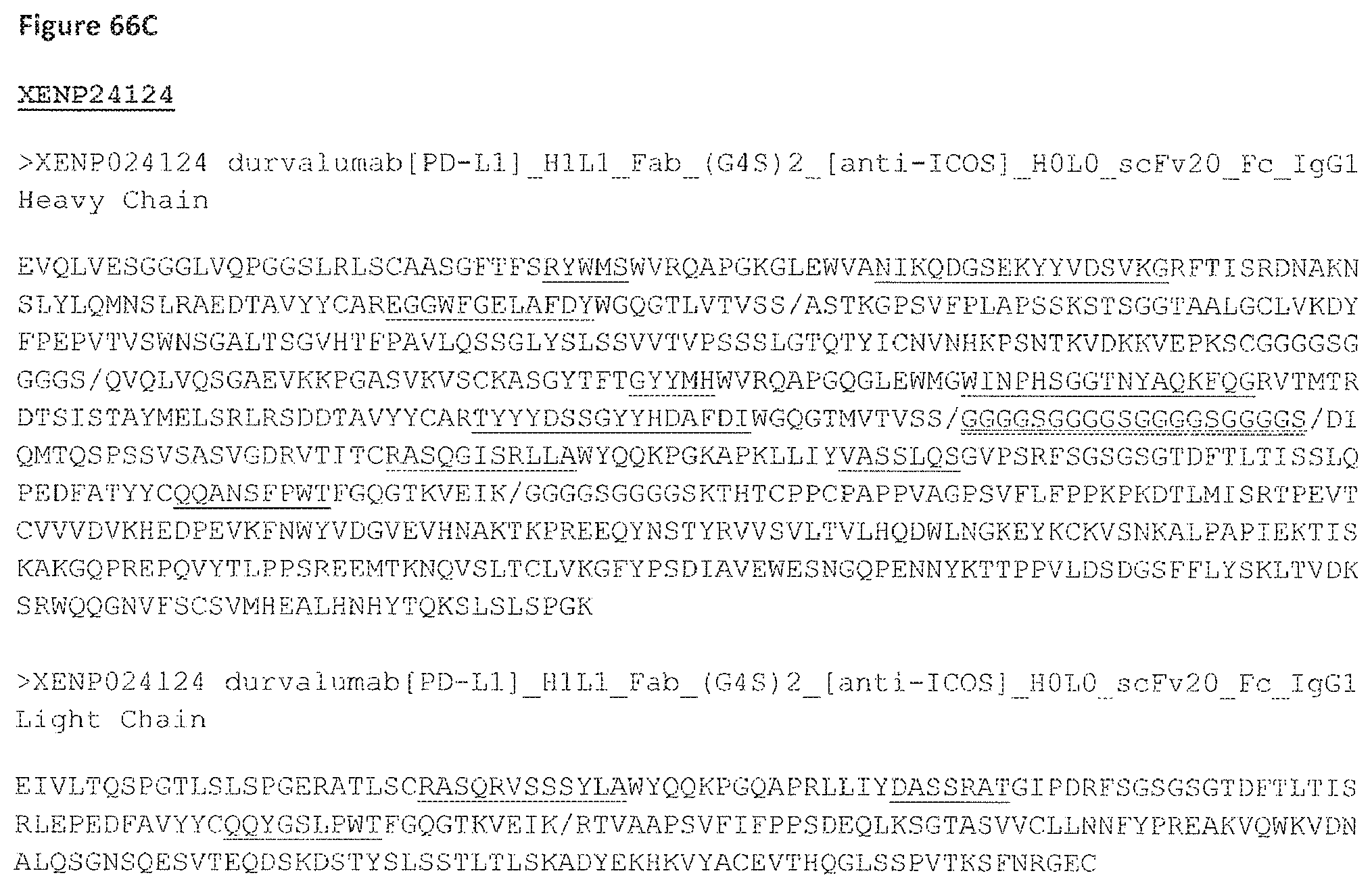
D00169
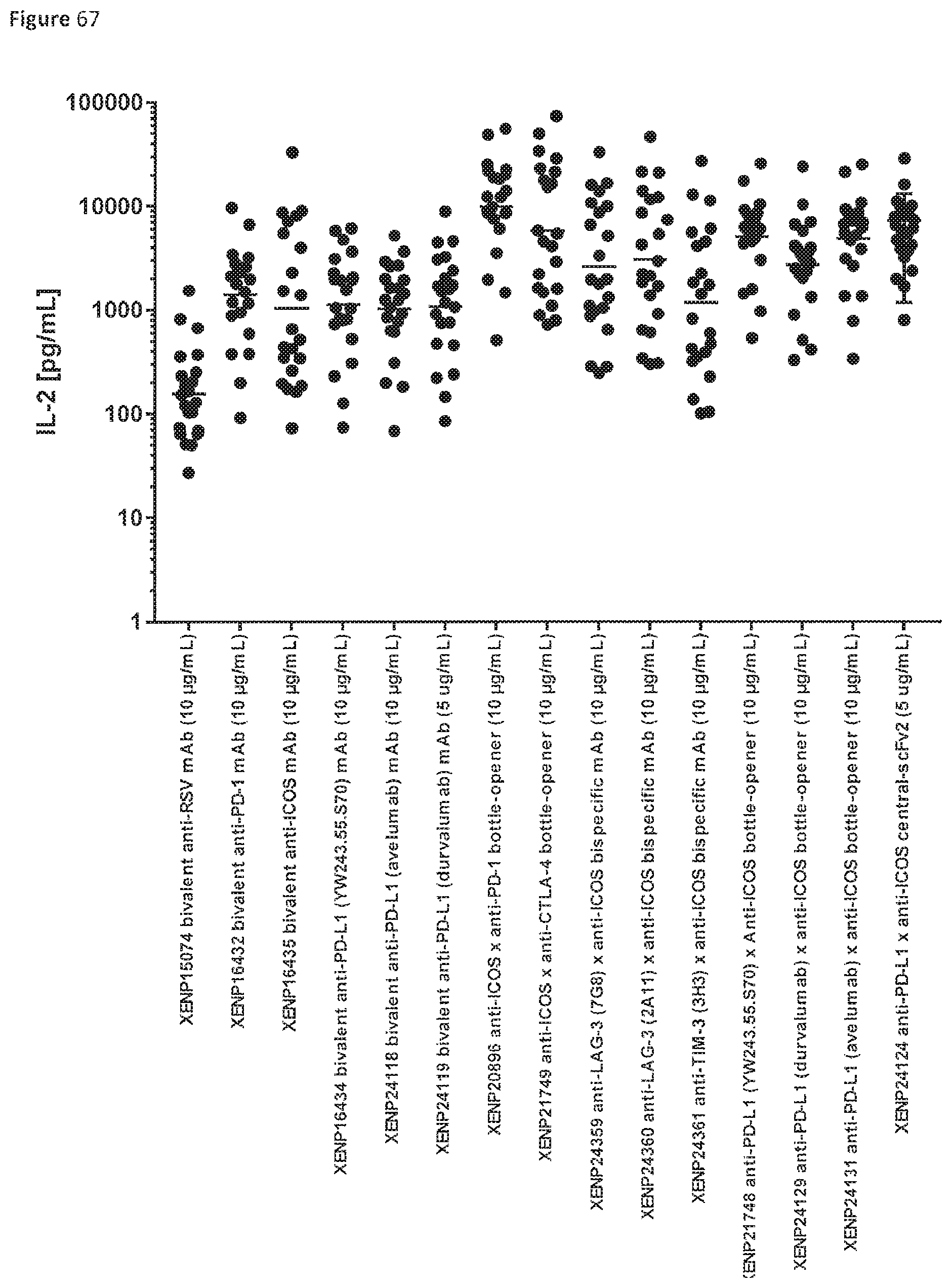
D00170

D00171
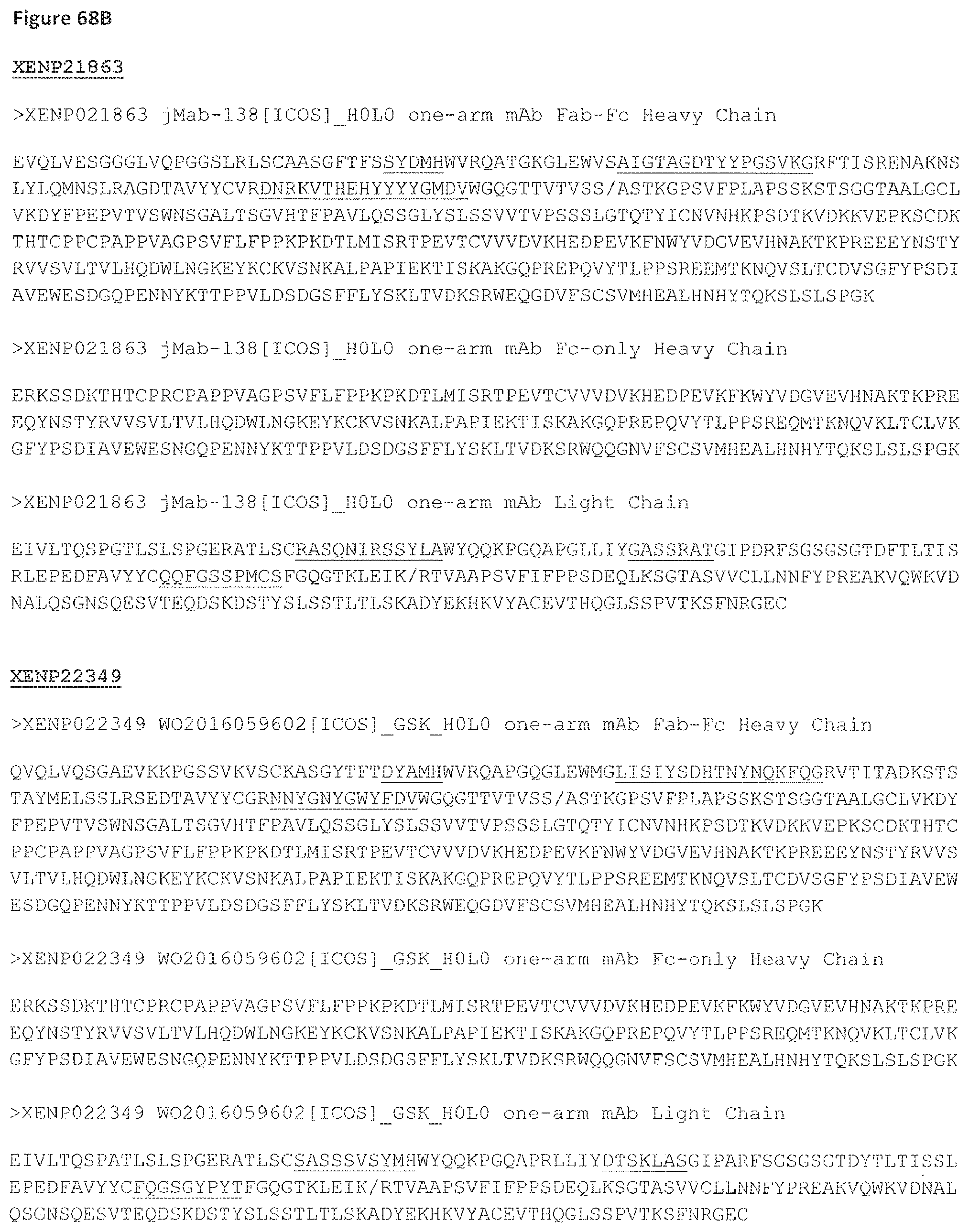
D00172
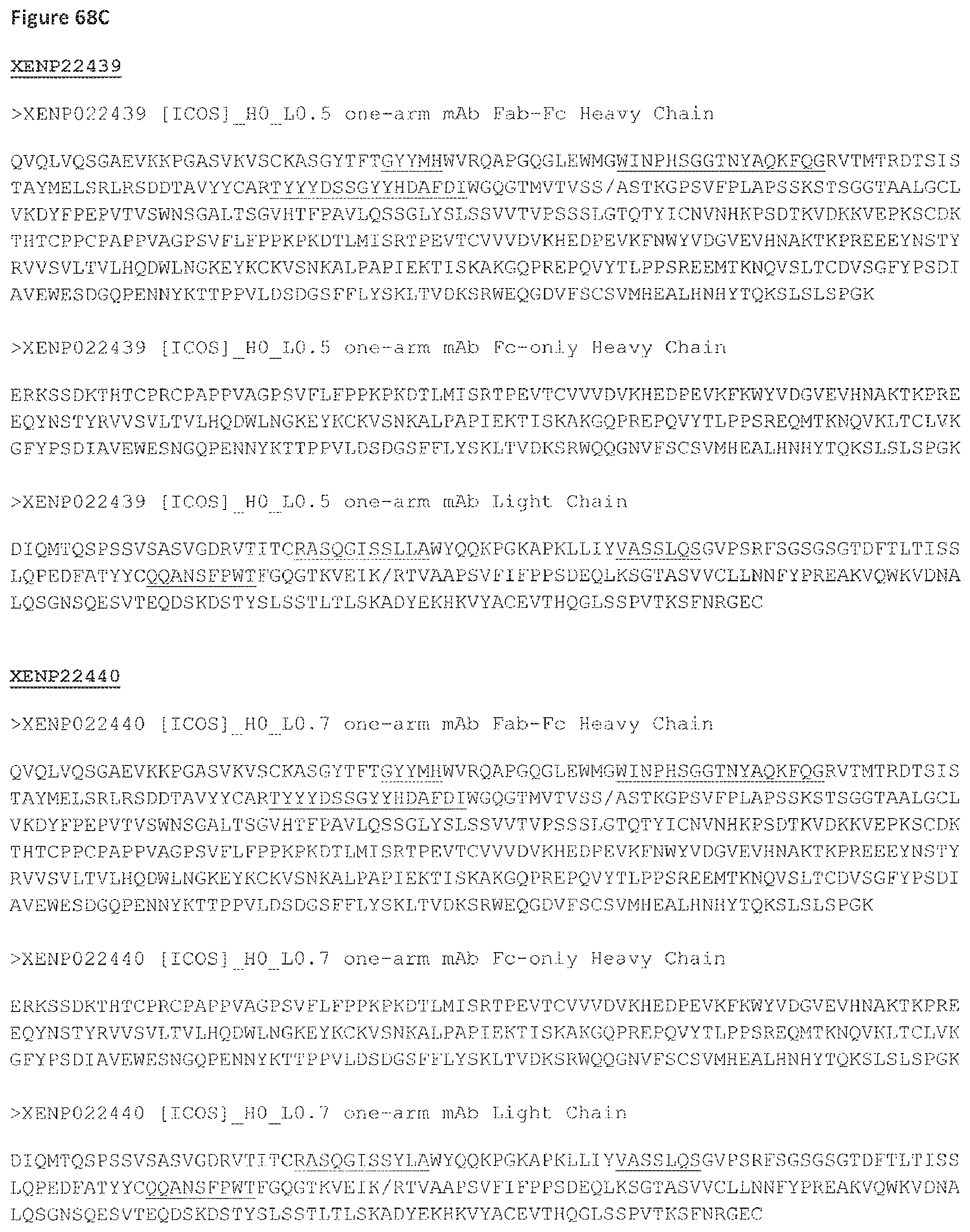
D00173
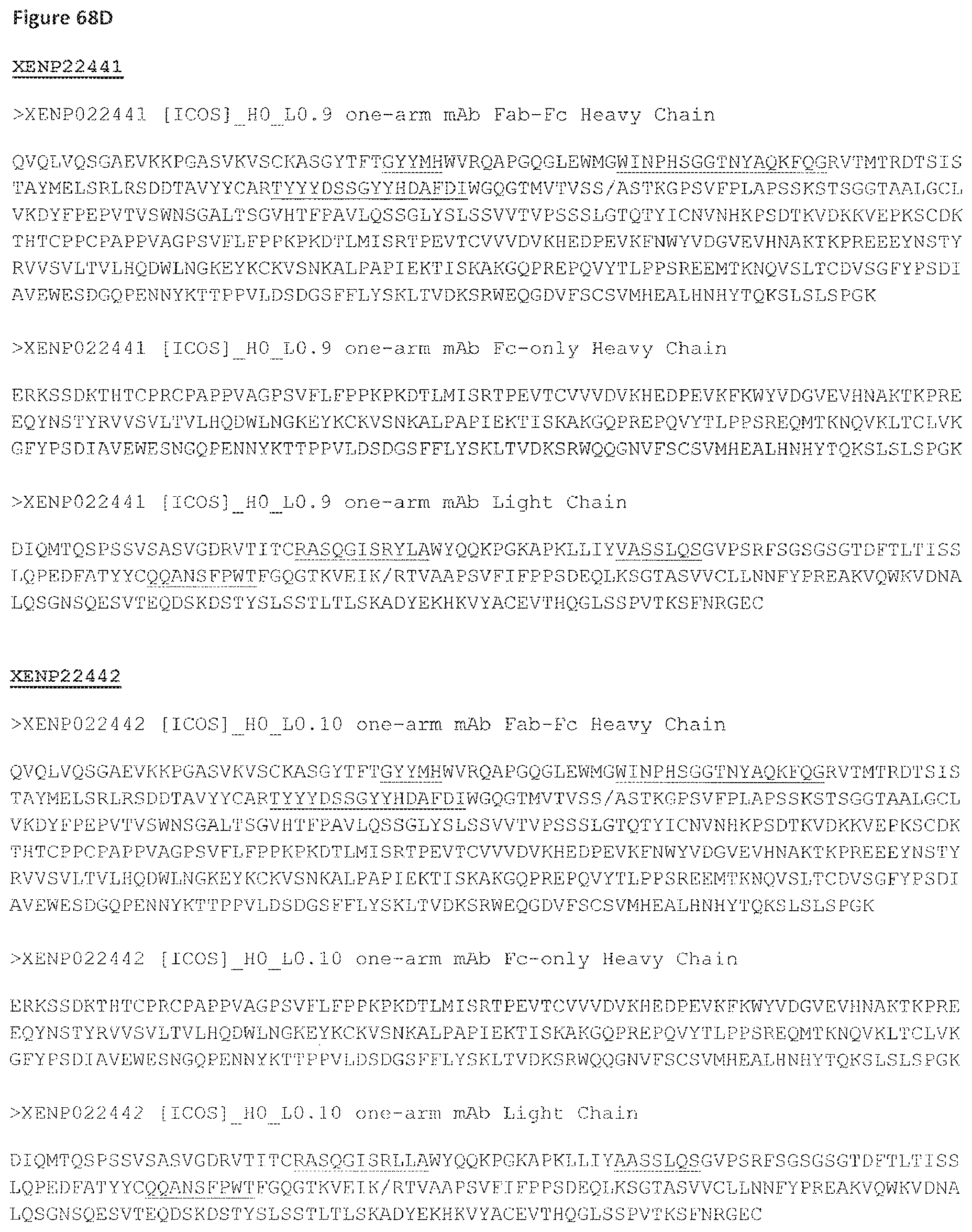
D00174
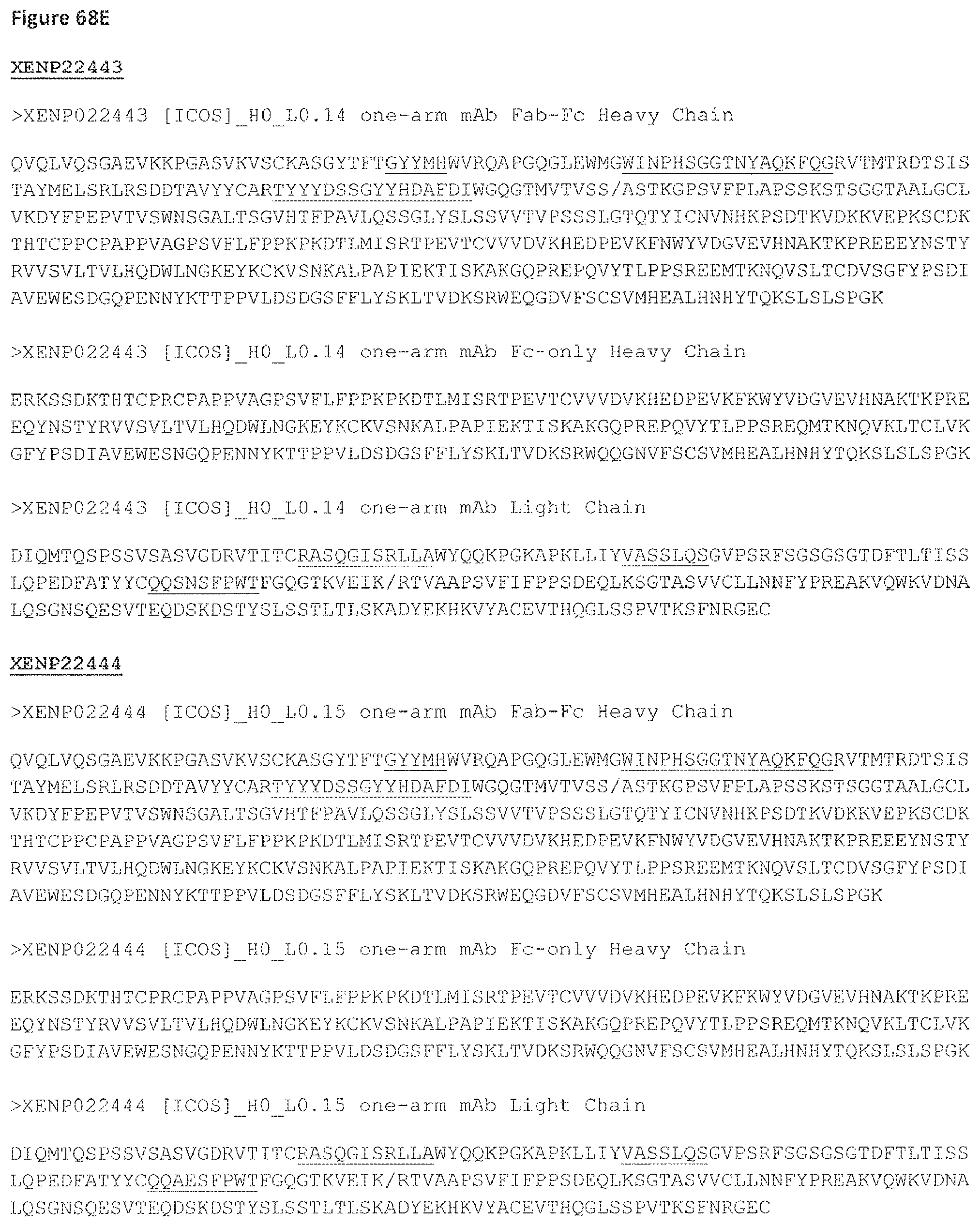
D00175

D00176
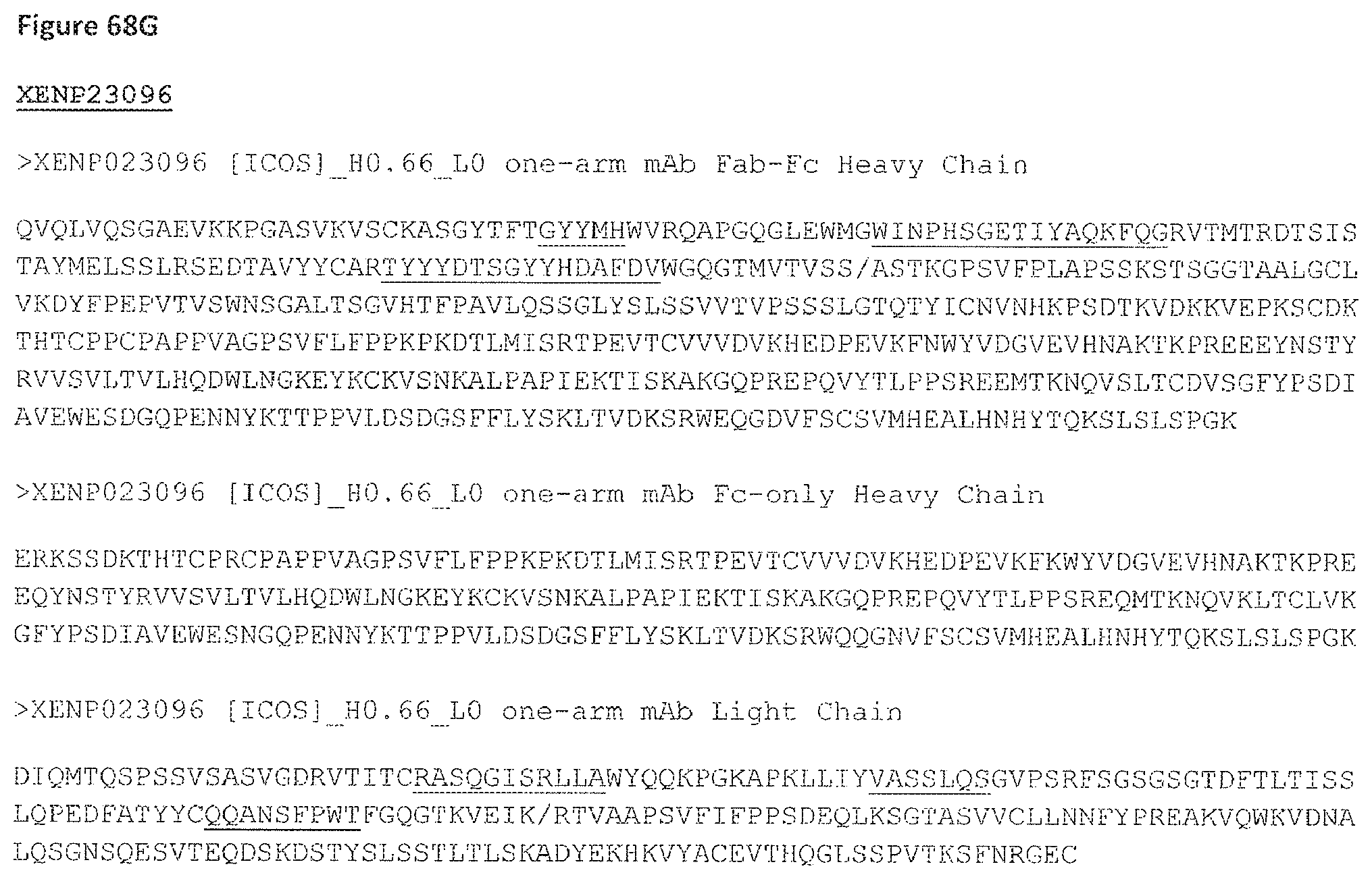
D00177
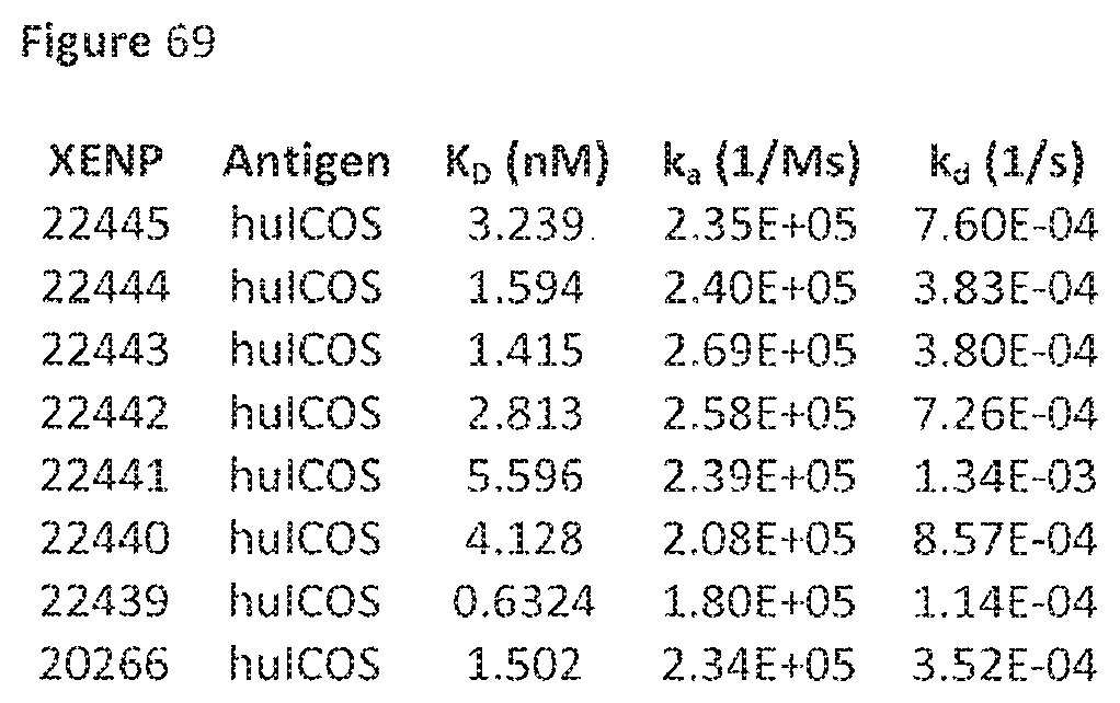
D00178

D00179

D00180
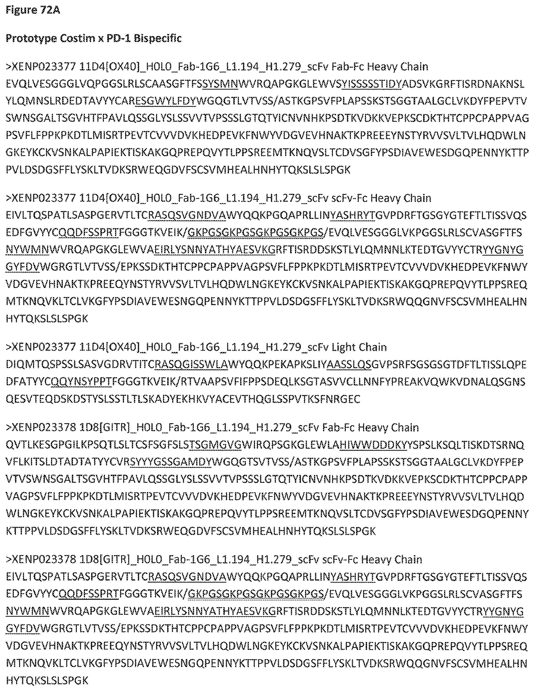
D00181
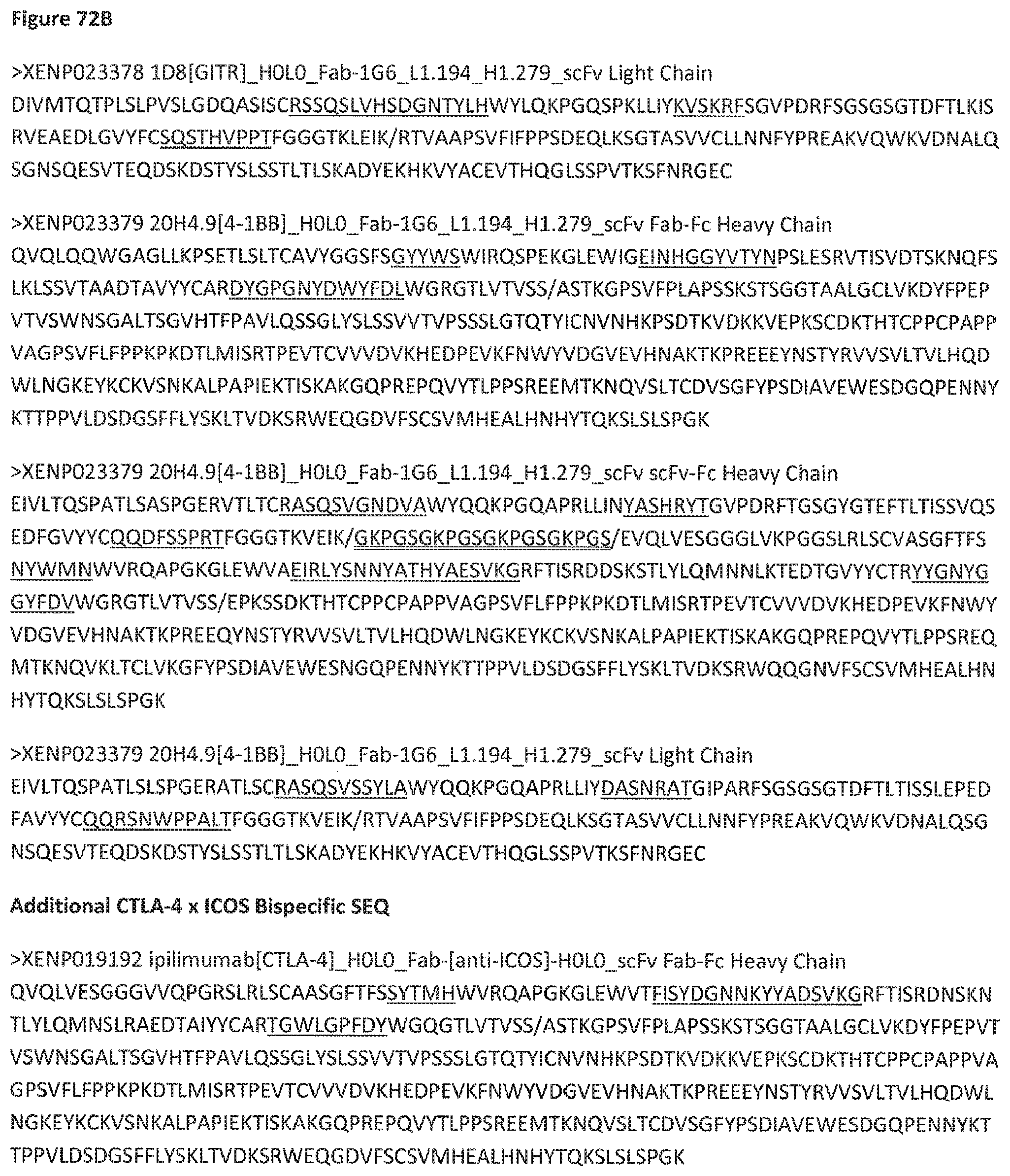
D00182
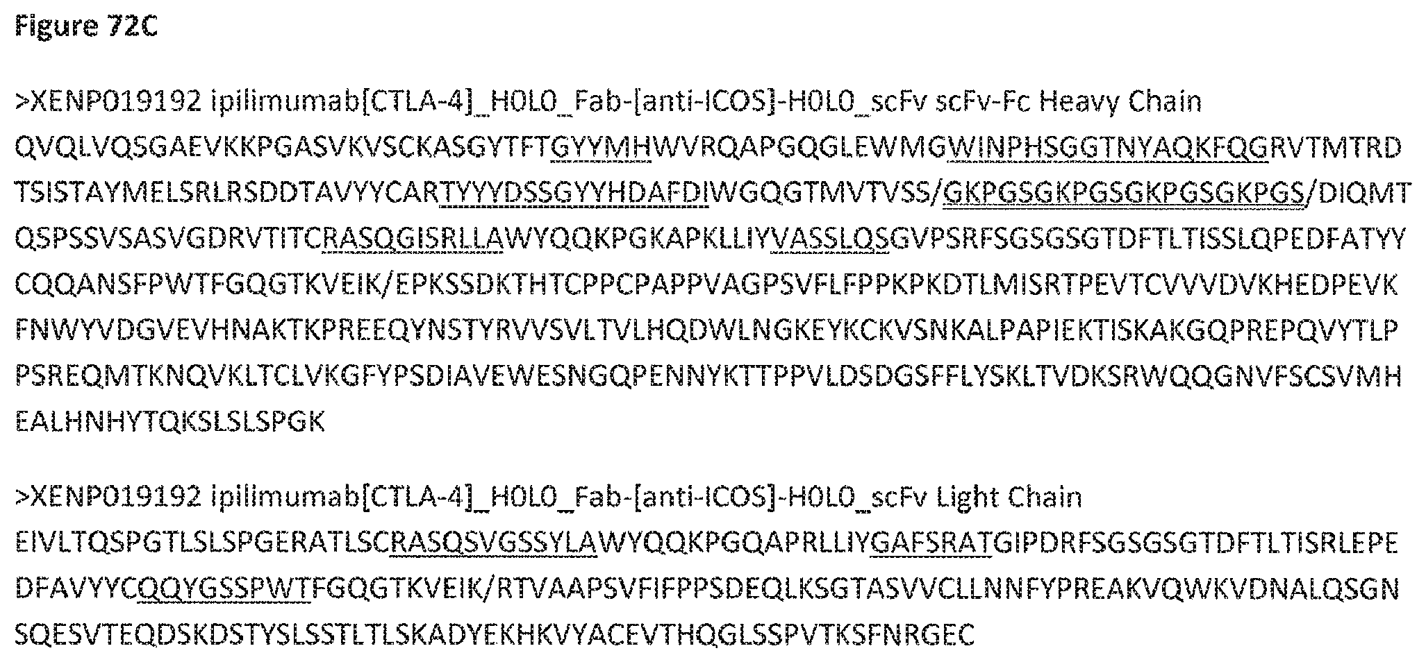
D00183

D00184
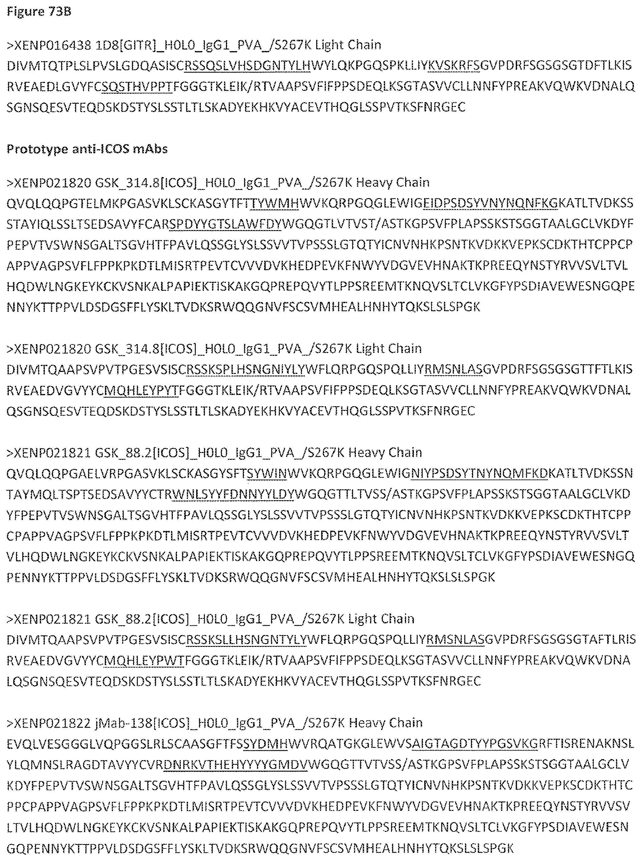
D00185

D00186
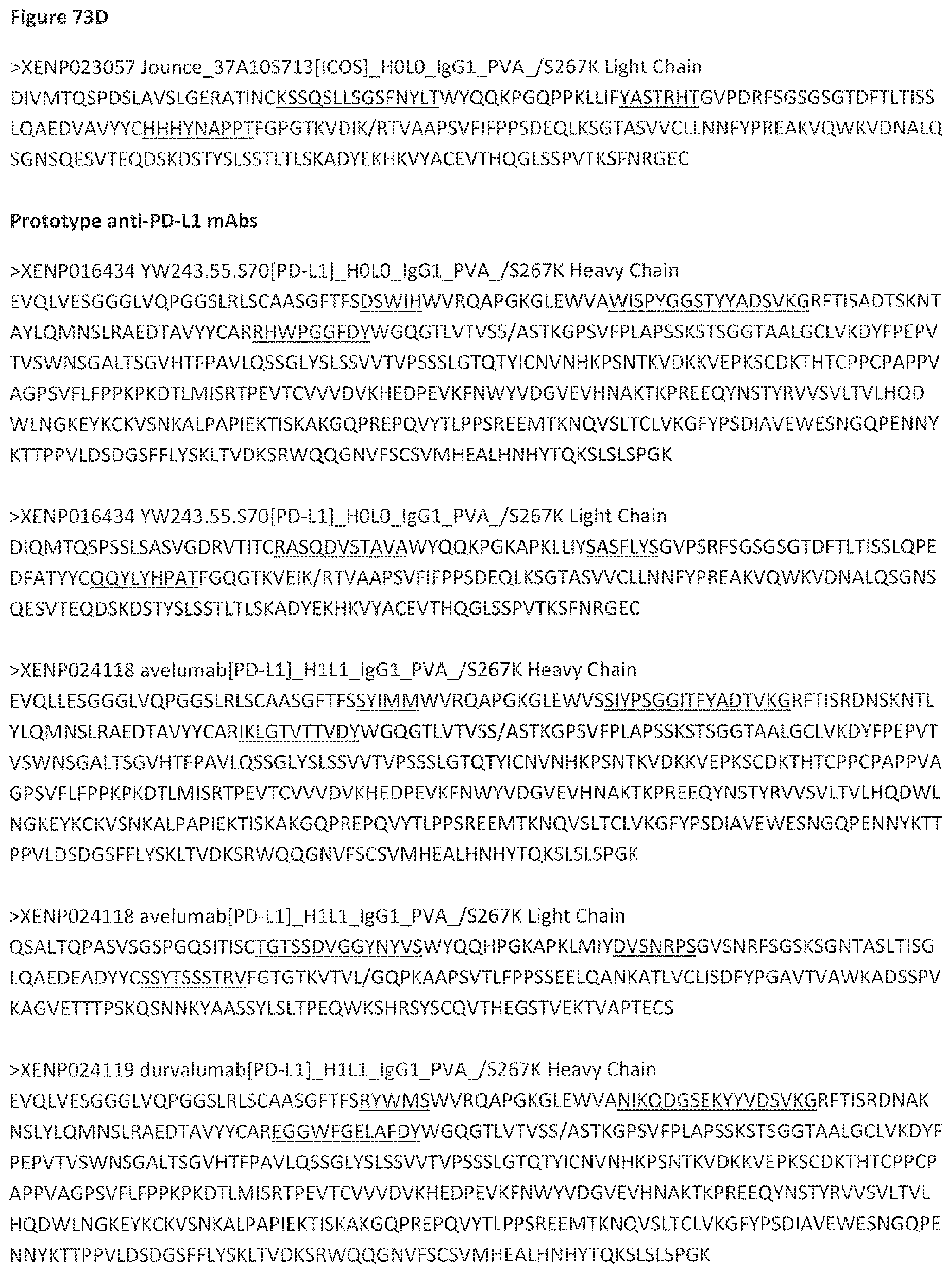
D00187

D00188

D00189
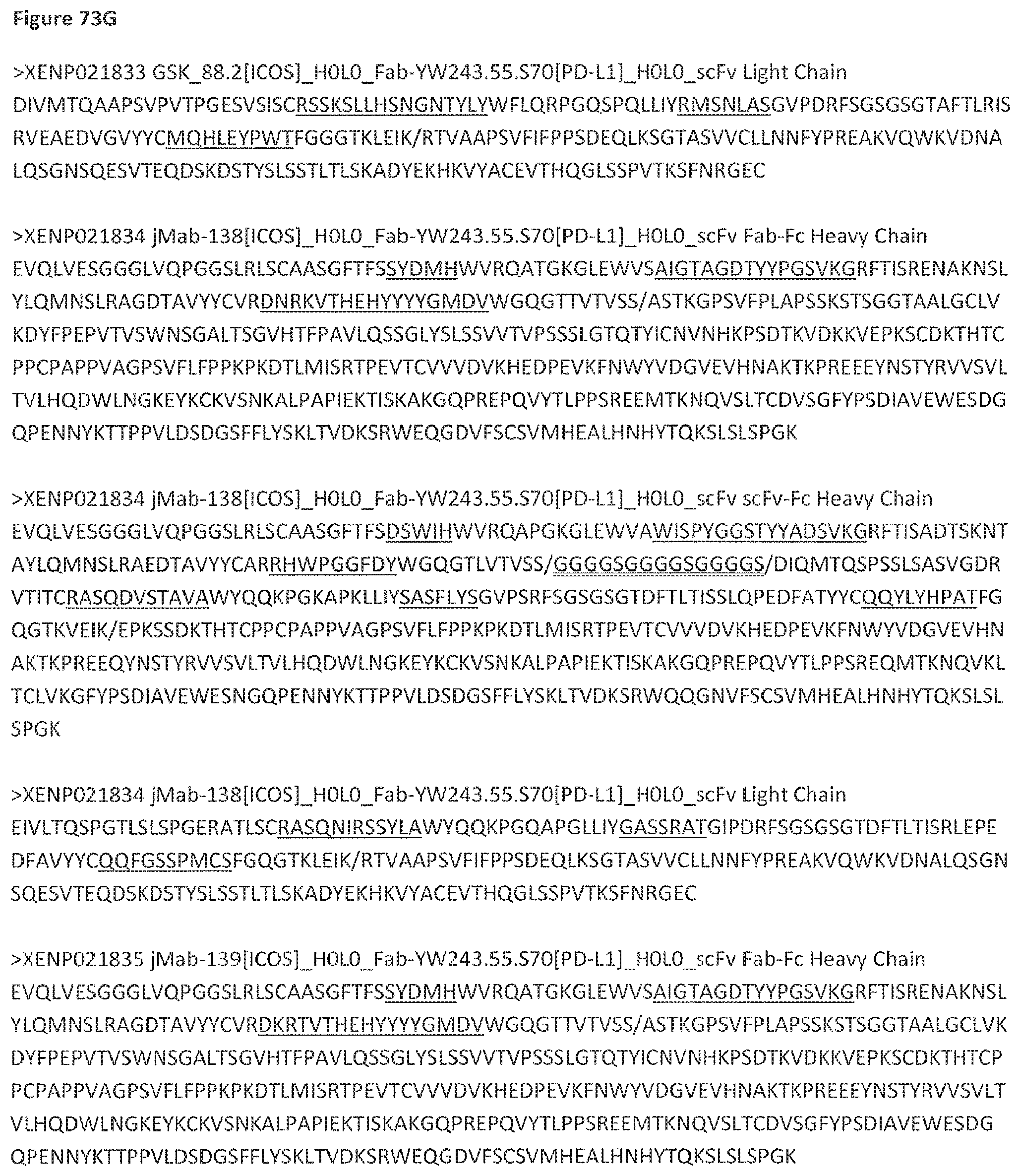
D00190
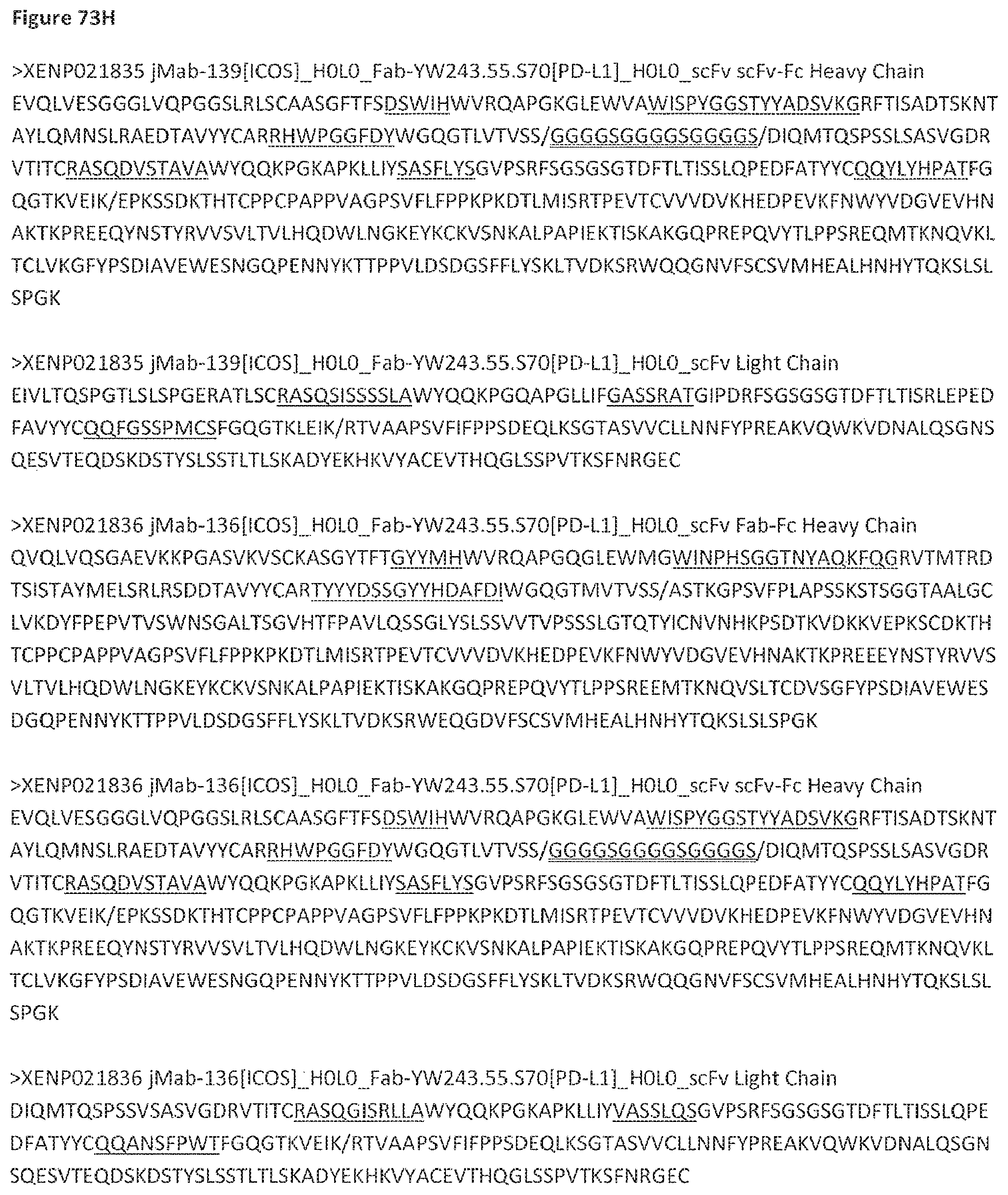
D00191
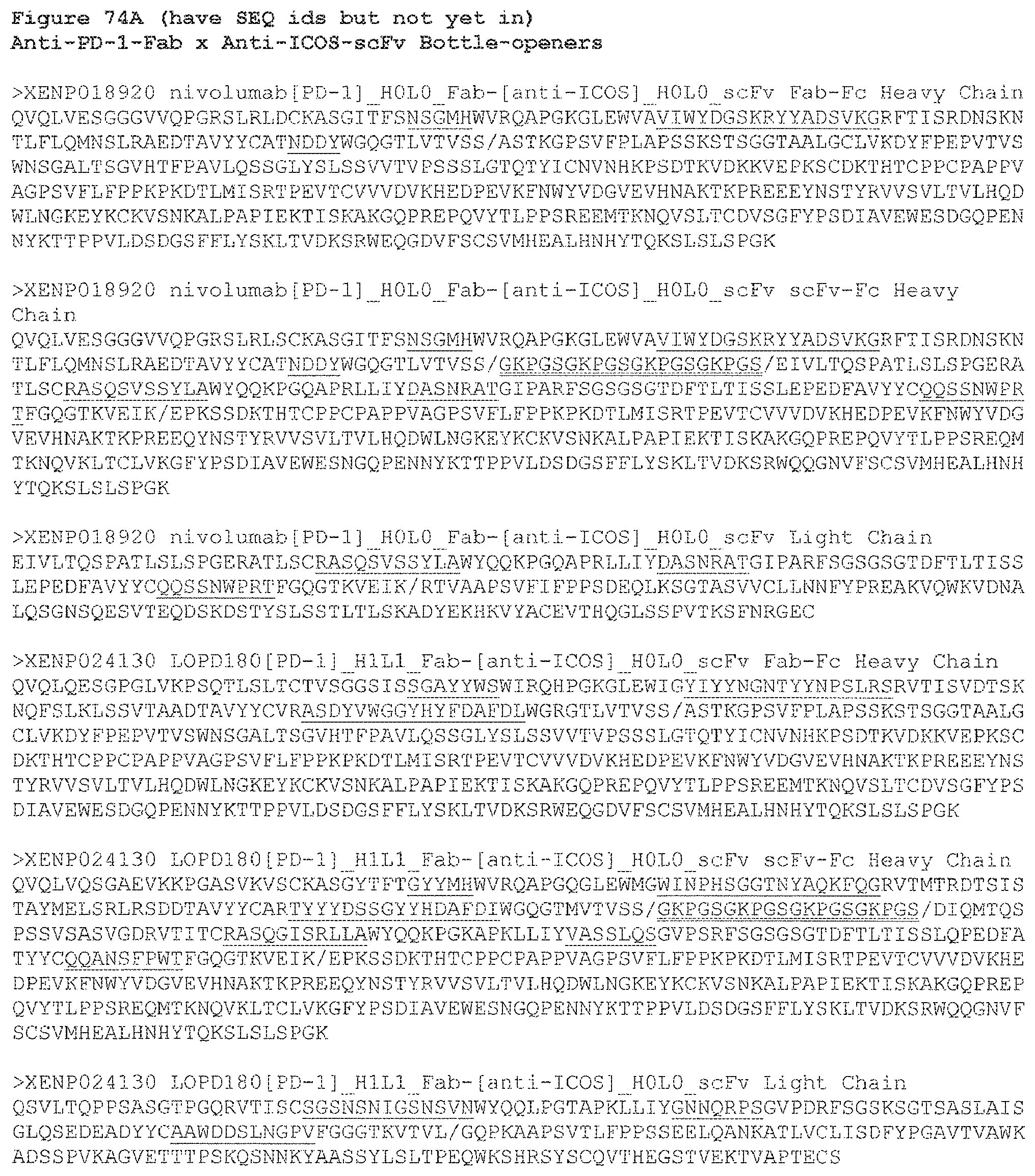
D00192
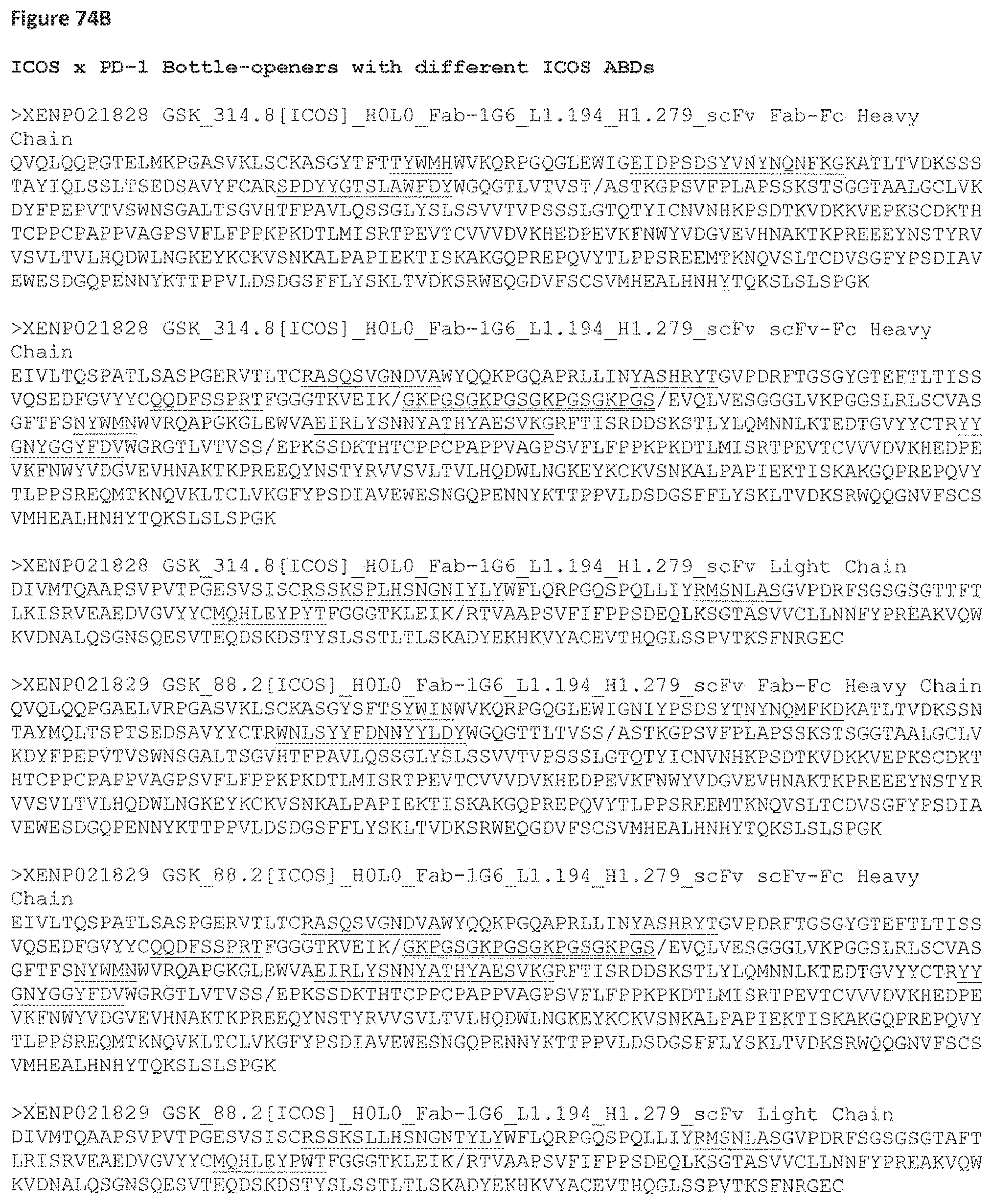
D00193
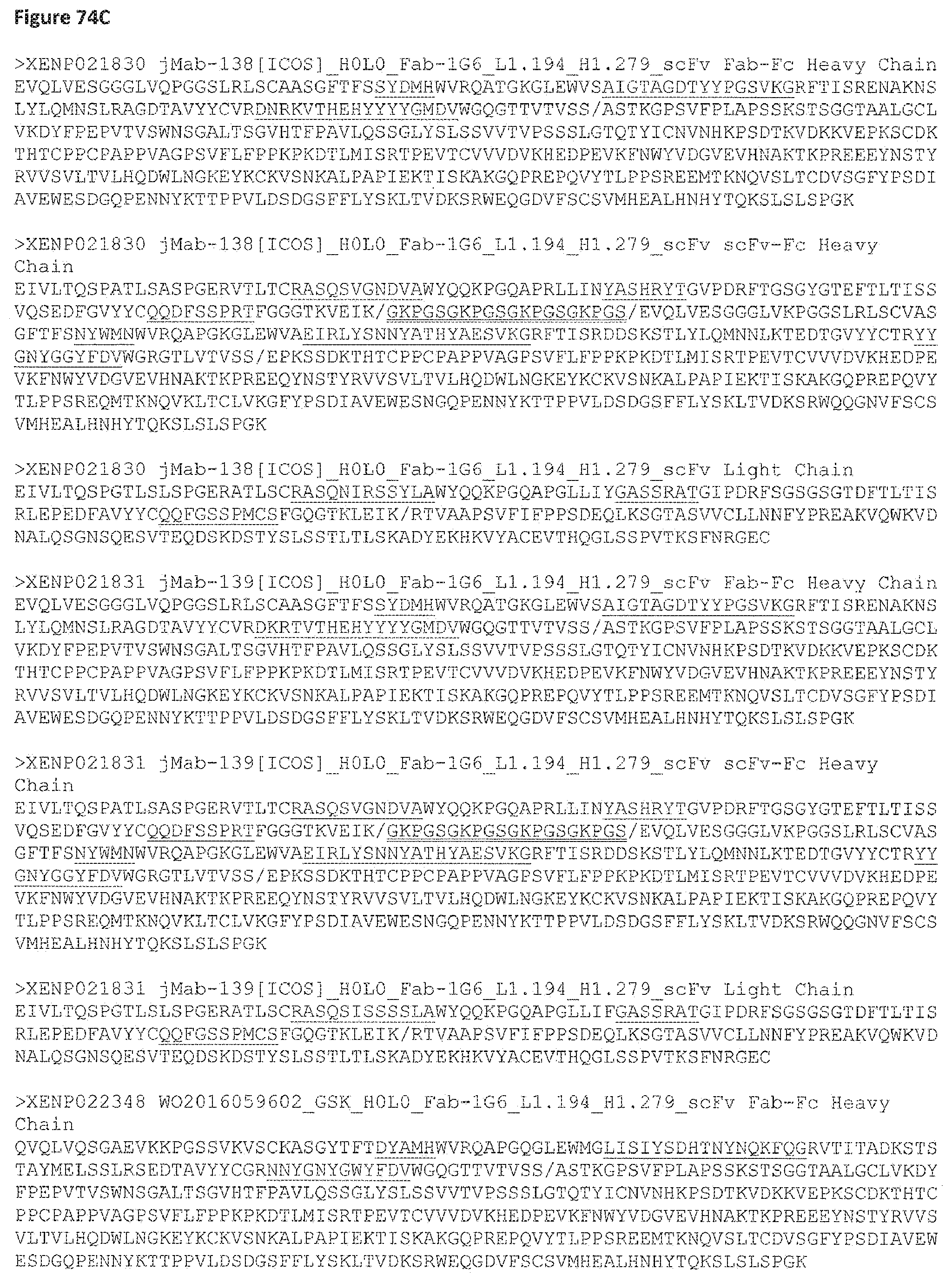
D00194

D00195

D00196

D00197

D00198

D00199
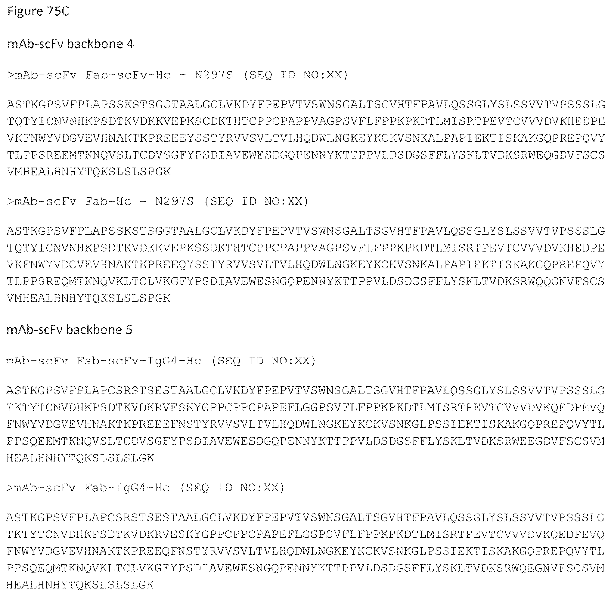
D00200
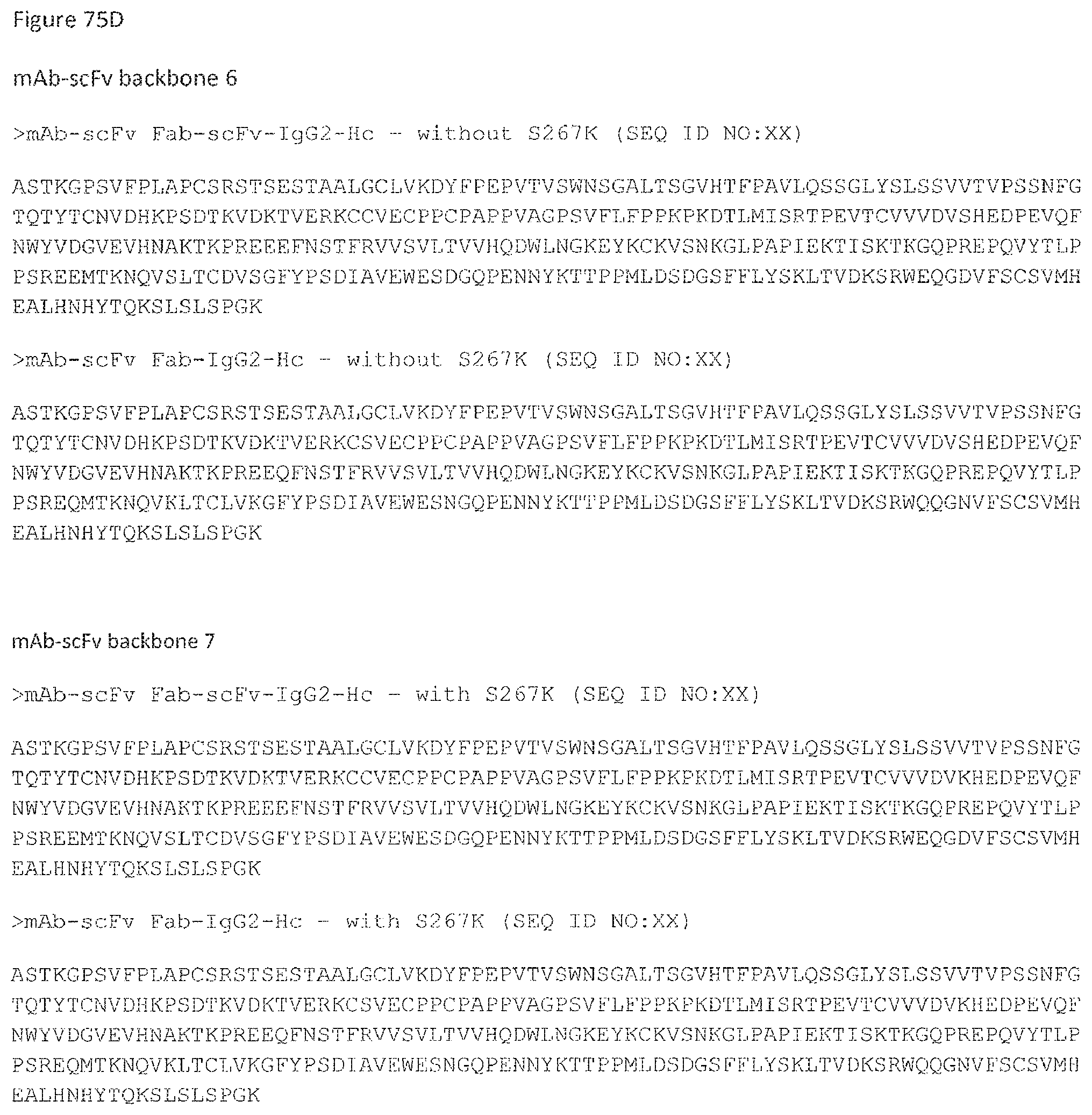
D00201

D00202
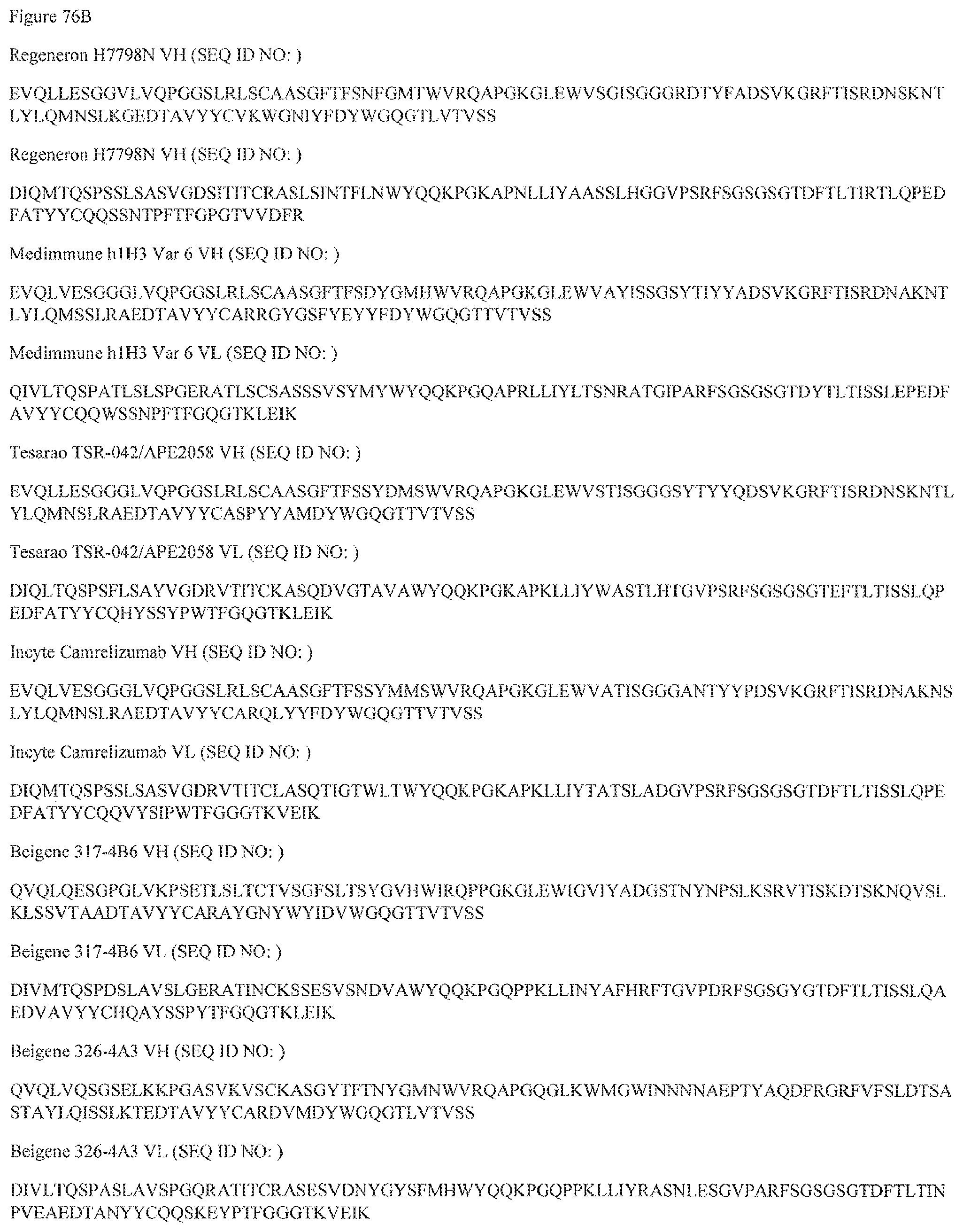
D00203

D00204
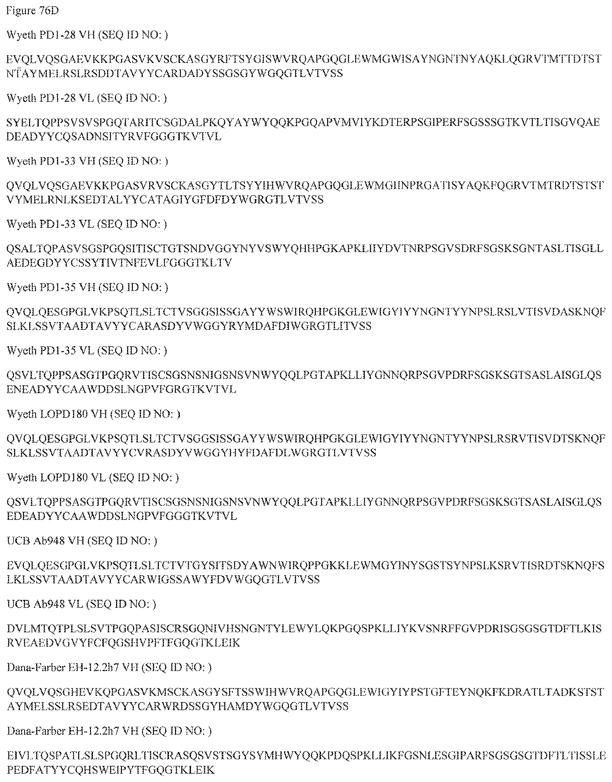
D00205

D00206
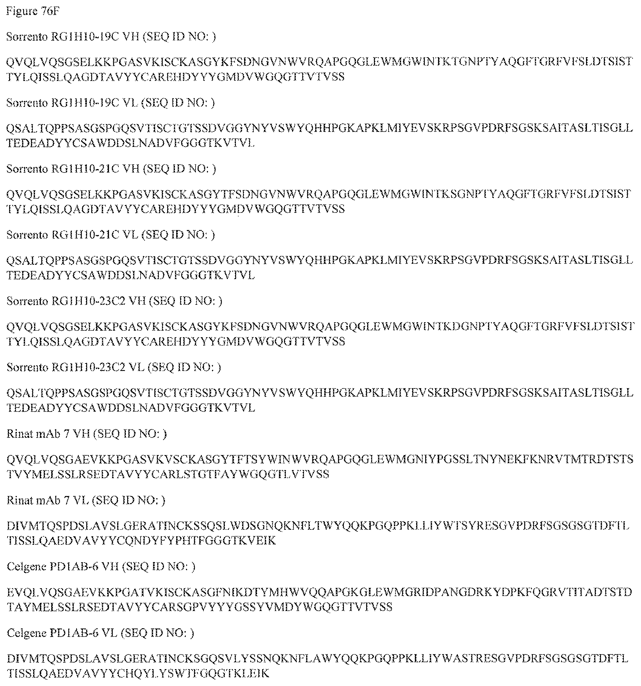
D00207
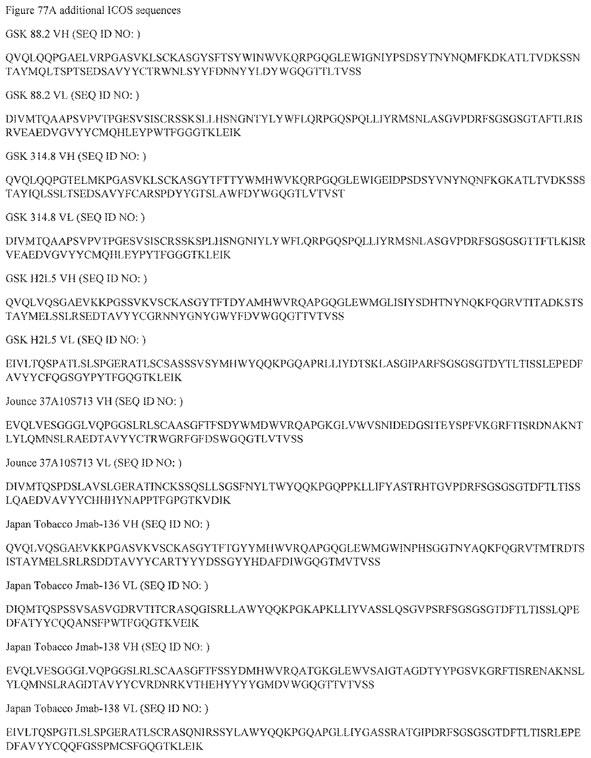
D00208
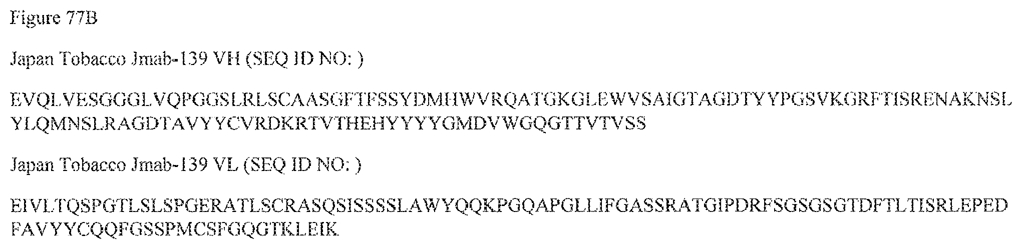
D00209
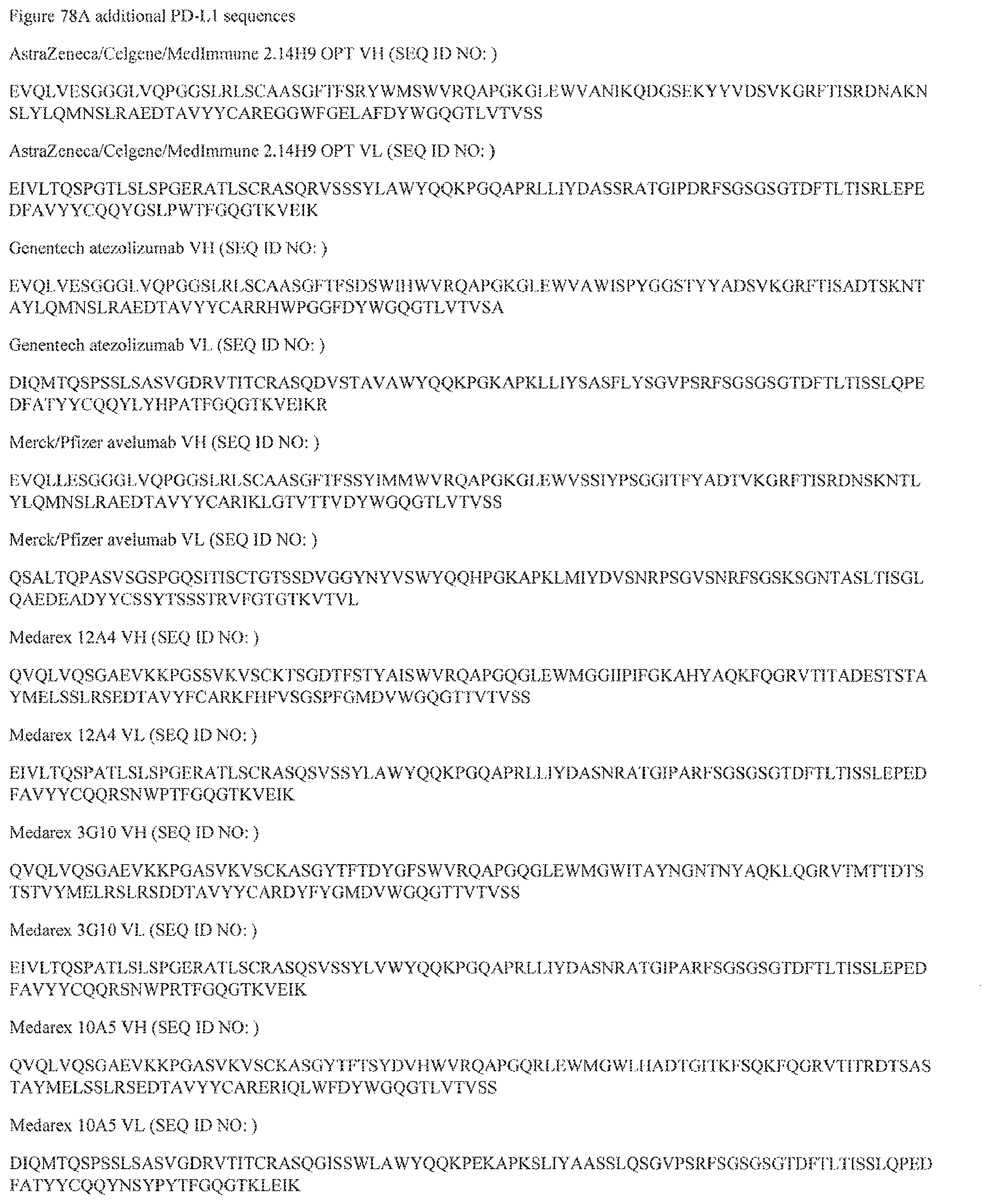
D00210

D00211
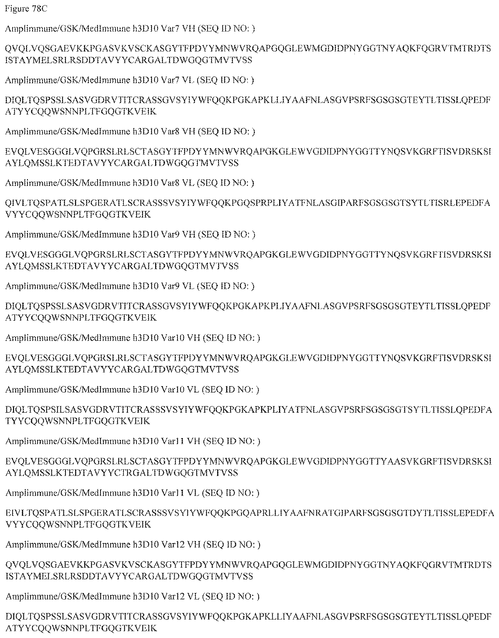
D00212
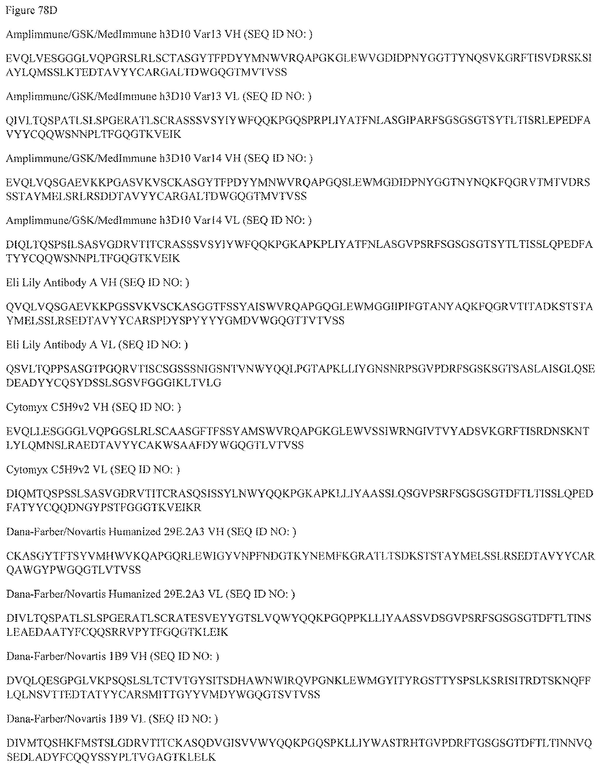
D00213

D00214
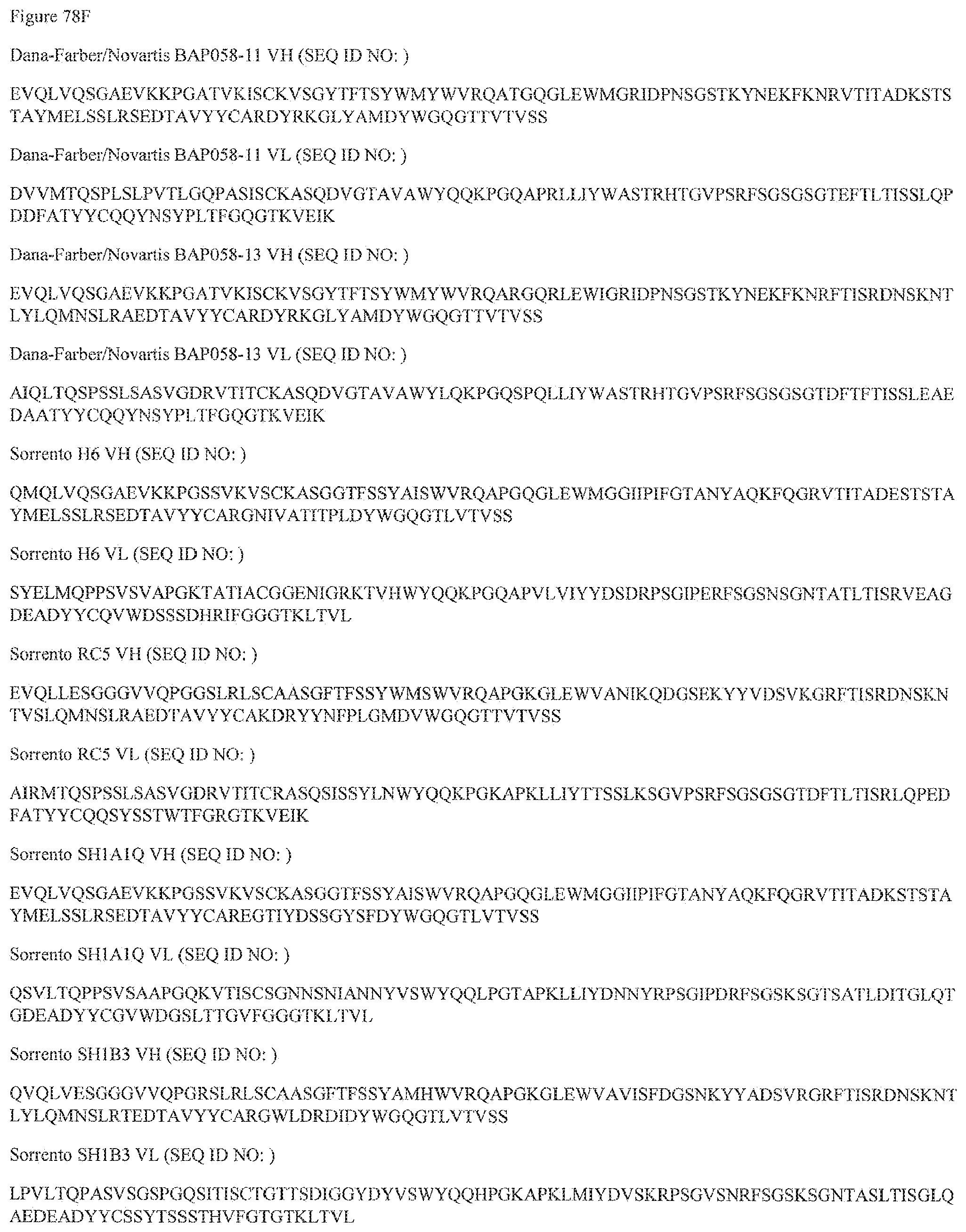
D00215

D00216

D00217

D00218
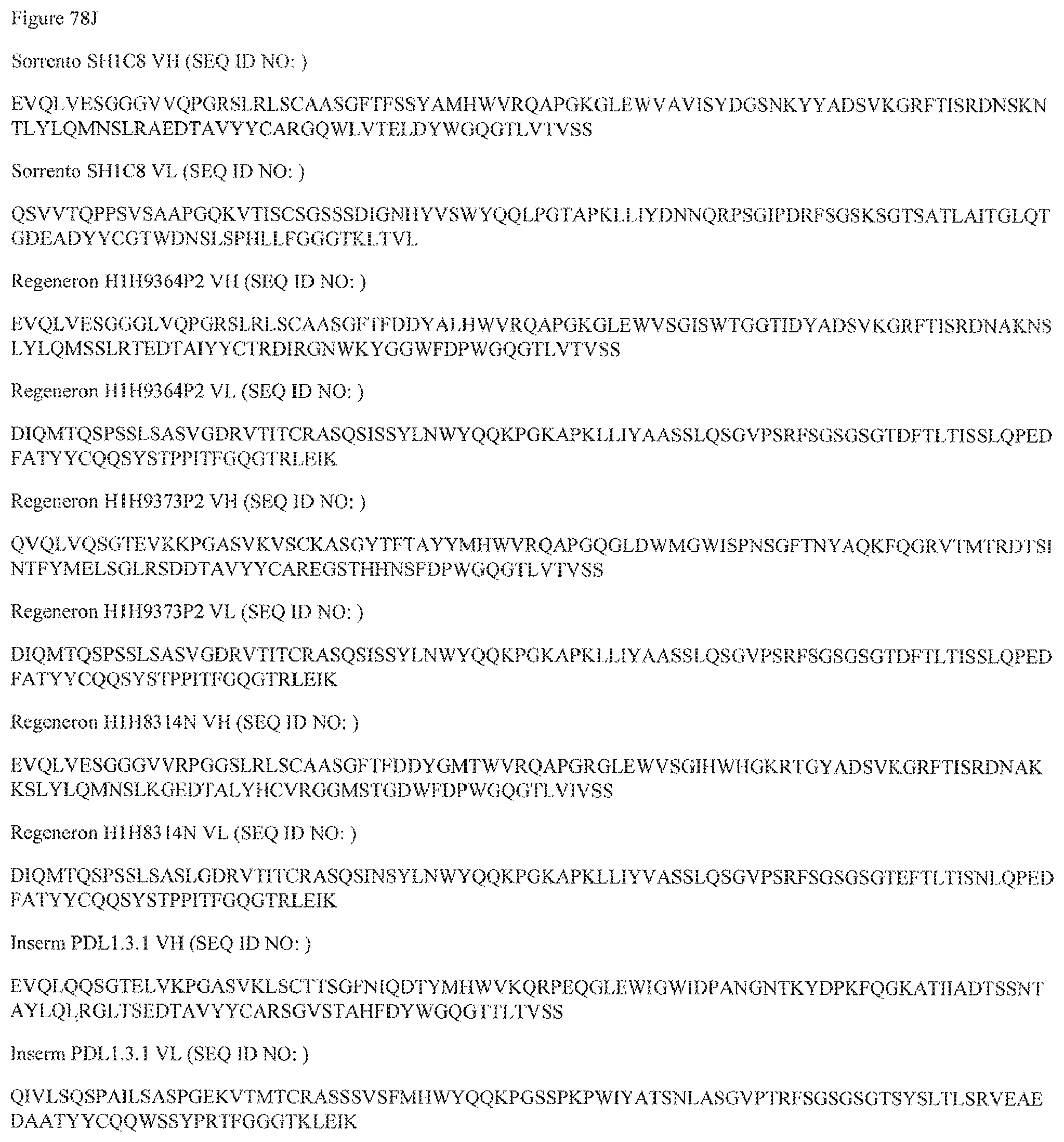
D00219
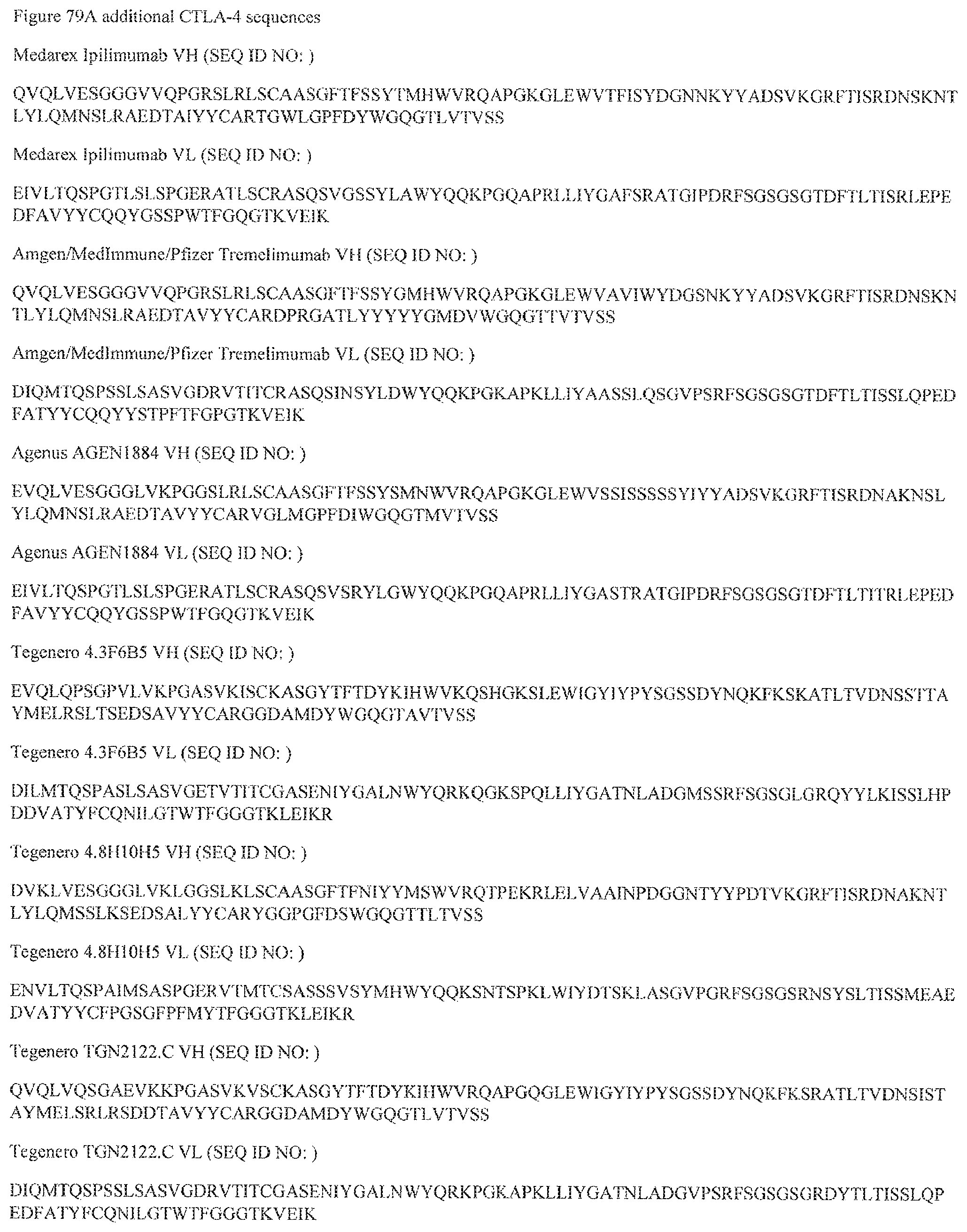
D00220
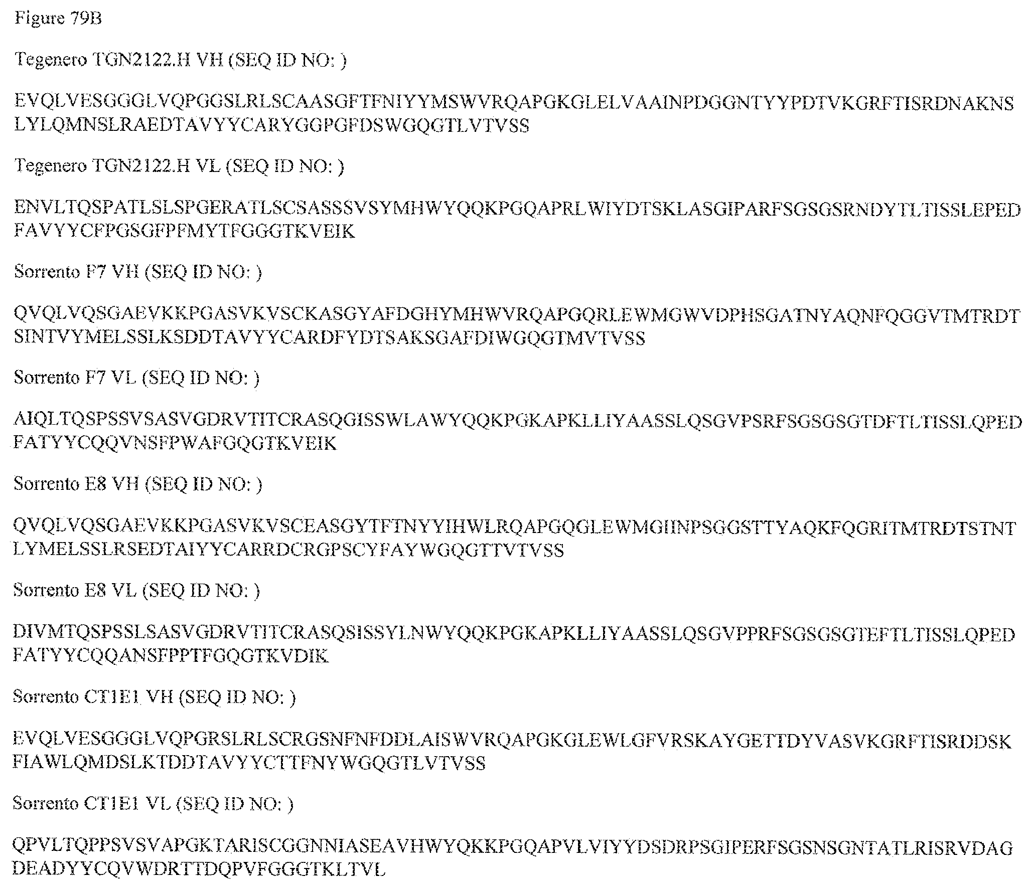
D00221
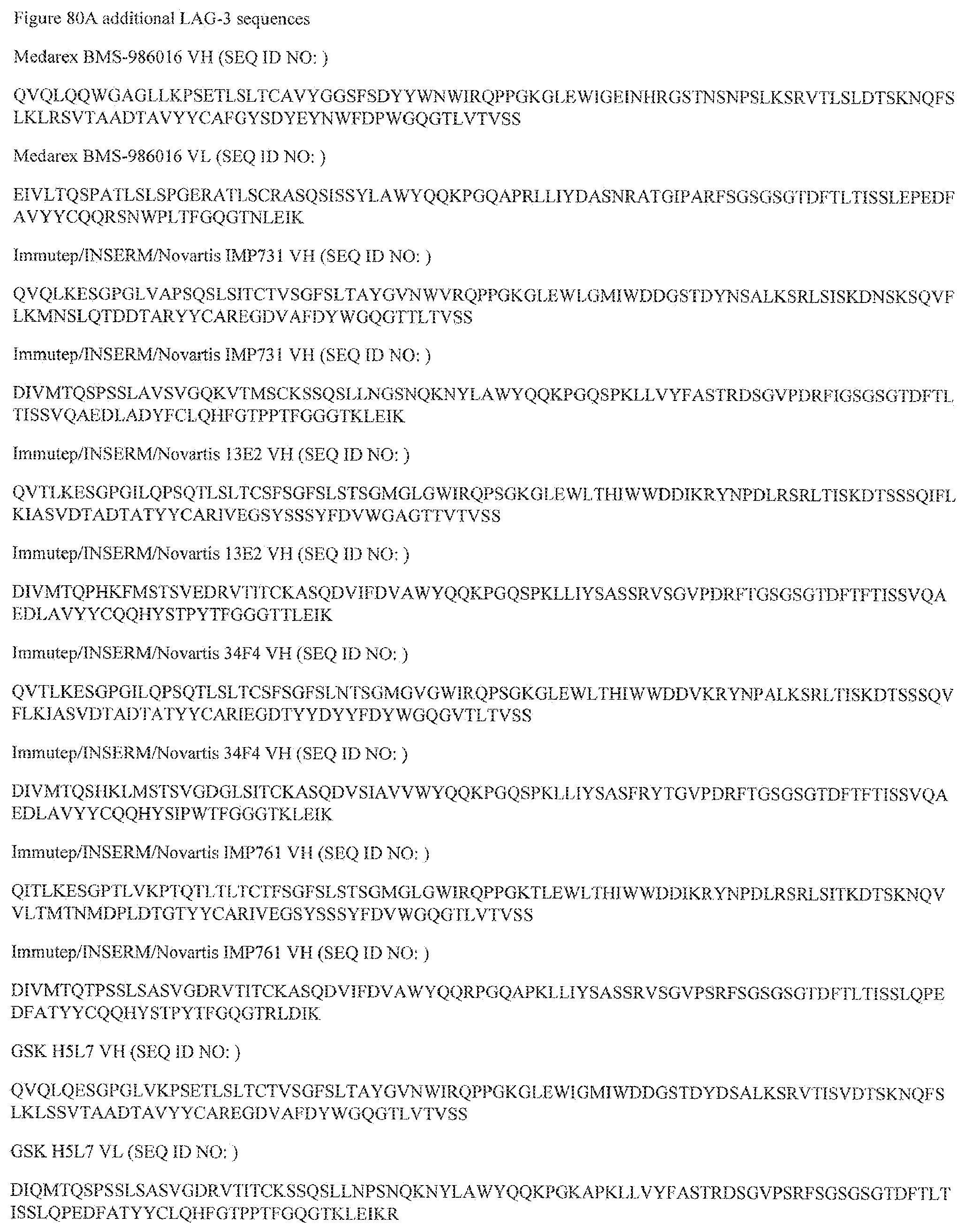
D00222

D00223
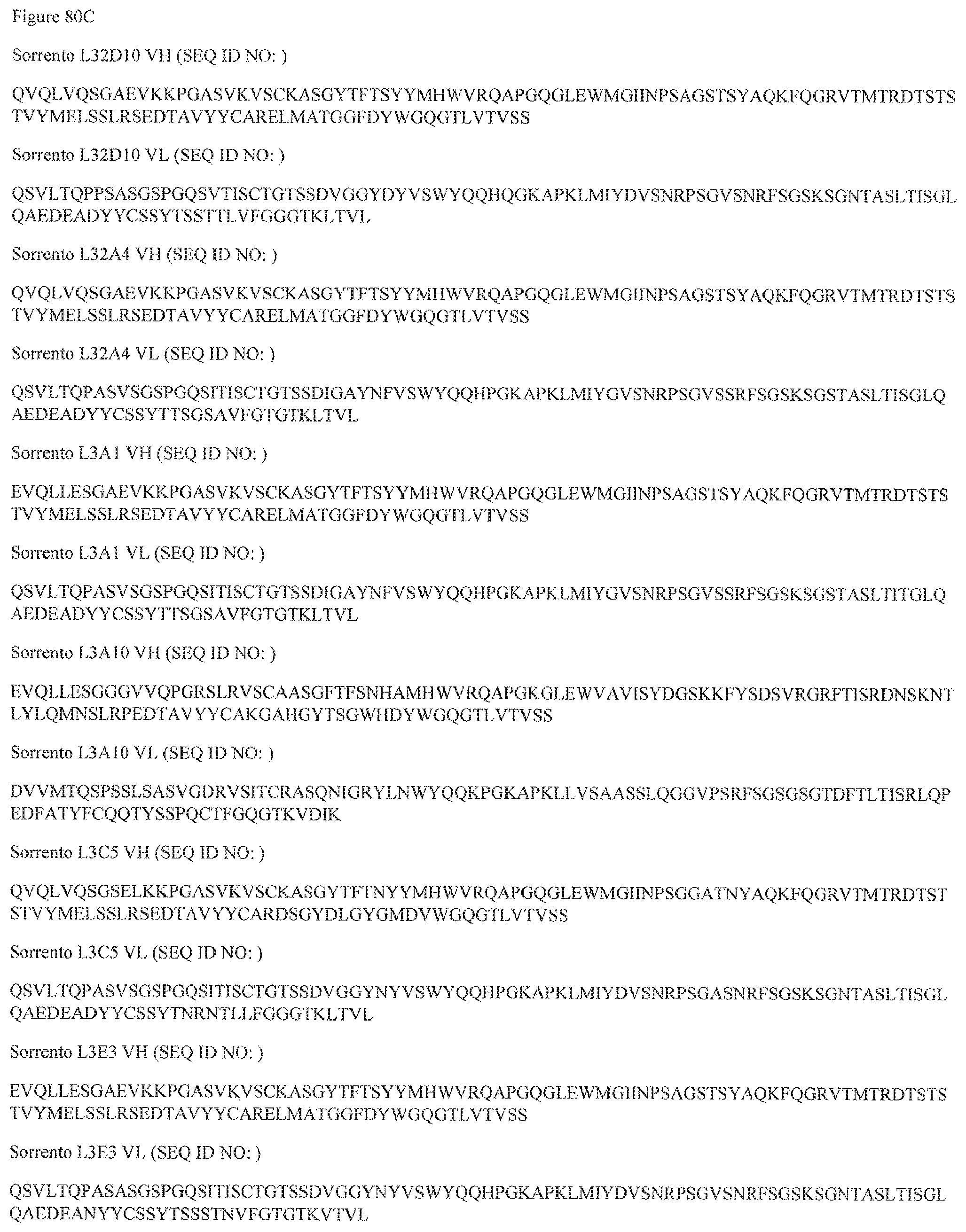
D00224

D00225
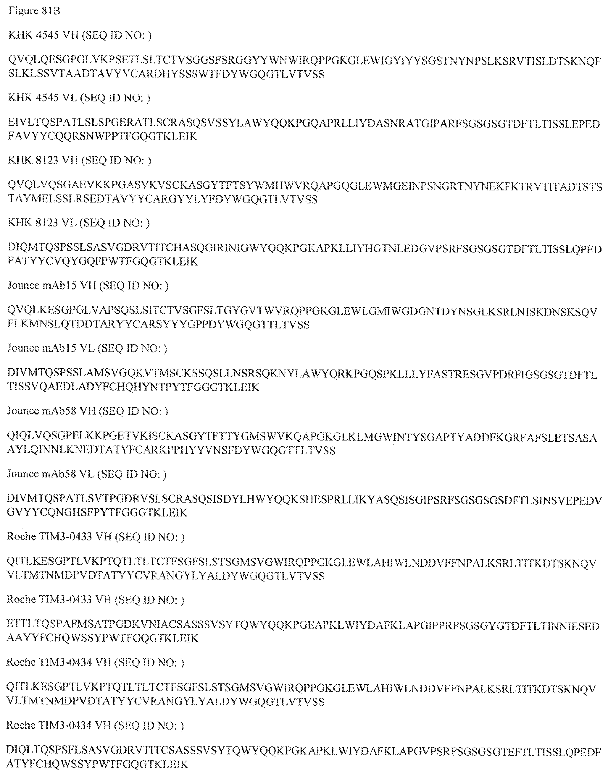
D00226
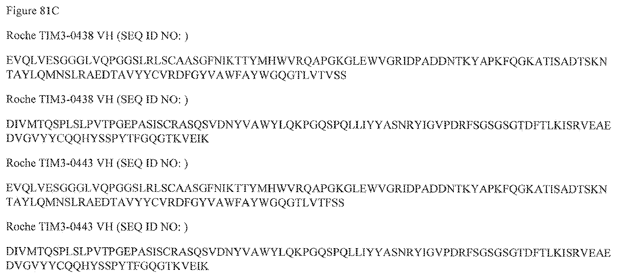
D00227
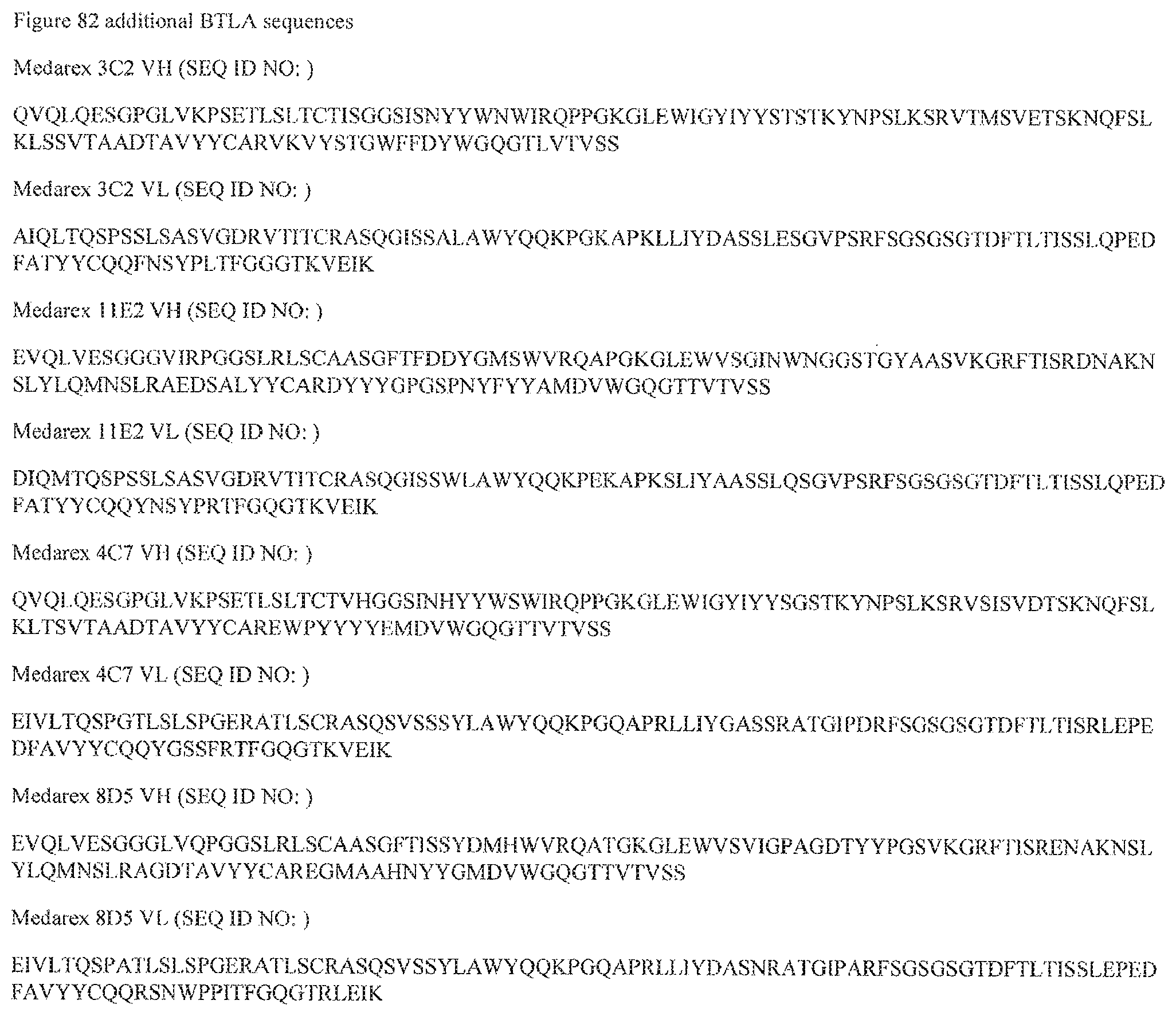
D00228
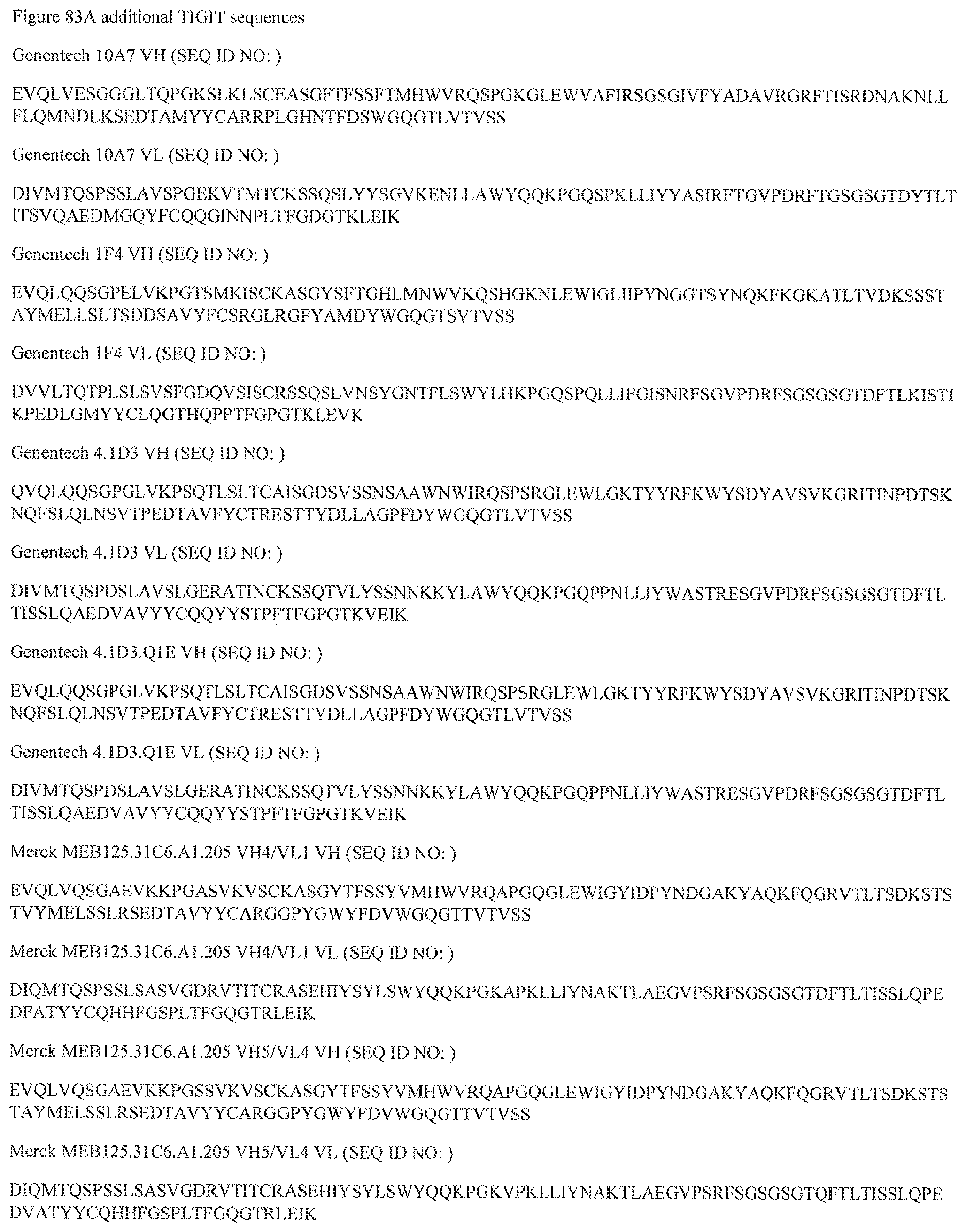
D00229

D00230

D00231

D00232

D00233
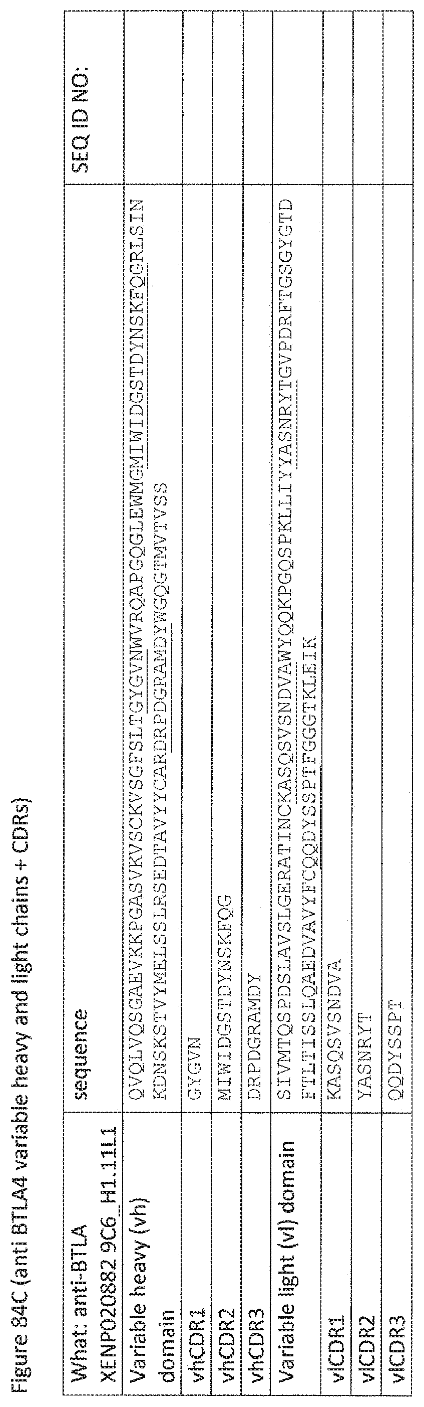
D00234
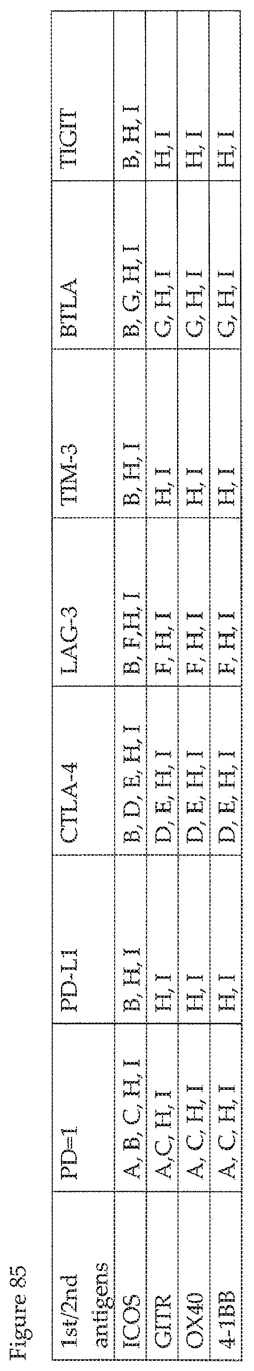
D00235
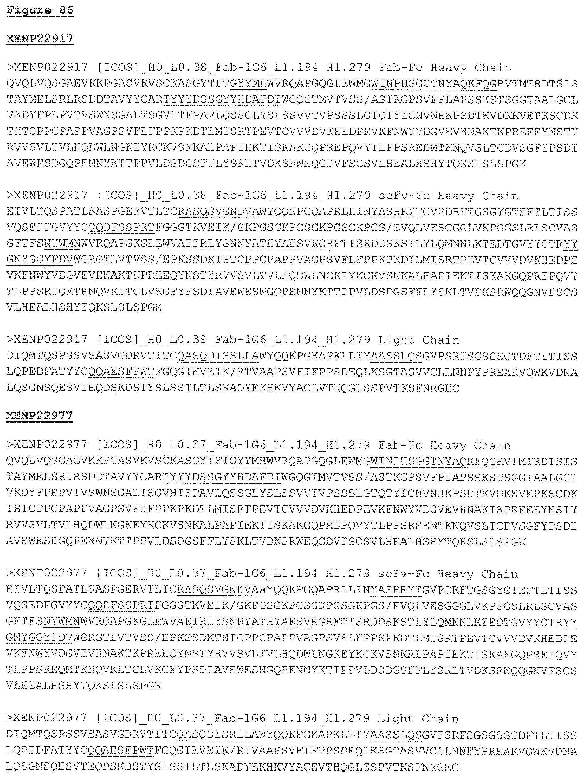
S00001
XML
uspto.report is an independent third-party trademark research tool that is not affiliated, endorsed, or sponsored by the United States Patent and Trademark Office (USPTO) or any other governmental organization. The information provided by uspto.report is based on publicly available data at the time of writing and is intended for informational purposes only.
While we strive to provide accurate and up-to-date information, we do not guarantee the accuracy, completeness, reliability, or suitability of the information displayed on this site. The use of this site is at your own risk. Any reliance you place on such information is therefore strictly at your own risk.
All official trademark data, including owner information, should be verified by visiting the official USPTO website at www.uspto.gov. This site is not intended to replace professional legal advice and should not be used as a substitute for consulting with a legal professional who is knowledgeable about trademark law.