Anti-A.beta. globulomer 4D10 antibodies
Barghorn , et al. Ja
U.S. patent number 10,538,581 [Application Number 15/290,699] was granted by the patent office on 2020-01-21 for anti-a.beta. globulomer 4d10 antibodies. This patent grant is currently assigned to ABBVIE DEUTSCHLAND GMBH & CO. KG, ABBVIE INC.. The grantee listed for this patent is ABBVIE DEUTSCHLAND GMBH & CO. KG, ABBVIE INC.. Invention is credited to Stefan Barghorn, Ulrich Ebert, Heinz Hillen, Patrick Keller, Boris Labkovsky, Andreas R. Striebinger.


View All Diagrams
| United States Patent | 10,538,581 |
| Barghorn , et al. | January 21, 2020 |
Anti-A.beta. globulomer 4D10 antibodies
Abstract
Anti-A.beta. globulomer antibodies, antigen-binding moieties thereof, corresponding hybridomas, nucleic acids, vectors, host cells, methods of producing said antibodies, compositions comprising said antibodies, uses of said antibodies and methods of using said antibodies. The present invention relates to anti-A.beta. globulomer antibodies having a binding affinity to A.beta.(20-42) globulomer that is greater than the binding affinity of the antibody to A.beta.(1-42) globulomer, antigen-binding moieties thereof, hybridomas producing said antibodies, nucleic acids encoding said antibodies, vectors comprising said nucleic acids, host cells comprising said vectors, methods of producing said antibodies, compositions comprising said antibodies, therapeutic and diagnostic uses of said antibodies and corresponding methods relating to Alzheimer's disease and other amyloidoses.
| Inventors: | Barghorn; Stefan (Mannheim, DE), Hillen; Heinz (Hassloch, DE), Labkovsky; Boris (Wales, MA), Striebinger; Andreas R. (Speyer, DE), Keller; Patrick (Darmstadt, DE), Ebert; Ulrich (Mannheim, DE) | ||||||||||
|---|---|---|---|---|---|---|---|---|---|---|---|
| Applicant: |
|
||||||||||
| Assignee: | ABBVIE INC. (North Chicago,
IL) ABBVIE DEUTSCHLAND GMBH & CO. KG (Wiesbaden, DE) |
||||||||||
| Family ID: | 37744650 | ||||||||||
| Appl. No.: | 15/290,699 | ||||||||||
| Filed: | October 11, 2016 |
Prior Publication Data
| Document Identifier | Publication Date | |
|---|---|---|
| US 20170022269 A1 | Jan 26, 2017 | |
Related U.S. Patent Documents
| Application Number | Filing Date | Patent Number | Issue Date | ||
|---|---|---|---|---|---|
| 13893780 | May 14, 2013 | 9540432 | |||
| 11574876 | Apr 8, 2014 | 8691224 | |||
| PCT/EP2006/011530 | Nov 30, 2006 | ||||
| 60740866 | Nov 30, 2005 | ||||
| 60779171 | Mar 3, 2006 | ||||
| 60787361 | Mar 30, 2006 | ||||
| 60842400 | Sep 5, 2006 | ||||
| Current U.S. Class: | 1/1 |
| Current CPC Class: | C07K 14/4711 (20130101); A61P 37/00 (20180101); A61P 25/28 (20180101); C07K 16/18 (20130101); A61P 25/00 (20180101); G01N 33/6896 (20130101); G01N 2800/387 (20130101); G01N 2800/2821 (20130101); C07K 2317/92 (20130101); C07K 2317/21 (20130101); C07K 2317/565 (20130101); G01N 2333/4709 (20130101); C07K 2317/41 (20130101); A61K 38/00 (20130101); A61K 2039/505 (20130101); C07K 2317/24 (20130101) |
| Current International Class: | C07K 16/00 (20060101); G01N 33/68 (20060101); C07K 16/18 (20060101); C07K 14/47 (20060101); A61K 38/00 (20060101); A61K 39/00 (20060101) |
References Cited [Referenced By]
U.S. Patent Documents
| 4399216 | August 1983 | Axel et al. |
| 4510245 | April 1985 | Cousens et al. |
| 4526938 | July 1985 | Churchill et al. |
| 4582788 | April 1986 | Erlich |
| 4634665 | January 1987 | Axel et al. |
| 4666829 | May 1987 | Glenner et al. |
| 4683194 | July 1987 | Saiki et al. |
| 4683202 | July 1987 | Mullis |
| 4816397 | March 1989 | Boss et al. |
| 4816567 | March 1989 | Cabilly et al. |
| 4880078 | November 1989 | Inoue et al. |
| 4946778 | August 1990 | Ladner et al. |
| 4968615 | November 1990 | Koszinowski et al. |
| 4980286 | December 1990 | Morgan et al. |
| 5107065 | April 1992 | Shewmaker et al. |
| 5128326 | July 1992 | Balazs et al. |
| 5134062 | July 1992 | Blass |
| 5168062 | December 1992 | Stinski |
| 5179017 | January 1993 | Axel et al. |
| 5223409 | June 1993 | Ladner et al. |
| 5225539 | July 1993 | Winter |
| 5231020 | July 1993 | Jorgensen et al. |
| 5258498 | November 1993 | Huston et al. |
| 5290540 | March 1994 | Prince et al. |
| 5403484 | April 1995 | Ladner et al. |
| 5427908 | June 1995 | Dower et al. |
| 5455169 | October 1995 | Mullan |
| 5516637 | May 1996 | Huang et al. |
| 5530101 | June 1996 | Queen et al. |
| 5545806 | August 1996 | Lonberg et al. |
| 5565332 | October 1996 | Hoogenboom et al. |
| 5565352 | October 1996 | Hochstrasser et al. |
| 5569825 | October 1996 | Lonberg et al. |
| 5571698 | November 1996 | Ladner et al. |
| 5580717 | December 1996 | Dower et al. |
| 5585089 | December 1996 | Queen et al. |
| 5624821 | April 1997 | Winter et al. |
| 5625126 | April 1997 | Lonberg et al. |
| 5627052 | May 1997 | Schrader |
| 5633425 | May 1997 | Lonberg et al. |
| 5648260 | July 1997 | Winter et al. |
| 5658727 | August 1997 | Barbas et al. |
| 5661016 | August 1997 | Lonberg et al. |
| 5679377 | October 1997 | Bernstein et al. |
| 5693762 | December 1997 | Queen et al. |
| 5698426 | December 1997 | Huse |
| 5705330 | January 1998 | Shah et al. |
| 5714350 | February 1998 | Co et al. |
| 5714352 | February 1998 | Jakobovits |
| 5723323 | March 1998 | Kauffman et al. |
| 5733743 | March 1998 | Johnson et al. |
| 5750753 | May 1998 | Kimae et al. |
| 5763192 | June 1998 | Kauffman et al. |
| 5766886 | June 1998 | Studnicka et al. |
| 5770429 | June 1998 | Lonberg et al. |
| 5780225 | July 1998 | Wigler et al. |
| 5807715 | September 1998 | Morrison et al. |
| 5814318 | September 1998 | Lonberg et al. |
| 5814476 | September 1998 | Kauffman et al. |
| 5817483 | October 1998 | Kauffman et al. |
| 5821047 | October 1998 | Garrard et al. |
| 5824514 | October 1998 | Kauffman et al. |
| 5837500 | November 1998 | Ladner et al. |
| 5855913 | January 1999 | Hanes et al. |
| 5874064 | February 1999 | Edwards et al. |
| 5877397 | March 1999 | Lonberg et al. |
| 5879909 | March 1999 | Perl |
| 5882644 | March 1999 | Chang et al. |
| 5885793 | March 1999 | Griffiths et al. |
| 5912015 | June 1999 | Bernstein et al. |
| 5912120 | June 1999 | Goldstein et al. |
| 5916597 | June 1999 | Lee et al. |
| 5916771 | June 1999 | Hori et al. |
| 5934272 | August 1999 | Lloyd et al. |
| 5939598 | August 1999 | Kucherlapati et al. |
| 5969108 | October 1999 | McCafferty et al. |
| 5976862 | November 1999 | Kauffman et al. |
| 5985309 | November 1999 | Edwards et al. |
| 5985320 | November 1999 | Edwards et al. |
| 5985615 | November 1999 | Jakobovits et al. |
| 5989463 | November 1999 | Tracy et al. |
| 5998209 | December 1999 | Jokobovits et al. |
| 6010913 | January 2000 | Vandermeeren et al. |
| 6019968 | February 2000 | Platz et al. |
| 6075181 | June 2000 | Kucherlapati et al. |
| 6091001 | July 2000 | Jakobovits et al. |
| 6114598 | September 2000 | Kucherlapati et al. |
| 6130364 | October 2000 | Jakobovits et al. |
| 6180370 | January 2001 | Queen et al. |
| 6204023 | March 2001 | Robinson et al. |
| 6218506 | April 2001 | Krafft et al. |
| 6323218 | November 2001 | Bush et al. |
| 6333034 | December 2001 | Gupta-Bansal et al. |
| 6350861 | February 2002 | Co et al. |
| 6660843 | December 2003 | Feige et al. |
| 6699658 | March 2004 | Wittrup et al. |
| 6710226 | March 2004 | Schenk |
| 6713450 | March 2004 | Frangione et al. |
| 6743427 | June 2004 | Schenk |
| 6761888 | July 2004 | Schenk |
| 6785434 | August 2004 | Castoldi et al. |
| 6787138 | September 2004 | Schenk |
| 6787139 | September 2004 | Schenk |
| 6787140 | September 2004 | Schenk |
| 6787143 | September 2004 | Schenk |
| 6787144 | September 2004 | Schenk |
| 6787637 | September 2004 | Schenk |
| 6875434 | April 2005 | Schenk |
| 6905686 | June 2005 | Schenk |
| 6913745 | July 2005 | Schenk |
| 6919075 | July 2005 | Solomon et al. |
| 6982084 | January 2006 | Schenk |
| 7045531 | May 2006 | Bush et al. |
| 7094884 | August 2006 | Scholz et al. |
| 7122374 | October 2006 | Saido et al. |
| 7135181 | November 2006 | Jensen et al. |
| 7169389 | January 2007 | Di Padova et al. |
| 7179463 | February 2007 | Lannfelt et al. |
| 7179606 | February 2007 | Jackowski et al. |
| 7179892 | February 2007 | Basi et al. |
| 7186881 | March 2007 | Games et al. |
| 7189703 | March 2007 | Balin et al. |
| 7195761 | March 2007 | Holtzman et al. |
| 7195881 | March 2007 | Geffard |
| 7196163 | March 2007 | Hazuda et al. |
| 7226730 | June 2007 | De La Monte et al. |
| 7238488 | July 2007 | Maresh et al. |
| 7238788 | July 2007 | Lee |
| 7247301 | July 2007 | Van De Winkel et al. |
| 7256273 | August 2007 | Basi et al. |
| 7270818 | September 2007 | Averback |
| 7279165 | October 2007 | Bachmann et al. |
| 7318923 | January 2008 | Tsurushita et al. |
| 7320790 | January 2008 | Hinton et al. |
| 7320793 | January 2008 | Renner et al. |
| 7339035 | March 2008 | Yanagisawa et al. |
| 7342091 | March 2008 | Kapurniotu et al. |
| 7375190 | May 2008 | Cheng et al. |
| 7625560 | December 2009 | Basi et al. |
| 7790363 | September 2010 | Wonderling et al. |
| 7892544 | February 2011 | Pfeifer et al. |
| 7902328 | March 2011 | Hillen et al. |
| 8691224 | April 2014 | Barghorn et al. |
| 2002/0015941 | February 2002 | Kim et al. |
| 2002/0086014 | July 2002 | Korman et al. |
| 2002/0132758 | September 2002 | Shell |
| 2002/0137134 | September 2002 | Gerngross |
| 2002/0182644 | December 2002 | Diamandis |
| 2002/0188106 | December 2002 | Mandelkow et al. |
| 2002/0197258 | December 2002 | Ghanbari et al. |
| 2003/0065141 | April 2003 | Carter et al. |
| 2003/0068316 | April 2003 | Klein et al. |
| 2003/0073655 | April 2003 | Chain |
| 2003/0077278 | April 2003 | Gallatin et al. |
| 2003/0077757 | April 2003 | Andrews |
| 2003/0086938 | May 2003 | Jensen et al. |
| 2003/0100011 | May 2003 | Jackowski et al. |
| 2003/0100058 | May 2003 | Roschke et al. |
| 2003/0114510 | June 2003 | Ingram et al. |
| 2003/0148356 | August 2003 | Cruts et al. |
| 2003/0157117 | August 2003 | Rasmussen et al. |
| 2003/0180722 | September 2003 | Godbole et al. |
| 2003/0185826 | October 2003 | Tobinick |
| 2003/0185827 | October 2003 | Rodriguez et al. |
| 2003/0186333 | October 2003 | Loring et al. |
| 2003/0186374 | October 2003 | Hufton et al. |
| 2003/0194403 | October 2003 | Van De Winkel et al. |
| 2003/0195347 | October 2003 | Baker et al. |
| 2003/0228307 | December 2003 | Ramakrishnan et al. |
| 2003/0229907 | December 2003 | Hsiao et al. |
| 2003/0232758 | December 2003 | St. George-Hyslop et al. |
| 2004/0013647 | January 2004 | Solomon et al. |
| 2004/0013680 | January 2004 | Bush et al. |
| 2004/0018590 | January 2004 | Gerngross et al. |
| 2004/0043418 | March 2004 | Holtzman et al. |
| 2004/0053371 | March 2004 | Maresh et al. |
| 2004/0081657 | April 2004 | Schenk |
| 2004/0116337 | June 2004 | Kapurniotu et al. |
| 2004/0127471 | July 2004 | Reisberg |
| 2004/0138296 | July 2004 | Robertson et al. |
| 2004/0142872 | July 2004 | Poduslo et al. |
| 2004/0157267 | August 2004 | Huang |
| 2004/0157779 | August 2004 | Schenk |
| 2004/0162414 | August 2004 | Santora et al. |
| 2004/0166119 | August 2004 | Schenk |
| 2004/0170641 | September 2004 | Schenk |
| 2004/0175394 | September 2004 | Schenk |
| 2004/0185039 | September 2004 | Kohler et al. |
| 2004/0223970 | November 2004 | Afar et al. |
| 2004/0228865 | November 2004 | Schenk |
| 2004/0241164 | December 2004 | Bales et al. |
| 2004/0265308 | December 2004 | Schenk |
| 2005/0009110 | January 2005 | Chang |
| 2005/0014821 | January 2005 | Tsai et al. |
| 2005/0019330 | January 2005 | Schenk |
| 2005/0019343 | January 2005 | Schenk |
| 2005/0037026 | February 2005 | Schenk |
| 2005/0042664 | February 2005 | Wu et al. |
| 2005/0048584 | March 2005 | Lamping et al. |
| 2005/0053614 | March 2005 | Schenk |
| 2005/0057813 | March 2005 | Hasei et al. |
| 2005/0059591 | March 2005 | Schenk et al. |
| 2005/0059802 | March 2005 | Schenk et al. |
| 2005/0090439 | April 2005 | Chalifour et al. |
| 2005/0112543 | May 2005 | Bush et al. |
| 2005/0123544 | June 2005 | Schenk et al. |
| 2005/0142131 | June 2005 | Hinton et al. |
| 2005/0142132 | June 2005 | Schenk et al. |
| 2005/0153381 | July 2005 | Marusich et al. |
| 2005/0163744 | July 2005 | Rasmussen et al. |
| 2005/0163788 | July 2005 | Schenk |
| 2005/0175626 | August 2005 | Delacourte et al. |
| 2005/0249725 | November 2005 | Schenk et al. |
| 2005/0249727 | November 2005 | Schenk |
| 2005/0255122 | November 2005 | Schenk |
| 2005/0272025 | December 2005 | Suo et al. |
| 2006/0029603 | February 2006 | Ellis et al. |
| 2006/0029611 | February 2006 | Schenk |
| 2006/0034858 | February 2006 | Schenk |
| 2006/0039906 | February 2006 | Holtzman et al. |
| 2006/0057702 | March 2006 | Rosenthal et al. |
| 2006/0062786 | March 2006 | Salcedo et al. |
| 2006/0099211 | May 2006 | Monthe et al. |
| 2006/0104968 | May 2006 | Bookbinder et al. |
| 2006/0105394 | May 2006 | Pomara |
| 2006/0141541 | June 2006 | McIntyre |
| 2006/0162129 | July 2006 | Strobel et al. |
| 2006/0166275 | July 2006 | Krafft et al. |
| 2006/0166311 | July 2006 | Okochi et al. |
| 2006/0188505 | August 2006 | Skurkovich et al. |
| 2006/0234947 | October 2006 | Gazit |
| 2006/0240007 | October 2006 | Sanders |
| 2006/0241038 | October 2006 | Watanabe et al. |
| 2006/0257420 | November 2006 | Zimmerman |
| 2006/0257882 | November 2006 | Shimkets et al. |
| 2007/0009931 | January 2007 | Kirsch |
| 2007/0010657 | January 2007 | Klocke et al. |
| 2007/0015217 | January 2007 | Durham et al. |
| 2007/0021345 | January 2007 | Gazit |
| 2007/0031416 | February 2007 | Shoji et al. |
| 2007/0036789 | February 2007 | Chung et al. |
| 2007/0036794 | February 2007 | Devaux et al. |
| 2007/0042424 | February 2007 | Ebinuma et al. |
| 2007/0048312 | March 2007 | Klein et al. |
| 2007/0071675 | March 2007 | Wu et al. |
| 2007/0081998 | April 2007 | Kinney et al. |
| 2007/0082350 | April 2007 | Landfield et al. |
| 2007/0086994 | April 2007 | Wallach et al. |
| 2007/0098721 | May 2007 | Hillen et al. |
| 2007/0105092 | May 2007 | Paul et al. |
| 2007/0110750 | May 2007 | Glabe et al. |
| 2007/0111252 | May 2007 | Suzuki et al. |
| 2007/0122405 | May 2007 | Roschke et al. |
| 2007/0128191 | June 2007 | Barrio |
| 2007/0134247 | June 2007 | Solomon |
| 2007/0135337 | June 2007 | Chalifour et al. |
| 2007/0140966 | June 2007 | Chang et al. |
| 2007/0148167 | June 2007 | Strohl |
| 2007/0160616 | July 2007 | Rosenthal et al. |
| 2007/0167522 | July 2007 | Imawaka et al. |
| 2007/0196367 | August 2007 | Dinu |
| 2007/0213512 | September 2007 | Krafft et al. |
| 2007/0218069 | September 2007 | Gordon et al. |
| 2007/0218499 | September 2007 | Lambert et al. |
| 2007/0231331 | October 2007 | Dewji et al. |
| 2007/0248606 | October 2007 | Lannfelt et al. |
| 2007/0264276 | November 2007 | Chalifour et al. |
| 2007/0280953 | December 2007 | Rosenberg et al. |
| 2007/0292410 | December 2007 | Cashman et al. |
| 2007/0292895 | December 2007 | Shi et al. |
| 2008/0009467 | January 2008 | Henderson |
| 2008/0014194 | January 2008 | Schenk et al. |
| 2008/0014596 | January 2008 | Jerecic et al. |
| 2008/0025988 | January 2008 | Yamaguchi et al. |
| 2008/0029911 | February 2008 | Jeon et al. |
| 2008/0044356 | February 2008 | Lesne et al. |
| 2008/0044406 | February 2008 | Johnson-Wood et al. |
| 2008/0051690 | February 2008 | Mattner et al. |
| 2008/0057053 | March 2008 | Stolen |
| 2008/0057593 | March 2008 | Vanderstichele et al. |
| 2008/0058276 | March 2008 | Lu et al. |
| 2008/0058330 | March 2008 | Paris et al. |
| 2008/0089885 | April 2008 | Smith et al. |
| 2008/0096818 | April 2008 | Schenk et al. |
| 2008/0107649 | May 2008 | Zurbriggen |
| 2008/0113444 | May 2008 | Pray |
| 2008/0220449 | September 2008 | Vasan et al. |
| 2008/0299111 | December 2008 | Delacourte et al. |
| 2009/0018084 | January 2009 | Krafft et al. |
| 2009/0035295 | February 2009 | Hillen et al. |
| 2009/0035307 | February 2009 | Barghorn et al. |
| 2009/0175847 | July 2009 | Barghorn et al. |
| 2009/0214515 | August 2009 | Holzman et al. |
| 2009/0232801 | September 2009 | Hillen et al. |
| 2009/0238831 | September 2009 | Hillen et al. |
| 2010/0028333 | February 2010 | Getty et al. |
| 2010/0173828 | July 2010 | Hillen et al. |
| 2010/0209346 | August 2010 | Hillen et al. |
| 2011/0092445 | April 2011 | Barghorn et al. |
| 2011/0212109 | September 2011 | Barghorn et al. |
| 2011/0287005 | November 2011 | Hillen et al. |
| 2012/0034166 | February 2012 | Hillen et al. |
| 2007200047 | Jan 2007 | AU | |||
| 2541522 | Sep 2007 | CA | |||
| 1396183 | Feb 2003 | CN | |||
| 1446581 | Oct 2003 | CN | |||
| 1673369 | Sep 2005 | CN | |||
| 1721437 | Jan 2006 | CN | |||
| 1803842 | Jul 2006 | CN | |||
| 101058608 | Oct 2007 | CN | |||
| 101084909 | Dec 2007 | CN | |||
| 101152576 | Apr 2008 | CN | |||
| 19902550 | Jul 2000 | DE | |||
| 10055703 | May 2002 | DE | |||
| 10303974 | Aug 2004 | DE | |||
| 102004039326 | Feb 2006 | DE | |||
| 45665 | Feb 1982 | EP | |||
| 0050424 | Sep 1985 | EP | |||
| 285159 | Oct 1988 | EP | |||
| 341491 | Nov 1989 | EP | |||
| 0084796 | May 1990 | EP | |||
| 391714 | Oct 1990 | EP | |||
| 411974 | Feb 1991 | EP | |||
| 415801 | Mar 1991 | EP | |||
| 0201184 | Dec 1992 | EP | |||
| 229246 | Aug 1993 | EP | |||
| 368684 | Mar 1994 | EP | |||
| 239400 | Aug 1994 | EP | |||
| 613007 | Aug 1994 | EP | |||
| 557270 | May 1995 | EP | |||
| 519598 | Jun 1995 | EP | |||
| 440619 | Jan 1996 | EP | |||
| 304013 | Jun 1996 | EP | |||
| 589877 | Nov 1996 | EP | |||
| 436597 | Apr 1997 | EP | |||
| 258017 | Jun 1997 | EP | |||
| 783104 | Jul 1997 | EP | |||
| 444856 | Sep 1997 | EP | |||
| 816492 | Jan 1998 | EP | |||
| 592127 | Apr 1998 | EP | |||
| 274826 | Aug 1998 | EP | |||
| 527839 | Dec 1998 | EP | |||
| 1038958 | Sep 2000 | EP | |||
| 1094080 | Apr 2001 | EP | |||
| 1172378 | Jan 2002 | EP | |||
| 1176195 | Jan 2002 | EP | |||
| 877939 | Jun 2002 | EP | |||
| 683234 | May 2003 | EP | |||
| 1308461 | May 2003 | EP | |||
| 1408333 | Apr 2004 | EP | |||
| 1270592 | Sep 2004 | EP | |||
| 1467212 | Oct 2004 | EP | |||
| 0592106 | Nov 2004 | EP | |||
| 1200470 | Nov 2004 | EP | |||
| 519596 | Feb 2005 | EP | |||
| 1538163 | Jun 2005 | EP | |||
| 1632242 | Mar 2006 | EP | |||
| 1092767 | Oct 2006 | EP | |||
| 1717250 | Nov 2006 | EP | |||
| 998495 | Dec 2006 | EP | |||
| 1731913 | Dec 2006 | EP | |||
| 1049712 | Jan 2007 | EP | |||
| 1741783 | Jan 2007 | EP | |||
| 1346041 | Feb 2007 | EP | |||
| 1752472 | Feb 2007 | EP | |||
| 1592476 | Apr 2007 | EP | |||
| 970203 | May 2007 | EP | |||
| 1787998 | May 2007 | EP | |||
| 948536 | Jun 2007 | EP | |||
| 1160256 | Jun 2007 | EP | |||
| 1379546 | Jun 2007 | EP | |||
| 1792991 | Jun 2007 | EP | |||
| 1842859 | Oct 2007 | EP | |||
| 1878751 | Jan 2008 | EP | |||
| 1434053 | Mar 2008 | EP | |||
| 1521831 | Apr 2008 | EP | |||
| 1778837 | Apr 2008 | EP | |||
| 1911765 | Apr 2008 | EP | |||
| 1781644 | May 2008 | EP | |||
| 911398 | Jun 2008 | EP | |||
| 2009445 | Dec 2008 | EP | |||
| 1623719 | Jul 2009 | EP | |||
| 1681566 | Aug 2009 | EP | |||
| 1861422 | Feb 2010 | EP | |||
| 1766396 | Aug 2010 | EP | |||
| 1720909 | Nov 2011 | EP | |||
| 2740454 | Apr 1997 | FR | |||
| 2741881 | Jun 1997 | FR | |||
| 1495159 | Dec 1977 | GB | |||
| 2371303 | Jul 2002 | GB | |||
| 1005016 | Oct 2005 | GR | |||
| S63240797 | Oct 1988 | JP | |||
| H04252195 | Sep 1992 | JP | |||
| H04320694 | Nov 1992 | JP | |||
| H07209295 | Aug 1995 | JP | |||
| H07209296 | Aug 1995 | JP | |||
| H07309900 | Nov 1995 | JP | |||
| H08245700 | Sep 1996 | JP | |||
| H0967397 | Mar 1997 | JP | |||
| H1075781 | Mar 1998 | JP | |||
| H10210982 | Aug 1998 | JP | |||
| 2000050885 | Feb 2000 | JP | |||
| 2000354487 | Dec 2000 | JP | |||
| 2001231578 | Aug 2001 | JP | |||
| 2002040023 | Feb 2002 | JP | |||
| 2002253252 | Sep 2002 | JP | |||
| 2004107260 | Apr 2004 | JP | |||
| 2006166879 | Jun 2006 | JP | |||
| 2006213621 | Aug 2006 | JP | |||
| 2006265189 | Oct 2006 | JP | |||
| 2007077103 | Mar 2007 | JP | |||
| 2007300856 | Nov 2007 | JP | |||
| 2007319127 | Dec 2007 | JP | |||
| 2008096311 | Apr 2008 | JP | |||
| 100806914 | Feb 2008 | KR | |||
| WO-8803951 | Jun 1988 | WO | |||
| WO-8906689 | Jul 1989 | WO | |||
| WO-9002809 | Mar 1990 | WO | |||
| WO-9005144 | May 1990 | WO | |||
| WO-9012870 | Nov 1990 | WO | |||
| WO-9014424 | Nov 1990 | WO | |||
| WO-9014430 | Nov 1990 | WO | |||
| WO-9014443 | Nov 1990 | WO | |||
| WO-9105548 | May 1991 | WO | |||
| WO-9109967 | Jul 1991 | WO | |||
| WO-9110737 | Jul 1991 | WO | |||
| WO-9110741 | Jul 1991 | WO | |||
| WO-9117271 | Nov 1991 | WO | |||
| WO-9200969 | Jan 1992 | WO | |||
| WO-9201047 | Jan 1992 | WO | |||
| WO-9202551 | Feb 1992 | WO | |||
| WO-9209690 | Jun 1992 | WO | |||
| WO-9215679 | Sep 1992 | WO | |||
| WO-9218619 | Oct 1992 | WO | |||
| WO-9219244 | Nov 1992 | WO | |||
| WO-9220791 | Nov 1992 | WO | |||
| WO-9222324 | Dec 1992 | WO | |||
| WO-9301288 | Jan 1993 | WO | |||
| WO-9308302 | Apr 1993 | WO | |||
| WO-9311236 | Jun 1993 | WO | |||
| WO-9402602 | Feb 1994 | WO | |||
| WO-9417197 | Aug 1994 | WO | |||
| WO-9507707 | Mar 1995 | WO | |||
| WO-9511311 | Apr 1995 | WO | |||
| WO-9515982 | Jun 1995 | WO | |||
| WO-9516787 | Jun 1995 | WO | |||
| WO-9520401 | Aug 1995 | WO | |||
| WO-9620218 | Jul 1996 | WO | |||
| WO-9620698 | Jul 1996 | WO | |||
| WO-9633735 | Oct 1996 | WO | |||
| WO-9634096 | Oct 1996 | WO | |||
| WO-9639512 | Dec 1996 | WO | |||
| WO-9708320 | Mar 1997 | WO | |||
| WO-97029131 | Aug 1997 | WO | |||
| WO-9732572 | Sep 1997 | WO | |||
| WO-9744013 | Nov 1997 | WO | |||
| WO-9746678 | Dec 1997 | WO | |||
| WO-9805350 | Feb 1998 | WO | |||
| WO-9807850 | Feb 1998 | WO | |||
| WO-9813490 | Apr 1998 | WO | |||
| WO-9816654 | Apr 1998 | WO | |||
| WO-9822120 | May 1998 | WO | |||
| WO-9824893 | Jun 1998 | WO | |||
| WO-9828445 | Jul 1998 | WO | |||
| WO-9831346 | Jul 1998 | WO | |||
| WO-9831700 | Jul 1998 | WO | |||
| WO-9833815 | Aug 1998 | WO | |||
| WO-9841201 | Sep 1998 | WO | |||
| WO-9847343 | Oct 1998 | WO | |||
| WO-9849286 | Nov 1998 | WO | |||
| WO-9850433 | Nov 1998 | WO | |||
| WO-9851793 | Nov 1998 | WO | |||
| WO-9909150 | Feb 1999 | WO | |||
| WO-9912870 | Mar 1999 | WO | |||
| WO-9913908 | Mar 1999 | WO | |||
| WO-9915154 | Apr 1999 | WO | |||
| WO-9920253 | Apr 1999 | WO | |||
| WO-9922024 | May 1999 | WO | |||
| WO-9925044 | May 1999 | WO | |||
| WO-9927944 | Jun 1999 | WO | |||
| WO-9927949 | Jun 1999 | WO | |||
| WO-9933815 | Jul 1999 | WO | |||
| WO-9936569 | Jul 1999 | WO | |||
| WO-9945031 | Sep 1999 | WO | |||
| WO-9945962 | Sep 1999 | WO | |||
| WO-9953049 | Oct 1999 | WO | |||
| WO-9955842 | Nov 1999 | WO | |||
| WO-9958157 | Nov 1999 | WO | |||
| WO-9958564 | Nov 1999 | WO | |||
| WO-9962505 | Dec 1999 | WO | |||
| WO-9966903 | Dec 1999 | WO | |||
| WO-0009560 | Feb 2000 | WO | |||
| WO-2000017345 | Mar 2000 | WO | |||
| WO-2000018805 | Apr 2000 | WO | |||
| WO-2000029446 | May 2000 | WO | |||
| WO-0037504 | Jun 2000 | WO | |||
| WO-2000032805 | Jun 2000 | WO | |||
| WO-2000035939 | Jun 2000 | WO | |||
| WO-0056772 | Sep 2000 | WO | |||
| WO-2000058344 | Oct 2000 | WO | |||
| WO-0078807 | Dec 2000 | WO | |||
| WO-2000072870 | Dec 2000 | WO | |||
| WO-2000072876 | Dec 2000 | WO | |||
| WO-2000072880 | Dec 2000 | WO | |||
| WO-2000075328 | Dec 2000 | WO | |||
| WO-2000077178 | Dec 2000 | WO | |||
| WO-0110900 | Feb 2001 | WO | |||
| WO-2001018169 | Mar 2001 | WO | |||
| WO-2001032712 | May 2001 | WO | |||
| WO-2001039796 | Jun 2001 | WO | |||
| WO-2001042306 | Jun 2001 | WO | |||
| WO-0162801 | Aug 2001 | WO | |||
| WO-2001062284 | Aug 2001 | WO | |||
| WO-2001068860 | Sep 2001 | WO | |||
| WO-0183525 | Nov 2001 | WO | |||
| WO-2001083519 | Nov 2001 | WO | |||
| WO-2001090182 | Nov 2001 | WO | |||
| WO-2001098361 | Dec 2001 | WO | |||
| WO-0203911 | Jan 2002 | WO | |||
| WO-2002000245 | Jan 2002 | WO | |||
| WO-2002021141 | Mar 2002 | WO | |||
| WO-2002030980 | Apr 2002 | WO | |||
| WO-2002034777 | May 2002 | WO | |||
| WO-2002036614 | May 2002 | WO | |||
| WO-02055552 | Jul 2002 | WO | |||
| WO-02059155 | Aug 2002 | WO | |||
| WO-02062851 | Aug 2002 | WO | |||
| WO-02074240 | Sep 2002 | WO | |||
| WO-02081505 | Oct 2002 | WO | |||
| WO-02085922 | Oct 2002 | WO | |||
| WO 02088306 | Nov 2002 | WO | |||
| WO-02088307 | Nov 2002 | WO | |||
| WO-02094870 | Nov 2002 | WO | |||
| WO-02094985 | Nov 2002 | WO | |||
| WO-02096350 | Dec 2002 | WO | |||
| WO-02096937 | Dec 2002 | WO | |||
| WO-03000714 | Jan 2003 | WO | |||
| WO-03008626 | Jan 2003 | WO | |||
| WO-03014162 | Feb 2003 | WO | |||
| WO-03014329 | Feb 2003 | WO | |||
| WO-03015617 | Feb 2003 | WO | |||
| WO-03015691 | Feb 2003 | WO | |||
| WO-03015812 | Feb 2003 | WO | |||
| WO-03016466 | Feb 2003 | WO | |||
| WO-03016467 | Feb 2003 | WO | |||
| WO-03020212 | Mar 2003 | WO | |||
| WO-03028668 | Apr 2003 | WO | |||
| WO-03031475 | Apr 2003 | WO | |||
| WO-03035835 | May 2003 | WO | |||
| WO-03039467 | May 2003 | WO | |||
| WO-03045128 | Jun 2003 | WO | |||
| WO-03046012 | Jun 2003 | WO | |||
| WO-03047499 | Jun 2003 | WO | |||
| WO-03051374 | Jun 2003 | WO | |||
| WO-03070760 | Aug 2003 | WO | |||
| WO-03074004 | Sep 2003 | WO | |||
| WO-03074081 | Sep 2003 | WO | |||
| WO-03074569 | Sep 2003 | WO | |||
| WO-03074715 | Sep 2003 | WO | |||
| WO-03076455 | Sep 2003 | WO | |||
| WO-03077858 | Sep 2003 | WO | |||
| WO-03089460 | Oct 2003 | WO | |||
| WO-03090772 | Nov 2003 | WO | |||
| WO-03091734 | Nov 2003 | WO | |||
| WO-03095429 | Nov 2003 | WO | |||
| WO-03100419 | Dec 2003 | WO | |||
| WO-03104437 | Dec 2003 | WO | |||
| WO-03105658 | Dec 2003 | WO | |||
| WO-2004001422 | Dec 2003 | WO | |||
| WO-2004003019 | Jan 2004 | WO | |||
| WO-2004003563 | Jan 2004 | WO | |||
| WO-2004006861 | Jan 2004 | WO | |||
| WO-2004009776 | Jan 2004 | WO | |||
| WO-2004011674 | Feb 2004 | WO | |||
| WO-2004011943 | Feb 2004 | WO | |||
| WO-2004013172 | Feb 2004 | WO | |||
| WO-2004014296 | Feb 2004 | WO | |||
| WO-2004014367 | Feb 2004 | WO | |||
| WO-2004016282 | Feb 2004 | WO | |||
| WO-2004016655 | Feb 2004 | WO | |||
| WO-2004018997 | Mar 2004 | WO | |||
| WO-2004019045 | Mar 2004 | WO | |||
| WO-2004024090 | Mar 2004 | WO | |||
| WO-2004029630 | Apr 2004 | WO | |||
| WO-2004031241 | Apr 2004 | WO | |||
| WO-2004031400 | Apr 2004 | WO | |||
| WO-2004032868 | Apr 2004 | WO | |||
| WO-2004033397 | Apr 2004 | WO | |||
| WO-2004038411 | May 2004 | WO | |||
| WO-2004041067 | May 2004 | WO | |||
| WO-2004043989 | May 2004 | WO | |||
| WO-2004044204 | May 2004 | WO | |||
| WO-2004045525 | Jun 2004 | WO | |||
| WO-2004050850 | Jun 2004 | WO | |||
| WO-2004050876 | Jun 2004 | WO | |||
| WO-2004056318 | Jul 2004 | WO | |||
| WO-2004058239 | Jul 2004 | WO | |||
| WO-2004058820 | Jul 2004 | WO | |||
| WO-2004062556 | Jul 2004 | WO | |||
| WO-04067561 | Aug 2004 | WO | |||
| WO-2004065419 | Aug 2004 | WO | |||
| WO-2004067561 | Aug 2004 | WO | |||
| WO-2004069182 | Aug 2004 | WO | |||
| WO-2004071408 | Aug 2004 | WO | |||
| WO-2004072286 | Aug 2004 | WO | |||
| WO-2004074837 | Sep 2004 | WO | |||
| WO-2004078140 | Sep 2004 | WO | |||
| WO-04090544 | Oct 2004 | WO | |||
| WO-2004085712 | Oct 2004 | WO | |||
| WO-2004087733 | Oct 2004 | WO | |||
| WO-2004087735 | Oct 2004 | WO | |||
| WO-2004095031 | Nov 2004 | WO | |||
| WO-2004098631 | Nov 2004 | WO | |||
| WO-2004104597 | Dec 2004 | WO | |||
| WO-2004108895 | Dec 2004 | WO | |||
| WO-2004111250 | Dec 2004 | WO | |||
| WO-2005000897 | Jan 2005 | WO | |||
| WO-2005011599 | Feb 2005 | WO | |||
| WO-2005012330 | Feb 2005 | WO | |||
| WO-2005014618 | Feb 2005 | WO | |||
| WO-2005016236 | Feb 2005 | WO | |||
| WO-05028511 | Mar 2005 | WO | |||
| WO-2005018424 | Mar 2005 | WO | |||
| WO-2005018536 | Mar 2005 | WO | |||
| WO-2005025516 | Mar 2005 | WO | |||
| WO-2005025592 | Mar 2005 | WO | |||
| WO-2005025616 | Mar 2005 | WO | |||
| WO-2005026360 | Mar 2005 | WO | |||
| WO-2005027965 | Mar 2005 | WO | |||
| WO-05037209 | Apr 2005 | WO | |||
| WO-2005033142 | Apr 2005 | WO | |||
| WO-2005033145 | Apr 2005 | WO | |||
| WO-05041650 | May 2005 | WO | |||
| WO-2005040212 | May 2005 | WO | |||
| WO-2005044306 | May 2005 | WO | |||
| WO-2005046605 | May 2005 | WO | |||
| WO-2005047484 | May 2005 | WO | |||
| WO-2005047860 | May 2005 | WO | |||
| WO-2005051998 | Jun 2005 | WO | |||
| WO-2005052002 | Jun 2005 | WO | |||
| WO-2005058815 | Jun 2005 | WO | |||
| WO-2005058940 | Jun 2005 | WO | |||
| WO-2005070965 | Aug 2005 | WO | |||
| WO-2005072777 | Aug 2005 | WO | |||
| WO-2005080986 | Sep 2005 | WO | |||
| WO-2005090971 | Sep 2005 | WO | |||
| WO-2005095457 | Oct 2005 | WO | |||
| WO-2005096730 | Oct 2005 | WO | |||
| WO-2005100584 | Oct 2005 | WO | |||
| WO-2005105841 | Nov 2005 | WO | |||
| WO-2005105847 | Nov 2005 | WO | |||
| WO-2005108378 | Nov 2005 | WO | |||
| WO-2005110056 | Nov 2005 | WO | |||
| WO-05123775 | Dec 2005 | WO | |||
| WO-2005123776 | Dec 2005 | WO | |||
| WO-2006005588 | Jan 2006 | WO | |||
| WO-2006005707 | Jan 2006 | WO | |||
| WO-2006014478 | Feb 2006 | WO | |||
| WO-2006014638 | Feb 2006 | WO | |||
| WO-2006015976 | Feb 2006 | WO | |||
| WO-2006033688 | Mar 2006 | WO | |||
| WO-06037604 | Apr 2006 | WO | |||
| WO-2006036291 | Apr 2006 | WO | |||
| WO-2006038729 | Apr 2006 | WO | |||
| WO-2006039470 | Apr 2006 | WO | |||
| WO-2006040153 | Apr 2006 | WO | |||
| WO-2006041934 | Apr 2006 | WO | |||
| WO-2006047254 | May 2006 | WO | |||
| WO-2006047670 | May 2006 | WO | |||
| WO-2006050041 | May 2006 | WO | |||
| WO-2006050667 | May 2006 | WO | |||
| WO-2006052924 | May 2006 | WO | |||
| WO-2006053428 | May 2006 | WO | |||
| WO-2006055178 | May 2006 | WO | |||
| WO-06066049 | Jun 2006 | WO | |||
| WO-06066171 | Jun 2006 | WO | |||
| WO-2006066089 | Jun 2006 | WO | |||
| WO-2006066118 | Jun 2006 | WO | |||
| WO-2006066233 | Jun 2006 | WO | |||
| WO-2006067792 | Jun 2006 | WO | |||
| WO-2006069081 | Jun 2006 | WO | |||
| WO-2006069202 | Jun 2006 | WO | |||
| WO-2006081171 | Aug 2006 | WO | |||
| WO-2006083689 | Aug 2006 | WO | |||
| WO-2006087550 | Aug 2006 | WO | |||
| WO-2006094192 | Sep 2006 | WO | |||
| WO-2006094724 | Sep 2006 | WO | |||
| WO-2006095041 | Sep 2006 | WO | |||
| WO-2006096529 | Sep 2006 | WO | |||
| WO-2006096653 | Sep 2006 | WO | |||
| WO-2006099543 | Sep 2006 | WO | |||
| WO-2006100679 | Sep 2006 | WO | |||
| WO-2006103116 | Oct 2006 | WO | |||
| WO-2006110748 | Oct 2006 | WO | |||
| WO-06121656 | Nov 2006 | WO | |||
| WO-06128163 | Nov 2006 | WO | |||
| WO-2006116369 | Nov 2006 | WO | |||
| WO-2006118959 | Nov 2006 | WO | |||
| WO-2006119449 | Nov 2006 | WO | |||
| WO-2006125830 | Nov 2006 | WO | |||
| WO-2006133164 | Dec 2006 | WO | |||
| WO-2006137354 | Dec 2006 | WO | |||
| WO-2007005358 | Jan 2007 | WO | |||
| WO-2007005359 | Jan 2007 | WO | |||
| WO-2007008547 | Jan 2007 | WO | |||
| WO-2007011834 | Jan 2007 | WO | |||
| WO-2007019620 | Feb 2007 | WO | |||
| WO-2007021886 | Feb 2007 | WO | |||
| WO-2007022416 | Feb 2007 | WO | |||
| WO-2007040437 | Apr 2007 | WO | |||
| WO-2007047967 | Apr 2007 | WO | |||
| WO-2007047995 | Apr 2007 | WO | |||
| WO-2007050359 | May 2007 | WO | |||
| WO-2007053661 | May 2007 | WO | |||
| WO-2007059135 | May 2007 | WO | |||
| WO-2007059203 | May 2007 | WO | |||
| WO-2007062852 | Jun 2007 | WO | |||
| WO-2007064917 | Jun 2007 | WO | |||
| WO-2007064972 | Jun 2007 | WO | |||
| WO-2007067512 | Jun 2007 | WO | |||
| WO-2007068411 | Jun 2007 | WO | |||
| WO-2007068429 | Jun 2007 | WO | |||
| WO-2007082750 | Jul 2007 | WO | |||
| WO-2007068412 | Aug 2007 | WO | |||
| WO-2007088399 | Aug 2007 | WO | |||
| WO-2007088712 | Aug 2007 | WO | |||
| WO-2007090872 | Aug 2007 | WO | |||
| WO-2007092861 | Aug 2007 | WO | |||
| WO-2007096076 | Aug 2007 | WO | |||
| WO-2007097251 | Aug 2007 | WO | |||
| WO-2007098417 | Aug 2007 | WO | |||
| WO-2007103788 | Sep 2007 | WO | |||
| WO-2007106617 | Sep 2007 | WO | |||
| WO-2007108756 | Sep 2007 | WO | |||
| WO-2007109107 | Sep 2007 | WO | |||
| WO-2007109749 | Sep 2007 | WO | |||
| WO-2007112288 | Oct 2007 | WO | |||
| WO-2007113172 | Oct 2007 | WO | |||
| WO-2007118984 | Oct 2007 | WO | |||
| WO-2007119685 | Oct 2007 | WO | |||
| WO-2007125351 | Nov 2007 | WO | |||
| WO-2007127393 | Nov 2007 | WO | |||
| WO-2007127448 | Nov 2007 | WO | |||
| WO-2007129457 | Nov 2007 | WO | |||
| WO-2007144198 | Dec 2007 | WO | |||
| WO-2007149032 | Dec 2007 | WO | |||
| WO-2008002893 | Jan 2008 | WO | |||
| WO-2008008939 | Jan 2008 | WO | |||
| WO-2008011348 | Jan 2008 | WO | |||
| WO-2008015384 | Feb 2008 | WO | |||
| WO-2008021296 | Feb 2008 | WO | |||
| WO-2008022349 | Feb 2008 | WO | |||
| WO-2008027526 | Mar 2008 | WO | |||
| WO-2008028939 | Mar 2008 | WO | |||
| WO-2008030251 | Mar 2008 | WO | |||
| WO-2008030973 | Mar 2008 | WO | |||
| WO-2008031911 | Mar 2008 | WO | |||
| WO-2008045962 | Apr 2008 | WO | |||
| WO-2008047111 | Apr 2008 | WO | |||
| WO-2008051017 | May 2008 | WO | |||
| WO-2008051326 | May 2008 | WO | |||
| WO-2008057240 | May 2008 | WO | |||
| WO-2008060364 | May 2008 | WO | |||
| WO-2008064244 | May 2008 | WO | |||
| WO-2008084402 | Jul 2008 | WO | |||
| WO-2008107677 | Sep 2008 | WO | |||
| WO-2008122441 | Oct 2008 | WO | |||
| WO-2008124940 | Oct 2008 | WO | |||
| WO-2008129023 | Oct 2008 | WO | |||
| WO-2008130449 | Oct 2008 | WO | |||
| WO-2008131298 | Oct 2008 | WO | |||
| WO-2008134034 | Nov 2008 | WO | |||
| WO-2008150467 | Dec 2008 | WO | |||
| WO-2008156621 | Dec 2008 | WO | |||
| WO-2009008890 | Jan 2009 | WO | |||
| WO-2009008891 | Jan 2009 | WO | |||
| WO-2009009768 | Jan 2009 | WO | |||
| WO-2009044160 | Apr 2009 | WO | |||
| WO-2009134711 | Nov 2009 | WO | |||
| WO-2010011947 | Jan 2010 | WO | |||
| WO-2010097012 | Sep 2010 | WO | |||
| WO-2012024187 | Feb 2012 | WO | |||
Other References
|
Aisen P.S., "The Development of Anti-Amyloid Therapy for Alzheimer's Disease : From Secretase Modulators to Polymerisation Inhibitors," CNS Drugs, 2005, vol. 19 (12), pp. 989-996. cited by applicant . Albert S.E., et al., "Time-Dependent Induction of Protective Anti-Influenza Immune Responses in Human Peripheral Blood Lymphocyte/SCID Mice," Journal of Immunology, 1997, vol. 159 (3), pp. 1393-1403. cited by applicant . Almquist R. G. et al., "Synthesis and biological activity of a ketomethylene analogue of a tripeptide inhibitor of angiotensin converting enzyme," J Med Chem , 1980, vol. 23, pp. 1392-1398. cited by applicant . Altschul S.F., et al., "Gapped BLAST and PSI-BLAST: A New Generation of Protein Database Search Programs," Nucleic Acids Research, 1997, 25 (17), 3389-3402. cited by applicant . Ames R.S., et al., "Conversion of Murine Fabs Isolated from a Combinatorial Phage Display Library to Full Length Immunoglobulins," Journal of Immunological Methods , 1995, vol. 184 (2), pp. 177-186. cited by applicant . Arai K., et al., "An ELISA to Determine the Biodistribution of Human Monoclonal Antibody in Tumor-Xenografted SCID Mice," Journal of Immunological Methods , 1998, vol. 217 (1-2), pp. 79-85. cited by applicant . Ardaillou R., "Un antagoniste de l'Ang II Ameliore la maladie d'Alzheimer de la souris," Medicine Sciences, 2008, vol. 24 (1), pp. 41-47. cited by applicant . Arispe N., et al., "Alzheimer Disease Amyloid B Protein Forms Calcium Channels in Bilayer Membranes: Blockade by Tromethamine and Aluminum ," The Proceedings of the National Academy of Sciences of the United States of America, 1993, vol. 90, pp. 567-571. cited by applicant . Armstrong, J. et al., "Familial Alzheimer disease associated with A713T mutation in APP, XP004607340 ," Neuroscience Letters, Limerick, IE, vol. 370 (2-3), pp. 241-243, 2004. cited by applicant . Asakura K., et al., "P/Q-type Ca2+ Channelblocker Gama-agatoxin IVA Protects Againstbrain Injury After Focal Ischemia in Rats," Brain Research, 1997, vol. 776 (1-2), 140-145. cited by applicant . Asakura K., et al.,"Alpha-Eudesmol, a PA-type Ca2+ Channel Blocker, Inhibits Neurogenic Vasodilatation and Extravasation Following Electrical Stimulation of Trigeminal Ganglion," Brain Research, 2000, vol. 873 (1), 94-101. cited by applicant . Askanas V., et al., "Inclusion-Body Myositis: A Myodegenerative Conformational Disorder Associated with Abeta, Protein Misfolding, and Proteasome Inhibition," Neurology, 2006, vol. 66 (2 Suppl. 1), pp. S39-S48. cited by applicant . Askanas V., et al., "Molecular Pathology and Pathogenesis of Inclusion-Body Myositis," Microscopy Research and Technique, 2005, vol. 67 (3-4), pp. 114-120. cited by applicant . Askanas V., et al., "Proposed Pathogenetic Cascade of Inclusion-Body Myositis: Importance of Amyloid-Beta, Misfolded Proteins, Predisposing Genes, and Aging," Current Opinion in Rheumatology, 2003, vol. 15 (6), pp. 737-744. cited by applicant . Atherton et al., "The Fluorenylmethoxycarbonyl Amino Protecting Group," The Peptides: Analysis, Synthesis, Biology, 1987, vol. 9, Academic Press, pp. 1-38. cited by applicant . Ausubel et al., Current Protocols in Molecular Biology, 1993, Table of Contents. cited by applicant . Ausubel F.M., et al., "A Compendium of Methods from Current Protocols in Molecular Biology" in: Short Protocols in Molecular Biology, John Wiely & Sons, 1989, Table of Contents. cited by applicant . Ausubel F.M., et al., Eds., Short Protocols in Molecular Biology: A Compendium of Methods from Current Protocols in Molecular Biology, 3rd Edition, John Wiley and Sons, Inc., 1995, Table of Contents. cited by applicant . Author Guidelines, Journal of Neurochemistry, Version 13, Jun. 2012, 14 pages. cited by applicant . Auvynet C., et al., "Structural Requirements for Antimicrobial Versus Chemoattractant Activities for Dermaseptin S9," FEBS Journal, 2008, vol. 275 (16), pp. 4134-4151. cited by applicant . Awasthi A., et al., "Amloid-Beta Causes Apoptosis of Newronal Cells via Caspase Cascade, which can be Prevented by Amyloid-Beta-Derived Short Peptides," Experimental Neurology, 2005, vol. 196 (2), pp. 282-289. cited by applicant . Azzazy H.M., et al., "Phage Display Technology: Clinical Applications and Recent Innovations," Clinical Biochemistry, 2002, vol. 35 (6), pp. 425-445. cited by applicant . Babcook J.S., et al., "A Novel Strategy for Generating Monoclonal Antibodies from Single, Isolated Lymphocytes Producing Antibodies of Defined Specificities," Proceedings of the National Academy of Sciences, 1996, vol. 93 (15), pp. 7843-7848. cited by applicant . Bagriantsev S., et al., "Modulation of Abeta42 Low-n Oligomerization Using a Novel Yeast Reporter System," BMC Biology, 2006, vol. 4:32. cited by applicant . Banker, G. A. & Cowan, W. M., Rat hippocampal neurons in dispersed cell culture, Brain Research, 1977, 126 (3), 397-425. cited by applicant . Barany et al., "The Peptides: Analysis, Synthesis, Biology," Special Methods in Peptide Synthesis, Part A, 1979, vol. 2, pp. 1-284. cited by applicant . Barbas C.F., et al., "Assembly of Combinatorial Antibody Libraries on Phage Surfaces: The Gene III Site," Proceedings of the National Academy of Sciences, 1991, vol. 88 (18), pp. 7978-7982. cited by applicant . Barghorn S., et al., "Globular Amyloid [beta7-peptidel-42 oligomer--A Homogenous and Stable Neuropathological Protein in Alzheimer's Disease.," Journal of Neurochemistry, 2005, vol. 95 (3), pp. 834-847. cited by applicant . Barrow C.J., et al., "Solution Conformations and Aggregational Properties of Synthetic Amyloid Beta-Peptides of Alzheimer's Disease. Analysis of Circular Dichroism Spectra," Journal of Molecular Biology, 1992, vol. 225 (4), pp. 1075-1093. cited by applicant . Bartolini M., et al, "Insight into the Kinetic of Amyloid Beta (1-42) Peptide Self-Aggregation: Elucidation of Inhibitors' Mechanism of Action," Chembiochem, 2007, vol. 8 (17), pp. 2152-2161. cited by applicant . Bateman D., et al., "Specific Binding of Alzheimer Amyloid Peptides to the Cell Surface Implicates the Presence of a Membrane Receptor," Neurobiol. of Aging, 2004, 9th International Conference on Alzheimers Disease and Related Disorders, Philadelphia, PA, Jul. 17-22, 2004. cited by applicant . Bateman D., et al., "Specific Binding of Alzheimer Amyloid Peptides to the Cell Surface Implicates the Presence of a Membrane Receptor," Neurobiology, 2004, vol. 25, pp. S77. cited by applicant . Bateman R.J., et al., "Human Amyloid-Beta Synthesis and Clearance Rates as Measured in Cerebrospinal Fluid In Vivo," Nature Medicine, 2006, vol. 12 (7), pp. 856-861. cited by applicant . Bates K.A., et al., "Clearance Mechanisms of Alzheimer's Amyloid-Beta Peptide: Implications for Therapeutic Design and Diagnostic Tests," Molecular Psychiatry, 2009, vol. 14 (5), pp. 469-486. cited by applicant . Bayer T.A., et al., "Review on the APP/PS1KI Mouse Model: Intraneuronal Abeta Accumulation Triggers Axonopathy, Neuron Loss and Working Memory Impairment," Genes, Brain and Behavior, 2008, vol. 7 (Suppl 1), pp. 6-11. cited by applicant . Bedzyk W.D., et al., "Active Site Structure and Antigen Binding Properties of Idiotypically Cross-Reactive Anti-Fluorescein Monoclonal Antibodies," The Journal of Biological Chemistry, 1990, vol. 265 (1), pp, 133-138. cited by applicant . Bell K.A., et al., "MAPK Recruitment by Beta-Amyloid in Organotypid Hippocampal Slice Cultures Depends on Physical State and Exposure Time," Journal of Neurochemistry , 2004, vol. 91 (2), pp. 349-361. cited by applicant . Belokon Y. N. et al., "Improved Procedures for the Synthesis of (S)-24n-(N'-Benzyl-Prolyl)Amino]Benzophenoe (Bpb) and Ni(Ii) Complexes of Schiff S Bases Derived from BPB and Amino Acida," Tetrahedron: Asymmetry, 1998, vol. 9, pp. 4249-4252. cited by applicant . Bennett D.A., et al., "Immunization Therapy for Alzheimer Disease", Neurology, 2005, vol. 64, pp. 10-12. cited by applicant . Berman D.E., et al., "Oligomeric Amyloid-Beta Peptide Disrupts Phosphatidylinositol-4,5-Bisphosphate Metabolism," Nature Neuroscience, 2008, vol. 11 (5), pp. 547-554. cited by applicant . Bernstein S.L., et al., "Amyloid Beta-Protein: Monomer Structure and Early Aggregation States of Abeta42 and its Pro19 Alloform," Journal of the American Chemical Society, 2005, vol. 127 (7), pp. 2075-2084. cited by applicant . Bernstein S.L., et al, "Amyloid-.beta. Protein Oligomerization and the Importance of Tetramers and Dodecamers in the Aetiology of Alzheimer's Disease," Nature Chemistry, 2009, vol. 1 (4), pp. 326-331. cited by applicant . Better M., et al. "Escherichia coli Secretion of an Active Chimeric Antibody Fragment," Science, 1988, vol. 240 (4855), pp. 1041-1043. cited by applicant . Bezprozvanny I., et al., "Neuronal Calcium Mishandling and the Pathogenesis of Alzheimer's Disease," Trends in Neurosciences, 2003, vol. 31 (9), pp. 454-463. cited by applicant . Bharadwaj P., et al., "A New Method to Measure Cellular Toxicity of Non-Fibrillar and Fibrillar Alzheimer's Abeta using Yeast," Journal of Alzheimer's Disease, 2008, vol. 13 (2), pp. 147-150. cited by applicant . Bhaskar K., et al., "The PI3K-Akt-mTOR Pathway Regulates Abeta Oligomer Induced Neuronal Cell Cycle Events," Molecular Neurodegeneration, 2009, vol. 4:14, 18 pages. cited by applicant . Bieniarz C. et al. , "Extended Length Heterobifunctional Coupling Agents for Protein Conjugations," Bioconjug chem, 1996, vol. 7 (1), pp. 88-95. cited by applicant . Bird R.E., et al., "Single-Chain Antigen-Binding Proteins," Science, 1988, vol. 242 (4877). pp. 423-426. cited by applicant . Birren B., et al., eds., Genome Analysis--A Laboratory Manual, vol. 1 & 2, Cold Spring Harbor Laboratory Press, 1998, Table of Contents. cited by applicant . Bitan G., et al., "A Molecular Switch in Amyloid Assembly: Met35 and Amyloid Beta-Protein Oligomerization," Journal of the American Chemical Society, 2003, vol. 125 (50), pp. 15359-15365. cited by applicant . Bitan G., et al., "Amyloid Beta-Protein (Abeta) Assembly: Abeta40 and Abeta42 Oligomerize Through Distinct Pathways ," The Proceedings of the National Academy of Sciences of the United States of America, 2003, vol. 100 (1), pp. 330-335. cited by applicant . Bitan G., et al., "Primary-Quaternary Structure Relationships Controlling Erarly ABeta Oligomerization" in: Peptide Revolution: Genomics, Proteomics & Therapeutics, Chorev M., et al., Eds., 18th American Peptide Symposium, 2003, pp. 765-767. cited by applicant . Bobich J.A., et al., "Incubation of Nerve Endings with a Physiological Concentration of Abeta1-42 Activates CaV2.2(N-Type)-Voltage Operated Calcium Channels and Acutely Increases Glutamate and Noradrenaline Release," Journal of Alzheimer's Disease, 2004, vol. 6 (3), pp. 243-255. cited by applicant . Bocher W.O., et al, "Antigen-specific B and T cells in Human/mouse Radiation Chimera Following Immunization in Vivo," Immunology, 1999, vol. 96 (4), pp. 634-641. cited by applicant . Bombil F., et al., "A Promising Model of Primary Human Immunization in Human-Scid Mouse," Immunobiology, 1996, vol. 195 (3), pp. 360-375. cited by applicant . Boridy S., et al., "The Binding of Pullulan Modified Cholesteryl Nanogels to Abeta Oligomers and their Suppression of Cytotoxicity," Biomaterials, 2009, vol. 30 (29), pp. 5583-5591. cited by applicant . Boss M.A., et al., "Genetically Engineered Antibodies," Immunology Today, 1985, vol. 6 (1), pp. 12-13. cited by applicant . Boutaud O., et al., "PGH2-Derived Levuglandin Adducts Increase the Neurotoxicity of Amyloid Beta1-42," Journal of Neurochemistry, 2006, vol. 96 (4), pp. 917-923. cited by applicant . Boutaud O., et al., "Prostaglandin H2 (PGH2) Accelerates Formation of Amyloid Beta1-42 Oligomers," Journal of Neurochemistry, 2002, vol. 82 (4), pp. 1003-1006. cited by applicant . Boyd-Kimball D., et al., "Neurotoxicity and Oxidative Stress in D1M-Substituted Alzheimer's A Beta(1-42): Relevance to N-Terminal Methionine Chemistry in Small Model Peptides," Peptides, 2005, vol. 26 (4), pp. 665-673. cited by applicant . Bravo R., et al., "Sulfated Polysaccharides Promote the Assembly of Amyloid Beta(1-42) Peptide into Stable Fibrils of Reduced Cytotoxicity," The Journal of Biological Chemistry, 2008, vol. 283 (47), pp. 32471-32483. cited by applicant . Brettschneider S., et al., "Decreased Serum Amyloid Beta(1-42) Autoantibody Levels in Alzheimer's Disease, Determined by a Newly Developed Immuno-Precipitation Assay with Radiolabeled Amyloid Beta(1-42) Peptide," Biological Psychiatry, 2005, vol. 57 (7), pp. 813-816. cited by applicant . Brinkley M. A., "A survey of methods for preparing protein conjugates with dyes, haptens and crosslinking reagents.," Bioconjugate Chem, 1992, vol. 3 , pp. 2-13. cited by applicant . Brinkmann U., et al., "Phage Display of Disulfide-stabilized Fv Fragments," Journal of Immunological Methods , 1995, vol. 182 (1), pp. 41-50. cited by applicant . Britschgi M., et al., "Neuroprotective natural antibodies to assemblies of amyloidogenic peptides decrease with normal aging and advancing Alzheimer's disease," Proceedings of the National Academy of Sciences, 2009, vol. 106 (29), pp. 12145-12150. cited by applicant . Brorson K., et al., "Mutational Analysis of Avidity and Fine Specificity of Anti-Levan Antibodies," The Journal of Immunology, 1999, vol. 163 (12), pp. 6694-6701. cited by applicant . Brown J.P., et al., "Protein Antigens of Normal and Malignant Human Cells Identified by Immunoprecipitatin with Monoclonal Antibodies ," The Journal of Biological Chemistry, 1980, vol. 255 (11), pp. 4980-4983. cited by applicant . Brown J.P., et al., "Structural Characterization of Human Melanoma-Associated Antigen p97 with Monoclonal Antibodies ," The Journal of Immunology, 1981, vol. 127 (2), pp. 539-546. cited by applicant . Brummell D.A., et al., "Probing the Combining Site of an Anti-Carbohydrate Antibody by Saturation-Mutagenesis: Role of the Heavy-Chain CDR3 Residues," Biochemistry, 1993, vol. 32 (4), pp. 1180-1187. cited by applicant . Brunger A.T., et al., "Crystallography & NMR System: A New Software Suite for Macromolecular Structure Determination," Acta Crystallographica Section D: Biological Crystallography, 1998, vol. 54 (Pt 5), pp. 905-921. cited by applicant . Brutlag, D., "Computational Molecular Biology Multiple Sequence Alignment" Departments of Biochemistry and Medicine standford University School of Medicine, 2007, pp. 1-54. cited by applicant . Buchwald H., et al., "Long-term, Continuous Intravenous Heparin Administration by an Implantable Infusion Pump in Ambulatory Patients with Recurrent Venous Thrombosis," Surgery, 1980, vol. 88 (4), pp. 507-516. cited by applicant . Buraei Z., et al., "Roscovitine Differentially Affects CaV2 and Kv Channels by Binding to the Open State," Neuropharmacology, 2007, vol. 52 (3), pp. 883-894. cited by applicant . Burks E.A., et al., "In Vitro Scanning Saturation Mutagenesis of an Antibody Binding Pocket," Proceedings of the National Academy of Sciences, 1997, vol. 94 (2), pp. 412-417. cited by applicant . Burton D.R., et al. , "Human Antibodies from Combinatorial Libraries," Advances in Immunology, 1994, vol. 57, pp. 191-280. cited by applicant . Butler D., et al., "Cellular Responses to Protein Accumulation Involve Autophagy and Lysosomal Enzyme Activation," Rejuvenation Research, 2005, vol. 8 (4), pp. 227-237. cited by applicant . Carlsson J., et al., "Protein Thiolation and Reversible Protein-Protein Conjugation. N-Succinimidyl 3-(2-pyridyldithio)propionate, a New Heterobifunctional Reagent," Biochemical Journal, 1978, vol. 173 (3), pp. 723-737. cited by applicant . Carter D.A., et al., "More Missense in Amyloid Gene," Nature Genetics, 1992, vol. 2 (4), pp. 255-256. cited by applicant . Carter P., et al., "Humanization of an Anti-p185 HER2 Antibody for Human Cancer Therapy," Proceedings of the National Academy of Sciences, 1992, vol. 89 (10), pp. 4285-4289. cited by applicant . Casset, F., et al., "A Peptic Mimetic of an Anti-CD4 Monoclonal Antibody by Rational Design," Biochemical and Biophysical Research Communications, 2003, vol. 307 (1), pp. 198-205. cited by applicant . Catterall WA et al., International Union of Pharmacology. XLVIII. Nomenclature and structure-function relationships of voltage-gated calcium channels, Pharmacological reviews, 2005, 57 (4), 411-25. cited by applicant . Cecchini C., et al., "Increased Susceptibility to Amyloid Toxicity in Familial Alzheimer's Fibroblasts," Neurobiology of Aging, 2007, vol. 28 (6), pp. 863-876. cited by applicant . Cecchini M., et al., "A Molecular Dynamics Approach to the Structural Characterization of Amyloid Aggregation," Journal of Molecular Biology, 2006, vol. 357 (4), pp. 1306-1321. cited by applicant . Chacon M.A., et al., "Frizzled-1 is Involved in the Neuroprotective Effect of Wnt3a Against Abeta Oligomers," Journal of Cellular Physiology, 2008, vol. 217 (1), pp. 215-227. cited by applicant . Chaiken IM. , "Semisynthetic peptides and proteins," CRC Crit. Rev. Biochem, 1981, vol. 11 (3), pp. 255-301. cited by applicant . Chamat S., et al., "Human Monoclonal Antibodies Isolated from Spontaneous Epstein-Barr Virus-Transformed Tumors of Hu-SPL-SCID Mice and Specific for Fusion Protein Display Broad Neutralizing Activity Toward Respiratory Syncytial Virus," The Journal of Infectious Diseases, 1999, vol. 180 (2), pp. 268-277. cited by applicant . Chander H., et al., "Binding of Trypsin to Fibrillar Amyloid Beta-Protein," Brain Research, 2006, vol. 1082 (1), pp. 173-181. cited by applicant . Chang L., et al., "Femtomole Immunodetection of Synthetic and Endogenous Amyloid-Beta Oligomers and its Application to Alzheimer's Disease Drug Candidate Screening," Journal of Molecular Neuroscience, 2003, vol. 20 (3), pp. 305-313. cited by applicant . Chen C., "Beta-Amyloid Increases Dendritic Ca2+ Influx by Inhibiting the A-type K+ Current in Hippocampal CA1 Pyramidal Neurons," Biochemical and Biophysical Research Communications, 2005, vol. 338 (4), pp. 1913-1919. cited by applicant . Chen K., et al., "Cooperation Between NOD2 and Toll-like Receptor 2 Ligands in the Up-Regulation of Mouse mFPR2, a G-Protein-Coupled Abeta42 Peptide Receptor, in Microglial Cells," Journal of Leukocyte Biology, 2008, vol. 83 (6), pp. 1467-1475. cited by applicant . Chen Y., et al, "Selection and Analysis of an Optimized Anti-VEGF Antibody: Crystal Structure of an Affinity-Matured Fab in Complex with Antigen," Journal of Molecular Biology , 1999, vol. 293 (4), pp. 865-881. cited by applicant . Chen Y.R., et al., "Distinct Early Folding and Aggregation Properties of Alzheimer Amyloid-Beta Peptides Abeta40 and Abeta42: Stable Trimer or Tetramer Formation by Abeta42," The Journal of Biological Chemistry, 2006, vol. 281 (34), pp. 24414-24422. cited by applicant . Chiang H.C., et al., "Distinctive Roles of Different Beta-Amyloid 42 Aggregates in Modulation of Synaptic Functions," The FASEB Journal, 2009, vol. 23 (6), pp. 1969-1977. cited by applicant . Chiang P.K., et al., "The Many Faces of Amyloid Beta in Alzheimer's Disease," Current Molecular Medicine, 2008, vol. 8 (6), pp. 580-584. cited by applicant . Chiarini A., et al., "Calcium-Sensing Receptor (CaSR) in Human Brain's Pathophysiology: Roles in Late-Onset Alzheimer's Disease (LOAD)," Current Pharmaceutical Biotechnology, 2009, vol. 10 (3), pp. 317-326. cited by applicant . Choo-Smith L.P., et al., "The Interaction Between Alzheimer Amyloid Beta(1-40) Peptide and Ganglioside GM1-Containing Membranes," FEBS Letters, 1997, vol. 402 (2-3), pp. 95-98. cited by applicant . Chothia C., et al., "Canonical Structures for the Hypervariable Regions of Immunoglobulins," Journal of Molecular Biology , 1987, vol. 196 (4), pp. 901-917. cited by applicant . Chothia C., et al., "Conformations of Immunoglobulin Hypervariable Regions," Nature, 1989, vol. 342 (6252), pp. 877-883. cited by applicant . Chothia C., et al., "Structural Repertoire of the Human VH Segments," Journal of Molecular Biology , 1992, vol. 227 (3), pp. 799-817. cited by applicant . Chrisey La et al. , "Covalent attachment of synthetic DNA to self-assembled monolayer films," Nucleic Acids Res. , 1996, vol. 24(15), pp. 3031-3039. cited by applicant . Christensen D.D., "Changing the Course of Alzheimer's Disease: Anti-Amyloid Disease-Modifying Treatments on the Horizon," Primary Care Companion to The Journal of Clinical Psychiatry, 2007, vol. 9 (1), pp. 32-41. cited by applicant . Chromy B.A., et al., "Self-Assembly of a Beta1-42 into Globular Neurotoxins," Biochemistry, 2003, vol. 42 (44), pp. 12749-12760. cited by applicant . Chromy B.A., et al., "Stability of Small Oligomers of ABeta1-42 (ADDLs)," Society for Neuroscience Abstracts, 1999, vol. 25, 852.5. cited by applicant . Chromy B.A., et al., "Stability of Small Oligomers of Abetal-42( ADDLs)," Society for Neuroscience Abstracts, Abstract No. 252129, 29th Annual Meeting of the Society for Neuroscience, Miami Beach, FL, 1999. cited by applicant . Chromy et al., "Oligomer/Conformation-Dependent Abeta Antibodies," Abstracts of the Annual Meeting of the Society for Neuroscience, 2000, vol. 26 (1-2), pp. 4. cited by applicant . Chung H., et al., "Degradation of Beta-Amyloid Peptide by Microglia," Society for Neuroscience Abstracts, 2000, vol. 26, 858.10. cited by applicant . Ciccotosto G.B., et al., "Methionine Oxidation: Implications for the Mechanism of Toxicity of the Beta-Amyloid Peptide from Alzheimer's Disease," Letters in Peptide Science, 2003, vol. 10 (5-6), pp. 413-417. cited by applicant . Clackson T., et al., "Making Antibody Fragments Using Phage Display Libraries," Nature, 1991, vol. 352 (6336), pp. 624-628. cited by applicant . Clark M.S., eds., "A Laboratory Manual," in: Plant Molecular Biology, Springer-Verlag Berlin Heidelberg, 1997, Table of Contents. cited by applicant . Cleary J.P., et al., "Cognitive Effects of Oligomeric and Fibril A.beta. in Rats," Society for Neuroscience--Abstract Archive, 2002, Presentation No. 882.2. cited by applicant . Cleek R.L., et al., "Biodegradable Polymeric Carriers for a bFGF Antibody for Cardiovascular Application," Proc. Intl. Symp. Control. Rel. Bioact. Mater., 1997, vol. 24, pp. 853-854. cited by applicant . Co M.S., et al., "Genetically Engineered Deglycosylation of the Variable Domain Increases the Affinity of an Anti-CD33 Monoclonal Antibody," Molecular Immunology, 1993, vol. 30 (15), pp. 1361-1367. cited by applicant . Cole G.M., et al., "Alzheimer's Amyloid story finds its Star," Trends in Molecular Medicine, 2006, vol. 12 (9), pp. 395-396. cited by applicant . Cole G.M., et al., "Cat and Mouse," Neuron , 2006, vol. 51 (6), pp. 671-672. cited by applicant . Cole G.M., et al., "Docosahexaenoic Acid Protects from Amyloid and Dendritic Pathology in an Alzheimer's Disease Mouse Model," Nutrition and Health, 2006, vol. 18 (3), pp. 249-259. cited by applicant . Cole, M.S., et al., "Human IgG2 Variants of Chimeric Anti-CD3 are Nonmitogenic to T Cells," J. of Immunol. , vol. 159 (7), pp. 3613-3621, 1997. cited by applicant . Colman P.M., "Effects of Amino acid Sequence Changes on Antibody-Antigen Interactions," Research in Immunology, 1994, vol. 145 (1), pp. 33-36. cited by applicant . Colombo R., et al., "CE can Identify Small Molecules that Selectively Target Soluble Oligomers of Amyloid Beta Protein and Display Antifibrillogenic Activity," Electrophoresis, 2009, vol. 30 (8), pp. 1418-1429. cited by applicant . Co-pending U.S. Appl. No. 14/792,500, filed Jul. 6, 2015. cited by applicant . Co-pending U.S. Appl. No. 14/864,526, filed Sep. 24, 2015. cited by applicant . Co-pending U.S. Appl. No. 60/126,603, filed Mar. 25, 1999. cited by applicant . Costantini C., et al., "The Expression of P75 Neurotrophin Receptor Protects against the Neurotoxicity of Soluble Oligomers of Beta-Amyloid," Experimental Cell Research, 2005, vol. 311 (1), pp. 126-134. cited by applicant . Craft J.M., et al., "Enhanced Susceptibility of S-100B Transgenic Mice to Neuroinflammation and Neuronal Dysfunction Induced by Intracerebroventricular Infusion of Human Beta-Amyloid," Glia, 2005, vol. 51 (3), pp. 209-216. cited by applicant . Crouch P.J., et al., "Soluble Oligomeric Amyloid Beta 1-42 Specifically Inhibits Cytochrome C Oxidase of Human Mitochondria," Mitochondrial Medicine, 2004, 4:71-472. cited by applicant . Crouse N.R., et al., "Oligorneric Amyloid-Beta(1-42) induces THP-1 Human Monocyte Adhesion and Maturation," Brain Research , 2009, vol. 1254, pp. 109-119. cited by applicant . Dahlgren K.N., et al., "Oligomeric and Fibrillar Species of Amyloid-Beta Peptides Differentially Affect Neuronal Viability," The Journal of Biological Chemistry, 2002, vol. 277 (35), pp. 32046-32053. cited by applicant . Das U., et al., "Interface Peptide of Alzheimer's Amyloid Beta: Application in Purification," Biochemical and Biophysical Research Communications, 2007, vol. 362 (2), pp. 538-542. cited by applicant . Dasilva K.A., et al., "Reduced Oligomeric and Vascular Amyloid-Beta following Immunization of TgCRDN8 Mice with an Alzheimer's DNA Vaccine," Vaccine, 2009, vol. 27 (9), pp. 1365-1376. cited by applicant . De Almeida E.R.P., et al., "Transgenic Expreession of Two Marker Genes Under the Controlof an Arabidopsis rbcS Promoter: Sequences Encoding the Rubisco Transit Peptide Increase Expression Levels," Molecular and General Genetics , 1989, vol. 218, pp. 78-86. cited by applicant . De Chaves P., et al., "Lipid Rafts in Amyloid Beta Endocytosis and Amyloid Beta-Induced Apoptosis," Journal of Neurochemistry, 2009, vol. 110 (Suppl. 2), pp. 146, S20-03. cited by applicant . De Felice F.G., et al., "Alzheimer's Disease-type Neuronal Tau Hyperphosphorylation Induced by Abeta Oligomers," Neurobiology of Aging, 2008, vol. 29 (9), pp. 1334-1347. cited by applicant . De Pascalis R., et al., "Grafting of "Abbreviated" Complementarity-Determining Regions Containing Specificity-Determining Residues Essential for Ligand Contact to Engineer a Less Immunogenic Humanized Monoclonal Antibody," Journal of Immunological Methods, 2002, vol. 169 (6), pp. 3076-3084. cited by applicant . Declaration of Andreas Muhs in support of opposition dated Oct. 10, 2014. cited by applicant . Declaration of Hartmut Engelmann in support of opposition dated Oct. 14, 2014. cited by applicant . Demattos R.B., et al., "In Vitro and in Vivo Characterization of Beta-Amyloid Antibodies Binding to Cerebral Amyloid Angiopathy (CAA) and the Selective Exacerbation of CAA-Associated Microhemorrhage," Poster Session:Therapeutics and Therapeutic Strategies--Therapeutic strategies, Amyloid based, 2004, vol. 25, pp. S577. cited by applicant . DeMattos R.B., et al., "Peripheral Anti-Abeta Antibody Alters CNS and Plasma Abeta Clearance and Decreases Brain Abeta Burden in a Mouse Model of Alzheimer's Disease," Proceedings of the National Academy of Sciences, 2001, vol. 98 (15), pp. 8850-8855. cited by applicant . Demeester N., et al., "Comparison of the Aggregation Properties, Secondary Structure and Apoptotic Effects of Wild-Type, Flemish and Dutch N-Terminally Truncated Amyloid Beta Peptides.," European Journal of Neuroscience , 2001, vol. 13 (11), pp. 2015-2024. cited by applicant . Demuro A., et al., "Calcium Dysregulation and Membrane Disruption as a Ubiquitous Neurotoxic Mechanism of Soluble Amyloid Oligomers," The Journal of Biological Chemistry, 2005, vol. 280 (17), pp. 17294-17300. cited by applicant . Denkewalter R.G., et al., Progress in Drug Research, 1966, vol. 10, 23 pages. cited by applicant . Dewachter I., et al., "Neuronal Deficiency of Presenilin 1 Inhibits Amyloid Plaque Formation and Corrects Hippocampal Long-Term Potentiation but not a Cognitive Defect of Amyloid Precursor Protein [V717I] Transgenic Mice.," Journal of Neuroscience, 2002, vol. 22 (9), pp. 3445-3453. cited by applicant . Dickson D.W., et al., "Correlations of Synaptic and Pathological Markers With Cognition of the Elderly," Neurobiology of Aging, 1995, vol. 16 (3), pp. 285-304. cited by applicant . Dillen K., et al., "A Two Decade Contribution of Molecular Cell Biology to the Centennial of Alzheimer's Disease: Are We Progressing Toward Therapy?," International Review of Cytology, 2006, vol. 254, pp. 215-300. cited by applicant . Dingledine R., et al., "Brain Slices," Plenum Press, 1984, Table of contents. cited by applicant . Dodel R., et al., "Naturally Occurring Autoantibodies against Beta-Amyloid: Investigating their Role in Transgenic Animal and in Vitro Models of Alzheimer's Disease," Journal of Neuro Sciences, 2011, vol. 31 (15), pp. 5847-5854. cited by applicant . Donnet et al., "Plasma Treatment Effect on the Surface Energy of Carbon and Carbon Fibers," Carbon, pp. 757-770, 1986, vol. 24 (6). cited by applicant . Du Y., et al., "Reduced Levels of Amyloid Beta-Peptide Antibody in Alzheimer Disease," Neurology, 2001, vol. 57 (5), pp. 801-805. cited by applicant . Dufner P., et al., "Harnessing Phage and Ribosome Display for Antibody Optimisation," Trends in Biotechnology, 2006, vol. 24 (11), pp. 523-529. cited by applicant . During M. J., et al., "Controlled Release of Dopamine from a Polymeric Brain Implant: In Vivo Characterization," Annals of Neurology, 1989, vol. 25 (4), pp. 351-356. cited by applicant . Durocher Y., et al., "High-Level and High-Throughput Recombinant Protein Production by Transient Transfection of Suspension-Growing Human 293-EBNA1 Cells," Nucleic Acids Research, 2002, vol. 30 (2), pp. e9. cited by applicant . Eckenhoff R.G., et al., "Anesthetics and Neurodegenerative Disorders; a Molecular Basis for Concern?," Anesthesiology, 2003, vol. 99, pp. A848. cited by applicant . Eckert A., et al., "Oligomeric and Fibrillar Species of Beta-Amyloid (A Beta 42) Both Impair Mitochondrial Function in P301L tau Transgenic Mice," Journal of Molecular Medicine, 2008, vol. 86 (11), pp. 1255-1267. cited by applicant . Eisenberg, et al., "Analysis of membrane and surface protein sequences with the hydrophobic moment plot," J. Mol. Biol, 1984, vol. 179 (1), pp. 125-142. cited by applicant . Englund H., et al., "Oligomerization Partially Explains the Lowering of Abeta42 in Alzheimer's Disease Cerebrospinal Fluid," Neuro-degenerative Diseases, 2009, vol. 6 (4), pp. 139-147. cited by applicant . Eren R., et al., "Human Monoclonal Antibodies Specific to Hepatitis B Virus Generated in a Human/mouse Radiation Chimera: the Trimera System," Immunology, 1998, vol. 93 (2), pp. 154-161. cited by applicant . Esteras-Chopo A., et al., "New Strategy for the Generation of Specific D-Peptide Amyloid Inhibitors," Journal of Molecular Biology, 2008, vol. 377 (5), pp. 1372-1381. cited by applicant . European Opposition of EP 06838873.5, Patent No. 1976877, by Genentech, Inc. and AC Immune SA dated Oct. 15, 2014, 35 pages. cited by applicant . European Patent Office Action for Application No. 11745902.4, dated Jul. 14, 2015, 5 pages. cited by applicant . European Search Report for Application No. EP09180982, dated May 31, 2010, 4 pages. cited by applicant . European Search Report for Application No. EP10178394.2, dated Jan. 31, 2011, 7 pages. cited by applicant . Evans C.G., et al., "Heat Shock Proteins 70 and 90 Inhibit Early Stages of Anyloid Beta-(1-42) Aggregation in Vitro," The Journal of Biological Chemistry, 2006, vol. 281 (44), pp. 33182-33191. cited by applicant . Evans et al., "Design of nonpeptidal ligands for a peptide receptor: cholecystokinin antagonists," J. Med. Chem, 1987, vol. 30, pp. 1229. cited by applicant . Evans N.A., et al., "Abeta(1-42) Reduces Synapse Number and Inhibits Neurite Outgrowth in Primary Cortical and Hippocampal Neurons: a Quantitative Analysis," Journal of Neuroscience Methods, 2008, vol. 175 (1), pp. 96-103. cited by applicant . Evin G., "Gamma-Secretase Modulators: Hopes and Setbacks for the Future of Alzheimer's Treatment," Expert Review of Neurotherapeutics, 2008, vol. 8 (11), pp. 1611-1613. cited by applicant . Fauchere, "Elements for the rational design of peptide drugs," Adv. Drug Research, 1986, vol. 15, pp. 29-69. cited by applicant . Feld M., et aL, "Effect on Memory of Acute Administration of Naturally Secreted Fibrils and Synthetic Amyloid-Beta Peptides in an Invertebrate Model," Neurobiology of Learning and Memory, 2008, vol. 89 (4), pp. 407-418. cited by applicant . Ferrao-Gonzales A.D., et al., "Controlling {beta}-Amyloid Oligomerization by the Use of Naphthalene Sulfonates: Trapping Low Molecular Weight Oligomeric Species," The Journal of Biological Chemistry, 2005, vol. 280 (41), pp. 34747-34754. cited by applicant . Final Office Action dated Oct. 1, 2012 for U.S. Appl. No. 13/102,713, filed May 6, 2011. cited by applicant . Final Office Action dated Sep. 11, 2012 for U.S. Appl. No. 13/188,034, filed Jul. 21, 2011. cited by applicant . Final Office Action dated Oct. 14, 2010 for U.S. Appl. No. 11/945,124, filed Nov. 26, 2007. cited by applicant . Final Office Action dated May 24, 2012 for U.S. Appl. No. 12/529,467, filed Jul. 27, 2010. cited by applicant . Final Office Action dated Nov. 26, 2013 for U.S. Appl. No. 13/085,891, filed Apr. 13, 2011. cited by applicant . Final Office Action dated Mar. 29, 2011 for U.S. Appl. No. 11/885,362, filed Apr. 17, 2008. cited by applicant . Fishwild D.M., et al., "High-Avidity Human IgGK Monoclonal Antibodies from a Novel Strain of Minilocus Transgenic Mice," Nature Biotech, 1996, vol. 14 (7), pp. 845-851. cited by applicant . Flink M.T., et al., "Ca2+ Channels as Targets of Neurological Disease: Lambert-Eaton Syndrome and Other Ca2+ Channelopathies," Journal of Bioenergetics and Biomembranes, 2003, vol. 35 (6), pp. 697-718. cited by applicant . Foote J., et al., "Antibody Framework Residues Affecting the Conformation of the Hypervariable Loops," Journal of Molecular Biology, 1992, vol. 224 (2), pp. 487-499. cited by applicant . Forsell C., et al., "Amyloid Precursor Protein Mutation at Codon 713 (Ala.fwdarw.Val) does not Cause Schizophrenia: Non-Pathogenic Variant Found at Codon 705 (silent)," Neuroscience Letters, 1995, vol. 184 (2), pp. 90-93. cited by applicant . Fradinger E.A., et al., "C-Terminal Peptides Coassemble into Abeta42 Oligomers and Protect Neurons Against Abeta42-Induced Neurotoxicity," Proceedings of the National Academy of Sciences, 2008, vol. 105 (37), pp. 14175-14180. cited by applicant . Fuchs P., et al., "Targeting Recombinant Antibodies to the Surface of Escherichia coli: Fusion to a Peptidoglycan Associated Lipoprotein," BioTechnology, 1991, vol. 9 (12), pp. 1369-1372. cited by applicant . Funke S.A., et al., "Detection of Amyloid-Beta Aggregates in Body Fluids: a Suitable Method for Early Diagnosis of Alzheimer's Disease?," Current Alzheimer Research, 2009, vol. 6 (3), pp. 285-289. cited by applicant . Galfre G., et al., "Antibodies to Major Histocompatibility Antigens Produced by Hybrid Cell Lines," Nature, 1977, vol. 266 (5602), pp. 550-552. cited by applicant . Gallo M.L., et al., "The Human Immunoglobulin Loci Introduced into Mice: V (D) and J Gene Segment Usage Similar to that of Adult Humans," European Journal of Immunology , 2000, vol. 30 (2), pp. 534-540. cited by applicant . Garrard L.J., et al., "Fab Assembly and Enrichment in a Monovalent Phage Display System," BioTechnology, 1991, vol. 9 (12), pp. 1373-1377. cited by applicant . Garzon D.J., et al., "Oligomeric Amyloid Decreases Basal Levels of Brain-Derived Neurotrophic Factor (BDNF) mRNA via Specific Downregulation of BDNF Transcripts IV and V in Differentiated Human Neuroblastoma Cells," The Journal of Neuroscience, 2007, vol. 27 (10), pp. 2628-2635. cited by applicant . Gavilondo J.V., et al., "Antibody Engineering at the Millennium," Biotechniques, 2000, vol. 29 (1), pp. 128-145. cited by applicant . Gefter M.L., et al., "A Simple Method for Polyethylene Glycol-Promoted Hybridization of Mouse Myeloma Cells," Somatic Cell Genetics, 1997, vol. 3 (2), pp. 231-236. cited by applicant . Gellermann, et al, "Abeta-globulomers are formed independently of the fibril pathway," Neurobiology of Disease, 2008, vol. 30 (2), pp. 212-220. cited by applicant . Gennaro A.R., ed., Remington: The Science and Practice of Pharmacy, 19th Edition, Mack Publishing, 1995, Table of Contents. cited by applicant . Gervais F., et al., "Targeting Soluble Abeta Peptide with Tramiprosate for the Treatment of Brain Amyloidosis," Neurobiology of Aging, 2007, vol. 28 (4), pp. 537-547. cited by applicant . Ghiso J., et al., "Systemic Catabolism of Alzheimer's Abeta40 and Abeta42," The Journal of Biological Chemistry, 2004, vol. 279 (44), pp. 45897-45908. cited by applicant . Ghosal K., et al., "Alzheimer's Disease-like Pathological Features in Transgenic Mice Expressing the APP Intracellular Domain," Proceedings of the National Academy of Sciences, 2009, vol. 106 (43). pp. 18367-18372. cited by applicant . Giacobini E., et al., "One Hundred Years After the Discovery of Alzheimer's Disease. A Turning Point for Therapy?," Journal of Alzheimer's Disease, 2007, vol. 12 (1), pp. 37-52. cited by applicant . Gibbs M.E., et al., "Rescue of Abeta(1-42)-Induced Memory Impairment in Day-Old Chick by Facilitation of Astrocytic Oxidative Metabolism: Implications for Alzheimer's Disease," Journal of Neurochemistry, 2009, vol. 109 (Suppl. 1), pp. 230-236. cited by applicant . Giege R., et al., "An introduction to the Crystallogenesis of Biological Macromolecules" in: Crystallization of Nucleic Acids and Proteins, Chapter 1, 2nd Edition, Ducruix A., et al., Eds., Oxford University Press, 1999, pp. 1-16. cited by applicant . Giliberto L., et al., "Mutant Presenilin 1 Increases the Expression and Activity of BACE1," The Journal of Biological Chemistry, 2009, vol. 284 (14), pp. 9027-9038. cited by applicant . Gillies S.D., et al., "High-Level Expression of Chimeric Antibodies Using Adapted cDNA Variable Region Cassettes," Journal of Immunological Methods, 1989, vol. 125 (1-2), pp. 191-202. cited by applicant . Giuffrida M.L., et al., "Abeta(25-35) and its C- and/or N-Blocked Derivatives: Copper Driven Structural Features and Neurotoxicity," Journal of Neuroscience Research, 2007, vol. 85 (3), pp. 623-633. cited by applicant . Giuffrida M.L., et al., "Beta-Amyloid Monomers are Neuroprotective," The Journal of Neuroscience, 2009, vol. 29 (34), pp. 10582-10587. cited by applicant . Goeddel D.V., "Systems for Heterologous Gene Expression," Methods in Enzymology, 1990, vol. 185, pp. 3-7. cited by applicant . Goldspiel B.R., et al., "Human Gene therapy," Clinical Pharmacy, 1993, vol. 12 (7), pp. 488-505. cited by applicant . Gong Y., et al., "Abeta-Derived Diffusible Ligands in Alzheimer's Disease Brain as Therapeutic Antibody Targets" in: Abstracts of the Annual Meeting of the Society of Neuroscience, 2002, 1 page. cited by applicant . Gong Y., et al., "Alzheimer's disease-affected brain: presence of oligomeric A beta ligands (ADDLs) suggests a molecular basis for reversible memory loss," Proceedings of the National Academy of Sciences, 2003, vol. 100 (18), pp. 10417-10422. cited by applicant . Gonzalo-Ruiz A., et al., "Oligomers of Beta-Amyloid (1-42) Peptide Induce Co-Localization of AB and Tau Proteins Associated with Calpain Activity," Journal of Neurochemistry, 2009, vol. 110 (Suppl. 2), pp. 57-58, MO08-04. cited by applicant . Goodson J.M., "Dental Applications" in: Medical Applications of Controlled Release, vol. 2, Chapter 6, Langer R.S., et al., eds., CRC Press, 1984, pp. 115-138. cited by applicant . Gowing E., et al., "Chemical Characterization of a Beta 17-42 Peptide, a Component of Diffuse Amyloid Deposits of Alzhemeimer Disease," Journal of Biological Chemistry, 1994, vol. 269 (15), pp. 10987-10988. cited by applicant . Grabarek, Z. and Gergely, J., "Zero-length crosslinking procedure with the use of active esters," Anal. Biochem., 1990, vol. 185 (1), pp. 131-135. cited by applicant . Grabowski T.J., et al., "Novel Amyloid Precursor Protein Mutation in an Lowa Family with Dementia and Severe Cerebral Amyloid Angiopathy," Annals of Neurology, 2001, vol. 49 (6), pp. 697-705. cited by applicant . Grace S.Y., "Abeta Induces Oxidative-Degradative Stress Through Nadph Oxidase and Phopholipase A2," Journal of Neurochemistry, 2009, vol. 110 (Suppl. 2), pp. 222, S30-02. cited by applicant . Gram H., et al., "In Vitro Selection and Affinity Maturation of Antibodies from a Naive Combinatorial Immunoglobulin Library," Proceedings of the National Academy of Sciences, 1992, vol. 89 (8), pp. 3576-3580. cited by applicant . Grange De La, P, et al.,, "FAST DB: a website resource for the study of the expression regulation of human gene products," Nucl. Acids Res., vol. 33 (13), pp. 4276-4284, 2005. cited by applicant . Green L. L., et al., "Regulation of B Cell Development by Variable Gene Complexity in Mice Reconstituted with Human Immunoglobulin Yeast Artificial Chromosomes," Journal of Experimental Medicine, 1998, vol. 188 (3), pp. 483-495. cited by applicant . Green L.L., "Antibody Engineering via Genetic Engineering of the Mouse: XenoMouse Strains are a Vehicle for the Facile Generation of Therapeutic Human Monoclonal Antibodies," Journal of Immunological Methods , 1999, vol. 231 (1-2), pp. 11-23. cited by applicant . Green L.L., et al., "Antigen-Specific Human Monoclonal Antibodies from Mice Engineered with Human Ig Heavy and Light Chain YACs," Nature Genetics, 1994, vol. 7 (1), pp. 13-21. cited by applicant . Griffiths A.D., et al., "Human Anti-Self Antibodies with High Specificity from Phage Display Libraries," European Molecular Biology Organization, 1993, vol. 12 (2), pp. 725-734. cited by applicant . Guo L., et al., "ApoE Down Regulates Pro-inflammatory Responses Induced by Oligomeric A.beta. in Activated Glia," Neuroscience 2002 Abstract, Presentation No. 883.12. cited by applicant . Ha C., et al., "Ex Situ Atomic Force Microscopy Analysis of Beta-Amyloid Self-Assembly and Deposition on a Synthetic Template," Langmuir, 2006, vol. 22 (16), pp. 6977-6985. cited by applicant . Ha C., et al., "Metal Ions Differentially Influence the Aggregation and Deposition of Alzheimer's Beta-Amyloid on a Solid Template," Biochemistry, 2007, vol. 46 (20), pp. 6118-6125. cited by applicant . Ha H., et al., "Development of Herbal Medicine for Alzheimer's Disease from Rhei Rhizoma," Journal of Neurochemistry, 2009, vol. 110 (Suppl. 2), pp. 114, TU05-06. cited by applicant . Haass C., et al., "Soluble Protein Oligorners in Neurodegeneration: Lessons from the Alzheimer's Amyloid Beta-Peptide," Nature Reviews. Molecular Cell Biology, 2007, vol. 8 (2), pp. 101-112. cited by applicant . Hachiya N.S., et al., "Oligorneric Aip2p/Dld2p Modifies the Protein Conformation of Both Properly Folded and Misfolded Substrates in Vitro," Biochemical and Biophysical Research Communications, 2004, vol. 323 (1), pp. 339-344. cited by applicant . Hagemeyer C.E., et al., "Single-Chain Antibodies as Diagnostic Tools and Therapeutic Agents," Thrombosis and Haemostasis, 2009, vol. 101 (6), pp. 1012-1019. cited by applicant . Hammerling G.J., et al., Eds., Monoclonal Antibodies and T-Cell Hybridomas : Perspectives and Technical Advances, Elsevier/North-Holland Biomedical Press, 1981, Appendix, pp. 563-587. cited by applicant . Hann, M. M., "On the double bond isostere of the peptide bond: preparation of an enkephalin analogue," J. Chem. Soc., Perkin Transactions 1, 1982, pp. 307-314. cited by applicant . Harding F.A., et al., "Class Switching in Human Immunoglobulin Transgenic Mice," Annals New York Academy of Sciences, 1995, vol. 764, pp. 536-546. cited by applicant . Hardy J., et al., "The Amyloid Hypothesis of Alzheimer's Disease: Progress and Problems on the Road to Therapeutics," Science, 2002, vol. 297 (5580), pp. 353-356. cited by applicant . Harris-White M.E., et al., "Effects of Low Dose, Low MW Soluble Amyloid Oligomers on Spatial Memory," Neuroscience 2003 Abstract, Presentation No. 240.11. cited by applicant . Hartley D.M., et al., "Transglutaminase Induces Protofibril-like Amyloid Beta-protein Assemblies that are Protease-resistant and Inhibit Long-term Potentiation," Journal of Biological Chemistry, 2008, vol. 283 (24), pp. 16790-16800. cited by applicant . Hashida S. et al., "More useful maleimide compounds for the conjugation of Fab to horseradish peroxidase through thiol groups in the hinge," J. Appl. Biochem, 1984, vol. 6, pp. 56-63. cited by applicant . Hashimoto M., et al., "Role of Protein Aggregation in Mitochondrial Dysfunction and Neurodegeneration in Alzheimer's and Parkinson's Diseases," Neuromolecular Medicine, 2003, vol. 4 (1-2), pp. 21-36. cited by applicant . Hawkins R.E., et al., "Selection of Phage Antibodies by Binding Affinity Mimicking Affinity Maturation," Journal of Molecular Biology, 1992, vol. 226 (3), pp. 889-896. cited by applicant . Hay B.N., et al., "Bacteriophage Cloning and Escherichia coli Expression of a Human IgM Fab," Human Antibodies and Hybridomas, 1992, vol. 3 (2), pp. 81-85. cited by applicant . Hayes G.M., et al., "Production of Beta-amyloid by Primary Human Foetal Mixed Brain Cell Cultures and its Modulation by Exogenous Soluble Beta-amyloid," Neuroscience, 2002, vol. 113 (3), pp. 641-646. cited by applicant . Head E., et al., "A Two-year Study with Fibrillar Beta-amyloid (Abeta) Immunization in Aged Canines: Effects on Cognitive Function and Brain Abeta," Journal of Neuroscience, 2008, vol. 28 (14), pp. 3555-3566. cited by applicant . Head E., et al., The Effects of Immunization with Fibrillar or Oligomeric Abeta in the Brain and CSF of Aged Canines: A Pilot Study, Society for Neuroscience--Abstract Archive, Presentation No. 525.24, 2003. cited by applicant . Heard C., et al., "Two Neutralizing Human Anti-RSV Antibodies: Cloning, Expression, and Characterization," Molecular Medicine, 1999, vol. 5 (1), pp. 35-45. cited by applicant . Heinitz K., et al., "Toxicity Mediated by Soluble Oligomers of Beta-amyloid(1-42) on Cholinergic SN56.B5.G4 Cells," Journal of Neurochemistry, 2006, vol. 98 (6), pp. 1930-1945. cited by applicant . Helisalmi S., et al., "Screening for Amyloid Beta Precursor Protein Codon 665, 670/671 and 717 Mutations in Finnish Patients with Alzheimer's Disease," Neuroscience Letters, 1996, vol. 205 (1), pp. 68-70. cited by applicant . Herz U. et al., "The Humanized (Hu-Pbmc) Scid Mouse as an in vivo Model for Human Ige Production and Allergic Inflammation of the Skin," International Archives of Allergy and Immunology , 1997, vol. 113 (1-3), 150-152. cited by applicant . Hess, et al., "Cooperation of Glycolytic Enzymes," J. Adv. Enzyme Reg., vol. 7, pp. 149-167, 1968. cited by applicant . Hieter P.A., et al., "Evolution of Human Immunoglobulin Kappa J Region Genes," Journal of Biological Chemistry, 1982, vol. 257 (3), pp. 1516-1522. cited by applicant . Higgins D.G., et al., "Fast and Sensitive Multiple Sequence Alignments on a Microcomputer," Computer Applications in the Biosciences, 1989, vol. 5 (2), pp. 151-153. cited by applicant . Higuchi R., Using PCR to Engineer DNA: PCR Technology, Erlich H.A., Stockton Press, 1989, pp. 61-70. cited by applicant . Hilbich C., et al., "Aggregation and Secondary Structure of Synthetic Amyloid Beta A4 Peptides of Alzheimer's Disease," Journal of Molecular Biology, 1991, vol. 218 (1), pp. 149-163. cited by applicant . Hillen H., et al., "Generation and Therapeutic Efficacy of Highly Oligomer-Specific Beta-Amyloid Antibodie," The Journal of Neuroscience, 2010, vol. 30 (31), pp. 10369-10379. cited by applicant . Hirko A.C., et al., "Peripheral Transgene Expression of Plasma Gelsolin Reduces Amyloid in Transgenic Mouse Models of Alzheimer's Disease," Molecular Therapy, 2007, vol. 15 (9), pp. 1623-1629. cited by applicant . Hock C., et al., "Clinical Observations with AN-1792 Using TAPIR Analyses," Neuro-degenerative Diseases, 2005, vol. 2 (5). pp. 273-276. cited by applicant . Holladay M.W., et al., "Synthesis of Hydroxyethelene and Ketomethylene Dipeptide Isosteres," Tetrahedron Lett., 1983, vol. 24, pp. 4401-4404. cited by applicant . Holliger P., et al., ""Diabodies": Small Bivalent and Bispecific Antibody Fragments," Proceedings of the National Academy of Sciences, 1993, vol. 90 (14), pp. 6444-6448. cited by applicant . Holm P., et al., "Functional Mapping and Single Chain Construction of the Anti-Cytokeratin & Monoclonal Antibody TS1," Molecular Immunology, 2007, vol. 44, pp. 1075-1084. cited by applicant . Hong H.S., et al., "Combining the Rapid MTT Formazan Exocytosis Assay and the MC65 Protection Assay Led to the Discovery of Carbazole Analogs as Small Molecule Inhibitors of Abeta Oligomer-Induced Cytotoxicity," Brain Research, 2007, vol. 1130 (1), pp. 223-234. cited by applicant . Hong H.S., et al,, "Inhibition of Alzheimer's Amyloid Toxicity with a Tricyclic Pyrone Molecule in Vitro and in Vivo," Journal of Neurochemistry, 2009, vol. 108 (4), pp. 1097-1108. cited by applicant . Hoogenboom H.R., et al., "Designing and Optimizing Library Selection Strategies for Generating High-Affinity Antibodies," Trends in Biotechnology , 1997, vol. 15 (2), pp. 62-70. cited by applicant . Hoogenboom H.R., et al., "Multi-Subunit Proteins on the Surface of Filamentous Phage: Methodologies for Displaying Antibody (Fab) Heavy and Light Chains," Nucleic Acids Research, 1991, vol. 19 (15), pp. 4133-4137. cited by applicant . Hoogenboom H.R., et al., "Natural and Designer Binding Sites Made by Phage Display Technology," Immunology Today, 2000, vol. 21 (8), pp. 371-378. cited by applicant . Hoozemans J.J., et al., "Always Around, Never the Same: Pathways of Arnyloid Beta Induced Neurodegeneration throughout the Pathogenic Cascade of Alzheimer's Disease," Current Medicinal Chemistry, 2006, vol. 13 (22), pp. 2599-2605. cited by applicant . Hossain S., et al., "Mechanism of Docosahexaenoic Acid-induced Inhibition of in Vitro Abeta1-42 Fibrillation and Abeta1-42-induced Toxicity in SH-S5Y5 Cells," Journal of Neurochemistry, 2009, vol. 111 (2), pp. 568-579. cited by applicant . Howard M.A., et al., "Intracerebral Drug Delivery in Rats with Lesion-Induced Memory Deficits," Journal of Neurosurgery, 1989, vol. 71 (1), pp. 105-112. cited by applicant . Howlett D.R., et al., "The Pathology of APP Transgenic Mice: A Model of Alzheimer's Disease or Simply Overexpression of APP?," Histology and Histopathology, 2009, vol. 24 (1), pp. 83-100. cited by applicant . Hoyer W., et al., "Stabilization of a Beta-hairpin in Monomeric Alzheimer's Amyloid-beta Peptide Inhibits Amyloid Formation," Proceedings of the National Academy of Sciences, 2008, vol. 105 (13), pp. 5099-5104. cited by applicant . Hruby V.J., "Conformational Restrictions of Biologically Active Peptides via Amino Acid Side Chain Groups," Life Sciences, 1982, vol. 31 (3), pp. 189-199. cited by applicant . Hsiao K., et al., "Correlative Memory Deficits, Ap Elevation, and Amyloid Plaques in Transgenic Mice," Science, 1996, vol. 274 (5284), pp. 99-102. cited by applicant . Huang C, C. et al., Selective enhancement of P-type calcium currents by isoproterenol in the rat amygdala, The Journal of Neuroscience, 1998, 18(6), 2276-2282. cited by applicant . Huang C. et al., Isoproterenol potentiates synaptic transmission primarily by enhancing presynaptic calcium influx via P- and/or Q-type calcium channels in the rat amygdala, XP002486000, Journal of Neuroscience, 1996, 16 (3), 1026-1033. cited by applicant . Huang X., et al., Metal-dependence of a beta Oligomerization, Society for Neuroscience--Abstract Archive, Presentation No. 19.1, 2002. cited by applicant . Huse W.D., et al., "Generation of a Large Combinatorial Library of the Immunoglobulin Repertoire in Phage Lambda," Science, 1989, vol. 246 (4935), pp. 1275-1281. cited by applicant . Huston J.S., et al., "Protein Engineering of Antibody Binding Sites: Recovery of Specific Activity in an Anti-Digoxin Single-Chain Fv Analogue Produced in Escherichia coli," Proceedings of the National Academy of Sciences, 1988, vol. 85 (16), pp. 5879-5883. cited by applicant . Huston J.S., et al., "Protein Engineering of Single-Chain Fv Analogs and Fusion Proteins," Methods in Enzymology, 1991, vol. 203, pp. 46-88. cited by applicant . Hutchins W.A., et al., "Human Immune Response to a Peptide Mimic of Neisseria meningititis Serogroup C in hu-PBMC-SCID Mice," Hybridoma, 1999, vol. 18 (2), pp. 121-129. cited by applicant . Hyman B.T., et al., "Autoantibodies to Amyloid-beta and Alzheimer's Disease," Annals of Neurology, 2001, vol. 49 (6), pp. 808-810. cited by applicant . Iijima K., et al., "Abeta42 Mutants with Different Aggregation Profiles Induce Distinct Pathologies in Drosophila," PLoS One, 2008, vol. 3 (2), pp. e1703. cited by applicant . Ilan E., et al., "The Hepatitis B Virus-Trimera Mouse: A Model for Human HBV Infectoin and Evaluation of Anti-HBV Therapeutic Agents," Hepatology, 1999, vol. 29 (2), pp. 553-562. cited by applicant . Ingelbrecht I.L., et al., "Different 3' End Regions Strongly Influence the Level of Gene Expression in Plant Cells," Plant Cell, 1989, vol. 1 (7), pp. 671-680. cited by applicant . International Preliminary Report on Patentability and Written Opinion for Application No. PCT/EP2004/000927, dated Aug. 5, 2005, 11 pages. cited by applicant . International Preliminary Report on Patentability and Written Opinion for Application No. PCT/EP2006/011530, dated Jun. 3, 2008, 11 pages. cited by applicant . International Preliminary Report on Patentability and Written Opinion for Application No. PCT/EP2008/001548, dated Sep. 1, 2009, 11 pages. cited by applicant . International Preliminary Report on Patentability and Written Opinion for Application No. PCT/IB2009/006636, dated Jan. 25, 2011, 7 pages. cited by applicant . International Preliminary Report on Patentability and Written Opinion for Application No. PCT/US2006/046043, dated Jun. 30, 2008, 32 pages. cited by applicant . International Preliminary Report on Patentability and Written Opinion for Application No. PCT/US2006/046148, dated Jun. 3, 2008, 8 pages. cited by applicant . International Preliminary Report on Patentability and Written opinion for Application No. PCT/US2007/085932, dated Jun. 3, 2009, 5 pages. cited by applicant . International Preliminary Report on Patentability and Written Opinion for Application No. PCT/US2008/065199, dated Dec. 1, 2009, 10 pages. cited by applicant . International Preliminary Report on Patentability and Written Opinion for Application No. PCT/US2008/065205, dated Dec. 1, 2009, 6 pages. cited by applicant . International Preliminary Report on Patentability and Written Opinion for Application No. PCT/US2009/051721, dated Jan. 25, 2011, 7 pages. cited by applicant . International Search Report for Application No. PCT/EP2004/000927, dated Jun. 14, 2004, 4 pages. cited by applicant . International Search Report for Application No. PCT/EP2006/011530, dated Jun. 6, 2007, 7 pages. cited by applicant . International Search Report for Application No. PCT/EP2008/001548, dated Jul. 4, 2008. 3 pages. cited by applicant . International Search Report for Application No. PCT/EP2008/001549, dated Dec. 23, 2008, 6 pages. cited by applicant . International Search Report for Application No. PCT/IB2009/006636, dated Jan. 22, 2010, 6 pages. cited by applicant . International Search Report for Application No. PCT/US2006/046043, dated Jan. 21, 2008, 16 pages. cited by applicant . International Search Report for Application No. PCT/US2006/046148, dated Jun. 19, 2007, 5 pages. cited by applicant . International Search Report for Application No. PCT/US2007/085932, dated Sep. 22, 2008, 3 pages. cited by applicant . International Search Report for Application No. PCT/US2008/065199, dated Sep. 26, 2008, 4 pages. cited by applicant . International Search Report for Application No. PCT/US2008/065205, dated Oct. 31, 2008, 3 pages. cited by applicant . International Search Report for Application No. PCT/US2009/051721, dated Mar. 16, 2010, 6 pages. cited by applicant . Invitation to Pay Fees for Application No. PCT/EP2015/065362, dated Dec. 10, 2015, 10 pages. cited by applicant . Jang Y.J., et al., "The Structural Basis for DNA Binding by an Anti-DNA Autoantibody," Molecular Immunology, 1998, vol. 35 (18), pp. 1207-1217. cited by applicant . Janssen J.C., et al., "Early Onset Familial Alzheimer's Disease: Mutation Frequency in 31 Families," Neurology, 2003, vol. 60 (2), pp. 235-239. cited by applicant . Jefferis R., "Glycosylation of Recombinant Antibody Therapeutics," Biotechnology Program, 2005, vol. 21 (1), pp. 11-16. cited by applicant . Jennings-White, C., et al., "Synthesis of ketomethylene analogs of dipeptides," Tetrahedron Lett., 1982, vol. 23 (25), pp. 2533-2534. cited by applicant . Jensen M.T., et al., "Lifelong Immunization with Human Beta-amyloid (1-42) Protects Alzheimer's Transgenic Mice against Cognitive Impairment throughout Aging," Neuroscience, 2005, vol. 130 (3), pp. 667-684. cited by applicant . Jeon D., et al., "Impaired Long-Term Memory and Long-Term Potentiation in N-type Ca2+ Channel-Deficient Mice," Genes, Brain and Behavior, 2007, vol. 6 (4), pp. 375-388. cited by applicant . Jiang, S., et al., "Recent Progress of Synthetic Studies to Peptide and Peptidomimetic Cyclization," Curr. Org, Chem., 2008, vol. 12 (17), pp. 1502-1542. cited by applicant . Joerchel S., et al., "Oligomeric Beta-amyloid(1-42) Induces the Expression of Alzheimer Disease-relevant Proteins in Cholinergic SN56.B5.G4 Cells as Revealed by Proteomic Analysis," International Journal of Developmental Neuroscience, 2008, vol. 26 (3-4), pp. 301-308. cited by applicant . Johansson A.S., et al., "Attenuated Amyloid-beta Aggregation and Neurotoxicity Owing to Methionine Oxidation," Neuroreport, 2007, vol. 18 (8), pp. 559-563. cited by applicant . Johansson A.S., et al., "Docosahexaenoic Acid Stabilizes Soluble Amyloid-beta Protofibrils and Sustains Amyloid-beta-induced Neurotoxicity in Vitro," FEBS Journal, 2007, vol. 274 (4), pp. 990-1000. cited by applicant . Johansson A.S., et al., Dramatic Changes in Fibrillization Rate and Oligomer/protofibrillar Formation of Beta-amyloid Peptide with Oxidized Methionine: Implications for Novel Therapeutic Approcches in Alzheimer's Disease, Society for Neuroscience--Abstract Archive, Presentation No. 123.8, 2002. cited by applicant . Johansson A.S., et al., "Physiochemical Characterization of the Alzheimer's Disease-related Peptides A Beta 1-42Arctic and A Beta 1-42wt," FEBS Journal, 2006, vol. 273 (12), pp. 2618-2630. cited by applicant . Johnsson B,. et al., "Comparison of Methods for Immobilization to Carboxymethyl Dextran Sensor Surfaces by Analysis of the Specific Activity of Monoclonal Antibodies," Journal of Molecular Recognition, 1995, vol. 8 (1-2), pp. 125-131. cited by applicant . Johnsson B., et al., "Immobilization of Proteins to a Carboxymethyidextran-Modified Gold Surface for Biospecific Interaction Analysis in Surface Plasmon Resonance Sensors," Analytical Biochemistry, 1991, vol. 198 (2), pp. 268-277. cited by applicant . Joliot A., et al., "Antennapedia Homeobox Peptide Regulates Neural Morphogenesis," Proceedings of the National Academy of Sciences, 1991, vol. 88 (5), pp. 1864-1868. cited by applicant . Jones C.T., et al., "Mutation in Codon 713 of the Beta Amyloid Precursor Protein Gene Presenting with Schizophrenia," Nature Genetics, 1992, vol. 1 (4), pp. 306-309. cited by applicant . Jones J.D., et al., "High Level Expression of Introduced Chimaeric Genes in Regenerated Transformed Plants," The EMBO Journal, 1985, vol. 4 (10), pp. 2411-2418. cited by applicant . Jonsson U., et al., "Introducing a Biosensor Based Technology for Real-Time Biospecific Interaction Analysis," Annales de Biologie Clinique, 1993, vol. 51 (1), pp. 19-26. cited by applicant . Jonsson U., et al., "Real-Time Biospecific Interaction Analysis Using Surface Plasmon Resonance and a Sensor Chip Technology," Biotechniques, 1991, vol. 11 (5), pp. 620-627. cited by applicant . Jungbauer L.M., et al., "Preparation of Fluorescently-labeled Amyloid-beta Peptide Assemblies: The Effect of Fluorophore Conjugation on Structure and Function," Journal of Molecular Recognition, 2009, vol. 22 (5), pp. 403-413. cited by applicant . Kabat E.A., et al., "Accession No. PS91-192898, Sequences of Proteins of Immunological Interest," 5th Edition, National Institutes of Health Publication No. 91-3242, 1991, Table of Contents. cited by applicant . Kabat E.A., et al., "Attempts to Locate Complementarity-Determining Residues in the variable Positions of Light and Heavy Chains," Annals New York Academy of Sciences, 1971, vol. 190, pp. 382-391. cited by applicant . Kaiser et al., "Peptide and protein synthesis by segment synthesis-condensation," Science, 1989, vol. 243, pp. 187. cited by applicant . Kakio A., et al., "Interactions of Amyloid Beta-Protein with various Gangliosides in Raft-like Membranes: Importance of GM1 Ganglioside-Bound form as an Endogenous Seed for Alzheimer Amyloid," Biochemistry, 2002, vol. 41 (23), pp. 7385-7390. cited by applicant . Kamino K., et al., "Linkage and Mutational Analysis of Familial Alzheimer Disease Kindreds for the APP Gene Region," American Journal of Human Genetics, 1992, vol. 51 (5), pp. 998-1014. cited by applicant . Kanemitsu H., et al., "Human Neprilysin is Capable of Degrading Amyloid Beta Peptide not only in the Monomeric Form but also the Pathological Oligomeric Form," Neuroscience Letters, 2003, vol. 350 (2), pp. 113-116. cited by applicant . Kaufman R.J., et al., "Amplification and Expression of Sequences Cotransfected with a Modular Dihydrofolate Reductase Complementary DNA Gene," Journal of Molecular Biology, 1982, vol. 159 (4), pp. 601-621. cited by applicant . Kawarabayashi T., et al., "Age-dependent Changes in Brain, CSF, and Plasma Amyloid (beta) Protein in the Tg2576 Transgenic Mouse Model of Alzheimer's Disease," Journal of Neuroscience, 2001, vol. 21 (2), pp. 372-381. cited by applicant . Kawarabayashi T., et al., "Dimeric Amyloid Beta Protein Rapidly Accumulates in Lipid Rafts Followed by Apolipoprotein E and Phosphorylated Tau Accumulation in the Tg2576 Mouse Model of Alzheimer's Disease," Journal of Neuroscience, 2004, vol. 24 (15), pp. 3801-3809. cited by applicant . Kayed R., et al., "Common Structure of Soluble Amyloid Oligomers Implies Common Mechanism of Pathogenesis," Science , 2003, vol. 300 (18), pp. 486-489. cited by applicant . Kellermann S.A., et al., "Antibody Discovery: The Use of Transgenic Mice to Generate Human Monoclonal Antibodies for Therapeutics," Current Opinion in Biotechnology, 2002, vol. 13 (6), pp. 593-597. cited by applicant . Kennett R.H., et al., eds., "Monoclonal Antibodies" in: A New Dimension in Biological Analyses, Plenum Press, 1980, Table of Contents. cited by applicant . Kent, S. B. H, "Chemical Synthesis of Peptides and Proteins," Ann. Rev. Biochem., 1988, vol. 57, pp. 957-989. cited by applicant . Keowkase R., et al., "Mechanism of CNS Drugs and their Combinations for Alzheimer's Disease," Central Nervous System Agents in Medicinal Chemistry, 2008, vol. 8 (4), pp. 241-248. cited by applicant . Kettleborough C.A., et al., "Isolation of Tumor Cell-specific Single-chain Fv from Immunized Mice using Phage-antibody Libraries and the Re-construction of Whole Antibodies from these Antibody Fragments," European Journal of Immunology, 1994, vol. 24 (4), pp. 952-958. cited by applicant . Kim, N. D. et al Putative therapeutic agents for the learning and memory deficits of people with Down syndrome, Bioorganic & Medicinal Chemistry Letters, 2006, 16 (14), 3772-3776. cited by applicant . Kim Y.S., et al., "Biological Tuning of Synthetic Tactics in Solid-phase Synthesis: Application to A Beta(1-42)," Journal of Organic Chemistry, 2004, vol. 69 (22), pp. 7776-7778. cited by applicant . Kipriyanov S.M., et al., "Single-Chain Antibody Streptavidin Fusions: Tetrameric Bifunctional scFv-Complexes with Biotin Binding Activity and Enhanced Affinity to Antigen," Human Antibodies and Hybridomas, 1995, vol. 6 (3), pp. 93-101. cited by applicant . Kipriyanov S.M., et al., "Recombinant Single-Chain Fv Fragments Carrying C-Terminal Cysteine Residues: Production of Bivlent and Biotinylated Miniantibodies," Molecular Immunology, 1994, vol. 31 (14), pp. 1047-1058. cited by applicant . Kirkitadze M.D., et al., "Identification and Characterization of Key Kinetic Intermediates in Amyloid beta-protein Fibrillogenesis," Journal of Molecular Biology, 2001, vol. 312 (5), pp. 1103-1119. cited by applicant . Kitamura Y., et al., "Stress Proteins and Regulation of Microglial Amyloid-beta Phagocytosis," Folia Pharmacologica Japonica, 2004, vol. 124 (6), pp. 407-413. cited by applicant . Kitchin K., et al., "Cloning, Expression, and Purification of an Anti-desipramine Single Chain Antibody in NS/O Myeloma Cells," Journal of Pharmaceutical Sciences, 1995, vol. 84 (10), pp. 1184-1189. cited by applicant . Klafki H.W., et al., "Electrophoretic Separation of BetaA4 Peptides (1-40) and (1-42)," Analytical Biochemistry, 1996, vol. 237 (1), pp. 24-29. cited by applicant . Klein W.L. , "A Beta Toxicity in Alzheimers Disease; Globular Oligomers (ADDIs) as New Vaccine and Drug Targets," Neurochemistry International, 2002, vol. 41 (5), pp. 345-352. cited by applicant . Klyubin I., et al., "Amyloid P Protein Immunotherapy Neutralizes Af3 Oligomers That Disrupt Synaptic Plasticity in Vivo," Nature Medicine, 2005, vol. 11 (5), pp. 556-561. cited by applicant . Klyubin L., et al., Amyloid Beta-protein (abeta) Bearing the Arctic Mutation is a More Potent Inhibitor of LTP than Wild Type ABeta, Society for Neuroscience--Abstract Archive, Presentation No. 904.13, 33rd Annual Meeting of the Society of Neuroscience, New Orleans, LA, Nov. 8-12, 2003. cited by applicant . Knappik A., et al., "Fully Synthetic Human Combinatorial Antibody Libraries (hUCAL)Based on Modular Consensus Frameworks and CDRs Randomized with Trinucleotides," Journal of Molecular Biology, 2000, vol. 296 (1), pp. 57-86. cited by applicant . Knowles J.K., et al., "The p75 Neurotrophin Receptor Promotes Amyloid-beta(1-42)-induced Neuritic Dystrophy in Vitro and in Vivo," Journal of Neuroscience, 2009, vol. 29 (34), pp. 10627-10637. cited by applicant . Kobayashi H., et al., "Tryptophan H33 plays an important role in pyrimidine (6-4) pyrimidone photoproduct binding by a high-affinity antibody", Protein Engineering, 1999, 12 (10), 879-884. cited by applicant . Koh S.H., et al., "Amyloid-beta-induced Neurotoxicity is Reduced by Inhibition of Glycogen Synthase Kinase-3," Brain Research, 2008, vol. 1188, pp. 254-262. cited by applicant . Kohler G., et al., "Continuous Cultures of Fused Cells Secreting Antibody of Predefined Specificity ," Nature, 1975, vol. 256 (5517), pp. 495-497. cited by applicant . Kokubo H., et al., "Oligomeric Proteins Ultrastructurally Localize to Cell Processes, Especially to Axon Terminals with Higher Density, But Not to Lipid Rafts in Tg2576 Mouse Brain," Brain Research, 2005, vol. 1045 (1-2), pp. 224-228. cited by applicant . Kontermann R., et al., eds., Antibody Engineering, Springer-Verlag Berlin Heidelberg, 2001, Table of Contents. cited by applicant . Kooistra J., et al., "A New Function of Human HtrA2 as an Amyloid-beta Oligomerization Inhibitor," Journal of Alzheimer's Disease, 2009, vol. 17 (2), pp. 281-294. cited by applicant . Kortekaas P., et al., "Development of HVA and LVA calcium currents in pyramidal CA1 neurons in the hippocampus of the rat," Developmental Brain Research, 1997, vol. 101 (1-2), pp. 139-147. cited by applicant . Kranenburg O., et al., "Beta-amyloid (Abeta) Causes Detachment of N1E-115 Neuroblastoma Cells by Acting as a Scaffold for Cell-associated Plasminogen Activation," Molecular and Cellular Neurosciences, 2005, vol. 28 (3), pp. 496-508. cited by applicant . Kriegler M., Gene Transfer and Expression: A Laboratory Manual, Stockton Press, 1990, Table of Contents. cited by applicant . Kumar A., et al., "Neuropathology and Therapeutic Management of Alzheimer's Disease--An Update," Drugs of the Future, 2008, vol. 33 (5), pp. 433-446. cited by applicant . Kumar S., et al., "Molecular Cloning and Expression of the Fabs of Human Autoantibodies in Escherichia coli. Determination of the Heavy or Light Chain Contribution to the Anti-Dna/-Cardiolipin Activity of the Fab," The Journal of Biological Chemistry, 2000, vol. 275 (45), pp. 35129-35136. cited by applicant . Kumar-Singh, S. et al., "Dense-Core Senile Plaques in the Flemish Variant of Alzheimer's Disease Are Vasocentric," Am J Pathol, vol. 161 (2), pp. 507-520, 2002. cited by applicant . Kuo Y.M., et al., "Water-soluble A3 ( N-40, N-42) Oligomers in Normal and Alzheimer Disease Brains," Journal of Biological Chemistry, 1996, vol. 271 (8), pp. 4077-4081. cited by applicant . Kwon Y.E., et al., "Synthesis, in Vitro Assay, and Molecular Modeling of New Piperidine Derivatives Having Dual Inhibitory Potency Against Acetylcholinesterase and Abeta1-42 Aggregation for Alzheimer's Disease Therapeutics," Bioorganic and Medicinal Chemistry, 2007, vol. 15 (20), pp. 6596-6607. cited by applicant . Lacor P.N., et al., "Synaptic targeting by Alzheimer's-related amyloid beta oligomers," Journal of Neuroscience, 2004, vol. 24 (45), pp. 10191-10200. cited by applicant . Laemmli U.K., et al., "Cleavage of Structural Proteins During the Assembly of the Head of Bacteriophage T4," Nature, 1970, vol. 227 (5259), pp. 680-685. cited by applicant . Lahiri D.K., et al., "Lethal Weapon: Amyloid Beta-peptide, Role in the Oxidative Stress and Neurodegeneration of Alzheimer's Disease," Neurobiology of Aging, 2004, vol. 25 (5), pp. 581-587. cited by applicant . Lam A.R., et al., "Effects of the Arctic (E22.fwdarw.G) Mutation on Amyloid Beta-protein Folding: Discrete Molecular Dynamics Study," Journal of the American Chemical Society, 2008, vol. 130 (51), pp. 17413-17422. cited by applicant . Lam X.M., et al., "Microencapsulation of Recombinant Humanized Monoclonal Antibody for Local Delivery," Proceedings of the 24th International Symposium on Controlled Release of Bioactive Materials, 1997, vol. 24, pp. 759-760. cited by applicant . Lambert M.P., et al., "Diffusible, Nonfibrillar Ligands Derived from Ar3, 42 are Potent Central Nervous System Neurotoxins," Proceedings of the National Academy of Sciences USA, 1998, vol. 95 (11), pp. 6448-6453. cited by applicant . Lambert M,P., et al., "Monoclonal Antibodies that Target Pathological Assemblies of a Beta," Journal of Neurochemistry, 2007, vol. 100 (1), pp. 23-35. cited by applicant . Lambert M.P., et al., "Vaccination with Soluble Abeta Oilgerm Generates Toxicity-Neutralizing Antibodies," Journal of Neurochemistry, 2001, vol. 79 (3), pp. 595-605. cited by applicant . Langer R., et al., "Chemical and Physical Structure of Polymers as Carriers for Controlled Rlease of Bioactive Agents: A Review," Journal of Macromolecular Science--Reviews in Macromolecular Chemistry & Physics, 1983, vol. C23 (1), pp. 61-126. cited by applicant . Langer R., "New Methods of Drug Delivery," Science, 1990, vol. 249 (4976), pp. 1527-1533. cited by applicant . Lanni C., et al., Studies and Screening of Molecules Interacting with Beta Amyloid and Other Amyloidogenic Proteins, Society for Neuroscience--Abstract Archive, Presentation No. 841.1, 2003. cited by applicant . Lashuel H.A., et al., "Neurodegenerative Disease: Amyloid Pores from Pathogenic Mutationg," Nature, 2002, vol. 418 (6895), pp. 291. cited by applicant . Lau T.L., et al., "Cholesterol and Clioquinol Modulation of Abeta(1-42) Interaction with Phospholipid Bilayers and Metals," Biochimica et Biophysica Acta, 2007, vol. 1768 (12), pp. 3135-3144. cited by applicant . Lauren J., et al., "Cellular Prion Protein Mediates Impairment of Synaptic Plasticity by Amyloid-beta Oligomers," Nature, 2009, vol. 457 (7233), pp. 1128-1132. cited by applicant . Lazo N.D, et al., "On the Nucleation of Amyloid Beta-protein Monomer Folding," Protein Science, 2005, vol. 14 (6), pp. 1581-1596. cited by applicant . Leader K.A., et al., "Antibody Responses to the Blood Group Antigen D in SCID Mice Reconstituted with Human Blood Mononuclear Cells," Immunology, 1992, vol. 76 (2), pp. 229-234. cited by applicant . Lecanu L., et al., "Caprospinol: Moving from a Neuroactive Steroid to a Neurotropic Drug," Expert Opinion on Investigational Drugs, 2009, vol. 18 (3), pp. 265-276. cited by applicant . Lee C.C., et al., "Insulin Rescues Amyloid Beta-induced Impairment of Hippocampal Long-term Potentiation," Neurobiology of Aging, 2009, vol. 30 (3), pp. 377-387. cited by applicant . Lee D.H., et al., "Differential Physiologic Responses of Alpha7 Nicotinic Acetylcholine Receptors to Beta-amyloid1-40 and Beta-amyloid1-42," Journal of Neurobiology, 2003, vol. 55 (1), pp. 25-30. cited by applicant . Lee E.B., et al., "Secretion and Intracellular Generation of Truncated Ar3 in (3-Site Amyloid-13 Precursor Protein-cleaving Enzyme Expressing Human Neurons," Journal of Biological Chemistry, 2003, vol. 278 (7), pp. 4458-4466. cited by applicant . Lee E.B., et al., "Targeting Amyloid-Beta Peptide (Abeta) Oligomers by Passive Immunization with a Conformationselective Monoclonal Antibody Improves Learning and Memory in Abeta Precursor Protein (APP) Transgenic Mice," The Journal of Biological Chemistry, 2006, vol. 281 (7), pp. 4292-4299. cited by applicant . Lee H.K., et al., "The Insulin/Akt Signaling Pathway is Targeted by Intracellular Beta-amyloid," Molecular Biology of the Cell, 2009, vol. 20 (5), pp. 1533-1544. cited by applicant . Lee T.Y., et al., "Artificial Proteases toward Catalytic Drugs for Amyloid Diseases," Pure and Applied Chemistry, 2009, vol. 81 (2), pp. 255-262. cited by applicant . Lemere C.A., "Developing Novel Immunogens for a Safe and Effective Alzheimer's Disease Vaccine," Progress in Brain Research, 2009, vol. 175, pp. 83-93. cited by applicant . Lemere C.A., et al., "Amyloid-beta Immunotherapy for the Prevention and Treatment of Alzheimer Disease: Lessons from Mice, Monkeys, and Humans," Rejuvenation Research, 2006, vol. 9 (1), pp. 77-84. cited by applicant . Lerner E.A., "How to Make a Hybridoma," The Yale Journal of Biology & Medicine, 1981, vol. 54 (5), pp. 387-402. cited by applicant . Leveillea F., et al., "Influence of Oligomeric Forms of the Amyloid Beta 1-42 on Neuronal Viability," Rev Neurol (Paris), 2007, vol. 163 (11 pt 2), pp. 4S23. cited by applicant . Levine H III., "Alzheimer's Beta-peptide Oligomer Formation at Physiologic Concentrations," Analytical Biochemistry, 2004, vol. 335 (1), pp. 81-90. cited by applicant . Levitt M., "Molecular Dynamics of Native Protein. I. Computer Simulation of Trajectories," Journal of Molecular Biology, 1983, vol. 168 (3), pp. 595-617. cited by applicant . Levy R.D., et al., "Inhibition of Calcification of Bioprosthetic Heart Valves by Local Controlled-Release Diphosphonate," Science, 1985, vol. 228 (4696), pp. 190-192. cited by applicant . Lewis H., et al, "Quantification of Alzheimer Pathology in Ageing and Dementia: Age-related Accumulation of Amyloid-beta(42) Peptide in Vascular Dementia," Neuropathology and Applied Neurobiology, 2006, vol. 32 (2), pp. 103-118. cited by applicant . Li H., et al., "SAR and Mechanistic Studies of Tetra-Peptide Inhibitors of ABeta42-Induced Neuro-Toxicity," Biopolymers, Young Investigators Abstracts, 2009, vol. 92 (4), P 077, pp. 324. cited by applicant . Liao Y.J., et al., "Anti-Ca2+ Channel Antibody Attenuates Ca2+ Currents and Mimics Cerebellar Ataxia in Vivo," Proceedings of the National Academy of Sciences, 2008, vol. 105 (7), pp. 2705-2710. cited by applicant . Lindberg C., et al., "Beta-amyloid Protein Structure Determines the Nature of Cytokine Release from Rat Microglia," Journal of Molecular Neuroscience, 2005, vol. 27 (1), pp. 1-12. cited by applicant . Lipscombe D., et al., "Functional Diversity in Neuronal Voltage-Gated Calcium Channels by Alternative Splicing of Ca(v)alpha1 ," Molecular Neurobiology, 2002, vol. 26 (1), pp. 21-44. cited by applicant . Little M., et al., "Of Mice and Men: Hybridoma and Recombinant Antibodies," Immunology Today, 2000, vol. 21 (8), pp. 364-370. cited by applicant . Liu M., et al., "Progress in Soluble Abeta Oligomers in Alzheimer's Disease and Drugs Targeting Abeta Oligomers," Chinese Pharmacological Bulletin, 2008, vol. 24 (12), pp. 1554-1557. cited by applicant . Liu Q., et al., "A Novel Nicotinic Acetylcholine Receptor Subtype in Basal Forebrain Cholinergic Neurons with High Sensitivity to Amyloid Peptides," Journal of Neuroscience, 2009, vol. 29 (4), pp. 918-929. cited by applicant . Liu R., et al., "Residues 17-20 and 35-35 of Beta-Amyloid Play Critical Roles in Aggregation," Journal of Neuroscience Research, 2004, vol. 75 (2), pp. 162-171. cited by applicant . Liu R., et al,, "Trehalose Differentially Inhibits Aggregation and Neurotoxicity of Beta-amyloid 40 and 42," Neurobiology of Disease, 2005, vol. 20 (1), pp. 74-81. cited by applicant . Lonberg N., et al., "Antigen-Specific Human Antibodies From Mice Comprising Four Distinct Genetic Modifications," Nature, 1994, vol. 368, pp. 856-859. cited by applicant . Lonberg N., et al, "Human Antibodies from Transgenic Mice," International Reviews of Immunology, 1995, vol. 13 (1), pp. 65-93. cited by applicant . Lue L.F., et al., "Soluble Amyloid 13 Peptide Concentration as a Predictor of Synaptic Change in Alzheimer's Disease," American Journal of Pathology, 1999, vol. 155 (3), 853-862. cited by applicant . Luhrs T., et al., "3D Structure of Alzheimer's Amyloid-beta(1-42) Fibrils," Proceedings of the National Academy of Sciences, 2005, vol. 102 (48), pp. 17342-17347. cited by applicant . Lunn M.P., et al., "High-Affinity Anti-Ganglioside IgG Antibodies Raised in Complex Ganglioside Knockout Mice: Reexamination of GDIa Immunolocalization," Journal of Neurochemistry, 2000, vol. 75 (1), pp. 404-412. cited by applicant . Ma B., et al., "Polymorphic C-Terminal-Sheet Interactions Determine the Formation of Fibril or Amyloid-Derived Diffusible Ligand-Like Globulomer for the Alzheimer A42 Dodecamer," The Journal of Biological Chemistry, 2010, vol. 285 (47), pp. 37102-37110. cited by applicant . Ma Q.L., et al., "p21-Activated Kinase-Aberrant Activation and Translocation in Alzheimer Disease Pathogenesis," The Journal of Biological Chemistry, 2008, vol. 283 (20), pp. 14132-14143. cited by applicant . Macao B., et al., "Recombinant Amyloid Beta-peptide Production by Coexpression with an Affibody Ligand," BMC Biotechnology, 2008, vol. 8, pp. 82. cited by applicant . Maccallum R.M., et al., "Antibody-Antigen Interactions: Contact Analysis and Binding Site Topography," journal of Molecular Biology, 1996, vol. 262 (5), pp. 732-745. cited by applicant . Maccioni R.B., et al., "What have we Learned From the Tau Hypothesis? Current Hypothesis and Research Milestones in Alzheimer's Disease Current Hypotheses and Research Milestones in Alzheimer's Disease" in: International Summitt Meeting on Current Hypotheses on Alzheimer Disease, 2007. cited by applicant . Maccioni R.B., et al., "What Have We Learned from the Tau Hypothesis?" in: Current Hypotheses and Research Milestones in Alzheimer's Disease, Springer, 2009, pp. 49-62. cited by applicant . Macquitty J.J., et al., "GenPharm's Knockout Mice," Science, 1992, vol. 257 (5074), pp. 1188. cited by applicant . Mader C. et al., "Interaction of the crystalline bacterial cell surface layer protein SbsB and the secondary cell wall polymer of Geobacillus stearothermophilus PV72 assessed by real-time surface plasmon resonance biosensor technology,"J. Bacteriol, 2004. cited by applicant . Madrigal J.L., et al, "Neuroprotective Actions of Noradrenaline: Effects on Glutathione Synthesis and Activation of Peroxisome Proliferator Activated Receptor Delta," Journal of Neurochemistry, 2007, vol. 103 (5), pp. 2092-2101. cited by applicant . Maier M., et al., "Short Amyloid-Beta Immunogens Reduce Cerebral in an Alzheimer's Disease Mouse Model in the Absence of an Amyloid-Beta-Specific Cellular immune Response," Journal of Neuroscience, 2006, vol. 26 (18), pp. 4717-4728. cited by applicant . Maliga, P., et al., Methods in Plant Molecular Biology--A Laboratory Manual, Tbl. of Cont., 1995. cited by applicant . Mandal P.K., et al., "Alzheimer's Disease: Halothane Induces Abeta Peptide to Oligameric Form--Solution NMR Studies," Neurochemical Research, 2006, vol. 31 (7), pp. 883-890. cited by applicant . Manelli A.M., et al., "Abeta42 Neurotoxicity in Primary Co-Cultures: Effect of apoE Isoform and Abeta Conformation," Neurobiology of Aging, 2007, vol. 28 (8), pp. 1139-1147. cited by applicant . Manelli A.M., et al., "ApoE and Abeta1-42 Interactions: Effects of Isoform and Conformation on Structure and Function," Journal of Molecular Neuroscience, 2004, vol. 23 (3), pp. 235-246. cited by applicant . Manelli A.M., et al., Glial Activation by Oligomeric Versus Fibrillar A.beta.1-42, Society for Neuroscience--Abstract Archive: 2000-2005, Presentation No. 193.9, 2002. cited by applicant . Marchalonis J.J., et al., "Evolutionary Factors in the Emergence of the Combinatorial Germline Antibody Repertoire," Advances in Experimental Medicine and Biology, 2001, vol. 484, pp. 13-30. cited by applicant . Mariette X., et al., "Nucleotidic Sequence Analysis of the Variable Domains of Four Human Monoclonal IgM with an Antibody Activity to Myelin-Associated Glycoprotein," European Journal of Immunology, 1993, vol. 23 (4), pp. 846-851. cited by applicant . Marlow L., et al., "APH1, PEN2, and Nicastrin Increase Abeta Levels and Gamma-Secretase Activity," Biochemical and Biophysical Research Communications, 2003, vol. 305 (3), pp. 502-509. cited by applicant . Masliah E., et al., "Progress in the Development of New Treatments for Combined Alzheimer's and Parkinson's Diseases," Drug Development Research, 2002, vol. 56, pp. 282-292. cited by applicant . Masman M.F., et al., "In Silico Study of Full-Length Amyloid Beta 1-42 Tri- and Penta-Oligomers in Solution," The Journal of Physical Chemistry B, 2009. vol. 113 (34), pp. 11710-11719. cited by applicant . Masters C.L., et al., "Amyloid Plaque Core Protein in Alzheimer Disease and Down Syndrome," Proceedings of the National Academy of Sciences, 1985, vol. 82 (12), 4245-4249. cited by applicant . Mastrangelo I.A., et al., "High-Resolution Atomic Force Microscopy of Soluble Abeta42 Oligomers," Journal of Molecular Biology, 2006, vol. 358 (1), pp. 106-119. cited by applicant . Masuda Y., et al., "Identification of Physiological and Toxic Conformations in Abeta42 Aggregates," Chembiochem, 2009. vol. 10 (2), pp. 287-295. cited by applicant . Mathura V.S., et al., "Model of Alzheimer's Disease Amyloid-Beta Peptide Based on a RNA Binding Protein," Biochemical and Biophysical Research Communications, 2005, vol. 332 (2), pp. 585-592. cited by applicant . Mattson et al., "A practical approach to crosslinking," Molecular Biology Reports , 1993, vol. 17, pp. 167-183. cited by applicant . Mattson M.P., et al., "Pathways Towards and Away from Alzheimer's Disease," Nature, 2004, vol. 431 (7004), pp. 107. cited by applicant . Mattson M.P., "Pathways Towards and Away from Alzheimer's Disease," Nature, 2004, vol. 430 (7000), pp. 631-639. cited by applicant . Matveeva V.A., "Prostaglandin E(2) Release by Human and Syrian Hamster Tumor Cells and their Sensitivity to Cytostatic Activity of Natural Killers," Bulletin of Experimental Biology and Medicine, 2001, vol. 131 (2), pp. 156-158. cited by applicant . Maurer MH et al., The proteome of neural stem cells from adult rat hippocampus, Proteome Science, 2003, 1 (1), 4. cited by applicant . May K., Buying a New Immunoassay System?: Immunoassay Automation--A Practical Guide, vol. 11, Chan D.W., ed., Academic Press, 1993, pp. 272. cited by applicant . Mccafferty J., et al., "Phage Antibodies: Filamentous Phage Displaying Antibody Variable Domains," Nature, 1990, vol. 348 (6301), pp. 552-554. cited by applicant . McLaurin J., et al., "Inositol Stereoisomers Stabilize an Oligomeric Aggregate of Alzheimer Amyloid Beta Peptide and Inhibit Abeta-Induced Toxicity," The Journal of Biological Chemistry, 2000, vol. 275 (24), pp. 18495-18502. cited by applicant . Mclaurin J., et al., "Review Modulating Factors in Amyloid-Beta Fibril Formation," Journal Structural Biology, 2000, vol. 130 (2-3), 259-270. cited by applicant . Mclean C.A., et al., "Soluble Pool of A-beta Amyloid as a Determinant of Severity of Neurodegeneration in Alzheimer's Disease," American Neurological Association, 1999, vol. 46 (6), 860-866. cited by applicant . Meijer et al., Biochemical and cellular effects of roscovitine, a potent and selective inhibitor of the cyclin-dependent kinases cdc2, cdk2 and cdk5, Eur. J. Biochem, 1997, 243 (1-2), 527-536. cited by applicant . Meli G., et al., "Direct in Vivo Intracellular Selection of Conformation-Sensitive Antibody Domains Targeting Alzheimer's Amyloid-Beta Oligomers," Journal of Molecular Biology, 2009, vol. 387 (3), pp. 584-606. cited by applicant . Mendez M.J., et al., "Functional Transplant of Megabase Human Immunoglobulin Loci Recapitulates Human Antibody Response in Mice," Nature Genetics, 1997, vol. 15 (2), pp. 146-156. cited by applicant . Merrifield B., "Solid Phase Synthesis," 1986, vol. 232, pp. 342. cited by applicant . Merrifield J. , "The total synthesis of an enzyme with ribonuclease A activity," J. Am. Chem. Soc, 1969, vol. 91, Academic Press, Inc. pp. 501-502. cited by applicant . Miller Y., et al., "Polymorphism of Alzheimer's Abeta17-42 (p3) Oligomers: the Importance of the Turn Location and its Conformation," Biophysical Journal, 2009, vol. 97 (4), pp. 1168-1177. cited by applicant . Minkeviciene R., et al., "Amyloid Beta-Induced Neuronal Hyperexcitability Triggers Progressive Epilepsy," The Journal of Neuroscience, 2009, vol. 29 (11), pp. 3453-3462. cited by applicant . Miscellaneous Communication dated Aug. 11, 2011 for U.S. Appl. No. 11/574,844, filed Sep. 30, 2008. cited by applicant . Mizushima S., et al., "pEF-BOS, a Powerful Mammalian Expression Vector," Nucleic Acids Research, 1990, vol. 18 (17), pp. 5322. cited by applicant . Moechars D., et al., "Early Phenotypic Changes in Transgenic Mice that Overexpress Different Mutants of Amyloid Precursor Protein in Brain," Journal of Biological Chemistry, 1999, vol. 274 (10), pp. 6483-6492. cited by applicant . Moir R.D., et al., "Autoantibodies to Redox-Modified Oligomeric Abeta are Attenuated in the Plasma of Alzheimer's Disease Patients," The Journal of Biological Chemistry, 2005, vol. 280 (17), pp. 17458-17463. cited by applicant . Monien B.H., et al., "A Novel Approach to Alzheimer's Disease Therapy: Inhibition of A.beta.42 Oligomerization by C-Terminal A.beta.42 Fragments," Journal of Peptide Science, 2006. cited by applicant . Morgan R.A., et al., "Human Gene Therapy," Annual Review of Biochemistry , 1993, vol. 62, 191-217. cited by applicant . Morgan T.E., et al., "ABeta Derived Diffusible Ligands (ADDLs): Clusterin (APO J), Congo Red Binding and Toxicity," Society for Neuroscience, 1999, vol. 25, Part 2, 29th Annual Meeting, Miami Beach, FLA, Abstract 852.8. cited by applicant . Morgan T.E., et al., "Abeta-Derived Diffusible Ligands (ADDLs): Clusterin (apo J), Congo Red Binding and Toxicity," Society for Neuroscience Abstracts, 29th Annual Meeting of the Society for Neuroscience, Miami Beach, FL, Abstract No. 252130, 1999. cited by applicant . Morley J.S., "Modulation of the Action of Regulatory Peptides by Structural Modification," TIPS , 1980, pp. 463-468. cited by applicant . Morrison S.L., et al., "Chimeric Human Antibody Molecules: Mouse Antigen-Binding Domains with Human Constant Region Domains," Proceedings of National Academy of Sciences, 1984, vol. 81 (21), pp. 6851-6855. cited by applicant . Morrison S.L., "Transfectomas Provide Novel Chimeric Antibodies," Science, 1985, vol. 229 (4719), pp. 1202-1207. cited by applicant . Mullan M., et al., "A Locus for Familial Early-onset Alzheimer's Disease on the Long Arm of Chromosome 14, Proximal to the Alpha 1-antichymotrypsin Gene," Nature Genetics, 1992, vol. 2 (4), pp. 340-342. cited by applicant . Mullan M., et al., "A Pathogenic Mutation for Probable Alzheimer's Disease in the APP Gene at the N-terminus of Beta-amyloid," Nature Genetics, 1992, vol. 1 (5), pp. 345-347. cited by applicant . Muller W., et al., "Apolipoprotein E Isoforms Increase Intracellular Ca2+ Differentially Through a Omega-Agatoxin IVa-Sensitive Ca2+-Channel," Brain Pathology, 1998, vol. 8 (4), pp. 641-653. cited by applicant . Muller W. et al., Impaired Ca-signaling in astrocytes from the Ts16 mouse model of Down syndrome, Neuroscience Letters, 1997, 223 (2), 81-84. cited by applicant . Mulligan R.C., "The Basic Science of Gene Therapy," Science, 1993, vol. 260 (5110), pp. 926-932. cited by applicant . Mullis K., et al., "Specific Enzymatic Amplification of DNA In Vitro: The Polymerase Chain Reaction," Cold Spring Harbor Symposia on Quantitative Biology, 1986, vol. 51 (Pt 1), pp. 263-273. cited by applicant . Munter L.M., et al., "GxxxG Motifs within the Amyloid Precursor Protein Transmembrane Sequence are Critical for the Etiology of Abeta42," The EMBO Journal, 2007, vol. 26 (6), pp. 1702-1712. cited by applicant . Murphy W.J., et al., "CD40 Stimulation Promotes Human Secondary Immunoglobulin Responses in HuPBL-SCID Chimeras," Clinical Immunology, 1999, vol. 90 (1), pp. 22-27. cited by applicant . Murphy W.J., et al., "The HuPBL-SCID Mouse as a Means to Examine Human Immune Function in Vivo," Seminars in Immunology, 1996, vol. 8 (4), pp. 233-241. cited by applicant . Murray M.M., et al., "Amyloid Beta Protein: Abeta40 Inhibits Abeta42 Oligomerization," Journal of the American Chemical Society, 2009, vol. 131 (18), pp. 6316-6317. cited by applicant . Myagkova M.A., et al.,"Autoantibodies to Beta-Amyloid and Neurotransmitters in Patients with Alzheimer's Disease and Senile Dementia of the Alzheimer Type," Bulletin of Experimental Biology and Medicine, 2001, vol. 131 (2), pp. 156-159. cited by applicant . Nagele R.G., et al., "Contribution of Glial Cells to the Development of Amyloid Plaques in Alzheimer's Disease," Neurobiology of Aging, 2004, vol. 25 (5), pp. 663-674. cited by applicant . Naslund J., et al., "Relative Abundance of Alzheimer A B Amyloid Peptide Variants in Alzheimer Disease and Normal Aging," The Proceedings of the National Academy of Sciences of the United States of America, 1994, vol. 91 (18), pp. 8378-8382. cited by applicant . Nath A., et al., "Autoantibodies to Amyloid Beta-peptide (Abeta) are Increased in Alzheimer's Disease Patients and Abeta Antibodies can Enhance Abeta Neurotoxicity," Neuromolecular Medicine, 2003, vol. 3 (1), pp. 29-39. cited by applicant . Needleman S.B., et al., "A General Method Applicable to the Search for Similarities in the Amino Acid Sequence of Two Proteins," Journal of Molecular Biology, 1970, vol. 48 (3), pp. 443-453. cited by applicant . Nerelius C., et al., "Alpha-Helix Targeting Reduces Amyloid-Beta Peptide Toxicity," Proceedings of the National Academy of Sciences, 2009, vol. 106 (23), pp. 9191-9196. cited by applicant . Neuberger M.S., et al., "Recombinant Antibodies Possessing Novel Effector Functions," Nature, 1984, vol. 312 (5995), pp. 604-608. cited by applicant . Nguyen H., et al., "Production of Human Monoclonal Antibodies in SCID Mouse," Microbiology and Immunology, 1997, vol. 41 (12), pp. 901-907. cited by applicant . Nicholas M.R., et al., "Different Amyloid-ent Amyloid-Beta Aggregation States Induced Monocyte Differentiation or Activation," Journal of Neurochemistry, 10867, 40th Annual Meeting of the American Society for Neurochemistry, Charleston, South Carolina, 2009. cited by applicant . Nielsen H.M., et al., "Preferential Uptake of Amyloid Beta 1-42 Oligomers by Primary Human Astrocytes in Vitro: Influence of SAP and C1q, OP116," Molecular Immunology, 2009, vol. 46, pp. 2860. cited by applicant . Nilges, M. et al., "Determination of Three-Dimensional Structures of Proteins From Interproton Distance Data by Hybrid Distance Geometry-Dynamical Simulated Annealing Calculations," FEBSLetters, 1989, 229 (2), 317-324. cited by applicant . Nimmrich V., et al., "Amyloid Beta Oligomers (A beta(1-42) Globulomer) Suppress Spontaneous Synaptic Activity by Inhibition of P/Q-type Calcium Currents," Journal of Neuroscience, 2008, vol. 28 (4), pp. 788-797. cited by applicant . Nimmrich V., et al., "Is Alzheimer's Disease a Result of Presynaptic Failure? Synaptic Dysfunctions Induced by Oligomeric Beta-Amyloid," Reviews in the Neurosciences, 2009, vol. 20 (1), pp. 1-12. cited by applicant . Ning S., et al., "Intratumoral Radioimmunotherapy of a Human Colon Cancer Xenograft Using a Sustained-Release Gel," Radiotherapy & Oncology: The Journal of the European Society for Therapeutic Radiology and Oncology, 1996, vol. 39 (2), pp. 179-189. cited by applicant . Nomura I., et al., "Mechanism of Impairment of Long-Term Potentiation by Amyloid Beta is Independent of NMDA Receptors or Voltage-Dependent Calcium Channels in Hippocampal CA1 Pyramidal Neurons," Neuroscience Letters, 2005, vol. 391 (1-2), pp. 1-6. cited by applicant . Non-Final Office Action dated Jan. 3, 2012 for U.S. Appl. No. 13/188,034, filed Jul. 21, 2011. cited by applicant . Non-Final Office Action dated Mar. 3, 2010 for U.S. Appl. No. 11/945,124, filed Nov. 26, 2007. cited by applicant . Non-Final Office Action dated Dec. 6, 2011 for U.S. Appl. No. 12/529,467, filed Jul. 27, 2010. cited by applicant . Non-Final Office Action dated Jun. 6, 2012 for U.S. Appl. No. 12/509,315, filed Jul. 24, 2009. cited by applicant . Non-Final Office Action dated Jun. 6, 2012 for U.S. Appl. No. 12/509,325, filed Jul. 24, 2009. cited by applicant . Non-Final Office Action dated Feb. 10, 2011 for U.S. Appl. No. 11/574,844, filed Sep. 30, 2008. cited by applicant . Non-Final Office Action dated Nov. 13, 2012 for U.S. Appl. No. 13/195,533, filed Aug. 1, 2011. cited by applicant . Non-Final Office Action dated Feb. 14, 2014 for U.S. Appl. No. 12/529,467, filed Jul. 27, 2010. cited by applicant . Non-Final Office Action dated Jul. 14, 2011 for U.S. Appl. No. 11/574,847, filed Dec. 31, 2008. cited by applicant . Non-Final Office Action dated Apr. 19, 2012 for U.S. Appl. No. 13/102,713, filed May 6, 2011. cited by applicant . Non-Final Office Action dated Jul. 22, 2010 for U.S. Appl. No. 11/885,362, filed Apr. 17, 2008. cited by applicant . Non-Final Office Action dated Feb. 24, 2014 for U.S. Appl. No. 13/195,533, filed Aug. 1, 2011. cited by applicant . Notice of Allowance dated Feb. 3, 2016 for U.S. Appl. No. 13/862,865, filed Apr. 15, 2013. cited by applicant . Notice of Allowance dated Apr. 4, 2011 for U.S. Appl. No. 11/945,124, filed Nov. 26, 2007. cited by applicant . Notice of Allowance dated Oct. 4, 2012 for U.S. Appl. No. 11/574,847, filed Dec. 31, 2008. cited by applicant . Notice of Allowance dated Mar. 5, 2012 for U.S. Appl. No. 11/945,124, filed Nov. 26, 2007. cited by applicant . Notice of Allowance dated Jul. 9, 2014 for U.S. Appl. No. 12/529,467, filed Jul. 27, 2010. cited by applicant . Notice of Allowance dated May 9, 2012 for U.S. Appl. No. 11/574,844, filed Sep. 30, 2008. cited by applicant . Notice of Allowance dated Oct. 9, 2012 for U.S. Appl. No. 11/945,124, filed Nov. 26, 2007. cited by applicant . Notice of Allowance dated Feb. 10, 2010 for U.S. Appl. No. 14/513,837, filed Oct. 14, 2014. cited by applicant . Notice of Allowance dated Feb. 10, 2012 for U.S. Appl. No. 11/574,847, filed Dec. 31, 2008. cited by applicant . Notice of Allowance dated Feb. 11, 2015 for U.S. Appl. No. 13/988,307, filed Mar. 7, 2014. cited by applicant . Notice of Allowance dated Mar. 14, 2014 for U.S. Appl. No. 13/102,713, filed May 6, 2011. cited by applicant . Notice of Allowance dated Jun. 24, 2014 for U.S. Appl. No. 13/102,713, filed May 6, 2011. cited by applicant . Notice of Allowance dated Jun. 25, 2015 for U.S. Appl. No. 13/195,533, filed Aug. 1, 2011. cited by applicant . Notice of Allowance dated Oct. 29, 2014 for U.S. Appl. No. 13/085,891, filed Apr. 13, 2011. cited by applicant . Office Action dated Feb. 1, 2016 for U.S. Appl. No. 14/514,168, filed Oct. 14, 2014. cited by applicant . Office Action dated Dec. 3, 2014 for U.S. Appl. No. 13/195,533, filed Aug. 1, 2011. cited by applicant . Office Action dated Jun. 4, 2015 for U.S. Appl. No. 13/862,865, filed Apr. 15, 2013. cited by applicant . Office Action dated Jan. 9, 2015 for U.S. Appl. No. 13/893,780, filed May 14, 2013. cited by applicant . Office Action dated Oct. 10, 2014 for U.S. Appl. No. 11/715,837, filed Apr. 13, 2011. cited by applicant . Office Action dated Nov. 13, 2015 for U.S. Appl. No. 14/513,837, filed Oct. 14, 2014. cited by applicant . Office Action dated Jul. 21, 2014 for U.S. Appl. No. 10/179,255, filed Feb. 2, 2004. cited by applicant . Office Action dated Jul. 21, 2014 for U.S. Appl. No. 10/179,281, filed Feb. 2, 2004. cited by applicant . Office Action dated Jul. 21, 2014 for U.S. Appl. No. 10/179,297, filed Feb. 2, 2004. cited by applicant . Office Action dated Jul. 31, 2015 for U.S. Appl. No. 14/303,300, filed Jun. 12, 2014. cited by applicant . Oi V.T., et al., "Chimeric Antibodies," BioTechniques, 1985, vol. 4 (3), pp. 214-215. cited by applicant . Oi V.T., et al., "Chimeric Antibodies," BioTechniques, 1986, vol. 4 (3), pp. 214-221. cited by applicant . Okamuro J.K., et al., "Regulation of Plant Gene Expression: General Principles" in: The Biochemistry of Plants: A Comprehensive Treatise, vol. 15, Marcus A., ed., Academic Press Limited, 1989, pp. 1-82. cited by applicant . Ono K., et al., "Effects of Grape Seed-derived Polyphenols on Amyloid Beta-protein Self-assembly and Cytotoxicity," Journal of Biological Chemistry, 2008, vol. 283 (47), pp. 32176-32187. cited by applicant . Opazo C., et al., "Metalloenzyme-like Activity of Alzheimer's Disease Beta-amyloid. Cu-dependent Catalytic Conversion of Dopamine, Cholesterol, and Biological Reducing Agents to Neurotoxic H(2)O(2).," Journal of Biological Chemistry, 2002, vol. 277 (43), pp. 40302-40308. cited by applicant . Oral Presentations, International Society for Neurochemistry, Journal of NeuroChemistry, 2009, vol. 108 (Suppl. 1), pp. 64-81. cited by applicant . Orgogozo J.M., et al., "Subacute Meningoencephalitis in a Subset of Patients with AD after Abeta42 Immunization," Neurology, 2003, vol. 61 (1), pp. 46-54. cited by applicant . Origlia N., et al., "Abeta-dependent Inhibition of LTP in Different Intracortical Circuits of the Visual Cortex: The Role of RAGE," Journal of Alzheimer's Disease, 2009, vol. 17 (1), pp. 59-68. cited by applicant . Otto M., et al., "Neurochemical Approaches of Cerebrospinal Fluid Diagnostics in Neurodegenerative Diseases," Methods, 2008, vol. 44 (4), pp. 289-298. cited by applicant . Padlan E.A., "A Possible Procedure for Reducing the Immunogenicity of Antibody Variable Domains While Preserving Their Ligand-Binding Properties," Molecular Immunology, 1991, vol. 28 (4-5), pp. 489-498. cited by applicant . Padlan E.A., et al., "Identification of Specificity-determining Residues in Antibodies," FASEB Journal, 1995, vol. 9 (1), pp. 133-139. cited by applicant . Padlan E.A., et al., "Structure of an Antibody-antigen Complex: Crystal Structure of the HyHEL-10 Fab-lysozyme Complex," Proceedings of the National Academy of Sciences, 1989, vol. 86 (15), pp. 5938-5942. cited by applicant . Palmer J.C., et al., "Endothelin-converting Enzyme-2 is Increased in Alzheimer's Disease and Up-regulated by Abeta," American Journal of Pathology, 2009, vol. 175 (1), pp. 262-270. cited by applicant . Pan X.D., et al., "Tripchlorolide Protects Neuronal Cells from Microglia-mediated Beta-amyloid Neurotoxicity through Inhibiting NF-kappaB and JNK Signaling," Glia, 2009, vol. 57 (11), pp. 1227-1238. cited by applicant . Partis M. D. et al., "Crosslinking of proteins by omega-maleimido alkanoyl N-hydroxysuccinimide esters," J. Protein. Chem, 1983, vol. 2, pp. 263-277. cited by applicant . Pastor M.T., et al., "Amyloid Toxicity is Independent of Polypeptide Sequence, Length and Chirality," Journal of Molecular Biology, 2008, vol. 375 (3), pp. 695-707. cited by applicant . Paul W.E., ed., Fv Structure and Diversity in Three Dimensions:Fundamental Immunology, 3rd Edition, Raven Press, Ltd., 1993, pp. 292-295. cited by applicant . Peacock M.L., et al., "Novel Amyloid Precursor Protein Gene Mutation (codon 665Asp) in a Patient with Late-onset Alzheimer's Disease," Annals of Neurology, 1994, vol. 35 (4), pp. 432-438. cited by applicant . Peacock M.L., et al., "Novel Polymorphism in the A4 Region of the Amyloid Precursor Protein Gene in a Patient without Alzheimer's Disease," Neurology, 1993, vol. 43 (6), pp. 1254-1256. cited by applicant . Pearson W.R., et al., "Improved Tools for Biological Sequence Comparison," Proceedings of the National Academy of Sciences, 1988, vol. 85 (8), pp. 2444-2448. cited by applicant . Pellicano M., et al., "The Sea Urchin Embryo: A Model to Study Alzheimer's Beta Amyloid Induced Toxicity," Archives of Biochemistry and Biophysics, 2009, vol. 483 (1), pp. 120-126. cited by applicant . Perouansky M., "Liaisons Dangereuses? General Anaesthetics and Long-term Toxicity in the CNS," European Journal of Anaesthesiology, 2007, vol. 24 (2), pp. 107-115. cited by applicant . Persic L., et al., "An Integrated Vector System for the Eukaryotic Expression of Antibodies or their Fragments After Selection from Phage Display Libraries," Gene, 1997, vol. 187 (1), pp. 9-18. cited by applicant . Petrushina I., et al., "Alzheimer's Disease Peptide Epitope Vaccine Reduces Insoluble but not Soluble/Oligomeric Abeta Species in Amyloid Precursor Protein Transgenic Mice," Journal of Neuroscience, 2007, vol. 27 (46), pp. 12721-12731. cited by applicant . Pfeifer M., et al., "Cerebral Hemorrhage After Passive Anti-ABeta Immunotherapy," Science, 2002, vol. 298 (5597), pp. 1379. cited by applicant . Phu J., et al., "Fluorescence Resonance Energy Transfer Analysis of Apolipoprotein E C-terminal Domain and Amyloid Beta Peptide (1-42) Interaction," Journal of Neuroscience Research, 2005, vol. 80 (6), pp. 877-886. cited by applicant . Pike C.J., et al., "Structure-activity Analyses of beta-amyloid Peptides: Contributions of the beta 25-35 Region to Aggregation and Neurotoxicity," Journal of Neurochemistry, 1995, vol. 64 (1), pp. 253-265. cited by applicant . Plant L.D., et al., "The Production of Amyloid p Peptide Is a Critical Requirement for the Viability of Central Neurons," Journal of Computational Neuroscience, 2003, vol. 23 (13), pp. 5531-5535. cited by applicant . Podlisny M.B., et al., "Aggregation of Secreted Amyloid Beta-protein into Sodium Dodecyl Sulfate-stable Oligomers in Cell Culture," Journal of Biological Chemistry, 1995, vol. 270 (16), pp. 9564-9570. cited by applicant . Poljak R.J., "Production and Structure of Diabodies," Structure, 1994, vol. 2 (12), pp. 1121-1123. cited by applicant . Portelius A., et al., "Targeted Proteomics in Alzheimer's Disease: Focus on Amyloid-beta," Expert Review of Proteomics, 2008, vol. 5 (2), pp. 225-237. cited by applicant . Portolano S., et al., "High Affinity, Thyroid-specific Human Autoantibodies Displayed on the Surface of Filamentous Phage Use V Genes Similar to Other Autoantibodies," Journal of Immunology, 1993, vol. 151 (5), pp. 2839-2851. cited by applicant . Presta L.G., et al., "Humanization of an Antibody Directed Against IgE," Journal of Immunology, 1993, vol. 151 (5), pp. 2623-2632. cited by applicant . Putney P.W., eds., Calcium Signaling, CRC Press Inc, 2nd Edition, 2005, Table of Contents. cited by applicant . Puzzo D., et al., "Picomolar Amyloid-beta Positively Modulates Synaptic Plasticity and Memory in Hippocampus," Journal of Neuroscience, 2008, vol. 28 (53), pp. 14537-14545. cited by applicant . Qian J., et al., "Presynaptic Ca2+ Channels and Neurotransmitter Release at the Terminal of a Mouse Cortical Neuron," Journal of Neuroscience, 2001, vol. 21 (11), pp. 3721-3728. cited by applicant . Qiu W., "Anaspec poster at 20th American Peptide Society Annual Meeting," 2008. cited by applicant . Qiu, W. et al., "Convenient, Large-Scale Asymmetric Synthesis of Eriantiomerically Pure Trans-Cinnamylglycine and -Alpha-Alamine," Tetrahedron, 2000, vol. 56, pp. 2577-2582. cited by applicant . Qiu W.Q., et al., "Degradation of Amyloid Beta-protein by a Metalloprotease Secreted by Microglia and Other Neural and Non-neural Cells," Journal of Biological Chemistry, 1997, vol. 272 (10), pp. 6641-6646. cited by applicant . Qiu W.Q., et al., "Insulin-degrading Enzyme Regulates Extracellular Levels of Amyloid Beta-protein by Degradation," Journal of Biological Chemistry, 1998, vol. 273 (49), pp. 32730-32738. cited by applicant . Quinn J.F., et al., "Copper Complexing with Tetrathiomolybdate Suppresses Amyloid Pathology in a Transgenic Mouse Model of Alzheimer's Disease," Neurology, 2009, vol. 72 (Suppl. 3), A52-A53, P02.009. cited by applicant . Racke M.M., et al., "Exacerbation of Cerebral Amyloid Angiopathy-Associated Microhemmorrhage in Amyloid Precursor Protein Transgenic Mice by Immunotherapy Is Dependent on Antibody Recognition of Deposited Forms of Amyloid 13," The Journal of Neuroscience, 2005, vol. 25 (3), pp. 629-636. cited by applicant . Rahimi F., et al., "Photo-induced Cross-linking of Unmodified Proteins (PICUP) Applied to Amyloidogenic Peptides," Journal of Visualized Experiments, 2009, vol. 23. cited by applicant . Rahimi F., et al., "Structure-function Relationships of Pre-fibrillar Protein Assemblies in Alzheimer's Disease and Related Disorders," Current Alzheimer Research, 2008, vol. 5 (3), pp. 319-341. cited by applicant . Rambaldi D.C., et al., "In vitro Amyloid Abeta(1-42) Peptide Aggregation Monitoring by Asymmetrical Flow Field-flow Fractionation with Multi-angle Light Scattering Detection," Analytical and Bioanalytical Chemistry, 2009, vol. 394 (8), pp. 2145-2149. cited by applicant . Rangachari V., et al., "Arnyloid-beta(1-42) Rapidly Forms Protofibrils and Oligomers by Distinct Pathways in Low Concentrations of Sodium Dodecylsulfate," Biochemistry, 2007, vol. 46 (43), pp. 12451-12462. cited by applicant . Rangachari V., et al., "Secondary Structure and Interfacial Aggregation of Amyloid-beta(1-40) on Sodium Dodecyl Sulfate Micelles," Biochemistry, 2006, vol. 45 (28), pp. 8639-8648. cited by applicant . Rangachari V., et al., "Rationally Designed Dehydroalanine (DeltaAla)-containing Peptides Inhibit Amyloid-beta (Abeta) Peptide Aggregation," Biopolymers, 2009, vol. 91 (6), pp. 456-465. cited by applicant . Ravault S., et al., "Fusogenic Alzheimer's Peptide Fragment Abeta (29-42) in Interaction with Lipid Bilayers: Secondary Structure, Dynamics, and Specific Interaction with Phosphatidyl Ethanolamine Polar Heads as Revealed by Solid-state NMR," Protein Science, 2005, vol. 14 (5), pp. 1181-1189. cited by applicant . Ravetch J.V., et al., "Structure of the Human Immunoglobulin mu Locus: Characterization of Embryonic and Rearranged J and D Genes," Cell, 1981, vol. 27 (3 Pt 2), pp. 583-591. cited by applicant . Reisner Y., et al., "The Trimera Mouse: Generating Human Monoclonal Antibodies and an Animal Model for Human Diseases," Trends in Biotechnology, 1998, vol. 16 (6), pp. 242-246. cited by applicant . Resende R., et al., "ER Stress is Involved in Abeta-induced GSK-3beta Activation and Tau Phosphorylation," Journal of Neuroscience Research, 2008, vol. 86 (9), pp. 2091-2099. cited by applicant . Resende R., et al., "Neurotoxic Effect of Oligomeric and Fibrillar Species of Amyloid-beta Peptide 1-42: Involvement of Endoplasmic Reticulum Calcium Release in Oligomer-induced Cell Death," Neuroscience, 2008, vol. 155 (3), pp. 725-737. cited by applicant . Riechmann L., et al., "Reshaping Human Antibodies for Therapy," Nature, 1988, vol. 332 (6162), pp. 323-327. cited by applicant . Robert R., et al., "Engineered Antibody Intervention Strategies for Alzheimer's Disease and Related Dementias by Targeting Amyloid and Toxic Oligomers," Protein Engineering, Design & Selection, 2009, vol. 22 (3), pp. 199-208. cited by applicant . Roberts R.W., et al., "RNA-peptide, Fusions for the in Vitro Selection of Peptides and Proteins," Proceedings of the National Academy of Sciences, 1997, vol. 94 (23), pp. 12297-12302. cited by applicant . Robinson J.R., Sustained and Controlled Release Drug Delivery Systems, Marcel Dekker, Inc., 1978, Table of Contents. cited by applicant . Roes J., et al., "Mouse Anti-mouse IgD Monoclonal Antibodies Generated in IgD-deficient Mice," Journal of Immunological Methods, 1995, vol. 183 (2), pp. 231-237. cited by applicant . Roguska M.A., et al., "Humanization of Murine Monoclonal Antibodies through Variable Domain Resurfacing," Proceedings of the National Academy of Sciences, 1994, vol. 91 (3), pp. 969-973. cited by applicant . Roher A.E., et al., "Morphology and Toxicity of Abeta-(1-42) Dimer Derived from Neuritic and Vascular Amyloid Deposits of Alzheimer's Disease," Journal of Biological Chemistry, 1996, vol. 272 (34), pp. 20631-20635. cited by applicant . Roher A.E., et al., "Oligomerizaiton and Fibril Assembly of the Amyloid-beta Protein," Biochimic et Biophysica Acta, 2000, vol. 1502 (1), pp. 31-43. cited by applicant . Ronicke R., et al., "Abeta Mediated Diminution of MTT Reduction--An Artefact of Single Cell Culture?," PLoS One, 2008, vol. 3 (9), pp. e3236. cited by applicant . Rossi G., et al., "A Family with Alzheimer Disease and Strokes Associated with A713T Mutation of the APP Gene," Neurology, 2004, vol. 63 (5), pp. 910-912. cited by applicant . Rouillard J.M., et al., "Gene2Oligo: Oligonucleotide Design for in Vitro Gene Synthesis," Nucleic Acids Research, 2004, vol. 32 (Web Server Issue), pp. W176-W180. cited by applicant . Rovira C., et al., "Abeta(25-35) and Abeta(1-40) Act on Different Calcium Channels in CA1 Hippocampal Neurons," Biochemical and Biophysical Research Communications, 2002, vol. 296 (5), pp. 1317-1321. cited by applicant . Rudikoff S., et al., "Single Amino Acid Substitution Altering Antigen-Binding Specificity," PNAS, Proceedings of the National Academy of Sciences, 1982, vol. 79 (6), pp. 1979-1983. cited by applicant . Russo C., et al., "Presenilin-1 Mutations in Alzheimer's Disease," Nature, 2000, vol. 405 (6786), pp. 531-532. cited by applicant . Sabella S., et al., "Capillary Electrophoresis Studies on the Aggregation Process of Beta-amyloid 1-42 and 1-40 Peptides," Electrophoresis, 2004, vol. 25 (18-19), pp. 3186-3194. cited by applicant . Saido T.C., et al., "Dominant and Differential Deposition of Distinct Beta-amyloid Peptide Species, ABeta N3(pE), in Senile Plaques," Neuron, 1995, vol. 14 (2), pp. 457-466. cited by applicant . Salomon A.R., et al., "Nicotine Inhibits Amyloid Formation by the Beta-Peptide," Biochemistry, 1996, vol. 35 (42), pp. 13568-13578. cited by applicant . Sambamurti K., et al., "A Partial Failure of Membrane Protein Turnover may cause Alzheimer's Disease: A New Hypothesis," Current Alzheimer Research, 2006, vol. 3 (1), pp. 81-90. cited by applicant . Sambrook J., et al., "Molecular Cloning," A Laboratory Manual, Second Edition, Cold Spring Harbor Laboratory Press,1989, Table of Contents. cited by applicant . Sambrook J., "Expression of Cloned Genes in Escherichia coli" in: Molecular Cloning, 2nd Edition, Cold Spring Harbor Laboratory Press, 1989, Chap. 17.2-17.9. cited by applicant . Samoszuk M. K. et al., "A peroxide-generating immunoconjugate directed to eosinophil peroxidase is cytotoxic to Hodgkin's disease cells in vitro," Antibody, Immunoconjugates and Radiopharmaceuticals, 1989, vol. 2, pp. 37-45. cited by applicant . Sandberg A., et al., "Stabilization of Neurotoxic Alzheimer Arnyloid-Beta Oligomers by Protein Engineering," Proceedings of the National Academy of Sciences, 2010, vol. 107 (35), pp. 15595-15600. cited by applicant . Sankaranarayanan S., "Genetically Modified Mice Models for Alzheimer's Disease," Current Topics in Medicinal Chemistry, 2006, vol. 6 (6), pp. 609-627. cited by applicant . Santos A.N., et al., "A Method for the Detection of Amyloid-Beta1-40, Amyloid-Beta1-42 and Amyloid-Beta Oligomers in Blood using Magnetic Beads in Combination with Flow Cytometry and its Application in the Diagnostics of Alzheimer's Disease," Journal of Alzheimer's Disease, 2008, vol. 14 (2), pp. 127-131. cited by applicant . Sanz-Blasco S., et al., "Mitochondrial Ca2+ Overload Underlies Abeta Oligomers Neurotoxicity Providing an Unexpected Mechanism of Neuroprotection by NSAIDs," PLoS One, 2008, vol. 3 (7), pp. e2718. cited by applicant . Sato J., et al., "Design of Peptides that form Amyloid-like Fibrils Capturing Amyloid Beta1-42 Peptides," Chemistry, 2007, vol. 13 (27), pp. 7745-7752. cited by applicant . Sato N., et al., "Development of New Screening System for Alzheimer Disease, In Vitro Abeta Sink Assay, to Identify the Dissociation of Soluble Abeta from Fibrils," Neurobiology of Disease, 2006, vol. 22 (3), pp. 487-495. cited by applicant . Saudek C.D., et al., "A Preliminary Trial of the Programmable Implantable Medication System for Insulin Delivery," The New England Journal of Medicine, 1989, vol. 321 (9), pp. 574-579. cited by applicant . Sawai H., et al., "Direct Production of the Fab Fragment Derived from the Sperm Immobilizing Antibody using Polymerase Chain Reaction and Cdna Expression Vectors," American Journal of Reproductive Immunology, 1995, vol. 34 (1), pp. 26-34. cited by applicant . Schafmeister et al., "An all-hydrocarbon cross-linking system for enhancing the helicity and metabolilc stability of peptides," J. Am. Chem. Soc, 2000, vol. 122, pp. 5891-5892. cited by applicant . Schenk D., "Amyloid-Beta Immunotherapy for Alzheimer's Disease: The End of the Beginning," Nature Reviews. Neuroscience, 2002, Vol, 3 (10), pp. 824-828. cited by applicant . Schenk D., et al., "Current Progress in Beta-Amyloid Immunotherapy," Current Opinion in Immunology, 2004, vol. 16 (5), pp. 599-606. cited by applicant . Schilling S., et al., "On the Seeding and Oligomerization of pGlu-Amyloid Peptides (in vitro)," Biochemistry, 2006, vol. 45 (41), pp. 12393-12399. cited by applicant . Scholtzova H., et al., "Induction of Toll-like Receptor 9 Signaling as a Method for Ameliorating Alzheimer's Disease-Related Pathology," The Journal of Neuroscience, 2009, vol. 29 (6), pp. 1846-1854. cited by applicant . Schott J.M., et al., "New Developments in Mild Cognitive Impairment and Alzheimer's Disease," Current Opinion in Neurology, 2006, vol. 19 (6), pp. 552-558. cited by applicant . Schuck P., "Size distribution analysis of macromolecules by sedimentation velocity ultracentrifugation and Lamm equation modeling," Biophysical Journal, 2000, vol. 78, pp. 1606-1619. cited by applicant . Sciarretta K.L., et al., "Abeta40-Lactam(D23/K28) Models a Conformation Highly Favorable for Nucleation of Amyloid," Biochemistry, 2005, vol. 44 (16), pp. 6003-6014. cited by applicant . Sefton M.V., et al., "Implantable Pumps," Critical Reviews in Biomedical Engineering, 1987, vol. 14 (3), pp. 201-240. cited by applicant . Selenica M.L., et al., "Cystatin C Reduces the in Vitro Formation of Soluble Abeta1-42 Oligomers and Protofibrils," Scandinavian Journal of Clinical & Laboratory Investigation, 2007, vol. 67 (2), pp. 179-190. cited by applicant . Selkoe D.J., "Alzheimer's Disease: Genes, Proteins, and Therapy," Physiological Reviews, 2001, vol. 81 (2), pp. 741-766. cited by applicant . Selkoe D.J., "Clearing the Brain's Amyloid Cobwebs," Neuron, 2001, vol. 32 (2), pp. 177-180. cited by applicant . Sergeant N., et al., "Truncated Beta Amyloid Peptide Species in Pre Alzheimer Diseases as New Targets for the Vaccination Approach," Journal of Neurochemistry, 2003, vol. 85 (6), pp. 1581-1591. cited by applicant . Shankar G.M., et al., "Natural Oligomers of the Alzheimer Amyloid-Beta Protein Induce Reversible Synapse Loss by Modulating an NMDA-type Glutamate Receptor-Dependent Signaling Pathway," Journal of Neuroscience, 2007, vol. 27 (11), pp. 2866-2875. cited by applicant . Shapiro G.S., et al., "DNA Target Motifs of Somatic Mutagenesis in Antibody Genes," Critical Reviews in Immunology, 2002, vol. 22 (3), pp. 183-200. cited by applicant . Shields R.L., et al., "Lack of Fucose on Human IgG1 N-Linked Oligosaccharide Improves Binding to Human Fcgamma RIII and Antibody-Dependent Cellular Toxicity," The Journal of Biological Chemistry, 2002, vol. 277 (30), pp. 26733-26740. cited by applicant . Shimizu E., et al., "IL-4-Induced Selective Clearance of Oligomeric Beta-Amyloid Peptide(1-42) by Rat Primary Type 2 Microglia," Journal of Immunology, 2008, vol. 181 (9), pp. 6503-6513. cited by applicant . Shu L., et al., "Secretion of a Single-Gene-Encoded Immunoglobulin from Myeloma Cells," Proceedings of the National Academy of Sciences, 1993, vol. 90 (17), pp. 7995-7999. cited by applicant . Shughrue P.J., et al., "Anti-ADDL Antibodies Differentially Block Oligomer Binding to Hippocampal Neurons," Neurobiology of Aging, 2010, vol. 31 (2), pp. 189-202. cited by applicant . Sikorski P., et al., "Structure and Texture of Fibrous Crystals Formed by Alzheimer's Abeta(11-25) Peptide Fragment," Structure, 2003, vol. 11 (8), pp. 915-926. cited by applicant . Sims M.J., et al, "A Humanized CD18 Antibody can Block Function without Cell Destruction," Journal of Immunology, 1993, vol. 151 (4), pp. 2296-2308. cited by applicant . Sinz A., "Chemical cross-linking and mass spectrometry for mapping three-dimensional structures of proteins and protein complexes," J. Mass Spectrom, 2003, vol. 38, pp. 1225-1237. cited by applicant . Sjogren M., et al., "Cholesterol and Alzheimer's Disease--is there a Relation?," Mechanisms of Ageing and Development, 2006, vol. 127 (2), pp. 138-147. cited by applicant . Sjogren M., et al., "The Link Between Cholesterol and Alzheimer's Disease," The World Journal of Biological Psychiatry, 2005, vol. 6 (2), pp. 85-97. cited by applicant . Skerra A., et al., "Assembly of a Functional Immunoglobulin Fv Fragment in Escherichia coli," Science, 1988, vol. 240 (4855), pp. 1038-1041. cited by applicant . Smith D.P., et al., "Concentration Dependent Cu2+ Induced Aggregation and Dityrosine Formation of the Alzheimer's Disease Amyloid-Beta Peptide," Biochemistry, 2007, vol. 46 (10), pp. 2881-2891. cited by applicant . Smith N.W., et al., "Amphotericin B Interactions with Soluble Oligomers of Amyloid Abeta1-42 Peptide," Bioorganic & Medicinal Chemistry, 2009, vol. 17 (6), pp. 2366-2370. cited by applicant . Smith T.F., et al., "Comparison of Biosequences," Advances in Applied Mathematics, 1981, vol. 2, pp. 482-489. cited by applicant . Smith-Gill S.J., et al., "Contributions of Immunoglobulin Heavy and Light Chains to Antibody Specificity for Lysozyme and Two Haptens," Journal of Immunology, 1987, vol. 139 (12), pp. 4135-4144. cited by applicant . Smithson S.L., et al., "Molecular Analysis of the Heavy Chain of Antibodies that Recognize the Capsular Polysaccharide of Neisseria Meningitidis in hu-PBMC Reconstituted SCID Mice and in the Immunized Human Donor," Molecular Immunology, 1999, vol. 36 (2), pp. 113-124. cited by applicant . Smolen V.F., et al., eds., Controlled Drug Bioavailability: Drug Product Design and Performance, vol. 1, John Wiley & Sons, 1984, Table of Contents. cited by applicant . Solorzano-Vargas R.S., et al., "Epitope Mapping and Neuroprotective Properties of a Human Single Chain FV Antibody that Binds an Internal Epitope of Amyloid-Beta 1-42," Molecular Immunology, 2008, vol. 45 (4), pp. 881-886. cited by applicant . Sondag C.M., et al., "Beta Amyloid Oligomers and Fibrils Stimulate Differential Activation of Primary Microglia," Journal of Neuroinflammation, 2009, vol. 6:1. cited by applicant . Song M.K., et al., "Light Chain of Natural Antibody Plays a Dominant Role in Protein Antigen Binding," Biochemical and Biophysical Research Communications, 2000, vol. 268 (2), pp. 390-394. cited by applicant . Song Y.K., et al., "Antibody Mediated Lung Targeting of Long-Circulating Emulsions," PDA Journal of Pharmaceutical Science & Technology, 1995, vol. 50 (6), pp. 372-377. cited by applicant . Soos K., et al., "An Improved Synthesis of Beta-Amyloid Peptides for in Vitro and in Vivo Experiments," Journal of Peptide Science, 2004, vol. 10, pp. 136. cited by applicant . Sorensen K., et al., "ApoE Counteracts the Impairment of Mitochondrial Activity Induced by Oligomeric ABeta 1-42," European Journal of Neurology, 2008, vol. 15, pp. 45. cited by applicant . Spatola A. F. et al., "Chemistry and Biochemistry of Amino Acids, Peptides, and Proteins," Science, 1983, Marcel Dekker, pp. 267-357. cited by applicant . Spatola A. F. et al., "Structure-activity relationships of enkephalins containing serially replaced thiomethylene amide bond surrogates," Life Sci , 1986, vol. 38, pp. 1243-1249. cited by applicant . Spencer B., et al., "Novel Strategies for Alzheimer's Disease Treatment," Expert Opinion on Biological Therapy, 2007, vol. 7 (12), pp. 1853-1867. cited by applicant . Spring S.M., et al., "Towards Inhibition of Amyloid Beta-Protein Oligomerization" in: Understanding Biology Using Peptides, Blondelle S.E., ed., Springer, 2005, pp. 515-516. cited by applicant . Stan, R. V., "Multiple PV1 dimers reside in the same stomatal or fenestral diaphragm," Am. J. Physiol., Heart Circ. Physiol, 2004, vol. 286 (4), pp. H1347-H1353. cited by applicant . Standridge J.B., "Vicious Cycles within the Neuropathophysiologic Mechanisms of Alzheimer's Disease," Current Alzheimer Research, 2006, vol. 3 (2), pp. 95-107. cited by applicant . Staros et al., "Enhancement by N-hydroxysulfosuccinimide of water-soluble carbodiimide-mediated coupling reactions," Anal. Biochem., 1986, vol. 156 (1), pp. 220-222. cited by applicant . Stewart J.M., et al., Solid-Phase Peptide Synthesis, 2nd Edition, Pierce Chemical Company, 1984, Table of Contents. cited by applicant . Stine W.B., et al., "Antibodies Specific for Toxic Abeta Oligomers," Society for Neuroscience, 2003. cited by applicant . Stine W.B. Jr., et al., "In Vitro Characterization of Conditions for Amyloid-Beta Peptide Oligomerization and Fibrillogenesis," The Journal of Biological Chemistry, 2003, vol. 278 (13), pp. 11612-11622. cited by applicant . Studnicka G.M., et al., "Human-Engineered Monoclonal Antibodies Retain Full Specific Binding Activity by Preserving Non-CDR Complementarity-Modulating Residues," Protein Engineering, 1994, vol. 7 (6), pp. 805-814. cited by applicant . Supplementary European Search Report for Application No. EP07864914, dated Apr. 28, 2010, 5 pages. cited by applicant . Supplementary European Search Report for Application No. EP2303920, dated Sep. 26, 2011, 3 pages. cited by applicant . Suram A., et al., "A New Evidence for DNA Nicking Property of Amyloid Beta-Peptide (1-42): Relevance to Alzheimer's Disease," Archives of Biochemistry and Biophysics, 2007, vol. 463 (2), pp. 245-252. cited by applicant . Tabaton M., "Coffee "Breaks" Alzheimer's Disease," Journal of Alzheimer's Disease, 2009, vol. 17 (3), pp. 699-700. cited by applicant . Tabaton M., et al., "Role of Water-Soluble Amyloid-Beta in the Pathogenesis of Alzheimer's Disease," International Journal of Experimental Pathology, 2005, vol. 86 (3), pp. 139-145. cited by applicant . Taguchi J., et al., "Different Expression of Calreticulin and Immunoglobulin Binding Protein in Alzheimer's Disease Brain," Acta Neuropathologica, 2000, vol. 100 (2), pp. 153-160. cited by applicant . Takano K., "Amyloid Beta Conformation in Aqueous Environment," Current Alzheimer Research, 2008, vol. 5 (6), pp. 540-547. cited by applicant . Takata K., et al., "High Mobility Group Box Protein-1 Enhances Amyloid Beta Neurotoxicity," Journal of Pharmaceutical Sciences, 2006, pp. 154p. cited by applicant . Takata K., et al., "Possible Involvement of Small Oligomers of Amyloid-Beta Peptides in 15-Deoxy-Delta 12,14 Prostaglandin J2-Sensitive Microglial Activation," Journal of Pharmacological Sciences, 2003, vol. 91 (4), pp. 330-333. cited by applicant . Takeda S., et al., "Construction of Chimaeric Processed Immunoglobulin Genes Containing Mouse Variable and Human Constant Region Sequences," Nature, 1985, vol. 314 (6010), pp. 452-454. cited by applicant . Tamagno E., et al., "The Various Aggregation States of Beta-Amyloid 1-42 Mediate Different Effects on Oxidative Stress, Neurodegeneration, and BACE-1 Expression," Free Radical Biology & Medicine, 2006, vol. 41 (2), pp. 202-212. cited by applicant . Tamura, et al., "Structural correlates of an anti-carcionma antibody : identification of specificity-determining residues(SDRs) and development of a minimally immunogenic antibody variant by retention of SDRs only," J. immunol, 2000, vol. 164, pp. 1432-1441. cited by applicant . Taniguchi A., et al., ""Click peptide": pH-Triggered in Situ Production and Aggregation of Monomer Abeta1-42," ChemBioChem, 2009, vol. 10 (4), pp. 710-715. cited by applicant . Taniuchi M. et al. , "Induction of nerve growth factor receptor in Schwann cells after axotomy," Proc. Natl. Acad. Sci, 1986, vol. 83, pp. 4094-4098. cited by applicant . Tanzi R., "Alzheimer Research Forum Discussion: Gain or Loss of Function--Time to Shake up Assumptions on Gamma-Secretase in Alzheimer Disease?," Journal of Alzheimer's Disease, 2007, vol. 11 (3), pp. 409. cited by applicant . Tanzi R.E., "Novel Therapeutics for Alzheimer's Disease," Neurotherapeutics, 2008, vol. 5 (3), pp. 377-380. cited by applicant . Tarozzi A., et al., "Cyanidin 3-O-Glucopyranoside Protects and Rescues SH-SY5Y Cells Against Amyloid-Beta Peptide-Induced Toxicity," NeuroReport, 2008, vol. 19 (15), pp. 1483-1486. cited by applicant . Taylor L.D., et al., "A Transgenic Mouse that Expresses a Diversity of Human Sequence Heavy and Light Chain Immunoglobulins," Nucleic Acids Research, 1992, vol. 20 (23), pp. 6287-6295. cited by applicant . Teplow D.B., et al., "Effects of Structural Modifications in A .beta. on its Oligomer Size Distribution," Society for Neuroscience, Presentation No. 91.20, 2002, Abstract. cited by applicant . Teplow D.B., et al., "Effects of Structural Modifications in A.beta. on its Oligomer Size Distribution," Society for Neuroscience, Abstract Viewer and Itinerary Planner, 2002, Abstract No. 19.6, 32nd Annual meeting of the Society for Neuroscience, Orlando, FL, Nov. 2-7, 2002. cited by applicant . Terry R.D., et al., "Physical Basis of Cognitive Alterations in Alzheimer's Disease: Synapse Loss is the Major Correlate of Cognitive Impairment," Annals of Neurology, 1991, vol. 30 (4), pp. 572-580. cited by applicant . Terryberry J.W., et al., "Autoantibodies in Neurodegenerative Diseases: Antigen-Specific Frequencies and Intrathecal Analysis," Neurobiology Aging, 1998, vol. 19 (3), pp. 205-216. cited by applicant . Tew D.J., et al., "Stabilization of Neurotoxic Soluble Beta-Sheet-Rich Conformations of the Alzheimer's Disease Amyloid-Beta Peptide," Biophysical Journal, 2008, vol. 94 (7), pp. 2752-2766. cited by applicant . Thal D.R., et al., "Fleecy Amyloid Deposits in the Internal Layers of the Human Entorhinal Cortex are Comprised of N-Terminal Truncated Fragments of Abeta," Journal of Neuropathology and Experimental Neurology, 1999, vol. 58 (2), pp. 210-216. cited by applicant . The Fast DB Documentation, Fast DB the Friendly Alternative Splicing and Transcripts DataBase, 22 pages. cited by applicant . Tijssen P., "Hybridization with Nucleic Acid Probes", Part I: Theory and Nucleic Acid Preparation, Elsevier Science, 1993, Table of Contents. cited by applicant . Tolstoshev P., "Gene Therapy, Concepts, Current Trials and Future Directions," Annual Review of Pharmacology and Toxicology, 1993, vol. 32, pp. 573-596. cited by applicant . Tomaselli S., et al., "The Alpha-to-Beta Conformational Transition of Alzheimer's Abeta-(1-42) Peptide in Aqueous Media is Reversible: a Step by Step Conformational Analysis Suggests the Location of Beta Conformation Seeding," ChemBioChem, 2006, vol. 7 (2), pp. 257-267. cited by applicant . Tomidokoro Y., et al., "Familial Danish Dementia: Co-Existence of Danish and Alzheimer Amyloid Subunits (ADan and A{beta}) in the Absence of Compact Plaques," The Journal of Biological Chemistry, 2005, vol. 280 (44), pp. 36883-36894. cited by applicant . Tomidokoro Y., et al, "Familial Danish Dementia: The Relationship of two Different Amyloids (ADAN/ABETA) Deposited in the Brain," Society for Neuroscience Abstract Viewer and Itinerary Planner, Annual Meeting of the Society of Neuroscience, 2002 Abstract No. 328.9, 32nd , Orlando, FL, 2002. cited by applicant . Tomiyama T., et al., "A New Amyloid Beta Variant Favoring Oligomerization in Alzheimer's-type Dementia," Annals of Neurology, 2008, vol. 63 (3), pp. 377-387. cited by applicant . Tsubuki S., et al., "Dutch, Flemish, Italian, and Arctic Mutations of APP and Resistance of Abeta to Physiologically Relevant Proteolytic Degradation," Lancet, 2003, vol. 361 (9373), pp. 1957-1958. cited by applicant . Turner R., et al., "The Potential Exploitation of Plant Viral Translational Enhancers in Biotechnology for Increased Gene Expression," Molecular Biotechnology, 1995, vol. 3 (3), pp. 225-236. cited by applicant . Tusell J.M., et al., "Upregulation of p21Cip1 in Activated Glial Cells," Glia, 2009, vol. 57 (5), pp. 524-534. cited by applicant . Ueki et al. , "Solid phase synthesis and biological activities of [Arg8]-vasopressin methylenedithioether," Bioorg. Med. Chem. Lett., 1999, vol. 9, pp. 1767-1772. cited by applicant . Umana P., et al., "Engineered Glycoforms of an Antineuroblastoma IgG1 with Optimized Antibody-dependent Cellular Cytotoxic Activity," Nature Biotechnology, 1999, vol. 17 (2), pp. 176-180. cited by applicant . Urbanc B., et al., "Computer Simulations of Alzheimer's Amyloid Beta-protein Folding and Assembly," Current Alzheimer Research, 2006, vol. 3 (5), pp. 493-504. cited by applicant . Urbanc B., et al., "In Silico Study of Amyloid Beta-protein Folding and Oligomerization," Proceedings of the National Academy of Sciences, 2004, vol. 101 (50), pp. 17345-17350. cited by applicant . Urbanc B., et al., "Molecular Dynamics Simulation of Amyloid Beta Dimer Formation," Biophysical Journal, 2004, vol. 87 (4), pp. 2310-2321. cited by applicant . Urlaub G., et al., "Isolation of Chinese Hamster Cell Mutants Deficient in Dihydrofolate Reductase Activity," Proceedings of the National Academy of Sciences, 1980, vol. 77 (7), pp. 4216-4220. cited by applicant . Uto L. et al., "Determination of urinary Tamm-Horsfall protein by ELISA using a maleimide method for enzyme-antibody conjugation," J. Immunol. Methods, 1991, vol. 138, pp. 87-94. cited by applicant . Vajdos F.F., et al., "Comprehensive Functional Maps of the Antigen-binding Site of an Anti-ErbB2 Antibody Obtained with Shotgun Scanning Mutagenesis," Journal of Molecular Biology, 2002, vol. 320 (2), pp. 415-428. cited by applicant . Valincius G., et al., "Soluble Amyloid Beta-oligomers Affect Dielectric Membrane Properties by Bilayer Insertion and Domain Formation: Implications for Cell Toxicity," Biophysical Journal, 2008, vol. 95 (10), pp. 4845-4861. cited by applicant . Van Broeck B., et al., "Current Insights into Molecular Mechanisms of Alzheimer Disease and their Implications for Therapeutic Approaches," Neuro-degenerative Diseases, 2007, vol. 4 (5), pp. 349-365. cited by applicant . Van Broeckhoven C., et al., "Amyloid Beta Protein Precursor Gene and Hereditary Cerebral Hemorrhage with Amyloidosis (Dutch)," Science, 1990, vol. 248 (4959), pp. 1120-1122. cited by applicant . Vattemi G., et al., "Amyloid-beta42 is Preferentially Accumulated in Muscle Fibers of Patients with Sporadic Inclusion-body Myositis," Acta Neuropathologica, 2009, vol. 117 (5), pp. 569-574. cited by applicant . Veber D.F., et al., "The Design of Metabolically-Stable Peptide Analogs," TINS, 1985, pp. 392-396. cited by applicant . Vestergaard M., et al,, "Detection of Alzheime's Amyloid Beta Aggregation by Capturing Molecular Trails of Individual Assemblies," Biochemical and Biophysical Research Communications, 2008, vol. 377 (2), pp. 725-728. cited by applicant . Vickers J.C., "A Vaccine against Alzheimer's Disease: Developments to Date," Drugs and Aging, 2002, vol. 19 (7), pp. 487-494. cited by applicant . Viola K.L., et al., ADDLs Bind Selectively to Nerve Cell Surfaces in Receptor-like Puncta, Society for Neuroscience--Abstract Archive, Presentation No. 91.9, 2002. cited by applicant . Viola K.L., et al., Immunolocalization of Oligomeric ABeta42 Binding to Primary Mouse Hippocampal Cells and B103 Rat Neuroblastoma Cells, Society for Neuroscience Abstracts, 1999, vol. 25, Part 2, 852.7. cited by applicant . Wahlstrom A., et al., "Secondary Structure Conversions of Alzheimer's Abeta(1-40) Peptide Induced by Membrane-mimicking Detergents," The FEBS Journal, 2008, vol. 275 (20), pp. 5117-5128. cited by applicant . Wakutani Y., et al., "Novel Amyloid Precursor Protein Gene Missense Mutation (D678N) in Probable Familial Alzheimer's Disease," Journal of Neurology, Neurosurgery and Psychiatry, 2004, vol. 75 (7), pp. 1039-1042. cited by applicant . Walensky et al., "Activation of apoptosis in vivo by a hydrocarbon-stapled BH3 helix," Science, 2004, vol. 305, pp. 1466-1470. cited by applicant . Wallick S.C., et al., "Glycosylation of a VH Residue of a Monoclonal Antibody against Alpha (1-6) Dextran Increases its Affinity for Antigen," Journal of Experimental Medicine, 1988, vol. 168 (3), pp. 1099-1109. cited by applicant . Wang H., et al., "Direct and Selective Elimination of Specific Prions and Amyloids by 4,5-dianilinophthalimide and Analogs," Proceedings of the National Academy of Sciences, 2008, vol. 105 (20), pp. 7159-7164. cited by applicant . Wang H.W., et al., Differential Effect of ABeta1-42 Conformation and ApoE Isoform on LTP, Society for Neuroscience--Abstract Archive, 2001, 752.18. cited by applicant . Wang H.W., et al., "Soluble Oligomers of Beta Amyloid (1-42) Inhibit Long-term Potentiation But not Long-term Depression in Rat Dentate Gyrus," Brain Research, 2002, vol. 924 (2), pp. 133-140. cited by applicant . Wang H.W., et al., Soluble Oligomers of ABeta(1042) Impair LTP in Rat Hippocampal Dentate Gyrus, Society for Neuroscience Abstracts, 2000, vol. 30, Part 2, 663.18. cited by applicant . Wang J., et al., "Development and Characterization of a TAPIR-like Mouse Monoclonal Antibody to Amyloid-beta," Journal of Alzheimer's Disease, 2008, vol. 14 (2), pp. 161-173. cited by applicant . Wang R., et al., "The Profile of Soluble Amyloid Beta Protein in Cultured Cell Media, Detection and Quantification of Amyloid Beta Protein and Variants by Immunoprecipitation-Mass Spectrometry," The Journal of Biological Chemistry, 1996, vol. 271 (50), pp. 31894-31902. cited by applicant . Wang Z., et al., "Per-6-substituted-per-6-deoxy Beta-cyclodextrins Inhibit the Formation of Beta-amyloid Peptide Derived Soluble Oligomers," Journal of Medicinal Chemistry, 2004, vol. 47 (13), pp. 3329-3333. cited by applicant . Ward E.S., et al., "Binding Activities of a Repertoire of Single Immunoglobulin Variable Domains Secreted from Escherichia coli," Nature, 1989, vol. 341 (6242), pp. 544-546. cited by applicant . Weggen S., et al., "Evidence that Nonsteroidal Anti-inflammatory Drugs Decrease Amyloid Beta 42 Production by Direct Modulation of Gamma-secretase Activity," Journal of Biological Chemistry, 2003, vol. 278 (34), pp. 31831-31837. cited by applicant . Weksler M.E., et al,, "Patients with Alzheimer Disease have Lower Levels of Serum Anti-amyloid Peptide Antibodies than Healthy Elderly Individuals," Experimenal Gerontology, 2002, vol. 37 (7), pp. 943-948. cited by applicant . Wels B. et al., "Synthesis of a novel potent cyclic peptide MC4-ligand by ring-closing metathesis," Bioorg. Med. Chem, 2005, vol. 13, pp. 4221-4227. cited by applicant . Werner, et al., "Glossary of Terms Used in Medicinal Chemistry," Pure and Applied Chemistry, 1998, vol. 70, pp. 1129-1143. cited by applicant . Westlind-Danielsson A., et al., "Spontaneous in Vitro Formation of Supramolecular Beta-amyloid Structures, `Betaamy Balls`, by Beta-amyloid 1-40 Peptide," Biochemistry, 2001, vol. 40 (49), pp. 14736-14743. cited by applicant . White J.A., et al., "Differential Effects of Oligomeric and Fibrillar Amyloid-beta 1-42 on Astrocyte-mediated Inflammation," Neurobiology of Disease, 2005, vol. 18 (3), pp. 459-465. cited by applicant . Wilcock D.M., et al., "Intracranially Administered Anti-Abeta Antibodies Reduce Beta-amyloid Deposition by Mechanisms both Independent of and Associated with Microglial Activation," Journal of Neuroscience, 2003, vol. 23 (9), pp. 3745-3751. cited by applicant . Wilcock D.M., et al., "Passive Immunotherapy against Abeta in Aged APP-transgenic Mice Reverses Cognitive Deficits and Depletes Parenchymal Amyloid Deposits in Spite of Increased Vascular Amyloid and Microhemorrhage," Journal of Neuroinflammation, 2004, vol. 1 (1), pp. 24. cited by applicant . Williamson M.P., et al., "Binding of Amyloid Beta-peptide to Ganglioside Micelles is Dependent on Histidine-13," Biochemical Journal, 2006, vol. 397 (3), pp. 483-490. cited by applicant . Wilson D.M., et al., "Free Fatty Acids Stimulate the Polymerization of Tau and Amyloid Beta Peptides. In Vitro Evidence for a Common Effector of Pathogenesis in Alzheimer's Disease," American Journal of Pathology, 1997, vol. 150 (6), pp. 2181-2195. cited by applicant . Wiltfang J., et al., "Highly Conserved and Disease-specific Patterns of Carboxyterminally Truncated Abeta Peptides 1-37/38/39 in Addition to 1-40/42 in Alzheimer's Disease and in Patients with Chronic Neuroinflammation," Journal of Neurochemistry, 2002, vol. 81 (3), pp. 481-496. cited by applicant . Windisch M., et al., "The Role of Alpha-synuclein in Neurodegenerative Diseases: A Potential Target for New Treatment Strategies?," Neuro-degenerative Diseases, 2008, vol. 5 (3-4), pp. 218-221. cited by applicant . Winnaker E.L., From Genes to Clones: Introduction to Gene Technology, VCH Publishers, 1987, Table of Contents. cited by applicant . WO2004067561 (Aug. 2004, Hillen, et al) Machine translation in English. cited by applicant . Wong P.T., et al., "Amyloid-beta Membrane Binding and Permeabilization are Distinct Processes Influenced Separately by Membrane Charge and Fluidity," Journal of Molecular Biology, 2009, vol. 386 (1), pp. 81-96. cited by applicant . Woodhouse A., et al., "Vaccination Strategies for Alzheimer's Disease: A New Hope?," Drugs and Aging, 2007, vol. 24 (2), pp. 107-119. cited by applicant . Wright A., et al., "Antibody Variable Region Glycosylation: Position Effects on Antigen Binding and Carbohydrate Structure," The EMBO Journal, 1991, vol. 10 (10), pp. 2717-2723. cited by applicant . Wu C., et al., "The Structure of Abeta42 C-terminal Fragments Probed by a Combined Experimental and Theoretical Study," Journal of Molecular Biology, 2009, vol. 387 (2), pp. 492-501. cited by applicant . Wu G., et al., "Delivery Systems for Gene Therapy," Biotherapy, 1991, vol. 3 (1), pp. 87-95. cited by applicant . Wu G.Y., et al., "Receptor-mediated in Vitro Gene Transformation by a Soluble DNA Carrier System," Journal of Biological Chemistry, 1987, vol. 262 (10), pp. 4429-4432. cited by applicant . Wu H., et al., "Humanization of a Murine Monoclonal Antibody by Simultaneous Optimization of Framework and CDR Residues," Journal of Molecular Biology, 1999, vol. 294 (1), pp. 151-162. cited by applicant . Wurth C., et al., "Mutations that Reduce Aggregation of the Alzheimer's Abeta42 Peptide: An Unbiased Search for the Sequence Determinants of Abeta Amyloidogenesis," Journal of Molecular Biology, 2002, vol. 319 (5), pp. 1279-1290. cited by applicant . Xia W., et al,, "A Specific Enzyme-linked Immunosorbent Assay for Measuring Beta-amyloid Protein Oligomers in Human Plasma and Brain Tissue of Patients with Alzheimer Disease," Archives of Neurology, 2009, vol. 66 (2), pp. 190-199. cited by applicant . Xia W., et al., "Enhanced Production and Oligomerization of the 42-residue Amyloid Beta-protein by Chinese Hamster Ovary Cells Stably Expressing Mutant Presenilins," Journal of Biological Chemistry, 1997, vol. 272 (12), pp. 7977-7982. cited by applicant . Xiao-Dong P., et al., "Effect of Inflammatory Responses in Microglia Induced by Oligomeric Beta-Amyloid1-42 on Neuronal Cells," Acta Anatomica Sinica, 2008, vol. 39 (6), pp. 804-809. cited by applicant . Xu X., "Gamma-secretase Catalyzes Sequential Cleavages of the AbetaPP Transmembrane Domain," Journal of Alzheimer's Disease, 2009, vol. 16 (2), pp. 211-224. cited by applicant . Yamamoto N., et al., "Environment and Mutation-dependent Aggregation Behavior of Alzheimer Amyloid Beta-protein," Journal of Neurochemistry, 2004, vol. 90 (1), pp. 62-69. cited by applicant . Yamin G., et al., "Amyloid Beta-protein Assembly as a Therapeutic Target of Alzheimer's Disease," Current Pharmaceutical Design, 2008, vol. 14 (30), pp. 3231-3246. cited by applicant . Yamin G., "NMDA Receptor-dependent Signaling Pathways that Underlie Amyloid Beta-protein Disruption of LTP in the Hippocampus," Journal of Neuroscience Research, 2009, vol. 87 (8), pp. 1729-1736. cited by applicant . Yan Y., et al., "Protection Mechanisms Against Abeta42 Aggregation," Current Alzheimer Research, 2008, vol. 5 (6), pp. 548-554. cited by applicant . Yan Z., et al., "Roscovitine: A Novel Regulator of P/Q-Type Calcium Channels and Transmitter Release in Central Neurons," The Journal of Physiology, 2002, vol. 540 (Pt 3), pp. 761-770. cited by applicant . Yang M., et al., "Amyloid Beta-Protein Monomer Folding: Free-Energy Surfaces Reveal Alloform-Specific Differences," Journal of Molecular Biology, 2008, vol. 384 (2), pp. 450-464. cited by applicant . Yang X.D., et al., "Fully Human Anti-Interleukin-8 Monoclonal Antibodies: Potential Therapeutics for the Treatment of Inflammatory Disease States," Journal of Leukocyte Biology, 1999, vol. 66 (3), pp. 401-410. cited by applicant . Ye C.P., et al., "Protofibrils of Amyloid Beta-Protein Inhibit Specific K+ Currents in Neocortical Cultures," Neurobiology of Disease, 2003, vol. 13 (3), pp. 177-190. cited by applicant . Yeh M.Y., et al., "A Cell-surface Antigen Which is Present in the Ganglioside Fraction and Shared by Human Melanomas," International Journal of Cancer, 1982, vol. 29 (3), pp. 269-275. cited by applicant . Yeh M.Y., et al., "Cell Surface Antigens of Human Melanoma Identified by Monoclonal Antibody," Proceedings of the National Academy of Sciences, 1979, vol. 76 (6), pp. 2927-2931. cited by applicant . Yoshinari K., et al., "Differential Effects of Immunosuppressants and Antibiotics on Human Monoclonal Antibody Production is SCID Mouse Ascites by Five Heterohybridomas," Hybridoma, 1998, vol. 17 (1), pp. 41-45. cited by applicant . Yoshitake et al., "Mild and Efficient Conjugation of Rabbit Fab' and Horseradish Peroxidase Using a Maleimide Compound and Its Use for Enzyme Immunoassay," J. Biochem., 1982, 92 (5), 1413-1424. cited by applicant . Young K.F., et al., "Oligomeric Amyloid Beta 1-42 Activities Extracellular Signal Regulated Kinases Erk1 and Erk2 of the Mitogen Activated Protein Kinase Pathway in SH-SY5YCells," Neurobiology, 2004, vol. 25, pp. S150. cited by applicant . Youssef I., et al., "N-Truncated Amyloid-Beta Oligomers Induce Learning Impairment and Neuronal Apoptosis," Neurobiology of Aging, 2008, vol. 29 (9), pp. 1319-1333. cited by applicant . Yu L., et al., "Structural Characterization of a Soluble Amyloid Beta-Peptide Oligomer," Biochemistry, 2009, vol. 48 (9), pp. 1870-1877. cited by applicant . Yun S., et al., "Role of Electrostatic Interactions in Amyloid Beta-Protein (A Beta) Oligomer Formation: A Discrete Molecular Dynamics Study," Biophysical Journal, 2007, vol. 92 (11), pp. 4064-4077. cited by applicant . Yun S.H., et al., "Amyloid-Beta1-42 Reduces Neuronal Excitability in Mouse Dentate Gyrus," Neuroscience Letters, 2006, vol. 403 (1-2), pp. 162-165. cited by applicant . Zameer A., et al., "Anti-Oligomeric Abeta Single-Chain Variable Domain Antibody Blocks Abeta-Induced Toxicity Against Human Neuroblastoma Cells," Journal of Molecular Biology, 2008, vol. 384 (4), pp. 917-928. cited by applicant . Zarandi M., et al., "Synthesis of Abeta[1-42] and its Derivatives with Improved Efficiency," Journal of Peptide Science, 2007, vol. 13 (2), pp. 94-99. cited by applicant . Zhao J.H., et al., "Molecular Dynamics Simulations to Investigate the Aggregation Behaviors of the Abeta(17-42) Oligomers," Journal of Biomolecular Structure and Dynamics, 2009, vol. 26 (4), pp. 481-490. cited by applicant . Zhao W., et al., "Identification of Antihypertensive Drugs which Inhibit Amyloid-Beta Protein Oligomerization," Journal of Alzheimer's Disease, 2009, vol. 16 (1), pp. 49-57. cited by applicant . Zheng J., et al., "Annular Structures as Intermediates in Fibril Formation of Alzheimer Abeta17-42," The Journal of Physical Chemistry B, 2008, vol. 112 (22), pp. 6856-6865. cited by applicant . Zhu D., et al., "Phospholipases A2 Mediate Amyloid-Beta Peptide-Induced Mitochondrial Dysfunction," The Journal of Neuroscience, 2006, vol. 26 (43), pp. 11111-11119. cited by applicant . Zlokovic B.V., "Clearing Amyloid through the Blood-Brain Barrier," Journal of Neurochemistry, 2004, vol. 89 (4), pp. 807-811. cited by applicant . Zou K., et al., "A Novel Function of Monomeric Amyloid Beta-Protein Serving as an Antioxidant Molecule Against Metal-Induced Oxidative Damage," The Journal of Neuroscience, 2002, vol. 22 (12), pp. 4833-4841. cited by applicant . Zou K., et al., "Amyloid Beta-Protein (Abeta)1-40 Protects Neurons from Damage Induced by Abeta1-42 in Culture and in Rat Brain," Journal of Neurochemistry, 2003, vol. 87 (3), pp. 609-619. cited by applicant. |
Primary Examiner: Kolker; Daniel E
Assistant Examiner: Dutt; Aditi
Attorney, Agent or Firm: Neal, Gerber & Eisenberg LLP
Parent Case Text
CROSS-REFERENCE TO RELATED APPLICATIONS
This application is a divisional application of Ser. No. 13/893,780, filed on May 14, 2013, now U.S. Pat. No. 9,540,432, which is a continuation of Ser. No. 11/574,876, filed on Feb. 25, 2009, now U.S. Pat. No. 8,691,224, which is a U.S. national stage entry of International Patent Application No. PCT/EP2006/011530, filed on Nov. 30, 2006, which claims priority to U.S. Patent Application No. 60/740,866, filed on Nov. 30, 2005, U.S. Patent Application No. 60/779,171, filed on Mar. 3, 2006, U.S. Patent Application No. 60/787,361, filed on Mar. 30, 2006, and U.S. Patent Application No. 60/842,400, filed on Sep. 5, 2006, the contents of all of which are fully incorporated herein by reference.
Claims
The invention claimed is:
1. An anti-A.beta.(20-42) globulomer antibody comprising: a heavy chain variable region (VH) having a VH CDR1 having amino acid residues 31-35 of SEQ ID NO: 27, a VH CDR2 having amino acid residues 50-65 of SEQ ID NO: 27, and a VH CDR3 having amino acid residues 98-101 of SEQ ID NO: 27; and a light chain variable region (VL) having a VL CDR1 having amino acid residues 24-39 of SEQ ID NO: 28, a VL CDR2 having amino acid residues 55-61 of SEQ ID NO: 28, and a VL CDR3 having amino acid residues 94-102 of SEQ ID NO: 28.
2. The antibody of claim 1, wherein the antibody has a binding affinity to an A.beta.(20-42) globulomer that is greater than the binding affinity of the antibody to an A.beta.(1-42) globulomer.
3. The antibody of claim 1, wherein the antibody has a binding affinity to an A.beta.(20-42) globulomer that is at least 10 times greater than the binding affinity of the antibody to an A.beta.(1-42) globulomer.
4. The antibody of claim 1, wherein the VH has an amino acid sequence represented by SEQ ID NO: 27.
5. The antibody of claim 1, wherein the VL has an amino acid sequence represented by SEQ ID NO: 28.
6. The antibody of claim 1, wherein the VH has an amino acid sequence represented by SEQ ID NO: 27 and the VL has an amino acid sequence represented by SEQ ID NO: 28.
7. The antibody of claim 1, wherein the antibody comprises a constant region.
8. The antibody of claim 7, wherein the antibody comprises a heavy chain constant region selected from the group consisting of IgG1, IgG2, IgG3, IgG4, IgM, IgA, IgD and IgE constant regions.
9. The antibody of claim 8, wherein the antibody comprises an IgG1 heavy chain constant region.
10. The antibody of claim 8, wherein the constant region is a human constant region.
11. The antibody of claim 7, wherein the constant region comprises the amino acid sequence of SEQ ID NO:39.
12. The antibody of claim 11, wherein the antibody possesses a human glycosylation pattern.
13. The antibody of claim 1, wherein the antibody is monoclonal antibody (4D10) obtainable from a hybridoma designated by American Type Culture Collection deposit number PTA-7405.
14. A hybridoma capable of producing the antibody of claim 1.
15. The hybridoma of claim 14, wherein the hybridoma is designated by American Type Culture Collection deposit number PTA-7405.
16. A composition comprising the antibody of claim 1.
17. The composition of claim 16, wherein the composition is a pharmaceutical composition and further comprises a pharmaceutically acceptable carrier.
Description
SEQUENCE LISTING
A sequence listing in accordance with 37 C.F.R. .sctn..sctn. 1.821-1.825 is included in the present application and contained in a file named "Sequence Listing (028542-8398)" (43,249 bytes, created Oct. 10, 2016) and is incorporated herein by reference.
The present invention relates to anti-A.beta. globulomer antibodies, antigen-binding moieties thereof, hybridomas producing said antibodies, nucleic acids encoding said antibodies, vectors comprising said nucleic acids, host cells comprising said vectors, methods of producing said antibodies, compositions comprising said antibodies, therapeutic and diagnostic uses of said antibodies and corresponding methods relating to Alzheimer's disease and other amyloidoses.
In 1907 the physician Alois Alzheimer first described the neuropathological features of a form of dementia subsequently named in his honour (Alzheimer 1907), Alzheimer's disease (AD) is the most frequent cause for dementia among the aged with an incidence of about 10% of the population above 65 years. With increasing age, the probability of disease also rises Globally, there are about 15 million people affected and further increases in life expectancy are expected to increase the number of diseased people to about threefold over the next decades.
From a molecular point of view Alzheimer's disease (AD) is characterized by a deposit of abnormally aggregated proteins. In the case of extra-cellular amyloid plaques these deposits consist mostly of amyloid-.beta.-peptide filaments, in the case of the intracellular neurofibrillary tangles (NFTs) of the tau protein The amyloid .beta. (A.beta.) peptide arises from the .beta.-amyloid precursor protein by proteolytic cleavage. This cleavage is effected by the cooperative activity of several proteases named .alpha.-, .beta.- and .gamma.-secretase. Cleavage leads to a number of specific fragments of differing length. The amyloid plaques consist mostly of peptides with a length of 40 or 42 amino acids (A.beta.40, A.beta.42) The dominant cleavage product is A.beta.40; however, A.beta.42 has a much stronger toxic effect.
Cerebral amyloid deposits and cognitive impairments very similar to those observed in Alzheimer's disease are also hallmarks of Down's syndrome (trisomy 21), which occurs at a frequency of about 1 in 800 births.
The amyloid cascade hypothesis of Hardy and Higgins postulated that increased production of A.beta.(1-42) would lead to the formation of protofibrils and fibrils, the principal components of A.beta. plaques, these fibrils being responsible for the symptoms of Alzheimer's disease. Despite the poor correlation between severity of dementia and A.beta. plaque burden deposited this hypothesis was favoured until recently. The discovery of soluble A.beta. forms in AD brains, which correlates better with AD symptoms than plaque load does, has led to a revised amyloid-cascade-hypothesis.
Active immunization with A.beta. peptides leads to a reduction in the formation as well as to partial dissolution of existing plaques. At the same time it leads to alleviation of cognitive defects in APP transgenic mouse models.
For passive immunization with antibodies directed to A.beta. peptides a reduction of an A.beta. plaque burden was also found.
The results of a phase IIa trial (ELAN Corporation Plc, South San Francisco, Calif., USA and Dublin, UK) of active immunization with AN-1792 (A.beta.(1-42) peptide in fibrillary condition of aggregation) suggest that immunotherapy directed to A.beta. peptide was successful. in a sub-group of 30 patients the progression of disease was significantly reduced in patients with positive anti-A.beta. antibody titer, measured by MMSE and DAD index, However, this study was stopped because of serious side effects in form of a meningoencephalitis (Bennett and Holtzman, 2005, Neurology, 64, 10-12).
Meningoencephalitis was characterized by neuroinfiammation and infiltration of T-cells into the brain. Presumably, this was due to a T-cell immune response induced by injection of A.beta.(1-42) as antigen. Such an immune response is not to be expected after passive immunization. To date, there are no clinical data with reference to this available yet. However, with reference to such a passive approach to immunization concerns about the side effect profile were voiced because of preclinical studies in very old APP23 mice which received an antibody directed against an N-terminal epitope of A.beta.(1-42) once a week over 5 months. These mice showed an increase in the number and severity of microhaemorrhages compared to control animals treated with saline (Pfeifer et al., 2002, Science, 298, 1379). A comparable increase in micro-haemorrhages was also described in very old (>24 months) Tg2576 and PDAPP mice (Racke et al., 2005, J Neurosci, 25, 629-636; Wilcock et al. 2004, J. Neuroinflammation, 1(1):24; De Mattos et al., 2004, Neurobiot Aging 25(S2):577), In both mouse strains antibody injection led to a significant increase in microhaemorrhages. In contrast, an antibody directed against the central region of the A.beta.(1-42) peptide did not induce microhaemorrhages (de Mattos et al., supra). The lack of inducing microhaemorrhages was associated with an antibody treatment which did not bind to aggregated A.beta. peptide in the form of CAA (Racke et al., J Neurosci, 25, 629-636). But, the exact mechanism leading to microhaemorrhages in mice transgenic for APP has not been understood. Presumably, cerebral amyloid angiopathy (CAA) induces or at least aggravates cerebral haemorrhages. CAA is present in nearly every Alzheimer's disease brain and about 20% of the cases are regared as "severe CAA". Passive immunization should therefore aim at avoiding microhaemorrhages by selecting an antibody which recognizes the central or the carboxy terminal region of the A.beta. peptide.
WO2004/067561 describes stable A.beta.(1-42) oligomers (A.beta.(1-42) globulomers) and antibodies directed specifically against the globulomers. Digestion with inspecific proteases shows that the A.beta. globulomer may be digested beginning with the hydrophilic N-terminus protruding from the globular core structure (Barghorn et al., 2005, J Neurochem, 95, 834-847). Such N-terminal truncated A.beta. globulomers (A.beta.(12-42) and A.beta.(20-42) globulomers) represent the basic structural unit of this oligomeric A.beta.. They are a very potent antigen for active immunization of rabbits and mice leading to high antibody titers (WO2004/067561). The putative pathological role of N-terminally truncated A.beta. forms in vivo has been suggested by several recent reports of their existence in AD brains (Sergeant et al., 2003, J Neurochem, 85, 1581-1591; Thal et al., 1999, J Neuropathol. Exp Neural. 58, 210-216). During in vivo digestion certain proteases found in brain, e.g. neprilysin (NEP 24.11) or insulin degrading enzyme (IDE), may be involved (Selkoe, 2001, Neuron, 32, 177-180).
It was an object of the present invention to provide antibodies directed against A.beta. globulomers which improve the cognitive performance of a patient in immunotherapy while at the same time reacting only with a small portion of the entire amount of A.beta. peptide in brain. This is expected to prevent a substantial disturbance of cerebral A.beta. balance and lead to less side effects. (For instance, a therapeutically questionable reduction of brain volume has been observed in the study of active immunization with A.beta. peptides in fibrillary condition of aggregation (ELAN trial with AN1792). Moreover, in this trial severe side effects in form of a meningoencephalitis were observed.
The present invention solves this problem by providing globulomer-specific antibodies possessing high affinity for truncated forms of A.beta. globulomers. These antibodies are capable of discriminating not only other forms of A.beta. peptides, particularly monomers and fibrils, but also untruncated forms of A.beta. globulomers.
Thus, the present invention relates to an antibody having a binding affinity to an A.beta.(20-42) globulomer that is greater than the binding affinity of this antibody to an A.beta.(1-42) globulomer.
Further, the present invention relates to an antibody having a binding affinity to an A.beta.(20-42) globulomer that is greater than the binding affinity of this antibody to an A.beta.(12-42) globulomer.
According to a particular embodiment, the invention thus relates to antibodies having a binding affinity to the A.beta.(20-42) globulomer that is greater than the binding affinity of the antibody to both the A.beta.(1-42) globulomer and the A.beta.(12-42) globulomer.
The term "A.beta.(X-Y)" here refers to the amino acid sequence from amino acid position X to amino acid position Y of the human amyloid .beta. protein including both X and Y, in particular to the amino acid sequence from amino acid position X to amino acid position Y of the amino acid sequence DAEFRHDSGY EVHHQKLVFF AEDVGSNKGA IIGLMVGGVVIAT (corresponding to amino acid postitions 1 to 43) or any of its naturally occuring variants, in particular those with at least one mutation selected from the group consisting of A2T, H6R, D7N, A21G ("Flemish"), E22G ("Arctic"), E22Q ("Dutch"), E22K ("Italian"), D23N ("Iowa"), A42T and A42V wherein the numbers are relative to the start of the A.beta. peptide, including both position X and position Y or a sequence with up to three additional amino acid substitutions none of which may prevent globulomer formation, preferably with no additional amino acid substitutions in the portion from amino acid 12 or X, whichever number is higher, to amino acid 42 or Y, whichever number is lower, more preferably with no additional amino acid substitutions in the portion from amino acid 20 or X, whichever number is higher, to amino acid 42 or Y, whichever number is lower, and most preferably with no additional amino acid substitutions in the portion from amino acid 20 or X, whichever number is higher, to amino acid 40 or Y, whichever number is lower, an "additional" amino acid substation herein being any deviation from the canonical sequence that is not found in nature.
More specifically, the term "A.beta.(1-42)" here refers to the amino acid sequence from amino acid position 1 to amino acid position 42 of the human amyloid .beta. protein including both 1 and 42, in particular to the amino acid sequence DAEFRHDSGY EVHHQKLVFF AEDVGSNKGA IIGLMVGGVV IA or any of its naturally occurring variants, in particular those with at least one mutation selected from the group consisting of A2T, H6R, D7N, A21G ("Flemish"), E22G ("Arctic"), E22Q ("Dutch"), E22K ("Italian"), D23N ("Iowa"), A42T and A42V wherein the numbers are relative to the start of the A.beta. peptide, including both 1 and 42 or a sequence with up to three additional amino acid substitutions none of which may prevent globulomer formation, preferably with no additional amino acid substitutions in the portion from amino acid 20 to amino acid 42. Likewise, the term "A.beta.(1-40)" here refers to the amino acid sequence from amino acid position 1 to amino acid position 40 of the human amyloid p protein including both 1 and 40, in particular to the amino acid sequence DAEFRHDSGY EVHHQKLVFF AEDVGSNKGA IIGLMVGGVV or any of its naturally occurring variants, in particular those with at least one mutation selected from the group consisting of A2T, H6R, D7N, A21G ("Flemish"), E22G ("Arctic"), E22Q ("Dutch"), E22K ("Italian"), and D23N ("Iowa") wherein the numbers are relative to the start of the A.beta. peptide, including both 1 and 40 or a sequence with up to three additional amino acid substitutions none of which may prevent globulomer formation, preferably with no additional amino acid substitutions in the portion from amino acid 20 to amino acid 40.
More specifically, the term "A.beta.(12-42)" here refers to the amino acid sequence from amino acid position 12 to amino acid position 42 of the human amyloid p protein including both 12 and 42, in particular to the amino acid sequence VHHQKLVFF AEDVGSNKGA IIGLMVGGVV IA or any of its naturally occurring variants, in particular those with at least one mutation selected from the group consisting of A21G ("Flemish"), E22G ("Arctic"), E22Q ("Dutch"), E22K ("Italian"), D23N ("Iowa"), A42T and A42V wherein the numbers are relative to the start of the A.beta.peptide, including both 12 and 42 or a sequence with up to three additional amino acid substitutions none of which may prevent globulomer formation, preferably with no additional amino acid substitutions in the portion from amino acid 20 to amino acid 42.
More specifically, the term "A.beta.(20-42)" here refers to the amino acid sequence from amino acid position 20 to amino acid position 42 of the human amyloid .beta. protein including both 20 and 42, in particular to the amino acid sequence F AEDVGSNKGA IIGLMVGGVV IA or any of its naturally occurring variants, in particular those with at least one mutation selected from the group consisting of A21G ("Flemish"), E22G ("Arctic"), E22Q ("Dutch"), E22K ("Italian"), D23N ("Iowa"), A42T and A42V wherein the numbers are relative to the start of the A.beta. peptide, including both 20 and 42 or a sequence with up to three additional amino acid substitutions none of which may prevent globulomer formation, preferably without any additional amino acid substitutions.
The term "A.beta.(X-Y) globulomer" (A.beta.(X-Y) globular oligomer) here refers to a soluble globular, non-covalent association of A.beta.(X-Y) peptides as defined above, possessing homogeneity and distinct physical characteristics. According to one aspect, A.beta.(X-Y) globulomers are stable, non-fibrillar, oligomeric assemblies of A.beta.(X-Y) peptides which are obtainable by incubation with anionic detergents. In contrast to monomer and fibrils, these globulomers are characterized by defined assembly numbers of subunits (e.g. early assembly forms, n=4-6, "oligomers A", and late assembly forms, n=12-14, "oligomers B", as described in WO2004/067561). The globulomers have a 3-dimensional globular type structure ("molten globule", see Barghorn et al., 2005, J Neurochem, 95, 834-847). They may be further characterized by one or more of the following features: cleavability of N-terminal amino acids X-23 with promiscuous proteases (such as thermolysin or endoproteinase GluC) yielding truncated forms of globulomers; non-accessibility of C-terminal amino acids 24-Y with promiscuous proteases and antibodies; truncated forms of these globulomers maintain the 3-dimensional core structure of said globulomers with a better accessibility of the core epitope A.beta.(20-Y) in its globulomer conformation.
According to the invention and in particular for the purpose of assessing the binding affinities of the antibodies of the present invention, the term "A.beta.(X-Y) globulomer" here refers in particular to a product which is obtainable by a process as described in WO 2004/067561, which is incorporated herein by reference.
Said process comprises unfolding a natural, recombinant or synthetic A.beta.(X-Y) peptide or a derivative thereof, exposing the at least partially unfolded A.beta.(X-Y) peptide or derivative thereof to a detergent, reducing the detergent action and continuing incubation.
For the purpose of unfoldin the peptide, hydrogen bond-breaking agents such as, for example, hexafluoroisopropanol (HFIP) may be allowed to act on the protein. Times of action of a few minutes, for example about 10 to 60 minutes, are sufficient when the temperature of action is from about 20 to 50.degree. C. and in particular about 35 to 40.degree. C. Subsequent dissolution of the residue evaporated to dryness, preferably in concentrated form, in suitable organic solvents miscible with aqueous buffers, such as, for example, dimethyl sulfoxide (DMSO), results in a suspension of the at least partially unfolded peptide or derivative thereof, which can be used subsequently. If required, the stock suspension may be stored at low temperature, for example at about -20.degree. C., for an interim period.
Alternatively, the peptide or the derivative thereof may be taken up in slightly acidic, preferably aqueous, solution, for example an about 10 mM aqueous HCl solution. After an incubation time of usually a few minutes, insoluble components are removed by centrifugation. A few minutes at 10000 g is expedient. These method steps are preferably carried out at room temperature, i.e. a temperature in the range from 20 to 30.degree. C. The supernatant obtained after centrifugation contains the A.beta.(X-Y) peptide or the derivative thereof and may be stored at low temperature, for example at about -20.degree. C., for an interim period.
The following exposure to a detergent relates to the oligomerization of the peptide or the derivative thereof to give an intermediate type of oligomers (in WO 2004/067561 referred to as oligomers A). For this purpose, a detergent is allowed to act on the at least partially unfolded peptide or derivative thereof until sufficient intermediate oligomer has been produced.
Preference is given to using ionic detergents, in particular anionic detergents.
According to a particular embodiment, a detergent of the formula (I): R--X, is used, in which the radical R is unbranched or branched alkyl having from 6 to 20 and preferably 10 to 14 carbon atoms OF unbranched or branched alkenyl having from 6 to 20 and preferably 10 to 14 carbon atoms, the radical X is an acidic group or salt thereof, with X being preferably selected from among --COO.sup.-M.sup.+, --SO.sub.3.sup.-M.sup.+, and especially --OSO.sub.3.sup.-M.sup.+ and M.sup.+ is a hydrogen cation or an inorganic or organic cation preferably selected from alkali metal and alkaline earth metal cations and ammonium cations.
Advantageous are detergents of the formula (I), in which R is unbranched alkyl of which alk-1-yl radicals must be mentioned in particular. Particular preference is given to sodium dodecyl sulfate (SDS). Lauric acid and oleic acid can also be used advantageously. The sodium salt of the detergent lauroylsarcosin (also known as sarkosyl NL-30 or Gardol.RTM.) is also particularly advantageous.
The time of detergent action in particular depends on whether--and if yes, to what extent--the peptide or the derivative thereof subjected to oligomerization has unfolded. If, according to the unfolding step, the peptide or derivative thereof has been treated beforehand with a hydrogen bond-breaking agent, i.e. in particular with hexafluoroisopropanol, times of action in the range of a few hours, advantageously from about 1 to 20 and in particular from about 2 to 10 hours, are sufficient when the temperature of action is about 20 to 50.degree. C. and in particular about 35 to 40.degree. C. If a less unfolded or an essentially not unfolded peptide or derivative thereof is the starting point, correspondingly longer times of action are expedient. If the peptide or the derivative thereof has been pretreated, for example, according to the procedure indicated above as an alternative to the HFIP treatment or said peptide or derivative thereof is directly subjected to oligomerization, times of action in the range from about 5 to 30 hours and in particular from about 10 to 20 hours are sufficient when the temperature of action is about 20 to 50.degree. C. and in particular about 35 to 40.degree. C. After incubation, insoluble components are advantageously removed by centrifugation. A few minutes at 10000 g is expedient.
The detergent concentration to be chosen depends on the detergent used. If SDS is used, a concentration in the range from 0.01 to 1% by weight, preferably from 0.05 to 0.5% by weight, for example of about 0.2% by weight, proves expedient. If lauric acid or oleic acid are used, somewhat higher concentrations are expedient, for example in a range from 0.05 to 2% by weight, preferably from 0.1 to 0.5% by weight, for example of about 0.5% by weight.
The detergent action should take place at a salt concentration approximately in the physiological range. Thus, in particular NaCl concentrations in the range from 50 to 500 mM, preferably from 100 to 200 mM and particularly at about 140 mM are expedient.
The subsequent reduction of the detergent action and continuation of incubation relates to a further oligomerization to give the A.beta.(X-Y) globulomer of the invention (in WO 2004/067561 referred to as oligomers B). Since the composition obtained from the preceding step regularly contains detergent and a salt concentration in the physiological range it is then expedient to reduce detergent action and, preferably, also the salt concentration. This may be carried out by reducing the concentration of detergent and salt, for example, by diluting, expediently with water or a buffer of lower salt concentration, for example Tris-HCl, pH 7.3. Dilution factors in the range from about 2 to 10, advantageously in the range from about 3 to 8 and in particular of about 4, have proved suitable,. The reduction in detergent action may also be achieved by adding substances which can neutralize said detergent action. Examples of these include substances capable of complexing the detergents, like substances capable of stabilizing cells in the course of purification and extraction measures, for example particular EO/PO block copolymers, in particular the block copolymer under the trade name Pluronic.RTM. F 68. Alkoxylated and, in particular, ethoxylated alkyl phenols such as the ethoxylated t-octylphenols of the Triton.RTM. X series, in particular Triton.RTM. X100, 3-(3-cholamidopropyldimethylammonio)-1-propanesulfonate (CHAPS.RTM.) or alkoxylated and, in particular, ethoxylated sorbitan fatty esters such as those of the Tween.RTM. series, in particular Tween.RTM. 20, in concentration ranges around or above the particular critical micelle concentration, may be equally used.
Subsequently, the solution is incubated until sufficient A.beta.(X-Y) globulomer of the invention has been produced. Times of action in the range of several hours, preferably in the range from about 10 to 30 hours and in particular in the range from about 15 to 25 hours, are sufficient when the temperature of action is about 20 to 50.degree. C. and in particular about 35 to 40.degree. C. The solution may then be concentrated and possible residues may be removed by centrifugation. Here too, a few minutes at 10000 g proves expedient. The supernatant obtained after centrifugation contains an A.beta.(X-Y) globulomer of the invention.
An A.beta.(X-Y) globulomer of the invention can be finally recovered in a manner known per se, e.g. by ultrafiltration, dialysis, precipitation or centrifugation.
It is further preferred if electrophoretic separation of the A.beta.(X-Y) globulomers under denaturing conditions, e.g. by SDS-PAGE, produces a double band (e.g., with an apparent molecular weight of 38/48 kDa for A.beta.(1-42)), and especially preferred if upon glutardialdehyde treatment of the globulomers before separation these two bands are merged into one. It is also preferred if size exclusion chromatography of the globulomers results in a single peak (e.g. corresponding to a molecular weight of approximately 100 kDa for A.beta.(1-42) globulomer or of approximately 60 kDa for glutardialdehyde cross-linked A.beta.(1-42) globulomer), respectively.
Starting out from A.beta.(1-42) peptide, A.beta.(12-42) peptide, and A.beta.(20-42) peptide said processes are in particular suitable for obtaining A.beta.(1-42) globulomers, A.beta.(12-42) globulomers, and A.beta.(20-42) globulomers.
In a particular embodiment of the invention, A.beta.(X-Y) globulomers wherein X is selected from the group consisting of the numbers 2 . . . 24 and Y is as defined above, are those which are obtainable by truncating A.beta.(1-Y) globulomers into shorter forms wherein X is selected from the group consisting of the numbers 2 . . . 24, with X preferably being 20 or 12, and Y is as defined above, which can be achieved by treatment with appropriate proteases. For instance, an A.beta.(20-42) globulomer can be obtained by subjecting an A.beta.(1-42) globulomer to thermolysin proteolysis, and an A.beta.(12-42) globulomer can be obtained by subjecting an A.beta.(1-42) globulomer to endoproteinase GluC proteolysis. When the desired degree of proteolysis is reached, the protease is inactivated in a generally known manner. The resulting globulomers may then be isolated following the procedures already described herein and, if required, processed further by further work-up and purification steps. A detailed description of said processes is disclosed in WO 2004/067561, which is incorporated herein by reference.
For the purposes of the present invention, an A.beta.(1-42) globulomer is in particular the A.beta.(1-42) globulomer as described in example 1a herein; an A.beta.(20-42) globulomer is in particular the A.beta.(20-42) globulomer as described in examples 1c herein, and an A.beta.(12-42) globulomer is in particular the A.beta.(12-42) globulomer as described in examples id herein.
Preferably, the globulomer shows affinity to neuronal cells. Preferably, the globulomer also exhibits neuromodulating effects.
According to another aspect of the invention, the globulomer consists of 11 to 16, and most preferably, of 12 to 14 A.beta.(X-Y) peptides.
According to another aspect of the invention, the term "A.beta.(X-Y) globulomer" here refers to a globulomer consisting essentially of A.beta.(X-Y) subunits, where it is preferred if on average at least 11 of 12 subunits are of the A.beta.(X-Y) type, more preferred if less than 10% of the globulomers comprise any non-A.beta.(X-Y) peptides, and most preferred if the content of non-A.beta.(X-Y) peptides is below the detection threshold.
More specifically, the term "A.beta.(1-42) globulomer" here refers to a globulomer consisting essentially of A.beta.(1-42) units as defined above; the term "A.beta.(12-42) globulomer" here refers to a globulomer consisting essentially of A.beta.(12-42) units as defined above; and the term "A.beta.(20-42) globulomer" here refers to a globulomer consisting essentially of A.beta.(20-42) units as defined above.
The term "cross-linked A.beta.(X-Y) globulomer" here refers to a molecule obtainable from an A.beta.(X-Y) globulomer as described above by cross-linking, preferably chemically cross-linking, more preferably aldehyde cross-linking, most preferably glutardialdehyde cross-linking of the constituent units of the globulomer. In another aspect of the invention, a cross-linked globulomer is essentially a globulomer in which the units are at least partially joined by covalent bonds, rather than being held together by non-covalent interactions only. For the purposes of the present invention, a cross-linked A.beta.(1-42) globulomer is in particular the cross-linked A.beta.(1-42) oligomer as described in example 1b herein.
The term "A.beta.(X-Y) globulomer derivative" here refers in particular to a globulomer that is labelled by being covalently linked to a group that facilitates detection, preferably a fluorophore, e.g. fluorescein isothiocyanate, phycoerythrin, Aequorea victoria fluorescent protein, Dictyosoma fluorescent protein or any combination or fluorescence-active derivative thereof; a chromophore; a chemoluminophore, e.g. luciferase, preferably Photinus pyralis luciferase, Vibrio fischeri luiferase, or any combination of chemiluminescence-active derivative thereof; an enzymatically active group, e.g., peroxidase, e.g. horseradish peroxidase, or any enzymatically active derivative thereof; an electron-dense group, e.g. a heavy metal containing group, e.g. a gold containing group; a hapten, e.g. a phenol derived hapten; a strongly antigenic structure, e.g., peptide sequence predicted to be antigenic, e.g. predicted to be antigenic by the algorithm of Kolaskar and Tongaonkar; an aptamer for another molecule; a chelating group, e.g. hexahistidinyl; a natural or nature-derived protein structure mediating further specific protein-protein interactions, e.g. a member of the fos/jun pair; a magnetic group, e.g. a ferromagnetic group; or a radioactive group, e.g. a group comprising .sup.1H, .sup.14C, .sup.32P, .sup.35S or .sup.125I or any combination thereof; or to a globulomer flagged by being covalently or by non-covalent high-affinity interaction, preferably covalently linked to a group that facilitates inactivation, sequestration, degradation and/or precipitation, preferably flagged with a group that promotes in vivo degradation, more preferably with ubiquitin, where is particularly preferred if this flagged oligomer is assembled in vivo; or to a globulomer modified by any combination of the above. Such labelling and flagging groups and methods for attaching them to proteins are known in the art. Labelling and/or flagging may be performed before, during or after globulomerisation. In another aspect of the invention, a globulomer derivative is a molecule obtainable from a globulomer by a labelling and/or flagging reaction.
Correspondingly, term "A.beta.(X-Y) monomer derivative" here refers in particular to an A.beta. monomer that is labelled or flagged as described for the globulomer.
Expediently, the antibody of the present invention binds to an A.beta.(20-42) globulomer with a K.sub.D in the range of 1.times.10.sup.-6 M to 1.times.10.sup.-12 M Preferably, the antibody binds to an A.beta.(20-42) globulomer with high affinity, for instance with a K.sub.D of 1.times.10.sup.-7 M or greater affinity, e.g. with a K.sub.D of 3.times.10.sup.-8 M or greater affinity, with a K.sub.D of 1.times.10.sup.-8 M or greater affinity, e.g. with a K.sub.D of 3.times.10.sup.-9 M or greater affinity, with a K.sub.D of 1'310.sup.-9 M or greater affinity, e.g. with a K.sub.D of 3.times.10.sup.-10 M or greater affinity, with a K.sub.D of 1.times.10.sup.-10 M or greater affinity, e.g. with a K.sub.D of 3.times.10.sup.-11 M or greater affinity, or with a K.sub.D of 1.times.10.sup.-11 M or greater affinity
The term "greater affinity" here refers to a degree of interaction where the equilibrium between unbound antibody and unbound globulomer on the one hand and antibody-globulomer complex on the other is further in favour of the antibody-globulomer complex. Likewise, the term "smaller affinity" here refers to a degree of interaction where the equilibrium between unbound antibody and unbound globulomer on the one hand and antibody-globulomer complex on the other is further in favour of the unbound antibody and unbound globulomer. The term "greater affinity"is synonymous with the term "higher affinity" and term "smaller affinity"is synonymous with the term "lower affinity".
According to a particular embodiment, the invention relates to an antibody which binds to the A.beta.(20-42) globulomer with a K.sub.D in the range of 1.times.10.sup.-8 M to 1.times.10.sup.-12 M, to the A.beta.(1-42) globulomer with a K.sub.D of 10.sup.-12 M or smaller affinity, the binding affinity to the A.beta.(20-42) globulomer being greater than the binding affinity to the A.beta.(1-42) globulomer.
It is preferred that the binding affinity of the antibody of the present invention to the A.beta.(20-42) globulomer is at least 2 times, e.g. at least 3 times or at least 5 times, preferably at least 10 times, e.g. at least 20 times, at least 30 times or at least 50 times, more preferably at least 100 times, e.g. at least 200 times, at least 300 times or at least 500 times, and even more preferably at least 1000 times, e.g. at least 2000 times, at least 3000 times or at least 5000 times, even more preferably at least 10000 times, e.g. at least 20000 times, at least 30000 or at least 50000 times, and most preferably at least 100000 times greater than the binding affinity of the antibody to the A.beta.(1-42) globulomer.
According to a particular embodiment, the invention relates to an antibody which binds to the A.beta.(12-42) globulomer with a K.sub.D with a K.sub.D of 10.sup.-12 M or smaller affinity, the binding affinity to the A.beta.(20-42) globulomer being greater than the binding affinity to the A.beta.(12-42) globulomer.
It is also preferred that the binding affinity of the antibody of the present invention to the A.beta.(20-42) globulomer is at least 2 times, e.g. at least 3 times or at least 5 times, preferably at least 10 times, e.g. at least 20 times, at least 30 times or at least 50 times, more preferably at least 100 times, e.g. at least 200 times, at least 300 times or at least 500 times, and even more preferably at least 1000 times, e.g. at least 2000 times, at least 3000 times or at least 5000 times, even more preferably at least 10000 times, e.g. at least 20000 times, at least 30000 or at least 50000 times, and most preferably at least 100000 times greater than the binding affinity of the antibody to the A.beta.(12-42) globulomer.
Preferably, the antibodies of the present invention bind to at least one A.beta. globulomer, as defined above, and have a comparatively smaller affinity for at least one non-globulomer form of A.beta..
Antibodies of the present invention having a comparatively smaller affinity for at least one non-globulomer form of A.beta. than for at least one A.beta. globulomer include antibodies having a binding affinity to the A.beta.(20-42) globulomer that is greater than to an A.beta.(1-42) monomer. Further, it is preferred that, alternatively or additionally, the binding affinity of the antibody to the A.beta.(20-42) globulomer is greater than to an A.beta.(1-40) monomer.
In a preferred embodiment of the invention, the affinity of the antibody to the A.beta.(20-42) globulomer is greater than its affinity to both the A.beta.(1-40) and the A.beta.(1-42) monomer.
The term "A.beta.(X-Y) monomer" here refers to the isolated form of the A.beta.(X-Y) peptide, preferably a form of the A.beta.(X-Y) peptide which is not engaged in essentially non-covalent interactions with other A.beta. peptides. Practically, the A.beta.(X-Y) monomer is usually provided in the form of an aqueous solution. In a particularly preferred embodiment of the invention, the aqueous monomer solution contains 0.05% to 0.2%, more preferably about 0.1% NR.sub.4OH. In another particularly preferred embodiment of the invention, the aqueous monomer solution contains 0.05% to 0.2%, more preferably about 0.1% NaOH, When used (for instance for determining the binding affinities of the antibodies of the present invention), it may be expedient to dilute said solution in an appropriate manner. Further, it is usually expedient to use said solution within 2 hours, in particular within 1 hour, and especially within 30 minutes after its preparation.
More specifically, the term "A.beta.(1-40) monomer" here refers to an A.beta.(1-40) monomer preparation as described in example 2 herein, and the term "A.beta.(1-42) monomer" here refers to an A.beta.(1-42) preparation as described in example 2 herein.
Expediently, the antibody of the present invention binds to one or, more preferably, both monomers with low affinity, most preferably with a K.sub.D of 1.times.10.sup.-8 M or smaller affinity, e.g. with a K.sub.D of 3.times.10.sup.-8 M or smaller affinity, with a K.sub.D of 1.times.10.sup.-7 M or smaller affinity, e.g. with a K.sub.D of 3.times.10.sup.-7 M or smaller affinity, or with a K.sub.D of 1.times.10.sup.-6 M or smaller affinity, e.g. with a K.sub.D of 3.times.10.sup.-5 M or smaller affinity, or with a K.sub.D of 1.times.10.sup.-5 M or smaller affinity.
It is especially preferred that the binding affinity of the antibody of the present invention to the A.beta.(20-42) globulomer is at least 2 times, e.g. at least 3 times or at least 5 times, preferably at least 10 times, e.g. at least 20 times, at least 30 times or at least 50 times, more preferably at least 100 times, e.g. at least 200 times, at least 300 times or at least 500 times, and even more preferably at least 1000 times, e.g. at least 2000 times, at least 3000 times or at least 5000 times, even more preferably at least 10000 times, e.g., at least 20000 times, at least 30000 or at least 50000 times, and most preferably at least 100000 times greater than the binding affinity of the antibody to one or, more preferably, both monomers.
Antibodies of the present invention having a comparatively smaller affinity for at least one non-globulomer form of A.beta. than for at least one A.beta. globulomer further include antibodies having a binding affinity to the A.beta.(20-42) globulomer that is greater than to A.beta.(1-42) fibrils Further, it is preferred that, alternatively or additionally, the binding affinity of the antibody to the A.beta.(20-42) globulomer is greater than to A.beta.(1-40) fibrils.
The term "fibril" here refers to a molecular structure that comprises assemblies of non-covalently associated, individual A.beta.(X-Y) peptides, which show fibrillary structure in the electron microscope, which bind Congo red and then exhibit birefringence under polarized light and whose X-ray diffraction pattern is a cross-.beta. structure.
In another aspect of the invention, a fibril is a molecular structure obtainable by a process that comprises the self-induced polymeric aggregation of a suitable A.beta. peptide in the absence of detergents, e.g. in 0.1 M HCl, leading to the formation of aggregates of more than 24, preferably more than 100 units. This process is well known in the art Expediently, A.beta.(X-Y) fibrils are used in the form of an aqueous solution. In a particularly preferred embodiment of the invention, the aqueous fibril solution is made by dissolving the A.beta. peptide in 0.1% NH.sub.4OH, diluting it 1:4 with 20 mM NaH.sub.2PO.sub.4, 140 mM NaCl, pH 7.4, followed by readjusting the pH to 7.4, incubating the solution at 37.degree. C. for 20 h, followed by centrifugation at 10000 g for 10 min and resuspension in 20 mM NaH.sub.2PO.sub.4, 140 mM NaCl, pH 7.4.
The term "A.beta.(X-Y) fibril" here refers to a fibril consisting essentially of A.beta.(X-Y) subunits, where it is preferred if on average at least 90% of the subunits are of the A.beta.(X-Y) type, more preferred if at least 98% of the subunits are of the A.beta.(X-Y) type, and most preferred if the content of non-A.beta.(X-Y) peptides is below the detection threshold.
More specifically, the term "A.beta.(1-42) fibril" here refers to a A.beta.(1-42) fibril preparation as described in example 3 herein.
Expediently, the antibody of the present invention binds to one or, more preferably, both fibrils with low affinity, most preferably with a K.sub.D of 1.times.10.sup.-8 M or smaller affinity, e.g. with a K.sub.D of 3.times.10.sup.-8 M or smaller affinity, with a K.sub.D of 1.times.10.sup.-7 M or smaller affinity, e.g. with a K.sub.D of 3.times.10.sup.-7 M or smaller affinity, or with a K.sub.D of 1.times.10.sup.-6 M or smaller affinity, e.g. with a K.sub.D of 3.times.10.sup.-5 M or smaller affinity, or with a K.sub.D of 1.times.10.sup.-5 M or smaller affinity.
It is especially preferred that the binding affinity of the antibody of the present invention to A.beta.(20-42) globulomer is at least 2 times, e.g., at least 3 times or at least 5 times, preferably at least 10 times, e.g., at least 20 times, at least 30 times or at least 50 times, more preferably at least 100 times, e.g. at least 200 times, at least 300 times or at least 500 times, and even more preferably at least 1000 times, e.g. at least 2000 times, at least 3000 times or at least 5000 times, even more preferably at least 10000 times, e.g. at least 20000 times, at least 30000 or at least 50000 times, and most preferably at least 100000 times greater than the binding affinity of the antibody to one or, more preferably, both fibrils.
According to one particular embodiment, the invention relates to antibodies having a binding affinity to the A.beta.(20-42) globulomer which is greater than its binding affinity to both A.beta.(1-40) and A.beta.(1-42) fibrils.
According to a particularly preferred embodiment, the present invention relates to antibodies having a comparatively smaller affinity for both the monomeric and fibrillary forms of A.beta. than for at least one A.beta. globulomer, in particular A.beta.(20-42) globulomer. These antibodies hereinafter are referred to globulomer-specific antibodies.
Antibodies of the present invention further include antibodies having a binding affinity to the A.beta.(20-42) globulomer that is greater than to a cross-linked A.beta.(1-42) globulomer, in particular to a glutardialdehyde cross-linked A.beta.(1-42) globulomer, such as that described in example 1b herein.
In a particularly preferred embodiment of the invention, the binding affinity of the antibody to A.beta.(20-42) globulomer is at least 2 times, e.g. at least 3 times or at least 5 times, preferably at least 10 times, e.g. at least 20 times, at least 30 times or at least 50 times, more preferably at least 100 times, e.g., at least 200 times, at least 300 times or at least 500 times, and even more preferably at least 1000 times, e.g. at least 2000 times, at least 3000 times or at least 5000 times, even more preferably at least 10000 times, e.g. at least 20000 times, at least 30000 or at least 50000 times, and most preferably at least 100000 times greater than the binding affinity of the antibody to cross-linked A.beta.(1-42) globulomer.
The antibodies of the present invention are preferably isolated, in particular monoclonal and more particularly recombinant.
The present invention also relates to a monoclonal antibody (5F7) that is obtainable from a hybridoma designated by American Type Culture Collection deposit number PTA-7241.
The present invention also relates to a monoclonal antibody (10F11) that is obtainable from a hybridoma designated by American Type Culture Collection deposit number PTA-7239.
The present invention also relates to a monoclonal antibody (7C6) that is obtainable from a hybridoma designated by American Type Culture Collection deposit number PTA-7240.
The present invention also relates to a monoclonal antibody (4B7) that is obtainable from a hybridoma designated by American Type Culture Collection deposit number PTA-7242.
The present invention also relates to a monoclonal antibody (6A2) that is obtainable from a hybridoma designated by American Type Culture Collection deposit number PTA-7409.
The present invention also relates to a monoclonal antibody (2F2) that is obtainable from a hybridoma designated by American Type Culture Collection deposit number PTA-7408.
The present invention also relates to a monoclonal antibody (4D10) that is obtainable from a hybridoma designated by American Type Culture Collection deposit number PTA-7405.
The present invention also relates to a monoclonal antibody (7E5) that is obtainable from a hybridoma designated by American Type Culture Collection deposit number PTA-7809.
The present invention also relates to a monoclonal antibody (1001) that is obtainable from a hybridoma designated by American Type Culture Collection deposit number PTA-7810.
The present invention also relates to a monoclonal antibody (3B10) that is obtainable from a hybridoma designated by American Type Culture Collection deposit number PTA-7851.
These antibodies of the present invention, 5F7, 10F11, 7C6, 4B7, 6A2, 2F2, 4D10, 7E5, 10C1 and 3B10, are characterized by having a binding affinity to an A.beta.(20-42) globulomer that is greater than the binding affinity of the antibody to an A.beta.(1-42) globulomer.
The present invention also relates to antibodies having a similar binding profile to that of any one of said monoclonal antibodies, 5F7, 10F11, 7C6, 4B7, 6A2, 2F2, 4D10, 7E5, 10C1 and 3B10. Antibodies having a similar binding profile to that of any one of said monoclonal antibodies should be understood as not being limited to antibodies having a binding affinity to an A.beta.(20-42) globulomer that is greater than the binding affinity of the antibody to an A.beta.(1-42) globulamer.
Antibodies having a binding profile similar to that of any one of said monoclonal antibodies, 5F7, 10F11, 7C6, 4B7, 6A2, 2F2, 4D10, 7E5, 10C1 and 3B10, include antibodies which bind to the same epitope as monoclonal antibody 5F7, 10F11, 7C6, 4B7, 6A2, 2F2, 4D10, 7E5, 10C1 and 3B10.
All monoclonal antibodies from the group consisting of 5F7, 10F11, 7C6, 4B7, 6A2, 2F2, 4D10, 7E5, 10C1 and 3B10 bind to an epitope contained within the 20 and 42 A.beta. sequence range, in particular within the 20-30 A.beta. sequence range. Without being bound to theory, said epitope is believed to be a structural, non-linear epitope in between subunits in the region of amino acids 20 and 42, in particular in the region of amino acids 20 and 30.
The present invention also relates to antibodies which are capable of competing with at least one, preferably all, antibodies selected from the group consisting of 5F7, 10F11, 7C6, 4B7, 6A2, 2F2, 4D10, 7E5, 10C1 and 3B10.
The term "competing antibodies" herein refers to any number of antibodies targeting the same molecular or stably but non-covalently linked supermolecular entity, preferably the same molecule, wherein at least one is capable of specifically reducing the measurable binding of another, preferably by sterically hampering the other's access to its target epitope or by inducing and/or stabilizing a conformation in the target entity that reduces the target's affinity for the other antibody, more preferably by directly blocking access to the other's target epitope by binding to an epitope in sufficiently close vicinity of the former, overlapping with the former or identical to the former, most preferably overlapping or identical, in particular identical. Two epitopes are herein said to be "overlapping" if they share part of their chemical structures, preferably their amino acid sequences, and to be "identical", if their chemical structures, preferably their amino acid sequences, are identical.
Thus, the present invention also relates to antibodies whose target epitopes are overlapping with, preferably identical to, the target epitope of at least one of the antibodies selected from the group consisting of 5F7, 10F11, 7C6, 4B7, 6A2, 2F2, 4D10, 7E5, 10C1 and 3B10.
Antibodies having a similar binding profile to that of any one of said monoclonal antibodies, 5F7, 10F11, 7C6, 4B7, 6A2, 2F2, 4D10, 7E5, 10C1 and 3B10, thus further include antibodies which comprise at least a portion of the antigen-binding moiety of any one of said monoclonal antibodies, 5F7, 10F11, 7C6, 4B7, 6A2, 2F2, 4D10, 7E5, 10C1 and 3B10. Preferably, said portion comprises at least one complementary determining region (CDR) of any one of said monoclonal antibodies, 5F7, 10F11, 7C6, 4B7, 6A2, 2F2, 4D10, 7E5, 10C1 and 3B10.
Thus, according to a further particular embodiment, the present invention relates to antibodies comprising the amino acid sequence of the heavy chain CDR3 and/or the amino acid sequence of the light chain CDR3 of monoclonal antibody 5F7, 10F11, 7C6, 4B7, 6A2, 2F2, 4D10, 7E5, 10C1 and 3B10. Specific examples of such antibodies include those which also comprise the amino acid sequence of the heavy chain CDR2 and/or the amino acid sequence of the light chain CDR2 of monoclonal antibody 5F7, 10F11, 7C6, 4B7, 6A2, 2F2, 4D10, 7E5, 10C1 and 3B10. respectively. Even more specifically, such antibodies include those which also comprise the amino acid sequence of the heavy chain CDR1 and/or the amino acid sequence of the light chain CDR1 of monoclonal antibody 5F7, 10F11, 7C6, 4B7, 6A2, 2F2, 4D10, 7E5, 10C1 and 3B10. respectively.
In one aspect, the present invention thus relates to antibodies comprising a heavy chain wherein the CDR3, CDR2 and/or CDR1 domain comprises the amino acid sequence of the heavy chain CDR3, CDR2 and/or CDR1 of monoclonal antibody 5F7, 10F11, 7C6, 4B7, 6A2, 2F2, 4D10, 7E5, 10C1 and 3B10.
In a further aspect, the present invention thus relates to antibodies comprising a light chain wherein the CDR3, CDR2 and/or CDR1 domain comprises the amino acid sequence of the light chain CDR3, CDR2 and/or CDR1, respectively, of monoclonal antibody 5F7, 10F11, 7C6, 4B7, 6A2, 2F2, 4D10, 7E5, 10C1 and 3B10.
Preferably, the antibody comprises at least one CDR comprising an amino acid sequence selected from the group consisting of: amino acid residues 31-35 of SEQ ID NO:3, amino acid residues 50-66 of SEQ ID NO:3, amino acid residues 99-109 of SEQ ID NO:3, amino acid residues 24-39 of SEQ ID NO:4, amino acid residues 55-61 of SEQ ID NO:4, amino acid residues 94-102 of SEQ ID NO:4, amino acid residues 31-35 of SEQ ID NO:7, amino acid residues 50-66 of SEQ ID NO:7, amino acid residues 97-109 of SEQ ID NO:7, amino acid residues 24-39 of SEQ ID NO:8, amino acid residues 55-61 of SEQ ID NO:8, amino acid residues 94-102 of SEQ ID NO:8, amino acid residues 31-35 of SEQ ID NO:11, amino acid residues 50-65 of SEQ ID NO:11, amino acid residues 98-107 of SEQ ID NO:11, amino acid residues 24-39 of SEQ ID NO:12, amino acid residues 55-61 of SEQ ID NO:12, amino acid residues 94-102 of SEQ ID NO:12, amino acid residues 31-35 of SEQ ID NO:15, amino acid residues 50-66 of SEQ ID NO:15, amino acid residues 99-107 of SEQ ID NO:15, amino acid residues 24-40 of SEQ ID NO:16, amino acid residues 56-62 of SEQ ID NO:16, amino acid residues 95-103 of SEQ ID NO:16, amino acid residues 31-35 of SEQ ID NO:19, amino acid residues 50-66 of SEQ ID NO:19, amino acid residues 99-109 of SEQ ID NO:19, amino acid residues 24-39 of SEQ ID NO:20, amino acid residues 55-61 of SEQ ID NO:20, amino acid residues 94-102 of SEQ ID NO:20, amino acid residues 31-35 of SEQ ID NO:23, amino acid residues 50-66 of SEQ ID NO:23, amino acid residues 99-109 of SEQ ID NO:23, amino acid residues 24-39 of SEQ ID NO:24, amino acid residues 55-61 of SEQ ID NO:24, amino acid residues 94-102 of SEQ ID NO:24, amino acid residues 31-35 of SEQ ID NO:27, amino acid residues 50-65 of SEQ ID NO:27, amino acid residues 98-101 of SEQ ID NO:27, amino acid residues 24-39 of SEQ ID NO:28, amino acid residues 55-61 of SEQ ID NO:28, amino acid residues 94-102 of SEQ ID NO:28, amino acid residues 31-35 of SEQ ID NO:31, amino acid residues 50-66 of SEQ ID NO:31, amino acid residues 99-107 of SEQ ID NO:31, amino acid residues 24-40 of SEQ ID NO:32, amino acid residues 56-62 of SEQ ID NO:32, amino acid residues 95-103 of SEQ ID NO:32, amino acid residues 31-35 of SEQ ID NO:35, amino acid residues 50-66 of SEQ ID NO:35, amino acid residues 99-107 of SEQ ID NO:35, amino acid residues 24-40 of SEQ ID NO:36, amino acid residues 56-62 of SEQ ID NO:36, amino acid residues 95-103 of SEQ ID NO:36, amino acid residues 31-35 of SEQ ID NO:38, amino acid residues 50-66 of SEQ ID NO:38, and amino acid residues 98-109 of SEQ ID NO:38.
In a preferred embodiment, the antibody comprises at least 3 CDRs selected from the group consisting of the sequences disclosed above More preferably the 3 CDRs selected are from sets of variable domain CDRs selected from the group consisting of:
TABLE-US-00001 VH 5F7 CDR Set VH 5F7 CDR-H1 TFYIH: residues 31-35 of SEQ ID NO: 3 VH 5F7 CDR-H2 MIGPGSGNTYYNEMFKD: residues 50-66 of SEQ ID NO: 3 VH 5F7 CDR-H3 AKSARAAWFAY: residues 99-109 of SEQ ID NO: 3 VL 5F7 CDR Set VL 5F7 CDR-L1 RSSQSVVQSNGNTYLE: residues 24-39 of SEQ ID NO: 4 VL 5F7 CDR-L2 KVSNRFS: residues 55-61 of SEQ ID NO: 4 VL 5F7 CDR-L3 FQGSHVPPT: residues 94-102 of SEQ ID NO: 4 VH 10F11 CDR Set VH 10F11 CDR-H1 SYVMH: residues 31-35 of SEQ ID NO: 7 VH 10F11 CDR-H2 YIYPYNDGTKYNEKFKG: residues 50-66 of SEQ ID NO: 7 VH 10F11 CDR-H3 TVEGATWDGYFDV: residues 97-109 of SEQ ID NO: 7 VL 10F11 CDR Set VL 10F11 CDR-L1 KSSQSLLYSKGKTYLN: residues 24-39 of SEQ ID NO: 8 VL 10F11 CDR-L2 LVSKLDS: residues 55-61 of SEQ ID NO: 8 VL 10F11 CDR-L3 VQGTHFPHT: residues 94-102 of SEQ ID NO: 8 VH 7C6 CDR Set VH 7C6 CDR-H1 SYAMS: residues 31-35 of SEQ ID NO: 11 VH 7C6 CDR-H2 SIHNRGTIFYLDSVKG: residues 50-65 of SEQ ID NO: 11 VH 7C6 CDR-H3 GRSNSYAMDY: residues 98-107 of SEQ ID NO: 11 VL 7C6 CDR Set VL 7C6 CDR-L1 RSTQTLVHRNGDTYLE: residues 24-39 of SEQ ID NO: 12 VL 7C6 CDR-L2 KVSNRFS: residues 55-61 of SEQ ID NO: 12 VL 7C6 CDR-L3 FQGSHVPYT: residues 94-102 of SEQ ID NO: 12 VH 4B7 CDR Set VH 4B7 CDR-H1 DYEMV: residues 31-35 of SEQ ID NO: 15 VH 4B7 CDR-H2 YISSGSRTIHYADTVKG: residues 50-66 of SEQ ID NO: 15 VH 4B7 CDR-H3 TLLRLHFDY: residues 99-107 of SEQ ID NO: 15 VL 4B7 CDR Set VL 4B7 CDR-L1 RSSQSLFYRSNQKNFLA: residues 24-40 of SEQ ID NO: 16 VL 4B7 CDR-L2 WASTRES: residues 56-62 of SEQ ID NO: 16 VL 4B7 CDR-L3 QQYYSYPWT: residues 95-103 of SEQ ID NO: 16 VH 2F2 CDR Set VH 2F2 CDR-H1 TFYIH: residues 31-35 of SEQ ID NO: 19 VH 2F2 CDR-H2 MIGPGSGNTYYNEMFKD: residues 50-66 of SEQ ID NO: 19 VH 2F2 CDR-H3 AKSARAAWFAY: residues 99-109 of SEQ ID NO: 19 VL 2F2 CDR Set VL 2F2 CDR-L1 RSSQSVVQSNGTYLE: residues 24-39 of SEQ ID NO: 20 VL 2F2 CDR-L2 KVSNRFS: residues 55-61 of SEQ ID NO: 20 VL 2F2 CDR-L3 FQGSHVPPT: residues 94-102 of SEQ ID NO: 20 VH 6A2 CDR Set VH 6A2 CDR-H1 TFYIH: residues 31-35 of SEQ ID NO: 23 VH 6A2 CDR-H2 MIGPGSGNTYYNEMFKD: residues 50-66 of SEQ ID NO: 23 VH 6A2 CDR-H3 AKSHRAAWFAY: residues 99-109 of SEQ ID NO: 23 VL 6A2 CDR Set VL 6A2 CDR-L1 RSSQSVVQSNGNTYLE: residues 24-39 of SEQ ID NO: 24 VL 6A2 CDR-L2 KVSNRFF: residues 55-61 of SEQ ID NO: 24 VL 6A2 CDR-L3 FQGSHVPPT: residues 94-102 of SEQ ID NO: 24 VH 4D10 CDR Set VH 4D10 CDR-H1 SYGVH: residues 31-35 of SEQ ID NO: 27 VH 4D10 CDR-H2 VIWRGGRIDYNAAFMS: residues 50-65 of SEQ ID NO: 27 VH 4D10 CDR-H3 NSDV: residues 98-101 of SEQ ID NO: 27 VL 4D10 CDR Set VL 4D10 CDR-L1 KSSQSLLDIDGKTYLN: residues 24-39 of SEQ ID NO: 28 VL 4D10 CDR-L2 LVSKLDS: residues 55-61 of SEQ ID NO: 28 VL 4D10 CDR-L3 WQGTHFPYT: residues 94-102 of SEQ ID NO: 28 VH 7E5 CDR Set VH 7E5 CDR-H1 DYEMV: residues 31-35 of SEQ ID NO: 31 VH 7E5 CDR-H2 YISSGSRTIHYADTVKG: residues 50-66 of SEQ ID NO: 31 VH 7E5 CDR-H3 TLLRLHFDY: residues 99-107 of SEQ ID NO: 31 VL 7E5 CDR Set VL 7E5 CDR-L1 RSSQSLFYRSNQKNFLA: residues 24-40 of SEQ ID NO: 32 VL 7E5 CDR-L2 WASTRES: residues 56-62 of SEQ ID NO: 32 VL 7E5 CDR-L3 QQYYSYPWT: residues 95-103 of SEQ ID NO: 32 VH 10C1 CDR Set VH 10C1 CDR-H1 DYEMV: residues 31-35 of SEQ ID NO: 35 VH 10C1 CDR-H2 YINSGSGTIHYADTVKG: residues 50-66 of SEQ ID NO: 35 VH 10C1 CDR-H3 TLLRLHFDY: residues 99-107 of SEQ ID NO: 35 VL 10C1 CDR Set VL 10C1 CDR-L1 KSSQSLFYSRNQKNFLA: residues 24-40 of SEQ ID NO: 36 VL 10C1 CDR-L2 WASTGES: residues 56-62 of SEQ ID NO: 36 VL 10C1 CDR-L3 QQYFSYPWT: residues 95-103 of SEQ ID NO: 36 VH 3B10 CDR Set VH 3B10 CDR-H1 DYVIH: residues 31-35 of SEQ ID NO: 38 VH 3B10 CDR-H2 YINPYNDGTQYNEKFKG: residues 50-66 of SEQ ID NO: 38 VH 3B10 CDR-H3 VEGGTWDGYFDV: residues 98-109 of SEQ ID NO: 38
In one embodiment the antibody of the invention comprises at least two variable domain CDR sets. More preferably, the two variable domain CDR sets are selected from the group consisting of: VH 5F7 CDR Set & VL 5F7 CDR Set; VH 10F11 CDR Set & VL 10F11 CDR Set; VH 7C6 CDR Set & VL 7C6 CDR Set; VH 4B7 CDR Set & VL 4B7 CDR Set; VH 2F2 CDR Set & VL 2F2 CDR Set; VH 6A2 CDR Set & VL 6A2 CDR Set; VH 4D10 CDR Set & VL 4D10 CDR Set; VH 7E5 CDR Set & VL 7E5 CDR Set; and VH 10C1 CDR Set & VL 10C1 CDR Set.
In another embodiment the antibody disclosed above further comprises a human acceptor framework.
In a preferred embodiment the antibody is a CDR grafted antibody. Preferably the CDR grafted antibody comprises one or more of the CDRs disclosed above.
Preferably the CDR grafted antibody comprises a human acceptor framework.
In a preferred embodiment the antibody is a humanized antibody. Preferably the humanized antibody comprises one or more of the CDRs disclosed above. More preferably the humanized antibody comprises three or more of the CDRs disclosed above. Most preferably the humanized antibody comprises six CDRs disclosed above. In a particular embodiment, the CDRs are incorporated into a human antibody variable domain of a human acceptor framework. Preferably the human antibody variable domain is a consensus human variable domain. More preferably the human acceptor framework comprises at least one Framework Region amino acid substitution at a key residue, wherein the key residue is selected from the group consisting of a residue adjacent to a CDR; a glycosylation site residue; a rare residue; a residue capable of interacting with A.beta.(20-42) globulomer; a residue capable of interacting with a CDR; a canonical residue; a contact residue between heavy chain variable region and light chain variable region; a residue within a Vernier zone; and a residue in a region that overlaps between a Chothia-defined variable heavy chain CDR1 and a Kabat-defined first heavy chain framework Preferably the human acceptor framework human acceptor framework comprises at least one Framework Region amino acid substitution, wherein the amino acid sequence of the framework is at least 65% identical to the sequence of said human acceptor framework and comprises at least 70 amino acid residues identical to said human acceptor framework.
In yet a further aspect, the present invention relates to antibodies comprising both the heavy and light chain as defined above.
Preferably, the antibody comprises at least one variable domain having an amino acid sequence selected from the group consisting of: SEQ ID NO:3, SEQ ID NO:4, SEQ ID NO:7, SEQ ID NO:8, SEQ ID NO:11, SEQ ID NO:12, SEQ ID NO:15, SEQ ID NO:16, SEQ ID NO:19, SEQ ID NO:20, SEQ ID NO:23, SEQ ID NO:24, SEQ ID NO:27, SEQ ID NO:28, SEQ ID NO:31, SEQ ID NO:32, SEQ ID NO:35, SEQ ID NO:36, and SEQ ID NO:38. More preferably, the antibody comprises two variable domains, wherein said two variable domains have amino acid sequences selected from the group consisting of: SEQ ID NO:3 & SEQ ID NO:4, SEQ ID NO:7 & SEQ ID NO:8 & SEQ ID NO:11 & SEQ ID NO:12, SEQ ID NO:15 & SEQ ID NO:16, SEQ ID NO:19 & SEQ ID NO:20, SEQ ID NO:23 & SEQ ID NO:24, SEQ ID NO:27 & SEQ ID NO:28, SEQ ID NO:31 & SEQ ID NO:32, and SEQ ID NO:35 & SEQ ID NO:36.
In another aspect, the antibodies of the present invention comprise a heavy chain constant region selected from the group consisting of IgG1, IgG2, IgG3, IgG4, IgM, IgA, IgD, IgE and human IgG1 Ala234 Ala235 mutant constant regions. In particular, the antibodies comprise a human constant region. More preferably, the antibodies comprise an amino acid sequence selected from the group consisting of SEQ ID NOs:39-42, Antibodies comprising an IgG1 heavy chain constant region are preferred.
In another embodiment the antibody is glycosylated Preferably the glycosylation pattern is a human glycosylation pattern or a glycosylation pattern produced by any one of the eukaryotic cells disclosed herein, in particular CHO cells.
The present invention also relates to an antigen-binding moiety of an antibody of the present invention. Such antigen-binding moieties include, but are not limited to, Fab fragments, F(ab').sub.2 fragments and single chain Fv fragments of the antibody. Further antigen-binding moieties are Fab' fragments, Fv fragments, and disulfide linked Fv fragments.
The invention also provides an isolated nucleic acid encoding any one of the antibodies disclosed herein. A further embodiment provides a vector comprising the isolated nucleic acid disclosed herein. Said vector may in particular be selected from the group consisting of pcDNA; pTT (Durocher et al., Nucleic Acids Research 2002, Vol 30, No. 2); pTT3 (pTT with additional multiple cloning site; pEFBOS (Mizushima, S. and Negate, S., (1990) Nucleic Acids Research Vol 18, No. 17); pBV; pJV; and pBJ.
In another aspect a host cell is transformed with the vector disclosed herein. Preferably the host cell is a prokaryotic cell. More preferably the host cell is E coli. In a related embodiment the host cell is a eukaryotic cell Preferably the eukaryotic cell is selected from the group consisting of a protist cell, an animal cell (e.g. a mammalian cell, an avian cell, and an insect cell), a plant cell and a fungal cell More preferably the host cell is a mammalian cell including, but not limited to, CHO and COS; or a fungal cell, e.g., a yeast cell, such as Saccharomyces cerevisiae; or an insect cell such as Sf9.
Another aspect of the invention provides a method of producing an antibody of the invention, comprising culturing any one of the host cells or a hybridoma disclosed herein in a culture medium under conditions suitable to produce the antibody. Another embodiment provides an antibody that is obtainable by the method disclosed herein.
Antibodies of the present invention can be obtained in a manner known per se.
B lymphocytes which, in totality, contain an antibody repertoire composed of hundreds of billions of different antibody specificities are a part of the mammalian immune system. A normal immune response to a particular antigen means selection of one or more antibodies of said repertoire which specifically bind to said antigen, and the success of an immune response is based at least partially on the ability of said antibodies to specifically recognize (and ultimately to eliminate) the stimulating antigen and to ignore other molecules in the environment of said antibodies.
The usefulness of antibodies which specifically recognize one particular target antigen has led to the development of monoclonal antibody technology. Standardized hybridoma technology now allows the production of antibodies with a single specificity for an antigen of interest. More recently, recombinant antibody techniques such as in-vitro screening of antibody libraries have been developed These techniques likewise allow antibodies having a single specificity for an antigen of interest to be produced.
In the method of the invention, the antigen of interest may be allowed to act on the antibody repertoire either in vivo or in vitro.
According to one embodiment, the antigen is allowed to act on the repertoire by immunizing an animal in vivo with said antigen. This in-vivo approach may furthermore comprise establishing from the lymphocytes of an animal a number of hybridomas and selecting a particular hybridoma which secretes an antibody specifically binding to said antigen. The animal to be immunized may be, for example, a mouse, rat, rabbit, chicken, camelid or sheep or may be a transgenic version of any of the animals mentioned above, for example a transgenic mouse with human immunoglobulin genes, which produces human antibodies after an antigenic stimulus. Other types of animals which may be immunized include mice with severe combined immunodeficiency (SCID) which have been reconstituted with human peripheral mononuclear blood cells (chimeric hu-PBMC SCID mice) or with lymphoid cells or precursors thereof, as well as mice which have been treated with a lethal total body irradiation, then protected against radiation with bone marrow cells from a mouse with severe combined immunodeficiency (SCID) and subsequently transplanted with functional human lymphocytes (the "Trimera" system). Another type of an animal to be immunized is an animal (e.g. a mouse) in whose genome an endogenous gene encoding the antigen of interest has been switched off (knocked out), for example by homologous recombination, so that, after immunization with the antigen, said animal recognizes said antigen as foreign It is obvious to the skilled worker that the polyclonal or monoclonal antibodies produced by this method are characterized and selected by using known screening methods which include, but are not limited to, ELISA and dot blot techniques.
According to another embodiment, the antigen is allowed to act on the antibody repertoire in vitro by screening a recombinant antibody library with said antigen. The recombinant antibody library may be expressed, for example, on the surface of bacteriophages or on the surface of yeast cells or on the surface of bacterial cells In a variety of embodiments, the recombinant antibody library is an scFv library or an Fab library, for example. According to another embodiment, antibody libraries are expressed as RNA-protein fusions.
Another approach to producing antibodies of the invention comprises a combination of in vivo and in vitro approaches. For example, the antigen may be allowed to act on the antibody repertoire by immunizing an animal in vivo with said antigen and then screening in vitro with said antigen a recombinant antibody library prepared from lymphoid cells of said animal or a single domain antibody library (e.g. containing heavy and/or light chains). According to another approach, the antigen is allowed to act on the antibody repertoire by immunizing an animal in vivo with said antigen and then subjecting a recombinant antibody library or single domain library produced from lymphoid cells of said animal to affinity maturation. According to another approach, the antigen is allowed to act on the antibody repertoire by immunizing an animal in vivo with said antigen, then selecting individual antibody-producing cells secreting an antibody of interest and obtaining from said selected cells cDNAs for the variable region of the heavy and light chains (e.g. by means of PCR) and expressing said variable regions of the heavy and light chains in mammalian host cells in vitro (this being referred to as selected lymphocyte antibody method or SLAM), thereby being able to further select and manipulate the selected antibody gene sequences. Moreover, monoclonal antibodies may be selected by expression cloning by expressing the antibody genes for the heavy and light chains in mammalian cells and selecting those mammalian cells which secrete an antibody having the desired binding affinity.
The present invention provides defined antigens for screening and counter screening. Thus it is possible, according to the invention, to select those polyclonal and monoclonal antibodies which bind to an A.beta.(20-42) globulomer with the binding affinities as defined above.
The methods of the invention for producing antibodies can be used to produce various types of antibodies. These include monoclonal, in particular recombinant antibodies, especially essentially human antibodies, chimeric antibodies, humanized antibodies and CDR graft antibodies, and also antigen-binding moieties thereof.
The present invention further relates to a hybridoma that is capable of producing (secreting) a monoclonal antibody of the present invention. Hybridomas of the present invention include those designated by an American Type Culture Collection deposit number selected from the group consisting of PTA-7241, PTA-7239, PTA-7240, PTA-7242, PTA-7408, PTA-7409, PTA-7405, PTA-7809, PTA-7810 and PTA-7851.
It is noted that the antibodies of the present invention may also be reactive with, i.e. bind to, A.beta. forms other than the A.beta. globulomers described herein. These antigens may or may not be oligomeric or globulomeric. Thus, the antigens to which the antibodies of the present invention bind include any A.beta. form that comprises the globulomer epitope with which the antibodies of the present invention are reactive. Such A.beta. forms include truncated and non-truncated A.beta.(X-Y) forms (with X and Y being defined as above), such as A.beta.(20-42), A.beta.(20-40), A.beta.(12-42), A.beta.(12-40), A.beta.(1-42), and A.beta.(1-40) forms, provided that said forms comprise the globulomer epitope.
The present invention also relates to a composition comprising an antibody of the invention or an antigen-binding moiety thereof, as defined above.
According to a particular embodiment, said composition is a pharmaceutical composition which comprises the antibody of the invention or the antigen-binding moiety and a pharmaceutical acceptable carrier.
The antibody of the invention or the antigen-binding moiety as defined above is preferably capable of neutralizing, both in vitro and in vivo, the activity of A.beta. globulomer or a derivative thereof to which it binds. Said antibody or antigen-binding moiety may therefore be used for inhibiting the activity of said globulomer or derivative thereof, for example in a preparation containing said globulomer or derivative thereof or in human individuals or other mammals in which said globulomer or derivative thereof is present.
According to one embodiment, the invention relates to a method of inhibiting the activity of said globulomer or derivative thereof, which method comprises allowing an antibody of the invention or an antigen-binding moiety thereof to act on a globulomer or derivative thereof so as to inhibit the activity of said globulomer or derivative thereof. Said activity may be inhibited in vitro, for example. For instance, the antibody of the invention or the antigen-binding moiety may be added to a preparation such as a sample derived from a subject or a cell culture which contains or is suspected to contain said globulomer or derivative thereof, in order to inhibit the activity of said globulomer or derivative thereof in said sample. Alternatively, the activity of the globulomer or derivative thereof may be inhibited in an individual in vivo.
Thus the present invention further relates to the use of an antibody or an antigen-binding moiety as defined above for preparing a pharmaceutical composition for treating or preventing an amyloidosis, in particular an amyloidosis selected from the group consisting of Alzheimer's disease and the amyloidosis of Down's syndrome. One aspect of said use of the invention is therefore a method of treating or preventing an amyloidosis, in particular Alzheimer's disease or the amyloidosis of Down's syndrome, in a subject in need thereof, which comprises administering an antibody or an antigen-binding moiety as defined above to the subject. Using said antibody or antigen-binding moiety for treating and especially preventing the amyloidosis, in particular Alzheimer's disease or the amyloidosis of Down's syndrome, is in particular for passive immunization. Accordingly, in the method of treating or preventing an amyloidosis, in particular Alzheimer's disease or the amyloidosis of Down's syndrome, in a subject in need thereof one purpose of administering the antibody or antigen-binding moiety to the subject is passively immunizing the subject against the amyloidosis, in particular Alzheimer's disease or the amyloidosis of Down's syndrome.
The antibody of the invention or the antigen-binding moiety as defined above is preferably capable of detecting, both in vitro and in vivo, an A.beta. globulomer or derivative thereof to which it binds. Said antibody or the antigen-binding moiety may therefore be used for detecting said globulomer or derivative thereof, for example in a preparation containing said globulomer or derivative thereof or in human individuals or other mammals in which said globulomer or derivatives thereof is present.
According to one embodiment, the invention relates to a method of detecting said globulomer or derivative thereof, which method comprises allowing an antibody of the invention or an antigen-binding moiety thereof to act on a globulomer or derivative thereof so as to bind to said globulomer or derivative thereof (and thereby preferably forming a complex comprising the antibody or antigen-binding moiety thereof and the globulomer or derivative thereof). Said globulomer may be detected in vitro, for example. For example, the antibody of the invention or the antigen-binding moiety may be added to a preparation, for instance a sample derived from a subject or a cell culture which contains or is suspected to contain said globulomer or derivative thereof, in order to detect said globulomer or derivative thereof in said preparation. Alternatively, the globulomer or derivative thereof may be detected in an individual in vivo.
Thus the present invention further relates to the use of an antibody or an antigen-binding moiety as defined above for preparing a composition for diagnosing an amyloidosis, in particular Alzheimer's disease or the amyloidosis of Down's syndrome. One aspect of said use of the invention is a method of diagnosing an amyloidosis, in particular Alzheimer's disease or the amyloidosis of Down's syndrome, in a subject suspect of having the amyloidosis, in particular Alzheimer's disease or the amyloidosis of Down's syndrome, which comprises administering to the subject an antibody or an antigen-binding moiety as defined above and detecting the formation of a complex comprising the antibody or the antigen-binding moiety with the antigen, the presence of the complex indicating the amyloidosis, in particular Alzheimer's disease or the amyloidosis of Down's syndrome, in the subject. A second aspect of said use of the invention is a method of diagnosing an amyloidosis, in particular Alzheimer's disease or the amyloidosis of Down's syndrome, in a subject suspect of having the amyloidosis, in particular Alzheimer's disease or the amyloidosis of Down's syndrome, which comprises providing a sample from the subject, contacting the sample with an antibody or an antigen-binding moiety as defined above and detecting the formation of a complex comprising the antibody or the antigen-binding moiety with the antigen, the presence of the complex indicating the amyloidosis, in particular Alzheimer's disease or the amyloidosis of Down's syndrome, in the subject.
DETAILED DESCRIPTION OF THE INVENTION
The binding affinities of the antibodies of the invention may be evaluated by using standardized in-vitro immunoassays such as ELISA, dot blot or BIAcore analyses (Pharmacia Biosensor AB, Uppsala, Sweden and Piscataway, N.J.). For further descriptions, see Jonsson, U., et al. (1993) Ann. Biol. Clin, 51:19-26; Jonsson, U., et al (1991) Biotechniques 11:620-627; Johnsson, B., et al. (1995) J. Mol. Recognit. 8:125-131; and Johnsson, B., et al. (1991) Anal. Biochem. 198:268-277.
According to a particular embodiment, the affinities defined herein refer to the values obtained by performing a dot blot as described in example 8 and evaluating it by densitometry. According to a particular embodiment of the invention, determining the binding affinity by dot blot comprises the following: a certain amount of the antigen (e.g. the A.beta.(X-Y) globulomer, A.beta.(X-Y) monomer or A.beta.(X-Y) fibrils, as defined above) or, expediently, an appropriate dilution thereof, for instance in 20 mM NaH.sub.2PO.sub.4, 140 mM NaCl, pH 7.4, 0.2 mg/ml BSA to an antigen concentration of, for example, 100 pmol/.mu.l, 10 pmol/.mu.l, 1 pmol/.mu.l, 0.1 pmol/.mu.l and 0.01 pmol/.mu.l, is dotted onto a nitrocellulose membrane, the membrane is then blocked with milk to prevent unspecific binding and washed, then contacted with the antibody of interest followed by detection of the latter by means of an enzyme-conjugated secondary antibody and a colorimetric reaction; at defined antibody concentrations, the amount of antibody bound allows affinity determination Thus the relative affinity of two different antibodies to one target, or of one antibody to two different targets, is here defined as the relation of the respective amounts of target-bound antibody observed with the two antibody-target combinations under otherwise identical dot blot conditions. Unlike a similar approach based on Western blotting, the dot blot approach will determine an antibody's affinity to a given target in the latter's natural conformation; unlike the ELISA approach, the dot blot approach does not suffer from differences in the affinities between different targets and the matrix, thereby allowing for more precise comparisons between different targets.
The term "K.sub.d", as used herein, is intended to refer to the dissociation constant of a particular antibody-antigen interaction as is known in the art.
The antibodies of the present invention are preferably isolated antibodies. An "isolated antibody" means an antibody having the binding affinities as described above and which is essentially free of other antibodies having different binding affinities. The term "essentially free" here refers to an antibody preparation in which at least 95% of the antibodies, preferably at least 98% of the antibodies and more preferably at least 99% of the antibodies have the desired binding affinity. Moreover, an isolated antibody may be substantially free of other cellular material and/or chemicals.
The isolated antibodies of the present invention include monoclonal antibodies. A "monoclonal antibody" as used herein is intended to refer to a preparation of antibody molecules, antibodies which share a common heavy chain and common light chain amino acid sequence, in contrast with "polyclonal" antibody preparations which contain a mixture of antibodies of different amino acid sequence. Monoclonal antibodies can be generated by several novel technologies like phage, bacteria, yeast or ribosomal display, as well as by classical methods exemplified by hybridoma-derived antibodies (e.g., an antibody secreted by a hybridoma prepared by hybridoma technology, such as the standard Kohler and Milstein hybridoma methodology ((1975) Nature 256:495-497). Thus, a non-hybridoma-derived antibody with uniform sequence is still referred to as a monoclonal antibody herein although it may have been obtained by non-classical methodologies, and the term "monoclonal" is not restricted to hybridoma-derived antibodies but used to refer to all antibodies derived from one nucleic acid clone.
Thus, the monoclonal antibodies of the present invention include recombinant antibodies. The term "recombinant" herein refers to any artificial combination of two otherwise separated segments of sequence, e.g., by chemical synthesis or by the manipulation of isolated segments of nucleic acids by genetic engineering techniques. In particular, the term "recombinant antibody" refers to antibodies which are produced, expressed, generated or isolated by recombinant means, such as antibodies which are expressed using a recombinant expression vector transfected into a host cell; antibodies isolated from a recombinant combinatorial antibody library; antibodies isolated from an animal (e.g. a mouse) which is transgenic due to human immunoglobulin genes (see, for example, Taylor, L. D., et al. (1992) Nucl Acids Res 20:6287-6295); or antibodies which are produced, expressed, generated or isolated in any other way in which particular immunoglobulin gene sequences (such as human immunoglobulin gene sequences) are assembled with other DNA sequences. Recombinant antibodies include, for example, chimeric, CDR graft and humanized antibodies. The person skilled in the art will be aware that expression of a conventional hybridoma-derived monoclonal antibody in a heterologous system will require the generation of a recombinant antibody even if the amino acid sequence of the resulting antibody protein is not changed or intended to be changed.
In a particular embodiment of the invention, the antibody is a humanized antibody.
According to a multiplicity of embodiments, the antibody may comprise an amino acid sequence derived entirely from a single species, such as a human antibody or a mouse antibody. According to other embodiments, the antibody may be a chimeric antibody or a CDR graft antibody or another form of a humanized antibody.
The term "antibody" is intended to refer to immunoglobulin molecules consisting of 4 polypeptide chains, two heavy (H) chains and two light (L) chains. The chains are usually linked to one another via disulfide bonds. Each heavy chain is composed of a variable region of said heavy chain (abbreviated here as HCVR or VH) and a constant region of said heavy chain. The heavy chain constant region consists of three domains CH1, CH2 and CH3. Each light chain is composed of a variable region of said light chain (abbreviated here as LCVR or VL) and a constant region of said light chain The light chain constant region consists of a CL domain. The VH and VL regions may be further divided into hypervariable regions referred to as complementarity-determining regions (CDRs) and interspersed with conserved regions referred to as framework regions (FR). Each VH and VL region thus consists of three CDRs and four FRs which are arranged from the N terminus to the C terminus in the following order: FR1, CDR1, FR2, CDR2, FR3, CDR3, FR4. This structure is well known to those skilled in the art.
The term "antigen-binding moiety" of an antibody (or simply "antibody moiety") refers to one or more fragments of an antibody of the invention, said fragment(s) still having the binding affinities as defined above. Fragments of a complete antibody have been shown to be able to carry out the antigen-binding function of an antibody. In accordance with the term "antigen-binding moiety" of an antibody, examples of binding fragments include (i) an Fab fragment, i.e. a monovalent fragment composed of the VL, VH, CL and CH1 domains; (ii) an F(ab').sub.2 fragment, i.e. a bivalent fragment comprising two Fab fragments linked to one another in the hinge region via a disulfide bridge; (iii) an Fd fragment composed of the VH and CH1 domains; (iv) an Fv fragment composed of the FL and VH domains of a single arm of an antibody; (v) a dAb fragment (Ward et al., (1989) Nature 341:544-546) consisting of a VH domain or of VH, CH1, CH2, DH3, or VH, CH2, CH3; and (vi) an isolated complementarity-determining region (CDR). Although the two domains of the Fv fragment, namely VL and VH, are encoded by separate genes, they may further be linked to one another using a synthetic linker, e.g. a poly-G.sub.4S amino acid sequence, and recombinant methods, making it possible to prepare them as a single protein chain in which the VL and VH regions combine in order to form monovalent molecules (known as single chain Fv (ScFv); see, for example, Bird et al. (1988) Science 242:423-426; and Huston et al. (1988) Proc. Natl. Acad. Sci. USA 85:5879-5883). The term "antigen-binding moiety" of an antibody is also intended to comprise such single chain antibodies. Other forms of single chain antibodies such as "diabodies" are likewise included here. Diaboclies are bivalent, bispecific antibodies in which VH and VL domains are expressed on a single polypeptide chain, but using a linker which is too short for the two domains being able to combine on the same chain, thereby forcing said domains to pair with complementary domains of a different chain and to form two antigen-binding sites (see, for example, Holliger, P., et al. (1993) Proc. Natl. Aced. Sci. USA 90:6444-6448; Poljak, R. J., et al. (1994) Structure 2:1121-1123). An immunoglobulin constant domain refers to a heavy or light chain constant domain. Human IgG heavy chain and light chain constant domain amino acid sequences are known in the art and are represented in Table 1.
TABLE-US-00002 TABLE 1 Sequence of human IgG heavy chain constant domain and light chain constant do- main Sequence Protein Sequence ID 12345678901234567890123456789012 Ig gamma-1 SEQ ID NO: 39 ASTKGPSVFFLAPSSKSTSGGTAALGCLVKDY constant region FPEPVTVSWNSGALTSGVHTFPAVLQSSGLYS LSSVVTVPSSSLGTQTYICNVNHKPSNTKVDK KVEPKSCDKTHTCPPCPAPELLGGPSVFLFPP KPKDTLMISRTPEVTCVVVDVSHEDPEVKFNW YVDGVEVHNAKTKPREEQYNSTYRVVSVLTVL HQDWLNGKEYKCKVSNKALPAPIEKTISKAKG QPREPQVYTLPPSREEMTKNQVSLTCLVKGFY PSDIAVEWESNGQPENNYKTTPPVLDSDGSFF LYSKLTVDKSRWQQGNVFSCSVMHEALHNHYT QKSLSLSPGK Ig gamma-1 SEQ ID NO: 40 ASTKGPSVFPLAPSSKSTSGGTAALGCLVKDY constant region FPEPVTVSWNSGALTSGVHTFPAVLQSSGLYS mutant LSSVVTVPSSSLGTQTYICNVNHKPSNTKVDK KVEPKSCDKTHTCPPCPAPEAAGGPSVFLFPP KPKDTLMISRTPEVTCVVVDVSHEDPEVKFNW YVDGVEVHNAKTKPREEQYNSTYRVVSVLTVL HQDWLNGKEYKCKVSNKALPAPIEKTISKAKG QPREPQVYTLPPSREEMTKNQVSLTCLVKGFY PSDIAVEWESNGQPENNYKTTPPVLDSDGSFF LYSKLTVDKSRWQQGNVFSCSVMHEALHNHYT QKSLSLSPGK Ig Kappa constant SEQ ID NO: 41 TVAAPSVFIFPPSDEQLKSGTASVVCLLNNFY region PREAKVQWKVDNALQSGNSQESVTEQDSKDST YSLSSTLTLSKADYEKHKVYACEVTHQGLSSP VTKSFNRGEC Ig Lambda SEQ ID NO: 42 QPKAAPSVTLFPPSSEELQANKATLVCLISDF constant region YPGAVTVAWKADSSPVKAGVETTTPSKQSNNK YAASSYLSLTPEQWKSHRSYSCQVTHEGSTVE KTVAPTECS
Furthermore, an antibody of the present invention or antigen-binding moiety thereof may be part of a larger immunoadhesion molecule formed by covalent or noncovalent association of said antibody or antibody moiety with one or more further proteins or peptides. Relevant to such immunoadhesion molecules are the use of the streptavidin core region in order to prepare a tetrameric scFv molecule (Kipriyanov, S. M., et al. (1995) Human Antibodies and Hybridomes 6:93-101) and the use of a cystein residue, a marker peptide and a C-terminal polyhistidinyl, e.g. hexahistidinyl, tag in order to produce bivalent and biotinylated scFv molecules (Kipriyanov, S. M., et al. (1994) Mol. Immunol. 31:1047-1058).
The term "human antibody" refers to antibodies whose variable and constant regions correspond to or are derived from immunoglobulin sequences of the human germ line, as described, for example, by Kabat et al., (see Kabat, et al. (1991) Sequences of Proteins of Immunological Interest, Fifth Edition, U.S. Department of Health and Human Services, NIH Publication No. 91-3242). However, the human antibodies of the invention may contain amino acid residues not encoded by human germ line immunoglobulin sequences (for example mutations which have been introduced by random or site-specific mutagenesis in vitro or by somatic mutation in vivo), for example in the CDRs, and in particular in CDR3. Recombinant human antibodies of the invention have variable regions and may also contain constant regions derived from immunoglobulin sequences of the human germ line (see Kabat, E. A., et al, (1991) Sequences of Proteins of Immunological Interest, Fifth Edition, U.S. Department of Health and Human Services, NIH Publication No. 91-3242). According to particular embodiments, however, such recombinant human antibodies are subjected to in-vitro mutagenesis (or to a somatic in-vivo mutagenesis, if an animal is used which is transgenic due to human Ig sequences) so that the amino acid sequences of the VH and VL regions of the recombinant antibodies are sequences which although related to or derived from VH and VL sequences of the human germ line, do not naturally exist in vivo within the human antibody germ line repertoire. According to particular embodiments, recombinant antibodies of this kind are the result of selective mutagenesis or back mutation or of both. Preferably, mutagenesis leads to an affinity to the target which is greater, and/or an affinity to non-target structures which is smaller than that of the parent antibody.
The term "chimeric antibody" refers to antibodies which contain sequences for the variable region of the heavy and light chains from one species and constant region sequences from another species, such as antibodies having murine heavy and light chain variable regions linked to human constant regions.
The term "CDR-grafted antibody" refers to antibodies which comprise heavy and light chain variable region sequences from one species but in which the sequences of one or more of the CDR regions of VH and/or VL are replaced with CDR sequences of another species, such as antibodies having murine heavy and light chain variable regions in which one or more of the murine CDRs (e. g., CDR3) has been replaced with human CDR sequences.
The term "humanized antibody" refers to antibodies which contain sequences of the variable region of heavy and light chains from a nonhuman species (e.g. mouse, rat, rabbit, chicken, camelid, sheep or goat) but in which at least one part of the VH and/or VL sequence has been altered in order to be more "human-like", i.e. to be more similar to variable sequences of the human germ line. One type of a humanized antibody is a CDR graft antibody in which human CDR sequences have been inserted into nonhuman VH and VL sequences to replace the corresponding nonhuman CDR sequences.
The terms "Kabat numbering", "Kabat definitions" and "Kabat labeling" are used interchangeably herein. These terms, which are recognized in the art, refer to a system of numbering amino acid residues which are more variable (i.e. hypervariable) than other amino acid residues in the heavy and light chain variable regions of an antibody, or an antigen binding portion thereof (Kabat et al. (1971) Ann. NY Acad, Sci. 190:382-391 and Kabat, E. A., et al. (1991) Sequences of Proteins of Immunological Interest, Fifth Edition, U.S. Department of Health and Human Services, NIH Publication No. 91-3242). For the heavy chain variable region, the hypervariable region ranges from amino acid positions 31 to 35 for CDR1, amino acid positions 50 to 65 for CDR2, and amino acid positions 95 to 102 for CDR3. For the light chain variable region, the hypervariable region ranges from amino acid positions 24 to 34 for CDR1, amino acid positions 50 to 56 for CDR2, and amino acid positions 89 to 97 for CDR3.
As used herein, the terms "acceptor" and "acceptor antibody" refer to the antibody or nucleic acid sequence providing or encoding at least 80%, at least 85%, at least 90%, at least 95%, at least 98% or 100% of the amino acid sequences of one or more of the framework regions. In some embodiments, the term "acceptor" refers to the antibody amino acid or nucleic acid sequence providing or encoding the constant region(s). In yet another embodiment, the term "acceptor" refers to the antibody amino acid or nucleic acid sequence providing or encoding one or more of the framework regions and the constant region(s). In a specific embodiment, the term "acceptor" refers to a human antibody amino acid or nucleic acid sequence that provides or encodes at least 80%, preferably, at least 85%, at least 90%, at least 95%, at least 98%, or 100% of the amino acid sequences of one or more of the framework regions. In accordance with this embodiment, an acceptor may contain at least 1, at least 2, at least 3, least 4, at least 5, or at least 10 amino acid residues not occuring at one or more specific positions of a human antibody. An acceptor framework region and/or acceptor constant region(s) may be, e.g., derived or obtained from a germline antibody gene, a mature antibody gene, a functional antibody (e.g., antibodies well-known in the art, antibodies in development, or antibodies commercially available).
As used herein, the term "CDR" refers to the complementarity determining region within antibody variable sequences There are three CDRs in each of the variable regions of the heavy chain and of the light chain, which are designated CDR1, CDR2 and CDR3, for each of the variable regions. The term "CDR set" as used herein refers to a group of three CDRs that occur in a single variable region capable of binding the antigen. The exact boundaries of these CDRs have been defined differently according to different systems The system described by Kabat (Kabat et al., Sequences of Proteins of Immunological Interest (National Institutes of Health, Bethesda, Md. (1987) and (1991)) not only provides an unambiguous residue numbering system applicable to any variable region of an antibody, but also provides precise residue boundaries defining the three CDRs. These CDRs may be referred to as Kabat CDRs Chothia and coworkers (Chothia & Lesk, J. Mol. Biol, 196:901-917(1987) and Chothia et al., Nature 342:877-883 (1989)) found that certain sub-portions within Kabat CDRs adopt nearly identical peptide backbone conformations, in spite of great diversity at the level of amino acid sequence These sub-portions were designated as L1, L2 and L3 or H1, H2 and H3 where the "L" and the "H" designates the light chain and the heavy chains regions, respectively. These regions may be referred to as Chothia CDRs, which have boundaries that overlap with Kabat CDRs. Other boundaries defining CDRs overlapping with the Kabat CDRs have been described by Padlan (FASEB J 9:133-139 (1995)) and MacCallum (J Mol Bid 262(5):732-45 (1996)). Still other CDR boundary definitions may not strictly follow one of the above systems, but will nonetheless overlap with the Kabat CDRs, although they may be shortened or lengthened in light of prediction or experimental findings that particular residues or groups of residues or even entire CDRs do not significantly impact antigen binding. The methods used herein may utilize CDRs defined according to any of these systems, although preferred embodiments use Kabat or Chothia defined CDRs.
As used herein, the term "canonical" residue refers to a residue in a CDR or framework that defines a particular canonical CDR structure as defined by Chothia et al. (J. Mol. Biol. 196:901-907 (1987); Chothia et al., J. Mol. Biol. 227:799 (1992), both are incorporated herein by reference). According to Chothia et al., critical portions of the CDRs of many antibodies have nearly identical peptide backbone confirmations despite great diversity at the level of amino acid sequence. Each canonical structure specifies primarily a set of peptide backbone torsion angles for a contiguous segment of amino acid residues forming a loop.
As used herein, the terms "donor" and "donor antibody" refer to an antibody providing one or more CDRs. In a preferred embodiment, the donor antibody is an antibody from a species different from the antibody from which the framework regions are obtained or derived. In the context of a humanized antibody, the term "donor antibody" refers to a non-human antibody providing one or more CDRs.
As used herein, the term "framework" or "framework sequence" refers to the remaining sequences of a variable region minus the CDRs. Because the exact definition of a CDR sequence can be determined using different systems, the meaning of a framework sequence is subject to correspondingly different interpretations. The six CDRs (CDR-L1, -L2, and -L3 of light chain and CDR-H1, -H2, and -H3 of heavy chain) also divide the framework regions on the light chain and the heavy chain into four sub-regions (FR1, FR2, FR3 and FR4) on each chain, in which CDR1 is positioned between FR1 and FR2, CDR2 between FR2 and FR3, and CDR3 between FR3 and FR4. Without specifying the particular sub-regions as FR1, FR2, FR3 or FR4, a framework region, as referred by others, represents the combined FR's within the variable region of a single, naturally occurring immunoglobulin chain. As used herein, a FR represents one of the four sub-regions, and FRs represents two or more of the four sub-regions constituting a framework region.
Human heavy chain and light chain acceptor sequences are known in the art.
As used herein, the term "germline antibody gene" or "gene fragment" refers to an immunoglobulin sequence encoded by non-lymphoid cells that have not undergone the maturation process that leads to genetic rearrangement and mutation for expression of a particular immunoglobulin, (See, e.g., Shapiro et al., Crit. Rev. Immunol. 22(3): 183-200 (2002); Marchalonis et al., Adv Exp Med Biol. 484:13-30 (2001)). One of the advantages provided by various embodiments of the present invention stems from the finding that germline antibody genes are more likely than mature antibody genes are to conserve essential amino acid sequence structures characteristic of individuals in the species, hence less likely to be recognized as non-self when used in that species.
As used herein, the term "key" residues refers to certain residues within the variable region that have more impact on the binding specificity and/or affinity of an antibody, in particular a humanized antibody. A key residue includes, but is not limited to, one or more of the following: a residue that is adjacent to a CDR, a potential glycosylation site (which can be either N- or O-glycosylation site), a rare residue, a residue capable of interacting with the antigen, a residue capable of interacting with a CDR, a canonical residue, a contact residue between heavy chain variable region and light chain variable region, a residue within the Vernier zone, and a residue in the region that overlaps between the Chothia definition of a variable heavy chain CDR1 and the Kabat definition of the first heavy chain framework.
As used herein, the term "humanized antibody" specifically refers to an antibody or a variant, derivative, analog or fragment thereof which immunospecifically binds to an antigen of interest and which comprises a framework (FR) region having substantially the amino acid sequence of a human antibody and a complementary determining region (CDR) having substantially the amino acid sequence of a non-human antibody. As used herein, the term "substantially" in the context of a CDR refers to a CDR having an amino acid sequence at least 80%, preferably at least 85%, at least 90%, at least 95%, at least 98% or at least 99% identical to the amino acid sequence of a non-human antibody CDR A humanized antibody comprises substantially all of at least one, and typically two, variable domains (Fab, Fab', F(ab')2, FabC, Fv) in which all or substantially all of the CDR regions correspond to those of a non-human immunoglobulin (i.e., donor antibody) and all or substantially all of the framework regions are those of a human immunoglobulin consensus sequence. Preferably, a humanized antibody also comprises at least a portion of an immunoglobulin constant region (Fc), typically that of a human immunoglobulin. In some embodiments, a humanized antibody contains both the light chain as well as at least the variable domain of a heavy chain. The antibody also may include the CH1, hinge, CH2, CH3, and CH4 regions of the heavy chain. In some embodiments, a humanized antibody only contains a humanized light chain. In some embodiments, a humanized antibody only contains a humanized heavy chain. In specific embodiments, a humanized antibody only contains a humanized variable domain of a light chain and/or humanized heavy chain.
The humanized antibody can be selected from any class of immunoglobulins, including IgM, IgG, IgD, IgA and IgE, and any subclass, including without limitation IgG 1, IgG2, IgG3 and IgG4.
The framework and CDR regions of a humanized antibody need not correspond precisely to the parental sequences, e.g., the donor antibody CDR or the consensus framework may be mutagenized by substitution, insertion and/or deletion of at least one amino acid residue so that the CDR or framework residue at that site does not correspond exactly to either the donor antibody or the consensus framework In a preferred embodiment, such mutations, however, will not be extensive. Usually, at least 80%, preferably at least 85%, more preferably at least 90%, and most preferably at least 95% of the humanized antibody residues will correspond to those of the parental FR and CDR sequences. As used herein, the term "consensus framework" refers to the framework region in the consensus immunoglobulin sequence. As used herein, the term "consensus immunoglobulin sequence" refers to the sequence formed from the most frequently occurring amino acids (or nucleotides) in a family of related immunoglobulin sequences (see e.g., Winnaker, From Genes to Clones (Verlagsgesellschaft, Weinheim, Germany 1987). In a family of immunoglobulins, each position in the consensus sequence is occupied by the amino acid occurring most frequently at that position in the family. Where two amino acids occur equally frequently, either can be included in the consensus sequence.
As used herein, "Vernier" zone refers to a subset of framework residues that may adjust CDR structure and fine-tune the fit to antigen as described by Foote and Winter (1992, J. Mol. Biol. 224:487-499, which is incorporated herein by reference). Vernier zone residues form a layer underlying the CDRs and may impact on the structure of CDRs and the affinity of the antibody.
The term "epitope" includes any polypeptide determinant capable of specific binding to an immunoglobulin. In certain embodiments, epitope determinants include chemically active surface groupings of molecules such as amino acids, sugar side chains, phosphoryl, or sulfonyl, and, in certain embodiments, may have specific three dimensional structural characteristics, and/or specific charge characteristics. An epitope is a region of an antigen that is bound by an antibody. In certain embodiments, an antibody is said to specifically bind an antigen when it preferentially recognizes its target antigen in a complex mixture of proteins and/or macromolecules.
The term "polynucleotide" as referred to herein means a polymeric form of two or more nucleotides, either ribonucleotides or deoxynucleotides or a modified form of either type of nucleotide. The term includes single and double stranded forms of DNA but preferably is double-stranded DNA.
The term "isolated polynucleotide" as used herein shall mean a polynucleotide (e.g., of genomic, cDNA, or synthetic origin, or any combination thereof) that, by virtue of its origin, the "isolated polynucleotide" is not associated with all Of a portion of a polynucleotide with which the "isolated polynucleotide" is found in nature; is operably linked to a polynucleotide that it is not linked to in nature; or does not occur in nature as part of a larger sequence.
The term "vector", as used herein, is intended to refer to a nucleic acid molecule capable of transporting another nucleic acid to which it has been linked. One type of vector is a "plasmid", which refers to a circular double stranded DNA into which additional DNA segments may be ligated. Another type of vector is a viral vector, wherein additional DNA segments may be ligated into the viral genome. Certain vectors are capable of autonomous replication in a host cell into which they are introduced (e.g., bacterial vectors having a bacterial origin of replication and episomal mammalian vectors). Other vectors (e.g., non-episomal mammalian vectors) can be integrated into the genome of a host cell upon introduction into the host cell, and thereby are replicated along with the host genome. Moreover, certain vectors are capable of directing the expression of genes to which they are operatively linked. Such vectors are referred to herein as "recombinant expression vectors" (or simply, "expression vectors"). In general, expression vectors of utility in recombinant DNA techniques are often in the form of plasmids. In the present specification, "plasmid" and "vector" may be used interchangeably as the plasmid is the most commonly used form of vector. However, the invention is intended to include such other forms of expression vectors, such as viral vectors (e.g., replication defective retroviruses, adenoviruses and adeno-associated viruses), which serve equivalent functions.
The term "operably linked" refers to a juxtaposition wherein the components described are in a relationship permitting them to function in their intended manner. A control sequence "operably linked" to a coding sequence is connected in such a way that expression of the coding sequence is achieved under conditions compatible with the control sequences. "Operably linked" sequences include both expression control sequences that are contiguous with the gene of interest and expression control sequences that act in trans or at a distance to control the gene of interest. The term "expression control sequence" as used herein refers to polynucleotide sequences which are necessary to effect the expression and processing of coding sequences to which they are ligated. Expression control sequences include appropriate transcription initiation, termination, promoter and enhancer sequences; efficient RNA processing signals such as splicing and polyadenylation signals; sequences that stabilize cytoplasmic mRNA; sequences that enhance translation efficiency (i.e., Kozak consensus sequence); sequences that enhance protein stability; and when desired, sequences that enhance protein secretion. The nature of such control sequences differs depending upon the host organism; in prokaryotes, such control sequences generally include promoter, ribosomal binding site, and transcription termination sequence; in eukaryotes, generally, such control sequences include promoters and transcription termination sequence. The term "control sequences" is intended to include components whose presence is essential for expression and processing, and can also include additional components whose presence is advantageous, for example, leader sequences and fusion partner sequences.
"Transformation", as defined herein, refers to any process by which exogenous DNA enters a host cell. Transformation may occur under natural or artificial conditions using various methods well known in the art. Transformation may rely on any known method for the insertion of foreign nucleic acid sequences into a prokaryotic or eukaryotic host cell The method is selected based on the host cell being transformed and may include, but is not limited to, viral infection, electroporation, lipofection, and particle bombardment. Such "transformed" cells include stably transformed cells in which the inserted DNA is capable of replication either as an autonomously replicating plasmid or as part of the host chromosome. They also include cells which transiently express the inserted DNA or RNA for limited periods of time.
The term "recombinant host cell" (or simply "host cell"), as used herein, is intended to refer to a cell into which exogenous DNA has been introduced. It should be understood that such terms are intended to refer not only to the particular subject cell, but, also to the progeny of such a cell. Because certain modifications may occur in succeeding generations due to either mutation or environmental influences, such progeny may not, in fact, be identical to the parent cell, but are still included within the scope of the term "host cell" as used herein. Preferably host cells include prokaryotic and eukaryotic cells selected from any of the kingdoms of life. Preferred eukaryotic cells include protist, fungal, plant and animal cells. Most preferably host cells include but are not limited to the prokaryotic cell line E. coli; mammalian cell lines CHO, HEK 293 and COS; the insect cell line Sf9; and the fungal cell Saccharomyces cerevisiae.
Standard techniques may be used for recombinant DNA, oligonucleotide synthesis, and tissue culture and transformation (e.g. electroporation, lipofection). Enzymatic reactions and purification techniques may be performed according to manufacturer's specifications or as commonly accomplished in the art or as described herein. The foregoing techniques and procedures may be generally performed according to convential methods well know in the art and as described in various general and more specific references that are cited and discussed throughout the present specification See e.g., Sambrook et al, Molecular Cloning: A Laboratory Manual (2d ed., Cold Spring Harbor Laboratory Press, Cold Spring Harbor, N.Y. (1989)), which is incorporated herein by reference for any purpose.
"Transgenic organism", as known in the art and as used herein, refers to an organism having cells that contain a transgene, wherein the transgene introduced into the organism (or an ancestor of the organism) expresses a polypeptide not naturally expressed in the organism. A "transgene" is a DNA construct, which is stably and operably integrated into the genome of a cell from which a transgenic organism develops, directing the expression of an encoded gene product in one or more cell types or tissues of the transgenic organism.
Methods of producing antibodies of the invention are described below. A distinction is made here between in-vivo approaches, in-vitro approaches or a combination of both.
Some methods of producing antibodies of the invention are described below. A distinction is made here between in-vivo approaches, in-vitro approaches or a combination of both.
In-Vivo Approaches
Depending on the type of the desired antibody, various host animals may be used for in-vivo immunization. A host expressing itself an endogenous version of the antigen of interest may be used. Alternatively, it is possible to use a host which has been made deficient in an endogenous version of the antigen of interest. For example, mice which had been made deficient in a particular endogenous protein via homologous recombination at the corresponding endogenous gene (i.e. knockout mice) have been shown to generate a humoral response to the protein with which they have been immunized and therefore to be able to be used for production of high-affinity monoclonal antibodies to the protein (see, for example, Roes, J. et al. (1995) J. Immunol. Methods 183:231-237; Lunn, M. P. et al. (2000) J. Neurochem 75:404-412).
A multiplicity of nonhuman mammals are suitable hosts for antibody production in order to produce nonhuman antibodies of the invention. They include mice, rats, chickens, camelids, rabbits, sheep and goats (and knockout versions thereof), although preference is given to mice for the production of hybridomas. Furthermore, a nonhuman host animal expressing a human antibody repertoire may be used for producing essentially human antibodies to a human antigen with dual specificity. Nonhuman animals of this kind include transgenic animals (e.g. mice) bearing human immunoglobulin transgenes (chimeric hu-PBMC SCID mice) and human/mouse irradiation chimeras which are described in more detail below.
According to one embodiment, the animal immunized with A.beta.(20-42) globulomer or derivative thereof is a nonhuman mammal, preferably a mouse, which is transgenic due to human immunoglobulin genes so that said nonhuman mammal makes human antibodies upon antigenic stimulation. Typically, immunoglobulin transgenes for heavy and light chains with human germ line configuration are introduced into such animals which have been altered such that their endogenous heavy and light chain loci are inactive. If such animals are stimulated with antigen (e.g. with a human antigen), antibodies derived from the human immunoglobulin sequences (human antibodies) are produced. It is possible to make from the lymphocytes of such animals human monoclonal antibodies by means of standardized hybridoma technology. For a further description of transgenic mice with human immunoglobulins and their use in the production of human antibodies, see, for example, U.S. Pat. No. 5,939,598, WO 96/33735, WO 96/34096, WO 98/24893 and WO 99/53049 (Abgenix Inc.), and U.S. Pat. Nos. 5,545,806, 5,569,825, 5,625,126, 5,633,425, 5,661,016, 5,770,429, 5,814,318, 5,877,397 and WO 99/45962 (Genpharm Inc); see also MacQuitty, J. J. and Kay, R. M. (1992) Science 257:1188; Taylor, L. D. et al. (1992) Nucleic Acids Res. 20:6287-6295; Lonberg, N. et al. (1994) Nature 368:856-859; Lonberg, N. and Huszar, D (1995) Int Rev. Immunol. 13:65-93; Harding, F. A. and Lonberg, N. (1995) Ann. N. Y. Acad. Sci. 764:536-546; Fishwild, D. M. et al. (1996) Nature Biotechnology 14:845-851; Mendez, M. J. et al. (1997) Nature Genetics 15:146-156; Green, L. L. and Jakobovits, A. (1998) J. Exp. Med. 188:483-495; Green, L. L. (1999) J. Immunol. Methods 231:11-23; Yang, X. D. et al. (1999) J. Leukoc. Biol. 66:401-410; Gallo, M. L. et al. (2000) Eur. J. Immunol. 30:534-540.
According to another embodiment, the animal which is immunized with A.beta.(20-42) globulomer or derivative thereof may be a mouse with severe combined immunodeficiency (SCID), which has been reconstituted with human peripheral mononuclear blood cells or lymphoid cells or precursors thereof. Such mice which are referred to as chimeric hu-PBMC SC1D mice produce human immunoglobulin responses upon antigenic stimulation, as has been proved. For a further description of these mice and of their use for generating antibodies, see, for example, Leader, K. A. et al. (1992) Immunology 76:229-234; Bombil, F. et al. (1996) Immunobiol. 195:360-375; Murphy, W. J. et al. (1996) Semin. Immunol. 8:233-241; Herz, U. et al. (1997) Int. Arch. Allergy Immunol. 113:150-152; Albert, S. E. et al. (1997) J. Immunol. 159:1393-1403; Nguyen, H. et al. (1997) Microbial. Immunol. 41:901-907; Arai, K. et al. (1998) J. Immunol. Methods 217:79-85; Yoshinari, K. and Arai, K (1998) Hybridoma 17:41-45; Hutchins, W. A. et al. (1999) Hybridoma 18:121-129; Murphy, W. J. et al. (1999) Clin. Immunal 90:22-27; Smithson, S. L. et al. (1999) Mol. Immunol. 36:113-124; Chamat, S et al. (1999) J. Infect. Diseases 180:268-277; and Heard, C. et al. (1999) Molec. Med. 5:35-45.
According to another embodiment, the animal which is immunized with A.beta.(20-42) globulomer or a derivative thereof is a mouse which has been treated with a lethal dose of total body irradiation, then protected from radiation with bone marrow cells from mice with severe combined immunodeficiency (SCID) and subsequently transplanted with functional human lymphocytes. This type of chimera, referred to as the Trimera system, is used in order to produce human monoclonal antibodies by immunizing said mice with the antigen of interest and then producing monoclonal antibodies by using standardized hybridoma technology. For a further description of these mice and of their use for generating antibodies, see, for example, Eren, R. et al. (1998) Immunology 93:154-161; Reisner, Y and Dagen, S. (1998) Trends Biotechnol. 16:242-246; Ilan, E et al. (1999) Hepatology 29:553-562; and Bocher, W. O. et al. (1999) Immunology 96:634-641.
Starting from the in-vivo generated antibody-producing cells, monoclonal antibodies may be produced by means of standardized techniques such as the hybridoma technique originally described by Kohler and Milstein (1975, Nature 256:495-497) (see also Brown et al. (1981) J. Immunol 127:539-46; Brown et al. (1980) J Biol Chem 255:4980-83; Yeh et al. (1976) PNAS 76:2927-31; and Yeh et al. (1982) Int. J. Cancer 29:269-75). The technology of producing monoclonal antibody hybridomas is sufficiently known (see generally R. H. Kenneth, in Monoclonal Antibodies: A New Dimension In Biological Analyses, Plenum Publishing Corp., New York, N.Y. (1980); E. A. Lerner (1981) Yale J. Biol. Med., 54:387-402; M. L. Gefter et al. (1977) Somatic Cell Genet., 3:231-36). Briefly, an immortalized cell line (typically a myeloma) is fused with lymphocytes (typically splenocytes or lymph node cells or peripheral blood lymphocytes) of a mammal immunized with the A.beta. globulomer of the invention or derivative thereof, and the culture supernatants of the resulting hybridoma cells are screened in order to identify a hybridoma which produces a monoclonal antibody of the present invention. Any of the many well known protocols for fusing lymphocytes and immortalized cell lines can be applied for this purpose (see also G. Galfre et al. (1977) Nature 266:550-52; Gefter et al. Somatic Cell Genet., cited supra; Lerner, Yale J. Biol. Med., cited supra; Kenneth, Monoclonal Antibodies, cited supra) Moreover, the skilled worker will appreciate that there are diverse variations of such methods, which are likewise useful. Typically, the immortalized cell line (e.g., a myeloma cell line) is derived from the same mammalian species as the lymphocytes. For example, murine hybridomas may be established by fusing lymphocytes from a mouse immunized with an immunogenic preparation of the invention with an immortalized mouse cell line Preferred immortalized cell lines are mouse myeloma cell lines which are sensitive to culture medium containing hypoxanthine, aminopterine and thymidine (HAT medium). Any of a number of myeloma cell lines may be used by default as fusion partner, for example the P3-NS1/1-Ag4-1, P3-x63-Ag8.653 or Sp2/O-Ag14 myeloma lines. These myeloma cell lines are available from the American Type Culture Collection (ATCC), Rockville, Md. Typically, HAT-sensitive mouse myeloma cells are fused to mouse splenocytes using polyethylene glycol (PEG). Hybridoma cells resulting from the fusion are then selected using HAT medium, thereby killing unfused and unproductively fused myeloma cells (unfused splenocytes die after several days because they are not transformed). Hybridoma cells producing monoclonal antibodies of the invention are identified by screening the hybridoma culture superantants for such antibodies, for example by using a dot blot assay as described above and in example 8 in order to select those antibodies which have the binding affinities as defined above.
The monoclonal antibodies 5F7, 10F11, 7C6, 4B7, 6A2, 2F2, 4D10, 7E5, 10C1, and 3B10 all have been generated using the above-described in-vivo approach and thereof are obtainable from a hybridoma as defined herein.
Likewise, said hybridoma can be used as a source of nucleic acid encoding light and/or heavy chains in order to recombinantly produce antibodies of the present invention, as is described below in further detail.
In-Vitro Approaches
As an alternative to producing antibodies of the invention by immunization and selection, antibodies of the invention may be identified and isolated by screening recombinant combinatorial immunoglobulin libraries with A.beta.(20-42) globulomer or derivative thereof to thereby isolate immunoglobulin library members which have the required binding affinity. Kits for generating and screening display libraries are commercially available (e.g. the Pharmacia Recombinant Phage Antibody System, catalog No. 27-9400-01; and the Stratagene SurfZAP.RTM. Phage Display Kit, catalog No. 240612). In many embodiments, the display library is an scFv library or an Fab library. The phage display technique for screening recombinant antibody libraries has been adequately described. Examples of methods and compounds which can be used particularly advantageously for generating and screening antibody display libraries can be found, for example, in McCafferty et al. WO 92/01047, U.S. Pat. No. 5,969,108 and EP 589 877 (describes in particular scFv display), Ladner et al. U.S. Pat. Nos. 5,223,409, 5,403,484, 5,571,698, 5,837,500 and EP 436 597 (describes pill fusion, for example); Dower et al. WO 91/17271, U.S. Pat. Nos. 5,427,908, 5,580,717 and EP 527 839 (describes in particular Fab display); Winter of al. International Publication WO 92/20791 and EP 368,684 (describes in particular the cloning of sequences for variable immunoglobulin domains); Griffiths et al. U.S. Pat. No. 5,885,793 and EP 589 877 (describes in particular isolation of human antibodies to human antigens by using recombinant libraries); Garrard et al. WO 92/09690 (describes in particular phage expression techniques); Knappik et al. WO 97/08320 (describes the human recombinant antibody library HuCal); Salfeld et al. WO97/29131, (describes production of a recombinant human antibody to a human antigen (human tumor necrosis factor alpha) and also in-vitro affinity maturation of the recombinant antibody) and Salfeld et al. U.S. Provisional Application No. 60/126,603 and the patent applications based hereupon (likewise describes production of recombinant human antibodies to human antigen (human interleukin-12), and also in-vitro affinity maturation of the recombinant antibody).
Further descriptions of screenings of recombinant antibody libraries can be found in scientific publications such as Fuchs et al. (1991) Bio/Technology 9:1370-1372; Hay et al. (1992) Hum Antibod Hybridomas 3:81-85; Huse et al. (1989) Science 246:1275-1281; Griffiths et al. (1993) EMBO J 12:725-734; Hawkins et al. (1992) J Mol Biol 226:889-896; Clarkson et al, (1991) Nature 352:624-628; Gram et al. (1992) PNAS 89:3576-3580; Garrard et al. (1991) Bio/Technology 9:1373-1377; Hoogenboom et al. (1991) Nuc Acid Res 19:4133-4137; Barbas et al. (1991) PNAS 88:7978-7982; McCafferty et al. Nature (1990) 348:552-554; and Knappik et al. (2000) J. Mol. Biol. 296:57-86.
As an alternative to using bacteriophage display systems, recombinant antibody libraries may be expressed on the surface of yeast cells or of bacterial cells. WO 99/36569 describes methods of preparing and screening libraries expressed on the surface of yeast cells. WO 98/49286 describes in more detail methods of preparing and screening libraries expressed on the surface of bacterial cells.
In all in vitro approaches, a selection process for enriching recombinant antibodies with the desired properties form an integral part of the process, which is generally referred to as "panning" and often takes the form of affinity chromatography over columns to whose matrix the target structure has been attached. Promising candidate molecules are then subjected to individual determination of their absolute and/or relative affinities, preferably by means of a standardized dot blot assay, as described above and in example 8.
Once an antibody of interest of a combinatorial library has been identified and sufficiently characterized, the DNA sequences encoding the light and heavy chains of said antibody are isolated by means of standardized molecular-biological techniques, for example by means of PCR amplification of DNA from the display package (e.g., the phage) which has been isolated during library screening. Nucleotide sequences of genes for light and heavy antibody chains, which may be used for preparing PCR primers, are known to the skilled worker. A multiplicity of such sequences are described, for example, in Kabat, E. A., et al. (1991) Sequences of Proteins of Immunological Interest, Fifth Edition, U.S. Department of Health and Human Services, NIH Publication No. 91-3242 and in the database of sequences of the human germ line VBASE.
An antibody or antibody moiety of the invention may be produced by recombinantly expressing the genes for light and heavy immunoglobulin chains in a host cell In order to recombinantly express an antibody, a host cell is transfected with one or more recombinant expression vectors carrying DNA fragments encoding the light and heavy immunoglobulin chains of said antibody, thereby expressing the light and heavy chains in the host cell and secreting them preferably into the medium in which said host cells are cultured The antibodies can be isolated from this medium. Standardized recombiant DNA methods are used in order to obtain genes for heavy and light antibody chains, to insert said genes into recombinant expression vectors and to introduce said vectors into host cells. Methods of this kind are described, for example, in Sambrook, Fritsch and Maniatis (eds.), Molecular Cloning; A Laboratory Manual, Second Edition, Cold Spring Harbor, N.Y., (1989), Ausubel, F. M. et al. (eds.) Current Protocols in Molecular Biology, Greene Publishing Associates, (1989) and in U.S. Pat. No. 4,816,397 by Boss et al.
Once DNA fragments encoding VH and VL segments of the antibody of interest have been obtained, said DNA fragments may be further manipulated using standardized recombinant DNA techniques, for example in order to convert the genes for variable regions to genes for full length antibody chains, to genes for Fab fragments or to an scFv gene. These manipulations comprise linking a VL- or VH-encoding DNA fragment operatively to another DNA fragment encoding another protein, for example a constant antibody region or a flexible linker. The term "operatively linked" is to be understood here as meaning that the two DNA fragments are linked in such a way that the amino acid sequences encoded by said two DNA fragments remain in frame.
The isolated DNA encoding the VH region may be converted to a gene for a full length heavy chain by operatively linking the VH-region encoding DNA with another DNA molecule encoding heavy chain constant regions (CH1, CH2 and CH3). The sequences of human heavy chain constant region genes are well known (see, for example, Kabat, E. A., et al. (1991) Sequences of Proteins of Immunological Interest, Fifth Edition, U.S. Department of Health and Human Services, NIH Publication No. 91-3242), and DNA fragments spanning said regions may be obtained by means of standardized PCR amplification. The heavy chain constant region may be a constant region from IgG1, IgG2, IgG3, IgG4, IgM, IgA, IgE or IgD, with preference being given to a constant region from IgG, in particular IgG1 or IgG4. To obtain a gene for a heavy chain Fab fragment, the VH-encoding DNA may be operatively linked to another DNA molecule encoding merely the heavy chain constant region CH1.
The isolated DNA encoding the VL region may be converted to a gene for a full length light chain (and a gene for an Fab light chain) by operatively linking the VL-encoding DNA to another DNA molecule encoding the light chain constant region CL. The sequences of genes of the constant region of human light chain are well known (see Kabat, E. A., et al. (1991) Sequences of Proteins of Immunological Interest, Fifth Edition, US. Department of Health and Human Services, NIH Publication No. 91-3242), and DNA fragments spanning said regions may be obtained by means of standardized PCR amplification. The light chain constant region may be a constant kappa or lambda region, a constant kappa region being preferred.
In order to generate an scFv gene, the VH- and VL-encoding DNA fragments may be operatively linked to another fragment encoding a flexible linker, for example the amino acid sequence (Gly.sub.4-Ser).sub.3 so that the VH and VL sequences are expressed as a continuous single-chain protein, with the VL and VH regions being linked to one another via said flexible linker (see Bird et al. (1988) Science 242:423-426; Huston et al. (1988) Proc. Natl. Acad. Sci. USA 85:5879-5883; McCafferty et al., Nature (1990) 348:552-554).
Single domain VH and VL having the binding affinities as described above may be isolated from single domain libraries by the above-described methods. Two VH single-domain chains (with or without CH1) or two VL chains or a pair of one VH chain and one VL chain with the desired binding affinity may be useful as described herein for the antibodies of the invention.
In order to express the recombinant antibodies or antibody moieties of the invention, the DNAs encoding partial or full length light and heavy chains may be inserted into expression vectors so as to operatively link the genes to appropriate transcriptional and translational control sequences. In this context, the term "operatively linked" is to be understood as meaning that an antibody gene is ligated in a vector in such a way that transcriptional and translational control sequences within the vector fulfill their intended function of regulating transcription and translation of said antibody gene.
Expediently, the expression vector and the expression control sequences are chosen so as to be compatible with the expression host cell used. The gene for the antibody light chain and the gene for the antibody heavy chain may be inserted into separate vectors or both genes are inserted into the same expression vector, this being the usual case. The antibody genes are inserted into the expression vector by means of standardized methods (for example by ligation of complementary restriction cleavage sites on the antibody gene fragment and the vector, or by ligation of blunt ends, if no restriction cleavage sites are present). The expression vector may already carry sequences for antibody constant regions prior to insertion of the sequences for the light and heavy chains. For example, one approach is to convert the and VL sequences to full length antibody genes by inserting them into expression vectors already encoding the heavy and, respectively, light chain constant regions, thereby operatively linking the VH segment to the CH segment(s) within the vector and also operatively linking the VL segment to the CL segment within the vector.
Additionally or alternatively, the recombinant expression vector may encode a signal peptide which facilitates secretion of the antibody chain from the host cell. The gene for said antibody chain may be cloned into the vector, thereby linking the signal peptide in frame to the N terminus of the gene for the antibody chain. The signal peptide may be an immunoglobulin signal peptide or a heterologous signal peptide (i.e. a signal peptide from a non-immunoglobulin protein). In addition to the genes for the antibody chain, the expression vectors of the invention may have regulatory sequences controlling expression of the genes for the antibody chain in a host cell.
The term "regulatory sequence" is intended to include promoters, enhancers and further expression control elements (e.g polyadenylation signals) which control transcription or translation of the genes for the antibody chain. Regulatory sequences of this kind are described, for example, in Goeddel; Gene Expression Technology: Methods in Enzymology 185, Academic Press, San Diego, Calif. (1990). The skilled worker will appreciate that the expression vector design which includes selection of regulatory sequences may depend on factors such as the choice of the host cell to be transformed, the desired strength of expression of the protein, etc. Preferred regulatory sequences for expression in mammalian host cells include viral elements resulting in strong and constitutive protein expression in mammalian cells, such as promoters and/or enhancers derived from cytomegalovirus (CMV) (such as the CMV promoter/enhancer), simian virus 40 (SV40) (such as the SV40 promoter/enhancer), adenovirus (e.g. the adenovirus major late promoter (AdMLP)) and polyoma. For a further description of viral regulatory elements and sequences thereof, see, for example, U.S. Pat. No. 5,168,062 to Stinski, U.S. Pat. No. 4,510,245 to Bell et al. and U.S. Pat. No. 4,968,615 to Schaffner et al.
Apart from the genes for the antibody chain and the regulatory sequences, the recombinant expression vectors of the invention may have additional sequences such as those which regulate replication of the vector in host cells (e.g. origins of replication) and selectable marker genes. The selectable marker genes facilitate the selection of host cells into which the vector has been introduced (see, for example, U.S. Pat. Nos. 4,399,216, 4,634,665 and 5,179,017, all to Axel et al.) For example, it is common for the selectable marker gene to render a host cell into which the vector has been inserted resistant to cytotoxic drugs such as G418, hygromycin or methotrexate. Preferred selectable marker genes include the gene for dihydrofolate reductase (DHFR) (for use in dhfc host cells with methotrexate selection/amplification) and the neo gene (for G418 selection).
For expression of the light and heavy chains, the expression vector(s) encoding said heavy and light chains is(are) transfected into a host cell by means of standardized techniques The various forms of the term "transfection" are intended to comprise a multiplicity of techniques customarily used for introducing exogenous DNA into a prokaryotic or eukaryotic host cell, for example electroporation, calcium phosphate precipitation, DEAE-dextran transfection, and the like. Although it is theoretically possible to express the antibodies of the invention either in prokaryotic or eukaryotic host cells, preference is given to expressing the antibodies in eukaryotic cells and, in particular, in mammalian host cells, since the probability of a correctly folded and immunologically active antibody being assembled and secreted is higher in such eukaryotic cells and in particular mammalian cells than in prokaryotic cells Prokaryotic expression of antibody genes has been reported as being ineffective for production of high yields of active antibody (Boss, M. A. and Wood, C. R. (1985) Immunology Today 6:12-13).
Preferred mammalian host cells for expressing recombinant antibodies of the invention include CHO cells (including dhfr CHO cells described in Urlaub and Chasin, (1980) Proc. Natl. Acad. Sci. USA 77:4216-4220, which are used together with a DHFR-selectable marker, as described, for example, in R. J. Kaufman and P. A. Sharp (1982) Mol. Biol. 159:601-621), NS0 myeloma cells, COS cells and SP2 cells. When introducing recombinant expression vectors encoding the antibody genes into mammalian host cells, the antibodies are produced by culturing the host cells until the antibody is expressed in said host cells or, preferably, the antibody is secreted into the culture medium in which the host cells grow. The antibodies may then be isolated from the culture medium by using standardized protein purification methods.
It is likewise possible to use host cells in order to produce moieties of intact antibodies, such as Fab fragments or scFv molecules. Variations of the above-described procedure are of course included in the invention. For example, it may be desirable to transfect a host cell with DNA encoding either the light chain or the heavy chain (but not both) of an antibody of the invention. If either light or heavy chains are present which are not required for binding of the antigen of interest, then the DNA encoding either such a light or such a heavy chain or both may be removed partially or completely by means of recombinant DNA technology. Molecules expressed by such truncated DNA molecules are likewise included in the antibodies of the invention. In addition, it is possible to produce bifunctional antibodies in which a heavy chain and a light chain are an antibody of the invention and the other heavy chain and the other light chain have specificity for an antigen different from the antigen of interest, by crosslinking an antibody of the invention to a second antibody by means of standardized chemical methods.
In a preferred system for recombinant expression of an antibody of the invention or an antigen-binding moiety thereof, a recombinant expression vector encoding both the antibody heavy chain and the antibody light chain is introduced into dhfr.sup.- CHO cells by means of calcium phosphate-mediated transfection. Within the recombinant expression vector, the genes for the heavy and light antibody chains are in each case operatively linked to regulatory CMV enhancer/AdMLF-promoter elements in order to effect strong transcription of said genes. The recombinant expression vector also carries a DHFR gene which can be used for selecting dhfr.sup.- CHO cells transfected with the vector by using methotrexate selection/amplification. The selected transformed host cells are cultured so that the heavy and light antibody chains are expressed, and intact antibody is isolated from the culture medium. Standardized molecular-biological techniques are used in order to prepare the recombinant expression vector, to transfect the host cells, to select the transformants, to culture said host cells, and to obtain the antibody from the culture medium. Thus the invention relates to a method of synthesizing a recombinant antibody of the invention by culturing a host cell of the invention in a suitable culture medium until a recombinant antibody of the invention has been synthesized. The method may furthermore comprise isolating said recombinant antibody from said culture medium.
As an alternative to screening recombinant antibody libraries by phage display, other methods known to the skilled worker may be used for screening large combinatorial libraries to identify the antibodies of the invention. Basically, any expression system in which a close physical linkage between a nucleic acid and the antibody encoded thereby is established and may be used to select a suitable nucleic acid sequence by virtue of the properties of the antibody it encodes may be employed.
In one type of an alternative expression system, the recombinant antibody library is expressed in the form of RNA-protein fusions, as described in WO 98/31700 to Szostak and Roberts, and in Roberts, R. W. and Szostak, J. W. (1997) Proc. Natl. Acad. Sci. USA 94:12297-12302. In this system, in-vitro translation of synthetic mRNAs carrying on their 3' end puromycin, a peptidyl acceptor antibiotic, generates a covalent fusion of an mRNA and the peptide or protein encoded by it. Thus a specific mRNA of a complex mixture of mRNAs (e.g. a combinatorial library) may be concentrated on the basis of the properties of the encoded peptide or protein (e.g. of the antibody or a moiety thereof), such as binding of said antibody or said moiety thereof to A.beta.(20-42) globulomer or a derivative thereof, Nucleic acid sequences which encode antibodies or moieties thereof and which are obtained by screening of such libraries may be expressed by recombinant means in the above-described manner (e.g. in mammalian host cells) and may, in addition, be subjected to further affinity maturation by either screening in further rounds mRNA-peptide fusions, introducing mutations into the originally selected sequence(s), or using other methods of in-vitro affinity maturation of recombinant antibodies in the above-described manner.
Combinations of In-Vivo and In-Vitro Approaches
The antibodies of the invention may likewise be produced by using a combination of in-vivo and in-vitro approaches such as methods in which A.beta.(20-42) globulomer or a derivative thereof is first allowed to act on an antibody repertoire in a host animal in vivo to stimulate production of A.beta.(20-42) globulomer- or derivative-binding antibodies and then further antibody selection and/or antibody maturation (i.e. optimization) are accomplished with the aid of one or more in-vitro techniques. According to one embodiment, a combined method of this kind may comprise firstly immunizing a nonhuman animal (e.g. a mouse, rat, rabbit, chicken, camelid, sheep or goat or a transgenic version thereof or a chimeric mouse) with said A.beta.(20-42) globulomer or derivative thereof to stimulate an antibody response to the antigen and then preparing and screening a phage display antibody library by using immunoglobulin sequences of lymphocytes which have been stimulated in vivo by the action of said A.beta.(20-42) globulomer or derivative. The first step of this combined procedure may be carried out in the manner described above in connection with the in-vivo approaches, while the second step of this procedure may be carried out in the manner described above in connection with the in-vitro approaches. Preferred methods of hyperimmunizing nonhuman animals with subsequent in-vitro screening of phage display libraries prepared from said stimulated lymphocytes include those described by BioSite Inc., see, for example, WO 98/47343, WO 91/17271, U.S. Pat. Nos. 5,427,908 and 5,580,717.
According to another embodiment, a combined method comprises firstly immunizing a nonhuman animal (e.g. a mouse, rat, rabbit, chicken, camelid, sheep, goat or a knockout and/or transgenic version thereof, or a chimeric mouse) with an A.beta.(20-42) globulomer of the invention or derivative thereof to stimulate an antibody response to said A.beta.(20-42) globulomer or derivative thereof and selecting the lymphocytes which produce the antibodies having the desired specificity by screening hybridomas (prepared, for example, from the immunized animals). The genes for the antibodies or single domain antibodies are isolated from the selected clones (by means of standardized cloning methods such as reverse transcriptase polymerase chain reaction) and subjected to in-vitro affinity maturation in order to improve thereby the binding properties of the selected antibody or the selected antibodies. The first step of this procedure may be conducted in the manner described above in connection with the in-vivo approaches, while the second step of this procedure may be conducted in the manner described above in connection with the in-vitro approaches, in particular by using methods of in-vitro affinity maturation, such as those described in WO 97/29131 and WO 00/56772.
In a further combined method, the recombinant antibodies are generated from individual isolated lymphocytes by using a procedure which is known to the skilled worker as selected lymphocyte antibody methods (SLAM) and which is described in U.S. Pat. No. 5,627,052, WO 92/02551 and Babcock, J. S. et al. (1996) Proc. Natl. Acad. Sci. USA 93:7843-7848. In this method, a nonhuman animal (e.g. a mouse, rat, rabbit, chicken, camelid, sheep, goat, or a transgenic version thereof, or a chimeric mouse) is firstly immunized in vivo with A.beta.(20-42) globulomer or a derivative thereof to stimulate an immune response to said oligomer or derivative, and then individual cells secreting antibodies of interest are selected by using an antigen-specific haemolytic plaque assay. To this end, the globulomer or derivative thereof or structurally related molecules of interest may be coupled to sheep erythrocytes, using a linker such as biotin, thereby making it possible to identify individual cells secreting antibodies with suitable specificity by using the haemolytic plaque assay. Following the identification of cells secreting antibodies of interest, cDNAs for the variable regions of the light and heavy chains are obtained from the cells by reverse transcriptase PCR, and said variable regions may then be expressed in association with suitable immunoglobulin constant regions (e.g. human constant regions) in mammalian host cells such as COS or CHO cells. The host cells transfected with the amplified immunoglobulin sequences derived from in vivo-selected lymphocytes may then be subjected to further in-vitro analysis and in-vitro selection by spreading out the transfected cells, for example, in order to isolate cells expressing antibodies with the binding affinity. The amplified immunoglobulin sequences may furthermore be manipulated in vitro.
Antibodies having the required affinities defined herein can be selected by performing a dot blot essentially as described above. Briefly, the antigen is attached to a solid matrix, preferably dotted onto a nitrocellulose membrane, in serial dilutions. The immobilized antigen is then contacted with the antibody of interest followed by detection of the latter by means of an enzyme-conjugated secondary antibody and a calorimetric reaction; at defined antibody and antigen concentrations, the amount of antibody bound allows affinity determination. Thus the relative affinity of two different antibodies to one target, or of one antibody to two different targets, is here defined as the relation of the respective amounts of target-bound antibody observed with the two antibody-target combinations under otherwise identical dot blot conditions.
Antibodies which bind to the same epitope as monoclonal antibody 5F7, 10F11, 7C6, 4B7, 6A2, 2F2, 4D10, 7E5, 10C1, or 3B10 can be obtained in a manner known per se.
In the same way as antibodies may be competing, described above, different target structures are herein said to be "competing" for a particular antibody if at least one of these structures is capable of specifically reducing the measurable binding of another, preferably by offering an overlapping or identical epitope, more preferably an identical epitope.
Competing target entities are useful for directly selecting antibodies by virtue of their relative affinity to such target structures. Relative affinities may thus be determined directly by using a competition assay in which distinguishable forms of the competing entities, e.g., differently labelled competing structures, are contacted with the antibody of interest, and the relative affinity of the antibody to each of these entities is deduced from the relative amounts of these entities which are bound by the antibody.
Such competition may be used to directly enrich for antibodies possessing a desired relative affinity to the target entity, by attaching the entity towards which greater affinity is desired to a solid matrix support and adding a suitable amount, preferably a molar excess, of the competing entity towards which smaller affinity is desired to the medium. Thus, the antibodies displaying the desired relative affinities will tend to bind to the matrix more strongly than others and may be obtained after washing out the less desirable forms, e.g. by washing out at low salt concentrations and then harvesting the bound antibody by reversibly detaching it from its target by using high salt concentrations. If desired, several rounds of enrichment may be performed. In a particular embodiment of the invention, where the genotype underlying an antibody is physically linked to this antibody, e.g. in a pool of hybridomas or antigen-displaying phages or yeast cells, the corresponding phenotype may be rescued.
In another embodiment of the invention, a modified dot blot is used where the immobilized antigen competes with a solved entity for antibody binding, so that the relative affinity of the antibody can be deduced from the percentage bound to the immobilized antigen.
Antibody moieties such as Fab and F(ab').sub.2 fragments may be produced from whole antibodies by using conventional techniques such as digestion with papain or pepsin. In addition, antibodies, antibody moieties and immunoadhesion molecules may be obtained by using standardized recombinant DNA techniques.
The present invention also relates to pharmaceutical agents (compositions) comprising an antibody of the invention and, optionally, a pharmaceutically suitable carrier. Pharmaceutical compositions of the invention may furthermore contain at least one additional therapeutic agent, for example one or more additional therapeutic agents for the treatment of a disease for whose relief the antibodies of the invention are useful. If, for example, the antibody of the invention binds to a globulomer of the invention, the pharmaceutical composition may furthermore contain one or more additional therapeutic agents useful for the treatment of disorders in which the activity of said globulomer is important.
Pharmaceutically suitable carriers include any solvents, dispersing media, coatings, antibacterial and antifungal agents, isotonic and absorption-delaying agents, and the like, as long as they are physiologically compatible. Pharmaceutically acceptable carriers include, for example, water, saline, phosphate-buffered saline, dextrose glycerol, ethanol and the like, and combinations thereof. In many cases, preference is given to using isotonic agents, for example sugars, polyalcohols such as mannitol or sorbitol, or sodium chloride in addition. Pharmaceutically suitable carriers may furthermore contain relatively small amounts of auxiliary substances such as wetting agents or emulsifiers, preservatives or buffers, which increase the half life or efficacy of the antibodies.
The pharmaceutical compositions may be suitable for parenteral administration, for example. Here, the antibodies are prepared preferably as injectable solutions with an antibody content of 0.1-250 mg/ml. The injectable solutions may be prepared in liquid or lyophilized form, the dosage form being a flint glass or vial, an ampoule or a filled syringe. The buffer may contain L-histidine (1-50 mM, preferably 5-10 mM) and have a pH of 5.0-7.0, preferably of 6.0. Further suitable buffers include, without being limited thereto, sodium succinate, sodium citrate, sodium phosphate or potassium phosphate buffers. Sodium chloride may be used in order to adjust the tonicity of the solution to a concentration of 0-300 mM (preferably 150 mM for a liquid dosage form). Cryoprotectants, for example sucrose (e.g. 0-10%, preferably 0.5-1.0%) may also be included for a lyophilized dosage form. Other suitable cryoprotectants are trehalose and lactose. Fillers, for example mannitol (e.g. 1-10%, preferably 2-4%) may also be included for a lyophilized dosage form. Stabilizers, for example L-methionine (e.g. 51-50 mM, preferably 5-10 mM) may be used both in liquid and lyophilized dosage forms Further suitable fillers are glycine and arginine. Surfactants, for example polysorbate 80 (e.g. 0-0.05%, preferably 0.005-0.01%), may also be used. Further surfactants are polysorbate 20 and BRIJ surfactants.
The compositions of the invention may have a multiplicity of forms. These include liquid, semi-solid and solid dosage forms, such as liquid solutions (e.g. injectable and infusible solutions), dispersions or suspensions, tablets, pills, powders, liposomes and suppositories. The preferred form depends on the intended type of administration and on the therapeutic application. Typically, preference is given to compositions in the form of injectable or infusible solutions, for example compositions which are similar to other antibodies for passive immunization of humans. The preferred route of administration is parenteral (e.g. intravenous, subcutaneous, intraperitoneal or intramuscular). According to a preferred embodiment, the antibody is administered by intravenous infusion or injection. According to another preferred embodiment, the antibody is administered by intramuscular or subcutaneous injection.
Therapeutic compositions must typically be sterile and stable under preparation and storage conditions. The compositions may be formulated as solutions, microemulsions, dispersions, liposomes or other ordered structures suitable for high concentrations of active substance. Sterile injectable solutions may be prepared by introducing the active compound (i.e. the antibody) in the required amount into a suitable solvent, where appropriate with one or a combination of the abovementioned ingredients, as required, and then sterile-filtering said solution. Dispersions are usually prepared by introducing the active compound into a sterile vehicle containing a basic dispersion medium and, where appropriate, other required ingredients. In the case of a sterile lyophilized powder for preparing sterile injectable solutions, vacuum drying and spray drying are preferred methods of preparation, which produces a powder of the active ingredient and, where appropriate, of further desired ingredients from a previously sterile-filtered solution. The correct flowability of a solution may be maintained by using, for example, a coating such as lecithin, by maintaining, in the case of dispersions the required particle size or by using surfactants. A prolonged absorption of injectable compositions may be achieved by additionally introducing into the composition an agent which delays absorption, for example monostearate salts and gelatine.
The antibodies of the invention may be administered by a multiplicity of methods known to the skilled worker, although the preferred type of administration for many therapeutic applications is subcutaneous injection, intravenous injection or infusion. The skilled worker will appreciate that the route and/or type of administration depend on the result desired According to particular embodiments, the active compound may be prepared with a carrier which protects the compound against rapid release, such as, for example, a formulation with sustained or controlled release, which includes implants, transdermal plasters and microencapsulated release systems. Biologically degradable biocompatible polymers such as ethylene vinyl acetate, polyanhydrides, polyglycolic acid, collagen, polyorthoesters and polylactic acid may be used. The methods of preparing such formulations are well known to the skilled worker; see, for example, Sustained and Controlled Release Drug Delivery Systems, J. R. Robinson, ed., Marcel Dekker, Inc., New York, 1978.
According to particular embodiments, an antibody of the invention may be administered orally, for example in an inert diluent or a metabolizable edible carrier. The antibody (and further ingredients, if desired) may also be enclosed in a hard or soft gelatine capsule, compressed to tablets or added directly to food. For oral therapeutic administration, the antibodies may be mixed with excipients and used in the form of oral tablets, buccal tablets, capsules, elixirs, suspensions, syrups and the like. If it is intended to administer an antibody of the invention via a route other than the parenteral one, it may be necessary to choose a coating from a material which prevents its inactivation.
The present invention also relates to a method of inhibiting the activity of globulomers of the invention in an individual which suffers from a disorder in which the amyloid .beta. protein is involved and in which in particular the activity of said globulomers of the invention is important. Said method comprises the administration of at least one antibody of the invention to the individual with the aim of inhibiting the activity of the globulomer to which the antibody binds. Said individual is preferably a human being. An antibody of the invention may be administered for therapeutic purposes to a human individual. In addition, an antibody of the invention may be administered to a nonhuman mammal for veterinary purposes or within the framework of an animal model for a particular disorder. Such animal models may be useful for evaluating the therapeutic efficacy of antibodies of the invention (for example for testing dosages and time courses of administration).
Disorders in which the globulomers of the invention play a part include in particular disorders in whose development and/or progression a globulomer of the invention is involved. These are in particular those disorders in which globulomers of the invention are evidently or presumably responsible for the pathophysiology of said disorder or are a factor which contributes to the development and/or progression of said disorder. Accordingly, those disorders are included here in which inhibition of the activity of globulomers of the invention can relieve symptoms and/or progression of the disorder. Such disorders can be verified, for example, by an increased concentration of globulomers of the invention in a biological fluid of an individual suffering from a particular disorder (e.g. increased concentration in serum, plasma, CSF, urine, etc.). This may be detected, for example, by using an antibody of the invention. The globulomers of the invention play an important part in the pathology associated with a multiplicity of disorders in which neurodegenerative elements, cognitive deficiencies, neurotoxic elements and inflammatory elements are involved.
In another aspect of the invention, disorders that can be treated or prevented include those associated with amyloidoses. The term "amyloidoses" here denotes a number of disorders characterized by abnormal folding, clumping, aggregation and/or accumulation of particular proteins (amyloids, fibrous proteins and their precursors) in various tissues of the body. In Alzheimer's disease and Down's syndrome, nerve tissue is affected, and in cerebral amyloid angiopathy (CAA) blood vessels are affected.
The pharmaceutical compositions of the invention may include a "therapeutically effective amount" or a "prophylactically effective amount" of an antibody or antibody moiety of the invention. A "therapeutically effective amount" refers to an amount effective, at dosages and for periods of time necessary, to achieve the desired therapeutic result. A therapeutically effective amount of the antibody or antibody moiety may be determined by a person skilled in the art and may vary according to factors such as the disease state, age, sex, and weight of the individual, and the ability of the antibody or antibody moiety to elicit a desired response in the individual. A therapeutically effective amount is also one in which any toxic or detrimental effects of the antibody or antibody portion are outweighed by the therapeutically beneficial effects. A "prophylactically effective amount" refers to an amount effective, at dosages and for periods of time necessary, to achieve the desired prophylactic result. Typically, since a prophylactic dose is used in subjects prior to or at an earlier stage of disease, the prophylactically effective amount will be less than the therapeutically effective amount.
Moreover, the present invention includes a further method of preventing or treating Alzheimer's disease in a patient in need of such prevention or treatment. This method comprises the step of administering the vaccine noted above to the patient in an amount sufficient to effect the prevention or treatment.
Further, the present invention encompasses a method of identifying compounds suitable for active immunization of a patient predicted to develop an amyloidosis, e.g. Alzheimer's disease. This method comprises: 1) exposing one or more compounds of interest to one or more of the antibodies described above for a time and under conditions sufficient for the one or more compounds to bind to the antibody or antibodies; 2) identifying those compounds which bind to the antibody or antibodies, the identified compounds to be used in active immunization in a patient predicated to develop an amyloidosis, e.g. Alzheimer's disease.
Within the framework of diagnostic usage of the antibodies, qualitative or quantitative specific globulomer determination serves in particular to diagnose disease-relevant amyloid .beta. forms In this context, specificity means the possibility of being able to detect a particular globulomer or a derivative thereof, or a mixture thereof with sufficient sensitivity. The antibodies of the invention advantageously have detection threshold concentrations of less than 10 ng/ml of sample, preferably of less than 1 ng/ml of sample and particularly preferably of less than 100 pg/ml of sample, meaning that at least the concentration of globulomer per ml of sample, indicated in each case, advantageously also lower concentrations, can be detected by the antibodies of the invention.
The detection is carried out immunologically. This may be carried out in principle by using any analytical or diagnostic assay method in which antibodies are used, including agglutination and precipitation techniques, immunoassays, immunohistochemical methods and immunoblot techniques, for example Western blotting or, preferably, dot blot methods. In vivo methods, for example imaging methods, are also included here.
The use in immunoassays is advantageous. Competitive immunoassays, i.e. assays where antigen and labelled antigen (tracer) compete for antibody binding, and sandwich immunoassays, i.e. assays where binding of specific antibodies to the antigen is detected by a second, usually labelled antibody, are both suitable. These assays may be either homogeneous, i.e. without separation into solid and liquid phases, or heterogeneous, i.e. bound labels are separated from unbound ones, for example via solid phase-bound antibodies. Depending on labelling and method of measurement, the various heterogeneous and homogeneous immunoassay formats can be classified into particular classes, for example RIAs (radioimmunoassays), ELISA (enzyme-linked immunosorbent assay), FIA (fluorescence immunoassay), LIA (luminescence immunoassay), TRFIA (time-resolved FIA), IMAC (immunoactivation), EMIT (enzyme-multiplied immune test), TIA (turbidometric immunoassay), I-PCR (immuno-PCR).
For the globulomer quantification of the invention, preference is given to competitive immunoassays in which a defined amount of labelled globulomer derivative serving as tracer competes with the globulomer of the sample (containing an unknown amount of unlabelled globulomers) to be quantified for binding to the antibody used The amount of antigen, i.e. the amount of globulomer, in the sample can be determined from the amount of the displaced tracer with the aid of a standard curve.
Of the labels available for these purposes, enzymes have proved advantageous. Systems based on peroxidases, in particular horseradish peroxidase, alkaline phosphatase and .beta.-D-galactosidase, may be used, for example. Specific substrates whose conversion can be monitored photometrically, for example, are available for these enzymes. Suitable substrate systems are based on p-nitrophenyl phosphate (p-NPP), 5-bromo-4-chloro-3-indolyl phosphate/nitroblue tetrazolium (BCIP/NPT), Fast-Red/naphthol-AS-TS phosphate for alkaline phosphatase; 2,2-azinobis(3-ethylbenzothiazoline-6-sulfonic acid) (ABTS), o-phenylenediamine (OPT), 3,3',5,5'-tetramethylbenzidine (TMB), o-dianisidine, 5-aminosalicylic acid, 3-dimethylaminobenzonic acid (DMAB) and 3-methyl-2-benzothiazolinehydrazone (MBTH) for peroxidases; o-nitrophenyl-.beta.-D-galactoside (o-NPG), p-nitrophenyl-p-.beta.-galactoside and 4-methylumbelliphenyl-.beta.-D-galactoside (MUG) for .beta.-D-galactosidase. In many cases, these substrate systems are commercially available in a ready-to-use form, for example in the form of tablets which may also contain further reagents such as appropriate buffers and the like.
The tracers used may be labelled globulomers In this sense, a particular globulomer can be determined by labelling the globulomer to be determined and using it as tracer.
The coupling of labels to globulomers for preparing tracers may be carried out in a manner known per se. The comments above on derivatization of globulomers of the invention are referred to by analogy. In addition, a number of labels appropriately modified for conjugation to proteins are available, for example biotin-, avidin-, extravidin- or streptavidin-conjugated enzymes, maleimide-activated enzymes and the like. These labels may be reacted directly with the oligomer or, if required, with the appropriately derivatized globulomer to give the tracer. If, for example, a streptavidin-peroxidase conjugate is used, then this firstly requires biotinylation of the globulomer. This applies correspondingly to the reverse order. Suitable methods to this end are also known to the skilled worker.
If a heterogeneous immunoassay format is chosen, the antigen-antibody complex may be separated by binding it to the support, for example via an anti-idiotypical antibody coupled to said support, e.g., an antibody directed against rabbit IgG. Appropriate supports, in particular microtiter plates coated with appropriate antibodies, are known and partly commercially available.
The present invention further relates to immunoassay sets having at least one antibody as described above and further components. Said sets are, usually in the form of a packaging unit, a combination of means for carrying out a globulomer determination of the invention. For the purpose of as easy handling as possible, said means are preferably provided in an essentially ready-to-use form. An advantageous arrangement offers the immunoassay in the form of a kit. A kit usually comprises multiple containers for separate arrangement of components. All components may be provided in a ready-to-use dilution, as a concentrate for diluting or as a dry substance or lyophilisate for dissolving or suspending; individual or all components may be frozen or stored at room temperature until use. Sera are preferably shock-frozen, for example at -20.degree. C. so that in these cases an immunoassay has to be kept preferably at temperatures below freezing prior to use.
Further components supplied with the immunoassay depend on the type of said immunoassay. Usually, standard protein, tracer which may or may not be required and control serum are supplied together with the antiserum. Furthermore, microliter plates, preferably antibody-coated, buffers, for example for testing, for washing or for conversion of the substrate, and the enzyme substrate itself may also be included.
General principles of immunoassays and generation and use of antibodies as auxiliaries in laboratory and hospital can be found, for example, in Antibodies, A Laboratory Manual (Harlow, E., and Lane, D., Ed., Cold Spring Harbor Laboratory, Cold Spring Harbor, N.Y., 1988).
Thus, the present invention also includes a method of diagnosing an amyloidosis, e.g. Alzheimer's disease, in a patient suspected of having this disease. This method comprises the steps of: 1) isolating a biological sample from the patient; 2) contacting the biological sample with at least one of the antibodies described above for a time and under conditions sufficient for formation of antigen/antibody complexes; and 3) detecting presence of the antigen/antibody complexes in said sample, presence of the complexes indicating a diagnosis of an amyloidosis, e.g. Alzheimer's disease, in the patient. The antigen may be, for example, an globulomer or a portion or fragment thereof which has the same functional properties as the full globulomer (e.g., binding activity).
Further, the present invention includes another method of diagnosing an amyloidosis, e.g. Alzheimer's disease in a patient suspected of having this disease. This method comprising the steps of: 1) isolating a biological sample from the patient; 2) contacting the biological sample with an antigen for a time and under conditions sufficient for the formation of antibody/antigen complexes; 3) adding a conjugate to the resulting antibody/antigen complexes for a time and under conditions sufficient to allow the conjugate to bind to the bound antibody, wherein the conjugate comprises one of the antibodies described above, attached to a signal generating compound capable of generating a detectable signal; and 4) detecting the presence of an antibody which may be present in the biological sample, by detecting a signal generated by the signal generating compound, the signal indicating a diagnosis of an amyloidosis, e.g. Alzheimer's disease in the patient. The antigen may be a globulomer or a portion or fragment thereof having the same functional properties as the full globulomer (e.g., binding activity).
The present invention includes an additional method of diagnosing an amyloidosis, e.g. Alzheimer's disease in a patient suspected of having an amyloidosis, e.g. Alzheimer's disease, This method comprises the steps of: 1) isolating a biological sample from said patient; 2) contacting the biological sample with anti-antibody, wherein the anti-antibody is specific for one of the antibodies described above, for a time and under conditions sufficient to allow for formation of anti-antibody/antibody complexes, the complexes containing antibody present in the biological sample; 2) adding a conjugate to resulting anti-antibody/antibody complexes for a time and under conditions sufficient to allow the conjugate to bind to bound antibody, wherein the conjugate comprises an antigen, which binds to a signal generating compound capable of generating a detectable signal; and 3) detecting a signal generated by the signal generating compound, the signal indicating a diagnosis of an amyloidosis, e.g. Alzheimer's disease in the patient.
Also, the present invention includes a kit comprising: a) at least one of the antibodies described above and b) a conjugate comprising an antibody attached to a signal-generating compound, wherein the antibody of the conjugate is different from the isolated antibody.
The present invention also encompasses a kit comprising: a) an anti-antibody to one of the antibodies described above and b) a conjugate comprising an antigen attached to a signal-generating compound. The antigen may be a globulomer or a fragment or portion thereof having the same functional characteristics as the globulomer (e.g., binding activity).
In one diagnostic embodiment of the present invention, an antibody of the present invention, or a portion thereof, is coated on a solid phase (or is present in a liquid phase). The test or biological sample (e.g., whole blood, cerebrospinal fluid, serum, etc.) is then contacted with the solid phase. If antigen (e.g., globulomer) is present in the sample, such antigens bind to the antibodies on the solid phase and are then detected by either a direct or indirect method. The direct method comprises simply detecting presence of the complex itself and thus presence of the antigens. In the indirect method, a conjugate is added to the bound antigen. The conjugate comprises a second antibody, which binds to the bound antigen, attached to a signal-generating compound or label. Should the second antibody bind to the bound antigen, the signal-generating compound generates a measurable signal. Such signal then indicates presence of the antigen in the test sample.
Examples of solid phases used in diagnostic immunoassays are porous and non-porous materials, latex particles, magnetic particles, microparticles (see U.S. Pat. No. 5,705,330), beads, membranes, microtiter wells and plastic tubes. The choice of solid phase material and method of labeling the antigen or antibody present in the conjugate, if desired, are determined based upon desired assay format performance characteristics.
As noted above, the conjugate (or indicator reagent) will comprise an antibody (or perhaps anti-antibody, depending upon the assay), attached to a signal-generating compound or label. This signal-generating compound or "label" is itself detectable or may be reacted with one or more additional compounds to generate a detectable product. Examples of signal-generating compounds include chromogens, radioisotopes (e.g., 125I, 131I, 32P, 3H, 355 and 14C), chemiluminescent compounds (e.g., acridinium), particles (visible or fluorescent), nucleic acids, complexing agents, or catalysts such as enzymes (e.g., alkaline phosphatase, acid phosphatase, horseradish peroxidase, beta-galactosidase and ribonuclease). In the case of enzyme use (e.g., alkaline phosphatase or horseradish peroxidase), addition of a chromo-, fluro-, or lumo-genic substrate results in generation of a detectable signal. Other detection systems such as time-resolved fluorescence, internal-reflection fluorescence, amplification (e.g., polymerase chain reaction) and Raman spectroscopy are also useful.
Examples of biological fluids which may be tested by the above immunoassays include plasma, whole blood, dried whole blood, serum, cerebrospinal fluid or aqueous or organoaqueous extracts of tissues and cells.
The present invention also encompasses a method for detecting the presence of antibodies in a test sample. This method comprises the steps of (a) contacting the test sample suspected of containing antibodies with anti-antibody specific for the antibodies in the patient sample under time and conditions sufficient to allow the formation of anti-antibody/antibody complexes, wherein the anti-antibody is an antibody of the present invention which binds to an antibody in the patient sample; (b) adding a conjugate to the resulting anti-antibody/antibody complexes, the conjugate comprising an antigen (which binds to the anti-antibody) attached to a signal generating compound capable of detecting a detectable signal; and (d) detecting the presence of the antibodies which may be present in the test sample by detecting the signal generated by the signal generating compound. A control or calibrator may be used which comprises antibody to the anti-antibody.
Kits are also included within the scope of the present invention. More specifically, the present invention includes kits for determining the presence of antigens (e.g., globulomers) in a patient suspected of having Alzheimer's disease or another condition characterized by cognitive impairment. In particular, a kit for determining the presence of antigens in a test sample comprises a) an antibody as defined herein or moiety thereof; and b) a conjugate comprising a second antibody (having specificity for the antigen) attached to a signal generating compound capable of generating a detectable signal. The kit may also contain a control or calibrator which comprises a reagent which binds to the antigen.
The present invention also includes a kit for detecting antibodies in a test sample. The kit may comprise a) an anti-antibody specific (for example, one of the subject invention) for the antibody of interest, and b) an antigen or portion thereof as defined above. A control or calibrator comprising a reagent which binds to the antigen may also be included. More specifically, the kit may comprise a) an anti-antibody (such as the one of the present invention) specific for the antibody and b) a conjugate comprising an antigen (e.g., globulomer) attached to a signal generating compound capable of generating a detectable signal. Again, the kit may also comprise a control of calibrator comprising a reagent which binds to the antigen.
The kit may also comprise one container such as vial, bottles or strip, with each container with a pre-set solid phase, and other containers containing the respective conjugates. These kits may also contain vials or containers of other reagents needed for performing the assay, such as washing, processing and indicator reagents.
It should also be noted that the subject invention not only includes the full length antibodies described above but also moities or fragments thereof, for example, the Fab portion thereof. Additionally, the subject invention encompasses any antibody having the same properties of the present antibodies in terms of, for example, binding specificity, structure, etc.
Advantages of the Invention
By immunization with A.beta.(20-42) globulomer different monoclonal antibodies may be obtained which differ in their tolerance or recognition of different A.beta.(1-42) oligomers and A.beta.(X-42) oligomers, as determined by comparative dot blotting as described above. This allows development of an antibody directed to N-terminally truncated A.beta. oligomers which possesses an optimal relation between cognition enhancing effect, desired specificity over other A.beta. forms and minimal side effect profile. Surprisingly, the A.beta.(1-42) and A.beta.(12-42) globulomers, in spite of containing the structural element, are only partly recognized by antibodies obtained by using the further truncated A.beta.(20-42) globulomer as antigen.
By restriction of the specific oligomeric A.beta. form to the basic structural principle (i.e. use of the N-terminally truncated A.beta. globulomers) an antibody profile is generated in active immunization which is highly specific for oligomeric A.beta. forms. The same holds true for monoclonal antibodies for use in passive immunization. The advantage of such a specific strategy for immunization (active and passive) is that it will not induce an immune response against A.beta. monomers, A.beta. peptides in fibrillary states of aggregation or sAPP.alpha.. This is advantageous in several ways: 1) In the form of insoluble A.beta. plaques A.beta. peptides in fibrillary states of aggregation amount to the major part of the entire A.beta. peptide pool in AD brains A massive release of A.beta. by dissolution of A.beta. plaques induced by reaction of anti-A.beta. antibodies with these plaques is to be regarded as detrimental This massive release of A.beta. would then cross the blood-brain barrier, enter the bloodstream and potentially increase the risk of microhaemorrhages. In addition, in the ELAN trial this very strategy of immunization with fibrillary A.beta. peptide forms required cancellation of the trial due to 6% of cases with an onset of meningoencephalitis. 2) Immune responses directed to monomeric A.beta. peptide forms are undesirable, as it was possible to show that the latter may exert cognition-enhancing effects. 3) Immune responses directed to sAPP.alpha. are likewise undesirable, as this might lead to a reaction with the physiologically occurring precursor protein APP and thus to an auto-immune reaction. Moreover, sAPP.alpha. was also shown to exert cognition-enhancing effects. 4) A response directed to vascular A.beta. peptide in the form of CAA is to be avoided in order to eschew the undesirable side effect of microhaemorrhages (antibodies against the central portion of A.beta. and which in addition do not bind to A.beta.-peptides aggregated in the form of CAA induce fewer microhaemorrhages when compared to such against the N-terminus, see above). 5) Antibodies which specifically react with A.beta. oligomers will have higher bioavailability with regard to the pathophysiologically relevant A.beta. species, as they will not be bound to, e.g., fibrillary A.beta. and thus made unavailable for therapeutic effect.
Deposit Information: The hybridoma which produces monoclonal antibody 5F7 was deposited with the American Type Culture Collection, 10801 University Boulevard, Manassas, Va. 20110 on Dec. 1, 2005 under the terms of the Budapest Treaty and received designation PTA-7241. Further, the hybridoma which produces monoclonal antibody 10F11 was deposited with the American Type Culture Collection, 10801 University Boulevard, Manassas, Va. 10801 on Dec. 1, 2005 under the terms of the Budapest Treaty and received designation PTA-7239. Additionally, the hybridoma which produces monoclonal antibody 4B7 was deposited with the American Type Culture Collection, 10801 University Boulevard, Manassas, Va. 10801 on Dec. 1, 2005 under the terms of the Budapest Treaty and received designation PTA-7242, and the hybridoma which produces monoclonal antibody 7C6 was deposited with the American Type Culture Collection, 10801 University Boulevard, Manassas, Va. 10801 on Dec. 1, 2005 under the terms of the Budapest Treaty and received designation PTA-7240. Additionally, the hybridoma which produces monoclonal antibody 6A2 was deposited with the American Type Culture Collection, 10801 University Boulevard, Manassas, Va. 10801 on Feb. 28, 2006 under the terms of the Budapest Treaty and received designation PTA-7409, and the hybridoma which produces monoclonal antibody 2F2 was deposited with the American Type Culture Collection, 10801 University Boulevard, Manassas, Va. 10801 on Feb. 28, 2006 under the terms of the Budapest Treaty and received designation PTA-7408. The hybridoma which produces monoclonal antibody 4D10 was deposited with the American Type Culture Collection, 10801 University Boulevard, Manassas, Va. 10801 on Feb. 28, 2006 under the terms of the Budapest Treaty and received designation PTA-7405. The hybridoma which produces monoclonal antibody 7E5 was deposited with the American Type Culture Collection, 10801 University Boulevard, Manassas, Va. 10801 on Aug. 16, 2006 under the terms of the Budapest Treaty and recieved designation PTA-7809. The hybridoma which produces monoclonal antibody 10C1 was deposited with the American Type Culture Collection, 10801 University Boulevard, Manassas, Va. 10801 on Aug. 16, 2006 under the terms of the Budapest Treaty and received designation PTA-7810. The hybridoma which produces monoclonal antibody 3B10 was deposited with the American Type Culture Collection, 10801 University Boulevard, Manassas, Va. 10801 on Sep. 01, 2006 under the terms of the Budapest Treaty and received designation PTA-7851. All deposits have been made on behalf of Abbott Laboratories, 100 Abbott Park Road, Abbott Park, Ill. 60064 (US).
In the drawings:
FIG. 1 shows size-exclusion chromatograms of A.beta.(1-42) and A.beta.(1-40). A.beta.(1-42) monomer was dissolved in A) 0.1% NH.sub.4OH, B) 70% formic acid C) 0.1% NaOH and in D) A.beta.(1-40) was dissolved in 0.1% NaOH. Subsequently, the samples were further diluted 1:10 in 20 mM NaH.sub.2PO.sub.4, 140 mM NaCl, pH 7.4. These samples were incubated for 5 min (left column) or 1 hour (right column) after dissolution at ambient temperature, then applied to the size exclusion column;
FIG. 2 A) shows an SIDS PAGE of standard proteins (molecular marker proteins, lane 1); A.beta.(1-42) fibril preparation; control (lane 2), A.beta.(1-42) fibril preparation+mAb 5F7, 20 h, 37.degree. C., supernatant (lane 3); A.beta.(1-42) fibril preparation+mAb 5F7, 20 h, 37.degree. C., pellet (lane 4); A.beta.(1-42) fibril preparation+mAb 6E10, 20 h, 37.degree. C., supernatant (lane 5); A.beta.(1-42) fibril preparation+mAb 6E10, 20 h 37.degree. C., pellet (lane 6););
B) shows the results of the quantitative analysis of mAbs bound to A.beta.-fibrils in percent of total antibody;
FIG. 3 is a bar diagram which shows the results of the object recognition test with APP/L transgenic mice after active immunization with A.beta.(1-42) monomers in 0.1% NH.sub.4OH, A.beta.(1-42) globulomers and A.beta.(20-42) globulomers as compared to wild-type mice (positive control) and PBS-treated APP/L mice (negative control), where circles indicate significant differences to PBS-treated APP/L mice and asterisks indicate highly significant differences to chance level (50%) according to post-hoc t-test after P<0.05 in ANOVA for differences among groups;
FIG. 4 shows dot blots of the reactivity of 100 pmol/.mu.l (row A); 10 pmol/.mu.l (row B); 1 pmol/.mu.l (row C), 0.1 pmol/.mu.l (row D) and 0.01 pmol/.mu.l (row E) of A.beta.(1-42) globulomer (column 1), of HFIP pretreated A.beta.(1-42) monomer in Pluronic F68 (column 2), of A.beta.(20-42) globulomer (column 3), of A.beta.(12-42) globulomer (column 4); of HFIP pretreated A.beta.(1-40) monomer in DMSO (column 5); of A.beta.(1-42) monomer, NH.sub.4OH (column 6), of an A.beta.(1-42) fibril preparation (column 7); and of sAPP.alpha. from Sigma (column 8) with various antisera obtained after an active immunization of APP/L Tg mice with A.beta.(20-42) globulomer;
FIG. 5 is a bar diagram which shows the concentrations of soluble and insoluble A.beta.(1-42) and A.beta.(1-40) peptide in brain extracts of actively immunized APP/PS1 Tg-mice with either A.beta.(1-42) monomer (0.1%) NH.sub.4OH), A.beta.(1-42) globulomer, A.beta.(20-42) globulomer or vehicle as control;
FIG. 6 is a bar diagram which shows the results of the object recognition test with APP/L transgenic mice after passive immunization with anti A.beta.(20-42) globulomer antibodies 5F7, 10F11, and 7C6 as compared to control mice for A) each antibody separately and B) for all antibodies taken together;
FIG. 7 A) shows a dot blot analysis of the specificity of different anti-A.beta. antibodies (-6E10, -5F7, -4B7, -10F11, -6A2, -4D10, -3B10, -2F2, -7C6, -7E5, -10C1). The monoclonal antibodies tested here were obtained by active immunization of mice with A.beta.(20-42) globulomer followed by selection of the fused hybridoma cells (except for the commercial available 6E10, Signet No 9320) The individual A.beta. forms were applied in serial dilutions and incubated with the respective monoclonal antibodies for immune reaction: 1. A.beta.(1-42) monomer, 0.1% NH.sub.4OH 2. A.beta.(1-40) monomer, 0.1% NH.sub.4OH 3. A.beta.(1-42) monomer, 0.1% NaOH 4 A.beta.(1-40) monomer, 0.1% NaOH 5. A.beta.(1-42) globulomer 6. A.beta.(12-42) globulomer 7. A.beta.(20-42) globulomer 8. A.beta.(1-42) fibril preparation 9. sAPP.alpha. (Sigma) (first dot: 1 pmol) B) Quantitative evaluation was done using a densitometric analysis of the intensity. For each A.beta. form, only the dot corresponding to the lowest antigen concentration was evaluated provided that it had a relative density of greater than 20% of the relative density of the last optically unambiguously identified dot of the A.beta.(20-42) globulomer (threshold). This threshold value was determined for every dot-blot independently. The value indicates the relation between recognition of A.beta.(20-42) globulomer and the respective A.beta. form for the antibody given;
FIG. 8 shows the binding of antibodies at different concentrations to transversal sections of the neocortices of Alzheimer's disease (AD) patients or old APP transgenic mice:
A) Verification of amyloid deposits by Congo Red staining as plaques in brain tissue and as cerebral amyloid angiopathy (CAA) in brain vessels in the APP transgenic mouse line Tg2576 and in an AD patient (RZ55);
B) Strong staining of parenchymal deposits of A.beta.(amyloid plaques) in an AD patient (RZ16) occurs only with 6G1 and the commercially available antibody 6E10 (left column) while antibodies 5F7, 2F2, and 6A2 (second column), 4D10, 10F11, and 3B10 (third column) and 7C6, 7E5, and 10C1 (right column) show no staining. All antibodies were used at a concentration of 0.7 .mu.g/ml; C) Strong staining of parenchymal deposits of A.beta.(amyloid plaques) in 19 month old Tg2576 occurs only with 6G1 and the commercially available antibody 6E10 (left column) while antibodies 5F7, 2F2, and 6A2 (second column), 4D10, 10F11, and 3B10 (third column) and 7C6, 7E5, and 10C1 (right column) show no staining. All antibodies were used at a concentration of 0.7 .mu.g/ml; D)-G) Quantification of the analysis of A.beta.plaque staining in the histological images using image analysis. Optical density values (0%=no staining) were calculated from the greyscale values of plaques subtracted by greyscale values of background tissue: D) Staining at 0.7 .mu.g/ml antibody in old Tg2576 mice, E) staining at 3 different concentrations of antibodies in APP/L mice, F) staining at 0.7 .mu.g/ml antibody in an AD patient (RZ55), and G) staining at 3 different concentrations of antibodies in an AD patient (RZ16). The differences between staining of the commercially available antibodies 6E10 (asterisks) and 4G8 (circles) and all other antibodies (three asterisks/circles: p<0.001 versus control; post-hoc Bonferroni's t-test after ANOVA with p<0001) were statistically evaluated (D,F). In E) and G) all antibodies except 6G1 showed always significantly less staining than the commercially available antibodies 6E10 and 4G8 (p<0.001 in post-hoc t-test after p<0.001 in ANOVA), H) Strong staining of vascular deposits of A.beta. (arrows) occurs only with 6G1 and the commercially available antibody 6E10 (left column) while antibodies 5F7, 2F2, and 6A2 (second column), 4D10, 10F11, and 3B10 (third column) and 7C6, 7E5, and 10C1 (right column) show no staining. All antibodies were used at a concentration of 0.7 .mu.g/ml. A qualitatively similar situation was found in Tg2576 mice (not shown here);
FIG. 9 Anti-A.beta.-antibody titer and dot-blot selectivity profile in plasma of TG2576 mice approximately one year after active immunization. Plasma samples of Tg2576-mice approximately one year after the last immunization with A) A.beta. (20-42) globulomer, B) A.beta. (12-42) globulomer, C) A.beta. (1-42) monomer and D) vehicle, were assessed for anti-A.beta. antibodies produced and still present by dot-blot, 1. A.beta. (1-42) globulomer 2. A.beta. (1-42) monomer, HFIP pretreated, in 0.1% Pluronic F68 3. A.beta. (20-42) globulomer 4. A.beta. (12-42) globulomer 5. A.beta. (1-40) monomer, HFIP pre-treated, 5 mM in DMSO 6. A.beta. (1-42) monomer, 0.1% NH.sub.4OH 7. A.beta. (1-42) fibril preparation 8. sAPP.alpha. (Sigma); (first dot: 1 pmol);
FIG. 10 shows a table summarizing the levels of A.beta.(20-42) globulomer in brain tissue of human beings having Alzheimer's disease and a non demented control;
FIG. 11 illustrates nucleotide and amino acid sequences of the variable heavy and light chains of monoclonal antibodies (mAbs) as follows (complementarity determining regions (CDRs) are underlined in each amino acid sequence):
TABLE-US-00003 FIG. 11 SEQ ID NO: Sequence Type Chain mAb A1 1 nucleotide variable heavy (VH) 5F7 A2 2 nucleotide variable light (VL) 5F7 A1 3 amino acid variable heavy (VH) 5F7 A2 4 amino acid variable light (VL) 5F7 B1 5 nucleotide variable heavy (VH) 10F11 B2 6 nucleotide variable light (VL) 10F11 B1 7 amino acid variable heavy (VH) 10F11 B2 8 amino acid variable light (VL) 10F11 C1 9 nucleotide variable heavy (VH) 7C6 C2 10 nucleotide variable light (VL) 7C6 C1 11 amino acid variable heavy (VH) 7C6 C2 12 amino acid variable light (VL) 7C6 D1 13 nucleotide variable heavy (VH) 4B7 D2 14 nucleotide variable light (VL) 4B7 D1 15 amino acid variable heavy (VH) 4B7 D2 16 amino acid variable light (VL) 4B7 E1 17 nucleotide variable heavy (VH) 2F2 E2 18 nucleotide variable light (VL) 2F2 E1 19 amino acid variable heavy (VH) 2F2 E2 20 amino acid variable light (VL) 2F2 F1 21 nucleotide variable heavy (VH) 6A2 F2 22 nucleotide variable light (VL) 6A2 F1 23 amino acid variable heavy (VH) 6A2 F2 24 amino acid variable light (VL) 6A2 G1 25 nucleotide variable heavy (VH) 4D10 G2 26 nucleotide variable light (VL) 4D10 G1 27 amino acid variable heavy (VH) 4D10 G2 28 amino acid variable light (VL) 4D10 H1 29 nucleotide variable heavy (VH) 7E5 H2 30 nucleotide variable light (VL) 7E5 H1 31 amino acid variable heavy (VH) 7E5 H2 32 amino acid variable light (VL) 7E5 I1 33 nucleotide variable heavy (VH) 10C1 I2 34 nucleotide variable light (VL) 10C1 I1 35 amino acid variable heavy (VH) 10C1 I2 36 amino acid variable light (VL) 10C1 J1 37 nucleotide variable heavy (VH) 3B10 J1 38 amino acid variable heavy (VH) 3B10
The following examples are intended to illustrate the invention, without limiting its scope.
EXAMPLE 1
Preparation of Globulomers
a) A.beta.(1-42) Globulomer
The A.beta.(1-42) synthetic peptide (H-1368, Bachem, Bubendorf, Switzerland) was suspended in 100% 1,1,1,3,3,3-hexafluoro-2-propanol (HFIP) at 6 mg/mL and incubated for complete solubilization under shaking at 37.degree. C. for 1.5 h. The HFIP acts as a hydrogen-bond breaker and is used to eliminate pre-existing structural inhomogeneities in the A.beta. peptide, HFIP was removed by evaporation in a Speed Vac and A.beta.(1-42) resuspended at a concentration of 5 mM in dimethylsulfoxide and sonicated for 20 s. The HFIP-pre-treated A.beta.(1-42) was diluted in phosphate-buffered saline (PBS) (20 mM NaH.sub.2PO.sub.4, 140 mM NaCl, pH 7.4) to 400 pM and 1/10 volume 2% sodium dodecyl sulfate (SDS) (in H.sub.2O) added (final concentration of 0.2% SDS) An incubation for 6 h at 37.degree. C. resulted in the 16/20-kDa A.beta.(1-42) globulomer (short form for globular oligomer) intermediate. The 38/48-kDa A.beta.(1-42) globulomer was generated by a further dilution with three volumes of H.sub.2O and incubation for 18 h at 37.degree. C. After centrifugation at 3000 g for 20 min the sample was concentrated by ultrafiltration (30-kDa cut-off), dialysed against 5 mM NaH.sub.2PO.sub.4, 35 mM NaCl, pH 7.4, centrifuged at 10000 g for 10 min and the supernatant comprsing the 38/48-kDa A.beta.(1-42) globulomer withdrawn As an alternative to dialysis the 38/48-kDa A.beta.(1-42) globulomer could also be precipitated by a ninefold excess (v/v) of ice-cold methanol/acetic acid solution (33% methanol, 4% acetic acid) for 1 h at 4.degree. C. The 38/48-kDa A.beta.(1-42) globulomer is then pelleted (10 min at 16200 g), resuspended in 5 mM NaH.sub.2PO.sub.4, 35 mM NaCl, pH 7.4, and the pH adjusted to 7.4.
b) Cross-linked A.beta.(1-42) Globulomer
The A.beta.(1-42) synthetic peptide (H-1368, Bachem, Bubendorf, Switzerland) was suspended in 100% 1,1,1,3,3,3-hexafluoro-2-propanol (HFIP) at 6 mg/mL and incubated for complete solubilization under shaking at 37.degree. C. for 1.5 h The HFIP acts as a hydrogen-bond breaker and was used to eliminate pre-existing structural inhomogeneities in the A.beta. peptide. HFIP was removed by evaporation in a SpeedVac and A.beta.(1-42) resuspended at a concentration of 5 mM in dimethylsulfoxide and sonicated for 20 s. The HFIP-pre-treated A.beta.(1-42) was diluted in phosphate-buffered saline (PBS) (20 mM NaH.sub.2PO.sub.4, 140 mM NaCl, pH 7.4) to 400 .mu.M and 1/10 volume 2% sodium dodecyl sulfate (SDS) (in H.sub.2O) added (final concentration of 0.2% SDS). An incubation for 6 h at 37.degree. C. resulted in the 16/20-kDa A.beta.(1-42) globulomer (short form for globular oligomer) intermediate. The 38/48-kDa A.beta.(1-42) globulomer was generated by a further dilution with three volumes of H.sub.2O and incubation for 18 h at 37.degree. C. Cross-linking of the 38/48-kDa A.beta.(1-42) globulomer was now performed by incubation with 1 mM glutaraldehyde for 2 h at 21.degree. C. room temperature (RT) followed by ethanolamine (5 mM) treatment for 30 min at RT.
c) A.beta.(20-42) Globulomer
1.59 ml of A.beta.(1-42) globulomer preparation prepared according to example 1a were admixed with 38 ml of buffer (50 mM MES/NaOH, pH 7.4) and 200 .mu.l of a 1 mg/ml thermolysin solution (Roche) in water. The reaction mixture was stirred at RT for 20 h. Then 80 .mu.l of a 100 mM EDTA solution, pH 74, in water were added and the mixture was furthermore adjusted to an SDS content of 0.01% with 400 .mu.l of a 1% strength SDS solution The reaction mixture was concentrated to approx. 1 ml via a 15 ml 30 kDa Centriprep tube. The concentrate was admixed with 9 ml of buffer (50 mM MES/NaOH, 0.02% SDS, pH 7.4) and again concentrated to 1 ml. The concentrate was dialyzed at 6.degree. C. against 1 l of buffer (5 mM sodium phosphate, 35 mM NaCl) in a dialysis tube for 16 h. The dialysate was adjusted to an SOS content of 0.1% with a 2% strength SDS solution in water. The sample was centrifuged at 10000 g for 10 min and the A.beta.(20-42) globulomer supernatant was withdrawn.
d) A.beta.(12-42) Globulomer
2 ml of an A.beta.(1-42) globulomer preparation prepared according to example 1a were admixed with 38 ml buffer (5 mM sodium phosphate, 35 mM sodium chloride, pH 7.4) and 150 .mu.l of a 1 mg/ml GluC endoproteinase (Roche) in water. The reaction mixture was stirred for 6 h at RT, and a further 150 .mu.l of a 1 mg/ml GILIC endoproteinase (Roche) in water were subsequently added The reaction mixture was stirred at RT for another 16 h, followed by addition of 8 .mu.l of a 5 M DIFP solution. The reaction mixture was concentrated to approx. 1 ml via a 15 ml 30 kDa Centriprep tube. The concentrate was admixed with 9 ml of buffer (5 mM sodium phosphate, 35 mM sodium chloride, pH 7.4) and again concentrated to 1 ml. The concentrate was dialyzed at 6.degree. C. against 1 l of buffer (5 mM sodium phosphate, 35 mM NaCl) in a dialysis tube for 16 h. The dialysate was adjusted to an SDS content of 0.1% with a 1% strength SDS solution in water, The sample was centrifuged at 10000 g for 10 min and the A.beta.(12-42) globulomer supernatant was withdrawn.
EXAMPLE 2
Size-exclusion Chromatography of Different A.beta.(1-42) Monomer and A.beta.(1-40) Monomer Preparations
A.beta.(1-42), 0.1% NH.sub.4OH:
1 mg of A.beta.(1-42) (Bachem, catalogue no. H-1368) were dissolved in 500 .mu.l of 0.1% NH.sub.4OH in H.sub.2O and agitated for 1 min at ambient temperature. The sample was centrifuged for 5 min at 10,000 g. The supernatant was collected A.beta.(1-42) concentration in the supernatant was determined according to Bradford's method (B10-RAD).
5 min Sample:
20 .mu.l of A.beta.(1-42) in the 0.1% NH.sub.4OH containing supernatant were diluted with 20 mM NaH.sub.2PO.sub.4, 140 mM NaCl, pH 7.4 to an A.beta.(1-42) concentration of 0.2 mg/ml The sample was incubated for 5 min at ambient temperature. Then 100 .mu.l were analyzed by size exclusion chromatography (SEC).
1 Hour Sample:
20 .mu.l of A.beta.(1-42) in the 0.1% NH.sub.4OH containing supernatant were diluted with 20 mM NaH.sub.2PO.sub.4, 140 mM NaCl, pH 7.4 to an A.beta.(1-42) concentration of 0.2 mg/ml. The sample was incubated for 1 hour at ambient temperature. Then 100 .mu.l were analyzed by size exclusion chromatography (SEC).
A.beta.(1-42), 70% HCOOH:
1 mg of A.beta.(1-42) were dissolved in 50 .mu.l 70% HCOOH in H.sub.2O and agitated 1 min at ambient temperature. The sample was centrifuged for 5 min at 10,000 g. The supernatant was collected. A.beta.(1-42) concentration in the supernatant is determined according to Bradford's method (B10-RAD).
5 min Sample:
2 .mu.l of A.beta.(1-42) in 70% HCOOH were diluted to a concentration of 0.2 mg/ml A.beta.(1-42) with 20 mM NaH.sub.2PO.sub.4, 140 mM NaCl, pH 7.4 and adjusted to pH 7.4 with 1 M NaOH. The sample was incubated for 5 min at ambient temperature. Then 100 .mu.l were analyzed by size exclusion chromatography.
1 Hour Sample:
2 .mu.l of A.beta.(1-42) in 70% HCOOH were diluted to a concentration of 0.2 mg/ml A.beta.(1-42) with 20 mM NaH.sub.2PO.sub.4, 140 mM NaCl, pH 7.4 and adjusted to pH 7.4 with 1 M NaOH. The sample was incubated for 1 hour at ambient temperature. Then 100 .mu.l were analyzed by size exclusion chromatography.
A.beta.(1-42), 0.1% NaOH:
1 mg of A.beta.(1-42) (Bachem, catalogue no. H-1368) were dissolved in 500 .mu.l of 0.1% NaOH in H.sub.2O and agitated 1 min at ambient temperature. The sample was centrifuged for 5 min at 10,000 g. The supernatant was collected. A.beta.(1-42) concentration in the supernatant is determined according to Bradford's method (BIO-RAD).
5 min Sample:
20 .mu.l of A.beta.(1-42) in 0.1% NaOH were diluted to a concentration of 0.2 mg/ml A.beta.(1-42) with 20 mM NaH.sub.2PO.sub.4, 140 mM NaCl, pH 7.4. The sample was incubated for 5 min at ambient temperature. Then 100 .mu.l were analyzed by size exclusion chromatography.
1 Hour Sample:
20 .mu.l of A.beta.(1-42) in 0.1% NaOH were diluted to a concentration of 02 mg/ml A.beta.(1-42) with 20 mM NaH.sub.2PO.sub.4, 140 mM NaCl, pH 7.4. The sample was incubated for 1 hour at ambient temperature. Then 100 .mu.l were analyzed by size exclusion chromatography.
A.beta.(1-40), 0.1% NaOH:
1 mg of A.beta.(1-40) (Bachem, catalogue no. H-1194) were dissolved in 500 .mu.l of 0.1% NaOH in H.sub.2O and agitated 1 min at ambient temperature. The sample is was centrifuged for 5 min at 10,000 g. The supernatant was collected. A.beta.(1-42) concentration in the supernatant was determined according to Bradford's method (BIO-RAD).
5 min Sample:
20 .mu.l of A.beta.(1-40) in 0.1% NaOH were diluted to a concentration of 0.2 mg/ml A.beta.(1-40) with 20 mM NaH.sub.2PO.sub.4, 140 mM NaCl, pH 7.4. The sample was incubated for 5 min at ambient temperature. Then 100 .mu.l were analyzed by size exclusion chromatography.
1 Hour Sample:
20 .mu.l of A.beta.(1-40) in 0.1% NaOH were diluted to a concentration of 0.2 mg/ml A.beta.(1-40) with 20 mM NaH.sub.2PO.sub.4, 140 mM NaCl, pH 7.4. The sample was incubated for 1 hour at ambient temperature. Then 100 .mu.l were analyzed by size exclusion chromatography.
Conditions for Size Exclusion Chromatography (SEC):
SEC column: Superose 12 HR 10/300 GL (Amersham, catalogue no. 17-5173-01)
Flow: 0.5 ml/min
Paper feed: 0.2 cm/min
Extinction at 214 nm: 0-0.2 absorption units
Mobile phase: 20 mM NaH.sub.2PO.sub.4, 140 mM NaCl, pH 7.4
Results are Shown in FIG. 1.
The preparation of a purely monomeric A.beta.-solution is a great challenge due to the strong tendency of the A.beta. peptide, especially the A.beta.(1-42) monomer, to aggregate into fibrils. Nevertheless, for the screening and characterization of anti-A.beta.(20-42) globulomers that discriminate A.beta.(1-42)-monomers and A.beta.(1-40)-monomers the best technically achievable A.beta.-monomeric preparation should be used. Here the effect of the initial solvent of the A.beta. peptide on the aggregation effect after further dilution into 20 mM NaH.sub.2PO.sub.4, 140 mM NaCl, pH 7.4 was tested. The A.beta. peptide supplier (Bachem) states in their technical information that the A.beta.(1-42) should be solubilized in 0.1% NH.sub.4OH. Five minutes at room temperature (RT) after solubilizing the A.beta.(1-42) in NH.sub.4OH and immediate further 1:10 dilution in 20 mM NaH.sub.2PO.sub.4, 140 mM NaCl, pH 7.4 a size-exclusion chromatography shows first signs of A.beta.(1-42) aggregation to fibrillary precursors with a minor peak at 74 kD. Monomeric A.beta.(1-42) runs at a major peak with 11 kD and a shoulder at 6 kD. After incubation for one hour at room temperature (RT) the A.beta.(1-42) peptide in NH.sub.4OH has already aggregated to a high extent to A.beta.(1-42) fibrils leading to a loss of detectable material that did not enter the size-exclusion chromatographic column If 70% formic acid is used as the initial solvent for A.beta.(1-42) peptide a high extent of aggregation after 1 h at RT occurs with only a minor fraction A.beta.(1-42) monomer left (note that the formic acid itself leads to a high background absorption at the protein detection wavelength) The best initial solvent for A.beta.(1-42) to prevent aggregation is 0.1% NaOH which even after 1 h incubation of solubilization and further dilution shows only a minor fraction of aggregated A.beta.(1-42) with the majority of A.beta.(1-42) being still monomeric. A.beta.(1-40) solubilized initially in 0.1% NaOH shows no signs at all of aggregation even after 1 h at RT incubation.
EXAMPLE 3
Semi-quantitative Analysis Visualized by SDS-PAGE of the Discrimination of A.beta.(20-42) Globulomer Selective Antibodies for A.beta.(1-42) Fibrils
A.beta.(1-42) Fibril Preparation:
1 mg of A.beta.(1-42) (Bachem, Cat. no.: H-1368) were dissolved in 500 .mu.l 0.1% NH.sub.4OH in H.sub.2O and agitated for 1 min at ambient temperature. The sample was centrifuged for 5 min at 10,000 g. The supernatant was collected, A.beta.(1-42) concentration in the supernatant was determined according to Bradford's method (BIO-RAD).
100 .mu.l of A.beta.(1-42) in 0.1% NH.sub.4OH were mixed with 300 .mu.l of 20 mM NaH.sub.2PO.sub.4, 140 mM NaCl, pH 7.4 and adjusted to pH 7.4 with 2% HCl. The sample was then incubated at 37.degree. C. for 20 hours. Following which the sample was centrifuged for 10 min at 10,000 g. The supernatant was discarded, and the residue was mixed with 400 .mu.l of 20 mM NaH.sub.2PO.sub.4, 140 mM NaCl, pH 7.4, resuspended by vigorous agitation ("vortexing") for 1 min and centrifuged for 10 min at 10,000 g. The supernatant was discarded and the residue was mixed with 400 .mu.l of 20 mM NaH.sub.2PO.sub.4, 140 mM NaCl, pH 7.4, resuspended by vigorous agitation ("vortexing") for 1 min and centrifuged for 10 min at 10,000 g once more. The supernatant was discarded. The residue was resuspended in 380 .mu.l of 20 mM NaH.sub.2PO.sub.4, 140 mM NaCl, pH 7.4 and prompted by vigorous agitation ("vortexing").
Binding of Anti-A.beta. Antibodies to A.beta.(1-42) Fibrils:
40 .mu.l of A.beta.(1-42) fibril preparation were diluted with 160 .mu.l of 20 mM NaH.sub.2PO.sub.4, 140 mM NaCl, 0.05% Tween 20, pH 7.4 and agitated 5 min at ambient temperature, then the sample was centrifuged for 10 min at 10,000 g. The supernatant was discarded, and the residue was resuspended in 95 .mu.l of 20 mM NaH.sub.2PO.sub.4, 140 mM NaCl, 0.05% Tween 20, pH 74. Resuspension was prompted by vigorous agitation ("vortexing").
Aliquots of 10 .mu.l of the Fibril Preparation were Each Mixed with: a) 10 .mu.l 20 mM NaH.sub.2PO.sub.4, 140 mM NaCl, pH 7.4 b) 10 .mu.l 0.5 .mu.g/.mu.l of 5F7 in 20 mM NaH.sub.2PO.sub.4, 140 mM NaCl, pH 7.4 c) 10 .mu.l 0.5 .mu.g/.mu.l of 6E10 (Signet Nr.: 9320) in 20 mM NaH.sub.2PO.sub.4, 140 mM NaCl, pH 7.4
The samples were incubated at 37.degree. C. for 20 hours, then centrifuged for 10 min at 10,000 g The supernatants were collected and mixed with 20 .mu.l of SDS-PAGE sample buffer The residues were mixed with 50 .mu.l of 20 mM NaH.sub.2PO.sub.4, 140 mM NaCl, 0.025% Tween 20, pH 7.4 and resuspended by "vortexing", then the samples were centrifuged for 10 min at 10,000 g. The supernatants were discarded, and the residues were mixed with 20 .mu.l 20 mM NaH.sub.2PO.sub.4, 140 mM NaCl, 0.025% Tween 20, pH 7.4, then with 20 .mu.l of SOS-PAGE sample buffer. The samples were applied to a 4-20% Tris/glycine gel for electrophoresis.
Parameters for SOS-PAGE: SDS sample buffer: 0.3 g SDS 4 ml 1 M Tris/HCl pH 6.8 8 ml glycerine 1 ml 1% bromphenol blue in ethanol Fill with H.sub.2O ad 50 ml 4-20% Tris/Glycine Gel: (Invitrogen, Cat no.: EC6025BOX) Electrophoresis buffer: 7.5 g Tris 36 g Glycine 2.5 g SOS Fill with H.sub.2O ad 2.5 l The gel is run at a constant current of 20 mA. Staining of the gels: Coomassie Blue R250
Results are Shown in FIG. 2.
Semiquantitative analysis of different anti-A.beta. antibodies and their discrimination of A.beta.(1-42) fibrils. Positions of antibodies, A.beta.(1-42) fibrils and A.beta.(1-42) monomers are marked at the edge of the gel. Due to their size, A.beta.(1-42) fibrils cannot enter the SDS-PAGE gel and can be seen in the gel slot. 1. Marker 2. A.beta.(1-42) fibril preparation; control 3. A.beta.(1-42) fibril preparation; +mAb 5F7; 20 h 37.degree. C.; supernatant 4. A.beta.(1-42) fibril preparation; +mAb 5F7; 20 h 37.degree. C.; pellet 5. A.beta.(1-42) fibril preparation; +mAb 6E10; 20 h 37.degree. C.; supernatant 6. A.beta.(1-42) fibril preparation; +mAb 6E10; 20 h 37.degree. C.; pellet
The relative binding to fibril type A.beta. was evaluated from SOS-PAGE analysis by measuring the Optical Density (OD) values from the Heavy Chain of the antibodies in the fibril bound (pellet-fraction) and the supernatant fractions after centrifugation Antibodies that have bound to the A.beta.fibrils should be co-pelleted with the A.beta.-fibrils and therefore are found in the pellet fraction whereas non-A.beta.-fibril bound (free) antibodies are found in the supernatant. The percentage of antibody bound to A.beta.-fibrils was calculated according to the following formula: Percent antibody bound to A.beta.-fibrils=OD.sub.fibril fraction.times.100%/(OD.sub.fibril fraction+OD.sub.supernatant fraction).
This procedure was performed for the mAbs 6E10 (Signet, Cat. no.: 9320), 5F7, 2F2, 6A2, 4D10, 10F11, 3B10, 7C6, 7E5 and 10C1.
In the Alzheimer disease brain the A.beta. fibrils are a major component of the total A.beta. peptide pool. By attacking these fibrils by anti A.beta.-antibodies the risk of negative side effects is elevated due to a liberation of high amounts of A.beta. which subsequently may increase the risk of micro-haemorrhages. An increased risk for microhaemorrhages was observed in an active immunization approach with fibrillar aggregates of the A.beta. peptide (Bennett and Holtzman, 2005, Neuralogy, 64, 10-12; Orgogozo J, Neurology, 2003, 61,46-54; Schenk et al., 2004, Curr Opin Immunol, 16, 599-606).
In contrast to the commercially available antibody 6E10 (Signet 9320) which recognizes a linear A.beta.-epitope between AA1-17, the A.beta.(20-42) globulomer selective antibody 5F7 (which actually has the lowest selectivity for A.beta.(20-42) globulomers over other A.beta.-forms) does not bind to A.beta.(1-42) fibrils in an co-pelleting experiment. This is shown by the fact that the 5F7 antibody after an incubation with A.beta.(1-42) fibrils remains after a pelleting step in the supernatant and is not co-pelleted because of being bound to the A.beta.(1-42) fibrils.
EXAMPLE 4
Analysis of Cognitive Performance in Mice by Means of an Object Recognition Test after Active Immunization with A.beta.(1-42) Monomer (0.1% NH.sub.4OH), A.beta.(1-42) Globulomer or A.beta.(20-42) Globulomer in Comparison to Wild Type
In these experiments mice overexpressing human APP with a point mutation were used. The point mutation refers to amino acid 717 (substitution of isoleucine for valine) and has been found in a London family where it leads to onset of AD before the beginning of the sixth decade of life (Mullan et al., Nature Genetics 2 (1992) 340-342). The transgenic mice, herein referred to as APP/L, were created by and first described in Leuven (Moechars et al., J. Biol Chem 274 (1999) 6483-6492) Female APP/L mice were subjected to active immunization at 6 weeks of age.
The mice received either 100 .mu.g of A.beta.(1-42) monomer (0.1% NH.sub.4OH), A.beta.(1-42) globulomer or A.beta.(20-42) globulomer in phosphate-buffered saline (PBS) mixed with an equal amount of complete Freund's adjuvant intraperitineally, followed by booster injections with the same amount of antigene in incomplete Freund's adjuvant every third week for three months. Throughout the time course of the experiment the animals were kept under standard conditions in a reverted day/night cycle (14 hours of light beginning at 7 pm/10 hours of darkness) Body weight gain over the time of the experiment was as expected and did not differ from a control group which received PBS/adjuvant alone, suggesting that the antigen treatments were well tolerated.
At 4.5 month of age cognitive ability of the mice was tested by an object recognition test as described in the art (Dewachter et al, Journal of Neuroscience 22 (2002) 3445-3453). To this end, mice were accustomed to an arena and then exposed for 10 minutes to an acquisition phase during which they were individually placed in the arena which now contained two identical elements (blue pyramid, green cube, yellow cylinder of similar size, ca. 4 cm). The duration and frequency with which the mouse explored the objects were recorded. During retention phase, 2.5 h later, mice were returned to the arena which now contained, in addition to the known object, an unknown object randomly selected from the other objects. Recognition of the new object was recorded as the time during which the mouse was exploring the old object relative to total time (exploration of old and new object). The "recognition index" expresses this relation (time for new object/total time). A mouse which does not remember the known object will consider it as equally interesting as the new object and spend an equal amount of time on exploring it, in other words, will show a recognition index of 50%. A mouse which remembers the known object will consider it as not interesting and therefore show a significantly higher recognition index. APP/L mice are known to be cognitively deficient at 45 months of age and exhibit a recognition index in the dimension of the random level, i.e. 50%.
Results are Shown in FIG. 3.
Object recognition test in mice. The test reports recognition of a known object in comparison to an unknown one, measured in terms of explorative behaviour during a 10 minute test phase. The recognition index is defined as the percentage of time which the mouse spends on exploring the unknown object relative to the time spent on exploring both objects. The known object was explored by the mouse during a 10 minute acquisition phase three hours before the test phase Five groups of mice (number n given below the columns) were compared Normal C57BI/6 mice (wild type) show a high RI significantly different from random level (50%, i.e. equal times of exploration spent on both the known and the unknown object) (***=p<0.001; Student's t-test). The other four groups of APP transgenic mice were subjected to active immunisation three months before. The immunogens used were A.beta.(1-42) monomer, A.beta.(1-42) globulomer and A.beta.(20-42) globulomer. Phosphate-buffered saline (PBS) was used as control Significant differences between PBS and the other groups are indicated with circles: .degree.p<0.05;.degree..degree.=p<0.01 (post-hoc t-test after p<0.05 in ANOVA).
APP/L mice are known to show, in contrast to non-transgenic mice, a cognitive deficiency at 4.5 months of age, scoring results close to random level (i.e. 50% recognition index). In fact, the PBS-treated mice showed random behaviour, in contrast to non-transgenic mice (wild type). Immunization with native A.beta.(1-42) globulomer as well as with A.beta.(20-42) globulomer resulted in significantly improved object recognition in APP/L mice.
As both globulomer preparations (native and truncated) resulted in memory improvement in APP transgenic animals and even superior recognition in animals treated with A.beta.(20-42) globulomer it is reasonable to assume that induction of antibodies against truncated A.beta.(20-42) globulomer will produce the best result, and that passive immunisation with antibodies reacting specifically with this species represents the optimal strategy of treatment.
EXAMPLE 5
Dot-Blot Analysis of the Antibody Profile for Different A.beta.-forms after an Active Immunization of APP/L Tg Mice with A.beta.(20-42) globulomer.
After immunization of mice (compare example 4) of APP/L mice (Moechars et al., 1999, J. Biol, Chem. 274, 6483-6492) with different forms of A.beta., plasma samples were assessed for anti-A.beta. antibodies. To this end, dilution series of the individual A.beta.(1-42) forms ranging from 100 pmol/.mu.l to 0.01 pmol/.mu.l in PBS supplemented with 0.2 mg/ml BSA were made. 1 .mu.l of each sample was blotted onto a nitrocellulose membrane. For detection the corresponding mouse plasma samples were used (diluted 1:400). Immunostaining was done using alkaline phosphatase conjugated anti-mouse-IgG and the staining reagent NBT/BCIP.
A.beta.-standards for Dot-blot:
1. A.beta.(1-42) Globulomer
The preparation of the A.beta.(1-42) globulomer is described in example 1a.
2. HFIP Pretreated A.beta.(1-42) Monomer in Pluronic F68
3 mg of A.beta.(1-42), (Bachem Inc.; cat no. H-1368) were dissolved in 0.5 ml HFIP (6 mg/ml suspension) in an 1.7 ml Eppendorff tube and was shaken (Eppendorff Thermo mixer, 1400 rpm) for 1.5 h at 37.degree. C. till a clear solution was obtained. The sample was dried in a SpeedVac concentrator (1.5 h) and resuspended in 13.2 .mu.l DMSO, shaken for 10 sec., followed by sonification (20 sec), and shaking (e.g. in Eppendorff Thermo mixer, 1400 rpm) for 10 min, 6 ml of 20 mM NaH.sub.2PO.sub.4; 140 mM NaCl; 0.1% Pluronic F68; pH 7.4 were added and stirred for 1 h at room temperature. The sample was centrifuged for 20 min at 3000 g. The supernatant was discarded and the precipitate solved in 0.6 ml 20 mM NaH.sub.2PO.sub.4; 140 mM NaCl; 1% Pluronic F68, pH 7.4, 3.4 ml of H.sub.2O were added and stirred for 1 h at room temperature followed by 20 min centrifugation at 3000 g. Eight aliquots of each 0.5 ml of the supernatant were stored at --20.degree. for further use.
3. A.beta.(20-42) globulomer
The preparation of the A.beta.(20-42) globulomer is described in example 1c.
4. A.beta.(12-42) globulomer
The preparation of the A.beta.(12-42) globulomer is described in example 1d.
5. HFIP Pretreated A.beta.(1-40) Monomer, 5 mM in DMSO
1 mg A.beta.(1-40), (Bachem Inc, cat. no. H-1194) were suspended in 0.25 ml HFIP (4 mg/ml suspension) in an Eppendorff tube. The tube was shaken (e.g. in Eppendorff Thermo mixer, 1400 rpm) for 1.5 h at 37.degree. C. to get a clear solution and afterwards dried in a speed vac concentrator (1.5 h). The sample was redissolved in 46 .mu.l DMSO (21.7 mg/ml solution=5 mM), shaken for 10 sec and subsequently sonicated for 20 sec. After 10 min shaking (e.g. in Eppendorff Thermo mixer, 1400 rpm) the sample is stored at -20.degree. C. for further use.
6. A.beta.(1-42) Monomer, 0.1% NH.sub.4OH
1 mg A.beta.(1-42) (Bachem Inc., cat. no. H-1368) were dissolved in 0.5 ml 0.1% NH.sub.4OH in H.sub.2O (freshly prepared)(=2 mg/ml) and immediately shaken for 30 sec. at room temperature to get a clear solution. The sample was stored at -20.degree. C. for further use.
7. A.beta.(1-42) Fibrils
1 mg A.beta.(1-42) (Bachem Inc. Catalog Nr.: 141368) were dissolved in 500 .mu.l aqueous 0.1% NH.sub.4OH (Eppendorff tube) and the sample was stirred for 1 min at room temperature. 100 .mu.l of this freshly prepared A.beta.(1-42) solution were neutralized with 300 .mu.l 20 mM NaH.sub.2PO.sub.4; 140 mM NaCl, pH 7.4. The pH was adjusted to pH 7.4 with 1% HCl. The sample was incubated for 24 h at 37.degree. C. and centrifuged (10 min at 10000 g). The supernatant was discarded and the fibril pellet resuspended with 400 .mu.l of 20 mM NaH.sub.2PO.sub.4; 140 mM NaCl, pH 7.4 by vortexing for 1 min.
8. sAPP.alpha.
Supplied from Sigma (cat no. S9564; 25 .mu.g in 20 mM NaH.sub.2PO.sub.4; 140 mM NaCl; pH 7.4). The sAPP.alpha. was diluted with 20 mM NaH.sub.2PO.sub.4, 140 mM NaCl, pH 74, 0.2 mg/ml BSA to 0.1 mg/ml(=1 pmol/.mu.l).
Materials Dot Blot:
A.beta.-standards: Serial dilution of A.beta.-antigens in 20 mM NaH.sub.2PO.sub.4, 140 mM NaCl, pH 7.4+0.2 mg/ml BSA 1) 100 pmol/.mu.l 2) 10 pmol/.mu.l 3) 1 pmol/.mu.l 4) 0.1 pmol/.mu.l 5) 0.01 pmol/.mu.l
Nitrocellulose: Trans-Blot Transfer medium, Pure Nitrocellulose Membrane (0.45 .mu.m); BIO-RAD
Anti-Mouse-AP: AQ330A (Chemicon)
Detection Reagent: NBT/BCIP Tablets (Roche)
Bovine Serum Albumin, (BSA): A-7888 (SIGMA)
Blocking Reagent: 5% low fat milk in TBS
Buffer Solutions: TBS 25 mM Tris/HCl buffer pH 7.5 +150 mM NaCl TTBS 25 mM Tris/HCl-buffer pH 7.5 +150 mM NaCl +0.05% Tween 20 PBS +0.2 mg/ml BSA 20 mM NaH.sub.2PO.sub.4 buffer pH 7.4 +140 mM NaCl +0.2 mg/a BSA
Antibody Solution I: Mouse plasma samples from an active immunization study with A.beta.(20-42) globulomer (1:400 diluted in 20 ml 1% low fat milk in TBS)
Antibody Solution II: 1:5000 dilution Anti-Mouse-A.beta. in 1% low fat milk in TBS
Dot-blot-procedure: 1) 1 .mu.l each of the different A.beta.-standards (in their 5 serial dilutions) were dotted onto the nitrocellulose membrane in a distance of approximately 1 cm from each other. 2) The A.beta.-standards dots were allowed to dry on the nitrocellulose membrane on air for at least 10 min at room temperature (RT)(=dot blot) 3) Blocking: The dot blot was incubated with 30 ml 5% low fat milk in TBS for 1.5 h at RT. 4) Washing: The blocking solution was discarded and the dot blot was incubated under shaking with 20 ml TTBS for 10 min at RT. 5) Antibody Solution I: The washing buffer was discarded and the dot blot was incubated with antibody solution I overnight at RT. 6) Washing: The antibody solution I was discarded and the dot blot was incubated under shaking with 20 ml TTBS for 10 min at RT The washing solution was discarded and the dot blot was incubated under shaking with 20 ml TTBS for 10 min at RT The washing solution was discarded and the dot blot was incubated under shaking with 20 ml TBS for 10 min at RT. 7) Antibody Solution II: The washing buffer was discarded and the dot blot was incubated with antibody solution II for 1 h at RT 8) Washing: The antibody solution II was discarded and the dot blot was incubated under shaking with 20 ml TTBS for 10 min at RT. The washing solution was discarded and the dot blot was incubated under shaking with 20 ml TTBS for 10 min at RT. The washing solution was discarded and the dot blot was incubated under shaking with 20 ml TBS for 10 min at RT. 9) Development: The washing solution was discarded. 1 tablet NBT/BCIP was dissolved in 20 ml H.sub.2O and the dot blot was incubated for 5 min with this solution. The development was stopped by intensive washing with H.sub.2O.
Results are Shown in FIG. 4.
Dot blot analysis of anti-A.beta. antibodies produced after active immunization of mice with A.beta.(20-42) globulomer to assess their specificity towards different forms of Aft The individual forms of A.beta. were blotted in serial dilutions and incubated with the corresponding mouse plasma containing anti A.beta.-antibodies produced during immune reaction. The individual dot blots correspond to different individuals of immunized mice. 1. A.beta.(1-42) globulomer 2. A.beta.(1-42) monomer, HFIP pretreated, in 0.1% Pluronic F68 3. A.beta.(20-42) globulomer 4. A.beta.(12-42) globulomer 5. A.beta.(1-40) monomer, HFIP pretreated, 5 mM in DMSO 6. A.beta.(1-42) monomer, 0.1% NH.sub.4OH 7. A.beta.(1-42) fibril preparation 8. sAPP.alpha. (Sigma); (first dot: 1 pmol)
In the active immunization study of APP/L Tg-mice it was shown that immunization with A.beta.(20-42) globulomer leads to the best result in alleviating a cognitive impairment in these mice compared to PBS treatment. Plasma samples from APP/L Tg mice after being actively immunized with A.beta.(20-42) globulomer exhibit an antibody profile (predominant recognition of A.beta.(20-42) globulomer and A.beta.(12-42) globulomer) which resembles that of the A.beta.(20-42) globulomer mAbs claimed herein.
EXAMPLE 6
Concentration of Soluble and Insoluble A.beta.(1-42) and A.beta.(1-40) Peptide in Brain Extracts of Actively Immunized APP/PS1 Tg Mice with Either A.beta.(1-42) Monomer, A.beta.(1-42) Globulomer, A.beta.(20-42) Globulomer or Vehicle as Control.
40 female mice of a double transgenic mouse model of Alzheimer's Disease (APP/PS1 Tg mice) in FVBxC57BI/6 background with an age of 4 months were used for this study. The APP/PS1 Tg mice contain the 695 amino acid form of human APP with the V717I mutation (position refering to the longest APP-isoform) and in addition the human Presenilin 1 gene with the A264E mutation. Both genes are under control of the Thy1 promotor. The mice were generated and characterized in one of the founding laboratories of reMYND, the Experimental Genetics Group, Campus Gasthuisberg, Catholic University Leuven, by Prof. Fred Van Leuven et al.
All mice were genotyped by polymerase chain reaction (PCR) at the age of 3 weeks and received a unique identity number, once the PCR results were known.
Mice had free access to pre-filtered and sterile water (UV-lamp) and standard mouse chow. The food was stored under dry and cool conditions in a well-ventilated storage room. The amount of water and food was checked daily, supplied when necessary and by default refreshed twice a week.
Mice were housed under a reversed day-night rhythm: 14 hours light/10 hours darkness starting at 7 p.m., in standard met al cages type RVS T2 (area of 540 cm.sup.2). The cages are equipped with solid floors and layer of bedding litter. The number of mice per cage was limited in accordance with legislation on animal welfare Five days before the onset of the behaviour test, mice were re-caged in macrolon Type 2 cages and transported to the laboratory in order to adapt to the laboratory environment in preparation to the behaviour test.
The mice received either 100 .mu.g of A.beta.(1-42) monomer (0.1% NH.sub.4OH), A.beta.(1-42) globulomer or A.beta.(20-42) globulomer in phosphate-buffered saline (PBS) mixed with an equal amount of complete Freund's adjuvant intraperitoneally, followed by booster injections with the same amount of antigene in incomplete Freund's adjuvant every third week for four months.
Biochemistry
The A.beta.(1-40) and A.beta.(1-42) in the soluble fraction of 1 hemisphere was determined by ELISA in addition the A.beta.(1-40) and A.beta.(1-42) of the insoluble membrane fraction of 1 hemisphere was determined by ELISA.
The mice were anaesthetized with a 2:1:1 mixture of Ket alar (ketamin), Rompun (xylazin 2%) and atropin and flushed trans-cardially with physiological serum at 4.degree. C. This was performed to remove blood from the brain vessels, a procedure which has no influence on organ integrity.
Cerebrospinal fluid (CSF) was collected via an incision in the neck muscles, between the skull and the first cervical vertebrae. A puncture into the cisterna magna was given with a 26 gauge needle and 10-20 .mu.l of CSF was collected with a fine glass pipette.
Blood was collected via a heart puncture and a 1 ml syringe into heparin-coated Eppendorf tubes. The blood was centrifuged at 14,000 rpm at 4.degree. C. for 5 minutes. The serum was stored at -70.degree. C.
The mice were flushed transcardially with physiological serum at 4.degree. C.
The brain was removed from the cranium and hindbrain and forebrain were separated by a cut in the coronal/frontal plane The cerebellum was discharged. The forebrain was divided evenly into left and right hemisphere by using a midline sagittal cut.
One hemisphere was immediately immersed in liquid nitrogen and stored at -70.degree. C. until biochemical analysis.
Homogenization and Fractionation of One Brain Hemisphere
Brains were homogenized using a Potter, a glass tube (detergent free, 2 cm.sup.3) and a mechanical homogenizer (650 rpm) A volume of 6.5.times.1/2 brain weight of freshly prepared 20 mM Tris/HCl buffer (pH 8.5) with Proteinase Inhibitors (1 tablet per 50 ml Tris/HCl buffer, Complete.TM., Roche, Mannheim, Germany) was used as homogenization buffer.
Samples were transferred from -70.degree. C. into a sample holder with liquid nitrogen and each individual sample was pre-warmed by incubation on the bench for a few seconds prior to homogenization. The homogenates were collected in Beckman centrifuge tubes TLX and collected on ice prior to centrifugation Between two samples, the Potter and the glass tube were rinsed carefully with distilled water (AD) without detergents and dried with absorption paper.
Samples were centrifuged in a pre-cooled ultracentrifuge (Beckman, Mannheim, Germany) for 1 hour and 20 minutes at 48000 rpm (135,000.times.g) at 4.degree. C. Due to a limited number of centrifuge holders (N=8), samples were sorted by brain weight (to equilibrate the centrifuge) and randomized in order to divide the different treatment groups over the different centrifugation sessions.
The supernatant (soluble fraction containing secreted APP and amyloid peptides) was separated from the pellet (membrane fraction containing membrane-bound APP-fragments and plaque-associated amyloid peptides in case of aged mice). The supernatant was divided over two tubes of which one was stored at -20.degree. C. as back-up and the other was processed further for column chromatography to concentrate the amyloid peptides.
Brain weights, volume Tris/HCl buffer used, centrifugation sessions (marked by colour) and volume soluble fraction used for column chromatography are given exemplary in the following table.
TABLE-US-00004 Weight V Tris 50% Sample brain (W) (=W .times. 6.5) V Tris N.sup.o Treatment Mouse ID N.sup.o (mg) (.mu.l) (.mu.l) 19 X TAB.TPF 1305 157.8 1026 513 21 X TAB.TPF 1335 160.2 1041 521
Small reversed phase columns (C18-Sep-Pack Vac 3 cc cartridges, Waters, Massachusetts, Mass.) were mounted on a vacuum system and washed with 80% acetonitrile in 0.1% trifluoroacetic acid (A-TFA) followed with 0.1% TFA twice. Then the samples were applied and the columns were washed successively with 5% and 25% A-TFA. Amyloid peptides were eluted with 75% A-TFA and the eluates were collected in 2 ml tubes on ice. Eluates were freeze-dried in a Speed Vac concentrator (Savant, Farmingdale, N.Y.) overnight and resolved in 330 .mu.l of the sample diluent furnished with the ELISA kits.
The pellets were further fractionated into different membrane fractions: membrane fraction A (MFA), membrane fraction B (MFB) containing full length APP and membrane fraction C (MFC) containing plaque associated amyloid. Therefore the pellets were dissolved in TBS buffer with proteinase inhibitors (1 tablet per 50 ml TBS buffer, Complete.TM., Roche, Mannheim, Germany) and the MFA was divided over two tubes of which one was stored at -20.degree. C. as back-up. 60% of MFA was further processed with addition of NP40 (2% of final volume) and Triton X-100 (2% of final volume) in TBS with proteinase inhibitors and centrifuged for one hour at 27,000 rpm (98,000.times.g) in a Beckman ultracentrifuge at 4.degree. C. using a swing-out rotor (SW60). The supernatant (MFB) was separated from the pellet (MFC) and both were stored at 20.degree. C.
Brain weights, 60% of the brain weight, the volumes TBS+PI+NP40+Triton X-100 buffer used and the centrifugation sessions (marked by colour) are given exemplary in the following table.
TABLE-US-00005 3/5 .times. Volume Weight Weight buffer = Sample brain (W) brain 3/5 W .times. 15 N.sup.o Treatment Mouse ID N.sup.o (mg) (mg) (.mu.l) 19 X TAB.TPF 1305 157.8 95 1420 21 X TAB.TPF 1335 160.2 96 1442
ELISA of Human A.beta. in the Soluble Fraction of One Hemisphere
To quantify the amount of human A.beta.(1-40) and human A.beta.(1-42) in the soluble fraction of the brain homogenates and/or in cerebrospinal fluid (CSF), commercially available Enzyme-Linked-Immunosorbent-Assay (ELISA) kits were used (h Amyloid .beta.40 or .beta.42 ELISA high sensitive. The Genetics Company, Zurich, Switzerland). The ELISA was performed according to the manufacturer's protocol. Briefly, standards (a dilution of synthetic A.beta.(1-40) or A.beta.(1-42)) and samples were prepared in a 96-well polypropylene plate without protein binding capacity (Greiner bio-one, Frickenhausen, Germany). The standard dilutions with final concentrations of 1000, 500, 250, 125, 62.5, 31.3 and 15.6 pg/ml and the samples were prepared in the sample diluent, furnished with the ELISA kit, to a final volume of 60 .mu.l. Since amyloid levels increase with the age of the mouse and since the actual evaluation requires that the readings of the samples are within the linear part of the standard curve, the samples for A.beta.(1-40) analysis were diluted 1:3, the samples for A.beta.(1-42) analysis were diluted 1:6.
Samples, standards and blanks (50 .mu.l) were added to the anti-A.beta.-coated polystyrol plate (capture antibody selectively recognizes the C-terminal end of the antigen) in addition with a selective anti-A.beta.-antibody conjugate (biotinylated detection antibody) and incubated overnight at 4.degree. C. in order to allow formation of the antibody-Amyloid-antibody-complex. The following day, a Streptavidine-Peroxidase-Conjugate was added, followed 30 minutes later by the addition of a TMB/peroxide mixture, resulting in the conversion of the substrate into a coloured product This reaction was stopped by the addition of sulfuric acid (1 M) and the colour intensity was measured by means of photometry with an ELISA-reader with a 450 nm filter. Quantification of the A.beta. content of the samples was obtained by comparing absorbance to the standard curve made with synthetic A.beta.(1-40) or A.beta.(1-42).
ELISA Human A.beta. in the Insoluble Fraction of One Hemisphere
To quantify the amount of human A.beta.(1-40) and human A.beta.(1-42) in the insoluble membrane fraction of the brain homogenates, the MFC samples were further processed and dissolved in 8M Guanidine in 80 mM Tris/HCl. Subsequently samples were incubated for 3 hours in a thermomixer at 25.degree. C. and pipetted up and down with a 100 .mu.l pipette every hour to dissolve the MFC pellet into the guanidine buffer Finally samples were centrifuged for only 1 minute at 4000 rpm to remove debris.
Brain weight, the weight of the MFC pellet and the volume of 8M guanidine buffer are given exemplary in the following table.
TABLE-US-00006 Weight pellet of Volume Weight MFC 8M brain (WMFC) guanidine Sample Treat- (W) (40% (WMFC .times. 1.6) N.sup.o ment Mouse ID N.sup.o (mg) brain) (.mu.l) 19 X TAB.TPF 1305 157.8 63 101 21 X TAB.TPF 1335 150.2 64 103
To quantify the amount of human A.beta.(1-40) and human A.beta.(1-42) in the final samples, commercially available Enzyme-Linked-Immunosorbent-Assay (ELISA) kits were used (h Amyloid .beta.40 or .beta.42 ELISA high sensitive. The Genetics Company, Zurich, Switzerland). The ELISA was performed according to the manufacturer's protocol, except for the preparation of the standards (a dilution of synthetic A.beta.(1-40) or A.beta.(1-42)). The samples were prepared in the sample diluent, furnished with the ELISA kit, to a final volume of 60 .mu.l. Since guanidine influences the OD-values of the standard curve, the standard dilutions with final concentrations of 1000, 500, 250, 125, 625, 31.3 and 15.6 pg/ml were prepared in sample diluent with the same concentration guanidine as for the samples. This was performed in a 96-well polypropylene plate without protein binding capacity (Greiner bio-one, Frickenhausen, Germany).
Since amyloid levels increase with the age of the mouse and since the actual evaluation requires that the readings of the samples are within the linear part of the standard curve, the samples for insoluble A.beta.(1-40) and insoluble A.beta.(1-42) analysis were diluted 1:500.
Samples, standards and blanks (50 .mu.l) were added to the anti-A.beta.-coated polystyrol plate (capture antibody selectively recognizes the C-terminal end of the antigen) in addition with a selective anti-A.beta.-antibody conjugate (biotinylated detection antibody) and incubated overnight at 4.degree. C. in order to allow formation of the antibody-Amyloid-antibody-complex. The following day, a streptaviclin-peroxidase conjugate was added, followed 30 minutes later by the addition of a TMB/peroxide mixture, resulting in the conversion of the substrate into a coloured product This reaction was stopped by the addition of sulfuric acid (1M) and the colour intensity was measured by means of photometry with an ELISA-reader with a 450 nm filter. Quantification of the A.beta. content of the samples was obtained by comparing absorbance to the standard curve made with synthetic A.beta.(1-40) or A.beta.(1-42).
Results are Shown in FIG. 5
Concentration of soluble and insoluble A.beta.(1-42) and A.beta.(1-40) peptide in brain extracts of actively immunized APP/PS1 Tg-mice with either A.beta.(1-42) monomer (0.1% NH.sub.4OH), A.beta.(1-42) globulomer, A.beta.(20-42) globulomer or vehicle as control.
In the soluble and insoluble fraction of a brain extract of APP/PS1 Tg-mice actively immunized with A.beta.(20-42) globulomer the level of A.beta.(1-40)- and A.beta.(1-42)-peptide is not significantly different to the vehicle control. In contrast, immunization with A.beta.(1-42) globulomer and A.beta.(1-42) monomer leads to a reduction in brain A.beta.(1-40)- and A.beta.(1-42)-levels. This shows that an A.beta.(20-42) globulomer directed immunization approach does not alter the total A.beta.-brain levels significantly but nonetheless is effective in alleviating the A.beta.-peptide related cognitive impairments (see example 4).
EXAMPLE 7
Analysis of Cognitive Performance by Object Recognition Test in APP/L Transgenic Mice after Passive Immunization with Anti-A.beta.(20-42) Globulomer Antibodies
In these experiments mice overexpressing human APP with a point mutation were used. The point mutation refers to amino acid 717 (substitution of isoleucine for valine) and has been found in a London family where it leads to onset of AD before the beginning of the sixth decade of life (Mullen et al., Nature Genetics 2 (1992) 340-342). The transgenic mice, herein referred to as APP/L, were created by and first described in Leuven (Moechars et al., J Biol. Chem. 274 (1999) 6483-6492). Female APP/L mice were subjected to passive immunization at 3 months of age. Mice received 250 .mu.g of any of the monoclonal mouse antibodies 5F7, 10F11 or 7C6 in 100 .mu.l of phosphate-buffered saline (PBS). Throughout the time course of the experiment the animals were kept under standard conditions in a reverted day/night cycle (14 hours of light beginning at 7 pm/10 hours of darkness). They tolerated passive immunization well, without any signs of adverse effects.
After the third injection (day 15 of experiment) cognitive ability of the mice was tested by an object recognition test as described in the art (Dewachter et al. Journal of Neuroscience 22 (2002) 3445-3453). To this end, mice were accustomed to an arena and then exposed for 10 minutes to an acquisition phase during which they were individually placed in the arena which now contained two identical elements (green cube or orange cylinder of similar size, ca, 4 cm). The duration and frequency with which the mouse explored the objects were recorded. During retention phase, 2.5 h later, mice were returned to the arena which now contained, in addition to the known object, the other object. Recognition of the new object was recorded as the time during which the mouse was exploring the old object relative to total time (exploration of old and new object). The "recognition index" expresses this relation (time for new object/total time). A mouse which does not remember the known object will consider it as equally interesting as the new object and spend an equal amount of time on exploring it, in other words, will show a recognition index of 50%. A mouse which remembers the known object will consider it as not interesting and therefore show a significantly higher recognition index. APP/L mice are known to be cognitively deficient at 4.5 months of age and exhibit a recognition index in the dimension of the random level, i.e. 50%.
Results are Shown in FIG. 6.
Object recognition test in mice. The test reports recognition of a known object in comparison to an unknown one, measured in terms of explorative behaviour during a 10 minute test phase The recognition index is defined as the percentage of time which the mouse spends on exploring the unknown object relative to the time spent on exploring both objects. The known object was explored by the mouse during a 10 minute acquisition phase 2.5 hours before the test phase.
a) APP transgenic mice were immunized once a week for three weeks by intraperitoneal injection of 250 .mu.g of the antibody 5F7 (n=9), the antibody 10F11 (n=11) or the antibody 7C6 (n=11); control animals received PBS (n=6). Significant differences from random level (50%, i.e. equal time of exploration spent on the known and the unknown object) are indicated with asterisks, *=p<0.05 (t-test)
b) Comparison of all mice treated with antibodies (5F7, 10F11 and 7C6; (n=31)) and mice treated with phosphate-buffered saline (PBS; n=6). The RI of the antibody-treated group different significantly from random level (**=P<0.01; t-Test).
APP/L mice are known to be cognitively deficient at 4.5 months of age and exhibit a recognition index in the dimension of the random level, i.e. 50%.
Indeed the PBS-treated mice showed random behaviour. Passive immunization with all three antibodies (5F7, 10F11 and 7C6) resulted in a markedly increased recognition index. When compared as a pooled group against the controls, the recognition index is significantly increased. This beneficial effect on memory performance of APP/L mice after administration of all three antibodies suggests that an antibody against truncated A.beta.(20-42) globulomer is sufficient to achieve cognitive improvement.
EXAMPLE 8
Dot-Blot Profile of the Selectivity of the Anti-A.beta.(20-42) Globulomer Antibodies
In order to characterize the selectivity of the monoclonal anti A.beta.(20-42) globulomer antibodies they were probed for recognition with different A.beta.-forms. To this end, serial dilutions of the individual A.beta.(1-42) forms ranging from 100 pmol/.mu.l to 0.01 pmol/.mu.l in PBS supplemented with 0.2 mg/ml BSA were made. 1 .mu.l of each sample was blotted onto a nitrocellulose membrane. For detection the corresponding antibody was used (0.2 .mu.g/ml). Immunostaining was done using peroxidase conjugated anti-mouse-IgG and the staining reagent BM Blue POD Substrate (Roche).
A.beta.-Standards for Dot-blot:
1. A.beta.(1-42) monomer, 0.1% NH.sub.4OH
1 mg A.beta.(1-42) (Bachem Inc., cat. no. H-1368) were dissolved in 0.5 ml 0.1% NH.sub.4OH in H.sub.2O (freshly prepared)(=2 mg/ml) and immediately shaken for 30 sec at room temperature to get a clear solution. The sample was stored at -20.degree. C. for further use.
2. A.beta.(1-40) monomer, 0.1% NH.sub.4OH
1 mg A.beta.(1-40) (Bachem Inc., cat. no. H-1368) were dissolved in 0.5 ml 0.1% NH.sub.4OH in H.sub.2O (freshly prepared)(=2 mg/ml) and immediately shaken for 30 sec. at room temperature to get a clear solution. The sample was stored at -20.degree. C. for further use.
3. A.beta.(1-42) monomer, 0.1% NaOH
2.5 mg A.beta.(1-42) (Bachem Inc., cat. no. H-1368) were dissolved in 0.5 ml 0.1% NaOH in H.sub.2O (freshly prepared)(=5 mg/ml) and immediately shaken for 30 sec. at room temperature to obtain a clear solution. The sample was stored at -20.degree. C. for further use.
4. A.beta.(1-40) monomer, 0.1% NaOH
2.5 mg A.beta.(1-40) (Bachem Inc., cat. no. H-1368) were dissolved in 0.5 ml 0.1% NaOH in H.sub.2O (freshly prepared)(=5 mg/ml) and immediately shaken for 30 sec at room temperature to obtain a clear solution. The sample was stored at -20.degree. C. for further use.
5. A.beta.(1-42) globulomer
The preparation of the A.beta.(1-42) globulomer is described in example 1a.
6. A.beta.(12-42) globulomer
The preparation of the A.beta.(12-42) globulomer is described in example 1d.
7. A.beta.(20-42) globulomer
The preparation of the A.beta.(20-42) globulomer is described in example 1c
8. A.beta.(1-42) fibrils
1 mg A.beta.(1-42) (Bachem Inc. cat. no.: H-1368) were solved in 500 .mu.l aqueous 0.1% NH.sub.4OH (Eppendorff tube) and the sample was stirred for 1 min at room temperature 100 .mu.l of this freshly prepared A.beta.(1-42) solution were neutralized with 300 .mu.l 20 mM NaH.sub.2PO.sub.4; 140 mM NaCl, pH 7.4. The pH was adjusted to pH 7.4 with 1% HCl. The sample was incubated for 24 h at 37.degree. C. and centrifuged (10 min at 10000 g). The supernatant was discarded and the fibril pellet resuspended with 400 .mu.l 20 mM NaH.sub.2PO.sub.4; 140 mM NaCl, pH 7.4 by vortexing for 1 min.
9. sAPP.alpha.
Supplied by Sigma (cat. no. S9564; 25 .mu.g in 20 mM NaH.sub.2PO.sub.4; 140 mM NaCl; pH 7.4). The sAPP.alpha. was diluted to 0.1 mg/ml (=1 pmol/.mu.l) with 20 mM NaH.sub.2PO.sub.4, 140 mM NaCl, pH 7.4, 0.2 mg/ml BSA.
Materials for Dot Blot:
A.beta.-Standards: Serial dilution of A.beta. antigens in 20 mM NaH.sub.2PO.sub.4, 140 mM NaCl, pH 7.4+0.2 mg/ml BSA 1) 100 pmol/.mu.l 2) 10 pmol/.mu.l 3) 1 pmol/.mu.l 4) 0.1 pmol/.mu.l 5) 0.01 pmol/.mu.l
Nitrocellulose: Trans-Blot Transfer medium, Pure Nitrocellulose Membrane (0.45 .mu.m); BIO-RAD
Anti-Mouse-POD: Cat no: 715-035-150 (Jackson Immuno Research)
Detection reagent: BM Blue POD Substrate, precipitating (Roche)
Bovine Serum Albumin, (BSA): Cat no: A-7888 (SIGMA)
Blocking reagent: 5% low fat milk in TBS
Buffer solutions: TBS 25 mM Tris/HCl buffer pH 7.5 +150 mM NaCl TTBS 25 mM Tris/HCl-buffer pH 7.5+ +150 mM NaCl +0.05% Tween 20 PBS +0.2 mg/ml BSA 20 mM NaH.sub.2PO.sub.4 buffer pH 7.4 +140 mM NaCl +0.2 mg/ml BSA
Antibody solution I: 0.2 .mu.g/ml antibody diluted in 20 ml 1% low fat milk in TBS
Antibody solution II: 1:5000 dilution Anti-Mouse-POD in 1% low fat milk in TBS
Dot blot procedure: 1) 1 .mu.l each of the different A.beta.-standards (in their 5 serial dilutions) were dotted onto the nitrocellulose membrane in a distance of approximately 1 cm from each other. 2) The A.beta.-standards dots were allowed to dry on the nitrocellulose membrane on air for at least 10 min at room temperature (RT)(=dot blot) 3) Blocking: The dot blot was incubated with 30 ml 5% low fat milk in TBS for 1.5 h at RT. 4) Washing: The blocking solution was discarded and the dot blot was incubated under shaking with 20 ml TTBS for 10 min at RT. 5) Antibody solution I: The washing buffer was discarded and the dot blot was incubated with antibody solution I for 2 h at RT 6) Washing: The antibody solution I was discarded and the dot blot was incubated under shaking with 20 ml TTBS for 10 min at RT. The washing solution was discarded and the dot blot was incubated under shaking with 20 ml TTBS for 10 min at RT. The washing solution was discarded and the dot blot was incubated under shaking with 20 ml TBS for 10 min at RT. 7) Antibody solution II: The washing buffer was discarded and the dot blot was incubated with antibody solution II overnight at RT 8) Washing: The antibody solution II was discarded and the dot blot was incubated under shaking with 20 ml TTBS for 10 min at RT. The washing solution was discarded and the dot blot was incubated under shaking with 20 ml TTBS for 10 min at RT. The washing solution was discarded and the dot blot was incubated under shaking with 20 ml TBS for 10 min at RT. 9) Development: The washing solution was discarded. The dot blot was developed with 10 ml BM Blue POD Substrate for 10 min. The development was stopped by intense washing of the dot blot with H.sub.2O. Quantitative evaluation was done using a densitometric analysis (GS800 densitometer (BioRad) and software package Quantity one, Version 4.5.0 (BioRad)) of the dot-intensity. Only dots were evaluted that had a relative density of greater than 20% of the relative density of the last optically unambiguously identified dot of the A.beta.(20-42) globulomer. This threshold value was determined for every dot-blot independently. The calculated value indicates the relation between recognition of A.beta.(20-42) globulomer and the respective A.beta. form for the antibody given
Results are Shown in FIG. 7.
Dot blot analysis of the specificity of different anti-A.beta. antibodies (6E10, 5F7, 4E37, 10F11, 6A2, 4D10, 2F2; 3B10, 7C6, 7E5, 10C1) towards different forms of A.beta.. The monoclonal antibodies tested were obtained (except for 6E10) by active immunization of mice with A.beta.(20-42) globulamer, followed by selection of the fused hybridoma cells. The individual A.beta. forms were applied in serial dilusions and incubated with the respective antibodies for immune reaction 1. A.beta.(1-42) monomer, 0.1% NH.sub.4OH 2. A.beta.(1-40) monomer, 0.1% NH.sub.4OH 3. A.beta.(1-42) monomer, 0.1% NaOH 4. A.beta.(1-40) monomer, 0.1% NaOH 5. A.beta.(1-42) globulomer 6. A.beta.(12-42) globulomer 7. A.beta.(20-42) globulomer 8. A.beta.(1-42) fibril preparation 9. sAPP.alpha. (Sigma); (first dot: 1 pmol)
The anti A.beta.(20-42) globulomer selective mAbs can be divided in 3 classes with respect to the discrimination of A.beta.(1-42) globulomer and A.beta.(12-42) globulomer. The first class comprising the antibodies 6A2, 5F7 and 2F2 recognizes preferentially A.beta.(20-42) globulomer and to some extent A.beta.(1-42) globulomer (and also A.beta.(12-42) globulomer). The second class comprising the antibodies 10F11, 4D10 and 3B10 recognizes preferentially A.beta.(20-42) globulomer and also recognizes A.beta.(12-42) globulomer but to a lesser extent and do not significantly recognize A.beta.(1-42) globulomer. The third class comprising the antibodies 7C6, 4B7, 7E5 and 10C1 recognizes A.beta.(20-42) globulomer but shows no significant recognition of the others. All three classes do not significantly recognize monomeric A.beta.(1-42), monomeric A.beta.(1-40), A.beta.(1-42) fibrils or sAPP.alpha..
The selectivity profile of the anti-A.beta.(20-42) globulomer antibodies shows that the significantly elevated recognition index in the passive immunization (in FIG. 6) must mainly be due to a selective recognition of truncated A.beta.(20-42) globulomer and A.beta.(12-42) globulomer and to a much lesser extent of A.beta.(1-42) globulomer and not monomeric A.beta.(1-42), monomeric A.beta.(1-40), A.beta.(1-42) fibrils or sAPP.alpha..
EXAMPLE 9
In Situ Analysis of the Specific Reaction of A.beta.(20-42) Selective Antibodies to Fibrillary A.beta. Peptide in the Form of A.beta. Plaques in Old TG2576 Mice and A.beta. Amyloid in Meningeal Vessels.
For these experiments brain material of 19 month old TG2576 mice (Hsiao et al., 1996, Science; 274(5284), 99-102) or 9 month old APP/LxPS1 mice (description as above; ReMYND, Leuven, Belgium) or autopsy material of two Alzheimer's disease patients (RZ16 and RZ55; obtained from BrainNet, Munich) was used. The mice overexpress human APP with the so-called Swedish mutation (K670N/M671L; Tg2576) or with the so-called London mutation (V717I) in addition with the human Presenilin 1 gene with the A264E mutation (APP/LxPS1) and formed .beta. amyloid deposits in the brain parenchyma at about 7-11 months of age and .beta. amyloid deposits in larger cerebral vessels at about 18 months of age (Tg2576). The animals were deeply anaesthetized and transcardially perfused with 0.1 M phosphate-buffered saline (PBS) to flush the blood. Then the brain was removed from the cranium and divided longitudinally. One hemisphere of the brain was shock-frozen, the other fixated by immersion into 4% paraformaldehyde. The immersion-fixated hemisphere was cryoprotected by soaking in 30% sucrose in PBS and mounted on a freezing microtome. The entire forebrain was cut into 40 .mu.m section which were collected in PBS and used for the subsequent staining procedure. The human brain material was an about 1 cm.sup.3 deep-frozen block of the neocortex. A small part of the block was immersion-fixated in 4% paraformaldehyde and further treated like the mouse brain material.
Individual sections were stained with Congo Red using the following protocol:
Material: Amyloid dye Congo Red kit (Sigma-Aldrich; HT-60), consisting of alcoholic NaCl solution, NaOH solution and Congo Red solution staining cuvettes microscope slides SuperfrostPlus and coverslips Ethanol, Xylol, embedding medium
Reagents: NaOH diluted 1:100 with NaCl solution yields alkaline saline alkaline saline diluted 1:100 with Congo Red solution yields alkaline Congo Red solution (prepare no more than 15 min before use, filtrate) mount sections on slide and allow them to dry incubate slide in staining cuvette, first for 30-40 minutes in alkaline saline, then for 30-40 minutes in alkaline Congo Red solution rinse three times with fresh ethanol and embed over xylol
Staining was first photographed using a Zeiss Axioplan microscope and evaluated qualitatively. Red colour indicated amyloid deposits both in the form of plaques and in larger meningeal vessels. These Results are shown in FIG. 8A. Later on, evaluation of antibody staining focused on these structures.
Antibody staining was performed by incubating the sections with a solution containing 0.07-7.0 .mu.g/ml of the respective antibody in accordance with the following protocol:
Materials: TBST washing solution (Tris Buffered Saline with Tween 20; 10.times. concentrate; DakoCytomation; S3306 1:10 in Aqua bidest) 0.3% H.sub.2O.sub.2 in methanol donkey serum (Serotec), 5% in TBST monoclonal mouse-anti-globulomer antibody diluted in TBST secondary antibody: biotinylated donkey-anti-mouse antibody (Jackson Immuno; 715-065-150; dilluted 1:500 in TBST) StreptABComplex (DakoCytomation; K 0377) Peroxidase Substrate Kit diaminobenzidine (=DAB; Vector Laboratories; SK-4100) SuperFrost Plus microscope slides and coverslips xylol free embedding medium (Medite; X-tra Kitt)
Procedure: transfer floating sections into ice-cold 0.3% H.sub.2O.sub.2 and incubate for 30 min wash for 5 min in TBST buffer incubate with donkey serum/TBST for 20 minutes incubate with primary antibody for 24 hours at room temperature wash in TBST buffer for 5 minutes incubate with blocking serum from the Vectastain Elite ABC peroxidase kit for 20 minutes wash in TBST buffer for 5 minutes incubate with secondary antibody for 60 minutes at ambient temperature wash in TBST buffer for 5 minutes incubate with StreptABComplex for 60 minutes at ambient temperature wash in TBST buffer for 5 minutes incubate with DAB from the Vectastain Elite ABC peroxidase kit for 20 minutes mount the section on slides, air-dry them, dehydrate them with alcohol and embed them
Besides visual inspection of the staining, plaque staining was additionally quantified by graphically excising 10 randomly selected plaques from the histological images using the ImagePro 5.0 image analysis system and determining their average greyscale value. Optical density values were calculated from the greyscale values by subtracting the mean background density of the stained material from the density of amyloid plaques (0%--no plaque staining above surrounding background, 100%--no transmission/maximal staining), and the differences between control and antibodies and between 6G1 and the antibodies selective for A.beta.(20-42), respectively, were tested for statistical significance with ANOVA.
Results of the staining in Tg2576 and APP/LxPS1 mice are shown in FIG. 8B-D and H.
Binding of different antibodies at a concentration of 0.7 .mu.g/ml in transversal section of the neocortices of transgenic TG 2576 mice at 19 months of age: C) Parenchymal A.beta. deposits (amyloid plaques) were stained only with 6G1 and 6E10 but not with the globulomer selective antibodies (i.e. 5F7, 2F2, 6A2, 4D10, 10F11, 3B10, 7C6, 7E5 and 10C1) D) All globulomer selective antibodies (i.e.5F7, 2F2, 6A2, 4D10, 10F11, 3B10, 7C6, 7E5 and 10C1) showed significantly less parenchymal plaque staining compared to the commercially available antibodies 6E10 and 4G8.
Binding of different antibodies at a concentration of 0.07-7.0 .mu.g/ml in transversal section of the neocortices of transgenic APP/LxPS1 mice at 11 months of age: E) Parenchymal Apdeposits (amyloid plaques) were significantly more and at lower concentrations stained with 6G1, 6E10 and 4G8 than with the globulomer selective antibodies (i.e. 5F7, 2F2, 6A2, 4D10, 10F11, 3B10, 7C6, 7E5 and 10C1).
All amyloid deposits had been verified by congophilic staining before (Congo Red; see FIG. 8A). Bar=100 .mu.m.
Evaluation of brown DAB deposits showed that the A.beta.-unselective 6G1 and 6E10 antibodies stained plaques and meningeal vessels, whereas the A.beta.(20-42) globulomer selective antibodies 5F7, 2F2, 6A2, 4D10, 10F11, 3B10, 7C6, 7E5 and 10C1 did not. This finding demonstrates that there is no or markedly less binding of these antibodies to A.beta. fibrils or other A.beta. species present in the amyloid structures in vivo. This reduced binding is supposed to reduce the danger of side effects induced by too fast dissolution of plaques and subsequent increase in soluble A.beta. or neuroinflammation due to the interaction of plaque-bound antibodies with microglia.
Results of the staining in human Alzheimer's disease brain are shown in FIG. 8B, F-H.
Binding of different antibodies at a concentration of 0.7 .mu.g/ml in transversal section of the neocortex of patient RZ55: B) Parenchymal A.beta. deposits (amyloid plaques) were stained only with 6G1 and 6E10 but not with the globulomer selective antibodies (i.e. 5F7, 2F2, 6A2, 4D10, 10F11, 3B10, 7C6, 7E5 and 10C1). F) All globulomer selective antibodies (i.e. 5F7, 2F2, 6A2, 4D10, 10F11, 3B10, 7C6, 7E5 and 10C1) showed significantly less staining compared to the commercially available antibodies 6E10 and 4G8. H) Vascular A.beta. deposits (arrows) were stained only with 6G1 and 6E10 but not with globulomer selective antibodies (i.e. 5F7, 2F2, 6A2, 4D10, 10F11, 3B10, 7C6, 7E5 and 10C1).
Binding of different antibodies at a concentration of 0.07-7.0 .mu.g/ml in transversal section of the neocortices of transgenic APP/LxPS1 mice at 11 months of age: G) Parenchymal A.beta. deposits (amyloid plaques) were significantly more and at lower concentrations stained with 6G1, 6E10 and 4G8 than with the globulomer selective antibodies (i.e. 5F7, 2F2, 6A2, 4D10, 10F11, 3B10, 7C6, 7E5 and 10C1).
All amyloid deposits had been verified by congophilic staining before (Congo Red; see FIG. 8A).
Evaluation of brown DAB deposits showed that the A.beta.-unselective 6G1 and 6E10 antibodies stained plaques and meningeal vessels, whereas the A.beta.(20-42) globulomer selective antibodies 5F7, 2F2, 6A2, 4D10, 10F11, 3B10, 7C6, 7E5 and 10C1 did not. Commercially available antibodies 6E10 and 4G8 showed stronger staining compared to globulomer selective antibodies, but less staining than 6G1. This finding confirms the staining pattern in APP transgenic mice where there is no or markedly less binding of the globulomer selective antibodies to A.beta. fibrils or other A.beta. species present in the amyloid structures in vivo. This reduced binding to human amyloid is supposed to reduce the danger of side effects induced by too fast dissolution of plaques and subsequent increase in soluble A.beta. or neuroinflammation due to the interaction of plaque-bound antibodies with microglia.
EXAMPLE 10
Anti-A.beta.-Antibody Titer and Dot-blot Selectivity Profile in Plasma of TG2576 Mice Approximately One Year after Active Immunization
Approximately one year after the last immunization (with A.beta.(20-42) globulomer, A.beta.(12-42) globulomer, A.beta.(1-42) monomer and vehicle) of Tg 2576 mice (from example 9) plasma samples were assessed for anti-A.beta. antibodies produced and still present. To this end, dilution series of the different forms of A.beta.(1-42) in the concentration range from 100 pmol/.mu.l to 0.01 pmol/.mu.l in PBS+0.2 mg/ml BSA were made. Of each sample, 1 .mu.l was applied to a nitrocellulose membrane. Detection was performed with suitable mouse plasma samples (diluted 1:400). Staining was done with anti-mouse-IgG conjugated alkaline phosphatase and addition of the staining reagent NBT/BCIP.
A.beta.-standards for Dot-blot:
1. A.beta.(1-42) globulomer
The preparation of the A.beta.(1-42) globulomer is described in example 1a.
2. HFIP pretreated A.beta.(1-42) monomer in Pluronic F68
3 mg A.beta.(1-42), (Bachem Inc.; cat. no. H-1368) were dissolved in 0.5 ml HFIP (6 mg/ml suspension) in an 1.7 ml Eppendorff tube and was shaken (Eppendorff Thermo mixer, 1400 rpm) for 1.5 h at 37.degree. C. till a clear solution was obtained. The sample was dried in a Speed Vac concentrator (1.5 h) and resuspended in 13.2 .mu.l DMSO, shaken for 10 sec., followed by sonification (20 sec), and shaking (e.g. in Eppendorff Thermo mixer, 1400 rpm) for 10 min. 6 ml 20 mM NaH.sub.2PO.sub.4; 140 mM NaCl; 0.1% Pluronic F68; pH 7.4 was added and stirred for 1 h at room temperature. The sample was centrifuged for 20 min at 3000 g. The supernatant was discarded and the precipitate solved in 0.6 ml 20 mM NaH.sub.2PO.sub.4; 140 mM NaCl; 1% Pluronic F68, pH 7.4 3.4 ml H.sub.2O was added and stirred for 1 hat room temperature followed by 20 min centrifugation at 3000 g Eight aliquots of each 0.5 ml of the supernatant were stored at -20.degree. for further use.
3. A.beta.(20-42) globulomer
The preparation of the A.beta.(20-42) globulomer is described in example 1c.
4. A.beta.(12-42) globulomer
The preparation of the A.beta.(1-42) globulomer is described in example 1d.
5. A.beta.(1-40) monomer, HFIP pretreated, 5 mM in DMSO
1 mg A.beta.(1-40), (Bachem Inc, cat. no. H-1194) were suspended in 0.25 ml HFIP (4 mg/ml suspension) in an Eppendorff tube. The tube was shaken (e.g. in Eppendorff Thermo mixer, 1400 rpm) for 1.5 h at 37.degree. C. to get a clear solution and afterwards dried in a Speed Vac concentrator (for 1.5 h). The sample was redissolved in 46 .mu.l DMSO (21.7 mg/ml solution=5 mM), shaken for 10 sec and subsequently sonicated for 20 sec. After shaking (e.g. in Eppendorff Thermo mixer, 1400 rpm) for 10 min the sample is stored at -20.degree. C. for further use.
6. A.beta.(1-42) monomer, 0.1% NH.sub.4OH
1 mg A.beta.(1-42) (Bachem Inc., cat. no. H-1368) were dissolved in 0.5 ml 0.1% NH.sub.4OH in H.sub.2O (freshly prepared)(=2 mg/ml) and immediately shaken for 30 sec. at room temperature to get a clear solution. The sample was stored at -20.degree. C. for further use.
7. A.beta.(1-42) fibrils
1 mg A.beta.(1-42) (Bachem Inc. Catalog Nr.: H-1368) were solved in 500 .mu.l aqueous 0.1% NH.sub.4OH (Eppendorff tube) and the sample was stirred for 1 min at room temperature. 100 .mu.l of this freshly prepared A.beta.(1-42) solution were neutralized with 300 .mu.l 20 mM NaH.sub.2PO.sub.4; 140 mM NaCl, pH 7.4. The pH was adjusted to pH 7.4 with 1% NCl. The sample was incubated for 24 h at 37.degree. C. and centrifuged (10 min at 10000 g). The supernatant was discarded and the fibril pellet resuspended with 400 .mu.l 20 mM NaH.sub.2PO.sub.4; 140 mM NaCl, pH 7.4 by vortexing for 1 min.
8. sAPP.alpha.
Supplied from Sigma (cat. no. S9564; 25 .mu.g in 20 mM NaH.sub.2PO.sub.4; 140 mM NaCl; pH 7.4). The sAPP.alpha. was diluted with 20 mM NaH.sub.2PO.sub.4, 140 mM NaCl, pH 7.4, 0.2 mg/ml BSA to 0.1 mg/ml(=1 pmol/.mu.l).
Materials for Dot Blot:
A.beta.-standards: Serial dilution of A.beta.-antigens in 20 mM NaH.sub.2PO.sub.4, 140 mM NaCl, pH 7.4+0.2 mg/ml BSA 1) 100 pmol/.mu.l 2) 10 pmol/.mu.l 3) 1 pmol/.mu.l 4) 0.1 pmol/.mu.l 5) 0.01 pmol/.mu.l
Nitrocellulose: Trans-Blot Transfer medium, Pure Nitrocellulose Membrane (0.45 .mu.m); BIO-RAD
Anti-Mouse-AP: AQ330A (Chemicon)
Detection reagent: NBT/BCIP Tablets (Roche)
Bovine Serum Albumin, (BSA): A-7888 (Fa. SIGMA)
Blocking reagent: 5% low fat milk in TBS
Buffer solutions; TBS 25 mM Tris/HCl-buffer pH 7.5 +150 mM NaCl TTBS 25 mM Tris/HCl-buffer pH 7.5 +150 mM NaCl +0.05% Tween 20 PBS+0.2 mg/ml BSA 20 mM NaH.sub.2PO.sub.4buffer pH 7.4 +140 mM NaCl +0.2 mg/ml BSA
Antibody solution I: Plasma of the TG2576 mice actively immunized 1/400 diluted in 20 ml 1% low fat milk in TBS
Antibody solution II: 1:5000 dilution Anti-Mouse-AP in 1% low fat milk in TBS
Dot Blot Procedure: 1) 1 .mu.l each of the different A.beta. standards (in their 5 serial dilutions) were dotted onto the nitrocellulose membrane in approximately 1 cm distance from each other. 2) The A.beta. standards dots are allowed to dry on the nitrocellulose membrane on air for at least 10 min at room temperature (RT)(=dot blot) 3) Blocking: The dot blot is incubated with 30 ml 5% low fat milk in TBS for 1.5 h at RT. 4) Washing: The blocking solution is discareded and the dot blot incubated under shaking with 20 ml TTBS for 10 min at RT. 5) Antibody solution I: The washing buffer is discarded and the dot blot incubated with antibody solution I overnight at RT 6) Washing: The antibody solution I is discarded and the dot blot incubated under shaking with 20 ml TTBS for 10 min at RT. The washing solution is discarded and the dot blot incubated under shaking with 20 ml TTBS for 10 min at RT. The washing solution is discarded and the dot blot incubated under shaking with 20 ml TBS for 10 min at RT. 7) Antibody solution II: The washing buffer is discarded and the dot blot incubated with antibody solution II for 1 h at RT. 8) Washing: The antibody solution II is discarded and the dot blot incubated under shaking with 20 ml TTBS for 10 min at RT. The washing solution is discarded and the dot blot incubated under shaking with 20 ml TTBS for 10 min at RT. The washing solution is discarded and the dot blot incubated under shaking with 20 ml TBS for 10 min at RT. 9) Development: The washing solution is discarded, 1 tablet NBT/BCIP is dissolved in 20 ml H.sub.2O and the dot blot is incubated for 5 min with this solution. The development is stopped by intensive washing with H.sub.2O.
Results are Shown in FIG. 9.
Sera of different immunization groups: a) A.beta.(20-42) globulomers; b) A.beta.(12-42) globulomers; c) A.beta.(1-42) monomer, 0.1% NH.sub.4OH; d) vehicle control were tested against different A.beta. forms in a dot blot for differing antibody profiles.
1. A.beta.(1-42) globulomer
2. A.beta.(1-42) monomer, HFIP pretreated, in 0.1% Pluronic F68
3. A.beta.(20-42) globulomer
4. A.beta.(12-42) globulomer
5. A.beta.(1-40) monomer, HFIP pretreated, 5 mM in DMSO
6. A.beta.(1-42) monomer, dissolved in 0.1% NH.sub.4OH
7. A.beta.(1-42) fibril preparation
8. sAPP.alpha. (Sigma); (first dot: 1 pmol)
In contrast to the active immunizations with either vehicle as control or A.beta.(1-42) monomer the immunization with A.beta.(20-42) globulomer or A.beta.(12-42) globulomer exhibits even after approximately one year of the last immunization a high titer of antibodies. These antibodies are selective for the A.beta.(20-42) globulomer in the case of the A.beta.(20-42) globulomer immunization or A.beta.(20-42) globulomer and A.beta.(12-42) globulomer selective in the case of the A.beta.(12-42) globulomer immunization. This shows that the truncated A.beta.(20-42) globulomer and A.beta.(12-42) globulomer represent a very good antigen and that antibodies directed against them persist very long in vivo.
(Note that on some dot-blots an unspecific staining signal is observed which is most likely a cross reaction of murine antibodies to the BSA used in the serial dilutions of the A.beta. peptides)
EXAMPLE 11
Brain-levels of A.beta.(20-42) Globulomer Epitopes in Alzheimer's Disease Patients
SDS-DTT-brain extract:
AD brain samples: RZ 16; RZ 52 und RZ 55 (obtained from Brain Net, Munich)
Control sample: RZ 92 (obtained from Brain Net, Munich)
One tablet of Complete Protease Inhibitor (Roche, Cat. No. 1697 498) is dissolved in 1 ml H.sub.2O (=protease inhibitor solution). 100 mg of AD brain sample are homogenized in 2.5 ml NaH.sub.2PO.sub.4, 140 mM NaCl, 0.05% Tween 20, 0.5% BSA (supplemented with 25 .mu.l protease inhibitor solution) with 20 strokes in a glass potter. The suspension is sonified for 30 sec on ice, then incubated at 37.degree. C. for 16 h. The suspension is centrifuged at 100,000 g and 8.degree. C. for one hour, then the supernatant is collected. The residue is dissolved in 5 mM NaH.sub.2PO.sub.4, 35 mM NaCl, pH 7.4 and homogenized with 10 strokes in a glass potter. 75 .mu.l of 10% SDS and 125 .mu.l of 0.16 mg/ml DTT are added and stirred for 20 minutes at ambient temperature. The sample was centrifuged for 10 minutes at 10,000 g, and the supernatant is stored overnight at -20.degree. C. Before use the supernatant is thawed and centrifuged for another 10 min at 10,000 g. The supernatant (=SDS/DTT brain extract) is used for ELISA.
a) Sandwich-ELISA for A.beta.(20-42) globulomer epitope
Reagent List: 1. F96 Cert. Maxisorp NUNC-Immuno Plate (Cat. No.: 439454) 2. Binding antibody: 5F7, 7C6, 10F11 3. Coupling buffer: 100 mM sodium hydrogen carbonate, pH9.6 4. Blocking reagent for ELISA (Roche Diagnostics GmbH Cat. No.: 1112589) 5. PBST buffer: 20 mM NaH.sub.2PO.sub.4, 140 mM NaCl, 0.05% Tween 20, pH 7.4 6. A.beta.(20-42) calibration standard 7. Primary antibody: anti-A.beta. pRAb BA199; affinity purified (by A.beta.(1-42) globulomer-Sepharose) IgG solution in PBS; Konz.: 0.22 mg/ml 8. Secondary antibody: anti-rabbit-POD conjugate; (Jackson ImmunoResearch, Cat. No.: 111-036-045) 9. Development: TMB; (Roche Diagnostics GmbH Cat. No.: 92817060) 42 mM in DMSO 3% H.sub.2O.sub.2 in H.sub.2O 100 mM sodium acetate, pH 4.9 Stop solution: 2M sulfuric acid
Preparation of Reagents: 1. Binding antibody: The individual binding antibodies 5F7, 7C6 and 10F11 are diluted to a final concentration of 0.7 .mu.g/ml in coupling buffer. 2. Blocking reagent: For preparation of the blocking stock solution the blocking reagent is dissolved in 100 ml H.sub.2O and stored at -20.degree. C. in aliquots of 10 ml each. 3 ml of the blocking stock solution are diluted with 27 ml H.sub.2O for blocking one ELISA plate. 3. A.beta.(20-42) calibration standard (CS1)
The preparation of the A.beta.(1-42) globulomer is described in example 1a.
The A.beta.(20-42) globulomer protein concentration was determined (6.81 mg/ml) after Bradford (BioRad) 14.68 .mu.l A.beta.(20-42) globulomer (6.81 mg/ml) are diluted in 10 ml 20 mM NaH.sub.2PO.sub.4, 140 mM NaCl, 0.05% Tween20, pH 7.4, 0.5% BSA(=10 .mu.g/ml). 10 .mu.l of the 10 .mu.g/ml solution are further diluted in 10 ml 20 mM NaH.sub.2PO.sub.4, 140 mM NaCl, 0.05% Tween20, pH 7.4, 0.5% BSA(=10 ng/ml=CS1) Calibration standards for A.beta.(20-42):
TABLE-US-00007 final volume of concentration Calibration calibration A.beta.(20-42) standard standard PBST + 0.5% BSA (pg/ml) CS1.1 1 ml of CS1 0 ml 10000 CS1.2 0.316 ml of CS1.1 0.684 ml 3160 CS1.3 0.316 ml of CS1.2 0.684 ml 1000 CS1.4 0.316 ml of CS1.3 0.684 ml 316 CS1.5 0.316 ml of CS1.4 0.684 ml 100 CS1.6 0.316 ml of CS1.5 0.684 ml 31.6 CS1.7 0.316 ml of CS1.6 0.684 ml 10 CS1.8 0.0 1.0 ml 0.0
SDS/DTT-brain extracts: SDS/DTT-brain extracts=E# (# represents the 4 human brain samples (1) RZ 16; (2) RZ 52; (3) RZ 55; (4) RZ 92)
TABLE-US-00008 extraction PBST + 0.5% dilution sample volume of extraction sample BSA factor E#.1 1 ml of E# 0.0 ml direct E#.2 0.316 ml of E#.1 0.684 ml 1:3.16 E#.3 0.316 ml of E#.1 0.684 ml 1:10 E#.4 0.316 ml of E#.1 0.684 ml 1:31.6
4. Primary antibody: The anti-A.beta. pRAb stock solution is diluted to 0.05 .mu.g/ml in PBST+0.5% BSA. The antibody solution is used immediately. 5. Secondary antibody: Lyophilized anti-rabbit-POD conjugate is dissolved in 0.5 ml H.sub.2O and mixed with 500 .mu.l glycerol. The antibody concentrate is then stored at -20.degree. C. in aliquots of 100 .mu.l. The concentrate is diluted 1:10,000 in PBST buffer. The antibody solution is used immediately. 6. TMB solution: 20 ml of 100 mM sodium acetate, pH 4.9, are mixed with 200 .mu.l TMB solution and 29.5 .mu.l of 3% hydrogen peroxide. This solution is used immediately. ELISA-plate for A.beta.(20-42): Calibration standards (CS1.1-CS1.8) and SDS/DTT-brain extracts of the 4 human brain samples (1) RZ 16; (2) RZ 52; (3) RZ 55; (4) RZ 92(=E1-E4 in their 4 serial dilutions E#.1-E#.4) are determined in double:
TABLE-US-00009 1 2 3 4 5 6 7 8 9 10 11 12 A CS1.1 CS1.1 E1.1 E1.1 E3.1 E3.1 B CS1.2 CS1.2 E1.2 E1.2 E3.2 E3.2 C CS1.3 CS1.3 E1.3 E1.3 E3.3 E3.3 D CS1.4 CS1.4 E1.4 E1.4 E3.4 E3.4 E CS1.5 CS1.5 E2.1 E2.1 E4.1 E4.1 F CS1.6 CS1.6 E2.2 E2.2 E4.2 E4.2 G CS1.7 CS1.7 E2.3 E2.3 E4.3 E4.3 H CS1.8 CS1.8 E2.4 E2.4 E4.4 E4.4
This ELISA is performed with each of the binding monoclonal antibodies 5F7, 7C6, 10F11.
Procedure 1. Add 100 .mu.l mAb solution per well. Incubate the ELISA plate overnight at +6.degree. C. (fridge). 2. Decant the antibody solution and wash wells three times with 250 .mu.l PBST buffer each. 3. Add 250 .mu.l/well of blocking solution. Incubate for 2 hours at ambient temperature. 4. Decant the blocking solution and wash wells three times with 250 .mu.l PBST buffer each. 5. Add 100 .mu.l/well each of calibration standards and SDS/DTT brain extracts. Incubate plate for 2 hours at ambient temperature, then overnight at 6.degree. C. 6. Decant the calibration standards and SDS/DTT brain extracts solution and wash wells three times with 250 .mu.l PBST buffer each. 7. Add 200 .mu.l/well of primary antibody solution and incubate for 1 hour at ambient temperature. 8. Decant the primary antibody solution and wash wells three times with 250 .mu.l PBST buffer each. 9. Add 200 .mu.l/well of secondary antibody solution and incubate for 1 hour at ambient temperature. 10. Decant the secondary antibody solution and wash wells three times with 250 .mu.l PBST buffer each. 11. Add 100 .mu.l/well of TMB solution. 12. Monitor plate colour during development (5-15 min at ambient temperature) and terminate reaction by adding 50 .mu.l/well of stop solution when an appropriate colour has developed. 13. Measure extinction at 450 nm. 14. Calculate results using calibration. 15. Evaluation: If the extinctions of the samples are beyond the linear calibration range, dilute them again and repeat.
Results are Shown in FIG. 10.
Brain levels of A.beta.(20-42) globulomer epitopes in brain extracts from AD patients and control subjects
A sandwich ELISA was used to assess brain extracts for their truncated A.beta.(20-42) globulomer epitope content. ELISAs with the respective antibodies against the A.beta.(20-42) globulomer were used for calibration.
Extraction of Alzheimer's disease brain tissue shows that the A.beta.(20-42) globulomer epitope content is significantly elevated compared to a control patient. This shows that indeed the A.beta.(20-42) globulomer epitope is a relevant A.beta.-species in human Alzheimer's disease brain and not only relevant for Alzheimer's disease animal models. Antibodies directed against the A.beta.(20-42) globulomer epitope therefore are highly desirable for the treatment of Alzheimer's disease.
EXAMPLE 12
Development of Anti-A.beta.(20-42) Globulomer Hybridoma Cell Lines
Monoclonal antibodies can be prepared using a wide variety of techniques known in the art including the use of hybridoma, recombinant, and phage display technologies, or a combination thereof. For example, monoclonal antibodies can be produced using hybridoma techniques including those known in the art and taught, for example, in Harlow et al., Antibodies: A Laboratory Manual, (Cold Spring Harbor Laboratory Press, 2nd ed. 1988); Hammerling, et al., in: Monoclonal Antibodies and T-Cell Hybridomas 563-681 (Elsevier, N.Y., 1981) (said references incorporated herein by reference in their entireties). The term "monoclonal antibody" as used herein is not limited to antibodies produced through hybridoma technology. The term "monoclonal antibody" refers to an antibody that is derived from a single clone, including any eukaryotic, prokaryotic, or phage clone, and not the method by which it is produced.
The particular protocol used to produce the antibodies described herein is as follows:
Immunization of mice: Balb/c and A/J mice (6-8week old) were immunized subcutaneously with 50 ug of antigen in CFA. Animals were boosted every three weeks with 50 ug of antigen in Immuneasy.TM. (Qiagen) for a total of three boosts. Four days prior to fusion, mice were boosted with 10 ug of antigen intravenously.
Cell fusion and hybridoma screening: Spleen cells from immunized animals were fused with SP2/0-Ag14 myeloma cells at a ratio of 5:1 using standard techniques. Seven to ten days post fusion, when macroscopic colonies were observed, SN were tested by ELISA for antibody to A.beta.(20-42) globulomer. Cells from ELISA positive wells were scaled up and cloned by limiting dilution.
Antibody isotype determination: The isotype of the anti-A.beta.(20-42) globulomer mAbs was determined using the Zymed EIA isotyping kit.
Scale up and purification of monoclonal antibodies: Hybridomas were expanded into media containing 5% Low IgG. Fetal bovine serum (Hyclone). Supernatant was harvested and concentrated. mAb was purified using Protein A chromatography and dialyzed into PBS.
Serum titers: Ten mice were immunized with the A.beta.(20-42) globulomer. All mice seroconverted with ELISA titers (1/2 Max OD 450 nm) of 1:5000-10,000.
* * *
Designations of Hybridomas Producing Monoclonal Antibodies
Internal designations of Abbott Laboratories used for the deposits.
Deposited Cell Lines:
1) ML13-7C6.1D4.4A9.5G8 (also referred to herein as "7C6")
2) ML15-5F7.5B10 (also referred to herein as "5F7")
3) ML15-10F11.3D9 (also referred to herein as "10F11")
4) ML15-4B7.3A6 (also referred to herein as "4B7")
5) ML15-2F2.3E12 (also referred to herein as "2F2")
6) ML15-6A2.4B10 (also referred to herein as "6A2")
7) ML13-4D10.3F3 (also referred to herein as "4D10")
8) ML15-7E5.5E12 (also referred to herein as "7E5")
9) ML15-10C1.5C6.3H4 (also referred to herein as "10C1")
10) ML15-3B10.2D5.3F1 (also referred to herein as "3B10")
SEQUENCE LISTINGS
1
421360DNAArtificial SequenceDescription of Artificial Sequence Synthetic VH ML15-5F7 polynucleotide 1caggtccagc tgaagcagtc tggagctgag ctggtgaggc ctgggacttc agtgaagatg 60tcctgcaagg cttctggcta caccttcact accttctata tacactgggt gaagcagagg 120cctggacagg gccttgagtg gattggaatg attggtcctg gaagtggtaa tacttactac 180aatgagatgt tcaaggacaa ggccacattg actgtagaca catcctccag cacagcctac 240atgcagctca gcagcctcac atctgaggac tctgcggtct atttctgtgc aagagcaaag 300tcagctcggg cggcctggtt tgcttactgg ggccaaggga ctctggtcac tgtctctgca 3602339DNAArtificial SequenceDescription of Artificial Sequence Synthetic VH ML15-5F7 polynucleotide 2gatgttttga tgacccaaac tccactctcc ctgcctgtca gtcttggaga tcaagcctcc 60atctcttgca gatctagtca gagcgttgta cagagtaatg gaaacaccta tttagaatgg 120tacctgcaga aaccaggcca gtctccaaag ctcctgatct acaaagtttc caaccgattt 180tctggggtcc cagacaggtt cagtggcagt ggatcaggga cagatttcac actcaagatc 240agcagagtgg aggctgagga tctgggagtt tattactgct ttcaaggttc acatgttcct 300cccacgttcg gaggggggac caagctggaa ataaaacgg 3393120PRTArtificial SequenceDescription of Artificial Sequence Synthetic VH ML15-5F7 polypeptide 3Gln Val Gln Leu Lys Gln Ser Gly Ala Glu Leu Val Arg Pro Gly Thr1 5 10 15Ser Val Lys Met Ser Cys Lys Ala Ser Gly Tyr Thr Phe Thr Thr Phe 20 25 30Tyr Ile His Trp Val Lys Gln Arg Pro Gly Gln Gly Leu Glu Trp Ile 35 40 45Gly Met Ile Gly Pro Gly Ser Gly Asn Thr Tyr Tyr Asn Glu Met Phe 50 55 60Lys Asp Lys Ala Thr Leu Thr Val Asp Thr Ser Ser Ser Thr Ala Tyr65 70 75 80Met Gln Leu Ser Ser Leu Thr Ser Glu Asp Ser Ala Val Tyr Phe Cys 85 90 95Ala Arg Ala Lys Ser Ala Arg Ala Ala Trp Phe Ala Tyr Trp Gly Gln 100 105 110Gly Thr Leu Val Thr Val Ser Ala 115 1204113PRTArtificial SequenceDescription of Artificial Sequence Synthetic VH ML15-5F7 polypeptide 4Asp Val Leu Met Thr Gln Thr Pro Leu Ser Leu Pro Val Ser Leu Gly1 5 10 15Asp Gln Ala Ser Ile Ser Cys Arg Ser Ser Gln Ser Val Val Gln Ser 20 25 30Asn Gly Asn Thr Tyr Leu Glu Trp Tyr Leu Gln Lys Pro Gly Gln Ser 35 40 45Pro Lys Leu Leu Ile Tyr Lys Val Ser Asn Arg Phe Ser Gly Val Pro 50 55 60Asp Arg Phe Ser Gly Ser Gly Ser Gly Thr Asp Phe Thr Leu Lys Ile65 70 75 80Ser Arg Val Glu Ala Glu Asp Leu Gly Val Tyr Tyr Cys Phe Gln Gly 85 90 95Ser His Val Pro Pro Thr Phe Gly Gly Gly Thr Lys Leu Glu Ile Lys 100 105 110Arg5360DNAArtificial SequenceDescription of Artificial Sequence Synthetic VH ML15-10F11 polynucleotide 5caggtccagc tgcagcagtc tggacctgag ctggtaaagc ctggggcttc agtgaagatg 60tcctgcaagg cttctggata cacattcact agctatgtta tgcactgggt gaagcagagg 120cctgggcagg gccttgagtg gattggatat atttatcctt acaatgatgg tactaagtac 180aatgagaagt tcaaaggcaa ggccacactg acttcagaca aatcctccag cacagtatat 240atggagctca gcagcctgac ctctgaggac tctacagtct attactgtac agtagagggt 300gctacctggg acgggtactt cgatgtctgg ggcacaggga ccacggtcac cgtctcctca 3606339DNAArtificial SequenceDescription of Artificial Sequence Synthetic VH ML15-10F11 polynucleotide 6gatgttgtga tgacccagac tccactcact ttgtcggtta ccattggaca atcagcctct 60atctcttgca agtcaagtca gagcctctta tatagtaaag gaaaaaccta tttgaattgg 120ttattacaga ggccaggcca gtctccaaag cgcctaatct atctggtgtc taaactggac 180tctggagtcc ctgacaggtt cactggcagt ggatcaggaa cagattttac actgaaaatc 240agcagagtgg aggctgagga tttgggagtt tattactgcg tgcaaggtac acattttcct 300cacacgttcg gaggggggac caagctggaa ataaaacgg 3397120PRTArtificial SequenceDescription of Artificial Sequence Synthetic VH ML15-10F11 polypeptide 7Gln Val Gln Leu Gln Gln Ser Gly Pro Glu Leu Val Lys Pro Gly Ala1 5 10 15Ser Val Lys Met Ser Cys Lys Ala Ser Gly Tyr Thr Phe Thr Ser Tyr 20 25 30Val Met His Trp Val Lys Gln Arg Pro Gly Gln Gly Leu Glu Trp Ile 35 40 45Gly Tyr Ile Tyr Pro Tyr Asn Asp Gly Thr Lys Tyr Asn Glu Lys Phe 50 55 60Lys Gly Lys Ala Thr Leu Thr Ser Asp Lys Ser Ser Ser Thr Val Tyr65 70 75 80Met Glu Leu Ser Ser Leu Thr Ser Glu Asp Ser Thr Val Tyr Tyr Cys 85 90 95Thr Val Glu Gly Ala Thr Trp Asp Gly Tyr Phe Asp Val Trp Gly Thr 100 105 110Gly Thr Thr Val Thr Val Ser Ser 115 1208113PRTArtificial SequenceDescription of Artificial Sequence Synthetic VH ML15-10F11 polypeptide 8Asp Val Val Met Thr Gln Thr Pro Leu Thr Leu Ser Val Thr Ile Gly1 5 10 15Gln Ser Ala Ser Ile Ser Cys Lys Ser Ser Gln Ser Leu Leu Tyr Ser 20 25 30Lys Gly Lys Thr Tyr Leu Asn Trp Leu Leu Gln Arg Pro Gly Gln Ser 35 40 45Pro Lys Arg Leu Ile Tyr Leu Val Ser Lys Leu Asp Ser Gly Val Pro 50 55 60Asp Arg Phe Thr Gly Ser Gly Ser Gly Thr Asp Phe Thr Leu Lys Ile65 70 75 80Ser Arg Val Glu Ala Glu Asp Leu Gly Val Tyr Tyr Cys Val Gln Gly 85 90 95Thr His Phe Pro His Thr Phe Gly Gly Gly Thr Lys Leu Glu Ile Lys 100 105 110Arg9354DNAArtificial SequenceDescription of Artificial Sequence Synthetic VH ML13-7C6 polynucleotide 9gaagtgaagc tggtggagtc tgggggaggc ttagtgaagc ctggagggtc cctgaaactc 60tcctgtgcag cctctggatt cactttcagt agctatgcca tgtcttgggt tcgccagact 120ccagcgaaga ggctagagtg ggtcgcgtcc attcataata gaggtactat cttctatcta 180gacagtgtga agggccgatt caccatctcc agagataatg tcaggaacac cctgtacctg 240caaatgagca gtctgaggtc tgaggacacg gccatatatt actgtacaag aggccggagt 300aactcctatg ctatggacta ctggggtcaa ggaacctcag tcaccgtctc ctcg 35410339DNAArtificial SequenceDescription of Artificial Sequence Synthetic VH ML13-7C6 polynucleotide 10gatgttttgg tgacccaatc tccactctcc ctgcctgtca ggcttggaga tcaagcctcc 60atctcttgcc gatctactca gacccttgta catcgtaatg gagacaccta tttagaatgg 120tacctgcaga aaccaggcca gtctccaaag tccctgatct acaaagtttc caaccgattt 180tctggggtcc cagacaggtt cagcggcagt ggatcaggga cagatttcac actcaagatc 240agcagagtgg aggctgagga tctgggagtt tattactgct ttcaaggttc acatgttccg 300tacacgttcg gaggggggac caagctggaa ataaaacgg 33911118PRTArtificial SequenceDescription of Artificial Sequence Synthetic VH ML13-7C6 polypeptide 11Glu Val Lys Leu Val Glu Ser Gly Gly Gly Leu Val Lys Pro Gly Gly1 5 10 15Ser Leu Lys Leu Ser Cys Ala Ala Ser Gly Phe Thr Phe Ser Ser Tyr 20 25 30Ala Met Ser Trp Val Arg Gln Thr Pro Ala Lys Arg Leu Glu Trp Val 35 40 45Ala Ser Ile His Asn Arg Gly Thr Ile Phe Tyr Leu Asp Ser Val Lys 50 55 60Gly Arg Phe Thr Ile Ser Arg Asp Asn Val Arg Asn Thr Leu Tyr Leu65 70 75 80Gln Met Ser Ser Leu Arg Ser Glu Asp Thr Ala Ile Tyr Tyr Cys Thr 85 90 95Arg Gly Arg Ser Asn Ser Tyr Ala Met Asp Tyr Trp Gly Gln Gly Thr 100 105 110Ser Val Thr Val Ser Ser 11512113PRTArtificial SequenceDescription of Artificial Sequence Synthetic VH ML13-7C6 polypeptide 12Asp Val Leu Val Thr Gln Ser Pro Leu Ser Leu Pro Val Arg Leu Gly1 5 10 15Asp Gln Ala Ser Ile Ser Cys Arg Ser Thr Gln Thr Leu Val His Arg 20 25 30Asn Gly Asp Thr Tyr Leu Glu Trp Tyr Leu Gln Lys Pro Gly Gln Ser 35 40 45Pro Lys Ser Leu Ile Tyr Lys Val Ser Asn Arg Phe Ser Gly Val Pro 50 55 60Asp Arg Phe Ser Gly Ser Gly Ser Gly Thr Asp Phe Thr Leu Lys Ile65 70 75 80Ser Arg Val Glu Ala Glu Asp Leu Gly Val Tyr Tyr Cys Phe Gln Gly 85 90 95Ser His Val Pro Tyr Thr Phe Gly Gly Gly Thr Lys Leu Glu Ile Lys 100 105 110Arg13354DNAArtificial SequenceDescription of Artificial Sequence Synthetic VH ML15-4B7 polynucleotide 13gaggtgaagc tggtggagtc tgggggaggc ttagtgcagc ctggaggctc ccggaaactc 60tcctgtgcag cctctggatt cactttcagt gactacgaaa tggtgtgggt tcgacaggct 120ccaggggagg ggctggagtg ggttgcatac attagtagtg gcagtcgtac catccactat 180gcagacacag tgaagggccg attcaccatc tccagagaca atcccaagaa caccctgttc 240ctgcaaatga gcagtctgag gtctgaggac acggccatgt attactgtgc aaggacatta 300ctacggctac actttgacta ctggggccaa ggcaccattc tcacagtctc ctca 35414342DNAArtificial SequenceDescription of Artificial Sequence Synthetic VH ML15-4B7 polynucleotide 14gacattgtga tgtcacagtc tccatcctcc ctagctgtgt cagttggaga gaaggttact 60atgagctgca ggtccagtca gagccttttc tataggagca atcaaaagaa cttcttggcc 120tggtaccagc agaaaccagg gcagtctcct aaactgctga tttactgggc atccactagg 180gaatctgggg tccctgatcg cttcacaggc agtggatctg ggacagattt cactctcacc 240atcagcagtg tgaaggctga agacctggca gtttattact gtcagcaata ttatagctat 300ccgtggacgt tcggtggagg caccaagctg gaaatcaaac gg 34215118PRTArtificial SequenceDescription of Artificial Sequence Synthetic VH ML15-4B7 polypeptide 15Glu Val Lys Leu Val Glu Ser Gly Gly Gly Leu Val Gln Pro Gly Gly1 5 10 15Ser Arg Lys Leu Ser Cys Ala Ala Ser Gly Phe Thr Phe Ser Asp Tyr 20 25 30Glu Met Val Trp Val Arg Gln Ala Pro Gly Glu Gly Leu Glu Trp Val 35 40 45Ala Tyr Ile Ser Ser Gly Ser Arg Thr Ile His Tyr Ala Asp Thr Val 50 55 60Lys Gly Arg Phe Thr Ile Ser Arg Asp Asn Pro Lys Asn Thr Leu Phe65 70 75 80Leu Gln Met Ser Ser Leu Arg Ser Glu Asp Thr Ala Met Tyr Tyr Cys 85 90 95Ala Arg Thr Leu Leu Arg Leu His Phe Asp Tyr Trp Gly Gln Gly Thr 100 105 110Ile Leu Thr Val Ser Ser 11516114PRTArtificial SequenceDescription of Artificial Sequence Synthetic VH ML15-4B7 polypeptide 16Asp Ile Val Met Ser Gln Ser Pro Ser Ser Leu Ala Val Ser Val Gly1 5 10 15Glu Lys Val Thr Met Ser Cys Arg Ser Ser Gln Ser Leu Phe Tyr Arg 20 25 30Ser Asn Gln Lys Asn Phe Leu Ala Trp Tyr Gln Gln Lys Pro Gly Gln 35 40 45Ser Pro Lys Leu Leu Ile Tyr Trp Ala Ser Thr Arg Glu Ser Gly Val 50 55 60Pro Asp Arg Phe Thr Gly Ser Gly Ser Gly Thr Asp Phe Thr Leu Thr65 70 75 80Ile Ser Ser Val Lys Ala Glu Asp Leu Ala Val Tyr Tyr Cys Gln Gln 85 90 95Tyr Tyr Ser Tyr Pro Trp Thr Phe Gly Gly Gly Thr Lys Leu Glu Ile 100 105 110Lys Arg17360DNAArtificial SequenceDescription of Artificial Sequence Synthetic VH ML15-2F2 polynucleotide 17caggtccagc tgaagcagtc tggagctgag ctggtgaggc ctgggacttc agtgaagatg 60tcctgcaagg cttctggcta caccttcact accttctata tacactgggt gaagcagagg 120cctggacagg gccttgagtg gattggaatg attggtcctg gaagtggtaa tacttactac 180aatgagatgt tcaaggacaa ggccacattg actgtagaca catcctccag cacagcctac 240atgcagctca gcagcctcac atctgaggac tctgcggtct atttctgtgc aagagcaaag 300tcagctcggg cggcctggtt tgcttactgg ggccaaggga ctctggtcac tgtctctgca 36018339DNAArtificial SequenceDescription of Artificial Sequence Synthetic VH ML15-2F2 polynucleotide 18gatgttttga tgacccaaac tccactctcc ctgcctgtca gtcttggaga tcaagcctcc 60atctcttgca gatctagtca gagcgttgta cagagtaatg gaaacaccta tttagaatgg 120tacctgcaga aaccaggcca gtctccaaag ctcctgatct acaaagtttc caaccgattt 180tctggggtcc cagacaggtt cagtggcagt ggatcaggga cagatttcac actcaagatc 240agcagagtgg aggctgagga tctgggagtt tattactgct ttcaaggttc acatgttcct 300cccacgttcg gaggggggac caagctggaa ataaaacgg 33919120PRTArtificial SequenceDescription of Artificial Sequence Synthetic VH ML15-2F2 polypeptide 19Gln Val Gln Leu Lys Gln Ser Gly Ala Glu Leu Val Arg Pro Gly Thr1 5 10 15Ser Val Lys Met Ser Cys Lys Ala Ser Gly Tyr Thr Phe Thr Thr Phe 20 25 30Tyr Ile His Trp Val Lys Gln Arg Pro Gly Gln Gly Leu Glu Trp Ile 35 40 45Gly Met Ile Gly Pro Gly Ser Gly Asn Thr Tyr Tyr Asn Glu Met Phe 50 55 60Lys Asp Lys Ala Thr Leu Thr Val Asp Thr Ser Ser Ser Thr Ala Tyr65 70 75 80Met Gln Leu Ser Ser Leu Thr Ser Glu Asp Ser Ala Val Tyr Phe Cys 85 90 95Ala Arg Ala Lys Ser Ala Arg Ala Ala Trp Phe Ala Tyr Trp Gly Gln 100 105 110Gly Thr Leu Val Thr Val Ser Ala 115 12020113PRTArtificial SequenceDescription of Artificial Sequence Synthetic VH ML15-2F2 polypeptide 20Asp Val Leu Met Thr Gln Thr Pro Leu Ser Leu Pro Val Ser Leu Gly1 5 10 15Asp Gln Ala Ser Ile Ser Cys Arg Ser Ser Gln Ser Val Val Gln Ser 20 25 30Asn Gly Asn Thr Tyr Leu Glu Trp Tyr Leu Gln Lys Pro Gly Gln Ser 35 40 45Pro Lys Leu Leu Ile Tyr Lys Val Ser Asn Arg Phe Ser Gly Val Pro 50 55 60Asp Arg Phe Ser Gly Ser Gly Ser Gly Thr Asp Phe Thr Leu Lys Ile65 70 75 80Ser Arg Val Glu Ala Glu Asp Leu Gly Val Tyr Tyr Cys Phe Gln Gly 85 90 95Ser His Val Pro Pro Thr Phe Gly Gly Gly Thr Lys Leu Glu Ile Lys 100 105 110Arg21360DNAArtificial SequenceDescription of Artificial Sequence Synthetic VH ML15-6A2 polynucleotide 21caggtccagc tgcagcagtc tggagctgag ctggtgaggc ctgggacttc agtgaagatg 60tcctgcaagg cttctggcta caccttcact accttctata tacactgggt gaggcagagg 120cctggacagg gccttgagtg gattggaatg attggtcctg gaagtggtaa tacttactac 180aatgagatgt tcaaggacaa ggccacattg actgtagaca catcctccag cacagcctac 240atgcagctca gcagcctcac atctgaggac tctgcggtct atttctgtgc aagagcaaag 300tcacatcggg cggcctggtt tgcttactgg ggccaaggga ctctggtcac tgtctctgca 36022339DNAArtificial SequenceDescription of Artificial Sequence Synthetic VH ML15-6A2 polynucleotide 22gatgttttga tgacccaaac tccactctcc ctgcctgtca gtcttggaga tcaagcctcc 60atctcttgca gatctagtca gagcgttgta cagagtaatg gaaacaccta tttagaatgg 120tacctgcaga aaccaggcca gtctccaaag ctcctgatct acaaagtttc caaccgattt 180tttggggtcc cagacaggtt cagtggcagt agatcaggga cagatttcac actcaagatc 240agcagagtgg aggctgagga tctgggagtt tattactgct ttcaaggttc acatgttcct 300cccacgttcg gaggggggac caagctggaa ataaaacgg 33923120PRTArtificial SequenceDescription of Artificial Sequence Synthetic VH ML15-6A2 polypeptide 23Gln Val Gln Leu Gln Gln Ser Gly Ala Glu Leu Val Arg Pro Gly Thr1 5 10 15Ser Val Lys Met Ser Cys Lys Ala Ser Gly Tyr Thr Phe Thr Thr Phe 20 25 30Tyr Ile His Trp Val Arg Gln Arg Pro Gly Gln Gly Leu Glu Trp Ile 35 40 45Gly Met Ile Gly Pro Gly Ser Gly Asn Thr Tyr Tyr Asn Glu Met Phe 50 55 60Lys Asp Lys Ala Thr Leu Thr Val Asp Thr Ser Ser Ser Thr Ala Tyr65 70 75 80Met Gln Leu Ser Ser Leu Thr Ser Glu Asp Ser Ala Val Tyr Phe Cys 85 90 95Ala Arg Ala Lys Ser His Arg Ala Ala Trp Phe Ala Tyr Trp Gly Gln 100 105 110Gly Thr Leu Val Thr Val Ser Ala 115 12024113PRTArtificial SequenceDescription of Artificial Sequence Synthetic VH ML15-6A2 polypeptide 24Asp Val Leu Met Thr Gln Thr Pro Leu Ser Leu Pro Val Ser Leu Gly1 5 10 15Asp Gln Ala Ser Ile Ser Cys Arg Ser Ser Gln Ser Val Val Gln Ser 20 25 30Asn Gly Asn Thr Tyr Leu Glu Trp Tyr Leu Gln Lys Pro Gly Gln Ser 35 40 45Pro Lys Leu Leu Ile Tyr Lys Val Ser Asn Arg Phe Phe Gly Val Pro 50 55 60Asp Arg Phe Ser Gly Ser Arg Ser Gly Thr Asp Phe Thr Leu Lys Ile65 70 75 80Ser Arg Val Glu Ala Glu Asp Leu Gly Val Tyr Tyr Cys Phe Gln Gly 85 90 95Ser His Val Pro Pro Thr Phe Gly Gly Gly Thr Lys Leu Glu Ile Lys 100 105 110Arg25336DNAArtificial
SequenceDescription of Artificial Sequence Synthetic VH ML13-4D10 polynucleotide 25caggtgcagc tgaagcagtc aggacctagc ctaatacagc cctcacagag cctgtccata 60acctgcacag tctctggttt ctcattaact agctatggtg tacactgggt tcgccagtct 120ccaggaaagg gtctggagtg gctgggagtg atatggagag gtggaaggat agactataat 180gcagctttca tgtccagact gagcatcacc aaggacaact ccaagagcca agttttcttt 240aaaatgaaca gtctgcaagc tgatgacact gccatatact actgtgccag aaactccgat 300gtctggggca cagggaccac ggtcaccgtc tcctca 33626339DNAArtificial SequenceDescription of Artificial Sequence Synthetic VH ML13-4D10 polynucleotide 26gatgttgtga tgacccagac tccactcact ttgtcggtta ccattggaca accagcctcc 60atctcttgca agtcaagtca gagcctctta gatattgatg gaaagacata tttgaattgg 120ttgttacaga ggccaggcca gtctccaaag cgcctaatct atctggtgtc taaactggac 180tctggagtcc ctgacaggtt cactggcagt ggatcaggga cagatttcac actgaaaatc 240agcagagtgg aggctgagga tttgggagtt tattattgct ggcaaggtac acattttccg 300tacacgttcg gaggggggac caagctggaa ataaaacgg 33927112PRTArtificial SequenceDescription of Artificial Sequence Synthetic VH ML13-4D10 polypeptide 27Gln Val Gln Leu Lys Gln Ser Gly Pro Ser Leu Ile Gln Pro Ser Gln1 5 10 15Ser Leu Ser Ile Thr Cys Thr Val Ser Gly Phe Ser Leu Thr Ser Tyr 20 25 30Gly Val His Trp Val Arg Gln Ser Pro Gly Lys Gly Leu Glu Trp Leu 35 40 45Gly Val Ile Trp Arg Gly Gly Arg Ile Asp Tyr Asn Ala Ala Phe Met 50 55 60Ser Arg Leu Ser Ile Thr Lys Asp Asn Ser Lys Ser Gln Val Phe Phe65 70 75 80Lys Met Asn Ser Leu Gln Ala Asp Asp Thr Ala Ile Tyr Tyr Cys Ala 85 90 95Arg Asn Ser Asp Val Trp Gly Thr Gly Thr Thr Val Thr Val Ser Ser 100 105 11028113PRTArtificial SequenceDescription of Artificial Sequence Synthetic VH ML13-4D10 polypeptide 28Asp Val Val Met Thr Gln Thr Pro Leu Thr Leu Ser Val Thr Ile Gly1 5 10 15Gln Pro Ala Ser Ile Ser Cys Lys Ser Ser Gln Ser Leu Leu Asp Ile 20 25 30Asp Gly Lys Thr Tyr Leu Asn Trp Leu Leu Gln Arg Pro Gly Gln Ser 35 40 45Pro Lys Arg Leu Ile Tyr Leu Val Ser Lys Leu Asp Ser Gly Val Pro 50 55 60Asp Arg Phe Thr Gly Ser Gly Ser Gly Thr Asp Phe Thr Leu Lys Ile65 70 75 80Ser Arg Val Glu Ala Glu Asp Leu Gly Val Tyr Tyr Cys Trp Gln Gly 85 90 95Thr His Phe Pro Tyr Thr Phe Gly Gly Gly Thr Lys Leu Glu Ile Lys 100 105 110Arg29354DNAArtificial SequenceDescription of Artificial Sequence Synthetic VH ML15-7E5 polynucleotide 29gaggtgaagc tggtggagtc tgggggaggc ttagtgcagc ctggaggctc ccggaaactc 60tcctgtgcag cctctggatt cactttcagt gactacgaaa tggtgtgggt tcgacaggct 120ccaggggagg ggctggagtg ggttgcatac attagtagtg gcagtcgtac catccactat 180gcagacacag tgaagggccg gttcaccatc tccagagaca atcccaagaa caccctgttc 240ctgcaaatga gcagtctgag gtctgaggac acggccatgt attactgtgc aaggacatta 300ctacggctac actttgacta ctggggccaa ggcaccattc tcacagtctc ctca 35430342DNAArtificial SequenceDescription of Artificial Sequence Synthetic VH ML15-7E5 polynucleotide 30gacattgtga tgtcacagtc tccatcctcc ctagctgtgt cagttggaga gaaggttact 60atgagctgca ggtccagtca gagccttttc tataggagca atcaaaagaa cttcttggcc 120tggtaccagc agaaaccagg gcagtctcct aaactgctga tttactgggc atccactagg 180gaatctgggg tccctgatcg cttcacaggc agtggatctg ggacagattt cactctcacc 240atcagcagtg tgaaggctga agacctggca gtttattact gtcagcaata ttatagctat 300ccgtggacgt tcggtggagg caccaagctg gaaatcaaac gg 34231118PRTArtificial SequenceDescription of Artificial Sequence Synthetic VH ML15-7E5 polypeptide 31Glu Val Lys Leu Val Glu Ser Gly Gly Gly Leu Val Gln Pro Gly Gly1 5 10 15Ser Arg Lys Leu Ser Cys Ala Ala Ser Gly Phe Thr Phe Ser Asp Tyr 20 25 30Glu Met Val Trp Val Arg Gln Ala Pro Gly Glu Gly Leu Glu Trp Val 35 40 45Ala Tyr Ile Ser Ser Gly Ser Arg Thr Ile His Tyr Ala Asp Thr Val 50 55 60Lys Gly Arg Phe Thr Ile Ser Arg Asp Asn Pro Lys Asn Thr Leu Phe65 70 75 80Leu Gln Met Ser Ser Leu Arg Ser Glu Asp Thr Ala Met Tyr Tyr Cys 85 90 95Ala Arg Thr Leu Leu Arg Leu His Phe Asp Tyr Trp Gly Gln Gly Thr 100 105 110Ile Leu Thr Val Ser Ser 11532114PRTArtificial SequenceDescription of Artificial Sequence Synthetic VH ML15-7E5 polypeptide 32Asp Ile Val Met Ser Gln Ser Pro Ser Ser Leu Ala Val Ser Val Gly1 5 10 15Glu Lys Val Thr Met Ser Cys Arg Ser Ser Gln Ser Leu Phe Tyr Arg 20 25 30Ser Asn Gln Lys Asn Phe Leu Ala Trp Tyr Gln Gln Lys Pro Gly Gln 35 40 45Ser Pro Lys Leu Leu Ile Tyr Trp Ala Ser Thr Arg Glu Ser Gly Val 50 55 60Pro Asp Arg Phe Thr Gly Ser Gly Ser Gly Thr Asp Phe Thr Leu Thr65 70 75 80Ile Ser Ser Val Lys Ala Glu Asp Leu Ala Val Tyr Tyr Cys Gln Gln 85 90 95Tyr Tyr Ser Tyr Pro Trp Thr Phe Gly Gly Gly Thr Lys Leu Glu Ile 100 105 110Lys Arg33354DNAArtificial SequenceDescription of Artificial Sequence Synthetic VH ML15-10C1 polynucleotide 33gaggtgaagc tggtggagtc tgggggaggt ttagtgcagc ctggaggctc ccggaaactc 60tcctgtgcag cctctggatt cactttcagt gactacgaaa tggtgtgggt tcgacaggct 120ccagggaagg ggctggagtg ggttgcatac attaatagtg gcagtggtac catccactat 180gcagacacag tgaagggccg attcaccatc tccagagaca atcccaagaa caccctgttc 240ctgcaaatga gcagtctaag gtctgaggac acggccatgt attactgtgc aaggacatta 300ctacggctac actttgacta ctggggccaa ggcaccattc tcactgtctc ctca 35434342DNAArtificial SequenceDescription of Artificial Sequence Synthetic VH ML15-10C1 polynucleotide 34gacattgtga tgtcacagtc tccatcctcc ctagctgtgt cagttggaga gaaggttact 60atgagctgca agtccagtca gagccttttc tatagtcgca atcaaaagaa cttcttggcc 120tggtaccagc agaaaccagg gcagtctcct aaactgctga tttactgggc atccactggg 180gaatctgggg tccctgatcg cttcacaggc agtggatctg ggacagattt cactctcacc 240atcagcagtg tgaaggctga agacctggca gtttattact gtcagcaata ttttagctat 300ccgtggacgt tcggtggagg caccaagctg gaaatcaaac gg 34235118PRTArtificial SequenceDescription of Artificial Sequence Synthetic VH ML15-10C1 polypeptide 35Glu Val Lys Leu Val Glu Ser Gly Gly Gly Leu Val Gln Pro Gly Gly1 5 10 15Ser Arg Lys Leu Ser Cys Ala Ala Ser Gly Phe Thr Phe Ser Asp Tyr 20 25 30Glu Met Val Trp Val Arg Gln Ala Pro Gly Lys Gly Leu Glu Trp Val 35 40 45Ala Tyr Ile Asn Ser Gly Ser Gly Thr Ile His Tyr Ala Asp Thr Val 50 55 60Lys Gly Arg Phe Thr Ile Ser Arg Asp Asn Pro Lys Asn Thr Leu Phe65 70 75 80Leu Gln Met Ser Ser Leu Arg Ser Glu Asp Thr Ala Met Tyr Tyr Cys 85 90 95Ala Arg Thr Leu Leu Arg Leu His Phe Asp Tyr Trp Gly Gln Gly Thr 100 105 110Ile Leu Thr Val Ser Ser 11536114PRTArtificial SequenceDescription of Artificial Sequence Synthetic VH ML15-10C1 polypeptide 36Asp Ile Val Met Ser Gln Ser Pro Ser Ser Leu Ala Val Ser Val Gly1 5 10 15Glu Lys Val Thr Met Ser Cys Lys Ser Ser Gln Ser Leu Phe Tyr Ser 20 25 30Arg Asn Gln Lys Asn Phe Leu Ala Trp Tyr Gln Gln Lys Pro Gly Gln 35 40 45Ser Pro Lys Leu Leu Ile Tyr Trp Ala Ser Thr Gly Glu Ser Gly Val 50 55 60Pro Asp Arg Phe Thr Gly Ser Gly Ser Gly Thr Asp Phe Thr Leu Thr65 70 75 80Ile Ser Ser Val Lys Ala Glu Asp Leu Ala Val Tyr Tyr Cys Gln Gln 85 90 95Tyr Phe Ser Tyr Pro Trp Thr Phe Gly Gly Gly Thr Lys Leu Glu Ile 100 105 110Lys Arg37360DNAArtificial SequenceDescription of Artificial Sequence Synthetic VH ML15-3B10 polynucleotide 37caggtccagc tgcagcagtc tggacctgag ctggtaaagc ctggggcttc agtgaagatg 60tcctgcaagg cttctggata cacattcact gactatgtta tacactgggt gaagcagaag 120cctgggcagg gccttgagtg gattggatat attaatcctt acaatgatgg tactcagtac 180aatgagaagt tcaaaggcaa ggccacactg acttcagaca aatcctccag cacagtctac 240atggagctca gcagtctgac ctctgaggac tctacagtct actactgtac agtagagggt 300ggtacctggg acgggtattt cgatgtctgg ggcacaggga ccacggtcac cgtctcctca 36038120PRTArtificial SequenceDescription of Artificial Sequence Synthetic VH ML15-3B10 polypeptide 38Gln Val Gln Leu Gln Gln Ser Gly Pro Glu Leu Val Lys Pro Gly Ala1 5 10 15Ser Val Lys Met Ser Cys Lys Ala Ser Gly Tyr Thr Phe Thr Asp Tyr 20 25 30Val Ile His Trp Val Lys Gln Lys Pro Gly Gln Gly Leu Glu Trp Ile 35 40 45Gly Tyr Ile Asn Pro Tyr Asn Asp Gly Thr Gln Tyr Asn Glu Lys Phe 50 55 60Lys Gly Lys Ala Thr Leu Thr Ser Asp Lys Ser Ser Ser Thr Val Tyr65 70 75 80Met Glu Leu Ser Ser Leu Thr Ser Glu Asp Ser Thr Val Tyr Tyr Cys 85 90 95Thr Val Glu Gly Gly Thr Trp Asp Gly Tyr Phe Asp Val Trp Gly Thr 100 105 110Gly Thr Thr Val Thr Val Ser Ser 115 12039330PRTArtificial SequenceDescription of Artificial Sequence Synthetic Ig gamma-1 constant region polypeptide 39Ala Ser Thr Lys Gly Pro Ser Val Phe Phe Leu Ala Pro Ser Ser Lys1 5 10 15Ser Thr Ser Gly Gly Thr Ala Ala Leu Gly Cys Leu Val Lys Asp Tyr 20 25 30Phe Pro Glu Pro Val Thr Val Ser Trp Asn Ser Gly Ala Leu Thr Ser 35 40 45Gly Val His Thr Phe Pro Ala Val Leu Gln Ser Ser Gly Leu Tyr Ser 50 55 60Leu Ser Ser Val Val Thr Val Pro Ser Ser Ser Leu Gly Thr Gln Thr65 70 75 80Tyr Ile Cys Asn Val Asn His Lys Pro Ser Asn Thr Lys Val Asp Lys 85 90 95Lys Val Glu Pro Lys Ser Cys Asp Lys Thr His Thr Cys Pro Pro Cys 100 105 110Pro Ala Pro Glu Leu Leu Gly Gly Pro Ser Val Phe Leu Phe Pro Pro 115 120 125Lys Pro Lys Asp Thr Leu Met Ile Ser Arg Thr Pro Glu Val Thr Cys 130 135 140Val Val Val Asp Val Ser His Glu Asp Pro Glu Val Lys Phe Asn Trp145 150 155 160Tyr Val Asp Gly Val Glu Val His Asn Ala Lys Thr Lys Pro Arg Glu 165 170 175Glu Gln Tyr Asn Ser Thr Tyr Arg Val Val Ser Val Leu Thr Val Leu 180 185 190His Gln Asp Trp Leu Asn Gly Lys Glu Tyr Lys Cys Lys Val Ser Asn 195 200 205Lys Ala Leu Pro Ala Pro Ile Glu Lys Thr Ile Ser Lys Ala Lys Gly 210 215 220Gln Pro Arg Glu Pro Gln Val Tyr Thr Leu Pro Pro Ser Arg Glu Glu225 230 235 240Met Thr Lys Asn Gln Val Ser Leu Thr Cys Leu Val Lys Gly Phe Tyr 245 250 255Pro Ser Asp Ile Ala Val Glu Trp Glu Ser Asn Gly Gln Pro Glu Asn 260 265 270Asn Tyr Lys Thr Thr Pro Pro Val Leu Asp Ser Asp Gly Ser Phe Phe 275 280 285Leu Tyr Ser Lys Leu Thr Val Asp Lys Ser Arg Trp Gln Gln Gly Asn 290 295 300Val Phe Ser Cys Ser Val Met His Glu Ala Leu His Asn His Tyr Thr305 310 315 320Gln Lys Ser Leu Ser Leu Ser Pro Gly Lys 325 33040330PRTArtificial SequenceDescription of Artificial Sequence Synthetic Ig gamma-1 constant region mutant polypeptide 40Ala Ser Thr Lys Gly Pro Ser Val Phe Pro Leu Ala Pro Ser Ser Lys1 5 10 15Ser Thr Ser Gly Gly Thr Ala Ala Leu Gly Cys Leu Val Lys Asp Tyr 20 25 30Phe Pro Glu Pro Val Thr Val Ser Trp Asn Ser Gly Ala Leu Thr Ser 35 40 45Gly Val His Thr Phe Pro Ala Val Leu Gln Ser Ser Gly Leu Tyr Ser 50 55 60Leu Ser Ser Val Val Thr Val Pro Ser Ser Ser Leu Gly Thr Gln Thr65 70 75 80Tyr Ile Cys Asn Val Asn His Lys Pro Ser Asn Thr Lys Val Asp Lys 85 90 95Lys Val Glu Pro Lys Ser Cys Asp Lys Thr His Thr Cys Pro Pro Cys 100 105 110Pro Ala Pro Glu Ala Ala Gly Gly Pro Ser Val Phe Leu Phe Pro Pro 115 120 125Lys Pro Lys Asp Thr Leu Met Ile Ser Arg Thr Pro Glu Val Thr Cys 130 135 140Val Val Val Asp Val Ser His Glu Asp Pro Glu Val Lys Phe Asn Trp145 150 155 160Tyr Val Asp Gly Val Glu Val His Asn Ala Lys Thr Lys Pro Arg Glu 165 170 175Glu Gln Tyr Asn Ser Thr Tyr Arg Val Val Ser Val Leu Thr Val Leu 180 185 190His Gln Asp Trp Leu Asn Gly Lys Glu Tyr Lys Cys Lys Val Ser Asn 195 200 205Lys Ala Leu Pro Ala Pro Ile Glu Lys Thr Ile Ser Lys Ala Lys Gly 210 215 220Gln Pro Arg Glu Pro Gln Val Tyr Thr Leu Pro Pro Ser Arg Glu Glu225 230 235 240Met Thr Lys Asn Gln Val Ser Leu Thr Cys Leu Val Lys Gly Phe Tyr 245 250 255Pro Ser Asp Ile Ala Val Glu Trp Glu Ser Asn Gly Gln Pro Glu Asn 260 265 270Asn Tyr Lys Thr Thr Pro Pro Val Leu Asp Ser Asp Gly Ser Phe Phe 275 280 285Leu Tyr Ser Lys Leu Thr Val Asp Lys Ser Arg Trp Gln Gln Gly Asn 290 295 300Val Phe Ser Cys Ser Val Met His Glu Ala Leu His Asn His Tyr Thr305 310 315 320Gln Lys Ser Leu Ser Leu Ser Pro Gly Lys 325 33041106PRTArtificial SequenceDescription of Artificial Sequence Synthetic Ig Kappa constant region polypeptide 41Thr Val Ala Ala Pro Ser Val Phe Ile Phe Pro Pro Ser Asp Glu Gln1 5 10 15Leu Lys Ser Gly Thr Ala Ser Val Val Cys Leu Leu Asn Asn Phe Tyr 20 25 30Pro Arg Glu Ala Lys Val Gln Trp Lys Val Asp Asn Ala Leu Gln Ser 35 40 45Gly Asn Ser Gln Glu Ser Val Thr Glu Gln Asp Ser Lys Asp Ser Thr 50 55 60Tyr Ser Leu Ser Ser Thr Leu Thr Leu Ser Lys Ala Asp Tyr Glu Lys65 70 75 80His Lys Val Tyr Ala Cys Glu Val Thr His Gln Gly Leu Ser Ser Pro 85 90 95Val Thr Lys Ser Phe Asn Arg Gly Glu Cys 100 10542105PRTArtificial SequenceDescription of Artificial Sequence Synthetic Ig Lambda constant region polypeptide 42Gln Pro Lys Ala Ala Pro Ser Val Thr Leu Phe Pro Pro Ser Ser Glu1 5 10 15Glu Leu Gln Ala Asn Lys Ala Thr Leu Val Cys Leu Ile Ser Asp Phe 20 25 30Tyr Pro Gly Ala Val Thr Val Ala Trp Lys Ala Asp Ser Ser Pro Val 35 40 45Lys Ala Gly Val Glu Thr Thr Thr Pro Ser Lys Gln Ser Asn Asn Lys 50 55 60Tyr Ala Ala Ser Ser Tyr Leu Ser Leu Thr Pro Glu Gln Trp Lys Ser65 70 75 80His Arg Ser Tyr Ser Cys Gln Val Thr His Glu Gly Ser Thr Val Glu 85 90 95Lys Thr Val Ala Pro Thr Glu Cys Ser 100 105
* * * * *
D00001
D00002
D00003
D00004
D00005
D00006
D00007
D00008
D00009
D00010
D00011
D00012
D00013

D00014
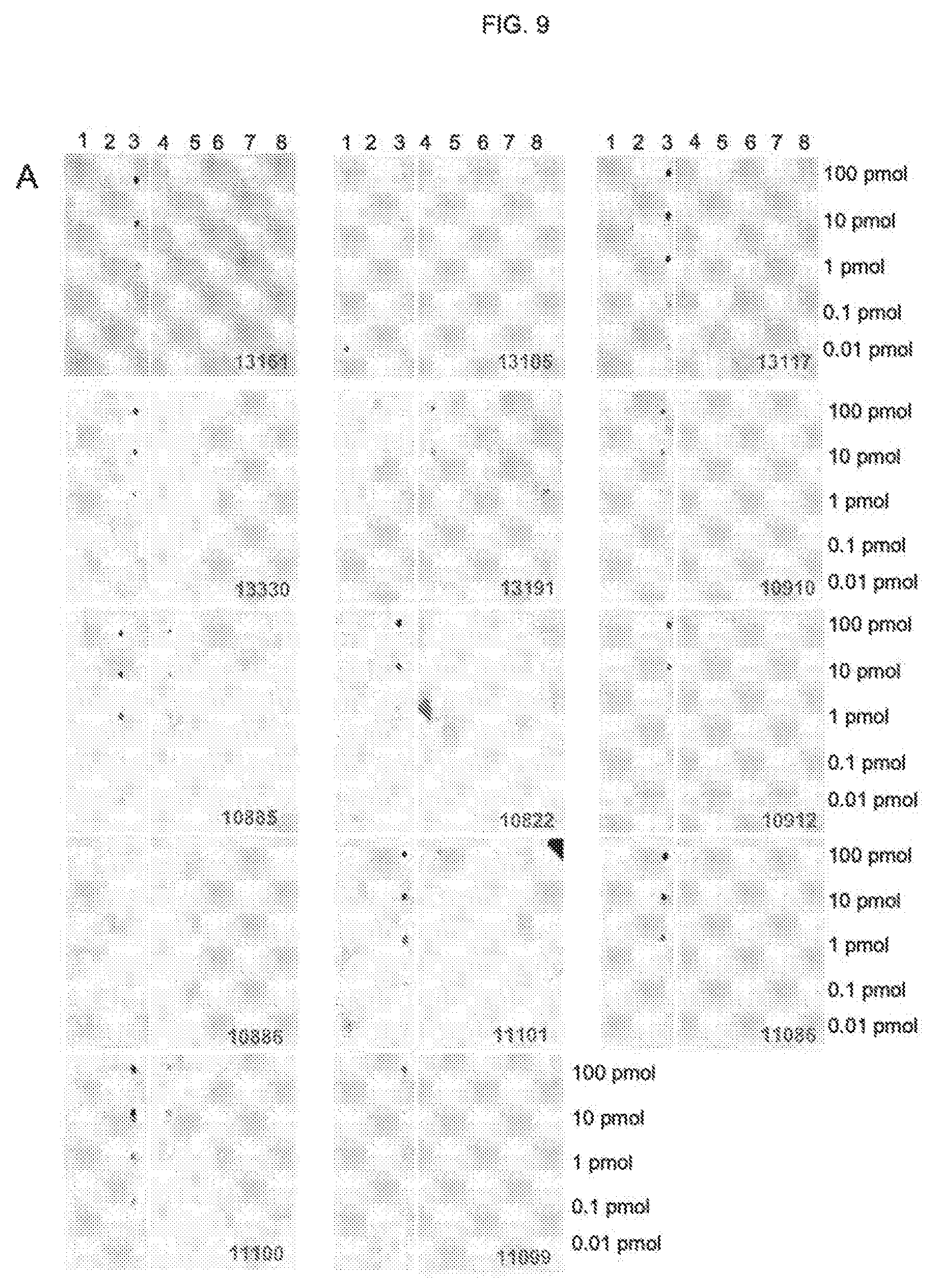
D00015

D00016

D00017

D00018

D00019

D00020

D00021
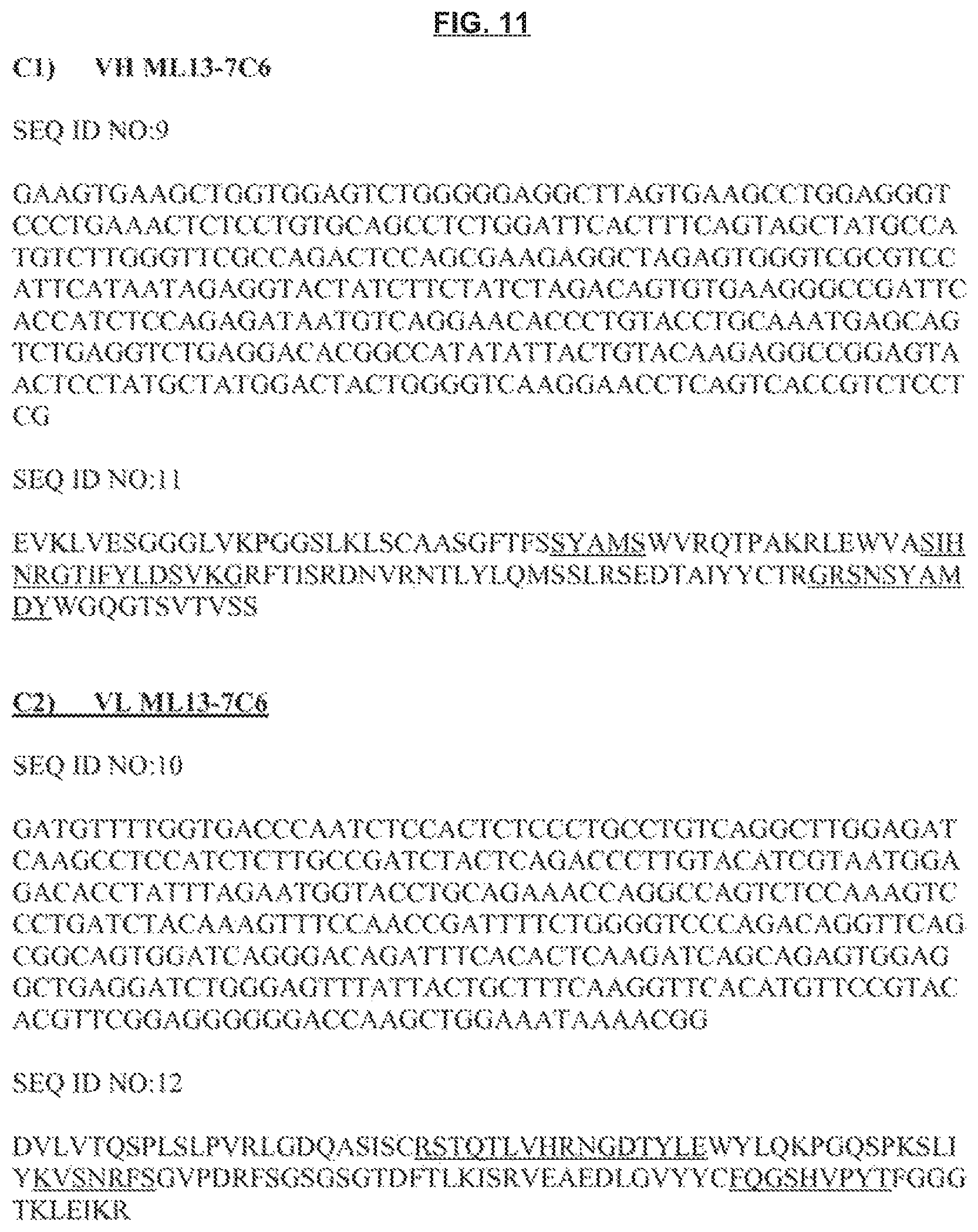
D00022

D00023

D00024
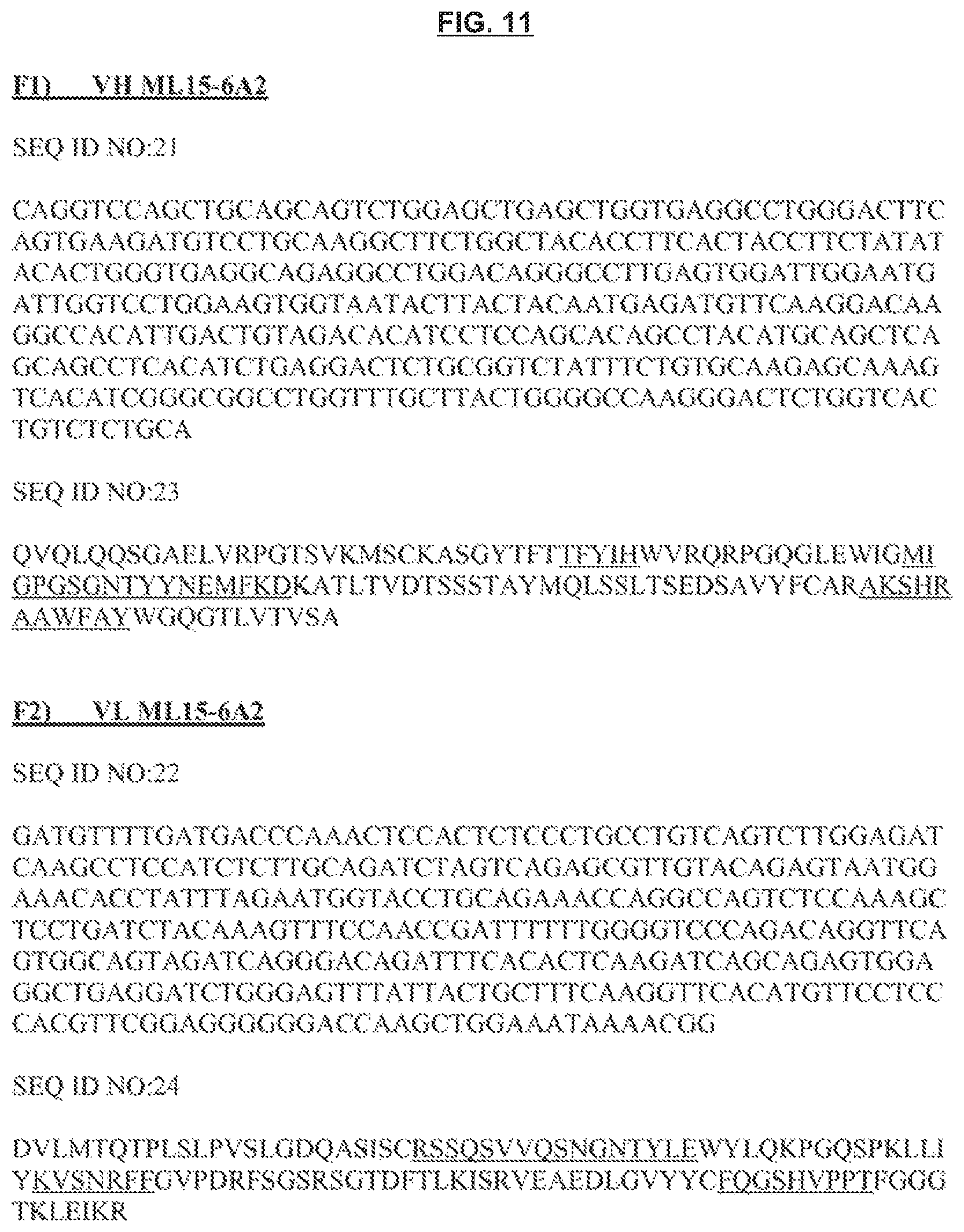
D00025

D00026

D00027

D00028

S00001
XML
uspto.report is an independent third-party trademark research tool that is not affiliated, endorsed, or sponsored by the United States Patent and Trademark Office (USPTO) or any other governmental organization. The information provided by uspto.report is based on publicly available data at the time of writing and is intended for informational purposes only.
While we strive to provide accurate and up-to-date information, we do not guarantee the accuracy, completeness, reliability, or suitability of the information displayed on this site. The use of this site is at your own risk. Any reliance you place on such information is therefore strictly at your own risk.
All official trademark data, including owner information, should be verified by visiting the official USPTO website at www.uspto.gov. This site is not intended to replace professional legal advice and should not be used as a substitute for consulting with a legal professional who is knowledgeable about trademark law.