Microparticles, methods for their preparation and use
Mooney , et al. Nov
U.S. patent number 10,471,016 [Application Number 15/915,686] was granted by the patent office on 2019-11-12 for microparticles, methods for their preparation and use. This patent grant is currently assigned to President and Fellows of Harvard College. The grantee listed for this patent is President and Fellows of Harvard College. Invention is credited to Esther Amstad, Angelo S. Mao, David J. Mooney, Raluca Ostafe, Radivoje Prodanovich, Stefanie Utech, Huanan Wang, David A. Weitz, Connie Chang Wilking.


| United States Patent | 10,471,016 |
| Mooney , et al. | November 12, 2019 |
Microparticles, methods for their preparation and use
Abstract
The invention relates to microparticles comprising a crosslinked gel and methods for making and using same.
| Inventors: | Mooney; David J. (Sudbury, MA), Weitz; David A. (Bolton, MA), Utech; Stefanie (Erlangen, DE), Prodanovich; Radivoje (Belgrade, RS), Amstad; Esther (Lausanne, CH), Ostafe; Raluca (Aachen, DE), Mao; Angelo S. (Somerville, MA), Wilking; Connie Chang (Bozeman, MT), Wang; Huanan (Dalian, CN) | ||||||||||
|---|---|---|---|---|---|---|---|---|---|---|---|
| Applicant: |
|
||||||||||
| Assignee: | President and Fellows of Harvard
College (Cambridge, MA) |
||||||||||
| Family ID: | 53041993 | ||||||||||
| Appl. No.: | 15/915,686 | ||||||||||
| Filed: | March 8, 2018 |
Prior Publication Data
| Document Identifier | Publication Date | |
|---|---|---|
| US 20180214385 A1 | Aug 2, 2018 | |
Related U.S. Patent Documents
| Application Number | Filing Date | Patent Number | Issue Date | ||
|---|---|---|---|---|---|
| 15035167 | |||||
| PCT/US2014/063846 | Nov 4, 2014 | ||||
| 61901949 | Nov 8, 2013 | ||||
| Current U.S. Class: | 1/1 |
| Current CPC Class: | A61K 9/1652 (20130101); A61K 9/5036 (20130101); A61K 35/00 (20130101); A61K 9/5089 (20130101) |
| Current International Class: | A61K 9/50 (20060101); A61K 35/00 (20060101); A61K 9/16 (20060101) |
References Cited [Referenced By]
U.S. Patent Documents
| 4732930 | March 1988 | Tanaka et al. |
| 4743507 | May 1988 | Franses et al. |
| 4996265 | February 1991 | Okubo et al. |
| 5055390 | October 1991 | Weaver et al. |
| 5100933 | March 1992 | Tanaka et al. |
| 5120349 | June 1992 | Stewart et al. |
| 5149625 | September 1992 | Church et al. |
| 5202231 | April 1993 | Drmanac et al. |
| 5216096 | June 1993 | Hattori et al. |
| 5326692 | July 1994 | Brinkley et al. |
| 5436130 | July 1995 | Mathies et al. |
| 5500223 | March 1996 | Behan et al. |
| 5512131 | April 1996 | Kumar et al. |
| 5695940 | December 1997 | Drmanac et al. |
| 5736330 | April 1998 | Fulton |
| 5834252 | November 1998 | Stemmer et al. |
| 5849055 | December 1998 | Arai et al. |
| 5851769 | December 1998 | Gray et al. |
| 6046003 | April 2000 | Mandecki |
| 6051377 | April 2000 | Mandecki |
| 6057107 | May 2000 | Fulton |
| 6103537 | August 2000 | Ullman et al. |
| 6297006 | October 2001 | Drmanac et al. |
| 6297017 | October 2001 | Schmidt et al. |
| 6355198 | March 2002 | Kim et al. |
| 6361950 | March 2002 | Mandecki |
| 6380297 | April 2002 | Zion et al. |
| 6432360 | August 2002 | Church |
| 6485944 | November 2002 | Church et al. |
| 6500447 | December 2002 | Dexter |
| 6511803 | January 2003 | Church et al. |
| 6524456 | February 2003 | Ramsey et al. |
| 6586176 | July 2003 | Trnovsky et al. |
| 6632606 | October 2003 | Ullman et al. |
| 6670133 | December 2003 | Knapp et al. |
| 6767731 | July 2004 | Hannah |
| 6800298 | October 2004 | Burdick et al. |
| 6806058 | October 2004 | Jesperson et al. |
| 6913935 | July 2005 | Thomas |
| 6929859 | August 2005 | Chandler et al. |
| 7041481 | May 2006 | Anderson et al. |
| 7268167 | September 2007 | Higuchi et al. |
| 7425431 | September 2008 | Church et al. |
| 7536928 | May 2009 | Kazuno |
| 7604938 | October 2009 | Takahashi et al. |
| 7638276 | December 2009 | Griffiths et al. |
| 7708949 | May 2010 | Stone et al. |
| 7776927 | August 2010 | Chu et al. |
| RE41780 | September 2010 | Anderson et al. |
| 7799553 | September 2010 | Mathies et al. |
| 7968287 | June 2011 | Griffiths et al. |
| 8252539 | August 2012 | Quake et al. |
| 8273573 | September 2012 | Ismagilov et al. |
| 8278071 | October 2012 | Brown et al. |
| 8329407 | December 2012 | Ismagilov et al. |
| 8748094 | June 2014 | Weitz et al. |
| 8748102 | June 2014 | Berka et al. |
| 8765380 | July 2014 | Berka et al. |
| 8871444 | October 2014 | Griffiths et al. |
| 9017948 | April 2015 | Agresti et al. |
| 9029085 | May 2015 | Agresti et al. |
| 9039273 | May 2015 | Weitz et al. |
| 9068210 | June 2015 | Agresti et al. |
| 9816121 | June 2017 | Agresti et al. |
| 9718044 | August 2017 | Wesner et al. |
| 9850526 | December 2017 | Agresti et al. |
| 10221437 | March 2019 | Weitz et al. |
| 2001/0020588 | September 2001 | Adourian et al. |
| 2001/0044109 | November 2001 | Mandecki |
| 2002/0034737 | March 2002 | Drmanac |
| 2002/0034747 | March 2002 | Bruchez et al. |
| 2002/0051992 | May 2002 | Bridgham et al. |
| 2002/0092767 | July 2002 | Bjornson et al. |
| 2002/0179849 | December 2002 | Maher et al. |
| 2003/0008285 | January 2003 | Fischer |
| 2003/0008323 | January 2003 | Ravkin et al. |
| 2003/0028981 | February 2003 | Chandler et al. |
| 2003/0039978 | February 2003 | Hannah |
| 2003/0044777 | March 2003 | Beattie |
| 2003/0044836 | March 2003 | Levine et al. |
| 2003/0099954 | May 2003 | Miltenyi et al. |
| 2003/0104466 | June 2003 | Knapp et al. |
| 2003/0108897 | June 2003 | Drmanac |
| 2003/0170698 | September 2003 | Gascoyne et al. |
| 2003/0182068 | September 2003 | Battersby et al. |
| 2003/0207260 | November 2003 | Trnovsky et al. |
| 2003/0215862 | November 2003 | Parce et al. |
| 2004/0063138 | April 2004 | McGinnis et al. |
| 2004/0096515 | May 2004 | Bausch et al. |
| 2004/0132122 | July 2004 | Banerjee et al. |
| 2004/0253613 | December 2004 | Taylor et al. |
| 2005/0019839 | January 2005 | Jesperson et al. |
| 2005/0042625 | February 2005 | Schmidt et al. |
| 2005/0123614 | June 2005 | Kim et al. |
| 2005/0136486 | June 2005 | Haushalter |
| 2005/0172476 | August 2005 | Stone et al. |
| 2005/0181379 | August 2005 | Su et al. |
| 2005/0221339 | October 2005 | Griffiths et al. |
| 2005/0244850 | November 2005 | Huang et al. |
| 2005/0287572 | December 2005 | Mathies et al. |
| 2006/0020371 | January 2006 | Ham et al. |
| 2006/0073487 | April 2006 | Oliver et al. |
| 2006/0078888 | April 2006 | Griffiths et al. |
| 2006/0078893 | April 2006 | Griffiths et al. |
| 2006/0153924 | July 2006 | Griffiths et al. |
| 2006/0163385 | July 2006 | Link et al. |
| 2006/0240506 | October 2006 | Kushmaro et al. |
| 2006/0257893 | November 2006 | Takahashi et al. |
| 2006/0292583 | December 2006 | Schneider et al. |
| 2007/0003442 | January 2007 | Link et al. |
| 2007/0020617 | January 2007 | Trnovsky et al. |
| 2007/0054119 | March 2007 | Garstecki et al. |
| 2007/0092914 | April 2007 | Griffiths et al. |
| 2007/0172827 | July 2007 | Murakami |
| 2007/0172873 | July 2007 | Brenner et al. |
| 2007/0195127 | August 2007 | Ahn et al. |
| 2007/0228588 | October 2007 | Noritomi et al. |
| 2007/0264320 | November 2007 | Lee et al. |
| 2008/0003142 | January 2008 | Link et al. |
| 2008/0004436 | January 2008 | Tawfik et al. |
| 2008/0014589 | January 2008 | Link et al. |
| 2009/0012187 | January 2009 | Chu et al. |
| 2009/0035770 | February 2009 | Mathies et al. |
| 2009/0068170 | March 2009 | Weitz et al. |
| 2009/0131543 | May 2009 | Weitz et al. |
| 2009/0134027 | May 2009 | Jary |
| 2009/0191276 | July 2009 | Kim et al. |
| 2009/0197772 | August 2009 | Griffiths et al. |
| 2009/0205829 | August 2009 | Sullivan et al. |
| 2009/0286687 | November 2009 | Dressman et al. |
| 2010/0022414 | January 2010 | Link et al. |
| 2010/0130369 | May 2010 | Shenderov et al. |
| 2010/0136544 | June 2010 | Agresti et al. |
| 2010/0137163 | June 2010 | Link et al. |
| 2010/0173394 | July 2010 | Colston, Jr. et al. |
| 2010/0210479 | August 2010 | Griffiths et al. |
| 2010/0213628 | August 2010 | Bausch et al. |
| 2010/0273219 | October 2010 | May et al. |
| 2011/0033854 | February 2011 | Drmanac et al. |
| 2011/0086780 | April 2011 | Colston et al. |
| 2011/0092392 | April 2011 | Colston et al. |
| 2011/0160078 | June 2011 | Fodor et al. |
| 2011/0218123 | September 2011 | Weitz et al. |
| 2011/0229545 | September 2011 | Shum et al. |
| 2011/0267457 | November 2011 | Weitz et al. |
| 2011/0305761 | December 2011 | Shum et al. |
| 2012/0003321 | January 2012 | Peng et al. |
| 2012/0010107 | January 2012 | Griffiths et al. |
| 2012/0015382 | January 2012 | Weitz et al. |
| 2012/0015822 | January 2012 | Weitz et al. |
| 2012/0076860 | March 2012 | Trout et al. |
| 2012/0135407 | May 2012 | Slatter |
| 2012/0190032 | July 2012 | Ness et al. |
| 2012/0199226 | August 2012 | Weitz et al. |
| 2012/0211084 | August 2012 | Weitz et al. |
| 2012/0220494 | August 2012 | Samuels et al. |
| 2012/0220497 | August 2012 | Jacobson et al. |
| 2012/0222748 | September 2012 | Weitz et al. |
| 2013/0004522 | January 2013 | Dvir et al. |
| 2013/0046030 | February 2013 | Rotem et al. |
| 2013/0064862 | March 2013 | Weitz |
| 2013/0079231 | March 2013 | Pushkarev et al. |
| 2013/0109575 | May 2013 | Kleinschmidt et al. |
| 2013/0157899 | June 2013 | Adler et al. |
| 2013/0210639 | August 2013 | Link et al. |
| 2013/0274117 | October 2013 | Church et al. |
| 2014/0065234 | March 2014 | Shum et al. |
| 2014/0155295 | June 2014 | Hindson et al. |
| 2014/0199730 | July 2014 | Agresti et al. |
| 2014/0199731 | July 2014 | Agresti et al. |
| 2014/0220350 | August 2014 | Kim et al. |
| 2014/0227684 | August 2014 | Hindson et al. |
| 2014/0235506 | August 2014 | Hindson et al. |
| 2014/0303039 | October 2014 | Weitz et al. |
| 2014/0378349 | December 2014 | Hindson et al. |
| 2015/0005200 | January 2015 | Hindson et al. |
| 2015/0314292 | November 2015 | Weitz et al. |
| 2015/0336068 | November 2015 | Weitz et al. |
| 2015/0336069 | November 2015 | Weitz et al. |
| 2015/0336070 | November 2015 | Weitz et al. |
| 2015/0336071 | November 2015 | Weitz et al. |
| 2015/0336072 | November 2015 | Weitz et al. |
| 2015/0337371 | November 2015 | Weitz et al. |
| 2015/0353999 | December 2015 | Agresti et al. |
| 2016/0279068 | September 2016 | Utech et al. |
| 2017/0183701 | June 2017 | Agresti et al. |
| 2017/0224849 | August 2017 | Carroll et al. |
| 2017/0319443 | December 2017 | Weitz et al. |
| 2018/0023109 | January 2018 | Weitz et al. |
| 2018/0119212 | May 2018 | Weitz et al. |
| 2018/0171373 | June 2018 | Weitz et al. |
| 2018/0296488 | October 2018 | Weitz et al. |
| 0 249 007 | Dec 1987 | EP | |||
| 0 272 659 | Jun 1988 | EP | |||
| 0 478 326 | Apr 1992 | EP | |||
| 1 019 496 | Sep 2004 | EP | |||
| 1 482 036 | Oct 2007 | EP | |||
| 1 594 980 | Nov 2009 | EP | |||
| 1 967 592 | Apr 2010 | EP | |||
| 2 258 846 | Dec 2010 | EP | |||
| 2 145 955 | Feb 2012 | EP | |||
| 1 905 828 | Aug 2012 | EP | |||
| 1 908 832 | Dec 2012 | EP | |||
| 2 540 389 | Jan 2013 | EP | |||
| S59-049832 | Mar 1984 | JP | |||
| 2004-361291 | Dec 2004 | JP | |||
| 2006-507921 | Mar 2006 | JP | |||
| 2006-289250 | Oct 2006 | JP | |||
| 2007-268350 | Oct 2007 | JP | |||
| 2007-298327 | Nov 2007 | JP | |||
| 2009-208074 | Sep 2009 | JP | |||
| 2014/0107381 | Sep 2014 | KR | |||
| WO 95/09613 | Apr 1995 | WO | |||
| WO 96/29629 | Sep 1996 | WO | |||
| WO 96/41011 | Dec 1996 | WO | |||
| WO 99/09217 | Feb 1999 | WO | |||
| WO 99/52708 | Oct 1999 | WO | |||
| WO 00/08212 | Feb 2000 | WO | |||
| WO 00/26412 | May 2000 | WO | |||
| WO 01/14589 | Mar 2001 | WO | |||
| WO 01/85138 | Nov 2001 | WO | |||
| WO 01/89787 | Nov 2001 | WO | |||
| WO 02/31203 | Apr 2002 | WO | |||
| WO 02/047665 | Jun 2002 | WO | |||
| WO 02/086148 | Oct 2002 | WO | |||
| WO 03/028653 | Apr 2003 | WO | |||
| WO 2004/002627 | Jan 2004 | WO | |||
| WO 2004/091763 | Oct 2004 | WO | |||
| WO 2004/102204 | Nov 2004 | WO | |||
| WO 2004/103565 | Dec 2004 | WO | |||
| WO 2004/105734 | Dec 2004 | WO | |||
| WO 2005/021151 | Mar 2005 | WO | |||
| WO 2005/040406 | May 2005 | WO | |||
| WO 2005/041884 | May 2005 | WO | |||
| WO 2005/049787 | Jun 2005 | WO | |||
| WO 2005/082098 | Sep 2005 | WO | |||
| WO 2005/084210 | Sep 2005 | WO | |||
| WO 2005/103106 | Nov 2005 | WO | |||
| WO 2006/078841 | Jul 2006 | WO | |||
| WO 2006/096571 | Sep 2006 | WO | |||
| WO 2007/001448 | Jan 2007 | WO | |||
| WO 2007/002490 | Jan 2007 | WO | |||
| WO 2007/012638 | Feb 2007 | WO | |||
| WO 2007/024840 | Mar 2007 | WO | |||
| WO 2007/081385 | Jul 2007 | WO | |||
| WO 2007/081387 | Jul 2007 | WO | |||
| WO 2007/089541 | Aug 2007 | WO | |||
| WO 2007/114794 | Oct 2007 | WO | |||
| WO 2007/121489 | Oct 2007 | WO | |||
| WO 2007/133710 | Nov 2007 | WO | |||
| WO 2007/133807 | Nov 2007 | WO | |||
| WO 2007/138178 | Dec 2007 | WO | |||
| WO 2007/139766 | Dec 2007 | WO | |||
| WO 2007/140015 | Dec 2007 | WO | |||
| WO 2007/149432 | Dec 2007 | WO | |||
| WO 2008/021123 | Feb 2008 | WO | |||
| WO 2008/058297 | May 2008 | WO | |||
| WO 2008/091792 | Jul 2008 | WO | |||
| WO 2008/102057 | Aug 2008 | WO | |||
| WO 2008/109176 | Sep 2008 | WO | |||
| WO 2008/121342 | Oct 2008 | WO | |||
| WO 2008/134153 | Nov 2008 | WO | |||
| WO 2009/005680 | Jan 2009 | WO | |||
| WO 2009/011808 | Jan 2009 | WO | |||
| WO 2009/085215 | Jul 2009 | WO | |||
| WO 2009/120254 | Oct 2009 | WO | |||
| WO 2009/148598 | Dec 2009 | WO | |||
| WO 2010/104604 | Sep 2010 | WO | |||
| WO 2010/151776 | Dec 2010 | WO | |||
| WO 2011/028760 | Mar 2011 | WO | |||
| WO 2011/028764 | Mar 2011 | WO | |||
| WO 2011/056546 | May 2011 | WO | |||
| WO 2011/116154 | Sep 2011 | WO | |||
| WO 2012/048341 | Apr 2012 | WO | |||
| WO 2012/156744 | Nov 2012 | WO | |||
| WO 2012/162296 | Nov 2012 | WO | |||
| WO 2013/006661 | Jan 2013 | WO | |||
| WO 2013/032709 | Mar 2013 | WO | |||
| WO 2013/083760 | Jun 2013 | WO | |||
| WO 2013/177220 | Nov 2013 | WO | |||
| WO 2015/069634 | May 2015 | WO | |||
| WO 2015/160919 | Oct 2015 | WO | |||
| WO 2016/085739 | Jun 2016 | WO | |||
| WO 2017/066231 | Apr 2017 | WO | |||
Other References
|
US. Appl. No. 15/670,977, filed Aug. 7, 2017, Weitz et al. cited by applicant . U.S. Appl. No. 15/792,218, filed Oct. 24, 2017, Weitz et al. cited by applicant . U.S. Appl. No. 15/884,215, filed Jan. 30, 2018, Weitz et al. cited by applicant . U.S. Appl. No. 13/119,470, filed May 4, 2011, Weitz et al. cited by applicant . U.S. Appl. No. 14/812,930, filed Jul. 29, 2015, Weitz et al. cited by applicant . U.S. Appl. No. 14/812,942, filed Jul. 29, 2015, Weitz et al. cited by applicant . U.S. Appl. No. 14/812,951, filed Jul. 29, 2015, Weitz et al. cited by applicant . U.S. Appl. No. 14/812,946, filed Jul. 29, 2015, Weitz et al. cited by applicant . U.S. Appl. No. 14/812,954, filed Jul. 29, 2015, Weitz et al. cited by applicant . U.S. Appl. No. 14/812,964, dated Jul. 29, 2015, Weitz et al. cited by applicant . U.S. Appl. No. 15/528,905, filed May 23, 2017, Weitz et al. cited by applicant . U.S. Appl. No. 15/035,167, filed May 6, 2016, Weitz et al. cited by applicant . PCT/US2008/003185, Oct. 22, 2008, Invitation to Pay Additional Fees. cited by applicant . U.S. Appl. No. 15/768,135, filed Apr. 13, 2018, Weitz et al. cited by applicant . PCT/US2018/047053, Oct. 23, 2018, International Search Report and Written Opinion. cited by applicant . PCT/US2008/003185, Jan. 12, 2009, International Search Report and Written Opinion. cited by applicant . PCT/US2008/003185, Sep. 17, 2009, International Preliminary Report on Patentability. cited by applicant . PCT/US2008/008563, Oct. 29, 2008, International Search Report and Written Opinion. cited by applicant . CN 200880127116.4, Jun. 18, 2012, Chinese Office Action. cited by applicant . CN 200880127116.4, May 23, 2013, Chinese Office Action. cited by applicant . CN 200880127116.4, Dec. 24, 2013, Chinese Office Action. cited by applicant . EP 08865992.5, Dec. 15, 2010, European Office Communication. cited by applicant . EP 08865992.5, Jan. 23, 2012, European Office Communication. cited by applicant . EP 08865992.5, Apr. 5, 2013, European Office Communication. cited by applicant . EP 08865992.5, Aug. 29, 2013, European Office Communication. cited by applicant . EP 08865992.5, Apr. 29, 2014, European Office Communication. cited by applicant . JP 2010-539498, Jul. 17, 2013, Japanese Office Action. cited by applicant . JP 2010-539498, Sep. 2, 2014, Japanese Office Action. cited by applicant . PCT/US2008/013912, Apr. 3, 2009, International Search Report and Written Opinion. cited by applicant . PCT/US2008/013912, Jul. 1, 2010, International Preliminary Report on Patentability. cited by applicant . PCT/US2009/005184, May 27, 2010, Invitation to Pay Additional Fees. cited by applicant . PCT/US2009/005184, Aug. 16, 2010, International Search Report and Written Opinion. cited by applicant . PCT/US2009/005184, Mar. 31, 2011, International Preliminary Report on Patentability. cited by applicant . EP 09758762.0, Aug. 13, 2015, European Office Action. cited by applicant . EP 9758762.0, Sep. 29, 2016, European Office Action. cited by applicant . KR 10-2011-7000094, Feb. 27, 2013, Office Action. cited by applicant . PCT/US2009/003389, Oct. 21, 2009, International Search Report and Written Opinion. cited by applicant . PCT/US2009/003389, Dec. 16, 2010, International Preliminary Report on Patentability. cited by applicant . PCT/US2009/004037, Oct. 2, 2009, International Search Report and Written Opinion. cited by applicant . EP 9804166.8, Nov. 7, 2014, European Office Action. cited by applicant . PCT/US2009/006649, Mar. 10, 2010, International Search Report and Written Opinion. cited by applicant . PCT/US2009/006649, Jun. 30, 2011, International Preliminary Report on Patentability. cited by applicant . AU 2010315580, Dec. 17, 2013, Australian Office Action. cited by applicant . CN 201080055990.9, Dec. 16, 2013, Chinese Office Action. cited by applicant . CN 201080055990.9, Jul. 30, 2014, Chinese Office Action. cited by applicant . JP 2012-536941, Nov. 19, 2013, Japanese Office Action. cited by applicant . JP 2012-536941, Aug. 5, 2014, Japanese Office Action. cited by applicant . PCT/US2010/054050, Jan. 31, 2011, International Search Report and Written Opinion. cited by applicant . PCT/US2010/054050, May 10, 2012, International Preliminary Report on Patentability. cited by applicant . PCT/US2016/056509, Jan. 10, 2017, International Search Report and Written Opinion. cited by applicant . EP 14860623.9, May 23, 2017, Extended European Search Report. cited by applicant . PCT/US2014/063846, Jan. 7, 2015, Invitation to Pay Additional Fees and Partial Search Report. cited by applicant . PCT/US2014/063846, Mar. 10, 2015, International Search Report and Written Opinion. cited by applicant . PCT/US2014/063846, May 19, 2016, International Preliminary Report on Patentability. cited by applicant . International Search Report and Written Opinion for Application No. PCT/US2018/047053 dated Oct. 23, 2018. cited by applicant . Extended European Search Report for Application No. EP 16856059.7 dated Apr. 1, 2019. cited by applicant . Office Action for U.S. Appl. No. 13/119,470 dated Apr. 24, 2013. cited by applicant . Final Office Action for U.S. Appl. No. 13/119,470 dated Dec. 5, 2013. cited by applicant . Advisory Action for U.S. Appl. No. 13/119,470 dated Mar. 21, 2014. cited by applicant . Office Action dated Jun. 24, 2015 for U.S. Appl. No. 13/119,470. cited by applicant . Invitation to Pay Additional Fees for PCT/US2008/003185 dated Oct. 22, 2008. cited by applicant . International Search Report and Written Opinion of the International Searching Authority for International Application No. PCT/US2008/003185, dated Jan. 12, 2009. cited by applicant . International Preliminary Report on Patentability for PCT/US2008/003185 dated Sep. 17, 2009. cited by applicant . International Search Report and Written Opinion of the International Searching Authority for International Application No. PCT/US2008/008563, dated Oct. 29, 2008. cited by applicant . Chinese Office Action dated Jun. 18, 2012 for CN Application No. 200880127116.4. cited by applicant . Chinese Office Action dated May 23, 2013 for Application No. CN 200880127116.4. cited by applicant . Chinese Office Action dated Dec. 24, 2013 for CN Application No. 200880127116.4. cited by applicant . Office Communication dated Dec. 15, 2010 for Application No. EP 08865992.5. cited by applicant . Office Communication dated Jan. 23, 2012 for Application No. EP 08865992.5. cited by applicant . Office Communication dated Apr. 5, 2013 for Application No. EP 08865992.5. cited by applicant . Office Communication dated Aug. 29, 2013 for Application No. EP 08865992.5. cited by applicant . Office Communication dated Apr. 29, 2014 for EP Application No. EP 08865992.5. cited by applicant . Japanese Office Action dated Jul. 17, 2013 for Application No. JP 2010-539498. cited by applicant . Japanese Office Action dated Sep. 2, 2014 for Application No. JP 2010-539498. cited by applicant . International Search Report and Written Opinion of the International Searching Authority for International Application No. PCT/US2008/013912, dated Apr. 3, 2009. cited by applicant . International Preliminary Report on Patentability for PCT/US2008/013912 dated Jul. 1, 2010. cited by applicant . Invitation to Pay Additional Fees for PCT Application PCT/US09/005184 dated May 27, 2010. cited by applicant . International Search Report and Written Opinion from PCT Application PCT/US09/005184 dated Aug. 16, 2010. cited by applicant . International Preliminary Report on Patentability for PCT Application PCT/US09/005184 dated Mar. 31, 2011. cited by applicant . European Office Action for Application No. EP 09758762.0 dated Aug. 13, 2015. cited by applicant . European Office Action for Application No. 09758762.0 dated Sep. 29, 2016. cited by applicant . Korean Office Action for Application No. KR 10-2011-7000094 dated Feb. 27, 2013. cited by applicant . International Search Report and Written Opinion for International Application No. PCT/US09/003389 dated Oct. 21, 2009. cited by applicant . International Preliminary Report on Patentability for International Application No. PCT/US2009/003389 dated Dec. 16, 2010. cited by applicant . International Search Report and Written Opinion of the International Searching Authority for International Application No. PCT/US2009/004037, dated Oct. 2, 2009. cited by applicant . European Office action dated Nov. 7, 2014 for Application No. EP 09804166.8. cited by applicant . International Search Report and Written Opinion for International Application No. PCT/US2009/006649 dated Mar. 10, 2010. cited by applicant . International Preliminary Report on Patentability for International Application No. PCT/US2009/006649 dated Jun. 30, 2011. cited by applicant . Australian Office Action dated Dec. 17, 2013 for Application No. AU 2010315580. cited by applicant . Chinese Office Action dated Dec. 16, 2013 for Application No. CN 201080055990.9. cited by applicant . Chinese Office Action dated Jul. 30, 2014 for Application No. CN 201080055990.9. cited by applicant . Japanese Office Action dated Nov. 19, 2013 for Application No. JP 2012-536941. cited by applicant . Japanese Office Action dated Aug. 5, 2014 for Application No. JP 2012-536941. cited by applicant . International Search Report and Written Opinion from PCT Application PCT/US2010/054050 dated Jan. 31, 2011. cited by applicant . International Preliminary Report on Patentability from PCT Application PCT/US2010/054050 dated May 10, 2012. cited by applicant . International Search Report and Written Opinion dated Jan. 10, 2017 for Application No. PCT/US2016/056509. cited by applicant . Extended European Search Report for Application No. EP 14860623.9 dated May 23, 2017. cited by applicant . International Search Report and Written Opinion from PCT/US2014/063846 dated Mar. 10, 2015. cited by applicant . Invitation to Pay Additional Fees and Partial Search Report for Application No. PCT/US2014/063846 dated Jan. 7, 2015. cited by applicant . International Preliminary Report on Patentability from PCT Application PCT/US2014/063846 dated May 19, 2016. cited by applicant . Office Action for U.S. Appl. No. 13/119,470 dated Feb. 1, 2018. cited by applicant . Office Action for U.S. Appl. No. 14/812,930 dated Nov. 13, 2017. cited by applicant . Office Action for U.S. Appl. No. 14/812,942 dated Feb. 1, 2018. cited by applicant . Office Action for U.S. Appl. No. 14/812,951 dated Jan. 25, 2018. cited by applicant . Office Action for U.S. Appl. No. 14/812,946 dated Jan. 25, 2018. cited by applicant . Office Communication for U.S. Appl. No. 14/812,954 dated Dec. 7, 2017. cited by applicant . Office Action for U.S. Appl. No. 14/812,964 dated Jan. 25, 2018. cited by applicant . Office Action for U.S. Appl. No. 15/035,167 dated May 16, 2017. cited by applicant . Office Action for U.S. Appl. No. 15/035,167 dated Nov. 27, 2017. cited by applicant . [No Author Listed], Toxnet, Toxicology Data Network. Vinyl Toluene. National Library of Medicine. 2015:1-38. cited by applicant . [No Author] Gene Characterization Kits. Stratagene Catalog. Statagene Cloning Systems: Tools and Technology for Lift Sciences. 1988. 3 pages. cited by applicant . [No Author] Microfluidic ChipShop. Microfluidic product catalogue. Mar. 2005. cited by applicant . [No Author] Microfluidic ChipShop. Microfluidic product catalogue. Oct. 2009. cited by applicant . Abate et al., Droplet Based Sequencing. American Physical Society. Presentation. Mar. 12, 2008. 25 pages. cited by applicant . Abate et al., Valve-based flow focusing for drog formation. Appl Phys Lett. 2009;94. 3 pages. (Month not cited on publication). cited by applicant . Abate et al., High-throughput injection with microfluidics using picoinjectors. Proc Natl Acad Sci U S A. Nov. 9, 2010;107(45):19163-6. doi: 10.1073/pnas.1006888107. Epub Oct. 20, 2010. cited by applicant . Adams et al., Entropically driven microphase transitions in mixtures of colloidal rods and spheres. Nature. May 28, 1998:393:349-52. cited by applicant . Agresti, "Selection of ribozymes that catalyse multiple-turnover Diels-Alder cycloadditions by using in vitro compartmentalization", PNAS, 102, 16170-16175 (2005). (Nov. 2005). cited by applicant . Agresti et al., Ultrahigh-throughput screening in drop-based microfluidics for directed evolution. Proc Natl Acad Sci U S A. Mar. 2, 2010;107(9):4004-9. doi: 10.1073/pnas.0910781107. Epub Feb. 8, 2010. Erratum in: Proc Natl Acad Sci U S A. Apr. 6, 2010;107(14):6550. cited by applicant . Akselband, "Enrichment of slow-growing marine microorganisms from mixed cultures using gel microdrop (GMD) growth assay and fluorescence-activated cell sorting", J. Exp. Marine Biol., 329: 196-205 (2006). (Month not cited on publication). cited by applicant . Akselband, "Rapid mycobacteria drug susceptibility testing using gel microdrop (GMD) growth assay and flow cytometry", J. Microbiol. Methods, 62:181-197 (2005). (Month not cited on publication). cited by applicant . Anna et al., Formation of dispersions using `flow focusing` in microchannels. Appln Phys Letts. 2003;82(3):364-66. (Jan. 2003). cited by applicant . Baret et al., Fluorescence-activated droplet sorting (FADS): efficient microfluidic cell sorting based on enzymatic activity. Lab Chip. Jul. 7, 2009;9(13):1850-8. doi: 10.1039/b902504a. Epub Apr. 23, 2009. cited by applicant . Boone, et al. Plastic advances microfluidic devices. The devices debuted in silicon and glass, but plastic fabrication may make them hugely successful in biotechnology application. Analytical Chemistry. Feb. 2002; 78A-86A. cited by applicant . Braeckmans et al., Scanning the Code. Modern Drug Discovery. 2003:28-32. (Feb. 2003). cited by applicant . Carroll, "The selection of high-producing cell lines using flow cytometry and cell sorting", Exp. Op. Biol. Therp., 4:11 1821-1829 (2004). (Month not cited on publication). cited by applicant . Chaudhary "A rapid method of cloning functional variable-region antibody genese in Escherichia coli as single-chain immunotoxins" Proc. Natl. Acad. Sci USA 87: 1066-1070 (Feb. 1990). cited by applicant . Chechetkin et al., Sequencing by hybridization with the generic 6-mer oligonucleotide microarray: an advanced scheme for data processing. J Biomol Struct Dyn. Aug. 2000;18(1):83-101. (Month not cited on publication). cited by applicant . Chou, et al. Disposable Microdevices for DNA Analysis and Cell Sorting. Proc. Solid-State Sensor and Actuator Workshop, Hilton Head, SC. Jun. 8-11, 1998; 11-14. cited by applicant . Chu, L., et al., "Controllable Monodisperse Multiple Emulsions," Angew. Chem. Int. Ed., vol. 46, pp. 8970-8974 (2007). (Month not cited on publication). cited by applicant . Clausell-Tormos et al., Droplet-based microfluidic platforms for the encapsulation and screening of Mammalian cells and multicellular organisms. Chem Biol. May 2008;15(5):427-37. doi: 10.1016/j.chembio1.2008.04.004. Erratum in: Chem Biol. Aug. 25, 2008;15(8):875. cited by applicant . De Bruin et al., UBS Investment Research. Q-Series.RTM. : DNA Sequencing. UBS Securities LLC. Jul. 12, 2007. 15 pages. cited by applicant . Dendukuri et al. Continuous-flow lithography for high-throughput microparticle synthesis. Nature Mat. May 2006;5:365-69. cited by applicant . Diaz, R.V., et al., "One-Month sustained release microspheres of 125 I-bovine calcitonin In Vitro-in vivo studies," Journal of Controlled Release, vol. 59, pp. 55-62 (1999). (Month not cited on publication). cited by applicant . Doerr, The smallest bioreactor. Nature Methods. 2005; 2(5):326. (May 2005). cited by applicant . Draget et al., Alginate based new materials. Int J Biol Macromol. Aug. 1997;21(1-2):47-55. cited by applicant . Drmanac eta 1., Sequencing by hybridization (SBH): advantages, achievements, and opportunities. Adv Biochem Eng Biotechnol. 2002;77:75-101. (Month not cited on publication). cited by applicant . Durant et al., Effects of cross-linking on the morphology of structured latex particles 1. Theoretical considerations. Macromol. 1996;29:8466-72. Month not cited on publication. cited by applicant . Fu, "A microfabricated fluorescence-activated cell sorter", Nature Biotech., 17:1109-1111 (1999). (Nov. 1999). cited by applicant . Fulton et al., Advanced multiplexed analysis with the FlowMetrix system. Clin Chem. Sep. 1997;43(9):1749-56. cited by applicant . Gartner, et al. The Microfluidic Toolbox--examples for fluidic interfaces and standardization concepts. Proc. SPIE 4982, Microfluidics, BioMEMS, and Medical Microsystems, (Jan. 17, 2003); doi: 10.1117/12.479566. cited by applicant . Ghadessy et al. Directed evolution of polymerase function by compartmentalized self-replication. Proc Natl Acad Sci USA. Apr. 10, 2001; 98(8):4552-7. Epub Mar. 27, 2001. cited by applicant . Gordon et al., Self-assembled polymer membrane capsules inflated by osmotic pressure. JACS. 2004;126:14117-22. Published on web Oct. 12, 2004. cited by applicant . Graham et al., Nanogels and microgels: The new polymeric materials playground. Pure Appl Chem. 1998;70(6):1271-75. Month not cited on publication. cited by applicant . Guo et al., Droplet microfluidics for high-throughput biological assays. Lab Chip. Jun. 21, 2012;12(12):2146-55. doi: 10.1039/c21c21147e. Epub Feb. 9, 2012. cited by applicant . He et al., "Selective Encapsulation of Single Cells and Subcellular Organelles into Picoliter--and Femtoliter--Volume Droplets" Anal. Chem 77: 1539-1544 (2005) (Mar. 2005). cited by applicant . Holtze et al., Biocompatible surfactants for water-in-fluorocarbon emulsions. Lab Chip. Oct. 2008; 8(10):1632-9. cited by applicant . Hsu et al., Self-assembled shells composed of colloidal particles: fabrication and characterization. Langmuir. 2005;21:2963-70. Published on web Feb. 23, 2005. cited by applicant . Huebner, "Quantitative detection of protein expression in single cells using droplet microfluidics", Chem. Commun. 1218-1220 (2007). (Month not cited on publication). cited by applicant . Hug et al. Measurement of the number of molecules of a single mRNA species in a complex mRNA preparation. J Theor Biol. Apr. 21, 2003; 221(4):615-24. cited by applicant . Jogun et al., Rheology and microstructure of dense suspensions of plate-shaped colloidal particles. J. Rheol. Jul./Aug. 1999;43:847-71. cited by applicant . Khatiwala et al., "Intrinsic mechanical properties of the extracellular matrix affect the behaviour of pre-osteoblastic MC3T3-E1 cells" Am. J. Physiol. Cell Physiol. 2006;290:C1640. cited by applicant . Khetani et al., Microscale culture of human liver cells for drug development. Nat Biotechnol. Jan. 2008;26(1):120-6. Epub Nov. 18, 2007. cited by applicant . Khomiakova et al., [Analysis of perfect and mismatched DNA duplexes by a generic hexanucleotide microchip]. Mol Biol (Mosk). Jul.-Aug. 2003;37(4):726-41. Russian. cited by applicant . Kim et al., Colloidal assembly route for responsive colloidsomes with tunable permeability. Nano Lett. 2007;7:2876-80. Published on web Aug. 3, 2007. cited by applicant . Kim et al., Fabrication of monodisperse gel shells and functional microgels in microfluidic devices. Angew Chem Int Ed Engl. Mar. 2007;46(11):1819-22. cited by applicant . Kim et al., Monodisperse nonspherical colloid materials with well-defined structures. Presentation. Sep. 16, 2005. 5 pages. cited by applicant . Kim et al., Synthesis of nonspherical colloidal particles with anisotropic properties. JACS. 2006;128:14374-77. Published on web Oct. 18, 2006. cited by applicant . Kim et al., Uniform nonspherical colloidal particles engineered by geometrically tunable gradient of crosslink density. 80.sup.th ACS Colloid Surf. Sci. Symp. Jun. 20, 2006. 23 pages. cited by applicant . Kim et al., Uniform nonspherical colloidal particles with tunable shapes. Adv. Mater. 2007;19:2005-09. Month not cited on publication. cited by applicant . Kim, "Fabrication of monodisperse gel shells and functional microgels in microfluidic devices", Angew. Chem., 119:1851-1854 (2007). cited by applicant . Kim, J., et al, "Albumin loaded microsphere of amphiphilic poly(ethylene glycol)/poly(a-ester) multiblock copolymer," European Journal of Pharmaceutical Sciences, vol. 23, pp. 245-251 (2004). (Month not cited on publication). cited by applicant . Klein et al., Cell-cycle control by physiological matrix elasticity and in vivo tissue stiffening. Curr Biol. Sep. 29, 2009;19(18):1511-8. doi: 10.1016/j.cub.2009.07.069. Epub Sep. 17, 2009. cited by applicant . Koo et al., A snowman-like array of colloidal dimers for antireflecting surfaces. Adv Mater. Feb. 3, 2004;16(3):274-77. cited by applicant . Koster et al., "Drop-based microfluidic devices for encapsulation of single cells", Lab on a Chip the Royal Soc. of Chem. 8:1110-1115 (2008). (Month not cited on publication). cited by applicant . Kumar et al., Biodegradable block copolymers. Adv Drug Deliv Rev. Dec. 3, 2001;53(1):23-44. cited by applicant . Kumaresan et al. High-Throughput Single Copy DNA Amplification and Cell Analysis in Engineered Nanoliter Droplets. Anal Chem. 2008. 80:3522-3529. cited by applicant . Landfester et al. Preparation of Polymer Particles in Nonaqueous Direct and Inverse Miniemulsions. Macromolecules. Mar. 11, 2000;33(7):2370-2376. cited by applicant . Landfester et al., Formulation and Stability Mechanisms of Polymerizable Miniemulsions. Macromolecules. 1999;32:5222-5228. Published on web Jul. 22, 1999. cited by applicant . Lee et al., Double emulsion-templated nanoparticle colloidosomes with selective permeability. Adv Mater. 2008;20:3498-503. Month not cited on publication. cited by applicant . Lee et al., Alginate: properties and biomedical applications. Prog Polym Sci. Jan. 2012;37(1):106-126. cited by applicant . Li, Y., et al., "PEGylated PLGA nanoparticles as protein carriers: synthesis, preparation and biodistribution in rats," Journal of Controlled Release, vol. 71, pp. 203-211 (2001). (Month not cited on publication). cited by applicant . Lin et al., Ultrathin cross-linked nanoparticle membranes. JACS. 2003;125:12690-91. Published on web Sep. 27, 2003. cited by applicant . Lorenceau et al., Generation of polymerosomes from double-emulsions. Langmuir. Sep. 27, 2005;21(20):9183-6. cited by applicant . Loscertales, Micro/Nano encapsulation via electrified coaxial liquid jets. Science. 2002;295:1695-98. (Mar. 2002). cited by applicant . Love, A microengraving method for rapid selection of single cells producing antigen-specific antibodies. Nature Biotech. Jun. 2006:24(6):703-07. cited by applicant . Manoharan et al., Dense packing and symmetry in small clusters of microspheres. Science. Jul. 25, 2003;301:483-87. cited by applicant . Mazutis et al., Selective droplet coalescence using microfluidic systems. Lab Chip. Apr. 24, 2012; 12(10):1800-6. cited by applicant . Mirzabekov, "DNA Sequencing by Hybridization--a Megasequencing Method and a Diagnostic Tool?" Trends in Biotechnology 12(1): 27-32 (1994) (Jan. 1994). cited by applicant . Mock et al., Synthesis of anisotropic nanoparticles by seeded emulsion polymerization. Langmuir. Apr. 25, 2006;22(9):4037-43. Published on web Mar. 31, 2006. cited by applicant . Mouritzen et al., Single nucleotide polymorphism genotyping using locked nucleic acid (LNA). Expert Rev Mol Diagn. Jan. 2003;3(1):27-38. cited by applicant . Nguyen, "In situ hybridization to chromosomes stabilized in gel microdrops", Cytometry, 21:111-119 (1995). (Month not cited on publication). cited by applicant . Nikolaides et al., Two Dimensional Crystallisation on Curved Surfaces. MRS Fall 2000 Meeting. Boston, MA. Nov. 27, 2000. Abstract #41061. cited by applicant . Okubo et al., Micron-sized, monodisperse, snowman/confetti-shaped polymer particles by seeded dispersion polymerization. Colloid Polym. Sci. 2005;283:1041-45. Published online Apr. 2, 2005. cited by applicant . Okushima, "Controlled production of monodisperse double emulsions by two-step droplet breakup in microfluidic devices", Langmuir, 20:9905-9908 (2004). (Month not cited on publication). cited by applicant . Park et al., Shear-reversibly crosslinked alginate hydrogels for tissue engineering. Macromol Biosci. Sep. 9, 2009;9(9):895-901. doi: 10.1002/mabi.200800376. cited by applicant . Perez, C., et al., "Poly(lactic acid)-poly(ethylene glycol) nanoparticles as new carriers for the delivery of plasmid DNA," Journal of Controlled Release, vol. 75, pp. 211-224 (2001). (Month not cited on publication). cited by applicant . Reculusa et al., Synthesis of daisy-shaped and multipod-like silica/polystyrene nanocomposites. Nano Lett. 2004;4:1677-82. Published on web Jul. 14, 2004. cited by applicant . Rimann et al., Synthetic 3D multicellular systems for drug development. Curr Opin Biotechnol. Oct. 2012;23(5):803-9. doi:10.1016/j.copbio.2012.01.011. Epub Feb. 10, 2012. cited by applicant . Roh et al., Biphasic janus particles with nanoscale anisotropy. Nature Med. Oct. 2005;4:759-63. cited by applicant . Ryan, "Rapid assay for mycobacterial growth and antibiotic susceptibility using gel microdrop and encapsulation", J. Clinical Microbiol., 33:7 1720-1726 (1995). (Jul. 1995). cited by applicant . Sakai et al., Both ionically and enzymatically crosslinkable alginate-tyramine conjugate as materials for cell encapsulation. J Biomed Mater Res A. May 2008;85(2):345-51. cited by applicant . Schirinzi et al., Combinatorial sequencing-by-hybridization: analysis of the NF1 gene. Genet Test. 2006 Spring;10(1):8-17. (Month not cited on publication). cited by applicant . Schmitt, "Bead-based multiplex genotyping of human papillomaviruses", J. Clinical Microbiol., 44:2 504-512 (2006). (Feb. 2006). cited by applicant . Schmitz et al., Dropspots: a picoliter array in a microfluidic device. Lab Chip. Jan. 7, 2009;9(1):44-9. doi: 10.1039/b809670h. Epub Oct. 28, 2008. cited by applicant . Schurch et al., "Potential of plant cells in culture for cosmetic applications." Phytochem. Rev. 2008;7:599. cited by applicant . Shah, "Fabrication of monodisperse thermosensitive microgels and gel capsules in microfluidic devices", Soft Matter, 4:2303-2309 (2008). (Month not cited on publication). cited by applicant . Sheu et al., Phase separation in polystyrene latex interpenetrating polymer networks. J. Poly. Sci. A. Poly. Chem. 1990;28:629-51. Month not cited on publication. cited by applicant . Shintaku et al. Micro cell encapsulation and its hydrogel-beads production using microfluidic device. Microsyst Technol. 2007. 13:951-958. cited by applicant . Shum et al., Double emulsion templated monodisperse phospholipid vesicles. Langmuir. Aug. 5, 2008;24(15):7651-3. Epub Jul. 10, 2008. cited by applicant . Shum et al., Microfluidic fabrication of monodisperse biocompatible and biodegradable polymersomes with controlled permeability. J Am Chem Soc. Jul. 23, 2008;130(29):9543-9. Epub Jun. 25, 2008. cited by applicant . Simeonov et al., Single nucleotide polymorphism genotyping using short, fluorescently labeled locked nucleic acid (LNA) probes and fluorescence polarization detection. Nucleic Acids Res. Sep. 1, 2002;30(17):e91. cited by applicant . Skjeltorp et al., Preparation of nonspherical, monodisperse polymer particles and their self-organization. J. Colloid Interf. Sci. Oct. 1986;113:577-82. cited by applicant . Sorokin et al., Discrimination between perfect and mismatched duplexes with oligonucleotide gel microchips: role of thermodynamic and kinetic effects during hybridization. J Biomol Struct Dyn. Jun. 2005;22(6):725-34. cited by applicant . Su et al., Microfluidics-Based Biochips: Technology Issues, Implementation Platforms, and Design-Automation Challenges. IEEE Transactions on Computer-Aided Design of Integrated Circuits and Systems. 2006;25(2):211-23. (Feb. 2006). cited by applicant . Sun et al., Progress in research and application of liquid-phase chip technology. Chinese Journal Experimental Surgery. May 2005;22(5):639-40. cited by applicant . Tan et al., "Monodisperse Alginate Hydrogel Microbeads for Cell Encapsulation" Adv. Mater. 2007;19:2696. cited by applicant . Tawfik, et al. Man-made cell-like compartments for molecular evolution. Nat Biotechnol. Jul. 1998;16(7):652-6. cited by applicant . Ulrich, Chapter 1. General Introduction. Chem. Tech. Carbodiimides. 2007:1-7. Month not cited on publication. cited by applicant . Van Blaaderen, Colloidal molecules and beyond. Science. Jul. 25, 2003;301:470-71. cited by applicant . Van Blaaderen, Colloids get complex. Nature. Feb. 2006;439:545-46. cited by applicant . Van De Hulst et al., Glare points. Appl Opt. Nov. 20, 1991;30(33):4755-63. cited by applicant . Velasco., Microfluidic encapsulation of cells in polymer microgels. Small. Jun. 11, 2012;8(11):1633-42. doi: 10.1002/sm11.201102464. Epub Mar. 29, 2012. cited by applicant . Velev et al., Assembly of latex particles by using emulsion droplets. 3. Reverse (water in oil) system. Langmuir. 1997;13:1856-59. Month not cited on publication. cited by applicant . Velev et al., Assembly of latex particles using emulsion droplets as templates. 1. Microstructured hollow spheres. Langmuir. 1996;12:2374-84. Month not cited on publication. cited by applicant . Velev et al., Assembly of latex particles using emulsion droplets as templates. 2. Ball-like and composite aggregates. Langmuir. 1996;12:2385-91. Month not cited on publication. cited by applicant . Wang et al., Single nucleotide polymorphism discrimination assisted by improved base stacking hybridization using oligonucleotide microarrays. Biotechniques. 2003;35:300-08. (Aug. 2003). cited by applicant . Weaver, "Rapid clonal growth measurements at the single-cell level: gel microdroplets and flow cytometry", Biotechnology, 9:873-877 (1991). (Sep. 1991). cited by applicant . Weitz, Nonspherical engineering of polymer colloids. Web Page. Exp. Soft Condensed Matter Group. Last updated Nov. 10, 2005. 1 page. cited by applicant . Weitz, Packing in the spheres. Science. Feb. 13, 2004;303:968-969. cited by applicant . Whitesides, "Soft lithography in biology and biochemistry", Annual Review of Biomedical Engineering, 3:335-373 (2001). (Month not cited on publication). cited by applicant . Xia, "Soft lithography", Annual Review of Material Science, 28:153-184 (1998). (Month not cited on publication). cited by applicant . Yin et al., Template-assisted self-assembly: a practical route to complex aggregates of monodispersed colloids with well-defined sizes, shapes, and structures. JACS. 2001;123:8718-29. Published on web Aug. 15, 2001. cited by applicant . Zhang, "Combinatorial marking of cells and organelles with reconstituted fluorescent proteins", Cell, 119:137-144 (Oct. 1, 2004). cited by applicant . Zhang et al., "Microfluidic Production of Biopolymer Microcapsules with Controlled Morphology." JACS. 2006;128:12205. cited by applicant . Zhang et al., "Exploring Microfluidic Routes to Microgels of Biological Polymers." Macromol. Rapid. Commun. vol. 280, Issue 5, p. 327 (2007). cited by applicant . Zhao, J., et al., "Preparation of hemoglobin-loaded nano-sized particles with porous structure as oxygen carriers," Biomaterials, vol. 28, pp. 1414-1422 (2007). Available online Nov. 2006. cited by applicant . Zimmerman, Microscale production of hybridomas by hypo-osmolar electrofusion. Hum Antibod Hybridomas. 1992;3 Jan. 14-18. cited by applicant. |
Primary Examiner: Azpuru; Carlos A
Attorney, Agent or Firm: Wolf, Greenfield & Sacks, P.C.
Government Interests
GOVERNMENT FUNDING
This invention was made with government support under Grant No. EB014703 awarded by National Institutes of Health, and under Grant Nos. 1310266 and 0820484 awarded by National Science Foundation. The government has certain rights in the invention.
Parent Case Text
CLAIM OF PRIORITY
This application is a divisional of U.S. patent application Ser. No. 15/035,167, filed May 6, 2016, which is a U.S. national stage filing under 35 U.S.C. 371 of International Patent Application Serial No. PCT/US2014/063846, filed Nov. 4, 2014, which claims priority to U.S. Provisional Patent Application Ser. No. 61/901,949, filed Nov. 8, 2013, each of which is hereby incorporated by reference in its entirety.
Claims
What is claimed is:
1. A method of forming microparticles, comprising: passing a plurality of microdroplets comprising a gel precursor through a curved microfluidic channel having at least one dimension measuring from about 5 .mu.m to about 200 .mu.m; and crosslinking the gel precursor to form a plurality of microparticles comprising a crosslinked gel, the microparticles having a shape conforming to the shape of the curved microfluidic channel.
2. The method of claim 1, wherein the microparticles have a coefficient of variation in size distribution of from about 0.03 to about 0.05.
3. The method of claim 1, wherein the microparticles are crescent- or hook-shaped.
4. The method of claim 1, wherein crosslinking the gel precursor to form a plurality of microparticles comprises microfluidically forming the microdroplets.
5. The method of claim 1, wherein the gel precursor comprises one or more crosslinkable linear polysaccharides and one or more crosslinking agents.
6. The method of claim 5, comprising crosslinking the gel precursor with a crosslinking promoter to promote crosslinking of the one or more crosslinkable linear polysaccharides.
7. The method of claim 6, wherein the crosslinking agent does not substantially crosslink the one or more crosslinkable linear polysaccharides in an initial state, but upon contacting with the crosslinking promoter, crosslinks the one or more crosslinkable linear polysaccharides in a second state.
8. The method of claim 7, wherein the initial state comprises a sequestered state of the one or more crosslinking agents and the second state comprises an unsequestered state of the one or more crosslinking agents.
9. The method of claim 6, wherein said crosslinking promoter comprises a change in pH, a change in temperature, a change in ionic strength or combinations thereof.
10. The method of claim 9, wherein said crosslinking promoter comprises a change in the pH.
11. The method of claim 10, wherein the change in the pH is effected with an acid.
12. The method of claim 6, wherein the crosslinking agent comprises divalent cations.
13. The method of claim 12, wherein the divalent cations are sequestered.
14. The method of claim 13, wherein the sequestered divalent cations are chelated.
15. The method of claim 14, wherein the chelated divalent cations comprise Ca.sup.2+-EDTA.
16. The method of claim 12, wherein the divalent cations are sequestered and the crosslinking promoter causes the sequestered divalent cations to be sufficiently freed from sequestration such that crosslinking of the one or more crosslinkable linear polysaccharides is promoted.
Description
BACKGROUND OF THE INVENTION
Microparticles formed from a crosslinked gel hold great potential for applications involving the encapsulation and release of actives for application in agriculture, encapsulation of food ingredients, health care, cosmetics, tissue engineering, sensors, optical components, coatings (e.g., paints and pigments), additives, catalysis, and oil recovery. Despite their potential, it is very difficult to obtain (hydro)gel microparticles having a defined shape and at least one dimension that is in the order of 50 .mu.m or less. In addition, it is difficult to control the distribution of the crosslinking agent used to form the gel that makes up the microparticle and to guarantee for a reliable, reproducible, and structural homogenous gelation.
SUMMARY OF THE INVENTION
Embodiments of the present invention are directed to microparticles and methods for making such microparticles. Such microparticles are characterized by a high degree of monodispersity and structural homogeneity. The methods for forming such microparticles described herein demonstrate a high degree of flexibility regarding size, shape, and morphology of the resulting microparticles. For example, microfluidic techniques can be used to prepare rods, crescents, hooks, as well as core-shell microparticles. Additionally, cells, including multiple (e.g., biofilms), as well as single cells, can be encapsulated in the microparticles, which allows for long-term cell culture of individual cells in an independent microenvironment. Further, there is an ability to grow identical colonies (i.e., clones) of cells (e.g., bacteria) using the microparticles of the embodiments of the present invention to encapsulate such cells.
The microparticles of the embodiments of the present invention can be used in a number of technological areas, including in the areas of pharmaceuticals, biotechnology, cosmetics, food additives, optical devices (e.g., lenses) and sensors.
The ability to generate three dimensional cell systems has an enormous potential to increase the biological accuracy and physiological relevance of cell-based efficacy and toxicological tests in the pharmaceutical industry. Three-dimensional cell culture techniques not only offer excellent extracellular matrix and tissue mimics, but also allow for high-throughput analysis and applications, especially in micron-sized systems. Hence, time and costs of relevant screening processes can be drastically reduced, resulting in a more efficient drug developing process. See, e.g., Rimann et al. Curr. Opin. Biotechnol. 23: 803 (2012).
The microparticles of the embodiments of the present invention can be used in drug delivery and drug release applications. By controlling the size, shape, and morphology, as well as the mechanical properties of the microparticles, release profiles and in vivo applicability can be improved and controlled.
Another area of possible applications for the microparticles of the embodiments of the present invention is regenerative medicine and tissue engineering. Microparticles comprising cells can act as scaffolds or modules for transplants. Alginate, for example, has shown great potential in enhancing the regeneration and formation of bones, cartilage, skeletal muscles, nerves, pancreas, and blood vessels. Lee et al. Prog. Polym. Sci. 37: 106 (2012). Their small size makes the presented cell-containing microparticles excellent candidates for injectable delivery vehicles in tissue engineering allowing for a tissue formation in a minimal invasive method. Park et al. Macromol. Biosci. 9: 895 (2009).
In the area of cosmetics, the microparticles of certain embodiments of the present invention may be used, for example, to deliver stem cells in products designed to lift, protect or enhance the skin. See, e.g., Cosmetic and Pharmaceutical Applications of Polymers (Gebelein et al. eds. Plenum 1991); and Schuarch et al. Phytochem. Rev. 7: 599 (2008).
The microparticles of the embodiments of the present invention can also be used in the construction of lenses or sensor systems. By virtue of their mechanical, chemical, and morphological properties, in conjunction with the natural response to environmental conditions, renders the microparticles of the embodiments of the present invention useful in optical devices and sensors. Microgel Suspensions (Fernandez-Nieves eds., Wiley 2011).
In various embodiments, the invention relates to microparticles comprising: a crosslinked gel; wherein the microparticles have a coefficient of variation in the size distribution of the microparticles of from about 0.03 to about 0.05 and wherein the microparticles have at least one dimension measuring from about 5 nm to about 200 nm.
In various other embodiments, the invention relates to microparticles comprising: a Ca.sup.2+-crosslinked alginate gel; wherein the microparticles have a coefficient of variation in the size distribution of the microparticles of from about 0.03 to about 0.05 and wherein the microparticles have at least one dimension measuring from about 5 nm to about 200 .mu.m.
In still other embodiments, the invention relates to a method of forming the microparticles, the method comprising: forming microdroplets comprising one or more crosslinkable linear polysaccharides and one or more crosslinking agents; contacting the microdroplets with a crosslinking promoter to promote crosslinking of the one or more crosslinkable linear polysaccharides.
In yet other embodiments, the invention relates to a method of forming the microparticles, the method comprising: forming microdroplets comprising alginate and Ca.sup.2+-EDTA; and contacting the microdroplets with a crosslinking promoter to promote crosslinking of the alginate.
BRIEF DESCRIPTION OF THE FIGURES
The drawings illustrate generally, by way of example, but not by way of limitation, various embodiments discussed in the present document.
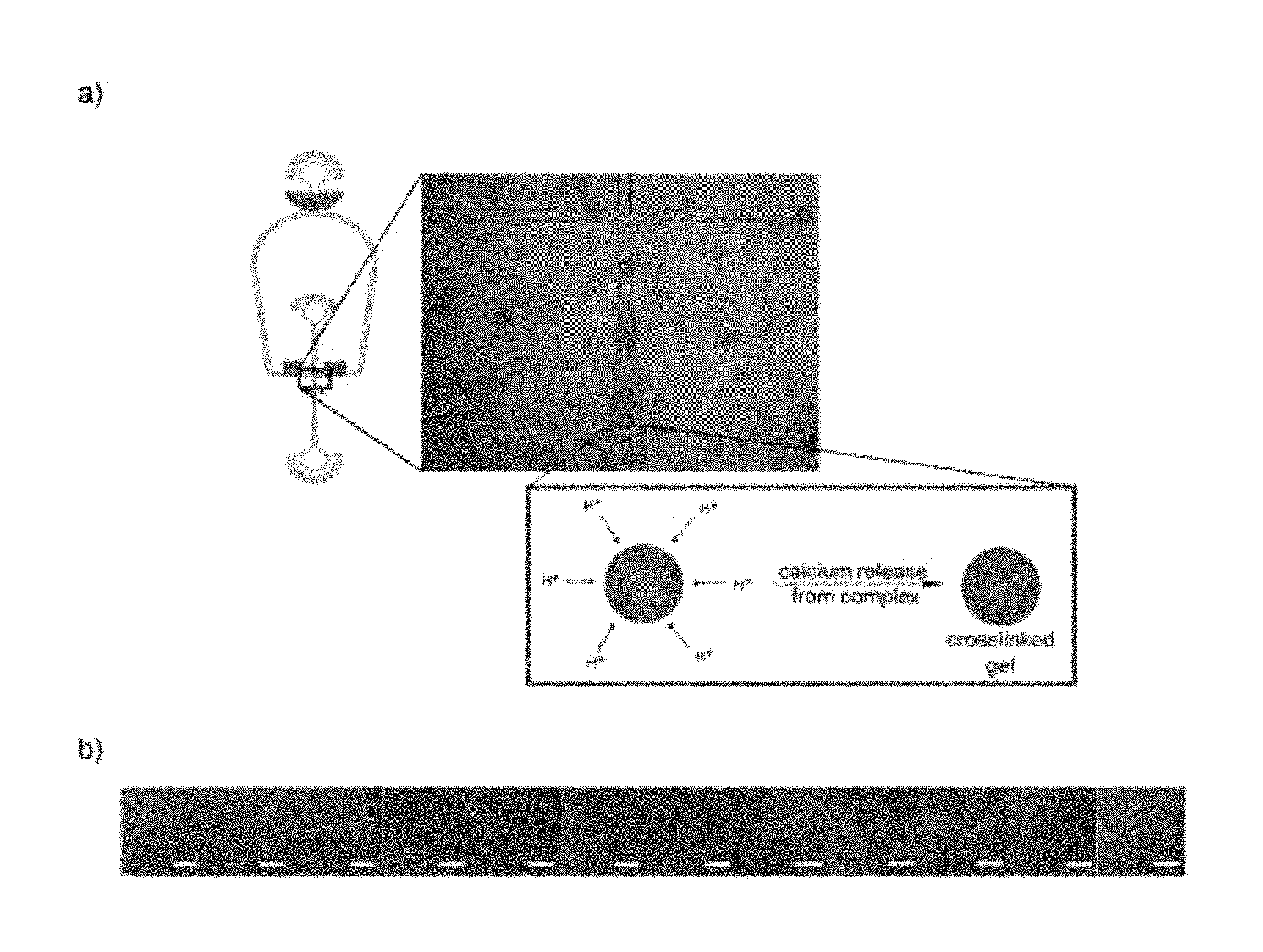
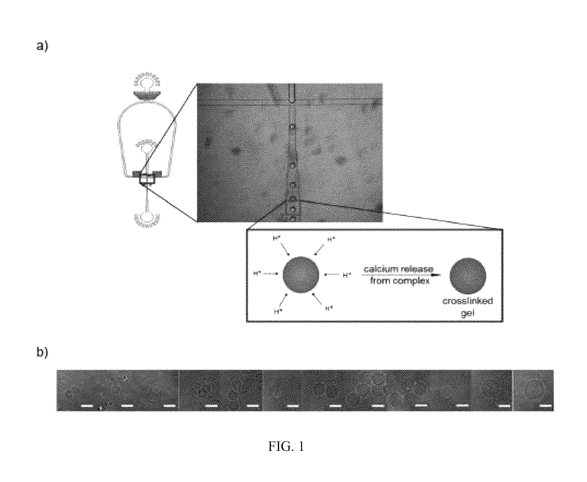
FIG. 1 is a scheme showing the formation of alginate microparticles using a 50 .mu.m polydimethylsiloxane (PDMS) dropmaker (panel a)) and microscopic images of resulting alginate microparticles in the size range of 15-50 .mu.m after transfer into aqueous medium (panel b)).
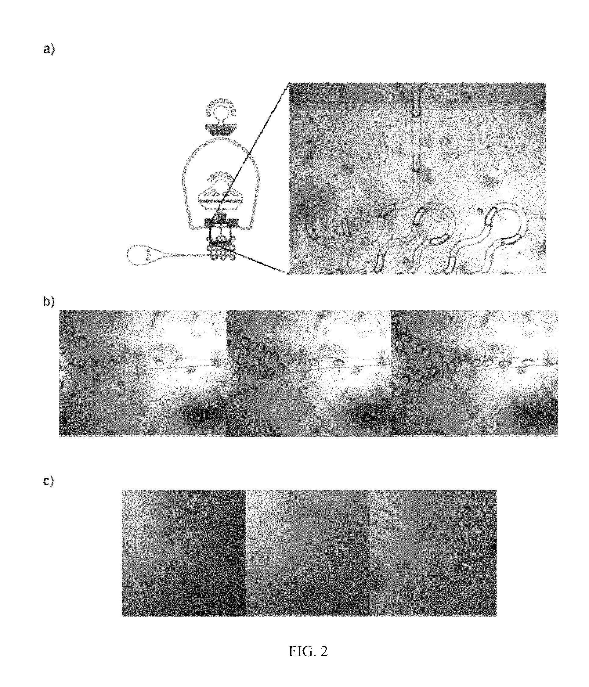
FIG. 2 is a scheme showing the formation of alginate microparticles using a 50 .mu.m PDMS dropmaker with integrated serpentine channel to alter the geometry of the formed microparticles (panel a)); microscopic images of cross-linked alginate microparticles (panel b)); and microscopic images of non-spherical alginate microparticles after transfer into aqueous medium.
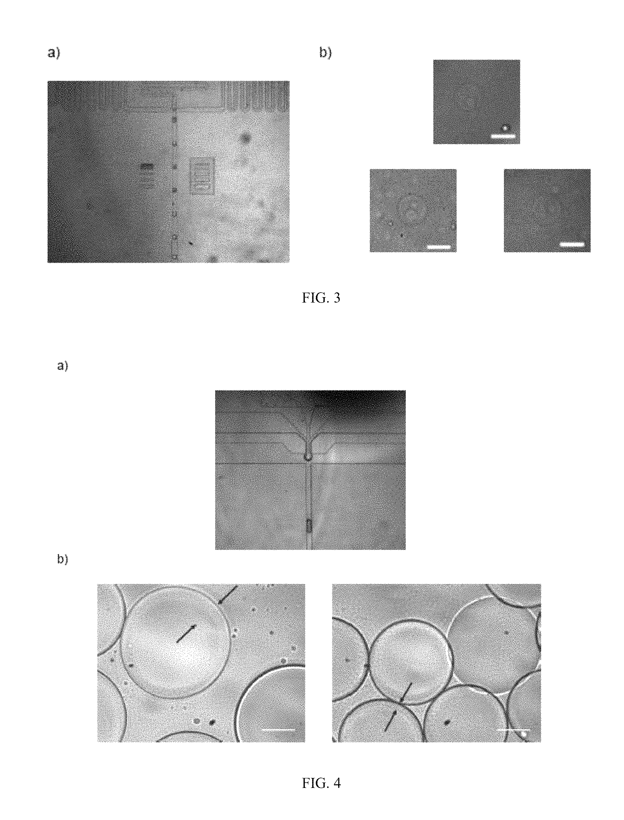
FIG. 3 is microscopic images of a 25 micrometer dropmaker (panel a)); and microscopic images of the resulting cell-containing microgels (panel b)) after breaking the emulsion with perfluoro-1-octanol (PFO).
FIG. 4 is microscopic images showing the formation of a water in water in oil (w/w/o) double emulsion using a two-dimensional microfluidic PDMS device (panel a)); and microscopic images of resulting microparticles with different alginate shell thicknesses (panel b)).
FIG. 5 is bright-field images (panels a) and d)) and fluorescent images (panel b) and e)) of alginate microparticles after transfer into an aqueous medium; and a plot of diameter vs. frequency (panel c)).
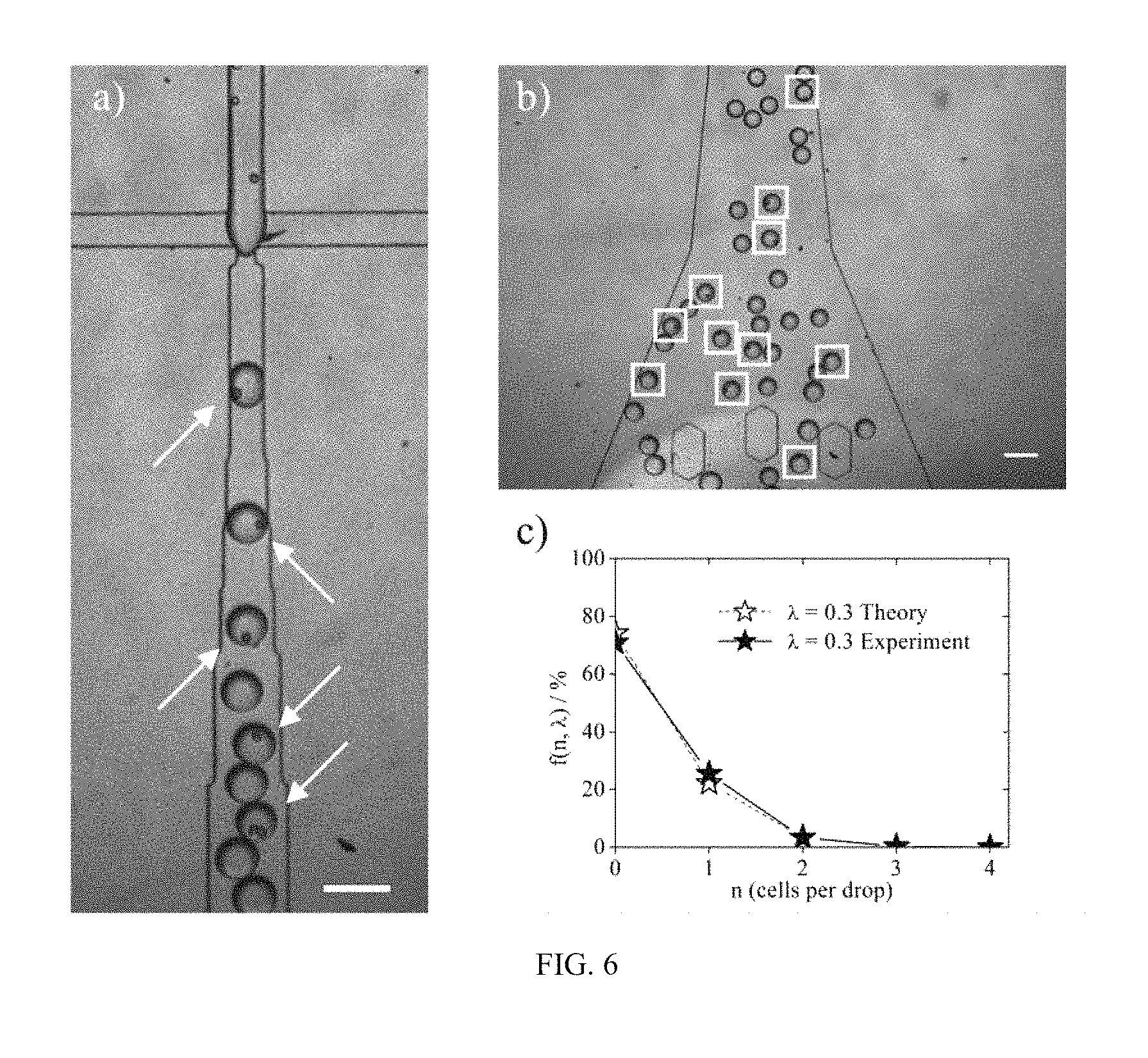
FIG. 6 is microscopic images of cell-laden microgels formed using a 50 micrometer dropmaker (panels a) and b)); and a plot showing a Poisson distribution resulting in approximately 22% of single-cell containing droplets (panel c)).
FIG. 7 is microscopic images of proliferating cells inside individual alginate microparticles during culturing over the course of three weeks after addition of a live-stain (panels a)-c)). Living cells show a bright green fluorescence.
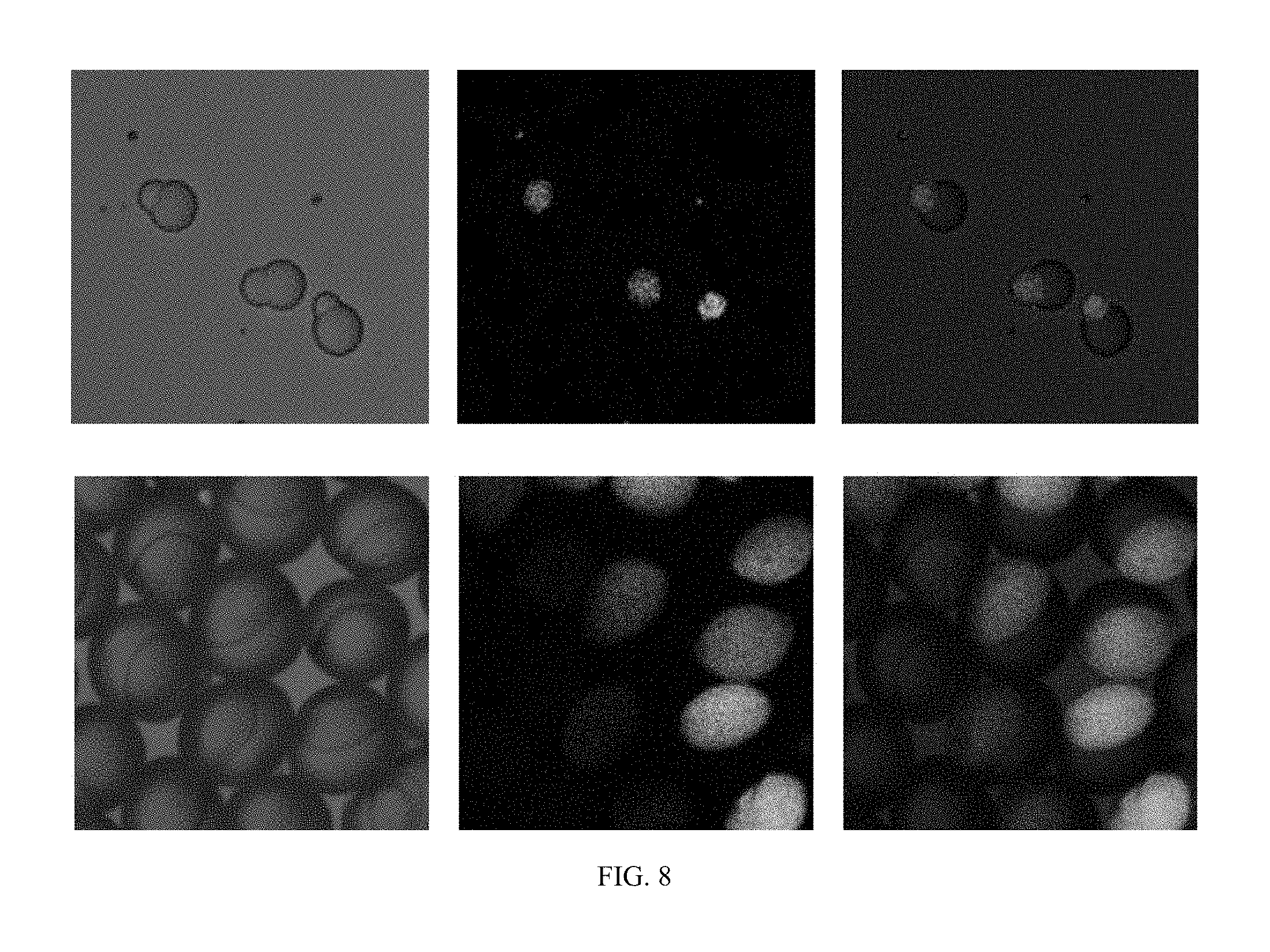
FIG. 8 is microscopic images of "Janus-type" binary microparticles. A fluorescently labeled alginate was used to identify the alginate-rich regions of the binary particle.
DETAILED DESCRIPTION OF THE INVENTION
Reference will now be made in detail to certain embodiments of the disclosed subject matter, examples of which are illustrated in part in the accompanying drawings. While the disclosed subject matter will be described in conjunction with the enumerated claims, it will be understood that the exemplified subject matter is not intended to limit the claims to the disclosed subject matter.
Values expressed in a range format should be interpreted in a flexible manner to include not only the numerical values explicitly recited as the limits of the range, but also to include all the individual numerical values or sub-ranges encompassed within that range as if each numerical value and sub-range were explicitly recited. For example, a range of "about 0.1% to about 5%" or "about 0.1% to 5%" should be interpreted to include not just about 0.1% to about 5%, but also the individual values (e.g., 1%, 2%, 3%, and 4%) and the sub-ranges (e.g., 0.1% to 0.5%, 1.1% to 2.2%, 3.3% to 4.4%) within the indicated range. The statement "about X to Y" has the same meaning as "about X to about Y," unless indicated otherwise. Likewise, the statement "about X, Y, or about Z" has the same meaning as "about X, about Y, or about Z," unless indicated otherwise.
In this document, the terms "a," "an," or "the" are used to include one or more than one unless the context clearly dictates otherwise. The term "or" is used to refer to a nonexclusive "or" unless otherwise indicated. In addition, it is to be understood that the phraseology or terminology employed herein, and not otherwise defined, is for the purpose of description only and not of limitation. Any use of section headings is intended to aid reading of the document and is not to be interpreted as limiting; information that is relevant to a section heading may occur within or outside of that particular section. Furthermore, all publications, patents, and patent documents referred to in this document are incorporated by reference herein in their entirety, as though individually incorporated by reference. In the event of inconsistent usages between this document and those documents so incorporated by reference, the usage in the incorporated reference should be considered supplementary to that of this document; for irreconcilable inconsistencies, the usage in this document controls.
In the methods described herein, the steps can be carried out in any order without departing from the principles of the invention, except when a temporal or operational sequence is explicitly recited. Furthermore, specified steps can be carried out concurrently unless explicit claim language recites that they be carried out separately. For example, a claimed step of doing X and a claimed step of doing Y can be conducted simultaneously within a single operation, and the resulting process will fall within the literal scope of the claimed process.
The term "about" as used herein can allow for a degree of variability in a value or range, for example, within 10%, within 5%, or within 1% of a stated value or of a stated limit of a range.
The term "substantially" as used herein refers to a majority of, or mostly, as in at least about 50%, 60%, 70%, 80%, 90%, 95%, 96%, 97%, 98%, 99%, 99.5%, 99.9%, 99.99%, or at least about 99.999% or more.
Embodiments of the present invention relate to microparticles comprising a crosslinked gel, wherein the microparticles have at least one dimension measuring from about 1 .mu.m to about 200 .mu.m (e.g., from about 5 .mu.m to about 200 .mu.m; or from about 40 .mu.m to about 200 .mu.m). In some embodiments, the microparticles have a coefficient of variation of from about 0.03 to about 0.05 (e.g., from about 0.04 to about 0.05, or from about 0.03 to about 0.04). The term "coefficient of variation" refers to the standard deviation of the size distribution of the microparticles, assuming a Gaussian distribution, divided by the mean size. The coefficient of variation is a measure of the size polydispersity observed for the contemplated microparticles. For non-spherical microparticles, the coefficient of variation is determined for each dimension of the particles, individually.
The microparticles may have any suitable dimensions and are, in some embodiments, substantially spherical such that the microparticles are substantially microspheres. But the microparticles may also be non-spherical and of any suitable shape, including oblong, rod-, crescent- or hook-shaped. The microparticles can also be core-shell microparticles where the microparticles may have a liquid core and a solid shell; a gas core and a solid shell; or a solid core and a solid shell, all of which may referred to as core-shell microparticles.
In some embodiments, the microparticles of the embodiments of the present invention may have at least one dimension measuring less than 200 .mu.m, less than 150 .mu.m, less than 100 .mu.m, less than 75 .mu.m, less than 65 .mu.m, less than 55 .mu.m, less than 45 .mu.m or less than 35 .mu.m, with a lower bound of about 1 .mu.m; from about 5 .mu.m to about 15 .mu.m; from about 10 .mu.m to about 200 .mu.m, from about 10 .mu.m to about 100 .mu.m, from about 10 .mu.m to about 75 .mu.m, from about 30 .mu.m to about 75 .mu.m, from about 30 .mu.m to about 100 .mu.m or from about 50 .mu.m to about 100 .mu.m. In some embodiments, the microparticles of the embodiments of the present invention are substantially spherical and have a diameter less than 200 .mu.m, less than 150 .mu.m, less than 100 .mu.m, less than 75 .mu.m, less than 65 .mu.m, less than 55 .mu.m, less than 45 .mu.m or less than 35 .mu.m, with a lower bound of about 10 .mu.m; from about 10 .mu.m to about 200 .mu.m, from about 10 .mu.m to about 100 .mu.m, from about 10 .mu.m to about 75 .mu.m, from about 30 .mu.m to about 75 .mu.m, from about 30 .mu.m to about 100 .mu.m, from about 50 .mu.m to about 100 .mu.m or from about 40 .mu.m to about 200 .mu.m.
When the microparticles of the embodiments of the present invention are core-shell microparticles, the shell may have any suitable thickness. In some embodiments, the shell has a thickness of from about 200 nm to about 200 .mu.m, about 200 nm to about 750 nm, from about 200 nm to about 1 .mu.m, from about 750 nm to about 50 .mu.m, from about 1 .mu.m to about 50 .mu.m, from about 25 .mu.m to about 50 .mu.m, from about 2 .mu.m to about 10 .mu.m or from about 2 .mu.m to about 5 .mu.m. In some embodiments, the thickness of the shell can be substantially uniform or it can be non-uniform. It should be appreciated that when the shell reaches a thickness that equals the diameter of the microparticle, then the microparticle will no longer be a core-shell microparticle and will instead be a microparticle.
In some embodiments, the microparticles of the embodiments of the present invention can comprise nanoparticles. In some embodiments, the nanoparticles can be homogenously or inhomogeneously distributed throughout the microparticles. And in embodiments where the microparticles are core-shell microparticles, the nanoparticles can be homogeneously or inhomogeneously distributed throughout the core, the shell or both. In some embodiments, the nanoparticles can be magnetic nanoparticles (e.g., iron oxide nanoparticles).
In some embodiments, the microparticles of the embodiments of the present invention can be core-shell microparticles and comprise a solid or a liquid core (e.g., a substantially aqueous core comprising a substantially aqueous liquid). The solid core may be made of the same material as the shell or of a different material than the shell. In some embodiments, the core is a liquid core. In some embodiments, the liquid core may be an aqueous core. When the liquid core is an aqueous core, it may be a water-only aqueous core or the water may comprise one or more materials dissolved in the water including salts (e.g., NaCl and MgCl.sub.2), buffers (e.g., phosphate buffer), acids (e.g., acetic acid and lactic acid), bases, cell growth medium, polymers (e.g., poly(ethylene glycol), dextran), nutrients, encapsulants, polymers, nanoparticles or combinations thereof.
In some embodiments, the liquid core may be a non-aqueous core that can comprise, e.g., an organic material including a solvent, a polymer, a dye, and the like.
In some embodiments, the core can be a solid core, a liquid core or a combination thereof. For example, the microparticles of the embodiments of the present invention may comprise a substantially solid core with liquid "pockets" distributed throughout the substantially solid core. The "pockets" may be of a uniform size or the size of the "pockets" may be variable.
In other embodiments, the core can be a solid core, a liquid core or a combination thereof, wherein the core can comprise nanoparticles (e.g., particles having at least one dimension having an average dimension of about 20 to about 500 nm, about 100 to about 500 nm, about 100 to about 300 nm or about 100 to about 200 nm) such as, but not limited to, magnetic nanoparticles such as iron oxide nanoparticles. In some embodiments, microparticles comprising such nanoparticles in their core can be useful in magnetic field-induced self-assembly of macrometer-sized constructs as engineered tissues for regenerative medicine. In other embodiments, microparticles comprising such nanoparticles in their core can be useful as targeting delivery vehicles, such that a magnet or magnetic field placed at or near a target site (e.g., organ or other tissue) would guide the microparticles comprising such nanoparticles to and concentrated at or near a target site at or near the magnet or magnetic field. Among other things, anti-cancer drugs covalently or non-covalently attached to such nanoparticles could be delivered at or near a target site.
Microparticles containing nanoparticles smaller than 20 nm (e.g., 1-20 nm) are also contemplated herein. Such nanoparticles (e.g., functionalized magnetic nanoparticles such as are known in the art) can be encapsulated or cross-linked within the crosslinked gel, crosslinked with the gel or combinations thereof.
In some embodiments, the aqueous or solid core and/or the shell can comprise viruses, one or more cells (e.g., mammalian cells, plant cells, bacteria, and combinations thereof) or proteins (e.g., collagen and antibodies). The cells or proteins can be substantially within the microparticles; may protrude into the exterior of the microparticles (e.g., through the shell of a core-shell microparticle); may protrude into the interior of the microparticles (e.g., through the shell of a core-shell microparticle and into the core); may protrude into the interior and the exterior of the microparticles (e.g., traversing the shell of a core-shell microparticle). In some embodiments, the core comprises a single cell or protein.
The encapsulation of cells in microparticles of the embodiments of the present invention may be advantageous for, e.g., long-term (e.g., twelve or more hours; fifteen or more hours; one or more days; five days to one month or more) cell culture of individual or multiple cells in an independent microenvironment. In addition, cells such as adherent cells can be cultured encapsulated in the microparticles of the embodiments of the present invention because the microparticles of certain embodiments of the present invention provide a solid support that allows for a natural adherence and spreading of the cells within the microparticle. The microparticles can then be transferred to a cell culture medium or media where the cells within the microparticles are guaranteed a sufficient nutrient supply, given the solidified spheres can, in some embodiments, be permeable to nutrients.
One advantage of having one or more cells or proteins protrude into the exterior of the microparticles, whether through the shell of a core-shell microparticle or a solid microparticle, is that the microparticle may have the propensity to form tissue-like assemblies. Briefly, by incorporating different cell types in in defined regions of the core-shell particles (e.g., encapsulation of one cell type into the core while a different cell type is incorporated into the shell of the particle) the balance of homotypic and heterotypic interactions can be controlled. See, e.g., Khetani et al. Nature Biotechnology 26: 120-126 (2008).
In some embodiments, whether the core is a liquid core or a solid core, or combinations thereof, the core can comprise an active agent distributed in the core. In some embodiments, the active agent is a cell (e.g., a plant stem cell), a pharmaceutical agent, an agrochemical agent or a food additive. See, e.g., Rimann et al. Curr. Opin. Biotechnol. 23: 803 (2012); Lee et al. Prog. Polym. Sci. 37: 106 (2012); and Microgel Suspensions (Fernandez-Nieves eds., Wiley 2011). Examples of pharmaceuticals include, but are not limited to antibiotics, antitussives, antihistamines, decongestants, alkaloids, mineral supplements, laxatives, antacids, anti-cholesterolemics, antiarrhythmics, antipyretics, analgesics, appetite suppressants, expectorants, anti-anxiety agents, anti-ulcer agents, anti-inflammatory substances, coronary dilators, cerebral dilators, peripheral vasodilators, anti-infectives, psychotropics, antimanics, stimulants, gastrointestinal agents, sedatives, anti-diarrheal preparations, anti-anginal drugs, vasodialators, anti-hypertensive drugs, vasoconstrictors, migraine treatments, antibiotics, tranquilizers, anti-psychotics, antitumor drugs, anticoagulants, antithrombotic drugs, hypontics, anti-emetics, anti-nausants, anti-convulsants, neuromuscular drugs, hyper- and hypoglycemic spasmodics, uterine relaxants, mineral and nutritional additives, antiobesity drugs, anabolic drugs, erythropoetic drugs, antiashmatics, cough suppressants, mucolytics, anti-uricemic drugs, mixtures thereof, and the like. Examples of agrochemicals include, but are not limited to, chemical pesticides (such as herbicides, algicides, fungicides, bactericides, viricides, insecticides, acaricides, miticides, nematicides, and molluscicides), herbicide safeners, plant growth regulators, fertilizers and nutrients, gametocides, defoliants, desiccants, mixtures thereof and the like. Examples of food additives include, but are not limited to, caffeine, taste-masking agents, vitamins, minerals, color additives, herbal additives (e.g., echinacea or St. John's Wort), antimicrobials, preservatives, mixtures thereof, and the like.
In some embodiments, the microparticles of the embodiments of the present invention may have pores. In some embodiments, the pores are distributed throughout the shell of core-shell microparticles of the embodiments of the present invention. The pores may have any suitable diameter and length. The pores may have, e.g., a diameter ranging from about 1 nm to about 5 .mu.m, e.g., from about 5 nm to about 5 .mu.m, from about 5 nm to about 750 nm, from about 50 nm to about 500 nm or from about 50 nm to about 250 nm, from about 50 nm to about 250 nm or from about 5 nm to about 1 .mu.m. The diameter of the pores may or may not be uniform within a single pore or across a multitude of pores.
One of the functions of the pores is to serve as a conduit for any active agent to diffuse from the microparticle (e.g., from the core; through the shell) into the environment surrounding the microparticles of the embodiments of the present invention. Those of skill in the art will recognize, however, that the pores can also function as a conduit for materials located in the environment surrounding the microparticles of the embodiments of the present invention to diffuse into the microparticles. For example, in applications where one or more cells are located in the microparticles of the embodiments of the present invention, pores may play a key role as conduits for nutrients that are necessary for cell growth within the microparticles.
In some embodiments, the microparticles of the embodiments of the present invention are degradable (e.g., biodegradable). For example, the microparticles may be digestible by one or more enzymes or may degrade by hydrolysis. In other embodiments, the microparticles of the embodiments of the present invention are non-degradable or partially degradable.
Microparticles of the embodiments of the present invention may be made of any suitable cross-linkable material that can be subsequently cross-linked via any suitable means for cross-linking, thereby yielding a cross-linked gel. Examples of suitable cross-linkable materials include, but are not limited to, cross-linkable linear polysaccharides. In some embodiments, the cross-linkable material comprises homopolymeric blocks of (1-4)-linked .beta.-D-mannuronate and .alpha.-L-guluronate. Non-limiting examples of cross-linkable materials that can be used to form the microparticles of the embodiments of the present invention include alginate, chitosan, curdlan, dextran, emulsan, a galactoglucopolysaccharide, gellan, glucuronan, N-acetyl-heparosan, hyaluronic acid, indicant, kefiran, lentinan, levan, mauran, pullulan, scleroglucan, schizophyllan, stewartan, succinoglycan, xanthan, xylane, welan, starch, tamarind, tragacanth, guar gum, derivatized guar, gum ghatti, gum arabic, locust bean gum, cellulose, hemicellulose, carboxymethyl cellulose, hydroxyethyl cellulose, carboxymethyl hydroxyethyl cellulose, hydroxypropyl cellulose, methyl hydroxyl ethyl cellulose, guar, hydroxypropyl guar, carboxy methyl guar, carboxymethyl hydroxylpropyl guar or combinations thereof.
In some embodiments, the cross-linkable material can be derivatized to include, among other things, small molecules (e.g., tyramine), oligonucleotides or oligopeptides (e.g., polypeptides comprising the Arg-Gly-Asp recognition sequence, also known as "RGD"). The cross-linkable material can be derivatized before it is crosslinked or after it is crosslinked. In some embodiments, the cross-linkable material is derivatized before it is cross-linked.
The cross-linkable material can be crosslinked via any suitable cross-linking mechanism. For example, the cross-linkable material can be crosslinked via covalent crosslinks, non-covalent crosslinks (e.g., with the use of a crosslinking agent) or via a combination of covalent and non-covalent crosslinks.
In some embodiments, the crosslinking agent comprises divalent cations including, but not limited to Ca.sup.2+, Mg.sup.2+, Ba.sup.2+ or combinations thereof. In some embodiments, the crosslinking agent is substantially homogeneously distributed in the microparticles of the embodiments of the present invention.
In some instances it may be advantageous for the divalent cations to be sequestered in any suitable way (e.g., chelation) so that the crosslinking timing and rate can be controlled. For example, in some embodiments, the divalent cations may be chelated with any chelating agent suitable for chelating divalent cations including, but not limited to, ethylenediaminetetraacetic acid (EDTA), ethylene glycol tetraacetic acid (EGTA), cyclohexane diamine tetraacetic acid (CDTA), citrate, and phosphate.
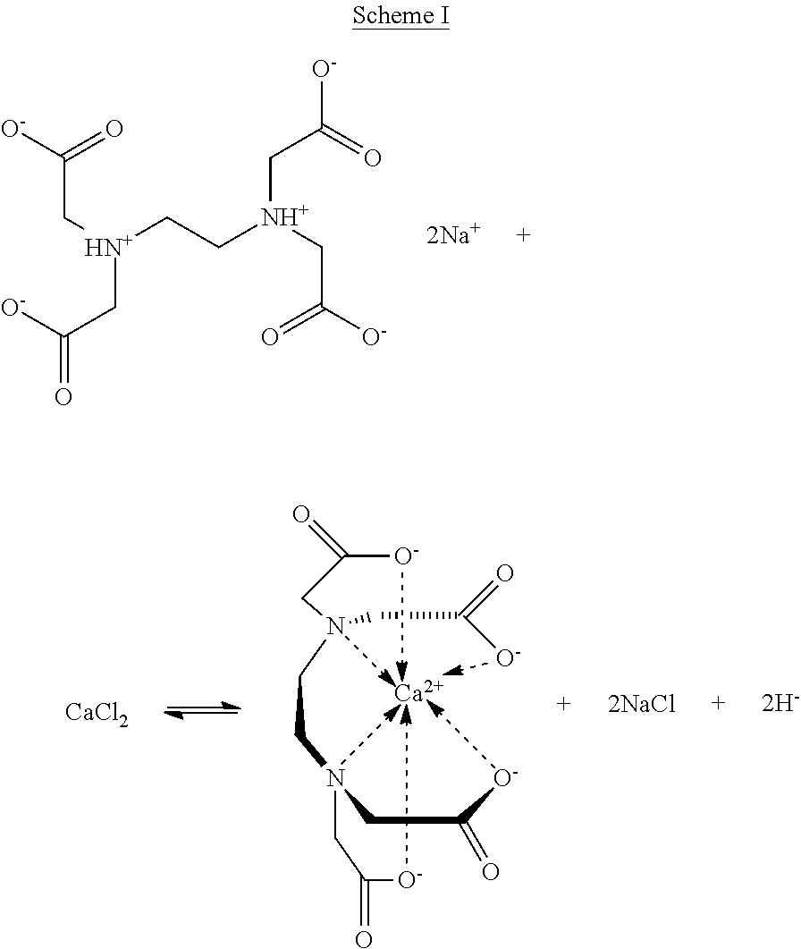
The microparticles of the embodiments of the present invention may be formed by a method comprising forming microdroplets (e.g., microfluidically forming the microdroplets) comprising one or more crosslinkable materials (e.g., linear polysaccharides) and one or more crosslinking agents. In some embodiments, both components, the crosslinkable materials and the crosslinking agents are in liquid form, e.g., as solutions in water or any suitable solvent. The resulting microdroplets can then be contacted with a crosslinking promoter to crosslink the one or more crosslinkable materials (e.g., linear polysaccharides). The crosslinking promoter, in some embodiments, may be a change in the pH, a change in the temperature, a change in the ionic strength or combinations thereof. In some embodiments, the crosslinking promoter is a change in the pH. The change in the pH may be effected with an acid or a base, preferably an acid. In other embodiments, the crosslinking promoter is an ionic species (e.g., in solution) that is different from the crosslinking agent.
The acid may be any suitable acid and the ionic species may be any suitable ionic species, particularly an ionic species having a higher affinity for a chelating agent than the crosslinking agent. The acid and the ionic species cause a sufficient amount of chelated divalent cations to be sufficiently freed from chelation, thereby providing a sufficient amount of unchelated divalent cations to promote crosslinking. Scheme I, below, shows a schematic representation of this process using EDTA as a specific, non-limiting chelating agent and Ca.sup.2+ as a specific, non-limiting crosslinking agent.
##STR00001##
Scheme I shows that there is an equilibrium between the chelated form of Ca.sup.2+ and the unchelated form of Ca.sup.2+. The equilibrium may be driven to the left (i.e., to the unchelated form of Ca.sup.2+) if a proton source (i.e., an acid, crosslinking promoter) is added. Enough acid may be added to generate a proton concentration that is, in turn, sufficient to generate a sufficient concentration of crosslinking agent (e.g., unchelated Ca.sup.2+) to promote crosslinking.
In a specific, non-limiting example, where the cross-linkable material comprises alginate and the cross-linking agent comprises EDTA-chelated Ca.sup.2+, above a certain pH, the binding affinity of EDTA for Ca.sup.2+ is higher than that of alginate. Thus, Ca.sup.2+ is bound by EDTA and cannot crosslink the alginate. Shifting the pH toward a lower pH decreases the binding strength of EDTA to Ca.sup.2+. Below a certain pH, the binding strength of alginate is higher than that of EDTA and thus, Ca.sup.2+ is complexed by alginate and serves as a crosslinking agent. It should be understood, however, that in some instances, depending on the cross-linkable material and/or the cross-linking agent comprising a chelated cation, the affinity of the chelating agent for the cation may be higher at lower pHs and lower at higher pHs, such that crosslinking is substantially prevented at lower pHs and promoted at higher pHs.
In some embodiments, the crosslinking promoter comprises an acid. The acid may be any suitable acid, including HC1 and HF. In some embodiments, the acid that may be used as crosslinking promoters includes, but is not limited to, an organic acid. In some embodiments, organic acids include, but are not limited to, carboxylic acids such as C.sub.2-C.sub.10 carboxylic acids (e.g., a C.sub.2-C.sub.8 carboxylic acid, a C.sub.2-C.sub.5 carboxylic acid, and a C.sub.2-C.sub.4 carboxylic acid). Examples of C.sub.2-C.sub.10 carboxylic acids include, but are not limited to acetic acid, formic acid, benzoic acid, citric acid, oxalic acid, lactic acid or combinations thereof.
In some embodiments, the crosslinkable material can comprise functional groups that can be crosslinked via crosslinking mechanisms, in addition to crosslinking via a crosslinking agent. Thus, for example, one of the crosslinking mechanisms can be crosslinking via a crosslinking agent, but there can be at least one other crosslinking mechanism including covalent crosslinking via reactions between alcohols and carboxylic acids to form esters; reactions between amines and carboxylic acids to form amides; reactions between aldehydes and primary amines to form imines that can be reduced to secondary amines, reactions between alcohols and isocyanates to form carbamates; reactions between amines and isocyanates to give ureas; aryl-aryl coupling (e.g., phenol-phenol coupling that is enzymatically catalyzed; see Journal of Biomedical Materials Research Par A 85: 345-351 (2008), which is incorporated herein by reference), or combinations thereof. Additional crosslinking mechanism can include radical and photochemical crosslinking mechanisms. In some embodiments, the primary crosslinking mechanism can be crosslinking via a crosslinking agent.
Physical crosslinking mechanisms are also contemplated herein.
The microparticles of the embodiments of the present invention may be used in methods for delivering an active agent to a subject (e.g., a mammal, specifically a human) in need thereof or, in the case of agrochemicals, to an area (e.g., a field or plot) in need thereof. The methods comprise (i) providing or obtaining one or more microparticle comprising an active agent; and (ii) delivering the microparticle to a location (e.g., capillaries, skin, and eye) in a subject in need thereof or a location in an area in need thereof.
The microparticles may be delivered to the subject in need thereof or, in the case of agrochemicals, to an area in need thereof, by any suitable means. Such means for delivering the microparticles of the embodiments of the present invention to a subject in need thereof include, but are not limited to, oral, peroral, parenteral, intravenous, intraperitoneal, intradermal, subcutaneous, intramuscular, nasal, buccal, rectal or topical means, for example on the skin, mucous membranes or in the eyes. Means for delivering or depositing the microparticles of the embodiments of the present invention in an area in need thereof include, but are not limited to, spraying (e.g., an aqueous suspension of the microparticles of the embodiments of the present invention).
In some embodiments, the microparticles of the embodiments of the present invention may be combined with other pharmaceutically acceptable or agronomically acceptable excipients. Such excipients may facilitate the incorporation of the microparticles of the embodiments of the present invention into dosage forms (e.g., capsules, tablets, lozenges, and the like) or into, e.g., pellets for agrochemical applications.
In some embodiments, when the microparticles of the embodiments of the present invention have a liquid core, and the core comprises an active agent, the microparticles can be ruptured by applying a suitable trigger. Such triggers include, but are not limited to mechanical force (e.g., from the hand, when applied to the skin), ultrasound, oxidizing stress, osmotic stress, pH, phototriggers; reducing agents, enzyme/enzymatic triggers, temperature, magnetic fields, and combinations thereof.
In some embodiments, applying oxidizing stress to the microparticles includes contacting the microparticles with or exposing the microparticles to an oxidizing agent. Suitable oxidizing agents include, but are not limited to, silver nitrate, potassium permanganate, sodium periodate, osmium tetroxide, peroxides, and sulfuric acid. An osmotic stress trigger includes, but is not limited to, exposing such microparticles to conditions where the ionic strength outside the microparticles is substantially less than the ionic strength inside the microparticles (i.e., in the core). An example of such a situation includes microparticles containing a high salt (e.g., CaCl.sub.2) concentration (e.g., from about 1 to about 2 M salt) in the core being exposed to a significantly lower salt (e.g., about 0 to about 0.5 M) concentration outside the microparticles or vice versa.
In various other embodiments, the invention relates to a system comprising one or more microparticles of the embodiments of the present invention and one or more cells encapsulated in the one or more microparticles. In some embodiments, each microparticle comprises more than one cell. In other embodiments, each microparticle comprises a single cell.
The terms and expressions which have been employed are used as terms of description and not of limitation, and there is no intention that in the use of such terms and expressions of excluding any equivalents of the features shown and described or portions thereof, but it is recognized that various modifications are possible within the scope of the invention claimed. Thus, it should be understood that although the present invention has been specifically disclosed by preferred embodiments and optional features, modification and variation of the concepts herein disclosed may be resorted to by those of ordinary skill in the art, and that such modifications and variations are considered to be within the scope of this invention as defined by the appended claims.
EXAMPLES
The present invention can be better understood by reference to the following examples which are offered by way of illustration. The present invention is not limited to the examples given herein.
Example 1
Solid Particles
Using droplet-based microfluidics, monodisperse droplets of a calcium-EDTA/alginate mixture can be produced with a precise size control in the micrometer regime. See FIG. 1, which contains two panels a) and b). Panel a) is a scheme showing the formation of alginate microparticles using a 50 .mu.m PDMS dropmaker (left: channel outline, right: microscopic image of crossjunction (4.times.)). The liquid microdroplets contain Ca-EDTA (100 mM, pH 7.0)/alginate (2.0% w/v) (1:1). The microdroplets are solidified into microparticles by diffusion of acetic acid present in the outer phase (1.0% PFPE-PEG block-copolymer surfactant (Holtze et al. Lab Chip 8: 1632 (2008)) in HFE7500 (3M), and 1 .mu.L/mL acetic acid) into the drop. Panel b) of FIG. 1 shows microscopic images of resulting alginate microparticles. The particle size can be controlled by adapting the flow rates of the system (scale bars 20 nm).
The gels shown in FIG. 1b are produced by using two different dropmakers: the first six images from the left: 25 .mu.m dropmaker, the last six images: 50 .mu.m dropmaker. The flow rate of the inner phase (alginate/EDTA-complex) was kept at 50 .mu.L/h. The flow rate of the oil phase was varied in the following order (from left image to right): 1000, 800, 600, 400, 200, 100 .mu.L/h;
1000, 800, 600, 400, 200, 100 .mu.L/h.
The gelation of the drops can either be induced by diffusion of acetic acid into the drop or by addition of acid-generating compounds into the drops like lactones, specific example is gluconolactone. By controlling the gelation rate, as well as the channel geometry, spherical as well as non-spherical particles are accessible. Rapid gelation of alginate leads to a solidification of the liquid alginate drops inside the microfluidic channel allowing for control over the shape of the cross-linked alginate particles through the design of the channel geometry. Therefore, a variety of non-spherical particles can be created including rods, hooks, crescents, for example. See FIG. 2, which contains three panels a)-c).). Panel a) is a scheme showing the formation of alginate microparticles using a 50 nm PDMS dropmaker (left: channel outline, right: microscopic image of crossjunction (4.times.)). The liquid microdroplets contain Ca-EDTA (100 mM, pH 7.0)/alginate (2.0% w/v) (1:1). The microdroplets are solidified into microparticles by diffusion of acetic acid present in the outer phase (1.0% PFPE-PEG block-copolymer surfactant (Holtze et al. Lab Chip 8: 1632 (2008)) in HFE7500 (3M), and 1 .mu.L/mL acetic acid) into the drop. Panel b) of FIG. 2 shows microscopic images of cross-linked alginate microparticles in the channel outlet (4.times.). The geometry of the rod-like structure can be influenced by controlling the flow rates during drop formation (flow rates inner/outer phase (in .mu.L/h) from left to right: 50/150, 50/400, 50/800). Panel c) of FIG. 2 shows non-spherical alginate microparticles obtained from different flow rates using the channel geometry shown in panel a) (flow rates inner/outer phase (in .mu.L/h) from left to right: 150/200, 150/500, 150/700) (scale bars 25 .mu.m).
The mechanical properties of the gels can also be controlled by the concentration and chemical nature of the alginates (e.g., molecular weights and .beta.-D-mannuronate (M)/.alpha.-L-guluronate (G) ratios/lengths). To enhance the stability of the microparticles, a combination of physical and chemical cross-linking can be performed by the incorporation of covalently cross-linkable groups, e.g., phenol units. See, e.g., Journal of Biomedical Materials Research Par A 85: 345-351 (2008), which is incorporated by reference.
The developed approach is compatible with microfluidic methods and drop formation techniques and can be combined with a variety of microfluidic applications as cell encapsulation, high-throughput analysis or materials production.
Example 2
Cell-Encapsulation
Cell-containing microparticles can be generated by combining the described method with microfluidic cell encapsulation techniques. See Clausell-Tormos et al. Chemistry & Biology 15: 427 (2008) and Koster et al. Lab Chip 8: 1110 (2008). After gelation the resulting cell-laden microparticles can be transferred into aqueous cell culture medium without losing the integrity of the generated microenvironment. Any surfactant present when the microparticles are formed may be removed by addition of a suitable agent such as perfluoro-1-octanol (PFO), followed by subsequent centrifugation, removal of the oil phase, and re-dispersion of the microparticles in cell culture medium.
Single, as well as multiple biological entities, e.g., mammalian and plant cells, proteins or peptides (e.g., collagen, RGD, antibodies), bacteria, viruses, can be incorporated into one microparticles of the embodiments of the present invention allowing for long-term biological screenings, single-cell analysis, and culturing in independent microenvironments. See FIG. 3, which contains two panels a) and b). Panel a) shows microscopic images of a 25 micrometer dropmaker (inner phase: Ca-EDTA (100 mM, pH 7.0)/alginate (2.0% w/v) (1:1); outer phase: 1.0% PFPE-PEG block-copolymer surfactant (Holtze et al. Lab Chip 8: 1632 (2008)) in HFE7500 (3M), and 1 .mu.L/mL acetic acid) (4.times.). Panel b) in FIG. 3 is microscopic images of the resulting cell-containing microgels (carrying single or multiple cells) after breaking the emulsion with PFO, centrifugation, and transfer to aqueous medium (scale bars: 25 micrometer).
The solid nature of the microparticle can also be used as scaffold or solid-support for adherent cells and thus mimics conditions found in vivo or in bulk cell culture experiments.
The encapsulated cells show good viability and proliferation for several weeks. In analogy to experiments with liquid cell-laden drops, the cell-containing microparticles can individually be manipulated (sorting, screening, picoinjection, etc.). See Schmitz et al., Lab Chip 9: 44 (2009); Abate et al., PNAS 107: 19163 (2010); Agresti et al., PNAS 107: 4004 (2010); Baret, Lab Chip 9: 1850 (2009); and Guo et al., Lab Chip 12: 2146 (2012).
Concerning long-term cell culture, a homogeneous gelation which is directly correlated with the mechanical properties of the corresponding gel may impact acceptable experimental reproducibility. It has been shown that the proliferation of cells may be influenced by the mechanical properties of their environment and is therefore mechanical properties is an important factor for screening and cell culture applications. See Klein et al., Current Biology 19: 1511 (2009); and Khatiwala et al., Am. J. Physiol. Cell Physiol. 290: C1640 (2006).
Example 3
Core-Shell Particles and Capsules
Core-shell geometries and capsules can be assembled as any suitable emulsion, including, but not limited to, water-in-water-in-oil (w/w/o), o/w/o, and w/o/w double emulsions. See FIG. 4, which contains two panels a) and b). Panel a) is microscopic images showing the formation of a water in water in oil (w/w/o) double emulsion using a two-dimensional microfluidic PDMS device (inner phase: 20% PEG, middle phase: Ca-EDTA (100 mM, pH 7.0)/alginate (2% w/v) (1:1), outer phase: 1.0% PFPE-PEG block-copolymer surfactant (Holtze et al. Lab Chip 8: 1632 (2008)) in HFE7500 (3M), and 1 .mu.L/mL acetic acid (4.times.)). The Ca-EDTA/alginate shell is solidified by diffusion of acetic acid present in the outer phase into the drop. Panel b) of FIG. 4 is microscopic images of resulting alginate microparticles with different shell thicknesses (not density matched) obtained by different flow rates (flow rates inner/middle/outer phase (in .mu.L/h) from left to right: 20/60/1000, 60/20/1000) (scale bars: 25 .mu.m).
If alginate is used as an outer phase, homogenous capsules can be assembled and their size and shell thickness can be tuned with the flow rates and device geometry. In analogy to the solid particles described in Example 1, the microparticles can be transferred into aqueous media after alginate is gelled.
For w/w/o emulsions alginate forms the middle phase and the oil phase forms the outer phase.
Core-shell particles of a great variety of materials can be formed by changing the composition of the inner phase. For example, the use of alginate solutions of different concentrations, properties, and/or compositions (e.g., molecular weights, M/G-ratios, ratio of concentration of alginate to Ca.sup.2+, and any additional degree of crosslinking, in cases where the alginte has been, e.g., covalently crosslinked to some/any extent) lead to alginate/alginate core-shell particles exhibiting a gradient in stiffness. Thus, for example, a lower molecular weight alginate will result in a "softer" microparticle. Also, when gels are made from an alginate rich in guluronic acid residues, higher moduli are obtained compared to gels made from alginates less enriched in G residues. While not being bound by any particular theory, it is believed that the reason for this behavior is that high-G gels, with their long G-blocks and their short elastic segments become more of a stiff open and static network compared to the more dynamic and entangled network structure of the low-G gels with their relative long elastic segments. See Inter. J. Biol. Macromol. 21: 47-55 (1997).
In analogy to alginate hydrogel drops, cells can also be loaded into these capsules and core-shell particles.
Additionally, the formation of shells thicknesses in the nanometer range as well as the generation of water in water in water (w/w/w) emulsions may be possible. Triple or higher order emulsions are also possible.
Example 4
FIG. 5 contains bright-field (panels a)) and fluorescent (panel b)) images of alginate microparticles after transfer into an aqueous medium. The images reflect the high monodispersity of the spherical particles as shown in panel c). The high-magnification images (panel d)=bright-field; and panel e)=fluorescent) reveals the homogeneous structure of the alginate microsphere. Panel f) contains bright-field images of homogeneous alginate microgels with 15, 21, 28 and 39 .mu.m in diameter. All scale bars are 50 .mu.m.
Example 5
Cell-laden microgels were formed using a 50 micrometer dropmaker. See FIG. 6 panel a). During the drop formation process, cells are encapsulated and the resulting microgels are collected at the channel outlet. See FIG. 6, panel b). Single-cell containing droplets are indicated by white arrows and boxes in panels a) and b), respectively. The encapsulation process follows the Poisson distribution resulting in approximately 22% of single-cell containing droplets. See panel c). All scale bars are 100 .mu.m.
Example 6
Cell-containing microparticles were observed directly after encapsulation and during culture over the period of 366 h. See FIG. 7, panel a). Representative images of proliferating cells inside individual alginate microparticles after addition of a live stain. See FIG. 7, panel b). Living cells exhibit green fluorescence. See FIG. 7, panel b), lower row. About 80% of the cells were alive directly after encapsulation and transfer into cell culture medium. The cells show a natural proliferation inside the droplets under maintenance of their spherical shape. After three weeks of encapsulation the cell viability was determined to be 55%. All scale bars are 25 .mu.m.
Example 7
Binary Microparticles
In some embodiments, the microparticles can be "Janus-type" binary microparticles, where the microparticle is divided into two separate regions that may be made of the same material. In some embodiments, the microparticle is divided into two separate regions (e.g., compartments) that may be made of a different material. See, e.g., FIG. 8. For example, in some embodiments, the two separate regions can contain two compartments, each compartment comprising different materials where one compartment can comprise a single or a plurality of suitable magnetic nanoparticles (e.g., iron oxide nanoparticles) and the other compartment can comprise no magnetic nanoparticles. And in embodiments where the two separate regions contain two compartments, each compartment comprising the same material, both compartments can comprise a single or a plurality of suitable magnetic nanoparticles (e.g., iron oxide nanoparticles).
In other embodiments, each compartment can comprise at least one cell, where the cell or cells in each compartment can be the same or the cell or cells in a first compartment is different from the cell or cells in a second compartment. Even if the at least one cell is the same in each compartment, the at least one active agent may be present at a different cell concentration in each compartment. Or if the at least one cell is different in each compartment, the two different at least one cells may also be present at a different cell concentration.
In other embodiments, each compartment can comprise at least one active agent, where the active agent or agents in each compartment can be the same or the active agent or agents in a first compartment is different from the active agent or agents in a second compartment. Even if the at least one active agent is the same in each compartment, the at least one active agent may be present at a different concentration in each compartment. Or if the at least one active agent is different in each compartment, the two different at least one active agents may also be present at a different concentration.
In some embodiments, binary microparticles may be prepared microfluidically using the same microfluidic apparatus used to make core-shell microparticles herein by mismatching the osmotic pressure in the inner phase (e.g., a PEG phase) and the middle phase (e.g., an alginate-containing phase) in a w/w/o double emulsion. A solution containing an EDTA-calcium complex is added to the alginate phase but not to the inner phase. After polymerization of the alginate phase, the core-shell microparticle phase-separate into two distinct domains. Ternary and higher order particle architectures can also be envisioned.
The present invention provides for the following exemplary embodiments, the numbering of which is not to be construed as designating levels of importance:
Embodiment 1 relates to microparticles comprising: a cross-linked gel; wherein the microparticles have a coefficient of variation in the size distribution of the microparticles of from about 0.03 to about 0.05 and wherein the microparticles have at least one dimension measuring from about 5 .mu.m to about 200 .mu.m.
Embodiment 2 relates to the microparticles of Embodiment 1, wherein the crosslinked gel comprises one or more crosslinked linear polysaccharides.
Embodiment 3 relates to the microparticles of Embodiment 2, wherein the crosslinked linear polysaccharide comprises alginate, chitosan, curdlan, dextran, emulsan, a galactoglucopolysaccharide, gellan, glucuronan, N-acetyl-heparosan, hyaluronic acid, indicant, kefiran, lentinan, levan, mauran, pullulan, scleroglucan, schizophyllan, stewartan, succinoglycan, xanthan, xylane, welan, starch, tamarind, tragacanth, guar gum, derivatized guar, gum ghatti, gum arabic, locust bean gum, cellulose, hemicellulose, carboxymethyl cellulose, hydroxyethyl cellulose, carboxymethyl hydroxyethyl cellulose, hydroxypropyl cellulose, methyl hydroxyl ethyl cellulose, guar, hydroxypropyl guar, carboxy methyl guar, carboxymethyl hydroxylpropyl guar or combinations thereof.
Embodiment 4 relates to the microparticles of Embodiment 2, wherein the crosslinked linear polysaccharide comprises crosslinked alginate.
Embodiment 5 relates to the microparticles of Embodiment 2, wherein the crosslinked linear polysaccharide comprises a crosslinking agent.
Embodiment 5A relates to the microparticles of Embodiment 5, wherein the crosslinking agent is substantially homogenously distributed in the microparticle.
Embodiment 5B relates to the microparticles of Embodiment 5, wherein the microparticles are core-shell microparticles or binary or higher order microparticles; and the crosslinking agent is substantially homogenously distributed in the core, the shell, or the core and the shell of core-shell microparticles; or, when the microparticles are binary or higher order microparticles, in one or more (preferably all) of the microparticles comprised in the binary or higher order microparticles.
Embodiment 6 relates to the microparticles of Embodiment 5, wherein the crosslinking agent comprises divalent cations.
Embodiment 7 relates to the microparticles of Embodiment 6, wherein the divalent cations comprise Ca.sup.2+, Mg.sup.2+, Ba.sup.2+ or combinations thereof.
Embodiment 8 relates to the microparticles of Embodiments 1-7, wherein the microparticles are substantially spherical.
Embodiment 9 relates to the microparticles of Embodiments 1-7, wherein the microparticles are rod-, crescent- or hook-shaped.
Embodiment 10 relates to the microparticles of Embodiments 8 or 9, wherein the microparticles are core-shell microparticles.
Embodiment 11 relates to the microparticles of Embodiments 8 or 9, wherein the microparticles are binary microparticles.
Embodiment 12 relates to the microparticles of Embodiment 10, wherein the core is a liquid core, a solid core or a gas core.
Embodiment 13 relates to the microparticles of Embodiment 12, wherein the core is an aqueous core.
Embodiment 14 relates to the microparticles of Embodiment 10, wherein the microparticles comprise a liquid core and a solid shell; a gas core and a solid shell; or a solid core and a solid shell.
Embodiment 15 relates to the microparticles of Embodiment 10, wherein the core and/or the shell comprises one or more cells. In another embodiment, at least one of the core and the shell comprises one or more cells.
Embodiment 16 relates to the microparticles of Embodiments 1-5, 5A, 5B, and 6-15, wherein the microparticles comprise an active agent.
Embodiment 17 relates to the microparticles of Embodiment 16, wherein the active agent is a pharmaceutical, an agrochemical or a food additive.
Embodiment 18 relates to the microparticles of Embodiments 1-5, 5A, 5B, and 6-17, wherein the microparticles are degradable, non-degradable or partially degradable.
Embodiment 19 relates to the microparticles of Embodiments 1-5, 5A, 5B, and 6-18, wherein the microparticles comprise pores.
Embodiment 20 relates to microparticles comprising: a Ca.sup.2+-cross-linked alginate gel; wherein the microparticles have a coefficient of variation in the size distribution of the microparticles of from about 0.03 to about 0.05 and wherein the microparticles have at least one dimension measuring from about 5 .mu.m to about 200 .mu.m.
Embodiment 21 relates to a method of forming the microparticles of Embodiment 1, the method comprising: forming microdroplets comprising one or more crosslinkable linear polysaccharides and one or more crosslinking agents; contacting the microdroplets with a crosslinking promoter to promote crosslinking of the one or more crosslinkable linear polysaccharides.
Embodiment 22 relates to the method of Embodiment 21, wherein the crosslinking agent does not substantially crosslink the one or more crosslinkable linear polysaccharides in an initial state, but, upon contacting with the crosslinking promoter, crosslinks the one or more crosslinkable linear polysaccharides in a second state.
Embodiment 23 relates to the method of Embodiment 22, wherein the first state comprises a sequestered state of the one or more crosslinking agents and the second state comprises an unsequestered state of the one or more crosslinking agents.
Embodiment 24 relates to the method of Embodiments 21-23, wherein the crosslinking agent is substantially homogenously distributed in the microparticle.
Embodiment 25 relates to the method of Embodiment 21-24, wherein said forming comprises microfluidically forming the microdroplets.
Embodiment 26 relates to the method of Embodiment 21-25, wherein the crosslinking agent comprises divalent cations.
Embodiment 27 relates to the method of Embodiment 26, wherein the divalent cations comprise Ca.sup.2+, Mg.sup.2+, Ba.sup.2+ or combinations thereof.
Embodiment 28 relates to the method of Embodiment 26, wherein the divalent cations are sequestered.
Emodiment 29 relates to the method of Embodiment 28, wherein the sequestered divalent cations are chelated.
Embodiment 30 relates to the method of Embodiment 29, wherein the chelated divalent cations comprise Ca.sup.2+-EDTA.
Embodiment 31 relates to the method of Embodiments 21-30, wherein said crosslinking promoter comprises a change in the pH, a change in the temperature, a change in the ionic strength or combinations thereof.
Embodiment 32 relates to the method of Embodiment 31, wherein said crosslinking promoter comprises a change in the pH.
Embodiment 33 relates to the method of Embodiment 32, wherein the change in the pH is effected with an acid.
Embodiment 34 relates to the method of Embodiment 33, wherein the acid comprises a carboxylic acid.
Embodiment 35 relates to the method of Embodiment 34, wherein the carboxylic acid comprises a C.sub.2-C.sub.10-carboxylic acid.
Embodiment 36 relates to the method of Embodiment 35, wherein the C.sub.2-C.sub.10-carboxylic acid comprises acetic acid, formic acid, benzoic acid, citric acid, oxalic acid, lactic acid or combinations thereof.
Embodiment 37 relates to the method of Embodiment 36, wherein the divalent cations are sequestered and the crosslinking promoter causes sequestered divalent cations to be sufficiently freed from sequestration such that crosslinking of the one or more crosslinkable linear polysaccharides is promoted.
Embodiment 38 relates to a method of forming the microparticles of Embodiment 20, the method comprising: forming microdroplets comprising alginate and Ca.sup.2-EDTA; and contacting the microdroplets with a crosslinking promoter to promote crosslinking of the alginate.
Embodiment 39 relates to a method for delivering one or more microparticles of Embodiments 1-5, 5A, 5B, and 6-20, or microparticles made according to the method of Embodiments 21-38, to a location in a subject in need thereof or to an area in need thereof, the method comprising (i) providing or obtaining one or more microparticle comprising an active agent; and (ii) delivering the microparticle to a location in a subject in need thereof or a location in an area in need thereof.
Embodiment 40 relates to a system comprising one or more microparticles of Embodiments 1-5, 5A, 5B, and 6-20, or microparticles made according to the method of Embodiments 21-38, and one or more cells encapsulated in the one or more microparticles of Embodiments 1-5, 5A, 5B, and 6-20, or microparticles made according to the method of Embodiments 21-38.
Embodiment 41 relates to the use of the microparticles of Embodiments 1-5, 5A, 5B, and 6-20, or microparticles made according to the method of Embodiments 21-38, in pharmaceuticals, medical, biotechnology, cosmetics, food additives, optical devices, sensors or combinations thereof.
Embodiment 42 relates to the microparticles Embodiments 1-5, 5A, 5B, and 6-19, wherein the microparticles comprise magnetic nanoparticles.
* * * * *
C00001
D00000
D00001
D00002
D00003
D00004
D00005
D00006
D00007
XML
uspto.report is an independent third-party trademark research tool that is not affiliated, endorsed, or sponsored by the United States Patent and Trademark Office (USPTO) or any other governmental organization. The information provided by uspto.report is based on publicly available data at the time of writing and is intended for informational purposes only.
While we strive to provide accurate and up-to-date information, we do not guarantee the accuracy, completeness, reliability, or suitability of the information displayed on this site. The use of this site is at your own risk. Any reliance you place on such information is therefore strictly at your own risk.
All official trademark data, including owner information, should be verified by visiting the official USPTO website at www.uspto.gov. This site is not intended to replace professional legal advice and should not be used as a substitute for consulting with a legal professional who is knowledgeable about trademark law.