Methods For Assessing Specificity Of Cell Engineering Tools
Urnov; Fyodor ; et al.
U.S. patent application number 17/047456 was filed with the patent office on 2021-05-20 for methods for assessing specificity of cell engineering tools. The applicant listed for this patent is ALTIUS INSTITUTE FOR BIOMEDICAL SCIENCES. Invention is credited to Shreeram Akilesh, Vivek Nandakumar, John A. Stamatoyannopoulos, Fyodor Urnov, Pavel Zrazhevskiy.
| Application Number | 20210147922 17/047456 |
| Document ID | / |
| Family ID | 1000005405456 |
| Filed Date | 2021-05-20 |


View All Diagrams
| United States Patent Application | 20210147922 |
| Kind Code | A1 |
| Urnov; Fyodor ; et al. | May 20, 2021 |
METHODS FOR ASSESSING SPECIFICITY OF CELL ENGINEERING TOOLS
Abstract
The present disclosure provides methods and compositions for image based analysis and quantification of a protein load from protein (e.g., p53BP1) accumulation, induced by a cellular perturbation, such as administration of a genome editing tool comprising a DNA binding domain and a nuclease domain, a gene repressor, or a gene activator.
| Inventors: | Urnov; Fyodor; (Seattle, WA) ; Stamatoyannopoulos; John A.; (Seattle, WA) ; Nandakumar; Vivek; (Seattle, WA) ; Zrazhevskiy; Pavel; (Seattle, WA) ; Akilesh; Shreeram; (Seattle, WA) | ||||||||||
| Applicant: |
|
||||||||||
|---|---|---|---|---|---|---|---|---|---|---|---|
| Family ID: | 1000005405456 | ||||||||||
| Appl. No.: | 17/047456 | ||||||||||
| Filed: | April 18, 2019 | ||||||||||
| PCT Filed: | April 18, 2019 | ||||||||||
| PCT NO: | PCT/US2019/028200 | ||||||||||
| 371 Date: | October 14, 2020 |
Related U.S. Patent Documents
| Application Number | Filing Date | Patent Number | ||
|---|---|---|---|---|
| 62690908 | Jun 27, 2018 | |||
| 62659664 | Apr 18, 2018 | |||
| Current U.S. Class: | 1/1 |
| Current CPC Class: | C12N 15/1024 20130101; C12N 2800/80 20130101; C12N 2310/20 20170501; C12Q 1/6841 20130101 |
| International Class: | C12Q 1/6841 20060101 C12Q001/6841; C12N 15/10 20060101 C12N015/10 |
Claims
1. A method comprising: contacting a live cell with a cell engineering tool comprising a DNA binding domain and a nuclease domain, a gene repressor, or a gene activator, wherein the live cell comprises genomic DNA comprising a target genomic locus for the DNA binding domain of the cell engineering tool; fixing the cell and contacting the fixed cell with a plurality of nucleic acid probes complementary to the target genomic locus and assaying for presence of a protein indicative of cellular response to the contacting; and assaying for colocalization of the probes and the protein, wherein detection of the colocalization indicates activity of the cell engineering tool at the target genomic locus and absence of the colocalization indicates activity of the cell engineering tool at an off-target site.
2. The method of claim 2, wherein assaying for colocalization comprises imaging the cell at 40.times. or higher magnification.
3. The method of any one of claims 1-3, wherein the fixing of the cell is performed within 24 hours or less of the contacting.
4. The method of any one of claims 1-3, wherein the cell engineering tool comprises a DNA binding domain and a nuclease domain.
5. The method of claim 4, wherein the nuclease domain induces a double strand break in the genomic DNA and wherein the protein indicative of cellular response to the contacting comprises a DNA repair protein.
6. The method of claim 5, wherein DNA repair protein comprises p53BP1, .gamma.H2AX, MRE-11, BRCA1, RAD-51, phospho-ATM or MDC1.
7. The method of any one of claims 1-3, wherein the cell engineering tool comprises a DNA binding domain and a gene repressor.
8. The method of claim 7, wherein the gene repressor comprises KRAB, Sin3a, LSD1, SUV39H1, G9A (EHMT2), DNMT1, DNMT3A-DNMT3L, DNMT3B, KOX, TGF-beta-inducible early gene (TIEG), v-erbA, SID, MBD2, MBD3, Rb, or MeCP2.
9. The method of any one of claims 1-3, wherein the cell engineering tool comprises a DNA binding domain and a gene activator.
10. The method of claim 9, wherein the gene activator comprises VP16, VP64, p65, p300 catalytic domain, TET1 catalytic domain, TDG, Ldb1 self-associated domain, SAM activator (VP64, p65, HSF1), VPR (VP64, p65, Rta).
11. The method any one of claims 1-10, wherein the DNA binding domain comprises a transcription activator-like effector (TALE) protein, a zinc finger protein (ZFP), or a single guide RNA (sgRNA).
12. The method of any one of claims 1-11, wherein the cell is a primary cell.
13. The method of any one of claims 1-11, wherein the cell is a hematopoietic stem cell (HSC), a T cell, a chimeric antigen receptor T cell (CAR T cell).
14. The method of any one of claims 1-11, wherein the cell is from a normal solid tissue or a tumorigenic solid tissue.
15. The method of any one of claims 1-11, wherein the cell is an immortalized cell.
16. The method of any one of claims 1-15, wherein the target genomic locus is within a PDCD1 gene, a CTLA4 gene, a LAG3 gene, a TET2 gene, a BTLA gene, a HAVCR2 gene, a CCR5 gene, a CXCR4 gene, a TRA gene, a TRB gene, a B2M gene, an albumin gene, a HBB gene, a HBA1 gene, a TTR gene, a NR3C1 gene, a CD52 gene, an erythroid specific enhancer of the BCL11A gene, a CBLB gene, a TGFBR1 gene, a SERPINA1 gene, a HBV genomic DNA in infected cells, a CEP290 gene, a DMD gene, a CFTR gene, or an IL2RG gene.
17. The method of any one of claims 1-16, wherein assaying for the colocalization comprises imaging the cell by a microscopy mode selected from the group consisting of epifluorescence, widefield, confocal, selective plane illumination, tomography, holography, super-resolution, and synthetic aperture optics (SAO).
18. The method of any one of claims 1-17, wherein the plurality of nucleic acid probes are 30-60 bases in length.
19. The method of any one of claims 1-18, wherein the plurality of nucleic acid probes comprise 20-200 probes having distinct sequences.
20. The method of any one of claims 1-19, wherein the plurality of nucleic acid probes bind to a 1 kilobase (kb) to 5 kb region comprising the target genomic locus.
21. The method of any one of claim 1-20, wherein when the absence of colocalization is detected, the method further comprises adjusting a parameter of the genome editing tool to improve specificity.
22. The method of claim 21, wherein the parameter is a sequence of the DNA binding domain or length of the DNA binding domain.
23. The method of claim 21, wherein the parameter is an amount of the genome editing tool introduced into the cell.
24. A method comprising: contacting a live cell with a cell engineering tool comprising a DNA binding domain and a nuclease domain, a gene repressor, or a gene activator, wherein the live cell comprises genomic DNA comprising a target genomic locus for the DNA binding domain of the cell engineering tool; fixing the cell and assaying for presence of a measurable change in nuclear protein load of a protein indicative of cellular response to the contacting, wherein the measurement reflects the total activity of the cell engineering tool.
25. The method of claim 24, further comprising contacting the fixed cell with a plurality of nucleic acid probes complementary to the target genomic locus; and assaying for colocalization of the probes and the protein indicative of cellular response, wherein detection of the colocalization indicates activity of the cell engineering tool at the target genomic locus and absence of the colocalization indicates activity of the cell engineering tool at an off-target site.
26. The method of claim 24 or 25, wherein assaying for the change in nuclear protein load comprises imaging the cell by a microscopy mode selected from the group consisting of epifluorescence, widefield, confocal, selective plane illumination, tomography, holography, super-resolution, and synthetic aperture optics (SAO) and comparing to nuclear protein load in a reference cell not contacted with the cell engineering tool.
27. The method of any one of claims 24-26, wherein when the measured change in protein load above an application-specific baseline level is detected, the method further comprises adjusting a parameter of the genome editing tool to improve specificity.
28. The method of claim 1, wherein assaying comprises imaging the cell at 40.times. or higher magnification.
29. The method of any one of claims 24-28, wherein the fixing of the cell is performed within 24 hours or less of the contacting.
30. The method of any one of claims 24-29, wherein the cell engineering tool comprises a DNA binding domain and a nuclease domain.
31. The method of claim 30, wherein the nuclease domain induces a double strand break in the genomic DNA and wherein the protein indicative of cellular response to the contacting comprises a DNA repair protein.
32. The method of claim 31, wherein DNA repair protein comprises p53BP1, .gamma.H2AX, MRE-11, BRCA1, RAD-51, phospho-ATM or MDC1.
33. The method of any one of claims 24-28, wherein the cell engineering tool comprises a DNA binding domain and a gene repressor.
34. The method of claim 33, wherein the gene repressor comprises KRAB, Sin3a, LSD1, SUV39H1, G9A (EHMT2), DNMT1, DNMT3A-DNMT3L, DNMT3B, KOX, TGF-beta-inducible early gene (TIEG), v-erbA, SID, MBD2, MBD3, Rb, or MeCP2.
35. The method of any one of claims 24-28, wherein the cell engineering tool comprises a DNA binding domain and a gene activator.
36. The method of claim 35, wherein the gene activator comprises VP16, VP64, p65, p300 catalytic domain, TET1 catalytic domain, TDG, Ldb1 self-associated domain, SAM activator (VP64, p65, HSF1), VPR (VP64, p65, Rta).
37. The method any one of claims 24-36, wherein the DNA binding domain comprises a transcription activator-like effector (TALE) protein, a zinc finger protein (ZFP), or a single guide RNA (sgRNA).
38. The method of any one of claims 24-37, wherein the cell is a primary cell.
39. The method of any one of claims 24-37, wherein the cell is a hematopoietic stem cell (HSC), a T cell, a chimeric antigen receptor T cell (CAR T cell).
40. The method of any one of claims 24-37, wherein the cell is from a normal solid tissue or a tumorigenic solid tissue.
41. The method of any one of claims 24-37, wherein the cell is an immortalized cell.
42. The method of any one of claims 24-41, wherein the target genomic locus is within a PDCD1 gene, a CTLA4 gene, a LAG3 gene, a TET2 gene, a BTLA gene, a HAVCR2 gene, a CCR5 gene, a CXCR4 gene, a TRA gene, a TRB gene, a B2M gene, an albumin gene, a HBB gene, a HBA1 gene, a TTR gene, a NR3C1 gene, a CD52 gene, an erythroid specific enhancer of the BCL11A gene, a CBLB gene, a TGFBR1 gene, a SERPINA1 gene, a HBV genomic DNA in infected cells, a CEP290 gene, a DMD gene, a CFTR gene, or an IL2RG gene.
43. The method of any one of claims 25-42, wherein the plurality of nucleic acid probes are 30-60 bases in length.
44. The method of any one of claims 25-43, wherein the plurality of nucleic acid probes comprise 20-200 probes having distinct sequences.
45. The method of any one of claims 25-44, wherein the plurality of nucleic acid probes bind to a 1 kilobase (kb) to 5 kb region comprising the target genomic locus.
46. The method of any one of claim 25-45, wherein when the absence of colocalization is detected, the method further comprises adjusting a parameter of the genome editing tool to improve specificity.
47. The method of claim 46, wherein the parameter is a sequence of the DNA binding domain or length of the DNA binding domain.
48. The method of claim 46, wherein the parameter is an amount of the genome editing tool introduced into the cell.
Description
CROSS-REFERENCE TO RELATED APPLICATIONS
[0001] This application claims priority to U.S. Provisional Application Ser. No. 62/659,664 filed Apr. 18, 2018 and U.S. Provisional Application Ser. No. 62/690,908 filed Jun. 27, 2018, the disclosures of which are herein incorporated by reference in their entirety.
INTRODUCTION
[0002] Current tools to assess off-target activity of nucleases such as transcription activator-like effector nucleases (TALENs), Zinc Finger Nucleases (ZFNs), Cas nucleases are predominantly bulk-cell based, and thus only provide population-averaged estimates. Furthermore, these techniques necessitate costly deep sequencing and complex computational strategies to obtain the required results. All current techniques preclude information about the cell-cell variability in the (1) the extent of off-target nuclease activity, (2) nuclear localization of nuclease activity, (3) cell transfection efficiency, (4) levels of nuclease expression, (5) nuclease induced cytotoxicity. Thus, there is a need torr a quantitative imaging-based assay to overcome these limitations, which could be applied to all nuclease classes in primary cells and immortalized cells.
SUMMARY
[0003] Methods to assess the specificity of cell engineering tools disclosed herein measure the differential response of a cell to a cellular perturbation by a cell engineering tool by quantifying the change in the load of protein relevant to such a response, relative to the background load of the same protein in untreated reference cells, and, in some cases, normalized by the predicted magnitude of response to perturbation by a target-specific cell engineering tool. Degree of deviation of the change in protein load beyond that expected for a target-specific cell engineering tool is used as an indicator of additional off-target activity by cell engineering tool, which might be undesirable. The cell engineering tool might be optimized to achieve an increased target-specific response using the analytical workflow disclosed herein.
[0004] In various aspects, the present disclosure provides a method of quantifying a protein load, the method comprising quantifying a protein that accumulates in a primary cell in response to a cellular perturbation on a per allele per cell basis.
[0005] In various aspects, the present disclosure provides a method of quantifying a protein load, the method comprising quantifying a protein that accumulates in a plurality of cells in response to a cellular perturbation in less than 24 hours on a per allele per cell basis.
[0006] In various aspects, the present disclosure provides a method of screening a plurality of cell engineering tools for specificity, the method comprising quantifying a protein load in an intact cell in less than 24 hours and determining the specificity of the cell engineering tool for a target genomic locus based on the protein load.
[0007] In various aspects, the present disclosure provides a method of producing a potent and specific cell engineering tool, the method comprising: a) administering a cell engineering tool to a cell; b) determining specificity, activity, or a combination thereof of the cell engineering tool for a target genomic locus by quantifying a protein load; c) quantifying potency of the cell engineering tool by measuring gene editing efficiency, activation of gene expression, or repression of gene expression; and d) adjusting a parameter of the cell engineering tool to increase specificity for the target genomic locus.
[0008] In some aspects, the protein accumulates in response to a cellular perturbation. In further aspects, the method further comprises quantifying the protein load on a per allele per cell basis. In some aspects, the intact cell comprises an intact primary cell. In some aspects, the cell comprises an intact primary cell. In further aspects, the cellular perturbation comprises administering a cell engineering tool.
[0009] In some aspects, the method further comprises determining specificity of the cell engineering tool for a target genomic locus. In some aspects, the method further comprises quantifying gene editing efficiency, activation of gene expression, or repression or gene expression. In some aspects, the plurality of cells comprises at least 5 cells, at least 10 cells, at least 20 cells, at least 50 cells, at least 100 cells, at least 200 cells, at least 500 cells, or at least 1000 cells.
[0010] In some aspects, the protein indicates a cellular response. In some aspects, the cellular response comprises a double strand break, activation of transcription, repression of transcription, or chromosome translocation.
[0011] In other aspects, the cell or intact cell comprises an immortalized cell. In some aspects, the cell engineering tool comprises a genome editing complex or a gene regulator. In some aspects, the gene regulator comprises a gene activator or a gene repressor. In some aspects, the protein comprises phosphorylated p53BP1 (p53BP1), .gamma.H2AX, 53BP1, H3K4me1, H3K4me2, H3K27ac, KAP1, H3K9me3, H3K27me3, or HP1. In further aspects, the protein comprises p53BP1.
[0012] In some aspects, the method further comprises staining the cell for the protein. In some aspects, the staining the cell for the protein comprises labeling with a primary antibody against the protein and a secondary antibody conjugated to a first fluorophore. In other aspects, the staining the cell for the protein comprises direct labeling with a primary antibody conjugated to a first fluorophore. In some aspects, the method further comprises imaging the cell for one or more protein foci comprising the first fluorophore. In some aspects, the method further comprises image analysis of the cell for the one or more protein foci comprising the first fluorophore.
[0013] In some aspects, the method further comprises quantifying the protein load from the one or more protein foci comprising the first fluorophore. In some aspects, the protein load comprises a number of protein foci, total protein content within the nucleus, spatial localization pattern, or any combination thereof. In further aspects, the cell engineering tool further comprises a polypeptide tag. In still further aspects, the polypeptide tag is a FLAG tag.
[0014] In some aspects, the method further comprises staining the cell for the cell engineering tool. In some aspects, the staining the cell for the cell engineering tool comprises staining with a primary antibody against the polypeptide tag and a secondary antibody conjugated to a second fluorophore. In other aspects, the staining the cell for the cell engineering tool comprises direct labeling with a primary antibody conjugated to a second fluorophore. In some aspects, the staining of the cell for the cell engineering tool comprises staining with a primary antibody against the nuclease and a secondary antibody conjugated to a second fluorophore. In other aspects, the staining the cell for the cell engineering tool comprises direct labeling with a primary antibody conjugated to a second fluorophore.
[0015] In some aspects, the method further comprises imaging the cell for one or more cell engineering tool foci comprising the second fluorophore. In some aspects, the method further comprises image analysis of the cell for the one or more cell engineering tool foci comprising the second fluorophore. In some aspects, the method further comprises quantifying cell engineering tool load from the one or more cell engineering tool foci comprising the second fluorophore. In some aspects, the cell engineering tool load comprises a number of cell engineering tool foci, total content of the cell engineering tool within the nucleus, spatial localization pattern, or any combination thereof.
[0016] In some aspects, the method further comprises hybridizing a probe set comprising a plurality of probes to the cell, wherein the probe set targets and binds to a target genomic locus. In some aspects, each probe of the plurality of probes comprises a third fluorophore. In some aspects, the probe set comprises an oligonucleotide probe set. In some aspects, the method further comprises imaging the cell for one or more Nano-FISH foci comprising the third fluorophore. In some aspects, the method further comprises image analysis of the cell for the one or more Nano-FISH foci comprising the third fluorophore. In some aspects, co-localization of signal from the first fluorophore and the third fluorophore indicates that the cellular perturbation occurs at the target genomic locus.
[0017] In some aspects, the method further comprises hybridizing a second probe set comprising a second plurality of probes to the cell, wherein the second probe set targets and binds to an off-target genomic locus. In some aspects, each probe of the second plurality of probes comprises a fourth fluorophore. In further aspects, the second probe set comprises a second oligonucleotide probe set. In further aspects, the method further comprises imaging the cell for one or more Nano-FISH foci comprising the fourth fluorophore. In some aspects, the method further comprises image analysis of the cell for the one or more Nano-FISH foci comprising the fourth fluorophore. In some aspects, co-localization of signal from the first fluorophore, the third fluorophore, and the fourth fluorophore indicates a chromosome translocation.
[0018] In some aspects, imaging the cell comprises acquiring images of the cell by a microscopy mode selected from the group consisting of epifluorescence, widefield, confocal, selective plane illumination, tomography, holography, super-resolution, and synthetic aperture optics (SAO). In further aspects, the method further comprises processing the acquired images to identify regions of interest (ROIs) comprising cell nuclei, protein marker foci, sites of cell engineering tool localization, or a combination thereof.
[0019] In some aspects, the method further comprises processing the ROIs to extract a plurality of features selected from the group consisting of count, spatial location, size (area/volume), shape (circularity/sphericity, eccentricity, irregularity (concavity/convexity), diameter, perimeter/surface area, quantitative measures of image texture that are pixel-based or region-based over a tunable length scale, nuclear diameter, nuclear area, nuclear volume, perimeter, surface area, DNA content, DNA texture measures, number of protein marker foci, size of protein marker foci, shape of protein marker foci, amount of protein marker per cell, spatial location and localization pattern of protein marker foci, number of nuclease per cell, amount of nuclease per cell, nuclease localization or texture, number of cell engineering tool foci, size of cell engineering tool foci, shape of cell engineering tool foci, amount of cell engineering tool foci per cell, spatial location and localization pattern of cell engineering tool foci, number of Nano-FISH foci, size of Nano-FISH foci, shape of Nano-FISH foci, amount of Nano-FISH foci, spatial location of Nano-FISH foci, and localization pattern of Nano-FISH foci.
[0020] In some aspects, the method further comprises processing the extracted plurality of features to measure a degree of co-localization between the one or more Nano-FISH foci and the one or more protein marker foci, thereby determining specificity of the genome editing complex or the gene regulator. In some aspects, the method further comprises applying a machine learning predictor to the extracted plurality of features to evaluate performance of cell engineering tools by predicting a distinction capability of nucleases.
[0021] In some aspects, the method further comprises the genome editing complex comprises a DNA binding domain and a nuclease. In further aspects, the genome editing complex further comprises a linker. In some aspects, the gene activator comprises a DNA binding domain and an activation domain. In further aspects, the gene activator further comprises a linker. In some aspects, the gene repressor comprises a DNA binding domain and a repressor domain. In further aspects, the gene repressor further comprises a linker.
[0022] In some aspects, the DNA binding domain comprises a transcription activator-like effector (TALE) protein, a zinc finger protein (ZFP), or a single guide RNA (sgRNA). In further aspects, the genome editing complex is a TALEN, a ZRN, a CRISPR/Cas9, a megaTAL, or a meganuclease. In some aspects, the nuclease comprises FokI. In further aspects, FokI has at least 70%, at least 75%, at least 80%, at least 85%, at least 90%, at least 92%, at least 95%, at least 97%, or at least 99% sequence identity to SEQ ID NO: 1062. In some aspects, the linker comprises the naturally occurring C-terminus of a TALE protein or any truncation thereof. In some aspects, the linker comprises 0-15 residues of glycine, methionine, aspartic acid, alanine, lysine, serine, leucine, threonine, tryptophan, or any combination thereof.
[0023] In some aspects, the activation domain comprises VP16, VP64, p65, p300 catalytic domain, TET1 catalytic domain, TDG, Ldb1 self-associated domain, SAM activator (VP64, p65, HSF1), VPR (VP64, p65, Rta). In other aspects, the repressor domain comprises KRAB, Sin3a, LSD1, SUV39H1, G9A (EHMT2), DNMT1, DNMT3A-DNMT3L, DNMT3B, KOX, TGF-beta-inducible early gene (TIEG), v-erbA, SID, MBD2, MBD3, Rb, or MeCP2.
[0024] In some aspects, a parameter of the genome editing complex or the gene regulator is adjusted improve specificity. In some aspects, the parameter is a sequence of the DNA binding domain or length of the DNA binding domain. In some aspects, the protein load is quantified in at least 50 to 100,000 cells. In some aspects, the protein load is quantified in no more than 1000, no more than 500, no more than 100, or no more than 50 cells. In some aspects, the cell comprises a hematopoietic stem cells (HSC), a T cell, a chimeric antigen receptor T cell (CAR T cell). In other aspects, the cell is from a normal solid tissue or a tumorigenic solid tissue. In some aspects, the target genomic locus is within a PDCD1 gene, a CTLA4 gene, a LAG3 gene, a TET2 gene, a BTLA gene, a HAVCR2 gene, a CCR5 gene, a CXCR4 gene, a TRA gene, a TRB gene, a B2M gene, an albumin gene, a HBB gene, a HBA1 gene, a TTR gene, a NR3C1 gene, a CD52 gene, an erythroid specific enhancer of the BCL11A gene, a CBLB gene, a TGFBR1 gene, a SERPINA1 gene, a HBV genomic DNA in infected cells, a CEP290 gene, a DMD gene, a CFTR gene, an IL2RG gene, or a combination thereof. In some aspects, a chimeric antigen receptor (CAR), alpha-L iduronidase (IDUA), iduronate-2-sulfatase (IDS), or Factor 9 (F9) is inserted upon cleavage of a region of the target nucleic acid sequence.
[0025] In certain aspects, a method for determining specificity of a protein engineering tool comprises contacting a live cell with a cell engineering tool comprising a DNA binding domain and a nuclease domain, a gene repressor, or a gene activator, wherein the live cell comprises genomic DNA comprising a target genomic locus for the DNA binding domain of the cell engineering tool; fixing the cell and contacting the fixed cell with a plurality of nucleic acid probes complementary to the target genomic locus and assaying for presence of a protein indicative of cellular response to the contacting; and assaying for colocalization of the probes and the protein, wherein detection of the colocalization indicates activity of the cell engineering tool at the target genomic locus and absence of the colocalization indicates activity of the cell engineering tool at an off-target site.
[0026] In certain aspects, assaying for colocalization comprises imaging the cell at 40.times. or higher magnification. In certain aspects, the fixing of the cell is performed within 24 hours or less of the contacting. The cell engineering tool may include a DNA binding domain and a nuclease domain. The nuclease domain induces a double strand break in the genomic DNA and where the protein indicative of cellular response to the contacting comprises a DNA repair protein. The DNA repair protein may be p53BP1, .gamma.H2AX, MRE-11, BRCA1, RAD-51, phospho-ATM or MDC1.
[0027] The cell engineering tool may include a DNA binding domain and a gene repressor. The gene repressor may be KRAB, Sin3a, LSD1, SUV39H1, G9A (EHMT2), DNMT1, DNMT3A-DNMT3L, DNMT3B, KOX, TGF-beta-inducible early gene (TIEG), v-erbA, SID, MBD2, MBD3, Rb, or MeCP2.
[0028] The cell engineering tool may include a DNA binding domain and a gene activator. The gene activator may be VP16, VP64, p65, p300 catalytic domain, IET1 catalytic domain, TDG, Ldb1 self-associated domain, SAM activator (VP64, p65, HSF1), VPR (VP64, p65, Rta).
[0029] The DNA binding domain may be a transcription activator-like effector (TALE) protein, a zinc finger protein (ZFP), or a single guide RNA (sgRNA).
[0030] The cell may be any cell of interest, including the cells as provided herein, e.g., primary cells. The cell may be hematopoietic stem cell (HSC), a T cell, or a chimeric antigen receptor T cell (CAR T cell). The cell may be from a normal solid tissue or a tumorigenic solid tissue. The cell may be an immortalized cell.
[0031] The target genomic locus may be within a PDCD1 gene, a CTLA4 gene, a LAG3 gene, a IET2 gene, a BTLA gene, a HAVCR2 gene, a CCR5 gene, a CXCR4 gene, a TRA gene, a TRB gene, a B2M gene, an albumin gene, a HBB gene, a HBA1 gene, a TTR gene, a NR3C1 gene, a CD52 gene, an erythroid specific enhancer of the BCL11A gene, a CBLB gene, a TGFBR1 gene, a SERPINA1 gene, a HBV genomic DNA in infected cells, a CEP290 gene, a DMD gene, a CFTR gene, or an IL2RG gene, e.g., in the open reading frame, intron, promoter, regulatory elements, and the like of the gene.
[0032] The assaying for the colocalization comprises imaging the cell by a microscopy mode selected epifluorescence, widefield, confocal, selective plane illumination, tomography, holography, super-resolution, and synthetic aperture optics (SAO).
[0033] The plurality of nucleic acid probes may be 30-60 bases in length and may include 20-200 probes having distinct sequences. The plurality of nucleic acid probes may bind to a 1 kilobase (kb) to 5 kb region comprising the target genomic locus.
[0034] In certain aspects, when the absence of colocalization is detected, the method further comprises adjusting a parameter of the genome editing tool to improve specificity. The parameter may be a sequence of the DNA binding domain or length of the DNA binding domain. The parameter may be an amount of the genome editing tool introduced into the cell.
[0035] Also provided is a method for measuring total activity of a cell engineering tool in a cell (for example, activity at the target genomic locus, as well as, at an off-target location(s)). The method may include contacting a live cell with a cell engineering tool comprising a DNA binding domain and a nuclease domain, a gene repressor, or a gene activator, wherein the live cell comprises genomic DNA comprising a target genomic locus for the DNA binding domain of the cell engineering tool; fixing the cell and assaying for presence of a measurable change in nuclear protein load of a protein indicative of cellular response to the contacting, wherein the measurement reflects the total activity of the cell engineering tool. In certain aspects, the method may further include contacting the fixed cell with a plurality of nucleic acid probes complementary to the target genomic locus; and assaying for colocalization of the probes and the protein indicative of cellular response, wherein detection of the colocalization indicates activity of the cell engineering tool at the target genomic locus and absence of the colocalization indicates activity of the cell engineering tool at an off-target site.
[0036] Assaying for the change in nuclear protein load comprises imaging the cell by a microscopy mode selected from the group consisting of epifluorescence, widefield, confocal, selective plane illumination, tomography, holography, super-resolution, and synthetic aperture optics (SAO) and comparing to nuclear protein load in a reference cell not contacted with the cell engineering tool.
[0037] In certain aspects, when the measured change in protein load above an application-specific baseline level is detected, the method further comprises adjusting a parameter of the genome editing tool to improve specificity.
[0038] Details of the type of genome engineering tools that can be assessed, types of cells, probes, and imaging are provided herein.
BRIEF DESCRIPTION OF THE DRAWINGS
[0039] FIG. 1 shows a brief summary of the assay workflow including the steps of nuclease transfection in cells, immunolabeling imaging, processing raw images by deconvolution, optional enhancement, deconvolution or reconstruction and segmentation, feature computation (e.g., count, amount, size, location of signal from immunolabel), and informatics and analysis (e.g., determining nuclease load and/or specificity, cytotoxicity, and/or heterogeneity).
[0040] FIG. 2 shows further details on image analysis including the steps of obtaining a microscopy image, deconvolution, delineation/segmentation of nuclei, p53BP1 foci, and nuclease protein, morphological data estimation, and informatics/analysis as described in FIG. 1.
[0041] FIGS. 3A and 3B illustrate dose response assessments of GA7 TALENs (XXX) in primary CD34+ hematopoietic stem cells.
[0042] FIG. 3A shows the number of p53BP1 foci per cell for CD34+ primary cells treated with a blank transfection control, 0.5 .mu.g GA7 per TALEN monomer, 1 .mu.g GA7 per TALEN monomer, 2 .mu.g GA7 per TALEN monomer, and 4 .mu.g GA7 per TALEN monomer.
[0043] FIG. 3B shows the total p53BP1 content (fluorescence intensity) per nucleus normalized by the nuclear size versus total FLAG tag content per nucleus normalized by the nuclear size indicative of a nuclease for CD34+ primary cells treated with a blank transfection control, 0.5 .mu.g GA7 per TALEN monomer, 1 .mu.g GA7 per TALEN monomer, 2 .mu.g GA7 per TALEN monomer, and 4 .mu.g GA7 per TALEN monomer.
[0044] FIGS. 4A and 4B illustrate dose response assessments of GA6 TALENs in immortalized K562 cells.
[0045] FIG. 4A shows the number of p53BP1 foci per cell for immortalized K562 cells treated with a blank transfection control, 0.5 .mu.g GA6 per TALEN monomer, 1 .mu.g GA6 per TALEN monomer, 2 .mu.g GA6 per TALEN monomer, and 4 .mu.g GA6 per TALEN monomer.
[0046] FIG. 4B shows the total p53BP1 content (fluorescence intensity) per nucleus normalized by the nuclear size versus total FLAG tag content per nucleus normalized by the nuclear size indicative of a nuclease for immortalized K562 cells treated with a blank transfection control, 0.5 .mu.g GA6 per TALEN monomer, 1 .mu.g GA6 per TALEN monomer, 2 .mu.g GA6 per TALEN monomer, and 4 .mu.g GA6 per TALEN monomer.
[0047] FIGS. 5A and 5B illustrate dose response assessments of AAVS1 TALENs in immortalized K562 cells.
[0048] FIG. 5A shows the number of p53BP1 foci per cell for immortalized K562 cells treated with a blank transfection control, 0.5 .mu.g AASV1 per TALEN monomer, 1 .mu.g AASV1 per TALEN monomer, 2 .mu.g AASV1 per TALEN monomer, and 4 .mu.g AASV1 per TALEN monomer.
[0049] FIG. 5B shows the total p53BP1 content (fluorescence intensity) per nucleus normalized by the nuclear size versus total FLAG tag content per nucleus normalized by the nuclear size indicative of a nuclease for immortalized K562 cells treated with a blank transfection control, 0.5 .mu.g AASV1 per TALEN monomer, 1 .mu.g GA6, 2 .mu.g AASV1 per TALEN monomer, and 4 .mu.g AAS per TALEN monomer.
[0050] FIG. 6 shows a graph of the number of p53BP1 foci per K562 cells at 6 hours, 12 hours, 24 hours, 48 hours, and 72 hours post transfection of AASV1 as compared to a control at each time point.
[0051] FIGS. 7A-7E show the results of control transfection and AASV1-targeting TALEN transfection in various cell types.
[0052] FIG. 7A shows the number of p53BP1 foci in adherent immortalized A549 cells transfected with a control and with an AASV1-targeting TALEN 24 hours post-transfection.
[0053] FIG. 7B shows the number of p53BP1 foci in suspension immortalized K562 cells transfected with a control and with an AASV1-targeting TALEN 24 hours post-transfection.
[0054] FIG. 7C shows the number of p53BP1 foci in primary CD34+ progenitor cells transfected with a control and with an AASV1-targeting TALEN 24 hours post-transfection.
[0055] FIG. 7D shows the number of p53BP1 foci in primary CD4+ T cells transfected with a control and with an AASV1-targeting TALEN 24 hours post-transfection.
[0056] FIG. 7E shows representative images of cells treated with AAVS1 TALENs versus untreated controls. Cells were stained for p53BP1 with an antibody and are visualized in green. TALENs were stained with a FLAG tag and are visualized in red. Nuclei were stained with DAPI and are visualized in grey. The scale bar indicates a size of 5 .mu.m.
[0057] FIGS. 8A-8B illustrate assessment of nuclease specificity in K562 cells for TALENs and Cas9 nucleases targeting the AAVS1 genomic locus.
[0058] FIG. 8A illustrates the number of p53BP1 foci per cell for K562 cells transfected with Cas9 protein along with AAVS1 guide RNAs as compared to a blank transfection control.
[0059] FIG. 8B illustrates the number of p53BP1 foci per cell for K562 cells transfected with AAVS1-targeting TALENs as compared to a blank transfection control.
[0060] FIGS. 9A-9B show the DNA damage response, as measured by p53BP1 foci quantification, in CD34+ cells and T cells with TALENs targeting various genomic loci.
[0061] FIG. 9A shows the number of p53BP1 foci per cell in primary CD34+ progenitor cells after transfection with GA6-targeting TALENs, AAVS1-targeting TALENs, GA7-targeting TALENs, GA6-EK-targeting TALENs, and GA7-targeting TALENs. Controls include blank transfection controls.
[0062] FIG. 9B shows the number of p53BP1 foci per cell in primary stimulated CD4+ T cells after transfection with TP150-targeting TALENs, AAVS1-targeting TALENs, and TP171-targeting TALENs. Controls include non-electroporated naive T cells, non-electroporated stimulated T cells, and untreated blank transfection control stimulated T cells.
[0063] FIG. 10 shows the number of p53BP1 foci per cell in K562 cells transfected with GA6_L14, GA6_L17, and GA6_L19.
[0064] FIG. 11 shows the number of p53BP1 foci per cell in K562 cells transfected with GA6_L, GA6_R, GA6_LR versus untreated control cells.
[0065] FIG. 12 shows the number of p53BP1foci per cell in K562 cells transfected with GA6 or GA6_EK TALENs.
[0066] FIG. 13 shows fluorescence microscopy images of control cells and AAVS1-targeting TALEN treated cells. A DAPI stain (gray) was used to visualize nuclei, p53BP1 is shown in green and the AAVS1 oligonucleotide Nano-FISH probe was visualized in red. Imaging showed that in cells transfected with AAVS1-targeting TALEN, spots indicative of double stranded breaks (indicated by p53BP1 foci) co-localized with AAVS1 oligonucleotide Nano-FISH probe spots.
[0067] FIGS. 14A-14C show histograms of the proportion of pairwise distances between AAVS1 Nano-FISH spots and p53BP1 foci.
[0068] FIG. 14A shows histograms of control and AAVS1 TALEN treated cells at pairwise distances of 0.1 to 0.5.
[0069] FIG. 14B shows histograms of control and AAVS1 TALEN treated cells at pairwise distances of 0 to 0.025.
[0070] FIG. 14C shows histograms of control and AAVS1 TALEN treated cells at pairwise distances of 0-0.08.
[0071] FIGS. 15A-15C show evaluation of nuclease specificity by counting p53BP1 foci in cells transfected with AAVS1-targeting TALENs.
[0072] FIG. 15A illustrates the number of p53BP1 foci on the x axis versus the proportion of cells with p53BP1 foci on the y-axis in cells transfected with AAVS1-targeting TALENs and, in 3D, imaged on a Nikon widefield fluorescence microscope with a 60.times. magnification lens using oil immersion contact techniques. "Ref" samples indicate control cells that were not transfected with TALENs Biological replicates are shown for control and transfected cells (indicated by set x). The number of cells analyzed in each sample is indicated by "n."
[0073] FIG. 15B illustrates the number of p53BP1 foci on the x axis versus the proportion of cells with p53BP1 foci on the y-axis in cells transfected with AAVS1-targeting TALENs and imaged, in 3D, on a Nikon widefield fluorescence microscope with a 40.times. magnification lens using non-contact techniques. `Ref` samples indicate control cells that were not transfected with TALENs Biological replicates are shown for control and transfected cells. The number of cells analyzed in each sample is indicated by "n."
[0074] FIG. 15C illustrates the number of p53BP1 foci on the x axis versus the proportion of cells with p53BP1 foci on the y-axis in cells transfected with AAVS1-targeting TALENs and imaged on a Stellar-Vision (SV) fluorescence microscope using non-contact techniques. "Ref" samples indicate control cells that were not transfected with TALENs. Biological replicates are shown for control and transfected cells. The number of cells analyzed in each sample is indicated by "n."
[0075] FIG. 16 shows a graph of the number of p53BP1 foci per CD4+ T cell at 24 hours and 48 hours post-transfection with AASV1-targeting TALENs as compared to blank transfection controls at each time point.
[0076] FIG. 17 shows an assay workflow for microscopy on a Stellar-Vision microscope. Images are captured on the Stellar-Vision microscope, images were reconstructed, images were segmented for regions of interest such as cell nucleic, p53BP1 foci, and nuclease localization, features were computed (such as count, size, diameter, area, volume, perimeter length, circularity, irregularity, eccentricity, etc.). The measured per-cell feature information was statistically analyzed to produce quantitative specificity metrics for the tested nuclease(s).
[0077] FIG. 18 depicts a method for estimating nuclease specificity based on p53BP1 foci characteristics.
[0078] FIG. 19 depicts a method for estimating nuclease specificity based on p53BP1 foci counts.
[0079] FIG. 20 shows a comparison of off-target activity estimated using Guide-Seq vs. p53BP1 imaging assay.
[0080] FIG. 21 illustrates use of the number of p53BP1 foci as a read out for improved nuclease specificity.
[0081] FIG. 22 illustrates use of the number of p53BP1 foci as a read out for improved nuclease specificity.
[0082] FIG. 23A illustrates the use of immunoNanoFISH and p53BP1 staining for per-allele per-cell on/off-target activity estimation in K562 cells.
[0083] FIG. 23B illustrates the use of immunoNanoFISH and p53BP1 staining for per-allele per-cell on/off-target activity estimation in CD34+ cells.
[0084] FIG. 24A illustrates the use of p53BP1 imaging for identifying nucleases suitable for targeting TCR-alpha locus.
[0085] FIG. 24B illustrates the use of p53BP1 imaging for identifying nucleases suitable for targeting PDCD-1.
[0086] FIG. 25 illustrates the use of p53BP1 imaging for dose titration of a lead TALEN.
[0087] FIG. 26 illustrates the use of p53BP1 imaging for screening nucleases for specificity and potency.
[0088] FIG. 27 shows that double strand break (DSB) repair protein serve as markers for evaluating nuclease specificity.
DETAILED DESCRIPTION
[0089] The present disclosure provides compositions and methods for image-based analysis of cells eliciting a cellular response comprising accumulation of a moiety, such as a domain or a protein, in response to a cellular perturbation. The methods disclosed herein can allow for quantification of a protein load in a cell, wherein the protein can accumulate in response to a cellular response to a cellular perturbation. In some embodiments, the cellular response can be accumulation of a protein at the site of a double strand break. Alternatively, the cellular response can be active or passive accumulation of a protein, which participates in activating or repressing translational machinery. In some embodiments, the cellular perturbation comprises administration of a cell engineering tool. Examples of cell engineering tools include genome editing complex or gene regulator (an epigenetic repressor or activator). The genome editing complex or gene regulator can be designed to edit or regulate a target genomic locus. Modification of the target genomic locus can have therapeutic value. For example, modification of the target genomic locus can include introduction of a gene encoding a functional protein, knocking out a gene encoding a protein, or repressing expression of a protein for, e.g., treatment of indications that would benefit from the modification of the target genomic locus, such as, an indication that results from aberrant protein expression.
[0090] In some embodiments, the methods and compositions disclosed herein include an image-based assay for quantitation of foci within the nucleus of the cell. For example, the image-based assay can allow for visualization of fluorescent foci within the cell nucleus. The fluorescent foci may indicate accumulation of a protein. The protein can be labeled with any detectable agent disclosed herein. Upon accumulation within the nucleus, said detectable agent-labeled protein can be visualized as agglomerations or spots, also referred to as "foci." The present disclosure also describes foci representing other detectable agents. For example, disclosed herein are foci of fluorescently labeled cell engineering tools (e.g., genome editing complex or gene regulator such as an epigenetic repressor or activator). Cell engineering tools (e.g., genome editing complex or gene regulator such as an epigenetic repressor or activator) can be labeled with a second fluorophore, different from the fluorophore conjugated to the protein. This can allow for simultaneous imaging and image analysis of the cell engineering tool (e.g., genome editing complex or gene regulator such as an epigenetic repressor or activator) and a protein, which accumulates during a cellular response. Also disclosed herein are foci of a fluorescently labeled genomic locus, wherein the genomic locus is visualized by labeled oligonucleotide Nano-FISH probe sets, which have a third fluorophore different from the first and second fluorophore. The genomic locus can be a target or off-target genomic locus. To visualize target and off-target genomic loci of interest, two separate Nano-FISH probe sets can be used, each with a different detectable agent.
[0091] The methods and compositions disclosed herein include an image-based assay for quantifying a protein that accumulates during a cellular response to a cellular perturbation caused by a cell engineering tool (e.g., genome editing complex or gene regulator such as an epigenetic repressor or activator), thereby serving as a marker of specificity and/or activity of the cell engineering tool. Specifically, the image-based methods can quantify a protein load, wherein the protein load is number of protein foci or total protein content per nucleus. The image-based methods described herein can also quantify a cell engineering tool load, wherein the cell engineering tool load can be a number of cell engineering tool foci or total cell engineering tool content per nucleus.
[0092] In some embodiments, a cellular perturbation comprising accumulation of a protein can be induced by a genome editing complex, which includes a DNA binding domain, a nuclease, and an optional linker. Genome editing complexes can also be referred to simply as "nucleases." Specific genome editing complexes, whose cellular activity can be monitored, can include TALENs, megaTAL, a meganuclease, CAS nuclease (e.g., CRISPR/Cas9 systems), and zinc finger nucleases (ZFNs).
[0093] In other embodiments, the cellular perturbation can be induced by a gene regulator, such as a gene repressor, which can include a DNA binding domain, a repressor domain, and, optionally, a linker. In certain embodiments, the image based analysis of this disclosure allows for quantification of spots in a cell or a subcellular compartment, such as the nucleus, which are indicative of protein accumulation in response to a cellular perturbation.
[0094] In some embodiments, the image-based assay allows for quantification of spots representing protein accumulation within the nucleus on a per allele per cell basis. For example, when cells are edited with a genome editing complex (e.g., a TALEN, CRISPR/Cas9, ZFN, megaTALs, or meganucleases) to introduce a functional gene or to knock out a gene, nucleases (e.g., FokI or Cas9) induce a double strand break at the site of modification. Upon induction of the double strand break, a protein, such as a DNA repair protein, e.g., phosphorylated (ser1778) 53BP1 (p53BP1) or .gamma.H2AX can accumulate at the site of the double strand break and is indicative of a DNA damage response. In some embodiments, p53BP1 serves as a surrogate marker of a double strand break.
[0095] The present disclosure provides methods for staining cells for p53BP1 with a detectable agent. The detectable agent can comprise a primary antibody and a secondary antibody conjugated to a fluorophore. In other embodiments, the detectable agent can comprise a direct primary antibody conjugated to a fluorophore. Thus, p53BP1 foci, including one or more p53BP1 protein moieties accumulating at the site of a double strand break, can be resolved and visualized in the nucleus of the cell. The number of p53BP1 foci can indicate the number of double strand breaks induced in a cell and image analysis can, thus, serve to quantitatively resolve the DNA damage process spatially and temporally in each cell induced by a gene editing complex (e.g., a TALEN, CRISPR/Cas9, megaTALs, or meganucleases). Staining and visualizing p53BP1 foci within the nucleus of a cell, using the staining and image analysis techniques disclosed herein, can serve as a powerful tool to probe the specificity of a genome editing complex (e.g., a TALEN, CRISPR/Cas9, Lf N, megaTALs, or meganucleases) on a per allele per cell basis.
[0096] The compositions and methods of the present disclosure can be a powerful tool for assessing the specificity and activity of cell engineering tools (e.g., genome editing complex or gene regulator such as an epigenetic repressor or activator). These methods can be used to screen at least 5, at least 10, at least 50, at least 100, at least 150, at least 200, at least 250, at least 300, at least 350, at least 400, at least 500, or at least 1000 cell engineering tools (e.g., genome editing complex or gene regulator such as an epigenetic repressor or activator). These methods can be used to screen at 5-10, 10-50, 50-100, 150-200, 200-250, 250-300, 300-350, 350-400, 400-450, 450-500, or 500-1000 (e.g., genome editing complex or gene regulator such as an epigenetic repressor or activator) for lead candidates that exhibit potency (e.g., high gene editing efficiency or heightened or dampened gene expression) and specificity (low off-target (not at the genomic locus) cellular responses). The methods of the present disclosure can also be used to produce a potent and specific cell engineering tool, by iteratively tuning a parameter of a cell engineering tool and testing for improved specificity.
[0097] The compositions and methods of the present disclosure can be used to evaluate cell engineering tools for activity and/or specificity in primary cells. In some embodiments, immortalized cells can also be used with the compositions and methods of the present disclosure. In further embodiments, the primary cells and immortalized cell lines can be intact. Thus, the image-based methods described herein allow probing of an allele in intact cells, such as, a fixed cell without requiring isolation of genomic DNA for sequencing.
Determining Specificity of Genome Editing Complexes
[0098] In some embodiments, the present disclosure provides compositions and methods for probing the specificity of a genome editing complex (e.g., a TALEN, CRISPR/Cas9, megaTALs, or meganucleases) by imaging and analyzing p53BP1 foci. Genome editing complexes are a type of a cell engineering tool and can be referred to herein as a "nuclease." In other words, imaging and analyzing p53BP1 foci after administration of a genome editing complex (e.g., a TALEN, CRISPR/Cas9, ZFN, megaTALs, or meganucleases) can be used to quantify off-target DNA damage induced by the nuclease. Described below are several genome editing complexes (e.g., a TALEN, CRISPR/Cas9, and/or ZFN), which can be used to introduce a functional gene or knock out a gene, via nuclease-induced double strand breaks. Genome editing complexes can be administered to a cell by electroporation, lipofection, viral transduction, or another suitable delivery method. Further described below are the types of outcomes or readouts that can be analyzed using image-based analysis of p53BP1 or .gamma.H2AX foci. In particular the methods can be used to quantify a protein (p53BP1) load, which can comprise the number of p53BP1foci and/or total p53BP1 content within the nucleus.
[0099] A. TALENs
[0100] A nuclease may comprise a Transcription Activator-Like Effector (TALE) sequence. A TALE may comprise a DNA-binding module which includes a variable number of repeat units or repeat modules having about 33-35 amino acid residues. Each acid repeat unit recognizes one nucleotide through two adjacent amino acids (such as at amino acids at positions 12 and 13 of the repeat). In general, the amino acid sequences of each repeat unit does not vary significantly outside of positions 12 and 13. The amino acids at positions 12 and 13 of a repeat may also be referred to as repeat-variable diresidue (RVD).
[0101] A TALE probe described herein may comprise between about 1 to about 50 TALE repeat modules. A TALE probe described herein may comprise between about 5 and about 45, between about 8 and about 45, between about 10 and about 40, between about 12 and about 35, between about 15 and about 30, between about 20 and about 30, between about 8 and about 40, between about 8 and about 35, between about 8 and about 30, between about 10 and about 35, between about 10 and about 30, between about 10 and about 25, between about 10 and about 20, or between about 15 and about 25 TAL effector repeat modules.
[0102] A TALE probe described herein may comprise about 1, about 2, about 3, about 4, about 5, about 6, about 7, about 8, about 9, about 10, about 11, about 12, about 13, about 14, about 15, about 16, about 17, about 18, about 19, about 20, about 21, about 22, about 23, about 24, about 25, about 26, about 27, about 28, about 29, about 30, about 31, about 32, about 33, about 34, about 35, about 36, about 37, about 38, about 39, about 40, about 45, or about 50 TALE repeat modules. A TALE probe described herein may comprise about 5 TALE repeat modules. A TALE probe described herein may comprise about 10 TALE repeat modules. A TALE probe described herein may comprise about 11 TALE repeat modules. A TALE probe described herein may comprise about 12 TALE repeat modules. A TALE probe described herein may comprise about 13 TALE repeat modules. A TALE probe described herein may comprise about 14 TALE repeat modules. A TALE probe described herein may comprise about 15 TALE repeat modules. A TALE probe described herein may comprise about 16 TALE repeat modules. A TALE probe described herein may comprise about 17 TALE repeat modules. A TALE probe described herein may comprise about 18 TALE repeat modules. A TALE probe described herein may comprise about 19 TALE repeat modules. A TALE probe described herein may comprise about 20 TALE repeat modules. A TALE probe described herein may comprise about 21 TALE repeat modules. A TALE probe described herein may comprise about 22 TALE repeat modules. A TALE probe described herein may comprise about 23 TALE repeat modules. A TALE probe described herein may comprise about 24 TALE repeat modules. A TALE probe described herein may comprise about 25 TALE repeat modules. A TALE probe described herein may comprise about 26 TALE repeat modules. A TALE probe described herein may comprise about 27 TALE repeat modules. A TALE probe described herein may comprise about 28 TALE repeat modules. A TALE probe described herein may comprise about 29 TALE repeat modules. A TALE probe described herein may comprise about 30 TALE repeat modules. A TALE probe described herein may comprise about 35 TALE repeat modules. A TALE probe described herein may comprise about 40 TALE repeat modules. A TALE probe described herein may comprise about 45 TALE repeat modules. A TALE probe described herein may comprise about 50 TALE repeat modules.
[0103] A TAL effector repeat module may be a wild-type TALE DNA-binding module or a modified TALE DNA-binding repeat module enhanced for specific recognition of a nucleotide. A TALE probe described herein may comprise one or more wild-type TALE DNA-binding module. A TALE probe described herein may comprise one or more modified TAL effector DNA-binding repeat module enhanced for specific recognition of a nucleotide. A modified TALE DNA-binding repeat module may comprise 1, 2, 3, 4, 5, 6, 7, 8, 9, 10, 15, 20, 25 or more mutations that may enhance the repeat module for specific recognition of a nucleic acid sequence (e.g., a target sequence). In some cases, a modified TALE DNA-binding repeat module is modified at amino acid position 2, 3, 4, 11, 12, 13, 21, 23, 24, 25, 26, 27, 28, 30, 31, 32, 33, 34, or 35. In some cases, a modified TALE DNA-binding repeat module is modified at amino acid positions 12 or 13.
[0104] A TALE repeat module may be a repeat module-like domain or RVD-like domain. A RVD-like domain has a sequence different from naturally occurring polynucleotidic repeat module comprising RVD (RVD domain) but have a similar function and/or global structure. Non-limiting examples of RVD-like domains include protein domains selected from Puf RNA binding protein or Ankyrin super-family.
[0105] A TALE repeat module may comprise a RVD of TABLE 1. A TALE probe described herein may comprise one or more RVDs selected from TABLE 1. Sometimes, a TALE probe described herein may comprise up to 1, up to 2, up to 3, up to 4, up to 5, up to 6, up to 7, up to 8, up to 9, up to 10, up to 11, up to 12, up to 13, up to 14, up to 15, up to 16, up to 17, up to 18, up to 19, up to 20, up to 21, up to 22, up to 23, up to 24, up to 25, up to 26, up to 27, up to 28, up to 29, up to 30, up to 31, up to 32, up to 33, up to 34, up to 35, up to 36, up to 37, up to 38, up to 39, up to 40, up to 45, up to 50, up to 60, up to 70, up to 80, up to 90, or up to 100 RVDs selected from TABLE 1.
TABLE-US-00001 TABLE 1 RVD Nucleotide HD C NG T NI A NN G > A NS G, A > C > T NH G N* T > C >> G, A NP T > A, C HG T H* T IG T HA C ND C NK G HI C HN G > A NT G > A NA G SN G or A SH G YG T IS --
[0106] A RVD may recognize or interact with one type of nucleotide (e.g., the RVD HD binds only to C). A RVD may recognize or interact with more than one type of nucleotide (e.g., the RVD binds to G and A). The efficiency of a RVD domain at recognizing a nucleotide is ranked as "strong", "intermediate" or "weak". The ranking may be according to a ranking described in Streubel et al., "TAL effector RVD specificities and efficiencies," Nature Biotechnology 30(7): 593-595 (2012). The ranking of RVD may be as illustrated in TABLE 2, based on the ranking provided in Streubel et al. Nature Biotechnology 30(7): 593-595 (2012).
TABLE-US-00002 TABLE 2 RVD Nucleotide Efficiency HD C strong NG T weak NI A weak NN G > A Strong (G), intermediate (A) NS G, A > C > T intermediate NH G intermediate N* T > C >> G, A weak NP T > A, C intermediate NK G weak HN G > A intermediate NT G > A intermediate SN G or A Weak SH G Weak IS -- weak *Denotes a gap in the repeat sequence corresponding to a lack of an amino acid residue at the second position of the RVD.
[0107] A TALE DNA-binding domain may further comprise a C-terminal truncated TALE DNA-binding repeat module, such as, a shortened, e.g., a half-repeat unit. A C-terminal truncated TALE DNA-binding repeat module may be between about 15 and about 34 residues in length. A C-terminal truncated TALE DNA-binding repeat module may be between about 15 and about 32, between about 18 and about 34, between about 18 and about 32, between about 24 and about 35, between about 28 and about 32, between about 25 and about 34, between about 25 and about 32, between about 25 and about 30, between about 28 and about 32, or between about 28 and about 30 residues in length. A C-terminal truncated TALE DNA-binding repeat module may be at least 18, at least 19, at least 20, at least 21, at least 22, at least 23, at least 24, at least 25, at least 26, at least 27, at least 28, at least 29, at least 30, at least 31, at least 32, at least 33, up to 34 residues in length. A C-terminal truncated TALE DNA-binding repeat module may be up to 15 residues, up to 18 residues, up to 19 residues, up to 20 residues, up to 21 residues, up to 22 residues, up to 23 residues, up to 24 residues, up to 25 residues, up to 26 residues, up to 27 residues, up to 28 residues, up to 29 residues, up to 30 residues, up to 31 residues, up to 32 residues, up to 33 residues, or up to 34 residues in length. A C-terminal truncated TALE DNA-binding repeat module may include a RVD of TABLE 1.
[0108] A TALE DNA-binding domain may further comprise an N-terminal cap. An N-terminal cap may be a polypeptide sequence flanking the DNA-binding repeat module. An N-terminal cap may be any length and may comprise from about 0 to about 136 amino acid residues in length. An N-terminal cap may be about 5, about 10, about 15, about 20, about 25, about 30, about 35, about 40, about 45, about 50, about 60, about 70, about 80, about 90, about 100, about 110, about 120, or about 130 amino acid residues in length. An N-terminal cap may modulate structural stability of the DNA-binding repeat modules. An N-terminal cap may modulate nonspecific interactions. An N-terminal cap may decrease nonspecific interaction. An N-terminal cap may reduce off-target effect. As used here, off-target effect refers to the binding of a DNA binding protein (e.g., a TALE protein) to a sequence that is not the target sequence of interest. An N-terminal cap may further comprise a wild-type N-terminal cap sequence of a TALE protein or may comprise a modified N-terminal cap sequence a TALE protein, such as a TALE protein from Xanthomonas.
[0109] A TALE DNA-binding domain may further comprise a C-terminal cap sequence. A C-terminal cap sequence may be a polypeptide portion flanking the C-terminal truncated TALE DNA-binding repeat module. A C-terminal cap may be any length and may comprise from about 0 to about 278 amino acid residues in length. A C-terminal cap may be about 5, about 10, about 15, about 20, about 25, about 30, about 35, about 40, about 45, about 50, about 60, about 80, about 100, about 150, about 200, or about 250 amino acid residues in length. A C-terminal cap may further comprise a wild-type C-terminal cap sequence of a TALE protein or may comprise a modified C-terminal cap sequence a TALE protein, such as a TALE protein from Xanthomonas.
[0110] A nuclease domain may be linked to a TALE DNA-binding domain either directly or through a linker. A linker may be between about 1 and about 50 amino acid residues in length. A linker may be from about 5 to about 45, from about 5 to about 40, from about 5 to about 35, from about 5 to about 30, from about 5 to about 25, from about 5 to about 20, from about 5 to about 15, from about 10 to about 40, from about 10 to about 35, from about 10 to about 30, from about 10 to about 25, from about 10 to about 20, from about 12 to about 40, from about 12 to about 35, from about 12 to about 30, from about 12 to about 25, from about 12 to about 20, from about 14 to about 40, from about 14 to about 35, from about 14 to about 30, from about 14 to about 25, from about 14 to about 20, from about 14 to about 16, from about 15 to about 40, from about 15 to about 35, from about 15 to about 30, from about 15 to about 25, from about 15 to about 20, from about 15 to about 18, from about 18 to about 40, from about 18 to about 35, from about 18 to about 30, from about 18 to about 25, from about 18 to about 24, from about 20 to about 40, from about 20 to about 35, from about 20 to about 30, or from about 25 to about 30 amino acid residues in length.
[0111] A nuclease domain fused to a TALE can be an endonuclease or an exonuclease. An endonuclease can include restriction endonucleases and homing endonucleases. An endonuclease can also include S1 Nuclease, mung bean nuclease, pancreatic DNase I, micrococcal nuclease, or yeast HO endonuclease. An exonuclease can include a 3'-5' exonuclease or a 5'-3' exonuclease. An exonuclease can also include a DNA exonuclease or an RNA exonuclease. Examples of exonuclease includes exonucleases I, II, III, IV, V, and VIII; DNA polymerase I, RNA exonuclease 2, and the like. A nuclease domain fused to a TALE can be a restriction endonuclease (or restriction enzyme). In some instances, a restriction enzyme cleaves DNA at a site removed from the recognition site and has a separate binding and cleavage domains. In some instances, such restriction enzyme is a Type IIS restriction enzyme.
[0112] A nuclease domain fused to a TALE can be a Type IIS nuclease. A Type IIS nuclease can be FokI or Bfil. In some cases, a nuclease domain fused to a TALE is FokI. In other cases, a nuclease domain fused to a TALE is Bfil.
[0113] FokI can be a wild-type FokI or can comprise one or more mutations. In some cases, FokI can comprise 1, 2, 3, 4, 5, 6, 7, 8, 9, 10, or more mutations. A mutation can enhance cleavage efficiency. A mutation can abolish cleavage activity. In some cases, a mutation can modulate homodimerization. For example, FokI can have a mutation at one or more amino acid residue positions 446, 447, 479, 483, 484, 486, 487, 490, 491, 496, 498, 499, 500, 531, 534, 537, and 538 to modulate homodimerization.
[0114] In some instances, a FokI cleavage domain is, for example, as described in Kim et al. "Hybrid restriction enzymes: Zinc finger fusions to Fok I cleavage domain," PNAS 93: 1156-1160 (1996), which is incorporated herein by reference in its entirety. In some cases, a FokI cleavage domain described herein is a FokI of (QLVKSELEEKKSELRHKLKYVPHEYIELIEIARNSTQDRILEMKVMEFFMKVYGYRG KHLGGSRKPDGAIYTVGSPIDYGVIVDTKAYSGGYNLPIGQADEMQRYVEENQTRN KHINPNEWWKVYPSSVTEFKFLFVSGHFKGNYKAQLTRLNHITNCNGAVLSVEELLI GGEMIKAGTLTLEEVRRKFNNGEINF, SEQ ID NO: 1062). In other instances, a FokI cleavage domain described herein is a FokI, for example, as described in U.S. Pat. No. 8,586,526, which is incorporated herein by reference in its entirety.
[0115] A TALE probe can be designed to recognize each strand of a double-stranded segment of DNA by engineering the TALE to include a sequence of repeat-variable diresidue subunits that may comprise about 22, about 23, about 24, about 25, about 26, about 27, about 28, about 29, about 30, about 31, about 32, about 33, about 34, about 35, about 36, about 37, about 38, about 39, or about 40 amino acid repeats capable of associating with specific DNA sequences, such that the detectable label of the TALE probe is located at the target nucleic acid sequence.
[0116] Also described herein are megaTALs, in which a TALE DNA binding domain is fused to a monomeric meganuclease, also referred to as a "homing endonuclease" capable of binding and cleaving a target genomic locus of interest. Image-based analysis methods and compositions described herein can be used to evaluate the specificity and/or activity of a megaTAL.
[0117] Image-based analysis methods and compositions described herein can be used to evaluate the specificity and/or activity of a meganuclease. Meganucleases can include intron endonucleases and intein endonucleases. Meganucleases can be a LAGLIDADG endonuclease and can include I-CreI or I-SceI.
[0118] B. CRISPR/Cas9
[0119] Similar to TALENs and ZFNs, clustered regularly interspaced palindromic repeats-associated-Cas9 (CRISPR-Cas9) systems can also be engineered to target and edit a specific nucleic acid sequence. A CRISPR-dCas9 can comprise multiple components in a ribonucleoprotein complex, which can include the Cas9 protein that can interact with a single-guide RNA (sgRNA), an optional linker, and a repressor domain. The sgRNA can be made of a CRISPR RNA (crRNA) and a trans-activating crRNA (tracrRNA). The CRISPR-Cas9s described herein can be used to modulate transcription of a target gene to which the sgRNA binds. For example, the CRISPR-Cas9s of the present disclosure can be used to repress expression of a target gene.
[0120] The sgRNA can comprise at least 18, at least 19, at least 20, at least 21, at least 22, at least 23, at least 24, or at least 25 nucleotides that are complementary to a target sequences of interest. Thus, this portion of the sgRNA is analogous to the DNA binding domain described herein with respect to TALENs and ZFNs. The portion of the sgRNA (e.g., the about 20 nucleotides within the sgRNA that bind to a target) bind adjacent to a protospacer adjacent motif (PAM), which can comprise 2-6 nucleotides in the target sequence that is bound by Cas9.
[0121] C. ZFNs
[0122] Similar to TALEN, zinc-finger nuclease (ZFN) is a restriction enzyme that can be engineered to target and edit specific nucleic acid sequences. A Lf N can comprise a zinc-finger DNA binding domain linked either directly or indirectly to a nuclease domain.
[0123] A zinc-finger DNA binding domain of a ZFN can comprise from about 1 to about 10 zinc finger motifs. A zinc-finger DNA binding domain can comprise from about 1 to about 9, from about 2 to about 8, from about 2 to about 6 or from about 2 to about 4 zinc finger motifs. In some cases, a zinc-finger DNA binding domain can comprise at least 1, 2, 3, 4, 5, 6, 7, 8, 9, 10, or more zinc finger motifs. A zinc-finger DNA binding domain can comprise at least 1, 2, 3, 4, 5, 6, 7, 8, 9, or 10 zinc finger motifs. A zinc-finger DNA binding domain can comprise about 1 zinc finger motif. A zinc-finger DNA binding domain can comprise about 2 zinc finger motif. A zinc-finger DNA binding domain can comprise about 3 zinc finger motif. A zinc-finger DNA binding domain can comprise about 4 zinc finger motif. A zinc-finger DNA binding domain can comprise about 5 zinc finger motif. A zinc-finger DNA binding domain can comprise about 6 zinc finger motif. A zinc-finger DNA binding domain can comprise about 7 zinc finger motif. A zinc-finger DNA binding domain can comprise about 8 zinc finger motif. A zinc-finger DNA binding domain can comprise about 9 zinc finger motif. A zinc-finger DNA binding domain can comprise about 10 zinc finger moti.
[0124] A zinc finger motif can be a wild-type zinc finger motif or a modified zinc finger motif enhanced for specific recognition of a set of nucleotides. A ZFN described herein can comprise one or more wild-type zinc finger motif. A ZFN described herein can comprise one or more modified zinc finger motif enhanced for specific recognition of a set of nucleotides. A modified zinc finger motif can comprise 1, 2, 3, 4, 5, 6, 7, 8, 9, 10, 15, 20, 25, or more mutations that can enhance the motif for specific recognition of a set of nucleotides. In some cases, one or more amino acid residues within the .alpha.-helix of a zinc finger motif are modified. In some cases, one or more amino acid residues at positions -1, +1, +2, +3, +4, +5, and/or +6 relative to the N-terminus of the .alpha.-helix of a zinc finger motif can be modified.
[0125] A nuclease domain linked to a zinc-finger DNA-binding domain can be an endonuclease or an exonuclease. An endonuclease can include restriction endonucleases and homing endonucleases. An endonuclease can also include S1 Nuclease, mung bean nuclease, pancreatic DNase I, micrococcal nuclease, or yeast HO endonuclease. An exonuclease can include a 3'-5' exonuclease or a 5'-3' exonuclease. An exonuclease can also include a DNA exonuclease or an RNA exonuclease. Examples of exonuclease includes exonucleases I, II, III IV, V and VIII; DNA polymerase I, RNA exonuclease 2, and the like.
[0126] A nuclease domain fused to a zinc-finger DNA-binding domain can be a restriction endonuclease (or restriction enzyme). In some instances, a restriction enzyme cleaves DNA at a site removed from the recognition site and has a separate binding and cleavage domains. In some instances, such restriction enzyme is a Type ITS restriction enzyme.
[0127] A nuclease domain fused to a zinc-finger DNA-binding domain can be a Type IIS nuclease. A Type ITS nuclease can be FokI or Bfil. In some cases, a nuclease domain fused to a zinc-finger DNA-binding domain is FokI. In other cases, a nuclease domain fused to a zinc-finger DNA-binding domain is Bfil.
[0128] A nuclease domain can be linked to a zinc-finger DNA-binding domain either directly or through a linker. A linker can be between about 1 to about 50 amino acid residues in length. A linker can be from about 5 to about 45, from about 5 to about 40, from about 5 to about 35, from about 5 to about 30, from about 5 to about 25, from about 5 to about 20, from about 5 to about 15, from about 10 to about 40, from about 10 to about 35, from about 10 to about 30, from about 10 to about 25, from about 10 to about 20, from about 12 to about 40, from about 12 to about 35, from about 12 to about 30, from about 12 to about 25, from about 12 to about 20, from about 14 to about 40, from about 14 to about 35, from about 14 to about 30, from about 14 to about 25, from about 14 to about 20, from about 14 to about 16, from about 15 to about 40, from about 15 to about 35, from about 15 to about 30, from about 15 to about 25, from about 15 to about 20, from about 15 to about 18, from about 18 to about 40, from about 18 to about 35, from about 18 to about 30, from about 18 to about 25, from about 18 to about 24, from about 20 to about 40, from about 20 to about 35, from about 20 to about 30, or from about 25 to about 30 amino acid residues in length.
[0129] A linker for linking a nuclease domain to a zinc-finger DNA-binding domain can be about 1, 2, 3, 4, 5, 6, 7, 8, 9, 10, 11, 12, 13, 14, 15, 16, 17, 18, 19, 20, 21, 22, 23, 24, 25, 26, 27, 28, 29, 30, 35, 40, 45, or 50 amino acid residues in length.
[0130] D. Genome Editing Complex Readouts
[0131] In some embodiments, the present disclosure provides an image-based assay for quantification of protein (e.g., p53BP1 or .gamma.H2AX) load on a per cell basis after administration of any of the gene editing complexes disclosed herein (e.g., a TALEN, CRISPR/Cas9, ZFN, megaTALs, or meganucleases). Protein load can be determined, for example, by quantification of number of p53BP1 foci or total p53BP1 content per nucleus. Types of analyses that can be performed include identification of DNA damage response proteins as surrogates for nuclease activity, development of a reliable quantitative imaging assay to visualize the protein (e.g., p53BP1 or .gamma.H2AX), quantification of nuclease activity in each cell at its target genomic locus and elsewhere (for example, by measurement of indels), quantification of cell transfection efficiency and levels of nuclease expression, quantification of cytotoxicity resulting from nuclease activity, screening of nucleases in a high-throughput (96-well) format, and screening of gene editing complexes with high precision using as low as 50 cells to as high as 1000 cells or more. Image-based analysis of p53BP1 for evaluating nuclease specificity can be performed across all nucleases (e.g., a TALEN, CRISPR/Cas9, ZFN megaTALs, or meganucleases) and across all cell types including immortalized cells and primary cells.
[0132] In some embodiments, the genome editing complex can be tagged, for example with a FLAG tag. When further staining for p53BP1 foci, the image analysis methods of the present disclosure allows for co-quantification of genome editing complex amount by staining for the FLAG tag (e.g., antibody-based methods) and p53BP1 load (e.g., number of p53BP1 foci, total p53BP1 amount per nucleus), which serves as a measure of genome editing complex specificity. Additionally, genome editing complex-induced cytotoxicity can be measured by quantifying the fraction of apoptotic nuclei in transfected cells.
[0133] Genome editing complex specificity can be measured by evaluating dose response in cells using the image-based assay of the present disclosure and analyzing for p53BP1 load. In certain embodiments, genome editing complex with high specificity can induce a similar level of double strand breaks, as visualized by a similar p53BP1 load, regardless of the genome editing complex dose. In some embodiments, genome editing complex specificity can be measured over time, for example up to 3 hrs post-transfection, up to 6 hours post-transfection, up to 12 hours post transfection, up to 24 hours post-transfection, up to 48 hours post transfection, up to 60 hrs post-transfection, 0 to 6 hours post-transfection, 3 to 60 hours post transfection, 6 to 12 hours post transfection, 24 to 48 hours post transfection, 6 to 24 hours 48 hours to 5 days after transfection. 5 to 10 days after transfection, 10-15 days post transfection 15 to 20 days post transfection, 20 to 25 days post transfection, 25 to 30 days post transfection, or 6 hours to 30 days post transfection.
[0134] In some embodiments, imaging p53BP1 foci for quantification of double strand breaks can be used to determine which component of a genome editing complex drives specificity versus off target activity. For example, TALENs can be comprised of a left DNA binding domain coupled to FokI targeting a top DNA strand and a right DNA binding domain coupled to FokI targeting a bottom DNA strand. These can be referred to as a left TALEN monomer and a right TALEN monomer. Quantification of p53BP1 foci after administration of just one TALEN monomer can reveal which monomer leads to off-target enzymatic activity.
[0135] In some embodiments, genome editing complexes can be iteratively improved upon by changing a parameter of the genome editing complex, testing for specificity by image analysis of p53BP1 load after administration in cells, and, optionally, further tuning the parameter of the genome editing complex and re-testing specificity. For example, as described herein, a TALEN can include a DNA binding domain comprising a number of repeat units. As length of the DNA binding domain is increased, specificity for the target genomic locus can be increased. TALENs can be iteratively designed to increase the number of repeats within the DNA binding domain, administering said TALEN to a cell, evaluating specificity by imaging for p53BP1 foci and quantifying p53BP1 load, and if needed further increasing the number of repeats within the DNA binding domain.
[0136] In some embodiments, visualization of DNA double strand breaks, induced by a genome editing complex, via staining for p53BP1 can be further combined with imaging of the target genomic locus of interest using oligonucleotide Nano-FISH probe sets and methods described further below. For example, cells can be transfected with a genome editing complex targeting a genomic locus of interest. The nuclease enzyme (e.g., FokI) of the genome editing complex can be tagged (e.g., via a FLAG tag) and cells can be denatured and labeled with oligonucleotide Nano-FISH probes for the same genomic locus of interest. DNA double strand breaks can be further imaged via staining for p53BP1 foci. Co-localization of signal from p53BP1 foci with signal from oligonucleotide Nano-FISH probe foci indicates nuclease activity at the target genomic locus of interest, thus indicating specificity. Signal from p53BP1 foci that are spatially separated from signal from oligonucleotide Nano-FISH probe foci can indicate off-target nuclease activity that may not be at the genomic locus of interest.
[0137] Image based analysis of the specificity of genome editing complexes via visualization of p53BP1 can be done at high throughput. High throughput analysis can involve analysis of greater than 1000, greater than 10,000, or greater than 100,000 cells in less than 24 hours or less than 48 hours. In some embodiments, high throughput analysis can involve analysis of more than 1 unique sample, more than 5 unique samples, more than 10 unique samples, or more than 100 unique samples within 24 hours. In other embodiments, cell populations less than 1000, less than 500, less than 100, or 50 or less can be analyzed.
[0138] In some embodiments, image-based analysis of p53BP1 content in a cell after administration of a gene editing complex can be combined with measurements of gene editing efficiency (e.g., measuring indels at the target site). Thus, the present disclosure allows assessment of genome editing complexes for potency and specificity, wherein potency is determined by measuring gene editing efficiency and specificity is measured via quantification of p53BP1 foci either alone or in combination with oligonucleotide Nano-FISH for the genomic locus of interest.
Gene Regulators
[0139] In some embodiments, the present disclosure provides compositions and methods for probing the specificity of a gene regulator (e.g., a TALE-TF, CRISPR/dCas9, and/or ZFP-TF) by imaging and analyzing for protein accumulation at a target genomic locus. Described below are several gene regulators (e.g., a TALE-TF, CRISPR/dCas9, and/or ZFP-TF), which can be used to activate expression of a target gene or repress expression of a target gene. In some cases, additional proteins are recruited to the target genomic locus and can serve as a marker for gene activation (e.g., H3K4me1, H3K4me2, H3K27ac) or gene repression (e.g., KAP1, H3K9me3, H3K27me3 or HP1). Further described below are the types of outcomes or readouts that can be analyzed using image-based analysis of gene repression.
[0140] A. Transcription Activator-Like Effector-Transcription Factor (TALE-TF)
[0141] The present disclosure provides for a gene regulator or an engineered transcription factor, wherein the engineered transcription factor can be a transcription activator-like effector-transcription factor (TALE-TF). A TALE-IF can include multiple components including the transcription activator-like effector (TALE) protein, an optional linker, and a repressor domain. The TALE-TFs described herein can be used to modulate transcription of a target gene to which the TALE protein binds. For example, the TALE-TFs of the present disclosure can be used to repress expression of a target gene.
[0142] In some embodiments, the TAL effector can be any TAL effector described above. A TALE-IF of the present disclosure can further include a transcription repressor domain. The repressor domain can be a Kruppel-associated box (KRAB) protein, which induces transcriptional repression of polymerases (RNA pol I, II, and/or II) by binding to other corepressors. Alternatively, the repressor domain can be any one of KOX, TGF-beta-inducible early gene (TIEG), v-erbA, SID, MBD2, MBD3, DNMT1, DNMT3A-L, or DNMT3B, Rb, and MeCP2.
[0143] In some embodiments, a TALE-TF of the present disclosure can further include a transcription activation domain. The activation domain can comprises VP16, VP64, p65, p300 catalytic domain, TET1 catalytic domain, TDG, Ldb1 self-associated domain, SAM activator (VP64, p65, HSF1), or VPR (VP64, p65, Rta)
[0144] In some embodiments, any one of the TALEs described herein can bind to a region of interest of any gene. For example, the TALEs described herein can bind upstream of the promoter region, upstream of the gene transcription start site, or downstream of the transcription start site. In certain embodiments, the TALE protein binding region is no farther than 50 base pairs downstream of the transcription start site. In some embodiments, the TALE protein is designed to bind in proximity to the transcription start site (TSS). In other embodiments, the TALE can be designed to bind in the 5' UTR region.
[0145] B. Zinc Finger Protein--Transcription Factor (ZFP-TF)
[0146] The present disclosure provides for a engineered transcription factor, wherein the engineered transcription factor can be a zinc-finger protein-transcription factor (ZFP-TF). A ZFP-TF can include multiple components including the zinc finger protein (ZFP), an optional linker, and a repressor domain. The ZFP-TFs described herein can be used to modulate transcription of a target gene to which the ZFP binds. For example, the ZFP-TFs of the present disclosure can be used to repress expression of a target gene. The repressor domain can be a Kruppel-associated box (KRAB) protein, which induces transcriptional repression of polymerases (RNA pol I, II, and/or III) by binding to other corepressors. Alternatively, the repressor domain can be any one of Sin3a, LSD1, SUV39H1, G9A (EHMT2), DNMT1, DNMT3A-DNMT3L, DNMT3B, KOX, TGF-beta-inducible early gene (TIEG), v-erbA, SID, MBD2, MBD3, Rb, or MeCP2.
[0147] In some embodiments, a ZFP-TF of the present disclosure can further include a transcription activation domain. The activation domain can comprises VP16, VP64, p65, p300 catalytic domain, TET1 catalytic domain, TDG, Ldb1 self-associated domain, SAM activator (VP64, p65, HSF1), or VPR (VP64, p65, Rta)
[0148] The ZFP can also be referred to as a zinc finger DNA binding domain. The zinc-finger DNA binding domain can comprise a set of zinc finger motifs. Each zinc finger motif can be about 30 amino acids in length and can folk into a pa structure in which the .alpha.-helix can be inserted into the major groove of the DNA double helix and can engage in sequence-specific interaction with the DNA site. In some cases, the sequence-specific recognition can span over 3 base pairs. In some cases, a single zinc finger motif can interact specifically with 1, 2 or 3 nucleotides.
[0149] C. CRISPR-dCas9--Transcription Factor (CRISPR-dCas9-TF)
[0150] The present disclosure provides for a engineered transcription factor, wherein the engineered transcription factor can be a clustered regularly interspaced palindromic repeats-associated-deactivated Cas9 (CRISPR-dCas9). A CRISPR-dCas9 can comprise multiple components in a ribonucleoprotein complex, which can include the dCas9 protein that can interact with a single-guide RNA (sgRNA), an optional linker, and a repressor domain. The sgRNA can be made of a CRISPR RNA (crRNA) and a trans-activating crRNA (tracrRNA). The CRISPR-dCas9s described herein can be used to modulate transcription of a target gene to which the sgRNA binds. For example, the CRISPR-dCas9s of the present disclosure can be used to repress expression of a target gene.
[0151] The sgRNA can comprise at least 18, at least 19, at least 20, at least 21, at least 22, at least 23, at least 24, or at least 25 nucleotides that are complementary to a target sequences of interest. Thus, this portion of the sgRNA is analogous to the DNA binding domain described above with respect to ZFPs and TALEs. The portion of the sgRNA (e.g., the about 20 nucleotides within the sgRNA that bind to a target) bind adjacent to a protospacer adjacent motif (PAM), which can comprise 2-6 nucleotides in the target sequence that is bound by dCas9.
[0152] The dCas9 can be generated from a wild-type Cas9 protein by mutating 2 residues. The CRISPR-dCas9 ribonucleoprotein complex can repress a target gene by steric hindrance. The CRISPR-dCas9 ribonucleoprotein complex can be further coupled to any repressor domain described herein (e.g., KRAB, Sin3a, LSD1, SUV39H1, G9A (EHMT2), DNMT1, DNMT3A-DNMT3L, DNMT3B, KOX, TGF-beta-inducible early gene (TIEG), v-erbA, SID, MBD2, MBD3, Rb, or MeCP2) to provide repression of a target gene.
[0153] In some embodiments, a CRISPR-dCas9 ribonucleoprotein complex can be further coupled to a transcription activation domain. The activation domain can comprises VP16, VP64, p65, p300 catalytic domain, TET1 catalytic domain, TDG, Ldb1 self-associated domain, SAM activator (VP64, p65, HSF1), or VPR (VP64, p65, Rta)
[0154] D. Epigenetic Regulation Readouts
[0155] In some embodiments, the present disclosure provides for imaging protein accumulation after administration of a gene regulator (e.g., TALE-TF, CRISPR-dCas9, or ZFP-TF). Types of analyses that can be performed include identification of protein for repression of translation machinery, development of a reliable quantitative imaging assay to visualize the chosen surrogate protein, quantification of gene repression activity in each cell at its target genomic locus and elsewhere, quantification of cell transfection efficiency and levels of gene regulator expression, and screening of gene regulators in a high-throughput (96-we) format. For example, a TALE-TF comprising a DNA binding domain, a KRAB repressor domain and, optionally, a linker can be transfected into a cell of interest. The cell can be an immortalized cell or a primary cell. Upon binding to the target genomic locus, the KRAB repressor domain is capable of recruiting other co-repressors (e.g., KAP1). Staining can be performed against recruited co-repressors (e.g., KAP1) for evaluating repressor activity. The staining can include a primary and secondary antibody-fluorophore conjugate or a primary antibody-fluorophore conjugate.
[0156] In another example, the TALE-TF can comprise a DNMT3a repressor domain. In another example, the TALE-TF can comprise any repressor domain or activation domain described herein. Staining can then be performed for proteins accumulating at the site gene activation (e.g., H3K4me1, H3K4me2, H3K27ac) or gene repression (e.g., KAP1, H3K9me3, H3K27me3 or HP1) to evaluate specificity of the gene regulator. These image-based analyses of proteins indicative of gene regulator activity can be performed across a gene regulators (e.g., TALE-TF, CRISPR/dCas9, ZFP-TFs) and across a cell types, including immortalized cells and primary cells.
[0157] In some embodiments, the activation or repression domain can be tagged with a detectable agent, such as a fluorescent moiety. When further staining for proteins that accumulate in response to gene activation (e.g., H3K4me1, H3K4me2, H3K27ac) or gene repression (e.g., KAP1, H3K9me3, H3K27me3 or HP1), the image analysis methods of the present disclosure allows for co-quantification of gene regulator amount and a protein (e.g., H3K4me1, H3K4me2, H3K27ac proteins for activation or KAP1, H3K9me3, H3K27me3 or HP1 proteins for repression) load, which serves as a measure of gene regulator activity. As described above, protein load can include number of protein foci or total protein content per nucleus.
[0158] Additionally, cytotoxicity induced by administration of gene regulators (e.g., TALE-TF, CRISPR-dCas9, or ZFP-TF) can be measured by quantifying the fraction of apoptotic nuclei in transfected cells. Gene regulator specificity can be measured by evaluating dose response in cells using the image-based assay of the present disclosure and analyzing for foci comprising markers of gene activation (e.g., H3K4me1, H3K4me2, H3K27ac) or gene repression (e.g., KAP1, H3K9me3, H3K27me3 or HP1). In some embodiments, gene regulator specificity can be measured over time, for example 6 hours post-transfection, 12 hours post transfection, 24 hours post-transfection, 48 hours post transfection, 0-6 hours post-transfection. 6-12 hours post transfection, 24-48 hours post transfection, 48 hours to 5 days after transfection. 5-10 days after transfection, 10-15 days post transfection. 15-20 days post transfection, 20-25 days post transfection, 25-30 days post transfection, or 6 hours-30 days post transfection.
[0159] In some embodiments, visualization of gene regulator activity, via staining for a protein that accumulates in response to gene activation (e.g., H3K4me, H3K4me2, H3K27ac) or gene repression (e.g., KAP1, H3K9me3, H3K27me3 or HP1), can be further combined with imaging of the target genomic locus of interest using oligonucleotide Nano-FISH probe sets and methods described further below. For example, cells can be transfected with a gene regulator (e.g., TALE-TF, ZFP-TF, CRISPR/dCas9) targeting a genomic locus of interest Cells can be denatured and labeled with oligonucleotide Nano-FISH probes for the same genomic locus of interest. Recruited protein that accumulates in response to gene activation (e.g., H3K4me1, H3K4me2, H3K27ac) or gene repression (e.g., KAP1, H3K9me3, H3K27me3 or HP1) can be further imaged via staining Co-localization of protein foci (e.g., H3K4me, H3K4me2, H3K27ac for activators or KAP1, H3K9me3, H3K27me3 or HP1 for repressors) with signal from oligonucleotide Nano-FISH probes indicates activity of the gene regulator at the target genomic locus of interest Signal from protein foci that are spatially separated from signal from oligonucleotide Nano-FISH probes indicates off-target gene regulator activity that may not be at the genomic locus of interest.
Translocation
[0160] In some embodiments, the present disclosure involves imaging of a translocation event, such as chromosome translocation. For example, chromosome translocation can involve the generation of double strand breaks in two non-homologous regions of DNA, which can result in joining of the two non-homologous regions (translocation).
[0161] A genome editing complex (e.g., TALEN, ZFN, CRISPR/Cas9, megaTAL, meganuclease) can be administered to an immortalized or primary cell. Cells can be stained for p53BP1 with a first detectable agent, subsequently or concurrently contacted with a oligonucleotide Nano-FISH probe set with a second detectable agent to hybridize to a target genomic locus, and contacted with a different oligonucleotide Nano-FISH probe set with a third detectable agent to hybridize to an off-target genomic locus. Samples are imaged and analyzed using the techniques disclosed herein. Foci of p53BP1 can be visualized by signal from the first detectable agent, indicating a double strand break and gene editing with the genome editing complex. Foci of the oligonucleotide Nano-FISH probe set hybridized to a target genomic locus can be visualized by signal from the second detectable agent, indicating the target genomic locus. Foci of the oligonucleotide Nano-FISH probe set hybridized to an off-target genomic locus can be visualized by signal from the third detectable agent, indicating the off-target genomic locus.
[0162] In the absence of a translocation event, co-localization of the signal from the first detectable agent and the second detectable agent can be visualized observed, indicating co-localization of p53BP1 with the oligonucleotide Nano-FISH probe set for the target genomic locus. When chromosomal translocation occurs, co-localization of the signal from the first detectable agent, the second detectable agent, and the third detectable agent can be observed, indicating co-localization of p53BP1 with the oligonucleotide Nano-FISH probe set for the target genomic locus and the oligonucleotide Nano-FISH probe set for the off-target genomic locus.
[0163] The term "hybridization" or "hybridizes" refers to a process in which a region of nucleic acid strand anneals to and forms a stable duplex, either a homoduplex or a heteroduplex, under normal hybridization conditions with a complementary nucleic acid strand and does not form a stable duplex with unrelated (non-complementary) nucleic acid molecules under the same normal hybridization conditions. The formation of a duplex is accomplished by annealing two complementary nucleic acids under hybridization conditions. The hybridization condition can be made to be highly specific by adjustment of the conditions under which the hybridization reaction takes place, such that two nucleic acid strands will not form a stable duplex, e.g., a duplex that retains a region of double-strandedness under normal stringency conditions, unless the two nucleic acid strands contain a certain number of nucleotides in specific sequences which are substantially or completely complementary. "Normal hybridization or normal stringency conditions" are readily determined for any given hybridization reaction. See, for example, Ausubel et al., Current Protocols in Molecular Biology, John Wiley & Sons, Inc., New York, or Sambrook et al., Molecular Cloning: A Laboratory Manual, Cold Spring Harbor Laboratory Press. As used herein, the term "hybridizing" or "hybridization" refers to any process by which a strand of nucleic acid binds with a complementary strand through base pairing.
Genes and Indications of Interest
[0164] In some embodiments, the image-based analysis of protein (e.g., p53BP1) of cellular perturbation (e.g., genome editing with a TALEN, CRISPR/Cas9, or ZFN) and/or Nano-FISH image analysis can be used to identify a lead genome editing complex for the purposes of genetic modification of a cell. In some embodiments, genome editing can be performed by fusing a nuclease of the present disclosure with a DNA binding domain for a particular genomic locus of interest. Genetic modification can involve introducing a functional gene for therapeutic purposes, knocking out a gene for therapeutic gene, or engineering a cell ex vivo (e.g., HSCs or CAR T cells) to be administered back into a subject in need thereof. For example, the genome editing complex can have a target site within a gene such as PDCD1, CTLA4, LAG3, TET2, BTLA, HAVCR2, CCR5, CXCR4, TRA, TRB, B2M, albumin, HBB, HBA1, TTR, NR3C1, CD52, erythroid specific enhancer of the BCL11A gene, CBLB, TGFBR1, SERPINA1, HBV genomic DNA in infected cells, CEP290, DMD, CFTR, IL2RG, CS-1, or any combination thereof. A "gene," for the purposes of the present disclosure, includes a DNA region encoding a gene product, as well as all DNA regions which regulate the production of the gene product, whether or not such regulatory sequences are adjacent to coding and/or transcribed sequences. Accordingly, a gene includes, but is not necessarily limited to, promoter sequences, terminators, translational regulatory sequences such as ribosome binding sites and internal ribosome entry sites, enhancers, silencers, insulators, boundary elements, replication origins, matrix attachment sites and locus control region.
[0165] In some embodiments, a genome editing complex can cleave double stranded DNA at a target site in order to insert a chimeric antigen receptor (CAR), alpha-L iduronidase (IDUA), iduronate-2-sulfatase (IDS), or Factor 9 (F9). Cells, such as hematopoietic stem cells (HSCs) and T cells, can be engineered ex vivo with the genome editing complex. Alternatively, genome editing complexes can be directly administered to a subject in need thereof. Image-based analysis of protein (e.g., p53BP1) of said genome editing complexes can enable the development of highly specific genome editing complexes with less than 10 off-target double strand breaks, less than 5 off-target double strand breaks, less than 4 off-target double strand breaks, less than 3 off-target double strand breaks, less than 2 off-target double strand breaks, less than 1 off-target double strand breaks, or no off-target double strand breaks.
[0166] The subject receiving treatment can be suffering from a disease such as transthyretin amyloidosis (ATTR), HIV, glioblastoma multiforme, cancer, acute lymphoblastic leukemia, acute myeloid leukemia, beta-thalassemia, sickle cell disease, MPSI, MPSII, Hemophilia B, multiple myeloma, melanoma, sarcoma, Leber congenital amaurosis (LCA10), CD19 malignancies, BCMA-related malignancies, duchenne muscular dystrophy (DMD), cystic fibrosis, alpha-1 antitrypsin deficiency, X-linked severe combined immunodeficiency (X-SCID), or Hepatitis B.
[0167] A Nano-FISH probe set, as described below, can be designed for any genomic locus of interest described herein (e.g., PDCD1, CTLA4, LAG3, TET2, BTLA, HAVCR2, CCR5, CXCR4, TRA, TRB, B2M, albumin, HBB, HBA, TTR, NR3C1, CD52, erythroid specific enhancer of the BCL11A gene, CBLB, TGFBR1, SERPINA1, HBV genomic DNA in infected cells, CEP290, DMD, CFTR, IL2RG, CS-1, or any combination thereof) to be used in combination with image-based analysis of protein (e.g., p53BP1) of cellular perturbation.
Nano-FISH and Viral Nano-FISH Techniques
[0168] Any of the above compositions and methods for image-based analysis of a surrogate marker (e.g., a protein such as p53BP1) for a cellular response induced by a cellular perturbation can be further combined with Nano-FISH. Oligonucleotide Nano-FISH probe sets can be used to visualize a target genomic locus of interest. Thus, the specificity of a genome editing complex (e.g., a TALEN, CRISPR/Cas9, ZFN), a gene regulator (e.g., a TALE-TF, ZFP-TF, CRISPR/dCas9), or a translocation event can be visualized by combination imaging with Nano-FISH. Compositions and methods for Nano-FISH are described in further detail below.
[0169] Described herein are methods of detecting a cellular regulatory element in situ utilizing a super-resolution microscopy technique to determine the presence, absence, and/or activity of a regulatory element. Also described herein are methods of detecting different types of regulatory elements simultaneously utilizing a heterogeneous set of detection agents, and translating the molecular information from the different types of regulatory elements to determine the activity state of a cell. The activity state of a cell may correlate to a localization, expression level, and/or interaction state of a regulatory element. One or more of the methods described herein may further interpolate 2-dimensional images to generate 3-dimensional maps which enable detection of localization, interaction states, and activity of one or more regulatory elements. Intrinsic properties such as size, intensity, and location of a detection agent further may enable detection of a regulatory element Described herein are methods of determining the localization of a regulatory element and measuring the activity of a regulatory element. The methods provided herein may avoid the introduction of artifacts such as biological stressors and perturbations or destroys cellular architecture.
[0170] One or more methods described herein may detect different types of regulatory elements, distinguish between different types of regulatory elements, and/or generate a map of a regulatory element (e.g., chromatin). For example, a regulatory element may be labeled by one or more different types of detection agents. The one or more different types of detection agents may include DNA detection agents, RNA detection agents, protein detection agents, or combinations thereof. The detection agent may comprise a probe portion, which may interact (e.g., hybridize) to a target site within the regulatory element, and optionally comprise a detectable moiety. The detectable moiety may include a fluorophore, such as a fluorescent dye or a quantum dot. The detection agent may be an unlabeled probe which can be further conjugated to an additional labeled probe. Upon labeling, the regulatory element may be detected by stochastic or deterministic super-resolution microscopy method. The stochastic super-resolution microscopy method may be a synthetic aperture optics (SAO) method. The SAO method may generate a detection profile, which can encompass fluorescent signal intensity, size, shape, or localization of the detection agent. Based on the detection profile, the activity state, the localization, expression level, and/or interaction state of the regulatory element may be determined. A map based on the detection profile of the regulatory element may also be generated, and may be correlated to cell type identification (e.g., cancerous cell identification). The regulatory element may be further analyzed in the presence of an exogenous agent or condition, such as a small molecule fragment or a drug or under an environment such as a change in temperature, pH, nutrient, or a combination thereof. The perturbation of the activity state of the regulatory element in the presence of the exogenous agent or condition may be measured. A report may further be generated and provided to a user, such as a laboratory clinician or health care provider.
[0171] The systems and methods disclosed herein also relate to a novel nanoscale fluorescence in situ hybridization methodology (hereinafter referred to as "Nano-FISH") to reliably label and detect localized small (less than 12 kb in size) DNA segments in cells. In some cases, Nano-FISH can utilize defined pools or sets of synthetic fluorescent dye-labeled oligonucleotides (probe pools or probe sets) to reliably detect small genomic regions in large numbers of adherent or suspension cells in situ. In some instances, Nano-FISH can be conducted utilizing conventional wide-field microscopic imaging. In other embodiments, Nano-FISH can be conducted using super-resolution imaging techniques.
[0172] In some cases, Nano-FISH can be coupled with an automated image informatics pipeline to enable high-throughput detection and 2D and/or 3D spatial localization of small genomic DNA elements in situ in hundreds of thousands of or more individual cells per experiment. In some instances, to facilitate rigorous statistical analyses of the resulting large image data sets, a scalable image analysis software suite can reliably identify and quantitatively annotate labeled loci on a single-cell basis.
[0173] In some cases, Nano-FISH can allow detection of the precise localization of specific regulatory genomic elements in 2D or 3D nuclear space, the identification of small-scale structural genomic variations (such as sequence gains or losses), the quantitation of spatial interactions between regulatory elements and their putative target gene(s), or the detection of genomic conformational changes that induce stimulus-dependent gene expression. In some instances, Nano-FISH can allow the visualization of the precise localization of a target nucleic acid sequence. The target nucleic acid sequence can be an endogenous nucleic acid sequence, a nucleic acid sequence derived from an exogenous source, or a combination thereof. An exogenous nucleic acid sequence can be introduced into a first cell and can be further detected in progeny of the first cen. An exogenous target nucleic acid sequence can be introduced to a cell through electroporation, lipofection, transfection, microinjection, viral transduction, or a gene gun. Non-limiting examples of vector systems that can be used to introduce a target nucleic acid sequence into a cell may include viral vector, episomal vector, naked RNA (recombinant or natural), naked DNA (recombinant or natural), bacterial artificial chromosome (BAC), and RNA/DNA hybrid systems used separately or in combination Vector systems can be used without additional reagents meant to aid in the incorporation and/or expression of desired mutations. A non-limiting list of reagents meant to aid in the incorporation and/or expression of desired mutations can include Lipofectamine, FuGENE, FuGENE HD, calcium phosphate, HeLaMONSTER, Xtreme Gene. An endogenous nucleic acid sequence can be a gene sequence or fragment thereof. An endogenous nucleic acid sequence can be a sequence in a chromosome. An endogenous nucleic acid sequence can be a nucleic acid sequence resulting from somatic chromosomal rearrangement, such as the nucleic acid sequence of a B cell receptor, T cell receptor, or fragment thereof. In some instances, Nano-FISH can allow the detection of the precise localization of exogenous nucleic acids inserted or integrated into a genome. In some embodiments, Nano-FISH can allow the detection of the precise localization of exogenous DNA inserted into a genome, as may be inserted by a genetic engineering technique or by viral infection or transduction. In some instances, Nano-FISH can allow the detection of an episomal nucleic acid sequence.
[0174] The systems and methods described herein can be useful in detecting or determining the presence, absence, identity, or quantity of a target nucleic acid sequence in a sample. In particular, the methods, compositions, and systems described herein can be used to efficiently detect, to identify, and to quantify a target nucleic acid sequence that is a short nucleic acid sequences. In some cases, a short nucleic acid sequence that can be detected or quantified using the disclosures of the present application may be from 15 nucleotides in length to about 12 kb in length. A short nucleic acid sequence can be less than 1 kb.
[0175] Methods for the detection, identification, and/or quantification of a short nucleic acid sequence of a sample can comprise contacting the short nucleic acid sequence with a probe comprising a detectable label and determining the presence, absence, or quantity of probes bound to the target nucleic acid sequence. Determination of the sequence position of the short nucleic acid sequence relative to other nucleotides or another short nucleic acid sequence (for instance, using a second probe capable of binding to a second target sequence of the nucleic acid) can be a step in the methods described herein. The methods described herein can also comprise determining the spatial position of the short nucleic acid sequence. For example, Nano-FISH can be used to measure the normalized inter-spot distance between a first short nucleic acid sequence encoding an enhancer or portion thereof and a second nucleic acid encoding a promoter of a gene or portion thereof which can be used to study changes in genome conformation that may be associated with gene function.
[0176] The methods described herein can comprise comparing the presence, absence, spatial position, sequence position, or quantity of a short nucleic acid sequence of a sample to a reference value. A non-limiting example of quantifying detection of a short nucleic acid sequence in a cell can comprise quantifying the number of copies of a nucleic acid sequence that has been incorporated into a modified cell (for example, a cell modified by the introduction of a nucleic acid sequence into the cell by genetic editing), which can be used as quality control for modified cells produced by cell engineering strategies.
[0177] The degree of precision and accuracy in nucleic acid sequence detection, identification, and quantification made possible by the methods, compositions, and systems of the present disclosure can enable the detection of viral nucleic acid sequences, which commonly range from about 1 kb in length to about 10 kb in length.
[0178] Also described herein are methods, compositions, and systems useful in characterizing and/or quantifying the presence, absence, position, or identity of a target nucleic acid sequence in a cell or sample derived therefrom relative to a reference nucleic acid sequence in the same cell or sample or relative to a control cell or sample. For example, improvements to the efficiency of detection and to a detection threshold, as described herein, can allow for the detection and characterization of short nucleic acid sequences (for instance, non-repeating nucleic acid sequence insertions) during analysis or validation of cell samples or cell lines.
[0179] Additionally, described herein, are methods, compositions, and systems for correlating protein expression with target nucleic acid sequence detection. For example, a target nucleic acid sequence can be associated with the expression of a target protein. Using Nano-FISH, the presence, absence, or quantity of the target nucleic acid sequence can be detected, and a detectable label may be used to detect a target protein expression, which therefore can allow for the correlation between the presence, absence, or quantity of the target nucleic acid sequence and the expression of the target protein.
[0180] The Nano-FISH methods as described herein can be used as a diagnostic for the detection, identification, and/or quantification of a short nucleic acid sequence of a sample. For example, Nano-FISH can be used as a diagnostic for HIV by detecting HIV nucleic acid sequences in a sample. The Nano-FISH methods as described herein can be used with therapeutics by detecting identifying and/or quantifying a short nucleic acid sequence of a sample. For example, Nano-FISH can be used with therapeutics in which a short nucleic acid sequence is integrated into a cell's DNA (e.g., chimeric antigen receptor T cell therapeutics) to determine detect, identify, and/or quantify the short nucleic acid sequence integration. This can be important for any type of viral-mediated (e.g., lentiviral-mediated) transgene integration because these integrations can be heterogeneous (i.e., some cells do not get infected, others are infected multiple times), and integrations occur randomly in the genome (i.e. inactive sequences, or active genes). In contrast to Nano-FISH, existing methods to measure transgene integration and expression suffer from limitations including lacking single-cell resolution (qPCR), providing data about protein products without DNA information (flow cell sorting), or being laborious (single-cell cloning).
[0181] Additionally, Nano-FISH is a significantly improved and distinct tool from conventional FISH for numerous reasons related to control over design of the probe set, which enable the detection of short nucleic acid sequences at high throughput and at a high signal-to-noise ratio.
[0182] In some embodiments, Nano-FISH probe sets of the present disclosure can be comprised of one or more short oligonucleotide Nano-FISH probes designed against a target, allowing for complete control over probe size. For example, using the Nano-FISH methods described herein, one or more oligonucleotide Nano-FISH probes of exact size can be designed against a transfer plasmid backbone. The oligonucleotide Nano-FISH probes of the present disclosure can be from 30 to 60 nucleotides in length. In certain embodiments, the oligonucleotide Nano-FISH probes of the present disclosure can be 40 nucleotides in length. In contrast, conventional FISH techniques require the use of fosmids (varying in size from 40-50 kilobases), BACs (varying in size from varying in size from 100-250 kilobases), or plasmids (varying in size from 5-10 kilobases), which are conventionally nick translated to incorporate hapten or fluorescently labeled-dUTP (or other nucleotide). The result of nick translating fosmids, BACs, and/or plasmids to obtain conventional FISH probes is the generation of a highly heterogeneous pool of probes of varying sizes. Conventional FISH probes average around 500 nucleotides in length but exhibit a size distribution from 100 bases to anywhere around 1.5 kilobases, which is up to 50 times larger than an oligonucleotide Nano-FISH probe. Alternatively, conventional probes can be generated by means of PCR with the incorporation of labeled nucleotides during the reaction. Thus, in contrast to the oligonucleotide Nano-FISH probes of this disclosure, there is poor control over the resulting probe size of nick translated conventional FISH probes made from fosmids, BACs, or plasmids.
[0183] In some embodiments, the Nano-FISH probes of the present disclosure are precisely controlled to introduce an exact number of fluorescent dye molecules per probe. For example, in some embodiments, each olignucleotide Nano-FISH probe of the present disclosure can have exactly a detectable agent at the 3' end. The detectable agent can be any dye molecule, such as a Quasar Dye (e.g., Q570 and Q670). Oligonucleotide Nano-FISH probes of the present disclosure may be synthesized from the 3' to 5' end, and the fluorophore may be included on the first nucleotide at the 3'end. In some embodiments, an oligonucleotide Nano-FISH probe of the present disclosure can have 2 fluorescent dye molecules. For example, a Nano-FISH oligonucleotide probe of the present disclosure with a size of 55 to 60 nucleotides can have 2 fluorescence dye molecules. In this case, the second dye molecule may be placed on an internal nucleotide or at the 5' end. Additionally, since the oligonucleotide Nano-FISH probes of the present disclosure directly incorporate a fluorophore at the 3'end of each probe, the present disclosure provides a probe set that can be directly labeled and, thus, offers direct labeling and detection of a target nucleotide sequence without any need for signal amplification.
[0184] In contrast, because conventional FISH probes can be nick translated to incorporate hapten-dUTPs or other labeled nucleotides for subsequent secondary detection by a fluorescent antibody/reagent, there is no control over the exact number of fluorescent dye molecules that are incorporated in a given probe. Thus, the resulting conventional FISH probes are a heterogeneous mixture with various degrees of fluorescent dye labels. Moreover, while some conventional FISH probes can directly incorporate a fluorescent dye, most conventional FISH probes contain Digoxigenin or biotin-labeled nucleotides, which are subsequently reacted to an antibody-fluorophore conjugate or a streptavidin-fluorophore conjugate. Thus, conventional FISH probes are indirectly labeled with a fluorophore. In contrast, the oligonucleotide Nano-FISH probes of the present disclosure are directly labeled with a fluorophore.
[0185] In some embodiments, the Nano-FISH probes of the present disclosure are designed to precisely target a desired strand of a target (e.g., the Watson strand, the Crick strand, or both strands). Moreover, the oligonucleotide Nano-FISH probes of the present disclosure can be designed to overlap by at least 5 base pairs. For example a first oligonucleotide Nano-FISH probe can be designed to target the Watson strand of a target sequence and a second oligonucleotide Nano-FISH probe can be designed to target an adjacent region on the Crick strand of a target sequence. The first and second probe can overlap by at least 5 nucleotides, can be directly adjacent to each other, or can be spaced apart by at least several nucleotides. In some embodiments, the first and second probe can overlap by 5-20 nucleotides. Overlapping probes on the plus and minus strands can allow for the design and hybridization of larger probe sets to target smaller nucleic acid sequences.
[0186] Finally, the oligonucleotide Nano-FISH probes of the present disclosure are designed and selected according to certain criteria in order to precisely target and detect an exogenous sequence (e.g., a viral nucleic acid sequence), while minimizing off-target binding that would increase the background noise during imaging. For example, a target can be selected and the hg38 coordinates can be determined. Next, a tiling density can be selected from all on one strand, a fixed 2 base pair spacing between adjacent oligonucleotide Nano-FISH probes, or a spacing of 30 base pairs on each DNA strands with a 5 base pair overlap between the top and bottom strands at each end. In some embodiments, oligonucleotide Nano-FISH probes of the present disclosure are tiled across a target to avoid steric hindrance between molecules. Next, oligonucleotide Nano-FISH probe sequences are tiled across regions of interest, such as the human genome or the human genome with an artificial extra chromosome representing the target (e.g., the CAR transfer plasmid). In some embodiments, a program can be used to tile oligonucleotide Nano-FISH probes across the region of interest. As an example, a 40 base pair probe pool can be generated by tiling 40 base pair oligonucleotide probes at a predetermined spacing between oligonucleotides across a target sequence. The tiled 40 base pair probe pool can be designed to provide a minimum spacing of 2 base pairs between each consecutive oligonucleotide Nano-FISH probe. Each oligonucleotide Nano-FISH probe in the resulting probe pool can be compared to a 16-mer database of genomic sequences to identify partial matches of probes to genomic sequences that can result in off-target background staining which would negatively affect the signal-to-noise ratio. An oligonucleotide Nano-FISH probe that comprises a total of 24 matches or less to the 16-mer database may be considered to be unique in the human genome and, thus, can be selected to move forward. A probe with more than 300 matches to the 16-mer database of genomic sequences can be discarded from consideration as it generates too many non-target hits. The number of matches of an oligonucleotide Nano-FISH probe can have to the 16-mer database of genomic sequences may depend on the size of the probe. For example, a 30 base pair long oligonucleotide Nano-FISH probe that exhibits a total of 14 matches or less to the 16-mer database may be considered to be unique in the human genome and, thus, may be selected to move forward. A 50 base pair long oligonucleotide Nano-FISH probe that exhibits a total of 34 matches or less to the 16-mer database may be considered to be unique in the human genome and, thus, may be selected to move forward. A 60 base pair long oligonucleotide Nano-FISH probe that exhibits a total of 44 matches or less to the 16-mer database may be considered to be unique in the human genome and, thus, may be selected to move forward. Thus, an oligonucleotide Nano-FISH probe of the present disclosure between 30 to 60 base pairs in length may exhibit 14 to 44 matches or less to the 16-mer database and be considered unique in the human genome. Oligonucleotide Nano-FISH probes of the present disclosure have less than 300 matches to the 16-mer database of genomic sequences. Pools of at least 30 oligonucleotide Nano-FISH probes that satisfied al design criteria can be selected to carry forward. Additional selection criteria that can be applied when selecting the oligonucleotide Nano-FISH probes of the present disclosure include percent GC content. For example, oligonucleotide Nano-FISH probes can have a percent GC content above at least 25%. In some embodiments, oligonucleotide Nano-FISH probes of the present disclosure are selected for use if they have less than 5 hits, less than 4 hits, less than 3 hits, less than 2 hits, or less than 1 hit of at least a 50% contiguous homology elsewhere in the human genome (e.g., by a BLAT search of each oligo against the genome). A BLAT search of each oligo against the genome may result in larger stretches of homology. A probe that exhibits less than 50% (.about.20 bases) homology may be considered to be unique and, thus, may be selected to move forward. When designing a probe set for enhanced resolution, the probe set can be designed to have a limited number of oligonucleotide Nano-FISH probes, such as 25-35 probes, that can be closely spaced. When designing a probe set for enhanced detection, the probe set can be designed include from 100-150 probes.
[0187] Additionally, oligonucleotide Nano-FISH probes of the present disclosure may be selected to not include a repetitive element. For example, a repetitive element may be short interspersed nuclear elements (SINE) including ALUs, long interspersed nuclear elements (LINE), long terminal repeat elements (LTR) including retroposons, DNA repeat elements, simple repeats (micro-satellites), low complexity repeats, satellite repeats, RNA repeats such as RNA, tRNA, rRNA, snRNA, scRNA, or srpRNA, or other repeats such as the class rolling circle (RC). Any one or more of the above design criteria may be used to select the oligonucleotide Nano-FISH probes that make up a probe set of the present disclosure. As described above, the process of comparing each oligonucleotide Nano-FISH probe against a 16-mer database of human genomic sequences may result in the selecting for probes that do not comprise repetitive elements.
[0188] In contrast to the designed and selected oligonucleotide Nano-FISH probes of the present disclosure, conventional FISH probes that are nick translated are not filtered for low homology to human genomic sequences. As a result, conventional FISH techniques incorporate a step of blocking the FISH probes with a blocking agent such as Cot-1 DNA, salmon sperm DNA, yeast tRNA, or any combination thereof which bind to any regions of the conventional FISH probes that are highly repetitive. The blocked conventional FISH probes are then incubated with cells. In contrast, the present oligonucleotide Nano-FISH probes can be directly incubated with cells for hybridization with a target sequence, without the need for a blocking agent. 10181 In some embodiments, a probe set is referred to herein as a "probe poor" or a "plurality of probes." For example, an oligonucleotide Nano-FISH probe set can comprise from 20-200 oligonucleotide probes. In some embodiments, the probe set can comprise 20-200 oligonucleotide Nano-FISH probes.
[0189] Overall, the above described properties of the Nano-FISH probes of the present disclosure, can lead to increased precision in detecting a target sequence, especially detection of small target sequences that are less than 5 kilobases, and lower background signals stemming from off target probe-DNA interactions, as compared to conventional FISH probes. In other words, the Nano-FISH probes of the present disclosure can yield a better or higher signal-to-noise ratio than conventional FISH probes.
[0190] In some embodiments, 9 oligonucleotide-Nano-FISH probes of the present disclosure may be used visualize insertions of an exogenous nucleic acid sequence in the nucleus at a signal to noise ratio of about 1.2-1.5 to 1. In some embodiments, 15 oligonucleotide-Nano-FISH probes of the present disclosure may be used visualize insertions of an exogenous nucleic acid sequence in the nucleus at a signal to noise ratio of about 1.5:1. In some embodiments, 30 oligonucleotide-Nano-FISH probes of the present disclosure may be used visualize insertions of an exogenous nucleic acid sequence in the nucleus at a signal to noise ratio of about 4-8 to 1. In some embodiments, 60 oligonucleotide-Nano-FISH probes of the present disclosure may be used visualize insertions of an exogenous nucleic acid sequence in the nucleus at a signal to noise ratio of about 5-10:1. In some embodiments, 90 oligonucleotide Nano-FISH probes of the present disclosure may result in at least one detected allele (in a triploid cell background) in about 98% of cells. In some embodiments, 60 oligonucleotide Nano-FISH probes of the present disclosure may result in at least one detected allele (in a triploid cell background) in about 92% of cells. In some embodiments, 30 oligonucleotide Nano-FISH probes of the present disclosure may result in at least one detected allele (in a triploid cell background) in about 89% of cells. In some embodiments, 15 oligonucleotide Nano-FISH probes of the present disclosure may result in at least one detected allele (in a triploid cell background) in about 34% of cells.
[0191] In some embodiments, the target exogenous nucleic acid sequence does not need to be amplified prior to detection. Thus, the exogenous nucleic acid sequences of the present disclosure are non-amplified exogenous nucleic acid sequences. In some embodiments, the signal from the oligonucleotide Nano-FISH probes of the present disclosure does not need to be amplified prior to detection. Thus, the Nano-FISH methods of the present disclosure provide methods of non-signal amplified detection. In other words, the Nano-FISH methods of the present disclosure provide methods of direct, non-amplified signal detection.
[0192] The compositions and methods provided herein can also comprise a plurality of probe sets, wherein each probe set can contain any number of oligonucleotide Nano-FISH probes described above. Within a probe set, oligonucleotide Nano-FISH probes may an labeled with the same fluorophore. Each probe set in the plurality of probe sets may be labeled with different fluorophores. Each probe set in the plurality of probe sets may further comprise oligonucleotide Nano-FISH probes for the detection of unique target sequences (e.g., exogenous or viral nucleic acid sequences). Thus, a plurality of probe sets can be used to detect multiple target sequences simultaneously, with each target sequence being labeled with a unique fluorophore.
[0193] A. Types of Regulatory Elements
[0194] A regulatory element may be DNA, RNA, a polypeptide, or a combination thereof. A regulatory element may be DNA. A regulatory element may be RNA. A regulatory element may be a polypeptide. A regulatory element may be any combination of DNA, RNA, and/or polypeptide (e.g., protein-protein complexes, protein-DNA/RNA complexes, and the like).
[0195] A regulatory element may be DNA. A regulatory element may be a single-stranded DNA regulatory element, a double-stranded DNA regulatory element, or a combination thereof. The DNA regulatory element may be single-stranded. The DNA regulatory element may be double-stranded. The DNA regulatory element may encompass a DNA fragment. The DNA regulatory element may encompass a gene. The DNA regulatory element may encompass a chromosome. The DNA regulatory element may include endogenous DNA regulatory elements (e.g., endogenous genes). The DNA regulatory element may include artificial DNA regulatory elements (e.g., foreign genes introduced into a cell).
[0196] A regulatory element may be RNA. A regulatory element may be a single-stranded RNA regulatory element, a double-stranded RNA regulatory element, or a combination thereof. The RNA regulatory element may be single-stranded. The RNA regulatory element may be double-stranded. The RNA regulatory element may include endogenous RNA regulatory elements. The RNA regulatory element may include artificial RNA regulatory elements. The RNA regulatory element may include microRNA (miRNA), transfer RNA (tRNA), ribosomal RNA (rRNA), messenger RNA (mRNA), pre-mRNA, transfer-messenger RNA (tmRNA), heterogeneous nuclear RNA (hnRNA), short interfering RNA (siRNA), or short hairpin RNA (shRNA). The RNA regulatory element may be a RNA fragment. The RNA regulatory element may be an anti-sense RNA.
[0197] An RNA regulatory element may be an enhancer RNA (eRNA). An enhancer RNA may be a non-coding RNA molecule transcribed from an enhancer region of a DNA molecule, and may be from about 50 base-pairs (bp) in length to about 3 kilo base pairs in length. An enhancer RNA may be a 1D eRNA or an eRNA that may be unidirectionally transcribed. An enhancer RNA may also be a 2D eRNA or an eRNA that may be bidirectionally transcribed. An eRNA may be polyadenylated. Alternatively, an eRNA may be non-polyadenylated.
[0198] A regulatory element may be a DNaseI hypersensitive site (DHS). DHS may be a region of chromatin unoccupied by transcription factors and which is sensitive to cleavage by the DNase I enzyme. The presence of DHS regions within a chromatin may demarcate transcription factory occupancy at a nucleotide resolution. The presence of DHS regions may further correlate with activation of cis-regulatory elements, such as an enhancer, promoter, silencer, insulator, or locus control region DHS variation may be correlated to variation in gene expression in healthy or diseased cells (e.g., cancerous cells) and/or correlated to phenotypic traits.
[0199] A DHS pattern may encode memory of prior cell fate decisions and exposures. For example, upon differentiation, a DHS pattern of a progeny may encode transcription factor occupancy of its parent. Further, a DHS pattern of a cell may encode an environmentally-induced transcription factor occupancy from an earlier time point.
[0200] A DHS pattern may encode cellular maturity. An embryonic stem cell may encode a set of DHSs that may be transmitted combinatorially to a differentiated progeny, and this set of DHSs may be decreased with each cycle of differentiation. As such, the set of DHSs may be correlated with time, thereby allowing a DHS pattern to be correlated with cellular maturity.
[0201] A DHS pattern may also encode splicing patterns. Protein coding exons may be occupied by transcription factors, which may further be correlated with codon usage patterns and amino acid choice on evolutionary time scales and human fitness. A transcription factory occupancy may further modulate alternative splicing patterns, for example, by imposing sequence constraints at a splice junction. As such, a DHS pattern may encode transcription factor occupancy of one or more exons of interest and may provide additional information on alternative splicing patterns.
[0202] A DHS pattern may encode a cell type. For example, within each cell type, about 100,000 to about 250,000 DHSs may be detected. About 5% of the detected DHSs may be located within a transcription start site and the remaining DHSs may be detected at a distal site from the transcription start site. Each cell type may contain a distinct DHS pattern at the distal site and mapping the DHS pattern at the distal site may allow identification of a cell type. An overlap may further be present within two DHS patterns from two different cell types, for example, an overlap of a set of detected DHSs within the two DHS patterns. An overlap may be less than about 70 of the detected DHSs. The presence of an overlap may not affect the identification of a cell type.
[0203] A regulatory element may be a polypeptide. The polypeptide may be a protein or a polypeptide fragment. For example, a regulatory element may be a transcription factor, DNA-binding protein or fictional fragment, RNA-binding protein or functional fragment, protein involved in chemical modification (e.g., involved in histone modification), or gene product. A regulatory element may be a transcription factor. A regulatory element may be a DNA or RNA-binding protein or fictional fragment. A regulatory element may be a product of a gene transcript. A regulatory element may be a chromatin.
[0204] B. Methods of Detecting a Regulatory Element
[0205] Described herein is a method of detecting a regulatory element. The detection may encompass identification of the regulatory element, determining the presence or absence of the regulatory element, and/or determining the activity of the regulatory element A method of detecting a regulatory element may include contacting a cell sample with a detection agent, binding the detection agent to the regulatory element, and analyzing a detection profile from the detection agent to determine the presence, absence, or activity of the regulatory element.
[0206] The method may involve utilizing one or more intrinsic properties associated with a detection agent to aid in detection of the regulatory element. The intrinsic properties may encompass the size of the detection agent, the intensity of the signal, and the location of the detection agent. The size of the detection agent may include the length of the probe and/or the size of the detectable moiety (e.g., the size of a fluorescent dye molecule) may modulate the specificity of interaction with a regulatory element. The intensity of the signal from the detection agent may correlate to the sensitivity of detection. For example, a detection agent with a molar extinction coefficient of about 0.5-5.times.10.sup.6 M.sup.-1cm.sup.-1 may have a higher intensity signal relative to a detection agent with a molar extinction coefficient outside of the 0.5-5.times.10.sup.6 M.sup.-1cm.sup.-1 range and may have lower attenuation due to scattering and absorption. Further, a detection agent with a longer excited state lifetime and a large Stoke shift (measured by the distance between the excitation and emission peaks) may further improve the sensitivity of detection. The location of the detection agent may, for example, provide the activity state of a regulatory element. A combination of intrinsic properties of the detection agent may be used to detect a regulatory element of interest.
[0207] A detection agent may comprise a detectable moiety that is capable of generating a light, and a probe portion that is capable of hybridizing to a target site on a regulatory element. As described herein, a detection agent may include a DNA probe portion, an RNA probe portion, a polypeptide probe portion, or a combination thereof. Sometimes, a DNA or RNA probe portion may be between 10 and about 100 nucleotides in length. Sometimes, a DNA or RNA probe portion may be 10 to 100, or more nucleotides in length. A DNA or RNA probe portion may be a TALEN probe, ZFN probe, or a CRISPR probe. A DNA or RNA probe portion may be a padlock probe. A polypeptide probe may comprise a DNA-binding protein, a RNA-binding protein, a protein involved in the transcription/translation process, a protein that detects the transcription/translation process, a protein that may detect an open or relaxed portion of a chromatin, or a protein interacting partner of a product of a regulatory element (e.g., an antibody or binding fragment thereof).
[0208] A detection agent may comprise a DNA or RNA probe portion which may be between about 10 and about 100 nucleotides in length. A detection agent may comprise a DNA or RNA probe portion which may be about 10 to 100, or more nucleotides in length.
[0209] A set of detection agents may be used to detect a regulatory element. The set of detection agents may comprise 2 to 20, or more detection agents may be used for detection of a regulatory element. A detection agent may comprise a polypeptide probe selected from a DNA-binding protein, a RNA-binding protein, a protein involved in the transcription/translation process, a protein that detects the transcription/translation process, a protein that may detect an open or relaxed portion of a chromatin, or a protein interacting partner of a product of a regulatory element (e.g., an antibody or binding fragment thereof).
[0210] A detectable moiety that is capable of generating a light may be directly conjugated or bound to a probe portion. A detectable moiety may be indirectly conjugated or bound to a probe portion by a conjugating moiety. As described herein, a detectable moiety may be a small molecule (e.g., a dye) which may be directly conjugated or bound to a probe portion. A detectable moiety may be a fluorescently labeled protein or molecule which may be attached to a conjugating moiety (e.g., a hapten group, an azido group, an alkyne group) of a probe.
[0211] A profile or a detection profile or signature may include the signal intensity, signal location, or size of the signal of the detection agent. The profile or the detection profile may comprise about 100 image frames to 50,000 frames, or more frames. Analysis of the profile or the detection profile may determine the activity of the regulatory element. The degree of activation may also be determined from the analysis of the profile or detection profile. Analysis of the profile or the detection profile may further determine the optical isolation and localization of the detection agents, which may correlate to the localization of the regulatory element.
[0212] In additional cases, a detection agent may comprise a polypeptide probe selected from a DNA-binding protein, a RNA-binding protein, a protein involved in the transcription/translation process or detects the transcription/translation process, a protein that may detect an open or relaxed portion of a chromatin, or a protein interacting partner of a product of a regulatory element (e.g., an antibody or binding fragment thereof).
[0213] Sometimes, a detectable moiety that is capable of generating a light is directly conjugated or bound to a probe portion. Other times, a detectable moiety is indirectly conjugated or bound to a probe portion by a conjugating moiety. As described elsewhere herein, a detectable moiety may be a small molecule (e.g., a dye) which may be directly conjugated or bound to a probe portion. Alternatively, a detectable moiety may be a fluorescently labeled protein or molecule which may be attached to a conjugating moiety (e.g., a hapten group, an azido group, an alkyne group) of a probe.
[0214] In some instances, a profile or a detection profile or signature may include the signal intensity, signal location, or size of the signal of the detection agent. Sometimes, the profile or the detection profile may comprise about 100 frames to 50,000 frames or more images. Analysis of the profile or the detection profile may determine the activity of the regulatory element. In some cases, the degree of activation may also be determined from the analysis of the profile or detection profile. In additional cases, analysis of the profile or the detection profile may further determine the optical isolation and localization of the detection agents, which may correlate to the localization of the regulatory element.
[0215] I. Detection of DNA and/or RNA Regulatory Elements
[0216] A regulatory element may be DNA. Described herein is a method of detecting a DNA regulatory element, which may include contacting a cell sample with a detection agent, binding the detection agent to the DNA regulatory element, and analyzing a profile from the detection agent to determine the presence, absence, or activity of the DNA regulatory element.
[0217] A regulatory element may be RNA. Described herein is a method of detecting a RNA regulatory element, which may include contacting a cell sample with a detection agent, binding the detection agent to the RNA regulatory element, and analyzing a profile from the detection agent to determine the presence, absence, or activity of the RNA regulatory element.
[0218] A regulatory element may be an enhancer RNA (eRNA). The presence of an eRNA may correlate to an activated regulatory element. For example, the production of an eRNA may correlate to the transcription of a target gene. As such, the detection of an eRNA element may indicate that a target gene downstream of the eRNA element may be activated.
[0219] Provided herein is a method of detecting an eRNA regulatory element, which may include contacting a cell sample with a detection agent, binding the detection agent to the eRNA regulatory element, and analyzing a profile from the detection agent to determine the presence, absence, or activity of the eRNA regulatory element Described herein is an in situ method of detecting an activated regulatory DNA site, which may include incubating a sample with a set of detection agents (e.g., fluorescently-labeled probes), hybridizing the set of detection agents to at least one enhancer RNA (eRNA), and analyzing a profile (e.g., a fluorescent profile) from the set of detection agents to determine the presence of an eRNA, in which the presence of eRNA correlates to an activated regulatory DNA site.
[0220] II. Detection of a DNaseI Hypersensitive Site, Generation of a DNaseI Hypersensitive Site Map, and Determination of a Cell Type Based on a DNaseI Hypersensitive Site Profile
[0221] A regulatory element may be a DNaseI hypersensitive site (DHS). A DNaseI hypersensitive site may be an inactivated DNaseI hypersensitive site. A DNaseI hypersensitive site may be an activated DNaseI hypersensitive site. Described herein is a method of detecting a DHS, which may include contacting a cell sample with a detection agent, binding the detection agent to the DHS, and analyzing a profile from the detection agent to determine the presence, absence, or activity of the DHS.
[0222] The DHS may be an active DHS and may further contain a single stranded DNA region. The single stranded DNA region may be detected by S1 nuclease. A method of detecting a DHS may further be extended to detect the presence of a single stranded DNA region within a DHS. Such a method, for example, may comprise contacting a cell sample with a detection agent, binding the detection agent to a single stranded region of a DHS, and analyzing a profile from the detection agent to determine the presence or absence of the single stranded region within a DHS.
[0223] Also described herein is a method of determining the activity level of a regulatory element, which may include incubating a cell sample with a set of detection agents (e.g., fluorescently labeled probes), in which each detection agent hybridizes to a DHS, measuring a signature (e.g., a fluorescent signature) from the set of detection agents, and based on the signature, determining a DHS profile, and comparing the DHS profile with a control, in which a correlation with the control indicates the activity level of the regulatory element in the cell sample. The signature (e.g., the fluorescent signature) may further correlate to a signal intensity (or a peak height). A set of signal intensities may be compiled into a DHS profile and compared with a control to generate a second DHS profile which comprises a set of relative signal intensities (or relative peak heights). The set of relative signal intensities may correlate to the activity level of a regulatory element.
[0224] Also described herein is a method of generating a DHS map, which may provide information on cell-to-cell variation in gene expression, memory of early developmental fate decisions which establish lineage hierarchies, quantitation of embryonic stem cell DHS sites which decreases with cell passage, and presence of oncogenic elements.
[0225] The location of a set of DHS sites may be correlated to a cell type. For example, the location of about 1 to 60, or more DHS may be used to determine a cell type. The cell may be a normal cell or a cancerous cell. DHS variation may be used to determine the presence of cancerous cells in a sample. A method of determining a cell type (e.g., a cancerous cell) may include incubating a cell sample with a set of detection agents (e.g., fluorescently labeled probes), in which each detection agent hybridizes to a DHS, measuring a signature (e.g., a fluorescent signature) from the set of detection agents, and based on the signature, determining a DHS profile, and comparing the DHS profile with a control, in which a correlation with the control indicates the cell type of the sample.
[0226] A DHS site may be visualized through a terminal deoxynucleotidyl transferase (TdT) dUTP Nick-End labeling (TUNEL) assay. A TUNEL assay may utilize a terminal deoxynucleotidyl transferase (TdT) which may catalyze the addition of a dUTP at the site of a nick or strand break. A fluorescent moiety may further be conjugated to dUWP. A TUNEL assay may be utilized for visualization of a plurality of DHSs present in a cell.
[0227] The sequence of a DHS site may be detected in situ, by utilizing an in situ sequencing methodology. For example, the two ends of a padlock probe may be hybridized to a target regulatory element sequence and the two ends may be further ligated together by a ligase (e.g., T4 ligase) when bound to the target sequence. An amplification (e.g., a rolling circle amplification or RCA) may be performed utilizing a polymerase (e.g., 29 polymerase), which may result in a single stranded DNA comprising at least about 1 to at least about 10, or more tandem copies of the target sequence. The amplified product at least about be sequenced by ligation in situ using partition sequencing compatible primers and labeled probes (e.g., fluorescently labeled probes). For example, each target sequence within the amplified product may bind to a primer and probe set resulting in a bright spot detectable by, e.g., an immunofluorescence microscopy. The labeled probe (e.g., the fluorescent label on the probe) may identify the nucleotide at the ligation site, thereby allowing the color detected to define the nucleotide at the respective ligation position. Sometimes, at least 1 to at least 20, or more rounds of ligation and detection may occur for detection of a DHS site.
[0228] A control as used herein may refer to a DHS profile generated from a regulatory element whose activity level is known. A control may also refer to a DHS profile generated from an inactivated regulatory element. A control may further refer to a DHS profile generated from an activated or inactivated regulatory element from a specific cell type. For example, the cell type may be an epithelial cell, connective tissue cell, muscle cell, or nerve cell type. The cell may be a cell derived from heart, lung, kidney, stomach, intestines, liver, pancreas, brain, esophagus, and the like. The cell type may be a hormone-secreting cell, such as a pituitary cell, a gut and respiratory tract cell, thyroid gland cell, adrenal gland cell, Leydig cell of testes, Theca interna cell of ovarian follicle, Juxtaglomerular cell, Macula densa cell, Peripolar cell, or Mesangial cell type. The cell may be a blood cell or a blood progenitor cell. The cell may be an immune system cell, e.g., monocytes, dendritic cell, neutrophile granulocyte, eosinophil granulocyte, basophil granulocyte, hybridoma cell, mast cell, helper T cell, suppressor T cell, cytotoxic T cell, Natural Killer T cell, B cell, or natural killer cell.
[0229] III. Detection and Mapping of a Chromatin
[0230] A regulatory element may also be a chromatin. Provided herein is a method of detecting a chromatin, which may include contacting a cell sample with a detection agent, binding the detection agent to the chromatin, and analyzing a profile from the detection agent to determine the activity state of the chromatin. The activity level of a chromatin may be determined based on the presence or activity level of a nucleic acid of interest or the presence or absence of a chromatin associated protein. The activity level of a chromatin may be determined based on DHS locations. The one or more DHS locations on a chromatin may be used to map chromatin activity state. For example, one or more DHSs may be localized in a region and the surrounding chromatin may be decompacted and readily visualized relative to an inactive chromatin state when a DHS is not present. The one or more DHSs within a localized region may further form a localized DHS set and a plurality of localized DHS sets may further provide a global map or pattern of chromatin activity (e.g., an activity pattern).
[0231] Also included herein is a method of generating a chromatin map based on the pattern of DNaseI hypersensitive sites, RNA regulatory elements (e.g., eRNA), chromatin associated proteins or gene products, or a combination thereof. The method of generating a chromatin map may be based on the pattern of DNaseI hypersensitive sites. The method may comprise generating a 3-dimensional map from a detection profile (or a 2-dimensional detection profile). A chromatin map may provide information on the compaction of chromatin, the spatial structure, spacing of regulatory elements, and localization of the regulatory elements to globally map chromatin structure and accessibility.
[0232] A chromatin map for a cell type may also be generated, in which each cell type comprises a different chromatin pattern. Each cell type may be associated with at least one unique marker. The at least one unique marker (or fiduciary marker) may be a genomic sequence. The at least one unique marker (or fiduciary marker) may be DHS. A cell type may comprise about 5, about 10, about 15, about 20, about 25, about 30, about 35, about 40, about 45, about 50, about 60, or more unique markers (or fiduciary markers). The cell type may be an epithelia cell, a connective tissue cell, a muscle cell, a nerve cell, a hormone-secreting cell, a blood cell, an immune system cell, or a stem cell type. The cell type may be a cancerous cell type.
[0233] A chromatin profile (e.g., based on DHSs) in the presence of an exogenous agent or condition may also be generated. The method may comprise incubating a cell sample with a set of fluorescently labeled probes specific to target sites (e.g., target DHSs) on a chromatin in the presence of an exogenous agent or condition; measuring a fluorescent signature of the set of fluorescently labeled probes; based on the fluorescent signature, generating a fluorescent profile of the chromatin; and comparing the fluorescent profile with a second fluorescent profile of a chromatin obtained from an equivalent sample incubated with an equivalent set of fluorescently labeled probes in the absence of the exogenous agent or condition, wherein a difference between the two sets of fluorescent profiles indicates a change in the chromatin density (e.g., changes in the presences or activation of DHSs) induced by the exogenous agent or condition. The exogenous agent or condition may comprise a small molecule or a drug. The exogenous agent may be a small molecule, such as a steroid. The exogenous agent or condition may comprise an environmental factor, such as a change in pH, temperature, nutrient, or a combination thereof.
[0234] C. Methods of Determining the Localization of a Regulatory Element
[0235] Also described herein is a method for determining the localization of a regulatory element. The localization of a regulatory element may provide an activity state of the regulatory element. The localization of a regulatory element may also provide an interaction state with at least one additional regulatory element. For example, the localization of a first regulatory element with respect to a second regulatory element may provide spatial coordinate and distance information between the two regulatory elements, and v further provide information regarding whether the two regulatory elements may interact with each other. The activity state of a regulatory element may include, for example, a transcription or translation initiation event, a translocation event, or an interaction event with one or more additional regulatory elements. The regulatory element may comprise DNA, RNA, polypeptides, or a combination thereof. The regulatory element may be DNA. The regulatory element may be RNA. The regulatory element may be an enhancer RNA (eRNA). The regulatory element may be a DNaseI hypersensitive site (DHS). The DHS may be an inactive DHS or an active DHS. The regulatory element may be a polypeptide. The regulatory element may be chromatin.
[0236] The localization of a regulatory element may include contacting a regulatory element with a first set of detection agents, photobleaching the first set of detection agents for a first time point at a first wavelength to generate a second set of detection agents capable of generating a light at a second wavelength, detecting at least one burst generated by the second set of detection agents to generate a detection profile of the second set of detection agents, and analyzing the detection profile to determine the localization of the regulatory element.
[0237] A detection agent may comprise a detectable moiety that is capable of generating a fight, and a probe portion that is capable of hybridizing to a target site on a regulatory element. Each detection agent within the first set of detection agents may have the same or a different detectable moiety. Each detection agent within the first set of detection agents may have the same detectable moiety. A detectable moiety may comprise a small molecule (e.g., a fluorescent dye). A detectable moiety may comprise a fluorescently labeled polypeptide, a fluorescently labeled nucleic acid probe, and/or a fluorescently labeled polypeptide complex.
[0238] Upon photobleaching a second set of detection agents may be generated from the first set of detection agents, in which the second set may include detection agents that are capable of generating a burst of light detectable at a second wavelength. For example, bleaching of the set of detection agents may lead to about 50%, about 60%, about 70%, about 80%, about 90%, or more detection agents within the set to enter into an "OFF-state." An "OFF-state" may be a dark state in which the detectable moiety crosses from the singlet excited or ON state to the triplet state or OFF-state in which detection of light (e.g., fluorescence) may be low (e.g., less than 10%, less than 5%, less than 1%, or less than 0.5% of the light may be detected). The remainder of the detection agents that have not entered into the OFF-state may generate bursts of lights, or to cycle between a singlet excited state (or ON-state) and a singlet ground state. As such, bleaching of the set of detection agents may generate about 40%, about 30%, about 20%, about 10%, about 5%, or less detection agents within the set that may generate bursts of lights. The bursts of lights may be detected stochastically, at a single burst level in which each burst of light correlates to a single detection agent.
[0239] A single wavelength may be used for photobleaching a set of detection agents. At least two wavelengths may be used for photobleaching a set of detection agents. A wavelength at 491 nm may be used. A wavelength at 405 m may be used in combination with the wavelength at 491 nm. The two wavelengths may be applied simultaneously to photobleach a set of detection agents. Alternatively, the two wavelengths may be applied sequentially to photobleach a set of detection agents.
[0240] The time for photobleaching a set of detection agents may be from about 10 seconds to about 4 hours, or more. The concentration of the detection agents may be from about 5 nM to about 1 .mu.M. The burst of lights from the set of detection agents may generate a detection profile. The detection profile may comprise about 100 image frames to about 50,000 frames, or more. The detection profile may also include the signal intensity, signal location, or size of the signal. Analysis of the detection profile may determine the optical isolation and localization of the detection agents, which may correlate to the localization of the regulatory element.
[0241] The detection profile may comprise a chromatic aberration correction. The detection profile may comprise less than 5%, chromatic aberration. The detection profile may comprise 0% chromatic aberration.
[0242] More than one regulatory element may be detected at the same time. At least 2 to 20, or more regulatory elements may be detected at the same time. Each of the regulatory elements may be detected by a set of detection agents. The detectable moiety between the different set of detection agents may be the same. For example, two different sets of detection agents may be used to detect two different regulatory elements and the detectable moieties from the two sets of detection agents may be the same. As such, at least 2 to at least 20, or more regulatory elements may be detected at the same time at the same wavelength. Sometimes, the detectable moiety between the different set of detection agents may also be different. For example, two different sets of detection agents may be used to detect two different regulatory elements and the detectable moiety from one set of detection agents may be detected at a different wavelength from the detectable moiety of the second set of detection agents. As such, at least 2 to 20, or more regulatory elements may be detected at the same time in which each of the regulatory elements may be detected at a different wavelength. The regulatory element may comprise DNA, RNA, polypeptides, or a combination thereof.
[0243] D. Methods of Measuring the Activity of a Regulatory Element
[0244] Also described herein is a method of measuring the activity of a target regulatory element. The method may include detection of a regulatory element and one or more products of the regulatory element. One or more products of the regulatory element may also include intermediate products or elements. The method may comprise contacting a cell sample with a first set and a second set of detection agents, in which the first set of detection agents interact with a target regulatory element within the cell and the second set of detection agents interact with at least one product of the target regulatory element, and analyzing a detection profile from the first set and the second set of detection agents, in which the presence or the absence of the at least one product indicates the activity of the target regulatory element.
[0245] As discussed herein, a detection agent may comprise a detectable moiety that is capable of generating a light, and a probe portion that is capable of hybridizing to a target site on a regulatory element. Each detection agent within the first set of detection agents may have the same or a different detectable moiety. Each detection agent within the first set of detection agents may have the same detectable moiety. A detectable moiety may comprise a small molecule (e.g., a fluorescent dye). A detectable moiety may comprise a fluorescently labeled polypeptide, a fluorescently labeled nucleic acid probe, and/or a fluorescently labeled polypeptide complex.
[0246] The method may also allow photobleaching of the first set and the second set of detection agents, thereby generating a subset of detection agents capable of generating a burst of light. A detection profile may be generated from the detection of a set of light bursts, in which the presence or the absence of the at least one product may indicate the activity of the target regulatory element.
[0247] The regulatory element may comprise DNA, RNA, polypeptides, or a combination thereof. The regulatory element may be DNA. The regulatory element may be RNA. The regulatory element may be an enhancer RNA (eRNA). The presence of an eRNA may correlate with target gene transcription that is downstream of eRNA. The regulatory element may be a DNaseI hypersensitive site (DHS). The DHS may be an activated DHS. The pattern of the DHS on a chromatin may correlate to the activity of the chromatin. The regulatory element may be a polypeptide, e.g., a transcription factor, a DNA or RNA-binding protein or binding fragment thereof or a polypeptide that is involved in chemical modification. The regulatory element may be chromatin.
[0248] E. Target Nucleic Acid Sequence
[0249] A target nucleic acid sequence may be a nucleic acid sequence of interest or may encode a DNA, RNA, or protein of interest or a portion thereof. A DNA, RNA, or protein of interest may be a DNA, RNA, or protein produced by a cell or contained within a cell. A target nucleic acid sequence may be incorporated into a structure of a cell. A target nucleic acid sequence may also be associated with a cell. For example, a target nucleic acid sequence may be in contact with the exterior of a cell. A target nucleic acid sequence may be unassociated with a structure of a cell. For example, a target nucleic acid sequence may be a circulating nucleic acid sequence. A target nucleic acid sequence or a portion thereof may be artificially constructed or modified. A target nucleic acid sequence may be a natural biological product. A target nucleic acid sequence may be a short nucleic acid sequence. A target nucleic acid sequence may be a nucleic acid sequence that is from a source that is exogenous to a cell. A target nucleic acid sequence may be an endogenous nucleic acid sequence. A target nucleic acid sequence may be a nucleic acid sequence that comprises a combination of an endogenous nucleic acid sequence and a nucleic acid sequence from a source that is exogenous to a cell. A target nucleic acid sequence may be a chromosomal nucleic acid sequence or fragment thereof. A target nucleic acid sequence may be an episomal nucleic sequence or fragment thereof. A target nucleic acid sequence may be a sequence resulting from somatic rearrangement or somatic hypermutation, such as a nucleic acid sequence from a T cell receptor, B cell receptor, or fragment thereof.
[0250] A nucleic acid of a cell or sample, which may comprise the target nucleic acid sequence, may comprise a deoxyribonucleic acid (DNA) or a ribonucleic acid (RNA), or a combination thereof. A nucleic acid may be a chromosome, an oligonucleotide, a plasmid, an artificial chromosome, or a fragment or portion thereof. A nucleic acid may comprise genomic DNA, episomal DNA, complementary DNA, mitochondrial DNA, recombinant DNA, cell-free DNA (cfDNA), messenger RNA (mRNA), pre-mRNA, microRNA (miRNA), transfer RNA (tRNA), transfer messenger RNA (tmRNA), ribosomal RNA (rRNA), heterogeneous nuclear RNA (hnRNA), short interfering RNA (siRNA), anti-sense RNA, or short hairpin RNA (shRNA). A nucleic acid may be singe-stranded, double-stranded, or a combination thereof.
[0251] A target nucleic acid sequence may comprise a naturally occurring nucleic acid sequence, an artificially constructed nucleic acid sequence (such as an artificially synthesized nucleic acid sequence), or a modified nucleic acid sequence (such as a naturally occurring nucleic acid sequence that has been altered or modified through a natural or artificial process).
[0252] A naturally occurring nucleic acid sequence may comprise a nucleic acid sequence present in a cellular sample. A naturally occurring nucleic acid sequence may comprise a nucleic acid sequence present in an unfixed cell. A naturally occurring nucleic acid sequence may comprise a nucleic acid sequence derived from a cellular sample. A nucleic acid sequence may also be derived from a virus (such as a viral nucleic acid sequence from a lentivirus or adenovirus).
[0253] A naturally occurring nucleic acid sequence may comprise a nucleic acid sequence present in an acellular sample. A naturally occurring nucleic acid sequence may comprise a nucleic acid sequence derived from an acellular sample. For example, a nucleic acid sequence may be a cell-free DNA sequence present in a bodily fluid (such as a sample of cerebrospinal fluid). A nucleic acid may comprise a target nucleic acid sequence that is not endogenous to the source (exogenous) from which it was taken or in which it is analyzed. A nucleic acid may be an artificially synthesized oligonucleotide.
[0254] A nucleic acid sequence may comprise one or more modifications. A modification may be a post-translational modification of a nucleic acid sequence or an epigenetic modification of nucleic acid sequence (e.g., modification to the methylation of a nucleic acid sequence). A modification may be a genetic modification. A genetic modification to a nucleic acid sequence may be an insertion, a deletion, or a substitution of a nucleic acid sequence. A nucleic acid sequence modification may comprise an insertion may comprise transformation, transduction, or transfection of a sample. For example, a nucleic acid sequence modification comprising an insertion may result from infection or transduction of a cell with a virus and subsequent incorporation of a viral nucleic acid sequence into a nucleic acid sequence of the cells, such as the cell's genomic DNA. The integrated viral nucleic acid sequence (viral integrant) or fragment thereof may be the target nucleic acid sequence. Modification of a nucleic acid sequence may be an artificial modification, resulting from, for instance, genetic engineering or intentional nucleic acid sequence modification during nucleic acid fabrication. A nucleic acid sequence may be the result of somatic rearrangement.
[0255] A modification to a nucleic acid sequence comprising an insertion, deletion or substitution may comprise a difference between the nucleic acid sequence and a reference sequence. A reference sequence may be a nucleic acid sequence in a database, an artificial nucleic acid, a viral nucleic acid sequence, a nucleic acid sequence of the same cell, a nucleic acid sequence of a cell from the tissue, a nucleic acid sequence from a different tissue of the same subject, or a nucleic acid sequence from a subject of a different species.
[0256] A modification to a nucleic acid sequence may comprise a difference in 1 nucleotide (a single nucleotide polymorphism, SNP), from 1 to 1,000 nucleotides. Modification to a nucleic acid sequence comprising a difference in a plurality of nucleotides may comprise differences in two or more adjacent nucleotides or nucleotide sequences relative to a reference nucleic acid sequence. Modifications to a nucleic acid sequence comprising a difference in a plurality of nucleotides may also comprise differences in two or more non-adjacent nucleotides or nucleotide sequences (such as two or more modifications to the nucleic acid sequence that are separated by at least one nucleotide) relative to a reference nucleic acid sequence.
[0257] A target sequence may be assayed in situ or it may be isolated and/or purified from a cellular or acellular sample. For example, a target sequence comprising a nucleic acid may comprise a portion (a region) of genomic DNA located in situ in the nucleus of a fixed (intact) cell. A target sequence may comprise a nucleic acid sequence that is isolated from a sample (such as an aliquot of cerebrospinal fluid).
[0258] F. Detection Agents
[0259] Detection agents may be utilized to detect nucleic acid sequence of interest. A detection agent may comprise a probe portion. The probe portion may include a probe, or a combination of probes. The probe portion may comprise a nucleic acid molecule, a polypeptide, or a combination thereof. The detection agents may further comprise a detectable moiety. The detectable moiety may comprise a fluorophore. A fluorophore may be a molecule that may absorb light at a first wavelength and transmit or emit light at a second wavelength. The fluorophore may be a small molecule (such as a dye) or a fluorescent polypeptide. The detectable moiety may be a fluorescent small molecule (such as a dye). The detectable moiety may not contain a fluorescent polypeptide. The detection agent may further comprise a conjugating moiety. The conjugating moiety may allow attachment of the detection agent to a nucleic acid sequence of interest. The detection agent may comprise a probe that is synthesized with direct dye incorporation at the 3' end or 5' end.
[0260] G. Probes
[0261] A detection agent may comprise a probe portion. A probe portion may comprise a probe or a combination of probes. A probe may be a nucleic acid probe, a polypeptide probe, or a combination thereof. A probe portion may be an unconjugated probe that does not contain a detectable moiety. A probe portion may be a conjugated probe which comprises a single probe with a detectable moiety, or two or more probes in which at least one probe may be an unconjugated probe bound to at least a second probe which comprises a detectable moiety.
[0262] A probe may be a nucleic acid probe. The nucleic acid probe may be a DNA probe, a RNA probe, or a combination thereof. The nucleic acid probe may be a DNA probe. The nucleic acid probe may be a RNA probe. The nucleic acid probe may be a double stranded nucleic acid probe, a single stranded nucleic acid probe, or may contain single-stranded and/or double stranded portions. The nucleic acid probe may further comprise overhangs on one or both termini, may further comprises blunt ends on one or both termini, or may further form a hairpin.
[0263] The nucleic acid probe may be at least 10 to about 100 nucleotides in length. TABLE 3 lists exemplary nucleotide sequences according to the present disclosure.
TABLE-US-00003 TABLE 3 Exemplary Probe Nucleotide Sequences % GC SEQ ID NO Sequence Content SEQ ID NO: 1 TTTCCCTTGCTCTTCATGATTTTAACAACATGATGGATTT 33 SEQ ID NO: 2 CCCTGCCCCCCATTAACTCACATCCTGAATTTTATGTTTA 43 SEQ ID NO: 3 GCACTTCATCATCGTCTTTGAAGTCCCCTTCTTGTCCTCC 50 SEQ ID NO: 4 TATGATGAACACCATGCACCACATGCAGGTTCTGGTGAAG 48 SEQ ID NO: 5 GATACAAAAGAATATTGGTATGTATGTTGCACAGACTCAT 33 SEQ ID NO: 6 CCTATTTCCCCCACACAGCCTTCCCACATTGGCCAACCCT 58 SEQ ID NO: 7 TACAAAGGGCTTCTCTGGCCAGAGAGAGCCGGTGTCTGCT 58 SEQ ID NO: 8 TGGGGGGGTTAATGGAGTTATGGACTGGGATGGGCAGCCT 58 SEQ ID NO: 9 ACCTACCTAGGGAACTCTTTCTCCCTGGCACTAGGCTAGT 53 SEQ ID NO: 10 ACTGACTGAGCTGACCTCCAGTACAGGGCCTGAGGCCACT 60 SEQ ID NO: 11 CTGGGAGCTAAATAGAAGCAAATATCCCCAGGCCTGGGTG 53 SEQ ID NO: 12 ATGCGTCAAGCAACTACACTCCCACAGTAAACTGGGAACC 50 SEQ ID NO: 13 CAGCTCCTTGGCAGCCTAGGCTCTAGCTCAACATCTGCTT 55 SEQ ID NO: 14 TGCTGGAGTCGCACCAACCTGGCTCTGCCTATCTCCAGCA 60 SEQ ID NO: 15 CTCTGTAGGCTGCACAACGTGGAACAGATGAAAGGAACCA 50 SEQ ID NO: 16 TGGGGTAAATTATAATCATGAAATTCCGTCAAGCTTGAAT 33 SEQ ID NO: 17 AACATATTTAATATGGCATATTCAAATGACAGAAAGTACG 28 SEQ ID NO: 18 CTTTATTCTTGCTAATGTTGACTCCTTAGCAAAGATAATT 30 SEQ ID NO: 19 TGATCTTTGCTAAACTCTTCAGGAATAAATGAACATTTCC 33 SEQ ID NO: 20 TTTTCAAGCAGTTAAGAAGCAAGAATTAATGACTCGAATA 30 SEQ ID NO: 21 ATGAGAGTGTTGACTGATGAAGGGCTCCTATACGCGGGTT 50 SEQ ID NO: 22 TCTTTCCCATCTGTTTCCCGGCCCCTACCAGAAATAAGTG 50 SEQ ID NO: 23 ATGAACCTCCCTCGCTCCAAGACCAGAGCTCCTAGGAAGT 55 SEQ ID NO: 24 TCTTTATTTTATTGGCCACAATTGAACATAGGTATAATTT 25 SEQ ID NO: 25 CAGAAGCAAGCCCTGATCAAGGAAACCATTCACACTTGAT 45 SEQ ID NO: 26 GTGGCTTTTGCTCAAAGTGAGGACGTTATCAGCTCTGCCC 53 SEQ ID NO: 27 CTTTAAACAAAAACTAAAGGCGTAAGGAAAGATAACTACT 30 SEQ ID NO: 28 CAGTTGCCACACTTTTTTTCACTGCTAAAGTTCGTAATGA 38 SEQ ID NO: 29 GGCAATCAGAAGTATTTTGGTTGCTTCTAGGTCAGAATGA 40 SEQ ID NO: 30 GGCAGCAAACTTGTTTAGGTATGATTCATCATTGTCTGCT 40 SEQ ID NO: 31 CTACAAAACAATGAGTCTGATTACGACCCACAGAAATGAA 38 SEQ ID NO: 32 CCTCCCACAGACCCAAACATGCTGCTGCAAATGTCTCACT 53 SEQ ID NO: 33 GGACAAGCACACACATCGCTGGGAAGATCTGCAAGCCTCC 58 SEQ ID NO: 34 TAAACCTGGATAACAAGAACACTGTTTCCACTGCGCTAGT 43 SEQ ID NO: 35 TCATCACGATGACAATGGACAAGCCATATCCCTAACAGGG 48 SEQ ID NO: 36 TTTCCATGACACCAGGACCGTAAAGCACCTTTTACACCGT 48 SEQ ID NO: 37 AATTGGGATGTGCAAAACCTCTTAACTTGTAGCACCAAGT 40 SEQ ID NO: 38 TCTTGTGTTATTCGCCTGCATTGAAATCCCATCCCAATCC 45 SEQ ID NO: 39 TGAGTGATCTCTTTGCTGATCATAAACATATTCCTCCATC 38 SEQ ID NO: 40 TGCATTCATTACTAAATACACAGGGCATAGCACATAGTAA 35 SEQ ID NO: 41 CTTCAATGTTGCCAGGAAAATCCTTGCAGGAATCACACCC 48 SEQ ID NO: 42 ATTTTTTTCTAAAGCTTTAGGAAATACACACGTTTCCCCT 33 SEQ ID NO: 43 AGAGTAATCTTCAACAATCCTTGGTCTAAACACACACAAG 38 SEQ ID NO: 44 CCCAGGGACCCACGCCAAGCTCACCGCACCTTCCACCAAA 65 SEQ ID NO: 45 AGCTCCTGTACTAGCTGGTGGGGTGTGGAGCACACAGCCC 63 SEQ ID NO: 46 TCACACAGGGAAAGTGAGGCTTGGTGGTTGATTTGAGCAA 48 SEQ ID NO: 47 CCTTCCAACAGCCGTGTGAGACAAGAGGTCTTATCCTCTT 50 SEQ ID NO: 48 ACAAGGGTCACTGAGCACATGCCATGTGTTGGGCACAGTG 55 SEQ ID NO: 49 GTCTCCTAAGTCTCATTCTTTTCTTAGGATTCTTCAGATC 38 SEQ ID NO: 50 TCCGCCTAAGTAAAACATAAAATTACTTAAGCTGCGTAAA 33 SEQ ID NO: 51 CATTTTGACCTGATTATCTTTGTCTATAAGTCTTAAGCCA 33 SEQ ID NO: 52 CCGGTTCCTCCACCCTCACTGCCCCAACAACTGAAAGAAG 58 SEQ ID NO: 53 ACAGTGTGTTGAAAGAATCCATAACTCTTTCTTTCCAGCC 40 SEQ ID NO: 54 GAAGTTTCATCTTTATCAAAATCTCCATTCCCAGGCGGAC 43 SEQ ID NO: 55 AAGTCCATTTTTTTAAGCTTTGCGCTTCAGCTCCAGAACA 40 SEQ ID NO: 56 TCTTCGTTATGAATACAAATAGGAAAACAATCAGACCCAA 33 SEQ ID NO: 57 TCCTCGGGGCATTCTAGAACCGTAGCAGACCTGCTTACAT 53 SEQ ID NO: 58 TCCTTATGTGGGAAAATAAAGAGGATAGACAGATTTGATT 33 SEQ ID NO: 59 AGCTGCGAGTCCCTAACAGACTTCCAGGACAGCTGAAAAA 50 SEQ ID NO: 60 AGGACAAGGGAGAGACGCCCACCCGCCTCTGTCAGGGATA 63 SEQ ID NO: 61 AATCCATGAGGGTGACATACACATCCTTACTGTTCCCACA 45 SEQ ID NO: 62 ACTTCCTTCCCTGAGATGCCCATCCTTTGATTCTGGGATT 48 SEQ ID NO: 63 GCTCCCGGATAAATTAATTACCGTGACCCTGAGCTGCTTC 50 SEQ ID NO: 64 TAGACTAAGAGAATCTAATTTGTGGCAAAGATCTTGAGTG 35 SEQ ID NO: 65 TGAAGGATGACTAAGAGCTTCCCTATAAACCCCATACTGG 45 SEQ ID NO: 66 AGCCAGGACTATAGAGTTTCAGAAAAGGGAGAAAATTCTA 38 SEQ ID NO: 67 TGCTGCTAATTTAAGTTTCTGGCAAGTCAAAATAAATCTC 33 SEQ ID NO: 68 CGAAAACCATCAATTAACTAGAATGATCAGGAAATTGCGT 35 SEQ ID NO: 69 TTTATTTAGTCCCCAGGGTGTATGAAGTGCTCTTCCAGGC 48 SEQ ID NO: 70 GGTCCTTCTTGGTACCGATATTGCCATATTGGCTGGACAT 48 SEQ ID NO: 71 TGGCTTGGTAGGATGCACTCACATGGGCTGTAGTAATACT 48 SEQ ID NO: 72 TATCACCAGCATAACTTGTGGTTCTTCAGCCAGTAATTTC 40 SEQ ID NO: 73 GAACAACTGGGTATCTACAGGCAAAGAAATGAACCTTGAC 43 SEQ ID NO: 74 TAGGTACTGTTGTGTCCCTATATATTTGACTTGGTAATAA 33 SEQ ID NO: 75 TATGTGAACATCGGTGAATATCATAATTTATTATGCAAAC 28 SEQ ID NO: 76 AGCTGAACACTCTTTGTGGTCCTCTTGAAGCCTAGAATTA 43 SEQ ID NO: 77 CCCCACCTCACTGCCCCCCAGTTCTGACTCACGGTGTCCC 68 SEQ ID NO: 78 ACTCCCATCACCTGGCCAGCTTGGCTGTCCCCTGACCCAC 65 SEQ ID NO: 79 GGCTGCCCAGCTGCCCAGCAGCAAAACTGCATAGGAACTC 60 SEQ ID NO: 80 GCCCAGGACGCCAAGTGTCACCACCCTCTCCCCAGGCAGG 70 SEQ ID NO: 81 CACAAGGTCAGCTCCACCCGTGGGTCAGTGTGCCCCAGAT 63 SEQ ID NO: 82 GGAGACAAAACGGGCACCCAGCCCAGTCATGCCCGTGCCT 65 SEQ ID NO: 83 CTGAAATCAGTCAGCAGTTTCGGTGAGTCTGCAGCTGACA 50 SEQ ID NO: 84 CGCCACATTTGGGGCTGGGAGAGATGTCACAGGGGCTGAC 63 SEQ ID NO: 85 CACATGTTCTCTGCATAGGTTTTTAAGCAGCCAGCAGCTG 48 SEQ ID NO: 86 TTTAAAATGAAAACCCACACTTCCAAAATAGCACTTGAGT 33 SEQ ID NO: 87 AACATGTTTGTGTAATTAAGCATTTTAAAATCATAACCAT 23 SEQ ID NO: 88 TGCTTATCTGTGCTTTTTATGTTCCACCCCCCCACCACCA 50 SEQ ID NO: 89 ATTAATAATAATTCTGTGTTTATGGGGATTGCAGATACAT 28 SEQ ID NO: 90 CCAGCTTTGTGTCTTCATGACCCAACTGGAGTAAGAATGG 48 SEQ ID NO: 91 AAAGACCTCATTTGCAGCATGGTTAGCAGTGTCAAACATT 40 SEQ ID NO: 92 TCTCGTAGCACTGGCTGCAGCCGGCCTGTGTGTGCCCACC 68 SEQ ID NO: 93 GCCTTCATCCTGAACGGCTGACCAGCGGAAACAAAAGATC 53 SEQ ID NO: 94 ATGGCCAGATAACAGTGTTTAGACATGTCTTTGATGTTTT 35 SEQ ID NO: 95 CCCTGACTGTGTAAGGGGTCTCTCTCCATGGGGAATAGAG 55 SEQ ID NO: 96 CTGAGCTTAGCTTCTACTGTGCTGTTAATTTCAGGCAAGA 43 SEQ ID NO: 97 AGATCAATAATATTTGCATTAGCTACTTACATCAGTCTCT 30 SEQ ID NO: 98 TAATTGCAGAAAACTTATAAAGCATGGAAGAATACAAAAC 28 SEQ ID NO: 99 AAACAAATTCCTCTACCTGGACATGACTGTTGTTAGCATT 38 SEQ ID NO: 100 GGGAGATTCTTCATATCCTTTTAATGTAGATATGCACATT 33 SEQ ID NO: 101 ACAAAAAAGGCTATCATATTGTACATATAACTTTGCTGTA 28 SEQ ID NO: 102 TCTGCTAGGAACCTGTACCCATGTCATTACTGTAAGCATT 43 SEQ ID NO: 103 ACTACTCAAATTTTAGTATCTGCAGATATCAGATATCCTT 30 SEQ ID NO: 104 TGAAATGGTATTGTTGCCCTTTCTGATTAGTAAAGTATAC 33 SEQ ID NO: 105 TTATAATCTAGCAAGGTTAGAGATCATGGATCACTTTCAG 35 SEQ ID NO: 106 ACAGCTTGCCTCCGATAAGCCAGAATTCCAGAGCTTCTGG 53 SEQ ID NO: 107 TCAATCAACCTGATAGCTTAGGGGATAAACTAATTTGAAG 35 SEQ ID NO: 108 GATCATGAAGGATGAAAGAATTTCACCAATATTATAATAA 25 SEQ ID NO: 109 TTTAGCCATCTGTATCAATGAGCAGATATAAGCTTTACAC 35 SEQ ID NO: 110 AGGGGTAGATTATTTATGCTGCCCATTTTTAGACCATAAA 35 SEQ ID NO: 111 CACTACCATTTCACAATTCGCACTTTCTTTCTTTGTCCTT 38 SEQ ID NO: 112 GCTCCATCAAATCATAAAGGACCCACTTCAAATGCCATCA 43 SEQ ID NO: 113 TCCTACTTTCAGGAACTTCTTTCTCCAAACGTCTTCTGCC 45 SEQ ID NO: 114 AATTCTATTTTTTCTTCAACGTACTTTAGGCTTGTAATGT 28 SEQ ID NO: 115 TAAGATGCAAATAGTAAGCCTGAGCCCTTCTGTCTAACTT 40 SEQ ID NO: 116 CTGTGTTTCAGAATAAAATACCAACTCTACTACTCTCATC 35 SEQ ID NO: 117 GAAACCATGTTTATCTCAGGTTTACAAATCTCCACTTGTC 38 SEQ ID NO: 118 CTTTGGAAAAGTAATCAGGTTTAGAGGAGCTCATGAGAGC 43 SEQ ID NO: 119 GCTGAATCCCCAACTCCCAATTGGCTCCATTTGTGGGGGA 55 SEQ ID NO: 120 GGTGTTATGAACTTAACGCTTGTGTCTCCAGAAAATTCAC 40 SEQ ID NO: 121 AGTTAATGCACGTTAATAAGCAAGAGTTTAGTTTAATGTG 30 SEQ ID NO: 122 TAATTGAGAAGGCAGATTCACTGGAGTTCTTATATAATTG 33
SEQ ID NO: 123 CACGGTCAGATGAAAATATAGTGTGAAGAATTTGTATAAC 33 SEQ ID NO: 124 CACAAGTCAGCATCAGCGTGTCATGTCTCAGCAGCAGAAC 53 SEQ ID NO: 125 GGAGGTGGGGACTTAGGTGAAGGAAATGAGCCAGCAGAAG 55 SEQ ID NO: 126 GTCACAGCATTTCAAGGAGGAGACCTCATTGTAAGCTTCT 45 SEQ ID NO: 127 AAAGAGGTGAAATTAATCCCATACCCTTAAGTCTACAGAC 38 SEQ ID NO: 128 CTTTACTAAGGAACTTTTCATTTTAAGTGTTGACGCATGC 35 SEQ ID NO: 129 CAGGTTTTTCTTTCCACGGTAACTACAATGAAGTGATCCT 40 SEQ ID NO: 130 GCTCTACAGGGAGGTTGAGGTGTTAGAGATCAGAGCAGGA 53 SEQ ID NO: 131 TACTATTTCCAACGGCATCTGGCTTTTCTCAGCCCTTGTG 48 SEQ ID NO: 132 AAGGTTTAGGCAGGGATAGCCATTCTATTTTATTAGGGGC 43 SEQ ID NO: 133 AGGGGCTCAACGAAGAAAAAGTGTTCCAAGCTTTAGGAAG 45 SEQ ID NO: 134 GGGCTGAACCCCCTTCCCTGGATTGCAGCACAGCAGCGAG 65 SEQ ID NO: 135 CTGACGTCATAATCTACCAAGGTCATGGATCGAGTTCAGA 45 SEQ ID NO: 136 GAAGGTAGAGCTCTCCTCCAATAAGCCAGATTTCCAGAGT 48 SEQ ID NO: 137 CACCAATATTATTATAATTCCTATCAACCTGATAGGTTAG 30 SEQ ID NO: 138 AGATATAAGCCTTACACAGGATTATGAAGTCTGAAAGGAT 35 SEQ ID NO: 139 ACATGTATCTTTCTGGTCTTTTAGCCGCCTAACACTTTGA 40 SEQ ID NO: 140 CAAAGAACAAGTGCAATATGTGCAGCTTTGTTGCGCAGGT 45 SEQ ID NO: 141 TATTATTATGTGAGTAACTGGAAGATACTGATAAGTTGAC 30 SEQ ID NO: 142 TAAAAATCTTTCTCACCCATCCTTAGATTGAGAGAAGTCA 35 SEQ ID NO: 143 TTGGGTTCACCTCAGTCTCTATAATCTGTACCAGCATACC 45 SEQ ID NO: 144 CACACCCATCTCACAGATCCCCTATCTTAAAGAGACCCTA 48 SEQ ID NO: 145 ATGGAACCCAACCAGACTCTCAGATATGGCCAAAGATCTA 45 SEQ ID NO: 146 GACACCAGTCTCTGACACATTCTTAAAGGTCAGGCTCTAC 48 SEQ ID NO: 147 AGAGATTCAAAAGATTCACTTGTTTAGGCCTTAGCGGGCT 43 SEQ ID NO: 148 TCCTTAGTCTGAGGAGGAGCAATTAAGATTCACTTGTTTA 38 SEQ ID NO: 149 TAAATGGGGAAGTTGTTTGAAAACAGGAGGGATCCTAGAT 40 SEQ ID NO: 150 GGGTTTATACATGACTTTTAGAACACTGCCTTGGTTTTTG 38 SEQ ID NO: 151 AACTCTTAAAAGATATTGCCTCAAAAGCATAAGAGGAAAT 30 SEQ ID NO: 152 AAATCGAGGAATAAGACAGTTATGGATAAGGAGAAATCAA 33 SEQ ID NO: 153 TCAGTTAGGATTTAATCAATGTCAGAAGCAATGATATAGG 33 SEQ ID NO: 154 CTTGAAAACACTTGAAATTGCTTGTGTAAAGAAACAGTTT 30 SEQ ID NO: 155 ATAATCTTCAGAGGAAAGTTTTATTCTCTGACTTATTTAA 25 SEQ ID NO: 156 AGATTCCTTCTGTCATTTTGCCTCTGTTCGAATACTTTCT 38 SEQ ID NO: 157 ATTTCAGCTTCTAAACTTTATTTGGCAATGCCTTCCCATG 38 SEQ ID NO: 158 GCAGGAGTTTGTTTTCTTCTGCTTCAGAGCTTTGAATTTA 38 SEQ ID NO: 159 ACATATCAACGGCACTGGTTCTTTATCTAACTCTCTGGCA 43 SEQ ID NO: 160 TTATGCTTCCCTGAAACAATACCACCTGCTATTCTCCACT 43 SEQ ID NO: 161 TTCTCACTCCCTACCACTGAGGACAAGTTTATGTCCTTAG 45 SEQ ID NO: 162 TTAGAGATTATGTCATTACCAGAGTTAAAATTCTATAATG 25 SEQ ID NO: 163 GGTCATTCTTAGAATAGTAATCCAGCCAATAGTACAGGTT 38 SEQ ID NO: 164 CAGGCAATAAGGGCTTTTTAAGCAAAACAGTTGTGATAAA 35 SEQ ID NO: 165 ATGATGGGCACTGAAGGTTAAAACTTGAGTCTGTCAACTT 40 SEQ ID NO: 166 AACTCATAAATATCCCATTTTCCGCTGAAATATAGCTTTA 30 SEQ ID NO: 167 CCTGGTTTCTTTGACCTTTTGGGACCTTGAGTAAGTAAAG 43 SEQ ID NO: 168 CTTCATTTATTTTCATGATTAAAATTCTAAGAAATTCTTG 20 SEQ ID NO: 169 TTTTTAATTAAATTGCATTGCCTAATGTATTTATGAACTA 20 SEQ ID NO: 170 CATAGAAATAAAACAATACTCTGAAGTAGTTCAGAATGTG 30 SEQ ID NO: 171 CAATTTATATAAAGAGTTAATTCAAATGAGACTATTTTAA 18 SEQ ID NO: 172 AGGGCTTTGAATCTTATGTCTAGAAATTTTGAAAAACCTC 33 SEQ ID NO: 173 TATATGCTAAGATTCCACCTCTAGTGCTAGAACTGAGAAG 40 SEQ ID NO: 174 TGACTTGGTGATCTTTTTTAAATTCTGAAACAACAGCAAC 33 SEQ ID NO: 175 AGCTAAGGACTTTTTCTTGCCTATGCATGCTATCTTCAGT 40 SEQ ID NO: 176 TGATTATTTAGTATTGAAACTATAACATAGTATGTTTCCT 23 SEQ ID NO: 177 AAAAAATGTGTATTTCTCTGGAGAAGGTTAAAACTGAGGA 33 SEQ ID NO: 178 CAAGTGAGCAAGGCTTAAATGGAAGAAGCAATGATCTCGT 43 SEQ ID NO: 179 CCACCTTCATTAACGAGATCATCCATCATGAGGAAATATG 40 SEQ ID NO: 180 ACCAGGCCCCCTCTGTTTTGTGTCACTAAGGGTGAGGATG 55 SEQ ID NO: 181 ATGATTTTTCCCTCCCCCGGGCTTCTTTTAGCCATCAATA 45 SEQ ID NO: 182 TAGCCCCACAGGAGTTTGTTCTGAAAGTAAACTTCCACAA 43 SEQ ID NO: 183 AAGCTTATTGAGGCTAAGGCATCTGTGAAGGAAAGAAACA 40 SEQ ID NO: 184 CTCTAAACCACTATGCTGCTAGAGCCTCTTTTCTGTACTC 45 SEQ ID NO: 185 CTCATTCAGACACTAGTGTCACCAGTCTCCTCATATACCT 45 SEQ ID NO: 186 TATTTTCTTCTTCTTGCTGGTTTAGTCATGTTTTCTGGGA 35 SEQ ID NO: 187 GGCAAACCCATTATTTTTTTCTTTAGACTTGGGATGGTGA 38 SEQ ID NO: 188 TGGGCAGCGTCAGAAACTGTGTGTGGATATAGATAAGAGC 48 SEQ ID NO: 189 GACTATGCTGAGCTGTGATGAGGGAGGGGCCTAGCTAAAG 55 SEQ ID NO: 190 TGAGAGTCAGAATGCTCCTGCTATTGCCTTCTCAGTCCCC 53 SEQ ID NO: 191 TTGGTTTCTACACAAGTAGATACATAGAAAAGGCTATAGG 35 SEQ ID NO: 192 TGTTTGAGAGTCCTGCATGATTAGTTGCTCAGAAATGCCC 45 SEQ ID NO: 193 TTACAAATATGTGATTATCATCAAAACGTGAGGGCTAAAG 33 SEQ ID NO: 194 CAGATAACTTGCAAGTCCTAGGATACCAGGAAAATAAATT 35 SEQ ID NO: 195 AGCATTATGTCTGTCTGTCATTGTTTTTCATCCTCTTGTA 35 SEQ ID NO: 196 TTCACAGTTACCCACACAGGTGAACCCTTTTAGCTCTCCT 48 SEQ ID NO: 197 GAATGTTTCTTTCCTCTCAGGATCAGAGTTGCCTACATCT 43 SEQ ID NO: 198 AATGCACCAAGACTGGCCTGAGATGTATCCTTAAGATGAG 45 SEQ ID NO: 199 TCCCAGTAGCACCCCAAGTCAGATCTGACCCCGTATGTGA 55 SEQ ID NO: 200 GTGTCCTCTAACAGCACAGGCCTTTTGCCACCTAGCTGTC 55 SEQ ID NO: 201 GGCAAACAAGGTTTGTTTTCTTTTCCTGTTTTCATGCCTT 38 SEQ ID NO: 202 TTCCATATCCTTGTTTCATATTAATACATGTGTATAGATC 28 SEQ ID NO: 203 AAATCTATACACATGTATTAATAAAGCCTGATTCTGCCGC 35 SEQ ID NO: 204 AGGTATAGAGGCCACCTGCAAGATAAATATTTGATTCACA 38 SEQ ID NO: 205 CTAATCATTCTATGGCAATTGATAACAACAAATATATATA 23 SEQ ID NO: 206 ATAATATATTCTAGAATATGTCACATTCTGTCTCAGGCAT 30 SEQ ID NO: 207 TTTCTTTATGATGCCGTTTGAGGTGGAGTTTTAGTCAGGT 40 SEQ ID NO: 208 AGCTTCTCCTTTTTTTTGCCATCTGCCCTGTAAGCATCCT 45 SEQ ID NO: 209 GGGACCCAGATAGGAGTCATCACTCTAGGCTGAGAACATC 53 SEQ ID NO: 210 CACACACCCTAAGCCTCAGCATGACTCATCATGACTCAGC 53 SEQ ID NO: 211 CTGTGCTTGAGCCAGAAGGTTTGCTTAGAAGGTTACACAG 48 SEQ ID NO: 212 AACTGCTCATGCTTGGACTATGGGAGGTCACTAATGGAGA 48 SEQ ID NO: 213 CAGAAATGTAACAGGAACTAAGGAAAAACTGAAGCTTATT 33 SEQ ID NO: 214 CAGAGATGAGGATGCTGGAAGGGATAGAGGGAGCTGAGCT 55 SEQ ID NO: 215 AAAAGTATAGTAATCATTCAGCAAATGGTTTTGAAGCACC 33 SEQ ID NO: 216 GTATCTTATTCCCCACAAGAGTCCAAGTAAAAAATAACAG 35 SEQ ID NO: 217 GAAAAGAATGTTTCTCTCACTGTGGATTATTTTAGAGAGT 33 SEQ ID NO: 218 AATGGTCAAGATTTTTTTAAAAATTAAGAAAACATAAGTT 18 SEQ ID NO: 219 CTTGAGAAATGAAAATTTATTTTTTTGTTGGAGGATACCC 30 SEQ ID NO: 220 TCTATCTCCCATCAGGGCAAGCTGTAAGGAACTGGCTAAG 50 SEQ ID NO: 221 AGTGAGACAGAGTGACTTAGTCTTAGAGGCCCCACTGGTA 50 SEQ ID NO: 222 GATGAGAAGGCACCTTCATCACTCATCACAGTCAGCTCTG 50 SEQ ID NO: 223 TCTCCTCTCTCCTTTCTCATCAGAAATTTCATAAGTCTAC 38 SEQ ID NO: 224 GTCAGGCAGATCACATAAGAAAAGAGGATGCCAGTTAAGG 45 SEQ ID NO: 225 GTTGCTGTTAGACAATTTCATCTGTGCCCTGCTTAGGAGC 48 SEQ ID NO: 226 TCTTTAATGAAAGCTAAGCTTTCATTAAAAAAAGTCTAAC 25 SEQ ID NO: 227 TGCATTCGACTTTGACTGCAGCAGCTGGTTAGAAGGTTCT 48 SEQ ID NO: 228 GAGGAGGGTCCCAGCCCATTGCTAAATTAACATCAGGCTC 53 SEQ ID NO: 229 ACTGGCAGTATATCTCTAACAGTGGTTGATGCTATCTTCT 40 SEQ ID NO: 230 CTTGCCTGCTACATTGAGACCACTGACCCATACATAGGAA 48 SEQ ID NO: 231 ATAGCTCTGTCCTGAACTGTTAGGCCACTGGTCCAGAGAG 53 SEQ ID NO: 232 CATCTCCTTTGATCCTCATAATAACCCTATGAGATAGACA 38 SEQ ID NO: 233 TATTACTCTTACTTTATAGATGATGATCCTGAAAACATAG 28 SEQ ID NO: 234 CAAGGCACTTGCCCCTAGCTGGGGGTATAGGGGAGCAGTC 63 SEQ ID NO: 235 GTAGTAGTAGAATGAAAAATGCTGCTATGCTGTGCCTCCC 45 SEQ ID NO: 236 CTTTCCCATGTCTGCCCTCTACTCATGGTCTATCTCTCCT 50 SEQ ID NO: 237 CCTGGGAGTCATGGACTCCACCCAGCACCACCAACCTGAC 63 SEQ ID NO: 238 CCACCTATCTGAGCCTGCCAGCCTATAACCCATCTGGGCC 60 SEQ ID NO: 239 TAGCTGGTGGCCAGCCCTGACCCCACCCCACCCTCCCTGG 73 SEQ ID NO: 240 TCTGATAGACACATCTGGCACACCAGCTCGCAAAGTCACC 53 SEQ ID NO: 241 GGGTCTTGTGTTTGCTGAGTCAAAATTCCTTGAAATCCAA 40 SEQ ID NO: 242 TTAGAGACTCCTGCTCCCAAATTTACAGTCATAGACTTCT 40 SEQ ID NO: 243 GGCTGTCTCCTTTATCCACAGAATGATTCCTTTGCTTCAT 43 SEQ ID NO: 244 CCATCCATCTGATCCTCCTCATCAGTGCAGCACAGGGCCC 60 SEQ ID NO: 245 GCAGTAGCTGCAGAGTCTCACATAGGTCTGGCACTGCCTC 58 SEQ ID NO: 246 ATGTCCGACCTTAGGCAAATGCTTGACTCTTCTGAGCTCA 48 SEQ ID NO: 247 TGTCATGGCAAAATAAAGATAATAATAGTGTTTTTTTATG 23 SEQ ID NO: 248 TAGCGTGAGGATGGAAAACAATAGCAAAATTGATTAGACT 35
SEQ ID NO: 249 AAGGTCTCAACAAATAGTAGTAGATTTTATCGTCCATTAA 30 SEQ ID NO: 250 TCCCTCTCCTCTCTTACTCATCCCATCACGTATGCCTCTT 50 SEQ ID NO: 251 TTCCCTTACCTATAATAAGAGTTATTCCTCTTATTATATT 25 SEQ ID NO: 252 TTATAGTGATTCTGGATATTAAAGTGGGAATGAGGGGCAG 40 SEQ ID NO: 253 CTAACGAAGAAGATGTTTCTCAAAGAAGCCATTCTCCCCA 43 SEQ ID NO: 254 GATCATCTCAGCAGGGTTCAGGAAGATAAAGGAGGATCAA 45 SEQ ID NO: 255 TGTTGAGGTGGGAGGACCGCTTGAGCCTGGGAAGTGCAAG 60 SEQ ID NO: 256 AGTGAGCCGAGATTTTGCCACTACACTCCCATTTGGGTGA 50 SEQ ID NO: 257 GTGAGACCCTTTCTCAAAAACAAACTAATTAAAAAACCCT 33 SEQ ID NO: 258 TTTACAGATGAAGAAACTGAGTCATACAACTACTAAGAGA 33 SEQ ID NO: 259 GAGTCACTAATCACTCAGGTGGTCTGGCTCCAGCATCTGT 53 SEQ ID NO: 260 TTAATCTCTGCTCTATACTGCCCAAGACTTTTATAAAGTC 35 SEQ ID NO: 261 GTTGAGTCACTGAAATGAGTTATTGGGATGGCTGTGTGGG 48 SEQ ID NO: 262 GTGCTAAGTTCTTTCCTAAAGGTATGTGAGAATACAAAGG 38 SEQ ID NO: 263 AAGCATCCTCCTTTTTACACACGTGAACTAGTGCATGCAA 43 SEQ ID NO: 264 GACACTCAGTGGGCCTGGGTGAAGGTGAGAATTTTATTGC 50 SEQ ID NO: 265 TGAGAGCCTCTGGGGACATCTTGCCAGTCAATGAGTCTCA 53 SEQ ID NO: 266 CAATTTCCTTCTCAGTCTTGGAGTAACAGAAGCTCATGCA 43 SEQ ID NO: 267 ATAAACGGAAATTTTGTATTGAAATGAGAGCCATTGGAAA 30 SEQ ID NO: 268 TTACTCCAGACTCCTACTTATAAAAAGAGAAACTGAGGCT 38 SEQ ID NO: 269 GAAGGGTGGGGACTTTCTCAGTATGACATGGAAATGATCA 45 SEQ ID NO: 270 TGGATTCAAAGCTCCTGACTTTCTGTCTAGTGTATGTGCA 43 SEQ ID NO: 271 GCCCCTTTTCCTCTAACTGAAAGAAGGAAAAAAAAATGGA 38 SEQ ID NO: 272 AAAATATTCTACATAGTTTCCATGTCACAGCCAGGGCTGG 43 SEQ ID NO: 273 TCTCCTGTTATTTCTTTTAAAATAAATATATCATTTAAAT 15 SEQ ID NO: 274 AAATAAGCAAACCCTGCTCGGGAATGGGAGGGAGAGTCTC 53 SEQ ID NO: 275 GTCCACCCCTTCTCGGCCCTGGCTCTGCAGATAGTGCTAT 60 SEQ ID NO: 276 GCCCTGACAGAGCCCTGCCCATTGCTGGGCCTTGGAGTGA 65 SEQ ID NO: 277 GCCTAGTAGAGAGGCAGGGCAAGCCATCTCATAGCTGCTG 58 SEQ ID NO: 278 GGAGAGAGAAAAGGGCTCATTGTCTATAAACTCAGGTCAT 43 SEQ ID NO: 279 ATTCTTATTCTCACACTAAGAAAAAGAATGAGATGTCTAC 30 SEQ ID NO: 280 ACCCTGCGTCCCCTCTTGTGTACTGGGGTCCCCAAGAGCT 63 SEQ ID NO: 281 AAAAGTGATGGCAAAGTCATTGCGCTAGATGCCATCCCAT 45 SEQ ID NO: 282 TATAAACCTGCATTTGTCTCCACACACCAGTCATGGACAA 43 SEQ ID NO: 283 CCTCCTCCCAGGTCCACGTGCTTGTCTTTGTATAATACTC 50 SEQ ID NO: 284 AATTTCGGAAAATGTATTCTTTCAATCTTGTTCTGTTATT 25 SEQ ID NO: 285 TTTCAATGGCTTAGTAGAAAAAGTACATACTTGTTTTCCC 33 SEQ ID NO: 286 ATTGACAATAGACAATTTCACATCAATGTCTATATGGGTC 33 SEQ ID NO: 287 TGTTTGCTGTGTTTGCAAAAACTCACAATAACTTTATATT 28 SEQ ID NO: 288 CTACTCTAAGAAAGTTACAACATGGTGAATACAAGAGAAA 33 SEQ ID NO: 289 TTACAAGTCCAGAAAATAAAAGTTATCATCTTGAGGCCTC 35 SEQ ID NO: 290 TTCTAGGAATAATATCAATATTACAAAATTAATCTAACAA 18 SEQ ID NO: 291 GAACAGCAATGAGATAATGTGTACAAAGTACCCAGACCTA 40 SEQ ID NO: 292 GTAGAGCATCAAGGAAGCGCATTGCGGAGCAGTTTTTTGT 48 SEQ ID NO: 293 TTGTTTTTGTATTCTGTTTCGTGAGGCAAGGTTTCACTCT 38 SEQ ID NO: 294 TCCAGGCTGGAGTGCAGTGGCAAGATCATGTCTCACTGCA 55 SEQ ID NO: 295 TGACCTCCTGAGCTCAAGGGATCCTCCCATTTCGGCCTCC 60 SEQ ID NO: 296 TAGCTGGGACTACAGGTGTACATCACATGCCTGGCTAATT 48 SEQ ID NO: 297 TTTTTTTTTTAAGTAGAGACGAGGTCTTGCTATGTTGTCC 35 SEQ ID NO: 298 TAATATCAAACTCTTGAGCTCAAGCAGTCCTCCCACTTCT 43 SEQ ID NO: 299 TGGAGGTATCCAGTATGAAATTTAGATAATACCTGCCTTC 38 SEQ ID NO: 300 GTTGAAATTAGAACTTAATGATATAATGCATCAATGAACT 25 SEQ ID NO: 301 ATAGTTCCTAGCACAAAGTAAGAATCCTTTCAATGTGTGT 35 SEQ ID NO: 302 GTGTATGTATTTATCTGTTATTAATAGGAATCTTATGGGC 30 SEQ ID NO: 303 TCTCACTTAATCCTTATTAATAACTATGAAGCAGGTATTT 28 SEQ ID NO: 304 GAGTTTTCCAAGTGAGTTAAGTATAGCTTGTAATACTTAA 30 SEQ ID NO: 305 ATATCCACAGGTTACATAGCTAGTATATAACTGAGAAATA 30 SEQ ID NO: 306 TATTTATATTATAAAACATTCTAACAATACAGATGTATAT 15 SEQ ID NO: 307 TAAAAAACTGAAAGGGCTCATGCAACCCTACCTTCTCAAT 40 SEQ ID NO: 308 CTTCTTCACTTAGAAAAAACCAGCCTTAGCTGTCTGCTAT 40 SEQ ID NO: 309 CCTTTCAAAATATACTTCTGAGAAATGAGAGAGAGAAATG 33 SEQ ID NO: 310 GGGTAGAAGGAAGGAAGATAGGGTAAGAGACAGGGAAGGA 50 SEQ ID NO: 311 TGGGGAAAGAAATTAAATTATTCTTTTCTCTGTCTCTTGA 30 SEQ ID NO: 312 GCTCTTTCCATTACATTGAATCAAAGGTAATGTTGCCATT 35 SEQ ID NO: 313 GACTCTTGAAATAAAGAAAGACCGATGTATGAAATAATTT 28 SEQ ID NO: 314 AGTCTATGGCATTTTCAAAATGCAAGGTGATGTCTTACTA 35 SEQ ID NO: 315 GCCTTTGCTTTATTATTAGAAATGGGGAAGTGAGTATAGA 35 SEQ ID NO: 316 TTATCAGGAGATATATTAGGAAAAAGGGAAACTGGAGAAA 33 SEQ ID NO: 317 GAGGAGTATCCAGATGTCCTGTCCCTGTAAGGTGGGGGCA 58 SEQ ID NO: 318 CCTTCAATCAAAAGGGCTCCTTAACAACTTCCTTGCTTGG 45 SEQ ID NO: 319 CCACCATCTTGGACCATTAGCTCCACAGGTATCTTCTTCC 50 SEQ ID NO: 320 AGTGGTCATAACAGCAGCTTCAGCTACCTCTCTAAAGAGT 45 SEQ ID NO: 321 CCAGATATAGGTCAGGAAATATAATCCACTAATAAAAAGA 30 SEQ ID NO: 322 CATTTTGACTGTAGTTGTTTGTTTTTTGTCATTGTGACTA 30 SEQ ID NO: 323 TAACATTCTCACTCTTTCATCAGTAATCACTCAGGTTATT 33 SEQ ID NO: 324 GACCAACAGACTGTGGGAAAAATCAGAGAAGGAGGCATCC 50 SEQ ID NO: 325 GCTTACTAGCCTAAACTGAAATTGCTATAGCAGAGTGAAC 40 SEQ ID NO: 326 AGGTTTACAGATATTTTCCACAAAGAGTAAAAGGATTGAA 30 SEQ ID NO: 327 TCTCCAGATCAATGCATAGGAAATAATAATGGACCATAAA 33 SEQ ID NO: 328 ATATTATGACGAACAACATTAGGATAAGTCCATATCAATT 28 SEQ ID NO: 329 ATCCAGTCATAAGCACAGACTACGTGAAGCACGTCCAAGT 48 SEQ ID NO: 330 GCAGGAGAAATGAGAGGAGCAAGAAAGAGGAGCCATTTGA 48 SEQ ID NO: 331 GAATAGCAGAAAAAGGAAAGGCAAGTCATATTAACAAATG 33 SEQ ID NO: 332 TCATGCCAACAGTACAGATAACTCTGCTAATAAAGGTAGA 38 SEQ ID NO: 333 TAATACAGGTAGTAGCAGATATCTACATAGTAGTTAAAGG 33 SEQ ID NO: 334 GGCCATCAGTACAGAAGATTCCATAAAGGAGAACCTAAAG 43 SEQ ID NO: 335 AGAATAATTTGTCAGAAGCTTAAAAGCTGAACTCTGAGGC 38 SEQ ID NO: 336 AACTACAATATCCTTTTGACTGTGGAAAGGGTGGTGAAAG 40 SEQ ID NO: 337 GTTCAAGGACATTTGAGCCAACATAGAGAGGAACATTGGC 45 SEQ ID NO: 338 TGAGGGATATCTGTCCTGATGTTGTCCAGGATGGTGATGA 48 SEQ ID NO: 339 CATATAAATAACGTAGAGAAAACAGGAGGGGATAGAGATC 38 SEQ ID NO: 340 CAAAGAGGCATCAAAGATAGGGATGTTTGTAAGGATGAAA 38 SEQ ID NO: 341 CTGTTCTTCTCTGAGTAGCCAAGCTCAGCTTGGTTCAAGC 50 SEQ ID NO: 342 CATACTGTGGATCTGTAGCAAATTCCCCCTGAAAACCCAG 48 SEQ ID NO: 343 TCTGACCCTCACATTCAAGTTCTGAGGAAGGGCCACTGCC 55 SEQ ID NO: 344 GCCTTGAGATACCTGGTCCTTATTCCTTGGACTTTGGCAA 48 SEQ ID NO: 345 ATAGGGCTTGTTTTAGGGAGAAACCTGTTCTCCAAACTCT 43 SEQ ID NO: 346 CTGGTGTCCATACTCTGAATGGGAAGAATGATGGGATTAC 45 SEQ ID NO: 347 AGCAGGAGAGGATCAACCCCATACTCTGAATCTAAGAGAA 45 SEQ ID NO: 348 TCAGATCCCTGGATGCAAGCCAGGTCTGGAACCATAGGCA 55 SEQ ID NO: 349 CTCCTCCCTACCACCTTTAGCCATAAGGAAACATGGAATG 48 SEQ ID NO: 350 GACACAAACCTGGGCCTTTCAATGCTATAACCTTTCTTGA 43 SEQ ID NO: 351 CTACCTGACTTCTGAGTCAGGATTTATAAGCCTTGTTACT 40 SEQ ID NO: 352 TGAACCAACAAGCATCGAAGCAATAATGAGACTGCCCGCA 48 SEQ ID NO: 353 GAAAAGCAATAATCCATTTTTCATGGTATCTCATATGATA 28 SEQ ID NO: 354 TAACACTTATCTCTCTGAACTTTGGGCTTTTAATATAGGA 33 SEQ ID NO: 355 TTTTCTGACTGTCTAATCTTTCTGATCTATCCTGGATGGC 40 SEQ ID NO: 356 ATCTTCATCGAATTTGGGTGTTTCTTTCTAAAAGTCCTTT 33 SEQ ID NO: 357 GAAATTACAAATGCTAAAGCAAACCCAAACAGGCAGGAAT 38 SEQ ID NO: 358 ATTAGGCATCTTACAGTTTTTAGAATCCTGCATAGAACTT 33 SEQ ID NO: 359 TACAATATTTGACTCTTCAGGTTAAACATATGTCATAAAT 25 SEQ ID NO: 360 AACATTCAGTGAAGTGAAGGGCCTACTTTACTTAACAAGA 38 SEQ ID NO: 361 TCTTTTCCTATCAGTGGTTTACAAGCCTTGTTTATATTTT 30 SEQ ID NO: 362 TATTTTTGTTCTGAGAATATAGATTTAGATACATAATGGA 23 SEQ ID NO: 363 CAAAATCTAACACAAAATCTAGTAGAATCATTTGCTTACA 28 SEQ ID NO: 364 AGAATTTATGACTTGTGATATCCAAGTCATTCCTGGATAA 33 SEQ ID NO: 365 TTACACTAGAAAATAGCCACAGGCTTCCTGCAAGGCAGCC 50 SEQ ID NO: 366 AGTTTGAACACTTGTTATGGTCTATTCTCTCATTCTTTAC 33 SEQ ID NO: 367 ACTTCGTGAGAGATGAGGCAGAGGTACACTACGAAAGCAA 48 SEQ ID NO: 368 TCTTGAGAATGAGCCTCAGCCCTGGCTCAAACTCACCTGC 55 SEQ ID NO: 369 AATAGGATGTCTGTGCTCCAAGTTGCCAGAGAGAGAGATT 45 SEQ ID NO: 370 ATTAAAGATCCCTCCTGCTTAATTAACATTCACAAGTAAC 33 SEQ ID NO: 371 ACTTAAAGTAGCGATACCCTTTCACCCTGTCCTAATCACA 43 SEQ ID NO: 372 TCTCAGGTGTTAACTTTATAGTGAGGACTTTCCTGCCATA 40 SEQ ID NO: 373 ATAGTTTCATATAAATGGGTTCCTCATCATCTATGGGTAC 35
SEQ ID NO: 374 GGTATTTACATTTGCCATTCCCTATGCCCTAAATATTTAA 33 SEQ ID NO: 375 TATTGATATTCCTTGAAAATTCTAAGCATCTTACATCTTT 25 SEQ ID NO: 376 CTTTTATTCTCCCCTTCACCGAATCTCATCCTACATTGGC 45 SEQ ID NO: 377 TAGTGTCCCAAATTTTATAATTTAGGACTTCTATGATCTC 30 SEQ ID NO: 378 ATATGGTCACCTCTTTGTTCAAAGTCTTCTGATAGTTTCC 38 SEQ ID NO: 379 ACAATCTTCCTGCTTCTACCACTGCCCCACTACAATTTCT 45 SEQ ID NO: 380 AGTCACTGTCACCACCACCTAAATTATAGCTGTTGACTCA 43 SEQ ID NO: 381 CTGACCCCTTGCCTTCACCTCCAATGCTACCACTCTGGTC 58 SEQ ID NO: 382 AGAAAATCCTGTTGGTTTTTCGTGAAAGGATGTTTTCAGA 35 SEQ ID NO: 383 ACATATACTCACAGCCAGAAATTAGCATGCACTAGAGTGT 40 SEQ ID NO: 384 ACCCAAAGACTCACTTTGCCTAGCTTCAAAATCCTTACTC 43 SEQ ID NO: 385 TGAGGTAGAGACTGTGATGAACAAACACCTTGACAAAATT 38 SEQ ID NO: 386 TCCATATCCACCCACCCAGCTTTCCAATTTTAAAGCCAAT 43 SEQ ID NO: 387 AAGGTATGATGTGTAGACAAGCTCCAGAGATGGTTTCTCA 43 SEQ ID NO: 388 CTCTGGTCAGCATCCAAGAAATACTTGATGTCACTTTGGC 45 SEQ ID NO: 389 AACTGTGAACTTCCTTCAGCTAGAGGGGCCTGGCTCAGAA 53 SEQ ID NO: 390 TGATTGTTCTCTGACTTATCTACCATTTTCCCTCCTTAAA 35 SEQ ID NO: 391 AAACAAAACCCATCAAATTCCCTGACCGAACAGAATTCTG 40 SEQ ID NO: 392 CAGAGGTCACAGCCTAAACATCAAATTCCTTGAGGTGCGG 50 SEQ ID NO: 393 GAAGGCAGGTGTGGCTCTGCAGTGTGATTGGGTACTTGCA 55 SEQ ID NO: 394 CATGGAGGAAAAACTCATCAGGGATGGAGGCACGCCTCTA 53 SEQ ID NO: 395 AGCTTGTTAAATTGAATTCTATCCTTCTTATTCAATTCTA 25 SEQ ID NO: 396 CATAGTTGTCAGCACAATGCCTAGGCTATAGGAAGTACTC 45 SEQ ID NO: 397 GCAGATATAGCTTGATGGCCCCATGCTTGGTTTAACATCC 48 SEQ ID NO: 398 CTAAATAACTAGAATACTCTTTATTTTTTCGTATCATGAA 23 SEQ ID NO: 399 AGTGTTTAAAGGGTGATATCAGACTAAACTTGAAATATGT 30 SEQ ID NO: 400 GGATGGGTCTAGAAAGACTAGCATTGTTTTAGGTTGAGTG 43 SEQ ID NO: 401 TGCTGCCAACATTAACAGTCAAGAAATACCTCCGAATAAC 40 SEQ ID NO: 402 TATTGTGAGAGGTCTGAATAGTGTTGTAAAATAAGCTGAA 33 SEQ ID NO: 403 TTACAACATGATGGCTTGTTGTCTAAATATCTCCTAGGGA 38 SEQ ID NO: 404 CTAAGTAGAAGGGTACTTTCACAGGAACAGAGAGCAAAAG 43 SEQ ID NO: 405 GTCTTGTATTGCCCAGTGACATGCACACTGGTCAAAAGTA 45 SEQ ID NO: 406 CCCTATGTCTTCCCTGATGGGCTAGAGTTCCTCTTTCTCA 50 SEQ ID NO: 407 AAAGTTTCCCCAAATTTTACCAATGCAAGCCATTTCTCCA 38 SEQ ID NO: 408 AACTGCAGATTCTCTGCATCTCCCTTTGCCGGGTCTGACA 53 SEQ ID NO: 409 TAGTGCTGTGGTGCTGTGATAGGTACACAAGAAATGAGAA 43 SEQ ID NO: 410 TAACTAGCGTCAAGAACTGAGGGCCCTAAACTATGCTAGG 48 SEQ ID NO: 411 CATTGGCTCCGTCTTCATCCTGCAGTGACCTCAGTGCCTC 58 SEQ ID NO: 412 TGTTTATGTGTTATAGTGTTCATTTACTCTTCTGGTCTAA 30 SEQ ID NO: 413 CCTTTGACCCCTTGGTCAAGCTGCAACTTTGGTTAAAGGG 50 SEQ ID NO: 414 TTCTCTTGGGTTACAGAGATTGTCATATGACAAATTATAA 30 SEQ ID NO: 415 TGGAAGTTGTGGTCCAAGCCACAGTTGCAGACCATACTTC 50 SEQ ID NO: 416 CTGCCCTGTGGCCCTTGCTTCTTACTTTTACTTCTTGTCG 50 SEQ ID NO: 417 AACTCAGATATTGTGGATGCGAGAAATTAGAAGTAGATAT 33 SEQ ID NO: 418 TACAGAACCACCAAGTAGTAAGGCTAGGATGTAGACCCAG 48 SEQ ID NO: 419 TGAGCTCTCCTACTGTCTACATTACATGAGCTCTTATTAA 38 SEQ ID NO: 420 AAGCTAATAAGTAGACAATTAGTAATTAGAAGTCAGATGG 30 SEQ ID NO: 421 AGCCCAATGTACTTGTAGTGTAGATCAACTTATTGAAAGC 38 SEQ ID NO: 422 CCAATACTCAGAAGTAGATTATTACCTCATTTATTGATGA 30 SEQ ID NO: 423 GCTAGAATCAAATTTAAGTTTATCATATGAGGCCGGGCAC 40 SEQ ID NO: 424 TAATACTAATGATAAGTAACACCTCTTGAGTACTTAGTAT 28 SEQ ID NO: 425 ATGGTAATTCTGTGAGATATGTATTATTGAACATACTATA 25 SEQ ID NO: 426 TGAAAGAGAAGTGGGAATTAATACTTACTGAAATCTTTCT 30 SEQ ID NO: 427 GAGAGACACGAGGAAATAGTGTAGATTTAGGCTGGAGGTA 45 SEQ ID NO: 428 GTTGAGAGGGAAACAAGATGGTGAAGGGACTAGAAACCAC 48 SEQ ID NO: 429 CAAGGTTCTGAACATGAGAAATTTTTAGGAATCTGCACAG 38 SEQ ID NO: 430 TGCCATCTAAAAAAATCTGACTTCACTGGAAACATGGAAG 38 SEQ ID NO: 431 GGGATCCTCTCTTAAGTGTTTCCTGCTGGAATCTCCTCAC 50 SEQ ID NO: 432 GTTTCCTTCATGTGACAGGGAGCCTCCTGCCCCGAACTTC 58 SEQ ID NO: 433 TTGGATAAGAGTAGGGAAGAACCTAGAGCCTACGCTGAGC 50 SEQ ID NO: 434 ATCTGGGGCTTTGTGAAGACTGGCTTAAAATCAGAAGCCC 48 SEQ ID NO: 435 ACCGCAATGCTTCCTGCCCATTCAGGGCTCCAGCATGTAG 58 SEQ ID NO: 436 TATGGGGAAGCAGGGTATGAAAGAGCTCTGAATGAAATGG 45 SEQ ID NO: 437 GGTTGCATGAATCAGATTATCAACAGAAATGTTGAGACAA 35 SEQ ID NO: 438 AATGCAGGCCTAGGCATGACTGAAGGCTCTCTCATAATTC 48 SEQ ID NO: 439 TAACGTTTTCTTGTCTGCTACCCCATCATATGCACAACAA 40 SEQ ID NO: 440 TTAATTCCCAAACTCATATAGCTCTGAGAAAGTCTATGCT 35 SEQ ID NO: 441 CCCTATAGGGGATTTCTACCCTGAGCAAAAGGCTGGTCTT 50 SEQ ID NO: 442 TCCTCACCATATAGAAAGCTTTTAACCCATCATTGAATAA 33 SEQ ID NO: 443 TAAGCTGTCTAGCAAAAGCAAGGGCTTGGAAAATCTGTGA 43 SEQ ID NO: 444 AGGATTAGAAGATTCTTCTGTGTGTAAGAATTTCATAAAC 30 SEQ ID NO: 445 ATTATCTTCTGGAATAGGGAATCAAGTTATATTATGTAAC 28 SEQ ID NO: 446 CTCTCTGGTTGACTGTTAGAGTTCTGGCACTTGTCACTAT 45 SEQ ID NO: 447 TCTTCAGTTAGATGGTTAACTTTGTGAAGTTGAAAACTGT 33 SEQ ID NO: 448 CTACACCATGTGGAGAAGGGGTGGTGGTTTTGATTGCTGC 53 SEQ ID NO: 449 ACTTTCCTAACCTGAGCCTAACATCCCTGACATCAGGAAA 45 SEQ ID NO: 450 TACACTTTATTCGTCTGTGTCCTGCTCTGGGATGATAGTC 45 SEQ ID NO: 451 TACTCTTTGCATTCCACTGTTTTTCCTAAGTGACTAAAAA 33 SEQ ID NO: 452 AAAGGCCTCCCAGGCCAAGTTATCCATTCAGAAAGCATTT 45 SEQ ID NO: 453 TATTGACATGTACTTCTTGGCAGTCTGTATGCTGGATGCT 43 SEQ ID NO: 454 TTTGGTCCTAATTATGTCTTTGCTCACTATCCAATAAATA 30 SEQ ID NO: 455 GTTAAAAAAACTACCTCTCAACTTGCTCAAGCATACACTC 38 SEQ ID NO: 456 TAATTAGTGCTTTGCATAATTAATCATATTTAATACTCTT 20 SEQ ID NO: 457 ACTAGTGTTCTGTACTTTATGCCCATTCATCTTTAACTGT 35 SEQ ID NO: 458 GTATTTTTTGTTTAACTGCAATCATTCTTGCTGCAGGTGA 35 SEQ ID NO: 459 GCAGTGACTTATAAATGCTAACTACTCTAGAAATGTTTGC 35 SEQ ID NO: 460 TTATAAGCATGATTACAGGAGTTTTAACAGGCTCATAAGA 33 SEQ ID NO: 461 AGTATCCCTCAAGTAGTGTCAGGAATTAGTCATTTAAATA 33 SEQ ID NO: 462 AGTCACCCATTTGGTATATTAAAGATGTGTTGTCTACTGT 35 SEQ ID NO: 463 TGGTCATAAAACATTGAATTCTAATCTCCCTCTCAACCCT 38 SEQ ID NO: 464 ACAGTTGAAAAGACCTAAGCTTGTGCCTGATTTAAGCCTT 40 SEQ ID NO: 465 CAACTACAGGGCCTTGAACTGCACACTTTCAGTCCGGTCC 55 SEQ ID NO: 466 GTGGTTCTTTGAAGAGACTTCCACCTGGGAACAGTTAAAC 45 SEQ ID NO: 467 TGGAGGAAATATTTATCCCCAGGTAGTTCCCTTTTTGCAC 43 SEQ ID NO: 468 GCCTGGTGCTTTTGGTAGGGGAGCTTGCACTTTCCCCCTT 58 SEQ ID NO: 469 TCTCATTTCTTTGAGAACTTCAGGGAAAATAGACAAGGAC 38 SEQ ID NO: 470 CAAACTTTTCAAGCCTTCTCTAATCTTAAAGGTAAACAAG 33 SEQ ID NO: 471 TCAACAAAGGAGAAAAGTTTGTTGGCCTCCAAAGGCACAG 45 SEQ ID NO: 472 GATGCAACAGACCTTGGAAGCATACAGGAGAGCTGAACTT 48 SEQ ID NO: 473 CATCTGAGATCCCAGCTTCTAAGACCTTCAATTCTCACTC 45 SEQ ID NO: 474 TATCTTAACAGTGAGTGAACAGGAAATCTCCTCTTTTCCC 40 SEQ ID NO: 475 AACTCATGCTTTGTAGATGACTAGATCAAAAAATTTCAGC 33 SEQ ID NO: 476 TCAAAGGAAGTCAAAAGATGTGAAAAACAATTTCTGACCC 35 SEQ ID NO: 477 TGCCTTCACTTAAGTAATCAATTCCTAGGTTATATTCTGA 33 SEQ ID NO: 478 CCCTACCTTGTTCAAAATGTTCCTGTCCAGACCAAAGTAC 45 SEQ ID NO: 479 GCACTTACAAATTATACTACGCTCTATACTTTTTGTTTAA 28 SEQ ID NO: 480 CTTTAGTTTCATTTCAAACAATCCATACACACACAGCCCT 38 SEQ ID NO: 481 TAGGGACCACAGGGTTAAGGGGGCAGTAGAATTATACTCC 50 SEQ ID NO: 482 CTCACAATTAAGCTAAGCAGCTAAGAGTCTTGCAGGGTAG 45 SEQ ID NO: 483 GTTGAAAGACAGAGAGGATGGGGTGCTATGCCCCAAATCA 50 SEQ ID NO: 484 GCTTGTCTAATTTTATATATCACCCTACTGAACATGACCC 38 SEQ ID NO: 485 AATATTGTACACGTACACCAAAGCATCATGTTGTACCCCA 40 SEQ ID NO: 486 TGTGAAGTGGTGGATTTGTTAATTAGCCTTATTTAACCAT 33 SEQ ID NO: 487 TGACACATATGACATTTTAACTATGTTCCAGATTTTTGAA 28 SEQ ID NO: 488 GCAAGGAATCATTCAATGTTTTCTAAATCTATTACTGCAT 30 SEQ ID NO: 489 CATTTTCATAGGTTTTCCTCGATTGATCATTATTCATGAT 30 SEQ ID NO: 490 AAAGTGATCAAGATATTTTTAGTTCAGGCTCCAAAATTTT 28 SEQ ID NO: 491 CTTTACAGGCCGAGAAAAATGAATCTGAATTCCTGACCTC 43 SEQ ID NO: 492 TCCACTCAAGGCCTACATTCTGCTATAATGCAATTTCAAG 40 SEQ ID NO: 493 AACTGCTTAAAATTAATGGCACAAGTCATGTTTTTGATGT 30 SEQ ID NO: 494 CTGACTGTGACGTAGCAATAAAGAAACCCACGTTTCATAT 40 SEQ ID NO: 495 CTGGCCCACTGCTTGGAGGAGAGCACTCAGGACCATGAAC 60 SEQ ID NO: 496 TTCTGAAATGATAAAGTCAATCACAGGAAGGCACCTGGAC 43 SEQ ID NO: 497 ATCATTCTCTTTCCCTTCCTCTATGTGGCAGAAAGTAAAA 38 SEQ ID NO: 498 GGAGATAATAATGTGTTACTCCCTAAGGCAGAGTGCCCTT 45 SEQ ID NO: 499 CAATTAACTTGGCCATGTGACTGGTTGTGACTAAAATAAT 35
SEQ ID NO: 500 CACTAAATCAATATACTTCTCAACAATTTCCAACAGCCCT 35 SEQ ID NO: 501 CTAGGCTCCTGAGTTTGCTGGGGATGCGAAGAACCCTTAT 53 SEQ ID NO: 502 CCGAGGACCCCGCACTCGGAGCCGCCAGCCGGCCCCACCG 83 SEQ ID NO: 503 TTGGAAGCACAGGGTGTGGGATAATGCTAATTACTAGTGA 43 SEQ ID NO: 504 GTTCAGTATGCCTTTGATTTTACAATAATATTCCTGTTAT 28 SEQ ID NO: 505 AGATTCCATGAAGTATTACAGCATTTGGTAGTCTTTTTGC 35 SEQ ID NO: 506 TATTTGCTCTGAAATAAGACATAATTTGGGGTGAGAAAGC 35 SEQ ID NO: 507 ACTCATGATATTTGGCTCTAGAATACATGCTCTGAATCAT 35 SEQ ID NO: 508 TCCAAGATGAAGTGGCTACTAACTGACAGAGGGCATAATT 43 SEQ ID NO: 509 TATTCACAGTAACTCTGTGCCTCAAGTACTATTGTAATAC 35 SEQ ID NO: 510 ACATCCTCAATCTACACACTAGGATAGTATAAAAGTAATA 30 SEQ ID NO: 511 GTCTACCCATATGTGACCTTCATGTCTTTGCTCTAAGCCC 48 SEQ ID NO: 512 CGTGTAATCCTTGACAATGTCATCTCATCTATTTATTCCC 38 SEQ ID NO: 513 TCTGAAAGAGACTAACCTTCCCTCGCTTTGCAGAGAAAGA 45 SEQ ID NO: 514 ATGCATGGATTCTCTTGAAAAAATGTTTCTGCCATGATGT 35 SEQ ID NO: 515 TAGTTGAAGACCTACTGTGTTCAGGGCCGTGAGCCAGGGC 58 SEQ ID NO: 516 CAACGTGGAGAGCTGTCCTGGCACCATTTCTTCCTGCTGT 55 SEQ ID NO: 517 ATCCTCAAAGGAGCCTGGCTTGGGCTAACAAGGAAGAACT 50 SEQ ID NO: 518 TGCCTGGGACCCTGCCCCAAGCAAAGTAATAATCTGAATG 50 SEQ ID NO: 519 CTGGTGTGTCCAGTGTGATCCCTGCACCCATGCCCGGAGC 65 SEQ ID NO: 520 CTGCCCCCTGCAGCAGGGAAGGGGCTCTGGAAGGGTCTGA 68 SEQ ID NO: 521 TAGCTGCTGCCCCACTATGCACCATCGCTTATCTGTTCTT 50 SEQ ID NO: 522 GAAACCCGAAAAATGTCCTGGTCCTCTTCTTAAGTCTGGG 48 SEQ ID NO: 523 GCTGAGAACATGACTCTGCTTGGCGTTCCATTTAATTGAC 45 SEQ ID NO: 524 GAGAGGGTGTGCATTTGAAGTATAGATTTGTTAAACATAG 35 SEQ ID NO: 525 CATCAGGCAAAAATACTTCGATGGGACTGTGTTCTTTCAG 43 SEQ ID NO: 526 TCTAAAGTGATGTAATGTTGCCACGGAAATTCTAATCCCT 38 SEQ ID NO: 527 CGTGCAGAACCAGCTCTGTCTTCCCAGACACTGTCGCTTT 55 SEQ ID NO: 528 ACCCCTGAGCACCTCAGTGTCCGTGACTGTGGAGCGGAGG 65 SEQ ID NO: 529 CTGCCTGGGACACGTACGGCTGCCCAGTGATCCTGAGCGC 68 SEQ ID NO: 530 CACAGCCGGATGGTGTGGGAGCTGGCACTGCCGGGGCTCC 73 SEQ ID NO: 531 CGTCTTGGCAGAGGCTCCCTGTCATCAAGGACCTGAGGTT 58 SEQ ID NO: 532 GACCCCACAAAGATGAGCGGGTCCCCTTCCCAATTTTCGG 58 SEQ ID NO: 533 TCAGGAAGCCGGTGCTCAGCAAACTTATCTGAAGCTCTTG 50 SEQ ID NO: 534 GAGGCTGCAGAGGAACATCGTTTGGTCAAATGTGAAATGT 45 SEQ ID NO: 535 CTAGCTTCTAGAAAGTGCTGCCAATTTGGGGACCAAGGGA 50 SEQ ID NO: 536 GGAAACACTTCTTTTTCCCTTGACAAAGGACATCCTCTGC 45 SEQ ID NO: 537 GCATGTGCATAAACACTCGTGTGTGTGTCCTTTTATCCCA 45 SEQ ID NO: 538 CCAAATCTCTATACATGTCCATAGAGAGAGGCAGACGTAT 43 SEQ ID NO: 539 GGGTTGAAGACAAGGGGCTCAGAGCTTGCTTTTTATACAC 48 SEQ ID NO: 540 AGATTCATCTTCATGGCAGGACTTCAGGCAAGAGAGGCCC 53 SEQ ID NO: 541 CTCACCCCTTAGCAGGACCCTGACGGAACTGGGTACAGGC 63 SEQ ID NO: 542 GGTTGGGAGACAATGGGTGGCCCCTCGGTGTGGTGTCCTC 65 SEQ ID NO: 543 AGAGTCTAGAGGGCCCGTGGGGACGGGAGTCCTGGGAACC 68 SEQ ID NO: 544 GCGGCATGTCCGGCTTCACCCTGCCCAGAATCACAGCCTC 65 SEQ ID NO: 545 ATGGTTAAAAAATTCTCCTACTTAAGACTCCCAGACCCCT 40 SEQ ID NO: 546 TGAGATTCCAGGGCTGGTTCCACAACGGCCGGCATCGGCC 65 SEQ ID NO: 547 CTGAGTCACTAACAAAGCTCAGGCCTGACCACAGGACATT 50 SEQ ID NO: 548 GGCTGGCCTACCTGCCACGGGGCCAGGGCTGGGTGCTTTC 73 SEQ ID NO: 549 GGGCTCTGGACGCTGGAGGCCTGAGGCTGCACCCCAGGTT 70 SEQ ID NO: 550 ACAGTGGCCACTCACCCACTGGGCCCACATCCCCACAGGC 68 SEQ ID NO: 551 ACTCTGCCAGCCTTTGATGCCTCGCTGAGACAGAGGGTCT 58 SEQ ID NO: 552 AGCCGGGGCTCTGGCCCCATCCAGGGGCTCCCCCAGCAGC 78 SEQ ID NO: 553 CCTTGGAAGTCAGTCAGCAGGTCAGGACACAGTTCAGCCC 58 SEQ ID NO: 554 TTACATGCAGTTGGTCTTCTCCTGTGAATGGGGAAACTGA 45 SEQ ID NO: 555 CTGCATCACAGAACAGCTGCATTTCTAATGTCAGGCTTCT 45 SEQ ID NO: 556 CAGCCTGGGAGGCTTGTCAACCTCCTTTGACAAGCACGCC 60 SEQ ID NO: 557 AGAAACTGGGGCTCCAGGGCATGGAGGCTGCCTGTGGCCA 65 SEQ ID NO: 558 TCCCGGCCTGGAGGAAGTCTTATTAGCCTCATTTCATGGA 50 SEQ ID NO: 559 TCCTGCCAGCCCCCTCACGCTCACGAATTCAGTCCCAGGG 65 SEQ ID NO: 560 AATTCTAAAGGTGAAGGGACGTCTACACCCCCAACAAAAC 45 SEQ ID NO: 561 GGAAATATTAGTCCCCTCTGCCTGGGACAAGACCACCGAA 53 SEQ ID NO: 562 AAACACACCTCTGAATGGAAAGCTGAGAAACAGTGATCTC 43 SEQ ID NO: 563 ACTGCACCCCCTCCCTTCCCGTGCCGGCAATTTAACCGGG 65 SEQ ID NO: 564 TGCCTTCCTACCTTGACCAGTCGGTCCTTGCGGGGGTCCC 65 SEQ ID NO: 565 ATTTCCTTCATCTTGTCCTTCTAGCCTGGAGACTCTTCGG 48 SEQ ID NO: 566 AATGCCCGAAAATTCCAGCAGCAGCCCAAGATGGTGGCCA 55 SEQ ID NO: 567 CGTTGCAAATGCCCAAGGGGGTAACCCTAAAAGTTAAAGG 48 SEQ ID NO: 568 ACACAACCCCTGTGCAAGTTTCATTCCGGCGCACAGGGGC 60 SEQ ID NO: 569 TGCAAGAACTAATTTAGCATGCAAGGACGGGGAGGACCGG 53 SEQ ID NO: 570 GCCACGAGGGCACCCACGGGCGGACAGACGGCCAAAGAAT 68 SEQ ID NO: 571 ACCCCATATCCAAGCCGGCAGAATGGGCGCATTTCCAAGA 55 SEQ ID NO: 572 GCCTGGGGAGACCACGAGAAGGGGTGACTGGGGCGCGGCG 75 SEQ ID NO: 573 CTGCAGTAGGGGACAACTAGGAAGGCCGGCAGGCCACACG 65 SEQ ID NO: 574 GAGTGGGTCCCCCGGGATTTAGGGGGTGAGGTGGAGGTGG 68 SEQ ID NO: 575 TCCCCGCCAGGGAAGAGGGGTGCAGGGGGCCCCGTCCGCC 80 SEQ ID NO: 576 TGAGGCGCCGCGCCTGCCCTGCGGCGGAGTTGCCCCTGTA 75 SEQ ID NO: 577 AAACGCCGGGAGCAGCGAGGGGCAGAGCCCAAAAGCCATC 65 SEQ ID NO: 578 TTGTTAAGCAAAGATCAAAGCCCGGCAGAGAATGGGAGCG 50 SEQ ID NO: 579 CAACTTCAACAAAACTCCCCTGTAGTCCGTGTGACGTTAC 48 SEQ ID NO: 580 CTGCTACTGCGCCGACAGCCCTCTGGAGGCTCCAGGACTT 65 SEQ ID NO: 581 GCTCTTCTGCCCCTCGCCGGAGCGTGCGGACTCTGCTGCT 70 SEQ ID NO: 582 TCCGCGCTCGGCTCTCGCTTCTGCTGCCCCGCGCTCCCTC 75 SEQ ID NO: 583 TTTCCACTTCGCAGCACAGGAGCTGGTGTTCCATGGCTGG 58 SEQ ID NO: 584 GGTCGTTGAGGAGGTTGGCATCGGGGTACGCGCGGCGGAT 68 SEQ ID NO: 585 TGTCCTACTTCAAATGTGTGCAGAAGGAGGTCCTGCCGTC 53 SEQ ID NO: 586 TCGGGCGGCTCTCTTAAGACTTCCCTGCAACTTGTTGCCC 58 SEQ ID NO: 587 ACCCACGTTTCTTTGCTACTCACCCCCCTCCCTTCTCTCC 58 SEQ ID NO: 588 CTAGAACTTTGAAGTTTGCCGTGGTGTTTCTAGGGATCCG 48 SEQ ID NO: 589 AGAAGGGGGTCCGGGAGGGGTGCCTTCGGGAGAAGCCAGT 68 SEQ ID NO: 590 CAGGGGCACCCCAATGGGCCCGAGGGTGCGGGCTGGCAGG 78 SEQ ID NO: 591 GGGTGCGCTTTGTGTCCCCCGCCTGCGCCCCAGCCCGGCT 78 SEQ ID NO: 592 GCCTCAGCGGCCGGGAGCCGCCAACTCCGGGGGGAGGGGG 83 SEQ ID NO: 593 AAAGTGCAGTAATACCCTTGATCAGAGTTGATGACTTGAA 38 SEQ ID NO: 594 GAGAGAAATAAAGTAGTTGCTCTATTTGTAAATTGAAAAG 28 SEQ ID NO: 595 GGTAGCAGTGATTGCTGTATATTTGTGAAAAGGAGGCAAG 43 SEQ ID NO: 596 TGCTGATAATGGAAGTGCAGTGGGTTAGCTTTGTTTCCAT 43 SEQ ID NO: 597 CCGTTCTACCGTGACTAGTATGGAATTGTGGGAACCAGAA 48 SEQ ID NO: 598 TTAACATCAGTGTCAACTGCAGTGTTGTTTCTGAGTAATA 35 SEQ ID NO: 599 CATAACTCCATGCTCTCAAACCAATCACTCCTTCATTCAT 40 SEQ ID NO: 600 TTCTCCTATGCTGCACCAGAAAGGGTTTTGTGGGTTATCA 45 SEQ ID NO: 601 ATCGTTCAGCATCTTTAGGAAATATCCAGAGACTGCATTG 40 SEQ ID NO: 602 TTTATTAAGAGCAAAAAAAGCCTGTTTCGTTAGCCAGTCA 35 SEQ ID NO: 603 TTGTTCATATGCCTAACTTAATAAATTCTTCATACAGAAA 25 SEQ ID NO: 604 ATAACTTTTAAACCCAAACACCTAGAGATTTCATTATGTA 28 SEQ ID NO: 605 TTCTTACCATTAAGTCTTCCAAATGATAATTTATTATAAA 20 SEQ ID NO: 606 TATGTAAGGACAACTTCATTATATGCTTGAAGAAATTGTT 28 SEQ ID NO: 607 AATCTTAAAAGTGACACTAGTCACATTCCACACGGTTAAA 35 SEQ ID NO: 608 ATTTTGAAAACTATTCCTTTATCTGGAATGAATGTAAACC 28 SEQ ID NO: 609 TTGCATTAAGGGCACCAGAAACTTATAGAAAACCAAAAAG 35 SEQ ID NO: 610 TAAAAGACAGTGAACTGAACAGTAATTAACATTACATCCA 30 SEQ ID NO: 611 CAAAAAACTGTGTTTATCATATACCAAACATTTTCAAGTT 25 SEQ ID NO: 612 TCTCAGGATATTTTGTTCTCTGACACAAATACACCAGTCA 38 SEQ ID NO: 613 TAGCTTTACATCTCAGAATGAATCAATGTGGGGGCAGAAA 40 SEQ ID NO: 614 AGACCTATATACCTATAGTGCCTAATAGACAATAAGCCAC 38 SEQ ID NO: 615 TCTCTCCCCTGCCTAGACTAAGGTAAGTGGGTCTTACCTT 50 SEQ ID NO: 616 CATCCTGCTTTTAAAACCCTTAGTGCTCAGCGGCTTGTCT 48 SEQ ID NO: 617 AGCTTATAAACTTCAGAGTAATGTAGCACAAATGTCTGTC 35 SEQ ID NO: 618 AACTTGAAATAAAACTTTAAACGTTGATTGATTCTTTCCC 28 SEQ ID NO: 619 GACAGGCTTAGAGTCCATAACAAACAATCTTAGCTGGAAA 40 SEQ ID NO: 620 TGCTCAACAACACTTGTGGAAGAGCAGGGCAAGCTATTTC 48 SEQ ID NO: 621 TTACAACATCACTGTAGACATTACTTTTACCCACAGTGCC 40 SEQ ID NO: 622 ATCCTAGTTGTATATACTTCTTGGATAAAGTATCTTCGTA 30 SEQ ID NO: 623 ATTTTTGGGGAGTGCCATTCCTGCAGGTCTTGAAGACAGG 50 SEQ ID NO: 624 CACACAGCCAATGAAACTGACAGAGCCAATGCAACCAAAA 45
SEQ ID NO: 625 ACGACTTCAATCAAGAGAAACAGGCAGGTCAGAGTGTGAA 45 SEQ ID NO: 626 CTGGTTATCAGGGTTCATAGCACATAGGTTTGACAACCAC 45 SEQ ID NO: 627 TTTATTATTCAGCTGGGTAAGCCAAGTGACAGTCTTCCCC 45 SEQ ID NO: 628 GTTTTATTCTAGGAATCAACTGCTTTCTAAAAATGTCTAA 28 SEQ ID NO: 629 TTTACTGATGGTACTTATTCCCCCAATTATTGATTATTGA 30 SEQ ID NO: 630 GCATTTAGGAATATTCAATATTGATACTAAGGTCATCTTT 28 SEQ ID NO: 631 TACTCTGTAATGTAGTAATCTTTATGAAGAAATAAATTTG 23 SEQ ID NO: 632 ATTTTGAAAAAATGTTTCACTGCATTTTACTATACAAGCT 25 SEQ ID NO: 633 ACCACACATTCATCAAAAAATACCTCAAAGAAAATTCTGC 33 SEQ ID NO: 634 GTTGTCACAATAAACTCAGTACTGAGTAAAATATCACAAA 30 SEQ ID NO: 635 GAGTATATATTGTATTACTTACCTGATGCGCAAAGACCCA 38 SEQ ID NO: 636 AAAATGACAGCAACATAGGTGCCACCTGAGGTCCACATCT 48 SEQ ID NO: 637 TGGAGAGAGTGGGGTTAATCTGTTACTACACTTTGCTACT 43 SEQ ID NO: 638 ATTTCCATCATTTTGTCTTTCAGTAAGCATGTACGAAGTA 33 SEQ ID NO: 639 GAGATGAAGATGGTACATCAGTAGGGAGCCCCTCTACTGG 53 SEQ ID NO: 640 TCTAATTCATCAAAGTATTCTGGGTTGATTCCAGGTACGT 38 SEQ ID NO: 641 ACAAACTCGTTTTGTACAGAGAGGAAAATATTAAAACACC 33 SEQ ID NO: 642 ATGTTAATTATAAACACTGTTATAAGTTTTACAAATGTAA 18 SEQ ID NO: 643 TCCACTGGCAGAGAGAATATATGTTTCCATTACGGTCCCA 45 SEQ ID NO: 644 TCAAAGGTTTTCTATCACGTTTTCTATTATTTACTCACAT 28 SEQ ID NO: 645 AAAAACAAGAGTCACACAACCTATGCTCCACAATATCTGC 40 SEQ ID NO: 646 ATAGGTTATTCTACAATCGACACCAACTATCAGCGGCTTT 40 SEQ ID NO: 647 ATTGAATTAAATGATGGCTTGATTATCCAGGAATCAGCCA 35 SEQ ID NO: 648 CTTACCATAACAGAGTAATCTCTAGCTTATTCCAAGGATA 35 SEQ ID NO: 649 ACCTAAAATTTAACTAGAATCACTTTTCAATGAAGCTGCT 30 SEQ ID NO: 650 TAAACTAAGAGCCTTTGATCTTGCCTTATTCTGATAAAAT 30 SEQ ID NO: 651 AAATAATAATTCACAAGGAAATCCTTATTGTTTATTTAAA 18 SEQ ID NO: 652 GTAATATGTAGGTTAAACAGAAATGTTGGTTGAATCATGT 30 SEQ ID NO: 653 TGCAGACACTAATCAAACCAAACAGGGCCAATTAAAATTG 38 SEQ ID NO: 654 TAAAGTGCAATGGGACAGAGCAACTTCATTTTCACAAACA 38 SEQ ID NO: 655 TAATCTAATTGCCAGAAATGCTTGCCCATTGCAATGGGAG 43 SEQ ID NO: 656 AGTTGACAATGACTGCTTAGTTTAGGGTTTTGAAGTAAAC 35 SEQ ID NO: 657 CAGATGGCAGGTATTCTGTGAATTAACACTGATGCTTCTG 43 SEQ ID NO: 658 AGTCAAGTTCAGAAATGATCTGTTATGACCCCATGAAACG 40 SEQ ID NO: 659 GGGATGCTCTGATACATCATTCAGTAAAATGATAGAAAAA 33 SEQ ID NO: 660 TAGCTGTATTGCTTGATAGCTTCATAGCTTGATAACCATT 35 SEQ ID NO: 661 TTTTAGCAGGGAATTAACACAGGTATATAAATGAAGAAAA 28 SEQ ID NO: 662 TTGATTGTTTATGAAGCTGAGATTGTTTACTGGTTTCGAG 35 SEQ ID NO: 663 TCTGTGTTTTTATGTTTGGGAACATGAGGGAATCAGTTCT 38 SEQ ID NO: 664 TTCTTAAGCTTTCATTTTTCCAGTGGTGAATGTAGAGAGA 35 SEQ ID NO: 665 ACGGTAACTGAATAAACTTAAGAACTGAGGTAAAGTTTTC 33 SEQ ID NO: 666 TCAATATGTAAAATTGATCAATTCAGACACCTTTATATGG 28 SEQ ID NO: 667 TGTCTCTTTCATGCTGTAAATAGAGCATTGCATGAAAGAT 35 SEQ ID NO: 668 TTCATAGCACAGTTTATAAACCTAAGAAAGCAAAGATGAA 30 SEQ ID NO: 669 AACCAAGCAGGATTCTATGACTAAAAAAGTGTATTTGTAT 30 SEQ ID NO: 670 AGATAGAGAATTTCAAAGAAACCATCTTTATCAGCTGCAC 35 SEQ ID NO: 671 CCAAGAATGAAAAGATGCACTAATTCGACTGAAAGCCAAG 40 SEQ ID NO: 672 TCATAGTTGAGACATATAACAACCATAAAGGTCCGCATAT 35 SEQ ID NO: 673 AGGAAAGGGTGGAAAGGCAAGCAGCGGGGAGTGTTGGCTG 60 SEQ ID NO: 674 CTATAAATTGACCTATCCTGTAAAAAAGGATGTCACAGCA 35 SEQ ID NO: 675 ACAATTGACCTAAGACTGTAAATTGTAAATTGACTATAAA 25 SEQ ID NO: 676 GCAAGACTGGGTATACTATTAATAGGAAAAAATGAACTTC 33 SEQ ID NO: 677 ATTGCTTTGATATTGATTGAATCACAGAGAAAATCCTAAG 30 SEQ ID NO: 678 TAGATTATGCTGGCAAATCTCAGTGATCAGAGAATTATAT 33 SEQ ID NO: 679 ATTCAGAAATGGAATAGGAAGATATTTATGTGCCATCCTG 35 SEQ ID NO: 680 GTTTGAATTATTATTCAAACAGTGTATGTTTGTTTGTACT 25 SEQ ID NO: 681 AATGCAACAGAGACAGGTATTTATAGCATCTGTTTTCCAT 35 SEQ ID NO: 682 TTTAATATCCAAATATGTATGGACACATACAATTGTACAT 25 SEQ ID NO: 683 ACGTCTACCGTCATTTTCGTAATTATTCGGTTTCCCTGTC 43 SEQ ID NO: 684 GGAGCGCTCCTGCGCGCCTTGTTCGTTAGGATTTATTTTT 50 SEQ ID NO: 685 GGTGGCTCCCTAATGCCTGCTCGTTTCAGGTCTCAGCTCT 58 SEQ ID NO: 686 CCTTAGTGTGTTGAGGACGCTGCAGAAGGTACAGAGGAGA 53 SEQ ID NO: 687 GACCAGATGGTAGGACAGTCATTCTCCTCTGCGTCTCCGC 58 SEQ ID NO: 688 CGTGAGGCATGGAGTTTTTGTCCTGCCCCTGCCTGGTTAG 58 SEQ ID NO: 689 TTTAAGTCTCTGGCACCGTGCATAGCAGAATTGGTTGGGA 48 SEQ ID NO: 690 TCTTTCTCCAAGTGCCTCTATGTTGGCACATCTCTGAAAT 43 SEQ ID NO: 691 TGCGTCCCGGCCAGGTAAGCAGCTTCCCTCTCAGCTGCCT 65 SEQ ID NO: 692 GGGTGTATGTAGCTGGCAGAAGTGGGACTTGGTCGCAACC 58 SEQ ID NO: 693 CGTGGCGAGTGGGCGGTAGCTGCTCGTAGAGCGTGTGAAA 63 SEQ ID NO: 694 GTTGGCCCTAAAAGTTATCATTCATGCTAGTTTGACCAAT 38 SEQ ID NO: 695 AAGTGGGAGGAGCTGGGCAAGAAAGTCCACCCCTTTTTCT 53 SEQ ID NO: 696 GCCGAGCCGAAGTCATCTGCCAATCAAAACAGCCACAGGG 58 SEQ ID NO: 697 CGCGTACCTAATGGGAGACAGACAGGTGCCTTTAAAGCGG 55 SEQ ID NO: 698 TGGGGAAAGCGGAGGAAGGCATGGAGTGTGGGCGTTAGGG 63 SEQ ID NO: 699 GCATATTCTGCCTTGAAGTCATTGGTTGGTCCTGGAAGTG 48 SEQ ID NO: 700 AATTGGTCTGGGGGAGGAGCTACGACAGTCCAGGGGCGGG 65 SEQ ID NO: 701 GTGTCGTGCTGATTGGATGTATCCGCCCCCCTCTCTTAAA 53 SEQ ID NO: 702 CAACACGCCAGCGCGAGGACCCGAACGTCAATCAAGAGAC 60 SEQ ID NO: 703 GCGTTCGATTGGCCTCCCGCGCAGGCTGCTAGGATTGGCT 65 SEQ ID NO: 704 CCCTGCCCCCTTTCGCGGATTGGGTGATCGCTCCAAGGCG 68 SEQ ID NO: 705 CTGACCCTTGGAGGCTTTCTATTGGTTCCTGGCAGGGATG 55 SEQ ID NO: 706 TCCCGAATATAGGCCAGTCATTGCTCCTGCTGAACGTCGC 55 SEQ ID NO: 707 CCCCTCCTCTCTTCTCGTCTCTGGCGCCGACCCGCCCCCG 75 SEQ ID NO: 708 GCTCAAGGGAGGCCGCGGCGTCTGCCGATGGCTCCGCGGA 75 SEQ ID NO: 709 TGGGGGAGTGGGCCCGGGGTTGTTCTGACGACGGGGGTCG 73 SEQ ID NO: 710 CCCGGGCGCTATCGCGATAGCGGCGCGAAGCGGAAGTGGG 73 SEQ ID NO: 711 CGGGGGAGGCGAGCGCCCGCCGCCTTTTTCTCGCGCCCCG 80 SEQ ID NO: 712 CACAGGAGCTGGCGCCGCCGCTGAGGAGCGTATCGCGACA 70 SEQ ID NO: 713 GTTGCCGACTCGCGCTCTCGGCTTCTGCTCCGGGGCTTCT 68 SEQ ID NO: 714 ACTCGGAGCTCGGATCCCAGTGTGGACCTGGACTCGAATC 60 SEQ ID NO: 715 GGCTCCTCCTTGTTCCGAGCCCGAAGGCCCGCCCCTTCAC 70 SEQ ID NO: 716 CTTTCCGGAGCCCGTCTGTTCCCCTTCGGGTCCAAAGCTT 60 SEQ ID NO: 717 GACCCCGCCTCATTCCTCACGGCGAGCTCCAGACCCCGCC 73 SEQ ID NO: 718 AGAACTCAAGCTCCCGATTGTGCCCGAAGGAACCCGAAGG 58 SEQ ID NO: 719 ACTATTGCCGAAGTGAGCCGAAGTTTGTGGCCCCGCTTCC 58 SEQ ID NO: 720 ACATGTGGCTCCGCCCACACTGGCCTCAGCTCTCCGTTCT 63 SEQ ID NO: 721 ACAGTGACCCTAAGGACTCGACTACCTCCGAAGAAAGCCG 55 SEQ ID NO: 722 CTTGTACCCAACTATCTACGAAGTAAACCGAAGCTTGTGG 45 SEQ ID NO: 723 TATCTGGCGAACCTGTTGACTCCGCCTATCATCCTAGCGT 53 SEQ ID NO: 724 GGCAAGTCGCTTTCGCCCCGCCCCCTTGTAAATACTCATG 58 SEQ ID NO: 725 CTCCTCTACTTGGGAACTTGAGGATCGTCACCCTGGCCCG 60 SEQ ID NO: 726 TTGGCTCCGCCCCACTGAGCGCACCTCCCTCTGCCGCTTC 70 SEQ ID NO: 727 TCCTTGCTCCACCCCCTCATGCCGACACCCTCGTCAACTT 60 SEQ ID NO: 728 TCCACCGATAGAACCAGCGAGTCACCTCATAAACAGTAAT 45 SEQ ID NO: 729 CGCTCAGTCCGCCTCCTTGCCTCCCTTCAGAATGTCCCAC 63 SEQ ID NO: 730 GCCGTCCACTCTCCGCTCGGGCGGGCTCACCCCAATTGGG 73 SEQ ID NO: 731 CGACCGAACCCCACAGCCGAAAGCCCCGCCCCCTGGACAC 73 SEQ ID NO: 732 CTCCGAGCGCCAGCGCACCCCAGTTGGGGAGTTCCCGCCC 75 SEQ ID NO: 733 AGCCCCGCCTCCTCCCGGACGCAATAGGTTCGGCGTTCGG 70 SEQ ID NO: 734 AGCAATTTGACGTTCGGGTGTTCTCGGCTCGGCCGAATCC 58 SEQ ID NO: 735 TGCCCCCTCCCGAGCACAGGAAGTTCGGCGTTCGGGCGTC 70 SEQ ID NO: 736 TTTCGGACCTCCTCGCTCTCAGACTCCCACAGTACAAAAC 53 SEQ ID NO: 737 CGAGCCTTCGCTCCTCCTCTTTCCGAACGACTGTGATTCG 58 SEQ ID NO: 738 GAGGCTAAGGCACCGCCGAGGCCACACCCTCTTCCGGACG 70 SEQ ID NO: 739 GCGTCCCCCTTCGGGTGTTCCCGTCAGCGGTCAGAAGCTC 68 SEQ ID NO: 740 CCTTACAAAGGTCCATTTTGGCACCACCCTCTTGCAAAGT 48 SEQ ID NO: 741 GGAGCGTGAAAAACAAACCTCCGCAAGCGCGGCGACACGC 63 SEQ ID NO: 742 ACCCGCTCTGTGCCCGCACTGCCGTACCTACCATTGCGCC 68 SEQ ID NO: 743 GGTCCTCAGCATCTGCATATGTAGCCCCTCCCGCTGGTCA 60 SEQ ID NO: 744 CCCAACCCCTACCCCCAATCCATCTTAGAGCTGATTCTCT 53 SEQ ID NO: 745 ACTCCAGTGATTCTTCCTTATGCTAGGGACTCGAGGACCC 53 SEQ ID NO: 746 GAGAATTGAGAAGTCAGTGTGGGAGGGGATGTCCCAGTAC 53 SEQ ID NO: 747 TTTCTGGTTCGCGTTGGCTGCATTGTGGAGCTGAGGGATG 55 SEQ ID NO: 748 TAGCTTCTTAATCTCCTTCTTTAGGTCAGCCTCATACTTT 38 SEQ ID NO: 749 TTCTCCCTGGGACCCAGCAGTCCACTCTCCCAGTTCCCTC 63 SEQ ID NO: 750 AAAGTCAGACCTCAGGACCCAGGAACTGGGGCCCACAGCT 60
SEQ ID NO: 751 TCTTGATTTGGTCCCTCAGCCGCTGCAGATGGGAAAAGCA 53 SEQ ID NO: 752 TAAGCTGCCTCTTGTCCTTGATCTCGTTGGACGCTACCCA 53 SEQ ID NO: 753 GGCTCTGGGCTCCTACCGTCTCAATGAGCTTGCGGTTGTC 60 SEQ ID NO: 754 TGAGGACCTCTGGGGTCTGGCCGCTCTGCCTCCGCCCCTT 70 SEQ ID NO: 755 CTGCCTCTTCACTTCCCTTAGGTGCAGAAACCTTACTTCT 48 SEQ ID NO: 756 CGACCTGAGCCTCGTGACCCTACTTTCTGAGCTCTGAGTC 58 SEQ ID NO: 757 TCAAAGGTGGGAAAGGAGCTGACTAAGGGCCAGCAGACAC 55 SEQ ID NO: 758 CCGTTCCATTTGCTGTAGAGAGTGCAGTTGGCAGGGGGGC 60 SEQ ID NO: 759 GCTGTAAGCTTTGGTTTTGGTCTCTCGTTCCACAACTTTG 45 SEQ ID NO: 760 CCAACTCACCGTGAGCCACTGGCCAACCTCTTCCTTCTCC 60 SEQ ID NO: 761 CCAGGGCTCAGGATCCTCAGAGTTCACCTCCTCTTCTCTA 55 SEQ ID NO: 762 GTCCACCTGCATGTTGAGCGTGTCGATGGTATTCTAGGGG 55 SEQ ID NO: 763 GCGTGTCTGCACTGACAGTGACTCCACTTCACTCTCAAAC 53 SEQ ID NO: 764 TGTCGGGTCTCCCTCACTCACATCCTTGTCGCCCTTCTTC 58 SEQ ID NO: 765 CTGCTGGCCAGCCCATTCCCATGCCCATCCCCATCCCAAA 63 SEQ ID NO: 766 GAATCCAGGCCCCAACTCCCAGGAGCATAAATGACTGGCC 58 SEQ ID NO: 767 TCTCAAATCCCTAATCCCGGCTGTTGGCCCTGTCCGCCTG 60 SEQ ID NO: 768 CCTGCCCCACGCGTGCAGCTGCTAAGCCCTCCCAATCCTG 68 SEQ ID NO: 769 CCCAGACACCCAGGGGACCCTGAGATTCTGTCTGACCTCC 63 SEQ ID NO: 770 CTTCCCCCAAGTCGCTCCTCTTCACAAAGGCCCCACGGTC 63 SEQ ID NO: 771 CCTCTGGGTGCCAGGAGGCCTCTTGCCATGGGTGTCCTTC 65 SEQ ID NO: 772 CTGCCTTGTCTCTACCCACTGTGCTCTCCCTAGGACCAGG 60 SEQ ID NO: 773 GGCGAGGGGGAGGTCCTGCAGCTGCTCGCGTGGGCTGCCC 78 SEQ ID NO: 774 TGCGCTCGATCTCATCCTTCAGTTCGTAGCCCACCTGGGG 60 SEQ ID NO: 775 TCACCTGCTTCACAGGCGGCGGCTCCTGCCACTTGTCGAA 63 SEQ ID NO: 776 CTCGCTTCTTCCGCTGTCCATCCAGGGGCGCAGGCAGCGG 70 SEQ ID NO: 777 CCCATGCCTACCGGACCCCCAGGGCCCCTCACCTGCGGCC 78 SEQ ID NO: 778 AGTCGGCTGGGAGGAGGACGCCGGCTTCTCCCCTCCATGA 68 SEQ ID NO: 779 ATCTTGCGGTACCTGGGGACGGGTGGGTGGGCGGCGCCAG 73 SEQ ID NO: 780 TTGGCCTGCTTCCGGATCTCCGTCAGCCCCAGCCGCTCCT 68 SEQ ID NO: 781 GGAGGGCGCTCTGGGAGTCTGACCTCTCCGAAGCTCATAC 63 SEQ ID NO: 782 AGGAGGCAGAGGGCGGTGGCGGCTGGCTGGCTGTGGGGTT 73 SEQ ID NO: 783 AGACATGAGCCAGGGCCACAGGACGAGAGGAGGGGCGGTG 68 SEQ ID NO: 784 CCAAGGGCCGCGAGGGTCGCTTTGGGGCTGAATGGATGGA 65 SEQ ID NO: 785 GATGGGAAGCCGCGGGGGCTCTAAGCAGCGGAGACACAGG 68 SEQ ID NO: 786 GGAGCCTCTGGGCAGGGAGGAACCGGCCAAGGAGCCCGGG 75 SEQ ID NO: 787 GGCGGGGCCCAGGGACGGGGCGGCCGTGCAGCAGGGCACT 83 SEQ ID NO: 788 CTGCAGGACCAAGGGGATGACGCTGGGATAACAGAGGAGA 58 SEQ ID NO: 789 CAGAACAGGTTTAATAGGATGAGGTGGCCTCTGAGTTCGG 50 SEQ ID NO: 790 CCATTCCTTCCTTACTCGTGTGGGTCGGGGGATGTCAGGA 58 SEQ ID NO: 791 GGCCCGGTCCCAGCACTGCTCTGTGAGCTCAGAGTTGGGA 65 SEQ ID NO: 792 TGGGGGCCCACACACGCGGGGGATGCCGGGGAGCCTGAGA 75 SEQ ID NO: 793 CACGGGCACCTGCTCCGGTACCCACTCGGCCCGGCTGAGG 75 SEQ ID NO: 794 CTCCACCAGCCGGAAGCCCAGCGGTCACCAGCCGGCCGGT 75 SEQ ID NO: 795 AGGCGTCCTCCTCGATCTAGGGGGAAGAGGAGGCGCCCTG 68 SEQ ID NO: 796 ACTTGCCCAGGTGGCCCAGGCTGAATCCCAGGTCCTCCTG 65 SEQ ID NO: 797 TGGCCTCGTTTACCTGTGTCTGCCGCACACGCCCACTGCC 65 SEQ ID NO: 798 GTCTGGCCCATACCTGCAGCGTCTTGGAGATCCTGGCCTT 60 SEQ ID NO: 799 GCTCCCCCCACCTTGTGTCCCTCGGTCCCCAGCCCCACCT 73 SEQ ID NO: 800 TGCAGGGTCCGCTGTGGGGAGGACAGGGAGGCTGCGATCT 68 SEQ ID NO: 801 TCGCGGATGGTGGACTTCCCGCCATATACGACGCTCTGCT 60 SEQ ID NO: 802 AGTGGGGTGAAGGCCACGCTGGAGGCCGTGCCCGAGGAGC 73 SEQ ID NO: 803 CGGCTGCTGAGCCTAACCACCTCCTGGGCTTCTTTCCAGC 63 SEQ ID NO: 804 GCTCATGGTATCCCTACCGCAGGCAATCTGTGGACAGCAC 58 SEQ ID NO: 805 CTGAATGTCACCTGAAGGGTCACAGAAGCTACTCACAGGG 53 SEQ ID NO: 806 TTAAGTGTTCTCAATATGAGATTAGCTGGAGCCGCCTAAT 40 SEQ ID NO: 807 GAAGATCCATCTGTTGGAAGCCAGAGGACTAGTGGGAAAC 50 SEQ ID NO: 808 CCCCCACAGGGATCTGACACACAACTTAGGTTGTCAGCCA 55 SEQ ID NO: 809 GCCCAGCTTCCCAAGTCCTGCCTGGACACCGCCCCATGGA 68 SEQ ID NO: 810 AATCACCTTCATGCTTAAAACACTCACACTGATTTCCAGC 40 SEQ ID NO: 811 CCTCTTGGGGACCTGGGTGACCTTACTCACCCTCATGGCT 60 SEQ ID NO: 812 GTTGCTGTGGACAGGCTTGGAGCCGTTTTTGGCTGGAGAC 58 SEQ ID NO: 813 GGAGGGGTAGGTGGGCGGCACAGCTGGGGACTGAGGGTGC 73 SEQ ID NO: 814 GCCAGGAGTGGTGCTCAAGGCAGAGGCAGCAGGCGGGGGG 73 SEQ ID NO: 815 CAGGGCACTTGGGGGTGCTGCGGGGGCGGGGACCCCATTG 75 SEQ ID NO: 816 GGTGCCCGAGTTGTGGCTGGGAGCTGGACTGGCCTTGGGG 70 SEQ ID NO: 817 CTGCTTGCCAGCCCCTCCACCGGCACTGCTGTTACTACTG 63 SEQ ID NO: 818 GCCCCCCACCCCGCTGCCTCCTCACTCACTGGTGGCGCCA 75 SEQ ID NO: 819 CGGGCTGTCTGCCACAACTGAGCTGTAACCTGGGAACAAA 55 SEQ ID NO: 820 GCTGGCATTGTTGCCCCCACTGCTGCTCAAAGCCACCTCT 60 SEQ ID NO: 821 AGGTGGGTTGTGGGGGCCGGAAGGGGGGCCCAAGGCCTGG 75 SEQ ID NO: 822 TCCCAACCCTGCCGATGGCCGAGACACTCACGAGGTGCTG 65 SEQ ID NO: 823 GGGGGTGAGGCGCCTGCGCCTCTCTGTTTCAAAAGGCTGC 65 SEQ ID NO: 824 ATTCCCAGCAGCAAGGGCGGGGGGTTCAGAACCCACCGAT 63 SEQ ID NO: 825 GGGGGTGTAACACCCGAGGGAGATGGAGGATAGCGCTTGG 63 SEQ ID NO: 826 CAAAGCAGGGAGGCTGATGTAGTTTCCTTGCTGGAAAGAA 48 SEQ ID NO: 827 CTTCCACTTAGATGAGAACGTATTTTAGAATGTTCTGAAG 35 SEQ ID NO: 828 TAACAGAAATGGGGAGGAAAGGGTATGGGGCTCTTGAGAA 48 SEQ ID NO: 829 AAACAGTGACCCTCCGGTGGCAGTCAATTGGCCTCAGGCA 58 SEQ ID NO: 830 GCAGAGGAATAAGGACTTCGGGACAATTCACTTTGAAAAG 43 SEQ ID NO: 831 GACCCAGTGGAATGGTCTGAGCTAAGATTTGAAGGAGTGG 50 SEQ ID NO: 832 TGCACACTGATCTTTCTTAGGGCATTCTTCGGGAAACAGG 48 SEQ ID NO: 833 GGCTCAGGATGAACAGCAACAGGGGTTGGGATGATCACTG 55 SEQ ID NO: 834 GATCATGGAGATGTGATCTAGGGAACAAAGCCAGAGAAGG 48 SEQ ID NO: 835 AGGCATTCCCACGGTGTGAGGTCAGATTGGGCAGGGCCTA 60 SEQ ID NO: 836 AGAGCCAGCACTTGCTGTTCCACACATACTAGATCAGTCT 48 SEQ ID NO: 837 TGGACAACCCCCTCCCACACCCAGAGCTGTGGAAGGGGAG 65 SEQ ID NO: 838 CACCTAGATGCTGACCAAGGCCCTCCCCATGCTGCTGGAG 63 SEQ ID NO: 839 ATAAAGCCTTCATTCTCCAGGACCCCGCCCTTGCCCTGTT 55 SEQ ID NO: 840 AGGTGGTGAGTTTGGGGCTGGGGGGCCTCCCTGAGGAGCC 70 SEQ ID NO: 841 GAGAGAACCAGGTCCCACATGCTGACACAGGTGTCCACGG 60 SEQ ID NO: 842 ATCCCCCCAATCTCACCAGTGCACCCCACAGACAAGGCGA 60 SEQ ID NO: 843 AAGGGCTTCAGCATAAGAGTCAGAACCCGCCCCCCTTCCT 58 SEQ ID NO: 844 TGTGGGCTGAAGGGACGAGGCTGGGGCACTGGGTGGGAGG 70 SEQ ID NO: 845 TTGCAATGTGGAAGAGTCAGGGGCACATTGTCTGGGCTGA 53 SEQ ID NO: 846 TAAGTGGGAGGGAGCGGGGACCTAGTGTGGGCATGAGGAC 63 SEQ ID NO: 847 GGAGCAGGGATTTGGCTGGGCAATGGAGAGAAAGGTCTGA 55 SEQ ID NO: 848 ACACAGAGATGCCCAGGAACTTGCTCTTTAGTAAAGCAGC 48 SEQ ID NO: 849 TGGAGAGAGGTCCTTGAAAGGTTTTGAACCCCATAAAGAG 45 SEQ ID NO: 850 TCAGGAGGCAGCCCAGTGATAGGGTCCAAGGAACCAGTGG 60 SEQ ID NO: 851 ACAGTCTACTGACTTTTCCTATTCAGCTGTGAGCATTCAA 40 SEQ ID NO: 852 CTGTCCCCTGGACCTTGACACCTGGCTCCCCAACCCTGTC 65 SEQ ID NO: 853 AGGAAACCCAGATTCCACCAGACACTTCCTTCTTCCCCCC 55 SEQ ID NO: 854 GGCTATCTGGCCTGAGACAACAAATGCTGCCTCCCACCCT 58 SEQ ID NO: 855 GTCTGGCACTGGGACTTTCAGAACTCCTCCTTCCCTGACT 55 SEQ ID NO: 856 TTGCCCCAGACCCGTCATTCAATGGCTAGCTTTTTCCATG 50 SEQ ID NO: 857 AAAAACACGAGCACCCCCAACCACAACGGCCAGTTCTCTG 55 SEQ ID NO: 858 TTAACCTTGGACATGGTAAACCATCCAAAACCTTCCTCTC 43 SEQ ID NO: 859 AGCAACTAAACCTCTCCACTGGGCACTTATCCTTGGTTTC 48 SEQ ID NO: 860 GAACCTCTTATTCTCTTAGAACCCACAGCTGCCACCACAG 50 SEQ ID NO: 861 TCCCTTCTCCCAGTGTAAGACCCCAAATCACTCCAAATGA 48 SEQ ID NO: 862 CAACCCCCAACCCGATGCCTGCTTCAGATGTTTCCCATGT 55 SEQ ID NO: 863 CATAAACCTGGCTCCTAAAGGCTAAATATTTTGTTGGAGA 38 SEQ ID NO: 864 CTGCTGACCTGCCCTCCCAGGTCAGAATCATCCTCATGCA 58 SEQ ID NO: 865 TGTTCTCCAGACCTGTGCACTCTATCTGTGCAACAGAGAT 48 SEQ ID NO: 866 CGTGCAGCAAACAATGTGGAATTCCAATAACCCCCCACTC 50 SEQ ID NO: 867 AAATATGAGTCTCCCAAAGTTCCCTAGCATTTCAAAATCC 38 SEQ ID NO: 868 CATCATAAAAAGATCTTGTGGTCCACAGATCCTCTAGCCC 45 SEQ ID NO: 869 CTCCCAACCCAGAATCCAGCTCCACAGATACATTGCTACT 50 SEQ ID NO: 870 CACTCTGAGACCAGAAACTAGAACTTTTATTCCTCATGCT 40 SEQ ID NO: 871 CACCAGCACTCAGGAGATTGTGAGACTCCCTGATCCCTGC 58 SEQ ID NO: 872 TGCCTAGATCCTTTGCACTCCAAGACCCAGTGTGCCCTAA 53 SEQ ID NO: 873 GGGGGTGGGTACGATCCCCGATTCTTCATACAAAGCCTCA 55 SEQ ID NO: 874 GGACAAAGGCAGAGGAGACACGCCCAGGATGAAACAGAAA 53 SEQ ID NO: 875 TGGATGCACCAGGCCCTGTAGCTCATGGAGACTTCATCTA 53
SEQ ID NO: 876 GGGAGAGCTAGCACTTGCTGTTCTGCAATTACTAGATCAC 48 SEQ ID NO: 877 GGCTGGACAACCCCCTCCCACACCCAGAGCTGTGGAAGGG 68 SEQ ID NO: 878 TGGCACCCAGAGGCTGACCAAGGCCCTCCCCATGCTGCTG 68 SEQ ID NO: 879 CCTATAAAACCTTCATTCCCCAGGACTCCGCCCCTGCCCT 58 SEQ ID NO: 880 TGCAGGTGGTAAGCTTGGGGCTGGGGAGCCTCCCCCAGGA 68 SEQ ID NO: 881 AGGAAGACAACCGGGACCCACATGGTGACACAGCTCTCCG 60 SEQ ID NO: 882 CAACCATGGCCCCTCTCACCAATCCACGTCACGGACAGGG 63 SEQ ID NO: 883 TCAGCTTGACAGTCAGGGCTGGCTCCCTCTCCTGCATCCC 63 SEQ ID NO: 884 TCCCTGTCTGGGCTGGGGTGCTGGGTTGGGGGGGAAAGAG 68 SEQ ID NO: 885 TGTGGGAGTGAGGACTGTTGCAATATGGAGGGGCTGGGGG 60 SEQ ID NO: 886 GGGAGAAAGTTCTGGGGTAAGTGGGAGGGAGCGGGGACCT 63 SEQ ID NO: 887 TTGTGGGGCTCAAAACCTCCAAGGACCTCTCTCAATGCCA 53 SEQ ID NO: 888 TGCCCAACCCTATCCCAGAGACCTTGATGCTTGGCCTCCC 60 SEQ ID NO: 889 TCTTGCCCTAGGATACCCAGATGCCAACCAGACACCTCCT 55 SEQ ID NO: 890 TTCCTAGCCAGGCTATCTGGCCTGAGACAACAAATGGGTC 53 SEQ ID NO: 891 TCTTAGCCCCAGACTCTTCATTCAGTGGCCCACATTTTCC 50 SEQ ID NO: 892 AGGAAAAACATGAGCATCCCCAGCCACAACTGCCAGCTCT 53 SEQ ID NO: 893 CCCCTTCAGAGTTACTGACAAACAGGTGGGCACTGAGACT 53 SEQ ID NO: 894 TGGAAAGTTAGCTTATTTGTTTGCAAGTCAGTAAAATGTC 33 SEQ ID NO: 895 GACTCAGGAGTCTCATGGACTCTGCCAGCATTCACAAAAC 50 SEQ ID NO: 896 ATGCTGTCTGCTAAGCTGTGAGCAGTAAAAGCCTTTGCCT 48 SEQ ID NO: 897 GATTTGGGGGGGGCAAGGTGTACTAATGTGAACATGAACC 50 SEQ ID NO: 898 GTGTGCACAGCATCCACCTAGACTGCTCTGGTCACCCTAC 58 SEQ ID NO: 899 AGGATTCCTAATCTCAGGTTTCTCACCAGTGGCACAAACC 48 SEQ ID NO: 900 CAAAGGCTGAGCAGGTTTGCAAGTTGTCCCAGTATAAGAT 45 SEQ ID NO: 901 GTCAAGGACAATCGATACAATATGTTCCTCCAGAGTAGGT 43 SEQ ID NO: 902 GCAAGATGATATCTCTCTCAGATCCAGGCTTGCTTACTGT 45 SEQ ID NO: 903 TCTGTGTGTCTTCTGAGCAAAGACAGCAACACCTTTTTTT 40 SEQ ID NO: 904 AACGTTGAGACTGTCCTGCAGACAAGGGTGGAAGGCTCTG 55 SEQ ID NO: 905 CATAAATAAGCAGGATGTGACAGAAGAAGTATTTAATGGT 33 SEQ ID NO: 906 GCTGCCAGACACAGTCGATCGGGACCTAGAACCTTGGTTA 55 SEQ ID NO: 907 GGGATCCTGAGCGCTGCCTTATTCTGGGTTTGGCAGTGGA 58 SEQ ID NO: 908 TCACTCAAACCCAGAAGTTCTGATCCCCAGCCATGCCCCT 55 SEQ ID NO: 909 AGCCTCTTCCTCCTTTGAAATTCAAGAGGGTGGACCCACT 50 SEQ ID NO: 910 GGAGCTGGGACCTTACCAGTCTCCTCCCTCATTGACCTAA 55 SEQ ID NO: 911 GAGGATATGAGATTCTTAGGCCATTCCCACATCAGTACCT 45 SEQ ID NO: 912 TACCCAGAACTCTACCCCTCAGGATTCCAGCACCTTCTTC 53 SEQ ID NO: 913 GCCTCTGCCCTTCAGGGGCCAAAGAGCCTTAAGCCACAAA 58 SEQ ID NO: 914 ATCCCATTACTATCACCCCAAACCCTGGACCTAATGGTTC 48 SEQ ID NO: 915 AATGGGCAACCCTCGATCCTCAGACTCTTGAGGAATCAAG 50 SEQ ID NO: 916 GATACCCTCAAGTGGAGTAAGGATTAGGTGGCAAGATGGA 48 SEQ ID NO: 917 GTGCTTGCCCAGGGGCACCTTCATGGAGCTAGAAGGGCTG 63 SEQ ID NO: 918 GATGACACCCAAGGCCTCTGGGGCATCTTTCATGCTCAGA 55 SEQ ID NO: 919 TGCTGGCCACACCCTCAGAGTGTGGATGCTGGATGATGAG 58 SEQ ID NO: 920 GAGGCACGCTGCAGGGATAGTCACAGCAACATGACGTCAT 55 SEQ ID NO: 921 AGAGGAGGATGTCGGCAGCTCTACGGTTGGCAGGTGGCTG 63 SEQ ID NO: 922 GACACTAGGCCTCAGCCTGGCACCATGCAGGCCACTCCCA 65 SEQ ID NO: 923 ACTTTTGAGTCCTGGATCCCTATGATTCCAGGCTCCCTGT 50 SEQ ID NO: 924 CCTTGAGATTTCATGGATGGTGACATATGGCCATTCTCTA 43 SEQ ID NO: 925 AAAACCCATAAGTTCAGGTCCCTGTGCCCTCCACCCAGAA 53 SEQ ID NO: 926 TCGTATCTGGGAGACTCACTTGGGAGAGCAATAGACTTGG 50 SEQ ID NO: 927 TACAAGATGTGGTGGAGATAAGGCTGATGCTGGCACAGTG 50 SEQ ID NO: 928 GTACACACCATGGTGTTCATCAGGGCCCTGGGTAGTCCCT 58 SEQ ID NO: 929 GCTGTGACCTCACAGGAGTCCGTGCCTCCACCCCCTACTC 65 SEQ ID NO: 930 TTGGCTGACCTGATTGCTGTGTCCTGTGTCAGCTGCTGCT 55 SEQ ID NO: 931 ATGTACCATTTGCCCCTGGATGTTCTGCACTATAGGGTAA 45 SEQ ID NO: 932 TACTTTTACCCATGCATTTAAAGTTCTAGGTGATATGGCC 38 SEQ ID NO: 933 AAACATGGGTATCACTTCTGGGCTGAAAGCCTTCTCTTCT 45 SEQ ID NO: 934 GGTGTTTAAATCTTGTGGGGTGGCTCCTTCTGATAATGCT 45 SEQ ID NO: 935 CATTTGCATGGCTGCTTGATGTCCCCCCACTGTGTTTAGC 53 SEQ ID NO: 936 CATCTGGCCTGGTGCAATAGGCCCTGCATGCACTGGATGC 60 SEQ ID NO: 937 GGTACTAGTAGTTCCTGCTATGTCACTTCCCCTTGGTTCT 48 SEQ ID NO: 938 GATAGGTGGATTATTTGTCATCCATCCTATTTGTTCCTGA 38 SEQ ID NO: 939 GTCCAGAATGCTGGTAGGGCTATACATTCTTACTATTTTA 38 SEQ ID NO: 940 GTCTACATAGTCTCTAAAGGGTTCCTTTGGTCCTTGTCTT 43 SEQ ID NO: 941 CTCCTGTGAAGCTTGCTCGGCTCTTAGAGTTTTATAGAAC 45 SEQ ID NO: 942 CGCATTTTGGACCAACAAGGTTTCTGTCATCCAATTTTTT 38 SEQ ID NO: 943 TCCTACTCCCTGACATGCTGTCATCATTTCTTCTAGTGTA 43 SEQ ID NO: 944 GCTCATTGCTTCAGCCAAAACTCTTGCCTTATGGCCGGGT 53 SEQ ID NO: 945 ATTGCCTCTCTGCATCATTATGGTAGCTGAATTTGTTACT 38 SEQ ID NO: 946 GCCACAATTGAAACACTTAACAATCTTTCTTTGGTTCCTA 35 SEQ ID NO: 947 TTTCCTAGGGGCCCTGCAATTTCTGGCTGTGTGCCCTTCT 55 SEQ ID NO: 948 CCCAGACCTGAAGCTCTCTTCTGGTGGGGCTGTTGGCTCT 60 SEQ ID NO: 949 GTCTATCGGCTCCTGCTTCTGAGGGGGAGTTGTTGTCTCT 55 SEQ ID NO: 950 GCCAAAGAGTGACCTGAGGGAAGTTAAAGGATACAGTTCC 48 SEQ ID NO: 951 CCTTTAGTTGCCCCCCTATCTTTATTGTGACGAGGGGTCG 53 SEQ ID NO: 952 CTTCTAATACTGTATCATCTGCTCCTGTATCTAATAGAGC 38 SEQ ID NO: 953 GTATCTGATCATACTGTCTTACTTTGATAAAACCTCCAAT 33 SEQ ID NO: 954 CTAATACTGTACCTATAGCTTTATGTCCACAGATTTCTAT 33 SEQ ID NO: 955 TCAACAGATTTCTTCCAATTATGTTGACAGGTGTAGGTCC 40 SEQ ID NO: 956 TTGGGCCATCCATTCCTGGCTTTAATTTTACTGGTACAGT 43 SEQ ID NO: 957 CAAATACTGGAGTATTGTATGGATTTTCAGGCCCAATTTT 35 SEQ ID NO: 958 CTTCCCAGAAGTCTTGAGTTCTCTTATTAAGTTCTCTGAA 38 SEQ ID NO: 959 CTGAAAAATATGCATCACCCACATCCAGTACTGTTACTGA 40 SEQ ID NO: 960 TGGTAAATGCAGTATACTTCCTGAAGTCTTCATCTAAGGG 40 SEQ ID NO: 961 ACTGATATCTAATCCCTGGTGTCTCATTGTTTATACTAGG 38 SEQ ID NO: 962 ATATTGCTGGTGATCCTTTCCATCCCTGTGGAAGCACATT 45 SEQ ID NO: 963 GTTTTCTAAAAGGCTCTAAGATTTTTGTCATGCTACTTTG 33 SEQ ID NO: 964 ACAAATCATCCATGTATTGATAGATAACTATGTCTGGATT 30 SEQ ID NO: 965 TTTTTGTTCTATGCTGCCCTATTTCTAAGTCAGATCCTAC 38 SEQ ID NO: 966 TGGTAAGTCCCCACCTCAACAGATGTTGTCTCAGCTCCTC 53 SEQ ID NO: 967 TAGGCTGTACTGTCCATTTATCAGGATGGAGTTCATAACC 43 SEQ ID NO: 968 GTATGTCATTGACAGTCCAGCTGTCTTTTTCTGGCAGCAC 48 SEQ ID NO: 969 GGTAAATCTGACTTGCCCAATTCAATTTCCCCACTAACTT 40 SEQ ID NO: 970 TTCCTCTAAGGAGTTTACATAATTGCCTTACTTTAATCCC 35 SEQ ID NO: 971 CTGCTTCTTCTGTTAGTGGTATTACTTCTGTTAGTGCTTT 38 SEQ ID NO: 972 CTGCTATTAAGTCTTTTGATGGGTCATAATACACTCCATG 38 SEQ ID NO: 973 AAATTTGATATGTCCATTGGCCTTGCCCCTGCTTCTGTAT 43 SEQ ID NO: 974 CTGTTAATTGTTTTACATCATTAGTGTGGGCACCCCTCAT 40 SEQ ID NO: 975 ATGTTTCCTTTTGTATGGGCAGTTTAAATTTAGGAGTCTT 33 SEQ ID NO: 976 GAATCCAGGTGGCTTGCCAATACTCTGTCCACCATGTTTC 50 SEQ ID NO: 977 ATAATTTCACTAAGGGAGGGGTATTAACAAACTCCCACTC 40 SEQ ID NO: 978 AGGTTTCTGCTCCTACTATGGGTTCTTTCTCTAACTGGTA 43 SEQ ID NO: 979 TTCCTAATTTAGTCTCCCTGTTAGCTGCCCCATCTACATA 43 SEQ ID NO: 980 TTGCTTGTAACTCAGTCTTCTGATTTGTTGTGTCAGTTAG 38 SEQ ID NO: 981 CTATGTTTACTTCTAATCCCGAATCCTGCAAAGCTAGATA 38 SEQ ID NO: 982 GTTGTGCTTGAATGATTCCTAATGCATATTGTGAGTCTGT 38 SEQ ID NO: 983 GCTCTATTATTTGATTGACTAACTCTGATTCACTTTGATC 33 SEQ ID NO: 984 TCCAATTACTGTGATATTTCTCATGTTCATCTTGGGCCTT 38 SEQ ID NO: 985 TTGCTACTACAGGTGGCAGGTTAAAATCACTAGCCATTGC 45 SEQ ID NO: 986 CTCCTTTTAGCTGACATTTATCACAGCTGGCTACTATTTC 40 SEQ ID NO: 987 CTACCAGGATAACTTTTCCTTCTAAATGTGTACAATCTAG 35 SEQ ID NO: 988 GAATAACTTCTGCTTCTATATATCCACTGGCTACATGAAC 38 SEQ ID NO: 989 ACCAACAGGCGGCCCTAACCGTAGCACCGGTGAAATTGCT 58 SEQ ID NO: 990 GGGGATTGTAGGGAATTCCAAATTCCTGCTTGATTCCCGC 50 SEQ ID NO: 991 TCTTAAGATGTTCAGCCTGATCTCTTACCTGTCCTATAAT 38 SEQ ID NO: 992 CTACTATTCTTTCCCCTGCACTGTACCCCCCAATCCCCCC 58 SEQ ID NO: 993 TCCAGAGGAGCTTTGCTGGTCCTTTCCAAAGTGGATTTCT 48 SEQ ID NO: 994 TTATGTCACTATTATCTTGTATTACTACTGCCCCTTCACC 38 SEQ ID NO: 995 CCTGTCTACTTGCCACACAATCATCACCTGCCATCTGTTT 48 SEQ ID NO: 996 CATATGGTGTTTTACTAAACTTTTCCATGTTCTAATCCTC 33 SEQ ID NO: 997 GTGATGTCTATAAAACCATCCCCTAGCTTTCCCTGAAACA 43 SEQ ID NO: 998 GATGTGTACTTCTGAACTTATTCTTGGATGAGGGCTTTCA 40 SEQ ID NO: 999 ACCCCAATATGTTGTTATTACCAATCTAGCATCCCCTAGT 40 SEQ ID NO: 1000 GTCAAAGTAATACAGATGAATTAGTTGGTCTGCTAGTTCA 35 SEQ ID NO: 1001 GTGTCCTAATAAGGCCTTTCTTATAGCAGAGTCTGAAAAA 38
SEQ ID NO: 1002 CTTGTTATGTCCTGCTTGATATTCACACCTAGGGCTAACT 43 SEQ ID NO: 1003 TGTTATTAATGCTGCTAGTGCCAAGTATTGTAGAGATCCT 38 SEQ ID NO: 1004 CAGTTTCGTAACACTAGGCAAAGGTGGCTTTATCTTTTTT 38 SEQ ID NO: 1005 GTGGCCCTTGGTCTTCTGGGGCTTGTTCCATCTATCCTCT 55 SEQ ID NO: 1006 CCTCTAAAAGCTCTAGTGTCCATTCATTGTGTGGCTCCCT 48 SEQ ID NO: 1007 GCCAAATCCTAGGAAAATGTCTAACAGCTTCATTCTTAAG 38 SEQ ID NO: 1008 TATCCCCATAAGTTTCATAGATATGTTGCCCTAAGCCATG 40 SEQ ID NO: 1009 GTTGTTGCAGAATTCTTATTATGGCTTCCACTCCTGCCCA 45 SEQ ID NO: 1010 TCTGCTATGTCGACACCCAATTCTGAAAATGGATAAACAG 40 SEQ ID NO: 1011 ACTGGCTCCATTTCTTGCTCTCCTCTGTCGAGTAACGCCT 53 SEQ ID NO: 1012 GGCTGACTTCCTGGATGCTTCCAGGGCTCTAGTCTAGGAT 55 SEQ ID NO: 1013 GAGATGCCTAAGGCTTTTGTTATGAAACAAACTTGGCAAT 38 SEQ ID NO: 1014 TGATGAGCTCTTCGTCGCTGTCTCCGCTTCTTCCTGCCAT 55 SEQ ID NO: 1015 ACTTACTGCTTTGATAGAGAAGCTTGATGAGTCTGACTGT 40 SEQ ID NO: 1016 GCTACTATTGCTACTATTGGTATAGGTTGCATTACATGTA 35 SEQ ID NO: 1017 CTGTCTTCTGCTCTTTCTATTAGTCTATCAATTAACCTGT 35 SEQ ID NO: 1018 TCATCAACATCCCAAGGAGCATGGTGCCCCATCTCCACCC 58 SEQ ID NO: 1019 CATAATAGACTGTGACCCACAATTTTTCTGTAGCACTACA 38 SEQ ID NO: 1020 CACAAAATAGAGTGGTGGTTGCTTCCTTCCACACAGGTAC 48 SEQ ID NO: 1021 AAACATTATGTACCTCTGTATCATATGCTTTAGCATCTGA 33 SEQ ID NO: 1022 CTTGTGGGTTGGGGTCTGTGGGTACACAGGCATGTGTGGC 60 SEQ ID NO: 1023 AACTGATTATATCCTCATGCATCTGTTCTACCATGTCATT 35 SEQ ID NO: 1024 GTGGGGTTAATTTTACACATGGCTTTAGGCTTTGATCCCA 43 SEQ ID NO: 1025 TAGTATCATTCTTCAAATCAGTGCACTTTAAACTAACACA 30 SEQ ID NO: 1026 CTCCTTTCTCCATTATCATTCTCCCGCTACTACTATTGGT 43 SEQ ID NO: 1027 TTGTCAACTTATAGCTGGTAGTATCATTATCTATTGGTAT 30 SEQ ID NO: 1028 ATACCTTTGGACAGGCCTGTGTAATGACTGAGGTGTTACA 45 SEQ ID NO: 1029 TTCCATGTGTACATTGTACTGTGCTGACATTTGTACATGG 40 SEQ ID NO: 1030 GACTGCCATTTAACAGCAGTTGAGTTGATACTACTGGCCT 45 SEQ ID NO: 1031 CCGTGAAATTGACAGATCTAATTACTACCTCTTCTTCTGC 40 SEQ ID NO: 1032 CTACAGATGTGTTCAGCTGTACTATTATGGTTTTAGCATT 35 SEQ ID NO: 1033 CTATTGTAACAAATGCTCTCCCTGGTCCTCTCTGGATACG 48 SEQ ID NO: 1034 TACTAATGTTACAATGTGCTTGTCTCATATTTCCTATTTT 28 SEQ ID NO: 1035 ATTTGCTAGCTATCTGTTTTAAAGTGTTATTCCATTTTGC 30 SEQ ID NO: 1036 TAAAACTGTGCGTTACAATTTCTGGGTCCCCTCCTGAGGA 48 SEQ ID NO: 1037 ACAGTTGTGTTGAATTACAGTAGAAAAATTCCCCTCCACA 38 SEQ ID NO: 1038 ACCCTTCAGTACTCCAAGTACTATTAAACCAAGTACTATT 35 SEQ ID NO: 1039 TGCATGGGAGGGTGATTGTGTCACTTCCTTCAGTGTTATT 45 SEQ ID NO: 1040 ATGAACATCTAATTTGTCCACTGATGGGAGGGGCATACAT 43 SEQ ID NO: 1041 TATTACCACCATCTCTTGTTAATAGCAGCCCTGTAATATT 35 SEQ ID NO: 1042 TATCTCCTCCTCCAGGTCTGAAGATCTCGGACTCATTGTT 48 SEQ ID NO: 1043 GTGGTAGCTGAAGAGGCACAGGCTCCGCAGATCGTCCCAG 63 SEQ ID NO: 1044 TTCCACAATCCTCGTTACAATCAAGAGTAAGTCTCTCAAG 40 SEQ ID NO: 1045 CCACCAATATTTGAGGGCTTCCCACCCCCTGCGTCCCAGA 60 SEQ ID NO: 1046 AGCACTATTCTTTAGTTCCTGACTCCAATACTGTAGGAGA 40 SEQ ID NO: 1047 CCCCTCAGCTACTGCTATGGCTGTGGCATTGAGCAAGCTA 55 SEQ ID NO: 1048 AGCTCTACAAGCTCCTTGTACTACTTCTATAACCCTATCT 40 SEQ ID NO: 1049 ACACTACTTTTTGACCACTTGCCACCCATCTTATAGCAAA 40 SEQ ID NO: 1050 TCAGCTCGTCTCATTCTTTCCCTTACAGTAGGCCATCCAA 48 SEQ ID NO: 1051 TCCAGGTCTCGAGATGCTGCTCCCACCCTATCTGCTGCTG 60 SEQ ID NO: 1052 TTGGTAGCTGCTGTATTGCTACTTGTGATTGCTCCATGTT 43 SEQ ID NO: 1053 GTCATTGGTCTTAAAGGTACCTGAGGTGTGACTGGAAAAC 45 SEQ ID NO: 1054 TCTTGTCTTCTTTGGGAGTGAATTAGCCCTTCCAGTCCCC 50 SEQ ID NO: 1055 GGGAAGTAGCCTTGTGTGTGGTAGATCCACAGATCAAGGA 50 SEQ ID NO: 1056 GGATATCTGACCCCTGGCCCTGGTGTGTAGTTCTGCTAAT 53 SEQ ID NO: 1057 GGCTCAACTGGTACTAGCTTGTAGCACCATCCAAAGGTCA 50 SEQ ID NO: 1058 AAGCTGGTGTTCTCTCCTTTATTGGCCTCTTCTATCTTAT 40 SEQ ID NO: 1059 CTCTCCGGGTCATCCATCCCATGCAGGCTCACAGGGTGTA 60 SEQ ID NO: 1060 TGAAATGCTAGGCGGCTGTCAAACCTCCACTCTAACACTT 48 SEQ ID NO: 1061 CAGTTCTTGAAGTACTCCGGATGCAGCTCTCGGGCCACGT 58
[0264] A nucleic acid probe may be a non-labeled probe, or a probe that does not contain a detectable moiety. A non-labeled probe may further interact with a labeled probe (e.g., a labeled nucleic acid probe). A non-labeled probe may hybridize with a labeled nucleic acid probe. A non-labeled probe may also interact with a labeled polypeptide probe. The labeled polypeptide probe may be a protein that recognizes a sequence within the non-labeled probe. A labeled probe may include a nucleic acid portion and a polypeptide tag portion and the polypeptide tag portion may further interact with a molecule comprising a detectable moiety. For example, a non-labeled probe may be a nucleic acid probe comprising a streptavidin which may interact with a biotinylated molecule comprising a detectable moiety.
[0265] A nucleic acid probe may comprise about 95%, about 96%, about 97%, about 98%, about 99%, or about 100% sequence specificity or sequence complementarity to a target site of a regulatory element. A nucleic acid probe may comprise about 95%, about 96%, about 97%, about 98%, about 99%, or about 100% sequence specificity or sequence complementarity to a target nucleic acid sequence. A nucleic acid probe may comprise about 95%, about 96%, about 97%, about 98%, about 99%, or about 100% sequence specificity or sequence complementarity to a target viral nucleic acid sequence The hybridization may be a high stringent hybridization condition.
[0266] A nucleic acid probe may hybridize with a genomic sequence that is present in low or single copy numbers (e.g., genomic sequences that are not repetitive elements). As used herein, repetitive element refers to a DNA sequence that is present in many identical or similar copies in the genome. Repetitive elements are not intended to refer to a DNA sequence that is present on each copy of the same chromosome (e.g., a DNA sequence that is present only once, but is found on both copies of chromosome 11, would not be considered a repetitive element, and would be considered a sequence that is present in the genome as one copy). The genome may consist of three broad sequence components: single copy or at least very low copy number DNA (approximately 60% of the human genome); moderately repetitive elements (approximately 30% of the human genome); and highly repetitive elements (approximately 10% of the human genome). For a review, see Human Molecular Genetics, Chapter 7 (1999), John Wiley & Sons, Inc.
[0267] A nucleic acid probe may have reduced off-target interaction. For example, "off-target" or "off-target interaction" may refer to an instance in which a nucleic acid probe against a given target hybridizes or interact with another target site (e.g., a different DNA sequence, RNA sequence, or a cellular protein or other moiety).
[0268] A nucleic acid probe may further be cross-linked to a target site of a regulatory element. For example, the nucleic acid probe may be cross-linked by a photo-crosslinking means such as UV or by a chemical cross-linking means such as by formaldehyde, or through a reactive group within the nucleic acid probe. Reactive group may include sulfhydryl-reactive linkers such as bismaleimidohexane (BMH), and the like.
[0269] A nucleic acid probe may include natural or unnatural nucleotide analogues or bases or a combination thereof. The unnatural nucleotide analogues or bases may comprise modifications at one or more of ribose moiety, phosphate moiety, nucleoside moiety, or a combination thereof. The unnatural nucleotide analogues or bases may comprise 2'-O-methyl, 2'-O-methoxyethyl (2'-O-MOE), 2'-O-aminopropyl, 2'-deoxy, T-deoxy-2'-fluoro, 2'-O-aminopropyl (2'-O-AP), 2'-O-dimethylaminoethyl (2'-O-DMAOE), 2'-O-dimethylaminopropyl (2'-O-DMAP), T-O-dimethylaminoethyloxyethyl (2'-O-DMAEOE), or 2'-O--N-methylacetamido (2'-O-NMA) modified, locked nucleic acid (LNA), ethylene nucleic acid (ENA), peptide nucleic acid (PNA), 1', 5'-anhydrohexitol nucleic acids (HNA), morpholino, methylphosphonate nucleotides, thiophosphonate nucleotides, or 2'-fluoro N3-P5'-phosphoramidites. The nucleic acid probes may further comprise one or more abasic sites. The abasic site may further be functionalized with a detectable moiety.
[0270] A nucleic acid probe may be a locked nucleic acid probe (such as a labeled locked nucleic acid probe), a labeled or unlabeled peptide nucleic acid (PNA) probe, a labeled or unlabeled oligonucleotide, an oligopaint, an ECHO probe, a molecular beacon probe, a padlock (or molecular inversion probe), a labeled or unlabeled toe-hold probe, a labeled TALE probe, a labeled ZFN probe, or a labeled CRISPR probe.
[0271] A nucleic acid probe may be a labeled or unlabeled locked nucleic acid probe or a labeled or unlabeled peptide nucleic acid probe. Locked nucleic acid probes and peptide nucleic acid probes are known to those of skill in the art and are described in Briones et al., Anal Bioanal Chem (2012) 402:3071-3089.
[0272] A nucleic acid probe may be a padlock (or molecular inversion probe). A padlock probe may be hybridized to a target regulatory element sequence in which the two ends may correspond to the target sequence. A padlock probe may be ligated together by a ligase (such as T4 ligase) when bound to the target sequence. An amplification (such as a rolling circle amplification or RCA) may be performed utilizing for example 29 polymerase, which may result in a single stranded DNA comprising multiple tandem copies of the target sequence.
[0273] A nucleic acid probe may be an oligopaint as described in U.S. Publication No. 2010/0304994; and in Beliveau, et al., "Versatile design and synthesis platform for visualizing genomes with oligopaint FISH probes," PNAS 109(52): 21301-21306 (2012). Oligopaint may refer to detectably labeled polynucleotides that have sequences complementary to an oligonucleotide sequence (such as a portion of a DNA sequence, like a particular chromosome or sub-chromosomal region of a particular chromosome). Oligopaints may be generated from synthetic probes and arrays that are, optionally, computationally patterned (rather than using natural DNA sequences and/or chromosomes as a template).
[0274] A nucleic acid probe can be a labeled or unlabeled toe-hold probe. Toe-hold probes are known to those of skill in the art as described in Zhang et al., Optimizing the Specificity of Nucleic Acid Hybridization, Nature Chemistry 4: 208-214 (2012).
[0275] A nucleic acid probe may be a molecular beacon. Molecular beacons may be hairpin shaped molecules with an internally quenched fluorophore whose fluorescence is restored when they bind to a target nucleic acid sequence. Molecular beacons are known to those of skill in the art as described in Guo et al., Anal. Bioanal. Chem. (2012) 4023115-3125.
[0276] A nucleic acid probe may be an ECHO probe. ECHO probes may be sequence-specific, hybridization-sensitive, quencher-free fluorescent probes for RNA detection, which may be designed using the concept of fluorescence quenching caused by intramolecular excitonic interaction of fluorescent dyes. ECHO probes are known to those of skill in the art as described in Kubota et al., PLoS ONE, Vol. 5, Issue 9, e13003 (2010); or Okamoto, Chem. Soc. Rev., 2011, 40, 5815-5828, Wang et al., RNA (2012), 18:166-175.
[0277] A probe may be a clustered regularly interspaced palindromic repeat (CRISPR) probe. The CRISPR system may use a Cas9 protein to recognize DNA sequences, in which the target specificity may be solely determined by a small guide (sg) RNA and a protospacer adjacent motif (PAM). Upon binding to target DNA, the Cas9-sgRNA complex may generate a DNA double-stranded break. For imaging applications, a Cas9 protein may be replaced with an endonuclease-deactivated Cas9 (dCas9) protein. For example, imaging a cell, such as by fluorescence in situ hybridization (FISH), may be achieved by synthesizing a dCas9 within the cell, synthesizing RNA within the cell to bind genomic DNA and to complex with the dCas9 forming a dCas9/RNA complex, labeling the dCas9/RNA complex, and imaging the labeled dCas9/RNA complex within the live cell bound to genomic DNA. The endonuclease-deactivated Cas9 may be synthesized in vivo by using an integrated construct, a transiently transfected construct, by injection into the cell of a syncytia of nuclei or via electroporation into cells and/or nuclei.
[0278] A probe may comprise an endonuclease-deactivated Cas9 (dCas9) protein as described in Chen et al., "Dynamic imaging of genomic loci in living human cells by an optimized CRISPR/Cas system," Cell 155(7): 1479-1491 (2013); or Ma et al., "Multicolor CRISPR labeling of chromosomal loci in human cells," PNAS 112(10): 3002-3007 (2015). The dCas9 protein may be further labeled with a detectable moiety.
[0279] The RNA of the Cas9/RNA complex may be synthesized in vivo by using an integrated construct, a transiently transfected construct, by injection into the cell of a syncytia of nuclei or via electroporation into cells and/or nuclei. The Cas9/RNA complex may be labeled by making a fusion protein that includes Cas9 and a reporter, by injection of RNA that has been attached to a reporter into the cell or by a syncytia of nuclei including RNA that has been attached to a reporter, by electroporation into cells or nuclei or by indirect labeling of the RNA by hybridization with a labeled secondary oligonucleotide. The label may be a conditional reporter, based on the binding of Cas9/RNA to the target nucleic acid. The label may be quenched and may then be activated upon the Cas9/RNA complex binding to the target nucleic acid. A probe may be a transcription activator-like effector nuclease (TALEN) probe or a zinc-finger nuclease (ZFN) probe.
[0280] A probe disclosed herein may be a polypeptide probe. A polypeptide probe may include a protein or a binding fragment thereof that interacts with a target site (such as a nucleic acid target site or a protein target) of interest. A polypeptide probe may comprise a DNA-binding protein, a RNA-binding protein, a protein involved in the transcription/translation process or detects the transcription/translation process, a protein that may detect an open or relaxed portion of a chromatin, or a protein interacting partner of a product of a regulatory element.
[0281] A polypeptide probe may be a DNA-binding protein. The DNA-binding protein may be a transcription factor that modulates the transcription process, polymerases, or histones. A DNA-binding protein may comprise a zinc finger domain, a helix-turn-helix domain, a leucine zipper domain (such as a basic leucine zipper domain), a high mobility group box (HMG-box) domain, and the like. The DNA-binding protein may interact with a nucleic acid region in a sequence specific manner. The DNA-binding protein may interact with a nucleic acid region in a sequence non-specific manner. The DNA-binding protein may interact with single-stranded DNA. The DNA-binding protein may interact with double-stranded DNA. The DNA-binding protein probe may further comprise a detectable moiety.
[0282] A polypeptide probe may be a RNA-binding protein. The RNA-binding protein may participate in forming ribonucleoprotein complexes. The RNA-binding protein may modulate post-transcription such as in splicing, polyadenylation, mRNA stabilization, mRNA localization, or in translation. A RNA-binding protein may comprise a RNA recognition motif (RRM), dsRNA binding domain, zinc finger domain, K-Homology domain (KH domain), and the like. The RNA-binding protein may interact with single-stranded RNA. The RNA-binding protein may interact with double-stranded RNA. The RNA-binding protein probe may further comprise a detectable moiety.
[0283] A polypeptide probe may be a protein that may detect an open or relaxed portion of a chromatin. The polypeptide probe may be a modified enzyme that lacks cleavage activity. The modified enzyme may be an enzyme that recognizes DNA or RNA (double-stranded or single-stranded). Examples of modified enzymes may be obtained from oxidoreductases, transferases, hydrolases, lyases, isomerases, or ligases. A modified enzyme may be an endonuclease (such as a deactivated restriction endonuclease such as the TALEN or CRISPR probes described herein).
[0284] A polypeptide probe may be an antibody or binding fragment thereof. The antibody or binding fragment thereof may be a protein interacting partner of a product of a regulatory element. The antibody or binding fragment thereof may comprise a humanized antibody or binding fragment thereof, murine antibody or binding fragment thereof, chimeric antibody or binding fragment thereof, monoclonal antibody or binding fragment thereof, monovalent Fab', divalent Fab2, F(ab)'3 fragments, single-chain variable fragment (scFv), bis-scFv, (scFv)2, diabody, minibody, nanobody, triabody, tetrabody, disufide stabilized Fv protein (dsFv), single-domain antibody (sdAb), Ig NAR, camelid antibody or binding fragment thereof or a chemically modified derivative thereof. The antibody or binding fragment thereof may further comprise a detectable moiety.
[0285] Multiple probes may be used together in a probe set to detect a nucleic acid sequence using Nano-FISH. A probe set can also be referred to herein as a "probe pool." The probe set may be designed for the detection of the target nucleic acid sequence. For example, the probe set may be optimized for probes based on GC content, 16mer base matches (for determining binding specificity of the probe), and their predicted melting temperature when hybridized. The 16mer base matches may have a total of 24 matches to the 16mer database. In some embodiments, probe sets with greater than 100 16-mer database matches may be discarded.
[0286] Exemplary probe nucleotide sequences are shown in TABLE 3 for probe sets for different target sequences. Some exemplary probe sequences may be target sequences located in the GREB1 promoter of chromosome 2, ER iDHS1 of chromosome 2, ER iDHS2 of chromosome 2, HBG1up of chromosome 11, HBG2 up of chromosome 11, HS1 of chromosome 11, HS2 of chromosome 11, HS3 of chromosome 11, HS4 of chromosome 11, HS5 of chromosome 11, HS1 Lflank of chromosome 11, HS1 2flank of chromosome 11, HS2 3 flank of chromosome 11, HS3 4flank of chromosome 11, HS4 5 flank of chromosome 11, HS5 Rflank of chromosome 11, CCND1 SNP of chromosome 11, CCND1 CTL of chromosome 11, the CCND1 promoter of chromosome 11, Chromosome 18 dead1 of chromosome 18, Chromosome 18 dead2 of chromosome 18, Chromosome dead3 of chromosome 18, CNOT promoter of chromosome 19, CNOT inter1 of chromosome 19, CNOT inter2 of chromosome 19, CNOT inter3 of chromosome 19, TSEN promoter of chromosome 19, KLK2 promoter of chromosome 19, KLK3 promoter of chromosome 19, or KLK eRNA of chromosome 19. GREB1 is gene that may be induced by estrogen stimulation of MCF-7 breast cancer cells. ER iDHS1 and ER iDHS2 are DHS that may be induced by estrogen stimulation of MCF-7 breast cancer cells. HBG1up and HBG2up are hemoglobin genes expressed in K562 erythroleukemia cells. HS1, HS2, HS3, HS4, and HS5 are hypersensitive sits in the beta-globin locus control region, and HS1 Lflank, HS2 3flank, HS3 4flank, HS4 5flank, HS5 Rflank are sequences in the intervening regions between HS1-HS5. CCND SNP is an enhancer for the CCND1 gene, CCND1 CTL is a control region adjacent to the CCND1 SNP, and the CCND1 promoter is the promoter region of the CCND1 gene. Chromosome 18 dead1, Chromosome 18 dead 2, and Chromosome 18 dead3 are non-hypersensitive regions of chromosome 18. The CNOT promoter is the promoter (active region) of CNOT. The TSEN promoter is the promoter (active region) of TSEN. The KLK2 promoter is the promoter KLK2. The KLK3 promoter is the promoter of KLK3. KLK eRNA is an enhancer for the KLK2 gene and/or the KLK3 gene, and which may also enhance RNA. For example, a probe set comprising at least nine different Q570 labeled probes selected from the group consisting of SEQ ID NO: 1-SEQ ID NO: 39 may be used to detect the GREB1 promoter in chromosome 2. A Q570 labeled probe set comprising probes with SEQ ID NO: 7-SEQ ID NO: 35 may be used to detect the GREB1 promoter in chromosome 2. A probe set comprising at least nine different Q670 labeled probes selected from the group consisting of SEQ ID NO: 40-SEQ ID NO: 72 may be used to detect the ER iDHS 1 in chromosome 2. A probe set comprising at least nine different Q670 labeled probes selected from the group consisting of SEQ ID NO: 73-SEQ ID NO: 104 may be used to detect the ER iDHS 2 in chromosome 2. A probe set comprising at least nine different Q570 labeled probes selected from the group consisting of SEQ ID NO: 105-SEQ ID NO: 134 may be used to detect the HBG1up in chromosome 11. A probe set comprising at least nine different Q570 labeled probes selected from the group consisting of SEQ ID NO: 135-SEQ ID NO: 164 may be used to detect the HBG2up in chromosome 11. A probe set comprising at least nine different Q570/670 labeled probes selected from the group consisting of SEQ ID NO: 165-SEQ ID NO: 194 may be used to detect HS1 in chromosome 11. A probe set comprising at least nine different Q570/670 labeled probes selected from the group consisting of SEQ ID NO: 195-SEQ ID NO: 224 may be used to detect HS2 in chromosome 11. A probe set comprising at least nine different Q570/670 labeled probes selected from the group consisting of SEQ ID NO: 225-SEQ ID NO: 254 may be used to detect HS3 in chromosome 11. A probe set comprising at least nine different Q670 labeled probes selected from the group consisting of SEQ ID NO: 255-SEQ ID NO: 298 may be used to detect HS4 in chromosome 11. A probe set comprising at least nine different Q570/670 labeled probes selected from the group consisting of SEQ ID NO: 299-SEQ ID NO: 340 may be used to detect HS5 in chromosome 11. A probe set comprising at least nine different Q670 labeled probes selected from the group consisting of SEQ ID NO: 341-SEQ ID NO: 370 may be used to detect HS1 Lflank in chromosome 11. A probe set comprising at least nine different Q570 labeled probes selected from the group consisting of SEQ ID NO: 371-SEQ ID NO: 400 may be used to detect HS1 2flank in chromosome 11. A probe set comprising at least nine different Q670 labeled probes selected from the group consisting of SEQ ID NO: 401-SEQ ID NO: 430 may be used to detect HS2 3flank in chromosome 11. A probe set comprising at least nine different Q570 labeled probes selected from the group consisting of SEQ ID NO: 431-SEQ ID NO: 460 may be used to detect HS3 4flank in chromosome 11. A probe set comprising at least nine different Q670 labeled probes selected from the group consisting of SEQ ID NO: 461-SEQ ID NO: 484 may be used to detect HS4 5flank in chromosome 11. A probe set comprising at least nine different Q570 labeled probes selected from the group consisting of SEQ ID NO: 485-SEQ ID NO: 514 nay be used to detect HS5 Rflank in chromosome 11. A probe set comprising at least nine different Q570 labeled probes selected from the group consisting of SEQ ID NO: 515-SEQ ID NO: 544 may be used to detect CCND1 SNP in chromosome 11. A probe set comprising at least nine different Q670 labeled probes selected from the group consisting of SEQ ID NO: 545, SEQ ID NO: 539-SEQ ID NO: 544, or SEQ ID NO: 546-SEQ ID NO: 564 may be used to detect CCND1 CTL in chromosome 11. A probe set comprising at least nine different Q670 labeled probes selected from the group consisting of SEQ ID NO: 559-SEQ ID NO: 592 may be used to detect the CCND1 promoter in chromosome 11. A probe set comprising at least nine different Q670 labeled probes selected from the group consisting of SEQ ID NO: 593-SEQ ID NO: 622 may be used to detect Chromosome 18 dead1 in chromosome 18. A probe set comprising at least nine different Q670 labeled probes selected from the group consisting of SEQ ID NO:623-SEQ ID NO: 652 may be used to detect Chromosome 18 dead2 in chromosome 18. A probe set comprising at least nine different Q670 labeled probes selected from the group consisting of SEQ ID NO: 653-SEQ ID NO: 682 may be used to detect Chromosome 18 dead3 in chromosome 18. A probe set comprising at least nine different Q670 labeled probes selected from the group consisting of SEQ ID NO: 683-SEQ ID NO: 712 may be used to detect the CNOT3 promoter in chromosome 19. A probe set comprising at least nine different Q670 labeled probes selected from the group consisting of SEQ ID NO: 713-SEQ ID NO: 742 may be used to detect the TSEN34 promoter in chromosome 19. A probe set comprising at least nine different Q670 labeled probes selected from the group consisting of SEQ ID NO: 743-SEQ ID NO: 772 may be used to detect CNOT3 inter1 in chromosome 19. A probe set comprising at least nine different Q670 labeled probes selected from the group consisting of SEQ ID NO: 773-SEQ ID NO: 802 may be used to detect CNOT3 iner2 in chromosome 19. A probe set comprising at least nine different Q670 labeled probes selected from the group consisting of SEQ ID NO: 803-SEQ ID NO: 832 may be used to detect CNOT3 inter3 in chromosome 19. A probe set comprising at least nine different Q570 labeled probes selected from the group consisting of SEQ ID NO: 833-SEQ ID NO: 862 may be used to detect the KLK2 promoter in chromosome 19. A probe set comprising at least nine different Q570 labeled probes selected from the group consisting of SEQ ID NO: 863-SEQ ID NO: 892 may be used to detect the KLK3 promoter in chromosome 19. A probe set comprising at least nine different Q670 labeled probes selected from the group consisting of SEQ ID NO: 893-SEQ ID NO: 929 may be used to detect KLK eRNA in chromosome 19. A probe set comprising at least at least nine different probes labeled with a detection agent selected from the group consisting of SEQ ID NO: 930-SEQ ID NO: 1061 may be used to detect an HIV nucleic acid sequence.
[0287] H. Detectable Moieties
[0288] A detecting agent may comprise a detectable moiety. A detectable moiety may be a small molecule (such as a dye) or a macromolecule. A macromolecule may include polypeptides (such as proteins and/or protein fragments), nucleic acids, carbohydrates, lipids, macrocycles, polyphenols, and/or endogenous macromolecule complexes. A detectable moiety may be a small molecule. A detectable moiety may be a macromolecule.
[0289] A detectable moiety may include a moiety that is detectable by a colorimetric method or a fluorescent method. For example, a colorimetric method may be an assay which utilizes reagents that undergo a measurable color change in the presence of an analyte (such as an enzyme, an antibody, a compound, a hormone). Exemplary colorimetric method may include enzyme-mediated detection method such as tyramide signal amplification (TSA) which utilizes horseradish peroxidase (HRP) to generate a signal when digested by tyramide substrate and 3,3',5,5'-Tetramethylbenzidine (TMB) which generates a blue color upon oxidation to 3,3'5,5'-tetramethylbenzidine diamine in the presence of a peroxidase enzyme such as HRP. A detectable moiety described herein may include a moiety that is detectable by a colorimetric method.
[0290] A detectable moiety may also include a moiety that is detectable by a fluorescent method. Sometimes, the detectable moiety may be a fluorescent moiety. A fluorescent moiety may be a small molecule (such as a dye) or a fluorescently labeled macromolecule. A fluorescently labeled macromolecule may include a fluorescently labeled polypeptide (such as a labeled protein and/or a protein fragment), a fluorescently labeled nucleic acid molecule, a fluorescently labeled carbohydrate, a fluorescently labeled lipid, a fluorescently labeled macrocycle, a fluorescently labeled polyphenol, and/or a fluorescently labeled endogenous macromolecule complex (such as a primary antibody-secondary antibody complex).
[0291] A fluorescent small molecule may comprise rhodamine, rhodol, fluorescein, thiofluorescein, aminofluorescein, carboxyfluorescein, chlorofluorescein, methylfluorescein, sulfofluorescein, aminorhodol, carboxyrhodol, chororhodol, methylrhodol, sulforhodol; aminorhodamine, carboxyrhodamine, chlororhodamine, methylrhodamine, sulforhodamine, thiorhodamine, cyanine, indocarbocyanine, oxacarbocyanine, thiacarbocyanine, merocyanine, cyanine 2, cyanine 3, cyanine 3.5, cyanine 5, cyanine 5.5, cyanine 7, oxadiamle derivatives, pyridyloxamole, nitrobenzoxadiazole, benzoxadiazole, pyren derivatives, cascade blue, oxazine derivatives, Nile red, Nile blue, cresyl violet, oxazine 170, acridine derivatives, proflavin, acridine orange, acridine yellow, arylmethine derivatives, auramine, crystal violet, malachite green, tetrapyrrole derivatives, porphin, phtalocyanine, bilirubin 1-dimethylaminonaphthyl-5-sulfonate, 1-anilino-8-naphthalene sulfonate, 2-p-touidinyl-6-naphthalene sulfonate, 3-phenyl-7-isocyanatocoumarin, N-(p-(2-benzoxazolyl)phenyl)maleimide, stilbenes, pyrenes, 6-FAM (Fluorescein), 6-FAM (NHS Ester), 5(6)-FAM, 5-FAM, Fluorescein dT, 5-TAMRA-cadavarine, 2-aminoacridone, HEX, JOE (NHS Ester), MAX, TET, ROX, TAMRA, TARMA.TM. (NHS Ester), TEX 615, ATTO.TM. 488, ATTO.TM. 532, ATTO.TM. 550, ATTO.TM. 565, ATTO.TM. Rho101, ATTO.TM. 590, ATTO.TM. 633, ATTO.TM. 647N, TYE.TM. 563, TYE.TM. 665, or TYE.TM. 705.
[0292] A fluorescent moiety may comprise Cy3, Cy5, Cy5.5, Cy7, Q570, Alexa488, Alexa555, Alexa594, Alexa647, Alexa680, Alexa 750, Alexa 790, TexasRed, CF610, Propidium iodide, Quasar 570 (Q570), Quasar 670 (Q670), IRDye700, IRDye800, Indocyanine green, Pacific Blue dye, Pacific Green dye, or Pacific Orange dye.
[0293] A fluorescent moiety may comprise a quantum dot (QD). Quantum dots may be a nanoscale semiconducting photoluminescent material, for example, as described in Alivisatos A. P., "Semiconductor clusters, nanocrystals, and quantum dots," Science 271(5251): 933-937 (1996).
[0294] Exemplary QDs may include, but are not limited to, CdS quantum dots, CdSe quantum dots, CdSe/CdS core/shell quantum dots, CdSe/ZnS core/shell quantum dots, CdTe quantum dots, PbS quantum dots, and/or PbSe quantum dots. As used herein, CdSe/ZnS may mean that a ZnS shell is coated on a CdSe core surface (a "core-shell" quantum dot). The shell materials of core-shell QDs may have a higher bandgap and passivate the core QDs surfaces, resulting in higher quantum yield and higher stability and wider applications than core QDs.
[0295] QDs may absorb a wide spectrum of light, and may be physically tuned with emission bandwidths in various wavelengths. See, e.g., Badolato, et al., Science 208:1158-61 (2005). For example, the emission bandwidth may be in the visible spectrum (from about 350 to about 750 un), the ultraviolet-visible spectrum (from about 100 nm to about 750 nm), or in the near-infrared spectrum (from about 750 nm to about 2500 nm). QDs that emit energy in the visible range may include, bit are not limited to, CdS, CdSe, CdTe, ZnSe, ZnTe, GaP, and GaAs. QDs that emit energy in the blue to near-ultraviolet range include, but are not limited to, ZnS and GaN. QDs that emit energy in the near-infrared range include, but are not limited to, InP, InAs, InSb, PbS, and PbSe.
[0296] The radius of a QD may be modulated to manipulate the emission bandwidth. For example, a radius of between about 5 and about 6 nm QD may emit wavelengths resulting in emission colors such as orange or red. A radius of between about 2 and about 3 nm may emit wavelengths resulting in emission colors such as blue or green.
[0297] A QD may further form a QD microstructure, which encompasses one or more layers of QD. For example, each quantum dot containing layer may comprise a single type of quantum dot of a specific emission color. For example, each layer may be made of any material suitable for use that (a) allows excitation light to reach the quantum dot and allows fluorescence generated from the quantum dot to pass through the layer(s) for detection and (b) may be combined with a quantum dot to form a layer. Examples of materials that may be used to form layers containing quantum dots include, but are not limited to, inorganic, organic, or polymeric material, each with or without biodegradable properties, and combinations thereof. The layers may comprise silica-based compounds or polymers. Exemplary silica-based layers may include, but are not limited to, those comprising tetramethoxy silane or tetraethylorthosilicate. Exemplary polymer layers may include, but are not limited to, those comprising polystyrene, poly (methyl methacrylate), polyhydroxyalkanoate, polylactide, or co-polymers thereof.
[0298] The quantum dot further may comprise a spacer layer which serves as a barrier to prevent interactions between different QD layers, and may be made of any material suitable for use that (a) allows excitation light to reach the quantum dots in the quantum dot containing layer(s) below it and allows fluorescence generated from those quantum dots to pass through it and (b) may segregate the quantum dots in one layer from those in other layers. Examples of materials that may be used to form spacer layers are the same as for the quantum dot containing layers.
[0299] The materials used for the quantum dot containing and spacer layers may be the same or different. The same material may be used in the quantum dot containing layers and the spacer layers.
[0300] The quantum dot containing layers and the spacer layers within a given QD molecule may be any thickness and may be varied. For example, thicker QD-containing layers may allow for the loading of increased QDs in the shell, resulting in greater fluorescence intensity for that layer than for a thinner layer containing the same concentration of QDs. Thus, varying layer thickness may facilitate preparing QD-containing layer of various intensities, thereby generating spectrally distinct QD bar codes. In various instances, the QD-containing layers may be between 5 nm and 500 nm. Those of skill in the art will understand that other methods for varying intensity also exist, for example, modifying concentrations of the same QD in one microstructure with a first unique barcode compared to a second QD microstructure with a different fluorescent barcode. The ability to vary the intensities for the same QD color allows for an increased number of distinct and distinguishable microstructures (e.g., spectrally distinct barcodes). The spacer layers may be greater than 10 nm, up to approximately 5 .mu.m thick; the spacer layers may be greater than 10 nm, up to approximately 500 nm thick; the space layers may be greater than 10 nm, up to approximately 100 nm thick.
[0301] The quantum dot-containing and spacer layers may be arranged in any order. Examples include, but are not limited to, alternating QD-containing layers and spacer layers, or quantum dot containing layers separated by more than one spacer layer. Tus, a "spacer layer" may comprise a single layer, or may comprise two or more such spacer layers.
[0302] The QD microstructure may comprise any number of quantum dot containing layers suitable for use with the microstructure. For example, a microstructure described herein may comprise 2 or more quantum dot-containing layers and an appropriate number of spacer layers based on the number of quantum dot-containing layers. Further, the number of quantum dot containing layers in a given microstructure may range from 1 to "m," where "m" is the number of quantum dots that may be used.
[0303] A defined intensity level may refer to a known amount of quantum dots in each quantum dot containing layer, resulting in a known amount of fluorescent intensity generated from the QD containing layer upon appropriate stimulation. Since each QD containing layer has a defined intensity level, each microstructure may possess a defined ratio of fluorescence intensities generated from the various QD-containing layers upon stimulation. This defined ratio is referred to herein as a barcode. Thus, each type of microstructure with the same QD layers possesses a similar barcode that may be distinguished from microstructures with different QD layers.
[0304] Tus, each quantum dot containing layer may comprise a single type of quantum dot of a specific emission color and the layer is produced to possess a defined intensity level, based on the concentration of the QD in the layer. By varying the intensity levels of QDs ("n") in different microstructures and using a variety of different quantum dots ("m"), the number of different unique barcodes (and thus the number of different unique microstructure populations that may be produced) is approximated by the equation, (n.sup.m-1) unique codes. This may provide the ability to generate a large number of different populations of microstructures each with its own unique barcode.
[0305] A set of QD-labeled probes may further generate a spectrally distinct barcode. For example, each probe with the set of QD-labeled probes may comprise a QD with a distinct excitation wavelength and the combination of the set may generate a distinct barcode. A set of spectrally distinct QD-labeled probes may be utilized to detect a regulatory element. As such, when detecting two or more regulatory elements, each regulatory element may be spectrally barcoded.
[0306] A quantum dot provided herein may include QDot525, QDot 545, QDot 565, QDot 585, QDot 605, or QDot 655. A probe described herein may comprise a quantum dot. A quantum dot may comprise a quantum dot as described in Han et al., "Quantum-dot-tagged microbeads for multiplexed optical coding of biomolecules," Nat. Biotechnol. 19:631-635 (2001); Gao X., "QD barcodes for biosensing and detection," Conf Proc IEEE Eng Med Biol Soc 2009: 6372-6373 (2009); and Zrazhevskiy, et al., "Multicolor multicycle molecular profiling with quantum dots for single-cell analysis," Nat Protoc 8:1852-1869 (2013).
[0307] A QD may further comprise a functional group or attachment moiety. One example of such a QD that has a functional group or attachment moiety is a QD with a carboxylic acid terminated surface, such as those commercially available though, for example, Quantum Dot, Inc., Hayward, Calif.
[0308] I. Conjugating Moiety
[0309] The probe may include a conjugating moiety. The conjugation moiety may be attached at the 5' terminus, the 3' terminus, or at an internal site. The conjugating moiety may be a nucleotide analog (such as bromodeoxyuridine). The conjugating moiety may be a conjugating functional group. The conjugating functional group may be an azido group or an alkyne group. The probe may further be derivatized through a chemical reaction such as click chemistry. The click chemistry may be a copper(I)-catalyzed [3+2]-Huisgen 1,3-dipolar cyclo-addition of alkynes and azides leading to 1,2,3-triazoles. The click chemistry may be a copper free variant of the above reaction.
[0310] The conjugating moiety may comprise a hapten group. A hapten group may include digoxigenin, 2,4-dinitrophenyl, biotin, avidin, or are selected from azoles, nitroaryl compounds, benzofuazans, triterpenes, ureas, thioureas, rotenones, oxazoles, thiazoles, coumarins, cyclolignans, heterobiaryl compounds, azoaryl compounds or benzodiazepines. A hapten group may include biotin.
[0311] The probe comprising the conjugating moiety may further be linked to a second probe (such as a nucleic acid probe or a polypeptide probe), a fluorescent moiety (such as a dye such as a quantum dot), a target nucleic acid, or a conjugating partner such as a polymer (such as PEG), a macromolecule (such as a carbohydrate, a lipid, a polypeptide), and the like.
[0312] J. Detection of a Target Nucleic Acid Sequence
[0313] The method may comprise an operation of providing one or more probes capable of binding to a target nucleic acid sequence, as described herein. The method may comprise an operation of binding the one or more probes to the target nucleic acid sequence, as described herein. The method may comprise an operation of detecting a signal associated with binding of the one or more probes to the target nucleic acid sequence, as described herein.
[0314] The target nucleic acid sequence may be detected in an intact cell. The target nucleic acid sequence may be detected in a fixed cell. The target nucleic sequence may be detected in a lysate or chromatin spread.
[0315] A probe may be used to detect a nucleic acid sequence in a sample. For example, a probe comprising a probe sequence capable of binding a nucleic acid sequence (such as a target nucleic acid sequence) and a detectable label (such as a detectable agent) may be used to detect the nucleic acid sequence. A method for detecting a nucleic acid sequence may comprise contacting a nucleic acid sequence with a probe comprising a probe sequence configured to bind at least a portion of the nucleic acid sequence and detecting the probe (such as detecting the detectable label of the probe). The detection of a nucleic acid sequence may comprise binding the probe to the nucleic acid sequence. For example, the detection of a nucleic acid sequence may comprise binding the probe sequence, such as the sequence of an oligonucleotide probe, to a target nucleic acid sequence. In some cases, the detection of a nucleic acid sequence may comprise hybridizing the probe sequence (such as the nucleic acid binding region) of a nucleic acid probe to a target nucleic acid sequence. The nucleic acid sequence may be a virus nucleic acid sequence. The nucleic acid sequence may be an agricultural viral nucleic acid sequence. The nucleic acid sequence may be a lentivirus nucleic acid sequence, an adenovirus nucleic acid sequence, an adeno-associated virus nucleic acid sequence, or a retrovirus nucleic acid sequence.
[0316] A nucleic acid sequence may be contacted with a plurality of probes. A nucleic acid sequence may be contacted with a number of probes ranging from about 1 to about 108 probes, from about 2 to about to about 50 million probes. The probes of the plurality of probes may be the same. A plurality of probes may have sequences such that the probes are tiled across the nucleic acid sequence. Each probe can bind to a target nucleic acid sequence along the nucleic acid sequence. The probes of a plurality may be different. A first probe of the plurality of probes may be different than a second probe of the plurality of probes. The plurality of probes may bind to the nucleic acid sequence with from 0 to 10 nucleotides separating each probe.
[0317] A nucleic acid sequence may be washed after it has been contacted with a probe. Washing a nucleic acid sequence after it has been contacted with a probe may reduce background signal for detection of the detectable label of the probe.
[0318] A nucleic acid sequence (such as a target nucleic acid sequence) can be contacted by a plurality of probes. A nucleic acid sequence can be contacted with a plurality of types of probes. That is, a method of detection of a nucleic acid sequence (such as a target nucleic acid sequence) may comprise contacting the target nucleic acid sequence with a plurality of sets of probes (such as a plurality of types of probes). A first probe set (such as a first type of probe) may be different from a second probe set (such a second type of probe) in that the first probe type comprises a first probe sequence which is different than the probe sequence of the second probe type. The probe sequence of a first type of probe may be the same as the probe sequence of a second type of probe. A first probe set may comprise a first detectable label and a first probe sequence and a second probe set may comprise a second detectable label and a second probe sequence, wherein the first and second probe sequences are the same and the first and second detectable labels are different. The first and second probe sequences may be different and the first and second detectable labels of a first and second probe set may be the same. The first and second probe sequences of a first and second probe set may be different and the first and second detectable labels of a first and second probe set may be different. A method of detecting a nucleic acid sequence may comprise contacting a nucleic acid sequence with 1 to 20 types of probes.
[0319] A first probe sequence may be configured to specifically recognize (such as to bind to or to hybridize with) a first nucleic acid sequence (such as a first target nucleic acid sequence). A second probe sequence may be configured to specifically recognize (such as to bind to or to hybridize with) a second nucleic acid sequence (such as a second target nucleic acid sequence).
[0320] A detectable label may be detected with a detector. A detector may detect the signal intensity of the detectable label. A detector may spatially distinguish between two detectable labels. A detector may also distinguish between a first and second detectable label based on the spectral pattern produced by the first and second detectable labels, wherein the first and second detectable label do not produce an identical spectral intensity pattern. For example, a detector may distinguish between a first and second detectable signal, wherein the wavelength of the signal produced by the first detectable label is not the same as the wavelength of the signal produced by the second detectable label. A detector may resolve (such as by spatially distinguishing or spectrally distinguishing) a first and second detectable label that are less than 1 kb apart to less than 100 kb apart on a chromosome. The detectable label of the probe may be detected optically. For example, a detectable label of a probe may be detected by light microscopy, fluorescence microscopy, or chromatography. Detection of the detectable label of a probe may comprise stimulating the probe or a portion thereof (such as the detectable label) with a source of radiation (such as a light source, such as a laser). Detection of the detectable label of a probe may also comprise an enzymatic reaction.
[0321] Detection of the target nucleic acid sequence may be within a period of not more than 12 hours to not more than 48 hours.
[0322] Determining the presence of a genetic modification in a cell using the Nano-FISH method described herein may be useful is assessing the phenotype of the cell resulting from the genetic modification. A method for assessing a phenotype of an intact genetically modified cell may comprise: a) providing the intact genetically modified cell comprising a target nucleic acid sequence less than 2.5 kilobases in length; b) contacting the intact genetically modified cell with a first plurality of probes, wherein each probe comprises a first detectable label and a probe sequence that binds to a portion of the target nucleic acid sequence; c) detecting a presence of the first detectable label in the intact cell, wherein the presence of the first detectable label indicates the presence of the target nucleic acid sequence; d) determining a phenotype of the intact genetically modified cell; and e) correlating the phenotype of the intact genetically modified cell with the presence of the target nucleic acid sequence. The method may further comprise determining a number or location of genetic modifications in the intact genetically modified cell. The method may further comprise f) selecting a first intact genetically modified cell comprising a phenotype of interest; g) determining a set of conditions used for a genetic modification of the first intact genetically modified cell; and h) preparing a second genetically modified cell using the set of conditions for genetic modification. The intact genetically modified cell may be a eukaryotic cell that was genetically modified. The intact genetically modified cell may be a bacteria cell that was genetically modified. The intact genetically modified cell may be a mammalian cell that was genetically modified. The intact genetically modified cell may be any cell as described herein that was genetically modified. The phenotype may be a product expressed as a result of the genetic modification of the cell. The phenotype may be an increased level or decreased level of the product expressed as a result of the genetic modification of the cell. The phenotype may be an increased quality of the product expressed as a result of the genetic modification of the cell. The expressed product may be protein, such as an enzyme. The expressed product may be a transgene protein, RNA, or a secondary product of the genetic modification. For example, if an enzyme is produced as a result of the genetic modification of the cell, a secondary product of the genetic modification is a product of the enzyme.
[0323] Determining the number of target nucleic acid sequences in a cell may be useful in determining the phenotype of the cell. Cells with a specific number of target nucleic acid sequences may be tested for increased cellular activity, decreased cellular activity, or toxicity. Increased cellular activity may be increased expression of a protein or a cellular product. Decreased cellular activity may be decreased expression of a protein or a cellular product. Toxicity may be a result of cellular activity that may be too high or too low, resulting in cell death. For example, the contacting a sample of virally transduced cells with a probe configured to bind to a particular target viral nucleic acid sequence and then determining the number of viral integrants may be an expedient means of determining whether virus has successfully integrated in the cells of the sample in way in which a desired therapeutic effect may result if given to a patient as a therapy.
[0324] Determining the presence, absence, identity, spatial position or sequence position of a target nucleic acid sequence in a sample may be useful in determining a condition of a patient. For example, the contacting a sample of cells with a probe configured to bind to a particular target nucleic acid sequence and then determining the number of target nucleic acid sequences in the cell may be an expedient means of determining the number of target nucleic acid sequences may be affecting the cell phenotype or function. For example, contacting a patient sample with a probe configured to bind to a particular nucleic acid sequence may be an expedient means of determining whether the patient has the nucleic acid sequence. As another example, contacting a sample of virally transduced cells with a probe configured to bind to a particular target viral nucleic acid sequence may be an expedient means of determining whether virus has successfully integrated in the cells of the sample. Similarly, contacting a patient sample with a plurality of types of probes, each configured to bind to a different nucleic acid sequence, may be an expedient means of screening patients for various genetic or acquired conditions, such as inherited mutations.
[0325] K. Quantification of a Target Nucleic Acid Sequence in a Cell
[0326] A method of detecting or determining the presence of a nucleic acid sequence may comprise determining the number of probes associated with the nucleic acid sequence. A method of detecting or determining the presence of a nucleic acid sequence may comprise determining the number of probes hybridized to the nucleic acid sequence.
[0327] It may also be possible to determine the quantity of target nucleic acid sequences in this manner. If a viral nucleic acid sequence comprises the target nucleic acid sequence, the number of viral nucleic acid sequences may be quantified using the methods described herein. Quantification of the number of viral nucleic acid sequences in a sample (such as a cell comprising viral integrations) may be useful in determining the multiplicity of infection. This quantification may also be useful for methods of enriching heterogeneous populations of transduced cells to a more homogenous cell population or to a cell population comprising a greater percentage of cells comprising a specific number or a specific range of viral integrations. Quantification of target nucleic acid sequences in a sample using the methods, compositions, and systems described herein may be useful in determining the number of repeated sequences in a nucleic acid of a sample.
[0328] In some embodiments, this method can be used for quantifying populations of cells transduced to express chimeric antigen receptors (CARs) in order to determine the average number of viral insertions per cell or the distribution of viral insertions per cell within the cell populations.
[0329] For example, a Nano-FISH probe or a Nano-FISH probe set of this disclosure can be used to verify the number of viral insertions in T cells that have been engineered to express CARs, such as BCMA, CD19, CD22, WT1, L1CAM, MUC16, ROR1, or LeY. Thus, the Nano-FISH probe or Nano-FISH probe sets of the present disclosure can be used as a quality control step to verify that engineered CAR T cells have truly been transduced with a vector encoding for a given CAR, prior to administering the CAR T cells to a subject in need thereof.
[0330] In some embodiments, this method can be used for quantifying populations of CD34+ hematopoietic stem cells (HSCs) transduced to express a gene of interest for the purpose of gene therapy, in order to determine the average number of viral insertions per cell or the distribution of viral insertions per cell within the cell populations.
[0331] For example, a Nano-FISH probe or a Nano-FISH probe set of this disclosure can be used to verify the number of viral insertions in CD34+ cells that have been engineered with any vector, such as a lentivirus vector or an adeno-associated virus vector to express any gene of interest. Thus, the Nano-FISH probe or Nano-FISH probe sets of the present disclosure can be used as a quality control step to verify that engineered CD34+ cells have truly been transduced with a vector encoding for a given gene, prior to administering the engineered CD34+ cells to a subject in need thereof. For example, in some embodiments a CD34+ cell from a human donor is transduced with the lentivirus vector encoding for any gene. A subset of the engineered CD34+ cells can be subject to viral Nano-FISH validation wherein, the CD34+ cells are hybridized to a Nano-FISH probe or Nano-FISH probe set of the present disclosure and imaged to detect and quantify spots in the cell nuclei corresponding to viral insertions. The engineered CD34+ cells can, thus, be verified for successful transduction of any gene. Furthermore, the engineered CD34+ cells can, thus, be characterized for the average number of insertions per cell and/or the distribution of viral insertions per cell. Viral Nano-FISH can provide these valuable metrics characterizing the heterogeneity and quality of the engineered CD34+ cells prior to administration to a subject in need thereof. The above described methods can be used to validate CD34+ cells engineered to in any of the following gene therapies: thalassemia, sickle cell disease, muscular dystrophy, or an immune disorder.
[0332] L. Enrichment and Optimintion for the Number of Target Nucleic Acid Sequences in a Cell
[0333] The quantification of a target nucleic acid sequence, such as a viral nucleic acid sequence, may allow for the precise tuning of per-cell viral integrant number among a pool of cells transduced with a virus, such as a retrovirus.
[0334] Viral transduction of cells may be heterogeneous, producing cells with no viral integrant, a single copy of a viral integrant, or two or more copies of a viral integrant. Using Nano-FISH, a pool of cells with a consistent number of viral integrants may be produced, wherein cells comprising an undesirable number of viral integrants (e.g., too many or no viral integrants) may be reduced or eliminated. Viral integrants may be detected using the methods as described herein for Nano-FISH, also referred to herein as "viral Nano-FISH." This may use microscopic imaging of fixed cells, and thus the imaged cells may not themselves be collected for subsequent use. However, pairing the Nano-FISH with a statistical approach may allow for (i) inferring the distribution of viral integrants in subpools of cells expanding in culture, and (ii) combining subpools to create a refined pool of cells with uniform viral integrants number. The pool of cells with the uniform number of viral integrants may be a therapeutic used to treat a disease.
[0335] In some embodiments, this method may be used for enriching populations of cells transduced to express chimeric antigen receptors (CARs) in order to deliver a cell population with a uniform number of CAR integrations to a patient as a cancer therapy.
[0336] The enrichment process may comprise the following steps: a) quantify the number of viral integrants in a sample from a source pool of cells; b) subdivide the remaining cells of the source pool into K subpools, each with approximately N cells (the value of N may be chosen to ensure a high likelihood of subpools having zero or a greatly reduced fraction of cells with more than one viral integrant; c) allow each subpool to undergo multiple cell divisions to create cell clones with identical numbers of viral integrants per cell; d) perform Nano-FISH on a representative sample from each subpool to assess the number of viral integrants in each cell; e) based on the assessment of step d) estimate the distribution of viral integrants for each subpool and eliminate the subpools with the unfavorable distribution of viral integrants; and f) combine the remaining subpools to create a single enriched pool comprising cells with a more homogenous number of viral integrants.
[0337] In some instances, the number of cell divisions and fraction of cells drawn for Nano-FISH analysis may be selected to ensure a high likelihood of detecting the presence of a multiple integration event given the random set of cells drawn. In some instances, any subpool may be eliminated if the proportion of cells with more than one viral integrants exceeds a specified threshold (which may be 0). Subpools may also be eliminated if the proportion of cells with no viral integrant is above a specified threshold. This secondary selection criterion may increase the relative abundance of the single viral integrant phenotype.
[0338] The above method for enrichment may allow numerous parameters to be specified in order to achieve a given goal. These parameters may include the number of cells per subpool, the number of subpools, the number of cell divisions (i.e., time in culture), and fraction of cells withdrawn for Nano-FISH. In addition, the optimal protocol may depend on the underlying rate of multiple viral insertions and the probability of detecting a spot with Nano-FISH. Finally, the approach may depend on the tolerance for allowing cells with multiple or no viral integrants into the enriched pool.
[0339] In some cases, subpools may be enriched so that no cells comprise multiple integrants. To achieve this, for example, a statistical model may be used. For example, the probability of a given pool of N cells containing zero cells with multiple insertions is given by (1-p).sup.N. If there are K subpools, then the total number of cells contained in subpools without any multiple insertions may be M=KN(1-p).sup.N. Therefore, K=M/[N(1-p).sup.N] subpools may be needed to achieve a total of M progenitor cells without multiple integrations. The optimal value of N may be 1/p.
[0340] In addition to the parameters N and K, the target number of cell division cycles D and fraction of cells F to be withdrawn for Nano-FISH may need to be determined. For this determination, all cells may undergo the same number of cell divisions, resulting in 2 copies of each. Thus, the probability of withdrawing k of the cells with 2 integrants in a fraction F of all cells in the subpool may be given by P(k|N,D,F) a hypergeometric probability distribution with 2.sup.D positive items in N2.sup.D total items with FN2.sup.D drawn from the total. In some cases, the likelihood of a Nano-FISH spot being detected may be S, then the overall probability of detection may be given by
.rho..sub.k=1.sup.2.sup.Dp(k|N,D,F)(1-(1-S.sup.2).sup.k)
[0341] Determining the presence, absence, identity, spatial position or sequence position of a target nucleic acid sequence in a sample may be useful in determining a condition of a patient. For example, contacting a patient sample with a probe configured to bind to a particular nucleic acid sequence may be an expedient means of determining whether the patient has the nucleic acid sequence. Similarly, contacting a patient sample with a plurality of types of probes, each configured to bind to a different nucleic acid sequence, may be an expedient means of screening patients for various genetic or acquired conditions, such as inherited mutations.
[0342] M. Determination of the Spatial Position of a Target Nucleic Acid Sequence
[0343] The method may comprise an operation of providing one or more probes capable of binding to a target nucleic acid sequence, as described herein. The method may comprise an operation of binding the one or more probes to the target nucleic acid sequence, as described herein. The method may comprise an operation of imaging a signal associated with binding of the one or more probes to the target nucleic acid sequence, as described herein.
[0344] A method of detecting or determining the presence of a nucleic acid sequence may comprise determining the spatial position of a nucleic acid sequence (such as a target nucleic acid sequence). Determining the spatial position of a nucleic acid sequence may comprise contacting a nucleic acid sequence with a probe, which may comprise a detectable label and a probe sequence configured to bind to the nucleic acid sequence, and detecting the detectable label of the probe.
[0345] The spatial position of the nucleic acid sequence may be determined relative to features of the sample (such as features of a cell), structures of the sample (such structures or organelles of the cell), or other nucleic acids by using the same or a different imaging modality to detect the reference features, structures, or nucleic acids. For instance, the spatial position of a nucleic acid sequence in a cell relative to the nucleus of a cell by using a plurality of antibodies with a detectable label to counter-label structures of the cell, such as the cell membrane. A cell line expressing a detectable label (such as a fusion protein with a structural protein expressed by the cell) may be used to determine spatial position of a nucleic acid sequence in a cell. If the target nucleic acid sequence comprises a viral nucleic acid sequence, the spatial location of the viral nucleic acid sequence may be determined by the methods as described herein.
[0346] Data collected from detection of all or a portion of the detectable labels in a sample may be used to form one or more two-dimensional images or a three-dimensional rendering or to make calculations determining or estimating the spatial position of the target nucleic acid sequence.
[0347] A first probe comprising a first detectable label and a first probe sequence configured to bind to a nucleic acid sequence (such as a target nucleic acid sequence) may be used as a reference position for a second probe comprising a second detectable label and a second probe sequence configured to bind to a second nucleic acid sequence (such as a second target nucleic acid sequence). For example, a first probe specific to a first target nucleic acid sequence of a nucleic acid with a known or anchored position on the nucleic acid may be used as a reference to determine the spatial position of a second target nucleic acid sequence bound by a second probe prior to or during imaging.
[0348] N. Detection of the Sequence Position of a Target Nucleic Acid Sequence
[0349] The method may comprise an operation of providing a first set of one or more probes capable of binding to one or more reference nucleic acid sequences with known positions in the genome, as described herein. The method may comprise an operation of binding the first set of one or more probes to the one or more reference nucleic acid sequences, as described herein. The method may comprise an operation of providing a second set of one or more probes capable of binding to a target nucleic acid sequence, as described herein. The method may comprise an operation of binding the second set of one or more probes to the target nucleic acid sequence, as described herein. The method may comprise an operation of detecting a signal associated with binding of the first set of one or more probes to the one or more reference nucleic acid sequences and of the second set of one or more probes to the target nucleic acid sequence, as described herein. The method may comprise an operation of comparing the signals associated with binding of the first set of one or more probes to the reference nucleic acid sequences to the signal associated with binding of the second set of one or more probes to the target nucleic acid sequence.
[0350] A method of detecting or determining the presence of a nucleic acid sequence may comprise determining the sequence position of a nucleic acid sequence (such as a target nucleic acid sequence). For example, a probe with a probe sequence configured to recognize a first target sequence with a known position in the sequence of a nucleic acid may be used as reference for calculations or estimations of the sequence position of a second target nucleic acid sequence on the nucleic acid. For example, a first probe having a probe sequence configured to recognize a first target sequence with a first known position in the sequence of a nucleic acid and a second probe having a probe sequence configured to recognize a second target nucleic acid sequence with a second known position in the sequence of the nucleic acid may be used as reference points for a third probe configured to recognize a third target nucleic acid sequence with an unknown position in the nucleic acid. The relative sequence position of the third target nucleic acid sequence may be determined or estimated by comparing it to the positions of the first and second target nucleic acid sequences, as indicated by the signals from the first and second probes.
[0351] O. Detection of Target Nucleic Acid Sequences in a Sample Relative to a Control
[0352] The method may comprise an operation of providing a one or more probes capable of binding to a target nucleic acid sequence in a reference sample and a target nucleic acid sequence in a sample under test, as described herein. The method may comprise an operation of binding the one or more probes to the target nucleic acid sequence in the reference sample and the target nucleic acid sequence in the sample under test, as described herein. The method may comprise an operation of detecting a signal associated with binding of the set of one or more probes to the target nucleic acid sequence in the reference sample and the target nucleic acid sequence in the sample being tested, as described herein. The method may comprise an operation of comparing the signal associated with binding of the one or more probes to the target nucleic acid sequence in the reference sample to the signal associated with binding of the one or more probes to the target nucleic acid sequence in the sample under test, as described herein.
[0353] P. Correlation of the Detection of a Target Nucleic Acid Sequence in a Sample with a Target Protein Expression
[0354] The detection of a target nucleic acid sequence in a cell may be correlated with a target protein expression in the same cell. The method may comprise providing a one or more probes capable of binding to a target nucleic acid sequence in a sample and a target nucleic acid sequence in a sample being tested, as described herein, and further comprise providing one or more detectable labels to detect the target protein expression. The presence, absence, or quantity of the detected target nucleic acid sequence may be correlated to the presence, absence, or quantity of the target protein expression. This information may be used to further investigate the relationship between the target nucleic acid sequence and the target protein, and/or how different treatments may perturb this correlation.
[0355] A viral nucleic acid sequence may be introduced into a cell by a viral vector, such as a virus particle, which may be called a virus or a virion. A virus particle may also be introduced to a cell by a bacteriophage. A virus particle may introduce a viral nucleic acid sequence into a cell through a series of steps that may include attachment (such as binding) of the virus particle to the cell membrane of the cell, internalization (such as penetration) of the viral particle into the cell (such as via formation of a vesicle around the virus particle), breakdown of the vesicle containing the virus particle (such as through uncoating, which may comprise breakdown of the portions of the virus such as a the viral coat), expression of the viral nucleic acid sequence or a portion thereof processing and/or maturation of the viral nucleic acid sequence's expression product, incorporation of the viral nucleic acid sequence or its expression product into a DNA sequence of the host cell, and/or or replication of the viral nucleic acid sequence or a portion thereof. A viral nucleic acid sequence may be targeted to the nucleus of the cell after internalization.
[0356] Introduction of a viral nucleic acid sequence into a cell by a virus particle may lead to permanent integration of the viral nucleic acid sequence into a DNA sequence of the cell. For example, a viral nucleic acid sequence introduced into a cell by a retrovirus, such as a lentivirus or adeno-associated virus, may be integrated directly into the DNA sequence of a cell. Introduction of a viral nucleic acid sequence into a cell by a virus particle may not lead to integration into a DNA sequence of the cell.
[0357] A viral particle may be a double-stranded DNA (dsDNA) virus, a single-stranded DNA (ssDNA) virus, a double-stranded RNA (dsRNA) virus, a sense single-stranded RNA (+ssRNA) virus, an antisense single-stranded RNA (-ssRNA). Some viral particles may introduce a reverse transcriptase, integrase, and/or protease (such as a reverse transcriptase encoded by a pol gene sequence, which may be a portion of the viral nucleic acid sequence) into the infected cell. Examples of virus particles that introduce reverse transcriptase into an infected cell include single-stranded reverse transcriptase RNA (ssRNA-RT) viruses and double-stranded DNA reverse transcriptase (dsDNA-RT) viruses. Examples of ssRNA-RT viruses include metaviridae, pseudoviridae, and retroviridae. Examples of dsDNA-RT viruses include hepadnaviridae (e.g., Hepatitis B virus) and caulimoviridae. Additional examples of viruses include lentiviruses, adenoviruses, adeno-associated viruses, and retroviruses.
[0358] A viral nucleic acid sequence may be introduced into a cell by a non-viral vector, such as a plasmid. A plasmid may be a DNA polynucleotide encoding one or more genes. A plasmid may comprise a viral nucleic acid sequence. A viral nucleic acid sequence of a plasmid may encode a non-coding RNA (such as a transfer RNA, a ribosomal RNA, a microRNA, an siRNA, a snRNA, a shRNA, an exRNA, a piwi RNA, a snoRNA, a scaRNA, or a long non-coding RNA) or a coding RNA (such as a messenger RNA). A coding RNA may be modified (such as by splicing poly-adenylation, or addition of a 5' cap) or translated into a polypeptide sequence (such as a protein) after being transcribed from a DNA nucleic acid sequence of a plasmid.
Samples for Analysis of Protein (e.g., p53BP1) Accumulation in Response to a Cellular Perturbation and Nano-FISH Analysis
[0359] A sample described herein may be a fresh sample or a fixed sample. The sample may be a fresh sample. The sample may be a fixed sample. The sample may be a live sample. The sample may be subjected to a denaturing condition. The sample may be cryopreserved.
[0360] The sample may be a cell sample. The cell sample may be obtained from the cells or tissue of an animal. The animal cell may comprise a cell from an invertebrate, fish, amphibian, reptile, or mammal. The mammalian cell may be obtained from a primate, ape, equine, bovine, porcine, canine, feline, or rodent. The mammal may be a primate, ape, dog, cat, rabbit, ferret, or the like. The rodent may be a mouse, rat, hamster, gerbil, hamster, chinchilla, or guinea pig. The bird cell may be from a canary, parakeet, or parrot. The reptile cell may be from a turtle, lizard, or snake. The fish cell may be from a tropical fish. For example, the fish cell may be from a zebrafish (such as Danio rerio). The amphibian cell may be from a frog. An invertebrate cell may be from an insect, arthropod, marine invertebrate, or worm. The worm cell may be from a nematode (such as Caenorhabditis elegans). The arthropod cell may be from a tarantula or hermit crab.
[0361] The cell sample may be obtained from a mammalian cell. For example, the mammalian cell may be an epithelial cell, connective tissue cell, hormone secreting cell, a nerve cell, a skeletal muscle cell, a blood cell, an immune system cell, or a stem cell. A cell may be a fresh cell, live cell fixed cell, intact cell, or cell lysate. Cell samples can be any primary cell, such as a hematopoetic stem cell (HSCs) or naive or stimulated T cells (e.g., CD4+ T cells).
[0362] Cell samples may be cells derived from a cell line, such as an immortalized cell line. Exemplary cell lines include, but are not limited to, 293A cell line, 293FT cell line, 293F cell line, 293 H cell line, HEK 293 cell line, CHO DG44 cell line, CHO-S cell line, CHO-K1 cell line, Expi293F.TM. cell line, Flp-In.TM. T-REx.TM. 293 cell line, Flp-In.TM.-293 cell line, Flp-In.TM.-3T3 cell line, Flp-In.TM.-BHK cell line, Flp-In.TM.-CHO cell line, Flp-In.TM.-CV-1 cell line, Flp-In.TM.-Jurkat cell line, FreeStyle.TM. 293-F cell line, FreeStyle.TM. CHO-S cell line, GripTite.TM. 293 MSR cell line, GS-CHO cell line, HepaRG.TM. cell line, T-REx.TM. Jurkat cell line, Per.C6 cell line, T-REx.TM.-293 cell line, T-REx.TM.-CHO cell line, T-REx.TM.-HeLa cell line, NC-HIMT cell line, PC12 cell line, A549 cells, and K562 cells.
[0363] The cell sample may be obtained from cells of a primate. The primate may be a human, or a non-human primate. The cell sample may be obtained from a human. For example, the cell sample may comprise cells obtained from blood, urine, stool, saliva, lymph fluid, cerebrospinal fluid, synovial fluid, cystic fluid, ascites, pleural effusion, amniotic fluid, chorionic villus sample, vaginal fluid, interstitial fluid, buccal swab sample, sputum, bronchial lavage, Pap smear sample, or ocular fluid. The cell sample may comprise cells obtained from a blood sample, an aspirate sample, or a smear sample.
[0364] The cell sample may be a circulating tumor cell sample. A circulating tumor cell sample may comprise lymphoma cells, fetal cells, apoptotic cells, epithelia cells, endothelial cells, stem cells, progenitor cells, mesenchymal cells, osteoblast cells, osteocytes, hematopoietic stem cells (HSC) (e.g., a CD34+ HSC), foam cells, adipose cells, transcervical cells, circulating cardiocytes, circulating fibrocytes, circulating cancer stem cells, circulating myocytes, circulating cells from a kidney, circulating cells from a gastrointestinal tract, circulating cells from a king, circulating cells from reproductive organs, circulating cells from a central nervous system, circulating hepatic cells, circulating cells from a spleen, circulating cells from a thymus, circulating cells from a thyroid, circulating cells from an endocrine gland, circulating cells from a parathyroid, circulating cells from a pituitary, circulating cells from an adrenal gland, circulating cells from islets of Langerhans, circulating cells from a pancreas, circulating cells from a hypothalamus, circulating cells from prostate tissues, circulating cells from breast tissues, circulating cells from circulating retinal cells, circulating ophthalmic cells, circulating auditory cells, circulating epidermal cells, circulating cells from the urinary tract, or combinations thereof.
[0365] The cell can be a T cell. For example, in some embodiments, the T cell can be an engineered T cell transduced to express a chimeric antigen receptor (CAR) or engineered T cell receptor (TCR). The CAR, or TCR T cell can be engineered to bind to BCMA, CD19, CD22, WT1, L1CAM, MUC16, ROR1, or LeY.
[0366] A cell sample may be a peripheral blood mononuclear cell sample.
[0367] A cell sample may comprise cancerous cells. The cancerous cells may form a cancer which may be a solid tumor or a hematologic malignancy. The cancerous cell sample may comprise cells obtained from a solid tumor. The solid tumor may include a sarcoma or a carcinoma. Exemplary sarcoma cell sample may include, but are not limited to, cell sample obtained from alveolar rhabdomyosarcoma, alveolar soft part sarcoma, ameloblastoma, angiosarcoma, chondrosarcoma, chordoma, clear cell sarcoma of soft tissue, dedifferentiated liposarcoma, desmoid, desmoplastic small round cell tumor, embryonal rhabdomyosarcoma, epithelioid fibrosarcoma, epithelioid hemangioendothelioma, epithelioid sarcoma, esthesioneuroblastoma, Ewing sarcoma, extrarenal rhabdoid tumor, extraskeletal myxoid chondrosarcoma, extraskeletal osteosarcoma, fibrosarcoma, giant cell tumor, hemangiopericytoma, infantile fibrosarcoma, inflammatory myofibroblastic tumor, Kaposi sarcoma, leiomyosarcoma of bone, liposarcoma, liposarcoma of bone, malignant fibrous histiocytoma (MFH), malignant fibrous histiocytoma (MFH) of bone, malignant mesenchymoma, malignant peripheral nerve sheath tumor, mesenchymal chondrosarcoma, myxofibrosarcoma, myxoid liposarcoma, myxoinflammatory fibroblastic sarcoma, neoplasms with perivascular epitheioid cell differentiation, osteosarcoma, parosteal osteosarcoma, neoplasm with perivascular epitheioid cell differentiation, periosteal osteosarcoma, pleomorphic liposarcoma, pleomorphic rhabdomyosarcoma, PNET/extraskeletal Ewing tumor, rhabdomyosarcoma, round cell liposarcoma, small cell osteosarcoma, solitary fibrous tumor, synovial sarcoma, or telangiectatic osteosarcoma.
[0368] Exemplary carcinoma cell samples may include, but are not limited to, cell samples obtained from an anal cancer, appendix cancer, bile duct cancer (i.e., cholangiocarcinoma), bladder cancer, brain tumor, breast cancer, cervical cancer, colon cancer, cancer of Unknown Primary (CUP), esophageal cancer, eye cancer, fallopian tube cancer, gastroenterological cancer, kidney cancer, liver cancer, lung cancer, medulloblastoma, melanoma, oral cancer, ovarian cancer, pancreatic cancer, parathyroid disease, penile cancer, pituitary tumor, prostate cancer, rectal cancer, skin cancer, stomach cancer, testicular cancer, throat cancer, thyroid cancer, uterine cancer, vaginal cancer, or vulvar cancer.
[0369] The cancerous cell sample may comprise cells obtained from a hematologic malignancy. Hematologic malignancy may comprise a leukemia, a lymphoma, a myeloma, a non-Hodgkin's lymphoma, or a Hodgkin's lymphoma. The hematologic malignancy may be a T-cell based hematologic malignancy. The hematologic malignancy may be a B-cell based hematologic malignancy. Exemplary B-cell based hematologic malignancy may include, but are not limited to, chronic lymphocytic leukemia (CLL), small lymphocytic lymphoma (SLL), high risk CLL, a non-CLL/SLL lymphoma, prolymphocytic leukemia (PLL), follicular lymphoma (FL), diffuse large B-cell lymphoma (DLBCL), mantle cell lymphoma (MCL), Waldenstrom's macroglobulinemia, multiple myeloma, extranodal marginal zone B cell lymphoma, nodal marginal zone B cell lymphoma, Burkitt's lymphoma, non-Burkitt high grade B cell lymphoma, primary mediastinal B-cell lymphoma (PMBL), immunoblastic large cell lymphoma, precursor B-lymphoblastic lymphoma, B cell prolymphocytic leukemia, lymphoplasmacytic lymphoma, splenic marginal zone lymphoma, plasma cell myeloma, plasmacytoma, mediastinal (thymic) large B cell lymphoma, intravascular large B cell lymphoma, primary effusion lymphoma, or lymphomatoid granulomatosis. Exemplary T-cell based hematologic malignancy may include, but are not limited to, peripheral T-cell lymphoma not otherwise specified (PTCL-NOS), anaplastic large cell lymphoma, angioimmunoblastic lymphoma, cutaneous T-cell lymphoma, adult T-cell leukemia/lymphoma (ATLL), blastic NK-cell lymphoma, enteropathy-type T-cell lymphoma, hematosplenic gamma-delta T-cell lymphoma, lymphoblastic lymphoma, nasal NK/T-cell lymphomas, or treatment-related T-cell lymphomas.
[0370] A cell sample described herein may comprise a tumor cell line sample. Exemplary tumor cell line sample may include, but are not limited to, cell samples from tumor cell lines such as 600MPE, AU565, BT-20, BT-474, BT-483, BT-549, Evsa-T, Hs578T, MCF-7, MDA-MB-231, SkBr3, T-47D, HeLa, DU145, PC3, LNCaP, A549, H1299, NCI-H460, A2780, SKOV-3/Luc, Neuro2a, RKO, RKO-AS45-1, HT-29, SW1417, SW948, DLD-1, SW480, Capan-1, MC/9, B72.3, B25.2, B6.2, B38.1, DMS 153, SU.86.86, SNU-182, SNU-423, SNU-449, SNU-475, SNU-387, Hs 817.T, LMH, LMH/2A, SNU-398, PLHC-1, HepG2/SF, OCI-Ly1, OCI-Ly2, OCI-Ly3, OCI-Ly4, OCI-Ly6, OCI-Ly7, OCI-Ly10, OCI-Ly18, OCI-Ly19, U2932, DB, HBL-1, RIVA, SUDHL2, TMD8, MEC1, MEC2, 8E5, CCRF-CEM, MOLT-3, TALL-104, AML-193, THP-1, BDCM, HL-60, Jurkat, RPMI 8226, MOLT-4, RS4, K-562, KASUMI-1, Daudi, GA-10, Raji, JeKo-1, NK-92, and Mino.
[0371] A cell sample may comprise cells obtained from a biopsy sample, necropsy sample, or autopsy sample.
[0372] The cell samples (such as a biopsy sample) may be obtained from an individual by any suitable means of obtaining the sample using we-known and routine clinical methods. Procedures for obtaining tissue samples from an individual are well known. For example, procedures for drawing and processing tissue sample such as from a needle aspiration biopsy are well-known and may be employed to obtain a sample for use in the methods provided. Typically, for collection of such a tissue sample, a thin hollow needle is inserted into a mass such as a tumor mass for sampling of cells that, after being stained, will be examined under a microscope.
[0373] A cell may be a live cen. A cell may be a eukaryotic cell. A cell may be a yeast cell. A cell may be a plant cen. A cell may be obtained from an agricultural plan.
High-Throughput Assay for Analysis of Protein Markers of Cellular Perturbation and Nano-FISH
[0374] In some embodiments, the present disclosure provides methods of high-throughput assaying of target nucleic acid cells in multi-well format. For example, the present disclosure provides methods for depositing cells in at least 24 wells, hybridizing oligonucleotide Nano-FISH probes with cells after denaturation, covering cells in each well with a glass coverslip, and imaging the cells with the microscopy techniques disclosed herein. As an example, PLL-coated 24-well glass-bottom plates can be used to hold 24 samples, wherein each sample contains a cell population. The cell population in each well can be the same or the cell population in each well can be different. Thus, at least 24 unique samples can be processed at the same time. Cells can be deposited into the 24-well plate, treated with fixative solution (e.g., 4$ formaldehyde in 1.times.PBS or 3 parts methanol and 1 part glacial acetic acid), washed, and hybridized to oligonucleotide Nano-FISH probes. The 24-well plate can then be washed and cells can be mounted with glass coverslips containing an anti-fade solution (e.g., Prolong Gold) prior to imaging. In some embodiments, up to 1 to 10 plates can be simultaneously processed.
Optical Detection of Surrogate Protein Markers (e.g., p53BP1) and/or Nucleic Acid Sequences
[0375] Described herein is a method of detecting a protein, such a surrogate protein marker (e.g., p53BP1) of a cellular response induced by a cellular perturbation (genome editing and methods of detecting a nucleic acid sequence. The detection may encompass identification of the nucleic acid sequence, determining the presence or absence of the nucleic acid sequence, and/or determining the activity of the nucleic acid sequence. A method of detecting a nucleic acid sequence may include contacting a cell sample with a detection agent, binding the detection agent to the nucleic acid sequence, and analyzing a detection profile from the detection agent to determine the presence, absence, or activity of the nucleic acid sequence.
[0376] The method may involve utilizing one or more intrinsic properties associated with a detection agent to aid in detection of the nucleic acid sequence. The intrinsic properties may encompass the size of the detection agent, the intensity of the signal, and the location of the detection agent. The size of the detection agent may include the length of the probe and/or the size of the detectable moiety (such as the size of a fluorescent dye molecule) may modulate the specificity of interaction with a regulatory element. The intensity of the signal from the detection agent may correlate to the sensitivity of detection. For example, a detection agent with a molar extinction coefficient of about 0.5-5.times.10.sup.6 M.sup.-1cm.sup.-1 may have a higher intensity signal relative to a detection agent with a molar extinction coefficient outside of the 0.5-5.times.10.sup.6 M.sup.-1cm.sup.-1 range and may have lower attenuation due to scattering and absorption. Further, a detection agent with a longer excited state lifetime and a large Stoke shift (measured by the distance between the excitation and emission peaks) may further improve the sensitivity of detection. The location of the detection agent may, for example, provide the activity state of a nucleic acid sequence. A combination of intrinsic properties of the detection agent may be used to detect a regulatory element of interes.
[0377] A detection agent may comprise a detectable moiety that is capable of generating a light, and a probe portion that is capable of hybridizing to a target site on a nucleic acid sequence. As described herein, a detection agent may include a DNA probe portion, an RNA probe portion, a polypeptide probe portion, or a combination thereof. A DNA or RNA probe portion may be between about 10 and about 100 nucleotides in length. A DNA or RNA probe portion may be a TALEN probe, ZFN probe, or a CRISPR probe. A DNA or RNA probe portion may be a padlock probe. A polypeptide probe may comprise a DNA-binding protein, a RNA-binding protein, a protein involved in the transcription/translation process or detects the transcription/translation process, a protein that may detect an open or relaxed portion of a chromatin, or a protein interacting partner of a product of a regulatory element (such as an antibody or binding fragment thereof). In some instances, a detection agent may comprise a DNA or RNA probe portion which may be between about 10 and about 100 nucleotides in length.
[0378] A set of detection agents may be used to detect a nucleic acid sequence. The set of detection agents may comprise about 2 to about 20, or more detection agents may be used for detection of a nucleic acid sequence. A detection agent may comprise a polypeptide probe selected from a DNA-binding protein, a RNA-binding protein, a protein involved in the transcription/translation process or detects the transcription/translation process, a protein that may detect an open or relaxed portion of a chromatin, or a protein interacting partner of a product of a regulatory element (such as an antibody or binding fragment thereof).
[0379] A detectable moiety that is capable of generating a light may be directly conjugated or bound to a probe portion. A detectable moiety may indirectly conjugated or bound to a probe portion by a conjugating moiety. As described herein, a detectable moiety may be a small molecule (such as a dye) which may be directly conjugated or bound to a probe portion. A detectable moiety may be a fluorescently labeled protein or molecule which may be attached to a conjugating moiety (such as a hapten group, an azido group, an alkyne group) of a probe.
[0380] A profile or a detection profile or signature may include the signal intensity, signal location, and/or size of the signal of the detection agent. The profile or the detection profile may comprise about 100 image frames to about 50,000 frames, or more image frames. Analysis of the profile or the detection profile may determine the activity of the regulatory element. The degree of activation may also be determined from the analysis of the profile or detection profile. Analysis of the profile or the detection profile may further determine the optical isolation and localization of the detection agents, which may correlate to the localization of the nucleic acid sequence.
[0381] The method may comprise an operation of providing one or more probes capable of binding to a target nucleic acid sequence, as described herein. The method may comprise an operation of binding the one or more probes to the target nucleic acid sequence, as described herein. The method may comprise an operation of photobleaching the one or more probes at one or more wavelengths, as described herein. The method may comprise an operation of detecting a profile of optical emissions associated with the photobleaching, as described herein. The method may comprise an operation of analyzing the detection profile to determine the localization of the target nucleic acid sequence, as described herein.
[0382] The localization of a nucleic acid sequence may include contacting a nucleic acid sequence with a first set of detection agents, photobleaching the first set of detection agents for a first time point at a first wavelength to generate a second set of detection agents capable of generating a light at a second wavelength, detecting at least one burst generated by the second set of detection agents to generate a detection profile of the second set of detection agents, and analyzing the detection profile to determine the localization of the nucleic acid sequence.
[0383] A detection agent may comprise a detectable moiety that is capable of generating a light, and a probe portion that is capable of hybridizing to a target site on a nucleic acid sequence. Each detection agent within the first set of detection agents may have the same or a different detectable moiety. Each detection agent within the first set of detection agents may have the same detectable moiety. A detectable moiety may comprise a small molecule (such as a fluorescent dye). A detectable moiety may comprise a fluorescently labeled polypeptide, a fluorescently labeled nucleic acid probe, and/or a fluorescently labeled polypeptide complex.
[0384] Upon photobleaching, a second set of detection agents may be generated from the first set of detection agents, in which the second set may include detection agents that are capable of generating a burst of light detectable at a second wavelength. For example, bleaching of the set of detection agents may lead to about 50%, or more detection agents within the set to enter into an "OFF-state". An "OFF-state" may be a dark state in which the detectable moiety crosses from the singlet excited electronic or ON state to the triplet electronic state or OFF-state in which detection of light (such as fluorescence) may be low (for instance, less than 10%, less than 5%, less than 1%, or less than 0.5% of light may be detected). The remainder of the detection agents that have not entered into the OFF-state may generate bursts of lights, or to cycle between a singlet excited electronic state (or ON-state) and a singlet ground electronic state. As such, bleaching of the set of detection agents may generate about 40% or less detection agents within the set that may generate bursts of lights. The bursts of lights may be detected stochastically, at a single burst level in which each burst of light correlates to a single detection agent.
[0385] A single wavelength may be used for photobleaching a set of detection agents. At least two wavelengths may be used for photobleaching a set of detection agents. A wavelength at 491 nm may be used. A wavelength at 405 nm may be used in combination with the wavelength at 491 nm. The two wavelengths may be applied simultaneously to photobleach a set of detection agents. The two wavelengths may be applied sequentially to photobleach a set of detection agents. The time for photobleaching a set of detection agents may be from about 10 seconds to about 4 hours, or more. The concentration of the detection agents may be from about 5 nM to about 1 .mu.M.
[0386] The burst of lights from the set of detection agents may generate a detection profile. The detection profile may comprise about 100 image frames to about 50,000 frames, or more image frames. The detection profile may also include the signal intensity, signal location, or size of the signal. Analysis of the detection profile may determine the optical isolation and localization of the detection agents, which may correlate to the localization of the nucleic acid sequence.
[0387] The detection profile may comprise a chromatic aberration correction. The detection profile may comprise less than 5% or 0% chromatic aberration.
[0388] More than one nucleic acid sequence may be detected at the same time. Sometimes, at least 2 to at least 20 or more nucleic acid sequence may be detected at the same time. Each of the nucleic acid sequences may be detected by a set of detection agents. The detectable moiety between the different set of detection agents may be the same. For example, two different sets of detection agents may be used to detect two different nucleic acid sequences and the detectable moieties from the two sets of detection agents may be the same. As such, at least 2 to at least 20 or more nucleic acid sequences may be detected at the same time at the same wavelength. The detectable moiety between the different set of detection agents may also be different. For example, two different sets of detection agents may be used to detect two different nucleic acid sequences and the detectable moiety from one set of detection agents may be detected at a different wavelength from the detectable moiety of the second set of detection agents. As such, at least 2 to at least 20, or more nucleic acid sequences may be detected at the same time in which each of the nucleic acid sequences may be detected at a different wavelength. The nucleic acid sequence may comprise DNA, RNA, polypeptides, or a combination thereof.
[0389] The activity of a target nucleic acid sequence may be measuring utilizing the methods described herein. The methods may include detection of a nucleic acid sequence and one or more products of the nucleic acid sequence. One or more products of the nucleic acid sequence may also include intermediate products or elements. The method may comprise contacting a cell sample with a first set and a second set of detection agents, in which the first set of detection agents interact with a target nucleic acid sequence within the cell and the second set of detection agents interact with at least one product of the target nucleic acid sequence, and analyze a detection profile from the first set and the second set of detection agents, in which the presence or the absence of the at least one product indicates the activity of the target nucleic acid sequence.
[0390] As described herein, a detection agent may comprise a detectable moiety that is capable of generating a light, and a probe portion that is capable of hybridizing to a target site on a nucleic acid sequence. Each detection agent within the first set of detection agents may have the same or a different detectable moiety. Each detection agent within the first set of detection agents may have the same detectable moiety. A detectable moiety may comprise a small molecule (such as a fluorescent dye). A detectable moiety may comprise a fluorescently labeled polypeptide, a fluorescently labeled nucleic acid probe, and/or a fluorescently labeled polypeptide complex.
[0391] The method may also allow photobleaching of the first set and the second set of detection agents, whereby generating a subset of detection agents capable of generating a burst of light. A detection profile may be generated from the detection of a set of light bursts, in which the presence or the absence of the at least one product may indicate the activity of the target nucleic acid sequence.
[0392] The nucleic acid sequence may comprise DNA, RNA, polypeptides, or a combination thereof. The nucleic acid sequence may be DNA. The nucleic acid sequence may be RNA. The nucleic acid sequence may be an enhancer RNA (eRNA). The presence of an eRNA may correlate with target gene transcription that is downstream of eRNA. The nucleic acid sequence may be a DNaseI hypersensitive site (DHS). The DHS may be an activated DHS. The pattern of the DHS on a chromatin may correlate to the activity of the chromatin. The nucleic acid sequence may be a polypeptide, such as a transcription factor, a DNA or RNA-binding protein or binding fragment thereof or a polypeptide that is involved in chemical modification. The nucleic acid sequence may be chromatin.
Image Analysis of Protein Markers (e.g., p53BP1) of Cellular Perturbation and Nano-FISH
[0393] The below disclosed imaging and image analysis techniques can be used to analyze protein markers (e.g., p53BP1) of cellular perturbation and/or Nano-FISH.
[0394] A. Epifluorescence Imaging
[0395] One or more far-field or near-field fluorescence techniques may be utilized for the detection, localization, activity determination, and mapping of one or more protein agglomerations or nucleic acid sequences described herein. A microscopy method may be an air or an oil immersion microscopy method used in a conventional microscope, a holographic or tomographic imaging microscope, or an imaging flow cytometer instrument. In such a method, imaging flow cytometers such as the ImageStream (EMD Millipore), conventional microscopes or commercial high-content imagers (such as the Operetta (Perkin Elmer), IN Cell (GE), etc.) deploying wide-field and/or confocal imaging modes may achieve subcellular resolution to detect signals of interest. For example, DAPI (4',6-diamidino-2-phenylindole) stain may be used to identify cell nuclei and another stain may be used to identify cells containing a nuclease protein.
[0396] B. Super-Resolution Imaging
[0397] A microscopy method may utilize a super-resolution microscopy, which allows images to be taken with a higher resolution than the diffraction limit. A super-resolution microscopy method may utilize a deterministic super-resolution microscopy method, which utilizes a fluorophore's nonlinear response to excitation to enhance resolution. Exemplary deterministic super-resolution methods may include stimulated emission depletion (STED), ground state depletion (GSD), reversible saturable optical linear fluorescence transitions (RESOLFT), and/or saturated structured illumination microscopy (SSIM). A super-resolution microscopy method may also include a stochastic super-resolution microscopy method, which utilizes a complex temporal behavior of a fluorophore, to enhance resolution. Exemplary stochastic super-resolution method may include super-resolution optical fluctuation imaging (SOFI), all single-molecular localization method (SMLM) such as spectral precision determination microscopy (SPDM), SPDMphymod, photo-activated localization microscopy (PALM), fluorescence photo-activated localization microscopy (FPALM), selective plane illumination microscopy (SPIM), stochastic optical reconstruction microscopy (STORM), and dSTORM.
[0398] A microscopy method may be a single-molecular localization method (SMLM). A microscopy method may be a spectral precision determination microscopy (SPDM) method. A SPDM method may rely on stochastic burst or blinking of fluorophores and subsequent temporal integration of signals to achieve lateral resolution at, for example, between about 10 nm and about 100 nm.
[0399] A microscopy method may be a spatially modulated illumination (SMI) method. A SMI method may utilize phased lasers and interference patterns to illuminate specimens and increase resolution by measuring the signal in fringes of the resulting Moire patterns.
[0400] A microscopy method may be a synthetic aperture optics (SAO) method. A SAO method may utilize a low magnification, low numerical aperture (NA) lens to achieve large field of view (FOV) and depth of field, without sacrificing spatial resolution. For example, an SAO method may comprise illuminating the detection agent-labeled target (such as a target protein agglomeration or nucleic acid sequence) with a predetermined number (N) of selective excitation patterns, where the number (N) of selective excitation patterns is determined based upon the detection agent's physical characteristics corresponding to spatial frequency content (such as the size, shape, and/or spacing of the detection agents on the imaging target) from the illuminated target, optically imaging the illuminated target at a resolution insufficient to resolve the objects on the target, and processing optical images of the illuminated target using information on the selective excitation patterns to obtain a final image of the illuminated target at a resolution sufficient to resolve the objects on the target. The number (N) of selective excitation patterns may correspond to the number of k-space sampling points in a k-space sampling space in a frequency domain, with the extent of the k-space sampling space being substantially proportional to an inverse of a minimum distance (.DELTA.x) between the objects that is to be resolved by SAO, and with the inverse of the k-space sampling interval between the k-space sampling points being less than a width (w) of a detected area captured by a pixel of a system for said optical imaging. The number (N) may include a function of various parameters of the imaging system (such as a magnification of the objective lens, numerical aperture of the objective lens, wavelength of the light emitted from the imaging target, and/or effective pixel size of the pixel sensitive area of the image detector, etc.).
[0401] A SAO method may analyze a set of detection agent profiles from at least 100, at least 200, at least 250, at least 500, at least 1000, or more cells imaged simultaneously within one field of view utilizing an imaging instrument. The one field of view may be a single wide field of view (FOV) allowing image capture of at least 50, at least 100, at least 200, at least 250, at least 500, at least 1000, or more cells. The single wide field of view may be about 0.70 mm by about 0.70 mm field of view. The SAO imaging instrument may enable a resolution of about 0.25 .mu.m with a 20.times./0.45NA lens. The SAO imaging instrument may enable a depth of field of about 2.72 .mu.m with a 20.times./0.45NA lens. The imaging instrument may enable a working distance of about 7 mm with a 20.times./0.45NA lens. The imaging instrument may enable a z-stack of 1 with a 20.times./0.45NA lens. The SAO method may further integrate and interpolate 3-dimensional images from 2-dimensional images. The SAO method may enable the image acquisition of cell images at high spatial resolution and FOV. For example, for a given cell type, the SAO method may provide a FOV that is at least about 1.5.times., at least about 2.times., at least about 3.times., at least about 4.times., at least about 5.times., at least about 6.times., at least about 7.times., at least about 8.times., at least about 9.times., at least about 10.times., at least about 15.times., at least about 20.times., or more as compared to a FOV provided by a method of microscope imaging using a 40.times. or 60.times. objective. For example, the SAO method may provide a FOV corresponding to a 20.times. microscope lens with a spatial resolution corresponding to a 100.times. microscope lens.
[0402] The SAO imaging instrument may be, for example, an SAO instrument as described in U.S. Patent Publication No. 2011/0228073 (Lee et al.). The SAO imaging instrument may be, for example, a StellarVision.TM. imaging platform supplied by Optical Biosystems, Inc. (Santa Clara, Calif.).
Analysis of Fluorescence Images
[0403] Fluorescence images may be processed by a method for analysis of, e.g., cell nuclei, target protein agglomerations (e.g., p53BP1), diffused localization of target proteins, and/or FISH signals. The method may comprise obtaining a fluorescence image of one or more probes bound to one or more target proteins or nucleic acid sequences, as described herein. The method may comprise deconvolving the image one or more times, as described herein. The method may comprise generating a region of interest (ROI) from the deconvolved image, as described herein. The method may comprise analyzing the ROI to determine the locations of all target proteins or nucleic acid sequences, as described herein.
[0404] Images obtained using the systems and methods described herein may be subjected to an image analysis method. The images may be obtained using the epifluorescence imaging systems and methods described herein. The image may be obtained using the super-resolution imaging systems and methods described herein.
[0405] The image analysis method may allow a quantitative morphometric analysis to be conducted on regions of interest (ROIs) within the images. The image analysis method may be implemented using Matlab, Octave, Python, Java, Perl, Visual Studio, C, or ImageJ. The image analysis method may be adapted from methods for processing fluorescence microscopy images of cells for segmentation of cell nuclei, protein agglomerations, Nano-FISH signals, and/or nuclease localization. The image analysis method may be fully automated and/or tunable by the user. The image analysis method may be configurable to identify p53BP1 foci regardless of the shapes of the foci. The image analysis method may be configurable to process two-dimensional and/or three-dimensional images. The image analysis method may allow high throughput of estimation of cell count and boundaries in cell populations, which may be obtained with a speed-up of at least about 2 times, at least about 5 times, at least about 10 times, at least about 15 times, at least about 20 times, at least about 25 times, at least about 30 times, at least about 35 times, at least about 40 times, at least about 45 times, at least about 50 times, at least about 100 times, or more, as compared to manual identification and counting of cell populations.
[0406] The image analysis method may comprise a deconvolution of the image. The deconvolution process may improve the contrast and resolution of cell images for further analysis. The image analysis method may comprise an iterative deconvolution of the image. The image analysis method may comprise 1, 2, 3, 4, 5, 6, 7, 8, 9, or 10 iterations of deconvolving the image. The image analysis method may comprise more than 1, more than 2, more than 3, more than 4, more than 5, more than 6, more than 7, more than 8, more than 9, or more than 10 iterations of deconvolving the image. The deconvolution procedure may remove or reduce out-of-focus blur or other sources of noise in the epifluorescence images or super-resolution images, thereby enhancing the signal-to-noise ratio (SNR) within ROIs.
[0407] The image analysis method may further comprise an identification of the ROIs (e.g., candidate cells). The ROIs may be identified using an automated detection method. The ROIs may be identified by processing the raw or deconvolved or reconstructed or pre-processed images by applying a segmentation algorithm. This may allow the rapid delineation of ROIs within the epifluorescence or super-resolution images, thereby allowing scalability of processing images. The segmentation of ROIs may comprise planarization of three-dimensional images (e.g., generated by z-stacking to obtain three-dimensional cell volumes) by utilizing a maximum intensity projection image to generate a two-dimensional ROI mask. For rapid segmentation, the two-dimensional ROI mask may act as a template for an initial three-dimensional mask. For instance, the initial three-dimensional mask may be generated by projecting the two-dimensional ROI mask into a third spatial dimension. The projection may be a weighted projection. The initial three-dimensional mask may be further refined to obtain a refined three-dimensional ROI mask. Refinement of the initial three-dimensional mask may be achieved utilizing adaptive thresholding and/or region growing methods. Refinement of the initial three-dimensional mask may be achieved by iteratively applying adaptive thresholding and/or region growing methods. The iterative procedure may result in a final three-dimensional ROI mask. The final three-dimensional ROI mask may comprise information regarding the locations of all fluorescently-labeled proteins or FISH-labeled nucleic acid sequences within each cell in a sample.
[0408] The segmentation may detect ROIs using two-dimensional or three-dimensional computer vision methods such as edge detection and morphology. The ROIs may include cell nuclei, protein (e.g., p53BP1) foci, FISH foci, nuclease localization, or a combination thereof within each cell in a cell population within a field of view (FOV).
[0409] The image analysis method may further comprise feature extraction/computation from the segmented ROIs (e.g., detected candidate cells). Such sets of features may be selected to enable high performance (e.g., accuracy, throughput, sensitivity, specificity, etc.) of identifying/counting ROIs. Morphological features/parameters may be extracted from the segmented ROIs, such as count, spatial location, size (area/volume), shape (circularity/sphericity, eccentricity, irregularity (concavity/convexity)), diameter, perimeter/surface area, etc. In addition, other image parameters may also be extracted from the segmented ROIs, such as quantitative measures of image texture that may be pixel-based or region-based over a tunable length scale (e.g., nuclear diameter, nuclear area, nuclear volume, perimeter, surface area, DNA content, DNA texture measures).
[0410] In the case of ROIs that include protein foci, extracted features may include number of protein marker foci, size of protein marker foci, shape of protein marker foci, amount of protein marker per cell, spatial location and localization pattern of protein marker foci. In the case of ROIs that include nuclease localization, number of nuclease per cell, amount of nuclease per cell, nuclease localization or texture, number of cell engineering tool foci, size of cell engineering tool foci, shape of cell engineering tool foci, amount of cell engineering tool foci per cell, spatial location and localization pattern of cell engineering tool foci. In addition, in the case of ROIs that include Nano-FISH foci, additional features may be extracted, such as number, size, shape, amount, spatial location and localization pattern of Nano-FISH foci.
[0411] After the image analysis method has analyzed the cell nuclei, target protein agglomerations (e.g., p53BP1), diffused localization of target proteins, and/or FISH signals, further informatics and analysis may be performed based on the image analysis results. For example, specificity analysis may be performed by analyzing locations of co-localization between Nano-FISH-labeled genomic loci and p53BP1. Cell images with high co-localization and similar counts between Nano-FISH-labeled genomic loci and p53BP1 may indicate samples with high potency and specificity of nuclease activity (e.g., with minimal off-target effects), while cell images without co-localization between immunoNanoFISH and p53BP1 may indicate samples with issues such as decreased potency of nuclease activity, decreased specificity of nuclease activity (e.g., with some off-target effects), or that an editing event was not detected by the assay.
[0412] The image analysis method may analyze acquired image data comprising a cell population to generate an output of estimating a count and/or boundaries (e.g., segmented ROIs) of the cell population. For example, the image analysis method may apply a prediction algorithm (e.g., a predictive analytics algorithm) to the acquired data to generate output of estimating a count and/or boundaries (e.g., segmented ROIs) of the cell population. The prediction algorithm may comprise an artificial intelligence based predictor, such as a machine learning based predictor, configured to process the acquired image data comprising a cell population to generate the output of estimating a count and/or boundaries (e.g., segmented ROIs) of the cell population. The machine learning predictor may be trained using datasets from one or more sets of images of known cell populations as inputs and known counts and/or boundaries (e.g., segmented ROIs) of the cell populations as outputs to the machine learning predictor.
[0413] The machine learning predictor may comprise one or more machine learning algorithms. Examples of machine learning algorithms may include a support vector machine (SVM), a naive Bayes classification, a random forest, a neural network, deep learning, or other supervised learning algorithm or unsupervised learning algorithm for classification and regression. The machine learning predictor may be trained using one or more training datasets corresponding to image data comprising cell populations.
[0414] Training datasets may be generated from, for example, one or more sets of image data having common characteristics (features) and outcomes (labels). Training datasets may comprise a set of features and labels corresponding to the features. Features may comprise characteristics such as, for example, certain ranges or categories of cell measurements, such as morphological features/parameters (count, size, diameter, area, volume, perimeter length, circularity, irregularity, eccentricity, etc.), other image parameters (contrast, correlation, entropy, energy, and homogeneity/uniformity, etc.), nuclear size (diameter, area, or volume), perimeter or surface area, shape (e.g., circularity, irregularity, eccentricity, etc.), DNA content, DNA texture measures, characteristics of p53BP1 foci (e.g., number, size, shape, etc.), amount of p53BP1 protein per cell, spatial location and localization pattern of p53BP1 foci, amount of nuclease per cell, nuclease localization or texture, and characteristics of FISH signals (number, size, shape, amount, spatial location and localization pattern). Labels may comprise outcomes such as, for example, estimated or actual counts and boundaries of cells in a cell population or nuclease specificity or its activity.
[0415] Training sets (e.g., training datasets) may be selected by random sampling of a set of data corresponding to one or more sets of image data. Alternatively, training sets (e.g., training datasets) may be selected by proportionate sampling of a set of data corresponding to one or more sets of image data. The machine learning predictor may be trained until certain predetermined conditions for accuracy or performance are satisfied, such as having minimum desired values corresponding to cell identification accuracy measures. For example, the cell identification accuracy measure may correspond to estimated or actual counts and boundaries (e.g., segmented ROIs) of cells in a cell population. Examples of cell identification accuracy measures may include sensitivity, specificity, positive predictive value (PPV), negative predictive value (NPV), accuracy, and area under the curve (AUC) of a Receiver Operating Characteristic (ROC) curve corresponding to the accuracy of generating estimated or actual counts and boundaries (e.g., segmented ROIs) of cells in a cell population.
[0416] For example, such a predetermined condition may be that the sensitivity of identifying a cell of interest comprises a value of, for example, at least about 50%, at least about 55%, at least about 60%, at least about 65%, at least about 70%, at least about 75%, at least about 80%, at least about 85%, at least about 90%, at least about 95%, at least about 96%, at least about 97%, at least about 98%, or at least about 99%.
[0417] As another example, such a predetermined condition may be that the specificity of identifying a cell of interest comprises a value of, for example, at least about 50%, at least about 55%, at least about 60%, at least about 65%, at least about 70%, at least about 75%, at least about 80%, at least about 85%, at least about 90%, at least about 95%, at least about 96%, at least about 97%, at least about 98%, or at least about 99%.
[0418] As another example, such a predetermined condition may be that the positive predictive value (PPV) of identifying a cell of interest comprises a value of, for example, at least about 50%, at least about 55%, at least about 60%, at least about 65%, at least about 70%, at least about 75%, at least about 80%, at least about 85%, at least about 90%, at least about 95%, at least about 96%, at least about 97%, at least about 98%, or at least about 99%.
[0419] As another example, such a predetermined condition may be that the negative predictive value (NPV) of identifying a cell of interest comprises a value of, for example, at least about 50%, at least about 55%, at least about 60%, at least about 65%, at least about 70%, at least about 75%, at least about 80%, at least about 85%, at least about 90%, at least about 95%, at least about 96%, at least about 97%, at least about 98%, or at least about 99%.
[0420] As another example, such a predetermined condition may be that the area under the curve (AUC) of a Receiver Operating Characteristic (ROC) curve of identifying a cell of interest comprises a value of at least about 0.50, at least about 0.55, at least about 0.60, at least about 0.65, at least about 0.70, at least about 0.75, at least about 0.80, at least about 0.85, at least about 0.90, at least about 0.95, at least about 0.96, at least about 0.97, at least about 0.98, or at least about 0.99.
[0421] In some embodiments, image analysis can also be carried out as shown in FIG. 1, which illustrates an assay workflow for cellular imaging of phospho-53BP1 (p53BP1) foci.
[0422] The image analysis method may be implemented in an automated manner, such as using the digital processing devices described herein.
[0423] In certain aspects, % nuclease specificity for a nuclease can be computed from the per-cell p53bp1 foci count data. The data distributions for the nuclease-treated and the corresponding untreated reference (background) cell samples are computed. Given the detection efficiency of the p53bp1 assay (PD) at the target site and the proliferating cell fraction (Fp), a theoretical on-target distribution is calculated for the on-target activity of the nuclease. Subsequently, the distribution of the nuclease-treated sample is normalized by the distribution of the control sample and the theoretical on-target distribution using a process of non-negative least squares deconvolution. Lastly, the specificity is calculated as follows from the distribution of the background-normalized cell population: Given the ploidy (P.sub.T) of the editing target, nuclease specificity is the % fraction of background-normalized cells containing p53BP1 foci from 0 to P.sub.T. For simplicity in modeling, F.sub.P and P.sub.D are set to 0 and 1.
[0424] Baseline level or threshold level above which a DNA binding domain of a gene editing tool (e.g., a nuclease) is deemed to be non-specific can be calculated empirically by carrying out the imaging assays described herein. Such baseline or threshold level may be application-specific and can be determined by the requirements of an application as a set threshold on the magnitude of change in protein load in response to treatment (relative to background protein load in reference untreated cells) beyond which cell engineering tool is deemed non-specific, or as a relative ranking of cell engineering tools in a screening application when one or several best performing tools are picked.
[0425] In one case, protein indicative of cellular response is stained and imaged in fixed cells, total protein load is calculated by measuring intensity of protein staining within a cell. Change in total protein load is used as a measure of cell response to treatment.
[0426] In another case, protein indicative of cellular response is stained and imaged in fixed cells, and protein accumulation at distinct locations within the cell is detected and enumerated. Change in the number of protein foci is used as a measure of cell response to treatment. In some instances, this change can be expressed as a specificity score.
[0427] In yet another case, protein indicative of cellular response is stained with immunofluorescence and target DNA loci are stained with nanoFISH and imaged in fixed cells. Protein accumulation at distinct locations and co-localization with nanoFISH spots within the cell are detected and enumerated. Change in the number of protein foci not co-localized with target nanoFISH spots is used as a measure of off-target cell response to treatment.
[0428] A. Digital Processing Device
[0429] The systems, apparatus, and methods described herein may include a digital processing device, or use of the same. The digital processing device may include one or more hardware central processing units (CPU) that carry out the device's functions. The digital processing device may further comprise an operating system configured to perform executable instructions. In some instances, the digital processing device is optionally connected to a computer network, is optionally connected to the Internet such that it accesses the World Wide Web, or is optionally connected to a cloud computing infrastructure. In other instances, the digital processing device is optionally connected to an intranet. In other instances, the digital processing device is optionally connected to a data storage device.
[0430] In accordance with the description herein, suitable digital processing devices may include, by way of non-limiting examples, server computers, desktop computers, laptop computers, notebook computers, sub-notebook computers, netbook computers, netpad computers, set-top computers, media streaming devices, handheld computers, Internet appliances, mobile smartphones, tablet computers, personal digital assistants, video game consoles, and vehicles. Those of skill in the art will recognize that many smartphones are suitable for use in the system described herein. Those of skill in the art will also recognize that select televisions, video players, and digital music players with optional computer network connectivity are suitable for use in the system described herein. Suitable tablet computers may include those with booklet, slate, and convertible configurations, known to those of skill in the art.
[0431] The digital processing device may include an operating system configured to perform executable instructions. The operating system may be, for example, software, including programs and data, which may manage the device's hardware and provides services for execution of applications. Those of skill in the art will recognize that suitable server operating systems may include, by way of non-limiting examples, FreeBSD, OpenBSD, NetBSD.RTM., Linux, Apple.RTM. Mac OS X Server.RTM., Oracle.RTM. Solaris.RTM., Windows Server.RTM., and Novell.RTM. NetWare.RTM.. Those of skill in the art will recognize that suitable personal computer operating systems include, by way of non-limiting examples, Microsoft.RTM. Windows.RTM., Apple.RTM. Mac OS X.RTM., UNIX.RTM., and UNIX-like operating systems such as GNU/Linux.RTM.. In some cases, the operating system is provided by cloud computing. Those of skill in the art will also recognize that suitable mobile smart phone operating systems include, by way of non-limiting examples, Nokia.RTM. Symbian.RTM. OS, Apple.RTM. iOS.RTM., Research In Motion.RTM. BlackBerry OS.RTM., Google.RTM. Android Microsoft.RTM. Windows Phone.RTM. OS, Microsoft.RTM. Windows Mobile.RTM. OS, Linux.RTM., and Palm.RTM. WebOS.RTM.. Those of skill in the art will also recognize that suitable media streaming device operating systems include, by way of non-limiting examples, Apple TV.RTM., Roku.RTM., Boxee.RTM., Google TV.RTM., Google Chromecast.RTM., Amazon Fire.RTM., and Samsung.RTM. HomeSync.RTM.. Those of skill in the art will also recognize that suitable video game console operating systems include, by way of non-limiting examples, Sony.RTM. PS3.RTM., Sony.RTM. PS4.RTM., Microsoft.RTM. Xbox 360.RTM., Microsoft Xbox One, Nintendo.RTM. Wii.RTM., Nintendo.RTM. U.RTM., and Ouya.RTM..
[0432] In some instances, the device may include a storage and/or memory device. The storage and/or memory device may be one or more physical apparatuses used to store data or programs on a temporary or permanent basis. In some instances, the device is volatile memory and requires power to maintain stored information. In other instances, the device is non-volatile memory and retains stored information when the digital processing device is not powered. In still other instances, the non-volatile memory comprises flash memory. The non-volatile memory may comprise dynamic random-access memory (DRAM). The non-volatile memory may comprise ferroelectric random access memory (FRAM). The non-volatile memory may comprise phase-change random access memory (PRAM). The device may be a storage device including, by way of non-limiting examples, CD-ROMs, DVDs, flash memory devices, magnetic disk drives, magnetic tapes drives, optical disk drives, and cloud computing based storage. The storage and/or memory device may also be a combination of devices such as those disclosed herein.
[0433] The digital processing device may include a display to send visual information to a user. The display may be a cathode ray tube (CRT). The display may be a liquid crystal display (LCD). Alternatively, the display may be a thin film transistor liquid crystal display (TFT-LCD). The display may further be an organic light emitting diode (OLED) display. In various cases, on OLED display is a passive-matrix OLED (PMOLED) or active-matrix OLED (AMOLED) display. The display may be a plasma display. The display may be a video projector. The display may be a combination of devices such as those disclosed herein.
[0434] The digital processing device may also include an input device to receive information from a user. For example, the input device may be a keyboard. The input device may be a pointing device including, by way of non-limiting examples, a mouse, trackball, track pad, joystick, game controller, or stylus. The input device may be a touch screen or a multi-touch screen. The input device may be a microphone to capture voice or other sound input. The input device may be a video camera or other sensor to capture motion or visual input. Alternatively, the input device may be a Kinect.TM., Leap Motion.TM., or the like. In further aspects, the input device may be a combination of devices such as those disclosed herein.
[0435] B. Non-Transitory Computer Readable Storage Medium
[0436] In some instances, the systems, apparatus, and methods disclosed herein may include one or more non-transitory computer readable storage media encoded with a program including instructions executable by the operating system of an optionally networked digital processing device. In further instances, a computer readable storage medium is a tangible component of a digital processing device. In still further instances, a computer readable storage medium is optionally removable from a digital processing device. A computer readable storage medium may include, by way of non-limiting examples, CD-ROMs, DVDs, flash memory devices, solid state memory, magnetic disk drives, magnetic tape drives, optical disk drives, cloud computing systems and services, and the like. In some cases, the program and instructions are permanently, substantially permanently, semi-permanently, or non-transitorily encoded on the media.
[0437] C. Computer Program
[0438] The systems, apparatus, and methods disclosed herein may include at least one computer program, or use of the same. A computer program includes a sequence of instructions, executable in the digital processing device's CPU, written to perform a specified task. In some embodiments, computer readable instructions are implemented as program modules, such as functions, objects, Application Programming Interfaces (APIs), data structures, and the like, that perform particular tasks or implement particular abstract data types. In light of the disclosure provided herein, those of skill in the art will recognize that a computer program, in certain embodiments, is written in various versions of various languages.
[0439] The functionality of the computer readable instructions may be combined or distributed as desired in various environments. A computer program may comprise one sequence of instructions. A computer program may comprise a plurality of sequences of instructions. In some instances, a computer program is provided from one location. In other instances, a computer program is provided from a plurality of locations. In additional cases, a computer program includes one or more software modules. Sometimes, a computer program may include, in part or in whole, one or more web applications, one or more mobile applications, one or more standalone applications, one or more web browser plug-ins, extensions, add-ins, or add-ons, or combinations thereof.
[0440] D. Web Application
[0441] A computer program may include a web application. In light of the disclosure provided herein, those of skill in the art will recognize that a web application, in various aspects, utilizes one or more software frameworks and one or more database systems. In some cases, a web application is created upon a software framework such as Microsoft.RTM. .NET or Ruby on Rails (RoR). In some cases, a web application utilizes one or more database systems including, by way of non-limiting examples, relational, non-relational, object oriented, associative, and XML database systems. Sometimes, suitable relational database systems may include, by way of non-limiting examples, Microsoft.RTM. SQL Server, mySQL.TM. and Oracle.RTM.. Those of skill in the art will also recognize that a web application, in various instances, is written in one or more versions of one or more languages. A web application may be written in one or more markup languages, presentation definition languages, client-side scripting languages, server-side coding languages, database query languages, or combinations thereof. A web application may be written to some extent in a markup language such as Hypertext Markup Language (HTML), Extensible Hypertext Markup Language (XHTML), or eXtensible Markup Language (XML). In some embodiments, a web application is written to some extent in a presentation definition language such as Cascading Style Sheets (CS S). A web application may be written to some extent in a client-side scripting language such as Asynchronous Javascript and XML (AJAX), Flash.RTM. Actionscript, Javascript, or Silverlight.RTM.. A web application may be written to some extent in a server-side coding language such as Active Server Pages (ASP), ColdFusion.RTM., Perl, Java.TM., JavaServer Pages (JSP), Hypertext Preprocessor (PHP), Python.TM., Ruby, Tcl, Smalltalk, WebDNA.RTM., or Groovy. Sometimes, a web application may be written to some extent in a database query language such as Structured Query Language (SQL). Other times, a web application may integrate enterprise server products such as IBM.RTM. Lotus Domino.RTM.. In some instances, a web application includes a media player element. In various further instances, a media player element utilizes one or more of many suitable multimedia technologies including, by way of non-limiting examples, Adobe.RTM. Flash.RTM., HTML 5, Apple.RTM. QuickTime.RTM., Microsoft.RTM. Silverlight.RTM., Java.TM., and Unity.RTM..
[0442] E. Mobile Application
[0443] A computer program may include a mobile application provided to a mobile digital processing device. In some cases, the mobile application is provided to a mobile digital processing device at the time it is manufactured. In other cases, the mobile application is provided to a mobile digital processing device via the computer network described herein.
[0444] In view of the disclosure provided herein, a mobile application is created by techniques known to those of skill in the art using hardware, languages, and development environments known to the art. Those of skill in the art will recognize that mobile applications are written in several languages. Suitable programming languages include, by way of non-limiting examples, C, C++, C#, Objective-C, Java.TM., Javascript, Pascal, Object Pascal, Python.TM., Ruby, VB.NET, WML, and XHTML/HTML with or without CSS, or combinations thereof.
[0445] Suitable mobile application development environments are available from several sources. Commercially available development environments include, by way of non-limiting examples, AirplaySDK, alcheMo, Appcelerator.RTM., Celsius, Bedrock, Flash Lite, .NET Compact Framework, Rhomobile, and WorkLight Mobile Platform. Other development environments are available without cost including, by way of non-limiting examples, Lazarus, MobiFlex, MoSync, and Phonegap. Also, mobile device manufacturers distribute software developer kits including, by way of non-limiting examples, iPhone and iPad (iOS) SDK, Android.TM. SDK, BlackBerry.RTM. SDK, BREW SDK, Palm.RTM. OS SDK, Symbian SDK, webOS SDK, and Windows.RTM. Mobile SDK.
[0446] Those of skill in the art will recognize that several commercial forums are available for distribution of mobile applications including, by way of non-limiting examples, Apple.RTM. App Store, Android.TM. Market, BlackBerry.RTM. App World, App Store for Palm devices, App Catalog for webOS, Windows.RTM. Marketplace for Mobile, Ovi Store for Nokia.RTM. devices, Samsung.RTM. Apps, and Nintendo.RTM. DSi Shop.
[0447] F. Standalone Application
[0448] A computer program may include a standalone application, which is a program that is run as an independent computer process, not an add-on to an existing process, e.g., not a plug-in. Those of skill in the art will recognize that standalone applications are often compiled. A compiler is a computer program(s) that transforms source code written in a programming language into binary object code such as assembly language or machine code. Suitable compiled programming languages include, by way of non-limiting examples, C, C++, Objective-C, COBOL, Delphi, Eiffel, Java.TM., Lisp, Python.TM., Visual Basic, and VB .NET, or combinations thereof. Compilation is often performed, at least in part, to create an executable program. A computer program may include one or more executable complied applications.
Web Browser Plug-in
[0449] The computer program may include a web browser plug-in. In computing, a plug-in is one or more software components that add specific functionality to a larger software application. Makers of software applications support plug-ins to enable third-party developers to create abilities which extend an application, to support easily adding new features, and to reduce the size of an application. When supported, plug-ins enable customizing the functionality of a software application. For example, plug-ins are commonly used in web browsers to play video, generate interactivity, scan for viruses, and display particular file types. Those of skill in the art will be familiar with several web browser plug-ins including, Adobe.RTM. Flash.RTM. Player, Microsoft.RTM. Silverlight.RTM., and Apple.RTM. QuickTime.RTM.. In some embodiments, the toolbar comprises one or more web browser extensions, add-ins, or add-ons. In some embodiments, the toolbar comprises one or more explorer bars, tool bands, or desk bands.
[0450] In view of the disclosure provided herein, those of skill in the art will recognize that several plug-in frameworks are available that enable development of plug-ins in various programming languages, including, by way of non-limiting examples, C++, Delphi, Java.TM. PHP, Python.TM., and VB .NET, or combinations thereof.
[0451] Web browsers (also called Internet browsers) may be software applications, designed for use with network-connected digital processing devices, for retrieving, presenting, and traversing information resources on the World Wide Web. Suitable web browsers include, by way of non-limiting examples, Microsoft.RTM. Internet Explorer.RTM., Mozilla.RTM. Firefox.RTM., Google.RTM. Chrome, Apple.RTM. Safari.RTM., Opera Software.RTM. Opera.RTM., and KDE Konqueror. In some embodiments, the web browser is a mobile web browser. Mobile web browsers (also called mircrobrowsers, mini-browsers, and wireless browsers) are designed for use on mobile digital processing devices including, by way of non-limiting examples, handheld computers, tablet computers, netbook computers, subnotebook computers, smartphones, music players, personal digital assistants (PDAs), and handheld video game systems. Suitable mobile web browsers include, by way of non-limiting examples, Google.RTM. Android.RTM. browser, RIM BlackBerry.RTM. Browser, Apple.RTM. Safari.RTM., Palm.RTM. Blazer, Palm.RTM. WebOS.RTM. Browser, Mozilla.RTM. Firefox.RTM. for mobile, Microsoft.RTM. Internet Explorer.RTM. Mobile, Amazon.RTM. Kindle.RTM. Basic Web, Nokia.RTM. Browser, Opera Software.RTM. Opera.RTM. Mobile, and Sony.RTM. PSP.TM. browser.
[0452] A. Software Modules
[0453] The systems and methods disclosed herein may include software, server, and/or database modules, or use of the same. In view of the disclosure provided herein, software modules may be created by techniques known to those of skill in the art using machines, software, and languages known to the art. The software modules disclosed herein may be implemented in a multitude of ways. A software module may comprise a file, a section of code, a programming object, a programming structure, or combinations thereof. A software module may comprise a plurality of files, a plurality of sections of code, a plurality of programming objects, a plurality of programming structures, or combinations thereof. In various aspects, the one or more software modules comprise, by way of non-limiting examples, a web application, a mobile application, and a standalone application. In some instances, software modules are in one computer program or application. In other instances, software modules are in more than one computer program or application. In some cases, software modules are hosted on one machine. In other cases, software modules are hosted on more than one machine. Sometimes, software modules may be hosted on cloud computing platforms. Other times, software modules may be hosted on one or more machines in one location. In additional cases, software modules are hosted on one or more machines in more than one location.
[0454] B. Databases
[0455] The methods, apparatus, and systems disclosed herein may include one or more databases, or use of the same. In view of the disclosure provided herein, those of skill in the art will recognize that many databases are suitable for storage and retrieval of analytical information described elsewhere herein. In various aspects described herein, suitable databases may include, by way of non-limiting examples, relational databases, non-relational databases, object oriented databases, object databases, entity-relationship model databases, associative databases, and XML databases. A database may be Internet-based. A database may be web-based. A database may be cloud computing-based. Alternatively, a database may be based on one or more local computer storage devices.
[0456] C. Services
[0457] Methods and systems described herein may further be performed as a service. For example, a service provider may obtain a sample that a customer wishes to analyze. The service provider may then encode the sample to be analyzed by any of the methods described herein, performs the analysis and provides a report to the customer. The customer may also perform the analysis and provides the results to the service provider for decoding. In some instances, the service provider then provides the decoded results to the customer. In other instances, the customer may receive encoded analysis of the samples from the provider and decodes the results by interacting with softwares installed locally (at the customer's location) or remotely (e.g. on a server reachable through a network). Sometimes, the softwares may generate a report and transmit the report to the costumer. Exemplary customers include clinical laboratories, hospitals, industrial manufacturers and the like. Sometimes, a customer or party may be any suitable customer or party with a need or desire to use the methods provided herein.
[0458] D. Server
[0459] The methods provided herein may be processed on a server or a computer server). The server may include a central processing unit (CPU, also "processor") which may be a single core processor, a multi core processor, or plurality of processors for parallel processing. A processor used as part of a control assembly may be a microprocessor. The server may also include memory (e.g. random access memory, read-only memory, flash memory); electronic storage unit (e.g. hard disk); communications interface (e.g. network adaptor) for communicating with one or more other systems; and peripheral devices which includes cache, other memory, data storage, and/or electronic display adaptors. The memory, storage unit, interface, and peripheral devices may be in communication with the processor through a communications bus (solid lines), such as a motherboard. The storage unit may be a data storage unit for storing data. The server may be operatively coupled to a computer network ("network") with the aid of the communications interface. A processor with the aid of additional hardware may also be operatively coupled to a network. The network may be the Internet, an intranet and/or an extranet, an intranet and/or extranet that is in communication with the Internet, a telecommunication or data network. The network with the aid of the server, may implement a peer-to-peer network, which may enable devices coupled to the server to behave as a client or a server. The server may be capable of transmitting and receiving computer-readable instructions (e.g., device/system operation protocols or parameters) or data (e.g., sensor measurements, raw data obtained from detecting metabolites, analysis of raw data obtained from detecting metabolites, interpretation of raw data obtained from detecting metabolites, etc.) via electronic signals transported through the network. Moreover, a network may be used, for example, to transmit or receive data across an international border. The server may be in communication with one or more output devices such as a display or printer, and/or with one or more input devices such as, for example, a keyboard, mouse, or joystick. The display may be a touch screen display, in which case it functions as both a display device and an input device. Different and/or additional input devices may be present such an enunciator, a speaker, or a microphone. The server may use any one of a variety of operating systems, such as for example, any one of several versions of Windows.RTM., or of MacOS.RTM., or of Unix.RTM., or of Linux.RTM..
[0460] The storage unit may store files or data associated with the operation of a device, systems or methods described herein. The server may communicate with one or more remote computer systems through the network. The one or more remote computer systems may include, for example, personal computers, laptops, tablets, telephones, Smart phones, or personal digital assistants. A control assembly may include a single server. In other situations, the system may include multiple servers in communication with one another through an intranet, extranet and/or the Internet. The server may be adapted to store device operation parameters, protocols, methods described herein, and other information of potential relevance. Such information may be stored on the storage unit or the server and such data is transmitted through a network.
Kits
[0461] A composition described herein may be supplied in the form of a kit. A composition may be materials and software for image analysis of a protein marker (e.g., p53BP1) of a cellular response induced by a cellular perturbation. Materials can include a detectable agent that binds to the protein (e.g., a primary antibody fluorophore conjugate or a primary antibody against the protein and a secondary antibody-fluorophore conjugate). Materials can further include a detectable agent that binds to a cell engineering tool (e.g., genome editing complex, gene regulator) to be tested (e.g., a primary antibody fluorophore conjugate or a primary antibody against the protein and a secondary antibody-fluorophore conjugate). A composition can be an oligonucleotide Nano-FISH probe set designed for a target nucleic acid sequence. The kits of the present disclosure may further comprise instructions regarding the method of using the detectable agents to detect protein (e.g., p53BP1) load, cell engineering tool, or probe set to detect the target nucleic acid sequence.
[0462] The components of the kit may be in dry or liquid form. If they are in dry foam, the kit may include a solution to solubilize the dried material. The kit may also include transfer factor in liquid or dry form. In some embodiments, if the transfer factor is in dry form, the kit includes a solution to solubilize the transfer factor. The kit may also include containers for mixing and preparing the components. The kits as described herein also may include a means for containing compositions of the present disclosure in close confinement for commercial sale and distribution.
[0463] Unless defined otherwise, all technical and scientific terms used herein have the same meaning as is commonly understood by one of skill in the art to which the claimed subject matter belongs. It is to be understood that the foregoing general description and the following detailed description are exemplary and explanatory only and are not restrictive of any subject matter claimed. In this application, the use of the singular includes the plural unless specifically stated otherwise. It must be noted that, as used in the specification and the appended claims, the singular forms "a," "an" and "the" include plural referents unless the context clearly dictates otherwise. In this application, the use of "or" means "and/or" unless stated otherwise. Furthermore, use of the term "including" as well as other forms, such as "include", "includes," and "included," is not limiting.
[0464] As used herein, ranges and amounts may be expressed as "about" a particular value or range. About also includes the exact amount. Hence "about 5 .mu.L" means "about 5 .mu.L" and also "5 .mu.L." Generally, the term "about" includes an amount that would be expected to be within experimental error.
[0465] The section headings used herein are for organizational purposes only and are not to be construed as limiting the subject matter described.
EXAMPLES
[0466] These examples are provided for illustrative purposes only and not to limit the scope of the claims provided herein.
Example 1
Assay Workflow for Cellular Imaging of p53BP1 Foci
[0467] This example illustrates an assay workflow for cellular imaging of phospho-53BP1 (p53BP1) foci. FIG. 1 shows a brief summary of the assay workflow including the steps of nuclease transfection in cells, immunolabeling, imaging processing raw images by deconvolution, enhancement, or reconstruction and segmentation, feature computation (e.g., count, amount, size, location), and informatics and analysis (determining nuclease load and/or specificity, cytotoxicity, and/or heterogeneity) from the extracted/computed features.
[0468] A nuclease (e.g., TALENs or Cas9) was delivered to cells by electroporation. Cells were incubated for a period of time, such as 24 hours, necessary for nuclease activity and cell response to nuclease-induced DNA double-stranded breaks.
[0469] The cells were sampled for evaluation of nuclease specificity. Cells were fixed onto glass slides, coverslips, or glass-bottom well-plates, stained with fluorescent labeled antibodies against p53BP1 and the nuclease protein, and imaged with a fluorescence microscope (e.g., Nikon). For microscopy on a Nikon, raw fluorescence microscopy images were deconvolved (e.g., by processing the raw images with a deconvolution algorithm), regions of interest such as cell nuclei, p53BP1 foci, and nuclease localization were algorithmically delineated (e.g., by processing the deconvolved images with a segmentation algorithm), and morphological features/parameters (such as count, size, diameter, area, volume, perimeter length, circularity, irregularity, eccentricity, etc.) and other image parameters (such as contrast, correlation, entropy, energy, and homogeneity/uniformity) were computed for each cell (e.g., by applying one or more feature extraction algorithms to the segmented images). The measured per-cell feature information was statistically analyzed to produce quantitative specificity metrics for the tested nuclease(s). FIG. 17 shows an assay workflow for microscopy on a Stellar-Vision microscope. Images are captured on the Stellar-Vision microscope, images were reconstructed, images were segmented for regions of interest such as cell nucleic, p53BP1 foci, and nuclease localization, features were computed (such as count, size, diameter, area, volume, perimeter length, circularity, irregularity, eccentricity, etc.). The measured per-cell feature information was statistically analyzed to produce quantitative specificity metrics for the tested nuclease(s).
[0470] FIG. 2 shows further details on image analysis including the steps of obtaining a fluorescence microscopy image, image deconvolution, delineation/segmentation of cell nuclei, p53BP1 foci, and nuclease protein, morphological data estimation, and informatics/analysis as described in FIG. 1. Acquired cell images were first deconvolved to minimize the effect of out-of-focus blurring caused by the widefield imaging optics. Subsequently, automated 2D/3D computer vision methods were used to delineate regions of interest (ROIs) such as the nucleus, p53BP1 foci, and nuclease protein localization within every cell in the field of view (FOV). The derived ROI masks were used to estimate per-cell morphological parameters (or features) such as count, size, amount, location, and heterogeneity as needed. The estimated morphological parameters and other image parameters of the cells were analyzed using informatics methods to obtain statistical inferences on the activity and specificity of the delivered nuclease relative to control cell samples.
Example 2
Transfection of Cells with Nucleases
[0471] This example illustrates transfection of cells with nucleases. For all transfections a BTX ECM830 device with a 2 mm gap cuvette was used. TALEN mRNAs were prepared using a mMessageMachine T7 Ultra Kit (#AM1345, Ambion). For each transfection, 0.2.times.10.sup.6 cells were washed twice with PBS and centrifuged. Cell pellets were resuspended in 100 .mu.l BTexpress solution (BTX Harvard Apparatus, Cat #45-0805) and 2 .mu.g mRNA per TALEN Monomer was added. Cell/mRNA mixtures were transferred to a transfection cuvette and electroporated with one pulse of 250V for 5 msec. Following electroporation, cells were transferred to pre-warmed media. K562 cells or A549 cells were transferred to 2 mL of pre-warmed IMDM/10% FBS/1% PS (for K562 cells) or 2 mL of pre-warmed F-12K/10% FBS/1% PS (for A549 cells) and CD34 cells were transferred to 600 .mu.l xvivo/CC110/IL6. Cells were incubated at 30.degree. C. for 24 hours prior to imaging. Genotyping was performed 24 and 48 hours post-transfection.
Example 3
T Cell Stimulation, and Transfection Methods
[0472] This example illustrates T cell stimulation and transfection methods. Human CD4.sup.+ T lymphocytes were isolated from peripheral blood mononuclear cells (PBMCs) of non-mobilized healthy donors by negative selection. Human CD4+ T lymphocyte culture medium was prepared with X-VIVO 15 (Lonza, Basel, Switzerland) supplemented with 10% FBS, 2 mM L-glutamine, 1% penicillin/streptomycin, and 20 ng/ml IL2 (PeproTech, Rocky Hill, N.J., USA). Cell washing media was prepared with 10% FBS in PBS. Cells were cultured by pre-warming the culture media and washing media to 37.degree. C. Cell tubes were filled with 30 ml washing media and cells were counted. Cells were centrifuged at 400.times.g for 8 minutes at room temperature, resuspended in complete culture media to a concentration of 1-2.times.10.sup.6 cells/mL, and placed in 37.degree. C., 5% CO2 humidified incubator for further experimentation.
[0473] T cells were activated with Anti-CD3/CD28-Dynabeads (Life Technologies, Cat #11132D). Dynabeads washing buffer was prepared containing PBS with 0.1% BSA and 2 mM EDTA, pH 7.4. Anti-CD3/CD28-Dynabeads were resuspended and transferred to a tube. An equal volume of Dynabeads washing buffer was added, the tube was placed on a magnet for 1 min, and the supernatant was discarded. Washed Dynabeads were resuspended in culture media. Washed Dynabeads were added to the CD4+ T cell culture suspension at a bead to cell ratio of 1:1 and the cells were mixed with a pipette. Plates were incubated at 37.degree. C., 5% CO2 humidified incubator for 24 hours to activate T cells. Activated T cells were mixed and placed on the magnet for 5 min and supernatants containing cells were collected. This step was repeated 2-3 times to obtain activated T cells (without Dynabeads) for further experimentation. For transfection of T cells, after transfection cell maintain medium was prepared containing X-VIVO 15 (Lonza, Basel, Switzerland) supplemented with 10% FBS, 2 mM L-glutamine, 1% penicillin/streptomycin, 20 ng/ml IL2 (PeproTech, Rocky Hill, N.J., USA), and 20 ng/ml IL7 (PeproTech, Rocky Hill, N.J., USA).
[0474] Electroporation settings included a choose mode of LV, set voltage of 250 V, set pulse length of 5 ms, 1 set number of pulses, a BTX Disposable Cuvette (2 mm gap) electrode type and a desired field strength of 3000 V/cm. Cell culture plates were prepared with after transfection cell maintain medium by filling appropriate number of wells with desired 800 .mu.l. Plates were pre-incubated/equilibrated in a humidified 37.degree. C., 5% CO.sub.2 incubator. 1-2 .mu.s of TALEN mRNA was aliquoted in a separate tube. BTXpress high performance electroporation solution (BTX, Holliston, Mass., USA) was brought to room temperature. Activated CD4+ T cells were collected and counted to determine cell density. Total cells needed (0.2-0.5.times.10.sup.6 cells per sample) were centrifuged at 300.times.g for 8 minutes at room temperature and washed twice with PBS. For transfection, CD4.sup.+ T cells were resuspended in BTXpress high performance electroporation solution (Harvard Apparatus, Holliston, Mass., USA), to a final density of 2-5.times.10.sup.6 cells/mL. 100 ul of cells was mixed with aliquoted mRNA. Cell-mRNA mixture was added to a well of MOS Multi-Well Electroporation Plate, sealed, and placed into the HT Electroporation System. T cells were electroporated in a BTX ECM830 Square Wave electroporator using a single pulse of 250 V for 5 ms. Electroporated CD4+ T cells were placed in an Axygen Deep 96-well plate or 12/24 well Falcon Polystyrene Microplates with pre-warmed cell maintain medium. Cells were "cold shocked" in a humidified 30.degree. C., 5% CO.sub.2 incubator for 16-24 hour, then incubated in a humidified 37.degree. C., 5% CO.sub.2 incubator until analysis. Gene expression or down regulation was detectable as early as 4-8 hours post electroporation. For imaging cells were collected 24 hours after transfection. For genomic DNA isolation, cells were incubated for around 48-72 hours. For RNA collection, cells were incubated up to 4-5 days.
Example 4
p53BP1 Immunofluorescence Imaging
[0475] This example illustrates p53BP1 immunofluorescence analysis using the compositions and methods of the present disclosure.
Coverslip Format
[0476] Cell preparation. Cells were prepared for immunofluorescence staining and image analysis on a coverslip and in 24 well plates. For preparation of cells on coverslips, cells were seeded onto a poly-1-lysine coated #1.5 glass coverslip (12 mm round or 18 mm square). First, coverslips were placed into a well of a 6-well tissue culture plate. Cells were pre-washed with PBS, resuspended to .sup..about.2,000,000 cells/mL in PBS, and 50-100 uL cells were spotted onto the center of each coverslip. Cells were allowed to settle for 10-15 minutes at room temperature. Next cells were fixed in 2 mL/well of fresh fixative (4% formaldehyde in 1.times.PBS) and incubated for 10 minutes at room temperature. Cells were washed twice with 3 mL/well 1.times.PBS over 5 minutes, permeabilized in 2 mL/well with 0.5% Triton X-100, 1.times.PBS for 15 minutes at room temperature. Cells were washed three times for 5 minutes per wash with 3 mL/well of 1.times.PBS. Cells were stored at 4.degree. C. in 1.times.PBS prior to staining.
[0477] Staining. Blocking buffer was prepared to contain 2% BSA (from 10% BSA/PBS), 0.05% Tween-20, and 1.times.PBS. Cells were blocked with 1.5 mL/well blocking buffer (in a 6-well plate) for 30 minutes at room temperature. Primary antibody incubation was carried out as follows. Primary antibodies were diluted in blocking buffer at the following ratios: 1:500 for anti-p53BP1 (tagging for p53BP1, which accumulates at the site of double strand breaks) and 1:2000 for anti-FLAG (tagging for FLAG label on a nuclease). A humidified chamber was prepared and a sheet of Parafilm was placed inside with 100 .mu.L spots of the primary antibody solution. Coverslips were removed from the 6-well plate, inverted onto the primary antibody spots inside the humidified chamber, and incubated for 2 hours at room temperature. Coverslips were returned into the original 6-well plate with blocking buffer and cells were washed with 2 mL/well with 1.times.PBS three times for 5 minutes per wash. Samples were protected from light for subsequent steps performed with the secondary antibody labeled with a fluorophore. Secondary antibody incubation was carried out as follows. The secondary antibodies (donkey-anti-rabbit-Cy3 and donkey-anti-mouse-AF647) were diluted in a blocking buffer at 1:500. A new sheet of Parafilm was placed inside the humidified chamber with 100 .mu.l spots of the secondary antibody solution. Coverslips were removed from the 6-well plate and inverted onto secondary antibody spots. Coverslips were incubated for 1.5 hours at room temperature. Coverslips were returned into the original 6-well plate and washed three times with 3 mL/well with 1.times.PBS for 5 minutes per wash. Finally, cells were stained with DAPI for visualization of the nucleus. Cells were incubated at 1.5 mL/well of 1.times.PBS with 100 ng/mL of DAPI for 10 minutes at room temperature. Cells were washed once with 1.times.PBS.
[0478] Mounting. 10 .mu.l of Prolong Gold was dropped onto a clean microscope slide (up to 2 coverslips per slide), coverslips were removed from the 6-well plate using tweezers and inverted onto Prolong Gold, and Prolong Gold was allowed to cure for 24 hours at room temperature. After 24 hours, the edges of coverslips were further sealed with nail polish and coverslips were cleaned with water and wiped dry prior to imaging.
24 Well Format
[0479] Plate Coating with PLL. 0.5 mL/well of poly-L-lysine solution (0.1%, SigmaAldrich, cat. no. P8920) was added to 24-well glass-bottom plates (#1.5H), Cellvis, cat. no. P24-1.5H-N and incubated for 1-2 hours at room temperature. PLL was aspirated, the plate was rinsed with 0.5 mL/well of ddH.sub.2O three times, water was removed from wells, and plates were dried overnight at room temperature.
[0480] Cell Preparation. Cells were seeded onto PLL coated glass bottom 24 well plates as follows. Cells were pre-washed with PBS and resuspended to .sup..about.2,000,000 cells/mL in PBS. 20-50 .mu.L of cells were spotted onto the center of each well and allowed to settle for 10-15 minutes at room temperature. Cells were fixed in 0.5 mL/well of fresh fixative (4% formaldehyde in 1.times.PBS) as follow. 500 .mu.L was added to each well, plates were shaked to dislodge poorly attached cells, and incubated for 10 minutes at room temperature. Cells were washed twice with 0.5 mL/well for 5 minutes each with 1.times.PBS, permeabilized in 0.5 mL/well 0.5% Triton X-100, 1.times.PBS for 15 minutes at room temperature, washed with 0.5 mL/well 1.times.PBS three times for 5 minutes each, and stored at 4.degree. C. in 1.times.PBS prior to staining.
[0481] Staining. A blocking buffer containing 2% BSA (from 10% BSA/PBS), 0.05% Tween-20, 1.times.PBS. Cells were blocked with 0.4 mL/well blocking buffer for 30 minutes at room temperature. Primary antibody incubation was carried out as follows. Primary antibodies were diluted in blocking buffer (1:500 for anti-p53BP1, 1:2000 for anti-FLAG), blocking buffer was removed from cells and 300 uL/well of the primary antibody solution was added to cells. Cells were incubated for 2 hours at room temperature and washed three times with 0.5 mL/well 1.times.PBS for 5 minutes each. Samples were protected from light for subsequent steps performed with the secondary antibody labeled with a fluorophore. Secondary antibody incubation was carried out as follows. Secondary antibody diluted in blocking buffer at a ratio of 1:500 was added at 300 uL/well. Cells were incubated for 1.5 hours at room temperature, washed three times with 0.5 mL/well of 1.times.PBS for 5 minutes per wash. Cells were stained with DAPI for visualization of the nucleus by incubating cells in 0.3 mL/well of 1.times.PBS+100 ng/mL DAPI for 10 minutes at room temperature. Cells were washed once with 1.times.PBS.
[0482] Mounting. 10 uL drop of Prolong Gold was placed on 12 mm round glass coverslips, PBS was aspirated from wells, coverslips with Prolong Gold were inverted onto cells in a well, and Prolong Gold was allowed to cure for 24 hours at room temperature.
96 Well Format
[0483] Cell Preparation. Cells were seeded onto coated glass bottom 96 well plates (e.g., PLL-coated plates, CC.sup.2 Nunc Micro-well plates) as follows. Cells were pre-washed with PBS and resuspended to .about.2,000,000 cells/mL in PBS. 10 .mu.L of cells were spotted onto the center of each well and allowed to settle for 10-15 minutes at room temperature. Cells were fixed in 0.1 mL/well of fresh fixative (4% formaldehyde in 1.times.PBS) as follow. 100 .mu.L was added to each well, plates were shaked to dislodge poorly attached cells, and incubated for 10 minutes at room temperature. Cells were washed twice with 0.1 mL/well for 5 minutes each with lx PBS, permeabilized in 0.1 mL/well 0.5% Triton X-100, 1.times.PBS for 15 minutes at room temperature, washed with 0.1 mL/well 1.times.PBS three times for 5 minutes each, and stored at 4.degree. C. in 1.times.PBS prior to staining.
[0484] Staining. A blocking buffer containing 2% BSA (from 10% BSA/PBS), 0.05% Tween-20, 1.times.PBS. Cells were blocked with 75 uL/well blocking buffer for 30 minutes at room temperature. Primary antibody incubation was carried out as follows. Primary antibodies were diluted in blocking buffer (1:500 for anti-p53BP1, 1:2000 for anti-FLAG), blocking buffer was removed from cells and 75 uL/well of the primary antibody solution was added to cells. Cells were incubated for 2 hours at room temperature and washed three times with 0.1 mL/well 1.times.PBS for 5 minutes each. Samples were protected from light for subsequent steps performed with the secondary antibody labeled with a fluorophore. Secondary antibody incubation was carried out as follows. Secondary antibody diluted in blocking buffer at a ratio of 1:500 was added at 75 uL/well. Cells were incubated for 1.5 hours at room temperature, washed three times with 0.1 mL/well of 1.times.PBS for 5 minutes per wash. Cells were stained with DAPI for visualization of the nucleus by incubating cells in 0.1 mL/well of 1.times.PBS+100 ng/mL DAPI for 10 minutes at room temperature. Cells were washed once with 1.times.PBS.
[0485] Mounting. No mounting was applied for 96 well format. Plate was filled with 0.1 mL/well of 1.times.PBS and stored at 4.degree. C. prior to imaging. Imaging was performed at room temperature with wells filled with 1.times.PBS.
Example 5
Dose Response Assessment of Nucleases in Multiple Cell Types Using p53BP1 Analysis
[0486] This example illustrates dose response assessment of nucleases in multiple cell types using p53BP1 analysis. Several TALENs (GA6, GA7, AAVS1) were tested for editing efficiency (quantification of the number of target sites with indels over the total number of target sites) and dose dependent generation of double stranded breaks, as determined by imaging for and counting p53BP1 foci. TALENs were transfected in cells as described in EXAMPLE 2 and p53BP1 was stained for and imaged as described in EXAMPLE 4 and EXAMPLE 1.
[0487] TABLE 4 below shows the nuclease designs including the left TALEN arm (bold), the right TALEN arm (italics), and the target sequence (underlined).
TABLE-US-00004 TABLE 4 TALEN Nuclease Constructs Nuclease Sequence GA6 T GTGTAACAATGCCT gtggctctctgatgac AGTGCATGGCTGCAATGTGTG A (SEQ ID NO: 1063) GA7 T GCTCAGCCCAGCTCAGCCT gcagccctgtgggaa ATGGTAGAGAATGAGAGGGGG A (SEQ ID NO: 1064) AAVS1 T CCCCTCCACCCCACAGT gtccctagtggcccc AGGATTGGTGACAGAA A (SEQ ID NO: 1065)
[0488] FIG. 3, FIG. 4, and FIG. 5 illustrate dose response assessments of GA7 TALENs in primary CD34+ hematopoietic stem cells, GA6 TALENs in immortalized K562 cells, and AAVS1 TALENs in immortalized K562 cells. FIG. 3A shows the number of p53BP1 foci per cell for CD34+ primary cells treated with a blank transfection control, 0.5 .mu.g GA7 per TALEN monomer, 1 .mu.g GA7 per TALEN monomer, 2 .mu.g GA7 per TALEN monomer, and 4 .mu.g GA7 per TALEN monomer. FIG. 3B shows the total p53BP1 content (fluorescence intensity) per nucleus normalized by the nuclear size versus total FLAG tag content per nucleus normalized by the nuclear size indicative of a nuclease for CD34+ primary cells treated with a blank transfection control, 0.5 .mu.g GA7 per TALEN monomer, 1 .mu.g GA7 per TALEN monomer, 2 .mu.g GA7 per TALEN monomer, and 4 .mu.g GA7 per TALEN monomer.
[0489] FIG. 4A shows the number of p53BP1 foci per cell for immortalized K562 cells treated with a blank transfection control, 0.5 .mu.g GA6 per TALEN monomer, 1 .mu.g GA6 per TALEN monomer, 2 .mu.g GA6 per TALEN monomer, and 4 .mu.g GA6 per TALEN monomer. FIG. 4B shows the total p53BP1 content (fluorescence intensity) per nucleus normalized by the nuclear size versus total FLAG tag content per nucleus normalized by the nuclear size indicative of a nuclease for immortalized K562 cells treated with a blank transfection control, 0.5 .mu.g GA6 per TALEN monomer, 1 .mu.g GA6 per TALEN monomer, 2 .mu.g GA6 per TALEN monomer, and 4 .mu.g GA6 per TALEN monomer.
[0490] FIG. 5A shows the number of p53BP1 foci per cell for immortalized K562 cells treated with a blank transfection control, 0.5 .mu.g AASV1 per TALEN monomer, 1 .mu.g AASV1 per TALEN monomer, 2 .mu.g AAS per TALEN monomer, and 4 .mu.g AAS per TALEN monomer. FIG. 5B shows the total p53BP1 content (fluorescence intensity) per nucleus normalized by the nuclear size versus total FLAG tag content per nucleus normalized by the nuclear size indicative of a nuclease for immortalized K562 cells treated with a blank transfection control, 0.5 .mu.g AAS per TALEN monomer, 1 .mu.g GA6, 2 .mu.g AAS per TALEN monomer, and 4 .mu.g AASV1 per TALEN monomer.
[0491] The corresponding editing efficiency of GA7 TALENs, GA6 TALENs, and AASV1 TALENS are shown below in TABLE 5.
TABLE-US-00005 TABLE 5 Gene Editing Efficiency Dose (.mu.g) GA7 GA6 AASV1 0.5 50% 85% 82% 1 51% 87% 88% 2 70% 91% 93% 4 57% 95% 82%
[0492] Nuclease specificity was assessed for each of GA7, GA6, and AASV1-targeting TALENs by evaluating the impact of nuclease dose on off-target cutting activity. TALENs that exhibited a high number of p53BP1 foci, indicative of double stranded breaks, in a dose-dependent manner indicate a nuclease with low specificity. For example, as shown in FIG. 3 CD34+ primary progenitor cells treated with a GA7 targeting TALEN exhibited only minimal increases in the DNA damage response, as indicated by the number of p53BP1 foci, as the delivered dose of the TALEN was increased. In contrast, the less specific GA6 (FIG. 4) and AASV1 (FIG. 5)-targeting TALENs resulted in increased off-target activity (increased number of p53BP1 foci) as the delivered dose of each of the TALENs was increased in K562 cells. The editing efficiency of each of the TALENs did not markedly change as dose was increased. Thus, examining off-target activity using the p53BP1-based image analysis disclosed herein, was used to optimize the nuclease dosage for low off-target activity while maintaining gene editing efficiency.
Example 6
Time Course Assessment of Nuclease Activity Using p53BP1 Analysis
[0493] This example illustrates a time course assessment of nuclease activity using the p53BP1 analysis of the present disclosure. Nuclease specificity was used to study the cellular response to nuclease activity at various times after treatment of immortalized K562 cells. K562 cells were transfected with mRNA encoding TALENs targeting the AAVS1 DNA locus. Cells were transfected as described in EXAMPLE 2 and p53BP1 was stained for and imaged as described in EXAMPLE 4 and EXAMPLE 1. Cells were sampled and imaged at 6 hours, 12 hours, 24 hours, 48 hours, and 72 hours post-transfection. FIG. 6 shows a graph of the number of p53BP1 foci per K562 cells at 6 hours, 12 hours, 24 hours, 48 hours, and 72 hours as compared to a control at each time point. The editing efficiency was determined to be 91% at 48 hours tested. Peak activity was observed for the AAVS1-targeting TALENs at 24 hours, and persisted beyond the 72 hour post-transfection time point. Additionally, an initial increase in the DNA damage response triggered by electroporation was detected in control cells. In a separate experiment, AASV1-targeting TALENs transfected in CD4+ T cells ceased all activity by 48 hours post-transfection, as shown in FIG. 16. FIG. 16 shows a graph of the number of p53BP1 foci per CD4+ T cell at 24 hours and 48 hours post-transfection with AASV1-targeting TALENs as compared to blank transfection controls at each time point.
Example 7
Utility of p53BP1 Analysis for Pan-Cell Type Assessment of AAVS1-Targeting TALEN Specificity
[0494] This example illustrates the utility of p53BP1 analysis of the present disclosure for pan-cell type assessment of AAVS1-targeting TALEN specificity. To demonstrate that nuclease specificity as determined by p53BP1 analysis can be measured across several cell types, TALENs targeting AAVS1 region were transfected in adherent immortalized A549 cells, suspension immortalized K562 cells, and primary cell samples isolated from blood including CD34+ progenitor cells and CD4+ T cells. Non-T cells were transfected as described in EXAMPLE 2, T cells were transfected as described in EXAMPLE 3, and p53BP1 was stained for and imaged as described in EXAMPLE 4 and EXAMPLE 1. All cells were transfected with 2 mRNAs encoding the respective TALEN monomers (one targeting a top strand of the target DNA genomic locus and the second targeting a bottom strand of the target DNA genomic locus). Cells were sampled for evaluation of p53BP1 foci 24 hours post-transfection.
[0495] FIG. 7 shows the results of control transfection and AASV1-targeting TALEN transfection in various cell types. FIG. 7A shows the number of p53BP1 foci in adherent immortalized A549 cells transfected with a control and with an AASV1-targeting TALEN 24 hours post-transfection. FIG. 7B shows the number of p53BP1 foci in suspension immortalized K562 cells transfected with a control and with an AASV1-targeting TALEN 24 hours post-transfection. FIG. 7C shows the number of p53BP1 foci in primary CD34+ progenitor cells transfected with a control and with an AASV1-targeting TALEN 24 hours post-transfection. FIG. 7D shows the number of p53BP1 foci in primary CD4+ T cells transfected with a control and with an AASV1-targeting TALEN 24 hours post-transfection. FIG. 7E shows representative images of cells treated with AAVS1 TALENs versus untreated controls. Cells were stained for p53BP1 with an antibody and are visualized in green. TALENs were stained with a FLAG tag and are visualized in red. Nuclei were stained with DAPI and are visualized in grey. The scale bar indicates a size of 5 .mu.m.
[0496] TABLE 6 below shows the gene editing efficiency of AAVS1-targeting TALENs in A549 cells, K562 cells, CD34+ cells, and CD4+ T cells.
TABLE-US-00006 TABLE 6 Gene Editing Efficiency of AAVS1-targeting TALENs in A549 cells, K562 cells, CD34+ cells, and CD4+ T cells Cell Type Gene Editing Efficiency A549 54% K562 94% CD34+ progenitors 74% CD4+ T cells 93%
[0497] All cells exhibited an increase in the number of p53BP1 DNA repair foci upon treatment with TALENs in comparison to untreated controls. Moreover, p53BP1 image analysis revealed differences in the level of background DNA repair activity as well as the magnitude of response to nuclease treatment between different cell types.
Example 8
Utility of p53BP1 Analysis for Pan-Nuclease Type Assessment of Genome Editing Specificity
[0498] This example illustrates the utility of p53BP1 analysis for pan-nuclease type assessment of genome editing specificity. To demonstrate that nuclease specificity as determined by p53BP1 analysis can be measured across various types of nucleases, TALENs and Cas9 nucleases targeting the AAVS1 genomic locus were transfected in K562 cells. For Cas9 treatment, K562 cells were transfected with Cas9 protein along with AAVS1-targeting guide RNAs and incubated at 37.degree. C. for 24 hours prior to sampling. For treatment with TALENs, K562 cells were transfected with 2 mRNAs encoding the respective TALEN monomers (one targeting a top strand of the target DNA genomic locus and the second targeting a bottom strand of the target DNA genomic locus) and incubated at 30.degree. C. for 24 hours prior to sampling. Cells were transfected as described in EXAMPLE 2 and p53BP1 was stained for and imaged as described in EXAMPLE 4 and EXAMPLE 1.
[0499] FIG. 8 illustrates assessment of nuclease specificity in K562 cells for TALENs and Cas9 nucleases targeting the AAVS1 genomic locus. FIG. 8A illustrates the number of p53BP1 foci per cell for K562 cells transfected with Cas9 protein along with AAVS1 guide RNAs as compared to a blank transfection control. FIG. 8B illustrates the number of p53BP1 foci per cell for K562 cells transfected with AAVS1-targeting TALENs as compared to a blank transfection control.
[0500] TABLE 7 below shows the editing efficiency of AAVS1-targeting Cas9 and AAVS1-targeting TALENs
TABLE-US-00007 TABLE 7 Editing Efficiency of AAVS1-Targeting Cas9 and TALENs Nuclease Gene Editing Efficiency AASV1-Targeting Cas9 86% AASV1-Targeting TALEN 95%
[0501] Both Cas9 and TALENs produced measurable DNA damage responses as indicated by the increased number of p53BP1 foci relative to the untreated controls.
Example 9
Utility of p53BP1 Analysis for Assessing Nuclease Activity in Diverse Cell Types and Several Genomic Loci
[0502] This example illustrates the utility of p53BP1 analysis for assessing nuclease activity in diverse cell types targeting various genomic loci. To demonstrate that nuclease specificity as determined by p53BP1 analysis can be used to screen multiple nucleases in diverse cell types, the performance of TALENs targeting GA6, AAVS1, and GA7 in CD34+ progenitor cells and the performance of TALENs targeting TP150, AAVS1, and TP171 in stimulated CD4+ T cells was evaluated. Non-T cells were transfected as described in EXAMPLE 2, T cells were transfected as described in EXAMPLE 3, and p53BP1 was stained for and imaged as described in EXAMPLE 4 and EXAMPLE 1. The performance of GA6 and GA7-targeting TALENs with a homodimeric FokI nuclease domain was compared to TALENs with the obligate heterodimeric ELD/KKR FokI nuclease domains (GA6-EK and GA7-EK) in primary CD34+ progenitor cells.
[0503] FIG. 9 shows the DNA damage response, as measured by p53BP1 foci quantification, in CD34+ cells and T cells with TALENs targeting various genomic loci. FIG. 9A shows the number of p53BP1 foci per cell in primary CD34+ progenitor cells after transfection with GA6-targeting TALENs, AAVS1-targeting TALENs, GA7-targeting TALENs, GA6-EK-targeting TALENs, and GA7-targeting TALENs. Controls include blank transfection controls. FIG. 9B shows the number of p53BP1 foci per cell in primary stimulated CD4+ T cells after transfection with TP150-targeting TALENs, AAVS1-targeting TALENs, and TP171-targeting TALENs Controls include non-electroporated naive T cells, non-electroporated stimulated T cells, and untreated blank transfection control stimulated T cells.
[0504] TABLE 8 below shows the editing efficiency of several TALENs targeting different genomic loci after transfection of primary CD34+ progenitor cells.
TABLE-US-00008 TABLE 8 Editing Efficiency of TALENs in Primary CD34+ Progenitor Cells Nuclease Gene Editing Efficiency GA6-Targeting TALEN 54% AAVS1-Targeting TALEN 26% GA7-Targeting TALEN 50% GA6_EK-Targeting TALEN 36% GA7_EK-Targeting TALEN 20%
[0505] TABLE 9 below shows the editing efficiency of several TALENs targeting different genomic loci after transfection of CD4+ T cells.
TABLE-US-00009 TABLE 9 Editing Efficiency of TALENs in CD4+ T cells Nuclease Gene Editing Efficiency TP150-Targeting TALEN 91% AAVS1-Targeting TALEN 90% TP171-Targeting TALEN 95%
[0506] Determination of nuclease specificity by p53BP1 foci analysis showed a range of cell responses to different nucleases, from minimal activation of DNA repair with more specific GA7-EK TALEN activity to substantially higher levels of DNA repair with less specific GA6 TALEN activity.
Example 10
Use of p53BP1 Analysis for Improving Nuclease Design
[0507] This example illustrates the use of p53BP1 analysis for improving nuclease design. Specificity was assessed using the p53BP1 tools and methods of analysis of the present disclosure to evaluate different designs of nucleases targeting the same genomic locus. Non-T cells were transfected as described in EXAMPLE 2 and p53BP1 was stained for and imaged as described in EXAMPLE 4 and EXAMPLE 1.
[0508] K562 cells were transfected with GA6-targeting TALENs having homodimeric FokI nuclease domains (GA6) or GA6-targeting TALENs with the obligate heterodimeric ELD/KKR FokI nuclease domains (GA6_EK). ELD FokI has a sequence of QLVKSEEEKKSELRHKLKYVPHEYIELIEIARNSTQDRILEMKVMEFFMKVYGYRG KHLGGSRKPDGAIYTVGSPIDYGVIVDTKAYSGGYNLPIGQADEMERYVEENQTRD KHLNPNEWWKVYPSSVTEFKFLFVSGHFKGNYKAQLTRLNHITNCNGAVLSVEELLI GGEMIKAGTLTLEEVRRKFNNGEINFRS (SEQ ID NO: 1066) and KKR FokI has a sequence of
TABLE-US-00010 (SEQ ID NO: 1067) QLVKSELEEKKSELRHKLKYVPHEYIELIEIARNSTQDRILEMKVMEFFM KVYGYRGKHLGGSRKPDGAIYTVGSPIDYGVIVDTKAYSGGYNLPIGQAD EMQRYVKENQTRNKHINPNEWWKVYPSSVTEFKFLFVSGHFKGNYKAQLT RLNRKTNCNGAVLSVEELLIGGEMIKAGTLTLEEVRRKFNNGEINFRS.
[0509] FIG. 12 shows the number of p53BP1foci per cell in K562 cells transfected with GA6 or GA6_EK TALENs.
[0510] TABLE 11 below shows the genome editing efficiency of GA6 and GA6_EK.
TABLE-US-00011 TABLE 11 Genome Editing Efficiency of GA6 and GA6_EK Nuclease Gene Editing Efficiency GA6-Targeting TALEN 54% GA6_EK-Targeting TALEN 36%
[0511] The results showed substantial off-target activity by GA6 (TALEN with homodimeric FokI), as evident from the large number of p53BP1 foci formed in response to transfection and also showed the high specificity of GA6_EK (TALEN with heterodimeric FokI).
[0512] In another experiment, the p53BP1 tools and methods of analysis of the present disclosure were used to evaluate the contribution of individual components of a nuclease. For example, the specificity of individual monomers of GA6 TALEN (GA6_L (left TALEN) and GA6_R (right TALEN)) was measured in K562 cells and compared GA6 homodimers (GA6_LR (left and right TALENs)) and a blank transfection control. Cells were transfected with mRNA encoding either GA6_L, GA6_R, or both GA6_L+GA6_R (GA6_LR) and incubated at 30.degree. C. for 24 hours prior to sampling. FIG. 11 shows the number of p53BP1 foci per cell in K562 cells transfected with GA6_L, GA6_R, GA6_LR versus untreated control cells. The genome editing efficiency of GA6_LR was 54%. The genome editing efficiencies of the individual monomers of the GA6 TALEN was 0% for GA6_L and GA6_R.
[0513] The results demonstrated substantial off-target DNA cutting by the GA6 homodimer, as evident from a large number of phospho-53BP1 foci forming in response to TALEN treatment. At the same time, it was evident that the GA6_L monomer alone contributed to the lack of specificity, being responsible for the majority of nuclease-induced DNA repair response while failing to produce DNA cleavage at the target site. Thus, it was possible to pinpoint the component responsible for the lack of nuclease specificity and guide design efforts in order to reduce off-target activity.
[0514] In another experiment, nuclease performance was optimized by varying the length of the DNA binding domain in a homodimeric FokI GA6-targeting TALEN As described above, the GA6_L monomer appeared responsible for the lack of specificity and high number of p53BP1 foci per cell, as shown in FIG. 11. To investigate if the specificity of the homodimeric FokI GA6-targeting TALEN could be improved, the DNA binding domain was extended from 14 repeat units (GA6_L14) to 17 repeat units (GA6_L17) and 19 repeat units (GA6_L19). FIG. 10 shows the number of p53BP1 foci per cell in K562 cells transfected with GA6_L14, GA6_L17, and GA6_L19.
[0515] TABLE 12 below shows the nuclease designs including the left TALEN arm (bold), the right TALEN arm (italics), and the target sequence (underlined).
TABLE-US-00012 TABLE 12 TALEN Nuclease Constructs Nuclease Sequence GA6_14 T GTGTAACAATGCCT gtggctctctgatgac AGTGCATGGCTGCAATGTGTG A (SEQ ID NO: 1068) GA6_17 T GTGTAACAATGCCTGTG gctctctgatgac AGTGCATGGCTGCAATGTGTG A (SEQ ID NO: 1069) GA6_19 T GGAGTGTGTAACAATGCCT gtggctctctgatgac AGTGCATGGCTGCAATGTGTG A (SEQ ID NO: 1070)
[0516] TABLE 13 below shows the genome editing efficiency of each GA6_L monomer with its corresponding GA6_R monomer.
TABLE-US-00013 TABLE 13 Genome Editing Efficiency Nuclease Gene Editing Efficiency GA6_L14 + GA6_R 96% GA6_L17 + GA6_R 98% GA6_L19 + GA6_R 86%
[0517] Assessment of p53BP1 foci showed that as the TALEN was tuned to have longer DNA binding domains, there was a dramatic reduction in off-target activity. At the same time, when combined with a match GA6_R monomer, GA6_L19 still exhibited unperturbed, high on-target editing efficiency.
Example 11
Multiplexed p53BP1, FLAG, and Nano-FISH Staining and Analysis Use of p53BP1 Analysis and Nano-FISH to Dissect On-Target Versus Off-Target Activity of Nucleases for Genome Editing
[0518] This example illustrates multiplexed p53BP1, FLAG, and Nano-FISH staining and analysis and the use of p53BP1 analysis and Nano-FISH to dissect on-target and off-target activity of nucleases for genome editing.
Multiplexed p53BP1, FLAG, and Nano-FISH Staining and Analysis
[0519] Nuclease specificity was assessed in a site-specific manner at the genomic locus of interest by imaging and analyzing nuclease (tagged with FLAG) induced double strand breaks (indicated by staining for p53BP1) at a particular genomic locus of interest, which is visualized by oligonucleotide Nano-FISH probe sets.
[0520] Cell Preparation. Cells were prepared for co-staining by seeding onto poly-1-lysine coated #1.5 glass coverslip (12 mm round or 18 mm square). Coverslips were placed into each well of a 6-well tissue culture plate, cells were prewashed with PBS and resuspended to 2,000,000 cells/mL in PBS. Cells were spotted (50-100 ul) onto the center of each coverslip and cells were allowed to settle for 10-15 minutes at room temperature. Cells were fixed in 2 mL/well with fresh fixative (4% formaldehyde in 1.times.PBS) and incubated for 10 minutes at room temperature. Cells were washed twice with 3 mL/well of 1.times.PBS, each over 5 minutes. Cells were permeabilized in 2 mL/well 0.5% Triton X-100, 1.times.PBS for 15 minutes at room temperature, cells were washed twice with 3 mL/well of 1.times.PBS for 5 minutes each, cells were incubated with 1.5 mL/well 0.1M HCl for 4 minutes at room temperature, and cells were washed twice with 3 mL/well of 2.times.SSC over 5 minutes. Cells were incubated in 1.5 mL/well of 2.times.SSC+25 ug/mL RNase A for 30 minutes at 37.degree. C., washed twice with 3 mL/well of 2.times.SSC, for 5 minutes each. Finally, cells were pre-equilibrated with 1.5 mL/well of 50% Formamide, 2.times.SSC [pH 7] for at least 30 minutes at room temperature prior to denaturation.
[0521] Denaturation/Hybridization. Denaturation solution (70% formamide, 2.times.SSC) was added at 3 mL/well in a new 6-well plate and the well-plate was heated for at least 30 minutes on a hotplate set to 78.degree. C. Denaturation was carried out as follows. Coverslips were transferred into the well plate with preheated denaturation solution and incubated for 4.5 minutes at 78.degree. C., then immediately transferred onto hybridization solution. All subsequent steps were carried out so that samples were protected from light. Hybridization solution with oligonucleotide Nano-FISH probes was prepared as follows. A hybridization buffer containing 50% formamide, 10% dextran sulfate, 0.05% Tween-20, 2.times.SSC. Oligonucleotides Nano-FISH probes at a concentration of 10 uM were diluted in Hybridization buffer at a ratio of 1:40, such that the final concentration was 250 nM. Oligonucleotide Nano-FISH probes were synthesized to include the Quasar-670 dye, which was imaged in the Cy5 channel. A humidified chamber was set up by placing a sheet of Parafilm onto a wet paper towel inside a dark plastic container. On a sheet of Parafilm, Hybridization solution was spotted at a volume of 80 ul. Hybridization was carried out by removing coverslips from the denaturation solution, inverting onto Hybridization solution spots inside the humidified chamber, and incubating overnight at 37.degree. C.
[0522] TABLE 10 below shows the oligonucleotide Nano-FISH probe set for AAVS1.
TABLE-US-00014 TABLE 10 AAVS1 Olignucleotide Nano-FISH Probe Set SEQ ID NO Sequence SEQ ID TGCAAGAACCAAAACCCGTTCCTCCTGGCTCAGGCCGGAA NO: 1071 SEQ ID TCTGGCCCAGTCGACTCAGGGGCTGAATCGGGCATGACTC NO: 1072 SEQ ID TCGTGGCCTGGAGCCACCGCTCCCTCCAACACCGCAAAGT NO: 1073 SEQ ID CTGGGGTTCAGTGAGAGCACGTGATCTGCTCAGCCAGTCA NO: 1074 SEQ ID TTCGCTTTCCCTGGCTTACTTGCTGTTTTCCTCTCTCTGG NO: 1075 SEQ ID GCTGGGAGAGAAGACAGACCGGCCTCAGGCACGACCATCC NO: 1076 SEQ ID GCTCTGGCCATAGTGTGGCCCTGGCAGCCACTCACAGGCA NO: 1077 SEQ ID CCACATGATGCAGAATTCCCCGAGGTGCTGGCATCCAGAC NO: 1078 SEQ ID CTCTAAGGAGGGCGGGTCTTTTGCACCCCCTGCAGGACAC NO: 1079 SEQ ID GGGCTGCAGTGCGCAGGACCTGGATCACAGGCTGCACCCC NO: 1080 SEQ ID GTGACACCCTGTGACACCCGGCTCCACACAGGAGCCTCAG NO: 1081 SEQ ID CGGGGTGGGACTCTGCGGCCCCAAATCACAAGGCGACTGC NO: 1082 SEQ ID AAGACCACTGGGGCCACTGGAAAGACCCTCAGCCGTGCTG NO: 1083 SEQ ID ACATTGGTGGGGGATATTGGCTTGTAGGATCAGCCAGGAA NO: 1084 SEQ ID GAAATTGCTCATAACTTGCATCAGCTTCTCAGAGGGGGCC NO: 1085 SEQ ID TCCAGGGGGTCTGTGAACTTTCTGACGTTGTATTTTCCTG NO: 1086 SEQ ID GGATCCAGATCTGGGTGATTTAGGCTCCCTCTGTCTGGAT NO: 1087 SEQ ID ATTCTTTGTAGCCTCTCCCGCTCTGGTTCAGGGCCCAGCT NO: 1088 SEQ ID ACCAACCTTGATGCTACACTGTTGCCTGCGTTTCTCCTTG NO: 1089 SEQ ID CACCCACCGCACCAACCTTGATGCTACACTCTCACCCACT NO: 1090 SEQ ID GCTACACTCTCACCCACCGCACCAACCTTGATGCTACACT NO: 1091 SEQ ID CAACCTTGATGCTACACTCTCACCCACCGCACCAACCTTG NO: 1092 SEQ ID CCCACCGCACCAACCTTGATGCTACACTCTCACCCACCGC NO: 1093 SEQ ID CAACACGCTACCCCCTGTGTTGACCTTGATGCTACACTCT NO: 1094 SEQ ID CCTGCCACAAGGAAAACCTCCTGCAGAACCACAGTAGGGA NO: 1095 SEQ ID TGCAGGCATTGTACATCTTCGCCTGATGCACAGCAGGTAT NO: 1096 SEQ ID GATCTCTTCCCAGGTATAGACATAAACACATTTTTTCCTA NO: 1097 SEQ ID tcatcatcccccaacgaaaccctgcaaccgcttagccatc NO: 1098 SEQ ID acggggtcgggcatttatgaccacattggttgtagaacat NO: 1099 SEQ ID aattcacccaaagtgcacacttcagtgctttttagtctat NO: 1100 SEQ ID tttacagaaaagttgaagcaatagcatgtgactacccata NO: 1101 SEQ ID GAAATGGGGAGTGGGTCAAATCAGCCCTGGACCTGGATTC NO: 1102 SEQ ID CGTGACGGCGGAGATCTGAGGTTCGGGAGCCCCTCTTTGG NO: 1103 SEQ ID GGGGTCCACGAGAGCCATGCGGGAGGACTAGCTAGTGGGA NO: 1104 SEQ ID GCCGCTGGCCAGGCTGAAAGGATAGGATTCCGCGTGGGTT NO: 1105 SEQ ID ACCGGCAGCCTCCGAGACTTCTGACGCGGCTGTCCTGACG NO: 1106 SEQ ID GGACCGTGTGGAAGGAAAGGGAGACTGACGAGGAAATGAG NO: 1107 SEQ ID tggagtggaagggtgtgagcatggttcccggcagacTCCA NO: 1108 SEQ ID ctggtgccgcttcatggggtggttgtcagggtctggctgg NO: 1109 SEQ ID cgtccctgaagcttgcttccctgatttcctaaaacaggac NO: 1110 SEQ ID ggcttgcctcccagctctgcctgtgactggtgactccagg NO: 1111 SEQ ID ACACAGGATCCCTGGGTCCCCAGCATGTCTTCTAAagtcc NO: 1112 SEQ ID TTCTAGGGAAGGGGTGTTGCTTCTAGCAGGTGTGTGATGG NO: 1113 SEQ ID GGGTCCAGGAGCCCCTGAAACTGTGTCTGGCCAGGTTCAT NO: 1114 SEQ ID CCTGTCCTCTGAGACTCATCGTACCCCAGGAGCCTTCATA NO: 1115 SEQ ID GGGGGGAGTAGGGGCATGCAGGGGTTGCCAGGGACTGGTC NO: 1116 SEQ ID AACCCTGCCGCAGGTCTTTCTGGGAGGGGATGCGTTTACT NO: 1117 SEQ ID GTGGAGGGACTCACCCAGGAGTGCGTTAGGTAGGATTGCT NO: 1118 SEQ ID TGAGTAACTGAGGGGATTGGAATGCCGGGGCGGGGTGGGT NO: 1119 SEQ ID ATGAGAACTCAAACCCCTACCAACTGGGACTGTCAATCCC NO: 1120 SEQ ID ggcctgcctccaggattgcttggagCCCAGCACACGCACA NO: 1121 SEQ ID GCCTGGGCACCGAGGCTGACCCTGCTTCCTAGGATTGTCT NO: 1122 SEQ ID ACCTCCTCACCCGTGGTCTCCAGGCTGAGAGCTTTAGAGG NO: 1123 SEQ ID GAGTCGGACGCCATGGAGGGGCTGCTGAAGGCGGAGATCG NO: 1124 SEQ ID GCCGCCGTCAACAGTGACGGGGACCTGCCCCTGGACCTGG NO: 1125 SEQ ID GCCCCCACCCCCAGGTACCTCCTGAGCCACGGGGCCAACA NO: 1126 SEQ ID GGACCTGGTCGGGGTGGGGGCCTGGACCCTCAGCCCTGAC NO: 1127 SEQ ID GCTACCTAGATATCGCCAGGTGAGGCAAGGGAGGGCCGGG NO: 1128 SEQ ID ACAACGAGGGCTGGACGCCACTGCACGTGGCCGCCTCCTG NO: 1129 SEQ ID TGCGCTTCTTGGTGGAGCAGGGCGCCACTGTGAACCAGGC NO: 1130 SEQ ID TTTCCCACCCCCAGGCCTGCATTGATGAGAACCTGGAGGT NO: 1131 SEQ ID TTGCTGGGACACCGTGGCTGGGGTAGGTGCGGCTGACGGC NO: 1132 SEQ ID TGTCCCTGGATCTGTTTTCGTGGCTCCCTCTGGAGTCCCG NO: 1133 SEQ ID GCCAGAGGCTGTTGGGTCATTTTCCCCACTGTCCTAGCAC NO: 1134 SEQ ID GCCTGACCACTGGGCAACCAGGCGTATCTTAAACAGCCAG NO: 1135 SEQ ID GAGTCCTTTCGTGGTTTCCACTGAGCACTGAAGGCCTGGC NO: 1136 SEQ ID CCCCCTCCCTTCCCCGTTCACTTCCTGTTTGCAGATAGCC NO: 1137 SEQ ID TCTAACAGGTACCATGTGGGGTTCCCGCACCCAGATGAGA NO: 1138 SEQ ID CTGGAAGCGCCACCTGTGGGTGGTGACGGGGGTTTTGCCG NO: 1139 SEQ ID CTGCTGGGGTGGTTTCCGAGCTTGACCCTTGGAAGGACCT NO: 1140 SEQ ID CCTGCATAGCCCTGGGCCCACGGCTTCGTTCCTGCAGAGT NO: 1141 SEQ ID AGGCCCCTGAGTCTGTCCCAGCACAGGGTGGCCTTCCTCC NO: 1142 SEQ ID ACACAGGTGTGCAGCTGTCTCACCCCTCTGGGAGTCCCGC NO: 1143 SEQ ID GGGGCCTCAGTGAACTGGAGTGTGACAGCCTGGGGCCCAG NO: 1144 SEQ ID GGTGGCCCGTGTCAGCCCCTGGCTGCAGGGCCCCGTGCAG NO: 1145 SEQ ID TGTCCCCCCAAGTTTTGGACCCCTAAGGGAAGAATGAGAA NO: 1146 SEQ ID CCTGGGGCAAGTCCCTCCTCCGACCCCCTGGACTTCGGCT NO: 1147 SEQ ID AGCTCCAGTTCAGGTCCCGGAGCCCACCCAGTGTCCACAA NO: 1148 SEQ ID ATTTATCCCGTGGATCTAGGAGTTTAGCTTCACTCCTTCC NO: 1149 SEQ ID TCCAGATGGGCAGCTTTGGAGAGGTGAGGGACTTGGGGGG NO: 1150 SEQ ID ATGACCTCATGCTCTTGGCCCTCGTAGCTCCCTCCCGCCT NO: 1151 SEQ ID CGTTCCCAGGGCACGTGCGGCCCCTTCACAGCCCGAGTTT
NO: 1152 SEQ ID CGCCATGACAACTGGGTGGAAATAAACGAGCCGAGTTCAT NO: 1153 SEQ ID GAAAGGGAAAGGCCCATTGCTCTCCTTGCCCCCCTCCCCT NO: 1154 SEQ ID TCAGGCATCTTTCACAGGGATGCCTGTACTGGGCAGGTCC NO: 1155 SEQ ID TTGggggctagagtaggaggggctggagccaggattctta NO: 1156 SEQ ID TGCCCCCATTCCTGCACCCCAATTGCCTTAGTGGCTAGGG NO: 1157 SEQ ID ACCCCACGTGGGTTTATCAACCACTTGGTGAGGCTGGTAC NO: 1158 SEQ ID AGCATCGCCCCCCTGCTGTGGCTGTTCCCAAGTTCTTAGG NO: 1159 SEQ ID GCTGTGTTTCTCGTCCTGCATCCTTCTCCAGGCAGGTCCC NO: 1160 SEQ ID ctctgggtGACTCTTGATTCCCGGCCAGTTTCTCCACCTG NO: 1161 SEQ ID gaaaccctcagtcctaggaaaacagggatggttggtcact NO: 1162 SEQ ID ccagcttatgctgtttgcccaggacagcctagttttagca NO: 1163 SEQ ID AGCAGGGGAGctgggtttgggtcaggtctgggtgtggggt NO: 1164 SEQ ID TTCAGAGAGGAGGGATTCCCTTCTCAGGTTACGTGGCCAA NO: 1165 SEQ ID CGGGGTATCCCAGGAGGCCTGGAGCATTGGGGTGGGCTGG NO: 1166 SEQ ID TCTCCTCCAACTGTGGGGTGACTGCTTGGCAAACTCACTC NO: 1167 SEQ ID GGCCACCCCAGCCCTGTCTACCAGGCTGCCTTTTGGGTGG NO: 1168 SEQ ID CCAGAGGCCCCAGGCCACCTACTTGGCCTGGACCCCACGA NO: 1169 SEQ ID cctgcatccccgttcccctgcatcccccttccccTGCATC NO: 1170 SEQ ID ACAGGGGTTCCTGGCTCTGCTCTTCAGACTGAGccccgtt NO: 1171 SEQ ID TCGTCCACCATCTCATGCCCCTGGCTCTCCTGCCCCTTCC NO: 1172 SEQ ID GCAAGCCCAGGAGAGGCGCTCAGGCTTCCCTGTCCCCCTT NO: 1173 SEQ ID TTCCCTAAGGCCCTGCTCTGGGCTTCTGGGTTTGAGTCCT NO: 1174 SEQ ID TGCTATCTGGGACATATTCCTCCGCCCAGAGCAGGGTCCC NO: 1175 SEQ ID GGTGCGTCCTAGGTGTTCACCAGGTCGTGGCCGCCTCTAC NO: 1176 SEQ ID gaggaGGGGGGTGTCCGTGTGGAAAACTCCCTTTGTGAGA NO: 1177 SEQ ID agataaggccagtagccagccccgtcctggcagggctgtg NO: 1178 SEQ ID ccccaatttatattgttcctccgtgcgtcagttttacctg NO: 1179 SEQ ID agttggtcctgagttctaactttggctcttcacctttcta NO: 1180 SEQ ID CTGGTGCGTTTCACTGATCCTGGTGCTGCAGCTTCCTTAC NO: 1181 SEQ ID CGCTACCCTCTCCCAGAACCTGAGCTGCTCTGACGCGGCC NO: 1182 SEQ ID GGGGGGGATGCGTGACCTGCCCGGTTCTCAGTGGCCACCC NO: 1183 SEQ ID TCCTTGCCAGAACCTCTAAGGTTTGCTTACGATGGAGCCA NO: 1184 SEQ ID CCTTATCTGGTGACACACCCCCATTTCCTGGAGCCATCTC NO: 1185
[0523] Post-hybridization washes. Coverslips were transferred from the humidified chamber into a new 6-well plate filled with 3 mL/well of 2.times.SSC and the plate was gently rocked to mix the remaining hybridization solution with SSC. SSC was aspirated and cells were washed with 3 mL/well of 2.times.SSC three times, each for 10 minutes, at room temperature. Cells were washed twice with 0.2.times.SSC, 0.2% Tween-20 with 2 mL/well of wash buffer on a digital hot plate set to 56.degree. C. for 7 minutes. Cells were washed with 2 mL/well of 4.times.SSC, 0.2% Tween-20 for 5 minutes at room temperature and cells were subsequently washed twice with 2.times.SSC for 5 minutes per wash.
[0524] IF Staining for p53BP1 and FLAG. Blocking buffer was prepared containing 2% BSA (from 10% BSA/PBS), 0.05% Tween-20, 1.times.PBS. Cells were blocked with 1.5 mL/well of blocking buffer in a 6-well plate for 30 minutes at room temperature. Primary antibody incubation was carried out by first diluting the primary antibody in a blocking buffer at the following ratios: 1:500 for anti-p53BP1, 1:2000 for anti-FLAG. A humidified chamber was prepared and on a sheet of Parafilm inside the humidified chamber, 100 ul spots of primary antibody solution was placed. Coverslips were removed from the 6-well plate, inverted onto primary antibody spots, and incubated for 2 hours at room temperature. Coverslips were returned into the original 6-well plate with blocking buffer and cells were washed three times with 3 mL/well of 1.times.PBS for 5 minutes each. Secondary antibody incubation was carried out by first diluting secondary antibodies (donkey-anti-rabbit-AF488 and donkey-anti-mouse-AF594) in blocking buffer at a ratio of 1:500. On a new sheet of Parafilm inside the humidified chamber, secondary antibody solution was spotted at a volume of 100 ul. Coverslips were removed from the 6-well plate, inverted onto the secondary antibody spots, and incubated for 1.5 hours at room temperature. Coverslips were returned into the original 6-well plate and cells were washed three times with 3 mL/well of 1.times.PBS for 5 minutes each. Cells were stained with DAPI to visualize the nuclease by incubating cells in 1.5 mL/well of 1.times.PBS+100 ng/mL DAPI for 10 minutes at room temperature and cells were washed once with 1.times.PBS.
[0525] Mounting. Prolong Gold was placed at 10 ul drops onto pre-cleaned microscope slide. Coverslips were removed from the 6-well plate with tweezers, inverted onto Prolong Gold, and allowed to cure for 24 hours at room temperature. After 24 hours, coverslips were further sealed with nail polish, cleaned with water, and wiped dry prior to imaging.
Use of p53BP1 Analysis and Nano-FISH to Dissect On-Target Versus Off-Target Activity of Nucleases for Genome Editing
[0526] The combination of Nano-FISH imaging methods and p53BP1 imaging disclosed herein allows for in situ visualization of on-target versus off-target nuclease cutting activity. Fluorophore-conjugated oligonucleotide Nano-FISH probes were designed to hybridize to a target DNA genomic locus of interest. K562 cells were transfected with AAVS1-targeting TALENs for 24 hours as described in EXAMPLE 2. A fluorescently labeled Nano-FISH oligonucleotide probe was allowed to hybridize to the AAVS1 genomic locus in K562 cells and cells were additionally stained for p53BP1, as described above.
[0527] FIG. 13 shows fluorescence microscopy images of control cells and AAVS1-targeting TALEN treated cells. A DAPI stain (gray) was used to visualize nuclei, p53BP1 is shown in green and the AAVS1 oligonucleotide Nano-FISH probe was visualized in red. Imaging showed that in cells transfected with AAVS1-targeting TALEN, spots indicative of double stranded breaks (indicated by p53BP1 foci) co-localized with AAVS1 oligonucleotide Nano-FISH probe spots. These results showed that the AAVS1-targeting TALEN exhibited nuclease specificity, as confirmed by co-localization of DNA repair signals at the genomic locus of interest.
[0528] After imaging at high magnification on a fluorescence microscope, the pairwise distances between all AAVS1 Nano-FISH spots and p53BP1 foci were measured and quantified. FIG. 14 shows histograms of the proportion of pairwise distances between AAVS1 Nano-FISH spots and p53BP1 foci. FIG. 14A shows histograms of control and AAVS1 TALEN treated cells at pairwise distances of 0.1 to 0.5. FIG. 14B shows histograms of control and AAVS1 TALEN treated cells at pairwise distances of 0 to 0.025. FIG. 14C shows histograms of control and AAVS1 TALEN treated cells at pairwise distances of 0-0.08. Histograms showed a significantly higher co-location between AAVS1 loci and sites of DNA repair in TALEN-treated cells relative to untreated control cells. Thus, the combination of Nano-FISH and p53BP1 foci visualization enable the measurement of off-target activity (the number of p53BP1 foci not co-localized with their target genomic loci).
Example 12
Use of p53BP1 Analysis for Diverse Micro Imaging Platforms and Small Cell Samples
[0529] This example illustrates the use of p53BP1 analysis for diverse micro imaging platforms and small cell samples. Nuclease specificity has also been determined using the compositions and methods described herein in on several types of imaging platforms and in smaller sample sizes. Samples were imaged using a Nikon microscope or the Stellar-Vision microscope, as described in EXAMPLE 1.
[0530] FIG. 15 shows evaluation of nuclease specificity by counting p53BP1 foci in cells transfected with AAVS1-targeting TALENs FIG. 15A illustrates the number of p53BP1 foci on the x axis versus the proportion of cells with p53BP1 foci on the y-axis in cells transfected with AAVS1-targeting TALENs and, in 3D, imaged on a Nikon widefield fluorescence microscope with a 60.times. magnification lens using oil immersion contact techniques. `Ref` samples indicate control cells that were not transfected with TALENs. Biological replicates are shown for control and transfected cells (indicated by set x). The number of cells analyzed in each sample is indicated by "n."
[0531] FIG. 15B illustrates the number of p53BP1 foci on the x axis versus the proportion of cells with p53BP1 foci on the y-axis in cells transfected with AAVS1-targeting TALENs and imaged, in 3D, on a Nikon widefield fluorescence microscope with a 40.times. magnification lens using non-contact techniques. "Ref" samples indicate control cells that were not transfected with TALENs. Biological replicates are shown for control and transfected cells. The number of cells analyzed in each sample is indicated by "n."
[0532] FIG. 15C illustrates the number of p53BP1 foci on the x axis versus the proportion of cells with p53BP1 foci on the y-axis in cells transfected with AAVS1-targeting TALENs and imaged on a Stellar-Vision (SV) fluorescence microscope using non-contact techniques. `Ref` samples indicate control cells that were not transfected with TALENs Biological replicates are shown for control and transfected cells. The number of cells analyzed in each sample is indicated by "n."
[0533] TABLE 14 below shows p values from several statistical tests including a t-test, Kolmogorov-Smirnov (KS) test, and Wilcoxon-smith (WS) test comparing of p53BP1 spots in transfected cells and control cells.
TABLE-US-00015 TABLE 14 Imaging Modality (n = 1000 cells) Test 60x 3D 40x 3D SV t-test 4e-96 2e-203 9e-102 KS test 6e-100 6e-225 2e-102 WS test 1e-121 1e-233 6e-116
[0534] TABLE 15 below shows p-values from a t-test comparing p53BP1 spots in transfected cells and control cells for different sample sizes. The results below show a high degree of statistical significance even when analyzing a small number of cells across all imaging modalities. These results demonstrated the utility of using p53BP1 analysis for clinically relevant applications that involve the use of small sample sizes to screen nucleases for lead candidates.
TABLE-US-00016 TABLE 15 t-test for Imaging Modality Sample size 60x 3D 40x 3D SV 1000 4e-96 2e-203 9e-102 500 1e-45 4e-95 4e-57 100 8e-12 2e-23 3e-10 50 4e-8 4e-11 4e-8
Example 13
Screening of Nucleases for Specificity
[0535] This example illustrates screening of nucleases for a nuclease with high specificity using the compositions and methods disclosed herein for staining, imaging, and analyzing a protein (e.g., p53BP1) that accumulates at the site of a double strand break. Several nucleases of various types (e.g., TALENS, Cas9) are screened for nuclease specificity in immortalized cells (e.g., K562, A549) and primary cells (e.g., CD34+ progenitor cells, naive or stimulated T cells). Nucleases are transfected in immortalized or primary cells, as described in EXAMPLE 2 or EXAMPLE 3. Cells are stained for p53BP1 using the methods as set forth in EXAMPLE 4. Imaging, image analysis, and informatics is carried out using the methods set forth in EXAMPLE 1. p53BP1 foci are automatically counted and plotted against a parameter of interest for each nuclease (dose of nuclease, RVD length, etc.). Nuclease specificity is assessed for each nuclease tested by quantifying the total p53BP1 load (e.g., number of protein foci or total protein content within the nucleus). A high p53BP1 load indicates nucleases with relatively poor specificity. A lower p53BP load indicates nucleases with better specificity.
Example 14
Confirming Specificity of Genome Editing with a Nuclease
[0536] This example illustrates confirming specificity of genome editing with a nuclease. A genome editing complex comprising a nuclease (e.g., TALENs, zinc finger nucleases (ZFNs), or CRISPR/Cas9) targeting a therapeutic gene of interest for genome editing is transfected in immortalized or primary cells as set forth in EXAMPLE 2 or EXAMPLE 3. The nuclease induces double stranded breaks. Cells are stained and analyzed as described in EXAMPLE 10 with an oligonucleotide Nano-FISH probe set for the particular genomic locus of the therapeutic gene of interest and for p53BP1, indicative of double strand breaks induced by the nuclease. Cells are imaged and analyzed as described in EXAMPLE 1. Co-localization of oligonucleotide Nano-FISH probes and all double strand breaks is observed, indicating a nuclease with high specificity and no off target activity.
Example 15
Screening of Epigenomic Repressors for Specificity
[0537] This example illustrates screening of repressors for a repressor with high specificity using the compositions and methods disclosed herein for staining, imaging, and analyzing a protein (e.g., KAP1, H3K9me3 or HP1) that accumulates at the site of repression (e.g., by KRAB). Repressors of various types (e.g., KRAB, Sin3a, LSD1, SUV39H1, G9A (EHMT2), DNMT1, DNMT3A-DNMT3L, DNMT3B, KOX, TGF-beta-inducible early gene (TIEG), v-erbA, SID, MBD2, MBD3, Rb, or MeCP2) are screened for specificity in immortalized cells (e.g., K562, A549) and primary cells (e.g., CD34+ progenitor cells, naive or stimulated T cells). Repressors coupled to a binding domain (e.g., RVDs for TALENs, guide RNAs for CRISPR/dCas9 systems) are transfected in immortalized or primary cells, as described in EXAMPLE 2 or EXAMPLE 3. Cells are stained for a protein (e.g., KAP1) using the methods as set forth in EXAMPLE 4 with antibodies specific to the protein. Imaging image analysis, and informatics is carried out using the methods set forth in EXAMPLE 1. Protein (e.g., KAP1) foci are automatically counted and plotted against a parameter of interest for each repressor (e.g., dose of repressor, RVD length, etc.). Repressor specificity is assessed for each repressor tested by counting for protein (e.g., KAP1) foci. A high number of protein (e.g., KAP1) foci indicate repressors with relatively low specificity. A lower number of protein (e.g., KAP1) foci indicate repressors with better specificity. Site-specific detection of proteins such as H3K9me3 or HP1 can be confirmed by combination imaging with Nano-FISH, as described in EXAMPLE 10.
Example 16
Detecting Chromosomal Trans Location Events Using p53BP1 Foci Analysis
[0538] This example illustrates the detection of translocation events using the image-based analyses of p53BP1 load disclosed herein. A genome editing complex (e.g., TALEN, CRISPR/Cas9, megaTAL, meganuclease) is transfected to an immortalized or primary cell, as described in EXAMPLE 2 or EXAMPLE 3. Cells are stained for p53BP1 as described in EXAMPLE 4 with a first detectable agent and subsequently administered a oligonucleotide Nano-FISH probe set with a second detectable agent for the target genomic locus and a different oligonucleotide Nano-FISH probe set with a third detectable agent for an off-target genomic locus. Samples are imaged as set forth in EXAMPLE 1. Foci of p53BP1 are visualized by signal from the first detectable agent, indicating a double strand break and gene editing with the genome editing complex. Foci of the first oligonucleotide Nano-FISH probe set are visualized by signal from the second detectable agent, indicating the target genomic locus. Foci of the second oligonucleotide Nano-FISH probe set are visualized by signal from the third detectable agent, indicating the off-target genomic locus. In the absence of a translocation event, co-localization of the signal from the first detectable agent and the second detectable agent is observed, indicating co-localization of p53BP1 with the oligonucleotide Nano-FISH probe set for the target genomic locus. When chromosomal translocation occurs, co-localization of the signal from the first detectable agent, the second detectable agent, and the third detectable agent is observed, indicating co-localization of p53BP1 with the oligonucleotide Nano-FISH probe set for the target genomic locus and the oligonucleotide Nano-FISH probe set for the off-target genomic locus.
Example 17
Determining Specificity of Genome Editing with a Transthyretin (TTR)-Targeting Nuclease
[0539] This example illustrates determining specificity of genome editing with a transthyretin (TTR)-targeting nuclease. A genome editing complex (e.g., TALEN, ZFN, CRISPR/Cas9, megaTAL, meganuclease) targeting TTR is transfected in immortalized or primary cells as set forth in EXAMPLE 2 or EXAMPLE 3. The nuclease induces double stranded breaks. Cells are stained and analyzed as described in EXAMPLE 11 with an oligonucleotide Nano-FISH probe set for TTR and for p53BP1, indicative of double strand breaks induced by the nuclease. Cells are imaged and analyzed as described in EXAMPLE 1. Co-localization of signal from oligonucleotide Nano-FISH probes and p53BP1 is quantified to determine the specificity of the nuclease for TTR and any off-target activity of the nuclease. A nuclease with high specificity for TTR and low to none off-target activity is used to administer in a subject in need thereof. The subject has transthyretin amyloidosis (ATTR).
Example 18
Determining Specificity of Genome Editing with a CCR5-Targeting Nuclease
[0540] This example illustrates determining specificity of genome editing with a CCR5-targeting nuclease. A genome editing complex (e.g., TALEN, ZFN, CRISPR/Cas9, megaTAL, meganuclease) targeting CCR5 is transfected in immortalized or primary cells as set forth in EXAMPLE 2 or EXAMPLE 3. The nuclease induces double stranded breaks. Cells are stained and analyzed as described in EXAMPLE 11 with an oligonucleotide Nano-FISH probe set for CCR5 and for p53BP1, indicative of double strand breaks induced by the nuclease. Cells are imaged and analyzed as described in EXAMPLE 1. Co-localization of signal from oligonucleotide Nano-FISH probes and p53BP1 is quantified to determine the specificity of the nuclease for CCR5 and any off-target activity of the nuclease. A nuclease with high specificity for CCR5 and low to none off-target activity is used to administer in a subject in need thereof. The subject has HIV.
Example 19
Determining Specificity of Genome Editing with a Glucocorticoid Receptor (NR3C1)-Targeting Nuclease
[0541] This example illustrates determining specificity of genome editing with a glucocorticoid receptor (NR3C1)-targeting nuclease. A genome editing complex (e.g., TALEN, ZFN, CRISPR/Cas9, megaTAL, meganuclease) targeting NR3C1 is transfected in immortalized or primary cells as set forth in EXAMPLE 2 or EXAMPLE 3. The nuclease induces double stranded breaks. Cells are stained and analyzed as described in EXAMPLE 11 with an oligonucleotide Nano-FISH probe set for NR3C1 and for p53BP1, indicative of double strand breaks induced by the nuclease. Cells are imaged and analyzed as described in EXAMPLE 1. Co-localization of signal from oligonucleotide Nano-FISH probes and p53BP1 is quantified to determine the specificity of the nuclease for NR3C1 and any off-target activity of the nuclease. A nuclease with high specificity for NR3C1 and low to none off-target activity is used to administer in a subject in need thereof. The subject has glioblastoma multiforme.
Example 20
Determining Specificity of Genome Editing with a TRA-Targeting Nuclease and/or a CD52-Targeting Nuclease
[0542] This example illustrates determining specificity of genome editing with a TRA-targeting nuclease and/or a CD52-targeting nuclease. A genome editing complex (e.g., TALEN, ZFN, CRISPR/Cas9, megaTAL, meganuclease) targeting TRA and a genome editing complex (e.g., TALEN, ZFN, CRISPR/Cas9, megaTAL, meganuclease) targeting CD52 are transfected in immortalized or primary cells as set forth in EXAMPLE 2 or EXAMPLE 3. The nuclease induces double stranded breaks. Cells are stained and analyzed as described in EXAMPLE 1 with an oligonucleotide Nano-FISH probe set for TRA and/or CD52 and for p53BP1, indicative of double strand breaks induced by the nuclease. Cells are imaged and analyzed as described in EXAMPLE 1. Co-localization of signal from oligonucleotide Nano-FISH probes and p53BP1 is quantified to determine the specificity of the nuclease for TRA and/or CD52 and any off-target activity of the nuclease. A nuclease with high specificity for TRA and/or CD52 and low to none off-target activity is used to administer to cells ex vivo to generate a universal T cell therapy, to be administered to a subject in need thereof. The subject has a cancer, such as acute lymphoblastic leukemia or acute myeloid leukemia.
Example 21
Determining Specificity of Genome Editing with a Nuclease Targeting the Erythroid Specific Enhancer of BCL11A
[0543] This example illustrates determining specificity of genome editing with a nuclease targeting the erythroid specific enhancer of BCL11A. A genome editing complex (e.g., TALEN, ZFN, CRISPR/Cas9, megaTAL, meganuclease) targeting the erythroid specific enhancer of BCL11A is transfected in immortalized or primary cells as set forth in EXAMPLE 2 or EXAMPLE 3. The nuclease induces double stranded breaks. Cells are stained and analyzed as described in EXAMPLE 11 with an oligonucleotide Nano-FISH probe set for the erythroid specific enhancer of BCL11A and for p53BP1, indicative of double strand breaks induced by the nuclease. Cells are imaged and analyzed as described in EXAMPLE 1. Co-localization of signal from oligonucleotide Nano-FISH probes and p53BP1 is quantified to determine the specificity of the nuclease for the erythroid specific enhancer of BCL11A and any off-target activity of the nuclease. A nuclease with high specificity for the erythroid specific enhancer of BCL11A and low to none off-target activity is used to engineer hematopoietic stem cells ex vivo, to be administered to a subject in need thereof. The subject has beta-thalassemia or sickle cell disease.
Example 22
Determining Specificity of Genome Editing with a Nuclease to Insert Alpha-L Iduronidase (IDUA)
[0544] This example illustrates determining specificity of genome editing with a nuclease disclosed herein to insert alpha-L iduronidase (IDUA). A genome editing complex (e.g., TALEN, ZFN, CRISPR/Cas9, megaTAL, meganuclease) targeting a desired genomic locus for insertion of an ectopic nucleic acid encoding for IDUA is transfected in immortalized or primary cells as set forth in EXAMPLE 2 or EXAMPLE 3. The nuclease induces double stranded breaks to insert a functional IDUA gene. Cells are stained and analyzed as described in EXAMPLE 11 with an oligonucleotide Nano-FISH probe set for IDUA and for p53BP1, indicative of double strand breaks induced by the nuclease. Cells are imaged and analyzed as described in EXAMPLE 1. Co-localization of signal from oligonucleotide Nano-FISH probes and p53BP1 is quantified to determine the specificity of the nuclease and any off-target activity of the nuclease. A nuclease with high and low to none off-target activity is used to administer in a subject in need thereof. The subject has MPSI.
Example 23
Determining Specificity of Genome Editing with a Nuclease to Insert Iduronate-2-Sulfatase (IDS)
[0545] This example illustrates determining specificity of genome editing with a nuclease disclosed herein to insert iduronate-2-sulfatase (IDS). A genome editing complex (e.g., TALEN, ZFN, CRISPR/Cas9, megaTAL, meganuclease) targeting a desired genomic locus for insertion of an ectopic nucleic acid encoding for IDS is transfected in immortalized or primary cells as set forth in EXAMPLE 2 or EXAMPLE 3. The nuclease induces double stranded breaks to insert a functional IDS gene. Cells are stained and analyzed as described in EXAMPLE 11 with an oligonucleotide Nano-FISH probe set for IDS and for p53BP1, indicative of double strand breaks induced by the nuclease. Cells are imaged and analyzed as described in EXAMPLE 1. Co-localization of signal from oligonucleotide Nano-FISH probes and p53BP1 is quantified to determine the specificity of the nuclease and any off-target activity of the nuclease. A nuclease with high specificity and low to none off-target activity is used to administer in a subject in need thereof. The subject has MPSII.
Example 24
Determining Specificity of Genome Editing with a Nuclease to Insert Factor IX
[0546] This example illustrates determining specificity of genome editing with a nuclease to insert Factor IX. A genome editing complex (e.g., TALEN, ZFN, CRISPR/Cas9, megaTAL, meganuclease) targeting a desired genomic locus for insertion of an ectopic nucleic acid encoding for Factor 9 is transfected in immortalized or primary cells as set forth in EXAMPLE 2 or EXAMPLE 3. The nuclease induces double stranded breaks to insert a functional Factor 9 gene. Cells are stained and analyzed as described in EXAMPLE 11 with an oligonucleotide Nano-FISH probe set for Factor 9 and for p53BP1, indicative of double strand breaks induced by the nuclease. Cells are imaged and analyzed as described in EXAMPLE 1. Co-localization of signal from oligonucleotide Nano-FISH probes and p53BP1 is quantified to determine the specificity of the nuclease and any off-target activity of the nuclease. A nuclease with high specificity and low to none off-target activity is used to administer in a subject in need thereof. The subject has Hemophilia B.
Example 25
Determining Specificity of Genome Editing with a PDCD1-Targeting Nuclease, a TRA-Targeting Nuclease, and/or a TRB-Targeting Nuclease
[0547] This example illustrates determining specificity of genome editing with a PDCD1-targeting nuclease, a TRA-target nuclease, and/or a TRB-targeting nuclease. A genome editing complex (e.g., TALEN, ZFN, CRISPR/Cas9, megaTAL, meganuclease) targeting PDCD1, TRA, and/or TRB is transfected in immortalized or primary cells as set forth in EXAMPLE 2 or EXAMPLE 3. The nuclease induces double stranded breaks. Cells are stained and analyzed as described in EXAMPLE 11 with an oligonucleotide Nano-FISH probe set for PDCD1, TRA, and/or TRB and for p53BP1, indicative of double strand breaks induced by the nuclease. Cells are imaged and analyzed as described in EXAMPLE 1. Co-localization of signal from oligonucleotide Nano-FISH probes and p53BP1 is quantified to determine the specificity of the nuclease for PDCD1, TRA, and/or TRB and any off-target activity of the nuclease. A nuclease with high specificity for PDCD1, TRA, and/or TRB and low to none off-target activity is used to administer to engineer CAR T cells ex vivo, to be administered to a subject in need thereof. The subject has cancer, such as multiple myeloma, melanoma, or sarcoma.
Example 26
Determining Specificity of Genome Editing with a TRA-Targeting Nuclease, a TRB-Targeting Nuclease, and/or a CS-1-Targeting Nuclease
[0548] This example illustrates determining specificity of genome editing with a TRA-targeting nuclease, a TRB-targeting nuclease, and/or a CS-1-targeting nuclease. A genome editing complex (e.g., TALEN, ZFN, CRISPR/Cas9, megaTAL, meganuclease) targeting TRA, TRB, and/or CS-1-1 is transfected in immortalized or primary cells as set forth in EXAMPLE 2 or EXAMPLE 3. The nuclease induces double stranded breaks. Cells are stained and analyzed as described in EXAMPLE 11 with an oligonucleotide Nano-FISH probe set for TRA, TRB, and/or CS-land for p53BP1, indicative of double strand breaks induced by the nuclease. Cells are imaged and analyzed as described in EXAMPLE 1. Co-localization of signal from oligonucleotide Nano-FISH probes and p53BP1 is quantified to determine the specificity of the nuclease for TRA, TRB, and/or CS-1 and any off-target activity of the nuclease. A nuclease with high specificity for TRA, TRB, and/or CS-1 and low to none off-target activity is used to administer to engineer CAR T cells ex vivo, to be administered to a subject in need thereof. The subject has cancer, such as multiple myeloma.
Example 27
Determining Specificity of Genome Editing with a TRA-Targeting Nuclease and/or a TRB-Targeting Nuclease
[0549] This example illustrates determining specificity of genome editing with a TRA-targeting nuclease and/or a TRB-targeting nuclease. A genome editing complex (e.g., TALEN, ZFN, CRISPR/Cas9, megaTAL, meganuclease) targeting TRA and/or TRB is transfected in immortalized or primary cells as set forth in EXAMPLE 2 or EXAMPLE 3. The nuclease induces double stranded breaks. Cells are stained and analyzed as described in EXAMPLE 11 with an oligonucleotide Nano-FISH probe set for TRA and/or TRB and for p53BP1, indicative of double strand breaks induced by the nuclease. Cells are imaged and analyzed as described in EXAMPLE 1. Co-localization of signal from oligonucleotide Nano-FISH probes and p53BP1 is quantified to determine the specificity of the nuclease for TRA and/or TRB and any off-target activity of the nuclease. A nuclease with high specificity for TRA and/or TRB and low to none off-target activity is used to administer to engineer CAR T cells ex vivo, to be administered to a subject in need thereof. The subject has cancer, such as acute lymphoblastic leukemia.
Example 28
Determining Specificity of Genome Editing with a CEP290-Targeting Nuclease
[0550] This example illustrates determining specificity of genome editing with a CEP290-targeting nuclease. A genome editing complex (e.g., TALEN, ZFN, CRISPR/Cas9, megaTAL, meganuclease) targeting CEP290 is transfected in immortalized or primary cells as set forth in EXAMPLE 2 or EXAMPLE 3. The nuclease induces double stranded breaks. Cells are stained and analyzed as described in EXAMPLE 11 with an oligonucleotide Nano-FISH probe set for CEP290 and for p53BP1, indicative of double strand breaks induced by the nuclease. Cells are imaged and analyzed as described in EXAMPLE 1. Co-localization of signal from oligonucleotide Nano-FISH probes and p53BP1 is quantified to determine the specificity of the nuclease for CEP290 and any off-target activity of the nuclease. A nuclease with high specificity for CEP290 and low to none off-target activity is used to administer to a subject in need thereof. The subject has Leber congenital amaurosis (LCA10).
Example 29
Determining Specificity of Genome Editing with a TRA-Targeting Nuclease, a TRB-Targeting Nuclease, and/or a B2M-Targeting Nuclease
[0551] This example illustrates determining specificity of genome editing with a TRA-targeting nuclease, a TRB-targeting nuclease, and/or a B2M-targeting nuclease. A genome editing complex (e.g., TALEN, ZFN, CRISPR/Cas9, megaTAL, meganuclease) targeting TRA, TRB, and/or B2M is transfected in immortalized or primary cells as set forth in EXAMPLE 2 or EXAMPLE 3. The nuclease induces double stranded breaks. Cells are stained and analyzed as described in EXAMPLE 11 with an oligonucleotide Nano-FISH probe set for TRA, TRB, and/or B2M and for p53BP1, indicative of double strand breaks induced by the nuclease. Cells are imaged and analyzed as described in EXAMPLE 1. Co-localization of signal from oligonucleotide Nano-FISH probes and p53BP1 is quantified to determine the specificity of the nuclease for TRA, TRB, and/or B2M and any off-target activity of the nuclease. A nuclease with high specificity for TRA, TRB, and/or B2M and low to none off-target activity is used to administer to engineer CAR T cells ex vivo, to be administered to a subject in need thereof. The subject has cancer, such as CD19 malignancies or BCMA-related malignancies.
Example 30
Multiplexed p53BP1, FLAG, and Nano-FISH Staining for Fine Structural Analysis
[0552] This example shows multiplexed p53BP1, FLAG, and Nano-FISH staining and analysis for fine structural analysis of specific genomic loci within the nucleus. Fine structural analysis using Nano-FISH is carried by, for example, probe pools are designed to target a 1.6kb region of chromosome 19 and a 1.4kb region of chromosome 18. Distinct spots are produced by Nano-FISH probes targeting specific loci on these chromosomes. To measure the relative localization of the detected loci, the relative radial distance (RRD), a normalized measure of the position of the detected spot with respect to the nuclear centroid, was calculated. Distributions are obtained across 2,396 chromosome 18 signals and 3,388 chromosome 19 signals. The differences in the distribution of signals with respect to the nuclear centroid are readily apparent in the histograms. Fine structural analysis using Nano-FISH is extended to the multiplexed p53BP1, FLAG, and Nano-FISH staining and analysis disclosed herein to spatially resolve the target genomic locus within the nucleus in 2D or 3D.
Example 31
Examination of Enhancer-Promoter Interactions Using Multiplexed p53BP1, FLAG, and Nano-FISH Staining
[0553] This example shows multiplexed p53BP1, FLAG, and Nano-FISH staining and analysis for examining the interaction of a gene enhancer with its target gene promoter. The positioning of a known enhancer is examined. Nano-FISH probes targeting the enhancer and promoter are designed and synthesized. The normalized inter-spot distance (NID) between two genomic loci is compared. Small size of genomic regions targeted by Nano-FISH permits fine scale localization of regulatory DNA regions and provides a granular view of their spatial localizations within nuclei. Examination of enhancer-promoter interactions using Nano-FISH is extended to the multiplexed p53BP1, FLAG, and Nano-FISH staining and analysis disclosed herein to examine enhancer-promoter interactions after editing cells with a genome editing complex (e.g., TALEN, ZFN, CRISPR/Cas9, megaTAL, meganuclease).
Example 32
Fine Scale Genome Localization Using Multiplexed p53BP1, FLAG, and Nano-FISH Staining and Super-Resolution Microscopy
[0554] This example shows multiplexed p53BP1, FLAG, and Nano-FISH staining and analysis super-resolution microscopy to obtain very fine-scale genome localization. Fine scale genome localization using Nano-FISH and super-resolution microscopy is carried out as follows. A custom automated stimulated emission and depletion (STED) microscope is utilized to efficiently acquire multiple measurements of the physical distance between the HS2 and HS3 genomic loci, which are separated by 4.1kb of linear genomic distance. Pairwise measurements of other closely situated genomic segments such as HS1-HS4 (.about.12kb) and HS2-HGB2 (.about.25kb) are also readily obtained and revealed non-linear compaction of the .beta.-globin locus control region and the surrounding genome which contains its target genes. Importantly, the high-throughput STED microscopy approach enables calculation of the distribution of actual distances between these various loci. Nano-FISH is suitable for super-resolution STED microscopy experiments. Examination of fine scale genome localization using Nano-FISH is extended to the multiplexed p53BP1, FLAG, and Nano-FISH staining and analysis disclosed herein to examine fine scale genome localization after editing cells with a genome editing complex (e.g., TALEN, ZFN, CRISPR/Cas9, megaTAL, meganuclease).
Example 33
Determining Specificity of Genome Editing with a CBLB-Targeting Nuclease
[0555] This example illustrates determining specificity of genome editing with a CBLB-targeting nuclease. A genome editing complex (e.g., TALEN, ZFN, CRISPR/Cas9, megaTAL, meganuclease) targeting CBLB is transfected in immortalized or primary cells as set forth in EXAMPLE 2 or EXAMPLE 3. The nuclease induces double stranded breaks. Cells are stained and analyzed as described in EXAMPLE 11 with an oligonucleotide Nano-FISH probe set for CBLB and for p53BP1, indicative of double strand breaks induced by the nuclease. Cells are imaged and analyzed as described in EXAMPLE 1. Co-localization of signal from oligonucleotide Nano-FISH probes and p53BP1 is quantified to determine the specificity of the nuclease for CBLB and any off-target activity of the nuclease. A nuclease with high specificity for CBLB and low to none off-target activity is administered to engineer CAR T cells ex vivo, to be administered to a subject in need thereof. The subject has cancer.
Example 34
Determining Specificity of Genome Editing with a TGFBR-Targeting Nuclease
[0556] This example illustrates determining specificity of genome editing with a TGFbR-targeting nuclease. A genome editing complex (e.g., TALEN, ZFN, CRISPR/Cas9, megaTAL, meganuclease) targeting TGFBR is transfected in immortalized or primary cells as set forth in EXAMPLE 2 or EXAMPLE 3. The nuclease induces double stranded breaks. Cells are stained and analyzed as described in EXAMPLE 11 with an oligonucleotide Nano-FISH probe set for TGFBR and for p53BP1, indicative of double strand breaks induced by the nuclease. Cells are imaged and analyzed as described in EXAMPLE 1. Co-localization of signal from oligonucleotide Nano-FISH probes and p53BP1 is quantified to determine the specificity of the nuclease for TGFBR and any off-target activity of the nuclease. A nuclease with high specificity for TGFBR and low to none off-target activity is administered to engineer CAR T cells ex vivo, to be administered to a subject in need thereof. The subject has multiple myeloma.
Example 35
Determining Specificity of Genome Editing with a DMD-Targeting Nuclease
[0557] This example illustrates determining specificity of genome editing with a DMD-targeting nuclease. A genome editing complex (e.g., TALEN, ZFN, CRISPR/Cas9, megaTAL, meganuclease) targeting DMD is transfected in immortalized or primary cells as set forth in EXAMPLE 2 or EXAMPLE 3. The nuclease induces double stranded breaks. Cells are stained and analyzed as described in EXAMPLE 11 with an oligonucleotide Nano-FISH probe set for DMD and for p53BP1, indicative of double strand breaks induced by the nuclease. Cells are imaged and analyzed as described in EXAMPLE 1. Co-localization of signal from oligonucleotide Nano-FISH probes and p53BP1 is quantified to determine the specificity of the nuclease for DMD and any off-target activity of the nuclease. A nuclease with high specificity for DMD and low to none off-target activity is administered to a subject in need thereof. The subject has duchenne muscular dystrophy (DMD).
Example 36
Determining Specificity of Genome Editing with a CFTR-Targeting Nuclease
[0558] This example illustrates determining specificity of genome editing with a CFTR-targeting nuclease. A genome editing complex (e.g., TALEN, ZFN, CRISPR/Cas9, megaTAL, meganuclease) targeting CFTR is transfected in immortalized or primary cells as set forth in EXAMPLE 2 or EXAMPLE 3. The nuclease induces double stranded breaks. Cells are stained and analyzed as described in EXAMPLE 11 with an oligonucleotide Nano-FISH probe set for CFTR and for p53BP1, indicative of double strand breaks induced by the nuclease. Cells are imaged and analyzed as described in EXAMPLE 1. Co-localization of signal from oligonucleotide Nano-FISH probes and p53BP1 is quantified to determine the specificity of the nuclease for CFTR and any off-target activity of the nuclease. A nuclease with high specificity for CFTR and low to none off-target activity is administered to a subject in need thereof. The subject has cystic fibrosis.
Example 37
Determining Specificity of Genome Editing with a Serpinal-Targeting Nuclease
[0559] This example illustrates determining specificity of genome editing with a serpinal-targeting nuclease. A genome editing complex (e.g., TALEN, ZFN, CRISPR/Cas9, megaTAL, meganuclease) targeting serpinal is transfected in immortalized or primary cells as set forth in EXAMPLE 2 or EXAMPLE 3. The nuclease induces double stranded breaks. Cells are stained and analyzed as described in EXAMPLE 11 with an oligonucleotide Nano-FISH probe set for serpinal and for p53BP1, indicative of double strand breaks induced by the nuclease. Cells are imaged and analyzed as described in EXAMPLE 1. Co-localization of signal from oligonucleotide Nano-FISH probes and p53BP1 is quantified to determine the specificity of the nuclease for serpinal and any off-target activity of the nuclease. A nuclease with high specificity for serpinal and low to none off-target activity is administered to a subject in need thereof. The subject has alpha-1 antitrypsin deficiency (dA1AT def).
Example 38
Determining Specificity of Genome Editing with an IL2Rg-Targeting Nuclease
[0560] This example illustrates determining specificity of genome editing with an IL2Rg-targeting nuclease. A genome editing complex (e.g., TALEN, ZFN, CRISPR/Cas9, megaTAL, meganuclease) targeting IL2Rg is transfected in immortalized or primary cells as set forth in EXAMPLE 2 or EXAMPLE 3. The nuclease induces double stranded breaks. Cells are stained and analyzed as described in EXAMPLE 11 with an oligonucleotide Nano-FISH probe set for IL2Rg and for p53BP1, indicative of double strand breaks induced by the nuclease. Cells are imaged and analyzed as described in EXAMPLE 1. Co-localization of signal from oligonucleotide Nano-FISH probes and p53BP1 is quantified to determine the specificity of the nuclease for IL2Rg and any off-target activity of the nuclease. A nuclease with high specificity for IL2Rg and low to none off-target activity is administered to a subject in need thereof. The subject has X-linked severe combined immunodeficiency (X-SCID).
Example 39
Determining Specificity of Genome Editing with Nuclease Targeting HBV Genomic DNA in Infected Cells
[0561] This example illustrates determining specificity of genome editing with a nuclease targeting HBV genomic DNA in infected cells. A genome editing complex (e.g., TALEN, ZFN CRISPR/Cas9, megaTAL, meganuclease) targeting HBV genomic DNA is transfected in immortalized or primary cells as set forth in EXAMPLE 2 or EXAMPLE 3. The nuclease induces double stranded breaks. Cells are stained and analyzed as described in EXAMPLE 11 with an oligonucleotide Nano-FISH probe set for HBV genomic DNA and for p53BP1, indicative of double strand breaks induced by the nuclease. Cells are imaged and analyzed as described in EXAMPLE 1. Co-localization of signal from oligonucleotide Nano-FISH probes and p53BP1 is quantified to determine the specificity of the nuclease for HBV genomic DNA and any off-target activity of the nuclease. A nuclease with high specificity for HBV genomic DNA and low to none off-target activity is administered to a subject in need thereof. The subject has Hepatitis B.
Example 40
Calculation of Nuclease Specificity
[0562] A modular software framework of image processing methods to quantify the amount and localization of proteins (such as p53bp1) on a per-cell basis in response to a perturbant such as a nuclease has been developed. For the protein of interest, morphometric data (such as foci (spot) count, foci size, foci intensity, overall nuclear expression (load), spatial localization patterns of foci, etc) are automatically estimated from the image data on a per-cell basis for the nuclease-treated and mock-treated (control) samples. A generalizable informatics framework of statistical methods to model and analyze the data distributions has also been developed. The informatics framework ultimately yields a numerical estimate ([0,1] or expressed as a percentage) for the specificity of the nuclease. The framework is depicted in FIG. 18. This framework thus provides an objective route for high throughput screening of nucleases to identify lead nucleases against therapeutically useful genomic targets.
Example 41
Calculation of Nuclease Specificity Using Per-Cell p53BP1 Foci Counts
[0563] Per-cell spot counts for the p53bp1 protein in control and nuclease-treated cells can be modeled and analyzed using the informatics framework detailed in FIG. 18 to yield numerical estimates of the nuclease specificity. The model incorporates parameters to reflect the sensitivity of the protein marker used, and the ploidy of the target locus that is being edited. The nuclease-treated cell distribution was normalized relative to the distribution of the control sample, and the fraction of cells with p53bp1 foci above the ploidy of the target genomic locus was computed as the promiscuity of the nuclease. Nuclease specificity was estimated to be 1-the promiscuity value. A method for calculation of nuclease specificity based on p53bp1 foci counts is depicted in FIG. 19.
Example 42
Calculation of Nuclease Specificity Using Per-Cell p53BP1 Foci Counts Vs. Guide-Seq
[0564] Guide-seq is a bulk-cell genomic sequencing-based assay that generally considered as the defacto method to derive the specificity of nucleases. The imaging assay disclosed herein provides a complementary estimate of the nuclease specificity, but within a fraction of the time and expense of the guide-seq assay.
[0565] The specificity of p53BP1 imaging assay was compared with guide-seq in K562 cells for 3 nucleases that are considered to have high on-target potency but differing specificities. The p53BP1 imaging-based assay mirrors the specificity profiles provided by guide-seq, but within a fraction of the time and cost of the guide-seq assay. See FIG. 20.
Example 43
p53BP1 Imaging Based Optimization of Nuclease Specificity by Altering DNA Binding Domain
[0566] p53BP1 imaging assay was utilized to optimize the specificity of nucleases in primary cells by modifying their design. CD34+ cells were treated with either TALENs featuring homodimeric FokI nuclease domains (GA6_14) or their variants that contained more repeat units (i.e. GA6_17 and GA6_19) in one of the monomers (the left monomer in this case) to enhance specific recognition of their target genomic locus. The assay revealed a dramatic reduction in off-target activity by using longer GA6_L monomers while still providing a comparable on-target editing efficiency (58% for GA6_14, 54% for GA6_17, and 52% for GA6_19). See FIG. 21.
Example 44
p53BP1 Imaging Based Optimization of Nuclease Specificity by Altering Nuclease Domain
[0567] p53BP1 imaging assay was utilized to optimize the specificity of nuclease action in primary cells. CD34+ cells were treated with either TALENs featuring homodimeric FokI nuclease domains (GA6, GA7) or their variants that contained obligate heterodimeric ETD/KKR FokI nuclease domains (GA6_EK, GA7_EK). The assay revealed a substantial decrease in the off-target nuclease activity of the obligate heterodimer variant of the GA6 talen. The improved specificity does occur with a collateral of lower editing (47% for GA6, 58% for GA7 vs 29% for GA6-EK and 21% for GA7-EK). See FIG. 22.
Example 45
p53BP1 Imaging Based Optimization of Nuclease Specificity by Altering Nuclease Domain
[0568] By multiplexing immunofluorescence with NanoFISH, p53BP1 imaging assay can be used to assess both on- and off-target activity on a per-cell basis. K562 cells or CD34+ progenitor cells were treated with AAVS1 and GA6 TALENs that target distinct genomic regions. Untransfected and mock transfected cells were used as controls. An mRNA dose of 2 ug per monomer was used for the TALENs. 24 hours post transfection, all cells were subject to p53BP1/FLAG immunofluorescence and NanoFISH with a pool of 115 oligoprobes that were designed to target the 5 kb genomic region adjacent to AAVS1 TALEN cut site. K562 cell experiments were conducted in duplicate. Colocalization analysis of the AAVS1 FISH probes and the p53BP1 protein foci revealed a significantly higher colocalization of AAVS1 FISH foci with p53BP1 foci in the AAVS1 TALEN treated cells compared to all the other conditions in both cell types. See FIGS. 23A and 23B. These results highlight the utility of the assay for a per-allele per-cell readout of on- and off-target activity of a nuclease.
Example 46
Imaging-Based Specificity Screen to Identify Lead Nucleases for Therapeutic Genetic Targets
[0569] The p53BP1 imaging assay was used to rapidly identify lead nucleases against therapeutically relevant genomic loci. TALENs against the first constant exon of the TCR-alpha gene and the first exon of the PDCD1 gene were designed, and their on-target potency and specificity on primary CD3+ T cells was evaluated. Multiple TALENs provided comparable on-target potency, TALEN #6 had the highest specificity. See FIGS. 24A and 24B. Thus, the p53BP1 imaging assay identified TALEN #6 as the lead nuclease for these genes.
[0570] FIGS. 24A-24B: Primary CD3+ T cells were transfected with a set of 8 TALENs against either TCR-alpha (FIG. 24A) or PDCD-1 (FIG. 24B), at a dose of 2 ug per monomer. TALEN mRNA was used for the transfection. Transfected cells were subject to cold shock (30C) for 24 hours, after which they were retrieved, washed with PBS, seeded onto PLL-coated, glass bottom 24-well plates, stained for p53BP1 and FLAG, and imaged in 3D using a Nikon epi fluorescence microscope fitted with an Andor Zyla camera and 60.times., 1.4 NA oil objective.
[0571] % on-target potency: On target potency is a measure of the cutting efficacy of the nuclease at the intended genomic target site. Genomic DNA is retrieved from cells 72-96 hours post transfection, amplicons generated for the intended target site, and these were sequenced with the miniseq (up to 500,000 reads). The on-target potency value is calculated from the sequencing data as the proportion of reads that contain either insertions or deletions at the edited target genomic locus to the total number of reads sequenced for the sample.
[0572] % nuclease specificity is computed from the per-cell p53bp1 foci count data. The data distributions for the nuclease-treated and the corresponding untreated reference (background) cell samples are computed. Given the detection efficiency of the p53BP1 assay (P.sub.D) at the target site and the proliferating cell fraction (Fp), a theoretical on-target distribution is calculated for the on-target activity of the nuclease. Subsequently, the distribution of the nuclease-treated sample is normalized by the distribution of the control sample and the theoretical on-target distribution using a process of non-negative least squares deconvolution. Lastly, the specificity is calculated as follows from the distribution of the background-normalized cell population: Given the ploidy (P.sub.T) of the editing target, nuclease specificity is the % fraction of background-normalized cells containing p53BP1 foci from 0 to P.sub.T. For simplicity in modeling, Fp and P.sub.D are set to 0 and 1, respectively.
Example 47
Imaging-Based Dose Titration for Identification of Optimal Nuclease Dosing
[0573] The p53BP1 imaging assay can be used to be used to optimize nuclease doses and thereby further reduce off-target effects of potent nucleases. The lead TALEN against the first constant exon of the TCR-alpha gene was evaluated for the effect of varying its dosage between 0.1 ug to 2 ug per monomer in primary CD3+ T cells. The off-target effects became more pronounced above a dose of 1 ug per monomer, while the on-target potency did not considerably increase. See FIG. 25. Thus, the nuclease dosage for a nuclease against a therapeutically relevant target was optimized using the p53BP1 imaging assay.
[0574] FIG. 25: Primary CD3+ T cells were transfected with a high-specificity TALEN against TCR-alpha, at doses of 0, 0.1, 0.25, 0.5, 1, and 2 ug per monomer. TALEN mRNA was used for the transfection. Transfected cells were subject to cold shock (30C) for 24 hours, after which they were retrieved, washed with PBS, seeded onto PLL-coated, glass bottom 24-well plates, stained for p53BP1 and FLAG, and imaged in 3D using a Nikon epi fluorescence microscope fitted with an Andor Zyla camera and 60.times., 1.4 NA oil objective. % on-target potency and % nuclease specificity were calculated as detailed above.
Example 48
High Throughput Screening of Nucleases for Clinically Relevant Applications
[0575] The p53BP1 imaging assay was used to rapidly screen nucleases on the basis of their specificity. 47 TALENs for a clinically relevant genomic target in the vicinity of the human gamma hemoglobin gene were generated, and their specificity evaluated in human erythroid HUDEP2 cells. A subset of TALENs that were highly specific while still being potent were identified. See FIG. 26.
[0576] FIG. 26: HUDEP2 cells were transfected with 47 TALENs against the HBG1/2 gene promoter locus, each at dose of 2.5 ug per monomer. TALEN mRNA was used for the transfection. Transfected cells were subject to cold shock (30C) for 24 hours, after which they were retrieved, washed with PBS, seeded onto PLL-coated, glass bottom 24-well plates or 96-well plates, stained for p53BP1 and FLAG, and imaged in 3D using a Nikon epi fluorescence microscope fitted with an Andor Zyla camera and 40.times., 0.9 NA air objective. % on-target potency and % nuclease specificity were calculated as detailed above. % indel rates were calculated from cells retrieved 14 days post transfection.
Example 49
Analysis of Cellular Perturbation
[0577] The methods provided herein can be used to evaluate the variation in any protein that responds to an external stimulus or perturbation. The change in foci spot distributions for 4 different DNA repair proteins (p53bp1, gamma-H2AX, BRCA1, and MRE-11) in 3 cell types (K562, HUDFP2, and CD3+ T cells) was analyzed. All of these proteins could be used to estimate nuclease specificity in a cell-type specific manner. FIG. 27.
[0578] The examples and embodiments described herein are for illustrative purposes only and various modifications or changes suggested to persons skilled in the art are to be included within the spirit and purview of this application and scope of the appended claims.
[0579] For reasons of completeness, certain embodiments of the methods of the present disclosure are set out in the following numbered aspects:
[0580] 1. A method of quantifying a protein load, the method comprising quantifying a protein that accumulates in a primary cell in response to a cellular perturbation on a per allele per cell basis.
[0581] 2. A method of quantifying a protein load, the method comprising quantifying a protein that accumulates in a plurality of cells in response to a cellular perturbation in less than 24 hours on a per allele per cell basis.
[0582] 3. A method of screening a plurality of cell engineering tools for specificity, the method comprising quantifying a protein load in an intact cell in less than 24 hours and determining the specificity of the cell engineering tool for a target genomic locus based on the protein load.
[0583] 4. A method of producing a potent and specific cell engineering tool, the method comprising: [0584] a) administering a cell engineering tool to a cell; [0585] b) determining specificity, activity, or a combination thereof of the cell engineering tool for a target genomic locus by quantifying a protein load; [0586] c) quantifying potency of the cell engineering tool by measuring gene editing efficiency, activation of gene expression, or repression of gene expression; and [0587] d) adjusting a parameter of the cell engineering tool to increase specificity for the target genomic locus.
[0588] 5. The method of any one of aspects 3-4, wherein the protein accumulates in response to a cellular perturbation.
[0589] 6. The method of any one of aspects 3-5, wherein the method further comprises quantifying the protein load on a per allele per cell basis.
[0590] 7. The method of any one of aspects 3 or 5-6, wherein the intact cell comprises an intact primary cell.
[0591] 8. The method of any one of aspects 1 or 4-6, wherein the cell or primary cell comprises an intact primary cell.
[0592] 9. The method of any one of aspects 1 or 5-8, wherein the cellular perturbation comprises administering a cell engineering tool.
[0593] 10. The method of aspect 9, the method further comprising determining specificity of the cell engineering tool for a target genomic locus.
[0594] 11. The method of any one of aspects 1-2 or 5-10, the method further comprising quantifying gene editing efficiency, activation of gene expression, or repression or gene expression.
[0595] 12. The method of aspect 2, wherein the plurality of cells comprises at least 5 cells, at least 10 cells, at least 20 cells, at least 50 cells, at least 100 cells, at least 200 cells, at least 500 cells, or at least 1000 cells.
[0596] 13. The method of any one of aspects 1-12, wherein the protein indicates a cellular response.
[0597] 14. The method of aspect 13, wherein the cellular response comprises a double strand break, activation of transcription, repression of transcription, or chromosome translocation.
[0598] 15. The method of any one of aspects 1-14, wherein the cell or intact cell comprises an immortalized cell.
[0599] 16. The method of any one of aspects 4 or 9-15, wherein the cell engineering tool comprises a genome editing complex or a gene regulator.
[0600] 17. The method of aspect 16, wherein the gene regulator comprises a gene activator or a gene repressor.
[0601] 18. The method of any one of aspects 1-17, wherein the protein comprises phosphorylated p53BP1 (p53BP1), .gamma.H2AX, 53BP1, H3K4me1, H3K4me2, H3K27ac, KAP1, H3K9me3, H3K27me3, or HP1.
[0602] 19. The method of any one of aspects 1-18, wherein the protein comprises p53BP1.
[0603] 20. The method of any one of aspects 1-19, the method further comprising staining the cell for the protein.
[0604] 21. The method of aspect 20, wherein the staining the cell for the protein comprises labeling with a primary antibody against the protein and a secondary antibody conjugated to a first fluorophore.
[0605] 22. The method of aspect 20, wherein the staining the cell for the protein comprises direct labeling with a primary antibody conjugated to a first fluorophore.
[0606] 23. The method of any one of aspects 21-22, the method further comprising imaging the cell for one or more protein foci comprising the first fluorophore.
[0607] 24. The method of any one of aspects 21-23, the method further comprising image analysis of the cell for the one or more protein foci comprising the first fluorophore.
[0608] 25. The method of aspect 24, the method further comprising quantifying the protein load from the one or more protein foci comprising the first fluorophore.
[0609] 26. The method of any one of aspects 1-25, wherein the protein load comprises a number of protein foci, total protein content within the nucleus, spatial localization pattern, or any combination thereof.
[0610] 27. The method of any one of aspects 3-26, wherein the cell engineering tool further comprises a polypeptide tag.
[0611] 28. The method of aspect 27, wherein the polypeptide tag is a FLAG tag.
[0612] 29. The method of any one of aspects 3-28, the method further comprising staining the cell for the cell engineering tool.
[0613] 30. The method of aspect 29, wherein the staining the cell for the cell engineering tool comprises staining with a primary antibody against the polypeptide tag and a secondary antibody conjugated to a second fluorophore.
[0614] 31. The method of aspect 29, wherein the staining the cell for the cell engineering tool comprises direct labeling with a primary antibody conjugated to a second fluorophore.
[0615] 32. The method of aspect 29, wherein the staining of the cell for the cell engineering tool comprises staining with a primary antibody against the nuclease and a secondary antibody conjugated to a second fluorophore.
[0616] 33. The method of aspect 29, wherein the staining the cell for the cell engineering tool comprises direct labeling with a primary antibody conjugated to a second fluorophore.
[0617] 34. The method of aspect 33, further comprising imaging the cell for one or more cell engineering tool foci comprising the second fluorophore.
[0618] 35. The method of aspect 34, further comprising image analysis of the cell for the one or more cell engineering tool foci comprising the second fluorophore.
[0619] 36. The method of aspect 35, the method further comprising quantifying cell engineering tool load from the one or more cell engineering tool foci comprising the second fluorophore.
[0620] 37. The method of aspect 36, wherein the cell engineering tool load comprises a number of cell engineering tool foci, total content of the cell engineering tool within the nucleus, spatial localization pattern, or any combination thereof.
[0621] 38. The method of any one of aspects 1-37, the method further comprising hybridizing a probe set comprising a plurality of probes to the cell, wherein the probe set targets and binds to a target genomic locus.
[0622] 39. The method of aspect 38, wherein each probe of the plurality of probes comprises a third fluorophore.
[0623] 40. The method of any one of aspects 38-39, wherein the probe set comprises an oligonucleotide probe set.
[0624] 41. The method of aspect 40, further comprising imaging the cell for one or more Nano-FISH foci comprising the third fluorophore.
[0625] 42. The method of aspect 41, further comprising image analysis of the cell for the one or more Nano-FISH foci comprising the third fluorophore.
[0626] 43. The method of any one of aspects 39-42, wherein co-localization of signal from the first fluorophore and the third fluorophore indicates that the cellular perturbation occurs at the target genomic locus.
[0627] 44. The method of any one of aspects 1-43, the method further comprising hybridizing a second probe set comprising a second plurality of probes to the cell, wherein the second probe set targets and binds to an off-target genomic locus.
[0628] 45. The method of aspect 44, wherein each probe of the second plurality of probes comprises a fourth fluorophore.
[0629] 46. The method of any one of aspects 44-45, wherein the second probe set comprises a second oligonucleotide probe set.
[0630] 47. The method of aspect 46, further comprising imaging the cell for one or more Nano-FISH foci comprising the fourth fluorophore.
[0631] 48. The method of aspect 47, further comprising image analysis of the cell for the one or more Nano-FISH foci comprising the fourth fluorophore.
[0632] 49. The method of any one of aspects 44-48, wherein co-localization of signal from the first fluorophore, the third fluorophore, and the fourth fluorophore indicates a chromosome translocation.
[0633] 50. The method of any one of aspects 23-49, wherein imaging the cell comprises acquiring images of the cell by a microscopy mode selected from the group consisting of epifluorescence, widefield, confocal, selective plane illumination, tomography, holography, super-resolution, and synthetic aperture optics (SAO).
[0634] 51. The method of aspect 50, further comprising processing the acquired images to identify regions of interest (ROIs) comprising cell nuclei, protein marker foci, sites of cell engineering tool localization, or a combination thereof.
[0635] 52. The method of aspect 51, further comprising processing the ROIs to extract a plurality of features selected from the group consisting of count, spatial location, size (area/volume), shape (circularity/sphericity, eccentricity, irregularity (concavity/convexity), diameter, perimeter/surface area, quantitative measures of image texture that are pixel-based or region-based over a tunable length scale, nuclear diameter, nuclear area, nuclear volume, perimeter, surface area, DNA content, DNA texture measures, number of protein marker foci, size of protein marker foci, shape of protein marker foci, amount of protein marker per cell, spatial location and localization pattern of protein marker foci, number of nuclease per cell, amount of nuclease per cell, nuclease localization or texture, number of cell engineering tool foci, size of cell engineering tool foci, shape of cell engineering tool foci, amount of cell engineering tool foci per cell, spatial location and localization pattern of cell engineering tool foci, number of Nano-FISH foci, size of Nano-FISH foci, shape of Nano-FISH foci, amount of Nano-FISH foci, spatial location of Nano-FISH foci, and localization pattern of Nano-FISH foci.
[0636] 53. The method of aspect 52, further comprising processing the extracted plurality of features to measure a degree of co-localization between the one or more Nano-FISH foci and the one or more protein marker foci, thereby determining specificity of the genome editing complex or the gene regulator.
[0637] 54. The method of any one of aspects 52-53, further comprising applying a machine learning predictor to the extracted plurality of features to evaluate performance of cell engineering tools by predicting a distinction capability of nucleases.
[0638] 55. The method of any one of aspects 16-54, wherein the genome editing complex comprises a DNA binding domain and a nuclease.
[0639] 56. The method of aspect 55, wherein the genome editing complex further comprises a linker.
[0640] 57. The method of any one of aspects 17-54, wherein the gene activator comprises a DNA binding domain and an activation domain.
[0641] 58. The method of aspect 57, wherein the gene activator further comprises a linker.
[0642] 59. The method of any one of aspects 17-54, wherein the gene repressor comprises a DNA binding domain and a repressor domain.
[0643] 60. The method of aspect 59, wherein the gene repressor further comprises a linker.
[0644] 61. The method of any one of aspects 55-60, wherein the DNA binding domain comprises a transcription activator-like effector (TALE) protein, a zinc finger protein (ZFP), or a single guide RNA (sgRNA).
[0645] 62. The method of any one of aspects 16-54 or 55-56, wherein the genome editing complex is a TALEN, a ZFN, a CRISPR/Cas9, a megaTAL, or a meganuclease.
[0646] 63. The method of any one of aspects 53-54 or 59-60, wherein the nuclease comprises FokI.
[0647] 64. The method of aspect 63, wherein FokI has at least 70%, at least 75%, at least 80%, at least 85%, at least 90%, at least 92%, at least 95%, at least 97%, or at least 99% sequence identity to SEQ ID NO: 1062.
[0648] 65. The method of any one of aspects 56-64, wherein the linker comprises the naturally occurring C-terminus of a TALE protein or any truncation thereof 66. The method of any one of aspects 56-64, wherein the linker comprises 0-15 residues of glycine, methionine, aspartic acid, alanine, lysine, serine, leucine, threonine, tryptophan, or any combination thereof 67. The method of any one of aspects 57-66, wherein the activation domain comprises VP16, VP64, p65, p300 catalytic domain, IET1 catalytic domain, TDG, Ldb1 self-associated domain, SAM activator (VP64, p65, HSF1), VPR (VP64, p65, Rta).
[0649] 68. The method of any one of aspects 59-66, wherein the repressor domain comprises KRAB, Sin3a, LSD1, SUV39H1, G9A (EHMT2), DNMT1, DNMT3A-DNMT3L, DNMT3B, KOX, TGF-beta-inducible early gene (TIEG), v-erbA, SID, MBD2, MBD3, Rb, or MeCP2.
[0650] 69. The method of any one of aspects 16-68 wherein a parameter of the genome editing complex or the gene regulator is adjusted improve specificity.
[0651] 70. The method of aspect 69, wherein the parameter is a sequence of the DNA binding domain or length of the DNA binding domain.
[0652] 71. The method of any one of aspects 1-70, the protein load is quantified in at least 50 to 100,000 cells.
[0653] 72. The method of aspect 71, wherein the protein load is quantified in no more than 1000, no more than 500, no more than 100, or no more than 50 cells. 73. The method of any one of aspects 1-72, wherein the cell comprises a hematopoietic stem cells (HSC), a T cell, a chimeric antigen receptor T cell (CAR T cell).
[0654] 74. The method of any one of aspects 1-72, wherein the cell is from a normal solid tissue or a tumorigenic solid tissue.
[0655] 75. The method of any one of aspects 1-74, wherein the target genomic locus is within a PDCD1 gene, a CTLA4 gene, a LAG3 gene, a IET2 gene, a BTLA gene, a HAVCR2 gene, a CCR5 gene, a CXCR4 gene, a TRA gene, a TRB gene, a B2M gene, an albumin gene, a HBB gene, a HBA1 gene, a TTR gene, a NR3C1 gene, a CD52 gene, an erythroid specific enhancer of the BCL11A gene, a CBLB gene, a TGFBR1 gene, a SERPINA1 gene, a HBV genomic DNA in infected cells, a CEP290 gene, a DMD gene, a CFTR gene, an IL2RG gene, or a combination thereof 76. The method of any one of aspects 1-75, wherein a chimeric antigen receptor (CAR), engineered T cell receptor (TCR), alpha-L iduronidase (IDUA), iduronate-2-sulfatase (IDS), IL-12, or Factor 9 (F9) is inserted upon cleavage of a region of the target nucleic acid sequence.
Sequence CWU 1
1
1185140DNAArtificial sequencesynthetic sequence 1tttcccttgc
tcttcatgat tttaacaaca tgatggattt 40240DNAArtificial
sequencesynthetic sequence 2ccctgccccc cattaactca catcctgaat
tttatgttta 40340DNAArtificial sequencesynthetic sequence
3gcacttcatc atcgtctttg aagtcccctt cttgtcctcc 40440DNAArtificial
sequencesynthetic sequence 4tatgatgaac accatgcacc acatgcaggt
tctggtgaag 40540DNAArtificial sequencesynthetic sequence
5gatacaaaag aatattggta tgtatgttgc acagactcat 40640DNAArtificial
sequencesynthetic sequence 6cctatttccc ccacacagcc ttcccacatt
ggccaaccct 40740DNAArtificial sequencesynthetic sequence
7tacaaagggc ttctctggcc agagagagcc ggtgtctgct 40840DNAArtificial
sequencesynthetic sequence 8tgggggggtt aatggagtta tggactggga
tgggcagcct 40940DNAArtificial sequencesynthetic sequence
9acctacctag ggaactcttt ctccctggca ctaggctagt 401040DNAArtificial
sequencesynthetic sequence 10actgactgag ctgacctcca gtacagggcc
tgaggccact 401140DNAArtificial sequencesynthetic sequence
11ctgggagcta aatagaagca aatatcccca ggcctgggtg 401240DNAArtificial
sequencesynthetic sequence 12atgcgtcaag caactacact cccacagtaa
actgggaacc 401340DNAArtificial sequencesynthetic sequence
13cagctccttg gcagcctagg ctctagctca acatctgctt 401440DNAArtificial
sequencesynthetic sequence 14tgctggagtc gcaccaacct ggctctgcct
atctccagca 401540DNAArtificial sequencesynthetic sequence
15ctctgtaggc tgcacaacgt ggaacagatg aaaggaacca 401640DNAArtificial
sequencesynthetic sequence 16tggggtaaat tataatcatg aaattccgtc
aagcttgaat 401740DNAArtificial sequencesynthetic sequence
17aacatattta atatggcata ttcaaatgac agaaagtacg 401840DNAArtificial
sequencesynthetic sequence 18ctttattctt gctaatgttg actccttagc
aaagataatt 401940DNAArtificial sequencesynthetic sequence
19tgatctttgc taaactcttc aggaataaat gaacatttcc 402040DNAArtificial
sequencesynthetic sequence 20ttttcaagca gttaagaagc aagaattaat
gactcgaata 402140DNAArtificial sequencesynthetic sequence
21atgagagtgt tgactgatga agggctccta tacgcgggtt 402240DNAArtificial
sequencesynthetic sequence 22tctttcccat ctgtttcccg gcccctacca
gaaataagtg 402340DNAArtificial sequencesynthetic sequence
23atgaacctcc ctcgctccaa gaccagagct cctaggaagt 402440DNAArtificial
sequencesynthetic sequence 24tctttatttt attggccaca attgaacata
ggtataattt 402540DNAArtificial sequencesynthetic sequence
25cagaagcaag ccctgatcaa ggaaaccatt cacacttgat 402640DNAArtificial
sequencesynthetic sequence 26gtggcttttg ctcaaagtga ggacgttatc
agctctgccc 402740DNAArtificial sequencesynthetic sequence
27ctttaaacaa aaactaaagg cgtaaggaaa gataactact 402840DNAArtificial
sequencesynthetic sequence 28cagttgccac actttttttc actgctaaag
ttcgtaatga 402940DNAArtificial sequencesynthetic sequence
29ggcaatcaga agtattttgg ttgcttctag gtcagaatga 403040DNAArtificial
sequencesynthetic sequence 30ggcagcaaac ttgtttaggt atgattcatc
attgtctgct 403140DNAArtificial sequencesynthetic sequence
31ctacaaaaca atgagtctga ttacgaccca cagaaatgaa 403240DNAArtificial
sequencesynthetic sequence 32cctcccacag acccaaacat gctgctgcaa
atgtctcact 403340DNAArtificial sequencesynthetic sequence
33ggacaagcac acacatcgct gggaagatct gcaagcctcc 403440DNAArtificial
sequencesynthetic sequence 34taaacctgga taacaagaac actgtttcca
ctgcgctagt 403540DNAArtificial sequencesynthetic sequence
35tcatcacgat gacaatggac aagccatatc cctaacaggg 403640DNAArtificial
sequencesynthetic sequence 36tttccatgac accaggaccg taaagcacct
tttacaccgt 403740DNAArtificial sequencesynthetic sequence
37aattgggatg tgcaaaacct cttaacttgt agcaccaagt 403840DNAArtificial
sequencesynthetic sequence 38tcttgtgtta ttcgcctgca ttgaaatccc
atcccaatcc 403940DNAArtificial sequencesynthetic sequence
39tgagtgatct ctttgctgat cataaacata ttcctccatc 404040DNAArtificial
sequencesynthetic sequence 40tgcattcatt actaaataca cagggcatag
cacatagtaa 404140DNAArtificial sequencesynthetic sequence
41cttcaatgtt gccaggaaaa tccttgcagg aatcacaccc 404240DNAArtificial
sequencesynthetic sequence 42atttttttct aaagctttag gaaatacaca
cgtttcccct 404340DNAArtificial sequencesynthetic sequence
43agagtaatct tcaacaatcc ttggtctaaa cacacacaag 404440DNAArtificial
sequencesynthetic sequence 44cccagggacc cacgccaagc tcaccgcacc
ttccaccaaa 404540DNAArtificial sequencesynthetic sequence
45agctcctgta ctagctggtg gggtgtggag cacacagccc 404640DNAArtificial
sequencesynthetic sequence 46tcacacaggg aaagtgaggc ttggtggttg
atttgagcaa 404740DNAArtificial sequencesynthetic sequence
47ccttccaaca gccgtgtgag acaagaggtc ttatcctctt 404840DNAArtificial
sequencesynthetic sequence 48acaagggtca ctgagcacat gccatgtgtt
gggcacagtg 404940DNAArtificial sequencesynthetic sequence
49gtctcctaag tctcattctt ttcttaggat tcttcagatc 405040DNAArtificial
sequencesynthetic sequence 50tccgcctaag taaaacataa aattacttaa
gctgcgtaaa 405140DNAArtificial sequencesynthetic sequence
51cattttgacc tgattatctt tgtctataag tcttaagcca 405240DNAArtificial
sequencesynthetic sequence 52ccggttcctc caccctcact gccccaacaa
ctgaaagaag 405340DNAArtificial sequencesynthetic sequence
53acagtgtgtt gaaagaatcc ataactcttt ctttccagcc 405440DNAArtificial
sequencesynthetic sequence 54gaagtttcat ctttatcaaa atctccattc
ccaggcggac 405540DNAArtificial sequencesynthetic sequence
55aagtccattt ttttaagctt tgcgcttcag ctccagaaca 405640DNAArtificial
sequencesynthetic sequence 56tcttcgttat gaatacaaat aggaaaacaa
tcagacccaa 405740DNAArtificial sequencesynthetic sequence
57tcctcggggc attctagaac cgtagcagac ctgcttacat 405840DNAArtificial
sequencesynthetic sequence 58tccttatgtg ggaaaataaa gaggatagac
agatttgatt 405940DNAArtificial sequencesynthetic sequence
59agctgcgagt ccctaacaga cttccaggac agctgaaaaa 406040DNAArtificial
sequencesynthetic sequence 60aggacaaggg agagacgccc acccgcctct
gtcagggata 406140DNAArtificial sequencesynthetic sequence
61aatccatgag ggtgacatac acatccttac tgttcccaca 406240DNAArtificial
sequencesynthetic sequence 62acttccttcc ctgagatgcc catcctttga
ttctgggatt 406340DNAArtificial sequencesynthetic sequence
63gctcccggat aaattaatta ccgtgaccct gagctgcttc 406440DNAArtificial
sequencesynthetic sequence 64tagactaaga gaatctaatt tgtggcaaag
atcttgagtg 406540DNAArtificial sequencesynthetic sequence
65tgaaggatga ctaagagctt ccctataaac cccatactgg 406640DNAArtificial
sequencesynthetic sequence 66agccaggact atagagtttc agaaaaggga
gaaaattcta 406740DNAArtificial sequencesynthetic sequence
67tgctgctaat ttaagtttct ggcaagtcaa aataaatctc 406840DNAArtificial
sequencesynthetic sequence 68cgaaaaccat caattaacta gaatgatcag
gaaattgcgt 406940DNAArtificial sequencesynthetic sequence
69tttatttagt ccccagggtg tatgaagtgc tcttccaggc 407040DNAArtificial
sequencesynthetic sequence 70ggtccttctt ggtaccgata ttgccatatt
ggctggacat 407140DNAArtificial sequencesynthetic sequence
71tggcttggta ggatgcactc acatgggctg tagtaatact 407240DNAArtificial
sequencesynthetic sequence 72tatcaccagc ataacttgtg gttcttcagc
cagtaatttc 407340DNAArtificial sequencesynthetic sequence
73gaacaactgg gtatctacag gcaaagaaat gaaccttgac 407440DNAArtificial
sequencesynthetic sequence 74taggtactgt tgtgtcccta tatatttgac
ttggtaataa 407540DNAArtificial sequencesynthetic sequence
75tatgtgaaca tcggtgaata tcataattta ttatgcaaac 407640DNAArtificial
sequencesynthetic sequence 76agctgaacac tctttgtggt cctcttgaag
cctagaatta 407740DNAArtificial sequencesynthetic sequence
77ccccacctca ctgcccccca gttctgactc acggtgtccc 407840DNAArtificial
sequencesynthetic sequence 78actcccatca cctggccagc ttggctgtcc
cctgacccac 407940DNAArtificial sequencesynthetic sequence
79ggctgcccag ctgcccagca gcaaaactgc ataggaactc 408040DNAArtificial
sequencesynthetic sequence 80gcccaggacg ccaagtgtca ccaccctctc
cccaggcagg 408140DNAArtificial sequencesynthetic sequence
81cacaaggtca gctccacccg tgggtcagtg tgccccagat 408240DNAArtificial
sequencesynthetic sequence 82ggagacaaaa cgggcaccca gcccagtcat
gcccgtgcct 408340DNAArtificial sequencesynthetic sequence
83ctgaaatcag tcagcagttt cggtgagtct gcagctgaca 408440DNAArtificial
sequencesynthetic sequence 84cgccacattt ggggctggga gagatgtcac
aggggctgac 408540DNAArtificial sequencesynthetic sequence
85cacatgttct ctgcataggt ttttaagcag ccagcagctg 408640DNAArtificial
sequencesynthetic sequence 86tttaaaatga aaacccacac ttccaaaata
gcacttgagt 408740DNAArtificial sequencesynthetic sequence
87aacatgtttg tgtaattaag cattttaaaa tcataaccat 408840DNAArtificial
sequencesynthetic sequence 88tgcttatctg tgctttttat gttccacccc
cccaccacca 408940DNAArtificial sequencesynthetic sequence
89attaataata attctgtgtt tatggggatt gcagatacat 409040DNAArtificial
sequencesynthetic sequence 90ccagctttgt gtcttcatga cccaactgga
gtaagaatgg 409140DNAArtificial sequencesynthetic sequence
91aaagacctca tttgcagcat ggttagcagt gtcaaacatt 409240DNAArtificial
sequencesynthetic sequence 92tctcgtagca ctggctgcag ccggcctgtg
tgtgcccacc 409340DNAArtificial sequencesynthetic sequence
93gccttcatcc tgaacggctg accagcggaa acaaaagatc 409440DNAArtificial
sequencesynthetic sequence 94atggccagat aacagtgttt agacatgtct
ttgatgtttt 409540DNAArtificial sequencesynthetic sequence
95ccctgactgt gtaaggggtc tctctccatg gggaatagag 409640DNAArtificial
sequencesynthetic sequence 96ctgagcttag cttctactgt gctgttaatt
tcaggcaaga 409740DNAArtificial sequencesynthetic sequence
97agatcaataa tatttgcatt agctacttac atcagtctct 409840DNAArtificial
sequencesynthetic sequence 98taattgcaga aaacttataa agcatggaag
aatacaaaac 409940DNAArtificial sequencesynthetic sequence
99aaacaaattc ctctacctgg acatgactgt tgttagcatt 4010040DNAArtificial
sequencesynthetic sequence 100gggagattct tcatatcctt ttaatgtaga
tatgcacatt 4010140DNAArtificial sequencesynthetic sequence
101acaaaaaagg ctatcatatt gtacatataa ctttgctgta 4010240DNAArtificial
sequencesynthetic sequence 102tctgctagga acctgtaccc atgtcattac
tgtaagcatt 4010340DNAArtificial sequencesynthetic sequence
103actactcaaa ttttagtatc tgcagatatc agatatcctt 4010440DNAArtificial
sequencesynthetic sequence 104tgaaatggta ttgttgccct ttctgattag
taaagtatac 4010540DNAArtificial sequencesynthetic sequence
105ttataatcta gcaaggttag agatcatgga tcactttcag 4010640DNAArtificial
sequencesynthetic sequence 106acagcttgcc tccgataagc cagaattcca
gagcttctgg 4010740DNAArtificial sequencesynthetic sequence
107tcaatcaacc tgatagctta ggggataaac taatttgaag 4010840DNAArtificial
sequencesynthetic sequence 108gatcatgaag gatgaaagaa tttcaccaat
attataataa 4010940DNAArtificial sequencesynthetic sequence
109tttagccatc tgtatcaatg agcagatata agctttacac 4011040DNAArtificial
sequencesynthetic sequence 110aggggtagat tatttatgct gcccattttt
agaccataaa 4011140DNAArtificial sequencesynthetic sequence
111cactaccatt tcacaattcg cactttcttt ctttgtcctt 4011240DNAArtificial
sequencesynthetic sequence 112gctccatcaa atcataaagg acccacttca
aatgccatca 4011340DNAArtificial sequencesynthetic sequence
113tcctactttc aggaacttct ttctccaaac gtcttctgcc 4011440DNAArtificial
sequencesynthetic sequence 114aattctattt tttcttcaac gtactttagg
cttgtaatgt 4011540DNAArtificial sequencesynthetic sequence
115taagatgcaa atagtaagcc tgagcccttc tgtctaactt 4011640DNAArtificial
sequencesynthetic sequence 116ctgtgtttca gaataaaata ccaactctac
tactctcatc 4011740DNAArtificial sequencesynthetic sequence
117gaaaccatgt ttatctcagg tttacaaatc tccacttgtc 4011840DNAArtificial
sequencesynthetic sequence 118ctttggaaaa gtaatcaggt ttagaggagc
tcatgagagc 4011940DNAArtificial sequencesynthetic sequence
119gctgaatccc caactcccaa ttggctccat ttgtggggga 4012040DNAArtificial
sequencesynthetic sequence 120ggtgttatga acttaacgct tgtgtctcca
gaaaattcac 4012140DNAArtificial sequencesynthetic sequence
121agttaatgca cgttaataag caagagttta gtttaatgtg 4012240DNAArtificial
sequencesynthetic sequence 122taattgagaa ggcagattca ctggagttct
tatataattg 4012340DNAArtificial sequencesynthetic sequence
123cacggtcaga tgaaaatata gtgtgaagaa tttgtataac 4012440DNAArtificial
sequencesynthetic sequence 124cacaagtcag catcagcgtg tcatgtctca
gcagcagaac 4012540DNAArtificial sequencesynthetic sequence
125ggaggtgggg acttaggtga aggaaatgag ccagcagaag 4012640DNAArtificial
sequencesynthetic sequence 126gtcacagcat ttcaaggagg agacctcatt
gtaagcttct 4012740DNAArtificial sequencesynthetic sequence
127aaagaggtga aattaatccc atacccttaa gtctacagac 4012840DNAArtificial
sequencesynthetic sequence 128ctttactaag gaacttttca ttttaagtgt
tgacgcatgc 4012940DNAArtificial sequencesynthetic sequence
129caggtttttc tttccacggt aactacaatg aagtgatcct 4013040DNAArtificial
sequencesynthetic sequence 130gctctacagg gaggttgagg tgttagagat
cagagcagga 4013140DNAArtificial sequencesynthetic sequence
131tactatttcc aacggcatct ggcttttctc agcccttgtg 4013240DNAArtificial
sequencesynthetic sequence 132aaggtttagg cagggatagc cattctattt
tattaggggc 4013340DNAArtificial sequencesynthetic sequence
133aggggctcaa cgaagaaaaa gtgttccaag ctttaggaag 4013440DNAArtificial
sequencesynthetic sequence 134gggctgaacc cccttccctg gattgcagca
cagcagcgag 4013540DNAArtificial sequencesynthetic sequence
135ctgacgtcat aatctaccaa ggtcatggat cgagttcaga 4013640DNAArtificial
sequencesynthetic sequence 136gaaggtagag ctctcctcca ataagccaga
tttccagagt 4013740DNAArtificial sequencesynthetic sequence
137caccaatatt attataattc ctatcaacct gataggttag 4013840DNAArtificial
sequencesynthetic sequence 138agatataagc cttacacagg attatgaagt
ctgaaaggat 4013940DNAArtificial sequencesynthetic sequence
139acatgtatct ttctggtctt ttagccgcct aacactttga 4014040DNAArtificial
sequencesynthetic sequence 140caaagaacaa gtgcaatatg tgcagctttg
ttgcgcaggt 4014140DNAArtificial sequencesynthetic sequence
141tattattatg tgagtaactg gaagatactg ataagttgac 4014240DNAArtificial
sequencesynthetic sequence 142taaaaatctt tctcacccat ccttagattg
agagaagtca 4014340DNAArtificial sequencesynthetic sequence
143ttgggttcac ctcagtctct ataatctgta ccagcatacc 4014440DNAArtificial
sequencesynthetic sequence 144cacacccatc tcacagatcc cctatcttaa
agagacccta 4014540DNAArtificial sequencesynthetic sequence
145atggaaccca accagactct cagatatggc caaagatcta 4014640DNAArtificial
sequencesynthetic sequence 146gacaccagtc tctgacacat tcttaaaggt
caggctctac 4014740DNAArtificial sequencesynthetic sequence
147agagattcaa aagattcact tgtttaggcc ttagcgggct 4014840DNAArtificial
sequencesynthetic sequence 148tccttagtct gaggaggagc aattaagatt
cacttgttta 4014940DNAArtificial sequencesynthetic sequence
149taaatgggga agttgtttga aaacaggagg gatcctagat 4015040DNAArtificial
sequencesynthetic sequence 150gggtttatac atgactttta gaacactgcc
ttggtttttg 4015140DNAArtificial sequencesynthetic sequence
151aactcttaaa agatattgcc
tcaaaagcat aagaggaaat 4015240DNAArtificial sequencesynthetic
sequence 152aaatcgagga ataagacagt tatggataag gagaaatcaa
4015340DNAArtificial sequencesynthetic sequence 153tcagttagga
tttaatcaat gtcagaagca atgatatagg 4015440DNAArtificial
sequencesynthetic sequence 154cttgaaaaca cttgaaattg cttgtgtaaa
gaaacagttt 4015540DNAArtificial sequencesynthetic sequence
155ataatcttca gaggaaagtt ttattctctg acttatttaa 4015640DNAArtificial
sequencesynthetic sequence 156agattccttc tgtcattttg cctctgttcg
aatactttct 4015740DNAArtificial sequencesynthetic sequence
157atttcagctt ctaaacttta tttggcaatg ccttcccatg 4015840DNAArtificial
sequencesynthetic sequence 158gcaggagttt gttttcttct gcttcagagc
tttgaattta 4015940DNAArtificial sequencesynthetic sequence
159acatatcaac ggcactggtt ctttatctaa ctctctggca 4016040DNAArtificial
sequencesynthetic sequence 160ttatgcttcc ctgaaacaat accacctgct
attctccact 4016140DNAArtificial sequencesynthetic sequence
161ttctcactcc ctaccactga ggacaagttt atgtccttag 4016240DNAArtificial
sequencesynthetic sequence 162ttagagatta tgtcattacc agagttaaaa
ttctataatg 4016340DNAArtificial sequencesynthetic sequence
163ggtcattctt agaatagtaa tccagccaat agtacaggtt 4016440DNAArtificial
sequencesynthetic sequence 164caggcaataa gggcttttta agcaaaacag
ttgtgataaa 4016540DNAArtificial sequencesynthetic sequence
165atgatgggca ctgaaggtta aaacttgagt ctgtcaactt 4016640DNAArtificial
sequencesynthetic sequence 166aactcataaa tatcccattt tccgctgaaa
tatagcttta 4016740DNAArtificial sequencesynthetic sequence
167cctggtttct ttgacctttt gggaccttga gtaagtaaag 4016840DNAArtificial
sequencesynthetic sequence 168cttcatttat tttcatgatt aaaattctaa
gaaattcttg 4016940DNAArtificial sequencesynthetic sequence
169tttttaatta aattgcattg cctaatgtat ttatgaacta 4017040DNAArtificial
sequencesynthetic sequence 170catagaaata aaacaatact ctgaagtagt
tcagaatgtg 4017140DNAArtificial sequencesynthetic sequence
171caatttatat aaagagttaa ttcaaatgag actattttaa 4017240DNAArtificial
sequencesynthetic sequence 172agggctttga atcttatgtc tagaaatttt
gaaaaacctc 4017340DNAArtificial sequencesynthetic sequence
173tatatgctaa gattccacct ctagtgctag aactgagaag 4017440DNAArtificial
sequencesynthetic sequence 174tgacttggtg atctttttta aattctgaaa
caacagcaac 4017540DNAArtificial sequencesynthetic sequence
175agctaaggac tttttcttgc ctatgcatgc tatcttcagt 4017640DNAArtificial
sequencesynthetic sequence 176tgattattta gtattgaaac tataacatag
tatgtttcct 4017740DNAArtificial sequencesynthetic sequence
177aaaaaatgtg tatttctctg gagaaggtta aaactgagga 4017840DNAArtificial
sequencesynthetic sequence 178caagtgagca aggcttaaat ggaagaagca
atgatctcgt 4017940DNAArtificial sequencesynthetic sequence
179ccaccttcat taacgagatc atccatcatg aggaaatatg 4018040DNAArtificial
sequencesynthetic sequence 180accaggcccc ctctgttttg tgtcactaag
ggtgaggatg 4018140DNAArtificial sequencesynthetic sequence
181atgatttttc cctcccccgg gcttctttta gccatcaata 4018240DNAArtificial
sequencesynthetic sequence 182tagccccaca ggagtttgtt ctgaaagtaa
acttccacaa 4018340DNAArtificial sequencesynthetic sequence
183aagcttattg aggctaaggc atctgtgaag gaaagaaaca 4018440DNAArtificial
sequencesynthetic sequence 184ctctaaacca ctatgctgct agagcctctt
ttctgtactc 4018540DNAArtificial sequencesynthetic sequence
185ctcattcaga cactagtgtc accagtctcc tcatatacct 4018640DNAArtificial
sequencesynthetic sequence 186tattttcttc ttcttgctgg tttagtcatg
ttttctggga 4018740DNAArtificial sequencesynthetic sequence
187ggcaaaccca ttattttttt ctttagactt gggatggtga 4018840DNAArtificial
sequencesynthetic sequence 188tgggcagcgt cagaaactgt gtgtggatat
agataagagc 4018940DNAArtificial sequencesynthetic sequence
189gactatgctg agctgtgatg agggaggggc ctagctaaag 4019040DNAArtificial
sequencesynthetic sequence 190tgagagtcag aatgctcctg ctattgcctt
ctcagtcccc 4019140DNAArtificial sequencesynthetic sequence
191ttggtttcta cacaagtaga tacatagaaa aggctatagg 4019240DNAArtificial
sequencesynthetic sequence 192tgtttgagag tcctgcatga ttagttgctc
agaaatgccc 4019340DNAArtificial sequencesynthetic sequence
193ttacaaatat gtgattatca tcaaaacgtg agggctaaag 4019440DNAArtificial
sequencesynthetic sequence 194cagataactt gcaagtccta ggataccagg
aaaataaatt 4019540DNAArtificial sequencesynthetic sequence
195agcattatgt ctgtctgtca ttgtttttca tcctcttgta 4019640DNAArtificial
sequencesynthetic sequence 196ttcacagtta cccacacagg tgaacccttt
tagctctcct 4019740DNAArtificial sequencesynthetic sequence
197gaatgtttct ttcctctcag gatcagagtt gcctacatct 4019840DNAArtificial
sequencesynthetic sequence 198aatgcaccaa gactggcctg agatgtatcc
ttaagatgag 4019940DNAArtificial sequencesynthetic sequence
199tcccagtagc accccaagtc agatctgacc ccgtatgtga 4020040DNAArtificial
sequencesynthetic sequence 200gtgtcctcta acagcacagg ccttttgcca
cctagctgtc 4020140DNAArtificial sequencesynthetic sequence
201ggcaaacaag gtttgttttc ttttcctgtt ttcatgcctt 4020240DNAArtificial
sequencesynthetic sequence 202ttccatatcc ttgtttcata ttaatacatg
tgtatagatc 4020340DNAArtificial sequencesynthetic sequence
203aaatctatac acatgtatta ataaagcctg attctgccgc 4020440DNAArtificial
sequencesynthetic sequence 204aggtatagag gccacctgca agataaatat
ttgattcaca 4020540DNAArtificial sequencesynthetic sequence
205ctaatcattc tatggcaatt gataacaaca aatatatata 4020640DNAArtificial
sequencesynthetic sequence 206ataatatatt ctagaatatg tcacattctg
tctcaggcat 4020740DNAArtificial sequencesynthetic sequence
207tttctttatg atgccgtttg aggtggagtt ttagtcaggt 4020840DNAArtificial
sequencesynthetic sequence 208agcttctcct tttttttgcc atctgccctg
taagcatcct 4020940DNAArtificial sequencesynthetic sequence
209gggacccaga taggagtcat cactctaggc tgagaacatc 4021040DNAArtificial
sequencesynthetic sequence 210cacacaccct aagcctcagc atgactcatc
atgactcagc 4021140DNAArtificial sequencesynthetic sequence
211ctgtgcttga gccagaaggt ttgcttagaa ggttacacag 4021240DNAArtificial
sequencesynthetic sequence 212aactgctcat gcttggacta tgggaggtca
ctaatggaga 4021340DNAArtificial sequencesynthetic sequence
213cagaaatgta acaggaacta aggaaaaact gaagcttatt 4021440DNAArtificial
sequencesynthetic sequence 214cagagatgag gatgctggaa gggatagagg
gagctgagct 4021540DNAArtificial sequencesynthetic sequence
215aaaagtatag taatcattca gcaaatggtt ttgaagcacc 4021640DNAArtificial
sequencesynthetic sequence 216gtatcttatt ccccacaaga gtccaagtaa
aaaataacag 4021740DNAArtificial sequencesynthetic sequence
217gaaaagaatg tttctctcac tgtggattat tttagagagt 4021840DNAArtificial
sequencesynthetic sequence 218aatggtcaag atttttttaa aaattaagaa
aacataagtt 4021940DNAArtificial sequencesynthetic sequence
219cttgagaaat gaaaatttat ttttttgttg gaggataccc 4022040DNAArtificial
sequencesynthetic sequence 220tctatctccc atcagggcaa gctgtaagga
actggctaag 4022140DNAArtificial sequencesynthetic sequence
221agtgagacag agtgacttag tcttagaggc cccactggta 4022240DNAArtificial
sequencesynthetic sequence 222gatgagaagg caccttcatc actcatcaca
gtcagctctg 4022340DNAArtificial sequencesynthetic sequence
223tctcctctct cctttctcat cagaaatttc ataagtctac 4022440DNAArtificial
sequencesynthetic sequence 224gtcaggcaga tcacataaga aaagaggatg
ccagttaagg 4022540DNAArtificial sequencesynthetic sequence
225gttgctgtta gacaatttca tctgtgccct gcttaggagc 4022640DNAArtificial
sequencesynthetic sequence 226tctttaatga aagctaagct ttcattaaaa
aaagtctaac 4022740DNAArtificial sequencesynthetic sequence
227tgcattcgac tttgactgca gcagctggtt agaaggttct 4022840DNAArtificial
sequencesynthetic sequence 228gaggagggtc ccagcccatt gctaaattaa
catcaggctc 4022940DNAArtificial sequencesynthetic sequence
229actggcagta tatctctaac agtggttgat gctatcttct 4023040DNAArtificial
sequencesynthetic sequence 230cttgcctgct acattgagac cactgaccca
tacataggaa 4023140DNAArtificial sequencesynthetic sequence
231atagctctgt cctgaactgt taggccactg gtccagagag 4023240DNAArtificial
sequencesynthetic sequence 232catctccttt gatcctcata ataaccctat
gagatagaca 4023340DNAArtificial sequencesynthetic sequence
233tattactctt actttataga tgatgatcct gaaaacatag 4023440DNAArtificial
sequencesynthetic sequence 234caaggcactt gcccctagct gggggtatag
gggagcagtc 4023540DNAArtificial sequencesynthetic sequence
235gtagtagtag aatgaaaaat gctgctatgc tgtgcctccc 4023640DNAArtificial
sequencesynthetic sequence 236ctttcccatg tctgccctct actcatggtc
tatctctcct 4023740DNAArtificial sequencesynthetic sequence
237cctgggagtc atggactcca cccagcacca ccaacctgac 4023840DNAArtificial
sequencesynthetic sequence 238ccacctatct gagcctgcca gcctataacc
catctgggcc 4023940DNAArtificial sequencesynthetic sequence
239tagctggtgg ccagccctga ccccacccca ccctccctgg 4024040DNAArtificial
sequencesynthetic sequence 240tctgatagac acatctggca caccagctcg
caaagtcacc 4024140DNAArtificial sequencesynthetic sequence
241gggtcttgtg tttgctgagt caaaattcct tgaaatccaa 4024240DNAArtificial
sequencesynthetic sequence 242ttagagactc ctgctcccaa atttacagtc
atagacttct 4024340DNAArtificial sequencesynthetic sequence
243ggctgtctcc tttatccaca gaatgattcc tttgcttcat 4024440DNAArtificial
sequencesynthetic sequence 244ccatccatct gatcctcctc atcagtgcag
cacagggccc 4024540DNAArtificial sequencesynthetic sequence
245gcagtagctg cagagtctca cataggtctg gcactgcctc 4024640DNAArtificial
sequencesynthetic sequence 246atgtccgacc ttaggcaaat gcttgactct
tctgagctca 4024740DNAArtificial sequencesynthetic sequence
247tgtcatggca aaataaagat aataatagtg tttttttatg 4024840DNAArtificial
sequencesynthetic sequence 248tagcgtgagg atggaaaaca atagcaaaat
tgattagact 4024940DNAArtificial sequencesynthetic sequence
249aaggtctcaa caaatagtag tagattttat cgtccattaa 4025040DNAArtificial
sequencesynthetic sequence 250tccctctcct ctcttactca tcccatcacg
tatgcctctt 4025140DNAArtificial sequencesynthetic sequence
251ttcccttacc tataataaga gttattcctc ttattatatt 4025240DNAArtificial
sequencesynthetic sequence 252ttatagtgat tctggatatt aaagtgggaa
tgaggggcag 4025340DNAArtificial sequencesynthetic sequence
253ctaacgaaga agatgtttct caaagaagcc attctcccca 4025440DNAArtificial
sequencesynthetic sequence 254gatcatctca gcagggttca ggaagataaa
ggaggatcaa 4025540DNAArtificial sequencesynthetic sequence
255tgttgaggtg ggaggaccgc ttgagcctgg gaagtgcaag 4025640DNAArtificial
sequencesynthetic sequence 256agtgagccga gattttgcca ctacactccc
atttgggtga 4025740DNAArtificial sequencesynthetic sequence
257gtgagaccct ttctcaaaaa caaactaatt aaaaaaccct 4025840DNAArtificial
sequencesynthetic sequence 258tttacagatg aagaaactga gtcatacaac
tactaagaga 4025940DNAArtificial sequencesynthetic sequence
259gagtcactaa tcactcaggt ggtctggctc cagcatctgt 4026040DNAArtificial
sequencesynthetic sequence 260ttaatctctg ctctatactg cccaagactt
ttataaagtc 4026140DNAArtificial sequencesynthetic sequence
261gttgagtcac tgaaatgagt tattgggatg gctgtgtggg 4026240DNAArtificial
sequencesynthetic sequence 262gtgctaagtt ctttcctaaa ggtatgtgag
aatacaaagg 4026340DNAArtificial sequencesynthetic sequence
263aagcatcctc ctttttacac acgtgaacta gtgcatgcaa 4026440DNAArtificial
sequencesynthetic sequence 264gacactcagt gggcctgggt gaaggtgaga
attttattgc 4026540DNAArtificial sequencesynthetic sequence
265tgagagcctc tggggacatc ttgccagtca atgagtctca 4026640DNAArtificial
sequencesynthetic sequence 266caatttcctt ctcagtcttg gagtaacaga
agctcatgca 4026740DNAArtificial sequencesynthetic sequence
267ataaacggaa attttgtatt gaaatgagag ccattggaaa 4026840DNAArtificial
sequencesynthetic sequence 268ttactccaga ctcctactta taaaaagaga
aactgaggct 4026940DNAArtificial sequencesynthetic sequence
269gaagggtggg gactttctca gtatgacatg gaaatgatca 4027040DNAArtificial
sequencesynthetic sequence 270tggattcaaa gctcctgact ttctgtctag
tgtatgtgca 4027140DNAArtificial sequencesynthetic sequence
271gccccttttc ctctaactga aagaaggaaa aaaaaatgga 4027240DNAArtificial
sequencesynthetic sequence 272aaaatattct acatagtttc catgtcacag
ccagggctgg 4027340DNAArtificial sequencesynthetic sequence
273tctcctgtta tttcttttaa aataaatata tcatttaaat 4027440DNAArtificial
sequencesynthetic sequence 274aaataagcaa accctgctcg ggaatgggag
ggagagtctc 4027540DNAArtificial sequencesynthetic sequence
275gtccacccct tctcggccct ggctctgcag atagtgctat 4027640DNAArtificial
sequencesynthetic sequence 276gccctgacag agccctgccc attgctgggc
cttggagtga 4027740DNAArtificial sequencesynthetic sequence
277gcctagtaga gaggcagggc aagccatctc atagctgctg 4027840DNAArtificial
sequencesynthetic sequence 278ggagagagaa aagggctcat tgtctataaa
ctcaggtcat 4027940DNAArtificial sequencesynthetic sequence
279attcttattc tcacactaag aaaaagaatg agatgtctac 4028040DNAArtificial
sequencesynthetic sequence 280accctgcgtc ccctcttgtg tactggggtc
cccaagagct 4028140DNAArtificial sequencesynthetic sequence
281aaaagtgatg gcaaagtcat tgcgctagat gccatcccat 4028240DNAArtificial
sequencesynthetic sequence 282tataaacctg catttgtctc cacacaccag
tcatggacaa 4028340DNAArtificial sequencesynthetic sequence
283cctcctccca ggtccacgtg cttgtctttg tataatactc 4028440DNAArtificial
sequencesynthetic sequence 284aatttcggaa aatgtattct ttcaatcttg
ttctgttatt 4028540DNAArtificial sequencesynthetic sequence
285tttcaatggc ttagtagaaa aagtacatac ttgttttccc 4028640DNAArtificial
sequencesynthetic sequence 286attgacaata gacaatttca catcaatgtc
tatatgggtc 4028740DNAArtificial sequencesynthetic sequence
287tgtttgctgt gtttgcaaaa actcacaata actttatatt 4028840DNAArtificial
sequencesynthetic sequence 288ctactctaag aaagttacaa catggtgaat
acaagagaaa 4028940DNAArtificial sequencesynthetic sequence
289ttacaagtcc agaaaataaa agttatcatc ttgaggcctc 4029040DNAArtificial
sequencesynthetic sequence 290ttctaggaat aatatcaata ttacaaaatt
aatctaacaa 4029140DNAArtificial sequencesynthetic sequence
291gaacagcaat gagataatgt gtacaaagta cccagaccta 4029240DNAArtificial
sequencesynthetic sequence 292gtagagcatc aaggaagcgc attgcggagc
agttttttgt 4029340DNAArtificial sequencesynthetic sequence
293ttgtttttgt attctgtttc gtgaggcaag gtttcactct 4029440DNAArtificial
sequencesynthetic sequence 294tccaggctgg agtgcagtgg caagatcatg
tctcactgca 4029540DNAArtificial sequencesynthetic sequence
295tgacctcctg agctcaaggg atcctcccat ttcggcctcc 4029640DNAArtificial
sequencesynthetic sequence 296tagctgggac tacaggtgta catcacatgc
ctggctaatt 4029740DNAArtificial sequencesynthetic sequence
297tttttttttt aagtagagac gaggtcttgc tatgttgtcc 4029840DNAArtificial
sequencesynthetic sequence 298taatatcaaa ctcttgagct caagcagtcc
tcccacttct 4029940DNAArtificial sequencesynthetic sequence
299tggaggtatc cagtatgaaa tttagataat acctgccttc 4030040DNAArtificial
sequencesynthetic sequence 300gttgaaatta gaacttaatg atataatgca
tcaatgaact 4030140DNAArtificial sequencesynthetic sequence
301atagttccta gcacaaagta agaatccttt caatgtgtgt
4030240DNAArtificial
sequencesynthetic sequence 302gtgtatgtat ttatctgtta ttaataggaa
tcttatgggc 4030340DNAArtificial sequencesynthetic sequence
303tctcacttaa tccttattaa taactatgaa gcaggtattt 4030440DNAArtificial
sequencesynthetic sequence 304gagttttcca agtgagttaa gtatagcttg
taatacttaa 4030540DNAArtificial sequencesynthetic sequence
305atatccacag gttacatagc tagtatataa ctgagaaata 4030640DNAArtificial
sequencesynthetic sequence 306tatttatatt ataaaacatt ctaacaatac
agatgtatat 4030740DNAArtificial sequencesynthetic sequence
307taaaaaactg aaagggctca tgcaacccta ccttctcaat 4030840DNAArtificial
sequencesynthetic sequence 308cttcttcact tagaaaaaac cagccttagc
tgtctgctat 4030940DNAArtificial sequencesynthetic sequence
309cctttcaaaa tatacttctg agaaatgaga gagagaaatg 4031040DNAArtificial
sequencesynthetic sequence 310gggtagaagg aaggaagata gggtaagaga
cagggaagga 4031140DNAArtificial sequencesynthetic sequence
311tggggaaaga aattaaatta ttcttttctc tgtctcttga 4031240DNAArtificial
sequencesynthetic sequence 312gctctttcca ttacattgaa tcaaaggtaa
tgttgccatt 4031340DNAArtificial sequencesynthetic sequence
313gactcttgaa ataaagaaag accgatgtat gaaataattt 4031440DNAArtificial
sequencesynthetic sequence 314agtctatggc attttcaaaa tgcaaggtga
tgtcttacta 4031540DNAArtificial sequencesynthetic sequence
315gcctttgctt tattattaga aatggggaag tgagtataga 4031640DNAArtificial
sequencesynthetic sequence 316ttatcaggag atatattagg aaaaagggaa
actggagaaa 4031740DNAArtificial sequencesynthetic sequence
317gaggagtatc cagatgtcct gtccctgtaa ggtgggggca 4031840DNAArtificial
sequencesynthetic sequence 318ccttcaatca aaagggctcc ttaacaactt
ccttgcttgg 4031940DNAArtificial sequencesynthetic sequence
319ccaccatctt ggaccattag ctccacaggt atcttcttcc 4032040DNAArtificial
sequencesynthetic sequence 320agtggtcata acagcagctt cagctacctc
tctaaagagt 4032140DNAArtificial sequencesynthetic sequence
321ccagatatag gtcaggaaat ataatccact aataaaaaga 4032240DNAArtificial
sequencesynthetic sequence 322cattttgact gtagttgttt gttttttgtc
attgtgacta 4032340DNAArtificial sequencesynthetic sequence
323taacattctc actctttcat cagtaatcac tcaggttatt 4032440DNAArtificial
sequencesynthetic sequence 324gaccaacaga ctgtgggaaa aatcagagaa
ggaggcatcc 4032540DNAArtificial sequencesynthetic sequence
325gcttactagc ctaaactgaa attgctatag cagagtgaac 4032640DNAArtificial
sequencesynthetic sequence 326aggtttacag atattttcca caaagagtaa
aaggattgaa 4032740DNAArtificial sequencesynthetic sequence
327tctccagatc aatgcatagg aaataataat ggaccataaa 4032840DNAArtificial
sequencesynthetic sequence 328atattatgac gaacaacatt aggataagtc
catatcaatt 4032940DNAArtificial sequencesynthetic sequence
329atccagtcat aagcacagac tacgtgaagc acgtccaagt 4033040DNAArtificial
sequencesynthetic sequence 330gcaggagaaa tgagaggagc aagaaagagg
agccatttga 4033140DNAArtificial sequencesynthetic sequence
331gaatagcaga aaaaggaaag gcaagtcata ttaacaaatg 4033240DNAArtificial
sequencesynthetic sequence 332tcatgccaac agtacagata actctgctaa
taaaggtaga 4033340DNAArtificial sequencesynthetic sequence
333taatacaggt agtagcagat atctacatag tagttaaagg 4033440DNAArtificial
sequencesynthetic sequence 334ggccatcagt acagaagatt ccataaagga
gaacctaaag 4033540DNAArtificial sequencesynthetic sequence
335agaataattt gtcagaagct taaaagctga actctgaggc 4033640DNAArtificial
sequencesynthetic sequence 336aactacaata tccttttgac tgtggaaagg
gtggtgaaag 4033740DNAArtificial sequencesynthetic sequence
337gttcaaggac atttgagcca acatagagag gaacattggc 4033840DNAArtificial
sequencesynthetic sequence 338tgagggatat ctgtcctgat gttgtccagg
atggtgatga 4033940DNAArtificial sequencesynthetic sequence
339catataaata acgtagagaa aacaggaggg gatagagatc 4034040DNAArtificial
sequencesynthetic sequence 340caaagaggca tcaaagatag ggatgtttgt
aaggatgaaa 4034140DNAArtificial sequencesynthetic sequence
341ctgttcttct ctgagtagcc aagctcagct tggttcaagc 4034240DNAArtificial
sequencesynthetic sequence 342catactgtgg atctgtagca aattccccct
gaaaacccag 4034340DNAArtificial sequencesynthetic sequence
343tctgaccctc acattcaagt tctgaggaag ggccactgcc 4034440DNAArtificial
sequencesynthetic sequence 344gccttgagat acctggtcct tattccttgg
actttggcaa 4034540DNAArtificial sequencesynthetic sequence
345atagggcttg ttttagggag aaacctgttc tccaaactct 4034640DNAArtificial
sequencesynthetic sequence 346ctggtgtcca tactctgaat gggaagaatg
atgggattac 4034740DNAArtificial sequencesynthetic sequence
347agcaggagag gatcaacccc atactctgaa tctaagagaa 4034840DNAArtificial
sequencesynthetic sequence 348tcagatccct ggatgcaagc caggtctgga
accataggca 4034940DNAArtificial sequencesynthetic sequence
349ctcctcccta ccacctttag ccataaggaa acatggaatg 4035040DNAArtificial
sequencesynthetic sequence 350gacacaaacc tgggcctttc aatgctataa
cctttcttga 4035140DNAArtificial sequencesynthetic sequence
351ctacctgact tctgagtcag gatttataag ccttgttact 4035240DNAArtificial
sequencesynthetic sequence 352tgaaccaaca agcatcgaag caataatgag
actgcccgca 4035340DNAArtificial sequencesynthetic sequence
353gaaaagcaat aatccatttt tcatggtatc tcatatgata 4035440DNAArtificial
sequencesynthetic sequence 354taacacttat ctctctgaac tttgggcttt
taatatagga 4035540DNAArtificial sequencesynthetic sequence
355ttttctgact gtctaatctt tctgatctat cctggatggc 4035640DNAArtificial
sequencesynthetic sequence 356atcttcatcg aatttgggtg tttctttcta
aaagtccttt 4035740DNAArtificial sequencesynthetic sequence
357gaaattacaa atgctaaagc aaacccaaac aggcaggaat 4035840DNAArtificial
sequencesynthetic sequence 358attaggcatc ttacagtttt tagaatcctg
catagaactt 4035940DNAArtificial sequencesynthetic sequence
359tacaatattt gactcttcag gttaaacata tgtcataaat 4036040DNAArtificial
sequencesynthetic sequence 360aacattcagt gaagtgaagg gcctacttta
cttaacaaga 4036140DNAArtificial sequencesynthetic sequence
361tcttttccta tcagtggttt acaagccttg tttatatttt 4036240DNAArtificial
sequencesynthetic sequence 362tatttttgtt ctgagaatat agatttagat
acataatgga 4036340DNAArtificial sequencesynthetic sequence
363caaaatctaa cacaaaatct agtagaatca tttgcttaca 4036440DNAArtificial
sequencesynthetic sequence 364agaatttatg acttgtgata tccaagtcat
tcctggataa 4036540DNAArtificial sequencesynthetic sequence
365ttacactaga aaatagccac aggcttcctg caaggcagcc 4036640DNAArtificial
sequencesynthetic sequence 366agtttgaaca cttgttatgg tctattctct
cattctttac 4036740DNAArtificial sequencesynthetic sequence
367acttcgtgag agatgaggca gaggtacact acgaaagcaa 4036840DNAArtificial
sequencesynthetic sequence 368tcttgagaat gagcctcagc cctggctcaa
actcacctgc 4036940DNAArtificial sequencesynthetic sequence
369aataggatgt ctgtgctcca agttgccaga gagagagatt 4037040DNAArtificial
sequencesynthetic sequence 370attaaagatc cctcctgctt aattaacatt
cacaagtaac 4037140DNAArtificial sequencesynthetic sequence
371acttaaagta gcgataccct ttcaccctgt cctaatcaca 4037240DNAArtificial
sequencesynthetic sequence 372tctcaggtgt taactttata gtgaggactt
tcctgccata 4037340DNAArtificial sequencesynthetic sequence
373atagtttcat ataaatgggt tcctcatcat ctatgggtac 4037440DNAArtificial
sequencesynthetic sequence 374ggtatttaca tttgccattc cctatgccct
aaatatttaa 4037540DNAArtificial sequencesynthetic sequence
375tattgatatt ccttgaaaat tctaagcatc ttacatcttt 4037640DNAArtificial
sequencesynthetic sequence 376cttttattct ccccttcacc gaatctcatc
ctacattggc 4037740DNAArtificial sequencesynthetic sequence
377tagtgtccca aattttataa tttaggactt ctatgatctc 4037840DNAArtificial
sequencesynthetic sequence 378atatggtcac ctctttgttc aaagtcttct
gatagtttcc 4037940DNAArtificial sequencesynthetic sequence
379acaatcttcc tgcttctacc actgccccac tacaatttct 4038040DNAArtificial
sequencesynthetic sequence 380agtcactgtc accaccacct aaattatagc
tgttgactca 4038140DNAArtificial sequencesynthetic sequence
381ctgacccctt gccttcacct ccaatgctac cactctggtc 4038240DNAArtificial
sequencesynthetic sequence 382agaaaatcct gttggttttt cgtgaaagga
tgttttcaga 4038340DNAArtificial sequencesynthetic sequence
383acatatactc acagccagaa attagcatgc actagagtgt 4038440DNAArtificial
sequencesynthetic sequence 384acccaaagac tcactttgcc tagcttcaaa
atccttactc 4038540DNAArtificial sequencesynthetic sequence
385tgaggtagag actgtgatga acaaacacct tgacaaaatt 4038640DNAArtificial
sequencesynthetic sequence 386tccatatcca cccacccagc tttccaattt
taaagccaat 4038740DNAArtificial sequencesynthetic sequence
387aaggtatgat gtgtagacaa gctccagaga tggtttctca 4038840DNAArtificial
sequencesynthetic sequence 388ctctggtcag catccaagaa atacttgatg
tcactttggc 4038940DNAArtificial sequencesynthetic sequence
389aactgtgaac ttccttcagc tagaggggcc tggctcagaa 4039040DNAArtificial
sequencesynthetic sequence 390tgattgttct ctgacttatc taccattttc
cctccttaaa 4039140DNAArtificial sequencesynthetic sequence
391aaacaaaacc catcaaattc cctgaccgaa cagaattctg 4039240DNAArtificial
sequencesynthetic sequence 392cagaggtcac agcctaaaca tcaaattcct
tgaggtgcgg 4039340DNAArtificial sequencesynthetic sequence
393gaaggcaggt gtggctctgc agtgtgattg ggtacttgca 4039440DNAArtificial
sequencesynthetic sequence 394catggaggaa aaactcatca gggatggagg
cacgcctcta 4039540DNAArtificial sequencesynthetic sequence
395agcttgttaa attgaattct atccttctta ttcaattcta 4039640DNAArtificial
sequencesynthetic sequence 396catagttgtc agcacaatgc ctaggctata
ggaagtactc 4039740DNAArtificial sequencesynthetic sequence
397gcagatatag cttgatggcc ccatgcttgg tttaacatcc 4039840DNAArtificial
sequencesynthetic sequence 398ctaaataact agaatactct ttattttttc
gtatcatgaa 4039940DNAArtificial sequencesynthetic sequence
399agtgtttaaa gggtgatatc agactaaact tgaaatatgt 4040040DNAArtificial
sequencesynthetic sequence 400ggatgggtct agaaagacta gcattgtttt
aggttgagtg 4040140DNAArtificial sequencesynthetic sequence
401tgctgccaac attaacagtc aagaaatacc tccgaataac 4040240DNAArtificial
sequencesynthetic sequence 402tattgtgaga ggtctgaata gtgttgtaaa
ataagctgaa 4040340DNAArtificial sequencesynthetic sequence
403ttacaacatg atggcttgtt gtctaaatat ctcctaggga 4040440DNAArtificial
sequencesynthetic sequence 404ctaagtagaa gggtactttc acaggaacag
agagcaaaag 4040540DNAArtificial sequencesynthetic sequence
405gtcttgtatt gcccagtgac atgcacactg gtcaaaagta 4040640DNAArtificial
sequencesynthetic sequence 406ccctatgtct tccctgatgg gctagagttc
ctctttctca 4040740DNAArtificial sequencesynthetic sequence
407aaagtttccc caaattttac caatgcaagc catttctcca 4040840DNAArtificial
sequencesynthetic sequence 408aactgcagat tctctgcatc tccctttgcc
gggtctgaca 4040940DNAArtificial sequencesynthetic sequence
409tagtgctgtg gtgctgtgat aggtacacaa gaaatgagaa 4041040DNAArtificial
sequencesynthetic sequence 410taactagcgt caagaactga gggccctaaa
ctatgctagg 4041140DNAArtificial sequencesynthetic sequence
411cattggctcc gtcttcatcc tgcagtgacc tcagtgcctc 4041240DNAArtificial
sequencesynthetic sequence 412tgtttatgtg ttatagtgtt catttactct
tctggtctaa 4041340DNAArtificial sequencesynthetic sequence
413cctttgaccc cttggtcaag ctgcaacttt ggttaaaggg 4041440DNAArtificial
sequencesynthetic sequence 414ttctcttggg ttacagagat tgtcatatga
caaattataa 4041540DNAArtificial sequencesynthetic sequence
415tggaagttgt ggtccaagcc acagttgcag accatacttc 4041640DNAArtificial
sequencesynthetic sequence 416ctgccctgtg gcccttgctt cttactttta
cttcttgtcg 4041740DNAArtificial sequencesynthetic sequence
417aactcagata ttgtggatgc gagaaattag aagtagatat 4041840DNAArtificial
sequencesynthetic sequence 418tacagaacca ccaagtagta aggctaggat
gtagacccag 4041940DNAArtificial sequencesynthetic sequence
419tgagctctcc tactgtctac attacatgag ctcttattaa 4042040DNAArtificial
sequencesynthetic sequence 420aagctaataa gtagacaatt agtaattaga
agtcagatgg 4042140DNAArtificial sequencesynthetic sequence
421agcccaatgt acttgtagtg tagatcaact tattgaaagc 4042240DNAArtificial
sequencesynthetic sequence 422ccaatactca gaagtagatt attacctcat
ttattgatga 4042340DNAArtificial sequencesynthetic sequence
423gctagaatca aatttaagtt tatcatatga ggccgggcac 4042440DNAArtificial
sequencesynthetic sequence 424taatactaat gataagtaac acctcttgag
tacttagtat 4042540DNAArtificial sequencesynthetic sequence
425atggtaattc tgtgagatat gtattattga acatactata 4042640DNAArtificial
sequencesynthetic sequence 426tgaaagagaa gtgggaatta atacttactg
aaatctttct 4042740DNAArtificial sequencesynthetic sequence
427gagagacacg aggaaatagt gtagatttag gctggaggta 4042840DNAArtificial
sequencesynthetic sequence 428gttgagaggg aaacaagatg gtgaagggac
tagaaaccac 4042940DNAArtificial sequencesynthetic sequence
429caaggttctg aacatgagaa atttttagga atctgcacag 4043040DNAArtificial
sequencesynthetic sequence 430tgccatctaa aaaaatctga cttcactgga
aacatggaag 4043140DNAArtificial sequencesynthetic sequence
431gggatcctct cttaagtgtt tcctgctgga atctcctcac 4043240DNAArtificial
sequencesynthetic sequence 432gtttccttca tgtgacaggg agcctcctgc
cccgaacttc 4043340DNAArtificial sequencesynthetic sequence
433ttggataaga gtagggaaga acctagagcc tacgctgagc 4043440DNAArtificial
sequencesynthetic sequence 434atctggggct ttgtgaagac tggcttaaaa
tcagaagccc 4043540DNAArtificial sequencesynthetic sequence
435accgcaatgc ttcctgccca ttcagggctc cagcatgtag 4043640DNAArtificial
sequencesynthetic sequence 436tatggggaag cagggtatga aagagctctg
aatgaaatgg 4043740DNAArtificial sequencesynthetic sequence
437ggttgcatga atcagattat caacagaaat gttgagacaa 4043840DNAArtificial
sequencesynthetic sequence 438aatgcaggcc taggcatgac tgaaggctct
ctcataattc 4043940DNAArtificial sequencesynthetic sequence
439taacgttttc ttgtctgcta ccccatcata tgcacaacaa 4044040DNAArtificial
sequencesynthetic sequence 440ttaattccca aactcatata gctctgagaa
agtctatgct 4044140DNAArtificial sequencesynthetic sequence
441ccctataggg gatttctacc ctgagcaaaa ggctggtctt 4044240DNAArtificial
sequencesynthetic sequence 442tcctcaccat atagaaagct tttaacccat
cattgaataa 4044340DNAArtificial sequencesynthetic sequence
443taagctgtct agcaaaagca agggcttgga aaatctgtga 4044440DNAArtificial
sequencesynthetic sequence 444aggattagaa gattcttctg tgtgtaagaa
tttcataaac 4044540DNAArtificial sequencesynthetic sequence
445attatcttct ggaataggga atcaagttat attatgtaac 4044640DNAArtificial
sequencesynthetic sequence 446ctctctggtt gactgttaga gttctggcac
ttgtcactat 4044740DNAArtificial sequencesynthetic sequence
447tcttcagtta gatggttaac tttgtgaagt tgaaaactgt 4044840DNAArtificial
sequencesynthetic sequence 448ctacaccatg tggagaaggg gtggtggttt
tgattgctgc 4044940DNAArtificial sequencesynthetic sequence
449actttcctaa cctgagccta acatccctga catcaggaaa 4045040DNAArtificial
sequencesynthetic sequence 450tacactttat tcgtctgtgt cctgctctgg
gatgatagtc 4045140DNAArtificial sequencesynthetic sequence
451tactctttgc attccactgt ttttcctaag tgactaaaaa 4045240DNAArtificial
sequencesynthetic sequence 452aaaggcctcc caggccaagt tatccattca
gaaagcattt
4045340DNAArtificial sequencesynthetic sequence 453tattgacatg
tacttcttgg cagtctgtat gctggatgct 4045440DNAArtificial
sequencesynthetic sequence 454tttggtccta attatgtctt tgctcactat
ccaataaata 4045540DNAArtificial sequencesynthetic sequence
455gttaaaaaaa ctacctctca acttgctcaa gcatacactc 4045640DNAArtificial
sequencesynthetic sequence 456taattagtgc tttgcataat taatcatatt
taatactctt 4045740DNAArtificial sequencesynthetic sequence
457actagtgttc tgtactttat gcccattcat ctttaactgt 4045840DNAArtificial
sequencesynthetic sequence 458gtattttttg tttaactgca atcattcttg
ctgcaggtga 4045940DNAArtificial sequencesynthetic sequence
459gcagtgactt ataaatgcta actactctag aaatgtttgc 4046040DNAArtificial
sequencesynthetic sequence 460ttataagcat gattacagga gttttaacag
gctcataaga 4046140DNAArtificial sequencesynthetic sequence
461agtatccctc aagtagtgtc aggaattagt catttaaata 4046240DNAArtificial
sequencesynthetic sequence 462agtcacccat ttggtatatt aaagatgtgt
tgtctactgt 4046340DNAArtificial sequencesynthetic sequence
463tggtcataaa acattgaatt ctaatctccc tctcaaccct 4046440DNAArtificial
sequencesynthetic sequence 464acagttgaaa agacctaagc ttgtgcctga
tttaagcctt 4046540DNAArtificial sequencesynthetic sequence
465caactacagg gccttgaact gcacactttc agtccggtcc 4046640DNAArtificial
sequencesynthetic sequence 466gtggttcttt gaagagactt ccacctggga
acagttaaac 4046740DNAArtificial sequencesynthetic sequence
467tggaggaaat atttatcccc aggtagttcc ctttttgcac 4046840DNAArtificial
sequencesynthetic sequence 468gcctggtgct tttggtaggg gagcttgcac
tttccccctt 4046940DNAArtificial sequencesynthetic sequence
469tctcatttct ttgagaactt cagggaaaat agacaaggac 4047040DNAArtificial
sequencesynthetic sequence 470caaacttttc aagccttctc taatcttaaa
ggtaaacaag 4047140DNAArtificial sequencesynthetic sequence
471tcaacaaagg agaaaagttt gttggcctcc aaaggcacag 4047240DNAArtificial
sequencesynthetic sequence 472gatgcaacag accttggaag catacaggag
agctgaactt 4047340DNAArtificial sequencesynthetic sequence
473catctgagat cccagcttct aagaccttca attctcactc 4047440DNAArtificial
sequencesynthetic sequence 474tatcttaaca gtgagtgaac aggaaatctc
ctcttttccc 4047540DNAArtificial sequencesynthetic sequence
475aactcatgct ttgtagatga ctagatcaaa aaatttcagc 4047640DNAArtificial
sequencesynthetic sequence 476tcaaaggaag tcaaaagatg tgaaaaacaa
tttctgaccc 4047740DNAArtificial sequencesynthetic sequence
477tgccttcact taagtaatca attcctaggt tatattctga 4047840DNAArtificial
sequencesynthetic sequence 478ccctaccttg ttcaaaatgt tcctgtccag
accaaagtac 4047940DNAArtificial sequencesynthetic sequence
479gcacttacaa attatactac gctctatact ttttgtttaa 4048040DNAArtificial
sequencesynthetic sequence 480ctttagtttc atttcaaaca atccatacac
acacagccct 4048140DNAArtificial sequencesynthetic sequence
481tagggaccac agggttaagg gggcagtaga attatactcc 4048240DNAArtificial
sequencesynthetic sequence 482ctcacaatta agctaagcag ctaagagtct
tgcagggtag 4048340DNAArtificial sequencesynthetic sequence
483gttgaaagac agagaggatg gggtgctatg ccccaaatca 4048440DNAArtificial
sequencesynthetic sequence 484gcttgtctaa ttttatatat caccctactg
aacatgaccc 4048540DNAArtificial sequencesynthetic sequence
485aatattgtac acgtacacca aagcatcatg ttgtacccca 4048640DNAArtificial
sequencesynthetic sequence 486tgtgaagtgg tggatttgtt aattagcctt
atttaaccat 4048740DNAArtificial sequencesynthetic sequence
487tgacacatat gacattttaa ctatgttcca gatttttgaa 4048840DNAArtificial
sequencesynthetic sequence 488gcaaggaatc attcaatgtt ttctaaatct
attactgcat 4048940DNAArtificial sequencesynthetic sequence
489cattttcata ggttttcctc gattgatcat tattcatgat 4049040DNAArtificial
sequencesynthetic sequence 490aaagtgatca agatattttt agttcaggct
ccaaaatttt 4049140DNAArtificial sequencesynthetic sequence
491ctttacaggc cgagaaaaat gaatctgaat tcctgacctc 4049240DNAArtificial
sequencesynthetic sequence 492tccactcaag gcctacattc tgctataatg
caatttcaag 4049340DNAArtificial sequencesynthetic sequence
493aactgcttaa aattaatggc acaagtcatg tttttgatgt 4049440DNAArtificial
sequencesynthetic sequence 494ctgactgtga cgtagcaata aagaaaccca
cgtttcatat 4049540DNAArtificial sequencesynthetic sequence
495ctggcccact gcttggagga gagcactcag gaccatgaac 4049640DNAArtificial
sequencesynthetic sequence 496ttctgaaatg ataaagtcaa tcacaggaag
gcacctggac 4049740DNAArtificial sequencesynthetic sequence
497atcattctct ttcccttcct ctatgtggca gaaagtaaaa 4049840DNAArtificial
sequencesynthetic sequence 498ggagataata atgtgttact ccctaaggca
gagtgccctt 4049940DNAArtificial sequencesynthetic sequence
499caattaactt ggccatgtga ctggttgtga ctaaaataat 4050040DNAArtificial
sequencesynthetic sequence 500cactaaatca atatacttct caacaatttc
caacagccct 4050140DNAArtificial sequencesynthetic sequence
501ctaggctcct gagtttgctg gggatgcgaa gaacccttat 4050240DNAArtificial
sequencesynthetic sequence 502ccgaggaccc cgcactcgga gccgccagcc
ggccccaccg 4050340DNAArtificial sequencesynthetic sequence
503ttggaagcac agggtgtggg ataatgctaa ttactagtga 4050440DNAArtificial
sequencesynthetic sequence 504gttcagtatg cctttgattt tacaataata
ttcctgttat 4050540DNAArtificial sequencesynthetic sequence
505agattccatg aagtattaca gcatttggta gtctttttgc 4050640DNAArtificial
sequencesynthetic sequence 506tatttgctct gaaataagac ataatttggg
gtgagaaagc 4050740DNAArtificial sequencesynthetic sequence
507actcatgata tttggctcta gaatacatgc tctgaatcat 4050840DNAArtificial
sequencesynthetic sequence 508tccaagatga agtggctact aactgacaga
gggcataatt 4050940DNAArtificial sequencesynthetic sequence
509tattcacagt aactctgtgc ctcaagtact attgtaatac 4051040DNAArtificial
sequencesynthetic sequence 510acatcctcaa tctacacact aggatagtat
aaaagtaata 4051140DNAArtificial sequencesynthetic sequence
511gtctacccat atgtgacctt catgtctttg ctctaagccc 4051240DNAArtificial
sequencesynthetic sequence 512cgtgtaatcc ttgacaatgt catctcatct
atttattccc 4051340DNAArtificial sequencesynthetic sequence
513tctgaaagag actaaccttc cctcgctttg cagagaaaga 4051440DNAArtificial
sequencesynthetic sequence 514atgcatggat tctcttgaaa aaatgtttct
gccatgatgt 4051540DNAArtificial sequencesynthetic sequence
515tagttgaaga cctactgtgt tcagggccgt gagccagggc 4051640DNAArtificial
sequencesynthetic sequence 516caacgtggag agctgtcctg gcaccatttc
ttcctgctgt 4051740DNAArtificial sequencesynthetic sequence
517atcctcaaag gagcctggct tgggctaaca aggaagaact 4051840DNAArtificial
sequencesynthetic sequence 518tgcctgggac cctgccccaa gcaaagtaat
aatctgaatg 4051940DNAArtificial sequencesynthetic sequence
519ctggtgtgtc cagtgtgatc cctgcaccca tgcccggagc 4052040DNAArtificial
sequencesynthetic sequence 520ctgccccctg cagcagggaa ggggctctgg
aagggtctga 4052140DNAArtificial sequencesynthetic sequence
521tagctgctgc cccactatgc accatcgctt atctgttctt 4052240DNAArtificial
sequencesynthetic sequence 522gaaacccgaa aaatgtcctg gtcctcttct
taagtctggg 4052340DNAArtificial sequencesynthetic sequence
523gctgagaaca tgactctgct tggcgttcca tttaattgac 4052440DNAArtificial
sequencesynthetic sequence 524gagagggtgt gcatttgaag tatagatttg
ttaaacatag 4052540DNAArtificial sequencesynthetic sequence
525catcaggcaa aaatacttcg atgggactgt gttctttcag 4052640DNAArtificial
sequencesynthetic sequence 526tctaaagtga tgtaatgttg ccacggaaat
tctaatccct 4052740DNAArtificial sequencesynthetic sequence
527cgtgcagaac cagctctgtc ttcccagaca ctgtcgcttt 4052840DNAArtificial
sequencesynthetic sequence 528acccctgagc acctcagtgt ccgtgactgt
ggagcggagg 4052940DNAArtificial sequencesynthetic sequence
529ctgcctggga cacgtacggc tgcccagtga tcctgagcgc 4053040DNAArtificial
sequencesynthetic sequence 530cacagccgga tggtgtggga gctggcactg
ccggggctcc 4053140DNAArtificial sequencesynthetic sequence
531cgtcttggca gaggctccct gtcatcaagg acctgaggtt 4053240DNAArtificial
sequencesynthetic sequence 532gaccccacaa agatgagcgg gtccccttcc
caattttcgg 4053340DNAArtificial sequencesynthetic sequence
533tcaggaagcc ggtgctcagc aaacttatct gaagctcttg 4053440DNAArtificial
sequencesynthetic sequence 534gaggctgcag aggaacatcg tttggtcaaa
tgtgaaatgt 4053540DNAArtificial sequencesynthetic sequence
535ctagcttcta gaaagtgctg ccaatttggg gaccaaggga 4053640DNAArtificial
sequencesynthetic sequence 536ggaaacactt ctttttccct tgacaaagga
catcctctgc 4053740DNAArtificial sequencesynthetic sequence
537gcatgtgcat aaacactcgt gtgtgtgtcc ttttatccca 4053840DNAArtificial
sequencesynthetic sequence 538ccaaatctct atacatgtcc atagagagag
gcagacgtat 4053940DNAArtificial sequencesynthetic sequence
539gggttgaaga caaggggctc agagcttgct ttttatacac 4054040DNAArtificial
sequencesynthetic sequence 540agattcatct tcatggcagg acttcaggca
agagaggccc 4054140DNAArtificial sequencesynthetic sequence
541ctcacccctt agcaggaccc tgacggaact gggtacaggc 4054240DNAArtificial
sequencesynthetic sequence 542ggttgggaga caatgggtgg cccctcggtg
tggtgtcctc 4054340DNAArtificial sequencesynthetic sequence
543agagtctaga gggcccgtgg ggacgggagt cctgggaacc 4054440DNAArtificial
sequencesynthetic sequence 544gcggcatgtc cggcttcacc ctgcccagaa
tcacagcctc 4054540DNAArtificial sequencesynthetic sequence
545atggttaaaa aattctccta cttaagactc ccagacccct 4054640DNAArtificial
sequencesynthetic sequence 546tgagattcca gggctggttc cacaacggcc
ggcatcggcc 4054740DNAArtificial sequencesynthetic sequence
547ctgagtcact aacaaagctc aggcctgacc acaggacatt 4054840DNAArtificial
sequencesynthetic sequence 548ggctggccta cctgccacgg ggccagggct
gggtgctttc 4054940DNAArtificial sequencesynthetic sequence
549gggctctgga cgctggaggc ctgaggctgc accccaggtt 4055040DNAArtificial
sequencesynthetic sequence 550acagtggcca ctcacccact gggcccacat
ccccacaggc 4055140DNAArtificial sequencesynthetic sequence
551actctgccag cctttgatgc ctcgctgaga cagagggtct 4055240DNAArtificial
sequencesynthetic sequence 552agccggggct ctggccccat ccaggggctc
ccccagcagc 4055340DNAArtificial sequencesynthetic sequence
553ccttggaagt cagtcagcag gtcaggacac agttcagccc 4055440DNAArtificial
sequencesynthetic sequence 554ttacatgcag ttggtcttct cctgtgaatg
gggaaactga 4055540DNAArtificial sequencesynthetic sequence
555ctgcatcaca gaacagctgc atttctaatg tcaggcttct 4055640DNAArtificial
sequencesynthetic sequence 556cagcctggga ggcttgtcaa cctcctttga
caagcacgcc 4055740DNAArtificial sequencesynthetic sequence
557agaaactggg gctccagggc atggaggctg cctgtggcca 4055840DNAArtificial
sequencesynthetic sequence 558tcccggcctg gaggaagtct tattagcctc
atttcatgga 4055940DNAArtificial sequencesynthetic sequence
559tcctgccagc cccctcacgc tcacgaattc agtcccaggg 4056040DNAArtificial
sequencesynthetic sequence 560aattctaaag gtgaagggac gtctacaccc
ccaacaaaac 4056140DNAArtificial sequencesynthetic sequence
561ggaaatatta gtcccctctg cctgggacaa gaccaccgaa 4056240DNAArtificial
sequencesynthetic sequence 562aaacacacct ctgaatggaa agctgagaaa
cagtgatctc 4056340DNAArtificial sequencesynthetic sequence
563actgcacccc ctcccttccc gtgccggcaa tttaaccggg 4056440DNAArtificial
sequencesynthetic sequence 564tgccttccta ccttgaccag tcggtccttg
cgggggtccc 4056540DNAArtificial sequencesynthetic sequence
565atttccttca tcttgtcctt ctagcctgga gactcttcgg 4056640DNAArtificial
sequencesynthetic sequence 566aatgcccgaa aattccagca gcagcccaag
atggtggcca 4056740DNAArtificial sequencesynthetic sequence
567cgttgcaaat gcccaagggg gtaaccctaa aagttaaagg 4056840DNAArtificial
sequencesynthetic sequence 568acacaacccc tgtgcaagtt tcattccggc
gcacaggggc 4056940DNAArtificial sequencesynthetic sequence
569tgcaagaact aatttagcat gcaaggacgg ggaggaccgg 4057040DNAArtificial
sequencesynthetic sequence 570gccacgaggg cacccacggg cggacagacg
gccaaagaat 4057140DNAArtificial sequencesynthetic sequence
571accccatatc caagccggca gaatgggcgc atttccaaga 4057240DNAArtificial
sequencesynthetic sequence 572gcctggggag accacgagaa ggggtgactg
gggcgcggcg 4057340DNAArtificial sequencesynthetic sequence
573ctgcagtagg ggacaactag gaaggccggc aggccacacg 4057440DNAArtificial
sequencesynthetic sequence 574gagtgggtcc cccgggattt agggggtgag
gtggaggtgg 4057540DNAArtificial sequencesynthetic sequence
575tccccgccag ggaagagggg tgcagggggc cccgtccgcc 4057640DNAArtificial
sequencesynthetic sequence 576tgaggcgccg cgcctgccct gcggcggagt
tgcccctgta 4057740DNAArtificial sequencesynthetic sequence
577aaacgccggg agcagcgagg ggcagagccc aaaagccatc 4057840DNAArtificial
sequencesynthetic sequence 578ttgttaagca aagatcaaag cccggcagag
aatgggagcg 4057940DNAArtificial sequencesynthetic sequence
579caacttcaac aaaactcccc tgtagtccgt gtgacgttac 4058040DNAArtificial
sequencesynthetic sequence 580ctgctactgc gccgacagcc ctctggaggc
tccaggactt 4058140DNAArtificial sequencesynthetic sequence
581gctcttctgc ccctcgccgg agcgtgcgga ctctgctgct 4058240DNAArtificial
sequencesynthetic sequence 582tccgcgctcg gctctcgctt ctgctgcccc
gcgctccctc 4058340DNAArtificial sequencesynthetic sequence
583tttccacttc gcagcacagg agctggtgtt ccatggctgg 4058440DNAArtificial
sequencesynthetic sequence 584ggtcgttgag gaggttggca tcggggtacg
cgcggcggat 4058540DNAArtificial sequencesynthetic sequence
585tgtcctactt caaatgtgtg cagaaggagg tcctgccgtc 4058640DNAArtificial
sequencesynthetic sequence 586tcgggcggct ctcttaagac ttccctgcaa
cttgttgccc 4058740DNAArtificial sequencesynthetic sequence
587acccacgttt ctttgctact cacccccctc ccttctctcc 4058840DNAArtificial
sequencesynthetic sequence 588ctagaacttt gaagtttgcc gtggtgtttc
tagggatccg 4058940DNAArtificial sequencesynthetic sequence
589agaagggggt ccgggagggg tgccttcggg agaagccagt 4059040DNAArtificial
sequencesynthetic sequence 590caggggcacc ccaatgggcc cgagggtgcg
ggctggcagg 4059140DNAArtificial sequencesynthetic sequence
591gggtgcgctt tgtgtccccc gcctgcgccc cagcccggct 4059240DNAArtificial
sequencesynthetic sequence 592gcctcagcgg ccgggagccg ccaactccgg
ggggaggggg 4059340DNAArtificial sequencesynthetic sequence
593aaagtgcagt aatacccttg atcagagttg atgacttgaa 4059440DNAArtificial
sequencesynthetic sequence 594gagagaaata aagtagttgc tctatttgta
aattgaaaag 4059540DNAArtificial sequencesynthetic sequence
595ggtagcagtg attgctgtat atttgtgaaa aggaggcaag 4059640DNAArtificial
sequencesynthetic sequence 596tgctgataat ggaagtgcag tgggttagct
ttgtttccat 4059740DNAArtificial sequencesynthetic sequence
597ccgttctacc gtgactagta tggaattgtg ggaaccagaa 4059840DNAArtificial
sequencesynthetic sequence 598ttaacatcag tgtcaactgc agtgttgttt
ctgagtaata 4059940DNAArtificial sequencesynthetic sequence
599cataactcca tgctctcaaa ccaatcactc cttcattcat 4060040DNAArtificial
sequencesynthetic sequence 600ttctcctatg ctgcaccaga aagggttttg
tgggttatca 4060140DNAArtificial sequencesynthetic sequence
601atcgttcagc atctttagga aatatccaga gactgcattg 4060240DNAArtificial
sequencesynthetic sequence 602tttattaaga gcaaaaaaag cctgtttcgt
tagccagtca 4060340DNAArtificial sequencesynthetic sequence
603ttgttcatat gcctaactta ataaattctt catacagaaa 4060440DNAArtificial
sequencesynthetic sequence 604ataactttta aacccaaaca cctagagatt
tcattatgta 4060540DNAArtificial sequencesynthetic sequence
605ttcttaccat taagtcttcc aaatgataat ttattataaa 4060640DNAArtificial
sequencesynthetic sequence 606tatgtaagga caacttcatt atatgcttga
agaaattgtt 4060740DNAArtificial sequencesynthetic sequence
607aatcttaaaa gtgacactag tcacattcca cacggttaaa 4060840DNAArtificial
sequencesynthetic sequence 608attttgaaaa ctattccttt atctggaatg
aatgtaaacc 4060940DNAArtificial sequencesynthetic sequence
609ttgcattaag ggcaccagaa acttatagaa aaccaaaaag 4061040DNAArtificial
sequencesynthetic sequence 610taaaagacag tgaactgaac agtaattaac
attacatcca 4061140DNAArtificial sequencesynthetic sequence
611caaaaaactg tgtttatcat ataccaaaca ttttcaagtt 4061240DNAArtificial
sequencesynthetic sequence 612tctcaggata ttttgttctc tgacacaaat
acaccagtca 4061340DNAArtificial sequencesynthetic sequence
613tagctttaca tctcagaatg aatcaatgtg ggggcagaaa 4061440DNAArtificial
sequencesynthetic sequence 614agacctatat acctatagtg cctaatagac
aataagccac 4061540DNAArtificial sequencesynthetic sequence
615tctctcccct gcctagacta aggtaagtgg gtcttacctt 4061640DNAArtificial
sequencesynthetic sequence 616catcctgctt ttaaaaccct tagtgctcag
cggcttgtct 4061740DNAArtificial sequencesynthetic sequence
617agcttataaa cttcagagta atgtagcaca aatgtctgtc 4061840DNAArtificial
sequencesynthetic sequence 618aacttgaaat aaaactttaa acgttgattg
attctttccc 4061940DNAArtificial sequencesynthetic sequence
619gacaggctta gagtccataa caaacaatct tagctggaaa 4062040DNAArtificial
sequencesynthetic sequence 620tgctcaacaa cacttgtgga agagcagggc
aagctatttc 4062140DNAArtificial sequencesynthetic sequence
621ttacaacatc actgtagaca ttacttttac ccacagtgcc 4062240DNAArtificial
sequencesynthetic sequence 622atcctagttg tatatacttc ttggataaag
tatcttcgta 4062340DNAArtificial sequencesynthetic sequence
623atttttgggg agtgccattc ctgcaggtct tgaagacagg 4062440DNAArtificial
sequencesynthetic sequence 624cacacagcca atgaaactga cagagccaat
gcaaccaaaa 4062540DNAArtificial sequencesynthetic sequence
625acgacttcaa tcaagagaaa caggcaggtc agagtgtgaa 4062640DNAArtificial
sequencesynthetic sequence 626ctggttatca gggttcatag cacataggtt
tgacaaccac 4062740DNAArtificial sequencesynthetic sequence
627tttattattc agctgggtaa gccaagtgac agtcttcccc 4062840DNAArtificial
sequencesynthetic sequence 628gttttattct aggaatcaac tgctttctaa
aaatgtctaa 4062940DNAArtificial sequencesynthetic sequence
629tttactgatg gtacttattc ccccaattat tgattattga 4063040DNAArtificial
sequencesynthetic sequence 630gcatttagga atattcaata ttgatactaa
ggtcatcttt 4063140DNAArtificial sequencesynthetic sequence
631tactctgtaa tgtagtaatc tttatgaaga aataaatttg 4063240DNAArtificial
sequencesynthetic sequence 632attttgaaaa aatgtttcac tgcattttac
tatacaagct 4063340DNAArtificial sequencesynthetic sequence
633accacacatt catcaaaaaa tacctcaaag aaaattctgc 4063440DNAArtificial
sequencesynthetic sequence 634gttgtcacaa taaactcagt actgagtaaa
atatcacaaa 4063540DNAArtificial sequencesynthetic sequence
635gagtatatat tgtattactt acctgatgcg caaagaccca 4063640DNAArtificial
sequencesynthetic sequence 636aaaatgacag caacataggt gccacctgag
gtccacatct 4063740DNAArtificial sequencesynthetic sequence
637tggagagagt ggggttaatc tgttactaca ctttgctact 4063840DNAArtificial
sequencesynthetic sequence 638atttccatca ttttgtcttt cagtaagcat
gtacgaagta 4063940DNAArtificial sequencesynthetic sequence
639gagatgaaga tggtacatca gtagggagcc cctctactgg 4064040DNAArtificial
sequencesynthetic sequence 640tctaattcat caaagtattc tgggttgatt
ccaggtacgt 4064140DNAArtificial sequencesynthetic sequence
641acaaactcgt tttgtacaga gaggaaaata ttaaaacacc 4064240DNAArtificial
sequencesynthetic sequence 642atgttaatta taaacactgt tataagtttt
acaaatgtaa 4064340DNAArtificial sequencesynthetic sequence
643tccactggca gagagaatat atgtttccat tacggtccca 4064440DNAArtificial
sequencesynthetic sequence 644tcaaaggttt tctatcacgt tttctattat
ttactcacat 4064540DNAArtificial sequencesynthetic sequence
645aaaaacaaga gtcacacaac ctatgctcca caatatctgc 4064640DNAArtificial
sequencesynthetic sequence 646ataggttatt ctacaatcga caccaactat
cagcggcttt 4064740DNAArtificial sequencesynthetic sequence
647attgaattaa atgatggctt gattatccag gaatcagcca 4064840DNAArtificial
sequencesynthetic sequence 648cttaccataa cagagtaatc tctagcttat
tccaaggata 4064940DNAArtificial sequencesynthetic sequence
649acctaaaatt taactagaat cacttttcaa tgaagctgct 4065040DNAArtificial
sequencesynthetic sequence 650taaactaaga gcctttgatc ttgccttatt
ctgataaaat 4065140DNAArtificial sequencesynthetic sequence
651aaataataat tcacaaggaa atccttattg tttatttaaa 4065240DNAArtificial
sequencesynthetic sequence 652gtaatatgta ggttaaacag aaatgttggt
tgaatcatgt 4065340DNAArtificial sequencesynthetic sequence
653tgcagacact aatcaaacca aacagggcca attaaaattg 4065440DNAArtificial
sequencesynthetic sequence 654taaagtgcaa tgggacagag caacttcatt
ttcacaaaca 4065540DNAArtificial sequencesynthetic sequence
655taatctaatt gccagaaatg cttgcccatt gcaatgggag 4065640DNAArtificial
sequencesynthetic sequence 656agttgacaat gactgcttag tttagggttt
tgaagtaaac 4065740DNAArtificial sequencesynthetic sequence
657cagatggcag gtattctgtg aattaacact gatgcttctg 4065840DNAArtificial
sequencesynthetic sequence 658agtcaagttc agaaatgatc tgttatgacc
ccatgaaacg 4065940DNAArtificial sequencesynthetic sequence
659gggatgctct gatacatcat tcagtaaaat gatagaaaaa 4066040DNAArtificial
sequencesynthetic sequence 660tagctgtatt gcttgatagc ttcatagctt
gataaccatt 4066140DNAArtificial sequencesynthetic sequence
661ttttagcagg gaattaacac aggtatataa atgaagaaaa 4066240DNAArtificial
sequencesynthetic sequence 662ttgattgttt atgaagctga gattgtttac
tggtttcgag 4066340DNAArtificial sequencesynthetic sequence
663tctgtgtttt tatgtttggg aacatgaggg aatcagttct 4066440DNAArtificial
sequencesynthetic sequence 664ttcttaagct ttcatttttc cagtggtgaa
tgtagagaga 4066540DNAArtificial sequencesynthetic sequence
665acggtaactg aataaactta agaactgagg taaagttttc 4066640DNAArtificial
sequencesynthetic sequence 666tcaatatgta aaattgatca attcagacac
ctttatatgg 4066740DNAArtificial sequencesynthetic sequence
667tgtctctttc atgctgtaaa tagagcattg catgaaagat 4066840DNAArtificial
sequencesynthetic sequence 668ttcatagcac agtttataaa cctaagaaag
caaagatgaa 4066940DNAArtificial sequencesynthetic sequence
669aaccaagcag gattctatga ctaaaaaagt gtatttgtat 4067040DNAArtificial
sequencesynthetic sequence 670agatagagaa tttcaaagaa accatcttta
tcagctgcac 4067140DNAArtificial sequencesynthetic sequence
671ccaagaatga aaagatgcac taattcgact gaaagccaag 4067240DNAArtificial
sequencesynthetic sequence 672tcatagttga gacatataac aaccataaag
gtccgcatat 4067340DNAArtificial sequencesynthetic sequence
673aggaaagggt ggaaaggcaa gcagcgggga gtgttggctg 4067440DNAArtificial
sequencesynthetic sequence 674ctataaattg acctatcctg taaaaaagga
tgtcacagca 4067540DNAArtificial sequencesynthetic sequence
675acaattgacc taagactgta aattgtaaat tgactataaa 4067640DNAArtificial
sequencesynthetic sequence 676gcaagactgg gtatactatt aataggaaaa
aatgaacttc 4067740DNAArtificial sequencesynthetic sequence
677attgctttga tattgattga atcacagaga aaatcctaag 4067840DNAArtificial
sequencesynthetic sequence 678tagattatgc tggcaaatct cagtgatcag
agaattatat 4067940DNAArtificial sequencesynthetic sequence
679attcagaaat ggaataggaa gatatttatg tgccatcctg 4068040DNAArtificial
sequencesynthetic sequence 680gtttgaatta ttattcaaac agtgtatgtt
tgtttgtact 4068140DNAArtificial sequencesynthetic sequence
681aatgcaacag agacaggtat ttatagcatc tgttttccat 4068240DNAArtificial
sequencesynthetic sequence 682tttaatatcc aaatatgtat ggacacatac
aattgtacat 4068340DNAArtificial sequencesynthetic sequence
683acgtctaccg tcattttcgt aattattcgg tttccctgtc 4068440DNAArtificial
sequencesynthetic sequence 684ggagcgctcc tgcgcgcctt gttcgttagg
atttattttt 4068540DNAArtificial sequencesynthetic sequence
685ggtggctccc taatgcctgc tcgtttcagg tctcagctct 4068640DNAArtificial
sequencesynthetic sequence 686ccttagtgtg ttgaggacgc tgcagaaggt
acagaggaga 4068740DNAArtificial sequencesynthetic sequence
687gaccagatgg taggacagtc attctcctct gcgtctccgc 4068840DNAArtificial
sequencesynthetic sequence 688cgtgaggcat ggagtttttg tcctgcccct
gcctggttag 4068940DNAArtificial sequencesynthetic sequence
689tttaagtctc tggcaccgtg catagcagaa ttggttggga 4069040DNAArtificial
sequencesynthetic sequence 690tctttctcca agtgcctcta tgttggcaca
tctctgaaat 4069140DNAArtificial sequencesynthetic sequence
691tgcgtcccgg ccaggtaagc agcttccctc tcagctgcct 4069240DNAArtificial
sequencesynthetic sequence 692gggtgtatgt agctggcaga agtgggactt
ggtcgcaacc 4069340DNAArtificial sequencesynthetic sequence
693cgtggcgagt gggcggtagc tgctcgtaga gcgtgtgaaa 4069440DNAArtificial
sequencesynthetic sequence 694gttggcccta aaagttatca ttcatgctag
tttgaccaat 4069540DNAArtificial sequencesynthetic sequence
695aagtgggagg agctgggcaa gaaagtccac ccctttttct 4069640DNAArtificial
sequencesynthetic sequence 696gccgagccga agtcatctgc caatcaaaac
agccacaggg 4069740DNAArtificial sequencesynthetic sequence
697cgcgtaccta atgggagaca gacaggtgcc tttaaagcgg 4069840DNAArtificial
sequencesynthetic sequence 698tggggaaagc ggaggaaggc atggagtgtg
ggcgttaggg 4069940DNAArtificial sequencesynthetic sequence
699gcatattctg ccttgaagtc attggttggt cctggaagtg 4070040DNAArtificial
sequencesynthetic sequence 700aattggtctg ggggaggagc tacgacagtc
caggggcggg 4070140DNAArtificial sequencesynthetic sequence
701gtgtcgtgct gattggatgt atccgccccc ctctcttaaa 4070240DNAArtificial
sequencesynthetic sequence 702caacacgcca gcgcgaggac ccgaacgtca
atcaagagac 4070340DNAArtificial sequencesynthetic sequence
703gcgttcgatt ggcctcccgc gcaggctgct aggattggct 4070440DNAArtificial
sequencesynthetic sequence 704ccctgccccc tttcgcggat tgggtgatcg
ctccaaggcg 4070540DNAArtificial sequencesynthetic sequence
705ctgacccttg gaggctttct attggttcct ggcagggatg 4070640DNAArtificial
sequencesynthetic sequence 706tcccgaatat aggccagtca ttgctcctgc
tgaacgtcgc 4070740DNAArtificial sequencesynthetic sequence
707cccctcctct cttctcgtct ctggcgccga cccgcccccg 4070840DNAArtificial
sequencesynthetic sequence 708gctcaaggga ggccgcggcg tctgccgatg
gctccgcgga 4070940DNAArtificial sequencesynthetic sequence
709tgggggagtg ggcccggggt tgttctgacg acgggggtcg 4071040DNAArtificial
sequencesynthetic sequence 710cccgggcgct atcgcgatag cggcgcgaag
cggaagtggg 4071140DNAArtificial sequencesynthetic sequence
711cgggggaggc gagcgcccgc cgcctttttc tcgcgccccg 4071240DNAArtificial
sequencesynthetic sequence 712cacaggagct ggcgccgccg ctgaggagcg
tatcgcgaca 4071340DNAArtificial sequencesynthetic sequence
713gttgccgact cgcgctctcg gcttctgctc cggggcttct 4071440DNAArtificial
sequencesynthetic sequence 714actcggagct cggatcccag tgtggacctg
gactcgaatc 4071540DNAArtificial sequencesynthetic sequence
715ggctcctcct tgttccgagc ccgaaggccc gccccttcac 4071640DNAArtificial
sequencesynthetic sequence 716ctttccggag cccgtctgtt ccccttcggg
tccaaagctt 4071740DNAArtificial sequencesynthetic sequence
717gaccccgcct cattcctcac ggcgagctcc agaccccgcc 4071840DNAArtificial
sequencesynthetic sequence 718agaactcaag ctcccgattg tgcccgaagg
aacccgaagg 4071940DNAArtificial sequencesynthetic sequence
719actattgccg aagtgagccg aagtttgtgg ccccgcttcc 4072040DNAArtificial
sequencesynthetic sequence 720acatgtggct ccgcccacac tggcctcagc
tctccgttct 4072140DNAArtificial sequencesynthetic sequence
721acagtgaccc taaggactcg actacctccg aagaaagccg 4072240DNAArtificial
sequencesynthetic sequence 722cttgtaccca actatctacg aagtaaaccg
aagcttgtgg 4072340DNAArtificial sequencesynthetic sequence
723tatctggcga acctgttgac tccgcctatc atcctagcgt 4072440DNAArtificial
sequencesynthetic sequence 724ggcaagtcgc tttcgccccg cccccttgta
aatactcatg 4072540DNAArtificial sequencesynthetic sequence
725ctcctctact tgggaacttg aggatcgtca ccctggcccg 4072640DNAArtificial
sequencesynthetic sequence 726ttggctccgc cccactgagc gcacctccct
ctgccgcttc 4072740DNAArtificial sequencesynthetic sequence
727tccttgctcc accccctcat gccgacaccc tcgtcaactt 4072840DNAArtificial
sequencesynthetic sequence 728tccaccgata gaaccagcga gtcacctcat
aaacagtaat 4072940DNAArtificial sequencesynthetic sequence
729cgctcagtcc gcctccttgc ctcccttcag aatgtcccac 4073040DNAArtificial
sequencesynthetic sequence 730gccgtccact ctccgctcgg gcgggctcac
cccaattggg 4073140DNAArtificial sequencesynthetic sequence
731cgaccgaacc ccacagccga aagccccgcc ccctggacac 4073240DNAArtificial
sequencesynthetic sequence 732ctccgagcgc cagcgcaccc cagttgggga
gttcccgccc 4073340DNAArtificial sequencesynthetic sequence
733agccccgcct cctcccggac gcaataggtt cggcgttcgg 4073440DNAArtificial
sequencesynthetic sequence 734agcaatttga cgttcgggtg ttctcggctc
ggccgaatcc 4073540DNAArtificial sequencesynthetic sequence
735tgccccctcc cgagcacagg aagttcggcg ttcgggcgtc 4073640DNAArtificial
sequencesynthetic sequence 736tttcggacct cctcgctctc agactcccac
agtacaaaac 4073740DNAArtificial sequencesynthetic sequence
737cgagccttcg ctcctcctct ttccgaacga ctgtgattcg 4073840DNAArtificial
sequencesynthetic sequence 738gaggctaagg caccgccgag gccacaccct
cttccggacg 4073940DNAArtificial sequencesynthetic sequence
739gcgtccccct tcgggtgttc ccgtcagcgg tcagaagctc 4074040DNAArtificial
sequencesynthetic sequence 740ccttacaaag gtccattttg gcaccaccct
cttgcaaagt 4074140DNAArtificial sequencesynthetic sequence
741ggagcgtgaa aaacaaacct ccgcaagcgc ggcgacacgc 4074240DNAArtificial
sequencesynthetic sequence 742acccgctctg tgcccgcact gccgtaccta
ccattgcgcc 4074340DNAArtificial sequencesynthetic sequence
743ggtcctcagc atctgcatat gtagcccctc ccgctggtca 4074440DNAArtificial
sequencesynthetic sequence 744cccaacccct acccccaatc catcttagag
ctgattctct 4074540DNAArtificial sequencesynthetic sequence
745actccagtga ttcttcctta tgctagggac tcgaggaccc 4074640DNAArtificial
sequencesynthetic sequence 746gagaattgag aagtcagtgt gggaggggat
gtcccagtac 4074740DNAArtificial sequencesynthetic sequence
747tttctggttc gcgttggctg cattgtggag ctgagggatg 4074840DNAArtificial
sequencesynthetic sequence 748tagcttctta atctccttct ttaggtcagc
ctcatacttt 4074940DNAArtificial sequencesynthetic sequence
749ttctccctgg gacccagcag tccactctcc cagttccctc 4075040DNAArtificial
sequencesynthetic sequence 750aaagtcagac ctcaggaccc aggaactggg
gcccacagct 4075140DNAArtificial sequencesynthetic sequence
751tcttgatttg gtccctcagc cgctgcagat gggaaaagca 4075240DNAArtificial
sequencesynthetic sequence 752taagctgcct cttgtccttg atctcgttgg
acgctaccca 4075340DNAArtificial sequencesynthetic sequence
753ggctctgggc tcctaccgtc tcaatgagct tgcggttgtc
4075440DNAArtificial sequencesynthetic sequence 754tgaggacctc
tggggtctgg ccgctctgcc tccgcccctt 4075540DNAArtificial
sequencesynthetic sequence 755ctgcctcttc acttccctta ggtgcagaaa
ccttacttct 4075640DNAArtificial sequencesynthetic sequence
756cgacctgagc ctcgtgaccc tactttctga gctctgagtc 4075740DNAArtificial
sequencesynthetic sequence 757tcaaaggtgg gaaaggagct gactaagggc
cagcagacac 4075840DNAArtificial sequencesynthetic sequence
758ccgttccatt tgctgtagag agtgcagttg gcaggggggc 4075940DNAArtificial
sequencesynthetic sequence 759gctgtaagct ttggttttgg tctctcgttc
cacaactttg 4076040DNAArtificial sequencesynthetic sequence
760ccaactcacc gtgagccact ggccaacctc ttccttctcc 4076140DNAArtificial
sequencesynthetic sequence 761ccagggctca ggatcctcag agttcacctc
ctcttctcta 4076240DNAArtificial sequencesynthetic sequence
762gtccacctgc atgttgagcg tgtcgatggt attctagggg 4076340DNAArtificial
sequencesynthetic sequence 763gcgtgtctgc actgacagtg actccacttc
actctcaaac 4076440DNAArtificial sequencesynthetic sequence
764tgtcgggtct ccctcactca catccttgtc gcccttcttc 4076540DNAArtificial
sequencesynthetic sequence 765ctgctggcca gcccattccc atgcccatcc
ccatcccaaa 4076640DNAArtificial sequencesynthetic sequence
766gaatccaggc cccaactccc aggagcataa atgactggcc 4076740DNAArtificial
sequencesynthetic sequence 767tctcaaatcc ctaatcccgg ctgttggccc
tgtccgcctg 4076840DNAArtificial sequencesynthetic sequence
768cctgccccac gcgtgcagct gctaagccct cccaatcctg 4076940DNAArtificial
sequencesynthetic sequence 769cccagacacc caggggaccc tgagattctg
tctgacctcc 4077040DNAArtificial sequencesynthetic sequence
770cttcccccaa gtcgctcctc ttcacaaagg ccccacggtc 4077140DNAArtificial
sequencesynthetic sequence 771cctctgggtg ccaggaggcc tcttgccatg
ggtgtccttc 4077240DNAArtificial sequencesynthetic sequence
772ctgccttgtc tctacccact gtgctctccc taggaccagg 4077340DNAArtificial
sequencesynthetic sequence 773ggcgaggggg aggtcctgca gctgctcgcg
tgggctgccc 4077440DNAArtificial sequencesynthetic sequence
774tgcgctcgat ctcatccttc agttcgtagc ccacctgggg 4077540DNAArtificial
sequencesynthetic sequence 775tcacctgctt cacaggcggc ggctcctgcc
acttgtcgaa 4077640DNAArtificial sequencesynthetic sequence
776ctcgcttctt ccgctgtcca tccaggggcg caggcagcgg 4077740DNAArtificial
sequencesynthetic sequence 777cccatgccta ccggaccccc agggcccctc
acctgcggcc 4077840DNAArtificial sequencesynthetic sequence
778agtcggctgg gaggaggacg ccggcttctc ccctccatga 4077940DNAArtificial
sequencesynthetic sequence 779atcttgcggt acctggggac gggtgggtgg
gcggcgccag 4078040DNAArtificial sequencesynthetic sequence
780ttggcctgct tccggatctc cgtcagcccc agccgctcct 4078140DNAArtificial
sequencesynthetic sequence 781ggagggcgct ctgggagtct gacctctccg
aagctcatac 4078240DNAArtificial sequencesynthetic sequence
782aggaggcaga gggcggtggc ggctggctgg ctgtggggtt 4078340DNAArtificial
sequencesynthetic sequence 783agacatgagc cagggccaca ggacgagagg
aggggcggtg 4078440DNAArtificial sequencesynthetic sequence
784ccaagggccg cgagggtcgc tttggggctg aatggatgga 4078540DNAArtificial
sequencesynthetic sequence 785gatgggaagc cgcgggggct ctaagcagcg
gagacacagg 4078640DNAArtificial sequencesynthetic sequence
786ggagcctctg ggcagggagg aaccggccaa ggagcccggg 4078740DNAArtificial
sequencesynthetic sequence 787ggcggggccc agggacgggg cggccgtgca
gcagggcact 4078840DNAArtificial sequencesynthetic sequence
788ctgcaggacc aaggggatga cgctgggata acagaggaga 4078940DNAArtificial
sequencesynthetic sequence 789cagaacaggt ttaataggat gaggtggcct
ctgagttcgg 4079040DNAArtificial sequencesynthetic sequence
790ccattccttc cttactcgtg tgggtcgggg gatgtcagga 4079140DNAArtificial
sequencesynthetic sequence 791ggcccggtcc cagcactgct ctgtgagctc
agagttggga 4079240DNAArtificial sequencesynthetic sequence
792tgggggccca cacacgcggg ggatgccggg gagcctgaga 4079340DNAArtificial
sequencesynthetic sequence 793cacgggcacc tgctccggta cccactcggc
ccggctgagg 4079440DNAArtificial sequencesynthetic sequence
794ctccaccagc cggaagccca gcggtcacca gccggccggt 4079540DNAArtificial
sequencesynthetic sequence 795aggcgtcctc ctcgatctag ggggaagagg
aggcgccctg 4079640DNAArtificial sequencesynthetic sequence
796acttgcccag gtggcccagg ctgaatccca ggtcctcctg 4079740DNAArtificial
sequencesynthetic sequence 797tggcctcgtt tacctgtgtc tgccgcacac
gcccactgcc 4079840DNAArtificial sequencesynthetic sequence
798gtctggccca tacctgcagc gtcttggaga tcctggcctt 4079940DNAArtificial
sequencesynthetic sequence 799gctcccccca ccttgtgtcc ctcggtcccc
agccccacct 4080040DNAArtificial sequencesynthetic sequence
800tgcagggtcc gctgtgggga ggacagggag gctgcgatct 4080140DNAArtificial
sequencesynthetic sequence 801tcgcggatgg tggacttccc gccatatacg
acgctctgct 4080240DNAArtificial sequencesynthetic sequence
802agtggggtga aggccacgct ggaggccgtg cccgaggagc 4080340DNAArtificial
sequencesynthetic sequence 803cggctgctga gcctaaccac ctcctgggct
tctttccagc 4080440DNAArtificial sequencesynthetic sequence
804gctcatggta tccctaccgc aggcaatctg tggacagcac 4080540DNAArtificial
sequencesynthetic sequence 805ctgaatgtca cctgaagggt cacagaagct
actcacaggg 4080640DNAArtificial sequencesynthetic sequence
806ttaagtgttc tcaatatgag attagctgga gccgcctaat 4080740DNAArtificial
sequencesynthetic sequence 807gaagatccat ctgttggaag ccagaggact
agtgggaaac 4080840DNAArtificial sequencesynthetic sequence
808cccccacagg gatctgacac acaacttagg ttgtcagcca 4080940DNAArtificial
sequencesynthetic sequence 809gcccagcttc ccaagtcctg cctggacacc
gccccatgga 4081040DNAArtificial sequencesynthetic sequence
810aatcaccttc atgcttaaaa cactcacact gatttccagc 4081140DNAArtificial
sequencesynthetic sequence 811cctcttgggg acctgggtga ccttactcac
cctcatggct 4081240DNAArtificial sequencesynthetic sequence
812gttgctgtgg acaggcttgg agccgttttt ggctggagac 4081340DNAArtificial
sequencesynthetic sequence 813ggaggggtag gtgggcggca cagctgggga
ctgagggtgc 4081440DNAArtificial sequencesynthetic sequence
814gccaggagtg gtgctcaagg cagaggcagc aggcgggggg 4081540DNAArtificial
sequencesynthetic sequence 815cagggcactt gggggtgctg cgggggcggg
gaccccattg 4081640DNAArtificial sequencesynthetic sequence
816ggtgcccgag ttgtggctgg gagctggact ggccttgggg 4081740DNAArtificial
sequencesynthetic sequence 817ctgcttgcca gcccctccac cggcactgct
gttactactg 4081840DNAArtificial sequencesynthetic sequence
818gccccccacc ccgctgcctc ctcactcact ggtggcgcca 4081940DNAArtificial
sequencesynthetic sequence 819cgggctgtct gccacaactg agctgtaacc
tgggaacaaa 4082040DNAArtificial sequencesynthetic sequence
820gctggcattg ttgcccccac tgctgctcaa agccacctct 4082140DNAArtificial
sequencesynthetic sequence 821aggtgggttg tgggggccgg aaggggggcc
caaggcctgg 4082240DNAArtificial sequencesynthetic sequence
822tcccaaccct gccgatggcc gagacactca cgaggtgctg 4082340DNAArtificial
sequencesynthetic sequence 823gggggtgagg cgcctgcgcc tctctgtttc
aaaaggctgc 4082440DNAArtificial sequencesynthetic sequence
824attcccagca gcaagggcgg ggggttcaga acccaccgat 4082540DNAArtificial
sequencesynthetic sequence 825gggggtgtaa cacccgaggg agatggagga
tagcgcttgg 4082640DNAArtificial sequencesynthetic sequence
826caaagcaggg aggctgatgt agtttccttg ctggaaagaa 4082740DNAArtificial
sequencesynthetic sequence 827cttccactta gatgagaacg tattttagaa
tgttctgaag 4082840DNAArtificial sequencesynthetic sequence
828taacagaaat ggggaggaaa gggtatgggg ctcttgagaa 4082940DNAArtificial
sequencesynthetic sequence 829aaacagtgac cctccggtgg cagtcaattg
gcctcaggca 4083040DNAArtificial sequencesynthetic sequence
830gcagaggaat aaggacttcg ggacaattca ctttgaaaag 4083140DNAArtificial
sequencesynthetic sequence 831gacccagtgg aatggtctga gctaagattt
gaaggagtgg 4083240DNAArtificial sequencesynthetic sequence
832tgcacactga tctttcttag ggcattcttc gggaaacagg 4083340DNAArtificial
sequencesynthetic sequence 833ggctcaggat gaacagcaac aggggttggg
atgatcactg 4083440DNAArtificial sequencesynthetic sequence
834gatcatggag atgtgatcta gggaacaaag ccagagaagg 4083540DNAArtificial
sequencesynthetic sequence 835aggcattccc acggtgtgag gtcagattgg
gcagggccta 4083640DNAArtificial sequencesynthetic sequence
836agagccagca cttgctgttc cacacatact agatcagtct 4083740DNAArtificial
sequencesynthetic sequence 837tggacaaccc cctcccacac ccagagctgt
ggaaggggag 4083840DNAArtificial sequencesynthetic sequence
838cacctagatg ctgaccaagg ccctccccat gctgctggag 4083940DNAArtificial
sequencesynthetic sequence 839ataaagcctt cattctccag gaccccgccc
ttgccctgtt 4084040DNAArtificial sequencesynthetic sequence
840aggtggtgag tttggggctg gggggcctcc ctgaggagcc 4084140DNAArtificial
sequencesynthetic sequence 841gagagaacca ggtcccacat gctgacacag
gtgtccacgg 4084240DNAArtificial sequencesynthetic sequence
842atccccccaa tctcaccagt gcaccccaca gacaaggcga 4084340DNAArtificial
sequencesynthetic sequence 843aagggcttca gcataagagt cagaacccgc
cccccttcct 4084440DNAArtificial sequencesynthetic sequence
844tgtgggctga agggacgagg ctggggcact gggtgggagg 4084540DNAArtificial
sequencesynthetic sequence 845ttgcaatgtg gaagagtcag gggcacattg
tctgggctga 4084640DNAArtificial sequencesynthetic sequence
846taagtgggag ggagcgggga cctagtgtgg gcatgaggac 4084740DNAArtificial
sequencesynthetic sequence 847ggagcaggga tttggctggg caatggagag
aaaggtctga 4084840DNAArtificial sequencesynthetic sequence
848acacagagat gcccaggaac ttgctcttta gtaaagcagc 4084940DNAArtificial
sequencesynthetic sequence 849tggagagagg tccttgaaag gttttgaacc
ccataaagag 4085040DNAArtificial sequencesynthetic sequence
850tcaggaggca gcccagtgat agggtccaag gaaccagtgg 4085140DNAArtificial
sequencesynthetic sequence 851acagtctact gacttttcct attcagctgt
gagcattcaa 4085240DNAArtificial sequencesynthetic sequence
852ctgtcccctg gaccttgaca cctggctccc caaccctgtc 4085340DNAArtificial
sequencesynthetic sequence 853aggaaaccca gattccacca gacacttcct
tcttcccccc 4085440DNAArtificial sequencesynthetic sequence
854ggctatctgg cctgagacaa caaatgctgc ctcccaccct 4085540DNAArtificial
sequencesynthetic sequence 855gtctggcact gggactttca gaactcctcc
ttccctgact 4085640DNAArtificial sequencesynthetic sequence
856ttgccccaga cccgtcattc aatggctagc tttttccatg 4085740DNAArtificial
sequencesynthetic sequence 857aaaaacacga gcacccccaa ccacaacggc
cagttctctg 4085840DNAArtificial sequencesynthetic sequence
858ttaaccttgg acatggtaaa ccatccaaaa ccttcctctc 4085940DNAArtificial
sequencesynthetic sequence 859agcaactaaa cctctccact gggcacttat
ccttggtttc 4086040DNAArtificial sequencesynthetic sequence
860gaacctctta ttctcttaga acccacagct gccaccacag 4086140DNAArtificial
sequencesynthetic sequence 861tcccttctcc cagtgtaaga ccccaaatca
ctccaaatga 4086240DNAArtificial sequencesynthetic sequence
862caacccccaa cccgatgcct gcttcagatg tttcccatgt 4086340DNAArtificial
sequencesynthetic sequence 863cataaacctg gctcctaaag gctaaatatt
ttgttggaga 4086440DNAArtificial sequencesynthetic sequence
864ctgctgacct gccctcccag gtcagaatca tcctcatgca 4086540DNAArtificial
sequencesynthetic sequence 865tgttctccag acctgtgcac tctatctgtg
caacagagat 4086640DNAArtificial sequencesynthetic sequence
866cgtgcagcaa acaatgtgga attccaataa ccccccactc 4086740DNAArtificial
sequencesynthetic sequence 867aaatatgagt ctcccaaagt tccctagcat
ttcaaaatcc 4086840DNAArtificial sequencesynthetic sequence
868catcataaaa agatcttgtg gtccacagat cctctagccc 4086940DNAArtificial
sequencesynthetic sequence 869ctcccaaccc agaatccagc tccacagata
cattgctact 4087040DNAArtificial sequencesynthetic sequence
870cactctgaga ccagaaacta gaacttttat tcctcatgct 4087140DNAArtificial
sequencesynthetic sequence 871caccagcact caggagattg tgagactccc
tgatccctgc 4087240DNAArtificial sequencesynthetic sequence
872tgcctagatc ctttgcactc caagacccag tgtgccctaa 4087340DNAArtificial
sequencesynthetic sequence 873gggggtgggt acgatccccg attcttcata
caaagcctca 4087440DNAArtificial sequencesynthetic sequence
874ggacaaaggc agaggagaca cgcccaggat gaaacagaaa 4087540DNAArtificial
sequencesynthetic sequence 875tggatgcacc aggccctgta gctcatggag
acttcatcta 4087640DNAArtificial sequencesynthetic sequence
876gggagagcta gcacttgctg ttctgcaatt actagatcac 4087740DNAArtificial
sequencesynthetic sequence 877ggctggacaa ccccctccca cacccagagc
tgtggaaggg 4087840DNAArtificial sequencesynthetic sequence
878tggcacccag aggctgacca aggccctccc catgctgctg 4087940DNAArtificial
sequencesynthetic sequence 879cctataaaac cttcattccc caggactccg
cccctgccct 4088040DNAArtificial sequencesynthetic sequence
880tgcaggtggt aagcttgggg ctggggagcc tcccccagga 4088140DNAArtificial
sequencesynthetic sequence 881aggaagacaa ccgggaccca catggtgaca
cagctctccg 4088240DNAArtificial sequencesynthetic sequence
882caaccatggc ccctctcacc aatccacgtc acggacaggg 4088340DNAArtificial
sequencesynthetic sequence 883tcagcttgac agtcagggct ggctccctct
cctgcatccc 4088440DNAArtificial sequencesynthetic sequence
884tccctgtctg ggctggggtg ctgggttggg ggggaaagag 4088540DNAArtificial
sequencesynthetic sequence 885tgtgggagtg aggactgttg caatatggag
gggctggggg 4088640DNAArtificial sequencesynthetic sequence
886gggagaaagt tctggggtaa gtgggaggga gcggggacct 4088740DNAArtificial
sequencesynthetic sequence 887ttgtggggct caaaacctcc aaggacctct
ctcaatgcca 4088840DNAArtificial sequencesynthetic sequence
888tgcccaaccc tatcccagag accttgatgc ttggcctccc 4088940DNAArtificial
sequencesynthetic sequence 889tcttgcccta ggatacccag atgccaacca
gacacctcct 4089040DNAArtificial sequencesynthetic sequence
890ttcctagcca ggctatctgg cctgagacaa caaatgggtc 4089140DNAArtificial
sequencesynthetic sequence 891tcttagcccc agactcttca ttcagtggcc
cacattttcc 4089240DNAArtificial sequencesynthetic sequence
892aggaaaaaca tgagcatccc cagccacaac tgccagctct 4089340DNAArtificial
sequencesynthetic sequence 893ccccttcaga gttactgaca aacaggtggg
cactgagact 4089440DNAArtificial sequencesynthetic sequence
894tggaaagtta gcttatttgt ttgcaagtca gtaaaatgtc 4089540DNAArtificial
sequencesynthetic sequence 895gactcaggag tctcatggac tctgccagca
ttcacaaaac 4089640DNAArtificial sequencesynthetic sequence
896atgctgtctg ctaagctgtg agcagtaaaa gcctttgcct 4089740DNAArtificial
sequencesynthetic sequence 897gatttggggg gggcaaggtg tactaatgtg
aacatgaacc 4089840DNAArtificial sequencesynthetic sequence
898gtgtgcacag catccaccta gactgctctg gtcaccctac 4089940DNAArtificial
sequencesynthetic sequence 899aggattccta atctcaggtt tctcaccagt
ggcacaaacc 4090040DNAArtificial sequencesynthetic sequence
900caaaggctga gcaggtttgc aagttgtccc agtataagat 4090140DNAArtificial
sequencesynthetic sequence 901gtcaaggaca atcgatacaa tatgttcctc
cagagtaggt 4090240DNAArtificial sequencesynthetic sequence
902gcaagatgat atctctctca gatccaggct tgcttactgt 4090340DNAArtificial
sequencesynthetic sequence 903tctgtgtgtc ttctgagcaa agacagcaac
accttttttt 4090440DNAArtificial sequencesynthetic sequence
904aacgttgaga ctgtcctgca
gacaagggtg gaaggctctg 4090540DNAArtificial sequencesynthetic
sequence 905cataaataag caggatgtga cagaagaagt atttaatggt
4090640DNAArtificial sequencesynthetic sequence 906gctgccagac
acagtcgatc gggacctaga accttggtta 4090740DNAArtificial
sequencesynthetic sequence 907gggatcctga gcgctgcctt attctgggtt
tggcagtgga 4090840DNAArtificial sequencesynthetic sequence
908tcactcaaac ccagaagttc tgatccccag ccatgcccct 4090940DNAArtificial
sequencesynthetic sequence 909agcctcttcc tcctttgaaa ttcaagaggg
tggacccact 4091040DNAArtificial sequencesynthetic sequence
910ggagctggga ccttaccagt ctcctccctc attgacctaa 4091140DNAArtificial
sequencesynthetic sequence 911gaggatatga gattcttagg ccattcccac
atcagtacct 4091240DNAArtificial sequencesynthetic sequence
912tacccagaac tctacccctc aggattccag caccttcttc 4091340DNAArtificial
sequencesynthetic sequence 913gcctctgccc ttcaggggcc aaagagcctt
aagccacaaa 4091440DNAArtificial sequencesynthetic sequence
914atcccattac tatcacccca aaccctggac ctaatggttc 4091540DNAArtificial
sequencesynthetic sequence 915aatgggcaac cctcgatcct cagactcttg
aggaatcaag 4091640DNAArtificial sequencesynthetic sequence
916gataccctca agtggagtaa ggattaggtg gcaagatgga 4091740DNAArtificial
sequencesynthetic sequence 917gtgcttgccc aggggcacct tcatggagct
agaagggctg 4091840DNAArtificial sequencesynthetic sequence
918gatgacaccc aaggcctctg gggcatcttt catgctcaga 4091940DNAArtificial
sequencesynthetic sequence 919tgctggccac accctcagag tgtggatgct
ggatgatgag 4092040DNAArtificial sequencesynthetic sequence
920gaggcacgct gcagggatag tcacagcaac atgacgtcat 4092140DNAArtificial
sequencesynthetic sequence 921agaggaggat gtcggcagct ctacggttgg
caggtggctg 4092240DNAArtificial sequencesynthetic sequence
922gacactaggc ctcagcctgg caccatgcag gccactccca 4092340DNAArtificial
sequencesynthetic sequence 923acttttgagt cctggatccc tatgattcca
ggctccctgt 4092440DNAArtificial sequencesynthetic sequence
924ccttgagatt tcatggatgg tgacatatgg ccattctcta 4092540DNAArtificial
sequencesynthetic sequence 925aaaacccata agttcaggtc cctgtgccct
ccacccagaa 4092640DNAArtificial sequencesynthetic sequence
926tcgtatctgg gagactcact tgggagagca atagacttgg 4092740DNAArtificial
sequencesynthetic sequence 927tacaagatgt ggtggagata aggctgatgc
tggcacagtg 4092840DNAArtificial sequencesynthetic sequence
928gtacacacca tggtgttcat cagggccctg ggtagtccct 4092940DNAArtificial
sequencesynthetic sequence 929gctgtgacct cacaggagtc cgtgcctcca
ccccctactc 4093040DNAArtificial sequencesynthetic sequence
930ttggctgacc tgattgctgt gtcctgtgtc agctgctgct 4093140DNAArtificial
sequencesynthetic sequence 931atgtaccatt tgcccctgga tgttctgcac
tatagggtaa 4093240DNAArtificial sequencesynthetic sequence
932tacttttacc catgcattta aagttctagg tgatatggcc 4093340DNAArtificial
sequencesynthetic sequence 933aaacatgggt atcacttctg ggctgaaagc
cttctcttct 4093440DNAArtificial sequencesynthetic sequence
934ggtgtttaaa tcttgtgggg tggctccttc tgataatgct 4093540DNAArtificial
sequencesynthetic sequence 935catttgcatg gctgcttgat gtccccccac
tgtgtttagc 4093640DNAArtificial sequencesynthetic sequence
936catctggcct ggtgcaatag gccctgcatg cactggatgc 4093740DNAArtificial
sequencesynthetic sequence 937ggtactagta gttcctgcta tgtcacttcc
ccttggttct 4093840DNAArtificial sequencesynthetic sequence
938gataggtgga ttatttgtca tccatcctat ttgttcctga 4093940DNAArtificial
sequencesynthetic sequence 939gtccagaatg ctggtagggc tatacattct
tactatttta 4094040DNAArtificial sequencesynthetic sequence
940gtctacatag tctctaaagg gttcctttgg tccttgtctt 4094140DNAArtificial
sequencesynthetic sequence 941ctcctgtgaa gcttgctcgg ctcttagagt
tttatagaac 4094240DNAArtificial sequencesynthetic sequence
942cgcattttgg accaacaagg tttctgtcat ccaatttttt 4094340DNAArtificial
sequencesynthetic sequence 943tcctactccc tgacatgctg tcatcatttc
ttctagtgta 4094440DNAArtificial sequencesynthetic sequence
944gctcattgct tcagccaaaa ctcttgcctt atggccgggt 4094540DNAArtificial
sequencesynthetic sequence 945attgcctctc tgcatcatta tggtagctga
atttgttact 4094640DNAArtificial sequencesynthetic sequence
946gccacaattg aaacacttaa caatctttct ttggttccta 4094740DNAArtificial
sequencesynthetic sequence 947tttcctaggg gccctgcaat ttctggctgt
gtgcccttct 4094840DNAArtificial sequencesynthetic sequence
948cccagacctg aagctctctt ctggtggggc tgttggctct 4094940DNAArtificial
sequencesynthetic sequence 949gtctatcggc tcctgcttct gagggggagt
tgttgtctct 4095040DNAArtificial sequencesynthetic sequence
950gccaaagagt gacctgaggg aagttaaagg atacagttcc 4095140DNAArtificial
sequencesynthetic sequence 951cctttagttg cccccctatc tttattgtga
cgaggggtcg 4095240DNAArtificial sequencesynthetic sequence
952cttctaatac tgtatcatct gctcctgtat ctaatagagc 4095340DNAArtificial
sequencesynthetic sequence 953gtatctgatc atactgtctt actttgataa
aacctccaat 4095440DNAArtificial sequencesynthetic sequence
954ctaatactgt acctatagct ttatgtccac agatttctat 4095540DNAArtificial
sequencesynthetic sequence 955tcaacagatt tcttccaatt atgttgacag
gtgtaggtcc 4095640DNAArtificial sequencesynthetic sequence
956ttgggccatc cattcctggc tttaatttta ctggtacagt 4095740DNAArtificial
sequencesynthetic sequence 957caaatactgg agtattgtat ggattttcag
gcccaatttt 4095840DNAArtificial sequencesynthetic sequence
958cttcccagaa gtcttgagtt ctcttattaa gttctctgaa 4095940DNAArtificial
sequencesynthetic sequence 959ctgaaaaata tgcatcaccc acatccagta
ctgttactga 4096040DNAArtificial sequencesynthetic sequence
960tggtaaatgc agtatacttc ctgaagtctt catctaaggg 4096140DNAArtificial
sequencesynthetic sequence 961actgatatct aatccctggt gtctcattgt
ttatactagg 4096240DNAArtificial sequencesynthetic sequence
962atattgctgg tgatcctttc catccctgtg gaagcacatt 4096340DNAArtificial
sequencesynthetic sequence 963gttttctaaa aggctctaag atttttgtca
tgctactttg 4096440DNAArtificial sequencesynthetic sequence
964acaaatcatc catgtattga tagataacta tgtctggatt 4096540DNAArtificial
sequencesynthetic sequence 965tttttgttct atgctgccct atttctaagt
cagatcctac 4096640DNAArtificial sequencesynthetic sequence
966tggtaagtcc ccacctcaac agatgttgtc tcagctcctc 4096740DNAArtificial
sequencesynthetic sequence 967taggctgtac tgtccattta tcaggatgga
gttcataacc 4096840DNAArtificial sequencesynthetic sequence
968gtatgtcatt gacagtccag ctgtcttttt ctggcagcac 4096940DNAArtificial
sequencesynthetic sequence 969ggtaaatctg acttgcccaa ttcaatttcc
ccactaactt 4097040DNAArtificial sequencesynthetic sequence
970ttcctctaag gagtttacat aattgcctta ctttaatccc 4097140DNAArtificial
sequencesynthetic sequence 971ctgcttcttc tgttagtggt attacttctg
ttagtgcttt 4097240DNAArtificial sequencesynthetic sequence
972ctgctattaa gtcttttgat gggtcataat acactccatg 4097340DNAArtificial
sequencesynthetic sequence 973aaatttgata tgtccattgg ccttgcccct
gcttctgtat 4097440DNAArtificial sequencesynthetic sequence
974ctgttaattg ttttacatca ttagtgtggg cacccctcat 4097540DNAArtificial
sequencesynthetic sequence 975atgtttcctt ttgtatgggc agtttaaatt
taggagtctt 4097640DNAArtificial sequencesynthetic sequence
976gaatccaggt ggcttgccaa tactctgtcc accatgtttc 4097740DNAArtificial
sequencesynthetic sequence 977ataatttcac taagggaggg gtattaacaa
actcccactc 4097840DNAArtificial sequencesynthetic sequence
978aggtttctgc tcctactatg ggttctttct ctaactggta 4097940DNAArtificial
sequencesynthetic sequence 979ttcctaattt agtctccctg ttagctgccc
catctacata 4098040DNAArtificial sequencesynthetic sequence
980ttgcttgtaa ctcagtcttc tgatttgttg tgtcagttag 4098140DNAArtificial
sequencesynthetic sequence 981ctatgtttac ttctaatccc gaatcctgca
aagctagata 4098240DNAArtificial sequencesynthetic sequence
982gttgtgcttg aatgattcct aatgcatatt gtgagtctgt 4098340DNAArtificial
sequencesynthetic sequence 983gctctattat ttgattgact aactctgatt
cactttgatc 4098440DNAArtificial sequencesynthetic sequence
984tccaattact gtgatatttc tcatgttcat cttgggcctt 4098540DNAArtificial
sequencesynthetic sequence 985ttgctactac aggtggcagg ttaaaatcac
tagccattgc 4098640DNAArtificial sequencesynthetic sequence
986ctccttttag ctgacattta tcacagctgg ctactatttc 4098740DNAArtificial
sequencesynthetic sequence 987ctaccaggat aacttttcct tctaaatgtg
tacaatctag 4098840DNAArtificial sequencesynthetic sequence
988gaataacttc tgcttctata tatccactgg ctacatgaac 4098940DNAArtificial
sequencesynthetic sequence 989accaacaggc ggccctaacc gtagcaccgg
tgaaattgct 4099040DNAArtificial sequencesynthetic sequence
990ggggattgta gggaattcca aattcctgct tgattcccgc 4099140DNAArtificial
sequencesynthetic sequence 991tcttaagatg ttcagcctga tctcttacct
gtcctataat 4099240DNAArtificial sequencesynthetic sequence
992ctactattct ttcccctgca ctgtaccccc caatcccccc 4099340DNAArtificial
sequencesynthetic sequence 993tccagaggag ctttgctggt cctttccaaa
gtggatttct 4099440DNAArtificial sequencesynthetic sequence
994ttatgtcact attatcttgt attactactg ccccttcacc 4099540DNAArtificial
sequencesynthetic sequence 995cctgtctact tgccacacaa tcatcacctg
ccatctgttt 4099640DNAArtificial sequencesynthetic sequence
996catatggtgt tttactaaac ttttccatgt tctaatcctc 4099740DNAArtificial
sequencesynthetic sequence 997gtgatgtcta taaaaccatc ccctagcttt
ccctgaaaca 4099840DNAArtificial sequencesynthetic sequence
998gatgtgtact tctgaactta ttcttggatg agggctttca 4099940DNAArtificial
sequencesynthetic sequence 999accccaatat gttgttatta ccaatctagc
atcccctagt 40100040DNAArtificial sequencesynthetic sequence
1000gtcaaagtaa tacagatgaa ttagttggtc tgctagttca
40100140DNAArtificial sequencesynthetic sequence 1001gtgtcctaat
aaggcctttc ttatagcaga gtctgaaaaa 40100240DNAArtificial
sequencesynthetic sequence 1002cttgttatgt cctgcttgat attcacacct
agggctaact 40100340DNAArtificial sequencesynthetic sequence
1003tgttattaat gctgctagtg ccaagtattg tagagatcct
40100440DNAArtificial sequencesynthetic sequence 1004cagtttcgta
acactaggca aaggtggctt tatctttttt 40100540DNAArtificial
sequencesynthetic sequence 1005gtggcccttg gtcttctggg gcttgttcca
tctatcctct 40100640DNAArtificial sequencesynthetic sequence
1006cctctaaaag ctctagtgtc cattcattgt gtggctccct
40100740DNAArtificial sequencesynthetic sequence 1007gccaaatcct
aggaaaatgt ctaacagctt cattcttaag 40100840DNAArtificial
sequencesynthetic sequence 1008tatccccata agtttcatag atatgttgcc
ctaagccatg 40100940DNAArtificial sequencesynthetic sequence
1009gttgttgcag aattcttatt atggcttcca ctcctgccca
40101040DNAArtificial sequencesynthetic sequence 1010tctgctatgt
cgacacccaa ttctgaaaat ggataaacag 40101140DNAArtificial
sequencesynthetic sequence 1011actggctcca tttcttgctc tcctctgtcg
agtaacgcct 40101240DNAArtificial sequencesynthetic sequence
1012ggctgacttc ctggatgctt ccagggctct agtctaggat
40101340DNAArtificial sequencesynthetic sequence 1013gagatgccta
aggcttttgt tatgaaacaa acttggcaat 40101440DNAArtificial
sequencesynthetic sequence 1014tgatgagctc ttcgtcgctg tctccgcttc
ttcctgccat 40101540DNAArtificial sequencesynthetic sequence
1015acttactgct ttgatagaga agcttgatga gtctgactgt
40101640DNAArtificial sequencesynthetic sequence 1016gctactattg
ctactattgg tataggttgc attacatgta 40101740DNAArtificial
sequencesynthetic sequence 1017ctgtcttctg ctctttctat tagtctatca
attaacctgt 40101840DNAArtificial sequencesynthetic sequence
1018tcatcaacat cccaaggagc atggtgcccc atctccaccc
40101940DNAArtificial sequencesynthetic sequence 1019cataatagac
tgtgacccac aatttttctg tagcactaca 40102040DNAArtificial
sequencesynthetic sequence 1020cacaaaatag agtggtggtt gcttccttcc
acacaggtac 40102140DNAArtificial sequencesynthetic sequence
1021aaacattatg tacctctgta tcatatgctt tagcatctga
40102240DNAArtificial sequencesynthetic sequence 1022cttgtgggtt
ggggtctgtg ggtacacagg catgtgtggc 40102340DNAArtificial
sequencesynthetic sequence 1023aactgattat atcctcatgc atctgttcta
ccatgtcatt 40102440DNAArtificial sequencesynthetic sequence
1024gtggggttaa ttttacacat ggctttaggc tttgatccca
40102540DNAArtificial sequencesynthetic sequence 1025tagtatcatt
cttcaaatca gtgcacttta aactaacaca 40102640DNAArtificial
sequencesynthetic sequence 1026ctcctttctc cattatcatt ctcccgctac
tactattggt 40102740DNAArtificial sequencesynthetic sequence
1027ttgtcaactt atagctggta gtatcattat ctattggtat
40102840DNAArtificial sequencesynthetic sequence 1028atacctttgg
acaggcctgt gtaatgactg aggtgttaca 40102940DNAArtificial
sequencesynthetic sequence 1029ttccatgtgt acattgtact gtgctgacat
ttgtacatgg 40103040DNAArtificial sequencesynthetic sequence
1030gactgccatt taacagcagt tgagttgata ctactggcct
40103140DNAArtificial sequencesynthetic sequence 1031ccgtgaaatt
gacagatcta attactacct cttcttctgc 40103240DNAArtificial
sequencesynthetic sequence 1032ctacagatgt gttcagctgt actattatgg
ttttagcatt 40103340DNAArtificial sequencesynthetic sequence
1033ctattgtaac aaatgctctc cctggtcctc tctggatacg
40103440DNAArtificial sequencesynthetic sequence 1034tactaatgtt
acaatgtgct tgtctcatat ttcctatttt 40103540DNAArtificial
sequencesynthetic sequence 1035atttgctagc tatctgtttt aaagtgttat
tccattttgc 40103640DNAArtificial sequencesynthetic sequence
1036taaaactgtg cgttacaatt tctgggtccc ctcctgagga
40103740DNAArtificial sequencesynthetic sequence 1037acagttgtgt
tgaattacag tagaaaaatt cccctccaca 40103840DNAArtificial
sequencesynthetic sequence 1038acccttcagt actccaagta ctattaaacc
aagtactatt 40103940DNAArtificial sequencesynthetic sequence
1039tgcatgggag ggtgattgtg tcacttcctt cagtgttatt
40104040DNAArtificial sequencesynthetic sequence 1040atgaacatct
aatttgtcca ctgatgggag gggcatacat 40104140DNAArtificial
sequencesynthetic sequence 1041tattaccacc atctcttgtt aatagcagcc
ctgtaatatt 40104240DNAArtificial sequencesynthetic sequence
1042tatctcctcc tccaggtctg aagatctcgg actcattgtt
40104340DNAArtificial sequencesynthetic sequence 1043gtggtagctg
aagaggcaca ggctccgcag atcgtcccag 40104440DNAArtificial
sequencesynthetic sequence 1044ttccacaatc ctcgttacaa tcaagagtaa
gtctctcaag 40104540DNAArtificial sequencesynthetic sequence
1045ccaccaatat ttgagggctt cccaccccct gcgtcccaga
40104640DNAArtificial sequencesynthetic sequence 1046agcactattc
tttagttcct gactccaata ctgtaggaga 40104740DNAArtificial
sequencesynthetic sequence 1047cccctcagct actgctatgg ctgtggcatt
gagcaagcta 40104840DNAArtificial sequencesynthetic sequence
1048agctctacaa gctccttgta ctacttctat aaccctatct
40104940DNAArtificial sequencesynthetic sequence 1049acactacttt
ttgaccactt gccacccatc ttatagcaaa 40105040DNAArtificial
sequencesynthetic sequence 1050tcagctcgtc tcattctttc ccttacagta
ggccatccaa 40105140DNAArtificial sequencesynthetic sequence
1051tccaggtctc gagatgctgc tcccacccta tctgctgctg
40105240DNAArtificial sequencesynthetic sequence 1052ttggtagctg
ctgtattgct acttgtgatt gctccatgtt 40105340DNAArtificial
sequencesynthetic sequence 1053gtcattggtc ttaaaggtac ctgaggtgtg
actggaaaac 40105440DNAArtificial sequencesynthetic sequence
1054tcttgtcttc tttgggagtg aattagccct tccagtcccc
40105540DNAArtificial sequencesynthetic sequence 1055gggaagtagc
cttgtgtgtg gtagatccac agatcaagga 40105640DNAArtificial
sequencesynthetic sequence 1056ggatatctga cccctggccc tggtgtgtag
ttctgctaat 40105740DNAArtificial sequencesynthetic sequence
1057ggctcaactg gtactagctt gtagcaccat ccaaaggtca
40105840DNAArtificial sequencesynthetic sequence 1058aagctggtgt
tctctccttt attggcctct tctatcttat 40105940DNAArtificial
sequencesynthetic sequence 1059ctctccgggt catccatccc atgcaggctc
acagggtgta 40106040DNAArtificial sequencesynthetic sequence
1060tgaaatgcta ggcggctgtc aaacctccac tctaacactt
40106140DNAArtificial sequencesynthetic sequence 1061cagttcttga
agtactccgg atgcagctct cgggccacgt 401062196PRTArtificial
sequencesynthetic sequence 1062Gln Leu Val Lys Ser Glu Leu Glu Glu
Lys Lys Ser Glu Leu Arg His1 5 10 15Lys Leu Lys Tyr Val Pro His Glu
Tyr Ile Glu Leu Ile Glu Ile Ala 20 25 30Arg Asn Ser Thr Gln Asp Arg
Ile Leu Glu Met Lys Val Met Glu Phe 35 40 45Phe Met Lys Val Tyr Gly
Tyr Arg Gly Lys His Leu Gly Gly Ser Arg 50 55 60Lys Pro Asp Gly Ala
Ile Tyr Thr Val Gly Ser Pro Ile Asp Tyr Gly65 70 75 80Val Ile Val
Asp Thr Lys Ala Tyr Ser Gly Gly Tyr Asn Leu Pro Ile 85 90 95Gly Gln
Ala Asp Glu Met Gln Arg Tyr Val Glu Glu Asn Gln Thr Arg 100 105
110Asn Lys His Ile Asn Pro Asn Glu Trp Trp Lys Val Tyr Pro Ser Ser
115 120 125Val Thr Glu Phe Lys Phe Leu Phe Val Ser Gly His Phe Lys
Gly Asn 130 135 140Tyr Lys Ala Gln Leu Thr Arg Leu Asn His Ile Thr
Asn Cys Asn Gly145 150 155 160Ala Val Leu Ser Val Glu Glu Leu Leu
Ile Gly Gly Glu Met Ile Lys 165 170 175Ala Gly Thr Leu Thr Leu Glu
Glu Val Arg Arg Lys Phe Asn Asn Gly 180 185 190Glu Ile Asn Phe
195106353DNAArtificial sequencesynthetic sequence 1063tgtgtaacaa
tgcctgtggc tctctgatga cagtgcatgg ctgcaatgtg tga
53106457DNAArtificial sequencesynthetic sequence 1064tgctcagccc
agctcagcct gcagccctgt gggaaatggt agagaatgag aggggga
57106550DNAArtificial sequencesynthetic sequence 1065tcccctccac
cccacagtgt ccctagtggc cccaggattg gtgacagaaa 501066198PRTArtificial
sequencesynthetic sequence 1066Gln Leu Val Lys Ser Glu Leu Glu Glu
Lys Lys Ser Glu Leu Arg His1 5 10 15Lys Leu Lys Tyr Val Pro His Glu
Tyr Ile Glu Leu Ile Glu Ile Ala 20 25 30Arg Asn Ser Thr Gln Asp Arg
Ile Leu Glu Met Lys Val Met Glu Phe 35 40 45Phe Met Lys Val Tyr Gly
Tyr Arg Gly Lys His Leu Gly Gly Ser Arg 50 55 60Lys Pro Asp Gly Ala
Ile Tyr Thr Val Gly Ser Pro Ile Asp Tyr Gly65 70 75 80Val Ile Val
Asp Thr Lys Ala Tyr Ser Gly Gly Tyr Asn Leu Pro Ile 85 90 95Gly Gln
Ala Asp Glu Met Glu Arg Tyr Val Glu Glu Asn Gln Thr Arg 100 105
110Asp Lys His Leu Asn Pro Asn Glu Trp Trp Lys Val Tyr Pro Ser Ser
115 120 125Val Thr Glu Phe Lys Phe Leu Phe Val Ser Gly His Phe Lys
Gly Asn 130 135 140Tyr Lys Ala Gln Leu Thr Arg Leu Asn His Ile Thr
Asn Cys Asn Gly145 150 155 160Ala Val Leu Ser Val Glu Glu Leu Leu
Ile Gly Gly Glu Met Ile Lys 165 170 175Ala Gly Thr Leu Thr Leu Glu
Glu Val Arg Arg Lys Phe Asn Asn Gly 180 185 190Glu Ile Asn Phe Arg
Ser 1951067198PRTArtificial sequencesynthetic sequence 1067Gln Leu
Val Lys Ser Glu Leu Glu Glu Lys Lys Ser Glu Leu Arg His1 5 10 15Lys
Leu Lys Tyr Val Pro His Glu Tyr Ile Glu Leu Ile Glu Ile Ala 20 25
30Arg Asn Ser Thr Gln Asp Arg Ile Leu Glu Met Lys Val Met Glu Phe
35 40 45Phe Met Lys Val Tyr Gly Tyr Arg Gly Lys His Leu Gly Gly Ser
Arg 50 55 60Lys Pro Asp Gly Ala Ile Tyr Thr Val Gly Ser Pro Ile Asp
Tyr Gly65 70 75 80Val Ile Val Asp Thr Lys Ala Tyr Ser Gly Gly Tyr
Asn Leu Pro Ile 85 90 95Gly Gln Ala Asp Glu Met Gln Arg Tyr Val Lys
Glu Asn Gln Thr Arg 100 105 110Asn Lys His Ile Asn Pro Asn Glu Trp
Trp Lys Val Tyr Pro Ser Ser 115 120 125Val Thr Glu Phe Lys Phe Leu
Phe Val Ser Gly His Phe Lys Gly Asn 130 135 140Tyr Lys Ala Gln Leu
Thr Arg Leu Asn Arg Lys Thr Asn Cys Asn Gly145 150 155 160Ala Val
Leu Ser Val Glu Glu Leu Leu Ile Gly Gly Glu Met Ile Lys 165 170
175Ala Gly Thr Leu Thr Leu Glu Glu Val Arg Arg Lys Phe Asn Asn Gly
180 185 190Glu Ile Asn Phe Arg Ser 195106853DNAArtificial
sequencesynthetic sequence 1068tgtgtaacaa tgcctgtggc tctctgatga
cagtgcatgg ctgcaatgtg tga 53106953DNAArtificial sequencesynthetic
sequence 1069tgtgtaacaa tgcctgtggc tctctgatga cagtgcatgg ctgcaatgtg
tga 53107058DNAArtificial sequencesynthetic sequence 1070tggagtgtgt
aacaatgcct gtggctctct gatgacagtg catggctgca atgtgtga
58107140DNAArtificial sequencesynthetic sequence 1071tgcaagaacc
aaaacccgtt cctcctggct caggccggaa 40107240DNAArtificial
sequencesynthetic sequence 1072tctggcccag tcgactcagg ggctgaatcg
ggcatgactc 40107340DNAArtificial sequencesynthetic sequence
1073tcgtggcctg gagccaccgc tccctccaac accgcaaagt
40107440DNAArtificial sequencesynthetic sequence 1074ctggggttca
gtgagagcac gtgatctgct cagccagtca 40107540DNAArtificial
sequencesynthetic sequence 1075ttcgctttcc ctggcttact tgctgttttc
ctctctctgg 40107640DNAArtificial sequencesynthetic sequence
1076gctgggagag aagacagacc ggcctcaggc acgaccatcc
40107740DNAArtificial sequencesynthetic sequence 1077gctctggcca
tagtgtggcc ctggcagcca ctcacaggca 40107840DNAArtificial
sequencesynthetic sequence 1078ccacatgatg cagaattccc cgaggtgctg
gcatccagac 40107940DNAArtificial sequencesynthetic sequence
1079ctctaaggag ggcgggtctt ttgcaccccc tgcaggacac
40108040DNAArtificial sequencesynthetic sequence 1080gggctgcagt
gcgcaggacc tggatcacag gctgcacccc 40108140DNAArtificial
sequencesynthetic sequence 1081gtgacaccct gtgacacccg gctccacaca
ggagcctcag 40108240DNAArtificial sequencesynthetic sequence
1082cggggtggga ctctgcggcc ccaaatcaca aggcgactgc
40108340DNAArtificial sequencesynthetic sequence 1083aagaccactg
gggccactgg aaagaccctc agccgtgctg 40108440DNAArtificial
sequencesynthetic sequence 1084acattggtgg gggatattgg cttgtaggat
cagccaggaa 40108540DNAArtificial sequencesynthetic sequence
1085gaaattgctc ataacttgca tcagcttctc agagggggcc
40108640DNAArtificial sequencesynthetic sequence 1086tccagggggt
ctgtgaactt tctgacgttg tattttcctg 40108740DNAArtificial
sequencesynthetic sequence 1087ggatccagat ctgggtgatt taggctccct
ctgtctggat 40108840DNAArtificial sequencesynthetic sequence
1088attctttgta gcctctcccg ctctggttca gggcccagct
40108940DNAArtificial sequencesynthetic sequence 1089accaaccttg
atgctacact gttgcctgcg tttctccttg 40109040DNAArtificial
sequencesynthetic sequence 1090cacccaccgc accaaccttg atgctacact
ctcacccact 40109140DNAArtificial sequencesynthetic sequence
1091gctacactct cacccaccgc accaaccttg atgctacact
40109240DNAArtificial sequencesynthetic sequence 1092caaccttgat
gctacactct cacccaccgc accaaccttg 40109340DNAArtificial
sequencesynthetic sequence 1093cccaccgcac caaccttgat gctacactct
cacccaccgc 40109440DNAArtificial sequencesynthetic sequence
1094caacacgcta ccccctgtgt tgaccttgat gctacactct
40109540DNAArtificial sequencesynthetic sequence 1095cctgccacaa
ggaaaacctc ctgcagaacc acagtaggga 40109640DNAArtificial
sequencesynthetic sequence 1096tgcaggcatt gtacatcttc gcctgatgca
cagcaggtat 40109740DNAArtificial sequencesynthetic sequence
1097gatctcttcc caggtataga cataaacaca ttttttccta
40109840DNAArtificial sequencesynthetic sequence 1098tcatcatccc
ccaacgaaac cctgcaaccg cttagccatc 40109940DNAArtificial
sequencesynthetic sequence 1099acggggtcgg gcatttatga ccacattggt
tgtagaacat 40110040DNAArtificial sequencesynthetic sequence
1100aattcaccca aagtgcacac ttcagtgctt tttagtctat
40110140DNAArtificial sequencesynthetic sequence 1101tttacagaaa
agttgaagca atagcatgtg actacccata 40110240DNAArtificial
sequencesynthetic sequence 1102gaaatgggga gtgggtcaaa tcagccctgg
acctggattc 40110340DNAArtificial sequencesynthetic sequence
1103cgtgacggcg gagatctgag gttcgggagc ccctctttgg
40110440DNAArtificial sequencesynthetic sequence 1104ggggtccacg
agagccatgc gggaggacta gctagtggga 40110540DNAArtificial
sequencesynthetic sequence 1105gccgctggcc aggctgaaag gataggattc
cgcgtgggtt 40110640DNAArtificial sequencesynthetic sequence
1106accggcagcc tccgagactt ctgacgcggc tgtcctgacg
40110740DNAArtificial sequencesynthetic sequence 1107ggaccgtgtg
gaaggaaagg gagactgacg aggaaatgag 40110840DNAArtificial
sequencesynthetic sequence 1108tggagtggaa gggtgtgagc atggttcccg
gcagactcca 40110940DNAArtificial sequencesynthetic sequence
1109ctggtgccgc ttcatggggt ggttgtcagg gtctggctgg
40111040DNAArtificial sequencesynthetic sequence 1110cgtccctgaa
gcttgcttcc ctgatttcct aaaacaggac 40111140DNAArtificial
sequencesynthetic sequence 1111ggcttgcctc ccagctctgc ctgtgactgg
tgactccagg 40111240DNAArtificial sequencesynthetic sequence
1112acacaggatc cctgggtccc cagcatgtct tctaaagtcc
40111340DNAArtificial sequencesynthetic sequence 1113ttctagggaa
ggggtgttgc ttctagcagg tgtgtgatgg 40111440DNAArtificial
sequencesynthetic sequence 1114gggtccagga gcccctgaaa ctgtgtctgg
ccaggttcat 40111540DNAArtificial sequencesynthetic sequence
1115cctgtcctct gagactcatc gtaccccagg agccttcata
40111640DNAArtificial sequencesynthetic sequence 1116ggggggagta
ggggcatgca ggggttgcca gggactggtc 40111740DNAArtificial
sequencesynthetic sequence 1117aaccctgccg caggtctttc tgggagggga
tgcgtttact 40111840DNAArtificial sequencesynthetic sequence
1118gtggagggac tcacccagga gtgcgttagg taggattgct
40111940DNAArtificial sequencesynthetic sequence 1119tgagtaactg
aggggattgg aatgccgggg cggggtgggt 40112040DNAArtificial
sequencesynthetic sequence 1120atgagaactc aaacccctac caactgggac
tgtcaatccc 40112140DNAArtificial sequencesynthetic sequence
1121ggcctgcctc caggattgct tggagcccag cacacgcaca
40112240DNAArtificial sequencesynthetic sequence 1122gcctgggcac
cgaggctgac cctgcttcct aggattgtct 40112340DNAArtificial
sequencesynthetic sequence 1123acctcctcac ccgtggtctc caggctgaga
gctttagagg 40112440DNAArtificial sequencesynthetic sequence
1124gagtcggacg ccatggaggg gctgctgaag gcggagatcg
40112540DNAArtificial sequencesynthetic sequence 1125gccgccgtca
acagtgacgg ggacctgccc ctggacctgg 40112640DNAArtificial
sequencesynthetic sequence 1126gcccccaccc ccaggtacct cctgagccac
ggggccaaca 40112740DNAArtificial sequencesynthetic sequence
1127ggacctggtc ggggtggggg cctggaccct cagccctgac
40112840DNAArtificial sequencesynthetic sequence 1128gctacctaga
tatcgccagg tgaggcaagg gagggccggg 40112940DNAArtificial
sequencesynthetic sequence 1129acaacgaggg ctggacgcca ctgcacgtgg
ccgcctcctg 40113040DNAArtificial sequencesynthetic sequence
1130tgcgcttctt ggtggagcag ggcgccactg tgaaccaggc
40113140DNAArtificial sequencesynthetic sequence 1131tttcccaccc
ccaggcctgc attgatgaga acctggaggt 40113240DNAArtificial
sequencesynthetic sequence 1132ttgctgggac accgtggctg gggtaggtgc
ggctgacggc 40113340DNAArtificial sequencesynthetic sequence
1133tgtccctgga tctgttttcg tggctccctc tggagtcccg
40113440DNAArtificial sequencesynthetic sequence 1134gccagaggct
gttgggtcat tttccccact gtcctagcac 40113540DNAArtificial
sequencesynthetic sequence 1135gcctgaccac tgggcaacca ggcgtatctt
aaacagccag 40113640DNAArtificial sequencesynthetic sequence
1136gagtcctttc gtggtttcca ctgagcactg aaggcctggc
40113740DNAArtificial sequencesynthetic sequence 1137ccccctccct
tccccgttca cttcctgttt gcagatagcc 40113840DNAArtificial
sequencesynthetic sequence 1138tctaacaggt accatgtggg gttcccgcac
ccagatgaga 40113940DNAArtificial sequencesynthetic sequence
1139ctggaagcgc cacctgtggg tggtgacggg ggttttgccg
40114040DNAArtificial sequencesynthetic sequence 1140ctgctggggt
ggtttccgag cttgaccctt ggaaggacct 40114140DNAArtificial
sequencesynthetic sequence 1141cctgcatagc cctgggccca cggcttcgtt
cctgcagagt 40114240DNAArtificial sequencesynthetic sequence
1142aggcccctga gtctgtccca gcacagggtg gccttcctcc
40114340DNAArtificial sequencesynthetic sequence 1143acacaggtgt
gcagctgtct cacccctctg ggagtcccgc 40114440DNAArtificial
sequencesynthetic sequence 1144ggggcctcag tgaactggag tgtgacagcc
tggggcccag 40114540DNAArtificial sequencesynthetic sequence
1145ggtggcccgt gtcagcccct ggctgcaggg ccccgtgcag
40114640DNAArtificial sequencesynthetic sequence 1146tgtcccccca
agttttggac ccctaaggga agaatgagaa 40114740DNAArtificial
sequencesynthetic sequence 1147cctggggcaa gtccctcctc cgaccccctg
gacttcggct 40114840DNAArtificial sequencesynthetic sequence
1148agctccagtt caggtcccgg agcccaccca gtgtccacaa
40114940DNAArtificial sequencesynthetic sequence 1149atttatcccg
tggatctagg agtttagctt cactccttcc 40115040DNAArtificial
sequencesynthetic sequence 1150tccagatggg cagctttgga gaggtgaggg
acttgggggg 40115140DNAArtificial sequencesynthetic sequence
1151atgacctcat gctcttggcc ctcgtagctc cctcccgcct
40115240DNAArtificial sequencesynthetic sequence 1152cgttcccagg
gcacgtgcgg ccccttcaca gcccgagttt 40115340DNAArtificial
sequencesynthetic sequence 1153cgccatgaca actgggtgga aataaacgag
ccgagttcat 40115440DNAArtificial sequencesynthetic sequence
1154gaaagggaaa ggcccattgc tctccttgcc cccctcccct
40115540DNAArtificial sequencesynthetic sequence 1155tcaggcatct
ttcacaggga tgcctgtact gggcaggtcc 40115640DNAArtificial
sequencesynthetic sequence 1156ttgggggcta gagtaggagg ggctggagcc
aggattctta 40115740DNAArtificial sequencesynthetic sequence
1157tgcccccatt cctgcacccc aattgcctta gtggctaggg
40115840DNAArtificial sequencesynthetic sequence 1158accccacgtg
ggtttatcaa ccacttggtg aggctggtac 40115940DNAArtificial
sequencesynthetic sequence 1159agcatcgccc ccctgctgtg gctgttccca
agttcttagg 40116040DNAArtificial sequencesynthetic sequence
1160gctgtgtttc tcgtcctgca tccttctcca ggcaggtccc
40116140DNAArtificial sequencesynthetic sequence 1161ctctgggtga
ctcttgattc ccggccagtt tctccacctg 40116240DNAArtificial
sequencesynthetic sequence 1162gaaaccctca gtcctaggaa aacagggatg
gttggtcact 40116340DNAArtificial sequencesynthetic sequence
1163ccagcttatg ctgtttgccc aggacagcct agttttagca
40116440DNAArtificial sequencesynthetic sequence 1164agcaggggag
ctgggtttgg gtcaggtctg ggtgtggggt 40116540DNAArtificial
sequencesynthetic sequence 1165ttcagagagg agggattccc ttctcaggtt
acgtggccaa 40116640DNAArtificial sequencesynthetic sequence
1166cggggtatcc caggaggcct ggagcattgg ggtgggctgg
40116740DNAArtificial sequencesynthetic sequence 1167tctcctccaa
ctgtggggtg actgcttggc aaactcactc 40116840DNAArtificial
sequencesynthetic sequence 1168ggccacccca gccctgtcta ccaggctgcc
ttttgggtgg 40116940DNAArtificial sequencesynthetic sequence
1169ccagaggccc caggccacct acttggcctg gaccccacga
40117040DNAArtificial sequencesynthetic sequence 1170cctgcatccc
cgttcccctg catccccctt cccctgcatc 40117140DNAArtificial
sequencesynthetic sequence 1171acaggggttc ctggctctgc tcttcagact
gagccccgtt 40117240DNAArtificial sequencesynthetic sequence
1172tcgtccacca tctcatgccc ctggctctcc tgccccttcc
40117340DNAArtificial sequencesynthetic sequence 1173gcaagcccag
gagaggcgct caggcttccc tgtccccctt 40117440DNAArtificial
sequencesynthetic sequence 1174ttccctaagg ccctgctctg ggcttctggg
tttgagtcct 40117540DNAArtificial sequencesynthetic sequence
1175tgctatctgg gacatattcc tccgcccaga gcagggtccc
40117640DNAArtificial sequencesynthetic sequence 1176ggtgcgtcct
aggtgttcac caggtcgtgg ccgcctctac 40117740DNAArtificial
sequencesynthetic sequence 1177gaggaggggg gtgtccgtgt ggaaaactcc
ctttgtgaga 40117840DNAArtificial sequencesynthetic sequence
1178agataaggcc agtagccagc cccgtcctgg cagggctgtg
40117940DNAArtificial sequencesynthetic sequence 1179ccccaattta
tattgttcct ccgtgcgtca gttttacctg 40118040DNAArtificial
sequencesynthetic sequence 1180agttggtcct gagttctaac tttggctctt
cacctttcta 40118140DNAArtificial sequencesynthetic sequence
1181ctggtgcgtt tcactgatcc tggtgctgca gcttccttac
40118240DNAArtificial sequencesynthetic sequence 1182cgctaccctc
tcccagaacc tgagctgctc tgacgcggcc 40118340DNAArtificial
sequencesynthetic sequence 1183gggggggatg cgtgacctgc ccggttctca
gtggccaccc 40118440DNAArtificial sequencesynthetic sequence
1184tccttgccag aacctctaag gtttgcttac gatggagcca
40118540DNAArtificial sequencesynthetic sequence 1185ccttatctgg
tgacacaccc ccatttcctg gagccatctc 40
D00000
D00001
D00002
D00003
D00004
D00005
D00006
D00007
D00008
D00009
D00010
D00011
D00012

D00013

D00014

D00015

D00016
D00017

D00018

D00019

D00020

D00021
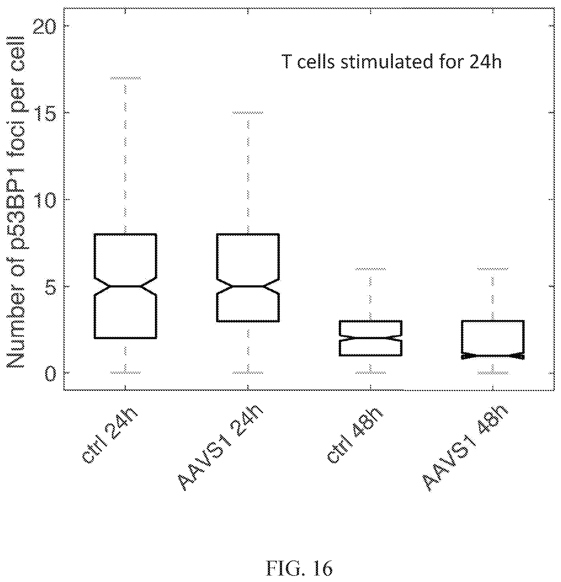
D00022

D00023
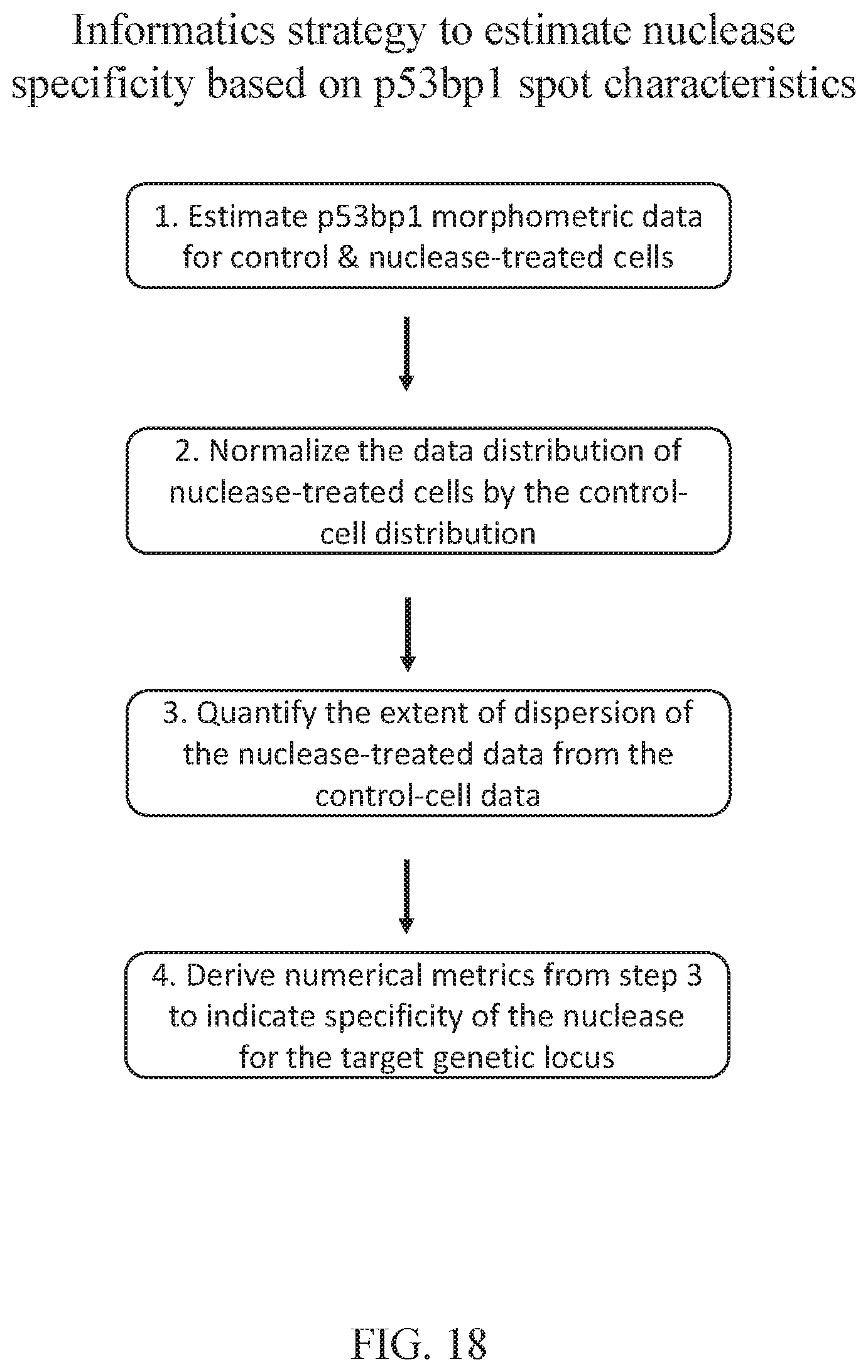
D00024

D00025

D00026
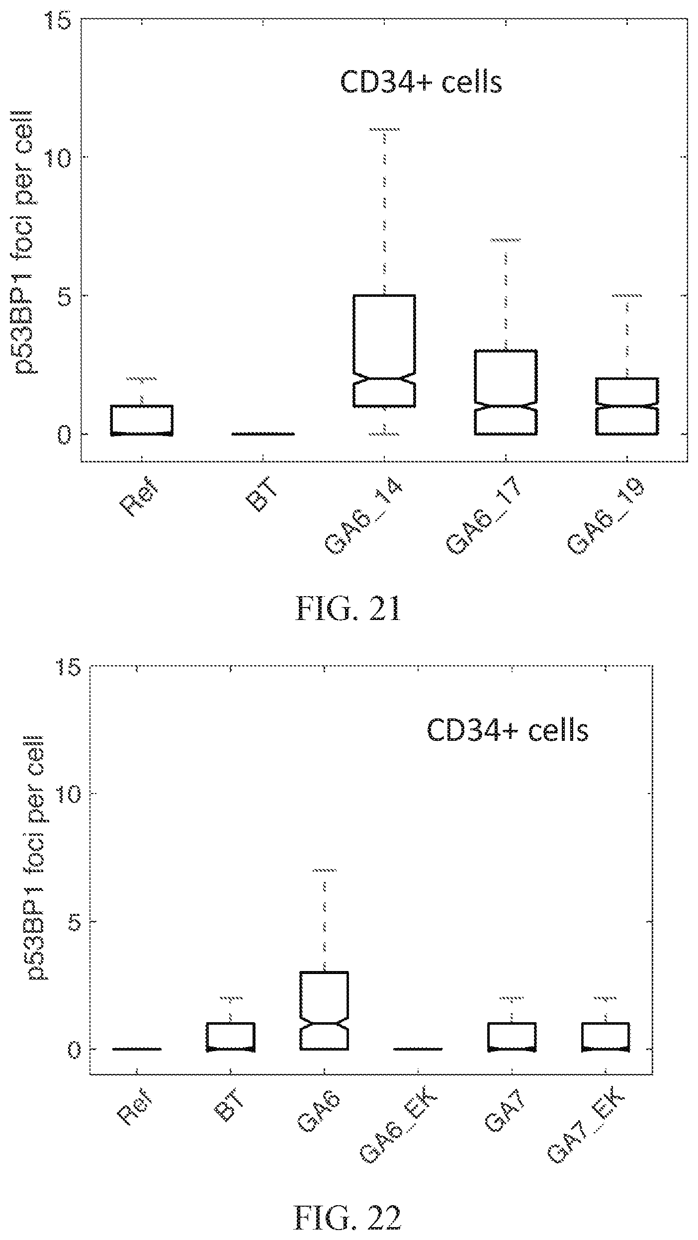
D00027

D00028

D00029
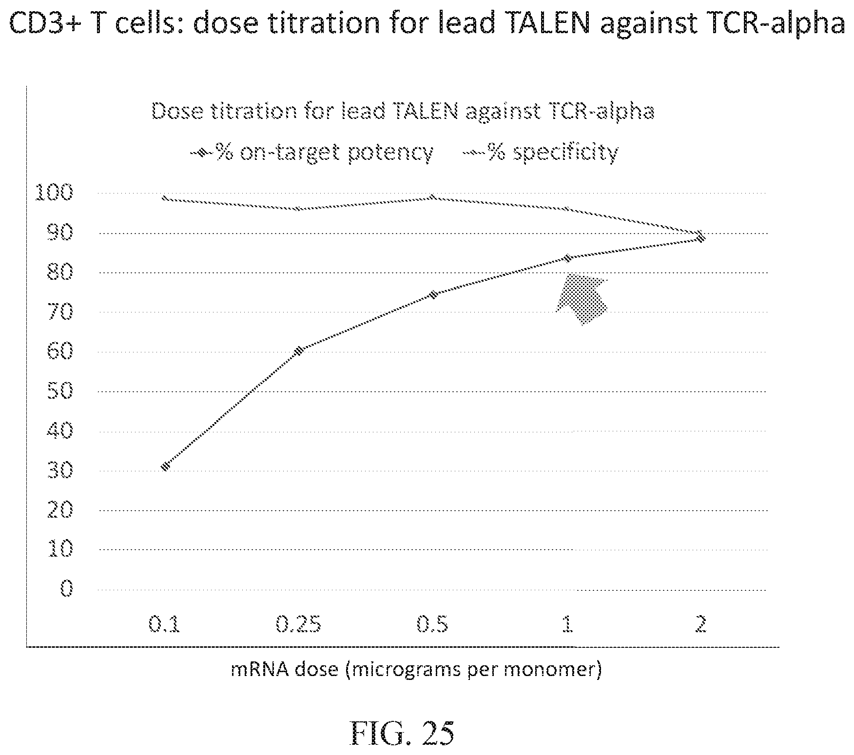
D00030

D00031

S00001
XML
uspto.report is an independent third-party trademark research tool that is not affiliated, endorsed, or sponsored by the United States Patent and Trademark Office (USPTO) or any other governmental organization. The information provided by uspto.report is based on publicly available data at the time of writing and is intended for informational purposes only.
While we strive to provide accurate and up-to-date information, we do not guarantee the accuracy, completeness, reliability, or suitability of the information displayed on this site. The use of this site is at your own risk. Any reliance you place on such information is therefore strictly at your own risk.
All official trademark data, including owner information, should be verified by visiting the official USPTO website at www.uspto.gov. This site is not intended to replace professional legal advice and should not be used as a substitute for consulting with a legal professional who is knowledgeable about trademark law.