Micellar Spherical Nucleic Acids From Thermoresponsive, Traceless Templates
MIRKIN; Chad A. ; et al.
U.S. patent application number 16/328025 was filed with the patent office on 2019-09-12 for micellar spherical nucleic acids from thermoresponsive, traceless templates. The applicant listed for this patent is NORTHWESTERN UNIVERSITY. Invention is credited to Resham J. BANGA, Chad A. MIRKIN, Sonbinh T. NGUYEN.
| Application Number | 20190275166 16/328025 |
| Document ID | / |
| Family ID | 61246323 |
| Filed Date | 2019-09-12 |


| United States Patent Application | 20190275166 |
| Kind Code | A1 |
| MIRKIN; Chad A. ; et al. | September 12, 2019 |
MICELLAR SPHERICAL NUCLEIC ACIDS FROM THERMORESPONSIVE, TRACELESS TEMPLATES
Abstract
Methods of preparing spherical nucleic acids using thermoresponsive traceless templates, such as PEO-PPO-PEO block copolymers.
| Inventors: | MIRKIN; Chad A.; (Wilmette, IL) ; NGUYEN; Sonbinh T.; (Evanston, IL) ; BANGA; Resham J.; (Evanston, IL) | ||||||||||
| Applicant: |
|
||||||||||
|---|---|---|---|---|---|---|---|---|---|---|---|
| Family ID: | 61246323 | ||||||||||
| Appl. No.: | 16/328025 | ||||||||||
| Filed: | August 25, 2017 | ||||||||||
| PCT Filed: | August 25, 2017 | ||||||||||
| PCT NO: | PCT/US2017/048726 | ||||||||||
| 371 Date: | February 25, 2019 |
Related U.S. Patent Documents
| Application Number | Filing Date | Patent Number | ||
|---|---|---|---|---|
| 62379352 | Aug 25, 2016 | |||
| Current U.S. Class: | 1/1 |
| Current CPC Class: | C07H 21/04 20130101; C12N 2310/11 20130101; C12N 15/113 20130101; C12N 2310/141 20130101; A61K 31/7105 20130101; C07H 21/02 20130101; A61K 31/711 20130101; C12N 15/88 20130101; A61K 47/6917 20170801 |
| International Class: | A61K 47/69 20060101 A61K047/69; C12N 15/88 20060101 C12N015/88; A61K 31/7105 20060101 A61K031/7105; A61K 31/711 20060101 A61K031/711; C12N 15/113 20060101 C12N015/113 |
Goverment Interests
STATEMENT OF GOVERNMENT SUPPORT
[0002] This invention was made with government support under U54 CA199091 and U54 CA151880 awarded by the National Institutes of Health. The government has certain rights in the invention.
Claims
1. A method of making a crosslinked micellar spherical nucleic acid (SNA) comprising: (a) admixing a polyethyleneoxide-polypropyleneoxide-polyethyleneoxide (PEO-PPO-PEO) block copolymer and a plurality of amphiphilic oligonucleotides in a buffer to form a SNA, wherein the amphiphilic oligonucleotide comprises (i) a lipid moiety and (ii) at least one reactive group; (b) admixing the resulting SNA with a PEGylated crosslinking agent to form the crosslinked micellar SNA, wherein the PEGylated crosslinking agent comprises at least two reactive sites, each reactive site reacting with the reactive group-of the amphiphilic oligonucleotide to form a covalent bond and thereby crosslink the amphiphilic oligonucleotides, and (c) optionally washing the crosslinked micellar SNA to remove uncrosslinked amphiphilic oligonucleotides.
2. The method of claim 1, wherein the PEO-PPO-PEO is present at a concentration of 0.5 wt % to 5 wt %.
3. (canceled)
4. The method of claim 1, wherein the amphiphilic oligonucleotide comprises an oligonucleotide sequence of 10 to 100 nucleobases.
5. (canceled)
6. The method of claim 1, wherein the amphiphilic oligonucleotide comprises a section of at least three nucleobases each having an amine functional group.
7. The method of claim 1, wherein the amphiphilic oligonucleotide comprises a section having a therapeutic sequence.
8. (canceled)
9. (canceled)
10. (canceled)
11. (canceled)
12. The method of claim 1, wherein the lipid of the amphiphilic oligonucleotide comprises 1,2-dioleoyl-sn-glycero-3-phosphocholine (DOPC), 1,2-dimyristoyl-sn-phosphatidylcholine (DMPC), 1-palmitoyl-2-oleoyl-sn-phosphatidylcholine (POPC), 1,2-distearoyl-sn-glycero-3-phospho-(1'-rac-glycerol) (DSPG), 1,2-dioleoyl-sn-glycero-3-phospho-(1'-rac-glycerol) (DOPG), 1,2-distearoyl-sn-glycero-3-phosphocholine (DSPC), 1,2-dipalmitoyl-sn-glycero-3-phosphocholine (DPPC), 1,2-di-(9Z-octadecenoyl)-sn-glycero-3-phosphoethanolamine (DOPE), or 1,2-dihexadecanoyl-sn-glycero-3-phosphoethanolamine (DPPE).
13. The method of claim 1, wherein the reactive group of the amphiphilic oligonucleotide comprises an amine, a hydroxyl, a succinimidyl, an alkyne, or an azide.
14. The method of claim 1, wherein the reactive sites of the PEGylated crosslinking agent comprise an amine, a hydroxyl, a succinimidyl, an alkyne, or an azide.
15. (canceled)
16. (canceled)
17. The method of claim 1, wherein the lipid moiety and the oligonucleotide are connected via a triazolyl.
18. (canceled)
19. (canceled)
20. The method of claim 1, further comprising cooling the crosslinked micellar SNA to a temperature below the micelle disassembly temperature to remove any free PEO-PPO-PEO.
21. (canceled)
22. A crosslinked micellar spherical nucleic acid (SNA) prepared by the method of claim 1.
23. A crosslinked micellar spherical nucleic acid (SNA) comprising a plurality of oligonucleotides and polyethyleneoxide-polypropyleneoxide-polyethyleneoxide (PEO-PPO-PEO), wherein each oligonucleotide comprises (i) a lipid portion and (ii) a nucleobase portion, and the oligonucleotides are crosslinked with a PEGylated crosslinking agent.
24. The crosslinked micellar SNA of claim 23, having a diameter of 150 nm or less.
25. (canceled)
26. (canceled)
27. (canceled)
28. The crosslinked micellar SNA of claim 23, having at least 200 strands of amphiphilic oligonucleotides.
29. (canceled)
30. The crosslinked micellar SNA of claim 23, wherein the SNA is cooperatively bound to a complementary SNA.
31. A method of inhibiting expression of a gene comprising the step of hybridizing a polynucleotide encoding said gene product with one or more oligonucleotides complementary to all or a portion of said polynucleotide, said oligonucleotide being attached to the crosslinked micellar SNA of claim 23, wherein hybridizing between said polynucleotide and said oligonucleotide occurs over a length of said polynucleotide with a degree of complementarity sufficient to inhibit expression of said gene product.
32. (canceled)
33. (canceled)
34. The method of claim 31, wherein the oligonucleotide comprises RNA and the RNA is a non-coding RNA or a microRNA.
35. (canceled)
36. (canceled)
37. (canceled)
38. (canceled)
39. The method of claim 31, wherein the oligonucleotide is DNA and the DNA is antisense-DNA.
40. (canceled)
41. A method for up-regulating activity of a toll-like receptor (TLR), comprising contacting a cell having the toll-like receptor with a crosslinked micellar SNA of claim 23.
42. (canceled)
43. (canceled)
44. A method for down-regulating activity of a toll-like receptor (TLR), comprising contacting a cell having the TLR with a crosslinked micellar SNA of claim 23.
45. (canceled)
46. (canceled)
47. (canceled)
48. (canceled)
Description
CROSS-REFERENCE TO RELATED APPLICATION
[0001] This application claims the benefit of U.S. Provisional Application Ser. No. 62/379,352, filed Aug. 25, 2016, the disclosure of which is incorporated herein by reference in its entirety.
FIELD OF THE DISCLOSURE
[0003] The disclosure relates generally to spherical nucleic acids. More particularly, the disclosure related to crosslinked micellar spherical nucleic acids that can be prepared under moderate conditions.
BACKGROUND
[0004] Spherical nucleic acids (SNAs) have become an important platform for programmable assembly,.sup.1 bio-detection.sup.2, and nucleic acid-based therapeutics..sup.3 Historically, SNAs have been synthesized by covalently immobilizing a dense layer of highly oriented nucleic acids onto a spherical gold nanoparticle core..sup.4 The resulting three-dimensional polyvalent architecture of the SNA makes it a higher affinity binder than the same linear sequence from which it is comprised..sup.5 The dense oligonucleotide shell also increases the resistance of SNAs to enzymatic degradation,.sup.6 increasing intact oligonucleotide lifetimes. In addition, by engaging cell-surface receptors, SNAs can actively traverse cell membranes without the need for transfection agents..sup.7 As a result, SNAs have emerged as "single-entity" intracellular diagnostic tools,.sup.2a gene-regulating structures,.sup.3 and immunomodulatory agents.sup.8 that exhibit minimal cytotoxicity and non-specific immunogenic responses..sup.9
[0005] Since the aforementioned physical and biological properties of SNAs are independent of the nature of the core,.sup.5 a broad range of materials (Au,.sup.4 Ag,.sup.10 .gamma.-Fe.sub.2O.sub.3,.sup.11 CdSe, Pt, Pd) have been used as templates for their syntheses. However, concerns about the potential long-term toxicity and metabolic fate of metallic nanoparticle cores.sup.12,13 have inspired a shift to the use of organic templates such as lipisomes,.sup.14 proteins,.sup.15 and block copolymer nanostructures.sup.16 as template materials.
[0006] An ideal SNA is one that can be rapidly made under moderate conditions from biocompatible reagents in monodisperse and size tunable form. Thus, a need exists for such methods to prepare SNAs.
SUMMARY
[0007] One aspect of the disclosure provides a method of making a crosslinked micellar spherical nucleic acid (SNA) comprising (a) admixing a polyethyleneoxide-polypropyleneoxide-polyethyleneoxide (PEO-PPO-PEO) block copolymer and a plurality of amphiphilic oligonucleotides in a buffer to form an SNA, wherein the amphiphilic oligonucleotide comprises (i) a lipid moiety and (ii) at least one reactive group, (b) admixing the resulting SNA with a PEGylated crosslinking agent to form the crosslinked micellar SNA, wherein the PEGylated crosslinking agent comprises at least two reactive sites, each reactive site reacting with the reactive group of the amphiphilic oligonucleotide to form a covalent bond and thereby crosslink the amphiphilic oligonucleotides, and (c) optionally washing the crosslinked micellar SNA to remove uncrosslinked amphiphilic oligonucleotides.
[0008] Another aspect of the disclosure provides a crosslinked micellar spherical nucleic acid prepared according to the method of the disclosure.
[0009] Another aspect of the disclosure provides a method of inhibiting expression of a gene comprising the step of hybridizing a polynucleotide encoding said gene product with one or more oligonucleotides complementary to all or a portion of said polynucleotide, said oligonucleotide being attached to the crosslinked micellar SNA of the disclosure, wherein hybridizing between said polynucleotide and said oligonucleotide occurs over a length of said polynucleotide with a degree of complementarity sufficient to inhibit expression of said gene product.
[0010] Another aspect of the disclosure provides a method for up-regulating activity of a toll-like receptor (TLR), comprising contacting a cell having the toll-like receptor with a crosslinked micellar SNA of the disclosure.
[0011] Another aspect of the disclosure provides a method for down-regulating toll-like receptor (TLR), comprising contacting a cell having the toll-like receptor with a crosslinked micellar SNA of the disclosure.
[0012] Further aspects and advantages will be apparent to those of ordinary skill in the art from a review of the following detailed descriptions. While the compositions and methods are susceptible of embodiments in various forms, the description hereafter includes specific embodiments with the understanding that the disclosure is illustrative and is not intended to limit the invention to the specific embodiments described herein.
BRIEF DESCRIPTION OF THE FIGURES
[0013] For a more complete understanding of the disclosure, reference should be made to the following detailed description and accompanying drawings wherein:
[0014] FIG. 1. (A) shows DLS histograms of the Pluronic F-127 templates before DNA insertion and the crosslinked micellar SNAs. (B) shows a plot of the amount of free, unincorporated DNA in the solution. The inset is a photographic image of the filtrates, showing the blue color of Cy-5 labeled DNA visually disappears in the 3.sup.rd wash. (C) is an AFM image of the crosslinked micellar SNAs after drop-cast and dried on mica. (D) is an AFM image of the crosslinked micellar SNAs after drop-casting and dried on mica. The inset shows a distribution centering at 30.+-.8 nm, slightly smaller than the DLS data as expected for dried materials.
[0015] FIG. 2. (A) shows a schematic representation of the hybridization of crosslinked micellar SNAs with complementary SNAs. (B) shows the melting profile of the crosslinked micellar SNA conjugates that have been hybridized to complementary nanoconstructs. (C) shows the fractions of nucleic acids dissociated from the crosslinked SNAs and non-crosslinked micellar SNAs over time after being incubated at 37.degree. C. in 10% serum-containing med.
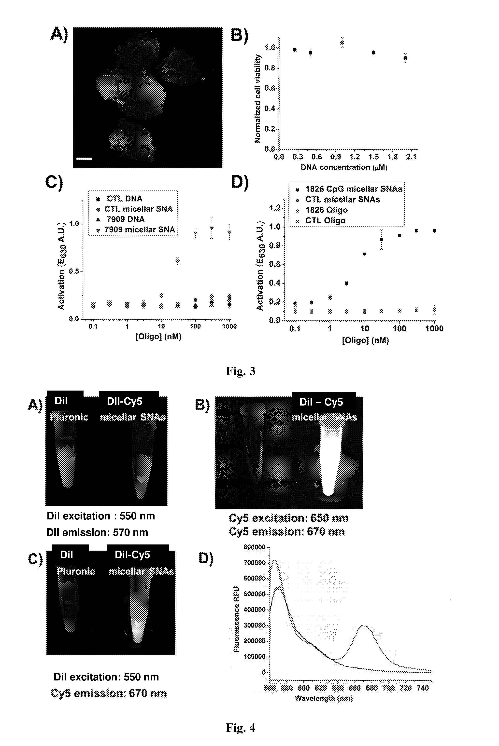
[0016] FIG. 3. (A) shows a confocal fluorescent micrograph of HEK-Blue cells that were incubated with 100 nM Cy5-labeled crosslinked micellar SNAs for 4 h. (B) shows a plot of normalized cell viability for HEKBlue mTLR9 cells after treatment with micellar SNAs at different DNA concentrations for 24 h. (C) shows a plot of potency of 7909 CpG-bearing immunostimulatory crosslinked micellar SNAs, control and crosslinked micellar SNAs (T.sub.20), and the two unmodified linear nucleic acids in HEK-Blue cells. (D) shows a plot of potency of 1826 CpG-bearing immunostimulatory crosslinked micellar SNAs, control and crosslinked micellar SNAs (T.sub.20), and the two unmodified linear nucleic acids in Ramos Blue cells.
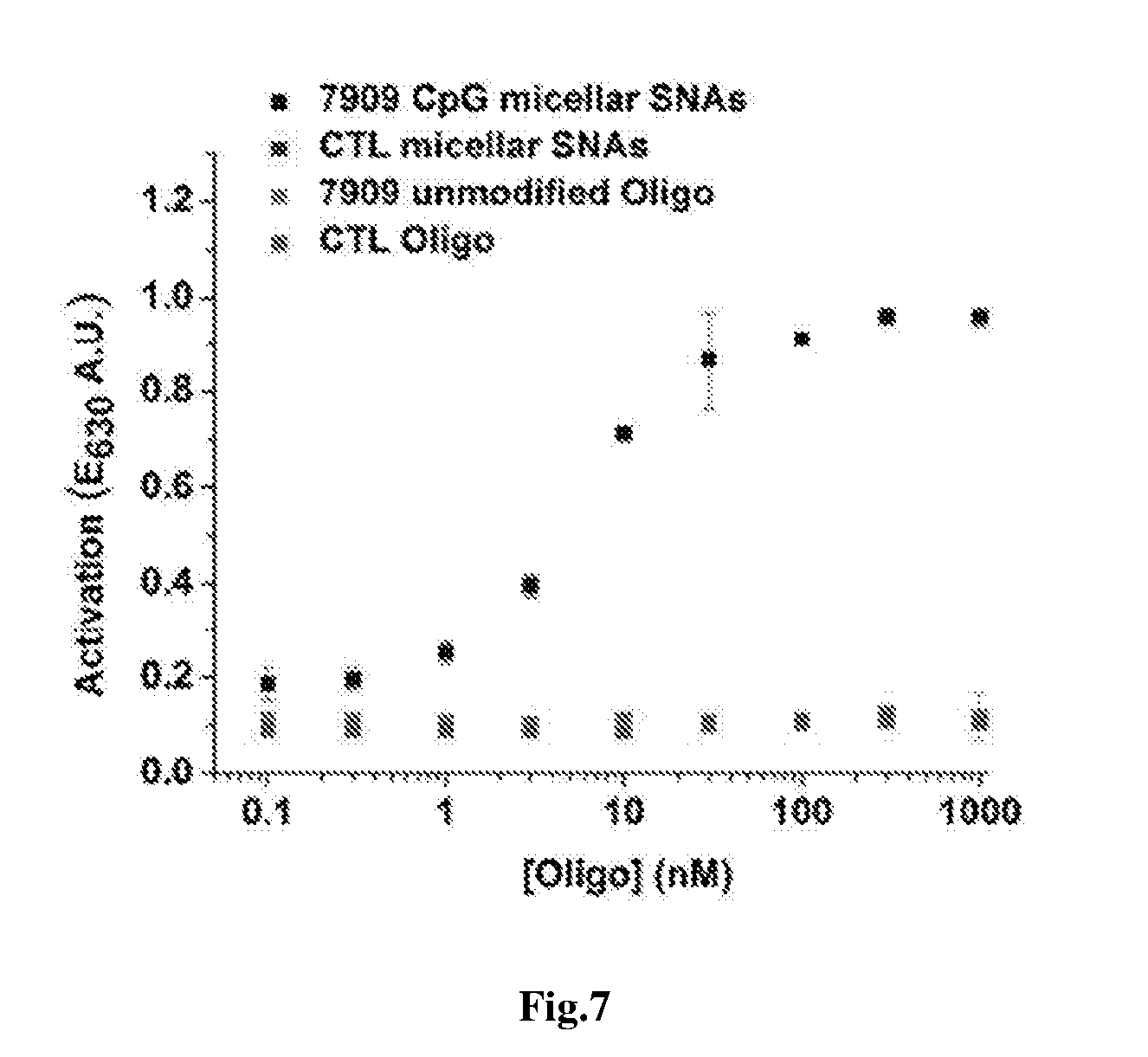
[0017] FIG. 4. (A) shows photographic images of Dil-encapsulated Pluronic F127 and Cy5-labeled DNA functionalized crosslinked micellar SNAs. (B) shows photographic images of Dil-encapsulated Pluronic F127 and Cy5-labeled DNA functionalized crosslinked micellar SNAs. (C) shows photographic images of Dil-encapsulated Pluronic F127 and Cy5-labeled DNA functionalized crosslinked micellar SNAs. (D) shows emission spectra of the Dil-encapsulated Pluronic F127 and Dil-encapsulated crosslinked micellar SNA.
[0018] FIG. 5 shows an image of a 1% agarose gel electrophoresis of micellar SNAs.
[0019] FIG. 6 shows an image of a 1% agarose gel electrophoresis of crosslinked micellar SNAs after being incubated in a 10% FBS solution in HBS for 2, 4, and 8 h (lanes 2-4).
[0020] FIG. 7 shows a plot of the amounts of secreted alkaline phosphatase (SEAP) by Ramos-Blue cells, as visualized by a colorimetric assay, showing enhanced immunostimulatory activity by micellar SNAs in comparison to control micellar SNAs bearing a T.sub.20 sequence and unmodified linear nucleic acids.
DETAILED DESCRIPTION
[0021] Provided herein are methods of making crosslinked micellar spherical nucleic acids (SNAs) under moderate conditions from biocompatible materials. The spherical nucleic acids of the disclosure comprise polyethyleneoxide-polypropyleneoxide-polyethyleneoxide, a plurality of oligonucleotides, wherein each oligonucleotide comprises a lipid moiety and a nucleobase portion, and the oligonucleotides are crosslinked with a PEGylated crosslinking agent. The methods disclosed herein allow for the facile assembly of nucleic acids with hydrophobic tails and stretches of functionalized T-bases biocompatible SNA constructs.
PEO-PPO-PEO Micelles
[0022] Polyoxyethylene-polyoxypropylene-polyoxyethylene (PEO-PPO-PEO) is an amphiphilic block copolymer that can be assembled into spherical micelles at room temperature at low critical micelle concentrations (CMC). In addition, PEO-PPO-PEO has a thermo-responsive CMC, and therefore micelles made from PEO-PPO-PEO can be easily assembled and disassembled based upon a change in temperature. The thermo-responsive CMC can be used for purifying the targeted SNA architectures..sup.18a, 19
[0023] Above their CMC, PEO-PPO-PEO block copolymers can assemble into small, monodispersed micelles that consist of a hydrophobic PPO as the core surrounded by a hydrophilic PEO shell..sup.20 The micelles can have a diameter of about 2 nm to about 100 nm in mean diameter, about 2 nm to about 90 nm in mean diameter, about 2 nm to about 80 nm in mean diameter, about 2 nm to about 70 nm in mean diameter, about 2 nm to about 60 nm in mean diameter, about 2 nm to about 50 nm in mean diameter, about 2 nm to about 40 nm in mean diameter, about 2 nm to about 30 nm in mean diameter, or about 2 nm to about 20 nm in mean diameter, about 2 nm to about 10 nm in mean diameter, about 5 nm to about 80 nm in mean diameter, about 5 nm to about 70 nm in mean diameter, about 5 nm to about 60 nm in mean diameter, about 5 nm to about 50 nm in mean diameter, about 5 nm to about 40 nm in mean diameter, about 5 nm to about 30 nm in mean diameter, about 6 nm to about 25 nm in mean diameter, about 7 nm to about 20 nm in mean diameter, about 8 nm to about 15 nm in mean diameter, about 8 nm to about 12 nm in mean diameter, about 10 nm to about 14 nm in mean diameter. At these size ranges, PEO-PPO-PEO-derived micelles advantageously demonstrate long in vivo circulation time and can deliver encapsulated chemotherapeutics into a tumor tissue via the enhanced permeation and retention (EPR) effect..sup.21
[0024] The PEO-PPO-PEO block copolymers can have a molecular weight in a range of about 500 Da to about 20000 Da, about 500 Da to about 14000, about 800 Da to about 13800 Da, about 1000 Da to about 12000 Da, about 1500 Da to about 11800 Da, about 2000 Da to about 11000 Da, about 2200 Da to about 10000 Da, about 2500 Da to about 9000 Da, about 3000 Da to about 8000 Da, about 3300 Da to about 7000 Da, about 3500 Da to about 6500 Da, about 3500 Da, about 6000 Da, about 1200 Da, and/or up to about 20000 Da. The block copolymer can be represented by the formula (PEO).sub.x(PEO).sub.y(PEO).sub.z wherein x can be in a range of 2 to 130, y can be in a range of 6 to 67, and z can be in a range of 2 to 130. In some embodiments, x and z have different values. In some embodiments, x and z have the same value. PEO-PPO-PEO block copolymers, also known as poloxamers, are available commercially, for example, under the Pluronic.RTM. tradename from BASF Chemicals, under the Synperonics.RTM. tradename from Croda International Chemicals Company, and under the Kolliphor.RTM. tradename from BASF Chemicals. Suitable block copolymers are pharmaceutical-grade. An exemplary pharmaceutical grade PEO-PPO-PEO block copolymer is Pluronic.RTM. F127, a solid block copolymer having a molecular weight of about 12500 Da and a 70% polyoxyethylene content and can be represented by the formula (PEO).sub.98(PPO).sub.67(PEO).sub.98.
[0025] Methods of forming PEO-PPO-PEO micelles are known in the art. PEO-PPO-PEO micelles readily assemble when the block copolymer is provided in solution in a concentration in an amount greater than its critical micelle concentration at a given solution temperature.
Spherical Nucleic Acids
[0026] The SNA can have a diameter of about 2 nm to about 250 nm in mean diameter, about 2 nm to about 240 nm in mean diameter, about 2 nm to about 230 nm in mean diameter, about 2 nm to about 220 nm in mean diameter, about 2 nm to about 210 nm in mean diameter, about 2 nm to about 200 nm in mean diameter, about 2 nm to about 190 nm in mean diameter, about 2 nm to about 180 nm in mean diameter, about 2 nm to about 170 nm in mean diameter, about 2 nm to about 160 nm in mean diameter, about 2 nm to about 150 nm in mean diameter, about 2 nm to about 140 nm in mean diameter, about 2 nm to about 130 nm in mean diameter, about 2 nm to about 120 nm in mean diameter, about 2 nm to about 110 nm in mean diameter, about 2 nm to about 100 nm in mean diameter, about 2 nm to about 90 nm in mean diameter, about 2 nm to about 80 nm in mean diameter, about 2 nm to about 70 nm in mean diameter, about 2 nm to about 60 nm in mean diameter, about 2 nm to about 50 nm in mean diameter, about 2 nm to about 40 nm in mean diameter, about 2 nm to about 30 nm in mean diameter, or about 2 nm to about 20 nm in mean diameter, about 2 nm to about 10 nm in mean diameter. The size of the SNA is from about 5 nm to about 150 nm (mean diameter), from about 5 to about 50 nm, from about 10 to about 30 nm, from about 10 to 150 nm, from about 10 to about 100 nm, from about 20 nm to about 150 nm, from about 10 to about 50 nm, from about 20 nm to about 50 nm, from about 25 to about 45 nm, or from about 30 nm to about 40 nm. The size of the SNA is from about 5 nm to about 150 nm (mean diameter), from about 30 to about 100 nm, from about 40 to about 80 nm. The size of the SNA used in a method varies as required by their particular use or application. The variation of size is advantageously used to optimize certain physical characteristics of the SNA, for example, optical properties or the amount of surface area that can be functionalized as described herein.
[0027] The SNA provided herein comprise amphiphilic oligonucleotides including (i) a lipid portion and (ii) a nucleobase portion. The SNA can include at least about 10 strands of amphiphilic oligonucleotides, at least about 50 strands of amphiphilic oligonucleotides, at least about 100 strands of amphiphilic oligonucleotides, at least about 150 strands of amphiphilic oligonucleotides, at least about 200 strands of amphiphilic oligonucleotides, at least about 250 strands of oligonucleotides, at least 300 strands of oligonucleotides, or at least 350 strands of oligonucleotides and/or up to about 400 strands of oligonucleotides, up to about 350 strands of oligonucleotides, or up to about 300 strands of oligonucleotides.
[0028] The oligonucleotide can comprise either RNA or DNA. In embodiments, the oligonucleotide comprises RNA. The RNA can be an inhibitory RNA (RNAi) that performs a regulatory function, and is chosen from the group consisting of a small RNAi that is selected from the group consisting of a small inhibitory RNA (siRNA), an RNA that forms a triplex with double stranded DNA, and a ribozyme. Alternatively, the RNA is microRNA that performs a regulatory function. In still further embodiments, the RNA is a piwi-interacting RNA (piRNA). In embodiments, the oligonucleotide comprises DNA. The DNA can be, in some embodiments, an antisense-DNA.
[0029] Oligonucleotides contemplated for use according to the disclosure are from about 5 to about 100 nucleotides in length. Methods and compositions are also contemplated wherein the oligonucleotide is about 10 to about 100 nucleotides in length, about 5 to about 90 nucleotides in length, about 5 to about 80 nucleotides in length, about 5 to about 70 nucleotides in length, about 5 to about 60 nucleotides in length, about 5 to about 50 nucleotides in length about 5 to about 45 nucleotides in length, about 5 to about 40 nucleotides in length, about 5 to about 35 nucleotides in length, about 5 to about 30 nucleotides in length, about 5 to about 25 nucleotides in length, about 5 to about 20 nucleotides in length, about 5 to about 15 nucleotides in length, about 5 to about 10 nucleotides in length, and all oligonucleotides intermediate in length of the sizes specifically disclosed, for example about 15 to about 35 nucleotides, to the extent that the oligonucleotide is able to achieve the desired result. Accordingly, oligonucleotides of 5, 6, 7, 8, 9, 10, 11, 12, 13, 14, 15, 16, 17, 18, 19, 20, 21, 22, 23, 24, 25, 26, 27, 28, 29, 30, 31, 32, 33, 34, 35, 36, 37, 38, 39, 40, 41, 42, 43, 44, 45, 46, 47, 48, 49, 50, 51, 52, 53, 54, 55, 56, 57, 58, 59, 60, 61, 62, 63, 64, 65, 66, 67, 68, 69, 70, 71, 72, 73, 74, 75, 76, 77, 78, 79, 80, 81, 82, 83, 84, 85, 86, 87, 88, 89, 90, 91, 92, 93, 94, 95, 96, 97, 98, 99, and 100 nucleotides in length are contemplated. Throughout, the term nucleotide is interchangeably referred to as a nucleobase. In embodiments, the oligonucleotide comprises a section of at least two nucleobases each having an amine functional group, at least three nucleobases each having an amine functional group, or at least four nucleobases each having an amine functional group and/or up to six nucleobases each having an amine functional group, up to five nucleobases each having an amine functional group, or up to four nucleobases each having an amine functional group.
[0030] The oligonucleotide can include a section having a therapeutic sequence. The therapeutic sequence can encode a single gene, multiple genes, chimeric proteins, DNA sequences or regulator RNA, or precursor of such regulatory RNA molecules. Encoded proteins can include signal peptides to aid in the excretion of gene products and/or other specific sequences to aid in the delivery, stability and activity of the gene product, depending on the therapeutic application. In embodiments, the therapeutic sequence comprises an immunomodulatory sequence. In embodiments, the therapeutic sequence can be complementary to a target polynucleotide.
Modified Oligonucleotides
[0031] Specific examples of oligonucleotides include those containing modified backbones or non-natural internucleoside linkages. Oligonucleotides having modified backbones include those that retain a phosphorus atom in the backbone and those that do not have a phosphorus atom in the backbone. Modified oligonucleotides that do not have a phosphorus atom in their internucleoside backbone are considered to be within the meaning of "oligonucleotide."
[0032] Modified oligonucleotide backbones containing a phosphorus atom include, for example, phosphorothioates, chiral phosphorothioates, phosphorodithioates, phosphotriesters, aminoalkylphosphotriesters, methyl and other alkyl phosphonates including 3'-alkylene phosphonates, 5'-alkylene phosphonates and chiral phosphonates, phosphinates, phosphoramidates including 3'-amino phosphoramidate and aminoalkylphosphoramidates, thionophosphoramidates, thionoalkylphosphonates, thionoalkylphosphotriesters, selenophosphates and boranophosphates having normal 3'-5' linkages, 2'-5' linked analogs of these, and those having inverted polarity wherein one or more internucleotide linkages is a 3' to 3', 5' to 5' or 2' to 2' linkage. Also contemplated are oligonucleotides having inverted polarity comprising a single 3' to 3' linkage at the 3'-most internucleotide linkage, i.e. a single inverted nucleoside residue which can be a basic (the nucleotide is missing or has a hydroxyl group in place thereof). Salts, mixed salts and free acid forms are also contemplated. Representative United States patents that teach the preparation of the above phosphorus-containing linkages include, U.S. Pat. Nos. 3,687,808; 4,469,863; 4,476,301; 5,023,243; 5,177,196; 5,188,897; 5,264,423; 5,276,019; 5,278,302; 5,286,717; 5,321,131; 5,399,676; 5,405,939; 5,453,496; 5,455,233; 5,466,677; 5,476,925; 5,519,126; 5,536,821; 5,541,306; 5,550,111; 5,563,253; 5,571,799; 5,587,361; 5,194,599; 5,565,555; 5,527,899; 5,721,218; 5,672,697 and 5,625,050, the disclosures of which are incorporated by reference herein.
[0033] Modified oligonucleotide backbones that do not include a phosphorus atom therein have backbones that are formed by short chain alkyl or cycloalkyl internucleoside linkages, mixed heteroatom and alkyl or cycloalkyl internucleoside linkages, or one or more short chain heteroatomic or heterocyclic internucleoside linkages. These include those having morpholino linkages; siloxane backbones; sulfide, sulfoxide and sulfone backbones; formacetyl and thioformacetyl backbones; methylene formacetyl and thioformacetyl backbones; riboacetyl backbones; alkene containing backbones; sulfamate backbones; methyleneimino and methylenehydrazino backbones; sulfonate and sulfonamide backbones; amide backbones; and others having mixed N, O, S and CH.sub.2 component parts. See, for example, U.S. Pat. Nos. 5,034,506; 5,166,315; 5,185,444; 5,214,134; 5,216,141; 5,235,033; 5,264,562; 5,264,564; 5,405,938; 5,434,257; 5,466,677; 5,470,967; 5,489,677; 5,541,307; 5,561,225; 5,596,086; 5,602,240; 5,610,289; 5,602,240; 5,608,046; 5,610,289; 5,618,704; 5,623,070; 5,663,312; 5,633,360; 5,677,437; 5,792,608; 5,646,269 and 5,677,439, the disclosures of which are incorporated herein by reference in their entireties.
[0034] In still other embodiments, oligonucleotide mimetics wherein both one or more sugar and/or one or more internucleotide linkage of the nucleotide units are replaced with "non-naturally occurring" groups. In one aspect, this embodiment contemplates a peptide nucleic acid (PNA). In PNA compounds, the sugar-backbone of an oligonucleotide is replaced with an amide containing backbone. See, for example U.S. Pat. Nos. 5,539,082; 5,714,331; and 5,719,262, and Nielsen et al., 1991, Science, 254: 1497-1500, the disclosures of which are herein incorporated by reference.
[0035] In still other embodiments, oligonucleotides are provided with phosphorothioate backbones and oligonucleosides with heteroatom backbones, and including --CH.sub.2--NH--O--CH.sub.2--, --CH.sub.2--N(CH.sub.3)--O--CH.sub.2--, --CH.sub.2--O--N(CH.sub.3)--CH.sub.2--, --CH.sub.2--N(CH.sub.3)--N(CH.sub.3)--CH.sub.2-- and --O--N(CH.sub.3)--CH.sub.2--CH.sub.2-- described in U.S. Pat. Nos. 5,489,677, and 5,602,240. Also contemplated are oligonucleotides with morpholino backbone structures described in U.S. Pat. No. 5,034,506.
[0036] In various forms, the linkage between two successive monomers in the oligonucleotide consists of 2 to 4, desirably 3, groups/atoms selected from --CH.sub.2--, --O--, --S--, --NR.sup.H--, >C.dbd.O, >C.dbd.NR.sup.H, >C=S, --Si(R'').sub.2--, --SO--, --S(O).sub.2--, --P(O).sub.2--, PO(BH.sub.3)--, --P(O,S)--, --P(S).sub.2--, --PO(R'')--, --PO(OCH.sub.3)--, and --PO(NHR.sup.H)--, where R.sup.H is selected from hydrogen and C.sub.1-4-alkyl, and R'' is selected from C.sub.1-6-alkyl and phenyl. Illustrative examples of such linkages are --CH.sub.2--CH.sub.2--CH.sub.2--, --CH.sub.2--CO--CH.sub.2--, --CH.sub.2--CHOH--CH.sub.2--, --O--CH.sub.2--O--, --O--CH.sub.2--CH.sub.2--, --O--CH.sub.2--CH.dbd., --CH.sub.2--CH.sub.2--O--, --NR.sup.H--CH.sub.2--CH.sub.2--, --CH.sub.2--CH.sub.2--NR.sup.H, --CH.sub.2--NR.sup.H--CH.sub.2--, --O--CH.sub.2--CH.sub.2--NR.sup.H--, --NR.sup.H--CO--O--, --NR.sup.H--CO--NR.sup.H--, --NR.sup.H--CS--NR.sup.H, --NR.sup.H--C(.dbd.NR.sup.H)--NR.sup.H--, --NR.sup.H--CO--CH.sub.2--NR.sup.H--O--CO--O--, O--CO--CH.sub.2--O--, --O--CH.sub.2--CO--O--, --CH.sub.2--CO--NR.sup.H--, --O--CO--NR.sup.H--, --NR.sup.HCO--CH.sub.2--, --O--CH.sub.2--CO--NR.sup.H--, --O--CH.sub.2--CH.sub.2--NR.sup.H--, --CH.dbd.N--O--, --CH.sub.2--NR.sup.H--O--, --CH.sub.2--O--N=, --CH.sub.2--O--NR.sup.H--, --CO--NR.sup.H--CH.sub.2--, --CH.sub.2--NR.sup.H--O--, --CH.sub.2--NR.sup.H--CO--, --O--NR.sup.H--CH.sub.2--, --O--NR.sup.H, --O--CH.sub.2--S--, --S--CH.sub.2--O--, --CH.sub.2--CH.sub.2--S--, --O--CH.sub.2--CH.sub.2--S--, --S--CH.sub.2--CH.dbd., --S--CH.sub.2--CH.sub.2--, --S--CH.sub.2--CH.sub.2--O--, --S--CH.sub.2--CH.sub.2--S--, CH.sub.2--S--CH.sub.2--, --CH.sub.2--SO--CH.sub.2--, --CH.sub.2--SO.sub.2--CH.sub.2--, --O--SO--O--, --O--S(O).sub.2--O--, --O--S(O).sub.2--CH.sub.2--, --OS(O).sub.2--NR.sup.H, --NR.sup.H--S(O).sub.2--CH.sub.2--; --O--S(O).sub.2--CH.sub.2--, --O--P(O).sub.2--O--, --O--P(O,S)--O--, --O--P(S).sub.2--O--, --S--P(O).sub.2--O--, --S--P(O,S)--O--, --S--P(S).sub.2--O--, --O--P(O).sub.2--S--, --O--P(O,S)--S--, --O--P(S).sub.2--S--, --S--P(O).sub.2--S--, --S--P(O,S)--S--, --S--P(S).sub.2--S--, --O--PO(R'')--O--, --O--PO(OCH.sub.3)--O--, --O--PO(O CH.sub.2CH.sub.3)--O--, --O--PO(O CH.sub.2CH.sub.2S--R)--O--, --O--PO(BH.sub.3)--O--, --O--PO(NHR.sup.N)--O--, --O--P(O).sub.2--NR.sup.H H--, --NR.sup.H--P(O).sub.2--O--, --O--P(O,NR.sup.H)--O--, --CH.sub.2--P(O).sub.2--O--, --O--P(O).sub.2--CH.sub.2--, and --O--Si(R'').sub.2--O--; among which --CH.sub.2--CO--NR.sup.H--, --CH.sub.2--NR.sup.H--O--, --S--CH.sub.2--O--, --O--P(O).sub.2--O--O--P(--O,S)--O--, --O--P(S).sub.2--O--, --NR.sup.H P(O).sub.2--O--, --O--P(O,NR.sup.H)--O--, --O--PO(R'')--O--, --O--PO(CH.sub.3)--O--, and --O--PO(NHR.sup.N)--O--, where R.sup.H is selected form hydrogen and C.sub.1-4-alkyl, and R'' is selected from C.sub.1-6-alkyl and phenyl, are contemplated. Further illustrative examples are given in Mesmaeker et. al., 1995, Current Opinion in Structural Biology, 5: 343-355 and Susan M. Freier and Karl-Heinz Altmann, 1997, Nucleic Acids Research, vol 25: pp 4429-4443.
[0037] Still other modified forms of oligonucleotides are described in detail in U.S. Patent Application No. 20040219565, the disclosure of which is incorporated by reference herein in its entirety.
[0038] Modified oligonucleotides can also contain one or more substituted sugar moieties. In certain aspects, oligonucleotides comprise one of the following at the 2' position: OH; F; O-, S-, or N-alkyl; O-, S-, or N-alkenyl; O-, S- or N-alkynyl; or O-alkyl-O-alkyl, wherein the alkyl, alkenyl and alkynyl can be substituted or unsubstituted C.sub.1 to C.sub.10 alkyl or C.sub.2 to C.sub.10 alkenyl and alkynyl. Other embodiments include O[(CH.sub.2).sub.nO].sub.mCH.sub.3, O(CH.sub.2).sub.nOCH.sub.3, O(CH.sub.2).sub.nNH.sub.2, O(CH.sub.2).sub.nCH.sub.3, O(CH.sub.2).sub.nONH.sub.2, and O(CH.sub.2).sub.nON[(CH.sub.2).sub.nCH.sub.3].sub.2, where n and m are from 1 to about 10. Other oligonucleotides comprise one of the following at the 2' position: C.sub.1 to C.sub.10 lower alkyl, substituted lower alkyl, alkenyl, alkynyl, alkaryl, aralkyl, O-alkaryl or O-aralkyl, SH, SCH.sub.3, OCN, Cl, Br, CN, CF.sub.3, OCF.sub.3, SOCH.sub.3, SO.sub.2CH.sub.3, ONO.sub.2, NO.sub.2, N.sub.3, NH.sub.2, heterocycloalkyl, heterocycloalkaryl, aminoalkylamino, polyalkylamino, substituted silyl, an RNA cleaving group, a reporter group, an intercalator, a group for improving the pharmacokinetic properties of an oligonucleotide, or a group for improving the pharmacodynamic properties of an oligonucleotide, and other substituents having similar properties. In one aspect, a modification includes 2'-methoxyethoxy (2'-O--CH.sub.2CH.sub.2OCH.sub.3, also known as 2'-O-(2-methoxyethyl) or 2'-MOE) (Martin et al., 1995, Helv. Chin. Acta, 78: 486-504) i.e., an alkoxyalkoxy group. Other modifications include 2'-dimethylaminooxyethoxy, i.e., a O(CH.sub.2).sub.2ON(CH.sub.3).sub.2 group, also known as 2'-DMAOE, as described in examples herein below, and 2'-dimethylaminoethoxyethoxy (also known in the art as 2'-O-dimethyl-amino-ethoxy-ethyl or 2'-DMAEOE), i.e., 2'-O--CH.sub.2--O--CH.sub.2N(CH.sub.3).sub.2, also described in examples herein below.
[0039] Still other modifications include 2'-methoxy (2'-O--CH.sub.3), 2'-aminopropoxy (2'-OCH.sub.2CH.sub.2CH.sub.2NH.sub.2), 2'-allyl (2'-CH.sub.2CH.dbd.CH.sub.2), 2'-O-allyl(2'-O--CH.sub.2CH.dbd.CH.sub.2) and 2'-fluoro (2'-F). The 2'-modification can be in the arabino (up) position or ribo (down) position. In one aspect, a 2'-arabino modification is 2'-F. Similar modifications can also be made at other positions on the oligonucleotide, for example, at the 3' position of the sugar on the 3' terminal nucleotide or in 2'-5' linked oligonucleotides and the 5' position of 5' terminal nucleotide. Oligonucleotides can also have sugar mimetics such as cyclobutyl moieties in place of the pentofuranosyl sugar. See, for example, U.S. Pat. Nos. 4,981,957; 5,118,800; 5,319,080; 5,359,044; 5,393,878; 5,446,137; 5,466,786; 5,514,785; 5,519,134; 5,567,811; 5,576,427; 5,591,722; 5,597,909; 5,610,300; 5,627,053; 5,639,873; 5,646,265; 5,658,873; 5,670,633; 5,792,747; and 5,700,920, the disclosures of which are incorporated herein by reference in their entireties.
[0040] In one aspect, a modification of the sugar includes Locked Nucleic Acids (LNAs) in which the 2'-hydroxyl group is linked to the 3' or 4' carbon atom of the sugar ring, thereby forming a bicyclic sugar moiety. The linkage is in certain aspects is a methylene (CH.sub.2).sub.n group bridging the 2' oxygen atom and the 4' carbon atom wherein n is 1 or 2. LNAs and preparation thereof are described in WO 98/39352 and WO 99/14226.
[0041] Oligonucleotides can also include base modifications or substitutions. As used herein, "unmodified" or "natural" bases include the purine bases adenine (A) and guanine (G), and the pyrimidine bases thymine (T), cytosine (C) and uracil (U). Modified bases include other synthetic and natural bases such as 5-methylcytosine (5mC), 5-hydroxymethyl cytosine, xanthine, hypoxanthine, 2-aminoadenine, 6-methyl and other alkyl derivatives of adenine and guanine, 2-propyl and other alkyl derivatives of adenine and guanine, 2-thiouracil, 2-thiothymine and 2-thiocytosine, 5-halouracil and cytosine, 5-propynyl uracil and cytosine and other alkynyl derivatives of pyrimidine bases; 6-azo uracil, cytosine and thymine; 5-uracil (pseudouracil); 4-thiouracil; 8-halo, 8-amino, 8-thiol, 8-thioalkyl, 8-hydroxyl and other 8-substituted adenines and guanines; 5-halo (particularly 5-bromo, 5-trifluoromethyl) and other 5-substituted uracils and cytosines; 7-methylguanine and 7-methyladenine, 2-F-adenine, 2-amino-adenine, 8-azaguanine and 8-azaadenine, 7-deazaguanine and 7-deazaadenine and 3-deazaguanine and 3-deazaadenine. Further modified bases include tricyclic pyrimidines such as phenoxazine cytidine(1H-pyrimido[5,4-b][1,4]benzoxazin-2(3H)-one), phenothiazine cytidine (1H-pyrimido[5,4-b][1,4]benzothiazin-2(3H)-one), G-clamps such as a substituted phenoxazine cytidine (e.g. 9-(2-aminoethoxy)-H-pyrimido[5,4-b][1,4]benzoxazin-2(3H)-one), carbazole cytidine (2H-pyrimido[4,5-b]indol-2-one), pyridoindole cytidine (H-pyrido[3',2':4,5]pyrrolo[2,3-d]pyrimidin-2-one). Modified bases can also include those in which the purine or pyrimidine base is replaced with other heterocycles, for example 7-deaza-adenine, 7-deazaguanosine, 2-aminopyridine and 2-pyridone. Further bases include those disclosed in U.S. Pat. No. 3,687,808, those disclosed in The Concise Encyclopedia of Polymer Science and Engineering, pages 858-859, Kroschwitz, J. I., ed. John Wiley & Sons, 1990, those disclosed by Englisch et al., 1991, Angewandte Chemie, International Edition, 30: 613, and those disclosed by Sanghvi, Y. S., Chapter 15, Antisense Research and Applications, pages 289-302, Crooke, S. T. and Lebleu, B., ed., CRC Press, 1993. Certain of these bases are useful for increasing the binding affinity and include 5-substituted pyrimidines, 6-azapyrimidines and N-2, N-6 and O--6 substituted purines, including 2-aminopropyladenine, 5-propynyluracil and 5-propynylcytosine. 5-methylcytosine substitutions have been shown to increase nucleic acid duplex stability by 0.6-1.2.degree. C. and are, in certain aspects combined with 2'-O-methoxyethyl sugar modifications. See, U.S. Pat. Nos. 3,687,808, 4,845,205; 5,130,302; 5,134,066; 5,175,273; 5,367,066; 5,432,272; 5,457,187; 5,459,255; 5,484,908; 5,502,177; 5,525,711; 5,552,540; 5,587,469; 5,594,121, 5,596,091; 5,614,617; 5,645,985; 5,830,653; 5,763,588; 6,005,096; 5,750,692 and 5,681,941, the disclosures of which are incorporated herein by reference.
[0042] A "modified base" or other similar term refers to a composition which can pair with a natural base (e.g., adenine, guanine, cytosine, uracil, and/or thymine) and/or can pair with a non-naturally occurring base. In certain aspects, the modified base provides a T.sub.n, differential of 15, 12, 10, 8, 6, 4, or 2.degree. C. or less. Exemplary modified bases are described in EP 1 072 679 and WO 97/12896.
[0043] By "nucleobase" is meant the naturally occurring nucleobases adenine (A), guanine (G), cytosine (C), thymine (T) and uracil (U) as well as non-naturally occurring nucleobases such as xanthine, diaminopurine, 8-oxo-N.sup.6-methyladenine, 7-deazaxanthine, 7-deazaguanine, N.sup.4,N.sup.4-ethanocytosin, N',N'-ethano-2,6-diaminopurine, 5-methylcytosine (mC), 5-(C.sup.3-C.sup.6)-alkynyl-cytosine, 5-fluorouracil, 5-bromouracil, pseudoisocytosine, 2-hydroxy-5-methyl-4-triiazolopyridin, isocytosine, isoguanine, inosine and the "non-naturally occurring" nucleobases described in Benner et al., U.S. Pat. No. 5,432,272 and Susan M. Freier and Karl-Heinz Altmann, 1997, Nucleic Acids Research, vol. 25: pp 4429-4443. The term "nucleobase" thus includes not only the known purine and pyrimidine heterocycles, but also heterocyclic analogues and tautomers thereof. Further naturally and non-naturally occurring nucleobases include those disclosed in U.S. Pat. No. 3,687,808 (Merigan, et al.), in Chapter 15 by Sanghvi, in Antisense Research and Application, Ed. S. T. Crooke and B. Lebleu, CRC Press, 1993, in Englisch et al., 1991, Angewandte Chemie, International Edition, 30: 613-722 (see especially pages 622 and 623, and in the Concise Encyclopedia of Polymer Science and Engineering, J. I. Kroschwitz Ed., John Wiley & Sons, 1990, pages 858-859; Cook, Anti-cancer Drug Design 1991, 6, 585-607, each of which are hereby incorporated by reference in their entirety). The term "nucleosidic base" or "base unit" is further intended to include compounds such as heterocyclic compounds that can serve like nucleobases including certain "universal bases" that are not nucleosidic bases in the most classical sense but serve as nucleosidic bases. Especially mentioned as universal bases are 3-nitropyrrole, optionally substituted indoles (e.g., 5-nitroindole), and optionally substituted hypoxanthine. Other desirable universal bases include, pyrrole, diazole or triazole derivatives, including those universal bases known in the art.
Spacers
[0044] In certain aspects, the oligonucleotide further comprises a spacer between the nucleotide portion and the lipid portion. "Spacer" as used herein means a moiety that does not participate in modulating gene expression per se but which serves to increase distance, for example, between the nucleobases and the lipid moiety. The spacer can be a polymer, including but not limited to a water-soluble polymer, a nucleic acid, a polypeptide, an oligosaccharide, a carbohydrate, a lipid, an ethylglycol, or combinations thereof.
Reactive Group
[0045] The amphiphilic oligonucleotide further comprises a reactive group. The amphiphilic oligonucleotide can be covalently bound to the crosslinking agent through the reactive group. In embodiments, the reactive group can include a nucleophile that can react with a succinimidyl group on a PEGylated crosslinking agent. The nucleophile can be any nucleophile that reacts with a succinimidyl group, for example an amine. In embodiments, the reactive group of the amphiphilic oligonucleotide comprises an amine, a hydroxyl, a succinimidyl, an alkyne, or an azide. In embodiments, the reactive group comprises an amine. In embodiments wherein the reactive group is an alkyne, the oligonucleotide can bind to a PEGylated crosslinking agent with an azide at each terminus (or vice versa) to allow crosslinking via a click chemistry type reaction.
Lipids
[0046] The lipid moiety of the amphiphilic oligonucleotide can be chosen from the phosphocholine family of lipids or the phosphoethanolamine family of lipids. Examples include 1,2-dioleoyl-sn-glycero-3-phosphocholine (DOPC), 1,2-dimyristoyl-sn-phosphatidylcholine (DMPC), 1-palmitoyl-2-oleoyl-sn-phosphatidylcholine (POPC), 1,2-distearoyl-sn-glycero-3-phospho-(1'-rac-glycerol) (DSPG), 1,2-dioleoyl-sn-glycero-3-phospho-(1'-rac-glycerol) (DOPG), 1,2-distearoyl-sn-glycero-3-phosphocholine (DSPC), 1,2-dipalmitoyl-sn-glycero-3-phosphocholine (DPPC), 1,2-di-(9Z-octadecenoyl)-sn-glycero-3-phosphoethanolamine (DOPE), and 1,2-dihexadecanoyl-sn-glycero-3-phosphoethanolamine (DPPE).
[0047] The lipid moiety and the oligonucleotide can be connected via Cu-free click chemistry using click-chemistry reactive pairs. Click-chemistry reactive pairs include a first click chemistry reagent (e.g., an azide) and a second click chemistry reagent (e.g., an alkyne). It will be appreciated that either entity of the click chemistry reactive pair can be incorporated into the lipid while the other entity of the reactive pair can be incorporated into the oligonucleotide. Additional suitable reactive pairs are well known in the art and include, but are not limited to, reactive pairs that couple amines to carboxylic acids, maleimides to sulfhydryls, vinyl sulfones to sulfhydryls, and acrylates to sulfhydryls. In embodiments, the lipid moiety and the oligonucleotide are connect via a triazolyl. The triazolyl can be formed by reacting an oligonucleotide having an alkyne moiety and a lipid having an azide moiety under conditions to form the triazolyl.
[0048] The amphiphilic oligonucleotide can be prepared by reacting a lipid moiety comprising a first click chemistry reagent with an oligonucleotide comprising a second click chemistry reagent, under conditions suitable for click chemistry. Suitable conditions are well known to one of ordinary skill in the art. For example, the lipid can be dissolved or suspended in a solvent, optionally activated, and admixed with an excess of a first click chemistry reagent, at ambient conditions. The reagent can be added in an amount to provide a reagent to lipid ratio of about 1:1 to about 1000:1, for example, about 1:1 to about 750:1, about 1:1 to about 500:1, about 1:1 to about 250:1, about 1:1 to about 100:1, about 1:1 to about 50:1, or about 1:1 to about 25:1. Similarly, the oligonucleotide can be dissolved in a solvent, optionally activated, and admixed with an excess of a second click chemistry reagent, at ambient conditions. The second reagent can be added in an amount to provide a reagent to oligonucleotide ratio of about 1:1 to about 1000:1, for example, about 1:1 to about 750:1, about 1:1 to about 500:1, about 1:1 to about 250:1, about 1:1 to about 100:1, about 1:1 to about 50:1, or about 1:1 to about 25:1. The lipid moiety comprising a first click chemistry reagent and oligonucleotide comprising a second click chemistry reagent can be admixed under ambient conditions to form the amphiphilic oligonucleotide. As used herein, and unless specified otherwise, "ambient conditions" refers to room temperature and atmospheric pressure.
PEGylated Crosslinking Agents
[0049] The amphiphilic oligonucleotides can be covalently bound to a PEGylated crosslinking agent to form the crosslinked micellar SNA. The PEGylated cross-linking agent includes at least two reactive sites, each of which can form a covalent bond with the reactive group of two amphiphilic oligonucleotide and thereby crosslink the amphiphilic oligonucleotides. The PEGylated crosslinking agent advantageously increases the stability of the SNA. PEGylated crosslinking agents can include PEGylated bis(sulfosuccinimidyl)suberate to increase the stability of the SNA. As can be readily appreciated, the oligonucleotides can be crosslinked with a PEGylated crosslinking agent using other compatible reactive groups. For example, the oligonucleotide can be modified to include a succinimidyl moiety and the PEGylated crosslinking agent can have each terminus modified with an amine or other nucleophile that reacts with the succinimidyl group. Or the oligonucleotide can be modified with an alkyne and the PEGylated crosslinking agent with an azide at each terminus (or vice versa) to allow crosslinking via a click chemistry type reaction. Thus other appropriate functional groups on the oligonucleotide and the PEGylated crosslinking agent are also contemplated in this disclosure. In embodiments, the reactive sites of the PEGylated crosslinking agent comprise an amine, a hydroxyl, a succinimidyl, an alkyne, or an azide. In embodiments, at least one of the reactive sites of the PEGylated crosslinking agent comprises a succinimidyl moiety. In embodiments, each reactive site of the PEGylated crosslinking agent comprises a succinimidyl moiety.
[0050] The crosslinked micellar SNA can be cooperatively bound to a complementary SNA.
Methods of Preparing SNA
[0051] The methods disclosed herein comprise (a) admixing a polyethyleneoxide-polypropyleneoxide-polyethyleneoxide (PEO-PPO-PEO) block copolymer and a plurality of amphiphilic oligonucleotides in a buffer to form a SNA, wherein the amphiphilic oligonucleotide comprises (i) a lipid moiety and (ii) at least one reactive group, (b) admixing the resulting SNA with a PEGylated crosslinking agent to form the crosslinked SNA, wherein the PEGylated crosslinking agent comprises at least two reactive sites, each reactive site reacting with the reactive group of the amphiphilic oligonucleotide to form a covalent bond and thereby crosslink the amphiphilic oligonucleotides, and (c) optionally washing the crosslinked SNA to remove uncrosslinked amphiphilic oligonucleotides.
[0052] Admixing the PEO-PPO-PEO block copolymer and plurality of amphiphilic oligonucleotides can be performed at any suitable temperature for forming and/or maintaining PEO-PPO-PEO micelles. It is understood in the art that as the temperature of a system increases, the critical micelle concentration decreases. In embodiments, the admixing of step (a) is performed at room temperature. The PEO-PPO-PEO and amphiphilic oligonucleotides can be admixed in any suitable solvent. Suitable solvents include physiologically acceptable solvents, media, and buffers including, but not limited to HEPES buffered saline (HBS) and phosphate buffered saline (PBS). In embodiments, the PEO-PPO-PEO and amphiphilic oligonucleotides are admixed in a buffer.
[0053] The PEO-PPO-PEO can be provided as already-formed micelles or as individual polymers. The concentration of PEO-PPO-PEO, whether in the form of micelles or individual polymers, is at least equal to or greater than the critical micelle concentration of the PEO-PPO-PEO such that the individual PEO-PPO-PEO polymers will assemble into micelles and any formed PEO-PPO-PEO micelles will not disassemble. In embodiments, the PEO-PPO-PEO can be provided in a concentration in a range of about 0.5 wt. % to about 15 wt. % based on the total weight of the solution/dispersion, for example, about 0.5 wt. %, about 0.75 wt. %, about 1 wt. %, about 1.5 wt. %, about 2 wt. %, about 2.5 wt. %, about 3 wt. %, about 3.5 wt. %, about 4 wt. %, about 4.5 wt. %, about 5 wt. %, about 6 wt. %, about 7 wt. %, about 8 wt. %, about 9 wt. %, about 10 wt. %, about 11 wt. %, about 12 wt. %, about 13 wt. %, about 14 wt. %, or about 15 wt. %, based on the total weight of the solution/dispersion. It is understood in the art that once the PEO-PPO-PEO is provided at the critical micelle concentration, any additional PEO-PPO-PEO or other surfactants added to the system will form micelles.
[0054] Surface functionalization of the PEO-PPO-PEO micelles with the amphiphilic oligonucleotides to form the SNA can be readily achieved. The amphiphilic oligonucleotide can be added in a suitable concentration to achieve the desired number of strands of amphiphilic oligonucleotide per PEO-PPO-PEO micelle. Suitable concentrations include at least about 0.5 .mu.M, at least about 1 .mu.M, at least about 5 .mu.M, at least about 10 .mu.M, at least about 15 .mu.M, at least about 20 .mu.M, at least about 25 .mu.M, at least about 30 .mu.M, at least about 35 .mu.M, at least about 40 .mu.M, at least about 45 .mu.M, or at least about 50 .mu.M, and up to about 100 .mu.M, up to about 90 .mu.M, up to about 80 .mu.M, up to about 70 .mu.M, up to about 60 .mu.M, or up to about 50 .mu.M. After admixing the PEO-PPO-PEO and the amphiphilic oligonucleotide, the mixture can be allowed to equilibrate prior to crosslinking. Without intending to be bound by theory it is believed that the lipid tail of the amphiphilic oligonucleotide intercalates into the hydrophobic core of the micelles, thereby allowing facile incorporation of the oligonucleotide into the micelles to form the SNA, without compromising the polydispersity of the micelles. The duration of equilibration of the mixture can be varied for any suitable time period for intercalating the lipid tail of the oligonucleotide into the PEO-PPO-PEO micelle. The rate of the intercalation of the lipid tail of the amphiphilic oligonucleotide into the hydrophobic core of the micelle can depend, in part, on the concentration of the amphiphilic oligonucleotide, the micelle surface to solution volume ratio, and the temperature. When the duration of equilibrium increases above 24 hours (and one of the foregoing exemplary concentrations of amphiphilic oligonucleotide is used), little difference in the amount of amphiphilic oligonucleotide incorporated into the SNA is expected (relative to a 24 hour exposure time). The concentration of oligonucleotide strands in the micellar SNAs can be determined by UV-vis spectroscopy nanoparticle tracking analysis to calculate the number of nanoparticles using dynamic light scattering technique.
[0055] The oligonucleotide strands of the resulting SNA can be crosslinked to increase stability of the SNA. Crosslinking of the oligonucleotide strands of the SNA can be achieved by admixing the SNA with a PEGylated crosslinking agent to form the crosslinked micellar SNA. Suitable solvents for crosslinking include physiologically acceptable solvents, media, and buffers, including, but not limited to HEPES buffered saline (HBS) and phosphate buffered saline (PBS). In embodiments, the SNA and PEGylated crosslinking agents are admixed in a buffer.
[0056] The PEGylated crosslinking agent can be added in any concentration suitable to achieve substantial crosslinking of the oligonucleotide strands. As used herein, "substantial crosslinking" and "substantially crosslinked" refer to at least 50%, at least 75%, at least 80%, at least 90%, or at least 95% of the reactive groups provided on the oligonucleotide strands are crosslinked. Because the PEGylated crosslinking agents have two reactive sites, the molar ratio of reactive groups to PEGylated crosslinking agent can be in a range of about 2:1 to about 4:1, or about 2:1, about 2.5:1, about 3:1, about 3.5:1, or about 4:1. An excess of PEGylated crosslinking agent (e.g., said ratio of about 2:1.1) can be used to ensure 99% or more crosslinking. Suitable concentrations include at least about 10 .mu.M, at least about 15 .mu.M, at least about 20 .mu.M, at least about 25 .mu.M, at least about 30 .mu.M, at least about 35 .mu.M, at least about 40 .mu.M, at least about 45 .mu.M, or at least about 50 .mu.M, and up to about 100 .mu.M, up to about 90 .mu.M, up to about 80 .mu.M, up to about 70 .mu.M, up to about 60 .mu.M, or up to about 50 .mu.M. Suitable concentrations include at least about 10 .mu.M, at least about 15 .mu.M, at least about 20 .mu.M, at least about 25 .mu.M, at least about 30 .mu.M, at least about 35 .mu.M, at least about 40 .mu.M, at least about 45 .mu.M, or at least about 50 .mu.M, and up to about 100 .mu.M, up to about 90 .mu.M, up to about 80 .mu.M, up to about 70 .mu.M, up to about 60 .mu.M, or up to about 50 .mu.M. In embodiments, the concentration of PEGylated crosslinking agent is provided in an amount that is half of the concentration of reactive groups. Particles not substantially crosslinked can be disassembled using the temperature-dependent property of PEO-PPO-PEO.
[0057] After admixing the SNA and the PEGylated crosslinking agent, the mixture can be agitated to facilitate crosslinking of the oligonucleotide strands. Without intending to be bound by theory it is believed that agitation of the mixture facilitates the diffusion of the PEGylated crosslinking agent into the SNA to access the strands of the oligonucleotide. The duration of agitation of the mixture can be varied for any suitable time period. The rate of the diffusion of the crosslinking agent into the SNA can depend, in part, on the concentration of the PEGylated crosslinking agent, the SNA surface to solution volume ratio, and the temperature. When the duration of agitation increases above 24 hours (and one of the foregoing exemplary concentrations of PEGylated crosslinking agent is used), little difference in the amount of crosslinking is expected (relative to a 24 hour exposure time). Suitable agitation times are at least about 30 min, at least about 1 h, at least about 2 h, at least about 4 h, at least about 6 h and/or up to about 24 h, up to about 18 h, up to about 16 h, up to about 14 h, up to about 12 h, up to about 10 h, or up to about 8 h.
[0058] Isolation of the crosslinked micellar SNAs from any excess PEO-PPO-PEO and any non-crosslinked, unbound amphiphilic oligonucleotides can be easily accomplished by low-temperature centrifugal filtration. Lowering the temperature of the crosslinked micellar SNA dispersion to a temperature below the critical micelle temperature of the PEO-PPO-PEO allows for the disassembly of any non-functionalized micelles (or those with low levels of functionalization) into individual polymer chains after crosslinking. These left-over block copolymer-based components, together with unincorporated oligonucleotides, can be removed via low-temperature cycles of membrane-filter-centrifugation/resuspension, where the temperature of the crosslinked micellar SNA dispersion is maintained below the temperature at which non-crosslinked polymer chains no longer remain a micelle. As used herein "micelle disassembly temperature" refers to the temperature at which non-crosslinked polymer chains no longer remain a micelle. The temperature of the crosslinked micellar SNA dispersion can be lowered to about 10.degree. C. or less, about 8.degree. C. or less, about 6.degree. C. or less, or about 4.degree. C. or less, for example, about 10.degree. C., about 9.degree. C., about 8.degree. C., about 7.degree. C., about 6.degree. C., about 5.degree. C., about 4.degree. C., about 3.degree. C., or about 2.degree. C. In embodiments, the crosslinked micellar SNA is cooled to a temperature of less than 10.degree. C. to remove free PEO-PPO-PEO. In embodiments, the crosslinked micellar SNA is cooled to a temperature of less than 4.degree. C. to remove any free Pluronic F127. The low-temperature cycle of membrane-filter-centrifugation/resuspension can be performed at least 3 times, at least 4 times, or at least 5 times and/or up to about 8 times, up to about 7 times, up to about 6 times, or up to about 5 times. In embodiments, isolating the crosslinked SNA comprises filtering. In embodiments, the method comprises three low-temperature cycles of membrane-filter-centrifugation/resuspension. As shown in FIG. 1B, three low-temperature cycles was sufficient to remove substantially all of the unincorporated oligonucleotides from the SNA of the Examples, which was verified with a standard colorimetric assay. As used herein, "substantially all" of the unincorporated oligonucleotides are removed if the amount of residual, unincorporated oligonucleotides is less than about 5%, less than about 3%, or less than about 1% of the oligonucleotides in the suspension.
[0059] The isolated crosslinked micellar SNAs can retain their as-synthesized particle characteristics (e.g., polydispersity index, mean particle diameter, and/or surface charge), suggesting that the crosslinking was effective and the purification process did not cause a significant loss in the template-assembled oligonucleotide component. Imaging of the crosslinked micellar SNAs deposited on a mica surface by atomic force microscopy (AFM, as shown in FIG. 1C-D) showed the presence of nanostructures that are consistent with the estimate of a single oligonucleotide shell (in FIG. 1C-D, DNA, wherein a DNA strand 8 nm in length).
[0060] SNAs can cooperatively bind to a complementary SNA partner, resulting in sharp and enhanced melting transition compared to the typical broad melting transitions observed for free DNA duplexes (FIG. 2A). This cooperative binding is a direct effect of the dense, uniform arrangement of nucleic acids on the surfaces which allows SNAs to hybridize in a polyvalent fashion. Indeed, when two samples of crosslinked micellar SNAs with complementary nucleic acids were combined at room temperature, visually observable aggregates were formed that exhibited a substantial increase in melting temperature (e.g., 72.degree. C. vs 55.degree. C. for the free DNA duplex, as shown in FIG. 2B) along with a narrow melting transition (full width at half maximum .about.2.degree. C.; FIG. 2B)..sup.4
[0061] The crosslinked micellar SNAs presented herein exhibit remarkable stability in biological media at physiological conditions. The crosslinked micellar SNAs can be stored at 37.degree. C. for at least 3 days, at least 5 days, or at least 7 days without demonstrating oligonucleotide leakage or interparticle fusion, as analyzed by direct gel electrophoresis of the nanoconstruct. Without intending to be bound by theory, it is believed that the thermal stability of the SNAs can be attributed to the electrostatic repulsive forces of the negatively charged oligonucleotide strands on the particle surface. It is further believed that the oligonucleotide corona is accompanied by a dense counter-ion cloud that decreases the propensity of SNAs interaction with nucleases and the crosslinking of the nucleic acids extends its serum stability. Thus, the SNAs disclosed herein can be used for delivery of a therapeutic agent encapsulated in the SNA and released upon cellular uptake. For example, a chemotherapeutic agent can be included in the SNA and can then be released after cellular uptake.
[0062] The ease of synthesis and scalability from readily available, non-toxic starting materials makes crosslinked micellar SNAs an advantageous route for effective intracellular delivery of therapeutically active nucleic acids with attractive properties. Furthermore, crosslinked micellar SNAs are advantageously in a size range which allows for enhanced circulation and tumor penetration, thereby allowing for effective delivery of therapeutically active nucleic acids that exhibit immunomodulation in diseased cells.
Uses of SNAs in Gene Regulation/Therapy
[0063] Methods for inhibiting gene product expression provided herein include those wherein expression of the target gene product is inhibited by at least about 5%, at least about 10%, at least about 15%, at least about 20%, at least about 25%, at least about 30%, at least about 35%, at least about 40%, at least about 45%, at least about 50%, at least about 55%, at least about 60%, at least about 65%, at least about 70%, at least about 75%, at least about 80%, at least about 85%, at least about 90%, at least about 95%, at least about 96%, at least about 97%, at least about 98%, at least about 99%, or 100% compared to gene product expression in the absence of an SNA. In other words, methods provided embrace those which results in essentially any degree of inhibition of expression of a target gene product.
[0064] The degree of inhibition is determined in vivo from a body fluid sample or from a biopsy sample or by imaging techniques well known in the art. Alternatively, the degree of inhibition is determined in a cell-culture assay, generally as a predictable measure of a degree of inhibition that can be expected in vivo resulting from use of a specific type of SNA and a specific oligonucleotide.
[0065] In some aspects of the disclosure, it is contemplated that a SNA performs both a gene inhibitory function as well as a therapeutic agent delivery function. In such aspects, a therapeutic agent is encapsulated in a SNA of the disclosure and the particle is additionally functionalized with one or more oligonucleotides designed to effect inhibition of target gene expression. In further embodiments, a therapeutic agent is attached to the SNA of the disclosure.
[0066] In various aspects, the methods include use of an oligonucleotide which is 100% complementary to the target polynucleotide, i.e., a perfect match, while in other aspects, the oligonucleotide is at least (meaning greater than or equal to) about 95% complementary to the polynucleotide over the length of the oligonucleotide, at least about 90%, at least about 85%, at least about 80%, at least about 75%, at least about 70%, at least about 65%, at least about 60%, at least about 55%, at least about 50%, at least about 45%, at least about 40%, at least about 35%, at least about 30%, at least about 25%, at least about 20% complementary to the polynucleotide over the length of the oligonucleotide to the extent that the oligonucleotide is able to achieve the desired degree of inhibition of a target gene product.
[0067] It is understood in the art that the sequence of an antisense compound need not be 100% complementary to that of its target nucleic acid to be specifically hybridizable. Moreover, an oligonucleotide can hybridize over one or more segments such that intervening or adjacent segments are not involved in the hybridization event (e.g., a loop structure or hairpin structure). The percent complementarity is determined over the length of the oligonucleotide. For example, given an antisense compound in which 18 of 20 nucleotides of the antisense compound are complementary to a 20 nucleotide region in a target polynucleotide of 100 nucleotides total length, the oligonucleotide would be 90 percent complementary. In this example, the remaining noncomplementary nucleotides can be clustered or interspersed with complementary nucleobases and need not be contiguous to each other or to complementary nucleotides. Percent complementarity of an antisense compound with a region of a target nucleic acid can be determined routinely using BLAST programs (basic local alignment search tools) and PowerBLAST programs known in the art (Altschul et al., J. Mol. Biol., 1990, 215, 403-410; Zhang and Madden, Genome Res., 1997, 7, 649-656).
[0068] Accordingly, methods of utilizing SNAs in gene regulation therapy are provided. This method comprises the step of hybridizing a polynucleotide encoding said gene product with one or more oligonucleotides complementary to all or a portion of said polynucleotide, said oligonucleotide being attached to a SNA, wherein hybridizing between said polynucleotide and said oligonucleotide occurs over a length of said polynucleotide with a degree of complementarity sufficient to inhibit expression of said gene product. The inhibition of gene expression can occur in vivo or in vitro. In embodiments, the expression of the gene product is inhibited in vivo. In embodiments, the expression of the gene product is inhibited in vitro.
[0069] The oligonucleotide utilized in this method is either RNA or DNA. In embodiments, the oligonucleotide comprises RNA. The RNA can be a non-coding RNA. The non-coding RNA can be an inhibitory RNA (RNAi). The RNA can be an inhibitory RNA (RNAi) that performs a regulatory function, and in various embodiments is selected from the group consisting of a small inhibitory RNA (siRNA), an RNA that forms a triplex with double stranded DNA, and a ribozyme. Alternatively, the RNA is microRNA that performs a regulatory function. The oligonucleotide can be DNA. The DNA is, in some embodiments, an antisense-DNA.
[0070] In another aspect of the disclosure, a SNA is used in a method for treating a traumatic brain injury (TBI). In the United States, there have been over 244,000 cases of TBI in the military since 2000, and it is the leading cause of death and disability in people under the age of 45. Further, it is currently difficult to predict the neurological outcome of "mild severity" incidents, and the secondary phase of the injury (e.g., inflammation, ischemia, and apoptosis) is very difficult to treat.
[0071] Thus, in some embodiments, methods of the disclosure are directed to the use of a SNA designed to target and regulate the expression of a gene product implicated in TBI. For example and without limitation, the target gene product is selected from the group consisting of histone deacetylase (HDAC), BCL2-associated X (BAX), a matrix metallopeptidase/metalloproteinase (MMP; including, without limitation, matrix metallopeptidase 9 (MMP-9)), a hypoxia-inducible factor (HIF; including, without limitation, hypoxia inducible factor 1 alpha (HIF1-.alpha.)), and calpain.
Use of SNA in Immune Regulation
[0072] Toll-like receptors (TLRs) are a class of proteins, expressed in sentinel cells, that plays a key role in regulation of innate immune system. The mammalian immune system uses two general strategies to combat infectious diseases. Pathogen exposure rapidly triggers an innate immune response that is characterized by the production of immunostimulatory cytokines, chemokines, and polyreactive IgM antibodies. The innate immune system is activated by exposure to Pathogen Associated Molecular Patterns (PAMPs) that are expressed by a diverse group of infectious microorganisms. The recognition of PAMPs is mediated by members of the Toll-like family of receptors. TLR receptors, such as TLR 4, TLR 8, and TLR 9, that response to specific oligonucleotide are located inside special intracellular compartments, called endosomes. The mechanism of modulation of TLR 4, TLR 8, and TLR9 receptors is based on DNA-protein interactions.
[0073] Synthetic immunostimulatory oligonucleotides that contain CpG motifs that are similar to those found in bacterial DNA stimulate a similar response of the TLR receptors. Therefore immunomodulatory oligonucleotides have various potential therapeutic uses, including treatment of immune deficiency and cancer. Employment of liposomal nanoparticles functionalized with immunomodulatory oligonucleotides will allow for increased preferential uptake and therefore increased therapeutic efficacy. Notably, smaller particles (25 to 40 nm) such as those provided herein penetrate tissue barriers more efficiently, therefore providing more effective activation of innate immune responses. Thus, SNAs of 30 nm in size, functionalized with stabilized with functional CpG motif-containing DNA, would provide enhanced therapeutic effect.
[0074] Down regulation of the immune system would involve knocking down the gene responsible for the expression of the Toll-like receptor. This antisense approach involves use of SNAs functionalized with specific antisense oligonucleotide sequences to knock out the expression of any toll-like protein.
[0075] Accordingly, methods of utilizing SNAs for modulating toll-like receptors are disclosed. The method either up-regulates or down-regulates the Toll-like-receptor through the use of a TLR agonist or a TLR antagonist, respectively. The method comprises contacting a cell having a toll-like receptor with an SNA. The toll-like receptors modulated include toll-like receptor 1 (TLR1), toll-like receptor 2 (TLR2), toll-like receptor 3 (TLR3), toll-like receptor 4 (TLR4), toll-like receptor 5 (TLR5), toll-like receptor 6 (TLR6), toll-like receptor 7 (TLR7), toll-like receptor 8 (TLR8), toll-like receptor 9 (TLR9), toll-like receptor 10 (TLR10), toll-like receptor 11 (TLR11), toll-like receptor 12 (TLR12), and toll-like receptor 13 (TLR13). In embodiments, modulating toll-like receptors can be performed in vitro. In embodiments, modulating toll-like receptors can be performed in vivo.
Use of SNA in Nanoflare Technology
[0076] In additional aspects of the disclosure, an SNA is used to detect an intracellular target. Such methods are disclosed in U.S. Pat. No. 8,507,200, which is incorporated by reference herein in its entirety.
[0077] Briefly, an oligonucleotide containing a recognition sequence that is specific for a target molecule is attached to an SNA as described herein. Thus, "recognition sequence" as used herein is understood to mean a sequence that is partially or completely complementary to a target molecule of interest.
[0078] The SNA with attached oligonucleotide containing a recognition sequence is initially associated with a reporter sequence. As used herein, a "reporter sequence" is understood to mean a sequence that is partially or completely complementary and therefore able to hybridize to the recognition sequence. The reporter sequence is labeled with a detectable label (such as, without limitation, a fluorophore), and is also referred to as a nanoflare. The reporter sequence is in various aspects comprised of fewer, the same or more bases than the recognition sequence, such that binding of the recognition sequence to its target molecule causes release of the hybridized reporter sequence, thereby resulting in a detectable and measurable change in the label attached to the reporter sequence.
Instrumentation.
[0079] UV-vis absorbance spectra and thermal denaturation curves were collected on an Varian Cary 5000 UV-vis spectrometer (Varian, Inc., Palo Alto, Calif.), or equivalent, using quartz cuvettes with a 1 cm path length.
[0080] Matrix-assisted laser desorption/ionization time-of-flight (MALDI-ToF) data was obtained on a Bruker AutoFlex III MALDI-ToF mass spectrometer (Bruker Daltonics Inc., MA, USA), or equivalent. For MALDI-ToF analysis, the matrix was prepared by mixing an aqueous solution of ammonium hydrogen citrate (0.6 .mu.L of a 35 wt % solution (15 mg in 30 .mu.L of H.sub.2O)) and 2-hydroxypicolinic acid (Fluka #56297, 2 mg in H.sub.2O:MeCN (30 pt of a 1:1 v/v mixture). An aliquot of the DNA (.about.0.5 pt of a 150 .mu.M solution) was then mixed with the matrix (1:1) and the resulting solution was added to a steel MALDI-ToF plate and dried at 25 C for 1 h before analysis. Samples were detected as negative ions using the linear mode. The laser was typically operated at 10-20% power with a sampling speed of 10 Hz. Each measurement averaged for five hundred scans with the following parameters: ion source voltage 1=20 kV, ion source voltage 2=18.5 kV, lens voltage=8.5 kV, linear detector voltage=0.6 kV, deflection mass=3000 Da.
[0081] Centrifugation was carried out in a temperature-controlled Eppendorf centrifuge 5430R (Eppendorf, Hauppauge, N.Y.), or equivalent.
[0082] Transmission electron microscopy (TEM) was performed on a Hitachi H2300 transmission electron microscope (Hitachi High-Technologies Corp., Tokyo, Japan), or equivalent, operating at an accelerating voltage of 200 kV.
[0083] Dynamic light scattering (DLS) and zeta potential measurements were collected on a Zetasizer Nano ZS (Malvern Instruments, UK), or equivalent, equipped with a He--Ne laser (633 nm).
Examples
[0084] Materials.
[0085] Unless otherwise noted, all reagents were purchased from commercial sources and used as received. For oligonucleotide synthesis, all phosphoramidites and reagents were purchased from Glen Research (Sterling, Va.). The BS(PEG).sub.5 (PEGylated bis(sulfosuccinimidyl)suberate) crosslinker and buffer solutions were purchased from Thermo Fisher Scientific Inc. (Richardson, Tex.). Gold nanoparticles were purchased from Ted Pella (Redding, Calif.). Amicon.RTM. Ultra centrifugal filter units were purchased from EMD Millipore (Billerica, Mass.). All other reagents were purchased from Sigma-Aldrich (St. Louis, Mo.). Ultrapure deionized (DI) H.sub.2O (18.2 M.OMEGA.cm resistivity) was obtained from a Millipore system (Milli-Q Biocel).
Example 1: Oligonucleotide Synthesis
[0086] The oligonucleotides described in Table 1 were synthesized on CPG support using an automated Expedite Nucleotide system (MM48 Synthesizer, Bioautomation, Plano, Tex.), or equivalent. Whenever a modified (i.e., non-nucleoside-bearing) phosphoramidites was used, the coupling time was extended to 20 min compared to the usual 90 seconds for a typical phosphoramidite coupling. After synthesis, the completed DNA was cleaved off the CPG support through an overnight exposure to aqueous 8 M ammonium hydroxide (28-30 wt %). Excess ammonium hydroxide was removed from the cleaved DNA solution by passing a stream of dry nitrogen gas over the content of the vial until the characteristic ammonia smell disappears. The remaining solution was then passed through a 0.2 .mu.m cellulose acetate membrane filter to remove the solid support and then purified on a Varian ProStar 210 (Agilent Technologies, CA, USA) equipped with reverse-phase semi-preparative Varian column ((Agilent Technologies, 250 mm.times.10 mm, Microsorb 300 .ANG./10 .mu.m/C4), gradient=100:0 v/v 0.1 M TEAA (aq):MeCN (TEAA (aq)=triethylammonium acetate, aqueous solution), and increased to pure acetonitrile in 30 min, flow rate=3 mL/min for each 1 .mu.mol DNA). The product fractions collected were concentrated using lyophilization. The lyophilized oligonucleotides were then re-suspended in ultrapure deionized water and their concentrations were measured using UV-vis spectroscopy. The purity of synthesized oligonucleotides was assessed using MALDI-ToF.
TABLE-US-00001 TABLE 1 Particle SEQ Type Application Sequence ID NO T.sub.20 Characterization 5'-T.sub.20-(NH.sub.2).sub.5-(Spacer18.sup.a).sub.2-DBCO.sup.b-3' 1 of SNAs Cy5-T.sub.20 Characterization 5'-Cy5-T.sub.20-(NH.sub.2).sub.5-(Spacer18).sub.2-DBCO-3' 2 and cellular uptake Melt A Melt analysis 5'-DBCO-(NH.sub.2).sub.5-T.sub.4- 3 AATCCTTATCAATATTT-3' Melt B Melt analysis 5'-DBCO-(NH.sub.2).sub.5-T.sub.4- 4 AAATATTGATAAGGATT-3' IS-1826 Immuno- 5'-TCCATGACGTTCCTGACGTT-T.sup.5- 5 stimulation (Spacer18).sub.2-DBCO-3' Scrambled Immuno- 5'-T.sub.20-(NH.sub.2).sub.5-(Spacer18).sub.2-DBCO-3' 1 stimulation IS-7909 Immuno- 5'-TCGTCGTTTTGTCGTTTTGTCGTT- 6 stimulation T.sub.5-(NH.sub.2).sub.5-(Spacer18).sub.2-DBCO-3' Scrambled Immuno- 5'-T.sub.20-(NH.sub.2).sub.5-(Spacer18).sub.2-DBCO-3' 1 stimulation .sup.aSpacer18 = 18-O-Dimethoxytritylhexaethyleneglycol,1-[(2-cyanoethyl)-(N,N-diisopropyl- )]-phosphoramidite .sup.bDBCO = 5'-Dimethoxytrityl-5-[(6-oxo-6-(dibenzo[b,f]azacyclooct-4-yn-1-yl)-capram- ido-N-hex-6-yl)-3-acrylimido]-2'-deoxyuridine,3'-[(2-cyanoethyl)-(N,N-diis- opropyl)]-phosphoramidite
[0087] Thus, Example 1 demonstrates the preparation of an oligonucleotide in accordance with the disclosure.
Example 2: Synthesis of Lipid-Conjugated Oligonucleotides
[0088] For the synthesis of lipid-conjugated oligonucleotides, the purified DBCO-terminated oligonucleotides prepared according to Example 1 (1 mol, see Table 1) were re-suspended in an aliquot of water (250 .mu.L). In a separate Eppendorf tube, 10 .mu.mol of DPPE (1,2-dipalmitoyl-sn-glycero-3-phosphoethanolamine-N-(6-azidohexanoyl) (ammonium salt), Avanti Polar Lipids, AL, USA) was suspended in ethanol (250 .mu.L). The lipid solution was then added to the oligonucleotide solution and the resulting mixture was allowed to shake overnight at room temperature on a benchtop Thermomixer R 5355 (Eppendorf AG North America, NY) instrument, or equivalent, at 850 rpm. The following day, the content was dried on a Labconco centrivap (Labconco, Kansas City, Miss. USA). The obtained dried pellet was re-suspended in ultrapure deionized water (300 .mu.L) and the resulting mixture was extracted with chloroform (3.times.300 .mu.L) to remove excess lipid. The lipid-conjugated DNA was purified from the unconjugated DNA via size exclusion chromatography on Sepharose CL6B (Sigma).
[0089] Thus, Example 2 demonstrates the preparation of an amphiphilic oligonucleotide in accordance with the disclosure.
Example 3: Synthesis of Crosslinked Micellar SNAs
[0090] For the preparation of micellar SNAs, free lipid-conjugated DNA strands (10 nmol for T.sub.3-sequence) was added to an aliquot of aqueous PEO-PPO-PEO block copolymer, Pluronic F127 (1 mL of a 2 wt % solution in 1.times.HBS (20 mM of HEPES buffer, 150 mM aqueous NaCl)). The resulting solution was allowed to shake at room temperature overnight on a benchtop Thermomixer R 5355 (Eppendorf AG North America, Hauppauge, N.Y.) instrument, or equivalent. To crosslink the DNA strands, a BS(PEG).sub.5 linker (2.5 equiv of DNA concentration, or 50% of the amino group concentration for achieving a theoretical 100% crosslink density) was next added and the resulting solution allowed to shake for 6 h more. In situ analysis of the reaction mixture shows particles with an increased size and a more negative surface charge, demonstrating successful functionalization of the micelle core with the nucleic acid strands. To remove the unincorporated Pluronic F127, the reaction mixture was cooled to 4.degree. C. and then centrifuged in an Amicon.RTM. Ultra centrifugal filter units (100,000 MWCO, 7500 g) at 4.degree. C. until .about.100 .mu.L of solution was retained. This solution of the desired micellar SNA products were subjected to three washes with 1.times.HBS and passed through a 0.1 .mu.m syringe filter before use.
[0091] To quantify the incorporated Pluronic F127, the collected filtrates were collected separately and the Pluronic F127 concentration in each filtrate fraction was determined by a previously reported colorimetric assay method with minor modifications. In brief, cobalt nitrate hexahydrate (0.3 g) and ammonium thiocyanate (1.2 g) were dissolved in water (3 mL) to make a cobalt thiocyanate reagent. Into an Eppendorf tube were combined an aliquot (100 .mu.L) of the cobalt thiocyanate solution, an aliquot (40 .mu.L) of the filtrate solution, ethyl acetate (200 .mu.L), and ethanol (80 .mu.L). The resulting semi-cloudy mixture was vortexed gently and centrifuged at 14000 g for 1 min. The blue supernatant was removed and the left-over blue pellet was washed with diethyl ether several (.about.5) times until the supernatant became colorless. The resulting pellet was then dissolved in acetone (1 mL) and subjected to UV-vis measurement. The absorbance value at 623 nm was compared to a calibration curve prepared from the colorimetric assay of standard Pluronic F127 solutions over a 0-2.5 wt % concentration range. The DNA:Pluronic F127 molar ratio in the purified solution is 0.55 (assuming a rounded-up molecular weight of Pluronic F127 to be 10,000 and the DNA sequence to be CyT-T.sub.30 (Table 1)).
[0092] The concentration of DNA strands in the crosslinked micellar SNAs is estimated against the UV-vis extinction coefficient for DNA at 260 nm (6=243600 L/mol cm for a T.sub.30 sequence) after disassembly (by sonicating with 0.1 M HCl and 0.01 wt % SDS solution for 30 sec and left in a 37.degree. C. water bath for 10 min). As the initial Pluronic F127 template at a high 2 wt % concentration does not show a significant absorption at 260 nm, the small amount that may be retained in the dissembled crosslinked micellar SNAs does not interfere with calculations of the DNA concentrations.
[0093] The purified micellar SNAs retain many of the as-synthesized (in situ) particle characteristics (particle size, poly dispersity, surface charge), demonstrating that the crosslinking was effective and the purification process did not cause a significant loss in the template-assembled DNA component.
[0094] Thus, Example 3 demonstrates the preparation of a crosslinked micellar SNA according to the disclosure.
Example 4: Characterization of Crosslinked Micellar SNAs
Dynamic Light Scattering (DLS)
[0095] The particle size distribution, and charge characterization of micellar SNAs was carried out via dynamic light scattering. To measure the size of nanoparticles, non-invasive backscatter method (detection at 173.degree. scattering angle) was used. The collected data were fitted, using the method of cumulants, to the logarithm of the correlation function, yielding the diffusion coefficient D. The calculated diffusion coefficient was applied to the Stokes-Einstein equation (D.sub.H=k.sub.BT/3.pi..eta.D, where k.sub.B is the Boltzmann constant, T is the absolute temperature, and .eta. is the solvent viscosity (.eta.=0.8872 cP for water at 25.degree. C.)), to obtain the hydrodynamic diameters (D.sub.H) of the nanoparticles (NPs). The reported DLS size for each sample was based on six measurements, each of which was subjected to non-negative least squares analysis.
Scanning and Transmission Electron Microscopy (STEM) Imaging.
[0096] In a typical experiment, the micellar SNAs sample was first "stirred up" gently using a micropipetting technique to ensure complete homogeneity (even though there was no visual evidence of precipitation). An aliquot (1.5 .mu.L) of this solution was then placed on a TEM carbon sample grid (Ted Pella, Inc. #01820). After 1 min, the excess solution was gently wicked away from the grid with a piece of filter paper. The excess of salt was removed from the grid by touching it briefly with a drop of ultrapure DI water and then gently wicking away the excess solution. The grid was allowed to air-dry for 1 h before being stained with a drop (1.5 .mu.L) of uranyl acetate solution (2 wt % in water). After 20 s, the excess solution was gently wicked away from the grid with a piece of filter paper and the grid was allowed to air-dry prior to analysis.
Atomic Force Microscropy (AFM) Imaging.
[0097] Sample for AFM imaging was carried out by drop-casting a small drop (2 .mu.L of 2 .mu.M solution of micellar SNAs) in H.sub.2O and air-drying the droplet on freshly cleaved muscovite mica (Ted Pella, Inc.). The DNA-functionalized particles appeared monodispersed on the substrate under the same conditions.
[0098] FIG. 1A shows DLS histograms of the Pluronic F-127 templates before DNA insertion and the crosslinked micellar SNAs. FIG. 1B shows a plot of the amount of free, unincorporated DNA in the dispersion of micellar SNAs, showing complete removal of unincorporated DNA after 3 centrifugal washes at 4.degree. C. The inset is a photographic image of the filtrates, showing the blue color of Cy-5 labeled DNA visually disappears in the 3.sup.rd wash. FIG. 1C is an AFM image of the crosslinked micellar SNAs after drop-cast and dried on mica, showing crosslinked micellar SNAs. FIG. 1D is an AFM image of the crosslinked micellar SNAs after drop-cast and dried on mica, showing crosslinked micellar SNAs. The inset shows a distribution centering at 30.+-.8 nm, slightly smaller than the DLS data as expected for dried materials.
Gel Electrophoresis.
[0099] The gel electrophoresis experiments were performed on a 1% agarose gel made in 1.times.TBE buffer (Tris/borate/EDTA) buffer. An aqueous solution of glycerol (30% v/v) was used as a loading agent. The gel was allowed to run at 80 V in 1.times.TBE buffer for 60 min. After the run was complete, the gel was imaged with Flourchem Q (BioRad, Hercules, Calif.) with Cy5 filter.
Thermal Stability
[0100] An aliquot (1 mL of a 10 .mu.M stock solution) of the Cy5-labeled micellar SNAs was transferred into an 1.5 mL safe-lock Eppendorf and diluted in 1.times.PBS (final concentration 1 .mu.M). The resulting solution was incubated at 37.degree. C. and 100 .mu.M aliquots were collected after one week for analysis by gel electrophoresis.
[0101] FIG. 5 shows an image of a 1% agarose gel electrophoresis of micellar SNAs. The negatively charged DNA corona of the micellar SNA surface allows the particles to move through the gel under the influence of the applied voltage. Differences in the size and charge between the free strand (lane 1) and micellar SNAs (freshly prepared, lane 2; after 7 days of incubation, lane 3) are reflected through the distances they traveled on the gel, as visualized using the Cy5 channel. That lanes 2 and 3 each only show a single band at a similar distance from the top of the gel confirms no thermal degradation and no dissociation of the DNA from the construct after 7 days of incubation at physiological temperature.
Example 5: Melt Analysis for Micellar SNAs
[0102] The cooperative melting profiles of materials assembled from complementary SNAs are diagnostic indicators of the SNA structure. This cooperative binding is a consequence of the dense, uniform arrangement of nucleic acids on their surfaces, which allows them to hybridize in a polyvalent fashion. A DNA-hybridized nanoparticle aggregate (FIG. 3A) was formed using two different micellar SNAs possessing sequences Melt A and Melt B as described in Table 1 and a 1:1 DNA stoichiometry. Nanoparticle aggregate was formed by mixing aliquots of the solutions of the two particles together and added enough water and conc. aq NaCl to make a mixture suitable for melt analysis (final DNA concentration of one component=0.5 .mu.M, total volume=1 mL, final NaCl concentration=500 mM). The resulting mixture was allowed to hybridize overnight and then subjected to heating (from 25 to 90.degree. C.) inside the UV-vis spectrometer at a rate of 0.5.degree. C./min, while the absorbance at 260 nm was continuously monitored.
[0103] FIG. 2A shows a schematic representation of the hybridization of crosslinked micellar SNAs with complementary SNAs. FIG. 2B shows the melting profile of crosslinked micellar SNA conjugates that have been hybridized to complementary nanoconstructs.
Example 6: Co-Localization of Pluronic F127 and Lipid-Conjugated DNA
[0104] Into an Eppendorf tube was combined a DMSO aliquot (1 .mu.L of a 25 mM solution) of the hydrophobic dye Dil Stain (1,1'-dioctadecyl-3,3,3',3'-Tetramethylindocarbocyanine perchlorate (aka--`Dil`; DilC.sub.18(3)) and an aliquot (1 mL) of a Pluronic F127 solution (1% w/v). The resulting solution (final dye concentration=25 .mu.M) was allowed to shake for 4 h on a benchtop shaker to form the Dil-encapsulated Pluronic F127 micelles, which were purified from the free dye by size-exclusion chromatography (NAP 25, GE Healthcare, Arlington Heights, Ill., USA).
[0105] The purified Dil-encapsulated micelles were further incubated overnight with lipid-functionalized DBCO-BHQ-2 dT-(NH.sub.2).sub.5-T.sub.20 sequence to form the Dil-encapsulated, BHQ-2-labeled micellar SNA, which was then isolated following the functionalization and purification protocols discussed above. The quenching of the Dil dye due to the proximity of BHQ-2-labeled DNA with the Pluronic F127 template in this latter sample was confirmed by the decrease of a fluorescence from the Dil dye in the core.
Example 7: Serum Stability Studies
[0106] The serum stability of crosslinked micellar SNAs was assessed using purified DilC.sub.18-encapsulated micelles that were functionalized with BHQ (black hole quencher)-T.sub.20-lipid material to form a DilC.sub.18-encapsulated, BHQ-2-labeled micellar SNA sample (10 .mu.M final DNA concentration, volume=3 mL). An aliquote (1.5 mL) of this material was removed and added to a separate Eppendorf tube. The BS(PEG).sub.5 crosslinker was added to the remaining mixture to form crosslinked micellar SNAs. The two samples were purified using size-exclusion chromatography with Sepharose CL-4B (Sigma-Aldrich). The quenching of the DilC.sub.18 dye due to the proximity of the BHQ-2-labeled DNA was confirmed by the decrease of its fluorescence. To analyze the serum stability of non-crosslinked and crosslinked micellar SNAs, the DilC.sub.18-encapsulated versions of these constructs were suspended in a solution comprising 10 vol % fetal bovine serum (FBS) in HBS at 37.degree. C. The release of the dye at 37.degree. C., as represented by the intensity of the solution fluorescence at 560 nm, was monitored continuously for a period of 200 minutes with sample excitation at 540 nm on a BioTek Synergy H4 Hybrid Reader (BioTek, Inc., Winooski, Vt., USA).
[0107] In an Eppendorf tube an aliquot from the stock solution of the Cy5-labeled micellar SNAs (stock solution: 1 mL of a 10 uM) was combined with a solution comprising 10% fetal bovine serum (FBS) in 1.times.PBS to achieve a final concentration of 2.5 uM of final DNA concentration. The resulting mixture was incubated at 37.degree. C. and 100 uL aliquots were collected after 2, 4 and 8 h for analysis by gel electrophoresis.
[0108] FIG. 6 shows an image of a 1% agarose gel electrophoresis of crosslinked micellar SNAs after being incubated in a 10% FBS solution in HBS for 2, 4, and 8 h (lanes 2-4). For reference, free DNA (not treated with the media) was also include, in lane 1. Dissociation of the DNA from the construct was not observed after 8 h of incubation.
[0109] As described in the previous paragraphs, the stability of the SNAs in serum can be experimentally determined by measuring the increase in fluorescence of DiIC.sub.16 dye encapsulated in the Pluronic F127 core. DiIC.sub.16-encapsulated Pluronic F127 core was functionalized with lipid-tailed DNA with a quencher modification. The dissociation of the lipid-tailed DNA from the core allows for an increase in fluorescence. In this experiment, the DiIC.sub.16-containing micellar SNAs with BHQ modifications were incubated at 37.degree. C. in 10 vol % serum media and the fluorescence was recorded for 2 h. A similar study was performed on the non-crosslinked micellar SNAs of same composition. Minimal increase in fluorescence was observed for crosslinked micellar SNAs suggested that the structures remained stable in serum. However, the non-crosslinked micellar SNAs showed a significant increase in fluorescence due to the dissociation of intercalated DNA strands from the DiIC.sub.16 containing Pluronic F127 core. FIG. 2C shows the fraction of nucleic acids dissociated from crosslinked SNAs and non-crosslinked micellar SNAs over time after being incubated at 37.degree. C. in 10 vol % serum-containing med. Unlike their non-crosslinked counterparts, the crosslinked micellar SNAs show no dissociation on incubating the particles with serum at 37.degree. C.
[0110] Thus, in Example 7, crosslinking the nucleic acids of the SNAs of the disclosure was shown to result in improved stability of the micellar SNA structures against degradation by nucleases, and increased serum stability.
Example 8: Cell Culture Studies
[0111] HEK-Blue.TM.-mTLR9 cells (InvivoGen, NY, USA) and Ramos-Blue cells (InvivoGen), derivatives of HEK-293 cells and Ramos cells, respectively, both stably expressing a secreted alkaline phosphatase (SEAP) inducible by NF-.kappa.B, were cultured as recommended by the supplier.
Confocal Microscopy.
[0112] The HEK-Blue cells were plated on 35 mm FluoroDish.TM. chambers at 30% confluency. Cy5-labeled micellar SNAs (0.1 .mu.M DNA) were incubated with cells (100,000 cell/well) in OptiMEM serum-free medium (Invitrogen, Grand Island, N.Y.) for 4 h and then washed three times with 1.times.PBS. After the incubation, the cell media was switched to normal serum-containing DMEM medium (Life Technologies, Grand Island, N.Y.) for 1 h before imaging. The resulting cell suspension was centrifuged and the supernatant was removed. The nuclei of incubated and untreated cells were stained with Hoechst 3342 (Invitrogen, NY, USA) following the manufacturer's protocol. The pellet was re-suspended in mounting medial (ProLong.RTM. Gold Antifade Mountant, ThermoFisher Scientific; 50 .mu.L final volume). A 10 .mu.L solution was added on a glass slide and allowed to dry in a dark chamber for 48 h. Confocal microscopy imaging of these live cells were carried out on an a Zeiss LSM 510 inverted laser-scanning confocal microscope (Carl Zeiss, Inc., NY, USA) equipped with a Mai Tai 3308 laser (Spectra-Physics, CA, USA) at 40.times. magnification. The Hoechst dye was excited at 780 nm and emission data were collected at 390-495 nm; the Cy5 dye was excited at 640 nm and emission data were collected at 650-710 nm.
[0113] FIG. 3A shows a confocal fluorescent micrograph of HEK-Blue cells that were incubated with 100 nM Cy5-labeled crosslinked micellar SNAs for 4 h (Scale bar 20 .mu.m). FIG. 3B shows a plot of normalized cell viability for HEKBlue mTLR9 cells after treatment with micellar SNAs at different DNA concentrations for 24 h. FIG. 3C shows a plot of potency of 7909 CpG-bearing immunostimulatory crosslinked micellar SNAs, control crosslinked micellar SNAs (T.sub.20), and the two unmodified linear nucleic acids in HEK-Blue cells. Immunostimulatory micellar SNAs demonstrated an increased potency against control liposomal and micellar SNAs (T20) and unmodified linear nucleic acids in HEK-Blue cells. FIG. 3D shows a plot of potency of 1826 Cp-G-bearing immunostimulatory crosslinked micellar SNAs, control crosslinked micellar SNAs (T.sub.20), and the two unmodified linear nucleic acids in Ramos Blue cells.) Micellar SNAs again show higher potency.
Flow Cytometry Experiments.
[0114] A comparative cell-uptake study between the micellar SNAs and free DNA was carried out using HEK-BLUE cells. Cells were plated on a 96 well plate in DMEM medium (supplemented with fetal bovine serum (10 vol %), penicillin (0.2 units/mL), and streptomycin (0.1 .mu.g/mL), Normocin.TM. (100 .mu.g/mL) 2 mM L-glutamine); 100 .mu.L of media/well) and incubated with either free-DNA or micellar SNAs (final DNA concentration 0.1 .mu.M) for 16 h. The fluorescence was normalized using untreated cells as a negative control for these time-points. At the end of incubation period, the cells were washed 3 times with 1.times.PBS. The resulting cell suspension was subjected to flow cytometry using the Cy5 intensity channel on a Guava easyCyte 8HT instrument (Millipore, Billerica, Mass., USA). The error-values were calculated using the standard error of the mean of median signal from different wells representing one type of sample.
[0115] As expected, the nucleic acid shell on the surface of micellar SNAs of the disclosure also facilitated their rapid cellular uptake into macrophages such as HEK-Blue cells. Indeed, incubating HEK-Blue cells with micellar SNAs comprising Cy5-labeled DNA for 4 h resulted in excellent cellular uptake in comparison to free DNA (see FIGS. 3a and 3b) as evaluated by confocal microscopy and flow cytometry. Consistent with the complete biocompatibility of its design, the micellar SNAs did not exhibit any cytotoxicity in HEK cells, even at higher concentrations where traditional cationic transfection agents such as Dharmafect can cause detrimental changes in cellular morphology.
[0116] FIG. 4A shows photographic images of Dil-encapsulated Pluronic F127 and Cy5-labeled DNA functionalized crosslinked micellar SNAs. FIG. 4B shows photographic images of Dil-encapsulated Pluronic F127 and Cy5-labeled DNA functionalized crosslinked micellar SNAs. FIG. 4C shows photographic images of Dil-encapsulated Pluronic F127 and Cy5-labeled DNA functionalized crosslinked micellar SNAs. FIG. 4D shows emission spectra of the Dil-encapsulated Pluronic F127 and Dil-encapsulated crosslinked micellar SNA, showing the presence of FRET at 670 nm for the latter. The presence of fluorescence resonance energy transfer (FRET) between the Dil and Cy5 dyes is due to the intercalation of the lipid-conjugated DNA into the Pluronic F127 template.
Example 9:Cytotoxicity Studies
[0117] The HEK-Blue cells were plated onto a 96 well plate, as described in the flow cytometry experiments above, 24 h before the experiment. The cells were then incubated with micellar SNAs at different concentrations for 24 h, washed three times with 1.times.PBS, and incubated in alamarBlue.RTM. solution (Thermo Fisher Scientific, Inc., Waltham, Mass., USA) for 4 h at 37.degree. C. under a humidified atmosphere with 5 vol % CO.sub.2. The fluorescence emission at 590 nm was recorded using a BioTek Synergy H4 Hybrid Reader (BioTek, Winooski, Vt.) and normalized to the signals for untreated cells.
In Vitro Cell Stimulation Studies.
[0118] HEK-Blue or Ramos-Blue cells were plated in 96 well plates at a density of 60,000 cells per well for HEK-Blue cells; Ramos-Blue cells were plated at 400,000 cells per well in their respective medium (supplemented with fetal bovine serum (10 vol %), penicillin (0.2 units/mL), and streptomycin (0.1 .mu.g/mL), Normocin.TM. (100 .mu.g/mL) 2 mM L-glutamine); 200 .mu.L of media/well). Immediately after the plating, the cells were treated with test reagent and incubated at 37.degree. C. in 5% CO.sub.2 for 16 h.
[0119] For analysis, in a separate plate, 180 .mu.L of QUANTI-Blue.TM. solution (Invivogen, prepared as per the manufacturer's protocol) was added to each well. To this plate, a 20 .mu.L aliquot of the supernatant of treated HEK-cells was added (20 .mu.l supernatant of untreated HEK-cells was used as a negative control). After 4 h incubation, the change in color due to SEAP activity was quantified by reading the OD at 620-655 nm using a BioTek Synergy H4 Hybrid Reader.
[0120] FIG. 7 shows a plot of the amounts of secreted alkaline phosphatase (SEAP) by Ramos-Blue cells, as visualized by a colorimetric assay, showing enhanced immunostimulatory activity by micellar SNAs in comparison to control micellar SNAs bearing a T.sub.20 sequence and unmodified linear nucleic acids.
[0121] Micellar SNAs were investigated for their ability for immunomodulatory activity, by incubating Ramos and Raw-Blue cells for 16 h with micellar SNAs synthesized using IS (TLR9 agonist) sequences. A dose-dependent immunostimulatory response was observed when compared to the untreated and negative controls. Convincingly, the micellar SNAs constructed with sequence-specific and therapeutically relevant immunostimulatory nucleic acids can perform better than linear unmodified strands but similar to previously synthesized IS-liposomal SNAs to their rapid cellular uptake, nuclease resistance and accumulation in endosomes. However, nucleic acids with hydrophobic modification form small micellar structures ensuing in similar IS activity as compared to SNAs.
REFERENCES
[0122] 1. Macfarlane, et al. Science 2011, 334 (6053), 204-8. [0123] 2. (a) Seferos, et al. J Am Chem Soc 2007, 129 (50), 15477-9; (b) Sun, et al. Anal Chem 2015, 87 (6), 3354-9. [0124] 3. Rosi, et al. Science 2006, 312 (5776), 1027-30. [0125] 4. Mirkin, et al. Nature 1996, 382 (6592), 607-609. [0126] 5. Cutler, J et al. J Am Chem Soc 2012, 134 (3), 1376-1391. [0127] 6. Chung, et al. Biosens Bioelectron 2013, 41, 827-32. [0128] 7. Choi, et al. Proc Natl Acad Sci USA 2013, 110 (19), 7625-30. [0129] 8. Radovic-Moreno, et al. Proc Natl Acad Sci USA 2015, 112 (13), 3892-7. [0130] 9. Massich, et al. ACS Nano 2010, 4 (10), 5641-6. [0131] 10. Lee, et al. Nano Lett 2007, 7 (7), 2112-5. [0132] 11. Cutler, et al. Nano Lett 2010, 10 (4), 1477-80. [0133] 12. Alkilany, et al. J Nanopart Res 2010, 12 (7), 2313-2333. [0134] 13. Arnida, et al. J Appl Toxicol 2010, 30 (3), 212-7. [0135] 14. Banga, et al. J Am Chem Soc 2014, 136 (28), 9866-9. [0136] 15. Brodin, et al. Proc Natl Acad Sci USA 2015, 112 (15), 4564-9. [0137] 16. Watson, et al. J Am Chem Soc 2001, 123 (23), 5592-3. [0138] 17. Diniz, et al. J Mater Sci-Mater M 2015, 26 (3). [0139] 18. (a) Zhang, et al. Nat Nanotechnol 2014, 9 (8), 631-8; (b) Batrakova, et al. J Control Release 2008, 130 (2), 98-106. [0140] 19. Linse, et al. Macromolecules 1992, 25 (20), 5434-5439. [0141] 20. Lin, et al. J Phys Chem B 2002, 106 (42), 10834-10844. [0142] 21. (a) Liu, et al. Theranostics 2012, 2 (7), 705-713; (b) Greish. Methods Mol Biol 2010, 624, 25-37. [0143] 22. Hong, et al. J Am. Chem. Soc. 2015, 137 (25), 8184-91. [0144] 23. Banchelli, et al. J Phys Chem. B 2008, 112 (35), 10942-52. [0145] 24. Banga, et al. J Am. Chem. Soc. 2014, 136 (28), 9866-9. [0146] 25. Calabrese, et al. Angew. Chemie 2015, 54 (2), 476-80.
Sequence CWU 1 SEQUENCE LISTING <160> NUMBER OF SEQ ID
NOS: 6 <210> SEQ ID NO 1 <211> LENGTH: 20 <212>
TYPE: DNA <213> ORGANISM: Artificial Sequence <220>
FEATURE: <223> OTHER INFORMATION: Synthetic oligonucleotide
<220> FEATURE: <221> NAME/KEY: misc_binding <222>
LOCATION: (20)..(20) <223> OTHER INFORMATION:
NH2-NH2-NH2-NH2-NH2 <220> FEATURE: <221> NAME/KEY:
misc_feature <222> LOCATION: (20)..(20) <223> OTHER
INFORMATION: 18-O-Dimethoxytritylhexaethyleneglycol,
1-[(2-cyanoethyl)-(N,N-diisopropyl)]-phosphoramidite <220>
FEATURE: <221> NAME/KEY: misc_feature <222> LOCATION:
(20)..(20) <223> OTHER INFORMATION:
18-O-Dimethoxytritylhexaethyleneglycol,
1-[(2-cyanoethyl)-(N,N-diisopropyl)]-phosphoramidite <220>
FEATURE: <221> NAME/KEY: misc_feature <222> LOCATION:
(20)..(20) <223> OTHER INFORMATION:
5'-Dimethoxytrityl-5-[(6-oxo-6-
(dibenzo[b,f]azacyclooct-4-yn-1-yl)-capramido-N-hex-6-yl)-3-
acrylimido]-2'-deoxyuridine,3'-[(2-cyanoethyl)-(N,N-
diisopropyl)]-phosphoramidite <400> SEQUENCE: 1 tttttttttt
tttttttttt 20 <210> SEQ ID NO 2 <211> LENGTH: 20
<212> TYPE: DNA <213> ORGANISM: Artificial Sequence
<220> FEATURE: <223> OTHER INFORMATION: Synthetic
oligonucleotide <220> FEATURE: <221> NAME/KEY:
misc_feature <222> LOCATION: (1)..(1) <223> OTHER
INFORMATION: Cy5 <220> FEATURE: <221> NAME/KEY:
misc_binding <222> LOCATION: (20)..(20) <223> OTHER
INFORMATION: NH2-NH2-NH2-NH2-NH2 <220> FEATURE: <221>
NAME/KEY: misc_feature <222> LOCATION: (20)..(20) <223>
OTHER INFORMATION: 18-O-Dimethoxytritylhexaethyleneglycol,
1-[(2-cyanoethyl)-(N,N-diisopropyl)]-phosphoramidite <220>
FEATURE: <221> NAME/KEY: misc_feature <222> LOCATION:
(20)..(20) <223> OTHER INFORMATION:
18-O-Dimethoxytritylhexaethyleneglycol,
1-[(2-cyanoethyl)-(N,N-diisopropyl)]-phosphoramidite <220>
FEATURE: <221> NAME/KEY: misc_feature <222> LOCATION:
(20)..(20) <223> OTHER INFORMATION:
5'-Dimethoxytrityl-5-[(6-oxo-6-(dibenzo
[b,f]azacyclooct-4-yn-1-yl)-capramido-N-hex-6-yl)-3-acrylimido]-
2'-deoxyuridine,3'-[(2-cyanoethyl)-(N,N-diisopropyl)]-
phosphoramidite <400> SEQUENCE: 2 tttttttttt tttttttttt 20
<210> SEQ ID NO 3 <211> LENGTH: 21 <212> TYPE:
DNA <213> ORGANISM: Artificial Sequence <220> FEATURE:
<223> OTHER INFORMATION: Synthetic oligonucleotide
<220> FEATURE: <221> NAME/KEY: misc_feature <222>
LOCATION: (1)..(1) <223> OTHER INFORMATION:
5'-Dimethoxytrityl-5-[(6-oxo-6-(dibenzo
[b,f]azacyclooct-4-yn-1-yl)-capramido-N-hex-6-yl)-3-acrylimido]-
2'-deoxyuridine,3'-[(2-cyanoethyl)-(N,N-diisopropyl)]-
phosphoramidite <220> FEATURE: <221> NAME/KEY:
misc_binding <222> LOCATION: (1)..(1) <223> OTHER
INFORMATION: NH2-NH2-NH2-NH2-NH2 <400> SEQUENCE: 3 ttttaatcct
tatcaatatt t 21 <210> SEQ ID NO 4 <211> LENGTH: 21
<212> TYPE: DNA <213> ORGANISM: Artificial Sequence
<220> FEATURE: <223> OTHER INFORMATION: Synthetic
oligonucleotide <220> FEATURE: <221> NAME/KEY:
misc_feature <222> LOCATION: (1)..(1) <223> OTHER
INFORMATION: 5'-Dimethoxytrityl-5-[(6-oxo-6-(dibenzo[b,f]
azacyclooct-4-yn-1-yl)-capramido-N-hex-6-yl)-3-acrylimido]-2'-
deoxyuridine,3'-[(2-cyanoethyl)-(N,N-diisopropyl)]-phosphoramidite
<220> FEATURE: <221> NAME/KEY: misc_binding <222>
LOCATION: (1)..(1) <223> OTHER INFORMATION:
NH2-NH2-NH2-NH2-NH2 <400> SEQUENCE: 4 ttttaaatat tgataaggat t
21 <210> SEQ ID NO 5 <211> LENGTH: 25 <212> TYPE:
DNA <213> ORGANISM: Artificial Sequence <220> FEATURE:
<223> OTHER INFORMATION: Synthetic oligonucleotide
<220> FEATURE: <221> NAME/KEY: misc_feature <222>
LOCATION: (25)..(25) <223> OTHER INFORMATION:
18-O-Dimethoxytritylhexaethyleneglycol,1-[(2-
cyanoethyl)-(N,N-diisopropyl)]-phosphoramidite <220> FEATURE:
<221> NAME/KEY: misc_feature <222> LOCATION: (25)..(25)
<223> OTHER INFORMATION:
18-O-Dimethoxytritylhexaethyleneglycol,1-[(2-
cyanoethyl)-(N,N-diisopropyl)]-phosphoramidite <220> FEATURE:
<221> NAME/KEY: misc_feature <222> LOCATION: (25)..(25)
<223> OTHER INFORMATION:
5'-Dimethoxytrityl-5-[(6-oxo-6-(dibenzo[b,f]
azacyclooct-4-yn-1-yl)-capramido-N-hex-6-yl)-3-acrylimido]-2'-
deoxyuridine,3'-[(2-cyanoethyl)-(N,N-diisopropyl)]-phosphoramidite
<400> SEQUENCE: 5 tccatgacgt tcctgacgtt ttttt 25 <210>
SEQ ID NO 6 <211> LENGTH: 29 <212> TYPE: DNA
<213> ORGANISM: Artificial Sequence <220> FEATURE:
<223> OTHER INFORMATION: Synthetic oligonucleotide
<220> FEATURE: <221> NAME/KEY: misc_binding <222>
LOCATION: (29)..(29) <223> OTHER INFORMATION:
NH2-NH2-NH2-NH2-NH2 <220> FEATURE: <221> NAME/KEY:
misc_feature <222> LOCATION: (29)..(29) <223> OTHER
INFORMATION: 18-O-Dimethoxytritylhexaethyleneglycol,1-[(2-
cyanoethyl)-(N,N-diisopropyl)]-phosphoramidite <220> FEATURE:
<221> NAME/KEY: misc_feature <222> LOCATION: (29)..(29)
<223> OTHER INFORMATION:
18-O-Dimethoxytritylhexaethyleneglycol,1-[(2-
cyanoethyl)-(N,N-diisopropyl)]-phosphoramidite <220> FEATURE:
<221> NAME/KEY: misc_feature <222> LOCATION: (29)..(29)
<223> OTHER INFORMATION:
5'-Dimethoxytrityl-5-[(6-oxo-6-(dibenzo[b,f]
azacyclooct-4-yn-1-yl)-capramido-N-hex-6-yl)-3-acrylimido]-2'-
deoxyuridine,3'-[(2-cyanoethyl)-(N,N-diisopropyl)]-phosphoramidite
<400> SEQUENCE: 6 tcgtcgtttt gtcgttttgt cgttttttt 29
1 SEQUENCE LISTING <160> NUMBER OF SEQ ID NOS: 6 <210>
SEQ ID NO 1 <211> LENGTH: 20 <212> TYPE: DNA
<213> ORGANISM: Artificial Sequence <220> FEATURE:
<223> OTHER INFORMATION: Synthetic oligonucleotide
<220> FEATURE: <221> NAME/KEY: misc_binding <222>
LOCATION: (20)..(20) <223> OTHER INFORMATION:
NH2-NH2-NH2-NH2-NH2 <220> FEATURE: <221> NAME/KEY:
misc_feature <222> LOCATION: (20)..(20) <223> OTHER
INFORMATION: 18-O-Dimethoxytritylhexaethyleneglycol,
1-[(2-cyanoethyl)-(N,N-diisopropyl)]-phosphoramidite <220>
FEATURE: <221> NAME/KEY: misc_feature <222> LOCATION:
(20)..(20) <223> OTHER INFORMATION:
18-O-Dimethoxytritylhexaethyleneglycol,
1-[(2-cyanoethyl)-(N,N-diisopropyl)]-phosphoramidite <220>
FEATURE: <221> NAME/KEY: misc_feature <222> LOCATION:
(20)..(20) <223> OTHER INFORMATION:
5'-Dimethoxytrityl-5-[(6-oxo-6-
(dibenzo[b,f]azacyclooct-4-yn-1-yl)-capramido-N-hex-6-yl)-3-
acrylimido]-2'-deoxyuridine,3'-[(2-cyanoethyl)-(N,N-
diisopropyl)]-phosphoramidite <400> SEQUENCE: 1 tttttttttt
tttttttttt 20 <210> SEQ ID NO 2 <211> LENGTH: 20
<212> TYPE: DNA <213> ORGANISM: Artificial Sequence
<220> FEATURE: <223> OTHER INFORMATION: Synthetic
oligonucleotide <220> FEATURE: <221> NAME/KEY:
misc_feature <222> LOCATION: (1)..(1) <223> OTHER
INFORMATION: Cy5 <220> FEATURE: <221> NAME/KEY:
misc_binding <222> LOCATION: (20)..(20) <223> OTHER
INFORMATION: NH2-NH2-NH2-NH2-NH2 <220> FEATURE: <221>
NAME/KEY: misc_feature <222> LOCATION: (20)..(20) <223>
OTHER INFORMATION: 18-O-Dimethoxytritylhexaethyleneglycol,
1-[(2-cyanoethyl)-(N,N-diisopropyl)]-phosphoramidite <220>
FEATURE: <221> NAME/KEY: misc_feature <222> LOCATION:
(20)..(20) <223> OTHER INFORMATION:
18-O-Dimethoxytritylhexaethyleneglycol,
1-[(2-cyanoethyl)-(N,N-diisopropyl)]-phosphoramidite <220>
FEATURE: <221> NAME/KEY: misc_feature <222> LOCATION:
(20)..(20) <223> OTHER INFORMATION:
5'-Dimethoxytrityl-5-[(6-oxo-6-(dibenzo
[b,f]azacyclooct-4-yn-1-yl)-capramido-N-hex-6-yl)-3-acrylimido]-
2'-deoxyuridine,3'-[(2-cyanoethyl)-(N,N-diisopropyl)]-
phosphoramidite <400> SEQUENCE: 2 tttttttttt tttttttttt 20
<210> SEQ ID NO 3 <211> LENGTH: 21 <212> TYPE:
DNA <213> ORGANISM: Artificial Sequence <220> FEATURE:
<223> OTHER INFORMATION: Synthetic oligonucleotide
<220> FEATURE: <221> NAME/KEY: misc_feature <222>
LOCATION: (1)..(1) <223> OTHER INFORMATION:
5'-Dimethoxytrityl-5-[(6-oxo-6-(dibenzo
[b,f]azacyclooct-4-yn-1-yl)-capramido-N-hex-6-yl)-3-acrylimido]-
2'-deoxyuridine,3'-[(2-cyanoethyl)-(N,N-diisopropyl)]-
phosphoramidite <220> FEATURE: <221> NAME/KEY:
misc_binding <222> LOCATION: (1)..(1) <223> OTHER
INFORMATION: NH2-NH2-NH2-NH2-NH2 <400> SEQUENCE: 3 ttttaatcct
tatcaatatt t 21 <210> SEQ ID NO 4 <211> LENGTH: 21
<212> TYPE: DNA <213> ORGANISM: Artificial Sequence
<220> FEATURE: <223> OTHER INFORMATION: Synthetic
oligonucleotide <220> FEATURE: <221> NAME/KEY:
misc_feature <222> LOCATION: (1)..(1) <223> OTHER
INFORMATION: 5'-Dimethoxytrityl-5-[(6-oxo-6-(dibenzo[b,f]
azacyclooct-4-yn-1-yl)-capramido-N-hex-6-yl)-3-acrylimido]-2'-
deoxyuridine,3'-[(2-cyanoethyl)-(N,N-diisopropyl)]-phosphoramidite
<220> FEATURE: <221> NAME/KEY: misc_binding <222>
LOCATION: (1)..(1) <223> OTHER INFORMATION:
NH2-NH2-NH2-NH2-NH2 <400> SEQUENCE: 4 ttttaaatat tgataaggat t
21 <210> SEQ ID NO 5 <211> LENGTH: 25 <212> TYPE:
DNA <213> ORGANISM: Artificial Sequence <220> FEATURE:
<223> OTHER INFORMATION: Synthetic oligonucleotide
<220> FEATURE: <221> NAME/KEY: misc_feature <222>
LOCATION: (25)..(25) <223> OTHER INFORMATION:
18-O-Dimethoxytritylhexaethyleneglycol,1-[(2-
cyanoethyl)-(N,N-diisopropyl)]-phosphoramidite <220> FEATURE:
<221> NAME/KEY: misc_feature <222> LOCATION: (25)..(25)
<223> OTHER INFORMATION:
18-O-Dimethoxytritylhexaethyleneglycol,1-[(2-
cyanoethyl)-(N,N-diisopropyl)]-phosphoramidite <220> FEATURE:
<221> NAME/KEY: misc_feature <222> LOCATION: (25)..(25)
<223> OTHER INFORMATION:
5'-Dimethoxytrityl-5-[(6-oxo-6-(dibenzo[b,f]
azacyclooct-4-yn-1-yl)-capramido-N-hex-6-yl)-3-acrylimido]-2'-
deoxyuridine,3'-[(2-cyanoethyl)-(N,N-diisopropyl)]-phosphoramidite
<400> SEQUENCE: 5 tccatgacgt tcctgacgtt ttttt 25 <210>
SEQ ID NO 6 <211> LENGTH: 29 <212> TYPE: DNA
<213> ORGANISM: Artificial Sequence <220> FEATURE:
<223> OTHER INFORMATION: Synthetic oligonucleotide
<220> FEATURE: <221> NAME/KEY: misc_binding <222>
LOCATION: (29)..(29) <223> OTHER INFORMATION:
NH2-NH2-NH2-NH2-NH2 <220> FEATURE: <221> NAME/KEY:
misc_feature <222> LOCATION: (29)..(29) <223> OTHER
INFORMATION: 18-O-Dimethoxytritylhexaethyleneglycol,1-[(2-
cyanoethyl)-(N,N-diisopropyl)]-phosphoramidite <220> FEATURE:
<221> NAME/KEY: misc_feature <222> LOCATION: (29)..(29)
<223> OTHER INFORMATION:
18-O-Dimethoxytritylhexaethyleneglycol,1-[(2-
cyanoethyl)-(N,N-diisopropyl)]-phosphoramidite <220> FEATURE:
<221> NAME/KEY: misc_feature <222> LOCATION: (29)..(29)
<223> OTHER INFORMATION:
5'-Dimethoxytrityl-5-[(6-oxo-6-(dibenzo[b,f]
azacyclooct-4-yn-1-yl)-capramido-N-hex-6-yl)-3-acrylimido]-2'-
deoxyuridine,3'-[(2-cyanoethyl)-(N,N-diisopropyl)]-phosphoramidite
<400> SEQUENCE: 6 tcgtcgtttt gtcgttttgt cgttttttt 29
D00001
D00002
D00003
D00004
S00001
XML
uspto.report is an independent third-party trademark research tool that is not affiliated, endorsed, or sponsored by the United States Patent and Trademark Office (USPTO) or any other governmental organization. The information provided by uspto.report is based on publicly available data at the time of writing and is intended for informational purposes only.
While we strive to provide accurate and up-to-date information, we do not guarantee the accuracy, completeness, reliability, or suitability of the information displayed on this site. The use of this site is at your own risk. Any reliance you place on such information is therefore strictly at your own risk.
All official trademark data, including owner information, should be verified by visiting the official USPTO website at www.uspto.gov. This site is not intended to replace professional legal advice and should not be used as a substitute for consulting with a legal professional who is knowledgeable about trademark law.