Combination Of Abcg2 Inhibitors With Sacituzumab Govitecan (immu-132) Overcomes Resistance To Sn-38 In Trop-2 Expressing Cancers
Chang; Chien-Hsing ; et al.
U.S. patent application number 16/393213 was filed with the patent office on 2019-08-15 for combination of abcg2 inhibitors with sacituzumab govitecan (immu-132) overcomes resistance to sn-38 in trop-2 expressing cancers. The applicant listed for this patent is Immunomedics, Inc.. Invention is credited to Chien-Hsing Chang, David M. Goldenberg.
| Application Number | 20190248917 16/393213 |
| Document ID | / |
| Family ID | 59496017 |
| Filed Date | 2019-08-15 |


View All Diagrams
| United States Patent Application | 20190248917 |
| Kind Code | A1 |
| Chang; Chien-Hsing ; et al. | August 15, 2019 |
COMBINATION OF ABCG2 INHIBITORS WITH SACITUZUMAB GOVITECAN (IMMU-132) OVERCOMES RESISTANCE TO SN-38 IN TROP-2 EXPRESSING CANCERS
Abstract
The present invention relates to therapeutic ADCs comprising a drug attached to an anti-cancer antibody or antigen-binding antibody fragment. Preferably the drug is SN-38. More preferably the antibody or fragment thereof binds to Trop-2 and the therapy is used to treat a Trop-2 positive cancer. Most preferably the antibody is hRS7. The ADC is administered to a subject with a cancer in combination with an ABCG2 inhibitor. The combination therapy is effective to treat cancers that are resistant to drug alone and/or to ADC alone.
| Inventors: | Chang; Chien-Hsing; (Downingtown, PA) ; Goldenberg; David M.; (Mendham, NJ) | ||||||||||
| Applicant: |
|
||||||||||
|---|---|---|---|---|---|---|---|---|---|---|---|
| Family ID: | 59496017 | ||||||||||
| Appl. No.: | 16/393213 | ||||||||||
| Filed: | April 24, 2019 |
Related U.S. Patent Documents
| Application Number | Filing Date | Patent Number | ||
|---|---|---|---|---|
| 15429671 | Feb 10, 2017 | |||
| 16393213 | ||||
| 62293530 | Feb 10, 2016 | |||
| 62329788 | Apr 29, 2016 | |||
| 62336985 | May 16, 2016 | |||
| Current U.S. Class: | 1/1 |
| Current CPC Class: | A61K 31/473 20130101; A61K 39/3955 20130101; A61K 41/0038 20130101; C07K 2317/90 20130101; A61K 31/473 20130101; A61K 47/6889 20170801; C07K 16/2887 20130101; A61K 31/4985 20130101; C07K 16/3038 20130101; A61K 31/4745 20130101; A61K 45/06 20130101; C07K 2317/77 20130101; C07K 16/30 20130101; A61K 2300/00 20130101; C07K 16/3053 20130101; C07K 16/3007 20130101; A61K 2300/00 20130101; C07K 16/303 20130101; A61K 31/4535 20130101; A61K 2300/00 20130101; A61K 2300/00 20130101; C07K 16/3046 20130101; C07K 16/3015 20130101; C07K 16/3061 20130101; A61K 31/4985 20130101; A61K 39/3955 20130101; C07K 16/3092 20130101; C07K 2317/92 20130101; C07K 2317/732 20130101; C07K 2317/24 20130101; A61K 47/6851 20170801; C07K 16/3069 20130101; A61K 47/6803 20170801; A61K 51/1057 20130101; A61K 31/4535 20130101; C07K 16/3023 20130101; C07K 2317/565 20130101 |
| International Class: | C07K 16/30 20060101 C07K016/30; A61K 47/68 20060101 A61K047/68; A61K 41/00 20060101 A61K041/00; A61K 31/4745 20060101 A61K031/4745; C07K 16/28 20060101 C07K016/28; A61K 51/10 20060101 A61K051/10; A61K 39/395 20060101 A61K039/395; A61K 45/06 20060101 A61K045/06; A61K 31/4535 20060101 A61K031/4535; A61K 31/473 20060101 A61K031/473; A61K 31/4985 20060101 A61K031/4985 |
Claims
1. A method of treating a subject with a Trop-2 positive cancer comprising: a. administering to the subject an anti-Trop-2 antibody-drug conjugate (ADC) comprising SN-38 conjugated to an anti-Trop-2 hRS7 antibody; and b. administering to the subject an ABCG2 inhibitor selected from the group consisting of fumitremorgin C, Ko143, GF120918 and YHO-13351; wherein the ABCG2 inhibitor provides a statistically significant improvement in survival when used in combination with the ADC, but not when used in combination with SN-38.
2. The method of claim 1, wherein the ABCG2 inhibitor is YHO-13351
3. The method of claim 1, wherein the ADC is administered at a dosage of between 8 mg/kg and 10 mg/kg.
4. The method of claim 1, wherein the subject is resistant to or relapsed from therapy with irinotecan, topetecan or SN-38.
5. The method of claim 1, wherein the subject is resistant to or relapsed from therapy with a checkpoint inhibitor.
6. The method of claim 1, further comprising: (i) screening cancer cells from the subject for sensitivity to the ADC; and (ii) selecting subjects for therapy with the ADC and ABCG2 inhibitor whose cancer cells are resistant to therapy with the ADC alone.
7. The method of claim 1, wherein the cancer is selected from the group consisting of colorectal, lung, stomach, urinary bladder, renal, pancreatic, breast, ovarian, uterine, esophageal and prostatic cancer.
8. The method of claim 1, wherein the treatment results in a reduction in tumor size of at least 15%, at least 20%, at least 30%, or at least 40%.
9. The method of claim 1, wherein the cancer is selected from the group consisting of triple-negative breast cancer, HER+, ER+, progesterone+breast cancer, metastatic non-small-cell lung cancer, metastatic small-cell lung cancer, metastatic urothelial cancer and metastatic pancreatic cancer.
10. The method of claim 1, wherein the ADC comprises 6 to 8 molecules of SN-38 conjugated to the antibody or antigen-binding fragment thereof.
11. The method of claim 1, wherein the cancer is metastatic.
12. The method of claim 11, further comprising reducing in size or eliminating the metastases.
13. The method of claim 1, wherein there is a linker between the SN-38 and the antibody.
14. The method of claim 13, wherein the linker is CL2A and the structure of the ADC is MAb-CL2A-SN-38 ##STR00001##
15. The method of claim 14, wherein the 10-hydroxy position of SN-38 in MAb-CL2A-SN-38 is a 10-O-ester or 10-O-carbonate derivative using a `COR` moiety, wherein "CO" is carbonyl and the "R" group is selected from (i) an N,N-disubstituted aminoalkyl group "N(CH.sub.3).sub.2--(CH.sub.2).sub.n--" wherein n is 1-10 and wherein the terminal amino group is optionally in the form of a quaternary salt; (ii) an alkyl residue "CH.sub.3--(CH.sub.2).sub.n--" wherein n is 0-10; (iii) an alkoxy moiety "CH.sub.3--(CH.sub.2)n-O--" wherein n is 0-10; (iv) an "N(CH.sub.3).sub.2--(CH.sub.2).sub.n--O--" wherein n is 2-10; or (v) an "R.sub.1O--(CH.sub.2--CH.sub.2--O).sub.n--CH.sub.2--CH.sub.2--O--" wherein R.sub.1 is ethyl or methyl and n is an integer with values of 0-10.
16. The method of claim 1, wherein the antibody comprises the light chain CDR sequences CDR1 (KASQDVSIAVA, SEQ ID NO: 14); CDR2 (SASYRYT, SEQ ID NO: 15); and CDR3 (QQHYITPLT, SEQ ID NO: 16) and the heavy chain CDR sequences CDR1 (NYGMN, SEQ ID NO: 17); CDR2 (WINTYTGEPTYTDDFKG, SEQ ID NO: 18) and CDR3 (GGFGSSYWYFDV, SEQ ID NO: 19).
17. The method of claim 1, further comprising administering to the subject at least one other anti-cancer therapy selected from the group consisting of surgery, external radiation, radioimmunotherapy, immunotherapy, chemotherapy, antisense therapy, interference RNA therapy, treatment with a therapeutic agent and gene therapy.
18. The method of claim 17, wherein the therapeutic agent is a drug, toxin, immunomodulator, second antibody, antigen-binding fragment of a second antibody, pro-apoptotic agent, toxin, RNase, hormone, radionuclide, anti-angiogenic agent, siRNA, RNAi, chemotherapeutic agent, cytokine, chemokine, prodrug or enzyme.
19. The method of claim 18, wherein the drug is selected from the group consisting of 5-fluorouracil, afatinib, aplidin, azaribine, anastrozole, anthracyclines, axitinib, AVL-101, AVL-291, bendamustine, bleomycin, bortezomib, bosutinib, bryostatin-1, busulfan, calicheamycin, camptothecin, carboplatin, 10-hydroxycamptothecin, carmustine, celecoxib, chlorambucil, cisplatin, Cox-2 inhibitors, irinotecan (CPT-11), SN-38, carboplatin, cladribine, camptothecans, crizotinib, cyclophosphamide, cytarabine, dacarbazine, dasatinib, dinaciclib, docetaxel, dactinomycin, daunorubicin, doxorubicin, cyano-morpholino doxorubicin, doxorubicin glucuronide, epirubicin glucuronide, erlotinib, estramustine, epipodophyllotoxin, erlotinib, entinostat, estrogen receptor binding agents, etoposide (VP16), etoposide glucuronide, etoposide phosphate, exemestane, fingolimod, floxuridine (FUdR), 3',5'-O-dioleoyl-FudR (FUdR-dO), fludarabine, flutamide, farnesyl-protein transferase inhibitors, flavopiridol, fostamatinib, ganetespib, GDC-0834, GS-1101, gefitinib, gemcitabine, hydroxyurea, ibrutinib, idarubicin, idelalisib, ifosfamide, imatinib, L-asparaginase, lapatinib, lenolidamide, leucovorin, LFM-A13, lomustine, mechlorethamine, melphalan, mercaptopurine, 6-mercaptopurine, methotrexate, mitoxantrone, mithramycin, mitomycin, mitotane, navelbine, neratinib, nilotinib, nitrosurea, olaparib, plicomycin, procarbazine, paclitaxel, PCI-32765, pentostatin, PSI-341, raloxifene, semustine, sorafenib, streptozocin, SU11248, sunitinib, tamoxifen, temazolomide (an aqueous form of DTIC), transplatinum, thalidomide, thioguanine, thiotepa, teniposide, topotecan, uracil mustard, vatalanib, vinorelbine, vinblastine, vincristine, vinca alkaloids and ZD1839.
20. The method of claim 18, wherein the immunomodulator is selected from the group consisting of cytokines, lymphokines, monokines, stem cell growth factors, lymphotoxins, hematopoietic factors, colony stimulating factors (CSF), interferons (IFN), parathyroid hormone, thyroxine, insulin, proinsulin, relaxin, prorelaxin, follicle stimulating hormone (FSH), thyroid stimulating hormone (TSH), luteinizing hormone (LH), hepatic growth factor, prostaglandin, fibroblast growth factor, prolactin, placental lactogen, OB protein, transforming growth factor (TGF), TGF-.alpha., TGF-.beta., insulin-like growth factor (IGF), erythropoietin, thrombopoietin, tumor necrosis factor (TNF), TNF-.alpha., TNF-.beta., mullerian-inhibiting substance, mouse gonadotropin-associated peptide, inhibin, activin, vascular endothelial growth factor, integrin, interleukin (IL), granulocyte-colony stimulating factor (G-CSF), granulocyte macrophage-colony stimulating factor (GM-CSF), interferon-.alpha., interferon-.beta., interferon-.gamma., S1 factor, IL-1, IL-2, IL-3, IL-4, IL-5, IL-6, IL-7, IL-8, IL-9, IL-10, IL-11, IL-12, IL-13, IL-14, IL-15, IL-16, IL-17, IL-18 IL-21, IL-23, IL-25, LIF, kit-ligand, FLT-3, angiostatin, thrombospondin and endostatin.
21. The method of claim 1, further comprising administering to the subject an inhibitor of ABCB1 or ABCC1.
22. The method of claim 1, further comprising administering to the subject a tyrosine kinase inhibitor.
Description
RELATED APPLICATIONS
[0001] This application is a continuation of U.S. patent application Ser. No. 15/429,671, filed Feb. 10, 2017, which claimed the benefit under 35 U.S.C. 119(e) of U.S. Provisional Patent Application Nos. 62/293,530, filed Feb. 10, 2016, 62/329,788, filed Apr. 29, 2016, and 62/336,985, filed May 16, 2016, the text of each of which is incorporated herein by reference in its entirety.
SEQUENCE LISTING
[0002] The instant application contains a Sequence Listing which has been submitted in ASCII format via EFS-Web and is hereby incorporated by reference in its entirety. Said ASCII copy, created on Feb. 8, 2017, is named IMM366US1 SL.txt and is 21,436 bytes in size.
FIELD OF THE INVENTION
[0003] The present invention relates to use of combination therapy with anti-Trop-2 antibody drug conjugates (ADCs) and inhibitors of the ABC (ATP-binding cassette) transporters (preferably ABCG2), which are responsible for drug resistance by promoting active drug efflux. The combination provides effective therapy against cancer cells and other disease-associated cells that are resistant to certain cytotoxic (chemotherapeutic) agents, such as camptothecins (e.g., SN-38, topotecan), anthracyclines (e.g., doxorubicin, daunorubicin), anthracenediones (e.g., mitoxantrone), taxanes (e.g., paclitaxel), vinca alkaloids (e.g., vincristine, vinblastine), epipodophyllotoxins (e.g., etoposide, teniposide) and platinum compounds (e.g., cisplatinum). Combination therapy with ADCs and ABCG2 inhibitors provides effective treatment for cancers that are otherwise resistant to the conjugated drug and/or ADC. In certain preferred embodiments, the conjugated drug may be SN-38 and the ADC may be IMMU-132 (sacituzumab govitecan or hRS7-CL2A-SN-38). In other preferred embodiments, the ABCG2 inhibitor may be fumitremorgin C, Ko143, YHO-13351, or other known ABCG2 inhibitors (e.g., curcumin, GF120918 (Elacridar), YHO-13177). In alternative embodiments, inhibitors of other ABC transporters that are known to be involved in drug resistance, such as ABCB1 or ABCC1, may also be used in combination therapy with one or more ADCs. Preferably, the combination therapy is effective to treat Trop-2 positive cancers in patients who had relapsed from or shown resistance to treatments comprising irinotecan therapy, such as pancreatic cancer, triple-negative breast cancer, small cell lung cancer and non-small cell lung cancer.
BACKGROUND OF THE INVENTION
[0004] Resistance of cancer cells to chemotherapy has been a major treatment difficulty for decades and results in approximately 90% of cancer treatment failures (Pluchino et al., 2012, Drug Resistance Updates 15: 98-105). Certain cancers exhibit intrinsic resistance to chemotherapeutic agents, while in others drug resistance develops during the course of therapy (Lippert et al., 2011, Int J Med Sci 8: 245-253; Ricci et al., 2015, J Develop Drugs 4:138). Multiple drug resistance (MDR) is one of the most common ways in which cancer cells develop resistance to chemotherapeutic agents, primarily due to the active drug efflux catalyzed by ATP binding cassette (ABC) transporters (Ricci et al., 2015, J Develop Drugs 4:138). While the ABC transporters comprise a very large superfamily with varied functions, only a few of these transporters appear to be involved in MDR, including ABCB1, ABCC1 and ABCG2 (Ricci et al., 2015, J Develop Drugs 4:138). Of these, the ABCG2 transporter is the predominant one expressed in solid tumors (Ricci et al., 2015, J Develop Drugs 4:138).
[0005] While chemotherapy with systemic drug administration has been widely used in cancer treatment, more recently the targeted delivery of drugs in the form of antibody-drug conjugates has improved efficacy and safety of anti-cancer agents. Such ADCs provide targeted delivery of the conjugated drug to cells expressing disease-associated antigens, such as tumor associated antigens (TAAs), while reducing systemic exposure of normal tissues that have lower expression levels or do not express the targeted antigen. Typically, the drug conjugate is released intracellularly after internalization of the ADC, although in some cases extracellular release from the cell-bound ADC may also occur. In either case, tumors may still exhibit intrinsic or developed resistant to the anti-cancer agent, due to the activity of ABC transporters. Thus, despite the notable success in treating diverse cancers, chemotherapeutics, including antibody-drug conjugates (ADCs), lose clinical activity over time, as exemplified by irinotecan (Xu et al, 2002, Ann Oncol 13:1841-51), doxorubicin (Miller et al., 1988, J Clin Oncol 6:880-8), paclitaxel (Orr et al., 2003, Oncogene 22:7280-95), cisplatin (Siddik, 2003, Oncogene 22:7265-79), gemtuzumab ozogamicin (Walter et al., 2007, Blood 109:4168-70), inotuzumab ozogamicin (Takeshita et al., 2009, Br J Haematol 146-34-43), and others (Szakacs et al., 2006, Nat Rev Drug Discov 5:219-34).
[0006] The occurrence of drug resistance in cancer cells can result from a variety of factors (Housman et al., 2014, Cancers 6:1769-92), such as decreased uptake of soluble drugs, activation of drug-detoxifying systems, modulation or mutation of drug targets, defective apoptosis pathways, and above all, over-expression of one or more efflux pumps of the ATP-binding cassette (ABC) superfamily (Gottesman et al., 2002, Nat Rev Cancer 2:48-58). The human genome comprises a total of 49 genes in the ABC superfamily (Vasiliou et al., 2009, Hum Genom 3:281-90), each assigned to one of seven subfamilies (A through G) based on the order and sequence homology of the transmembrane (TM) domain and the nucleotide-binding folds (NBFs). To date, ABCB1 (also known as MDR1 or P-gp), ABCC1 (also known as MRP1), and ABCG2 (also known as BCRP, MXR, or ABC-P), account for most studies on MDR (Ambudkar et al., 2003, Oncogene 22:7468-85; Cole, 2014, J Biol Chem 289:30880-8; Doyle and Ross, 2003, Oncogene 22:7340-58).
[0007] Members of the ABC superfamily are transmembrane proteins, expressed either as a full- or half-transporter. A full-transporter typically contains two transmembrane (TM) domains and two nucleotide-binding folds (NBFs), with the TM domains participating in substrate recognition and translocation across the membrane, while the cytosolic NBFs provide the driving force for transport via hydrolysis of the bound ATP. By contrast, a half-transporter has only one TM domain and one NBF, and must form either homodimers or heterodimers to be functional. A notable example of a full-transporter is ABCB1, whose substrates include vinca alkaloids, anthracyclines, epipodophyllotoxins, taxanes, irinotecan, and SN-38 (Szakacs et al., 2006, Nat Rev Drug Discov 5:219-34). The five members of the ABCG subfamily are all half-transporters (Vasiliou et al., 2009, Hum Genom 3:281-90), of which ABCG2 has been identified for its role in mediating cellular resistance to SN-38 (Brangi et al., 1999, Cancer Res 59:5938-46; Kawabata et al., 2001, Biochem Biophys Res Commun 280:1216-23), as well as to tyrosine kinase inhibitors (Ozvegy-Laczka et al., 2005, Drug Resist Updat 8:15-26).
[0008] With the molecular mechanisms of intrinsic and acquired drug resistance in cancer increasingly being delineated, multiple approaches to circumvent MDR have emerged. For example, the sensitivity of inotuzumab ozogamicin in ABCB1-expressing sublines of Daudi and Raji lymphomas could be restored effectively with PSC-833 (Walter et al., 2007, Blood 109:4168-70), a second-generation modifier of ABCB1 (Twentyman and Bleehan, 1991, Eur J Cancer 27:1639-42). The use of a hydrophilic linker for conjugating DM1 to antibodies also enabled such ADCs to evade ABCB1-mediated resistance (Kovton et al., 2010, Cancer Res 70:2528-37), presumably due to the generation of a cytotoxic metabolite that was better retained by the ABCB1-expressing cells. In addition, targeting detoxifying enzymes, such as glutathione S-transferase, with intracellularly activated prodrugs was found to be promising (Ramsay and Dilda, 2014, Front Pharmacol 5:181). However, the rationale of using inhibitors of ABC transporters to overcome MDR has met little success in clinical trials, which could be in part due to both imperfect inhibitors and inadequate study design. This is being addressed by developing newer agents with greater substrate specificity, higher potency, lower toxicity, and improved pharmacokinetic properties.
[0009] Sacituzumab govitecan, hereafter referred to as IMMU-132, is a Trop-2-targeting ADC of SN-38, the active metabolite of irinotecan. IMMU-132 departs from most ADCs in its use of a moderately, not ultratoxic drug, its high drug to antibody ratio (DAR) without impairing target affinity and pharmacokinetics, and its selection of a pH-sensitive, cleavable linker to confer cytotoxicity to both tumor and bystander cells (Cardillo et al., 2011, Clin Cancer Res 17:3157-69; Cardillo et al., 2015, Bioconjugate Chem 26:919-31; Goldenberg et al., 2015, Oncotarget 6:22496-512). This novel ADC is currently in clinical trials for patients with advanced triple-negative breast cancer (Starodub et al., 2015, Clin Cancer Res 21:3870-8), urothelial bladder cancer (Faltas et al., 2016, Clin Genitourinary Cancer 14:e75-9) and other solid cancers. Since these patients were all heavily pretreated with chemotherapy, the presence of acquired resistance with the expression of MDR genes is highly likely, which may affect the therapeutic outcome of IMMU-132. A need exists for improved methods of use of ADCs, such as IMMU-132, to treat tumors that are resistant to chemotherapy.
SUMMARY OF THE INVENTION
[0010] The present invention resolves an unfulfilled need in the art by providing improved methods and compositions for treating drug-resistant tumors with a combination of anti-Trop-2 ADC and an inhibitor of ABC transporters, preferably an inhibitor of ABCG2. More preferably, the ADC is conjugated to SN-38. However, the person of ordinary skill will realize that alternative drugs may be incorporated in the ADC. The disclosed methods and compositions are of use for the treatment of a variety of diseases and conditions which are refractory or less responsive to other forms of therapy. Preferred diseases or conditions that may be treated with the subject combination therapy include, for example, Trop-2 positive cancer, such as metastatic pancreatic cancer, triple-negative breast cancer, urothelial cancer, small cell lung cancer or non-small cell lung cancer.
[0011] Preferably, the ADC incorporates an anti-Trop-2 antibody, antibody fragment, bispecific or other multivalent antibody, or other antibody-based molecule or compound. The antibody can be of various isotypes, preferably human IgG1, IgG2, IgG3 or IgG4, more preferably comprising human IgG1 hinge and constant region sequences. The antibody or fragment thereof can be a chimeric human-mouse, a chimeric human-primate, a humanized (human framework and murine hypervariable (CDR) regions), or fully human antibody, as well as variations thereof, such as half-IgG4 antibodies (referred to as "unibodies"), as described by van der Neut Kolfschoten et al. (Science 2007; 317:1554-1557). More preferably, the antibody or fragment thereof may be designed or selected to comprise human constant region sequences that belong to specific allotypes, which may result in reduced immunogenicity when the ADC is administered to a human subject. Preferred allotypes for administration include a non-G1m1 allotype (nG1m1), such as G1m3, G1m3,1, G1m3,2 or G1m3,1,2. More preferably, the allotype is selected from the group consisting of the nG1m1, G1m3, nG1m1,2 and Km3 allotypes.
[0012] In alternative embodiments, the combination of anti-Trop-2 ADC and ABCG2 inhibitor may be administered alone, or else in further combination with another anti-cancer antibody. Antibodies against a wide variety of human tumor-associated antigens (TAAs) are known. Such TAAs, include but not limited to, carbonic anhydrase IX, alpha-fetoprotein (AFP), .alpha.-actinin-4, A3, antigen specific for A33 antibody, ART-4, B7, Ba 733, BAGE, BrE3-antigen, CA125, CAMEL, CAP-1, CASP-8/m, CCL19, CCL21, CD1, CD1a, CD2, CD3, CD4, CD5, CD8, CD11A, CD14, CD15, CD16, CD18, CD19, CD20, CD21, CD22, CD23, CD25, CD29, CD30, CD32b, CD33, CD37, CD38, CD40, CD40L, CD44, CD45, CD46, CD52, CD54, CD55, CD59, CD64, CD66a-e, CD67, CD70, CD70L, CD74, CD79a, CD80, CD83, CD95, CD126, CD132, CD133, CD138, CD147, CD154, CDC27, CDK-4/m, CDKN2A, CTLA-4, CXCR4, CXCR7, CXCL12, HIF-1.alpha., colon-specific antigen-p (CSAp), CEA (CEACAM5), CEACAM6, c-Met, DAM, EGFR, EGFRvIII, EGP-1 (Trop-2), EGP-2, ELF2-M, Ep-CAM, fibroblast growth factor (FGF), Flt-1, Flt-3, folate receptor, G250 antigen, GAGE, gp100, GRO-.beta., HLA-DR, HM1.24, human chorionic gonadotropin (HCG) and its subunits, HER2/neu, HMGB-1, hypoxia inducible factor (HIF-1), HSP70-2M, HST-2, Ia, IGF-1R, IFN-.gamma., IFN-.alpha., IFN-.beta., IFN-.lamda., IL-4R, IL-6R, IL-13R, IL-15R, IL-17R, IL-18R, IL-2, IL-6, IL-8, IL-12, IL-15, IL-17, IL-18, IL-23, IL-25, insulin-like growth factor-1 (IGF-1), KC4-antigen, KS-1-antigen, KS1-4, Le-Y, LDR/FUT, macrophage migration inhibitory factor (MIF), MAGE, MAGE-3, MART-1, MART-2, NY-ESO-1, TRAG-3, mCRP, MCP-1, MIP-1A, MIP-1B, MIF, MUC1, MUC2, MUC3, MUC4, MUC5ac, MUC13, MUC16, MUM-1/2, MUM-3, NCA66, NCA95, NCA90, PAM4 antigen, pancreatic cancer mucin, PD-1 receptor, placental growth factor, p53, PLAGL2, prostatic acid phosphatase, PSA, PRAME, PSMA, P1GF, ILGF, ILGF-1R, IL-6, IL-25, RS5, RANTES, T101, SAGE, S100, survivin, survivin-2B, TAC, TAG-72, tenascin, TRAIL receptors, TNF-.alpha., Tn antigen, Thomson-Friedenreich antigens, tumor necrosis antigens, VEGFR, ED-B fibronectin, WT-1, 17-1A-antigen, complement factors C3, C3a, C3b, C5a, C5, an angiogenesis marker, bcl-2, bcl-6, Kras, an oncogene marker and an oncogene product (see, e.g., Sensi et al., Clin Cancer Res 2006, 12:5023-32; Parmiani et al., J Immunol 2007, 178:1975-79; Novellino et al. Cancer Immunol Immunother 2005, 54:187-207). Preferably, the antibody binds to CEACAM5, CEACAM6, Trop-2, MUC-16, AFP, MUC5ac, CD74, CD19, CD20, CD22 or HLA-DR.
[0013] Exemplary antibodies that may be utilized include, but are not limited to, hR1 (anti-IGF-1R, U.S. patent application Ser. No. 12/722,645, filed Mar. 12, 2010), hPAM4 (anti-mucin, U.S. Pat. No. 7,282,567), hA20 (anti-CD20, U.S. Pat. No. 7,251,164), hA19 (anti-CD19, U.S. Pat. No. 7,109,304), hIMMU31 (anti-AFP, U.S. Pat. No. 7,300,655), hLL1 (anti-CD74, U.S. Pat. No. 7,312,318), hLL2 (anti-CD22, U.S. Pat. No. 7,074,403), hMu-9 (anti-CSAp, U.S. Pat. No. 7,387,773), hL243 (anti-HLA-DR, U.S. Pat. No. 7,612,180), hMN-14 (anti-CEACAM5, U.S. Pat. No. 6,676,924), hMN-15 (anti-CEACAM6, U.S. Pat. No. 7,541,440), hRS7 (anti-EGP-1, U.S. Pat. No. 7,238,785), hMN-3 (anti-CEACAM6, U.S. Pat. No. 7,541,440), the Examples section of each cited patent or application incorporated herein by reference. More preferably, the antibody is IMMU-31 (anti-AFP), hRS7 (anti-Trop-2), hMN-14 (anti-CEACAM5), hMN-3 (anti-CEACAM6), hMN-15 (anti-CEACAM6), hLL1 (anti-CD74), hLL2 (anti-CD22), hL243 or IMMU-114 (anti-HLA-DR), hA19 (anti-CD19) or hA20 (anti-CD20). As used herein, the terms epratuzumab and hLL2 are interchangeable, as are the terms veltuzumab and hA20, hL243g4P, hL243gamma4P and IMMU-114.
[0014] Alternative antibodies of use include, but are not limited to, abciximab (anti-glycoprotein IIb/IIIa), alemtuzumab (anti-CD52), bevacizumab (anti-VEGF), cetuximab (anti-EGFR), gemtuzumab (anti-CD33), ibritumomab (anti-CD20), panitumumab (anti-EGFR), rituximab (anti-CD20), tositumomab (anti-CD20), trastuzumab (anti-ErbB2), lambrolizumab (anti-PD-1 receptor), nivolumab (anti-PD-1 receptor), ipilimumab (anti-CTLA-4), abagovomab (anti-CA-125), adecatumumab (anti-EpCAM), atlizumab (anti-IL-6 receptor), benralizumab (anti-CD125), obinutuzumab (GA101, anti-CD20), CC49 (anti-TAG-72), AB-PGI-XG1-026 (anti-PSMA, U.S. patent application Ser. No. 11/983,372, deposited as ATCC PTA-4405 and PTA-4406), D2/B (anti-PSMA, WO 2009/130575), tocilizumab (anti-IL-6 receptor), basiliximab (anti-CD25), daclizumab (anti-CD25), efalizumab (anti-CD11a), GA101 (anti-CD20; Glycart Roche), muromonab-CD3 (anti-CD3 receptor), natalizumab (anti-a4 integrin), omalizumab (anti-IgE); anti-TNF-.alpha. antibodies such as CDP571 (Ofei et al., 2011, Diabetes 45:881-85), MTNFAI, M2TNFAI, M3TNFAI, M3TNFABI, M302B, M303 (Thermo Scientific, Rockford, Ill.), infliximab (Centocor, Malvern, Pa.), certolizumab pegol (UCB, Brussels, Belgium), anti-CD40L (UCB, Brussels, Belgium), adalimumab (Abbott, Abbott Park, Ill.), Benlysta (Human Genome Sciences); antibodies for therapy of Alzheimer's disease such as Alz 50 (Ksiezak-Reding et al., 1987, J Biol Chem 263:7943-47), gantenerumab, solanezumab and infliximab; anti-fibrin antibodies like 59D8, T2G1s, MH1; anti-CD38 antibodies such as MOR03087 (MorphoSys AG), MOR202 (Celgene), HuMax-CD38 (Genmab) or daratumumab (Johnson & Johnson).
[0015] In a preferred embodiment, the chemotherapeutic moiety is selected from camptothecin (CPT) and its analogs and derivatives and is more preferably SN-38. However, other chemotherapeutic moieties that may be utilized include taxanes (e.g, baccatin III, taxol), epothilones, anthracyclines (e.g., doxorubicin (DOX), epirubicin, morpholinodoxorubicin (morpholino-DOX), cyanomorpholino-doxorubicin (cyanomorpholino-DOX), 2-pyrrolinodoxorubicin (2-PDOX) or a prodrug form of 2-PDOX (pro-2-PDOX); see, e.g., Priebe W (ed.), ACS symposium series 574, published by American Chemical Society, Washington D.C., 1995 (332pp) and Nagy et al., Proc. Natl. Acad. Sci. USA 93:2464-2469, 1996), benzoquinoid ansamycins exemplified by geldanamycin (DeBoer et al., Journal of Antibiotics 23:442-447, 1970; Neckers et al., Invest. New Drugs 17:361-373, 1999), and the like. Preferably, the antibody or fragment thereof links to at least one chemotherapeutic moiety; preferably 1 to about 5 chemotherapeutic moieties; more preferably 6 or more chemotherapeutic moieties, most preferably about 6 to about 8 chemotherapeutic moieties.
[0016] Various embodiments may concern use of the subject methods and compositions to treat a cancer, including but not limited to non-Hodgkin's lymphomas, B-cell acute and chronic lymphoid leukemias, Burkitt lymphoma, Hodgkin's lymphoma, acute large B-cell lymphoma, hairy cell leukemia, acute myeloid leukemia, chronic myeloid leukemia, acute lymphocytic leukemia, chronic lymphocytic leukemia, T-cell lymphomas and leukemias, multiple myeloma, Waldenstrom's macroglobulinemia, carcinomas, melanomas, sarcomas, gliomas, bone, and skin cancers. The carcinomas may include carcinomas of the oral cavity, esophagus, gastrointestinal tract, pulmonary tract, lung, stomach, colon, breast, ovary, prostate, uterus, endometrium, cervix, urinary bladder, urothelium, pancreas, bone, brain, connective tissue, liver, gall bladder, urinary bladder, kidney, skin, central nervous system and testes. Preferably, the cancer expresses Trop-2 antigen.
[0017] In certain embodiments involving treatment of cancer, therapy with anti-Trop-2 ADC and ABCG2 inhibitor may be used in combination with standard anti-cancer treatments, such as surgery, radiation therapy, chemotherapy, immunotherapy with naked antibodies, radioimmunotherapy, immunomodulators, vaccines, and the like. These combination therapies can allow lower doses of each therapeutic to be given in such combinations, thus reducing certain severe side effects, and potentially reducing the courses of therapy required. When there is no or minimal overlapping toxicity, ful doses of each can also be given.
[0018] Preferred optimal dosing of ADC may include a dosage of between 3 mg/kg and 18 mg/kg, preferably given either weekly, twice weekly or every other week. The optimal dosing schedule may include treatment cycles of two consecutive weeks of therapy followed by one, two, three or four weeks of rest, or alternating weeks of therapy and rest, or one week of therapy followed by two, three or four weeks of rest, or three weeks of therapy followed by one, two, three or four weeks of rest, or four weeks of therapy followed by one, two, three or four weeks of rest, or five weeks of therapy followed by one, two, three, four or five weeks of rest, or administration once every two weeks, once every three weeks or once a month. Treatment may be extended for any number of cycles, preferably at least 2, at least 4, at least 6, at least 8, at least 10, at least 12, at least 14, or at least 16 cycles. The dosage may be up to 24 mg/kg. Exemplary dosages of use may include 1 mg/kg, 2 mg/kg, 3 mg/kg, 4 mg/kg, 5 mg/kg, 6 mg/kg, 7 mg/kg, 8 mg/kg, 9 mg/kg, 10 mg/kg, 11 mg/kg, 12 mg/kg, 13 mg/kg, 14 mg/kg, 15 mg/kg, 16 mg/kg, 17 mg/kg, and 18 mg/kg. Preferred dosages are 4, 6, 8, 9, 10, or 12 mg/kg. The person of ordinary skill will realize that a variety of factors, such as age, general health, specific organ function or weight, as well as effects of prior therapy on specific organ systems (e.g., bone marrow) may be considered in selecting an optimal dosage of ADC, and that the dosage and/or frequency of administration may be increased or decreased during the course of therapy. The dosage may be repeated as needed, with evidence of tumor shrinkage observed after as few as 4 to 8 doses. The optimized dosages and schedules of administration disclosed herein show unexpected superior efficacy and reduced toxicity in human subjects. Surprisingly, the superior efficacy allows treatment of tumors that were previously found to be resistant to one or more standard anti-cancer therapies, including the parental compound, CPT-11 (irinotecan), from which SN-38 is derived in vivo.
[0019] The subject methods may include use of CT and/or PET/CT, or MRI, to measure tumor response at regular intervals. Blood levels of tumor markers, such as CEA (carcinoembryonic antigen), CA19-9, AFP, CA 15.3, or PSA, may also be monitored. Dosages and/or administration schedules may be adjusted as needed, according to the results of imaging and/or marker blood levels.
[0020] A surprising result with the instant claimed compositions and methods is the unexpected tolerability of high doses of antibody-drug conjugate, even with repeated infusions, with only relatively low-grade toxicities of nausea and vomiting observed, or manageable neutropenia. A further surprising result is the lack of accumulation of the antibody-drug conjugate, unlike other products that have conjugated SN-38 to albumin, PEG or other carriers. The lack of accumulation is associated with improved tolerability and lack of serious toxicity even after repeated or increased dosing. These surprising results allow optimization of dosage and delivery schedule, with unexpectedly high efficacies and low toxicities. The claimed methods provide for shrinkage of solid tumors, in individuals with previously resistant cancers, of 15% or more, preferably 20% or more, preferably 30% or more, more preferably 40% or more in size (as measured by longest diameter). The person of ordinary skill will realize that tumor size may be measured by a variety of different techniques, such as total tumor volume, maximal tumor size in any dimension or a combination of size measurements in several dimensions. This may be with standard radiological procedures, such as computed tomography, ultrasonography, and/or positron-emission tomography. The means of measuring size is less important than observing a trend of decreasing tumor size with ADC and ABCG2 inhibitor treatment, preferably resulting in elimination of the tumor.
[0021] While the ADC may be administered as a periodic bolus injection, in alternative embodiments the ADC may be administered by continuous infusion of antibody-drug conjugate. In order to increase the Cmax and extend the PK of the ADC in the blood, a continuous infusion may be administered for example by indwelling catheter. Such devices are known in the art, such as HICKMAN.RTM., BROVIAC.RTM. or PORT-A-CATH.RTM. catheters (see, e.g., Skolnik et al., Ther Drug Monit 32:741-48, 2010) and any such known indwelling catheter may be used. A variety of continuous infusion pumps are also known in the art and any such known infusion pump may be used. The dosage range for continuous infusion may be between 0.1 and 3.0 mg/kg per day. More preferably, these ADCs can be administered by intravenous infusions over relatively short periods of 2 to 5 hours, more preferably 2-3 hours.
[0022] In particularly preferred embodiments, the ADC and dosing schedules may be efficacious in patients resistant to standard therapies. For example, an anti-Trop-2 hRS7-SN-38 ADC, in combination with ABCG2 inhibitor, may be administered to a patient who has not responded to prior therapy with irinotecan, the parent agent of SN-38. Surprisingly, the irinotecan-resistant patient may show a partial or even a complete response to hRS7-SN-38 and ABCG2 inhibitor. The ability of the ADC to specifically target the tumor tissue may overcome tumor resistance by improved targeting and enhanced delivery of the therapeutic agent, while the combination of ADC and ABCG2 inhibitor may be effective in tumors with active transport mechanisms for eliminating SN-38. Other antibody-SN-38 ADCs, such as IMMU-130 (anti-CEACAM-5-SN-38) or IMMU-140 (anti-HLA-DR-SN-38) may show similar improved efficacy and/or decreased toxicity when used with an ABCG2 inhibitor, compared to alternative standard therapeutic treatments. A specific preferred subject may be a metastatic colorectal cancer patient, metastatic pancreatic cancer patient, a triple-negative breast cancer patient, a HER+, ER+, progesterone+breast cancer patient, a metastatic non-small-cell lung cancer (NSCLC) patient, a metastatic small-cell lung cancer patient, a metastatic stomach cancer patient, a metastatic renal cancer patient, a metastatic urinary bladder cancer patient, a metastatic ovarian cancer patient, a metastatic urothelial cancer patient, or a metastatic uterine cancer patient.
BRIEF DESCRIPTION OF THE FIGURES
[0023] FIG. 1. Preclinical in vivo therapy of athymic nude mice, bearing Capan 1 human pancreatic carcinoma, with SN-38 conjugates of hRS7 (anti-Trop-2), hPAM4 (anti-MUC5ac), hMN-14 (anti-CEACAM5) or non-specific control hA20 (anti-CD20).
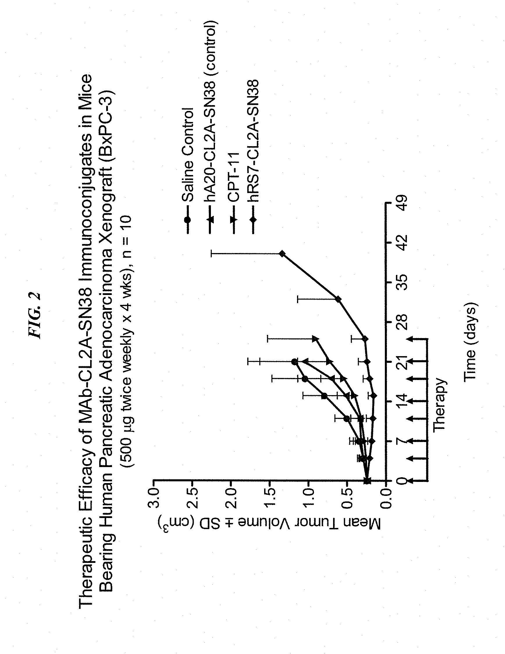
[0024] FIG. 2. Preclinical in vivo therapy of athymic nude mice, bearing BxPC3 human pancreatic carcinoma, with anti-TROP2-CL2A-SN-38 conjugates compared to controls.
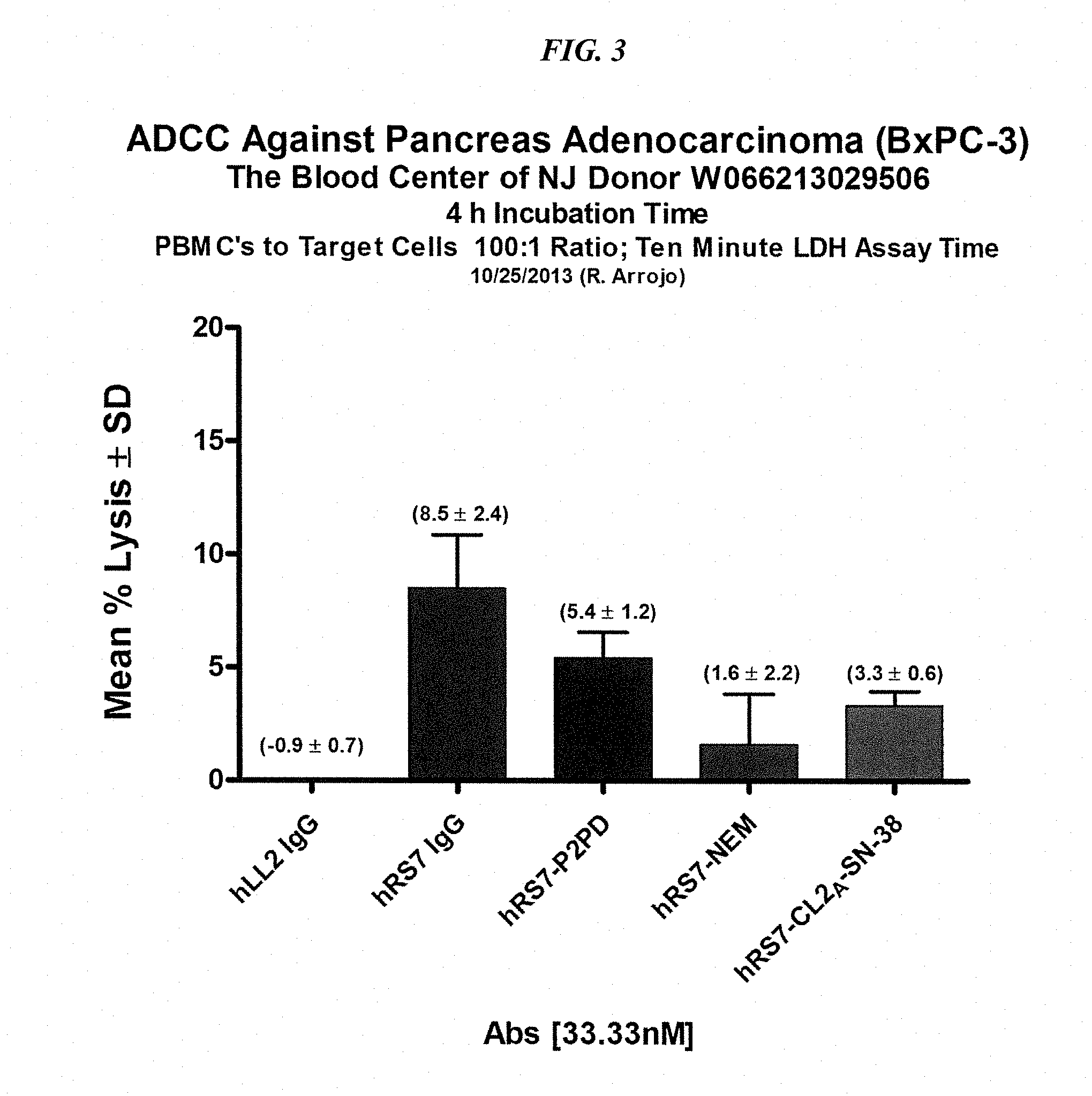
[0025] FIG. 3. ADCC of various hRS7-ADCs vs. hRS7 IgG.
[0026] FIG. 4A. Structures of CL2-SN-38 and CL2A-SN-38.
[0027] FIG. 4B. Comparative efficacy of anti-Trop-2 ADC linked to CL2 vs. CL2A linkers versus hA20 ADC and saline control, using COLO 205 colonic adenocarcinoma. Animals were treated twice weekly for 4 weeks as indicated by the arrows. COLO 205 mice (N=6) were treated with 0.4 mg/kg ADC and tumors measured twice a week.
[0028] FIG. 4C. Comparative efficacy of anti-Trop-2 ADC linked to CL2 vs. CL2A linkers versus hA20 ADC and saline control, using Capan-1 pancreatic adenocarcinoma. Animals were treated twice weekly for 4 weeks as indicated by the arrows. Capan-1 mice (N=10) were treated with 0.2 mg/kg ADC and tumors measured weekly.
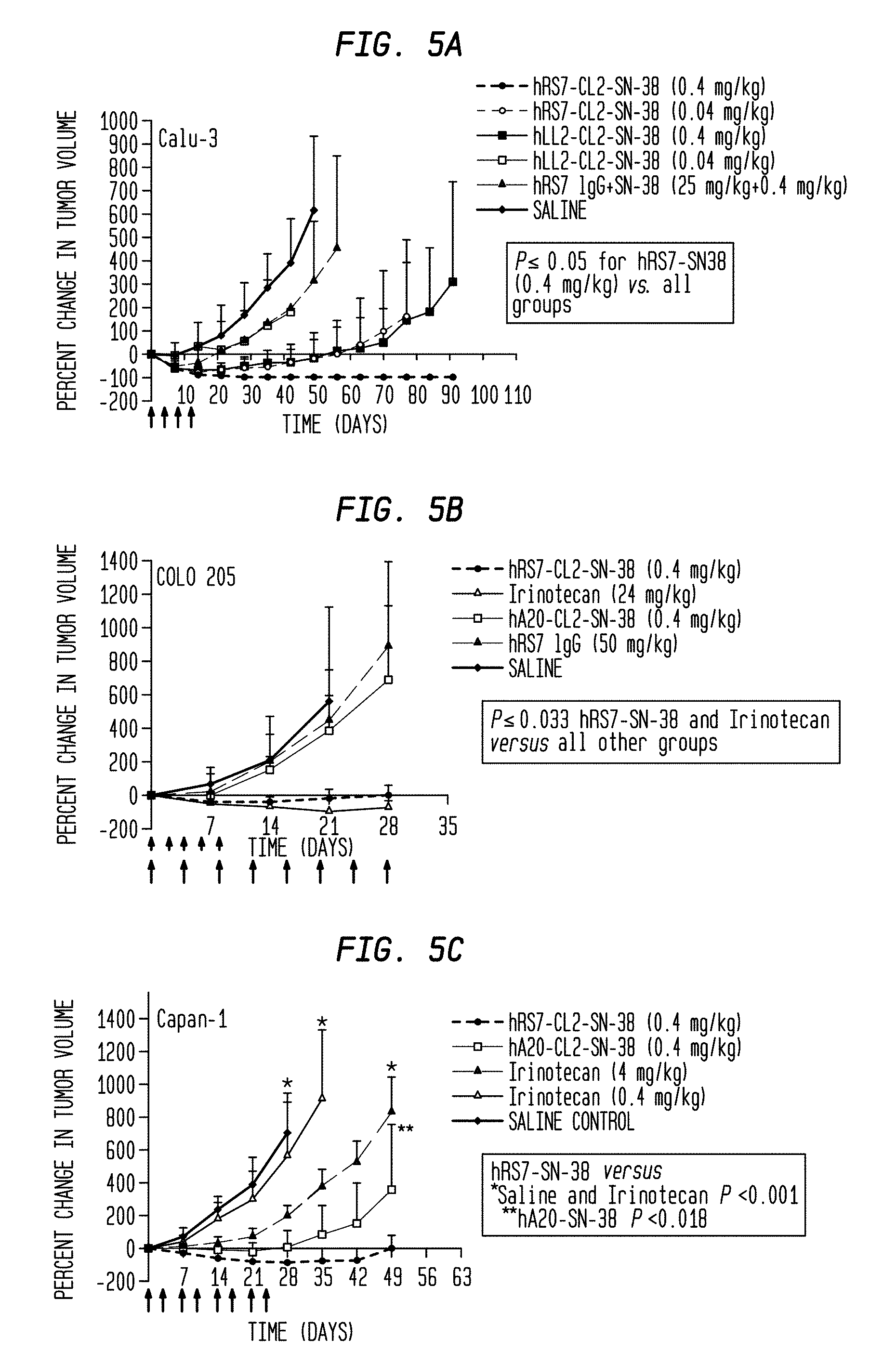
[0029] FIG. 5A. Therapeutic efficacy of hRS7-SN-38 ADC in several solid tumor-xenograft disease models. Efficacy of hRS7-CL2-SN-38 and hRS7-CL2A-SN-38 ADC treatment was studied in mice bearing human non-small cell lung, colorectal, pancreatic, or squamous cell lung tumor xenografts. All the ADCs and controls were administered in the amounts indicated (expressed as amount of SN-38 per dose; long arrows=conjugate injections, short arrows=irinotecan injections). Mice bearing Calu-3 tumors (N=5-7) were injected with hRS7-CL2-SN-38 every 4 days for a total of 4 injections (q4dx4).
[0030] FIG. 5B. Therapeutic efficacy of hRS7-SN-38 ADC in several solid tumor-xenograft disease models. Efficacy of hRS7-CL2-SN-38 and hRS7-CL2A-SN-38 ADC treatment was studied in mice bearing human non-small cell lung, colorectal, pancreatic, or squamous cell lung tumor xenografts. All the ADCs and controls were administered in the amounts indicated (expressed as amount of SN-38 per dose; long arrows=conjugate injections, short arrows=irinotecan injections). COLO 205 tumor-bearing mice (N=5) were injected 8 times (q4dx8) with the ADC or every 2 days for a total of 5 injections (q2dx5) with the MTD of irinotecan.
[0031] FIG. 5C. Therapeutic efficacy of hRS7-SN-38 ADC in several solid tumor-xenograft disease models. Efficacy of hRS7-CL2-SN-38 and hRS7-CL2A-SN-38 ADC treatment was studied in mice bearing human non-small cell lung, colorectal, pancreatic, or squamous cell lung tumor xenografts. All the ADCs and controls were administered in the amounts indicated (expressed as amount of SN-38 per dose; long arrows=conjugate injections, short arrows=irinotecan injections). Capan-1 (N=10) were treated twice weekly for 4 weeks with the agents indicated.
[0032] FIG. 5D. Therapeutic efficacy of hRS7-SN-38 ADC in several solid tumor-xenograft disease models. Efficacy of hRS7-CL2-SN-38 and hRS7-CL2A-SN-38 ADC treatment was studied in mice bearing human non-small cell lung, colorectal, pancreatic, or squamous cell lung tumor xenografts. All the ADCs and controls were administered in the amounts indicated (expressed as amount of SN-38 per dose; long arrows=conjugate injections, short arrows=irinotecan injections). BxPC-3 tumor-bearing mice (N=10) were treated twice weekly for 4 weeks with the agents indicated.
[0033] FIG. 5E. Therapeutic efficacy of hRS7-SN-38 ADC in several solid tumor-xenograft disease models. Efficacy of hRS7-CL2-SN-38 and hRS7-CL2A-SN-38 ADC treatment was studied in mice bearing human non-small cell lung, colorectal, pancreatic, or squamous cell lung tumor xenografts. All the ADCs and controls were administered in the amounts indicated (expressed as amount of SN-38 per dose; long arrows=conjugate injections, short arrows=irinotecan injections). In addition to ADC given twice weekly for 4 week, SK-MES-1 tumor-bearing (N=8) mice received the MTD of CPT-11 (q2dx5).
[0034] FIG. 6A. Tolerability of hRS7-CL2A-SN-38 in Swiss-Webster mice. Fifty-six Swiss-Webster mice were administered 2 i.p. doses of buffer or the hRS7-CL2A-SN-38 3 days apart (4, 8, or 12 mg/kg of SN-38 per dose; 250, 500, or 750 mg conjugate protein/kg per dose). Seven and 15 days after the last injection, 7 mice from each group were euthanized, with blood counts and serum chemistries performed. Graphs show the percent of animals in each group that had elevated levels of AST.
[0035] FIG. 6B. Tolerability of hRS7-CL2A-SN-38 in Swiss-Webster mice. Fifty-six Swiss-Webster mice were administered 2 i.p. doses of buffer or the hRS7-CL2A-SN-38 3 days apart (4, 8, or 12 mg/kg of SN-38 per dose; 250, 500, or 750 mg conjugate protein/kg per dose). Seven and 15 days after the last injection, 7 mice from each group were euthanized, with blood counts and serum chemistries performed. Graphs show the percent of animals in each group that had elevated levels of ALT.
[0036] FIG. 6C. Tolerability of hRS7-CL2A-SN-38 in Cynomolgus monkeys. Six monkeys per group were injected twice 3 days apart with buffer (control) or hRS7-CL2A-SN-38 at 0.96 mg/kg or 1.92 mg/kg of SN-38 equivalents per dose (60 and 120 mg/kg conjugate protein). All animals were bled on day -1, 3, and 6. Four monkeys were bled on day 11 in the 0.96 mg/kg group, 3 in the 1.92 mg/kg group. Changes in neutrophil counts in Cynomolgus monkeys.
[0037] FIG. 6D. Tolerability of hRS7-CL2A-SN-38 in Cynomolgus monkeys. Six monkeys per group were injected twice 3 days apart with buffer (control) or hRS7-CL2A-SN-38 at 0.96 mg/kg or 1.92 mg/kg of SN-38 equivalents per dose (60 and 120 mg/kg conjugate protein). All animals were bled on day -1, 3, and 6. Four monkeys were bled on day 11 in the 0.96 mg/kg group, 3 in the 1.92 mg/kg group. Changes in platelet counts in Cynomolgus monkeys.
[0038] FIG. 7A. Comparison of in vitro efficacy of anti-Trop-2 ADCs (hRS7-SN-38 versus MAB650-SN-38) in Capan-1 human pancreatic adenocarcinoma.
[0039] FIG. 7B. Comparison of in vitro efficacy of anti-Trop-2 ADCs (hRS7-SN-38 versus MAB650-SN-38) in BxPC-3 human pancreatic adenocarcinoma.
[0040] FIG. 7C. Comparison of in vitro efficacy of anti-Trop-2 ADCs (hRS7-SN-38 versus MAB650-SN-38) in NCI-N87 human gastric adenocarcinoma.
[0041] FIG. 8A. Comparison of in vitro efficacy of 162-46.2-SN-38 vs. hRS7-SN-38 in BxPC-3 human pancreatic adenocarcinoma cells.
[0042] FIG. 8B. Comparison of in vitro efficacy of 162-46.2-SN-38 vs. hRS7-SN-38 in MDA-MB-468 human breast adenocarcinoma cells.
[0043] FIG. 9. IMMU-132 phase I/II data for best response by RECIST criteria.
[0044] FIG. 10. IMMU-132 phase I/II data for time to progression and best response (RECIST).
[0045] FIG. 11. Therapeutic efficacy of murine anti-Trop-2-SN-38 ADC (162-46.2-SN-38) compared to hRS7-SN-38 in mice bearing NCI-N87 human gastric carcinoma xenografts.
[0046] FIG. 12. Accumulation of SN-38 in tumors of nude mice with Capan-1 human pancreatic cancer xenografts, when administered as free irinotecan vs. IMMU-132 ADC.
[0047] FIG. 13. Individual patient demographics and prior treatment for phase I/II IMMU-132 anti-Trop-2 ADC in pancreatic cancer patients.
[0048] FIG. 14. Response assessment to IMMU-132 anti-Trop-2 ADC in pancreatic cancer patients.
[0049] FIG. 15. Summary of time to progression (TTP) results in human pancreatic cancer patients administered IMMU-132 anti-Trop-2 ADC.
[0050] FIG. 16. Adverse events observed in phase I study of IMMU-132 in various tumor types.
[0051] FIG. 17. Response assessment in sacituzumab govitecan-treated patients. (A) Composite schematic showing the best response (y-axis) determined from target lesion measurements according to RECIST 1.1 and the time-to-progression (z-axis; TTP expressed in months), measured from the date of the first dose until CT documentation of progression as per RECIST. Best response bars are color-coded to identify the 4 starting dose levels. Four of the 25 patients (numbers 6, 9, 14, and 23; 2 PDC, 1 GC and 1 SCLC) who were classified with disease progression are not shown because either they did not have a follow-up CT with measurement of target lesions or they had new lesions despite having stable target lesions measurements. A bar break (//) shown for two PD patients denotes target lesions increased >30%, whereas TTP values in the boxes at top of the graph show the patients who exceeded 9 months. The number of prior therapies (in parentheses) and the patients who received prior topoisomerase I therapy (asterisks) are indicated below the graph. (B) Graph showing the patients sorted according to survival, showing also their TTP. Survival data were unavailable for 2 PDC patients (numbers 6 and 17 with TTP 1.0 and 2.9 months).
[0052] FIG. 18. CT response assessment in 2 of 3 patients with >30% reduction in target lesions. Patient 22 is a 65-year-old woman with poorly differentiated SCLC (Trop-2 expression by immunohistology, 3+) who had received 2 months of carboplatin/etoposide (topoisomerase-II inhibitor) and 1 month of topotecan (topoisomerase-I inhibitor) with no response, followed by local radiation for 6 weeks (3000 cGy), but progressed. Four weeks later, the patient started sacituzumab govitecan at 12 mg/kg (2 doses), which was reduced to 9.0 mg/kg (1 dose), and finally to 6.75 mg/kg for 9 doses. The patient presented initially with the sum of the longest diameters (SLD) of the target lesions totaling 19.3 cm. Two of the target lesions showing the best shrinkage are shown at baseline (A and C). After 4 treatments, she had a 38% reduction in target lesions, including a substantial reduction in the main lung lesion (5.8 to 2.7 cm; panels B and D). On her next CT assessment 12 weeks later, the patient progressed. Patient 3 a 62 year-old woman, who 5 months after her initial diagnosis and surgery for colon cancer had a hepatic resection for liver metastases and then received 7 months of treatment with FOLFOX and 1 month of only 5-fluorouracil. She was referred to the sacituzumab govitecan trial with multiple lesions, primarily in the liver (left panels A, C, and E). Immunohistology showed a 2+ staining of her primary cancer; her plasma CEA was 781 ng/mL. Therapy was initiated at 8 mg/kg and 6 treatments later (12 weeks), the 3 target lesions had reduced from 7.9 cm to 5.0 cm (-37%; PR). The response was confirmed 6.6 weeks later (after ten doses), with additional shrinkage to 3.8 cm (-52%). Panels B, D, and F show the shrinkage of these 3 lesions (59% reduction at this time) 32 weeks from the start of treatment and after receiving 18 doses The patient continued therapy, achieving a maximum tumor reduction of 65% 10 months after treatment was initiated (28 doses). Plasma CEA decreased to 26.5 ng/mL after 18 doses, but thereafter began to increase despite continued radiological evidence (target and non-target lesions) of additional disease reduction or stabilization. Approximately 1 year from the start of treatment (31 doses given), one of the 3 target lesions progressed.
[0053] FIG. 19. Therapeutic efficacy of IMMU-132 with different DARs. NCI-N87 human gastric carcinoma xenografts (subcutaneous) were established as described in Methods. (A) Four groups of mice (N=9) were injected IV with 2.times.0.5 mg (arrows) of IMMU-132 conjugates prepared with a DAR=6.89, 3.28, or 1.64. Control animals received saline. Therapy began 7 days after tumor cells were administered (size was 0.248.+-.0.047 cm.sup.3). Survival curves were generated based on the time to progression to >1.0 cm.sup.3, and were analyzed by log-rank test (significance at P<0.05). (B) NCI-N87 tumor-bearing mice (N=7-9; starting size=0.271.+-.0.053 cm.sup.3) were treated with either 0.5 mg IMMU-132 (DAR=6.89) or 1.0 mg DAR=3.28 twice weekly for two weeks (arrows). Mice were euthanized and deemed to have succumbed to disease once tumors grew to >1.0 cm.sup.3. Profiles of individual tumor growth were obtained through linear-curve modeling. Statistical analysis of tumor growth was based on area under the curve (AUC) performed up to the time that the first animal within a group was euthanized due to disease progression. An f-test was employed to determine equality of variance between groups prior to statistical analysis of growth curves. A two-tailed t-test was used to assess statistical significance between the various treatment groups and controls, except for the saline control, where a one-tailed t-test was used (significance at P<0.05).
[0054] FIG. 20. Therapeutic efficacy of IMMU-132 in TNBC xenograft models. (A) Twenty-two days after s.c. implantation of MDA-MB-468 tumors (at the onset of treatment, tumors averaged 0.223.+-.0.055 cm.sup.3), nude mice (7-8 per group) were injected IV with IMMU-132 or a control hA20 anti-CD20-SN-38 conjugate twice weekly for two weeks (0.12 or 0.20 mg/kg SN-38 equivalents per dose). Other animals were given irinotecan (10 mg/kg/dose; SN-38 equivalent based on mass=5.8 mg/kg) IV every other day for 10 days for a total of five injections. (B) Starting on day 56 after treatment initiation, all animals in the control hA20-SN-38 group were given IMMU-132 (4.times.0.2 mg/kg SN-38 equivalents). The size of the tumors in the individual animals of this group from the onset of tumor transplantation is given. Red arrows indicate when the hA20-SN-38 conjugate was first given, and purple arrows indicate when the treatment with IMMU-132 was initiated. (C) Mice (N=12) bearing the MDA-MB-231 TNBC cell line (0.335.+-.0.078 cm.sup.3) were treated with IMMU-132 or the control hA20-SN-38 conjugate (0.4 mg/kg SN-38 equivalents), irinotecan (6.5 mg/kg; .about.3.8 mg/kg SN-38 equivalents), or a combination of hRS7 IgG (25 mg/kg) plus irinotecan (6.5 mg/kg).
[0055] FIG. 21. ADCC activity of IMMU-132. Specific cell lysis of target cells by human PBMCs mediated by IMMU-132 was compared to parental hRS7. Target cells were plated the night before and the assay performed as described in the Examples below. (A) MDA-MB-468 target cells. (B) NIH:OVCAR-3 target cells. (C) BxPC-3 target cells. *hRS7 versus all the other test agents (P<0.0054). **IMMU-132 versus negative controls hLL2-SN-38 and hLL2 (P<0.0003). *** IMMU-132 versus negative control hLL2-CL2ASN-38 (P<0.0019).
[0056] FIG. 22. Pharmacokinetics of IMMU-132 in mice. Naive nude mice (N=5) were injected i.v. with 200 .mu.g of IMMU-132. At various time-points these mice were bled and serum obtained and analyzed for intact conjugate and carrier hRS7 antibody, as described in the Examples below. For comparison, another group of mice was injected with 200 .mu.g parental hRS7. (A) Serum concentration and clearance of hRS7 from parental control injected mice. Concentration and clearance of (B) hRS7 carrier antibody versus (C) intact conjugate from IMMU-132 injected mice. Graphed data shown as mean.+-.S.D.
[0057] FIG. 23. Efficacy of IMMU-132 in mice bearing human gastric carcinoma xenograft. Mice bearing NCI-N87 human gastric tumors (TV=0.249.+-.0.049 cm.sup.3) were treated with 0.35 mg IMMU-132 twice weekly for four weeks. (A) Mean tumor growth curves for IMMU-132 treated animals compared to saline and non-tumor-targeting control ADC, hA20-CL2A-SN-38, treated mice. Arrows indicate therapy days. (B) Survival curves for treated mice with a disease end-point of tumor progression greater than 1.0 cm.sup.3.
[0058] FIG. 24. Various IMMU-132 dosing schemes in mice bearing pancreatic and gastric tumor xenografts. Nude mice (N=8-10) bearing s.c. BxPC-3 or NCI-N87 xenografts were prepared. (A) BxPC-3-bearing mice were treated (arrows) with two cycles of IMMU-132 at 1 mg every 14 d, 0.5 mg weekly for two weeks, or 0.25 mg twice weekly for two weeks, totaling 2 mg IMMU-132 to all the mice. (B) Similar dosing of NCIN87-bearing mice (arrows), with mice in the 1-mg treatment group receiving one additional cycle. (C) Chronic dosing of IMMU-132 in mice bearing NCI-N87, using 0.5-mg once weekly for 2 weeks in a 3-week treatment cycle for a total of 4 cycles. Corresponding survival curves (end-point: tumor progression >1.0 cm3) to right of each tumor growth curve.
[0059] FIG. 25. Responses in 52 human TNBC patients treated with 10 mg/kg IMMU-132, after failing numerous prior therapies.
[0060] FIG. 26. Time to progression for CR+PR+SD in TNBC patients treated with 10 mg/kg IMMU-132.
[0061] FIG. 27. Progression-free survival in TNBC patients treated with 10 mg/kg IMMU-132.
[0062] FIG. 28. Best response in 29 assessable human NSCLC patients treated with 8 to 10 mg/kg IMMU-132.
[0063] FIG. 29. Time to progression in NSCLC patients treated with 8-10 mg/kg IMMU-132.
[0064] FIG. 30. Progression-free survival in NSCLC patients treated with 8 or 10 mg/kg IMMU-132.
[0065] FIG. 31. Best response in 25 assessable human SCLC patients treated with 8 to 10 mg/kg IMMU-132.
[0066] FIG. 32. Time to progression in SCLC patients treated with 8-10 mg/kg IMMU-132.
[0067] FIG. 33. Progression-free survival in SCLC patients treated with 8 or 10 mg/kg IMMU-132.
[0068] FIG. 34. Best response in 11 assessable human urothelial cancer patients treated with IMMU-132.
[0069] FIG. 35. Time to progression in urothelial cancer patients treated with IMMU-132.
[0070] FIG. 36. .gamma.H2AX assay for DSB by flow cytometry. Cells were treated or not treated with SN-38 (250 nM) for 3 h and the levels of .gamma.H2AX were monitored hourly by flow cytometry and shown as MFI in bar diagram (A) or as percent of untreated in (B). The effect of FTC (10 .mu.M) to increase the formation of DSB/.gamma.H2AX was shown for MDA-MB-231-5120 treated with SN-38 (C) and for NCI-N87-S120 treated for either SN-38 or IMMU-132 (D).
[0071] FIG. 37. Dose response curves of parental and S-120 cells treated with different concentrations of SN-38 in the absence and presence of YHO-13351 or Ko143. Reversal of SN-38-resistance by YHO-13351 was shown for MDA-MB-231-S120 in (A), NCI-N87-S120 in (B), and by Ko143 for NCI-N87-S120 in (C).
[0072] FIG. 38. Efficacy of IMMU-132 in mice bearing SN-38-resistant NCI-N87-S-120 gastric carcinoma xenograft. (A) Mean tumor growth curves for NCI-N87 and NCI-N87-S-120 xenografts. (B) Mice bearing NCI-N87-S-120 SN-38-resistant human gastric tumors were treated with IMMU-132, irinotecan, YHO-13551, or combinations as indicated on the graph and described in Materials and Methods. (C) Mice bearing parental NCI-N87 tumors treated with IMMU-132 or irinotecan at the same dose and schedule as used in NCI-N87-S-120 tumor-bearing animals. In the survival curves of (B) and (C), the starting day of therapy (when tumor volumes reached approximately 0.25 cm.sup.3) was marked as Day 0. Mice were euthanized once tumors grew to >1.0 cm.sup.3 in size.
DETAILED DESCRIPTION OF THE INVENTION
Definitions
[0073] In the description that follows, a number of terms are used and the following definitions are provided to facilitate understanding of the claimed subject matter. Terms that are not expressly defined herein are used in accordance with their plain and ordinary meanings.
[0074] Unless otherwise specified, a or an means "one or more."
[0075] The term about is used herein to mean plus or minus ten percent (10%) of a value. For example, "about 100" refers to any number between 90 and 110.
[0076] An antibody, as used herein, refers to a full-length (i.e., naturally occurring or formed by normal immunoglobulin gene fragment recombinatorial processes) immunoglobulin molecule (e.g., an IgG antibody) or an antigen-binding portion of an immunoglobulin molecule, such as an antibody fragment. An antibody or antibody fragment may be conjugated or otherwise derivatized within the scope of the claimed subject matter. Such antibodies include but are not limited to IgG1, IgG2, IgG3, IgG4 (and IgG4 subforms), as well as IgA isotypes. As used below, the abbreviation "MAb" may be used interchangeably to refer to an antibody, antibody fragment, monoclonal antibody or multispecific antibody.
[0077] An antibody fragment is a portion of an antibody such as F(ab').sub.2, F(ab).sub.2, Fab', Fab, Fv, scFv (single chain Fv), single domain antibodies (DABs or VHHs) and the like, including the half-molecules of IgG4 cited above (van der Neut Kolfschoten et al. (Science 2007; 317(14 September):1554-1557). Regardless of structure, an antibody fragment of use binds with the same antigen that is recognized by the intact antibody. The term "antibody fragment" also includes synthetic or genetically engineered proteins that act like an antibody by binding to a specific antigen to form a complex. For example, antibody fragments include isolated fragments consisting of the variable regions, such as the "Fv" fragments consisting of the variable regions of the heavy and light chains and recombinant single chain polypeptide molecules in which light and heavy variable regions are connected by a peptide linker ("scFv proteins"). The fragments may be constructed in different ways to yield multivalent and/or multispecific binding forms.
[0078] A naked antibody is generally an entire antibody that is not conjugated to a therapeutic agent. A naked antibody may exhibit therapeutic and/or cytotoxic effects, for example by Fc-dependent functions, such as complement fixation (CDC) and ADCC (antibody-dependent cell cytotoxicity). However, other mechanisms, such as apoptosis, anti-angiogenesis, anti-metastatic activity, anti-adhesion activity, inhibition of heterotypic or homotypic adhesion, and interference in signaling pathways, may also provide a therapeutic effect. Naked antibodies include polyclonal and monoclonal antibodies, naturally occurring or recombinant antibodies, such as chimeric, humanized or human antibodies and fragments thereof. In some cases a "naked antibody" may also refer to a "naked" antibody fragment. As defined herein, "naked" is synonymous with "unconjugated," and means not linked or conjugated to a therapeutic agent.
[0079] A chimeric antibody is a recombinant protein that contains the variable domains of both the heavy and light antibody chains, including the complementarity determining regions (CDRs) of an antibody derived from one species, preferably a rodent antibody, more preferably a murine antibody, while the constant domains of the antibody molecule are derived from those of a human antibody. For veterinary applications, the constant domains of the chimeric antibody may be derived from that of other species, such as a primate, cat or dog.
[0080] A humanized antibody is a recombinant protein in which the CDRs from an antibody from one species; e.g., a murine antibody, are transferred from the heavy and light variable chains of the murine antibody into human heavy and light variable domains (framework regions). The constant domains of the antibody molecule are derived from those of a human antibody. In some cases, specific residues of the framework region of the humanized antibody, particularly those that are touching or close to the CDR sequences, may be modified, for example replaced with the corresponding residues from the original murine, rodent, subhuman primate, or other antibody.
[0081] A human antibody is an antibody obtained, for example, from transgenic mice that have been "engineered" to produce human antibodies in response to antigenic challenge. In this technique, elements of the human heavy and light chain loci are introduced into strains of mice derived from embryonic stem cell lines that contain targeted disruptions of the endogenous heavy chain and light chain loci. The transgenic mice can synthesize human antibodies specific for various antigens, and the mice can be used to produce human antibody-secreting hybridomas. Methods for obtaining human antibodies from transgenic mice are described by Green et al., Nature Genet. 7:13 (1994), Lonberg et al., Nature 368:856 (1994), and Taylor et al., Int. Immun. 6:579 (1994). A fully human antibody also can be constructed by genetic or chromosomal transfection methods, as well as phage display technology, all of which are known in the art. See for example, McCafferty et al., Nature 348:552-553 (1990) for the production of human antibodies and fragments thereof in vitro, from immunoglobulin variable domain gene repertoires from unimmunized donors. In this technique, human antibody variable domain genes are cloned in-frame into either a major or minor coat protein gene of a filamentous bacteriophage, and displayed as functional antibody fragments on the surface of the phage particle. Because the filamentous particle contains a single-stranded DNA copy of the phage genome, selections based on the functional properties of the antibody also result in selection of the gene encoding the antibody exhibiting those properties. In this way, the phage mimics some of the properties of the B cell. Phage display can be performed in a variety of formats, for their review, see e.g. Johnson and Chiswell, Current Opinion in Structural Biology 3:5564-571 (1993). Human antibodies may also be generated by in vitro activated B cells. See U.S. Pat. Nos. 5,567,610 and 5,229,275, the Examples section of each of which is incorporated herein by reference.
[0082] A therapeutic agent is an atom, molecule, or compound that is useful in the treatment of a disease. Examples of therapeutic agents include, but are not limited to, antibodies, antibody fragments, ADCs, drugs, cytotoxic agents, pro-apopoptotic agents, toxins, nucleases (including DNAses and RNAses), hormones, immunomodulators, chelators, boron compounds, photoactive agents or dyes, radionuclides, oligonucleotides, interference RNA, siRNA, RNAi, anti-angiogenic agents, chemotherapeutic agents, cyokines, chemokines, prodrugs, enzymes, binding proteins or peptides or combinations thereof.
[0083] An ADC is an antibody, antigen-binding antibody fragment, antibody complex or antibody fusion protein that is conjugated to a therapeutic agent. Conjugation may be covalent or non-covalent. Preferably, conjugation is covalent.
[0084] As used herein, the term antibody fusion protein is a recombinantly-produced antigen-binding molecule in which one or more natural antibodies, single-chain antibodies or antibody fragments are linked to another moiety, such as a protein or peptide, a toxin, a cytokine, a hormone, etc. In certain preferred embodiments, the fusion protein may comprise two or more of the same or different antibodies, antibody fragments or single-chain antibodies fused together, which may bind to the same epitope, different epitopes on the same antigen, or different antigens.
[0085] An immunomodulator is a therapeutic agent that when present, alters, suppresses or stimulates the body's immune system. Typically, an immunomodulator of use stimulates immune cells to proliferate or become activated in an immune response cascade, such as macrophages, dendritic cells, B-cells, and/or T-cells. However, in some cases an immunomodulator may suppress proliferation or activation of immune cells. An example of an immunomodulator as described herein is a cytokine, which is a soluble small protein of approximately 5-20 kDa that is released by one cell population (e.g., primed T-lymphocytes) on contact with specific antigens, and which acts as an intercellular mediator between cells. As the skilled artisan will understand, examples of cytokines include lymphokines, monokines, interleukins, and several related signaling molecules, such as tumor necrosis factor (TNF) and interferons. Chemokines are a subset of cytokines. Certain interleukins and interferons are examples of cytokines that stimulate T cell or other immune cell proliferation. Exemplary interferons include interferon-.alpha., interferon-.beta., interferon-.gamma. and interferon-.lamda..
Anti-Trop-2 Antibodies
[0086] Preferably, the subject ADCs include at least one antibody or fragment thereof that binds to Trop-2. In a specific preferred embodiment, the anti-Trop-2 antibody may be a humanized RS7 antibody (see, e.g., U.S. Pat. No. 7,238,785, incorporated herein by reference in its entirety), comprising the light chain CDR sequences CDR1 (KASQDVSIAVA, SEQ ID NO:14); CDR2 (SASYRYT, SEQ ID NO:15); and CDR3 (QQHYITPLT, SEQ ID NO:16) and the heavy chain CDR sequences CDR1 (NYGMN, SEQ ID NO:17); CDR2 (WINTYTGEPTYTDDFKG, SEQ ID NO:18) and CDR3 (GGFGSSYWYFDV, SEQ ID NO:19).
[0087] The RS7 antibody was a murine IgG.sub.1 raised against a crude membrane preparation of a human primary squamous cell lung carcinoma. (Stein et al., Cancer Res. 50: 1330, 1990) The RS7 antibody recognizes a 46-48 kDa glycoprotein, characterized as cluster 13. (Stein et al., Int. J. Cancer Supp. 8:98-102, 1994) The antigen was designated as EGP-1 (epithelial glycoprotein-1), but is also referred to as Trop-2.
[0088] Trop-2 is a type-I transmembrane protein and has been cloned from both human (Fornaro et al., Int J Cancer 1995; 62:610-8) and mouse cells (Sewedy et al., Int J Cancer 1998; 75:324-30). In addition to its role as a tumor-associated calcium signal transducer (Ripani et al., Int J Cancer 1998; 76:671-6), the expression of human Trop-2 was shown to be necessary for tumorigenesis and invasiveness of colon cancer cells, which could be effectively reduced with a polyclonal antibody against the extracellular domain of Trop-2 (Wang et al., Mol Cancer Ther 2008; 7:280-5).
[0089] The growing interest in Trop-2 as a therapeutic target for solid cancers (Cubas et al., Biochim Biophys Acta 2009; 1796:309-14) is attested by further reports that documented the clinical significance of overexpressed Trop-2 in breast (Huang et al., Clin Cancer Res 2005; 11:4357-64), colorectal (Ohmachi et al., Clin Cancer Res 2006; 12:3057-63; Fang et al., Int J Colorectal Dis 2009; 24:875-84), and oral squamous cell (Fong et al., Modern Pathol 2008; 21:186-91) carcinomas. The latest evidence that prostate basal cells expressing high levels of Trop-2 are enriched for in vitro and in vivo stem-like activity is particularly noteworthy (Goldstein et al., Proc Natl Acad Sci USA 2008; 105:20882-7).
[0090] Flow cytometry and immunohistochemical staining studies have shown that the RS7 MAb detects antigen on a variety of tumor types, with limited binding to normal human tissue (Stein et al., 1990). Trop-2 is expressed primarily by carcinomas such as carcinomas of the lung, stomach, urinary bladder, breast, ovary, uterus, and prostate. Localization and therapy studies using radiolabeled murine RS7 MAb in animal models have demonstrated tumor targeting and therapeutic efficacy (Stein et al., 1990; Stein et al., 1991).
[0091] Strong RS7 staining has been demonstrated in tumors from the lung, breast, bladder, ovary, uterus, stomach, and prostate. (Stein et al., Int. J. Cancer 55:938, 1993) The lung cancer cases comprised both squamous cell carcinomas and adenocarcinomas. (Stein et al., Int. J. Cancer 55:938, 1993) Both cell types stained strongly, indicating that the RS7 antibody does not distinguish between histologic classes of non-small-cell carcinoma of the lung.
[0092] The RS7 MAb is rapidly internalized into target cells (Stein et al., 1993). The internalization rate constant for RS7 MAb is intermediate between the internalization rate constants of two other rapidly internalizing MAbs, which have been demonstrated to be useful for ADC production. (Id.) It is well documented that internalization of ADCs is a requirement for anti-tumor activity. (Pastan et al., Cell 47:641, 1986) Internalization of ADCs has been described as a major factor in anti-tumor efficacy. (Yang et al., Proc. Nat'l Acad. Sci. USA 85: 1189, 1988) Thus, the RS7 antibody exhibits several important properties for therapeutic applications.
[0093] While the hRS7 antibody is preferred, other anti-Trop-2 antibodies are known and/or publicly available and in alternative embodiments may be utilized in the subject ADCs. While humanized or human antibodies are preferred for reduced immunogenicity, in alternative embodiments a chimeric antibody may be of use. As discussed below, methods of antibody humanization are well known in the art and may be utilized to convert an available murine or chimeric antibody into a humanized form.
[0094] Anti-Trop-2 antibodies are commercially available from a number of sources and include LS-C126418, LS-C178765, LS-C126416, LS-C126417 (LifeSpan BioSciences, Inc., Seattle, Wash.); 10428-MM01, 10428-MM02, 10428-R001, 10428-R030 (Sino Biological Inc., Beijing, China); MR54 (eBioscience, San Diego, Calif.); sc-376181, sc-376746, Santa Cruz Biotechnology (Santa Cruz, Calif.); MM0588-49D6, (Novus Biologicals, Littleton, Colo.); ab79976, and ab89928 (ABCAM.RTM., Cambridge, Mass.).
[0095] Other anti-Trop-2 antibodies have been disclosed in the patent literature. For example, U.S. Publ. No. 2013/0089872 discloses anti-Trop-2 antibodies K5-70 (Accession No. FERM BP-11251), K5-107 (Accession No. FERM BP-11252), K5-116-2-1 (Accession No. FERM BP-11253), T6-16 (Accession No. FERM BP-11346), and T5-86 (Accession No. FERM BP-11254), deposited with the International Patent Organism Depositary, Tsukuba, Japan. U.S. Pat. No. 5,840,854 disclosed the anti-Trop-2 monoclonal antibody BR110 (ATCC No. HB11698). U.S. Pat. No. 7,420,040 disclosed an anti-Trop-2 antibody produced by hybridoma cell line AR47A6.4.2, deposited with the IDAC (International Depository Authority of Canada, Winnipeg, Canada) as accession number 141205-05. U.S. Pat. No. 7,420,041 disclosed an anti-Trop-2 antibody produced by hybridoma cell line AR52A301.5, deposited with the IDAC as accession number 141205-03. U.S. Publ. No. 2013/0122020 disclosed anti-Trop-2 antibodies 3E9, 6G11, 7E6, 15E2, 18B1. Hybridomas encoding a representative antibody were deposited with the American Type Culture Collection (ATCC), Accession Nos. PTA-12871 and PTA-12872. Immunoconjugate PF 06263507, comprising an anti-5T4 (anti-Trop-2) antibody attached to the tubulin inhibitor monomethylauristatin F (MMAF) is available from Pfizer, Inc. (Groton, Conn.) (see, e.g., Sapra et al., 2013, Mol Cancer Ther 12:38-47). U.S. Pat. No. 8,715,662 discloses anti-Trop-2 antibodies produced by hybridomas deposited at the AID-ICLC (Genoa, Italy) with deposit numbers PD 08019, PD 08020 and PD 08021. U.S. Patent Application Publ. No. 20120237518 discloses anti-Trop-2 antibodies 77220, KM4097 and KM4590. U.S. Pat. No. 8,309,094 (Wyeth) discloses antibodies A1 and A3, identified by sequence listing. The Examples section of each patent or patent application cited above in this paragraph is incorporated herein by reference. Non-patent publication Lipinski et al. (1981, Proc Natl. Acad Sci USA, 78:5147-50) disclosed anti-Trop-2 antibodies 162-25.3 and 162-46.2.
[0096] Numerous anti-Trop-2 antibodies are known in the art and/or publicly available. As discussed below, methods for preparing antibodies against known antigens were routine in the art. The sequence of the human Trop-2 protein was also known in the art (see, e.g., GenBank Accession No. CAA54801.1). Methods for producing humanized, human or chimeric antibodies were also known. The person of ordinary skill, reading the instant disclosure in light of general knowledge in the art, would have been able to make and use the genus of anti-Trop-2 antibodies in the subject ADCs.
[0097] Use of antibodies against targets related to Trop-2 has been disclosed for immunotherapeutics other than ADCs. The murine anti-Trop-1 IgG2a antibody edrecolomab (PANOREX.RTM.) has been used for treatment of colorectal cancer, although the murine antibody is not well suited for human clinical use (Baeuerle & Gires, 2007, Br. J Cancer 96:417-423). Low-dose subcutaneous administration of ecrecolomab was reported to induce humoral immune responses against the vaccine antigen (Baeuerle & Gires, 2007). Adecatumumab (MT201), a fully human anti-Trop-1 antibody, has been used in metastatic breast cancer and early-stage prostate cancer and is reported to act through ADCC and CDC activity (Baeuerle & Gires, 2007). MT110, a single-chain anti-Trop-1/anti-CD3 bispecific antibody construct has reported efficacy against ovarian cancer (Baeuerle & Gires, 2007). Proxinium, an immunotoxin comprising anti-Trop-1 single-chain antibody fused to Pseudomonas exotoxin, has been tested in head-and-neck and bladder cancer (Baeuerle & Gires, 2007). None of these studies contained any disclosure of the use of anti-Trop-2 ADCs.
ABCG2 Inhibitors
[0098] In preferred embodiments, combination therapy is performed with an anti-Trop-2 ADC (e.g., hRS7-CL2A-SN-38) in combination with one or more inhibitors of an ABC transporter, preferably an inhibitor of ABCB1, ABCC1 or ABCG2, more preferably an inhibitor of ABCG2. The role of ABCG2 inhibitors in combination cancer therapy has been recently reviewed by Ricci et al. (2015, J Develop Drugs 4:138). ABCG2 is unique among the ABC transporters in that it is mainly overexpressed in drug-resistant solid tumors, although it has also been found to be overexpressed in a number of hematopoietic tumors along with ABCB1 and ABCC1 (Ricci et al., 2015, J Develop Drugs 4:138). Although ABCG2 can transport a number of chemotherapeutic agents, the most well known include topotecan, mitoxantrone, SN-38, doxorubicin and daunorubicin (Ricci et al., 2015, J Develop Drugs 4:138). Elevated expression of ABCG2 has been reported to be associated with decreased survival rates in small cell lung cancer, non-small cell lung cancer, pancreatic cancer, mantle cell lymphoma, acute myeloid leukemia, ovarian cancer, colorectal cancer and breast cancer (Ricci et al., 2015, J Develop Drugs 4:138).
[0099] Many drugs have been found to be inhibitors of ABCG2 activity (see Table 1). However, of these, only a handful have been tested in vivo and/or in humans, with relatively limited success to date in improving chemotherapeutic efficacy (Ricci et al., 2015, J Develop Drugs 4:138).
TABLE-US-00001 TABLE 1 ABCG2 Inhibitors With In vitro Efficacy* in Clinical Drug vivo trials 1,4-dihydropyridines[48] Artesunate[39] X AST1306[51] Bifendate-chalcone hybrids[53] Botryllamides[55] Cadmium[57] Calcium Channel Blockers (nicardipine, nitrendipine, nimodipine, dipyridamole)[59] Camptothecin analog (ST1481)[40] X Camptothecin analog (CHO793076)[41] X Cannabinoids[63] CCT129202[42] X Chalcone[66] Curcumin[30] X Cyclosporin A[69] Dihydropyridines and Pyridines[71] X Dimethoxyaurones[73] Dofequidar fumarate[38] X Repurposed Drugs[76] EGFR Inhibitors[78] Flavones & Benzoflavones[80] Tropical Plant Flavonoids[82] Fruit Juices (quercetin, kaempferol, bergamotin, 6',7'-dihydroxybergamottin, tangeretin, nobiletin, hesperidin, hesperetin)[84] Fumitremorgin C[29, 31] X Fumitremorgin C analogue (ko143)[32] X Gefitinib[44, 88] X GF120918, BNP1350[32, 33] X X GW583340 and GW2974[91] HM30181 Derivatives[93] Human cathelicidin[95] Imatinib mesylate[43, 98] X X Lapatinib[45-47, 49] X X LY294002[50] MBLI-87[52] X Methoxy Stilbenes[54] Mithramycin A[56] Quercetin derivatives[58] Naphthopyrones[60] Nilotinib[61] Novobiocin[62] X NP-1250[64] Olomoucine II and purvalanol A[65] Organ chlorine and Pyrethroid[67] OSI-930[68] Phytoestrogens/Flavonoids[70] Piperazinobenzopyranones and Phenalkylaminobenzopyranones[72] Ponatinib[74] PZ-39[75] Quinazolines[77] Quizartinib[79] Sildenafil[81] Sorafenib[83] Substituted Chromones[85] Sunitinib[86] Tandutinib[87] Tariquidar[89] Terpenoids[90] CI1033[92] Toremifene[94] XR9577[96], WK-X-34[96, 97], X WK-X-50[96], and WK-X-84[96] YHO-13177[34]and YHO-13351[34] X *From Ricci et al., 2015, J Develop Drugs 4: 138
[0100] Fumitremorgin C was the first ABCG2 inhibitor to be described which reversed chemoresistance of colon carcinoma to MTX (Rabindran et al., 1998, Cancer Res 58:5850-58). Since that time, over 60 agents have been described that inhibit the action of ABCG2 in vitro (Table 1). Of those, only 15 compounds inhibiting ABCG2 activity have exhibited anti-cancer activity in vivo in animal models of human cancer xenografts (Table 1 and 2). Only 6 of those compounds are direct antagonists specific for ABCG2: curcumin, FTC, Ko143, GF120918 (Elacridar), YHO-13177, YHO-13351, and the recently reported compounds 177, 724, and 505 (Shukla et al., 2009, Pharm Res 26:480-87; Garimella et al., 2005, Cancer Chemother Pharmacol 55:101-9; Allen et al., 2002, Mol Cancer Ther 1:417-25; Hyafil et al., Cancer Res 53:4595-602; Yamazaki et al., 2011, Mol Cancer Ther 10:1252-63; Strouse et al., 2013, J Biomol Screen 18:26-38; Strouse et al., 2013, Anal Biochem 437:77-87). Of the specific ABCG2 antagonists, only YHO-13177 and the recently reported compounds CID44640177, CID1434724, and CID46245505 (Ricci et al., 2016, Mol Cancer Ther 15:2853-62) were reported to have an antitumor effect when combined with TPT (Ricci et al., 2015, J Develop Drugs 4:138).
[0101] A number of compounds have shown efficacy in animal models of cancer (Table 2) (Ricci et al., 2015, J Develop Drugs 4:138). Some of these compounds have an indirect inhibitory effect on ABCG2, and include antimalarial agents, chemotherapeutic analogs of camptothecin, and an aurora kinase inhibitor (Table 2) (Ricci et al., 2015, J Develop Drugs 4:138). The aurora kinase inhibitor, CCT129202, was able to increase the accumulation of doxorubicin and rhodamine 123 in ABCB1 and ABCG2 overexpressing human colon cancer cells.
[0102] As discussed in the Examples below, the present studies demonstrate that at least the ABCG2 inhibitors fumitremorgin C, Ko143 and YHO-13351 restored toxicity of SN-38 in MDA-MB-231 human breast cancer cells and NCI-N87-S120 human gastric cancer cells with induced resistance to SN-38, and the combination of YHO-13351 with IMMU-132 (anti-Trop-2 ADC) increased median survival of mice bearing NCI-N87-S120 xenografts. These results support the use of ABCG2 inhibitors with anti-cancer ADCs, preferably anti-Trop-2 ADCs, more preferably IMMU-132, for combination therapy in drug resistant cancers. Such combination therapy may be effective in either cancers with innate resistance to chemotherapeutic agents or in cancers that develop resistance during the course of therapy. The instant invention demonstrates unexpected efficacy of combination therapy with ABCG2 inhibitors and anti-cancer ADCs.
TABLE-US-00002 TABLE 2 Compounds Antagonizing ABCG2 In vivo In Animal Cancer Models* Drug Mechanism of Action Cancer Artesunate[39] Down-regulates expression of NSCLC ABCG2 Camptothecin analog Chemotherapy Colon Carcinoma (ST1481)[40] Camptothecin analog Chemotherapy Ovarian and Breast Cancer (CHO793076)[41] CCT129202[42] Aurora Kinase inhibitor Colon Carcinoma Curcumin[30] ABCG2 inhibitor Breast Cancer Dihydropyridines and ABCB1 + ABCG2 inhibitor Breast Cancer and NSCLC Pyridines[71] Dofequidar fumarate[38] ABCB1, ABCC1, ABCG2 HeLa side population cancer stem-like inhibitor cells Fumitremorgin C[31] ABCG2 inhibitor Ovarian Cancer FumitremorginC ABCG2 inhibitor Ovarian Cancer analogue(ko143)[32] Gefitinib[44] ERBB1 inhibitor, ABCG2 Neuroblastoma, Osteosarcoma, inhibitor Rhabdomyosarcoma, Glioblastoma GF120918[33], BNP1350[33] ABCG2 inhibitor Mouse Leukemia and Ovarian Cancer Imatinib mesylate[43] PDGFR inhibitor, ABCG2 Rhabdomyosarcoma inhibitor Lapatinib[45] Tyrosine kinase inhibitor, Vincristine resistant epidermoid ABCB1, ABCG2 inhibitor carcinoma WK-X-34[97] ABCB1 and ABCG2 inhibitor Ovarian Cancer YHO-13177[34]and ABCG2 inhibitor Human colon cancer, mouse leukemia YHO-13351[34] *From Ricci et al., 2015, J Develop Drugs 4: 138
General Antibody Techniques
[0103] Techniques for preparing monoclonal antibodies against virtually any target antigen are well known in the art. See, for example, Kohler and Milstein, Nature 256: 495 (1975), and Coligan et al. (eds.), CURRENT PROTOCOLS IN IMMUNOLOGY, VOL. 1, pages 2.5.1-2.6.7 (John Wiley & Sons 1991). Briefly, monoclonal antibodies can be obtained by injecting mice with a composition comprising an antigen, removing the spleen to obtain B-lymphocytes, fusing the B-lymphocytes with myeloma cells to produce hybridomas, cloning the hybridomas, selecting positive clones which produce antibodies to the antigen, culturing the clones that produce antibodies to the antigen, and isolating the antibodies from the hybridoma cultures. The person of ordinary skill will realize that where antibodies are to be administered to human subjects, the antibodies will bind to human antigens.
[0104] MAbs can be isolated and purified from hybridoma cultures by a variety of well-established techniques. Such isolation techniques include affinity chromatography with Protein-A or Protein-G Sepharose, size-exclusion chromatography, and ion-exchange chromatography. See, for example, Coligan at pages 2.7.1-2.7.12 and pages 2.9.1-2.9.3. Also, see Baines et al., "Purification of Immunoglobulin G (IgG)," in METHODS IN MOLECULAR BIOLOGY, VOL. 10, pages 79-104 (The Humana Press, Inc. 1992).
[0105] After the initial raising of antibodies to the immunogen, the antibodies can be sequenced and subsequently prepared by recombinant techniques. Humanization and chimerization of murine antibodies and antibody fragments are well known to those skilled in the art, as discussed below.
[0106] The skilled artisan will realize that the claimed methods and compositions may utilize any of a wide variety of antibodies known in the art. Antibodies of use may be commercially obtained from a wide variety of known sources. For example, a variety of antibody secreting hybridoma lines are available from the American Type Culture Collection (ATCC, Manassas, Va.). A large number of antibodies against various disease targets, including but not limited to tumor-associated antigens, have been deposited at the ATCC and/or have published variable region sequences and are available for use in the claimed methods and compositions. See, e.g., U.S. Pat. Nos. 7,312,318; 7,282,567; 7,151,164; 7,074,403; 7,060,802; 7,056,509; 7,049,060; 7,045,132; 7,041,803; 7,041,802; 7,041,293; 7,038,018; 7,037,498; 7,012,133; 7,001,598; 6,998,468; 6,994,976; 6,994,852; 6,989,241; 6,974,863; 6,965,018; 6,964,854; 6,962,981; 6,962,813; 6,956,107; 6,951,924; 6,949,244; 6,946,129; 6,943,020; 6,939,547; 6,921,645; 6,921,645; 6,921,533; 6,919,433; 6,919,078; 6,916,475; 6,905,681; 6,899,879; 6,893,625; 6,887,468; 6,887,466; 6,884,594; 6,881,405; 6,878,812; 6,875,580; 6,872,568; 6,867,006; 6,864,062; 6,861,511; 6,861,227; 6,861,226; 6,838,282; 6,835,549; 6,835,370; 6,824,780; 6,824,778; 6,812,206; 6,793,924; 6,783,758; 6,770,450; 6,767,711; 6,764,688; 6,764,681; 6,764,679; 6,743,898; 6,733,981; 6,730,307; 6,720,155; 6,716,966; 6,709,653; 6,693,176; 6,692,908; 6,689,607; 6,689,362; 6,689,355; 6,682,737; 6,682,736; 6,682,734; 6,673,344; 6,653,104; 6,652,852; 6,635,482; 6,630,144; 6,610,833; 6,610,294; 6,605,441; 6,605,279; 6,596,852; 6,592,868; 6,576,745; 6,572,856; 6,566,076; 6,562,618; 6,545,130; 6,544,749; 6,534,058; 6,528,625; 6,528,269; 6,521,227; 6,518,404; 6,511,665; 6,491,915; 6,488,930; 6,482,598; 6,482,408; 6,479,247; 6,468,531; 6,468,529; 6,465,173; 6,461,823; 6,458,356; 6,455,044; 6,455,040, 6,451,310; 6,444,206; 6,441,143; 6,432,404; 6,432,402; 6,419,928; 6,413,726; 6,406,694; 6,403,770; 6,403,091; 6,395,276; 6,395,274; 6,387,350; 6,383,759; 6,383,484; 6,376,654; 6,372,215; 6,359,126; 6,355,481; 6,355,444; 6,355,245; 6,355,244; 6,346,246; 6,344,198; 6,340,571; 6,340,459; 6,331,175; 6,306,393; 6,254,868; 6,187,287; 6,183,744; 6,129,914; 6,120,767; 6,096,289; 6,077,499; 5,922,302; 5,874,540; 5,814,440; 5,798,229; 5,789,554; 5,776,456; 5,736,119; 5,716,595; 5,677,136; 5,587,459; 5,443,953; 5,525,338, the Examples section of each of which is incorporated herein by reference. These are exemplary only and a wide variety of other antibodies and their hybridomas are known in the art. The skilled artisan will realize that antibody sequences or antibody-secreting hybridomas against almost any disease-associated antigen may be obtained by a simple search of the ATCC, NCBI and/or USPTO databases for antibodies against a selected disease-associated target of interest. The antigen binding domains of the cloned antibodies may be amplified, excised, ligated into an expression vector, transfected into an adapted host cell and used for protein production, using standard techniques well known in the art. Isolated antibodies may be conjugated to therapeutic agents, such as camptothecins, using the techniques disclosed herein.
[0107] Chimeric and Humanized Antibodies
[0108] A chimeric antibody is a recombinant protein in which the variable regions of a human antibody have been replaced by the variable regions of, for example, a mouse antibody, including the complementarity-determining regions (CDRs) of the mouse antibody. Chimeric antibodies exhibit decreased immunogenicity and increased stability when administered to a subject. Methods for constructing chimeric antibodies are well known in the art (e.g., Leung et al., 1994, Hybridoma 13:469).
[0109] A chimeric monoclonal antibody may be humanized by transferring the mouse CDRs from the heavy and light variable chains of the mouse immunoglobulin into the corresponding variable domains of a human antibody. The mouse framework regions (FR) in the chimeric monoclonal antibody are also replaced with human FR sequences. To preserve the stability and antigen specificity of the humanized monoclonal, one or more human FR residues may be replaced by the mouse counterpart residues. Humanized monoclonal antibodies may be used for therapeutic treatment of subjects. Techniques for production of humanized monoclonal antibodies are well known in the art. (See, e.g., Jones et al., 1986, Nature, 321:522; Riechmann et al., Nature, 1988, 332:323; Verhoeyen et al., 1988, Science, 239:1534; Carter et al., 1992, Proc. Nat'l Acad. Sci. USA, 89:4285; Sandhu, Crit. Rev. Biotech., 1992, 12:437; Tempest et al., 1991, Biotechnology 9:266; Singer et al., J. Immun., 1993, 150:2844.)
[0110] Other embodiments may concern non-human primate antibodies. General techniques for raising therapeutically useful antibodies in baboons may be found, for example, in Goldenberg et al., WO 91/11465 (1991), and in Losman et al., Int. J. Cancer 46: 310 (1990). In another embodiment, an antibody may be a human monoclonal antibody. Such antibodies may be obtained from transgenic mice that have been engineered to produce specific human antibodies in response to antigenic challenge, as discussed below.
Human Antibodies
[0111] Methods for producing fully human antibodies using either combinatorial approaches or transgenic animals transformed with human immunoglobulin loci are known in the art (e.g., Mancini et al., 2004, New Microbiol. 27:315-28; Conrad and Scheller, 2005, Comb. Chem. High Throughput Screen. 8:117-26; Brekke and Loset, 2003, Curr. Opin. Phamacol. 3:544-50; each incorporated herein by reference). Such fully human antibodies are expected to exhibit even fewer side effects than chimeric or humanized antibodies and to function in vivo as essentially endogenous human antibodies. In certain embodiments, the claimed methods and procedures may utilize human antibodies produced by such techniques.
[0112] In one alternative, the phage display technique may be used to generate human antibodies (e.g., Dantas-Barbosa et al., 2005, Genet. Mol. Res. 4:126-40, incorporated herein by reference). Human antibodies may be generated from normal humans or from humans that exhibit a particular disease state, such as cancer (Dantas-Barbosa et al., 2005). The advantage to constructing human antibodies from a diseased individual is that the circulating antibody repertoire may be biased towards antibodies against disease-associated antigens.
[0113] In one non-limiting example of this methodology, Dantas-Barbosa et al. (2005) constructed a phage display library of human Fab antibody fragments from osteosarcoma patients. Generally, total RNA was obtained from circulating blood lymphocytes (Id.) Recombinant Fab were cloned from the .mu., .gamma. and .kappa. chain antibody repertoires and inserted into a phage display library (Id.) RNAs were converted to cDNAs and used to make Fab cDNA libraries using specific primers against the heavy and light chain immunoglobulin sequences (Marks et al., 1991, J Mol. Biol. 222:581-97, incorporated herein by reference). Library construction was performed according to Andris-Widhopf et al. (2000, In: Phage Display Laboratory Manual, Barbas et al. (eds), 1s.sup.t edition, Cold Spring Harbor Laboratory Press, Cold Spring Harbor, N.Y. pp. 9.1 to 9.22, incorporated herein by reference). The final Fab fragments were digested with restriction endonucleases and inserted into the bacteriophage genome to make the phage display library. Such libraries may be screened by standard phage display methods. The skilled artisan will realize that this technique is exemplary only and any known method for making and screening human antibodies or antibody fragments by phage display may be utilized.
[0114] In another alternative, transgenic animals that have been genetically engineered to produce human antibodies may be used to generate antibodies against essentially any immunogenic target, using standard immunization protocols as discussed above. Methods for obtaining human antibodies from transgenic mice are described by Green et al., Nature Genet. 7:13 (1994), Lonberg et al., Nature 368:856 (1994), and Taylor et al., Int. Immun. 6:579 (1994). A non-limiting example of such a system is the XENOMOUSE.RTM. (e.g., Green et al., 1999, J. Immunol. Methods 231:11-23, incorporated herein by reference) from Abgenix (Fremont, Calif.), in which the mouse antibody genes have been inactivated and replaced by functional human antibody genes, while the remainder of the mouse immune system remains intact.
[0115] The transgenic mice were transformed with germline-configured YACs (yeast artificial chromosomes) that contained portions of the human IgH and Ig kappa loci, including the majority of the variable region sequences, along accessory genes and regulatory sequences. The human variable region repertoire may be used to generate antibody producing B cells, which may be processed into hybridomas by known techniques. A XENOMOUSE.RTM. immunized with a target antigen will produce human antibodies by the normal immune response, which may be harvested and/or produced by standard techniques discussed above. A variety of strains of genetically engineered mice are available, each of which is capable of producing a different class of antibody. Transgenically produced human antibodies have been shown to have therapeutic potential, while retaining the pharmacokinetic properties of normal human antibodies (Green et al., 1999). The skilled artisan will realize that the claimed compositions and methods are not limited to use of the XENOMOUSE.RTM. system but may utilize any transgenic animal that has been genetically engineered to produce human antibodies.
[0116] Production of Antibody Fragments
[0117] Some embodiments of the claimed methods and/or compositions may concern antibody fragments. Such antibody fragments may be obtained, for example, by pepsin or papain digestion of whole antibodies by conventional methods. For example, antibody fragments may be produced by enzymatic cleavage of antibodies with pepsin to provide a 5S fragment denoted F(ab').sub.2. This fragment may be further cleaved using a thiol reducing agent and, optionally, a blocking group for the sulfhydryl groups resulting from cleavage of disulfide linkages, to produce 3.5S Fab' monovalent fragments. Alternatively, an enzymatic cleavage using pepsin produces two monovalent Fab fragments and an Fc fragment. Exemplary methods for producing antibody fragments are disclosed in U.S. Pat. Nos. 4,036,945; 4,331,647; Nisonoff et al., 1960, Arch. Biochem. Biophys., 89:230; Porter, 1959, Biochem. J., 73:119; Edelman et al., 1967, METHODS IN ENZYMOLOGY, page 422 (Academic Press), and Coligan et al. (eds.), 1991, CURRENT PROTOCOLS IN IMMUNOLOGY, (John Wiley & Sons).
[0118] Other methods of cleaving antibodies, such as separation of heavy chains to form monovalent light-heavy chain fragments, further cleavage of fragments or other enzymatic, chemical or genetic techniques also may be used, so long as the fragments bind to the antigen that is recognized by the intact antibody. For example, Fv fragments comprise an association of V.sub.H and V.sub.L chains. This association can be noncovalent, as described in Inbar et al., 1972, Proc. Nat'l. Acad. Sci. USA, 69:2659. Alternatively, the variable chains may be linked by an intermolecular disulfide bond or cross-linked by chemicals such as glutaraldehyde. See Sandhu, 1992, Crit. Rev. Biotech., 12:437.
[0119] Preferably, the Fv fragments comprise V.sub.H and V.sub.L chains connected by a peptide linker. These single-chain antigen binding proteins (scFv) are prepared by constructing a structural gene comprising DNA sequences encoding the V.sub.H and V.sub.L domains, connected by an oligonucleotides linker sequence. The structural gene is inserted into an expression vector that is subsequently introduced into a host cell, such as E. coli. The recombinant host cells synthesize a single polypeptide chain with a linker peptide bridging the two V domains. Methods for producing scFvs are well-known in the art. See Whitlow et al., 1991, Methods: A Companion to Methods in Enzymology 2:97; Bird et al., 1988, Science, 242:423; U.S. Pat. No. 4,946,778; Pack et al., 1993, Bio/Technology, 11:1271, and Sandhu, 1992, Crit. Rev. Biotech., 12:437.
[0120] Another form of an antibody fragment is a single-domain antibody (dAb), sometimes referred to as a single chain antibody. Techniques for producing single-domain antibodies are well known in the art (see, e.g., Cossins et al., Protein Expression and Purification, 2007, 51:253-59; Shuntao et al., Molec Immunol 2006, 43:1912-19; Tanha et al., J. Biol. Chem. 2001, 276:24774-780). Other types of antibody fragments may comprise one or more complementarity-determining regions (CDRs). CDR peptides ("minimal recognition units") can be obtained by constructing genes encoding the CDR of an antibody of interest. Such genes are prepared, for example, by using the polymerase chain reaction to synthesize the variable region from RNA of antibody-producing cells. See Larrick et al., 1991, Methods: A Companion to Methods in Enzymology 2:106; Ritter et al. (eds.), 1995, MONOCLONAL ANTIBODIES: PRODUCTION, ENGINEERING AND CLINICAL APPLICATION, pages 166-179 (Cambridge University Press); Birch et al., (eds.), 1995, MONOCLONAL ANTIBODIES: PRINCIPLES AND APPLICATIONS, pages 137-185 (Wiley-Liss, Inc.)
[0121] Antibody Variations
[0122] In certain embodiments, the sequences of antibodies, such as the Fc portions of antibodies, may be varied to optimize the physiological characteristics of the conjugates, such as the half-life in serum. Methods of substituting amino acid sequences in proteins are widely known in the art, such as by site-directed mutagenesis (e.g. Sambrook et al., Molecular Cloning, A laboratory manual, 2.sup.nd Ed, 1989). In preferred embodiments, the variation may involve the addition or removal of one or more glycosylation sites in the Fc sequence (e.g., U.S. Pat. No. 6,254,868, the Examples section of which is incorporated herein by reference). In other preferred embodiments, specific amino acid substitutions in the Fc sequence may be made (e.g., Hornick et al., 2000, J Nucl Med 41:355-62; Hinton et al., 2006, J Immunol 176:346-56; Petkova et al. 2006, Int Immunol 18:1759-69; U.S. Pat. No. 7,217,797; each incorporated herein by reference).
[0123] Target Antigens and Exemplary Antibodies
[0124] In a preferred embodiment, antibodies are used that recognize and/or bind to antigens that are expressed at high levels on target cells and that are expressed predominantly or exclusively on diseased cells versus normal tissues. More preferably, the antibodies internalize rapidly following binding. An exemplary rapidly internalizing antibody is the LL1 (anti-CD74) antibody, with a rate of internalization of approximately 8.times.10.sup.6 antibody molecules per cell per day (e.g., Hansen et al., 1996, Biochem 1 320:293-300). Thus, a "rapidly internalizing" antibody may be one with an internalization rate of about 1.times.10.sup.6 to about 1.times.10.sup.7 antibody molecules per cell per day. Antibodies of use in the claimed compositions and methods may include MAbs with properties as recited above. Exemplary antibodies of use for therapy of, for example, cancer include but are not limited to LL1 (anti-CD74), LL2 or RFB4 (anti-CD22), veltuzumab (hA20, anti-CD20), rituxumab (anti-CD20), obinutuzumab (GA101, anti-CD20), lambrolizumab (anti-PD-1 receptor), nivolumab (anti-PD-1 receptor), ipilimumab (anti-CTLA-4), RS7 (anti-Trop-2), PAM4 (anti-MUC-5ac), MN-14 (anti-CEACAM-5), MN-15 or MN-3 (anti-CEACAM-6), Mu-9 (anti-colon-specific antigen-p), Immu 31 (an anti-alpha-fetoprotein), hR1 (anti-IGF-1R), A19 (anti-CD19), TAG-72 (e.g., CC49), Tn, J591 or HuJ591 (anti-PSMA (prostate-specific membrane antigen)), AB-PG1-XG1-026 (anti-PSMA dimer), D2/B (anti-PSMA), G250 (an anti-carbonic anhydrase IX MAb), L243 (anti-HLA-DR) alemtuzumab (anti-CD52), bevacizumab (anti-VEGF), cetuximab (anti-EGFR), gemtuzumab (anti-CD33), ibritumomab tiuxetan (anti-CD20); panitumumab (anti-EGFR); tositumomab (anti-CD20); and trastuzumab (anti-ErbB2). Such antibodies are known in the art (e.g., U.S. Pat. Nos. 5,686,072; 5,874,540; 6,107,090; 6,183,744; 6,306,393; 6,653,104; 6,730.300; 6,899,864; 6,926,893; 6,962,702; 7,074,403; 7,230,084; 7,238,785; 7,238,786; 7,256,004; 7,282,567; 7,300,655; 7,312,318; 7,585,491; 7,612,180; 7,642,239; and U.S. Patent Application Publ. No. 20050271671; 20060193865; 20060210475; 20070087001; the Examples section of each incorporated herein by reference.) Specific known antibodies of use include hPAM4 (U.S. Pat. No. 7,282,567), hA20 (U.S. Pat. No. 7,251,164), hA19 (U.S. Pat. No. 7,109,304), hIMMU-31 (U.S. Pat. No. 7,300,655), hLL1 (U.S. Pat. No. 7,312,318), hLL2 (U.S. Pat. No. 7,074,403), hMu-9 (U.S. Pat. No. 7,387,773), hL243 (U.S. Pat. No. 7,612,180), hMN-14 (U.S. Pat. No. 6,676,924), hMN-15 (U.S. Pat. No. 7,541,440), hR1 (U.S. patent application Ser. No. 12/772,645), hRS7 (U.S. Pat. No. 7,238,785), hMN-3 (U.S. Pat. No. 7,541,440), AB-PG1-XG1-026 (U.S. patent application Ser. No. 11/983,372, deposited as ATCC PTA-4405 and PTA-4406) and D2/B (WO 2009/130575) the text of each recited patent or application is incorporated herein by reference with respect to the Figures and Examples sections. In a particularly preferred embodiment, the antibody is hRS7.
[0125] Other useful antigens that may be targeted using the described conjugates include carbonic anhydrase IX, B7, CCL19, CCL21, CSAp, HER-2/neu, BrE3, CD1, CD1a, CD2, CD3, CD4, CD5, CD8, CD11A, CD14, CD15, CD16, CD18, CD19, CD20 (e.g., C2B8, hA20, 1F5 MAbs), CD21, CD22, CD23, CD25, CD29, CD30, CD32b, CD33, CD37, CD38, CD40, CD40L, CD44, CD45, CD46, CD52, CD54, CD55, CD59, CD64, CD67, CD70, CD74, CD79a, CD80, CD83, CD95, CD126, CD133, CD138, CD147, CD154, CEACAM5, CEACAM6, CTLA-4, alpha-fetoprotein (AFP), VEGF (e.g., bevacizumab, fibronectin splice variant), ED-B fibronectin (e.g., L19), EGP-1 (Trop-2), EGP-2 (e.g., 17-1A), EGF receptor (ErbB1) (e.g., cetuximab), ErbB2, ErbB3, Factor H, FHL-1, Flt-3, folate receptor, Ga 733, GRO-.beta., HMGB-1, hypoxia inducible factor (HIF), HM1.24, HER-2/neu, insulin-like growth factor (ILGF), IFN-.gamma., IFN-.alpha., IFN-.beta., IFN-.lamda., IL-2R, IL-4R, IL-6R, IL-13R, IL-15R, IL-17R, IL-18R, IL-2, IL-6, IL-8, IL-12, IL-15, IL-17, IL-18, IL-25, IP-10, IGF-1R, Ia, HM1.24, gangliosides, HCG, the HLA-DR antigen to which L243 binds, CD66 antigens, i.e., CD66a-d or a combination thereof, MAGE, mCRP, MCP-1, MIP-1A, MIP-1B, macrophage migration-inhibitory factor (MIF), MUC1, MUC2, MUC3, MUC4, MUC5ac, placental growth factor (P1GF), PSA (prostate-specific antigen), PSMA, PD-1 receptor, NCA-95, NCA-90, A3, A33, Ep-CAM, KS-1, Le(y), mesothelin, 5100, tenascin, TAC, Tn antigen, Thomas-Friedenreich antigens, tumor necrosis antigens, tumor angiogenesis antigens, TNF-.alpha., TRAIL receptor (R1 and R2), Trop-2, VEGFR, RANTES, T101, as well as cancer stem cell antigens, complement factors C3, C3a, C3b, C5a, C5, and an oncogene product.
[0126] A comprehensive analysis of suitable antigen (Cluster Designation, or CD) targets on hematopoietic malignant cells, as shown by flow cytometry and which can be a guide to selecting suitable antibodies for drug-conjugated immunotherapy, is Craig and Foon, Blood prepublished online Jan. 15, 2008; DOL 10.1182/blood-2007-11-120535.
[0127] The CD66 antigens consist of five different glycoproteins with similar structures, CD66a-e, encoded by the carcinoembryonic antigen (CEA) gene family members, BCG, CGM6, NCA, CGMI and CEA, respectively. These CD66 antigens (e.g., CEACAM6) are expressed mainly in granulocytes, normal epithelial cells of the digestive tract and tumor cells of various tissues. Also included as suitable targets for cancers are cancer testis antigens, such as NY-ESO-1 (Theurillat et al., Int. J. Cancer 2007; 120(11):2411-7), as well as CD79a in myeloid leukemia (Kozlov et al., Cancer Genet. Cytogenet. 2005; 163(1):62-7) and also B-cell diseases, and CD79b for non-Hodgkin's lymphoma (Poison et al., Blood 110(2):616-623). A number of the aforementioned antigens are disclosed in U.S. Provisional Application Ser. No. 60/426,379, entitled "Use of Multi-specific, Non-covalent Complexes for Targeted Delivery of Therapeutics," filed Nov. 15, 2002. Cancer stem cells, which are ascribed to be more therapy-resistant precursor malignant cell populations (Hill and Penis, J. Natl. Cancer Inst. 2007; 99:1435-40), have antigens that can be targeted in certain cancer types, such as CD133 in prostate cancer (Maitland et al., Ernst Schering Found. Sympos. Proc. 2006; 5:155-79), non-small-cell lung cancer (Donnenberg et al., J. Control Release 2007; 122(3):385-91), and glioblastoma (Beier et al., Cancer Res. 2007; 67(9):4010-5), and CD44 in colorectal cancer (Dalerba er al., Proc. Natl. Acad. Sci. USA 2007; 104(24)10158-63), pancreatic cancer (Li et al., Cancer Res. 2007; 67(3):1030-7), and in head and neck squamous cell carcinoma (Prince et al., Proc. Natl. Acad. Sci. USA 2007; 104(3)973-8). Another useful target for breast cancer therapy is the LIV-1 antigen described by Taylor et al. (Biochem. J. 2003; 375:51-9). The CD47 antigen is a further useful target for cancer stem cells (see, e.g., Naujokat et al., 2014, Immunotherapy 6:290-308; Goto et al., 2014, Eur J Cancer 50:1836-46; Unanue, 2013, Proc Natl Acad Sci USA 110:10886-7).
[0128] For multiple myeloma therapy, suitable targeting antibodies have been described against, for example, CD38 and CD138 (Stevenson, Mol Med 2006; 12(11-12):345-346; Tassone et al., Blood 2004; 104(12):3688-96), CD74 (Stein et al., ibid.), CS1 (Tai et al., Blood 2008; 112(4):1329-37, and CD40 (Tai et al., 2005; Cancer Res. 65(13):5898-5906).
[0129] Checkpoint inhibitor antibodies have been used in cancer therapy. Immune checkpoints refer to inhibitory pathways in the immune system that are responsible for maintaining self-tolerance and modulating the degree of immune system response to minimize peripheral tissue damage. However, tumor cells can also activate immune system checkpoints to decrease the effectiveness of immune response against tumor tissues. Exemplary checkpoint inhibitor antibodies against cytotoxic T-lymphocyte antigen 4 (CTLA4, also known as CD152), programmed cell death protein 1 (PD1, also known as CD279) and programmed cell death 1 ligand 1 (PD-L1, also known as CD274), may be used in combination with one or more other agents to enhance the effectiveness of immune response against disease cells. Exemplary anti-PD1 antibodies include lambrolizumab (MK-3475, MERCK), nivolumab (BMS-936558, BRISTOL-MYERS SQUIBB), AMP-224 (MERCK), and pidilizumab (CT-011, CURETECH LTD.). Anti-PD1 antibodies are commercially available, for example from ABCAM.RTM. (AB137132), BIOLEGEND.RTM. (EH12.2H7, RMP1-14) and AFFYMETRIX EBIOSCIENCE (J105, J116, MIH4). Exemplary anti-PD-L1 antibodies include MDX-1105 (MEDAREX), MEDI4736 (MEDIMMUNE) MPDL3280A (GENENTECH) and BMS-936559 (BRISTOL-MYERS SQUIBB). Anti-PD-L1 antibodies are also commercially available, for example from AFFYMETRIX EBIOSCIENCE (MIH1). Exemplary anti-CTLA4 antibodies include ipilimumab (Bristol-Myers Squibb) and tremelimumab (PFIZER). Anti-PD1 antibodies are commercially available, for example from ABCAM.RTM. (AB134090), SINO BIOLOGICAL INC. (11159-H03H, 11159-H08H), and THERMO SCIENTIFIC PIERCE (PAS-29572, PAS-23967, PAS-26465, MA1-12205, MA1-35914). Ipilimumab has recently received FDA approval for treatment of metastatic melanoma (Wada et al., 2013, J Transl Med 11:89).
[0130] Macrophage migration inhibitory factor (MIF) is an important regulator of innate and adaptive immunity and apoptosis. It has been reported that CD74 is the endogenous receptor for MIF (Leng et al., 2003, J Exp Med 197:1467-76). The therapeutic effect of antagonistic anti-CD74 antibodies on MIF-mediated intracellular pathways may be of use for treatment of a broad range of disease states, such as cancers of the bladder, prostate, breast, lung, colon and chronic lymphocytic leukemia (e.g., Meyer-Siegler et al., 2004, BMC Cancer 12:34; Shachar & Haran, 2011, LeukLymphoma 52:1446-54); autoimmune diseases such as rheumatoid arthritis and systemic lupus erythematosus (Morand & Leech, 2005, Front Biosci 10:12-22; Shachar & Haran, 2011, LeukLymphoma 52:1446-54); kidney diseases such as renal allograft rejection (Lan, 2008, Nephron Exp Nephrol. 109:e79-83); and numerous inflammatory diseases (Meyer-Siegler et al., 2009, Mediators Inflamm epub Mar. 22, 2009; Takahashi et al., 2009, Respir Res 10:33; Milatuzumab (hLL1) is an exemplary anti-CD74 antibody of therapeutic use for treatment of MIF-mediated diseases.
[0131] Anti-TNF-.alpha. antibodies are known in the art and may be of use to treat immune diseases, such as autoimmune disease, immune dysfunction (e.g., graft-versus-host disease, organ transplant rejection) or diabetes. Known antibodies against TNF-.alpha. include the human antibody CDP571 (Ofei et al., 2011, Diabetes 45:881-85); murine antibodies MTNFAI, M2TNFAI, M3TNFAI, M3TNFABI, M302B and M303 (Thermo Scientific, Rockford, Ill.); infliximab (Centocor, Malvern, Pa.); certolizumab pegol (UCB, Brussels, Belgium); and adalimumab (Abbott, Abbott Park, Ill.). These and many other known anti-TNF-.alpha. antibodies may be used in the claimed methods and compositions. Other antibodies of use for therapy of immune dysregulatory or autoimmune disease include, but are not limited to, anti-B-cell antibodies such as veltuzumab, epratuzumab, milatuzumab or hL243; tocilizumab (anti-IL-6 receptor); basiliximab (anti-CD25); daclizumab (anti-CD25); efalizumab (anti-CD11a); muromonab-CD3 (anti-CD3 receptor); anti-CD40L (UCB, Brussels, Belgium); natalizumab (anti-a4 integrin) and omalizumab (anti-IgE).
[0132] In another preferred embodiment, antibodies are used that internalize rapidly and are then re-expressed, processed and presented on cell surfaces, enabling continual uptake and accretion of circulating conjugate by the cell. An example of a most-preferred antibody/antigen pair is LL1, an anti-CD74 MAb (invariant chain, class II-specific chaperone, Ii) (see, e.g., U.S. Pat. Nos. 6,653,104; 7,312,318; the Examples section of each incorporated herein by reference). The CD74 antigen is highly expressed on B-cell lymphomas (including multiple myeloma) and leukemias, certain T-cell lymphomas, melanomas, colonic, lung, and renal cancers, glioblastomas, and certain other cancers (Ong et al., Immunology 98:296-302 (1999)). A review of the use of CD74 antibodies in cancer is contained in Stein et al., Clin Cancer Res. 2007 September 15; 13(18 Pt 2):55565-5563s, incorporated herein by reference.
[0133] The diseases that are preferably treated with anti-CD74 antibodies include, but are not limited to, non-Hodgkin's lymphoma, Hodgkin's disease, melanoma, lung, renal, colonic cancers, glioblastome multiforme, histiocytomas, myeloid leukemias, and multiple myeloma. Continual expression of the CD74 antigen for short periods of time on the surface of target cells, followed by internalization of the antigen, and re-expression of the antigen, enables the targeting LL1 antibody to be internalized along with any chemotherapeutic moiety it carries. This allows a high, and therapeutic, concentration of LL1-chemotherapeutic drug conjugate to be accumulated inside such cells. Internalized LL1-chemotherapeutic drug conjugates are cycled through lysosomes and endosomes, and the chemotherapeutic moiety is released in an active form within the target cells.
[0134] Antibodies of use to treat autoimmune disease or immune system dysfunctions (e.g., graft-versus-host disease, organ transplant rejection) are known in the art and may be conjugated to SN-38 using the disclosed methods and compositions. Antibodies of use to treat autoimmune/immune dysfunction disease may bind to exemplary antigens including, but not limited to, BCL-1, BCL-2, BCL-6, CD1a, CD2, CD3, CD4, CD5, CD7, CD8, CD10, CD11b, CD11c, CD13, CD14, CD15, CD16, CD19, CD20, CD21, CD22, CD23, CD25, CD33, CD34, CD38, CD40, CD40L, CD41a, CD43, CD45, CD55, TNF-alpha, interferon and HLA-DR.
[0135] Antibodies that bind to these and other target antigens, discussed above, may be used to treat autoimmune or immune dysfunction diseases. Autoimmune diseases that may be treated with ADCs may include acute idiopathic thrombocytopenic purpura, chronic idiopathic thrombocytopenic purpura, dermatomyositis, Sydenham's chorea, myasthenia gravis, systemic lupus erythematosus, lupus nephritis, rheumatic fever, polyglandular syndromes, bullous pemphigoid, diabetes mellitus, Henoch-Schonlein purpura, post-streptococcal nephritis, erythema nodosum, Takayasu's arteritis, ANCA-associated vasculitides, Addison's disease, rheumatoid arthritis, multiple sclerosis, sarcoidosis, ulcerative colitis, erythema multiforme, IgA nephropathy, polyarteritis nodosa, ankylosing spondylitis, Goodpasture's syndrome, thromboangitis obliterans, Sjogren's syndrome, primary biliary cirrhosis, Hashimoto's thyroiditis, thyrotoxicosis, scleroderma, chronic active hepatitis, polymyositis/dermatomyositis, polychondritis, bullous pemphigoid, pemphigus vulgaris, Wegener's granulomatosis, membranous nephropathy, amyotrophic lateral sclerosis, tabes dorsalis, giant cell arteritis/polymyalgia, pernicious anemia, rapidly progressive glomerulonephritis, psoriasis or fibrosing alveolitis.
[0136] The antibodies discussed above and other known antibodies against disease-associated antigens may be used as camptothecin-conjugates, more preferably SN-38-conjugates, in the practice of the claimed methods and compositions. In a most preferred embodiment, the drug-conjugated antibody is an anti-Trop-2-SN-38 (e.g., hRS7-SN-38) conjugate.
Bispecific and Multispecific Antibodies
[0137] Bispecific antibodies are useful in a number of biomedical applications. For instance, a bispecific antibody with binding sites for a tumor cell surface antigen and for a T-cell surface receptor can direct the lysis of specific tumor cells by T cells. Bispecific antibodies recognizing gliomas and the CD3 epitope on T cells have been successfully used in treating brain tumors in human patients (Nitta, et al. Lancet. 1990; 355:368-371). A preferred bispecific antibody is an anti-CD3 X anti-CD19 antibody. In alternative embodiments, an anti-CD3 antibody or fragment thereof may be attached to an antibody or fragment against another B-cell associated antigen, such as anti-CD3 X anti-Trop-2, anti-CD3 X anti-CD20, anti-CD3 X anti-CD22, anti-CD3 X anti-HLA-DR or anti-CD3 X anti-CD74. In certain embodiments, the techniques and compositions for therapeutic agent conjugation disclosed herein may be used with bispecific or multispecific antibodies as the targeting moieties.
[0138] Numerous methods to produce bispecific or multispecific antibodies are known, as disclosed, for example, in U.S. Pat. No. 7,405,320, the Examples section of which is incorporated herein by reference. Bispecific antibodies can be produced by the quadroma method, which involves the fusion of two different hybridomas, each producing a monoclonal antibody recognizing a different antigenic site (Milstein and Cuello, Nature, 1983; 305:537-540).
[0139] Another method for producing bispecific antibodies uses heterobifunctional cross-linkers to chemically tether two different monoclonal antibodies (Staerz, et al. Nature, 1985; 314:628-631; Perez, et al. Nature, 1985; 316:354-356). Bispecific antibodies can also be produced by reduction of each of two parental monoclonal antibodies to the respective half molecules, which are then mixed and allowed to reoxidize to obtain the hybrid structure (Staerz and Bevan. Proc Natl Acad Sci USA. 1986; 83:1453-1457). Another alternative involves chemically cross-linking two or three separately purified Fab' fragments using appropriate linkers. (See, e.g., European Patent Application 0453082).
[0140] Other methods include improving the efficiency of generating hybrid hybridomas by gene transfer of distinct selectable markers via retrovirus-derived shuttle vectors into respective parental hybridomas, which are fused subsequently (DeMonte, et al. Proc Natl Acad Sci USA. 1990, 87:2941-2945); or transfection of a hybridoma cell line with expression plasmids containing the heavy and light chain genes of a different antibody.
[0141] Cognate V.sub.H and V.sub.L domains can be joined with a peptide linker of appropriate composition and length (usually consisting of more than 12 amino acid residues) to form a single-chain Fv (scFv) with binding activity. Methods of manufacturing scFvs are disclosed in U.S. Pat. Nos. 4,946,778 and 5,132,405, the Examples section of each of which is incorporated herein by reference. Reduction of the peptide linker length to less than 12 amino acid residues prevents pairing of V.sub.H and V.sub.L domains on the same chain and forces pairing of V.sub.H and V.sub.L domains with complementary domains on other chains, resulting in the formation of functional multimers. Polypeptide chains of V.sub.H and V.sub.L domains that are joined with linkers between 3 and 12 amino acid residues form predominantly dimers (termed diabodies). With linkers between 0 and 2 amino acid residues, trimers (termed triabody) and tetramers (termed tetrabody) are favored, but the exact patterns of oligomerization appear to depend on the composition as well as the orientation of V-domains (V.sub.H-linker-V.sub.L or V.sub.L-linker-V.sub.H), in addition to the linker length.
[0142] These techniques for producing multispecific or bispecific antibodies exhibit various difficulties in terms of low yield, necessity for purification, low stability or the labor-intensiveness of the technique. More recently, a technique known as "dock and lock" (DNL.RTM.) has been utilized to produce combinations of virtually any desired antibodies, antibody fragments and other effector molecules (see, e.g., U.S. Pat. Nos. 7,521,056; 7,527,787; 7,534,866; 7,550,143; 7,666,400; 7,858,070; 7,871,622; 7,906,121; 7,906,118; 8,163,291; 7,901,680; 7,981,398; 8,003,111 and 8,034,352, the Examples section of each of which incorporated herein by reference). The technique utilizes complementary protein binding domains, referred to as anchoring domains (AD) and dimerization and docking domains (DDD), which bind to each other and allow the assembly of complex structures, ranging from dimers, trimers, tetramers, quintamers and hexamers. These form stable complexes in high yield without requirement for extensive purification. The DNL technique allows the assembly of monospecific, bispecific or multispecific antibodies. Any of the techniques known in the art for making bispecific or multispecific antibodies may be utilized in the practice of the presently claimed methods.
[0143] In various embodiments, a conjugate as disclosed herein may be part of a composite, multispecific antibody. Such antibodies may contain two or more different antigen binding sites, with differing specificities. The multispecific composite may bind to different epitopes of the same antigen, or alternatively may bind to two different antigens.
DOCK-AND-LOCK.RTM. (DNL.RTM.)
[0144] In preferred embodiments, a bivalent or multivalent antibody is formed as a DOCK-AND-LOCK.RTM. (DNL.RTM.) complex (see, e.g., U.S. Pat. Nos. 7,521,056; 7,527,787; 7,534,866; 7,550,143; 7,666,400; 7,858,070; 7,871,622; 7,906,121; 7,906,118; 8,163,291; 7,901,680; 7,981,398; 8,003,111 and 8,034,352, the Examples section of each of which is incorporated herein by reference.) Generally, the technique takes advantage of the specific and high-affinity binding interactions that occur between a dimerization and docking domain (DDD) sequence of the regulatory (R) subunits of cAMP-dependent protein kinase (PKA) and an anchor domain (AD) sequence derived from any of a variety of AKAP proteins (Baillie et al., FEBS Letters. 2005; 579: 3264. Wong and Scott, Nat. Rev. Mol. Cell Biol. 2004; 5: 959). The DDD and AD peptides may be attached to any protein, peptide or other molecule. Because the DDD sequences spontaneously dimerize and bind to the AD sequence, the technique allows the formation of complexes between any selected molecules that may be attached to DDD or AD sequences.
[0145] Although the standard DNL.RTM. complex comprises a trimer with two DDD-linked molecules attached to one AD-linked molecule, variations in complex structure allow the formation of dimers, trimers, tetramers, pentamers, hexamers and other multimers. In some embodiments, the DNL.RTM. complex may comprise two or more antibodies, antibody fragments or fusion proteins which bind to the same antigenic determinant or to two or more different antigens. The DNL.RTM. complex may also comprise one or more other effectors, such as proteins, peptides, immunomodulators, cytokines, interleukins, interferons, binding proteins, peptide ligands, carrier proteins, toxins, ribonucleases such as onconase, inhibitory oligonucleotides such as siRNA, antigens or xenoantigens, polymers such as PEG, enzymes, therapeutic agents, hormones, cytotoxic agents, anti-angiogenic agents, pro-apoptotic agents or any other molecule or aggregate.
[0146] PKA, which plays a central role in one of the best studied signal transduction pathways triggered by the binding of the second messenger cAMP to the R subunits, was first isolated from rabbit skeletal muscle in 1968 (Walsh et al., J. Biol. Chem. 1968; 243:3763). The structure of the holoenzyme consists of two catalytic subunits held in an inactive form by the R subunits (Taylor, J. Biol. Chem. 1989; 264:8443). Isozymes of PKA are found with two types of R subunits (RI and RII), and each type has .alpha. and .beta. isoforms (Scott, Pharmacol. Ther. 1991; 50:123). Thus, the four isoforms of PKA regulatory subunits are RI.alpha., RI.beta., RII.alpha. and RII.beta.. The R subunits have been isolated only as stable dimers and the dimerization domain has been shown to consist of the first 44 amino-terminal residues of RII.alpha. (Newlon et al., Nat. Struct. Biol. 1999; 6:222). As discussed below, similar portions of the amino acid sequences of other regulatory subunits are involved in dimerization and docking, each located near the N-terminal end of the regulatory subunit. Binding of cAMP to the R subunits leads to the release of active catalytic subunits for a broad spectrum of serine/threonine kinase activities, which are oriented toward selected substrates through the compartmentalization of PKA via its docking with AKAPs (Scott et al., J. Biol. Chem. 1990; 265; 21561)
[0147] Since the first AKAP, microtubule-associated protein-2, was characterized in 1984 (Lohmann et al., Proc. Natl. Acad. Sci USA. 1984; 81:6723), more than 50 AKAPs that localize to various sub-cellular sites, including plasma membrane, actin cytoskeleton, nucleus, mitochondria, and endoplasmic reticulum, have been identified with diverse structures in species ranging from yeast to humans (Wong and Scott, Nat. Rev. Mol. Cell Biol. 2004; 5:959). The AD of AKAPs for PKA is an amphipathic helix of 14-18 residues (Carr et al., J. Biol. Chem. 1991; 266:14188). The amino acid sequences of the AD are quite varied among individual AKAPs, with the binding affinities reported for RII dimers ranging from 2 to 90 nM (Alto et al., Proc. Natl. Acad. Sci. USA. 2003; 100:4445). AKAPs will only bind to dimeric R subunits. For human RIIa, the AD binds to a hydrophobic surface formed by the 23 amino-terminal residues (Colledge and Scott, Trends Cell Biol. 1999; 6:216). Thus, the dimerization domain and AKAP binding domain of human RII.alpha. are both located within the same N-terminal 44 amino acid sequence (Newlon et al., Nat. Struct. Biol. 1999; 6:222; Newlon et al., EMBO J. 2001; 20:1651), which is termed the DDD herein.
[0148] We have developed a platform technology to utilize the DDD of human PKA regulatory subunits and the AD of AKAP as an excellent pair of linker modules for docking any two entities, referred to hereafter as A and B, into a noncovalent complex, which could be further locked into a DNL.RTM. complex through the introduction of cysteine residues into both the DDD and AD at strategic positions to facilitate the formation of disulfide bonds. The general methodology of the approach is as follows. Entity A is constructed by linking a DDD sequence to a precursor of A, resulting in a first component hereafter referred to as a. Because the DDD sequence would effect the spontaneous formation of a dimer, A would thus be composed of az. Entity B is constructed by linking an AD sequence to a precursor of B, resulting in a second component hereafter referred to as b. The dimeric motif of DDD contained in a.sub.2 will create a docking site for binding to the AD sequence contained in b, thus facilitating a ready association of a.sub.2 and b to form a binary, trimeric complex composed of a.sub.2b. This binding event is made irreversible with a subsequent reaction to covalently secure the two entities via disulfide bridges, which occurs very efficiently based on the principle of effective local concentration because the initial binding interactions should bring the reactive thiol groups placed onto both the DDD and AD into proximity (Chmura et al., Proc. Natl. Acad. Sci. USA. 2001; 98:8480) to ligate site-specifically. Using various combinations of linkers, adaptor modules and precursors, a wide variety of DNL.RTM. constructs of different stoichiometry may be produced and used (see, e.g., U.S. Pat. Nos. 7,550,143; 7,521,056; 7,534,866; 7,527,787 and 7,666,400.)
[0149] By attaching the DDD and AD away from the functional groups of the two precursors, such site-specific ligations are also expected to preserve the original activities of the two precursors. This approach is modular in nature and potentially can be applied to link, site-specifically and covalently, a wide range of substances, including peptides, proteins, antibodies, antibody fragments, and other effector moieties with a wide range of activities. Utilizing the fusion protein method of constructing AD and DDD conjugated effectors described in the Examples below, virtually any protein or peptide may be incorporated into a DNL .degree. construct. However, the technique is not limiting and other methods of conjugation may be utilized.
[0150] A variety of methods are known for making fusion proteins, including nucleic acid synthesis, hybridization and/or amplification to produce a synthetic double-stranded nucleic acid encoding a fusion protein of interest. Such double-stranded nucleic acids may be inserted into expression vectors for fusion protein production by standard molecular biology techniques (see, e.g. Sambrook et al., Molecular Cloning, A laboratory manual, 2.sup.nd Ed, 1989). In such preferred embodiments, the AD and/or DDD moiety may be attached to either the N-terminal or C-terminal end of an effector protein or peptide. However, the skilled artisan will realize that the site of attachment of an AD or DDD moiety to an effector moiety may vary, depending on the chemical nature of the effector moiety and the part(s) of the effector moiety involved in its physiological activity. Site-specific attachment of a variety of effector moieties may be performed using techniques known in the art, such as the use of bivalent cross-linking reagents and/or other chemical conjugation techniques.
[0151] In various embodiments, an antibody or antibody fragment may be incorporated into a DNL.RTM. complex by, for example, attaching a DDD or AD moiety to the C-terminal end of the antibody heavy chain, as described in detail below. In more preferred embodiments, the DDD or AD moiety, more preferably the AD moiety, may be attached to the C-terminal end of the antibody light chain (see, e.g., U.S. patent application Ser. No. 13/901,737, filed May 24, 2013, the Examples section of which is incorporated herein by reference.)
[0152] For different types of DNL.RTM. constructs, different AD or DDD sequences may be utilized. Exemplary DDD and AD sequences are provided below.
TABLE-US-00003 DDD1 (SEQ ID NO: 1) SHIQIPPGLTELLQGYTVEVLRQQPPDLVEFAVEYFTRLREARA DDD2 (SEQ ID NO: 2) CGHIQIPPGLTELLQGYTVEVLRQQPPDLVEFAVEYFTRLREARA AD1 (SEQ ID NO: 3) QIEYLAKQIVDNAIQQA AD2 (SEQ ID NO: 4) CGQIEYLAKQIVDNAIQQAGC
[0153] The skilled artisan will realize that DDD1 and DDD2 are based on the DDD sequence of the human RII.alpha. isoform of protein kinase A. However, in alternative embodiments, the DDD and AD moieties may be based on the DDD sequence of the human Ma form of protein kinase A and a corresponding AKAP sequence, as exemplified in DDD3, DDD3C and AD3 below.
TABLE-US-00004 DDD3 (SEQ ID NO: 5) SLRECELYVQKHNIQALLKDSIVQLCTARPERPMAFLREYFERLEKEEAK DDD3C (SEQ ID NO: 6) MSCGGSLRECELYVQKHNIQALLKDSIVQLCTARPERPMAFLREYFERLE KEEAK AD3 (SEQ ID NO: 7) CGFEELAWKIAKMIWSDVFQQGC
[0154] In other alternative embodiments, other sequence variants of AD and/or DDD moieties may be utilized in construction of the DNL.RTM. complexes. For example, there are only four variants of human PKA DDD sequences, corresponding to the DDD moieties of PKA RI.alpha., RII.alpha., RI.beta. and RII.beta.. The RII.alpha. DDD sequence is the basis of DDD1 and DDD2 disclosed above. The four human PKA DDD sequences are shown below. The DDD sequence represents residues 1-44 of RII.alpha., 1-44 of RII.beta., 12-61 of RI.alpha. and 13-66 of RI.beta.. (Note that the sequence of DDD1 is modified slightly from the human PKA RII.alpha. DDD moiety.)
TABLE-US-00005 PKA RI.alpha. (SEQ ID NO: 8) SLRECELYVQKHNIQALLKDVSIVQLCTARPERPMAFLREYFEKLEKEEA K PKA RI.beta. (SEQ ID NO: 9) SLKGCELYVQLHGIQQVLKDCIVHLCISKPERPMKFLREHFEKLEKEENR QILA PKA RII.alpha. (SEQ ID NO: 10) SHIQIPPGLTELLQGYTVEVGQQPPDLVDFAVEYFTRLREARRQ PKA RII.beta. (SEQ ID NO: 11) SIEIPAGLTELLQGFTVEVLRHQPADLLEFALQHFTRLQQENER
[0155] The structure-function relationships of the AD and DDD domains have been the subject of investigation. (See, e.g., Burns-Hamuro et al., 2005, Protein Sci 14:2982-92; Carr et al., 2001, J Biol Chem 276:17332-38; Alto et al., 2003, Proc Natl Acad Sci USA 100:4445-50; Hundsrucker et al., 2006, Biochem J 396:297-306; Stokka et al., 2006, Biochem J 400:493-99; Gold et al., 2006, Mol Cell 24:383-95; Kinderman et al., 2006, Mol Cell 24:397-408, the entire text of each of which is incorporated herein by reference.)
[0156] Antibody Allotypes
[0157] Immunogenicity of therapeutic antibodies is associated with increased risk of infusion reactions and decreased duration of therapeutic response (Baert et al., 2003, N Engl J Med 348:602-08). The extent to which therapeutic antibodies induce an immune response in the host may be determined in part by the allotype of the antibody (Stickler et al., 2011, Genes and Immunity 12:213-21). Antibody allotype is related to amino acid sequence variations at specific locations in the constant region sequences of the antibody. The allotypes of IgG antibodies containing a heavy chain .gamma.-type constant region are designated as Gm allotypes (1976, J Immunol 117:1056-59).
[0158] For the common IgG1 human antibodies, the most prevalent allotype is G1m1 (Stickler et al., 2011, Genes and Immunity 12:213-21). However, the G1m3 allotype also occurs frequently in Caucasians (Id.). It has been reported that G1m1 antibodies contain allotypic sequences that tend to induce an immune response when administered to non-G1m1 (nG1m1) recipients, such as G1m3 patients (Id.). Non-G1m1 allotype antibodies are not as immunogenic when administered to G1m1 patients (Id.).
[0159] The human G1m1 allotype comprises the amino acids aspartic acid at Kabat position 356 and leucine at Kabat position 358 in the CH3 sequence of the heavy chain IgG1. The nG1m1 allotype comprises the amino acids glutamic acid at Kabat position 356 and methionine at Kabat position 358. Both G1m1 and nG1m1 allotypes comprise a glutamic acid residue at Kabat position 357 and the allotypes are sometimes referred to as DEL and EEM allotypes. A non-limiting example of the heavy chain constant region sequences for G1m1 and nG1m1 allotype antibodies is shown for the exemplary antibodies rituximab (SEQ ID NO:12) and veltuzumab (SEQ ID NO:13).
TABLE-US-00006 Rituximab heavy chain variable region sequence (SEQ ID NO: 12) ASTKGPSVFPLAPSSKSTSGGTAALGCLVKDYFPEPVTVSWNSGALTSGV HTFPAVLQSSGLYSLSSVVTVPSSSLGTQTYICNVNHKPSNTKVDKKAEP KSCDKTHTCPPCPAPELLGGPSVFLFPPKPKDTLMISRTPEVTCVVVDVS HEDPEVKFNWYVDGVEVHNAKTKPREEQYNSTYRVVSVLTVLHQDWLNGK EYKCKVSNKALPAPIEKTISKAKGQPREPQVYTLPPSRDELTKNQVSLTC LVKGFYPSDIAVEWESNGQPENNYKTTPPVLDSDGSFFLYSKLTVDKSRW QQGNVFSCSVMHEALHNHYTQKSLSLSPGK Veltuzumab heavy chain variable region (SEQ ID NO: 13) ASTKGPSVFPLAPSSKSTSGGTAALGCLVKDYFPEPVTVSWNSGALTSGV HTFPAVLQSSGLYSLSSVVTVPSSSLGTQTYICNVNHKPSNTKVDKRVEP KSCDKTHTCPPCPAPELLGGPSVFLFPPKPKDTLMISRTPEVTCVVVDVS HEDPEVKFNWYVDGVEVHNAKTKPREEQYNSTYRVVSVLTVLHQDWLNGK EYKCKVSNKALPAPIEKTISKAKGQPREPQVYTLPPSREEMTKNQVSLTC LVKGFYPSDIAVEWESNGQPENNYKTTPPVLDSDGSFFLYSKLTVDKSRW QQGNVFSCSVMHEALHNHYTQKSLSLSPGK
[0160] Jefferis and Lefranc (2009, mAbs 1:1-7) reviewed sequence variations characteristic of IgG allotypes and their effect on immunogenicity. They reported that the G1m3 allotype is characterized by an arginine residue at Kabat position 214, compared to a lysine residue at Kabat 214 in the G1m17 allotype. The nG1m1,2 allotype was characterized by glutamic acid at Kabat position 356, methionine at Kabat position 358 and alanine at Kabat position 431. The G1m1,2 allotype was characterized by aspartic acid at Kabat position 356, leucine at Kabat position 358 and glycine at Kabat position 431. In addition to heavy chain constant region sequence variants, Jefferis and Lefranc (2009) reported allotypic variants in the kappa light chain constant region, with the Km1 allotype characterized by valine at Kabat position 153 and leucine at Kabat position 191, the Km1,2 allotype by alanine at Kabat position 153 and leucine at Kabat position 191, and the Km3 allotypoe characterized by alanine at Kabat position 153 and valine at Kabat position 191.
[0161] With regard to therapeutic antibodies, veltuzumab and rituximab are, respectively, humanized and chimeric IgG1 antibodies against CD20, of use for therapy of a wide variety of hematological malignancies. Table 3 compares the allotype sequences of rituximab vs. veltuzumab. As shown in Table 3, rituximab (G1m17,1) is a DEL allotype IgG1, with an additional sequence variation at Kabat position 214 (heavy chain CH1) of lysine in rituximab vs. arginine in veltuzumab. It has been reported that veltuzumab is less immunogenic in subjects than rituximab (see, e.g., Morchhauser et al., 2009, J Clin Oncol 27:3346-53; Goldenberg et al., 2009, Blood 113:1062-70; Robak & Robak, 2011, BioDrugs 25:13-25), an effect that has been attributed to the difference between humanized and chimeric antibodies. However, the difference in allotypes between the EEM and DEL allotypes likely also accounts for the lower immunogenicity of veltuzumab.
TABLE-US-00007 TABLE 3 Allotypes of Rituximab vs. Veltuzumab Heavy chain position and associated allotypes Complete (allo- 356/ (allo- (allo- allotype 214 type) 358 type) 431 type) Rituximab G1m17,1 K 17 D/L 1 A -- Veltuzumab G1m3 R 3 E/M -- A --
[0162] In order to reduce the immunogenicity of therapeutic antibodies in individuals of nG1m1 genotype, it is desirable to select the allotype of the antibody to correspond to the G1m3 allotype, characterized by arginine at Kabat 214, and the nG1m1,2 null-allotype, characterized by glutamic acid at Kabat position 356, methionine at Kabat position 358 and alanine at Kabat position 431. Surprisingly, it was found that repeated subcutaneous administration of G1m3 antibodies over a long period of time did not result in a significant immune response. In alternative embodiments, the human IgG4 heavy chain in common with the G1m3 allotype has arginine at Kabat 214, glutamic acid at Kabat 356, methionine at Kabat 359 and alanine at Kabat 431. Since immunogenicity appears to relate at least in part to the residues at those locations, use of the human IgG4 heavy chain constant region sequence for therapeutic antibodies is also a preferred embodiment. Combinations of G1m3 IgG1 antibodies with IgG4 antibodies may also be of use for therapeutic administration.
[0163] Amino Acid Substitutions
[0164] In alternative embodiments, the disclosed methods and compositions may involve production and use of proteins or peptides with one or more substituted amino acid residues. The skilled artisan will be aware that, in general, amino acid substitutions typically involve the replacement of an amino acid with another amino acid of relatively similar properties (i.e., conservative amino acid substitutions). The properties of the various amino acids and effect of amino acid substitution on protein structure and function have been the subject of extensive study and knowledge in the art.
[0165] For example, the hydropathic index of amino acids may be considered (Kyte & Doolittle, 1982, J Mol. Biol., 157:105-132). The relative hydropathic character of the amino acid contributes to the secondary structure of the resultant protein, which in turn defines the interaction of the protein with other molecules. Each amino acid has been assigned a hydropathic index on the basis of its hydrophobicity and charge characteristics (Kyte & Doolittle, 1982), these are: isoleucine (+4.5); valine (+4.2); leucine (+3.8); phenylalanine (+2.8); cysteine/cystine (+2.5); methionine (+1.9); alanine (+1.8); glycine (-0.4); threonine (-0.7); serine (-0.8); tryptophan (-0.9); tyrosine (-1.3); proline (-1.6); histidine (-3.2); glutamate (-3.5); glutamine (-3.5); aspartate (-3.5); asparagine (-3.5); lysine (-3.9); and arginine (-4.5). In making conservative substitutions, the use of amino acids whose hydropathic indices are within .+-.2 is preferred, within .+-.1 are more preferred, and within .+-.0.5 are even more preferred.
[0166] Amino acid substitution may also take into account the hydrophilicity of the amino acid residue (e.g., U.S. Pat. No. 4,554,101). Hydrophilicity values have been assigned to amino acid residues: arginine (+3.0); lysine (+3.0); aspartate (+3.0); glutamate (+3.0); serine (+0.3); asparagine (+0.2); glutamine (+0.2); glycine (0); threonine (-0.4); proline (-0.5.+-0.1); alanine (-0.5); histidine (-0.5); cysteine (-1.0); methionine (-1.3); valine (-1.5); leucine (-1.8); isoleucine (-1.8); tyrosine (-2.3); phenylalanine (-2.5); tryptophan (-3.4). Replacement of amino acids with others of similar hydrophilicity is preferred.
[0167] Other considerations include the size of the amino acid side chain. For example, it would generally not be preferred to replace an amino acid with a compact side chain, such as glycine or serine, with an amino acid with a bulky side chain, e.g., tryptophan or tyrosine. The effect of various amino acid residues on protein secondary structure is also a consideration. Through empirical study, the effect of different amino acid residues on the tendency of protein domains to adopt an alpha-helical, beta-sheet or reverse turn secondary structure has been determined and is known in the art (see, e.g., Chou & Fasman, 1974, Biochemistry, 13:222-245; 1978, Ann. Rev. Biochem., 47: 251-276; 1979, Biophys. J., 26:367-384).
[0168] Based on such considerations and extensive empirical study, tables of conservative amino acid substitutions have been constructed and are known in the art. For example: arginine and lysine; glutamate and aspartate; serine and threonine; glutamine and asparagine; and valine, leucine and isoleucine. Alternatively: Ala (A) leu, ile, val; Arg (R) gln, asn, lys; Asn (N) his, asp, lys, arg, gln; Asp (D) asn, glu; Cys (C) ala, ser; Gln (Q) glu, asn; Glu (E) gln, asp; Gly (G) ala; His (H) asn, gln, lys, arg; Ile (I) val, met, ala, phe, leu; Leu (L) val, met, ala, phe, ile; Lys (K) gln, asn, arg; Met (M) phe, ile, leu; Phe (F) leu, val, ile, ala, tyr; Pro (P) ala; Ser (S), thr; Thr (T) ser; Trp (W) phe, tyr; Tyr (Y) trp, phe, thr, ser; Val (V) ile, leu, met, phe, ala.
[0169] Other considerations for amino acid substitutions include whether or not the residue is located in the interior of a protein or is solvent exposed. For interior residues, conservative substitutions would include: Asp and Asn; Ser and Thr; Ser and Ala; Thr and Ala; Ala and Gly; Ile and Val; Val and Leu; Leu and Ile; Leu and Met; Phe and Tyr; Tyr and Trp. (See, e.g., PROWL website at rockefeller.edu) For solvent exposed residues, conservative substitutions would include: Asp and Asn; Asp and Glu; Glu and Gln; Glu and Ala; Gly and Asn; Ala and Pro; Ala and Gly; Ala and Ser; Ala and Lys; Ser and Thr; Lys and Arg; Val and Leu; Leu and Ile; Ile and Val; Phe and Tyr. (Id.) Various matrices have been constructed to assist in selection of amino acid substitutions, such as the PAM250 scoring matrix, Dayhoff matrix, Grantham matrix, McLachlan matrix, Doolittle matrix, Henikoff matrix, Miyata matrix, Fitch matrix, Jones matrix, Rao matrix, Levin matrix and Risler matrix (Idem.)
[0170] In determining amino acid substitutions, one may also consider the existence of intermolecular or intramolecular bonds, such as formation of ionic bonds (salt bridges) between positively charged residues (e.g., His, Arg, Lys) and negatively charged residues (e.g., Asp, Glu) or disulfide bonds between nearby cysteine residues.
[0171] Methods of substituting any amino acid for any other amino acid in an encoded protein sequence are well known and a matter of routine experimentation for the skilled artisan, for example by the technique of site-directed mutagenesis or by synthesis and assembly of oligonucleotides encoding an amino acid substitution and splicing into an expression vector construct.
[0172] Antibody-Drug Conjugates
[0173] In certain embodiments, a cytotoxic drug or other therapeutic or diagnostic agent may be covalently attached to an antibody or antibody fragment to form an ADC. In some embodiments, a drug or other agent may be attached to an antibody or fragment thereof via a carrier moiety. Carrier moieties may be attached, for example to reduced SH groups and/or to carbohydrate side chains. A carrier moiety can be attached at the hinge region of a reduced antibody component via disulfide bond formation. Alternatively, such agents can be attached using a heterobifunctional cross-linker, such as N-succinyl 3-(2-pyridyldithio)propionate (SPDP). Yu et al., Int. J. Cancer 56: 244 (1994). General techniques for such conjugation are well-known in the art. See, for example, Wong, CHEMISTRY OF PROTEIN CONJUGATION AND CROSS-LINKING (CRC Press 1991); Upeslacis et al., "Modification of Antibodies by Chemical Methods," in MONOCLONAL ANTIBODIES: PRINCIPLES AND APPLICATIONS, Birch et al. (eds.), pages 187-230 (Wiley-Liss, Inc. 1995); Price, "Production and Characterization of Synthetic Peptide-Derived Antibodies," in MONOCLONAL ANTIBODIES: PRODUCTION, ENGINEERING AND CLINICAL APPLICATION, Ritter et al. (eds.), pages 60-84 (Cambridge University Press 1995). Alternatively, the carrier moiety can be conjugated via a carbohydrate moiety in the Fc region of the antibody.
[0174] Methods for conjugating functional groups to antibodies via an antibody carbohydrate moiety are well-known to those of skill in the art. See, for example, Shih et al., Int. J. Cancer 41: 832 (1988); Shih et al., Int. J. Cancer 46: 1101 (1990); and Shih et al., U.S. Pat. No. 5,057,313, the Examples section of which is incorporated herein by reference. The general method involves reacting an antibody having an oxidized carbohydrate portion with a carrier polymer that has at least one free amine function. This reaction results in an initial Schiff base (imine) linkage, which can be stabilized by reduction to a secondary amine to form the final conjugate.
[0175] The Fc region may be absent if the antibody component of the ADC is an antibody fragment. However, it is possible to introduce a carbohydrate moiety into the light chain variable region of a full length antibody or antibody fragment. See, for example, Leung et al., J. Immunol. 154: 5919 (1995); U.S. Pat. Nos. 5,443,953 and 6,254,868, the Examples section of which is incorporated herein by reference. The engineered carbohydrate moiety is used to attach the therapeutic or diagnostic agent.
[0176] An alternative method for attaching carrier moieties to a targeting molecule involves use of click chemistry reactions. The click chemistry approach was originally conceived as a method to rapidly generate complex substances by joining small subunits together in a modular fashion. (See, e.g., Kolb et al., 2004, Angew Chem Int Ed 40:3004-31; Evans, 2007, Aust J Chem 60:384-95.) Various forms of click chemistry reaction are known in the art, such as the Huisgen 1,3-dipolar cycloaddition copper catalyzed reaction (Tornoe et al., 2002, J Organic Chem 67:3057-64), which is often referred to as the "click reaction." Other alternatives include cycloaddition reactions such as the Diels-Alder, nucleophilic substitution reactions (especially to small strained rings like epoxy and aziridine compounds), carbonyl chemistry formation of urea compounds and reactions involving carbon-carbon double bonds, such as alkynes in thiol-yne reactions.
[0177] The azide alkyne Huisgen cycloaddition reaction uses a copper catalyst in the presence of a reducing agent to catalyze the reaction of a terminal alkyne group attached to a first molecule. In the presence of a second molecule comprising an azide moiety, the azide reacts with the activated alkyne to form a 1,4-disubstituted 1,2,3-triazole. The copper catalyzed reaction occurs at room temperature and is sufficiently specific that purification of the reaction product is often not required. (Rostovstev et al., 2002, Angew Chem Int Ed 41:2596; Tornoe et al., 2002, J Org Chem 67:3057.) The azide and alkyne functional groups are largely inert towards biomolecules in aqueous medium, allowing the reaction to occur in complex solutions. The triazole formed is chemically stable and is not subject to enzymatic cleavage, making the click chemistry product highly stable in biological systems. Although the copper catalyst is toxic to living cells, the copper-based click chemistry reaction may be used in vitro for ADC formation.
[0178] A copper-free click reaction has been proposed for covalent modification of biomolecules. (See, e.g., Agard et al., 2004, J Am Chem Soc 126:15046-47.) The copper-free reaction uses ring strain in place of the copper catalyst to promote a [3+2] azide-alkyne cycloaddition reaction (Id.) For example, cyclooctyne is an 8-carbon ring structure comprising an internal alkyne bond. The closed ring structure induces a substantial bond angle deformation of the acetylene, which is highly reactive with azide groups to form a triazole. Thus, cyclooctyne derivatives may be used for copper-free click reactions (Id.)
[0179] Another type of copper-free click reaction was reported by Ning et al. (2010, Angew Chem Int Ed 49:3065-68), involving strain-promoted alkyne-nitrone cycloaddition. To address the slow rate of the original cyclooctyne reaction, electron-withdrawing groups are attached adjacent to the triple bond (Id.) Examples of such substituted cyclooctynes include difluorinated cyclooctynes, 4-dibenzocyclooctynol and azacyclooctyne (Id.) An alternative copper-free reaction involved strain-promoted alkyne-nitrone cycloaddition to give N-alkylated isoxazolines (Id.) The reaction was reported to have exceptionally fast reaction kinetics and was used in a one-pot three-step protocol for site-specific modification of peptides and proteins (Id.) Nitrones were prepared by the condensation of appropriate aldehydes with N-methylhydroxylamine and the cycloaddition reaction took place in a mixture of acetonitrile and water (Id.) These and other known click chemistry reactions may be used to attach carrier moieties to antibodies in vitro.
[0180] Agard et al. (2004, J Am Chem Soc 126:15046-47) demonstrated that a recombinant glycoprotein expressed in CHO cells in the presence of peracetylated N-azidoacetylmannosamine resulted in the bioincorporation of the corresponding N-azidoacetyl sialic acid in the carbohydrates of the glycoprotein. The azido-derivatized glycoprotein reacted specifically with a biotinylated cyclooctyne to form a biotinylated glycoprotein, while control glycoprotein without the azido moiety remained unlabeled (Id.) Laughlin et al. (2008, Science 320:664-667) used a similar technique to metabolically label cell-surface glycans in zebrafish embryos incubated with peracetylated N-azidoacetylgalactosamine. The azido-derivatized glycans reacted with difluorinated cyclooctyne (DIFO) reagents to allow visualization of glycans in vivo.
[0181] The Diels-Alder reaction has also been used for in vivo labeling of molecules. Rossin et al. (2010, Angew Chem Int Ed 49:3375-78) reported a 52% yield in vivo between a tumor-localized anti-TAG72 (CC49) antibody carrying a trans-cyclooctene (TCO) reactive moiety and an .sup.111In-labeled tetrazine DOTA derivative. The TCO-labeled CC49 antibody was administered to mice bearing colon cancer xenografts, followed 1 day later by injection of .sup.111In-labeled tetrazine probe (Id.) The reaction of radiolabeled probe with tumor localized antibody resulted in pronounced radioactivity localization in the tumor, as demonstrated by SPECT imaging of live mice three hours after injection of radiolabeled probe, with a tumor-to-muscle ratio of 13:1 (Id.) The results confirmed the in vivo chemical reaction of the TCO and tetrazine-labeled molecules.
[0182] Modifications of click chemistry reactions are suitable for use in vitro or in vivo. Reactive targeting molecule may be formed either by either chemical conjugation or by biological incorporation. The targeting molecule, such as an antibody or antibody fragment, may be activated with an azido moiety, a substituted cyclooctyne or alkyne group, or a nitrone moiety. Where the targeting molecule comprises an azido or nitrone group, the corresponding targetable construct will comprise a substituted cyclooctyne or alkyne group, and vice versa. Such activated molecules may be made by metabolic incorporation in living cells, as discussed above.
[0183] Alternatively, methods of chemical conjugation of such moieties to biomolecules are well known in the art, and any such known method may be utilized. General methods of ADC formation are disclosed, for example, in U.S. Pat. Nos. 4,699,784; 4,824,659; 5,525,338; 5,677,427; 5,697,902; 5,716,595; 6,071,490; 6,187,284; 6,306,393; 6,548,275; 6,653,104; 6,962,702; 7,033,572; 7,147,856; and 7,259,240, the Examples section of each incorporated herein by reference.
[0184] The preferred conjugation protocol is based on a thiol-maleimide, a thiol-vinylsulfone, a thiol-bromoacetamide, or a thiol-iodoacetamide reaction that is facile at neutral or acidic pH. This obviates the need for higher pH conditions for conjugations as, for instance, would be necessitated when using active esters. Further details of exemplary conjugation protocols are described below in the Examples section.
Therapeutic Treatment
[0185] In another aspect, the invention relates to a method of treating a subject, comprising administering to a subject a therapeutically effective amount of an antibody-drug conjugate (ADC) and an ABCG2 inhibitor as described herein. Diseases that may be treated with the ADCs and ABCG2 inhibitors include, but are not limited to B-cell malignancies (e.g., non-Hodgkin's lymphoma, mantle cell lymphoma, multiple myeloma, Hodgkin's lymphoma, diffuse large B cell lymphoma, Burkitt lymphoma, follicular lymphoma, acute lymphocytic leukemia, chronic lymphocytic leukemia, hairy cell leukemia) using, for example an anti-CD22 antibody such as the hLL2 MAb (epratuzumab, see U.S. Pat. No. 6,183,744), against another CD22 epitope (hRFB4) or antibodies against other B cell antigens, such as CD19, CD20, CD21, CD22, CD23, CD37, CD40, CD40L, CD52, CD74, CD80 or HLA-DR. Other diseases include, but are not limited to, adenocarcinomas of endodermally-derived digestive system epithelia, cancers such as breast cancer and non-small cell lung cancer, and other carcinomas, sarcomas, glial tumors, myeloid leukemias, etc. In particular, antibodies against an antigen, e.g., an oncofetal antigen, produced by or associated with a malignant solid tumor or hematopoietic neoplasm, e.g., a gastrointestinal, stomach, colon, esophageal, liver, lung, breast, pancreatic, liver, prostate, ovarian, testicular, brain, bone or lymphatic tumor, a sarcoma or a melanoma, are advantageously used. Such therapeutics can be given once or repeatedly, depending on the disease state and tolerability of the conjugate, and can also be used optionally in combination with other therapeutic modalities, such as surgery, external radiation, radioimmunotherapy, immunotherapy, chemotherapy, antisense therapy, interference RNA therapy, gene therapy, and the like. Each combination will be adapted to the tumor type, stage, patient condition and prior therapy, and other factors considered by the managing physician.
[0186] As used herein, the term "subject" refers to any animal (i.e., vertebrates and invertebrates) including, but not limited to mammals, including humans. It is not intended that the term be limited to a particular age or sex. Thus, adult and newborn subjects, as well as fetuses, whether male or female, are encompassed by the term. Doses given herein are for humans, but can be adjusted to the size of other mammals, as well as children, in accordance with weight or square meter size.
[0187] In a preferred embodiment, an ADC comprising an anti-Trop-2 antibody such as the hRS7 MAb can be used to treat carcinomas such as carcinomas of the esophagus, pancreas, lung, stomach, colon and rectum, urinary bladder, breast, ovary, uterus, kidney and prostate, as disclosed in U.S. Pat. Nos. 7,238,785; 7,517,964 and 8,084,583, the Examples section of which is incorporated herein by reference. An hRS7 antibody is a humanized antibody that comprises light chain complementarity-determining region (CDR) sequences CDR1 (KASQDVSIAVA, SEQ ID NO:14); CDR2 (SASYRYT, SEQ ID NO:15); and CDR3 (QQHYITPLT, SEQ ID NO:16) and heavy chain CDR sequences CDR1 (NYGMN, SEQ ID NO:17); CDR2 (WINTYTGEPTYTDDFKG, SEQ ID NO:18) and CDR3 (GGFGSSYWYFDV, SEQ ID NO:19)
[0188] In another preferred embodiment, an ADC comprising an anti-CEACAM5 antibody (e.g., hMN-14, labretuzumab) and/or an anti-CEACAM6 antibody may be used to treat any of a variety of cancers that express CEACAM5 and/or CEACAM6, as disclosed in U.S. Pat. Nos. 7,541,440; 7,951,369; 5,874,540; 6,676,924 and 8,267,865, the Examples section of each incorporated herein by reference. Solid tumors that may be treated using anti-CEACAM5, anti-CEACAM6, or a combination of the two include but are not limited to breast, lung, pancreatic, esophageal, medullary thyroid, ovarian, colon, rectum, urinary bladder, mouth and stomach cancers. A majority of carcinomas, including gastrointestinal, respiratory, genitourinary and breast cancers express CEACAM5 and may be treated with the subject ADCs. An hMN-14 antibody is a humanized antibody that comprises light chain variable region CDR sequences CDR1 (KASQDVGTSVA; SEQ ID NO:20), CDR2 (WTSTRHT; SEQ ID NO:21), and CDR3 (QQYSLYRS; SEQ ID NO:22), and the heavy chain variable region CDR sequences CDR1 (TYWMS; SEQ ID NO:23), CDR2 (EIHPDSSTINYAPSLKD; SEQ ID NO:24) and CDR3 (LYFGFPWFAY; SEQ ID NO:25).
[0189] In another preferred embodiment, an ADC comprising an anti-CD22 antibody (e.g., hLL2, epratuzumab, disclosed in U.S. Pat. Nos. 5,789,554; 6,183,744; 6,187,287; 6,306,393; 7,074,403 and 7,641,901, the Examples section of each incorporated herein by reference, or the chimeric or humanized RFB4 antibody) may be used to treat any of a variety of cancers that express CD22, including but not limited to indolent forms of B-cell lymphomas, aggressive forms of B-cell lymphomas, chronic lymphatic leukemias, acute lymphatic leukemias, non-Hodgkin's lymphoma, Hodgkin's lymphoma, Burkitt lymphoma, follicular lymphoma or diffuse B-cell lymphoma. An hLL2 antibody is a humanized antibody comprising light chain CDR sequences CDR1 (KSSQSVLYSANHKYLA, SEQ ID NO:26), CDR2 (WASTRES, SEQ ID NO:27), and CDR3 (HQYLSSWTF, SEQ ID NO:28) and the heavy chain CDR sequences CDR1 (SYWLH, SEQ ID NO:29), CDR2 (YINPRNDYTEYNQNFKD, SEQ ID NO:30), and CDR3 (RDITTFY, SEQ ID NO:31)
[0190] In another preferred embodiment, an ADC comprising an anti-HLA-DR MAb, such as hL243, can be used to treat lymphoma, leukemia, cancers of the skin, esophagus, stomach, colon, rectum, pancreas, lung, breast, ovary, bladder, endometrium, cervix, testes, kidney, liver, melanoma or other HLA-DR-producing tumors, as disclosed in U.S. Pat. No. 7,612,180, the Examples section of which is incorporated herein by reference. An hL243 antibody is a humanized antibody comprising the heavy chain CDR sequences CDR1 (NYGMN, SEQ ID NO:32), CDR2 (WINTYTREPTYADDFKG, SEQ ID NO:33), and CDR3 (DITAVVPTGFDY, SEQ ID NO:34) and light chain CDR sequences CDR1 (RASENIYSNLA, SEQ ID NO:35), CDR2 (AASNLAD, SEQ ID NO:36), and CDR3 (QHFWTTPWA, SEQ ID NO:37).
[0191] In another preferred embodiment, therapeutic conjugates comprising an anti-CD74 antibody (e.g., hLL1, milatuzumab, disclosed in U.S. Pat. Nos. 7,074,403; 7,312,318; 7,772,373; 7,919,087 and 7,931,903, the Examples section of each incorporated herein by reference) may be used to treat any of a variety of cancers that express CD74, including but not limited to renal, lung, intestinal, stomach, breast, prostate or ovarian cancer, as well as several hematological cancers, such as multiple myeloma, chronic lymphocytic leukemia, acute lymphoblastic leukemia, non-Hodgkin lymphoma, and Hodgkin lymphoma. An hLL1 antibody is a humanized antibody comprising the light chain CDR sequences CDR1 (RSSQSLVHRNGNTYLH; SEQ ID NO:38), CDR2 (TVSNRFS; SEQ ID NO:39), and CDR3 (SQSSHVPPT; SEQ ID NO:40) and the heavy chain variable region CDR sequences CDR1 (NYGVN; SEQ ID NO:41), CDR2 (WINPNTGEPTFDDDFKG; SEQ ID NO:42), and CDR3 (SRGKNEAWFAY; SEQ ID NO:43).
[0192] In another preferred embodiment, therapeutic conjugates comprising an anti-CD20 MAb, such as veltuzumab (hA20), 1F5, obinutuzumab (GA101), or rituximab, can be used to treat lymphoma, leukemia, immune thrombocytopenic purpura, systemic lupus erythematosus, Sjogren's syndrome, Evans syndrome, arthritis, arteritis, pemphigus vulgaris, renal graft rejection, cardiac graft rejection, rheumatoid arthritis, Burkitt lymphoma, non-Hodgkin's lymphoma, follicular lymphoma, small lymphocytic lymphoma, diffuse B-cell lymphoma, marginal zone lymphoma, chronic lymphocytic leukemia, acute lymphocytic leukemia, Type I diabetes mellitus, GVHD, multiple sclerosis or multiple myeloma, as disclosed in U.S. Pat. No. 7,435,803 or 8,287,864, the Examples section of each incorporated herein by reference. An hA20 (veltuzumab) antibody is a humanized antibody comprising the light chain CDR sequences CDRL1 (RASSSVSYIH, SEQ ID NO:44), CDRL2 (ATSNLAS, SEQ ID NO:45) and CDRL3 (QQWTSNPPT, SEQ ID NO:46) and heavy chain CDR sequences CDRH1 (SYNMH, SEQ ID NO:47), CDRH2 (AIYPGNGDTSYNQKFKG, SEQ ID NO:48) and CDRH3 (STYYGGDWYFDV, SEQ ID NO:49).
[0193] In another preferred embodiment, therapeutic conjugates comprising an anti-MUC-5ac antibody, such as an hPAM4 antibody, can be used to treat tumors that express MUC-5ac, such as pancreatic cancer and colorectal cancer. An hPAM4 is a humanized antibody comprising the light chain variable region complementarity-determining region (CDR) sequences CDR1 (SASSSVSSSYLY, SEQ ID NO:50); CDR2 (STSNLAS, SEQ ID NO:51); and CDR3 (HQWNRYPYT, SEQ ID NO:52); and the heavy chain CDR sequences CDR1 (SYVLH, SEQ ID NO:53); CDR2 (YINPYNDGTQYNEKFKG, SEQ ID NO:54) and CDR3 (GFGGSYGFAY, SEQ ID NO:55)
[0194] In a preferred embodiment, the antibodies that are used in the treatment of human disease are human or humanized (CDR-grafted) versions of antibodies; although murine and chimeric versions of antibodies can be used. Same species IgG molecules as delivery agents are mostly preferred to minimize immune responses. This is particularly important when considering repeat treatments. For humans, a human or humanized IgG antibody is less likely to generate an anti-IgG immune response from patients. Antibodies such as hLL1 and hLL2 rapidly internalize after binding to internalizing antigen on target cells, which means that the chemotherapeutic drug being carried is rapidly internalized into cells as well. However, antibodies that have slower rates of internalization can also be used to effect selective therapy.
[0195] In a preferred embodiment, a more effective incorporation into cells can be accomplished by using multivalent, multispecific or multivalent, monospecific antibodies. Examples of such bivalent and bispecific antibodies are found in U.S. Pat. Nos. 7,387,772; 7,300,655; 7,238,785; and 7,282,567, the Examples section of each of which is incorporated herein by reference. These multivalent or multispecific antibodies are particularly preferred in the targeting of cancers, which express multiple antigen targets and even multiple epitopes of the same antigen target, but which often evade antibody targeting and sufficient binding for immunotherapy because of insufficient expression or availability of a single antigen target on the cell. By targeting multiple antigens or epitopes, said antibodies show a higher binding and residence time on the target, thus affording a higher saturation with the drug being targeted in this invention.
[0196] In another preferred embodiment, the ADCs can be used to treat autoimmune disease or immune system dysfunction (e.g., graft-versus-host disease, organ transplant rejection). Antibodies of use to treat autoimmune/immune dysfunction disease may bind to exemplary antigens including, but not limited to, BCL-1, BCL-2, BCL-6, CD1a, CD2, CD3, CD4, CD5, CD7, CD8, CD10, CD11b, CD11c, CD13, CD14, CD15, CD16, CD19, CD20, CD21, CD22, CD23, CD25, CD33, CD34, CD38, CD40, CD40L, CD41a, CD43, CD45, CD55, CD56, CCD57, CD59, CD64, CD71, CD74, CD79a, CD79b, CD117, CD138, FMC-7 and HLA-DR.
[0197] Antibodies that bind to these and other target antigens, discussed above, may be used to treat autoimmune or immune dysfunction diseases. Autoimmune diseases that may be treated with ADCs may include acute idiopathic thrombocytopenic purpura, chronic idiopathic thrombocytopenic purpura, dermatomyositis, Sydenham's chorea, myasthenia gravis, systemic lupus erythematosus, lupus nephritis, rheumatic fever, polyglandular syndromes, bullous pemphigoid, diabetes mellitus, Henoch-Schonlein purpura, post-streptococcal nephritis, erythema nodosum, Takayasu's arteritis, ANCA-associated vasculitides, Addison's disease, rheumatoid arthritis, multiple sclerosis, sarcoidosis, ulcerative colitis, erythema multiforme, IgA nephropathy, polyarteritis nodosa, ankylosing spondylitis, Goodpasture's syndrome, thromboangitis obliterans, Sjogren's syndrome, primary biliary cirrhosis, Hashimoto's thyroiditis, thyrotoxicosis, scleroderma, chronic active hepatitis, polymyositis/dermatomyositis, polychondritis, bullous pemphigoid, pemphigus vulgaris, Wegener's granulomatosis, membranous nephropathy, amyotrophic lateral sclerosis, tabes dorsalis, giant cell arteritis/polymyalgia, pernicious anemia, rapidly progressive glomerulonephritis, psoriasis or fibrosing alveolitis.
[0198] In various embodiments, the anti-Trop-2 ADC and ABCG2 inhibitor may be used in further combination with one or more chemotherapeutic drugs such as vinca alkaloids, anthracyclines, epidophyllotoxins, taxanes, antimetabolites, tyrosine kinase inhibitors, alkylating agents, antibiotics, Cox-2 inhibitors, antimitotics, antiangiogenic and proapoptotic agents, particularly doxorubicin, methotrexate, taxol, other camptothecins, and others from these and other classes of anticancer agents, and the like. Other cancer chemotherapeutic drugs include nitrogen mustards, alkyl sulfonates, nitrosoureas, triazenes, folic acid analogs, pyrimidine analogs, purine analogs, platinum coordination complexes, hormones, and the like. Suitable chemotherapeutic agents are described in REMINGTON'S PHARMACEUTICAL SCIENCES, 19th Ed. (Mack Publishing Co. 1995), and in GOODMAN AND GILMAN'S THE PHARMACOLOGICAL BASIS OF THERAPEUTICS, 7th Ed. (MacMillan Publishing Co. 1985), as well as revised editions of these publications. Other suitable chemotherapeutic agents, such as experimental drugs, are known to those of skill in the art.
[0199] Exemplary drugs of use include, but are not limited to, 5-fluorouracil, afatinib, aplidin, azaribine, anastrozole, anthracyclines, axitinib, AVL-101, AVL-291, bendamustine, bleomycin, bortezomib, bosutinib, bryostatin-1, busulfan, calicheamycin, camptothecin, carboplatin, 10-hydroxycamptothecin, carmustine, celebrex, chlorambucil, cisplatin (CDDP), Cox-2 inhibitors, irinotecan (CPT-11), SN-38, carboplatin, cladribine, camptothecans, crizotinib, cyclophosphamide, cytarabine, dacarbazine, dasatinib, dinaciclib, docetaxel, dactinomycin, daunorubicin, doxorubicin, 2-pyrrolinodoxorubicine (2P-DOX), cyano-morpholino doxorubicin, doxorubicin glucuronide, epirubicin glucuronide, erlotinib, estramustine, epidophyllotoxin, erlotinib, entinostat, estrogen receptor binding agents, etoposide (VP16), etoposide glucuronide, etoposide phosphate, exemestane, fingolimod, floxuridine (FUdR), 3',5'-O-dioleoyl-FudR (FUdR-dO), fludarabine, flutamide, farnesyl-protein transferase inhibitors, flavopiridol, fostamatinib, ganetespib, GDC-0834, GS-1101, gefitinib, gemcitabine, hydroxyurea, ibrutinib, idarubicin, idelali sib, ifosfamide, imatinib, L-asparaginase, lapatinib, lenolidamide, leucovorin, LFM-A13, lomustine, mechlorethamine, melphalan, mercaptopurine, 6-mercaptopurine, methotrexate, mitoxantrone, mithramycin, mitomycin, mitotane, navelbine, neratinib, nilotinib, nitrosurea, olaparib, plicomycin, procarbazine, paclitaxel, PCI-32765, pentostatin, PSI-341, raloxifene, semustine, sorafenib, streptozocin, SU11248, sunitinib, tamoxifen, temazolomide (an aqueous form of DTIC), transplatinum, thalidomide, thioguanine, thiotepa, teniposide, topotecan, uracil mustard, vatalanib, vinorelbine, vinblastine, vincristine, vinca alkaloids and ZD1839. Such agents may be part of the conjugates described herein or may alternatively be administered in combination with the described conjugates, either prior to, simultaneously with or after the conjugate.
[0200] Yet another class of therapeutic agent that may be used in addition to the combination of ADC and ABCG2 inhibitor may comprise one or more immunomodulators. Immunomodulators of use may be selected from a cytokine, a stem cell growth factor, a lymphotoxin, an hematopoietic factor, a colony stimulating factor (CSF), an interferon (IFN), erythropoietin, thrombopoietin and a combination thereof. Specifically useful are lymphotoxins such as tumor necrosis factor (TNF), hematopoietic factors, such as interleukin (IL), colony stimulating factor, such as granulocyte-colony stimulating factor (G-CSF) or granulocyte macrophage-colony stimulating factor (GM-CSF), interferon, such as interferons-.alpha., -.beta., -.gamma. or -.lamda., and stem cell growth factor, such as that designated "S1 factor". Included among the cytokines are growth hormones such as human growth hormone, N-methionyl human growth hormone, and bovine growth hormone; parathyroid hormone; thyroxine; insulin; proinsulin; relaxin; prorelaxin; glycoprotein hormones such as follicle stimulating hormone (FSH), thyroid stimulating hormone (TSH), and luteinizing hormone (LH); hepatic growth factor; prostaglandin, fibroblast growth factor; prolactin; placental lactogen, OB protein; tumor necrosis factor-.alpha. and -.beta.; mullerian-inhibiting substance; mouse gonadotropin-associated peptide; inhibin; activin; vascular endothelial growth factor; integrin; thrombopoietin (TPO); nerve growth factors such as NGF-.beta.; platelet-growth factor; transforming growth factors (TGFs) such as TGF-.alpha. and TGF-.beta.; insulin-like growth factor-I and --II; erythropoietin (EPO); osteoinductive factors; interferons such as interferon-.alpha., -.beta., and -.gamma.; colony stimulating factors (CSFs) such as macrophage-CSF (M-CSF); interleukins (ILs) such as IL-1, IL-1.alpha., IL-2, IL-3, IL-4, IL-5, IL-6, IL-7, IL-8, IL-9, IL-10, IL-11, IL-12; IL-13, IL-14, IL-15, IL-16, IL-17, IL-18, IL-21, IL-25, LIF, kit-ligand or FLT-3, angiostatin, thrombospondin, endostatin, tumor necrosis factor and lymphotoxin (LT). As used herein, the term cytokine includes proteins from natural sources or from recombinant cell culture and biologically active equivalents of the native sequence cytokines.
[0201] Chemokines of use include RANTES, MCAF, MIP1-alpha, MIP1-Beta and IP-10.
[0202] An alternative therapeutic agent of use is a tyrosine kinase inhibitor. A variety of tyrosine kinase inhibitors are known in the art and any such known therapeutic agent may be utilized. Exemplary tyrosine kinase inhibitors include, but are not limited to canertinib, dasatinib, erlotinib, gefitinib, imatinib, lapatinib, leflunomide, nilotinib, pazopanib, semaxinib, sorafenib, sunitinib, sutent and vatalanib. A specific class of tyrosine kinase inhibitor is the Bruton tyrosine kinase inhibitor. Bruton tyrosine kinase (Btk) has a well-defined role in B-cell development. Bruton kinase inhibitors include, but are not limited to, PCI-32765 (ibrutinib), PCI-45292, GDC-0834, LFM-A13 and RN486.
[0203] The person of ordinary skill will realize that the subject ADC and ABCG2 inhibitor, may be used alone or in combination with one or more other therapeutic agents, such as a second antibody, second antibody fragment, second ADC, radionuclide, toxin, drug, chemotherapeutic agent, radiation therapy, chemokine, cytokine, immunomodulator, enzyme, hormone, oligonucleotide, RNAi or siRNA. Such additional therapeutic agents may be administered separately, in combination with, or attached to the subject ADC.
Formulation and Administration
[0204] Suitable routes of administration of the conjugates include, without limitation, oral, parenteral, subcutaneous, rectal, transmucosal, intestinal administration, intramuscular, intramedullary, intrathecal, direct intraventricular, intravenous, intravitreal, intraperitoneal, intranasal, or intraocular injections. The preferred routes of administration are parenteral. Alternatively, one may administer the compound in a local rather than systemic manner, for example, via injection of the compound directly into a solid tumor.
[0205] ADCs and ABCG2 inhibitors can be formulated according to known methods to prepare pharmaceutically useful compositions, whereby the ADC and/or ABCG2 inhibitor is combined in a mixture with a pharmaceutically suitable excipient. Sterile phosphate-buffered saline is one example of a pharmaceutically suitable excipient. Other suitable excipients are well-known to those in the art. See, for example, Ansel et al., PHARMACEUTICAL DOSAGE FORMS AND DRUG DELIVERY SYSTEMS, 5th Edition (Lea & Febiger 1990), and Gennaro (ed.), REMINGTON'S PHARMACEUTICAL SCIENCES, 18th Edition (Mack Publishing Company 1990), and revised editions thereof.
[0206] In a preferred embodiment, the ADC is formulated in Good's biological buffer (pH 6-7), using a buffer selected from the group consisting of N-(2-acetamido)-2-aminoethanesulfonic acid (ACES); N-(2-acetamido)iminodiacetic acid (ADA); N,N-bis(2-hydroxyethyl)-2-aminoethanesulfonic acid (BES); 4-(2-hydroxyethyl)piperazine-1-ethanesulfonic acid (HEPES); 2-(N-morpholino)ethanesulfonic acid (IVIES); 3-(N-morpholino)propanesulfonic acid (MOPS); 3-(N-morpholinyl)-2-hydroxypropanesulfonic acid (MOPSO); and piperazine-N,N'-bis(2-ethanesulfonic acid) [Pipes]. More preferred buffers are MES or MOPS, preferably in the concentration range of 20 to 100 mM, more preferably about 25 mM. Most preferred is 25 mM MES, pH 6.5. The formulation may further comprise 25 mM trehalose and 0.01% v/v polysorbate 80 as excipients, with the final buffer concentration modified to 22.25 mM as a result of added excipients. The preferred method of storage is as a lyophilized formulation of the conjugates, stored in the temperature range of -20.degree. C. to 2.degree. C., with the most preferred storage at 2.degree. C. to 8.degree. C.
[0207] The ADC can be formulated for intravenous administration via, for example, bolus injection, slow infusion or continuous infusion. Preferably, the antibody of the present invention is infused over a period of less than about 4 hours, and more preferably, over a period of less than about 3 hours. For example, the first 25-50 mg could be infused within 30 minutes, preferably even 15 min, and the remainder infused over the next 2-3 hrs. Formulations for injection can be presented in unit dosage form, e.g., in ampoules or in multi-dose containers, with an added preservative. The compositions can take such forms as suspensions, solutions or emulsions in oily or aqueous vehicles, and can contain formulatory agents such as suspending, stabilizing and/or dispersing agents. Alternatively, the active ingredient can be in powder form for constitution with a suitable vehicle, e.g., sterile pyrogen-free water, before use.
[0208] ABCG2 inhibitors may be prepared in a capsule or tablet suitable for oral administration, or may also be administered intravenously, subcutaneously, or by other parenteral administration.
[0209] Additional pharmaceutical methods may be employed to control the duration of action of the ADC and/or ABCG2 inhibitor. Control release preparations can be prepared through the use of polymers to complex or adsorb the active agents. For example, biocompatible polymers include matrices of poly(ethylene-co-vinyl acetate) and matrices of a polyanhydride copolymer of a stearic acid dimer and sebacic acid. Sherwood et al., Bio/Technology 10: 1446 (1992). The rate of release of an agent from such a matrix depends upon the molecular weight of the agent, the amount of agent within the matrix, and the size of dispersed particles. Saltzman et al., Biophys. 1 55: 163 (1989); Sherwood et al., supra. Other solid dosage forms are described in Ansel et al., PHARMACEUTICAL DOSAGE FORMS AND DRUG DELIVERY SYSTEMS, 5th Edition (Lea & Febiger 1990), and Gennaro (ed.), REMINGTON'S PHARMACEUTICAL SCIENCES, 18th Edition (Mack Publishing Company 1990), and revised editions thereof.
[0210] Generally, the dosage of an administered ADC and/or ABCG2 inhibitor for humans will vary depending upon such factors as the patient's age, weight, height, sex, general medical condition and previous medical history. It may be desirable to provide the recipient with a dosage of ADC that is in the range of from about 1 mg/kg to 24 mg/kg as a single intravenous infusion, although a lower or higher dosage also may be administered as circumstances dictate. A dosage of 1-20 mg/kg for a 70 kg patient, for example, is 70-1,400 mg, or 41-824 mg/m.sup.2 for a 1.7-m patient. The dosage may be repeated as needed, for example, once per week for 4-10 weeks, once per week for 8 weeks, or once per week for 4 weeks. It may also be given less frequently, such as every other week for several months, or monthly or quarterly for many months, as needed in a maintenance therapy. Preferred dosages may include, but are not limited to, 1 mg/kg, 2 mg/kg, 3 mg/kg, 4 mg/kg, 5 mg/kg, 6 mg/kg, 7 mg/kg, 8 mg/kg, 9 mg/kg, 10 mg/kg, 11 mg/kg, 12 mg/kg, 13 mg/kg, 14 mg/kg, 15 mg/kg, 16 mg/kg, 17 mg/kg, 18 mg/kg, 19 mg/kg, 20 mg/kg, 22 mg/kg and 24 mg/kg. Any amount in the range of 1 to 24 mg/kg may be used. The dosage is preferably administered multiple times, once or twice a week. A minimum dosage schedule of 4 weeks, more preferably 8 weeks, more preferably 16 weeks or longer may be used. The schedule of administration may comprise administration once or twice a week, on a cycle selected from the group consisting of: (i) weekly; (ii) every other week; (iii) one week of therapy followed by two, three or four weeks off; (iv) two weeks of therapy followed by one, two, three or four weeks off; (v) three weeks of therapy followed by one, two, three, four or five week off; (vi) four weeks of therapy followed by one, two, three, four or five week off; (vii) five weeks of therapy followed by one, two, three, four or five week off; and (viii) monthly. The cycle may be repeated 4, 6, 8, 10, 12, 16 or 20 times or more.
[0211] Alternatively, an ADC and/or ABCG2 inhibitor may be administered as one dosage every 2 or 3 weeks, repeated for a total of at least 3 dosages. Or, twice per week for 4-6 weeks. If the dosage is lowered to approximately 200-300 mg/m.sup.2 (340 mg per dosage for a 1.7-m patient, or 4.9 mg/kg for a 70 kg patient), it may be administered once or even twice weekly for 4 to 10 weeks. Alternatively, the dosage schedule may be decreased, namely every 2 or 3 weeks for 2-3 months. It has been determined, however, that even higher doses, such as 12 mg/kg once weekly or once every 2-3 weeks can be administered by slow i.v. infusion, for repeated dosing cycles. The dosing schedule can optionally be repeated at other intervals and dosage may be given through various parenteral routes, with appropriate adjustment of the dose and schedule.
[0212] In preferred embodiments, the ADCs and ABCG2 inhibitors are of use for therapy of cancer. Examples of cancers include, but are not limited to, carcinoma, lymphoma, glioblastoma, melanoma, sarcoma, and leukemia, myeloma, or lymphoid malignancies. More particular examples of such cancers are noted below and include: squamous cell cancer (e.g., epithelial squamous cell cancer), Ewing sarcoma, Wilms tumor, astrocytomas, lung cancer including small-cell lung cancer, non-small cell lung cancer, adenocarcinoma of the lung and squamous carcinoma of the lung, cancer of the peritoneum, gastric or stomach cancer including gastrointestinal cancer, pancreatic cancer, glioblastoma multiforme, cervical cancer, ovarian cancer, liver cancer, bladder cancer, hepatoma, hepatocellular carcinoma, neuroendocrine tumors, medullary thyroid cancer, differentiated thyroid carcinoma, breast cancer, ovarian cancer, colon cancer, rectal cancer, endometrial cancer or uterine carcinoma, salivary gland carcinoma, kidney or renal cancer, prostate cancer, vulvar cancer, anal carcinoma, penile carcinoma, as well as head-and-neck cancer. The term "cancer" includes primary malignant cells or tumors (e.g., those whose cells have not migrated to sites in the subject's body other than the site of the original malignancy or tumor) and secondary malignant cells or tumors (e.g., those arising from metastasis, the migration of malignant cells or tumor cells to secondary sites that are different from the site of the original tumor).
[0213] Other examples of cancers or malignancies include, but are not limited to: Acute Childhood Lymphoblastic Leukemia, Acute Lymphoblastic Leukemia, Acute Lymphocytic Leukemia, Acute Myeloid Leukemia, Adrenocortical Carcinoma, Adult (Primary) Hepatocellular Cancer, Adult (Primary) Liver Cancer, Adult Acute Lymphocytic Leukemia, Adult Acute Myeloid Leukemia, Adult Hodgkin's Lymphoma, Adult Lymphocytic Leukemia, Adult Non-Hodgkin's Lymphoma, Adult Primary Liver Cancer, Adult Soft Tissue Sarcoma, AIDS-Related Lymphoma, AIDS-Related Malignancies, Anal Cancer, Astrocytoma, Bile Duct Cancer, Bladder Cancer, Bone Cancer, Brain Stem Glioma, Brain Tumors, Breast Cancer, Cancer of the Renal Pelvis and Ureter, Central Nervous System (Primary) Lymphoma, Central Nervous System Lymphoma, Cerebellar Astrocytoma, Cerebral Astrocytoma, Cervical Cancer, Childhood (Primary) Hepatocellular Cancer, Childhood (Primary) Liver Cancer, Childhood Acute Lymphoblastic Leukemia, Childhood Acute Myeloid Leukemia, Childhood Brain Stem Glioma, Childhood Cerebellar Astrocytoma, Childhood Cerebral Astrocytoma, Childhood Extracranial Germ Cell Tumors, Childhood Hodgkin's Disease, Childhood Hodgkin's Lymphoma, Childhood Hypothalamic and Visual Pathway Glioma, Childhood Lymphoblastic Leukemia, Childhood Medulloblastoma, Childhood Non-Hodgkin's Lymphoma, Childhood Pineal and Supratentorial Primitive Neuroectodermal Tumors, Childhood Primary Liver Cancer, Childhood Rhabdomyosarcoma, Childhood Soft Tissue Sarcoma, Childhood Visual Pathway and Hypothalamic Glioma, Chronic Lymphocytic Leukemia, Chronic Myelogenous Leukemia, Colon Cancer, Cutaneous T-Cell Lymphoma, Endocrine Pancreas Islet Cell Carcinoma, Endometrial Cancer, Ependymoma, Epithelial Cancer, Esophageal Cancer, Ewing's Sarcoma and Related Tumors, Exocrine Pancreatic Cancer, Extracranial Germ Cell Tumor, Extragonadal Germ Cell Tumor, Extrahepatic Bile Duct Cancer, Eye Cancer, Female Breast Cancer, Gaucher's Disease, Gallbladder Cancer, Gastric Cancer, Gastrointestinal Carcinoid Tumor, Gastrointestinal Tumors, Germ Cell Tumors, Gestational Trophoblastic Tumor, Hairy Cell Leukemia, Head and Neck Cancer, Hepatocellular Cancer, Hodgkin's Lymphoma, Hypergammaglobulinemia, Hypopharyngeal Cancer, Intestinal Cancers, Intraocular Melanoma, Islet Cell Carcinoma, Islet Cell Pancreatic Cancer, Kaposi's Sarcoma, Kidney Cancer, Laryngeal Cancer, Lip and Oral Cavity Cancer, Liver Cancer, Lung Cancer, Lymphoproliferative Disorders, Macroglobulinemia, Male Breast Cancer, Malignant Mesothelioma, Malignant Thymoma, Medulloblastoma, Melanoma, Mesothelioma, Metastatic Occult Primary Squamous Neck Cancer, Metastatic Primary Squamous Neck Cancer, Metastatic Squamous Neck Cancer, Multiple Myeloma, Multiple Myeloma/Plasma Cell Neoplasm, Myelodysplastic Syndrome, Myelogenous Leukemia, Myeloid Leukemia, Myeloproliferative Disorders, Nasal Cavity and Paranasal Sinus Cancer, Nasopharyngeal Cancer, Neuroblastoma, Non-Hodgkin's Lymphoma, Nonmelanoma Skin Cancer, Non-Small Cell Lung Cancer, Occult Primary Metastatic Squamous Neck Cancer, Oropharyngeal Cancer, Osteo-/Malignant Fibrous Sarcoma, Osteosarcoma/Malignant Fibrous Histiocytoma, Osteosarcoma/Malignant Fibrous Histiocytoma of Bone, Ovarian Epithelial Cancer, Ovarian Germ Cell Tumor, Ovarian Low Malignant Potential Tumor, Pancreatic Cancer, Paraproteinemias, Polycythemia vera, Parathyroid Cancer, Penile Cancer, Pheochromocytoma, Pituitary Tumor, Primary Central Nervous System Lymphoma, Primary Liver Cancer, Prostate Cancer, Rectal Cancer, Renal Cell Cancer, Renal Pelvis and Ureter Cancer, Retinoblastoma, Rhabdomyosarcoma, Salivary Gland Cancer, Sarcoidosis Sarcomas, Sezary Syndrome, Skin Cancer, Small Cell Lung Cancer, Small Intestine Cancer, Soft Tissue Sarcoma, Squamous Neck Cancer, Stomach Cancer, Supratentorial Primitive Neuroectodermal and Pineal Tumors, T-Cell Lymphoma, Testicular Cancer, Thymoma, Thyroid Cancer, Transitional Cell Cancer of the Renal Pelvis and Ureter, Transitional Renal Pelvis and Ureter Cancer, Trophoblastic Tumors, Ureter and Renal Pelvis Cell Cancer, Urethral Cancer, Uterine Cancer, Uterine Sarcoma, Vaginal Cancer, Visual Pathway and Hypothalamic Glioma, Vulvar Cancer, Waldenstrom's macroglobulinemia, Wilms' tumor, and any other hyperproliferative disease, besides neoplasia, located in an organ system listed above.
[0214] The methods and compositions described and claimed herein may be used to treat malignant or premalignant conditions and to prevent progression to a neoplastic or malignant state, including but not limited to those disorders described above. Such uses are indicated in conditions known or suspected of preceding progression to neoplasia or cancer, in particular, where non-neoplastic cell growth consisting of hyperplasia, metaplasia, or most particularly, dysplasia has occurred (for review of such abnormal growth conditions, see Robbins and Angell, Basic Pathology, 2d Ed., W. B. Saunders Co., Philadelphia, pp. 68-79 (1976)).
[0215] Dysplasia is frequently a forerunner of cancer, and is found mainly in the epithelia. It is the most disorderly form of non-neoplastic cell growth, involving a loss in individual cell uniformity and in the architectural orientation of cells. Dysplasia characteristically occurs where there exists chronic irritation or inflammation. Dysplastic disorders which can be treated include, but are not limited to, anhidrotic ectodermal dysplasia, anterofacial dysplasia, asphyxiating thoracic dysplasia, atriodigital dysplasia, bronchopulmonary dysplasia, cerebral dysplasia, cervical dysplasia, chondroectodermal dysplasia, cleidocranial dysplasia, congenital ectodermal dysplasia, craniodiaphysial dysplasia, craniocarpotarsal dysplasia, craniometaphysial dysplasia, dentin dysplasia, diaphysial dysplasia, ectodermal dysplasia, enamel dysplasia, encephalo-ophthalmic dysplasia, dysplasia epiphysialis hemimelia, dysplasia epiphysialis multiplex, dysplasia epiphysialis punctata, epithelial dysplasia, faciodigitogenital dysplasia, familial fibrous dysplasia of jaws, familial white folded dysplasia, fibromuscular dysplasia, fibrous dysplasia of bone, florid osseous dysplasia, hereditary renal-retinal dysplasia, hidrotic ectodermal dysplasia, hypohidrotic ectodermal dysplasia, lymphopenic thymic dysplasia, mammary dysplasia, mandibulofacial dysplasia, metaphysial dysplasia, Mondini dysplasia, monostotic fibrous dysplasia, mucoepithelial dysplasia, multiple epiphysial dysplasia, oculoauriculovertebral dysplasia, oculodentodigital dysplasia, oculovertebral dysplasia, odontogenic dysplasia, opthalmomandibulomelic dysplasia, periapical cemental dysplasia, polyostotic fibrous dysplasia, pseudoachondroplastic spondyloepiphysial dysplasia, retinal dysplasia, septo-optic dysplasia, spondyloepiphysial dysplasia, and ventriculoradial dysplasia.
[0216] Additional pre-neoplastic disorders which can be treated include, but are not limited to, benign dysproliferative disorders (e.g., benign tumors, fibrocystic conditions, tissue hypertrophy, intestinal polyps or adenomas, and esophageal dysplasia), leukoplakia, keratoses, Bowen's disease, Farmer's Skin, solar cheilitis, and solar keratosis.
[0217] In preferred embodiments, the method of the invention is used to inhibit growth, progression, and/or metastasis of cancers, in particular those listed above.
[0218] Additional hyperproliferative diseases, disorders, and/or conditions include, but are not limited to, progression, and/or metastases of malignancies and related disorders such as leukemia (including acute leukemias; e.g., acute lymphocytic leukemia, acute myelocytic leukemia [including myeloblastic, promyelocytic, myelomonocytic, monocytic, and erythroleukemia]) and chronic leukemias (e.g., chronic myelocytic [granulocytic] leukemia and chronic lymphocytic leukemia), polycythemia vera, lymphomas (e.g., Hodgkin's disease and non-Hodgkin's disease), multiple myeloma, Waldenstrom's macroglobulinemia, heavy chain disease, and solid tumors including, but not limited to, sarcomas and carcinomas such as fibrosarcoma, myxosarcoma, liposarcoma, chondrosarcoma, osteogenic sarcoma, chordoma, angiosarcoma, endotheliosarcoma, lymphangiosarcoma, lymphangioendotheliosarcoma, synovioma, mesothelioma, Ewing's tumor, leiomyosarcoma, rhabdomyosarcoma, colon carcinoma, pancreatic cancer, breast cancer, ovarian cancer, prostate cancer, squamous cell carcinoma, basal cell carcinoma, adenocarcinoma, sweat gland carcinoma, sebaceous gland carcinoma, papillary carcinoma, papillary adenocarcinomas, cystadenocarcinoma, medullary carcinoma, bronchogenic carcinoma, renal cell carcinoma, hepatoma, bile duct carcinoma, choriocarcinoma, seminoma, embryonal carcinoma, Wilm's tumor, cervical cancer, testicular tumor, lung carcinoma, small cell lung carcinoma, bladder carcinoma, epithelial carcinoma, glioma, astrocytoma, medulloblastoma, craniopharyngioma, ependymoma, pinealoma, emangioblastoma, acoustic neuroma, oligodendroglioma, meningioma, melanoma, neuroblastoma, and retinoblastoma.
[0219] Autoimmune diseases that may be treated with ADCs may include acute and chronic immune thrombocytopenias, dermatomyositis, Sydenham's chorea, myasthenia gravis, systemic lupus erythematosus, lupus nephritis, rheumatic fever, polyglandular syndromes, bullous pemphigoid, diabetes mellitus, Henoch-Schonlein purpura, post-streptococcal nephritis, erythema nodosum, Takayasu's arteritis, ANCA-associated vasculitides, Addison's disease, rheumatoid arthritis, multiple sclerosis, sarcoidosis, ulcerative colitis, erythema multiforme, IgA nephropathy, polyarteritis nodosa, ankylosing spondylitis, Goodpasture's syndrome, thromboangitis obliterans, Sjogren's syndrome, primary biliary cirrhosis, Hashimoto's thyroiditis, thyrotoxicosis, scleroderma, chronic active hepatitis, polymyositis/dermatomyositis, polychondritis, bullous pemphigoid, pemphigus vulgaris, Wegener's granulomatosis, membranous nephropathy, amyotrophic lateral sclerosis, tabes dorsalis, giant cell arteritis/polymyalgia, pernicious anemia, rapidly progressive glomerulonephritis, psoriasis or fibrosing alveolitis.
Kits
[0220] Various embodiments may concern kits containing components suitable for treating diseased tissue in a patient. Exemplary kits may contain at least one ADC and/or ABCG2 inhibitor as described herein. If the composition containing components for administration is not formulated for delivery via the alimentary canal, such as by oral delivery, a device capable of delivering the kit components through some other route may be included. One type of device, for applications such as parenteral delivery, is a syringe that is used to inject the composition into the body of a subject. Inhalation devices may also be used.
[0221] The kit components may be packaged together or separated into two or more containers. In some embodiments, the containers may be vials that contain sterile, lyophilized formulations of a composition that are suitable for reconstitution. A kit may also contain one or more buffers suitable for reconstitution and/or dilution of other reagents. Other containers that may be used include, but are not limited to, a pouch, tray, box, tube, or the like. Kit components may be packaged and maintained sterilely within the containers. Another component that can be included is instructions to a person using a kit for its use.
EXAMPLES
[0222] Various embodiments of the present invention are illustrated by the following examples, without limiting the scope thereof.
Example 1. Production and Use of Anti-Trop-2-SN-38 Antibody-Drug Conjugate
[0223] The humanized RS7 (hRS7) anti-Trop-2 antibody was produced as described in U.S. Pat. No. 7,238,785, the Figures and Examples section of which are incorporated herein by reference. SN-38 attached to a CL2A linker was produced and conjugated to hRS7 (anti-Trop-2), hPAM4 (anti-MUC5ac), hA20 (anti-CD20) or hMN-14 (anti-CEACAM5) antibodies according to U.S. Pat. No. 7,999,083 (Example 10 and 12 of which are incorporated herein by reference). The conjugation protocol resulted in a ratio of about 6 SN-38 molecules attached per antibody molecule.
[0224] Immune-compromised athymic nude mice (female), bearing subcutaneous human pancreatic or colon tumor xenografts were treated with either specific CL2A-SN-38 conjugate or control conjugate or were left untreated. The therapeutic efficacies of the specific conjugates were observed. FIG. 1 shows a Capan 1 pancreatic tumor model, wherein specific CL2A-SN-38 conjugates of hRS7 (anti-Trop-2), hPAM4 (anti-MUC-5ac), and hMN-14 (anti-CEACAM5) antibodies showed better efficacies than control hA20-CL2A-SN-38 conjugate (anti-CD20) and untreated control. Similarly in a BXPC3 model of human pancreatic cancer, the specific hRS7-CL2A-SN-38 showed better therapeutic efficacy than control treatments (FIG. 2).
Example 2. ADCC Activity of Anti-Trop-2 ADCs
[0225] The ADCC activity of various hRS7-ADC conjugates was determined in comparison to hRS7 IgG (FIG. 3). PBMCs were purified from blood purchased from the Blood Center of New Jersey. A Trop-2-positive human pancreatic adenocarcinoma cell line (BxPC-3) was used as the target cell line with an effector to target ratio of 100:1. ADCC mediated by hRS7 IgG was compared to hRS7-Pro-2-PDox, hRS7-CL2A-SN-38, and the reduced and capped hRS7-NEM. All were used at 33.3 nM.
[0226] Results are shown in FIG. 3. Overall activity was low, but significant. There was 8.5% specific lysis for the hRS7 IgG which was not significantly different from hRS7-Pro-2-PDox. Both were significantly better than hLL2 control and hRS7-NEM and hRS7-SN-38 (P<0.02, two-tailed t-test). There was no difference between hRS7-NEM and hRS7-SN-38.
Example 3. Efficacy of Anti-Trop-2-SN-38 ADC Against Diverse Epithelial Cancers In Vivo
[0227] Abstract
[0228] The purpose of this study was to evaluate the efficacy of an SN-38-anti-Trop-2 (hRS7) ADC against several human solid tumor types, and to assess its tolerability in mice and monkeys, the latter with tissue cross-reactivity to hRS7 similar to humans. Two SN-38 derivatives, CL2-SN-38 and CL2A-SN-38, were conjugated to the anti-Trop-2-humanized antibody, hRS7. The ADCs were characterized in vitro for stability, binding, and cytotoxicity. Efficacy was tested in five different human solid tumor-xenograft models that expressed Trop-2 antigen. Toxicity was assessed in mice and in Cynomolgus monkeys.
[0229] The hRS7 conjugates of the two SN-38 derivatives were equivalent in drug substitution (-6), cell binding (K.sub.d.about.1.2 nmol/L), cytotoxicity (IC.sub.50.about.2.2 nmol/L), and serum stability in vitro (t/.sub.1/2.about.20 hours). Exposure of cells to the ADC demonstrated signaling pathways leading to PARP cleavage, but differences versus free SN-38 in p53 and p21 upregulation were noted. Significant antitumor effects were produced by hRS7-SN-38 at nontoxic doses in mice bearing Calu-3 (P.ltoreq.0.05), Capan-1 (P<0.018), BxPC-3 (P<0.005), and COLO 205 tumors (P<0.033) when compared to nontargeting control ADCs. Mice tolerated a dose of 2.times.12 mg/kg (SN-38 equivalents) with only short-lived elevations in ALT and AST liver enzyme levels. Cynomolgus monkeys infused with 2.times.0.96 mg/kg exhibited only transient decreases in blood counts, although, importantly, the values did not fall below normal ranges.
[0230] In summary, the anti-Trop-2 hRS7-CL2A-SN-38 ADC provided significant and specific antitumor effects against a range of human solid tumor types. It was well tolerated in monkeys, with tissue Trop-2 expression similar to humans, at clinically relevant doses.
INTRODUCTION
[0231] Successful irinotecan treatment of patients with solid tumors has been limited, due in large part to the low conversion rate of the CPT-11 prodrug into the active SN-38 metabolite. Others have examined nontargeted forms of SN-38 as a means to bypass the need for this conversion and to deliver SN-38 passively to tumors. We conjugated SN-38 covalently to a humanized anti-Trop-2 antibody, hRS7. This antibody-drug conjugate has specific antitumor effects in a range of s.c. human cancer xenograft models, including non-small cell lung carcinoma, pancreatic, colorectal, and squamous cell lung carcinomas, all at nontoxic doses (e.g., <3.2 mg/kg cumulative SN-38 equivalent dose). Trop-2 is widely expressed in many epithelial cancers, but also some normal tissues, and therefore a dose escalation study in Cynomolgus monkeys was performed to assess the clinical safety of this conjugate. Monkeys tolerated 24 mg SN-38 equivalents/kg with only minor, reversible, toxicities. Given its tumor-targeting and safety profile, hRS7-SN-38 provides a significant improvement in the management of solid tumors responsive to irinotecan.
[0232] Material and Methods
[0233] Cell Lines, Antibodies, and Chemotherapeutics--
[0234] All human cancer cell lines used in this study were purchased from the American Type Culture Collection. These include Calu-3 (non-small cell lung carcinoma), SK-MES-1 (squamous cell lung carcinoma), COLO 205 (colonic adenocarcinoma), Capan-1 and BxPC-3 (pancreatic adenocarcinomas), and PC-3 (prostatic adenocarcinomas). Humanized RS7 IgG and control humanized anti-CD20 (hA20 IgG, veltuzumab) and anti-CD22 (hLL2 IgG, epratuzumab) antibodies were prepared at Immunomedics, Inc. Irinotecan (20 mg/mL) was obtained from Hospira, Inc.
[0235] SN-38 ADCs and In Vitro Aspects--
[0236] Synthesis of CL2-SN-38 has been described previously (Moon et al., 2008, J Med Chem 51:6916-26). Its conjugation to hRS7 IgG and serum stability were performed as described (Moon et al., 2008, J Med Chem 51:6916-26; Govindan et al., 2009, Clin Chem Res 15:6052-61). Preparations of CL2A-SN-38 (M.W. 1480) and its hRS7 conjugate, and stability, binding, and cytotoxicity studies, were conducted as described in the preceding Examples.
[0237] In Vivo Therapeutic Studies--
[0238] For all animal studies, the doses of SN-38 ADCs and irinotecan are shown in SN-38 equivalents. Based on a mean SN-38/IgG substitution ratio of 6, a dose of 500 .mu.g ADC to a 20-g mouse (25 mg/kg) contains 0.4 mg/kg of SN-38. Irinotecan doses are likewise shown as SN-38 equivalents (i.e., 40 mg irinotecan/kg is equivalent to 24 mg/kg of SN-38).
[0239] NCr female athymic nude (nu/nu) mice, 4 to 8 weeks old, and male Swiss-Webster mice, 10 weeks old, were purchased from Taconic Farms. Tolerability studies were performed in Cynomolgus monkeys (Macaca fascicularis; 2.5-4 kg male and female) by SNBL USA, Ltd. Animals were implanted subcutaneously with different human cancer cell lines. Tumor volume (TV) was determined by measurements in 2 dimensions using calipers, with volumes defined as: L.times.w.sup.2/2, where L is the longest dimension of the tumor and w is the shortest. Tumors ranged in size between 0.10 and 0.47 cm.sup.3 when therapy began. Treatment regimens, dosages, and number of animals in each experiment are described in the Results. The lyophilized hRS7-CL2A-SN-38 and control ADC were reconstituted and diluted as required in sterile saline. All reagents were administered intraperitoneally (0.1 mL), except irinotecan, which was administered intravenously. The dosing regimen was influenced by our prior investigations, where the ADC was given every 4 days or twice weekly for varying lengths of time (Moon et al., 2008, J Med Chem 51:6916-26; Govindan et al., 2009, Clin Chem Res 15:6052-61). This dosing frequency reflected a consideration of the conjugate's serum half-life in vitro, to allow a more continuous exposure to the ADC.
[0240] Statistics--
[0241] Growth curves are shown as percent change in initial TV over time. Statistical analysis of tumor growth was based on area under the curve (AUC). Profiles of individual tumor growth were obtained through linear-curve modeling. An f-test was employed to determine equality of variance between groups before statistical analysis of growth curves. A 2-tailed t-test was used to assess statistical significance between the various treatment groups and controls, except for the saline control, where a 1-tailed t-test was used (significance at P<0.05). Statistical comparisons of AUC were performed only up to the time that the first animal within a group was euthanized due to progression.
[0242] Pharmacokinetics and Biodistribution--
[0243] .sup.111In-radiolabeled hRS7-CL2A-SN-38 and hRS7 IgG were injected into nude mice bearing s.c. SK-MES-1 tumors (.about.0.3 cm.sup.3). One group was injected intravenously with 20 .mu.Ci (250-.mu.g protein) of .sup.111In-hRS7-CL2A-SN-38, whereas another group received 20 .mu.Ci (250-.mu.g protein) of .sup.111In-hRS7 IgG. At various timepoints mice (5 per timepoint) were anesthetized, bled via intracardiac puncture, and then euthanized. Tumors and various tissues were removed, weighed, and counted by .gamma. scintillation to determine the percentage injected dose per gram tissue (% ID/g). A third group was injected with 250 .mu.g of unlabeled hRS7-CL2A-SN-38 3 days before the administration of .sup.111In-hRS7-CL2A-SN-38 and likewise necropsied. A 2-tailed t-test was used to compare hRS7-CL2A-SN-38 and hRS7 IgG uptake after determining equality of variance using the f-test. Pharmacokinetic analysis on blood clearance was performed using WinNonLin software (Parsight Corp.).
[0244] Tolerability in Swiss-Webster Mice and Cynomolgus Monkeys--
[0245] Briefly, mice were sorted into 4 groups each to receive 2-mL i.p. injections of either a sodium acetate buffer control or 3 different doses of hRS7-CL2A-SN-38 (4, 8, or 12 mg/kg of SN-38) on days 0 and 3 followed by blood and serum collection, as described in Results. Cynomolgus monkeys (3 male and 3 female; 2.5-4.0 kg) were administered 2 different doses of hRS7-CL2A-SN-38. Dosages, times, and number of monkeys bled for evaluation of possible hematologic toxicities and serum chemistries are described in the Results.
[0246] Results
[0247] Stability and Potency of hRS7-CL2A-SN-38--
[0248] Two different linkages were used to conjugate SN-38 to hRS7 IgG (FIG. 4A). The first is termed CL2-SN-38 and has been described previously (Moon et al., 2008, J Med Chem 51:6916-26; Govindan et al., 2009, Clin Chem Res 15:6052-61). A change in the synthesis of CL2 to remove the phenylalanine moiety within the linker was used to produce the CL2A linker. This change simplified the synthesis, but did not affect the conjugation outcome (e.g., both CL2-SN-38 and CL2A-SN-38 incorporated .about.6 SN-38 per IgG molecule). Side-by-side comparisons found no significant differences in serum stability, antigen binding, or in vitro cytotoxicity. This result was surprising, since the phenylalanine residue in CL2 is part of a designed cleavage site for cathepsin B, a lysosomal protease.
[0249] To confirm that the change in the SN-38 linker from CL2 to CL2A did not impact in vivo potency, hRS7-CL2A and hRS7-CL2-SN-38 were compared in mice bearing COLO 205 (FIG. 4B) or Capan-1 tumors (FIG. 4C), using 0.4 mg or 0.2 mg/kg SN-38 twice weekly.times.4 weeks, respectively, and with starting tumors of 0.25 cm.sup.3 size in both studies. Both the hRS7-CL2A and CL2-SN-38 conjugates significantly inhibited tumor growth compared to untreated (AUC.sub.14days P<0.002 vs. saline in COLO 205 model; AUC.sub.21days P<0.001 vs. saline in Capan-1 model), and a nontargeting anti-CD20 control ADC, hA20-CL2A-SN-38 (AUC.sub.14days P<0.003 in COLO-205 model; AUC.sub.35days: P<0.002 in Capan-1 model). At the end of the study (day 140) in the Capan-1 model, 50% of the mice treated with hRS7-CL2A-SN-38 and 40% of the hRS7-CL2-SN-38 mice were tumor-free, whereas only 20% of the hA20-ADC-treated animals had no visible sign of disease. As demonstrated in FIG. 4, the CL2A linker resulted in a somewhat higher efficacy compared to CL2.
[0250] Mechanism of Action--
[0251] In vitro cytotoxicity studies demonstrated that hRS7-CL2A-SN-38 had IC.sub.50 values in the nmol/L range against several different solid tumor lines (Table 4). The IC.sub.50 with free SN-38 was lower than the conjugate in all cell lines. Although there was no apparent correlation between Trop-2 expression and sensitivity to hRS7-CL2A-SN-38, the IC.sub.50 ratio of the ADC versus free SN-38 was lower in the higher Trop-2-expressing cells, most likely reflecting the enhanced ability to internalize the drug when more antigen is present.
[0252] SN-38 is known to activate several signaling pathways in cells, leading to apoptosis (e.g., Cusack et al., 2001, Cancer Res 61:3535-40; Liu et al. 2009, Cancer Lett 274:47-53; Lagadec et al., 2008, Br J Cancer 98:335-44). Our initial studies examined the expression of 2 proteins involved in early signaling events (p21.sup.Waf1/Cip1 and p53) and 1 late apoptotic event [cleavage of poly-ADP-ribose polymerase (PARP)] in vitro (not shown). In BxPC-3, SN-38 led to a 20-fold increase in p21.sup.Waf1/Cip1 expression (not shown), whereas hRS7-CL2A-SN-38 resulted in only a 10-fold increase (not shown), a finding consistent with the higher activity with free SN-38 in this cell line (Table 4). However, hRS7-CL2A-SN-38 increased p21.sup.Waf1/CiP1 expression in Calu-3 more than 2-fold over free SN-38 (not shown).
[0253] A greater disparity between hRS7-CL2A-SN-38- and free SN-38-mediated signaling events was observed in p53 expression (not shown). In both BxPC-3 and Calu-3, upregulation of p53 with free SN-38 was not evident until 48 hours, whereas hRS7-CL2A-SN-38 upregulated p53 within 24 hours (not shown). In addition, p53 expression in cells exposed to the ADC was higher in both cell lines compared to SN-38 (not shown). Interestingly, although hRS7 IgG had no appreciable effect on p21.sup.Waf1/Cip1 expression, it did induce the upregulation of p53 in both BxPC-3 and Calu-3, but only after a 48-hour exposure (not shown). In terms of later apoptotic events, cleavage of PARP was evident in both cell lines when incubated with either SN-38 or the conjugate (not shown). The presence of the cleaved PARP was higher at 24 hours in BxPC-3 (not shown), which correlates with high expression of p21 and its lower IC.sub.50. The higher degree of cleavage with free SN-38 over the ADC was consistent with the cytotoxicity findings.
[0254] Efficacy of hRS7-SN-38--
[0255] Because Trop-2 is widely expressed in several human carcinomas, studies were performed in several different human cancer models, which started using the hRS7-CL2-SN-38 linkage, but later, conjugates with the CL2A-linkage were used. Calu-3-bearing nude mice given 0.04 mg SN-38/kg of the hRS7-CL2-SN-38 every 4 days.times.4 had a significantly improved response compared to animals administered the equivalent amount of non-targeting hLL2-CL2-SN-38 (TV=0.14.+-.0.22 cm.sup.3 vs. 0.80.+-.0.91 cm.sup.3, respectively; AUC.sub.42days P<0.026; FIG. 5A). A dose-response was observed when the dose was increased to 0.4 mg/kg SN-38 (FIG. 5A). At this higher dose level, all mice given the specific hRS7 conjugate were "cured" within 28 days, and remained tumor-free until the end of the study on day 147, whereas tumors regrew in animals treated with the irrelevant ADC (specific vs. irrelevant AUC.sub.98days: P=0.05). In mice receiving the mixture of hRS7 IgG and SN-38, tumors progressed >4.5-fold by day 56 (TV=1.10.+-.0.88 cm.sup.3; AUC.sub.56days P<0.006 vs. hRS7-CL2-SN-38) (FIG. 5A).
[0256] Efficacy also was examined in human colonic (COLO 205) and pancreatic (Capan-1) tumor xenografts. In COLO 205 tumor-bearing animals, (FIG. 13B), hRS7-CL2-SN-38 (0.4 mg/kg, q4dx8) prevented tumor growth over the 28-day treatment period with significantly smaller tumors compared to control anti-CD20 ADC (hA20-CL2-SN-38), or hRS7 IgG (TV=0.16.+-.0.09 cm.sup.3, 1.19.+-.0.59 cm.sup.3, and 1.77.+-.0.93 cm.sup.3, respectively; AUC.sub.28days P<0.016).
TABLE-US-00008 TABLE 4 Expression of Trop-2 in vitro cytotoxicity of SN-38 and hRS7-SN-38 in various solid tumor lines Trop-2 expression via FACS Cytotoxicity results Median SN-38 95% CI hRS7-SN-38 95% CI ADC/free fluorescence Percent IC.sub.50 IC.sub.50 IC.sub.50 IC.sub.50 SN-38 Cell line (background) positive (nmol/L) (nmol/L) (nmol/L) (nmol/L) ratio Calu-3 282.2 (4.7) 99.6% 7.19 5.77-8.95 9.97 8.12-12.25 1.39 COLO 205 141.5 (4.5) 99.5% 1.02 0.66-1.57 1.95 1.26-3.01 1.91 Capan-1 100.0 (5.0) 94.2% 3.50 2.17-5.65 6.99 5.02-9.72 2.00 PC-3 46.2 (5.5) 73.6% 1.86 1.16-2.99 4.24 2.99-6.01 2.28 SK-MES-1 44.0 (3.5) 91.2% 8.61 6.30-11.76 23.14 17.98-29.78 2.69 BxPC-3 26.4 (3.1) 98.3% 1.44 1.04-2.00 4.03 3.25-4.98 2.80
[0257] The MTD of irinotecan (24 mg SN-38/kg, q2dx5) was as effective as hRS7-CL2-SN-38 in COLO 205 cells, because mouse serum can more efficiently convert irinotecan to SN-38 (Morton et al., 2000, Cancer Res 60:4206-10) than human serum, but the SN-38 dose in irinotecan (2,400 .mu.g cumulative) was 37.5-fold greater than with the conjugate (64 .mu.g total).
[0258] Animals bearing Capan-1 (FIG. 5C) showed no significant response to irinotecan alone when given at an SN-38-dose equivalent to the hRS7-CL2-SN-38 conjugate (e.g., on day 35, average tumor size was 0.04.+-.0.05 cm.sup.3 in animals given 0.4 mg SN-38/kg hRS7-SN-38 vs. 1.78.+-.0.62 cm.sup.3 in irinotecan-treated animals given 0.4 mg/kg SN-38; AUC.sub.day35 P<0.001; FIG. 5C). When the irinotecan dose was increased 10-fold to 4 mg/kg SN-38, the response improved, but still was not as significant as the conjugate at the 0.4 mg/kg SN-38 dose level (TV=0.17.+-.0.18 cm.sup.3 vs. 1.69.+-.0.47 cm.sup.3, AUC.sub.day49 P<0.001) (FIG. 5C). An equal dose of nontargeting hA20-CL2-SN-38 also had a significant antitumor effect as compared to irinotecan-treated animals, but the specific hRS7 conjugate was significantly better than the irrelevant ADC (TV=0.17.+-.0.18 cm.sup.3 vs. 0.80.+-.0.68 cm.sup.3, AUC.sub.day49 P<0.018) (FIG. 5C).
[0259] Studies with the hRS7-CL2A-SN-38 ADC were then extended to 2 other models of human epithelial cancers. In mice bearing BxPC-3 human pancreatic tumors (FIG. 13D), hRS7-CL2A-SN-38 again significantly inhibited tumor growth in comparison to control mice treated with saline or an equivalent amount of nontargeting hA20-CL2A-SN-38 (TV=0.24.+-.0.11 cm.sup.3 vs. 1.17.+-.0.45 cm.sup.3 and 1.05.+-.0.73 cm.sup.3, respectively; AUC.sub.day21 P<0.001), or irinotecan given at a 10-fold higher SN-38 equivalent dose (TV=0.27.+-.0.18 cm.sup.3 vs. 0.90.+-.0.62 cm.sup.3, respectively; AUC.sub.day25 P<0.004) (FIG. 5D). Interestingly, in mice bearing SK-MES-1 human squamous cell lung tumors treated with 0.4 mg/kg of the ADC (FIG. 5E), tumor growth inhibition was superior to saline or unconjugated hRS7 IgG (TV=0.36.+-.0.25 cm.sup.3 vs. 1.02.+-.0.70 cm.sup.3 and 1.30.+-.1.08 cm.sup.3, respectively; AUC.sub.28days, P<0.043), but nontargeting hA20-CL2A-SN-38 or the MTD of irinotecan provided the same antitumor effects as the specific hRS7-SN-38 conjugate (FIG. 5E). In all murine studies, the hRS7-SN-38 ADC was well tolerated in terms of body weight loss (not shown).
[0260] Biodistribution of hRS7-CL2A-SN-38--The biodistributions of hRS7-CL2A-SN-38 or unconjugated hRS7 IgG were compared in mice bearing SK-MES-1 human squamous cell lung carcinoma xenografts (not shown), using the respective .sup.111In-labeled substrates. A pharmacokinetic analysis was performed to determine the clearance of hRS7-CL2A-SN-38 relative to unconjugated hRS7 (not shown). The ADC cleared faster than the equivalent amount of unconjugated hRS7, with the ADC exhibiting .about.40% shorter half-life and mean residence time. Nonetheless, this had a minimal impact on tumor uptake (not shown). Although there were significant differences at the 24- and 48-hour timepoints, by 72 hours (peak uptake) the amounts of both agents in the tumor were similar. Among the normal tissues, hepatic and splenic differences were the most striking (not shown). At 24 hours postinjection, there was >2-fold more hRS7-CL2A-SN-38 in the liver than hRS7 IgG (not shown). Conversely, in the spleen there was 3-fold more parental hRS7 IgG present at peak uptake (48-hour timepoint) than hRS7-CL2A-SN-38 (not shown). Uptake and clearance in the rest of the tissues generally reflected differences in the blood concentration (not shown).
[0261] Because twice-weekly doses were given for therapy, tumor uptake in a group of animals that first received a predose of 0.2 mg/kg (250 .mu.g protein) of the hRS7 ADC 3 days before the injection of the .sup.111In-labeled antibody was examined. Tumor uptake of .sup.111In-hRS7-CL2A-SN-38 in predosed mice was substantially reduced at every timepoint in comparison to animals that did not receive the predose (e.g., at 72 hours, predosed tumor uptake was 12.5%.+-.3.8% ID/g vs. 25.4%.+-.8.1% ID/g in animals not given the predose; P=0.0123; not shown). Predosing had no appreciable impact on blood clearance or tissue uptake (not shown). These studies suggest that in some tumor models, tumor accretion of the specific antibody can be reduced by the preceding dose(s), which likely explains why the specificity of a therapeutic response could be diminished with increasing ADC doses and why further dose escalation is not indicated.
[0262] Tolerability of hRS7-CL2A-SN-38 in Swiss-Webster Mice and Cynomolgus Monkeys
[0263] Swiss-Webster mice tolerated 2 doses over 3 days, each of 4, 8, and 12 mg SN-38/kg of the hRS7-CL2A-SN-38, with minimal transient weight loss (not shown). No hematopoietic toxicity occurred and serum chemistries only revealed elevated aspartate transaminase (AST, FIG. 6A) and alanine transaminase (ALT, FIG. 6B). Seven days after treatment, AST rose above normal levels (>298 U/L) in all 3 treatment groups (FIG. 6A), with the largest proportion of mice being in the 2.times.8 mg/kg group. However, by 15 days posttreatment, most animals were within the normal range. ALT levels were also above the normal range (>77 U/L) within 7 days of treatment (FIG. 6B) and with evidence of normalization by Day 15. Livers from all these mice did not show histologic evidence of tissue damage (not shown). In terms of renal function, only glucose and chloride levels were somewhat elevated in the treated groups. At 2.times.8 mg/kg, 5 of 7 mice had slightly elevated glucose levels (range of 273-320 mg/dL, upper end of normal 263 mg/dL) that returned to normal by 15 days postinjection. Similarly, chloride levels were slightly elevated, ranging from 116 to 127 mmol/L (upper end of normal range 115 mmol/L) in the 2 highest dosage groups (57% in the 2.times.8 mg/kg group and 100% of the mice in the 2.times.12 mg/kg group), and remained elevated out to 15 days postinjection. This also could be indicative of gastrointestinal toxicity, because most chloride is obtained through absorption by the gut; however, at termination, there was no histologic evidence of tissue damage in any organ system examined (not shown).
[0264] Because mice do not express Trop-2 identified by hRS7, a more suitable model was required to determine the potential of the hRS7 conjugate for clinical use. Immunohistology studies revealed binding in multiple tissues in both humans and Cynomolgus monkeys (breast, eye, gastrointestinal tract, kidney, lung, ovary, fallopian tube, pancreas, parathyroid, prostate, salivary gland, skin, thymus, thyroid, tonsil, ureter, urinary bladder, and uterus; not shown). Based on this cross-reactivity, a tolerability study was performed in monkeys.
[0265] The group receiving 2.times.0.96 mg SN-38/kg of hRS7-CL2A-SN-38 had no significant clinical events following the infusion and through the termination of the study. Weight loss did not exceed 7.3% and returned to acclimation weights by day 15. Transient decreases were noted in most of the blood count data (neutrophil and platelet data shown in FIG. 6C and FIG. 6D), but values did not fall below normal ranges. No abnormal values were found in the serum chemistries. Histopathology of the animals necropsied on day 11 (8 days after last injection) showed microscopic changes in hematopoietic organs (thymus, mandibular and mesenteric lymph nodes, spleen, and bone marrow), gastrointestinal organs (stomach, duodenum, jejunum, ileum, cecum, colon, and rectum), female reproductive organs (ovary, uterus, and vagina), and at the injection site. These changes ranged from minimal to moderate and were fully reversed at the end of the recovery period (day 32) in all tissues, except in the thymus and gastrointestinal tract, which were trending towards full recovery at this later timepoint (not shown).
[0266] At the 2.times.1.92 mg SN-38/kg dose level of the conjugate, there was 1 death arising from gastrointestinal complications and bone marrow suppression, and other animals within this group showed similar, but more severe adverse events than the 2.times.0.96 mg/kg group (not shown). These data indicate that dose-limiting toxicities were identical to that of irinotecan; namely, intestinal and hematologic. Thus, the MTD for hRS7-CL2A-SN-38 lies between 2.times.0.96 and 1.92 mg SN-38/kg, which represents a human equivalent dose of 2.times.0.3 to 0.6 mg/kg SN-38.
DISCUSSION
[0267] Trop-2 is a protein expressed on many epithelial tumors, including lung, breast, colorectal, pancreas, prostate, and ovarian cancers, making it a potentially important target for delivering cytotoxic agents (Ohmachi et al., 2006, Clin Cancer Res 12:3057-63; Fong et al., 2008, Br J Cancer 99:1290-95; Cubas et al., 2009, Biochim Biophys Acta 1796:309-14). The RS7 antibody internalizes when bound to Trop-2 (Shih et al., 1995, Cancer Res 55:5857s-63s), which enables direct intracellular delivery of cytotoxics.
[0268] SN-38 is a potent topoisomerase-I inhibitor, with IC.sub.50 values in the nanomolar range in several cell lines. It is the active form of the prodrug, irinotecan, that is used for the treatment of colorectal cancer, and which also has activity in lung, breast, and brain cancers. We reasoned that a directly targeted SN-38, in the form of an ADC, would be a significantly improved therapeutic over CPT-11, by overcoming the latter's low and patient-variable bioconversion to active SN-38 (Mathijssen et al., 2001, Clin Cancer Res 7:2182-94).
[0269] The Phe-Lys peptide inserted in the original CL2 derivative allowed for possible cleavage via cathepsin B. To simplify the synthetic process, in CL2A the phenylalanine was eliminated, and thus the cathepsin B cleavage site was removed. Interestingly, this product had a better-defined chromatographic profile compared to the broad profile obtained with CL2 (not shown), but more importantly, this change had no impact on the conjugate's binding or stability, and surprisingly produced a small increase in potency in side-by-side testing.
[0270] In vitro cytotoxicity of hRS7 ADC against a range of solid tumor cell lines consistently had IC.sub.50 values in the nmol/L range. However, cells exposed to free SN-38 demonstrated a lower IC.sub.50 value compared to the ADC. This disparity between free and conjugated SN-38 was also reported for ENZ-2208 (Sapra et al., 2008, Clin Cancer Res 14:1888-96, Zhao et al., 2008, Bioconjug Chem 19:849-59) and NK012 (Koizumi et al., 2006, Cancer Res 66:10048-56). ENZ-2208 utilizes a branched PEG to link about 3.5 to 4 molecules of SN-38 per PEG, whereas NK012 is a micelle nanoparticle containing 20% SN-38 by weight. With our ADC, this disparity (i.e., ratio of potency with free vs. conjugated SN-38) decreased as the Trop-2 expression levels increased in the tumor cells, suggesting an advantage to targeted delivery of the drug. In terms of Viir0 serum stability, both the CL2- and CL2A-SN-38 forms of hRS7-SN-38 yielded a t/1/2 of -20 hours, which is in contrast to the short t/1/2 of 12.3 minutes reported for ENZ-2208 (Zhao et al., 2008, Bioconjug Chem 19:849-59), but similar to the 57% release of SN-38 from NK012 under physiological conditions after 24 hours (Koizumi et al., 2006, Cancer Res 66:10048-56). Treatment of tumor-bearing mice with hRS7-SN-38 (either with CL2-SN-38 or CL2A-SN-38) significantly inhibited tumor growth in 5 different tumor models. In 4 of them, tumor regressions were observed, and in the case of Calu-3, all mice receiving the highest dose of hRS7-SN-38 were tumor-free at the conclusion of study. Unlike in humans, irinotecan is very efficiently converted to SN-38 by a plasma esterase in mice, with a greater than 50% conversion rate, and yielding higher efficacy in mice than in humans (Morton et al., 2000, Cancer Res 60:4206-10; Furman et al., 1999, J Clin Oncol 17:1815-24). When irinotecan was administered at 10-fold higher or equivalent SN-38 levels, hRS7-SN-38 was significantly better in controlling tumor growth. Only when irinotecan was administered at its MTD of 24 mg/kg q2dx5 (37.5-fold more SN-38) did it equal the effectiveness of hRS7-SN-38. In patients, we would expect this advantage to favor hRS7-CL2A-SN-38 even more, because the bioconversion of irinotecan would be substantially lower.
[0271] We also showed in some antigen-expressing cell lines, such as SK-MES-1, that using an antigen-binding ADC does not guarantee better therapeutic responses than a nonbinding, irrelevant conjugate. This is not an unusual or unexpected finding. Indeed, the nonbinding SN-38 conjugates mentioned earlier enhance therapeutic activity when compared to irinotecan, and so an irrelevant IgG-SN-38 conjugate is expected to have some activity. This is related to the fact that tumors have immature, leaky vessels that allow the passage of macromolecules better than normal tissues (Jain, 1994, Sci Am 271:58-61). With our conjugate, 50% of the SN-38 will be released in .about.13 hours when the pH is lowered to a level mimicking lysosomal levels (e.g., pH 5.3 at 37.degree. C.; data not shown), whereas at the neutral pH of serum, the release rate is reduced nearly 2-fold. If an irrelevant conjugate enters an acidic tumor microenvironment, it is expected to release some SN-38 locally. Other factors, such as tumor physiology and innate sensitivities to the drug, will also play a role in defining this "baseline" activity. However, a specific conjugate with a longer residence time should have enhanced potency over this baseline response as long as there is ample antigen to capture the specific antibody. Biodistribution studies in the SK-MES-1 model also showed that if tumor antigen becomes saturated as a consequence of successive dosing, tumor uptake of the specific conjugate is reduced, which yields therapeutic results similar to that found with an irrelevant conjugate.
[0272] Although it is challenging to make direct comparisons between our ADC and the published reports of other SN-38 delivery agents, some general observations can be made. In our therapy studies, the highest individual dose was 0.4 mg/kg of SN-38. In the Calu-3 model, only 4 injections were given for a total cumulative dose of 1.6 mg/kg SN-38 or 32 .mu.g SN-38 in a 20 g mouse. Multiple studies with ENZ-2208 were done using its MTD of 10 mg/kg.times.5 (Sapra et al., 2008, Clin Cancer Res 14:1888-96; Pastorini et al., 2010, Clin Cancer Res 16:4809-21), and preclinical studies with NK012 involved its MTD of 30 mg/kg.times.3 (Koizumi et al., 2006, Cancer Res 66:10048-56). Thus, significant antitumor effects were obtained with hRS7-SN-38 at 30-fold and 55-fold less SN-38 equivalents than the reported doses in ENZ-2208 and NK012, respectively. Even with 10-fold less hRS7 ADC (0.04 mg/kg), significant antitumor effects were observed, whereas lower doses of ENZ-2208 were not presented, and when the NK012 dose was lowered 4-fold to 7.5 mg/kg, efficacy was lost (Koizumi et al., 2006, Cancer Res 66:10048-56). Normal mice showed no acute toxicity with a cumulative dose over 1 week of 24 mg/kg SN-38 (1,500 mg/kg of the conjugate), indicating that the MTD was higher. Thus, tumor-bearing animals were effectively treated with 7.5- to 15-fold lower amounts of SN-38 equivalents.
[0273] Biodistribution studies revealed the hRS7-CL2A-SN-38 had similar tumor uptake as the parental hRS7 IgG, but cleared substantially faster with 2-fold higher hepatic uptake, which may be due to the hydrophobicity of SN-38. With the ADC being cleared through the liver, hepatic and gastrointestinal toxicities were expected to be dose limiting. Although mice had evidence of increased hepatic transaminases, gastrointestinal toxicity was mild at best, with only transient loss in weight and no abnormalities noted upon histopathologic examination. Interestingly, no hematological toxicity was noted. However, monkeys showed an identical toxicity profile as expected for irinotecan, with gastrointestinal and hematological toxicity being dose-limiting.
[0274] Because Trop-2 recognized by hRS7 is not expressed in mice, it was important to perform toxicity studies in monkeys that have a similar tissue expression of Trop-2 as humans. Monkeys tolerated 0.96 mg/kg/dose (.about.12 mg/m.sup.2) with mild and reversible toxicity, which extrapolates to a human dose of .about.0.3 mg/kg/dose (.about.11 mg/m.sup.2). In a Phase I clinical trial of NK012, patients with solid tumors tolerated 28 mg/m.sup.2 of SN-38 every 3 weeks with Grade 4 neutropenia as dose-limiting toxicity (DLT; Hamaguchi et al., 2010, Clin Cancer Res 16:5058-66). Similarly, Phase I clinical trials with ENZ-2208 revealed dose-limiting febrile neutropenia, with a recommendation to administer 10 mg/m.sup.2 every 3 weeks or 16 mg/m.sup.2 if patients were administered G-CSF (Kurzrock et al., AACR-NCI-EORTC International Conference on Molecular Targets and Cancer Therapeutics; 2009 Nov. 15-19: Boston, Mass.; Poster No C216: Patnaik et al., AACR-NCI-EORTC International Conference on Molecular Targets and Cancer Therapeutics; 2009 Nov. 15-19; Boston, Mass.; Poster No C221). Because monkeys tolerated a cumulative human equivalent dose of 22 mg/m.sup.2, it appears that even though hRS7 binds to a number of normal tissues, the MTD for a single treatment of the hRS7 ADC could be similar to that of the other nontargeting SN-38 agents. Indeed, the specificity of the anti-Trop-2 antibody did not appear to play a role in defining the DLT, because the toxicity profile was similar to that of irinotecan. More importantly, if antitumor activity can be achieved in humans as in mice that responded with human equivalent dose of just at 0.03 mg SN-38 equivalents/kg/dose, then significant antitumor responses may be realized clinically.
[0275] In conclusion, toxicology studies in monkeys, combined with in vivo human cancer xenograft models in mice, have indicated that this ADC targeting Trop-2 is an effective therapeutic in several tumors of different epithelial origin.
Example 4. Cell Binding Assay of Anti-Trop-2 Antibodies
[0276] Two different murine monoclonal antibodies against human Trop-2 were obtained for ADC conjugation. The first, 162-46.2, was purified from a hybridoma (ATCC, HB-187) grown up in roller-bottles. A second antibody, MAB650, was purchased from R&D Systems (Minneapolis, Minn.). For a comparison of binding, the Trop-2 positive human gastric carcinoma, NCI-N87, was used as the target. Cells (1.5.times.10.sup.5/well) were plated into 96-well plates the day before the binding assay. The following morning, a dose/response curve was generated with 162-46.2, MAB650, and murine RS7 (0.03 to 66 nM). These primary antibodies were incubated with the cells for 1.5 h at 4.degree. C. Wells were washed and an anti-mouse-HRP secondary antibody was added to all the wells for 1 h at 4.degree. C. Wells are washed again followed by the addition of a luminescence substrate. Plates were read using Envision plate reader and values are reported as relative luminescent units.
[0277] All three antibodies had similar K.sub.D-values of 0.57 nM for RS7, 0.52 nM for 162-46.2 and 0.49 nM for MAB650. However, when comparing the maximum binding (B.sub.max) of 162-46.2 and MAB650 to RS7 they were reduced by 25% and 50%, respectively (B.sub.Max 11,250 for RS7, 8,471 for 162-46.2 and 6,018 for MAB650) indicating different binding properties in comparison to RS7.
Example 5. Cytotoxicity of Anti-Trop-2 ADC (MAB650-SN-38)
[0278] A novel anti-Trop-2 ADC was made with SN-38 and MAB650, yielding a mean drug to antibody substitution ratio of 6.89. Cytotoxicity assays were performed to compare the MAB650-SN-38 and hRS7-SN-38 ADCs using two different human pancreatic adenocarcinoma cell lines (BxPC-3 and Capan-1) and a human triple negative breast carcinoma cell line (MDA-MB-468) as targets.
[0279] One day prior to adding the ADCs, cells were harvested from tissue culture and plated into 96-well plates. The next day cells were exposed to hRS7-SN-38, MAB650-SN-38, and free SN-38 at a drug range of 3.84.times.10.sup.-12 to 2.5.times.10.sup.-7 M. Unconjugated MAB650 was used as a control at protein equivalent doses as the MAB650-SN-38. Plates were incubated at 37.degree. C. for 96 h. After this incubation period, an MTS substrate was added to all of the plates and read for color development at half-hour intervals until an OD.sub.492nm of approximately 1.0 was reached for the untreated cells. Growth inhibition was measured as a percent of growth relative to untreated cells using Microsoft Excel and Prism software (non-linear regression to generate sigmoidal dose response curves which yield IC.sub.50-values.
[0280] As shown in FIG. 7, hRS7-SN-38 and MAB650-SN-38 had similar growth-inhibitory effects with IC.sub.50-values in the low nM range which is typical for SN-38-ADCs in these cell lines. In the human Capan-1 pancreatic adenocarcinoma cell line (FIG. 7A), the hRS7-SN-38 ADC showed an IC.sub.50 of 3.5 nM, compared to 4.1 nM for the MAB650-SN-38 ADC and 1.0 nM for free SN-38. In the human BxPC-3 pancreatic adenocarcinoma cell line (FIG. 7B), the hRS7-SN-38 ADC showed an IC.sub.50 of 2.6 nM, compared to 3.0 nM for the MAB650-SN-38 ADC and 1.0 nM for free SN-38. In the human NCI-N87 gastric adenocarcinoma cell line (FIG. 7C), the hRS7-SN-38 ADC showed an IC.sub.50 of 3.6 nM, compared to 4.1 nM for the MAB650-SN-38 ADC and 4.3 nM for free SN-38.
[0281] In summary, in these in vitro assays, the SN-38 conjugates of two anti-Trop-2 antibodies, hRS7 and MAB650, showed equal efficacies against several tumor cell lines, which was similar to that of free SN-38. Because the targeting function of the anti-Trop-2 antibodies would be a much more significant factor in vivo than in vitro, the data support that anti-Trop-2-SN-38 ADCs as a class would be highly efficacious in vivo, as demonstrated in the Examples above for hRS7-SN-38.
Example 6. Cytotoxicity of Anti-Trop-2 ADC (162-46.2-SN-38)
[0282] A novel anti-Trop-2 ADC was made with SN-38 and 162-46.2, yielding a drug to antibody substitution ratio of 6.14. Cytotoxicity assays were performed to compare the 162-46.2-SN-38 and hRS7-SN-38 ADCs using two different Trop-2-positive cell lines as targets, the BxPC-3 human pancreatic adenocarcinoma and the MDA-MB-468 human triple negative breast carcinoma.
[0283] One day prior to adding the ADC, cells were harvested from tissue culture and plated into 96-well plates at 2000 cells per well. The next day cells were exposed to hRS7-SN-38, 162-46.2-SN-38, or free SN-38 at a drug range of 3.84.times.10.sup.-12 to 2.5.times.10.sup.-7M. Unconjugated 162-46.2 and hRS7 were used as controls at the same protein equivalent doses as the 162-46.2-SN-38 and hRS7-SN-38, respectively. Plates were incubated at 37.degree. C. for 96 h. After this incubation period, an MTS substrate was added to all of the plates and read for color development at half-hour intervals until untreated control wells had an OD.sub.492nm reading of approximately 1.0. Growth inhibition was measured as a percent of growth relative to untreated cells using Microsoft Excel and Prism software (non-linear regression to generate sigmoidal dose response curves which yield IC.sub.50-values).
[0284] As shown in FIG. 8A and FIG. 8B, the 162-46.2-SN-38 ADC had a similar IC.sub.50-values when compared to hRS7-SN-38. When tested against the BxPC-3 human pancreatic adenocarcinoma cell line (FIG. 8A), hRS7-SN-38 had an IC.sub.50 of 5.8 nM, compared to 10.6 nM for 162-46.2-SN-38 and 1.6 nM for free SN-38. When tested against the MDA-MB-468 human breast adenocarcinoma cell line (FIG. 8B), hRS7-SN-38 had an IC.sub.50 of 3.9 nM, compared to 6.1 nM for 162-46.2-SN-38 and 0.8 nM for free SN-38. The free antibodies alone showed little cytotoxicity to either Trop-2 positive cancer cell line.
[0285] In summary, comparing the efficacies in vitro of three different anti-Trop-2 antibodies conjugated to the same cytotoxic drug, all three ADCs exhibited equivalent cytotoxic effects against a variety of Trop-2 positive cancer cell lines. These data support that the class of anti-Trop-2 antibodies, incorporated into drug-conjugated ADCs, are effective anti-cancer therapeutic agents for Trop-2 expressing solid tumors.
Example 7. Clinical Trials With IMMU-132 Anti-Trop-2 ADC Comprising hRS7 Antibody Conjugated to SN-38
SUMMARY
[0286] The present Example reports results from a phase I clinical trial and ongoing phase II extension with IMMU-132, an ADC of the internalizing, humanized, hRS7 anti-Trop-2 antibody conjugated by a pH-sensitive linker to SN-38 (mean drug-antibody ratio=7.6). Trop-2 is a type I transmembrane, calcium-transducing, protein expressed at high density (-1.times.10.sup.5), frequency, and specificity by many human carcinomas, with limited normal tissue expression. Preclinical studies in nude mice bearing Capan-1 human pancreatic tumor xenografts have revealed IMMU-132 is capable of delivering as much as 120-fold more SN-38 to tumor than derived from a maximally tolerated irinotecan therapy.
[0287] The present Example reports the initial Phase I trial of 25 patients who had failed multiple prior therapies (some including topoisomerase-I/II inhibiting drugs), and the ongoing Phase II extension now reporting on 69 patients, including in colorectal (CRC), small-cell and non-small cell lung (SCLC, NSCLC, respectively), triple-negative breast (TNBC), pancreatic (PDC), esophageal, and other cancers.
[0288] As discussed in detail below, Trop-2 was not detected in serum, but was strongly expressed (>2.sup.+ immunohistochemical staining) in most archived tumors. In a 3+3 trial design, IMMU-132 was given on days 1 and 8 in repeated 21-day cycles, starting at 8 mg/kg/dose, then 12 and 18 mg/kg before dose-limiting neutropenia. To optimize cumulative treatment with minimal delays, phase II is focusing on 8 and 10 mg/kg (n=30 and 14, respectively). In 49 patients reporting related AE at this time, neutropenia >Grade 3 occurred in 28% (4% Grade 4). Most common non-hematological toxicities initially in these patients have been fatigue (55%; .gtoreq.G3=9%), nausea (53%; .gtoreq.G3=0%), diarrhea (47%; .gtoreq.G3=9%), alopecia (40%), and vomiting (32%; .gtoreq.G3=2%); alopecia also occurred frequently. Homozygous UGT1A1*28/*28 was found in 6 patients, 2 of whom had more severe hematological and GI toxicities.
[0289] In the Phase I and the expansion phases, there are now 48 patients (excluding PDC) who are assessable by RECIST/CT for best response. Seven (15%) of the patients had a partial response (PR), including patients with CRC (N=1), TNBC (N=2), SCLC (N=2), NSCLC (N=1), and esophageal cancers (N=1), and another 27 patients (56%) had stable disease (SD), for a total of 38 patients (79%) with disease response; 8 of 13 CT-assessable PDC patients (62%) had SD, with a median time to progression (TTP) of 12.7 wks compared to 8.0 weeks in their last prior therapy. The TTP for the remaining 48 patients is 12.6+ wks (range 6.0 to 51.4 wks). Plasma CEA and CA19-9 correlated with responses who had elevated titers of these antigens in their blood. No anti-hRS7 or anti-SN-38 antibodies were detected despite dosing over months.
[0290] The conjugate cleared from the serum within 3 days, consistent with in vivo animal studies where 50% of the SN-38 was released daily, with >95% of the SN-38 in the serum being bound to the IgG in a non-glucoronidated form, and at concentrations as much as 100-fold higher than SN-38 reported in patients given irinotecan. These results show that the hRS7-SN-38-containing ADC is therapeutically active in metastatic solid cancers, with manageable diarrhea and neutropenia.
[0291] Pharmacokinetics
[0292] Two ELISA methods were used to measure the clearance of the IgG (capture with anti-hRS7 idiotype antibody) and the intact conjugate (capture with anti-SN-38 IgG/probe with anti-hRS7 idiotype antibody). SN-38 was measured by HPLC. Total IMMU-132 fraction (intact conjugate) cleared more quickly than the IgG (not shown), reflecting known gradual release of SN-38 from the conjugate. HPLC determination of SN-38 (Unbound and TOTAL) showed >95% the SN-38 in the serum was bound to the IgG. Low concentrations of SN-38G suggest SN-38 bound to the IgG is protected from glucoronidation. Comparison of ELISA for conjugate and SN-38 HPLC revealed both overlap, suggesting the ELISA is a surrogate for monitoring SN-38 clearance.
[0293] A summary of the dosing regiment and patient pool is provided in Table 5.
TABLE-US-00009 TABLE 5 Clinical Trial Parameters Dosing regimen Once weekly for 2 weeks administered every 21 days for up to 8 cycles. In the initial enrollment, the planned dose was delayed and reduced if .gtoreq.Grade 2 treatment-related toxicity; protocol was amended to dose delay and reduction only in the event of .gtoreq.Grade 3 toxicity. Dose level cohorts 8, 12, 18 mg/kg; later reduced to an intermediate dose level of 10 mg/kg. Cohort size Standard Phase I [3 + 3] design; expansion includes ~15 patients in select cancers. DLT Grade 4 ANC .gtoreq.7 d; .gtoreq.Grade 3 febrile neutropenia of any duration; G4 Plt .gtoreq.5 d; G4 Hgb; Grade 4 N/V/D any duration/G3 N/V/D for >48 h; G3 infusion-related reactions; related .gtoreq.G3 non-hematological toxicity. Maximum Maximum dose where .gtoreq.2/6 patients tolerate 1.sup.st 21-d cycle w/o delay or Acceptable Dose reduction or .gtoreq.G3 toxicity. (MAD) Patients Metastatic colorectal, pancreas, gastric, esophageal, lung (NSCLC, SCLC), triple-negative breast (TNBC), prostate, ovarian, renal, urinary bladder, head/neck, hepatocellular. Refractory/relapsed after standard treatment regimens for metastatic cancer. Prior irinotecan-containing therapy NOT required for enrollment. No bulky lesion >5 cm. Must be 4 weeks beyond any major surgery, and 2 weeks beyond radiation or chemotherapy regimen. Gilbert's disease or known CNS metastatic disease are excluded.
[0294] Clinical Trial Status
[0295] A total of 69 patients (including 25 patients in Phase I) with diverse metastatic cancers having a median of 3 prior therapies were reported. Eight patients had clinical progression and withdrew before CT assessment. Thirteen CT-assessable pancreatic cancer patients were separately reported. The median TTP (time to progression) in PDC patients was 11.9 wks (range 2 to 21.4 wks) compared to median 8 wks TTP for the preceding last therapy.
[0296] A total of 48 patients with diverse cancers had at least 1 CT-assessment from which Best Response (FIG. 9) and Time to Progression (TTP; FIG. 20) were determined. To summarize the Best Response data, of 8 assessable patients with TNBC (triple-negative breast cancer), there were 2 PR (partial response), 4 SD (stable disease) and 2 PD (progressive disease) for a total response [PR+SD] of 6/8 (75%). For SCLC (small cell lung cancer), of 4 assessable patients there were 2 PR, 0 SD and 2 PD for a total response of 2/4 (50%). For CRC (colorectal cancer), of 18 assessable patients there were 1 PR, 11 SD and 6 PD for a total response of 12/18 (67%). For esophageal cancer, of 4 assessable patients there were 1 PR, 2 SD and 1 PD for a total response of 3/4 (75%). For NSCLC (non-small cell lung cancer), of 5 assessable patients there were 1 PR, 3 SD and 1 PD for a total response of 4/5 (80%). Over all patients treated, of 48 assessable patients there were 7 PR, 27 SD and 14 PD for a total response of 34/48 (71%). These results demonstrate that the anti-Trop-2 ADC (hRS7-SN-38) showed significant clinical efficacy against a wide range of solid tumors in human patients.
[0297] The reported side effects of therapy (adverse events) are summarized in Table 6. As apparent from the data of Table 6, the therapeutic efficacy of hRS7-SN-38 was achieved at dosages of ADC showing an acceptably low level of adverse side effects.
TABLE-US-00010 TABLE 6 Related Adverse Events Listing for IMMU-132-01 Criteria: Total .gtoreq.10% or .gtoreq.Grade 3 N = 47 patients TOTAL Grade 3 Grade 4 Fatigue 55% 4 (9%) 0 Nausea 53% 0 0 Diarrhea 47% 4 (9%) 0 Neutropenia 43% 11 (24%) 2 (4%) Alopecia 40% -- -- Vomiting 32% 1 (2%) 0 Anemia 13% 2 (4%) 0 Dysgeusia 15% 0 0 Pyrexia 13% 0 0 Abdominal pain 11% 0 0 Hypokalemia 11% 1 (2%) 0 WBC Decrease 6% 1 (2%) 0 Febrile Neutropenia 6% 1 (2%) 2 (4%) Deep vein thrombosis 2% 1 (2%) 0 Grading by CTCAE v 4.0
[0298] The study reported in Table 6 has continued, with 261 patients enrolled to date. The results (not shown) have generally followed along the lines indicated in Table 6, with only neutropenia showing an incidence of Grade 3 or higher adverse events of over 10% of the patients tested. For all other adverse events, the incidence of Grade 3 or higher responses was less than 10%. This distinguishes the instant ADCs from the great majority of ADCs and in certain embodiments, the claimed methods and compositions relate to anti-Trop-2 ADCs that show efficacy in diverse solid tumors, with an incidence of Grade 3 or higher adverse events of less than 10% of patients for all adverse events other than neutropenia. In a follow-up study, in a total of 421 samples from 121 patients with baseline and at least one follow-up sample available, no anti-hRS7 or anti-SN-38 antibody response has been detected, despite repeated cycles of treatment.
[0299] Exemplary partial responses to the anti-Trop-2 ADC were confirmed by CT data (not shown). As an exemplary PR in CRC, a 62 year-old woman first diagnosed with CRC underwent a primary hemicolectomy. Four months later, she had a hepatic resection for liver metastases and received 7 mos of treatment with FOLFOX and 1 mo SFU. She presented with multiple lesions primarily in the liver (3+ Trop-2 by immunohistology), entering the hRS7-SN-38 trial at a starting dose of 8 mg/kg about 1 year after initial diagnosis. On her first CT assessment, a PR was achieved, with a 37% reduction in target lesions (not shown). The patient continued treatment, achieving a maximum reduction of 65% decrease after 10 months of treatment (not shown) with decrease in CEA from 781 ng/mL to 26.5 ng/mL), before progressing 3 months later.
[0300] As an exemplary PR in NSCLC, a 65 year-old male was diagnosed with stage IIIB NSCLC (sq. cell). Initial treatment of caboplatin/etoposide (3 mo) in concert with 7000 cGy XRT resulted in a response lasting 10 mo. He was then started on Tarceva maintenance therapy, which he continued until he was considered for IMMU-132 trial, in addition to undergoing a lumbar laminectomy. He received first dose of IMMU-132 after 5 months of Tarceva, presenting at the time with a 5.6 cm lesion in the right lung with abundant pleural effusion. He had just completed his 6.sup.th dose two months later when the first CT showed the primary target lesion reduced to 3.2 cm (not shown).
[0301] As an exemplary PR in SCLC, a 65 year-old woman was diagnosed with poorly differentiated SCLC. After receiving carboplatin/etoposide (Topoisomerase-II inhibitor) that ended after 2 months with no response, followed with topotecan (Topoisomerase-I inhibitor) that ended after 2 months, also with no response, she received local XRT (3000 cGy) that ended 1 month later. However, by the following month progression had continued. The patient started with IMMU-132 the next month (12 mg/kg; reduced to 6.8 mg/kg; Trop-2 expression 3+), and after two months of IMMU-132, a 38% reduction in target lesions, including a substantial reduction in the main lung lesion occurred (not shown). The patient progressed 3 months later after receiving 12 doses.
[0302] These results are significant in that they demonstrate that the anti-Trop-2 ADC was efficacious, even in patients who had failed or progressed after multiple previous therapies. In conclusion, at the dosages used, the primary toxicity was a manageable neutropenia, with few Grade 3 toxicities. IMMU-132 showed evidence of activity (PR and durable SD) in relapsed/refractory patients with triple-negative breast cancer, small cell lung cancer, non-small cell lung cancer, colorectal cancer and esophageal cancer, including patients with a previous history of relapsing on topoisomerase-I inhibitor therapy. These results show efficacy of the anti-Trop-2 ADC in a wide range of cancers that are resistant to existing therapies.
Example 8. Comparative Efficacy of Different Anti-Trop-2 ADCs
[0303] The therapeutic efficacy of a murine anti-Trop-2 monoclonal antibody (162-46.2) conjugated with SN-38 was compared to hRS7-SN-38 antibody-drug conjugate (ADC) in mice bearing human gastric carcinoma xenografts (NCI-N87). NCI-N87 cells were expanded in tissue culture and harvested with trypsin/EDTA. Female athymic nude mice were injected s.c. with 200 .quadrature.L of NCI-N87 cell suspension mixed 1:1 with matrigel such that 1.times.10.sup.7 cells was administered to each mouse. Once tumors reached approximately 0.25 cm.sup.3 in size (6 days later), the animals were divided up into seven different treatment groups of nine mice each. For the SN-38 ADCs, mice received 500 .mu.g i.v. injections once a week for two weeks. Control mice received the non-tumor targeting hA20-SN-38 ADC at the same dose/schedule. A final group of mice received only saline and served as the untreated control. Tumors were measured and mice weighed twice a week. Mice were euthanized for disease progression if their tumor volumes exceeded 1.0 cm.sup.3 in size.
[0304] Mean tumor volumes for the SN-38-ADC treated mice are shown in FIG. 11. As determined by area under the curve (AUC), both hRS7-SN-38 and 162-46.2-SN-38 significantly inhibited tumor growth when compared to saline and hA20-SN-38 control mice (P<0.001). Treatment with hRS7-SN-38 achieved stable disease in 7 of 9 mice with mean time to tumor progression (TTP) of 18.4.+-.3.3 days. Mice treated with 162-46.2-SN-38 achieved a positive response in 6 of 9 mice with the remaining 3 achieving stable disease. Mean TTP was 24.2.+-.6.0 days, which is significantly longer than hRS7-SN-38 treated animals (P=0.0382). These results confirm the in vivo efficacy of different anti-Trop-2 ADCs for treatment of human gastric carcinoma.
Example 9. Treatment of Patients with Advanced, Metastatic Pancreatic Cancer With Anti-Trop-2 ADC
SUMMARY
[0305] IMMU-132 (hRS7-SN-38) is an anti-Trop-2 ADC comprising the cancer cell internalizing, humanized, anti-Trop-2 hRS7 antibody, conjugated by a pH-sensitive linker to SN-38, the active metabolite of irinotecan, at a mean drug-antibody ratio of 7.6. Trop-2 is a type-I transmembrane, calcium-transducing protein expressed at high density, frequency, and specificity in many epithelial cancers, including pancreatic ductal adenocarcinoma, with limited normal tissue expression. All 29 pancreatic tumor microarray specimens tested were Trop-2-positive by immunohistochemistry, and human pancreatic cancer cell lines were found to express 115k-891k Trop-2 copies on the cell membrane.
[0306] We reported above the results from the IMMU-132 Phase I study enrolling patients with 13 different tumor types using a 3+3 design. The Phase I dose-limiting toxicity was neutropenia. Over 80% of 24 assessable patients in this study had long-term stable disease, with partial responses (RECIST) observed in patients with colorectal (CRC), triple-negative breast (TNBC), small-cell and non-small cell lung (SCLC, NSCLC), and esophageal (EAC) cancers. The present Example reports the results from the IMMU-132 Phase I/II study cohort of patients with metastatic PDC. Patients with PDC who failed a median of 2 prior therapies (range 1-5) were given IMMU-132 on days 1 and 8 in repeated 21-day cycles.
[0307] In the subgroup of PDC patients (N=15), 14 received prior gemcitabine-containing regimens. Initial toxicity data from 9 patients found neutropenia [3 of 9.gtoreq.G3, 33%; and 1 case of G4 febrile neutropenia), which resulted in dose delays or dose reductions. Two patients had Grade 3 diarrhea; no patient had Grade 3-4 nausea or vomiting. Alopecia (Grades 1-2) occurred in 5 of 9 patients. Best response was assessable in 13 of 14 patients, with 8 stable disease for 8 to 21.4 wks (median 12.7 wks; 11.9 wks all 14 patients). One patient who is continuing treatment has not yet had their first CT assessment. Five had progressive disease by RECIST; 1 withdrew after just 1 dose due to clinical progression and was not assessable. Serum CA19-9 titers decreased in 3 of the patients with stable disease by 23 to 72%. Despite multiple administrations, none of the patients developed an antibody response to IMMU-132 or SN-38. Peak and trough serum samples showed that IMMU-132 cleared more quickly than the IgG, which is expected based on the known local release of SN-38 within the tumor cell. Concentrations of SN-38-bound to IgG in peak samples from one patient given 12 mg/kg of IMMU-132 showed levels of 4000 ng/mL, which is 40-times higher than the SN-38 titers reported in patients given irinotecan therapy.
[0308] We conclude that IMMU-132 is active (long-term stable disease) in 62% (8/13) of PDC patients who failed multiple prior therapies, with manageable neutropenia and little GI toxicity. Advanced PDC patients can be given repeated treatment cycles (>6) of 8-10 mg/kg IMMU-132 on days 1 and 8 of a 21-day cycle, with some dose adjustments or growth factor support for neutropenia in subsequent treatment cycles. These results agree with the findings in patients with advanced CRC, TNBC, SCLC, NSCLC, EAC who have shown partial responses and long-term stable disease with IMMU-132 administration. In summary, monotherapy IMMU-132 is a novel, efficacious treatment regimen for patients with PDC, including those with tumors that were previously resistant to other therapeutic regimens for PDC.
[0309] Methods and Results
[0310] Trop-2 Expression--
[0311] The expression of Trop-2 on the surface of various cancer cell lines was determined by flow cytometry using QUANTBRITE.RTM. PE beads. The results for number of Trop-2 molecules detected in the different cell lines was: BxPC-3 pancreatic cancer (891,000); NCI-N87 gastric cancer (383,000); MDA-MB-468 breast cacner (341,000); SK-IVIES-1 squamous cell lung cancer (27,000); Capan-1 pancreatic cancer (115,000); AGS gastric cancer (78,000) COLO 205 colon cancer (52,000). Trop-2 expression was also observed in 29 of 29 (100%) tissue microarrays of pancreatic adenocarcinoma (not shown).
[0312] Sn-38 Accumulation--
[0313] SN-38 accumulation was determined in nude mice bearing Capan-1 human pancreatic cancer xenografts (-0.06-0.27 g). Mice were injected IV with irinotecan 40 mg/kg (773 .mu.g; Total SN-38 equivalents=448 .mu.g). This dose is MTD in mice. Human dose equivalent=3.25 mg/kg or -126 mg/m.sup.2. Or mice were injected IV with IMMU-132 1.0 mg (SN-38:antibody ratio=7.6; SN-38 equivalents=20 .mu.g). This dose is well below the MTD in mice. Human equivalent dose .about.4 mg/kg IMMU-132 (-80 .mu.g/kg SN-38 equivalents). Necropsies were performed on 3 animals per interval, in irinotecan injected mice at 5 min, 1, 2, 6 and 24 hours or in IMMU-132 injected mice at 1, 6, 24, 48 and 72 h. Tissues were extracted and analyzed by reversed-phase HPLC analysis for SN-38, SN-38G, and irinotecan. Extracts from IMMU-132-treated animals also were acid hydrolyzed to release SN-38 from the conjugate (i.e., SN-38 (TOTAL]). The results, shown in FIG. 12, demonstrate that the IMMU-132 ADC has the potential to deliver 120 times more SN-38 to the tumor compared to irinotecan, even though 22-fold less SN-38 equivalents were administered with the ADC.
[0314] IMMU-132 Clinical Protocol--
[0315] The protocol used in the phase I/II study was as indicated in Table 7 below.
TABLE-US-00011 TABLE 7 Clinical Protocol Using IMMU-132: OVERVIEW Dosing Once weekly for 2 weeks administered every 21 days for up to 8 cycles. regimen Patients with objective responses are allowed to continue beyond 8 cycles. In the initial enrollment, the planned dose was delayed and reduced if .gtoreq.Grade 2 treatment-related toxicity; protocol was amended later in study to dose delay and reduction only in the event of .gtoreq.Grade 3 toxicity. The development of severe toxicities due to treatment requires dose reduction by 25% of the assigned dose for 1.sup.st occurrence, 50% for 2.sup.nd occurrence, and treatment discontinued entirely in the event of a 3.sup.rd occurrence. Dose level 8, 12, 18 mg/kg; later reduced to an intermediate dose level of 10 mg/kg. cohorts Cohort size Standard Phase I [3 + 3] design; expansion includes 15 patients in select cancers. DLT Grade 4 ANC .gtoreq.7 d; .gtoreq.Grade 3 febrile neutropenia of any duration; Grade 4 Platelets .gtoreq.5 d; Grade 4 Hgb; Grade 4 N/V/D of any duration or any Grade 3 N/V/D for >48 h; Grade 3 infusion-related reactions; .gtoreq.Grade 3 non- heme toxicity at least possibly due to study drug. Maximum Maximum dose where .gtoreq.2/6 patients tolerate the full 21-d treatment cycle Acceptable without dose delay or reduction or .gtoreq.Grade 3 toxicity. Dose (MAD) Patients Metastatic colorectal, pancreas, gastric, esophageal, lung (NSCLC, SCLC), triple-negative breast, prostate, ovarian, renal, urinary bladder, head and neck, hepatocellular. Refractory/relapsed after standard treatment regimens for metastatic cancer. Prior irinotecan-containing therapy NOT required for enrollment. No bulky lesion >5 cm. Must be 4 weeks beyond any major surgery, and 2 weeks beyond radiation or chemotherapy regimen. Gilbert's disease or known CNS metastatic disease are excluded.
[0316] Patients were administered IMMU-132 according to the protocol summarized above. The response assessment to last prior therapy before IMMU-132 treatment is summarized in FIG. 13. The response assessment to IMMU-132 administration is shown in FIG. 14. A summary of time to progression (TTP) results following administration of IMMU-132 is shown in FIG. 15. An exemplary case study is as follows. A 34 y/o white male initially diagnosed with metastatic pancreatic cancer (liver) had progressed on multiple chemotherapy regimens, including gemcitabine/Erlotinib/FG-3019, FOLFIRINOX and GTX prior to introduction of IMMU-132 (8 mg/kg dose given days 1 and 8 of a 21 day cycle). The patient received the drug for 4 mo with good symptomatic tolerance, an improvement in pain, a 72% maximum decline in CA19-9 (from 15885 U/mL to 4418 U/mL) and stable disease by CT RECIST criteria along with evidence of tumor necrosis. Therapy had to be suspended due to a liver abscess; the patient expired .about.6 weeks later, 6 mo following therapy initiation.
CONCLUSIONS
[0317] Preclinical studies indicated that IMMU-132 delivers 120-times the amount of SN-38 to a human pancreatic tumor xenograft than when irinotecan is given. As part of a larger study enrolling patients with diverse metastatic solid cancers, the Phase 2 dose of IMMU-132 was determined to be 8 to 10 mg/kg, based on manageable neutropenia and diarrhea as the major side effects. No anti-antibody or anti-SN-38 antibodies have been detected to-date, even with repeated therapeutic cycles.
[0318] A study of 14 advanced PDC patients who relapsed after a median of 2 prior therapies showed CT-confirmed antitumor activity consisting of 8/13 (62%) with stable disease. Median duration of TTP for 13 CT assessable pts was 12.7 weeks compared to 8.0 weeks estimated from last prior therapy. This ADC, with a known drug of nanomolar toxicity, conjugated to an antibody targeting Trop-2 prevalent on many epithelial cancers, by a linker affording cleavage at the tumor site, represents a new efficacious strategy in pancreatic cancer therapy with ADCs. In comparison to the present standard of care for pancreatic cancer patients, the extension of time to progression in pancreatic cancer patients, particularly in those resistant to multiple prior therapies, was surprising and could not have been predicted.
Example 10. Combining Antibody-Targeted Radiation (Radioimmunotherapy) and Anti-Trop-2-SN-38 ADC Improves Pancreatic Cancer Therapy
[0319] We previously reported effective anti-tumor activity in nude mice bearing human pancreatic tumors with .sup.90Y-humanized PAM4 IgG (hPAM4; .sup.90Y-clivatuzumab tetraxetan) that was enhanced when combined with gemcitabine (GEM) (Gold et al., Int J. Cancer 109:618-26, 2004; Clin Cancer Res 9:3929S-37S, 2003). These studies led to clinical testing of fractionated .sup.90Y-hPAM4 IgG combined with GEM that is showing encouraging objective responses. While GEM is known for its radiosensitizing ability, alone it is not a very effective therapeutic agent for pancreatic cancer and its dose is limited by hematologic toxicity, which is also limiting for .sup.90Y-hPAM4 IgG.
[0320] As discussed in the Examples above, an anti-Trop-2 ADC composed of hRS7 IgG linked to SN-38 shows anti-tumor activity in various solid tumors. This ADC is very well tolerated in mice (e.g., .gtoreq.60 mg), yet just 4.0 mg (0.5 mg, twice-weekly.times.4) is significantly therapeutic. Trop-2 is also expressed in most pancreatic cancers.
[0321] The present study examined combinations of .sup.90Y-hPAM4 IgG with RS7-SN-38 in nude mice bearing 0.35 cm.sup.3 subcutaneous xenografts of the human pancreatic cancer cell line, Capan-1. Mice (n=10) were treated with a single dose of .sup.90Y-hPAM4 IgG alone (130 i.e., the maximum tolerated dose (MTD) or 75 with RS7-SN-38 alone (as above), or combinations of the 2 agents at the two .sup.90Y-hPAM4 dose levels, with the first ADC injection given the same day as the .sup.90Y-hPAM4. All treatments were tolerated, with <15% loss in body weight. Objective responses occurred in most animals, but they were more robust in both of the combination groups as compared to each agent given alone. All animals in the 0.13-mCi .sup.90Y-hPAM4 IgG+ hRS7-SN-38 group achieved a tumor-free state within 4 weeks, while other animals continued to have evidence of persistent disease. These studies provide the first evidence that combined radioimmunotherapy and ADC enhances efficacy at safe doses.
[0322] In the ongoing PAM4 clinical trials, a four week clinical treatment cycle is performed. In week 1, subjects are administered a dose of .sup.111In-hPAM4, followed at least 2 days later by gemcitabine dose. In weeks 2, 3 and 4, subjects are administered a .sup.90Y-hPAM4 dose, followed at least 2 days later by gemcitabine (200 mg/m.sup.2). Escalation started at 3.times.6.5 mCi/m.sup.2. The maximum tolerated dose in front-line pancreatic cancer patients was 3.times.15 mCi/m.sup.2 (hematologic toxicity is dose-limiting). Of 22 CT-assessable patients, the disease control rate (CR+PR+SD) was 68%, with 5 (23%) partial responses and 10 (45%) having stabilization as best response by RECIST criteria.
[0323] Preparation of Antibody-Drug Conjugate (ADC)
[0324] The SN-38 conjugated hRS7 antibody was prepared as described above and according to previously described protocols (Moon et al. J Med Chem 2008, 51:6916-6926; Govindan et al., Clin Cancer Res 2009. 15:6052-6061). A reactive bifunctional derivative of SN-38 (CL2A-SN-38) was prepared. The formula of CL2A-SN-38 is (maleimido-[x]-Lys-PABOCO-20-O-SN-38, where PAB is p-aminobenzyl and `x` contains a short PEG). Following reduction of disulfide bonds in the antibody with TCEP, the CL2A-SN-38 was reacted with reduced antibody to generate the SN-38 conjugated RS7.
[0325] .sup.90Y-hPAM4 is prepared as previously described (Gold et al., Clin Cancer Res 2003, 9:3929S-37S; Gold et al., Int J Cancer 2004, 109:618-26).
[0326] Combination RAIT+ADC
[0327] The Trop-2 antigen is expressed in most epithelial cancers (lung, breast, prostate, ovarian, colorectal, pancreatic) and hRS7-SN-38 conjugates are being examined in various human cancer-mouse xenograft models. Initial clinical trials with .sup.90Y-hPAM4 IgG plus radiosensitizing amounts of GEM are encouraging, with evidence of tumor shrinkage or stable disease. However, therapy of pancreatic cancer is very challenging. Therefore, a combination therapy was examined to determine whether it would induce a better response. Specifically, administration of hRS7-SN-38 at effective, yet non-toxic doses was combined with RAIT with .sup.90Y-hPAM4 IgG.
[0328] The results demonstrated that the combination of hRS7-SN-38 with .sup.90Y-hPAM4 was more effective than either treatment alone, or the sum of the individual treatments (not shown). At a dosage of 75 .mu.Ci .sup.90Y-hPAM4, only 1 of 10 mice was tumor-free after 20 weeks of therapy (not shown), the same as observed with hRS7-SN-38 alone (not shown). However, the combination of hRS7-SN-38 with .sup.90Y-hPAM4 resulted in 4 of 10 mice that were tumor-free after 20 weeks (not shown), and the remaining subjects showed substantial decrease in tumor volume compared with either treatment alone (not shown). At 130 .mu.Ci .sup.90Y-hPAM4 the difference was even more striking, with 9 of 10 animals tumor-free in the combined therapy group compared to 5 of 10 in the RAIT alone group (not shown). These data demonstrate the synergistic effect of the combination of hRS7-SN-38 with .sup.90Y-hPAM4. RAIT+ADC significantly improved time to progression and increased the frequency of tumor-free treatment. The combination of ADC with hRS7-SN-38 added to the MTD of RAIT with .sup.90Y-hPAM4 had minimal additional toxicity, indicated by the % weight loss of the animal in response to treatment (not shown).
[0329] The effect of different sequential treatments on tumor survival indicated that the optimal effect is obtained when RAIT is administered first, followed by ADC (not shown). In contrast, when ADC is administered first followed by RAIT, there is a decrease in the incidence of tumor-free animals (not shown). Neither unconjugated hPAM4 nor hRS7 antibodies had anti-tumor activity when given alone (not shown).
Example 11. Use of hRS7-SN-38 (IMMU-132) to Treat Therapy-Refractive Metastatic Breast Cancer
[0330] The patient was a 57-year-old woman with stage IV, triple-negative, breast cancer (ER/PR negative, HER-neu negative), originally diagnosed in 2005. She underwent a lumpectomy of her left breast in 2005, followed by Dose-Dense ACT in adjuvant setting in September 2005. She then received radiation therapy, which was completed in November. Local recurrence of the disease was identified when the patient palpated a lump in the contralateral (right) breast in early 2012, and was then treated with CMF (cyclophosphamide, methotrexate, 5-fluorouracil) chemotherapy. Her disease recurred in the same year, with metastatic lesions in the skin of the chest wall. She then received a carboplatin+TAXOL.RTM. chemotherapy regimen, during which thrombocytopenia resulted. Her disease progressed and she was started on weekly doxorubicin, which was continued for 6 doses. The skin disease also was progressing. An FDG-PET scan on 09/26/12 showed progression of disease on the chest wall and enlarged, solid, axillary nodes. The patient was given oxycodone for pain control.
[0331] She was given IXEMPRA.RTM. from October 2012 until February 2013 (every 2 weeks for 4 months), when the chest wall lesion opened up and bled. She was then put on XELODA.RTM., which was not tolerated well due to neuropathy in her hands and feet, as well as constipation. The skin lesions were progressive and then she was enrolled in the IMMU-132 trial after giving informed consent. The patient also had a medical history of hyperthyroidism and visual disturbances, with high risk of CNS disease (however, brain MRI was negative for CNS disease). At the time of enrollment to this trial, her cutaneous lesions (target) in the right breast measured 4.4 cm and 2.0 cm in the largest diameter. She had another non-target lesion in the right breast and one enlarged lymph node each in the right and left axilla.
[0332] The first IMMU-132 infusion (12 mg/kg) was started on Mar. 12, 2013, which was tolerated well. Her second infusion was delayed due to Grade 3 absolute neutrophil count (ANC) reduction (0.9) on the scheduled day of infusion, one week later. After a week delay and after receiving NEULASTA.RTM., her second IMMU-132 was administered, with a 25% dose reduction at 9 mg/kg. Thereafter she has been receiving IMMU-132 on schedule as per protocol, once weekly for 2 weeks, then one week off. Her first response assessment on May 17, 2013, after 3 therapy cycles, showed a 43% decrease in the sum of the long diameter of the target lesions, constituting a partial response by RECIST criteria. She is continuing treatment at the 9 mg/kg dose level. Her overall health and clinical symptoms improved considerably since she started treatment with IMMU-132.
Example 12. Use of hRS7-SN-38 (IMMU-132) to Treat Refractory, Metastatic, Small-Cell Lung Cancer
[0333] This is a 65-year-old woman with a diagnosis of small-cell lung cancer, involving her left lung, mediastinal lymph nodes, and Mill evidence of a metastasis to the left parietal brain lobe. Prior chemotherapy includes carboplatin, etoposide, and topotecan, but with no response noted. Radiation therapy also fails to control her disease. She is then given IMMU-132 at a dose of 18 mg/kg once every three weeks for a total of 5 infusions. After the second dose, she experiences hypotension and a Grade 2 neutropenia, which improve before the next infusion. After the fifth infusion, a CT study shows 13% shrinkage of her target left lung mass. MM of the brain also shows a 10% reduction of this metastasis. She continues her IMMU-132 dosing every 3 weeks for another 3 months, and continues to show objective and subjective improvement of her condition, with a 25% reduction of the left lung mass and a 21% reduction of the brain metastasis.
Example 13. Therapy of a Gastric Cancer Patient with Stage IV Metastatic Disease with hRS7-SN-38 (IMMU-132)
[0334] This patient is a 60-year-old male with a history of smoking and periods of excessive alcohol intake over a 40-year-period. He experiences weight loss, eating discomfort and pain not relieved by antacids, frequent abdominal pain, lower back pain, and most recently palpable nodes in both axilla. He seeks medical advice, and after a workup is shown to have an adenocarcinoma, including some squamous features, at the gastro-esophageal junction, based on biopsy via a gastroscope. Radiological studies (CT and FDG-PET) also reveal metastatic disease in the right and left axilla, mediastinal region, lumbar spine, and liver (2 tumors in the right lobe and 1 in the left, all measuring between 2 and 4 cm in diameter). His gastric tumor is resected and he is then put on a course of chemotherapy with epirubicin, cisplatin, and 5-fluorouracil. After 4 months and a rest period of 6 weeks, he is switched to docetaxel chemotherapy, which also fails to control his disease, based on progression confirmed by CT measurements of the metastatic tumors and some general deterioration.
[0335] The patient is then given therapy with IMMU-132 (hRS7-SN-38) at a dose of 10 mg/kg infused every-other-week for a total of 6 doses, after which CT studies are done to assess status of his disease. These infusions are tolerated well, with some mild nausea and diarrhea, controlled with symptomatic medications. The CT studies reveal that the sum of his index metastatic lesions has decreased by 28%, so he continues on this therapy for another 5 courses. Follow-up CT studies show that the disease remains about 35% reduced by RECIST criteria from his baseline measurements prior to IMMU-132 therapy, and his general condition also appears to have improved, with the patient regaining an optimistic attitude toward his disease being under control.
Example 14. Clinical Trials of IMMU-132 in Diverse Trop-2 Positive Cancers Abstract
[0336] Sacituzumab govitecan (IMMU-132, also known as hRS7-CL2A-SN-38) is an antibody-drug conjugate (ADC) targeting Trop-2, a surface glycoprotein expressed on many epithelial tumors, for delivery of SN-38, the active metabolite of irinotecan. Unlike most ADCs that use ultratoxic drugs and stable linkers, IMMU-132 uses a moderately toxic drug with a moderately stable carbonate bond between SN-38 and the linker. Flow cytometry and immunohistochemistry disclosed Trop-2 is expressed in a wide range of tumor types, including gastric, pancreatic, triple-negative breast (TNBC), colonic, prostate, and lung. While cell-binding experiments reveal no significant differences between IMMU-132 and parental hRS7 antibody, surface plasmon resonance analysis using a Trop-2 CM5 chip shows a significant binding advantage for IMMU-132 over hRS7. The conjugate retained binding to the neonatal receptor, but lost greater than 60% of the antibody-dependent cell-mediated cytotoxicity activity compared to hRS7.
[0337] Exposure of tumor cells to either free SN-38 or IMMU-132 demonstrated the same signaling pathways, with pJNK1/2 and p21WAF1/Cip1 up-regulation followed by cleavage of caspases 9, 7, and 3, ultimately leading to poly-ADP-ribose polymerase cleavage and double-stranded DNA breaks. Pharmacokinetics of the intact ADC in mice reveals a mean residence time (MRT) of 15.4 h, while the carrier hRS7 antibody cleared at a similar rate as unconjugated antibody (MRT=.about.300 h). IMMU-132 treatment of mice bearing human gastric cancer xenografts (17.5 mg/kg; twice weekly.times.4 weeks) resulted in significant anti-tumor effects compared to mice treated with a non-specific control. Clinically relevant dosing schemes of IMMU-132 administered either every other week, weekly, or twice weekly in mice bearing human pancreatic or gastric cancer.
[0338] The present Phase I trial evaluated this ADC as a potential therapeutic for pretreated patients with a variety of metastatic solid cancers. In particular embodiments, the therapy is of use to treat patients who had previously been found to be resistant to, or had relapsed from, standard anti-cancer treatments, including but not limited to treatment with irinotecan, the parent compound of SN-38. These results were surprising and unexpected and could not have been predicted.
[0339] Sacituzumab govitecan was administered on days 1 and 8 of 21-day cycles, with cycles repeated until dose-limiting toxicity or progression. Dose escalation followed a standard 3+3 scheme with 4 planned dose levels and dose delay or reduction allowed. Twenty-five patients (52-60 years old, 3 median prior chemotherapy regimens) were treated at dose levels of 8 (N=7), 10 (N=6), 12 (N=9), and 18 (N=3) mg/kg. Neutropenia was dose-limiting, with 12 mg/kg the maximum tolerated dose for cycle 1, but too toxic with repeated cycles. Lower doses were acceptable for extended treatment with no treatment-related grade 4 toxicities and grade 3 toxicities limited to fatigue (N=3), neutropenia (N=2), diarrhea (N=1), and leukopenia (N=1). Using CT-based RECIST 1.1 criteria, 3 patients achieved partial responses (triple-negative breast cancer, small-cell lung cancer, colon cancer) and 15 others had stable disease as best response; of these, 12 maintained disease control with continued treatment for 16-36 weeks. No pre-selection of patients based on tumor Trop-2 expression was undertaken.
[0340] It was concluded that sacituzumab govitecan is a promising ADC conjugate with acceptable toxicity and encouraging therapeutic activity in patients with difficult-to-treat cancers. The 8 and 10 mg/kg doses were selected for Phase II studies.
INTRODUCTION
[0341] Two new antibody-drug conjugates (ADCs) incorporating different ultratoxic (picomolar potency) drugs have been approved, leading to further development of other ADCs based on similar principles, including use of ultratoxic drugs (Younes et al., 2011, Nat Rev Drug Discov 11:19-20; Sievers & Senter, 2013, Ann Rev Med 64:15-29; Krop & Winer, 2014, Clin Cancer Res 20:15-20). Aternatively, Moon et al. (2008, J Med Chem 51:6916-26) and Govindan et al. (2009, Clin Cancer Res 15:6052-61) selected SN-38, a topoisomerase I inhibitor that is the active metabolite of irinotecan, an approved drug with well-known but complex pharmacology (Mathijssen et al., 2001, Clin Cancer Res 7:2182-94). Several linkers for conjugating SN-38 were evaluated for release from the IgG at varying rates, from several hours to days (Moon et al., 2008, J Med Chem 51:6916-26; Govindan et al., 2009, Clin Cancer Res 15:6052-61; Cardillo et al., 2011, Clin Cancer Res 17:3157-69). The optimal linker that was selected, designated CL2A, exhibiting an intermediate conjugate stability in serum, was attached to the hydroxyl group on SN-38's lactone ring, thereby protecting this ring from opening to the less toxic carboxylate form while bound to the linker, and contained a short polyethylene glycol moiety to enhance solubility (Cardillo et al., 2011, Clin Cancer Res 17:3157-69). The active form of SN-38 was liberated when the carbonate bond between the linker and SN-38 was cleaved, which occurred at low pH, such as that found in lysosomes, as well as the tumor microenvironment, or possibly through enzymatic degradation.
[0342] The antibody chosen for this ADC targeted a tumor-associated antigen, Trop-2 (trophoblast cell-surface antigen) (Cardillo et al., 2011, Clin Cancer Res 17:3157-69), using the humanized RS7 monoclonal antibody that was shown previously to internalize (Stein et al., 1993, Int J Cancer 55:938-46. Trop-2 is an important tumor target for an ADC, because it is over-expressed on many epithelial tumors, particularly more aggressive types (Ambrogi et al., 2014, PLoS One 9:e96993; Cubas et al., 2009, Biochim Biophys Act 1796:309-14; Trerotola et al., 2013, Oncogene 32:222-33). Trop-2 is also present on a number of normal tissues, but preclinical studies in monkeys that express the antigen observed only dose-limiting neutropenia and diarrhea with this new ADC, with no evidence of appreciable toxicity to the Trop-2-expressing normal tissues (Cardillo et al., 2011, Clin Cancer Res 17:3157-69). Therefore, with preclinical data demonstrating activity in several human tumor xenograft models and showing a high therapeutic window (Cardillo et al., 2011, Clin Cancer Res 17:3157-69), a Phase I clinical trial was initiated to determine the maximum tolerated and optimal doses of this novel ADC in heavily-pretreated patients with diverse, relapsed/refractory, metastatic epithelial tumors. This trial was registered at ClinicalTrials.gov (NCT01631552).
Materials and Methods
[0343] Entry Criteria--
[0344] The primary objective was to determine the safety and tolerability of sacituzumab govitecan (IMMU-132) as a single agent. The trial was designed as a standard 3+3 Phase I design, starting at a dose of 8 mg/kg per injection, with dosages given weekly for 2 weeks in a 3-week treatment cycle.
[0345] Male and non-pregnant, non-lactating females >18 years of age were eligible if they had a diagnosis of one of thirteen different types of epithelial tumors. Although no pre-selection based on Trop-2 expression was required, these tumors are expected to have Trop-2 expression in >75% of the cases based on immunohistology studies on archival specimens. Patients were required to have measurable metastatic disease (no single lesions >5 cm) and had relapsed or were refractory to at least one approved standard chemotherapeutic regimen for that indication. Other key criteria included adequate (grade <1) hematology, liver and renal function, and no known history of anaphylactic reactions to irinotecan, or grade >3 gastrointestinal toxicity to prior irinotecan or other topoisomerase-I treatments. Since patients with such diverse diseases were allowed, prior irinotecan therapy was not a prerequisite. Patients with Gilbert's disease or those who had not tolerated previously administered irinotecan or with known CNS metastatic disease were excluded.
[0346] Study Design--
[0347] Baseline evaluations were performed within 4 weeks of the start of treatment, with regular monitoring of blood counts, serum chemistries, vital signs, and any adverse events. Anti-antibody and anti-SN-38 antibody responses were measured by ELISA, with samples taken at baseline and then prior to the start of every even-numbered treatment cycle. The first CT examination was obtained 6-8 weeks from the start of treatment and then continued at 8- to 12-week intervals until progression. Additional follow-up was required only to monitor any ongoing treatment-related toxicity. Toxicities were graded using the NCI CTCAE version 4.0, and efficacy assessed by RECIST 1.1.
[0348] An ELISA to detect Trop-2 in serum was developed that has a sensitivity of 2 ng/mL, but after testing 12 patients and finding no evidence of circulating Trop-2, no further screening was performed. Although not an eligibility criterion, specimens of previously archived tumors were requested for Trop-2 determination by immunohistology, using a goat polyclonal antibody anti-human Trop-2 (R&D Systems, Minneapolis, Minn.), since the epitope recognized by the ADC's antibody, hRS7, is not preserved in formalin-fixed, paraffin-embedded sections (Stein et al., 1993, Int J Cancer 55:938-46). Staining was performed as described below.
[0349] Therapeutic Regimen--
[0350] Lyophilized sacituzumab govitecan was reconstituted in saline and infused over 2-3 h (100 mg of antibody contained .about.1.6 mg of SN-38, with a mean drug:antibody ratio [DAR] of 7.6:1). Prior to the start of each infusion, most patients received acetaminophen, anti-histamines (H1 and H2 blockers), and dexamethasone. Prophylactic use of antiemetics or anti-diarrheal medications was prohibited. Therapy consisted of 2 consecutive doses given on days 1 and 8 of a 3-week treatment cycle, with the intent to allow patients to continue treatment for up to 8 cycles (i.e., 16 treatments) unless there was unacceptable toxicity or progression. Patients showing disease stabilization or response after 8 cycles could continue treatments.
[0351] Dose-limiting toxicities (DLT) were considered as grade >3 febrile neutropenia of any duration, grade 3 thrombocytopenia with significant bleeding or grade 4 thrombocytopenia >5 days, any grade 3 nausea, vomiting or diarrhea that persisted for >48 h despite optimal medical management, or grade 4 (life threatening) nausea, vomiting or diarrhea of any duration, or any other grade >3 non-hematologic toxicity at least possibly due to study drug, as well as the occurrence of any grade 3 infusion-related reactions.
[0352] The maximum tolerated dose (MTD) was judged on the patient's tolerance to the first treatment cycle. On a scheduled treatment day, any patient with grade >2 treatment-related toxicity, with the exception of alopecia, had their treatment delayed in weekly increments for up to 2 weeks. Treatment was reinitiated once toxicity had resolved to grade <1. The protocol also initially required all subsequent treatment doses to be reduced (25% if recovered within 1 week, 50% if within 2 weeks), but this was relaxed later in the trial when the protocol was amended to permit supportive care after the first cycle. However, if toxicity did not recover within 3 weeks or worsened, treatment was terminated. Importantly, a dose delay with reduction did not constitute a DLT, and therefore this allowed treatments to continue, but at a lower dose. Therefore, a patient requiring a dose delay/reduction who was able to continue treatment was not considered assessable for DLT, and then replaced.
[0353] Since a DLT event resulted in the termination of all further treatments, a secondary objective was to assess a dose level that could be tolerated over multiple cycles of treatment with minimal dose delays or reductions. This dose level was designated the maximum acceptable dose, and required patients to tolerate a given dose level in the first cycle without having a delay or reduction during that cycle and leading up to the start of the second cycle.
[0354] Pharmacokinetics and Immunogenicity--
[0355] Blood samples were taken within .about.30 min from the end of the infusion (e.g., peak) and then prior to each subsequent injection (e.g., trough). Samples were separated and sera frozen for determination of total IgG and sacituzumab govitecan concentrations by ELISA. Serum samples from seven patients also were assayed for SN-38 content, both total (representing SN-38 bound to the IgG and free) and free SN-38 (i.e., unbound SN-38).
[0356] Results
[0357] Patient Characteristics--
[0358] Twenty-five patients were enrolled (Table 8). The median age ranged from 52 to 60 years, with 76% having an ECOG 1 performance status, the remaining ECOG 0. Most patients had metastatic pancreatic cancer (PDC) (N=7), followed by triple-negative breast cancer (TNBC) (N=4), colorectal cancer (CRC) (N=3), small cell lung cancer (SCLC) (N=2), and gastric cancer (GC) (N=2), with single cases of esophageal adenocarcinoma (EAC), hormone-refractory prostate cancer (HRPC), non-small cell lung cancer (NSCLC), epithelial ovarian cancer (EOC), renal, tonsil, and urinary bladder cancers (UBC).
[0359] Immunohistology was performed on archival tissues from 17 patients, with 13 (76.4%) having 2+ to 3+ membrane and cytoplasmic staining on >10% of the tumor cells in the specimens; 3 specimens (17.6%) were negative. Several representative cases are disclosed below.
[0360] All patients entered the trial with metastatic disease in sites typical for their primary cancer. CT determined that the median sum of the largest tumor diameters for all patients was 9.7 cm (range 2.9 to 29.8 cm), with 14 patients having 3 or more target lesions (over all patients median=4, range 1-10 lesions) and a median of 2 non-target lesions (range=0-7 lesions) identified in their baseline studies. The median number of prior systemic therapies was 3, with 7 patients (2 PDC and GC, 1 each CRC, TNBC, tonsil) having one prior therapy, and 7 having five or more prior therapies; eleven patients had prior radiation therapy. Prior topoisomerase I therapy was given to nine patients, with 2/3 CRC, 4/7 PDC, and 1 patient with EAC receiving irinotecan, and 2/2 patients with SCLC having topotecan, with three of these (2 with SCLC and one with CRC) failing to respond to the anti-topoisomerase 1 therapy. Further, seven of 23 patients (2 undetermined) had responded to their last prior therapy, with a median duration of 3 months (range, 1-11 months).
[0361] Nearly all patients received multiple sacituzumab govitecan treatments (median, 10 doses) until there was definitive evidence of disease progression by CT using RECIST 1.1; one patient withdrew because systematic deterioration, and 1 patient did not have their target lesion measured in first follow-up when a new lesion was observed.
TABLE-US-00012 TABLE 8 Baseline demographics and disease characteristics (N = 25 patients). 8 mg/kg 10 mg/kg 12 mg/kg 18 mg/kg M/F 2/5 3/3 3/6 2/1 Age, y Median (range) 52 (43-62) 58.5 (49-80) 60 (50-74) 56 (52-60) ECOG performance status 0 3 1 2 0 1 1 5 7 3 Tumor Type N N N N Colorectal 2 1 0 0 Pancreas 3 1 3 0 TNBC 0 1 2 1 SCLC 0 0 1 1 Other.sup.a 2 3 3 1 (EOC, GC) (GC, RCC, (UBC, (EAC) Tonsil) NSCLC, HRPC) Trop-2 expression N 1+ 1 (TNBC) 2+ 3 (CRC) 3+ 10 (2 each of EAC, PDC, TNBC; 1 each of EOC, Tonsil, NSCLC, SCLC) Negative 3 (1 each of TNBC, Gastric, Renal) Not determined 8 (5 PDC, 1 each of HRPC, SCLC, UBC) Prior Therapy N N N N Radiotherapy 2 4 3 2 Systemic therapy.sup.c 1 4 2 1 0 2 0 1 3 1 3 1 1 0 1 4 1 0 2 0 .gtoreq.5 1 2 3 1 Prior Topoisomerase I inhibitor 3 1 4 1 Tumor metastases (# patients) N N N N Target and non-target sites Chest/head/neck 0 2 4 3 Liver.sup.b 4 (3) 4 (2) 5 (3) 2 (1) Lungs.sup.b 4 (3) 4 (2) 4 (3) 1 (1) Lymph nodes 3 2 5 2 Abdomen/pelvis 4 3 4 2 Bone 1 2 2 1 .gtoreq.3 target lesions 4 5 5 0 Patients treated 7 6 9 3 Delay/adjustment 1.sup.st cycle 1 0 5 2 Dose-limiting toxicity 1.sup.st cycle 0 0 0 2 # treatments at this dose median (range) 3 (1-31) 10 (1-31) 3 (1-8) 1 (1-2) Total # treatments median (range) 6 (3-31) 10 (1-31) 12 (4-34) 4 (3-16) .sup.aOther cancers include ovarian (EOC), gastric (GC), urinary bladder (UBC), non-small cell lung cancer (NSCLC), hormone refractory prostate cancer (HRPC), esophageal adenocarcinoma (EAC), renal cell cancer (RCC), and a squamous cell carcinoma of the tonsil. .sup.bNumber of patients with liver or lung involvement; in parenthesis number of these patients with both liver and lung involvement. .sup.bSystemic therapy includes chemotherapy and other forms of therapy, including biologicals and investigational agents.
[0362] Dose Assessment--
[0363] There were no dose delays or reductions, nor DLT events in the 3 patients (1 CRC, 2 PDC) enrolled at the starting dose level of 8.0 mg/kg. At the next dose level of 12 mg/kg, nine patients were enrolled because of protocol-required delays in administering the second dose were encountered. Five patients experienced a delay in the first cycle (4 had a 1-week delay, with 2 given myeloid growth factor support, and 1 patient having a 2-week delay before being given a second dose). All but 1 of these patients received 12 mg/kg as their second dose. Four of the nine patients at the 12 mg/kg dose level had their third dose that started the second cycle decreased to 9 mg/kg, and the second cycle was delayed 1 additional week in 3 patients. Despite these protocol-required delays/reductions, none of the 9 patients had a dose-limiting event during the first cycle (e.g., 1 patient had disease-related grade 3 hemoglobin after first dose, 2 patients with grade 3 neutropenia after first dose were given myeloid growth factors, 1 had grade 3 neutropenia after first dose that recovered without support, 2 had grade 3 neutropenia after second dose, 2 patients had grade 2 neutropenia after the first or second dose, and 1 patient had no adverse events), and therefore accrual to the 18 mg/kg dose level was allowed. Here, all three patients had dose delays after their first treatment, with only 1 patient receiving the second treatment at 18 mg/kg. Two patients had dose-limiting grade 4 neutropenia, 1 after first dose, the other after the second 18 mg/kg dose, with this latter patient also experiencing grade 2 diarrhea after this dose. Therefore, with 0/9 patients having DLT in the first cycle at 12 mg/kg, this level was declared the MTD.
[0364] Additional dose-finding studies continued to refine the dose level that would allow multiple cycles to be given with minimal delay between treatments/cycles. Therefore, 4 more patients were enrolled at the 8 mg/kg dose level, and a new intermediate level of 10 mg/kg was opened. Of the initial three patients enrolled at 8 mg/kg, two CRC patients continued treatment at 8 mg/kg for a total of 31 and 11 treatments, while a PDC patient received three 8 mg/kg doses before dose reduction to 6 mg/kg because of a grade-2 neutropenia on the fourth dose, and then completed 3 more treatments at this level before withdrawing due to disease progression. The additional 4 patients received 3 to 9 doses of 8 mg/kg before withdrawing with disease progression. Two of these patients received only 1 dose before a protocol-required reduction to 6 mg/kg, because of a grade-2 rash and grade-2 neutropenia.
[0365] Five of the six patients enrolled at 10 mg/kg received 6 to 30 doses without reduction before withdrawing due to disease progression. One GC patient (#9) developed grade 3 febrile neutropenia as well as grade 4 hemoglobin after receiving 1 dose. While the febrile neutropenia was considered possibly-related to treatment, because it occurred shortly after the first dose, a perforation in the stomach lining was found to likely contribute to the grade 4 hemoglobin, and was considered unrelated. Ultimately, the patient had rapid deterioration and died 4 weeks from the first dose.
[0366] Thus, while the overall results supported 12 mg/kg as the MTD, since 8 to 10 mg/kg were better tolerated in the first cycle and permitted repeated cycles with minimal toxicity, Phase II clinical studies are in progress to evaluate these 2 dose levels.
[0367] Adverse Events--
[0368] There were 297 infusion of sacituzumab govitecan given over 2-3 h, with most investigators electing to pre-medicate prior to each infusion. There were no infusion-related adverse events. While more than half of the patients experienced fatigue, nausea, alopecia, diarrhea, and neutropenia that were considered at least likely related to sacituzumab govitecan treatment; these were mostly grade 1 and 2 (FIG. 16). The most reported grade 3 or 4 toxicity was neutropenia (N=8), but six of these patients were treated initially at 12 and 18 mg/kg. Febrile neutropenia occurred in 2 patients, one was the GC patient #9 already mentioned who received only one 10 mg/kg dose, and a second PDC patient (#19), who had received 4 doses of 12 mg/kg. Diarrhea was mild in most patients, with only three (12%) experiencing grade 3. Two occurred at the 12 mg/kg dose level, 1 after receiving 4 doses, and the other after the first dose, but this patient received 6 more doses at 12 mg/kg with only grade 2 diarrhea reported. Subsequently, both patients were prescribed an over-the-counter anti-diarrheal and treatment continued. There were no other significant toxicities associated with sacituzumab govitecan, but two patients reported a grade 2 rash and 3 patients had a grade 1 pruritus.
[0369] Efficacy--
[0370] FIG. 17A provides a graphic representation of the best response as measured by the change in target lesions and time-to-progression data from patients who had at least one post-treatment CT measurement of their target lesions. Four patients with disease progression are not represented in the graph, because they did not have a follow-up CT assessment (N=1) or they had new lesions and therefore progressed irrespective of their target lesion status (N=3). Overall, 3 patients had more than a 30% reduction in their target lesions (partial response, PR). Two of these patients (#3 and #15) had confirmatory follow-up CTs, while the third patient (#22) progressed at the next CT performed 12 weeks later. Fifteen patients had stable disease (SD), and 7 progressed (PD) as the best response by RECIST 1.1. The median time to progression from the start of treatment for 24 patients (excluding 1 patient who received only 1 treatment and withdrew) was 3.6 months [range, 1-12.8 months]; 4.1 months (range, 2.6-12.8 months) for all patients with SD or PR (N=18). Of the nine patients who received prior therapy containing a topoisomerase-I inhibitor, two had significant reductions of their target lesions (28% and 38%), 5 had stable disease, including 2 for sustained periods (4.1 and 6.9 months, respectively), whereas 2 progressed at their first assessment.
[0371] FIG. 17B compares TTP with survival of these patients, indicating that 16 patients survived from onset of therapy for 15-20 months, including two with a PR (patients 15 (TNBC) and 3 (CRC), and the other four with SD (2 CRC, 1 HRPC, 1 TNBC). Examples of radiological responses in 2 patients with >30% reduction in their target lesions (PR) are shown in FIG. 18.
[0372] In addition to the 3 patients with PR as best response, there were several notable cases of extended stable disease. A 50-year-old patient with TNBC (patient 18; immunohistology Trop-2 expression=3+) experienced a 13% reduction after just 3 doses, culminating after 16 doses in a 19% reduction in the 4 target lesions (SLD decreased from 7.5 to 6.1 cm), before progressing 45 weeks after starting treatment and receiving 26 doses. A 63-year-old female with CRC (patient 10; immunohistology 2+) with 7 prior treatments, including 3 separate courses of an irinotecan-containing regimens, had an overall 23% reduction in 5 target lesions after receiving 5 doses of 10 mg/kg sacituzumab govitecan, culminating in a maximum 28% reduction after 18 doses. Her plasma CEA decreased to 1.6 ng/mL from a baseline level of 38.5 ng/mL. After receiving 25 doses (27 weeks), she had PD with a 20% increase from the SLD nadir. Interesting, plasma CEA at the time treatment ended was only 4.5 ng/mL. A 68-year-old patient with HRPC (patient 20; no immunohistology) presented with 5 target lesions (13.3 cm) and 5 non-target lesions (3 bone metastases). He received 34 treatments over a period of 12.7 months until progression, with PSA levels increasing gradually over this time. Another notable case was a 52-year-old male with esophageal cancer (patient 25; immunohistology 3+) who had received 6 prior therapies, including 6 months of FOLFIRI as his 3.sup.rd course of treatment. Treatment was initiated at 18 mg/kg of sacituzumab govitecan, which was reduced to 13.5 mg/kg because of neutropenia. He had SD over a period of 30 weeks, receiving 15 doses before progressing. A 60-year-old female with PDC (#11) with liver metastases was treated at 10 mg/kg. Her baseline CA19-9 serum titer decreased from 5880 to 2840 units/mL after 8 doses and there was disease stabilization (12% shrinkage as best response) for a period or 15 weeks (11 doses) before a new lesion was discovered. Nevertheless, because CA19-9 remained reduced (2814 units/mL), the patient received another 8 treatments (3 months) at 10 mg/kg before coming off study with progression of her target lesions.
[0373] At this time, the potential utility of testing Trop-2 expression in archived samples from this small sampling of 16 patients with diverse cancers is insufficient to allow for a definitive assessment, primarily because most showed elevated expression.
[0374] PK and Immunogenicity--
[0375] Concentrations of sacituzumab govitecan and IgG in the 30-min serum sample are provided in Table 9, which showed a general trend for the values to increase as the dose increased. In a representative case, a patient with TNBC (#15) received multiple doses, starting at 12 mg/kg, with subsequent reductions over the course of her treatment. Concentrations of the IgG and sacituzumab govitecan in the 30-min serum over multiple doses by ELISA were similar over time (not shown), adjusting lower when the dose was reduced. While residual IgG could be found in the serum drawn immediately before the next dose (trough samples), no sacituzumab govitecan could be detected (not shown).
[0376] Total SN-38 concentration in the 30-min serum sample of patient 15 was 3,930 ng/mL after the first dose in cycle 1 (C1D1), but when sacituzumab govitecan treatment was reduced to 9.0 mg/kg for the second dose of the first cycle (C1D2), the level decreased to 2,947 ng/mL (not shown). A further reduction to 2,381 ng/mL was observed in the 6.sup.th cycle, when the dose was further reduced to 6.0 mg/kg. The amount of free SN-38 in these samples ranged from 88 to 102 ng/mL (2.4% to 3.6% of total SN-38), illustrating that >96% of the SN-38 in the serum in these peak samples was bound to IgG. Twenty-eight 30-min serum samples from 7 patients were analyzed by HPLC, with free SN-38 averaging 2.91.+-.0.91% of the total SN-38 in these samples. Free SN-38G concentrations measured in 4 patients never exceeded SN-38 levels, and were usually several-fold lower. For example, patient #25 had determinations assessed in the 30-min sample for 12 injections over 8 cycles of treatment. At a starting dose of 18 mg/kg, he had 5,089 ng/mL of SN-38 in the acid-hydrolyzed sample (total SN-38) and just 155.2 ng/mL in the non-hydrolyzed sample (free SN-38; 3.0%). Free SN-38G (glucuronidated form) in this sample was 26.2 ng/mL, or just 14.4% of the total unbound SN-38+SN-38G in the sample. The patient continued treatment at 13.5 mg/kg, with SN-38 averaging 3309.8.+-.601.8 ng/mL in the 11 remaining peak, acid-hydrolyzed samples, while free SN-38 averaged 105.4.+-.47.7 ng/mL (i.e., 96.8% bound to the IgG), and free SN-38G averaging 13.9.+-.4.1 ng/mL (11.6% of the total SN-38+SN-38G). Importantly, in nearly all of the patients, concentrations of SN-38G in the acid-hydrolyzed and non-hydrolyzed samples were similar, indicating that none of the SN-38 bound to the conjugate was glucuronidated.
TABLE-US-00013 TABLE 9 Serum concentration (.mu.g/mL) of intact sacituzumab govitecan (ADC) and hRS7 IgG by ELISA. Assays were performed in samples taken 0.5 h after the first dose. 8 mg/kg 10 mg/kg 12 mg/kg 18 mg/kg N 7 5 9 3 IgG ADC IgG ADC IgG ADC IgG ADC Mean 193.1 141.5 203.35 185.77 239.2 183.3 409.16 258.27 SD 56.5 23.8 55.72 54.14 70.7 71.8 88.78 143.26 Min 96.0 95.0 152.00 144.00 168.9 98.0 321.48 162.80 Max 249.0 169.3 285.85 237.39 400.0 311.0 499.00 423.00
[0377] None of these patients had a positive baseline level (i.e., >50 ng/mL) or a positive antibody response to either the IgG or SN-38 over their course of treatment.
DISCUSSION
[0378] Trop-2 is expressed abundantly in many epithelial tumors, making it an antigen of interest for targeted therapies (Cubas et al., 2009, Biochim Biophys Acta 1796:309-14), especially since it is considered a prognostic marker and oncogene in several cancer types (Cardillo et al., 2011, Clin Cancer Res 17:3157-69; Ambrogi et al., 2014, PLoS One 9:e96993; Cubas et al., 2009, Biochim Biophys Acta 1796:309-14; Trerotola et al., 2013, Oncogene 32:222-33). Although its expression in normal tissues and relationship to another well-studied tumor-associated antigen, EpCam, drew some initial words of caution regarding the safety of developing immunotherapeutics to Trop-2 (Trerotola et al., 2009, Biochim Biophys Acta 1805:119-20), our studies in Cynomolgus monkeys that express Trop-2 in tissues similar to humans indicated sacituzumab govitecan was very well tolerated at a human equivalent dose of -40 mg/kg (Cardillo et al., 2011, Clin Cancer Res 17:3157-69). At higher doses, animals experienced neutropenia and diarrhea, known side-effects associated with SN-38 derived from irinotecan therapy, yet evidence for significant histopathological changes in Trop-2-expressing normal tissues was lacking (Cardillo et al., 2011, Clin Cancer Res 17:3157-69). Thus, with other preclinical studies finding sacituzumab govitecan was potent at the low nanomolar level and effective in a variety of human epithelial tumor xenografts at non-toxic doses, a phase I trial was undertaken in patients who had failed one or more standard therapies for their diverse metastatic epithelial tumors.
[0379] A major finding of this study was that despite using a more conventional drug that is not considered as ultratoxic (drugs active in picomolar range, whereas SN-38 has potency in the low nanomolar range), the sacituzumab govitecan anti-Trop-2-SN-38 conjugate proved clinically to be therapeutically active in a wide range of solid cancers at doses with moderate and manageable toxicity, thus exhibiting a high therapeutic index. A total of 297 doses of sacituzumab govitecan were given to 25 patients without incident; 4 patients received >25 injections. Importantly, no antibody response to the hRS7 IgG or SN-38 was detected, even in patients with multiple cycles of treatment for up to 12 months. Although Trop-2 is expressed in low quantities in a variety of normal tissues (Cardillo et al., 2011, Clin Cancer Res 17:3157-69), neutropenia was the only dose-limiting toxicity, with myeloid growth factor support used in 2 patients given >12 mg/kg of sacituzumab govitecan to expedite recovery and allow continuation of treatment in patients who had exhausted their options for other therapy. While the MTD was declared to be 12 mg/kg, 8.0 and 10.0 mg/kg dose levels were selected for further expansion, since patients were more likely to tolerate additional cycles at these levels with minimal supportive care, and responses were observed at these levels. Only 2 of 13 patients (15.4%) experienced grade-3 neutropenia at these dose levels. The grade 3 and 4 neutropenia incidence for irinotecan monotherapy given weekly or once every 3 weeks in a front- or second-line setting was 14 to 26% (Camptosar--irinotecan hydrochloride injection, solution (prescribing information, package insert) Pfizer, 2012). With sacituzumab govitecan, only 1 patient at the 10 mg/kg dose level had grade-3 diarrhea. This incidence is lower than the 31% of patients given weekly.times.4 doses of irinotecan who experienced grades 3 and 4 late diarrhea (Camptosar--irinotecan hydrochloride injection, solution (prescribing information, package insert) Pfizer, 2012). Other common toxicities attributed to sacituzumab govitecan included fatigue, nausea, and vomiting, most being grade 1 and 2, as well as alopecia. Two incidents of febrile neutropenia and one of grade 3 deep vein thrombosis also occurred at the 10 and 12 mg/kg dose levels. UGT1A1 monitoring was not initiated until after dose exploration was completed, and therefore an assessment of its contribution to toxicity cannot be reported at this time.
[0380] Patients enrolled in this trial were not pre-selected for Trop-2 expression, primarily because immunohistological assessments of tissue microarrays of diverse cancers (such as prostate, breast, pancreas, colorectal, and lung cancers) had indicated the antigen was present in >90% of the specimens (not shown). In addition, Trop-2 was not found in the sera of 12 patients with diverse metastatic cancers, further suggesting that a serum assay would not be useful for patient selection. Although we are attempting to collect archival specimens of the tumors from patients enrolled in the trial, there is insufficient evidence at this time to suggest patient selection based on immunohistological staining will correlate with anti-tumor activity, so no patient enrichment based on Trop-2 expression has been undertaken.
[0381] As a monotherapy, sacituzumab govitecan had good anti-tumor activity in patients with diverse metastatic, relapsed/refractory, epithelial tumors, showing appreciable reductions in target lesions by CT, using RECIST1.1 criteria, including sustained disease stabilization. Three (12%) of the 25 patients (1 each of SCLC [after progressing with topotecan], TNBC, and colon cancer) had >30% reductions of their target lesions before progressing 2.9, 4.3, and 7.1 months, respectively, from the onset of therapy. Fifteen patients (60%) had SD, with 9 of these progressing after >4 months from the start of treatment. Responses or disease stabilization occurred in 7 of 9 patients who had prior therapy with a topoisomerase I inhibitor-containing drug or regimen. Three of these failed to respond to their prior topoisomerase I inhibitor therapy (irinotecan or topotecan), yet sacituzumab govitecan was able to induce tumor shrinkage in 2 of them: 13% in a patient with colon cancer and 38% in the other with SCLC. Thus, sacituzumab govitecan may be therapeutically active in those who failed or relapsed to a prior topoisomerase I-containing regimen, which should be examined further in the Phase II expansion study.
[0382] Although the largest number of patients enrolled in this trial had advanced pancreatic ductal cancer (N=7; median time to progression 2.9 months]; range, 1.0 to 4.0 months), even in this difficult-to-treat disease, there have been encouraging reductions in target lesions and CA19-9 serum concentrations to suggest activity (Picozzi et al., 2014, presented at the AACR Special Conference "Pancreatic Cancer: Innovations in Research and Treatment, New Orleans, La. USA, p. B99). However, responses in patients with TNBC and SCLC are of particular interest, given the need for targeted therapies in these indications. Indeed, additional partial responses in patients with TNBC (Goldenberg et al., 2014, presented at the AACR San Antonio Breast cancer Symposium, San Antonio, Tex.) and SCLC (Goldenberg et al., 2014, Sci Transl Med) observed in the on-going expansion phase of this trial have suggested further emphasis on these cancers, but encouraging responses in NSCLC, EAC, UBC, and CRC are also being followed. Indeed, in a recent update of the on-going trial of sacituzumab govitecan, in 17 TNBC patients studied to date, an overall response rate (PR) of 29%, with 46% clinical benefit rate (PR+SD>6 months) has been observed. Long-term survival (15-20 months) was observed for almost 25% (6/25) of the patients studied, and included 2 with PRs and 4 with SD, including patients with TNBC (N=2), CRC (N=3), and HRPC (N=1).
[0383] Analysis of the serum samples 30 min after the end of infusion showed >96% of the SN-38 was bound to the IgG. More detailed pharmacokinetics will be available when the phase II portion of the trial is completed. HPLC analysis also detected only trace amounts of free SN-38G in the serum, whereas with irinotecan therapy the AUC for the less active SN-38G is >4.5-fold higher than SN-38 (Xie et al., 2002, J Clin Oncol 20:3293-301). Comparison of SN-38 delivery in tumor-bearing animals given sacituzumab govitecan and irinotecan has indicated the SN-38 bound to the IgG is not glucuronidated, whereas in animals given irinotecan, >50% of the total SN-38 in the serum is glucuronidated (Goldenberg et al., 2014, J Clin Oncol 32: Abstract 3107). More importantly, analysis of SN-38 concentrations were .about.135-fold higher in Capan-1 human pancreatic cancer xenografts given sacituzumab govitecan than irinotecan (Goldenberg et al., 2014, Sci Transl Med). Thus, sacituzumab govitecan has several distinct advantage over non-targeted forms of topoisomerase-I inhibitors: (i) a mechanism that selectively retains the conjugate in the tumor (anti-Trop-2 binding), and (ii) the targeted SN-38 also appears to be fully protected (i.e., not glucuronidated and in the lactone form), such that any SN-38 accreted by the tumor cells either by the direct internalization of the conjugate or through its release into the tumor microenvironment from the conjugate bound to the tumor will be in its most potent form. These results suggest that a moderately-toxic, but well understood, cytotoxic agent, SN-38, can be effective as part of a tumor-targeting ADC, such as sacituzumab govitecan. But by administering an ADC with a moderately-toxic drug conjugated at a high drug:antibody ratio (7.6:1), higher concentrations of SN-38 can be delivered to the cancers targeted, as suggested in the improved concentration of SN-38 achieved with sacituzumab govitecan compared to that released from irinotecan.
[0384] In conclusion, this phase I experience has shown that sacituzumab govitecan was tolerated with moderate and manageable toxicity, all related to the activity of SN-38, with no evidence of damage to normal tissues known to contain Trop-2. Importantly, sacituzumab govitecan was active in patients with diverse metastatic solid tumors, even after failing prior therapy with topoisomerase-I inhibitors. Thus, it appears from this initial experience that sacituzumab govitecan has a high therapeutic index, even in patients with tumors not known to be responsive to topoisomerase I inhibitors, such as SCLC and TNBC. This clinical trial is continuing, focusing on starting doses of 8 and 10 mg/kg in patients with TNBC, SCLC, and other Trop-2.sup.+ cancers.
Example 15. Use of IMMU-132 in Triple Negative Breast Cancer (TNBC)
[0385] The Trop-2/TACSTD2 gene has been cloned (Fornaro et al., 1995, Int J Cancer 62:610-18) and found to encode a transmembrane Ca.sup.++-signal transducer (Basu et al., 1995, Int J Cancer 62:472-72; Ripani et al., 1998, Int J Cancer 76:671-76) functionally linked to cell migration and anchorage-independent growth, with higher expression in a variety of human epithelial cancers, including breast, lung, gastric, colorectal, pancreatic, prostatic, cervical, head-and-neck, and ovarian carcinomas, compared to normal tissues (Cardillo et al., 2011, Clin Cancer Res 17:3157-69; Stein et al., 1994; Int J Cancer Suppl 8:98-102; Cubas et al., 2009, Biochim Biophys Acta 196:309-14; Trerotola et al., 2013, Oncogene 32:222-33). The increased expression of Trop-2 has been reported to be necessary and sufficient for stimulation of cancer growth (Trerotola et al., 2013, Oncogene 32:222-33), while a bi-cistronic cyclin D1-Trop-2 mRNA chimera is an oncogene (Guerra et al., 2008, Cancer Res 68:8113-21). Importantly, elevated expression has been associated with more aggressive disease and a poor prognosis in several cancer types (Cubas et al., 2009, Biochim Biophys Acta 196:309-14; Guerra et al., 2008, Cancer Res 68:8113-21; Bignotti et al., 2010, Eur J Cancer 46:944-53; Fang et al., 2009, Int J Colorectal Dis 24:875-84; Muhlmann et al., 2009, J Clin Pathol 62:152-58), including breast cancer (Ambrogi et al., 2014, PLoS One 9:e96993; Lin et al., 2013, Exp Mol Pathol 94:73-8). Increased Trop-2 mRNA is a strong predictor of poor survival and lymph node metastasis in patients with invasive ductal breast cancers, and Kaplan-Meier survival curves showed that breast cancer patients with high Trop-2 expression had a significantly shorter survival (Lin et al., 2013, Exp Mol Pathol 94:73-8).
[0386] Methods
[0387] DAR Determination by HIC--
[0388] Clinical lots of IMMU-132 were analyzed by hydrophobic interaction chromatography (HIC) using a butyl-NPR HPLC column (Tosoh Bioscience, King of Prussia, Pa.). IMMU-132 injections (100 .mu.g) were resolved with a 15-min linear gradient of 2.25-1.5 M NaCl in 25 mM sodium phosphate, pH 7.4, run at 1 mL/min and room temperature.
[0389] DAR Determination by LC-MS--
[0390] Because the interchain disulfides are reduced and the resulting sulfhydryl groups are used for drug conjugation (or blocked), the heavy and light chains resolved during LC-MS analysis without addition of reducing agents, and were analyzed independently. Different lots of IMMU-132 were injected on an Agilent 1200 series HPLC using an Aeris Widepore C4 reverse-phase HPLC column (3.6 .mu.M, 50.times.2.1 mm) and resolved by reverse phase HPLC with a14-min linear gradient of 30-80% acetonitrile in 0.1% formic acid. Electrospray ionization time of flight (ESI-TOF) mass spectrometry was accomplished with an in-line Agilent 6210 ESI-TOF mass spectrometer with Vcap, fragmentor and skimmer set to 5000V, 300V and 80V, respectively. The entire RP-HPLC peak representing all kappa or heavy chain species were used to generate deconvoluted mass spectra.
[0391] Cell Lines--
[0392] All human cancer cell lines used in this study were purchased from the American Type Culture Collection (Manassas, Va.), except where noted, and all were authenticated by short tandem repeat (STR) assay by the ATCC.
[0393] Trop-2 Surface Expression on Various Human Breast Carcinoma Cell Lines--
[0394] Expression of Trop-2 on the cell surface is based on flow cytometry. Briefly, cells were harvested with Accutase Cell Detachment Solution (Becton Dickinson (BD); Franklin Lakes, N.J.; Cat. No. 561527) and assayed for Trop-2 expression using QuantiBRITE PE beads (BD Cat. No. 340495) and a PE-conjugated anti-Trop-2 antibody (eBiosciences, Cat. No. 12-6024) following the manufacturer's instructions. Data were acquired on a FACSCalibur Flow Cytometer (BD) with CellQuest Pro software, with analysis using Flowjo software (Tree Star; Ashland Oreg.).
[0395] In Vitro Cytotoxicity Testing--
[0396] Sensitivity to SN-38 was determined using the 3-(4,5-dimethylthiazol-2-yl)-5-(3-carboxymethoxyphenyl)-2-(4-sulfophenyl)- -2H-tetrazolium dye reduction assay (MTS dye reduction assay; Promega, Madison, Wis.). Briefly, cells were plated into 96-well clear, flat-bottomed plates as described above. SN-38 dissolved in DMSO was diluted with media to a final concentration of 0.004 to 250 nM. Plates were incubated in humidified chamber for 96 h 37.degree. C./5% CO.sub.2, after which the MTS dye was added and placed back into the incubator until untreated control cells had an absorbance greater than 1.0. Growth inhibition was measured as a percent of growth relative to untreated cells. Dose-response curves were generated from the mean of triplicate determinations, and IC.sub.50-values were calculated using Prism GraphPad Software.
[0397] In Vitro Specificity Testing by Flow Cytometry with rH2AX-Stained Cells--
[0398] For drug activity testing, HCC1806 and HCC1395 TNBC cell lines cells were seeded in 6-well plates at 5.times.10.sup.5 cells/well and held at 37.degree. C. overnight. After cooling the cells for 10 min on ice, the cells were incubated with either IMMU-132 or hA20 anti-CD20-SN38 at -20 .mu.g/ml (equal SN38/well for both agents) for 30 minutes on ice, washed three times with fresh media, and then returned to 37.degree. C. overnight. Cells were trypsinized briefly, pelleted by centrifugation, fixed in 4% formalin for 15 min, then washed and permeabilized in 0.15% Triton-X100 in PBS for another 15 min. After washing twice with 1% bovine serum albumin-PBS, cells were incubated with mouse anti-rH2AX-AF488 (EMD Millipore Corporation, Temecula, Calif.) for 45 minutes at 4.degree. C. The signal intensity of rH2AX was measured by flow cytometry using a BD FACSCalibur (BD Biosciences, San Jose, Calif.).
[0399] IHC of Trop-2 in Tumor Microarrays and Patient Specimens--
[0400] This involved standard IHC methods on tissue and microarray sections. Scoring was based on the intensity of the stain in >10% of the tumor cells within the specimen, including negative, 1+(weak), 2+(moderate), and 3+(strong).
[0401] In Vivo Therapeutic Studies in Xenograft Models--
[0402] SN-38 equivalents in a dose of 250 .mu.g ADC to a 20-gram mouse (12.5 mg/kg) is equal to 0.2 mg SN-38/kg. For irinotecan (irinotecan-HCl injection; AREVA Pharmaceuticals, Inc., Elizabethtown, Ky.), 10 mg irinotecan/kg converts to 5.8 mg SN-38/kg based on mass.
[0403] Immunoblotting--
[0404] Cells (2.times.10.sup.6) were plated in 6-well plates overnight. The following day they were treated with either SN-38 or IMMU-132 at an SN-38 concentration equivalent of 0.4 .mu.g/mL (1 .mu.M) for 24 and 48 h. Parental hRS7 was used as a control for the ADC.
[0405] Quantification of SN-38 in Mice with Human Tumor Xenografts--
[0406] Two groups, each with 15 animals bearing subcutaneous implants of the human pancreatic carcinoma cell line, were administered either irinotecan or IMMU-132. At 5 different intervals, 3 animals per group were euthanized. The Capan-1 tumors (0.131.+-.0.054 g; N=30) were removed and homogenized in deionized water (DI) (1 part tissue+10 parts DI water); serum was diluted with an equal part DI water. Serum and tissue homogenates were extracted and analyzed by reversed-phase HPLC (RP-HPLC). While extracted samples were adequate for detecting products from the irinotecan-treated animals, samples from animals given IMMU-132 were split into 2 portions, with one undergoing an acid-hydrolysis step in order to release all of the SN-38 bound to the IgG, which would otherwise go undetected in the extracted samples.
[0407] Statistics--
[0408] Statistical analyses were performed using GraphPad Prism version 5.00 for Windows, GraphPad Software, La Jolla Calif. USA. The specific testing performed is identified with each study.
[0409] Results
[0410] SN-18 Structure and Properties--
[0411] IMMU-132 utilizes the topoisomerase I inhibitor, SN-38, the water soluble metabolite of the anticancer camptothecin, irinotecan (7-ethyl-1044-(1-piperidino)-1-piperidino]carbonyloxycamptothecin), that is therapeutically active in colorectal, lung, cervical, and ovarian cancers (Garcia-Carbonero et al., 2002, Clin Cancer Res 8:641061). An important advantage for selecting SN-38 is that the drug's in-vivo pharmacology is well known. Irinotecan must be cleaved by esterases to form SN-38, which is 2-3 orders of magnitude more potent than irinotecan, with activity in the low nanomolar range (Kawato et al., 1991, Cancer Res 51:4187-91). At physiological pH, camptothecins exist in an equilibrium comprising the more active lactone form and the less active (10% potency) open carboxylic acid form (Burke & Mi, 1994, J Med Chem 37:40-46).
[0412] The design of the SN-38 derivative used in IMMU-132, CL2A-SN-38, addressed multiple challenges in using this drug in the ADC format, and involved the following features: (i) A short polyethylene glycol (PEG) moiety was placed in the cross-linker to confer aqueous solubility to this highly insoluble drug; (ii) a maleimide group was incorporated for fast thiol-maleimide conjugation to mildly reduced antibody, with a specially-designed synthetic procedure enabling high-yield incorporation of maleimide in the context of assembling the carbonate linkage; (iii) a benzylcarbonate site provided a pH-mediated cleavage site to release the drug from the linker; and (iv) importantly, the crosslinker was attached to SN-38's 20-hydroxy position, which kept the lactone ring of the drug from opening to the less active carboxylic acid form under physiological conditions (Giovanella et al., 2000, Ann NY Acad Sci 922:27-35). The synthesis of SN-38 derivatives and the conjugation of CL2A-SN-38 to mildly reduced hRS7 IgG has been described above. The limited reduction procedure breaks only the interchain disulfide bridges between the heavy-heavy and heavy-light chains, but not the intra-domain disulfides, generating 8 site-specific thiols per antibody molecule. It is then conjugated to CL2A-SN-38, purified by diafiltration, and lyophilized for storage. During manufacturing, conditions are adjusted to minimize any loss of SN-38 from IMMU-132, with the final lyophilized product consistently having <1% free SN-38 when reconstituted. However, when placed in serum and held at 37.degree. C., SN-38 is released from the conjugate with a half-life of -1 day (not shown).
[0413] The release of SN-38 appears to be an important feature of IMMU-132, with this type of linker selected based on efficacy studies that tested SN-38 conjugated to a variety of linkers that had different rates of SN-38 release, from .about.10 h release half-life to being highly stable (Moon et al., 2008(30, 31). Optimal therapeutic activity was found with a conjugate having an intermediate release rate in serum of -2 days. We subsequently improved the manufacturing process for this type of linker, designated CL2A, by removing a phenylalanine residue (Cardillo et al., 2011, Clin Cancer Res 17:3157-64, and then again compared the efficacy with that of another stably-linked anti-Trop-2 conjugate (CL2) that was designed to release SN-38 only under lysosomal conditions (i.e., in the presence of cathepsin B and pH 5.0). In animal models, the anti-Trop-2 conjugate prepared with the CL2A linker yielded better therapeutic responses than when SN-38 was linked stably, indicating that even antibodies that internalized quickly benefitted when SN-38 was allowed to be released in serum with a half-life of .about.1 day (Govidan et al. 2013, Mol Cancer Ther 12:968-78). Since clinical studies with radiolabeled antibodies have found the antibodies localize in tumors within a few hours, reaching peak concentrations within 1 day (Sharkey et al., 1995, Cancer Res 55:5935s-45s), selectively enhanced concentrations of SN-38 are delivered locally in the tumor through internalization of the intact conjugate, extracellular release of the free drug, or both mechanisms in concert.
[0414] Drug-Antibody Ratio (DAR) Determination.
[0415] Five clinical lots of IMMU-132 were evaluated by hydrophobic interaction HPLC (HIC-HPLC), which resolved three peaks representing species with DARs of 6, 7 and 8, with the greatest fraction comprising a DAR=8 (not shown). IMMU-132 was produced consistently by this manufacturing process, with an overall DAR (DARAvE) of 7.60.+-.0.03 among the five clinical lots (not shown). HIC-HPLC results were confirmed by liquid chromatography-mass spectrometry (LC-MS) (not shown). The analysis showed that >99% of the 8 available sulfhydryl groups were coupled with the CL2A linker, either with or without SN-38. There were no unsubstituted (or N-ethylmaleimide capped) heavy or light chains detected. Thus, the difference in DAR among the species results from SN-38 liberation from the linker during manufacturing and not from a lower initial substitution ratio. Once prepared and lyophilized, IMMU-132 has been stable for several years.
[0416] Effect of DAR on Pharmacokinetics and Anti-Tumor Efficacy in Mice.
[0417] Mice bearing Trop-2.sup.+ human gastric carcinoma xenografts (NCI-N87) were given 2 treatments 7 days apart, each with equal protein (0.5 mg) doses of IMMU-132 having DARs of 6.89, 3.28, or 1.64 (FIG. 19A). Animals treated with the ADCs having a DAR of 6.89 had a significantly improved median survival time (MST) compared to mice given ADCs with either 3.38 or 1.64 DARs (MST=39 days vs. 25 and 21 days, respectively; P<0.0014). There was no difference between groups treated with the 3.28 or 1.64 DAR conjugates and the saline control group.
[0418] To further elucidate the importance of a higher DAR, mice bearing NCI-N87 gastric tumors were administered 0.5 mg IMMU-132 with a DAR of 6.89 twice weekly for two weeks (FIG. 19B). Another group received twice the protein (1 mg) dose of an IMMU-132 conjugate with a DAR of 3.28. Although both groups received the same total amount of SN-38 (36 .mu.g) with each dosing scheme, those treated with the 6.89 DAR conjugate inhibited tumor growth significantly more than tumor-bearing animals treated with the 3.28 DAR conjugate (P=0.0227; AUC). Additionally, treatment with the lower DAR was not significantly different than the untreated controls. Collectively, these studies indicate that a lower DAR reduces efficacy.
[0419] An examination of the pharmacokinetic behavior of conjugates prepared at these different ratios was performed in non-tumor-bearing mice given 0.2 mg of each conjugate, unconjugated hRS7 IgG, or hRS7 IgG that was reduced and then capped with N-ethylmaleimide. Serum was taken at 5 intervals from 0.5 to 168 h and assayed by ELISA for hRS7 IgG. There was no significant difference in the clearance of these conjugates compared to the unconjugated IgG (not shown). Thus, the substitution level did not affect the pharmacokinetics of the conjugates, and equally important, the reduction of the interchain disulfide bonds did not appear to destabilize the antibody.
[0420] Trop-2 Expression in TNBC and SN-38 Sensitivity.
[0421] Trop-2 expression was determined by immunohistochemistry (IHC) in several tissue microarrays of human tumor specimens. In one microarray containing 31 TNBC specimens, as well as 15 hormone-receptor- or HER-2-positive breast cancers, positive staining occurred in over 95% of the tumors, with 3+ staining indicated in 65% of the cases.
[0422] Table 10 lists 6 human breast cancer cell lines, including four TNBC, showing their surface expression of Trop-2 and sensitivity to SN-38. Trop-2 surface expression in 5 of the 6 cell lines exceeded 90,000 copies per cell. SN-38 potency ranged from 2 to 6 nM in 5 of the 6 cell lines, with MCF-7 having the lowest sensitivity of 33 nM. In vitro potency for IMMU-132 is not provided, because nearly all of the SN-38 associated with IMMU-132 is released into the media during the 4-day incubation period, and therefore its potency would be similar to that of SN-38. Therefore, a different strategy was required to illustrate the importance of antibody targeting as a mechanism for delivering SN-38.
TABLE-US-00014 TABLE 10 Trop-2 expression and SN-38 sensitivity in breast cancer cell lines. Receptor Trop-2 surface IC.sub.50 (nM) Cell Line status expression.sup.A SN-38 SK-BR-3 HER2.sup.+ 328,281 .+-. 47,996 2 MDA-MB-468 TNBC 301,603 .+-. 29,470 2 HCC38 TNBC 181,488 .+-. 69,351 2 MCF-7 ER.sup.+ 110,646 .+-. 17,233 33 HCC1806 TNBC 91,403 .+-. 20,817 1 MDA-MB-231 TNBC 32,380 .+-. 5,460 6 .sup.AMean .+-. SD number of surface Trop-2 molecules per cell from three separate assays.
[0423] Antigen-positive (HCC1806) or -negative (HCC1395) TNBC cell lines that were incubated at 4.degree. C. for 30 min with either IMMU-132 or a non-binding anti-CD20 SN-38 conjugate. The cells were then washed to remove unbound conjugate, and then incubated overnight at 37.degree. C. Cells were fixed and permeabilized, and then stained with the fluorescent anti-phospho-histone H2A.X antibody to detect dsDNA breaks by flow cytometry (Bonner et al., 2008, Nat Rev Cancer 8:957-67) (Table 11). The Trop-2.sup.+ breast cancer cell line, HCC1806, when incubated with IMMU-132, had an increase in median fluorescence intensity (MFI) from 168 (untreated baseline) to 546, indicating the increased presence of dsDNA breaks, whereas the MFI for cells incubated with the non-binding conjugate remained at baseline levels. In contrast, MFI for the Trop-2 antigen-negative cell line, HCC1395, remained at baseline levels following treatment with either IMMU-132 or the non-binding control conjugate. Thus, the specificity of IMMU-132 over an irrelevant ADC was conclusively revealed by evidence of dsDNA breaks only in Trop-2-expressing cells incubated with the anti-Trop-2-binding conjugate.
TABLE-US-00015 TABLE 11 Specificity of IMMU-132 anti-tumor activity in vitro using flow cytometry with phospho-H2AX (anti-histone)-stained cells..sup.A Median fluorescence intensity HCC1806 HCC1395 Treatment (Trop-2.sup.+) (Trop-2.sup.-) Cell alone 4.25 5.54 Cell + anti-rH2AX-AF488 168 122 Cell + IMMU-132 + anti-rH2AX-AF488 546 123 Cell + hA20-SN38 + anti-rH2AX-AF488 167 123 .sup.AHCC1806 (Trop-2.sup.+) or HCC1395 (Trop-2.sup.-) were incubated at 4.degree. C. with IMMU-132 or a non-binding control conjugate (anti-CD20-SN-38) for 30 min, washed and incubated overnight at 37.degree. C. in fresh drug-free media. Cells were harvested, fixed, and permeabilized, then stained with the fluorescently-conjugated anti-histone antibody (rH2AX-AF488) for detection of double-stranded DNA breaks. The median fluorescence intensity (MFI) is given for (a) background staining of the cells alone (no anti-histone antibody), (b) the background level of dsDNA breaks for the cells that had no prior exposure to the conjugates, and (c) after exposed to IMMU-132 or hA20-SN-38 conjugates.
[0424] In Vivo Efficacy of hRS7-CL2A-SN-38 in TNBC Xenografts.
[0425] The efficacy of IMMU-132 was assessed in nude mice bearing MDA-MB-468 TNBC tumors (FIG. 20A). IMMU-132 at a dose of 0.12 or 0.20 mg/kg SN-38-equivalents (0.15 and 0.25 mg IMMU-132/dose) induced significant tumor regression, compared to saline, irinotecan (10 mg/kg; .about.5.8 mg/kg SN-38 equivalents by weight), or a control anti-CD20 ADC, hA20-CL2A-SN-38, given at the same 2 dose levels (P<0.0017, area under the curve, AUC). Since mice convert irinotecan to SN-38 more efficiently than humans (38) (in our studies, it averaged .about.25%, see below), at this irinotecan dose .about.145 to 174 .mu.g of SN-38 would be produced, while the administered dose of IMMU-132 contained only 9.6 .mu.g. Nevertheless, because IMMU-132 selectively targeted SN-38 to the tumors, it was more efficacious. These results corroborate findings in other solid tumor models (Cardillo et al., 2011, Clin Cancer Res 17:3157-69) showing that specific targeting of a small amount of SN-38 to the tumor with IMMU-132 is much more effective than a much larger dose of irinotecan, or for that matter a mixture of hRS7 IgG with an equal amount of free SN-38 (Cardillo et al., 2011, Clin Cancer Res 17:3157-69). The unconjugated RS7 antibody, even at repeated doses of 1 mg per animal, did not show any antitumor effects (Cardillo et al., 2011, Clin Cancer Res 17:3157-69). However, in-vitro studies with gynecological cancers expressing Trop-2 have indicated cell killing with the RS7 mAb by antibody-dependent cellular cytotoxicity (Bignotti et al., 2010, Eur J Cancer 46:944-53; Raji et al., 2011, J Exp Clin Cancer Res 30:106; Varughese et al., 2011, Gynecol Oncol 122:171-7; Varughese et al., 2011, Am J Obstet Gyneol 205:567). Also, a monovalent Fab of another anti-Trop-2 antibody has been reported to be therapeutically active in vitro and in animal studies.
[0426] On therapy day 56, four of the seven tumors in mice given 0.12 mg/kg of the hA20-CL2A-SN-38 control ADC already had progressed to the endpoint of 1.0 cm.sup.3 (FIG. 20B). At this time, these animals were treated with IMMU-132, electing to use the higher dose of 0.2 mg/kg in an attempt to affect the progression of these much larger tumors. Despite the substantial size of the tumors in several animals, all mice demonstrated a therapeutic response, with tumors significantly smaller in size five weeks later (total volume [TV]=0.14.+-.0.14 cm.sup.3 vs. 0.74.+-.0.41 cm.sup.3, respectively; P=0.0031, two-tailed t-test). Similarly, we chose two animals in the irinotecan-treated group with tumors that progressed to .about.0.7 cm.sup.3 and re-treated one with irinotecan and the other with IMMU-132 (not shown). Within 2 weeks of ending treatment, the tumor in the irinotecan-treated animal decreased 23% and then began to progress, while the tumor treated with IMMU-132 had stabilized with a 60% decrease in tumor size. These results demonstrate that even in tumors that continued to grow after exposure to SN-38 via a non-specific ADC, a significantly enhanced therapeutic response could be achieved when treated with the Trop-2-specific IMMU-132. However, specific therapeutic effects with IMMU-132 were not achieved in MDA-MB-231 (FIG. 20C). This cell line had the lowest Trop-2 levels, but also was the least sensitive to SN-38.
[0427] Mechanism of Action of IMMU-132 in TNBC--
[0428] The apoptotic pathway utilized by IMMU-132 was examined in the TNBC cell line, MDA-MB-468, and in the HER2.sup.-/- SK-BR-3 cell line, in order to confirm that the ADC functions on the basis of its incorporated SN-38 (not shown). SN-38 alone and IMMU-132 mediated >2-fold up-regulation of p21.sup.WAF1/Cip1 within 24 h in MDA-MB-468, and by 48 h, the amount of p21.sup.WAF1/Cip1 in these cells began to decrease (31% and 43% with SN-38 or IMMU-132, respectively). Interestingly, in the HER2.sup.-/- SK-BR-3 tumor line, neither SN-38 nor IMMU-132 mediated the up-regulation of p21.sup.WAF1/Cip1 above constitutive levels in the first 24 h, but as seen in MDA-MB-468 cells after 48-h exposure to SN-38 or IMIMU-132, the amount of p21.sup.WAF1/Cip1 decreased >57%. Both SN-38 and IMMU-132 resulted in cleavage of pro-caspase-3 into its active fragments within 24 h, but with the greater degree of active fragments observed after exposure for 48 h. Of note, in both cell lines, IMIMU-132 mediated a greater degree of pro-caspase-3 cleavage, with the highest level observed after 48 h when compared to cells exposed to SN-38. Finally, SN-38 and IMMU-132 both mediated poly ADP ribose polymerase (PARP) cleavage, starting at 24 h, with near complete cleavage after 48 h. Taken together, these results confirm that IMMU-132 has a mechanism of action similar to that of free SN-38 when administered in vitro.
[0429] Delivery of SN-38 by IMMU-132 vs. Irinotecan in a Human Tumor Xenograft Model--
[0430] Constitutive products derived from irinotecan or IMIMU-132 were determined in the serum and tumors of mice implanted s.c. with a human pancreatic cancer xenograft (Capan-1) administered irinotecan (773 .mu.g; SN-38 equivalents=448 .mu.g) and IMMU-132 (1.0 mg; SN-38 equivalents=16 .mu.g).
[0431] Irinotecan cleared very rapidly from serum, with conversion to SN-38 and SN-38G seen within 5 min. None of the products was detected at 24 h. The AUCs over a 6-h period were 21.0, 2.5, and 2.8 .mu.g/mLh for irinotecan, SN-38, and SN-38G, respectively (SN-38 conversion in mice=[2.5+2.8)/21=25.2%]). Animals given IMMU-132 had much lower concentrations of free SN-38 in the serum, but it was detected through 48 h (FIG. 5A). Free SN-38G was detected only at 1 and 6 h, and was 3- to 7-times lower than free SN-38.
[0432] In the Capan-1 tumors excised from irinotecan-treated animals, irinotecan levels were high over 6 h, but undetectable a 24 h (AUC.sub.5min-6h=48.4 .mu.g/gh). SN-38 was much lower and detected only through 2 h (i.e., AUC.sub.5min-2h=0.4 .mu.g/gh), with SN-38G values almost 3-fold higher (AUC=1.1 .mu.g/gh) (not shown). Tumors taken from animals given IMMU-132 did not have any detectable free SN-38 or SN-38G, but instead, all SN-38 in the tumor was bound to IMMU-132. Importantly, since no SN-38G was detected in the tumors, this suggests SN-38 bound to IMMU-132 was not glucuronidated. The AUC for SN-38 bound to IMMU-132 in these tumors was 54.3 .mu.g/gh, which is 135-fold higher than the amount of SN-38 in the tumors of animals treated with irinotecan over the 2-h period that SN-38 could be detected, even though mice given irinotecan received 28-fold more SN-38 equivalents than administered with IMIMU-132 (i.e., 448 vs 16 .mu.g SN-38 equivalents, respectively)
DISCUSSION
[0433] We describe a new ADC targeting Trop-2, and early clinical results suggest it is well tolerated and effective in patients with TNBC, as well as other Trop-2.sup.+ cancers (Bardia et al., 2014, San Antonio Breast Cancer Symposium, P5-19-27). Due to its distinct properties, IMMU-132 represents a second-generation ADC. Typically, ADCs require 4 broad attributes to be optimally-effective: (i) selective targeting/activity; (ii) binding, affinity, internalization, and immunogenicity of the antibody used in the ADC; (iii) the drug, its potency, metabolism and pharmacological disposition, and (iv) how the drug is bound to the antibody. Target selectivity is the most common requirement for all ADCs, since this will play a major role in defining the therapeutic index (ratio of toxicity to tumor vs. normal cells). Trop-2 appears to have both a high prevalence on a number of epithelial cancers, but it is also expressed by several normal tissues (Cubas et al., 2009, Biochim Biophys Acta 1796:309-14; Trerotola et al., 2013, Oncogene 32:222-33; Stepan et al., 2011, 59:701-10), which could have impacted specificity. However, expression in normal tissues appears to be lower than in cancers (Bignotti et al., 2010, Eur J Cancer 46:944-53), and Trop-2 appears to be shielded by normal tissue architecture that limits accessibility to an antibody, whereas in cancer, these tissue barriers are compromised by the invading tumor. Evidence of this was apparent from initial toxicological studies in monkeys, where despite escalating IMMU-132 doses to levels leading to irinotecan-like neutropenia and diarrhea, histopathological damage to Trop-2-expressing normal tissues did not occur (Cardillo et al., 2011, Clin Cancer Res 17:3157-69). These results appear to have been confirmed clinically, where no specific organ toxicity has been noted in patients to-date, except for the known toxicities of the parental compound, irinotecan (Bardia et al., 2014, San Antonio Breast Cancer Symposium, P5-19-27), which are more manageable with IMMU-132.
[0434] A generally accepted and important criterion for ADC therapy is that the antibody should internalize, delivering its chemotherapeutic inside the cell, where it is usually metabolized in lysosomes. Despite IMMU-132's internalization, we believe that the linker in this ADC, which affords local release of SN-38 that likely can induce a bystander effect on the cancer cells, is another feature that sets this platform apart from those using an ultratoxic drug. Indeed, having an ultratoxic agent linked stably to the IgG is the only configuration that would preserve a useful therapeutic window for those types of compounds. However, using a more moderately-toxic drug does not give the latitude to use a linker that would release the drug too early once in the circulation. Our group explored linkers that released SN-38 from the conjugate with different half-lives in serum, ranging from .about.10 h to a highly stable linker, but it was the linker with the intermediate stability that provided the best therapeutic response in mouse-human tumor xenograft models (Moon et al., 2008, J Med Chem 51:6916-26; Govindan et al., 2009, Clin Chem Res 15:6052-61). Since this initial work, we showed that a highly stable linkage of SN-38 was significantly less effective than the CL2A linker that has a more intermediate stability in serum (Govindan et al., 2013, Mol Cancer Ther 12:968-78).
[0435] Another current tenet of ADC design is to use an ultra-cytotoxic drug to compensate for low levels of antibody accretion in tumors, typically 0.003 to 0.08% of the injected dose per gram (Sharkey et al., 1995, Cancer Res 55:5935s-45s). The current generation of ultratoxic-drug conjugates have found a drug:antibody substitutions of <4:1 to be optimal, since higher ratios adversely affected their pharmacokinetics and diminished the therapeutic index by collateral toxicities (Hamblett et al., 2004, Clin Cancer Res 10:7063-70). In this second-generation ADC platform, we elected to use an IgG-coupling method that site-specifically links the drug to the interchain disulfides through mild reduction of the IgG, which exposes 8 binding sites. With the CL2A-SN-38 linker, we achieved a DAR of 7.6:1, with LC-MS data showing each of the 8 coupling sites bears the CL2A linker, but apparently some SN-38 is lost during the manufacturing procedure. Nevertheless, 95% of the CL2A linker has 7-8 SN-38 molecules. We found subsequently that (a) coupling to these sites does not destabilize the antibody, and (b) conjugates prepared with these sites substituted at higher levels did not compromise antibody binding, nor did it affect pharmacokinetic properties. Indeed, we demonstrated that conjugates prepared at the maximum substitution level had the best therapeutic response in mouse-human tumor xenograft models.
[0436] One of the more notable features of IMMU-132 from a tolerability perspective is that the SN-38 bound to IgG is not glucuronidated, which is a critical step in the detoxification of irinotecan. With irinotecan therapy, most of the SN-38 generated is readily converted in the liver to the inactive SN-38G form. Estimates of the AUC for SN-38G show it is often 4.5- to 32-times higher than SN-38 (Gupta et al., 1994, Cancer Res 54:3723-25; Xie et al., 2002, J Clin Oncol 20:3293-301). SN-38G's secretion into the bile and subsequent deconjugation by beta-glucuronidase produced by the intestinal flora is strongly implicated in the enterohepatic recirculation of SN-38 and the delayed severe diarrhea observed with irinotecan (Stein et al., 2010, Ther Adv Med Oncol 2:51-63). After IMMU-132 administration, concentrations of SN-38G were very low in our animal and clinical studies (e.g., in the serum of patients given IMU-132, only 20-40% of the free SN-38 levels are in the form of SN-38G), providing strong evidence that SN-38 bound to IgG is largely protected from glucuronidation, even though the 10-hydroxy position of the SN-38 is available. We speculate that low levels of SN-38G generated by IMMU-132 contributes to the lower incidence and intensity of diarrhea in patients receiving this ADC compared to irinotecan therapy.
[0437] Preventing glucuronidation of the SN-38 bound to the antibody may also contribute to improved therapeutic effects for SN-38 delivered to the tumor. Extracts of tumors from animals given irinotecan found high levels of irinotecan, with 10-fold lower concentrations of SN-38 and SN-38G. In contrast, the only SN-38 found in the tumors of animals given IMMU-132 was SN-38 bound to the IgG. We hypothesize that the conjugate retained in the tumor will eventually be internalized, thereby releasing its SN-38 payload, or SN-38 could be release outside the tumor cell; however, it would be released in its fully active form, with a lower likelihood of being converted to SN-38G, which occurs primarily in the liver. It is also important to emphasize that by coupling the linker to the 20-hydroxy position of SN-38, the SN-38 is maintained in the active lactone form (Zhao et al., 2000, J Org Chem 65:4601-6). Collectively, these results suggest that IMMU-132 is able to deliver and concentrate SN-38 to Trop-2.sup.+ tumors in a selective manner compared to SN-38 derived from non-targeted irinotecan, with the SN-38 delivered by IMMU-132 likely being released in the tumor in the fully active, non-glucuronidated, lactone form.
[0438] Irinotecan is not conventionally used to treat breast cancer patients. However, the experiments shown here with TNBC cell lines indicate that concentrating higher amounts of SN-38 into the tumor enhances its activity. In both the MDA-MB-468 TNBC and HER2.sup.-/- SK-BR-3 tumor lines, IMMU-132 mediated the activation of the intrinsic apoptotic pathway, with cleavage of pro-caspases into their active fragments and PARP cleavage. The demonstration of double-stranded DNA breaks of cancer cells treated with IMMU-132 (Bardia et al., 2014, San Antonio Breast Cancer Symposium, P5-19-27) compared to an irrelevant SN-38 ADC) confirms the selective delivery of SN-38 into the target cells. Most importantly, these laboratory findings are confirmed by therapy of patients with heavily-pretreated, metastatic TNBC, where durable objective responses have been observed (Bardia et al., 2014, San Antonio Breast Cancer Symposium, P5-19-27). It also appears that IMMU-132 is active in patients with other cancers and who have failed a prior therapy regimen containing a topoisomerase I inhibitor (Starodub et al., 2015, Clin Cancer Res 21:3870-78).
[0439] In conclusion, the use of SN-38 conjugated at a very high ratio of drug to antibody, using a moderately-stable linker, is efficacious in animal models and also clinically, constituting a second-generation ADC platform. Our findings indicate that Trop-2 is a clinically-relevant and novel target in Trop-2+ solid tumors, particularly TNBC.
Example 16. Studies on the Mechanism of Action of IMMU-132
[0440] Sacituzumab govitecan (IMMU-132, also known as hRS7-CL2A-SN-38) is an antibody-drug conjugate (ADC) targeting Trop-2, a surface glycoprotein expressed on many epithelial tumors, for delivery of SN-38, the active metabolite of irinotecan. Unlike most ADCs that use ultratoxic drugs and stable linkers, IMMU-132 uses a moderately toxic drug with a moderately stable carbonate bond between SN-38 and the linker. Flow cytometry and immunohistochemistry disclosed Trop-2 is expressed in a wide range of tumor types, including gastric, pancreatic, triple-negative breast (TNBC), colonic, prostate, and lung. While cell-binding experiments reveal no significant differences between IMMU-132 and parental hRS7 antibody, surface plasmon resonance analysis using a Trop-2 CM5 chip shows a significant binding advantage for IMMU-132 over hRS7. The conjugate retained binding to the neonatal receptor, but lost greater than 60% of the antibody-dependent cell-mediated cytotoxicity activity compared to hRS7.
[0441] Exposure of tumor cells to either free SN-38 or IMMU-132 demonstrated the same signaling pathways, with pJNK1/2 and p21WAF1/Cip1 up-regulation followed by cleavage of caspases 9, 7, and 3, ultimately leading to poly-ADP-ribose polymerase cleavage and double-stranded DNA breaks.
[0442] Pharmacokinetics of the intact ADC in mice reveals a mean residence time (MRT) of 15.4 h, while the carrier hRS7 antibody cleared at a similar rate as unconjugated antibody (MRT=.about.300 h). IMMU-132 treatment of mice bearing human gastric cancer xenografts (17.5 mg/kg; twice weekly.times.4 weeks) resulted in significant anti-tumor effects compared to mice treated with a non-specific control. Clinically relevant dosing schemes of IMMU-132 administered either every other week, weekly, or twice weekly in mice bearing human pancreatic or gastric cancer xenografts demonstrate similar, significant anti-tumor effects in both models. Current Phase I/II clinical trials (ClinicalTrials.gov, NCT01631552) confirm anticancer activity of IMMU-132 in cancers expressing Trop-2, including gastric and pancreatic cancer patients.
INTRODUCTION
[0443] There will be an estimated 22,220 new cases of gastric cancer diagnosed in the United States this year, with a further 10,990 deaths attributed to this disease (Siegel et al., 2014, CA Cancer J Clin 64:9-29). While 5-year survival rates are trending upward (currently at 29%), they are still quite low when compared to most others, including cancers of the colon, breast, and prostate (65%, 90%, and 100%, respectively). In fact, among human cancers, only esophageal, liver, lung, and pancreatic have worse 5-year survival rates. Pancreatic cancer remains the fourth leading cause of all cancer deaths in the U.S., with a 5-year survival rate of only 6% (Siegel et al., 2014, CA Cancer J Clin 64:9-29). It is clear from such grim statistics for gastric and pancreatic cancer that new therapeutic approaches are needed.
[0444] Trop-2 is a 45-kDa glycoprotein that belongs to the TACSTD gene family, specifically TACSTD22. Overexpression of this trans-membrane protein on many different epithelial cancers has been linked to an overall poor prognosis. Trop-2 is essential for anchorage-independent cell growth and tumorigenesis (Wang et al., 2008, Mol Cancer Ther 7:280-85; Trerotola et al., 2013, Oncogene 32:222-33). It functions as a calcium signal transducer that requires an intact cytoplasmic tail that is phosphorylated by protein kinase C12-14. Pro-growth signaling associated with Trop-2 includes NF-.kappa.B, cyclin D1 and ERK (Guerra et al., 2013, Oncogene 32:1594-1600; Cubas et al., 2010, Mol Cancer 9:253).
[0445] In pancreatic cancer, Trop-2 overexpression was observed in 55% of patients studied, with a positive correlation with metastasis, tumor grade, and poor progression-free survival of patients who underwent surgery with curative intent (Fong et al., 2008, Br J Cancer 99:1290-95). Likewise, in gastric cancer 56% of patients exhibited Trop-2 overexpression on their tumors, which again correlated with shorter disease-free survival and a poorer prognosis in those patients with lymph node involvement of Trop-2-positive tumor cells (Muhlmann et al., 2009, J Clin Pathol 63:152-58). Given these characteristics and the fact that Trop-2 is linked to so many intractable cancers, Trop-2 is an attractive target for therapeutic intervention with an antibody-drug conjugate (ADC).
[0446] A general paradigm for using an antibody to target a drug to a tumor includes several key features, among them: (a) an antigen target that is preferentially expressed on the tumor versus normal tissue, (b) an antibody that has good affinity and is internalized by the tumor cell, and (c) an ultra-toxic drug that is coupled stably to the antibody (Panowski et al., 2014, mAbs 6:34-45). Along these lines, we developed an antibody, designated RS7-3G11 (RS7), that bound to Trop-2 in a number of solid tumors (Stein et al., 1993, Int J Cancer 55:938-46; Basu et al., 1995, Int J Cancer 62:472-79) with nanomolar affinity (Cardillo et al., 2011k, Clin Cancer Res 17:3157-69), and once bound to Trop-2, is internalized by the cell (Shih et al., 1995, Cancer Res 55:5857s-63s).
[0447] By immunohistochemistry, Trop-2 is expressed in some normal tissues, though usually at much lower intensities when compared to neoplastic tissue, and often is present in regions of the tissues with restricted vascular access (Trerotola et al., 2013, Oncogene 32:222-33). Based on these characteristics, RS7 was humanized and conjugated with the active metabolite of irinotecan, 7-ethyl-10-hydroxycamptothecin (SN-38). In vitro cytotoxicity in numerous cell lines has found IC.sub.50-values in the single digit nanomolar range for SN-38, compared to picomolar range for many of the ultra-toxic drugs currently used in ADCs (Cardillo et al., 2011, Clin Cancer Res 17:3157-69). While the prevailing opinion is to use ultra-toxic agents, such as auristatins or maytansines, to make ADCs with only 2-4 drugs per antibody linked stably to the antibody, such agents have a narrow therapeutic window, resulting in renewed efforts to re-engineer ADCs to broaden their therapeutic index (Junutula et al., 2010, Clin Cancer Res 16:4769-78).
[0448] As one approach to diverge from this practice, we conjugated 7-8 SN-38 molecules per antibody using a linker that releases SN-38 with half-life of .about.1 day in human serum. It is hypothesized that using a less stable linker allows for SN-38 release at the tumor site after the ADC targets the cells, making the drug accessible to surrounding tumor cells and not just cells directly targeted by the ADC. The resulting ADC, hRS7-CL2A-SN-38 (sacituzumab govitecan, or IMMU-132), has shown anti-tumor activity against a wide range of tumor types (Cardillo et al., 2011, Clin Cancer Res 17:3157-69). More recently, IMMU-132 has demonstrated significant anti-tumor activity against a pre-clinical model of triple negative breast cancer (TNBC) (Goldenberg et al., 2014, Poster presented at San Antonia Breast Cancer Symposium, December 9-13, Abstr. P5-19-08). Most importantly, in a current Phase I/II clinical trial, IMMU-132 has shown activity in TNBC patients (Bardia et al., 2014, Poster presented at San Antonia Breast Cancer Symposium, December 9-13, Abstr. P5-19-2), thus validating this paradigm shift in ADC chemistry using a less toxic drug and a linker that releases SN-38 over time rather than being totally dependent on internalization of the ADC to achieve activity.
[0449] SN-38 is a known topoisomerase-I inhibitor that induces significant damage to a cell's DNA. It mediates the up-regulation of early pro-apoptotic proteins, p53 and p21WAF1/Cip1, resulting in caspase activation and poly-ADP-ribose polymerase (PARP) cleavage. Expression of p21WAF1/Cip1 is associated with G1 arrest of the cell cycle and is thus a hallmark of the intrinsic apoptotic pathway. We demonstrated previously that IMMU-132 likewise could mediate the up-regulation of early pro-apoptosis signaling events (p53 and p21WAF1/Cip1) resulting in PARP cleavage in NSCLC (Calu-3) and pancreatic (BxPC-3) cell lines consistent with the intrinsic pro-apoptosis signaling pathway (Cardillo et al., 2011, Clin Cancer Res 17:3157-69).
[0450] Herein, we further characterize IMMU-132, with particular attention towards the treatment of solid cancers, especially human gastric and pancreatic tumors. Trop-2 surface expression across a range of solid tumor types is examined and correlated with in vivo expression in tumor xenografts. Mechanistic studies further elucidate the intrinsic pro-apoptotic signaling events mediated by IMMU-132, including evidence of increased double stranded DNA (dsDNA) breaks and later caspase activation. Finally, clinically-relevant and non-toxic dosing schemes are compared in gastric and pancreatic carcinoma disease models, testing twice-weekly, weekly, and every other week schedules to ascertain which treatment cycle may be best applied to a clinical setting without loss of efficacy.
Experimental Procedures
[0451] Cell Lines and Chemotherapeutics--
[0452] All human cancer cell lines used were purchased from the American Type Culture Collection (ATCC) (Manassas, Va.). Each was maintained according to the recommendations of ATCC and routinely tested for mycoplasma, and all were authenticated by short tandem repeat (STR) assay by the ATCC. IMMU-132 (hRS7-SN-38) and control ADCs (anti-CD20 hA20-SN-38 and anti-CD22 hLL2-SN-38) were made as previously described and stored at -20.degree. C. (Cardillo et al., 2011, Clin Cancer Res 17:3157-69). SN-38 was purchased (Biddle Sawyer Pharma, LLC, New York, N.Y.) and stored in 1 mM aliquots in DMSO at -20.degree. C.
[0453] Trop-2 ELISA--
[0454] Recombinant human Trop-2 with a His-tag (Sino Biological, Inc., Bejing, China; Cat#10428-H09H) and recombinant mouse Trop-2 with a His-Tag (Sino Biological, Inc., Cat#50922-MO8H) were plated onto Ni-NTA Hissorb strips (Qiagen GmbH Cat#35023) at 1 .mu.g for 1 h at room temperature. The plate was washed four times with PBS-Tween (0.05%) wash buffer. Serial dilutions of hRS7 were made in 1% BSA-PBS dilution buffer to a test range of 0.1 ng/mL to 10 .mu.g/mL. The plates were then incubated for 2 h at room temperature before being washed four times followed by the addition of a peroxidase conjugated secondary antibody (goat anti-human, Fc fragment specific; Jackson Immunoresearch Cat#109-036-098). After a 45-min incubation, the plate was washed and a substrate solution (o-phenylenediamine dihydrochloride (OPD); Sigma, Cat# P828) added to all the wells. Plates were incubated in the dark for 15 min before the reaction was stopped with 4N sulfuric acid. The plates were read at 450 nm on Biotek ELX808 plate reader. Data were analyzed and graphed using Prism GraphPad Software (v4.03) (Advanced Graphics Software, Inc.; Encinitas, Calif.).
[0455] In Vitro Cell Binding--
[0456] LumiGLO Chemiluminescent Substrate System (KPL, Gaithersberg, Md.) was used to detect antibody binding to cells. Briefly, cells were plated into a 96 black-well, flat-clear-bottom plate overnight. Antibodies were serially diluted 1:2 and added in triplicate, yielding a concentration range from 0.03 to 66.7 nM. After incubating for 1 h at 4.degree. C., the media was removed and the cells washed with fresh cold media followed by the addition of a 1:20,000 dilution of goat-antihuman horseradish peroxidase-conjugated secondary antibody (Jackson Immunoresearch, West Grove, Pa.) for 1 h at 4.degree. C. The plates were again washed before the addition of the LumiGLO reagent. Plates were read for luminescence using an Envision plate reader (Perkin Elmer, Boston Mass.). Data were analyzed by non-linear regression to determine the equilibrium dissociation constant (KD). Statistical comparisons of KD-values were made with Prism GraphPad Software (v4.03) (Advanced Graphics Software, Inc.; Encinitas, Calif.) using an F-Test on the best-fit curves for the data. Significance was set at P<0.05.
[0457] Antibody-Dependent Cell-Mediated Cytotoxicity (ADCC)--
[0458] A four-hour LDH-release assay was performed to evaluate ADCC activity elicited by IMMU-132, hRS7 IgG, hLL2-SN-38 and hLL2 IgG (hLL2 are non-binding anti-CD22 conjugates for the solid tumor cell lines). Briefly, target cells (MDA-MB-468, NIH:OVCAR-3, or BxPC-3) were plated at 1.times.10.sup.4 cells/well in a 96-well, black, flat-bottom plate and incubated overnight. The next day, peripheral blood mononuclear effector cells (PBMCs) were freshly isolated from a donor and added to assigned wells on the reaction plate at an E:T ratio of 50:1. Acquisition of human PBMCs was done under the approval of the New England Institutional Review Board (Newton, Mass.). Test reagents were added to their assigned wells at a final concentration of 33.3 nM. One set of wells received ADCC assay medium alone for background control and another set of wells received cells alone plus TritonX100 for maximum cells lysis control. The plate was incubated for 4 h at 37.degree. C. After 4 h, target cell lysis was assessed by a homogenous fluorometric LDH release assay (Cyto Tox-One Homogenous Membrane Integrity Assay; Promega, Cat. G7891).
[0459] The plates were read (544 nm-590 nm) using an Envision plate reader (PerkinElmer LAS, Inc.; Shelton, Conn.). Data were analyzed by Microsoft Excel. Percent specific lysis was calculated as follows:
% Specific Lysis = Experimental - ( Effector + Target Control ) Max . Lysis - ( Target Control ) .times. 100 ##EQU00001##
where:
[0460] Experimental: effector+target cells+antibody
[0461] Effector+Target Control: effector+target cells
[0462] Max. Lysis: target cells+Triton-X100
[0463] Target Control: target cells only
[0464] Surface Plasmon Resonance Binding (BIACORE)--
[0465] Briefly, rhTrop-2/TACSTD2 (Sino Biological, Inc.) or recombinant human neonatal receptor (FcRn), produced as described (Wang et al., 2011, Drug Metab Dispos 39:1469-77), was immobilized with an amine coupling kit (GE Healthcare; Cat. No. BR-1000-50) on a CM5 sensor chip (GE Healthcare; Cat. #BR-1000-12) following manufacturer's instructions for a low-density chip. Three separate sets of dilutions of hRS7 IgG and IMMU-132 were made in running buffer (400 nM, 200 nM, 100 nM, 50 nM and 25 nM). Each set would make up a separate run on the BIACORE (BIACORE-X; Biacore Inc., Piscataway, N.J.) and data were analyzed using BIAevaluation Software (Biacore Inc., v4.1). Analysis was performed with a 1:1 (Langmuir) Binding Model and Fit, using all five concentration points for each sample run to determine the best fit (lowest .chi..sup.2 value). The KD value was calculated using the formula KD=kd1/ka1, where kd1 is the dissociation rate-constant and ka1 is the association-rate constant.
[0466] Immunohistological Assessment of the Distribution of Trop-2 in Formalin-Fixed, Paraffin-Embedded Tissues--
[0467] Tumor xenografts were taken from mice, fixed in 10% buffered formalin and paraffin-embedded. After de-paraffination, 5 .mu.m sections were incubated with Tris/EDTA buffer (DaKo Target Retrieval Solution, pH 9.0; Dako, Denmark), at 95.degree. C. for 30 min in a NxGen Decloaking Chamber (Biocare Medical, Concord, Calif.). Trop-2 was detected with a goat polyclonal antihuman Trop-2 antibody at 10 .mu.g/mL (R&D Systems, Minneapolis, Minn.) and stained with Vector VECTASTAINR ABC Kit (Vector Laboratories, Inc., Burlingame, Calif.). Normal goat antibody was used as the negative control (R&D Systems, Minneapolis, Minn.). Tissues were counterstained with hematoxylin for 6 seconds.
[0468] Trop-2 Surface Expression on Human Carcinoma Cell Lines--
[0469] Expression of Trop-2 on the cell surface is based on flow cytometry. Briefly, cells were harvested with Accutase Cell Detachment Solution (Becton Dickinson (BD), Franklin Lakes, N.J.; Cat. No. 561527) and assayed for Trop-2 expression using QuantiBRITE PE beads (BD Cat. No. 340495) and a PE-conjugated anti-Trop-2 antibody (eBiosciences, Cat. No. 12-6024) following the manufactures' instructions. Data were acquired on a FACSCalibur Flow Cytometer (BD) with CellQuest Pro software. Staining was analyzed with Flowjo software (Tree Star, Ashland Oreg.).
[0470] Pharmacokinetics--
[0471] Naive female NCr nude (nu/nu) mice, 8-10 weeks old, were purchased from Taconic Farms (Germantown, N.Y.). Mice (N=5) were injected i.v. with 200 .mu.g of IMMU-132, parental hRS7, or modified hRS7-NEM (hRS7 treated with TCEP and conjugated with N-ethylmaleimide). Animals were bled via retroorbital plexis at 30-min, 4-, 24-, 72- and 168-h post-injection. ELISA was utilized to determine serum concentrations of total hRS7 IgG by competing for the binding to an anti-hRS7 IgG idiotype antibody with a horseradish peroxidase conjugate of hRS7. Serum concentrations of intact IMMU-132 were determined using an anti-SN-38 antibody to capture and a horseradish peroxidase-conjugated anti-hRS7 IgG antibody to detect. Pharmacokinetic (PK) parameters were computed by noncompartmental analysis using Phoenix WinNonlin software (version 6.3; Pharsight Corp., Mountainview, Calif.).
[0472] Assessment of Double-Stranded DNA Breaks In Vitro--
[0473] For drug activity testing, BxPC-3 cells were seeded in 6-well plates at 5.times.105 cells/well and held at 37.degree. C. overnight. After 10 min cooling on ice, cells were incubated with IMMU-132, hA20-SN-38, or hRS7-IgG at the final concentration of 20 .mu.g/ml for 30 min on ice, washed three times with fresh media, and then returned to 37.degree. C. to continue culture overnight. The following morning, cells were trypsinized briefly, spun down, stained with Fixable Viability Stain 450 (BD Biosciences, San Jose, Calif.), washed with 1% BSA-PBS, and then fixed in 4% formalin for 15 min, washed again and permeabilized in 0.15% Triton-X100 in PBS for another 15 min. After washing twice with 1% BSA-PBS, cells were incubated with mouse anti-.gamma.H2AX-AF488 (EMD Millipore Corporation, Temecula, Calif.) for 45 min at 4.degree. C. The signal intensity of .gamma.H2AX was measured by flow cytometry using a BD FACSCanto (BD Biosciences, San Jose, Calif.).
[0474] In Vivo Therapeutic Studies--
[0475] NCr female athymic nude (nu/nu) mice, 4-8 weeks old, were purchased from Taconic Farms (Germantown, N.Y.). NCI-N87 gastric tumor xenografts were established by harvesting cells from tissue culture and making a final cell suspension 1:1 in matrigel (BD Bioscience; San Jose, Calif.), with each mouse receiving a total of 1.times.10.sup.7 cells s.c. in the right flank. For BxPC-3. xenografts of 1 g were harvested, and a tumor suspension made in HBSS to a concentration of 40% tumor w/v. This suspension was mixed 1:1 with matrigel for a final tumor suspension of 20% w/v. Mice were then injected with 300 .mu.L s.c. Tumor volume (TV) was determined by measurements in two dimensions using calipers, with volumes defined as: L.times.w.sup.2/2, where L is the longest dimension of the tumor and w the shortest. For IHC, tumors were allowed to grow to approximately 0.5 cm.sup.3 before the mice were euthanized and the tumors removed, formalin-fixed and paraffin-embedded. For therapy studies, mice were randomized into treatment groups and therapy begun when tumor volumes were approximately 0.25 cm.sup.3. Treatment regimens, dosages, and number of animals in each experiment are described in the Results and in the Figure legends. The lyophilized IMMU-132 and control ADC (hA20-SN-38) were reconstituted and diluted as required in sterile saline.
[0476] Mice were euthanized and deemed to have succumbed to disease once tumors grew to greater than 1.0 cm.sup.3 in size. Best responses to therapy were defined as: partial response, shrinking >30% from starting size; stable disease, tumor volumes shrinking up to 29% or increase no greater than 20% of initial size; progression, tumors increase >20% either from their starting size or from their nadir. Time to progression (TTP) was determined as time post-therapy initiation when the tumor grew more than 20% in size from its nadir.
[0477] Statistical analysis of tumor growth was based on area under the curve (AUC). Profiles of individual tumor growth were obtained through linear-curve modeling. An f-test was employed to determine equality of variance between groups prior to statistical analysis of growth curves. A two-tailed t-test was used to assess statistical significance between the various treatment groups and controls, except for the saline control, where a one-tailed t-test was used (significance at P<0.05). Survival studies were analyzed using Kaplan-Meier plots (log-rank analysis), using the Prism GraphPad Software (v4.03) software package (Advanced Graphics Software, Inc., Encinitas, Calif.).
[0478] Immunoblotting--
[0479] Cells (2.times.10.sup.6) were plated in 6-well plates overnight. The following day they were treated with either free SN-38 (dissolved in DMSO) or IMMU-132 at an SN-38 concentration equivalent to 0.4 .mu.g/mL (1 .mu.M). Parental hRS7 was used as a control for the ADC. Cells were lysed in buffer containing 10 mM Tris, pH 7.4, 150 mM NaCl, protease inhibitors and phosphatase inhibitors (2 mM Na2PO4, 10 mM NaF). A total of 20 .mu.g protein was resolved in a 4-20% SDS polyacrylamide gel, transferred onto a nitrocellulose membrane and blocked by 5% non-fat milk in 1.times.TBS-T (Tris-buffered saline, 0.1% Tween-20) for 1 h at room temperature. Membranes were probed overnight at 4.degree. C. with primary antibodies followed by 1-h incubation with antirabbit secondary antibody (1:2500) at room temperature. Signal detection was done using a chemiluminescence kit (Supersignal West Dura, Thermo Scientific; Rockford, Ill.) with the membranes visualized on a Kodak Image Station 40000R. Primary antibodies p21Waf1/Cip1 (Cat. No. 2947), Caspase-3 (Cat. No. 9665), Caspase-7 (Cat. No. 9492), Caspase-9 (Cat. No. 9502), PARP (Cat. No. 9542), (3-actin (Cat. No. 4967), pJNK1/2 (Cat. No. 4668), JNK (Cat. No. 9258), and goat anti-rabbit-HRP secondary antibody (Cat. No. 7074) were obtained from Cell Signal Technology (Danvers, Mass.).
[0480] Results
[0481] Trop-2 Expression Levels in Multiple Solid Tumor Cell Lines--
[0482] Surface expression of Trop-2 is evident in a variety of human solid tumor lines, including gastric, pancreatic, breast, colon, and lung (Table 12). There is no one tumor type that had higher expression above any other, with variability observed within a given tumor cell type. For example, within gastric adenocarcinomas, Trop-2 levels ranged from very low 494.+-.19 (Hs 746T) to high 246,857.+-.64,651 (NCI-N87) surface molecules per cell.
[0483] Gastrointestinal tumor xenografts stained for Trop-2 expression showed both cytoplasmic and membrane staining (not shown). Staining intensity correlated well with the results for surface Trop-2 expression determined by FACS analysis. For the pancreatic adenocarcinomas, all three had homogenous staining, with BxPC-3 representing 2+ to 3+ staining. NCI-N87 gastric adenocarcinoma had a more heterogeneous staining pattern, with 3+ staining of the apical lining of the glands and less pronounced staining of surrounding tumor cells. COLO 205 demonstrated only very focal 1+ to 2+ staining, whereas HT-29 showed very rare 1+ staining of a few cells.
TABLE-US-00016 TABLE 12 Trop-2 surface expression levels in various solid tumor lines via FACS analysis. a Number of Surface Trop-2 Molecules per Cell Cell Line Mean .+-. SD Gastric NCI-N87 246,857 .+-. 64,651 AGS 53,756 .+-. 23,527 Hs 746T 494 .+-. 19 Pancreatic BxPC-3 493,773 .+-. 97,779 CFPAC-1 162,871 .+-. 28,161 Capan-1 157,376 .+-. 36,976 HPAF-II 115,533 .+-. 28,627 Breast (TN) MDA-MB-468 301,603 .+-. 29,470 HCC38 181,488 .+-. 69,351 HCC1806 91,403 .+-. 20,817 MDA-MB-231 32,380 .+-. 5,460 Breast SK-BR-3 (HER2+) 328,281 .+-. 47,996 MCF-7 (ER2+) 110,646 .+-. 17,233 Colon COLO 205 58,179 .+-. 6,909 HT-29 68 .+-. 17 NSCLC Calu-3 128,201 .+-. 50,708 Sq. Cell Lung SK-MES-1 29,488 .+-. 5,824 Acute T-Cell Leukemia Jurkat 0 a Three separate assays were performed, with the mean and standard deviation provided.
[0484] IMMU-132 Binding Characteristics--
[0485] To further demonstrate that hRS7 does not cross-react with murine Trop-2, an ELISA was performed on plates coated with either recombinant murine Trop-2 or human Trop-2 (not shown). Humanized RS7 specifically bound only to the human Trop-2 (KD=0.3 nM); there was no cross-reactivity with the murine Trop-2. Control polyclonal rabbit anti-murine Trop-2 and antihuman Trop-2 antibodies did cross-react and bound to both forms of Trop-2 (data not shown).
[0486] IMMU-132 binding to multiple cell lines was examined, with comparison to parental hRS7 as well as to modified hRS7, hRS7-NEM (hRS7 treated with TCEP and conjugated with N-ethylmaleimide) (not shown). In all cases, calculated KD-values were in the sub-nanomolar range, with no significant differences between hRS7, IMMU-132, and hRS7-NEM within a given cell line.
[0487] Comparisons in binding of IMMU-132 and hRS7 were further investigated using surface plasmon resonance (BIACORE) analysis (not sown). A low-density Trop-2 biosensor chip (density=1110 RU) was utilized with recombinant human Trop-2. Not only did three independent binding runs demonstrate that IMMU-132 is not affected adversely by the SN-38-conjugation process, but it demonstrated a higher binding affinity to Trop-2 than hRS7 (0.26.+-.0.14 nM vs. 0.51.+-.0.04 nM, respectively; P=0.0398).
[0488] Mechanism of Action: ADCC and Intrinsic Apoptosis Signaling Pathways--
[0489] ADCC activity of IMMU-132 was compared to hRS7 in three different cell lines, TNBC (MDAMB-468), ovarian (NIH:OVCAR-3), and pancreatic (BxPC-3) (FIG. 21). In all three, hRS7 significantly mediated cell lysis compared to all other treatments, including IMMU-132 (P<0.0054). ADCC decreased by more than 60% when IMMU-132 was used to target the cells as compared to hRS7. For example, in MDA-MB-468, specific lysis mediated by hRS7 was 29.8.+-.2.6% versus 8.6.+-.2.6% for IMMU-132 (FIG. 21A; P<0.0001). Similar loss in ADCC activity was likewise observed in NIH:OVCAR-3 and BxPC-3 (FIG. 21B and FIG. 21C; P<0.0001 and P<0.0054; respectively). This diminished ADCC activity appears to be the result of changes to the antibody during the conjugation process, since this same loss in specific cell lysis was evident with hRS7-NEM, which lacks the CL2A-SN-38 linker, having the cysteines blocked instead with N-ethylmaleimide (FIG. 21C). There is no CDC activity associated with hRS7 or IMMU-132 (data not shown).
[0490] IMMU-132 has been shown previously to mediate the up-regulation of early pro-apoptosis signaling events (p53 and p21WAF1/Cip1), ultimately leading to the cleavage of PARP20. In order to better define the apoptotic pathway utilized by IMMU-132, the NCI-N87 human gastric carcinoma and BxPC-3 pancreatic adenocarcinoma cell lines were exposed to 1 .mu.M of free SN-38 or the equivalent amount of IMMU-132 (not shown). Both free SN-38 and IMMU-132 mediate the up-regulation of p21WAF1/Cip1, though it is not until 48 h that the up-regulation between the NCI-N87 cells exposed to free SN-38 versus IMMU-132 are the same (not shown), whereas in BxPC-3 maximum up-regulation is evident within 24 h (not shown). Both free SN-38 and IMMU-132 demonstrate cleavage of pro-caspase-9 and -7 within 48 h of exposure. Procaspase-3 is cleaved in both cell lines with the highest degree of cleavage observed after 48 h. Finally, both free SN-38 and IMMU-132 mediated PARP cleavage. This first becomes evident at 24 h, with increased cleavage at 48 h. Taken together, these data confirm that the SN-38 contained in IMMU-132 has the same activity as free SN-38.
[0491] In addition to these later apoptosis signaling events, an earlier event associated with this pathway, namely the phosphorylation of JNK (pJNK), is also evident in BxPC-3 cells exposed for a short time to either free SN-38 or IMMU-132, but not naked hRS7 (not shown). Increased amounts of pJNK are evident by 4 h, with no appreciable change at 6 h. There is a higher intensity of phosphorylation in the cells exposed to free-SN-38 as compared to IMMU-132, but both are substantially higher than controls. As an end-point for the mechanism of action of IMMU-132, measurements of dsDNA breaks were made in BxPC-3 cells. Exposure of BxPC-3 to IMMU-132 for only 30 min resulted in a greater than two-fold induction of .gamma.H2AX when compared to a non-targeting control ADC (Table 13). Approximately 70% of the cells were positive for .gamma.H2AX staining versus <20% for naked hRS7, hA20-SN-38 irrelevant ADC, and untreated controls (P<0.0002).
TABLE-US-00017 TABLE 13 IMMU-132-mediated dsDNA breaks in BxPC-3: .gamma.H2AX induction..sup.a Treatment Mean Fluorescence Intensity Percent Positive Untreated 2516 .+-. 191 18.8 .+-. 6.3 hRS7 2297 .+-. 18 13.0 .+-. 0.6 hA20-SN-38 2246 .+-. 58 12.7 .+-. 2.4 IMMU-132 5349 .+-. 234 69.0 .+-. 4.1 .sup.aIMMU-132 vs. all 3 control treatments, P < 0.0002 (onetailed t-Test; N = 3).
[0492] Pharmacokinetics of IMMU-132--
[0493] Binding to the human neonatal receptor (FcRn) was determined by BIACORE analysis (not shown). Using a low-density FcRn biosensor chip (density=1302 RU), three independent binding runs at five different concentrations (400 to 25 nM) were conducted for each agent. Overall, both hRS7 and IMMU-132 demonstrate KD-values in the nanomolar range (92.4.+-.5.7 nM and 191.9.+-.47.6 nM, respectively), with no significant difference between the two. Mice were injected with IMMU-132, with the clearance of IMMU-132 versus the hRS7 IgG compared to the parental hRS7 using two ELISAs (FIG. 22). Mice injected with hRS7 demonstrated a biphasic clearance pattern (FIG. 22A) that was similar to what was observed for the hRS7 targeting portion of IMMU-132 (FIG. 22B), with alpha and beta half-lives of approximately 3 and 200 h, respectively. In contrast, a rapid clearance of intact IMMU-132 was observed with a half-life of 11 h and mean residence time (MRT) of 15.4 h (FIG. 22C).
[0494] To further confirm that disruption of interchain disulfide bonds does not alter the PK of the targeting antibody, the PK of parental hRS7 was compared to modified hRS7 (hRS7-NEM). There were no significant differences noted between either agent in terms of half-life, Cmax, AUC, clearance, or MRT (not shown).
[0495] IMMU-132 Efficacy in Human Gastric Carcinoma Xenografts--
[0496] Efficacy of IMMU-132 has been demonstrated previously in non-small-cell lung, colon, TNBC, and pancreatic carcinoma xenograft models (Cardillo et al., 2011, Clin Cancer Res 17:3157-69; Goldenberg et al., Poster presented at San Antonio Breast Cancer Symposium, December 9-13, Abstr P5-19-08). To further extend these findings to other gastrointestinal cancers, IMMU-132 was tested in mice bearing a human gastric carcinoma xenograft, NCI-N87 (FIG. 23). Treatment with IMMU-132 achieved significant tumor regressions compared to saline and non-targeting hA20 (anti-CD20)-SN-38 ADC controls (FIG. 23A; P<0.001). There were 6 of 7 mice in the IMMU-132 group that were partial responders that lasted for more than 18 days after the last therapy dose was administered to the animals. This resulted in a mean time to progression (TTP) of 41.7.+-.4.2 days compared to no responders in the control ADC group, with a TTP of 4.1.+-.2.0 days (P<0.0001). Overall, the median survival time (MST) for IMMU-132-treated mice was 66 days versus 24 days for control ADC and 14 days for saline control animals (FIG. 23B; P<0.0001).
[0497] Clinically-Relevant Dosing Schemes--
[0498] The highest repeated doses tolerated of IMMU-132 currently being tested clinically are 8 and 10 mg/kg given on days 1 and 8 of 21-day cycles. A human dose of 8 mg/kg translates to a murine dose of 98.4 mg/kg, or approximately 2 mg to a 20 g mouse. Three different dose schedules of fractionated 2 mg of IMMU-132 were examined in a human pancreatic adenocarcinoma xenograft model (BxPC-3). This total dose was fractionated using one of three different dosing schedules: one group received two IMMU-132 doses of 1 mg (therapy days 1 and 15), one received four doses of 0.5 mg (therapy days 1, 8, 22, and 29), and the final group eight doses of 0.25 mg (therapy days 1, 4, 8, 11, 22, 25, 29, and 32). All three dosing schemes provided a significant anti-tumor effect when compared to untreated control animals, both in terms of tumor growth inhibition and overall survival (FIG. 24A; P<0.0009 and P<0.0001, respectively). There are no significant differences in TTP between the three different treatment groups, which ranged from 22.4.+-.10.1 days for the 1-mg dosing group to 31.7.+-.14.5 days for the 0.25-mg dosing group (TTP for untreated control group=5.0.+-.2.3 days).
[0499] A similar dose schedule experiment was performed in mice bearing NCI-N87 human gastric tumor xenografts (FIG. 24B). All three dose schedules had a significant anti-tumor effect when compared to untreated control mice but were no different from each other (AUC; P<0.0001). Likewise, in terms of overall survival, while all three dose schedules provided a significant survival benefit when compared to untreated control (P<0.0001), there were no differences between any of these three different schedules.
[0500] To further discriminate possible dosing schemes, mice bearing NCI-N87 tumors were subjected to chronic IMMU-132 dosing in which mice received 0.5 mg injections of IMMU-132 once a week for two weeks followed by one week off before starting another cycle (FIG. 24C), as in the current clinical trial schedule. In all, four treatment cycles were administered to the animals.
[0501] This dosing schedule slowed tumor growth with a TTP of 15.7.+-.11.1 days versus 4.7.+-.2.2 days for control ADC-treated mice (P=0.0122). Overall, chronic dosing increased the median 19 survival 3-fold from 21 days for control ADC-treated mice to 63 days for those animals administered IMMU-132 (P=0.0001). Importantly, in all these different dosing scheme evaluations, no treatment-related toxicities were observed in the mice as demonstrated by no significant loss in body weight (data not shown).
DISCUSSION
[0502] In a current Phase I/II clinical trial (ClinicalTrials.gov, NCT01631552), IMMU-132 (sacituzumab govitecan) is demonstrating objective responses in patients presenting with a wide range of solid tumors (Starodub et al., 2015, Clin Cancer Res 21:3870-78). As this Phase I/II clinical trial continues, efficacy of IMMU-132 needs to be further explored in an expanding list of Trop-2-positive cancers. Additionally, the uniqueness of IMMU-132, in contrast to other clinically relevant ADCs that make use of ultratoxic drugs, needs to be further elucidated as we move forward in its clinical development.
[0503] The work presented here further characterizes IMMU-132 and demonstrates its efficacy against gastric and pancreatic adenocarcinoma at clinically-relevant dosing schemes. The prevailing view of a successful ADC is that it should use an antibody recognizing an antigen with high tumor expression levels relative to normal tissue and one that preferably internalizes when bound to the tumor cells (Panowski et al., 2014, mAbs 6:34-45). All of the currently approved ADCs have used an ultra-toxic drug (pM IC50) coupled to the antibody by a highly stable linker at low substitution ratios (2-4 drugs per antibody). IMMU-132 diverges from this paradigm in three main aspects: (i) SN-38, a moderately cytotoxic drug (nM IC50), is used as the chemotherapeutic agent, (ii) SN-38 is conjugated site-specifically to 8 interchain thiols of the antibody, yielding a substitution of 7.6 drugs per antibody, and (iii) a carbonate linker is used that is cleavable at low pH, but will also release the drug with a half-life in serum of .about.24 h (Cardillo et al., 2011, Clin Cancer Res 17:3157-69). IMMU-132 is composed of an antibody that internalizes after binding to an epitope, as we have shown, that is specific for human Trop-2, which is highly expressed on many different types of epithelial tumors, as well as at lower concentrations in their corresponding normal tissues (Shih et al., 1995, Cancer Res 55:5857s-63s). Despite the presence in normal tissues, prior studies in monkeys, which also express Trop-2 in similar tissues, indicated relatively mild and reversible histopathological changes even at very high doses where dose-limiting neutropenia and diarrhea occurred, suggesting the antigen in the normal tissues was sequestered in some manner, or that the use of a less toxic drug spared these normal tissues from severe damage (Cardillo et al., 2011, Clin Cancer Res 17:3157-69).
[0504] Herein, we expanded an assessment of Trop-2 expression on multiple human solid tumor lines, examining in vitro expression in a more quantitative manner than reported previously, but also, importantly, in xenografts that illustrate Trop-2 expression ranging from homogenous (e.g., NCIN87) to very focal (e.g., COLO 205). Overall, surface expression levels of Trop-2 determined in vitro correlated with staining intensity upon IHC analysis of xenografts. It is particularly interesting that even in a tumor like COLO 205, where there are only focal pockets of Trop-2-expressing cells revealed by immunohistology, IMMU-132 was still capable of eliciting specific tumor regressions, suggesting that a bystander effect may occur as a result of the release of SN-38 from the conjugate bound to the antigen-presenting cells (Cardillo et al., 2011, Clin Cancer Res 17:3157-69). Indeed, SN-38 readily penetrates cell membranes, and therefore its local release within the tumor microenvironment provides another mechanism for its entry into cells without requiring internalization of the intact conjugate. Importantly, the SN-38 bound to the conjugate remains in a fully active state; namely, it is not glucuronidated and would be in the lactone ring form at the time of release (Sharkey et al., 2015, Clin Cancer Res, 21:5131-8). This property is unique, distinguishing IMMU-132's ability to localize a fully active form of SN-38 in a more selective manner than any of the other slow-release SN-38 or irinotecan agents studied to date.
[0505] The Phase I clinical trial with IMMU-132 identified 8 to 10 mg/kg given weekly for two weeks on a 21-day cycle for further investigation in Phase II (Starodub et al., 2015, Clin Cancer Res 21:3870-78). Patients with a wide range of metastatic solid tumors, including pancreatic and gastric cancers, have shown extended periods of disease stabilization after relapsing to multiple prior therapies (Starodub et al., 2015, Clin Cancer Res 21:3870-78; Starodub et al., 2014, J Clin Oncol 32:5s (Suppl Abstr 3032)). Additional studies in xenograft models were undertaken to determine if different dosing schedules may be more efficacious. To this end, the equivalent to the human dose of 8 mg/kg (mouse dose of 98.4 mg/kg) was fractionated over three different dosing schedules, including every other week, weekly, or twice-weekly on a 21-day cycle. In the pancreatic and gastric tumor models, no significant difference in therapeutic responses were observed for all three schedules, with tumors progressing only after therapy was discontinued. Therefore, these data support the continued use of the once-weekly dosing regimen currently being pursued clinically.
[0506] With clinical trials recommending an IMMU-132 each treatment dose of 8 to 10 mg/kg (Starodub et al., 2015, Clin Cancer Res 21:3870-78), it was important to examine whether the antibody alone might contribute to the IMMU-132's activity. Previous studies in nude mice-human xenograft models had included unconjugated hRS7 IgG alone (e.g., repeated doses of 25 to 50 mg/kg), with no evidence of therapeutic activity (Cardillo et al., 2011, Clin Cancer Res 17:3157-69); however, studies in mice cannot always predict immunological functionality. ADCC activity of hRS7 in vitro has been reported in Trop-2-positive ovarian and uterine carcinomas (Raji et al., 2011, J Exp Clin Cancer Res 30:106; Bignotti et al., 2011, Int J Gynecol Cnacer 21:1613-21; Varughese et al., 2011, Am J Obstet Gynecol 205:567; Varughese et al., 2011, Cancer 117:3163-72). We confirmed unconjugated hRS7 ADCC activity in three different cells lines, but found IMMU-132 lost 60-70% of its effector function. Since the reduced/NEM-blocked IgG has a similar loss of ADCC activity, it appears that the attachment of the CL2A-SN-38 component was not, in itself, responsible.
[0507] Antibodies also can elicit cell death by acting on various apoptotic signaling pathways. However, we did not observe any effects of the unconjugated antibody in a number of apoptotic signaling pathways, but instead noted IMMU-132 elicited similar intrinsic apoptotic events as SN-38. Early events include the phosphorylation of JNK1/2 as well as the up-regulation of p21WAF1/Cip1 leading to the activation of caspase-9, -7, and -3, with the end result of PARP cleavage and significant levels of dsDNA breaks, as measured by increased amounts of phosphorylated histone H2AX (.gamma.H2AX).sub.41. These data suggest that IMMU-132's primary mechanism of action is related to SN-38.
[0508] Surface plasmon resonance (BIACORE) analysis did not detect a significant difference in IMMU-132's binding to the human neonatal receptor (FcRn), despite the average binding levels being .about.2-fold lower for IMMU-132. FcRn binding has been linked to an extended IgG half-life in serum (Junghans & Anderson, 1996, Proc Natl Acad Sci USA 93:5512-16), but because an antibody's affinity for FcRn in vitro may not correlate with in vivo clearance rates (Datta-Mannan et al., 2007, J Biol Chem 282:1709-17), the overall importance of this finding is unknown. Previous experiments in tumor-bearing mice using .sup.111In-DTPA-IMMU-132 revealed that the conjugate cleared at a somewhat faster rate from the serum than .sup.111In-DTPA-hRS7, although both had similar tumor uptake (Cardillo et al., 2011, Clin Cancer Res 17:3157-69). In the current studies, an ELISA assay that also measured the clearance of the IgG component found IMMU-132 and the reduced and NEM-blocked IgG cleared at similar rates as unconjugated hRS7, suggesting that the coupling to the interchain disulfides does not destabilize the antibody. As expected, when using an ELISA that monitored the clearance of the intact conjugate (capturing using an anti-SN-38 antibody and probe with an anti-idiotype antibody), its clearance rate was faster than when monitoring only the IgG component. This difference simply reflects SN-38's release from the conjugate with a half-life of .about.1 day. We also have examined the clearance rates of hRS7-SN-38 conjugates prepared at different substitution levels by ELISA, and again found no appreciate difference in their clearance rates (Goldenberg et al., 2015, Oncotarget 8:22496-512). Overall, these data suggest that mild reduction of the antibody, with the subsequent site-specific modification of some or all interchain disulfides, has minimal if any impact on the serum clearance of the IgG, but IMMU-132's overall clearance rate will be defined largely by the rate of release of SN-38 from the linker.
[0509] Additionally, extensive cell-binding experiments demonstrated no significant difference in the binding of IMMU-132, the unconjugated antibody, or the NEM-modified antibody, suggesting that the site-specific linkage to the interchain disulfides protects the antigen-binding properties of the antibody. Interestingly, when analyzed by BIACORE, which more accurately measures the on-rate and off-rate in addition to overall affinity, IMMU-132 had a significant 2-fold improvement in calculated KD-values for Trop-2 binding when compared to naked hRS7.
[0510] We speculate that this improvement may be result of the added hydrophobicity when SN-38 is conjugated to the antibody. Hydrophobic residues, as well as hydrophobicity of enclosed regions of protein binding sites, have been shown to impart a stronger affinity for the epitope (Park et al., 2000, Nat Biotechnol 18:194-98; Berezov et al., 2001, J Med Chem 44:2565-74; Young et al., 2007, Proc Natl Acad Sci USA 104:808013). These regions do not have to be at the protein-protein interface, but can lie in surrounding, less energetically contact residues (Li et al., 2005, Structure 13:297-307). While none of the SN-38 conjugation sites are present in the complement-determining regions (CDR) of hRS7, the prospects that the SN-38 on the antibody may displace some of the water molecules around the epitope, resulting in the improved binding affinity observed for IMMU-132 relative to naked hRS7, cannot be discounted.
[0511] Most efforts in ADC development have been directed towards using a stable linker and an ultratoxic drug, with preclinical studies indicating the specific optimal requirements for those conjugates (Panowski et al., 2014, mAbs 6:34-45; Phillips et al., 2008, Cancer Res 68:9280-90). For example, a comparison of T-DM1 to another less stable derivative, T-SSPDM1, revealed that intact T-SSP-DM1 cleared at an approximately 2-fold faster rate than T-DM-1 in non-tumor-bearing mice (Phillips et al., 2008, Cancer Res 68:9280-90; Erickson et al., 2012, Mol Cancer Ther 11:1133-42), with 1.5-fold higher levels of T-DM1 compared to T-SSPDM1 in the tumors. Unexpectedly, and most interesting, was the finding that the amount of free, active maytansinoid catabolites in the targeted tumors was very similar between the two ADCs (Erickson et al., 2012, Mol Cancer Ther 11:1133-42).
[0512] In other words, T-SSP-DM1 was able to overcome its deficiencies in linker stability due to the fact that the lower stability resulted in more efficient release of the drug at the tumor than the more stable T-DM1. Not surprisingly, this equivalency of active drug-catabolite between the two ADCs in the tumors resulted in similar anti-tumor effects in tumor-bearing animals. Ultimately, T-DM1 was chosen based on toxicity issues that arise when using an ultra-toxic drug and less stable linkers (Phillips et al., 2008, Cancer Res 68:9280-90). Since SN-38 is at least a log-fold less toxic than these maytansines, its release from the ADC is expected to have less toxicity. However, even with its release in serum, the amount of SN-38 localized in human gastric or pancreatic tumor xenografts was up to 136-fold higher than in tumor-bearing mice injected with irinotecan doses that had >20-fold higher SN-38 equivalents (Sharkey et al., 2015, Clin Cancer Res, 21:5131-8). While we have tested more stable linkers in the development of IMMU-132, they were significantly less effective in xenograft tumor models than IMMU-132 (Govindan et al., 2013, Mol Cancer Ther 12:968-78).
[0513] Similarly, linkers that released SN-38 more quickly (e.g., serum half-life of -10 h) also were less effective in xenograft models (Moon et al., 2008, J Med Chem 51:6916-26; Govindan et al., 2009, Clin Chem Res 15:6052-61), suggesting that there is an optimal window at which the release of SN-38 leads to improved efficacy. Thus, current data demonstrate that IMMU-132 is a more efficient way to target and release the drug at the tumor than irinotecan.
[0514] Early clinical studies have shown encouraging objective responses in various solid tumors, and importantly have indicated a better safety profile, with a lower incidence of diarrhea, than irinotecan therapy (Starodub et al., 2015, Clin Cancer Res 21:3870-78).
[0515] In summary, IMMU-132 (sacituzumab govitecan) is a paradigm-shift in ADC development. It uses a moderately-stable linker to conjugate 7-8 molecules of the more tolerable active metabolite of irinotecan, SN-38, to an anti-Trop-2 antibody. Despite these seemingly counterintuitive characteristics vis-a-vis ultra-toxic ADCs, non-clinical studies have demonstrated that IMMU-132 very effectively targets Trop-2-expressing tumors with significant efficacy and no appreciable toxicity. In early phase I/II clinical trials against a wide range of solid tumors, including pancreatic, gastric, TNBC, small-cell and non-small-cell lung carcinomas, IMMU-132 is likewise exhibiting anti-tumor effects with manageable toxicities in these patients, with no immune responses to either the IgG or SN-38 detected, even after many months of dosing (Starodub et al., 2015, Clin Cancer Res 21:3870-78). Given the elevated expression of Trop-2 on such a wide variety of solid tumors, IMMU-132 continues to be studied clinically, especially in advanced cancers that have been refractory to most current therapy strategies.
Example 17. Further Results from Phase HI Clinical Studies of IMMU-132
[0516] Triple-Negative Breast Cancer (TNBC)
[0517] The phase I/II clinical trial (NCT01631552) discussed in the Examples above has continued, accruing 56 TNBC patients who were treated with 10 mg/kg. The patient population had previously been extensively treated before initiating IMMU-132 therapy, with at least 2 prior lines of therapy including taxane treatment. Previous treatments included cyclophosphamide, doxorubicin, carboplatin, gemcitabine, capecitabine, eribulin, cisplatinum, anastrozole, vinorelbine, bevacizumab and tamoxifen. Despite this extensive treatment history TNBC patients responded well to IMMU-132, with 2 confirmed complete responses (CR), 13 partial responses (PR) and 25 stable disease (SD), for an objective response rate of 29% (15/52) (FIG. 25). Adding the incidence of CR plus PR plus SD, treatment in TNBC resulted in a a 71% favorable response rate for IMMU-132 treated patients (FIG. 26). The median time to progression in this heavily pretreated population of TNBC patients was 9.4 months, with a range of 2.9 to 14.2 months to date. However, 72% of patients in the study were still ongoing treatment. The progression-free survival in this group of patients is shown in FIG. 27.
[0518] Metastatic NSCLC
[0519] The clinical trial is also ongoing for patients with metastatic non-small cell lung cancer (NSCLC), with 29 assessable patients accrued to date, who were treated with 8 or 10 mg/kg IMMU-132. The best responses by RESIST 1.1 criteria are shown in FIG. 28. Out of 29 patients, there were 8 PR and 13 SD. The time to progression for NSCLC patients is shown in FIG. 29, which shows that 21/33 (64%) of NSCLC patients exhibited PR or SD. The median time to progression was 9/4 months, with a range from 1.8 to 15.5+ months and 47% of patients still undergoing treatment. Progression-free survival in NSCLC patients treated with 8 or 10 mg/kg IMMU-132 is shown in FIG. 30. Median PFS was 3.4 months at 8 mg/kg and 3.8 months at 10 mg/kg. However, studies are still ongoing and the median progression-free survival numbers are likely to improve.
[0520] Metastatic SCLC
[0521] Comparable results in metastatic SCLC patients are shown in FIG. 31-33. Best response by RECIST 1.1 for metastatic SCLC patients treated with 8 or 10 mg/kg IMMU-132 showed 6 PR and 8 SD out of 25 assessable patients (FIG. 31). Time to progression (FIG. 32) showed a median of 4.9 months, with a range of 1.8 to 15.7+ months and 7 patients still undergoing treatment with IMMU-132. The progression free survival (FIG. 33) showed a median PFS of 2.0 months at 8 mg/kg and 3.6 months at 10 mg/kg. The median OS was 8.1 months at 8 mg/kg and could not be determined yet for 10 mg/kg.
[0522] Urothelial Cancer
[0523] Similar results were obtained with urothelial cancer patients treated with 8 or 10 mg/kg IMMU-132. The best response daeta for 11 assessable patients showed 6 PR and 2 SD (FIG. 34). Time to progression (FIG. 35) showed a median of 8.1 months, with a range of 3.6 yo 9.7+ months.
[0524] In summary, the continuing phase I/II clinical trial shows superior efficacy of IMMU-132, when administered at the recited dosages of ADC, in at least TNBC, NSCLC, SCLC and urothelial cancers. The superior therapeutic effect in these heavily pretreated and resistant metastatic cancers occurred without inducing severe toxicities that might preclude clinical use. IMMU-132 showed an acceptable safety profile in heavily pretreated patients with diverse solid cancers, and a median of 2-5 prior therapies. Only neutropenia showed an incidence of greater than 20% of the patient population for Grade 3 or higher adverse reactions. The study further demonstrates that repeated doses of IMMU-132 may be administered to human patients, at therapeutic dosages, without evoking interfering host anti-IMMU-132 antibodies. These results demonstrate the safety and utility of IMMU-132 for treating diverse Trop-2 positive cancers in human patients.
Example 18. Combination Therapy with Anti-Trop-2 ADC and ABCG2 Inhibitor
Summary
[0525] As discussed above, IMMU-132 is showing promising therapeutic results in a phase II trials of patients with metastatic triple-negative breast and other cancers who were heavily pretreated (ClinicalTrials.gov, NCT01631552), and who express high levels of Trop-2. This novel Trop-2-targeting humanized antibody is conjugated with 7.6 moles of SN-38, the active form of irinotecan, by a proprietary linker, and is less glucuronidated in vivo than irinotecan, accounting for significantly lower incidence of diarrhea in patients treated with the agent. Since members of the ATP-binding cassette (ABC) transporters, notably P-gp (ABCB1) and BCRP (ABCG2), confer resistance by active drug efflux, which is a frequent cause of treatment failure, we examined the use of known inhibitors of ABC transporters for improving therapeutic results of IMMU-132 by overcoming SN-38 resistance in preclinical studies.
[0526] Two SN-38-resistant human cancer cell lines, MDA-MB-231-S120 (breast cancer) and NCI-N87-S120 gastric cancer), were generated by continuous exposure in culture media with stepwise increased concentrations of SN-38 (up to 120 nM) and analyzed by flow cytometry for functional activities of ABCG2 and ABCB1, immunoblotting and RT-qPCR for the expression of ABCG2 at both protein and mRNA levels, and MTS assays for the potency of SN-38 alone or in combination with a modulator of ABC transporters. Also, a xenograft model of NCI-N87-S120 in SCID mice was used for assessing antitumor activity in vivo.
[0527] MDA-MB-231-S120 and NCI-N87-S120 displayed reduced sensitivity to SN-38, with IC.sub.50 values approximately 50-fold higher than parental MDA-MB-231 and NCI-N87 cells. The increase in drug resistance of both S-120 cells is associated with the expression of functional ABCG2, but not ABCB1; a 2- to 4-fold higher topoisomerase-1; and a lack of continuing accumulation of double-strand breaks as observed for the parental cells over a period of 3 h by monitoring the levels of phosphorylated H2AX (.gamma.H2AX). Importantly, treatment of both S-120 cells with known ABCG2 inhibitors (fumitremorgin C, Ko143, GF120918, and YHO-13351) restored toxicity of SN-38 in vitro, and the combination of YHO-13351 with IMMU-132 improved the median survival of mice bearing NCI-N87-S120 xenografts compared to IMMU-132 alone. We conclude that combination therapy of IMMU-132 and YHO-13351, or other inhibitors of ABC transporters, is of use for treatment of cancers that are resistant to camptothecin chemotherapeutic agents and/or camptothecin-conjugated ADCs.
[0528] Materials and Methods
[0529] Cell Lines and Cultures.
[0530] Human cancer cell lines (MDA-MB-231, breast; NCI-N87, stomach; A549, lung; HCT15, colon) were purchased from the American Type Culture Collection (ATCC) with authentication by Short Tandem Repeat profiling. Each cell line was maintained according to the recommendations of ATCC and routinely tested for mycoplasma using MYCOALERT.TM. mycoplasma detection kits (Lonza). The two SN-38-resistant cell lines, MDA-MB-231-5120 and NCI-N87-S120, were established by continuous exposure of the parental cells to stepwise increased concentrations of SN-38 from 6 pM to 120 nM over a period of approximately 2 years. In addition, a revertant cell line (NCI-N87-S120-REV) was obtained after culturing NCI-N87-S120 in SN-38-free medium for 4 months. The S-120 clones were maintained in medium containing 120 nM of SN-38, but cultured in drug-free medium for 7 to 14 days prior to any experiment. All cells were grown at 37.degree. C. as monolayer cultures in a humidified atmosphere of 5% CO.sub.2.
[0531] Antibodies, IMMU-132, and reagents. Polyclonal rabbit anti-ABCG2 antibody (#4477) was purchased from Cell Signaling and murine anti-.gamma.H2AX-AF488 (05-636-AF488) from EMD Millipore. Pheophorbide A (PhA), fumitremorgin C (FTC), Ko143, YHO-13351, doxorubicin, paclitaxel, rhodamine 123, and verapamil were obtained from Sigma. SN-38, purchased from Biddle Sawyer Pharma, was diluted to 1 mM in dimethyl sulfoxide (DMSO) and stored in aliquots at -20.degree. C. Irinotecan-HCl injection was purchased from AREVA Pharmaceuticals. The properties and preparation of IMMU-132 have been described previously (Cardillo et al., 2011, Clin Cancer Res 17:3157-69; Cardillo et al., 2015, Bioconjugate Chem 26:919-31; Goldenberg et al., 2015, Oncotarget 6:22496-512; U.S. Pat. No. 7,999,083).
[0532] Functional assays of ABC transporters. Functional assays of ABCG2 with PhA as the substrate and FTC as the inhibitor were performed essentially as described by Robey et al (Robey et al., 2004, Cancer Res 64:1242-6). Briefly, trypsinized cells were incubated with 1 PhA or with a combination of 1 .mu.M PhA and 10 .mu.M FTC for 30 min at 37.degree. C. in 5% CO.sub.2, and washed. Subsequent incubations at 37.degree. C. for 1 h were performed in PhA-free medium for cells treated only with PhA or in PhA-free medium containing 10 .mu.M FTC for cells treated with both PhA and FTC. PhA fluorescence was measured on a FACScanto flow cytometer equipped with a 635-nm red diode laser.
[0533] Functional assays of ABCB1 were performed as described above for ABCG2, with the substitution of rhodamine 123 for PhA and verapamil for FTC.
[0534] Western Blotting.
[0535] Cells were lysed in RIPA buffer (Cell Signaling). The protein concentration of the lysate was quantitated by the Pierce BCA Protein Assay Kit (ThermoFisher Scientific) using bovine serum albumin as the standard. Equal amounts of lysate were loaded and separated by SDS-polyacrylamide gels (4-20%), and transferred onto nitrocellulose membranes. The membranes were blocked with 5% non-fat milk powder in TBS for 1 h and probed with appropriate primary antibodies. After washing with TBS-T, the membrane was incubated with anti-rabbit IgG, HRP-linked antibody (Cell Signaling) and visualized using enhanced chemiluminescence.
[0536] In Vitro cytotoxicity. Sensitivity to SN-38 or IMMU-132 was determined by an MTS-based assay using CELL TITER 96.RTM. AQ.sub.ueous One Solution (Promega). Briefly, cells were placed into wells of 96-well plates. Working solutions of IMMU-132 (at a concentration of 2,500 nM in SN-38 equivalents) and SN-38 at 2,500 nM were prepared from respective stock solutions in sterile media. From these working solutions, serial 5-fold dilutions were made in sterile media to yield final concentrations between 500 and 0.0064 nM in test wells. Plates were incubated at 37.degree. C. in a humidified chamber with 5% CO.sub.2 for 96 h, after which 3-(4, 5-dimethylthiazol-2-yl)-5-(3-carboxymethoxyphenyl)-2-(4-sulfophenyl)-2H-t- etrazolium was added and returned to the incubator until cells of the untreated control had an absorbance greater than 1.0. Cytotoxicity was measured as a percent of growth relative to untreated cells. Dose-response curves were generated from the mean of triplicate determinations, and IC.sub.50 values were calculated using GraphPad Prism Software V 6.05 (Advanced Graphics Software).
[0537] The effect of ABCG2 inhibitors on the IC.sub.50 values of SN-38 for the parental and resistant cell lines was determined from the respective dose-response curves obtained for SN-38 in the presence of a non-toxic concentration of ABCG2 inhibitors.
[0538] Estimation of Double-Strand Breaks with Measurements of .gamma.H2AX-Stained Cells.
[0539] Cells (5.times.10.sup.5 cells/mL per sample) were incubated with or without SN-38 (250 nM) at 37.degree. C. for the indicated times, fixed in 4% formalin for 15 min, then washed and permeabilized in 0.15% TRITON.TM. X-100 in PBS for 15 min. After washing twice with 1% bovine serum albumin-PBS, the cells were incubated with murine anti-.gamma.H2AX-AF488 for 45 min at 4.degree. C. The signal intensity of .gamma.H2AX was measured by flow cytometry using a FACSCALIBUR.TM. (BD Biosciences).
[0540] Reverse Transcription-Quantitative Real-Time PCR (RT-qPCR).
[0541] Total RNA was extracted using RNEASY.RTM. Mini Kit (Qiagen) and reverse-transcribed to complementary DNA using SUPERSCRIPT.RTM. IV First-Strand Synthesis System (Life Technologies). Quantitative real-time PCR (qPCR) was performed for each sample in triplicate on a BioRad CFX96 Real-Time System using the TAQMAN.RTM. Gene Expression Assay (with primer sets of Hs01053790 ml for ABCG2 and Hs99999905 ml for GAPDH) and TaqMan.RTM. Universal Master Mix II, all procured from Life Technologies. The mRNA level of ABCG2 was normalized to that of GAPDH and expressed as 2.sup.-.DELTA.Ct, where .DELTA.Ct=[Ct (ABCG2)-Ct (GAPDH)], and Ct (threshold cycle) is the number of PCR cycle corresponding to the intersection between an amplification curve and a threshold line determined for a target gene.
[0542] In Vivo Therapy Studies.
[0543] NCr female athymic nude (nu/nu) mice, 4 weeks old, were purchased from Taconic Farms. NCI-N87 and NCI-N87-S120 tumor xenografts were established by harvesting cells from tissue culture, making a 1:1 cell suspension in MATRIGEL.RTM. (BD Bioscience), and injecting each mouse with a total of 1.times.10.sup.7 cells s.c. in the right flank. Tumor volume (TV) was determined by measurements in two dimensions using calipers, with volumes defined as: L.times.w.sup.2/2, where L is the longest dimension of the tumor and w the shortest. Mice were randomized into treatment groups of 9-10 and therapy begun when tumor volumes were approximately 0.25 cm.sup.3. Mice bearing NCI-N87-S-120 tumors were treated with irinotecan (40 mg/kg i.v., q2dx5d) or IMMU-132 (0.5 mg i.v. twice weekly for four weeks). YHO-13351 (0.6 mg i.v.) was administered at the same time the therapy started and again at 4 h post-therapy. For the IMMU-132+ YHO-13351 combination group, a third injection of YHO-13351 was administered 24 h post-IMMU-132 administration. Control mice received each agent alone. For YHO-13351 control mice, they were injected on the same schedule as when combined with irinotecan. Another group of mice bearing parental NCI-N87 tumors served as a control for efficacy of IMMU-132 and irinotecan in tumors lacking the ABCG2 pump. The lyophilized IMMU-132 was reconstituted and diluted in sterile saline as required. Irinotecan-HCl injection was diluted in sterile saline and the final dose based on body weight (40 mg/kg). Mice were euthanized and deemed to have succumbed to disease once tumors grew to >1.0 cm.sup.3 in size.
[0544] Statistics. Statistical analysis for the tumor growth data was based on area under the curve (AUC) and survival time. Profiles of individual tumor growth were obtained through linear curve modeling. An f-test was employed to determine equality of variance between groups prior to statistical analysis of growth curves. A two-tailed t-test was used to assess statistical significance between groups. As a consequence of incompleteness of some of the growth curves due to deaths, statistical comparisons of AUC were only be performed up to the time at which the first animal within a group was sacrificed. Log-rank analysis to compare the Kaplan-Meier survival curves of two groups was performed with GraphPad Prism V6.05. Significance was set at P.ltoreq.0.05.
[0545] Results
[0546] Establishment of SN-38-Resistant Cell Lines.
[0547] Table 14 summarizes the IC.sub.50 values of the parental cell lines (MDA-MB-231 and NCI-N87) and their SN-38-resistant counterparts (MDA-MB-231-5120 and NCI-N87-S120), as determined for SN-38, doxorubicin and paclitaxel. Compared to the parental cells, both S-120 cells were about 50-fold more resistant to SN-38, and relatively not cross-resistant to either doxorubicin or paclitaxel. NCI-N87-S120 cells cultured for three weeks or longer in SN-38-free medium gradually restored their sensitivity to SN-38, resulting in a revertant cell line (NCI-N87-S120-REV) with reduced resistance to SN-38 (IC.sub.50=50 nM) over a period of 4 months.
TABLE-US-00018 TABLE 14 Sensitivity of the parental and SN-38-resistant cell lines to selected drugs. IC.sub.50 (nM) NCI-N87- MDA-MB- MDA-MB- Drug NCI-N87 S120 RF.sup.a 231 231-S120 RF.sup.a SN-38 4.3 .+-. 0.7 211 .+-. 39 49.1 4.8 .+-. 1.7 248 .+-. 79 51.7 SN-38 + YHO- 3.9 .+-. 0.5 6.4 .+-. 1.6 1.6 2.4 16 .+-. 11 6.7 13351.sup.b SN-38 + Ko143.sup.b ND 1.3 .+-. 0.4 ND ND ND ND Doxorubicin 32.2 .+-. 7.5 74.6 .+-. 3.3 2.3 10.9 .+-. 0.6 43.1 .+-. 4.6 4.0 Paclitaxel 7.9 .+-. 4.0 8.6 .+-. 6.5 1.1 2.5 .+-. 0.1 3.2 .+-. 0.7 1.3 .sup.aRF is the resistance factor obtained as the [IC.sub.50 mean of S120] divided by the [IC.sub.50 mean of parent]. .sup.bThe non-toxic concentrations used for YHO-13351 and Ko143 were 2 and 1 .mu.M, respectively.
[0548] Overexpression of Functional ABCG2 in the S-120 Cell Lines.
[0549] The presence of ABCG2 in the S-120 cells, but little, if any, in the parental cells was shown by Western blot analysis (data not shown) and corroborated by RT-qPCR, which indicated that virtually no mRNA transcripts of ABCG2 could be detected in MDA-MB-231 and NCI-N87 cells (data not shown). On the other hand, the mRNA levels of ABCG2 relative to GAPDH in MDA-MB-231-S120 and NCI-N87-S120 are calculated to be 27,408-fold and 167-fold higher than those in MDA-MB-231 and NCI-N87, respectively (data not shown). The expression of ABCG2 was also confirmed in samples obtained from NCI-N87-S120, but not NCI-N87, xenografts (data not shown).
[0550] That ABCG2 is functionally active in the two S-120 cell populations, but absent in the parental cells, was demonstrated by histogram analysis for PhA, which is a fluorescent substrate of ABCG2. In both parental cells, the intracellular levels of PhA, as measured by the median fluorescence intensity (MFI), were relatively high and remained practically the same with or without FTC (data not shown), a potent mycotoxin initially identified for its effective reversal of resistance to mitoxantrone, doxorubicin, and topotecan in a multidrug-selected cell line. By contrast, in either S-120 cells, a high level of intracellular PhA similar to that in the parental cells could only be observed with the addition of FTC, whereas the omission of FTC resulted in a greater than 95% reduction of intracellular PhA (data not shown). Similar results were obtained in human lung cancer A549 cells, known to express functional ABCG2 (Scharenberg et al., 2002, Blood 99:507-12). In separate studies with rhodamine 123 and verapamil as the substrate and the inhibitor for ABCB1, respectively, activity of ABCB1 was detected in human colorectal cancer HCT15 cells, which served as a positive control for expressing ABCB1 (Alvarez et al., 1995, J Clin Invest 1995; 95:2205-14), but not in either NCI-N87-S120 or NCI-N87 (data not shown).
[0551] The activity of ABCG2 in the two S-120-resistant cell lines also was demonstrated by comparing the levels of DNA double-strand breaks (DSBs) induced by SN-38 with those in the parental cells, as measured by a flow cytometric assay for quantification of .gamma.H2AX, whose signal intensities directly correspond to the number of DSBs formed (Rogakou et al., 1998, J Biol Chem 273:5858-68). As shown in FIG. 36A, upon treatment with 250 nM of SN-38 the levels of .gamma.H2AX rose steadily in the parental, but not the resistant S-120, cells, culminating, after 3 h, in an increase over the untreated controls that was about 2-fold for MDA-MB-231 and about 4-fold for NCI-N87 (FIG. 36B). ABCG2 is implicated in preventing the increase of .gamma.H2AX in the S-120 cells treated with SN-38, since the addition of both FTC (10 .mu.M) and SN-38 (250 nM) to MDA-MB-231-5120 could elevate .gamma.H2AX levels with a comparable fold-increase to that of SN-38-treated MDA-MB-231 (FIG. 36C). Similar results were obtained with NCI-N87-S120 when treated with SN-38 or IMMU-132 in the presence of FTC (FIG. 36D).
[0552] Sensitizing S-120 Cells to SN-38 with Selected ABCG2 Inhibitors.
[0553] The effect of two known ABCG2 inhibitors, Ko143 (Allen et al., 2002, Mol Cancer Ther 1:417-25) and YHO-13351 (Yamazaki et al., 2011, Mol Cancer Ther 10:1252-63), on reversing the resistance of S-120 cells to SN-38 was examined in vitro at a concentration not affecting the growth of either the parental or the S-120 cells. As shown in FIG. 37A-C of the representative dose-response curves, and in Table 14 of the corresponding IC.sub.50 values, the addition of either ABCG2 inhibitor, while conferring little impact on the sensitivity of parental cells to SN-38, reduced the IC.sub.50 of SN-38 by more than 90% in both resistant S-120 cell lines. Limited studies also were done for other ABCG2 inhibitors, such as FTC (Rabindran et al., 2000, Cancer Res 60:47-50), cyclosporine A (Qadir et al., 2005, Clin Cancer Res 11:2320-6), and GF120918 (de Bruin et al., 1999, Cancer Lett 146:117-26), with results showing comparable or less potency (data not shown). Noting the reported instability of Ko143 in rat serum (Weidner et al., 2015, 354:384-93), YHO-13351 was selected over Ko143 for in vivo evaluation.
[0554] Improved efficacy of IMMU-132 when combined with YHO-13351 in SN-38-resistant NCI-N87-S-120 tumors. NCI-N87-S-120 tumors grew more slowly in the mice than did parental NCI-N87 (FIG. 38A; P=0.005, AUC), with the median survival for untreated animals more than 2-fold longer for the mice bearing NCI-N87-S-120 (P=0.0006 vs. NCI-N87). While treatment with IMMU-132 or irinotecan provided no significant survival benefit to mice bearing NCI-N87-S-120 tumors (FIG. 38B), both of these therapies resulted in a greater than 2-fold increase in survival in mice bearing NCI-N87 (P<0.0001; FIG. 38C). However, when IMMU-132 therapy was combined with YHO-13351 in mice bearing SN-38-resistant NCI-N87-S-120, a significant 64% improvement in survival was achieved in comparison to untreated animals (P=0.0278). Although irinotecan plus YHO-13551 likewise improved the survival of the mice, it did not reach significance (P=0.0852). Since the in vitro assay showed there was no difference between parental cell lines treated with SN-38 alone or in combination with YHO-13351, and the tumor xenograft samples of NCI-N87 were absent of ABCG2 by Western blot (not shown), a combination of IMMU-132 and YHO-13351 was not examined in mice bearing NCI-N87.
DISCUSSION
[0555] IMMU-132 is a first-in-class ADC made by conjugating the moderately toxic drug, SN-38 (nanomolar potency), with a partially stable linker, site-specifically and at a high DAR of 7.6, to a humanized antibody against Trop-2, an antigen expressed in many solid cancers. Preclinical studies have demonstrated that IMMU-132, in comparison to irinotecan, protects the IgG-bound SN-38 from glucuronidation and delivers much more SN-38 (20- to 136-fold higher) to tumor xenografts, resulting in improved pharmacokinetics and pharmacodynamics (Sharkey et al., 2015, Clin Cancer Res 21:5131-8). As such, IMMU-132 provides a paradigm change that contrasts the prevailing approach of conjugating a low level of an ultratoxic payload (picomolar potency) with a stable linker to an antibody capable of internalization upon target engagement (Sievers et al., 2013, Annu Rev Med 64:15-29; Gordon et al., 2015, Bioconjugate Chem 26:2198-2215).
[0556] Importantly, the ongoing Phase II studies with IMMU-132 as a single agent in patients with metastatic triple-negative breast cancer (mTNBC) who had received a median of 5 (range=2 to 12) prior lines of therapy have shown an interim objective response rate of 31% by RECIST1.1 in 58 evaluable patients (Bardia et al., Safety and efficacy of anti-Trop-2 antibody drug conjugate, sacituzumab govitecan (IMMU-132), in heavily pretreated patients with TNBC. San Antonio Breast Cancer Symposium 2015), thus extending the results obtained in Phase I trials which indicated IMMU-132 had acceptable toxicity and encouraging therapeutic activity in patients with difficult-to-treat solid cancers (Starodub et al., 2015, Clin Cancer Res 21:3870-8). Promising initial results also have been reported for IMMU-132 administered to patients with platinum-resistant urothelial carcinoma (Faltas et al., 2016, Clin Genitourinary Cancer 14:e75-9). Whereas the Phase II results observed in heavily pretreated patients with mTNBC have led the FDA to grant Breakthrough Therapy Designation to IMMU-132, those who showed early progression of disease, thus failing IMMU-132, may reflect possible drug resistance resulting from one or more preexisting efflux pumps, since SN-38 is susceptible to multiple ABC transporters (Szakacs et al., 2006, Nat Rev Drug Discov 5:219-34).
[0557] In the current Example, NCI-N87-S120 and MDA-MB-231-5120, the two sublines made resistant to SN-38, were shown to be 50-fold less responsive to SN-38 than their parental cells. The sensitivity of NCI-N87-S120 to SN-38 could be restored to within 5-fold of NCI-N87 when propagated in vitro without SN-38 after a period of 3 weeks or longer, suggesting a non-genetic origin of such acquired resistance. The presence of ABCG2 in the two S120 sublines, but not their parents, was supported by several lines of evidence, including the demonstration of active efflux of PhA; the detection of expressed protein by Western blotting, which was corroborated with RT-qPCR of mRNA; the increased accumulation of SN-38 by FTC using .gamma.H2AX as a surrogate marker; and the reverted resistance by YHO-13351 or Ko143. Of note, both S-120 sublines were found not to carry ABCB1 and neither phenotype was cross-resistant to doxorubicin or paclitaxel, similar to a previous observation (Candeil et al., 2004, Int J Cancer 2004; 109:848-54) that the ABCG2-expressing, SN-38-resistant human colorectal HCT116-SN50 cancer subline showed no significant cross-resistance to doxorubicin (as well as to 5-fluorouracil and oxaliplatin). In other human cancer clones selected for resistance to SN-38 via continuous exposure of parental cell lines to the drug in culture, over-expression of ABCG2 also has been reported for the sublines generated from MCF-7 (Imai et al., 2014, JSM Clin Oncol Res 2:1008; Jandu et al., 2016, BMC Cancer 16:34), MDA-MB-231 (Jandu et al., 2016, BMC Cancer 16:34), the small-cell lung carcinoma PC-6 (Kawabata et al., 2001, Biochem Biophys Res Commun 280:1216-23), the non-small-cell lung cancer adenocarcinoma H23 (Bessho et al., 2006, Cancer Sci 97:192-8), and the cervical cancer carcinoma HeLa (Takara et al., 2009, Cancer Lett 278:88-96). Cross-resistance of these sublines to doxorubicin varied somewhat, with the resistance ratio (IC.sub.50 in resistant subline divided by IC.sub.50 in the parental cell) being less than 1.3 for the sublines of PC-6 (Kawabata et al., 2001, Biochem Biophys Res Commun 280:1216-23), 2.5 for the sublines of HeLa (Takara et al., 2009, Cancer Lett 278:88-96), and about 7.0 for the subline of H23 (Bessho et al., 2006, Cancer Sci 97:192-8). Whereas cross-resistance to doxorubicin was not determined for the SN-38 resistant sublines derived from MCF-7 (Imai et al., 2014, JSM Clin Oncol Res 2:1008; Jandu et al., 2016, BMC Cancer 16:34) or MDA-MB-231 (Jandu et al., 2016, BMC Cancer 16:34), these sublines remained sensitive to vincristine (Imai et al., 2014, JSM Clin Oncol Res 2:1008), cisplatin (Imai et al., 2014, JSM Clin Oncol Res 2:1008; Jandu et al., 2016, BMC Cancer 16:34) and docetaxel (Jandu et al., 2016, BMC Cancer 16:34).
[0558] When cultured in vitro, SN-38-resistant sublines of MCF-7 or MDA-MB-231 had longer doubling times than their parental cells (Jandu et al., 2016, BMC Cancer 16:34). Thus, it is not surprising that NCI-N87-S120 xenografts grew significantly slower in the mice than NCI-N87, which is consistent with the notion that drug-resistant tumor sublines selected in vitro frequently manifest less aggressive properties than their drug-sensitive parental cell lines (Kerbel et al., 1994, J Cell Biochem 56:37-47). Nevertheless, in vivo studies show that the NCI-N87-S-120 xenograft retained ABCG2 expression and was resistant to IMMU-132, yet its growth could be significantly subdued by IMMU-132 in combination with YHO-13351. A parallel study shows the parental xenograft was responsive to IMMU-132 or irinotecan, but with a shorter median survival time. Together, these in vivo results suggest that suitable inhibitors that are tolerated well by the host animals can overcome ADC resistance and that the resistant tumor lines can become appreciably responsive to IMMU-132 and to a lesser extent to irinotecan. It further appears that testing for drug resistance is of use as a predictive bioassay (Jandu et al., 2016, BMC Cancer 16:34, Bessho et al., 2006, Cancer Sci 97:192-8) to select patients who should receive ABC-blocking therapy with IMMU-132. It further appears that clinically-tested tyrosine kinase inhibitors, some of which interfere with the functions of ABC transporters at non-toxic levels (Anreddy et al., 2014, Molecules 19:13848-77), are of use to enhance the potency of IMMU-132 in cancer cells that are intrinsically or made resistant to SN-38.
[0559] From the foregoing description, one skilled in the art can easily ascertain the essential characteristics of this invention, and without departing from the spirit and scope thereof, can make various changes and modifications of the invention to adapt it to various usage and conditions without undue experimentation. All patents, patent applications and publications cited herein are incorporated by reference.
Sequence CWU 1
1
55144PRTArtificial SequenceDescription of Artificial Sequence
Synthetic polypeptide 1Ser His Ile Gln Ile Pro Pro Gly Leu Thr Glu
Leu Leu Gln Gly Tyr1 5 10 15Thr Val Glu Val Leu Arg Gln Gln Pro Pro
Asp Leu Val Glu Phe Ala 20 25 30Val Glu Tyr Phe Thr Arg Leu Arg Glu
Ala Arg Ala 35 40245PRTArtificial SequenceDescription of Artificial
Sequence Synthetic polypeptide 2Cys Gly His Ile Gln Ile Pro Pro Gly
Leu Thr Glu Leu Leu Gln Gly1 5 10 15Tyr Thr Val Glu Val Leu Arg Gln
Gln Pro Pro Asp Leu Val Glu Phe 20 25 30Ala Val Glu Tyr Phe Thr Arg
Leu Arg Glu Ala Arg Ala 35 40 45317PRTArtificial
SequenceDescription of Artificial Sequence Synthetic peptide 3Gln
Ile Glu Tyr Leu Ala Lys Gln Ile Val Asp Asn Ala Ile Gln Gln1 5 10
15Ala421PRTArtificial SequenceDescription of Artificial Sequence
Synthetic peptide 4Cys Gly Gln Ile Glu Tyr Leu Ala Lys Gln Ile Val
Asp Asn Ala Ile1 5 10 15Gln Gln Ala Gly Cys 20550PRTArtificial
SequenceDescription of Artificial Sequence Synthetic polypeptide
5Ser Leu Arg Glu Cys Glu Leu Tyr Val Gln Lys His Asn Ile Gln Ala1 5
10 15Leu Leu Lys Asp Ser Ile Val Gln Leu Cys Thr Ala Arg Pro Glu
Arg 20 25 30Pro Met Ala Phe Leu Arg Glu Tyr Phe Glu Arg Leu Glu Lys
Glu Glu 35 40 45Ala Lys 50655PRTArtificial SequenceDescription of
Artificial Sequence Synthetic polypeptide 6Met Ser Cys Gly Gly Ser
Leu Arg Glu Cys Glu Leu Tyr Val Gln Lys1 5 10 15His Asn Ile Gln Ala
Leu Leu Lys Asp Ser Ile Val Gln Leu Cys Thr 20 25 30Ala Arg Pro Glu
Arg Pro Met Ala Phe Leu Arg Glu Tyr Phe Glu Arg 35 40 45Leu Glu Lys
Glu Glu Ala Lys 50 55723PRTArtificial SequenceDescription of
Artificial Sequence Synthetic peptide 7Cys Gly Phe Glu Glu Leu Ala
Trp Lys Ile Ala Lys Met Ile Trp Ser1 5 10 15Asp Val Phe Gln Gln Gly
Cys 20851PRTHomo sapiens 8Ser Leu Arg Glu Cys Glu Leu Tyr Val Gln
Lys His Asn Ile Gln Ala1 5 10 15Leu Leu Lys Asp Val Ser Ile Val Gln
Leu Cys Thr Ala Arg Pro Glu 20 25 30Arg Pro Met Ala Phe Leu Arg Glu
Tyr Phe Glu Lys Leu Glu Lys Glu 35 40 45Glu Ala Lys 50954PRTHomo
sapiens 9Ser Leu Lys Gly Cys Glu Leu Tyr Val Gln Leu His Gly Ile
Gln Gln1 5 10 15Val Leu Lys Asp Cys Ile Val His Leu Cys Ile Ser Lys
Pro Glu Arg 20 25 30Pro Met Lys Phe Leu Arg Glu His Phe Glu Lys Leu
Glu Lys Glu Glu 35 40 45Asn Arg Gln Ile Leu Ala 501044PRTHomo
sapiens 10Ser His Ile Gln Ile Pro Pro Gly Leu Thr Glu Leu Leu Gln
Gly Tyr1 5 10 15Thr Val Glu Val Gly Gln Gln Pro Pro Asp Leu Val Asp
Phe Ala Val 20 25 30Glu Tyr Phe Thr Arg Leu Arg Glu Ala Arg Arg Gln
35 401144PRTHomo sapiens 11Ser Ile Glu Ile Pro Ala Gly Leu Thr Glu
Leu Leu Gln Gly Phe Thr1 5 10 15Val Glu Val Leu Arg His Gln Pro Ala
Asp Leu Leu Glu Phe Ala Leu 20 25 30Gln His Phe Thr Arg Leu Gln Gln
Glu Asn Glu Arg 35 4012330PRTArtificial SequenceDescription of
Artificial Sequence Synthetic polypeptide 12Ala Ser Thr Lys Gly Pro
Ser Val Phe Pro Leu Ala Pro Ser Ser Lys1 5 10 15Ser Thr Ser Gly Gly
Thr Ala Ala Leu Gly Cys Leu Val Lys Asp Tyr 20 25 30Phe Pro Glu Pro
Val Thr Val Ser Trp Asn Ser Gly Ala Leu Thr Ser 35 40 45Gly Val His
Thr Phe Pro Ala Val Leu Gln Ser Ser Gly Leu Tyr Ser 50 55 60Leu Ser
Ser Val Val Thr Val Pro Ser Ser Ser Leu Gly Thr Gln Thr65 70 75
80Tyr Ile Cys Asn Val Asn His Lys Pro Ser Asn Thr Lys Val Asp Lys
85 90 95Lys Ala Glu Pro Lys Ser Cys Asp Lys Thr His Thr Cys Pro Pro
Cys 100 105 110Pro Ala Pro Glu Leu Leu Gly Gly Pro Ser Val Phe Leu
Phe Pro Pro 115 120 125Lys Pro Lys Asp Thr Leu Met Ile Ser Arg Thr
Pro Glu Val Thr Cys 130 135 140Val Val Val Asp Val Ser His Glu Asp
Pro Glu Val Lys Phe Asn Trp145 150 155 160Tyr Val Asp Gly Val Glu
Val His Asn Ala Lys Thr Lys Pro Arg Glu 165 170 175Glu Gln Tyr Asn
Ser Thr Tyr Arg Val Val Ser Val Leu Thr Val Leu 180 185 190His Gln
Asp Trp Leu Asn Gly Lys Glu Tyr Lys Cys Lys Val Ser Asn 195 200
205Lys Ala Leu Pro Ala Pro Ile Glu Lys Thr Ile Ser Lys Ala Lys Gly
210 215 220Gln Pro Arg Glu Pro Gln Val Tyr Thr Leu Pro Pro Ser Arg
Asp Glu225 230 235 240Leu Thr Lys Asn Gln Val Ser Leu Thr Cys Leu
Val Lys Gly Phe Tyr 245 250 255Pro Ser Asp Ile Ala Val Glu Trp Glu
Ser Asn Gly Gln Pro Glu Asn 260 265 270Asn Tyr Lys Thr Thr Pro Pro
Val Leu Asp Ser Asp Gly Ser Phe Phe 275 280 285Leu Tyr Ser Lys Leu
Thr Val Asp Lys Ser Arg Trp Gln Gln Gly Asn 290 295 300Val Phe Ser
Cys Ser Val Met His Glu Ala Leu His Asn His Tyr Thr305 310 315
320Gln Lys Ser Leu Ser Leu Ser Pro Gly Lys 325
33013330PRTArtificial SequenceDescription of Artificial Sequence
Synthetic polypeptide 13Ala Ser Thr Lys Gly Pro Ser Val Phe Pro Leu
Ala Pro Ser Ser Lys1 5 10 15Ser Thr Ser Gly Gly Thr Ala Ala Leu Gly
Cys Leu Val Lys Asp Tyr 20 25 30Phe Pro Glu Pro Val Thr Val Ser Trp
Asn Ser Gly Ala Leu Thr Ser 35 40 45Gly Val His Thr Phe Pro Ala Val
Leu Gln Ser Ser Gly Leu Tyr Ser 50 55 60Leu Ser Ser Val Val Thr Val
Pro Ser Ser Ser Leu Gly Thr Gln Thr65 70 75 80Tyr Ile Cys Asn Val
Asn His Lys Pro Ser Asn Thr Lys Val Asp Lys 85 90 95Arg Val Glu Pro
Lys Ser Cys Asp Lys Thr His Thr Cys Pro Pro Cys 100 105 110Pro Ala
Pro Glu Leu Leu Gly Gly Pro Ser Val Phe Leu Phe Pro Pro 115 120
125Lys Pro Lys Asp Thr Leu Met Ile Ser Arg Thr Pro Glu Val Thr Cys
130 135 140Val Val Val Asp Val Ser His Glu Asp Pro Glu Val Lys Phe
Asn Trp145 150 155 160Tyr Val Asp Gly Val Glu Val His Asn Ala Lys
Thr Lys Pro Arg Glu 165 170 175Glu Gln Tyr Asn Ser Thr Tyr Arg Val
Val Ser Val Leu Thr Val Leu 180 185 190His Gln Asp Trp Leu Asn Gly
Lys Glu Tyr Lys Cys Lys Val Ser Asn 195 200 205Lys Ala Leu Pro Ala
Pro Ile Glu Lys Thr Ile Ser Lys Ala Lys Gly 210 215 220Gln Pro Arg
Glu Pro Gln Val Tyr Thr Leu Pro Pro Ser Arg Glu Glu225 230 235
240Met Thr Lys Asn Gln Val Ser Leu Thr Cys Leu Val Lys Gly Phe Tyr
245 250 255Pro Ser Asp Ile Ala Val Glu Trp Glu Ser Asn Gly Gln Pro
Glu Asn 260 265 270Asn Tyr Lys Thr Thr Pro Pro Val Leu Asp Ser Asp
Gly Ser Phe Phe 275 280 285Leu Tyr Ser Lys Leu Thr Val Asp Lys Ser
Arg Trp Gln Gln Gly Asn 290 295 300Val Phe Ser Cys Ser Val Met His
Glu Ala Leu His Asn His Tyr Thr305 310 315 320Gln Lys Ser Leu Ser
Leu Ser Pro Gly Lys 325 3301411PRTArtificial SequenceDescription of
Artificial Sequence Synthetic peptide 14Lys Ala Ser Gln Asp Val Ser
Ile Ala Val Ala1 5 10157PRTArtificial SequenceDescription of
Artificial Sequence Synthetic peptide 15Ser Ala Ser Tyr Arg Tyr
Thr1 5169PRTArtificial SequenceDescription of Artificial Sequence
Synthetic peptide 16Gln Gln His Tyr Ile Thr Pro Leu Thr1
5175PRTArtificial SequenceDescription of Artificial Sequence
Synthetic peptide 17Asn Tyr Gly Met Asn1 51817PRTArtificial
SequenceDescription of Artificial Sequence Synthetic peptide 18Trp
Ile Asn Thr Tyr Thr Gly Glu Pro Thr Tyr Thr Asp Asp Phe Lys1 5 10
15Gly1912PRTArtificial SequenceDescription of Artificial Sequence
Synthetic peptide 19Gly Gly Phe Gly Ser Ser Tyr Trp Tyr Phe Asp
Val1 5 102011PRTArtificial SequenceDescription of Artificial
Sequence Synthetic peptide 20Lys Ala Ser Gln Asp Val Gly Thr Ser
Val Ala1 5 10217PRTArtificial SequenceDescription of Artificial
Sequence Synthetic peptide 21Trp Thr Ser Thr Arg His Thr1
5228PRTArtificial SequenceDescription of Artificial Sequence
Synthetic peptide 22Gln Gln Tyr Ser Leu Tyr Arg Ser1
5235PRTArtificial SequenceDescription of Artificial Sequence
Synthetic peptide 23Thr Tyr Trp Met Ser1 52417PRTArtificial
SequenceDescription of Artificial Sequence Synthetic peptide 24Glu
Ile His Pro Asp Ser Ser Thr Ile Asn Tyr Ala Pro Ser Leu Lys1 5 10
15Asp2510PRTArtificial SequenceDescription of Artificial Sequence
Synthetic peptide 25Leu Tyr Phe Gly Phe Pro Trp Phe Ala Tyr1 5
102616PRTArtificial SequenceDescription of Artificial Sequence
Synthetic peptide 26Lys Ser Ser Gln Ser Val Leu Tyr Ser Ala Asn His
Lys Tyr Leu Ala1 5 10 15277PRTArtificial SequenceDescription of
Artificial Sequence Synthetic peptide 27Trp Ala Ser Thr Arg Glu
Ser1 5289PRTArtificial SequenceDescription of Artificial Sequence
Synthetic peptide 28His Gln Tyr Leu Ser Ser Trp Thr Phe1
5295PRTArtificial SequenceDescription of Artificial Sequence
Synthetic peptide 29Ser Tyr Trp Leu His1 53017PRTArtificial
SequenceDescription of Artificial Sequence Synthetic peptide 30Tyr
Ile Asn Pro Arg Asn Asp Tyr Thr Glu Tyr Asn Gln Asn Phe Lys1 5 10
15Asp317PRTArtificial SequenceDescription of Artificial Sequence
Synthetic peptide 31Arg Asp Ile Thr Thr Phe Tyr1 5325PRTArtificial
SequenceDescription of Artificial Sequence Synthetic peptide 32Asn
Tyr Gly Met Asn1 53317PRTArtificial SequenceDescription of
Artificial Sequence Synthetic peptide 33Trp Ile Asn Thr Tyr Thr Arg
Glu Pro Thr Tyr Ala Asp Asp Phe Lys1 5 10 15Gly3412PRTArtificial
SequenceDescription of Artificial Sequence Synthetic peptide 34Asp
Ile Thr Ala Val Val Pro Thr Gly Phe Asp Tyr1 5 103511PRTArtificial
SequenceDescription of Artificial Sequence Synthetic peptide 35Arg
Ala Ser Glu Asn Ile Tyr Ser Asn Leu Ala1 5 10367PRTArtificial
SequenceDescription of Artificial Sequence Synthetic peptide 36Ala
Ala Ser Asn Leu Ala Asp1 5379PRTArtificial SequenceDescription of
Artificial Sequence Synthetic peptide 37Gln His Phe Trp Thr Thr Pro
Trp Ala1 53816PRTArtificial SequenceDescription of Artificial
Sequence Synthetic peptide 38Arg Ser Ser Gln Ser Leu Val His Arg
Asn Gly Asn Thr Tyr Leu His1 5 10 15397PRTArtificial
SequenceDescription of Artificial Sequence Synthetic peptide 39Thr
Val Ser Asn Arg Phe Ser1 5409PRTArtificial SequenceDescription of
Artificial Sequence Synthetic peptide 40Ser Gln Ser Ser His Val Pro
Pro Thr1 5415PRTArtificial SequenceDescription of Artificial
Sequence Synthetic peptide 41Asn Tyr Gly Val Asn1
54217PRTArtificial SequenceDescription of Artificial Sequence
Synthetic peptide 42Trp Ile Asn Pro Asn Thr Gly Glu Pro Thr Phe Asp
Asp Asp Phe Lys1 5 10 15Gly4311PRTArtificial SequenceDescription of
Artificial Sequence Synthetic peptide 43Ser Arg Gly Lys Asn Glu Ala
Trp Phe Ala Tyr1 5 104410PRTArtificial SequenceDescription of
Artificial Sequence Synthetic peptide 44Arg Ala Ser Ser Ser Val Ser
Tyr Ile His1 5 10457PRTArtificial SequenceDescription of Artificial
Sequence Synthetic peptide 45Ala Thr Ser Asn Leu Ala Ser1
5469PRTArtificial SequenceDescription of Artificial Sequence
Synthetic peptide 46Gln Gln Trp Thr Ser Asn Pro Pro Thr1
5475PRTArtificial SequenceDescription of Artificial Sequence
Synthetic peptide 47Ser Tyr Asn Met His1 54817PRTArtificial
SequenceDescription of Artificial Sequence Synthetic peptide 48Ala
Ile Tyr Pro Gly Asn Gly Asp Thr Ser Tyr Asn Gln Lys Phe Lys1 5 10
15Gly4912PRTArtificial SequenceDescription of Artificial Sequence
Synthetic peptide 49Ser Thr Tyr Tyr Gly Gly Asp Trp Tyr Phe Asp
Val1 5 105012PRTArtificial SequenceDescription of Artificial
Sequence Synthetic peptide 50Ser Ala Ser Ser Ser Val Ser Ser Ser
Tyr Leu Tyr1 5 10517PRTArtificial SequenceDescription of Artificial
Sequence Synthetic peptide 51Ser Thr Ser Asn Leu Ala Ser1
5529PRTArtificial SequenceDescription of Artificial Sequence
Synthetic peptide 52His Gln Trp Asn Arg Tyr Pro Tyr Thr1
5535PRTArtificial SequenceDescription of Artificial Sequence
Synthetic peptide 53Ser Tyr Val Leu His1 55417PRTArtificial
SequenceDescription of Artificial Sequence Synthetic peptide 54Tyr
Ile Asn Pro Tyr Asn Asp Gly Thr Gln Tyr Asn Glu Lys Phe Lys1 5 10
15Gly5510PRTArtificial SequenceDescription of Artificial Sequence
Synthetic peptide 55Gly Phe Gly Gly Ser Tyr Gly Phe Ala Tyr1 5
10
D00001
D00002
D00003
D00004
D00005
D00006
D00007
D00008
D00009
D00010
D00011
D00012

D00013

D00014
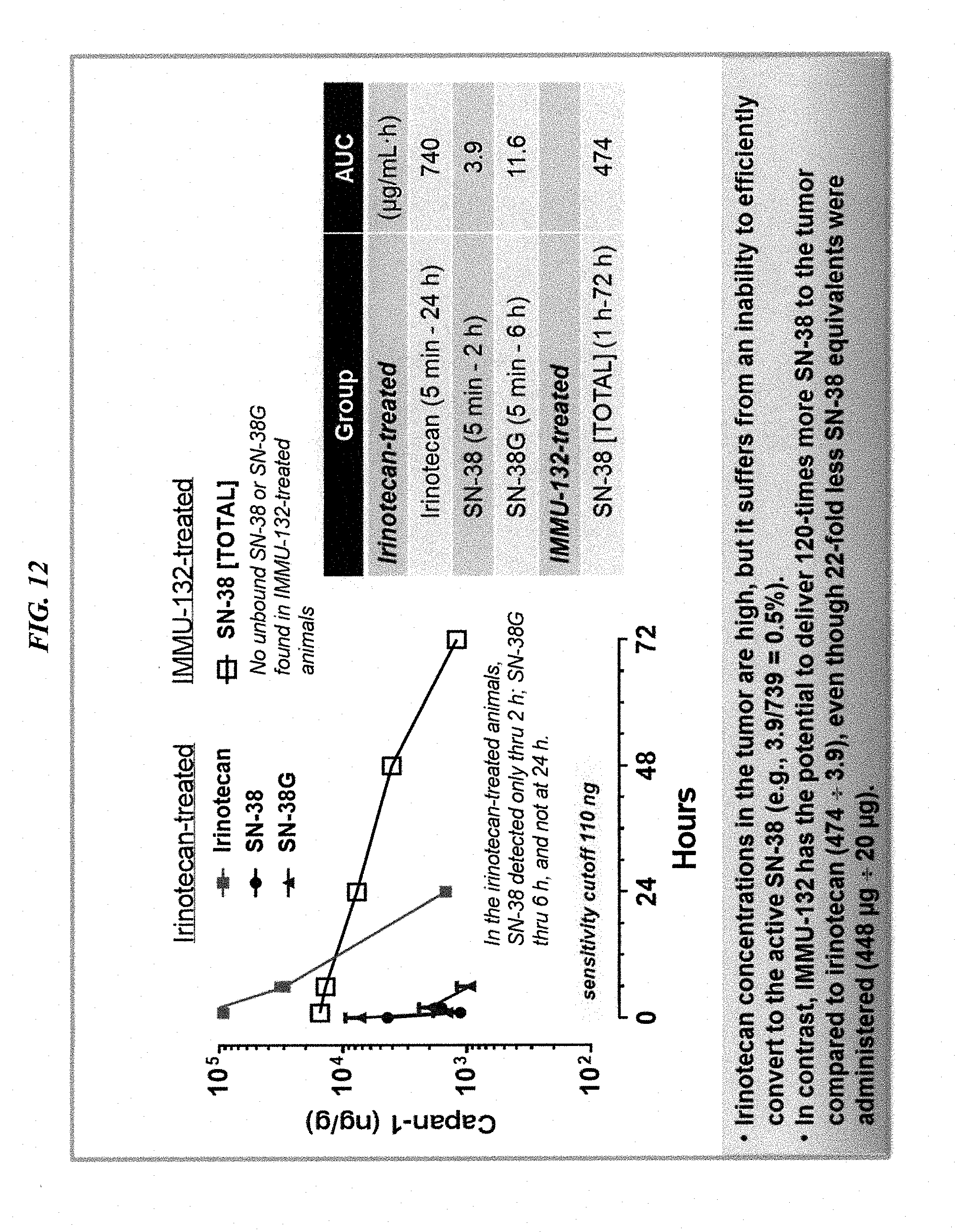
D00015

D00016
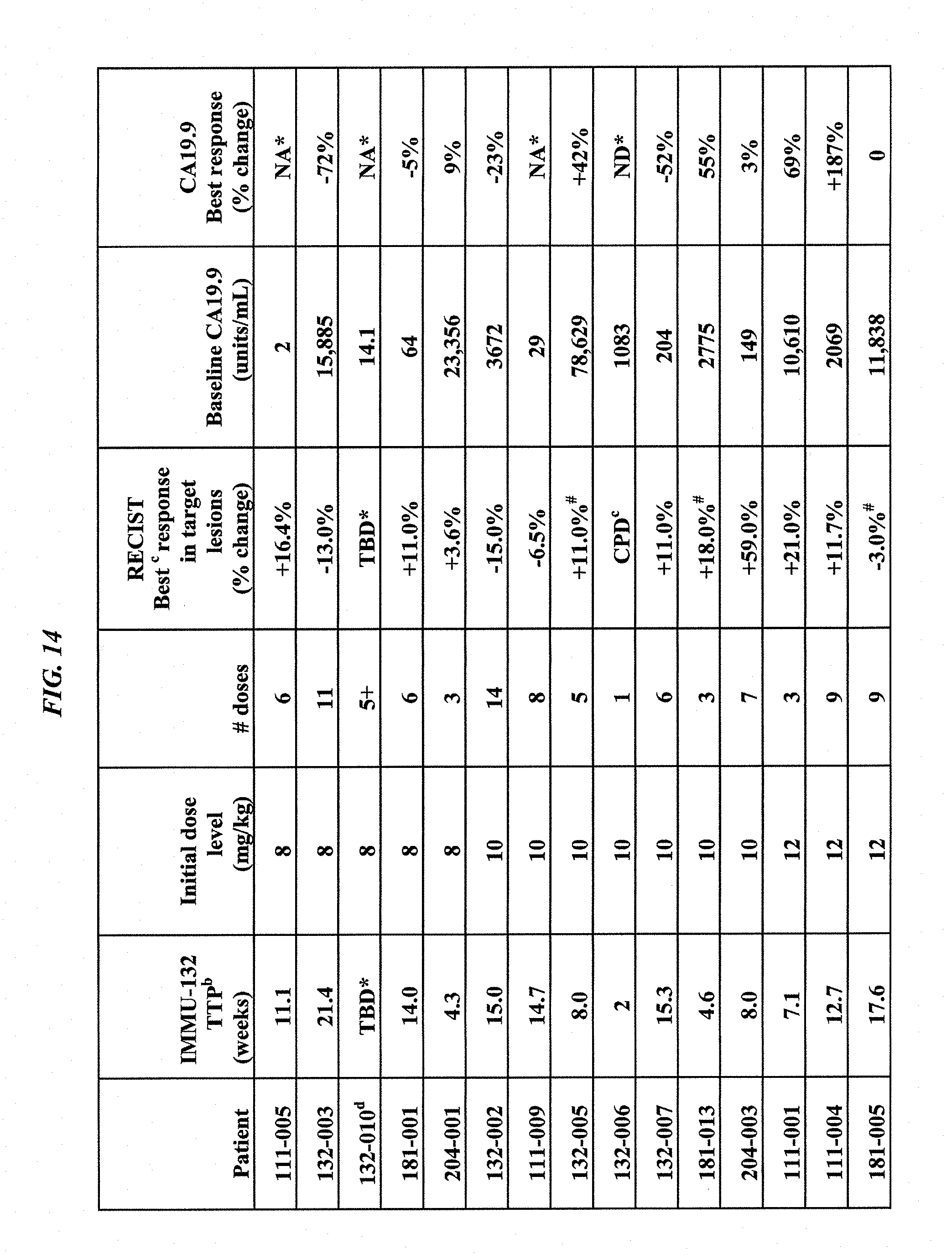
D00017

D00018

D00019

D00020

D00021
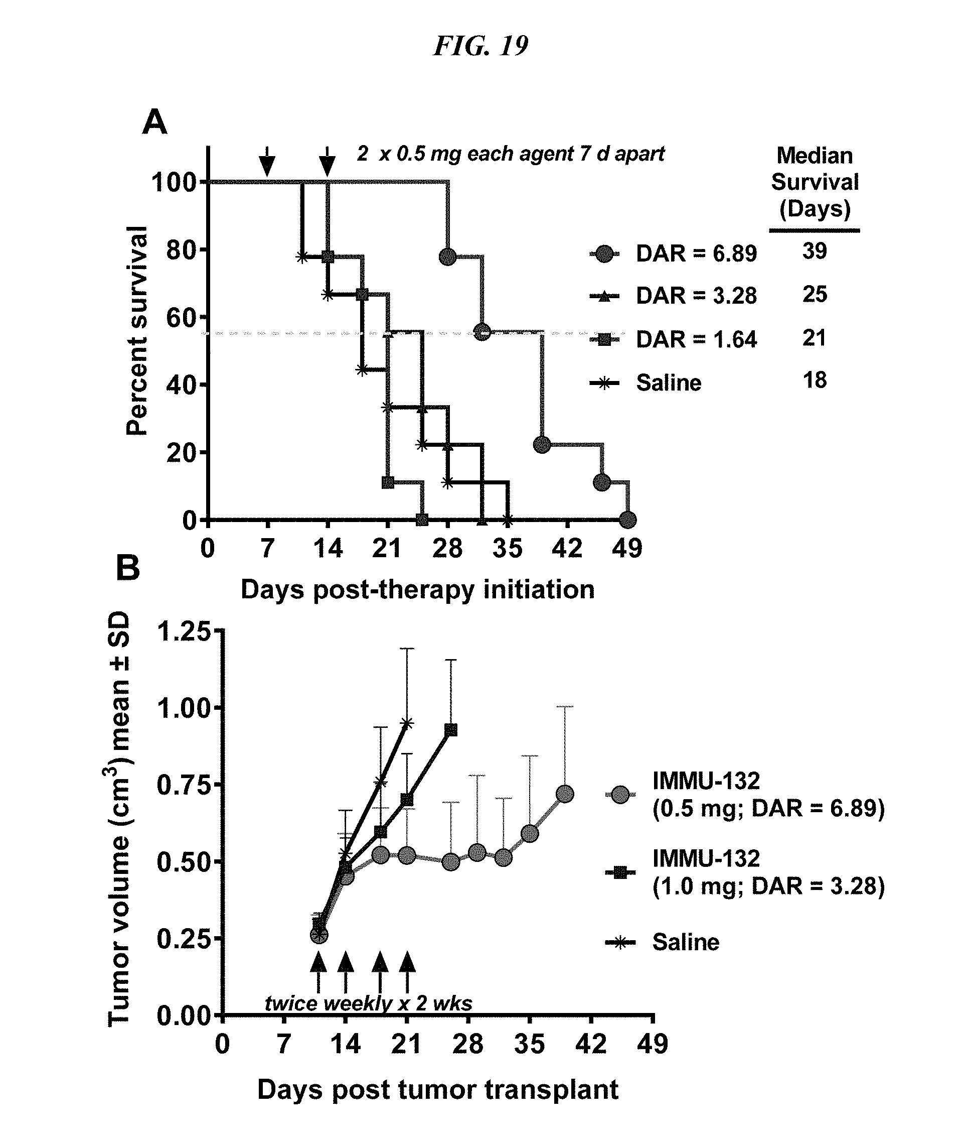
D00022

D00023
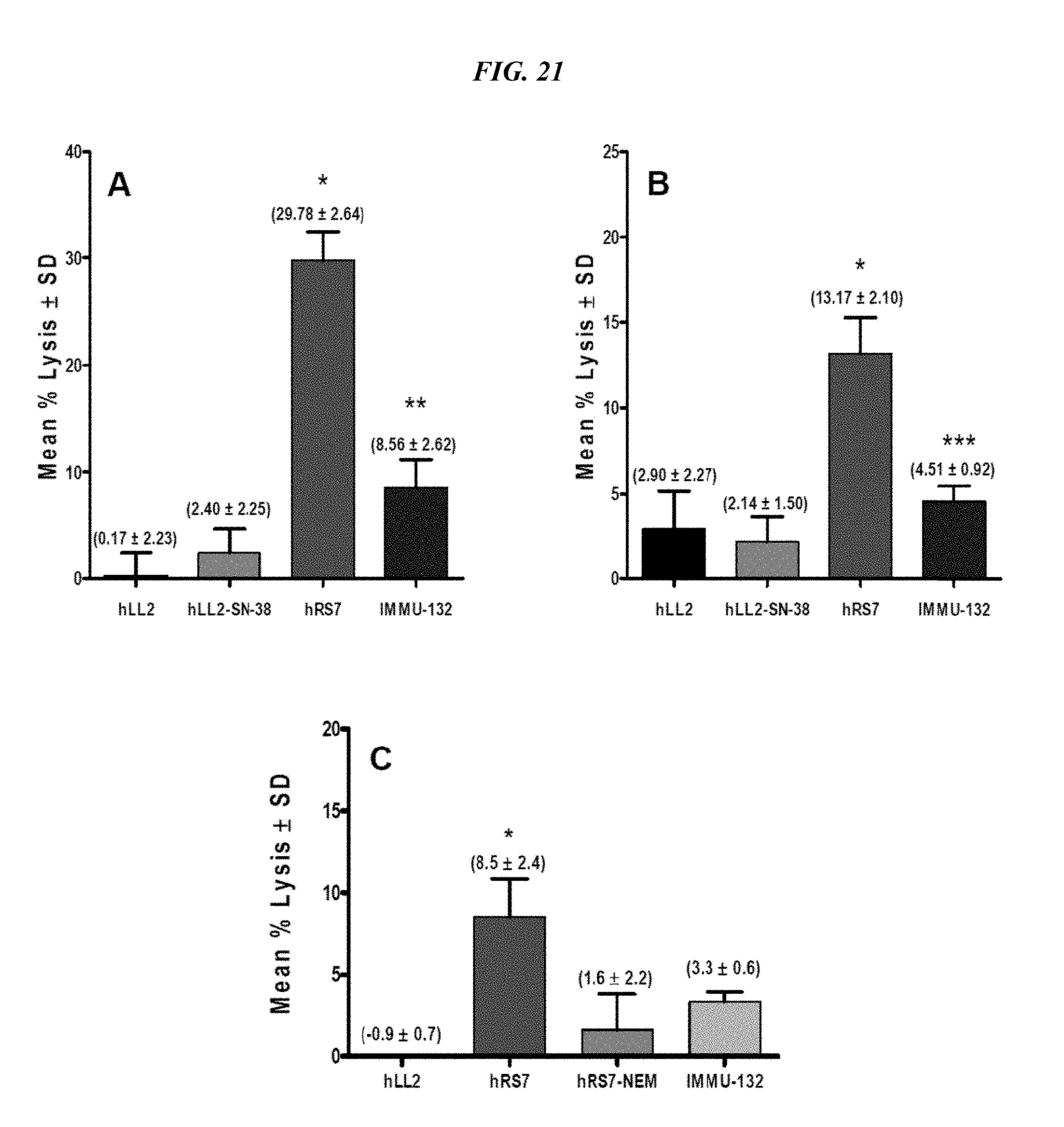
D00024

D00025
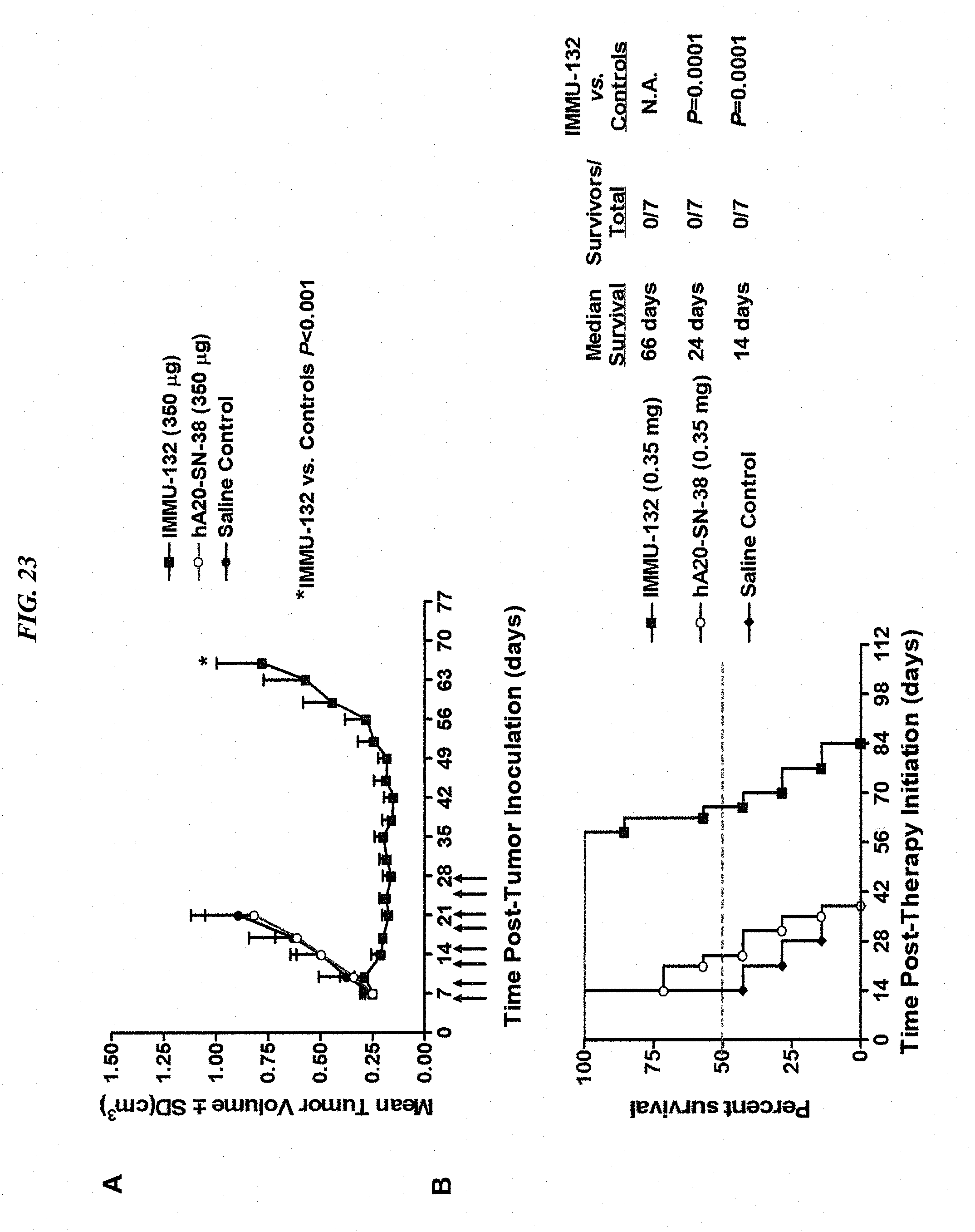
D00026
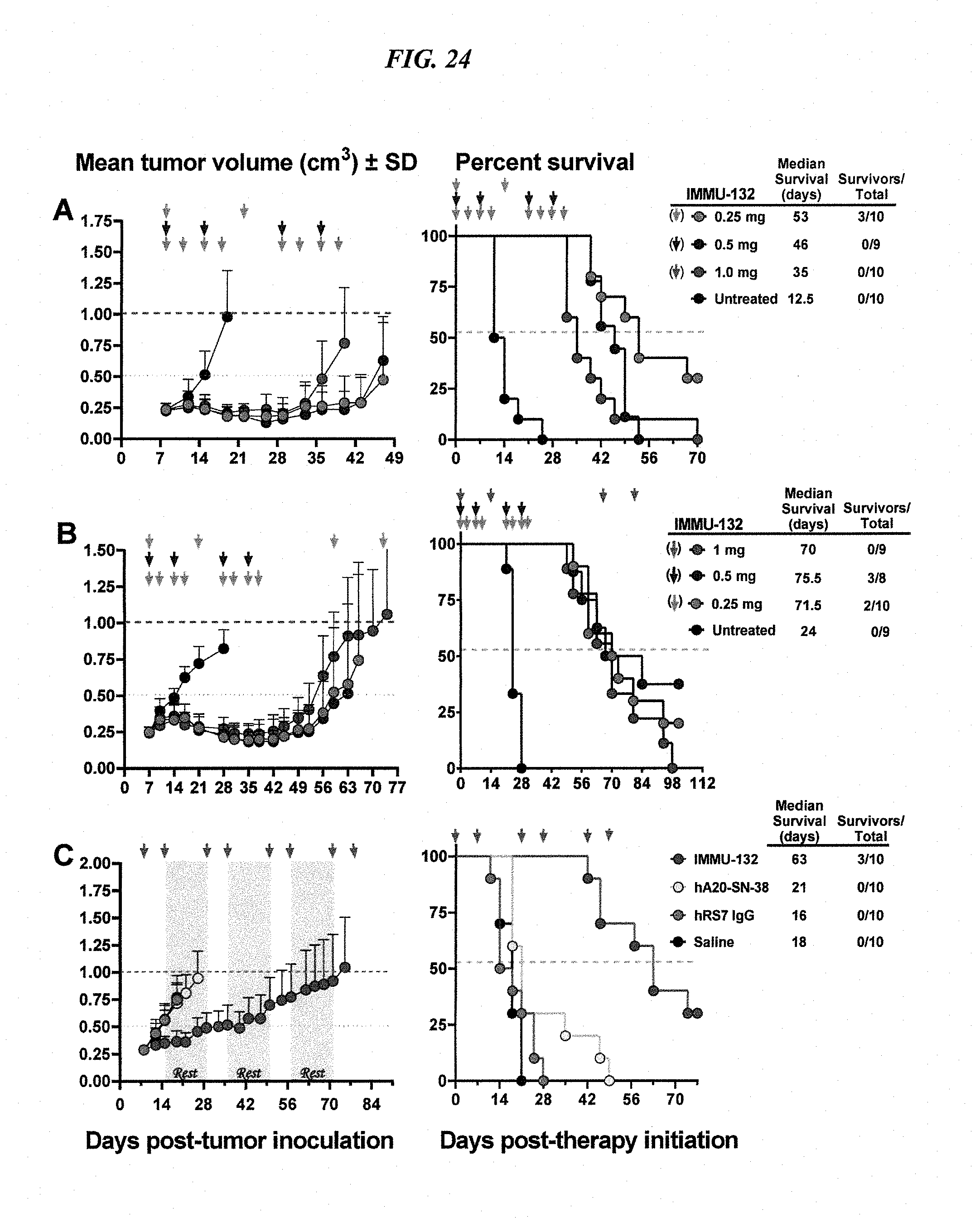
D00027

D00028

D00029
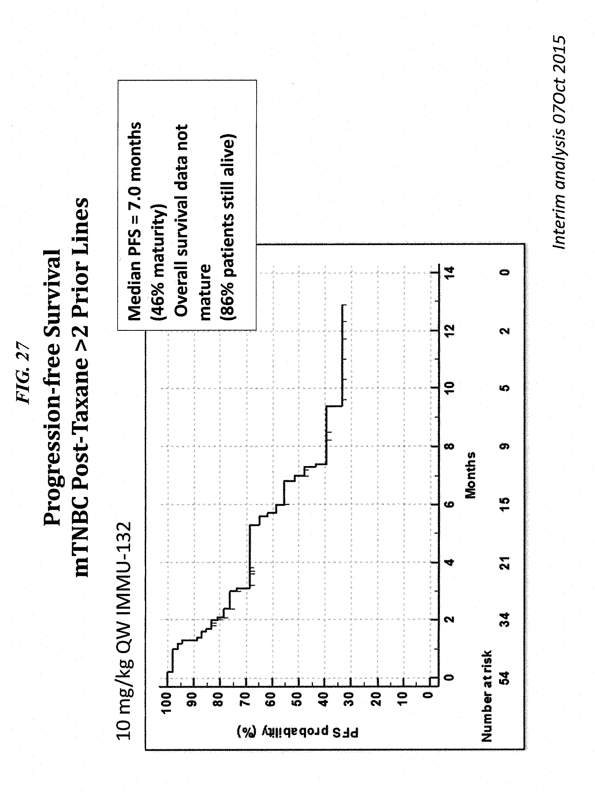
D00030
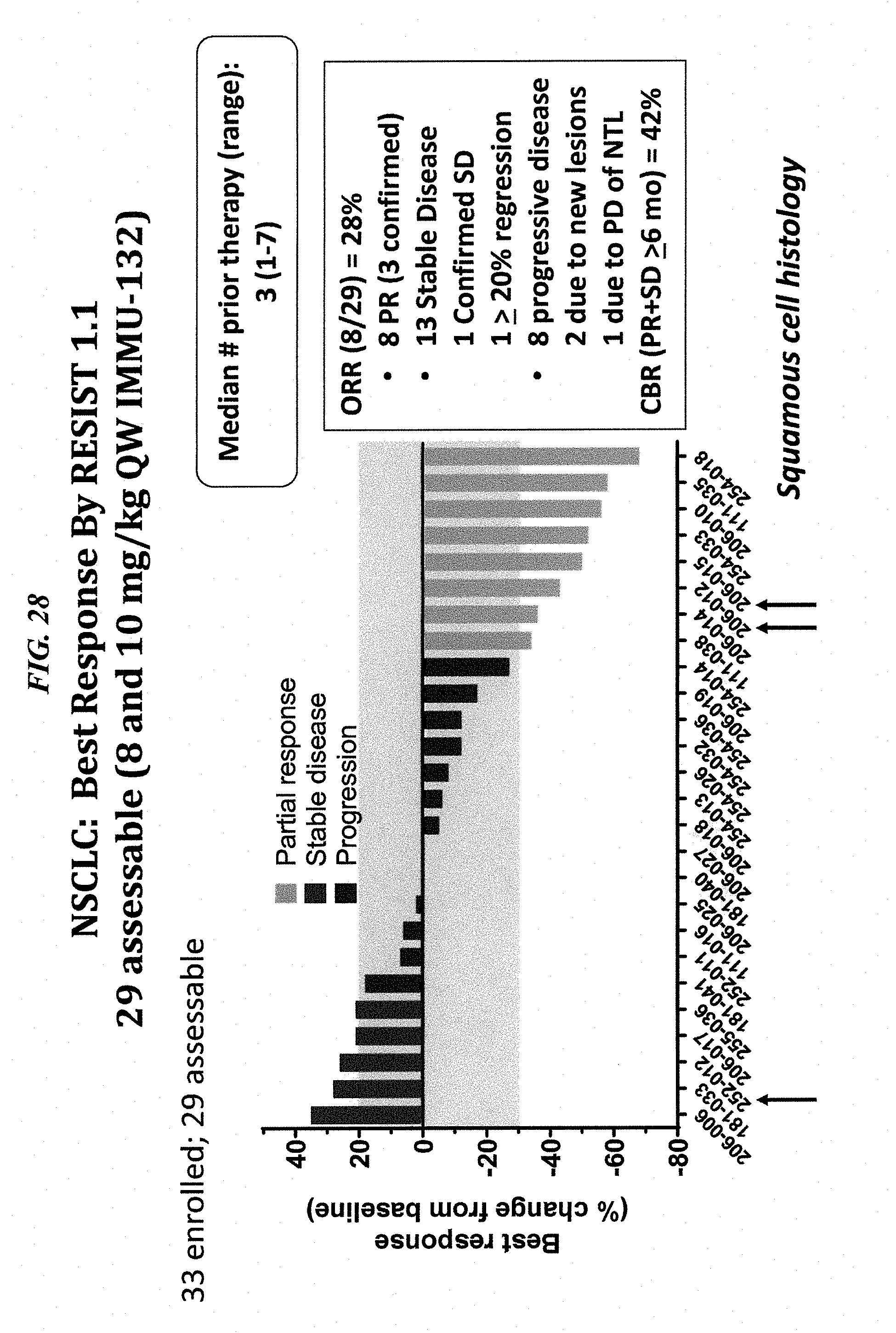
D00031
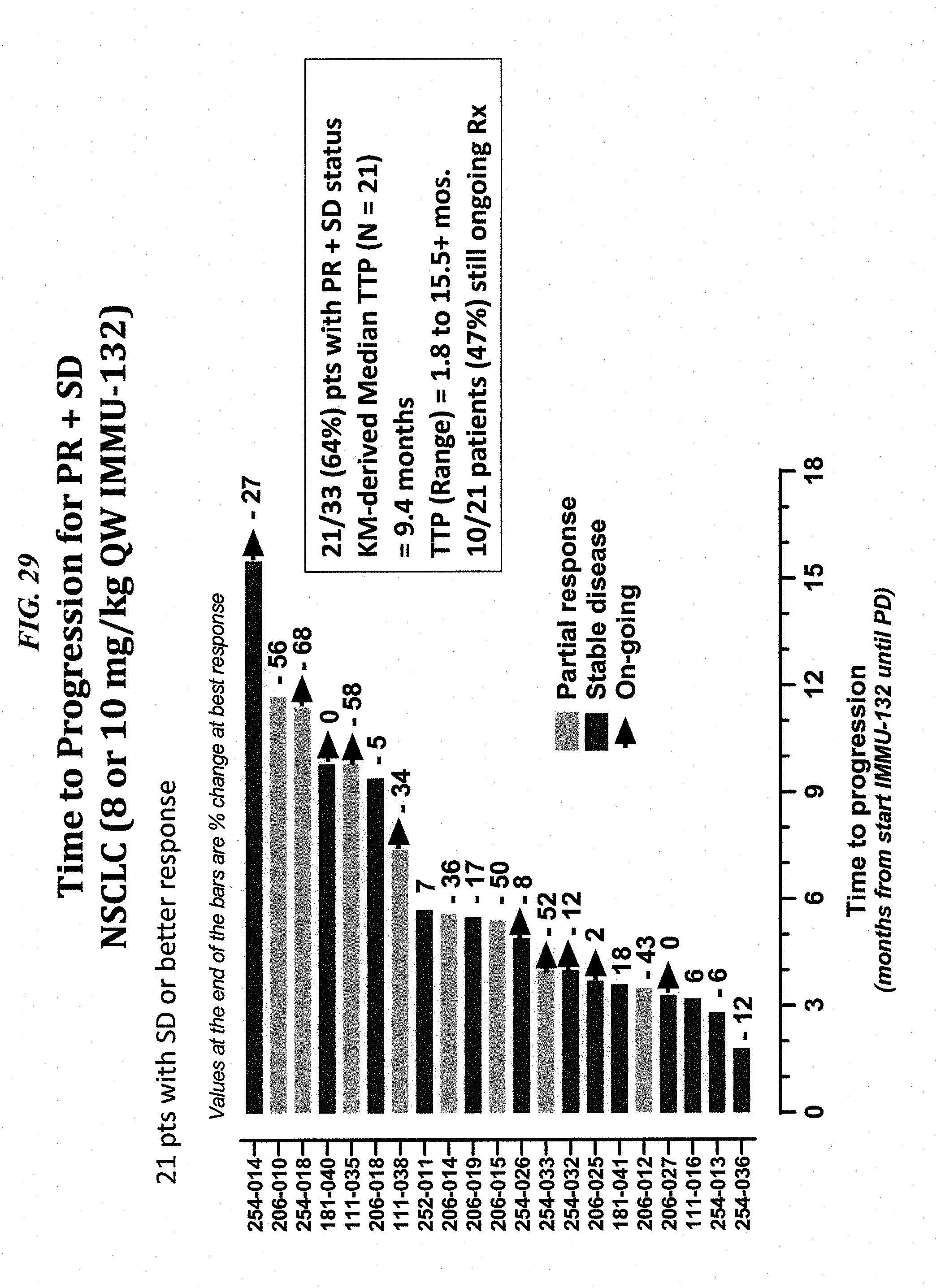
D00032

D00033

D00034

D00035

D00036

D00037
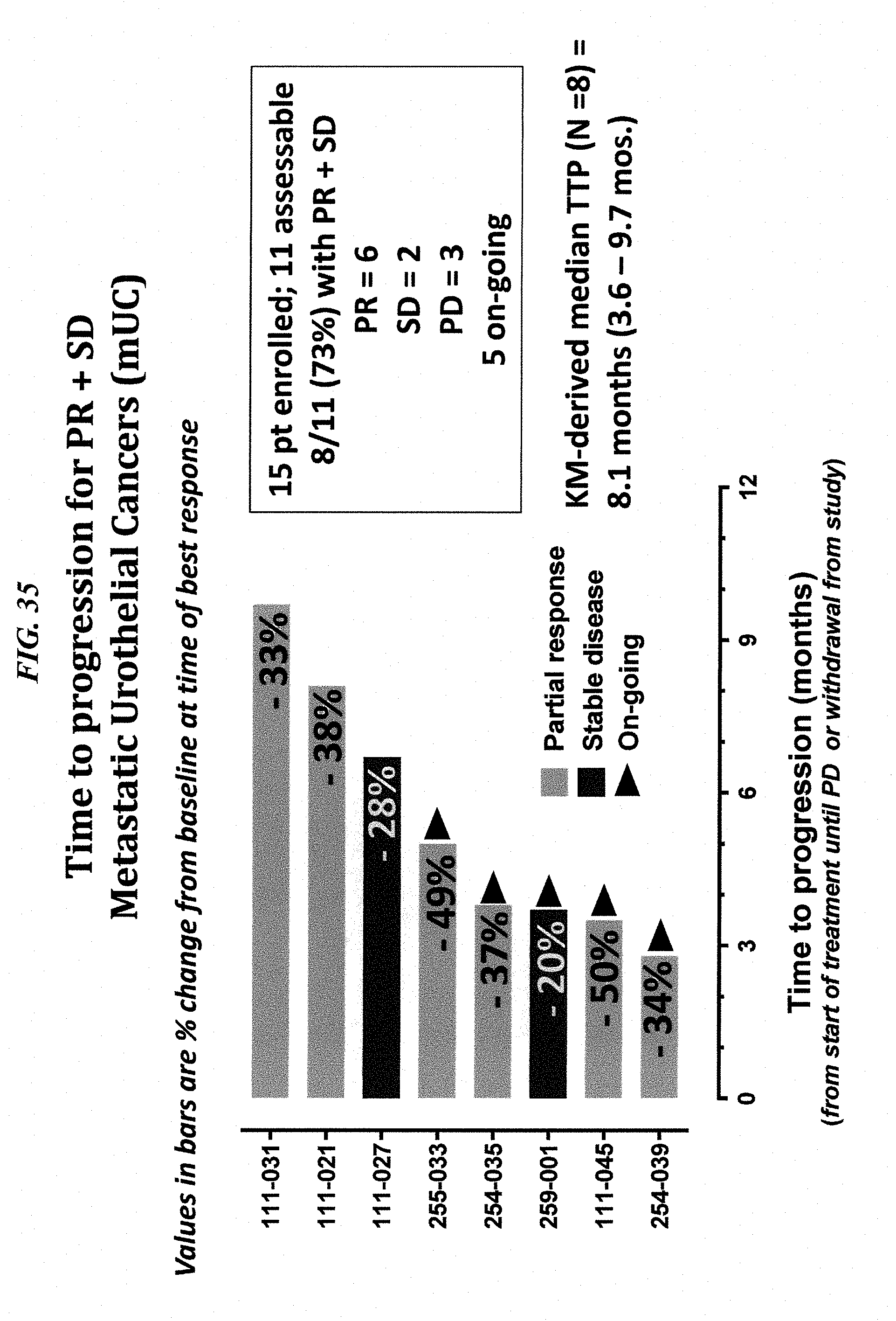
D00038

D00039

D00040


S00001
XML
uspto.report is an independent third-party trademark research tool that is not affiliated, endorsed, or sponsored by the United States Patent and Trademark Office (USPTO) or any other governmental organization. The information provided by uspto.report is based on publicly available data at the time of writing and is intended for informational purposes only.
While we strive to provide accurate and up-to-date information, we do not guarantee the accuracy, completeness, reliability, or suitability of the information displayed on this site. The use of this site is at your own risk. Any reliance you place on such information is therefore strictly at your own risk.
All official trademark data, including owner information, should be verified by visiting the official USPTO website at www.uspto.gov. This site is not intended to replace professional legal advice and should not be used as a substitute for consulting with a legal professional who is knowledgeable about trademark law.