Nanoparticle Sensor Having A Nanofibrous Membrane Scaffold
Zhong; Chuan-Jian ; et al.
U.S. patent application number 16/057314 was filed with the patent office on 2019-02-07 for nanoparticle sensor having a nanofibrous membrane scaffold. The applicant listed for this patent is The Research Foundation for The State University of New York. Invention is credited to Benjamin S. Hsiao, Ning Kang, Jing Li, Jin Luo, Mark D. Poliks, Shiyao Shan, Shan Yan, Chuan-Jian Zhong.
| Application Number | 20190038190 16/057314 |
| Document ID | / |
| Family ID | 65231368 |
| Filed Date | 2019-02-07 |


View All Diagrams
| United States Patent Application | 20190038190 |
| Kind Code | A1 |
| Zhong; Chuan-Jian ; et al. | February 7, 2019 |
NANOPARTICLE SENSOR HAVING A NANOFIBROUS MEMBRANE SCAFFOLD
Abstract
Nanoparticle-fibrous membrane composites are provided as tunable interfacial scaffolds for flexible chemical sensors and biosensors by assembling gold nanoparticles (Au NPs) in a fibrous membrane. The gold nanoparticles are functionalized with organic, polymeric and/or biological molecules. The fibrous membranes may include different filter papers, with one example featuring a multilayered fibrous membrane consisting of a cellulose nanofiber (CN) top layer, an electrospun polyacrylonitrile (PAN) nanofibrous midlayer (or alternate material), and a nonwoven polyethylene terephthalate (PET) fibrous support layer, with the nanoparticles provided on the fibrous membranes through interparticle molecular/polymeric linkages and nanoparticle-nanofibrous interactions. Molecular linkers may be employed to tune hydrogen bonding and electrostatic and/or hydrophobic/hydrophilic interactions to provide sensor specificity to gases or liquids. The sensors act as chemiresistor-type sensors. A preferred implementation is a sweat sensor.
| Inventors: | Zhong; Chuan-Jian; (Endwell, NY) ; Poliks; Mark D.; (Vestal, NY) ; Hsiao; Benjamin S.; (Setauket, NY) ; Kang; Ning; (Vestal, NY) ; Yan; Shan; (Vestal, NY) ; Li; Jing; (Vestal, NY) ; Shan; Shiyao; (Vestal, NY) ; Luo; Jin; (Vestal, NY) | ||||||||||
| Applicant: |
|
||||||||||
|---|---|---|---|---|---|---|---|---|---|---|---|
| Family ID: | 65231368 | ||||||||||
| Appl. No.: | 16/057314 | ||||||||||
| Filed: | August 7, 2018 |
Related U.S. Patent Documents
| Application Number | Filing Date | Patent Number | ||
|---|---|---|---|---|
| 62542067 | Aug 7, 2017 | |||
| Current U.S. Class: | 1/1 |
| Current CPC Class: | A61B 2560/0412 20130101; A61B 2562/12 20130101; A61B 5/082 20130101; A61B 5/14517 20130101; A61B 2562/0295 20130101; B33Y 80/00 20141201; A61B 5/1477 20130101; A61B 5/4266 20130101 |
| International Class: | A61B 5/1477 20060101 A61B005/1477; A61B 5/00 20060101 A61B005/00; A61B 5/08 20060101 A61B005/08; A61B 5/145 20060101 A61B005/145; B33Y 80/00 20060101 B33Y080/00 |
Goverment Interests
STATEMENT OF GOVERNMENT RIGHTS
[0002] This invention was made with government support under IIP-1640669, awarded by the National Science Foundation. The government has certain rights in the invention.
Claims
1. A sensor, comprising: a sensing medium, comprising a fibrous layer and a plurality of nanoparticles, coating fibers within the fibrous layer, the plurality of nanoparticles being derivatized to interact with the fibrous layer and an analyte in a medium, based on at least one of electronic charge, ligand coordination, hydrogen bonding, van der Waals force, polarity, hydrophilicity, and hydrophobicity; and an electrode, configured to sense a state of the sensing medium in response to the analyte in the medium, and to produce an electrical signal output corresponding to the state.
2. The sensor according to claim 1, wherein the fibrous layer comprises nanofibers.
3. The sensor according to claim 1, wherein the fibrous layer comprises nanofibrous cellulose.
4. The sensor according to claim 2, wherein the fibrous layer comprises at least one region consisting essentially of a plurality of nanofibers, to which the nanoparticles adhere.
5. The sensor according to claim 1, wherein the fibrous layer comprises a natural cellulose fiber paper.
6. The sensor according to claim 1, wherein the nanoparticles are gold nanoparticles.
7. The sensor according to claim 1, wherein the derivatized nanoparticles are charged.
8. The sensor according to claim 1, further comprising a permeable electrospun fiber layer supporting the fibrous layer.
9. The sensor according to claim 1, further comprising a permeable layer, and a non-woven layer, the permeable layer being supported on the nonwoven layer, and the fibrous layer being supported on the permeable layer, wherein permeable layer consists essentially of fibers having a diameter larger than a diameter of the fibers within the fibrous layer, and fibers of the nonwoven layer have a larger diameter than fibers of the permeable layer.
10. The sensor according to claim 1, further comprising a permeable layer comprising at least one of crosslinked polyacrylonitrile (PAN) or crosslinked polyethylene glycol diacrylate (PEGDA), and a polyethylene terephthalate (PET) non-woven layer, the permeable layer being supported on the non-woven layer, and the fibrous layer being supported on the permeable layer.
11. The sensor according to claim 1, wherein the sensing medium comprises the fibrous layer consisting essentially of fibers having a fiber diameter of less than about 15 nm, supported on a fibrous intervening layer having a fiber diameter of less than about 250 nm, disposed on a flexible support layer.
12. The sensor according to claim 1, wherein the plurality of nanoparticles are at least one of: linked to a thiolate through a thiol bond; linked to 11-mercaptoundecanoic acid (MUA) within the nanofibrous layer through a hydrogen bond; linked to a carboxylic acid; and electrostatically bound to poly(diallyl) ammonium) within the nanofibrous layer.
13. The sensor according to claim 1, wherein the fibrous layer is cast from a slurry of nanofibers on an electrospun layer.
14. The sensor according to claim 1, wherein the electrical signal output is selectively responsive to at least one of moisture and ions.
15. The sensor according to claim 1, wherein the electrode comprises a pair of interdigitated conductive traces spaced across a gap to sense a change in conductivity or capacitance of the fibrous layer coated with the derivatized nanoparticles.
16. The sensor according to claim 1, further comprising an electronic circuit configured to receive the electrical signal output, and to determine a quantitative parameter of the analyte.
17. The sensor according to claim 1, having a monotonically increasing response to a concentration an ionic species within the analyte over a range from 0 to 100 mM.
18. A method of sensing an analyte, comprising: providing a sensor, comprising an electrode for sensing an electrical state of a sensing medium, and producing as an output an electrical signal corresponding to the state, the sensing medium comprising a fibrous layer and a plurality of nanoparticles, coating fibers of the fibrous layer, the plurality of nanoparticles being derivatized to interact with the nanofibrous layer alter the electrical state of the sensing medium in response to the analyte, based on at least one of an electronic charge, hydrogen bonding, van der Waals force, polarity, hydrophilicity, and hydrophobicity; exposing the sensor to the analyte; and producing the output, dependent on the electrical state of the sensing medium.
19. A method of manufacturing a sensing medium, comprising a nanofibrous layer and a plurality of nanoparticles supported on and embedded in the nanofibrous layer, comprising: providing derivatized insoluble conductive nanoparticles having a diameter less than about 150 nm, the derivatized insoluble conductive nanoparticles being derivatized with a ligand capable of at least one of ionic bonding, hydrogen bonding, and van der Waals bonding; providing organic nanofibers in an aqueous slurry, having a nanofiber diameter of less than about 15 nm, and having exposed groups capable of at least one of ionic bonding, hydrogen bonding, and van der Waals interaction with the derivatized insoluble conductive nanoparticles; providing a fibrous layer having a fiber diameter of between about 5-250 nm, deposited on a non-woven fibrous substrate; casting a layer of the organic nanofibers in the aqueous slurry onto the non-woven fibrous substrate, under conditions which cause the organic nanofibers in the aqueous slurry to gel and remain on a surface of the non-woven fibrous substrate or fibrous layer; and depositing a solution containing the nanoparticles on the layer of the organic nanofibers, to link the nanoparticles with nanofibers within the layer of the organic nanofibers, by at least one of electrostatic bonding, hydrogen bonding, and van der Waals interaction, to thereby produce the sensing medium, comprising a nanofibrous layer, on the non-woven fibrous substrate or the fibrous layer, and a plurality of nanoparticles, coating and interacting with nanofibers within the nanofibrous layer.
20. The method according to claim 19, wherein the layer of organic nanofibers is cast on the non-woven fibrous substrate as a gel which remains on a surface of the fibrous layer substantially without invading the interior of the non-woven fibrous substrate.
Description
CROSS REFERENCE TO RELATED APPLICATION
[0001] The present application is a non-provisional of, and claims benefit of priority under 35 U.S.C. .sctn. 119(e) from, U.S. Provisional Patent Application 62/542,067, filed Aug. 7, 2017, the entirety of which is expressly incorporated herein by reference.
FIELD OF THE INVENTION
[0003] The present invention relates to the field of biological fluid sensors, and more particularly to sweat or biomarker secretion sensors.
BACKGROUND OF THE INVENTION
[0004] Sweating is primarily used to regulate body temperature by cooling the body down with secretion of water. The inability of human body to sweat properly is potentially harmful, and a complete absence of sweating (anhidrosis) or sweating less than normal (hypohidrosis) is an abnormal lack of sweat in response to heat, which is also one of the symptoms of some genetic diseases. As such, monitoring of the moisture levels from perspiration provides useful information for assessing the physical conditions, especially for people exposed to high temperature or experiencing long time exercise who face the risk of dehydration. The fact that sweat contains abundant information of medical significance has been an important driving force for the increasing interests in developing wearable sweat sensors. In addition to moisture, sweat is also rich with ions such as sodium, potassium and chlorine ranging from 10 to 80 mM. Monitoring the saltiness thus provides further useful information. Moreover, sweat may also contain biomarkers related to metabolites of the human body, e.g., glucose, lactate, and uric acid. While some progress has been made in developing sweat sensors, key challenges remain, including lack of multifunctionality, biocompatibility, and flexibility in some monitoring conditions, high cost in manufacturing, and insufficient sensitivity and selectivity, calling for breakthroughs in materials design and fabrication.
[0005] See, U.S. Pat. Nos. 5,729,203; 6,198,953; 6,882,940; 7,109,933; 7,187,960; 7,383,072; 8,032,199; 8,328,420; 8,527,028; 8,560,044; 8,721,562; 8,834,020; 8,849,379; 9,011,349; 9,028,405; 9,198,605; 9,198,617; 9,204,808; 9,254,437; 9,277,867; 9,301,719; 9,398,856; 9,408,572; 9,449,084; 9,510,784; 9,545,221; 9,563,995; 9,579,024; 9,579,040; 9,603,560; 9,622,725; 9,636,061; 9,645,133; 9,686,499; 9,704,205; 9,713,447; 9,717,455; 20020106709; 20040059212; 20040242976; 20050119540; 20050194012; 20050195118; 20060253011; 20070173886; 20070197878; 20070219434; 20080027679; 20080081963; 20080091097; 20080287769; 20080287770; 20080294058; 20090018412; 20090105605; 20090269003; 20090312615; 20100099957; 20100240962; 20120020033; 20120157793; 20120215076; 20120229661; 20130057720; 20130066168; 20130096396; 20130124039; 20130197319; 20130234724; 20130245388; 20130288777; 20140012145; 20140031705; 20140106816; 20140200432; 20140206948; 20140249763; 20140275828; 20140275838; 20140275840; 20140277649; 20140347187; 20150038874; 20150093725; 20150094914; 20150112164; 20150112165; 20150145676; 20150148628; 20150148681; 20150150453; 20150150467; 20150164238; 20150164409; 20150216479; 20150248651; 20150301594; 20150320588; 20160038082; 20160117029; 20160174892; 20160232625; 20160262666; 20160262667; 20160270239; 20160270726; 20160278700; 20160287148; 20160290952; 20160302730; 20160310011; 20160331235; 20160349790; 20160371372; 20160374598; 20170065183; 20170079574; 20170094216; 20170095183; 20170095184; 20170095233; 20170100035; 20170100071; 20170100072; 20170100076; 20170100102; 20170103166; 20170105104; 20170105646; 20170105662; 20170116879; 20170119311; 20170135633; 20170136264; 20170136265; 20170140626; 20170156641; 20170156662; 20170157430; 20170157431; 20170164865; 20170164866; 20170164876; 20170164878; 20170172470; 20170172484; 20170181659; and 20170181684.
[0006] Sensors formed using gold nanoparticles are known: see, U.S. Pat. Nos. 6,361,944; 6,417,340; 6,495,324; 6,506,564; 6,541,617; 6,582,921; 6,610,491; 6,645,721; 6,673,548; 6,677,122; 6,682,895; 6,709,825; 6,720,147; 6,720,411; 6,730,269; 6,740,491; 6,750,016; 6,759,199; 6,767,702; 6,773,884; 6,777,186; 6,812,334; 6,818,753; 6,828,432; 6,861,221; 6,878,814; 6,902,895; 6,903,207; 6,962,786; 6,969,761; 6,984,491; 6,986,989; 7,052,854; 7,098,320; 7,169,556; 7,195,780; 7,208,587; 7,250,499; 7,259,252; 7,267,948; 7,435,386; 7,485,419; 7,534,560; 7,569,354; 7,611,628; 7,612,185; 7,695,738; 7,799,554; 7,816,491; 7,863,376; 7,985,424; 8,012,743; 8,067,393; 8,216,854; 8,282,967; 8,283,414; 8,323,694; 8,323,888; 8,376,013; 8,383,415; 8,389,958; 8,426,214; 8,486,720; 8,501,921; 8,507,200; 8,524,457; 8,598,046; 8,618,509; 8,652,778; 8,666,471; 8,679,859; 8,703,439; 8,717,558; 8,758,772; 8,770,203; 8,883,964; 8,906,831; 8,920,971; 8,927,615; 8,945,943; 8,951,561; 8,956,658; 8,956,863; 8,962,029; 8,962,912; 8,993,349; 8,999,947; 9,000,137; 9,004,131; 9,012,156; 9,067,181; 9,102,520; 9,114,107; 9,157,109; 9,174,190; 9,174,873; 9,201,071; 9,216,155; 9,222,884; 9,242,857; 9,243,128; 9,246,122; 9,250,238; 9,274,108; 9,276,238; 9,283,275; 9,290,799; 9,302,116; 9,310,372; 9,315,942; 9,316,645; 9,320,813; 9,321,030; 9,333,163; 9,340,416; 9,376,690; 9,393,396; 9,403,851; 9,403,852; 9,410,949; 9,416,493; 9,437,628; 9,439,868; 9,440,195; 9,446,150; 9,447,129; 9,486,512; 9,494,524; 9,506,056; 9,508,956; 9,511,329; 9,526,913; 9,526,914; 9,532,956; 9,534,024; 9,540,422; 9,555,392; 9,556,473; 9,557,340; 9,567,645; 9,567,646; 9,579,523; 9,581,590; 9,592,198; 9,598,544; 9,598,736; 9,604,168; 9,623,352; 9,623,381; 9,624,275; 9,630,022; 9,637,380; 9,637,799; 9,637,830; 9,649,391; 9,662,299; 9,662,388; 9,662,389; 9,664,674; 9,688,750; 9,691,873; 9,701,784; 9,709,559; 9,717,685; 9,719,089; 20020127574; 20020137058; 20020137070; 20020137071; 20020137072; 20020146720; 20020155442; 20020155458; 20020155459; 20020155461; 20020155462; 20020160381; 20020164605; 20020172953; 20020182611; 20020182613; 20030022169; 20030044805; 20030049630; 20030049631; 20030054358; 20030059777; 20030059820; 20030068622; 20030087242; 20030124528; 20030143538; 20030148282; 20030180783; 20030198956; 20030207296; 20030215903; 20040018587; 20040072231; 20040076681; 20040110220; 20040219520; 20050037374; 20050059030; 20050059031; 20050130174; 20050130240; 20050176029; 20050250094; 20050272114; 20050287552; 20060014172; 20060040318; 20060057613; 20060068378; 20060257883; 20070059763; 20070087383; 20070087400; 20070122829; 20070125181; 20070127164; 20070154903; 20070258894; 20070269821; 20070298006; 20080146701; 20080226995; 20080241071; 20080241964; 20080279946; 20080287342; 20090042200; 20090042739; 20090130773; 20090214618; 20090246142; 20090294692; 20090325215; 20090325812; 20100003316; 20100009872; 20100016568; 20100016569; 20100016783; 20100018862; 20100021933; 20100028559; 20100062232; 20100069621; 20100196920; 20100234579; 20100261263; 20100290992; 20110012096; 20110021970; 20110065807; 20110098197; 20110114244; 20110114511; 20110129537; 20110136139; 20110171749; 20110176130; 20110206740; 20110263920; 20120021055; 20120034169; 20120064134; 20120070376; 20120087949; 20120135437; 20120156135; 20120263648; 20120263793; 20120315322; 20130023714; 20130034599; 20130034915; 20130095499; 20130116405; 20130140649; 20130156905; 20130171060; 20130177598; 20130183243; 20130196872; 20130203073; 20130210023; 20130240758; 20130252843; 20130252848; 20130261010; 20130295688; 20140005426; 20140024026; 20140050793; 20140058124; 20140120534; 20140163303; 20140222117; 20140243934; 20140273029; 20140294927; 20140322823; 20140335154; 20140343479; 20140363833; 20140364332; 20140378676; 20150005188; 20150017258; 20150031571; 20150056627; 20150093774; 20150111308; 20150141266; 20150164117; 20150182543; 20150198606; 20150202304; 20150202351; 20150211134; 20150231635; 20150251016; 20150253317; 20150253318; 20150265706; 20150265725; 20150299784; 20150330025; 20160005503; 20160010136; 20160010151; 20160022976; 20160025634; 20160033861; 20160054310; 20160060279; 20160074511; 20160130056; 20160130335; 20160130370; 20160131615; 20160145683; 20160146799; 20160161472; 20160175251; 20160184226; 20160185814; 20160228574; 20160238591; 20160243235; 20160258012; 20160263393; 20160265069; 20160274086; 20160325111; 20160334398; 20160351874; 20170014511; 20170021040; 20170043178; 20170050046; 20170065636; 20170067021; 20170088875; 20170119820; 20170121472; 20170121708; 20170130200; 20170131291; 20170151339; 20170166760; 20170173350; 20170184574; 20170189481; 20170196977; 20170210115; and 20170214020.
[0007] Various chemiresistor technologies, and applications thereof, are known: see, U.S. Pat. Nos. 4,636,767; 4,759,210; 4,847,594; 4,886,625; 4,900,817; 4,992,244; 5,045,285; 5,071,770; 5,089,294; 5,210,217; 5,224,972; 5,238,729; 5,279,795; 5,280,183; 5,302,935; 5,321,146; 5,387,462; 5,433,971; 5,469,369; 5,498,323; 5,512,882; 5,536,473; 5,550,062; 5,571,401; 5,589,396; 5,629,435; 5,674,752; 5,698,083; 5,698,089; 5,788,833; 5,858,186; 5,891,398; 5,911,872; 5,951,846; 5,959,191; 5,976,466; 6,004,494; 6,010,616; 6,013,229; 6,015,869; 6,017,440; 6,085,576; 6,093,308; 6,170,318; 6,221,673; 6,238,085; 6,244,096; 6,290,911; 6,319,724; 6,331,244; 6,350,369; 6,359,444; 6,387,329; 6,397,661; 6,408,250; 6,418,783; 6,421,588; 6,422,061; 6,458,327; 7,122,152; 7,136,716; 7,138,090; 7,144,553; 7,144,949; 7,168,294; 7,175,885; 7,179,421; 7,189,353; 7,189,360; 7,189,867; 7,191,805; 7,201,035; 7,211,439; 7,211,637; 7,226,530; 7,229,593; 7,233,781; 7,242,310; 7,253,004; 7,265,560; 7,272,530; 7,288,415; 7,313,447; 7,340,941; 7,342,479; 7,347,974; 7,356,420; 7,359,802; 7,387,010; 7,395,693; 7,397,072; 7,404,928; 7,421,883; 7,438,079; 7,449,050; 7,471,185; 7,477,994; 7,489,252; 7,501,091; 7,527,821; 7,531,136; 7,531,137; 7,538,538; 7,550,310; 7,556,775; 7,595,023; 7,595,734; 7,645,422; 7,708,947; 7,726,175; 7,737,322; 7,760,101; 7,793,675; 7,799,276; 7,801,687; 7,840,359; 7,880,026; 7,889,954; 7,911,010; 7,912,561; 7,927,558; 7,939,130; 7,950,271; 7,955,561; 7,966,132; 7,998,415; 7,998,416; 8,000,903; 8,012,326; 8,012,420; 8,030,100; 8,087,283; 8,088,341; 8,105,538; 8,123,834; 8,123,841; 8,152,908; 8,153,439; 8,168,438; 8,178,045; 8,187,887; 8,231,746; 8,246,910; 8,268,630; 8,269,029; 8,272,250; 8,285,493; 8,309,028; 8,310,016; 8,336,402; 8,352,049; 8,366,630; 8,394,330; 8,409,510; 8,412,147; 8,426,208; 8,426,932; 8,441,081; 8,448,532; 8,449,824; 8,461,354; 8,481,324; 8,497,130; 8,519,726; 8,562,878; 8,567,232; 8,569,691; 8,618,330; 8,691,390; 8,694,267; 8,695,401; 8,703,500; 8,707,760; 8,736,287; 8,771,613; 8,790,400; 8,795,359; 8,808,373; 8,816,116; 8,828,733; 8,846,406; 8,877,636; 8,884,382; 8,889,420; 8,900,516; 8,903,661; 8,904,849; 8,920,731; 8,940,092; 8,951,473; 8,957,253; 8,978,444; 8,986,615; 8,989,053; 8,999,244; 8,999,245; 9,017,773; 9,034,266; 9,034,275; 9,034,659; 9,067,070; 9,080,942; 9,120,677; 9,144,488; 9,144,489; 9,147,338; 9,157,842; 9,182,231; 9,182,232; 9,211,185; 9,212,055; 9,217,722; 9,234,757; 9,254,099; 9,260,683; 9,267,908; 9,267,964; 9,315,848; 9,326,730; 9,333,071; 9,339,372; 9,360,509; 9,377,426; 9,402,242; 9,429,536; 9,442,100; 9,448,219; 9,453,811; 9,459,222; 9,459,223; 9,476,862; 9,494,541; 9,510,316; 9,514,632; 9,518,956; 9,529,385; 9,536,122; 9,536,449; 9,538,657; 9,563,833; 9,567,225; 9,582,035; 9,589,686; 9,591,607; 9,598,282; 9,598,785; 9,606,245; 9,613,521; 9,625,341; 9,632,050; 9,638,653; 9,658,178; 9,658,196; 9,674,812; 9,678,059; 9,683,974; 9,689,826; 9,696,311; 9,714,370; 20010029774; 20010041366; 20020002414; 20020004995; 20020005580; 20020014415; 20020017125; 20020045274; 20020045275; 20020081232; 20020081397; 20020098119; 20020110901; 20020131901; 20020132361; 20020141901; 20020142477; 20020149466; 20020164643; 20020178789; 20020197390; 20020198574; 20030010097; 20030024814; 20030069002; 20030083756; 20030109056; 20030109951; 20030136960; 20030144746; 20030159927; 20030165882; 20030165987; 20040018633; 20040018642; 20040029288; 20040033165; 20040042933; 20040147038; 20040192072; 20040194534; 20040200722; 20040202856; 20040204920; 20040211243; 20040215402; 20040223876; 20040237631; 20050000830; 20050016276; 20050022581; 20050048414; 20050065230; 20050072213; 20050090015; 20050121999; 20050126909; 20050131139; 20050150778; 20050159922; 20050177317; 20050202358; 20050216114; 20050241935; 20050244978; 20050263394; 20050272881; 20050280814; 20060034731; 20060053871; 20060057597; 20060099113; 20060099715; 20060124195; 20060124448; 20060144123; 20060174941; 20060208254; 20060244618; 20060249385; 20060259163; 20060275720; 20060282225; 20060292033; 20070018779; 20070095678; 20070114138; 20070117207; 20070119236; 20070126061; 20070131021; 20070142799; 20070151449; 20070180892; 20070187239; 20070229294; 20070231947; 20070235348; 20070252710; 20070252711; 20070275690; 20080003530; 20080017507; 20080025876; 20080054382; 20080077331; 20080101994; 20080103751; 20080236251; 20080245675; 20080262743; 20080278140; 20080278181; 20080319682; 20090004612; 20090007636; 20090007777; 20090049890; 20090084162; 20090090168; 20090130421; 20090148690; 20090196796; 20090201120; 20090214762; 20090216461; 20090227059; 20090234587; 20090256215; 20090260423; 20090261987; 20090263287; 20090273354; 20090309614; 20090315728; 20100001211; 20100008619; 20100060465; 20100073016; 20100102975; 20100132547; 20100140597; 20100188110; 20100191474; 20100203648; 20100204676; 20100209301; 20100225337; 20100229658; 20100272612; 20100273665; 20100276302; 20110010107; 20110015872; 20110054202; 20110081724; 20110089051; 20110098591; 20110125409; 20110127446; 20110171137; 20110184649; 20110244584; 20110246086; 20110269632; 20110286889; 20110300637; 20110320136; 20120041574; 20120050038; 20120056632; 20120071362; 20120071737; 20120090378; 20120097917; 20120143515; 20120156099; 20120165623; 20120186987; 20120212242; 20120270205; 20120282594; 20120295360; 20120301360; 20130022755; 20130040399; 20130046485; 20130059758; 20130065319; 20130126363; 20130143247; 20130158881; 20130162403; 20130171733; 20130183766; 20130210679; 20130236980; 20130236981; 20130241726; 20130259749; 20130311108; 20130315816; 20130330231; 20130338768; 20130338769; 20130338770; 20130338771; 20130338772; 20130338773; 20140015548; 20140022058; 20140083869; 20140107362; 20140127822; 20140145736; 20140151631; 20140193925; 20140208828; 20140220703; 20140242237; 20140274804; 20140275716; 20140288647; 20140296663; 20140296978; 20140318990; 20140330043; 20140347491; 20140349256; 20140349257; 20140371105; 20150076007; 20150079697; 20150082920; 20150087935; 20150101392; 20150116093; 20150126873; 20150132857; 20150168365; 20150232598; 20150268207; 20150272105; 20150273521; 20150276516; 20150276635; 20150276643; 20150276644; 20150276648; 20150276656; 20150301021; 20150309535; 20150313496; 20150320588; 20150325100; 20150327989; 20150366504; 20150370320; 20160008182; 20160011135; 20160012749; 20160018350; 20160073886; 20160095731; 20160103104; 20160110991; 20160112684; 20160140870; 20160169810; 20160195486; 20160195504; 20160209420; 20160231267; 20160232811; 20160282302; 20160290980; 20160317060; 20160349790; 20160366065; 20160370310; 20170023509; 20170038326; 20170093981; 20170110678; 20170124110; 20170135633; 20170160252; 20170162023; 20170164878; 20170167934; and 20170173262.
[0008] Each reference cited herein is expressly incorporated herein by reference in its entirety.
SUMMARY OF THE INVENTION
[0009] The ability to tune the sensing properties with nanostructured materials in a flexible scaffold is essential for constructing highly sensitive and wearable sensors or biosensors, especially for secretions, e.g., from skin or mucous membranes. Flexibility provides an ability to conform to the surface, which is important in some applications. Dynamic flexibility can also be important. Typically, the sensor should remain in close contact with the source of the secretion, for example to avoid void space, to speed response, and assure measurement of the secretion itself. It is noted that the sensors provided herein are not limited to biological secretions from skin and mucous membranes, and the form factor of the sensor may be provided based on its application. For example, the sensor may be provided as an implant or intravascular device, in which case the sensor would measure the surrounding medium.
[0010] The assembly of functional nanoparticles into nanofibrous membranes represents a new strategy for constructing flexible composite materials with multifunctional and tunable properties. Examples of the scaffold papers or membranes include Whatman filter paper or membrane filter, Minipore filter paper, Fisherbrand paper, Ivory Linen paper, etc. Generally, a suitable paper is chemically pulped cellulose fiber, with no additives or finishes. Commercially-available technical papers may therefore be suitable. One implementation provides a nanocomposite scaffold derived from assembling gold nanoparticles (Au NPs) in a multi-layered nanofibrous membrane through controllable interactions with molecular linking and polymeric electrostatic binding.
[0011] A preferred embodiment includes a 3-layer structured membrane consisting of cellulose nanofibers (CN), cross-linked polyethylene glycol diacrylate (PEGDA) and nonwoven polyethylene terephthalate (PET) layers, utilized in combination with either 11-mercaptoundecanoic acid (MUA) as a molecular linker with hydrogen-bonding groups for interlinking alkanethiolate-capped Au NPs or poly(diallyl ammonium) (PDA) as a matrix with positively-changed groups for anchoring negative-charge capped Au NPs.
[0012] In one embodiment, gold nanoparticles (Au NPs) are provided in a multilayered fibrous membrane consisting of cellulose nanofiber (CN) top layer (fiber diameter 5 nm), electrospun polyacrylonitrile (PAN) nanofibrous midlayer (fiber diameter 150 nm), and nonwoven polyethyleneterephthalate (PET) fibrous support layer (fiber diameter 20 .mu.m) through interparticle molecular/polymeric linkages and nanoparticle-nanofibrous interactions. 11-mercaptoundecanoic acid (MUA) may be used as a molecular linker having hydrogen-bonding groups for interlinking alkanethiolate-capped Au NPs. Poly(diallyldimethylammonium) (PDA) may be used as a matrix with positively changed groups for anchoring negative-charge capped Au NPs.
[0013] Impedance measurements of the nanocomposite membrane (Au NPs/CN/PAN/PET) as a scaffold on chemiresistor-type platforms demonstrate the viability of detecting ionic species in solutions with dissolved salts with different cations and changes of relative humidity in gas phase.
[0014] The sensor may be made specific for particular ions, solutes, reagents, or conditions by control over the nanoparticles, the fibers themselves and the arrangement of fiber layers, the environment of use, chemispecific reagents, etc.
[0015] This type of nanoparticle-nanofibrous scaffold is further demonstrated as a flexible sensor strip for detecting changes in sweating and perspiration for volunteers before and after exercise. The sensitivity of the electrical responses in this case depends on the nature of molecular interactions in the nanocomposite materials.
[0016] In comparison with existing sensor or biosensor thin film technologies, the new type of nanocomposite scaffolds enables tunable sensitivity and selectivity, controllable permeation of water, device flexibility and wearability, and low-cost manufacturing capability.
[0017] In comparison with traditional 2D sensing materials in most previous studies of sweat sensors, a 3D scaffold offers intriguing opportunities to address some of the current challenges. Specifically, the incorporation of assemblies of functional nanoparticles into flexible paper or membrane such as nanofibrous membranes represents a new pathway for constructing flexible sensors with tunable and multifunctional properties.
[0018] This new technology features new nanocomposite types of scaffolds consisting of functionalized gold nanoparticles and fibrous scaffolds. These types of nanocomposite are demonstrated to function as sensitive materials on a flexible platform for sensor and biosensor applications. The flexible and printable characteristics of nanocomposite scaffolds also feature device flexibility and wearability, and low-cost manufacturing capability.
[0019] The nanoparticles may be selectively deposited on the membrane using an additive manufacturing process (e.g., pad printing, mask printing, 3D printing), to pattern the sensor. On the other hand, in some cases, a homogeneous distribution of nanoparticles may be altered after deposition by a selective poisoning, disruption or inactivation, to provide a physical pattern. For example, there the functioning of the sensor is dependent on characteristics of an organic linker or ligand, the organic linker or ligand may be of a type which is degraded by UV light, and thus susceptible to mask-illumination patterning. Further, the hydrophilicity of the fiber matrix surrounding the nanoparticles may be important, and the hydrophilicity/hydrophobicity of one or more of the fiber layers may be controlled or modified before or after the nanoparticles are deposited, e.g., by deposition of a hydrophobicity modulating agent, oxidation or surface modification of the fibers, etc. One reason for patterning the sensor is to control the impedance and sensitivity. In a large area sensor, the output tends to be the average response across the area. On the other hand, a patterned sensor may place regions of the sensor in series with one another, resulting in a long effective sensing distance, and increased sensitivity to regional effects. Another practical reason for patterning is to facilitate electrical connection of the sensor. Another reason is to provide separate sensing channels for different analytes. For example, the patterning may include a gradation of a physical, chemical, or biological property, so that an array of sensor elements is provided with a range of sensing characteristics.
[0020] Impedance measurements of the nanocomposite membrane (AuNPs/CN/PEGDA/PET) as a scaffold of chemiresistor platform demonstrated the viability of detecting ionic species in solutions with dissolved salts (e.g., NaCl and KCl) and changes of relative humidity in the atmosphere. This nanoparticle-nanofiber sensor platform is further demonstrated as a flexible sensor strip for detecting changes in sweating and perspiration for individuals before and after excises. These are, of course, prototype applications, and the sensor is not limited to these examples.
[0021] A nanocomposite scaffold is fabricated by the assembly of functionalized gold nanoparticles (Au NPs) in a layered nanofibrous membrane through controlled molecular linking and polymeric electrostatic binding interactions. This technology features the nanomaterials of functionalized gold nanoparticles and their assemblies through controlled molecular linking and polymeric electrostatic binding interactions, and the thin-film nanofibrous-cellulose composite membranes with controllable porosity and high permeation flux of water. It is noted that gold nanoparticles are environmentally stable, and thus provide a basis for a durable sensor. However, other types of nanoparticles may be employed, subject to their chemical biolochemical reactivity as a possible advantage or disadvantage.
[0022] A preferred class of nanofibrous membranes consists of three-layered structures including a cellulose nanofiber (CN) top layer (fiber diameter around 5 nm), electrospun polyacrylonitrile (PAN) midlayer (fiber diameter around 150 nm), and nonwoven polyethylene terephthalate (PET) substrate layer (fiber diameter around 20 .mu.m), featuring a combination of nano- and microporosities with extremely high surface to volume ratio. Cross-linked polyethylene glycol diacrylate (PEGDA) may be used in place of PAN. To impart electrically responsive function to the nanofibrous membrane toward chemical or biosensing sensing, molecularly mediated nanoparticles assembled in a thin film offer highly tunable molecular interactions and electrical properties.
[0023] The present technology provides nanocomposite scaffolds structured by assembling gold nanoparticles (Au NPs) in a flexible multilayered nanofibrous membrane through interactions involving molecular linkage and electrostatic binding.
[0024] In one exemplary embodiment, 11-mercaptoundecanoic acid (MUA) was used as a molecular linker with hydrogen-bonding groups for interlinking alkanethiolate-capped Au NPs. In another exemplary embodiment, poly(diallyl ammonium) (PDA) was used as positively-charged matrix for anchoring negative-charge capped Au NPs. The derivatized nanoparticles have an affinity for the fibers, and can be used to assemble a three-layer structured membrane consisting of cellulose nanofiber (CN), cross-linked polyethylene glycol diacrylate (PEGDA) and nonwoven (PET) layers. The resulting membranes were demonstrated as sensitive scaffolds on sensor and biosensor devices for detection of humidity, ionic or biologically-relevant chemical species. The materials have been demonstrated for applications in water contaminants monitoring, sweat monitoring, etc. One example involves using the nanocomposite membrane as a scaffold for detecting ionic species in solutions and changes of relative humidity in the atmosphere. It functions as a sweat sensor strip for detecting changes in sweating and perspiration to provide diagnostic information.
[0025] In one embodiment, a CN (thickness <2 .mu.m)/PAN (thickness 40 .mu.m)/PET (thickness 100 .mu.m) three-layer membrane was utilized in combination with assemblies of Au NPs, with different nanoparticle-nanofibrous interactions. One involves 11-mercaptoundecanoic acid (MUA) as a molecular linker having hydrogen-bonding groups for interlinking alkanethiolate-capped Au NPs. Another features poly-(diallyldimethylammonium) (PDA) as a matrix with positively charged groups for anchoring negative-charge capped Au NPs.
[0026] These sensors are effective for detection of chemical species relevant to sweating or perspiration, such as moisture and ionic species, demonstrating the viability of potential applications of a new class of nanoparticle-nanofibrous membranes in wearable sweat sensors.
[0027] It is an object to provide a sensor, comprising a nanofibrous layer having nanofibers coated with adherent nanoparticles which are derivatized to specifically interact an analyte, to produce an electronic or optical response. The response of the nanoparticles to the analyte is qualitatively different from freely suspended nanoparticles as a result of local interaction with the nanofibers, and is dependent on the derivatization. The nanofibers may also be derivatized to tune the response.
[0028] It is an object to provide a chemical sensor, comprising: a sensing medium, comprising a nanofibrous layer and a plurality of nanoparticles, coating nanofibers within the nanofibrous layer, the plurality of nanoparticles being derivatized to interact with the nanofibrous layer and an analyte in a medium, based on at least one of electronic charge, ligand coordination, hydrogen bonding, van der Waals force, polarity, hydrophilicity, and hydrophobicity; and an electrode, configured to sense a state of the sensing medium in response to the analyte in the medium, and to produce an electrical signal output corresponding to the state.
[0029] The nanofibrous layer may comprise nanofibrous cellulose. The nanofibrous layer may comprise a porosity of between 10-99%.
[0030] The nanofibrous layer may be supported on a permeable layer, e.g., an electrospun fiber layer. The nanofibrous layer may be supported on at least one of a permeable crosslinked polyacrylonitrile (PAN) layer and a permeable crosslinked polyethylene glycol diacrylate (PEGDA) layer. The chemical sensor may further comprise an electrospun fiber permeable layer supporting the nanofibrous layer. The permeable layer may be a crosslinked polyacrylonitrile (PAN) layer or crosslinked polyethylene glycol diacrylate (PEGDA) layer supporting the nanofibrous layer. The permeable layer may be supported on non-woven layer. The nanofibrous layer may be cast from a slurry on an electrospun layer formed on a nonwoven layer.
[0031] The sensing medium may comprise the nanofibrous layer having a fiber diameter of less than about 15 nm, supported on a fibrous intervening layer having a fiber diameter of less than about 250 nm, on a flexible support layer.
[0032] The nanoparticles may be gold or other metallic nanoparticles. The derivatized nanoparticles may be charged. The plurality of nanoparticles may be at least one of: linked to a thiolate; hydrogen bonded to 11-mercaptoundecanoic acid (MUA) within the nanofibrous layer; linked to a carboxylic acid; and electrostatically bound to poly(diallyl) ammonium) within the nanofibrous layer. The thiolate may be an organic thiolate, such as an alkane thiolate.
[0033] The electrical signal output may be selectively responsive to moisture, water, solute concentration, ions, monovalent cations, polyvalent cations, or other organic or inorganic analytes.
[0034] The electrode may comprise a printed or otherwise deposited or formed conductive element or patter, for example a pair of interdigitated conductive traces spaced across a gap to sense a change in conductivity or capacitance of the nanofibrous layer coated with the nanoparticles.
[0035] The electrical signal output may be fed to an electronic circuit receiving the electrical signal output, configured to determine a quantitative parameter of the analyte. The sensor may be resistive, voltometric; impedometric; amperometric; capacitive; potentiostatic or other 3-electrode configuration, enzymatic, redox, or the like. The sensor may have a measurable response to the analyze over a range from, e.g., 0 to 100 mM. The response may be linear, non-linear, logarithmic, or other response function.
[0036] It is also an object to provide a sensor, comprising: a sensing medium, comprising a fibrous layer and a plurality of nanoparticles, coating fibers within the fibrous layer, the plurality of nanoparticles being derivatized to interact with the fibrous layer and an analyte in a medium, based on at least one of electronic charge, ligand coordination, hydrogen bonding, van der Waals force, polarity, hydrophilicity, and hydrophobicity; and an electrode, configured to sense a state of the sensing medium in response to the analyte in the medium, and to produce an electrical signal output corresponding to the state. The fibrous layer may comprise nanofibers, e.g., nanofibrous cellulose. The fibrous layer may also comprise a natural cellulose fiber paper, such as a filter or membrane paper.
[0037] The sensor may comprise a permeable layer, and a non-woven layer, the permeable layer being supported on the nonwoven layer, and the fibrous layer being supported on the permeable layer, wherein permeable layer consists essentially of fibers having a diameter larger than a diameter of the fibers within the fibrous layer, and fibers of the nonwoven layer have a larger diameter than fibers of the permeable layer.
[0038] The sensor may have a monotonically increasing response to a concentration an ionic species within the analyte over a range from 0 to 100 mM.
[0039] It is also an object to provide a method of sensing an analyte, comprising: providing a sensor, comprising an electrode for sensing an electrical state of a sensing medium, and producing as an output an electrical signal corresponding to the state, the sensing medium comprising a fibrous layer and a plurality of nanoparticles, coating fibers of the fibrous layer, the plurality of nanoparticles being derivatized to interact with the nanofibrous layer alter the electrical state of the sensing medium in response to the analyte, based on at least one of an electronic charge, hydrogen bonding, van der Waals force, polarity, hydrophilicity, and hydrophobicity; exposing the sensor to the analyte; and producing the output, dependent on the electrical state of the sensing medium.
[0040] It is a further object to provide a method of manufacturing a sensing medium, comprising a nanofibrous layer and a plurality of nanoparticles supported on and embedded in the nanofibrous layer, comprising: providing derivatized insoluble conductive nanoparticles having a diameter less than about 150 nm, the derivatized insoluble conductive nanoparticles being derivatized with a ligand capable of at least one of ionic bonding, hydrogen bonding, and van der Waals bonding; providing organic nanofibers in an aqueous slurry, having a nanofiber diameter of less than about 15 nm, and having exposed groups capable of at least one of ionic bonding, hydrogen bonding, and van der Waals interaction with the derivatized insoluble conductive nanoparticles; providing a fibrous layer having a fiber diameter of between about 5-250 nm, deposited on a non-woven fibrous substrate; casting a layer of the organic nanofibers in the aqueous slurry onto the non-woven fibrous substrate, under conditions which cause the organic nanofibers in the aqueous slurry to gel and remain on a surface of the non-woven fibrous substrate or fibrous layer; and depositing a solution containing the nanoparticles on the layer of the organic nanofibers, to link the nanoparticles with nanofibers within the layer of the organic nanofibers, by at least one of electrostatic bonding, hydrogen bonding, and van der Waals interaction, to thereby produce the sensing medium, comprising a nanofibrous layer, on the non-woven fibrous substrate or the fibrous layer, and a plurality of nanoparticles, coating and interacting with nanofibers within the nanofibrous layer. The layer of organic nanofibers may be cast on the non-woven fibrous substrate as a gel which remains on a surface of the fibrous layer substantially without invading the interior of the non-woven fibrous substrate.
[0041] It is also an object to provide a method of sensing an analyte, comprising: providing a sensor, comprising an electrode for sensing an electrical state of a sensing medium, and producing as an output an electrical signal corresponding to the state, the sensing medium comprising a nanofibrous layer and a plurality of nanoparticles, supported on and embedded in the nanofibrous layer, and coating fibers of the nanofibrous layer, the plurality of nanoparticles being derivatized with at least one composition configured to interact with the nanofibrous layer and the analyte to alter the electrical state of the sensing medium, based on at least one of an electronic charge, hydrogen bonding, van der Waals force, polarity, hydrophilicity, and hydrophobicity; exposing the sensor to the analyte; and producing the output, dependent on the electrical state of the sensing medium.
[0042] It is an object to provide a chemical sensor, comprising: an electrode, configured to sense an electrical state of an adjacent sensing medium in response to a chemical in the surrounding environment, and to produce as an output an electrical signal corresponding to the state; and a sensing medium, comprising: a nanofibrous layer; and a plurality of nanoparticles, supported on and imbedded in the nanofibrous layer, and coating fibers of the nanofibrous layer, each nanoparticle being derivatized with at least one composition configured to interact with the nanofibrous layer and a chemical based on at least one of electronic charge, ligand coordination, hydrogen bonding, van der Waals force, polarity, hydrophilicity, and hydrophobicity.
[0043] It is another object to provide a method of sensing a chemical or biological agent, comprising: providing a sensor, comprising an electrode for sensing an electrical state of a sensing medium, and producing as an output an electrical signal corresponding to the state, and the sensing medium comprising a fibrous or nanofibrous layer and a plurality of nanoparticles, supported on and embedded in the nanofibrous layer, and coating fibers of the fibrous or nanofibrous layer, the plurality of nanoparticles being derivatized with at least one composition configured to interact with the fibrous or nanofibrous layer and the chemical to alter the electrical state of the sensing medium, based on at least one of an electronic charge, hydrogen bonding, van der Waals force, polarity, hydrophilicity, and hydrophobicity; inducing an electrical potential in the sensing medium; and producing the output, dependent on the electrical state of the sensing medium.
[0044] It is a further object to provide a method of manufacturing a sensing medium, comprising a nanofibrous layer and a plurality of nanoparticles supported on and embedded in the nanofibrous layer, comprising: providing insoluble conductive nanoparticles derivatized with an organic ligand, capable of at least one of ionic bonding, hydrogen bonding, and van der Waals bonding, having a diameter less than about 150 nm; providing organic nanofibers in an aqueous slurry, having a nanofiber diameter of less than about 15 nm, and having exposed groups capable of at least one of ionic bonding, hydrogen bonding, and van der Waals interaction; providing a fibrous layer having a fiber diameter of between about 50-250 nm, deposited on a non-woven fibrous substrate; casting a layer of the organic nanofibers in the aqueous slurry onto the spun non-woven fiber layer, under chemical conditions which cause the organic nanofibers in the aqueous slurry to gel and remain on a surface of the spun non-woven fiber layer; and depositing a solution containing the nanoparticles on the layer of the organic nanofibers, to bond the nanoparticles with nanofibers within the layer of the organic nanofibers, by at least one of electrostatic bonding, hydrogen bonding, and van der Waals interaction.
[0045] The sensing medium may comprise the nanofibrous layer having a fiber diameter of less than about 15 nm, on a fibrous intervening layer having a fiber diameter of less than about 250 nm, on a flexible support layer. The electrode may be a printed electrode formed on the nanofibrous layer, the fibrous intervening layer, or the flexible support layer.
[0046] The output may be selectively responsive to moisture, humidity, monovalent cations, divalent cations, multivalent cations, sodium ions, and/or potassium ions. The output may form part of a chemiresistive or chemi-capacitive sensor. The nanoparticles and nanofibrous layer may selectively interact with a chemical based on a chemically charged species in the chemical.
[0047] The electrode may comprise a pair of interdigitated conductive traces spaced across a gap to sense a change in conductivity of the nanofibrous layer coated with the nanoparticles. The electrode may comprise a platinum interdigitated microsensor electrode. The electrode may comprise a printed ink, a printed carbon ink, graphite or a graphene ink.
[0048] The chemical sensor may be provided in combination with an electronic circuit configured to determine a qualitative parameter of the chemical, or an electronic circuit configured to determine a quantitative parameter of the chemical. The output may correspond to an amount of sweat or perspiration secreted from human skin adjacent to the sensing medium.
[0049] The chemical sensor may have a non-linear response of impedance to analyze a concentration of the chemical over a range from 0 to 100 mM, a linear response of impedance to analyze a concentration of the chemical over a range from 10 to 100 mM, and/or a sensitive response to a range of chemical concentration over a range from at least 20 mM to 60 mM.
[0050] The nanoparticles may be metallic nanoparticles, or gold nanoparticles, or metallic nanoparticles which are stable in an aqueous solution containing the chemical. The nanoparticles may be negatively charged or positively charged.
[0051] The insoluble conductive nanoparticles may comprise gold nanoparticles capped with decanethiolate shells, alkylthiolate shells, acrylate shells, or citrate shells. The nanoparticles may be gold or another metal, linked to an alkanethiolate, such as decanethiolate (DT). The plurality of nanoparticles may be hydrogen bonded to 11-mercaptoundecanoic acid (MUA) within the nanofibrous layer. The plurality of nanoparticles may be linked to a carboxylic acid. The plurality of nanoparticles may be electrostatically bound to poly(diallyl) ammonium) within the nanofibrous layer. The plurality of nanoparticles may be derivatized with at least one composition configured to interact with the nanofibrous layer and the chemical based on at least electronic charge, ligand coordination, hydrogen bonding, van der Waals force, polarity, hydrophilicity, and/or hydrophobicity.
[0052] The nanoparticles may be about 1, 2, 3, 4, 5, 6, 7, 8, 9, 10, 12, 15, 17, 20, 25, 30, 35, 40, 42, 45, 50, 55, 60, 65, 70, 75, 80, 90, 100, 110, 120, 125, 130, 140, 150, 160, 170, 180, 190, or 200 in diameter.
[0053] The insoluble conductive nanoparticles may have a size distribution of less than .+-.20%, .+-.17.5%, .+-.15%, +10%, .+-.7.5%, .+-.5%, .+-.3%, or .+-.2.5%.
[0054] The nanofibrous layer may comprise nanofibrous cellulose and/or oxidized cellulose nanofibers, e.g., having a fiber diameter of about 1, 2, 3, 4, 5, 6, 7, 8, 9, 10, 15, 20, 25, 30, 35, 40, 45, 50, 50, 70, 80, 90, 100, 125, 150, 200, 250, 300, 350 or 400 nm. The nanofibrous layer may comprise nanofibrous cellulose having a fiber diameter of between about 1 nm to 400 nm, about 1-100 nm, about 2-50 nm, or about 5-25 nm.
[0055] The nanofibrous layer may comprise nanofibrous cellulose having a controlled porosity. The nanofibrous layer may comprise a porosity of between 10-99%, about 10%, about 20%, about 30%, about 40%, about 50%, about 60%, about 70%, about 80%, about 90%, about 95%, about 97%, about 99%, or about 60-90%.
[0056] The nanofibrous layer may be supported on a permeable layer. The permeable layer may be electrospun, and/or formed of crosslinked polyacrylonitrile (PAN) or crosslinked polyethylene glycol diacrylate (PEGDA). The permeable layer may be supported on a nonwoven layer. The nonwoven layer may comprise polyethylene terephthalate. The nonwoven layer may consist of nonwoven polyethylene terephthalate (PET). The nanofibrous layer may be supported on a crosslinked PAN or PEGDA permeable layer, and the permeable layer may be supported on a PET nonwoven layer.
[0057] The nanofibrous layer may be cast from a slurry on an electrospun layer formed on a nonwoven layer. The layer of organic nanofibers may be cast on the fibrous layer as a gel which remains on a surface of the layer of organic nanofibers substantially without invading the interior of the fibrous layer. The chemical conditions which cause the organic nanofibers in the aqueous slurry to gel and remain on a surface of the spun non-woven fiber layer may comprise a pH of less than 3, 2.5, or 2.
[0058] The solution deposited containing the nanoparticles on the layer of the organic nanofibers may comprise a molecular linker with hydrogen-bonding groups for interlinking the nanoparticles with the organic ligand. The solution deposited containing the nanoparticles on the layer of the organic nanofibers may comprise 11-mercaptoundecanoic acid (MUA). The solution deposited containing the nanoparticles on the layer of the organic nanofibers may comprise a molecular linker with positively charged groups for ionically interlinking nanoparticles with the organic ligand. The solution deposited containing the nanoparticles on the layer of the organic nanofibers may comprise poly(diallyl ammonium) (PDA).
BRIEF DESCRIPTION OF THE DRAWINGS
[0059] FIGS. 1A and 1B show SEM images of MUA-mediated assembly of DT-capped 7 nm Au NPs (M-NPs/NM) in CN/PAN/PET membrane: FIG. 1A shows nanoparticles on the CN and PAN fibers and FIG. 1B shows a magnified image of the nanoparticles on the PAN fibers.
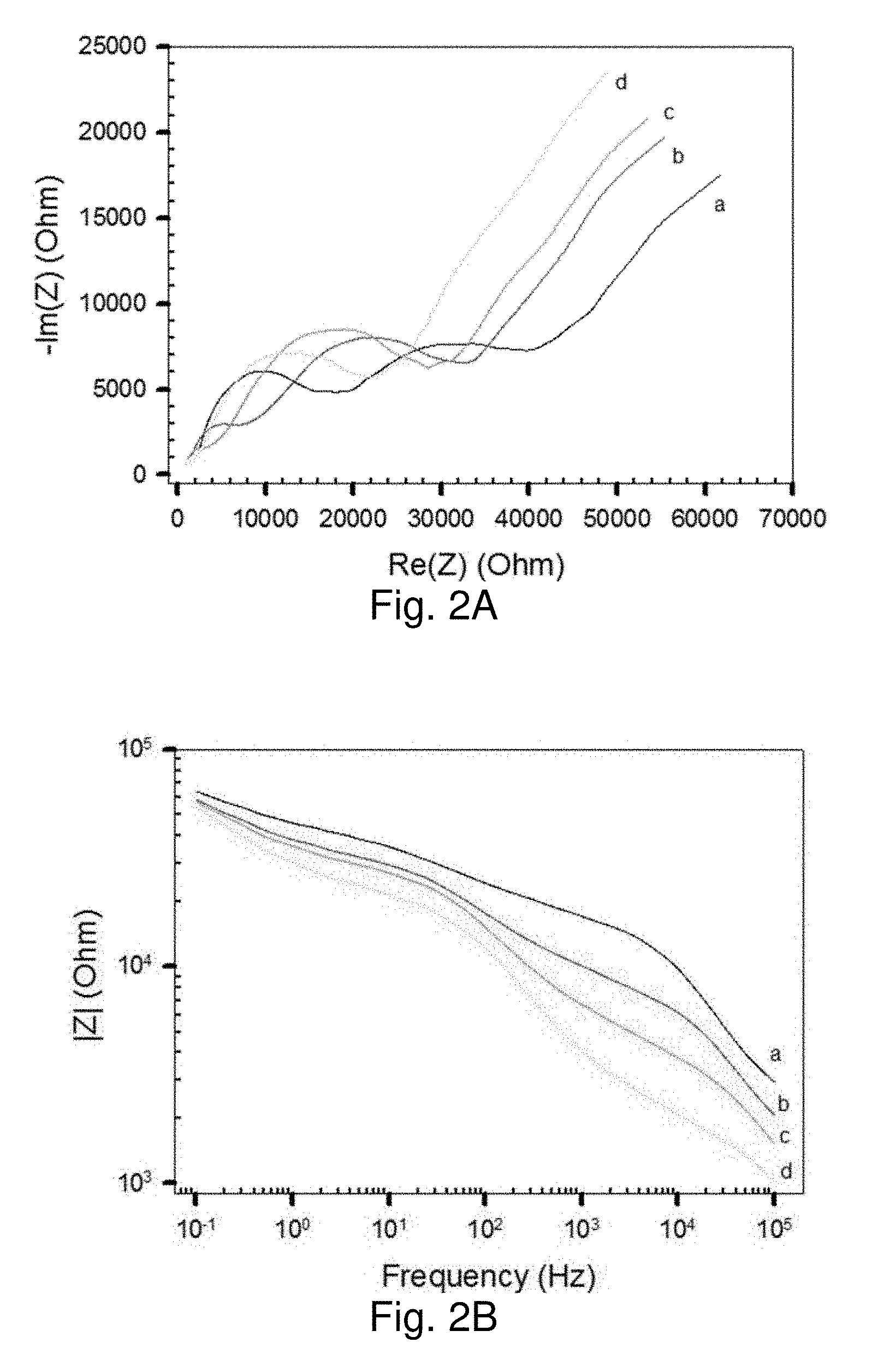
[0060] FIG. 2A shows a Nyquist impedance plot, and
[0061] FIG. 2B shows a Bode impedance plot of MUA-AuNPs/CN/PAN/PET NM placed on top of a Pt interdigitated IME device in solutions of Na+ with different concentration (5 (a), 20 (b), 40 (c), and 80 mM (d)).
[0062] FIGS. 3A and 3B show plots of impedance and resistance values from Bode impedance and Nyquist impedance plots: FIG. 3A shows 1/|Z| vs concentration curves obtained from Bode impedance plots at 1 kHz; FIG. 3B shows 1/R values obtained by semicircle fit to Nyquist impedance plots with MUA-AuNPs/CN/PAN/PET NM on Pt-IME in solutions of K+ (a), Na+ (b), and Li+ (c) as a function of concentration.
[0063] FIGS. 4A and 4B show plots of 1/|Z| values obtained from Bode impedance plots at 1 kHz for MUA-AuNPs/CN/PAN/PET, in solutions as a function of K+ (a) and Na+ (b) concentration: FIG. 4A shows plots of 1/|Z| values with G-PE on the PET side; FIG. 4B shows plots of 1/|Z| values with G-PE on the CN side; FIG. 4C shows plots of 1/|Z| values obtained from Bode impedance plots at 1 kHz for PDA-Au NPs (42 nm)/CN/PAN/PET with G-PE in solutions of K+ (a) and Na+ (b) as a function of salt concentration.
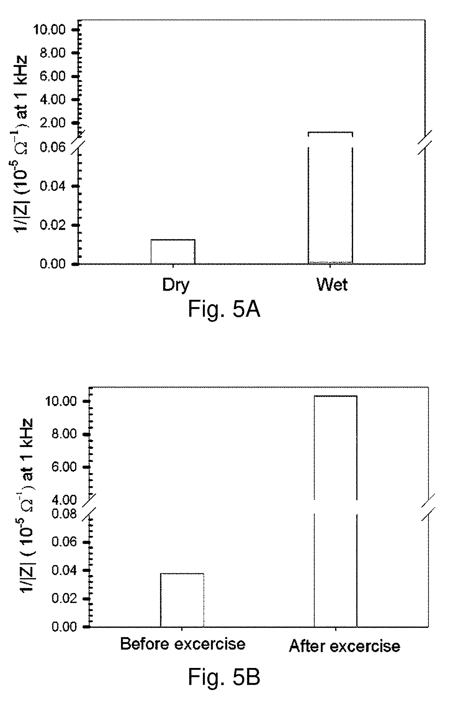
[0064] FIGS. 5A and 5B show 1/|Z| values extracted from Bode impedance plots for MUA-AuNPs/CN/PAN/PET with G-PE: FIG. 5A shows plots in response to water (as a control);
[0065] FIG. 5B shows plots in response to sweat (perspiration test).
[0066] FIGS. 6A and 6B show a comparison of 1/|Z| (FIG. 6A) and .DELTA.|Z|/|Zi| (FIG. 6B) vs RH % curves (extracted from Bode impedance plots at 20 kHz) as a function of relative humidity for two different sensing scaffolds: MUA-Au NPs (7 nm) NM (slope in linear region: -3.2.times.10-2 (a)) and PDA-Au NPs (70 nm) NM (slope in linear region: -1.5.times.10-2 (b)).
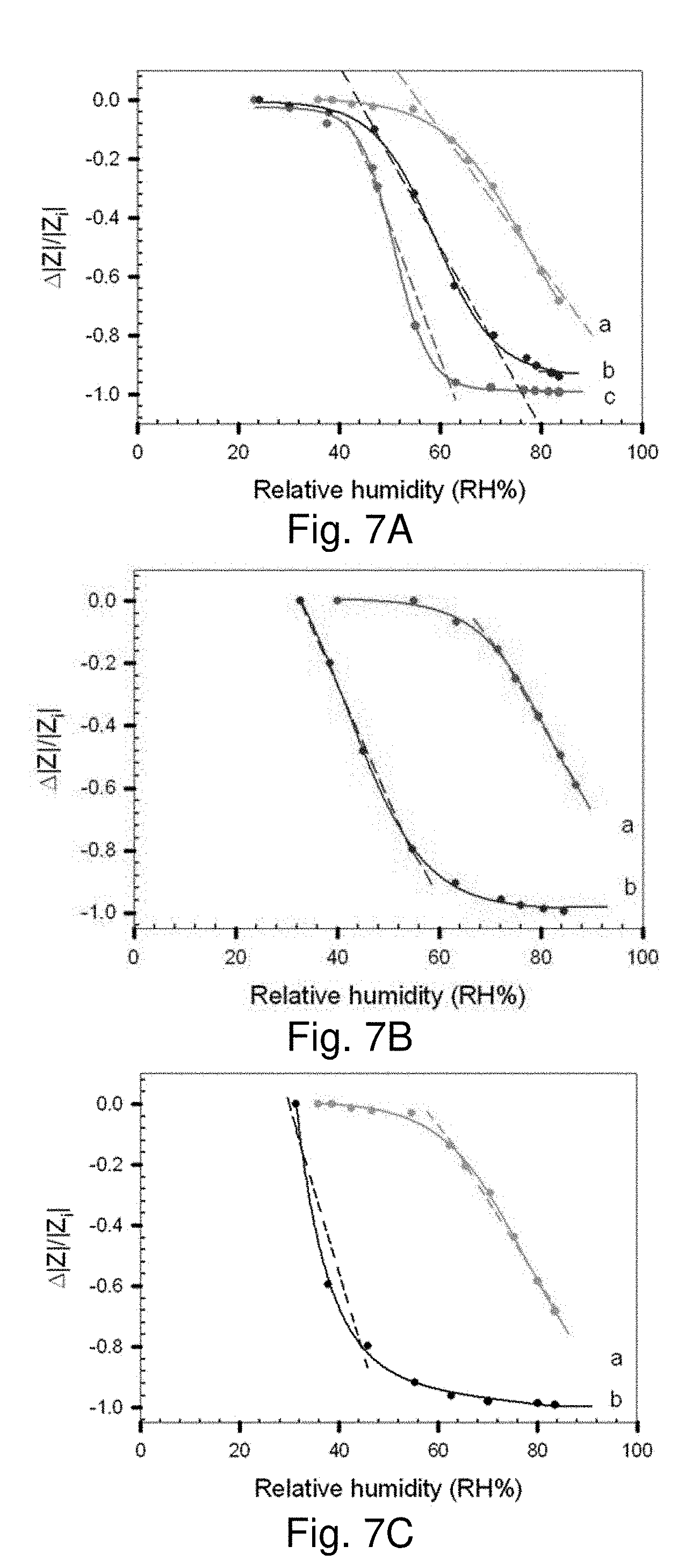
[0067] FIGS. 7A, 7B and 7C show plots of .DELTA.|Z|/|Zi| vs RH % for PDA-AuNPs/CN/PAN/PET NM on Pt-IME: FIG. 7A shows data for scaffolds derived from PDA of constant concentration and Au NPs (70 nm) of different concentrations (5.0.times.1010 (a), 2.0.times.10.sup.11 (b), and 1.0.times.10.sup.11 NPs/mL (c), Slopes: -2.3.times.10.sup.-2 (a); -2.0.times.10.sup.-2 (b); and -4.5.times.10.sup.-2 (c)); FIG. 7B shows data for scaffolds derived from PDA with different concentrations (0.4 M (a) and 0.76 M (b)) and the same concentration of Au NPs (70 nm, 5.0.times.1010 NPs/mL, Slopes: -2.8.times.10.sup.-2 (a); and -3.1.times.10.sup.-2 (b)); FIG. 7C shows data for scaffolds derived from PDA of the same concentration (0.4 M) and Au NPs of two different sizes (70 nm (a) and 42 nm (b)). (Slopes: -2.6.times.10.sup.-2 (a) and -3.6.times.10.sup.-2 (b).)
[0068] FIGS. 8A, 8B and 8C show sensor responses (.DELTA.|Z|/|Zi|) measured at -20 kHz for a device of PDA-AuNPs (70 nm)/CN/PAN/PET with G-PE for two volunteers (#1 (A) and #2 (B)) before and after exercise (running stairs for .about.5 min), and a calibration curve for the sensor: FIG. 8A shows sensor responses before exercise; FIG. 8B shows sensor responses after exercise; FIG. 8C shows a calibration curve for the same sensor device with controlled RH % in air (slope: 2.0.times.10.sup.-2).
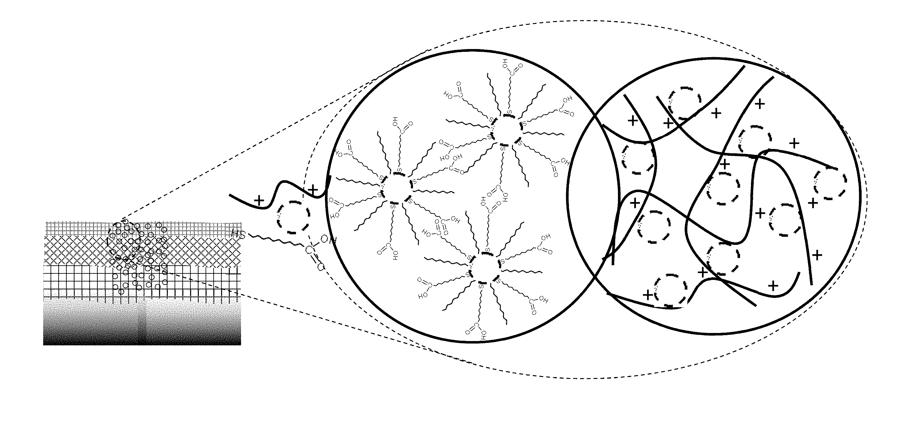
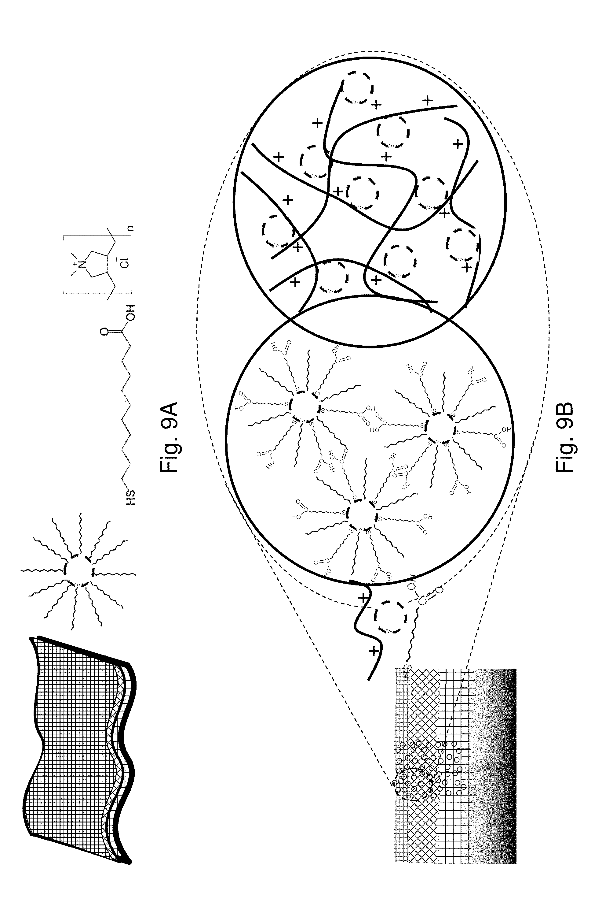
[0069] FIG. 9A shows illustrations of the Nanofibrous Membrane (NM), Gold Nanoparticles and Molecular/Polymeric Linkers.
[0070] FIG. 9B shows the Nanocomposite Scaffolds by Molecularly-Mediated Hydrogen-Bonding Linkages of Au NPs (M-NPs) or Polymer-Mediated Electrostatic Linkages of Au NPs (P-NPs) in the NM.
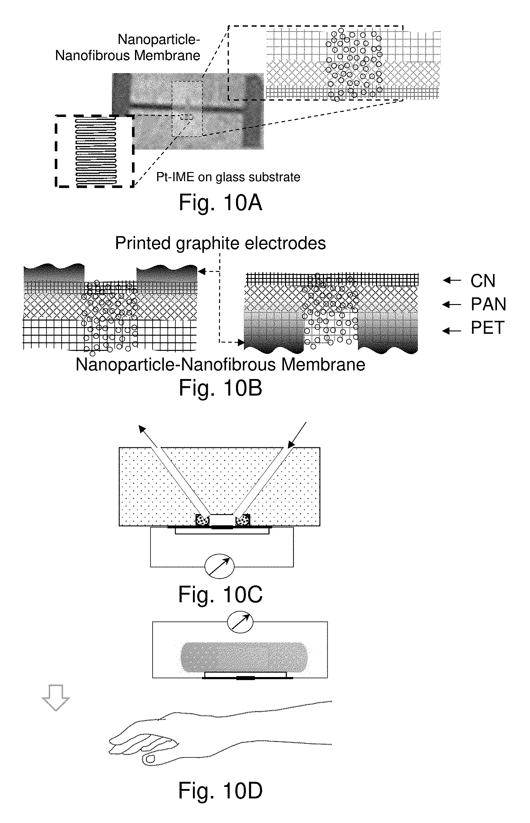
[0071] FIGS. 10A-10E show illustrations of the Nanoparticle-Nanofibrous Membrane Sensor Device and Measurement Configurations: FIG. 10A shows the membrane being placed on top of a Pt IME/glass device; FIG. 10B shows the membrane with graphite printed electrodes (G-PE) in which G-PE is on either the CN or PET side (from left to right); FIG. 10C shows a cross section of the manifold with embedded flow channels and a sample-holding plate with electrical leads for impedance measurement under controlled liquid flow, for e.g., for detection of metal ions, or gas flow, e.g., for detection of relative humidity; FIG. 10D shows a patch with a thin nonwoven scaffold between the membrane and the wrist skin for sweat detection; and FIG. 10E shows a mini-compartment where the NM is placed above the palm for perspiration detection.
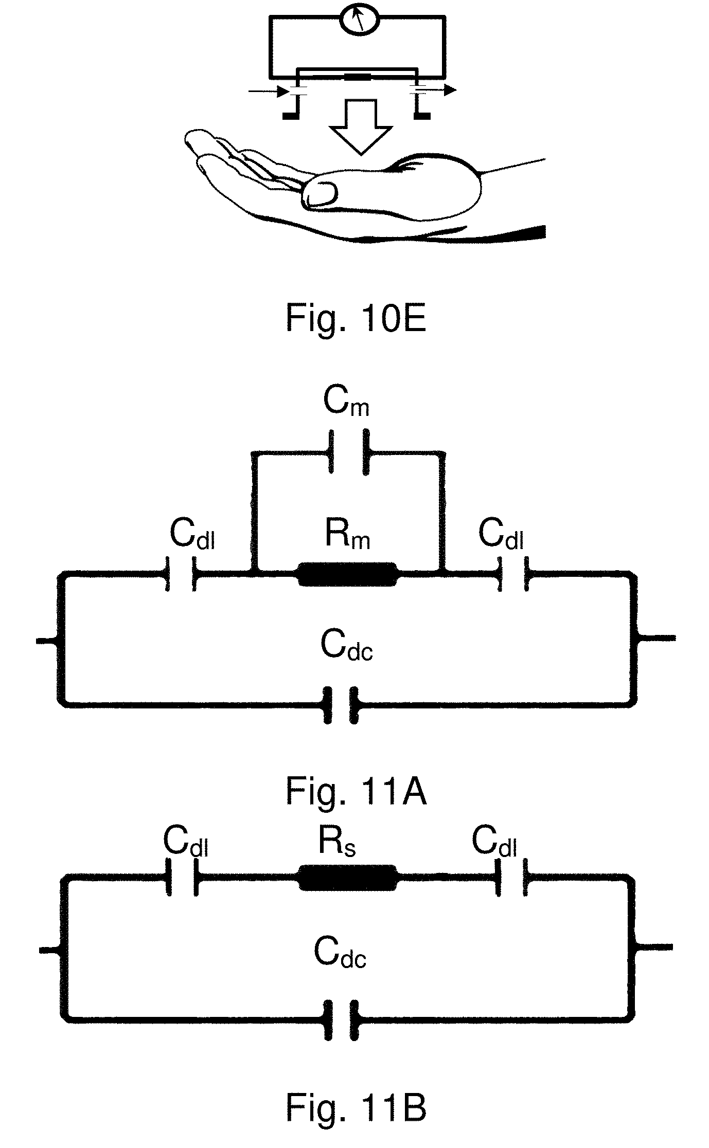
[0072] FIGS. 11A and 11B show illustrations of ideal model of an equivalent circuit for the chemiresistor-type device with the nanoparticle-nanofibrous nanocomposite membrane, having Cm in parallel with Rm, and Rs in series with Cdl, respectively.
[0073] FIG. 12 shows a schematic representation of the sensor incorporated into a self-adhesive strip, which can be applied to the skin.
[0074] FIGS. 13A-13C show TEM images for DT-capped Au NPs (A, 7.1.+-.1.0 nm) and acrylate-capped Au NPs (B, 42.2.+-.6.9 nm, and C, 70.6.+-.2.0 nm).
[0075] FIGS. 14A-14C show SEM images of the three-layer nanofibrous membrane;
[0076] FIG. 14D shows SEM images of MUA-AuNPs (7 nm)/CN/PAN/PET;
[0077] FIG. 14E shows SEM images of PDA-AuNPs (42 nm)/CN/PAN/PET; and
[0078] FIG. 14F shows SEM images of PDA-Au NPs (70 nm)/CN/PAN/PET.
[0079] FIG. 15A shows a Bode impedance plots for MUA-AuNPs/CN/PAN/PET with G-PE on the PET side in solution of Na+ with different concentrations (top to bottom: 5, 10, 20, 60, 70, and 100 mM).
[0080] FIG. 15B shows a Bode impedance plots for MUA-AuNPs/CN/PAN/PET with G-PE on the CN side in solutions of Na+ with different concentrations.
[0081] FIG. 15C shows a Bode impedance plots for PDA-AuNPs (42 nm)/CN/PAN/PET with G-PE in solutions of Na+ with different concentrations (top to bottom: 1, 5, 10, 20, 40, 60, 70, and 100 mM).
[0082] FIGS. 16A-16C show plots of 1/|Z| vs RH % (extracted from Bode impedance plots at 20 kHz) for PDA-Au NPs/CN/PAN/PET NM on Pt-IME as a function of relative humidity: FIG. 16A shows data for scaffolds derived from PDA of fixed concentration and Au NPs (70 nm) of different concentrations (5.0.times.10.sup.10 (a), 2.0.times.10.sup.11 (b), and 1.6.times.10.sup.11 NPs/mL (c)); FIG. 16B shows data for scaffolds derived from PDA of different concentrations (0.4 M (a) and 0.76 M (b)) and the same concentration of Au NPs (70 nm, 5.0.times.1010 NPs/mL); FIG. 16C shows data for scaffolds derived from PDA of the same concentration (0.4 M) and Au NPs of two different sizes (70 nm (a) 42 nm Au NPs (b)).
[0083] FIGS. 17A and 17B show photos showing the sensor setups for human secretion monitoring: FIG. 17A shows sweat detection from forearm; FIG. 17B shows perspiration detection from hand, before and after exercise.
[0084] FIGS. 18A and 18B show photos showing the flexibility and bending characteristics for the nanofibrous membrane.
[0085] FIG. 19 shows plots showing the real time monitoring of sweat before and after exercise (5 minutes exercise).
[0086] FIGS. 20A, 20B, 20D, 20E, and 20F show SEM (FIG. 20A) and TEM (FIG. 20B) images of the nanofibrous paper (CN/PAN/PET) showing CN layer and PAN fibers, with the cross section being shown in the inset in FIG. 20A.
[0087] FIG. 20C shows a sensor scheme, and the representative locations from which FIGS. 20A (SEM) and FIG. 20D (TEM) are derived.
[0088] FIGS. 20E-20F show TEM images for a sensor device with gold nanoparticles with dendrons (AuNPs@dendrons) embedded in CN/PAN/PET paper as sensing scaffold.
[0089] FIGS. 21A-21C show response profiles of the sensing scaffolds with different loading of gold nanoparticles with AuNPs@deSG-SS in the nanofibrous paper (0 upper curve), 1.times. loading (2.04.times.10.sup.9 NPs mm.sup.-2, middle curve) and 2.times. loading (4.08.times.10.sup.9 NPs mm.sup.-2, lower curve) of the AuNPs@deSG-SS solution, for water (FIG. 21A), ethanol (FIG. 21B), and acetone (FIG. 21C) vapors respectively:
[0090] FIG. 21D shows the sensor response sensitivities (ppm (M)) for water (-1.9.times.10.sup.-4, circle) ethanol (-1.5.times.10.sup.-5, triangle), and acetone (-1.1.times.10.sup.-5, rectangle).
[0091] FIG. 22A shows a plots of R/R.sub.i versus RH %, showing a linear relationship in the range of 22-50% RH %, with slopes of -0.035, -0.029, and -0.031 for Au30 nm@deSG-SS, Au30 nm@deLG-SS, and Au58 nm@deSG-SS.
[0092] FIG. 22B shows response profiles recorded with three different sensing materials in the scaffolds: Au30 nm@deSG-SS (lower), Au30 nm@deLG-SS (upper), and Au58 nm@deSG-SS (middle), during the individual's normal breathing, holding the breath briefly and then taking deep breathing.
[0093] FIG. 22C shows a plot of impedance (at 1.3 kHz) versus RH %, with slopes (RH %>43%) of -8.7.times.10.sup.-3 and -7.3.times.10.sup.-3 for Au30 nm@deSG-SS and Au58 nm@deSG-SS.
[0094] FIG. 22D shows response profiles (in terms of relative impedance change, |Z|/|Z.sub.i|) with a sensor device of Au30 nm@deSG-SS recorded during perspiration test for an individual before (upper) and after (lower) min exercise.
DETAILED DESCRIPTION OF THE PREFERRED EMBODIMENT
Example 1
Experimental Section
Chemicals and Synthesis of Gold Nanoparticles.
[0095] Hydrogen tetrachloroaurate trihydrate (99%), tetraoctylammonium bromide (99%), decanethiol (DT, 96%), sodium borohydride (99%), 11-mercaptoundecanoic acid (MUA, 95%), (poly)diallyldimethylammonium (PDA) (20%), sodium acrylate, sodium chloride (NaCl), potassium chloride (KCl), lithium chloride (LiCl), and graphite powders were purchased from Aldrich. Solvents included hexane (Hx, 99.9%) and toluene (Tl, 99%) from Fisher, and ethanol (99.9%) from Aldrich. Water was purified with a Millipore Milli-Q water system. 2,2,6,6-Tetramethylpiperidinooxy (TEMPO, 98%) was purchased from Acros. Sodium hypochlorite (NaClO solution, available chlorine 7-10%) was purchased from Sigma-Aldrich. Sodium bromide (NaBr) was obtained from Fisher Scientific Company. Polyacrylonitrile (PAN) having an average molecular weight (Mw) of 150 kDa was purchased from Sigma-Aldrich. Poly(ethylene terephthalate) nonwoven substrate (PET microfilter F02413 with an average fiber diameter of about 30 .mu.m) for the membrane support was provided by Freudenberg Nonwovens (Hopkinsville, Ky.).
[0096] Gold nanoparticles of 2 nm (Au2 nm) capped with decanethiolate (DT) monolayer shells were synthesized by two-phase reduction of AuC14--according to Brust's two-phase protocol and a synthetic modification. DT-capped gold nanoparticles of 7.1.+-.1.0 nm diameter were synthesized from a thermally activated processing of Au2 nm nanoparticles (Maye, M. M.; Zheng, W.; Leibowitz, F. L.; Ly, N. K.; Zhong, C. J. Heating-induced evolution of thiolate-encapsulated gold nano-particles: a strategy for size and shape manipulation. Langmuir 2000, 16, 490-497.) Briefly, the solution containing the as-synthesized DT-Au2 nm nanoparticles was heated at 150.degree. C. to produce larger-sized Au nanoparticles. (Maye, M. M.; Zheng, W.; Leibowitz, F. L.; Ly, N. K.; Zhong, C. J. Heating-induced evolution of thiolate-encapsulated gold nanoparticles: a strategy for size and shape manipulation. Langmuir 2000, 16, 490-497; Han, L.; Luo, J.; Kariuki, N. N.; Maye, M. M.; Jones, V. W.; Zhong, C. J. Novel interparticle spatial properties of hydrogen-bonding mediated nanoparticle assembly. Chem. Mater. 2003, 15, 29-37.) Acrylate-capped gold nanoparticles 42 nm (42.2.+-.6.9 nm) and 70 nm (70.6.+-.2.0 nm) were prepared by a seeded aggregative growth method. Briefly, the synthesis involves reacting mixture of Au seeds (30 nm) and HAuCl.sub.4 under controlled concentrations of the reducing and capping agents, which produced acrylate-capped Au NPs of >30 nm. (Njoki, P. N.; Lim, I. I. S.; Mott, D.; Park, H. Y.; Khan, B.; Mishra, S.; Sujakumar, R.; Luo, J.; Zhong, C. J. Size correlation of optical and spectroscopic properties for gold nanoparticles. J. Phys. Chem. C 2007, 111, 14664-14669.)
[0097] Preparation of Nanofibrous Membranes.
[0098] Ultrafine cellulose nanofibers were prepared by the following procedure. In brief, 10 g of wood pulps (Biofloc 96 supplied by the Tembec Tartas factory in France) was dispersed in 192 g of water. NaBr (0.2 g) and TEMPO (0.04 g) were subsequently dissolved in the suspension. Then 30 g of 10-15% NaClO aqueous solution was added to start this reaction. The pH value of the system was adjusted in the range of 10.0-10.3 by adding sodium hydroxide (NaOH) aqueous solution (0.5 mol/L). After 24 h, the reaction was stopped by adding ethanol (10 mL). The oxidized cellulose product was purified by dialysis process. The resulting cellulose slurry was dispersed in 100 g of water by using a homogenizer (Cole Parmer, VCX-400) for 5 min. The CN concentration was determined by using a Total Organic Carbon analyzer (TOC-500, Shi-madzu Corporation).
[0099] To prepare electrospun PAN/PET substrate, PAN was dissolved in DMF at 60.degree. C. for 2 days until the mixture became a homogeneous solution (the solution concentration was 8 wt %). The homogeneous PAN solution was electrospun onto the nonwoven PET substrate under a high electrical voltage of 20 kV. The flow rate during this electrospinning operation was 16 .mu.L/min and the inner diameter of the spinneret was 0.7 mm. The working distance between the spinneret and the collector was 10 cm. The average fiber diameter of the electrospun nanofiber estimated from the SEM image was 150.+-.10 nm.
[0100] To complete the three-layered fibrous membrane containing the ultrafine cellulose nanofiber top layer, the electrospun PAN/PET substrate was first immersed in an acidic aqueous solution (pH=2). The cellulose nanofiber aqueous suspension (0.05 wt %) was subsequently cast on top of the electrospun PAN nanofibrous scaffold. The low pH value was used to gel the cellulose nanofiber suspension, thus preventing the penetration of cellulose nanofibers into the electrospun scaffold. The barrier layer thickness was controlled by the gap of the casting knife. After coating, the resulting membrane was dried at room temperature and forms a uniform coating layer of CN.
[0101] Preparation of Nanoparticle-Nanofibrous Membranes.
[0102] For the assembly of MUA-linked DT-capped Au NPs in NM (M-NPs/NM), typically a controlled volume (e.g., 2 .mu.L) of MUA mediated Au NPs solution (7.1.times.10.sup.14 NPs/mL) was directly deposited in the nanofibrous membrane (NM). For the assembly of PDA-linked acrylate-capped Au NPs in the NM (P-NPs/NM), a controlled volume of 10.times. concentrated acrylate-capped 70 nm Au NPs (5.0.times.10.sup.11 NPs/mL), or 2.times. concentrated 42 nm NPs (2.7.times.10.sup.13 NPs/mL), was first mixed with PDA solution (0.4 M) by sonication for 10 min. A controlled volume (2 .mu.L) of the solution was then deposited in the NM, followed by further annealing at room temperature for at least 1 h before use.
[0103] Instrumentation and Measurements.
[0104] Electrochemical impedance spectroscopic (EIS) measurements were performed on a SP-150 single-channel potentiostat (Biologic). The spectra were recorded at open circuit in a frequency range from 100 kHz to 0.1 Hz.
[0105] Transmission electron microscopy (TEM) was employed to determine the morphology of the nanoparticles. TEM was performed on a JEOL JEM-ARM200F instrument operated at 200 kV with a spherical aberration corrector. The nanoparticle samples were suspended in hexane or water before drop casting on a carbon-coated copper grid. The samples were then dried by evaporation in ambient atmosphere.
[0106] Scanning electron microscopy (SEM) images of the nanofibrous membrane and nanocomposite were performed with a LEO-1550 (Carl Zeiss) field emission scanning electron microscope. The membrane samples were mounted on a sample holder. It was then followed by carbon-coating with a sputter coater.
[0107] Results and Discussion
[0108] General Characteristics of Nanocomposite Membranes and Devices.
[0109] FIG. 9A shows illustrations of the Nanofibrous Membrane (NM), Gold Nanoparticles and Molecular/Polymeric Linkers. FIG. 9B shows the Nanocomposite Scaffolds by Molecularly-Mediated Hydrogen-Bonding Linkages of Au NPs (M-NPs) or Polymer-Mediated Electrostatic Linkages of Au NPs (P-NPs) in the NM.
[0110] As illustrated in FIGS. 9A and 9B, two pathways have been explored for assembling Au NPs in the three-layered CN/PAN/PET membrane by either molecular linkers or polymeric electrostatic interactions. One pathway involves a molecular linker MUA, which forms molecularly mediated thin-film assembly of Au NPs (M-NPs) via hydrogen bonding. In this case, the interaction between the nanoparticle assemblies and the nanofibers feature mainly hydrophobic interactions. The other pathway involves polymeric linker PDA, which forms polymer-mediated thin film assembly of Au NPs (P-NPs) in the membrane via electrostatic interactions. In this case, the polymeric structure provides an adhesive force between the nanoparticle assemblies and the nanofibers.
[0111] FIGS. 13A-13C show TEM images for DT-capped Au NPs (A, 7.1.+-.1.0 nm) and acrylate-capped Au NPs (B, 42.2.+-.6.9 nm, and C, 70.6.+-.2.0 nm).
[0112] FIGS. 14A-14F show SEM images of the three-layer nanofibrous membrane (FIGS. 14A-14C), MUA-AuNPs (7 nm)/CN/PAN/PET (FIG. 14D), PDA-AuNPs (42 nm)/CN/PAN/PET (FIG. 14E), and PDA-Au NPs (70 nm)/CN/PAN/PET (FIG. 14F).
[0113] Gold nanoparticles of different sizes and hydrophilicity characteristics were studied for their assembly in the nanofibrous membranes, including hydrophobic DT-capped Au NPs and hydrophilic acrylate-capped Au NPs (FIG. 13A). While the DT-capped Au NPs (7 nm) and acrylate-capped Au NPs (42 nm, or 70 nm) feature highly monodispersed sizes, the nanofibrous membrane features a three-layer CN/PAN/PET structure (FIGS. 14A-14C).
[0114] FIGS. 1A and 1B show SEM images of MUA-mediated assembly of DT-capped 7 nm Au NPs (M-NPs/NM) in CN/PAN/PET membrane: FIG. 1A shows nanoparticles on the CN and PAN fibers and FIG. 1B shows a magnified image of the nanoparticles on the PAN fibers. As shown in FIGS. 1A and 1B for the MUA-linked DT-capped 7 nm sized Au NPs in the NM (M-NPs/NM), the nanoparticles are well distributed on the surface of the CN layer and along the PAN fibers (see also FIG. 14D). Similar assemblies were also observed for PDA-linked acrylate-capped 42 or 70 nm Au NPs (P-NPs/NM) in the NM, but with a less even distribution on the fibers and a certain degree of aggregation at cross-fiber junctions (FIGS. 14E, 14F). The fact that M-NPs/NM shows a much better dispersion of NPs along the fibers than that for the P-NPs/NM indicates that the assembly depends on the surface properties of the nanoparticles and the solvent used for the assembly. Au NPs of different sizes have been assembled in the NM, but only selected examples are discussed herein for the exploration of sensing properties.
[0115] FIGS. 10A-10E show illustrations of the Nanoparticle-Nanofibrous Membrane (NM) Sensor Device and Measurement Configurations. FIG. 10A shows the membrane being placed on top of a Pt IME/glass device; FIG. 10B shows the membrane with graphite printed electrodes (G-PE) in which G-PE is on either the CN or PET side (from left to right), for impedance measurement under either liquid flow for detection of metal ions or gas flow for detection of relative humidity (C); FIG. 10C shows the manifold with embedded flow channels and a sample-holding plate with electrical leads for impedance measurement under controlled liquid or gas flow; FIG. 10D shows a patch with a thin nonwoven scaffold between the membrane and the wrist skin for sweat detection; and FIG. 10E shows a mini-compartment where the NM is placed above the palm for perspiration detection.
[0116] The M-NPs/NM, i.e., MUA-AuNPs/CN/PAN/PET, features largely hydrophobic network with partial hydrophilic domains (i.e., the region of hydrogen-bonding of carboxylic acid groups). In contrast, the P-NPs/NM, i.e., PDA-AuNPs/CN/PAN/PET, features largely hydrophilic network with partial hydrophobic polymer backbone structure. Both nanocomposite membranes were studied as resistance- or conductance-responsive scaffolds on chemiresistor-type platform via two different approaches. The first involves placing the NPs/NM on top of a prefabricated Pt-interdigitated micro-electrode (Pt-IME) device (FIG. 10A), whereas the second involves configuring the NPs/NM in between a printed pair of graphite electrodes on CN or PET sides of the membrane with a controlled gap (0.5-1.0 mm) (FIG. 10B). In each approach, the electrical responses to the analytes such as ions in a solution or moisture change in a gas atmosphere were measured by impedance spectroscopy. As illustrated in FIGS. 10C-10E, the measurements were performed in a manifold where the NM is sandwiched between manifold with embedded flow channels and a sample-holding plate with electrical leads (FIG. 10C).
[0117] Ideally, the above chemiresistor-type device can be represented by two equivalent circuit models featuring the nanoparticle--nanofibrous membrane with two-electrode configurations (see FIG. 9). One consists of two double layer capacitors (capacitance near the surface of an electrode, C.sub.dl), one for each set of the electrodes, connected in series with a parallel combination of a membrane resistor (R.sub.m) and a membrane dielectric capacitor (C.sub.m), and all of them in parallel with a parasitic capacitor. The other model consists of two double layer capacitors (C.sub.dl), connected in series with a medium resistor (R.sub.s), and a dielectric capacitor (C.sub.di).
[0118] Detection of Salts Dissolved in Water and from Sweat.
[0119] Detection of Salts in Water.
[0120] With a M-NPs/NM scaffold sensor device of M-AuNPs/CN/PAN/PET on Pt-IME (CN facing Pt-IME, FIG. 10A) placed in the test manifold (FIG. 10C), solution samples of K+, Na+, and Li+ with a common anion (Cl--), prepared by dissolving KCl, NaCl or LiCl in aqueous solutions with controlled concentrations, were introduced by a flow controller.
[0121] FIG. 2A shows a Nyquist impedance plot, and FIG. 2B shows a Bode impedance plot of MUA-AuNPs/CN/PAN/PET NM placed on top of a Pt interdigitated IME device in solutions of Na+ with different concentration (5 (a), 20 (b), 40 (c), and 80 mM (d)). FIG. 2A shows a representative set of Nyquist impedance plots of solutions with different Na+ concentrations. Similar results have also been obtained from samples of K+ or Li+ solutions. The semicircle characteristic of the charge transfer region and the slope characteristic of the mass transfer region show clear variations with the concentration of ions. In FIG. 2B, the data are plotted in Bode impedance, which clearly reveals that absolute impedance |Z| depends on the Na+ concentration.
[0122] By extracting the impedance values (|Z|) from FIG. 2B in the high frequency region, |Z| is shown to be dependent on the concentration of the ions, especially in the low concentration range.
[0123] FIGS. 3A and 3B show plots of impedance and resistance values from Bode impedance and Nyquist impedance plots: FIG. 3A shows 1/|Z| vs concentration curves obtained from Bode impedance plots at 1 kHz; FIG. 3B shows 1/R values obtained by semicircle fit to Nyquist impedance plots with MUA-AuNPs/CN/PAN/PET NM on Pt-IME in solutions of K+ (a), Na+ (b), and Li+ (c) as a function of concentration. A representative set of 1/|Z| vs concentration curves is shown in FIG. 3A. In the low concentration range (<20-60 mM), the linear regression slopes of the 1/|Z| vs concentration curves display the order of K+ (3.9.times.10.sup.-6)>Na+ (2.3.times.10.sup.-6)>Li+ (1.6.times.10.sup.-6). The |Z| value becomes smaller when ion concentration increases. In other words, the conductivity increases as the ion concentration increases, and a plateau appears at >60 mM. The slopes are steep when the concentration is <20-60 mM. The nanocomposite membrane appears to be more sensitive to the ions in the lower concentration region. Similar trends are also observed by extracting the charge transfer resistance values from the semicircle fit of Nyquist impedance plots in the high frequency region, as shown in FIG. 3B. The magnitudes for the slopes of 1/R vs concentration curves display the order of K+ (5.6.times.10.sup.-6)>Na+ (4.2.times.10.sup.-6)>Li+ (2.3.times.10.sup.-6), quite similar to those obtained from the data from the Bode impedance plots (FIG. 3A).
[0124] These results indicate that the nanocomposite membrane functions as an ion sensitive and selective interfacial scaffold on the interdigitated microelectrode, which is consistent with the cation exchange membrane character of the MUA-Au NP films embedded in the nanofibrous membrane. In a typical cation-exchange membrane as stationary phase in chromatographic column, the relative affinities of different counterions in the mobile phase depend on the ionic charge, polarizability, and size of the solvated ion, and the type and interaction of the functional groups on the stationary phase. An increase of the charge-density (charge/solvated size) of the ion, or higher charge with smaller solvated ion radius, leads to higher electrostatic interactions with the stationary charges in the membrane (carboxylates), typically K+>Na+>Li+. In that case, the ionic conductance of the nanocomposite membranes (1/|Z|) would display the order K+<Na+<Li+, which is consistent with the experimental observation (FIG. 3A).
[0125] The same M-Au NPs/CN/PAN/PET scaffold is configured in between a pair of graphite printed electrodes on CN or PET sides of the membrane (FIG. 10B). For example, the impedance responses were measured in the test manifold in which G-PE is on the PET side of the membrane (FIG. 10C).
[0126] FIG. 15A shows a Bode impedance plots for MUA-AuNPs/CN/PAN/PET with G-PE on the PET side in solution of Na+ with different concentrations (top to bottom: 5, 10, 20, 60, 70, and 100 mM). FIG. 15B shows a Bode impedance plots for MUA-AuNPs/CN/PAN/PET with G-PE on the CN side in solutions of Na+ with different concentrations. FIG. 15C shows a Bode impedance plots for PDA-AuNPs (42 nm)/CN/PAN/PET with G-PE in solutions of Na+ with different concentrations (top to bottom: 1, 5, 10, 20, 40, 60, 70, and 100 mM). As shown in Bode impedance plots for samples containing Na+ (FIG. 15A), |Z| value starts to differentiate with various concentrations even in the lower frequency region, and the difference becomes greater in the higher frequency region.
[0127] FIGS. 4A and 4B show plots of 1/|Z| values obtained from Bode impedance plots at 1 kHz for MUA-AuNPs/CN/PAN/PET with G-PE on the PET side (FIG. 4A) and the CN side (FIG. 4B) in solutions as a function of K+ (a) and Na+ (b) concentration. FIG. 4C shows plots of 1/|Z| values obtained from Bode impedance plots at 1 kHz for PDA-Au NPs (42 nm)/CN/PAN/PET with G-PE in solutions of K+ (a) and Na+ (b) as a function of salt concentration. FIG. 4A shows plots of 1/|Z| values extracted from Bode impedance at 1 kHz vs ionic concentration. In the high frequency region, 1/|Z| shows a clear dependence of the ion concentration, similar to the results from NM-Pt-IME device (FIG. 3B) except subtle differences in the slope values. The magnitudes for the slopes of 1/|Z| vs concentration curves display the order of K+ (1.3.times.10.sup.-7)>Na+ (1.2.times.10.sup.-7) in the entire concentration range. A similar trend is also observed by extracting the resistance values from the semicircle fit of Nyquist impedance plots.
[0128] For the case of G-PE on the CN side in solutions containing Na+ with different concentrations, the |Z| vs frequency curves (FIG. 15B) in the low frequency region appear to be independent of the concentration of ions. At >10 kHz, the impedance shows changes in the |Z| vs frequency curves, exhibiting a clear dependence of the ion concentration. A similar Bode impedance plot is also observed for samples containing K+. By extracting the impedance values (|Z|) from the Bode impedance plots at three frequencies in the high frequency region, |Z| is shown to be linearly dependent on the ion concentrations (FIG. 4B). The slopes of the 1/|Z| vs concentration curves are quite comparable (K+ (5.0.times.10.sup.-7) Na+ (4.2.times.10.sup.-7)), showing much smaller differences in comparison with the data shown in FIGS. 4A and 4B. A similar trend is also observed by extracting the resistance values from the semicircle fit of Nyquist impedance plots.
[0129] The P-NPs/NM scaffolds were also examined. For example, with PDA-Au NPs (42 nm)/CN/PAN/PET being configured in between a printed pair of graphite electrodes on CN or PET sides of the membrane (see FIG. 10B), the Bode impedance plots of solutions containing different concentrations of Na+ show clear changes (FIG. 15C). For the data at frequency greater than 10 Hz, Bode impedance curves display a clear dependence on the ion concentration. By extracting the impedance values (|Z|) at 1 kHz, a comparison between Na+ and K+ shows that the overall 1/|Z| values of K+ are greater than those of Na+ (FIG. 4C). This finding is opposite to the results observed from Au-MUA NPs (see FIGS. 3A and 3B), suggesting the operation of a different mechanism in the thin film. In the acrylate-capped Au NPs (42 nm)/CN/PAN/PET membrane, the acrylate-capped Au NPs feature multiple negative charges on the nanoparticle surface. Upon incorporating the positively charged polymer (diallyl ammonium groups), there must be excess negative charges on the NPs which are balanced by the mobile cations in the solution, forming an electrical double layer around the nanoparticles. In this case, it is the ionic mobility that determines the ionic conductance of the membrane. Since K+ ions exhibit a higher ionic mobility than Na+ ions, the overall 1/|Z| values of K+ are greater than those of Na+ in the MUA-Au NPs (7 nm)/CN/PAN/PET membrane. It is important to note that the slopes of the 1/|Z| vs concentration curves are quite comparable between K+ and Na+ (e.g., K+ (6.50.times.10.sup.-6) Na+ (5.35.times.10.sup.-6) for <20 mM). Unlike MUA-AuNPs film, the selectivity of the PDA-Au NPs film is apparently very limited.
[0130] Detection of Sweat.
[0131] On the basis of the above data for the detection of salts in solutions, the viability of the MUA-AuNPs/CN/PAN/PET with G-PE on the PET side was further examined with normal volunteers before and after exercises. While sweat contains different chemical constituents with different concentrations, as stated earlier, the study described in this subsection focused on the salt and moisture detections to demonstrate the sensing properties of the as-prepared nano-particle-nanofibrous nanocomposites.
[0132] The detection of biological species (e.g., glucose, urea, or lactate) is possible by use of enzymatic or nonenzymatic modifications of the nanocomposite.
[0133] FIGS. 5A and 5B show 1/|Z| values extracted from Bode impedance plots for MUA-AuNPs/CN/PAN/PET with G-PE in response to water (as a control, FIG. 5A) and sweat (perspiration test, FIG. 5B). FIGS. 5A and 5B show a representative set of data with the measurement configuration illustrated in FIG. 10D. Data were collected by placing a thin nonwoven scaffold on the sensor surface (CN side) and lightly pressing it against the wrist (see FIG. 10D) before and after 5 min exercise (running stairs). The control experiment was performed by directly placing a drop of water on the dry nanocomposite in between the two electrodes, followed by impedance measurement of the wet membrane.
[0134] FIG. 19 shows plots showing the real time monitoring of sweat before and after exercise (5 minutes exercise) or in real time. A control experiment was also performed by measuring the impedance before and after placing a small drop of pure water onto the sensor surface. In the control experiment, the results clearly show a sharp drop of impedance (or increase of conductance) from dry to wet membrane (FIG. 5A). In comparison, the difference between "before exercise" and "after exercise" is much greater than that observed from the control experiment (FIG. 5B). This is mainly attributed to the presence of salts in the sweat, which is substantiated by rinsing the membrane with water several times after which the impedance values were almost the same as that before subjecting to sweat test.
[0135] It is evident that the relative change for the individual before and after exercise (96%) is smaller than that for the control experiment using pure water (99%). This is expected because salts in the sweat greatly increase the conductivity in comparison with pure water.
[0136] Detection of Relative Humidity Changes in Air and from Perspiration.
[0137] Detection of Relative Humidity Changes in Air.
[0138] With devices of Au70 nm/CN/PAN/PET (with G-PE on CN side), the response characteristics between PDA-Au NPs (70 nm) and MUA-Au NPs (7 nm) in NM were first compared. The impedance data at different RH % were collected by flowing air or N.sub.2 from a water bubbler with a flow controller, at each flow rate the RH % were recorded by a commercial humidity meter.
[0139] FIGS. 6A and 6B show a comparison of 1/|Z| (FIG. 6A) and .DELTA.|Z|/|Zi| (FIG. 6B) vs RH % curves (extracted from Bode impedance plots at 20 kHz) as a function of relative humidity for two different sensing scaffolds: MUA-Au NPs (7 nm) NM (slope in linear region: -3.2.times.10.sup.-2 (a)) and PDA-Au NPs (70 nm) NM (slope in linear region: -1.5.times.10.sup.-2 (b)).
[0140] Data were first obtained with a sensor device of MUA-Au NPs/CN/PAN/PET on G-PE in which the G-PE is on CN side. On the basis of impedance data extracted from Bode impedance plots, both 1/|Z| (FIG. 6A) and .DELTA.|Z|/|Z.sub.i|(=(|Z|-|Z.sub.i|)/|Z.sub.i| where Z.sub.i is the initial impedance) (FIG. 6B) are plotted against RH %, in which the .DELTA.|Z|/|Z.sub.i| RH % plots seem to provide a better comparison of the responses. When RH %>50%, the results display a significant response. When RH %<50%, the response is independent of RH %. The results are likely reflecting the low hydrophilicity of the nanocomposite membrane, which is responsible for the insignificant response sensitivity in the low RH % range.
[0141] To manipulate the hydrophilicity, a highly hydrophilic polymer, PDA, was used for the assembly of Au NPs in the nanocomposite membrane. The membranes were fabricated by PDA mediated assembly of acrylate-capped Au NPs of different sizes (70 or 42 nm) in the nanofibrous membranes. Different concentration ratios of PDA vs Au NPs were studied for the assemblies. As shown in FIG. 6B for PDA-Au NPs (70 nm)/CN/PAN/PET NM on G-PE, it is evident that the response to a wider RH % range. Note that the slope in the low RH % range (<50%) is slightly smaller than that in the high RH % range. In comparison with MUA-Au NPs/CN/PAN/PET on G-PE, in the high RH % range (>50%), the sensitivity of PDA-Au NPs (70 nm)/CN/PAN/PET NM on G-PE is relatively smaller, indicating the importance of balancing hydrophobicity and hydrophilicity in the nanocomposite. Note that the sensitivity also depends on the gap between electrodes. A smaller gap shows a great sensitivity, which is evidenced by the impedance changes as a function of relative humidity for PDA-Au NPs (70 nm)/CN/PAN/PET NM with G-PE with different G-electrode gaps. The sensitivity may be optimized by adjusting the electrode gap.
[0142] To understand the composition effect of the nanocomposite on response characteristics, the nanocomposite membranes were examined with different ratios of Au NPs vs PDA, as well as different particle sizes using IME as the detection platform. With PDA-Au NPs (70 nm)/CN/PAN/PET on Pt-IME (FIG. 10A) placed in the test manifold (FIG. 10C), a series of impedance values (|Z|) extracted from the Bode impedance plots at 20 kHz (not shown) as a function of the relative humidity are compared with sensors of PDA-Au NPs/CN/PAN/PET on Pt-IME with subtle differences in PDA and Au NPs.
[0143] FIGS. 7A, 7B and 7C show plots of .DELTA.|Z|/|Zi| vs RH % for PDA-AuNPs/CN/PAN/PET NM on Pt-IME. FIG. 7A shows data for scaffolds derived from PDA of constant concentration and Au NPs (70 nm) of different concentrations (5.0.times.1010 (a), 2.0.times.1011 (b), and 1.0.times.1011 NPs/mL (c), Slopes: -2.3.times.10.sup.-2 (a); -2.0.times.10.sup.-2 (b); and -4.5.times.10.sup.-2 (c)). FIG. 7B shows data for scaffolds derived from PDA with different concentrations (0.4 M (a) and 0.76 M (b)) and the same concentration of Au NPs (70 nm, 5.0.times.10.sup.10 NPs/mL, Slopes: -2.8.times.10.sup.-2 (a); and -3.1.times.10.sup.-2 (b)). FIG. 7C shows data for scaffolds derived from PDA of the same concentration (0.4 M) and Au NPs of two different sizes (70 nm (a) and 42 nm (b)). (Slopes: -2.6.times.10.sup.-2 (a) and -3.6.times.10.sup.-2 (b).)
[0144] FIG. 7A shows a set of .DELTA.Z/Z.sub.i data (also see FIG. 16A) as a function of the relative humidity for PDA-Au NPs/CN/PAN/PET prepared by mixing PDA with a constant concentration and Au NPs (70 nm) with different concentrations. It is evident that both the sensitivity and the sensitive range increase with an increase ratio of Au NPs to PDA in the NM. FIG. 7B shows a set of .DELTA.Z/Z.sub.i data (also see FIG. 16B) as a function of relative humidity for PDA-Au NPs/CN/PAN/PET prepared by mixing Au NPs with a constant concentration and PDA of different concentrations. The sensitivity in the low RH % range increases with PDA concentration in the NM. However, in the high RH % range the sensitivity increases with a decrease of PDA concentration in the NM. This behavior is consistent with the highly hydrophilic nature of PDA.
[0145] FIGS. 16A-16C show plots of 1/|Z| vs RH % (extracted from Bode impedance plots at 20 kHz) for PDA-Au NPs/CN/PAN/PET NM on Pt-IME as a function of relative humidity. FIG. 16A shows data for scaffolds derived from PDA of fixed concentration and Au NPs (70 nm) of different concentrations (5.0.times.10.sup.10 (a), 2.0.times.10.sup.11 (b), and 1.6.times.10.sup.11 NPs/mL (c)); FIG. 16B shows data for scaffolds derived from PDA of different concentrations (0.4 M (a) and 0.76 M (b)) and the same concentration of Au NPs (70 nm, 5.0.times.1010 NPs/mL); FIG. 16C shows data for scaffolds derived from PDA of the same concentration (0.4 M) and Au NPs of two different sizes (70 nm (a) 42 nm Au NPs (b)).
[0146] The particle size effect on the response sensitivity was also examined. FIG. 7C shows a set of .DELTA.Z/Z.sub.i data as a function of relative humidity for PDA-Au NPs/CN/PAN/PET NM/G-PE with Au NPs of two different sizes. It is evident that the NM with small size Au NPs seems to exhibit overall smaller 1/|Z| value in comparison with the large size NPs (see FIG. 16C). The sensitivity clearly depends on the particle size in different RH % ranges. The smaller-sized particles show a higher sensitivity in the low RH % region, whereas the larger-sized particles exhibit a higher sensitivity in the high RH % region.
[0147] Detection of Perspiration.
[0148] The viability of PDA-AuNPs/CN/PAN/PET devices (with G-PE) for detection of perspiration was examined with normal individual volunteers before and after exercises.
[0149] FIGS. 8A and 8B show sensor responses (.DELTA.|Z|/|Zi|) measured at -20 kHz for a device of PDA-AuNPs (70 nm)/CN/PAN/PET with G-PE for two volunteers (#1 (A) and #2 (B)) before (FIG. 8A) and after (FIG. 8B) exercise (running stairs for .about.5 min). FIG. 8C shows a calibration curve for the same sensor device with controlled RH % in air (slope: 2.0.times.10.sup.-2).
[0150] FIGS. 8A and 8B show a typical set of .DELTA.Z/Z.sub.i response (measured at -20 kHz) of a device of PDA-AuNPs/CN/PAN/PET (with G-PE on CN side) with the measurement configuration illustrated in FIG. 10E. The measurement was performed by placing the sensor compartment on the palm and the perspiration was measured by stopped air flow method. Data were collected from palms of volunteers #1 and #2 before and after .about.5 min exercise (running stairs). The measurement was performed by placing the sensor compartment on top of the palm and the perspiration was measured by stopped air flow method. During the initial 30 s, air flows into the device to establish a baseline. The perspiration of the volunteer's palm was monitored for 45 s upon stopping air flow. The baseline returns upon air flowing again. This procedure was repeated for one more time. The responses to exercises are significant and reversible, with the response magnitude being clearly dependent on the individual, as reflected by the difference between #1 and #2.
[0151] Based on the relative changes of .DELTA.Z/Z.sub.i values before and after the exercises, it is evident that the corresponding changes of RH % falls in between 50 and 72% (for #1) and 56-62% (for #2), as estimated from the calibration data shown in FIG. 8C.
[0152] Devices with G-PE on the PET side were also tested. For example, the response for PDA-AuNPs/CN/PAN/PET with G-PE on the PET side was found to be much smaller than that with G-PE on the CN side. This finding is indicative of the importance of the nanocomposite membrane-electrode configuration in the sensor response.
[0153] FIGS. 11A and 11B show illustrations of ideal model of an equivalent circuit for the chemiresistor-type device with the nanoparticle-nanofibrous nanocomposite membrane, having Cm in parallel with R.sub.m, and R.sub.s in series with C.sub.dl, respectively.
[0154] FIG. 12 shows a schematic representation of the sensor incorporated into a self-adhesive strip, which can be applied to the skin. Electrical interfacing (not shown) can be wired or wireless, and may perform as a data logger or a low power radio frequency transmitter or backscatter (e.g., RF-ID-type) communication device.
[0155] Conclusion
[0156] A novel class of nanocomposite membranes has been demonstrated for constructing moisture and chemi-sensitive scaffolds for potential applications, such as flexible sweat sensors. The nanocomposites are assembled by molecular or polymeric linkers that incorporate gold nanoparticles into a three-layer structured nanofibrous membrane. Impedance measurements of the nanocomposite membrane as a scaffold of chemiresistor-type platform have demonstrated the capabilities for ion detection in solutions with dissolved salts and changes of relative humidity in the atmosphere. This nanoparticle-nanofiber sensor platform is further demonstrated as a flexible sensor strip for detecting changes in sweating and perspiration of individuals before and after exercises, showing promising potentials for applications of the flexible nanocomposite scaffolds in wearable sweat sensors.
[0157] Various embodiments are shown and disclosed herein. The scope of the invention, while exemplified by the embodiments, is not limited thereby.
[0158] The word "about" means having a structure and function which is functionally similar with proportional changes in properties, dependent on quantitative changes. Typically, when the properties change linearly over a range, the word "about" may encompass a range of a factor of 2, i.e., 50% to 200%.
Example 2
[0159] The nanofibrous paper chemiresistor sensors employ dendronized nanoparticles that exhibit structurally tunable and negative-going responses to human breathing and sweating processes. The device consists of multilayered fibrous paper as a low-cost biocompatible matrix and dendron coated gold nanoparticles as designated sensing elements with tunable sizes, shapes, and structures. The ability to control the interparticle spatial interactions [Edel et al. 2016, Scultz et al. 2013] in a fibrous membrane represents an important pathway to utilize their unique electrical and optical properties for constructing the 3D sensing interfaces. The multilayered fibrous membrane consists of a cellulose nanofiber (CN) and an electrospun poly(acrylonitrile) (PAN), nanofibrous layer, sup-ported on a nonwoven poly(ethylene terephthalate) (PET), film.[Mat et al 2010] The membrane paper features an extremely high surface to volume ratio and nanofiltration capability. With metal nanoparticles, which are widely exploited as sensing or biosensing materials, different molecules or biomolecules have been utilized as interparticle linkers ranging from alkyl dithiols or functionalized thiols to polymers or biopolymers [Ahmad et al. 2015, Lim et al 2007]. In comparison to traditional linkers, dendrimers represent a class of macromolecules with well-defined 3D branched structure exhibiting a high degree of functionality and versatility. Dendrimers featuring multiple "wedges" and dendrons featuring single "wedge" are not only well defined as structural building blocks, but also have controllable sizes ranging from 1 to 30 nm depending on their generation.[Kaga et al. 2016, Albrecht et al. 2016] In particular, dendrimer-nanoparticle conjugates have been attracting considerable interest in medicine [Caminade et al. 2015, Parat et al 2010], drug delivery [Mullen et al. 2010], vapor sorption [Krasteva et al 2007], diagnostics [Astruc 2012] and electrochemical biosensors for enzyme immobilization [Hasanzadeh et al. 2014]. The exploration of size- and structure-tunable dendrons as interparticle linkers for the assembly of gold nanoparticles and the embedding of them in the flexible and nanofibrous membrane paper represent a pathway for design of highly sensitive interfacial materials for addressing some of challenges in wearable sensors and biosensors.
[0160] Dendrons or dendrimers are utilized for the coating of gold nanoparticles (Au NPs). The process involves thiolate binding of dendrons on the surface of gold nanoparticles and subsequent interparticle linkage via interactions of the immobilized dendrons. The dependence of the interparticle interactions on both the size and structure of dendrons and nanoparticles leads to intriguing optical and electrical properties of the resulting materials. The optical properties facilitate the assessment of the interparticle interactions and arrangements. The electrical properties of the nanofibrous membrane matrix with dendronized nanoparticles are harnessed for exploring the multiple hydrophilic/hydrogen-bonding sites in a 3D structural interface for sensing applications in moisture-dominant environment such as human breathing and sweating. The structurally tunable negative-going response characteristics, in contrast to the positive-going response characteristics for most chemiresistors, provides a new sensor design strategy for constructing sensor arrays for complex sensing environment. In addition, these low-cost sensing materials are highly versatile in various types of noninvasive and disposable applications due to the high surface area to volume ratio of the fibrous paper and the printable microelectrodes with minimum use of the dendronized nanoparticles. For example, the paper devices can be formatted as strips of different sizes or shapes with embedded electrical plug-in module, and an individual can place such a strip in a premade mask for breath monitoring or in a premade bandage for sweat monitoring.
[0161] Experimental Section Synthesis of Dendrons: LG-N.sub.3 dendron was synthesized according to previously published procedure [Wang et al. 2014], and then coupled with lipoic propargyl ester, which upon deprotection of the peripheral OH groups yielded deLG-SS. SG-N.sub.3 dendron and the final deSG-SS fragment were synthesized in a similar fashion.
[0162] Synthesis and Assembly of gold nanoparticles having dendrons (AuNPs@Dendrons): The synthesis of gold nanoparticles involved the use of acrylate as both reducing and capping agent in an aqueous solution of HAuCl.sub.4 [Njoki et al. 2007]. For example, in the synthesis of 30 nm nanoparticle seeds, an aqueous solution of HAuCl.sub.4 (2.0.times.10.sup.-4 M) was mixed with sodium acrylate (12.0.times.10.sup.-3 M) and the mixture was stirred at controlled room temperature. Gold nanoparticles with diameters larger than the seeds were prepared by seeded growth via reduction of AuC14 in the presence of pre-synthesized Au seeds. Briefly, the seeds underwent a seeded aggregative growth reaction in the presence of HAuCl.sub.4 under a range of controlled concentrations of the reducing and capping agents (sodium citrates) to form large-sized Au nanoparticles. The particle size was controlled by varying the concentration of the seeds and the concentration of AuC14. Then Au NPs (0.73.times.10.sup.-12 M) with different sizes (30, 47, and 58 nm) were resuspended into methanol solution or doubly distilled H.sub.2O followed by drop by drop addition of the dendrons in methanol solution. The disulfide functional group of dendrons serves as an anchoring group to gold surface through gold-thiolate binding upon breaking down the disulfide linkage. The concentrations of the gold nanoparticles and the dendrons are precisely controlled so that the relative rates of the dendron-citrate exchange reaction and the dendron-dendron linking reaction can be controlled to achieve the desired interparticle spatial properties. The dendronized nanoparticles were incorporated into the paper by pipetting and casting the solution with controlled concentration and volume, or inkjet printing of the nanoink with a controlled concentration and speed. UV-vis spectra were acquired with an HP 8453 spectrophotometer. Spectra were collected over the range of 200-1100 nm. TEM analysis was performed using an FEI Tecnai T12 Spirit Twin TEM/scanning electron microscopy (SEM) electron microscope (120 kV).
[0163] Sensor Measurements: Computer-interfaced multichannel Keithley (Model 2700) instrument was used to measure the lateral resistance of the nanostructured thin films on the laser-written devices, which were housed in a Teflon chamber with tubing connections to vapor and N.sub.2 sources (at 22.+-.1.degree. C.). The concentration was controlled by bubbling dry N.sub.2 gas through the solvent using a calibrated Aalborg mass-flow controller (AFC-2600). The vapor generating system consisted of multichannel module linked to different vapor sources.
[0164] Results and Discussion
[0165] The dendronized Au NPs, along with their interparticle linkages, are embedded in the fibrous membranes such as nanofibrous membrane-type paper, forming a physically flexible and chemically tunable nanocomposite scaffold. In the scaffold, the distance between the nanoparticles is well-defined due to the semi-rigid character of the dendrons and lack of in-depth interpenetration among the dendron shells. In other words, the interparticle forces involve a combination of hydrogen-bonding and van der Waals interactions between the partially interpenetrating dendrons.
[0166] The structural and spatial characteristics defined by the multiple hydrogen-bonding and van der Waals interactions between the individually dendronized nanoparticles constitute the basis for harnessing the structurally and spatially tunable interparticle properties. To exploit such interparticle properties, the AuNP@dendrons are incorporated into a multilayered fibrous membrane-type paper with printed electrodes as an electrically responsive sensing scaffold. The interparticle properties of this type of 3D nanocomposite scaffold are expected to be sensitive to perturbation by molecular adsorption, leading to a change in the electrical properties. As shown in FIGS. 20E and 20F, AuNP@dendron assemblies are embedded in the three-layered fibrous membrane paper, consisting of a cellulose (CN) nanofiber layer, a PAN nanofibrous layer, and a nonwoven PET, layer [Ma et al. 2010] On this membrane-type nanocomposite platform, a gap (.apprxeq.62 .mu.m) defined by spray printed graphite (GE) or aerosol jet printed carbon electrodes (FIG. 20C). In comparison with the CN/PAN (FIGS. 20A, 20B), CN surface and the PAN fibers are clearly decorated with the dendronized Au NPs (FIGS. 20D-20F). Most of these dendronized Au NPs on the CN layer or PAN fibers display a clear aggregation, consistent with the interparticle interactions defined by the dendrons as discussed above.
[0167] The change in electrical properties as a result of the adsorption of volatile organic compounds (VOCs) in the above nanocomposite can be detected by the printed microelectrodes, which has important applications for environmental monitoring of VOC pollution in air, and of great interest for human breath sensing of VOC biomarkers from various diseases (e.g., acetone as breath biomarker associated with diabetes). As shown in FIGS. 21A-21C, upon exposure to water, ethanol, or acetone vapors, changes in interparticle distances and dielectric properties translate to a change in electrical conductivity with its sensitivity and selectivity depending on the molecular interaction between the VOCs and the interparticle dendron shells. In comparison with the blank CN/PAN/PET paper device without the nanoparticle assemblies, the nanocomposite CN/PAN/PET paper device with different loading of gold nanoparticles with deSG-SS dendrons (AuNPs@deSG-SS) as the sensing element shows a clear negative-going response profile for the relative resistance change (.DELTA.R/Ri) in contact with water (FIG. 21A), ethanol (FIG. 21B), and acetone (FIG. 21C) vapors.
[0168] In general, the increase in the amount of AuNPs@deSG-SS assemblies in the fibrous membrane is found to lead to a larger response, which is consistent with the electrical characteristics of the nanoparticle assemblies. Note that the responses to other alcohol vapors such as methanol and propanol also display negative-going response profiles. These response profiles exhibit a remarkable contrast to most of the traditional chemiresistors assembled by polymers or nanoparticles with molecular linkers (alkyl dithiols and alkyl dicarboxylic acid) on flexible or rigid substrates (PET and glass), which exhibited often positively going response characteristics in terms of .DELTA.R/Ri,[Wang et al 2007, Wang et al. 2010, Yin et al. 2011, Shan et al. 2014, Zhao et al. 2015, Olichwer et al. 2016, Ghosh et al. 2017, Segev-Bar et al. 2017] but less often negative-going responses depending on the interparticle dielectric medium properties [Wang et al. 2010, Yin et al. 2011, Kim et al. 2014, Ibanez et al 2012]. In the assembly of the dendronized Au NPs, the persistence of the negative-going response profiles observed in this work is indicative of a significant impact of an increase of the interparticle dielectric medium constant as a result of the highly branched dendrons with both hydrophilic and hydrophobic structures in the 3D matrix on the electrical conductivity. Such an impact imparts a unique response characteristic to the nanofibrous membrane paper embedded with dendronized nanoparticles as novel sensing interface.
[0169] It is evident that the magnitude of the negative-going response scales with the vapor concentration. With the same amount of AuNPs@deSG-SS assemblies in the fibrous membrane devices, the response sensitivity (FIG. 21D) for water vapor (-1.9.times.10.sup.-4) is found to be higher than that for ethanol vapor (-1.5.times.10.sup.-5) and acetone vapor -(-1.1.times.10.sup.-5). Note that the 3D-paper based sensor also shows a higher sensitivity than that derived from similar nanoparticle assemblies on a 2D-substrate, such as glass or polyimide. For example, the response sensitivity for acetone was found to be higher than that observed in previous studies with 2D substrates (4.0.times.10.sup.-6) [Zhao et al. 2015].
[0170] The AuNPs@deSG-SS embedded membrane paper sensor is further tested as a flexible device for detecting human breathing and sweating. Such tests are significant considering the facts that human breathing relates the physical conditions or performances, and that the human breath exhale contain various volatiles (e.g., under normal conditions, 5% H.sub.2O vapor, a few ppm (V) of H.sub.2, CO.sub.2, and NH.sub.3, and .apprxeq.1 ppm (V) of acetone, methanol, ethanol, and other VOCs) [Vaks et al. 2014] which can be used to diagnose some conditions related to human health, such as lung cancer and diabetes [Zhao et al. 2016]. The pattern of exhale breath is either a strong predictor of diseases such as colorectal cancer and gastric cancer, or used as a unique breath print for illnesses such as chronic obstructive pulmonary disease, pneumonia, and asthma [Nakhleh et al. 2016].
[0171] To test the viability of the membrane-type paper device as a breath sensor in terms of inhale and exhale cycles, which are the more accessible and useful source for monitoring disorders and health of the human body than blood tests, response profiles of the device to breathing, perspiration, and sweating are measured.
[0172] As shown by a typical set of chemiresistive sensing data for moisture exposure of the nanocomposite membrane paper device (FIG. 22A), the sensor responses to the change in relative humidity (RH %), display an approximately linear dependence on RH % in the range of 20-50%. It also depends on the size and structure of the sensing element: Au30 nm@deSG-SS shows a higher sensitivity (slope=-0.035) than that of Au30 nm@ deLG-SS and Au58 nm@deSG-SS. The response sensitivity and reversibility to the relative humidity change is further exploited as a means to monitor the human breathing. The breathing test involved a facemask embedded with the nanocomposite membrane sensor which records an individual's breath inhale and exhale cycles. A typical set of result (FIG. 22B) shows a well-defined and completely reversible pattern of negative-going responses under normal breathing condition. The magnitude of the responses is intensified upon holding the breath briefly and then taking deep breathing cycles of inhale and exhale. As a control, the breathing data collected from the same sensor device but without the nanocomposite sensing element, the response of which is much smaller and poorly defined. It should be noted that the unique porosity of the nanofibrous membrane paper played an important role in the well-defined response pattern and baseline characteristics, which is reflected by the lack of baseline drift as compared to the significant baseline drift for the same devices made using regular copy paper [Guder et al. 2016]. The nanocomposite sensing element (AuNPs@dendrons) is responsible for the response sensitivity due to the interactions between the volatile molecules (mostly water vapor) and the AuNPs@dendrons, which change the interparticle distances and dielectric properties and thus the electrical signal output. Again, the breathing response sensitivity depends on the chemical nature of the molecular and nonstructural interactions in the membrane, as evidenced by the variance observed with different dendrons and particle sizes. This is further shown by the data taken under deep breathing condition, in which the magnitude of responses apparently display the following order: Au30 nm@deSG-SS>Au58 nm@deSG-SS for the same dendronized Au NPs, and Au30 nm@deSG-SS>Au30 nm@deLG-SS for the same particle size. In addition, the breathing patterns were also found to depend on the individual's breathing. Au30 nm@deLG-SS, and Au58 nm@ deSG-SS showed high sensitivity in high-moisture environment. This finding serves as an important milestone because the ability to discriminate high humidity changes is a key requirement for developing breath and sweat sensors, which upon introducing sensor array could increase the sensor's selectivity to different analytes, a feature useful for exploitation in personalized wearable sensor applications.
[0173] The flexible nanocomposite membrane sensor devices were also tested for monitoring human sweating process. Sweat analysis is known to contain physiologically and metabolically rich information that can be retrieved noninvasively, which is used for applications such as disease diagnosis, drug abuse detection, and athletic performance optimization [Gao et al. 2016, Koh et al. 2016, Kang et al. 2016].
[0174] To monitor perspiration, a stopped air-flow manifold with the nanocomposite sensor device being embedded inside was placed on the palm. When the air-flow system was turned off (stopped), the response signal came from sweating. The sensing mechanism takes advantage of the reversible moisture response characteristics of the sensor device, as shown by a typical set of impedance response data as a function of RH % (FIG. 22C), exhibiting an approximate linear dependence in the range of RH %>43%. Similar to the breathing data, the sensor with Au30 nm@deSG-SS displays a higher sensitivity (slope=-8.7.times.10.sup.-3) than that of Au30 nm@deLG-SS and Au58 nm@deSG-SS. By collecting data of sweating for an individual before and after exercise (FIG. 22D), reversible response profiles are evident. An analysis of the response data indicates that the perspiration detected in terms of RH % corresponds to 60% before exercise and 87% after exercise, very high sensitivity to the perspiration under such a relatively high moisture condition. The sweat sensing capability is further substantiated by testing volunteers with absent or excessive sweating (hypohidrosis or hyperhidrosis), demonstrating the viability of the flexible nanocomposites for sweat sensor applications.
[0175] Conclusions
[0176] A membrane-type composite sensing interface consisting of multilayered fibrous paper and dendronized gold nanoparticles with tunable sizes, shapes, and structures has been demonstrated to enable 3D sensitive interface with structural tunability and molecular sensitivity for wearable physiological monitoring. This type of tunability by a combination of nanoparticles, dendrons, and membrane-type 3D scaffold with controllable sizes and structures is to our knowledge the first example in constructing sensing interfaces. Dendrons with tunable molecular sizes, structures, and multiple hydrogenbonding sites function as an interparticle linkage of gold nanoparticles, leading to well-defined interparticle spatial, optical, and electrical properties. The interparticle interactions are shown to be tunable by both dendron size and nanoparticle size, as evidenced by the size dependencies of the red shift of the surface plasmon resonance band of gold nanoparticles and the different kinetics of the assembly processes. The interparticle spacing is characterized by a certain degree of inter interpenetration between deSG-SS and deLG-SS dendron shells immobilized on the nanoparticle surface to maximize the hydrogen-bonding on multiple dendron sites or van der Waals interactions. Theoretical simulation of the SP band spectral evolution shows a good agreement with the experimentally observed SP band, providing insights into the size- and structure-dependent interparticle interactions. The embedding of the sensing nanostructures in the 3D fibrous membrane paper constitutes a promising strategy for the creation of highly sensitive interfaces under conformal conditions to the human body for wearable human performance monitoring and point-of-care sensing applications.
[0177] Embedding the dendronized nanoparticle assemblies in a multilayer nanofibrous membrane serves as a nanocomposite scaffold, which was demonstrated as a structurally tunable chemiresistor featuring all negative-going response characteristics, in contrast to most conventional chemiresistive sensing profiles. The electrical properties of the nanofibrous membrane-type paper as a breath and sweat sensing scaffold have shown tunability in terms of sensitivity through manipulating an array of structural parameters including the generation and functional groups for the dendrons, the particle size, and their relative compositions.
REFERENCES
[0178] Each patent and publication cited herein is expressly incorporated herein by reference in its entirety. [0179] Technologies for integration of the present sensor into a system are known from the following, each of which is expressly incorporated herein by reference in its entirety: U.S. Pat. Nos. 6,607,484; 6,819,247; 6,893,396; 6,942,615; 6,985,078; 7,029,852; 7,034,677; 7,044,908; 7,128,716; 7,171,312; 7,171,331; 7,174,277; 7,285,090; 7,353,136; 7,353,137; 7,407,484; 7,440,844; 7,463,142; 7,486,979; 7,552,031; 7,555,327; 7,559,902; 7,574,244; 7,574,245; 7,590,439; 7,598,878; 7,620,520; 7,627,451; 7,647,084; 7,650,177; 7,657,294; 7,657,295; 7,657,296; 7,658,612; 7,673,528; 7,684,843; 7,693,559; 7,701,332; 7,711,506; 7,731,517; 7,738,937; 7,753,685; 7,811,234; 7,825,815; 7,827,011; 7,856,339; 7,860,725; 7,869,850; 7,881,762; 7,881,862; 7,904,285; 7,959,567; 8,036,842; 8,060,171; 8,063,307; 8,070,508; 8,071,935; 8,094,009; 8,097,926; 8,107,920; 8,125,331; 8,126,675; 8,157,730; 8,157,731; 8,175,671; 8,190,224; 8,190,225; 8,195,264; 8,199,007; 8,204,786; 8,219,170; 8,228,188; 8,251,903; 8,275,635; 8,280,469; 8,280,681; 8,280,682; 8,284,046; 8,285,560; 8,308,489; 8,311,602; 8,315,685; 8,323,188; 8,323,982; 8,352,010; 8,352,172; 8,358,214; 8,369,936; 8,374,825; 8,382,590; 8,386,002; 8,396,527; 8,396,687; 8,398,546; 8,426,932; 8,428,675; 8,428,904; 8,449,471; 8,452,366; 8,467,133; 8,472,120; 8,477,425; 8,482,859; 8,488,246; 8,498,811; 8,512,242; 8,515,515; 8,515,537; 8,527,213; 8,528,185; 8,536,667; 8,553,223; 8,571,620; 8,579,834; 8,636,670; 8,638,228; 8,641,612; 8,652,409; 8,655,441; 8,660,814; 8,665,087; 8,684,900; 8,688,406; 8,690,799; 8,696,616; 8,702,607; 8,702,627; 8,708,904; 8,714,983; 8,715,206; 8,731,512; 8,744,783; 8,755,535; 8,759,791; 8,764,657; 8,781,548; 8,788,002; 8,795,173; 8,798,702; 8,805,465; 8,812,130; 8,844,057; 8,852,098; 8,870,766; 8,888,701; 8,929,963; 8,929,965; 8,935,195; 8,942,776; 8,961,414; 8,961,415; 8,964,298; 8,965,473; 8,968,196; 8,984,954; 9,020,752; 9,028,405; 9,033,876; 9,042,596; 9,044,180; 9,050,471; 9,058,703; 9,060,714; 9,072,941; 9,078,610; 9,097,890; 9,097,891; 9,119,533; 9,125,625; 9,128,281; 9,129,295; 9,132,217; 9,134,534; 9,141,994; 9,147,144; 9,162,063; 9,165,117; 9,174,055; 9,182,596; 9,186,060; 9,186,098; 9,215,992; 9,216,528; 9,223,134; 9,229,227; 9,254,099; 9,254,383; 9,256,906; 9,258,350; 9,262,772; 9,265,453; 9,265,949; 9,267,793; 9,269,000; 9,270,627; 9,272,091; 9,282,574; 9,285,589; 9,289,175; 9,301,092; 9,301,719; 9,320,842; 9,326,731; 9,341,843; 9,349,234; 9,351,669; 9,366,862; 9,384,609; 9,389,260; 9,396,486; 9,398,856; 9,408,572; 9,412,273; 9,415,125; 9,426,433; 9,439,566; 9,439,567; 9,439,797; 9,442,070; 9,442,100; 9,445,720; 9,445,767; 9,453,774; 9,456,755; 9,462,979; 9,504,423; 9,514,278; 9,515,417; 9,521,962; 9,522,317; 9,524,597; 9,529,385; 9,532,737; 9,536,449; 9,538,921; 9,538,980; 9,572,647; 9,582,035; 9,582,072; 9,582,080; 9,582,833; 9,590,438; 9,592,007; 9,594,402; 9,597,004; 9,598,282; 9,613,659; 9,615,798; 9,619,213; 9,620,000; 9,625,330; 9,630,011; 9,636,992; 9,636,993; 9,643,091; 9,654,200; 9,662,069; 9,669,699; 9,669,700; 9,687,183; 9,691,428; 9,696,833; 9,701,190; 9,703,751; 9,707,466; 9,717,455; RE44,408; RE45,766; 20010049471; 20020028988; 20020120203; 20030076968; 20030163287; 20030181795; 20030194205; 20030195398; 20030204132; 20040029183; 20040133081; 20040135684; 20040152956; 20040152957; 20040158194; 20040204915; 20050080322; 20050080566; 20050148828; 20060052983; 20060143645; 20060252999; 20060254369; 20060293714; 20070016096; 20070063850; 20070100666; 20070106138; 20070111753; 20070112542; 20070118328; 20070152811; 20070197881; 20070208542; 20070255176; 20070270672; 20080030330; 20080077440; 20080146334; 20080146892; 20080161654; 20080161655; 20080162088; 20080167535; 20080167536; 20080167537; 20080167538; 20080167539; 20080171919; 20080171920; 20080171921; 20080171922; 20080220535; 20080246629; 20080262376; 20080275309; 20080306357; 20080318678; 20080319781; 20080319786; 20080319787; 20080319796; 20080319855; 20080320029; 20080320030; 20090006457; 20090006458; 20090093985; 20090112071; 20090171166; 20090174547; 20090177068; 20090212941; 20090322513; 20100076692; 20100081895; 20100121227; 20100122832; 20100152621; 20100176952; 20100217099; 20100241464; 20100241465; 20100249557; 20100286532; 20110003610; 20110004072; 20110034912; 20110035190; 20110054359; 20110070835; 20110082484; 20110092825; 20110098112; 20110106627; 20110145162; 20110191044; 20110213225; 20110213271; 20110213272; 20110213273; 20110213274; 20110223583; 20110282828; 20110288574; 20110298613; 20110319729; 20120010642; 20120050038; 20120075168; 20120095352; 20120123232; 20120123960; 20120143495; 20120143514; 20120146784; 20120149996; 20120150483; 20120165617; 20120172783; 20120172792; 20120190941; 20120190942; 20120190943; 20120190951; 20120194418; 20120194419; 20120194420; 20120194549; 20120194550; 20120194551; 20120194552; 20120194553; 20120197093; 20120197098; 20120197222; 20120197737; 20120200488; 20120200499; 20120200601; 20120203081; 20120203453; 20120203491; 20120203511; 20120206322; 20120206323; 20120206334; 20120206335; 20120206485; 20120209088; 20120212398; 20120212399; 20120212400; 20120212406; 20120212414; 20120212484; 20120212499; 20120218172; 20120218301; 20120226111; 20120226112; 20120226130; 20120235883; 20120235884; 20120235885; 20120235886; 20120235887; 20120235900; 20120236030; 20120236031; 20120242678; 20120242697; 20120242698; 20120244807; 20120245439; 20120245447; 20120246788; 20120249797; 20120265296; 20120265477; 20120273354; 20120277546; 20120283577; 20120283578; 20120296175; 20120296184; 20120296191; 20130013333; 20130030711; 20130059396; 20130060480; 20130066395; 20130096466; 20130103416; 20130127980; 20130131519; 20130144564; 20130151699; 20130172691; 20130211788; 20130216989; 20130238276; 20130245486; 20130274642; 20130278631; 20130311084; 20130314303; 20130338470; 20130346148; 20140012105; 20140018638; 20140039290; 20140051946; 20140052567; 20140058272; 20140063054; 20140063055; 20140081175; 20140081578; 20140081667; 20140088442; 20140091811; 20140094136; 20140095102; 20140107495; 20140107498; 20140107932; 20140114699; 20140115008; 20140122496; 20140122536; 20140122537; 20140143064; 20140156698; 20140172358; 20140180018; 20140180024; 20140180025; 20140180598; 20140180720; 20140180993; 20140181108; 20140187872; 20140187873; 20140188874; 20140197947; 20140200426; 20140202264; 20140203797; 20140203972; 20140206955; 20140213854; 20140213855; 20140213856; 20140213857; 20140213938; 20140214552; 20140214836; 20140214873; 20140214874; 20140214903; 20140220525; 20140221730; 20140221769; 20140221770; 20140221773; 20140221774; 20140221775; 20140221776; 20140221849; 20140221850; 20140222174; 20140222732; 20140222733; 20140222734; 20140222735; 20140222804; 20140222847; 20140222848; 20140222849; 20140222850; 20140222851; 20140223406; 20140223407; 20140223421; 20140232516; 20140249381; 20140267299; 20140274216; 20140275824; 20140275855; 20140275898; 20140285402; 20140287833; 20140288394; 20140288396; 20140303452; 20140303508; 20140303520; 20140306814; 20140306834; 20140306835; 20140308636; 20140308639; 20140308902; 20140309838; 20140309919; 20140309930; 20140309939; 20140309940; 20140310105; 20140310223; 20140310274; 20140310275; 20140310276; 20140310284; 20140310294; 20140310295; 20140310296; 20140310297; 20140310298; 20140310379; 20140310702; 20140316229; 20140316885; 20140317039; 20140317042; 20140317119; 20140317135; 20140343370; 20140343371; 20140343380; 20140344208; 20140344282; 20140347491; 20140349256; 20140349257; 20140350883; 20140368643; 20140378853; 20150005680; 20150025338; 20150031967; 20150032505; 20150047091; 20150054628; 20150057512; 20150057516; 20150058110; 20150058133; 20150061895; 20150063202; 20150078140; 20150080741; 20150080746; 20150088007; 20150102208; 20150111088; 20150119657; 20150123641; 20150126824; 20150126873; 20150140397; 20150141772; 20150148623; 20150148624; 20150148625; 20150148632; 20150148635; 20150148636; 20150157220; 20150164404; 20150168365; 20150170540; 20150173674; 20150178915; 20150181840; 20150182322; 20150182843; 20150185088; 20150199010; 20150216484; 20150245797; 20150248833; 20150254724; 20150254964; 20150259110; 20150261254; 20150265214; 20150265217; 20150269369; 20150271164; 20150272494; 20150276396; 20150281424; 20150281811; 20150282767; 20150305682; 20150306505; 20150309316; 20150309535; 20150309563; 20150312712; 20150316419; 20150320588; 20150331512; 20150331997; 20150335283; 20150335284; 20150339570; 20150340891; 20150350752; 20150351655; 20150351670; 20150351671; 20150351672; 20150351673; 20150355045; 20150356093; 20150359457; 20150359489; 20150363563; 20150370320; 20150374289; 20150379238; 20160007933; 20160009169; 20160009179; 20160009181; 20160009223; 20160009293; 20160009334; 20160009335; 20160009336; 20160009337; 20160009338; 20160009339; 20160011003; 20160011598; 20160011599; 20160012545; 20160012652; 20160012721; 20160012723; 20160012749; 20160014205; 20160014252; 20160015267; 20160015268; 20160015280; 20160015299; 20160015303; 20160015972; 20160018257; 20160019813; 20160022210; 20160030078; 20160030809; 20160034663; 20160034764; 20160040998; 20160041820; 20160042534; 20160045162; 20160045654; 20160051184; 20160051185; 20160051806; 20160058328; 20160058378; 20160058380; 20160062333; 20160066716; 20160066894; 20160067494; 20160073886; 20160074276; 20160074661; 20160075175; 20160075177; 20160075226; 20160082772; 20160086193; 20160089089; 20160112684; 20160112775; 20160113503; 20160117937; 20160118640; 20160120433; 20160120434; 20160128615; 20160128638; 20160129280; 20160134642; 20160140870; 20160141718; 20160143547; 20160148103; 20160148215; 20160148531; 20160148597; 20160151628; 20160157779; 20160165852; 20160165853; 20160166203; 20160166786; 20160166930; 20160169930; 20160171623; 20160171846; 20160174039; 20160174099; 20160174857; 20160174891; 20160174903; 20160178392; 20160180222; 20160182625; 20160184703; 20160187654; 20160189534; 20160195566; 20160199249; 20160202755; 20160205450; 20160206232; 20160209648; 20160210416; 20160217259; 20160222539; 20160228640; 20160232811; 20160233469; 20160233946; 20160234176; 20160242646; 20160243927; 20160245686; 20160249832; 20160249853; 20160258758; 20160267238; 20160269692; 20160270126; 20160270671; 20160270700; 20160274048; 20160277528; 20160278647; 20160280069; 20160287089; 20160287164; 20160287166; 20160296839; 20160301581; 20160307284; 20160313798; 20160314564; 20160317049; 20160317060; 20160321677; 20160322744; 20160324478; 20160338626; 20160338627; 20160338644; 20160338646; 20160339428; 20160342744; 20160349090; 20160349305; 20160349790; 20160351045; 20160354543; 20160360153; 20160361014; 20160367151; 20160367202; 20160374577; 20160374621; 20160375308; 20160376650; 20170000359; 20170000936; 20170005958; 20170010658; 20170010664; 20170010665; 20170010667; 20170010672; 20170011182; 20170011602; 20170014067; 20170020390; 20170020391; 20170020417; 20170020431; 20170020440; 20170020441; 20170020442; 20170023509; 20170024530; 20170024535; 20170024555; 20170024771; 20170026790; 20170027511; 20170030877; 20170032258; 20170046740; 20170048257; 20170055851; 20170055882; 20170055887; 20170055896; 20170068790; 20170078223; 20170079594; 20170080346; 20170086291; 20170086709; 20170087363; 20170090466; 20170091412; 20170091426; 20170091498; 20170095153; 20170095233; 20170095670; 20170095721; 20170100064; 20170105622; 20170109829; 20170112379; 20170112422; 20170112447; 20170112671; 20170113641; 20170113702; 20170118551; 20170119255; 20170120052; 20170120107; 20170124110; 20170124276; 20170127957; 20170131163; 20170133873; 20170136264; 20170136265; 20170140482; 20170142023; 20170142113; 20170156635; 20170156662; 20170164876; 20170164878; 20170168566; 20170172424; 20170172463; 20170172470; 20170177025; 20170181711; 20170185731; 20170185743; 20170185745; 20170188872; 20170189751; 20170193395; 20170199979; 20170200296; 20170200898; 20170205221; 20170206721; 20170209095; 20170214963; 20170215742; 20170215745; 20170216671; 20170216672; 20170216673; and 20170220772. [0180] Ahmad, R., N. Griffete, A. Lamouri, N. Felidj, M. M. Chehimi, C. Mangeney, Chem. Mater. 2015, 27, 5464. [0181] Albrecht, K., Y. Hirabayashi, M. Otake, S. Mendori, Y. Tobari, Y. Azuma, Y. Majima, K. Yamamoto, Sci. Adv. 2016, 2, e1601414. [0182] Al-Omari, M.; Sel, K.; Mueller, A.; Edwards, J.; Kaya, T. Detection of relative [Na+] and [K+] levels in sweat with optical measurements. J. Appl. Phys. 2014, 115, 203107. [0183] Astruc, D., Nat. Chem. 2012, 4, 255. [0184] Bandodkar, A. J.; Hung, V. W.; Jia, W.; Valdes-Ramirez, G.; Windmiller, J. R.; Martinez, A. G.; Ramirez, J.; Chan, G.; Kerman, K.; Wang, J. Tattoo-based potentiometric ion-selective sensors for epidermal pH monitoring. Analyst 2013, 138, 123-128. [0185] Bandodkar, A. J.; Molinnus, D.; Mirza, O.; Guinovart, T.; Windmiller, J. R.; Valdes-Ramirez, G.; Andrade, F. J.; Schoning, M. J.; Wang, J. Epidermal tattoo potentiometric sodium sensors with wireless signal transduction for continuous non-invasive sweat monitoring. Biosens. Bioelectron. 2014, 54, 603-609. [0186] Brust, M.; Walker, M.; Bethell, D.; Schiffrin, D. J.; Whyman, R. Synthesis of thiol-derivatised gold nanoparticles in a two-phase liquid-liquid system. J. Chem. Soc., Chem. Commun. 1994, 7, 801-802. [0187] Caminade, A. M., S. Fruchon, C. O. Turrin, M. Poupot, A. Ouali, A. Maraval, M. Garzoni, M. Maly, V. Furer, V. Kovalenko, J. P. Majoral, G. M. Pavan, R. Poupot, Nat. Commun. 2015, 6, 7722. [0188] Cerda, B. A.; Wesdemiotis, C. Li+, Na+, and K+ binding to the DNA and RNA nucleobases. Bond energies and attachment sites from the dissociation of metal ion-bound heterodimers. J. Am. Chem. Soc. 1996, 118, 11884-11892. [0189] Chaga, G. S. Twenty-five years of immobilized metal ion affinity chromatography: past, present and future. J. Biochem. Biophys. Methods 2001, 49, 313-334. [0190] Chami Khazraji, A., S. Robert, J. Nanomater. 2013, 2013, 1. [0191] Chang, B. W.; Yeh, S. J.; Tsai, P. P.; Chang, H. C. Monitoring perspiration from palms of hypohidrosis patients with a stopped-flow conductometric mini-system. Clin. Chim. Acta 2004, 348, 107-111. [0192] Cheung, R. C. F.; Wong, J. H.; Ng, T. B. Immobilized metal ion affinity chromatography: a review on its applications. Appl. Microbiol. Biotechnol. 2012, 96, 1411-1420. [0193] Corrie, S. R.; Coffey, J. W.; Islam, J.; Markey, K. A.; Kendall, M. A. F. Blood, sweat, and tears: developing clinically relevant protein biosensors for integrated body fluid analysis. Analyst 2015, 140, 4350-4364. [0194] Curto, V. F.; Coyle, S.; Byrne, R.; Angelov, N.; Diamond, D.; Benito-Lopez, F. Concept and development of an autonomous wearable micro-fluidic platform for real time pH sweat analysis. Sens. Actuators, B 2012, 175, 263-270. [0195] Curto, V. F.; Fay, C.; Coyle, S.; Byrne, R.; O'Toole, C.; Barry, C.; Hughes, S.; Moyna, N.; Diamond, D.; Benito-Lopez, F. Real-time sweat pH monitoring based on a wearable chemical barcode micro-fluidic platform incorporating ionic liquids. Sens. Actuators, B 2012, 171, 1327-1334.
[0196] Edel, J. B., A. A. Kornyshev, A. R. Kucernak, M. Urbakh, Chem. Soc. Rev. 2016, 45, 1581. [0197] Eisenach, J. H.; Atkinson, J. L. D.; Fealey, R. D. Hyperhidrosis: Evolving Therapies for a Well-Established Phenomenon. Mayo Clin. Proc. 2005, 80, 657-666. [0198] Gaberc-Porekar, V.; Menart, V. Perspectives of immobilized-metal affinity chromatography. J. Biochem. Biophys. Methods 2001, 49, 335-360. [0199] Gao, W.; Emaminejad, S.; Nyein, H. Y.; Challa, S.; Chen, K.; Peck, A.; Fahad, H. M.; Ota, H.; Shiraki, H.; Kiriya, D.; Lien, D. H.; Brooks, G. A.; Davis, R. W.; Javey, A. Fully integrated wearable sensor arrays for multiplexed in situ perspiration analysis. Nature 2016, 529, 509-514. [0200] Ghosh, A., A. Maity, R. Banerjee, S. B. Majumder, J. Alloys Compd. 2017, 692, 108. [0201] Gong, S., W. Schwalb, Y. Wang, Y. Chen, Y. Tang, J. Si, B. Shirinzadeh, W. Cheng, Nat. Commun. 2014, 5, 4132. [0202] Guder, F., A. Ainla, J. Redston, B. Mosadegh, A. Glavan, T. Martin, G. M. Whitesides, [0203] Angew. Chem. 2016, 128, 5821. [0204] Guinovart, T.; Bandodkar, A. J.; Windmiller, J. R.; Andrade, F. J.; Wang, J. A potentiometric tattoo sensor for monitoring ammonium in sweat. Analyst 2013, 138, 7031-7038. [0205] Han, L.; Luo, J.; Kariuki, N. N.; Maye, M. M.; Jones, V. W.; Zhong, C. J. Novel interparticle spatial properties of hydrogen-bonding mediated nanoparticle assembly. Chem. Mater. 2003, 15, 29-37. [0206] Hasanzadeh, M., N. Shadjou, M. Eskandani, J. Soleymani, F. Jafari, M. de la Guardia, Trends Anal. Chem. 2014, 53, 137. [0207] Hohenester, U., A. Trugler, Comput. Phys. Commun. 2012, 183, 370. [42] Z. Skeete, H. W. Cheng, Q. M. Ngo, C. Salazar, W. Sun, J. Luo, C. J. Zhong, Nanotechnology 2016, 27, 325706. [0208] Hostetler, M. J.; Wingate, J. E.; Zhong, C. J.; Harris, J. E.; Vachet, R. W.; Clark, M. R.; Londono, J. D.; Green, S. J.; Stokes, J. J.; Wignall, G. D.; Glish, G. L.; et al. Alkanethiolate gold cluster molecules with core diameters from 1.5 to 5.2 nm: core and monolayer properties as a function of core size. Langmuir 1998, 14, 17-30. [0209] Ibanez, F. J., F. P. Zamborini, Small 2012, 8, 174. [0210] Imani, S., A. J. Bandodkar, A. M. Mohan, R. Kumar, S. Yu, J. Wang, P. P. Mercier, Nat. Commun. 2016, 7, 11650. [0211] Indo, Y.; Tsuruta, M.; Hayashida, Y.; Karim, M. A.; Ohta, K.; Kawano, T.; Mitsubuchi, H.; Tonoki, H.; Awaya, Y.; Matsuda, I. Mutations in the TRKA/NGF receptor gene in patients with congenital insensitivity to pain with anhidrosis. Nat. Genet. 1996, 13, 485-488. [0212] Jishkariani, D., B. T. Diroll, M. Cargnello, D. R. Klein, L. A. Hough, C. B. Murray, B. Donnio, J. Am. Chem. Soc. 2015, 137, 10728. [0213] Kaga, S., M. Arslan, R. Sanyal, A. Sanyal, Molecules 2016, 21, 497. [0214] Kang, N., F. Lin, W. Zhao, J. P. Lombardi, M. Almihdhar, K. Liu, S. Yan, J. Kim, J. Luo, B. S. Hsiao, C. J. Zhong, ACS Sens. 2016, 1, 1060. [0215] Khodagholy, D.; Curto, V. F.; Fraser, K. J.; Gurfinkel, M.; Byrne, R.; Diamond, D.; Malliaras, G. G.; Benito-Lopez, F.; Owens, R. M. Organic electrochemical transistor incorporating an ionogel as a solid state electrolyte for lactate sensing. J. Mater. Chem. 2012, 22, 4440-4443. [0216] Kim, J. S., H. W. Yoo, H. O. Choi, H. T. Jung, Nano Lett. 2014, 14, 5941. [0217] Kim, J.; de Araujo, W. R.; Samek, I. A.; Bandodkar, A. J.; Jia, W.; Brunetti, B.; Paixao, T. R.; Wang, J. Wearable temporary tattoo sensor for real-time trace metal monitoring in human sweat. Electrochem. Commun. 2015, 51, 41-45. [0218] Koh, A., D. Kang, Y. Xue, S. Lee, R. M. Pielak, J. Kim, T. Hwang, S. Min, A. Banks, P. Bastien, Sci. Transl. Med. 2016, 8, 366ra165. [0219] Krasteva, N., Y. Fogel, R. E. Bauer, K. Mullen, Y. Joseph, N. Matsuzawa, A. Yasuda, T. Vossmeyer, Adv. Funct. Mater. 2007, 17, 881. [0220] Lao, L. M.; Kumakiri, M.; Mima, H.; Kuwahara, H.; Ishida, H.; Ishiguro, K.; Fujita, T.; Ueda, K. The ultrastructural characteristics of eccrine sweat glands in a Fabry disease patient with hypohidrosis. J. Dermatol. Sci. 1998, 18, 109-117. [0221] Lee, H., T. K. Choi, Y. B. Lee, H. R. Cho, R. Ghaffari, L. Wang, H. J. Choi, T. D. Chung, N. Lu, T. Hyeon, S. H. Choi, D. H. Kim, Nat. Nanotechnol. 2016, 11, 566. [0222] Lee, J.; Pyo, M.; Lee, S. H.; Kim, J.; Ra, M.; Kim, W. Y.; Park, B. J.; Lee, C. W.; Kim, J. M. Hydrochromic conjugated polymers for human sweat pore mapping. Nat. Commun. 2014, 5, 1-10. [0223] Lee, S. H.; Rasaiah, J. C. Molecular dynamics simulation of ionic mobility. I. Alkali metal cations in water at 25.degree. C. J. Chem. Phys. 1994, 101, 6964-6974. [0224] Lim, I. I. S., C. Vaiana, Z. Y. Zhang, Y. J. Zhang, D. L. An, C. J. Zhong, J. Am. Chem. Soc. 2007, 129, 5368. [0225] Ma, H., K. Yoon, L. Rong, Y. Mao, Z. Mo, D. Fang, Z. Hollander, J. Gaiteri, B. S. Hsiao, B. Chu, J. Mater. Chem. 2010, 20, 4692. [0226] Ma, H.; Burger, C.; Hsiao, B. S.; Chu, B. Ultrafine polysaccharide nanofibrous membranes for water purification. Biomacromolecules 2011, 12, 970-976. [0227] Ma, H.; Hsiao, B. S.; Chu, B. Thin-film nanofibrous composite membranes containing cellulose or chitin barrier layers fabricated by ionic liquids. Polymer 2011, 52, 2594-2599. [0228] Mahfouz, M. R.; Kuhn, M. J.; To, G. Wireless medical devices: A review of current research and commercial systems. In Biomedical Wireless Technologies, Networks, and Sensing Systems (BioWireleSS); IEEE Topical Conference; IEEE: New York, 2013, pp 16-18.10.1109/BioWireleSS.2013.6613660 [0229] Marino, T.; Russo, N.; Toscano, M. Interaction of Li+, Na+, and K+ with the proline amino acid. Complexation modes, potential energy profiles, and metal ion affinities. J. Phys. [0230] Chem. B 2003, 107, 2588-2594. [0231] Matzeu, G.; Florea, L.; Diamond, D. Advances in wearable chemical sensor design for monitoring biological fluids. Sens. Actuators, B 2015, 211, 403-418. [0232] Maye, M. M.; Zheng, W.; Leibowitz, F. L.; Ly, N. K.; Zhong, C. J. Heating-induced evolution of thiolate-encapsulated gold nano-particles: a strategy for size and shape manipulation. Langmuir 2000, 16, 490-497. [0233] Morgan, R. M.; Patterson, M. J.; Nimmo, M. A. Acute effects of dehydration on sweat composition in men during prolonged exercise in the heat. Acta Physiol. Scand. 2004, 182, 37-43. [0234] Mullen, D. G., M. Fang, A. Desai, J. R. Baker Jr., B. G. Orr, M. M. Banaszak Holl, ACS Nano 2010, 4, 657. [0235] Nakhleh, M. K., H. Amal, R. Jeries, Y. Y. Broza, M. Aboud, A. Gharra, H. Ivgi, S. Khatib, S. Badarneh, L. Har-Shai, L. Glass-Marmor, I. Lejbkowicz, A. Miller, S. Badarny, R. Winer, J. Finberg, S. Cohen-Kaminsky, F. Perros, D. Montani, B. Girerd, G. Garcia, G. Simonneau, F. Nakhoul, S. Baram, R. Salim, M. Hakim, M. Gruber, O. Ronen, T. Marshak, I. Doweck, O. Nativ, Z. Bahouth, D. Y. Shi, W. Zhang, Q. L. Hua, Y. Y. Pan, L. Tao, H. Liu, A. Karban, E. Koifman, T. Rainis, R. Skapars, A. Sivins, G. Ancans, I. Liepniece-Karele, I. Kikuste, I. Lasina, I. Tolmanis, D. Johnson, S. Z. Millstone, J. Fulton, J. W. Wells, L. H. Wilf, M. Humbert, M. Leja, N. Peled, H. Haick, ACS Nano 2016, 11, 112. [0236] Njoki, P. N.; Lim, I. I. S.; Mott, D.; Park, H. Y.; Khan, B.; Mishra, S.; Sujakumar, R.; Luo, J.; Zhong, C. J. Size correlation of optical and spectroscopic properties for gold nanoparticles. J. Phys. Chem. C 2007, 111, 14664-14669. [0237] Oh, J. Y., S. Rondeau-Gagne, Y. C. Chiu, A. Chortos, F. Lissel, G. N. Wang, B. C. Schroeder, T. Kurosawa, J. Lopez, T. Katsumata, J. Xu, C. Zhu, X. Gu, W. G. Bae, Y. Kim, L. Jin, J. W. Chung, J. B. Tok, Z. Bao, Nature 2016, 539, 411. [0238] Olichwer, N., A. Meyer, M. Yesilmen, T. Vossmeyer, J. Mater. Chem. C 2016, 4, 8214. [0239] Parat, A., C. Bordeianu, H. Dib, A. Garofalo, A. Walter, S. Begin-Colin, D. Felder-Flesch, Nanomedicine 2015, 10, 977. [0240] Remko, M.; Rode, B. M. Effect of metal ions (Li+, Na+, K+, Mg2+, Ca2+, Ni2+, Cu2+, and Zn2+) and water coordination on the structure of glycine and zwitterionic glycine. J. Phys. Chem. A 2006, 110, 1960-1967. [0241] Schazmann, B.; Morris, D.; Slater, C.; Beirne, S.; Fay, C.; Reuveny, R.; Moyna, N.; Diamond, D. A wearable electrochemical sensor for the real-time measurement of sweat sodium concentration. Anal. Methods 2010, 2, 342-348. [0242] Schittek, B.; Hipfel, R.; Sauer, B.; Bauer, J.; Kalbacher, H.; Stevanovic, S.; Schirle, M.; Schroeder, K.; Blin, N.; Meier, F.; Rassner, G.; Garbe, C. Dermcidin: a novel human antibiotic peptide secreted by sweat glands. Nat. Immunol. 2001, 2, 1133-1137. [0243] Schulz, F., T. Vossmeyer, N. G. Bastus, H. Weller, Langmuir 2013, 29, 9897. [0244] Segev-Bar, M., N. Bachar, Y. Wolf, B. Ukrainsky, L. Sarraf, H. Haick, Adv. Funct. Mater. 2017, 2, 1600206. [0245] Shan, S.; Zhao, W.; Luo, J.; Yin, J.; Switzer, J. C.; Joseph, P.; Lu, S.; Poliks, M.; Zhong, C. J. Flexibility characteristics of a polyethylene terephthalate chemiresistor coated with a nanoparticle thin film assembly. J. Mater. Chem. C 2014, 2, 1893-1903. [0246] Sonner, Z.; Wilder, E.; Heikenfeld, J.; Kasting, G.; Beyette, F.; Swaile, D.; Sherman, F.; Joyce, J.; Hagen, J.; Kelley-Loughnane, N.; Naik, R. The microfluidics of the eccrine sweat gland, including biomarker partitioning, transport, and biosensing implications. Biomicrofluidics 2015, 9, 031301. [0247] Srivastava, S., B. L. Frankamp, V. M. Rotello, Chem. Mater. 2005, 17, 487. [0248] Teesch, L. M.; Adams, J. Fragmentations of gas-phase complexes between alkali metal ions and peptides: metal ion binding to carbonyl oxygens and other neutral functional groups. J. Am. Chem. Soc. 1991, 113, 812-820. [0249] Vaks, V. L., E. G. Domracheva, E. A. Sobakinskaya, M. B. Chernyaeva, Phys.-Usp. 2014, 57, 684. [0250] Wang, L., D. J. Kiemle, C. J. Boyle, E. L. Connors, I. Gitsov, Macromolecules 2014, 47, 2199. [0251] Wang, L., J. Luo, J. Yin, H. Zhang, J. Wu, X. Shi, E. Crew, Z. Xu, Q. Rendeng, S. Lu, C. J. Zhong, J. Mater. Chem. 2010, 20, 907. [0252] Wang, L., L. Wang, X. Shi, N. N. Kariuki, M. Schadt, G. R. Wang, Q. Rendeng, J. Choi, J. Luo, S. Lu, C. J. Zhong, J. Am. Chem. Soc. 2007, 129, 2161. [0253] Wang, X., Z. Liu, T. Zhang, Small 2017, 13, 1602790. [0254] Wang, X.; Fang, D.; Hsiao, B. S.; Chu, B. Nanofiltration membranes based on thin-film nanofibrous composites. J. Membr. Sci. 2014, 469, 188-197. [0255] Yin, J., P. Hu, J. Luo, L. Wang, M. F. Cohen, C. J. Zhong, ACS Nano 2011, 5, 6516. [0256] Zhao, W., T. Rovere, D. Weerawarne, G. Osterhoudt, N. Kang, P. Joseph, J. Luo, B. Shim, M. Poliks, C. J. Zhong, ACS Nano 2015, 9, 6168. [0257] Zhao, W.; Al-Nasser, L. F.; Shan, S.; Li, J.; Skeete, Z.; Kang, N.; Luo, J.; Lu, S.; Zhong, C. J.; Grausgruber, C. J.; Harris, R. Detection of mixed volatile organic compounds and lung cancer breaths using chemiresistor arrays with crosslinked nanoparticle thin films. Sens. Actuators, B 2016, 232, 292-299. [0258] Zhao, W.; Luo, J.; Shan, S.; Lombardi, J. P.; Xu, Y.; Cartwright, K.; Lu, S.; Poliks, M.; Zhong, C. J. Sensors: Nanoparticle-Structured Highly Sensitive and Anisotropic Gauge Sensors. Small 2015, 11, 4508-4508. [0259] Zhao, W.; Rovere, T.; Weerawarne, D.; Osterhoudt, G.; Kang, N.; Joseph, P.; Luo, J.; Shim, B.; Poliks, M.; Zhong, C. J. Nanoalloy Printed and Pulse-Laser Sintered Flexible Sensor Devices with Enhanced Stability and Materials Compatibility. ACS Nano 2015, 9, 6168-6177.
* * * * *
D00000
D00001
D00002
D00003
D00004
D00005
D00006
D00007
D00008
D00009
D00010
D00011
D00012

D00013
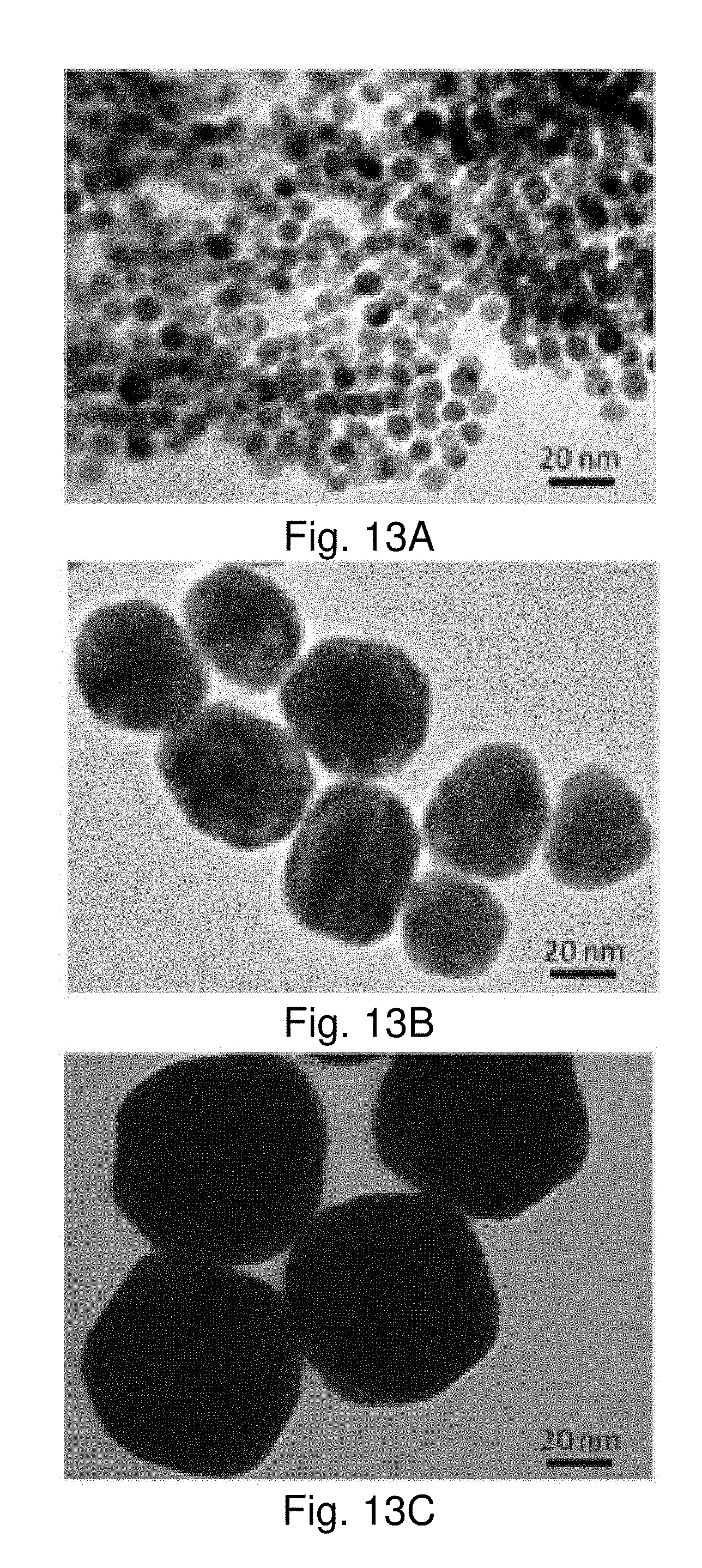
D00014

D00015

D00016

D00017

D00018

D00019
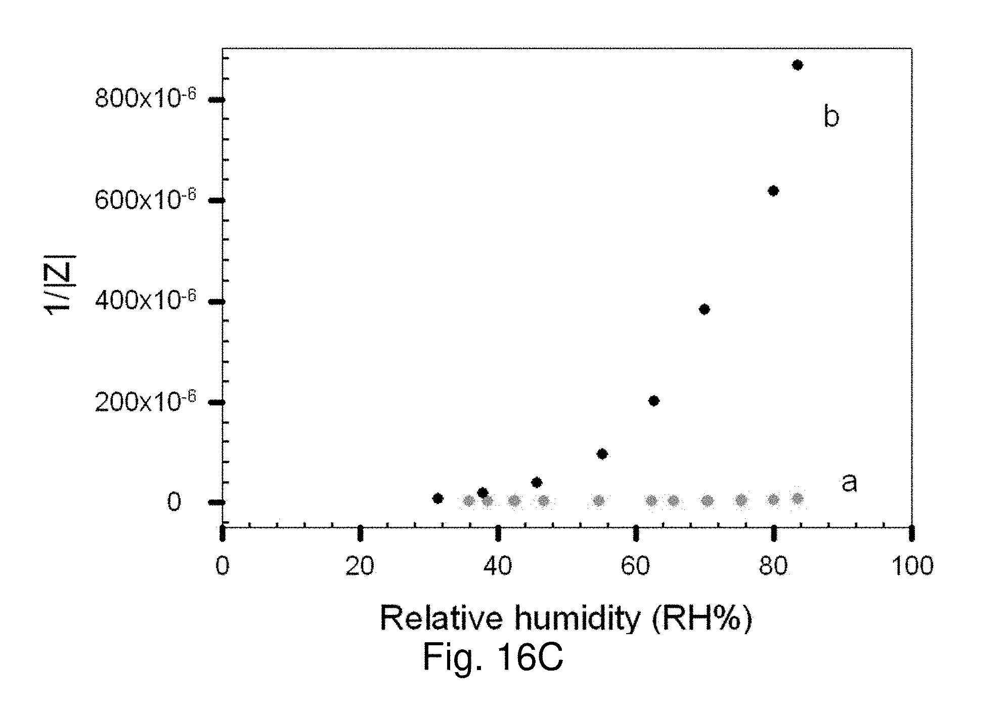
D00020
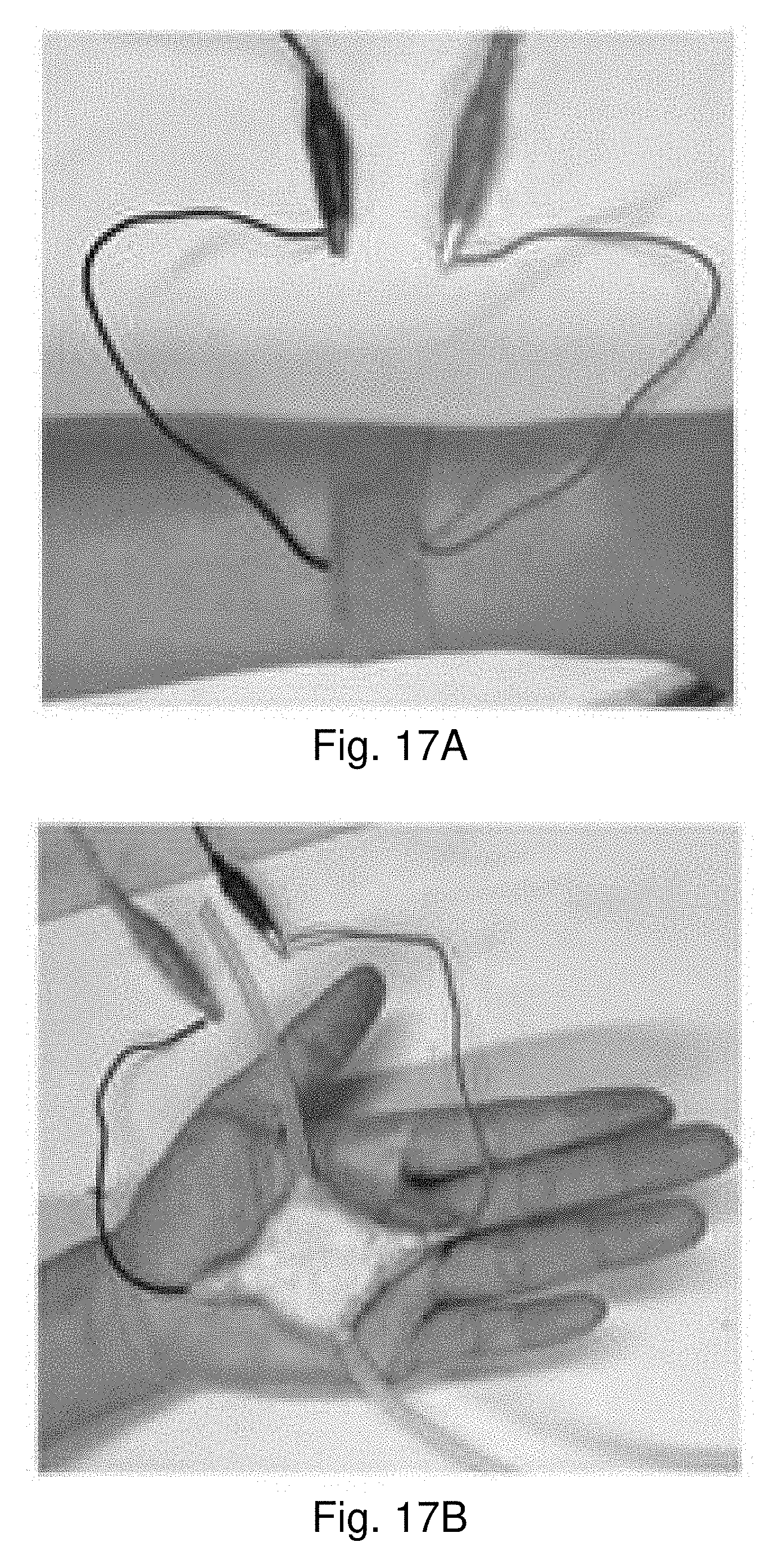
D00021

D00022

D00023
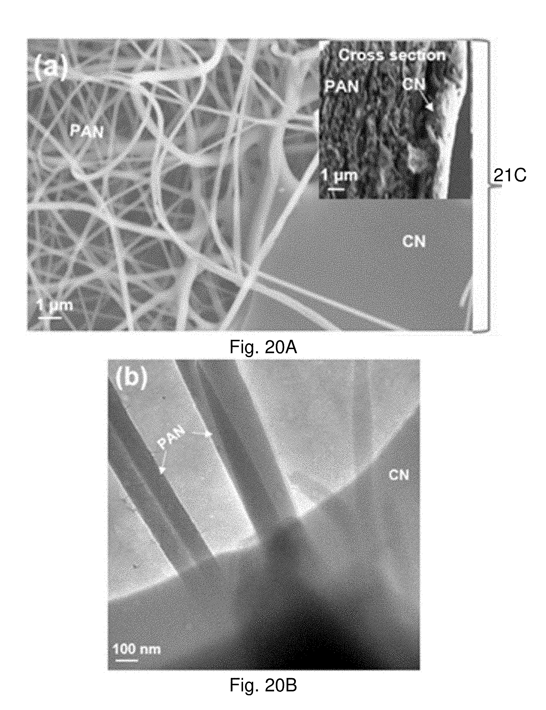
D00024

D00025
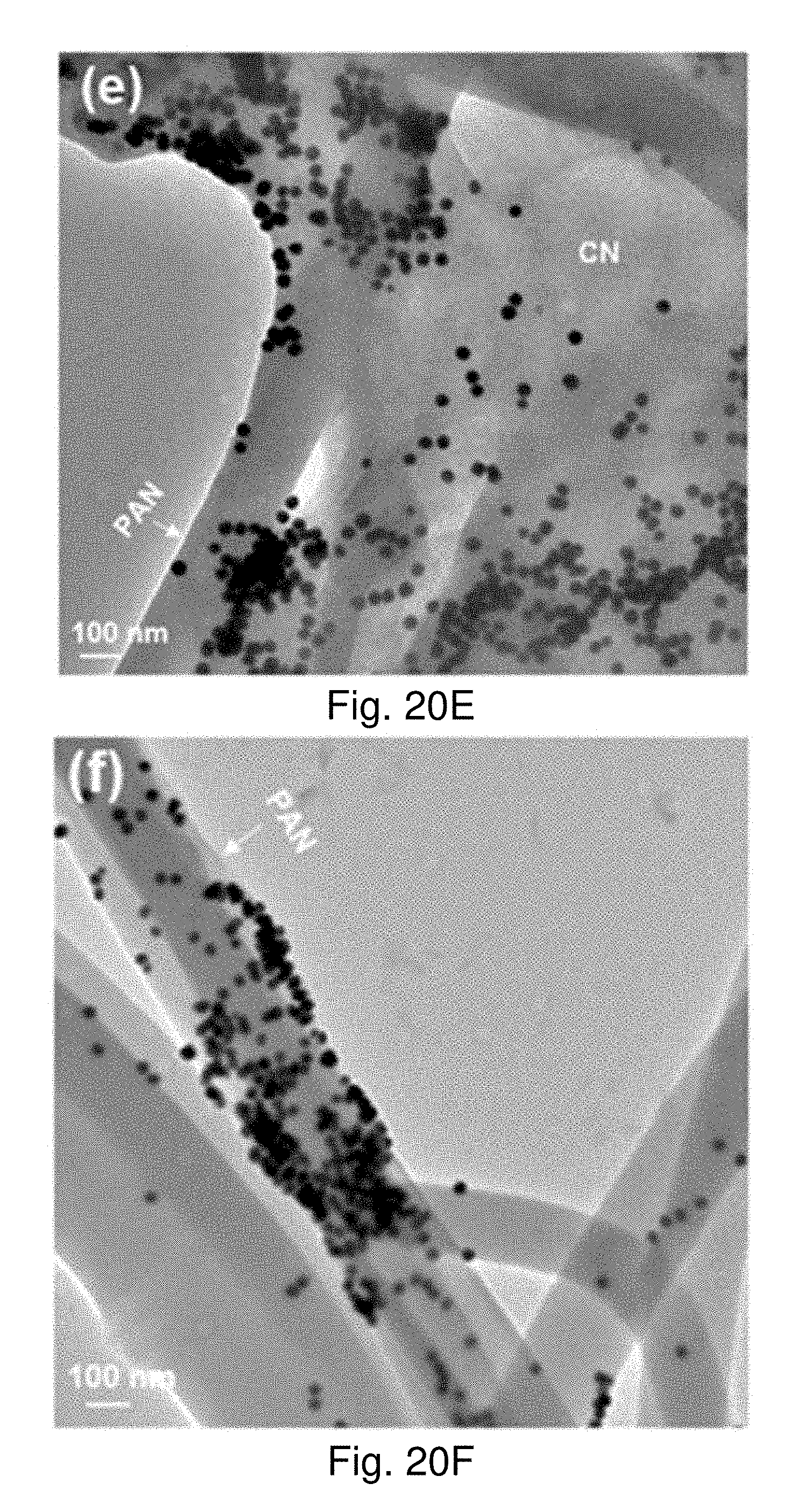
D00026
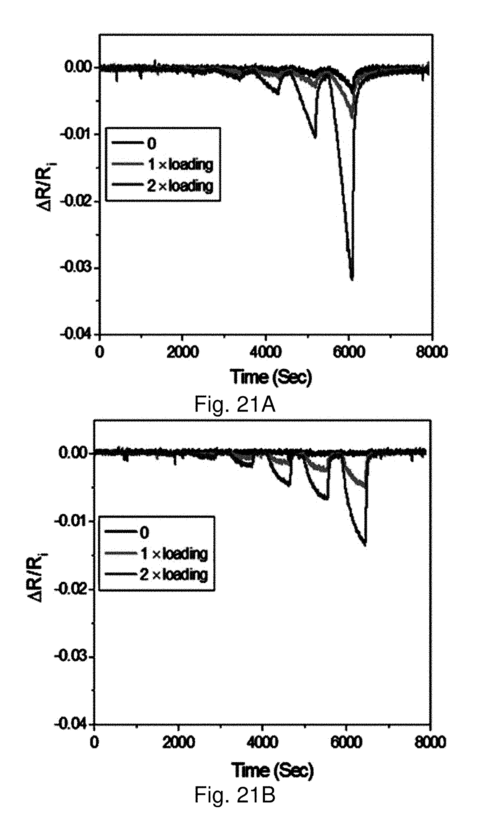
D00027

D00028

D00029
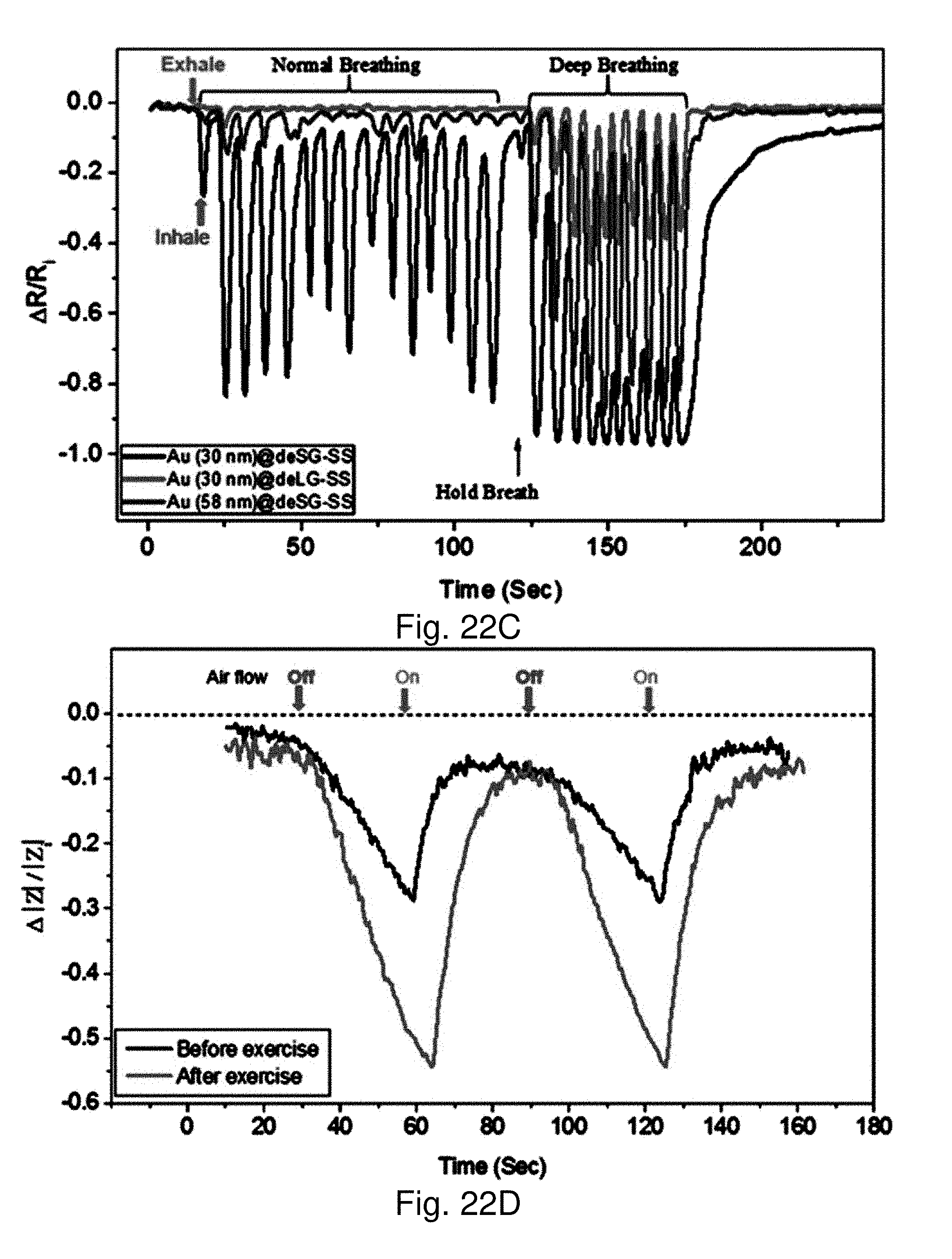
XML
uspto.report is an independent third-party trademark research tool that is not affiliated, endorsed, or sponsored by the United States Patent and Trademark Office (USPTO) or any other governmental organization. The information provided by uspto.report is based on publicly available data at the time of writing and is intended for informational purposes only.
While we strive to provide accurate and up-to-date information, we do not guarantee the accuracy, completeness, reliability, or suitability of the information displayed on this site. The use of this site is at your own risk. Any reliance you place on such information is therefore strictly at your own risk.
All official trademark data, including owner information, should be verified by visiting the official USPTO website at www.uspto.gov. This site is not intended to replace professional legal advice and should not be used as a substitute for consulting with a legal professional who is knowledgeable about trademark law.