Activators of autophagic flux and phospholipase D and clearance of protein aggregates including tau and treatment of proteinopathies
Rinderspacher , et al. May 18, 2
U.S. patent number 11,008,341 [Application Number 16/027,930] was granted by the patent office on 2021-05-18 for activators of autophagic flux and phospholipase d and clearance of protein aggregates including tau and treatment of proteinopathies. This patent grant is currently assigned to NY State Psychiatric Institute, The Trustees of Columbia University in the City of New York. The grantee listed for this patent is NY STATE PSYCHIATRIC INSTITUTE, THE TRUSTEES OF COLUMBIA UNIVERSITY IN THE CITY OF NEW YORK. Invention is credited to Shi-Xian Deng, Karen Duff, Donald Landry, Kirsten Alison Rinderspacher, Wai Yu.


View All Diagrams
| United States Patent | 11,008,341 |
| Rinderspacher , et al. | May 18, 2021 |
Activators of autophagic flux and phospholipase D and clearance of protein aggregates including tau and treatment of proteinopathies
Abstract
The present application discloses compounds which are activators of autophagic flux and pharmaceutical compositions comprising said activators. It further discloses use of said compounds and pharmaceutical compositions in the treatment of neurodegenerative diseases, particularly proteinopathies and tauopathies such as Alzheimer's disease. It further discloses methods of enhancing autophagic flux.
| Inventors: | Rinderspacher; Kirsten Alison (Mount Vernon, NY), Yu; Wai (New York, NY), Duff; Karen (New York, NY), Landry; Donald (New York, NY), Deng; Shi-Xian (White Plains, NY) | ||||||||||
|---|---|---|---|---|---|---|---|---|---|---|---|
| Applicant: |
|
||||||||||
| Assignee: | The Trustees of Columbia University
in the City of New York (New York, NY) NY State Psychiatric Institute (New York, NY) |
||||||||||
| Family ID: | 58488491 | ||||||||||
| Appl. No.: | 16/027,930 | ||||||||||
| Filed: | July 5, 2018 |
Prior Publication Data
| Document Identifier | Publication Date | |
|---|---|---|
| US 20180312526 A1 | Nov 1, 2018 | |
Related U.S. Patent Documents
| Application Number | Filing Date | Patent Number | Issue Date | ||
|---|---|---|---|---|---|
| 15480220 | Apr 5, 2017 | ||||
| PCT/US2016/055561 | Oct 5, 2016 | ||||
| 62237342 | Oct 5, 2015 | ||||
| Current U.S. Class: | 1/1 |
| Current CPC Class: | A61K 31/517 (20130101); C07D 239/94 (20130101); C07D 401/12 (20130101); C07D 405/12 (20130101); C07D 217/22 (20130101); C07D 403/12 (20130101); C07D 513/04 (20130101); C07D 239/88 (20130101); C07D 495/04 (20130101); C07D 471/04 (20130101); C07D 239/86 (20130101); A61P 25/28 (20180101); C07D 239/93 (20130101); C07D 491/04 (20130101); C07D 491/048 (20130101) |
| Current International Class: | C07D 513/04 (20060101); A61P 25/28 (20060101); C07D 491/04 (20060101); C07D 471/04 (20060101); C07D 239/94 (20060101); C07D 239/93 (20060101); C07D 239/86 (20060101); C07D 405/12 (20060101); C07D 403/12 (20060101); C07D 491/048 (20060101); C07D 401/12 (20060101); C07D 239/88 (20060101); C07D 495/04 (20060101); A61K 31/517 (20060101); C07D 217/22 (20060101) |
References Cited [Referenced By]
U.S. Patent Documents
| 3266990 | August 1966 | Lutz et al. |
| 3272824 | September 1966 | Ebetino et al. |
| 3936461 | February 1976 | Mortlock et al. |
| 4114939 | September 1978 | Burt |
| 5145843 | September 1992 | Arnold et al. |
| 5650415 | July 1997 | Tang et al. |
| 5693652 | December 1997 | Takase et al. |
| 6002008 | December 1999 | Wissner et al. |
| 6103728 | August 2000 | Tang et al. |
| 6143764 | November 2000 | Kubo et al. |
| 6184225 | February 2001 | Thomas et al. |
| 6251912 | June 2001 | Wissner et al. |
| 6265410 | July 2001 | Bridges et al. |
| 6297258 | October 2001 | Wissner et al. |
| 6391874 | May 2002 | Cockerill et al. |
| 6596727 | July 2003 | Schaper et al. |
| 6630489 | October 2003 | Crawley |
| 6919338 | July 2005 | Mortlock et al. |
| 6995174 | February 2006 | Wang et al. |
| 7081461 | July 2006 | Mortlock et al. |
| 7135466 | November 2006 | Sakai et al. |
| 7141577 | November 2006 | Ple |
| 7173135 | February 2007 | Hennequin et al. |
| 7173136 | February 2007 | Hennequin et al. |
| 7211587 | May 2007 | Kubo et al. |
| 7262201 | August 2007 | Hennequin et al. |
| 7268230 | September 2007 | Hennequin |
| 7402583 | July 2008 | Boyle et al. |
| 7425564 | September 2008 | Fujiwara et al. |
| 7432377 | October 2008 | Chew et al. |
| 7462623 | December 2008 | Ple |
| 7479473 | January 2009 | Queen |
| 7495104 | February 2009 | Miwa et al. |
| 7504408 | March 2009 | Hennequin et al. |
| 7560558 | July 2009 | Shimizu et al. |
| 7598258 | October 2009 | Kubo et al. |
| 7601716 | October 2009 | Dorsey et al. |
| 7763731 | July 2010 | Rockway et al. |
| 7799772 | September 2010 | Freyne et al. |
| 7960546 | June 2011 | Schroeder et al. |
| 8143276 | March 2012 | Yang et al. |
| 8148532 | April 2012 | Chen |
| 8212033 | July 2012 | Su et al. |
| 8232294 | July 2012 | Xi |
| 8349857 | January 2013 | Kume et al. |
| 8394786 | March 2013 | Freyne et al. |
| 8404697 | March 2013 | Solca et al. |
| 8492560 | July 2013 | Stokes et al. |
| 8575203 | November 2013 | Englehardt et al. |
| 8618289 | December 2013 | Abraham et al. |
| 8642767 | February 2014 | Spinelli et al. |
| 8673929 | March 2014 | Gao et al. |
| 9169236 | October 2015 | Fontana et al. |
| 9353122 | May 2016 | Ong et al. |
| 9382232 | July 2016 | Gong et al. |
| 9853396 | December 2017 | Musick |
| 2003/0092721 | May 2003 | Pitts et al. |
| 2003/0191308 | October 2003 | Hennequin et al. |
| 2005/0009815 | January 2005 | Devita et al. |
| 2005/0085465 | April 2005 | Hennequin |
| 2006/0069077 | March 2006 | Rice et al. |
| 2007/0027318 | February 2007 | Kubo et al. |
| 2008/0058342 | March 2008 | Hennequin |
| 2008/0096884 | April 2008 | Edin et al. |
| 2008/0207617 | August 2008 | Miwa et al. |
| 2008/0227812 | September 2008 | Chen |
| 2008/0280917 | November 2008 | Albrecht et al. |
| 2009/0053236 | February 2009 | Yamamoto |
| 2009/0069316 | March 2009 | Hong et al. |
| 2009/0131461 | May 2009 | Davidson et al. |
| 2009/0312313 | December 2009 | Shimizu et al. |
| 2010/0022586 | January 2010 | Govek et al. |
| 2010/0144639 | June 2010 | Singer et al. |
| 2010/0234324 | September 2010 | Eggenweiler et al. |
| 2011/0269758 | November 2011 | Xiao et al. |
| 2012/0129877 | May 2012 | Martinez et al. |
| 2012/0142929 | June 2012 | Patel et al. |
| 2012/0238587 | September 2012 | Lee et al. |
| 2013/0079342 | March 2013 | Dransfield et al. |
| 2013/0345258 | December 2013 | Bury et al. |
| 2014/0221402 | August 2014 | Lu et al. |
| 2014/0221405 | August 2014 | Scarborough et al. |
| 2014/0275129 | September 2014 | Lee et al. |
| 2015/0057263 | February 2015 | Brown et al. |
| 2015/0329498 | November 2015 | Romero et al. |
| 2016/0060249 | March 2016 | Casillas et al. |
| 2016/0101106 | April 2016 | Werner |
| 2016/0108035 | April 2016 | Peng et al. |
| 2016/0297775 | October 2016 | Zhang |
| 2017/0035761 | February 2017 | Alami et al. |
| 2017/0174703 | June 2017 | Wu et al. |
| 2417955 | Jan 2003 | CA | |||
| 1083811 | Mar 1994 | CN | |||
| 0326328 | Aug 1989 | EP | |||
| 326329 | Aug 1989 | EP | |||
| 0326329 | Aug 1989 | EP | |||
| 326330 | Aug 1989 | EP | |||
| 0414386 | Feb 1991 | EP | |||
| 1409481 | May 1992 | EP | |||
| 520722 | Dec 1992 | EP | |||
| 566226 | Oct 1993 | EP | |||
| 635498 | Jan 1995 | EP | |||
| 635507 | Sep 1999 | EP | |||
| 2268623 | Jan 2011 | EP | |||
| 2445886 | May 2012 | EP | |||
| 2522658 | Nov 2012 | EP | |||
| 3012251 | Apr 2016 | EP | |||
| 1990005523 | May 1990 | WO | |||
| 1992017452 | Oct 1992 | WO | |||
| 1992022533 | Dec 1992 | WO | |||
| 9404526 | Mar 1994 | WO | |||
| 1994004527 | Mar 1994 | WO | |||
| 1996009294 | Mar 1996 | WO | |||
| 1996039145 | Dec 1996 | WO | |||
| 1997003069 | Jan 1997 | WO | |||
| 1998013350 | Apr 1998 | WO | |||
| 1998043960 | Oct 1998 | WO | |||
| 9931072 | Jun 1999 | WO | |||
| 2000068199 | Nov 2000 | WO | |||
| 2000068200 | Nov 2000 | WO | |||
| 2000068201 | Nov 2000 | WO | |||
| 2001021596 | Mar 2001 | WO | |||
| 2001046150 | Jun 2001 | WO | |||
| 2002018375 | Mar 2002 | WO | |||
| 2002020489 | Mar 2002 | WO | |||
| 2003047582 | Jun 2003 | WO | |||
| 2003047583 | Jun 2003 | WO | |||
| 2003047585 | Jun 2003 | WO | |||
| 2003048159 | Jun 2003 | WO | |||
| 2003053960 | Jul 2003 | WO | |||
| 2003055866 | Jul 2003 | WO | |||
| 2005058318 | Jun 2005 | WO | |||
| 2003047584 | Jun 2006 | WO | |||
| 2006108059 | Oct 2006 | WO | |||
| 2006121767 | Nov 2006 | WO | |||
| 2007055513 | May 2007 | WO | |||
| 2008024423 | Feb 2008 | WO | |||
| 2009010139 | Jan 2009 | WO | |||
| 2009024611 | Feb 2009 | WO | |||
| 2009148659 | Dec 2009 | WO | |||
| 2010018555 | Feb 2010 | WO | |||
| 2010025451 | Mar 2010 | WO | |||
| 2011014039 | Feb 2011 | WO | |||
| 2011143444 | Nov 2011 | WO | |||
| 2012090179 | Jul 2012 | WO | |||
| 2013012915 | Jan 2013 | WO | |||
| 2013074633 | May 2013 | WO | |||
| 2014043437 | Mar 2014 | WO | |||
| 2015079067 | Jun 2015 | WO | |||
| 2015079251 | Jun 2015 | WO | |||
| 2015106025 | Jul 2015 | WO | |||
| 2015144001 | Oct 2015 | WO | |||
| 2015155262 | Oct 2015 | WO | |||
Other References
|
Chemspider, 6-Fluoro-4-(tetrahydro-2H-pyran-4-ylsulfanyl)quinazoline, Royal Society of Chemistry, 2015, Retreived from the internet <URL http://www.chemspider.com/Chemical-Structure_30132867.html?rid=9a2b8938-1- 827-42ad>. cited by applicant . Harsora, Synthesis, Characterization and Antimicrobial Screening of Some Bio-active Heterocyclic Compounds, Maharaja Krishnakumarsinhji Bhavnagar University Thesis, Jan. 2011, Part I, 26-88. cited by applicant . Kumar, Studies on Diversity Oriented synthesis of Bioactive Compounds, Jawaharlal Nehru University Thesis, Jun. 2009, Chaper 1, 1-27. cited by applicant . Pubchem, CID 23631972, Jan. 3, 2008, pp. 1-11. Retrieved from the Internet <URL: https:// pubchem.ncbl.nlm.nih.gov/compound/236319721972>. cited by applicant . Pubchem, CID 616883, Mar. 27, 2005, pp. 1-14. Retrelved from the Internet <URL: https://pubchem.nal.nlm.nlh.gov/compound/616883>. cited by applicant . Pubchem, CID 21002400, Dec. 5, 2007, pp. 1-10. Retreived from the Internet <URL: https://pubchem.ncbi.nlm.nih.gov/compound/21002400>. cited by applicant . International Search Report and Written Opinion, from the International Searching authority, in Int. App. No. PCT/US16/55561, dated Mar. 27, 2017. cited by applicant . Pubchem, CID 640956, Jan. 25, 2006, Retrieved from the internet <URL https://pubchem.ncbi.nlm.nih.gov/compound/640956>. cited by applicant . Pubchem, CID 57263900, Jun. 15, 2012, Retrieved from the Internet <URLhttps://pubchem.ncbi.nlm.nih.gov/compound/57263900>. cited by applicant . International Search Report and Written Opinion, from the International Searching authority, in Int. App. No. PCT/US17/54969, dated Jan. 18, 2018. cited by applicant . PubChem, CID 88934329, retrieved from the Internet <URL: https://pubchem.ncbi.nlm.nih.gov/compound/88934329> (record date, Feb. 13, 2015). cited by applicant . Supplementary European Search Report in European Application No. 16854245.4, dated Mar. 19, 2019. cited by applicant . Chemical Reactions, Benha University Thesis, Chapter Synthesis of 4-(3H)-quinazolinones from Anthranilic Acid or its Derivatives. cited by applicant . PubChem, CID 88934329, retrieved from the internet<URL:https://pubchem.ncbi,nim,nih,gov/compund/88934329. (record date, Feb. 13, 2015). cited by applicant . Byju et al., Therapeutic Potential of Quinazoline and its Derivatives--A Review, International Journal of Institutional Pharmacy and Life Sciences 5(2): Mar.-Apr. 2015. cited by applicant . Ajani et al., Quinazoline Pharmacophore in Therapeutic Medicine, Bangladesh J Pharmacol 2016; 11: 716-733. cited by applicant . Alam et al., A Review. Recent Investigations on Quinazoline Scaffold, International Journal of Advance Research (2015), vol. 3, Issue 12, 1656-1664. cited by applicant . Wang et al., Quinazoline Derivatives: Synthesis and Bioactivities, Chemistry Central Journal 2013, 7:95. cited by applicant . Chandrika et al., Synthesis Leading to Novel 2,4,6 Trisubstituted Quinazoline Derivatives, their Antibacterial and Cytotoxic Activity against THP-1, HL-60 and A375 Cell Lines, Indian Journal of Chemistry, vol. 48B, Jun. 2009, 340-847. cited by applicant . El-Azab et al., Design, Synthesis and Biologica Evaluation of novel Quinazofine Derivatives as Potential Antitumor Agents: Molecular Docking Study, European Journal of Medicinal Chemistry, vol. 45, Issue 9, Sep. 2010, 1188-4198. cited by applicant . Chao et al., Substituted Isoquinolines and Quinazolines as Potential Antiinfiammatory Agents. Synthesis and Biological Evaluation of Inhibitors of Tumor Necrosis Factor a, Journal of Medicinal Chemistry, 1999, 42 (19), 3860-3873. cited by applicant . German et al., Use of Gyrase Resistance Mutants to Guide Selection of 8-Methoxy-Quinazoline-2,4-Diones, American Society for Microbiology, vol. 52, No. 11, Nov. 2008, 3915-3921. cited by applicant . Naga Raju et al., Quinazoline: Unique and Versatile Pharmacophore in the field or Cancer, Indo American Journal of Pharmaceutical Sciences, 2015, vol. 2 (4), 827-832. cited by applicant . Asif, Chemical Characteristics, Synthetic Methods, and Biological Potential of Quinazoline and Quinazolirtone Derivatives, International Journal of Medicinal Chemistry, vol. 2014, Article ID 395637. cited by applicant . Jafari et al. Quinazolinone and Quinazoline Derivatives: Recent Structures with Potent Antimicrobial and Cytotoxic Activities, Research in Pharmaceutical Sciences, Jan.-Feb. 2016. 11(1): 1-14. cited by applicant . Xu, Synthesis and Antifungal Activity of Novel s-Substituted 6-fluoro-4-alkyl(aryl)thioquinazoline Derivatives, Bioorganic & Medicinal Chemistry 15, Mar. 2007, 3768-3774. cited by applicant . Babu, Synthesis, Characterization and Biological Evaluation of Novel Tri Substituted Quinazoline-isatin Mannich Basses Bearing Morpholine and Biphenyl Moieties, Hetrocyclic Letters, May-Jul. 2015, vol. 5, No. 3, 395-411. cited by applicant . Al-Salahi, Synthesis of Novel 2-Alkoxy(aralkoxy)-4H-[1,2,4]triazolo11,5-a] quinazolin-5-ones and Derivatives, University of Hamburg, Jan. 23, 2009. cited by applicant . El-Sakka et al., Synthesis and Biological Activity of Nitrogen Heterocycles From Anthranilic Acid Derivative, Oriental Journal of Chemistry, May 6, 2009, vol. 25(2), 287-294. cited by applicant . Prajapati et al., Synthesis and Preliminary in-vitro Cytotoxic Activity of Morpholino Propoxy Quinazoline Derivatives, International Journal of ChemTech Research, Jan.-Mar. 2014, vol. 6, No. 1, 547-555. cited by applicant . Diab et al., Radiation Dosimetric Properties of New Oxa-, Thiadiazole, Tilazote, International Journal of Physics and Research, Dec. 2013, vol. 3, Issue 5, 11-20. cited by applicant . Aman et al., QSAR Study of Quinazoline Derivatives as Inhibitor of Epidermal Growth Factor Receptor-Tyrosine Kinase (EGFR-TK), 3rd International Conference on Computation for Science and Technology, Nov. 30, 2014. cited by applicant . Chandrika et al., Quinazoline Derivatives with Potent Anti-Inflammatory and Anti-Allergic Activities, Int. J. Chem. Sci., 2008, 6(3), 1119-1146. cited by applicant . Manjula et at., Medicinal and Biological Significance of Quinazoline: A Highly Important Scaffold for Drug Discovery: A Review, International Journal of Pharma and Bio Sciences, Jan.-Mar. 2011, vol. 2, Issue 1, 780-809. cited by applicant . Connolly, Synthesis of Quinazolinones and Quinazofines, Tetrahedron 61, Jun. 2005, Report No. 737, 10153-10202. cited by applicant . Jantova et al., In Vitro Antibacterial Activity of Ten Series of Substituted Quinazolines, Biologia, Bratislava, 2004, 59/6, 741-752. cited by applicant . Barclay et al, Quinolines as Chemotherapeutic Agents for Leishmaniasis, Griffith University, 2013. cited by applicant . Selvam et al., Quinazoline Marketed Drugs--A Review, Research in Pharmacy, 2011, 1(1), 1-21. cited by applicant . Wan et al, Synthesis, Antiviral Bioactivity of Novel 4-Thioquinazoline Derivatives Containing Chalcone Moiety, Molecules, Jun. 2015, 20, 11861-11874. cited by applicant . El-Hashash et al., Synthesis and Evaluation of New 2,3- and 2,4-Disubstituted Quinazoline Derivatives as Potential Antibacterial and Antifungal Agents, Scholars Research Library, Der Pharma Chemica, 2011, 3(6), 147-159. cited by applicant . Sahu et al., In Silico Screening, Synthesis and In Vitro Evaluation of Some Quinazolinone Derivatives as Dihydrofolate Reductase Inhibitors for Anticancer Activity, International Journal of Pharmacy and Pharmaceutical Sciences, Mar. 2014, vol. 6, Issue 5, 193-199. cited by applicant . Lynch et al., 4-Aminoquinolines as Antimalarial Drugs, Trinity Student Scientific Review vol. II, 2016 Edition, 196-211. cited by applicant . Rudrapal et al., Novel 4-Aminoquinoline Analogues as Antimalarial Agents: A Review, Scholars Research Library, Der Phamacia Letter, 2011, 3(3), 29-36. cited by applicant . Kuldeep et al., Review of Synthesis Schemes of Some Biological Active Quinolines, International Journal of Pharmacotherapy, 2015, 5(2), 75-79. cited by applicant . Odingo et al., Synthesis and Evaluation of the 2,4-Diaminoquinazoline Series as Anti-Tubercular Agents, Biorganic & Medicinal Chemistry 22 (2014) 6965-6979. cited by applicant . Dassonville-Klimpt et al., Mefloquine Derivatives : Synthesis, Mechanisms of Action, Antimicrobial Activities, Universite de Picardie Jule Verne, 2011. cited by applicant . Xie et al., Synthesis of Nitrogen-Containing Heterocycles Using Nitro Compounds as Building Blocks: Part I: Synthesis of 3-Substituted Azepanes. Part II: Synthesis of 1-Azoniapropellanes as Phase Transfer Catalysts, University of Illinois at Urbana-Champaign, 2010. cited by applicant . Viart et al., Synthesis and Structure-Activity Relationship of Uposomal Substrates for Phospholipase A(2), Absracts )1 Papers of the American Chemical Society, 2011, 242, Medi 344. cited by applicant . Dow et al., Utility of Alkylaminoquinolinyl Methanols as New Antimalarial Drugs, Antimicrobial Agents and Chemotherapy, Dec. 2006, vol. 50, No. 12, 4132-4143. cited by applicant . Marella et al., Quinoline: A Versatile Heterocyclic, King Saud University, Saudi Pharmaceutical Journal, Mar. 2012, 21-12. cited by applicant . Poonan et al., A Review on Biological Activities of Quinoline Derivatives, Journal of Management Information Technology and Engineering, Jun. 2016, vol. 2, Issue 1, 1-14. cited by applicant . Yang, Discovery of Selective Histone Deacetylase 6 Inhibitors Using the Quinazoline as the Cap for the Treatment of Cancer, Journal of Medical Chemistry, Oct. 2015, 59(4), 1455-1470. cited by applicant . Duan, Synthesis and Evaluation of (2-(4-Methoxyphenyl)-4-quinolinyl) (2-piperidinyl)methanol (NSC23925) Isomers to Reverse Multidrug Resistance in Cancer, Journal of Medicinal Chemistry, Mar. 2012, 55, 3113-3121. cited by applicant . Pubchem, CID 21002494, Retrieved from the Internet <URL https://pubchem.ncbi.nlm.nih.gov/compound/21002494#section-Chemical-Vendo- rs>. cited by applicant . Pubchem, CID 44428806, Retrieved from the Internet <URL: https://pubchem.ncbi.nlm.nih.gov/compound/4428806itsection=Top>. cited by applicant . Guidechem, 4(3H)-Quinazolinethione,2-methyl--(CAS No. 648428-2), Retrieved from the Internet <URL: http://guidechem.com/reference/dic-168980>. cited by applicant . Shah, Synthesis and Biological Evaluation of Certain Nitrogen and Sulfur Based Bioactive Scaffolds, Gujarat University Thesis, Chapter 1, 2014. cited by applicant . ChemFuture Catalog Products 2010, ChemFuture PharmaTech (China) Ltd. cited by applicant . 4-(furan-2-ylmethylsulfanyl)quinazoline, Henan Coreychem Co.,Ltd, 2012, Retrieved from the Internet <URL http://coreychem.com/PRODUCTS/Phamaceutical_intermediates/2015/05231/1957- 60.html>. cited by applicant . Pubchem, CID 7160394, Retrieved from the Internet <URL: https://pubchem.ncbi.nlm.nth.gov/compound/7160394#section-Top>. cited by applicant . Pubchem, CID 60329852, Retrieved from the Internet <URL: https://pubchem.ncbi.nlm.nih.gov/compound/60329852/#section=2D-Structure&- gt;. cited by applicant . A. Rinderspacher, et al., "Potent Inhibitors of Huntingtin Protein Aggregation in a Cell-Based Assay", Bioorg Med Chem Lett., 19(6), pp. 1715-1717 (2009). cited by applicant . R. Saari, et al., "Microwave-assisted synthesis of quinoline, isoquinoline, quinoxaline and quinazoline derivatives as 2,132 receptor agonists", Bioorganic & Medicinal Chemistry, 19, pp. 939-950 (2011). cited by applicant . B. Hu, et al., "Flexachlorocyclotriphosphazene (HCCP)-Mediated Direct Formation of Thioethers and Ethers from Quinazolin-4(3H)-ones", Molecules, 18, pp. 5580-5593 (2013). cited by applicant . S. Radl, et al., "Synthesis and Analgesic Activity of Some Quinazoline Analogs of Anpirtoline", Arch. Pharm. Med. Chem., 333, pp. 381-386 (2000). cited by applicant . S. Yang, et al., "Synthesis and bioactivity of 4-alkyl(aryl)thioquinazoline derivatives", Bioorganic & Medicinal Chemistry Letter, 17, pp. 2193-2196 (2007). cited by applicant . CAS Registry; RN:1523031-22-2, Entered on Jan. 17, 2014. cited by applicant . CAS Registry; RN:1274044-98-2, Entered on Apr. 3, 2011. cited by applicant . CAS Registry; RN:1153094-45-1, Entered on Jan. 7, 2009. cited by applicant . CAS Registry; RN:1097030-29-9, Entered on Jan. 28, 2009. cited by applicant . CAS Registry; RN:1401991-13-6, Entered on Oct. 24, 2012. cited by applicant . CAS Registry; RN: 929503-36-6, Entered on Apr. 9, 2007. cited by applicant . CAS Registry; RN:929370-68-3, Entered on Apr. 8, 2007. cited by applicant. |
Primary Examiner: Ward; Paul V
Attorney, Agent or Firm: Venable LLP
Parent Case Text
CROSS-REFERENCE TO RELATED APPLICATIONS
The present application is a division of U.S. patent application Ser. No. 15/480,220 filed Apr. 5, 2017, published as US 2017/0210759, is a Continuation-in-Part (CIP) application that claims benefit to International Application Serial No. PCT/US16/055561, filed Oct. 5, 2016, published as WO 2017/062500, which International Application claims benefit to U.S. Provisional Application Ser. No. 62/237,342, filed Oct. 5, 2015. The entire contents of the aforementioned applications are incorporated by reference as if recited in full herein.
Claims
What is claimed is:
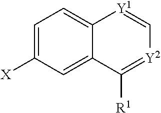
1. A compound having the formula (II): ##STR00173## wherein one of Y.sup.1 and Y.sup.2 is CH and one of Y.sup.1 and Y.sup.2 is N; wherein X is selected from the group consisting of H, halide, and aryl; wherein R.sup.1 is selected from the group consisting of optionally substituted thioheteroaryl, hydroxyl-substituted (2-aminoethyl)aryl, and optionally substituted thiocycloalkyl wherein the cycloalkyl is a 5 or 6-membered cycloalkyl and wherein 1-3 carbon atoms of the cycloalkyl is optionally replaced with a heteroatom selected from the group consisting of O and S, or a salt, enantiomer, racemate, mixture thereof, or combination thereof.
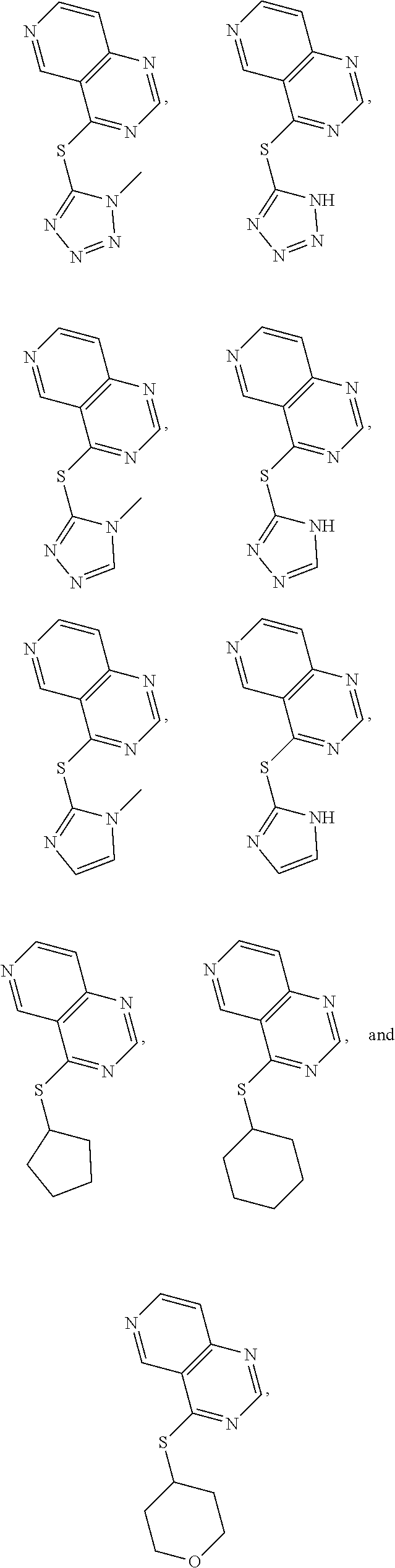
2. The compound of claim 1, wherein the compound is selected from the group consisting of: ##STR00174## or a salt thereof.
3. A compound having the formula (III): ##STR00175## wherein Y.sup.1 is CH; wherein Y.sup.2 is N; wherein X is halide; wherein R.sup.1 is selected from the group consisting of optionally substituted thioheteroaryl, optionally substituted (2-aminoethyl)aryl, and optionally substituted thiocycloalkyl wherein the cycloalkyl is a 5 or 6-membered cycloalkyl and wherein 1-3 carbon atoms of the cycloalkyl is optionally replaced with a heteroatom selected from the group consisting of O and S, and thioaryl, or a salt, enantiomer, racemate, mixture thereof, or combination thereof.
4. The compound of claim 3, wherein the compound is selected from the group consisting of: ##STR00176## or a salt thereof.
5. A pharmaceutical composistion comprising a compound of claims 1 or 4, or a pharmaceutically acceptable salt thereof.
6. A method of treating a neurodegenerative disease comprising administering to a subject in need thereof an effective amount of a compound of claim 1 or 3, wherein the neurodegenerative disease is a proteinopathy.
7. The method of claim 6, wherein the proteinopathy is a tauopathy.
8. A method of treating a neurodegenerative disease comprising administering to a subject in need thereof an effective amount of a compound of claims 1 or 3, wherein the neurodegenerative disease is Alzheimer's disease.
9. A method of enhancing autophagic flux comprising providing to a cell or a protein aggregate an effective amount of a compound of claims 1 or 3.
10. A method of treating a proteinopathy comprising administering to a subject in need thereof an effective amount of a compound of claims 1 or 3.
11. A method of treating a tauopathy comprising administering to a subject in need therof an effective amound of a compound of claims 1 or 3.
12. A method of treating a neurodegenerative disease comprising administering to a subject in need thereof an effective amount of the pharmaceutical composition of claim 5, wherein the neurodegenerative disease is proteinopathy.
13. The method of claim 12, wherein the proteinopathy is tauopathy.
14. A method of treating a neurodegenerative disease comprising administering to a subject in need therof an effective amount of the pharmaceutical composition of claim 5, wherein the neurodegenerative disease is Alzheimer's disease.
15. A method of enhancing autophagic flux comprising providing to a cell or a protein aggregate an effective amount of the pharmaceutical composition of claim 5.
16. A method of treating a proteinopathy comprising adminstering to a subject in need thereof an effective amount of the pharmaceutical composition of claim 5.
17. A method of treating a tauopathy comprising adminstering to a subject in need thereof an effective amount of the pharmaceutical composition of claim 5.
18. A method of treating a neurodegenerative disease comprising adminstering to a subject in need thereof an effective amount of a compound having the following formula: ##STR00177## wherein one of Y.sup.1 and Y.sup.2 is CH and one of Y.sup.1 and Y.sup.2 is N; wherein X is selected from the group consisting of H, halide, and aryl; wherein R.sup.1 is selected from the group consisting of optionally substituted thioheteroaryl, hydroxyl-substituted (2-aminoethyl)aryl, halide, and optionally substituted thiocycloalkyl wherein 1-3 carbon atoms of the cycloalkyl is optionally replaced with a heteroatom selected from the group consisting of O, S and N, and thioaryl, or a salt, enantiomer, racemate, mixture thereof, or a combination thereof, wherein the neurodegenerative disease is a proteinopathy.
19. The method of claim 18, wherein the proteinopathy is a tauopathy.
20. The method of claim 18, wherein the proteinopathy is Alzheimer's disease.
21. A method of treating a neurodegenerative disease comprising administering to a subject in need thereof an effective amount of a compound having the following formula: ##STR00178## wherein Y.sup.1 is CH; wherein Y.sup.2 is N; wherein X is halide; wherein R.sup.1 is selected from the group consisting of optionally substituted thioheteroaryl, optionally substituted (2-aminoethyl)aryl, halide, and optionally substituted thiocycloalkyl wherein 1-3 carbon atoms of the cycloalkyl is optionally replaced with a heteroatom selected from the group consisting of O, S and N, and thioaryl, or a salt, enantiomer, racemate, mixture thereof, or combination thereof wherein the neurodegenerative disease is a proteinopathy.
22. The method of claim 21, wherein the proteinopathy is a tauopathy.
23. The method of claim 21, wherein the proteinopathy is Alzheimer's disease.
Description
FIELD OF THE DISCLOSURE
The present disclosure relates to compounds which are activators of autophagic flux and pharmaceutical compositions comprising said compounds. It further relates to use of said compounds in the treatment of neurodegenerative diseases, particularly Alzheimer's disease.
BACKGROUND OF THE INVENTION
Alzheimer's disease (AD) affects approximately five million Americans and this number is predicted to triple by 2050. At present, there are no therapies to treat Alzheimer's or other related tauopathies. While clinical trials using immunotherapy targeting amyloid beta (An) have had limited success, this in only subset of those afflicted with AD or other neurodegenerative diseases. Moreover, there are no therapies targeting other proteinopathies, including tau, the other major neuropathological component of AD. AD accounts for most of the dementias afflicting individuals over 65 and is estimated to cost $226 billion in healthcare, long-term care, and hospice for people with AD and other dementias annually. This extensive economic and societal burden does not account for lost income of many at-home primary caregivers including spouses and other family members.
Enhancing autophagy has been shown to have therapeutic potential in the treatment of Alzheimer's disease. Autophagic flux (including the fusion of autophagosomes to lysosomes) is a novel regulator of autophagy as it leads to the clearance of protein aggregates and reversal of pathophysiological decline. Therefore, there exists an ongoing need for promoters of autophagic flux and the clearance of autophagosomes bearing proteinopathies.
SUMMARY OF THE INVENTION
In some embodiments, compounds including pharmaceutically acceptable salts thereof, which are disclosed herein, are provided.
In some embodiments a pharmaceutical composition is provided comprising a compound disclosed herein or a pharmaceutically acceptable salt thereof. In other embodiments, methods of making the compounds and pharmaceutical compositions are also provided in, e.g., the Examples provided below.
In some embodiments a method of treating a neurodegenerative disease comprising administering to a subject in need thereof an effective amount of a compound or pharmaceutical composition disclosed herein is provided.
In some embodiments a method of enhancing autophagic flux is provided. This method comprises providing to a cell or a protein aggregate an effective amount of a compound or pharmaceutical composition disclosed herein.
These and other aspects of the invention are further disclosed in the detailed description and examples which follow.
BRIEF DESCRIPTION OF THE DRAWINGS
The following drawings form part of the present specification and are included to further demonstrate certain aspects of the present invention. The invention may be better understood by reference to one or more of these drawings in combination with the detailed description of specific embodiments presented herein.
FIG. 1 is a graph showing a photodiode array (PDA) spectrum of WHYKD8 in mouse brain.
FIG. 2 shows Western blots of LC3-II levels in primary cortical neurons following a 6 hour treatment with WHYKD1 (.+-.BafA1) or WHYKD5.
FIG. 3 shows Western blots of LC3-II, tau, and p62 levels in organotypic slice cultures following a 6 hour treatment with WHYKD1 (top) or WHYKD3, WHYKD5, WHYKD8, WHYKD9, or WHYKD12 (bottom).
FIG. 4 is a bar graph showing the activation of phospholipase D (PLD) by the WHYKD series compounds (10 .mu.M), and their ability to convert phospholipids to phosphatidylethanols in the presence of ethanol. C=Control, 12=WHYKD12, 15=WHYKD15, 19=WHYKD19, 5=WHYKD5, 8=WHYKD8, Fipi=a noncompetitive inhibitor of PLD activity.
FIG. 5 is a bar graph showing the activation of phospholipase D (PLD) by the WHYKD series compounds (1 .mu.M), and their ability to convert phospholipids to phosphatidylethanols in the presence of ethanol.
DETAILED DESCRIPTION OF THE INVENTION
Although macroautophagy is known to be an essential degradative process whereby autophagosomes mediate the engulfment and delivery of cytoplasmic components into lysosomes, the lipid changes underlying autophagosomal membrane dynamics are undetermined. The inventors have previously shown that PLD1, which is primarily associated with the endosomal system, partially relocalizes to the outer membrane of autophagosome-like structures upon nutrient starvation (Dall'Armi, 2010). The localization of PLD1, as well as the starvation-induced increase in PLD activity, are altered by wortmannin, a phosphatidylinositol 3-kinase inhibitor, suggesting PLD1 may act downstream of Vps34. Pharmacological inhibition of PLD and genetic ablation of PLD1 in mouse cells decreased the starvation-induced expansion of LC3-positive compartments, consistent with a role of PLD1 in the regulation of autophagy. Furthermore, inhibition of PLD results in higher levels of tau and p62 aggregates in organotypic brain slices. These in vitro and in vivo findings establish a role for PLD1 in autophagy.
In some embodiments, a compound is provided having the formula (II):
##STR00001## wherein Y.sup.1 and Y.sup.2 are independently selected from the group consisting of CH and N; wherein X is selected from the group consisting of H, halide, and aryl; wherein R.sup.1 is selected from the group consisting of optionally substituted thioheteroaryl, hydroxyl-substituted (2-aminoethyl)aryl, halide, optionally substituted thiocycloalkyl wherein 1-3 carbon atoms of the cycloalkyl is optionally replaced with a heteroatom selected from the group consisting of O, S and N, and thioaryl, or a salt, enantiomer, racemate, mixture thereof, or combination thereof.
In some embodiments, the compound is selected from the group consisting of:
##STR00002## ##STR00003## ##STR00004## ##STR00005## or a salt, enantiomer, racemate, mixture thereof, or combination thereof.
In one embodiment the compound is:
##STR00006## or a salt, enantiomer, racemate, mixture thereof, or combination thereof.
In another embodiment the compound is:
##STR00007## or a salt, enantiomer, racemate, mixture thereof, or combination thereof.
In some embodiments, a compound is provided having the formula (III):
##STR00008## wherein Y.sup.1 is CH; wherein Y.sup.2 is N; wherein X is halide; wherein R.sup.1 is selected from the group consisting of optionally substituted thioheteroaryl, optionally substituted (2-aminoethyl)aryl, halide, optionally substituted thiocycloalkyl wherein 1-3 carbon atoms of the cycloalkyl is optionally replaced with a heteroatom selected from the group consisting of O, S and N, and thioaryl, or a salt, enantiomer, racemate, mixture thereof, or combination thereof.
In some embodiments, the compound is selected from the group consisting of:
##STR00009## or a salt, enantiomer, racemate, mixture thereof, or combination thereof.
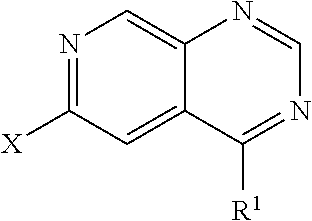
In some embodiments, a compound is provided having the formula (IV):
##STR00010## wherein X is halide; wherein R.sup.1 is selected from the group consisting of optionally substituted thioheteroaryl, optionally substituted (2-aminoethyl)aryl, halide, optionally substituted thiocycloalkyl wherein 1-3 carbon atoms of the cycloalkyl is optionally replaced with a heteroatom selected from the group consisting of O, S and N, and thioaryl, or a salt, enantiomer, racemate, mixture thereof, or combination thereof.
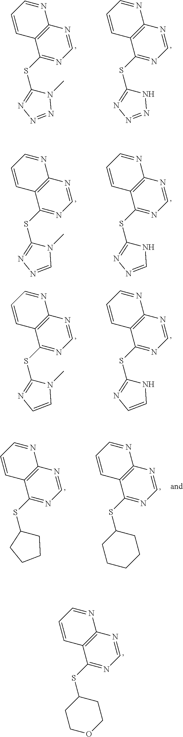
In some embodiments, the compound is selected from the group consisting of:
##STR00011## or a salt, enantiomer, racemate, mixture thereof, or combination thereof.
In some embodiments, a compound is provided having the formula (V):
##STR00012## wherein X is H; wherein R.sup.1 is selected from the group consisting of optionally substituted thioheteroaryl, optionally substituted (2-aminoethyl)aryl, halide, optionally substituted thiocycloalkyl wherein 1-3 carbon atoms of the cycloalkyl is optionally replaced with a heteroatom selected from the group consisting of O, S and N, and thioaryl, or a salt, enantiomer, racemate, mixture thereof, or combination thereof.
In some embodiments, the compound is selected from the group consisting of:
##STR00013## or a salt, enantiomer, racemate, mixture thereof, or combination thereof.
In some embodiments, a compound is provided having the formula (VI):
##STR00014## wherein X is H; wherein R.sup.1 is selected from the group consisting of optionally substituted thioheteroaryl, optionally substituted (2-aminoethyl)aryl, halide, optionally substituted thiocycloalkyl wherein 1-3 carbon atoms of the cycloalkyl is optionally replaced with a heteroatom selected from the group consisting of O, S and N, and thioaryl, or a salt, enantiomer, racemate, mixture thereof, or combination thereof.
In some embodiments, the compound is selected from the group consisting of:
##STR00015## or a salt, enantiomer, racemate, mixture thereof, or combination thereof.
In some embodiments, a compound is provided having the formula (VII):
##STR00016## wherein R.sup.1 is selected from the group consisting of optionally substituted thioheteroaryl, optionally substituted (2-aminoethyl)aryl, halide, optionally substituted thiocycloalkyl wherein 1-3 carbon atoms of the cycloalkyl is optionally replaced with a heteroatom selected from the group consisting of O, S and N, and thioaryl, or a salt, enantiomer, racemate, mixture thereof, or combination thereof.
In some embodiments, the compound is selected from the group consisting of:
##STR00017## or a salt, enantiomer, racemate, mixture thereof, or combination thereof.
In some embodiments, a compound is provided having the formula (VIII):
##STR00018## wherein R.sup.1 is selected from the group consisting of optionally substituted thioheteroaryl, optionally substituted (2-aminoethyl)aryl, halide, optionally substituted thiocycloalkyl wherein 1-3 carbon atoms of the cycloalkyl is optionally replaced with a heteroatom selected from the group consisting of O, S and N, and thioaryl, or a salt, enantiomer, racemate, mixture thereof, or combination thereof.
In some embodiments, the compound is selected from the group consisting of:
##STR00019## or a salt, enantiomer, racemate, mixture thereof, or combination thereof.
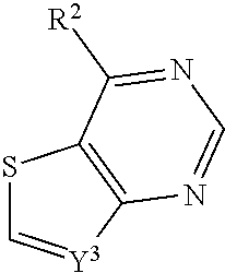
In some embodiments, a compound is provided having the formula (IX):
##STR00020## wherein Y.sup.3 is CH or N; wherein R.sup.2 is optionally substituted (2-aminoethyl)aryl, or a salt, enantiomer, racemate, mixture thereof, or combination thereof.
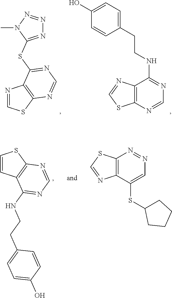
In some embodiments, the compound is selected from the group consisting of:
##STR00021## or a salt, enantiomer, racemate, mixture thereof, or combination thereof.
In some embodiments, a compound is provided having the formula (X):
##STR00022## wherein Y.sup.3 is CH; wherein R.sup.2 is selected from the group consisting of optionally substituted thioheteroaryl, optionally substituted (2-aminoethyl)aryl, halide, optionally substituted thiocycloalkyl wherein 1-3 carbon atoms of the cycloalkyl is optionally replaced with a heteroatom selected from the group consisting of O, S and N, and thioaryl, or a salt, enantiomer, racemate, mixture thereof, or combination thereof.
In some embodiments, the compound is selected from the group consisting of:
##STR00023## or a salt, enantiomer, racemate, mixture thereof, or combination thereof.
In some embodiments, a compound is provided having the formula (XI):
##STR00024## wherein R.sup.2 is selected from the group consisting of optionally substituted thioheteroaryl, optionally substituted (2-aminoethyl)aryl, halide, optionally substituted thiocycloalkyl wherein 1-3 carbon atoms of the cycloalkyl is optionally replaced with a heteroatom selected from the group consisting of O, S and N, and thioaryl, or a salt, enantiomer, racemate, mixture thereof, or combination thereof.
In some embodiments, the compound is selected from the group consisting of:
##STR00025## or a salt, enantiomer, racemate, mixture thereof, or combination thereof.
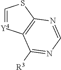
In some embodiments, a compound is provided having the formula (XII):
##STR00026## wherein Y.sup.4 is CH or N; wherein R.sup.3 is selected from the group consisting of optionally substituted thioheteroaryl, optionally substituted (2-aminoethyl)aryl, halide, optionally substituted thiocycloalkyl wherein 1-3 carbon atoms of the cycloalkyl is optionally replaced with a heteroatom selected from the group consisting of O, S and N, and thioaryl, or a salt, enantiomer, racemate, mixture thereof, or combination thereof.
In some embodiments, the compound is selected from the group consisting of:
##STR00027## or a salt, enantiomer, racemate, mixture thereof, or combination thereof.
In some embodiments, a compound is provided having the formula (XIII):
##STR00028## wherein R.sup.2 is selected from the group consisting of optionally substituted thioheteroaryl, optionally substituted (2-aminoethyl)aryl, halide, optionally substituted thiocycloalkyl wherein 1-3 carbon atoms of the cycloalkyl is optionally replaced with a heteroatom selected from the group consisting of O, S and N, and thioaryl, or a salt, enantiomer, racemate, mixture thereof, or combination thereof.
In some embodiments, the compound is selected from the group consisting of:
##STR00029## or a salt, enantiomer, racemate, mixture thereof, or combination thereof.
In some embodiments, a compound is provided having the formula (XIV):
##STR00030## wherein R.sup.2 is selected from the group consisting of optionally substituted thioheteroaryl, optionally substituted (2-aminoethyl)aryl, halide, optionally substituted thiocycloalkyl wherein 1-3 carbon atoms of the cycloalkyl is optionally replaced with a heteroatom selected from the group consisting of O, S and N, and thioaryl, or a salt, enantiomer, racemate, mixture thereof, or combination thereof.
In some embodiments, the compound is selected from the group consisting of:
##STR00031## or a salt, enantiomer, racemate, mixture thereof, or combination thereof.
In some embodiments, a compound is provided having the formula (XV):
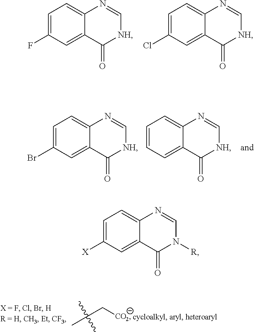
##STR00032## wherein X is H or halide; wherein Z.sup.1 is O; wherein R.sup.4 is selected from the group consisting of H, optionally substituted alkyl, Et, CF.sub.3, optionally substituted cycloalkyl, optionally substituted aryl, optionally substituted heteroaryl, and
##STR00033##
In some embodiments, the compound is selected from the group consisting of:
##STR00034## or a salt, enantiomer, racemate, mixture thereof, or combination thereof.
In one embodiment the compound is
##STR00035## or a salt, enantiomer, racemate, mixture thereof, or combination thereof.
In some embodiments a pharmaceutical composition is provided comprising a compound disclosed herein or a pharmaceutically acceptable salt thereof.
In some embodiments a method of treating a neurodegenerative disease comprising administering to a subject in need thereof an effective amount of a compound or pharmaceutical composition disclosed herein is provided. In some embodiments the neurodegenerative disease is a proteinopathy. Proteinopathies include, but are not limited to, Parkinson's disease, Alzheimer's disease, Amyotrophic Lateral Sclerosis (ALS), Huntington's disease, chronic traumatic encephalopathy (CTE), frontotemporal dementia (FTD), inclusion body myopathy (IBM), Paget's disease of bone (PDB), cerebral .beta.-amyloid angiopathy, prion diseases, familial dementia, CADASIL, amyloidosis, Alexander disease, seipinopathies, type II diabetes, pulmonary alveolar proteinosis, cataracts, cystic fibrosis and sickle cell disease. In some aspects of this embodiment, the proteinopathy is a tauopathy. Tauopothies include but are not limited to, Alzheimer's disease, Parkinson's disease, Huntington's disease, progressive supranuclear palsy, chronic traumatic encephalopathy (CTE), frontotemporal dementia (FTD), Lytico-Bodig disease, subacute sclerosing panencephalitis, ganglioglioma, gangliocytoma, and argyrophilic grain disease. In a preferred embodiment, the neurodegenerative disease is Alzheimer's disease.
In some embodiments a method of enhancing autophagic flux is provided. This method comprises providing to a cell or a protein aggregate an effective amount of a compound or pharmaceutical composition disclosed herein.
The embodiments described in this disclosure can be combined in various ways. Any aspect or feature that is described for one embodiment can be incorporated into any other embodiment mentioned in this disclosure. While various novel features of the inventive principles have been shown, described and pointed out as applied to particular embodiments thereof, it should be understood that various omissions and substitutions and changes may be made by those skilled in the art without departing from the spirit of this disclosure. Those skilled in the art will appreciate that the inventive principles can be practiced in other than the described embodiments, which are presented for purposes of illustration and not limitation.
EXAMPLES
The following examples are provided to further illustrate certain aspects of the present invention. These examples are illustrative only and are not intended to limit the scope of the invention in any way.
Example 1
Example Synthetic Schemes
Scheme 1 shows the synthesis of compounds of the formula:
##STR00036## e.g., compounds of formula (II) and formula (III).
##STR00037## Representative General Procedure
A 4-chloroquinazoline and thiol were stirred in anhydrous THF at room temperature. A base, such as triethylamine, was added. The reaction mixture was heated to 80.degree. C. and was stirred overnight at said temperature, after which it was allowed to cool to room temperature. It was then diluted with distilled water, and the organic material was extracted with ethyl acetate (3.times.). The combined organic extracts were washed with brine (lx) and dried with anhydrous sodium sulfate. The solvent was evaporated in vacuo, and the crude material was purified either via column chromatography or prep TLC, employing either 10% MeOH in methylene chloride or 10:1 pentane:diethyl ether as the eluent.
The above procedure is representative. Other examples disclosed herein could be made by similar techniques or other methods known in the art.
Scheme 2 shows preparation of 1-chloro-7-fluoroisoquinoline.
##STR00038##
Scheme 3 shows the synthesis of compounds of the formula:
##STR00039## e.g., compounds of formula (IV), formula (V), formula (VI), formula (VII), and formula (VIII).
##STR00040##
Scheme 4 shows the synthesis of compounds of the formula:
##STR00041## e.g., compounds of formula (XII), and formula (XIII).
##STR00042##
Scheme 5 shows the synthesis of compounds of the formula:
##STR00043## e.g., compounds of formula (IX), formula (X), and formula (XI).
##STR00044##
Scheme 6 shows the synthesis of compounds of the formula:
##STR00045## e.g., compounds of formula (XIV).
##STR00046##
Example 2
Activators of Autophagic Flux and Phospholipase D
The WHYKD series of compounds were synthesized for optimal brain penetrance based on the molecular weight (MW) and partition coefficient (log P), according to Lipinski's Rule for CNS penetrance: MW.ltoreq.400, log P.ltoreq.5.
Activators according to the formula:
##STR00047## were synthesized according to the schemes above. Molecular weights and log P were calculated. Results are shown in Table 1 below.
TABLE-US-00001 TABLE 1 PROJECT STRUCTURE ID M.W. log P X Y.sup.1 Y.sup.2 R.sup.1 ##STR00048## WHYKD3 323.17 3.85 Br N N thioheteroaryl ##STR00049## WHYKD4 369.44 5.69 aryl N N Thioheteroaryl ##STR00050## WHYKD5 262.27 3.18 F N N Thioheteroryl ##STR00051## WHYKD6 244.28 3.02 H N N thioheteroaryl ##STR00052## WHYKD7 278.72 3.58 Cl N N thioheteroaryl ##STR00053## WHYKD8 299.76 3.91 Cl N N (2- aminoethyl)aryl ##STR00054## WHYKD9 182.58 2.58 F N N Cl ##STR00055## WHYKD10 243.29 2.9 H N CH thioheteroaryl ##STR00056## WHYKD11 261.28 3.06 F N CH thioheteroaryl ##STR00057## WHYKD12 262.35 4.38 F N N thiocycloalkyl ##STR00058## WHYKD13 316.44 5.21 F N N thiocycloalkyl ##STR00059## WHYKD14 314.42 4.66 F N N thiocycloalkyl ##STR00060## WHYKD15 248.32 3.96 F N N thiocycloalkyl ##STR00061## WHYKD16 274.36 4.19 F N N thiocycloalkyl ##STR00062## WHYKD17 357.49 4.09 F N N thiocycloalkyl ##STR00063## WHYKD18 386.48 4.41 F N N thiocycloalkyl ##STR00064## WHYKD19 264.32 2.63 F N N thiocycloalkyl ##STR00065## WHYKD20 296.36 4.8 F N N thioaryl ##STR00066## WHYKD30 356.23 5.16 I N N thiocycloalkyl
Activators according to the formula:
##STR00067## were synthesized according to the schemes above. Molecular weights and log P were calculated. Results are shown in Table 2 below.
TABLE-US-00002 TABLE 2 PROJECT STRUCTURE ID M.W. log P Y.sup.3 R.sup.2 ##STR00068## WHYKD21 272.33 3.36 N (2-aminoethyl)aryl ##STR00069## WHYKD23 271.34 3.66 CH (2-aminoethyl)aryl
Activators according to the formula:
##STR00070## were synthesized according to the schemes above. Molecular weights and log P were calculated. Results are shown in Table 3 below.
TABLE-US-00003 TABLE 3 PROJECT STRUCTURE ID M.W. log P Y.sup.4 R.sup.3 ##STR00071## WHYKD1 251.29 2.56 N thioheteroaryl ##STR00072## WHYKD2 272.33 2.89 N (2-aminoethyl)aryl ##STR00073## WHYKD22 271.34 3.34 CH (2-aminoethyl)aryl
Activators according to the formula:
##STR00074## were synthesized according to the schemes above. Molecular weights and log P were calculated. Results are shown in Table 4 below.
TABLE-US-00004 TABLE 4 PROJECT log STRUCTURE ID M.W. P X Y.sup.1 Y.sup.2 R.sup.4 Z.sup.1 ##STR00075## WHYKD24 164.14 1.02 F N N H O
Example 3
Design of Derivatives
Several series of derivatives were synthesized based on the following lead compounds:
##STR00076##
In addition to log P, the topological polar surface area (tPSA), C Log P (log P calculated by group contribution method), and Log S (solubility) were calculated. Results are shown in the Tables below.
TABLE-US-00005 TABLE 5 Modifications to the core and side chain (Series 1) STRUCTURE log P tPSA CLogP LogS ##STR00077## 3.35 52.68 2.65154 -3.235 ##STR00078## 3.12 61.47 2.34241 -3.295 ##STR00079## 2.94 40.32 1.83259 -4.663 ##STR00080## 3.19 27.96 3.25375 -3.864 ##STR00081## 4.14 12.36 4.64041 -4.354 ##STR00082## 2.71 49.11 2.01759 -4.354 ##STR00083## 2.95 36.75 3.23654 ~3.554 ##STR00084## 2.8 21.59 2.80041 -3.813 ##STR00085## 4.56 12.36 5.19941 -4.832
TABLE-US-00006 TABLE 6 Modifications to the core and side chain (Series 2) STRUCTURE log P tPSA CLogP LogS ##STR00086## 2.31 77.4 0.803829 -1.704 ##STR00087## 2.07 86.19 0.539011 -1.765 ##STR00088## 1.9 65.04 -0.0366305 ~3.133 ##STR00089## 1.66 73.83 0.148224 ~2.824 ##STR00090## 2.14 52.68 1.40054 -2.334 ##STR00091## 1.91 61.47 1.38428 -2.024 ##STR00092## 3.09 37.08 2.83701 -2.823 ##STR00093## 3.51 37.08 3.39601 ~3.301 ##STR00094## 1.76 46.31 0.997011 -2.283
TABLE-US-00007 TABLE 7 Modifications to the core and side chain (Series 3) STRUCTURE log P tPSA CLogP LogS ##STR00095## 2.89 77.4 0.647513 -1.626 ##STR00096## 2.65 86.19 0.382662 -1.686 ##STR00097## 2.48 65.04 ~0.192932 ~3.117 ##STR00098## 2.25 73.83 ~0.00808129 ~2.806 ##STR00099## 2.73 52.68 1.24423 ~2.303 ##STR00100## 2.49 61.47 1.22796 -1.992 ##STR00101## 3.68 37.08 2.68066 ~2.893 ##STR00102## 4.09 37.08 3.23966 -3.372 ##STR00103## 2.34 46.31 0.840662 ~2.256
TABLE-US-00008 TABLE 8 Modifications to the core and side chain (Series 4) STRUCTURE log P tPSA CLogP LogS ##STR00104## 1.68 77.4 0.647513 ~1.441 ##STR00105## 1.45 86.19 0.382662 -1.501 ##STR00106## 1.28 65.04 ~0.192932 ~2.932 ##STR00107## 1.04 73.83 ~0.00808129 ~2.621 ##STR00108## 1.52 52.68 1.24423 ~2.119 ##STR00109## 1.28 61.47 1.22796 ~1.808 ##STR00110## 2.47 37.08 2.68066 ~2.704 ##STR00111## 2.89 37.08 3.23966 ~3.183 ##STR00112## 1.13 46.31 0.840662 ~2.071
TABLE-US-00009 TABLE 9 Modifications to the core and side chain (Series 5) STRUCTURE log P tPSA CLogP LogS ##STR00113## 1.68 77.4 0.647513 ~1.466 ##STR00114## 1.45 86.19 0.382662 ~1.526 ##STR00115## 1.28 65.04 ~0.192932 ~2.957 ##STR00116## 1.04 73.83 ~0.00808129 ~2.646 ##STR00117## 1.52 52.68 1.24423 -2.144 ##STR00118## 1.28 61.47 1.22796 ~1.832 ##STR00119## 2.47 37.08 2.68066 ~2.733 ##STR00120## 2.89 37.08 3.23966 ~3.212 ##STR00121## 1.13 46.31 0.840662 -2.096
TABLE-US-00010 TABLE 10 Modifications to the core and side chain (Series 6) STRUCTURE log P tPSA CLogP LogS ##STR00122## 2.11 77.4 0.857513 ~1.525 ##STR00123## 1.87 86.19 0.592663 -1.585 ##STR00124## 1.7 65.04 0.0170677 ~3.017 ##STR00125## 1.46 73.83 0.201919 -2.705 ##STR00126## 1.94 52.68 1.45423 -2.203 ##STR00127## 1.71 61.47 1.43796 -1.892 ##STR00128## 2.89 37.08 2.89066 ~2.787 ##STR00129## 3.31 37.08 3.44966 -3.266 ##STR00130## 1.55 46.31 1.05066 -2.155
TABLE-US-00011 TABLE 11 Modifications to the core and side chain (Series 7) STRUCTURE log P tPSA CLogP LogS ##STR00131## 1.63 74.27 1.1096 -1.275 ##STR00132## 1.4 83.06 0.834 ~1.333 ##STR00133## 1.23 61.91 0.272969 ~2.704 ##STR00134## 0.99 70.7 0.457768 ~2.391 ##STR00135## 1.47 49.55 1.70682 -1.904 ##STR00136## 1.24 58.34 1.69005 ~1.592 ##STR00137## 2.42 33.95 3.132 ~2.403 ##STR00138## 2.84 33.95 3.691 ~2.883 ##STR00139## 1.08 43.18 1.292 -1.864
TABLE-US-00012 TABLE 12 Modifications to the core and side chain (Series 8) STRUCTURE log P tPSA CLogP LogS ##STR00140## 1.96 74.27 0.8996 ~1.745 ##STR00141## 1.72 83.06 0.624 ~1.803 ##STR00142## 1.55 61.91 0.0629689 -3.174 ##STR00143## 1.31 70.7 0.247768 -2.862 ##STR00144## 1.79 49.55 1.49682 -2.374 ##STR00145## 1.56 58.34 1.48005 ~2.062 ##STR00146## 2.74 33.95 2.922 ~2.874 ##STR00147## 3.16 33.95 3.481 ~3.353 ##STR00148## 1.4 43.18 1.082 ~2.335
TABLE-US-00013 TABLE 13 Modifications to the core and side chain (Series 9) STRUCTURE log P tPSA CLogP LogS ##STR00149## 3.0 65.04 1.74907 -2.051 ##STR00150## 2.76 73.83 1.47586 ~2.109 ##STR00151## 2.59 52.68 0.911314 ~3.542 ##STR00152## 2.36 61.47 1.09641 ~3.23 ##STR00153## 2.84 40.32 2.34546 -2.728 ##STR00154## 2.6 49.11 2.32952 ~2.416 ##STR00155## 3.79 24.72 3.77386 ~3.323 ##STR00156## 4.2 24.72 4.33286 -3.802 ##STR00157## 2.45 33.95 1.93386 ~2.687
TABLE-US-00014 TABLE 14 Modifications to the core and side chain (Series 10) STRUCTURE log P tPSA CLogP LogS ##STR00158## 2.94 65.04 1.53907 -2.188 ##STR00159## 2.71 73.83 1.26586 ~2.247 ##STR00160## 2.54 52.68 0.701314 -3.68 ##STR00161## 2.3 61.47 0.886405 -3.367 ##STR00162## 2.78 40.32 2.13546 -2.866 ##STR00163## 2.55 49.11 2.11952 -2.554 ##STR00164## 3.73 24.72 3.56386 ~3.468 ##STR00165## 4.15 24.72 4.12286 ~3.947 ##STR00166## 2.39 33.95 1.72386 ~2.824
TABLE-US-00015 TABLE 15 Quinazolinones (Series 11) STRUCTURE log P tPSA CLogP LogS ##STR00167## 1.02 41.46 0.506065 ~1.702 ##STR00168## 1.42 41.46 1.07606 -2.152 ##STR00169## 1.69 41.46 1.22606 ~2.273 ##STR00170## 0.86 41.46 0.305 ~1.452 ##STR00171## ##STR00172##
Example 4
Biological Testing of WHYKD Compounds
The assays listed below were carried out using a transfected HEK 293 (Human Embryo Kidney) cell line that has been engineered to express fluorescently tagged (mKate2) Tau (unless otherwise noted). The cells were grown in the presence of the antibiotic doxycycline. When the antibiotic is removed the cells produce Tau (which can be quantified), thus allowing the test compounds' effects on Tau's production to be compared. Doxycycline was removed for 72 hours prior to exposure to the test compounds to promote sufficient Tau production. Cells were subsequently plated into plates for each of the assays described below.
Autophagy, Aggregate and Tau-mKate2 IC50 Assays
Preparation of Plated Tet-Regulated HEK 293 Cells (Jump-In.TM. Cells)
1) HEK cells with tet-regulated expression of mKate2 tagged Tau were grown in DMEM medium (Dulbecco's Modified Eagle's Medium) containing 0.5 .mu.g/mL doxycycline (Dox) (MP-Bio #198955), after 3-5 days Dox was removed by washing the cells twice with sterile phosphate buffered saline (PBS; Invitrogen #14190-144), cells were left in DMEM without Dox for 72 hours (to further clear remaining Dox in cells).
2) 96 well, black plates with ultra-thin clear bottom (Costar #3720) were coated with Poly D Lysine (PDL) (Sigma-p0899-M wt. 70,000-150,000) or commercially sourced transparent polystyrene/glass bottom plates were used and coated. PDL was aspirated after 2 hours and plates were allowed to dry out for another 2-3 hours. The coating procedure was completed under sterile conditions. Plates were used immediately after coating.
3) Cells were detached from the flasks using triple express (Invitrogen #12605-010) and plated in PDL-coated 96 well plates. For plating, 200 .mu.L of medium was added to each well at a concentration of 400 k cells/ml (80 k cells/well). 1 row of peripheral wells on all sides was spared to prevent any changes in experimental conditions due to evaporation from these wells. Warm medium/PBS (containing no cells) was pipetted into the peripheral wells and PBS was pipetted into spaces between wells to maintain homogenous conditions across the central wells. Cells were then allowed to settle down and attach to the bottom of the plate for 18-24 hours.
Treatment and Staining with Cyto-ID.TM.
Cyto-ID.TM. assay measures autophagic vacuoles and monitors autophagic flux in lysosomally inhibited live cells using a novel dye that selectively labels accumulated autophagic vacuoles. The dye used in the kit prevents its accumulation within lysosomes, but enables labelling of vacuoles associated with the autophagy pathway using the LC3 biomarker.
The test compounds (along with CytoID.TM. dye and Hoechst stain) were added to the cells and incubated for 3 hours. Reference compounds were also used in each plate, Rapamycin was used for autophagy induction and chloroquine was used for lysosomal inhibition. After 3 hours test compound was aspirated off and kept. Cells were then washed and read using a fluorescence plate reader. Hoescht, Cyto-ID.TM. and mKate2 were measured using 3 distinct wavelengths on the plate reader. The Hoescht measurement allowed normalisation of results across wells. After reading, test compound was re-added and the cells are left for a further 21 hours. At the 24 hour time point cells were once again washed and plates read using the fluorescence plate reader.
1) Before initiating treatment, all wells were washed once with warm FluoroBrite.TM. (FB) DMEM (Dulbecco's Modified Eagle's Medium) (Invitrogen # A18967-01) with 10% Fetal Bovine Serum, dialyzed (FBS; Invitrogen #26400-044) and 1% MEM (Minimal Essential Media) NEAA (Non Essential Amino Acids). 280 .mu.l of warm medium/PBS was maintained in peripheral wells.
2) Test samples were prepared in warm FluoroBrite.TM. DMEM and with 10% FBS and 1% NEAA (FB-DMEM). Rapamycin 200 nM (Enzo # BML-A275-0025) was used for autophagy induction and 15 mM chloroquine was used for lysosomal inhibition (bafilomycin or similar compounds were not used as they give a false negative result in the assay). Dimethyl Sulfoxide 1:1000 (DMSO; Fisher #BP231-100) was used as control. Autophagosome marker Cyto ID.TM. (1:500; Enzo #ENZ-51031-k200) and Hoechst (1:200) was added to these test sample preparations before treating the cells.
3) The treatment groups were staggered in order to obtain similar conditions across all groups. For an n=6, 3 control wells were near the periphery and 3 were near the center of the plate, same was true for Rapamycin and any other drug treatments.
4) Cells were treated/stained for 3 hours at 37.degree. C. At 3 hours post-treatment, test sample preparation containing Cyto-ID.TM. was carefully aspirated and transferred to a fresh sterile microplate (Falcon #353072) for reuse. Medium in the treatment plate was immediately replaced with warm FB-DMEM. The new plate with test compound preparations was placed in an incubator at 37.degree. C.
5) Treatment plate was then washed quickly 3 times with warm FB-DMEM (100 .mu.L/well). After the 3rd wash 50 .mu.L of warm medium was left in each well. At this point, the plate was ready for reading. See details in Reading Plate section. Hoescht was read at Excitation wavelength (Ex)=355 nm & Emission wavelength (Em)=446 nm. mKate2 was read at Ex=575 nm & Em=630 nm. Cyto-ID.TM. was read at Ex=463 nm & Em=534 nm.
6) Afterwards, medium was replaced with corresponding wells of the plate containing test compound/stain preparation and re-incubated at 37.degree. C.
7) At 24 hours post reaction all medium was aspirated and the plate was washed 3 times with warm FB-DMEM, and read again for Hoescht (Ex=355 nm Em=446 nm), CytoID.TM. (Ex=463 nm; Em=534 nm) and mKate2 (Ex=575 nm; Em=630 nm) in 80 .mu.l of FB-DMEM.
Proteostat.TM. Protein Aggregation Assay
Proteostat.TM. assay was used to detect aggresomes via measurement of p62. Aggresomes are inclusion bodies that form when the ubiquitin-proteasome machinery is overwhelmed with aggregation-prone proteins. Typically, an aggresome forms in response to some cellular stress, such as hyperthermia, viral infection, or exposure to reactive oxygen species. Aggresomes may provide a cytoprotective function by sequestering the toxic, aggregated proteins and may also facilitate their ultimate elimination from cells by autophagy. Following the final plate read described in the Cyto-ID.TM. assay described above, Proteostat.TM. detection reagent was added to every well and the plate was incubated for 15 minutes. Following this incubation, the plate was read by fluorescence plate reader at the specified wavelength. After this plate read, the cells were fixed by incubating with warm paraformaldehyde for 8 minutes. The fixed cells were then read by plate reader as before.
8) Detection solution was prepared by adding 10 .mu.l Proteostat.TM. detection reagent (ENZ-51023-KP002) and 200 .mu.L of 10.times. assay buffer into 1790 .mu.L water and mixing well.
9) 20 .mu.L was added per well (each well had 80 .mu.l of Fluorobrite.TM. media with no FBS) and incubated in the dark for 15 min at room temperature.
10) Fluorescence for Proteostat.TM. (Ex=550 nm; Em=600 nm) was then read.
11) Plate was fixed by adding warm 4% paraformaldehyde 100 .mu.L (PFA; EMS #15710) to all the wells and was incubated at room temperature for 8 min. PFA was removed and plate was washed with PBS (room temp) 3 times.
12) Plate was read again with fixed cells and same configuration at plate reader.
Note: CytoID.TM. and Proteostat.TM. are measures for LC3 and p62, which can be substituted using fluorescent tagged antibodies with corresponding fluorophores.
Reading Plate Using Tecan M200
13) Plate was transferred to plate reader (Tecan Infinite M200) and read at optimal gain for mKate2, Cyto ID.TM. and Proteostat.TM. (last read only).
14) Each well was read at 5 consistent locations per well for the three signals. Each of these 5 locations was flashed 25 times. Signal from each well was first recorded as an average of 25 flashes, and the final value was based on average of 5 read locations per well. A peripheral border of 1000 .mu.m was spared in all wells to mitigate any inconsistencies in reads due to minor cell loss across the periphery resulting from aspiration and washings. Peripheral wells were used to detect any background noise due to medium or PBS.
15) mKate2 was read at Ex=575 nm and Em=630 nm. Cyto-ID.TM. was read at Ex=463 nm and Em=534 nm. For the final read, Proteostat.TM. was read at Ex=490 nm and Em=600 nm. Excitation & Emission Bandwidth for all three reads were 9 nm & 20 nm respectively.
16) Calculations were initially subtracted from well background (media only well) and normalized using Hoescht levels.
Reading Plate Using IN Cell Analyzer 2000 (High Content Imaging).
17) Plate was transferred to IN Cell analyzer 2000 and imaged for mKate2, Cyto ID.TM., Proteostat.TM. and Hoechst (last read only).
18) Each well was imaged at 4 consistent fields located around the center of the well. All images were taken using 20.times. objective. The average reading from these 4 fields was recorded as the reading for that corresponding well. No images were taken from periphery of the wells to mitigate any inconsistencies in reads due to minor cell loss across the periphery resulting from aspiration and washings. Peripheral wells were used to detect any background noise due to medium or PBS.
19) FITC/FITC filter (Ex=490 nm-bandwidth 20 nm/Em=525 nm-bandwidth 36 nm) was used to image Cyto ID.TM.. mKate2 was imaged using TexasRed/TexasRed filters (Ex=579 nm-bandwidth 34 nm/Em=624 nm-bandwidth 40 nm). For the Proteostat.TM. images FITC/dsRed combination was used (Ex=490 nm-bandwidth 20 nm/Em=605 nm-bandwidth 52 nm). Nuclei were imaged using DAPI/DAPI filter set (Ex=350 nm-bandwidth 50 nm/Em=455 nm-bandwidth 50 nm).
p62 Aggregate and Tau Aggregate Western Blot Assay
Western blot assays were performed to determine protein changes in Tau and p62. Cells were cultured as above and incubated with test compounds for 24 h. Following test compound incubation, the test compound was aspirated off and cells were washed before harvesting. Cells were spun at a low speed and supernatant was aspirated to leave the cell pellet. The cell pellet was then homogenized in buffer, centrifuged at high speed, and the supernatant aspirated and further separated into total fraction and aggregate fraction allowing quantification of soluble and insoluble proteins. Western blots were run on the samples, gels transferred to nitrocellulose and incubated with antibodies for Tau and p62. After incubation with secondary antibodies for detection, the bands of protein were quantified by chemiluminescence.
1) Jump-In.TM. cells (see above) were maintained in 0.5 .mu.g/mL doxycycline until use.
2) Three days prior to plating, cells were replated at 40% confluency in media without doxycycline.
3) The day prior to experimentation, 750 000 cells were plated per well in a 6-well plate (250 000 cells in 12 well plate).
4) On the day of experiment, cells were washed in warmed HBSS (Hank's Balanced Salt Solution) twice before media containing test compound (n=3 per compound per dose) or vehicle were added to the wells (1.5 mL in 6 well, 600 .mu.L in 12 well). Plates were incubated for 24 hours at 37.degree. C. with 5% CO.sub.2.
5) Cells were rinsed twice in warmed HBSS before harvesting in 1 mL HBSS and transferred to 1.5 mL microtubes.
6) Samples were spun at 500.times.g for 2 min at 4.degree. C., and HBSS supernatant aspirated, leaving only the cell pellet. The cell pellet may be flash frozen and stored at -80.degree. C. until use.
Sample Preparation
7) The cell pellet was homogenized in RIPA+(Radio-Immunoprecipitation Assay) buffer containing protease inhibitors and phosphatase inhibitors and gently homogenized using a cell homogenizer.
8) Samples were then centrifuged for 20 min at 20,000 g at 4.degree. C.
9) The supernatant was transferred to a new tube.
10) The supernatant was quantified for protein concentration using the Pierce.TM. Protein Assay.
11) Total fraction: 200 .mu.g of supernatant was used to make a 1 mg/ml protein solution by adding RIPA+ buffer to bring the volume to 130 .mu.L and adding 20 .mu.L of 1M DTT (dithiothreitol) (to make a final concentration of 100 mM DTT) and 50 .mu.L of 4.times. Invitrogen NuPAGE LDS (lithium dodecyl sulfate) loading buffer (4.times., 10 ml-NP0007) with 100 mM DTT.
12) Aggregate fraction: For aggregates, 100 .mu.g of sample was brought up to a final volume of 900 .mu.L.
13) 100 .mu.L of 10% sarkosyl solution was added to the sample and rotated at 4.degree. C. for 60 min.
14) The sample was then centrifuged at 100,000 g for 60 min at 4.degree. C.
15) The supernatant was carefully removed, leaving the pellet undisturbed. The tube was inverted to remove any additional liquid. If there was excess liquid, steps 12-14 were repeated to ensure a pure aggregate sample. Pellets were resuspended and solubilized in 65 .mu.L of PBS before the addition of 10 .mu.L of 1M and 25 .mu.L of 4.times. Invitrogen NuPAGE LDS loading buffer with 100 mM DTT.
Electrophoresis/Western Blot
16) Prior to loading, the samples were heated at 90.degree. C. for 2 min. A quick spin of the samples was performed to ensure there was no condensation on the tube. The 4-12% Tris-Bis gel was prepared using 1.times.MOPS buffer (Invitrogen--NP0001) and anti-oxidant (NP0005). For the soluble fraction, 2 .mu.g of sample was loaded to each well; for the insoluble fraction, 5 .mu.g of sample was loaded per well. 8 .mu.L of Invitrogen Sharp MW standard was then loaded (optimally, all wells had the same volume of sample buffer.)
17) Sample was electrophoresed at 150V for approximately 1 h15 min (until the running dye reached the base of the gel.)
18) The gel was then equilibrated in transfer buffer (25 mM Tris-HCl pH 8.3, 192 mM glycine, 20% (v:v) methanol) for 5 min.
19) The gel was then removed and transferred onto 0.2 .mu.M nitrocellulose (GE BA83 10600001) at 200 mA for 90 minutes.
20) After transfer, the blot was briefly stained (15 s) with 0.1% Ponceau S in 5% acetic acid to ensure consistent sample loading between lanes.
21) The blot was then rinsed in TBS-T (Tris-buffered saline-polysorbate) for 2 min to remove the Ponceau S stain prior to immunoprobing.
Immunodetection
22) Samples were blocked in 5% milk in TBST for 30 min, and then rinsed in TBS-T until all buffer was clear (no milk residues remained in solution).
23) Blots were incubated in primary antibody (1:4000 PHF1 or CP27 for tau; 1:4000 p62 Abnova for p62; 1:5000 GAPDH (glyceraldehyde 3-phosphate dehydrogenase) for loading control) overnight in SuperBlock.TM. TBS-T at 4.degree. C. on a rocking table.
24) After primary antibody exposure, blots were washed three times in TBS-T for 15 min.
25) Blots were then incubated in 1:4000 secondary antibody (Jackson Laboratories goat anti-mouse HRP conjugate).
26) After secondary, blots were washed three times in TBST for 15 min.
27) Blots were then developed using Millipore chemiluminescent fluid (1 ml per reagent; WBKLS0500) and detected using a Fuji LAS3000 Imaging unit at increments of 10 seconds.
28) Images were quantified using resident software or NIH Image J.
PLD Assay
Cells were incubated with test compound for 24 hours. 25 minutes prior to harvest at 24 hours, 3% ethanol solution was added to the wells to catalyze cleavage of the phospholipid. Cells were then placed on ice, washed and harvested. The harvested cells were centrifuged and the supernatant was aspirated and the pellet kept. The pellet was resuspended and chloroform/methanol lipid extraction was performed. The sample was centrifuged and the organic layer was separated, dried under nitrogen and stored at -80.degree. C. On the day of analysis, samples were resuspended and analysed by LC/MS. All phosphatidylethanol species (PEtOH32-40:0-6:16/18:0/1) were combined together and represented as total lipid content (all lipid species).
The assay was run in e18 primary cortical neurons from PLD1 or PLD2 KO mice cultured for 14 days. Alternatively, the assay can be run in the cells described above in the presence of excess PLD1 or PLD2 inhibitor to preclude that particular action from contributing to the effect of the drug.
1) Jump-In.TM. cells (see above) were maintained in 0.5 .mu.g/mL doxycycline until use. For neurons, e18 fetuses were used, cortical neurons extracted and plated onto PLD-collagen coated 6 well plates and incubated for 14 days prior to use.
2) Three days prior to plating, cells were replated at 40% confluency in media without doxycycline.
3) The day prior to experimentation, 750 000 cells were plated per well in a 6-well plate (250 000 cells in 12 well plate).
4) On the day of experiment, cells were washed in warmed HBSS twice before media containing test compound (n=3 per compound per dose), inhibitor (355 nM ML298 or 50 nM VU0150669), or vehicle are added to the wells (1.5 mL in 6 well, 600 .mu.L in 12 well). Plates were incubated for 24 hours at 37.degree. C. with 5% CO.sub.2.
5) 25 minutes prior to harvest, a 165 .mu.L of a 3% ethanol solution was added to each well.
6) For harvesting, plates were placed on ice and cells were rinsed twice with ice cold HBSS before harvesting in 1 mL HBSS and transferring to 1.5 mL microtubes.
7) Samples were spun at 500.times.g for 2 min at 4.degree. C., and HBSS supernatant aspirated, leaving only the cell pellet. The cell pellet could be flash frozen and stored at -80.degree. C. until use.
TABLE-US-00016 TABLE 16 Biological Testing of WHKD Compounds 1-24 % inhibition % Increase Cytotoxixity Compound Tau/aggregate (1 .mu.M) Autophagy Markers PLD LC50 WHYKD1 No Effect @ 2 .mu.M No Effect @ 2 .mu.M NC >100 .mu.M WHYKD2 No Effect @ 2 .mu.M No Effect @ 2 .mu.M ND >100 .mu.M WHYKD3 No Effect @ 2 .mu.M No Effect @ 2 .mu.M NC >100 .mu.M WHYKD4 No Effect @ 2 .mu.M No Effect @ 2 .mu.M ND >100 .mu.M WHYKD5 35% 20% .sup. 1.7X >100 .mu.M WHYKD6 No Effect @ 2 .mu.M No Effect @ 2 .mu.M ND >100 .mu.M WHYKD7 No Effect @ 2 .mu.M No Effect @ 2 .mu.M ND >100 .mu.M WHYKD8 30% 25% .sup. 2.8X >100 .mu.M WHYKD9 No Effect @ 2 .mu.M No Effect @ 2 .mu.M ND >100 .mu.M WHYKD10 No Effect @ 2 .mu.M No Effect @ 2 .mu.M ND >100 .mu.M WHYKD11 No Effect @ 2 .mu.M No Effect @ 2 .mu.M ND >100 .mu.M WHYKD12 60% 40% 3.4 >100 .mu.M WHYKD13 No Effect @ 2 .mu.M No Effect @ 2 .mu.M ND >100 .mu.M WHYKD14 No Effect @ 2 .mu.M No Effect @ 2 .mu.M ND >100 .mu.M WHYKD15 65% 40% 3.6 >100 .mu.M WHYKD16 No Effect @ 2 .mu.M No Effect @ 2 .mu.M ND >100 .mu.M WHYKD17 No Effect @ 2 .mu.M No Effect @ 2 .mu.M ND >100 .mu.M WHYKD18 No Effect @ 2 .mu.M No Effect @ 2 .mu.M ND >100 .mu.M WHYKD19 55% 35% 3.5 >100 .mu.M WHYKD20 No Effect @ 2 .mu.M No Effect @ 2 .mu.M ND >100 .mu.M WHYKD21 No Effect @ 2 .mu.M No Effect @ 2 .mu.M ND >100 .mu.M WHYKD22 No Effect @ 2 .mu.M No Effect @ 2 .mu.M ND >100 .mu.M WHYKD23 No Effect @ 2 .mu.M No Effect @ 2 .mu.M ND >100 .mu.M WHYKD24 30% 15% ND >100 .mu.M ND--not done, NC--no change
TABLE-US-00017 TABLE 17 Bioiogical Testing of WHKD Compounds 30-36 LC50 EC50 (nM) - EC50 (nM) - TI - Compound (mM) Proteostat mKate2 mKate2 WHYKD30 >200 2510 1113 180 WHYKD32 >200 1992 1562 128 WHYKD33 >200 8805 637 314 WHYKD35 >200 1295 518 386 WHYKD36 67 383 1044 64 LC50 (50% lethal concentration) was tested using an XTT assay for cell viability, Measurements listed as 200 represent the upper concentration limit used for testing, whereby the viability is >50% at this upper limit. EC50 (50% reduction) was based on the concentration whereby the levels were 50% lower than the initiail readings based on mKate2 fluorescence (tau tag) or Proteostat levels (fluorescent marker of aggregates). TI (therapeutic index) was based on the ratio of LC50:EC50, with the upper limit being 200 mM
TABLE-US-00018 TABLE 18 PLD1 & PLD2 Activity Proteostat (nM) using mKate2 (nM) using pharmacological inhibitor pharmacological inhibitor PLD2/PLD1 PLD2/PLD1 Compound PLD1 PLD2 ratio PLD1 PLD2 ratio WHYKD36 394 604 1.5 1332 2284 1.7 To compare the two isoforms of phospholipase D, samples were treated with either ML298, a PLD2 inhibitor (355 nM) that would yield PLD1 activity or a PLD1 inhibitor (VU0150669, 50 nM) to yield only PLD2 activity.
Example 5
Detection and Results of WHYKD Compounds
A photodiode array (PDA) was used to detect WHYKD8 in mouse brain (FIG. 1). The sample was readily detected with a discrete peak based on time (left) and with a measurable area under the curve (AUC) (inset).
LC3-II levels were measured in primary cortical neurons following 6 hours of treatment with WHYKD1, WHYKD5, or WHYKD1+BafA1 (FIG. 2). The presence of LC3-II is an indication of autophagy.
LC3-II levels were then measured in organotypic slice cultures following 6 hours of treatment with WHYKD1 (FIG. 3, top panel). Other compounds in the WHYKD series produced similar results (FIG. 3, bottom panel). RFP is a tag on the tau protein and also can be probed.
These experiments show that the WHYKD series of compounds can induce autophagy and reduce the aggregated forms of tau as well as its aggresome surrogate p62.
PLD activation converts phospholipids to phosphatidylethanols in the presence of ethanol. This conversion was measured to show that the WHYKD series of compounds activate PLD at 10 .mu.M concentration (FIG. 4) and at 1 .mu.M (FIG. 5). FIPI is a non-competitive inhibitor of PLD activity and was used as a negative control.
All patents, patent applications, and publications cited above are incorporated herein by reference in their entirety as if recited in full herein.
The invention being thus described, it will be obvious that the same may be varied in many ways. Such variations are not to be regarded as a departure from the spirit and scope of the invention and all such modifications are intended to be included within the scope of the following claims.
* * * * *
References
-
chemspider.com/Chemical-Structure_30132867.html?rid=9a2b8938-1827-42ad
-
pubchem.ncbl.nlm.nih.gov/compound/236319721972
-
pubchem.nal.nlm.nlh.gov/compound/616883
-
pubchem.ncbi.nlm.nih.gov/compound/21002400
-
-
-
-
pubchem.ncbi
-
-
-
guidechem.com/reference/dic-168980
-
coreychem.com/PRODUCTS/Phamaceutical_intermediates/2015/05231/195760.html
-
pubchem.ncbi.nlm.nth.gov/compound/7160394#section-Top
-
C00001
C00002
C00003
C00004
C00005
C00006
C00007
C00008
C00009
C00010
C00011
C00012
C00013
C00014

C00015

C00016

C00017
C00018

C00019

C00020
C00021

C00022

C00023

C00024

C00025

C00026
C00027
C00028
C00029

C00030
C00031
C00032
C00033

C00034
C00035

C00036
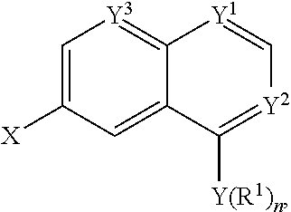
C00037

C00038
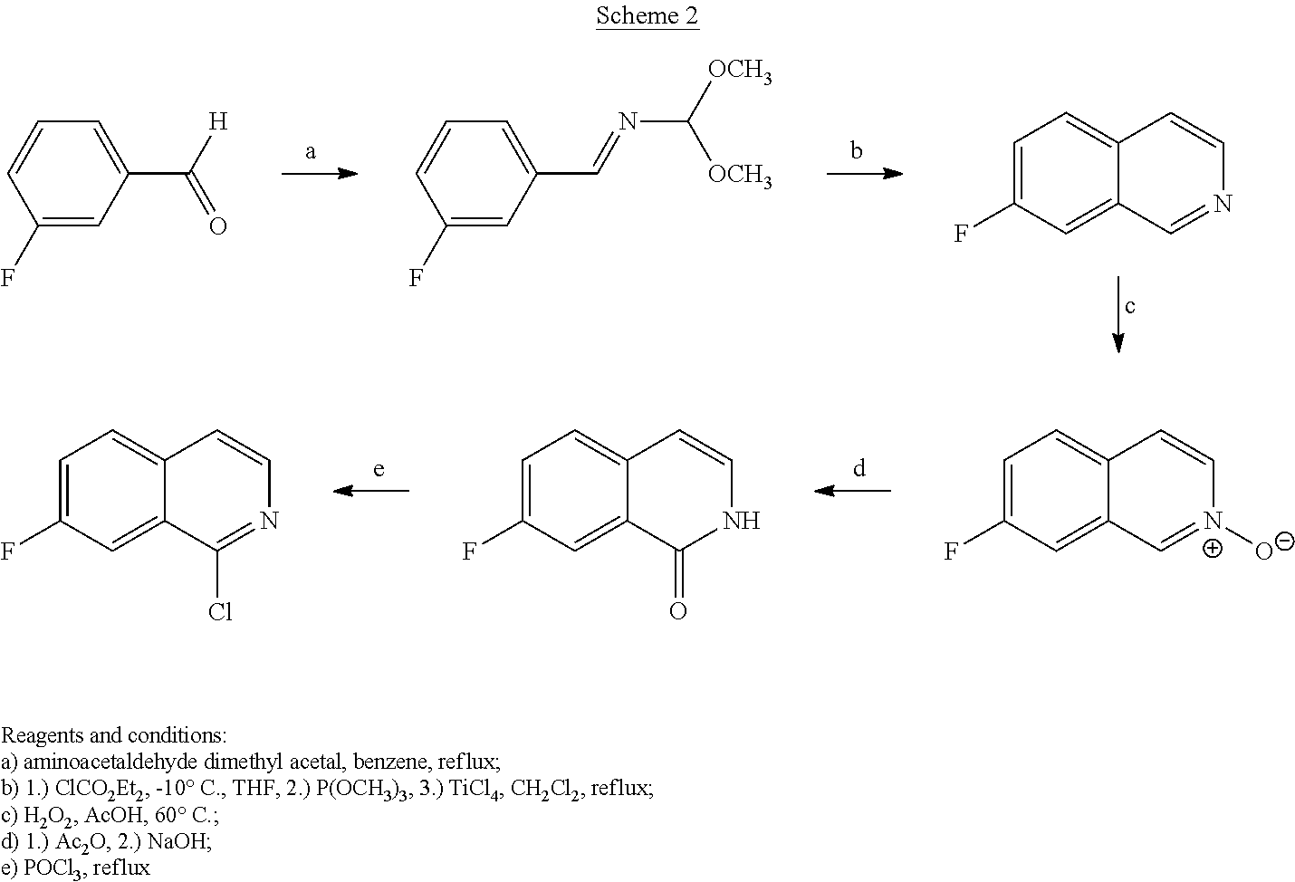
C00039

C00040
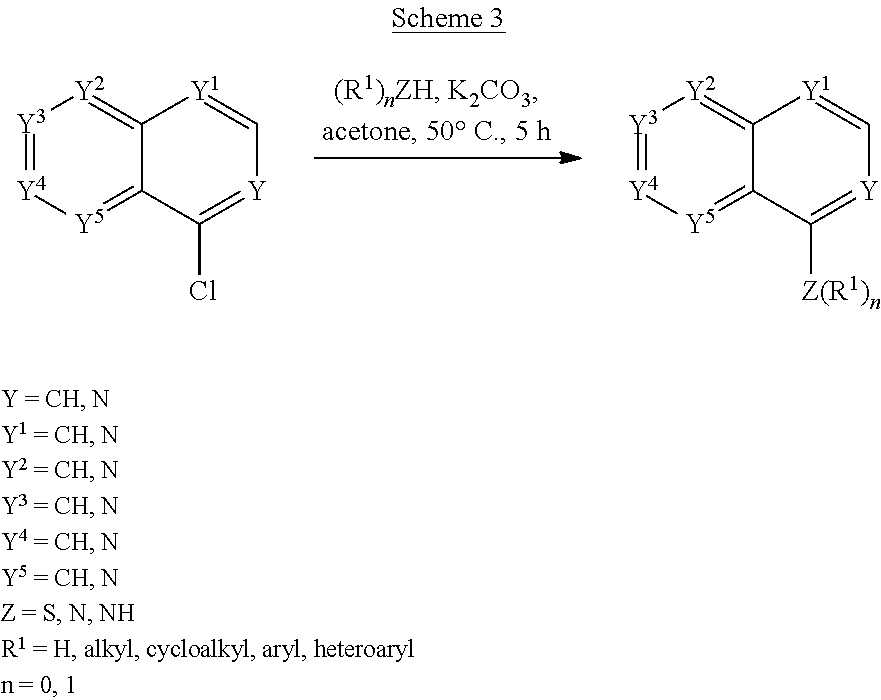
C00041

C00042

C00043

C00044

C00045

C00046
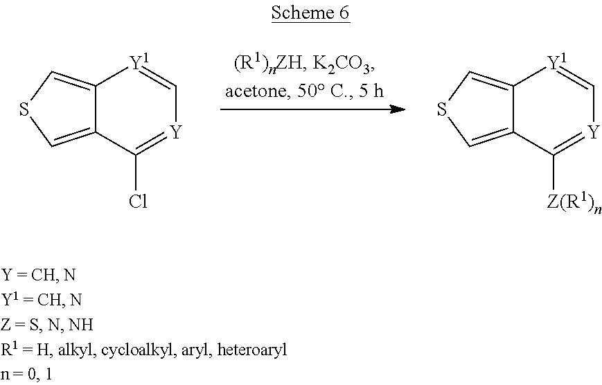
C00047

C00048

C00049

C00050

C00051
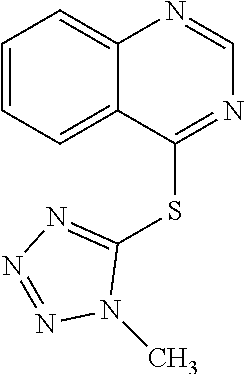
C00052
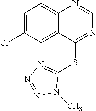
C00053

C00054

C00055
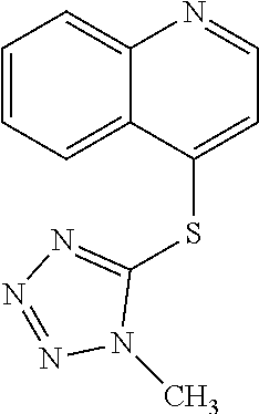
C00056

C00057

C00058

C00059

C00060

C00061
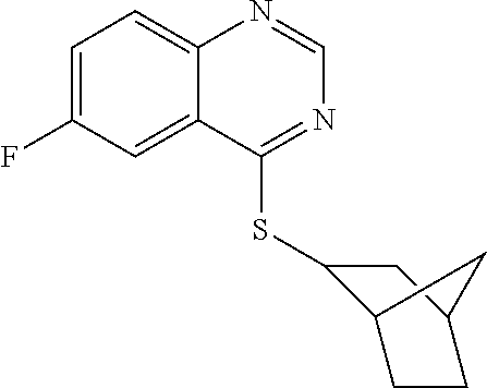
C00062

C00063
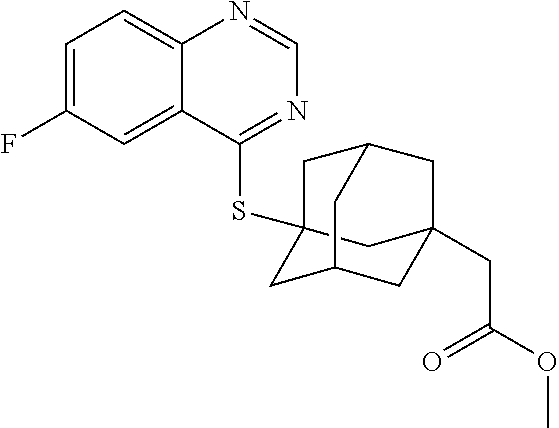
C00064
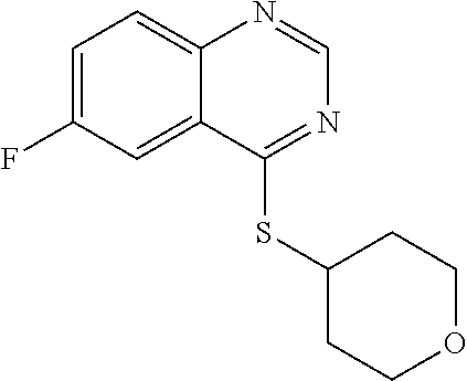
C00065

C00066

C00067
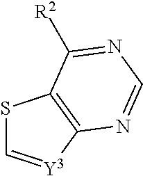
C00068
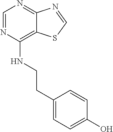
C00069

C00070

C00071
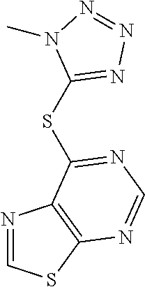
C00072

C00073

C00074

C00075
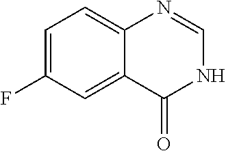
C00076

C00077

C00078
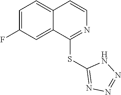
C00079

C00080
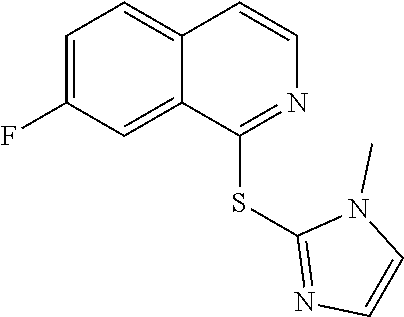
C00081

C00082

C00083

C00084

C00085

C00086
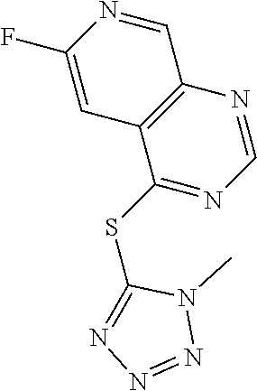
C00087

C00088
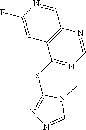
C00089
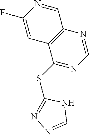
C00090

C00091

C00092
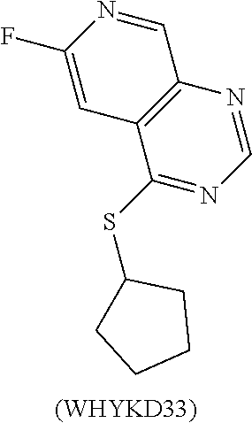
C00093

C00094
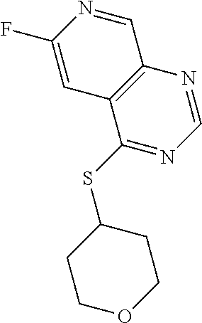
C00095

C00096

C00097

C00098

C00099

C00100

C00101

C00102

C00103

C00104
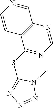
C00105
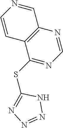
C00106
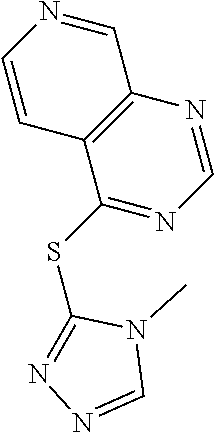
C00107

C00108

C00109
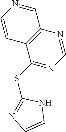
C00110
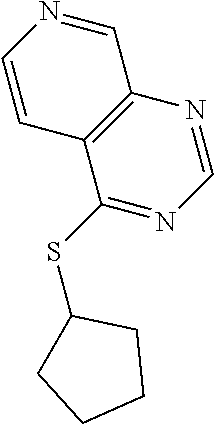
C00111

C00112

C00113

C00114

C00115
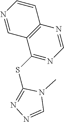
C00116

C00117
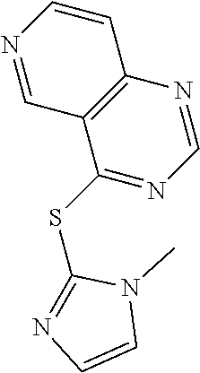
C00118
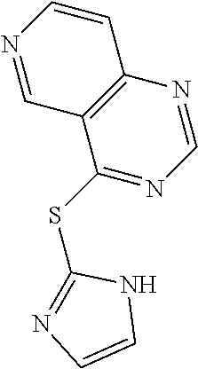
C00119
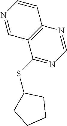
C00120
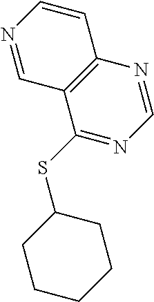
C00121
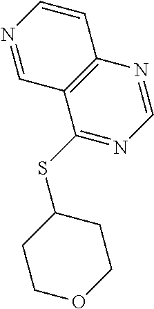
C00122

C00123

C00124
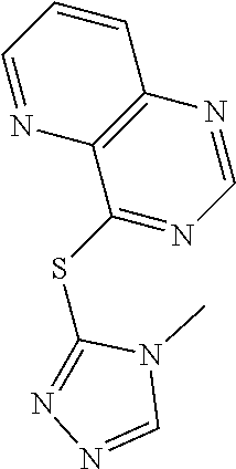
C00125

C00126

C00127

C00128
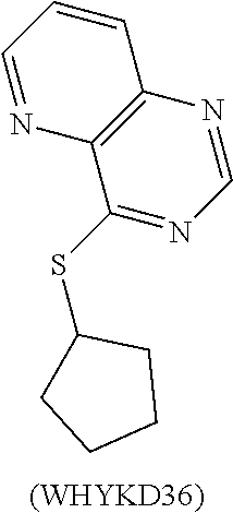
C00129

C00130

C00131
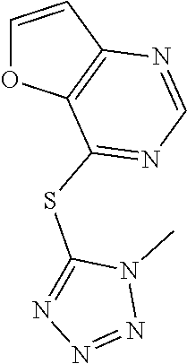
C00132

C00133

C00134
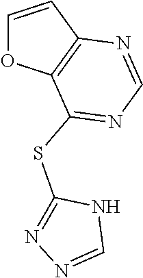
C00135
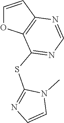
C00136

C00137

C00138
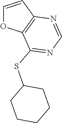
C00139
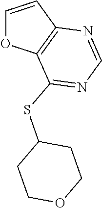
C00140

C00141

C00142
C00143

C00144

C00145

C00146

C00147

C00148
C00149

C00150
C00151

C00152
C00153
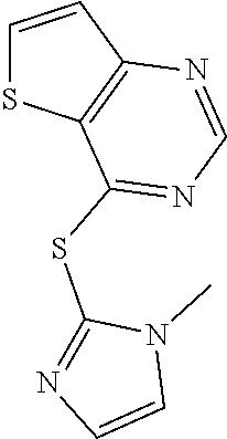
C00154
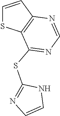
C00155

C00156

C00157

C00158
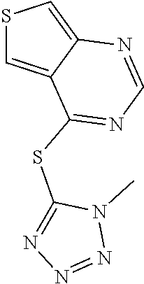
C00159

C00160

C00161

C00162

C00163

C00164
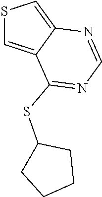
C00165
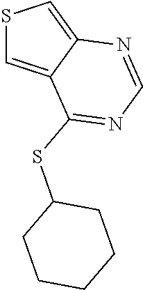
C00166

C00167
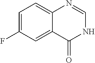
C00168
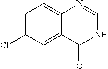
C00169
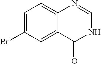
C00170

C00171

C00172

C00173

C00174
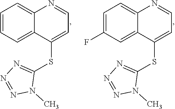
C00175

C00176

C00177
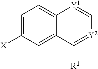
C00178

D00001
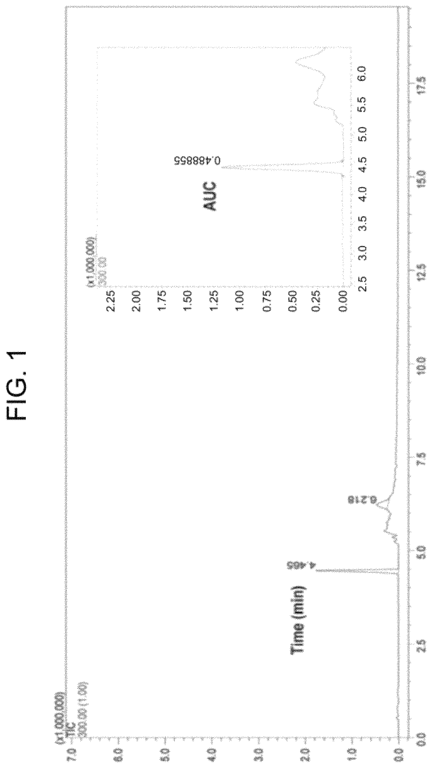
D00002
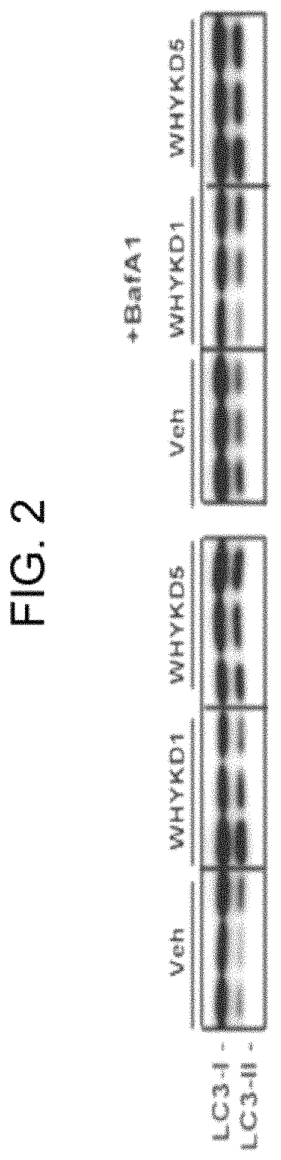
D00003
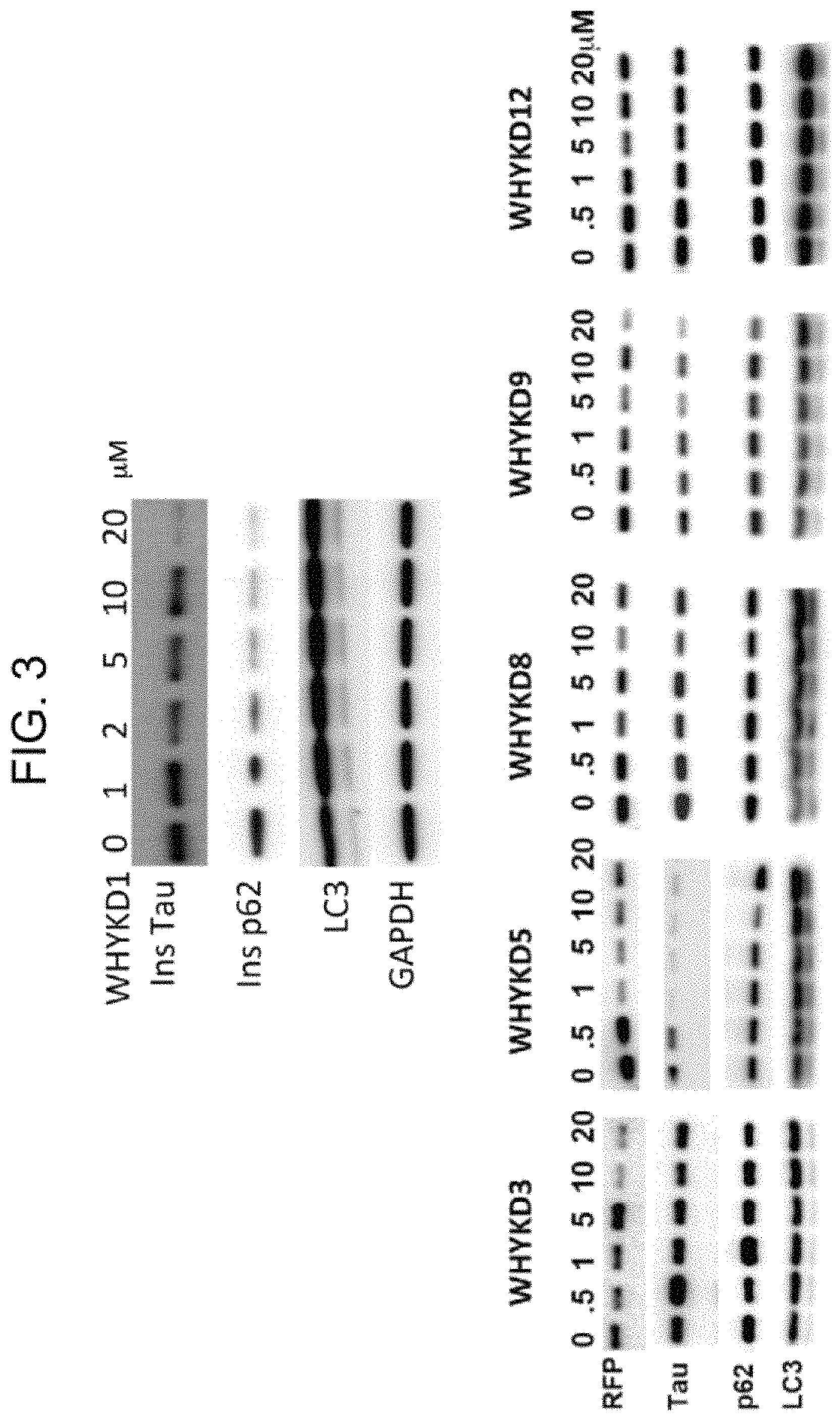
D00004
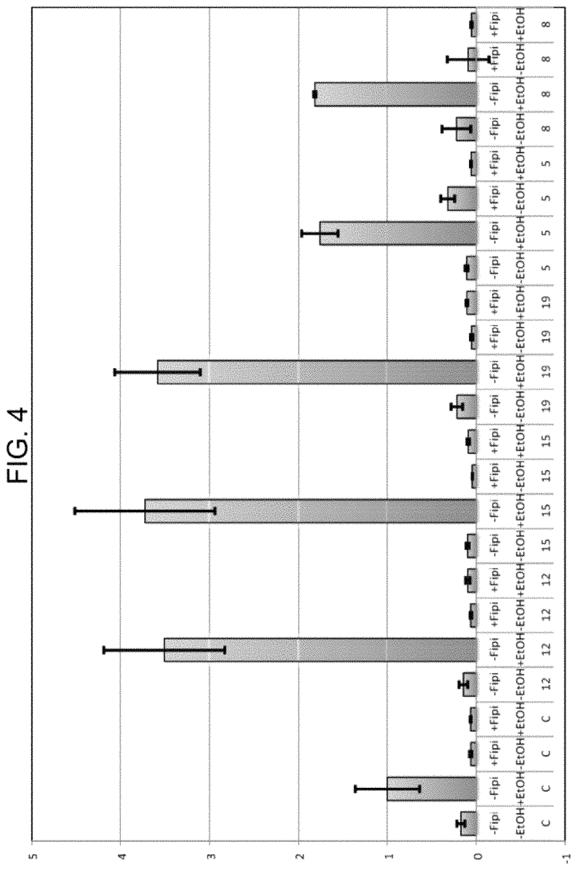
D00005

XML
uspto.report is an independent third-party trademark research tool that is not affiliated, endorsed, or sponsored by the United States Patent and Trademark Office (USPTO) or any other governmental organization. The information provided by uspto.report is based on publicly available data at the time of writing and is intended for informational purposes only.
While we strive to provide accurate and up-to-date information, we do not guarantee the accuracy, completeness, reliability, or suitability of the information displayed on this site. The use of this site is at your own risk. Any reliance you place on such information is therefore strictly at your own risk.
All official trademark data, including owner information, should be verified by visiting the official USPTO website at www.uspto.gov. This site is not intended to replace professional legal advice and should not be used as a substitute for consulting with a legal professional who is knowledgeable about trademark law.