Screening devices and methods for obstructive sleep apnea therapy
Tesfayesus , et al. May 11, 2
U.S. patent number 11,000,208 [Application Number 16/355,341] was granted by the patent office on 2021-05-11 for screening devices and methods for obstructive sleep apnea therapy. This patent grant is currently assigned to LivaNova USA, Inc.. The grantee listed for this patent is LivaNova USA, Inc.. Invention is credited to Stephen L. Bolea, Peter R. Eastwood, David R. Hillman, Wondimeneh Tesfayesus.


| United States Patent | 11,000,208 |
| Tesfayesus , et al. | May 11, 2021 |
Screening devices and methods for obstructive sleep apnea therapy
Abstract
Devices and methods for treating obstructive sleep apnea by first performing an assessment of the patient that involves observing the patient's upper airway during a tongue protrusion maneuver. The assessment may, for example, be done using endoscopy to observe the upper airway while the patient is awake in the supine position. An adequate response of the upper airway during the tongue protrusion maneuver is indicative of likely therapeutic success with hypoglossal nerve stimulation, and may be used for making clinical decisions. The principles of the present invention may be applied to other therapeutic interventions for OSA involving the upper airway.
| Inventors: | Tesfayesus; Wondimeneh (St. Paul, MN), Bolea; Stephen L. (Watertown, MN), Eastwood; Peter R. (Western Australia, AU), Hillman; David R. (Western Australia, AU) | ||||||||||
|---|---|---|---|---|---|---|---|---|---|---|---|
| Applicant: |
|
||||||||||
| Assignee: | LivaNova USA, Inc. (Houston,
TX) |
||||||||||
| Family ID: | 51018071 | ||||||||||
| Appl. No.: | 16/355,341 | ||||||||||
| Filed: | March 15, 2019 |
Prior Publication Data
| Document Identifier | Publication Date | |
|---|---|---|
| US 20190274587 A1 | Sep 12, 2019 | |
Related U.S. Patent Documents
| Application Number | Filing Date | Patent Number | Issue Date | ||
|---|---|---|---|---|---|
| 15418467 | Jan 27, 2017 | 10231645 | |||
| 14803779 | Jul 20, 2015 | 9555247 | |||
| 14475130 | Sep 2, 2014 | 9113838 | |||
| 13205315 | Aug 8, 2011 | 8855771 | |||
| 13113524 | May 23, 2011 | ||||
| 61437573 | Jan 28, 2011 | ||||
| 61467758 | Mar 25, 2011 | ||||
| Current U.S. Class: | 1/1 |
| Current CPC Class: | A61N 1/0551 (20130101); A61B 1/267 (20130101); A61N 1/3611 (20130101); A61B 5/4552 (20130101); A61B 1/233 (20130101); A61N 1/36139 (20130101); A61N 1/3601 (20130101); A61B 5/0826 (20130101); A61N 1/0519 (20130101) |
| Current International Class: | A61B 1/267 (20060101); A61N 1/36 (20060101); A61N 1/05 (20060101); A61B 5/08 (20060101); A61B 5/00 (20060101); A61B 1/233 (20060101) |
References Cited [Referenced By]
U.S. Patent Documents
| 758030 | April 1904 | Carence |
| 1520930 | December 1924 | Calhoun |
| 1701277 | February 1929 | Shindel |
| 1914418 | June 1933 | Goyena |
| 2046664 | July 1936 | Weaver |
| 2151227 | March 1939 | Pawelek |
| 2237954 | April 1941 | Wilsom |
| 2243360 | May 1941 | Slatis et al. |
| 2274886 | March 1942 | Carroll |
| 2526586 | October 1950 | Shuff |
| 2693799 | November 1954 | Herman, Jr. |
| 2777442 | January 1957 | Zelano |
| 2928388 | March 1960 | Jaroslaw |
| 3457917 | July 1969 | Mercurio |
| 3513839 | May 1970 | Vacante |
| 3680555 | August 1972 | Warncke |
| 3722509 | March 1973 | Nebel |
| 3774618 | November 1973 | Avery |
| 3865106 | February 1975 | Palush |
| 3884223 | May 1975 | Keindl |
| 3893463 | July 1975 | Williams |
| 3906936 | September 1975 | Habal |
| 4160252 | July 1979 | Lucas et al. |
| 4160255 | July 1979 | Kobayashi |
| 4178524 | December 1979 | Ritter |
| 4200440 | April 1980 | Renko |
| 4220150 | September 1980 | King |
| 4221217 | September 1980 | Amezcua |
| 4225034 | September 1980 | Sarovich |
| 4239918 | December 1980 | Keeley |
| 4242987 | January 1981 | Viessmann |
| 4267831 | May 1981 | Aguilar |
| 4283867 | August 1981 | Brown |
| 4302951 | December 1981 | Fall et al. |
| 4313442 | February 1982 | Knudson et al. |
| 4346398 | August 1982 | Lai |
| 4374527 | February 1983 | Iversen |
| 4414986 | November 1983 | Dickhudt et al. |
| 4506666 | March 1985 | Durkan |
| 4567892 | February 1986 | Plicchi et al. |
| 4573481 | March 1986 | Bullara |
| 4602624 | July 1986 | Naples et al. |
| 4612934 | September 1986 | Borkan |
| 4777963 | October 1988 | McKenna |
| 4830008 | May 1989 | Meer |
| 4899750 | February 1990 | Ekwall |
| 4915105 | April 1990 | Lee |
| 4919136 | April 1990 | Alt |
| 4934368 | June 1990 | Lynch |
| 4940065 | July 1990 | Tanagho et al. |
| 4960133 | October 1990 | Hewson |
| 4979511 | December 1990 | Terry, Jr. |
| 4996983 | March 1991 | Amrhein |
| 5016808 | May 1991 | Heil et al. |
| 5036862 | August 1991 | Pohndorf |
| 5095905 | March 1992 | Klepinski |
| 5105826 | April 1992 | Smits et al. |
| 5121754 | June 1992 | Mullett |
| 5133354 | July 1992 | Kallok |
| 5146918 | September 1992 | Kallok et al. |
| 5158080 | October 1992 | Kallok |
| 5174287 | December 1992 | Kallok et al. |
| 5178156 | January 1993 | Takishima et al. |
| 5190053 | March 1993 | Meer |
| 5211173 | May 1993 | Kallok et al. |
| 5215082 | June 1993 | Kallok et al. |
| 5277193 | January 1994 | Takishima et al. |
| 5281219 | January 1994 | Kallok |
| 5282468 | February 1994 | Klepinski |
| 5300094 | April 1994 | Kallok et al. |
| 5324321 | June 1994 | Pohndorf et al. |
| 5335657 | August 1994 | Terry, Jr. et al. |
| 5344438 | September 1994 | Testerman et al. |
| 5388578 | February 1995 | Yomtov et al. |
| 5392773 | February 1995 | Bertrand |
| 5417205 | May 1995 | Wang |
| 5425359 | June 1995 | Liou |
| 5458629 | October 1995 | Baudino et al. |
| 5483969 | January 1996 | Testerman et al. |
| 5485836 | January 1996 | Lincoln |
| 5485851 | January 1996 | Erickson |
| 5487756 | January 1996 | Kallesoe et al. |
| 5511543 | April 1996 | Shirley |
| 5522382 | June 1996 | Sullivan et al. |
| 5522862 | June 1996 | Testerman et al. |
| 5531778 | July 1996 | Maschino et al. |
| 5540731 | July 1996 | Testerman |
| 5540732 | July 1996 | Testerman |
| 5540733 | July 1996 | Testerman et al. |
| 5540734 | July 1996 | Zabara |
| 5546938 | August 1996 | McKenzie |
| 5549655 | August 1996 | Erickson |
| 5568808 | October 1996 | Rimkus |
| 5591216 | January 1997 | Testerman et al. |
| 5630411 | May 1997 | Holscher |
| 5682881 | November 1997 | Winthrop et al. |
| 5697105 | December 1997 | White |
| 5697363 | December 1997 | Hart |
| 5730122 | March 1998 | Lurie |
| 5740798 | April 1998 | McKinney |
| 5752511 | May 1998 | Simmons et al. |
| 5787884 | August 1998 | Tovey |
| 5826579 | October 1998 | Remmers et al. |
| 5848589 | December 1998 | Welnetz |
| 5855552 | January 1999 | Houser et al. |
| 5890491 | April 1999 | Rimkus |
| 5895360 | April 1999 | Christopherson et al. |
| 5919220 | July 1999 | Stieglitz et al. |
| 5922014 | July 1999 | Warman et al. |
| 5938596 | August 1999 | Woloszko et al. |
| 5944680 | August 1999 | Christopherson et al. |
| 5947119 | September 1999 | Reznick |
| 6010459 | January 2000 | Silkoff et al. |
| 6015389 | January 2000 | Brown |
| 6021352 | February 2000 | Christopherson et al. |
| 6021354 | February 2000 | Warman et al. |
| 6029667 | February 2000 | Lurie |
| 6041780 | March 2000 | Richard et al. |
| 6051052 | April 2000 | Monereau et al. |
| 6066165 | May 2000 | Racz |
| 6098624 | August 2000 | Utamaru |
| 6109262 | August 2000 | Tovey |
| 6119690 | September 2000 | Pantaleo |
| 6126611 | October 2000 | Bourgeois et al. |
| 6132384 | October 2000 | Christopherson et al. |
| 6198970 | March 2001 | Freed et al. |
| 6201994 | March 2001 | Warman et al. |
| 6205360 | March 2001 | Carter et al. |
| 6217527 | April 2001 | Selmon et al. |
| 6221049 | April 2001 | Selmon et al. |
| 6231546 | May 2001 | Milo et al. |
| 6240316 | May 2001 | Richmond et al. |
| 6244267 | June 2001 | Eifrig |
| 6251126 | June 2001 | Ottenhoff et al. |
| 6269269 | July 2001 | Ottenhoff et al. |
| 6269703 | August 2001 | Bowers |
| 6292703 | September 2001 | Meier et al. |
| 6345202 | February 2002 | Richmond et al. |
| 6366815 | April 2002 | Haugland et al. |
| 6460539 | October 2002 | Japuntich et al. |
| 6484725 | November 2002 | Chi |
| 6511458 | January 2003 | Milo et al. |
| 6514217 | February 2003 | Selmon et al. |
| 6542776 | April 2003 | Gordon et al. |
| 6561188 | May 2003 | Ellis |
| 6587725 | July 2003 | Durand et al. |
| 6600956 | July 2003 | Maschino et al. |
| 6606521 | August 2003 | Paspa et al. |
| 6609031 | August 2003 | Law et al. |
| 6626179 | September 2003 | Pedley |
| 6636767 | October 2003 | Knudson et al. |
| 6641542 | November 2003 | Cho et al. |
| 6647289 | November 2003 | Prutchi |
| 6651652 | November 2003 | WaRD |
| 6718982 | April 2004 | Smith et al. |
| 6719725 | April 2004 | Milo et al. |
| 6721603 | April 2004 | Zabara et al. |
| 6772015 | August 2004 | Dahl et al. |
| 6776162 | August 2004 | Wood |
| 6799575 | October 2004 | Carter |
| 6819958 | November 2004 | Weiner et al. |
| 6829503 | December 2004 | Alt |
| 6829508 | December 2004 | Schulman et al. |
| RE38705 | February 2005 | Hill et al. |
| 6876885 | April 2005 | Swoyer et al. |
| 6881192 | April 2005 | Park |
| 6883518 | April 2005 | Mittelstadt et al. |
| 6890306 | May 2005 | Poezevera |
| 6907295 | June 2005 | Gross et al. |
| 6904320 | July 2005 | Park et al. |
| 6928324 | August 2005 | Park et al. |
| 6978171 | December 2005 | Goetz et al. |
| 6997177 | February 2006 | Wood |
| 7027869 | April 2006 | Danek et al. |
| 7054692 | May 2006 | Whitehurst et al. |
| 7065410 | June 2006 | Bardy et al. |
| 7082331 | July 2006 | Park et al. |
| 7087053 | August 2006 | Vanney |
| 7089932 | August 2006 | Dodds |
| 7094206 | August 2006 | Hoffman |
| 7117036 | October 2006 | Florio |
| 7128717 | October 2006 | Thach et al. |
| 7142919 | November 2006 | Hine et al. |
| 7149573 | December 2006 | Wang |
| 7152604 | December 2006 | Hickle et al. |
| 7155278 | December 2006 | King et al. |
| 7156098 | January 2007 | Dolezal et al. |
| 7160252 | January 2007 | Cho et al. |
| 7160255 | January 2007 | Saadat |
| 7178524 | February 2007 | Noble |
| 7200440 | April 2007 | Kim et al. |
| 7225034 | May 2007 | Ries et al. |
| 7239320 | July 2007 | Hall et al. |
| 7239918 | July 2007 | Strother et al. |
| 7239920 | July 2007 | Thacker et al. |
| 7242987 | July 2007 | Holleman et al. |
| 7263996 | September 2007 | Yung Ho |
| 7277749 | October 2007 | Gordon et al. |
| 7283867 | October 2007 | Strother et al. |
| 7302951 | December 2007 | Mittelstadt et al. |
| 7313442 | December 2007 | Velasco et al. |
| 7343202 | March 2008 | Marva et al. |
| 7346398 | March 2008 | Gross et al. |
| 7366572 | April 2008 | Heruth et al. |
| 7396333 | July 2008 | Stahmann et al. |
| 7438686 | October 2008 | Cho et al. |
| 7453928 | November 2008 | Ten et al. |
| 7463928 | December 2008 | Lee et al. |
| 7473227 | January 2009 | Hsu et al. |
| 7515968 | April 2009 | Metzler et al. |
| 7524292 | April 2009 | Cho et al. |
| 7561922 | July 2009 | Cohen et al. |
| 7591265 | September 2009 | Lee et al. |
| 7596413 | September 2009 | Libbus et al. |
| 7596414 | September 2009 | Whitehurst et al. |
| 7627375 | December 2009 | Bardy et al. |
| 7630771 | December 2009 | Cauller |
| 7634315 | December 2009 | Cholette |
| 7636602 | December 2009 | Baru Fassio et al. |
| 7657311 | February 2010 | Bardy et al. |
| 7660632 | February 2010 | Kirby et al. |
| 7662105 | February 2010 | Hatlestad |
| 7672728 | March 2010 | Libbus et al. |
| 7672729 | March 2010 | Koh et al. |
| 7680537 | March 2010 | Stahmann et al. |
| 7680538 | March 2010 | Durand et al. |
| 7684869 | March 2010 | Bradley et al. |
| 7697968 | April 2010 | Moore |
| 7697984 | April 2010 | Hill et al. |
| 7697990 | April 2010 | Ujhazy et al. |
| 7717848 | May 2010 | Heruth et al. |
| 7720534 | May 2010 | Bardy et al. |
| 7725195 | May 2010 | Lima et al. |
| 7725198 | May 2010 | Cross et al. |
| 7734340 | June 2010 | De Ridder |
| 7734348 | June 2010 | Zhang et al. |
| 7738952 | June 2010 | Yun et al. |
| 7747323 | June 2010 | Libbus et al. |
| 7751880 | July 2010 | Cholette |
| 7751885 | July 2010 | Bardy et al. |
| 7758384 | July 2010 | Alexander et al. |
| 7765000 | July 2010 | Zhang et al. |
| 7769461 | August 2010 | Whitehurst et al. |
| 7783353 | August 2010 | Libbus et al. |
| 7785262 | August 2010 | Melker et al. |
| 7787959 | August 2010 | Morgan |
| 7792590 | September 2010 | Pianca et al. |
| 7797050 | September 2010 | Libbus et al. |
| 7797057 | September 2010 | Harris |
| 7797058 | September 2010 | Mrva et al. |
| 7805195 | September 2010 | Zealear |
| 7809442 | October 2010 | Bolea et al. |
| 7813797 | October 2010 | Bardy et al. |
| 7813802 | October 2010 | Tcheng et al. |
| 7813809 | October 2010 | Strother et al. |
| 7818063 | October 2010 | Wallace et al. |
| 7822486 | October 2010 | Foster et al. |
| 7860570 | December 2010 | Whitehurst et al. |
| 7979128 | July 2011 | Tehrani et al. |
| 8221049 | July 2012 | Westendorf et al. |
| 8249723 | August 2012 | McCreery |
| 8255056 | August 2012 | Tehrani |
| 8311645 | November 2012 | Bolea et al. |
| 8386046 | February 2013 | Tesfayesus et al. |
| 8428727 | April 2013 | Bolea et al. |
| 8498712 | July 2013 | Bolea et al. |
| 8626304 | January 2014 | Bolea et al. |
| 8639354 | January 2014 | Bolea et al. |
| 8718783 | May 2014 | Bolea et al. |
| 8744584 | June 2014 | Camps et al. |
| 8744589 | June 2014 | Bolea et al. |
| 8855771 | October 2014 | Tesfayesus et al. |
| 2001/0010010 | July 2001 | Richmond et al. |
| 2001/0031929 | October 2001 | O'Toole |
| 2002/0010495 | January 2002 | Freed et al. |
| 2002/0049479 | April 2002 | Pitts |
| 2002/0092527 | July 2002 | Wood |
| 2002/0128700 | September 2002 | Cross, Jr. |
| 2002/0156507 | October 2002 | Lindenthaler |
| 2002/0165462 | November 2002 | Westbrook et al. |
| 2002/0166556 | November 2002 | Jacob |
| 2002/0195108 | December 2002 | Mittelstadt et al. |
| 2002/0195109 | December 2002 | Mittelstadt et al. |
| 2003/0034031 | February 2003 | Lev et al. |
| 2003/0078643 | April 2003 | Schulman et al. |
| 2003/0083696 | May 2003 | Avital |
| 2003/0093128 | May 2003 | Freed et al. |
| 2003/0106555 | June 2003 | Tovey |
| 2003/0106556 | June 2003 | Alperovich et al. |
| 2003/0114895 | June 2003 | Gordon et al. |
| 2003/0114905 | June 2003 | Kuzma |
| 2003/0153953 | August 2003 | Park et al. |
| 2003/0167018 | September 2003 | Wyckoff |
| 2003/0195571 | October 2003 | Burnes et al. |
| 2003/0209145 | November 2003 | Soper |
| 2003/0216789 | November 2003 | Deem et al. |
| 2004/0015204 | January 2004 | Whitehurst et al. |
| 2004/0020489 | February 2004 | Gillespie et al. |
| 2004/0049241 | March 2004 | Campos |
| 2004/0055603 | March 2004 | Bruce |
| 2004/0073272 | April 2004 | Knudson et al. |
| 2004/0089303 | May 2004 | Chien |
| 2004/0111139 | June 2004 | McCreery |
| 2004/0116819 | June 2004 | Alt |
| 2004/0116978 | June 2004 | Bradley |
| 2004/0138581 | July 2004 | Frei et al. |
| 2004/0153127 | August 2004 | Gordon et al. |
| 2004/0162499 | August 2004 | Nagai et al. |
| 2004/0194784 | October 2004 | Bertrand |
| 2004/0215288 | October 2004 | Lee et al. |
| 2004/0215290 | October 2004 | Zealear |
| 2004/0230278 | November 2004 | Dahl et al. |
| 2004/0233058 | November 2004 | Dodds |
| 2004/0260310 | December 2004 | Harris |
| 2004/0261791 | December 2004 | Horian |
| 2005/0004610 | January 2005 | Kim et al. |
| 2005/0004810 | January 2005 | Tanaka |
| 2005/0010265 | January 2005 | Baru Fassio et al. |
| 2005/0038490 | February 2005 | Gross et al. |
| 2005/0039757 | February 2005 | Wood |
| 2005/0043644 | February 2005 | Stahmann et al. |
| 2005/0043772 | February 2005 | Stahmann et al. |
| 2005/0076908 | April 2005 | Lee et al. |
| 2005/0085865 | April 2005 | Tehrani |
| 2005/0085866 | April 2005 | Tehrani |
| 2005/0085868 | April 2005 | Tehrani et al. |
| 2005/0085869 | April 2005 | Tehrani et al. |
| 2005/0085874 | April 2005 | Davis et al. |
| 2005/0098176 | May 2005 | Hoffrichter |
| 2005/0101833 | May 2005 | Hsu et al. |
| 2005/0119711 | June 2005 | Cho et al. |
| 2005/0139216 | June 2005 | Mittelstadt et al. |
| 2005/0165457 | July 2005 | Benser et al. |
| 2005/0209513 | September 2005 | Heruth et al. |
| 2005/0209643 | September 2005 | Heruth et al. |
| 2005/0234523 | October 2005 | Levin et al. |
| 2005/0235992 | October 2005 | Djupesland |
| 2005/0240241 | October 2005 | Yun et al. |
| 2005/0251216 | November 2005 | Hill et al. |
| 2005/0261747 | November 2005 | Schuler et al. |
| 2005/0267380 | December 2005 | Poezevara |
| 2005/0267547 | December 2005 | Knudson et al. |
| 2005/0277844 | December 2005 | Stroether et al. |
| 2005/0277999 | December 2005 | Strother et al. |
| 2005/0278000 | December 2005 | Strother et al. |
| 2006/0004429 | January 2006 | Mrva et al. |
| 2006/0005842 | January 2006 | Rashad et al. |
| 2006/0025828 | February 2006 | Armstrong et al. |
| 2006/0030919 | February 2006 | Mrva et al. |
| 2006/0032497 | February 2006 | Doshi |
| 2006/0041295 | February 2006 | Osypka |
| 2006/0052836 | March 2006 | Kim et al. |
| 2006/0058588 | March 2006 | Zdeblick |
| 2006/0058852 | March 2006 | Koh et al. |
| 2006/0064029 | March 2006 | Arad (Abboud) |
| 2006/0064138 | March 2006 | Velasco et al. |
| 2006/0079802 | April 2006 | Jensen et al. |
| 2006/0095088 | May 2006 | De Ridder |
| 2006/0111755 | May 2006 | Stone et al. |
| 2006/0116739 | June 2006 | Betser et al. |
| 2006/0129189 | June 2006 | George et al. |
| 2006/0135886 | June 2006 | Lippert et al. |
| 2006/0136024 | June 2006 | Cohen et al. |
| 2006/0142815 | June 2006 | Tehrani et al. |
| 2006/0144398 | July 2006 | Doshi et al. |
| 2006/0149334 | July 2006 | Tehrani et al. |
| 2006/0149345 | July 2006 | Boggs et al. |
| 2006/0150978 | July 2006 | Doshi et al. |
| 2006/0150979 | July 2006 | Doshi et al. |
| 2006/0150980 | July 2006 | Kim |
| 2006/0155341 | July 2006 | Tehrani et al. |
| 2006/0167497 | July 2006 | Armstrong et al. |
| 2006/0184204 | August 2006 | He |
| 2006/0195170 | August 2006 | Cohen et al. |
| 2006/0211951 | September 2006 | Milijasevic et al. |
| 2006/0224209 | October 2006 | Meyer |
| 2006/0224211 | October 2006 | Durand et al. |
| 2006/0241506 | October 2006 | Melker et al. |
| 2006/0241708 | October 2006 | Boute |
| 2006/0247729 | November 2006 | Tehrani et al. |
| 2006/0259079 | November 2006 | King |
| 2006/0264777 | November 2006 | Drew |
| 2006/0266369 | November 2006 | Atkinson et al. |
| 2006/0271118 | November 2006 | Libbus et al. |
| 2006/0271137 | November 2006 | Stanton-Hicks |
| 2006/0282127 | December 2006 | Zealear |
| 2006/0293720 | December 2006 | Dilorenzo |
| 2006/0293723 | December 2006 | Whitehurst et al. |
| 2007/0021785 | January 2007 | Inman et al. |
| 2007/0027482 | February 2007 | Parnis et al. |
| 2007/0038265 | February 2007 | Tcheng et al. |
| 2007/0043411 | February 2007 | Foster et al. |
| 2007/0095347 | May 2007 | Lampotang et al. |
| 2007/0150006 | June 2007 | Libbus et al. |
| 2007/0125379 | July 2007 | Pierro et al. |
| 2007/0173893 | July 2007 | Pitts |
| 2007/0175478 | August 2007 | Brunst |
| 2007/0179342 | August 2007 | Miller et al. |
| 2007/0227542 | October 2007 | Kashmakov et al. |
| 2007/0239243 | October 2007 | Moffitt et al. |
| 2007/0277832 | December 2007 | Doshi et al. |
| 2007/0282410 | December 2007 | Cross et al. |
| 2007/0283692 | December 2007 | Tetsuka et al. |
| 2007/0283962 | December 2007 | Doshi et al. |
| 2007/0295338 | December 2007 | Loomas et al. |
| 2008/0023007 | January 2008 | Dolezal et al. |
| 2008/0027480 | January 2008 | Van Der Burg et al. |
| 2008/0027502 | January 2008 | Ransom |
| 2008/0041373 | February 2008 | Doshi et al. |
| 2008/0099029 | May 2008 | Lamberg |
| 2008/0103407 | May 2008 | Bolea et al. |
| 2008/0103545 | May 2008 | Bolea et al. |
| 2008/0147142 | June 2008 | Testerman et al. |
| 2008/0163875 | July 2008 | Aarestad et al. |
| 2008/0183254 | July 2008 | Bly et al. |
| 2008/0188947 | August 2008 | Sanders |
| 2009/0014012 | January 2009 | Sanders |
| 2009/0044814 | February 2009 | Iancea et al. |
| 2009/0270707 | October 2009 | Alfoqaha et al. |
| 2009/0276024 | November 2009 | Bonde et al. |
| 2009/0308395 | December 2009 | Lee et al. |
| 2009/0318986 | December 2009 | Alo et al. |
| 2009/0326408 | December 2009 | Moon |
| 2010/0016749 | January 2010 | Atsma et al. |
| 2010/0036285 | February 2010 | Govari et al. |
| 2010/0047376 | February 2010 | Imbeau et al. |
| 2010/0076536 | March 2010 | Merz et al. |
| 2010/0094379 | April 2010 | Meadows et al. |
| 2010/0100150 | April 2010 | Kirby et al. |
| 2010/0125310 | May 2010 | Wilson et al. |
| 2010/0131029 | May 2010 | Durand et al. |
| 2010/0137931 | June 2010 | Hopper et al. |
| 2010/0137949 | June 2010 | Mazgalev et al. |
| 2010/0137956 | June 2010 | Osypka |
| 2010/0152553 | June 2010 | Ujhazy et al. |
| 2010/0174341 | July 2010 | Bolea et al. |
| 2010/0198103 | August 2010 | Meadows |
| 2010/0228133 | September 2010 | Averina et al. |
| 2010/0228317 | September 2010 | Libbus et al. |
| 2010/0241207 | September 2010 | Bluger |
| 2010/0257729 | October 2010 | Alexander et al. |
| 2010/0262209 | October 2010 | King et al. |
| 2011/0071591 | March 2011 | Bolea et al. |
| 2011/0093032 | April 2011 | Boggs et al. |
| 2012/0017920 | January 2012 | Sanders |
| 2012/0022389 | January 2012 | Sanders |
| 2012/0192874 | August 2012 | Bolea et al. |
| 2013/0085546 | April 2013 | Bolea et al. |
| 2013/0123629 | May 2013 | Rosenberg et al. |
| 0 900 102 | Jul 2004 | EP | |||
| 0 892 926 | Jun 2006 | EP | |||
| 1 404 221 | Feb 2007 | EP | |||
| 1 854 494 | Nov 2007 | EP | |||
| 1 322 384 | Dec 2007 | EP | |||
| 09-294819 | Nov 1997 | JP | |||
| 2000-508601 | May 2000 | JP | |||
| 2000-508562 | Jul 2000 | JP | |||
| 2003-305135 | Oct 2003 | JP | |||
| 2004-508908 | Mar 2004 | JP | |||
| 2004-532707 | Oct 2004 | JP | |||
| 3688301 | Jun 2005 | JP | |||
| 2005-521485 | Jul 2005 | JP | |||
| 2007-021156 | Feb 2007 | JP | |||
| WO-98/20938 | May 1998 | WO | |||
| WO-02/24279 | Mar 2002 | WO | |||
| WO-03/000133 | Jan 2003 | WO | |||
| WO-2006/063339 | Jun 2006 | WO | |||
| WO-2008/046190 | Apr 2008 | WO | |||
Other References
|
Aziz, L. and Ejnell, H. "Obstructive Sleep Apnea Caused by Bilateral Vocal Fold Paralysis." Ear Nose Throat J. Apr. 2003; 82(4): 326-7. Abstract. cited by applicant . Campbell et al., "Nasal Continuous positive airway pressure from high flow cannula versus Infant Flow for preterm infants," Journal of Perinatology, Jul. 2006, pp. 546-549, vol. 26 (9), Nature Publishing Group. cited by applicant . De Almeida et al., "Nasal pressure recordings to detect obstructive sleep apnea," Sleep and Breathing, Feb. 25, 2006, pp. 62-69, vol. 10 (2), Springer Heidelberg. cited by applicant . Eastwood et al., "Treating Obstructive Sleep Apnea with Hypoglossal Nerve Stimulation," Sleep, 2011, pp. 1479-1486B, vol. 34, No. 11. cited by applicant . Ferguson et al., "Effect of Mandibular and Tongue Protrusion on Upper Airway Size During Wakefulness," American Journal of Respiratory and Critical Care Medicine, 1997, pp. 1748-1754, vol. 155. cited by applicant . Goding JR et al., "Relief of Upper Airway Obstruction With Hypoglossal Nerve Stimulation in the Canine", The Laryngoscope, Feb. 1998, pp. 162-169, vol. 108, Lippincott-Raven Publishers, U.S.A. cited by applicant . Huang et al. "Dilation of the oropharynx via selective stimulation of the hypoglossal nerve." J. Neural Eng. 2005; 2:73-80. cited by applicant . Isono et al., "Interaction of cross-sectional area, driving pressure, and airflow of passive velopharynx," American Physiological Society, 1997, pp. 851-859, vol. 83. cited by applicant . Kirkness et al., "Nasal airflow dynamics: mechanisms and responses associated with an external nasal dilator strip," University of Western Sydney, T.C. Amis School of Science, Department of Respiratory Medicine, Westmead Hospital and University of Sydney, Westmead, Australia, 2000. cited by applicant . Mahadevia et al., "Effects of expiratory positive airway pressure on sleep-induced respiratory abnormalities in patients with hypersomnia-sleep apnea syndrome," Am. Rev. Respir. Cis., Feb. 1983, vol. 128, pp. 708-711. cited by applicant . Mann EA, et al., "The Effect of Neuromuscular Stimulation of the Genioglossus on the Hypopharyngeal Airway." Laryngoscope 112: 351-356, 2002. cited by applicant . Noseda et al., "Compliance with nasal continuous positive airway pressure assessed with a pressure monitor: pattern of use and influence of sleep habits," Chest Clinics and Sleep Laboratories, Hopitaux Erasme et Brugmann, Universit Libre de Bruxelles, Brussels, Belgium, 2000, vol. 94, pp. 76-81. cited by applicant . Oliven et al., "Effect of genioglossus contraction on pharyngeal lumen and airflow in sleep apnoea patients," European Respiratory Journal, 2007, pp. 748-758, vol. 30, No. 4. cited by applicant . Paquereau et al., "Positive pressure titration in the treatment of obstructive sleep apnea syndrome using continuous airway positive pressure," Revue Des Maladies Respiratoires, Apr. 2000, pp. 459-465, vol. 17 (2), Masson Editeur. cited by applicant . Partial European Search Report dated Aug. 19, 2009 issued in European Patent Application No. 09161958.5 (4 pages). cited by applicant . Response to the Notice of Opposition for Opposition against patent EP 2 116 274 (Application No. 09 161 958.5) dated Dec. 2, 2013 (30 pages). cited by applicant . Sahin et al., "Chronic recording of Hypoglossal nerve activity in a dog model of upper airway obstruction", Journal of Applied Physiology 87 (6), 1999, The American Physiological Society, pp. 2197-2206. cited by applicant . Saslow et al., "Work of breathing using high-flow nasal cannula in preterm infants," Journal of Perinatology, May 11, 2006, pp. 476-480, vol. 26 (8), Nature Publishing Group. cited by applicant . Schwartz et al. "Therapeutic Electrical Stimulation of the Hypoglossal Nerve in Obstructive Sleep Apnea." Arch Otolaryngol Head Neck Surg/vol. 127, Oct. 2001 (8 pages). cited by applicant . Spence et al., "High-flow nasal cannula as a device to provide continuous positive airway pressure in infants," Journal of Perinatologv, Dec. 2007, pp. 772-775, vol. 27 (12), Nature Publishing Group. cited by applicant . Statement of Grounds filed in Opposition of EP Patent 2116274 dated Jul. 25, 2012 (32 pages). cited by applicant . Stern et al. "Obstructive sleep apnea following treatment of head and neck cancer", Ear, Nose, and Throat Journal, Feb. 2007, vol. 86, No. 2, pp. 101-103. cited by applicant . Strollo et al., "Upper-Airway Stimulation for Obstructive Sleep Apnea," New England Journal of Medicine, 2014, pp. 139-149, N Engl J. Med 270;2. cited by applicant . Tiran et al., "An Improved Device for Posterior Rhinomanometry to Measure Nasal Resistance," Journal of Biomechnical Engineering, Nov. 2005, vol. 127, pp. 994-997. cited by applicant . Trevisanuto et al., "A new device for administration of continuous positive airway pressure in preterm infants: comparison with a standard nasal CPAP continuous positive airway pressure system," Intensive Care Medicine, Apr. 2005, pp. 859-864, vol. 31 (6), Springer-Verlag. cited by applicant . Verse et al., "New developments in the therapy of obstructive sleep apnea," European Archives of Oto-Rhino-Laryngology, Jan. 2001, pp. 31-37, vol. 258 (1), Springer-Verlag. cited by applicant . Wells, Jonathan, et al., Biophysical Mechanisms of Transient Optical Stimulation of Peripheral Nerve, Biophysical Journal, Oct. 2007, pp. 2567-2580, vol. 93. cited by applicant. |
Primary Examiner: Manuel; George
Attorney, Agent or Firm: Foley & Lardner LLP
Parent Case Text
CROSS REFERENCE TO RELATED APPLICATIONS
This patent application is a continuation of U.S. patent application Ser. No. 15/418,467, filed Jan. 27, 2017, entitled SCREENING DEVICES AND METHODS FOR OBSTRUCTIVE SLEEP APNEA THERAPY, which is a continuation of U.S. patent application Ser. No. 14/803,779, filed Jul. 20, 2015, entitled SCREENING DEVICES AND METHODS FOR OBSTRUCTIVE SLEEP APNEA THERAPY, which is a continuation of U.S. patent application Ser. No. 14/475,130, filed Sep. 2, 2014, entitled SCREENING DEVICES AND METHODS FOR OBSTRUCTIVE SLEEP APNEA THERAPY, now U.S. Pat. No. 9,113,838 which is a continuation of U.S. patent application Ser. No. 13/205,315, filed Aug. 8, 2011, entitled SCREENING DEVICES AND METHODS FOR OBSTRUCTIVE SLEEP APNEA THERAPY, now U.S. Pat. No. 8,855,771, which is a continuation of U.S. patent application Ser. No. 13/113,524, filed May 23, 2011, now abandoned, which claims the benefits of priority under 35 U.S.C. .sctn..sctn. 119 and 120 to U.S. Provisional Patent Application No. 61/437,573, filed Jan. 28, 2011, entitled OBSTRUCTIVE SLEEP APNEA TREATMENT DEVICES, SYSTEMS AND METHODS, and U.S. Provisional Patent Application No. 61/467,758, filed Mar. 25, 2011, entitled SCREENING DEVICES AND METHODS FOR OBSTRUCTIVE SLEEP APNEA THERAPY. This patent application is related to U.S. patent application Ser. No. 13/106,460, filed May 12, 2011, entitled OBSTRUCTIVE SLEEP APNEA TREATMENT DEVICES, SYSTEMS AND METHODS to Bolea et al., which claims the benefit of U.S. Provisional Patent Application No. 61/437,573, filed Jan. 28, 2011, entitled OBSTRUCTIVE SLEEP APNEA TREATMENT DEVICES, SYSTEMS AND METHODS. This patent application is also related to U.S. patent application Ser. No. 13/251,856, filed Oct. 3, 2011, now U.S. Pat. No. 8,386,046, entitled SCREENING DEVICES AND METHODS FOR OBSTRUCTIVE SLEEP APNEA THERAPY, which is a continuation of U.S. patent application Ser. No. 13/205,315, filed Aug. 8, 2011, entitled SCREENING DEVICES AND METHODS FOR OBSTRUCTIVE SLEEP APNEA THERAPY, and to U.S. patent application Ser. No. 14/178,104, filed Feb. 11, 2014, entitled OBSTRUCTIVE SLEEP APNEA TREATMENT DEVICES, SYSTEMS AND METHODS, which is a continuation-in-part of U.S. patent application Ser. No. 13/205,315. The entire disclosures of all of the above-listed applications are incorporated by reference herein in their entireties.
Claims
What is claimed is:
1. A method of assessing a patient's suitability for an upper airway stimulation therapy, the method comprising: inserting a visualization device into the patient's upper airway; measuring a size of the patient's upper airway during a tongue protrusion maneuver; and generating an indication of suitability for the upper airway stimulation therapy based on the measured size of the patient's upper airway.
2. The method of claim 1, wherein measuring the size of the patient's upper airway comprises measuring the size of the patient's upper airway using a qualitative scale.
3. The method of claim 2, wherein the qualitative scale is a visual analog scale.
4. The method of claim 1, wherein measuring the size of the patient's upper airway comprises measuring the size of the patient's upper airway using a quantitative scale.
5. The method of claim 4, wherein the quantitative scale includes at least one of a pixel count of captured images representative of a cross-sectional area of the patient's upper airway, a linear dimension of the patient's upper airway, or a measure of a circumference of the patient's upper airway.
6. The method of claim 1, wherein generating the indication of suitability for the upper airway stimulation therapy comprises comparing the measured size of the patient's upper airway to a threshold, the indication indicating the patient is suitable for the upper airway stimulation therapy based on the measured size meeting or exceeding the threshold.
7. The method of claim 6, wherein the threshold is one of an absolute value or a relative value.
8. The method of claim 7, wherein the threshold is a relative value of the measured size of the patient's upper airway relative to a size of an anatomical landmark of the patient.
9. The method of claim 1, further comprising measuring the size of the patient's upper airway with the tongue at rest; and wherein generating the indication of suitability for the upper airway stimulation therapy comprises: taking a difference between the measured size of the patient's upper airway during the tongue protrusion maneuver and the measured size of the patient's upper airway with the tongue at rest; and comparing the difference to a threshold, the indication indicating the patient is suitable for the upper airway stimulation therapy based on the difference meeting or exceeding the threshold.
10. The method of claim 9, wherein the threshold is one of a percentage increase in size of the patient's upper airway, an absolute value, or a relative value.
11. The method of claim 1, further comprising measuring the size of the patient's upper airway with the tongue at rest; and wherein generating the indication of suitability for the upper airway stimulation therapy comprises: taking a difference between the measured size of the of the patient's upper airway during the tongue protrusion maneuver and the measured size of the patient's upper airway with the tongue at rest; and comparing the measured size of the patient's upper airway during the tongue protrusion maneuver to a first threshold and comparing the difference to a second threshold, the indication indicating the patient is suitable for the upper airway stimulation therapy based on the measured size of the patient's upper airway during the tongue protrusion maneuver meeting or exceeding the first threshold and the difference meeting or exceeding the second threshold.
12. The method of claim 11, wherein the first threshold is one of an absolute value or a relative value, and wherein the second threshold is one of a percentage increase in size of the patient's upper airway, an absolute value, or a relative value.
13. The method of claim 1, further comprising, responsive to the indication indicating the patient is suitable for the upper airway stimulation therapy, applying stimulation to the patient's upper airway using an implantable neurostimulator.
14. A method of assessing a patient's suitability for an upper airway stimulation therapy, the method comprising: recording, by an imaging device, a patient's upper airway during a tongue protrusion maneuver; generating a record of the patient's upper airway, the record including a characterization of a size of the patient's upper airway during the tongue protrusion maneuver; generating an indication of suitability for the upper airway stimulation therapy based on the characterization of the size of the patient's upper airway during the tongue protrusion maneuver; and responsive to the indication indicating the patient is suitable for the upper airway stimulation therapy, applying stimulation to the patient's upper airway using an implantable neurostimulator.
15. The method of claim 14, wherein generating the record of the patient's upper airway comprises receiving a value on a qualitative scale characterizing the size of the patient's upper airway during the tongue protrusion maneuver.
16. The method of claim 14, wherein generating the record of the patient's upper airway comprises measuring the size of the patient's upper airway during the tongue protrusion maneuver using a quantitative scale, the characterization comprising the measured size of the patient's upper airway during the tongue protrusion maneuver.
17. The method of claim 16, wherein the quantitative scale includes at least one of a pixel count of captured images representative of a cross-sectional area of the patient's upper airway, a linear dimension of the patient's upper airway, or a measure of a circumference of the patient's upper airway.
18. The method of claim 14, wherein generating the indication of suitability for the upper airway stimulation therapy comprises comparing the characterization of the patient's upper airway to a threshold, the indication indicating the patient is suitable for the upper airway stimulation therapy based on the characterization of the patient's upper airway meeting or exceeding the threshold.
19. The method of claim 14, further comprising recording, by the imaging device, the patient's upper airway with the tongue at rest, wherein the record of the patient's upper airway further includes a characterization of the size of the patient's upper airway with the tongue at rest; and wherein generating the indication of suitability for the upper airway stimulation therapy comprises: taking a difference between the characterization of the size of the patient's upper airway during the tongue protrusion maneuver and the characterization of the patient's upper airway with the tongue at rest; and comparing the difference to a threshold, the indication indicating the patient is suitable for the upper airway stimulation therapy based on the difference meeting or exceeding the threshold.
20. The method of claim 14, further comprising recording, by the imaging device, the patient's upper airway with the tongue at rest, wherein the record of the patient's upper airway further includes a characterization of the size of the patient's upper airway with the tongue at rest; and wherein generating the indication of suitability for the upper airway stimulation therapy comprises: taking a difference between the characterization of the size of the patient's upper airway during the tongue protrusion maneuver and the characterization of the patient's upper airway with the tongue at rest; and comparing the characterization of the patient's upper airway during the tongue protrusion maneuver to a first threshold and comparing the difference to a second threshold, the indication indicating the patient is suitable for the upper airway stimulation therapy based on the characterization of the patient's upper airway during the tongue protrusion maneuver meeting or exceeding the first threshold and the difference meeting or exceeding the second threshold.
Description
FIELD
The inventions described herein relate to devices and methods for assessing and treating the upper airway. More particularly, the inventions described herein relate to devices and methods for assessing and treating the upper airway in patients with obstructive sleep apnea.
BACKGROUND
Hypoglossal nerve stimulation has been proposed for the treatment of obstructive sleep apnea. An example of an implantable hypoglossal nerve stimulation system is described in U.S. Pat. No. 7,809,442 to Bolea et al. Published data suggest that response to hypoglossal nerve stimulation varies across subjects. Before undergoing a surgical procedure to implant a hypoglossal nerve stimulation system, it would be desirable to understand the likelihood of therapeutic success, and make clinical judgments accordingly.
SUMMARY
To address this and other unmet needs, the present invention offers, in one example embodiment, a method for treating obstructive sleep apnea by first performing an assessment of the patient that involves observing the patient's upper airway during a tongue protrusion maneuver. The assessment may, for example, be done using endoscopy to observe the upper airway while the patient is awake in the supine position. The tongue protrusion maneuver may, for example, involve the patient volitionally protruding the tongue to its maximal extent with the mouth open or the lips loosely touching the tongue. The tongue protrusion maneuver mimics the effect of genioglossus activation by hypoglossal nerve stimulation (HGNS). Thus, an adequate increase in airway size during the tongue protrusion maneuver would be indicative of likely therapeutic success with HGNS. Ifthe assessment shows an adequate increase in airway size during the maneuver, a HGNS device may be implanted in the patient with a higher confidence in a successful outcome. The principles of the present invention may be applied to other therapeutic interventions for OSA involving the upper airway.
BRIEF DESCRIPTION OF THE DRAWINGS
It is to be understood that both the foregoing summary and the following detailed description are provided by way of example, not limitation. Together with the following detailed description, the drawings illustrate example embodiments and serve to explain certain principles of the invention. In the drawings,
FIG. 1 is a schematic illustration of a hypoglossal nerve stimulation system;
FIGS. 2A and 2B are schematic illustrations showing simplified structures of the upper airway in a lateral dissection with the palate and mandible shown in medial sagittal section;
FIG. 3A is a schematic illustration showing an endoscope inserted into the airway
FIGS. 3B and 3C are views of the upper airway from the endoscope shown in FIG. 3A while the tongue is in a resting awake state (FIG. 3B) and during a tongue protrusion maneuver (FIG. 3C);
FIG. 3D is a schematic illustration of a modified endoscope;
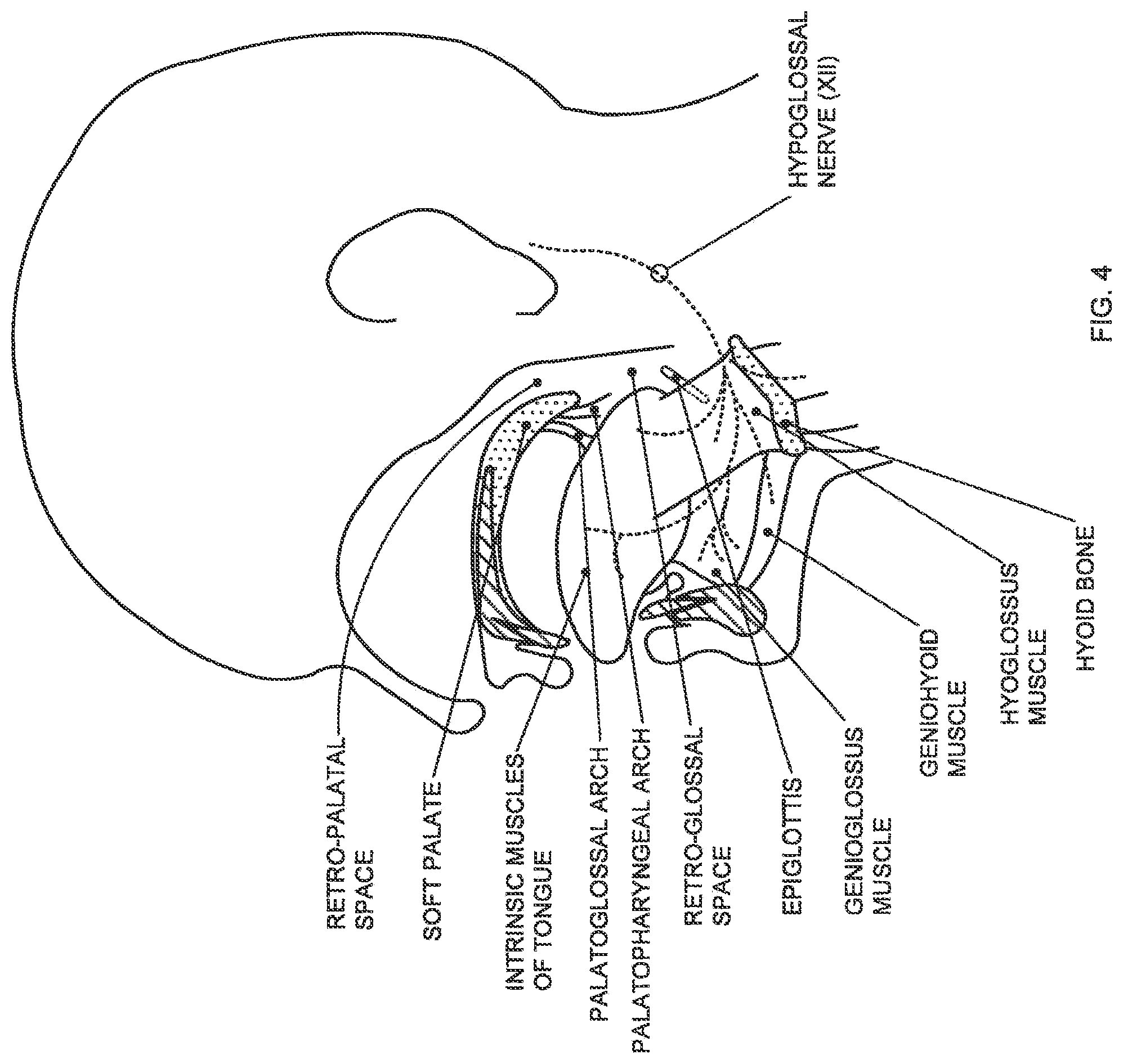
FIG. 4 is a schematic illustration showing the structures of the upper airway in a lateral dissection with the palate and mandible shown in medial sagittal section;
FIG. 5 is a schematic illustration showing the structures of the upper airway from the oral cavity;
FIG. 6 is a schematic illustration showing isolated structures of the upper airway in a transverse section;
FIG. 7 is a schematic illustration showing structures of the upper airway in a posterior dissection of the interior pharynx;
FIG. 8 is a schematic illustration showing structures of the upper airway in a posterior dissection of the exterior pharynx.
DETAILED DESCRIPTION
The following detailed description should be read with reference to the drawings in which similar elements in different drawings are numbered the same. The drawings, which are not necessarily to scale, depict illustrative embodiments and are not intended to limit the scope of the invention.
FIG. 1 schematically illustrates a hypoglossal nerve stimulation (HGNS) system 100 comprising internal components 200 and external components 300. The HGNS system 100 is intended to treat obstructive sleep apnea (OSA) by increasing neuromuscular activity of the genioglossus muscle via stimulation of the hypoglossal nerve (HGN) synchronous with inspiration to mitigate upper airway collapse during sleep. Stimulation is generated by an implantable neurostimulator (INS) 210, synchronized with inspiration as measured by the respiration sensing lead (RSL) 220 using bio-impedance, and delivered to the hypoglossal nerve by a stimulation lead (STL) 230. A programmer system 310 and a therapy controller 320 are wirelessly linked to the INS 210. The programmer system 310 includes a computer 330, a programmer interface 340, and a programmer head 350. The programmer system 310 is used by the physician to control and program the INS 210 during surgery and therapy titration, and the therapy controller 320 is used by the patient to control limited aspects of therapy delivery (e.g., start, stop, and pause).
The implanted components 200 of the HGNS system 100 include the INS 210, STL 230, and RSL 320. The INS is designed to accommodate one STL 230 and one RSL 220. One STL 230 may be used for unilateral implantation and unilateral hypoglossal nerve stimulation. Similarly, one RSL 220 may be used for respiration detection, and may be bifurcated as shown.
The implanted components 200 may be surgically implanted with the patient under general anesthesia. The INS 210 may be implanted in a subcutaneous pocket inferior to the clavicle over the pectoralis fascia. The distal end of the STL 230 (cuff 235) may be implanted on the hypoglossal nerve or a branch of the hypoglossal nerve in the submandibular region, and the proximal end of the STL 230 may be tunneled under the skin to the INS 210. The RSL 220 may be tunneled under the skin from the INS 210 to the rib cage and placed on both lateral sides of the costal margin. The INS 210 detects respiration via the RSL 220 using bio-impedance and stimulates the hypoglossal nerve via the STL 230 synchronous with inspiration.
Further aspects of the HGNS system 100 may be found in U.S. Provisional Patent Application No. 61/437,573, filed Jan. 28, 2011, entitled OBSTRUCTIVE SLEEP APNEA TREATMENT DEVICES, SYSTEMS AND METHODS, the entire disclosure of which is incorporated herein by reference.
Patients with obstructive sleep apnea have repeated episodes of complete (apnea) or partial (hypopnea) upper airway collapse during sleep. The upper airway is generally defined by four walls: the posterior pharyngeal wall, the right and left lateral pharyngeal walls, and anteriorly, the soft palate and the tongue. The posterior pharyngeal wall is relatively fixed to the spinal column. Thus, collapse of the upper airway generally involves, depending on the level and mode of collapse, the tongue, the soft palate and/or the lateral walls. In rare cases, collapse may involve the nasopharynx and/or hypopharynx. As seen in FIG. 2A, the tongue and the soft palate have been displaced posteriorly, thus occluding the airway at the level of the tongue (retro-glossal collapse) and at the level of the soft palate (retro-palatal collapse). As seen in FIG. 2B, activation of the genioglossus muscle, for example by HGNS, causes anterior displacement of the tongue, thus opening the retro-glossal airway space. Activation of the genioglossus muscle can also cause anterior displacement of the soft palate, thus opening the retro-palatal airway space. Although not visible in this view, activation of the genioglossus muscle can further cause lateral displacement of the lateral pharyngeal walls, thus further opening the upper airway. In this manner, activation of the genioglossus muscle, for example by HGNS, can mitigate upper airway collapse in OSA subjects.
Although the effect of genioglossus activation on the tongue to open the retro-glossal airway is predictable given the mechanism of action, the effect of genioglossus activation on the soft palate and lateral walls has been heretofore poorly understood and variable across subjects. Nevertheless, in the majority of OSA patients, the soft palate and the lateral walls can contribute to upper airway collapse, alone or in combination with the tongue. Thus, observing these effects can be important to predicting the success of HGNS therapy. This is particularly true if the soft palate and/or lateral walls are known to contribute to airway collapse for a given OSA patient.
The present invention offers a method to mimic genioglossus activation to observe and assess the effects thereof on structures of the upper airway. The method generally involves causing the tongue to protrude while observing the response of the upper airway using an imaging technique. In general, the desired response is an increase in airway size. An adequate increase in airway size during the tongue protrusion maneuver is indicative of likely therapeutic success with HGNS. If an adequate increase in airway size is observed during the maneuver, a HGNS device may be implanted in the patient with a higher confidence of a successful outcome.
With reference to FIG. 3A, a naso-endoscope 400 may be used to visually observe the upper airway while the patient is awake in the supine position. Alternatively, the observation may be made while the patient is in a seated or semi-recumbent position. A conventional naso-endoscope 400 including a fiber optic shaft 410 and a hand piece 420 may be used. Hand piece 420 may include a light source and a viewing window, and/or facilitate connection to ancillary imaging equipment (e.g., light source, camera, monitor, recorder, etc.). The patient may be asked to volitionally protrude his/her tongue straight and to its maximal extent with the mouth open and the lips loosely touching the tongue. Alternatively, the tongue protrusion may be performed sub-maximally, which may limit muscle contraction to the genioglossus without recruiting other musculature. Also alternatively, the tongue protrusion may be performed by asking the patient to point the tip of the tongue to one side or the other, which may more closely mimic unilateral hypoglossal nerve stimulation. The distal end of the endoscope may be positioned superior to the soft palate and substantially parallel with the posterior pharyngeal wall to visualize the retro-palatal space. The distal end of the endoscope may be positioned inferior to the soft palate, superior to the tongue base and substantially parallel with the posterior pharyngeal wall to visualize the retro-glossal space. An example of the view of the retro-palatal upper airway space with the tongue in a relaxed (nominal) position is shown in FIG. 3B, and the same view with the tongue protruded is shown in FIG. 3C. As can be seen by comparing the views in FIGS. 3B and 3C, tongue protrusion can result in an increase in airway size, including area, circumference, anterior-posterior dimension, and lateral dimension. The increase in airway size at the level of the tongue and palate may be most discernable by an increase in anterior-posterior (AP) dimension between the posterior pharyngeal wall and the posterior side of the tongue base (retro-glossal) and soft palate (retro-palatal), respectively. Since the posterior pharyngeal wall is fixed relative to the spinal column, the increase in AP dimension involves anterior displacement of the tongue and soft palate, respectively. The increase in airway size may also be discernable by an increase in lateral dimension between the right and left lateral pharyngeal walls.
During the tongue protrusion maneuver, observing an adequate increase in size of the retro-glossal airway is predictive of HGNS efficacy in patients with isolated tongue base collapse. However, as mentioned above, the soft palate contributes to upper airway collapse in the majority of OSA patients, thus also observing an increase in size of the retro-palatal airway during the tongue protrusion maneuver is predictive of HGNS efficacy in patients with isolated soft palate collapse and combined tongue plus soft palate collapse.
By way of example, not limitation, the following procedure may be followed to conduct the assessment and tongue protrusion maneuver. With the patient awake in the supine position, a nasal endoscope is inserted into the pharynx via one of the nares to allow visualization of the upper airway. Video and still images may be captured at both the retro-palatal and retro-glossal levels to document the effect of different maneuvers on anatomic structures of the upper airway (tongue, palate, epiglottis, pharyngeal walls, etc.). When imaging the retro-palatal level, the endoscope may be placed such that all four walls (soft palate, posterior wall, and the two lateral walls) of the pharynx are visible before, during and after maneuvers. Similarly, when imaging the retro-glossal level, the endoscope may be placed such that all four walls (tongue base, posterior wall, and the two lateral walls) of the pharynx are visible before, during and after maneuvers. The endoscope may be placed such that it runs generally parallel to the posterior wall and provides a symmetric field of view. This may be achieved by initially placing the distal end of the endoscope near the level of the epiglottis and subsequently pulling back to the desired level. The patient then performs a series of maneuvers, including a tongue protrusion maneuver while breathing through their nose. The tongue protrusion maneuvers involves voluntary maximal straight tongue protrusion with lips loosely touching the tongue, with the mouth completely open, and/or with the teeth clenched closed. Other maneuvers such as a Mueller maneuver (inspiratory efforts against a closed airway) may be performed as well. Each maneuver is held for 2:2 seconds, and performed several times while data (images and measurements) are gathered.
Alternative non-volitional tongue protrusion maneuvers include, for example, manually gripping and pulling the tongue anteriorly (e.g., by the physician), using a tongue retaining device (e.g., as used for the treatment of OSA), both of which are non-invasive. Another alternative is to stretch the palatoglossal arch by pushing the tongue down (depress tongue), by pushing the arch laterally outward, or by pulling the arch anteriorly (all palatoglossal maneuvers) using a tongue depressor or similar device. The palatoglossal maneuver may be used in place of or in combination with the tongue protrusion maneuver, and the entire description herein with respect to the tongue protrusion maneuver is applicable to the palatoglossal maneuver. Other alternative non-volitional tongue protrusion maneuvers include, for example, sub-mental stimulation and intra-muscular stimulation (using fine wire electrodes, for example), both of which are relatively more invasive, but have the benefit of more selectively activating the genioglossus muscle alone to more closely mimic HGNS, as compared to volitional tongue protrusion which may recruit more than the genioglossus muscle.
Although naso-endoscopy is perhaps the most practical imaging technique to employ to assess the response of the upper airway to the tongue protrusion maneuver, other imaging techniques may be used as well. For example, x-ray imaging, fluoroscopy, x-ray computed tomography (CT), and optical coherence tomography (OCT) are suitable alternatives. These alternatives may provide more quantitative measurements by using a reference marker of known dimension in the field of view. Alternatively, improvements may be made to conventional naso-endoscopes to facilitate more quantitative measurements. For example, with reference to FIG. 3D, conventional naso-endoscope 400 includes a fiber optic shaft 410 and a hand piece 420. The distal end of the shaft 410 may include an attached extension 430 having a tip 440. The extension 430 positions the tip 440 into the field of view and may be approximated to the upper airway structure being visualized. The tip 440 may have a known dimension (e.g., diameter of 1 French or 3 mm), such that quantitative measurements of upper airway structures may be made by comparison. Other devices to make quantitative measurements may be employed, such as a laser pointer of know beam diameter projected onto the upper airway structure of interest. As an alternative, a catheter (e.g., nasogastric, nasoesophageal or nasopharyngeal catheter) may be inserted into the nasopharynx such that it resides in the field of view of the endoscope to serve as a quantitative reference of known dimension (e.g., diameter).
As mentioned above, the upper airway assessment during tongue protrusion maneuver may be used as a screening tool wherein the patient is treated with the desired therapy (e.g., HGNS) only if the increase in size of the upper airway meets a predefined criterion. To this end, the response of the upper airway may be measured using a qualitative scale such as a visual analog scale of 0-10, wherein 0 represents a closed airway and 10 represents a completely open or patent airway. The airway size may be scored with the tongue at rest and during the tongue protrusion maneuver. The patient may be treated if the difference between the two scores meets a threshold, if the score during the maneuver meets a threshold, or if both the difference between the scores and the score during the maneuver meet thresholds (e.g., 5 on a scale of 0-10).
Alternatively, the response of the upper airway may be measured using a quantitative scale such as: a pixel count of captured images which may be representative of cross-sectional area; a linear dimension such as anterior-posterior and/or lateral; or a measure of circumference. Here again, the airway size may be measured (e.g., pixel count, AP length, and/or lateral width) with the tongue at rest and during the tongue protrusion maneuver. The patient may be treated if the difference between the two measures meets a threshold, if the measure during the maneuver meets a threshold, or if both the difference in measures and the measure during the maneuver meet thresholds.
In each case, the threshold may be a percentage increase in size (e.g., difference in AP length=50%), an absolute value (e.g., difference of AP length=0.5 cm), or a relative value. The relative value may be with reference to an anatomical landmark such as the width of the superior aspect of the epiglottis (e.g., difference in AP length=50% of epiglottal width).
Other response criteria observed during the tongue protrusion maneuver, in addition to an increase in airway size, may be used as well. For example, movement of the hyoid bone may be observed visually, by palpation or by x-ray. Movement of the hyoid bone in an anterior direction and/or inferior direction during the tongue protrusion maneuver may be predictive of therapeutic success with HGNS.
As mentioned above, although the effect of HGNS and genioglossus activation on the tongue to open the retro-glossal airway is predictable given the mechanism of action, the effect of genioglossus activation on the soft palate and lateral walls has been heretofore poorly understood. The explanation lies in the mechanical linkages between the genioglossus and other pharyngeal structures defining the upper airway. The linkages are primarily muscular, and can be effective without independent activation. Nevertheless, it may be desirable to independently activate any one or a combination of the muscular structures described below by stimulating the muscle directly or by stimulating the corresponding motor nerve innervating the muscle.
With reference to FIG. 4, the mechanical linkages may be explained in more detail. By way of context, the hypoglossal nerve (cranial nerve XII) innervates the genioglossus muscle, which is the largest upper airway dilator muscle. Activation of the genioglossus muscle causes tongue protrusion and, in some cases, anterior displacement of the soft palate, due to linkage via the palatoglossal arch (muscle). Anterior displacement of the soft palate, in turn, can cause tension to be applied to the lateral pharyngeal walls via the palatopharyngeal arch (muscle), the effect of which is discussed in more detail below. Thus, activation of the genioglossus muscle causes opening of the upper airway at the level of the tongue base (retro-glossal space) and, in some cases, at the level of the soft palate (retro-palatal space). Because the linkage between the genioglossus and the soft palate via the palatoglossal arch varies across subjects, the response to HGNS at the level of the palate will vary as well. This is significant because most people with OSA have some involvement of the palate during obstructive events, and it may be helpful to identify those subjects with inadequate retro-palatal opening due to poor linkage (i.e., poor coupling) between the genioglossus and soft palate, possibly due to tissue redundancy (i.e., slack) in the palatoglossus. Tissue redundancy may also be present in the lateral pharyngeal walls due to the presence of adipose tissue (i.e., fat) at discrete locations (e.g., fat pads) or distributed throughout the pharyngeal walls, particularly in patients with high BMI.
The anatomical linkage between the tongue base (genioglossus) and the soft palate via the palatoglossal arch may be more clearly seen in FIGS. 5 and 6. The palatoglossus muscle forms the palatoglossal arch and the anterior-inferior aspect of the soft palate on either side of the uvula. The inferior and lateral ends of the palatoglossus muscle insert into the genioglossus muscle. Posterior to the palatoglossal arch are the palatine tonsils, and posterior to the palatine tonsils is the palatopharyngeus muscle forming the palatopharyngeal arch and the posterior-inferior aspect of the soft palate on either side of the uvula. The inferior and lateral ends of the palatopharyngeus muscle insert into the lateral walls of the pharynx. The soft palate is also linked to the lateral pharyngeal walls inferiorly via the pharyngoepiglottic fold as best seen in FIG. 7. Activation of the genioglossus serves to pull the soft palate anteriorly via the palatoglossal linkage. Anterior displacement of the soft palate serves to apply anterior and lateral (outward) tension to the lateral pharyngeal walls via the palatopharyngeal linkage as well as the inferior lateral pharyngeal walls via the pharyngoepiglottic linkage.
The anatomical linkage between the tongue base (genioglossus) and the lateral pharyngeal walls may be better appreciated with reference to FIG. 8. The anterior-inferior aspect (not visible) of the styloglossus muscles insert into the genioglossus, and the posterior-superior aspect of the styloglossus muscles attach to the styloid process. Similarly, the anterior-inferior aspect (not visible) of the stylopharyngeus muscles insert into the lateral pharyngeal walls, and the posterior-superior aspect of the stylopharyngeus muscles attach to the styloid process. The glossopharyngeal aspects of the superior pharyngeal constrictor muscle also insert into the genioglossus. Thus, activation of the genioglossus serves to apply tension to the styloglossus and the glossopharyngeal aspects of the superior pharyngeal constrictor muscle, which in turn apply lateral outward tension to the lateral pharyngeal walls by virtue of the lateral outward position of the styloid process and the linkage via the stylopharyngeus muscles.
In sum, activation of the genioglossus muscle opens the retro-glossal airway as well as the retro-palatal airway via the linkages described above. In addition, activation of the genioglossus muscle serves to open the lateral pharyngeal walls via the linkages described above. However, the linked effects on the soft palate and the lateral pharyngeal walls is not present in all subjects but may be important for therapeutic success of HGNS depending on the level and mode of collapse in a given patient. By using a tongue protrusion maneuver to mimic the effect on the genioglossus muscle seen with HGNS, the response of the soft palate and lateral walls may be observed using endoscopy, for example. If the palatal and lateral walls respond sufficiently to the tongue protrusion maneuver, the likelihood of successful treatment with HGNS increases. Thus, observing the response of upper airway structures to the tongue protrusion maneuver may be used as a screening tool prior to implantation of a HGNS device.
Optionally, it may be desirable to observe the response of the airway at the level of collapse. The level of collapse may be determined during sleep or simulated sleep (e.g. sedation) using known techniques such as drug induced sleep endoscopy (DISE), or may be determined by examination of the airway structures using known techniques such as naso-endoscopy. The airway may collapse at the level of the tongue base (i.e., retro-glossal), at the level of the palate (i.e. retro-palatal), or both levels. Because most OSA patients have palatal involvement in airway collapse, it may not be necessary to determine the level of collapse. In this case, collapse may be assumed to occur at least at the level of the palate, and therefore an adequate response (e.g., increase in airway size) in the retro-palatal space during the tongue protrusion maneuver would be indicative of likely therapeutic success with HGNS.
The principles of the present invention may be applied to other therapeutic interventions for OSA involving the upper airway. For example, the tongue protrusion maneuver may be used as a screening tool for surgery of the upper airway, such as uvulopalatopharyngoplasty (UPPP), palatal implants, genioglossus advancement, maxilla-mandibular advancement, etc. Also, the tongue protrusion maneuver may be used as a screening tool for oral appliances such as mandibular repositioning devices, tongue retaining devices, etc.
Those skilled in the art will recognize that the present invention may be manifested in a variety of forms other than the specific embodiments described and contemplated herein. Accordingly, departures in form and detail may be made without departing from the scope and spirit of the present invention as described in the appended claims.
* * * * *
D00000
D00001
D00002
D00003
D00004
D00005
D00006
D00007
D00008
XML
uspto.report is an independent third-party trademark research tool that is not affiliated, endorsed, or sponsored by the United States Patent and Trademark Office (USPTO) or any other governmental organization. The information provided by uspto.report is based on publicly available data at the time of writing and is intended for informational purposes only.
While we strive to provide accurate and up-to-date information, we do not guarantee the accuracy, completeness, reliability, or suitability of the information displayed on this site. The use of this site is at your own risk. Any reliance you place on such information is therefore strictly at your own risk.
All official trademark data, including owner information, should be verified by visiting the official USPTO website at www.uspto.gov. This site is not intended to replace professional legal advice and should not be used as a substitute for consulting with a legal professional who is knowledgeable about trademark law.