Drug conjugated nanogels in microcapsule for delayed sustained protein delivery
Jabbari April 6, 2
U.S. patent number 10,967,098 [Application Number 15/897,347] was granted by the patent office on 2021-04-06 for drug conjugated nanogels in microcapsule for delayed sustained protein delivery. This patent grant is currently assigned to University' of South Carolina. The grantee listed for this patent is University of South Carolina. Invention is credited to Esmaiel Jabbari.


| United States Patent | 10,967,098 |
| Jabbari | April 6, 2021 |
Drug conjugated nanogels in microcapsule for delayed sustained protein delivery
Abstract
Injectable compositions and use of the injectable compositions in tissue engineering applications are described. The injectable compositions are hydrogel-based compositions that can be crosslinked in situ following placement. The injectable compositions include microcapsules having predetermined erosion profiles that are loaded with nanogels having predetermined sustained release profiles for signaling molecules conjugated to the nanogels. Following crosslinking, the compositions are designed to sequentially release signaling molecules over a predetermined period of time with various release profiles. The compositions can carry additional components to stimulate tissue generation such as stem cells and extracellular matrix (ECM) components.
| Inventors: | Jabbari; Esmaiel (Bethesda, MD) | ||||||||||
|---|---|---|---|---|---|---|---|---|---|---|---|
| Applicant: |
|
||||||||||
| Assignee: | University' of South Carolina
(Columbia, SC) |
||||||||||
| Family ID: | 1000005467353 | ||||||||||
| Appl. No.: | 15/897,347 | ||||||||||
| Filed: | February 15, 2018 |
Prior Publication Data
| Document Identifier | Publication Date | |
|---|---|---|
| US 20180250438 A1 | Sep 6, 2018 | |
Related U.S. Patent Documents
| Application Number | Filing Date | Patent Number | Issue Date | ||
|---|---|---|---|---|---|
| 62465312 | Mar 1, 2017 | ||||
| Current U.S. Class: | 1/1 |
| Current CPC Class: | A61L 27/3834 (20130101); A61L 27/26 (20130101); A61L 27/54 (20130101); A61L 27/3633 (20130101); A61L 27/58 (20130101); A61L 27/3612 (20130101); A61L 2400/06 (20130101); A61L 2300/622 (20130101); A61L 2400/12 (20130101); A61L 2300/414 (20130101); A61L 2430/06 (20130101); A61L 2300/64 (20130101) |
| Current International Class: | A61L 27/26 (20060101); A61L 27/38 (20060101); A61L 27/36 (20060101); A61L 27/54 (20060101); A61L 27/58 (20060101) |
References Cited [Referenced By]
U.S. Patent Documents
| 6884432 | April 2005 | Yaszemski et al. |
| 7001987 | February 2006 | Van Dyke |
| 7642300 | January 2010 | Yaszemski et al. |
| 9101654 | August 2015 | Jabbari |
| 9314549 | April 2016 | Jabbari |
| 9808555 | November 2017 | Jabbari |
| 2007/0043202 | February 2007 | Yaszemski et al. |
| 2008/0206308 | August 2008 | Jabbari et al. |
| 2009/0324722 | December 2009 | Elisseeff |
| 2010/0084784 | April 2010 | Jabbari |
| 2010/0086607 | April 2010 | Jabbari |
| 2010/0322979 | December 2010 | Jabbari |
| 2010/0327494 | December 2010 | Jabbari |
| 2012/0226295 | September 2012 | Jabbari |
| 2014/0349367 | November 2014 | Jabbari |
| 2015/0175972 | June 2015 | Jabbari |
| 2018/0251505 | September 2018 | Jabbari |
| 2018/0256780 | September 2018 | Jabbari |
Other References
|
Lin et al. (Pharmaceutical Research. Mar. 2009; 26(3): 631-643). (Year: 2009). cited by examiner . Jayasuriya et al. (Ann N Y Acad Sci. Nov. 2016 ; 1383(1): 21-33.) (Year: 2016). cited by examiner . Anderson, et al. "Post-Traumatic Osteoarthritis: Improved Understanding and Opportunities for Early Intervention." J. Orthop. Res. 29(6): (2011), pp. 802-809. cited by applicant . Andrades, et al., "Induction of superficial zone protein (SZP)/lubricin/PRG 4 in muscle-derived mesenchymal stem/progenitor cells by transforming growth factor-beta1 and bone morphogenetic protein-7," Arthritis Res. Ther. 14(2): (2012), R72. cited by applicant . Annabi et al., "25th Anniversary Article: Rational Design and Applications of Hydrogels in Regenerative Medicine," Advanced Materials, 26 (2014), pp. 85-124. cited by applicant . Aoyama et al., "Keratin Nanofiber Scaffold for Vascular Graft" (Abstract Only), Tissue Engineering Part A, 21 (2015), p. S244. cited by applicant . Arai et al., "Amino acid sequence of feather keratin from fowl," European Journal of Biochemistry, 132 (1983), pp. 506-507. cited by applicant . Audouin, et al., "Surface-initiated RAFT polymerization of NIPAM from monolithic macroporous polyHIPE," European Polymer Journal 49(5): pp. 1073-1079 (2013). cited by applicant . Balaji et al., "Characterization of keratincollagen 3D scaffold for biomedical applications," Polymers for Advanced Technologies, 23 (2012), pp. 500-507. cited by applicant . Barati et al., "Effect of Organic Acids on Calcium Phosphate Nucleation and Osteogenic Differentiation of Human Mesenchymal Stem Cells on Peptide Functionalized Nanofibers," Langmuir, 31 (2015), pp. 5130-5140. cited by applicant . Barati et al., "Spatiotemporal release of BMP-2 and VEGF enhances osteogenic and vasculogenic differentiation of human mesenchymal stem cells and endothelial colony-forming cells coencapsulated in a patterned hydrogel," Journal of Controlled Release, 223 (2016), pp. 126-136. cited by applicant . Barati, et al., "Synthesis and characterization of photocrosslinkable keratin hydrogels for stem cell Encapsulation," Biomacromolecules, 18(2): (2017), pp. 398-412. cited by applicant . Barati et al., "Time dependence of material properties of polyethylene glycol hydrogels chain extended with short hydroxy acid segments," Polymer, 55 (2014), pp. 3894-3904. cited by applicant . Barone et al., "Thermally processed keratin films," Journal of Applied Polymer Science, 97 (2005), pp. 1644-1651. cited by applicant . Bernardes et al., "Facile conversion of cysteine and alkyl cysteines to dehydroalanine on protein surfaces: versatile and switchable access to functionalized proteins," Journal of the American Chemical Society, 130 (2008), pp. 5052-5053. cited by applicant . Bhardwaj et al., "Silk fibroin-keratin based 3D scaffolds as a dermal substitute for skin tissue engineering," Integrative Biology, 7 (2015), pp. 53-63. cited by applicant . Burnett et al., "Hemostatic properties and the role of cell receptor recognition in human hair keratin protein hydrogels," Biomaterials, 34 (2013), pp. 2632-2640. cited by applicant . Chalker et al., "Chemical modification of proteins at cysteine: opportunities in chemistry and biology," Chemistry--An Asian Journal, 4 (2009), pp. 630-640. cited by applicant . Chan et al., "Crosslinking of collagen scaffolds promotes blood and lymphatic vascular stability," Journal of Biomedical Materials Research Part A. 102 (2014), pp. 3186-3195. cited by applicant . Chen et al., "A Universal and Facile Approach for the Formation of a Protein Hydrogel for 3D Cell Encapsulation," Advanced Functional Materials, 25 (2015), pp. 6189-6198. cited by applicant . Chen et al., "Engineering Vascularized Tissue Constructs using an Injectable Cell-laden Collagen Hydrogel" (Abstract Only), Tissue Engineering Part A, 21 (2015), S102. cited by applicant . Dawson et al., "Biomaterials for stem cell differentiation," Advanced Drug Delivery Reviews, 60 (2008), pp. 215-228. cited by applicant . D'Este, et al., "Evaluation of an injectable thermoresponsive hyaluronan hydrogel in a rabbit osteochondral defect model," Journal Biomed. Mater. Res. Part A 104(6) (2016), pp. 1469-1478. cited by applicant . Dong et al., "In Situ "Clickable" Zwitterionic Starch-Based Hydrogel for 3D Cell Encapsulation," ACS Applied Materials & Interfaces, 8 (2016), pp. 4442-4455. cited by applicant . Dong et al., "Injectable Hybrid Hydrogel for Mesenchymal Stem Cell Delivery, from PEG-based Multifunctional Hyperbranched Polymers" (Abstract Only), Tissue Engineering Part A, 21 (2015), S298-S289. cited by applicant . Eastoe, "The amino acid composition of mammalian collagen and gelatin," Biochemical Journal, 61 (1955), p. 589. cited by applicant . Evans, et al., "Use of Genetically Modified Muscle and Fat Grafts to Repair Defects in Bone and Cartilage," Eur. Cells Mater. 18 (2009), pp. 96-111. cited by applicant . Falah, et al., "Treatment of articular cartilage lesions of the knee." International Orthopaedics, 34(5) (2010), pp. 621-630. cited by applicant . Ferlin et al., "Development of a Dynamic Stem Cell Culture Platform for Mesenchymal Stem Cell Adhesion and Evaluation," Molecular Pharmaceutics, 11 (2014), pp. 2172-2181. cited by applicant . Fraser et al., Keratins: their composition, structure, and biosynthesis: Charles C. Thomas, (1972). cited by applicant . Fuhrmann et al., "Injectable hydrogel promotes early survival of induced pluripotent stem cell-derived oligodendrocytes and attenuates longterm teratoma formation in a spinal cord injury model," Biomaterials, 83 (2016), pp. 23-36. cited by applicant . Fukumoto, et al., "Combined effects of insulin-like growth factor-1 and transforming growth factor-.beta. 1 on periosteal mesenchymal cells during chondrogenesis in vitro," Osteoarthritis Cartilage 11(1) (2003), pp. 55-64. cited by applicant . Golub et al., "The Role of Alkaline Phosphatase in Cartilage Mineralization," Bone and Mineral, 17 (1992), pp. 273-278. cited by applicant . Gorman, "Materials Take Wing: What to do with 4 billion pounds of feathers?", Science News, 161 (2002), p. 120. cited by applicant . Grogan, et al., "Zone-Specific Gene Expression Patterns in Articular Cartilage," Arthritis Rheum. 65(2) (2013), pp. 418-428. cited by applicant . Guo et al., "In vitro generation of an osteochondral construct using injectable hydrogel composites encapsulating rabbit marrow mesenchymal stem cells," Biomaterials, 30 (2009), pp. 2741-2752. cited by applicant . Han et al., "Alkylation of human hair keratin for tunable hydrogel erosion and drug delivery in tissue engineering applications," Acta Biomaterialia, 23 (2015), pp. 201-213. cited by applicant . Han, et al., "Bioerodable PLGA-Based Microparticles for Producing Sustained Release Drug Formulations and Strategies for Improving Drug Loading," Frontiers Pharmacol. 7 (2016). cited by applicant . Haralson et al., "Extracellular matrix: A practical approach," Annales de Biologie Clinique, (1996), pp. 383-384. cited by applicant . He et al., "Effect of grafting RGD and BMP-2 protein-derived peptides to a hydrogel substrate on osteogenic differentiation of narrow stromal cells," Langmuir, 24 (2008) pp. 12508-12516. cited by applicant . Hoffman, "Hydrogels for biomedical applications," Advanced Drug Delivery Reviews, 64 (2012), pp. 18-23. cited by applicant . Jayathilakan et al., "Utilization of byproducts and waste materials from meat, poultry and fish processing industries: a review," Journal of Food Science and Technology, 49 (2012), pp. 278-293. cited by applicant . Kakkar et al., "Extraction and characterization of keratin from bovine hoof: A potential material for biomedical applications," Springerplus, 3 (2014), p. 596. cited by applicant . Karaman et al., "Effect of surface modification of nanofibres with glutamic acid peptide on calcium phosphate nucleation and osteogenic differentiation of marrow stromal cells," Journal of Tissue Engineering and Regenerative Medicine, 10 (2016), pp. E132-E146. cited by applicant . Karimi et al., "A developmentally inspired combined mechanical and biochemical signaling approach on zonal lineage commitment of mesenchymal stem cells in articular cartilage regeneration," Integrative Biology, 7 (2015), pp. 112-127. cited by applicant . Kelly et al., "How to study proteins by circular dichroism," Biochimica et Biophysica Acta (BBA)-Proteins and Proteomics, 1751 (2005), pp. 119-139. cited by applicant . Klein, et al., "Depth dependent biomechanical and biochemical properties of fetal, newborn, and tissue-engineered articular cartilage," Journal Biomech. 40(1) (2007), pp. 182-190. cited by applicant . Kwon et al., "In vivo osteogenic differentiation of human turbinate mesenchymal stem cells in an injectable in situ-forming hydrogel," Biomaterials, 35 (2014), pp. 5337-5346. cited by applicant . Lam, et al., "Osteochondral defect repair using bilayered hydrogels encapsulating both chondrogenically and osteogenically pre-differentiated mesenchymal stem cells in a rabbit model," Osteoarthritis Cartilage 22(9) (2014), pp. 1291-1300. cited by applicant . Lee, et al., "Regeneration of the articular surface of the rabbit synovial joint by cell homing: a proof of concept study," Lancet 376 (9739), pp. 440-448. cited by applicant . Li, et al., "The Effect of Oxygen Tension on Human Articular Chondrocyte Matrix Synthesis: Integration of Experimental and Computational Approaches," Biotechnol. Bioeng. 111(9), (2014), pp. 1876-1885. cited by applicant . Lin et al., "Allyl sulfides are privileged substrates in aqueous cross-metathesis: application to site protein modification," Journal of the American Chemical Society, 130 (2008), pp. 9642-9643. cited by applicant . Long et al., "Improving the mechanical properties of collagen-based membranes using silk fibroin for corneal tissue engineering," Journal of Biomedical Materials Research Part A, 103 (2015), pp. 1159-1168. cited by applicant . Lotz, M.K., "Posttraumatic osteoarthritis: pathogenesis and pharmacological treatment options," Arthritis Res. Ther. 12(3) (2010). cited by applicant . Lv et al., "Structural and functional evaluation of oxygenating keratin/silk fibroin scaffold and initial assessment of their potential for urethral tissue engineering," Biomaterials, 84 (2016), pp. 99-110. cited by applicant . Ma et al., "Enhanced biological stability of collagen porous scaffolds by using amino acids as novel cross-linking bridges," Biomaterials, 25 (2004), pp. 2997-3004. cited by applicant . Mabry et al., "Microarray analyses to quantify advantages of 2D and 3D hydrogel culture systems in maintaining the native valvular interstitial cell phenotype," Biomaterials, 74 (2016), pp. 31-41. cited by applicant . Mak, et al., "Indian hedgehog signals independently of PTHrP to promote chondrocyte hypertrophy," Development 135(11) (2008), pp. 1947-1956. cited by applicant . Mariani, et al., "Signaling Pathways in Cartilage Repair," International Journal of Molecular Science 15(5): (2014), pp. 8667-8698. cited by applicant . Melrose, et al., "Chondroitin sulphate and heparan sulphate sulphation motifs and their proteoglycans are involved in articular cartilage formation during human foetal knee joint development," Histochem. Cell Biol. 138(3): (20112), pp. 463-475. cited by applicant . Mercado, et al., "Effect of grafting BMP2-derived peptide to nanoparticles on osteogenic and vasculogenic expression of stromal cells," Journal of Tissue Eng. Regen. Med. 8(1): (2014), pp. 15-28. cited by applicant . Mi, et al., "Adverse effects of adenovirus-mediated gene transfer of human transforming growth factor beta 1 into rabbit knees," Arthritis Res. Ther. 5(3): (2003), pp. R132-R139. cited by applicant . Moeinzadeh et al., "B13Nanostructure Formation and Transition from Surface to Bulk Degradation in Polyethylene Glycol Gels Chain-Extended with Short Hydroxy Acid Segments," Biomacromolecules, 14 (2013), pp. 2917-2928. cited by applicant . Moeinzadeh, et al. "Comparative effect of physicomechanical and biomolecular cues on zone-specific chondrogenic differentiation of mesenchymal stem cells," Biomaterials 92, (2016), pp. 57-70. cited by applicant . Moeinzadeh et al., "Gelation Characteristics and Osteogenic Differentiation of Stromal Cells in Inert Hydrolytically Degradable Micellar Polyethylene Glycol Hydrogels," Biomacromolecules, 13 (2012), pp. 2073-2086. cited by applicant . Munoz-Pinto et al., "Collagen-mimetic hydrogels promote human endothelial cell adhesion, migration and phenotypic Maturation," Journal of Materials Chemistry B. 3 (2015), pp. 7917-7919. cited by applicant . Namba, et al., "Spontaneous repair of superficial defects in articular cartilage in a fetal lamb model," J. Joint Surg. Am. vol. 80A(1):(1998), pp. 4-10. cited by applicant . Nichol et al., "Cell-laden microengineered gelatin methacrylate hydrogels," Biomaterials, 31 (2010), pp. 5536-5544. cited by applicant . Oliver-Welsh, et al, "Deciding how best to treat cartilage defects," Orthopedics 39: (2016), pp. 343-350. cited by applicant . Orth, et al., "Reliability, Reproducibility, and Validation of Five Major Histological Scoring Systems for Experimental Articular Cartilage Repair in the Rabbit Model," Tissue Eng. Part C Methods 18(5): (2012), pp. 329-339. cited by applicant . Pace et al., "A Human Hair Keratin Hydrogel Scaffold Enhances Median Nerve Regeneration in Nonhuman Primates: An Electrophysiological and Histological Study," Tissue Engineering Part A, 20 (2014), pp. 507-517. cited by applicant . Pascher, et al., "Gene delivery to cartilage defects using coagulated bone marrow aspirate," Gene Ther. 11(2): (2004), pp. 133-141. cited by applicant . Patel et al., "Biodegradable polymer scaffold for tissue engineering," Trends in Biomaterials and Artificial Organs, 25 (2011), pp. 20-29. cited by applicant . Pfaff, K. "A third of soldiers disabled after ACI for lesions in the knee," Orthopedics Today (2014). cited by applicant . Punzi, et al., "Post-traumatic arthritis: overview on pathogenic mechanisms and role of inflammation," Rheumatic Musculoskeletal Dis. 2(2): (2016), p. 279. cited by applicant . Rehmann et al., "Tuning microenvironment modulus and biochemical composition promotes human mesenchymal stem cell tenogenic differentiation," Journal of Biomedical Materials Research Part A, 104 (2016), pp. 1162-1174. cited by applicant . Rivera, et al, "Posttraumatic osteoarthritis caused by battlefield injuries: the primary source of disability in warriors," Journal Am. Acad. Orthop. Surg. 20(1): (2012), pp. S64-S69. cited by applicant . Rivera, et al, "The burden of posttraumatic arthritis," AAOS/OTA/SOMOS/ORS Extremity War Injuries VIII: Sequelae of Combat (2013). cited by applicant . Rouse et al., "A Review of Keratin-Based Biomaterials for Biomedical Applications," Materials, 3 (2010), pp. 999-1014. cited by applicant . Saravanan et al., "Exploration on the Amino Acid Content and Morphological Structure in Chicken Feather Fiber" Journal of Textile and Apparel, Technology and Management, 7-3, (2012). cited by applicant . Sawada at al., "Scaffold for Cell Culture Made by Electrospun Keratin Nanofibers" (Abstract Only), Tissue Engineering Part A, 20 (2014), p. S65. cited by applicant . Simank, et al., "Effects of local application of growth and differentiation factor-5 (GDF-5) in a full-thickness cartilage defect model," Growth Factors 22(1): (2004), pp. 35-43. cited by applicant . Stenman et al., "Trypsin-2 degrades human type II collagen and is expressed and activated in mesenchymally transformed rheumatoid arthritis synovitis tissue," American Journal of Pathology, 167 (2005), pp. 1119-1124. cited by applicant . Stockwell, R.A. "Interrelationship of Cell Density and Cartilage Thickness in Mammalian Articular Cartilage," Journal of Anatomy 109: (1971), pp. 411-421. cited by applicant . Studer, et al., "Molecular and Biophysical Mechanisms Regulating Hypertrophic Differentiation in Chondrocytes and Mesenchymal Stem Cells," Eur. Cells Mater. 24: (2012), pp. 118-135. cited by applicant . Tan et al., "Fabrication and Evaluation of Porous Keratin/chitosan (KCS) Scaffolds for Effectively Accelerating Wound Healing," Biomedical and Environmental Sciences, 28 (2015), pp. 178-189. cited by applicant . Tanabe at al., "Fabrication and characterization of chemically crosslinked keratin films," Materials Science and Engineering: C, 24 (2004), pp. 441-446. cited by applicant . Tropel et al., "Isolation and characterization of mesenchymal stem cells from adult mouse bone marrow," Experimental Cell Research, 295 (2004), pp. 395-406. cited by applicant . Verma et al., "Preparation of scaffolds from human hair proteins for tissue-engineering applications," Biomedical materials, 3-2 (2008), 025007. cited by applicant . Verschure, et al., "Localization of insulin-like growth factor-I receptor in human normal and osteoarthritic cartilage in relation to proteoglycan synthesis and content," Br. J. Rheumatol. 35(11): (1996), pp. 1044-1055. cited by applicant . Visser, et al., "Crosslinkable Hydrogels Derived from Cartilage, Meniscus, and Tendon Tissue" Tissue Eng. Part A 21(7-8): (2015), pp. 1195-1206. cited by applicant . Wagegg, et al., "Hypoxia Promotes Osteogenesis but Suppresses Adipogenesis of Human Mesenchymal Stromal Cells in a Hypoxia-Inducible Factor-1 Dependent Manner," PloS One 7(9): (2012), e46483 10. cited by applicant . Wang et al., "Human keratin hydrogels support fibroblast attachment and proliferation in vitro," Cell and Tissue Research, 347 (2012), pp. 795-802. cited by applicant . Wang, et al., "TGF .beta. signaling in cartilage development and maintenance," Birth Defects Res. Part C Embryo Today Rev. 102(1): (2014), pp. 37-51. cited by applicant . Watson, et al., "Gene delivery of TGE-.beta. 1 induces arthrofibrosis and chondrometaplasia of synovium in vivo" Laboratory Investigation, 90(11): (2010), pp. 1615-1627. cited by applicant . Wehling, et al., "Interleukin-1 .beta. and Tumor Necrosis Factor .alpha. Inhibit Chondrogenesis by 16 Human Mesenchymal Stem Cells Through NF-.kappa. B-Dependent Pathways," Arthritis Rheum. 60(3): (2009), pp. 801-812. cited by applicant . Williamson, et al., "Growth of immature articular cartilage in vitro: Correlated variation in tensile biomechanical and collagen network properties," Tissue Eng. 9(4): (2003), pp. 625-634. cited by applicant . Wong, et al., "Chondrocyte biosynthesis correlates with local tissue strain in statically compressed adult articular cartilage," Journal of Orthop. Res. 15(2): (1997), pp. 189-196. cited by applicant . Wu, et al. "Human developmental chondrogeriesis as a basis for engineering chondrocytes from pluripotent stem cells," Stem Cell Reports 1(6): (2013), pp. 575-589. cited by applicant . Xu et al., "Water-Stable Three-Dimensional Ultrafine Fibrous Scaffolds from Keratin for Cartilage Tissue Engineering," Langmuir, 30 (2014), pp. 8461-8470. cited by applicant . Yamauchi et al., "Preparation of stable aqueous solution of keratins, and physiochemical and biodegradational properties of films," Journal of Biomedical Materials Research, 31 (1996), pp. 439-444. cited by applicant . Yang, et al., "Engineering Orthopedic Tissue Interfaces," Tissue Engineering Part B Rev. 15(2): (2009), pp. 127-141. cited by applicant . Yin et al., "Study on effective extraction of chicken feather keratins and their films for controlling drug release," Biomaterials Science, 1 (2013), pp. 528-536. cited by applicant . Yue et al., "Synthesis, properties, and biomedical applications of gelatin methacryloyl (GelMA) hydrogels," Biomaterials, 73 (2015), pp. 254-271. cited by applicant . Zhang, et al., "The role of tissue engineering in articular cartilage repair and regeneration," Crit. Rev. Biomed. Eng. 37(1-2): (2009), pp. 1-57. cited by applicant . Lee, et al. "Production of nanoparticles-in-microparticles by a double emulsion method: A comprehensive study" Eur. J. Pharm. Biopharm. 83 (2013) pp. 168-173. cited by applicant. |
Primary Examiner: Long; Scott
Attorney, Agent or Firm: Dority & Manning, P.A.
Parent Case Text
CROSS REFERENCE TO RELATED APPLICATION
This application claims filing benefit of U.S. Provisional Patent Application Ser. No. 62/465,312 having a filing date of Mar. 1, 2017, which is incorporated herein by reference for all purposes.
Claims
What is claimed is:
1. A composition for tissue engineering comprising; a plurality of first nanogels, each first nanogel including a first crosslinked degradable polymer conjugated to a first signaling molecule, the first degradable polymer comprising a first polyethylene glycol having a first molecular weight, the first signaling molecule stimulating formation or repair of a biological tissue, the first molecular weight of the first polyethylene glycol providing a first degradation rate to the first nanogel; a plurality of first microcapsules, each first microcapsule carrying a plurality of the first nanogels, the first microcapsules each comprising a shell that includes a first bioerodable polymer; a plurality of second nanogels, each second nanogel including a second crosslinked degradable polymer conjugated to a second signaling molecule, the second degradable polymer comprising a second polyethylene glycol having a second molecular weight, the second signaling molecule stimulating formation or repair of the biological tissue, the second molecular weight of the second polyethylene glycol providing a second degradation rate to the second nanogel, the second degradation rate differing from the first degradation rate; a plurality of second microcapsules, each second microcapsule carrying a plurality of the second nanogels, the second microcapsules each comprising a shell that includes a second bioerodable polymer; a crosslinkable polymer.
2. The composition of claim 1, the composition further comprising a plurality of living cells.
3. The composition of claim 2, wherein the living cells comprise stem cells.
4. The composition of claim 1, the composition further comprising one or more components of an extra-cellular matrix.
5. The composition of claim 1, the first and second polyethylene glycols comprising hydrophobic segments at the termini.
6. The composition of claim 5, the hydrophobic segments comprising lactide monomers and glycolide monomers.
7. The composition of claim 1, wherein the first bioerodable polymer comprises a first polylactide-based polymer and the second bioerodable polymer comprises a second polylactide-based polymer.
8. The composition of claim 1, wherein the crosslinkable polymer comprises a biopolymer.
9. The composition of claim 8, wherein the crosslinkable biopolymer has been derived from cartilage.
10. The composition of claim 9, wherein the crosslinkable biopolymer has been derived from fetal articular cartilage.
11. The composition of claim 1, the first and second signaling molecules stimulating the formation or repair of cartilage.
12. The composition of claim 1, further comprising additional microcapsules, the additional microcapsules carrying additional nanogels comprising additional crosslinked degradable polymers conjugated to additional signaling molecules.
13. The composition of claim 1, wherein the first signaling molecule is TGF.beta.1, and the second signaling molecule is BMP7, the composition further comprising a plurality of third microcapsules, each third microcapsule carrying a plurality of third nanogels that include a third crosslinked degradable polymer conjugated to IGF1, the composition further comprising a plurality of fourth microcapsules, each fourth microcapsule carrying a plurality of fourth nanogels that include a fourth crosslinked degradable polymer conjugated to IHH.
14. The composition of claim 1, wherein the first microcapsules have a first size and the second microcapsules have a second size that differs from the first size.
15. The composition of claim 1, wherein the first and second microcapsules have a largest external dimension of from about 1 micrometer to about 10 micrometers and the composition is an injectable composition.
16. A composition for tissue engineering comprising: a plurality of first nanogels, each first nanogel including a first crosslinked degradable polymer conjugated to a first signaling molecule, the first signaling molecule stimulating formation or repair of a biological tissue, the first degradable polymer comprising a first polyethylene glycol comprising a first hydrophobic segment at a terminus, the first hydrophobic segment comprising first monomers; a plurality of first microcapsules, each first microcapsule carrying a plurality of the first nanogels, the first microcapsules each comprising a shell that includes a first polylactide-based polymer; a plurality of second nanogels, each second nanogel including a second crosslinked degradable polymer conjugated to a second signaling molecule, the second signaling molecule stimulating formation or repair of biological tissue, the second degradable polymer comprising a second polyethylene glycol comprising a second hydrophobic segment at a terminus, the second hydrophobic segment comprising second monomers that are more hydrophobic than the first monomers; a plurality of second microcapsules, each second microcapsule carrying a plurality of the second nanogels, the second microcapsules each comprising a shell that includes a second polylactide-based polymer; and a crosslinkable polymer.
17. The composition of claim 16, the first monomers comprising glycolide monomers.
18. The composition of claim 16, the second monomers comprising caprolactone monomers.
19. The composition of claim 16, the first hydrophobic segment and/or the second hydrophobic segment further comprising lactide monomers.
20. The composition of claim 16, wherein the first polylactide-based polymer comprises a polylactide-co-glycolide copolymer.
21. The composition of claim 20, wherein the second polylactide-based polymer comprises a polylactide-co-caprolactone copolymer.
22. The composition of claim 16, wherein the first microcapsules have a first size and the second microcapsules have a second size that differs from the first size.
23. The composition of claim 16, wherein the first and second microcapsules have a largest external dimension of from about 1 micrometer to about 10 micrometers and the composition is an injectable composition.
Description
BACKGROUND
Natural tissue formation and differentiation takes place according to a well-ordered process that is controlled by sequential expression of signaling molecules. For instance, during embryonic development of cartilage, mesenchymal stem cells (MSCs) condense by up-regulation of transforming growth factor-.beta.1 (TGF.beta.1) signaling. The condensed MSCs differentiate into pre-chondrocytes by co-expression of TGF.beta.1 and bone morphogenetic protein-7 (BMP7) to form a matrix remarkably similar to the superficial zone of articular cartilage. Next, the developing fetal cartilage divides into two morphologically distinct zones, namely the superficial and middle zones, by further differentiation of pre-chondrocytes to pre-hypertrophic chondrocytes driven by the gradients in BMP7 and insulin-like growth factor-1 (IGF1). In a similar process, the cells further away from the synovial cavity mature into hypertrophic chondrocytes by gradients in BMP7, IGF1, and Indian Hedgehog (IHH) to generate the complete zonal organization (superficial, middle, calcified) of articular cartilage. Early morphogenetic events are also significantly affected by the local environment of the developing tissue. For instance, during the embryonic development of cartilage, in addition to the sequential expression of morphogens, the extracellular matrix (ECM) motifs in fetal articular cartilage play a significant development role.
Unfortunately, later in the life cycle, the body is no longer able to generate tissue in repair/replacement processes as effectively as is possible during embryonic development. For instance, fetal articular cartilage injuries are able to heal spontaneously and avoid fibrous scar tissue, while postnatal articular cartilage lacks the ability for complete self-repair.
The inability of tissue damage to heal effectively can lead to issues due to scar tissue formation as well as long term tissue damage. A significant consequence of traumatic extremity injuries and the inability for self-repair of articular cartilage is post-traumatic osteoarthritis (PTOA). The rapidly applied load in traumatic injuries focally disrupts the articular cartilage beginning with cell death, decreased stiffness and rapid progression to a full-thickness defect with time. Approximately 12% of all osteoarthritis (OA) cases are caused by PTOA. About 3.5 million individuals in the US suffer from PTOA of the hip, knee, or ankle with a total cost of $3 billion to the health care system. PTOA can occur in any age from any acute physical trauma such as sport, vehicle accident, fall, or military injury.
Unfortunately, treatment of tissue damage absent of or only minimally supported by the benefit of natural repair mechanisms remains limited in effectiveness. Conventional clinical approaches to treatment of full-thickness chondral defects such as subchondral drilling, microfracture, and abrasion arthroplasty create mechanically-inferior fibrocartilage. Osteochondral autograft transfer or mosaicplasty suffers from an additional surgical intervention and donor site morbidity. Autologous chondrocyte implantation (ACI) fails to restore zonal organization of the articular cartilage and in some cases leads to peripheral hypertrophy and calcification. For example, 33% of ACI procedures performed on soldiers suffering from PTOA of the knee resulted in permanent disability. Tissue engineered (TE) articular cartilage grafts have also been developed. As the middle zone is the thickest layer and bears the bulk of the compressive load, most TE articular cartilage grafts are based on mimicking the properties of the middle zone. However, these grafts have been shown to degenerate into biologically inferior fibrocartilage tissue.
What are needed in the art are improved compositions for use in reconstruction and repair of damaged tissue that can more closely mimic and encourage the body's natural tissue formation mechanisms. For instance, injectable therapies that can regenerate the zonal microstructure of articular cartilage would be of great benefit in the art, and have the potential to prevent permanent disability in patients suffering from PTOA.
SUMMARY
A composition is disclosed for use in tissue engineering applications. The composition includes nano-sized particles (i.e., nanogels) that include a crosslinked polymer conjugated to a signaling molecule (e.g., a protein or a functional fragment of a protein) that encourages some aspect of tissue formation/repair. For example, a signaling molecule can include an expression product that has efficacy in stem cell differentiation and/or tissue formation (e.g., TGF.beta.1, BMP7, IGF1, IHH, BMP2, vascular endothelial growth factor (VEGF), etc.). The crosslinked polymer of the nanogel is designed to degrade over a predetermined time period and thus provide for sustained release of the signaling molecule over a desired period of time.
The composition can also include a plurality of microcapsules, and each microcapsule can carry a plurality of the nanogels. Each nanogel of a single microcapsule can carry the same signaling molecule (or combination of signaling molecules) as one another. Alternatively, the nanogels held within a single microcapsule can vary from one another. The microcapsules can include a bioerodable polymer that is designed to exhibit a controlled release profile for the nanogels carried inside. As such, the release of the nanogels (and the subsequent sustained release of the signaling molecule(s) carried by the nanogels) can be controlled (e.g., delayed) following placement of the composition at a targeted location, e.g., via injection. For instance, a composition can carry a first set of microcapsules that release the nanogels immediately following placement of the composition. The composition can also carry a second set of microcapsules that release the nanogels only after a predesigned delay period. Thus, the signaling molecules of the nanogels can be released into the area according to a sequential and sustained release profile.
The composition can also include a crosslinkable polymer and can define a rheology suitable for injection to a targeted site. Following placement of the composition, the polymer can be crosslinked in situ by application of energy (e.g., UV crosslinking, temperature-based crosslinking, etc.) so as to form a semi-solid, crosslinked hydrogel at a location of desired tissue formation.
The crosslinkable composition can include a plurality of the microcapsules (that, in turn each carry a plurality of nanogels) as well as additional components for tissue engineering. For instance, a composition can carry differentiated or stem cells (e.g., mesenchymal stem cells) that can be affected by the sequential release of the signaling molecules from the nanogels to form the desired tissue. The composition can also carry extracellular matrix motifs for the particular environment of the desired tissue formation. For instance, in the formation of articular cartilage, a composition can include a fetal articular cartilage extracellular matrix mimetic.
BRIEF DESCRIPTION OF THE FIGURES
A full and enabling disclosure of the present subject matter, including the best mode thereof to one of ordinary skill in the art, is set forth more particularly in the remainder of the specification, including reference to the accompanying figures in which:
FIG. 1 schematically illustrates a synthesis process as may be utilized in formation of a composition as described herein.
FIG. 2 schematically presents an approach to in situ formation of zonal regeneration of articular cartilage as may be carried out by use of disclosed compositions.
FIG. 3 is a schematic representation of gradient formation by sequential release of BMP7, IGF1, and IHH from different composite particles held in a hydrogel.
FIG. 4 graphically illustrates the effect of polymer molecular weight on the release rate for a protein conjugated to the polymer of a nanogel.
FIG. 5 presents the release kinetics of six different proteins from nanogels as described herein.
FIG. 6 illustrates the effect of modification in polymer/emulsifying agent concentration used in the formation of microcapsules on the delay time prior to release of protein from nanogels encapsulated within the microcapsules.
FIG. 7 illustrates the effect of TGF.beta.1/BMP7 release with incubation time on the expression of the cartilage superficial zone marker SZP from mesenchymal stem cells.
FIG. 8 illustrates the effect of TGF.beta.1/IGF1 release with incubation time on the expression of the cartilage middle zone marker AGC from mesenchymal stem cells.
FIG. 9 illustrates the effect of TGF.beta.1/IHH release with incubation time on the presence of Col X in mesenchymal stem cells.
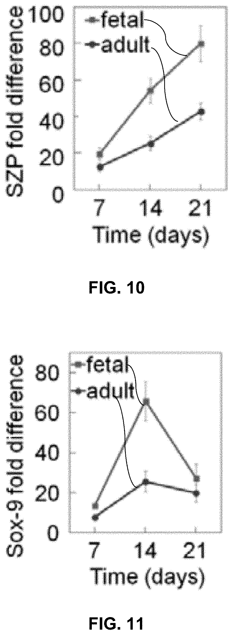
FIG. 10 compares a matrix based upon digested fetal articular cartilage with a matrix based upon digested adult articular cartilage with regard to the difference in the cartilage superficial zone marker SZP.
FIG. 11 compares a matrix based upon digested fetal articular cartilage with a matrix based upon digested adult articular cartilage with regard to the difference in the pre-chondrogenic marker Sox 9.
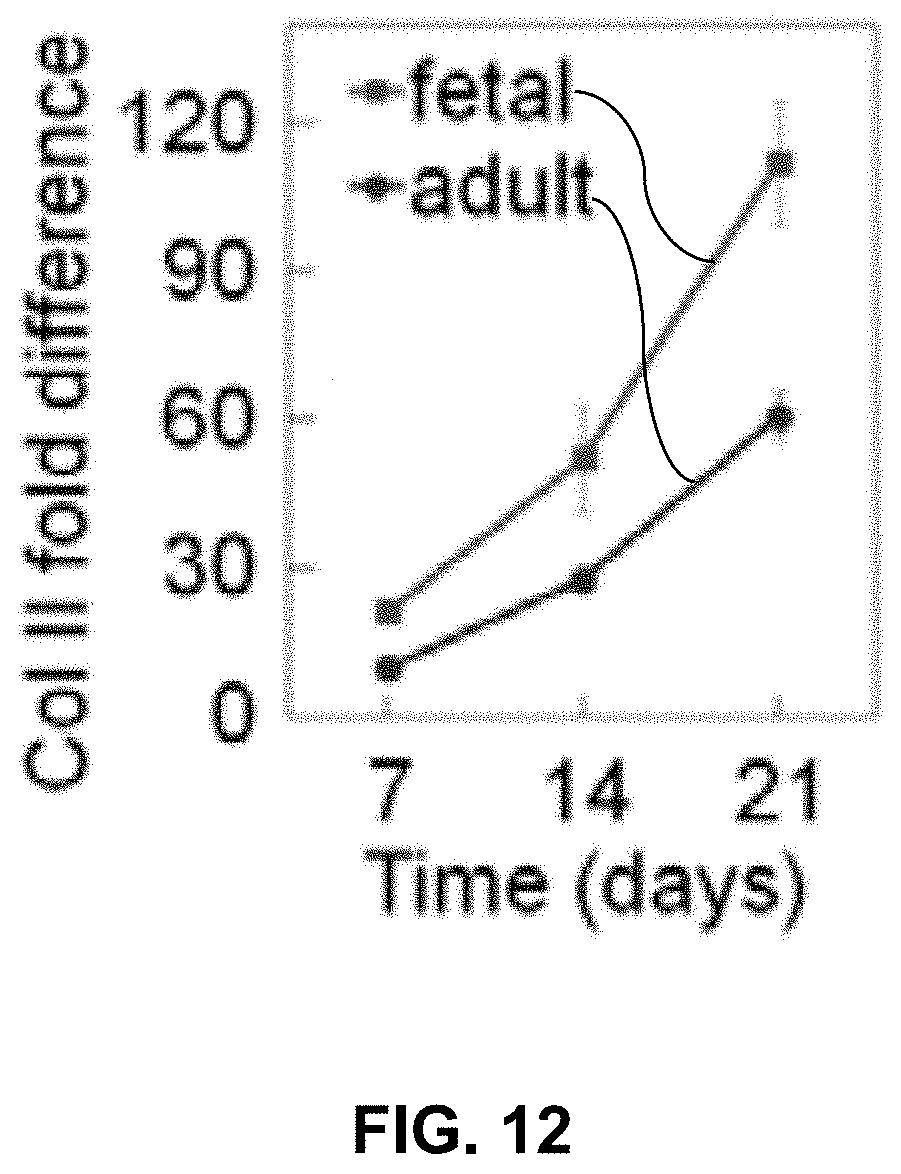
FIG. 12 compares a matrix based upon digested fetal articular cartilage with a matrix based upon digested adult articular cartilage with regard to the difference in the quantity of Col II.
Repeat use of reference characters in the present specification and drawings is intended to represent the same or analogous features or elements of the present invention.
DETAILED DESCRIPTION
Reference will now be made in detail to various embodiments of the disclosed subject matter, one or more examples of which are set forth below. Each embodiment is provided by way of explanation of the subject matter, not limitation thereof. In fact, it will be apparent to those skilled in the art that various modifications and variations may be made in the present disclosure without departing from the scope or spirit of the subject matter. For instance, features illustrated or described as part of one embodiment, may be used in another embodiment to yield a still further embodiment.
In general, disclosed herein are compositions and use of the compositions in tissue engineering applications. The compositions are hydrogel-based compositions that can be crosslinked in situ following placement and that are designed to sequentially release multiple (i.e., at least two) signaling molecules over a predetermined period of time so as to encourage tissue development or healing in the local area. The compositions can also carry additional components to encourage tissue generation. For instance, a composition can carry cells (either differentiated cells or stem cells) that can interact with the signaling molecules in the tissue generation. In addition, a composition can carry extracellular matrix (ECM) components that can further encourage tissue generation. In one particular embodiment, a composition can have characteristics so as to be injectable prior to crosslinking.
While much of the following discussion is directed to development of articular cartilage, the disclosure is in no way intended to be limited to compositions and methods for articular cartilage engineering applications, and through selection of particular signaling molecules, release profiles, ECM components, cell types, etc. the compositions can be utilized in development of other types of tissues including, without limitation, bone, connective tissues, etc.
The compositions described herein allow for not only the sustained release of signaling molecules (e.g., proteins) to stimulate tissue healing/generation, but also allow for the delayed release of signaling molecules, in order that the sequential signaling naturally present in tissue formation/healing can be mimicked in the tissue engineering applications. In the absence of sequential and sustained delivery, signaling molecules that are merely located at a desired site of activity will diffuse away from the site of regeneration within a few hours and gradients necessary for desired biological activity will disappear. By way of example, in order to mimic fetal development of articular cartilage, morphogens (e.g., BMP2, IGF1, IHH) need to be available both sequentially and sustainably.
In formation of the compositions, signaling molecules such as known primary articular cartilage morphogens can be conjugated to nanogels so as to provide for sustained release of the morphogens from the nanogels. In addition, the nanogels that carry the signaling molecules can be encapsulated in microcapsules that can provide for delayed release of the nanogels. Through combination of the nanogels and the microcapsules, optionally in conjunction with additional tissue engineering components (e.g., ECM components, stem cells, etc.), the compositions can be utilized for tissue engineering that more closely mimics natural tissue generation mechanisms.
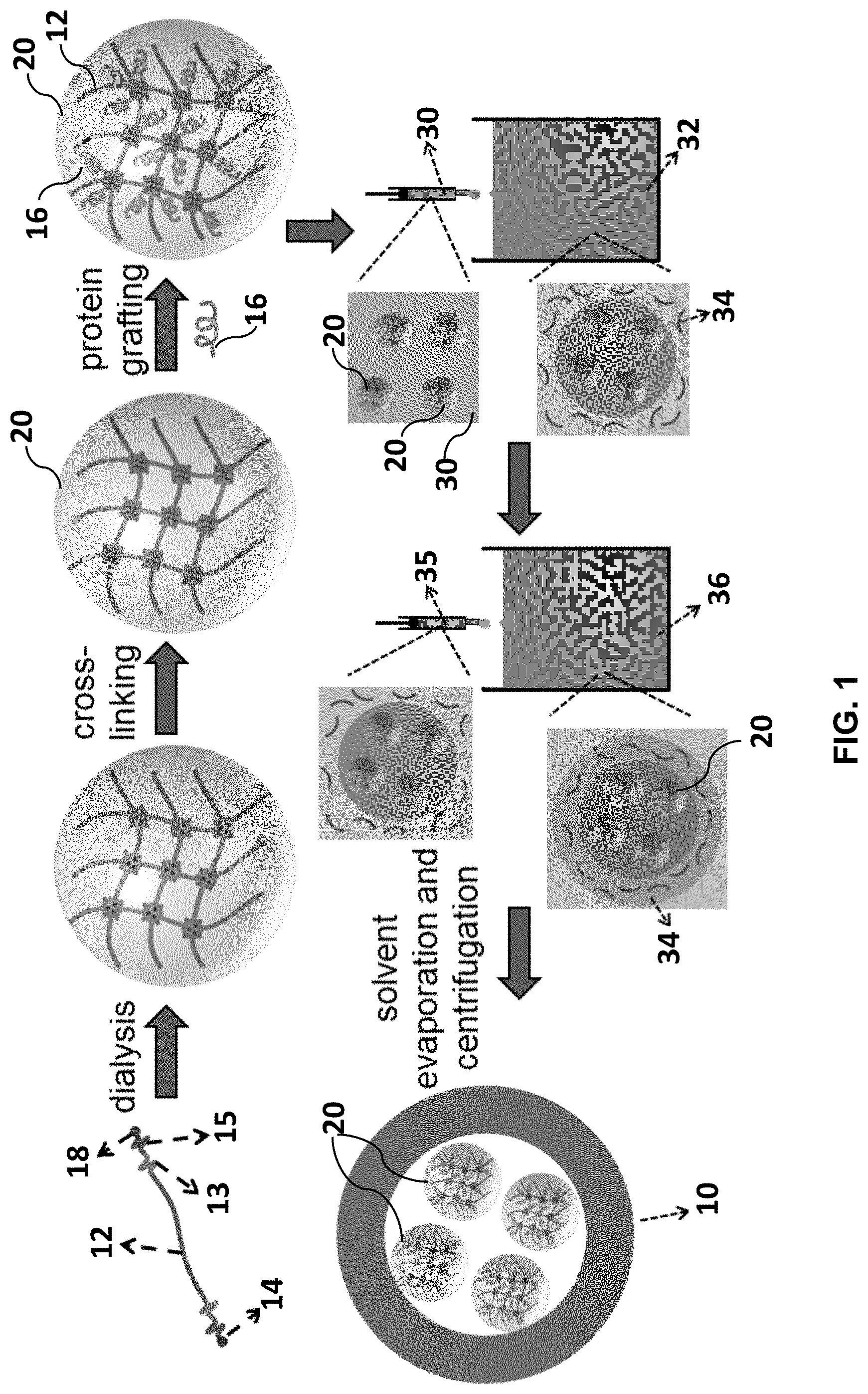
FIG. 1 schematically illustrates one embodiment for formation of the composite microcapsule/nanogel structures of a composition. As shown, a composite structure can include a microcapsule 10 that encapsulate a plurality of nanogels 20. Each nanogel 20 includes a polymer 12 that in turn includes a first functionality 14 configured for conjugation with a signaling molecule 16 and a second functionality 18 configured for crosslinking of the polymer. The combination of microcapsules 10 that can be designed to delayed release of the contents with the sustained releasing nanogels 20 held within the microcapsules 10 can provide for delayed/sustained-release of the signaling molecules. When considering an articular cartilage tissue engineering composition, a composition that includes a plurality of composite nanogel/microcapsule types that differ by release profile and signaling molecule can provide for delayed/sustained release of the signaling molecules (e.g., TGF.beta.1, BMP7, IGF1, and IHH) for sequential differentiation of stem cells that can also be carried by the composition to pre-chondrogenic, pre-hypertrophic, and hypertrophic cell phenotypes of articular cartilage in a sequential fashion that can mimic natural tissue development.
Referring to FIG. 1, the nanogels 20 can be formed of a biocompatible polymer 12 that can be derivatized as necessary so as to be crosslinkable via the presence of a functionality 18, controllably degradable in a biological environment (e.g., hydrolysable), and capable of conjugation to the signaling molecules 16 via the presence of a reactive functionality 14.
While any hydrophilic polymer can be used as a base for the synthesis of nanogels, in one embodiment, the nanogels can be based upon a polyethylene glycol (PEG) polymer. Other hydrophilic polymers that can be used include, without limitation, polyvinyl alcohol, polyvinyl pyrrolidinone, polyacrylic acid, poly(2-hydroxyethyl methacrylate), poly(acrylamide), and the like as well as mixtures of the aforementioned hydrophilic polymers. PEG-based hydrogels are inert, non-immunogenic, compatible with biological components (e.g., stem cells) and can be derivatized relatively easily. Due to their inert nature, PEG hydrogels provide enormous flexibility in design and control of the local microenvironment. The inert nature of PEG can also minimize adsorption and denaturation of signaling molecules (e.g., proteins) incorporated in the nanogels, which could otherwise adversely affect the composition function, and can stabilize the signaling molecules by reducing aggregation. Beneficially, flexible PEG polymers can crosslink to produce hydrogels with high compressive modulus without adversely affecting the function of the conjugated signaling molecules. The PEG polymers can impart stability to the nanogels in aqueous solution.
In general, the polymer 12 used in formation of the nanogels 20 can have a number average molecular weight of from about 4 kDa to about 12 kDa.
In order to control self-assembly of the polymer 12 in the initial formation of the nanogels and degradation of the nanogels according to a desired sustained release profile for the conjugated signaling molecules, the polymer 12 can be chain extended at the termini with relatively short hydrophobic segments that include a series of hydrophobic monomers. The hydrophobic monomers can include any biocompatible hydrophobic monomers such as, without limitation, lipid monomers, anyhydride monomers, orthoester monomers, phosphazene monomers, hydroxy acid monomers, and the like as well as mixtures of hydrophobic monomers. For instance, the hydrophobic segment can include, without limitation, glycolide monomers, lactide monomers, dioxanone monomers, .epsilon.-caprolactone monomers, hydroxy butyrate monomers, valcrolactone monomers, malonic acid monomers, as well as mixtures of monomers.
In one embodiment, a hydrophobic segment of a polymer 12 terminus can include a first length 13 that includes a series of lactide monomers that can regulate self-assembly of the nanogels and a second length 15 that includes a series of glycolide monomers that can aid in control of the degradation rate of the nanogel. Each length 13, 15 of a hydrophobic segment can be about 10 monomer units in length or less. For instance, the length 13 of lactide monomers can generally include from 2 to about 9 monomer units and the length 15 of glycolide monomers can generally include from 1 to about 6 monomer units.
The short hydrophobic segments 13, 15 can be bonded to the polymer 12 according to any suitable process, such as by combining the polymer with the hydrophobic monomer under reactive conditions with a catalyst, e.g., a tin(II)2-ethylhexanoate catalyst.
Without wishing to be bound to any particular theory, it is believed that the short hydrophobic segments can be sequestered within the core of the nanogels formed during the crosslinking reactions. In addition, the crosslinking moieties and any initiators used in conjunction with the crosslinking reaction can be sequestered within the nanogel core. By sequestering self-assembly, gelation and degradation components within the crosslinked nanogel structures, potential cytotoxicity of the nanogels to cells or other components of the composition can be reduced.
Furthermore, the size of each individual nanogel can be controlled by specific components used to form the nanogel particles (e.g., the polymer size and relative hydrophobicity of the short terminal segments), and the nanogel size can affect the degradation rate of the nanogels. The degradation rate of the nanogels and sustained release of the signaling molecules carried in the nanogels can be tuned from a few days to many months through variation of the polymer size as well as the characteristics of the hydrophobic end segments. The hydrolysis rate of the nanogels can be strongly dependent on the number and type of hydrophobic monomers in the hydrophobic segment. For instance, a nanogel that incorporates a less hydrophobic monomer, such as glycolide, can release the signaling molecules over the course of a few days while one that incorporates a more hydrophobic monomer, such as .epsilon.-caprolactone, can release the signaling molecules over the course of many months.
The polymers 12 can also be functionalized with a reactive functionality 18 that can provide for crosslinking of the polymers 12 and formation of stable nanogels 20. Accordingly, in addition to the chain extension of the polymers with the hydrophobic segments, a polymer 12 can be further processed to promote crosslinking of the polymer and formation of the crosslinked nanogels. For example, to crosslink the polymers 12 via UV, the polymer 12 can be functionalized at the termini to have a UV-crosslinkable functionality 18. Such groups are typically acrylates or methacrylates. The general scheme can include replacing terminal hydroxyl and/or carboxylic acid groups of a hydrophobic segment with acrylate or methacrylate functionality according to standard practice.
Crosslinking may be carried out via self-crosslinking of the polymer and/or through the inclusion of a separate crosslinking agents and/or initiators. Suitable crosslinking agents, for instance, may include polyglycidyl ethers, such as ethylene glycol diglycidyl ether and polyethylene glycol dicglycidyl ether; acrylamides; compounds containing one or more hydrolyzable groups, such as alkoxy groups (e.g., methoxy, ethoxy and propoxy); alkoxyalkoxy groups (e.g., methoxyethoxy, ethoxyethoxy and methoxypropoxy); acyloxy groups (e.g., acetoxy and octanoyloxy); ketoxime groups (e.g., dimethylketoxime, methylketoxime and methylethylketoxime); alkenyloxy groups (e.g., vinyloxy, isopropenyloxy, and 1-ethyl-2-methylvinyloxy); amino groups (e.g., dimethylamino, diethylamino and butylamino); aminoxy groups (e.g., dimethylaminoxy and diethylaminoxy); and amide groups (e.g., N-methylacetamide and N-ethylacetamide).
If included, an initiator can be used to initiate crosslinking of the polymer 12. Examples of UV initiators include, without limitation, IRGACURE.RTM. 184 (1-hydroxycyclohexyl phenyl ketone), and DAROCURE.RTM. 1173 (.alpha.-hydroxy-1, .alpha.-dimethylacetophenone) which are both commercially available from Ciba-Geigy Corp. Additional examples of initiators (which may be UV-initiators, thermal initiators, or other types of initiators) may include, without limitation, benzoyl peroxide, azo-bis-isobutyro-nitrile, di-t-butyl peroxide, bromyl peroxide, cumyl peroxide, lauroyl peroxide, isopropyl percarbonate, methylethyl ketone peroxide, cyclohexane peroxide, t-butylhydroperoxide, di-t-amyl peroxide, dicymyl peroxide, t-butyl perbenzoate, benzoin alkyl ethers (such as benzoin, benzoin isopropyl ether, and benzoin isobutyl ether), benzophenones (such as benzophenone and methyl-o-benzoyl benzoate), acetophenones (such as acetophenone, trichloroacetophenone, 2,2-diethoxyacetophenone, p-t-butyltrichloro-acetophenone, 2,2-dimethoxy-2-phenylacetophenone, and p-dimethylaminoacetophenone), thioxanthones (such as xanthone, thioxanthone, 2-chlorothioxanthone, and 2-isopropyl thioxanthone), benzyl 2-ethyl anthraquinone, methylbenzoyl formate, 2-hydroxy-2-methyl-1-phenyl propane-1-one, 2-hydroxy-4'-isopropyl-2-methyl propiophenone, e-hydroxy ketone, tet-remethyl thiuram monosulfide, allyl diazonium salt, and a combination of camphorquinone or 4-(N,N-dimethylamino)benzoate.
Any of a variety of different crosslinking mechanisms may be employed, such as thermal initiation (e.g., condensation reactions, addition reactions, etc.), electromagnetic radiation, and so forth. Some suitable examples of electromagnetic radiation that may be used include, but are not limited to, electron beam radiation, natural and artificial radio isotopes (e.g., .alpha., .beta., and .gamma. rays), x-rays, neutron beams, positively-charged beams, laser beams, ultraviolet, etc. The wavelength X of the radiation may vary for different types of radiation of the electromagnetic radiation spectrum, such as from about 10.sup.-14 meters to about 10.sup.-5 meters. Besides selecting the particular wavelength .lamda. of the electromagnetic radiation, other parameters may also be selected to control the degree of crosslinking. For example, the dosage may range from about 0.1 megarads (Mrads) to about 10 Mrads, and in some embodiments, from about 1 Mrads to about 5 Mrads.
In one embodiment, the polymer 12 can be acrylate-functionalized 18 at one chain-end as shown in FIG. 1. For instance, a PEG polymer that has been previously functionalized with short lactide/glycolide segments at the termini can be reacted with acryloyl chloride in 1:1 molar ratio to produce a functionalized polymer that includes an acrylate at one chain-end.
In addition to short hydrophobic control segments 13, 15 and crosslinkable functionality 18, the polymer 12 can also be functionalized so as to enable conjugation with the desired signaling molecule 16. For instance, a polymer 12 can be end-functionalized with succinimide by reaction with N,N'-disuccinimidyl carbonate according to standard practice.
In general, the functionalized polymers will be crosslinked prior to conjugation with the desired signaling molecules. For instance, the functionalized polymers can be assembled in aqueous solution by dialysis and crosslinked (e.g., via UV radiation) to generate water-swollen nanogels 20. Following functionalization and crosslinking, the nanogels 20 can generally have a largest dimension of from about 10 nanometer to about 100 nanometers.
The signaling molecule 16 of choice can be conjugated to the crosslinked nanogels by reaction between the nanogel functionality 18 (e.g., a succinimide group) and a functionality of the signaling molecule (e.g., an amine group). By conjugation in the nanogels 20, the signaling molecule 16 can be prevented from diffusion into the organic phase of the encapsulating microcapsules and can be sequestered from release until the degradation of the surrounding microcapsule, upon which the nanogels can release the conjugated signaling molecule 16 according to the engineered sustained release profile.
A nanogel 20 may include as a signaling molecule 16 any biologically active compound (or combination thereof) as may affect a developing system. For instance, a nanogel 20 can include a signaling molecule 16 that can act as a signal for modifying cell adhesion, growth, or migration, for instance in stimulating or promoting the adhesion, growth, or migration of the desirable cells, and/or inhibiting or stimulating the adhesion, growth, or migration of undesirable cells. Such compounds can include growth factors, hormones, extracellular matrix proteins and other cellular adhesion peptides identified in the extracellular matrix protein. Suitable signaling molecules may include, for example, TGF.beta.1, BMP7, IGF1, IHH, BMP2, epithelial growth factor (EGF), acidic or basic fibroblast growth factor (FGF), vascular endothelial growth factor (VEGF), hepatocyte growth factor (HGF), heparin binding growth factor (HGBF), transforming growth factor (TGF), nerve growth factor (NGF), muscle morphogenic factor (MMP), and platelet derived growth factor (PDGF). Examples of extracellular matrix proteins include fibronectin, collagens, laminins, and vitronectins, and the tri-peptide RGD (arginine-glycine-aspartate) that is found in many of the extracellular matrix proteins. A signaling molecule can also be included to induce the ingrowth of desirable cells, e.g., smooth muscle cells and epithelial cells. Signaling molecules that inhibit or stimulate undesired cells, such as cancerous cells or inflammatory cells can be included.
A signaling molecule may also include a small molecule such as a bisphosphonate that can prevent the loss of bone mass. Another example of a small molecule signaling molecule is an antibiotic that can prevent bacterial contamination. Examples of small molecule bisphosphonates include, without limitation, Etidronate, Clodronate, Tiludronate, Pamidronate, Neridronate, Olpadronate, Alendronate, Ibandronate, Risedronate, Zoledronate, and the like as well as mixtures of bisphosphonates. Example of antibacterial agents include, without limitation, gentamicin, tetracycline, vancomycin, ibuprofen, dexamethasone, ciprofloxacin, gatifloxacin, polymyxin B and the like as well as mixtures of the antibacterial small molecules. Of course, different types of signaling molecules (e.g., proteinaceous and small molecule, bisphosphonates and antibacterial agents, etc.) can be combined in individual nanogels or in different nanogels held in the same or different microcapsules of a composition.
In general, the nanogels 20 can include the signaling molecules 16 in a concentration of from about 0.5 .mu.g/mL to about 4 .mu.g/mL per 20 mg of nanogels.
In forming a composition, a plurality of signaling molecule-conjugated nanogels 20 can be encapsulated in a polymer microcapsule 10 that can provide for delayed release of the nanogels 20 and the signaling molecules 16 carried by the nanogels 20. For instance, and as schematically illustrated in FIG. 1, a plurality of nanogels 20 can be encapsulated in a polylactide-based polymer microcapsule 10 according to a water-oil-water (w-o-w) double emulsion technique for delayed release of the signaling molecules. The polymer utilized in formation of the microcapsule 10 can be selected to provide a desired degradation rate and delay for the microcapsules. For instance, a poly(lactide-co-glycolide) (PLGA)-based microcapsule can be selected in design of materials for relatively rapid release of the contained nanogels (e.g., delays of up to about 6 weeks), while more hydrophobic polymers such as poly(lactide-co-caprolactone) (PLCL) copolymer may be preferred to achieve longer delay times and release rates from the microcapsules. Polylactide homopolymer as well as other lactide-based copolymers as are known in the art may alternatively be utilized in formation of the microcapsules. In general, any hydrophobic polymer that can form microcapsules in aqueous solution can be used. Examples include, without limitation polyanhydrides, polyorthoesters, polyphosphazenes, polypropylene fumarate, polyhydroxyacids, polyhydroxybutyrate, polyvalcrolactone, polymalonic acid as well as mixtures of polymers.
A double emulsion w-o-w encapsulation technique as may be utilized in forming the microcapsules 10 and as schematically illustrated in FIG. 1 can generally include dispersing the nanogels 20 in a first aqueous phase 30. Following, the aqueous phase 30 can be dispersed in a suitable organic phase 32 comprising an organic solution of the polymer (e.g., a poly(D,L lactide-co-glycolide) polymer) 34, the organic phase 32 being non-miscible with the aqueous phase 30. For example, a nanogel dispersion including about 0.1 mg nanogels/mL or greater, or from about 1 mg/mL to about 100 mg/mL or even greater in some embodiments can be injected (such as with a 25 gauge needle) into the organic polymer phase 32 while homogenizing, e.g., at about 15,000 to 25,000 rpm.
By way of example, an aqueous phase 30 including the nanogels 20 can be initially emulsified in an organic phase 32 including PLGA polymer of a predetermined molecular weight, poly(lactide-co-ethylene oxide fumarate) (PLEOF) as emulsifying agent, and chloroform as organic solvent. Other organic solvents and emulsifying agents as are known in the art may alternatively be utilized. For instance, organic solvents can include, without limitation, methylene chloride, ethyl acetate with or without benzyl alcohol or acetone, and combinations of organic solvents.
After this first homogenization (generally about 0.5 to about 5 minutes, for instance for about 1 minute), this emulsion 35 is added to a second aqueous emulsification solution 36 to form a second emulsion. The second emulsification aqueous solution 36, can be for instance a polyvinyl alcohol solution, optionally including ethyl acetate. The w-o-w emulsion mixture is mixed at high speed (generally about 1700 to 2500 rpm) to generate the microcapsules 10 including the polymer 34 surrounding the nanogels 20.
The stable microcapsules 10, each encapsulating a plurality of nanogels 20 can be formed and collected upon evaporation of the organic solvent (e.g., at ambient conditions, dependent upon the solvent) and centrifugation to remove the aqueous phase, as shown.
When considering formation of an injectable composition, the microcapsules can generally be formed with a largest external dimension of from about 1 .mu.m to about 10 .mu.m. However, the microcapsules are not limited to this size and larger materials may be preferred in certain applications. For example, when considering in vitro applications, it may be preferred to form larger microcapsules.
Design factors that may be modified to provide microcapsules with a desired size and degradation delay can include molecular weight and the type of polymer 34, emulsifier concentration in the organic phase, copolymer component ratio on a polymer (e.g., L:G ratio for a PLGA copolymer), and polymer concentration in the organic phase (affects capsule wall thickness). In general, lactide-based polymers (PLGA, PLCL, PLA, etc.) for use in the disclosed microcapsules can have a number average molecular weight of from about 5 kDa to about 75 kDa and a lactide copolymer ratio (e.g., L:G ratio) of from about 100:0 to about 50:50. The organic phase can generally include the emulsifying agent (e.g., the PLOEF) at a concentration of from about 5 wt. % to about 50 wt. %, and can include the lactide-based polymer at a concentration of from about 5 wt. % to about 15 wt. % in the organic solvent.
The composite nanogel/microcapsules can have a conjugation efficiency of the signaling molecules of greater than about 90% and a release level of greater than about 75%.
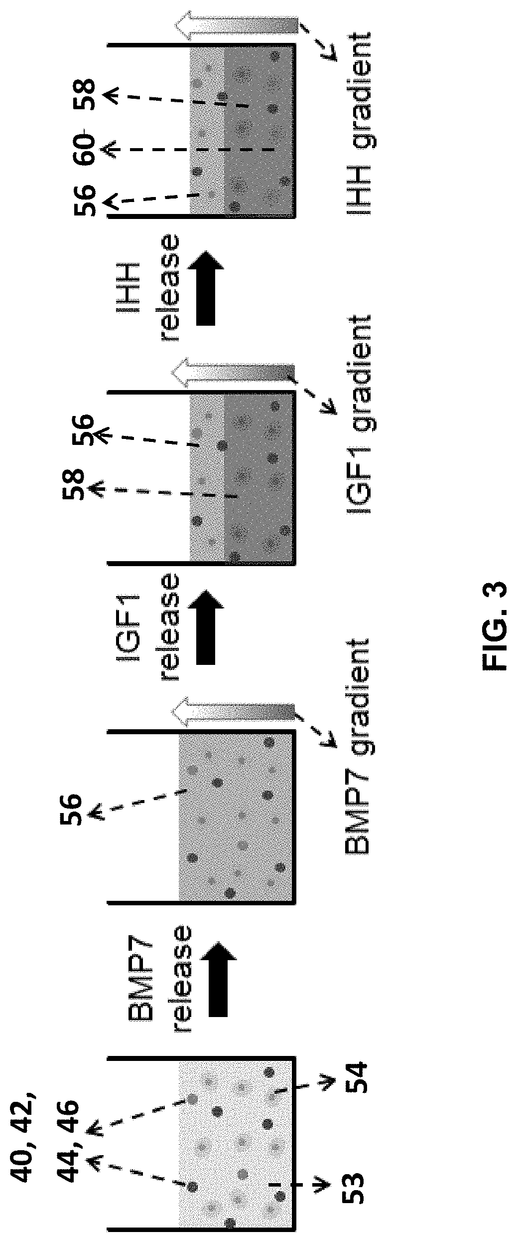
A composition can incorporate a plurality of different microcapsules so as to provide for sequential release of the signaling agents carried by the microcapsules. For instance, when considering a composition designed to encourage formation of articular cartilage, a composition can incorporate microcapsules designed for sequential, sustained-release of TGF.beta.1, BMP7, IGF1, and IHH morphogens to generate the zonal structure of articular cartilage. For example, and as schematically illustrated in FIG. 2, a first type of microcapsule 40 of the composition can incorporate TGF.beta.1-conjugated nanogels and can be designed for release of the TGF.beta.1 over the entire course of the protocol (e.g., over weeks 1-9 of a 9-week protocol). A second type of microcapsule 42 of the composition can incorporate BMP7-conjugated nanogels and can be designed to release the BMP7 over weeks 1-3 of the protocol. A third type of microcapsule 44 of the composition can incorporate IGF1-conjugated nanogels and can be designed to release the IGF1 over weeks 4-6 of the protocol. A fourth type of microcapsule 46 of the composition can incorporate IHH-conjugated nanogels and can be designed to release the IHH over weeks 7-9 of the protocol. A composition 51 as illustrated in FIG. 2 can thus provide for the sequential, sustained-release of BMP7, IGF1, and IHH morphogens in situ.
Optionally, microcapsules can incorporate multiple different nanogels therein. For instance, in the above example, as TGF.beta.1 is released in conjunction with the other signaling agents, microcapsules of a composition can incorporate both TGF.beta.1-conjugated nanogels and nanogels carrying another signaling agent (BMP7-, IGF1-, or IHH-conjugated nanogels). Alternatively, nanogels can be formed to include multiple different types of signaling agents, e.g., both TGF.beta.1 and one of the other signaling agents together in a single nanogel.
A composition 51 can include the microcapsules and a crosslinkable matrix-forming polymer 52. The composition 51 can have suitable rheological characteristics for delivery (e.g., injectability). The matrix-forming polymer 52 can be crosslinked following placement (e.g., injection) of the composition to form a crosslinked matrix 53. In one embodiment, the crosslinkable polymer 52 can be based upon a natural biopolymer that can provide biocompatibility to the composition. In addition, the composition 51 can incorporate one or more additional components conducive to the desired tissue formation, e.g., ECM components. For example, and as illustrated in FIG. 2, a composition 51 can be derived from a natural tissue 50 (e.g., allograft or xenograft tissue) that can be decellularized, digested, and functionalized to provide the crosslinkable polymer 52 in conjunction with other components of the natural tissue 50 in an injectable, crosslinkable composition 51. As such, the composition 51 can provide both a crosslinkable polymer 52 capable of forming the hydrogel matrix 53 and other components conducive to the tissue engineering application.
By way of example, when considering formation of articular cartilage, the composition 51 can be based upon natural articular cartilage. As fetal articular cartilage is capable of spontaneous healing while avoiding fibrocartilage formation, in one embodiment, a crosslinkable polymer 52 can be derived from fetal articular cartilage, e.g., the injectable composition 51 can be based on digested, decellularized and functionalized fetal bovine articular cartilage.
In one embodiment, fetal bovine cartilage can be decellularized by treatment with 10 mM Tris/1% triton, 1 U/mL deoxyribonuclease, and 1 U/mL ribonuclease as known. After freeze-drying, the decellularized material can be digested with pepsin until a clear suspension is obtained. The digested and decellularized material can then be functionalized to include a crosslinkable functionality (e.g., acrylate or methacrylate functionality) on the digested polymers. For instance, the material can be reacted with methacrylic anhydride to produce a methacrylated biopolymer-based crosslinkable polymer 52. Maximum added reactivity content of the biopolymer (e.g., acrylate functionality) can be such that will not significantly affect biocompatibility of the composition 51.
In one embodiment, a composition 51 (including all additives) can have a viscosity of from about 5 mPa-s to about 15 mPa-s, so as to be injectable through a 0.43 mm microcatheter. A crosslinkable composition 51 can have a gelation time of about 1 min or less, and can have a compressive modulus following crosslinking of about 50 kPa to about 150 kPa, so as to support the developing tissue. However, viscosity, injectability, gelation time, and mechanical characteristics (e.g., compressive modulus) of the composition 51 and the crosslinked hydrogel 53 formed from the composition 51 are not limited to these values and can be varied, for instance depending upon the specific application of the composition.
In one embodiment, the composition 51 can also include cells 54 that can be affected by the release of the signaling molecules. Depending upon the specific application of the composition, cells 54 contained in a composition 51 can include differentiated cells and/or stem cells. For instance, in one particular embodiment, a composition 51 can include mesenchymal stem cells (MSC).
Cells 54 can be loaded into a composition 51 at a density similar to that of the tissue type of interest. For instance, in one embodiment a composition 51 can incorporate MSCs at a density of from about 50 million cells/mL to about 200 million cells/m L, which corresponds to that of adult and fetal articular cartilage, respectively.
The concentrations of the different microcapsules 41, 42, 44, 46 included in a composition 51 can vary so as to achieve the desired concentrations of the various signaling molecules. For instance, when considering the formation of articular cartilage, a composition can include microcapsules at a concentration to provide about 100 .mu.g/mL BMP7, about 100 .mu.g/mL IGF1, and about 2.5 .mu.g/mL IHH. Further, it has been shown that TGF.beta.1 concentrations of 3 and 30 ng/mL can stimulate chondrogenic differentiation of MSCs to the superficial and calcified zone phenotypes, respectively. Thus, in this particular embodiment, a composition can include microcapsules that include TGF.beta.1-conjugated nanogels so as to provide TGF.beta.1 concentrations of 3, 15, and 30 ng/mL at various time points throughout a tissue generation protocol.
Following formation of the hydrogel precursor composition 51 that includes a crosslinkable polymer 52 and the signaling molecule-conjugated nanogel/microcapsule composites 40, 42, 44, 46, optionally in conjunction with living cells 54 and one or more ECM components (not illustrated in FIG. 2), the composition 51 can be placed in the desired tissue formation location (e.g., via injection) and crosslinked to form a crosslinked hydrogel 53. The in situ gelation time (e.g., the UV exposure time) can be varied (e.g., from about 1 minute to about 5 minutes) to reach a targeted compressive modulus for the crosslinked composition 53. For instance, following injection, the composition can be subjected to gelation for a time so as to reach the compressive modulus of the superficial zone of articular cartilage (about 80 kPa).
As illustrated in FIG. 2 and FIG. 3, following the placement and crosslinking of the hydrogel 53, the microcapsules 40, 42, 44, 46 can degrade according to the predetermined release profiles, which, in turn, can release the nanogels contained therein and allow for the sustained release of the signaling molecules contained in the nanogels. For example, following injection, crosslinking can be carried out to form a fetal articular cartilage-mimetic matrix 53 loaded with MSCs 54, TGF.beta.1-conjugated nanogel/microcapsules 40, BMP7-conjugated nanogel/microcapsules 42, IGF1-conjugated nanogel/microcapsules 44, and IHH-conjugated nanogel/microcapsules 46. The TGF.beta.1-conjugated nanogel/microcapsules 40 can begin to degrade relatively quickly following formation with the nanogels releasing the TGF.beta.1 over the entire course of the tissue formation e.g., weeks 1-9), which can encourage condensation of the MSCs contained in the matrix. The BMP7-conjugated nanogel/microcapsules 42 can also begin to degrade relatively quickly following formation, and these nanogels can release the BMP7 over weeks 1-3 of the tissue formation. The release of the BMP7 in conjunction with the TGF.beta.1 can encourage chondrogenic differentiation of the MSCs to form a matrix similar to the superficial zone 56 of articular cartilage. The IGF1-conjugated nanogel/microcapsules 44 can begin to degrade following the predetermined delay period so as to release the IGF1 over weeks 4-6 of the tissue formation leading to the development of two distinct superficial 56 and middle zones 58 and the further differentiation of pre-chondrocytes to pre-hypertrophic chondrocytes. Release of IHH from the IHH-conjugated nanogel/microcapsules 46 following the predetermined delay period over weeks 7-9 of the tissue formation leading to the development of two distinct superficial 56 and middle zones 58 and the can lead to generation of the complete zonal organization of articular cartilage including the calcified zone 60.
Thus, the combination of delayed and sustained release of the signaling molecules in the composition can provide for in situ differentiation and maturation of cells that are either in the local area of the composition or contained in the composition itself so as to provide improved tissue generation and healing.
The present disclosure may be better understood with reference to the Examples set forth below.
Example
Articular cartilage is structurally composed of multiple zones with distinct functions including the superficial zone for joint lubrication, middle zone for compressive strength, and calcified zone for load transfer to the underlying bone tissue. The superficial zone is characterized by pre-chondrocytes with high expression of superficial zone protein (SZP) for joint lubrication. The middle zone is characterized by pre-hypertrophic chondrocytes with high expression of glycosaminoglycans (GAG) and aggrecans (AGCs) for compressive strength. The calcified zone is characterized by hypertrophic chondrocytes with high expression of collagen type X (Col X) and alkaline phosphatase (ALP) for the formation of a mineralized matrix.
It has been demonstrated that proteins can be conjugated with high efficiency to nanogels based on polyethylene oxide macromers chain-extended with short lactide-glycolide segments (PEG-sLG) and the protein can be released with high bioactivity and at a prescribed rate. FIG. 4 shows that the release rate and duration of bovine serum albumin (BSA) conjugated to PEG-sLG nanogels can be controlled by changing the PEG molecular weight (P4-I-4 kDa PEG, P8-II-8 kDa PEG, P12-III-12 kDa PEG). FIG. 5 shows that different proteins (BMP7, BMP2, IGF1, TGF.beta.1, FGF, VEGF) conjugated to PEG-sLG nanogels are released at a prescribed rate with >60% bioactivity. FIG. 6 shows that BSA-conjugated nanogels formed of acrylated PEG-sLG (PEG-sLG-Ac) and further encapsulated in PLGA microcapsules (NG-MCs) release the protein after a predefined delay time ranging from a few days to 20 days, depending upon the polymer type and the PLEOF emulsifier concentration (PLGA50--PLGA copolymer using 50:50 wt. % PLGA:PLEOF in the organic phase; PLGA95--PLGA copolymer using 95:5 wt. % PLGA:PLEOF in the organic phase; PLA50--PLA homopolymer using 50:50 wt. % PLA:PLEOF in the organic phase; PLA95--PLA homopolymer using 95:5 wt. % PLA:PLEOF in the organic phase).
The combination of TGF.beta.1 and BMP7 stimulates chondrogenic differentiation of encapsulated mesenchymal stem cells to the superficial zone phenotype (FIG. 7); the combination of TGF.beta.1 and IGF1 stimulates the middle zone phenotype (FIG. 8); the combination of TGF.beta.1 and IHH stimulates the calcified zone phenotype (FIG. 9).
Encapsulation of mesenchymal stem cells in a hydrogel based on digested bovine fetal articular cartilage and cultivation in chondrogenic medium supplemented with BMP7 sharply enhanced stem cell differentiation to the superficial zone phenotype compared to that of adult articular cartilage (FIG. 10, FIG. 11, FIG. 12). As shown, human MSCs encapsulated in digested fetal bovine articular cartilage and cultured in chondrogenic medium have higher expression of pre-chondrogenic markers Sox-9 (FIG. 11) and superficial zone protein (SZP) (FIG. 10) and collagen type II (Col II; FIG. 12) compared to those of adult cartilage.
These results support in situ zonal regeneration of articular cartilage by sequential/sustained release of zone-specific morphogens in an injectable fetal articular cartilage-derived matrix.
While certain embodiments of the disclosed subject matter have been described using specific terms, such description is for illustrative purposes only, and it is to be understood that changes and variations may be made without departing from the spirit or scope of the subject matter.
* * * * *
D00001
D00002
D00003
D00004
D00005
D00006
D00007
D00008
XML
uspto.report is an independent third-party trademark research tool that is not affiliated, endorsed, or sponsored by the United States Patent and Trademark Office (USPTO) or any other governmental organization. The information provided by uspto.report is based on publicly available data at the time of writing and is intended for informational purposes only.
While we strive to provide accurate and up-to-date information, we do not guarantee the accuracy, completeness, reliability, or suitability of the information displayed on this site. The use of this site is at your own risk. Any reliance you place on such information is therefore strictly at your own risk.
All official trademark data, including owner information, should be verified by visiting the official USPTO website at www.uspto.gov. This site is not intended to replace professional legal advice and should not be used as a substitute for consulting with a legal professional who is knowledgeable about trademark law.