Consensus/ancestral immunogens
Haynes , et al. March 16, 2
U.S. patent number 10,946,090 [Application Number 15/813,992] was granted by the patent office on 2021-03-16 for consensus/ancestral immunogens. This patent grant is currently assigned to Duke University, Triad National Security, LLC, The University of Alabama at Birmingham Research Foundation. The grantee listed for this patent is Duke University, Los Alamos National Security, LLC, The University of Alabama at Birmingham Research Foundation. Invention is credited to Julie Decker, Feng Gao, Beatrice H. Hahn, Barton F. Haynes, Bette T. Korber, Ying Ying Li, Hua-Xin Liao, Denise L. Monti, George M. Shaw.


View All Diagrams
| United States Patent | 10,946,090 |
| Haynes , et al. | March 16, 2021 |
Consensus/ancestral immunogens
Abstract
The present invention relates, in general, to an immunogen and, in particular, to an immunogen for inducing antibodies that neutralizes a wide spectrum of HIV primary isolates and/or to an immunogen that induces a T cell immune response. The invention also relates to a method of inducing anti-HIV antibodies, and/or to a method of inducing a T cell immune response, using such an immunogen. The invention further relates to nucleic acid sequences encoding the present immunogens.
| Inventors: | Haynes; Barton F. (Durham, NC), Gao; Feng (Durham, NC), Korber; Bette T. (Los Alamos, NM), Hahn; Beatrice H. (Birmingham, AL), Shaw; George M. (Birmingham, AL), Monti; Denise L. (Birmingham, AL), Li; Ying Ying (Birmingham, AL), Decker; Julie (Birmingham, AL), Liao; Hua-Xin (Durham, NC) | ||||||||||
|---|---|---|---|---|---|---|---|---|---|---|---|
| Applicant: |
|
||||||||||
| Assignee: | Duke University (Durham,
NC) The University of Alabama at Birmingham Research Foundation (Birmingham, AL) Triad National Security, LLC (Los Alamos, NM) |
||||||||||
| Family ID: | 1000005422272 | ||||||||||
| Appl. No.: | 15/813,992 | ||||||||||
| Filed: | November 15, 2017 |
Prior Publication Data
| Document Identifier | Publication Date | |
|---|---|---|
| US 20180296665 A1 | Oct 18, 2018 | |
Related U.S. Patent Documents
| Application Number | Filing Date | Patent Number | Issue Date | ||
|---|---|---|---|---|---|
| 13137517 | Aug 2, 2011 | 9844589 | |||
| 10572638 | Dec 6, 2011 | 8071107 | |||
| PCT/US2004/030397 | Sep 17, 2004 | ||||
| 60604722 | Aug 27, 2004 | ||||
| 60503460 | Sep 17, 2003 | ||||
| Current U.S. Class: | 1/1 |
| Current CPC Class: | C07K 14/005 (20130101); C07K 16/4225 (20130101); A61K 39/21 (20130101); A61K 39/12 (20130101); C12N 2740/16322 (20130101); A61K 2039/53 (20130101); C12N 2740/15034 (20130101); C12N 2740/16222 (20130101); C12N 2740/15022 (20130101); C12N 2740/16034 (20130101); C12N 2740/16122 (20130101); C12N 2740/16134 (20130101); A61K 2039/57 (20130101) |
| Current International Class: | A61K 39/21 (20060101); A61K 39/12 (20060101); C07K 14/005 (20060101); C07K 16/42 (20060101); A61K 39/00 (20060101) |
References Cited [Referenced By]
U.S. Patent Documents
| 5580859 | December 1996 | Felgner et al. |
| 5589466 | December 1996 | Felgner et al. |
| 5703055 | December 1997 | Felgner et al. |
| 7172761 | February 2007 | Haynes et al. |
| 8048431 | November 2011 | Haynes |
| 8071107 | December 2011 | Haynes et al. |
| 2003/0044421 | March 2003 | Emini et al. |
| 2003/0096778 | May 2003 | Shiver et al. |
| 2005/0137387 | June 2005 | Mullins et al. |
| 2007/0178562 | August 2007 | Haynes et al. |
| 2009/0162384 | June 2009 | Haynes |
| 060838 | Aug 2001 | WO | |||
| 0224149 | Mar 2002 | WO | |||
Other References
|
Yang, X., et al., 2003, Role of the gp120 inner domain beta-sandwich in the interaction between the human immunodeficiency virus envelope glycoprotein subunits, Virol. 313:117-125. cited by examiner . Huang, W., et al., Jun. 2008, Coreceptor tropism can be influenced by amino acid substitutions in the gp41 transmembrane subunit of human immunodeficiency virus type 1 envelope protein, J. Virol. 82(11):5584-5593. cited by examiner . Murphy, M. K., et al., Feb. 2013, Viral escape from neutralizing antibodies in early subtype A HIV-1 infection drives an increase in autologous neutralization breadth, PLoS Pathogens 9(2):e1003173, pp. 1-20. cited by examiner . Mathys, L., et al., Jun. 2014, Deletion of the highly conserved N-glycan at Asn260 of HIV-1 gp120 affects folding and lysosomal degradation of gp120, and results in loss of viral infectivity, PLoS One 9(6):e101181, pp. 1-11. cited by examiner . Andre et al., "Increased immune response elicited by DNA vaccination with a synthetic gp120 sequence with optimized codon usage," J Virol. Feb. 1998;72(2):1497-503. cited by applicant . Australian Patent Application No. 2014240343 Examination Report dated Jan. 21, 2016. cited by applicant . Baldridge et al., "Immunostimulatory activity of aminoalkyl glucosaminide 4-phosphates (AGPs): induction of protective innate immune responses by RC-524 and RC-529," J Endotoxin Res. 2002;8(6):453-8. cited by applicant . Barbeau et al., "Modulation of human immunodeficiency virus type 1-induced syncytium formation by the conformational state of LFA-1 determined by a new luciferase-based syncytium quantitative assay," J Virol. Sep. 1998;72(9):7125-36. cited by applicant . Bartlett et al., "Safety and immunogenicity of an HLA-based HIV envelope polyvalent synthetic peptide immunogen. DATRI 010 Study Group. Division of AIDS Treatment Research Initiative," AIDS. Jul. 30, 1998;12(11)1291-300. cited by applicant . Binley et al; Enhancing the Proteolytic Maturation of Human Immunodeficiency Virus Type 1 Envelope Glycoproteins; Journal of Virology vol. 76, No. 6; Mar. 2002, p. 2606-2616. cited by applicant . Blanchard et al., "Future Vaccines for HIV," Lancet. Dec. 21-28, 1996;348(9043):1741. cited by applicant . Bosch et al.; Mutational Analysis of the Human Immunodeficiency Virus Typle 1 env Gene Product Proteolytic Cleavage Site; Journal of Virology vol. 64, No. 5; May 1990; p. 2337-2344. cited by applicant . Brandt et al., "Association of chemokine-mediated block to HIV entry with coreceptor internalization," J Biol Chem. May 10, 2002;277(19):17291-9. Epub Jan. 8, 2002. cited by applicant . Bures et al., "Immunization with recombinant canarypox vectors expressing membrane-anchored glycoprotein 120 followed by glycoprotein 160," AIDS Res Hum Retroviruses. Dec. 10, 2000;16(18):2019-35. cited by applicant . Bures et al., "Regional clustering of shared neutralization determinants on primary isolates of clade C human immunodeficiency virus type 1 from South Africa," J Virol. Mar. 2002;76(5):2233-44. cited by applicant . Carr et al, Human retroviruses and AIDS 1998: a compilation and analysis of nucleic acid and amino acid sequences, eds. Korber et al (Theoretical Biology and Biophysics Group, Los Alamos National Laboratory, Los Alamos, N. Mex.), pp. III-10-III-19 (1998)). cited by applicant . Chakrabarti et al.; Modifications of the Human Immunodeficiency Virus Envelope Glycoprotein Enhance Immunogenicity for Genetic Immunization; Journal of Virology vol. 76, No. 11; Jun. 2002; p. 5357-5368. cited by applicant . Cho et al., "Polyvalent envelope glycoprotein vaccine elicits a broader neutralizing antibody response but is unable to provide sterilizing protection against heterologous Simian/human immunodeficiency virus infection in pigtailed macaques," J Virol. Mar. 2001;75(5):2224-34. cited by applicant . Cormier et al., "Specific interaction of CCR5 amino-terminal domain peptides containing sulfotyrosines with HIV-1 envelope glycoprotein gp120," Proc Natl Acad Sci U S A. May 23, 2000;97(11):5762-7. cited by applicant . Cornelissen et al., "Human immunodeficiency virus type 1 subtypes defined by env show high frequency of recombinant gag genes. The UNAIDS Network for HIV Isolation and Characterization," J Virol. Nov. 1996;70(11):8209-12. cited by applicant . Demi et al, "Multiple Effects of Codon Usage Optimization on Expression and Immunogenicity of DNA Candidate Vaccines Encoding the Human Immunodeficiency Virust Type 1 Gag Protein", Journal of Virology 75(22): 10991-11001 (2001). cited by applicant . Derdeyn et al., "Sensitivity of human immunodeficiency virus type 1 to the fusion inhibitor T-20 is modulated by coreceptor specificity defined by the V3 loop of gp120," J Virol. Sep. 2000;74(18):8358-67. cited by applicant . Dowling et al., "Forty-one near full-length HIV-1 sequences from Kenya reveal an epidemic of subtype A and A-containing recombinants," AIDS. Sep. 6, 2002;16(13):1809-20. cited by applicant . Ellenberger et al, "Generation of a Consensus Sequence from Prevalent and Incident HIV-1 Infections in West Africa to Guide AIDS Vaccine Development", Virology 302:156-163 (2002). cited by applicant . Evans et al., "A canarypox vaccine expressing multiple human immunodeficiency virus type 1 genes given alone or with rgp120 elicits broad and durable CD8+ cytotoxic T lymphocyte responses in seronegative volunteers," J Infect Dis. Aug. 1999;180(2):290-8. cited by applicant . Ferrari et al., "Identification of highly conserved and broadly cross-reactive HIV type 1 cytotoxic T lymphocyte epitopes as candidate immunogens," AIDS Res Hum Retroviruses. Sep. 20, 2000;16(14):1433-43. cited by applicant . Ferrari et al., "Clade B-based HIV-1 vaccines elicit cross-clade cytotoxic T lymphocyte reactivities in uninfected volunteers," Proc Natl Acad Sci U S A. Feb. 18, 1997;94(4)1396-401. cited by applicant . Fouts et al., "Crosslinked HIV-1 envelope-CD4 receptor complexes elicit broadly cross-reactive neutralizing antibodies in rhesus macaques," Proc Natl Acad Sci U S A. Sep. 3, 2002;99(18):11842-7. Epub Aug. 21, 2002. cited by applicant . Fouts et al., "Neutralization of the human immunodeficiency virus type 1 primary isolate JR-FL by human monoclonal antibodies correlates with antibody binding to the oligomeric form of the envelope glycoprotein complex," J Virol. Apr. 1997;71(4):2779-85. cited by applicant . Gallo, Robert C., "The end or the beginning of the drive to an HIV-preventive vaccine: a view from over 20 years", The Lancet 366:1894-1898 (2005). cited by applicant . Gao et al, "Centralized immunogens as a vaccine strategy to overcome HIV-1 diversity", Expert Rev Vaccines 3(4 Suppl)S 161-8 (2004 )-Abstract. cited by applicant . Gao et al, "Codon usage optimization of HIV type 1 subtype C gag, pol, env, and nef genes: in vitro expression and immune responses in DNA-vaccinated mice", AIDS Res. Hum. Retroviruses 19(9):817-823 (2003). cited by applicant . Gao et al. Antigenicity and Immunogenicity of a Synthetic Human Immunodeficiency Virus Type 1 Group M consensus Envelope Glycoprotein; Journal of Virology vol. 79, No. 2; Jan. 2005; p. 1154-1163. cited by applicant . Gaschen et al; Diversity Considerations in HIV-1 Vaccine Selection; Science 296, p. 2354-2360; Jun. 28, 2002. cited by applicant . Guo et al.; Characterization of an HIV-1 Point Mutant Blocked in Envelope Glycoprotein Cleavage; Virology 174, p. 217-224 (1990). cited by applicant . Gurtler, et al., "A new subtype of human immunodeficiency virus type 1 (MVP-5180) from Cameroon," J Virol. Mar. 1994;68(3):1581-5. cited by applicant . Haas et al., "Codon usage limitation in the expression of HIV-1 envelope glycoprotein," Curr Biol. Mar. 1, 1996;6(3):315-24. cited by applicant . Haynes et al., "HIV vaccines: where we are and where we are going," Lancet. Oct. 5, 1996;348(9032):933-7. cited by applicant . Haynes et al., "Induction of HIVMN neutralizing antibodies in primates using a prime-boost regimen of hybrid synthetic gp120 envelope peptides," J Immunol. Aug. 1, 1993;151(3):1646-53. cited by applicant . HIV Sequence Compendium 2003. Leitner T, Foley B, Hahn B, Marx P, McCutchan F, Mellors J, Wolinsky S, and Korber B, Eds. Published by Theoretical Biology and Biophysics Group, Los Alamos National Laboratory, NM, LA-UR 04-7420 (2003). cited by applicant . International Search Report issued in connection with PCT/US04/30397 dated Mar. 8, 2005. cited by applicant . Jiang et al., "A conformation-specific monoclonal antibody reacting with fusion-active gp41 from the human immunodeficiency virus type 1 envelope glycoprotein," J Virol. Dec. 1998;72(12):10213-7. cited by applicant . Kofman et al, "HIV-1 gag expression is quantitatively dependent on the ratio of native and optimized codons", Tsitologiia 45(1 ):86-93 (2003). cited by applicant . Korber et al., "Evolutionary and immunological implications of contemporary HIV-1 variation", British Medical Bulletin, 58:19-42 (2001). cited by applicant . Korber et al., "Timing the ancestor of the HIV-1 pandemic strains," Science. Jun. 9, 2000;288(5472):1789-96. cited by applicant . Kuiken et al, Human retroviruses and AIDS 2000: a compilation and analysis of nucleic acid and amino acid sequences (Theoretical Biology and Biophysics Group, Los Alamos National Laboratory, Los Alamos, N. Mex.)), pp. 355-456 (2000). cited by applicant . Kwong et al., "Structure of an HIV gp120 envelope glycoprotein in complex with the CD4 receptor and a neutralizing human antibody," Nature. Jun. 18, 1998;393(6686):648-59. cited by applicant . LaCasse et al., "Fusion-competent vaccines: broad neutralization of primary isolates of Hiv," Science. Jan. 15, 1999;283(5400):357-62. [Retracted in Nunberg et al., Science. May 10, 2002;296(5570):1025]. cited by applicant . Leitner et al, eds., "HIV Sequence Compendium 2003", Theoretical Biology and Biophysics Group, Los Alamos National Laboratory, LA-UR No. 04-7420, pp. 513-573 and attached appendix. cited by applicant . Letvin et al., "Prospects for vaccine protection against HIV-1 infection and AIDS," Annu Rev Immunol. 2002;20:73-99. Epub Oct. 4, 2001. cited by applicant . Levine, Arnold J., "Why Do We Not Yet Have a Human Immunodeficiency Virus Vaccine?", Journal of Virology 82(24):11998-1200 (2008). cited by applicant . Li et al.; Control of Expression, Glycosylation, and Secretion of HIV-1 gp 120 by Homologous and Heterologous Signal Sequences; Virology 204, 266-278 (1994). cited by applicant . Li et al.; Effects of inefficient cleavage of the signal sequence of HIV-1 gpl20 on its association with calnexin, folding, and intracellular transport; Proc. Natl. Acad. Sci. USA vol. 93 Sep. 1996 p. 9606-9611. cited by applicant . Liao et al, "A Group M Consensus Envelope Glycoprotein induces Antibodies That Neutralize Subsets of Subtype B and C HIV-1 Primary Viruses", NIH Public Access, pp. 1-30, Published in final edited form as Virology 353(2):268-282 (2006). cited by applicant . Liao et al., "Immunogenicity of constrained monoclonal antibody A32-human immunodeficiency virus (HIV) Env gp120 complexes compared to that of recombinant HIV type 1 gp120 envelope glycoproteins," J Virol. May 2004;78(10):5270-8. cited by applicant . Liao et al.; Antigenicity and Immunogenicity of Transmitted/Founder, Consensus, and Chronic Envelope Glycoproteins of Human Immunodeficiency Virus Type 1; Journal of Virology vol. 87, No. 8; p. 4185-4201; Apr. 2013. cited by applicant . Liao et al; A Group M Consensus Envelope Glycoprotein Induces Antibodies That Neutralize Subsets of Subtype B and C HIV-1 Primary Viruses; Virology 353(2) p. 268-282; Sep. 30, 2006. cited by applicant . Mascola et al., "Protection of Macaques against pathogenic simian/human immunodeficiency virus 89.6PD by passive transfer of neutralizing antibodies," J Virol. May 1999;73(5):4009-18. cited by applicant . Mascola et al., "Protection of macaques against vaginal transmission of a pathogenic HIV-1/SIV chimeric virus by passive infusion of neutralizing antibodies," Nat Med. Feb. 2000;6(2):207-10. cited by applicant . McCune et al; Endoproteolytic Cleavage of gp160 Is Required for the Activation of Human Immunodeficiency Virus; Cell, vol. 53; p. 55-67; Apr. 8, 1988. cited by applicant . McMichael et al., "HIV T cell vaccines, the importance of Glades," Vaccine. May 6, 2002;20(15):1918-21. cited by applicant . Mo et al., "Human immunodeficiency virus type 1 mutants that escape neutralization by human monoclonal antibody IgG1b12. off," J Virol. Sep. 1997;71(9):6869-74. cited by applicant . Moore and Binley, "Envelope's letters boxed into shape," Nature. Jun. 18, 1998;393(6686):630-31. cited by applicant . Moore et al., "Exploration of antigenic variation in gp120 from Glades A through F of human immunodeficiency virus type 1 by using monoclonal antibodies," J Virol. Dec. 1994;68(12):8350-64. cited by applicant . Morris et al., "Characterization and selection of HIV-1 subtype C isolates for use in vaccine development", AIDS Res Hum Retroviruses, 19(2):133-144 (2003)-Abstract. cited by applicant . Muster et al., "Cross-neutralizing activity against divergent human immunodeficiency virus type 1 isolates induced by Ihe gp41 sequence ELDKWAS," J Virol. Jun. 1994;68(6):4031-4. cited by applicant . Nickle et al, "Consensus and Ancestral State HIV Vaccines", Science 299(5612):1515-1518 (2003). cited by applicant . Notice of Allowance dated Jan. 4, 2011 in U.S. Appl. No. 10/572,638. cited by applicant . Notice of Allowance dated May 12, 2011 in U.S. Appl. No. 10/572,638. cited by applicant . Notice of Allowance dated Nov. 15, 2010 in U.S. Appl. No. 11/896,934. cited by applicant . Novitsky et al, "Human Immunodeficiency Virus Type 1 Subtype C Molecular Phylogeny: Consensus Sequences for an AIDS Vaccine Design?", Journal of Virology 66(11 ):5435-5451 (2002). cited by applicant . Nyambi et al., "Multivariate analysis of human immunodeficiency virus type 1 neutralization data," J Virol. Sep. 1996;70(9):6235-43. cited by applicant . Office Action dated Jul. 7, 2010 in U.S. Appl. No. 11/896,934. cited by applicant . Office Action dated Jun. 4, 2010 in U.S. Appl. No. 10/572,638. cited by applicant . Office Action dated Nov. 30, 2009 in U.S. Appl. No. 10/572,638. cited by applicant . Office Action dated Oct. 6, 2009 in U.S. Appl. No. 11/896,934. cited by applicant . Ourmanov et al., "Recombinant modified vaccinia virus ankara expressing the surface gp120 of simian immunodeficiency virus (SIV) primes for a rapid neutralizing antibody response to SIV infection in macaques," J Virol. Mar. 2000;74(6):2960-5. cited by applicant . Pal et al., "ALVAC-SIV-gag-pol-env-based vaccination and macaque major histocompatibility complex class I (A*01) delay simian immunodeficiency virus SIVmac-induced immunodeficiency," J Virol. Jan. 2002;76(1):292-302. cited by applicant . Polacino et al., "Limited breadth of the protective immunity elicited by simian immunodeficiency virus SIVmne gp160 vaccines in a combination immunization regimen," J Virol. Jan. 1999;73(1):618-30. cited by applicant . Rimsky et al., "Determinants of human immunodeficiency virus type 1 resistance to gp41-derived inhibitory peptides," J Virol. Feb. 1998;72(2):986-93. cited by applicant . Roben et al., "Recognition properties of a panel of human recombinant Fab fragments to the CD4 binding site of gp120 that show differing abilities to neutralize human immunodeficiency virus type 1," J Virol. Aug. 1994;68(8):4821-8. cited by applicant . Robertson et al, Human retroviruses and AIDS 1999: a compilation and analysis of nucleic acid and amino acid sequences, eds. Kuiken et al (Theoretical Biology and Biophysics Group, Los Alamos National Laboratory, Los Alamos, N. Mex.), pp. 492-505 (1999)). cited by applicant . Robertson et al., "Recombination in HIV-1," Nature. Mar. 9, 1995;374(6518):124-6. cited by applicant . Rossio et al., "Inactivation of human immunodeficiency virus type 1 infectivity with preservation of conformational and functional integrity of virion surface proteins," J Virol. Oct. 1998;72(10):7992-8001. cited by applicant . Saphire et al., "Crystal structure of a neutralizing human IGG against HIV-1: a template for vaccine design," Science. Aug. 10, 2001;293(5532):1155-9. cited by applicant . Sbai et al., "Use of T cell epitopes for vaccine development," Curr Drug Targets Infect Disord. Nov. 2001;1(3):303-13. cited by applicant . Simon et al., "Identification of a new human immunodeficiency virus type 1 distinct from group M and group O," Nat Med. Sep. 1998;4(9):1032-7. cited by applicant . Supplementary Partial European Search Report dated Aug. 1, 2008--EP Appln. No. 04 78 4298. cited by applicant . Trkola et al., "Human monoclonal antibody 2G12 defines a distinctive neutralization epitope on the gp120 glycoprotein of human immunodeficiency virus type 1," J Virol. Feb. 1996;70(2):1100-8. cited by applicant . Vanden Haesevelde et al., "Genomic cloning and complete sequence analysis of a highly divergent African human immunodeficiency virus isolate," J Virol. Mar. 1994;68(3):1586-96. cited by applicant . Wain Hobson, "More ado about HIV's origins," Nat Med. Sep. 1998;4(9):1001-2. cited by applicant . Walker and Burton, "Toward an AIDS Vaccine", Science 320:760-764 (2008). cited by applicant . Wang, Lai-Xi, "Bioorganic Approaches Towards HIV Vaccine Design", Current Pharmaceutical Design 9:1771-1787 (2003). cited by applicant . Wei et al., "Emergence of resistant human immunodeficiency virus type 1 in patients receiving fusion inhibitor (T-20) monotherapy," Antimicrob Agents Chemother. Jun. 2002;46(6):1896-905. cited by applicant . Williamson et al, "Characterization and selection of HIV-1 subtype C isolates for use in vaccine development", AIDS Res. Hum. Retroviruses 19(2):133-144 (2003). cited by applicant . Written Opinion for corresponding PCT application PCT/US04/30397 dated Mar. 8, 2005. cited by applicant . Wyatt et al., "Involvement of the V1N2 variable loop structure in the exposure of human immunodeficiency virus type 1 gp120 epitopes induced by receptor binding," J Virol. Sep. 1995;69(9):5723-33. cited by applicant . Wyatt et al., "The antigenic structure of the HIV gp120 envelope glycoprotein," Nature. Jun. 18, 1998;393(6686):705-11. cited by applicant . Ye et al., "Association of structural changes in the V2 and V3 loops of the gp120 envelope glycoprotein with acquisition of neutralization resistance in a simian-human immunodeficiency virus passaged in vivo," J Virol. Dec. 2000;74(24):11955-62. cited by applicant. |
Primary Examiner: Parkin; Jeffrey S
Attorney, Agent or Firm: Baker Donelson
Parent Case Text
This application is a Continuation of U.S. application Ser. No. 13/137,517, filed Aug. 23, 2011, which is a Continuation of U.S. application Ser. No. 10/572,638, filed Dec. 22, 2006, which is a National Stage Application under 35 U.S.C. section 371 of PCT/US2004/030397, filed Sep. 17, 2004, which claims priority from U.S. Provisional Application No. 60/503,460, filed Sep. 17, 2003, and U.S. Provisional Application No. 60/604,722, filed Aug. 27, 2004, the entire contents of which are incorporated herein by reference in their entireties.
Claims
What is claimed is:
1. A nucleic acid comprising a nucleotide sequence that encodes a group M consensus Env comprising SEQ ID NO: 13, or a Cons-S gp140CFI Env comprising SEQ ID NO: 30, or a CON-S gp140CF Env comprising SEQ ID NO: 36.
2. The nucleic acid according to claim 1, wherein said nucleotide sequence comprises codons optimized for expression in human cells.
3. The nucleic acid according to claim 2, wherein said nucleic acid comprises the nucleotide sequence set forth in SEQ ID NO: 14.
4. The nucleic acid according to claim 2, wherein said nucleic acid comprises the nucleotide sequence set forth in SEQ ID NO: 31.
5. The nucleic acid according to claim 2, wherein said nucleic acid comprises the nucleotide sequence set forth in SEQ ID NO: 37.
6. A nucleic acid comprising a nucleotide sequence that encodes a recombinant gp120 envelope protein, wherein the recombinant gp120 envelope protein comprises the consecutive amino acid sequence represented by amino acid numbers 30 to 503 of SEQ ID NO: 13.
7. The nucleic acid of claim 6 comprising the consecutive nucleotide sequence from SEQ ID NO: 14 that encodes the consecutive amino acid sequence represented by amino acid numbers 30 to 503 of SEQ ID NO: 13, wherein said nucleotide sequence comprises codons optimized for expression in human cells.
8. A vector comprising the nucleic acid according to claim 1.
9. An isolated mammalian cell comprising a nucleic acid according to claim 1 for recombinant protein expression.
10. A vector comprising the nucleic acids of claim 6.
11. A composition comprising the nucleic acid according to claim 1 and a carrier.
12. A method of inducing an immune response in a mammal, the method comprising administering to said mammal the composition of claim 11 in an amount sufficient to effect such induction.
13. A composition comprising the nucleic acid according to claim 1 and a carrier, wherein the nucleic acid comprises a nucleotide sequence that encodes a group M consensus Env comprising SEQ ID NO: 13 and a carrier.
14. A composition comprising the nucleic acid according to claim 6 and a carrier.
15. An isolated mammalian cell comprising the nucleic acid according to claim 1, wherein the nucleic acid comprises a nucleotide sequence that encodes a group M consensus Env comprising SEQ ID NO: 13 or recombinant protein expression.
16. An isolated mammalian cell comprising the nucleic acid according to claim 6 for recombinant protein expression.
17. A method of inducing an immune response in a mammal, the method comprising administering to said mammal the composition of claim 13 in an amount sufficient to effect such induction.
18. A method of inducing an immune response in a mammal, the method comprising administering to said mammal the composition of claim 14 in an amount sufficient to effect such induction.
Description
SEQUENCE LISTING
The instant application contains a Sequence Listing which has been filed electronically in ASCII format and is hereby incorporated by reference in its entirety. Said ASCII copy, created on Jun. 28, 2018, is named 2933311-030-US7_SL.txt and is 1,108,293 bytes in size.
TECHNICAL FIELD
The present invention relates, in general, to an immunogen and, in particular, to an immunogen for inducing antibodies that neutralize a wide spectrum of HIV primary isolates and/or to an immunogen that induces a T cell immune response. The invention also relates to a method of inducing anti-HIV antibodies, and/or to a method of inducing a T cell immune response, using such an immunogen. The invention further relates to nucleic acid sequences encoding the present immunogens.
BACKGROUND
The high level of genetic variability of HIV-1 has presented a major hurdle for AIDS vaccine development. Genetic differences among HIV-1 groups M, N, and O are extensive, ranging from 30% to 50% in gag and env genes, respectively (Gurtler et al, J. Virol. 68:1581-1585 (1994), Vanden Haesevelde et al, J. Virol. 68:1586-1596 (1994), Simon et al, Nat. Med. 4:1032-1037 (1998), Kuiken et al, Human retroviruses and AIDS 2000: a compilation and analysis of nucleic acid and amino acid sequences (Theoretical Biology and Biophysics Group, Los Alamos National Laboratory, Los Alamos, N. Mex.)). Viruses within group M are further classified into nine genetically distinct subtypes (A-D, F-H, J and K) (Kuiken et al, Human retroviruses and AIDS 2000: a compilation and analysis of nucleic acid and amino acid sequences (Theoretical Biology and Biophysics Group, Los Alamos National Laboratory, Los Alamos, New Mex., Robertson et al, Science 288:55-56 (2000), Robertson et al, Human retroviruses and AIDS 1999: a compilation and analysis of nucleic acid and amino acid sequences, eds. Kuiken et al (Theoretical Biology and Biophysics Group, Los Alamos National Laboratory, Los Alamos, N. Mex.), pp. 492-505 (2000)). With the genetic variation as high as 30% in env genes among HIV-1 subtypes, it has been difficult to consistently elicit cross-subtype T and B cell immune responses against all HIV-1 subtypes. HIV-1 also frequently recombines among different subtypes to create circulating recombinant forms (CRFs) (Robertson et al, Science 288:55-56 (2000), Robertson et al, Human retroviruses and AIDS 1999: a compilation and analysis of nucleic acid and amino acid sequences, eds. Kuiken et al (Theoretical Biology and Biophysics Group, Los Alamos National Laboratory, Los Alamos, N. Mex.), pp. 492-505 (2000), Carr et al, Human retroviruses and AIDS 1998: a compilation and analysis of nucleic acid and amino acid sequences, eds. Korber et al (Theoretical Biology and Biophysics Group, Los Alamos National Laboratory, Los Alamos, N. Mex.), pp. III-10-III-19 (1998)). Over 20% of HIV-1 isolates are recombinant in geographic areas where multiple subtypes are common (Robertson et al, Nature 374:124-126 (1995), Cornelissen et al, J. virol. 70:8209-8212 (1996), Dowling et al, AIDS 16:1809-1820 (2002)), and high prevalence rates of recombinant viruses may further complicate the design of experimental HIV-1 immunogens.
To overcome these challenges in AIDS vaccine development, three computer models (consensus, ancestor and center of the tree) have been used to generate centralized HIV-1 genes to (Gaschen et al, Science 296:2354-2360 (2002), Gap et al, Science 299:1517-1518 (2003), Nickle et al, Science 299:1515-1517 (2003), Novitsky et al, J. Virol. 76:5435-5451 (2002), Ellenberger et al, Virology 302:155-163 (2002), Korber et al, Science 288:1789-1796 (2000)). The biology of HIV gives rise to star-like phylogenies, and as a consequence of this, the three kinds of sequences differ from each other by 2-5% (Gao et al, Science 299:1517-1518 (2003)). Any of the three centralized gene strategies will reduce the protein distances between immunogens and field virus strains. Consensus sequences minimize the degree of sequence dissimilarity between a vaccine strain and contemporary circulating viruses by creating artificial sequences based on the most common amino acid in each position in an alignment (Gaschen et al, Science 296:2354-2360 (2002)). Ancestral sequences are similar to consensus sequences but are generated using maximum-likelihood phylogenetic analysis methods (Gaschen et al, Science 296:2354-2360 (2002), Nickle et al, Science 299:1515-1517 (2003)). In doing so, this method recreates the hypothetical ancestral genes of the analyzed current wild-type sequences (FIG. 26). Nickle et al proposed another method to generate centralized HIV-1 sequences, center of the tree (COT), that is similar to ancestral sequences but less influenced by outliers (Science 299:1515-1517 (2003)).
The present invention results, at least in part, from the results of studies designed to determine if centralized immunogens can induce both T and B cell immune responses in animals. These studies involved the generation of an artificial group M consensus env gene (CON6), and construction of DNA plasmids and recombinant vaccinia viruses to express CON6 envelopes as soluble gp120 and gp140CF proteins. The results demonstrate that CON6 Env proteins are biologically functional, possess linear, conformational and glycan-dependent epitopes of wild-type HIV-1, and induce cytokine-producing T cells that recognize T cell epitopes of both HIV subtypes B and C. Importantly, CON6 gp120 and gp140CF proteins induce antibodies that neutralize subsets of subtype B and C HIV-1 primary isolates.
The iterative nature of study of the centralized HIV-1 gene approach is derived from the rapidly expanding evolution of HIV-1 sequences, and the fact that sequences collected in the HIV sequence database (that is, the Los Alamos National Database) are continually being updated with new sequences each year. The CON6 gp120 envelope gene derives from Year 1999 Los Alamos National Database sequences, and Con-S derives from Year 2000 Los Alamos National Database sequences. In addition, CON6 has Chinese subtype C V1, V2, V4, and V5 Env sequences, while Con-S has all group M consensus Env constant and variable regions, that have been shortened to minimal-length variable loops. Codon-optimized genes for a series of Year 2003 group M and subtype consensus sequences have been designed, as have a corresponding series of wild-type HIV-1 Env genes for comparison, for use in inducing broadly reactive T and B cell responses to HIV-1 primary isolates.
SUMMARY OF THE INVENTION
The present invention relates to an immunogen for inducing antibodies that neutralize a wide spectrum of HIV primary isolates and/or to an immunogen that induces a T cell immune response, and to nucleic acid sequences encoding same. The invention also relates to a method of inducing anti-HIV antibodies, and/or to a method of inducing a T cell immune response, using such an immunogen.
Objects and advantages of the present invention will be clear from the description that follows.
BRIEF DESCRIPTION OF THE DRAWINGS
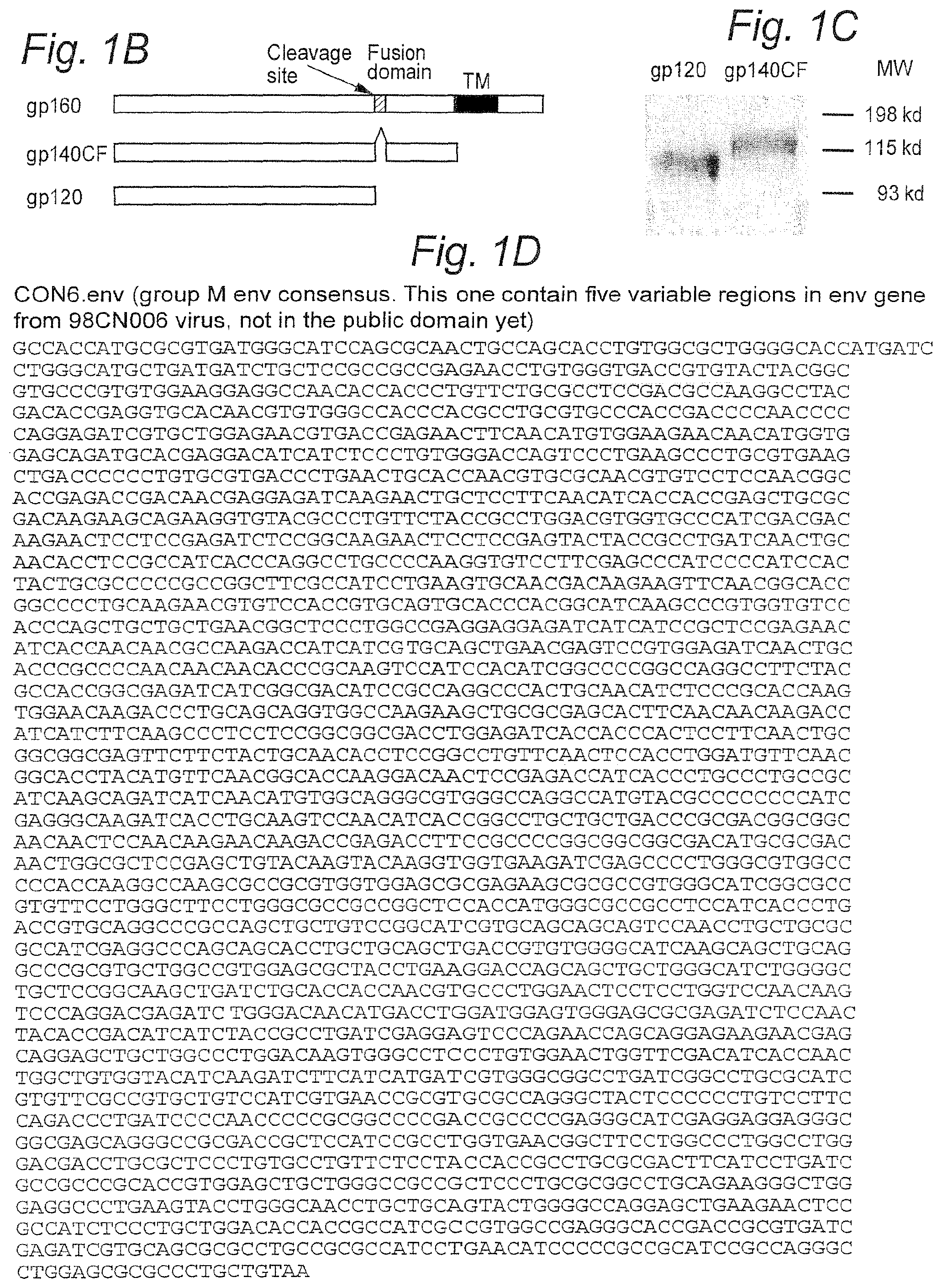
FIGS. 1A-1D: Generation and expression of the group M consensus env gene (CON6). The complete amino acid sequence of CON6 gp160 is shown. (FIG. 1A) (SEQ ID NO: 1) The five regions from the wild-type CRF08_BC (98CN006) env gene are indicated by underlined letters. Variable regions are indicated by brackets above the sequences. Potential N-liked glycosylation sites are highlighted with bold-faced letters. (FIG. 1B) Constructs of CON6 gp120 and gp140CF. CON6 gp120 and gp140CF plasmids were engineered by introducing a stop codon after the gp120 cleavage site or before the transmembrane domain, respectively. The gp120/gp41 cleavage site and fusion domain of gp41 were deleted in the gp140CF protein. (FIG. 1C) Expression of CON6 gp120 and gp140CF. CON6 gp120 and gp140CF were purified from the cell culture supernatants of rVV-infected 293T cells with galanthus Nivalis argarose lectin columns. Both gp120 and gp140CF were separated on a 10% SDS-polyarylamide gel and stained with Commassie blue. (FIG. 1D.) (SEQ ID NO: 2) CON6 env gene optimized based on codon usage for highly expressed human genes.
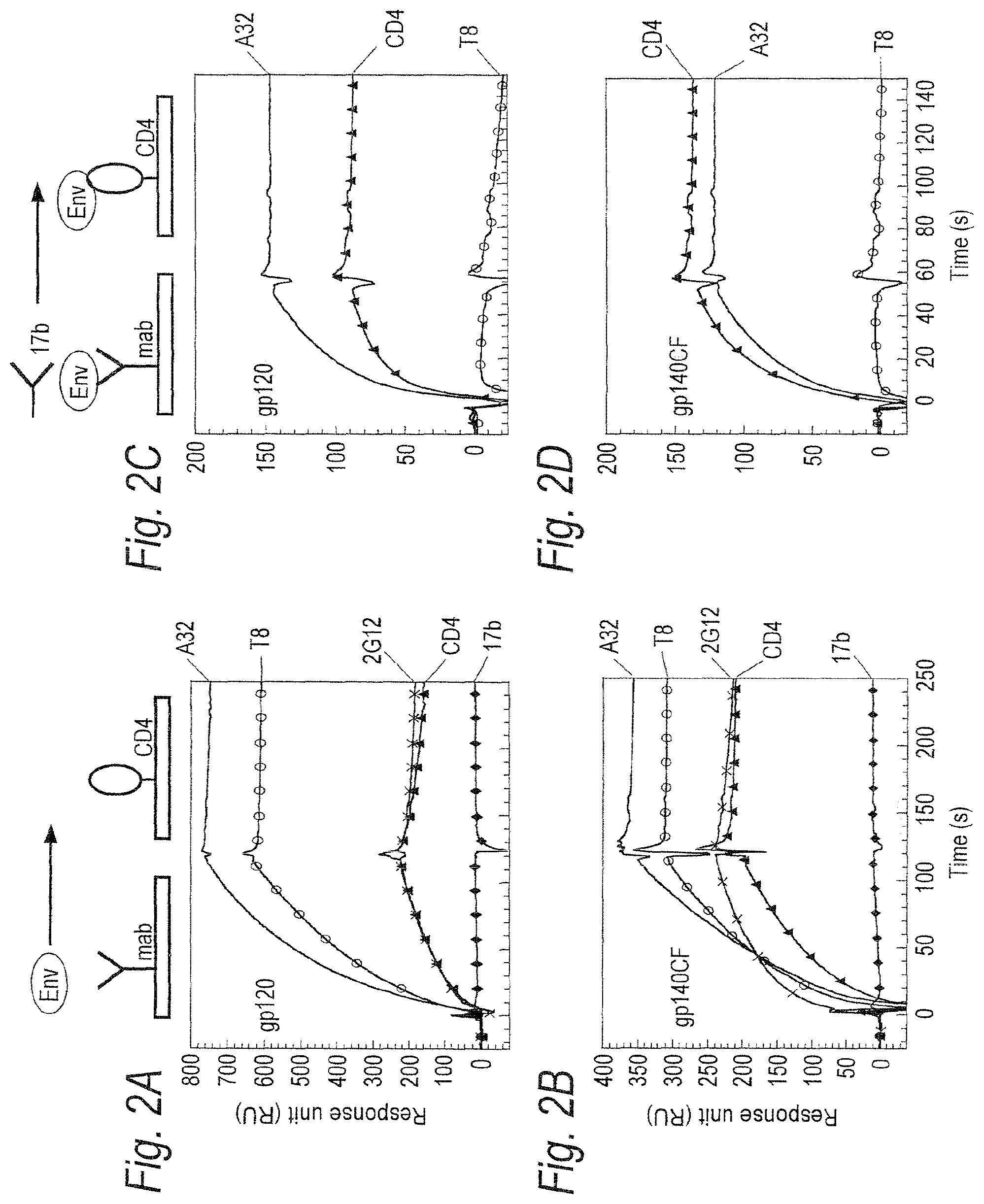
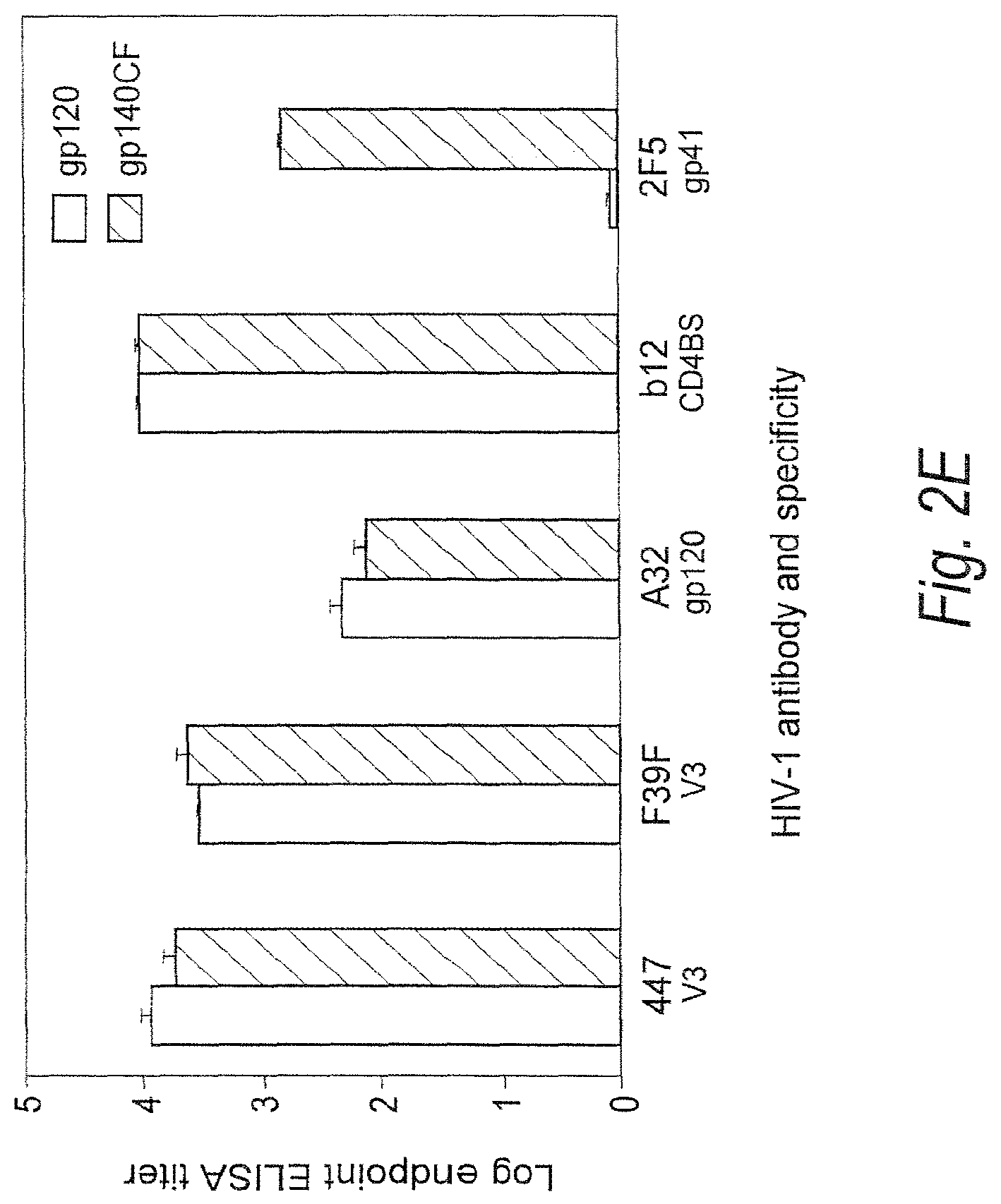
FIGS. 2A-2E. Binding of CON6 gp120 gp140 CF to soluble CD4 (sCD4) and anti-Env mAbs. (FIGS. 2A-2B) Each of the indicated mabs and sCD4 was covalently immobilized to a CM5 sensor chip (BIAcore) and CON6 gp120 (FIG. 2A) or gp140CF (FIG. 2B) (100 .mu.g/ml and 300 .mu.g/ml, respectively) were injected over each surface. Both gp120 and gp140CF proteins reacted with each anti-gp120 mabs tested except for 17b mab, which showed negligible binding to both CON6 gp120 and gp140CF. To determine induction of 17b mab binding to CON6 gp120 and gp140CF, CON6 gp120 (FIG. 2C) or gp140CF (FIG. 2D) proteins were captured (400-580 RU) on individual flow cells immobilized with sCD4 or mabs A32 or T8. Following stabilization of each of the surface, mAb 17b was injected and flowed over each of the immobilized flow cells. Overlay of curves show that the binding of mab 17b to CON6 Env proteins was markedly enhanced on both sCD4 and mab A32 surfaces but not on the T8 surface (FIGS. 2C-2D). To determine binding of CON6 gp120 and gp140CF to human mabs in ELISA, stock solutions of 20: g/ml of mabs 447, F39F, A32, IgG1b12 and 2F5 on CON6 gp120 and gp140CF were tittered (FIG. 2E). Mabs 447 (V3), F39F (V3) A32 (gp120) and IgG1b12 (CD4 binding site) each bound to both CON6 gp120 and 140 well, while 2F5 (anti-gp41 ELDKWAS) (SEQ ID NO: 321) only bound gp140CF. The concentration at endpoint titer on gp120 for mab 447 and F39F binding was <0.003 .mu.g/ml and 0.006 .mu.g/ml, respectively; for mab A32 was <0.125 .mu.g/ml; for IgG1b12 was <0.002 .mu.g/ml; and for 2F5 was 0.016 .mu.g/ml.
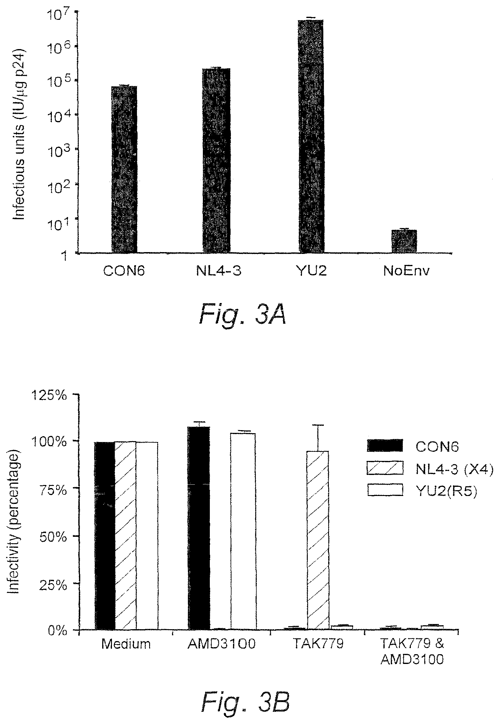
FIGS. 3A and 3B. Infectivity and coreceptor usage of CON6 envelope. (FIG. 3A) CON6 and control env plasmids were cotransfected with HIV-1/SG3.DELTA.env backbone into human 293T cells to generate Env-pseudovirions. Equal amounts of each pseudovirion (5 ng p24) were used to infect JC53-BL cells. The infectivity was determined by counting the number of blue cells (infectious units, IU) per microgram of p24 of pseudovirons (IU/.mu.g p24) after staining the infected cells for .beta.-gal expression. (FIG. 3B) Coreceptor usage of the CON6 env gene was determined on JC53BL cells treated with AMD3100 and/or TAK-799 for 1 hr (37.degree. C.) then infected with equal amounts of p24 (5 ng) of each Env-pseudovirion. Infectivity in the control group (no blocking agent) was set as 100%. Blocking efficiency was expressed as the percentage of IU from blocking experiments compared to those from control cultures without blocking agents. Data shown are mean.+-.SD.
FIG. 4. Western blot analysis of multiple subtype Env proteins against multiple subtype antisera. Equal amount of Env proteins (100 ng) were separated on 10% SDS-polyacrylamide gels. Following electrophoresis, proteins were transferred to Hybond ECL nitrocellulose membranes and reacted with sera from HIV-1 infected patients (1:1,000) or guinea pigs immunized with CON6 gp120 DNA prime, rVV boost (1:1,000). Protein-bound antibody was probed with fluorescent-labeled secondary antibodies and the images scanned and recorded on an infrared imager Odyssey (Li-Cor, Lincoln, Nebr.). Subtypes are indicated by single-letters after Env protein and serum IDs. Four to six sera were tested for each subtype, and reaction patterns were similar among all sera from the same subtype. One representative result for each subtype serum is shown.
FIG. 5. T cell immune responses induced by CON6 Env immunogens in mice. Splenocytes were isolated from individual immunized mice (5 mice/group). After splenocytes were stimulated in vitro with overlapping Env peptide pools of CON6 (black column), subtype B (hatched column), subtype C (white column), and medium (no peptide; gray column), INF-.gamma. producing cells were determined by the ELISPOT assay. T cell IFN-.gamma. responses induced by either CON6 gp120 or gp140CF were compared to those induced by subtype specific Env immunogens (JRFL and 96ZM651). Total responses for each envelope peptide pool are expressed as SFCs per million splenocytes. The values for each column are the mean.+-.SEM(of IFN-.gamma. SFCs (n=5 mice/group).
FIGS. 6A-6E. Construction of codon usage optimized subtype C ancestral and consensus envelope genes (FIGS. 6A and 6B, respectively) (SEQ ID NOS 3-4). Ancestral and consensus amino acid sequences (FIGS. 6C and 6D, respectively) (SEQ ID NOS 5-6) were transcribed to mirror the codon usage of highly expressed human genes. Paired oligonucleotides (80-mers) overlapping by 20 bp were designed to contain 5' invariant sequences including the restriction enzyme sites EcoRI, BbsI, Bam HI and BsmBI. BbsI and BsmBI are Type II restriction enzymes that cleave outside of their recognition sequences. Paired oligomers were linked individually using PCR and primers complimentary to the 18 bp invariant sequences in a stepwise fashion, yielding 140 bp PCR products. These were subcloned into pGEM-T and sequenced to confirm the absence of inadvertant mutations/deletions. Four individual pGEM-T subclones containing the proper inserts were digested and ligated together into pcDNA3.1. Multi-fragment ligations occurred repeatly amongst groups of fragments in a stepwise manner from the 5' to the 3' end of the gene until the entire gene was reconstructed in pcDNA3.1. (See schematic in FIG. 6E.)
FIG. 7. JC53-BL cells are a derivative of HeLa cells that express high levels of CD4 and the HIV-1 coreceptors CCR5 and CXCR4. They also contain the reporter cassettes of luciferase and .beta.-galactosidase that are each expressed from an HIV-1 LTR. Expression of the reporter genes is dependent on production of HIV-1 Tat. Briefly, cells are seeded into 24 or 96-well plates, incubated at 37.degree. C. for 24 hours and treated with DEAE-Dextran at 37.degree. C. for 30 minutes. Virus is serially diluted in 1% DMEM, added to the cells incubating in DEAE-Dextran, and allowed to incubate for 3 hours at 37.degree. C. after which an additional cell media is added to each well. Following a final 48-hour incubation at 37.degree. C., cells are either fixed, stained using X-Gal to visualize .beta.-galactosidase expressing blue foci or frozen-thawed three times to measure luciferase activity.
FIG. 8. Sequence alignment of subtype C ancestral and consensus env genes. Alignment of the subtype C ancestral (bottom line) (SEQ ID NO: 8) and consensus (top line) (SEQ ID NO: 7) env sequences showing a 95.5% sequence homology; amino acid sequence differences are indicated. One noted difference is the addition of a glycosylation site in the C ancestral env gene at the base of the V1 loop. A plus sign indicates a within-class difference of amino acid at the indicated position; a bar indicates a change in the class of amino acid. Potential N-glycosylation sites are marked in blue. The position of truncation for the gp140 gene is also shown.
FIG. 9. Expression of subtype C ancestral and consensus envelopes in 293T cells. Plasmids containing codon-optimized gp160, gp140, or gp120 subtype C ancestral and consensus genes were transfected into 293T cells, and protein expression was examined by Western Blot analysis of cell lysates. 48-hours post-transfection, cell lysates were collected, total protein content determined by the BCA protein assay, and 2 .mu.g of total protein was loaded per lane on a 4-20% SDS-PAGE gel. Proteins were transferred to a PVDF membrane and probed with HIV-1 plasma from a subtype C infected patient.
FIGS. 10A and 10B. FIG. 10A. Trans complementation of env-deficient HIV-1 with codon-optimized subtype C ancestral and consensus gp160 and gp140. Plasmids containing codon-optimized, subtype C ancestral or consensus gp160 or gp140 genes were co-transfected into 293T cells with an HIV-1/SG3Aenv provirus. 48 hours post-transfection cell supernatants containing pseudotyped virus were harvested, clarified by centrifugation, filtered through at 0.2 .mu.M filter, and pelleted through a 20% sucrose cushion. Quantification of p24 in each virus pellet was determined using the Coulter HIV-1 p24 antigen assay; 25 ng of p24 was loaded per lane on a 4-20% SDS-PAGE gel for particles containing a codon-optimized envelope. 250 ng of p24 was loaded per lane for particles generated by co-transfection of a rev-dependent wild-type subtype C 96ZAM651env gene. Differences in the amount of p24 loaded per lane were necessary to ensure visualization of the rev-dependent envelopes by Western Blot. Proteins were transferred to a PVDF membrane and probed with pooled plasma from HIV-1 subtype B and subtype C infected individuals. FIG. 10B. Infectivity of virus particles containing subtype C ancestral and consensus envelope glycoproteins. Infectivity of pseudotyped virus containing ancestral or consensus gp160 or gp140 envelope was determined using the JC53-BL assay. Sucrose cushion purified virus particles were assayed by the Coulter p24 antigen assay, and 5-fold serial dilutions of each pellet were incubated with DEAE-Dextran treated JC53-BL cells. Following a 48-hour incubation period, cells were fixed and stained to visualize .beta.-galactosidase expressing cells. Infectivity is represented as infectious units per ng of p24 to normalize for differences in the concentration of the input pseudovirions.
FIG. 11. Co-receptor usage of subtype C ancestral and consensus envelopes. Pseudotyped particles containing ancestral or consensus envelope were incubated with DEAE-Dextran treated JC53-BL cells in the presence of AMD3100 (a specific inhibitor of CXCR4), TAK779 (a specific inhibitor of CCR5), or AMD3000+TAK779 to determine co-receptor usage. NL4.3, an isolate known to utilize CXCR4, and YU-2, a known CCR5-using isolate, were included as controls.
FIGS. 12A-12C. Neutralization sensitivity of subtype C ancestral and consensus envelope glycoproteins. Equivalent amounts of pseudovirions containing the ancestral, consensus or 96ZAM651 gp160 envelopes (1,500 infectious units) were preincubated with a panel of plasma samples from HIV-1 subtype C infected patients and then added to the JC53-BL cell monolayer in 96-well plates. Plates were cultured for two days and luciferase activity was measured as an indicator of viral infectivity. Virus infectivity is calculated by dividing the luciferase units (LU) produced at each concentration of antibody by the LU produced by the control infection. The mean 50% inhibitory concentration (IC.sub.50) and the actual % neutralization at each antibody dilution are then calculated for each virus. The results of all luciferase experiments are confirmed by direct counting of blue foci in parallel infections.
FIGS. 13A-13F. Protein expression of consensus subtype C Gag (FIG. 13A) and Nef (FIG. 13B) following transfection into 293T cells. Consensus subtype C Gag and Nef amino acid sequences are set forth in FIGS. 13C and 13D (SEQ ID NOS: 9-10), respectively, and encoding sequences are set forth in FIGS. 13E and 13F (SEQ ID NOS: 11-12), respectively.
FIGS. 14A-14C. FIGS. 14A and 14B show the Con-S Env amino acid sequence and encoding sequence, respectively (SEQ ID NOS: 13-14). FIG. 14C shows expression of Group M consensus Con-S Env proteins using an in vitro transcription and translation system.
FIGS. 15A and 15B. Expression of Con-S env gene in mammalian cells. (FIG. 15A--cell lysate, FIG. 15B--supernatant.)
FIGS. 16A and 16B. Infectivity (FIG. 16A) and coreceptor usage (FIG. 16B) of CON6 and Con-S env genes.
FIGS. 17A-17C. Env protein incorporation in CON6 and Con-S Env-pseudovirions. (FIG. 17A--lysate, FIG. 17B--supernatant, FIG. 17C pellet.)
FIGS. 18A-18D. FIGS. 18A and 18B show subtype A consensus Env amino acid sequence and nucleic acid sequence encoding same, respectively (SEQ ID NOS: 15-16). FIGS. 18C and 18D show expression of A.con env gene in mammalian cells (FIG. 18C--cell lysate, FIG. 18D--supernatant).
FIGS. 19A-19H. M.con.gag (FIG. 19A) (SEQ ID NO: 17), M.con.pol (FIG. 19B) (SEQ ID NO: 18), M.con.nef (FIG. 19C) (SEQ ID NO: 19) and C.con.pol (FIG. 19D) (SEQ ID NO: 20) nucleic acid sequences and corresponding encoded amino acid sequences (FIGS. 19E-19H, respectively) (SEQ ID NOS: 21-24).
FIGS. 20A-20D. Subtype B consensus gag (FIG. 20A) (SEQ ID NO: 25) and env (FIG. 20B) (SEQ ID NO: 26) genes. Corresponding amino acid sequences are shown in FIGS. 20C and 20D (SEQ ID NOS: 28-29).
FIG. 21. Expression of subtype B consensus env and gag genes in 293T cells. Plasmids containing codon-optimized subtype B consensus gp160, gp140, and gag genes were transfected into 293T cells, and protein expression was examined by Western Blot analysis of cell lysates. 48-hours post-transfection, cell lysates were collected, total protein content determined by the BCA protein assay, and 2 .mu.g of total protein was loaded per lane on a 4-20% SDS-PAGE gel. Proteins were transferred to a PVDF membrane and probed with serum from an HIV-1 subtype B infected individual.
FIG. 22. Co-receptor usage of subtype B consensus envelopes. Pseudotyped particles containing the subtype B consensus gp160 Env were incubated with DEAE-Dextran treated JC53-BL cells in the presence of AMD3100 (a specific inhibitor of CXCR4), TAK779 (a specific inhibitor of CCR5), and AMD3000+TAK779 to determine co-receptor usage. NL4.3, an isolate known to utilize CXCR4 and YU-2, a known CCR5-using isolate, were included as controls.
FIGS. 23A and 23B. Trans complementation of env-deficient HIV-1 with codon-optimized subtype B consensus gp160 and gp140 genes. Plasmids containing codon-optimized, subtype B consensus gp160 or gp140 genes were co-transfected into 293T cells with an HIV-1/SG3.DELTA.env provirus. 48-hours post-transfection cell supernatants containing pseudotyped virus were harvested, clarified in a tabletop centrifuge, filtered through a 0.2 .mu.M filter, and pellet through a 20% sucrose cushion. Quantification of p24 in each virus pellet was determined using the Coulter HIV-1 p24 antigen assay; 25 ng of p24 was loaded per lane on a 4-20% SDS-PAGE gel. Proteins were transferred to a PVDF membrane and probed with anti-HIV-1 antibodies from infected HIV-1 subtype B patient serum. Trans complementation with a rev-dependent NL4.3 env was included for control. FIG. 23B. Infectivity of virus particles containing the subtype B concensus envelope. Infectivitiy of pseudotyped virus containing consensus B gp160 or gp140 was determined using the JC53-BL assay. Sucrose cushion purified virus particles were assayed by the Coulter p24 antigen assay, and 5-fold serial dilutions of each pellet were incubated with DEAE-Dextran treated JC53-BL cells. Following a 48-hour incubation period, cells were fixed and stained to visualize .beta.-galactosidase expressing cells. Infectivity is expressed as infectious units per ng of p24.
FIGS. 24A-24D. Neutralization sensitivity of virions containing subtype B consensus gp160 envelope. Equivalent amounts of pseudovirions containing the subtype B consensus or NL4.3 Env (gp160) (1,500 infectious units) were preincubated with three different monoclonal neutralizing antibodies and a panel of plasma samples from HIV-1 wubtype B infected individuals, and then added to the JC53-BL cell monolayer in 96-well plates. Plates were cultured for two days and luciferase activity was measured as an indicator of viral infectivity. Virus infectivity was calculated by dividing the luciferase units (LU) produced at each concentration of antibody by the LU produced by the control infection. The mean 50% inhibitory concentration (IC.sub.50) and the actual % neutralization at each antibody dilution were then calculated for each virus. The results of all luciferase experiments were confirmed by direct counting of blue foci in parallel infections. FIG. 24A. Neutralization of Pseudovirions containing Subtype B consensus Env (gp160). FIG. 24B. Neutralization of Pseudovirions containing NL4.3 Env (gp160).
FIG. 24C. Neutralization of Pseudovirions containing Subtype B consensus Env (gp160). FIG. 24D. Neutralization of Pseudovirions containing NL4.3 Env (gp160).
FIGS. 25A and 25B. FIG. 25A. Density and p24 analysis of sucrose gradient fractions. 0.5 ml fractions were collected from a 20-60% sucrose gradient. Fraction number 1 represents the most dense fraction taken from the bottom of the gradient tube. Density was measured with a refractometer and the amount of p24 in each fraction was determined by the Coulter p24 antigen assay. Fractions 6-9, 10-15, 16-21, and 22-25 were pooled together and analyzed by Western Blot. As expected, virions sedimented at a density of 1.16-1.18 g/ml.
FIG. 25B. VLP production by co-transfection of subtype B consensus gag and env genes. 293T cells were co-transfected with subtype B consensus gag and env genes. Cell supernatants were harvested 48-hours post-transfection, clarified through at 20% sucrose cushion, and further purified through a 20-60% sucrose gradient. Select fractions from the gradient were pooled, added to 20 ml of PBS, and centrifuged overnight at 100,000.times.g. Resuspended pellets were loaded onto a 4-20% SDS-PAGE gel, proteins were transferred to a PVDF membrane, and probed with plasma from an HIV-1 subtype B infected individual.
FIGS. 26A and 26B. FIG. 26A. 2000 Con-S 140CFI.ENV (SEQ ID NO: 30). FIG. 26B. Codon-optimized Year 2000 Con-S 140CFI.seq (SEQ ID NO: 31).
FIG. 27. Individual C57BL/6 mouse T cell responses to HIV-1 envelope peptides. Comparative immunogenicity of CON6 gp140CFI and Con-S gp140CFI in C57BL/C mice. Mice were immunized with either HIV5305 (Subtype A), 2801 (Subtype B), CON6 or Con-S Envelope genes in DNA prime, rVV boost regimens, 5 mice per group. Spleen cells were assayed for IFN-.gamma. spot-forming cells 10 days after rVV boost, using mixtures of overlapping peptides from Envs of HIV-1 UG37(A), MN(B), Ch19(C), 89.6(B) SF162(B) or no peptide negative control.
FIGS. 28A-28C. FIG. 28A. Con-B 2003 Env. pep (841 a.a.) (SEQ ID NO: 32). Amino acid sequence underlined is the fusion domain that is deleted in 140CF design and the "W" underlined is the last amino acid at the C-terminus, all amino acids after the "W" are deleted in the 140CF design. FIG. 28B. Con-B-140CF.pep (632 a.a.) (SEQ ID NO: 33). Amino acids in bold identify the junction of the deleted fusion cleavage site. FIG. 28C. Codon-optimized Con-B 140CF.seq (1927 nt.) (SEQ ID NO: 34).
FIGS. 29A-29C. FIG. 29A. CON_OF_CONS-2003 (829 a.a.) (SEQ ID NO: 35). Amino acid sequence underlined is the fusion domain that is deleted in 140CF design and the "W" underlined is the last amino acid at the C-terminus, all amino acids after the "W" are deleted in the 140CF design. FIG. 29B. ConS-2003 140CF.pep (620 a.a.) (SEQ ID NO: 36). Amino acids in bold identify the junction of the deleted fusion cleavage site. FIG. 29C. CODON-OPTIMIZED ConS-2003 140CF.seq (1891 nt.) (SEQ ID NO: 37).
FIGS. 30A-30C. FIG. 30A. CONSENSUS_A1-2003 (845 a.a.) (SEQ ID NO: 38). Amino acid sequence underlined is the fusion domain that is deleted in 140CF design and the "W" underlined is the last amino acid at the C-terminus, all amino acids after the "W" are deleted in the 140CF design. FIG. 30B. Con-A1-2003 140CF.pep (629 a.a.) (SEQ ID NO: 39). Amino acids in bold identify the junction of the deleted fusion cleavage site. FIG. 30C. CODON-OPTIMIZED Con-A1-2003.seq (SEQ ID NO: 40).
FIGS. 31A-31C. FIG. 31A. CONSENSUS_C-2003 (835 a.a.) (SEQ ID NO: 41). Amino acid sequence underlined is the fusion domain that is deleted in 140CF design and the "W" underlined is the last amino acid at the C-terminus, all amino acids after the "W" are deleted in the 140CF design. FIG. 31B. Con-C 2003 140CF.pep (619 a.a.) (SEQ ID NO: 42). Amino acids in bold identify the junction of the deleted fusion cleavage site. FIG. 31C. CODON-OPTIMIZED Con-C-2003 (140 CF (1,888 nt.) (SEQ ID NO: 43).
FIGS. 32A-32C. FIG. 32A. CONSENSUS_G-2003 (842 a.a.) (SEQ ID NO: 44). Amino acid sequence underlined is the fusion domain that is deleted in 140CF design and the "W" underlined is the last amino acid at the C-terminus, all amino acids after the "W" are deleted in the 140CF design. FIG. 32B. Con-G-2003 140CF.pep (626 a.a.) (SEQ ID NO: 45). Amino acids in bold identify the junction of the deleted fusion cleavage site. FIG. 32C. CODON-OPTIMIZED Con-G-2003.seq (SEQ ID NO: 46).
FIGS. 33A-33C. FIG. 33A. CONSENSUS_01_AE-2003 (854 a.a.) (SEQ ID NO: 47). Amino acid sequence underlined is the fusion domain that is deleted in 140CF design and the "W" underlined is the last amino acid at the C-terminus, all amino acids after the "W" are deleted in the 140CF design. FIG. 33B. Con-AE01-2003 140CF.pep (638 a.a.) (SEQ ID NO: 48). Amino acids in bold identify the junction of the deleted fusion cleavage site. FIG. 33C, CODON-OPTIMIZED Con-AE01-2003.seq. (1945 nt.) (SEQ ID NO: 49).
FIGS. 34A-34C. FIG. 34A. Wild-type subtype A Env. 00KE MSA4076-A (Subtype A, 891 a.a.) (SEQ ID NO: 50). Amino acid sequence underlined is the fusion domain that is deleted in 140CF design and the "W" underlined is the last amino acid at the C-terminus, all amino acids after the "W" are deleted in the 140CF design. FIG. 34B. 00KE MSA4076-A 140CF.pep (647 a.a.) (SEQ ID NO: 51). Amino acids in bold identify the junction of the deleted fusion cleavage site. FIG. 34C. CODON-OPTIMIZED 00KE MSA4076-A 140CF.seq. (1972 nt.) (SEQ ID NO: 52).
FIGS. 35A-35C. FIG. 35A. Wild-type subtype B. QH0515.1g gp160 (861 a.a.) (SEQ ID NO: 53). Amino acid sequence underlined is the fusion domain that is deleted in 140CF design and the "W" underlined is the last amino acid at the C-terminus, all amino acids after the "W" are deleted in the 140CF design. FIG. 35B. QH0515.1g 140CF (651 a.a.) (SEQ ID NO: 54). Amino acids in bold identify the junction of the deleted fusion cleavage site. FIG. 35C. CODON-OPTIMIZED QH0515.1g 140CF.seq (1984 nt.) (SEQ ID NO: 55).
FIGS. 36A-36C. FIG. 36A. Wild-type subtype C. DU123.6 gp160 (854 a.a.) (SEQ ID NO: 56). Amino acid sequence underlined is the fusion domain that is deleted in 140CF design and the "W" underlined is the last amino acid at the C-terminus, all amino acids after the "W" are deleted in the 140CF design. FIG. 36B. DU123.6 140CF (638 a.a.) (SEQ ID NO: 57). Amino acids in bold identify the junction of the deleted fusion cleavage site. FIG. 36C. CODON-OPTIMIZED DU123.6 140CF.seq (1945 nt.) (SEQ ID NO: 58).
FIGS. 37A-37C. FIG. 37A. Wild-type subtype CRF01_AE. 97CNGX2F-AE (854 a.a.) (SEQ ID NO: 59). Amino acid sequence underlined is the fusion domain that is deleted in 140CF design and the "W" underlined is the last amino acid at the C-terminus, all amino acids after the "W" are deleted in the 140CF design. FIG. 37B. 97CNGX2F-AE 140CF.pep (629 a.a.) (SEQ ID NO: 60). Amino acids in bold identify the junction of the deleted fusion cleavage site. FIG. 37C. CODON-OPTIMIZED 97CNGX2F-AE 140CF.seq (1921 nt.) (SEQ ID NO: 61).
FIGS. 38A-38C. FIG. 38A. Wild-type DRCBL-G (854 a.a.) (SEQ ID NO: 62). Amino acid sequence underlined is the fusion domain that is deleted in 140CF design and the "W" underlined is the last amino acid at the C-terminus, all amino acids after the "W" are deleted in the 140CF design. FIG. 38B. DRCBL-G 140CF.pep (630 a.a.) (SEQ ID NO: 63). Amino acids in bold identify the junction of the deleted fusion cleavage site. FIG. 38C. CODON-OPTIMIZED DRCBL-G 140CF.seq (1921 nt.) (SEQ ID NO: 64).
FIGS. 39A and 39B. FIG. 39A. 2003 Con-S Env (SEQ ID NO: 65). FIG. 39B. 2003 Con-S Env.seq.opt. (Seq.opt.=codon optimized encoding sequence.) (SEQ ID NO: 72)
FIGS. 40A and 40B. FIG. 40A. 2003 M. Group.Anc Env (SEQ ID NO: 66). FIG. 40B. 2003 M. Group.anc Env.seq.opt. (Seq.opt.=codon optimized encoding sequence.) (SEQ ID NO: 67)
FIGS. 41A and 41B. FIG. 41A. 2003 CON_A1 Env (SEQ ID NO: 68). FIG. 41B. 2003 CON_A1 Env.seq.opt. (Seq.opt.=codon optimized encoding sequence.) (SEQ ID NO: 70)
FIGS. 42A and 42B. FIG. 42A. 2003 A1.Anc Env (SEQ ID NO: 69). FIG. 42B. 2003 A1.anc Env.seq.opt. (Seq.opt.=codon optimized encoding sequence.) (SEQ ID NO: 71)
FIGS. 43A and 43B. FIG. 43A. 2003 CON_A2 Env (SEQ ID NO: 73). FIG. 43B. 2003 CON_A2 Env.seq.opt. (Seq.opt.=codon optimized encoding sequence.) (SEQ ID NO: 75)
FIGS. 44A and 44B. FIG. 44A. 2003 CON_B Env (SEQ ID NO: 74). FIG. 44B. 2003 CON_B Env.seq.opt. (Seq.opt.=codon optimized encoding sequence.) (SEQ ID NO: 76)
FIGS. 45A and 45B. FIG. 45A. 2003 B.anc Env (SEQ ID NO: 77). FIG. 45B. 2003 B.anc Env.seq.opt. (Seq.opt.=codon optimized encoding sequence.) (SEQ ID NO: 79)
FIGS. 46A and 46B. FIG. 46A. 2003 CON_C Env (SEQ ID NO: 78). FIG. 46B. 2003 CON_C Env.seq.opt. (Seq.opt.=codon optimized encoding sequence.) (SEQ ID NO: 80)
FIGS. 47A and 47B. FIG. 47A. 2003 C.anc Env (SEQ ID NO: 81). FIG. 47B. 2003 C.anc Env.seq.opt. (Seq.opt.=codon optimized encoding sequence.) (SEQ ID NO: 83)
FIGS. 48A and 48B. FIG. 48A. 2003 CON_D Env (SEQ ID NO: 82). FIG. 48B. 2003 CON D Env.seq.opt. (Seq.opt.=codon optimized encoding sequence.) (SEQ ID NO: 84)
FIGS. 49A and 49B. FIG. 49A. 2003 CON_F1 Env (SEQ ID NO: 85). FIG. 49B. 2003 CON_F1 Env.seq.opt. (Seq.opt.=codon optimized encoding sequence.) (SEQ ID NO: 87)
FIGS. 50A and 50B. FIG. 50A. 2003 CON_F2 Env (SEQ ID NO: 86). FIG. 50B. 2003 CON_F2 Env.seq.opt. (Seq.opt.=codon optimized encoding sequence.) (SEQ ID NO: 88)
FIGS. 51A and 51B. FIG. 51A. 2003 CON_G Env (SEQ ID NO: 89). FIG. 51B. 2003 CON_G Env.seq.opt. (Seq.opt.=codon optimized encoding sequence.) (SEQ ID NO: 91)
FIGS. 52A and 52B. FIG. 52A. 2003 CON_H Env (SEQ ID NO: 90). FIG. 52B. 2003 CON_H Env.seq.opt. (Seq.opt.=codon optimized encoding sequence.) (SEQ ID NO: 92)
FIGS. 53A and 53B. FIG. 53A. 2003_CON_01_AE Env (SEQ ID NO: 93). FIG. 53B. 2003_CON_01_AE Env.seq.opt. (Seq.opt.=codon optimized encoding sequence.) (SEQ ID NO: 95)
FIGS. 54A and 54B. FIG. 54A. 2003 CON_02_AG Env (SEQ ID NO: 94). FIG. 54B. 2003 CON_02_AG Env.seq.opt. (Seq.opt.=codon optimized encoding sequence.) (SEQ ID NO: 96)
FIGS. 55A and 55B. FIG. 55A. 2003 CON_03_AB Env (SEQ ID NO: 97). FIG. 55B. 2003 CON_03_AB Env.seq.opt. (Seq.opt.=codon optimized encoding sequence.) (SEQ ID NO: 99)
FIGS. 56A and 56B. FIG. 56A. 2003 CON_04_CPX Env (SEQ ID NO: 98). FIG. 56B. 2003 CON_04_CPX Env.seq.opt. (Seq.opt.=codon optimized encoding sequence.) (SEQ ID NO: 100)
FIGS. 57A and 57B. FIG. 57A. 2003 CON_06_CPX Env (SEQ ID NO: 101). FIG. 57B. 2003 CON_06_CPX Env.seq.opt. (Seq.opt.=codon optimized encoding sequence.) (SEQ ID NO: 103)
FIGS. 58A and 58B. FIG. 58A. 2003 CON_08_BC Env (SEQ ID NO: 102). FIG. 58B. 2003 CON_08_BC Env.seq.opt. (Seq.opt.=codon optimized encoding sequence.) (SEQ ID NO: 104)
FIGS. 59A and 59B. FIG. 59A. 2003 CON_10_CD Env (SEQ ID NO: 105). FIG. 59B. 2003 CON_10_CD Env.seq.opt. (Seq.opt.=codon optimized encoding sequence.) (SEQ ID NO: 107)
FIGS. 60A and 60B. FIG. 60A. 2003 CON_11_CPX Env (SEQ ID NO: 106). FIG. 60B. 2003 CON_11_CPX Env.seq.opt. (Seq.opt.=codon optimized encoding sequence.) (SEQ ID NO: 108)
FIGS. 61A and 61B. FIG. 61A. 2003 CON_12_BF Env (SEQ ID NO: 109). FIG. 61B. 2003 CON_12_BF Env.seq.opt. (Seq.opt.=codon optimized encoding sequence.) (SEQ ID NO: 111)
FIGS. 62A and 62B. FIG. 62A. 2003 CON_14_BG Env (SEQ ID NO: 110). FIG. 62B, 2003 CON_14_BG Env.seq.opt. (Seq.opt.=codon optimized encoding sequence.) (SEQ ID NO: 112)
FIGS. 63A and 63B. FIG. 63A. 2003_CON_S gag.PEP (SEQ ID NO: 113). FIG. 63B. 2003_CON_S gag.OPT. (OPT=codon optimized encoding sequence.) (SEQ ID NO: 114)
FIGS. 64A and 64B. FIG. 64A. 2003_M.GROUP.anc gag.PEP (SEQ ID NO: 115). FIG. 64B. 2003_M.GROUP.anc gag.OPT. (OPT=codon optimized encoding sequence.) (SEQ ID NO: 116)
FIGS. 65A-65D. FIG. 65A. 2003_CON_A1 gag.PEP (SEQ ID NO: 117). FIG. 65B. 2003_CON_A1 gag.OPT (SEQ ID NO: 118). FIG. 65C. 2003_A1.anc gag.PEP (SEQ ID NO: 119). FIG. 65D. 2003_A1.anc gag.OPT (SEQ ID NO: 120). (OPT=codon optimized encoding sequence.)
FIGS. 66A and 66B. FIG. 66A. 2003_CON_A2 gag.PEP (SEQ ID NO: 121), FIG. 66B. 2003_CON_A2 gag.OPT. (OPT=codon optimized encoding sequence.) (SEQ ID NO: 122)
FIGS. 67A-67D. FIG. 67A. 2003_CON_B gag.PEP (SEQ ID NO: 123). FIG. 67B. 2003_CON_B gag.OPT (SEQ ID NO: 124). FIG. 67C. 2003_B, anc gag.PEP (SEQ ID NO: 125). FIG. 67D. 2003_B.anc gag.OPT. (OPT=codon optimized encoding sequence.) (SEQ ID NO: 126)
FIGS. 68A-68D. FIG. 68A. 2003_CON_C gag.PEP (SEQ ID NO: 127). FIG. 68B. 2003_CON_C gag.OPT (SEQ ID NO: 128). FIG. 68C. 2003_C.anc.gag.PEP (SEQ ID NO: 129). FIG. 68D. 2003_C.anc.gag.OPT. (OPT=codon optimized encoding sequence.) (SEQ ID NO: 130)
FIGS. 69A and 69B. FIG. 69A. 2003_CON_D gag.PEP (SEQ ID NO: 131). FIG. 69B. 2003_CON_D gag.OPT. (OPT=codon optimized encoding sequence.) (SEQ ID NO: 132)
FIGS. 70A and 70B. FIG. 70A. 2003_CON_F gag.PEP (SEQ ID NO: 133). FIG. 70B. 2003_CON_F gag.OPT. (OPT=codon optimized encoding sequence.) (SEQ ID NO: 134)
FIGS. 71A and 71B. FIG. 71A. 2003_CON_G gag.PEP (SEQ ID NO: 135). FIG. 71B. 2003_CON_G gag.OPT. (OPT=codon optimized encoding sequence.) (SEQ ID NO: 136)
FIGS. 72A and 72B. FIG. 72A. 2003_CON_H gag.PEP (SEQ ID NO: 137). FIG. 72B. 2003_CON_H gag.OPT. (OPT=codon optimized encoding sequence.) (SEQ ID NO: 138)
FIGS. 73A and 73B. FIG. 73A. 2003_CON_K gag.PEP (SEQ ID NO: 139). FIG. 73B. 2003_CON_K gag.OPT. (OPT=codon optimized encoding sequence.) (SEQ ID NO: 140)
FIGS. 74A and 74B. FIG. 74A. 2003_CON_01_AE gag.PEP (SEQ ID NO: 141). FIG. 7B. 2003_CON_01_AE gag.OPT. (OPT=codon optimized encoding sequence.) (SEQ ID NO: 142)
FIGS. 75A and 75B. FIG. 75A. 2003_CON_02_AG gag.PEP (SEQ ID NO: 143). FIG. 75B. 2003_CON_02_AG gag.OPT. (OPT=codon optimized encoding sequence.) (SEQ ID NO: 144)
FIGS. 76A and 76B. FIG. 76A. 2003_CON_03_ABG gag.PEP (SEQ ID NO: 145). FIG. 76B. 2003_CON_03_ABG gag.OPT. (OPT=codon optimized encoding sequence.) (SEQ ID NO: 146)
FIGS. 77A and 77B. FIG. 77A. 2003_CON_04_CFX gag.PEP (SEQ ID NO: 147). FIG. 77B. 2003_CON_04_CFX gag.OPT. (OPT=codon optimized encoding sequence.) (SEQ ID NO: 148)
FIGS. 78A and 78B. FIG. 78A. 2003_CON_06_CPX gag.PEP (SEQ ID NO: 150). FIG. 78B. 2003_CON_06_CPX gag.OPT. (OPT=codon optimized encoding sequence.) (SEQ ID NO: 151)
FIGS. 79A and 79B. FIG. 79A. 2003_CON_07_BC gag.PEP (SEQ ID NO: 152). FIG. 79B. 2003_CON_07_BC gag.OPT. (OPT=codon optimized encoding sequence.) (SEQ ID NO: 153)
FIGS. 80A and 80B. FIG. 80A. 2003_CON_08_BC gag.PEP (SEQ ID NO: 154). FIG. 80B. 2003_CON_08_BC gag.OPT. (OPT=codon optimized encoding sequence.) (SEQ ID NO: 155)
FIGS. 81A and 81B. FIG. 81A. 2003_CON_10_CD gag.PEP (SEQ ID NO: 156). FIG. 81B. 2003_CON_10_CD gag.OPT. (OPT=codon optimized encoding sequence.) (SEQ ID NO: 157)
FIGS. 82A and 82B. FIG. 82A. 2003_CON_11_CPX gag.PEP (SEQ ID NO: 158). FIG. 82B. 2003_CON_11_CPX gag.OPT. (OPT=codon optimized encoding sequence.) (SEQ ID NO: 159)
FIGS. 83A and 83B. FIG. 83A. 2003_CON_12_BF.gag.PEP (SEQ ID NO: 160) FIG. 83B. 2003_CON_12_BF.gag.OPT. (OPT=codon optimized encoding sequence.) (SEQ ID NO: 161)
FIGS. 84A and 84B. FIG. 84A. 2003_CON_14_BG gag.PEP (SEQ ID NO: 162). FIG. 84B. 2003_CON_14_BG gag.OPT. (OPT=codon optimized encoding sequence.) (SEQ ID NO: 163)
FIGS. 85A and 85B. FIG. 85A. 2003_CONS nef.PEP (SEQ ID NO: 164). FIG. 85B. 2003_CONS nef.OPT. (OPT=codon optimized encoding sequence.) (SEQ ID NO: 165)
FIGS. 86A and 86B. FIG. 86A. 2003_M GROUP.anc nef.PEP (SEQ ID NO: 166). FIG. 86B. 2003 M GROUP.anc.nef OPT. (OPT=codon optimized encoding sequence.) (SEQ ID NO: 167)
FIGS. 87A and 87B. FIG. 87A. 2003_CON_A nef.PEP (SEQ ID NO: 168). FIG. 87B. 2003_CON_A nef OPT. (OPT=codon optimized encoding sequence.) (SEQ ID NO: 169)
FIGS. 88A-88D. FIG. 88A. 2003_CON_A1 nef.PEP (SEQ ID NO: 170). FIG. 88B. 2003_CON_A1 nef.OPT (SEQ ID NO: 171). FIG. 88C. 2003_A1.anc nef.PEP (SEQ ID NO: 172). FIG. 88D. 2003_A1.anc nef.OPT. (OPT=codon optimized encoding sequence.) (SEQ ID NO: 173)
FIGS. 89A and 89B. FIG. 89A. 2003_CON_A2 nef.PEP (SEQ ID NO: 174). FIG. 89B. 2003_CON_A2 nef.OPT. (OPT=codon optimized encoding sequence.) (SEQ ID NO: 175)
FIGS. 90A-90D. FIG. 90A. 2003_CON_B nef PEP (SEQ ID NO: 176). FIG. 90B. 2003_CON-B nef OPT (SEQ ID NO: 177). FIG. 90C. 2003_B.anc nef.PEP (SEQ ID NO: 178). FIG. 90D. 2003_B.anc nef.OPT. (OPT=codon optimized encoding sequence.) (SEQ ID NO: 179)
FIGS. 91A and 91B. FIG. 91A. 2003_CON_02_AG nef.PEP (SEQ ID NO: 180). FIG. 91B. 2003_CON_02_AG nef.OPT. (OPT=codon optimized encoding sequence.) (SEQ ID NO: 181)
FIGS. 92A-92D. FIG. 92A. 2003_CON_C nef PEP (SEQ ID NO: 182). FIG. 92B. 2003_CON_C nef OPT (SEQ ID NO: 183). FIG. 92C. 2003_C.anc nef PEP (SEQ ID NO: 184). FIG. 92D. 2003_C.anc nef.OPT. (OPT=codon optimized encoding sequence.) (SEQ ID NO: 185)
FIGS. 93A and 93B. FIG. 93A. 2003_CON_D nef.PEP (SEQ ID NO: 186). FIG. 93B. 2003_CON_D nef OPT. (OPT=codon optimized encoding sequence.) (SEQ ID NO: 187)
FIGS. 94A and 94B. FIG. 94A. 2003_CON_F1 nef PEP (SEQ ID NO: 188). FIG. 94B. 2003_CON_F1 nef OPT. (OPT=codon optimized encoding sequence.) (SEQ ID NO: 189)
FIGS. 95A and 95B. FIG. 95A. 2003_CON_F2 nef.PEP (SEQ ID NO: 190). FIG. 95B. 2003_CON_F2 nef.OPT. (OPT=codon optimized encoding sequence.) (SEQ ID NO: 191)
FIGS. 96A and 96B. FIG. 96A. 2003_CON_G nef.PEP (SEQ ID NO: 192). FIG. 96B. 2003_CON_G nef OPT. (OPT=codon optimized encoding sequence.) (SEQ ID NO: 193)
FIGS. 97A and 97B. FIG. 97A. 2003_CON_H nef.PEP (SEQ ID NO: 194). FIG. 97B. 2003_CON_H nef.OPT. (OPT=codon optimized encoding sequence.) (SEQ ID NO: 195)
FIGS. 98A and 98B. FIG. 98A. 2003_CON_01_AE nef.PEP (SEQ ID NO: 196). FIG. 98B. 2003_CON_01_AE nef OPT. (OPT=codon optimized encoding sequence.) (SEQ ID NO: 197)
FIGS. 99A and 99B. FIG. 99A. 2003_CON_03_AE nef.PEP (SEQ ID NO: 198). FIG. 99B. 2003_CON_03_AE nef OPT. (OPT=codon optimized encoding sequence.) (SEQ ID NO: 199)
FIGS. 100A and 100B. FIG. 100A. 2003_CON_04_CFX nef.PEP (SEQ ID NO: 200). FIG. 100B. 2003_CON_04_CFX nef.OPT. (OPT=codon optimized encoding sequence.) (SEQ ID NO: 201)
FIGS. 101A and 101B. FIG. 101A. 2003_CON_06_CFX nef.PEP (SEQ ID NO: 202). FIG. 101B. 2003_CON_06_CFX nef.OPT. (OPT=codon optimized encoding sequence.) (SEQ ID NO: 203)
FIGS. 102A and 102B. FIG. 102A. 2003_CON_08_BC nef.PEP (SEQ ID NO: 204). FIG. 102B. 2003_CON_08_BC nef OPT. (OPT=codon optimized encoding sequence.) (SEQ ID NO: 205)
FIGS. 103A and 103B. FIG. 103A. 2003_CON_10_CD nef.PEP (SEQ ID NO: 206). FIG. 103B. 2003_CON_10_CD nef.OPT. (OPT=codon optimized encoding sequence.) (SEQ ID NO: 207)
FIGS. 104A and 104B. FIG. 104A. 2003_CON_11_CFX nef.PEP (SEQ ID NO: 208). FIG. 104B. 2003_CON_11_CFX nef.OPT. (OPT=codon optimized encoding sequence.) (SEQ ID NO: 209)
FIGS. 105A and 105B. FIG. 105A. 2003_CON_12_BF nef.PEP (SEQ ID NO: 210). FIG. 105B. 2003_CON_12_BF nef.OPT. (OPT=codon optimized encoding sequence.) (SEQ ID NO: 211)
FIGS. 106A and 106B. FIG. 106A. 2003_CON_14_BG nef.PEP (SEQ ID NO: 212). FIG. 106B. 2003_CON_14_BG nef.OPT. (OPT=codon optimized encoding sequence.) (SEQ ID NO: 213)
FIGS. 107A and 107B. FIG. 107A. 2003_CON_S pol.PEP (SEQ ID NO: 214). FIG. 107B. 2003_CON_S pol.OPT. (OPT=codon optimized encoding sequence.) (SEQ ID NO: 215)
FIGS. 108A and 108B. FIG. 108A. 2003_M GROUP anc pol.PEP (SEQ ID NO: 216). FIG. 108B. 2003_M.GROUP anc pol.OPT. (OPT=codon optimized encoding sequence.) (SEQ ID NO: 218)
FIGS. 109A-109D. FIG. 109A. 2003_CON_A1 pol.PEP (SEQ ID NO: 217). FIG. 109B. 2003_CON_A1 pol.OPT (SEQ ID NO: 219). FIG. 109C. 2003_A1.anc pol.PEP (SEQ ID NO: 220). FIG. 109D. 2003_A1.anc pol.OPT (SEQ ID NO: 221). (OPT=codon optimized encoding sequence.)
FIGS. 110A and 110B. FIG. 110A. 2003_CON_A2 pol.PEP (SEQ ID NO: 222). FIG. 110B. 2003_CON_A2 pol.OPT. (OPT=codon optimized encoding sequence.) (SEQ ID NO: 224)
FIGS. 111A-111D. FIG. 111A. 2003_CON_B pol.PEP (SEQ ID NO: 223). FIG. 111B. 2003_CON_B pol.OPT (SEQ ID NO: 225). FIG. 111C. 2003_B.anc pol.PEP (SEQ ID NO: 226). FIG. 111D. 2003_B.anc pol.OPT (SEQ ID NO: 227). (OPT=codon optimized encoding sequence.)
FIGS. 112A-112D. FIG. 112A. 2003_CON_C pol.PEP (SEQ ID NO: 228). FIG. 112B. 2003_CON_C pol.OPT (SEQ ID NO: 229). FIG. 112C. 2003_C.anc pol.PEP (SEQ ID NO: 230). FIG. 112D. 2003_C.anc pol.OPT. (OPT=codon optimized encoding sequence.) (SEQ ID NO: 231)
FIGS. 113A and 113B. FIG. 113A. 2003_CON_D pol.PEP (SEQ ID NO: 232). FIG. 113B. 2003_CON_D pol.OPT. (OPT=codon optimized encoding sequence.) (SEQ ID NO: 224)
FIGS. 114A and 114B. FIG. 114A. 2003_CON_F1 pol.PEP (SEQ ID NO: 233). FIG. 114B. 2003_CON_F1 pol.OPT. (OPT=codon optimized encoding sequence.) (SEQ ID NO: 235)
FIGS. 115A and 115B. FIG. 115A. 2003_CON_F2 pol.PEP (SEQ ID NO: 236). FIG. 115B. 2003_CON_F2 pol.OPT. (OPT=codon optimized encoding sequence.) (SEQ ID NO: 238)
FIGS. 116A and 116B. FIG. 116A. 2003_CON_G pol.PEP (SEQ ID NO: 237). FIG. 116B. 2003_CON_G pol.OPT. (OPT=codon optimized encoding sequence.) (SEQ ID NO: 239)
FIGS. 117A and 117B. FIG. 117A. 2003_CON_H pol.PEP (SEQ ID NO: 240). FIG. 117B. 2003_CON_H pol.OPT. (OPT=codon optimized encoding sequence.) (SEQ ID NO: 242)
FIGS. 118A and 118B. FIG. 118A. 2003_CON_01_AE pol.PEP (SEQ ID NO: 241). FIG. 118B. 2003_CON_01_AE pol.OPT. (OPT=codon optimized encoding sequence.) (SEQ ID NO: 243)
FIGS. 119A and 119B. FIG. 119A. 2003_CON_02_AG pol.PEP (SEQ ID NO: 244). FIG. 119B. 2003_CON_02_AG pol.OPT. (OPT=codon optimized encoding sequence.) (SEQ ID NO: 246)
FIGS. 120A and 120B. FIG. 120A. 2003_CON_03_AB pol.PEP (SEQ ID NO: 245). FIG. 120B. 2003_CON_03_AB pol.OPT. (OPT=codon optimized encoding sequence.) (SEQ ID NO: 247)
FIGS. 121A and 121B. FIG. 121A. 2003_CON_04_CPX pol.PEP (SEQ ID NO: 248). FIG. 121B. 2003_CON_04_CPX pol.OPT. (OPT=codon optimized encoding sequence.) (SEQ ID NO: 250)
FIGS. 122A and 122B. FIG. 122A. 2003_CON_06_CPX pol.PEP (SEQ ID NO: 249). FIG. 122B. 2003_CON06_CPX pol.OPT. (OPT=codon optimized encoding sequence.) (SEQ ID NO: 251)
FIGS. 123A and 123B. FIG. 123A. 2003_CON_08_BC pol.PEP (SEQ ID NO: 252). FIG. 123B. 2003_CON_08_BC pol.OPT. (OPT=codon optimized encoding sequence.) (SEQ ID NO: 254)
FIGS. 124A and 124B. FIG. 124A. 2003_CON_10_CD pol.PEP (SEQ ID NO: 253). FIG. 124B. 2003_CON_10_CD pol.OPT. (OPT=codon optimized encoding sequence.) (SEQ ID NO: 255)
FIGS. 125A and 125B. FIG. 125A. 2003_CON_11_CPX pol.PEP (SEQ ID NO: 256). FIG. 125B. 2003_CON_11_CPX pol.OPT. (OPT=codon optimized encoding sequence.) (SEQ ID NO: 258)
FIGS. 126A and 126B. FIG. 126A. 2003_CON_12_BF pol.PEP (SEQ ID NO: 257). FIG. 126B. 2003_CON_12_BF pol.OPT. (OPT=codon optimized encoding sequence.) (SEQ ID NO: 259)
FIGS. 127A and 127B. FIG. 127A. 2003_CON_14_BG pol.PEP (SEQ ID NO: 260). FIG. 127B. 2003_CON_14_BG pol.OPT. (OPT=codon optimized encoding sequence.) (SEQ ID NO: 261)
DETAILED DESCRIPTION OF THE INVENTION
The present invention relates to an immunogen that induces antibodies that neutralize a wide spectrum of human immunodeficiency virus (HIV) primary isolates and/or that induces a T cell response. The immunogen comprises at least one consensus or ancestral immunogen (e.g., Env, Gag, Nef or Poly, or portion or variant thereof. The invention also relates to nucleic acid sequences encoding the consensus or ancestral immunogen, or portion or variant thereof. The invention further relates to methods of using both the immunogen and the encoding sequences. While the invention is described in detail with reference to specific consensus and ancestral immunogens (for example, to a group M consensus Env), it will be appreciated that the approach described herein can be used to generate a variety of consensus or ancestral immunogens (for example, envelopes for other HIV-1 groups (e.g., N and O)).
In accordance with one embodiment of the invention, a consensus env gene can be constructed by generating consensus sequences of env genes for each subtype of a particular HIV-1 group (group M being classified into subtypes A-D, F-H, J an K), for example, from sequences in the Los Alamos HIV Sequence Database (using, for example, MASE (Multiple Aligned Sequence Editor)). A consensus sequence of all subtype consensuses can then be generated to avoid heavily sequenced subtypes (Gaschen et al, Science 296:2354-2360 (2002), Korber et al, Science 288:1789-1796 (2000)). In the case of the group M consensus env gene described in Example 1 (designated CON6), five highly variable regions from a CRF08_BC recombinant strain (98CN006) (V1, V2, V4, V5 and a region in cytoplasmic domain of gp41) are used to fill in the missing regions in the sequence (see, however, corresponding regions for Con-S). For high levels of expression, the codons of consensus or ancestral genes can be optimized based on codon usage for highly expressed human genes. (Haas et al, Curr. Biol. 6:315-324 (2000), Andre et al, J. Virol. 72:1497-1503 (1998)).
With the Year 1999 consensus group M env gene, CONE, it has been possible to demonstrate induction of superior T cell responses by CONE versus wild-type B and C env by the number of ELISPOT .gamma.-interferon spleen spot forming cells and the number of epitopes recognized in two strains of mice (Tables 1 and 2 show the data in BALB/c mice). The ability of CON6 Env protein to induce neutralizing antibodies to HIV-1 primary isolates has been compared to that of several subtype B Env. The target of neutralizing antibodies induced by CON6 includes several non-B HIV-1 strains.
TABLE-US-00001 TABLE 1 T cell epitope mapping of CON6, JRFL and 96ZM651 Env immunogen in BALB/c mice. Table 1 discloses SEQ ID NOS: 262-287, respectively, in order of appearance. Immunogen JRFL 96ZM651 T cell Peptide CON6 (B) (C) response CON 6 (group M consensus) 16 DTEVHNVWATHACVP + + CD4 48 KNSSEYYRLINCNTS + + CD4 49 EYYRLINCNTSAITQ 53 CPKVSFEPIPIHYCA + CD4 54 SFEPIPIHYCAPAGF 62 NVSTVQCTHGIKPVV + CD4 104 ETITLPCRIKQIINM + CD8 105 LPCRIKQIINMWQGV 130 GIVQQQSNLLRAIEA + CD4 131 VQQSNLLRAIEAQQHL 134 AQQHLLQLTVWGIKQLQ + CD4 135 LQLTVWGIKQLQARVL Subtype B (MN) 6223 AKAYDTEVHNVWATQ + CD4 6224 DTEVHNVWATQACVP 6261 ACPKISFEPIPIHYC + CD4 6262 ISFEPIPIHYCAPAG 6286 RKRIHIGPGRAFYTT + CD8 6287 HIGPGRAFYTTKNII 6346 IVQQQNNLLRAIEAQ + CD4 6347 QNNLLRAIEAQQHML Subtype C (Chn19) 4834 VPVWKEAKTTLFCASDAKSY + CD4 4836 GKEVHNVWATHACVPTDPNP + + CD4 4848 SSENSSEYYRLINCNTSAIT + + CD4 4854 STVQCTHGIKPVVSTQLLLN + CD4 4884 QQSNLLRAIEAQQHLLQLTV + CD4 4885 AQQHLLQLTVWGIKQLQTRV + CD4
TABLE-US-00002 TABLE 2 T cell epitope mapping of CON6.gp120 immunogen in C57BL/6 mice. Table 2 discloses SEQ ID NOS: 288-304, respectively, in order of appearance. Peptide Peptide sequence T cell response CON 6 (consensus) 2 GIQRNCQHLWRWGTM CD8 3 NCQHLWRWGTMILGM 16 DTEVENVWATHACVP CD4 53 CPKVSFEPIPIHYCA CD4 97 FYCNTSGLFNSTWMF CD8 99 FNSTWMFNGTYMFNG CD8 Subtype B (MN) 6210 GIRRNYQHWWGWGTM CD8 6211 NYQHWWGWGTMLLGL 6232 NMWKNNMVEQMHEDI CD4 6262 ISFEPIPIHYCAPAG CD4 6290 NIIGTIRQAHCNISR CD4 6291 TIRQAHCNISRAKWN Subtype C (Chn 19) 4830 MRVTGIRKNYQHLWRWGTML CD8 5446 RWGTMLLGMLMICSAAEN CD8 4836 GKEVENVWATHACVPTDPNP CD4 4862 GDIRQAHCNISKDKWNETLQ CD4 4888 LLGIWGCSGKLICTTTVPWN CD8
For the Year 2000 consensus group M env gene, Con-S, the Con-S envelope has been shown to be as immunogenic as the CON6 envelope gene in T cell .gamma. interferon ELISPOT assays in two strains of mice (the data for C57BL/6 are shown in FIG. 27). Furthermore, in comparing CON6 and Con-S gp140 Envs as protein immunogens for antibody in guinea pigs (Table 3), both gp140 Envs were found to induce antibodies that neutralized subtype B primary isolates. However, Con-S gp140 also induced robust neutralization of the subtype C isolates TV-1 and DU 123 as well as one subtype A HIV-1 primary isolate, while CON6 did not.
TABLE-US-00003 TABLE 3 Ability of Group M Consensus CON6 and Con-S Envs to Induce Neutralization of HIV-1 Primary Isolates CON6 gp140CF CON6 gp140 CFI CONS gp140 CFI HIV-1 Isolate Guinea Pig Number (Subtype) 770 771 772 775 781 783 784 786 776 777 778 780 BX08(B) 520 257 428 189 218 164 >540 199 >540 >540 >540 >5 QH0692 (B) 46 55 58 77 <20 91 100 76 109 <20 <20 <20 SS1196(B) 398 306 284 222 431 242 >540 351 >540 296 >540 >540 JRLFL(B) <20 <20 <20 <20 <20 169 <20 <20 <20 <2- 0 <20 <20 BG1168(B) <20 <20 <20 <20 <20 <20 <20 <20 <20 &- lt;20 <20 <20 3988(B) <20 <20 <20 <20 <20 <20 <20 <20 <20 <- ;20 <20 <20 6101(B) <20 <20 <20 <20 <20 <20 <20 <20 <20 <- ;20 <20 <20 TV-1(C) <20 <20 <20 <20 <20 <20 <20 <20 356 439 &g- t;540 >540 DU123(C) <20 <20 71 74 <20 72 <20 <20 176 329 387 378 DU172(C) <20 <20 96 64 <20 <20 <20 <20 <20 235 <20- 213 ZM18108.6(C) ND ND ND ND <20 <20 <20 <20 84 61 86 43 ZM14654.7(C) ND ND ND ND <20 <20 <20 <20 <20 <20 30 <- 20 DU151(C) <20 <20 <20 <20 <20 <20 <20 <20 <20 &l- t;20 <20 <20 DU422(C) <20 <20 <20 <20 <20 <20 <20 <20 <20 &l- t;20 <20 <20 DU156(C) <20 <20 <20 <20 <20 <20 <20 <20 <20 &l- t;20 <20 <20 92RWO20(A) <20 <20 <20 <20 <20 <20 <20 <20 116 204- 95 117 92UG037(A) <20 <20 30 <20 <20 44 <20 <20 <20 <20 &- lt;20 <2 .dagger-dbl. 50% Neutralization titers after 4th or 5th immunizations Year 2000 Con-S 140CFI.ENV sequence is shown in FIG. 26A. Gp140 CFI refers to an HIV-1 envelope design in which the cleavage-site is deleted (c), the fusion-site is deleted (F) and the gp41 immunodominant region is deleted (I), in addition to the deletion of transmembrane and cytoplasmic domains. The codon-optimized Year 2000 Con-S 140 CFI sequence is shown in FIG. 26B.
As the next iteration of consensus immunogens, and in recognition of the fact that a practical HIV-1 immunogen can be a polyvalent mixture of either several subtype consensus genes, a mixture of subtype and consensus genes, or a mixture of centralized genes and wild type genes, a series of 11 subtype consensus, and wild type genes have been designed from subtypes A, B, C, CRF AE01, and G as well as a group M consensus gene from Year 2003 Los Alamos National Database sequences. The wild type sequences were chosen either because they were known to come from early transmitted HIV-1 strains (those strains most likely to be necessary to be protected against by a vaccine) or because they were the most recently submitted strains in the database of that subtype. These nucleotide and amino acid sequences are shown in FIGS. 28-38 (for all 140CF designs shown, 140CF gene can be flanked with the 5' sequence "TTCAGTCGACGGCCACC" (SEQ ID NO: 305) that contains a Kozak sequence (GCCACCATGG/A) (SEQ ID NO: 306) and SalI site and 3' sequence of TAAAGATCTTACAA (SEQ ID NO: 307) containing stop codon and BglII size). Shown in FIGS. 39-62 are 2003 centralized (consensus and ancestral) HIV-1 envelope proteins and the codon optimized gene sequences.
Major differences between CONE gp140 (which does not neutralize non-clade B HIV strains) and Con-S gp140 (which does induce antibodies that neutralize non-clade B HIV strains) are in Con-S V1, V2, V4 and V5 regions. For clade B strains, peptides of the V3 region can induce neutralizing antibodies (Haynes et al, J. Immunol. 151:1646-1653 (1993)). Thus, construction of Th-V1, Th-V2, Th-V4, Th-V5 peptides can be expected to give rise to the desired broadly reactive anti-non-clade B neutralizing antibodies. Therefore, the Th-V peptides set forth in Table 4 are contemplated for use as a peptide immunogen(s) derived from Con-S gp140. The gag Th determinant (GTH, Table 4) or any homologous GTH sequence in other HIV strains, can be used to promote immunogenicity and the C4 region of HIV gp120 can be used as well (KQIINMWQVVGKAMYA) (SEQ ID NO: 308) or any homologous C4 sequence from other HIV strains (Haynes et al, J. Immunol. 151:1646-1653 (1993)). Con-S V1, V2, V4, V5 peptides with an N-terminal helper determinant can be used singly or together, when formulated in a suitable adjuvant such as Corixa's RC529 (Baldridge et al, J. Endotoxin Res. 8:453-458 (2002)), to induce broadly cross reactive neutralizing antibodies to non-clade B isolates.
TABLE-US-00004 TABLE 4 1) GTH Con-S V1 YKRWIILGLNKIVRMYTNVNVTNTTNNT 132-150 EEKGEIKN 2) GTH Con-S V2 YKRWIILGLNKIVRMYTEIRDKKQKVYA 157-189 LFYRLDVVPIDDNNNNSSNYR 3) GTH Con-S V3 YKRWIILGLNKIVRMYTRPNNNTRKSIR 294-315 IGPGQAFYAT 4) GTH Con-S V4 YKRWIILGLNKIVRMYNTSGLFNSTWIG 381-408 NGTKNNNNTNDTITLP 5) GTH Con-S V5 YKRWIILGLNKIVRMYRDGGNNNTNETE 447-466 IFRPGGGD 6) GTH Con-6 V1 YKRWIILGLNKIVRMYNVRNVSSNGTET 132-150 DNEEIKN 7) GTH Con-6 V2 YKRWIILGLNKIVRMYTELRDKKQKVYA 157-196 LFYRLDVVPIDDKNSSEISGKNSSEYYR 8) GTH-Con 6 V3 YKRWIILGLNKIVRMYTRPNNNTRKSIH 301-322 IGPGQAFYAT 9) GTH Con-6 V4 YKRWIILGLNKIVRMYNTSGLFNSTWMF 388-418 NGTYMFNGTKDNSETITLP 10 GTH Con 6 V5 YKRWIILGLNKIVRMYRDGGNNSNKNKT 457-477 ETFRPGGGD
It will be appreciated that the invention includes portions and variants of the sequences specifically disclosed herein. For example, forms of codon optimized consensus encoding sequences can be constructed as gp140CF, gp140 CFI, gp120 or gp160 forms with either gp120/41 cleaved or uncleaved. For example, and as regards the consensus and ancestral envelope sequences, the invention encompasses envelope sequences devoid of V3. Alternatively, V3 sequences can be selected from preferred sequences, for example, those described in U.S. application Ser. No. 10/431,596 and U.S. Provisional Application No. 60/471,327. In addition, an optimal immunogen for breadth of response can include mixtures of group M consensus gag, pol, nef and env encoding sequences, and as well as consist of mixtures of subtype consensus or ancestral encoding sequences for gag, pol, nef and env HIV genes. For dealing with regional differences in virus strains, an efficacious mixture can include mixtures of consensus/ancestral and wild type encoding sequences.
A consensus or ancestral envelope of the invention can be been "activated" to expose intermediate conformations of neutralization epitopes that normally are only transiently or less well exposed on the surface of the HIV virion. The immunogen can be a "frozen" triggered form of a consensus or ancestral envelope that makes available specific epitopes for presentation to B lymphocytes. The result of this epitope presentation is the production of antibodies that broadly neutralize HIV. (Attention is directed to WO 02/024149 and to the activated/triggered envelopes described therein.)
The concept of a fusion intermediate immunogen is consistent with observations that the gp41 HR-2 region peptide, DP178, can capture an uncoiled conformation of gp41 (Furata et al, Nature Struct. Biol. 5:276 (1998)), and that formalin-fixed HIV-infected cells can generate broadly neutralizing antibodies (LaCasse et al, Science 283:357 (1997)). Recently a monoclonal antibody against the coiled-coil region bound to a conformational determinant of gp41 in HR1 and HR2 regions of the coiled-coil gp41 structure, but did not neutralize HIV (Jiang et al, J. Virol. 10213 (1998)). However, this latter study proved that the coiled-coil region is available for antibody to bind if the correct antibody is generated.
The immunogen of one aspect of the invention comprises a consensus or ancestral envelope either in soluble form or anchored, for example, in cell vesicles or in liposomes containing translipid bilayer envelope. To make a more native envelope, gp140 or gp160 consensus or ancestral sequences can be configured in lipid bilayers for native trimeric envelope formation. Alternatively, triggered gp160 in aldrithio 1-2 inactivated HIV-1 virions can be used as an immunogen. The gp160 can also exist as a recombinant protein either as gp160 or gp140 (gp140 is gp160 with the transmembrane region and possibly other gp41 regions deleted). Bound to gp160 or gp140 can be recombinant CCR5 or CXCR4 co-receptor proteins (or their extracellular domain peptide or protein fragments) or antibodies or other ligands that bind to the CXCR4 or CCR5 binding site on gp120, and/or soluble CD4, or antibodies or other ligands that mimic the binding actions of CD4. Alternatively, vesicles or liposomes containing CD4, CCR5 (or CXCR4), or soluble CD4 and peptides reflective of CCR5 or CXCR4 gp120 binding sites. Alternatively, an optimal CCR5 peptide ligand can be a peptide from the N-terminus of CCR5 wherein specific tyrosines are sulfated (Bormier et al, Proc. Natl. Acad. Sci. USA 97:5762 (2001)). The triggered immunogen may not need to be bound to a membrane but may exist and be triggered in solution. Alternatively, soluble CD4 (sCD4) can be replaced by an envelope (gp140 or gp160) triggered by CD4 peptide mimetopes (Vitra et al, Proc. Natl. Acad. Sci. USA 96:1301 (1999)). Other HIV co-receptor molecules that "trigger" the gp160 or gp140 to undergo changes associated with a structure of gp160 that induces cell fusion can also be used. Ligation of soluble HIV gp140 primary isolate HIV 89.6 envelope with soluble CD4 (sCD4) induced conformational changes in gp41.
In one embodiment, the invention relates to an immunogen that has the characteristics of a receptor (CD4)-ligated consensus or ancestral envelope with CCR5 binding region exposed but unlike CD4-ligated proteins that have the CD4 binding site blocked, this immunogen has the CD4 binding site exposed (open). Moreover, this immunogen can be devoid of host CD4, which avoids the production of potentially harmful anti-CD4 antibodies upon administration to a host.
The immunogen can comprise consensus or ancestral envelope ligated with a ligand that binds to a site on gp120 recognized by an A32 monoclonal antibodies (mab) (Wyatt et al, J. Virol. 69:5723 (1995), Boots et al, AIDS Res. Hum. Retro. 13:1549 (1997), Moore et al, J. Virol. 68:8350 (1994), Sullivan et al, J. Virol. 72:4694 (1998), Fouts et al, J. Virol. 71:2779 (1997), Ye et al, J. Virol. 74:11955 (2000)). One A32 mab has been shown to mimic CD4 and when bound to gp120, upregulates (exposes) the CCR5 binding site (Wyatt et al, J. Virol. 69:5723 (1995)). Ligation of gp120 with such a ligand also upregulates the CD4 binding site and does not block CD4 binding to gp120. Advantageously, such ligands also upregulate the HR-2 binding site of gp41 bound to cleaved gp120, uncleaved gp140 and cleaved gp41, thereby further exposing HR-2 binding sites on these proteins--each of which are potential targets for anti-HIV neutralizing antibodies.
In a specific aspect of this embodiment, the immunogen comprises soluble HIV consensus or ancestral gp120 envelope ligated with either an intact A32 mab, a Fab2 fragment of an A32 mab, or a Fab fragment of an A32 mab, with the result that the CD4 binding site, the CCR5 binding site and the HR-2 binding site on the consensus or ancestral envelope are exposed/upregulated. The immunogen can comprise consensus or ancestral envelope with an A32 mab (or fragment thereof) bound or can comprise consensus or ancestral envelope with an A32 mab (or fragment thereof) bound and cross-linked with a cross-linker such as 0.3% formaldehyde or a heterobifunctional cross-linker such as DTSSP (Pierce Chemical Company). The immunogen can also comprise uncleaved consensus or ancestral gp140 or a mixture of uncleaved gp140, cleaved gp41 and cleaved gp120. An A32 mab (or fragment thereof) bound to consensus or ancestral gp140 and/or gp120 or to gp120 non-covalently bound to gp41, results in upregulation (exposure) of HR-2 binding sites in gp41, gp120 and uncleaved gp140. Binding of an A32 mab (or fragment thereof) to gp120 or gp140 also results in upregulation of the CD4 binding site and the CCR5 binding site. As with gp120 containing complexes, complexes comprising uncleaved gp140 and an A32 mab (or fragment thereof) can be used as an immunogen uncross-linked or cross-linked with cross-linker such as 0.3% formaldehyde or DTSSP. In one embodiment, the invention relates to an immunogen comprising soluble uncleaved consensus or ancestral gp140 bound and cross linked to a Fab fragment or whole A32 mab, optionally bound and cross-linked to an HR-2 binding protein.
The consensus or ancestral envelope protein triggered with a ligand that binds to the A32 mab binding site on gp120 can be administered in combination with at least a second immunogen comprising a second envelope, triggered by a ligand that binds to a site distinct from the A32 mab binding site, such as the CCR5 binding site recognized by mab 17b. The 17b mab (Kwong et al, Nature 393:648 (1998) available from the AIDS Reference Repository, NIAID, NIH) augments sCD4 binding to gp120. This second immunogen (which can also be used alone or in combination with triggered immunogens other than that described above) can, for example, comprise soluble HIV consensus or ancestral envelope ligated with either the whole 17b mab, a Fab2 fragment of the 17b mab, or a Fab fragment of the 17b mab. It will be appreciated that other CCR5 ligands, including other antibodies (or fragments thereof), that result in the CD4 binding site being exposed can be used in lieu of the 17b mab. This further immunogen can comprise gp120 with the 17b mab, or fragment thereof, (or other CCR5 ligand as indicated above) bound or can comprise gp120 with the 17b mab, or fragment thereof, (or other CCR5 ligand as indicated above) bound and cross-linked with an agent such as 0.3% formaldehyde or a heterobifunctional cross-linker, such as DTSSP (Pierce Chemical Company). Alternatively, this further immunogen can comprise uncleaved gp140 present alone or in a mixture of cleaved gp41 and cleaved gp120. Mab 17b, or fragment thereof (or other CCR5 ligand as indicated above) bound to gp140 and/or gp120 in such a mixture results in exposure is of the CD4 binding region. The 17b mab, or fragment thereof, (or other CCR5 ligand as indicated above) gp140 complexes can be present uncross-linked or cross-linked with an agent such as 0.3% formaldehyde or DTSSP.
Soluble HR-2 peptides, such as T649Q26L and DP178, can be added to the above-described complexes to stabilize epitopes on consensus gp120 and gp41 as well as uncleaved consensus gp140 molecules, and can be administered either cross-linked or uncross-linked with the complex.
A series of monoclonal antibodies (mabs) have been made that neutralize many HIV primary isolates, including, in addition to the 17b mab described above, mab IgGlb12 that binds to the CD4 binding site on gp120 (Roben et al, J. Virol. 68:482 (1994), Mo et al, J. Virol. 71:6869 (1997)), mab 2G12 that binds to a conformational determinant on gp120 (Trkola et al, J. Virol. 70:1100 (1996)), and mab 2F5 that binds to a membrane proximal region of gp41 (Muster et al, J. Virol. 68:4031 (1994)).
As indicated above, various approaches can be used to "freeze" fusogenic epitopes in accordance with the invention. For example, "freezing" can be effected by addition of the DP-178 or T-649Q26L peptides that represent portions of the coiled coil region, and that when added to CD4-triggered consensus or ancestral envelope, result in prevention of fusion (Rimsky et al, J. Virol. 72:986-993 (1998)). HR-2 peptide bound consensus or ancestral gp120, gp140, gp41 or gp160 can be used as an immunogen or crosslinked by a reagent such as DTSSP or DSP (Pierce Co.), formaldehyde or other crosslinking agent that has a similar effect.
"Freezing" can also be effected by the addition of 0.1% to 3% formaldehyde or paraformaldehyde, both protein cross-linking agents, to the complex, to stabilize the CD4, CCR5 or CXCR4, HR-2 peptide gp160 complex, or to stabilize the "triggered" gp41 molecule, or both (LaCasse et al, Science 283:357-362 (1999)).
Further, "freezing" of consensus or ancestral gp41 or gp120 fusion intermediates can be effected by addition of heterobifunctional agents such as DSP (dithiobis[succimidylproprionate]) (Pierce Co. Rockford, Ill., No. 22585ZZ) or the water soluble DTSSP (Pierce Co.) that use two NHS esters that are reactive with amino groups to cross link and stabilize the CD4, CCR5 or CXCR4, HR-2 peptide gp160 complex, or to stabilize the "triggered" gp41 molecule, or both.
Analysis of T cell immune responses in immunized or vaccinated animals and humans shows that the envelope protein is normally not a main target for T cell immune response although it is the only gene that induces neutralizing antibodies. HIV-1 Gag, Pol and Nef proteins induce a potent T cell immune response. Accordingly, the invention includes a repertoire of consensus or ancestral immunogens that can induce both humoral and cellular immune responses. Subunits of consensus or ancestral sequences can be used as T or B cell immunogens. (See Examples 6 and 7, and Figures referenced therein, and FIGS. 63-127.
The immunogen of the invention can be formulated with a pharmaceutically acceptable carrier and/or adjuvant (such as alum) using techniques well known in the art. Suitable routes of administration of the present immunogen include systemic (e.g. intramuscular or subcutaneous). Alternative routes can be used when an immune response is sought in a mucosal immune system (e.g., intranasal).
The immunogens of the invention can be chemically synthesized and purified using methods which are well known to the ordinarily skilled artisan. The immunogens can also be synthesized by well-known recombinant DNA techniques. Nucleic acids encoding the immunogens of the invention can be used as components of, for example, a DNA vaccine wherein the encoding sequence is administered as naked DNA or, for example, a minigene encoding the immunogen can be present in a viral vector. The encoding sequence can be present, for example, in a replicating or non-replicating adenoviral vector, an adeno-associated virus vector, an attenuated Mycobacterium tuberculosis vector, a Bacillus Calmette Guerin (BCG) vector, a vaccinia or Modified Vaccinia Ankara (MVA) vector, another pox virus vector, recombinant polio and other enteric virus vector, Salmonella species bacterial vector, Shigella species bacterial vector, Venezuelean Equine Encephalitis Virus (VEE) vector, a Semliki Forest Virus vector, or a Tobacco Mosaic Virus vector. The encoding sequence, can also be expressed as a DNA plasmid with, for example, an active promoter such as a CMV promoter. Other live vectors can also be used to express the sequences of the invention. Expression of the immunogen of the invention can be induced in a patient's own cells, by introduction into those cells of nucleic acids that encode the immunogen, preferably using codons and promoters that optimize expression in human cells. Examples of methods of making and using DNA vaccines are disclosed in U.S. Pat. Nos. 5,580,859, 5,589,466, and 5,703,055.
The composition of the invention comprises an immunologically effective amount of the immunogen of this invention, or nucleic acid sequence encoding same, in a pharmaceutically acceptable delivery system. The compositions can be used for prevention and/or treatment of immunodeficiency virus infection. The compositions of the invention can be formulated using adjuvants, emulsifiers, pharmaceutically-acceptable carriers or other ingredients routinely provided in vaccine compositions. Optimum formulations can be readily designed by one of ordinary skill in the art and can include formulations for immediate release and/or for sustained release, and for induction of systemic immunity and/or induction of localized mucosal immunity (e.g, the formulation can be designed for intranasal administration). The present compositions can be administered by any convenient route including subcutaneous, intranasal, oral, intramuscular, or other parenteral or enteral route. The immunogens can be administered as a single dose or multiple doses. Optimum immunization schedules can be readily determined by the ordinarily skilled artisan and can vary with the patient, the composition and the effect sought.
The invention contemplates the direct use of both the immunogen of the invention and/or nucleic acids encoding same and/or the immunogen expressed as minigenes in the vectors indicated above. For example, a minigene encoding the immunogen can be used as a prime and/or boost.
The invention includes any and all amino acid sequences disclosed herein and, where applicable, CF and CFI forms thereof, as well as nucleic acid sequences encoding same (and nucleic acids complementary to such encoding sequences).
Certain aspects of the invention can be described in greater detail in the non-limiting Examples that follows.
Example 1
Artificial HIV-1 Group M Consensus Envelope
Experimental Details
Expression of CONE gp120 and gp140 proteins in recombinant vaccinia viruses (VV). To express and purify the secreted form of HIV-1 CON6 envelope proteins, CON6 gp120 and gp140CF plasmids were constructed by introducing stop codons after the gp120 cleavage site (REKR) (SEQ ID NO: 319) and before the transmembrane domain (YIKIFIMIVGGLIGLRIVFAVLSIVN) (SEQ ID NO: 320), respectively. The gp120/gp41 cleavage site and fusion domain of gp41 were deleted in the gp140CF protein. Both CON6 gp120 and gp140CF DNA constructs were cloned into the pSC65 vector (from Bernard Moss, NIH, Bethesda, Md.) at SalI and KpnI restriction enzyme sites. This vector contains the lacZ gene that is controlled by the p7.5 promoter. A back-to-back P E/L promoter was used to express CON6 env genes. BSC-1 cells were seeded at 2.times.10.sup.5 in each well in a 6-well plate, infected with wild-type vaccinia virus (WR) at a MOI of 0.1 pfu/cell, and 2 hr after infection, pSC65-derived plasmids containing CON6 env genes were transfected into the VV-infected cells and recombinant (r) VV selected as described (Moss and Earl, Current Protocols in Molecular Biology, eds, Ausubel et al (John Wiley & Sons, Inc. Indianapolis, Ind.) pp. 16.15.1-16.19.9 (1998)). Recombinant VV that contained the CON6 env genes were confirmed by PCR and sequencing analysis. Expression of the CON6 envelope proteins was confirmed by SDS-PAGE and Western blot assay. Recombinant CON6 gp120 and gp140CF were purified with agarose galanthus Nivalis lectin beads (Vector Labs, Burlingame, Calif.), and stored at 70.degree. C. until use. Recombinant VV expressing JRFL (vCB-28) or 96ZM651 (vT241R) gp160 were obtained from the NIH AIDS Research and Reference Reagent Program (Bethesda, Md.).
Monoclonal Antibodies and gp120 Wild-type Envelopes. Human mabs against a conformational determinant on gp120 (A32), the gp120 V3 loop (F39F) and the CCR5 binding site (17b) were the gifts of James Robinson (Tulane Medical School, New Orleans, La.) (Wyatt et al, Nature 393; 705-711 (1998), Wyatt et al, J. Virol. 69:5723-5733 (1995)). Mabs 2F5, 447, b12, 2G12 and soluable CD4 were obtained from the NIH AIDS Research and Reference Reagent Program (Bethesda, Md.) (Gorny et al, J. Immunol. 159:5114-5122 (1997), Nyambi et al, J. Virol. 70:6235-6243 (1996), Purtscher et al, AIDS Res. Hum. Retroviruses 10:1651-1658 (1994), Trkola et al, J. Virol 70:1100-1108 (1996)). T8 is a murine mab that maps to the gp120 C1 region (a gift from P. Earl, NIH, Bethesda, Md.). BaL (subtype B), 96ZM651 (subtype C), and 93TH975 (subtype E) gp120s were provided by QBI, Inc. and the Division of AIDS, NIH. CHO cell lines that express 92U037 (subtype A) and 93BR029 (subtype F) gp140 (secreted and uncleaved) were obtained from NICBS, England.
Surface Plasmon Resonance Biosensor (SPR) Measurements and ELISA. SPR biosensor measurements were determined on a BIAcore 3000 instrument (BIAcore Inc., Uppsala, Sweden) instrument and data analysis was performed using BIAevaluation 3.0 software (BIAcore Inc, Upsaala, Sweden). Anti-gp120 mabs (T8, A32, 17b, 2G12) or sCD4 in 10 mM Na-acetate buffer, pH 4.5 were directly immobilized to a CM5 sensor chip using a standard amine coupling protocol for protein immobilization. FPLC purified CON6 gp120 monomer or gp140CF oligomer recombinant proteins were flowed over CM5 sensor chips at concentrations of 100 and 300 .mu.g/ml, respectively. A blank in-line reference surface (activated and de-activated for amine coupling) or non-bonding mab controls were used to subtract non-specific or bulk responses. Soluble 89.6 gp120 and irrelevant IgG was used as a positive and negative control respectively and to ensure activity of each mab surface prior to injecting the CON6 Env proteins. Binding of CON6 envelope proteins was monitored in real-time at 25.degree. C. with a continuous flow of PBS (150 mM NaCl, 0.005% surfactant P20), pH 7.4 at 10-30 .mu.l/min. Bound proteins were removed and the sensor surfaces were regenerated following each cycle of binding by single or duplicate 5-10 .mu.pulses of regeneration solution (10 mM glycine-HCl, pH 2.9). ELISA was performed to determine the reactivity of various mabs to CON6 gp120 and gp140CF proteins as described (Haynes et al, AIDS Res. Hum. Retroviruses 11:211-221 (1995)). For assay of human mab binding to rgp120 or gp140 proteins, end-point titers were defined as the highest titer of mab (beginning at 20 .mu.g/ml) at which the mab bound CON6 gp120 and gp140CF Env proteins .gtoreq.3 fold over background control (non-binding human mab).
Infectivity and coreceptor usage assays. HIV-1/SG3.DELTA.env and CON6 or control env plasmids were cotransfected into human 293T cells. Pseudotyped viruses were harvested, filtered and p24 concentration was quantitated (DuPont/NEN Life Sciences, Boston, Mass.). Equal amounts of p24 (5 ng) for each pseudovirion were used to infect JC53-BL cells to determine the infectivity (Derdeyn e al, J. Virol. 74:8358-8367 (2000), Wei et al, Antimicrob Agents Chemother. 46:1896-1905 (2002)). JC53-BL cells express CD4, CCR5 and CXCR4 receptors and contain a .beta.-galactosidase (.beta.-gal) gene stably integrated under the transcriptional control of an HIV-1 long terminal repeat (LTR). These cells can be used to quantify the infectious titers of pseudovirion stocks by staining for .beta.-gal expression and counting the number of blue cells (infectious units) per microgram of p24 of pseudovirons (IU/.mu.g p24) (Derdeyn e al, J. Virol. 74:8358-8367 (2000), Wei et al, Antimicrob Agents Chemother. 46:1896-1905 (2002)). To determine the coreceptor usage of the CON6 env gene, JC53BL cells were treated with 1.2 .mu.M AMD3100 and 4 .mu.M TAK-799 for 1 hr at 37.degree. C. then infected with equal amounts of p24 (5 ng) of each Env pseudotyped virus. The blockage efficiency was expressed as the percentage of the infectious units from blockage experiments compared to that from control culture without blocking agents. The infectivity from control group (no blocking agent) was arbitrarily set as 100%.
Immunizations. All animals were housed in the Duke University Animal Facility under AALAC guidelines with animal use protocols approved by the Duke University Animal Use and Care Committee. Recombinant CON6 gp120 and gp140CF glycoproteins were formulated in a stable emulsion with RIBI-CWS adjuvant based on the protocol provided by the manufacturer (Sigma Chemical Co., St. Louis, Mo.). For induction of anti-envelope antibodies, each of four out-bred guinea pigs (Harlan Sprague, Inc., Chicago, Ill.) was given 100 .mu.g either purified CON6 gp120 or gp140CF subcutaneously every 3 weeks (total of 5 immunizations). Serum samples were heat-inactivated (56.degree. C., 1 hr), and stored at -20.degree. C. until use.
For induction of anti-envelope T cell responses, 6-8 wk old female BALB/c mice (Frederick Cancer Research and Developmental Center, NCI, Frederick, Md.) were immunized i.m. in the quadriceps with 50 .mu.g plasmid DNA three times at a 3-week interval. Three weeks after the last DNA immunization, mice were boosted with 10.sup.7 PFU of rVV expressing Env proteins. Two weeks after the boost, all mice were euthanized and spleens were removed for isolation of splenocytes.
Neutralization assays. Neutralization assays were performed using either a MT-2 assay as described in Bures et al, AIDS Res. Hum. Retroviruses 16:2019-2035 (2000), a luciferase-based multiple replication cycle HIV-1 infectivity assay in 5.25.GFP.Luc.M7 cells using a panel of HIV-1 primary isolates (Bures et al, AIDS Res. Hum. Retroviruses 16:2019-2035 (2000), Bures et al, J. Virol. 76:2233-2244 (2002)), or a syncytium (fusion from without) inhibition assay using inactivated HIV-1 virions (Rossio et al, J. Virol. 72:7992-8001 (1998)). In the luciferase-based assay, neutralizing antibodies were measured as a function of a reduction in luciferase acitivity in 5.25.EGFP.Luc.M7 cells provided by Nathaniel R. Landau, Salk Institute, La Jolla, Calif. (Brandt et al, J. Biol. Chem. 277:17291-17299 (2002)). Five hundred tissue culture infectious dose 50 (TCID.sub.50) of cell-free virus was incubated with indicated serum dilutions in 150 .mu.l (1 hr, at 37.degree. C.) in triplicate in 96-well flat-bottom culture plates. The 5.25.EGFP.Luc.M7 cells were suspended at a density of 5.times.10.sup.5/ml in media containing DEAE dextran (10 .mu.g/ml). Cells (100 .mu.l) were added and until 10% of cells in control wells (no test serum sample) were positive for GFP expression by fluorescence microscopy. At this time the cells were concentrated 2-fold by removing one-half volume of media. A 50 .mu.l suspension of cells was transferred to 96-well white solid plates (Costar, Cambridge, Mass.) for measurement of luciferase activity using Bright-Glo.TM. substrate (Promega, Madison, Wis.) on a Wallac 1420 Multilabel Counter (PerkinElmer Life Sciences, Boston, Mass.). Neutralization titers in the MT-2 and luciferase assays were those where .gtoreq.50% virus infection was inhibited. Only values that titered beyond 1:20 (i.e. >1:30) were considered significantly positive. The syncytium inhibition "fusion from without" assay utilized HIV-1 aldrithiol-2 (AT-2) inactivated virions from HIV-1 subtype B strains ADA and AD8 (the gift of Larry Arthur and Jeffrey Lifson, Frederick Research Cancer Facility, Frederick, Md.) added to SupT1 cells, with syncytium inhibition titers determined as those titers where .gtoreq.90% of syncytia were inhibited compared to prebleed sera.
Enzyme linked immune spot (ELISPOT) assay. Single-cell suspensions of splenocytes from individual immunized mice were prepared by mincing and forcing through a 70 .mu.m Nylon cell strainer (BD Labware, Franklin Lakes, N.J.). Overlapping Env peptides of CON6 gp140 (159 peptides, 15mers overlapping by 11) were purchased from Boston Bioscence, Inc (Royal Oak, Mich.). Overlapping Env peptides of MN gp140 (subtype B; 170 peptides, 15mers overlapping by 11) and Chn19 gp140 (subtype C; 69 peptides, 20mers overlapping by 10) were obtained from the NIH AIDS Research and Reference Reagent Program (Bethesda, Md.). Splenocytes (5 mice/group) from each mouse were stimulated in vitro with overlapping Env peptides pools from CON6, subtype B and subtype C Env proteins. 96-well PVDF plates (MultiScreen-IP, Millipore, Billerica, Mass.) were coated with anti-IFN-.gamma. mab (5 .mu.g/ml, AN18; Mabtech, Stockholm, Sweden). After the plates were blocked at 37.degree. C. for 2 hr using complete Hepes buffered RPMI medium, 50 .mu.l of the pooled overlapping envelope peptides (13 CON6 and MN pools, 13-14 peptides in each pool; 9 Chn19 pool, 7-8 peptide in each pool) at a final concentration of 5 .mu.g/ml of each were added to the plate. Then 50 .mu.l of splenocytes at a concentration of 1.0.times.10.sup.7/ml were added to the wells in duplicate and incubated for 16 hr at 37.degree. C. with 5% CO.sub.2. The plates were incubated with 100 .mu.l of a 1:1000 dilution of streptavidin alkaline phosphatase (Mabtech, Stockholm, Sweden), and purple spots developed using 100 .mu.l of BCIP/NBT (Plus) Alkaline Phosphatase Substrate (Moss, Pasadena, Md.). Spot forming cells (SFC) were measured using an Immunospot counting system (CTL Analyzers, Cleveland, Ohio). Total responses for each envelope peptide pool are expressed as SFCs per 10.sup.6 splenocytes.
Results
CON6 Envelope Gene Design, Construction and Expression. An artificial group M consensus env gene (CON6) was constructed by generating consensus sequences of env genes for each HIV-1 subtype from sequences in the Los Alamos HIV Sequence Database, and then generating a consensus sequence of all subtype consensuses to avoid heavily sequenced subtypes (Gaschen et al, Science 296:2354-2360 (2002), Korber et al, Science 288:1789-1796 (2000)). Five highly variable regions from a CRF08_BC recombinant strain (98CN006) (V1, V2, V4, V5 and a region in cytoplasmic domain of gp41) were then used to fill in the missing regions in CON6 sequence. The CON6 V3 region is group M consensus (FIG. 1A). For high levels of expression, the codons of CON6 env gene were optimized based on codon usage for highly expressed human genes (Haas et al, Curr. Biol. 6:315-324 (2000), Andre et al, J. Virol. 72:1497-1503 (1998)). (See FIG. 1D.) The codon optimized CON6 env gene was constructed and subcloned into pcDNA3.1 DNA at EcoR I and BamH I sites (Gao et al, AIDS Res. Hum. Retroviruses, 19:817-823 (2003)). High levels of protein expression were confirmed with Western-blot assays after transfection into 293T cells. To obtain recombinant CON6 Env proteins for characterization and use as immunogens, rVV was generated to express secreted gp120 and uncleaved gp140CF (FIG. 1B). Purity for each protein was .gtoreq.90% as determined by Coomassie blue gels under reducing conditions (FIG. 1C).
CD4 Binding Domain and Other Wild-type HIV-1 Epitopes are Preserved on CON6 Proteins. To determine if CON6 proteins can bind to CD4 and express other wild-type HIV-1 epitopes, the ability of CON6 gp120 and gp140CF to bind soluble(s) CD4, to bind several well-characterized anti-gp120 mabs, and to undergo CD4-induced conformational changes was assayed. First, BIAcore CM5 sensor chips were coated with either sCD4 or mabs to monitor their binding activity to CON6 Env proteins. It was found that both monomeric CON6 gp120 and oligomeric gp140CF efficiently bound sCD4 and anti-gp120 mabs T8, 2G12 and A32, but did not constitutively bind mab 17b, that recognizes a CD4 inducible epitope in the CCR5 binding site of gp120 (FIGS. 2A and 2B). Both sCD4 and A32 can expose the 17b binding epitope after binding to wild-type gp120 (Wyatt et al, Nature 393; 705-711 (1998), Wyatt et al, J. Virol. 69:5723-5733 (1995)). To determine if the 17b epitope could be induced on CON6 Envs by either sCD4 or A32, sCD4, A32 and T8 were coated on sensor chips, then CON6 gp120 or gp140CF captured, and mab 17b binding activity monitored. After binding sCD4 or mab A32, both CON6 gp120 and gp140CF were triggered to undergo conformational changes and bound mab 17b (FIGS. 2C and 2D). In contrast, after binding mab T8, the 17b epitope was not exposed (FIGS. 2C and 2D). ELISA was next used to determine the reactivity of a panel of human mabs against the gp120 V3 loop (447, F39F), the CD4 binding site (b12), and the gp41 neutralizing determinant (2F5) to CON6 gp120 and gp140CF (FIG. 2E). Both CON6 rgp120 and rgp140CF proteins bound well to neutralizing V3 mabs 447 and F39F and to the potent neutralizing CD4 binding site mab b12. Mab 2F5, that neutralizes HIV-1 primary isolates by binding to a C-terminal gp41 epitope, also bound well to CON6 gp140CF (FIG. 2E).
CON6 env Gene is Biologically Functional and Uses CCR5 as its Coreceptor. To determine whether CON6 envelope gene is biologically functional, it was co-transfected with the env-defective SG3 proviral clone into 293T cells. The pseudotyped viruses were harvested and JC53BL cells infected. Blue cells were detected in JC53-BL cells infected with the CON6 Env pseudovirions, suggesting that CON6 Env protein is biologically functional (FIG. 3A). However, the infectious titers were 1-2 logs lower than that of pseudovirions with either YU2 or NL4-3 wild-type HIV-1 envelopes.
The co-receptor usage for the CON6 env gene was next determined. When treated with CXCR4 blocking agent AMD3100, the infectivity of NL4-3 Env-pseudovirons was blocked while the infectivity of YU2 or CON6 Env-pseudovirons was not inhibited (FIG. 3B). In contrast, when treated with CCR5 blocking agent TAK-779, the infectivity of NL4-3 Env-pseudovirons was not affected, while the infectivity of YU2 or CON6 Env-pseudovirons was inhibited. When treated with both blocking agents, the infectivity of all pseudovirions was inhibited. Taken together, these data show that the CON6 envelope uses the CCR5 co-receptor for its entry into target cells.
Reaction of CON6 gp120 With Different Subtype Sera. To determine if multiple subtype linear epitopes are preserved on CON6 gp120, a recombinant Env protein panel (gp120 and gp140) was generated. Equal amounts of each Env protein (100 ng) were loaded on SDS-polyacrylamide gels, transferred to nitrocellulose, and reacted with subtype A through G patient sera as well as anti-CON6 gp120 guinea pig sera (1:1,000 dilution) in Western blot assays. For each HIV-1 subtype, four to six patient sera were tested. One serum representative for each subtype is shown in FIG. 4.
It was found that whereas all subtype sera tested showed variable reactivities among Envs in the panel, all group M subtype patient sera reacted equally well with CON6 gp120 Env protein, demonstrating that wild-type HIV-1 Env epitopes recognized by patient sera were well preserved on the CON6 Env protein. A test was next made as to whether CON6 gp120 antiserum raised in guinea pigs could react to different subtype Env proteins. It was found that the CON6 serum reacted to its own and other subtype Env proteins equally well, with the exception of subtype A Env protein (FIG. 4).
Induction of T Cell Responses to CON6, Subtype B and Subtype C Envelope Overlapping Peptides. To compare T cell immune responses induced by CON6 Env immunogens with those induced by subtype specific immunogens, two additional groups of mice were immunized with subtype B or subtype C DNAs and with corresponding rVV expressing subtype B or C envelope proteins. Mice immunized with subtype B (JRFL) or subtype C (96ZM651) Env immunogen had primarily subtype-specific T cell immune responses (FIG. 5). IFN-.gamma. SFCs from mice immunized with JRFL (subtype B) immunogen were detected after stimulation with subtype B (MN) peptide pools, but not with either subtype C (Chn19) or CON6 peptide pools. IFN-.gamma. SFCs from mice immunized with 96ZM651 (subtype C) immunogen were detected after the stimulation with both subtype C (Chn19) and CON6 peptide pools, but not with subtype B (MN) peptide pools. In contrast, IFN-.gamma. SFCs were identified from mice immunized with CON6 Env immunogens when stimulated with either CON6 peptide pools as well as by subtype B or C peptide pools (FIG. 5). The T cell immune responses induced by CON6 gp140 appeared more robust than those induced by CON6 gp120. Taken together, these data demonstrated that CON6 gp120 and gp140CF immunogens were capable of inducing T cell responses that recognized T cell epitopes of wild-type subtype B and C envelopes.
Induction of Antibodies by Recombinant CON6 gp120 and gp140CF Envelopes that Neutralize HIV-1 Subtype B and C Primary Isolates. To determine if the CON6 envelope immunogens can induce antibodies that neutralize HIV-1 primary isolates, guinea pigs were immunized with either CON6 gp120 or gp140CF protein. Sera collected after 4 or 5 immunizations were used for neutralization assays and compared to the corresponding prebleed sera. Two AT-2 inactivated HIV-1 isolates (ADA and AD8) were tested in syncytium inhibition assays (Table 5A). Two subtype B SHIV isolates, eight subtype B primary isolates, four subtype C, and one each subtype A, D, and E primary isolates were tested in either the MT-2 or the luciferase-based assay (Table 5B). In the syncytium inhibition assay, it was found that antibodies induced by both CON 6 gp120 and gp140CF proteins strongly inhibited AT-2 inactivated ADA and AD8-induced syncytia (Table 5A). In the MT-2 assay, weak neutralization of 1 of 2 SHIV isolates (SHIV SF162P3) by two gp120 and one gp140CF sera was found (Table 5B). In the luciferase-based assay, strong neutralization of 4 of 8 subtype B primary isolates (BXO8, SF162, SS1196, and BAL) by all gp120 and gp140CF sera was found, and weak neutralization of 2 of 8 subtype B isolates (6101, 0692) by most gp120 and gp140CF sera was found. No neutralization was detected against HIV-1 PAVO (Table 5B). Next, the CON6 anti-gp120 and gp140CF sera were tested against four subtype C HIV-1 isolates, and weak neutralization of 3 of 4 isolates (DU179, DU368, and S080) was found, primarily by anti-CON6 gp120 sera. One gp140CF serum, no. 653, strongly neutralized DU179 and weakly neutralized S080 (Table 5B). Finally, anti-CON6 Env sera strongly neutralized a subtype D isolate (93ZR001), weakly neutralized a subtype E (CM244) isolate, and did not neutralize a subtype A (92RW020) isolate.
TABLE-US-00005 TABLE 5A Ability of HIV-1 Group M Consensus Envelope CON6 Proteins to Induce Fusion Inhibiting Antibodies Syncytium Inhibition antibody titer.sup.1 Guinea Pig No. Immunogen AD8 ADA 646 gp120 270 270 647 gp120 90 90 648 gp120 90 270 649 gp120 90 90 Geometric Mean Titer 119 156 650 gp140 270 270 651 gp140 90 90 652 gp140 .gtoreq.810 810 653 gp140 270 90 Geometric Mean Titer 270 207 .sup.1Reciprocal serum dilution at which HIV-induced syncytia of Sup T1 cells was inhibited by >90% compared to pre-immune serum. All prebleed sera were negative (titer <10).
TABLE-US-00006 TABLE 5B Ability of Group M Consensus HIV-1 Envelope CON6 gp120 and gp140CF Proteins to Induce Antibodies that Neutralize HIV Primary Isolates CON6 gp120 Protein CON6 gp140CF Protein HIV Isolate Guinea Pig No. Guinea Pig No. Controls (Subtype) 646 647 648 649 GMT 650 651 652 653 GMT TriMab.sub.2.dagger-dbl.- CD4-IgG2 HIV + Serum SHIV 89.6P*(B) <20 <20 <20 <20 <20 <20 <20 <20 <- ;20 <20 NT NT NT SHIV SF162P3*(B) <20 30 48 <20 <20 27 <20 <20 <20 <20- NT 0.2 .mu.g/ml NT BX08(B) 270 183 254 55 102 199 64 229 150 187 0.7 .mu.g/ml NT 2384 6101(B) <20 38 35 <20 <20 <20 90 72 73 39 1.1 .mu.g/ml NT NT BG1168(B) <20 <20 <20 <20 <20 40 <20 <20 25 <20 2.- 7 .mu.g/ml NT NT 0692(B) 31 32 34 <20 24 28 33 30 45 33 0.8 .mu.g/ml NT 769 PAVO(B) <20 <20 <20 <20 <20 <20 <20 <20 <20 <- ;20 2.9 .mu.g/ml NT NT SF162(B) 2,146 308 110 282 379 206 5,502 15,098 174 1,313 NT NT >540 SS1196(B) 206 26 148 59 83 381 401 333 81 253 NT NT 301# BAL(B) 123 90 107 138 113 107 146 136 85 116 NT NT 3307 92RW020(A) <20 <20 <20 <20 <20 <20 <20 <20 <20 - <20 NT NT 693 DU179(C) <20 43 <20 24 <20 <20 <20 24 515 33 NT 0.8 .mu.g/ml NT DU368(C) 25 35 62 <20 27 <20 <20 <20 23 <20 NT 2.3 .mu.g/ml NT S021(C) <20 <20 33 <20 <20 <20 <20 <20 <20 <20 - NT 8.3 .mu.g/ml NT S080(C) 24 37 70 41 40 <20 <20 <20 52 <20 NT 3.4 .mu.g/ml NT 93ZR001(D) 275 144 126 114 154 306 195 129 173 191 NT NT 693 CM244(E) 35 43 64 ND 46 31 25 27 25 26 NT NT 693 *MT-2 Assay; All other HIV isolates were tested in the M7-luciferase assay. HIV-1 isolates QH0692, SS1196, SF162, 6101, BX08, BG1168, BAL were assayed with post-injection 5 serum; other HIV-1 isolates were assayed with post-injection 4 serum. ND = not done. HIV + sera was either HIV-1 + human serum (LEH3) or an anti-gp120 guinea pig serum (#) with known neutralizing activity for HIV-1 isolate SS1196. GMT = geometric mean titer of four animals per group. Neutralizing titers reported are after subtraction of any background neutralization in prebleed sera. .dagger-dbl.TriMab.sub.2 = a mixture of human mabs 2F5, b12, 2G12.
Conclusions
The production of an artificial HIV-1 Group M consensus env genes (encoding sequences) (CONE and Con-S) have been described that encodes a functional Env protein that is capable of utilizing the CCR5 co-receptor for mediating viral entry. Importantly, these Group M consensus envelope genes could induce T and B cell responses that recognized epitopes of subtype B and C HIV-1 primary isolates. In addition, Con-S induces antibodies that strongly neutralize Subtype-C and A HIV-1 strains (see Table 3).
The correlates of protection to HIV-1 are not conclusively known. Considerable data from animal models and studies in HIV-1-infected patients suggest the goal of HIV-1 vaccine development should be the induction of broadly-reactive CD4+ and CD8+ anti-HIV-1 T cell responses (Letvin et al, Annu. Rev. Immunol. 20:73-99 (2002)) and high levels of antibodies that neutralize HIV-1 primary isolates of multiple subtypes (Mascola et al, J. Viral. 73:4009-4018 (1999), Mascola et al, Nat. Med. 6:270-210 (2000)).
The high level of genetic variability of HIV-1 has made it difficult to design immunogens capable of inducing immune responses of sufficient breadth to be clinically useful. Epitope based vaccines for T and B cell responses (McMichael et al, Vaccine 20:1918-1921 (2002), Sbai et al, Curr. Drug Targets Infect, Disord. 1:303-313 (2001), Haynes, Lancet 348:933-937 (1996)), constrained envelopes reflective of fusion intermediates (Fouts et al, Proc. Natl. Acad. Sci. USA 99:11842-22847 (2002)), as well as exposure of conserved high-order structures for induction of anti-HIV-1 neutralizing antibodies have been proposed to overcome HIV-1 variability (Roben et al, J. Virol. 68:4821-4828 (1994), Saphire et al, Science 293:1155-1159 (2001)). However, with the ever-increasing diversity and rapid evolution of HIV-1, the virus is a rapidly moving complex target, and the extent of complexity of HIV-1 variation makes all of these approaches problematic. The current most common approach to HIV-1 immunogen design is to choose a wild-type field HIV-1 isolate that may or may not be from the region in which the vaccine is to be tested. Polyvalent envelope immunogens have been designed incorporating multiple envelope immunogens (Bartlett et al, AIDS 12:1291-1300 (1998), Cho et al, J. Virol. 75:2224-2234 (2001)).
The above-described study tests a new strategy for HIV-1 immunogen design by generating a group M consensus env gene (CON6) with decreased genetic distance between this candidate immunogen and wild-type field virus strains. The CON6 env gene was generated for all subtypes by choosing the most common amino acids at most positions (Gaschen et al, Science 296:2354-2360 (2002), Korber et al, Science 288:1789-1796 (2000)). Since only the most common amino acids were used, the majority of antibody and T cell epitopes were well preserved. Importantly, the genetic distances between the group M consensus env sequence and any subtype env sequences was about 15%, which is only half of that between wild-type subtypes (30%) (Gaschen et al, Science 296:2354-2360 (2002)). This distance is approximately the same as that among viruses within the same subtype. Further, the group M consensus env gene was also about 15% divergent from any recombinant viral env gene, as well, since CRFs do not increase the overall genetic divergence among subtypes.
Infectivity of CON6-Env pseudovirions was confirmed using a single-round infection system, although the infectivity was compromised, indicating the artificial envelope was not in an "optimal" functional conformation, but yet was able to mediate virus entry. That the CON6 envelope used CCR5 (R5) as its coreceptor is important, since majority of HIV-1 infected patients are initially infected with R5 viruses.
BIAcore analysis showed that both CON6 gp120 and gp140CF bound sCD4 and a number of mabs that bind to wild-type HIV-1 Env proteins. The expression of the CON6 gp120 and 140CF proteins that are similar antigenically to wild-type HIV-1 envelopes is an important step in HIV-1 immunogen development. However, many wild-type envelope proteins express the epitopes to which potent neutralizing human mabs bind, yet when used as immunogens themselves, do not induce broadly neutralizing anti-HIV-1 antibodies of the specificity of the neutralizing human mabs.
The neutralizing antibody studies were encouraging in that both CON6 gp120, CON6 gp140CF and Con-S gp140CFI induced antibodies that neutralized select subtype B, C and D HIV-1 primary isolates, with Con-S gp140CFI inducing the most robust neutralization of non-subtype B primary HIV isolates. However, it is clear that the most difficult-to-neutralize primary isolates (PAVO, 6101, BG1168, 92RW020, CM244) were either only weakly or not neutralized by anti-CON6 gp120 or gp140 sera (Table 4b). Nonetheless, the Con-S envelope immunogenicity for induction of neutralizing antibodies is promising, given the breadth of responses generated with the Con-S subunit gp140CFI envelope protein for non-subtype B HIV isolates. Previous studies with poxvirus constructs expressing gp120 and gp160 have not generated high levels of neutralizing antibodies (Evans et al, J. Infect. Dis. 180:290-298 (1999), Polacino et al, J. Virol. 73:618-630 (1999), Ourmanov et al, J. Virol. 74:2960-2965 (2000), Pal et al, J. Virol 76:292-302 (2002), Excler and Plotkin, AIDS 11(Suppl A):S127-137 (1997). rVV expressing secreted CON6 gp120 and gp140 have been constructed and antibodies that neutralize HIV-1 primary isolates induced. An HIV neutralizing antibody immunogen can be a combination of Con-S gp140CFI, or subunit thereof, with immunogens that neutralize most subtype B isolates.
The structure of an oligomeric gp140 protein is critical when evaluating protein immunogenicity. In this regard, study of purified CON6 gp140CF proteins by fast performance liquid chromatography (FPLC) and analytical ultracentrifiguration has demonstrated that the purified gp140 peak consists predominantly of trimers with a small component of dimers.
Thus, centralized envelopes such as CON6, Con-S or 2003 group M or subtype consensus or ancestral encoding sequences described herein, are attractive candidates for preparation of various potentially "enhanced" envelope immunogens including CD4-Env complexes, constrained envelope structures, and trimeric oligomeric forms. The ability of CON6-induced T and B cell responses to protect against HIV-1 infection and/or disease in SHIV challenge models will be studied in non-human primates.
The above study has demonstrated that artificial centralized HIV-1 genes such as group M consensus env gene (CON6) and Con-S can also induce T cell responses to T cell epitopes in wild-type subtype B and C Env proteins as well as to those on group M consensus Env proteins (FIG. 5). While the DNA prime and rVV boost regimen with CON6 gp140CF immunogen clearly induced IFN-.gamma. producing T cells that recognized subtype B and C epitopes, further studies are needed to determine if centralized sequences such as are found in the CON6 envelope are significantly better at inducing cross-clade T cell responses than wild-type HIV-1 genes (Ferrari et al, Proc. Natl. Acad. Sci. USA 94:1396-1401 (1997), Ferrari et al, AIDS Res. Hum. Retroviruses 16:1433-1443 (2000)). However, the fact that CON6 (and Con-S, env encoding sequence) prime and boosted splenocyte T cells recognized HIV-1 subtype B and C T cell epitopes is an important step in demonstration that CON6 (and Con-S) can induce T cell responses that might be clinically useful.
Three computer models (consensus, ancestor and center of the tree (COT)) have been proposed to generate centralized HIV-1 genes (Gaschen et al, Science 296:2354-2360 (2002), Gao et al, Science 299:1517-1518 (2003), Nickle et al, Science 299:1515-1517 (2003), Korber et al, Science 288:1789-1796 (2000). They all tend to locate at the roots of the star-like phylogenetic trees for most HIV-1 sequences within or between subtypes. As experimental vaccines, they all can reduce the genetic distances between immunogens and field virus strains. However, consensus, ancestral and COT sequences each have advantages and disadvantages (Gaschen et al, Science 296:2354-2360 (2002), Gao et al, Science 299:1517-1518 (2003), Nickle et al, Science 299:1515-1517 (2003). Consensus and COT represent the sequences or epitopes in sampled current wild-type viruses and are less affected by outliers HIV-1 sequences, while ancestor represents ancestral sequences that can be significantly affected by outlier sequences. However, at present, it is not known which centralized sequence can serve as the best immunogen to elicit broad immune responses against diverse HIV-1 strains, and studies are in progress to test these different strategies.
Taken together, the data have shown that the HIV-1 artificial CON6 and Con-S envelope can induce T cell responses to wild-type HIV-1 epitopes, and can induce antibodies that neutralize HIV-1 primary isolates, thus demonstrating the feasibility and promise of using artificial centralized HIV-1 sequences in HIV-1 vaccine design.
Example 2
HIV-1 Subtype C Ancestral and Consensus Envelope Glycoproteins
Experimental Details
HIV-1 subtype C ancestral and consensus env genes were obtained from the Los Alamos HIV Molecular Immunology Database (http://hiv-web.lanl.gov/immunology), codon-usage optimized for mammalian cell expression, and synthesized (FIG. 6). To ensure optimal expression, a Kozak sequence (GCCGCCGCC) was inserted immediately upstream of the initiation codon. In addition to the full-length genes, two truncated env genes were generated by introducing stop codons immediately after the gp41 membrane-spanning domain (IVNR) and the gp120/gp41 cleavage site (REKR), generating gp140 and gp120 form of the glycoproteins, respectively (FIG. 8).
Genes were tested for integrity in an in vitro transcription/translation system and expressed in mammalian cells. To determine if the ancestral and consensus subtype C envelopes were capable of mediating fusion and entry, gp160 and gp140 genes were co-transfected with an HIV-1/SG3Aenv provirus and the resulting pseudovirions tested for infectivity using the JC53-BL cell assay (FIG. 7). Co-receptor usage and envelope neutralization sensitivity were also determined with slight modifications of the JC53-BL assay. Codon-usage optimized and rev-dependent 96ZAM651 env genes were used as contemporary subtype C controls.
Results
Codon-optimized subtype C ancestral and consensus envelope genes (gp160, gp140, gp120) express high levels of env glycoprotein in mammalian cells (FIG. 9).
Codon-optimized subtype C gp160 and gp140 glycoproteins are efficiently incorporated into virus particles. Western Blot analysis of sucrose-purified pseudovirions reveals ten-fold higher levels of virion incorporation of the codon-optimized envelopes compared to that of a rev-dependent contemporary envelope controls (FIG. 10A).
Virions pseudotyped with either the subtype C consensus gp160 or gp140 envelope were more infectious than pseudovirions containing the corresponding gp160 and gp140 ancestral envelopes. Additionally, gp160 envelopes were consistently more infectious than their respective gp140 counterparts (FIG. 10B).
Both subtype C ancestral and consensus envelopes utilize CCR5 as a co-receptor to mediate virus entry (FIG. 11).
The infectivity of subtype C ancestral and consensus gp160 containing pseudovirions was neutralized by plasma from subtype C infected patients. This suggests that these artificial envelopes possess a structure that is similar to that of native HIV-1 env glycoproteins and that common neutralization epitopes are conserved. No significant differences in neutralization potential were noted between subtype C ancestral and consensus env glycoproteins (gp160) (FIG. 12).
Conclusions
HIV-1 subtype C viruses are among the most prevalent circulating isolates, representing approximately fifty percent of new infections worldwide. Genetic diversity among globally circulating HIV-1 strains poses a challenge for vaccine design. Although HIV-1 Env protein is highly variable, it can induce both humoral and cellular immune responses in the infected host. By analyzing 70 HIV-1 complete subtype C env sequences, consensus and ancestral subtype C env genes have been generated. Both sequences are roughly equidistant from contemporary subtype C strains and thus expected to induce better cross-protective immunity. A reconstructed ancestral or consensus sequence derived-immunogen minimizes the extent of genetic differences between the vaccine candidate and contemporary isolates. However, consensus and ancestral subtype C env genes differ by 5% amino acid sequences. Both consensus and ancestral sequences have been synthesized for analyses. Codon-optimized subtype C ancestral and consensus envelope genes have been constructed and the in vitro biological properties of the expressed glycoproteins determined. Synthetic subtype C consensus and ancestral env genes express glycoproteins that are similar in their structure, function and antigenicity to contemporary subtype C wild-type envelope glycoproteins.
Example 3
Codon-Usage Optimization of Consensus of Subtype C gag and nef Genes (C.con.gag and C.con.nef)
Subtype C viruses have become the most prevalent viruses among all subtypes of Group M viruses in the world. More than 50% of HIV-1 infected people are currently carrying HIV-1 subtype C viruses. In addition, there is considerable intra-subtype C variability: different subtype C viruses can differ by as much as 10%, 6%, 17% and 16% of their Gag, Pol, Env and Nef proteins, respectively. Most importantly, the subtype C viruses from one country can vary as much as the viruses isolated from other parts of the world. The only exceptions are HIV-1 strains from India/China, Brazil and Ethiopia/Djibouti where subtype C appears to have been introduced more recently. Due to the high genetic variability of subtype C viruses even within a single country, an immunogen based on a so single virus isolate may not elicit protective immunity against other isolates circulating in the same area.
Thus gag and nef gene sequences of subtype C viruses were gathered to generate consensus sequences for both genes by using a 50% consensus threshold. To avoid a potential bias toward founder viruses, only one sequence was used from India/China, Brazil and Ethiopia/Djibouti, respectively, to generate the subtype C consensus sequences (C.con.gag and C.con.nef). The codons of both C.con.gag and C.con.nef genes were optimized based on the codon usage of highly expressed human genes. The protein expression following transfection into 293T cells is shown in FIG. 13. As can be seen, both consensus subtype C Gag and Nef proteins were expressed efficiently and recognized by Gag- and Nef-specific antibodies. The protein expression levels of both C.con.gag and C.con.nef genes are comparible to that of native subtype env gene (96ZM651).
Example 4
Synthesis of a Full Length "Consensus of the Consensus env Gene with Consensus Variable Regions" (CON-S)
In the synthesized "consensus of the consensus" env gene (CON6), the variable regions were replaced with the corresponding regions from a contemporary subtype C virus (98CN006). A further con/con gene has been designed that also has consensus variable regions (CON-s). The codons, of the Con-S env gene were optimized based on the codon usage of highly expressed human genes. (See FIGS. 14A and 14B for amino acid sequences and nucleic acid sequences, respectfully.)
Paired oligonucleotides (80-mers) which overlap by 20 bp at their 3' ends and contain invariant sequences at their 5' and 3' ends, including the restriction enzyme sites EcoRI and BbsI as well as BsmBI and BamHI, respectively, were designed. BbsI and BamHI are Type II restriction enzymes that cleave outside of their recognition sequences. They have been positioned in the oligomers in such a way that they cleave the first four resides adjacent to the 18 bp invariant region, leaving 4 base 5' overhangs at the end of each fragment for the following ligation step. 26 paired oligomers were linked individually using PCR and primers complimentary to the 18 bp invariant sequences. Each pair was cloned into pGEM-T (Promega) using the T/A cloning method and sequenced to confirm the absence of inadvertent mutations/deletions. pGEM-T subclones containing the proper inserts were then digested, run on a 1% agarose gel, and gel purified (Qiagen). Four individual 108-mers were ligated into pcDNA3.1 (Invitrogen) in a multi-fragment ligation reaction. The four-way ligations occurred among groups of fragments in a stepwise manner from the 5' to the 3' end of the gene. This process was repeated until the entire gene was reconstructed in the pcDNA3.1 vector.
A complete Con-S gene was constructed by ligating the codon usage optimized oligo pairs together. To confirm its open reading frame, an in vitro transcription and translation assay was performed. Protein products were labeled by S.sup.35-methionine during the translation step, separated on a 10% SDS-PAGE, and detected by radioautography. Expected size of the expressed Con-S gp160 was identified in 4 out of 7 clones (FIG. 14C).
CONs Env protein expression in the mammalian cells after transfected into 293T cells using a Western blot assay (FIG. 15). The expression level of Con-S Env protein is very similar to what was observed from the previous CON6 env clone that contains the consensus conservative regions and variable loops from 98CN006 virus isolate.
The Env-pseudovirons was produced by cotransfecting Con-S env clone and env-deficient SG3 proviral clone into 293T cells. Two days after transfection, the pseudovirions were harvested and infected into JC53BL-13 cells. The infectious units (IU) were determined by counting the blue cells after staining with X-gal in three independent experiments. When compared with CON6 env clone, Con-S env clones produce similar number of IU in JC53BL-13 cells (FIG. 16). The IU titers for both are about 3 log higher than the SG3 backbone clone control (No Env). However, the titers are also about 2 log lower than the positive control (the native HIV-1 env gene, NL4-3 or YU2). These data suggest that both consensus group M env clones are biologically functional. Their functionality, however, has been compromised. The functional consensus env genes indicate that these Env proteins fold correctly, preserve the basic conformation of the native Env proteins, and are able to be developed as universal Env immunogens.
It was next determined what coreceptor Con-S Env uses for its entry into JC53-BL cells. When treated with CXCR4 blocking agent AMD3100, the infectivity of NL4-3 Env-pseudovirons was blocked while the infectivity of YU2, Con-S or CON6 Env-pseudovirons was not inhibited. In contrast, when treated with CCR5 blocking agent TAK779, the infectivity of NL4-3 Env-pseudovirons was not affected, while the infectivity of YU2, Con-S or CON6 Env-pseudovirons was inhibited. When treated with both blocking agents, the infectivity of all pseudovirions was inhibited. Taken together, these data show that the Con-S as well as CON6 envelope uses the CCR5 but not CXCR4 co-receptor for its entry into target cells.
It was next determined whether CON6 or Con-S Env proteins could be equally efficiently incorporated in to the pseudovirions. To be able precisely compare how much Env proteins were incorporated into the pseudovirions, each pseudovirions is loaded on SDS-PAGE at the same concentraion: 5 .mu.g total protein for cell lysate, 25 ng p24 for cell culture supernatant, or 150 ng p24 for purified virus stock (concentrated pseudovirions after super-speed centrifugation). There was no difference in amounts of Env proteins incorporated in CON6 or Con-S Env-pseudovirions in any preparations (cell lysate, cell culture supernatant or purified virus stock) (FIG. 17).
Example 5
Synthesis of a Consensus Subtype A Full Length env (A.con.env) Gene
Subtype A viruses are the second most prevalent HIV-1 in the African continent where over 70% of HIV-1 infections have been documented. Consensus gag, env and nef genes for subtype C viruses that are the most prevalent viruses in Africa and in the world were previously generated. Since genetic distances between subtype A and C viruses are as high as 30% in the env gene, the cross reactivity or protection between both subtypes will not be optimal. Two group M consensus env genes for all subtypes were also generated. However, to target any particular subtype viruses, the subtype specific consensus genes will be more effective since the genetic distances between subtype consensus genes and field viruses from the same subtype will be smaller than that between group M consensus genes and these same viruses. Therefore, consensus genes need to be generated for development of subtype A specific immunogens. The codons of the A.con.env gene were optimized based on the codon usage of highly expressed human genes. (See FIGS. 18A and 18B for amino acid and nucleic acid sequences, respectively.)
Each pair of the oligos has been amplified, cloned, ligated and sequenced. After the open reading frame of the A.con env gene was confirmed by an in vitro transcription and translation system, the A.con env gene was transfected into the 293T cells and the protein expression and specificity confirmed with the Western blot assay (FIG. 18). It was then determined whether A.con envelope is biologically functional. It was co-transfected with the env-defective SG3 proviral clone into 293T cells. The pseudotyped viruses were harvested and used to infect JC53BL cells. Blue cells were detected in JC53-BL cells infected with the A.con Env-pseudovirions, suggesting that A.con Env protein is biologically functional (Table 6). However, the infectious titer of A.con Env-psuedovirions was about 7-fold lower than that of pseudovirions with wild-type subtype C envelope (Table 6). Taken together, the biological function A.con Env proteins suggests that it folds correctly and may induce linear and conformational T and B cell epitopes if used as an Env immunogen.
TABLE-US-00007 TABLE 6 Infectivity of pseudovirons with A.con env genes JC53BL13 (IU/ul) Mar. 31, 2003 Apr. 7, 2003 Apr. 25, 2003 non filtered supt. 0.22 .mu.m filtered 0.22 .mu.m filtered A.con + SG3 4 8.5 15.3 96ZM651 + SG3 87 133 104 SG3 backbone 0 0.07 0.03 Neg control 0 0.007 0
Example 6
Design of Full Length "Consensus of the Consensus gag, pol and nef Genes" (M.con.gag, M.con.pol and M.con.nef) and a Subtype C Consensus pol Gene (C.con.pol)
For the group M consensus genes, two different env genes were constructed, one with virus specific variable regions (CON6) and one with consensus variable regions (Con-S). However, analysis of T cell immune responses in immunized or vaccinated animals and humans shows that the env gene normally is not a main target for T cell immune response although it is the only gene that will induce neutralizing antibody. Instead, HIV-1 Gag, Pol and Nef proteins are found to be important for inducing potent T cell immune responses. To generate a repertoire of immunogens that can induce both broader humoral and cellular immune responses for all subtypes, it may be necessary to construct other group M consensus genes other than env gene alone. "Consensus of the consensus" gag, pol and nef genes (M.con.gag., M.con.pol and M.con.nef) have been designed. To generate a subtype consensus pol gene, the subtype C consensus poi gene (C.con.pol) was also designed. The codons of the M.con.gag., M.con.pol, M.con.nef and C.con.pol. genes were optimized based on the codon usage of highly expressed human genes. (See FIG. 19 for nucleic acid and amino acid sequences.)
Example 7
Synthetic Subtype B Consensus gag and env Genes
Experimental Details
Subtype B consensus gag and env sequences were derived from 37 and 137 contemporary HIV-1 strains, respectively, codon-usage optimized for mammalian cell expression, and synthesized (FIGS. 20A and 20B). To ensure optimal expression, a Kozak sequence (GCCGCCGCC) was inserted immediately upstream of the initiation codon. In addition to the full-length env gene, a truncated env gene was generated by introducing a stop codon immediately after the gp41 membrane-spanning domain (IVNR) to create a gp145 gene. Genes were tested for integrity in an in vitro transcription/translation system and expressed in mammalian cells. (Subtype B consensus Gag and Env sequences are set forth in FIGS. 20C and 20D, respectively.)
To determine if the subtype B consensus envelopes were capable of mediating fusion and entry, gp160 and gp145 genes were co-transfected with an HIV-1/SG3.DELTA.env provirus and the resulting pseudovirions were tested for infectivity using the JC53-BL cell assay. JC53-BL cells are a derivative of HeLa cells that express high levels of CD4 and the HIV-1 coreceptors CCR5 and CXCR4. They also contain the reporter cassettes of luciferase and .beta.-galactosidase that are each expressed from an HIV-1 LTR. Expression of the reporter genes is dependent on production of HIV-1 Tat. Briefly, cells are seeded into 24-well plates, incubated at 37.degree. C. for 24 hours and treated with DEAE-Dextran at 37.degree. C. for 30 min. Virus is serially diluted in 1% DMEM, added to the cells incubating in DEAE-dextran, and allowed to incubate for 3 hours at 37.degree. C. after which an additional 500 .mu.L of cell media is added to each well. Following a final 48-hour incubation at 37.degree. C., cells are fixed, stained using X-Gal, and overlaid with PBS for microscopic counting of blue foci. Counts for mock-infected wells, used to determine background, are subtracted from counts for the sample wells. Co-receptor usage and envelope neutralization sensitivity were also determined with slight modifications of the JC53-BL assay.
To determine whether the subtype B consensus Gag protein was capable of producing virus-like particles (VLPs) that incorporated Env glycoproteins, 293T cells were co-transfected with subtype B consensus gag and env genes. 48-hours post-transfection, cell supernatants containing VLPs were collected, clarified in a tabletop centrifuge, filtered through a 0.2 mM filter, and pellet through a 20% sucrose cushion. The VLP pellet was resuspended in PBS and transferred onto a 20-60% continuous sucrose gradient. Following overnight centrifugation at 100,000.times.g, 0.5 ml fractions were collected and assayed for p24 content. The refractive index of each fraction was also measured. Fractions with the correct density for VLPs and containing the highest levels of p24 were pooled and pellet a final time. VLP-containing pellets were re-suspended in PBS and loaded on a 4-20% SDS-PAGE gel. Proteins were transferred to a PVDF membrane and probed with serum from a subtype B HIV-1 infected individual.
Results
Codon-usage optimized, subtype B consensus envelope (gp160, gp145) and gag genes express high levels of glycoprotein in mammalian cells (FIG. 21).
Subtype B gp160 and gp145 glycoproteins are efficiently incorporated into virus particles. Western Blot analysis of sucrose-purified pseudovirions suggests at least five-fold higher levels of consensus B envelope incorporation compared to incorporation of a rev-dependent contemporary envelope (FIG. 23A). Virions pseudotyped with either the subtype B consensus gp160 or gp145 envelope are more infectious than pseudovirions containing a rev-dependent contemporary envelope (FIG. 23 B).
Subtype B consensus envelopes utilize CCR5 as the co-receptor to gain entry into CD4 bearing target cells (FIG. 22).
The infectivity of pseudovirions containing the subtype B consensus gp160 envelope was neutralized by plasma from HIV-1 subtype B infected patients (FIG. 24C) and neutralizing monoclonal antibodies (FIG. 24A). This suggests that the subtype B synthetic consensus B envelopes is similar to native HIV-1 Env glycoproteins in its overall structure and that common neutralization epitopes remain intact. FIGS. 24B and 24D show neutralization profiles of a subtype B control envelope (NL4.3 Env).
Subtype B consensus Gag proteins are able to bud from the cell membrane and form virus-like particles (FIG. 25A). Co-transfection of the codon-optimized subtype B consensus gag and gp160 genes produces VLPs with incorporated envelope (FIG. 25B).
Conclusions
The synthetic subtype B consensus env and gag genes express viral proteins that are similar in their structure, function and antigenicity to contemporary subtype B Env and Gag proteins. It is contemplated that immunogens based on subtype B consensus genes will elicit CTL and neutralizing immune responses that are protective against a broad set of HIV-1 isolates.
All documents and other information sources cited above are hereby incorporated in their entirety by reference. Also incorporated by reference is Liao et al, J. Virol. 78:5270 (2004)).
SEQUENCE LISTINGS
0 SQTB SEQUENCE LISTING The patent contains a lengthy "Sequence Listing" section. A copy of the "Sequence Listing" is available in electronic form from the USPTO web site (https://seqdata.uspto.gov/?pageRequest=docDetail&DocID=US10946090B2- ). An electronic copy of the "Sequence Listing" will also be available from the USPTO upon request and payment of the fee set forth in 37 CFR 1.19(b)(3).
* * * * *
References
D00001
D00002
D00003
D00004
D00005
D00006
D00007
D00008
D00009
D00010
D00011
D00012
D00013
D00014
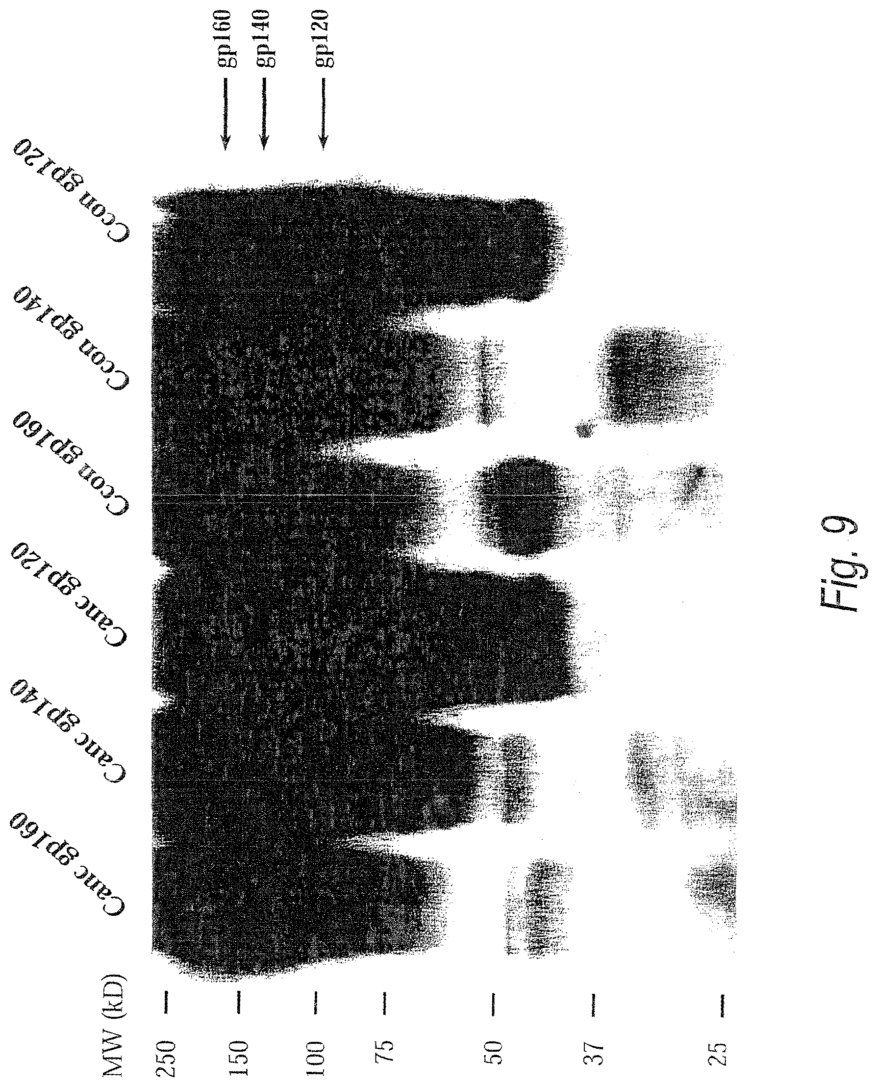
D00015

D00016
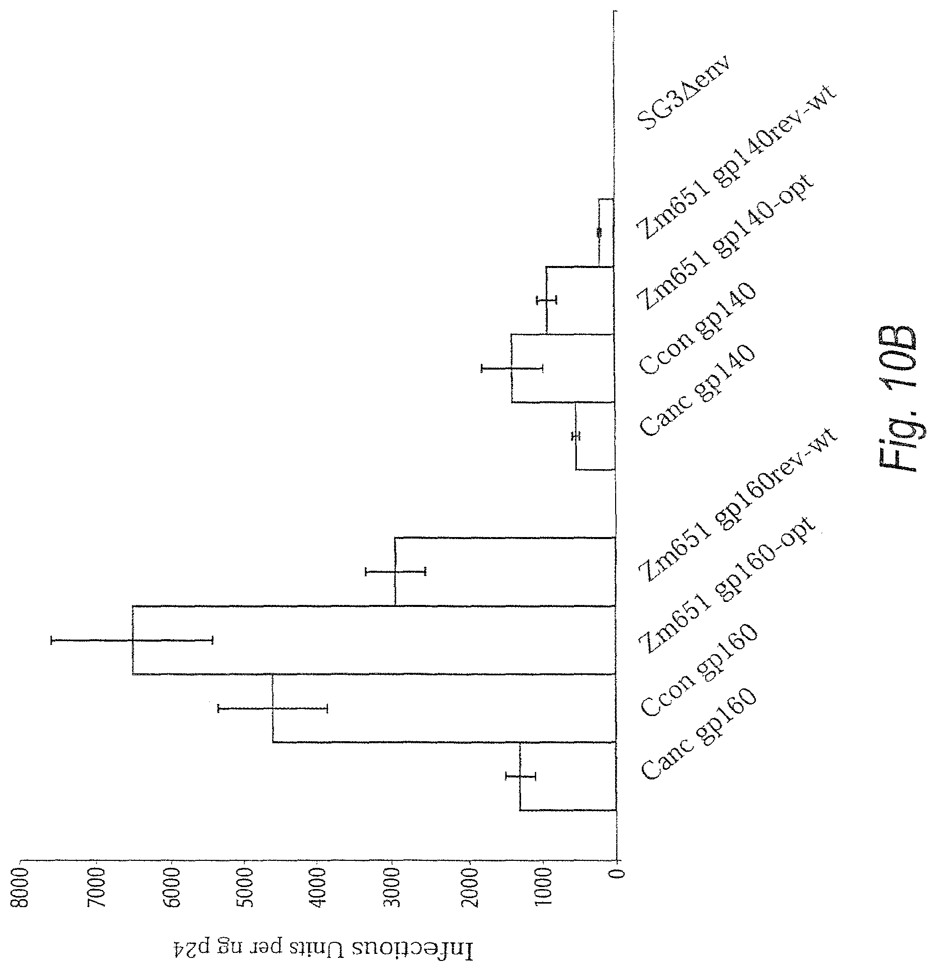
D00017
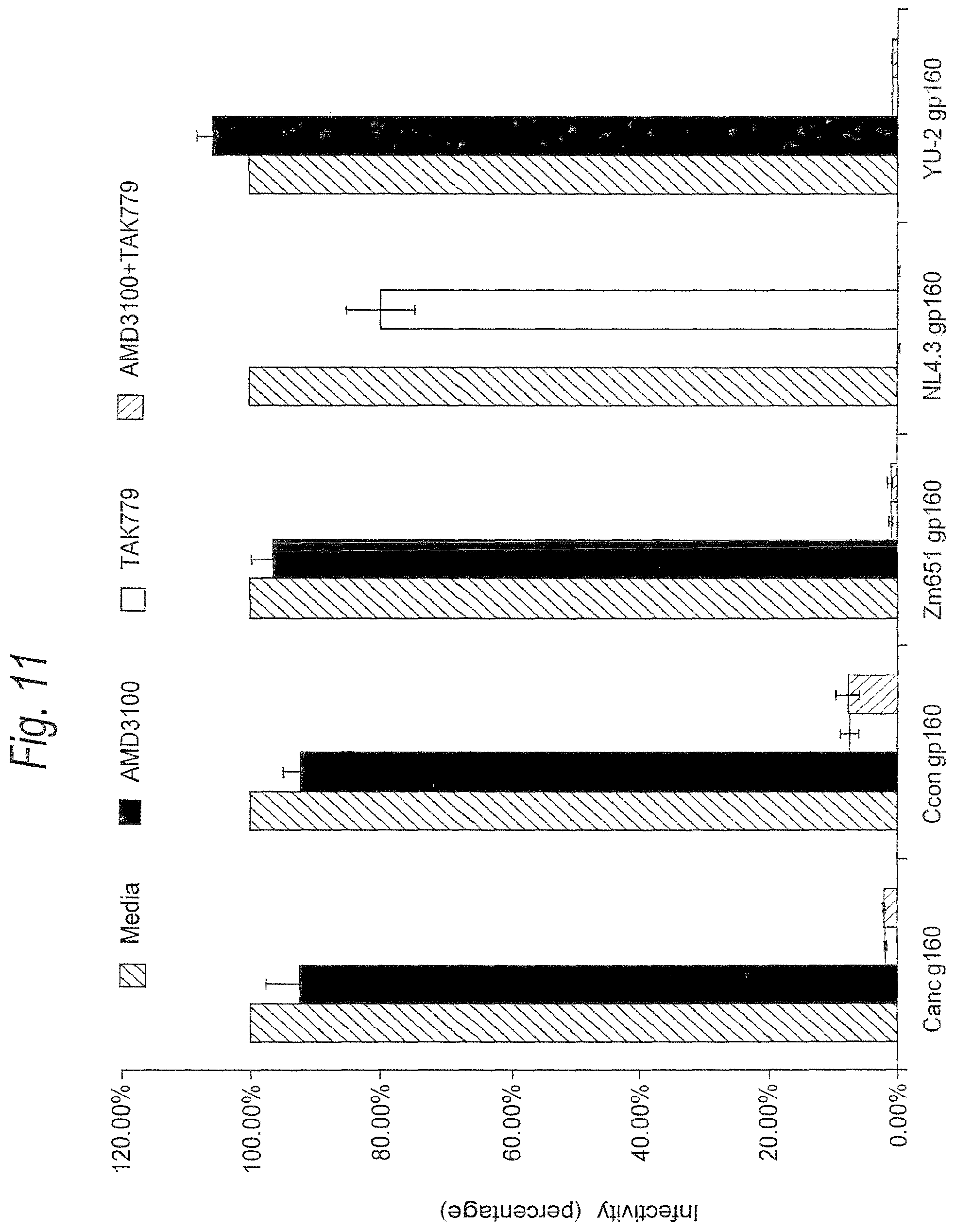
D00018
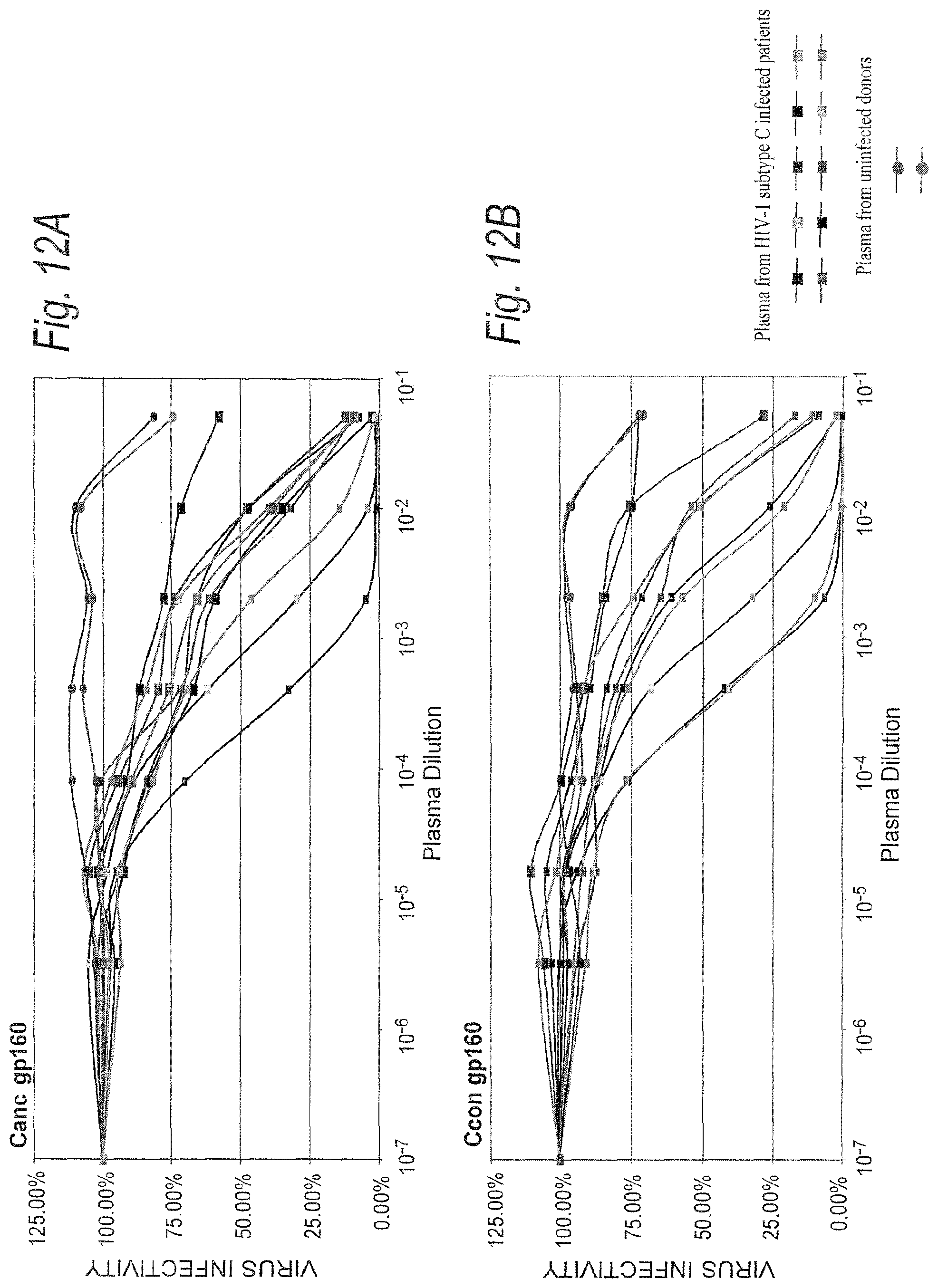
D00019
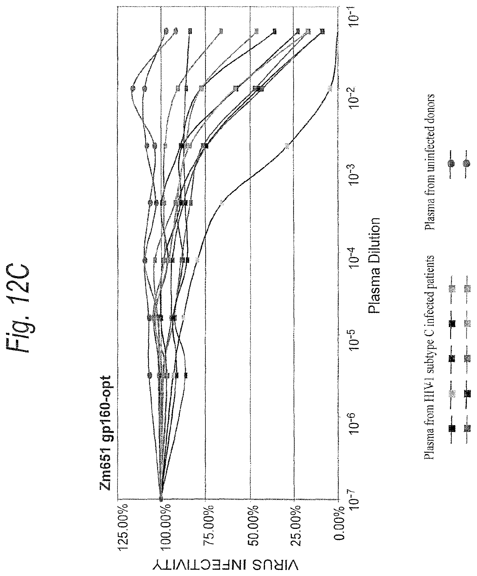
D00020
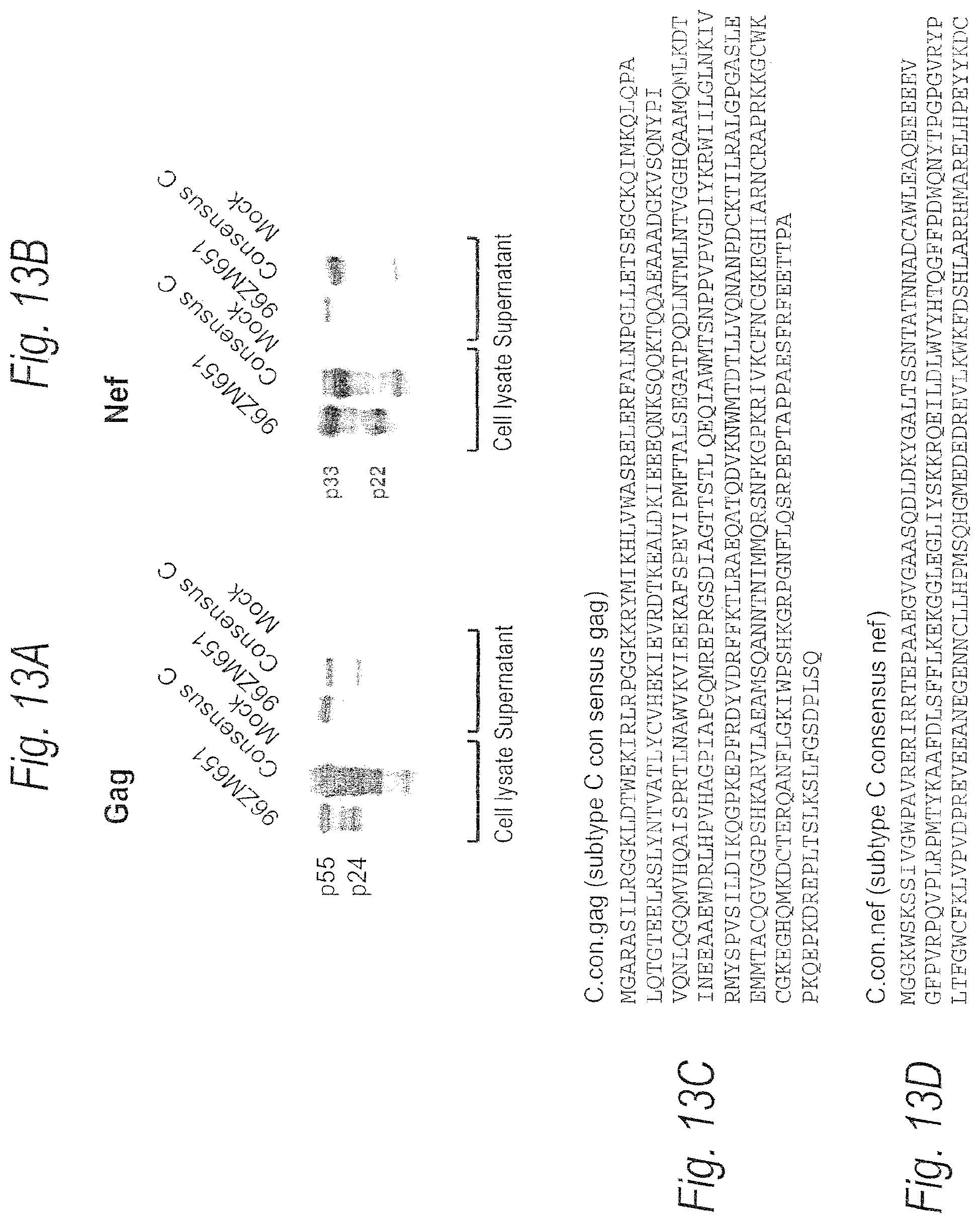
D00021
D00022

D00023
D00024
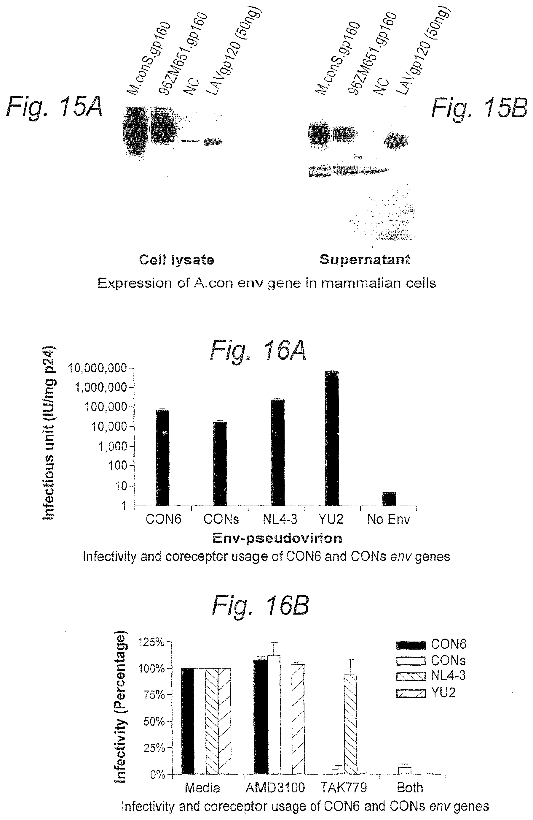
D00025
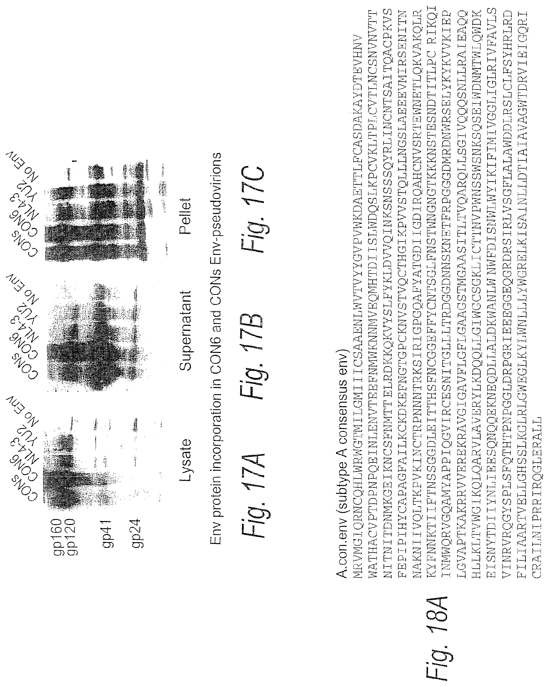
D00026

D00027
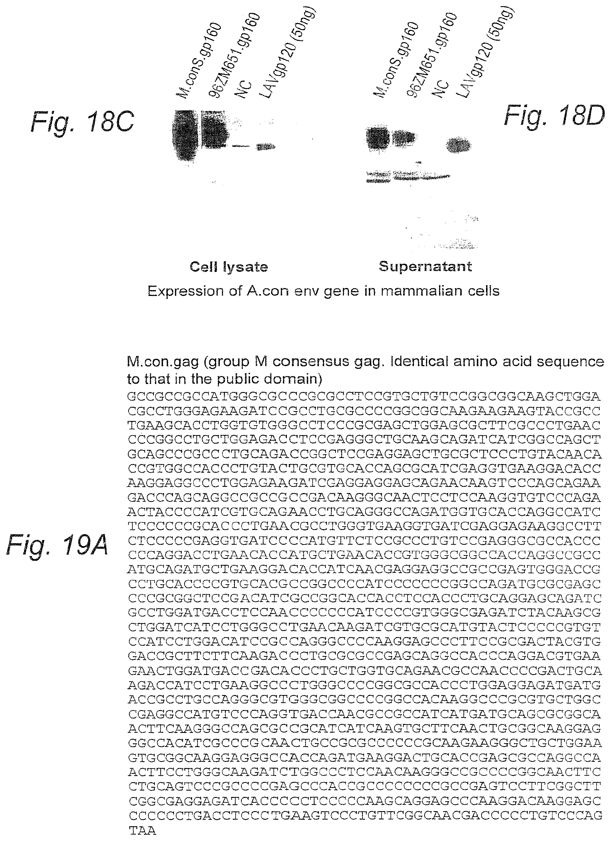
D00028
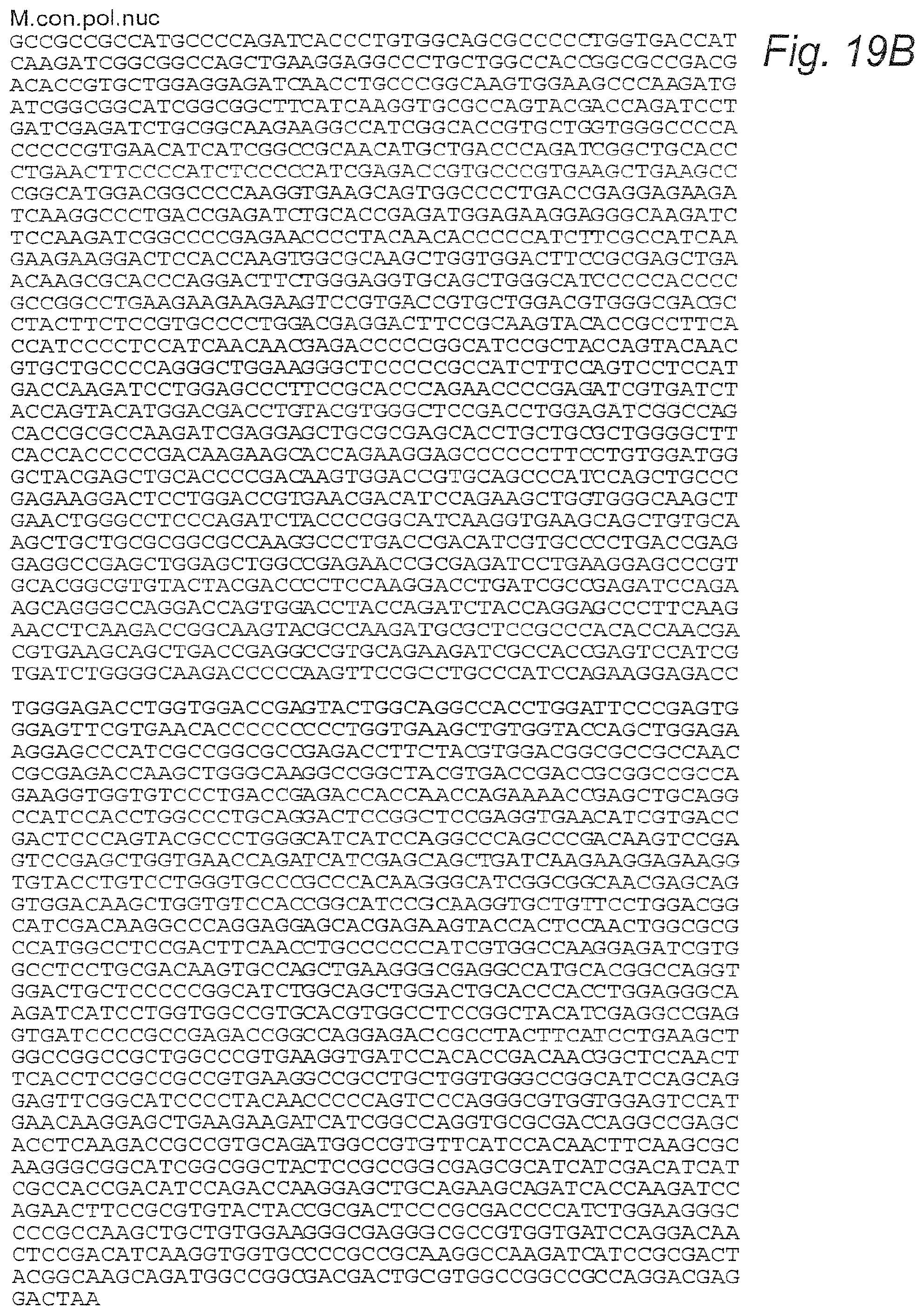
D00029
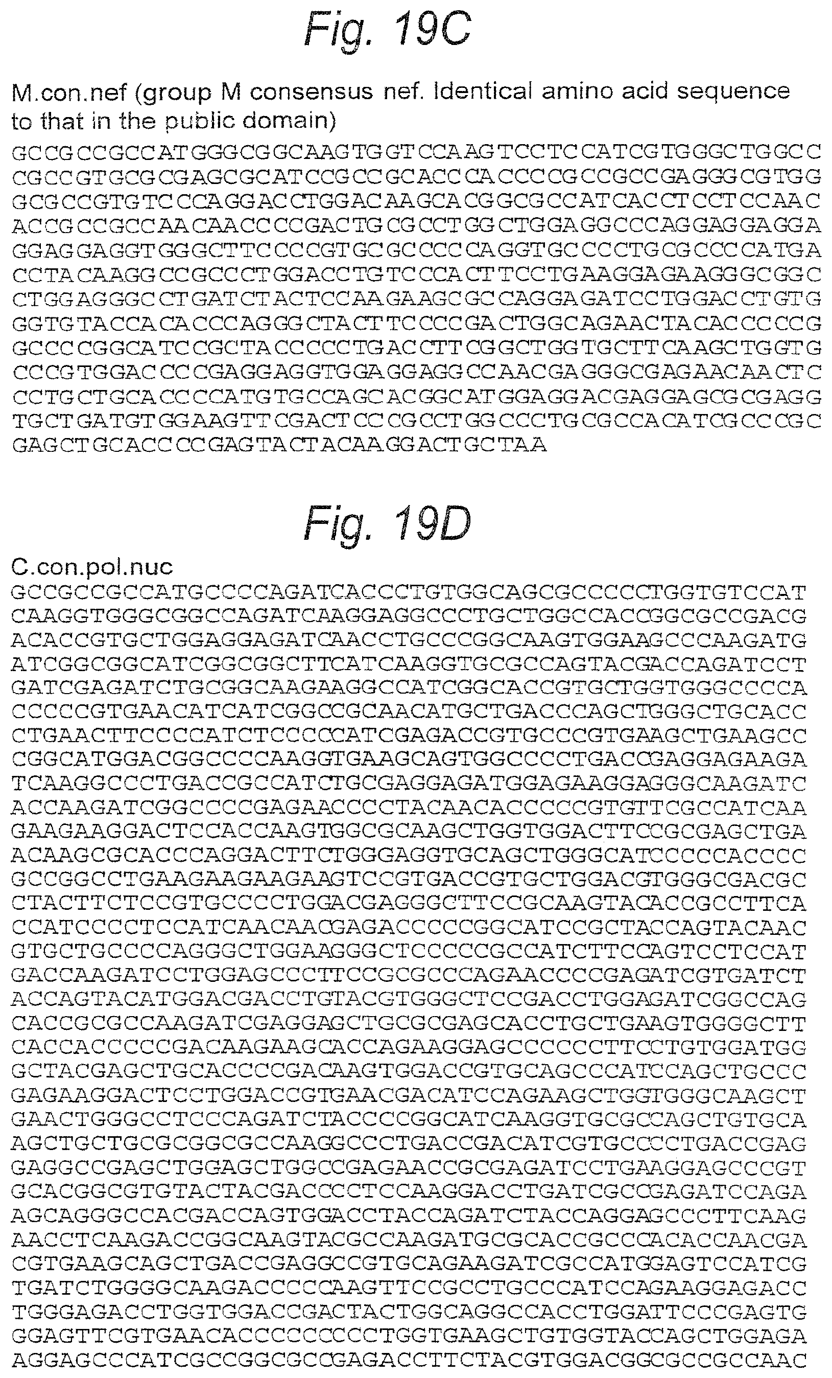
D00030
D00031
D00032
D00033

D00034
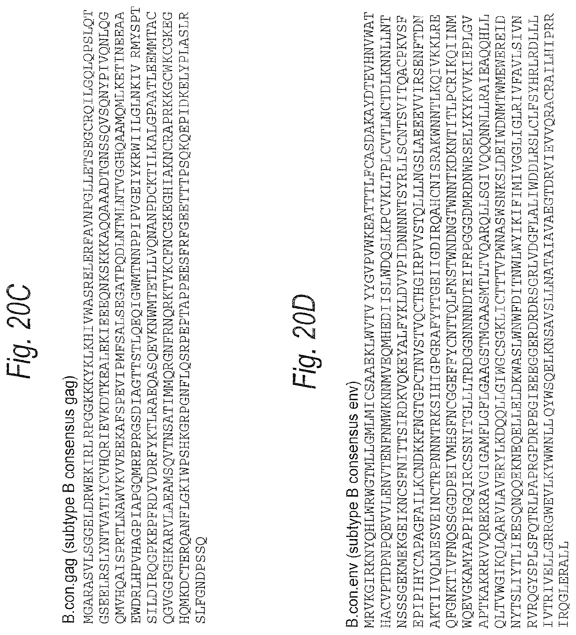
D00035

D00036
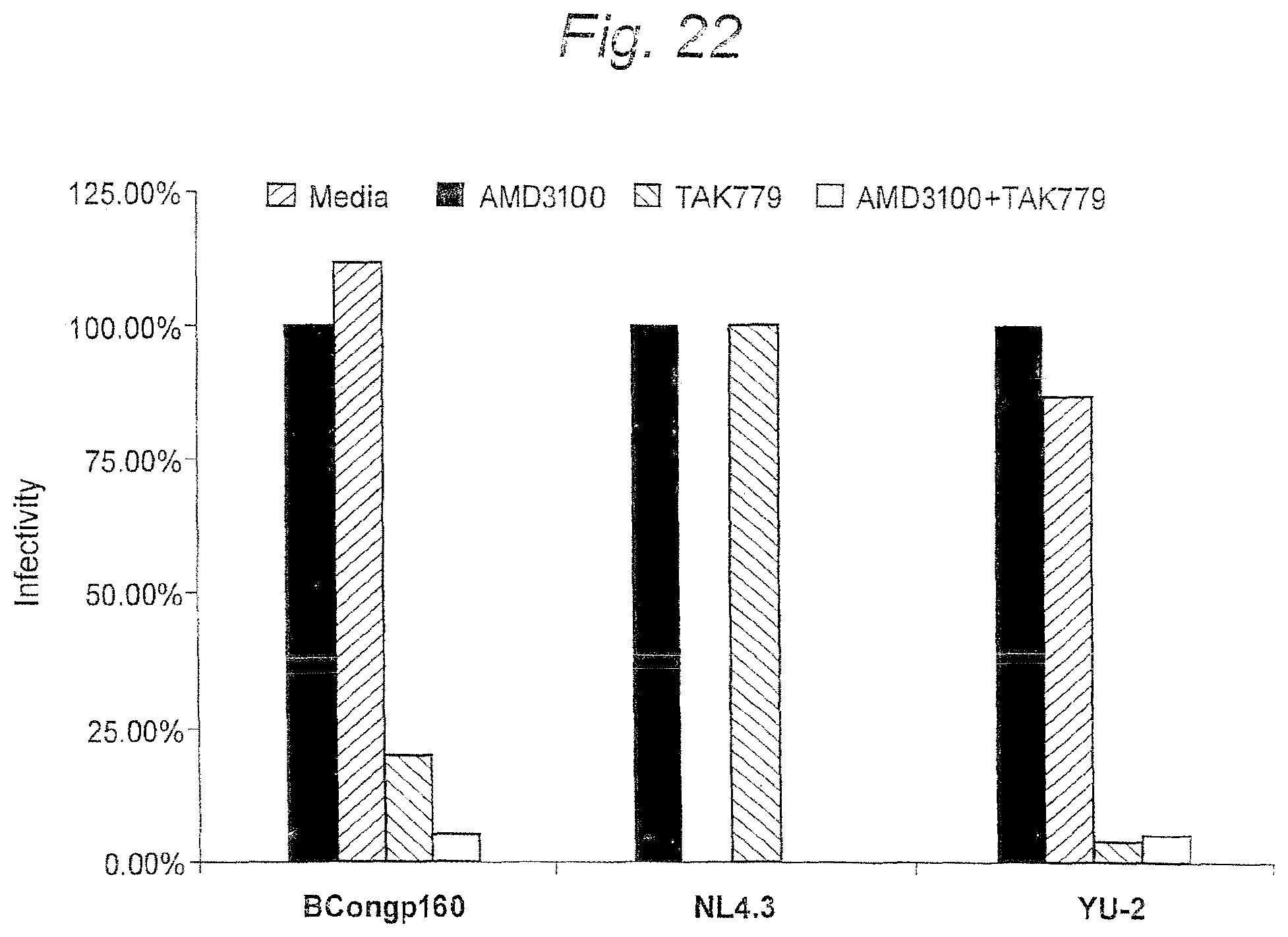
D00037
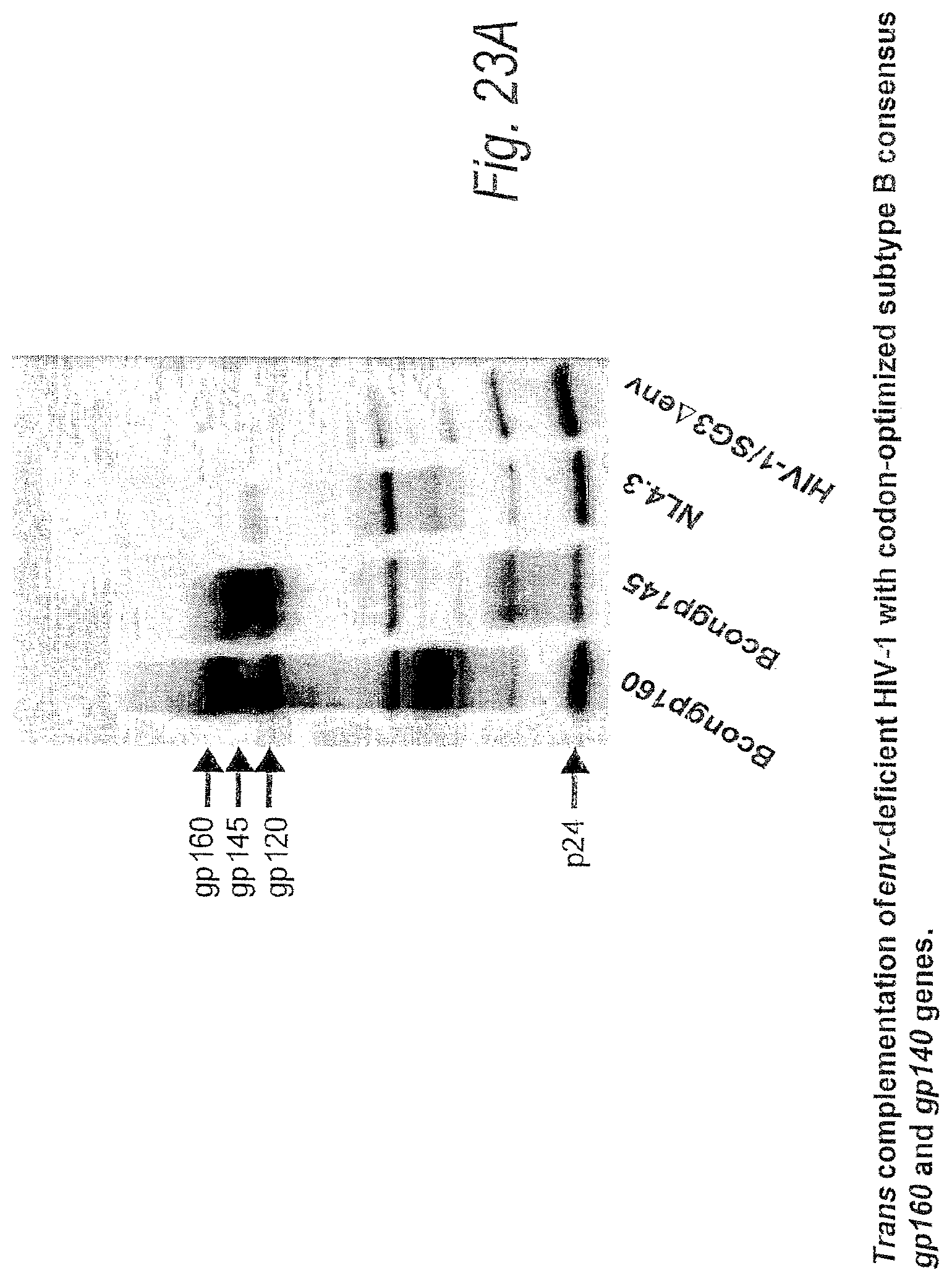
D00038

D00039

D00040
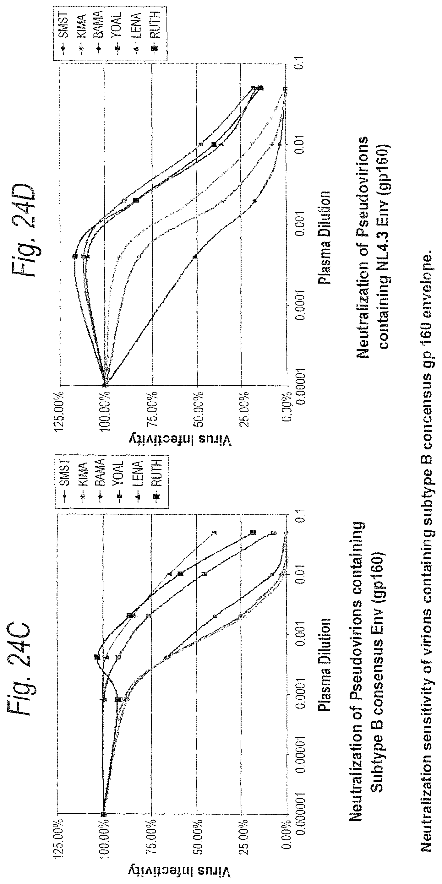
D00041
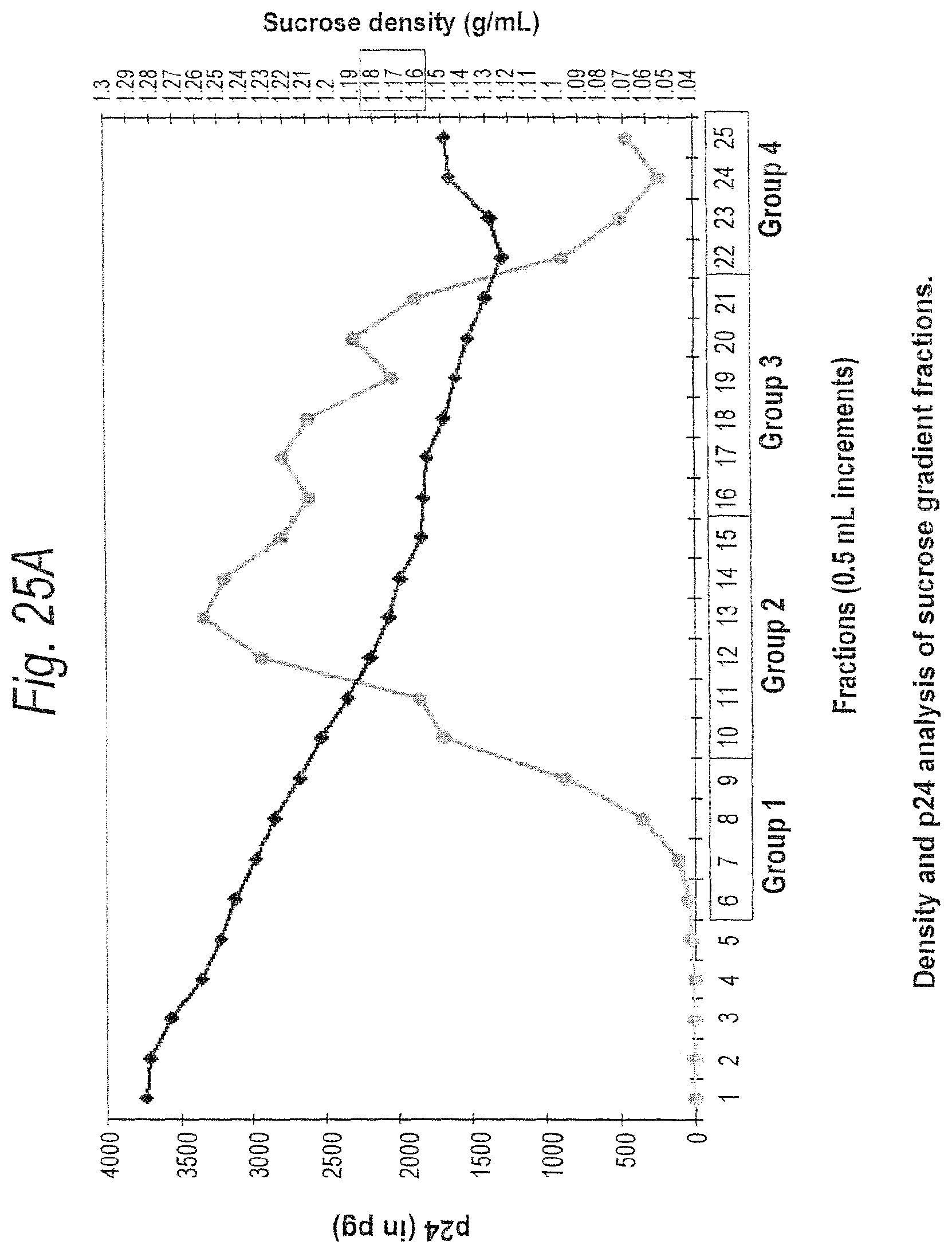
D00042
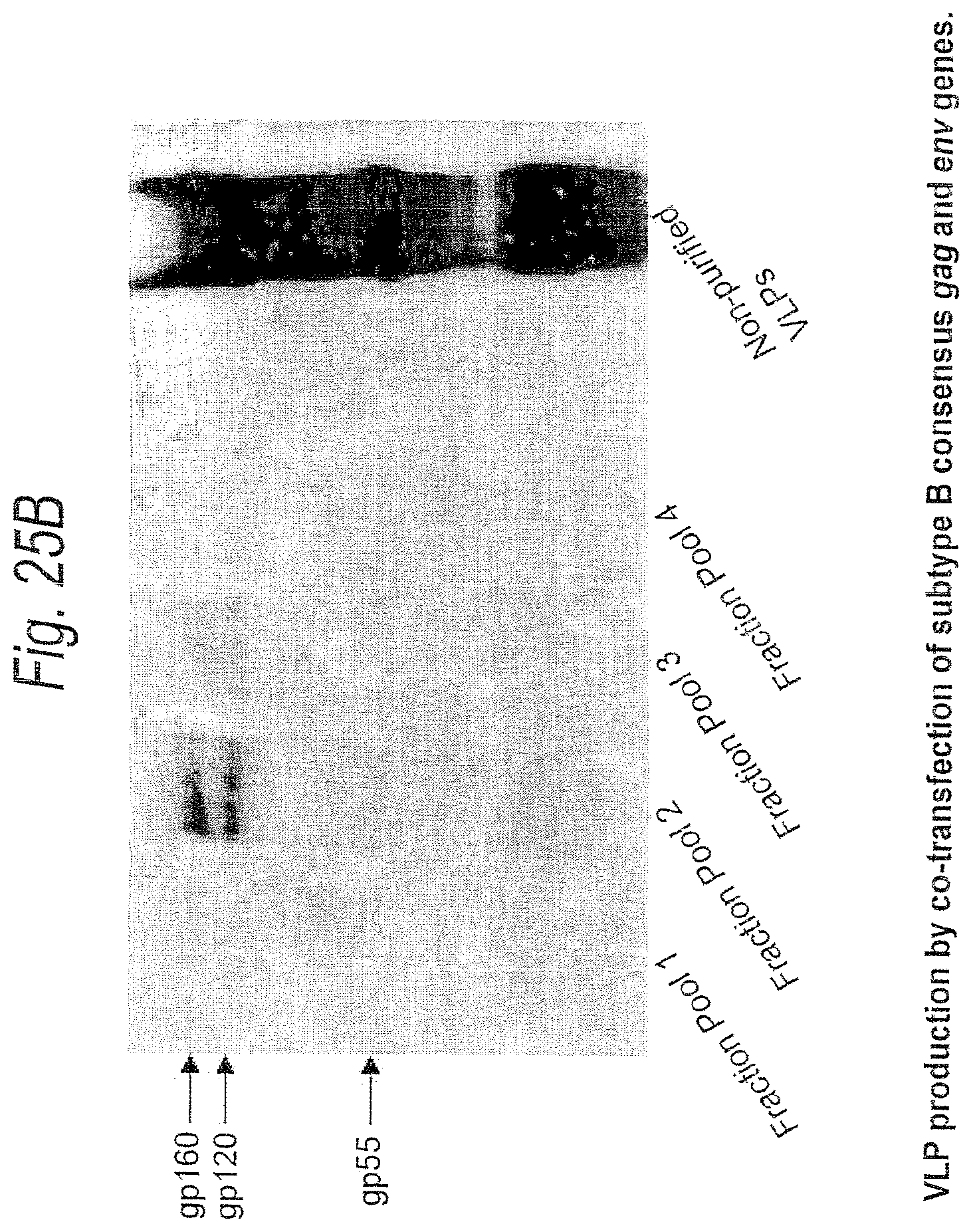
D00043
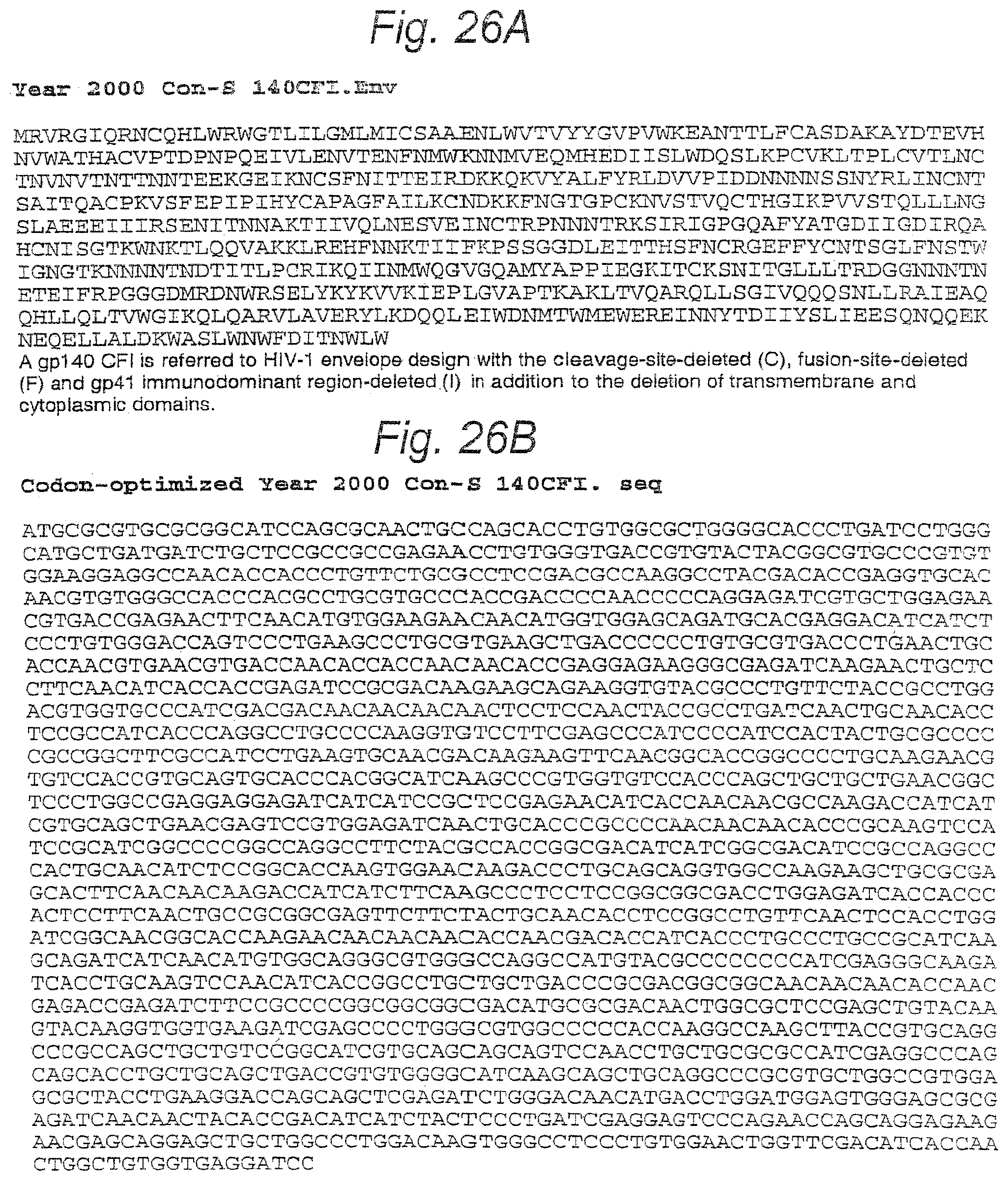
D00044

D00045
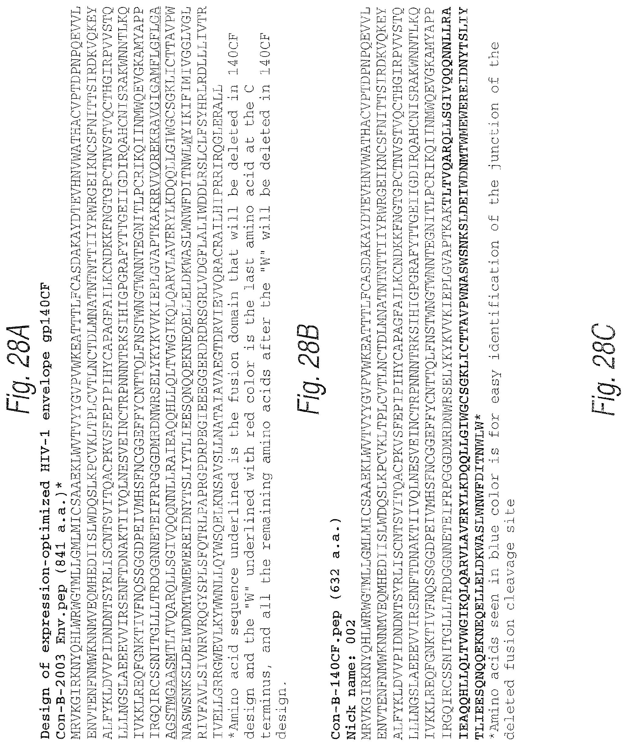
D00046
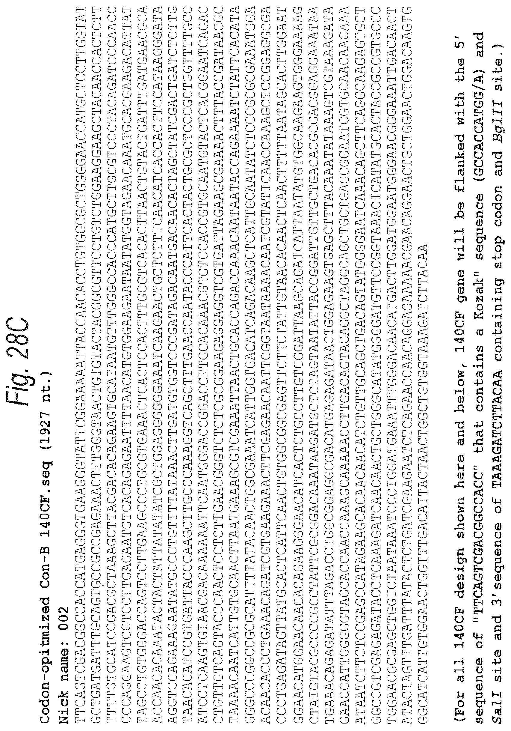
D00047

D00048

D00049
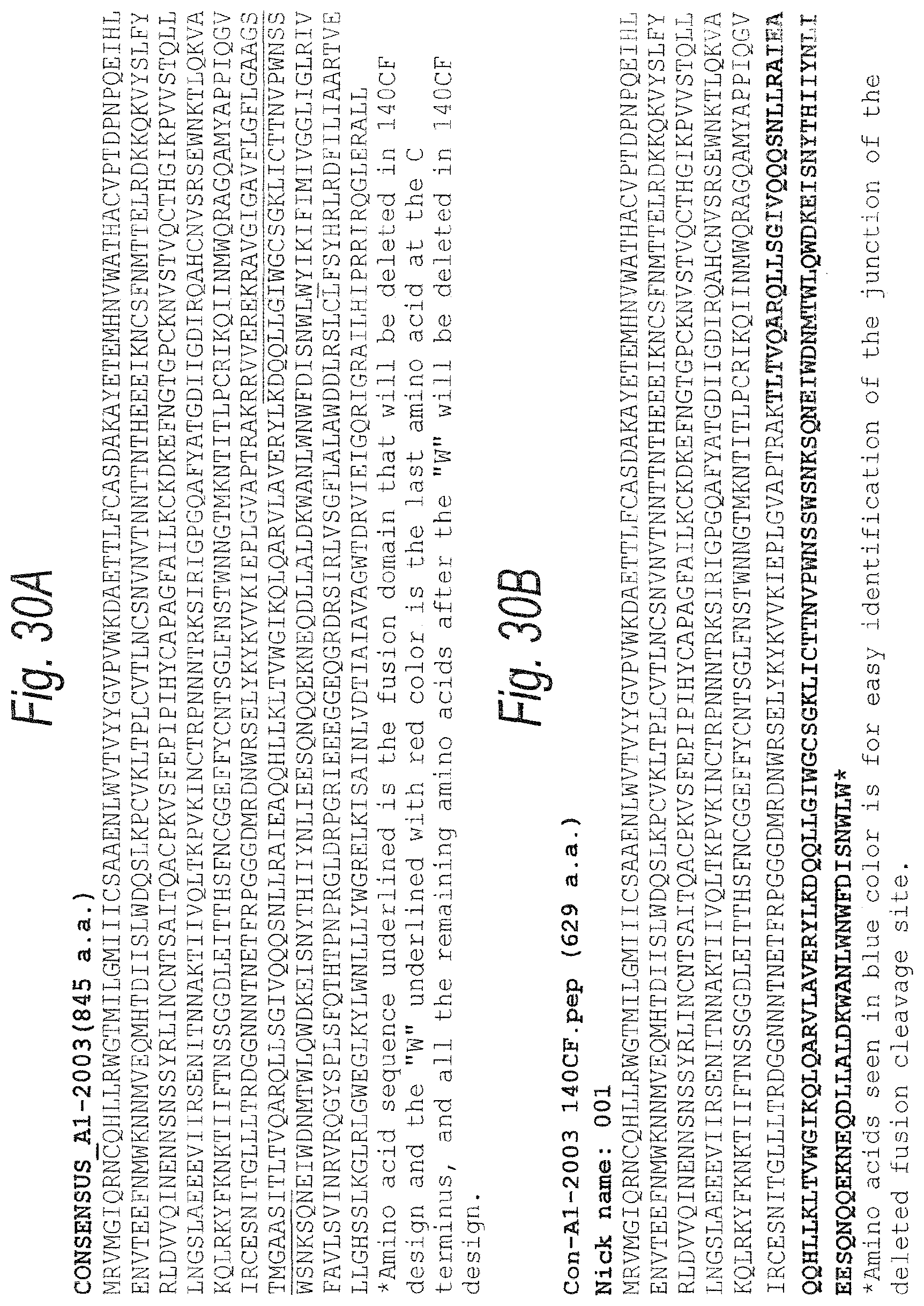
D00050

D00051
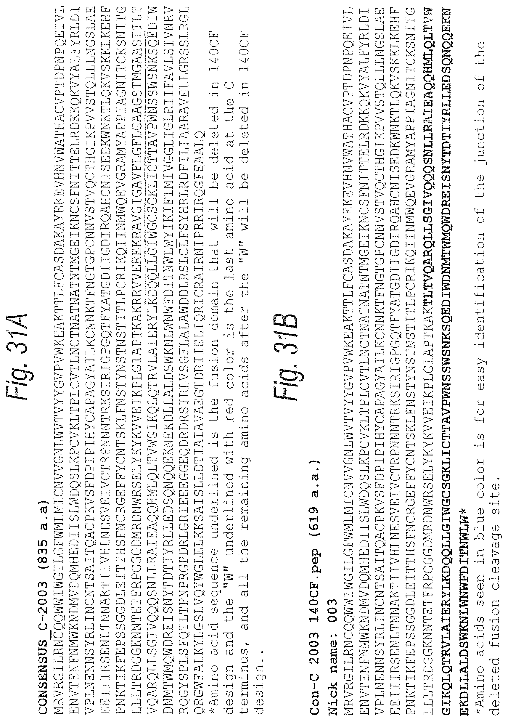
D00052

D00053

D00054
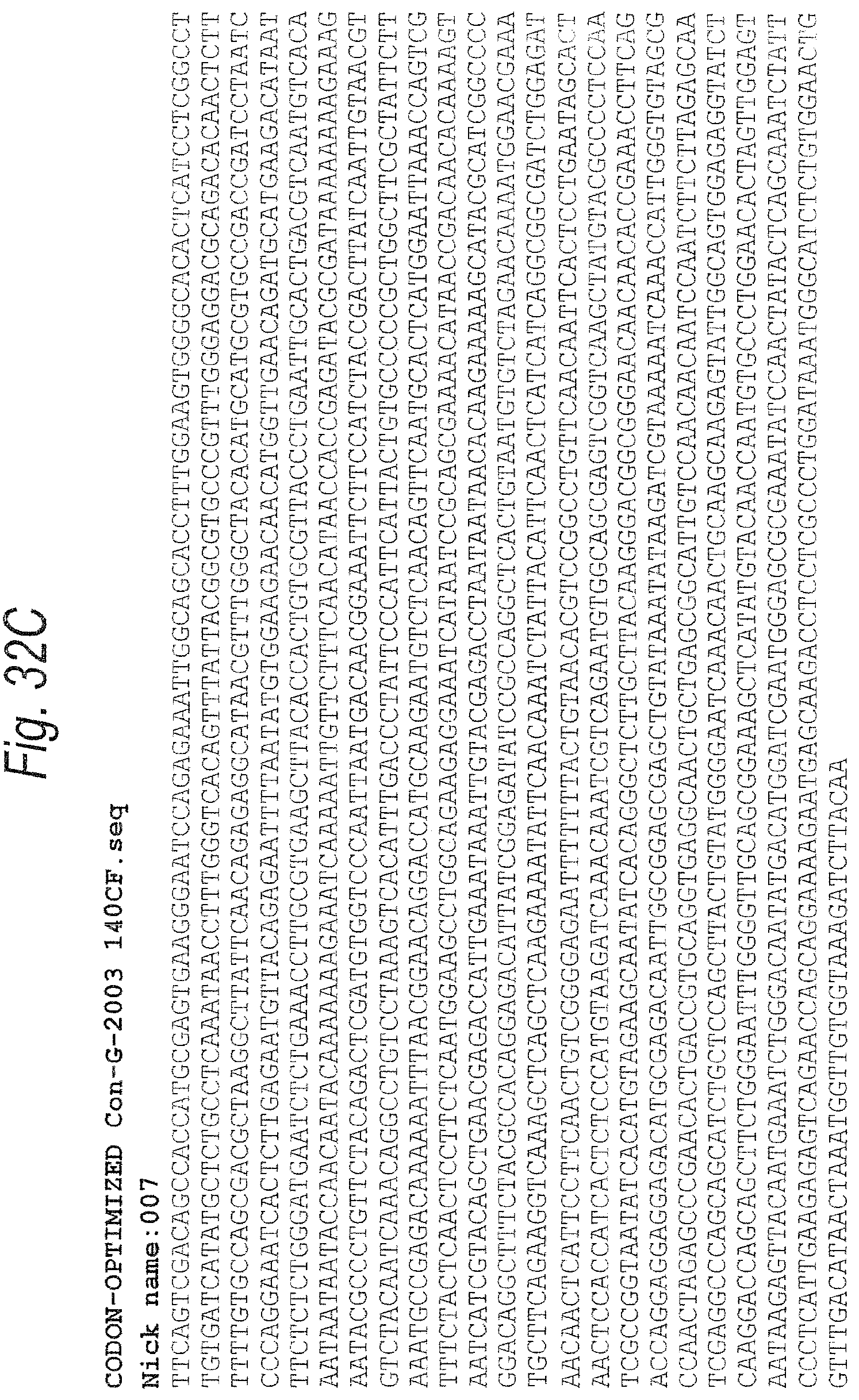
D00055
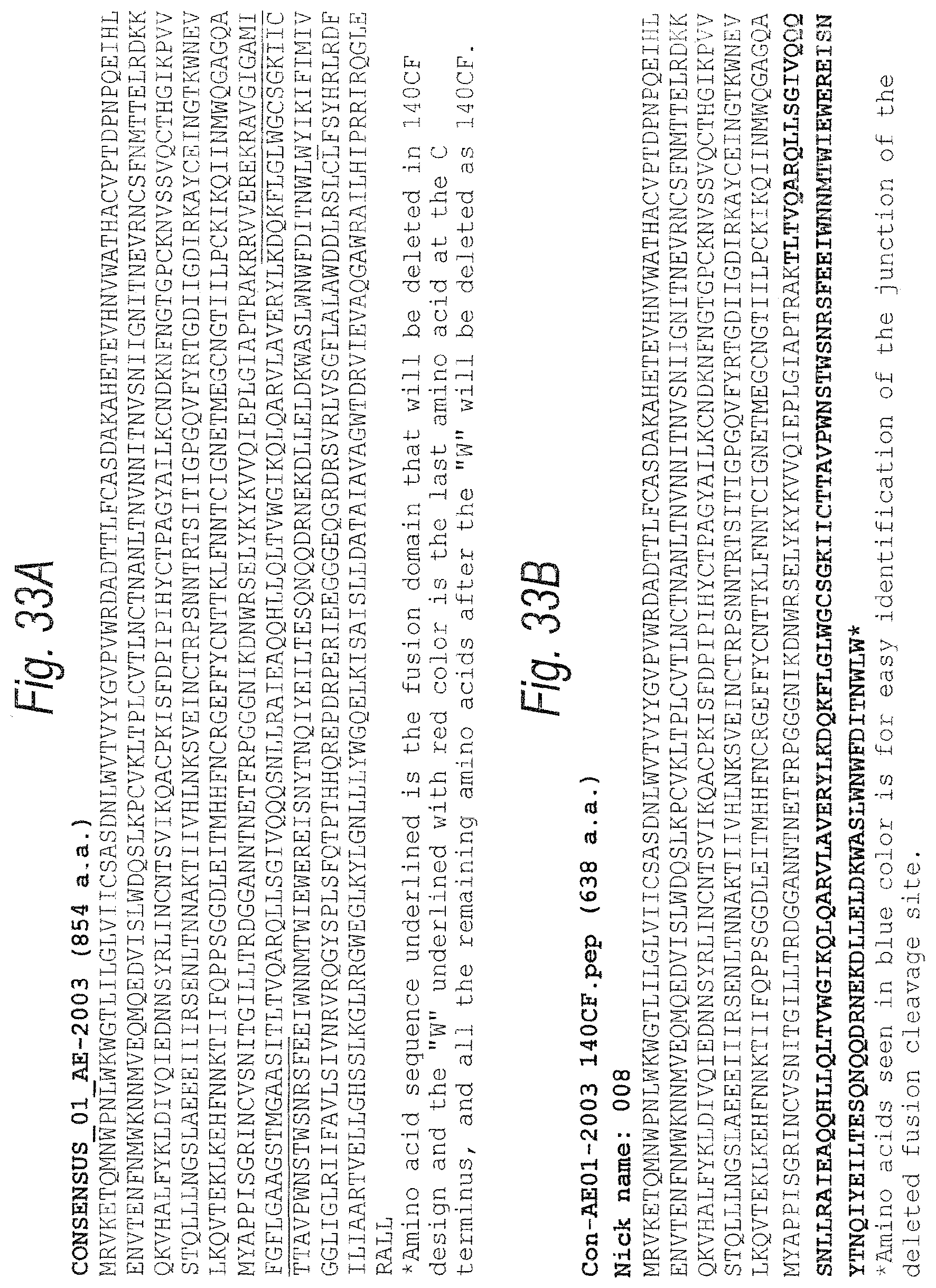
D00056

D00057

D00058

D00059
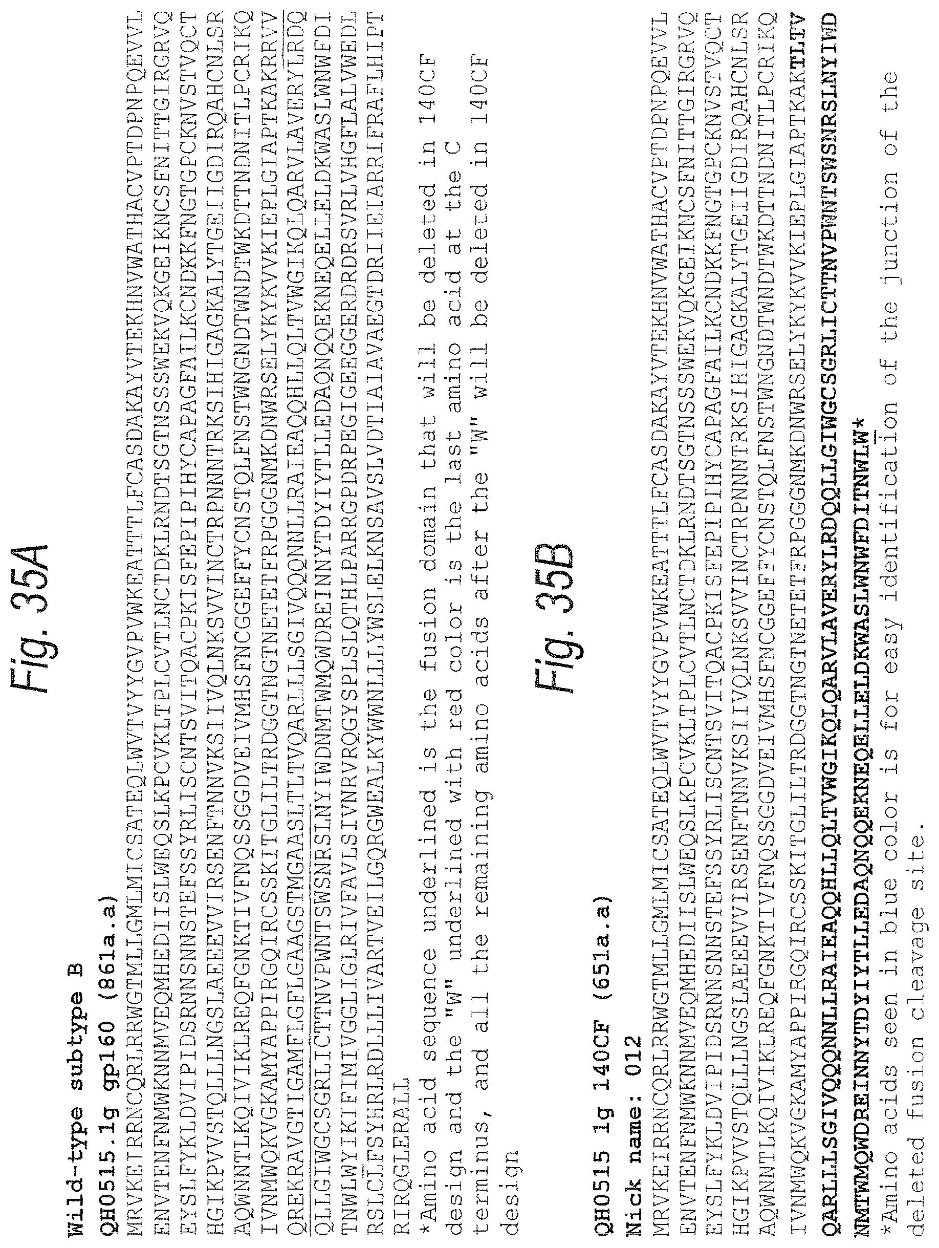
D00060
D00061
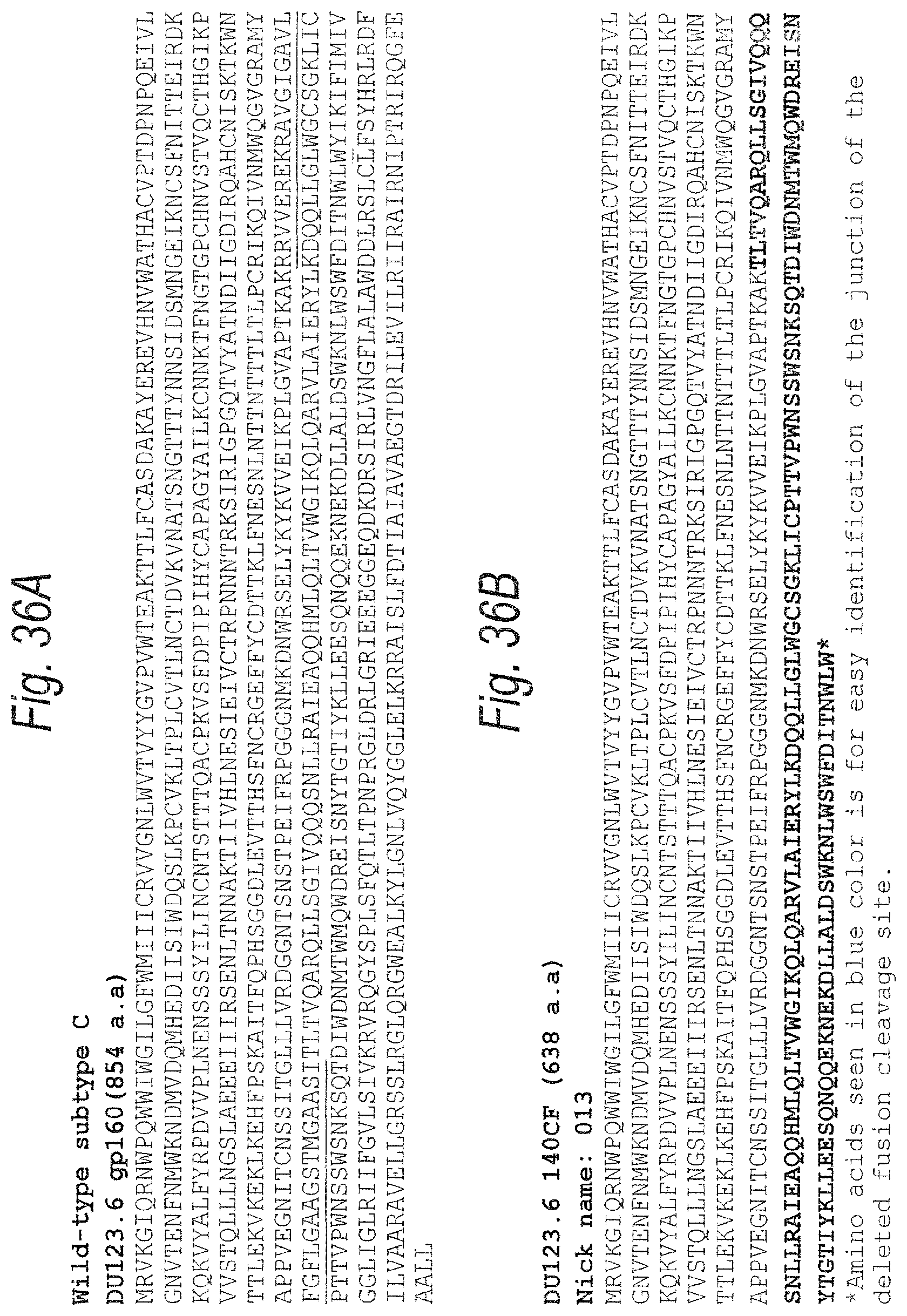
D00062
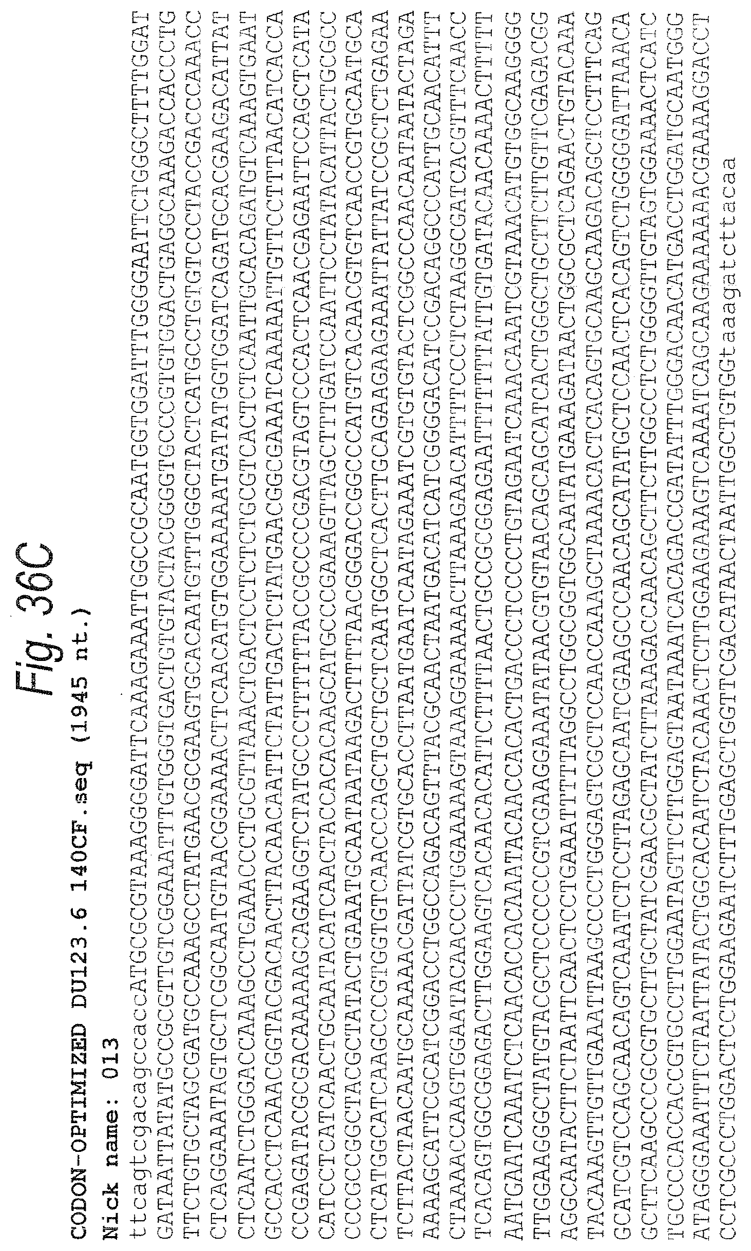
D00063
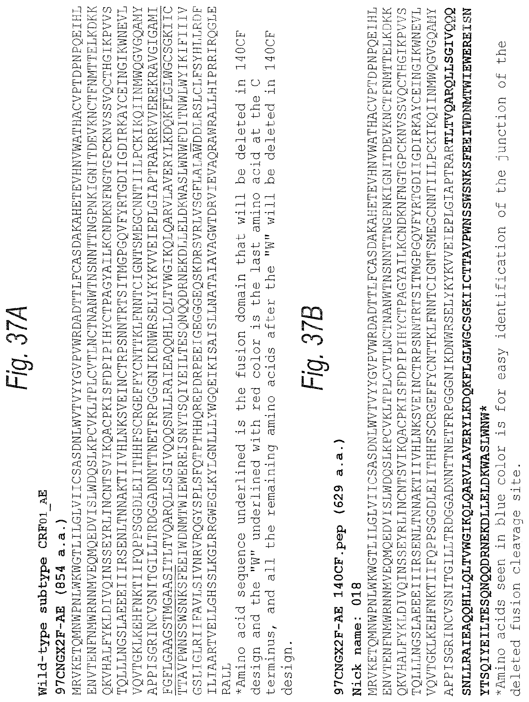
D00064

D00065
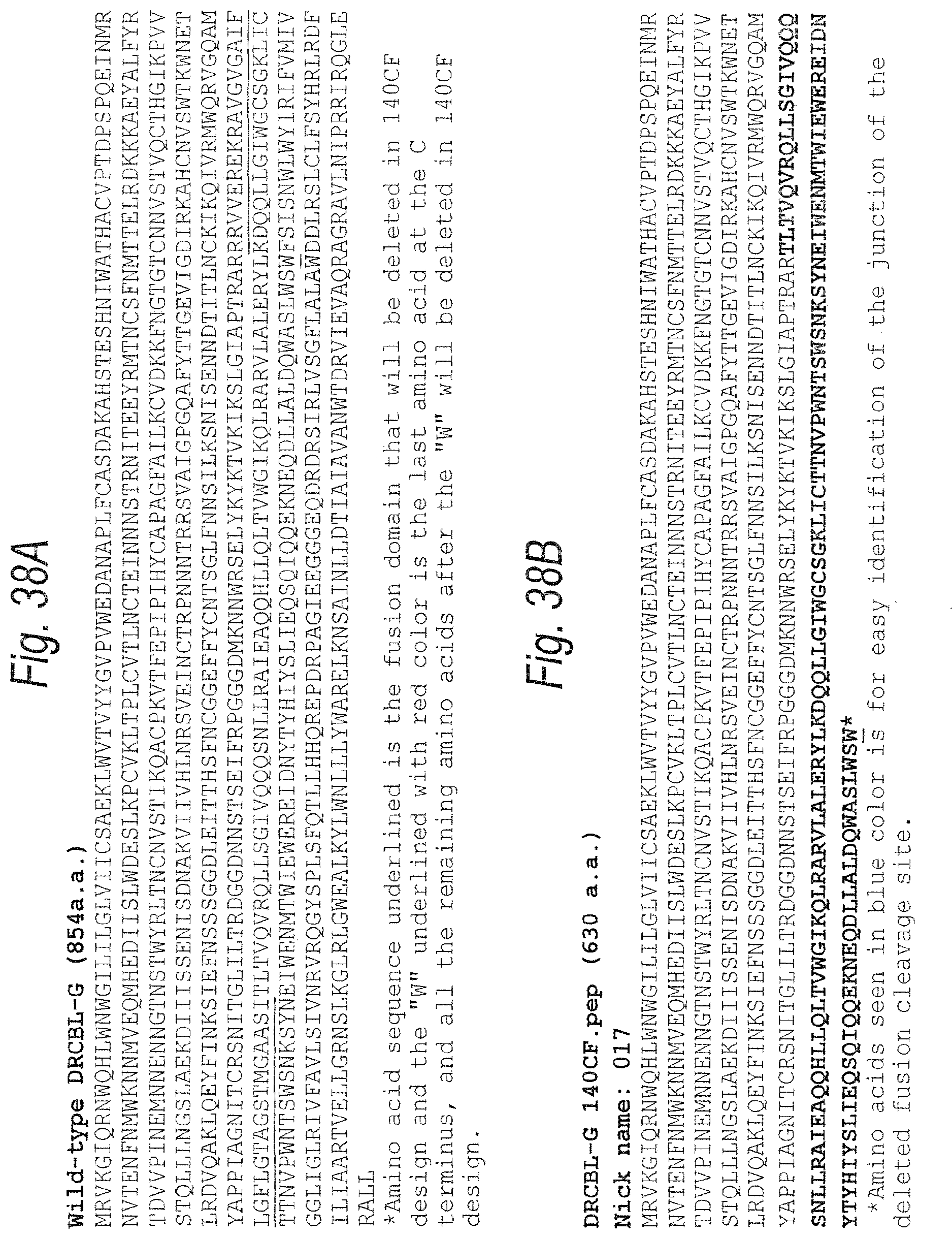
D00066
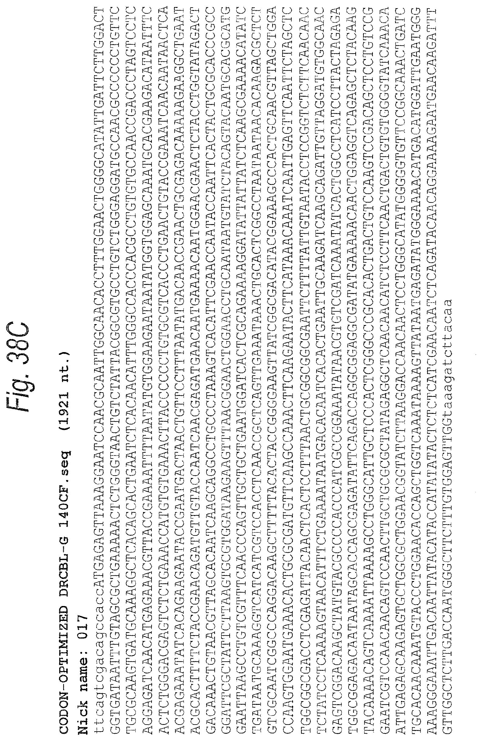
D00067
D00068
D00069
D00070
D00071
D00072
D00073

D00074

D00075
D00076
D00077
D00078
D00079
D00080

D00081

D00082
D00083

D00084

D00085
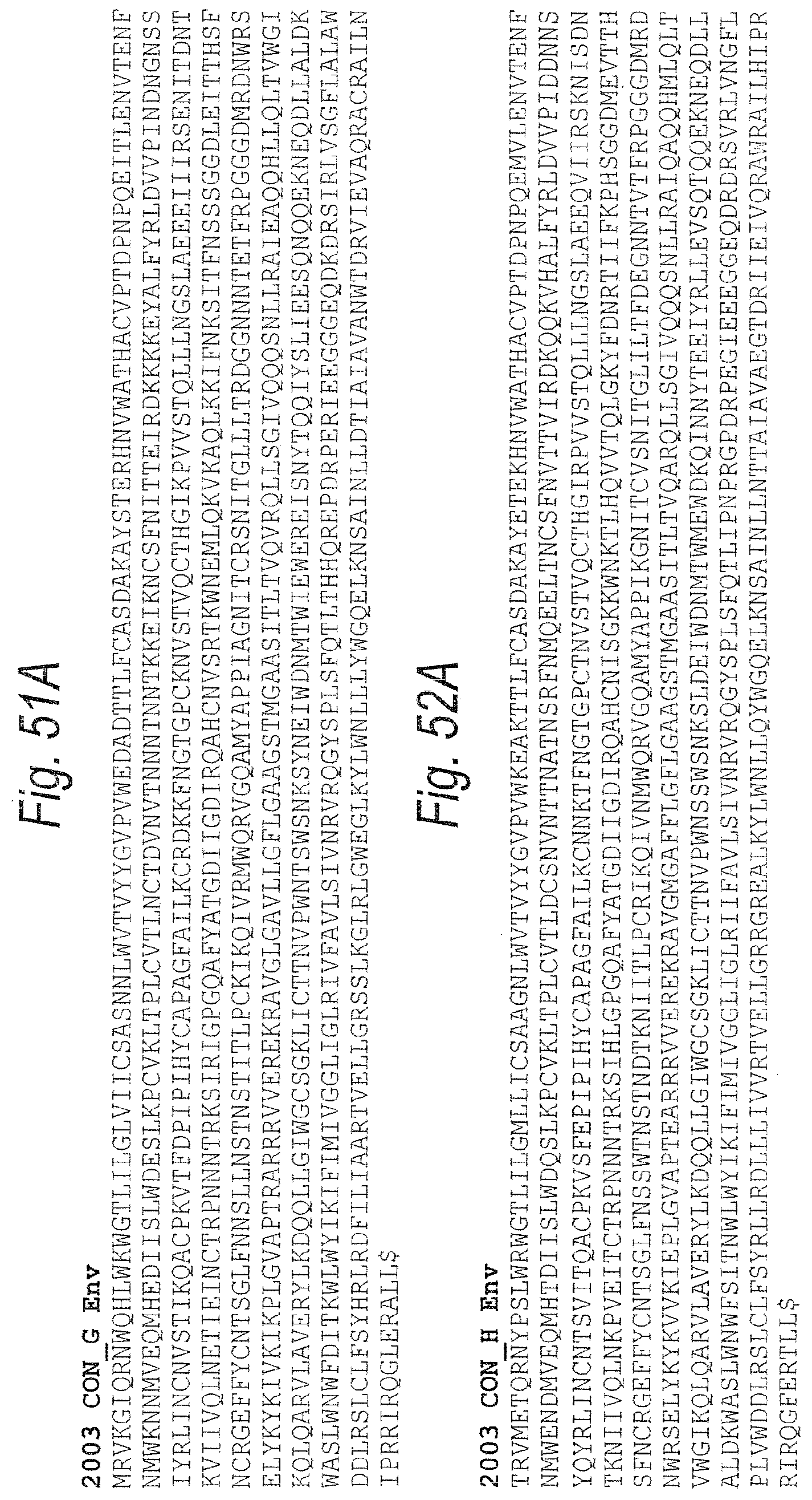
D00086
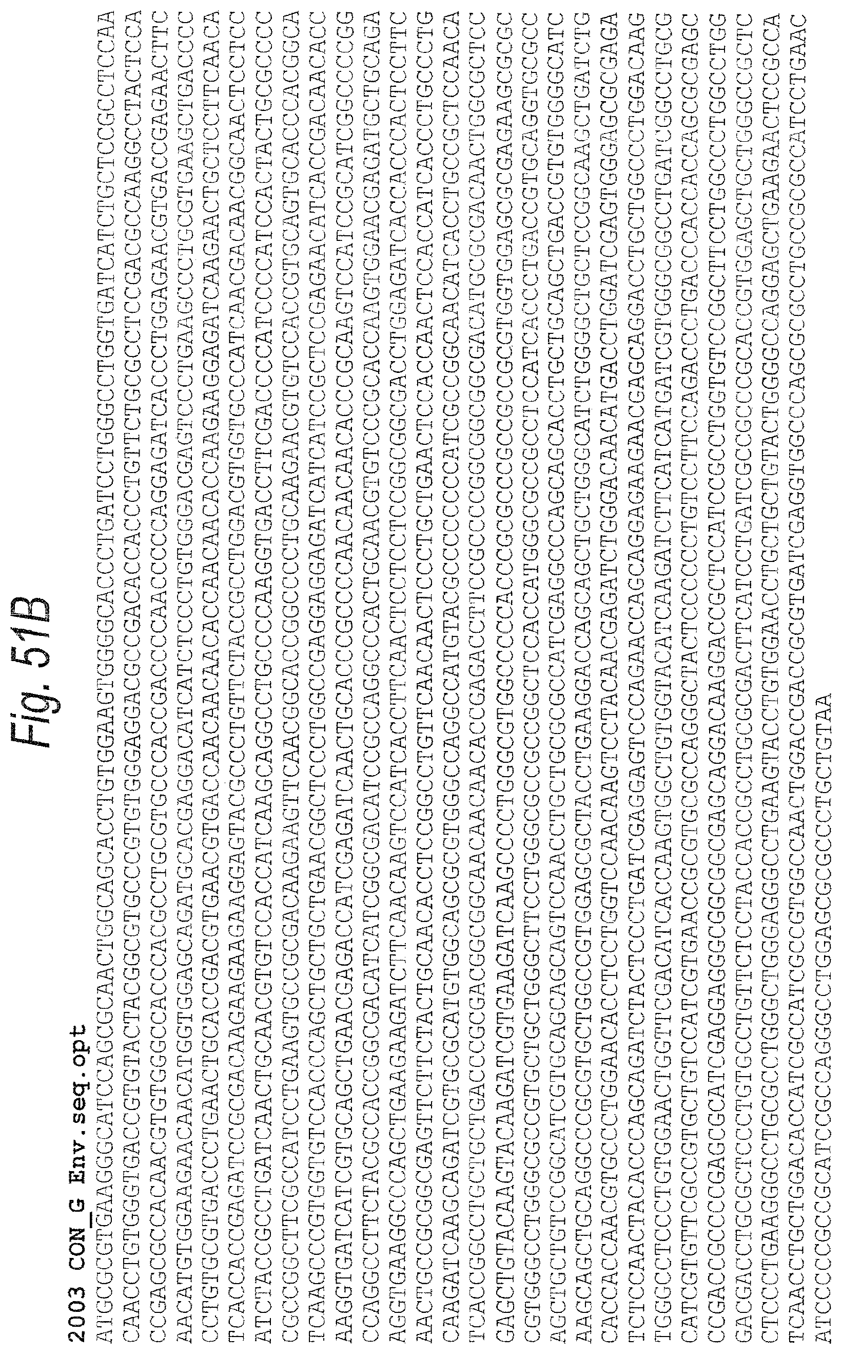
D00087
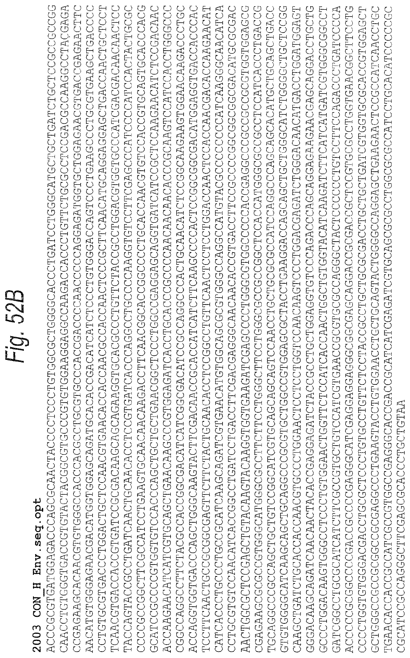
D00088

D00089

D00090
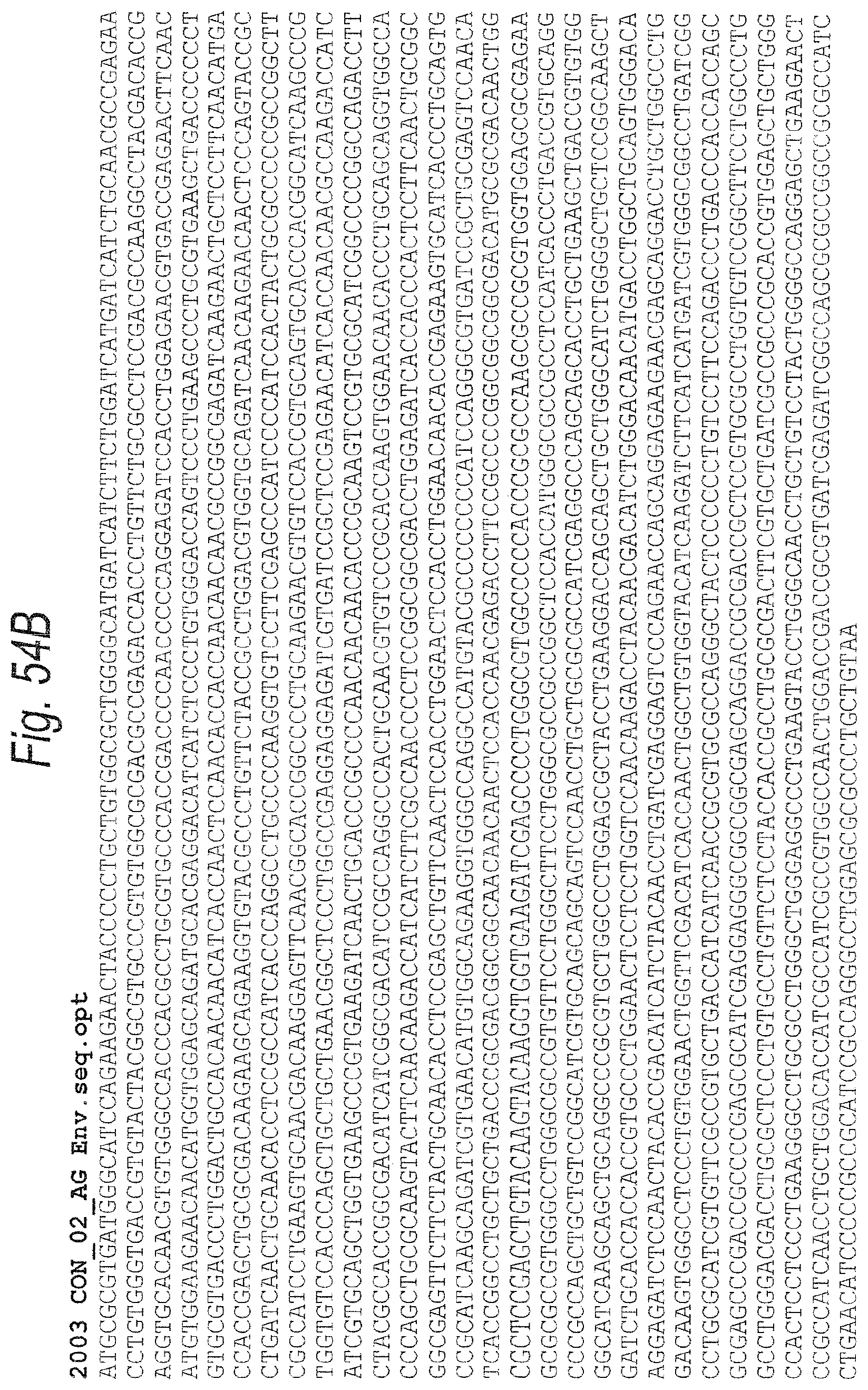
D00091
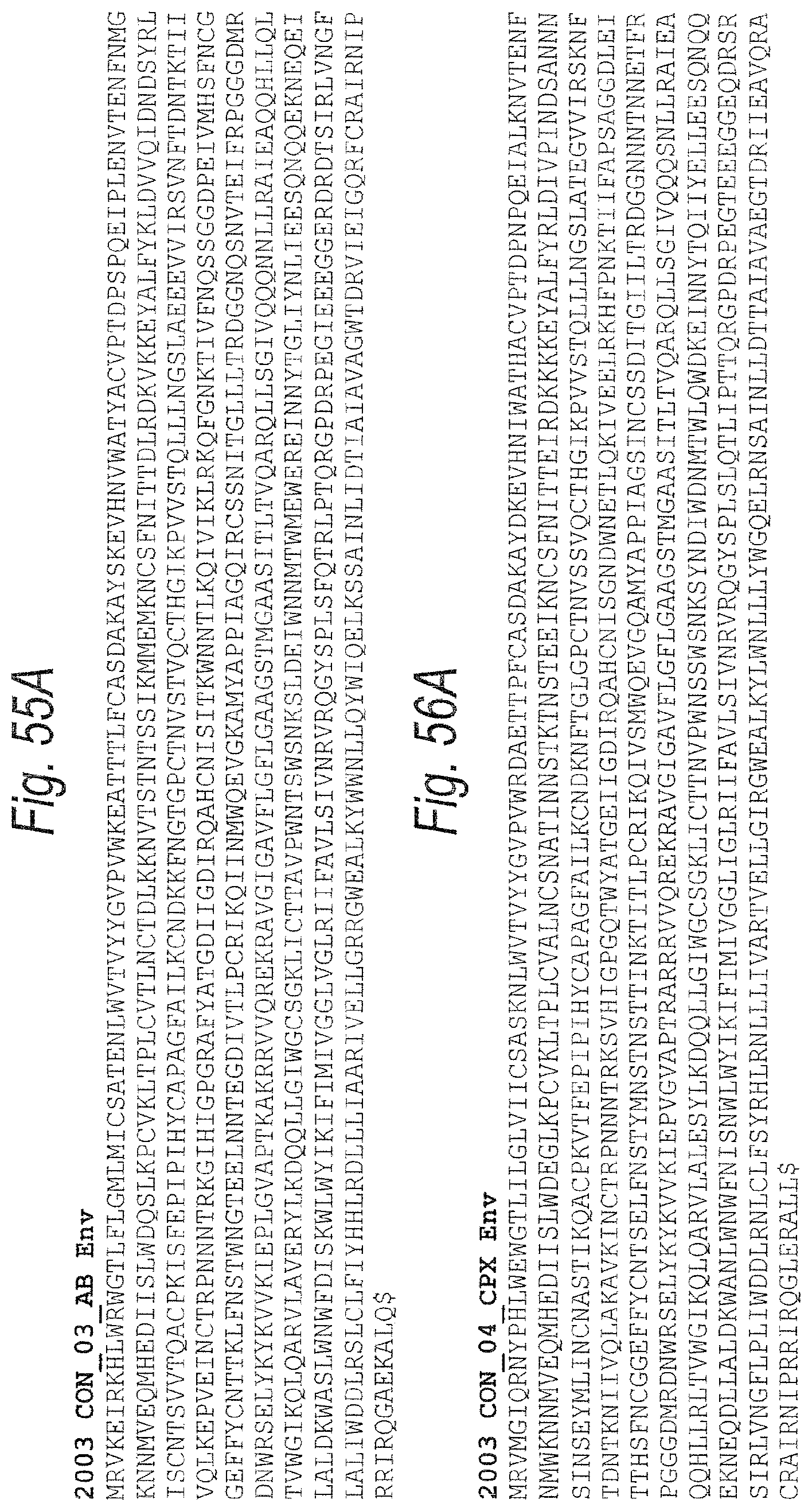
D00092
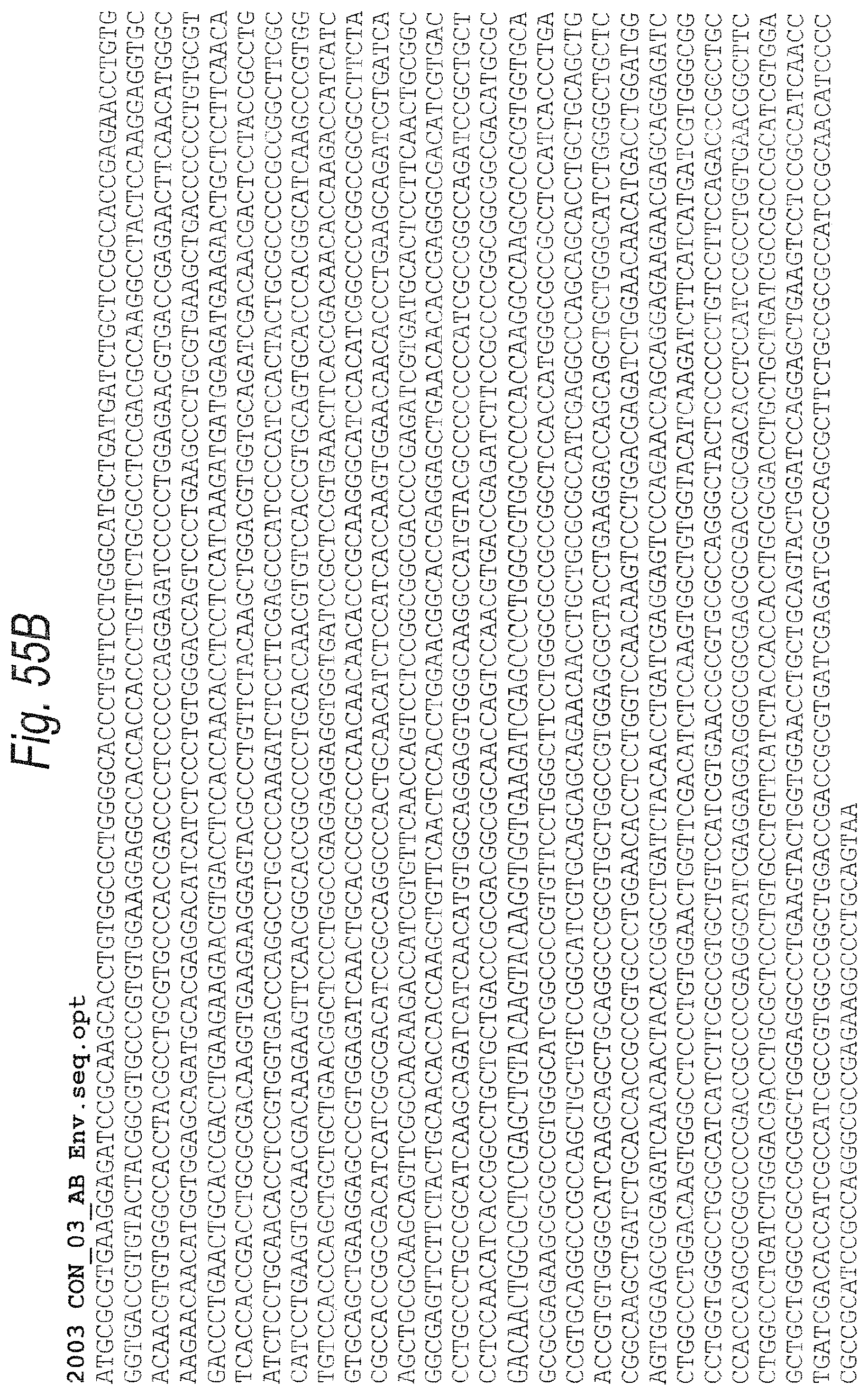
D00093
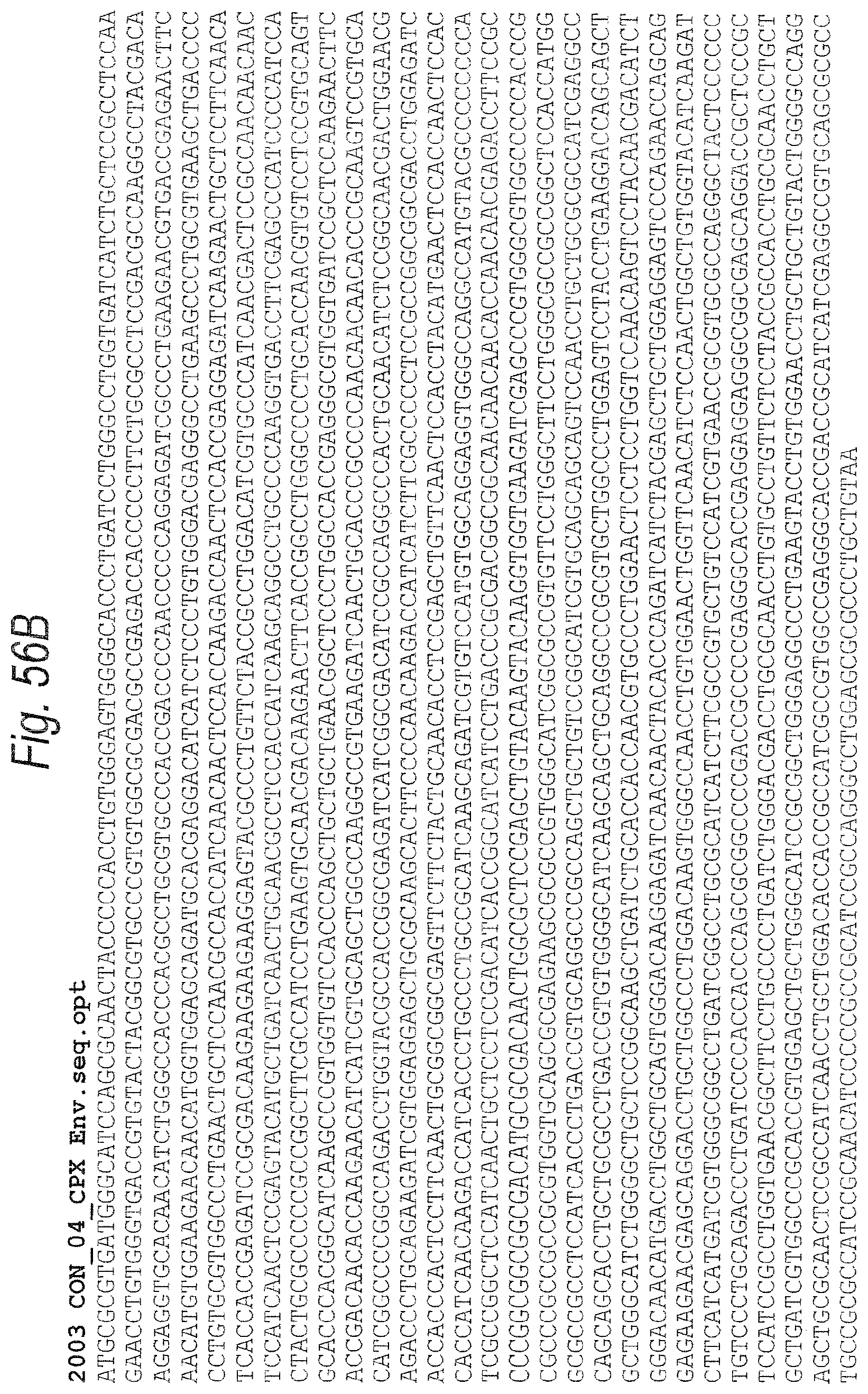
D00094

D00095
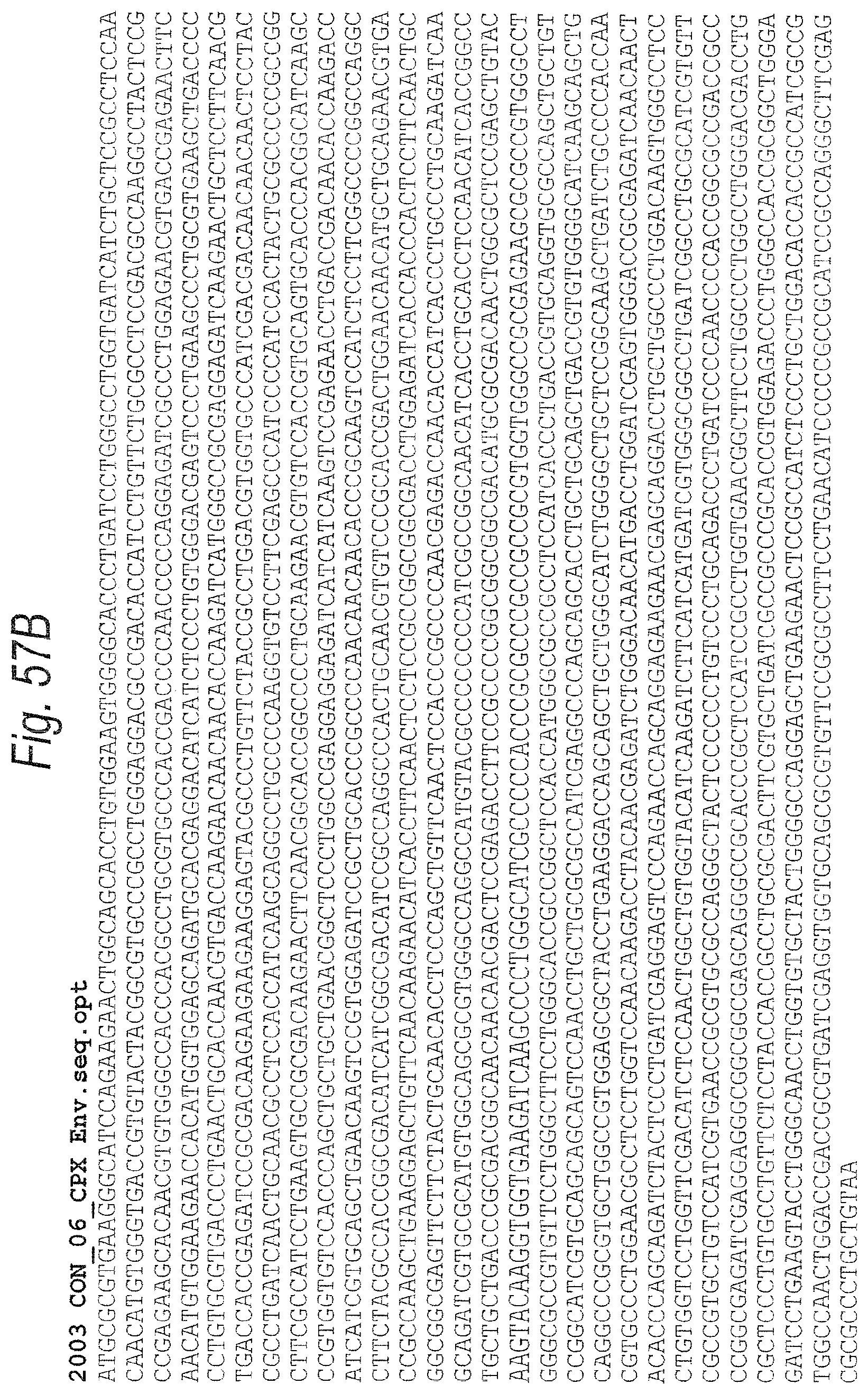
D00096

D00097
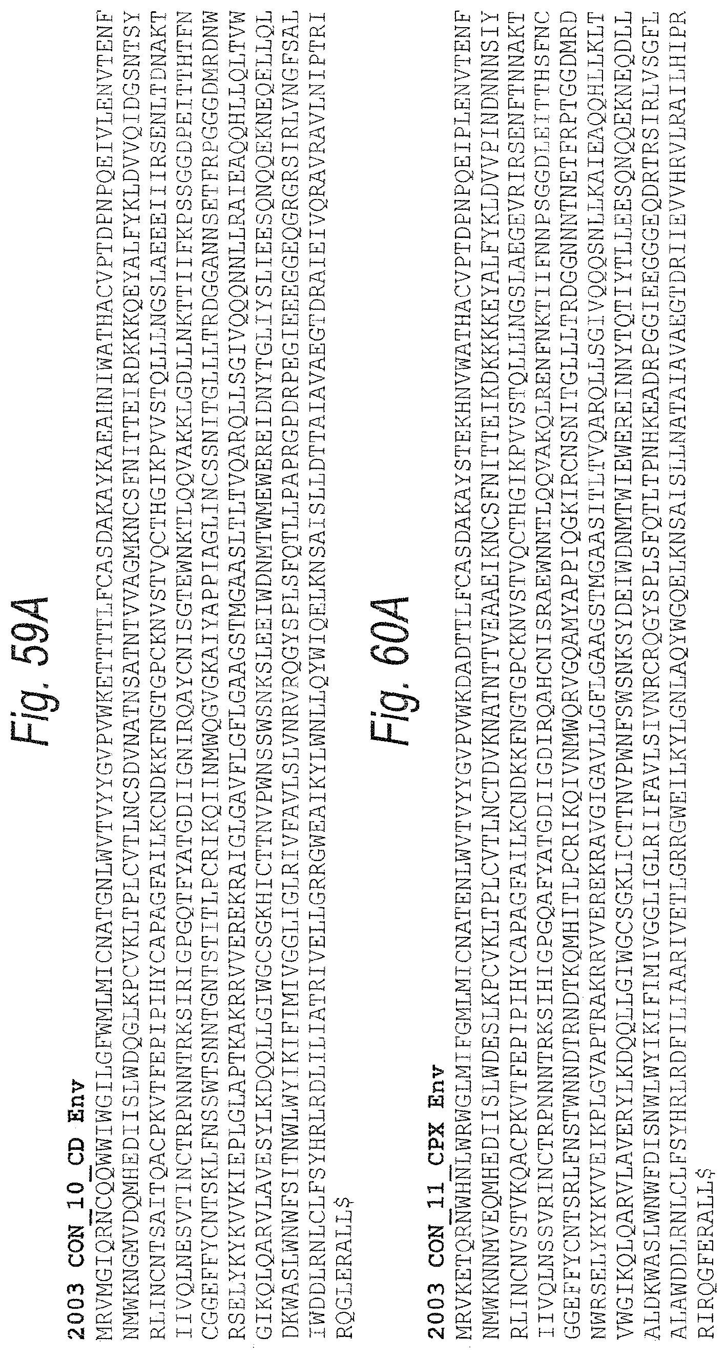
D00098
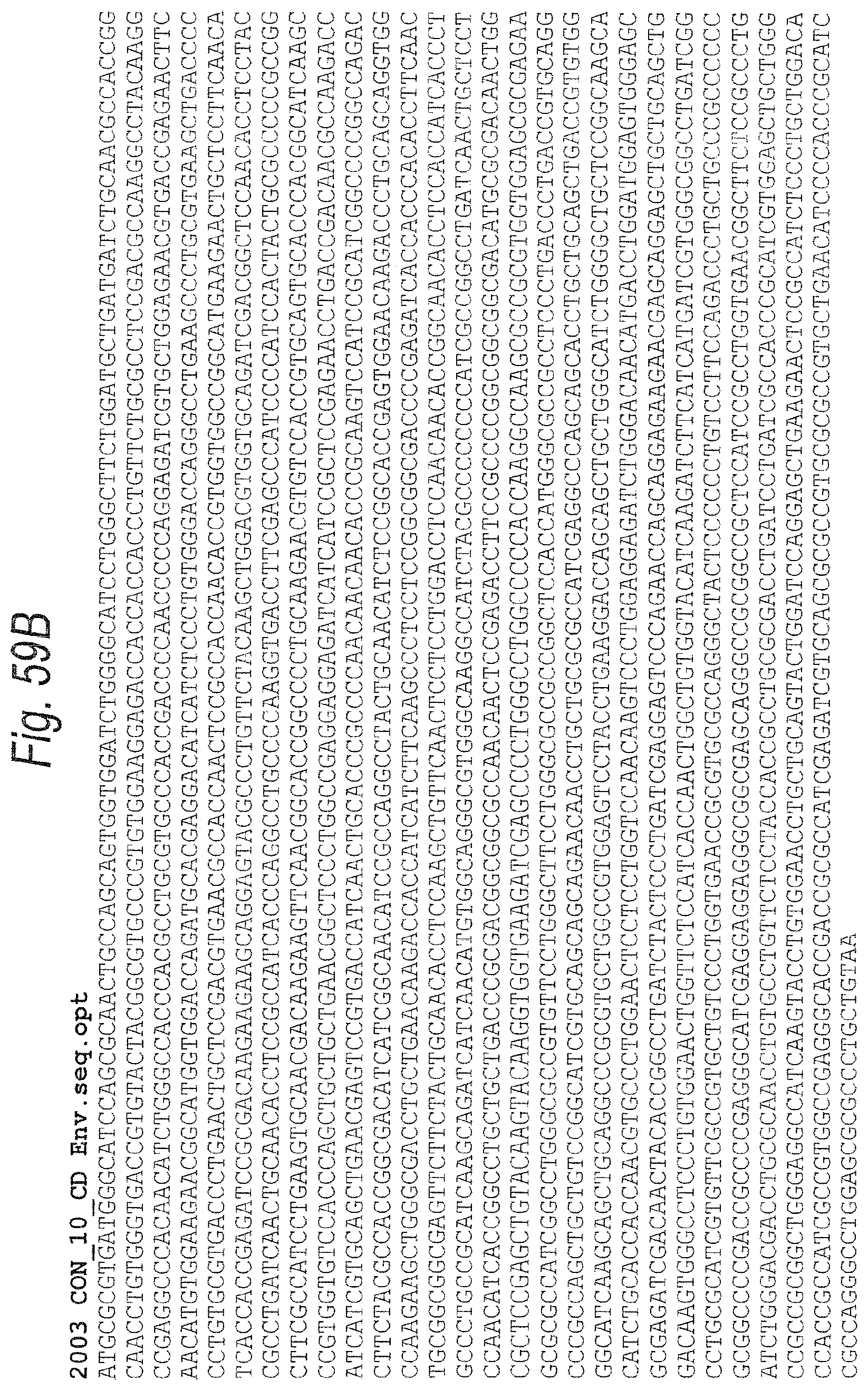
D00099
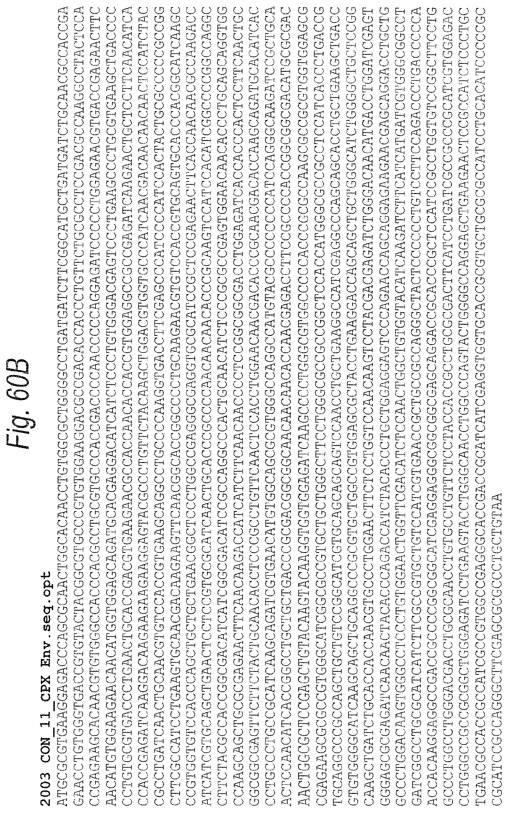
D00100
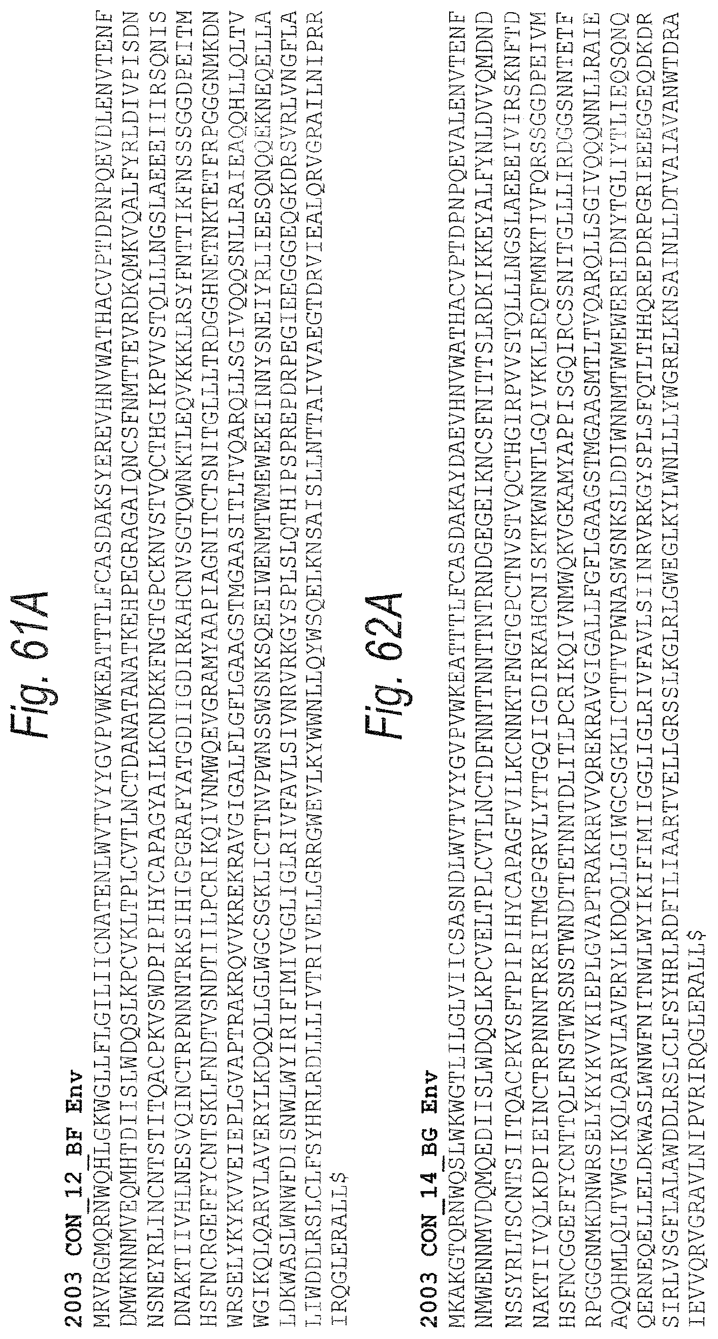
D00101

D00102

D00103
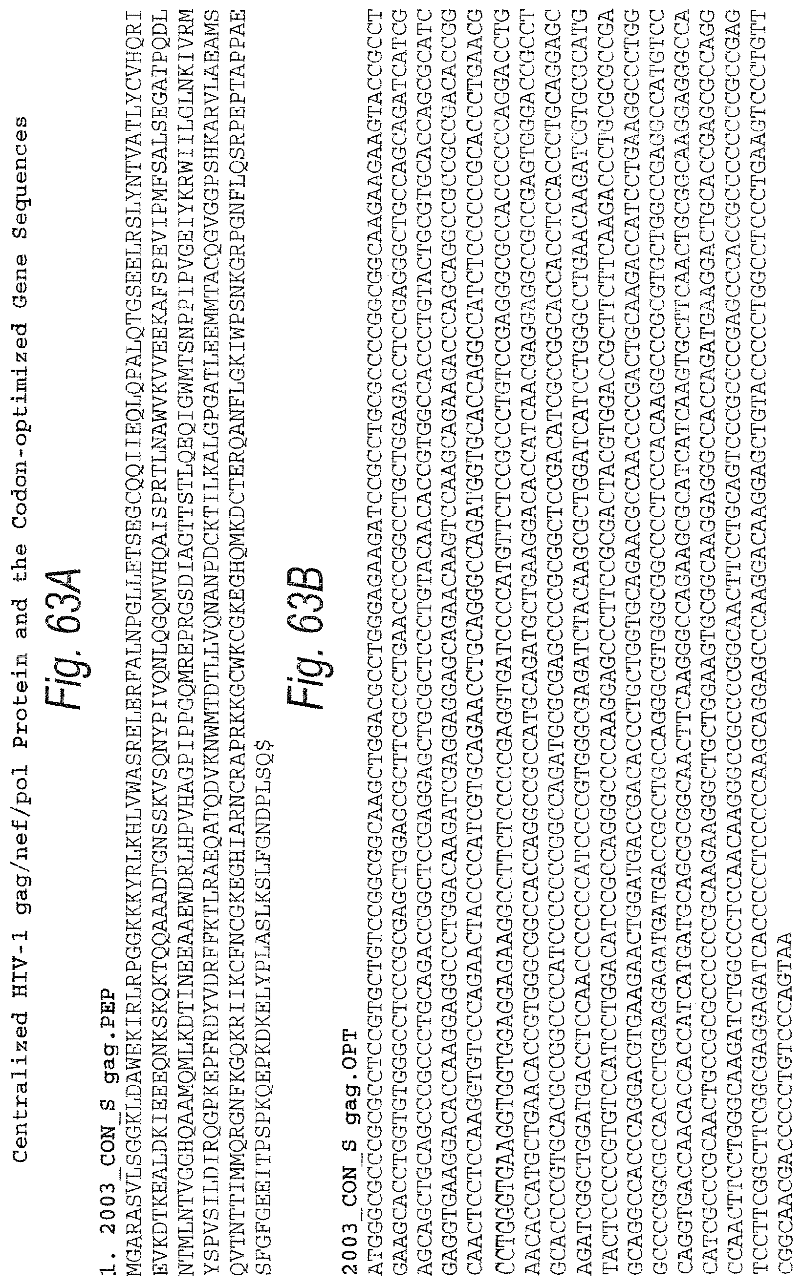
D00104
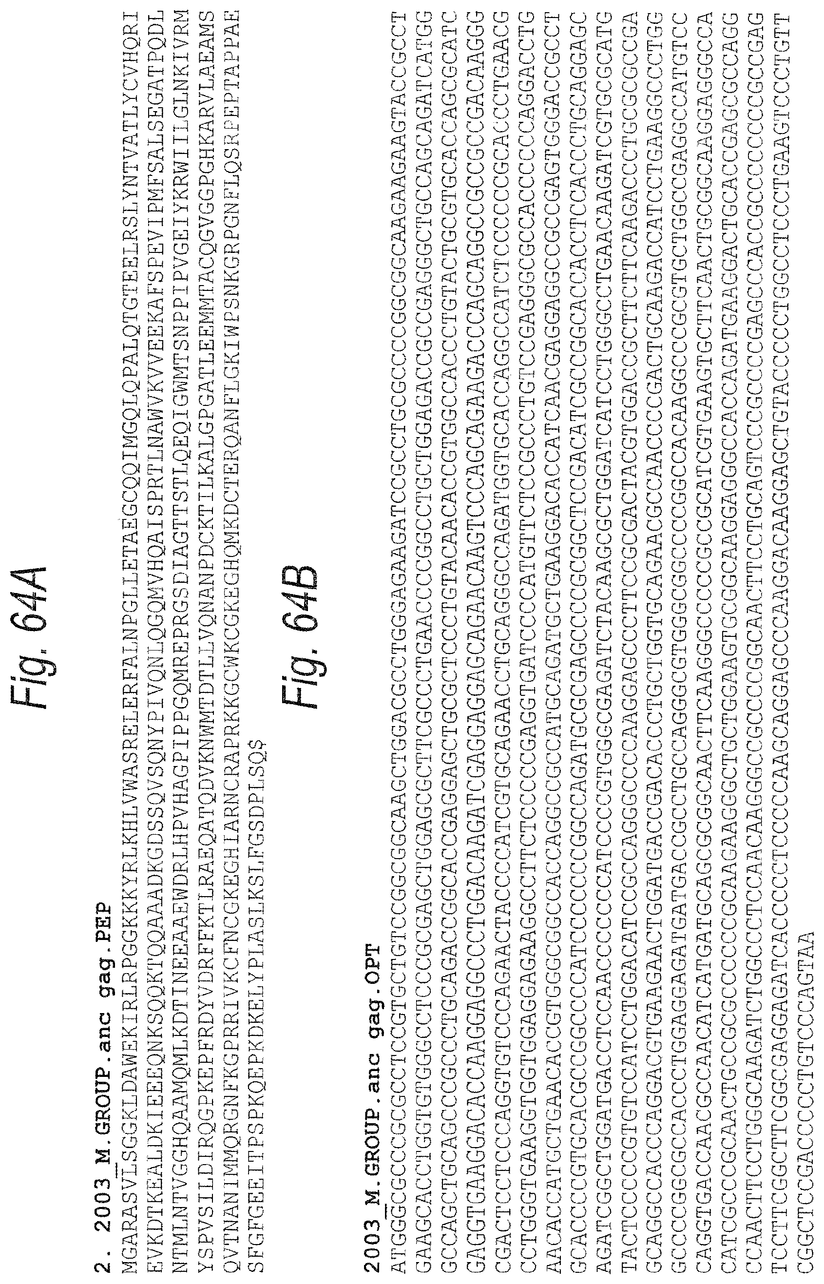
D00105

D00106
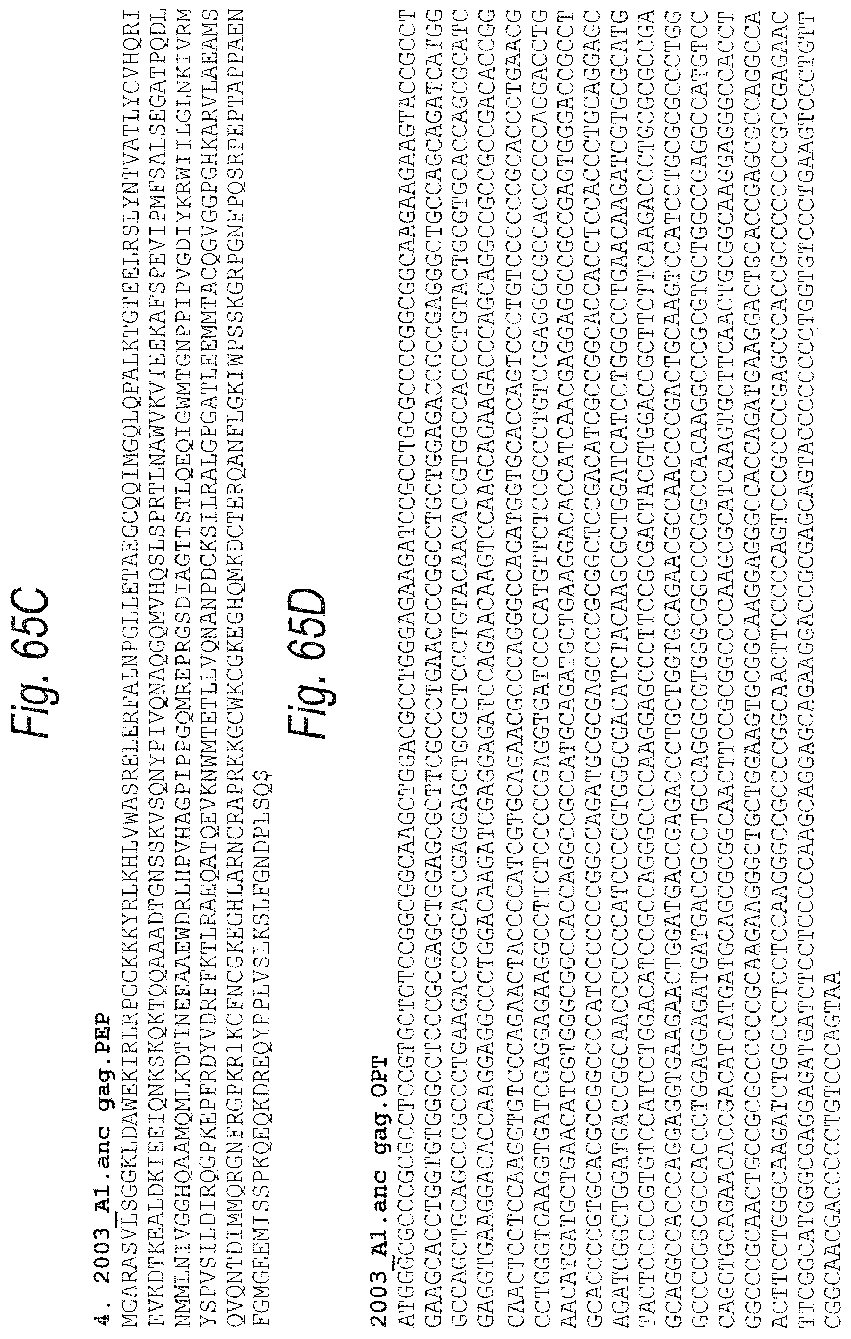
D00107
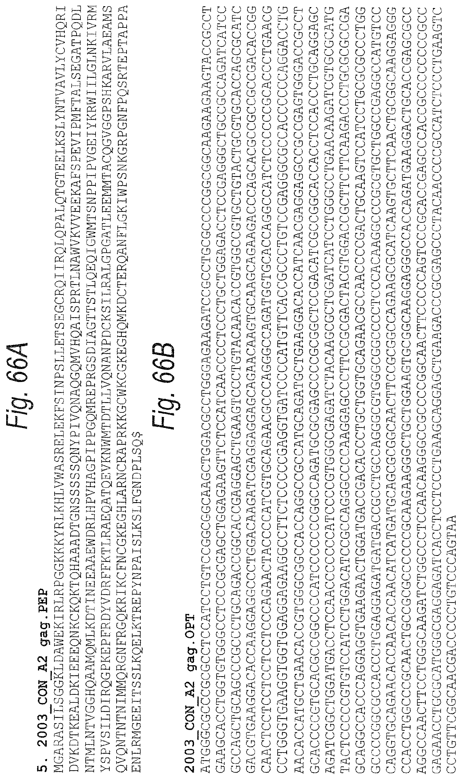
D00108
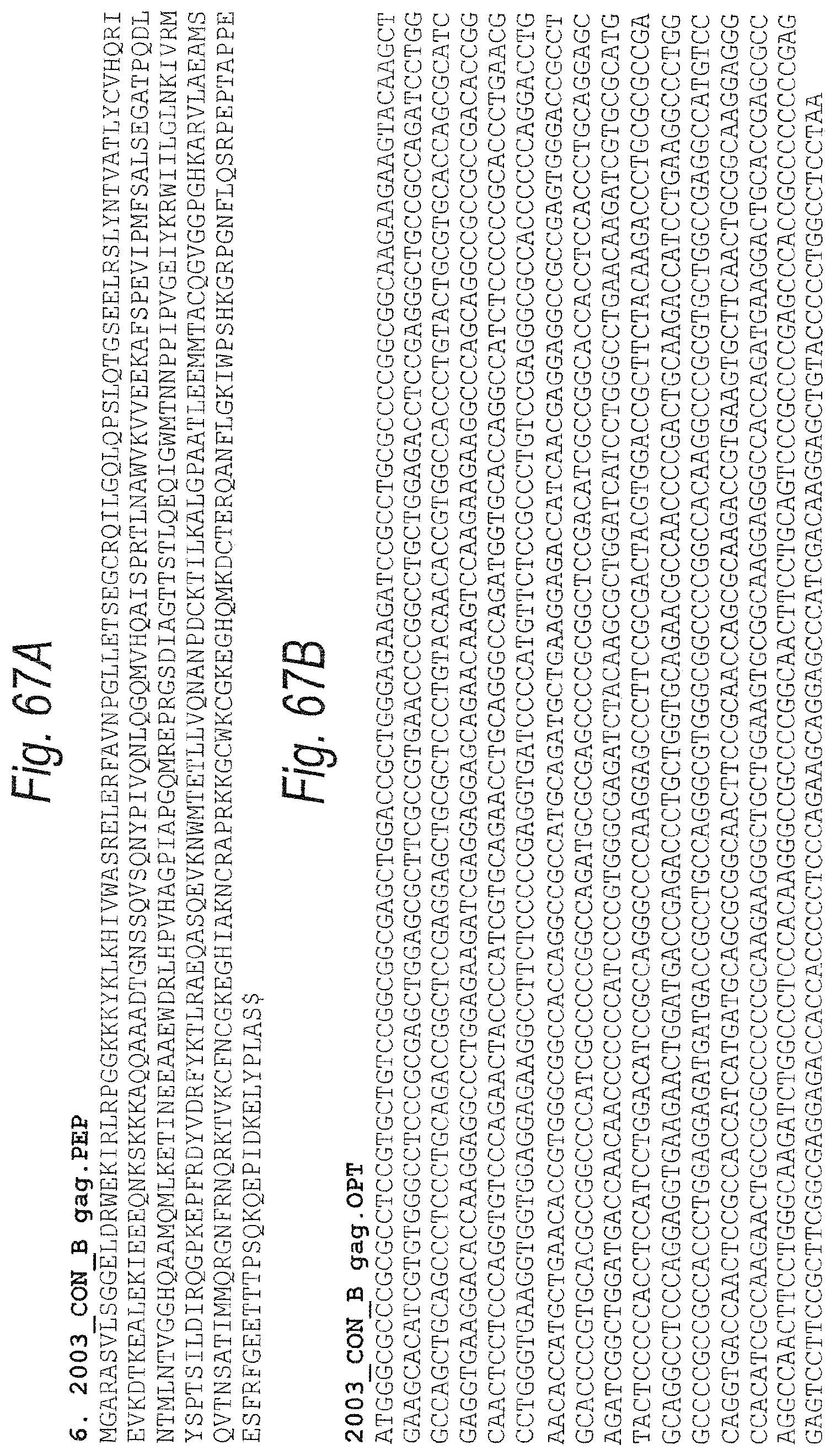
D00109
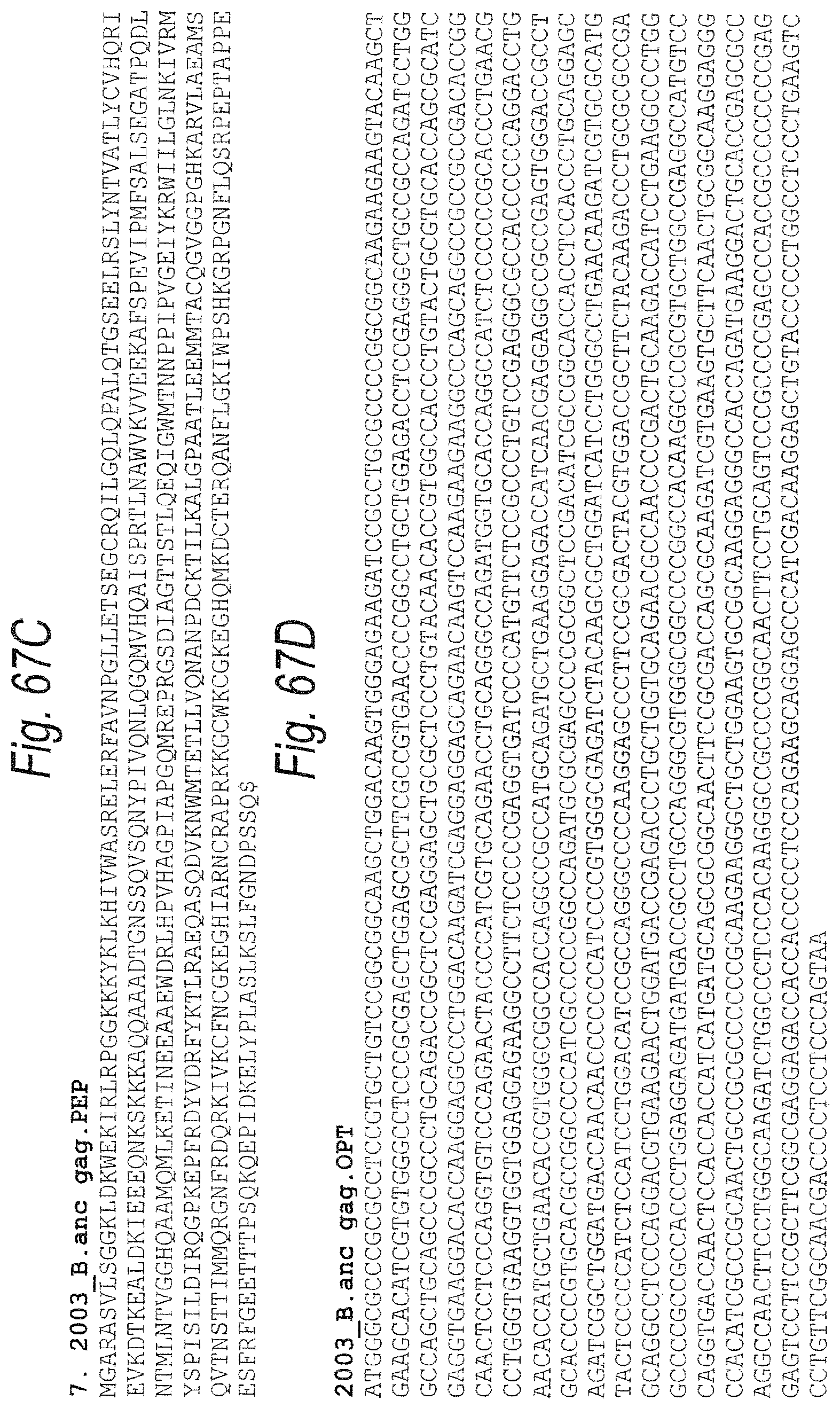
D00110
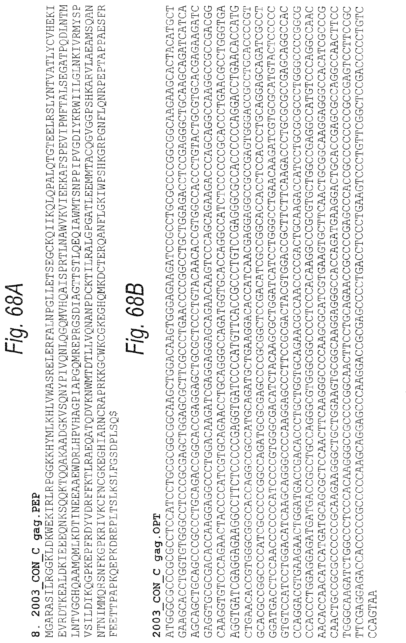
D00111
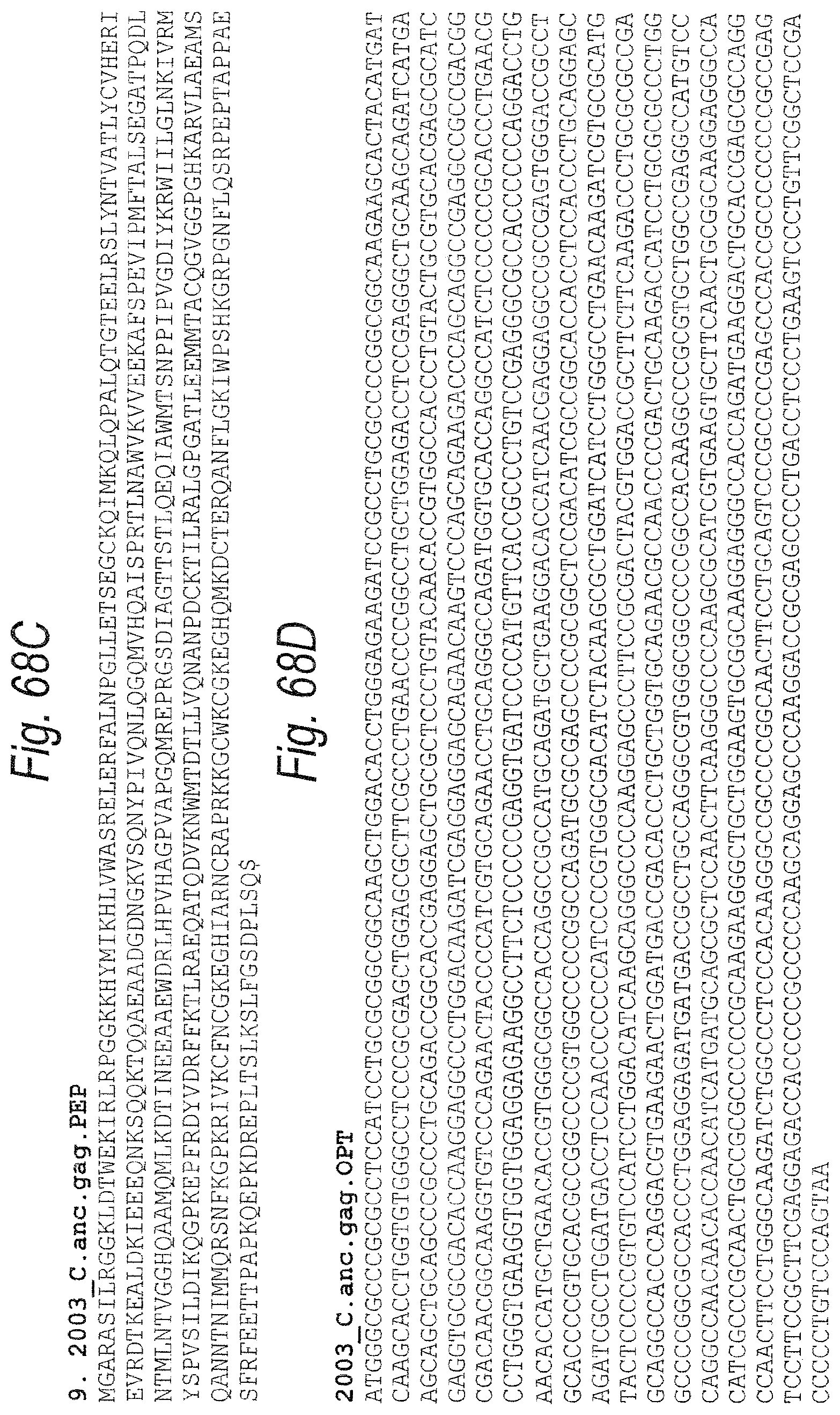
D00112

D00113

D00114

D00115

D00116
D00117
D00118
D00119

D00120

D00121
D00122

D00123
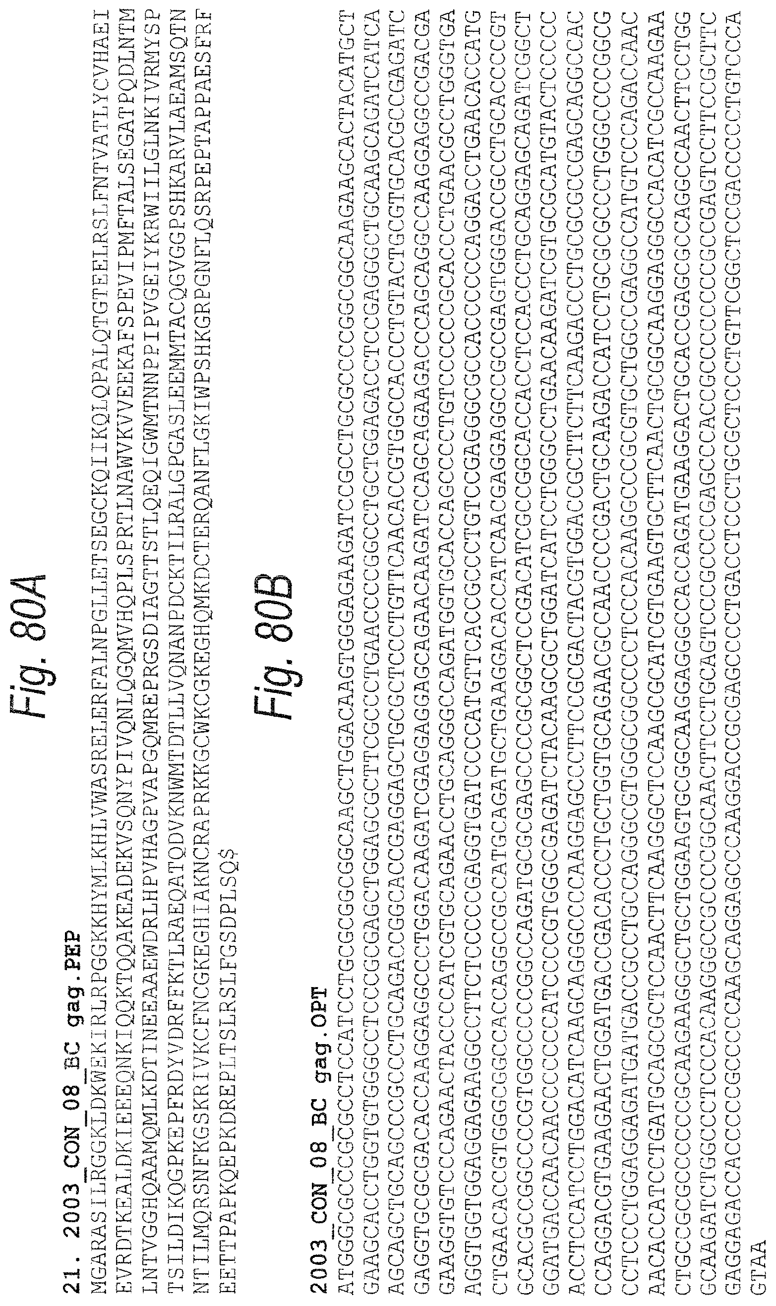
D00124
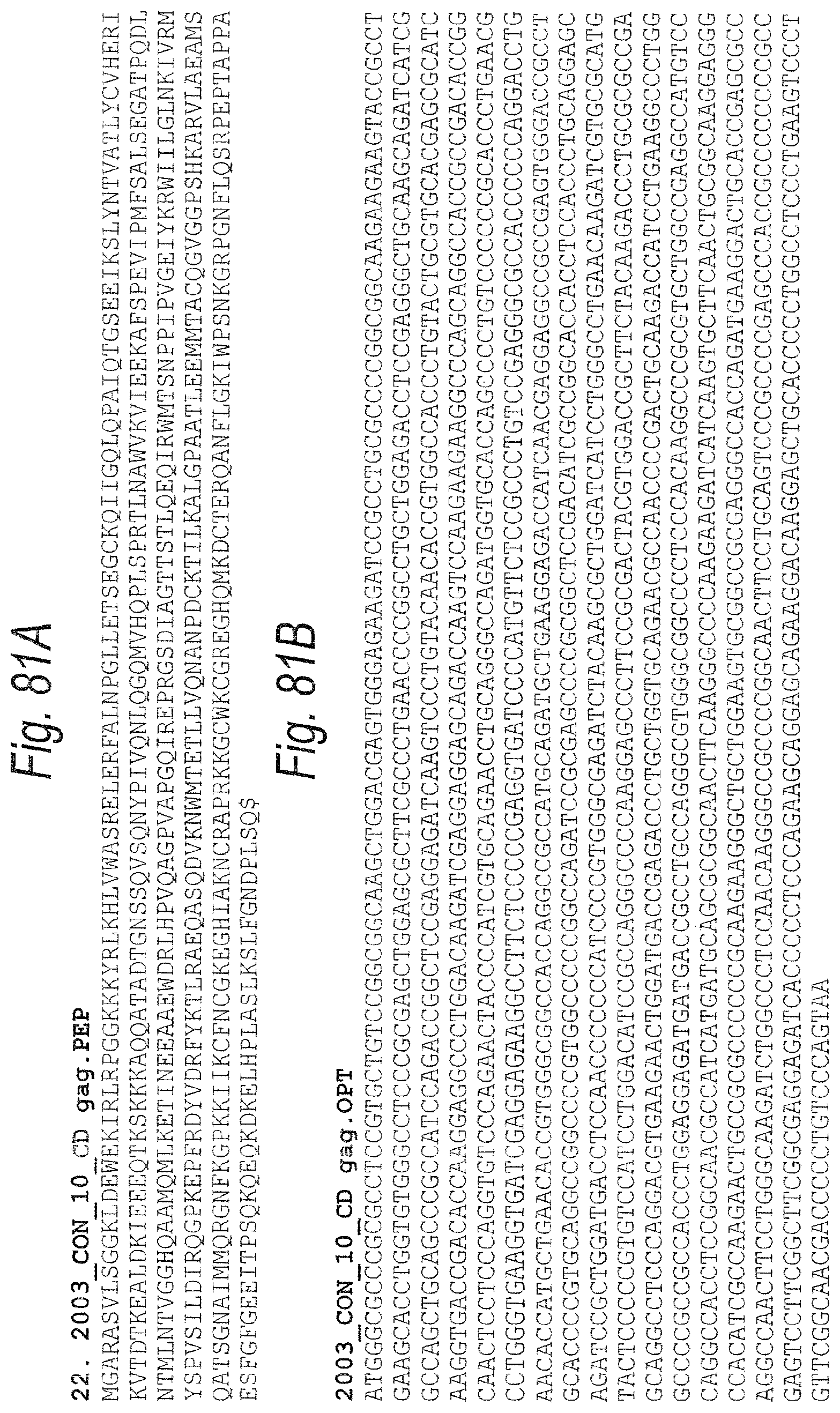
D00125
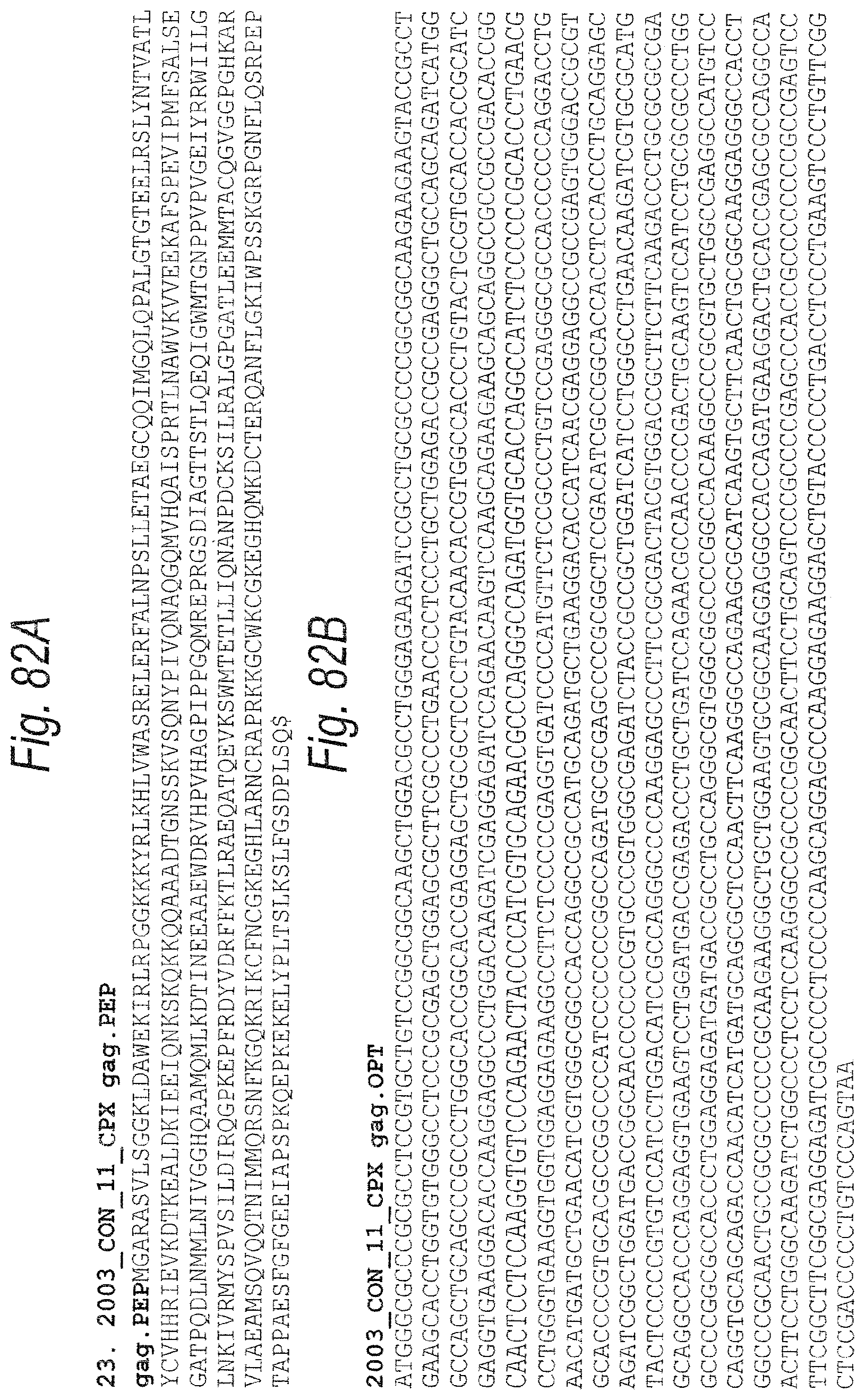
D00126

D00127

D00128
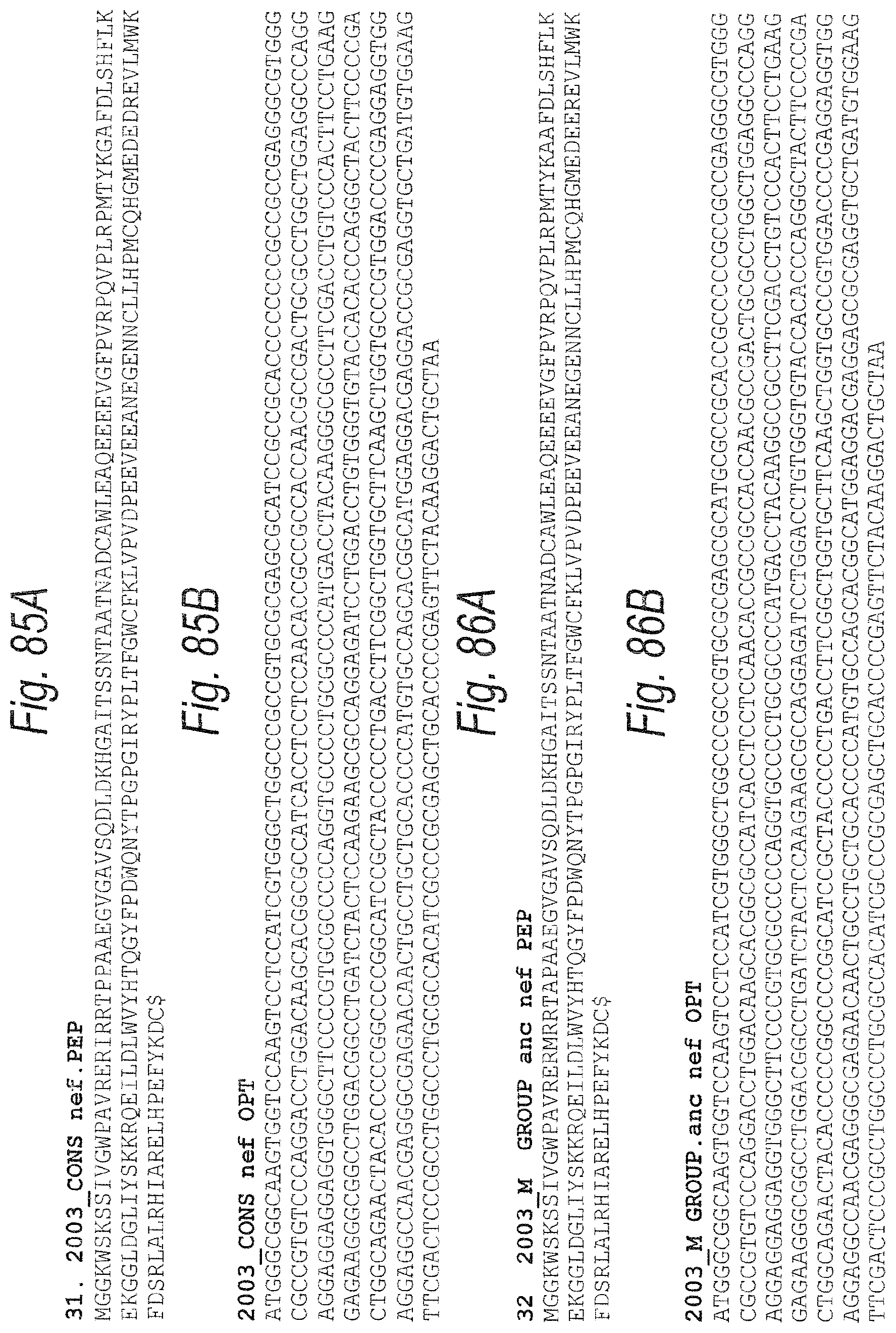
D00129
D00130

D00131

D00132
D00133

D00134
D00135
D00136
D00137
D00138
D00139

D00140
D00141
D00142
D00143
D00144
D00145

D00146

D00147
D00148

D00149
D00150

D00151
D00152

D00153
D00154

D00155

D00156

D00157

D00158
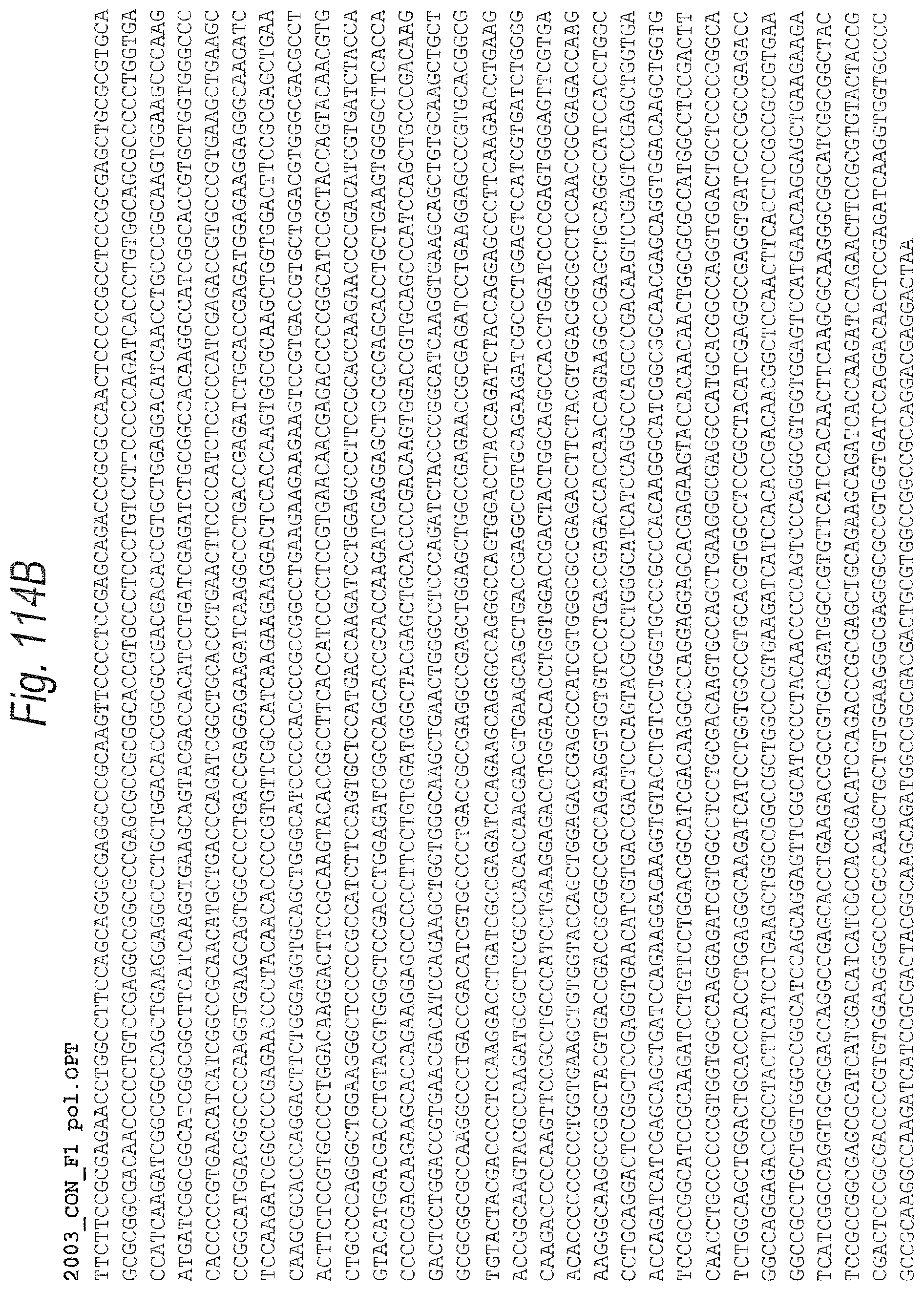
D00159
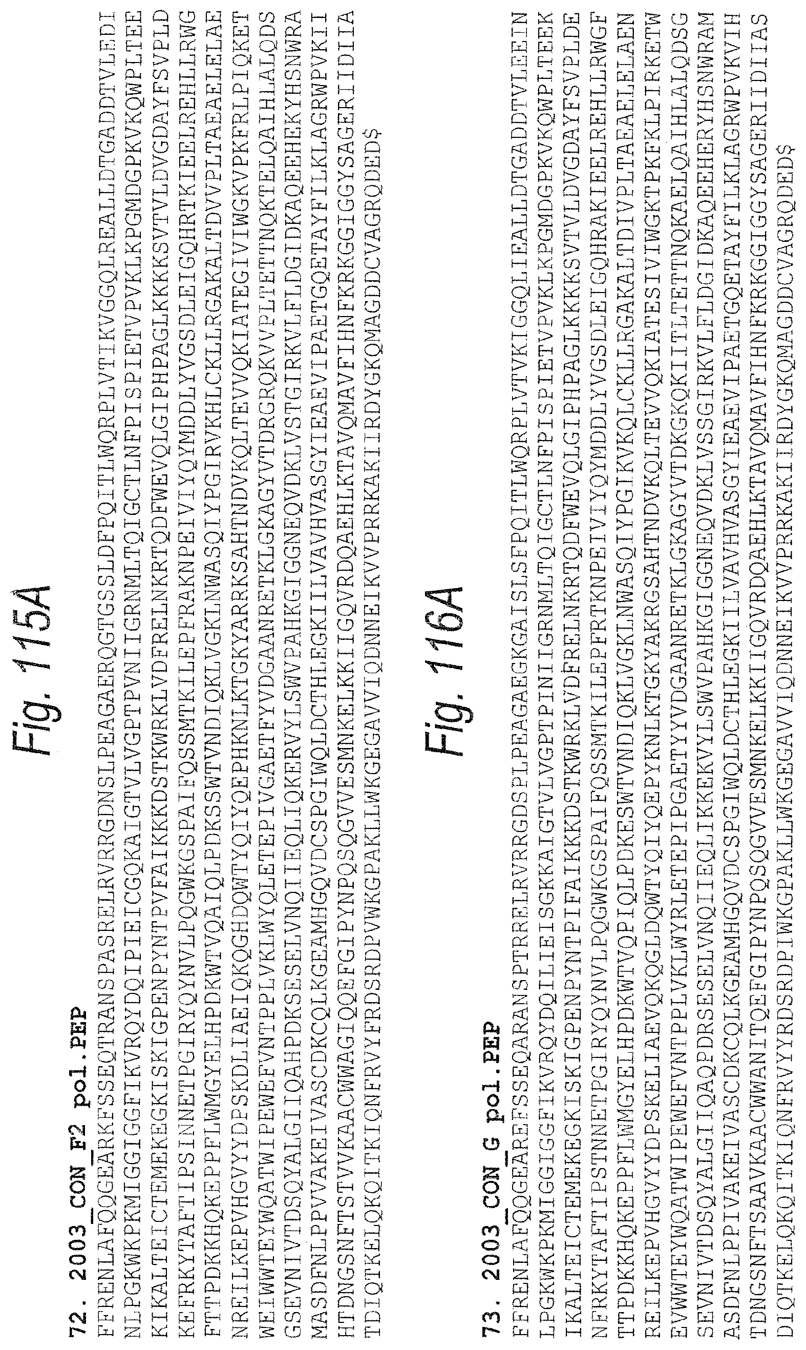
D00160

D00161
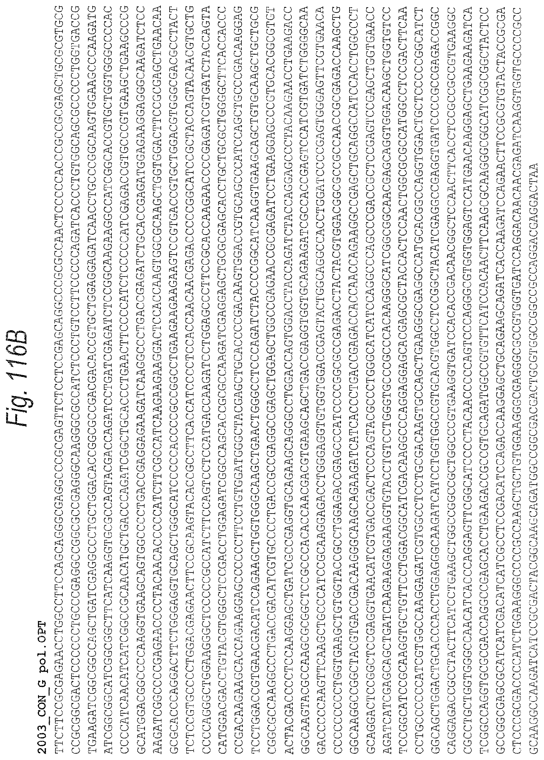
D00162
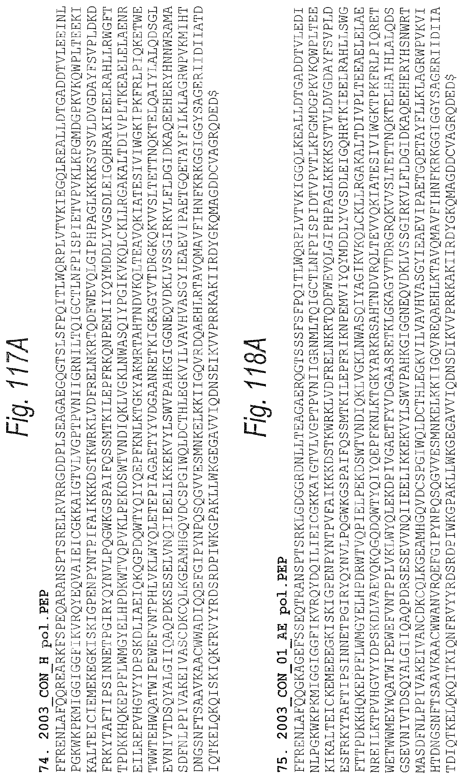
D00163
D00164
D00165

D00166
D00167
D00168

D00169

D00170
D00171

D00172

D00173
D00174
D00175
D00176
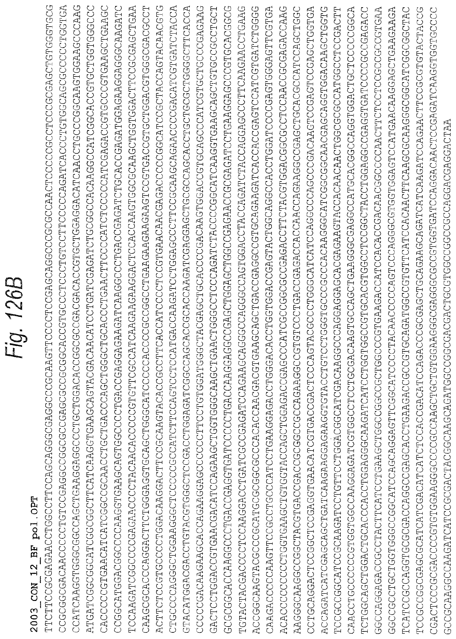
D00177
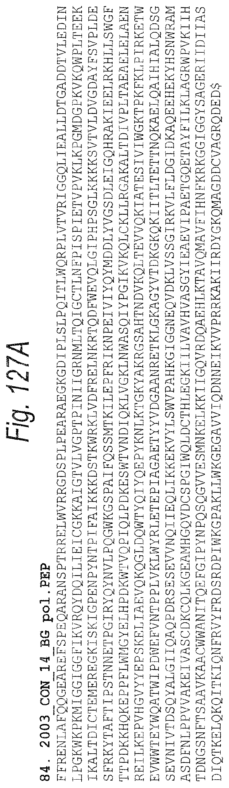
D00178

S00001
XML
uspto.report is an independent third-party trademark research tool that is not affiliated, endorsed, or sponsored by the United States Patent and Trademark Office (USPTO) or any other governmental organization. The information provided by uspto.report is based on publicly available data at the time of writing and is intended for informational purposes only.
While we strive to provide accurate and up-to-date information, we do not guarantee the accuracy, completeness, reliability, or suitability of the information displayed on this site. The use of this site is at your own risk. Any reliance you place on such information is therefore strictly at your own risk.
All official trademark data, including owner information, should be verified by visiting the official USPTO website at www.uspto.gov. This site is not intended to replace professional legal advice and should not be used as a substitute for consulting with a legal professional who is knowledgeable about trademark law.