Temporary interatrial shunts
Keren March 9, 2
U.S. patent number 10,940,296 [Application Number 15/570,752] was granted by the patent office on 2021-03-09 for temporary interatrial shunts. This patent grant is currently assigned to The Medical Research, Infrastructure and Health Services Fund of the Tel Aviv Medical Center, The Medical Research, Infrastructure and Health Services Fund of the Tel Aviv Medical Center, V-Wave Ltd.. The grantee listed for this patent is THE MEDICAL RESEARCH INFRASTRUCTURE AND HEALTH SERVICES FUND OF THE TEL-AVIV MEDICAL CENTER, THE MEDICAL RESEARCH INFRASTRUCTURE AND HEALTH SERVICES FUND OF THE TEL-AVIV MEDICAL CENTER. Invention is credited to Gad Keren.


| United States Patent | 10,940,296 |
| Keren | March 9, 2021 |
Temporary interatrial shunts
Abstract
Described embodiments include apparatus (28) that includes a shunt (26). The shunt includes a flared distal portion (40), a flared proximal portion (44), and an intermediate portion (42), disposed between the distal portion and the proximal portion. The apparatus further includes a constricting flexible longitudinal element (38) passing circumferentially along the intermediate portion of the shunt, configured to constrict the intermediate portion of the shunt, and one or more proximal-portion-collapsing flexible longitudinal elements (36) configured to collapse the proximal portion of the shunt. Other embodiments are also described.
| Inventors: | Keren; Gad (Kiryat Ono, IL) | ||||||||||
|---|---|---|---|---|---|---|---|---|---|---|---|
| Applicant: |
|
||||||||||
| Assignee: | The Medical Research,
Infrastructure and Health Services Fund of the Tel Aviv Medical
Center (Tel Aviv, IL) V-Wave Ltd. (Caesarea, IL) |
||||||||||
| Family ID: | 57217701 | ||||||||||
| Appl. No.: | 15/570,752 | ||||||||||
| Filed: | May 5, 2016 | ||||||||||
| PCT Filed: | May 05, 2016 | ||||||||||
| PCT No.: | PCT/IB2016/052561 | ||||||||||
| 371(c)(1),(2),(4) Date: | October 31, 2017 | ||||||||||
| PCT Pub. No.: | WO2016/178171 | ||||||||||
| PCT Pub. Date: | November 10, 2016 |
Prior Publication Data
| Document Identifier | Publication Date | |
|---|---|---|
| US 20180280667 A1 | Oct 4, 2018 | |
Related U.S. Patent Documents
| Application Number | Filing Date | Patent Number | Issue Date | ||
|---|---|---|---|---|---|
| 62158022 | May 7, 2015 | ||||
| Current U.S. Class: | 1/1 |
| Current CPC Class: | A61M 27/002 (20130101); A61B 17/11 (20130101); A61B 2017/1107 (20130101); A61F 2250/0059 (20130101); A61F 2002/9511 (20130101); A61B 2017/1139 (20130101); A61M 2205/3344 (20130101); A61B 2017/00243 (20130101); A61M 2210/125 (20130101); A61M 25/09 (20130101) |
| Current International Class: | A61M 27/00 (20060101); A61B 17/11 (20060101); A61M 25/09 (20060101); A61F 2/95 (20130101); A61B 17/00 (20060101) |
References Cited [Referenced By]
U.S. Patent Documents
| 3852334 | December 1974 | Dusza et al. |
| 3874388 | April 1975 | King et al. |
| 3952334 | April 1976 | Bokros et al. |
| 4484955 | November 1984 | Hochstein |
| 4601309 | July 1986 | Chang |
| 4617932 | October 1986 | Kornberg |
| 4662355 | May 1987 | Pieronne et al. |
| 4665906 | May 1987 | Jervis |
| 4705507 | November 1987 | Boyles |
| 4836204 | June 1989 | Landymore et al. |
| 4979955 | December 1990 | Smith |
| 4988339 | January 1991 | Vadher |
| 4995857 | February 1991 | Arnold |
| 5035706 | July 1991 | Giantureo et al. |
| 5037427 | August 1991 | Harada et al. |
| 5089005 | February 1992 | Harada |
| 5186431 | February 1993 | Tamari |
| 5197978 | March 1993 | Hess |
| 5267940 | December 1993 | Moulder |
| 5290227 | March 1994 | Pasque |
| 5312341 | May 1994 | Turi |
| 5326374 | July 1994 | Ilbawi et al. |
| 5332402 | July 1994 | Teitelbaum |
| 5334217 | August 1994 | Das |
| 5378239 | January 1995 | Termin et al. |
| 5409019 | April 1995 | Wilk |
| 5429144 | July 1995 | Wilk |
| 5500015 | March 1996 | Deac |
| 5531759 | July 1996 | Kensey et al. |
| 5545210 | August 1996 | Hess et al. |
| 5556386 | September 1996 | Todd |
| 5578008 | November 1996 | Hara |
| 5584803 | December 1996 | Stevens et al. |
| 5597377 | January 1997 | Aldea |
| 5645559 | July 1997 | Hachtman et al. |
| 5655548 | August 1997 | Nelson et al. |
| 5662711 | September 1997 | Douglas |
| 5702412 | December 1997 | Popov et al. |
| 5725552 | March 1998 | Kotula et al. |
| 5741324 | April 1998 | Glastra |
| 5749880 | May 1998 | Banas et al. |
| 5779716 | July 1998 | Cano et al. |
| 5795307 | August 1998 | Krueger |
| 5810836 | September 1998 | Hussein et al. |
| 5824062 | October 1998 | Patke et al. |
| 5824071 | October 1998 | Nelson et al. |
| 5846261 | December 1998 | Kotula et al. |
| 5910144 | June 1999 | Hayashi |
| 5916193 | June 1999 | Stevens et al. |
| 5941850 | August 1999 | Shah et al. |
| 5957949 | September 1999 | Leonhardt et al. |
| 5990379 | November 1999 | Gregory |
| 6027518 | February 2000 | Gaber |
| 6039755 | March 2000 | Edwin et al. |
| 6039759 | March 2000 | Carpentier et al. |
| 6086610 | July 2000 | Duerig et al. |
| 6111520 | August 2000 | Allen et al. |
| 6117159 | September 2000 | Huebsch et al. |
| 6120534 | September 2000 | Ruiz |
| 6124523 | September 2000 | Banas et al. |
| 6126686 | October 2000 | Badylak et al. |
| 6165188 | December 2000 | Saadat et al. |
| 6210318 | April 2001 | Lederman |
| 6214039 | April 2001 | Banas et al. |
| 6217541 | April 2001 | Yu |
| 6221096 | April 2001 | Aiba et al. |
| 6242762 | June 2001 | Brown et al. |
| 6245099 | June 2001 | Edwin et al. |
| 6254564 | July 2001 | Wilk et al. |
| 6260552 | July 2001 | Mortier et al. |
| 6264684 | July 2001 | Banas et al. |
| 6270526 | August 2001 | Cox |
| 6277078 | August 2001 | Porat et al. |
| 6278379 | August 2001 | Allen et al. |
| 6302892 | October 2001 | Wilk |
| 6306141 | October 2001 | Jervis |
| 6328699 | December 2001 | Eigler et al. |
| 6344022 | February 2002 | Jarvik |
| 6358277 | March 2002 | Duran |
| 6391036 | May 2002 | Berg et al. |
| 6398803 | June 2002 | Layne et al. |
| 6406422 | June 2002 | Landesberg |
| 6447539 | September 2002 | Nelson et al. |
| 6451051 | September 2002 | Drasler et al. |
| 6458153 | October 2002 | Bailey et al. |
| 6468303 | October 2002 | Amplatz et al. |
| 6475136 | November 2002 | Forsell |
| 6478776 | November 2002 | Rosenman et al. |
| 6485507 | November 2002 | Walak et al. |
| 6488702 | December 2002 | Besselink |
| 6491705 | December 2002 | Gifford et al. |
| 6527698 | March 2003 | Kung et al. |
| 6544208 | April 2003 | Ethier et al. |
| 6547814 | April 2003 | Edwin et al. |
| 6562066 | May 2003 | Martin |
| 6572652 | June 2003 | Shaknovich |
| 6579314 | June 2003 | Lombardi et al. |
| 6589198 | July 2003 | Soltanpour et al. |
| 6632169 | October 2003 | Korakianitis et al. |
| 6638303 | October 2003 | Campbell |
| 6641610 | November 2003 | Wolf et al. |
| 6652578 | November 2003 | Bailey et al. |
| 6685664 | February 2004 | Levin et al. |
| 6712836 | March 2004 | Berg et al. |
| 6740115 | May 2004 | Lombardi et al. |
| 6758858 | July 2004 | McCrea et al. |
| 6764507 | July 2004 | Shanley et al. |
| 6770087 | August 2004 | Layne et al. |
| 6797217 | September 2004 | McCrea et al. |
| 6890350 | May 2005 | Walak et al. |
| 6970742 | November 2005 | Mann et al. |
| 7001409 | February 2006 | Amplatz |
| 7004966 | February 2006 | Edwin et al. |
| 7025777 | April 2006 | Moore |
| 7060150 | June 2006 | Banas et al. |
| 7083640 | August 2006 | Lombardi et al. |
| 7115095 | October 2006 | Eigler et al. |
| 7118600 | October 2006 | Dua et al. |
| 7137953 | November 2006 | Eigler et al. |
| 7147604 | December 2006 | Allen et al. |
| 7149587 | December 2006 | Wardle et al. |
| 7169160 | January 2007 | Middleman et al. |
| 7169172 | January 2007 | Levine et al. |
| 7195594 | March 2007 | Eigler et al. |
| 7208010 | April 2007 | Shanley et al. |
| 7226558 | June 2007 | Nieman et al. |
| 7245117 | July 2007 | Joy et al. |
| 7294115 | November 2007 | Wilk |
| 7306756 | December 2007 | Edwin et al. |
| 7402899 | July 2008 | Whiting et al. |
| 7439723 | October 2008 | Allen et al. |
| 7468071 | December 2008 | Edwin et al. |
| 7483743 | January 2009 | Mann et al. |
| 7498799 | March 2009 | Allen et al. |
| 7509169 | March 2009 | Eigler et al. |
| 7550978 | June 2009 | Joy et al. |
| 7578899 | August 2009 | Edwin et al. |
| 7590449 | September 2009 | Mann et al. |
| 7615010 | November 2009 | Najafi et al. |
| 7621879 | November 2009 | Eigler et al. |
| 7679355 | March 2010 | Allen et al. |
| 7717854 | May 2010 | Mann et al. |
| 7794473 | September 2010 | Tessmer et al. |
| 7839153 | November 2010 | Joy et al. |
| 7842083 | November 2010 | Shanley et al. |
| 7854172 | December 2010 | O'Brien et al. |
| 7862513 | January 2011 | Eigler et al. |
| 7914639 | March 2011 | Layne et al. |
| 7939000 | May 2011 | Edwin et al. |
| 7988724 | August 2011 | Salahieh et al. |
| 7993383 | August 2011 | Hartley et al. |
| 8012194 | September 2011 | Edwin et al. |
| 8016877 | September 2011 | Seguin et al. |
| 8021420 | September 2011 | Dolan |
| 8025625 | September 2011 | Allen |
| 8025668 | September 2011 | McCartney |
| 8043360 | October 2011 | McNamara et al. |
| 8070708 | December 2011 | Rottenberg et al. |
| 8091556 | January 2012 | Keren et al. |
| 8096959 | January 2012 | Stewart et al. |
| 8137605 | March 2012 | McCrea et al. |
| 8142363 | March 2012 | Eigler et al. |
| 8147545 | April 2012 | Avior |
| 8157852 | April 2012 | Bloom et al. |
| 8157860 | April 2012 | McNamara et al. |
| 8157940 | April 2012 | Edwin et al. |
| 8158041 | April 2012 | Colone |
| 8187321 | May 2012 | Shanley et al. |
| 8202313 | June 2012 | Shanley et al. |
| 8206435 | June 2012 | Shanley et al. |
| 8235916 | August 2012 | Whiting et al. |
| 8235933 | August 2012 | Keren et al. |
| 8246677 | August 2012 | Ryan |
| 8287589 | October 2012 | Otto et al. |
| 8298150 | October 2012 | Mann et al. |
| 8298244 | October 2012 | Garcia et al. |
| 8303511 | November 2012 | Eigler et al. |
| 8313524 | November 2012 | Edwin et al. |
| 8328751 | December 2012 | Keren et al. |
| 8337650 | December 2012 | Edwin et al. |
| 8348996 | January 2013 | Tuval et al. |
| 8398708 | March 2013 | Meiri et al. |
| 8460366 | June 2013 | Rowe |
| 8468667 | June 2013 | Straubinger et al. |
| 8480594 | July 2013 | Eigler et al. |
| 8579966 | November 2013 | Seguin et al. |
| 8597225 | December 2013 | Kapadia |
| 8617337 | December 2013 | Layne et al. |
| 8617441 | December 2013 | Edwin et al. |
| 8652284 | February 2014 | Bogert et al. |
| 8665086 | March 2014 | Miller et al. |
| 8696611 | April 2014 | Nitzan et al. |
| 8790241 | July 2014 | Edwin et al. |
| 8882697 | November 2014 | Celermajer et al. |
| 8882798 | November 2014 | Schwab et al. |
| 8911489 | December 2014 | Ben-Muvhar |
| 9005155 | April 2015 | Sugimoto |
| 9034034 | May 2015 | Nitzan et al. |
| 9055917 | June 2015 | Mann et al. |
| 9060696 | June 2015 | Eigler et al. |
| 9067050 | June 2015 | Gallagher et al. |
| 9205236 | December 2015 | McNamara et al. |
| 9220429 | December 2015 | Nabutovsky et al. |
| 9358371 | June 2016 | McNamara et al. |
| 9393115 | July 2016 | Tabor et al. |
| 9456812 | October 2016 | Finch et al. |
| 9622895 | April 2017 | Cohen et al. |
| 9629715 | April 2017 | Nitzan et al. |
| 9681948 | June 2017 | Levi et al. |
| 9707382 | July 2017 | Nitzan et al. |
| 9713696 | July 2017 | Yacoby et al. |
| 9724499 | August 2017 | Rottenberg et al. |
| 9757107 | September 2017 | McNamara et al. |
| 9789294 | October 2017 | Taft et al. |
| 9918677 | March 2018 | Eigler et al. |
| 9943670 | April 2018 | Keren et al. |
| 9980815 | May 2018 | Nitzan et al. |
| 10045766 | August 2018 | McNamara et al. |
| 10047421 | August 2018 | Khan et al. |
| 10076403 | September 2018 | Eigler et al. |
| 10105103 | October 2018 | Goldshtein et al. |
| 10111741 | October 2018 | Michalak |
| 10207087 | February 2019 | Keren et al. |
| 10207807 | February 2019 | Moran et al. |
| 10251740 | April 2019 | Eigler et al. |
| 10251750 | April 2019 | Eigler et al. |
| 10299687 | May 2019 | Nabutovsky et al. |
| 10357320 | July 2019 | Beira |
| 10357357 | July 2019 | Levi et al. |
| 10368981 | August 2019 | Nitzan et al. |
| 10463490 | November 2019 | Rottenberg et al. |
| 10478594 | November 2019 | Yacoby et al. |
| 10548725 | February 2020 | Alkhatib et al. |
| 10561423 | February 2020 | Sharma |
| 10639459 | May 2020 | Nitzan et al. |
| 2002/0120277 | August 2002 | Hauschild et al. |
| 2002/0165479 | November 2002 | Wilk |
| 2002/0165606 | November 2002 | Wolf et al. |
| 2002/0169371 | November 2002 | Gilderdale |
| 2002/0169377 | November 2002 | Khairkhahan et al. |
| 2002/0173742 | November 2002 | Keren et al. |
| 2003/0100920 | May 2003 | Akin et al. |
| 2003/0125798 | July 2003 | Martin |
| 2003/0136417 | July 2003 | Fonseca et al. |
| 2003/0176914 | September 2003 | Rabkin et al. |
| 2003/0209835 | November 2003 | Chun et al. |
| 2003/0216679 | November 2003 | Wolf et al. |
| 2003/0216803 | November 2003 | Ledergerber |
| 2004/0010219 | January 2004 | McCusker et al. |
| 2004/0016514 | January 2004 | Nien |
| 2004/0077988 | April 2004 | Tweden et al. |
| 2004/0088045 | May 2004 | Cox |
| 2004/0093075 | May 2004 | Kuehne |
| 2004/0102797 | May 2004 | Golden et al. |
| 2004/0116999 | June 2004 | Ledergerber |
| 2004/0138743 | July 2004 | Myers et al. |
| 2004/0147869 | July 2004 | Wolf et al. |
| 2004/0147871 | July 2004 | Burnett |
| 2004/0147886 | July 2004 | Bonni |
| 2004/0162514 | August 2004 | Alferness et al. |
| 2004/0193261 | September 2004 | Berreklouw |
| 2004/0210190 | October 2004 | Kohler et al. |
| 2004/0210307 | October 2004 | Khairkhahan |
| 2004/0225352 | November 2004 | Osborne et al. |
| 2005/0003327 | January 2005 | Elian et al. |
| 2005/0033327 | February 2005 | Gainor |
| 2005/0033351 | February 2005 | Newton |
| 2005/0065589 | March 2005 | Schneider et al. |
| 2005/0137682 | June 2005 | Justino |
| 2005/0148925 | July 2005 | Rottenberg et al. |
| 2005/0165344 | July 2005 | Dobak, III |
| 2005/0182486 | August 2005 | Gabbay |
| 2005/0283231 | December 2005 | Haug et al. |
| 2006/0009800 | January 2006 | Christianson et al. |
| 2006/0025857 | February 2006 | Bergheim et al. |
| 2006/0111660 | May 2006 | Wolf et al. |
| 2006/0116710 | June 2006 | Corcoran et al. |
| 2006/0122647 | June 2006 | Callaghan et al. |
| 2006/0167541 | July 2006 | Lattouf |
| 2006/0184231 | August 2006 | Rucker |
| 2006/0212110 | September 2006 | Osborne et al. |
| 2006/0241745 | October 2006 | Solem |
| 2006/0256611 | November 2006 | Bednorz et al. |
| 2006/0282157 | December 2006 | Hill et al. |
| 2007/0010852 | January 2007 | Blaeser et al. |
| 2007/0021739 | January 2007 | Weber |
| 2007/0043435 | February 2007 | Seguin et al. |
| 2007/0191863 | August 2007 | De Juan et al. |
| 2007/0213813 | September 2007 | Von Segesser et al. |
| 2007/0276413 | November 2007 | Nobles |
| 2007/0276414 | November 2007 | Nobles |
| 2007/0282157 | December 2007 | Rottenberg et al. |
| 2007/0299384 | December 2007 | Faul et al. |
| 2008/0034836 | February 2008 | Eigler et al. |
| 2008/0086205 | April 2008 | Gordy et al. |
| 2008/0125861 | May 2008 | Webler et al. |
| 2008/0177300 | July 2008 | Mas et al. |
| 2008/0262602 | October 2008 | Wilk et al. |
| 2008/0319525 | December 2008 | Tieu et al. |
| 2009/0030499 | January 2009 | Bebb et al. |
| 2009/0054976 | February 2009 | Tuval et al. |
| 2009/0125104 | May 2009 | Hoffman |
| 2009/0198315 | August 2009 | Boudjemline |
| 2009/0276040 | November 2009 | Rowe et al. |
| 2009/0319037 | December 2009 | Rowe et al. |
| 2010/0004740 | January 2010 | Seguin et al. |
| 2010/0022940 | January 2010 | Thompson |
| 2010/0057192 | March 2010 | Celermajer |
| 2010/0069836 | March 2010 | Satake |
| 2010/0070022 | March 2010 | Kuehling |
| 2010/0081867 | April 2010 | Fishler et al. |
| 2010/0121434 | May 2010 | Paul et al. |
| 2010/0179590 | July 2010 | Fortson et al. |
| 2010/0191326 | July 2010 | Alkhatib |
| 2010/0249909 | September 2010 | McNamara et al. |
| 2010/0249910 | September 2010 | McNamara et al. |
| 2010/0249915 | September 2010 | Zhang |
| 2010/0256548 | October 2010 | McNamara et al. |
| 2010/0256753 | October 2010 | McNamara et al. |
| 2010/0298755 | November 2010 | McNamara et al. |
| 2011/0022057 | January 2011 | Eigler et al. |
| 2011/0022157 | January 2011 | Essinger et al. |
| 2011/0054515 | March 2011 | Bridgeman et al. |
| 2011/0071623 | March 2011 | Finch et al. |
| 2011/0071624 | March 2011 | Finch et al. |
| 2011/0093059 | April 2011 | Fischell et al. |
| 2011/0152923 | June 2011 | Fox |
| 2011/0190874 | August 2011 | Celermajer et al. |
| 2011/0218479 | September 2011 | Rottenberg et al. |
| 2011/0218480 | September 2011 | Rottenberg et al. |
| 2011/0218481 | September 2011 | Rottenberg et al. |
| 2011/0257723 | October 2011 | McNamara |
| 2011/0264203 | October 2011 | Dwork et al. |
| 2011/0276086 | November 2011 | Al-Qbandi et al. |
| 2011/0295182 | December 2011 | Finch et al. |
| 2011/0295183 | December 2011 | Finch et al. |
| 2011/0295362 | December 2011 | Finch et al. |
| 2011/0295366 | December 2011 | Finch et al. |
| 2011/0306916 | December 2011 | Nitzan et al. |
| 2011/0319806 | December 2011 | Wardle |
| 2012/0022633 | January 2012 | Olson et al. |
| 2012/0035590 | February 2012 | Whiting et al. |
| 2012/0041422 | February 2012 | Whiting et al. |
| 2012/0046528 | February 2012 | Eigler et al. |
| 2012/0046739 | February 2012 | von Oepen et al. |
| 2012/0053686 | March 2012 | McNamara et al. |
| 2012/0071918 | March 2012 | Amin et al. |
| 2012/0130301 | May 2012 | McNamara et al. |
| 2012/0165928 | June 2012 | Nitzan |
| 2012/0179172 | July 2012 | Paul et al. |
| 2012/0190991 | July 2012 | Bornzin et al. |
| 2012/0265296 | October 2012 | McNamara et al. |
| 2012/0271398 | October 2012 | Essinger et al. |
| 2012/0289882 | November 2012 | McNamara et al. |
| 2012/0290062 | November 2012 | McNamara et al. |
| 2013/0030521 | January 2013 | Nitzan et al. |
| 2013/0046373 | February 2013 | Cartledge et al. |
| 2013/0138145 | May 2013 | Von Oepen |
| 2013/0178783 | July 2013 | McNamara et al. |
| 2013/0178784 | July 2013 | McNamara et al. |
| 2013/0184633 | July 2013 | McNamara et al. |
| 2013/0184634 | July 2013 | McNamara et al. |
| 2013/0197423 | August 2013 | Keren et al. |
| 2013/0197547 | August 2013 | Fukuoka et al. |
| 2013/0197629 | August 2013 | Gainor et al. |
| 2013/0204175 | August 2013 | Sugimoto |
| 2013/0231737 | September 2013 | McNamara et al. |
| 2013/0261531 | October 2013 | Gallagher et al. |
| 2013/0281988 | October 2013 | Magnin et al. |
| 2013/0304192 | November 2013 | Chanduszko |
| 2014/0012181 | January 2014 | Sugimoto et al. |
| 2014/0012303 | January 2014 | Heipl |
| 2014/0012368 | January 2014 | Sugimoto et al. |
| 2014/0012369 | January 2014 | Murry et al. |
| 2014/0067037 | March 2014 | Fargahi |
| 2014/0094904 | April 2014 | Salahieh et al. |
| 2014/0128795 | May 2014 | Keren et al. |
| 2014/0128796 | May 2014 | Keren et al. |
| 2014/0163449 | June 2014 | Rottenberg et al. |
| 2014/0194971 | July 2014 | McNamara |
| 2014/0213959 | July 2014 | Nitzan et al. |
| 2014/0222144 | August 2014 | Eberhardt et al. |
| 2014/0249621 | September 2014 | Eidenschink |
| 2014/0257167 | September 2014 | Celermajer |
| 2014/0275916 | September 2014 | Nabutovsky et al. |
| 2014/0277045 | September 2014 | Fazio et al. |
| 2014/0277054 | September 2014 | McNamara et al. |
| 2014/0303710 | October 2014 | Zhang et al. |
| 2014/0350565 | November 2014 | Yacoby et al. |
| 2014/0350658 | November 2014 | Benary et al. |
| 2014/0350661 | November 2014 | Schaeffer |
| 2014/0350669 | November 2014 | Gillespie et al. |
| 2014/0357946 | December 2014 | Golden et al. |
| 2015/0039084 | February 2015 | Levi et al. |
| 2015/0066140 | March 2015 | Quadri et al. |
| 2015/0073539 | March 2015 | Geiger et al. |
| 2015/0119796 | April 2015 | Finch |
| 2015/0127093 | May 2015 | Hosmer et al. |
| 2015/0142049 | May 2015 | Delgado et al. |
| 2015/0148731 | May 2015 | McNamara et al. |
| 2015/0148896 | May 2015 | Karapetian et al. |
| 2015/0157455 | June 2015 | Hoang et al. |
| 2015/0173897 | June 2015 | Raanani et al. |
| 2015/0182334 | July 2015 | Bourang et al. |
| 2015/0190229 | July 2015 | Seguin |
| 2015/0196383 | July 2015 | Johnson |
| 2015/0201998 | July 2015 | Roy et al. |
| 2015/0209143 | July 2015 | Duffy et al. |
| 2015/0230924 | August 2015 | Miller et al. |
| 2015/0238314 | August 2015 | Bortlein et al. |
| 2015/0245908 | September 2015 | Nitzan et al. |
| 2015/0272731 | October 2015 | Racchini et al. |
| 2015/0282790 | October 2015 | Quinn et al. |
| 2015/0282931 | October 2015 | Brunnett et al. |
| 2015/0359556 | December 2015 | Vardi |
| 2016/0007924 | January 2016 | Eigler et al. |
| 2016/0022423 | January 2016 | McNamara et al. |
| 2016/0022970 | January 2016 | Forucci et al. |
| 2016/0073907 | March 2016 | Nabutovsky et al. |
| 2016/0120550 | May 2016 | McNamara et al. |
| 2016/0129260 | May 2016 | Mann et al. |
| 2016/0157862 | June 2016 | Hernandez et al. |
| 2016/0166381 | June 2016 | Sugimoto et al. |
| 2016/0184561 | June 2016 | McNamara et al. |
| 2016/0206423 | July 2016 | O'Connor et al. |
| 2016/0213467 | July 2016 | Backus et al. |
| 2016/0220360 | August 2016 | Lin et al. |
| 2016/0220365 | August 2016 | Backus et al. |
| 2016/0262878 | September 2016 | Backus et al. |
| 2016/0262879 | September 2016 | Meiri et al. |
| 2016/0287386 | October 2016 | Alon et al. |
| 2016/0296325 | October 2016 | Edelman et al. |
| 2016/0361167 | December 2016 | Tuval et al. |
| 2016/0361184 | December 2016 | Tabor et al. |
| 2017/0035435 | February 2017 | Amin et al. |
| 2017/0113026 | April 2017 | Finch |
| 2017/0128705 | May 2017 | Forcucci et al. |
| 2017/0135685 | May 2017 | McNamara et al. |
| 2017/0165532 | June 2017 | Khan et al. |
| 2017/0216025 | August 2017 | Nitzan et al. |
| 2017/0224323 | August 2017 | Rowe et al. |
| 2017/0224444 | August 2017 | Viecilli et al. |
| 2017/0231766 | August 2017 | Hariton et al. |
| 2017/0273790 | September 2017 | Vettukattil et al. |
| 2017/0281339 | October 2017 | Levi et al. |
| 2017/0312486 | November 2017 | Nitzan et al. |
| 2017/0319823 | November 2017 | Yacoby et al. |
| 2017/0325956 | November 2017 | Rottenberg et al. |
| 2017/0340460 | November 2017 | Rosen et al. |
| 2018/0099128 | April 2018 | McNamara et al. |
| 2018/0104053 | April 2018 | Alkhatib et al. |
| 2018/0130988 | May 2018 | Nishikawa et al. |
| 2018/0243071 | August 2018 | Eigler |
| 2018/0256865 | September 2018 | Finch et al. |
| 2018/0263766 | September 2018 | Nitzan et al. |
| 2018/0344994 | December 2018 | Karavany et al. |
| 2019/0000327 | January 2019 | Doan et al. |
| 2019/0008628 | January 2019 | Eigler et al. |
| 2019/0015188 | January 2019 | Eigler et al. |
| 2019/0021861 | January 2019 | Finch |
| 2019/0110911 | April 2019 | Nae et al. |
| 2019/0239754 | August 2019 | Nabutovsky et al. |
| 2019/0254814 | August 2019 | Nitzan et al. |
| 2019/0262118 | August 2019 | Eigler et al. |
| 2019/0328513 | October 2019 | Levi et al. |
| 2019/0336163 | November 2019 | McNamara et al. |
| 2020/0060825 | February 2020 | Rottenberg et al. |
| 2020/0078196 | March 2020 | Rosen et al. |
| 2020/0078558 | March 2020 | Yacoby et al. |
| 2020/0085600 | March 2020 | Schwartz et al. |
| 2238933 | Oct 2010 | EP | |||
| 2238933 | Oct 2010 | EP | |||
| 1965842 | Nov 2011 | EP | |||
| 2827153 | Jan 2003 | FR | |||
| WO-9727898 | Aug 1997 | WO | |||
| WO-99/60941 | Dec 1999 | WO | |||
| WO-00/44311 | Aug 2000 | WO | |||
| WO-0110314 | Feb 2001 | WO | |||
| WO-02/071974 | Sep 2002 | WO | |||
| WO-03/053495 | Jul 2003 | WO | |||
| WO-2005/027752 | Mar 2005 | WO | |||
| WO-2005/074367 | Aug 2005 | WO | |||
| WO-2006/127765 | Nov 2006 | WO | |||
| WO-2007/083288 | Jul 2007 | WO | |||
| WO-2008/055301 | May 2008 | WO | |||
| WO-2009/029261 | Mar 2009 | WO | |||
| WO-2010/128501 | Nov 2010 | WO | |||
| WO-2010129089 | Nov 2010 | WO | |||
| 2011062858 | May 2011 | WO | |||
| WO-2011/062858 | May 2011 | WO | |||
| WO-2013/096965 | Jun 2013 | WO | |||
| WO-2016/178171 | Nov 2016 | WO | |||
| WO-2017/118920 | Jul 2017 | WO | |||
| WO-2018158747 | Sep 2018 | WO | |||
| WO-2019/015617 | Jan 2019 | WO | |||
| WO-2019/085841 | May 2019 | WO | |||
| WO-2019/109013 | Jun 2019 | WO | |||
| WO-2019/142152 | Jul 2019 | WO | |||
| WO-2019/179447 | Sep 2019 | WO | |||
| WO-2019218072 | Nov 2019 | WO | |||
Other References
|
Ando et al., "Left ventricular decompression through a patent foramen ovale in a patient with hypertropic cardiomyopathy: A case report," Cardiovascular Ultrasound 2: 1-7 (2004). cited by applicant . Atrium Advanta V12, Balloon Expandable Covered Stents, Improving Patient Outcomes with an Endovascular Approach, brochure, 8 pages, Getinge (2017). cited by applicant . Braunwald, Heart Disease, Chapter 6, p. 186. cited by applicant . Bridges, et al., The Society of Thoracic Surgeons Practice Guideline Series: Transmyocardial Laser Revascularization, Ann Thorac Surg., 77:1494-1502 (2004). cited by applicant . Bristow et al., Improvement in cardiac myocite function by biological effects of medical therapy: a new concept in the treatment of heart failure, European Heart Journal 16 (Suppl. F): 20-31 (1995). cited by applicant . Case et al., "Relief of High Left-Atrial Pressure in Left-Ventricular Failure," Lancet, pp. 841-842 (Oct. 14, 1964). cited by applicant . Coats et al., "Controlled trial of physical training in chronic heart failure: Exercise performance, hemodynamics, ventilation and autonomic function," Circulation 85:2119-2131 (1992). cited by applicant . Partial International Search dated Aug. 17, 2017 in Int'l PCT Patent Appl. Serial No. PCT/IB2017/053188. cited by applicant . Davies et al., "Reduced contraction and altered frequency response of isolated ventricular myocytes from patients with heart failure," Circulation 92: 2540-2549 (1995). cited by applicant . Ennezat et al., An unusual case of low-flow, low-gradient severe aortic stenosis: Left-to-right shunt due to atrial septal defect, Cardiology 113(2): 146-148 (2009). cited by applicant . Ewert et al., "Acute left heart failure after interventional occlusion of an atrial septal defect," Z Kardiol. 90(5): 362-366 (May 2001). cited by applicant . Ewert et al., Masked Left Ventricular Restriction in Elderly Patients With Atrial Septal Defects: A Contraindication for Closure?, Catheterization and Cardiovascular Interventions 52: 177-180 (2001). cited by applicant . Extended EP Search Report dated Sep. 19, 2016 in EP Patent Application Serial No. 16170281.6. cited by applicant . Extended European Search Report dated Jan. 8, 2015 in EP Patent Appl No. 10772089.8. cited by applicant . Geiran et al., "Changes in cardiac dynamics by opening an interventricular shunt in dogs," J. Surg. Res. 48(1): 6-12 (Jan. 1990). cited by applicant . Gelernter-Yaniv et al., "Transcatheter closure of left-to-right interatrial shunts to resolve hypoxemia," Conginit. Heart Dis. 31(1) 47-53 (Jan. 2008). cited by applicant . Gewillig et al., "Creation with a stent of an unrestrictive lasting atrial communication," Cardio. Young 12(4): 404-407 (2002). cited by applicant . International Search Report & Written Opinion dated May 29, 2018 in Int'l PCT Patent Appl. Serial No. PCT/IB2018/051355. cited by applicant . International Search Report for PCT/IL2005/000131, 3 pages (Apr. 7, 2008). cited by applicant . International Search Report for PCT/IL2010/000354 dated Aug. 25, 2010 (1 pg). cited by applicant . Int'l Search Report & Written Opinion dated Feb. 16, 2015 in Int'l PCT Patent Appl. Serial No. PCT/IB2014/001771. cited by applicant . Khositseth et al., Transcatheter Amplatzer Device Closure of Atrial Septal Defect and Patent Foramen Ovale in Patients With Presumed Paradoxical Embolism, Mayo Clinic Proc., 79:35-41 (2004). cited by applicant . Kramer et al., "Controlled study of captopril in chronic heart failure: A rest and exercise hemodynamic study," Circulation 67(4): 807-816 (1983). cited by applicant . Lai et al., Bidirectional Shunt Through a Residual Atrial Septal Defect After Percutaneous Transvenous Mitral Commissurotomy, Cardiology 83(3): 205-207 (1993). cited by applicant . Lemmer et al., "Surgical implications of atrial septal defect complicating aortic balloon valvuloplasty," Ann Thorac. Surg. 48(2): 295-297 (Aug. 1989). cited by applicant . Merriam-Webster "Definition of `Chamber`," O-line Dictionary 2004, Abstract. cited by applicant . Park, et al., Blade Atrial Septostomy: Collaborative Study, Circulation, 66(2):258-266 (1982). cited by applicant . Roven et al., "Effect of Compromising Right Ventricular Function in Left Ventricular Failure by Means of Interatrial and Other Shunts," American Journal Cardiology, 24:209-219 (1969). cited by applicant . Salehian et al., Improvements in Cardiac Form and Function After Transcatheter Closure of Secundum Atrial Septal Defects, Journal of the American College of Cardiology, 45(4):499-504 (2005). cited by applicant . Schmitto et al., Chronic heart failure induced by multiple sequential coronary microembolization in sheep, the International Journal of Artificial Organs, 31(4):348-353 (2008). cited by applicant . Schubert et al., Left Ventricular Conditioning in the Elderly Patient to Prevent Congestive Heart Failure After Transcatheter Closure of the Atrial Septal Defect, Catheterization and Cardiovascular Interventions, 64(3): 333-337 (2005). cited by applicant . Stormer et al., Comparative Study of in vitro Flow Characteristics Between a Human Aortic Valve and a Designed Aortic Valve and Six Corresponding Types of Prosthetic Heart Valves, European Surgical Research 8(2): 117-131 (1976). cited by applicant . Stumper et al., "Modified technique of stent fenestration of the atrial septum," Heart 89: 1227-1230 (2003). cited by applicant . Trainor et al., Comparative Pathology of an Implantable Left Atrial Pressure Sensor, ASAIO Journal, Clinical Cardiovascular/Cardiopulmonary Bypass, 59(5):486-92 (2013). cited by applicant . Zhou et al., Unidirectional Valve Patch for Repair of Cardiac Septal Defects With Pulmonary Hypertension, Annals of Thoracic Surgeons, 60:1245-1249 (1995). cited by applicant . European Application # 16789391.6 partial search report dated Dec. 11, 2018. cited by applicant . Hasenfub et al., "A transcatheter intracardiac shunt device for heart failure with preserved ejection fraction (REDUCE LAP-HF): a multicentre, open-label, single-arm, phase 1 trial", Lancet, vol. 387, pp. 1298-1304, Mar. 26, 2016. cited by applicant . Rossignol et al., "Left-to-right atrial shunting: new hope for heart failure?", Lancet, vol. 387, pp. 1253-1255 , Mar. 26, 2016. cited by applicant . Del Trigo et al,, "Unidirectional left-to-right interatrial shunting for treatment of patients with heart failure with reduced ejection fraction: a safety and proof-of-principle cohort study", Lancet, vol. 387, pp. 1290-1297 , Mar. 26, 2016. cited by applicant . International Application # PCT/IB2016/052561 Corrected search report and written opinion dated Nov. 7, 2016. cited by applicant . International Application # PCT/IB2016/052561 search report dated Sep. 20, 2016. cited by applicant . Boehm, et al., Balloon Atrial Septostomy: History and Technique, Images Paeditr. Cardiol., 8(1):8-14 (2006). cited by applicant . Drexel, et al., The Effects of Cold Work and Heat Treatment on the Properties of Nitinol Wire, Proceedings of the International Conference on Shape Memory and Superelastic Technologies, May 7-11, 2006, Pacific Grove, California, USA (pp. 447-454). cited by applicant . Eigler, et al., Implantation and Recovery of Temporary Metallic Stents in Canine Coronary Arteries, JACC, 22(4):1207-1213 (1993). cited by applicant . International Search Report & Written Opinion dated Feb. 7, 2020 in Int'l PCT Patent Appl. Serial No. PCT/IB2019/060257. cited by applicant . Abraham et al., "Hemodynamic Monitoring in Advanced Heart Failure: Results from the LAPTOP-HF Trial," J Card Failure, 22;940 (2016) (Abstract Only). cited by applicant . Abraham et al., "Sustained efficacy of pulmonary artery pressure to guide adjustment of chronic heart failure therapy: complete follow-up results from the CHAMPION randomised trial," The Lancet, http://dx.doi.org/10.1016/S0140-6736(15)00723-0 (2015). cited by applicant . Abraham et al., "Wireless pulmonary artery hemodynamic monitoring in chronic heart failure: a randomised controlled trial," The Lancet, DOI:10.1016/S0140-6736(11)60101-3 (2011). cited by applicant . Abreu et al., "Doppler ultrasonography of the femoropopliteal segment in patients with venous ulcer," J Vasc Bras., 11(4):277-285 (2012). cited by applicant . Adamson et al., "Ongoing Right Ventricular Hemodynamics in Heart Failure Clinical Value of Measurements Derived From an Implantable Monitoring System," J Am Coll Cardiol., 41(4):565-571 (2003). cited by applicant . Adamson et al., "Wireless Pulmonary Artery Pressure Monitoring Guides Management to Reduce Decompensation in Heart Failure With Preserved Ejection Fraction," Circ. Heart Fail., 7:935-944 (2014). cited by applicant . Ambrosy et al. "The Global Health and Economic Burden of Hospitalizations for Heart Failure," J Am Coll. Cardiol., 63:1123-1133 (2014). cited by applicant . Aminde et al., "Current diagnostic and treatment strategies for Lutembacher syndrome: the pivotal role of echocardiography," Cardiovasc. Diagn. Ther., 5(2):122-132 (2015). cited by applicant . Anderas E. "Advanced MEMS Pressure Sensors Operating in Fluids," Digital Comprehensive Summaries of Uppsala Dissertation from the Faculty of Science and Technology 933. Uppsala ISBN 978-91-554-8369-2 (2012). cited by applicant . Anderas et al., "Tilted c-axis Thin-Film Bulk Wave Resonant Pressure Sensors with Improved Sensitivity," IEEE Sensors J., 12(8):2653-2654 (2012). cited by applicant . Ando et al., "Left ventricular decompression through a patent foramen ovale in a patient with hypertrophic cardiomyopathy: a case report," Cardiovascular Ultrasound 2(2):1-7 (2004). cited by applicant . Article 34 Amendments dated May 28, 2013 in Int'l PCT Patent Appl. Serial No. PCT/IB2012/001859. cited by applicant . Article 34 Amendments dated Nov. 27, 2012 in Int'l PCT Patent Appl. Serial No. PCT/IL2011/000958. cited by applicant . Ataya et al., "A Review of Targeted Pulmonary Arterial Hypertension-Specific Pharmacotherapy," J. Clin. Med., 5(12):114(2016). cited by applicant . Bannan et al., "Characteristics of Adult Patients with Atrial Septal Defects Presenting with Paradoxical Embolism," Catheterization and Cardiovascular Interventions 74:1066-1069 (2009). cited by applicant . Baumgartner et al., "ESC Guidelines for the management of grown-up congenital heart disease (new version 2010)--The Task Force on the Management of Grown-up Congenital Heart Disease of the European Society of Cardiology (ESC)," Eur. Heart J., 31:2915-2957 (2010). cited by applicant . Beemath et al., "Pulmonary Embolism as a Cause of Death in Adults Who Died With Heart Failure," Am J Cardiol., 98:1073-1075 (2006). cited by applicant . Benza et al., "Monitoring Pulmonary Arterial Hypertension Using an Implantable Hemodynamic Sensor," Chest, 156(6):1176-1186 (2019). cited by applicant . Bruch et al., "Fenestrated Occluders for Treatment of ASD in Elderly Patients with Pulmonary Hypertension and/or Right Heart Failure," J Interven Cardiol., 21(1):44-49 (2008). cited by applicant . Burkhoff et al., "Assessment of systolic and diastolic ventricular properties via pressure-volume analysis: a guide for clinical, translational, and basic researchers," Am J Physiol Heart Circ Physiol., 289:H501-H512 (2005). cited by applicant . Butler et al. "Recognizing Worsening Chronic Heart Failure as an Entity and an End Point in Clinical Trials," JAMA., 312(8):789-790 (2014). cited by applicant . Chakko et al., "Clinical, radiographic, and hemodynamic correlations in chronic congestive heart failure: conflicting results may lead to inappropriate care," Am J Medicine, 90:353-359 (1991) (Abstract Only). cited by applicant . Chang et al., "State-of-the-art and recent developments in micro/nanoscale pressure sensors for smart wearable devices and health monitoring systems," Nanotechnology and Precision Engineering, 3:43-52 (2020). cited by applicant . Chen et al., "Continuous wireless pressure monitoring and mapping with ultra-small passive sensors for health monitoring and critical care," Nature Communications, 5(1):1-10 (2014). cited by applicant . Chen et al., "National and Regional Trends in Heart Failure Hospitalization and Mortality Rates for Medicare Beneficiaries, 1998-2008," JAMA, 306(15):1669-1678 (2011). cited by applicant . Chiche et al., "Prevalence of patent foramen ovale and stroke in pulmonary embolism patients," Eur Heart J., 34:P1142 (2013) (Abstract Only). cited by applicant . Chin et al., "The right ventricle in pulmonary hypertension," Coron Artery Dis., 16(1):13-18 (2005) (Abstract Only). cited by applicant . Chun et al., "Lifetime Analysis of Hospitalizations and Survival of Patients Newly Admitted With Heart Failure," Circ Heart Fail., 5:414-421 (2012). cited by applicant . Ciarka et al., "Atrial Septostomy Decreases Sympathetic Overactivity in Pulmonary Arterial Hypertension," Chest, 131(6):P1831-1837, (2007) (Abstract Only). cited by applicant . Cleland et al., "The EuroHeart Failure survey programme--a survey on the quality of care among patients with heart failure in Europe--Part 1: patient characteristics and diagnosis," Eur Heart J., 24:442-463 (2003). cited by applicant . Clowes et al., "Mechanisms of Arterial Graft Healing--Rapid Transmural Capillary Ingrowth Provides a Source of Intimal Endothelium and Smooth Muscle in Porous PTFE Prostheses," Am. J. Pathol., 123:220-230 (1986). cited by applicant . Davies et al., "Abnormal left heart function after operation for atrial septal defect," British Heart Journal, 32:747-753 (1970). cited by applicant . Della Lucia et al., "Design, fabrication and characterization of SAW pressure sensors for offshore oil and gas exploration," Sensors and Actuators A: Physical, 222:322-328 (2015). cited by applicant . Drazner et al., "Prognostic Importance of Elevated Jugular Venous Pressure and a Third Heart Sound in Patients with Heart Failure," N Engl J Med., 345(8):574-81 (2001). cited by applicant . Drazner et al., "Relationship between Right and Left-Sided Filling Pressures in 1000 Patients with Advanced Heart Failure," Heart Lung Transplant, 18:1126-1132 (1999). cited by applicant . Eigler et al., "Cardiac Unloading with an Implantable Interatrial Shunt in Heart Failure: Serial Observations in an Ovine Model of Ischemic Cardiomyopathy," Structural Heart, 1:40-48 (2017). cited by applicant . Eshaghian et al., "Relation of Loop Diuretic Dose to Mortality in Advanced Heart Failure," Am J Cardiol., 97:1759-1764 (2006). cited by applicant . Ewert, et al., Masked Left Ventricular Restriction in Elderly Patients With Atrial Septal Defects: A Contraindication for Closure?, Catheterization and Cardiovascular Intervention, 52:177-180 (2001). cited by applicant . Extended European Search Report dated Mar. 29, 2019 in EP Patent Appl. Serial No. EP16789391. cited by applicant . Feldman et al., "Transcatheter Interatrial Shunt Device for the Treatment of Heart Failure with Preserved Ejection Fraction (REDUCE LAP-HF I [Reduce Elevated Left Atrial Pressure in Patients With Heart Failure]), A Phase 2, Randomized, Sham-Controlled Trial," Circulation, 137:364-375 (2018). cited by applicant . Ferrari et al., "Impact of pulmonary arterial hypertension (PAH) on the lives of patients and carers: results from an international survey," Eur. Respir. J., 42:26312 (2013) (Abstract Only). cited by applicant . Fonarow et al., "Characteristics, Treatments, and Outcomes of Patients With Preserved Systolic Function Hospitalized for Heart Failure," J. Am. Coll. Cardiol., 50(8):768-777 (2007). cited by applicant . Fonarow et al., "Risk Stratification for In-Hospital Mortality in Acutely Decompensated Heart Failure: Classification and Regression Tree Analysis," JAMA, 293(5):572-580 (2005). cited by applicant . Fonarow, G., "The Treatment Targets in Acute Decompensated Heart Failure," Rev. Cardiovasc. Med., 2:(2):S7-S12 (2001). cited by applicant . Galie et al., "2015 ESC/ERS Guidelines for the diagnosis and treatment of pulmonary hypertension--The Joint Task Force for the Diagnosis and Treatment of Pulmonary Hypertension of the European Society of Cardiology (ESC) and the European Respiratory Society (ERS)," European Heart Journal, 37:67-119 (2016). cited by applicant . Galie et al., "Pulmonary arterial hypertension: from the kingdom of the near-dead to multiple clinical trial meta-analyses," Eur Heart J., 31:2080-2086 (2010). cited by applicant . Galipeau et al., "Surface acoustic wave microsensors and applications," Smart Materials and Structures, 6(6):658-667 (1997) (Abstract Only). cited by applicant . Geva et al., "Atrial septal defects," Lancet, 383:1921-32 (2014). cited by applicant . Gheorghiade et al., "Acute Heart Failure Syndromes, Current State and Framework for Future Research," Circulation, 112:3958-3968 (2005). cited by applicant . Gheorghiade et al., "Effects of Tolvaptan, a Vasopressin Antagonist, in Patients Hospitalized With Worsening Heart Failure a Randomized Controlled Trial," JAMA., 291:1963-1971 (2004). cited by applicant . Go et al. "Heart Disease and Stroke Statistics-2014 Update--A Report From the American Heart Association," Circulation, 128:1-267 (2014). cited by applicant . Guillevin et al., "Understanding the impact of pulmonary arterial hypertension on patients' and carers' lives," Eur Respir Rev., 22:535-542 (2013). cited by applicant . Guyton et al., "Effect of Elevated Left Atrial Pressure and Decreased Plasma Protein Concentration on the Development of Pulmonary Edema," Circulation Research, 7:643-657 (1959). cited by applicant . Hasenfub et al., "A transcatheter intracardiac shunt device for heart failure with preserved ejection fraction (REDUCE LAP-HF): a multicentre, open-label, single-arm, phase 1 trial," Lancet, 387:1298-304 (2016). cited by applicant . Hoeper et al., "Definitions and Diagnosis of Pulmonary Hypertension," J Am Coll Cardiol., 62(5):D42-D50 (2013). cited by applicant . Hogg et al., "Heart Failure With Preserved Left Ventricular Systolic Function. Epidemiology, Clinical Characteristics, and Prognosis," J Am Coll Cardiol., 43(3):317-327 (2004). cited by applicant . Howell et al., "Congestive heart failure and outpatient risk of venous thromboembolism: A retrospective, case-control study," Journal of Clinical Epidemiology 54:810-816 (2001). cited by applicant . Huang et al., "Remodeling of the chronic severely failing ischemic sheep heart after coronary microembolization: functional, energetic, structural, and cellular responses," Am J Physiol Heart Circ Physiol., 286:H2141-H2150 (2004). cited by applicant . Humbert et al., "Pulmonary Arterial Hypertension in France--Results from a National Registry," Am J Respir Crit Care Med., 173:1023-1030 (2006). cited by applicant . International Search Report & Written Opinion dated May 29, 2018 in Int'l. PCT Patent Appl. Serial No. PCT/IB2018/051385. cited by applicant . International Search Report & Written Opinion dated Feb. 6, 2013 in Int'l PCT Patent Appl. Serial No. PCT/IB2012/001859. cited by applicant . International Search Report & Written Opinion dated May 13, 2019 in Int'l PCT Patent Appl. Serial No. PCT/IB2019/050452. cited by applicant . International Search Report & Written Opinion dated Aug. 28, 2012 in Int'l PCT Patent Appl. Serial No. PCT/IL2011/000958. cited by applicant . International Search Report & Written Opinion dated Jul. 20, 2020 in Int'l PCT Patent Appl. Serial No. PCT/IB2020/054699. cited by applicant . International Search Report & Written Opinion dated Oct. 26, 2007 in International PCT Patent Appl. Serial No. PCT/IB07/50234. cited by applicant . Jessup et al. "2009 Focused Update: ACC/AHA Guidelines for the Diagnosis and Management of Heart Failure in Adults: A Report of the American College of Cardiology Foundation/American Heart Association Task Force on Practice Guidelines: Developed in Collaboration With the International Society for Heart and Lung Transplantation." J. Am. Coll. Cardiol., 53:1343-1382 (2009). cited by applicant . Jiang, G., "Design challenges of implantable pressure monitoring system," Frontiers in Neuroscience, 4(29):1-4 (2010). cited by applicant . Kane et al., "Integration of clinical and hemodynamic parameters in the prediction of long-term survival in patients with pulmonary arterial hypertension," Chest, 139(6):1285-1293 (2011) (Abstract Only). cited by applicant . Kaye et al., "Effects of an Interatrial Shunt on Rest and Exercise Hemodynamics: Results of a Computer Simulation in Heart Failure," Journal of Cardiac Failure, 20(3): 212-221 (2014). cited by applicant . Kaye et al., "One-Year Outcomes After Transcatheter Insertion of an Interatrial Shunt Device for the Management of Heart Failure With Preserved Ejection Fraction," Circulation: Heart Failure, 9(12):e003662 (2016). cited by applicant . Keogh et al., "Interventional and Surgical Modalities of Treatment in Pulmonary Hypertension," J Am Coll. Cardiol., 54:S67-77 (2009). cited by applicant . Kretschmar et al., "Shunt Reduction With a Fenestrated Amplatzer Device," Catheterization and Cardiovascular Interventions 76:564-571 (2010). cited by applicant . Kropelnicki et al., "CMOS-compatible ruggedized high-temperature Lamb wave pressure sensor," J. Micromech. Microeng., 23:085018 pp. 1-9 (2013). cited by applicant . Krumholz et al., "Patterns of Hospital Performance in Acute Myocardial Infarction and Heart Failure 30-Day Mortality and Readmission," Circ. Cardiovasc. Qual. Outcomes, 2:407-413 (2009). cited by applicant . Kulkarni et al., "Lutembacher's syndrome," J Cardiovasc Did Res., 3(2):179-181 (2012). cited by applicant . Kurzyna et al., "Atrial Septostomy in Treatment of End-Stage Right Heart Failure in Patients With Pulmonary Hypertension," Chest, 131:977-983 (2007). cited by applicant . Lammers et al., "Efficacy and Long-Term Patency of Fenerstrated Amplatzer Devices in Children," Catheter Cardiovasc Interv., 70:578-584 (2007). cited by applicant . Lindenfeld et al. "Executive Summary: HFSA 2010 Comprehensive Heart Failure Practice Guideline," J. Cardiac Failure, 16(6):475-539 (2010). cited by applicant . Luo, Yi,Selective and Regulated RF Heating of Stent Toward Endohyperthermia Treatment of In-Stent Restenosis, A Thesis Submitted in Partial Fulfillment of the Requirements for the Degree of Master of Applied Science in the Faculty of Graduate and Postdoctoral Studies (Electrical and Computer Engineering), The University of British Columbia, Vancouver, Dec. 2014. cited by applicant . MacDonald et al., "Emboli Enter Penetrating Arteries of Monkey Brain in Relation to Their Size," Stroke, 26:1247-1251 (1995). cited by applicant . Maluli et al., "Atrial Septostomy: A Contemporary Review," Clin. Cardiol. 38(6):395-400 (2015). cited by applicant . Maurer et al., "Rationale and Design of the Left Atrial Pressure Monitoring to Optimize Heart Failure Therapy Study (LAPTOP-HF)," Journal of Cardiac Failure, 21(6): 479-488 (2015). cited by applicant . McClean et al., "Noninvasive Calibration of Cardiac Pressure Transducers in Patients With Heart Failure: An Aid to Implantable Hemodynamic Monitoring and Therapeutic Guidance," J Cardiac Failure, 12(7):568-576 (2006). cited by applicant . McLaughlin et al., "Management of Pulmonary Arterial Hypertension," J Am Coll Cardiol., 65(18):1976-1997 (2015). cited by applicant . McLaughlin et al., "Survival in Primary Pulmonary Hypertension--The Impact of Epoprostenol Therapy.," Circulation, 106:1477-1482 (2002). cited by applicant . Mu et al., "Dual mode acoustic wave sensor for precise pressure reading," Applied Physics Letters, 105:113507-1-113507-5 (2014). cited by applicant . Nagaragu et al., "A 400.mu.W Differential FBAR Sensor Interface IC with digital readout," IEEE., pp. 218-221 (2015). cited by applicant . Noordegraaf et al., "The role of the right ventricle in pulmonary arterial hypertension," Eur. Respir. Rev., 20(122):243-253 (2011). cited by applicant . O'Byrne et al., "The effect of atrial septostomy on the concentration of brain-type natriuretic peptide in patients with idiopathic pulmonary arterial hypertension," Cardiology in the Young, 17(5):557-559 (2007) (Abstract Only). cited by applicant . Oktay et al., "The Emerging Epidemic of Heart Failure with Preserved Ejection Fraction," Curr Heart Fail Rep., 10(4):1-17 (2013). cited by applicant . Owan et al., "Trends in Prevalence and Outcome of Heart Failure with Preserved Ejection Fraction," N. Engl. J. Med., 355:251-259 (2006). cited by applicant . Paitazoglou et al., "Title: The AFR-Prelieve Trial: A prospective, non-randomized, pilot study to assess the Atrial Flow Regulator (AFR) in Heart Failure Patients with either preserved or reduced ejection fraction," EuroIntervention, 28:2539-50 (2019). cited by applicant . Park Blade Septostomy Catheter Instructions for Use, Cook Medical, 28 pages, Oct. 2015. cited by applicant . Peters et al., "Self-fabricated fenestrated Amplatzer occluders for transcatheter closure of atrial septal defect in patients with left ventricular restriction: midterm results," Clin. Res. Cardiol., 95:88-92 (2006). cited by applicant . Ponikowski et al., "2016 ESC Guidelines for the diagnosis and treatment of acute and chronic heart failure. The Task Force for the diagnosis and treatment of acute and chronic heart failure of the European Society of Cardiology (ESC)," Eur Heart J., doi:10.1093/eurheartj/ehw128 (2016). cited by applicant . Potkay, J. A., "Long term, implantable blood pressure monitoring systems," Biomed Microdevices, 10:379-392 (2008). cited by applicant . Pretorious et al., "An Implantable Left Atrial Pressure Sensor Lead Designed for Percutaneous Extraction Using Standard Techniques," PACE, 00:1-8 (2013). cited by applicant . Rajeshkumar et al., "Atrial septostomy with a predefined diameter using a novel occlutech atrial flow regulator improves symptoms and cardiac index in patients with severe pulmonary arterial hypertension," Catheter Cardiovasc. Interv. 1-9 (2017). cited by applicant . Rich et al., "Atrial Septostomy as Palliative Therapy for Refractory Primary Pulmonary Hypertension," Am. J. Cardiol., 51:1560-1561 (1983). cited by applicant . Ritzema et al., "Direct Left Atrial Pressure Monitoring in Ambulatory Heart Failure Patients--Initial Experience With a New Permanent Implantable Device," Circulation, 116:2952-2959 (2007). cited by applicant . Ritzema et al., "Physician-Directed Patient Self-Management of Left Atrial Pressure in Advanced Chronic Heart Failure," Circulation, 121:1086-1095 (2010). cited by applicant . Roberts et al., "Integrated microscopy techniques for comprehensive pathology evaluation of an implantable left atrial pressure sensor," J. Histotechnology, 36(1):17-24 (2013). cited by applicant . Rodes-Cabau et al., "Interatrial Shunting for Heart Failure Early and Late Results From the First-in-Human Experience With the V-Wave System," J. Am. Coll. Cardiol. Intv., 11:2300-2310.doi:10.1016/j.cin.2018.07.001 (2018). cited by applicant . Rosenquist et al., Atrial Septal Thickness and Area in Normal Heart Specimens and in Those With Ostium Secundum Atrial Septal Defects, J. Clin. Ultrasound, 7:345-348 (1979). cited by applicant . Ross et al., "Interatrial Communication and Left Atrial Hypertension--A Cause of Continuous Murmur," Circulation, 28:853-860 (1963). cited by applicant . Sandoval et al., "Effect of atrial septostomy on the survival of patients with severe pulmonary arterial hypertension," Eur. Respir. J., 38:1343-1348 (2011). cited by applicant . Sandoval et al., "Graded Balloon Dilation Atrial Septostomy in Severe Primary Pulmonary Hypertension--A Therapeutic Alternative for Patients Nonresponsive to Vasodilator Treatment," JACC, 32(2):297-304 (1998). cited by applicant . Schiff et al., "Decompensated heart failure: symptoms, patterns of onset, and contributing factors," Am. J. Med., 114(8):625-630 (2003) (Abstract Only). cited by applicant . Schneider et al., "Fate of a Modified Fenestration of Atrial Septal Occluder Device after Transcatheter Closure of Atrial Septal Defects in Elderly Patients," J. Interven. Cardiol., 24:485-490 (2011). cited by applicant . Scholl et al., "Surface Acoustic Wave Devices for Sensor Applications," Phys. Status Solidi Appl. Res., 185(1):47-58 (2001) (Abstract Only). cited by applicant . Schubert et al., "Left Ventricular Conditioning in the Elderly Patient to Prevent Congestive Heart Failure After Transcatheter Closure of Atrial Septal Defect," Cardiovasc. Interv., 64:333-337 (2005). cited by applicant . Setoguchi et al., "Repeated hospitalizations predict mortality in the community population with heart failure," Am. Heart J., 154:260-266 (2007). cited by applicant . Shah et al., "Heart Failure With Preserved, Borderline, and Reduced Ejection Fraction--5-Year Outcomes," J. Am. Coll. Cardiol., https://doi.org/10.1016/j.jacc.2017.08.074 (2017). cited by applicant . Shah et al., "One-Year Safety and Clinical Outcomes of a Transcatheter Interatrial Shunt Device for the Treatment of Heart Failure With Preserved Ejection Fraction in the Reduce Elevated Left Atrial Pressure in Patients With Heart Failure (REDUCE LAP-HF I) Trial--A Randomized Clinical Trial," JAMA Cardiol. doi:10.1001/jamacardio.2018.2936 (2018). cited by applicant . Sitbon et al., "Selexipag for the Treatment of Pulmonary Arterial Hypertension.," N. Engl. J. Med., 373(26):2522-2533 (2015). cited by applicant . Sitbon et al., "Epoprostenol and pulmonary arterial hypertension: 20 years of clinical experience," Eur. Respir. Rev., 26:160055:1-14 (2017). cited by applicant . Steimle et al., "Sustained Hemodynamic Efficacy of Therapy Tailored to Reduce Filling Pressures in Survivors With Advanced Heart Failure," Circulation, 96:1165-1172 (1997). cited by applicant . Stevenson et al., "The Limited Reliability of Physical Signs for Estimating Hemodynamics in Chronic Heart Failure," JAMA, 261(6):884-888 (1989) (Abstract Only). cited by applicant . Su et al., "A film bulk acoustic resonator pressure sensor based on lateral field excitation," International Journal of Distributed Sensor Networks, 14(11):1-8 (2018). cited by applicant . Supplementary European Search Report dated Nov. 13, 2009 in EP Patent Appl. Serial No. 05703174.2. cited by applicant . Thenappan et al., "Evolving Epidemiology of Pulmonary Arterial Hypertension," Am J Resp Critical Care Med., 186:707-709 (2012). cited by applicant . Tomai et al., "Acute Left Ventricular Failure After Transcatheter Closure of a Secundum Atrial Septal Defect in a Patient With Coronary Artery Disease: A Critical Reappraisal," Catheterization and Cardiovascular Interventions, 55:97-99 (2002). cited by applicant . Torbicki et al., "Atrial Septostomy," The Right Heart, 305-316 (2014). cited by applicant . Trainor et al., "Comparative Pathology of an Implantable Left Atrial Pressure Sensor," ASAIO Journal, 59:486-492 (2013). cited by applicant . Troost et al., "A Modified Technique of Stent Fenestration of the Interatrial Septum Improves Patients With Pulmonary Hypertension," Catheterization and Cardiovascular Interventions 73:173-179 (2009). cited by applicant . Troughton et al., "Direct Left Atrial Pressure Monitoring in Severe Heart Failure: Long-Term Sensor Performance," J. of Cardiovasc. Trans. Res., 4:3-13 (2011). cited by applicant . Vank-Noordegraaf et al., "Right Heart Adaptation to Pulmonary Arterial Hypertension--Physiology and Pathobiology," J. Am. Coll. Cardiol., 62(25):D22-33 (2013). cited by applicant . Verel et al., "Comparison of left atrial pressure and wedge pulmonary capillary pressure--Pressure gradients between left atrium and left ventricle," British Heart J., 32:99-102 (1970). cited by applicant . Viaene et al., "Pulmonary oedema after percutaneous ASD-closure," Acta Cardiol., 65(2):257-260 (2010). cited by applicant . Wang et al., "A Low Temperature Drifting Acoustic Wave Pressure Sensor with an Integrated Vacuum Cavity for Absolute Pressure Sensing," Sensors, 20(1788):1-13 (2020). cited by applicant . Warnes et al., "ACC/AHA 2008 Guidelines for the Management of Adults With Congenital Heart Disease--A Report of the American College of Cardiology/American Heart Association Task Force on Practice Guidelines (Writing Committee to Develop Guidelines on the Management of Adults With Congenital Heart Disease)," JACC, 52(23):e143-e263 (2008). cited by applicant . Webb et al., "Atrial Septal Defects in the Adult Recent Progress and Overview," Circulation, 114:1645-1653 (2006). cited by applicant . Wiedemann, H.R., "Earliest description by Johann Friedrich Meckel, Senior (1750) of what is known today as Lutembacher syndrome (1916)," Am. J. Med. Genet., 53(1):59-64 (1994) (Abstract Only). cited by applicant . Written Opinion dated Apr. 7, 2008 in Int'l PCT Patent Appl. Serial No. PCT/IL05/00131. cited by applicant . Yantchev et al., "Thin Film Lamb Wave Resonators in Frequency Control and Sensing Applications: A Review," Journal of Micromechanics and Microengineering, 23(4):043001 (2013). cited by applicant . Zhang et al., "Acute left ventricular failure after transcatheter closure of a secundum atrial septal defect in a patient with hypertrophic cardiomyopathy," Chin Med J., 124(4):618-621 (2011). cited by applicant. |
Primary Examiner: Wiest; Philip R
Attorney, Agent or Firm: Eversheds Sutherland (US) LLP Bolten; Christopher C. Pisano; Nicola A.
Parent Case Text
CROSS-REFERENCE TO RELATED APPLICATIONS
The present application claims priority from U.S. Provisional Application 62/158,022, entitled "Percutaneous device for temporary stenting and shunting," filed May 7, 2015, whose disclosure is incorporated herein by reference.
Claims
The invention claimed is:
1. An apparatus configured to be positioned across an aperture in an atrial septum of a subject, the apparatus comprising: a catheter; a shunt, comprising: a flared distal portion; a flared proximal portion, the flared distal and proximal portions each configured to transition between a collapsed delivery state and an expanded deployed state; and an intermediate portion disposed between the flared distal portion and the flared proximal portion, the intermediate portion configured to be positioned in the aperture in the atrial septum, the shunt defining a passageway for blood to flow across the atrial septum through the shunt; and one or more retrieval elements extending from the shunt to the catheter for collapsing the flared proximal portion of the shunt from the expanded deployed state to the collapsed delivery state, the one or more retrieval elements configured to remain coupled to the shunt throughout the time the shunt is placed inside the subject.
2. The apparatus according to claim 1, wherein the one or more retrieval elements comprise a constricting flexible longitudinal element passing circumferentially along the intermediate portion of the shunt, configured to constrict the intermediate portion of the shunt.
3. The apparatus according to claim 2, wherein the constricting flexible longitudinal element comprises a wire and the intermediate portion of the shunt is shaped to define a plurality of orifices, and wherein the wire passes circumferentially along the intermediate portion by passing through the orifices.
4. The apparatus according to claim 1, wherein the flared proximal portion of the shunt is shaped to define a plurality of orifices, and wherein the one or more retrieval elements comprise one or more proximal-portion-collapsing flexible longitudinal elements comprising one or more wires, each of which passes through at least two of the orifices.
5. The apparatus according to claim 1, further comprising: a sheath having a distal end, the shunt being configured to open from the collapsed delivery state to the expanded deployed state upon deployment from the sheath.
6. The apparatus according to claim 5, wherein the shunt and the one or more retrieval elements comprise a plurality of wires, distal ends of which are joined together, and proximal ends of which are coupled to the sheath.
7. The apparatus according to claim 6, wherein the shunt is configured to open by the wires expanding radially outward from each other.
8. The apparatus according to claim 5, further comprising: a guidewire; and a stopper coupled to a distal portion of the guidewire, the stopper configured to apply a longitudinally compressive force to the shunt by pressing against the flared distal portion of the shunt when the sheath and the shunt are over the guidewire.
9. The apparatus according to claim 8, wherein the shunt is shaped to define a distal aperture configured to fittingly receive the stopper.
10. The apparatus according to claim 8, wherein the stopper comprises a bead.
11. The apparatus according to claim 1, wherein, in the expanded deployed state, the flared proximal portion of the shunt and the flared distal portion of the shunt are wider than the intermediate portion of the shunt.
12. The apparatus according to claim 1, further comprising a guide comprising a bead coupled to a distal portion of the guide for collapsing the shunt.
Description
FIELD OF THE INVENTION
Embodiments of the present invention relate generally to the field of medical devices, and specifically to percutaneous devices for treatment of a subject.
BACKGROUND
Some medical conditions are treated by implanting a shunt between two body cavities, typically to release excess pressure from one of the cavities into the other. For example, a shunt may be implanted between the right and left atria of the heart for the treatment of pulmonary hypertension by decompression of the right atrium, or for the treatment of congestive heart failure by decompression of the left atrium. Implantable shunts of this sort are described, for example, in U.S. Pat. No. 8,091,556, whose disclosure is incorporated herein by reference.
U.S. Pat. No. 9,067,050, whose disclosure is incorporated herein by reference, describes an arteriovenous shunt assembly including a shunt and a pull wire operated flow control mechanism. The shunt has a tubular body that defines a fluid passageway between a first end and a second end thereof. The pull wire mechanism includes a portion disposed around the tubular shunt in at least one loop. The at least one loop may be selectively tightened or loosened remotely from the shunt to regulate the rate of blood flow through the tubular shunt.
US Patent Application Publication 2014/0303710, whose disclosure is incorporated herein by reference, describes a recyclable and adjustable interventional stent for intravascular constriction. The stent main body is divided into three parts and shaped like a waist drum with expansion parts being arranged on the upper and lower parts of the stent main body respectively for supporting and positioning. A variable aperture part is arranged in the middle of the stent main body. The upper expansion part is or is not provided with a coating; the middle variable aperture part and the upper half part of the lower expansion part are covered with a pericardium subjected to anti-calcification treatment; and a metal wire ring is passed through the lowermost edge of the stent. A compound conveying guide pipe is composed of an outer sheath and a core. The core is a hollow pipe and a wire hanging groove is arranged on the outer side wall of the tip of the pipe to hang the metal wire ring of the lowermost edge of the stent. A fixing bolt on the outer sheath is used for fixing the relative position between the outer sheath and the core. The stent is used to replace conventional pulmonary artery banding as, adhesion not being formed around the heart and major vessels and pulmonary stenosis not being formed, difficulties during radical surgery are not increased.
US Patent Publication 2013/0178784, whose disclosure is incorporated herein by reference, describes devices and methods for treating heart disease by normalizing elevated blood pressure in the left and right atria of a heart of a mammal. Devices may include an adjustable hydraulic diameter shunt portion which can be manually adjusted in vivo. Methods are provided for adjusting the flow rate of the devices in vivo.
U.S. Pat. No. 5,035,706, whose disclosure is incorporated herein by reference, describes a self-expanding stent formed of stainless steel wire arranged in a closed zig-zag configuration. The stent includes an endless series of straight sections joined at their ends by bends. The stent is compressible into a reduced diameter size for insertion into and removal from a body passageway. The bends of at least one end of the stent are formed into eyes for connection with the eyes at one end of a similarly constructed stent to permit single-step introduction of several lengths of stent into the passageway. A stent can include a monofilament thread passing through successive eyes at one end of the stent, the thread passing through each eye at least once and through some of the eyes a second time. The trailing ends of the thread extend from the stent and outside the body passageway. The stent can be retrieved from the body passageway by threading a tube of the free ends of the thread until the tube is adjacent the stent. The diameter at one end of the stent is reduced by pulling the free ends of the thread through the tube. A sheath concentrically disposed over the tube is introduced into the body passageway and over the remaining length of the stent to further compress the stent for removal from the passageway.
U.S. Pat. No. 6,221,096, whose disclosure is incorporated herein by reference, describes an intravascular stent having an elastic self-expandable cylindrical stent proper. The stent proper is connected to metal support wires that are long enough to reach outside of the body of a patient through a catheter. Manipulation of the support wires pushes the stent proper into a blood vessel from within the catheter, thereby allowing it to expand there, and then contracts and retracts the stent proper into the catheter, repeatedly.
U.S. Pat. No. 6,468,303, whose disclosure is incorporated herein by reference, describes a collapsible medical device and associated method for shunting selected organs and vessels, wherein the medical device is shaped from a shape memory metal fabric. The device may be used, for example, to non-surgically create a transjugular intrahepatic portosystemic shunt. The device is preferably made from a continuous tubular metal fabric and includes two outer flanges that reduce device migration and includes a central passageway between the two outer flanges. The metal fabric may be heat treated within a mold in order to substantially set a desired relaxed shape of the device. The medical device includes a fastener for attaching to the end of a guide wire or delivery catheter. The medical device having the desired relaxed shape may be collapsed and delivered through a catheter or the like for deployment in a desired channel or opening in a patient's body and is retrievable after deployment.
SUMMARY OF THE INVENTION
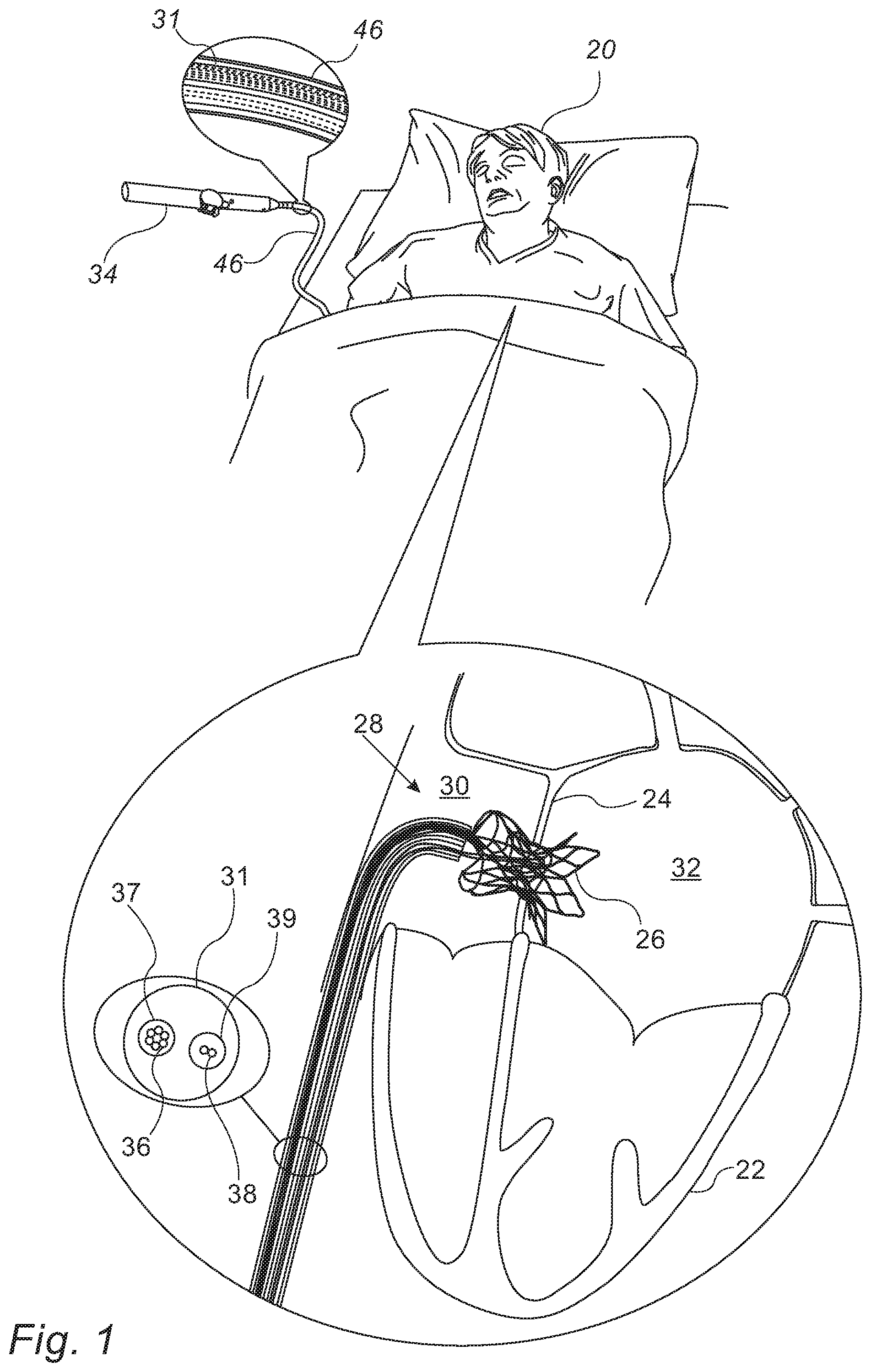
An implanted interatrial shunt may cause various complications, such as distortion of the interatrial septum, cardiac arrhythmias, inability to use the transseptal approach for future interventions, paradoxical embolism, and/or blood desaturation. Hence, for cases in which an interatrial shunt is required only temporarily (i.e., for a short period of time, such as less than one week), an implanted shunt may not necessarily be the most appropriate solution for treatment.
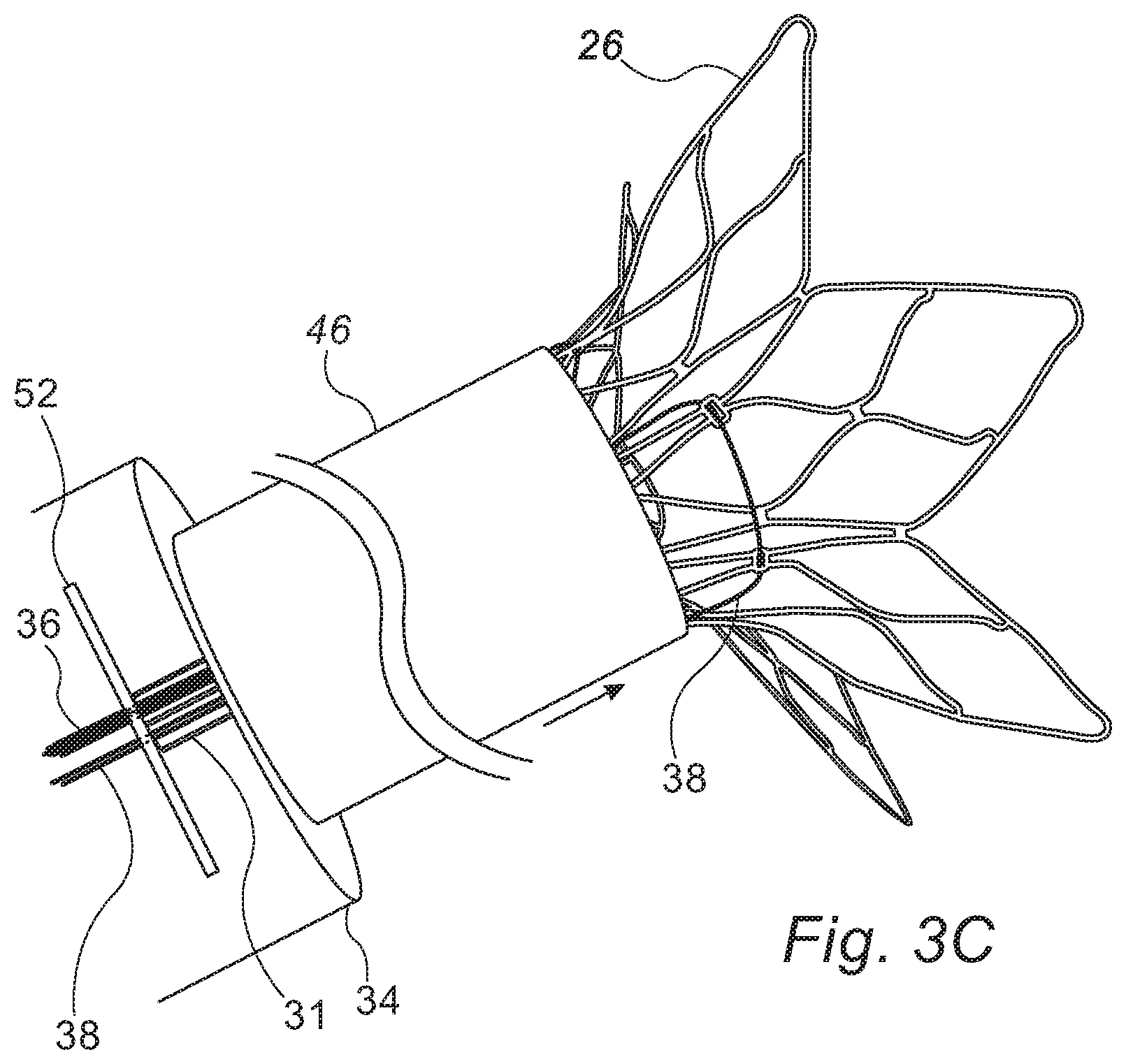
Embodiments of the present invention therefore provide--as an alternative to an implanted shunt--shunting apparatus that may be placed within the subject for only a short period of time, e.g., for less than one week, or even less than one day (e.g., 2-3 hours, 3-6 hours, or 6-12 hours). The apparatus comprises a shunt, along with one or more wires that extend from the proximal portion of the shunt to the exterior of the subject. These wires, which are typically controlled via a control handle, may be used to collapse the shunt, whenever the shunt is no longer needed. Following the collapse of the shunt, the shunt may be easily withdrawn from the subject.
In some embodiments, another wire, which passes circumferentially along the intermediate portion of the shunt and also extends to the exterior of the subject, may be used to adjust the diameter of the shunt while the shunt is inside the subject, thus regulating the flow of blood across the interatrial septum.
Other temporary shunts described herein include a shunt that is coupled to a distal end of a sheath. The shunt is advanced, in a collapsed state, over a guidewire, until the shunt spans the interatrial septum. Subsequently, the guidewire is retracted while the sheath is held in place, such that a stopper coupled to the distal portion of the guidewire applies a longitudinally compressive force to the shunt, thus causing the shunt to open. The sheath is then locked with respect to the guidewire, such that the shunt remains open. Upon the conclusion of treatment, the sheath is unlocked, such that the shunt collapses, and subsequently, the shunt is removed from the subject.
(In general, within the context of medical applications, the term "shunt" may refer to (i) a passage that diverts a bodily fluid from one portion of the body to another, or (ii) a device that is used to establish, and/or maintain, such a passage. In the context of the present application, including the claims, the term "shunt" typically refers to the latter.)
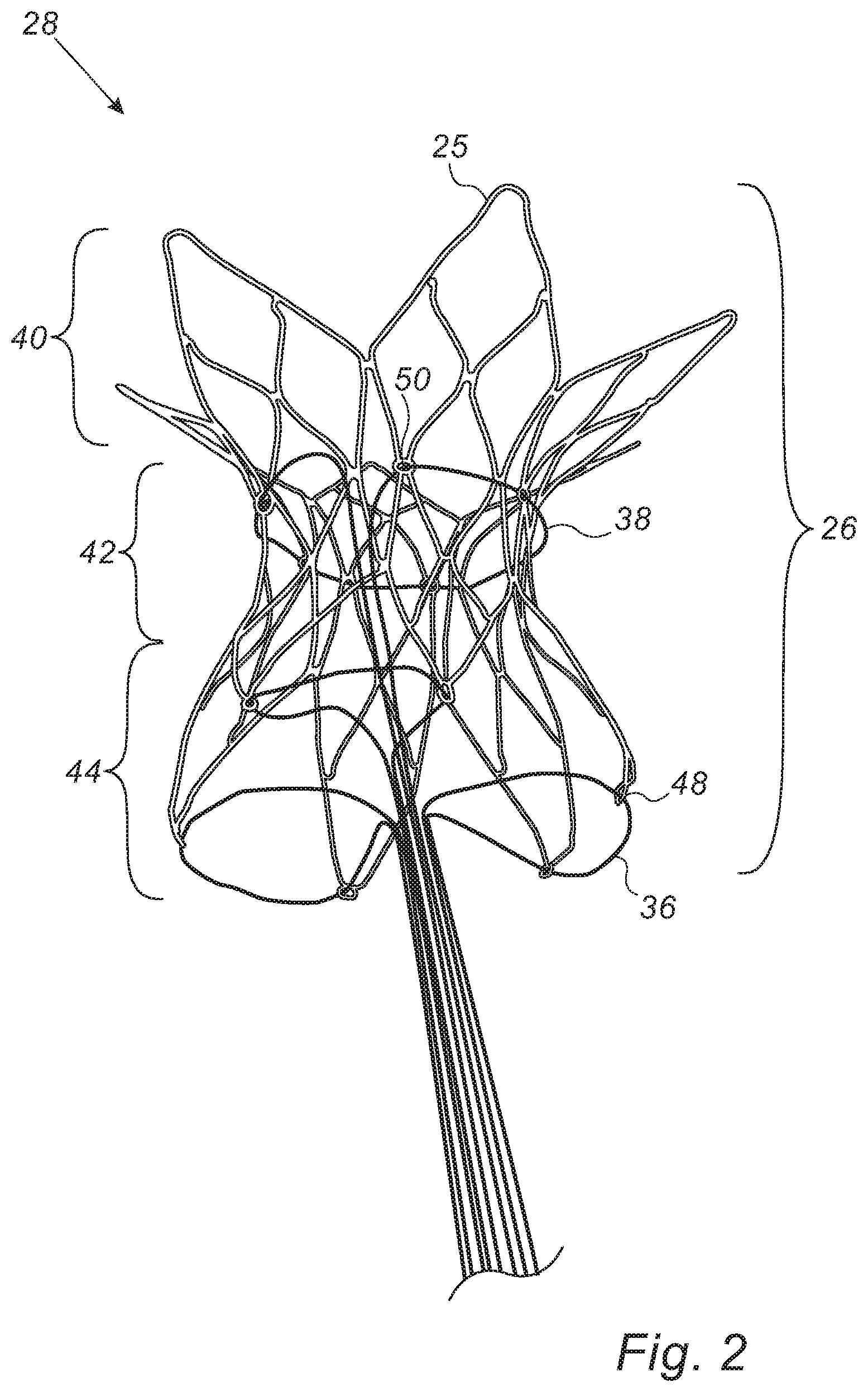
There is therefore provided, in accordance with some embodiments of the present invention, apparatus that includes a shunt. The shunt includes a flared distal portion, a flared proximal portion, and an intermediate portion, disposed between the distal portion and the proximal portion. The apparatus further includes a constricting flexible longitudinal element passing circumferentially along the intermediate portion of the shunt, configured to constrict the intermediate portion of the shunt, and one or more proximal-portion-collapsing flexible longitudinal elements configured to collapse the proximal portion of the shunt.
In some embodiments, the constricting flexible longitudinal element includes a wire.
In some embodiments, the intermediate portion of the shunt is shaped to define a plurality of orifices, and the wire passes circumferentially along the intermediate portion by passing through the orifices.
In some embodiments, the proximal portion of the shunt is shaped to define a plurality of orifices, and the proximal-portion-collapsing flexible longitudinal elements include one or more wires, each of which passes through at least two of the orifices.
There is further provided, in accordance with some embodiments of the present invention, a method that includes placing a shunt between two chambers of a heart of a subject, such that one or more shunt-collapsing flexible longitudinal elements extend from a proximal portion of the shunt to an exterior of the subject. The method further includes, subsequently, using the shunt-collapsing flexible longitudinal elements, collapsing the shunt into a catheter.
In some embodiments, the method further includes, using the catheter, removing the shunt from the subject.
In some embodiments, removing the shunt from the subject includes removing the shunt from the subject after less than one week from the placement of the shunt.
In some embodiments, the two chambers of the heart are two atria of the heart.
In some embodiments, the two chambers of the heart are two ventricles of the heart.
In some embodiments, the method further includes, while the shunt is between the two chambers of the heart, constricting the shunt, using a constricting flexible longitudinal element that extends from the shunt to the exterior of the subject.
There is further provided, in accordance with some embodiments of the present invention, a method that includes placing a shunt between two atria of a subject, and, after less than one week from the placement of the shunt, withdrawing the shunt from the subject.
There is further provided, in accordance with some embodiments of the present invention, apparatus that includes a sheath, and a shunt coupled to a distal end of the sheath, the shunt being configured to open from a collapsed state to an open state upon a longitudinally compressive force being applied to the shunt.
In some embodiments, the shunt includes a plurality of wires, distal ends of which are joined together, and proximal ends of which are coupled to the sheath.
In some embodiments, the shunt is configured to open by the wires expanding radially outward from each other.
In some embodiments, in the open state, a proximal portion of the shunt and a distal portion of the shunt are wider than an intermediate portion of the shunt that is between the proximal portion of the shunt and the distal portion of the shunt.
In some embodiments, the apparatus further includes:
a guidewire; and
a stopper coupled to a distal portion of the guidewire, the stopper being configured to apply the longitudinally compressive force to the shunt by pressing against a distal portion of the shunt when the sheath and the shunt are over the guidewire.
In some embodiments, the shunt is shaped to define a distal aperture configured to fittingly receive the stopper.
In some embodiments, the stopper includes a bead.
There is further provided, in accordance with some embodiments of the present invention, a method that includes passing a guidewire across a septum that separates between a first chamber of a heart of a subject and a second chamber of the heart, such that a stopper coupled to the guidewire is in the second chamber. The method further includes, subsequently, passing a shunt, in a collapsed state, over the guidewire, until a proximal portion of the shunt is in the first chamber, and a distal portion of the shunt is in the second chamber, and subsequently, using the stopper, opening the shunt from the collapsed state to an open state.
In some embodiments, opening the shunt includes opening the shunt by, using the stopper, pressing against the distal portion of the shunt.
In some embodiments, the shunt is shaped to define a distal aperture, and pressing against the distal portion of the shunt includes pressing against the distal portion of the shunt while the stopper is fittingly received by the distal aperture.
In some embodiments, the first chamber is a right atrium, and the second chamber is a left atrium.
In some embodiments, the first chamber is a right ventricle, and the second chamber is a left ventricle.
In some embodiments, the shunt is coupled to a distal end of a sheath, and the method further includes, subsequently to opening the shunt, maintaining the open state of the shunt by locking the sheath with respect to the guidewire.
In some embodiments, the method further includes: causing the shunt to collapse, by unlocking the sheath with respect to the guidewire; and subsequently, removing the shunt from the subject.
In some embodiments, removing the shunt from the subject includes removing the shunt from the subject after less than one week from the opening of the shunt.
The present invention will be more fully understood from the following detailed description of embodiments thereof, taken together with the drawings, in which:
BRIEF DESCRIPTION OF THE DRAWINGS
FIG. 1 is a schematic illustration of a temporary shunt apparatus inside a subject, in accordance with some embodiments of the present invention;
FIG. 2 is a schematic illustration of a temporary shunt apparatus, in accordance with some embodiments of the present invention;
FIG. 3A-C collectively show a technique for removing a shunt from a subject, in accordance with some embodiments of the present invention; and
FIGS. 4A-B are schematic illustrations showing the placement of a temporary shunt within an interatrial septum, in accordance with some embodiments of the present invention.
DETAILED DESCRIPTION OF EMBODIMENTS
Reference is initially made to FIG. 1, which is a schematic illustration of a temporary shunt apparatus 28 inside a subject 20, and to FIG. 2, which is a schematic illustration of temporary shunt apparatus 28, in accordance with some embodiments of the present invention.
Apparatus 28 comprises a shunt 26, which may be placed between two chambers of the heart 22 of subject 20, such as within the interatrial septum 24 of heart 22, between the right atrium 30 and the left atrium 32. Alternatively, the shunt may be placed between the two ventricles of the heart, or between any other two body cavities. Shunt 26 typically comprises a flared distal portion 40, a flared proximal portion 44, and an intermediate portion 42, which is disposed between distal portion 40 and proximal portion 44. Distal portion 40 and proximal portion 44 anchor the shunt to septum 24 (i.e., prevent migration of the shunt from within the septum), while intermediate portion 42 provides a passageway across the septum, through which blood may flow. Typically, shunt 26 comprises a shape-memory material, such as Nitinol, such that the shunt expands to its natural shape (the shape shown in FIGS. 1-2) upon being released from a delivery catheter, as further described below. (It is noted that, for clarity, shunt apparatus 28 is drawn disproportionately large, relative to heart 22, in FIG. 1.)
In the context of the present application, including the claims, the proximal and distal portions of the shunt are "flared," in that these portions extend radially outward from the intermediate portion of the stent. In some embodiments, as shown, each of the proximal and distal portions of the shunt comprises a plurality of leaves 25, such as, for example, six leaves 25, as shown. In other embodiments, the proximal portion and/or the distal portion does not comprise a plurality of leaves, but rather, is shaped to define a flared ring, or has some other suitable form.
To facilitate removal of the shunt, embodiments described herein provide one or more shunt-collapsing flexible longitudinal elements, which extend from proximal portion 44 to the exterior of the subject. For example, as shown in the figures, the shunt-collapsing flexible longitudinal elements may comprise wires 36. Typically, while inside the subject, wires 36 are contained within a lumen 37 of a sheath 31 passing between proximal portion 44 and the exterior of the subject. (For example, sheath 31 may exit the subject via a femoral vein of the subject.) As further described below with reference to FIG. 3A, as wires 36 are pulled, wires 36 exert an inward radial force on the proximal portion of the shunt, such that the proximal portion of the shunt is collapsed. Typically, as shown in FIG. 1, the proximal ends of wires 36 are coupled to control handle 34, via which wires 36 may be pulled (or alternatively, released, such as to allow the proximal portion of the shunt to expand).
Typically, wires 36 remain coupled to the shunt throughout the time that the shunt is in place inside the subject. Due to wires 36 remaining coupled to the shunt, the shunt may be easily removed immediately upon receiving indication that further shunting is no longer required. In this respect, embodiments described herein differ from other, hypothetical shunting applications in which (i) the shunt is removed only in the event of a complication, and (ii) to remove the shunt, it is necessary to use a lasso or other specialized shunt-removal instrument.
FIG. 2 shows a particular embodiment in which proximal portion 44 is shaped to define a plurality of orifices 48, and each of wires 36 passes through at least two of orifices 48. For example, as shown, the end of each leaf 25 may be shaped to define an orifice 48, and each wire may pass through the respective orifices of two adjacent leaves, such that the wire forms a loop that passes through the orifices. (Thus, as shown, a shunt having six proximal leaves is coupled to three wires 36, each wire separately controlling the collapse of a respective pair of adjacent leaves.) To collapse the proximal portion of the shunt, the two proximal ends of each of the wires are pulled, as described below with reference to FIG. 3A.
Alternatively to the embodiment shown, a single wire 36 may form a loop that passes through all of the orifices, this single wire controlling the collapse of the entire proximal portion. (In other words, by pulling on the two ends of this single wire, the entire proximal portion may be collapsed.) In yet other embodiments, wires 36 do not form loops; rather, a separate wire is coupled to each leaf. For example, each leaf may be coupled to the distal end of a respective wire. (Thus, for example, a shunt having six proximal leaves is coupled to six wires, one wire per leaf) Similarly, wires 36 may be formed as extensions of the leaves, such that each leaf has a wire extension that extends to the exterior of the subject. In such embodiments, the proximal portion of the shunt may be collapsed by pulling on the single proximal end of each of the wires.
In some cases, it may be beneficial to adjust the diameter of intermediate portion 42 while the shunt is inside the subject. Hence, apparatus 28 typically further comprises a constricting flexible longitudinal element that passes circumferentially along intermediate portion 42, and extends from the intermediate portion of the shunt to the exterior of the subject. For example, as shown, the constricting flexible longitudinal element may comprise a wire 38 that loops around the intermediate portion of the shunt, both ends of the wire passing through the vasculature of the subject (e.g., within lumen 37, or within a separate lumen 39 of sheath 31), exiting from the subject (e.g., via a femoral vein), and being coupled to control handle 34. The constricting flexible longitudinal element is configured to constrict the intermediate portion of the shunt, by exerting an inward radial force on the intermediate portion of the shunt. For example, to constrict the intermediate portion, and thus reduce the flow of blood across the septum, the control handle may be used to pull both ends of wire 38. (Subsequently, the control handle may be used to hold wire 38 in place, to prevent the intermediate portion from re-expanding.) Conversely, to widen the intermediate portion, the control handle may be used to release wire 38, thus allowing the intermediate portion to expand radially outward.
In some embodiments, as shown, the intermediate portion is shaped to define a plurality of orifices 50, and the wire passes circumferentially along the intermediate portion by passing through the orifices. Alternatively or additionally to comprising a wire, the constricting flexible longitudinal element may comprise a band, strap, ribbon, or any other suitable type of longitudinal element.
In some embodiments, the adjustment of the diameter of the shunt is based on pressure monitoring. For example, as further described below, pressure sensors disposed on the shunt may be used to acquire intra-atrial pressure measurements, and the diameter of the shunt may be adjusted in response to such measurements. Alternatively or additionally, the diameter of the shunt may be adjusted in response to hemodynamic monitoring, such as by the application of flow imaging techniques such as pulsed wave (PW) or continuous wave (CW) Doppler echocardiography.
Typically, to place the shunt within the septum, the shunt is first collapsed and placed inside a delivery catheter 46. Subsequently, catheter 46 is percutaneously inserted into the vasculature of the subject, such as via a femoral vein of the subject, and is then passed through the vasculature into right atrium 30, e.g., via the inferior vena cava. (Alternatively, catheter 46 may be passed into the right atrium via the jugular vein and superior vena cava.) Subsequently, the distal end of the catheter is passed through the septum and into left atrium 32, such that the distal and proximal portions of the shunt are on opposite sides of the septum. (As is known in the art, prior to passing the distal end of the catheter through the septum, a puncturing element may be used to create an opening in the septum, and, optionally, a dilator may be used to enlarge the opening, such that the distal end of the catheter may easily pass through the septum.) The catheter is then withdrawn from over the shunt while the shunt is held in place. As the catheter is withdrawn from over the shunt, the shunt reassumes its natural, non-collapsed shape, such that distal portion 40 opens on the left-atrium side of the septum, and proximal portion 44 opens on the right-atrium side of the septum.
Following the deployment of the shunt, the catheter is withdrawn from the subject, leaving behind the shunt, along with the shunt-collapsing flexible longitudinal elements and the constricting flexible longitudinal element. Alternatively, the catheter may remain within the subject while the shunt is in place. For example, the catheter may remain within the subject such that the distal end of the catheter is near the proximal portion of the shunt. The catheter may thus be used to deliver medication to the shunt site, and/or pressure sensors in the catheter may be used to monitor the intra-atrial pressure.
By way of example, FIG. 1 shows the catheter coupled to a control handle 34, such that control handle 34 may be used to advance and withdraw the catheter. (FIG. 1 shows a scenario in which the catheter has been withdrawn from within the subject, but remains coupled to the control handle, poised for reentry into the subject.)
Shunt 26 helps relieve excess intra-atrial pressure, by allowing blood to flow from the higher-pressure atrium to the lower-pressure atrium. Shunt 26 may thus be used to treat any relevant condition (e.g., pulmonary hypertension or congestive heart failure) for which the relief of excess pressure is beneficial, or, for example, to help prevent left ventricular dilation and remodeling following an acute myocardial insult. Typically, the subject remains hospitalized until the subject's physician decides that sufficient treatment has been provided, at which point the shunt is removed from the subject (as further described immediately below), and the subject is released from hospital. In some embodiments, shunt apparatus 28 comprises one or more pressure sensors, disposed, for example, on shunt 26, on any of the longitudinal elements, and/or in sheath 31. Such pressure sensors may be used to measure (e.g., continuously) the pressure in the subject's right atrium and/or left atrium, in order to monitor progression of the treatment, and ascertain the point in time at which the shunt may be removed from the subject. For example, one pressure sensor may be disposed on the proximal portion of the shunt, and another pressure sensor on the distal portion of the shunt, such that the pressure in both the left atrium and the right atrium is measured.
Reference is now made to FIGS. 3A-C, which collectively show a technique for removing shunt 26 from subject 20, in accordance with some embodiments of the present invention. It is noted that many of the details shown in FIGS. 3A-C are provided by way of example only, and that many variations of the illustrated technique are included within the scope of the present disclosure.
In FIG. 3A, catheter 46 is reinserted into the subject, and is then advanced until the distal end of the catheter is close to proximal portion 44 of the shunt. Subsequently, wires 36 are pulled, as indicated by the arrow 54 shown in the figure, such that an inward radial force is exerted on proximal portion 44. The inward radial force causes proximal portion 44 to collapse, as shown in FIG. 3B. Following the collapse of the proximal portion of the shunt, as shown in FIG. 3C, the catheter is advanced distally over the shunt. (In passing over the shunt, the catheter may at least partly pass through the interatrial septum.) As the catheter continues to pass over the shunt from the position shown in FIG. 3C, the catheter collapses the distal end of the shunt, such that the shunt becomes entirely collapsed within the catheter. Subsequently, the catheter, containing the shunt, may be removed from the subject.
In some embodiments, the catheter is advanced while proximal portion 44 is collapsing, such that, as proximal portion 44 continues to collapse, the catheter passes over the shunt, until the distal end of the catheter crosses through the septum and reaches the distal portion of the shunt. (In such embodiments, the state shown in FIG. 3B does not actually come to transpire, since catheter 46 covers the proximal portion of the shunt before the proximal portion of the shunt is fully collapsed.) Then, as the pulling of wires 36 continues while the catheter is held in place or is pushed forward, the distal end of the catheter exerts a force on the distal portion of the shunt, such that the distal portion of the shunt collapses, and the shunt is drawn into the catheter. Such embodiments have the advantage that, due to the catheter being advanced over the shunt while wires 36 are pulled, the shunt is less likely to be pulled into the right atrium.
FIGS. 3A-C show, by way of example, an embodiment in which sheath 31 extends to a stopper 52 contained inside of control handle 34, wires 36 passing through stopper 52. As the wires are pulled, stopper 52 prevents sheath 31 from moving proximally, such that most of the pulling force acts on proximal portion 44, rather than on sheath 31. (Although flexible, sheath 31 is resistant to buckling, such that the pulling force is effectively transferred to proximal portion 44.) A similar mechanism may be used for wire 38, which, as described above, controls the diameter of the intermediate portion of the shunt. (In the context of the present application, including the claims, the term "stopper" may refer to anything that stops something else. For example, stopper 52 is referred to as a "stopper," in that it stops the proximal movement of sheath 31.)
In some embodiments, two separate tubes run through a single lumen, or two separate lumens, of sheath 31, one of these tubes holding wires 36, and the other of these tubes holding wire 38. Such tubes--which may comprise, for example, HHS.RTM. ("Helical Hollow Strand") Tubes--may provide additional resistance to buckling, such that the pulling force exerted on the wires is effectively transmitted to the shunt. In such embodiments, stopper 52 may be used to prevent the wire-holding tubes from moving proximally as the wires are pulled.
It is noted that the apparatus and methods described above may also be used for applications in which an implanted shunt is required. In such applications, during the implantation procedure, wires 36 may be used to facilitate the retrieval of shunt 26, in the event that the shunt was not placed at the proper location. Subsequently, upon confirmation that the shunt is properly situated, wires 36 may be detached from shunt 26, and removed from the subject. Similarly, during the implantation procedure, the constricting flexible longitudinal element may be used to adjust the diameter of the shunt. Subsequently, upon completion of the adjustment, the constricting flexible longitudinal element may be locked in place, such as to maintain the diameter of the shunt, and then any proximally-extending portion of the constricting flexible longitudinal element may be detached and removed.
Reference is now made to FIGS. 4A-B, which are schematic illustrations showing the placement of a temporary shunt 56 within interatrial septum 24, in accordance with some embodiments of the present invention. Apparatus and methods described with reference to FIGS. 4A-B may be used for applications in which temporary shunting is desired, alternatively to apparatus and methods described with reference to earlier figures. Thus, for example, as described above for shunt 26, shunt 56 may be removed less than one week from placement of the shunt.
Typically, shunt 56 is an expandable structure made from a suitable shape-memory material, such as Nitinol, such that shunt 56 may move from a collapsed state (shown in FIG. 4A) to a preconfigured open state (shown in FIG. 4B) upon being subjected to an appropriate force, and may then recollapse upon removal of the force. In general, shunt 56 may have any suitable form. For example, as shown in the figures, shunt 56 may comprise a plurality of wires 68, which are joined together at their distal ends (e.g., by being connected to a common ring), and are coupled to a sheath 62 at their proximal ends. (Sheath 62 may be referred to as a "subselective sheath," in that the diameter of sheath 62 is small, relative to the "vascular sheath" referred to below.) In the collapsed state of the shunt, wires 68 are approximately parallel to each other along the majority of the length of the wires. Upon the distal and proximal ends of the wires being pushed together (i.e., upon the shunt being longitudinally compressed), the wires expand radially outward from each other, such that the shunt assumes the preconfigured open state. Typically, in the shunt's preconfigured open state, the proximal portion of the shunt and the distal portion of the shunt are wider than the intermediate portion of the shunt that is between the proximal portion of the shunt and the distal portion of the shunt. The wider proximal and distal portions of the shunt anchor the shunt to the septum (i.e., prevent migration of the shunt from within the septum).
As described above with respect to shunt 26, shunt 56 is percutaneously inserted into the subject. First, a guidewire 58 is inserted into the right atrium, e.g., via the femoral vein and the inferior vena cava, as shown. Subsequently, guidewire 58 is passed across septum 24 (from the right atrium to the left atrium), using conventional techniques known in the art, such that the distal portion of the guidewire is in the left atrium. (To introduce guidewire 58, a vascular sheath (not shown), which may alternatively be referred to as a catheter, may be used, as is known in the art. If necessary, a dilating tool may be passed over the guidewire, and through the opening created by the guidewire, to enlarge the opening in the septum.) Subsequently to passing the guidewire across the septum, shunt 56 is passed, in a collapsed state, over the guidewire, until the distal portion of the shunt is in the left atrium, and the proximal portion of the shunt is in the right atrium. In other words, as shown in FIG. 4A, shunt 56 is positioned over the guidewire such that the shunt spans the septum.
A stopper 60 is coupled to the distal portion of guidewire 58 (i.e., near the distal end of the guidewire, such as within one or two millimeters of the distal end). Subsequently to positioning the shunt such that the shunt spans the septum, the stopper is used to open the shunt from the shunt's initial collapsed state. In one embodiment of this technique, the guidewire is retracted (proximally pulled), as indicated by the arrow 70 in the figure, such that the stopper presses against the distal portion of the shunt. While the guidewire is retracted, sheath 62 (and hence, the proximal end of the shunt) is held in place or is advanced, such that, by pressing against the distal portion of the shunt, the stopper applies a longitudinally compressive force to the shunt. The compressive force causes the shunt to open, and hence, the opening in the septum to become enlarged, as shown in FIG. 4B. (In alternative embodiments, the stopper may press against the distal portion of the shunt by virtue of the sheath being advanced while the guidewire is held in place.)
Stopper 60 is referred to herein as a "stopper," in that stopper 60 stops the advancement of the shunt along the guidewire, by virtue of the stopper being large enough such that the shunt cannot pass over the stopper. Stopper 60 may have any form suitable for applying the longitudinally compressive force to the shunt. For example, the stopper may comprise a bead, as shown in the figure; alternatively, for example, stopper 60 may comprise a plate or rod which, when pressed against the shunt, applies the longitudinally compressive force. In some embodiments, the shunt is shaped to define a distal aperture 66, shaped to fittingly receive the stopper. (In such embodiments, the stopper may be referred to as a "lock," and distal aperture 66 may be referred to as a "fitting.") As the guidewire is withdrawn in the proximal direction, the stopper engages with, i.e., is fittingly received by, distal aperture 66. Subsequently, the stopper continues to press against the distal portion of the shunt while the stopper is fittingly received by the distal aperture. The engagement between the distal aperture and the stopper helps the pressing force to be more effectively applied to the shunt.
Typically, to maintain the open state of shunt 56, it is necessary to maintain the longitudinally compressive force on the shunt. This may be accomplished by, using a locking mechanism, locking sheath 62 with respect to (or "against") the guidewire, i.e., fixing the position of the sheath with respect to the guidewire. For example, FIG. 4B shows a lock 64 placed over the guidewire, proximally to the sheath. Lock 64 prevents the sheath from sliding backward along the guidewire, and the guidewire from sliding forward within the sheath, such that the longitudinally compressive force on the shunt is maintained. In some embodiments, the guidewire and sheath are coupled to control handle 34 (FIG. 1), and the control handle is used to lock the sheath.
Subsequently, upon completion of the treatment, the sheath is unlocked, e.g., by removing lock 64. The unlocking of the sheath allows the sheath to move proximally, and/or allows the guidewire to move distally, thus removing the longitudinally compressive force that had heretofore been applied to the shunt. The shunt therefore collapses. Subsequently, the sheath, and the shunt, are removed from the subject.
In some embodiments, prior to unlocking the sheath, a catheter is advanced over the sheath, until the distal end of the catheter is near the proximal end of the shunt. Subsequently, upon collapse of the shunt, the shunt is pulled into the catheter, and/or the catheter is advanced over the shunt. The catheter is then used to remove the shunt from the subject. In other embodiments, no catheter is used for removal of the shunt. Rather, upon collapse of the shunt, the sheath and shunt are proximally pulled, through the vasculature of the subject, out of the subject.
In some embodiments, one or more pressure sensors are disposed on guidewire 58, shunt 56, and/or sheath 62. Such pressure sensors may be used to measure the pressure in the subject's right atrium and/or left atrium, as described above for apparatus 28.
Although the description above relates mainly to interatrial shunting, it is noted that shunt 56, and the above-described techniques for deployment thereof, may be used for shunting between any relevant two body cavities. For example, in an interventricular shunting application, guidewire 58 may be passed through the interventricular septum, from the right ventricle to the left ventricle, and the shunt may then be deployed over the guidewire and opened within the interventricular septum.
It will be appreciated by persons skilled in the art that the present invention is not limited to what has been particularly shown and described hereinabove. Rather, the scope of the present invention includes both combinations and subcombinations of the various features described hereinabove, as well as variations and modifications thereof that are not in the prior art, which would occur to persons skilled in the art upon reading the foregoing description.
* * * * *
References
D00000
D00001
D00002
D00003
D00004
D00005
D00006
XML
uspto.report is an independent third-party trademark research tool that is not affiliated, endorsed, or sponsored by the United States Patent and Trademark Office (USPTO) or any other governmental organization. The information provided by uspto.report is based on publicly available data at the time of writing and is intended for informational purposes only.
While we strive to provide accurate and up-to-date information, we do not guarantee the accuracy, completeness, reliability, or suitability of the information displayed on this site. The use of this site is at your own risk. Any reliance you place on such information is therefore strictly at your own risk.
All official trademark data, including owner information, should be verified by visiting the official USPTO website at www.uspto.gov. This site is not intended to replace professional legal advice and should not be used as a substitute for consulting with a legal professional who is knowledgeable about trademark law.