Modified guide RNAs, CRISPR-ribonucleotprotein complexes and methods of use
Carlson-Stevermer , et al. February 2, 2
U.S. patent number 10,907,150 [Application Number 16/008,376] was granted by the patent office on 2021-02-02 for modified guide rnas, crispr-ribonucleotprotein complexes and methods of use. This patent grant is currently assigned to WISCONSIN ALUMNI RESEARCH FOUNDATION. The grantee listed for this patent is WISCONSIN ALUMNI RESEARCH FOUNDATION. Invention is credited to Amr Ashraf Abdeen, Jared Matthew Carlson-Stevermer, Lucille Katherine Kohlenberg, Krishanu Saha.


View All Diagrams
| United States Patent | 10,907,150 |
| Carlson-Stevermer , et al. | February 2, 2021 |
Modified guide RNAs, CRISPR-ribonucleotprotein complexes and methods of use
Abstract
Described herein are modified guide RNAs such as a single guide RNA including, from 5' to 3', a single-stranded protospacer sequence, a first complementary strand of a binding region for the Cas9 polypeptide, an aptamer that binds a biotin-binding molecule, and a second complementary strand of the binding region for the Cas9 polypeptide. Also described is an RNP complex including the modified guide RNA and a Cas9 polypeptide or active fragment thereof. Also included are methods of modifying target genes in cells using the modified guide RNAs.
| Inventors: | Carlson-Stevermer; Jared Matthew (Madison, WI), Saha; Krishanu (Middleton, WI), Abdeen; Amr Ashraf (Madison, WI), Kohlenberg; Lucille Katherine (Madison, WI) | ||||||||||
|---|---|---|---|---|---|---|---|---|---|---|---|
| Applicant: |
|
||||||||||
| Assignee: | WISCONSIN ALUMNI RESEARCH
FOUNDATION (Madison, WI) |
||||||||||
| Family ID: | 1000005335027 | ||||||||||
| Appl. No.: | 16/008,376 | ||||||||||
| Filed: | June 14, 2018 |
Prior Publication Data
| Document Identifier | Publication Date | |
|---|---|---|
| US 20180362971 A1 | Dec 20, 2018 | |
Related U.S. Patent Documents
| Application Number | Filing Date | Patent Number | Issue Date | ||
|---|---|---|---|---|---|
| 62519317 | Jun 14, 2017 | ||||
| Current U.S. Class: | 1/1 |
| Current CPC Class: | C12N 15/102 (20130101); C12N 9/22 (20130101); C12N 15/11 (20130101); C12N 15/907 (20130101); C12N 15/111 (20130101); C12N 2800/80 (20130101); C12N 2310/3519 (20130101); C12N 2310/16 (20130101); C12N 2310/20 (20170501); C12N 2310/20 (20170501); C12N 2310/16 (20130101); C12N 2310/3519 (20130101) |
| Current International Class: | C12N 15/11 (20060101); C12N 15/90 (20060101); C12N 9/22 (20060101); C12N 15/10 (20060101) |
References Cited [Referenced By]
U.S. Patent Documents
| 8889418 | November 2014 | Zhang et al. |
| 9856497 | January 2018 | Qi et al. |
| 9868962 | January 2018 | May et al. |
| 10377998 | August 2019 | Zhang et al. |
| 10450584 | October 2019 | Barrangou et al. |
| 2015/0283265 | October 2015 | Peyman |
| 2017/0081650 | March 2017 | Joung et al. |
| 2017/0152508 | July 2017 | Joung et al. |
| 2020/0140858 | May 2020 | Saha et al. |
| 2016183402 | Nov 2016 | WO | |||
Other References
|
Darmostuk et al. Current approaches in SELEX: An update to aptamer selection technology. Biotechnology Advances, vol. 33, pp. 1141-1161, 2015. (Year: 2015). cited by examiner . Zetsche et al. Nature Biotechnology, vol. 33, No. 2, pp. 139-142, Feb. 2015, including pp. 1-9 of Supplementary Information. (Year: 2015). cited by examiner . pp. 1-37 of Supplementary Information of Shechner et al. Nature Methods, vol. 12, No. 7, pp. 664-670, and pp. 1-5 of Online Methods, 2015, which is cited as reference 31 on the IDS filed Aug. 21, 2018. (Year: 2015). cited by examiner . Walker et al. Chapter 26. Jeffrey E. Gerst (ed.), RNA Detection and Visualization: Methods and Protocols, Methods in Molecular Biology, vol. 714, Springer Science+Business Media, LLC, 2011, pp. 423-444. (Year: 2011). cited by examiner . Carlson-Stevermer et al.; "Assembly of CRISPR Ribonucleoproteins With Biotinylated Oligonucleotides via an RNA Aptamer for Precise Gene Editing"; Nature Communications; 8(1); 13 pages; (2017). cited by applicant . International Search Report and Written Opinion; International Application No. PCT/US2018/037531; International filing date Jun. 14, 2018; dated Sep. 6, 2018; 18 pages. cited by applicant . Lee et al.; "Synthetically Modified Guide RNA and Donor DNA are a Versatile Platform for CRISPR-Cas9 Engineering"; ELIFE; 6; 17 pages; (2017). cited by applicant . Ming et al.; "Efficient Generation of Mice Carrying Homozygous Double-floxp Alleles Using the Cas9-Avidin/Biotin-Donor DNA System"; Cell Research; 27; pp. 578-581; (2017). cited by applicant . Bao et al.; "Multifunctional Nanoparticles for Drug Delivery and Molecular Imaging"; Annu. Rev. Biomed. Eng.; 15; pp. 253-282; (2013). cited by applicant . Brinkman et al.; "Easy Quantitative Assessment of Genome Editing by Sequence Trace Decomposition"; Nucleic Acids Research; 8 pages, (2014), downloaded from https://academic.oup.com/nar/article-abstract/42/22/e168/2411890, by University of Wisconsin--Madison on Jul. 5, 2018. cited by applicant . Carlson-Stevermer et al.; "High-Content Analysis of CRISPR-Cas9 Gene-Edited Human Embryonic Stem Stem Cells"; Stem Cell Reports; 6; pp. 109-120; (2016). cited by applicant . Chu et al.; "Increasing the Efficiency of Homology-Directed Repair for CRISPR-Cas9-Induced Precise Gene Editing in Mammalian Cells"; Nature Biotechnology; 33(5); pp. 543-550; (2015). cited by applicant . Davis et al.; "Small Molecule-Triggered Cas9 Protein With Improved Genome-Editing Specificity"; Nature Chemical Biology; vol. 11, pp. 316-318; (2015). cited by applicant . De Ravin et al.; "CRISPR-Cas9 Gene Repair of Hematopoietic Stem Cells From Patients with X-Linked Chronic Granulomatous Disease"; Sci. Transl. Med.; 9; eaah3480; 10 pages; (2017). cited by applicant . Dever et al.; "CRISPR/Cas9 Beta-globin Gene Targeting in Human Haematopoietic Stem Cells"; Nature; vol. 539; 384, 19 pages; (2016). cited by applicant . DeWitt et al.; "Selection-Free Genome Editing of the Sickle Mutation in Human Adult Hematopoietic Stem/Progenitor Cells"; Sci. Transl. Med.; 8; 360ra134; 9 pages; (2016). cited by applicant . Duda et al.; "High-Efficiency Genome Editing via 2A-Coupled Co-Expression of Fluorescent Proteins and Zinc Finger Nucleases or CRISPR/Cas9 Nickase Pairs"; Nucleic Acids Research; 42(10);; e84; 16 pages; (2014); downloaded from https://academic.oup.com/nar/article-abstract/42/10/e84/2434652 by University of Wisconsin--Madison Libraries on Jul. 5, 2018. cited by applicant . Eyquem et al.; "Targeting a Car to the Trac Locus With CRISPR/Cas9 Enhances Tumour Rejection"; Nature; vol. 543; p. 113; 19 pages.; (2017). cited by applicant . Gaj et al.; "Targeted Gene Knock-In by Homology-Directed Genome Editing Using Cas9 Ribonucleoprotein and AAV Donor Delivery"; Nucleic Acids Research; 45(11); e98; 11 pages, Downloaded from https://academic.oup.com/nar/article-abstract/45/11/398/3059660 by University of Wisconsin--Madison on Jul. 5, 2018. cited by applicant . Hemphill et al.; "Optical Control of CRISPR/Cas9 Gene Editing"; J. Am. Chem. Soc.; 137; pp. 5642-5645; (2015). cited by applicant . Kleinstiver et al.; "High-Fidelity CRISPR-Cas9 Nucleases With No Detectable Genome-Wide Off-Target Effects"; Nature; 529; pp. 490; 17 pages; (2016). cited by applicant . Konermann et al.; "Genome-Scale Transcriptional Activation by an Engineered CRISPR-Cas9 Complex"; Nature; vol. 517; p. 583-585; (2015). cited by applicant . Landrum et al.; "ClinVar: Public Archive of Interpretations of Clinically Relevant Variants"; D862-D868 Nucleic Acids Research; vol. 44, pp. D862-D868; Database issue; (2016). cited by applicant . Le Trong et al.; "Streptavidin and Its Biotin Complex at Atomic Resolution"; Acta Cryst.; D67; pp. 813-821; (2011). cited by applicant . Leppek et al.; "An Optimized Streptavidin-Binding RNA Aptamer for Purification of Ribonucleoprotein Complexes Identifies Novel ARE-Binding Proteins"; Nucleic Acids Research; vol. 42(2); e13; 15 pages; (2014), downloaded from https://academic.oup.com/nar/article-abstract/42/2/e13/1030103 by University of Wisconsin--Madison Libraries on Jul. 5, 2018. cited by applicant . Li et al.; "Optimization of Genome Engineering Approaches With the CRISPR/Cas9 System"; PLoS One; 9(8): doi:10.1371/journal.pone.0105779; (2014) e105779. cited by applicant . Liang et al.; "Enhanced CRISPR/Cas9-Mediated Precise Genome Editing by Improved Design and Delivery of gRNA, Cas9 Nuclease, and Donor DNA"; Journal of Biotechnology; 241; pp. 136-146; (2017). cited by applicant . Lin et al.; "Enhanced Homology-Directed Human Genome Engineering by Controlled Timing of CRISPR/Cas9 Delivery"; Weigel D, ed. eLife.; 32 pages; (2014) ;3:e04766. doi:10.7554/eLife.04766. cited by applicant . Lonowski et al.; "Genome Editing Using FACS Enrichment of Nuclease-Expressing Cells and Indel Detection by Amplicon Analysis"; Nature Protocols; 12(3); pp. 581-603; (2017). cited by applicant . Ma et al.; "Efficient Generation of Mice Carrying Homozygous Double-Floxp Alleles Using the Cas9-Avidin/Biotin Donor DNA System"; Cell Research; 27; pp. 578-581; (2017). cited by applicant . Maruyama et al.; "Increasing the Efficiency of Precise Genome Editing With CRISPR-Cas9 by Inhibition of Nonhomologous End Joinging" Nature Biotechnology; 33(5); p. 538; 9 pages; (2015). cited by applicant . Merkle et al.; "Efficient CRISPR-Cas9-Mediated Generation of Knockin Human Pluripotent Stem Cells Lacking Undesired Mutations at the Targeted Locus": Cell Reports; 11; pp. 875-883; (2015). cited by applicant . Nishimasu et al.; "Crystal Structure of Cas9 in Complex with Guide RNA and Target DNA"; Cell; 156; pp. 935-949; (2014). cited by applicant . Paquet et al.; "Efficient Introduction of Specific Homozygous and Heterozygous Mutations Using CRISPR/Cas9"; Nature; 533; p. 125; 18 pages; (2016). cited by applicant . Pattanayak et al.; "High-Throughput Profiling of Off-Target DNA Cleavage Reveals RNA-Programmed Cas9 Nuclease Specificity"; Nature Biotechnology; 31(9); pp. 839-845; (2013). cited by applicant . Ran et. al.; "Genome Engineering Using the CRISPR-Cas9 System"; Nature Protocols; 8(11); pp. 2281-2308; (2013). cited by applicant . Richardson et al.; "Enhancing Homology-Directed Genome Editing by Catalytically Active and Inactive CRISPR-Cas9 Using Asymmetric Donor DNA"; Nature Biotechnology; 34(3); pp. 339-345; (2016). cited by applicant . Sevier et al.; "Formation and Transfer of Disulphide Bonds in Living Cells"; Nature Reviews; Molecular Cell Biology; 3; pp. 836-847; (2002). cited by applicant . Shechner, et al.; "Multiplexable, Locus-Specific Targeting of Long RNAs With CRISPR-Display"; Nature Methods; 12(7); p. 664; 12 pages (2015). cited by applicant . Song et al; "RS-1 Enhances CRISPR/Cas9- and TALEN-Mediated Knock-In Efficiency"; Nature Communications 7 pages; Article No. 10548; (2016). cited by applicant . Steyer et al.; "Scarless Genome Editing of Human Pluripotent Stem Cells via Transient Puromycin Selection"; Stem Cell Reports; 10; pp. 642-654; (2018). cited by applicant . Yang et al.; "Optimization of Scarless Human Stem Cell Genome Editing"; Nucleic Acids Research; 41(19); pp. 9049-9061; (2013). cited by applicant . Zuris et al.; "Cationic Lipid-Mediated Delivery of Proteins Enables Efficient Protein-Based Genome Editing In Vitro and In Vivo"; Nature Biotechnology; 33(1); pp. 73-80; (2015). cited by applicant . Ellington et al.; "In Vitro Selection of RNA Molecules That Bind Specific Ligands"; Nature; 346; pp. 818-822; (1990). cited by applicant . Ruigrok et al., "Kinetic and Stoichiometric Characterisation of Streptavidin-Binding Apramers"; ChemBioChem; 13; pp. 829-836; (2012). cited by applicant . Srisawat et al.; "SteptavidinAptamers: Affinity Tags for the Study of RNAs and Ribonucleoproteins"; RNA; 7; pp. 632-641; (2001). cited by applicant . Wang et al.; "In Vitro Selection of High-affinity DNA Aptamers for Streptavidin"; Acta Biochim Biophys Sin; 41(4); pp. 335-340; (2009). cited by applicant . Chen et al.; "Enhanced proofreading governs CRISPR-Cas9 targeting accuracy"; Nature, vol. 550, Issue No. 7676; 2017; doi:10.1038/nature24268; pp. 407-422. cited by applicant . Chew et al.; "A multifunctional AAV-CRISPR-Cas9 and its host response"; Nature Methods, vol. 13, Issue No. 10; 2016; doi:10.1038/nmeth.3993; pp. 868-879. cited by applicant . Hasegawa et al.; "Methods for Improving Aptamer Binding Affinity"; Molecules, vol. 21, Issue No. 4; 2016; doi:10.3390/molecules21040421; pp. 421-435. cited by applicant . Hernandez et al.; "Aptamers as a model for functional evaluation of LNA and 20-amino LNA"; Bioorganic & Medicinal Chemistry Letters, vol. 19, Issue 23; 2007; doi:10.1016/j.bmcl.2009.10.039; pp. 6585-6587. cited by applicant . Hernandez et al.; "Label free optical sensor for Avidin based on single gold nanoparticles functionalized with aptamers"; Journal of Biophotonics, vol. 2, Issue No. 4; 2009; DOI 10.1002/jbio.200910006; pp. 227-231. cited by applicant . Lorenz et al.; "Genomic systematic evolution of ligands by exponential enrichment (Genomic SELEX) for the identification of protein-binding RNAs independent of their expression levels"; Nature Protocols, vol. 1, Issue No. 5; 2006; doi:10.1038/nprot.2006.372; pp. 2204-2212. cited by applicant . Ma et al.; "Rational Design of Mini-Cas9 for Transcriptional Activation"; ACS Synthetic Biology, vol. 7, Issue No. 4; 2018; DOI: 10.1021/acssynbio.7b00404; pp. 978-985. cited by applicant . Stoltenburg et al.; "FluMag-SELEX as an advantageous method for DNA aptamer selection"; Analytical and Bioanalytical Chemistry, vol. 383, Issue No. 1; 2005; DOI 10.1007/s00216-005-3388-9; pp. 83-91. cited by applicant . Stoltenburg et al.; "SELEX--a (r) evolutionary method to generate high-affinity nucleic acid ligands"; Biomolecular Engineering, vol. 24, Issue No. 4; 2007; doi:10.1016/j.bioeng.2007.06.001; pp. 381-403. cited by applicant. |
Primary Examiner: Dunston; Jennifer
Attorney, Agent or Firm: Cantor Colburn LLP
Government Interests
FEDERAL FUNDING STATEMENT
This invention was made with government support under GM119644 awarded by the National Institutes of Health and CBET1350178 awarded by the National Science Foundation. The government has certain rights in the invention.
Parent Case Text
CROSS-REFERENCE TO RELATED APPLICATIONS
This application claims priority to U.S. Provisional Application 62/519,317 filed on Jun. 14, 2017, which is incorporated herein by reference in its entirety.
Claims
The invention claimed is:
1. A ribonucleoprotein (RNP) complex, comprising a modified guide RNA comprising, a crRNA comprising a single-stranded protospacer sequence and a first complementary strand of a binding region for a Cas9 polypeptide, a tracrRNA comprising a second complementary strand of the binding region for the Cas9 polypeptide, wherein the crRNA or the tracrRNA comprises a nucleic acid aptamer that binds an avidin, wherein the crRNA and the tracrRNA hybridize through the first and second complementary strands of the binding region for the Cas9 polypeptide; an avidin the Cas9 polypeptide, wherein the Cas9 polypeptide is active for guide RNA binding, and has an active, inactive or partially inactive nuclease domain, and a biotinylated molecule.
2. The RNP complex of claim 1, wherein the avidin has one, two or four biotin binding sites, and wherein the avidin optionally comprises a fluorescent label.
3. The RNP complex of claim 1, wherein the biotinylated molecule is a biotinylated donor polynucleotide.
4. The RNP complex of claim 3, wherein the donor polynucleotide comprises single-stranded DNA, double-stranded DNA, RNA, or a duplex of RNA and DNA.
5. The RNP complex of claim 3, wherein the donor polynucleotide includes a mutation, deletion, alteration, integration, gene correction, gene replacement, transgene insertion, nucleotide deletion, gene disruption, and/or gene mutation in a target nucleic acid.
6. The RNP complex of claim 1, wherein the biotinylated molecule comprises a biotinylated nanoparticle, dye, contrast agent, or peptide.
7. The RNP complex of claim 6, wherein the nanoparticle is a quantum dot, a gold particle, a magnetic particle, or a polymeric nanoparticle.
8. The RNP complex of claim 1, wherein the avidin is covalently linked to a donor polynucleotide, either directly or via a linker molecule.
9. The RNP complex of claim 8, wherein the donor polynucleotide comprises single-stranded DNA, double-stranded DNA, RNA, or a duplex of RNA and DNA.
10. The RNP complex of claim 8, wherein the donor polynucleotide includes a mutation, deletion, alteration, integration, gene correction, gene replacement, transgene insertion, nucleotide deletion, gene disruption, and/or gene mutation in a target nucleic acid.
11. The RNP complex of claim 1, wherein the avidin is covalently linked to a nanoparticle, dye molecule, or a peptide, either directly or via a linker molecule.
12. A kit comprising a modified guide RNA, the modified guide RNA comprising, a crRNA comprising, a single-stranded protospacer sequence and a first complementary strand of a binding region for a Cas9 polypeptide, and a tracrRNA comprising, a second complementary strand of the binding region for the Cas9 polypeptide, wherein the crRNA or the tracrRNA comprises a nucleic acid aptamer that binds an avidin, wherein the crRNA and the tracrRNA hybridize through the first and second complementary strands of the binding region for the Cas9 polypeptide; an avidin; a Cas9 polypeptide, and a biotinylated molecule.
13. A method of modifying a target nucleic acid in a cell, comprising delivering to the cell an RNP complex, the RNP complex comprising a modified guide RNA comprising, a crRNA comprising a single-stranded protospacer sequence and a first complementary strand of a binding region for a Cas9 polypeptide, a tracrRNA comprising a second complementary strand of the binding region for the Cas9 polypeptide, wherein the crRNA or the tracrRNA comprises a nucleic acid aptamer that binds an avidin, wherein the crRNA and the tracrRNA hybridize through the first and second complementary strands of the binding region for the Cas9 polypeptide; an avidin; and the Cas9 polypeptide, wherein the Cas9 polypeptide is active for guide RNA binding, and has an active, inactive or partially inactive nuclease domain, wherein the single-stranded protospacer sequence of the modified guide RNA hybridizes to a sequence in the target nucleic acid to be modified.
14. The method of claim 13, wherein modifying the target nucleic acid increases or decreases expression of a gene product of the target nucleic acid.
15. The method of claim 13, further comprising delivering a donor polynucleotide to the cell, and wherein modifying the target nucleic acid comprises homology-directed repair (HDR).
16. The method of claim 13, further comprising delivering a donor polynucleotide to the cell, and wherein modifying the target nucleic acid comprises addition of a genetically encoded functionality, or correction of a mutation in the target nucleic acid.
17. The method of claim 13, wherein modifying the target nucleic acid creates a double strand break (DSB) which is repaired by a non-homologous end joining (NHEJ) cell repair mechanism generating indels thereby modifying the polynucleotide sequence of the target nucleic acid.
18. The method of claim 13, further comprising delivering a donor polynucleotide to the cell, and wherein modifying the target nucleic acid creates a DSB which is repaired by a HDR cell repair mechanism incorporating a donor DNA sequence thereby modifying the polynucleotide sequence of the target nucleic acid.
19. The method of claim 13, further comprising delivering a biotinylated molecule, wherein the biotinylated molecule targets the RNP complex to a specific cell type, organ or tissue.
20. The method of claim 13, wherein the RNP complex further comprises a biotinylated molecule.
21. The method of claim 20, wherein the biotinylated molecule is a biotinylated donor polynucleotide.
22. The method of claim 21, wherein the donor polynucleotide comprises single-stranded DNA, double-stranded DNA, RNA, or a duplex of RNA and DNA.
23. The method of claim 21, wherein the donor polynucleotide includes a mutation, deletion, alteration, integration, gene correction, gene replacement, transgene insertion, nucleotide deletion, and/or gene disruption.
24. A method of modifying a target nucleic acid in a cell, comprising delivering to the cell two RNP complexes, wherein each RNP complex comprises a modified guide RNA comprising, a crRNA comprising a single-stranded protospacer sequence and a first complementary strand of a binding region for a Cas9 polypeptide, a tracrRNA comprising a second complementary strand of the binding region for the Cas9 polypeptide, wherein the crRNA or the tracrRNA comprises a nucleic acid aptamer that binds an avidin, wherein the crRNA and the tracrRNA hybridize through the first and second complementary strands of the binding region for the Cas9 polypeptide; an avidin; and a Cas9 polypeptide, wherein the Cas9 polypeptide is active for guide RNA binding, and has an active, inactive or partially inactive nuclease domain, wherein each of the RNP complexes hybridizes to a different sequence in the target nucleic acid.
25. The method of claim 24, further comprising delivering a donor polynucleotide to the cell, wherein the donor polynucleotide comprises a gene correction relative to the sequence of the target nucleic acid, thereby providing multiple allelic correction of the target nucleic acid, or excision of target DNA from the target nucleic acid.
26. The method of claim 24, further comprising delivering a donor polynucleotide to the cell, wherein the donor polynucleotide comprises a gene correction relative to the sequence of the target nucleic acid, thereby providing multiple allelic correction of the target nucleic acid.
27. The method of claim 24, wherein modifying the target nucleic acid provides excision of genomic DNA.
28. The method of claim 24, wherein each RNP complex further comprises a biotinylated molecule.
29. The method of claim 28, wherein the biotinylated molecules are biotinylated donor polynucleotides.
30. The method of claim 29, wherein the donor polynucleotides comprise single-stranded DNA, double-stranded DNA, RNA, or a duplex of RNA and DNA.
31. The method of claim 29, wherein the donor polynucleotides include a mutation, deletion, alteration, integration, gene correction, gene replacement, transgene insertion, nucleotide deletion, and/or gene disruption.
32. A method of modifying a target nucleic acid in a cell, the cell comprising a Cas9 polypeptide, wherein the Cas9 polypeptide is active for guide RNA binding, and has an active, inactive or partially inactive nuclease domain, the method comprising delivering to the cell a modified guide RNA, the modified guide RNA comprising, a crRNA comprising, a single-stranded protospacer sequence and a first complementary strand of a binding region for the Cas9 polypeptide, and a tracrRNA comprising, a second complementary strand of the binding region for a Cas9 polypeptide, wherein the crRNA or the tracrRNA comprises a nucleic acid aptamer that binds an avidin, wherein the crRNA and the tracrRNA hybridize through the first and second complementary strands of the binding region for the Cas9 polypeptide; wherein the modified guide RNA is associated with an avidin; and wherein the single-stranded protospacer sequence of the modified guide RNA hybridizes to a sequence in the target nucleic acid to be modified.
33. The method of claim 32, wherein two modified guide RNAs are delivered to the cell, and wherein each of the modified guide RNAs hybridizes to a different nucleic acid sequence.
34. The method of claim 32, further comprising delivering a donor polynucleotide to the cell, wherein the donor polynucleotide comprises a gene correction relative to the sequence of the target nucleic acid, thereby providing multiple allelic correction of the target nucleic acid, or excision of target DNA from the target nucleic acid.
35. A method of modifying a target nucleic acid in a cell, comprising delivering to the cell a vector expressing a modified guide RNA, a vector expressing a Cas9 polypeptide, an avidin, and a biotinylated donor DNA template, the modified guide RNA comprising, a crRNA comprising, a single-stranded protospacer sequence and a first complementary strand of a binding region for the Cas9 polypeptide, and a tracrRNA comprising, a second complementary strand of the binding region for the Cas9 polypeptide, wherein the crRNA or the tracrRNA comprises a nucleic acid aptamer that binds the avidin, wherein the crRNA and the tracrRNA hybridize through the first and second complementary strands of the binding region for the Cas9 polypeptide, wherein the single-stranded protospacer sequence of the modified guide RNA hybridizes to a sequence in the target nucleic acid to be modified.
36. The method of claim 35, wherein the cell is a human cell.
37. The method of claim 36, wherein the human cell is a human pluripotent stem cell line, or a primary blood cell.
Description
FIELD OF THE DISCLOSURE
The present disclosure is related to modified guide RNAs and CRISPR-ribonucleoprotein complexes containing the modified guide RNAs and their use in genome editing methods.
BACKGROUND
Precise editing of DNA sequences in the human genome can be used to correct mutations or introduce novel genetic functionality for many biomedical purposes. Specifically, nonviral delivery of pre-formed CRISPR ribonucleoproteins (RNPs) is currently being developed for somatic gene editing applications. RNPs combining Streptococcus pyogenes Cas9 nuclease (Sp.Cas9, a high-affinity nuclease isolated from a type II CRISPR-associated system) and a single-guide RNA (sgRNA), for example, generate on-target DNA double strand breaks (DSBs) with little to no off-target DNA cleavage. This break can be repaired through error prone non-homologous end joining (NHEJ) or precise homology directed repair (HDR), in which a template is used. Co-delivery of a nucleic acid donor template with the Sp.Cas9 RNP (Sp.Cas9+sgRNA) is capable of producing precise edits at target loci through HDR of the DSB. However, variable delivery of the CRISPR system along with the donor templates generates a spectrum of edits, where a majority of cells include imprecise insertions and deletions (indels) of DNA bases from NHEJ repair of the DSB. Even when precise HDR of the DSB occurs on one allele, there is a chance that both alleles in diploid cells are not identically edited, resulting in imprecise edits on the other allele. Faithful writing of DNA, or scarless gene editing, within human cells remains an outstanding challenge.
Strategies to promote precise editing include addition of small molecules to block NHEJ and restricting Sp.Cas9 activity to particular phases of the cell cycle, but variability and toxicity has been observed across human cell lines when applying small molecules to promote HDR. Also, selection strategies through viral integration and excision of drug or cell-surface selection cassettes, flow cytometry for co-expressed fluorescent protein, or through transient drug selection can assist in the isolation of cells with one or two precisely-edited alleles. For all of these strategies, imprecise editing through NHEJ typically outnumbers precise HDR outcomes. None of the current strategies precisely control the delivery of the RNP with the donor template, and many resort to `flooding` the cell with high Cas9 expression and/or the donor template.
What is needed are new strategies for genome editing that have improved editing fidelity.
BRIEF SUMMARY
In one aspect, a modified guide RNA, comprises
a crRNA comprising a single-stranded protospacer sequence, and a first complementary strand of a binding region for the Cas9 polyp eptide, and
a tracrRNA comprising, a second complementary strand of the binding region for the Cas9 polypeptide,
wherein the crRNA or the tracrRNA comprises an aptamer that binds a biotin-binding molecule,
wherein the crRNA and the tracrRNA hybridize through the first and second complementary strands of the binding region for the Cas9 polypeptide.
In another aspect, a modified sg RNA comprises, from 5' to 3',
a single-stranded protospacer sequence,
a first complementary strand of a binding region for the Cas9 polypeptide,
an aptamer that binds a biotin-binding molecule, and a second complementary strand of the binding region for the Cas9 polypeptide.
In another aspect, an RNP complex comprises the modified guide RNA such as the sgRNA and a Cas9 polypeptide or active fragment thereof.
In another aspect, a method of modifying a target gene in a cell comprises delivering to the cell the RNP complex described above, wherein the single-stranded protospacer sequence of the modified guide RNA such as the sgRNA hybridizes to a sequence in the target gene to be modified.
In another aspect, a method of modifying a target gene in a cell comprises delivering to the cell the modified guide RNA described above, wherein the modified guide RNA is associated with a biotin-binding molecule, and wherein the single-stranded protospacer sequence of the modified guide RNA hybridizes to a sequence in the target gene to be modified.
BRIEF DESCRIPTION OF THE DRAWINGS
The patent or application file contains at least one drawing executed in color. Copies of this patent or patent application publication with color drawing(s) will be provided by the Office upon request and payment of the necessary fee.
FIG. 1 is a schematic showing assembled ssODN-S1mplexes which are complexes of Sp.Cas9 protein, sgRNA with S1m aptamer, streptavidin, and a single-stranded oligodeoxynucleotide (ssODN) donor template. S1m-sgRNAs add an RNA aptamer at the first stem loop of the sgRNA that is capable of binding streptavidin protein. A biotin-ssODN is then added to this tertiary complex. ssODN-S1mplex particles are designed to promote homology directed repair (HDR).
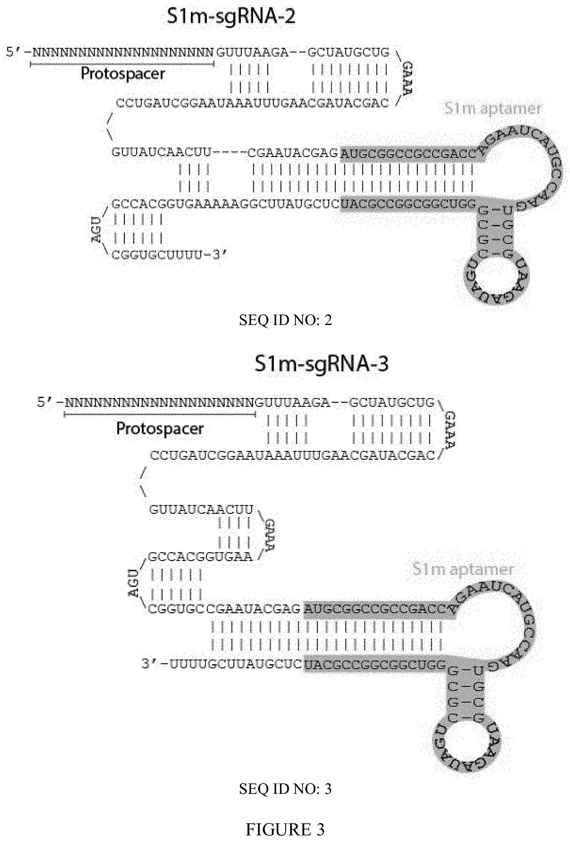
FIG. 2 shows the predicted secondary structure of S1m-sgRNA. Protospacer designates the region that defines the sequence to target in the human genome. S1m stem loop (coral) binds streptavidin.
FIG. 3 shows the predicted secondary structure of S1m-sgRNAs variants.
FIG. 4 shows in vitro transcription of S1m-sgRNAs compared to standard sgRNAs. S1m-sgRNAs are larger than sgRNAs due to the insertion of S1m stem loop.
FIG. 5 shows in vitro complexes of sgRNAs and streptavidin. Lane 1: S1m-sgRNA. Lane 2: streptavidin. Lane 3-5: Progressive ratios of S1m-sgRNA streptavidin. As streptavidin concentration was increased the electrophoretic front of S1m-sgRNAs was slowed. The presence of several bands may be due to multiple S1m-sgRNAs binding to a single streptavidin. Lane 6-7: Addition of streptavidin to standard sgRNAs do not shift the electrophoretic front.
FIG. 6 shows dynamic light scattering of ssODN-S1mplex (S1mplex=tertiary complexes of Sp.Cas9, S1m-sgRNA, and streptavidin) particle assembly. Cas9 (orange) and streptavidin (blue) proteins fail to interact when in solution together and have a hydrodynamic radius consistent with published data. The addition of sgRNA to Sp.Cas9 protein increases the radius of the particle to 10 nm (yellow). This radius does not change with the addition of streptavidin (red). When S1m-sgRNAs are added to Sp.Cas9 (purple), the radius is increased by a larger amount than sgRNAs, potentially due to the larger size of the S1m-sgRNA. When streptavidin is added to this complex (green), a shift in size of about 3 nm occurs, the size of streptavidin. A second peak at 35 nm may be associated with multiple Cas9-S1m-sgRNA complexes connected to a single streptavidin.
FIG. 7 shows two representative single cell multispectral flow cytometric images of S1m-sgRNA and sgRNA transfected cells with Cas9 immunohistochemistry and fluorescent streptavidin (scale bar: 10 .mu.m). Arrowheads indicate presence of overlapping colors. Numbers in yellow are measured log Pearson correlation coefficient as determined by IDEAS software.
FIG. 8 shows the correlation coefficient of Cas9 immunocytochemistry fluorescent signal and streptavidin fluorescence, as measured by multispectral image cytometry within hPSCs. Use of S1m-sgRNA significantly increased the correlation between the two signals (***p<10.sup.-5, Student's two-tailed t-test).
FIG. 9 shows representative confocal images of S1m-sgRNA and sgRNA transfected cells with Cas9 immunohistochemistry and fluorescent streptavidin (scale bar: 5 .mu.m). Arrowheads indicate presence of overlapping colors.
FIG. 10 shows the correlation coefficient of Cas9 immunocytochemistry and streptavidin fluorescence inside the nuclei of transfected cells. Introduction of S1m-sgRNAs significantly increased the correlation between the two molecules (*p<0.05, Student's two-tailed t-test).
FIG. 11 shows in vitro tertiary complexes of S1m-sgRNA, streptavidin, and ssODN. Lanes 1-4: Components of S1m particles ran individually. Lanes 5-7: complexes of S1m-sgRNAs, streptavidin, and biotin-ssODNs. Three concentrations of ssODN were used while amount of S1m-sgRNA and streptavidin was held constant. Major bands showing the complexation of all three components can be seen. Elongated bands may be due to different stoichiometry of bio-ssODN and S1m-sgRNA connected to streptavidin.
FIG. 12 shows in vitro tertiary complexes of S1m-sgRNA, streptavidin, and ssODN. Lanes 1-4: Components of S1m particles ran individually. Lanes 5-7: complexes of S1m-sgRNAs, streptavidin, and biotinylated ssODNs. Numbers represent relative stoichiometry between components ran on gel. Major bands showing the complexation of all three components can be seen. Elongated bands may be due to different stoichiometry of biotin-ssODN and S1m-sgRNA connected to streptavidin. Lanes 8-10: complexes of S1m-sgRNAs, streptavidin, and ssODNs. ssODNs do not interfere with the binary complex. Lane 11: complexes of streptavidin and biotin-ssODNs, with free sgRNAs. None of the typical S1m-sgRNA-streptavidin complexes can be seen in this lane.
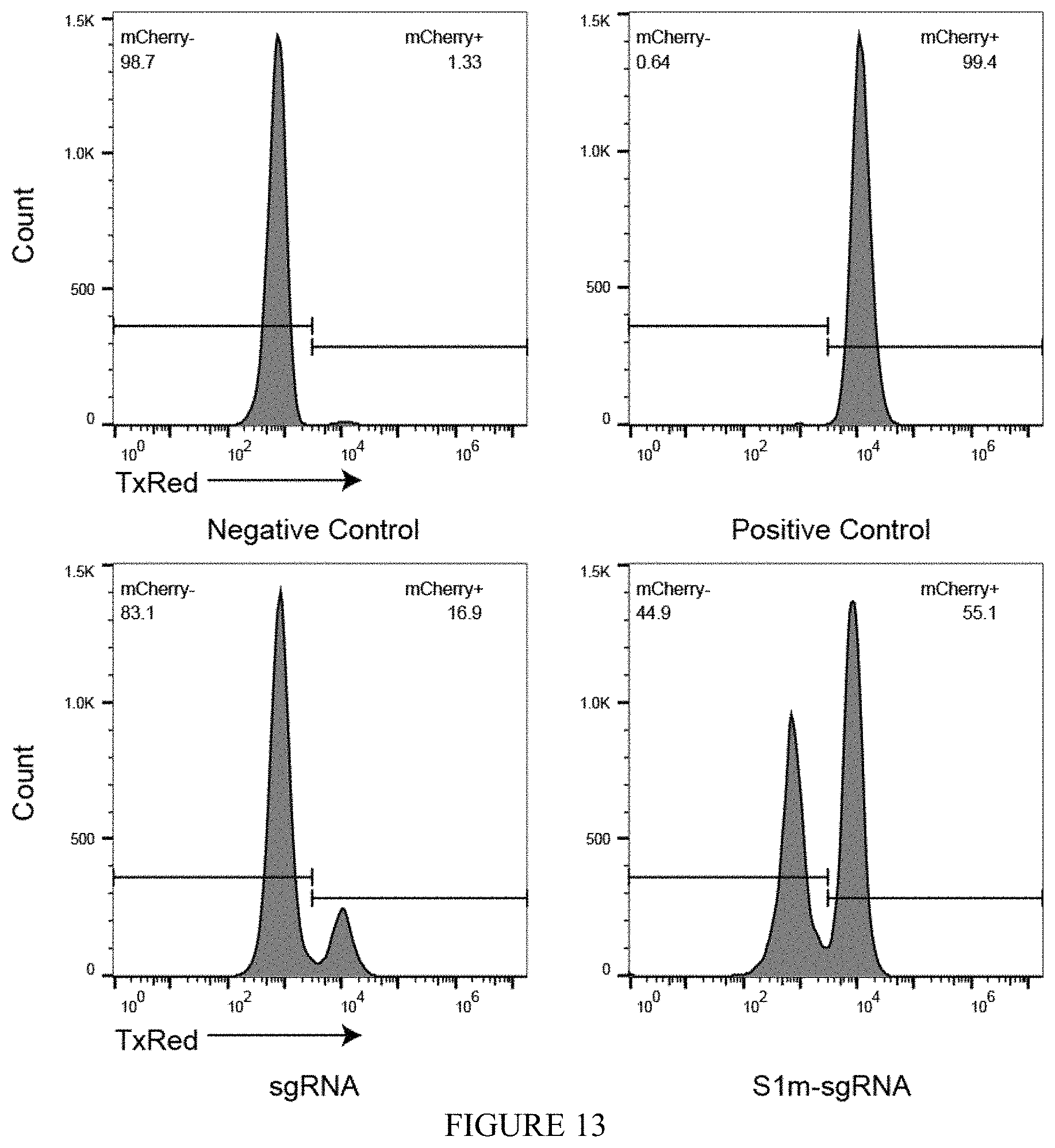
FIG. 13 shows gene editing via NHEJ using S1m-sgRNA RNPs. Knockout of integrated H2B-mCherry fluorescence in human embryonic kidney (HEK) cells. When transfected together with a plasmid encoding Sp.Cas9, S1m-sgRNAs induced .about.50% the level of NHEJ as sgRNA as measured by the loss of fluorescence (44.9% vs. 83.1%) five days post transfection.
FIG. 14 shows the ratio of precise to imprecise editing using S1mplexes formed with different S1m-sgRNA variants in hPSCs. Each S1m-sgRNA increased the ratio of precise to imprecise editing when compared to sgRNAs. S1mplexes with S1m-sgRNA-1, and S1m-sgRNA-2 had the highest ratios of precise editing.
FIG. 15 shows the ratio of precise to imprecise editing at BFP locus. ssODN-S1mplexes had an 18.4-fold higher ratio than sgRNAs and contained four precise edits to every one indel as analyzed by deep sequencing 8 days post lipofection of HEKs.
FIG. 16 shows the ratio of precise to imprecise editing at EMX1 locus. ssODN-S1mplexes had a 2.7-fold higher ratio than sgRNAs.
FIG. 17 shows the ratio of precise insertions to imprecise indels at BFP locus in hPSCs as analyzed by deep sequencing. ssODN-S1mplexes had a 9.7-fold increase in comparison to standard sgRNAs and a 7.4-fold increase when compared with untethered ssODNs.
FIG. 18 shows the ratio of precise insertions to imprecise indels at EMX1 locus. Addition of streptavidin to S1mplex resulted in a 15-fold increase in the ratio of precise insertions to imprecise indels.
FIGS. 19 and 20: ssODN design. Genomic sequence is denoted with black bars. sgRNA targeting site and PAM is denoted by `PAM` inside genomic locus, while red triangles are the sgRNA cut site. ssODN length is measured around cut site either upstream (-) or downstream (+) as read by the reading frame. Biotin (blue hexagon) was attached to either the 5' or 3' end of the ssODN. ssODNs were identical in sequence to either the PAM or Non-PAM sequence as read in a 5'-3' direction. RNP controls were standard sgRNAs plus corresponding ssODN.
FIG. 19 shows absolute NHEJ (orange diamonds) and HDR percentages (purple diamonds) as a function of total reads at two different loci in hPSCs using different ssODN designs. Each symbol represents a single replicate analyzed by deep sequencing 4 days after nucleofection into hPSCs. HDR levels were generally higher in each replicate than NHEJ levels.
FIG. 20 shows the ratio of HDR:indel reads in deep sequencing using each ssODN combined with S1mplexes. Blue circles represent individual biological replicates. With each ssODN, S1mplexes increased the ratio of HDR:indel when compared to sgRNA controls but no significant trends as to symmetry, sidedness, or biotin location were observed.
FIG. 21 is a schematic of S1mplexes with quantum dot cargoes. Qdots can be complexed with the S1mplex by a disulfide linker (Qdot-SS-S1mplex, top) or by using streptavidin covalently attached directly to the quantum dot (QdotSA-S1mplex, bottom). The quantum dot has a mean diameter of 20 nm.
FIG. 22 shows a gene editing comparison of different Qdot S1mplexes. Gene editing of HEK H2B-mCherry reporter cells five days post sorting as assayed by flow cytometry. QdotSA interferes with RNP activity, while Qdot-SS has equivalent gene editing activity as the free RNP (n=3 technical replicates).
FIG. 23 shows gene-editing using various combinations of components with QdotSA. Conjugation of S1mplexes to QdotSA significantly lowers gene editing efficiency. Editing efficiency is lower even if QdotSA is transfected separately from the S1mplexes without complexation. S1m-sgRNA|QdotSA indicates complexation of S1m-sgRNA RNP with transfection agent in a separate tube from QdotSA complexation with transfection agent, and subsequent addition of the contents of the S1m-sgRNA tube followed immediately by addition of the QdotSA tube. 5 hr. gap indicates a 5 hour culture time between transfections. Immediate application of the QdotSA can moderately interfere with the activity of the RNP, but these interference effects are abrogated if QdotSA is added 5 hours later. All RNP activity is abrogated by complexation with the QdotSA (last column) (n=3 technical replicates).
FIG. 24 shows representative epifluorescence images of untransfected and Qdot-SS-S1mplex transfected cells 24 hours post transfection (Scale bar: 10 .mu.m). Arrowheads indicate Qdot fluorescence in the cytoplasm.
FIG. 25 shows increased fluorescence of Qdot-S1mplex allows sorting out of quantum dot positive fractions compared to untransfected cells 24 hours post transfection.
FIG. 26 shows quantum dot conjugation to S1mplex via a cleavable disulfide linker allows fluorescent enrichment of gene-edited human cells. Increased fluorescence of Qdot-S1mplex after cleavage of the disulfide linker allows sorting out of quantum dot positive fractions compared to untransfected cells 24 hours post transfection (n=3 biological replicates).
FIG. 27 shows a schematic of simultaneous editing at two loci strategy. HEK cells were transfected simultaneously with two S1m particles, labeled with distinct fluorophores. Editing at the BFP locus was associated with Red-ssODN-S1mplexes (AlexaFluor.RTM.-594 fluorophore), while editing at the EMX1 locus was associated with Green-ssODN-S1mplexes (AlexaFluor.RTM.-488 fluorophore).
FIG. 28 shows single cells sorting for enrichment of editing at BFP locus. In enriched S1mplex clonal populations, indels (brown) and HDR (blue) events occurred in a 1:1 ratio. In sgRNA clones, all isolated clones either had indel or wildtype genotypes. Genotypes were assayed by Sanger sequencing. No mosaic genotypes were observed.
FIG. 29 shows fluorescent S1mplexes inside the cell using confocal microscopy. Arrows denote Green-S1mplex both inside the nucleus and outside the cell (Scale bar: 10 .mu.m).
FIG. 30 shows twenty-four hours post transfection, cells were sorted into populations that were positive for either fluorophore, both or neither. Analysis via deep sequencing was done 6 days post sorting. Top: ratio of precise (perfect sequence match to ssODN) to imprecise editing (indels) in sorted populations. Populations enriched for BFP targeted S1mplexes (Red+ and double positive) had elevated ratios up to 40 times as many insertions as indels. Bottom: ratio of precise to imprecise editing in sorted populations. Populations enriched for EMX1 targeted S1mplexes (Red+ and double positive) had elevated ratios of precise insertions to indels.
FIG. 31 Off-target analysis of double positive populations using TIDE at the top 5 off-target locations for each sgRNA. No modifications were detected below the TIDE limit of detection (dotted line).
FIG. 32 shows an off-target analysis of sorted S1mplex populations. Off-target analysis using TIDE software at the top 5 predicted off-target sites within the human genome at the BFP and EMX1 loci. Y axis indicates the percentage of cells with 0 mismatches from the parental sequence (perfect matches in sequencing reads). None of the sorted S1mplex populations showed off-target effects above the limit of detection. The unsorted sgRNA RNP population had a small proportion of cells that may have been edited at OT-2 of the EMX1 off-target sites.
FIG. 33 shows release of a biotin-ssODN through a photocleavable linkage had no significant effect on HDR editing. FIG. 33a shows a biotin-ssODN that contained a UV-cleavable linker was attached to streptavidin and S1mplex particles in order to study the potential of releasing the ssODN inside the cell to promote HDR. Lane 1: DNA standard. Lane 2: Photo-cleavable biotin-ssODN. Lane 3: standard ssODN. Lane 4: Binary complexes of streptavidin and photo-cleavable biotin-ssODNs. Lane 5-6: Binary complexes cleaved by either exposure to light through a DAPI filter cube (lane 5) or exposure to a UV transilluminator (lane 6). DAPI filter cube cleaved nearly all ssODN after 10 minutes whereas transilluminator had complete cleavage. Cleaved DNA product was the same length as control standard ssODN. FIG. 33b shows release of biotin-ssODN by 15 minutes of light exposure through a DAPI filter cube every hour post transfection. Levels of HDR were not significantly affected by the release of the ssODN within the cell at any time point (n=3 biological replicates).
FIG. 34 is a schematic of the structure and sequence of S1m-sgRNA-V3. This sequence removes 6 nt from the beginning of the S1m aptamer. Removal of these nucleotides simplified the secondary structure of the RNA. This modification may potentially decrease the number of incorrectly folded and therefore inactive S1m-sgRNAs.
FIG. 35 shows the binding capability of S1m-sgRNA-1 and S1m-sgRNA-V3 with streptavidin using an electrophoretic mobility shift assay (EMSA). S1m-sgRNAs or standard sgRNAs were mixed with native streptavidin protein at the indicated ratios (w/w) and allowed to complex prior to being loaded on an agarose gel. Lane 1: S1m-sgRNA-1. Lane 2: S1m-sgRNA-V3. Lane 3: Streptavidin. Lane 4: 10:1 S1m-sgRNA-1:Streptavidin. Lane 5: 1:1 S1m-sgRNA-1:Streptavidin. Lane 6: 1:10 S1m-sgRNA-1:Streptavidin. Lane 7: 10:1 S1m-sgRNA-V3:Streptavidin. Lane 8: 1:1 S1m-sgRNA-V3:Streptavidin. Lane 9: 1:10 S1m-sgRNA-V3:Streptavidin. Lane 10: sgRNA. Lane 7: 1:10 sgRNA:Streptavidin.
FIG. 36 shows the induction of NHEJ using various sgRNAs. Cas9 RNPs were formed with standard sgRNA, S1m-sgRNA-1, or S1m-sgRNA-V3 targeting the same locus and transfected into H2b-mCherry expressing HEK cells. % NHEJ was measured by loss of fluorescence 7 days post transfection. Both S1m-sgRNA versions were less effective at creating double strand breaks repaired by NHEJ than standard sgRNA. S1m-sgRNA-V3 induced more NHEJ events than V1 (.about.3-fold higher) potentially due to simplified secondary structure. Both S1m-sgRNA variants were still capable of creating genetic modifications. (n=3 technical replicates. Error bars represent .+-.1 S.D.)
FIG. 37 shows the induction of HDR using various sgRNAs. Cas9 RNPs were formed with standard sgRNA, S1m-sgRNA-1, or S1m-sgRNA-V3 targeting the same locus. S1m-sgRNA-1 and V3 were also used to create S1mplexes containing an ssODN to induce HDR at the target site. S1m-sgRNAs again formed fewer DSBs and S1m-sgRNA-V3 was more efficient at inducing NHEJ than V1. Similarly, when S1mplexes were formed using S1m-sgRNAs, V3 induced higher levels of HDR than V1. However, in this replicate, ratios of HDR:NHEJ differed from what was seen in previous experiments (n=3 technical replicates. Error bars represent .+-.1 S.D.)
FIG. 38 shows identification of corrected Pompe iPSCs using ArrayEdit platform following transfection with fluorescent S1mplexes. Array Edit enables tracking of phenotypic characteristics.
FIG. 39 shows the phenotypic difference between wildtype and Pompe disease iPSCs. Cell lines were cocultured together at the indicated ratio and evaluated for the presence of mCherry (wildtype) or DAPI (disease). Lysosome acidity was measured using LysoSensor.TM. Green and quantified on a per-cell basis.
FIG. 40 shows identification of corrected Pompe iPSCs. Pompe iPSCs and H9-H2b-mCherry cells were mock transfected and plated of ArrayEdit platform. Over seven days number of cells per feature was tracked and used to calculate average growth rate (bottom right). On day seven, wells were stained with LysoSensor.TM. Green and per cell intensity was measured (top left). Data was plotted as a per-feature average. Pompe iPSCs were transfected with S1mplex-ssODNs targeting diseased loci and analyzed in the same manner as described above but with the addition of S1mplex presence on day 1. Clones to be selected (bottom left) were determined by gating out the lowest average growth rate of mock transfected cells as well as the upper intensity limit of mock transfected Pompe iPSCs. Microfeatures with cells meeting both of these criteria as well as displaying S1mplex presence were selected and expanded.
FIG. 41 shows selection of gene-corrected disease iPSCs. Sanger sequencing traces of corrected cell lines. Heterozygous mutations within the PAM sequence show that the ssODN was used as the HDR template in all lines.
FIG. 42 shows dual S1mplexes for the precise excision of genomic DNA. a) 2 sgRNAs designed in the LAMA5 locus for excision of a 238 bp stretch of genomic DNA. B) Mixed S1m sgRNAs (1,2) with streptavidin added to HEK 293s, with ratio sgRNA:streptavidin 2:1 at 50 ng/well per guide. Gel shows LAMA5 locus PCR amplicon spanning both guides. Average excision efficiency of 22% with dual S1mplexes.
The above-described and other features will be appreciated and understood by those skilled in the art from the following detailed description, drawings, and appended claims.
DETAILED DESCRIPTION
Described herein are modified guide RNAs such as sgRNAs and their RNP complexes with Cas9. Without being held to theory, the inventors hypothesized that some of the errors in gene editing outcomes could be reduced by preassembling RNPs with donor template or other moieties that enable the isolation of precisely-edited cells (FIG. 1). The inventors designed a strategy inspired by CRISPR display that leverages structural studies of the RNP to identify locations in the guide RNA sequence where RNA aptamers could be tolerated.
The S1mplex tool described here exploits high affinity interactions between a short RNA aptamer and streptavidin to promote more faithful writing of the human genome. In an aspect, these RNP-containing complexes can be assembled outside the cell to a desired stoichiometry and delivered as an all-in-one gene-editing nanoparticle together with a donor nucleic acid template. In addition, the complexes can be easily decorated with additional moieties such as fluorophores or Qdots to enrich for edited cells. Use of these particles with a biotinylated ssODN reduced heterogeneity in delivery among RNPs and nucleic acids within human cells and enriches the ratio of precisely-edited to imprecisely-alleles edited alleles up to 18-fold higher than standard RNP methods, approaching a ratio of four precise edits to every one imprecise edit. Further functionalization with a unique fluorophore enables multiplexed editing and enrichment of precisely edited populations through cell sorting. Taken together, advances with the S1mplex tool generates new, chemically-defined reagents to promote precise editing of the human genome.
The inventors devised a strategy inspired by CRISPR display that leverages structural studies of the RNP to identify locations in the sgRNA sequence where RNA aptamers could be tolerated (FIG. 1). Three sgRNAs with a modification either in a stem loop of the sgRNA or at the 3' end were designed (FIG. 2), as these locations have previously been shown to tolerate additions with a minimal loss in Cas9 binding activity. Separately, at each location, a perfectly complementary 10 nucleotide block was added which was previously shown to aid aptamer addition to sgRNAs and a 60 nucleotide S1m aptamer, which has a strong non-covalent interaction with streptavidin. The added sequence extends the sgRNA stem loop and contains two distinct bulges used for binding. We termed these new sgRNAs S1m-sgRNA-1, S1m-sgRNA-2, and S1m-sgRNA-3 in reference to their position in the sgRNA from 5' to 3' (FIG. 2).
CRISPR refers to the Clustered Regularly Interspaced Short Palindromic Repeats type II system used by bacteria and archaea for adaptive defense. This system enables bacteria and archaea to detect and silence foreign nucleic acids, e.g., from viruses or plasmids, in a sequence-specific manner. In type II systems, guide RNA interacts with Cas9 and directs the nuclease activity of Cas9 to target DNA sequences complementary to those present in the guide RNA. Guide RNA base pairs with complementary sequences in target DNA. Cas9 nuclease activity then generates a double-stranded break in the target DNA.
CRISPR/Cas9 is an RNP complex. CRISPR RNA (crRNA) includes a 20 base protospacer element that is complementary to a genomic DNA sequence as well as additional elements that are complementary to the transactivating RNA (tracrRNA). The tracrRNA hybridizes to the crRNA and binds to the Cas9 protein, to provide an active RNP complex. Thus, in nature, the CRISPR/Cas9 complex contains two RNA species.
sgRNA refers to a single RNA species which combines the tracrRNA and the crRNA and is capable of directing Cas9-mediated cleavage of target DNA. An sgRNA thus contains the sequences necessary for Cas9 binding and nuclease activity and a target sequence complementary to a target DNA of interest (protospacer sequence). In general, in an sgRNA, the tracrRNA and the crRNA are connected by a linker loop sequence. sgRNAs are well-known in the art. While sgRNA is generally used throughout this disclosure, two-part guide RNAs containing a crRNA and a tracrRNA can also be employed.
As used herein, a guide RNA protospacer sequence refers to the nucleotide sequence of a guide RNA that binds to a target DNA sequence and directs Cas9 nuclease activity to the target DNA locus. In some embodiments, the guide RNA protospacer sequence is complementary to the target DNA sequence. As described herein, the protospacer sequence of a single guide RNA may be customized, allowing the targeting of Cas9 activity to a target DNA of interest.
Any desired target DNA sequence of interest may be targeted by a guide RNA target sequence. Any length of target sequence that permits CRISPR-Cas9 specific nuclease activity may be used in a guide RNA. In some embodiments, a guide RNA contains a 20 nucleotide protospacer sequence.
In addition to the protospacer sequence, the targeted sequence includes a protospacer adjacent motif (PAM) adjacent to the protospacer region which is a sequence recognized by the CRISPR RNP as a cutting site. Without wishing to be bound to theory, it is thought that the only requirement for a target DNA sequence is the presence of a protospacer-adjacent motif (PAM) adjacent to the sequence complementary to the guide RNA target sequence. Different Cas9 complexes are known to have different PAM motifs. For example, Cas9 from Streptococcus pyogenes has a NGG trinucleotide PAM motif; the PAM motif of N. meningitidis Cas9 is NNNNGATT; the PAM motif of S. thermophilus Cas9 is NNAGAAW; and the PAM motif of T. denticola Cas9 is NAAAAC.
A modified guide RNA is a one-part or two-part RNA capable of directing Cas-9-mediated cleavage of target DNA. A modified sg RNA is a single RNA species capable of directing Cas9-mediated cleavage of target DNA. A modified sgRNA, for example, comprises sequences that provide Cas9 nuclease activity, a protospacer sequence complementary to a target DNA of interest, and an aptamer that binds a biotin-binding molecule. The inventors of the present application unexpectedly found that the linker loop that connects the tracrRNA and the crRNA in an sgRNA can be replaced with an aptamer that binds a biotin-binding molecule such as a streptavidin-binding aptamer. Unexpectedly, the modified sgRNAs can bind both Cas9 protein and streptavidin, and form active RNP complexes which induce error-prone DNA repair less frequently than standard CRISPR-Cas9 RNP complexes.
In an aspect, a modified guide RNA, comprises
a crRNA comprising a single-stranded protospacer sequence and a first complementary strand of a binding region for the Cas9 polypeptide, and
a tracrRNA comprising a second complementary strand of the binding region for the Cas9 polypeptide,
wherein the crRNA or the tracrRNA comprises an aptamer that binds a biotin-binding molecule, wherein the crRNA and the tracrRNA hybridize through the first and second complementary strands of the binding region for the Cas9 polypeptide.
In another aspect, the crRNA and the tracrRNA form an sgRNA, the sgRNA comprise from 5' to 3',
the single-stranded protospacer sequence,
the first complementary strand of a binding region for the Cas9 polypeptide,
the aptamer that binds a biotin-binding molecule, and
the second complementary strand of the binding region for the Cas9 polypeptide.
More specifically, a modified sgRNA comprises, from 5' to 3', a single-stranded protospacer sequence, a first complementary strand of a binding region for the Cas9 polypeptide, an aptamer that binds a biotin-binding molecule, and a second complementary strand of the binding region of the Cas9 protein. In an embodiment, in the secondary structure of the modified sgRNA, the stem forms a stem-loop structure with the aptamer that binds the biotin-binding molecule. Specific modified sgRNAs are provided in FIG. 2.
The single-stranded protospacer region can comprise 17 to 20 nucleotides. Exemplary binding regions for Cas9 polypeptides comprise 10 to 35 base pairs.
In an aspect, the aptamer that binds a biotin-binding molecule forms a stem-loop structure. The stem portion of the stem-loop structure optionally forms a contiguous double strand with the double-stranded binding region for the Cas9 polypeptide. The stem portion of the aptamer can comprise 9 to 15 base pairs, while the loop comprises 30 nucleotides. As shown in FIG. 2, the aptamer may contain more than one stem-loop structure. As shown in Example 9, the length of the stem portion of the aptamer is not critical and can be adjusted depending on the application of the modified guide RNA.
Also included herein is an RNP complex comprising the modified guide RNA, e.g., sgRNA, and a Cas9 polypeptide or active fragment thereof. Exemplary modified sgRNAs include:
TABLE-US-00001 (SEQ ID NO: 1) NNNNNNNNNNNNNNNNNNNNGUUUAAGAGCUAUGCUGCGAAUACGAGA UGCGGCCGCCGACCAGAAUCAUGCAAGUGCGUAAGAUAGUCGCGGGUC GGCGGCCGCAUCUCGUAUUCGCAGCAUAGCAAGUUUAAAUAAGGCUAG UCCGUUAUCAACUUGAAAAAGUGGCACCGAGUCGGUGCUUUU; (SEQ ID NO: 2) NNNNNNNNNNNNNNNNNNNNGUUUAAGAGCUAUGCUGGAAACAGCAUA GCAAGUUUAAAUAAGGCUAGUCCGUUAUCAACUUCGAAUACGAGAUGC GGCCGCCGACCAGAAUCAUGCAAGUGCGUAAGAUAGUCGCGGGUCGGC GGCCGCAUCUCGUAUUCGGAAAAAGUGGCACCGAGUCGGUGCUUUU; or (SEQ ID NO: 3) NNNNNNNNNNNNNNNNNNNNGUUUAAGAGCUAUGCUGGAAACAGCAUA GCAAGUUUAAAUAAGGCUAGUCCGUUAUCAACUUGAAAAAGUGGCACC GAGUCGGUGCCGAAUACGAGAUGCGGCCGCCGACCAGAAUCAUGCAAG UGCGUAAGAUAGUCGCGGGUCGGCGGCCGCAUCUCGUAUUCGUUUU; or (SEQ ID NO: 70) NNNNNNNNNNNNNNNNNNNNGUUUAAGAGCUAUGCUGCGAAUACGAGC CGCCGACCAGAAUCAUGCAAGUGCGUAAGAUAGUCGCGGGUCGGCGGC UCGUAUUCGCAGCAUAGCAAGUUUAAAUAAGGCUAGUCCGUUAUCAAC UUGAAAAAGUGGCACCGAGUCGGUGCUUUU
A "Cas9" polypeptide is a polypeptide that functions as a nuclease when complexed to a guide RNA, e.g., an sgRNA or modified sgRNA. The Cas9 (CRISPR-associated 9, also known as Csn1) family of polypeptides, for example, when bound to a crRNA:tracrRNA guide or single guide RNA, are able to cleave target DNA at a sequence complementary to the sgRNA target sequence and adjacent to a PAM motif as described above. Cas9 polypeptides are characteristic of type II CRISPR-Cas systems. The broad term "Cas9" Cas9 polypeptides include natural sequences as well as engineered Cas9 functioning polypeptides. The term "Cas9 polypeptide" also includes the analogous Clustered Regularly Interspaced Short Palindromic Repeats from Prevotella and Francisella 1 or CRISPR/Cpfl which is a DNA-editing technology analogous to the CRISPR/Cas9 system. Cpfl is an RNA-guided endonuclease of a class II CRISPR/Cas system. This acquired immune mechanism is found in Prevotella and Francisella bacteria. Additional Class I Cas proteins include Cas3, Cas8a, Cas5, Cas8b, Cas8c, Cas 10d, Case1, Cse 2, Csy 1, Csy 2, Csy 3, GSU0054, Cas 10, Csm 2, Cmr 5, Cas10, Csx11, Csx10, and Csf 1. Additional Class 2 Cas9 polypeptides include Csn 2, Cas4, C2c1, C2c3 and Cas13a.
Exemplary Cas9 polypeptides include Cas9 polypeptide derived from Streptococcus pyogenes, e.g., a polypeptide having the sequence of the Swiss-Prot accession Q99ZW2 (SEQ ID NO: 5); Cas9 polypeptide derived from Streptococcus thermophilus, e.g., a polypeptide having the sequence of the Swiss-Prot accession G3ECR1 (SEQ ID NO: 6); a Cas9 polypeptide derived from a bacterial species within the genus Streptococcus; a Cas9 polypeptide derived from a bacterial species in the genus Neisseria (e.g., GenBank accession number YP_003082577; WP_015815286.1 (SEQ ID NO: 7)); a Cas9 polypeptide derived from a bacterial species within the genus Treponema (e.g., GenBank accession number EMB41078 (SEQ ID NO: 8)); and a polypeptide with Cas9 activity derived from a bacterial or archaeal species. Methods of identifying a Cas9 protein are known in the art. For example, a putative Cas9 protein may be complexed with crRNA and tracrRNA or sgRNA and incubated with DNA bearing a target DNA sequence and a PAM motif.
The term "Cas9" or "Cas9 nuclease" refers to an RNA-guided nuclease comprising a Cas9 protein, or a fragment thereof (e.g., a protein comprising an active, inactive, or partially active DNA cleavage domain of Cas9, and/or the gRNA binding domain of Cas9). In some embodiments, a Cas9 nuclease has an inactive (e.g., an inactivated) DNA cleavage domain, that is, the Cas9 is a nickase. Other embodiments of Cas9, both DNA cleavage domains are inactivated. This is referred to as catalytically-inactive Cas9, dead Cas9, or dCas9.
Functional Cas9 mutants are described, for example, in US20170081650 and US20170152508, incorporated herein by reference for its disclosure of Cas9 mutants.
In addition, to the modified sgRNA and the Cas9 polypeptide or active fragment thereof, an RNP complex may further comprise a biotin-binding molecule such as an avidin such as avidin, streptavidin, or NeutrAvidin.TM. which bind with high affinity to the aptamer that binds the biotin-binding molecule in the modified sgRNA. Avidin, streptavidin and NeutrAvidin.TM. are a tetramers and each subunit can bind biotin with equal affinity. Avidin, streptavidin and NeutrAvidin.TM. variants that contain one, two or three biotin binding sites are also available and may be employed in the complex.
When the RNP complex comprises a biotin-binding molecule, the complex can further comprise a biotinylated molecule which associates with the complex via the biotin-binding molecule. The biotinylated molecule can target the RNP complex to a specific cell type, organ or tissue. For example, PEG-coated gold nanoparticles exhibit size-dependent in vivo toxicity; the renal clearance of quantum dots can be controlled; and the accumulation of PEGylated silane-coated magnetic iron oxide nanoparticles has been shown to be size dependent.
In one embodiment, the biotinylated molecule is a biotinylated oligodeoxynucleotide, such as a biotinylated donor DNA template. Homologous recombination can insert an exogenous polynucleotide sequence into the target nucleic acid cleavage site. An exogenous polynucleotide sequence can be called a donor polynucleotide or a donor sequence. In some embodiments, a donor polynucleotide, a portion of a donor polynucleotide, a copy of a donor polynucleotide, or a portion of a copy of a donor polynucleotide can be inserted into a target nucleic acid cleavage site. A donor polynucleotide can be single-stranded DNA, double-stranded DNA, RNA, or a duplex of RNA and DNA. A donor polynucleotide can be a sequence that does not naturally occur at a target nucleic acid cleavage site. In some embodiments, modifications of a target nucleic acid due to NHEJ and/or HDR can lead to, for example, mutations, deletions, alterations, integrations, gene correction, gene replacement, transgene insertion, nucleotide deletion, gene disruption, and/or gene mutation. The process of integrating non-native nucleic acid(s) into genomic DNA can be referred to as "genome engineering".
In an embodiment, the biotinylated molecule is a nanoparticle, such as a quantum dot, a gold particle, a magnetic particle, a polymeric nanoparticle. In another embodiment, the biotinylated molecule is a biotinylated fluorescent dye such as Atto 425-Biotin, Atto 488-Biotin, Atto 520-Biotin, Atto-550 Biotin, Atto 565-Biotin, Atto 590-Biotin, Atto 610-Biotin, Atto 620-Biotin, Atto 655-Biotin, Atto 680-Biotin, Atto 700-Biotin, Atto 725-Biotin, Atto 740-Biotin, fluorescein biotin, biotin-4-fluorescein, biotin-(5-fluorescein) conjugate, and biotin-B-phycoerythrin, Alexa Fluor.RTM. 488 biocytin, Alexa Fluor.RTM.546, Alexa Fluor.RTM. 549, lucifer yellow cadaverine biotin-X, Lucifer yellow biocytin, Oregon green 488 biocytin, biotin-rhodamine and tetramethylrhodamine biocytin. Biotinylated molecule may also be a peptide, proteins or protein domains, specifically antibodies and Fab domains.
In another aspect, the biotin-binding molecule can be covalently linked to a donor polynucleotide, a nanoparticle, or a dye molecule either directly or via a linker molecule, using, for example a disulfide linker. The bound biotin-binding molecule can then bind the aptamer of the modified sgRNA. Additional biotinylated donor polynucleotides, nanoparticle, contrast agent, or dye molecules can then be associated with the bound biotin-binding molecule. Alternatively, the biotin-binding molecule can be associated with the biotinylated molecule prior to adding to modified sgRNA.
Further included herein are methods of modifying a target gene, such as a target gene in a cell by contacting the cell with the RNP complexes and modified guide RNAs described herein. The cell can be from any organism (e.g., a bacterial cell, an archaeal cell, a cell of a single-cell eukaryotic organism, a plant cell, an algal cell, a fungal cell (e.g., a yeast cell), a cell from an invertebrate animal, a cell from a vertebrate animal, or a cell from a mammal, including a cell from a human.
Also included herein is a method of modifying a target gene in a cell, comprising delivering to the cell the modified guide RNA, wherein the modified guide RNA is associated with a biotin-binding molecule, and wherein the single-stranded protospacer sequence of the modified guide RNA hybridizes to a sequence in the target gene to be modified.
In some embodiments, the present disclosure provides for methods of modifying a target gene in a plant. As used herein, the term "plant" refers to whole plants, plant organs, plant tissues, seeds, plant cells, seeds and progeny of the same. Plant cells include, without limitation, cells from seeds, suspension cultures, embryos, meristematic regions, callus tissue, leaves, roots, shoots, gametophytes, sporophytes, pollen and microspores. Plant parts include differentiated and undifferentiated tissues including, but not limited to roots, stems, shoots, leaves, pollens, seeds, tumor tissue and various forms of cells and culture (e.g., single cells, protoplasts, embryos, and callus tissue).
In an embodiment, modifying the target gene increases or decreases the expression of a gene product of the target gene.
In another embodiment, modifying the target gene comprises high-fidelity homology-directed repair (HDR).
In another embodiment, modifying the target gene comprises the addition of a genetic functionality, or the correction of a mutation.
In yet another embodiment, modifying the target gene creates a double strand break (DSB) which is repaired by a non-homologous end joining (NHEJ) cell repair mechanism generating indels thereby modifying the polynucleotide sequence of the target gene.
In a further embodiment, modifying the target gene creates a DSB which is repaired by a homologous recombination (HDR) cell repair mechanism incorporating a donor DNA sequence thereby modifying the polynucleotide sequence of the target gene.
In an aspect, the S1m-sgRNAs described herein can be used for biallelic correction. Infantile-onset Pompe disease contains two distinct deleterious mutations at different points within a single gene. In an aspect, two S1m-sgRNAs can be employed simultaneously, one for correction of each disease locus. As shown in Example 11, clones containing edits at both alleles were identified.
In another aspect, the S1m-sgRNAs described herein can be used for the excision of genomic DNA. In an aspect, two S1m-sgRNAs can be employed simultaneously, wherein each S1m-sgRNA targets an end of the region to be excised. As shown in Example 12, human cells contain the properly excised region of genomic DNA
Delivery of polynucleotides and RNPs of the present disclosure to cells, in vitro, or in vivo, may be achieved by a number of methods known to one of skill in the art. These methods include lipofection, electroporation, nucleofection, microinjection, biolistics, liposomes, immunoliposomes, polycation or lipid:nucleic acid conjugates. Lipofection is well known and lipofection reagents are sold commercially. Cationic and neutral lipids that are suitable for efficient receptor-recognition lipofection of polynucleotides are described in the art.
Lipid:nucleic acid complexes, including targeted liposomes such as immunolipid complexes, and the preparation of such complexes is well known to one of skill in the art.
Electroporation can be used to deliver the polynucleotides and RNPs of the present disclosure. In these methods, the polynucleotides or RNPs are mixed in an electroporation buffer with the target cells to form a suspension. This suspension is then subjected to an electrical pulse at an optimized voltage, which creates temporary pores in the phospholipid bilayer of the cell membrane, permitting charged molecules like DNA and proteins to be driven through the pores and into the cell. Reagents and equipment to perform electroporation are sold commercially.
Biolistic, or microprojectile delivery, can be used to deliver the polynucleotides and RNPs of the present disclosure. In these methods, microprojectiles, such as gold or tungsten, are coated with the polynucleotide by precipitation with calcium chloride, spermidine or polyethylene glycol. The microprojectile particles are accelerated at high speed into a cell using a device such as the BIOLISTIC.RTM. PDS-1000/He Particle Delivery System (Bio-Rad; Hercules, Calif.).
In another embodiment, a viral vector expressing the modified guide RNA of the present disclosure, a viral vector expressing a Cas9 polypeptide and biotinylated donor DNA template (e.g., a biotinylated donor DNA template), can be transfected into a cell, such as a human cell. Human cells include human pluripotent stem cell lines and primary blood cell such as hematopoietic stem and progenitor cells and T-cells. Once editing has occurred in the cell line, the cells can be differentiated and transplanted into a subject, or used for drug development.
In some embodiments, the polynucleotides of the present disclosure may also comprise modifications that, for example, increase stability of the polynucleotide. Such modifications may include phosphorothioates, chiral phosphorothioates, phosphorodithioates, phosphotriesters, aminoalkylphosphotriesters, methyl and other alkyl phosphonates such as 3'-alkylene phosphonates, 5'-alkylene phosphonates, chiral phosphonates, phosphinates, phosphoramidates including 3'-amino phosphoramidate and amino alkylphosphoramidates, phosphorodiamidates, thionophosphoramidates, thionoalkylphosphonates, thionoalkylphosphotriesters, selenophosphates, and boranophosphates having normal 3'-5' linkages, 2-5' linked analogs, and those having inverted polarity wherein one or more internucleotide linkages is a 3' to 3', a 5' to 5' or a 2' to 2' linkage. Exemplary nucleic acid-targeting polynucleotides having inverted polarity can comprise a single 3' to 3' linkage at the 3'-most internucleotide linkage (i.e. a single inverted nucleoside residue in which the nucleobase is missing or has a hydroxyl group in place thereof). Various salts (e.g., potassium chloride or sodium chloride), mixed salts, and free acid forms can also be included.
In some embodiments, the polynucleotides of the present disclosure may also contain other nucleic acids, or nucleic acid analogues. An example of a nucleic acid analogue is peptide nucleic acid (PNA).
The invention is further illustrated by the following non-limiting examples.
EXAMPLES
Methods
Cell Culture:
WA09 hESCs (WiCell, Madison, Wis.) were maintained in E8 medium on Matrigel.RTM. (WiCell) coated tissue culture polystyrene plate (BD Falcon). Cells were passaged every 3-4 days at a 1:6 ratio using Versene.RTM. solution (Life Technologies). WA09-BFP hESCs were generated through lentiviral transduction of BFP dest clone (Addgene #71825) and sorted to ensure clonal populations. After expansion, lines were sorted monthly on a BD FACS Aria to maintain expression levels.
Human embryonic kidney cells (293T) were obtained from ATCC and were maintained between passage 15-60 in Growth medium containing DMEM (Life Technologies), 10% v/v FBS (WiCell), 2 mM L-Glutamine (Life Technologies), and 50 U/mL Penicillin-Streptomycin (Life Technologies). Cells were passaged 1:40 with Trypsin-EDTA (Life Technologies) onto Gelatin-A (Sigma) coated plates. HEK-H2B-mCherry lines were generated through CRISPR-mediated insertion of a modified AAV-CAGGS-EGFP plasmid (Addgene #22212) at the AAVS safe harbor locus using gRNA AAVS1-T2 (Addgene #41818). HEK-BFP lines were generated and maintained as mentioned above. All cells were maintained at 37.degree. C. and 5% CO.sub.2.
One Pot Transcription of S1m-sgRNA:
S1m-sgRNAs were synthesized by first creating a double stranded DNA block that encoded the sgRNA scaffold as well as the S1m aptamer. This scaffold was formed by overlap PCR using Phusion.RTM. High-Fidelity Polymerase (New England Biolabs) according to the manufacturer's protocols and was placed in the thermocycler for 30 cycles of 98.degree. C. for 10 s and 72.degree. C. for 15 s with a final extension period of 72.degree. C. for 10 min. A second primer consisting of a truncated T7 promoter, the sgRNA target, and homology to the S1m scaffold was then added to the scaffold and PCR was performed again using Phusion.RTM. and placed in a thermocycler at 98.degree. C. for 30 s followed by 35 cycles of 98.degree. C. for 5 s, 60.degree. C. for 10 s, and 72.degree. C. for 15 s, with a final extension period of 72.degree. C. for 10 min. S1m PCR products were then incubated overnight at 37.degree. C. in a HiScribe.TM. T7 IVT reaction (New England Biolabs) according to manufacturer's protocol. The resulting RNA was purified using MEGAclear.TM. Transcription Clean-Up Kit (Thermo Fisher) and quantified on a Nanodrop.TM.2000.
S1m RNP Formation:
NLS-Cas9-NLS protein (Aldevron, Madison, Wis.) was combined with S1m-sgRNAs and allowed to complex for 5 minutes with gentle mixing. To this complex, streptavidin (Life Technologies) was added and the mixture was allowed to complex for an additional 5 minutes. Finally, biotin-ssODNs (Integrated DNA Technologies) were added to the tertiary complex and subsequently vortexed at low speed. This final mixture was then allowed to sit for 10 minutes to ensure complete complexation.
S1m-sgRNA and Streptavidin Binding Gel Shift Assays:
S1m-sgRNAs were heated at 75.degree. C. for 5 min and cooled to room temperature for 15 min. 20 pmol S1m-sgRNA was combined with streptavidin at 10:1, 1:1, and 1:10 molar ratios in a final volume of 5 .mu.l and the mixture was allowed to complex for 10 min. The S1m-sgRNA-streptavidin complexes were run on a 1% agarose gel. Tertiary complexes were assembled by first mixing 15 pmol each of S1m-sgRNA and streptavidin. To this mixture, 6, 15, or 30 pmol of ssODN was added prior to running the complexes through a 1% agarose gel. All gels were run using Kb+ Ladder (Invitrogen) as a molecular weight marker to allow for inter-gel size comparisons even when running RNA samples.
Biotin Competition Assay:
S1m-sgRNA was heated to 75.degree. C. for 5 min and cooled to room temperature. 20 pmol each S1m-sgRNA and streptavidin were complexed for 10 min. 80 pmol biotin was added at 30, 20, 10, 5, and 0 min intervals prior to running the complexes through a 1% agarose gel.
Dynamic Light Scattering:
DLS was performed using a DynaPro.RTM. NanoStar.RTM. (Wyatt Technology) using small volume (4 .mu.L) disposable cuvettes. 10 .mu.g of each component was added into the cuvette and diluted as necessary with dH.sub.2O to reach 4 .mu.L solution volume. In mixed component conditions, components were allowed to mix for 5 minutes while taking readings. Acquisitions were performed for 20 seconds with a minimum of 4 acquisitions per measurement. 5 measurements were performed per sample and were conducted at room temperature. Data was graphed as a function of percent intensity.
Quantum Dot Biotin Conjugation:
To make Qdot-SS-s1mplexes, amine-PEG green fluorescent quantum dots (Qdot.RTM. ITK.TM. 525--ThermoFisher) were reacted with a degradable dithiol biotin linker (EZ-Link.TM..RTM. Sulfo-NHS-Biotin--ThermoFisher) as follows: First, 25 .mu.l of an 8 .mu.M Quantum dot solution in 50 mM Borate buffer were desalted into PBS using Zeba desalting columns (40K MWCO--ThermoFisher) and then reacted with excess sulfoNHS-dithiol-biotin linker for 2 hours at 4.degree. C. with shaking. The conjugate was purified from excess linker through buffer exchange in the desalting columns. Quantum dots retained their fluorescence and were stored at 4.degree. C. until use.
RNP Delivery:
HEK transfections were performed using TransIT-X2.RTM. delivery system (Mirus Bio, Madison, Wis.) according to manufacturer's protocol. 2.5.times.10.sup.5 cells/cm.sup.2 were seeded in a 24-well plate 24 hours prior to transfection. RNP complexes were formed as described in 25 uL of Opti-MEM.TM. (Life Technologies). 1 .mu.g of Ca9 protein, 500 ng sgRNA, 500 ng streptavidin, and 500 ng ssODN were used. In a separate tube, 25 uL of Opti-MEM.TM. was combined with 0.75 uL of TransIT-X2.RTM. reagent and allowed to mix for 5 minutes. TransIT-X2.RTM. and RNP solutions were then mixed by gentle pipetting and placed aside for 15 minutes. After this incubation, 50 .mu.L of solution were added dropwise into the well. Media was changed 24 hours post transfection.
For HEK transfections involving quantum dots, Lipofectamine.TM. 2000 (Life Technologies) was used for delivery. Qdot-RNP complexes were formed according to the following amounts (for 24 wells: 500 ng of Ca9 protein, 187.5 ng sgRNA, 187.5 ng streptavidin, 3.125 pMoles of quantum dots and 3 ul Lipofectamine.TM. per well; a quarter of these amounts were used when transfecting 5000 cells in 96 well plates).
All hPSC transfections were performed using the 4D-Nucleofector.TM. System (Lonza) in P3 solution using protocol CB150. Cells were pretreated with Rho-kinase (ROCK) inhibitor (Y-27632 Selleck Chemicals) 24 hours prior to transfection. 8 .mu.g Cas9, 3.5 .mu.g sgRNA, 3.5 .mu.g streptavidin, and 1 .mu.g ssODN were used to form particles as described above. Cells were then harvested using TrypLE.TM. (Life Technologies) and counted. 2.times.10.sup.5 cells per transfection were then centrifuged at 100.times.g for 3 minutes. Excess media was aspirated and cells were resuspended using 20 .mu.L of RNP solution per condition. After nucleofection, samples were incubated in nucleocuvettes at room temperature for 15 minutes prior to plating into one well of a 6-well plate containing E8 media+10 .mu.M ROCK inhibitor. Media was changed 24 hours post transfection and replaced with E8 medium.
Immunocytochemistry:
To measure correlation hPSCs were transfected with Cas9 protein and streptavidin-AF-647. 24 hours post transfection, cells were fixed using 4% PFA and incubated at room temperature for 10 minutes. Cells were then permeabilized using 0.05% Triton X-100 and incubated for 10 minutes. Following two washes with 5% goat serum, Cas9 antibody (Clontech #632607, 1:150) was added to cells and incubated overnight at 4.degree. C. The next day, cells were rinsed twice with 5% goat serum and then incubated with a goat anti-rabbit secondary antibody (Santa Cruz Biotech #sc-362262, 1:500) for one hour at room temperature. Cells were then washed twice with PBS and mounted for imaging.
To visualize S1mplexes in the nucleus human embryonic kidney cells (HEK293T) were plated at 16,000 cells per well in an 8-well chamber slide at day 0. On day 1, 20 mL of transfection media was added to cells in 200 .mu.L of maintenance media. Transfection media contained 20 .mu.L Opti-MEM.RTM. (Life Technologies), 10 pmol Streptavidin Alexa Fluor.RTM. 488 conjugate (Thermo Fisher), and 0.6 .mu.L TransIT.RTM. transfection reagent (Mirus). On day 3, cells were incubated with 1.times. CellMask.TM. Plasma Membrane Stain (ThermoFisher) and 1.times. Hoechst for 10 min. Following incubation at 37.degree. C., cells were immediately washed with PBS and fixed in 4% paraformaldehyde (IBI Scientific) at room temperature for 15 min. Cells were analyzed using a Nikon Eclipse TI epifluorescent microscope and a Nikon AR1 confocal microscope.
Multispectral Imaging Flow Cytometry:
hPSCs were transfected and stained as described above. After staining, cells were centrifuged and resuspended in 50 .mu.L PBS. Fluorescence was detected on ImageStream.RTM. X Mark II (EMD Millipore) according to manufacture instructions. Cellular colocalization was measured by IDEAS software package (Amnis) using predefined colocalization wizard.
Flow Cytometry:
Flow cytometry of BFP expression and conversion to GFP was measured using a BD FACS Aria using the DAPI and FITC filters and analyzed using FlowJo. Voltages were established by running wild type WA09 hPSCs as well as WA09-BFP hPSCs. Sorting was performed on a BD FACSAria.TM. II with a nozzle size of 100 .mu.m at room temperature and sorted into culture media.
Genomic Analysis:
DNA was isolated from cells using DNA QuickExtract.TM. (Epicentre, Madison, Wis.) following treatment by 0.05% trypsin-EDTA and centrifugation. QuickExtract.TM. solution was incubated at 65.degree. C. for 15 minutes, 68.degree. C. for 15 minutes, and finally 98.degree. C. for 10 minutes. Genomic PCR was performed following manufacturer's instructions using AccuPrime.TM. HiFi Taq (Life Technologies) and 500 ng of genomic DNA. Products were then purified using AMPure.RTM. XP magnetic bead purification kit (Beckman Coulter) and quantified using a Nanodrop.TM.2000. For deep sequencing, samples were pooled and run on an Illumina HiSeq.TM. 2500 High Throughput at a run length of 2.times.125 bp or an Illumina Miseq.RTM. 2.times.150 bp.
Deep Sequencing Data Analysis:
A custom python script was developed to perform sequence analysis. The pipeline starts with preprocessing, which consists of filtering out low quality sequences and finding the defined ends of the reads. For each sample, sequences with frequency of less than 100 were filtered from the data. Sequences in which the reads matched with primer and reverse complement subsequences classified as "target sequences". Target sequences were aligned with corresponding wildtype sequence using global pairwise sequence alignment. Sequences that were misaligned around the expected cut site were classified as NHEJ events while sequences that had insertions larger that 15 bp were classified as HDR events. The frequency, length, and position of matches, insertions, deletions, and mismatches were all tracked in the resulting aligned sequences.
Cell Membrane Staining:
Human embryonic kidney cells (HEK293) were plated at 16,000 cells/well in an 8-well chamber slide at day 0. On day 1, 20 pt of transfection media was added to cells in 200 .mu.L of maintenance media. Transfection media contained 20 .mu.L Opti-MEM.RTM. (Life Technologies), 400 ng Streptavidin Alexa Fluor.RTM. 488 conjugate (Thermo Fisher), and 0.6 .mu.L TransIT.RTM. transfection reagent (Mirus). On day 3, cells were incubated with 1.times. CellMask.TM. Plasma Membrane Stain (ThermoFisher) and 1.times. Hoechst for 10 min. Following incubation at 37 C, cells were immediately washed with PBS and fixed in 4% paraformaldehyde (IBI Scientific) at room temperature for 15 min. Cells were analyzed using a Nikon Eclipse TI epifluorescent microscope and a Nikon AR1 confocal microscope.
Statistics:
All error bars are shown as .+-.1 standard deviation. p values were computed using a Student's two-tailed t-test and deemed significant at .alpha.<0.05.
Nucleic Acid Sequences:
The relevant nucleic acid sequences are provided in the following tables:
TABLE-US-00002 TABLE 1 Primers used to create sgRNA and S1m-sgRNAs. S1m SEQ Construct ID Name Sequence (5' to 3') NO: S1m- GTTTAAGAGCTATGCTGCGAATACGAGATGCGGC 8 sgRNA- CGCCGACCAGAATCATGCAAGTGCGTAAGATAGT 1_F CGCGGGTCGGCGGCCGCATCTCGTATTC S1m- AAAAGCACCGACTCGGTGCCACTTTTTCAAGTTGA 9 sgRNA- TAACGGACTAGCCTTATTTAAACTTGCTATGCTGC 1_R GAATACGAGATGCGGCCGCCGACCCG S1m TTAATACGACTCACTATAGGNNNNNNNNNNNNNN 10 Forward NNNNNNGTTTAAGAGCTATGCTGCGA RNATracR AAAAGCACCGACTCGGTGCC 11
TABLE-US-00003 TABLE 2 Protospacer and respective PAMs used for genomic targeting. SEQ ID sgRNA Name Sequence (5' to 3') PAM NO: BFP GCTGAAGCACTGCACGCCAT GGG 12 (BFP.fwdarw.GFP) EMX1 GTCACCTCCAATGACTAGGG TGG 13 (EMX1_21) mCherry GGAGCCGTACATGAACTGAG GGG 14 (mCherry_15)
TABLE-US-00004 TABLE 3 Forward and reverse primers for genomic loci. SEQ SEQ Genomic Forward ID Reverse ID Primer (5' to 3') NO: (5' to 3') NO: EMX1 CCATCCCCTT 15 GGAGATTGGAG 16 CTGTGAATGT ACACGGAGA EMX1 TCCACCTTGG 17 CCCTCCACCAG 18 Symmetric CTTGGCTTTG CTACCCAC mCherry AAGGGCGAGG 19 TTGTACAGCTC 20 Interior AGGATAACATGG GTCCATGCCG EMX1 CCAATGACAA 21 Insertion GCTTGCTAGC
TABLE-US-00005 TABLE 4 ssODNs used to direct HDR after DSB formation. SEQ ssODN ID Donor Sequence (5' to 3') NO: BFP.fwdarw.GFP TCATGTGGTCGGGGTAGCGGCTGAAGCACTGCA 22 NT CGCCATGGGTCAGGGTGGTCACGAGGGTGGGC CAGGGCACCGGCAGCTTGCCGGTGGTGCAGAT GAA BFP.fwdarw.GFP 5Biotin/TCATGTGGTCGGGGTAGCGGCTGAAG 23 5PCBi CACTGCACGCCATGGGTCAGGGTGGTCACGAGGGT NT GGGCCAGGGCACCGGCAGCTTGCCGGTGGTGC AGATGAA EMX1 AAGCAGCACTCTGCCCTCGTGGGTTTGTGGTTG 24 NT CCCACCGCTAGCAAGCTTGTCATTGGAGGTGAC ATCGATGTCCTCCCCATTGGCCTG EMX1 5Biotin/AAGCAGCACTCTGCCCTCGTGGGTTT 25 5PCBio GTGGTTGCCCACCGCTAGCAAGCTTGTCATTGGAG NT GTGACATCGATGTCCTCCCCATTGGCCTG
TABLE-US-00006 TABLE 5 Off-target sequences and corresponding genomic locus for each sgRNA used. Mismatches from protospacer are labelled in red. sgRNA SEQ Target ID Sequence Off-Target Sequence NO: PAM Locus BFP.fwdarw. GFP OT1 GCAGAAGCACTG 27 CAG chr17: GCTGAAGCACT CAAGCCAT +39786906 GCACGCCAT OT2 TCTGAAGTGCTG 28 CAG chr2: (SEQ ID CACGCCAT -238397265 NO: 26) OT3 GTGGAAGCACTG 29 TGG chr7: CAAGCCAT -11228464 OT4 GCTGGAGCAGGG 30 CAG chr9: CACGCCAT +109114765 OT5 GAAGAAGCACTG 31 CAG chr13: CACCCCAT -75660548 EMX1 OT1 AGGACCACCAAT 33 CAG chr3: GTCACCTCCAA GACTAGGG -64303990 TGACTAGGG OT2 ACCACCTGTAAT 34 TAG chr4: (SEQ ID GACTAGGG -149749778 NO: 32) OT3 GGAGCCTCCAGT 35 GAG chr17: GACTAGGG -38423030 OT4 GTGAACTACAGT 36 TGG chr8: GACTAGGG +112210096 OT5 CTGGCCTCCAAA 37 GAG chr15: GACTAGGG -75011931
TABLE-US-00007 TABLE 6 Forward and reverse primers used to amplify off-target genomic loci. Off- SEQ SEQ Target Forward ID Reverse ID Primer (5' to 3') NO: (5' to 3') NO: BFP OT1 TTTCCTAGCAAGC 38 AGCTGTCCTTTGTCC 39 AGACTCAGA CATTGA BFP OT2 TCTCCATGCCCTCC 40 GGATGTAGTCCATGA 41 TTTCCAT TCTTCCCC BFP OT3 TCCCAGAATGTGA 42 CTGTGGGCTTTCCTC 43 AAGTGGAGG AGCTC BFP OT4 GCTGACTAACGTC 44 TGGACCTATGTTTTT 45 CACTGCT CTTCGTCAC BFP OT5 AAAGTCTGTGGCC 46 AACCCTACCCCCTAC 47 TTGTGAGA CTGAA EMX1 OT1 TTCCCCAGGTAGT 48 TCTGCACATGTCCCA 49 TGCTGTTC ACTGTC EMX1 OT2 ATCCGTACCTAAC 50 GCACAGATCTTGGTG 51 ATGACCC GCTTT EMX1 OT3 GGCTGGGTTTCCC 52 CAAACTGCTGTGTTG 53 AAACGTA GGTGG EMX1 OT4 ACTTGGAAGGGTC 54 CCTTGAATAGAGCAT 55 CACACAA TTTTCCCCA EMX1 OT5 TCCTACCCTTGGA 56 GGGCTACACGGTCCC 57 TGGGGTT TAAAG
Example 1: Design of Modified sgRNA
A novel sgRNA with a modification at the stem loop closest to the 5' end of the sgRNA was designed (FIG. 3). This location was chosen because it has previously been shown to tolerate additions with a minimal loss in activity. An S1m aptamer was added, which has a strong non-covalent interaction with streptavidin. The added S1m aptamer extends the sgRNA stem loop closest to the 5' end and contains two distinct bulges used for binding. These modifications do not otherwise disrupt the predicted sgRNA secondary structure (FIG. 3). We confirmed that S1m-sgRNAs can be made rapidly in vitro via one-pot transcription and are larger than standard sgRNAs when analyzed by agarose gel electrophoresis (FIG. 3).
Similar experiments were performed with sgRNAs S1m-sgRNA-1, S1m-sgRNA-2, S1m-sgRNA-3, and S1m-sgRNA-V3.
Example 2: Formation of Streptavidin and Cas9 Complexes with Modified sgRNA
Next, we verified the ability of S1m-sgRNAs to complex with streptavidin in vitro by combining a constant amount of S1m-sgRNA with increasing amounts of streptavidin. The electrophoretic front of the S1m-sgRNA slowed as streptavidin levels increased (FIG. 5). At the maximum amount of streptavidin, 40% of the front had slowed demonstrating the binding of the S1m-sgRNA with streptavidin. In contrast, when the same amount of standard (non-S1m) sgRNA was run with streptavidin, the electrophoretic front remained constant.
To demonstrate the ability of S1m-sgRNA-1 to complex with streptavidin and Cas9 protein simultaneously, we performed dynamic light scattering (DLS). When streptavidin and Cas9 were combined in solution, two peaks were distinct at 3.0 nm and 7.8 nm (FIG. 6), both of which match closely the radii previously reported for each protein. We next formed Cas9 RNPs with excess standard sgRNAs and observed that the species formed were larger than Cas9 alone and did not increase in radius with the addition of streptavidin. Excess sgRNA was not detected by DLS and was included in the DLS studies to ensure all key components were able to assemble together (data not shown). Additionally, these samples had a discernable peak corresponding to the presence of streptavidin alone. RNPs containing S1m-sgRNAs and Sp.Cas9 protein increased in radius by a larger amount than RNPs containing standard sgRNAs and Sp.Cas9 protein, likely due to the increased length of S1m-sgRNAs. When streptavidin was added to S1m-sgRNA RNPs, the average radius of the complex was increased by .about.3 nm, the radius of streptavidin protein. These tertiary complexes of Sp.Cas9, S1m-sgRNA-1, and streptavidin are termed "S1mplexes". The second, larger peak in the S1mplex DLS trace is attributed to the tetrameric nature of streptavidin that can harbor up to four RNPs.
While assembly of S1mplexes in vitro is important, the maintenance of complexes post-delivery is imperative to gene editing function. To demonstrate this capability, we delivered Cas9 protein and streptavidin in combination with either sgRNAs or S1m-sgRNAs into human pluripotent stem cells (hPSCs) via nucleofection and conducted immunohistochemistry for the two protein components. Multispectral imaging flow cytometric analysis of single fixed cells confirmed the co-localization of the two protein components within hPSCs (FIG. 7). Significantly higher correlation in the fluorescent signals from the two protein components were seen when S1m-sgRNA-1 was included (p<10.sup.-5, Student's two-tailed t-test FIG. 8). To gain further subcellular resolution of these components after S1mplex delivery, images obtained using confocal microscopy on fixed, intact hPSC cultures were analyzed using CellProfiler for overlap between the two components within the nuclei. At 24 hours after delivery, the correlation between the fluorescent signals arising from Cas9 and streptavidin within the nucleus was significantly higher when using S1m-sgRNAs than sgRNAs (p<0.05, Student's two-tailed t-test, FIG. 9, 10). Together, these results indicate that complexes between Cas9 and streptavidin are preserved specifically through the S1m aptamer during transfection and subsequent subcellular trafficking such as nuclear transport.
Example 3: Formation of a Quaternary Complex with Donor DNA Template
After demonstrating the ability to form S1mplexes, we searched for a method to combine donor DNA template with S1mplexes and form a quaternary complex. Given the strong interaction between streptavidin and biotin (K.sub.D=10.sup.-15M) we selected biotinylated single-stranded oligodeoxynucleotide (ssODNs) donor templates. All components (S1m-sgRNA, streptavidin, biotin-ssODN) were run alone individually on a gel and compared side-by-side with standard reagents (sgRNA, ssODN) to establish baseline migration characteristics. The biotin-ssODN ran slightly higher than the standard ssODN, presumably due to the biotin modification (FIG. 11, 12). Tertiary complexes were formed using varying levels of biotin-ssODNs. The primary band displayed a higher electrophoretic shift than either the sgRNA or ssODN alone, indicating complex formation (FIG. 11, lanes 5-7). To demonstrate that all components combined successfully, unmodified ssODNs were run in the place of biotin-ssODNs. The unmodified ssODN displayed the expected electrophoretic shift despite the presence of the S1m-streptavidin complex (FIG. 12, lanes 8-10). Finally, standard sgRNA was run with streptavidin and biotin-ssODN. In this condition, the smeared band from S1m-streptavidin binding was not observed and instead solid bands representing sgRNA and ssODN-streptavidin were present (FIG. 12, lane 11).
Due to the strong interaction of biotin and streptavidin, we needed to ensure that biotin did not displace S1m-sgRNA-1 already bound to streptavidin when added in solution. To do so, we combined S1m-sgRNA-1s with streptavidin at a 1:1 molar ratio. We then added 4-fold molar excess of biotin to occupy every binding site on each streptavidin molecule and incubated the complex for 0, 5, 10, 20, or 30 minutes. After incubation, gel shift following electrophoresis was not different from bound S1m-sgRNA: streptavidin combinations suggesting that biotin did not interfere with the S1m-streptavidin interaction at four times the concentrations used in this study (data not shown).
Example 4: Gene Editing Activity of S1m-sgRNAs in Human Cells
Next, we examined the ability of S1m-sgRNAs to edit genes within human cells. We created a human embryonic kidney (HEK) cell line that constitutively expressed blue fluorescent protein (BFP) from an integrated transgene. DSBs produced by sgRNAs that target the fluorophore in combination with Cas9 expressed from a transfected plasmid are repaired predominantly through NHEJ, with indel formation at the DSB. NHEJ-mediated gene edits are expected to result in a loss of BFP fluorescence within this HEK line. After delivery of S1m-sgRNAs and a plasmid encoding Cas9 to this HEK line, BFP expression was analyzed via flow cytometry. All S1m-sgRNAs (1, 2, and 3) created indels at approximately half the frequency of standard sgRNAs (data not shown). While the .about.2-fold decrease in generating indel edits is significant, such decreases in indel formation have been linked to a concomitant decrease in off-target effects.
We also created a human embryonic kidney (HEK) cell line that constitutively expressed a histone 2B (H2B)-mCherry fusion protein generated by integrating a transgene into one chromosome at the safe harbor AAVS1 locus. DSBs produced by sgRNAs that target the mCherry fluorophore in combination with Sp.Cas9 expressed from a transfected plasmid will be repaired predominantly though NHEJ, with indel formation at the DSB. NHEJ-mediated gene edits are expected to create a loss of mCherry fluorescence assayed via flow cytometry. When transfected into cells, S1m-sgRNAs created NHEJ gene edits at approximately half the frequency of standard sgRNAs, knocking out fluorescence in 45% of cells compared to 83% loss by standard sgRNAs (FIG. 13). While the .about.2-fold decrease in generating NHEJ edits is significant, such decreases in NHEJ activity have been linked to a concomitant decrease in off-target effects.
Example 5: Increased HDR to Indel Ratios in Human Cells
We tested the ability of all three ssODN-S1mplexes to induce HDR in a hPSC line containing a BFP-expressing transgene that can be switched to express GFP through a 3 nucleotide switch (data not shown). S1mplexes with biotin-ssODNs (ssODN-S1mplexes) were assembled using one of the three S1m-sgRNAs and compared to standard sgRNAs and ssODN combinations. After delivery of ssODN-S1mplexes and subsequent deep sequencing of genomic DNA, we found that all three ssODN-S1mplexes had a higher ratio of HDR:indel editing than standard RNPs. ssODN-S1mplexes with S1m-sgRNA-1 and S1m-sgRNA-2 induced similar ratios of HDR:indel editing while ssODN-S1mplexes with S1m-sgRNA-3 had a slightly depressed HDR:indel ratio (FIG. 14). The decreased HDR:indel ratio found using S1m-sgRNA-3 may have been due to the lower binding affinity of this sgRNA with streptavidin, as seen in the EMSA (data not shown). In order to minimize the frequency of indel mutations while maximizing HDR, we decided to use S1m-sgRNA-1 for all remaining experiments and will refer to it henceforth simply as S1m-sgRNA.
With this knowledge, we then evaluated S1mplexes in multiple human cell lines for their ability to generate a variety of precise nucleotide changes. We assembled ssODN-S1mplexes to again switch BFP to GFP. After delivery to HEK cells, deep sequencing revealed that the ssODN-S1mplexes enriched the ratio of precise insertions to imprecise editing 18.4-fold over standard RNPs and approached a ratio of four precise edits to every one indel (FIG. 15). When the same experiments were conducted in hPSCs, results from flow cytometry assays were consistent with these conclusions from deep sequencing (data not shown). Additionally, when introducing a 12 nucleotide insertion into the EMX1 locus.sup.29 of HEKs with ssODN-S1mplexes, the ratio of precise insertions to imprecise editing increased 2.7-fold over standard sgRNA RNPs (FIG. 16 and data not shown). Taken together, this shows that ssODN-S1mplexes are able to shift the balance of editing to enrich for small, precise edits within the genome.
We tested the ability of this strategy to create even larger sequence changes in hPSCs by designing an ssODN that carried a variable 18 nucleotide insertion. We deep sequenced the cell population after delivery of ssODN-S1mplexes, again targeting the BFP and EMX1 loci. When standard sgRNA RNPs were transfected with streptavidin-ssODN complexes, minimal insertion was seen with a subsequently low ratio of precise HDR to imprecise indel alleles (FIG. 17). Equivalent precise:imprecise ratios were seen when standard sgRNA RNPs and ssODNs were transfected as when S1m-sgRNA RNPs were transfected with biotin-ssODN (without streptavidin) (FIG. 17 and data not shown). However, levels of indels were increased in the sgRNA RNP-free ssODN condition (data not shown). When the full ssODN-S1mplexes were transfected into hPSCs, HDR insertion levels greatly increased (data not shown) as did the ratio of precisely-edited to imprecisely-edited alleles to 9.7 fold over standard RNP methods (FIG. 17). Again, we observed four precise edits to every one indel with ssODN-S1mplexes at this locus. At the endogenous EMX1 locus, we delivered the S1m-sgRNA RNPs with biotin-ssODNs either with or without streptavidin. When streptavidin was added to generate the full ssODN-S1mplex, rates of insertion increased 51-fold (data not shown), and the ratio of precise to imprecise gene-editing increased 15-fold (FIG. 18). Taken together, each component of the ssODN-S1mplex is necessary to drive higher HDR:indel ratios within human cells.
Example 6: Design Constraints on the ssODN-S1mplex
Recent studies have reported that the design of the ssODN has a significant effect on the rate of HDR. Accordingly, we explored various ssODN designs with ssODN-S1mplexes. Designs were limited to a 100 nucleotide length for ease of synthetic synthesis, but varied as follows: asymmetrical around the cut site, extending 30 upstream and 67 bp downstream or vice-versa, either identical to the sequence containing the PAM or the reverse complement (non-PAM), and biotinylated on either the 5' or 3' end of the ssODN (FIGS. 19, 20, left). S1mplexes containing each unique ssODN were assembled and transfected separately into BFP-expressing hPSCs. Four days after delivery, genomic DNA from each condition was collected and analyzed using deep sequencing. Under these conditions, 2.8.+-.2.2% of alleles in all samples were edited via HDR and NHEJ (FIG. 19, top and data not shown). We observed that neither the asymmetry, sidedness, biotin, nor location on the ssODN had a significant effect on the HDR or indel outcomes using ssODN-S1mplexes (FIG. 19, top and data not shown). Precise editing ranged from 2-10 times greater than imprecise editing (FIG. 20, top and data not shown).
We next sought to test these ssODN designs at an endogenous GAA locus using a patient-derived hPSC line that contains a pathogenic 1 bp deletion in exon 10 on one allele. We designed sgRNAs that target only the mutant allele as well as ssODNs to correct the mutation to wildtype and modify the PAM site. These ssODNs were again asymmetrical, 34 bp upstream and 66 bp downstream from the cut site, complementary to the PAM or non-PAM strand, and biotinylated at either the 5' or 3' end of the ssODN (FIG. 19, 20, bottom). At this locus ssODN-S1mplexes again had higher levels of precise to imprecise editing than RNPs consisting of sgRNAs, with 3-8 precise edits occurring for every imprecise edit (FIG. 20, bottom and data not shown). Consistent with the sequencing results at the BFP locus, absolute levels of HDR and NHEJ editing were 2.0.+-.1.1% (FIG. 19, 20, bottom and data not shown). There was still no significant difference between any of the ssODNs tested when complexed to the S1mplex.
Example 7: Imaging of S1mplexes Transfected Cells
To facilitate isolation of the precisely-edited cells, we pursued a strategy to label the cells that received the S1mplexes by including additional biotinylated fluorescent cargoes. We preassembled standard streptavidin-conjugated quantum dots (QdotSA, 20 nm diameter) with S1mplexes (QdotSA-S1mplexes, FIG. 21, bottom). After transfection of QdotSA-S1mplexes, a subpopulation of cells contained Qdots within the cytoplasm. High-intensity green fluorescence dots were distributed variably across the transfected cell population, indicating that standard transfection methods likely generate significant heterogeneity in the number of RNPs delivered to each cell. Despite the presence of Qdots in the cytoplasm, no gene editing was observed upon further culture and analysis within the HEK H2B-mCherry reporter cell line (FIG. 22, FIG. 23). When the biotin linkage of the S1mplex to the Qdot was mediated through a pH-sensitive disulfide linker (Qdot-SS-S1mplex, FIG. 21, top), we observed a gain in gene editing activity (FIG. 22), while the Qdots remained largely within the cytoplasm (FIG. 24), suggesting separation and nuclear transport of the RNP. The fluorescence from the Qdot at 24 hours post transfection was utilized for fluorescence activated cell sorting (FACS). There was a shift in fluorescence for the whole cell population, indicating uptake of Qdot-S1mplexes in most cells, although to differing extents (FIG. 25). The fluorescence from the Qdot at 24 hours post transfection was utilized for cell sorting, and sorted cells with positive fluorescent signal were gene edited at 3.7-fold higher rates versus cells transfected using standard methods (FIG. 26).
Example 8: Multiplexed Gene Editing with S1mplexes
To obtain further control and refine the mutagenic spectrum of S1mplexes, we attached a fluorescent label directly to streptavidin that could be used for identification during flow cytometry. We preassembled an S1m-sgRNA and biotin-ssODN targeting BFP with a streptavidin labeled with a red fluorophore (AlexaFluor.RTM.-594) (FIG. 27) and then performed a single cell FACS for the isolation of clones that had high fluorescence after delivery. Upon further cell culture, clones were analyzed by Sanger sequencing for editing at the BFP locus. Of the 34 isolated clones in the S1mplex-positive population, eight underwent HDR; eight harbored indels; and, the rest remained unedited (FIG. 28). In comparison, when using sgRNAs, seven of the 41 isolated clones harbored indels and none were positive for HDR. Cell populations did not contain mosaic gene editing, indicating that defined gene editing outcomes could be enriched by FACS on the S1mplex fluorescence. Using this capability we tested whether if it was possible to multiplex edits using differently colored S1mplexes. We thus assembled the same ssODN-S1mplex targeting BFP, termed red-ssODN-S1mplex, and separately complexed an S1m-sgRNA and biotin-ssODN targeting EMX1 with a streptavidin labeled with a green fluorophore (AlexaFluor.RTM.-488), termed green-ssODN-S1mplex (FIG. 27). The two ssODN-S1mplexes were mixed and transfected simultaneously into HEKs (FIG. 29).
Twenty-four hours post transfection, we sorted cells using FACS into one of four populations: positive for either fluorophore, both, or neither (FIG. 30). Only the top 2% of each population was taken, as we observed some association of the fluorescent S1mplex with the cell membrane in addition to robust fluorescent signal within the nucleus of some of the cells (FIG. 29). One-week post sort, each of the four populations was analyzed for editing via deep sequencing as well as by flow cytometry for BFP editing or insert-based PCR for EMX1. Deep sequencing revealed that editing at the EMX1 locus was increased in the presence of green-ssODN-S1mplexes (Green+ and double positive fractions) (FIG. 30, and data not shown). In these populations the ratio of precise to imprecise edits increased and approached one and was 2-fold greater than that of the double negative fraction (data not shown). Similarly, editing at the BFP locus was increased in the Red+ and double positive fractions. As was seen in previous deep sequencing experiments, the ratio of precise to imprecise edits was elevated in the presence of S1mplexes. With the addition and sorting of fluorescent S1mplexes, the ratio was greater than 10 insertions per indel (FIG. 30 and data not shown). Interestingly, the level of indels was highest in the double negative fraction (data not shown); this may be due to the presence of unlabeled RNPs that did not complex with streptavidin. Results with conventional flow cytometry and PCR assays followed the same trends, consistent with these conclusions from deep sequencing data not shown). We analyzed the top 5 off-target sites for both the BFP and EMX1 sgRNAs using TIDE.sup.31 in the sorted fractions as well as previous samples used for deep sequencing. None of the sorted populations using ssODN-S1mplexes had modifications above the TIDE limit of detection (FIG. 31, data not shown). However, using standard sgRNA RNPs, notable off-target mutagenesis occurred at EMX1 off-target site 2 (data not shown). Taken together, the assembly of S1mplex particles with a fluorescent tag can be used to create multiple, precise edits with increased efficiency without needing multiple transfections or extended culture.
We analyzed the top 5 off-target sites for both the BFP and EMX1 sgRNAs using TIDE in the sorted fractions as well as previous samples used for deep sequencing. None of the sorted populations using ssODN-S1mplexes had modification above the limit of detection (FIG. 32). However, using standard sgRNA RNPs, notable off-target mutagenesis occurred at EMX1 off-target site 2 (FIG. 32). Taken together, the pairing of S1mplex particles with a fluorescent tag can be used to create multiple, precise edits with increased efficiency without needing multiple transfections or extended culture.
FIG. 33 shows release of a biotin-ssODN through a photocleavable linkage had no significant effect on HDR editing. FIG. 33a shows a biotin-ssODN that contained a UV-cleavable linker was attached to streptavidin and S1mplex particles in order to study the potential of releasing the ssODN inside the cell to promote HDR. Lane 1: DNA standard. Lane 2: Photo-cleavable biotin-ssODN. Lane 3: standard ssODN. Lane 4: Binary complexes of streptavidin and photo-cleavable biotin-ssODNs. Lane 5-6: Binary complexes cleaved by either exposure to light through a DAPI filter cube (lane 5) or exposure to a UV transilluminator (lane 6). DAPI filter cube cleaved nearly all ssODN after 10 minutes whereas transilluminator had complete cleavage. Cleaved DNA product was the same length as control standard ssODN. FIG. 33b shows release of biotin-ssODN by 15 minutes of light exposure through a DAPI filter cube every hour post transfection. Levels of HDR were not significantly affected by the release of the ssODN within the cell at any time point (n=3 biological replicates).
Conclusions from Examples 1-8
The S1mplex strategy provides a straightforward, robust and modular method to regulate the gene editing activity of Sp.Cas9 RNPs. RNA modification of the sgRNA with S1m can be performed readily through short nucleic acid synthesis methods, whereas other methods that engineer the Cas9 protein can add challenges in protein expression, purification and stability. Our strategy could complement and add functionality to generate engineered variants (e.g., high fidelity, switchable, and optogenetic nucleases). Pre-assembled S1mplexes could also be readily manufactured to be off-the-shelf reagents with well-defined critical quality attributes appropriate for clinical use: avidin has previously been tolerated in clinical trials and clinical grade Sp.Cas9 is available from several vendors.
Gene editing in human cells could be controlled by the linkages within the S1mplex. For the Qdot-S1mplexes, a gain of RNP activity occurred after switching to a labile disulfide bond. Without being held to theory, it is believed that large cargoes such as Qdots (20 nm diameter) complexed with the RNP inhibit Cas9 nuclease activity. The smaller ssODN-S1mplexes without labile bonds with mean diameters of 16 nm could generate edits at target loci. The Qdot-S1mplex results demonstrate that the biotin-streptavidin linkage is strong enough to associate biotinylated cargoes with the RNP, while disulfide bonds, which are enzymatically labile at low pH, likely dissociate the S1mplex in low pH endocytotic trafficking compartments and release the RNP from the cargo to fully recover activity. Regulating CRISPR gene editing tightly through the release of large cargoes could be explored with other chemistries that generate labile cargoes upon excitation by light or heat. Such strategies could advance targeted therapy to specific areas and cell types within the body.
The site-specific complexation of the HDR donor template with the RNP through a biotin-streptavidin noncovalent interaction and an S1m RNA aptamer-streptavidin interaction favored precise gene editing outcomes at a ratio of .about.1-10 precise edits to each indel. Absolute levels of precise editing decreased as the length of insertion increased, which has been shown previously, and we anticipate that even higher ratios of precise to imprecise editing could be generated for single nucleotide changes. 44,750 disease-associated single nucleotide or indel mutations in the ClinVar database can be corrected, in principle, by HDR via donor templates of 1-50 nucleotides in length. While dissociation of the RNP from its complexed quantum dot cargo was required for Cas9 activity, release of the biotin-ssODN through a photocleavable linkage had no significant effect on HDR editing (FIG. 34). Using a different chemistry in mouse cells, biotin-ssODNs could be recruited to RNPs within the cell produced by translation of injected Cas9-avidin mRNA. Increased local concentration of biotinylated donor template at the DSB through the streptavidin bridge of the S1mplex could be one mechanism that increases precise editing. Other potential mechanisms include differential modification of the ssODN ends to promote strand invasion or enhance stability within the cells, and a more defined stoichiometry of the RNP to the ssODN within each cell. Further modifications to the ssODN template and linkers could be used to dissect these gene editing mechanisms. The S1mplex strategy coupled with the variety of conjugatable biotinylated reagents enables the formation of a versatile toolkit centered around precise gene editing to advance gene editing scientific development and gene therapy.
Additional Materials and Methods
S1m-sgRNA-V3 was generated in a similar fashion but scaffold PCR was performed under different conditions. Phusion.RTM. PCR was performed using the following thermocycling protocol: 30 cycles of 98.degree. C. for 10 s and 72.degree. C. for 15 s with a final extension period of 72.degree. C. for 10 min. These scaffolds were then combined with the same second primer as in S1m-sgRNA-1 but cycled for 30 cycles of 98.degree. C. for 10 s and 60.degree. C. for 10 s and 72.degree. C. for 15 s with a final extension period of 72.degree. C. for 10 min.
LysoSensor.TM. Quantification.
H9 hESCs and Pompe iPSCs were harvested and counted to establish correct cell number ratios prior to being plated on glass-bottom well slides (Ibidi.TM.). Cells were allowed to attach for 24 hours prior to analysis. Cocultures were stained with LysoSensor.TM. Green (1:1000) and Hoescht33342 (1:2000) for 5 minutes followed by 2.times. washes with PBS. Images were obtained using confocal microscopy (Nikon AR-1) and analyzed using CellProfiler.
Creation of ArrayEdit Platform.
.mu.CP was performed using previously described methods. The surface modification involved printing of an alkanethiol initiator to nucleate the polymerization of hydrophilic poly(ethylene glycol) (PEG) chains. Briefly, double sided-adhesive was attached to the bottom of a standard tissue culture plate, after which a laser cutter was used to cut out the well bottoms. Glass sheets were purchased at a size slightly smaller than a well plate. A metal evaporator was then used to deposit a thin layer of titanium, followed by a layer of gold onto one side of the glass sheet. Using previously described chemistry, patterns were transferred to gold-coated glass via a polydimethylsiloxane stamp after which the glass was submerged in a poly(ethylene glycol) (PEG) solution overnight to build hydrophillic PEG chains surrounding .mu.Features. After submersion, sheets were washed with deionized water to remove residual copper deposited by the reaction and 70% ethanol to sterilize. Standard tissue culture plates with well bottoms cut out were then fastened to processed sheets using a custom-made alignment device.
Biallelic Correction of Pompe iPSC.
All hPSC transfections were performed using the 4D-Nucleofector.TM. System (Lonza) in P3 solution using protocol CA-137. Cells were pretreated with Rho-kinase (ROCK) inhibitor (Y-27632 Selleck Chemicals) 24 hours prior to transfection. 50 pmol Cas9, 60 pmol sgRNA, 50 pmol streptavidin, and 60 pmol ssODN were used to form particles per ssODN-S1mplex as described above. Cells were then harvested using TrypLE.TM. (Life Technologies) and counted. 2.times.10.sup.5 cells per transfection were then centrifuged at 100.times.g for 3 minutes. Excess media was aspirated and cells were resuspended using 20 .mu.L of RNP solution per condition. After nucleofection, samples were incubated in nucleocuvettes at room temperature for 15 minutes prior to plating into 3.times.10.sup.4 cells per well of an ArrayEdit plate containing mTeSR1+10 .mu.M ROCK inhibitor. Media was changed 24 hours post transfection and replaced with mTeSR1 medium.
High-Content Image Acquisition and Analysis.
Automated microscopy was performed using a Nikon Eclipse TI epifluorescent microscope and NIS Elements Advanced Research (V4.30) software. The ND acquisition 6D module was used to establish a 20.times.20 grid pattern such that one 10.times. image was taken at each .mu.feature and combined in a single file. Nikon Perfect Focus was used to ensure that all images were in the same Z-plane and in focus. Each image was then corrected for illumination defects using CellProfiler and the number of nuclei was determined as well as LysoSensor.TM. intensity and S1mplex presence within the cell.
Dual S1mplexes for the Excision of Genomic DNA.
Two different s1m-sgRNA-1 sequences, cutting .about.238 bps apart in the LAMA5 locus were designed (target sequences+PAM: GTAGCCGGGGAAGCGAAGCA-GGG (SEQ ID NO: 58) and GCTCACGGACGGCTCCTACC-TGG (SEQ ID NO: 59)) and sgRNAs for these sequences were made through in vitro transcription. One day prior to transfection, HEK 293 cells were seeded at .about.5,000 cells/well in a 96 well plate. Prior to transfection, first, RNPs were formed by mixing each S1m-sgRNA at a 1:1 molar ratio with Cas9 protein separately. Dual S1mplexes were then formed by mixing the two different RNPs with streptavidin at a 1:1:1 molar ratio. S1mplexes were then mixed with Lipofectamine.TM. (100 ng Dual S1mplexes mixed with 0.75 uL Lipofectamine.TM. 2000 per well) and used to transfect the HEK293 cells. Three days post transfection, cells were harvested and genomic DNA extracted as described previously. A 744 bp portion of the LAMA5 locus spanning both targets was amplified using PCR (With primers CCCCATCGTTCCATCTCCTCT (SEQ ID NO: 60) and CGCGGGTTCTTTTGGTATCTTG (SEQ ID NO: 61)) and band intensities of unaffected and excised portions were used to quantify excision efficiency.
TABLE-US-00008 TABLE 7 primers S1m SEQ Construct ID Name NO: Sequence (5' to 3') S1m_V3_F 62 GTTTAAGAGCTATGCTGCGAATACGAGCCGCCG ACCAGAATCATGCAAGTGCGTAAGATAGTCGCG GGTCGGCGGCTCGTATTC S1m_V3_R 63 AAAAGCACCGACTCGGTGCCACTTTTTCAAGTT GATAACGGACTAGCCTTATTTAAACTTGCTATG CTGCGAATACGAGCCGCCGACCCG S1m1 64 TTAATACGACTCACTATAGGNNNNNNNNNNNNN Forward NNNNNNNGTTTAAGAGCTATGCTGCGA S1m-SL2_F 65 GTTTAAGAGCTATGCTGGAAACAGCATAGCAAG TTTAAATAAGGCTAGTCCGTTATCAACTTCGAA TACGAGATGCGGCCGCCGACCAGA S1m-SL2_R 66 AAAAAAAGCACCGACTCGGTGCCACTTTTTCCG AATACGAGATGCGGCCGCCGACCCGCGACTATC TTACGCACTTGCATGATTCTGGTCGGCGGC S1m-SL3_F 67 GTTTAAGAGCTATGCTGGAAACAGCATAGCAAG TTTAAATAAGGCTAGTCCGTTATCAACTTGAAA AAGTGGCACCGAGTCGGTGCCGAA S1m-SL3_R 68 AAAAAAACGAATACGAGATGCGGCCGCCGACCC GCGACTATCTTACGCACTTGCATGATTCTGGTC GGCGGCCGCATCTCGTATTCGGCACCGACT RNATracR 69 AAAAGCACCGACTCGGTGCC
TABLE-US-00009 TABLE 8 protospacers and respective PAMs used for genomic targeting SEQ ID Sequence sgRNA Name NO: (5' to 3') PAM BFP 71 GCTGAAGCACTGCACGCCAT GGG (BFP.fwdarw.GFP) mCherry 72 GGAGCCGTACATGAACTGAG GGG (mCherry_15) GAA .DELTA.T 73 CTCGTTGTCCAGGTAGGCCC GGG GAA X746 74 TGGACCACCAGCTCCTGTGG GGG
Example 9: Variants of S1m-Sg RNA: Variable Length of S1m Linker
We have created two different S1m-sgRNA versions that may serve different functions for downstream applications. Importantly, we have shown that the exact sequence of the construct is malleable and can be fine-tuned as desired. S1m-sgRNA-1 has a longer stem loop and may demonstrate more degrees of freedom in solution or when bound to Cas9 to form an RNP. This structure may have advantages when attaching larger cargoes such as additional proteins that may cause steric interference with Cas9 protein. Similarly S1m-sgRNA-V3 (FIG. 34) contains a shorter stem loop linking the sgRNA and S1m aptamer. This structure may be easier to fold in to the correct secondary structure due to the decreased complexity of the sequence and fewer binding partners for each nucleotide in the sequence. This sequence may also be amenable to synthetic construction methodologies that are length limited to preserve fidelity of the final product
We next texted the capability of both sgRNAs to bind to streptavidin through an electrophoretic mobility shift assay (FIG. 35). Both sgRNAs showed a similar shift on the gel suggesting the same binding capability of both aptamer constructions. This is as we expected as the core sequence and therefore secondary structure of the streptavidin binding region is unchanged. However, with this assay we are unable to distinguish the portion of S1m-sgRNAs that are folded correctly. Both S1m-sgRNA-1 and V3 showed similar upward mobility following EMSA suggesting the presence of larger complexes within the solution. In comparison, no so shift was observed when mixing sgRNAs with streptavidin.
A core capability of the CRISPR/Cas9 system is the ability to create double strand breaks that are subsequently repaired by cellular mechanisms. To test this capability with S1m-sgRNAs we transfected Cas9 RNPs containing an sgRNA in targeting the fluorophore (Table 8) into H2b-mCherry expressing HEK cells and tested for the loss of fluorescence after 7 days. Both S1m-sgRNA variants induced fewer NHEJ events than a standard sgRNA (FIG. 36). While this loss of function is significant, it may lend greater utility to S1m-sgRNAs in applications relating to precise editing. In clinical settings, the high level of uncontrolled NHEJ products is undesirable. Between the two S1m-sgRNA variants, V3 induced .about.3-fold higher NHEJ events than S1m-sgRNA-1. This may be due to a higher number of active sgRNAs within the transfected pool and may also suggest that V3 is more suitable to targeted deletion strategies.
We next tested the capabilities of both S1m-sgRNAs to induce HDR when formed in to an ssODN-S1mplex. S1m-sgRNA-V3 again induced a higher level of HDR when compared to S1m-sgRNA-1 (FIG. 37). However, the ratio of precise to imprecise mutations was decreased in this condition as the level of NHEJ was significantly higher than S1m-sgRNA-1. This suggests that S1m-sgRNA-1 may be a better choice for when only precise mutations are desired within the target cell population.
Both S1m-sgRNA-1 and S1m-sgRNA-V3 have potential to be used in the field of clinical gene editing and may span different applications. S1m-sgRNA-V3 is easier to create and induces higher levels of overall editing, a feature that may be useful in ex vivo therapies. Due to the higher cutting efficiency of S1m-sgRNA-V3, one could also envision a strategy of large deletions by tethering together two RNPs at a defined length. S1m-sgRNA-1 in comparison is a longer aptamer and may feature more utility for attachment of larger cargoes such as qDots or growth factors. It generally has a lower level of overall editing efficiency for both HDR and NHEJ applications but may be more useful for in vivo editing where precise mutations are desired.
Example 10: Isolation of Biallelic Corrected iPSCs
We obtained an iPSC line derived from a patient afflicted with infantile-onset Pompe disease. This cell line contains two distinct deleterious mutations at different points within a single gene. We created two fluorescent S1mplex-ssODNs containing sgRNA (Table 8) and ssODNs specific to each diseased locus and transfected them into cells prior to plating on our ArrayEdit platform (FIG. 38). ArrayEdit functions by looking for phenotypic differences between cell colonies to enrich the proportion of selected clones that are edited. We identified lysosome acidity as a potential difference between healthy and diseased cell lines that can be analyzed using image cytometry. To test this hypothesis we co-cultured WA09-H2b-mCherry expressing cells with diseased Pompe iPSCs and stained the lysosomes with LysoSensor.TM. Green. LysosSensor.TM. Green is a dye that is preferentially trafficked to acidic organelles and fluoresces at higher intensity at lower pH. We then analyzed the green intensity of each cell within the colculture using CellProfiler and found that there was a significant difference between the two populations, even when growing within the same colony (FIG. 39).
With this knowledge we mock transfected WA09 and Pompe PSCs and plated them on ArrayEdit to obtain baseline phenotypic data. We simultaneously transfected Pompe iPSCs with both fluorescent S1mplex-ssODNs. Across all conditions we tracked the growth rate of colonies and seven days post-transfection the LysoSensor.TM. intensity. We also measured the presence of each S1mplex in the corresponding condition. We again found that the WA09 cell colonies had a significantly higher LysoSensor.TM. intensity than Pompe iPSCs. Importantly, we also observed Pompe iPSC colonies that displayed intensities similar to that of the control WA09 line, suggesting editing events (FIG. 40). In previous experiments we observed that edited cell colonies may suffer a decrease in fitness while editing events occurred. Accordingly, we tracked cell number of each colony over from day 1-7 of the experiment and plotted the average change in cell number over this time course. We again observed cell colonies that grew slower than mock transfected Pompe iPSCs. Importantly, there were numerous cell colonies that fit all of the criteria for selection for downstream analysis. These were: low growth rate, high Lysosensor.TM. intensity, and presence of at least one S1mplex type. After selection and Sanger sequencing we observed that we had obtained clones that were positive for correction at both loci individually, and most importantly one clones that contained edits at both alleles simultaneously including mutations to the PAM site (SEQ ID NOs. 76-79; 81-84), showing the ssODN that was the used as the donor DNA (SEQ ID NOs. 75 and 80) (FIG. 41).
TGCAGCCTCTCGTTGTCCAGGTATGGCCCGGGTCCACTGCC SEQ ID NO: 75;
TNCAGCCTCTCGTTGTCCAGGTATGGCCNGGNTCAATTGCT SEQ ID NO: 76;
TNCAGCCTCTCGTTGTCCAGGTATGGCCCGGATCCACTGCC SEQ ID NO: 77;
CTCAGACCTNTTNTTNTCAAGGTAAGGCCCGGGTCCACTGCC SEQ ID NO: 78;
TNCAGCCTCTCGTTGTCCAGGTATGGCCCGGATCCACTGCC SEQ ID NO: 79;
CCTGGACTGTGGACCACCAGCTCCTGTGGGGGAGGCCCT SEQ ID NO: 80;
CCTGGACTGTGGACCACCAGCTCCTGTNGGGGAGGCCCT SEQ ID NO: 81;
CCTGGACTGTGGACCACCAGCTCCTGTGGGGAGAGGCCCT SEQ ID NO: 82.
Example 11: Dual S1mplexes for the Excision of Genomic DNA
Dual S1mplexes containing S1m-sgRNAs targeted to 2 different spots in the LAMA5 locus were formed (FIG. 42) in order to test whether RNPs targeting 2 positions packaged into S1mplexes and transfected into HEK 293 cells were able to excise the intermediate genomic sequence. After genomic isolation and PCR amplification of the LAMA5, analysis (FIG. 42) showed an average excision efficiency of .about.22% of the region spanned by the two sgRNAs in HEK293 cells, demonstrating the utility of dual guided S1mplexes for excision purposes.
To isolate the specific S1mplexes containing only one RNP targeting each site, we will use HPLC (high performance liquid chromatography) to separate out the various S1mplex species formed by random mixing of streptavidin and the various RNPs. We expect to be able to isolate the specific fraction containing one RNP for each of the two sites bound to a single streptavidin. We will compare the excision efficiency of that isolated dual S1mplexes with that of standard double sgRNAs, with and without a donor template for precise excision. For S1mplexes, the donor will be biotinylated and attached to the streptavidin as part of the S1mplex. We expect the simultaneous delivery in a nanoparticle of both RNPs as well as a donor to both increase the efficiency and precision of excision.
The use of the terms "a" and "an" and "the" and similar referents (especially in the context of the following claims) are to be construed to cover both the singular and the plural, unless otherwise indicated herein or clearly contradicted by context. The terms first, second etc. as used herein are not meant to denote any particular ordering, but simply for convenience to denote a plurality of, for example, layers. The terms "comprising", "having", "including", and "containing" are to be construed as open-ended terms (i.e., meaning "including, but not limited to") unless otherwise noted. Recitation of ranges of values are merely intended to serve as a shorthand method of referring individually to each separate value falling within the range, unless otherwise indicated herein, and each separate value is incorporated into the specification as if it were individually recited herein. The endpoints of all ranges are included within the range and independently combinable. All methods described herein can be performed in a suitable order unless otherwise indicated herein or otherwise clearly contradicted by context. The use of any and all examples, or exemplary language (e.g., "such as"), is intended merely to better illustrate the invention and does not pose a limitation on the scope of the invention unless otherwise claimed. No language in the specification should be construed as indicating any non-claimed element as essential to the practice of the invention as used herein.
While the invention has been described with reference to an exemplary embodiment, it will be understood by those skilled in the art that various changes may be made and equivalents may be substituted for elements thereof without departing from the scope of the invention. In addition, many modifications may be made to adapt a particular situation or material to the teachings of the invention without departing from the essential scope thereof. Therefore, it is intended that the invention not be limited to the particular embodiment disclosed as the best mode contemplated for carrying out this invention, but that the invention will include all embodiments falling within the scope of the appended claims. Any combination of the above-described elements in all possible variations thereof is encompassed by the invention unless otherwise indicated herein or otherwise clearly contradicted by context.
SEQUENCE LISTINGS
1
741180RNAartificialsgRNAmisc_feature(1)..(20)n is a, c, g, or u 1nnnnnnnnnn nnnnnnnnnn guuuaagagc uaugcugcga auacgagaug cggccgccga 60ccagaaucau gcaagugcgu aagauagucg cgggucggcg gcucguauuc gcagcauagc 120aaguuuaaau aaggcuaguc cguuaucaac uugaaaaagu ggcaccgagu cggugcuuuu 1802190RNAartificialsgRNAmisc_feature(1)..(20)n is a, c, g, or u 2nnnnnnnnnn nnnnnnnnnn guuuaagagc uaugcuggaa acagcauagc aaguuuaaau 60aaggcuaguc cguuaucaac uucgaauacg agaugcggcc gccgaccaga aucaugcaag 120ugcguaagau agucgcgggu cggcggccgc aucucguauu cggaaaaagu ggcaccgagu 180cggugcuuuu 1903184RNAartificialsgRNAmisc_feature(1)..(20)n is a, c, g, or u 3nnnnnnnnnn nnnnnnnnnn guuuaagagc uaugcuggaa acagcauagc aaguuuaaau 60aaggcuaguc cguuaucaac uugaaaaagu ggcaccgagu cggugccgaa uacgagaugc 120ggccgccgac cagaaucaug caagugcgua agauagucgc gggucggcgg cucguauucg 180uuuu 18441368PRTStreptococcus pyogenes 4Met Asp Lys Lys Tyr Ser Ile Gly Leu Asp Ile Gly Thr Asn Ser Val1 5 10 15Gly Trp Ala Val Ile Thr Asp Glu Tyr Lys Val Pro Ser Lys Lys Phe 20 25 30Lys Val Leu Gly Asn Thr Asp Arg His Ser Ile Lys Lys Asn Leu Ile 35 40 45Gly Ala Leu Leu Phe Asp Ser Gly Glu Thr Ala Glu Ala Thr Arg Leu 50 55 60Lys Arg Thr Ala Arg Arg Arg Tyr Thr Arg Arg Lys Asn Arg Ile Cys65 70 75 80Tyr Leu Gln Glu Ile Phe Ser Asn Glu Met Ala Lys Val Asp Asp Ser 85 90 95Phe Phe His Arg Leu Glu Glu Ser Phe Leu Val Glu Glu Asp Lys Lys 100 105 110His Glu Arg His Pro Ile Phe Gly Asn Ile Val Asp Glu Val Ala Tyr 115 120 125His Glu Lys Tyr Pro Thr Ile Tyr His Leu Arg Lys Lys Leu Val Asp 130 135 140Ser Thr Asp Lys Ala Asp Leu Arg Leu Ile Tyr Leu Ala Leu Ala His145 150 155 160Met Ile Lys Phe Arg Gly His Phe Leu Ile Glu Gly Asp Leu Asn Pro 165 170 175Asp Asn Ser Asp Val Asp Lys Leu Phe Ile Gln Leu Val Gln Thr Tyr 180 185 190Asn Gln Leu Phe Glu Glu Asn Pro Ile Asn Ala Ser Gly Val Asp Ala 195 200 205Lys Ala Ile Leu Ser Ala Arg Leu Ser Lys Ser Arg Arg Leu Glu Asn 210 215 220Leu Ile Ala Gln Leu Pro Gly Glu Lys Lys Asn Gly Leu Phe Gly Asn225 230 235 240Leu Ile Ala Leu Ser Leu Gly Leu Thr Pro Asn Phe Lys Ser Asn Phe 245 250 255Asp Leu Ala Glu Asp Ala Lys Leu Gln Leu Ser Lys Asp Thr Tyr Asp 260 265 270Asp Asp Leu Asp Asn Leu Leu Ala Gln Ile Gly Asp Gln Tyr Ala Asp 275 280 285Leu Phe Leu Ala Ala Lys Asn Leu Ser Asp Ala Ile Leu Leu Ser Asp 290 295 300Ile Leu Arg Val Asn Thr Glu Ile Thr Lys Ala Pro Leu Ser Ala Ser305 310 315 320Met Ile Lys Arg Tyr Asp Glu His His Gln Asp Leu Thr Leu Leu Lys 325 330 335Ala Leu Val Arg Gln Gln Leu Pro Glu Lys Tyr Lys Glu Ile Phe Phe 340 345 350Asp Gln Ser Lys Asn Gly Tyr Ala Gly Tyr Ile Asp Gly Gly Ala Ser 355 360 365Gln Glu Glu Phe Tyr Lys Phe Ile Lys Pro Ile Leu Glu Lys Met Asp 370 375 380Gly Thr Glu Glu Leu Leu Val Lys Leu Asn Arg Glu Asp Leu Leu Arg385 390 395 400Lys Gln Arg Thr Phe Asp Asn Gly Ser Ile Pro His Gln Ile His Leu 405 410 415Gly Glu Leu His Ala Ile Leu Arg Arg Gln Glu Asp Phe Tyr Pro Phe 420 425 430Leu Lys Asp Asn Arg Glu Lys Ile Glu Lys Ile Leu Thr Phe Arg Ile 435 440 445Pro Tyr Tyr Val Gly Pro Leu Ala Arg Gly Asn Ser Arg Phe Ala Trp 450 455 460Met Thr Arg Lys Ser Glu Glu Thr Ile Thr Pro Trp Asn Phe Glu Glu465 470 475 480Val Val Asp Lys Gly Ala Ser Ala Gln Ser Phe Ile Glu Arg Met Thr 485 490 495Asn Phe Asp Lys Asn Leu Pro Asn Glu Lys Val Leu Pro Lys His Ser 500 505 510Leu Leu Tyr Glu Tyr Phe Thr Val Tyr Asn Glu Leu Thr Lys Val Lys 515 520 525Tyr Val Thr Glu Gly Met Arg Lys Pro Ala Phe Leu Ser Gly Glu Gln 530 535 540Lys Lys Ala Ile Val Asp Leu Leu Phe Lys Thr Asn Arg Lys Val Thr545 550 555 560Val Lys Gln Leu Lys Glu Asp Tyr Phe Lys Lys Ile Glu Cys Phe Asp 565 570 575Ser Val Glu Ile Ser Gly Val Glu Asp Arg Phe Asn Ala Ser Leu Gly 580 585 590Thr Tyr His Asp Leu Leu Lys Ile Ile Lys Asp Lys Asp Phe Leu Asp 595 600 605Asn Glu Glu Asn Glu Asp Ile Leu Glu Asp Ile Val Leu Thr Leu Thr 610 615 620Leu Phe Glu Asp Arg Glu Met Ile Glu Glu Arg Leu Lys Thr Tyr Ala625 630 635 640His Leu Phe Asp Asp Lys Val Met Lys Gln Leu Lys Arg Arg Arg Tyr 645 650 655Thr Gly Trp Gly Arg Leu Ser Arg Lys Leu Ile Asn Gly Ile Arg Asp 660 665 670Lys Gln Ser Gly Lys Thr Ile Leu Asp Phe Leu Lys Ser Asp Gly Phe 675 680 685Ala Asn Arg Asn Phe Met Gln Leu Ile His Asp Asp Ser Leu Thr Phe 690 695 700Lys Glu Asp Ile Gln Lys Ala Gln Val Ser Gly Gln Gly Asp Ser Leu705 710 715 720His Glu His Ile Ala Asn Leu Ala Gly Ser Pro Ala Ile Lys Lys Gly 725 730 735Ile Leu Gln Thr Val Lys Val Val Asp Glu Leu Val Lys Val Met Gly 740 745 750Arg His Lys Pro Glu Asn Ile Val Ile Glu Met Ala Arg Glu Asn Gln 755 760 765Thr Thr Gln Lys Gly Gln Lys Asn Ser Arg Glu Arg Met Lys Arg Ile 770 775 780Glu Glu Gly Ile Lys Glu Leu Gly Ser Gln Ile Leu Lys Glu His Pro785 790 795 800Val Glu Asn Thr Gln Leu Gln Asn Glu Lys Leu Tyr Leu Tyr Tyr Leu 805 810 815Gln Asn Gly Arg Asp Met Tyr Val Asp Gln Glu Leu Asp Ile Asn Arg 820 825 830Leu Ser Asp Tyr Asp Val Asp His Ile Val Pro Gln Ser Phe Leu Lys 835 840 845Asp Asp Ser Ile Asp Asn Lys Val Leu Thr Arg Ser Asp Lys Asn Arg 850 855 860Gly Lys Ser Asp Asn Val Pro Ser Glu Glu Val Val Lys Lys Met Lys865 870 875 880Asn Tyr Trp Arg Gln Leu Leu Asn Ala Lys Leu Ile Thr Gln Arg Lys 885 890 895Phe Asp Asn Leu Thr Lys Ala Glu Arg Gly Gly Leu Ser Glu Leu Asp 900 905 910Lys Ala Gly Phe Ile Lys Arg Gln Leu Val Glu Thr Arg Gln Ile Thr 915 920 925Lys His Val Ala Gln Ile Leu Asp Ser Arg Met Asn Thr Lys Tyr Asp 930 935 940Glu Asn Asp Lys Leu Ile Arg Glu Val Lys Val Ile Thr Leu Lys Ser945 950 955 960Lys Leu Val Ser Asp Phe Arg Lys Asp Phe Gln Phe Tyr Lys Val Arg 965 970 975Glu Ile Asn Asn Tyr His His Ala His Asp Ala Tyr Leu Asn Ala Val 980 985 990Val Gly Thr Ala Leu Ile Lys Lys Tyr Pro Lys Leu Glu Ser Glu Phe 995 1000 1005Val Tyr Gly Asp Tyr Lys Val Tyr Asp Val Arg Lys Met Ile Ala 1010 1015 1020Lys Ser Glu Gln Glu Ile Gly Lys Ala Thr Ala Lys Tyr Phe Phe 1025 1030 1035Tyr Ser Asn Ile Met Asn Phe Phe Lys Thr Glu Ile Thr Leu Ala 1040 1045 1050Asn Gly Glu Ile Arg Lys Arg Pro Leu Ile Glu Thr Asn Gly Glu 1055 1060 1065Thr Gly Glu Ile Val Trp Asp Lys Gly Arg Asp Phe Ala Thr Val 1070 1075 1080Arg Lys Val Leu Ser Met Pro Gln Val Asn Ile Val Lys Lys Thr 1085 1090 1095Glu Val Gln Thr Gly Gly Phe Ser Lys Glu Ser Ile Leu Pro Lys 1100 1105 1110Arg Asn Ser Asp Lys Leu Ile Ala Arg Lys Lys Asp Trp Asp Pro 1115 1120 1125Lys Lys Tyr Gly Gly Phe Asp Ser Pro Thr Val Ala Tyr Ser Val 1130 1135 1140Leu Val Val Ala Lys Val Glu Lys Gly Lys Ser Lys Lys Leu Lys 1145 1150 1155Ser Val Lys Glu Leu Leu Gly Ile Thr Ile Met Glu Arg Ser Ser 1160 1165 1170Phe Glu Lys Asn Pro Ile Asp Phe Leu Glu Ala Lys Gly Tyr Lys 1175 1180 1185Glu Val Lys Lys Asp Leu Ile Ile Lys Leu Pro Lys Tyr Ser Leu 1190 1195 1200Phe Glu Leu Glu Asn Gly Arg Lys Arg Met Leu Ala Ser Ala Gly 1205 1210 1215Glu Leu Gln Lys Gly Asn Glu Leu Ala Leu Pro Ser Lys Tyr Val 1220 1225 1230Asn Phe Leu Tyr Leu Ala Ser His Tyr Glu Lys Leu Lys Gly Ser 1235 1240 1245Pro Glu Asp Asn Glu Gln Lys Gln Leu Phe Val Glu Gln His Lys 1250 1255 1260His Tyr Leu Asp Glu Ile Ile Glu Gln Ile Ser Glu Phe Ser Lys 1265 1270 1275Arg Val Ile Leu Ala Asp Ala Asn Leu Asp Lys Val Leu Ser Ala 1280 1285 1290Tyr Asn Lys His Arg Asp Lys Pro Ile Arg Glu Gln Ala Glu Asn 1295 1300 1305Ile Ile His Leu Phe Thr Leu Thr Asn Leu Gly Ala Pro Ala Ala 1310 1315 1320Phe Lys Tyr Phe Asp Thr Thr Ile Asp Arg Lys Arg Tyr Thr Ser 1325 1330 1335Thr Lys Glu Val Leu Asp Ala Thr Leu Ile His Gln Ser Ile Thr 1340 1345 1350Gly Leu Tyr Glu Thr Arg Ile Asp Leu Ser Gln Leu Gly Gly Asp 1355 1360 136551409PRTStreptococcus thermophilus 5Met Leu Phe Asn Lys Cys Ile Ile Ile Ser Ile Asn Leu Asp Phe Ser1 5 10 15Asn Lys Glu Lys Cys Met Thr Lys Pro Tyr Ser Ile Gly Leu Asp Ile 20 25 30Gly Thr Asn Ser Val Gly Trp Ala Val Ile Thr Asp Asn Tyr Lys Val 35 40 45Pro Ser Lys Lys Met Lys Val Leu Gly Asn Thr Ser Lys Lys Tyr Ile 50 55 60Lys Lys Asn Leu Leu Gly Val Leu Leu Phe Asp Ser Gly Ile Thr Ala65 70 75 80Glu Gly Arg Arg Leu Lys Arg Thr Ala Arg Arg Arg Tyr Thr Arg Arg 85 90 95Arg Asn Arg Ile Leu Tyr Leu Gln Glu Ile Phe Ser Thr Glu Met Ala 100 105 110Thr Leu Asp Asp Ala Phe Phe Gln Arg Leu Asp Asp Ser Phe Leu Val 115 120 125Pro Asp Asp Lys Arg Asp Ser Lys Tyr Pro Ile Phe Gly Asn Leu Val 130 135 140Glu Glu Lys Val Tyr His Asp Glu Phe Pro Thr Ile Tyr His Leu Arg145 150 155 160Lys Tyr Leu Ala Asp Ser Thr Lys Lys Ala Asp Leu Arg Leu Val Tyr 165 170 175Leu Ala Leu Ala His Met Ile Lys Tyr Arg Gly His Phe Leu Ile Glu 180 185 190Gly Glu Phe Asn Ser Lys Asn Asn Asp Ile Gln Lys Asn Phe Gln Asp 195 200 205Phe Leu Asp Thr Tyr Asn Ala Ile Phe Glu Ser Asp Leu Ser Leu Glu 210 215 220Asn Ser Lys Gln Leu Glu Glu Ile Val Lys Asp Lys Ile Ser Lys Leu225 230 235 240Glu Lys Lys Asp Arg Ile Leu Lys Leu Phe Pro Gly Glu Lys Asn Ser 245 250 255Gly Ile Phe Ser Glu Phe Leu Lys Leu Ile Val Gly Asn Gln Ala Asp 260 265 270Phe Arg Lys Cys Phe Asn Leu Asp Glu Lys Ala Ser Leu His Phe Ser 275 280 285Lys Glu Ser Tyr Asp Glu Asp Leu Glu Thr Leu Leu Gly Tyr Ile Gly 290 295 300Asp Asp Tyr Ser Asp Val Phe Leu Lys Ala Lys Lys Leu Tyr Asp Ala305 310 315 320Ile Leu Leu Ser Gly Phe Leu Thr Val Thr Asp Asn Glu Thr Glu Ala 325 330 335Pro Leu Ser Ser Ala Met Ile Lys Arg Tyr Asn Glu His Lys Glu Asp 340 345 350Leu Ala Leu Leu Lys Glu Tyr Ile Arg Asn Ile Ser Leu Lys Thr Tyr 355 360 365Asn Glu Val Phe Lys Asp Asp Thr Lys Asn Gly Tyr Ala Gly Tyr Ile 370 375 380Asp Gly Lys Thr Asn Gln Glu Asp Phe Tyr Val Tyr Leu Lys Asn Leu385 390 395 400Leu Ala Glu Phe Glu Gly Ala Asp Tyr Phe Leu Glu Lys Ile Asp Arg 405 410 415Glu Asp Phe Leu Arg Lys Gln Arg Thr Phe Asp Asn Gly Ser Ile Pro 420 425 430Tyr Gln Ile His Leu Gln Glu Met Arg Ala Ile Leu Asp Lys Gln Ala 435 440 445Lys Phe Tyr Pro Phe Leu Ala Lys Asn Lys Glu Arg Ile Glu Lys Ile 450 455 460Leu Thr Phe Arg Ile Pro Tyr Tyr Val Gly Pro Leu Ala Arg Gly Asn465 470 475 480Ser Asp Phe Ala Trp Ser Ile Arg Lys Arg Asn Glu Lys Ile Thr Pro 485 490 495Trp Asn Phe Glu Asp Val Ile Asp Lys Glu Ser Ser Ala Glu Ala Phe 500 505 510Ile Asn Arg Met Thr Ser Phe Asp Leu Tyr Leu Pro Glu Glu Lys Val 515 520 525Leu Pro Lys His Ser Leu Leu Tyr Glu Thr Phe Asn Val Tyr Asn Glu 530 535 540Leu Thr Lys Val Arg Phe Ile Ala Glu Ser Met Arg Asp Tyr Gln Phe545 550 555 560Leu Asp Ser Lys Gln Lys Lys Asp Ile Val Arg Leu Tyr Phe Lys Asp 565 570 575Lys Arg Lys Val Thr Asp Lys Asp Ile Ile Glu Tyr Leu His Ala Ile 580 585 590Tyr Gly Tyr Asp Gly Ile Glu Leu Lys Gly Ile Glu Lys Gln Phe Asn 595 600 605Ser Ser Leu Ser Thr Tyr His Asp Leu Leu Asn Ile Ile Asn Asp Lys 610 615 620Glu Phe Leu Asp Asp Ser Ser Asn Glu Ala Ile Ile Glu Glu Ile Ile625 630 635 640His Thr Leu Thr Ile Phe Glu Asp Arg Glu Met Ile Lys Gln Arg Leu 645 650 655Ser Lys Phe Glu Asn Ile Phe Asp Lys Ser Val Leu Lys Lys Leu Ser 660 665 670Arg Arg His Tyr Thr Gly Trp Gly Lys Leu Ser Ala Lys Leu Ile Asn 675 680 685Gly Ile Arg Asp Glu Lys Ser Gly Asn Thr Ile Leu Asp Tyr Leu Ile 690 695 700Asp Asp Gly Ile Ser Asn Arg Asn Phe Met Gln Leu Ile His Asp Asp705 710 715 720Ala Leu Ser Phe Lys Lys Lys Ile Gln Lys Ala Gln Ile Ile Gly Asp 725 730 735Glu Asp Lys Gly Asn Ile Lys Glu Val Val Lys Ser Leu Pro Gly Ser 740 745 750Pro Ala Ile Lys Lys Gly Ile Leu Gln Ser Ile Lys Ile Val Asp Glu 755 760 765Leu Val Lys Val Met Gly Gly Arg Lys Pro Glu Ser Ile Val Val Glu 770 775 780Met Ala Arg Glu Asn Gln Tyr Thr Asn Gln Gly Lys Ser Asn Ser Gln785 790 795 800Gln Arg Leu Lys Arg Leu Glu Lys Ser Leu Lys Glu Leu Gly Ser Lys 805 810 815Ile Leu Lys Glu Asn Ile Pro Ala Lys Leu Ser Lys Ile Asp Asn Asn 820 825 830Ala Leu Gln Asn Asp Arg Leu Tyr Leu Tyr Tyr Leu Gln Asn Gly Lys 835 840 845Asp Met Tyr Thr Gly Asp Asp Leu Asp Ile Asp Arg Leu Ser Asn Tyr 850 855 860Asp Ile Asp His Ile Ile Pro Gln Ala Phe Leu Lys Asp Asn Ser Ile865 870 875 880Asp Asn Lys Val Leu Val Ser Ser Ala Ser Asn Arg Gly Lys Ser Asp 885 890 895Asp Phe Pro Ser Leu Glu Val Val Lys Lys Arg Lys Thr Phe Trp Tyr 900 905 910Gln Leu Leu Lys Ser Lys Leu Ile Ser Gln Arg Lys Phe Asp Asn Leu 915 920 925Thr Lys Ala Glu Arg Gly Gly Leu Leu Pro Glu Asp Lys Ala Gly Phe 930 935 940Ile Gln Arg Gln Leu Val Glu Thr Arg Gln Ile Thr Lys His Val
Ala945 950 955 960Arg Leu Leu Asp Glu Lys Phe Asn Asn Lys Lys Asp Glu Asn Asn Arg 965 970 975Ala Val Arg Thr Val Lys Ile Ile Thr Leu Lys Ser Thr Leu Val Ser 980 985 990Gln Phe Arg Lys Asp Phe Glu Leu Tyr Lys Val Arg Glu Ile Asn Asp 995 1000 1005Phe His His Ala His Asp Ala Tyr Leu Asn Ala Val Ile Ala Ser 1010 1015 1020Ala Leu Leu Lys Lys Tyr Pro Lys Leu Glu Pro Glu Phe Val Tyr 1025 1030 1035Gly Asp Tyr Pro Lys Tyr Asn Ser Phe Arg Glu Arg Lys Ser Ala 1040 1045 1050Thr Glu Lys Val Tyr Phe Tyr Ser Asn Ile Met Asn Ile Phe Lys 1055 1060 1065Lys Ser Ile Ser Leu Ala Asp Gly Arg Val Ile Glu Arg Pro Leu 1070 1075 1080Ile Glu Val Asn Glu Glu Thr Gly Glu Ser Val Trp Asn Lys Glu 1085 1090 1095Ser Asp Leu Ala Thr Val Arg Arg Val Leu Ser Tyr Pro Gln Val 1100 1105 1110Asn Val Val Lys Lys Val Glu Glu Gln Asn His Gly Leu Asp Arg 1115 1120 1125Gly Lys Pro Lys Gly Leu Phe Asn Ala Asn Leu Ser Ser Lys Pro 1130 1135 1140Lys Pro Asn Ser Asn Glu Asn Leu Val Gly Ala Lys Glu Tyr Leu 1145 1150 1155Asp Pro Lys Lys Tyr Gly Gly Tyr Ala Gly Ile Ser Asn Ser Phe 1160 1165 1170Ala Val Leu Val Lys Gly Thr Ile Glu Lys Gly Ala Lys Lys Lys 1175 1180 1185Ile Thr Asn Val Leu Glu Phe Gln Gly Ile Ser Ile Leu Asp Arg 1190 1195 1200Ile Asn Tyr Arg Lys Asp Lys Leu Asn Phe Leu Leu Glu Lys Gly 1205 1210 1215Tyr Lys Asp Ile Glu Leu Ile Ile Glu Leu Pro Lys Tyr Ser Leu 1220 1225 1230Phe Glu Leu Ser Asp Gly Ser Arg Arg Met Leu Ala Ser Ile Leu 1235 1240 1245Ser Thr Asn Asn Lys Arg Gly Glu Ile His Lys Gly Asn Gln Ile 1250 1255 1260Phe Leu Ser Gln Lys Phe Val Lys Leu Leu Tyr His Ala Lys Arg 1265 1270 1275Ile Ser Asn Thr Ile Asn Glu Asn His Arg Lys Tyr Val Glu Asn 1280 1285 1290His Lys Lys Glu Phe Glu Glu Leu Phe Tyr Tyr Ile Leu Glu Phe 1295 1300 1305Asn Glu Asn Tyr Val Gly Ala Lys Lys Asn Gly Lys Leu Leu Asn 1310 1315 1320Ser Ala Phe Gln Ser Trp Gln Asn His Ser Ile Asp Glu Leu Cys 1325 1330 1335Ser Ser Phe Ile Gly Pro Thr Gly Ser Glu Arg Lys Gly Leu Phe 1340 1345 1350Glu Leu Thr Ser Arg Gly Ser Ala Ala Asp Phe Glu Phe Leu Gly 1355 1360 1365Val Lys Ile Pro Arg Tyr Arg Asp Tyr Thr Pro Ser Ser Leu Leu 1370 1375 1380Lys Asp Ala Thr Leu Ile His Gln Ser Val Thr Gly Leu Tyr Glu 1385 1390 1395Thr Arg Ile Asp Leu Ala Lys Leu Gly Glu Gly 1400 140561082PRTNeisseria 6Met Ala Ala Phe Lys Pro Asn Pro Ile Asn Tyr Ile Leu Gly Leu Asp1 5 10 15Ile Gly Ile Ala Ser Val Gly Trp Ala Met Val Glu Ile Asp Glu Glu 20 25 30Glu Asn Pro Ile Arg Leu Ile Asp Leu Gly Val Arg Val Phe Glu Arg 35 40 45Ala Glu Val Pro Lys Thr Gly Asp Ser Leu Ala Met Val Arg Arg Leu 50 55 60Ala Arg Ser Val Arg Arg Leu Thr Arg Arg Arg Ala His Arg Leu Leu65 70 75 80Arg Ala Arg Arg Leu Leu Lys Arg Glu Gly Val Leu Gln Ala Ala Asp 85 90 95Phe Asp Glu Asn Gly Leu Ile Lys Ser Leu Pro Asn Thr Pro Trp Gln 100 105 110Leu Arg Ala Ala Ala Leu Asp Arg Lys Leu Thr Pro Leu Glu Trp Ser 115 120 125Ala Val Leu Leu His Leu Ile Lys His Arg Gly Tyr Leu Ser Gln Arg 130 135 140Lys Asn Glu Gly Glu Thr Ala Asp Lys Glu Leu Gly Ala Leu Leu Lys145 150 155 160Gly Val Ala Asp Asn Ala His Ala Leu Gln Thr Gly Asp Phe Arg Thr 165 170 175Pro Ala Glu Leu Ala Leu Asn Lys Phe Glu Lys Glu Ser Gly His Ile 180 185 190Arg Asn Gln Arg Gly Asp Tyr Ser His Thr Phe Ser Arg Lys Asp Leu 195 200 205Gln Ala Glu Leu Ile Leu Leu Phe Glu Lys Gln Lys Glu Phe Gly Asn 210 215 220Pro His Ile Ser Gly Gly Leu Lys Glu Gly Ile Glu Thr Leu Leu Met225 230 235 240Thr Gln Arg Pro Ala Leu Ser Gly Asp Ala Val Gln Lys Met Leu Gly 245 250 255His Cys Thr Phe Glu Pro Ala Glu Pro Lys Ala Ala Lys Asn Thr Tyr 260 265 270Thr Ala Glu Arg Phe Ile Trp Leu Thr Lys Leu Asn Asn Leu Arg Ile 275 280 285Leu Glu Gln Gly Ser Glu Arg Pro Leu Thr Asp Thr Glu Arg Ala Thr 290 295 300Leu Met Asp Glu Pro Tyr Arg Lys Ser Lys Leu Thr Tyr Ala Gln Ala305 310 315 320Arg Lys Leu Leu Gly Leu Glu Asp Thr Ala Phe Phe Lys Gly Leu Arg 325 330 335Tyr Gly Lys Asp Asn Ala Glu Ala Ser Thr Leu Met Glu Met Lys Ala 340 345 350Tyr His Ala Ile Ser Arg Ala Leu Glu Lys Glu Gly Leu Lys Asp Lys 355 360 365Lys Ser Pro Leu Asn Leu Ser Pro Glu Leu Gln Asp Glu Ile Gly Thr 370 375 380Ala Phe Ser Leu Phe Lys Thr Asp Glu Asp Ile Thr Gly Arg Leu Lys385 390 395 400Asp Arg Ile Gln Pro Glu Ile Leu Glu Ala Leu Leu Lys His Ile Ser 405 410 415Phe Asp Lys Phe Val Gln Ile Ser Leu Lys Ala Leu Arg Arg Ile Val 420 425 430Pro Leu Met Glu Gln Gly Lys Arg Tyr Asp Glu Ala Cys Ala Glu Ile 435 440 445Tyr Gly Asp His Tyr Gly Lys Lys Asn Thr Glu Glu Lys Ile Tyr Leu 450 455 460Pro Pro Ile Pro Ala Asp Glu Ile Arg Asn Pro Val Val Leu Arg Ala465 470 475 480Leu Ser Gln Ala Arg Lys Val Ile Asn Gly Val Val Arg Arg Tyr Gly 485 490 495Ser Pro Ala Arg Ile His Ile Glu Thr Ala Arg Glu Val Gly Lys Ser 500 505 510Phe Lys Asp Arg Lys Glu Ile Glu Lys Arg Gln Glu Glu Asn Arg Lys 515 520 525Asp Arg Glu Lys Ala Ala Ala Lys Phe Arg Glu Tyr Phe Pro Asn Phe 530 535 540Val Gly Glu Pro Lys Ser Lys Asp Ile Leu Lys Leu Arg Leu Tyr Glu545 550 555 560Gln Gln His Gly Lys Cys Leu Tyr Ser Gly Lys Glu Ile Asn Leu Gly 565 570 575Arg Leu Asn Glu Lys Gly Tyr Val Glu Ile Asp His Ala Leu Pro Phe 580 585 590Ser Arg Thr Trp Asp Asp Ser Phe Asn Asn Lys Val Leu Val Leu Gly 595 600 605Ser Glu Asn Gln Asn Lys Gly Asn Gln Thr Pro Tyr Glu Tyr Phe Asn 610 615 620Gly Lys Asp Asn Ser Arg Glu Trp Gln Glu Phe Lys Ala Arg Val Glu625 630 635 640Thr Ser Arg Phe Pro Arg Ser Lys Lys Gln Arg Ile Leu Leu Gln Lys 645 650 655Phe Asp Glu Asp Gly Phe Lys Glu Arg Asn Leu Asn Asp Thr Arg Tyr 660 665 670Val Asn Arg Phe Leu Cys Gln Phe Val Ala Asp Arg Met Arg Leu Thr 675 680 685Gly Lys Gly Lys Lys Arg Val Phe Ala Ser Asn Gly Gln Ile Thr Asn 690 695 700Leu Leu Arg Gly Phe Trp Gly Leu Arg Lys Val Arg Ala Glu Asn Asp705 710 715 720Arg His His Ala Leu Asp Ala Val Val Val Ala Cys Ser Thr Val Ala 725 730 735Met Gln Gln Lys Ile Thr Arg Phe Val Arg Tyr Lys Glu Met Asn Ala 740 745 750Phe Asp Gly Lys Thr Ile Asp Lys Glu Thr Gly Glu Val Leu His Gln 755 760 765Lys Thr His Phe Pro Gln Pro Trp Glu Phe Phe Ala Gln Glu Val Met 770 775 780Ile Arg Val Phe Gly Lys Pro Asp Gly Lys Pro Glu Phe Glu Glu Ala785 790 795 800Asp Thr Pro Glu Lys Leu Arg Thr Leu Leu Ala Glu Lys Leu Ser Ser 805 810 815Arg Pro Glu Ala Val His Glu Tyr Val Thr Pro Leu Phe Val Ser Arg 820 825 830Ala Pro Asn Arg Lys Met Ser Gly Gln Gly His Met Glu Thr Val Lys 835 840 845Ser Ala Lys Arg Leu Asp Glu Gly Val Ser Val Leu Arg Val Pro Leu 850 855 860Thr Gln Leu Lys Leu Lys Asp Leu Glu Lys Met Val Asn Arg Glu Arg865 870 875 880Glu Pro Lys Leu Tyr Glu Ala Leu Lys Ala Arg Leu Glu Ala His Lys 885 890 895Asp Asp Pro Ala Lys Ala Phe Ala Glu Pro Phe Tyr Lys Tyr Asp Lys 900 905 910Ala Gly Asn Arg Thr Gln Gln Val Lys Ala Val Arg Val Glu Gln Val 915 920 925Gln Lys Thr Gly Val Trp Val Arg Asn His Asn Gly Ile Ala Asp Asn 930 935 940Ala Thr Met Val Arg Val Asp Val Phe Glu Lys Gly Asp Lys Tyr Tyr945 950 955 960Leu Val Pro Ile Tyr Ser Trp Gln Val Ala Lys Gly Ile Leu Pro Asp 965 970 975Arg Ala Val Val Gln Gly Lys Asp Glu Glu Asp Trp Gln Leu Ile Asp 980 985 990Asp Ser Phe Asn Phe Lys Phe Ser Leu His Pro Asn Asp Leu Val Glu 995 1000 1005Val Ile Thr Lys Lys Ala Arg Met Phe Gly Tyr Phe Ala Ser Cys 1010 1015 1020His Arg Gly Thr Gly Asn Ile Asn Ile Arg Ile His Asp Leu Asp 1025 1030 1035His Lys Ile Gly Lys Asn Gly Ile Leu Glu Gly Ile Gly Val Lys 1040 1045 1050Thr Ala Leu Ser Phe Gln Lys Tyr Gln Ile Asp Glu Leu Gly Lys 1055 1060 1065Glu Ile Arg Pro Cys Arg Leu Lys Lys Arg Pro Pro Val Arg 1070 1075 108071395PRTTreponema 7Met Lys Lys Glu Ile Lys Asp Tyr Phe Leu Gly Leu Asp Val Gly Thr1 5 10 15Gly Ser Val Gly Trp Ala Val Thr Asp Thr Asp Tyr Lys Leu Leu Lys 20 25 30Ala Asn Arg Lys Asp Leu Trp Gly Met Arg Cys Phe Glu Thr Ala Glu 35 40 45Thr Ala Glu Val Arg Arg Leu His Arg Gly Ala Arg Arg Arg Ile Glu 50 55 60Arg Arg Lys Lys Arg Ile Lys Leu Leu Gln Glu Leu Phe Ser Gln Glu65 70 75 80Ile Ala Lys Thr Asp Glu Gly Phe Phe Gln Arg Met Lys Glu Ser Pro 85 90 95Phe Tyr Ala Glu Asp Lys Thr Ile Leu Gln Glu Asn Thr Leu Phe Asn 100 105 110Asp Lys Asp Phe Ala Asp Lys Thr Tyr His Lys Ala Tyr Pro Thr Ile 115 120 125Asn His Leu Ile Lys Ala Trp Ile Glu Asn Lys Val Lys Pro Asp Pro 130 135 140Arg Leu Leu Tyr Leu Ala Cys His Asn Ile Ile Lys Lys Arg Gly His145 150 155 160Phe Leu Phe Glu Gly Asp Phe Asp Ser Glu Asn Gln Phe Asp Thr Ser 165 170 175Ile Gln Ala Leu Phe Glu Tyr Leu Arg Glu Asp Met Glu Val Asp Ile 180 185 190Asp Ala Asp Ser Gln Lys Val Lys Glu Ile Leu Lys Asp Ser Ser Leu 195 200 205Lys Asn Ser Glu Lys Gln Ser Arg Leu Asn Lys Ile Leu Gly Leu Lys 210 215 220Pro Ser Asp Lys Gln Lys Lys Ala Ile Thr Asn Leu Ile Ser Gly Asn225 230 235 240Lys Ile Asn Phe Ala Asp Leu Tyr Asp Asn Pro Asp Leu Lys Asp Ala 245 250 255Glu Lys Asn Ser Ile Ser Phe Ser Lys Asp Asp Phe Asp Ala Leu Ser 260 265 270Asp Asp Leu Ala Ser Ile Leu Gly Asp Ser Phe Glu Leu Leu Leu Lys 275 280 285Ala Lys Ala Val Tyr Asn Cys Ser Val Leu Ser Lys Val Ile Gly Asp 290 295 300Glu Gln Tyr Leu Ser Phe Ala Lys Val Lys Ile Tyr Glu Lys His Lys305 310 315 320Thr Asp Leu Thr Lys Leu Lys Asn Val Ile Lys Lys His Phe Pro Lys 325 330 335Asp Tyr Lys Lys Val Phe Gly Tyr Asn Lys Asn Glu Lys Asn Asn Asn 340 345 350Asn Tyr Ser Gly Tyr Val Gly Val Cys Lys Thr Lys Ser Lys Lys Leu 355 360 365Ile Ile Asn Asn Ser Val Asn Gln Glu Asp Phe Tyr Lys Phe Leu Lys 370 375 380Thr Ile Leu Ser Ala Lys Ser Glu Ile Lys Glu Val Asn Asp Ile Leu385 390 395 400Thr Glu Ile Glu Thr Gly Thr Phe Leu Pro Lys Gln Ile Ser Lys Ser 405 410 415Asn Ala Glu Ile Pro Tyr Gln Leu Arg Lys Met Glu Leu Glu Lys Ile 420 425 430Leu Ser Asn Ala Glu Lys His Phe Ser Phe Leu Lys Gln Lys Asp Glu 435 440 445Lys Gly Leu Ser His Ser Glu Lys Ile Ile Met Leu Leu Thr Phe Lys 450 455 460Ile Pro Tyr Tyr Ile Gly Pro Ile Asn Asp Asn His Lys Lys Phe Phe465 470 475 480Pro Asp Arg Cys Trp Val Val Lys Lys Glu Lys Ser Pro Ser Gly Lys 485 490 495Thr Thr Pro Trp Asn Phe Phe Asp His Ile Asp Lys Glu Lys Thr Ala 500 505 510Glu Ala Phe Ile Thr Ser Arg Thr Asn Phe Cys Thr Tyr Leu Val Gly 515 520 525Glu Ser Val Leu Pro Lys Ser Ser Leu Leu Tyr Ser Glu Tyr Thr Val 530 535 540Leu Asn Glu Ile Asn Asn Leu Gln Ile Ile Ile Asp Gly Lys Asn Ile545 550 555 560Cys Asp Ile Lys Leu Lys Gln Lys Ile Tyr Glu Asp Leu Phe Lys Lys 565 570 575Tyr Lys Lys Ile Thr Gln Lys Gln Ile Ser Thr Phe Ile Lys His Glu 580 585 590Gly Ile Cys Asn Lys Thr Asp Glu Val Ile Ile Leu Gly Ile Asp Lys 595 600 605Glu Cys Thr Ser Ser Leu Lys Ser Tyr Ile Glu Leu Lys Asn Ile Phe 610 615 620Gly Lys Gln Val Asp Glu Ile Ser Thr Lys Asn Met Leu Glu Glu Ile625 630 635 640Ile Arg Trp Ala Thr Ile Tyr Asp Glu Gly Glu Gly Lys Thr Ile Leu 645 650 655Lys Thr Lys Ile Lys Ala Glu Tyr Gly Lys Tyr Cys Ser Asp Glu Gln 660 665 670Ile Lys Lys Ile Leu Asn Leu Lys Phe Ser Gly Trp Gly Arg Leu Ser 675 680 685Arg Lys Phe Leu Glu Thr Val Thr Ser Glu Met Pro Gly Phe Ser Glu 690 695 700Pro Val Asn Ile Ile Thr Ala Met Arg Glu Thr Gln Asn Asn Leu Met705 710 715 720Glu Leu Leu Ser Ser Glu Phe Thr Phe Thr Glu Asn Ile Lys Lys Ile 725 730 735Asn Ser Gly Phe Glu Asp Ala Glu Lys Gln Phe Ser Tyr Asp Gly Leu 740 745 750Val Lys Pro Leu Phe Leu Ser Pro Ser Val Lys Lys Met Leu Trp Gln 755 760 765Thr Leu Lys Leu Val Lys Glu Ile Ser His Ile Thr Gln Ala Pro Pro 770 775 780Lys Lys Ile Phe Ile Glu Met Ala Lys Gly Ala Glu Leu Glu Pro Ala785 790 795 800Arg Thr Lys Thr Arg Leu Lys Ile Leu Gln Asp Leu Tyr Asn Asn Cys 805 810 815Lys Asn Asp Ala Asp Ala Phe Ser Ser Glu Ile Lys Asp Leu Ser Gly 820 825 830Lys Ile Glu Asn Glu Asp Asn Leu Arg Leu Arg Ser Asp Lys Leu Tyr 835 840 845Leu Tyr Tyr Thr Gln Leu Gly Lys Cys Met Tyr Cys Gly Lys Pro Ile 850 855 860Glu Ile Gly His Val Phe Asp Thr Ser Asn Tyr Asp Ile Asp His Ile865 870 875 880Tyr Pro Gln Ser Lys Ile Lys Asp Asp Ser Ile Ser Asn Arg Val Leu 885 890 895Val Cys Ser Ser Cys Asn Lys Asn Lys Glu Asp Lys Tyr Pro Leu Lys 900 905 910Ser Glu Ile Gln Ser Lys Gln Arg Gly Phe Trp Asn Phe Leu Gln Arg
915 920 925Asn Asn Phe Ile Ser Leu Glu Lys Leu Asn Arg Leu Thr Arg Ala Thr 930 935 940Pro Ile Ser Asp Asp Glu Thr Ala Lys Phe Ile Ala Arg Gln Leu Val945 950 955 960Glu Thr Arg Gln Ala Thr Lys Val Ala Ala Lys Val Leu Glu Lys Met 965 970 975Phe Pro Glu Thr Lys Ile Val Tyr Ser Lys Ala Glu Thr Val Ser Met 980 985 990Phe Arg Asn Lys Phe Asp Ile Val Lys Cys Arg Glu Ile Asn Asp Phe 995 1000 1005His His Ala His Asp Ala Tyr Leu Asn Ile Val Val Gly Asn Val 1010 1015 1020Tyr Asn Thr Lys Phe Thr Asn Asn Pro Trp Asn Phe Ile Lys Glu 1025 1030 1035Lys Arg Asp Asn Pro Lys Ile Ala Asp Thr Tyr Asn Tyr Tyr Lys 1040 1045 1050Val Phe Asp Tyr Asp Val Lys Arg Asn Asn Ile Thr Ala Trp Glu 1055 1060 1065Lys Gly Lys Thr Ile Ile Thr Val Lys Asp Met Leu Lys Arg Asn 1070 1075 1080Thr Pro Ile Tyr Thr Arg Gln Ala Ala Cys Lys Lys Gly Glu Leu 1085 1090 1095Phe Asn Gln Thr Ile Met Lys Lys Gly Leu Gly Gln His Pro Leu 1100 1105 1110Lys Lys Glu Gly Pro Phe Ser Asn Ile Ser Lys Tyr Gly Gly Tyr 1115 1120 1125Asn Lys Val Ser Ala Ala Tyr Tyr Thr Leu Ile Glu Tyr Glu Glu 1130 1135 1140Lys Gly Asn Lys Ile Arg Ser Leu Glu Thr Ile Pro Leu Tyr Leu 1145 1150 1155Val Lys Asp Ile Gln Lys Asp Gln Asp Val Leu Lys Ser Tyr Leu 1160 1165 1170Thr Asp Leu Leu Gly Lys Lys Glu Phe Lys Ile Leu Val Pro Lys 1175 1180 1185Ile Lys Ile Asn Ser Leu Leu Lys Ile Asn Gly Phe Pro Cys His 1190 1195 1200Ile Thr Gly Lys Thr Asn Asp Ser Phe Leu Leu Arg Pro Ala Val 1205 1210 1215Gln Phe Cys Cys Ser Asn Asn Glu Val Leu Tyr Phe Lys Lys Ile 1220 1225 1230Ile Arg Phe Ser Glu Ile Arg Ser Gln Arg Glu Lys Ile Gly Lys 1235 1240 1245Thr Ile Ser Pro Tyr Glu Asp Leu Ser Phe Arg Ser Tyr Ile Lys 1250 1255 1260Glu Asn Leu Trp Lys Lys Thr Lys Asn Asp Glu Ile Gly Glu Lys 1265 1270 1275Glu Phe Tyr Asp Leu Leu Gln Lys Lys Asn Leu Glu Ile Tyr Asp 1280 1285 1290Met Leu Leu Thr Lys His Lys Asp Thr Ile Tyr Lys Lys Arg Pro 1295 1300 1305Asn Ser Ala Thr Ile Asp Ile Leu Val Lys Gly Lys Glu Lys Phe 1310 1315 1320Lys Ser Leu Ile Ile Glu Asn Gln Phe Glu Val Ile Leu Glu Ile 1325 1330 1335Leu Lys Leu Phe Ser Ala Thr Arg Asn Val Ser Asp Leu Gln His 1340 1345 1350Ile Gly Gly Ser Lys Tyr Ser Gly Val Ala Lys Ile Gly Asn Lys 1355 1360 1365Ile Ser Ser Leu Asp Asn Cys Ile Leu Ile Tyr Gln Ser Ile Thr 1370 1375 1380Gly Ile Phe Glu Lys Arg Ile Asp Leu Leu Lys Val 1385 1390 1395890DNAartificialprimer 8gtttaagagc tatgctgcga atacgagatg cggccgccga ccagaatcat gcaagtgcgt 60aagatagtcg cgggtcggcg gctcgtattc 90990DNAartificialprimer 9aaaagcaccg actcggtgcc actttttcaa gttgataacg gactagcctt atttaaactt 60gctatgctgc gaatacgagc cgccgacccg 901060DNAartificialprimermisc_feature(21)..(40)n is a, c, g, or t 10ttaatacgac tcactatagg nnnnnnnnnn nnnnnnnnnn gtttaagagc tatgctgcga 601120DNAartificialprimer 11aaaagcaccg actcggtgcc 201220DNAartificialprotospacer 12gctgaagcac tgcacgccat 201320DNAartificialprotospacer 13gtcacctcca atgactaggg 201420DNAartificialprotospacer 14ggagccgtac atgaactgag 201520DNAartificialprimer 15ccatcccctt ctgtgaatgt 201620DNAartificialprimer 16ggagattgga gacacggaga 201720DNAartificialprimer 17tccaccttgg cttggctttg 201819DNAartificialprimer 18ccctccacca gtaccccac 191922DNAartificialprimer 19aagggcgagg aggataacat gg 222021DNAartificialprimer 20ttgtacagct cgtccatgcc g 212120DNAartificialprimer 21ccaatgacaa gcttgctagc 2022100DNAartificialssODN 22tcatgtggtc ggggtagcgg ctgaagcact gcacgccatg ggtcagggtg gtcacgaggg 60tgggccaggg caccggcagc ttgccggtgg tgcagatgaa 10023100DNAartificialssODN 23tcatgtggtc ggggtagcgg ctgaagcact gcacgccatg ggtcagggtg gtcacgaggg 60tgggccaggg caccggcagc ttgccggtgg tgcagatgaa 1002490DNAartificialssODN 24aagcagcact ctgccctcgt gggtttgtgg ttgcccaccg ctagcaagct tgtcattgga 60ggtgacatcg atgtcctccc cattggcctg 902590DNAArtificial SequencessODN 25aagcagcact ctgccctcgt gggtttgtgg ttgcccaccg ctagcaagct tgtcattgga 60ggtgacatcg atgtcctccc cattggcctg 902620DNAartificialOff-target sequence 26gctgaagcac tgcacgccat 202720DNAartificialOff-target sequence 27gcagaagcac tgcaagccat 202820DNAartificialOff-target sequence 28tctgaagtgc tgcacgccat 202920DNAartificialOff-target sequence 29gtggaagcac tgcaagccat 203020DNAartificialOff-target sequence 30ggtggagcag ggcacgccat 203120DNAartificialOff-target sequence 31gaagaagcac tgcaccccat 203220DNAartificialOff-target sequence 32gtcacctcca atgactaggg 203320DNAartificialOff-target sequence 33aggaccacca atgactaggg 203420DNAartificialOff-target sequence 34accacctgta atgactaggg 203520DNAartificialOff-target sequence 35ggagcctcca gtgactaggg 203620DNAartificialOff-target sequence 36gtgaactaca gtgactaggg 203720DNAartificialOff-target sequence 37ctggcctcca aagactaggg 203822DNAartificialprimer 38tttcctagca agcagactca ga 223921DNAartificialprimer 39agctgtcctt tgtcccattg a 214021DNAartificialprimer 40tctccatgcc ctcctttcca t 214123DNAartificialprimer 41ggatgtagtc catgatcttc ccc 234222DNAartificialprimer 42tcccagaatg tgaaagtgga gg 224320DNAartificialprimer 43ctgtgggctt tcctcagctc 204420DNAartificialprimer 44gctgactaac gtccactgct 204524DNAartificialprimer 45tggacctatg tttttcttcg tcac 244621DNAartificialprimer 46aaagtctgtg gccttgtgag a 214720DNAartificialprimer 47aaccctaccc cctacctgaa 204821DNAartificialprimer 48ttccccaggt agttgctgtt c 214921DNAartificialprimer 49tctgcacatg tcccaactgt c 215021DNAartificialprimer 50atccgtacct aaccatgacc c 215120DNAartificialprimer 51gcacagatct tggtggcttt 205220DNAartificialprimer 52ggctgggttt cccaaacgta 205320DNAartificialprimer 53caaactgctg tgttgggtgg 205420DNAartificialprimer 54acttggaagg gtccacacaa 205524DNAartificialprimer 55ccttgaatag agcatttttc ccca 245620DNAartificialprimer 56tcctaccctt ggatggggtt 205720DNAartificialprimer 57gggctacacg gtccctaaag 205823DNAartificialtarget sequence 58gtagccgggg aagcgaagca ggg 235923DNAartificialtarget sequence 59gctcacggac ggctcctacc tgg 236021DNAartificialprimer 60ccccatcgtt ccatctcctc t 216122DNAartificialprimer 61cgcgggttct tttggtatct tg 226284DNAartificialprimer 62gtttaagagc tatgctgcga atacgagccg ccgaccagaa tcatgcaagt gcgtaagata 60gtcgcgggtc ggcggctcgt attc 846390DNAartificialprimer 63aaaagcaccg actcggtgcc actttttcaa gttgataacg gactagcctt atttaaactt 60gctatgctgc gaatacgagc cgccgacccg 906460DNAartificialprimermisc_feature(21)..(40)n is a, c, g, or t 64ttaatacgac tcactatagg nnnnnnnnnn nnnnnnnnnn gtttaagagc tatgctgcga 606590DNAartificialprimer 65gtttaagagc tatgctggaa acagcatagc aagtttaaat aaggctagtc cgttatcaac 60ttcgaatacg agatgcggcc gccgaccaga 906696DNAartificialprimer 66aaaaaaagca ccgactcggt gccacttttt ccgaatacga gatgcggccg ccgacccgcg 60actatcttac gcacttgcat gattctggtc ggcggc 966790DNAartificialprimer 67gtttaagagc tatgctggaa acagcatagc aagtttaaat aaggctagtc cgttatcaac 60ttgaaaaagt ggcaccgagt cggtgccgaa 906896DNAartificialprotospacer 68aaaaaaacga atacgagatg cggccgccga cccgcgacta tcttacgcac ttgcatgatt 60ctggtcggcg gccgcatctc gtattcggca ccgact 966920DNAartificialprotospacer 69aaaagcaccg actcggtgcc 2070174RNAartificialsgRNAmisc_feature(1)..(20)n is a, c, g, or u 70nnnnnnnnnn nnnnnnnnnn guuuaagagc uaugcugcga auacgagccg ccgaccagaa 60ucaugcaagu gcguaagaua gucgcggguc ggcggcucgu auucgcagca uagcaaguuu 120aaauaaggcu aguccguuau caacuugaaa aaguggcacc gagucggugc uuuu 1747120DNAartificialprotospacer 71gctgaagcac tgcacgccat 207220DNAartificialprotospacer 72ggagccgtac atgaactgag 207320DNAartificialprotospacer 73ctcgttgtcc aggtaggccc 207420DNAartificialprotospacer 74tggaccacca gctcctgtgg 20
* * * * *
References
-
academic.oup.com/nar/article-abstract/42/22/e168/2411890
-
-
-
D00001
D00002
D00003
D00004
D00005
D00006
D00007
D00008
D00009
D00010
D00011
D00012
D00013
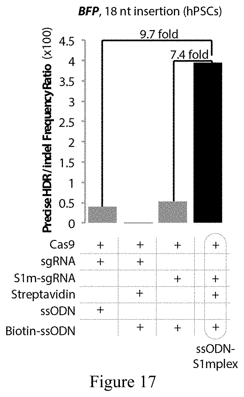
D00014
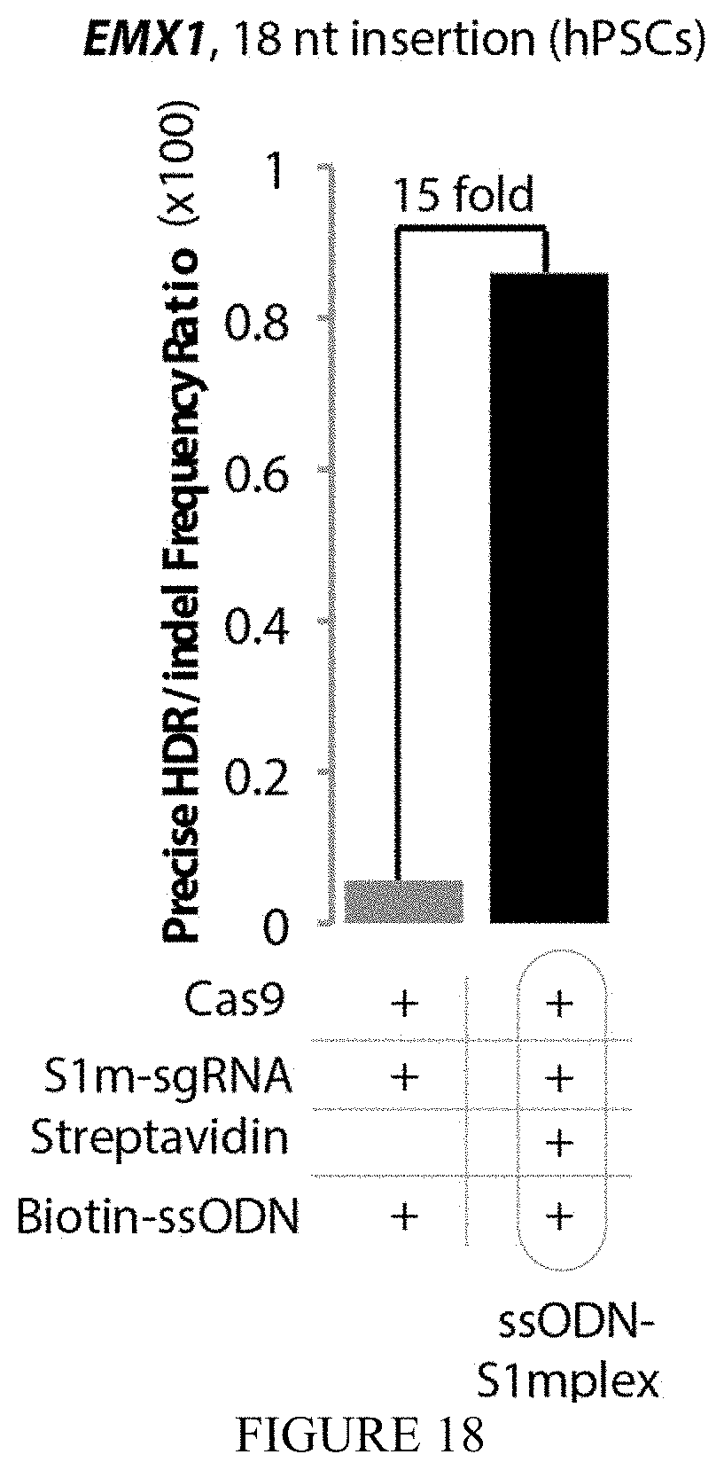
D00015

D00016
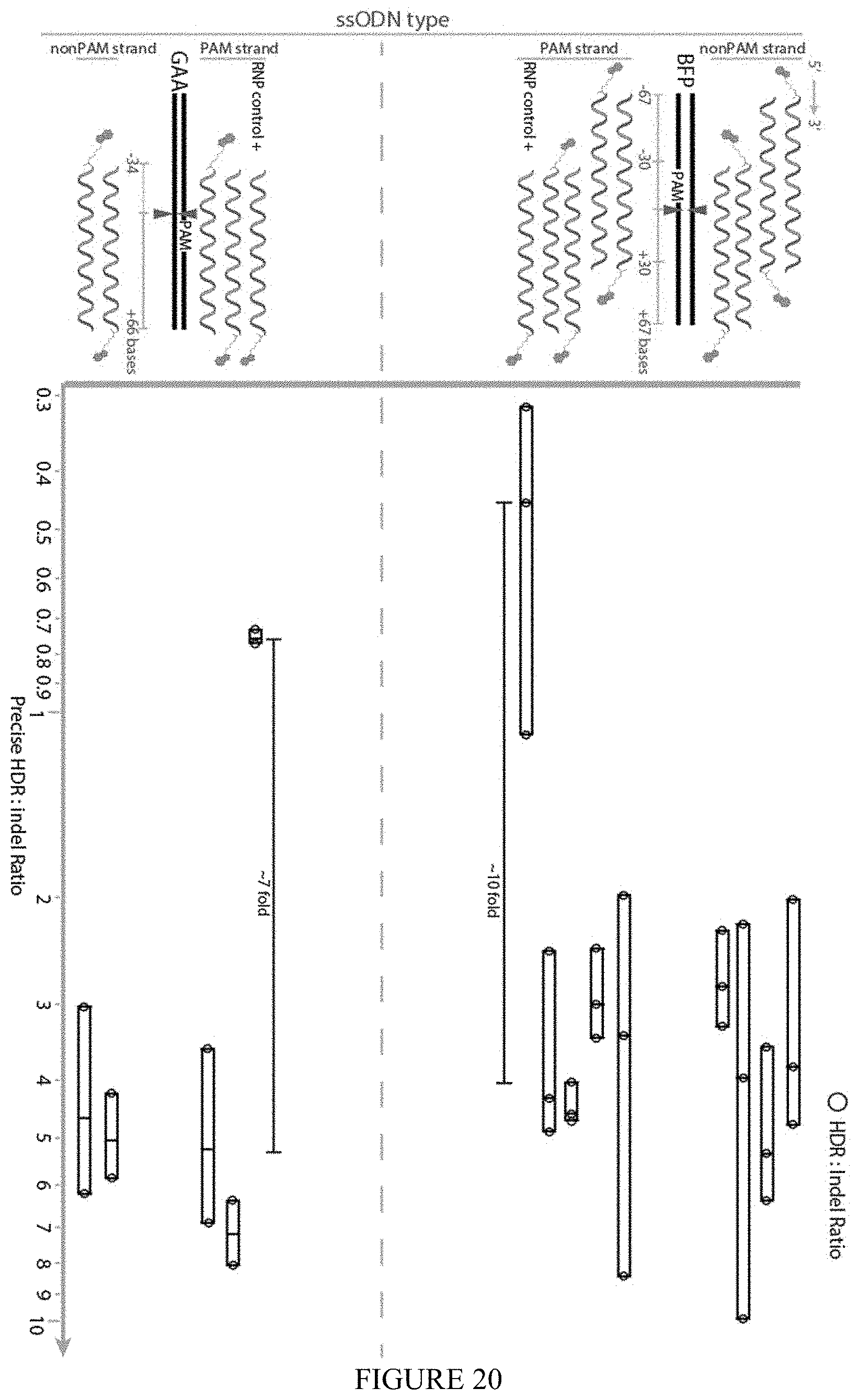
D00017

D00018

D00019
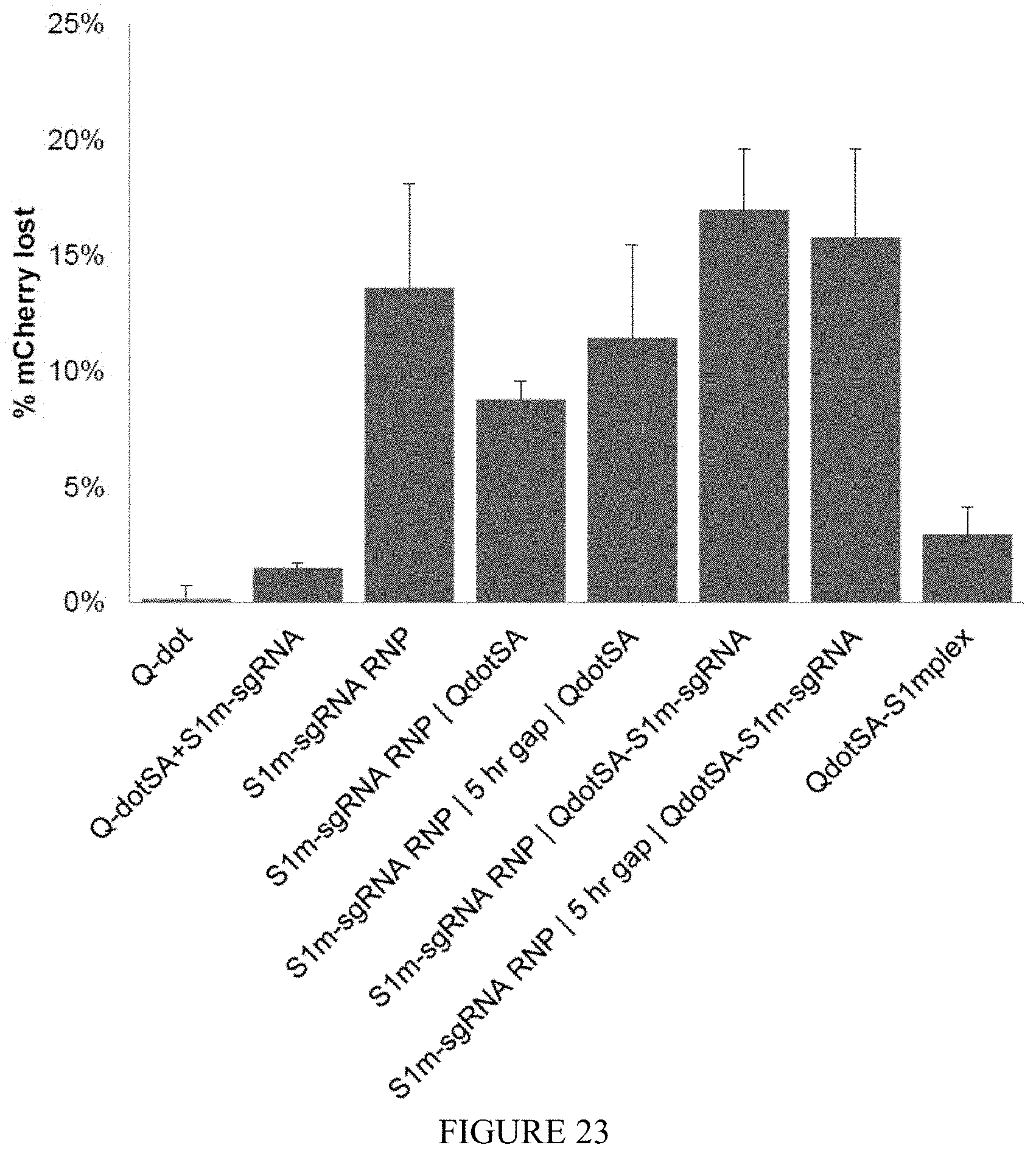
D00020
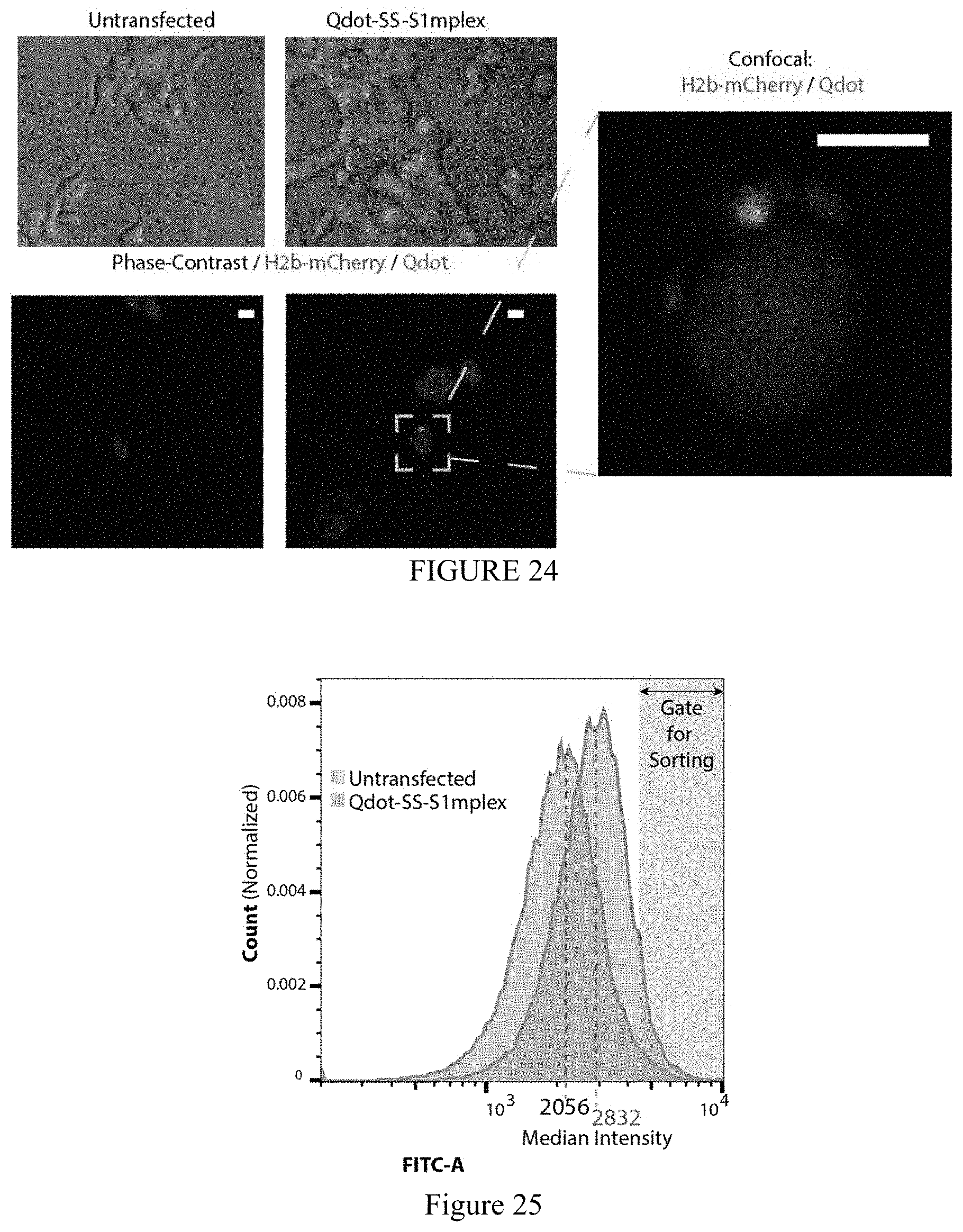
D00021
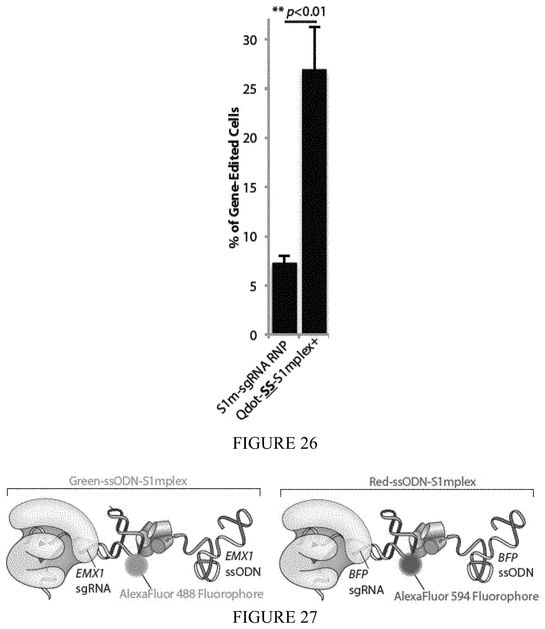
D00022

D00023

D00024
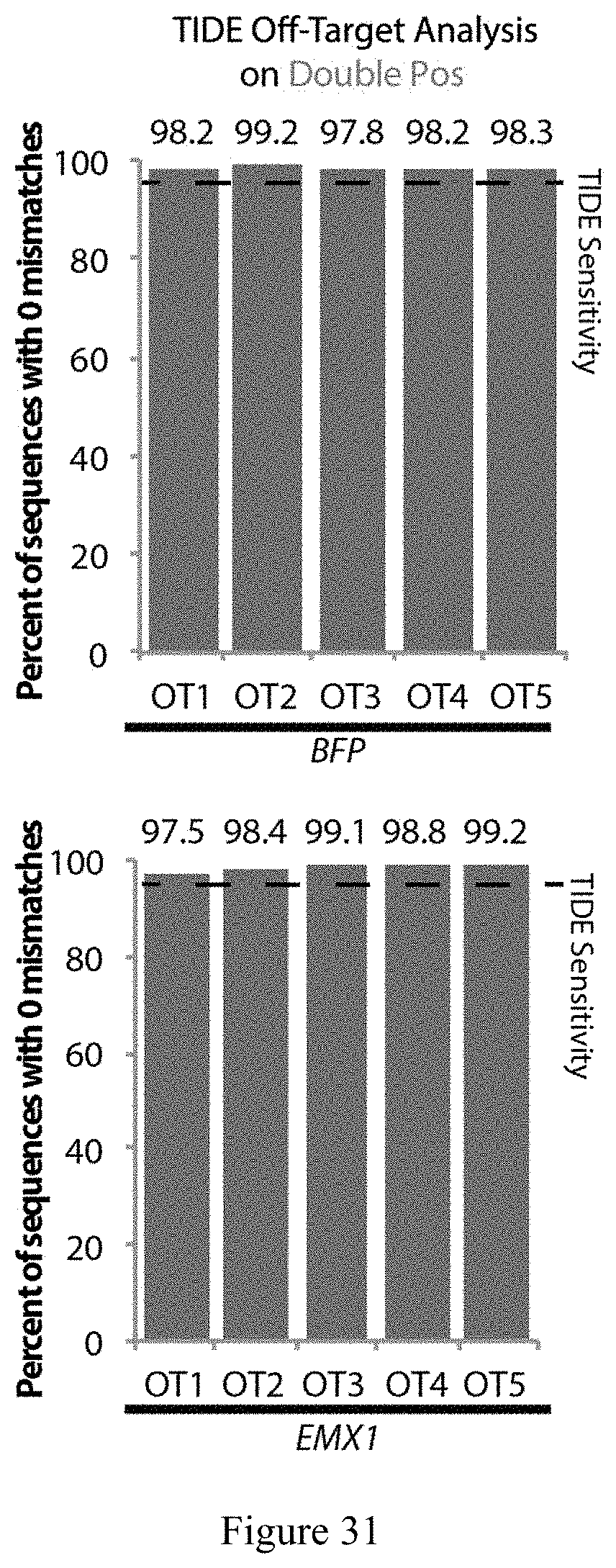
D00025
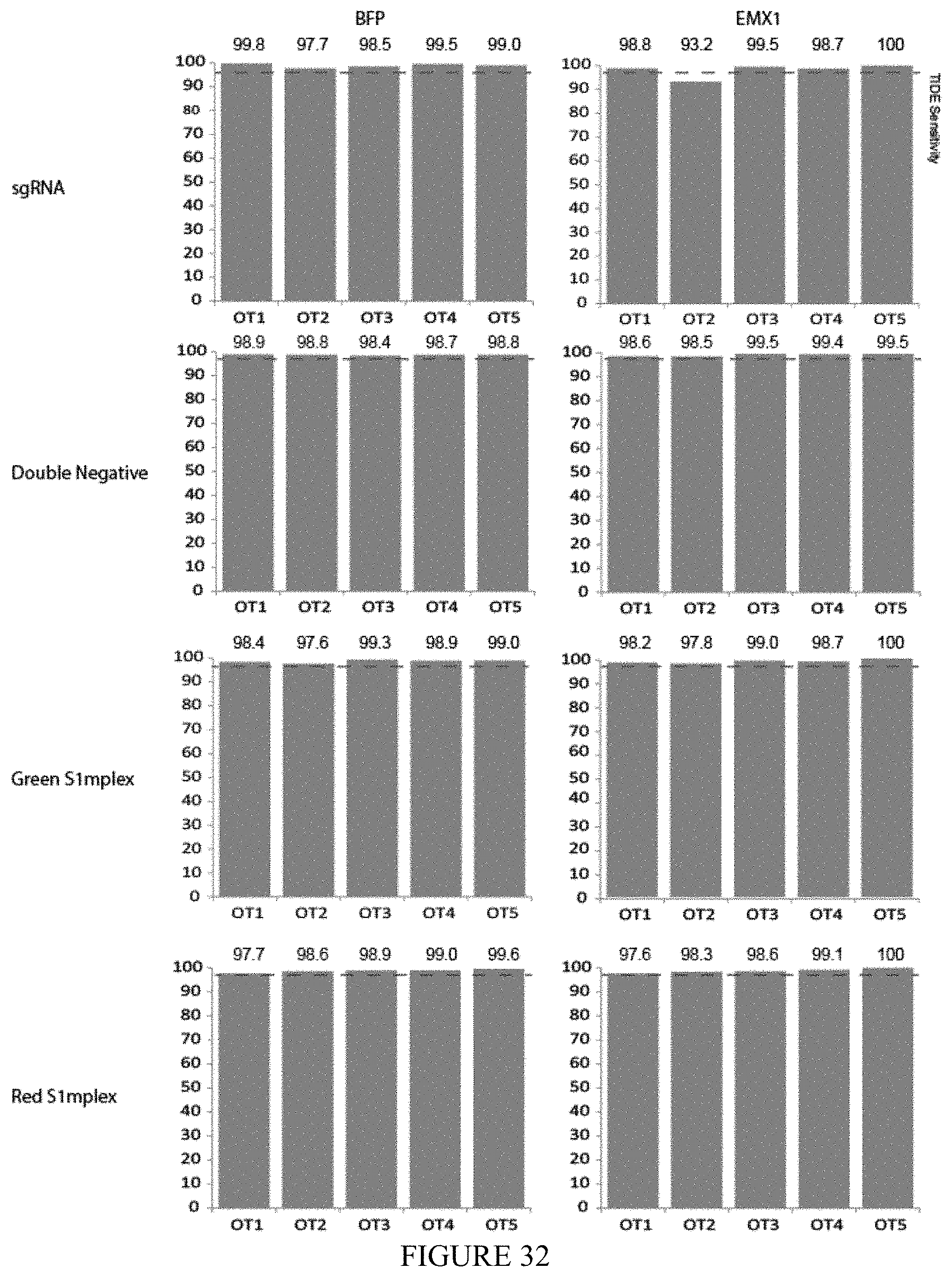
D00026

D00027

D00028

D00029
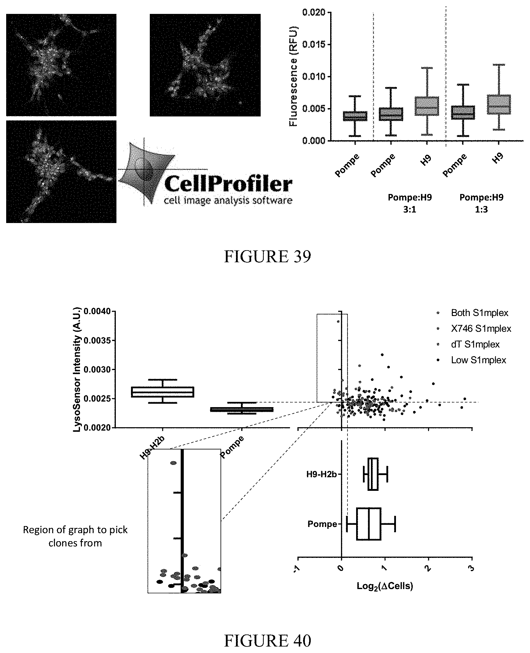
D00030

D00031
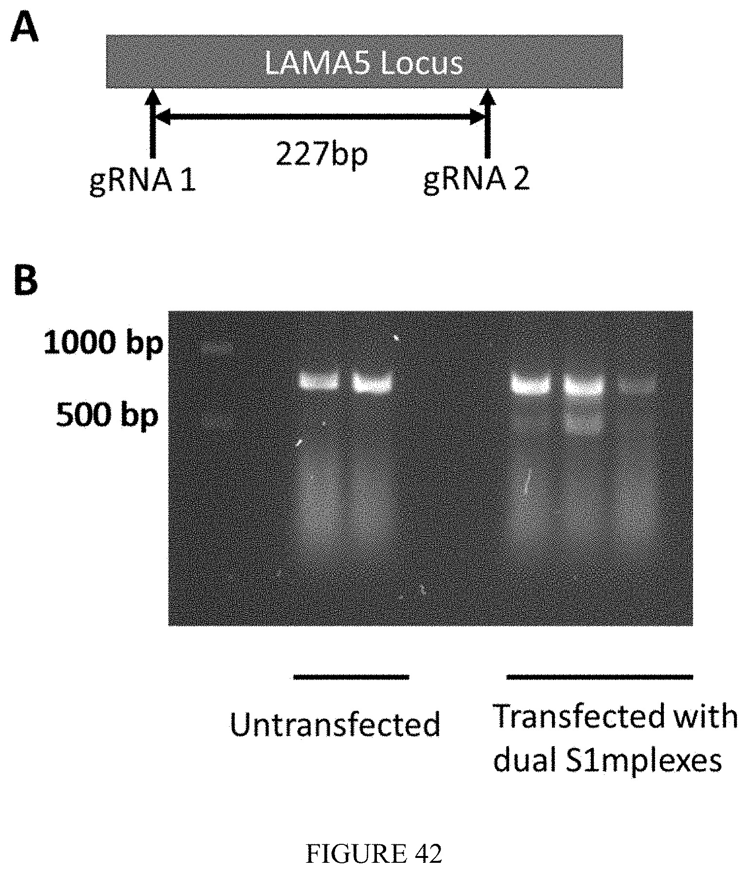
S00001
XML
uspto.report is an independent third-party trademark research tool that is not affiliated, endorsed, or sponsored by the United States Patent and Trademark Office (USPTO) or any other governmental organization. The information provided by uspto.report is based on publicly available data at the time of writing and is intended for informational purposes only.
While we strive to provide accurate and up-to-date information, we do not guarantee the accuracy, completeness, reliability, or suitability of the information displayed on this site. The use of this site is at your own risk. Any reliance you place on such information is therefore strictly at your own risk.
All official trademark data, including owner information, should be verified by visiting the official USPTO website at www.uspto.gov. This site is not intended to replace professional legal advice and should not be used as a substitute for consulting with a legal professional who is knowledgeable about trademark law.