Filovirus antibody
Lai , et al. December 29, 2
U.S. patent number 10,875,907 [Application Number 16/134,041] was granted by the patent office on 2020-12-29 for filovirus antibody. This patent grant is currently assigned to Albert Einstein College of Medicine, The Governing Council of the University of Toronto, The Government of the United States as Represented by the Secretary of the Army. The grantee listed for this patent is ALBERT EINSTEIN COLLEGE OF MEDICINE, INC., THE GOVERNING COUNCEL OF THE UNIVERSITY OF TORONTO, THE GOVERNMENT OF THE UNITED STATES AS REPRESENTED BY THE SECRETARY OF THE ARMY, THE GOVERNMENT OF THE UNITED STATES AS REPRESENTED BY THE SECRETARY OF THE ARMY. Invention is credited to Kartik Chandran, Gang Chen, John M. Dye, Julia Frei, Jayne F. Koellhoffer, Jonathan R. Lai, Sachdev Sidhu, Samantha Zak.
| United States Patent | 10,875,907 |
| Lai , et al. | December 29, 2020 |
Filovirus antibody
Abstract
The present invention addresses a need for antibodies useful for filovirus infections.
| Inventors: | Lai; Jonathan R. (Dobbs Ferry, NY), Koellhoffer; Jayne F. (New York, NY), Frei; Julia (Bronx, NY), Chandran; Kartik (Brooklyn, NY), Sidhu; Sachdev (Toronto, CA), Chen; Gang (Toronto, CA), Dye; John M. (Frederick, MD), Zak; Samantha (Frederick, MD) | ||||||||||
|---|---|---|---|---|---|---|---|---|---|---|---|
| Applicant: |
|
||||||||||
| Assignee: | Albert Einstein College of
Medicine (Bronx, NY) The Governing Council of the University of Toronto (Toronto, CA) The Government of the United States as Represented by the Secretary of the Army (Fort Detrick, MD) |
||||||||||
| Family ID: | 1000005268115 | ||||||||||
| Appl. No.: | 16/134,041 | ||||||||||
| Filed: | September 18, 2018 |
Prior Publication Data
| Document Identifier | Publication Date | |
|---|---|---|
| US 20190185547 A1 | Jun 20, 2019 | |
Related U.S. Patent Documents
| Application Number | Filing Date | Patent Number | Issue Date | ||
|---|---|---|---|---|---|
| 15327857 | Jan 20, 2017 | 10435461 | |||
| 15404662 | Jan 12, 2017 | 10391171 | |||
| 15161634 | May 23, 2016 | 10081669 | |||
| PCT/US2015/057499 | |||||
| PCT/US2015/043927 | Aug 6, 2015 | ||||
| 14291608 | May 30, 2014 | 9346875 | |||
| 62131472 | Mar 11, 2015 | ||||
| 62069516 | Oct 28, 2014 | ||||
| 62039504 | Aug 20, 2014 | ||||
| 61830325 | Jun 3, 2013 | ||||
| Current U.S. Class: | 1/1 |
| Current CPC Class: | C07K 16/10 (20130101); C07K 16/24 (20130101); C07K 2317/567 (20130101); A61K 2039/505 (20130101); C07K 2317/565 (20130101); C07K 2317/24 (20130101); C07K 2317/76 (20130101) |
| Current International Class: | C07K 16/10 (20060101); C07K 16/24 (20060101); A61K 39/00 (20060101) |
References Cited [Referenced By]
U.S. Patent Documents
| 2004/0234519 | November 2004 | Tso et al. |
| 2007/0298042 | December 2007 | Hart et al. |
| 2012/0164153 | June 2012 | Dye et al. |
| 2014/0356354 | December 2014 | Lai et al. |
Other References
|
PCT International Search Report and Written Opinion, dated Jan. 11, 2016 in connection with PCT International Application No. PCT/US2015/043927, 10 pages. cited by applicant . Chen G et al., entitled "Synthetic Antibodies with a Human Framework That Protect Mice from Lethal Sudan Ebolavirus Challenge," ACS Chemical Biology, Aug. 20, 2014, vol. 9, pp. 2263-2273. cited by applicant . Rudikoff et al., PNAS USA, 1982,79:1979-1983 (Year: 1982). cited by applicant . MacCallum et al., J. Mol. Biol., 1996,262:732-745 (Year: 1996). cited by applicant . De Pascalis is et al., The Journal of Immunology, 2002, 169:3076-3084 (Year: 2002). cited by applicant . Casset et al., BBRC, 2003, 307: 198-205 (Year: 2003). cited by applicant. |
Primary Examiner: Chen; Stacy B
Attorney, Agent or Firm: Foley Hoag LLP
Government Interests
STATEMENT OF GOVERNMENT SUPPORT
This invention was made with government support under grant number A1090249 awarded by the National Institutes of Health. The government has certain rights in the invention.
Parent Case Text
CROSS-REFERENCE TO RELATED APPLICATIONS
This application is a continuation-in-part of and claims benefit of (i) U.S. application Ser. No. 15/327,857, a U.S. national stage entry under 35 U.S.C. .sctn. 371 of PCT International Patent Application No. PCT/US2015/043927, filed Aug. 6, 2015, which claims benefit of U.S. Provisional Application No. 62/039,504, filed Aug. 20, 2014; and (ii) of U.S. application Ser. No. 15/404,662, filed Jan. 12, 2017, a continuation-in-part of PCT International Application No. PCT/US2015/57499, filed Oct. 27, 2015, which claims benefit of U.S. Provisional Application Nos. 62/131,472, filed Mar. 11, 2015 and 62/069,516, filed Oct. 28, 2014; and (iii) and of U.S. application Ser. No. 15/161,634, filed May 23, 2016, which is a continuation of U.S. patent application Ser. No. 14/291,608, filed May 30, 2014, now U.S. Pat. No. 9,346,875, issued May 24, 2016, which claims benefit of U.S. Provisional Application No. 61/830,325 filed Jun. 3, 2013, the contents of each of which are hereby incorporated by reference
Claims
What is claimed:
1. An isolated humanized anti-filovirus anti-Sudan strain Ebola virus glycoprotein pre-fusion core antibody comprising a framework region having a sequence of 95% or greater identity to a human antibody framework region, and comprising: (a) a heavy chain CDR1 comprising GFAFNYYDMF (SEQ ID NO:1); a heavy chain CDR2 comprising YIKPGGGNTYYADSV (SEQ ID NO:2); and a heavy chain CDR3 comprising QLYGNSFFDY (SEQ ID NO:3); and (b) a light chain sequence CDR1 comprising DVTTA (SEQ ID NO:4); a light chain sequence CDR2 comprising WASTR (SEQ ID NO:5); and a light chain sequence CDR3 comprising HYSTPLT (SEQ ID NO:6).
2. The humanized antibody of claim 1, wherein the light chain comprises the sequence: TABLE-US-00008 (SEQ ID NO: 7) DIQMTQSPSSLSASVGDRVTITCKASQDVTTAVAWYQQKPGKAPKLLIYW ASTRHTGVPSRFSGSGSGTDFTLTISSLQPEDFATYYCQQHYSTPLTFGQ GTKVEIK.
3. The humanized antibody of claim 1, wherein the light chain comprises the sequence: TABLE-US-00009 (SEQ ID NO: 8) DIQMTQSPSSLSASVGDRVTITCKASQDVTTAVAWYQQKPGKAPKWYWAS TRHTGVPSRFSGSGSGTDFTLTISSLQPEDFATYYCQQHYSTPLTFGQGT KVEIKRTVAAPSVFIFPPSDEQLKSGTASVVCLLNNFYPREAKVQWKVDN ALQSGNSQESVTEQDSKDSTYSLSSTLTLSKADYEKHKVYACEVTHQGLS SPVTKSFNRGECGGS or (SEQ ID NO: 13) DIQMTQSPSSLSASVGDRVTITCKASQDVTTAVAWYQQKPGKAPKWYWAS TRHTGVPSRFSGSGSGTDFTLTISSLQPEDFATYYCQQHYSTPLTFGQGT KVEIKRTVAAPSVFIFPPSDEQLKSGTASVVCLLNNFYPREAKVQWKVDN ALQSGNSQESVTEQDSKDSTYSLSSTLTLSKADYEKHKVYACEVTHQGLS SPVTKSFNRGEC.
4. The humanized antibody of claim 1, wherein the heavy chain comprises the sequence: TABLE-US-00010 (SEQ ID NO: 9) EVQLVESGGGLVQPGGSLRLSCAASGFAFNYYDMFWVRQAPGKGLEWVAY IKPGGGNTYYADSVKGRFTISADTSKNTAYLQMNSLRAEDTAVYYCARQL YGNSFFDYWGQGTLVTVSS.
5. The humanized antibody of claim 1, wherein the heavy chain comprises the sequence: TABLE-US-00011 (SEQ ID NO: 10) EVQLVESGGGLVQPGGSLRLSCAASGFAFNYYDMFWVRQAPGKGLEWVAYI KPGGGNTYYADSVKGRFTISADTSKNTAYLQMNSLRAEDTAVYYCARQLYG NSFFDYWGQGTLVTVSSASTKGPSVFPLAPSSKSTSGGTAALGCLVKDYFP EPVTVSWNSGALTSGVHTFPAVLQSSGLYSLSSVVTVPSSSLGTQTYICNV NHKPSNTKVDKKVEPKSCDKTHTCPPCPAPELLGRPSVFLFPPKPKDTLMI SRTPEVTCVVVDVSHEDPEVKFNWYVDGVEVHNAKTKPREEQYNSTYRVVS VLTVLHQDWLNGKEYKCKVSNKALPAPIEKTISKAKGQPREPQVYTLPPSR EEMTKNQVSLTCLVKGFYPSDIAVEWESNGQPENNYKTTPPVLDSDGSFFL YSKLTVDKSRWQQGNVFSCSVMHEALHNHYTQKSLSLSPGK, (SEQ ID NO: 11) EVQLVESGGGLVQPGGSLRLSCAASGFAFNYYDMFWVRQAPGKGLEWVAYI KPGGGNTYYADSVKGRFTISADTSKNTAYLQMNSLRAEDTAVYYCARQLYG NSFFDYWGQGTLVTVSSASTKGPSVFPLAPSSKSTSGGTAALGCLVKDYFP EPVTVSWNSGALTSGVHTFPAVLQSSGLYSLSSVVTVPSSSLGTQTYICNV NHKPSNTKVDKKVEPKSCDKTHTCPPCPAPELLGRPSVFLFPPKPKDTLMI SRTPEVTCVVVDVSHEDPEVKFNWYVDGVEVHNAKTKPREEQYNSTYRVVS VLTVLHQDWLNGKEYKCKVSNKALPAPIEKTISKAKGQPREPQVYTLPPSR EEMTKNQVSLTCLVKGFYPSDIAVEWESNGQPENNYKTTPPVLDSDGSFFL YSKLTVDKSRWQQGNVFSCSVMHEALHNHYTQKSLSLSPG, or (SEQ ID NO: 12) EVQLVESGGGLVQPGGSLRLSCAASGFAFNYYDMFWVRQAPGKGLEWVAYI KPGGGNTYYADSVKGRFTISADTSKNTAYLQMNSLRAEDTAVYYCARQLYG NSFFDYWGQGTLVTVSSASTKGPSVFPLAPSSKSTSGGTAALGCLVKDYFP EPVTVSWNSGALTSGVHTFPAVLQSSGLYSLSSVVTVPSSSLGTQTYICNV NHKPSNTKVDKKVEPKSCDKTHTCPPCPAPELLGGPSVFLFPPKPKDTLMI SRTPEVTCVVVDVSHEDPEVKFNWYVDGVEVHNAKTKPREEQYNSTYRVVS VLTVLHQDWLNGKEYKCKVSNKALPAPIEKTISKAKGQPREPQVYTLPPSR DELTKNQVSLTCLVKGFYPSDIAVEWESNGQPENNYKTTPPVLDSDGSFFL YSKLTVDKSRWQQGNVFSCSVMHEALHNHYTQKSLSLSPG.
6. An antigen-binding fragment of the antibody of claim 1.
7. A recombinant nucleic acid encoding the antibody of claim 1.
8. A cell, wherein the cell is not in a human subject, transformed with the recombinant nucleic acid of claim 7.
9. A composition comprising the antibody of claim 1 or an antigen binding fragment thereof.
10. The composition of claim 9, comprising a pharmaceutically acceptably carrier.
11. A method of treating a filovirus infection in a subject comprising administering to the subject an amount of the antibody of claim 1 or an antigen-binding fragment thereof effective to treat a filovirus infection in a subject, wherein the filovirus is a Sudan strain Ebola virus.
12. The method of claim 11, wherein the antibody or the antigen-binding fragment is administered after the subject has been exposed to the filovirus.
13. A method of inhibiting a filovirus infection of a subject comprising administering to the subject an amount of the antibody of claim 1 or an antigen-binding fragment thereof effective to inhibit a filovirus infection in a subject, wherein the filovirus is a Sudan strain Ebola virus.
14. The method of claim 13, wherein the antibody or the antigen-binding fragment is administered prior to the subject being exposed to the filovirus.
15. The antibody of claim 1 or an antigen-binding fragment thereof, wherein the antibody is a neutralizing antibody.
Description
SEQUENCE LISTING
The instant application contains a Sequence Listing which has been submitted in ASCII format via EFS-Web and is hereby incorporated by reference in its entirety. Said ASCII copy, created on Aug. 5, 2020, is named AET-00703_ST25.txt and is 19.6 KB in size.
BACKGROUND OF THE INVENTION
Throughout this application, various publications are referred to in parentheses. Full citations for these references may be found at the end of the specification. The disclosures of these publications, and all patents, patent application publications and books referred to herein are hereby incorporated by reference in their entirety into the subject application to more fully describe the art to which the subject invention pertains.
Ebola virus (EBOV) pathogenesis and cell entry: The infectious agents EBOV and Marburg virus (MARV) are the two major species of the Filoviridae family of enveloped negative-sense RNA viruses (1-4). Based on nucleotide sequence and outbreak location, isolates in the EBOV species are classified into five species: Zaire (ZEBOV), Tai Forest (TAFV), Sudan (SUDV), Reston (RESTV), and Bundibugyo (BDBV). There are two MARV variants (Marburg and Ravn). Severe human disease and deaths (30-90% case fatality rates in large outbreaks) are associated with ZEBOV, SUDV, BDBV, and MARV (2). Although the ecology of these agents remains incompletely understood, several species of African fruit bats may be reservoirs for EBOV and MARV (5). ZEBOV and SUDV are the most pathogenic among the ebolaviruses, and are the only two that have been associated with recurring outbreaks (6). Among the 13 documented ZEBOV outbreaks and the six SUDV outbreaks, the average human case fatality rates are 70% and 52%, respectively. Together, ZEBOV and SUDV account for 94% of EBOV-related deaths (6). Therefore, therapeutic agents effective against ZEBOV and SUDV would greatly reduce the threat of an EBOV pandemic.
All human outbreaks occur as a result of direct contact with infected wildlife, with subsequent person-to-person transmission, mostly through the mucosa or contaminated needles. Uncontrolled viral replication is central to EBOV/MARV-induced disease, both because it is cytopathic and because it induces dysregulation of the host immune system (2, 7, 8). Therefore, antiviral therapies that reduce viral load are expected to increase patient survival, in part, by allowing time to mount an effective immune response. While many cell types can be infected with EBOV/MARV in vitro and in vivo, antigen-presenting cells (macrophages and dendritic cells) appear to be early and sustained targets of infection in vivo. Infected macrophages are unable to stimulate a robust immune response, and cause a "cytokine storm" that is proposed to be the primary cause of the blood-clotting abnormalities and vascular leakage characteristic of EBOV/MARV hemorrhagic fever (9). Damage to other tissues (e.g., liver, kidneys, vascular endothelia) is thought to contribute to the above and to late-stage multi-organ failure. Death typically occurs 8-15 days after infection (10). Because of their high mortality rate, rapid proliferation, and potential for aerosolization, EBOV and MARV are classified as Category A biodefense pathogens. There are currently no FDA-approved treatments for EBOV or MARV infection.
The EBOV/MARV genome is a .about.19 kb single-strand negative-sense RNA genome that encodes seven genes arranged in a linear fashion (1-4). In mature viral particles and infected cells, the genome is intimately associated with four viral proteins: the nucleocapsid protein NP, the polymerase L, the polymerase accessory protein VP35, and the transcriptional activator protein VP30. This nucleocapsid structure is in turn encapsidated in a viral matrix, comprising proteins VP40 and VP24. The host-derived viral membrane bilayer surrounds, and is peripherally associated with, the matrix. Embedded in the viral membrane are trimers of the viral glycoprotein, GP, which mediates the first step in infection: delivery of the viral nucleocapsid "payload" into the cytoplasm of the host cell. GP is the target of virus-directed antibodies that neutralize extracellular filovirus particles (4, 11-14).
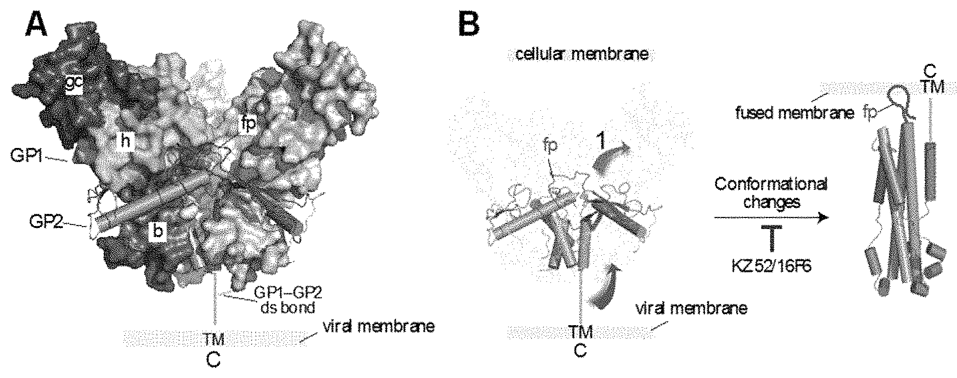
The mature EBOV/MARV GP spike is a trimer of three disulfide-linked GP1-GP2 heterodimers, generated by endoproteolytic cleavage of the GPO precursor polypeptide by furin during virus assembly (4, 13-15). GP1 mediates viral adhesion to host cells and regulates the activity of the transmembrane subunit GP2, which mediates fusion of viral and cellular membranes during cell entry. The prefusion GP1-GP2 spike has a "chalice-and-bowl" morphology--the three GP2 subunits form the chalice within which the bowl, comprised of the three GP1 subunits, rests (FIG. 1A) (13-15). This trimeric assembly is stabilized mainly by GP1-GP2 and GP2-GP2 contacts. The GP1 subunit is organized into three subdomains. The base (`b`, light blue) interacts extensively with GP2 and clamps it in its prefusion conformation. The head (`h`, green) contains a putative receptor-binding sequence. Together with GP2, the base and head subdomains of GP1 form the conserved structural core of the GP1-GP2 spike. In contrast to the GP1-GP2 core, the most external subdomains of GP1--the glycan cap (`gc`, dark blue) and the mucin-like domain (not shown)--are extensively glycosylated and display a high degree of sequence variation among filovirus isolates. In response to a fusion trigger within host cell endosomes, GP2 disengages from GP1 and undergoes a series of large-scale conformational changes that drive coalescence of viral and cellular membrane bilayers (FIG. 1B) (4, 16-19). The result of viral membrane fusion is cytoplasmic release of the viral nucleocapsid. Neutralizing antibodies likely function by inhibiting these fusion-associated conformational changes (4, 13, 14).
The present invention addresses a need for antibodies for filovirus infections.
SUMMARY OF THE INVENTION
The present invention addresses a need for improved treatments for filovirus infections. This invention provides An isolated humanized anti-filovirus glycoprotein pre-fusion core antibody comprising a framework region having a sequence of 95% or greater identity to a human antibody framework region, and comprising: (a) a heavy chain CDR1 comprising GFAFNYYDMF (SEQ ID NO:1); a heavy chain CDR2 comprising YIKPGGGNTYYADSV (SEQ ID NO:2); and a heavy chain CDR3 comprising QLYGNSFFDY (SEQ ID NO:3); and (b) a light chain sequence CDR1 comprising DVTTA (SEQ ID NO:4); a light chain sequence CDR2 comprising WASTR (SEQ ID NO:5); and a light chain sequence CDR3 comprising HYSTPLT (SEQ ID NO:6).
Also provided is an antigen-binding fragment of any of the antibodies described herein.
Also provided is composition comprising any of the antibodies described herein or the antigen-binding fragments described herein. In an embodiment, the composition comprises a pharmaceutically acceptably carrier.
Also provided is a method of treating a filovirus infection in a subject comprising administering to the subject an amount of any of the antibodies described herein, or an amount of any of the antigen-binding fragments described herein or an amount of any of the compositions described herein effective to treat a filovirus infection in a subject.
Also provided is a method of inhibiting a filovirus infection of a subject comprising administering to the subject an amount of any of the antibodies described herein, or an amount of any of the antigen-binding fragments described herein or an amount of any of the compositions described herein effective to inhibit a filovirus infection in a subject.
BRIEF DESCRIPTION OF THE DRAWINGS
FIG. 1A-1B: Structure of the pre-fusion GP1-GP1 spike and GP conformational changes that lead to viral membrane fusion. FIG. 1A shows the structure of the GP1-GP2 spike ectodomain (PDB ID: 3CSY, ref 13). The GP1 subunits are shown in surface-shaded view and GP2 as rods and loops. One GP1 subunit is colored to show the subdomains: b, base; h, head; gc, glycan cap. fp, fusion peptide. TM, transmembrane domain. C, GP C-terminus. FIG. 1B shows membrane fusion-associated conformational rearrangements in GP2 inferring from its pre-fusion and putative post-fusion structures (PDB ID: 1EBO, ref 17).
DETAILED DESCRIPTION OF THE INVENTION
An isolated humanized anti-Filovirus glycoprotein pre-fusion core antibody comprising a framework region having a sequence of 95% or greater identity to a human antibody framework region, and comprising: (a) a heavy chain CDR1 comprising GFAFNYYDMF (SEQ ID NO:1); a heavy chain CDR2 comprising YIKPGGGNTYYADSV (SEQ ID NO:2); and a heavy chain CDR3 comprising QLYGNSFFDY (SEQ ID NO:3); and (b) a light chain sequence CDR1 comprising DVTTA (SEQ ID NO:4); a light chain sequence CDR2 comprising WASTR (SEQ ID NO:5); and a light chain sequence CDR3 comprising HYSTPLT (SEQ ID NO:6).
In embodiments, the heavy chain CDR1 comprises the sequence GFAFNYYDMF (SEQ ID NO: 1).
In embodiments, the heavy chain CDR2 comprises the sequence YIKPGGGNTYYADSV (SEQ ID NO: 2).
In embodiments, the heavy chain CDR3 comprises the sequence QLYGNSFFDY (SEQ ID NO: 3).
In embodiments, the light chain CDR3 comprises QHYSTPLT (SEQ ID NO: 14). In embodiments, the light chain CDR3 comprises CQQHYSTPLT (SEQ ID NO: 15).
In embodiments, the heavy chain of antibody comprises the following sequence:
TABLE-US-00001 (SEQ ID NO: 9) EVQLVESGGGLVQPGGSLRLSCAASGFAFNYYDMFWVRQAPGKGLEWVAY IKPGGGNTYYADSVKGRFTISADTSKNTAYLQMNSLRAEDTAVYYCARQL YGNSFFDYWGQGTLVTVSS
The heavy chain can comprise any constant region, preferably a constant region having a sequence identical to a human antibody constant region or 90% or more identical thereto. In embodiments, the heavy chain of antibody comprises the following sequence:
TABLE-US-00002 (SEQ ID NO: 10) EVQLVESGGGLVQPGGSLRLSCAASGFAFNYYDMFWVRQAPGKGLEWVAY IKPGGGNTYYADSVKGRFTISADTSKNTAYLQMNSLRAEDTAVYYCARQL YGNSFFDYWGQGTLVTVSSASTKGPSVFPLAPSSKSTSGGTAALGCLVKD YFPEPVTVSWNSGALTSGVHTFPAVLQSSGLYSLSSVVTVPSSSLGTQTY ICNVNHKPSNTKVDKKVEPKSCDKTHTCPPCPAPELLGRPSVFLFPPKPK DTLMISRTPEVTCVVVDVSHEDPEVKFNWYVDGVEVHNAKTKPREEQYNS TYRVVSVLTVLHQDWLNGKEYKCKVSNKALPAPIEKTISKAKGQPREPQV YTLPPSREEMTKNQVSLTCLVKGFYPSDIAVEWESNGQPENNYKTTPPVL DSDGSFFLYSKLTVDKSRWQQGNVFSCSVMHEALHNHYTQKSLSLSPGK
Alternatively, the heavy chain of antibody may comprise the following sequence:
TABLE-US-00003 (SEQ ID NO: 11) EVQLVESGGGLVQPGGSLRLSCAASGFAFNYYDMFWVRQAPGKGLEWVAY IKPGGGNTYYADSVKGRFTISADTSKNTAYLQMNSLRAEDTAVYYCARQL YGNSFFDYWGQGTLVTVSSASTKGPSVFPLAPSSKSTSGGTAALGCLVKD YFPEPVTVSWNSGALTSGVHTFPAVLQSSGLYSLSSVVTVPSSSLGTQTY ICNVNHKPSNTKVDKKVEPKSCDKTHTCPPCPAPELLGRPSVFLFPPKPK DTLMISRTPEVTCVVVDVSHEDPEVKFNWYVDGVEVHNAKTKPREEQYNS TYRVVSVLTVLHQDWLNGKEYKCKVSNKALPAPIEKTISKAKGQPREPQV YTLPPSREEMTKNQVSLTCLVKGFYPSDIAVEWESNGQPENNYKTTPPVL DSDGSFFLYSKLTVDKSRWQQGNVFSCSVMHEALHNHYTQKSLSLSPG.
Alternatively, the heavy chain of antibody may comprise the following sequence:
TABLE-US-00004 (SEQ ID NO: 12) EVQLVESGGGLVQPGGSLRLSCAASGFAFNYYDMFWVRQAPGKGLEWVAY IKPGGGNTYYADSVKGRFTISADTSKNTAYLQMNSLRAEDTAVYYCARQL YGNSFFDYWGQGTLVTVSSASTKGPSVFPLAPSSKSTSGGTAALGCLVKD YFPEPVTVSWNSGALTSGVHTFPAVLQSSGLYSLSSVVTVPSSSLGTQTY ICNVNHKPSNTKVDKKVEPKSCDKTHTCPPCPAPELLGGPSVFLFPPKPK DTLMISRTPEVTCVVVDVSHEDPEVKFNWYVDGVEVHNAKTKPREEQYNS TYRVVSVLTVLHQDWLNGKEYKCKVSNKALPAPIEKTISKAKGQPREPQV YTLPPSRDELTKNQVSLTCLVKGFYPSDIAVEWESNGQPENNYKTTPPVL DSDGSFFLYSKLTVDKSRWQQGNVFSCSVMHEALHNHYTQKSLSLSPG.
In embodiments, the light chain CDR1 comprises the sequence DVTTA (SEQ ID NO: 4).
In embodiments, the light chain CDR2 comprises the sequence WASTR (SEQ ID NO: 5).
In embodiments, the light chain CDR3 comprises the sequence HYSTPLT (SEQ ID NO: 6).
In embodiments, the light chain of antibody comprises the following sequence:
TABLE-US-00005 (SEQ ID NO: 7) DIQMTQSPSSLSASVGDRVTITCKASQDVTTAVAWYQQKPGKAPKWYWAS TRHTGVPSRFSGSGSGTDFTLTISSLQPEDFATYYCQQHYSTPLTFGQGT KVEIK
In embodiments, the light chain of antibody comprises the following sequence:
TABLE-US-00006 (SEQ ID NO: 8) DIQMTQSPSSLSASVGDRVTITCKASQDVTTAVAWYQQKPGKAPKWYWAS TRHTGVPSRFSGSGSGTDFTLTISSLQPEDFATYYCQQHYSTPLTFGQGT KVEIKRTVAAPSVFIFPPSDEQLKSGTASVVCLLNNFYPREAKVQWKVDN ALQSGNSQESVTEQDSKDSTYSLSSTLTLSKADYEKHKVYACEVTHQGLS SPVTKSFNRGECGGS.
Alternatively, the light chain of antibody may comprise the following sequence:
TABLE-US-00007 (SEQ ID NO: 13) DIQMTQSPSSLSASVGDRVTITCKASQDVTTAVAWYQQKPGKAPKLLIYW ASTRHTGVPSRFSGSGSGTDFTLTISSLQPEDFATYYCQQHYSTPLTFGQ GTKVEIKRTVAAPSVFIFPPSDEQLKSGTASVVCLLNNFYPREAKVQWKV DNALQSGNSQESVTEQDSKDSTYSLSSTLTLSKADYEKHKVYACEVTHQG LSSPVTKSFNRGEC.
Also provided is an antigen-binding fragment of any of the antibodies described herein.
Also provided is composition comprising any of the antibodies described herein or the antigen-binding fragments described herein. In an embodiment, the composition comprises a pharmaceutically acceptably carrier.
Also provided is a method of treating a filovirus infection in a subject comprising administering to the subject an amount of any of the antibodies described herein, or an amount of any of the antigen-binding fragments described herein or an amount of any of the compositions described herein effective to treat a filovirus infection in a subject.
Also provided is a method of inhibiting a filovirus infection of a subject comprising administering to the subject an amount of any of the antibodies described herein, or an amount of any of the antigen-binding fragments described herein or an amount of any of the compositions described herein effective to inhibit a filovirus infection in a subject.
In an embodiment of the methods, the antibody, antigen-binding fragment or composition are administered prior to the subject being exposed to the filovirus. In an embodiment of the methods, the antibody, antigen-binding fragment or composition are administered after the subject has been exposed to the filovirus. In an embodiment of the methods, the filovirus is an Ebola virus. In an embodiment of the methods, the Ebola virus is the Sudan strain. In an embodiment of the methods, the filovirus is a Marburg virus. In an embodiment of the methods, the filovirus is not a Marburg virus.
In an embodiment of any of the antibodies described herein, or any of the antigen-binding fragments described herein or any of the compositions described herein, or the methods described herein, the antibody is a neutralizing antibody. In an embodiment, the pre-fusion core is a heterohexamer of three copies of the GP1 and 3 copies of the GP2.
In an embodiment, the isolated antibody or antigen-binding antibody fragment comprises an Fc region having a sequence identical to a human Fc region.
In an embodiment, the Fc region of the antibody is glycosylated.
A "humanized" antibody as used herein, unless otherwise indicated, is a chimeric antibodies that contain minimal sequence (CDRs) derived from non-human immunoglobulin (e.g. such as a mouse immunoglobulin). In one embodiment, a humanized antibody is an antibody having a sequence of a human immunoglobulin (recipient antibody) in which CDR residues of a hypervariable region (HVR) of the recipient are replaced by CDR residues from a non-human species (donor antibody) such as a mouse having the desired specificity. In some instances, FR residues of the human immunoglobulin variable domain are replaced by corresponding non-human residues, for example by a back-mutation. In general, a humanized antibody will comprise substantially all of at least one, and typically two, variable domains, in which all or substantially all of the hypervariable loops correspond to those of a non-human immunoglobulin, and all or substantially all of the FRs are those of a human immunoglobulin sequence. The humanized antibody optionally will also comprise at least a portion of an immunoglobulin constant region (Fc), typically that of a human immunoglobulin. See, e.g., Jones et al., Nature 321:522-525 (1986); Riechmann et al., Nature 332:323-329 (1988); Presta, Curr. Op. Struct. Biol. 2:593-596 (1992); Vaswani and Hamilton, Ann. Allergy, Asthma & Immunol. 1:105-115 (1998); Harris, Biochem. Soc. Transactions 23:1035-1038 (1995); Hurle and Gross, Curr. Op. Biotech. 5:428-433 (1994); and U.S. Pat. Nos. 6,982,321 and 7,087,409, the contents of each of which references and patents are hereby incorporated by reference in their entirety. Other techniques to humanize a monoclonal antibody are described in U.S. Pat. Nos. 4,816,567; 5,807,715; 5,866,692; 6,331,415; 5,530,101; 5,693,761; 5,693,762; 5,585,089; and 6,180,370, the content of each of which is hereby incorporated by reference in its entirety. The framework regions of the antibodies of the invention having a sequence identical to a human framework region may include amino acid residues not encoded by human germline sequences (e.g., mutations introduced by random or site-specific mutagenesis). In an embodiment, the isolated antibody or antigen-binding antibody fragment comprises a variable domain framework sequence having a sequence identical to a human variable domain framework sequence FR1, FR2, FR3 or FR4. In an embodiment, the isolated antibody or antigen-binding antibody fragment comprises a variable domain framework sequence having a sequence identical to at least two of human variable domain framework sequences FR1, FR2, FR3 or FR4. In an embodiment, the isolated antibody or antigen-binding antibody fragment comprises a variable domain framework sequence having a sequence identical to at least three of human variable domain framework sequences FR1, FR2, FR3 or FR4. In an embodiment, the isolated antibody or antigen-binding antibody fragment comprises a variable domain framework sequence having a sequence identical to all four of human variable domain framework sequences FR1, FR2, FR3 and FR4.
An isolated nucleic acid is provided encoding a VH or a VL of the antibodies, or fragments thereof, as described herein. In an embodiment, the isolated nucleic acid is a DNA. In an embodiment, the isolated nucleic acid is a cDNA. In an embodiment, the isolated nucleic acid is a RNA. A recombinant nucleic acid encoding an antibody as described herein is also provided. Also provided is a cell, wherein the cell is not in a human subject, transformed with the recombinant nucleic acid. In an embodiment, the cell is a mammalian cell. In an embodiment, the cell is derived from a human but is not in a human subject. In an embodiment, the cell is not a human cell.
As used herein, "at least 90% identical to" encompasses a sequence that has at least 91%, 92%, 93%, 94%, 95%, 96%, 97%, 98% or 99% identity with, or is 100% identical to, the referenced sequence. All other percent identities are defined analogously. Accordingly, the individual embodiments of at least 95% identical to, at least 96% identical to, at least 97% identical to, at least 98% identical to, at least 99% identical to, and 100% identical to, all encompassed by the invention with regard to Fc human sequences or human framework variable sequences, are each all separately encompassed by the invention.
The antigen, in regard to the term "antigen-binding fragment" as used herein, is a Filovirus glycoprotein pre-fusion core.
In an embodiment of the antibodies, fragments, methods and compositions described herein, the fragment of the antibody comprises an Fab, an Fab', an F(ab')2, an Fd, an Fv, or a complementarity determining region (CDR). In an embodiment, the fragment comprises a CDR3 of a VH chain. In an embodiment the fragment further comprises one of, more than one of, or all of CDR1, CDR2 of Vh and CDR1, CDR2 and CDR3 of a VL. As used herein, an Fd fragment means an antibody fragment that consists of the VH and CH1 domains; an Fv fragment consists of the VL and VH domains of a single arm of an antibody; and a dAb fragment (Ward et al., Nature 341:544-546 (1989) hereby incorporated by reference in its entirety) consists of a VH domain. In some embodiments, fragments are at least 5, 6, 8 or 10 amino acids long. In other embodiments, the fragments are at least 14, at least 20, at least 50, or at least 70, 80, 90, 100, 150 or 200 amino acids long.
In an embodiment, the fragment of the antibody encompassed by the invention is a single-chain antibody (scFv) is a variable domain light chain (VL) and a variable domain heavy chain (VH) which are linked N--C or C--N, respectively, via a peptide linker. In an embodiment the linker of the scFv is 5-30 amino acids in length. In an embodiment the linker of the scFv is 10-25 amino acids in length. In an embodiment the peptide linker comprises glycine, serine and/or threonine residues. For example, see Bird et al., Science, 242: 423-426 (1988) and Huston et al., Proc. Natl. Acad. Sci. USA, 85:5879-5883 (1988), each of which are hereby incorporated by reference in their entirety. In an embodiment, the fragment of the antibody of the invention is not a single-chain antibody (scFv).
The term "Fc region" herein is used to define a C-terminal region of an immunoglobulin heavy chain, including native sequence Fc regions and variant Fc regions. Although the boundaries of the Fc region of an immunoglobulin heavy chain might vary, the human IgG heavy chain Fc region is often defined to stretch from an amino acid residue at position Cys226, or from Pro230, to the carboxyl-terminus thereof. The C-terminal lysine of the Fc region may be removed, for example, during production or purification of the antibody, or by recombinantly engineering the nucleic acid encoding a heavy chain of the antibody. Accordingly, an intact antibody as used herein may be an antibody with or without the otherwise C-terminal cysteine.
In an embodiment, the antibodies of the invention described herein comprise a human Fc region or a variant human Fc region. A variant human Fc region comprises an amino acid sequence which differs from that of a native sequence Fc region by virtue of at least one amino acid modification, yet retains at least one effector function of the native sequence human Fc region. Preferably, the variant Fc region has at least one amino acid substitution compared to a native sequence Fc region or to the Fc region of a parent polypeptide, e.g. from about one to about ten amino acid substitutions, and preferably, from about one to about five amino acid substitutions in a native sequence Fc region or in the Fc region of the parent polypeptide. The variant Fc region herein will preferably possess at least about 80% sequence identity with a native sequence Fc region and/or with an Fc region of a parent polypeptide, and most preferably, at least about 90% sequence identity therewith, more preferably, at least about 95%, at least about 96%, at least about 97%, at least about 98%, at least about 99% sequence identity therewith.
Although the boundaries of the Fc region of an immunoglobulin heavy chain might vary, the human IgG heavy chain Fc region is often defined to stretch from an amino acid residue at position Cys226, or from Pro230, to the carboxyl-terminus thereof. The C-terminal lysine of the Fc region may be removed, for example, during production or purification of the antibody, or by recombinantly engineering the nucleic acid encoding a heavy chain of the antibody. Accordingly, an intact antibody as used herein may be an antibody with or without the otherwise C-terminal cysteine.
In an embodiment of the methods, the antibody, antibodies, antibody fragment or antibody fragments are administered as an adjuvant therapy to a primary therapy for the disease or condition.
The invention also provides diagnostic kits comprising any or all of the antibodies described herein. The diagnostic kits are useful for, for example, detecting the presence of a filovirus in a sample.
The humanized antibodies of the invention exclude any antibodies that naturally occur in a human.
As used herein, the term "isolated antibody" refers to an antibody that by virtue of its origin or source of derivation has one to four of the following characteristics: (1) is not associated with naturally associated components that accompany it in its native state, (2) is free of other proteins from the same species, (3) is expressed by a cell from a different species, or (4) does not occur in nature.
The phrase "and/or" as used herein, with option A and/or option B for example, encompasses the embodiments of (i) option A, (ii) option B, and (iii) option A plus option B.
It is understood that wherever embodiments are described herein with the language "comprising," otherwise analogous embodiments described in terms of "consisting of" and/or "consisting essentially of" are also provided.
Where aspects or embodiments of the invention are described in terms of a Markush group or other grouping of alternatives, the present invention encompasses not only the entire group listed as a whole, but each member of the group subjectly and all possible subgroups of the main group, but also the main group absent one or more of the group members. The present invention also envisages the explicit exclusion of one or more of any of the group members in the claimed invention.
All combinations of the various elements described herein are within the scope of the invention unless otherwise indicated herein or otherwise clearly contradicted by context.
In the event that one or more of the literature and similar materials incorporated by reference herein differs from or contradicts this application, including but not limited to defined terms, term usage, described techniques, or the like, this application controls.
This invention will be better understood from the Experimental Details, which follow. However, one skilled in the art will readily appreciate that the specific methods and results discussed are merely illustrative of the invention as described more fully in the claims that follow thereafter.
EXPERIMENTAL RESULTS
Example 1
There is a gap in treatment of EBOV infection: Only a handful of animal challenge studies have been performed with mAb therapies, in part because few mAbs that target GP (the primary neutralization target) exist. Most antibodies elicited in natural infection react preferentially with a secreted, dimeric version of the glycoprotein known as sGP and do not neutralize the fusion-relevant GP spike (4, 23, 24). Wilson et al. first demonstrated that GP-specific neutralizing antibodies (nAbs) could protect mice from ZEBOV challenge (25). However, three of five protective antibodies recognize highly variable sequences within the GP1 mucin-like domain, rendering them unlikely candidates for development of cross-neutralizing mAbs. Antibodies KZ52 and 16F6 are among the few well-characterized nAbs and both bind to the GP prefusion core (elaborated further in Section 3b) (13, 14). KZ52 was identified by phage-based panning of a B-cell antibody library isolated from a human survivor of ZEBOV infection (26). Initial experiments in rodent protection studies were promising, but KZ52 failed to protect in macaques when administered on days -1 and +4 at 50 mg/kg (12, 20). However, it is possible that a more aggressive treatment regimen may provide protection. 16F6, a mouse mAb, was identified recently by Dr. Dye's group by vaccination with vector-based vaccine expressing SUDV GP (14). mAb 16F6 is much more potent than is KZ52 against the corresponding virus species, but its murine scaffold limits therapeutic utility at this point. Head-to-head comparison in neutralizations assays using a vesicular stomatitis virus pseudotype (VSV-GP) with KZ52 (against ZEBOV GP, GP.sub.ZEBOV) and 16F6 (against SUDV GP, GP.sub.SUDV) indicates that 16F6 can reduce infectivity by at least 10-fold more than KZ52 at high antibody concentrations (FIG. 3). The cause for this discrepancy is not clear, but these data nonetheless demonstrate that there is room for improvement in KZ52 activity. An immunocompetent mouse SUDV model is not available; however 16F6 treatment delays death of SCID mice challenged with SCID-adapted SUDV by 5-7 days (14). It is therefore likely that an optimized 16F6 variant will be protective.
Several candidate therapies and vaccines are under exploration for filovirus infection (27-33). Multiple promising vaccine candidates are able to protect NHPs from lethal challenge, including adenovirus-vectored, VSV-vectored, and virus-like particle-based vaccines (28-30). While any safe and effective EBOV vaccine will be useful for populations or workers that are at high risk for exposure, it is unlikely that vaccination against EBOV will be practical on a general population level. Therefore, there is still a need for an EBOV therapy that can be used to treat acute exposure or infection. Other biologics are under evaluation, including an antisense therapy undergoing clinical trials, and a promising RNAi therapy (31, 32). However, the use of nucleic acids as therapeutic agents in general is in its infancy and therefore there is a high barrier to FDA approval for such biologics. Furthermore, these therapeutic nucleic acids are strain-specific. Some small molecules against EBOV or host targets are also being explored, but studies are largely limited to early proof-of-concept stage (33-35). A mAb treatment has lower barriers to FDA approval than other therapeutic platforms given the broad use of mAbs in autoimmune diseases and cancer, as well as more recent use in prevention and treatment of infectious diseases (36).
Synthetic antibody engineering permits identification of antibodies with enhanced properties: Antibody phage display has emerged as a powerful alternative to hybridoma technology for the generation of mAbs (37-40). It is now possible to select high-affinity antibodies against virtually any antigen from phage libraries that bear tailored diversity elements encoded by synthetic DNA ("synthetic antibodies") (41-45). This approach obviates the requirement for human or animal immunization, greatly reducing the labor and cost of antibody production. Selective enrichment of high-affinity binders from phage antibody libraries under controlled conditions enhances the reliability of output antibodies, and permits selection of binding with user-specified stringency (45). The expression of antibody domains on the surface of bacteriophage was first reported nearly two decades ago, but only recently have synthetic libraries (where diversity is not borne from natural source repertoires) become sophisticated enough for general use. Combined empirical and bioinformatic data guide predictions of locations in the antibody complementarity determining regions (CDRs) that favor antigen recognition (38, 41). The chemical (i.e., amino acid side chain) diversity encoded at these CDR positions can then be specified with designed codon sets that reduce sequence complexity but optimize combining site properties for molecular recognition (41, 42).
Humanizing SUDV-specific antibody 16F6 ("hu16F6"): 16F6 itself is of limited therapeutic utility because it is a murine antibody (14). (See also WO/2011/071574 for 16F6 antibodies. The contents of WO/2011/071574 are hereby incorporated by reference in their entirety). A sequence alignment of 16F6 in comparison to a synthetic antibody based on the optimized human framework of Herceptin (YADS1, ref 48) is shown in FIG. 4, and a structural alignment of the variable domains shown in FIG. 5. Notably, 16F6 and YADS1 have high homology in the framework regions and the structural alignment shows that positioning of the CDR beginning and end points is similar among the two scaffolds. This analysis suggests that 16F6 can be humanized by grafting the 16F6 CDR segments onto the YADS1 framework to produce a 16F6-YADS1 chimera Fab. A summary of the steps is as follows:
Randomization is included at framework or structural (i.e., non-contact) CDR positions in a manner that permits the residue identity of 16F6, YADS1, or side chains with similar physicochemical attributes. Two positions on the YADS1 scaffold that correspond to contacting framework residues in 16F6 (T53 and T56) are diversified to allow for all 20 genetically-encoded amino acids. The `theoretical diversity` of this library is 4.times.10.sup.7, which can be exhaustively sampled by phage display libraries that contain >10.sup.10 unique members. Binding to the target was assessed at a preliminary level by phage ELISA. The most promising clones were produced as IgGs and screened for neutralization against vesicular stomatitis virus pseudotyped with GP.sub.SUDV (VSV-GP.sub.SUDV).
Neutralization of authentic SUDV and binding to GPSUDV: The antibody disclosed herein neutralized authentic SUDV by 80% or more at less than 0.625 .mu.g/mL with complement and 1.25 .mu.g/mL without complement. There was no cross-reactivity for GP from ZEBOV (GP Zaire) or 5% non-fat dry milk (NFDM). The half-maximal binding titers for GPSUDV was 5.1 nM.
REFERENCES
1. Kuhn, J. H., Becker, S., Ebihara, H., Geisbert, T. W., Johnson, K. M., Kawaoka, Y., Lipkin, W. I., Negredo, A. I., Netesov, S. V., Nichol, S. T., Palacios, G., Peters, C. J., Tenorio, A., Volchkov, V. E., and Jahrling, P. B. (2010) Proposal for a revised taxonomy of the family Filoviridae: classification, names of taxa and viruses, and virus abbreviations. Arch. Virol. 155, 2083-2103. 2. Feldmann, H., and Gesibert, T. W. (2011) Ebola haemorrhagic fever. Lancet 9768, 849-862. 3. Miller, E. H., and Chandran, K. (2012) Filovirus entry into cells--new insights. Curr. Opin. Virol. 2, 206-214. 4. Lee, J. E., and Saphire, E. O. (2009) Neutralizing ebolavirus: structural insights into the envelope glycoprotein and antibodies targeted against it. Curr. Opin. Struct. Biol. 19, 408-417. 5. Leroy, E. M., Kumulungui, B., Pourrut, X., Rouquet, P., Hassanin, A., Yaba, P., Delicat, A., Paweska, J. T., Gonzalez, J. P., and Swanepoel, R. (2005) Fruit bats as reservoirs of Ebola virus. Nature 438, 575-576. 6. http://www.cdc.gov/ncidod/dvrd/spb/mnpages/dispages/ebola.htm 7. Bradfute, S. B., Warfield, K. L., and Bavari, S. (2008) Functional CD8+ T cell responses in lethal Ebola virus infection. J. Immunol. 180, 4058-4066. 8. Zampieri, C. A., Sullivan, N. J., and Nabel, G. J. (2007) Immunopathology of highly virulent pathogens: insights from Ebola virus. Nat. Immunol. 8, 1159-1164. 9. Geisbert, T. W., Hensley, L. E., Larsen, T., Young, H. A., Reed, D. S., Geisbert, J. B., Scott, D. P., Kagan, E., Jahrling, P. B., and Davis, K. J. (2003) Pathogenesis of Ebola hemorrhagic fever in cynomolgus macaques: evidence that dendritic cells are early and sustained targets of infection. Am. J. Pathol. 163, 2347-2370. 10. Hensley, L. E., Jones, S. M., Feldmann, H., Jahrling, P. B., and Geisbert, T. W. (2005) Ebola and Marburg viruses: pathogenesis and development of countermeasures. Curr. Mol. Med. 5, 761-772. 11. Wilson, J. A., and Hart, M. K. (2001) Protection from Ebola virus mediated by cytotoxic T lymphocytes specific for the viral nucleoprotein. J. Virol. 75, 2660-2664. 12. Parren, P. W., Geisbert, T. W., Maruyama, T., Jahrling, P. B., and Burton, D. R. (2002) Pre- and postexposure prophylaxis of Ebola virus infection in an animal model by passive transfer of a neutralizing human antibody. J. Virol. 76, 6408-6412.
13. Lee, J. E., Fusco, M. L., Hessell, A. J., Oswald, W. B., Burton, D. R., and Saphire, E. O. (2008) Structure of the Ebola virus glycoprotein bound to an antibody from a human survivor. Nature 454, 177-182. 14. Dias, J. M., Kuehne, A. I., Abelson, D. M., Bale, S., Wong, A. C., Halfmann, P., Muhammad, M. A., Fusco, M. L., Zak, S. E., Kang, E., Kawaoka, Y., Chandran, K., Dye, J. M., and Saphire, E. O. (2011) A shared structural solution for neutralizing ebolaviruses. Nat. Struct. Mol. Biol. 18, 1424-1427. 15. Lee, J. E., and Saphire, E. O. (2009) Ebolavirus glycoprotein structure and mechanism of entry. Future Virol. 4, 621-635. 16. Weissenhorn, W., Carfi, A., Lee, K.-H., Skehel, J. J., and Wiley, D. C. (1998) Crystal structure of the Ebola virus membrane fusion subunit, GP2, from the envelope glycoprotein ectodomain. Mol. Cell 2, 605-616. 17. Malashkevich, V. N., Schneider, B. J., McNally, M. L., Milhollen, M. A., Pang, J. X., and Kim, P. S. (1999) Core structure of the envelope glycoprotein GP2 from Ebola virus at 1.9-.ANG. resolution. Proc. Natl. Acad. Sci. USA 96, 2662-2667. 18. Harrison, J. S., Higgins, C. D., Chandran, K., and Lai, J. R. (2011) Designed protein mimics of the Ebola virus glycoprotein GP2 .alpha.-helical bundle: Stability and pH effects. Protein Sci. 20, 1587-1596. 19. Harrison, J. S., Koellhoffer, J. F., Chandran, K., and Lai, J. R. (2012) Marburg virus glycoprotein GP2: pH-dependent stability of the ectodomain .alpha.-helical bundle. Biochemistry 51, 2515-2525. 20. Oswald, W. B., Geisbert, T. W., Davis, K. J., Geisbert, J. B., Sullivan, N. J., Jahrling, P. B., Parren, P. W., and Burton, D. R. (2007) Neutralizing antibody fails to impact the course of Ebola virus infection in monkeys. PLoS Pathog. 3, e9. 21. Dye, J. M., Herbert, A. S., Kuehne, A. I., Barth, J. F., Muhammad, M. A., Zak, S. E., Ortiz, R. A., Prugar, L. I., and Pratt, W. D. (2012) Postexposure antibody prophylaxis protects nonhuman primates from Filovirus disease. Proc. Natl. Acad. Sci. USA 109, 5034-5039. 22. Marzi, A., Yoshida, R., Miyamoto, H., Ishijim, M., Suzuki, Y., Higuchi, M., Matsuyama, Y., Igarashi, M., Nakayama, E., Kuroda, M., Saijo, M., Feldmann, F., Brining, D., Feldmann, H., and Takada A. (2012) Protective efficacy of neutralizing monoclonal antibodies in a nonhuman primate model of Ebola hemorrhagic fever. PLoS One 7, e36192. 23. Wilson, J. A., Bosio, C. M., and Hart, M. K. (2001) Ebola virus: the search for vaccines and treatments. Cell Mol. Life Sci. 58, 1826-1841. 24. Sullivan, N. J., Martin, J. E., Graham, B. S., and Nabel, G. J. (2009) Correlates of protective immunity for Ebola vaccines: implications for regulatory approval by the animal rule. Nat. Rev. Microbiol. 7, 393-400. 25. Wilson, J. A., Hevey, M., Bakken, R., Guest, S., Bray, M., Schmaljohn, A. L., and Hart, M. K. (2000) Epitopes involved in antibody-mediated protection from Ebola virus. Science 287, 1664-1666. 26. Maruyama, T., Rodriguez, L. L., Jahrling, P. B., Sanchez, A., Khan, A. S., Nichol, S. T., Peters, C. J., Parren, P. W., and Burton, D. R. (1999) Ebola virus can be effectively neutralized by antibody produced in natural human infection. J. Virol. 73, 6024-6030. 27. Shurtleff, A. C., Warren, T. K., and Bavari, S. (2011) Non-human primates as models for the discovery and development of ebolavirus therapeutics. Expert Opin. Drug Discov. 6, 233-250. 28. Warfield, K. L., and Aman, M. J. (2011) Advances in virus-like particle vaccines for filoviruses. J. Infect. Dis. 204 Suppl 3, S1053-1059. 29. Fausther-Bovendo, H., Mulangu, S., and Sullivan, N. J. (2012) Ebolavirus vaccines for humans and apes. Curr. Opin. Virol. [Epub ahead of print] (May 3, PMID: 22560007) 30. Hoenen, T., Grosth, A., and Feldmann, H. (2012) Current ebola vaccines. Expert Opin. Biol. Ther. [Epub ahead of print] (May 5, PMID: 22559078) 31. Warren, T. K., Warfield, K. L., Wells, J., Swenson, D. L., Donner, K. S., Van Tongeren, S. A., Garza, N. L., Dong, L., Mourich, D. V., Crumley, S., Nichols, D. K., Iversen, P. L., and Bavari, S. (2010) Advanced antisense therapies for postexposure protection against lethal filovirus infections. Nat. Med. 16, 991-994. 32. Geisbert, T. W., Lee, A. C., Robbins, M., Geisbert, J. B., Honko, A. N., Sood, V., Johnson, J. C., de Jong, S., Tavakoli, I., Judge, A., Hensley, L. E., and Maclachlan, I. (2010) Postexposure protection of non-human primates against a lethal Ebola virus challenge with RNA interference: a proof-of-concept study. Lancet 375, 1896-1905. 33. Panchal, R. G., Reid, S. P., Tran, J. P., Bergeron, A. A., Wells, J., Kota, K. P., Aman, J., and Bavari, S. (2012) Identification of an antioxidant small-molecule with broad-spectrum antiviral activity. Antiviral Res. 93, 23-29. 34. Cote, M., Misasi, J., Ren, T., Bruchez, A., Lee, K., Filone, C. M., Hensley, L., Li, Q., Ory, D., Chandran, K., and Cunningham, J. (2011) Small molecule inhibitors reveal Niemann-Pick C1 is essential for Ebola virus infection. Nature 477, 344-348. 35. Basu, A., Li, B., Mills, D. M., Panchal, R. G., Cardinale, S. C., Butler, M. M., Peet, N. P., Majgier-Baranowska, H., Williams, J. D., Patel, I., Moir, D. T., Bavari, S., Ray, R., Farzan, M. R., Rong, L., and Bowlin, T. L. (2011) Identification of a small-molecule entry inhibitor for filoviruses. J. Virol. 85, 3106-3119. 36. Dimitrov, D. S., and Marks, J. D. (2009) Therapeutic antibodies: current state and future trends--is a paradigm change coming soon? Methods Mol. Biol. 525, 1-27. 37. Lerner, R. A., Kang, A. S., Bain, J. D., Burton, D. R., and Barbas, C. F. (1992) Antibodies without immunization. Science 258, 1313-1314. 38. Sidhu, S. S., and Fellouse, F. A. Synthetic therapeutic antibodies. (2006) Nat. Chem. Biol. 2, 682-688. 39. Winter, G. (1998) Synthetic human antibodies and a strategy for protein engineering. FEBS Lett. 430, 92-94. 40. Lerner, R. A. (2006) Manufacturing immunity to disease in a test tube: the magic bullet realized. Angew. Chem. Int. Ed. Engl. 48, 8106-8125. 41. Fellouse, F. A., Esaki, K., Birtalan, S., Raptis, D., Cancasci, V. J., Koide, A., Jhurani, P., Vasser, M., Wiesmann, C., Kossiakoff, A. A., Koide, S., and Sidhu, S. S. (2007) High-throughput generation of synthetic antibodies from highly functional minimalist phage-displayed libraries. J. Mol. Biol. 373, 924-940. 42. Fellouse, F. A., Li, B., Compaan, D. M., Peden, A. A., Hymowitz, S. G., and Sidhu, S. S. (2005) Molecular recognition by a binary code. J. Mol. Biol. 348, 1153-1162. 43. Liu, Y., Regula, L. K., Stewart, A., Lai, J. R. (2011) Synthetic Fab fragments that bind the HIV-1 gp41 heptad repeat regions. Biochem. Biophys. Res. Commun. 413, 611-615. 44. Ye, J. D., Tereshko, V., Frederiksen, J. K., Koide, A., Fellouse, F. A., Sidhu, S. S., Kossiakoff, A. A., and Piccirilli, J. A. (2008) Synthetic antibodies for specific recognition and crystallization of structured RNA. Proc. Natl. Acad. Sci. USA 105, 82-87. 45. Gao, J., Sidhu, S. S., and Wells, J. A. (2009) Two-state selection of conformation-specific antibodies. Proc. Natl. Acad. Sci. USA 106, 3071-3076. 46. Bostrom, J., Yu, S. F., Kan, D., Appleton, B. A., Lee, C. V., Billeci, K., Man, W., Peale, F., Ross, S., Weismann, C., and Fuh, G. (2009) Variants of the antibody Herceptin that interact with HER2 and VEGF at the antigen binding site. Science 323, 1610-1614. 47. Chandran, K., Sullivan, N. J., Felbor, U., Whelan, S. P., and Cunningham, J. M. (2005) Endosomal proteolysis of the Ebola virus glycoprotein is necessary for infection. Science 308, 1643-1645. 48. Fellouse, F. A., Wiesmann, C., and Sidhu, S. S. (2004) Synthetic antibodies from a four-amino-acid code: A dominant role for Tyrosine in antigen recognition. Proc. Natl. Acad. Sci. USA 101, 12467-12472. 49. Bostrom, J., Lee, C. V., Haber, L., and Fuh, G. Chapter 19: Improving antibody binding affinity and specificity for therapeutic development. In "Therapeutic Antibodies: Methods and Protocols", vol 525, Dimitrov, A. S. (Ed). 2009. Humana Press: New York, N.Y. Pp 353-376. 50. Pal, G., Kouadio, J. L., Artis, D. R., Kossiakoff, A. A., and Sidhu, S. S. (2006) Comprehensive and quantitative mapping of energy landscapes for protein-protein interactions by rapid combinatorial scanning. J. Biol. Chem. 281, 22378-22385. 51. Pal, G., Fong, S. Y., Kossiakoff, A. A., and Sidhu, S. S. (2005) Alternative views of functional protein binding epitopes obtained by combinatorial shotgun scanning mutagenesis. Protein Sci. 14, 2405-2413. 52. Weiss, G. A., Watanabe, C. K., Zhong, A., Goddard, A., and Sidhu, S. S. (2000) Rapid mapping of protein functional epitopes by combinatorial alanine scanning. Proc. Natl. Acad. Sci. USA 97, 8950-8954. 53. Vajdos, F. F., Adams, C. W., Breece, T. N., Presta, L. G., de Vos, A. M., and Sidhu, S. S. (2002) Comprehensive functional maps of the antigen-binding site of an anti-ErbB2 antibody obtained with shotgun scanning mutagenesis. J. Mol. Biol. 320, 415-428. 54. Da Silva, G. F., Harrison, J. S., and Lai, J. R. (2010) Contribution of light chain residues to high affinity binding in an HIV-1 antibody explored by combinatorial scanning mutagenesis. Biochemistry 49, 5464-5472. 55. Clackson, T., and Wells, J. A. (1995) A hot spot of binding energy in a hormone-receptor interface. Science 267, 383-386. 56. Kouadio, J. L., Horn, J. R., Pal, G., and Kossiakoff, A. A. (2005) Shotgun alanine scanning shows that growth hormone can bind productively to its receptor through a drastically minimized interface. J. Biol. Chem. 280, 25524-25532. 57. Mazor, Y., Barnea, I., Keydar, I., and Benhar, I. (2007) Antibody internalization studied using a novel IgG binding toxin fusion. J. Immunol. Methods 321, 41-59. 58. Phoolcharoen, W., Dye, J. M., Kilbourne, J., Piensook, K., Pratt, W. D., Arntzen, C. J., Chen, Q., Mason, H. S., Herbst-Kralovetz, M. M. (2011) A nonreplicating subunit vaccine protects mice against lethal Ebola virus challenge. Proc. Natl. Acad. Sci. USA 108, 20695-20700.
SEQUENCE LISTINGS
1
15110PRTArtificial Sequenceheavy chain CDR1 1Gly Phe Ala Phe Asn Tyr Tyr Asp Met Phe1 5 10215PRTArtificial Sequenceheavy chain CDR2 2Tyr Ile Lys Pro Gly Gly Gly Asn Thr Tyr Tyr Ala Asp Ser Val1 5 10 15310PRTArtificial Sequenceheavy chain CDR3 3Gln Leu Tyr Gly Asn Ser Phe Phe Asp Tyr1 5 1045PRTArtificial Sequencelight chain sequence CDR1 4Asp Val Thr Thr Ala1 555PRTArtificial Sequencelight chain sequence CDR2 5Trp Ala Ser Thr Arg1 567PRTArtificial Sequencelight chain sequence CDR3 6His Tyr Ser Thr Pro Leu Thr1 57107PRTArtificial Sequencelight chain 7Asp Ile Gln Met Thr Gln Ser Pro Ser Ser Leu Ser Ala Ser Val Gly1 5 10 15Asp Arg Val Thr Ile Thr Cys Lys Ala Ser Gln Asp Val Thr Thr Ala 20 25 30Val Ala Trp Tyr Gln Gln Lys Pro Gly Lys Ala Pro Lys Leu Leu Ile 35 40 45Tyr Trp Ala Ser Thr Arg His Thr Gly Val Pro Ser Arg Phe Ser Gly 50 55 60Ser Gly Ser Gly Thr Asp Phe Thr Leu Thr Ile Ser Ser Leu Gln Pro65 70 75 80Glu Asp Phe Ala Thr Tyr Tyr Cys Gln Gln His Tyr Ser Thr Pro Leu 85 90 95Thr Phe Gly Gln Gly Thr Lys Val Glu Ile Lys 100 1058217PRTArtificial Sequencelight chain 8Asp Ile Gln Met Thr Gln Ser Pro Ser Ser Leu Ser Ala Ser Val Gly1 5 10 15Asp Arg Val Thr Ile Thr Cys Lys Ala Ser Gln Asp Val Thr Thr Ala 20 25 30Val Ala Trp Tyr Gln Gln Lys Pro Gly Lys Ala Pro Lys Leu Leu Ile 35 40 45Tyr Trp Ala Ser Thr Arg His Thr Gly Val Pro Ser Arg Phe Ser Gly 50 55 60Ser Gly Ser Gly Thr Asp Phe Thr Leu Thr Ile Ser Ser Leu Gln Pro65 70 75 80Glu Asp Phe Ala Thr Tyr Tyr Cys Gln Gln His Tyr Ser Thr Pro Leu 85 90 95Thr Phe Gly Gln Gly Thr Lys Val Glu Ile Lys Arg Thr Val Ala Ala 100 105 110Pro Ser Val Phe Ile Phe Pro Pro Ser Asp Glu Gln Leu Lys Ser Gly 115 120 125Thr Ala Ser Val Val Cys Leu Leu Asn Asn Phe Tyr Pro Arg Glu Ala 130 135 140Lys Val Gln Trp Lys Val Asp Asn Ala Leu Gln Ser Gly Asn Ser Gln145 150 155 160Glu Ser Val Thr Glu Gln Asp Ser Lys Asp Ser Thr Tyr Ser Leu Ser 165 170 175Ser Thr Leu Thr Leu Ser Lys Ala Asp Tyr Glu Lys His Lys Val Tyr 180 185 190Ala Cys Glu Val Thr His Gln Gly Leu Ser Ser Pro Val Thr Lys Ser 195 200 205Phe Asn Arg Gly Glu Cys Gly Gly Ser 210 2159119PRTArtificial Sequenceheavy chain 9Glu Val Gln Leu Val Glu Ser Gly Gly Gly Leu Val Gln Pro Gly Gly1 5 10 15Ser Leu Arg Leu Ser Cys Ala Ala Ser Gly Phe Ala Phe Asn Tyr Tyr 20 25 30Asp Met Phe Trp Val Arg Gln Ala Pro Gly Lys Gly Leu Glu Trp Val 35 40 45Ala Tyr Ile Lys Pro Gly Gly Gly Asn Thr Tyr Tyr Ala Asp Ser Val 50 55 60Lys Gly Arg Phe Thr Ile Ser Ala Asp Thr Ser Lys Asn Thr Ala Tyr65 70 75 80Leu Gln Met Asn Ser Leu Arg Ala Glu Asp Thr Ala Val Tyr Tyr Cys 85 90 95Ala Arg Gln Leu Tyr Gly Asn Ser Phe Phe Asp Tyr Trp Gly Gln Gly 100 105 110Thr Leu Val Thr Val Ser Ser 11510449PRTArtificial Sequenceheavy chain 10Glu Val Gln Leu Val Glu Ser Gly Gly Gly Leu Val Gln Pro Gly Gly1 5 10 15Ser Leu Arg Leu Ser Cys Ala Ala Ser Gly Phe Ala Phe Asn Tyr Tyr 20 25 30Asp Met Phe Trp Val Arg Gln Ala Pro Gly Lys Gly Leu Glu Trp Val 35 40 45Ala Tyr Ile Lys Pro Gly Gly Gly Asn Thr Tyr Tyr Ala Asp Ser Val 50 55 60Lys Gly Arg Phe Thr Ile Ser Ala Asp Thr Ser Lys Asn Thr Ala Tyr65 70 75 80Leu Gln Met Asn Ser Leu Arg Ala Glu Asp Thr Ala Val Tyr Tyr Cys 85 90 95Ala Arg Gln Leu Tyr Gly Asn Ser Phe Phe Asp Tyr Trp Gly Gln Gly 100 105 110Thr Leu Val Thr Val Ser Ser Ala Ser Thr Lys Gly Pro Ser Val Phe 115 120 125Pro Leu Ala Pro Ser Ser Lys Ser Thr Ser Gly Gly Thr Ala Ala Leu 130 135 140Gly Cys Leu Val Lys Asp Tyr Phe Pro Glu Pro Val Thr Val Ser Trp145 150 155 160Asn Ser Gly Ala Leu Thr Ser Gly Val His Thr Phe Pro Ala Val Leu 165 170 175Gln Ser Ser Gly Leu Tyr Ser Leu Ser Ser Val Val Thr Val Pro Ser 180 185 190Ser Ser Leu Gly Thr Gln Thr Tyr Ile Cys Asn Val Asn His Lys Pro 195 200 205Ser Asn Thr Lys Val Asp Lys Lys Val Glu Pro Lys Ser Cys Asp Lys 210 215 220Thr His Thr Cys Pro Pro Cys Pro Ala Pro Glu Leu Leu Gly Arg Pro225 230 235 240Ser Val Phe Leu Phe Pro Pro Lys Pro Lys Asp Thr Leu Met Ile Ser 245 250 255Arg Thr Pro Glu Val Thr Cys Val Val Val Asp Val Ser His Glu Asp 260 265 270Pro Glu Val Lys Phe Asn Trp Tyr Val Asp Gly Val Glu Val His Asn 275 280 285Ala Lys Thr Lys Pro Arg Glu Glu Gln Tyr Asn Ser Thr Tyr Arg Val 290 295 300Val Ser Val Leu Thr Val Leu His Gln Asp Trp Leu Asn Gly Lys Glu305 310 315 320Tyr Lys Cys Lys Val Ser Asn Lys Ala Leu Pro Ala Pro Ile Glu Lys 325 330 335Thr Ile Ser Lys Ala Lys Gly Gln Pro Arg Glu Pro Gln Val Tyr Thr 340 345 350Leu Pro Pro Ser Arg Glu Glu Met Thr Lys Asn Gln Val Ser Leu Thr 355 360 365Cys Leu Val Lys Gly Phe Tyr Pro Ser Asp Ile Ala Val Glu Trp Glu 370 375 380Ser Asn Gly Gln Pro Glu Asn Asn Tyr Lys Thr Thr Pro Pro Val Leu385 390 395 400Asp Ser Asp Gly Ser Phe Phe Leu Tyr Ser Lys Leu Thr Val Asp Lys 405 410 415Ser Arg Trp Gln Gln Gly Asn Val Phe Ser Cys Ser Val Met His Glu 420 425 430Ala Leu His Asn His Tyr Thr Gln Lys Ser Leu Ser Leu Ser Pro Gly 435 440 445Lys11448PRTArtificial Sequenceheavy chain 11Glu Val Gln Leu Val Glu Ser Gly Gly Gly Leu Val Gln Pro Gly Gly1 5 10 15Ser Leu Arg Leu Ser Cys Ala Ala Ser Gly Phe Ala Phe Asn Tyr Tyr 20 25 30Asp Met Phe Trp Val Arg Gln Ala Pro Gly Lys Gly Leu Glu Trp Val 35 40 45Ala Tyr Ile Lys Pro Gly Gly Gly Asn Thr Tyr Tyr Ala Asp Ser Val 50 55 60Lys Gly Arg Phe Thr Ile Ser Ala Asp Thr Ser Lys Asn Thr Ala Tyr65 70 75 80Leu Gln Met Asn Ser Leu Arg Ala Glu Asp Thr Ala Val Tyr Tyr Cys 85 90 95Ala Arg Gln Leu Tyr Gly Asn Ser Phe Phe Asp Tyr Trp Gly Gln Gly 100 105 110Thr Leu Val Thr Val Ser Ser Ala Ser Thr Lys Gly Pro Ser Val Phe 115 120 125Pro Leu Ala Pro Ser Ser Lys Ser Thr Ser Gly Gly Thr Ala Ala Leu 130 135 140Gly Cys Leu Val Lys Asp Tyr Phe Pro Glu Pro Val Thr Val Ser Trp145 150 155 160Asn Ser Gly Ala Leu Thr Ser Gly Val His Thr Phe Pro Ala Val Leu 165 170 175Gln Ser Ser Gly Leu Tyr Ser Leu Ser Ser Val Val Thr Val Pro Ser 180 185 190Ser Ser Leu Gly Thr Gln Thr Tyr Ile Cys Asn Val Asn His Lys Pro 195 200 205Ser Asn Thr Lys Val Asp Lys Lys Val Glu Pro Lys Ser Cys Asp Lys 210 215 220Thr His Thr Cys Pro Pro Cys Pro Ala Pro Glu Leu Leu Gly Arg Pro225 230 235 240Ser Val Phe Leu Phe Pro Pro Lys Pro Lys Asp Thr Leu Met Ile Ser 245 250 255Arg Thr Pro Glu Val Thr Cys Val Val Val Asp Val Ser His Glu Asp 260 265 270Pro Glu Val Lys Phe Asn Trp Tyr Val Asp Gly Val Glu Val His Asn 275 280 285Ala Lys Thr Lys Pro Arg Glu Glu Gln Tyr Asn Ser Thr Tyr Arg Val 290 295 300Val Ser Val Leu Thr Val Leu His Gln Asp Trp Leu Asn Gly Lys Glu305 310 315 320Tyr Lys Cys Lys Val Ser Asn Lys Ala Leu Pro Ala Pro Ile Glu Lys 325 330 335Thr Ile Ser Lys Ala Lys Gly Gln Pro Arg Glu Pro Gln Val Tyr Thr 340 345 350Leu Pro Pro Ser Arg Glu Glu Met Thr Lys Asn Gln Val Ser Leu Thr 355 360 365Cys Leu Val Lys Gly Phe Tyr Pro Ser Asp Ile Ala Val Glu Trp Glu 370 375 380Ser Asn Gly Gln Pro Glu Asn Asn Tyr Lys Thr Thr Pro Pro Val Leu385 390 395 400Asp Ser Asp Gly Ser Phe Phe Leu Tyr Ser Lys Leu Thr Val Asp Lys 405 410 415Ser Arg Trp Gln Gln Gly Asn Val Phe Ser Cys Ser Val Met His Glu 420 425 430Ala Leu His Asn His Tyr Thr Gln Lys Ser Leu Ser Leu Ser Pro Gly 435 440 44512448PRTArtificial Sequenceheavy chain 12Glu Val Gln Leu Val Glu Ser Gly Gly Gly Leu Val Gln Pro Gly Gly1 5 10 15Ser Leu Arg Leu Ser Cys Ala Ala Ser Gly Phe Ala Phe Asn Tyr Tyr 20 25 30Asp Met Phe Trp Val Arg Gln Ala Pro Gly Lys Gly Leu Glu Trp Val 35 40 45Ala Tyr Ile Lys Pro Gly Gly Gly Asn Thr Tyr Tyr Ala Asp Ser Val 50 55 60Lys Gly Arg Phe Thr Ile Ser Ala Asp Thr Ser Lys Asn Thr Ala Tyr65 70 75 80Leu Gln Met Asn Ser Leu Arg Ala Glu Asp Thr Ala Val Tyr Tyr Cys 85 90 95Ala Arg Gln Leu Tyr Gly Asn Ser Phe Phe Asp Tyr Trp Gly Gln Gly 100 105 110Thr Leu Val Thr Val Ser Ser Ala Ser Thr Lys Gly Pro Ser Val Phe 115 120 125Pro Leu Ala Pro Ser Ser Lys Ser Thr Ser Gly Gly Thr Ala Ala Leu 130 135 140Gly Cys Leu Val Lys Asp Tyr Phe Pro Glu Pro Val Thr Val Ser Trp145 150 155 160Asn Ser Gly Ala Leu Thr Ser Gly Val His Thr Phe Pro Ala Val Leu 165 170 175Gln Ser Ser Gly Leu Tyr Ser Leu Ser Ser Val Val Thr Val Pro Ser 180 185 190Ser Ser Leu Gly Thr Gln Thr Tyr Ile Cys Asn Val Asn His Lys Pro 195 200 205Ser Asn Thr Lys Val Asp Lys Lys Val Glu Pro Lys Ser Cys Asp Lys 210 215 220Thr His Thr Cys Pro Pro Cys Pro Ala Pro Glu Leu Leu Gly Gly Pro225 230 235 240Ser Val Phe Leu Phe Pro Pro Lys Pro Lys Asp Thr Leu Met Ile Ser 245 250 255Arg Thr Pro Glu Val Thr Cys Val Val Val Asp Val Ser His Glu Asp 260 265 270Pro Glu Val Lys Phe Asn Trp Tyr Val Asp Gly Val Glu Val His Asn 275 280 285Ala Lys Thr Lys Pro Arg Glu Glu Gln Tyr Asn Ser Thr Tyr Arg Val 290 295 300Val Ser Val Leu Thr Val Leu His Gln Asp Trp Leu Asn Gly Lys Glu305 310 315 320Tyr Lys Cys Lys Val Ser Asn Lys Ala Leu Pro Ala Pro Ile Glu Lys 325 330 335Thr Ile Ser Lys Ala Lys Gly Gln Pro Arg Glu Pro Gln Val Tyr Thr 340 345 350Leu Pro Pro Ser Arg Asp Glu Leu Thr Lys Asn Gln Val Ser Leu Thr 355 360 365Cys Leu Val Lys Gly Phe Tyr Pro Ser Asp Ile Ala Val Glu Trp Glu 370 375 380Ser Asn Gly Gln Pro Glu Asn Asn Tyr Lys Thr Thr Pro Pro Val Leu385 390 395 400Asp Ser Asp Gly Ser Phe Phe Leu Tyr Ser Lys Leu Thr Val Asp Lys 405 410 415Ser Arg Trp Gln Gln Gly Asn Val Phe Ser Cys Ser Val Met His Glu 420 425 430Ala Leu His Asn His Tyr Thr Gln Lys Ser Leu Ser Leu Ser Pro Gly 435 440 44513214PRTArtificial Sequencelight chain 13Asp Ile Gln Met Thr Gln Ser Pro Ser Ser Leu Ser Ala Ser Val Gly1 5 10 15Asp Arg Val Thr Ile Thr Cys Lys Ala Ser Gln Asp Val Thr Thr Ala 20 25 30Val Ala Trp Tyr Gln Gln Lys Pro Gly Lys Ala Pro Lys Leu Leu Ile 35 40 45Tyr Trp Ala Ser Thr Arg His Thr Gly Val Pro Ser Arg Phe Ser Gly 50 55 60Ser Gly Ser Gly Thr Asp Phe Thr Leu Thr Ile Ser Ser Leu Gln Pro65 70 75 80Glu Asp Phe Ala Thr Tyr Tyr Cys Gln Gln His Tyr Ser Thr Pro Leu 85 90 95Thr Phe Gly Gln Gly Thr Lys Val Glu Ile Lys Arg Thr Val Ala Ala 100 105 110Pro Ser Val Phe Ile Phe Pro Pro Ser Asp Glu Gln Leu Lys Ser Gly 115 120 125Thr Ala Ser Val Val Cys Leu Leu Asn Asn Phe Tyr Pro Arg Glu Ala 130 135 140Lys Val Gln Trp Lys Val Asp Asn Ala Leu Gln Ser Gly Asn Ser Gln145 150 155 160Glu Ser Val Thr Glu Gln Asp Ser Lys Asp Ser Thr Tyr Ser Leu Ser 165 170 175Ser Thr Leu Thr Leu Ser Lys Ala Asp Tyr Glu Lys His Lys Val Tyr 180 185 190Ala Cys Glu Val Thr His Gln Gly Leu Ser Ser Pro Val Thr Lys Ser 195 200 205Phe Asn Arg Gly Glu Cys 210148PRTArtificial Sequencelight chain CDR3 14Gln His Tyr Ser Thr Pro Leu Thr1 51510PRTArtificial Sequencelight chain CDR3 15Cys Gln Gln His Tyr Ser Thr Pro Leu Thr1 5 10
* * * * *
References
D00001
S00001
XML
uspto.report is an independent third-party trademark research tool that is not affiliated, endorsed, or sponsored by the United States Patent and Trademark Office (USPTO) or any other governmental organization. The information provided by uspto.report is based on publicly available data at the time of writing and is intended for informational purposes only.
While we strive to provide accurate and up-to-date information, we do not guarantee the accuracy, completeness, reliability, or suitability of the information displayed on this site. The use of this site is at your own risk. Any reliance you place on such information is therefore strictly at your own risk.
All official trademark data, including owner information, should be verified by visiting the official USPTO website at www.uspto.gov. This site is not intended to replace professional legal advice and should not be used as a substitute for consulting with a legal professional who is knowledgeable about trademark law.