Antibody evolution immunogens
Haynes , et al. December 1, 2
U.S. patent number 10,849,970 [Application Number 15/864,822] was granted by the patent office on 2020-12-01 for antibody evolution immunogens. This patent grant is currently assigned to The Board of Trustees of the Leland Stanford Junior University, Duke University, The Government of The United States of America as Represented by the Secretary of the Department of Health and Human Services, Triad National Security, LLC, Trustees of Boston University, The Trustees of The University of Pennsylvania. The grantee listed for this patent is The Board of Trustees of the Leland Stanford Junior University, Duke University, The Government of The United States of America as Represented by the Secretary of the Department of Health and Human Services, Triad National Security, LLC, Trustees of Boston University, The Trustees of The University of Pennsylvania. Invention is credited to Scott Boyd, Feng Gao, Beatrice H. Hahn, Barton F. Haynes, Thomas B. Kepler, Bette T. Korber, Peter Kwong, Hua-Xin Liao, Rebecca M. Lynch, John R. Mascola, George M. Shaw, Tongqing Zhou.


View All Diagrams
| United States Patent | 10,849,970 |
| Haynes , et al. | December 1, 2020 |
Antibody evolution immunogens
Abstract
The present invention relates, in general, to HIV-1 and, in particular, to broadly neutralizing HIV-1 antibodies, and to HIV-1 immunogens and to methods of using such immunogens to induce the production of broadly neutralizing HIV-1 antibodies in a subject (e.g., a human).
| Inventors: | Haynes; Barton F. (Durham, NC), Liao; Hua-Xin (Durham, NC), Lynch; Rebecca M. (Rockville, MD), Zhou; Tongqing (Rockville, MD), Gao; Feng (Durham, NC), Boyd; Scott (Palo Alto, CA), Shaw; George M. (Philadelphia, PA), Hahn; Beatrice H. (Philadelphia, PA), Kepler; Thomas B. (Boston, MA), Korber; Bette T. (Los Alamos, NM), Kwong; Peter (Rockville, MD), Mascola; John R. (Rockville, MD) | ||||||||||
|---|---|---|---|---|---|---|---|---|---|---|---|
| Applicant: |
|
||||||||||
| Assignee: | Duke University (Durham,
NC) Triad National Security, LLC (Los Alamos, NM) The Trustees of The University of Pennsylvania (Philadelphia, PA) Trustees of Boston University (Boston, MA) The Government of The United States of America as Represented by the Secretary of the Department of Health and Human Services (Rockville, MD) The Board of Trustees of the Leland Stanford Junior University (Palo Alto, CA) |
||||||||||
| Family ID: | 1000005212831 | ||||||||||
| Appl. No.: | 15/864,822 | ||||||||||
| Filed: | January 8, 2018 |
Prior Publication Data
| Document Identifier | Publication Date | |
|---|---|---|
| US 20180360948 A1 | Dec 20, 2018 | |
Related U.S. Patent Documents
| Application Number | Filing Date | Patent Number | Issue Date | ||
|---|---|---|---|---|---|
| 14427581 | 10004800 | ||||
| PCT/US2013/000210 | Sep 12, 2013 | ||||
| 61764421 | Feb 13, 2013 | ||||
| 61708466 | Oct 1, 2012 | ||||
| 61700252 | Sep 12, 2012 | ||||
| Current U.S. Class: | 1/1 |
| Current CPC Class: | A61K 39/12 (20130101); C12N 7/00 (20130101); C07K 14/005 (20130101); A61K 39/21 (20130101); C12N 2740/15022 (20130101); C12N 2740/16022 (20130101); C12N 2740/16122 (20130101); C12N 2740/16134 (20130101); A61K 2039/57 (20130101) |
| Current International Class: | A61K 39/21 (20060101); C12N 7/00 (20060101); C07K 14/005 (20060101); A61K 39/12 (20060101); A61K 39/00 (20060101) |
References Cited [Referenced By]
U.S. Patent Documents
| 10004800 | June 2018 | Haynes |
| 2009/0232830 | September 2009 | Quinnan et al. |
| 2010/0215682 | August 2010 | Berkower |
| 2012/0039923 | February 2012 | Broder et al. |
| 2012/0269821 | October 2012 | Haynes et al. |
| 2014/0341949 | November 2014 | Haynes et al. |
| 2015/0366961 | December 2015 | Haynes et al. |
| 2006-149234 | Jun 2006 | JP | |||
| WO-2004-014420 | Feb 2004 | WO | |||
| WO-2009-058989 | May 2009 | WO | |||
| WO-2011-035082 | Mar 2011 | WO | |||
| WO-2013-052095 | Apr 2013 | WO | |||
| WO-2014-042669 | Mar 2014 | WO | |||
Other References
|
Adams, P. D., et al., "PHENIX: building new software for automated crystallographic structure determination," Acta Crystallogr. Section D. Biol. Crystallogr., vol. D58, pp. 1948-1954 (2002). cited by applicant . Alam, S. M., et al., "Differential Reactivity of Germ Line Allelic Variants of a Broadly Neutralizing HIV-1 Antibody to a gp41 Fusion Intermediate Conformation," Journal of Virology, vol. 85, No. 22, pp. 11725-11731 (Nov. 2011). cited by applicant . Alam, S. M., et al., "Human Immunodeficiency Virus Type 1 gp41 Antibodies That Mask Membrane Proximal Region Epitopes: Antibody Binding Kinetics, Induction, and Potential for Regulation in Acute Infection," Journal of Virology, vol. 82, No. 1, pp. 115-125 (Jan. 2008). cited by applicant . Alam, S. M., et al., "The Role of Antibody Polyspecificity and Lipid Reactivity in Binding of Broadly Neutralizing Anti-HIV-1 Envelope Human Monoclonal Antibodies 2F5 and 4E10 to Glycoprotein 41 Membrane Proximal Envelope Epitopes," J. Immunol., vol. 178, No. 7, pp. 4424-4435, Author Manuscript--25 pages (Apr. 1, 2007). cited by applicant . Andre, S., et al., "Increased Immune Response Elicited by DNA Vaccination with a Synthetic gp120 Sequence with Optimized Codon Usage," Journal of Virology, vol. 72, No. 2, pp. 1497-1503 (Feb. 1998). cited by applicant . Bar, K. J., et al., "Early Low-Titer Neutralizing Antibodies Impede HIV-1 Replication and Select for Virus Escape," PLoS Pathog., vol. 8, Issue 5, el002721, pp. 1-20 (May 2012). cited by applicant . Barouch, D. H., et al., "Mosaic HIV-1 Vaccines Expand the Breadth and Depth of Cellular Immune Responses in Rhesus Monkeys," Nature Med., vol. 16, No. 3, pp. 319-323, Author Manuscript--15 pages (Mar. 2010). cited by applicant . Bonsignori, M., et al., "Analysis of a Clonal Lineage of HIV-1 Envelope V2/V3 Conformational Epitope-Specific Broadly Neutralizing Antibodies and Their Inferred Unmutated Common Ancestors," Journal of Virology, vol. 85, No. 19, pp. 9998-10009 (Oct. 2011). cited by applicant . Bonsignori, M., et al., "Two Distinct Broadly Neutralizing Antibody Specificities of Different Clonal Lineages in a Single HIV-1-Infected Donor: Implications for Vaccine Design," Journal of Virology, vol. 86, No. 8, pp. 4688-4692 (Apr. 23, 2012). cited by applicant . Boyd, S. D., et al., "Measurement and clinical monitoring of human lymphocyte clonality by massively parallel VDJ pyrosequencing," Sci. Transl. Med., vol. 1, No. 12, 12ra23, Author Manuscript--16 pages (Dec. 23, 2009). cited by applicant . Burton, D. R., et al., "Broadly neutralizing antibodies suggest new prospects to counter highly antigenically diverse viruses," Science, vol. 337, No. 6091, pp. 183-186, Author Manuscript--10 pages (Jul. 13, 2012). cited by applicant . Chen, W., et al., "All Known Cross-Reactive HIV-1 Neutralizing Antibodies are Highly Divergent from Germline and Their Elicitation May Require Prolonged Periods of Time," AIDS Research and Human Retroviruses, vol. 24, Supplement, Abstracts from AIDS Vaccine 2008, Cape Town South Africa, pp. 11-12 (Oct. 13-16, 2008) (Abstract Only). cited by applicant . Cohen, M. S., et al., "Prevention of HIV-1 Infection with Early Antiretroviral Therapy," New Eng. J. Med., vol. 365, No. 6, pp. 493-505 (Aug. 11, 2011). cited by applicant . Collaborative Computational Project, No. 4, "The CCP4 suite: programs for protein crystallography," Acta Crystallographica Section D Biol. Crystallogr., vol. 50, Part 5, pp. 760-763 (Sep. 1, 1994). cited by applicant . Corti, D., et al., "Analysis of Memory B Cell Responses and Isolation of Novel Monoclonal Antibodies with Neutralizing Breadth from HIV-1-Infected Individuals," PLoS One, vol. 5, Issue 1, e8805, pp. 1-15 (Jan. 2010). cited by applicant . Davis, I. W., et al., "MolProbity: all-atom contacts and structure validation for proteins and nucleic acids," Nucleic Acids Res., vol. 35, Web Server Issue, pp. W375-W383 (2007). cited by applicant . Dey, A. K., et al., "Novel adjuvantation of gp140 with MF59 elicits neutralizing antibodies against HIV-1 primary isolates," Poster P02.1 OLB, Late Breaker Abstracts from AIDS Vaccine 2010, Atlanta, Georgia, U.S.A. (Sep. 28-Oct. 1, 2009) (1 page). cited by applicant . Dimitro, Dimiter S., "Therapeutic antibodies, vaccines and antibodyomes," mAbs, vol. 2, No. 3, pp. 347-356 (May/Jun. 2010). cited by applicant . Emsley, P. and Cowtan, K., "Coot: model-building tools for molecular graphics," Acta Crystallogr. Section D. Biol. Crystallogr., vol. D60, pp. 2126-2132 (2004). cited by applicant . Envelope Glycoprotein [Human Immunodeficiency Virus 1], Genbank: AGG24903.1, Apr. 22, 2013, retrieved from URL: https://www.ncbi.nlm.nih.gov/protein/AGG24903.1 (2 pages). cited by applicant . Envelope Glycoprotein [Human Immunodeficiency Virus 1], Genbank: AGG25274.1, Apr. 22, 2013, retrieved from URL: https://www.ncbi.nlm.nih.gov/protein/AGG25274 (2 pages). cited by applicant . Envelope Glycoprotein [Human Immunodeficiency Virus 1], Genbank:AGG25129.1, Apr. 22, 2013, retreived from URL: https://www.ncbi.nlm.nih.gov/protein/AGG25129.1 (2 pages). cited by applicant . Envelope Glycoprotein [Human Immunodeficiency VIRUS1], GENBANK:AGG24254.1, Apr. 22, 2013, retreived from URL: https://www.ncbi.nlm.nih.gov/grotein/AGG24254.1 (2 pages). cited by applicant . Falkowska, E., et al., "PGV04, an HIV-1 gp120 CD4 binding site antibody, is broad and potent in neutralization but does not induce conformational changes characteristic of CD4", Journal of Virology, vol. 8, No. 8, pp. 4394-4403 (Feb. 15, 2012). cited by applicant . Geall, A. J., et al., "Nonviral delivery of self-amplifying RNA vaccines," Proc. Natl. Acad. Sci. USA, vol. 109, No. 36, pp. 14604-14609 (Sep. 4, 2012). cited by applicant . Giorgi, F. M., et al., "Algorithm-driven Artifacts in median polish summarization of Microarray data," BMC Bioinformatics, vol. 11, No. 553, pp. 1-12 (Oct. 25, 2010). cited by applicant . Goonetilleke, N., et al., "The first T cell response to transmitted/founder virus contributes to the control of acute viremia in HIV-1 infection," J. Exp. Med., vol. 206, No. 6, pp. 1253-1272 (Jun. 8, 2009). cited by applicant . Gray, E. S., et al., "Broad Neutralization of Human Immunodeficiency Virus Type 1 Mediated by Plasma Antibodies against the gp41 Membrane Proximal External Region," Journal of Virology, vol. 83, No. 21, pp. 11265-11274 (Nov. 2009). cited by applicant . Gray, E. S., et al., "The Neutralization Breadth of HIV-1 Develops Incrementally over Four Years and is Associated with CD4+ T Cell Decline and High Viral Load during Acute Infection," Journal of Virology, vol. 85, No. 10, pp. 4828-4840 (May 2011). cited by applicant . Guindon, S., et al., "A Simple, Fast and Accurate Method to Estimate Large Phylogenies by Maximum Likelihood," Syst. Biol., vol. 52, No. 5, pp. 696-704 (2003). cited by applicant . Haynes, B. F. et al., "Antibody polyspecificity and neutralization of HIV-1: A hypothesis," Hum. Antibodies, vol. 14, Nos. 3-4, pp. 59-67, Author Manuscript--12 pages (2005). cited by applicant . Haynes, B. F., et al., "B-cell-lineage immunogen design in vaccine development with HIV-1 as a case study," Nat. Biotechnol., vol. 30, No. 5, pp. 423-433 (May 2012). cited by applicant . Haynes, B. F., et al., "Cardiolipin Polyspecific Autoreactivity in two Broadly Neutralizing HIV-1 Antibodies," Science, vol. 308, pp. 1906-1908, 4 pages (Jun. 24, 2005). cited by applicant . Hoot, S., et al., "Recombinant HIV Envelope Proteins Fail to Engage Germline Versions of Anti-CD4bs bNAbs," PloS Pathog., vol. 9, No. 1, e1003106, pp. 1-15 (Jan. 3, 2013). cited by applicant . International Preliminary Report on Patentability dated Mar. 17, 2015 and Written Opinion of the International Searching Authority dated Jan. 24, 2014, issued in connection with PCT/US2013/000210 (17 pages). cited by applicant . International Search Report for PCT/US2013/000210, dated Jan. 24, 2014 (5 pages). cited by applicant . Jones, D. T., et al., "The rapid generation of mutation data matrices from protein sequences," Comput. Appl. Biosci., vol. 8, No. 3, pp. 275-282 (1992). cited by applicant . Junier, T. and Zdobnov, E. M., "The Newick utilities: high-throughput phylogenetic tree processing in the Unix shell," Bioinformatics, vol. 26, No. 13, pp. 1669-1670 (2010). cited by applicant . Keele, B. F., et al., "Identification and characterization of transmitted and early founder virus envelopes in primary HIV-1 infection," Proc. Natl. Acad. Sci. USA, vol. 105, No. 21, pp. 7552-7557 (May 27, 2008). cited by applicant . Kepler, Thomas B., "Reconstructing a B cell clonal lineage. I. Statistical Inference of Unobserved Ancestors [v1; ref status: indexed, http://f1000r.es/z6]," F1000Research, vol. 2, No. 103, pp. 1-15 (2013). cited by applicant . Kibler, K. V., et al., "Improved NYVAC-Based Vaccine Vectors," PLoS One, vol. 6, No. 11, e25674, pp. 1-13 (Nov. 2011). cited by applicant . Klein, F., et al., "Broad neutralization by a combination of antibodies recognizing the CD4 binding site and a new conformational epitope on the HIV-1 envelope protein," J. Exp. Med., vol. 209, No. 8, pp. 1469-1479 (Jul. 23, 2012). cited by applicant . Korber, B. T. M., et al., "Genetic Differences between Blood- and Brain-Derived Viral Sequences from Human Immunodeficiency Virus Type 1-Infected Patients: Evidence of Conserved Elements in the V3 Region of the Envelope Protein of Brain-Derived Sequences," Journal of Virology, vol. 68, No. 11, pp. 7467-7481 (Nov. 1994). cited by applicant . Krissinel, E. and Henrick, K., "Inference of macromolecular assemblies from crystalline state," J. Mol. Biol., vol. 372, pp. 774-797 (2007). cited by applicant . Kwong, P. D. and Mascola, J. R., "Human Antibodies that Neutralize HIV-1: Identification, Structures, and B Cell Ontogenies," Immunity, vol. 37, No. 3, pp. 412-425, Author Manuscript--20 pages (Sep. 21, 2012). cited by applicant . Ledgerwood, J. E., et al., "Influenza Virus H5 DNA Vaccination is Immunogenic by Intramuscular and Intradermal Routes in Humans," Clin. Vaccine Immunol., vol. 19, No. 11, pp. 1792-1797 (Nov. 2012). cited by applicant . Li, Y., et al "Effects of inefficient cleavage of the signal sequence of HIV-1 gp120 on its associations with calnexin, folding, and intracellular transport", Proc. Natl. Acad. Sci. USA, vol. 93, pp. 9606-9611 (Sep. 1996). cited by applicant . Li, Y. et al., "Control Expression, Glycosylation and Secretion of HIV-1 gp120 by Homologous and Heterologous Signal Sequences," Virology, vol. 204, No. 1, pp. 266-278 (Oct. 1994). cited by applicant . Liao, H.-X., et al., "A group M consensus envelope glycoprotein induces antibodies that neutralize subsets of subtype B and C HIV-1 primary viruses," Virology, vol. 353, pp. 268-282 (2006). cited by applicant . Liao, H.-X., et al., "Co-evolution of a broadly neutralizing HIV-1 antibody and founder virus," Nature, vol. 496, No. 7446, pp. 469-476, Author Manuscript--25 pages (Apr. 25, 2013). cited by applicant . Liao, H.-X., et al., "High-throughput isolation of immunoglobulin genes from single human B cells and expression as monoclonal antibodies," J. Virol. Methods, vol. 158, Nos. 1-2, pp. 171-179, Author Manuscript--22 pages (Jun. 2009). cited by applicant . Liao, H.-X., et al., "Initial antibodies binding to HIV-1 gp41 in acutely infected subjects are polyreactive and highly mutated," J. Exp. Med., vol. 208, pp. 1-13 (Oct. 10, 2011). cited by applicant . Liao, H.-X., et al., "Vaccine Induction of Antibodies against a Structurally Heterogeneous Site of Immune Pressure within HIV-1 Envelope Protein Variable Regions 1 and 2," Immunity, vol. 38, pp. 176-186 (Jan. 24, 2013). cited by applicant . Lutteke, T. and von der Lieth, C.-W., "pdb-care (PDB CArbohydrate REsidue check): a program to support annotation of complex carbohydrate structures in PDB files," BMC Bioinformatics, vol. 5, pp. 1-6 (Jun. 4, 2004). cited by applicant . Lynch, R. M., et al., "The Development of CD4 Binding Site Antibodies during HIV-1 Infection," Journal of Virology, vol. 86, No. 14, pp. 7588-7595 (Jul. 2012). cited by applicant . Ma, B.-J., et al., "Envelope Deglycosylation Enhances Antigenicity of HIV-1 gp41 Epitopes for Both Broad Neutralizing Antibodies and Their Unmutated Ancestor Antibodies," PLoS Pathog., vol. 7, Issue 9, el002200, pp. 1-16 (Sep. 2001). cited by applicant . Malherbe, D. C., et al., "Sequential Immunization with a Subtype B HIV-1 Envelope Quasispecies Partially Mimics the In Vivo Development of Neutralizing Antibodies," Journal of Virology, vol. 85, No. 11, pp. 5262-5274 (Jun. 2011). cited by applicant . McCoy, A. J., et al., "Phaser crystallographic software," J. Appl. Crystallogr., vol. 40, pp. 658-674 (2007). cited by applicant . McElrath, M. J. and Haynes, B. F., "Induction of Immunity to Human Immunodeficiency Virus Type-1 by Vaccination," Immunity, vol. 33, No. 4, pp. 542-554 (Oct. 29, 2010). cited by applicant . McMichael, A. J., et al., "The immune response during acute HIV-1 infection: clues for vaccine development," Nature Rev. Immunol., vol. 10, No. 1, pp. 11-23, Author Manuscript--29 pages (Jan. 2010). cited by applicant . Moir, S., et al., "Normalization of B Cell Counts and Subpopulations after Antiretroviral Therapy in Chronic HIV Disease," The Journal of Infectious Diseases, vol. 197, pp. 572-579 (Feb. 15, 2008). cited by applicant . Montefiori, D. C., et al., "Magnitude and Breadth of the Neutralizing Antibody Response in the RV144 and Vax003 HIV-1 Vaccine Efficacy Trials," The Journal of Infectious Diseases, vol. 206, pp. 431-441 (Aug. 1, 2012). cited by applicant . Moore, P. L., et al., "Potent and Broad Neutralization of HIV-1 Subtype C by Plasma Antibodies Targeting a Quaternary Epitope Including Residues in the V2 Loop," Journal of Virology, vol. 85, No. 7, pp. 3128-3141 (Apr. 2011). cited by applicant . Moore, P. L., et al., "Specificity of the autologous neutralizing antibody response," Curr. Opin. HIV AIDS, vol. 4, No. 5, pp. 358-363, Author Manuscript--11 pages (Sep. 2009). cited by applicant . Moore, P. M., et al., "Limited Neutralizing Antibody Specificities Drive Neutralization Escape in Early HIV-1 Subtype C Infection," PLoS Pathogens, vol. 5, No. 9, e1000598, pp. 1-15 (Sep. 18, 2009). cited by applicant . Morris, L., et al., "Isolation of a Human Anti-HIV gp41 Membrane Proximal Region Neutralizing Antibody by Antigen-Specific Single B Cell Sorting," PLoS One, vol. 6, Issue 9, e23532, pp. 1-10 (Sep. 2011). cited by applicant . Mouquet, H. and Nussenzweig, M. C., "Polyreactive antibodies in adaptive immune responses to viruses," Cell Mol. Life Sci., vol. 69, pp. 1435-1445 (2012). cited by applicant . Mouquet, H., et al., "Polyreactivity increases the apparent affinity of anti-HIV antibodies by heteroligation," Nature, vol. 467, No. 7315, pp. 591-595, Author Manuscript--15 pages (Sep. 30, 2010). cited by applicant . NCBI, Genbank Accession No. AGG 24895.1, (Apr. 22, 2013) (2 pages). cited by applicant . Ojeda, S., et al., GenBank accession No. AEI00390.1 (Jun. 20, 2011) (2 pages). cited by applicant . Otwinowski, Z. and Minor, W., "[20] Processing of X-ray Diffraction Data Collected in Oscillation Mode," Methods in Enzymology, vol. 276, pp. 307-326 (1997). cited by applicant . Pancera, M., et al., "Crystal Structure of PG16 and Chimeric Dissection with Somatically Related PG9: Structure-Function Analysis of Two Quaternary-Specific Antibodies that Effectively Neutralize HIV-1," Journal of Virology, vol. 84, No. 16, pp. 8098-8110 (Aug. 2010). cited by applicant . Paradis, E., et al., "APE: Analyses of Phylogenetics and Evolution in R language," Bioinformatics, vol. 20, No. 2, pp. 289-290 (2004). cited by applicant . Perreau, M., et al., "DNA/NYVAC Vaccine Regimen Induces HIV-Specific CD4 and CD8 T-Cell Responses in Intestinal Mucosa," Journal of Virology, vol. 85, No. 19, pp. 9854-9862 (Oct. 2011). cited by applicant . Pissani, F., et al., "Motif-Optimized Subtype A HIV Envelope-based DNA Vaccines Rapidly Elicit Neutralizing Antibodies When Delivered Sequentially," Vaccine, vol. 30, No. 37, pp. 5519-5526, Author Manuscript--17 pages (Aug. 10, 2012). cited by applicant . Rerks-Ngarm, S., et al., "Vaccination with ALVAC and AIDSVAX to Prevent HIV-1 Infection in Thailand," NEJM, vol. 361, No. 23, pp. 2209-2220 (Dec. 3, 2009). cited by applicant . Richman, D. D. et al., "Rapid evolution of the neutralizing antibody response to HIV type 1 infection," Proc. Natl. Acad. Sci. USA, vol. 100, No. 7, pp. 4144-4149 (Apr. 1, 2003). cited by applicant . Santra, S., et al., "Mosaic Vaccines Elicit CD8+ T lymphocyte Responses in Monkeys that Confer Enhanced Immune Coverage of Diverse HIV Strains," Nature Med., vol. 16, No. 3, pp. 324-328, Author Manuscript--13 pages (Mar. 2010). cited by applicant . Sattentau, Q. J. and McMichael, A. J., "New templates for HIV-1 antibody-based vaccine design," F1000 Biol. Rep., vol. 2, No. 60, pp. 1-6 (Aug. 9, 2010). cited by applicant . Scheid, J. F., et al., "Broad diversity of neutralizing antibodies isolated from memory B cells in HIV-infected individuals," Nature, vol. 458, pp. 636-640 (Apr. 2, 2009). cited by applicant . Scheid, J. F., et al., "Sequence and Structural Convergence of Broad and Potent HIV Antibodies That Mimic CD4 Binding," Science, vol. 333, No. 6049, pp. 1633-1637, Author Manuscript--7 pages (Sep. 16, 2011). cited by applicant . Scheid, J., et al., "A method for identification of HIV gp140 binding memory B cells in human blood," J. Immunol. Methods, vol. 343, No. 2, pp. 65-67, Author Manuscript--7 pages (Apr. 15, 2009). cited by applicant . Seaman, M. S., et al., "Tiered Categorization of a Diverse Panel of HIV-1 Env Pseudoviruses for Assessment of Neutralizing Antibodies," J. Virol., vol. 84, No. 3, pp. 1439-1452 (Feb. 2010). cited by applicant . Shingai, M., et al., "Most rhesus macaques infected with the CCR5-tropic SHIVAD8 generate cross-reactive antibodies that neutralize multiple HIV-1 strains," Proc. Natl. Acad. Sci. USA, vol. 109, No. 48, pp. 19769-19774 (Nov. 27, 2012). cited by applicant . Stamatatos, L., "HIV vaccine design: the neutralizing antibody conundrum," Curr. Opin. Immunol., vol. 24, pp. 316-323 (2012). cited by applicant . Tomaras, G. D., et al., "Initial B-Cell Responses to Transmitted Human Immunodeficiency Virus Type 1: Virion-Binding Immunoglobulin M (IgM) and IgG Antibodies Followed by Plasma Anti-gp41 Antibodies with Ineffective Control of Initial Viremia," J. Virol., vol. 82, No. 24, pp. 12449-12463 (Dec. 2008). cited by applicant . Tomaras, G. D., et al., "Polyclonal B Cell Responses to Conserved Neutralization Epitopes in a Subset of HIV-1-Infected Individuals," Journal of Virology, vol. 85, No. 21, pp. 11502-11519 (Nov. 2011). cited by applicant . U.S. Appl. No. 61/708,503, filed Oct. 1, 2012 (41 pages). cited by applicant . U.S. Appl. No. 61/806,717, filed Mar. 29, 2013 (71 pages). cited by applicant . Walker, L. M., et al., "Broad and Potent Neutralizing Antibodies from an African Donor Reveal a New HIV-1 Vaccine," Science, vol. 326, No. 5950, pp. 285-289, Author Manuscript--10 pages (Oct. 9, 2009). cited by applicant . Walker, L. M., et al., "Broad neutralization coverage of HIV by multiple highly potent antibodies," Nature, vol. 477, No. 7365, pp. 466-470, Author Manuscript--14 pages (Sep. 22, 2011). cited by applicant . Wardemann, H., et al., "Predominant Autoantibody Production by Early Human B Cell Precursors," Science, vol. 301, pp. 1374-1377 (Sep. 5, 2003). cited by applicant . Wei, X. et al., "Antibody neutralization and escape by HIV-1," Nature, vol. 422, pp. 307-312 (Mar. 20, 2003). cited by applicant . Wu, L. et al., "CD4-induced interaction of primary HIV-1 gp120 glycoproteins with the chemokine receptor CCR-5," Nature, vol. 384, No. 6605, pp. 179-183 (Nov. 14, 1996). cited by applicant . Wu, X., et al., "Focused Evolution of HIV-1 Neutralizing Antibodies Revealed by Structures and Deep Sequencing," Science, vol. 333, No. 6049, pp. 1593-1602, Author Manuscript--17 pages (Sep. 16, 2011). cited by applicant . Wu, X., et al., "Rational Design of Envelope Identifies Broadly Neutralizing Human Monoclonal Antibodies to HIV-1," Science, vol. 329, pp. 856-861 (Aug. 13, 2010). cited by applicant . Xiao, X., et al., "Germline-like predecessors of broadly neutralizing antibodies lack measurable binding to HIV-1 envelope glycoproteins: implications for evasion of immune responses and design of vaccine immunogens," Biochem. Biophys. Res. Commun., vol. 390, No. 3, pp. 404-409, Author Manuscript--14 pages (Dec. 18, 2009). cited by applicant . Yu, J-S, et al., "Generation of Mucosal Anti-Human Immunodeficiency Virus Type 1 T-Cell Responses by Recombinant Mycobacterium smegmatis," Clinical and Vaccine Immunology, vol. 13, No. 11, pp. 1204-1211 (Nov. 2006). cited by applicant . Yu, J.-S., et al., "Recombinant Mycobacterium bovis, Bacillus Calmette-Guerin Elicits Human Immunodeficiency Virus Type 1 Envelope-Specific T Lymphocytes at Mucosal Sites," Clinical and Vaccine Immunology, vol. 14, No. 7, pp. 886-893 (Jul. 2007). cited by applicant . Zhou, T., et al., "Structural Basis for Broad and Potent Neutralization of HIV-1 by Antibody VRC01," Science, vol. 329, No. 5993, pp. 811-817, Author Manuscript--19 pages (Aug. 13, 2010). cited by applicant. |
Primary Examiner: Foley; Shanon A.
Assistant Examiner: Hill; Myron G
Attorney, Agent or Firm: Wilmer Cutler Pickering Hale and Dorr LLP
Government Interests
This invention was made with government support under Grants AI1067854 and AI100645 awarded by the National Institutes of Health. The government has certain rights in the invention. The United States government also has rights in this invention pursuant to Contract No. DE-AC52-06NA25396 between the United States Department of Energy and Los Alamos National Security, LLC for the operation of Los Alamos National Laboratory.
Parent Case Text
This application is a continuation of U.S. patent application Ser. No. 14/427,581, filed Mar. 11, 2015 and issued as U.S. Pat. No. 10,004,800, which is a U.S. National Phase of International Application No. PCT/US2013/000210, filed Sep. 12, 2013, which designated the U.S. and claims priority from U.S. Provisional Application Nos. 61/700,252, filed Sep. 12, 2012, 61/708,466, filed Oct. 1, 2012 and 61/764,421, filed Feb. 13, 2013, the entire contents of each of which are incorporated herein by reference.
Claims
What is claimed is:
1. A composition comprising a recombinant HIV-1 envelope protein comprising all consecutive amino acids immediately after the signal peptide in SEQ ID NO: 884 or SEQ ID NO: 866 and a carrier.
2. The composition according to claim 1 wherein said composition comprises a purified gp120 HIV-1 envelope protein consisting of all consecutive amino acids immediately after the signal peptide in SEQ ID NO: 884 or SEQ ID NO: 866.
3. A recombinant HIV-1 envelope protein comprising all consecutive amino acids immediately after the signal peptide in SEQ ID NO: 884 or SEQ ID NO: 866.
4. The composition according to claim 1 wherein the recombinant HIV-1 envelope protein is a gp140 HIV-1 envelope protein.
5. The composition according to claim 1 wherein said composition further comprises an adjuvant.
6. A method of inducing an immune response comprising administering to a mammal in need thereof the composition according to claim 1 in an amount sufficient to effect said induction.
7. The method according to claim 6 wherein the HIV-1 envelope protein is a purified gp120 HIV-1 envelope protein consisting of all consecutive amino acids immediately after the signal peptide in SEQ ID NO: 884 or SEQ ID NO: 866 and is administered as a boost in a prime/boost regimen.
8. The method according to claim 7 further comprising administering a recombinant HIV-1 envelope protein comprising all consecutive amino acids immediately after the signal peptide in SEQ ID NO: 889 or SEQ ID NO: 871.
9. The method according to claim 7 further comprising administering a recombinant HIV-1 envelope protein comprising all consecutive amino acids immediately after the signal peptide in SEQ ID NO: 879 or SEQ ID NO: 861.
10. The method of claim 9, wherein the recombinant HIV-1 envelope protein comprising all consecutive amino acids immediately after the signal peptide in SEQ ID NO: 879 or SEQ ID NO: 861 is administered as a prime in a prime/boost regimen.
11. The method according to claim 7 wherein said composition is administered by injection.
12. The method according to claim 7 wherein said composition is administered intrarectally or vaginally.
13. The method according to claim 7 wherein said mammal is a human.
14. The method of claim 7 further comprising administering a nucleic acid encoding CH505 TF HIV-1 envelope as a prime in a prime/boost regimen.
15. The composition according to claim 1, wherein said composition comprises the recombinant HIV-1 envelope protein comprising all consecutive amino acids immediately after the signal peptide in SEQ ID NO: 884.
16. The composition according to claim 1, wherein said composition comprises the recombinant HIV-1 envelope protein comprising all consecutive amino acids immediately after the signal peptide in SEQ ID NO: 866.
17. The composition according to claim 2, wherein said composition comprises a purified gp120 HIV-1 envelope protein consisting of all consecutive amino acids immediately after the signal peptide in SEQ ID NO: 884.
18. The composition according to claim 2, wherein said composition comprises a purified gp120 HIV-1 envelope protein consisting of all consecutive amino acids immediately after the signal peptide in SEQ ID NO: 866.
19. The composition according to claim 17 wherein said composition further comprises an adjuvant.
20. The composition according to claim 18 wherein said composition further comprises an adjuvant.
21. A method of inducing an immune response comprising administering to a mammal in need thereof the composition according to claim 19 in an amount sufficient to effect said induction.
22. A method of inducing an immune response comprising administering to a mammal in need thereof the composition according to claim 20 in an amount sufficient to effect said induction.
23. The method of claim 21 further comprising administering a recombinant HIV-1 envelope protein comprising all consecutive amino acids immediately after the signal peptide in SEQ ID NO: 889 or SEQ ID NO: 871.
24. The method according to claim 21 further comprising administering a recombinant HIV-1 envelope protein comprising all consecutive amino acids immediately after the signal peptide in SEQ ID NO: 879 or SEQ ID NO: 861.
25. The method of claim 24, wherein the recombinant HIV-1 envelope protein comprising all consecutive amino acids immediately after the signal peptide in SEQ ID NO: 879 or SEQ ID NO: 861 is administered as a prime in a prime/boost regimen.
26. The method of claim 22 further comprising administering a recombinant HIV-1 envelope protein comprising all consecutive amino acids immediately after the signal peptide in SEQ ID NO: 889 or SEQ ID NO: 871.
27. The method according to claim 22 further comprising administering a recombinant HIV-1 envelope protein comprising all consecutive amino acids immediately after the signal peptide in SEQ ID NO: 879 or SEQ ID NO: 861.
28. The method of claim 27, wherein the recombinant HIV-1 envelope protein comprising all consecutive amino acids immediately after the signal peptide in SEQ ID NO: 879 or SEQ ID NO: 861 is administered as a prime in a prime/boost regimen.
29. The method of claim 19 further comprising administering a nucleic acid encoding CH505 TF HIV-1 envelope as a prime in a prime/boost regimen.
30. The method of claim 20 further comprising administering a nucleic acid encoding CH505 TF HIV-1 envelope as a prime in a prime/boost regimen.
Description
SEQUENCE LISTING
The instant application contains a Sequence Listing which has been submitted electronically in ASCII format and is hereby incorporated by reference in its entirety. Said ASCII copy, created on Jun. 4, 2018, is named 1234300_0024US5_SL.TXT and is 4,718,641 bytes in size.
TECHNICAL FIELD
The present invention relates, in general, to HIV-1 and, in particular, to broadly neutralizing HIV-1 antibodies, and to HIV-1 immunogens and to methods of using such immunogens to induce the production of broadly neutralizing HIV-1 antibodies in a subject (e.g., a human).
BACKGROUND
Induction of HIV-1 envelope (Env) broadly neutralizing antibodies (BnAbs) is a key goal of HIV-1 vaccine development. BnAbs can target conserved regions that include conformational glycans, the gp41 membrane proximal region, the V1/V2 region, glycans-associated C3/V3 on gp120, and the CD4 binding site (CD4bs) (Walker et al, Science 326:285-289 (2009), Walker et al, Nature 477:466-470 (2011), Burton et al, Science 337:183-186 (2012), Kwong and Mascola, Immunity 37:412-425 (2012), Wu et al, Science 329:856-861 (2010), Wu et al, Science 333:1593-1602 (2011), Zhou et al, Science 329:811-817 (2010), Sattentau and McMichael, F1000 Biol. Rep. 2:60 (2010), Stamatotos, Curr. Opin. Immunol. 24:316-323 (2012)). Most mature BnAbs have one or more unusual features (long heavy chain third complementarity determining regions [HCDR3 s], polyreactivity for non-HIV-1 antigens, and high levels of somatic mutation) suggesting substantial barriers to their elicitation (Kwong and Mascola, Immunity 37:412-425 (2012), Haynes et al, Science 308:1906-1908 (2005), Haynes et al, Nat. Biotechnol. 30:423-433 (2012), Mouquet and Nussenzweig, Cell Mol. Life Sci. 69:1435-1445 (2012), Scheid et al, Nature 458:636-640 (2009)). In particular, CD4bs BnAbs have extremely high levels of somatic mutation suggesting complex or prolonged maturation pathways (Kwong and Mascola, Immunity 37:412-425 (2012), Wu et al, Science 329:856-861 (2010), Wu et al, Science 333:1593-1602 (2011), Zhou et al, Science 329:811-817 (2010)). Moreover, it has been difficult to find Envs that bind with high affinity to BnAb germline or unmutated common ancestors (UCAs), a trait that would be desirable for candidate immunogens for induction of BnAbs (Zhou et al, Science 329:811-817 (2010), Chen et al, AIDS Res. Human Retrovirol. 23:11 (2008), Dimitrol, MAbs 2:347-356 (2010), Ma et al, PLoS Pathog. 7:e1002200 (2001), Pancera et al, J. Virol. 84:8098-8110 (2010), Xiao et al, Biochem. Biophys. Res. Commun. 390:404-409 (2009)). Whereas it has been found that Envs bind to UCAs of BnAbs targeting gp41 membrane proximal region (Ma et al, PLoS Pathog. 7:e1002200 (2001), Alam et al, J. Virol. 85:11725-11731 (2011)), and to UCAs of some V1/V2 BnAb (Bonsignori et al, J. Virol. 85:9998-10009 (2011)), to date, heterologous Envs have not been identified that bind the UCAs of CD4bs BnAb lineages (Zhou et al, Science 329:811-817 (2010), Xiao et al, Biochem. Biophys. Res. Commun. 390:404-409 (2009), Mouquet et al, Nature 467:591-595 (2010), Scheid et al, Science 333:1633-1637 (2011), Hoot et al, PLoS Pathog. 9:e1003106 (2013)), although Envs that bind CD4bs BnAb UCAs should exist (Hoot et al, PLoS Pathog. 9:e1003106 (2013)).
Eighty percent of heterosexual HIV-1 infections are established by one transmitted/founder (T/F) virus (Keele et al, Proc. Natl. Acad. Sci. USA 105:7552-7557 (2008)). The initial neutralizing antibody response to this virus arises approximately 3 months after transmission and is strain-specific (Richman et al, Proc. Natl. Acad. Sci. USA 100:4144-4149 (2003), Corti et al, PLoS One 5:e8805 (2010)). This antibody response to the T/F virus drives viral escape, such that virus mutants become resistant to neutralization by autologous plasma (Richman et al, Proc. Natl. Acad. Sci. USA 100:4144-4149 (2003), Corti et al, PLoS One 5:e8805 (2010)). This antibody-virus race leads to poor or restricted specificities of neutralizing antibodies in .about.80% of patients; however in .about.20% of patients, evolved variants of the T/F virus induce antibodies with considerable neutralization breadth, e.g. BnAbs (Walker et al, Nature 477:466-470 (2011), Bonsignori et al, J. Virol. 85:9998-10009 (2011), Corti et al, PLos One 5:e8805 (2010), Gray et al, J. Virol. 85:4828-4840 (2011), Klein et al, J. Exp. Med. 209:1469-1479 (2012), Lynch et al, J. Virol. 86:7588-7595 (2012), Moore et al, Curr. Opin. HIV AIDS 4:358-363 (2009), Moore et al, J. Virol. 85:3128-3141 (2011), Tomaras et al, J. Virol. 85:11502-11519 (2011)).
There are a number of potential molecular routes by which antibodies to HIV-1 may evolve and, indeed, types of antibodies with different neutralizing specificities may follow different routes (Wu et al, Science 333:1593-1602 (2011), Haynes et al, Nat. Biotechnol. 30:423-433 (2012), Dimitrol, MAbs 2:347-356 (2010), Liao et al, J. Exp. Med. 208:2237-2249 (2011)). Because the initial autologous neutralizing antibody response is specific for the T/F virus (Moore et al, Curr. Opin. HIV AIDS 4:358-363 (2009)), some T/F Envs might be predisposed to binding the germline or unmutated common ancestor (UCA) of the observed BnAb in those rare patients that make BnAbs. Thus, although neutralizing breadth generally is not observed until chronic infection, a precise understanding of the interplay between virus evolution and maturing BnAb lineages in early infection may provide insight into events that ultimately lead to BnAb development. BnAbs studied to date have only been isolated from individuals who were sampled during chronic infection (Walker et al, Science 326:285-289 (2009), Burton et al, Science 337:183-186 (2012), Kwong and Mascola, Immunity 37:412-425 (2012), Wu et al, Science 329:856-861 (2010), Wu et al, Science 333:1593-1602 (2011), Zhou et al, Science 329:811-817 (2010), Bonsignori et al, J. Virol. 85:9998-10009 (2011), Corti et al, PLoS One 5:e8805 (2010), Klein et al, J. Exp. Med. 209:1469-1479 (2012)). Thus, the evolutionary trajectories of virus and antibody from the time of virus transmission through the development of broad neutralization remain unknown.
Vaccine strategies have been proposed that begin by targeting unmutated common ancestors (UCAs), the putative naive B cell receptors of BnAbs, with relevant Env immunogens to trigger antibody lineages with potential ultimately to develop breadth (Wu et al, Science 333:1593-1602 (2011), Haynes et al, Nat. Biotechnol. 30:423-433 (2012), Scheid et al, Nature 458:636-640 (2009), Chen et al, AIDS Res. Human Retrovirol. 23:11 (2008), Dimitrol, MAbs 2:347-356 (2010), Ma et al, PLoS Pathog. 7:e1002200 (2001), Xiao et al, Biochem. Biophys. Res. Commun. 390:404-409 (2009), Alam et al, J. Virol. 85:11725-11731 (2011), Mouquet et al, Nature 467:591-595 (2010)). This would be followed by vaccination with Envs specifically selected to stimulate somatic mutation pathways that give rise to BnAbs. Both aspects of this strategy have proved challenging due to lack of knowledge of specific Envs capable of interacting with UCAs and early intermediate (I) antibodies of BnAbs.
The present invention results, at least in part, from studies that resulted in the isolation of the CH103 CD4bs BnAb clonal lineage from an African patient, CH505, who was followed from acute HIV-1 infection through BnAb development. The studies show that the CH103 BnAb lineage is less mutated than most other CD4 binding site BnAbs, and may be first detectable by as early as 14 weeks after HIV-1 infection. Early autologous neutralization by antibodies in this lineage triggered virus escape, but rapid and extensive Env evolution in and near the epitope region preceded the acquisition of plasma antibody neutralization breadth defined as neutralization of heterologous viruses. Analysis of the cocrystal structure of the CH103 Fab and a gp120-core demonstrated a novel loop binding mode of antibody neutralization.
SUMMARY OF THE INVENTION
In general, the present invention relates to HIV-1 and to broadly neutralizing HIV-1 antibodies. More specifically, the invention relates to HIV-1 immunogens and compositions comprising same. The invention further relates to methods of inducing the production of broadly neutralizing HIV-1 antibodies in a subject (e.g., a human) and to compounds and compositions suitable for use in such methods.
Objects and advantages of the present invention will be clear from the description that follows.
In certain aspects, the invention provides a composition comprising an HIV-1 envelope protein set forth in FIG. 17 or FIG. 19, or subunit thereof comprising the gp120 CD4 binding site loop region, and a carrier. In some embodiments, said composition comprises the gp120 subunit of an HIV-1 envelope protein set forth in FIG. 17 or FIG. 19. In some embodiments, said composition comprises at least one HIV-1 envelope protein set forth in FIG. 19, or said subunit thereof. In some embodiments, said composition comprises the HIV-1 envelope protein 703010505.TF, 703010505.w53.16, 703010505.w78.33 or 703010505.w100.B6, or said subunit thereof. In some embodiments, said composition further comprises an adjuvant.
In certain aspects, the invention provides a construct comprising a nucleotide sequence encoding an HIV-1 envelope protein set forth in FIG. 17 or FIG. 19, or subunit thereof comprising the gp120 CD4 binding site loop region, wherein said nucleotide sequence is present in a vector. In some embodiments, said vector is a viral vector or mycobacterial vector. In some embodiments, said vector is an adenoviral vector or a pox virus vector. In some embodiments, the invention provides a composition comprising a construct comprising a nucleotide sequence encoding an HIV-1 envelope protein set forth in FIG. 17 or FIG. 19, or subunit thereof comprising the gp120 CD4 binding site loop region, wherein said nucleotide sequence is present in a vector, and a carrier.
In certain aspects, the invention provides a method of inducing an immune response comprising administering to a mammal in need thereof a composition comprising an HIV-1 envelope protein set forth in FIG. 17 or FIG. 19, or subunit thereof comprising the gp120 CD4 binding site loop region, and a carrier. in an amount sufficient to effect said induction. In some embodiments, the HIV-1 envelope protein 703010505.TF, or subunit thereof, is administered as a prime in a prime boost regimen. In some embodiments, at least one HIV-1 envelope protein set forth in FIG. 19, or subunit thereof, is administered. In some embodiments, said at least one HIV-1 envelope protein is selected from the group consisting of 703010505.w53.16, 703010505.w78.33 and 703010505.w100.B6, or subunit thereof. In some embodiments, said composition is administered by injection. In some embodiments, said composition is administered intrarectally or vaginally. In some embodiments, said mammal is a human.
In certain aspects, the invention provides a method of inducing an immune response comprising administering to a mammal in need thereof a composition comprising a construct comprising a nucleotide sequence encoding an HIV-1 envelope protein set forth in FIG. 17 or FIG. 19, or subunit thereof comprising the gp120 CD4 binding site loop region, wherein said nucleotide sequence is present in a vector, and a carrier, under conditions such that said nucleotide sequence is expressed, said HIV-1 envelope protein, or subunit thereof, is produced and said response is induced. In some embodiments, said mammal is a human.
BRIEF DESCRIPTION OF THE DRAWINGS
FIGS. 1A-1D. Development of neutralization breadth in donor CH505 and isolation of antibody. FIG. 1A, Shown are HIV-1 viral RNA copies and reactivity of longitudinal plasmas samples with HIV1-1 YU2 core gp120, RSC3 and negative control ARSC3 proteins. FIG. 1B, PBMCs from week 136 was used for sorting CD19.sup.+, CD20+, IgG.sup.+, RSC3.sup.+ and RSC3.DELTA.371I.sup.- memory B cells (0.198%). Cells indicated as orange, blue and green dots yielded mAbs CH103, CH104 and CH106, as identified by index sorting. FIG. 1C, Shown are HIV-1 neutralization potency and breadth of CH103 antibody. A neighbor joining phylogenetic tree created by neighbor joining method (NJ tree PHYLIP package software) of 196 HIV-1 Envs representing major circulating clades is colored according to IC50 of neutralized virus by CH103. FIG. 1D, Cross competition of CH103 binding to YU2 gp120 by the indicated HIV-1 antibodies, and soluble CD4-Ig was determined in ELISA.
FIGS. 2A-2D. CH103-clonal family with time of appearance, V.sub.HDJ.sub.H mutations, and HIV-1 Env reactivity. Phylogenies of V.sub.HDJ.sub.H (FIG. 2A) and V.sub.LJ.sub.L (FIG. 2B) sequences from sorted single memory B cells and pyrosequencing. Figure was produced using DNA sequences and the EBI bioinformatics server at www.ebi.ac.uk/Tools/phylogeny/ with ancestral reconstructions performed using dnaml maximum likelihood. Neighbor joining was used to illustrate the correspondence of sampling date and read abundance in the context of the clonal history. Within time-point V.sub.H monophyletic clades are collapsed to single branches; variant frequencies are indicated on the right. Isolated mature antibodies are red, pyrosequencing-derived sequences are black. The inferred evolutionary paths to observed matured antibodies are bold. FIG. 2C, CH103 lineage with the inferred intermediates (circles, I1-4, I7 and I8), and percentage mutated V.sub.H sites and timing (blue), indicated. FIG. 2D, Binding affinity (Kd, nM) of antibodies to autologous CH505 (left box) and heterologous B.63521 were measured by SPR (right box).
FIGS. 3A-3D. Structure of antibody CH103 in complex with the outer domain of HIV-1 gp120 (OD). FIG. 3A, Overall structure of complex with gp120 polypeptide depicted in red ribbon and CH103 shown as a molecular surface (heavy chain in green and light chain in blue). FIG. 3B, Superposition of OD bound by CH103 (red) and core gp120 bound by CH103 (gray) with polypeptide shown in ribbon representation. FIG. 3C, CH103 epitope (green) on OD (red) with the initial CD4-binding site superposed (yellow boundaries) in surface representation. FIG. 3D, Sequence alignment of outer domains of the crystallized gp120 shown on the first line and diverse HIV-1 Envs recognized by CH103 (SEQ ID NOS 23-28, respectively, in order of appearance). Secondary structure elements are labeled above the alignment with gray dashed lines indicating disordered regions. Symbols in yellow or green denote gp120 OD contacts for CD4 and CH103, respectively, with open circles representing main-chain contacts, open circles with rays representing side-chain contact, and filled circles representing both main-chain and side-chain contacts.
FIGS. 4A-4D. CH103 paratope, critical residues, and required immune precursors. FIG. 4A, Overall structure of complex with variable domains of CH103 depicted in ribbon representation and gp120 shown as a molecular surface. The color scheme is the same as in FIG. 3A. FIG. 4B, CH103 paratope surface displayed on top of an underlying polypeptide ribbon. The surface is colored and labeled by contributing antibody components. FIG. 4C, CH103 paratope surface colored by maturation states of the underlying residues. Unmutated residues are colored magenta while affinity matured residues are colored green and light blue for heavy and light chains respectively. FIG. 4D, Sequence alignment of heavy (SEQ ID NOS 29-42, respectively, in order of appearance) and light (SEQ ID NOS 43-47, respectively, in order of appearance) chains of CH103 clonal lineage members. Framework and CDR residues are labeled, as are residues that interact with the gp120 (open circle, main chain interaction; open circle with rays, side chain interactions; filled circle, both main chain and side chain interactions). The unmutated paratope residues are highlighted in magenta and the maturation-gained paratope residues are highlighted in green for heavy chain and blue for light chain.
FIG. 5. Sequence Logo displaying variation in key regions of Ch505 Envs. The frequency of each amino acid variant per site is indicated by its height, deletions are indicated by grey bars. The first recurring mutation, N279K, appears at week 4 (open arrow). The timing of BnAb activity development (from FIG. 8 and Table 1 in FIG. 31) is on the left. Viral diversification, which precedes acquisition of breadth, is highlighted by vertical arrows to the right of each region. CD4 and CH103 contact residues, and amino acid position numbers based on HIV-1 HXB2, are shown along the base of each Logo column.
FIGS. 6A and 6B. Development of neutralization breadth in the CH103-clonal lineage. FIG. 6A, Phylogenetic CH103 clonal lineage tree showing the IC50 (.mu.g/ml) of neutralization of either the autologous T/F (C.CH505), heterologous tier clades A (A.Q842) and B (B.BG1168) viruses as indicated. FIG. 6B, Interplay between evolving virus and developing clonal lineage mapped on to models of CH103-developmental variants and contemporaneous virus. The outer domain of HIV gp120 is depicted in worm representation, with worm thickness and color (white to red) mapping the degree of per-site sequence diversity at each time point. Models of antibody intermediates are shown in cartoon diagram with somatic mutations at each time point highlighted in spheres and colored red for mutations carried over from 18 to mature antibody, cyan for mutations carried over from 14 to mature antibody, green for mutations carried over from 13 to mature antibody, blue for mutations carried over from 12 to mature antibody, orange for mutations carried over from II to mature antibody, magenta for CH103 mutations from I1. Transient mutations that did not carry all the way to mature antibody are colored in deep olive. The antibody (paratope) residues are shown in surface representation and colored by their chemical types as in FIG. 5.
FIGS. 7A and 7B. Hamming distance frequency distributions of sequences at (FIG. 7A) week 4 and (FIG. 7B) week 14. A model of the best fit Poisson distribution is shown as a red line. Analysis of the sequence diversity in the first available sample (FIG. 7A) from subject CH505 using the Poisson Fitter tool (ref below) indicates that the sequences were a consistent with a star phylogeny and that the mutations were accumulating according to a Poisson distribution (goodness of fit p=0.11). This is consistent with a single founder virus establishing the infection, with random accumulation of mutations prior to selection. The lambda parameter was 1.325, and assuming the mutation rate of 2.16 10.sup.-05, the estimated time from the most recent common ancestor was 22 days (95% CI, 18-27). Given that the outer bound of this confidence interval is 27 days, it is highly like this sample was taken within 4 weeks of infection, thus this sampling time is called "week 4" as a conservative estimate. This timing estimate is further supported by Feibig staging at time of enrollment. By week 14 (FIG. 7B), the tree was no longer consistent with a star phylogeny or a Poisson distribution (p<<10-10), indicating selection was well underway. Of note, although the mutation data at week 4 (FIG. 7A) is statistically consistent with a Poisson distribution, the observed number of pairwise sequence identities was somewhat reduced relative to expectation, and the observed number of Hamming distances of 1 and 2 are slightly more than expected. This is of interest as this shift is the a result of a single mutation in loop D, in a CH103 contact residue (N279K)--so although the deviation from the Poisson was not significant, given its location it is possible that the site is a very early indicator of selection. (Giorgi et al, BMC Bioinformatics October 25; 11:532 (2010), PMID: 20973976 www.hiv.lanl.gov/content/sequence/POISSON_FITTER/poisson_fitter.html
FIG. 8. Binding of plasma antibodies of CH505 patient over time to autologous transmitted/founder (T/F) and heterologous HIV-1 Env proteins. Plasma samples were longitudinally collected from HIV-1 patient CH505 starting from time of infection (in x axis) and tested for neutralization activity against the autologous transmitted/founder (T/F) virus and heterologous HIV-1 Env pseudoviruses including subtype B (B) SF162, JRFL and BG1168) and subtype A in TZM-bl cell-based neutralization assays. Results were expressed as IC50 (reciprocal plasma dilution) (in y axis).
FIGS. 9A and 9B. Reactivity of antibodies in CH103 clonal lineage to HIV-1 Env resurfaced core3 (RSC3) and RSC3 mutant. Antibodies in CH103 clonal lineage were tested in dose range from 100 .mu.g to 0.0005 .mu.g/ml for binding to (FIG. 9A) HIV-I Env RSC) and (FIG. 9B) RSC3 with P363N and .DELTA.371I mutations in ELISA. Results are expressed as EC50 (.mu.g/ml) and are indicated next to the individual antibodies. NB=no detectable binding.
FIGS. 10A and 10B. SDS-PAGE analysis of recombinant HIV-1 Env gp140 and gp120 proteins*. HIV-1 Env gp120 and gp140 proteins were analyzed on SDS-PAG under reducing condition (FIG. 10A) and gp140 proteins were analyzed on blue negative PAGE for (FIG. 10B). Individual HIV-1 Env proteins are identified on the tope of gels. FIG. 10A, The HIV-1 gp120 and gp140 used in the study had no degradation under reducing condition in SDS-PAGE. FIG. 10B, Most heterologous HIV-1 Env gp140 Envs and all autologous CH505 gp140 Envs migrated predominantly as trimers and also contain dimer and monomer forms.
FIGS. 11A-11D. Polyreactivity analysis of CH103 clonal lineage antibodies by HEp-2 staining, ANA assays and protein array microchip analysis. Reactivity of antibodies in CH103 clonal lineage was assayed by indirect immunofluorescence staining (FIG. 11A) and by ANA assays (FIG. 11B). Pictures at magnification .times.200 of immunofluorescence staining for individual antibodies are presented next to the antibody ID. Results of the reactivity of individual antibodies with panel of autoantigens assayed by ANA is indicated (FIG. 11B). The intermediate antibody (I1) and CH106 were identified as reactive with HEp-2 cells and then selected for further testing for reactivity with human host cellular antigens (FIGS. 11C and 11D) using Invitrogen ProtoArrays.TM.. It was found that I1 (FIG. 11C) and CH106 ((FIG. 11D) exhibit specific autoreactivity and robust polyreactivity. Bound antibody was determined by immunofluorescence and relative fluorescence intensities for 9,400 recombinant human proteins in the 151K array (y-axis) is plotted against (x-axis) the homologous intensities in IA1 (FIG. 11C) and CH106 (FIG. 11D) arrays. All proteins are printed in duplicate on each array and each data point represents one fluorescence measurement. The diagonals in each graph represent equal fluorescence intensities (equivalent binding) by the I1, CH106 and 151K control Ab. Self-antigens bound by the I1 and CH106 are identified by high fluorescence intensity versus 151K and are indicated by circles. Polyreactivity is indicated by significant and general skewing from the diagonal. Autoantigens identified: BHMT2 (betaine-homocysteine methyltransferase 2); CENP-R (centromere protein R) [151K]; eEF-2K (eukaryotic elongation factor-2 kinase); UBE3A (ubiquitin-protein ligase E3A) [IA1 and CH106]; TGM2 (transglutaminase 2) [CH106]; NFKBIA (nuclear factor of kappa light polypeptide gene enhancer in B-cells inhibitor, alpha); FAM184A (family with sequence similarity 184, member A) [I1].
FIGS. 12A and 12B. Crystal packing of the CH103-gp120 complex in P21 space group. FIG. 12A, A view of the crystal lattice. The two complexes in each asymmetric unit are marked with red and blue dashed lines and are shown in cartoon diagrams with gp120 in red and salmon, CH103 heavy chain in green and palegreen, and CH103 light chain in light blue and cyan. FIG. 12B, A close-up view of the lattice between two neighboring complexes. When extended core gp120 of clade C ZM176.66 from the VRC01 complex is superposed with its ordered corresponding portions in the CH103 complex, the inner domain shown in magenta clashes with the neighboring complex, indicating inner domain of gp120 is not present in the CH103-gp120 crystal due to proteolytic degradation during crystal growth.
FIG. 13. Pixel map and phylogenetic tree of HIV evolution over time in is CH505. The pixel tool (www.hiv.lanl.gov/content/sequence/pixel/pixel.html) was used to illustrate the amino changes in the V1 to V5 region of the envelope; focus was on this region as it most critical CD4bs antibody susceptibility, and includes of all known CD4 binding contacts, which are indicated as black tic marks along the top of the figure. Blue tic marks indicated CH103 contact residues, and the horizontal blue line indicates that part of gp120 that was used for the CH103 crystal structure (although the contact surface is mostly there, still quite a bit is missing that is important for CD4 and VRC01, which is why we use CD4 contacts to help define bits that may be important for CH103 binding in those missing regions). Each row is a sequences, and they are ordered according to the phylogeny. Red bits indicate amino acid change relative to the TF virus, and black bits indicate either an insertion or deletion. The phylogenetic tree on the right was made with PhyML v2 [1] and the JTT substitution model [2] from the translated Env sequences. The tree was configured as a ladder and the T/F virus was reconstructed from the first time point sequences obtained at week 4 after transmission. Colors indicated the estimated number of weeks from infection. The tree was rendered with APE v3.0-6 [3] and both used R v2.15.1 [4]. The arrow indicates the week 30-53 selective bottleneck. (Guindon et al, Syst. Biol. 52:696-704 (2003), Jones et al, Comput Applic Biosci 8: 275-282 (1992), Paradis et al, Bioinformatics 20: 289-290 (2004), R Core Team. 2012. R: A language and environment for statistical computing. R Foundation for Statistical Computing, Vienna, Austria. ISBN 3-900051-07-0, URL www.R-project.org.)
FIG. 14. Entropy map illustrating the per site diversity within each time point sampled in CH505. Full gp160 is shown, and CD4 and CH103 contact residues are highlighted. This figure shows the Shannon entropy of each position in the alignment, where the observed frequency of all in a position characters is considered, and a gap is treated as a character (Korber J Virol. 1994; 68(11):7467-81). This provides a map of regional within-time point diversity spanning Env, and illustrates where mutations are concentrated and the relative diversity of key regions over time.
FIGS. 15A and 15B. A comparison of the speed of viral sequence evolution in CH505 in regions relevant to the CH103 epitope to other subjects. FIG. 15A, The distribution of sequence distances expressed as the percentage of amino acids that are different between two sequences, resulting from in a pairwise comparison of all sequences sampled in a given time point. These are all homogeneous infection cases, so in acute infection there is very little mutation in the CH103 relevant regions, or elsewhere in the virus (left hand panels). By 24 weeks after enrollment (week 30 from infection in CH505, labeled month 6 here as it is approximate), extensive mutations have begun to accrue, focused in CH103 relevant regions (top middle panel), but not in other regions of Env (bottom middle panel). CH103 has the highest ranked diversity among 15 subject sampled in this time frame (p=0.067), indicating a focused selective pressure began unusually early in this subject. By 1 year (month 12 indicates samples taken between 10-14 months from enrollment), this region has begun to evolve in many individuals, possibly due to autologous NAb responses that come up later in infection. FIG. 15B, Phylogenetic trees based on CH103 relevant regions. In this view, the extensive evolution away from the T/F virus by month 6, shown in gold, is particularly striking. The distance between sequences sampled in CH505 at month 6 and the T/F ancestral state were much greater than the sequences in the second most variable individual 704010042 (Wilcoxon rank sum, p=0.0003: CH505, median=0.064, range=0.019-0.13, N=25, and 704010042, median=0.027, range=0.009-0.056, N=26).
FIG. 16. Co-evolution of virus and antibody--interplay between maturation of antibody CH103 and sequence variability epitope in gp120. The sequence variability (within sample) at each time point is mapped on a gp120 structure that tracks the viral evolution over time from 14 weeks thru 100 weeks post-transmission. Entropy at each residue is color-coded as green to white to red to indicate no sequence variation to slight variation to high sequence variation. This extensive virus within-time point diversity coincides with maturing antibody lineages that ultimately develop breadth. Here, the somatic mutations are captured along the CH103 clonal lineage beginning with unmutated common ancestor (UCA) to I-8 to I-4 to I-3 to I-2 to I-1 and to mature CH103. The color balls in heavy (violet) and light (cyan) chains of antibody indicate the appearance/disappearance of somatic mutations during the evolutionary path according to the following scheme. Red balls: mutations appeared in I-8 and remained all the way thru maturation to CH103, Orange balls: mutations appeared in I-4 and remained thru maturation to CH103, Blue balls: mutations that appeared early but are lost before maturing to CH103, and Gray balls: mutations that appeared very late in maturation. Structure of Fab CH103-gp120 from ZM176.66 complex determined in this work is used to map these mutations. The sequence entropy at week 100 is used to pair with CH103 to simply illustrate the relative spatial locations of somatic mutations and sequence variability in gp120. As discussed in the text, viral evolution with time tracks with neutralization breadth and this simple mapping supports that (i) T/F virus began to diversify very early in regions in or proximal to the epitope, and (ii) Somatic mutations that occur early in evolution and remain fixed in heavy chain tend to cluster near the gp120 contact region unlike those mutations that appear later.
FIGS. 17A and 17B. Amino acid (FIG. 17A) (SEQ ID NOS 48-445, respectively, in order of appearance) and nucleic acid (FIG. 17B) (SEQ ID NOS 446-843, respectively, in order of appearance) sequences. 703010505.TF is the transmitted/founder sequence and "W and number" indicates the week after transmission
FIG. 18. Antibody-virus co-evolution in acutely infected patients followed to BnAb induction.
FIGS. 19A-19D. Multivalent vaccine sequences. CH505 Env sequences (FIGS. 19A and 19B) (FIGS. 19A and 19B each disclose SEQ ID NOS 844-860, respectively, in order of appearance), and CH505 D8gp120 sequences (FIG. 19C) (SEQ ID NOS 861-878, respectively, in order of appearance) and corresponding cleavage site mutations (FIG. 19D) (SEQ ID NOS 879-896, respectively, in order of appearance) (underlined).
FIG. 20. The HIV-1 arms race: isolation of broad neutralizing antibodies from chronically infected patient CH0505 followed from time of transmission.
FIG. 21. The same virus clonal lineage tree of CH0505 shown in FIG. 20--starred in the right panel are examples of sequential envs chosen for immunogens and starred on the tree on the left are env sequences in FIG. 17.
FIG. 22. Contact region for CD4, VRC01 and b12 and the signature sites that impact VRC01 and b12 neutralization are under intense selective pressure in CH0505.
FIG. 23. The number of pairwise differences in just the CD4/b12/VRC001 contact residues is also relatively high for CH0505.
FIG. 24. Clonal lineage tree of Clone 103 from CH0505--binding to CH0505 transmitted/founder Env gp140 (EC50 .mu.g/ml).
FIG. 25. Clonal lineage tree of Clone CH103 from CH0505--neutralization of tier 2 CH0505 (EC50, .mu./ml).
FIG. 26. HIV-1 vaccine design. FIG. 26 discloses SEQ ID NOS 897-898, respectively, in order of appearance.
FIG. 27. Viral evolution during BnAb development in the HIV-1 infected individual (CH505). FIG. 27 discloses SEQ ID NOS 899-908, respectively, in order of appearance.
FIG. 28. Alignment of CH505 Env gp120 with RSC3. FIG. 28 discloses SEQ ID NOS 909-910, respectively, in order of appearance.
FIG. 29. Design for CH505 outer domain immunogen. FIG. 29 discloses SEQ ID NO: 911, residues 1-65 of SEQ ID NO: 911, SEQ ID NO: 912, residues 1-65 of SEQ ID NO: 912 and SEQ ID NO: 913, respectively, in order of appearance.
FIG. 30. Plasma binding ratio of RSC3 to RSC3delta371 proteins induced by CH505 Env variants alone or sequentially administered to BALB/c mice.
FIG. 31 shows plasma neutralization activity developed over time of in patient CH505 against the autologous transmitted/funder (T/F) and heterologous viruses. *EC50 values for positive control antibody 2F5 are presented as ug/ml. MuLV=murine leukemia virus as negative control. FIG. 31 corresponds to, and is referred to, as Table 1 throughout the specification.
FIG. 32 shows V(D)J rearrangement of the matured, and reverted unmutated ancestor and intermediate antibodies in CH103 clonal lineage. .sup.1The HCDR3 and LCDR3 lengths of the CH103 lineage are similar to the median of HCDR3 and LCDR3 lengths of unrelated antibodies in pyrosequencing database or Genbank. Using the same 454 pyrosequencing dataset derived from three HIV infected subjects unrelated to the CH505 patient as the source of comparison, we find that the CH103 CDRH3 length of 45 nucleotides (15 aa) is the median value. The interquartile range is 39-54 nucleotides (13-18 aa). 9% of all heavy chains in this database have HCDR3 length=45 nucleotides, this is the second most-frequent length, after 42 nucleotides. We used human L\lambda rearrangements from Genbank to compare the light chain. The CH103 light chain CDR3 is 30 nucleotides (10 aa) long. The median among Genbank human lambda chains is 33 nucleotides (11 aa). 24% of all human lambda chains have HCDR3 length=30 nt, second-most frequent after 33 nt. FIG. 32 corresponds to, and is referred to, as Table 2 throughout the specification.
FIG. 33 shows a comparison of neutralization activity of CH103, and other CD4bs mAbs against 25 clade A Env-pseudoviruses. .sup.aValues <1 .mu.g/ml are indicated in single outlined cells and values 1-50 .mu.g/ml are in double outlined cells. .sup.bGeometric means were calculated for neutralization sensitive viruses with an IC.sub.50 or IC.sub.80 value <50 .mu.g/ml. *Results of 118 isolates summarized in Tables 3a, b and c are representatives of total of 196 isolates tested. FIG. 33 corresponds to, and is referred to, as Table 3a throughout the specification.
FIG. 34 shows a comparison of neutralization activity of CH103, and other CD4bs mAbs against 39 clade B Env-pseudoviruses. .sup.aValues <1 .mu.g/ml are indicated in single outlined cells and values 1-50 .mu.g/ml are in double outlined cells. .sup.bGeometric means were calculated for neutralization sensitive viruses with an IC.sub.50 or IC.sub.80 value <50 .mu.g/ml. FIG. 34 corresponds to, and is referred to, as Table 3b throughout the specification.
FIG. 35 shows a comparison of neutralization activity of CH103, and other CD4bs mAbs against 54 clade C Env-pseudoviruses. .sup.aValues <1 .mu.g/ml are indicated in single outlined cells and values 1-50 .mu.g/ml are in double outlined cells. .sup.bGeometric means were calculated for neutralization sensitive viruses with an IC.sub.50 or IC.sub.80 value <50 .mu.g/ml. FIG. 35 corresponds to, and is referred to, as Table 3c throughout the specification.
FIG. 36 shows binding of antibodies in CH103 clonal lineage to heterologous HIV-1 Env proteins. NB=No dateable binding. FIG. 36 corresponds to, and is referred to, as Table 4 throughout the specification.
FIG. 37 shows affinity and kinetics of CH103 UCAs binding to autologous T/F CH505 gp140. SPR binding rate constants and dissociation constant (Kd) was measured with each antibody captured on an anti-IgG (Fc specific) antibody surface and CH505 gp140 was injected in solution at concentrations ranging from 2 to 100 ug/mL and as described in the online Methods section. Data is representative of at least two independent measurements. .sup.b Amino acid sequences encoded by V.sub.HDJ.sub.H of CH103UCAs-2, 4-6 are the same amino acid as shown in the alignment shown. DNA sequence alignment of V.sub.HDJ.sub.H CH103 UCAs: (SEQ. ID NOS 1-6, respectively in order of appearance). Amino acid sequence of V.sub.HDJ.sub.H CH103 UCAs: (SEQ ID NOS 7-12, respectively, in order of appearance). FIG. 37 corresponds to, and is referred to, as Table 5 throughout the specification.
FIG. 38 shows reactivity of autologous Envs with antibodies in CH103 clonal lineage in ELISA. *Env proteins outlined had 2-fold or greater loss of binding affinity to antibodies in CH103 clonal lineage compared with the binding of transmitted/founder (T/F) Env to the same antibodies. NB=No detectable binding. FIG. 38 corresponds to, and is referred to, as Table 6 throughout the specification.
FIG. 39 shows V.sub.HDJ.sub.H sequences 2 genes (IZ95W and 021V4) very similar to the CH103 VDJ genes, possible clonal members, identified by 454 sequencing and alignment with their UCA. V.sub.HDJ.sub.H genes of IZ95W and 02IV4 were produced as recombinant antibodies complemented with V.sub.LJ.sub.L genes of UCA and tested for binding to the autologous CH505 T/F Env and heterologous HIV-1 Envs in ELISA assays. MAb IZ95W bound CH505 T/F gp140 with end point titer of 11.1 ug/ml, but did not BIND with heterologous Envs, 6321, 9021, 1086C and 427299. FIG. 39 corresponds to, and is referred to, as Table 7 throughout the specification.
FIG. 40 shows crystallographic data collection and refinement statistics. * Values in parentheses are for highest-resolution shell. The antigen-binding fragment (Fab) of CH103 was screened for crystallization, either by itself or in complex with various strains of HIV-1 expressed with an extended gp120 core.sup.1, which had been deglycosylated to protein-proximal N-acetyl glucosamines.sup.2. Crystals of Fab CH103 by itself diffracted to 1.6-.ANG. resolution, and the Fab CH103 structure was solved by molecular replacement and refined to R.sub.crystal/R.sub.free of 17.9%/20.1%. .sup.1Kwon Y D, et al. (2012) Unliganded HIV-1 gp120 core structures assume the CD4-bound conformation with regulation by quaternary interactions and variable loops. Proc Natl Acad Sci USA 109(15):5663-5668. .sup.2Kwong P D, et al. (1999) Probability analysis of variational crystallization and its application to gp120, the exterior envelope glycoprotein of type 1 human immunodeficiency virus (HIV-1). J Biol Chem 274(7):4115-4123. FIG. 40 corresponds to, and is referred to, as Table 8 throughout the specification.
FIG. 41 shows a comparison of interactions between HIV-1 gp120 and CD4, CH103 and other CD4-binding site antibodies. *: Residues with interacting surface area less than 2.0 .ANG..sup.2 are not listed. Table 9 discloses the residues at positions 122-127, 278-283, 364-373, 424-432, 458-463 and 471-475 as SEQ ID NOS 914-919, respectively. FIG. 41 corresponds to, and is referred to, as Table 9 throughout the specification.
FIG. 42 shows the interface between antibody CH103 and ZM176.66 gp120. Supplementary Table 10a shows the total buried surface areas across the interface of CH103 and HIV-1 gp120. Table 10b, Residue-by-residue buried surface area of gp120 residues that interact with CH103. * Bond type: H: Hydrogen, S: Salt bridge. Detailed gp120:CH103 interface data was calculated on the EBI PISA server (www.ebi.ac.uk/msdsrv/protint/cgi-bin/piserver). Table 10b discloses the residues at positions 364-371 and 457-463 as SEQ ID NOS 920-921, respectively. Table 10c. Residue-by-residue buried surface areas of the CH103 paratope residues. * Bond type: Hydrogen, D: Disulphide bond, S: Salt bridge, C: Covalent link. Detailed gp120:CH103 interface data was calculated on the EBI PISA server (www.ebi.ac.uk/msdsrv/protint/cgi-bin/piserver). Table 10c discloses the residues at positions 97-100B, 50-53 and 65-68 as SEQ ID NOS 922-924, respectively. FIG. 42 corresponds to, and is referred to, as Supplementary Table 10a, Table 10b, and Table 10c throughout the specification.
FIG. 43 shows hydrogen bonds and salt bridges between CH103 and ZM176.66 gp120. FIG. 43 corresponds to, and is referred to, as Table 11 throughout the specification.
FIG. 44 shows residue-by-residue specification of unmutated versus mutated residues on antibody CH103. Table 12 discloses residues 26-27C as SEQ ID NO: 925. To determine the frequency of germline antibodies that could potentially serve as unmutated common ancestors of a lineage line CH103, we have interrogated a combined dataset of 454 pyrosequences of three HIV infected subjects unrelated to the CH505 patient. Gene segment frequencies in this dataset demonstrate that the frequency of the VH4-59 gene is 4.2%, the JH4 is 49.7% and the frequency of HCDR3 length of the CH103 VH length (a 15mer) is 8.9%. The proportion of sequences with all three characteristics, if independent is VH4-59/JH4/CDR3 Length=15 is 1/540 with the actual count in the analyzed data set of the combinantion=637/386853=1/607. This frequency is clearly very common. The question that remains regards the prevalence of the relevant characteristics of CDR3. For example, the HC CDR3 contact residues (from FIG. 4 of the paper) are RGQLVN (SEQ ID NO: 926) starting at position 4 in HCDR3 with the following conservative substitutions: R: K; G: A; Q: E; L: I, V; V: I, V; N: D. We therefore use the HCDR3 motif: XXX(R/K)(G/A)(Q/E)(L/V/I)(L/V/I)(N/D)nX, and scanned our pyrosequencing heavy-chain dataset for its occurrence. This motif occurred 10 times among the 337567 in-frame HCDR3 in our pyrosequencing database. If we allow positions other than the fourth (which contains the R/K necessary for the salt bridge) to vary we obtain the table below. The number of positions at which the observed HCDR3 differs from the CH103 HCDR3 motif is on the left, and the number out of 337567 HCDR3 sequences is on the right. All of the CDR3 in this table have R or K at position 4.
TABLE-US-00001 distance number of sequences out of 337567 0 10 1 71 2 1028
An appropriate light-chain UCA is also likely to be readily available. We downloaded 2312 rearranged human lambda V-region sequences from Genbank and analyzed them for comparison. The CH103 light chain uses IGLV3-1 and IGLJ1. These genes are found in 9.6% and 15.5% respectively of all sequences in the Genbank lambda database. The CH103 light chain is 30 nt long, as are 23.7% of the Genbank lambdas. The single contact residue in the light-chain CDR3 is tryptophan at the 3.sup.rd CDR3 position, which is encoded by the IGLV gene. Indeed 43% of all Genbank lambda chains have W at position 3 of CDR3. Thus, there is considerable evidence that the germlines of the CH103 lineage are relatively common by a variety of criteria. FIG. 44 corresponds to, and is referred to, as Table 12 throughout the specification.
FIG. 45 shows localization of sites under positive selection using the fixed effects likelihood (FEL).sup.1 (p-value <0.10) and the mixed effects model of evolution MEME.sup.2 (q-value <0.1). .sup.aNumber of positively selected sites; .sup.b Number of negatively selected sites; .sup.cNumber of positively selected sites among 92 sites inside CH103 binding regions (footprint); .sup.d.Number of positively selected sites among 830 sites not in CH103 footprint; .sup.ep value from Fisher's exact test for positively selected sites inside vs. outside CH103 footprint; .sup.fp value from Fisher's exact test for negatively selected sites inside vs. outside CH103 footprint; and .sup.gPer-site substitution rate among 922 aligned sites. The 922 codons in the CH505 alignment were considered as 2 sets: 92 codons (10%) were included in the candidate regions for CH103 selection (CH103, CD4, and VRC01 contact residues, as well as V1 and V5 hypervariable loops which border these contacts), and the 830 other codons remaining in the alignment. We used FEL.sup.1 and MEME.sup.2 methods to quantify selection in the CH505 codon-aligned sequences, implemented through the HyPhy package at the DATAMONKEY website (www.datamonkey.org). The full alignment was used for the initial analysis, and the codon sets defined above were used to see if positive selection was concentrated in the CH103 contact/CD4bs region. We used the strategy implemented at the DATAMONKEY web site to select optimal substitution models, with a p<0.10 cutoff as evidence suggesting positive selection for the FEL model, and a q<0.10 cutoff for the MEME model. Analysis by using both FEL and MEME methods showed that positive selection was enriched in CH103 binding regions by week 20, and this focus continued throughout the course of the study, through week 160. Fisher's exact test was used to test the null hypothesis that the positively selected sites are evenly distributed throughout Env; they are not, and are enriched in the CH103 region. In contrast, the number of sites under negative selection was evenly distributed between the two regions. The amino acids that are changing in the regions of interest for CH103 escape are shown in FIG. 5. At week 4, using FEL.sup.1 and MEME.sup.2, there was no statistical evidence for positive selection anywhere in the CH505 codon-aligned sequences, though there was evidence for negative selection at 6 positions with p values below the cutoff. However, FEL and MEME will underestimate positive selection within a subject, as the frequencies of identical sequences are not considered, and thus changes in population frequency are not considered positive selection. Given this, it is of note that in the week-4 sample, a single mutation in the full alignment of 55 sequences occurred more than once, and it was a N279K change in Loop D, found in 5 of the 55 sequences. There was also one instance of a short (7 residue) in-frame deletion spanning this position. This would produce just one ancestral change in the phylogenetic tree, so it could not provide statistical evidence of selection, but still coincidence of facts makes it of interest: 279 is located in a key contact position for CH103 in Loop D, in a region under clear strong subsequent selective pressure. Neighboring positions are mutating by week 14, a further indication that local positive selection might be underway, leaving open the possibility that these sites may targeted by the CH103 lineage very early in infection. Codon models also do not take into account insertions and deletions, an essential aspect of HIV env evolution, which is evident in CH505 in V1 by week 20. 1. Kosakovsky Pond, S. L. & Frost, S. D. Not so different after all: a comparison of methods for detecting amino acid sites under selection. Molecular biology and evolution 22, 1208-1222 (2005). 2. Murrell, B., et al. Detecting individual sites subject to episodic diversifying selection. PLoS genetics 8, e1002764 (2012). FIG. 45 corresponds to, and is referred to, as Table 13 throughout the specification.
FIG. 46 shows autologous and heterologous neutralization activity of CH103 clonal lineage antibodies. FIG. 46 corresponds to, and is referred to, as Table 14 throughout the specification.
FIG. 47 shows alignment of gp160 Env sequences of CH505 transmitted/funder (T/F) and tested heterologous HIV-1 viruses. FIG. 47 corresponds to, and is referred to, as Table 15 throughout the specification.
DETAILED DESCRIPTION OF THE INVENTION
The results of the study described in the Example that follows demonstrate that the binding of a T/F Env to a UCA B cell receptor of a BnAb lineage was responsible for the induction of broad neutralizing antibodies, thus providing a logical starting place for vaccine-induced CD4bs BnAb clonal activation and expansion. Importantly, the number of mutations required to achieve neutralization breadth was reduced in the CH103 lineage compared to most CD4bs BnAbs, although the CH103 lineage had reduced neutralization breadth compared to more mutated CD4bs BnAbs. By tracking viral evolution through early infection, it was found that intense selection and epitope diversification in the T/F virus preceded the acquisition of NAb breadth in this individual--thus demonstrating the viral variants or combination of variants associated with development of BnAbs directly from autologous neutralizing antibodies and illuminating a pathway for induction of similar B cell lineages. (See viral envelope sequences (and encoding sequences) in FIGS. 17A, B and 19A-D.) The envelopes to be used as immunogens can be expressed as full gp140, gp145 with transmembrane portions, gp120s, gp120 resurfaced core proteins, gp120 outer domain constructs, or other minimal gp120 constructs with portions of the CH103 contacts such as the gp120 D loop, the V5 loop and the CD4 binding site loop region expressed such that the UCA, and/or Intermediate antibodies and/or mature CH103, CH104, CH105, and CH106 mature antibodies bind to the immunogen constructs.
In accordance with the invention, immunization regimens can include sequential immunizations of Env constructs selected from FIGS. 17 and 19, or can involve prime and boosts of combinations of Envs, or the administration of "swarms" of such sequences (e.g., those in FIG. 19A-D). Immunogenic fragments/subunits can also be used as can encoding nucleic acid sequences. Alternatively, the transmitted founder virus Env constructs can be used as primes, followed by a boost with the transmitted founder Env and sequential additions of Envs from progressively later times after transmission in patient CH505. Further, repetitive immunization can be effected with "swarms" of CH505 Envs (for example, including various combinations of the proteins and nucleic acid sequences in FIGS. 19A-D) ranging from, for example, 2 to 40 Envs. Examples of vaccine strategies of the invention are shown in FIG. 18.
The data provided in the Examples below have implications for understanding the B cell maturation pathways of the CH103 lineage and for replicating similar pathways in a vaccine setting. First, it was demonstrated in CH505 that BnAbs were driven by sequential Env evolution beginning as early as 14 weeks after transmission, a time period compatible with induction of this type of BnAb lineage with a vaccine given the correct set of immunogens. Second, whereas heterologous Envs did not bind with UCAs or early intermediate antibodies of this lineage, the CH505 T/F Env bound remarkably well to the CH103 UCA, and subsequent Envs bound with increased affinity to later clonal lineage members. Thus, immunizations with similar sequences of Env or Env subunits can be expected drive similar lineages. Third, the CH103 lineage is less complicated than those of the VRC01-class of antibodies because antibodies in this lineage have fewer somatic mutations, and no indels, except CH103 V.sub.L has a deletion of 3 amino acid residues in the LCDR1 region. The study described in the Example 1 below was in one patient. Nonetheless, in each BnAb patient, analysis of viral evolution should elucidate a similar pathway of evolved Envs that induce BnAb breadth. The observation that rhesus macaques infected with the CCR5-tropic SHIV-AD8 virus frequently develop neutralization breadth (Shingai et al, Proc. Natl. Acad. Sci. USA 109:19769-19774 (2012)) indicates that certain envelopes may be more likely to induce breadth and potency than others.
Polyreactivity to host molecules in the CH103-lineage arose during affinity maturation in the periphery coincident with BnAb activity. This finding is compatible with the hypothesis that BnAbs may be derived from an inherently polyreactive pool of B cells, with polyreactivity providing a neutralization advantage via heteroligation of Env and host molecules (Mouquet et al, Nature 467:591-595 (2010), Alam et al, J. Immunol. 178:4424-4435 (2007)). Alternatively, as CH103 affinity maturation involves adapting to the simultaneous presence of diverse co-circulating forms of the epitope (Malherbe et al, J. Virol. 85:5262-5274 (2011)), the selection of antibodies that can interact with extensive escape-generated epitope diversification may be an evolutionary force that also drives incidental acquisition of polyreactivity.
Thus, in one embodiment, the present invention relates to a method of activating an appropriate naive B cell response in a subject (e.g., a human) by administering the CH505 T/F Env or Env subunits that can include the gp145 with a transmembrane portion, gp41 and gp120, an uncleaved gp140, a cleaved gp140, a gp120, a gp120 subunit such as a resurfaced core (Wu X, Science 329:856-61 (2010)), an outerdomain, or a minimum epitope expressing only the contact points of CH103 with Env, i.e., the gp120 D loop, the V5 loop and the CD4 binding site loop region (the minimal epitope to avoid dominant Env non-neutralizing epitopes), followed by boosting with representatives of the subsequently evolved CH505 Env variants (e.g., those in FIGS. 17 and 19) either given in combination to mimic the high diversity observed in vivo during affinity maturation, or in series, using vaccine immunogens specifically selected to trigger the appropriate maturation pathway by high affinity binding to UCA and antibody intermediates (Haynes et al, Nat. Biotechnol. 30:423-433 (2012)). DNA, RNA, protein or vectored immunogens can be used alone or in combination. In one embodiment of the invention, transmitted founder virus envelope (e.g., B.6240 (see also FIG. 17)) is administered to the subject (e.g., human) as the priming envelope and then one or more of the sequential envelopes disclosed herein is administered as a boost in an amount and under conditions such that BnAbs are produced in the subject (e.g., human). By way of example, 2, 3, 4, 5, 6, 7, 8, 9, 10, 11, 12, 13, 14, 15, 16, 17 or 18 envelopes (or subunits thereof) (e.g., from FIG. 19) can be used with one prime and multiple boosts.
The data provided in the Examples demonstrate the importance of studying subjects followed from the transmission event through the development of plasma BnAb activity for concomitant isolation of both T/F viruses and their evolved quasispecies along with the clonal lineage of induced BnAbs. The finding that the T/F Env can be the stimulator of a potent BnAb and bind optimally to that BnAb UCA is a critical insight for vaccine design, and makes possible the induction of BnAbs by targeting UCAs and IAs of BnAb clonal lineage trees (Haynes et al, Nat. Biotechnol. 30:423-433 (2012)).
The present invention includes the specific envelope proteins disclosed herein (e.g., those in FIG. 17A and FIG. 19A-D disclosing, among other sequences, envelope proteins without signal peptides) and nucleic acids comprising nucleotide sequences encoding same (e.g., those in FIG. 17B). Preferred sequences (amino acid and nucleic acid) include those designated 703010505.TF, 703010505.w53.16, 703010505.w78.33 and 703010505.w100.86. The envelope proteins (and subunits) can be expressed, for example, in 293T cells, 293F cells or CHO cells (Liao et al, Virology 353:268-82 (2006)). As indicated above, the envelope proteins can be expressed, for example, as gp120 or gp140 proteins and portions of the envelope proteins can be used as immunogens such as the resurfaced core protein design (RSC) (FIG. 28) (Wu et al, Science 329:856-861 (2010)); another possible design is an outer domain design (FIG. 29) (Lynch et al, J. Virol. 86:7588-95 (2012)). The invention includes immunogenic fragments/subunits of the envelope sequences disclosed herein, including fragments without a signal peptide and fragments at least 6, 10, 20, 30, 40, 50, 60, 70, 80, 90, 100, 120, 140, 160, 180, 200, 220, 240, 260, 280 300, 320 or more amino acids in length, as well as nucleic acids comprising nucleotide sequences encoding such fragments and vectors containing same.
In other embodiments, the invention provides variants of the sequences in FIG. 17, wherein the variants comprise a mutation which repairs a trypsin cleavage site, thereby preventing protein clipping during Env protein production in a cell line, e.g., a CHO cell line. Non-limiting examples of such trypsin resistant variants are shown in FIG. 19D (the portion of the document called constructs cleavage site mutations). In one embodiment, amino acid "A" at position 289 in CH0505TF 7gp120 is changed to "T", and amino acid "Q" at position 295 is changed to "D." The invention contemplates trypsin resistant Env variants that include changes at the corresponding positions in any of the Env sequences in FIG. 17.
The envelopes (immunogens) can be formulated with appropriate carriers using standard techniques to yield compositions suitable for administration. The compositions can include an adjuvant, such as, for example, alum, poly IC, MF-59 or other squalene-based adjuvant, ASO0B or other liposomal based adjuvant suitable for protein immunization.
As indicated above, nucleic acid sequences (e.g., DNA sequences) encoding the immunogens can also be administered to a subject (e.g., a human) under conditions such that the immunogen is expressed in vivo and BnAbs are produced. The DNA can be present as an insert in a vector, such as a rAdenoviral (Barouch, et al. Nature Med. 16: 319-23 (2010), recombinant mycobacterial (i.e., BCG or M smegmatis) (Yu et al. Clinical Vaccine Immunol. 14: 886-093 (2007); ibid 13: 1204-11 (2006), or recombinant vaccinia type of vector (Santra S. Nature Med. 16: 324-8 (2010)).
Immunogens of the invention, and nucleic acids (e.g., DNAs) encoding same, are suitable for use in generating an immune response (e.g., BnAbs) in a patient (e.g., a human patient) to HIV-1. The mode of administration of the immunogen, or encoding sequence, can vary with the particular immunogen, the patient and the effect sought, similarly, the dose administered. Typically, the administration route is intramuscular or subcutaneous injection (intravenous and intraperitoneal can also be used). Additionally, the formulations can be administered via the intranasal route, or intrarectally or vaginally as a suppository-like vehicle. Optimum dosing regimens can be readily determined by one skilled in the art. The immunogens (and nucleic acids encoding same) are preferred for use prophylactically, however, their administration to infected individuals may reduce viral load.
Previous attempts to use sequential immunizations with Env proteins that have developed over time in humans or animals that have developed neutralization breadth have failed, primarily because the viruses have been isolated but the envelope immunogens have not been matched to bind to the BnAbs themselves, i.e., they are not antigenic. That is, in the two studies that have isolated Envs over time in BnAb subjects, no transmitted founder viruses or subsequent (sequential) viruses were available and thus the correct Env immunogens to choose were not apparent (Malherbe et al, J Virol. 85:5262-74 (2011); Pissoni, Vaccine 30:5519-26 (2012)). What is different here is that both the BnAbs and the virus Env sequences tht drove the induction and maturation of the BnAbs are known, and, thus, those envelopes can be chosen with mutations in the CD4 binding site or in regions that are important for CD4 binding site BnAb binding, such as V5 loop region (Zhou et al, Science 329:811-17 (2010); Wu et al, Science 333:1593-602 (2011)).
Certain aspects of the invention can be described in greater detail in the non-limiting Examples that follow. (See also U.S. Provisional Application No. 61/542,469, filed Oct. 3, 2011, U.S. Provisional Application No. 61/700,234, filed Sep. 12, 2012, U.S. Provisional Application No. 61/708,413, filed Oct. 1, 2012, U.S. Provisional Application No. 61/700,252, filed Sep. 12, 2012, U.S. Provisional Application No. 61/708,466, filed Oct. 1, 2012, U.S. Provisional Application No. 61/708,503, filed Oct. 1, 2012, U.S. Provisional Application No. 61/806,717, filed Mar. 29, 2013, U.S. application Ser. No. 13/314,712, filed Dec. 8, 2011 and PCT/US2012/000442, filed Oct. 3, 2012, the entire contents of each of which are incorporated herein by reference.)
Example 1
Experimental Details
In summary, serial blood samples were collected from a HIV-1 infected subject CH505 starting 4 weeks after infection up to 236 weeks after infection. MAbs CH103, CH104 and CH106 were generated by the isolation, amplification and cloning of single RSC3-specific memory B cells as described (Scheid et al, J. Immunol. Methods 343:65-67 (2009), Wu et al, Science 329:856-861 (2010), Wu et al, Science 333:1593-1602 (2011), Zhou et al, Science 329:811-817 (2010), Scheid et al, Science 333:1633-1637 (2011)). V.sub.HDJ.sub.H and V.sub.LJ.sub.L 454 pyrosequencing was performed on samples from 5 timepoints after transmission (Wu et al, Science 333:1593-1602 (2011)). Inference of unmutated ancestor (UCA), identification and production of clone members were performed using the methods as described (Liao et al, J. Exp. Med. 208:2237-2249 (2011)) (Kepler, T. B. submitted, 2012). Additional V.sub.HDJ.sub.H and V.sub.LJ.sub.L and V.sub.LJ.sub.L genes were identified by 454 pyrosequencing (Wu et al, Science 333:1593-1602 (2011), Liao et al, J. Exp. Med. 208:2237-2249 (2011), Boyd et al, Sci. Transl. Med. 1:12ra23 (2009)) and select V.sub.HDJ.sub.H and V.sub.LJ.sub.L genes were used to produce recombinant antibodies as reported previously (Liao et al, J. Exp. Med. 208:2237-2249 (2011)). Binding of patient plasma antibodies and CH103 clonal lineage antibody members to autologous and heterologous HIV-1 Envs was measured by ELISA and surface plasmon resonance (Alam et al, J. Virol. 85:11725-11731 (2011), Liao et al, J. Exp. Med. 208:2237-2249 (2011), Alam et al, J. Immunol. 178:4424-4435 (2007), Alam et al, J. Virol. 82:115-125 (2008)), and neutralizing activity of patient plasma and CH103 antibody clonal lineage members was determined in a TZM-bl-based pseudovirus neutralization assay (Wu et al, Science 329:856-861 (2010), Seaman et al, J. Virol. 84:1439-1452 (2010), Montefiori, Cur. Protoc. Immunol., Chapter 12, Unit 12 11 (2005)). Crystallographic analysis of CH103 bound to HIV-1 outer domain was performed as previously reported (Zhou et al, Science 329:811-817 (2010)). The GenBank accession numbers for 292 CH505 Envs are KC247375-KC247667, for 459 V.sub.HDJ.sub.H are 174 V.sub.LJ.sub.L sequences of antibody members in CH103 clonal lineage are KC575845-KC576303 and KC576304-KC576477, respectively. Coordinates and structure factors for unbound CH103 Fab as well as CH103 Fab in complex with ZM176.66 outer domain have been deposited with the Protein Data Bank.
The methods used are described in greater detail below.
Study Subject.
Plasma and peripheral blood mononuclear cells (PBMC) were isolated from serial blood samples that were collected from a HIV-1 infected subject CH505 starting 6 weeks after infection up to 236 weeks after infection (Table 1) and frozen at -80.degree. C. and liquid nitrogen tanks, respectively. During this time, no anti-retroviral therapy was administered. All work related to human subjects was in compliance with Institutional Review Board protocols approved by the Duke University Health System Institutional Review Board.
Inference of Unmutated Common Ancestor (UCA) and Identification of Clone Members.
The variable regions of heavy- and light chain (V.sub.HDJ.sub.H and V.sub.LJ.sub.L) gene segments were inferred from the natural pairs themselves. The posterior probabilities for these two gene segments are 0.999 and 0.993, respectively. The UA was first inferred from the natural pairs. Additional clonally related variable region sequences were then identified from deep sequencing and the estimate of the UCA refined iteratively. All variable region sequences inferred were identified to have been rearranged to the same V.sub.HDJ.sub.H and J.sub.H, and to have the correct CDR3 length. For each sequence, a count was made of the number of mismatches between the sequence and the presumed V.sub.HDJ.sub.H gene up to the codon for the second invariant cysteine. Each iteration was based on the CDR3 of the current posterior modal UA. For each candidate sequence, the number of nucleotide mismatches between its CDR3 and the UA CDR3 were computed. The sequence was rejected as a potential clone member if the z-statistic in a test for difference between proportion is greater than two (Zar, Biostatistical Analysis, entice-Hall, Inc., Upper Saddle River, N.J. (1974)). Once the set of candidates has been thus filtered by CDR3 distance, the UA was inferred on that larger set of sequences as described (Haynes et al, Nat. Biotechnol. 30:423-433 (2012), Ma et al, PLoS Pathog. 7:e1002200 (2001), Liao et al, J. Exp. Med. 208:2237-2249 (2011)). The paper, Kepler, T. B., Reconstructing a B cell clonal lineage: I. Statistical Inference of Unobserved Ancestors, that describes the methods and their mathematical basis in detail has been deposited to the arXiv preprint collection arxiv.org at Cornell. If the new posterior modal UA differed from the previous one, the process was repeated until convergence was reached. Due to the greatest uncertainty occurring in the CDRH3, from the V.sub.HDJ.sub.H sequences derived from observed antibodies and sequences identified by 454 pyrosequencing, the 7 most likely VH UCA sequences were inferred resulting in 4 unique amino acid sequences that were all produced and assayed for reactivity with the transmitted/founder envelope gp140 (Table 5).
Isolation of V.sub.HDJ.sub.H and V.sub.L Genes and Expression of V.sub.HDJ.sub.H and V.sub.LJ.sub.L Genes as Full-Length IgG1 Recombinant mAbs.
The V.sub.HDJ.sub.H and V.sub.LJ.sub.L gene segment pairs of the observed CH103, CH104 and CH106 and the V.sub.HDJ.sub.H gene segment of CH105 were amplified by RT/PCR of flow sorted HIV-1 Env RSC3 (re-surface core3)-specific memory B cells using the methods as described previously (Scheid et al, J. Immunol. Methods 343:65-67 (2009), Wu et al, Science 329:856-861 (2010), Wu et al, Science 333:1593-1602 (2011), Zhou et al, Science 329:811-817 (2010), Scheid et al, Science 333:1633-1637 (2011). Additional V.sub.HDJ.sub.H and V.sub.LJ.sub.L and V.sub.LJ.sub.L genes were identified by 454 pyrosequencing. Clonally related V.sub.HDJ.sub.H and V.sub.LJ.sub.L sequences derived from either sorted single B cells or 454 pyrosequencing were combined and used to generate neighbor-joining phylogenetic trees (FIGS. 2A and 2B). Antibodies that were recovered from single memory B cells are noted in the figure in red, and bolded lines show the inferred evolutionary paths from the UCA to mature BnAbs. For clarity, related V.sub.H variants that grouped within monophyletic clades from the same time-point were collapsed to single branches, condensing 457 V.sub.HDJ.sub.H and 174 V V.sub.LJ.sub.L variants to 119 and 46 branches, respectively, via the "nw_condense" function from the Newick Utilities package (v. 1.6) (Junier and Zdobnov, Bioinformatics 26:1669-1670 (2010)). The frequencies of V.sub.HDJ.sub.H variants in each B cell sample are shown to the right of the V.sub.HDJ.sub.H tree in FIG. 2A, and were computed from sample sizes of 188,793, 186,626, and 211,901 sequences from weeks 53, 92, and 144, respectively. Two V.sub.HDJ.sub.H genes (IZ95W and 02IV4) were found at 14 weeks after transmission and paired with UCA V.sub.LJ.sub.L for expression as IgG1 mAbs. IZ95W mAb weakly bound the CH505 T/F Env gp140 with end-point titer of 11 ug/ml. Among heavy chain sequences in the tree, the mean distance of each to its nearest neighbor to was calculated to be 8.1 nt. The cumulative distribution function shows that, while there are pairs that are very close together (nearly 30% of sequences are Int from its neighbor), 45% of all sequences differ by 6 nt or more from its nearest neighbor. The probability of generating a sequence that differs by 6 or more nucleotides from the starting sequence by PCR and sequencing is very small. The numbers of sequences obtained from a total of 100 million PBMC were within the expected range of 50-500 antigen-specific B cells.
Regarding the number of unique V.sub.HDJ.sub.H and V.sub.LJ.sub.L genes that have been isolated, this issue has been analyzed in a number of ways. First, the calculations have been clarified for the possible number of antigen-specific CD4bs memory B cells that could have been isolated from the samples studied. Five patient CH505 time points were studied with pyrosequencing with .about.20 million PBMC per time point for a total of 100 million PBMC studied. In chronic HIV, there is a mean of 145 total B cells per ul of blood, and a mean of 60 memory B cells per ul of blood (Moir et al, The Journal of Infectious Diseases 197:572-579 (2008)). This high percent of memory B cells of .about.40% of the total B cells in chronic HIV infection is due to selective loss of naive B cells in HIV infection. Thus, in 100 ml (100,000 .mu.l) of blood, there will be approximately 6 million memory B cells. If 0.1 to 1.0% are antigen specific, that that would be 6,000 to 60,000 antigen-specific B cells sampled, and if, of these, 5% were CD4bs antibodies, then from 300 to 3000 CD4 bs B cells would have been sampled in 100 million PBMC studied. This is completely compatible and within the range of the calculations of the reviewer above (50 CD4 bs B cells per 5 million PBMC), since studied 100 million PBMC, there should, by these calculations, 1000 CD4bs B cells sampled. Either calculation therefore yields estimates that are completely compatible with the 474 V.sub.HDJ.sub.H genes amplified.
To further study the plausibility of sequences isolated, the second method of analysis used was as follows. Among heavy chain sequences in the tree, one can compute the distance of each to its nearest neighbor. The mean distance to the nearest neighbor is 8.1 nt. The cumulative distribution function shows that, while there are pairs that are very close together (nearly 30% of sequences are Int from its neighbor), 45% of all sequences differ by 6 nt or more from its nearest neighbor. The probability of generating a sequence that differs by 6 or more nucleotides from the starting sequence by PCR and sequencing is very small. It is believed that the number of genes represented in the sample is closer to 200 than to 50, and most likely is larger than 200.
The third analysis performed was to compute the distance of each heavy chain sequences in the tree to its nearest neighbor. The mean distance to the nearest neighbor is 8.1 nt. Agglomerative clustering was used to prune the sequence alignment. At the stage where no pairs of sequences were 3 nucleotides apart or closer, there were 335 of 452 sequences remaining; when no pairs are 6 nt apart or closer, there are still 288 sequences remaining. Therefore, with this analysis, it is believed that the number of genes represented in the sample is closer to 300 than to 50, and may be larger. Thus, by the sum of these re-analyses, it is believed that the number of genes in the trees in FIG. 2 are quite plausible.
The isolated Ig V.sub.HDJ.sub.H and V.sub.LJ.sub.L gene pairs, the inferred UCA and intermediate V.sub.HDJ.sub.H and V.sub.LJ.sub.L sequences, and select V.sub.HDJ.sub.H gene sequences identified by pyrosequencing were studied experimentally (Table 2) and used to generate a phylogenetic tree showing percentage of mutated V.sub.H sites and time of appearance after transmission (FIG. 2C) and binding affinity (FIG. 2D). The isolated four mature antibodies are indicated in red, antibodies derived from 454 pyrosequencing are indicated in black, and inferred-intermediate antibodies (I1-I4, I7, I8) are indicated by circles at ancestral nodes. The deep clades in this tree had modest bootstrap support, and the branching order and UCA inference were somewhat altered when more sequences were added to the phylogenetic analysis (compare the branching order of FIG. 2C and FIG. 2A). The tree depicted in FIGS. 2C and 2D was used to derive the ancestral intermediates of the representative lineage early in the study, and marked an important step in the analysis of antibody affinity maturation. The V.sub.HDJ.sub.H and V.sub.LJ.sub.L genes were synthesized (GenScript, NJ) and cloned into pcDNA3.1 plasmid (Invitrogen, Grand Island, N.Y.) for production of purified recombinant IgG1 antibodies as described previously (Liao et al, J. Virol. Methods 158:171-179 (2009), Liao et al, Immunity 38:176-186 (2013)). The V.sub.HDJ.sub.H genes of I1-I4, I7 and I8 as well as the V.sub.HDJ.sub.H of CH105 were paired with either the V.sub.L gene of the inferred UCA or 12 depending on the genetic distance of the V.sub.HDJ.sub.H to either the UCA or mature antibodies for expressing as full-length IgG1 antibodies as described (Liao et al, J. Meth. Virol. 158:171-179 (2009)) (Table 2).
Production of Recombinant HIV-1 Proteins.
HIV-1 Env genes including subtype B, 63521, subtype C, 1086, and subtype CRF_01, 427299, as well as subtype C, CH505 autologous transmitted/founder Env were obtained from acutely infected HIV-1 subjects by single genome amplification (Keele et al, Proc. Natl. Acad. Sci. USA 105:7552-7557 (2008)) codon-optimized by employing the codon usage of highly expressed human housekeeping genes (Andre et al, Journal of Virology 72:1497-1503 (1998)), de novo synthesized (GeneScript, NJ) as gp140 or gp120 (AE.427299) and cloned into a mammalian expression plasmid pcDNA3.1/hygromycin (Invitrogen, Grand Island, N.Y.). Recombinant Env glycoproteins were produced in 293F cells cultured in serum-free medium and transfected with the HIV-1 gp140- or gp120-expressing pcDNA3.1 plasmids, purified from the supernatants of transfected 293F cells by using Galanthus nivalis lectin-agarose (Vector Labs, Burlingame, Calif.) column chromatography (Ma et al, PLoS Pathog. 7:e1002200 (2001)), Liao et al, Virology 353:268-282 (2006), Liao et al, Immunity 38:176-186 (2013)), and stored at -80.degree. C. until use. Select Env made as CH505 T/F Env were further purified by superpose 6 column chromatography to trimer forms, and used in binding assays that showed similar results as with the lectin-purified oligomers.
Enzyme-Linked Immunoassay (ELISA).
Binding of patient plasma antibodies and CH103 clonal lineage antibody members to autologous and heterologous HIV-1 Envs was measured by ELISA as described previously (Liao et al, J. Exp. Med. 208:2237-2249 (2011), Liao et al, Immunity 38:176-186 (2013)). Plasma samples in serial 3-fold dilutions starting at 1:30 to 1:521,4470 or purified mAbs in serial 3-fold dilutions starting at 100 .mu.g/ml to 0.000 .mu.g/ml diluted in PBS were assayed for binding to autologous and heterologous HIV-1 Envs. Binding of biotin-labeled CH103 at the subsaturating concentration was assayed for cross competition by unlabeled HIV-1 antibodies and soluble CD4 in serial 4-fold dilutions starting at 10 .mu.g/ml. The half maximal effective concentration (EC50) of plasma samples and mAbs to HIV-1 Envs were determined and expressed as either the reciprocal dilution of the plasma samples or concentration of mAbs.
Surface Plasmon Resonance (SPR) Affinity and Kinetics Measurements.
Binding K.sub.d and rate constant (association rate k.sub.a, dissociation rate k.sub.d) measurements of mAbs and all candidate UCAs to the autologous Env C. CH05 gp140 and/or the heterologous Env B.63521 gp120 were carried out on BIAcore 3000 instruments as described previously (Alam et al, J. Virol. 85:11725-11731 (2011), Alam et al, J. Immunol. 178:4424-4435 (2007), Alam et al, J. Virol. 82:115-125 (2008)). Anti-human IgG Fc antibody (Sigma Chemicals) was immobilized on a CM5 sensor chip to about 15000 Response Unit (RU) and each antibody was captured to about 50-200 RU on three individual flow cells for replicate analysis, in addition to having one flow cell captured with the control Synagis (anti-RSV) mAb on the same sensor chip. Double referencing for each mAb-HIV-1 Env binding interactions was used to subtract non-specific binding and signal drift of the Env proteins to the control surface and blank buffer flow respectively. Antibody capture level on the sensor surface was optimized for each mAb to minimize rebinding and any associated avidity effects. C.CH505 Env gp140 protein was injected at concentrations ranging from 2 to 25 .mu.g/mL and B.63521 gp120 was injected at 50-400 .mu.g/mL for UCA and early intermediates (IA8, IA4), 10-100 .mu.g/mL (IA3), and 1-25 .mu.g/mL for the distal and mature mAbs. All curve fitting analysis were performed using global fit of to the 1:1 Langmuir model and are representative of at least three measurements. All data analysis was performed using the BIAevaluation 4.1 analysis software (GE Healthcare).
Neutralization Assays.
Neutralizing antibody assays in TZM-bl cells were performed as described previously (Montefiori, The Journal of Infectious Diseases 206:431-441 (2012)). Neutralizing activity of plasma samples in 8 serial 3-fold dilutions starting at 1:20 dilution and for recombinant mAbs in 8 serial 3-fold dilutions starting at 50 ug/ml were tested against autologous and herologous HIV-1 Env-pseudotyped viruses in TZM-bl-based neutralization assays using the methods as described (Wu et al, Science 329:856-861 (2010), Seaman et al, J. Virol. 84:1439-1452 (2010), Montefiori, The Journal of Infectious Diseases 206:431-441 (2012)). The data were calculated as a reduction in luminescence units compared with control wells and reported as IC50 in either reciprocal dilution for plasma samples or in .mu.g/ml for mAbs.
Crystallization of Antibody CH103 and its gp120 Complex.
The antigen binding fragment (Fab) of CH103 was generated by LyS-C(Roche) digestion of IgG1 CH103 and purified with protocols described previously (Zhou et al, Science 329:811-817 (2010)). The extended core gp120 of HIV-1 clade C ZM176.66 was used to form complex with Fab CH103. Briefly, deglycosylated ZM176.66 extended core gp120 that was produced using the method as described previously (Zhou et al, Science 329:811-817 (2010)) and Fab CH103 were mixed at a 1:1.2 molar ratio at room temperature and purified by size exclusion chromatography (Hiload 26/60 Superdex S200 prep grade, GE Healthcare) with buffer containing 0.35 M NaCl, 2.5 mM Tris pH 7.0, 0.02% NaN.sub.3. Fractions of the Fab or gp120:CH103 complex were concentrated to .about.10 mg/ml, flash frozen with liquid nitrogen before storing at -80.degree. C. and used for crystallization screening experiments.
Commercially available screens, Hampton Crystal Screen (Hampton Research), Precipitant Synergy Screen (Emerald BioSystems), Wizard Screen (Emerald BioSystems), PACT Suite and JCSG+ (Qiagen) were used for initial crystallization screening of both Fab CH103 and its gp120 complex. Vapor-diffusion sitting drops were set up robotically by mixing 0.2 .mu.l of protein with an equal volume of precipitant solutions (Honeybee 963, DigiLab). The screen plates were stored at 20.degree. C. and imaged at scheduled times with RockImager (Formulatrix.). The Fab CH103 crystals appeared in a condition from the JCSG+ kit containing 170 mM ammonium sulfate, 15% glycerol and 25.5% PEG 4000. For the gp120:CH103 complex, crystals were obtained after 21 days of incubation in a fungi-contaminated droplet of the PACT suite that contained 200 mM sodium formate, 20% PEG 3350 and 100 mM Bistrispropane, pH 7.5.
X-Ray Data Collection, Structure Determination and Refinement for the gp120:
CH103 complex. Diffraction data were collected under cryogenic conditions. Best cryo-protectant conditions were obtained by screening several commonly used cryo-protectants as described previously (Zhou et al, Science 329:811-817 (2010)). X-ray diffraction data were collected at beam-line ID-22 (SER-CAT) at the Advanced Photon Source, Argonne National Laboratory, with 1.0000 .ANG. radiation, processed and reduced with HKL2000 (Otwinowski, Methods in Enzymology 276:307 (1997)). For the Fab CH103 crystal, a data set at 1.65 .ANG. resolution was collected with a cryo-solution containing 20% ethylene glycol, 300 mM ammonium sulfate, 15% glycerol and 25% PEG 4000 (Table 7). For the gp120:CH103 crystals, a data set at 3.20 .ANG. resolution was collected using a cryo-solution containing 30% glycerol, 200 mM sodium formate, 30% PEG 3350 and 100 mM Bistrispropane, pH 7.5 (Table 7).
The Fab CH103 crystal was in the P21 space group with cell dimensions at a=43.0, b=146.4, c=66.3, .alpha.=90.0, .beta.=97.7, .gamma.=90.0 and contained two Fab molecules per asymmetric unit (Table 7). The crystal structures of Fab CH103 were solved by molecular replacement using Phaser (McCoy et al, J. Appl. Crystallogr. 40:658-674 (2007)) in the CCP4 Program Suite (Project, cta Crystallographica Section D 50:760 (1994)) with published antibody structures as searching models. The gp120:CH103 crystal also belonged to the P21 space group with cell dimensions at a=48.9, b=208.7, c=69.4, .alpha.=90, .beta.=107.2, .gamma.=90.0, and contained two gp120:CH103 complexes per asymmetric unit (Table 7). The high resolution Fab CH103 structure was used as an initial model to place the Fab CH103 component in the complex. With the Fab CH103 position fixed, searching with the extended core gp120 of ZM176.66 in the VRC01-bound form as an initial model failed to place the gp120 component in the complex. After trimming the inner domain and bridging sheet from the gp120 model, Phaser was able to correctly place the remaining outer domain of gp120 into the complex without significant clashes. Analysis of the packing of the crystallographic lattice indicated the lack of space to accommodate the inner domain of gp120, suggesting possible protease cleavage of the gp120 by the containing fungi during crystallization.
Structural refinements were carried out with PHENIX (Adams et al, Acta Crystallogr. D. Biol. Crystallogr. 58:1948-1954 (2002)). Starting with torsion-angle simulated annealing with slow cooling, iterative manual model building was carried out on COOT (Emsley and Cowtan, Acta Crystallogr. D. Biol. Crystallogr. 60:2126-2132 (2004)) with maps generated from combinations of standard positional, individual B-factor, TLS refinement algorithms and non-crystallographic symmetry (NCS) restraints. Ordered solvents were added during each macro cycle. Throughout the refinement processes, a cross validation (R.sub.free) test set consisting of 5% of the data was used and hydrogens were included as riding model. Structure validations were performed periodically during the model building/refinement process with MolProbity (Davis et al, Nucleic Acids Res. 35:W375-383 (2007)) and pdb-care (Lutteke and von der Lieth, BMC Bioinformatics 5:69 (2004)). X-ray crystallographic data and refinement statistics are summarized in Table 7. The Kabat nomenclature (Kabat et 1, C. Sequences of Proteins of Immunological Interest, 5.sup.th Edition (1991)) was used for numbering of amino acid residues in amino acid sequences in antibodies.
Protein Structure Analysis and Graphical Representations.
PISA (Krissinel and Henrick, J. Mol. Biol. 372:774-797 (2007)) was used to perform protein-protein interfaces analysis. CCP4 (Emsley and Cowtan, Acta Crystallogr. D. Biol. Crystallogr. 60:2126-2132 (2004)) was used for structural alignments. All graphical representation with protein crystal structures were made with Pymol (DeLano, The PyMOL Molecular Graphics System, DeLano Scientific, San Carlos, Calif., USA www.pymol.org (2002)).
Polyreactivity Analysis of CH103 Clonal Lineage Antibodies by HEp-2 Cell Staining, ANA Assays and Protein Array Microchip.
All antibodies in CH103 clonal lineage were assayed at 50 .mu.g/ml for autoreactivity to HEp-2 cells (Inverness Medical Professional Diagnostics, Princeton, N.J.) by indirect immunofluorescence staining and a panel of autogens by ANA assays using the methods as reported previously (Haynes et al, Science 308:1906-1908 (2005)). The intermediate antibody (IA1) and CH106 were identified as reactive with HEp-2 cells and then selected for further testing for reactivity with human host cellular antigens using ProtoArray 5 microchip (Invitrogen, Grand Island, N.Y.) according to the instructions of the microchip manufacturer. Briefly, ProtoArray 5 microchips were blocked and exposed to 2 .mu.g/ml IA1, CH106 or an isotype-matched (IgG1, k) human myeloma protein, 151K (Southern Biotech) for 90 min at 4.degree. C. Protein-Ab interactions were detected by 1 .mu.g/mL Alexa Fluor 647-conjugated anti-human IgG. The arrays were scanned at 635 nm with 10 m resolution using 100% power and 600 gain (GenePix 4000B scanner, Molecular Devices). Fluorescence intensities were quantified using GenePix Pro 5.0 (Molecular Devices). Lot-specific protein spot definitions were provided by the microchip manufacturer and aligned to the image.
Results
Isolation of the CH103 BnAb Lineage
The CH505 donor was enrolled in the CHAVI001 acute HIV-1 infection cohort (Tomaras et al, J. Virol. 82:12449-12463 (2008)) approximately 4 weeks after HIV-1 infection (FIG. 7) and followed for more than 3 years. Single genome amplification of 53 plasma viral Env gp160 RNAs (5) from 4 weeks after transmission identified a single clade C transmitted/founder (T/F) virus. Serologic analysis demonstrated the development of autologous neutralizing antibodies at 14 weeks, CD4 binding site (CD4bs) antibodies that bound to a recombinant Env protein (resurfaced core, RSC3) (Wu et al, Science 329:856-861 (2010)) at 53 weeks, and evolution of plasma cross-reactive neutralizing activity from 41-92 weeks after transmission (Lynch et al, J. Virol. 86:7588-7595 (2012)) (FIG. 1, Table 1, FIG. 8). The natural variable regions of heavy- (V.sub.HDJ.sub.H) and light-chain (V.sub.LJ.sub.L) gene pairs of antibodies CH103, CH104, CH106 were isolated from peripheral blood mononuclear cells (PBMC) at 136 weeks after transmission by flow sorting of memory B cells that bound RSC3 Env protein (Scheid et al, J. Immunol. Methods 343:65-67 (2009), Wu et al, Science 329:856-861 (2010), (Scheid et al, Nature 458:636-640 (2009)) (FIG. 1B). The V.sub.HDJ.sub.H gene of antibody CH105 was similarly isolated, but no V.sub.LJ.sub.L gene was identified from the same cell. Analysis of characteristics of V.sub.HDJ.sub.H (V.sub.H4-59 [posterior probability, PP=0.99), D3-16 (PP=0.74), J.sub.H4 [PP=1.00]) and V.sub.LJ.sub.L (VX3-1 [PP=1.00], J 1 [PP=1.00]) rearrangements in mAbs CH103, CH104, CH105 and CH106 demonstrated that these antibodies were representatives of a single clonal lineage designated as the CH103 clonal lineage (FIG. 2, Table 2).
Neutralization assays using a previously described (Wu et al, Science 329:856-861 (2010), (Seaman et al, J. Virol. 84:1439-1452 (2010)) panel of 196 of geographically and genetically diverse Env-pseudoviruses representing the major circulated genetic subtypes and circulating recombinant forms demonstrated that CH103 neutralized 55% of viral isolates with a geometric mean IC.sub.50 of 4.54 ug/ml among sensitive isolates (FIG. 1C, Table 3). ELISA cross-competition analysis demonstrated that CH103 binding to gp120 was competed by known CD4bs ligands such as mAb VRC01 and the chimeric protein CD4-Ig (FIG. 1D); CH103 binding to RSC3 Env was also substantially diminished by gp120 with P363N and A371I mutations known to reduce binding of most CD4bs mAbs (FIG. 9) (Wu et al, Science 329:856-861 (2010), Lynch et al, J. Virol. 86:7588-7595 (2012)).
Molecular Characterization of the CH103 BnAb Lineage
The RSC3 probe isolated CH103, CH104, CH105, and CH106 BnAbs by single cell flow sorting. The CH103 clonal lineage was enriched by V.sub.HDJ.sub.H and V.sub.LJ.sub.L sequences identified by pyrosequencing PBMC DNA (Liao et al, J. Exp. Med. 208:2237-2249 (2011), Boyd et al, Sci. Transl. Med. 1:12ra23 (2009)) obtained 66 and 140 weeks after transmission and cDNA antibody transcripts (Wu et al, Science 333:1593-1602 (2011)) obtained 6, 14, 53, 92 and 144 weeks after transmission. From pyrosequencing of antibody gene transcripts, 457 unique heavy and 171 unique light chain clonal members were found (FIGS. 2A, 2B). For comprehensive study, a representative 14 member BnAb pathway was reconstructed from V.sub.HDJ.sub.H sequences (1AH92U, 1AZCET and 1A102R) recovered by pyrosequencing, and V.sub.HDJ.sub.H genes of the inferred intermediate (I) antibodies (I1-I4, I7, I8) (Haynes et al, Nat. Biotechnol. 30:423-433 (2012), (Ma et al, PLoS Pathog. 7:e1002200 (2001)), Liao et al, J. Exp. Med. 208:2237-2249 (2011)) (Kepler, T B, Submitted, 2012) that were paired and expressed with either the UCA or 12 V.sub.LJ.sub.L depending on the genetic distance of the V.sub.HDJ.sub.H to either the UCA or mature antibodies (FIG. 2C, Table 2). The mature CH103, CH104 and CH106 antibodies were paired with their natural V.sub.LJ.sub.L. The CH105 natural V.sub.HD.sub.HJ.sub.H isolated from RSC3 memory B cell sorting was paired with the V.sub.LJ.sub.L of 12.
Whereas the V.sub.HDJ.sub.H mutation frequencies of the published CD4bs BnAbs VRC01, CH31 and NIH45-46 V.sub.HDJ.sub.H are 30-36% (Wu et al, Science 329:856-861 (2010), Wu et al, Science 333:1593-1602 (2011), Zhou et al, Science 329:811-817 (2010), Scheid et al, Science 333:1633-1637 (2011), (Bonsignori et al, J. Virol. 86:4688-4692 (2012)), the CH103 lineage CH103, CH104, CH105 and CH106 V.sub.HDJ.sub.H frequencies are 13-17% (FIG. 2C). Additionally, antibodies in CH103 clonal lineage do not contain the large (>3 nt) insertion or deletion mutations common in VRC01-class of BnAbs (1-3) with the exception of the V.sub.LJ.sub.L of CH103 which contained a 3 aa LCDR1 deletion.
It has been proposed that one reason CD4bs BnAbs are difficult to induce is heterologous HIV-1 Envs do not bind their UCAs (Zhou et al, Science 329:811-817 (2010), Xiao et al, Biochem. Biophys. Res. Commun. 390:404-409 (2009)), Scheid et al, Science 333:1633-1637 (2011)). The question presented was whether the CH505 T/F Env, the initial driving antigen for the CH103 BnAb lineage, would preferentially bind to early CH103 clonal lineage members and the UCA compared to heterologous Envs. Indeed, a heterologous gp120 T/F Env, B.63521, did not bind to the CH103 UCA (FIG. 2D) but did bind to later members of the clonal lineage. Affinity for this heterologous Env increased four orders of magnitude during somatic evolution of the CH103 lineage, with maximal K.sub.d values of 2.4 to 7.0 nM in the mature CH103-CH106 mAbs (FIG. 2D). The CH103 UCA mAb also did not bind other heterologous T/F Envs AE.427299, B.9021 and C.1086 (Table 4), confirming lack of heterologous Env binding to CD4bs UCAs. Moreover, the gp120 Env RSC3 protein was also not bound by the CH103 UCA and earlier members of the clonal lineage (FIG. 9A) and no binding was seen with RSC3 mutant proteins known to disrupt CD4bs BnAb binding (FIG. 9B).
In contrast to heterologous Envs, the CH505 T/F Env gp140 bound well to all of the candidate UCAs (Table 5) with the highest UCA affinity of K.sub.d=37.5 nM. In addition, the CH505 T/F Env gp140 was recognized by all members of the CH103 clonal lineage (FIG. 2D). Whereas affinity to the heterologous T/F Env B.63521 increased by over four orders of magnitude as the CH103 lineage matured, affinity for the CH505 T/F Env increased by no more than ten fold (FIG. 2D). To directly demonstrate Env escape from CH103 lineage members, autologous recombinant gp140 Envs isolated at weeks 30, 53 and 78 postinfection were expressed and compared with the CH505 T/F Env for binding to the BnAb arm of the CH103 clonal lineage (Table 6, FIG. 10). Escape mutant Envs could be isolated that were progressively less reactive with the CH103 clonal lineage members. Envs isolated from weeks 30, 53 and 78 lost UCA reactivity and only bound intermediate antibodies 3, 2 and 1 as well as BnAbs CH103, CH104, CH105 and CH106 (Table 6). In addition, two Env escape mutants from week-78 viruses also lost either strong reactivity to all intermediate antibodies or to all lineage members (Table 6).
To quantify CH103 clonal variants from initial generation to induction of broad and potent neutralization, pyrosequencing of antibody cDNA transcripts from five time points, weeks 6, 14, 53, 92 and 144 weeks after transmission was used (Table 7). Two V.sub.HDJ.sub.H chains closely related to, and possibly members of, the CH103 clonal lineage were found (FIG. 2A, Table 7). Moreover, one of these V.sub.HDJ.sub.H when reconstituted in a full IgG1 backbone and expressed with the UCA V.sub.LJ.sub.L weakly bound the CH505 T/F Env gp140 at endpoint titer of 11 .mu.g/ml (FIG. 2A). These reconstructed antibodies were present concomitant with CH505 plasma autologous neutralizing activity at 14 weeks after transmission (FIG. 8). Antibodies that bound the CH505 T/F Env were present in plasma as early as 4 weeks after transmission (data not shown). Both CH103 lineage V.sub.HDJ.sub.H and V.sub.LJ.sub.L sequences peaked at week 53 with 230 and 83 unique transcripts, respectively. V.sub.HDJ.sub.H clonal members fell to 46 at week 144, and V.sub.LJ.sub.L members were 76 at week 144.
Polyreactivity is a common trait of BnAbs, suggesting that the generation of some BnAbs may be controlled by tolerance mechanisms (Haynes et al, Science 308:1906-1908 (2005), Mouquet et al, Nature 467:591-595 (2010), Haynes et al, Hum. Antibodies 14:59-67 (2005)). Conversely, polyreactivity can arise during the somatic evolution of B cells in germinal centers as a normal component of B-cell development (Wardemann et al, Science 301:1374-1377 (2003)). The CH103 clonal lineage was evaluated for polyreactivity as measured by HEp-2 cell reactivity and binding to a panel of autoantigens (Haynes et al, Science 308:1906-1908 (2005)). While earlier members of the CH103 clonal lineage were not polyreactive by these measures, polyreactivity was acquired in concert with BnAb activity by the intermediate antibody 12, I1, and clonal members, CH103, CH104 and CH106 (FIGS. 11A, 11B). The BnAbs CH106 and intermediate antibody 11 also demonstrated polyreactivity in protein arrays with specific reactivity to several human autoantigens, including elongation factor-2 kinase and ubiquitin-protein ligase E3A (FIGS. 11C and 11D).
Structure of CH103 in Complex with HIV-1 gp120
Crystals of the complex between Fab CH103 and the ZM176.66 strain of HIV diffracted to 3.15-.ANG. resolution, and molecular replacement identified solutions for Fab CH103 and for the outer domain of gp120 (FIG. 3A). Inspection of the CH103-gp120 crystal lattice (FIG. 12) indicated the absence of the gp120 inner domain was likely related to proteolytic degradation of the extended gp120 core to an outer domain fragment. Refinement to R.sub.crystal/R.sub.free of 19.1%/25.3% (Table 8) confirmed a lack of electron density for gp120 residues N terminal to residue Val 255.sub.gp120 or C terminal to Gly472.sub.gp120 (gp120 residues are numbered according to standard HXB2 nomenclature), and no electron density was observed for residues 301-324.sub.gp120 (V3), 398-411.sub.gp120 (V4) and 421-439.sub.gp120 (1320-21). Superposition of the ordered portions of gp120 (gp120 residues are numbered according to standard HXB2 nomenclature) in complex with CH103 with the fully extended core gp120 bound by antibody VRC01 (Zhou et al, Science 329:811-817 (2010)) indicated a highly similar structure (C.alpha.-rmsd 1.16 .ANG.) (FIG. 3B). Despite missing portions of core gp120, the entire CH103 epitope appeared to be present in the electron density for the experimentally observed gp120 outer domain.
The surface bound by CH103 formed an elongated patch with dimensions of .about.40.times.10 .ANG., which stretched across the site of initial CD4 contact on the outer domain of gp120 (FIG. 3C). The gp120 surface recognized by CH103 correlated well with the initial site of CD4 contact; of the residues contacted by CH103, only eight of these residues were not predicted to interact with CD4. CH103 interacted with these residues through side-chain contact with Ser256.sub.gp120 in loop D, main- and side-chain contacts with His364.sub.gp120 and Leu369.sub.gp120 in the CD4-binding loop, and main- and side-chain contacts with Asn463.sub.gp120 and Asp464.sub.gp120 in the V5 loop (FIG. 3D). Notably, residue 463 is a predicted site of N-linked glycosylation in strain ZM176.66 as well as in the autologous CH505 virus, but electron density for an N-linked glycan was not observed. Overall, of the 22 residues that mAb CH103 was observed to contact on gp120, 14 were expected to interact with CD4 (16 of these residues with antibody VRC01), providing a structural basis for the CD4-epitope specificity of CH103 and its broad recognition (Table 9).
Residues 1-215.sub.HC on the antibody heavy chain and 1-209.sub.LC showed well defined backbone densities. Overall, CH103 utilizes a CDR H3 dominated mode of interaction, although all six of the complementarity-determining regions (CDRs) interacted with gp120 as well as the light chain framework region 3 (FWR3) (FIG. 4A,B, Tables 10 and 11). It is important to note that .about.40% of the antibody contact surface was altered by somatic mutation, in two regions, in the CDR H2 and in the CDR L1, L2 and FWR3. In particular, residues 56.sub.HC, 50.sub.LC, 51.sub.LC and 66.sub.LC are altered by somatic mutation to form hydrogen bonds with the CD4-binding loop, loop D and loop V5 of gp120. Nevertheless, 88% of the CH103 V.sub.HD.sub.HJ.sub.H and 44% of the V.lamda.J.lamda. contact areas were with amino acids unmutated in the CH103 germline, potentially providing an explanation for the robust binding of the T/F Env to the CH103 UCA (FIGS. 4C, 4D, and Table 12).
Evolution of Transmitted/Founder Env Sequences Tracks Acquisition of BnAb Activity
Using single genome amplification and sequencing ((Keele et al, Proc. Natl. Acad. Sci. USA 105:7552-7557 (2008)), the evolution of CH505 env genes was tracked longitudinally from the T/F virus through 160 weeks post-transmission (FIG. 5, FIG. 12). The earliest recurrent mutation in Env, N279K (HIV-1 HXB2 numbering), was found at 4 weeks post-infection, and was in Env loop D in a CH103 contact residue. By week 14 additional mutations in loop D appeared, followed by mutations and insertions in V1 at week 20. Insertions and mutations in the V5 loop began to accumulate by week 30 (FIG. 5). Thus, the T/F virus began to diversify in key CD4 contact regions starting within 3 months of infection (FIGS. 13, 14). Loop D and V5 mutations were directly in or adjacent to CH103/Env contact residues. Although the V1 region was not included in the CH103-Env co-crystal, the observed V1 CH505 Env mutations were adjacent to contact residues for CD4 and VRC01 so are likely to be relevant. It is also possible that early V1 insertions (FIG. 5) were selected by inhibiting access to the CD4bs in the trimer or that they arose in response to early T cell pressure. CD4 binding-loop mutations were present by week 78. Once regions that could directly impact CH103-lineage binding began to evolve (loop D, V5, the CD4 binding, loop, and possibly V1), they were under sustained positive selective pressure throughout the study period (FIG. 5, FIGS. 13, 14, Table 13).
Considerable within-sample virus variability was evident in Env regions that could impact CH103-linage antibody binding, and diversification within these regions preceded neutralization breadth. Expanding diversification early in viral evolution (4-22 weeks after transmission) (FIGS. 13, 14) coincided with autologous NAbs development, consistent with autologous NAb escape mutations. Mutations that accumulated from weeks 41-78 in CH505 Env contact regions immediately preceded development of NAb breadth (FIG. 5, FIGS. 13, 14). By weeks 30-53, extensive within-sample diversity resulted from both point mutations in and around CH103 contact residues, and to multiple insertions and deletions in V1 and V5 (FIG. 14). A strong selective pressure seems to have come into play between weeks 30 and 53, perhaps due to autologous neutralization escape, and neutralization breadth developed after this point (FIGS. 5, 13, 14). Importantly, due to apparent strong positive selective pressure between week 30 and week 53, there was a dramatic shift in the viral population that is evident in the phylogenetic tree, such that only viruses carrying multiple mutations relative to the T/F, particularly in CH103 contact regions, persisted after week 30. This was followed by extreme and increasing within time-point diversification in key epitope regions, beginning at week 53 (FIG. 14). Emergence of antibodies with neutralization breadth occurred during this time (FIG. 8, Table 1). Thus, plasma breadth evolved in the presence of highly diverse forms of the CH103 epitope contact regions (FIG. 5, FIG. 8).
To evaluate and compare the immune pressure on amino acids in the region of CH103 and CD4 contacts, a comparison was made of the frequency of mutations in evolving T/F sequences of patient CH505 during the first year of infection and in 16 other acutely infected subjects followed over time (FIG. 15). The accumulation of mutations in the CH505 virus population was concentrated in regions likely to be associated with escape from the CH103 lineage (FIG. 15A), and diversification of these regions was far more extensive during the first six months of infection in CH505 than in other subjects (FIG. 15B). However, by one year into their infections, viruses from the other subjects had also begun to acquire mutations in these regions. Thus, the early and continuing accumulation of mutations in CH103 contact regions may have potentiated the early development of neutralizing antibody breadth in patient CH505.
Neutralization of Autologous and Heterologous Viruses and the CH103 Lineage
Heterologous BnAb activity was confined to the later members (13 and later) of the BnAb arm of the CH103 lineage as manifested by their neutralization capacity of pseudoviruses carrying tier 2 Envs A.Q842 and B.BG1168 (FIG. 6A). Similar results were seen with Envs A.Q168, B.JRFL, B.SF162 and C.ZM106 (Tables 14 and 15). In contrast, neutralizing activity of clonal lineage members against the autologous T/F Env pseudovirus appeared earlier with measurable neutralization of the CH505 T/F virus by all members of the lineage after the UCA except mAb 1AH92U (FIG. 6A). Thus, within the CH103 lineage, early intermediate antibodies neutralized the T/F virus, while later intermediate antibodies gained neutralization breadth, indicating evolution of neutralization breadth with affinity maturation, and CH103-CH106 BnAbs evolved from an early autologous neutralizing antibody response. Moreover, the clonal lineage was heterogeneous, with an arm of the lineage represented in FIG. 6A evolving neutralization breadth and another antibody arm capable of mediating only autologous T/F virus neutralization. While some escape viruses are clearly emerging over time (Table 4), it is important to point out that, whereas escape mutant viruses are driving BnAb evolution, the BnAbs remain capable of neutralizing the CH505 T/F virus (FIG. 6A). Of note, the earliest mutations in the heavy chain lineage clustered near the contact points with gp120, and these remained fixed throughout the period of study, while mutations that accumulated later tended to be further from the binding site and may be impacting binding less directly (FIG. 10). Thus, stimulation of the CH103 BnAbs occurs in a manner to retain reactivity with the core CD4bs epitope present on the T/F Env. One possibility that might explain this is that the footprint of UCA binding contracts to the central core binding site of the CH103 mature antibody. Obtaining a crystal structure of the UCA with the T/F Env should inform this notion. Another possibility is that because affinity maturation is occurring in the presence of highly diverse forms of the CD4bs epitope, antibodies that favor tolerance of variation in and near the epitope are selected instead of those antibodies that acquire increased affinity for particular escape Envs. In both scenarios, persistence of activity to the T/F form and early viral variants would be expected. FIG. 6B and FIG. 16 show views of accumulations of mutations or entropy during the parallel evolution of the antibody paratope and the Env epitope bound by mAb CH103.
Example 2
Shown in FIG. 19 are CH505 Env sequences for a multivalent-valent vaccine that can be made both with RNAs (Geall et al, Proc. Natl. Acad. Sci. 109: 14604-14609 (2012)) and DNAs (Ledgerwood, et al. Clin Vaccine Immunol. 19:1792-7 (2012)) as gp160s for genetic immunization and as well made as gp160s and gp140s (Liao et al, Nature 201: 469-76 (2013)) for poxvirus vector immunizations in ALVAC (canary pox) vectors such as was used in RV144 (Rerks-Ngarm et al. NEJM 361:2209-2220 (2009)) and NYVAC that either are replicating (such as NYVAC-KC, Kibler et al, PLoS One 6: e25674, Epub 2011 Nov. 9) or non-replicating (such as NYVAC-C, Perreau et al, J. Virol. 85: 9854-62 (2011)). Criteria for choosing the Envs was based on the following criteria: (i) the expressed Envs optimally bound to members of the CH103 BnAb lineage, or (ii) the viruses with these Envs escaped the CH103 lineage and, therefore, were involved in its early stimulation, or (iii) the viruses with these Envs did not escape from the CH103 lineage and, therefore, were able to continue to stimulate the later stages of the CH103 lineage, or (iv) the viruses with these Envs were hypersensitive to neutralization by the CH103 lineage and, therefore, were able to optimally drive the CH103 lineage.
Example 3
The HIV-1 arms race in patient CH0505, in which CD4 binding site BnAbs develop over time (clonal lineage under "antibody") in response to HIV-1 virus evolution (virus evolution tree under "HIV-1"), is shown in FIG. 20. In the clonal lineage shown, env binding to the heterologous 63521 clade B transmitted founder Env increased 4 logs over the time of clonal lineage development.
FIG. 21 shows the same virus clonal lineage tree of CH0505 and shows at the stars on the right panel examples of sequential envs chosen for immunogens. The stars on the tree in the left panel are env sequences shown in FIG. 17
The contact regions for CD4, VRC01, and b12, and the signature sites that impact VRC01 and b12 neutralization, are under intense selective pressure in CH0505. FIG. 22 illustrates several points: i) the 110 positions that are in or near the CD4bs are under far more intense selective pressure than the 846 positions that are not in the CD4bs region (see: "in or near CD4b" vs "excluding CD4bs"), ii) using the 10 CHAVI 17 samples that had a 24 week time point (blue), it can be seen that the diversification in or near the CD4bs is strikingly high in CH0505 very early on, already at 24 weeks, iii) using the 9 CHAVI 17 samples that had a sample between 1-2 years (range: 60-96 weeks), it can be seen that the pressure on the CD4bs region is unrelenting, and remarkable compared to other subjects, and iv) population breadth is first apparent at week 92, this autologous pressure first drove extensive diversification in the CD4 region, and then breadth developed in the presence of these diverse forms.
FIG. 23, like FIG. 22, shows how sites within the CD4 binding site of the CH0505 virus sequences are highly mutated in response to the antibodies generated in this patient.
FIG. 24 shows that the single transmitted/founder virus Envelope gp140C of CH0505 binds remarkably well to the unmutated common ancestor of the CH103, 104 and 105 CD4 binding site bnAbs isolated from CH0505, and this env should be able to drive this clonal lineage.
FIG. 25 shows that neutralization arises early on in the clonal lineage at 14 antibody and there are relatively few mutations from the UCA to 14 that an immunogen must induce.
FIG. 27 shows viral evolution during BnAb development in the HIV-1 infected individual CH505.
Example 4
FIG. 26 shows an Env designed to focus induction of CD4 binding site antibodies by deletion of V1, V2 and V3 loop sequences that were highlighted in red font (underlined) (as example by CH0505_CON gp120) resulting in a core Env as example shown in example as CH0505_DV123core. This strategy can also be applied to the other HIV-1 Envs in the list of evolved CH0505 Envs (evolved CH0505 Envs are Env sequences obtained in sequential times after transmission) and well as the other heterologous HIV-1 Envs.
Example 5
BALB/c mice were immunized IM with 25 .mu.g per dose of either the CH505 Transmitted/founder (T/F) Env delta 7 gp120 X4, the week 53.e16 CH505 variant X4, the week 78.33 CH505 variant X4, or the week 100B6 CH505 variant. In addition, BALB/c mice were also immunized IM with sequential Envs T/F, then week 53.e16 Env gp120, then week 78.33 Env gp120, then week 100B6 CH505 gp120 Env. A significant level of CD4 binding site antibodies occurs when a plasma titer of >1:200 to the resurfaced core 3 (RSC3) is present that is >2.8 times over plasma binding to the RSC3 with an isoleucine deletion at position 371 (Lynch R M et al. J. Virol. 86: 7588-95, 2012). Each group represents the mean of 3-4 mice per group. Data represent the ration of binding RSC# to RSCEDelta 371 proteins expressed as log Area Under the Curve (AUC) RSC3/log AUC RSC3Delta 371. Each animal end point binding titers were >200. FIG. 30 demonstrates that immunization with each individual gp120 alone X4 did not induce antibodies with a ration above 2 except for the week 533 nv where the ratio went to .about.3. However, the sequential immunization induced RSC3/RSC3D371 ratio of RSC3-binding antibodies of >4 demonstrating the superiority of this particular combination of antibodies of inducing the desired type of CD4 binding site antibodies over individual Env immunizations.
All documents and other information sources cited herein are hereby incorporated in their entirety by reference. Also incorporated by reference are Wei et al, Nature 422: 307-12 (2003); McMichael et al, Nature Rev. Immunol. 10:11-23 (2010) Epub 2009 Dec. 11; Cohen et al, New Eng. J. Med. 364:1943-54 (2011), Bar et al, PLoS Pathog. 8: e1002721, Epub 2012 May 31; Goonetilleke et al, J. Exp. Med. 206:1253-72 (2009); Keele et al, Proc. Natl. Acad. Sci. 105:7552-7 (2008), Gray et al, J. Virol. 85:4828-40 (2011); Moore et al, PLoS Pathogens 5:e1000598, Epub 2009 Sep. 18; Gray et al, J. Virol. 83:11265-74 (2009); Morris et al, PLoS One 6:e23532 (2011) September 30; McElrath and Haynes, Immunity 33: 542-54 (2010) and Haynes et al, Nature Biotech. 30:423-33 (2012)).
SEQUENCE LISTINGS
0 SQTB SEQUENCE LISTING The patent contains a lengthy "Sequence Listing" section. A copy of the "Sequence Listing" is available in electronic form from the USPTO web site (https://seqdata.uspto.gov/?pageRequest=docDetail&DocID=US10849970B2- ). An electronic copy of the "Sequence Listing" will also be available from the USPTO upon request and payment of the fee set forth in 37 CFR 1.19(b)(3).
* * * * *
References
-
ncbi.nlm.nih.gov/protein/AGG24903.1
-
-
-
-
f1000r.es/z6
-
ebi.ac.uk/Tools/phylogeny/withancestralreconstructionsperformedusingdnamlmaximumlikelihood
-
hiv.lanl.gov/content/sequence/POISSON_FITTER/poisson_fitter.htmlFIG
-
R-project.org
-
pymol.org
-
seqdata.uspto.gov/?pageRequest=docDetail&DocID=US10849970B2
D00001
D00002
D00003
D00004
D00005
D00006
D00007
D00008
D00009
D00010
D00011
D00012
D00013

D00014

D00015

D00016

D00017

D00018

D00019

D00020

D00021

D00022

D00023

D00024

D00025

D00026

D00027

D00028

D00029

D00030

D00031

D00032

D00033

D00034

D00035

D00036

D00037

D00038

D00039

D00040

D00041

D00042

D00043

D00044

D00045

D00046

D00047

D00048

D00049

D00050

D00051

D00052

D00053

D00054

D00055

D00056

D00057

D00058

D00059

D00060

D00061

D00062

D00063

D00064

D00065

D00066

D00067

D00068

D00069

D00070

D00071

D00072

D00073

D00074

D00075

D00076

D00077

D00078

D00079

D00080

D00081

D00082

D00083

D00084
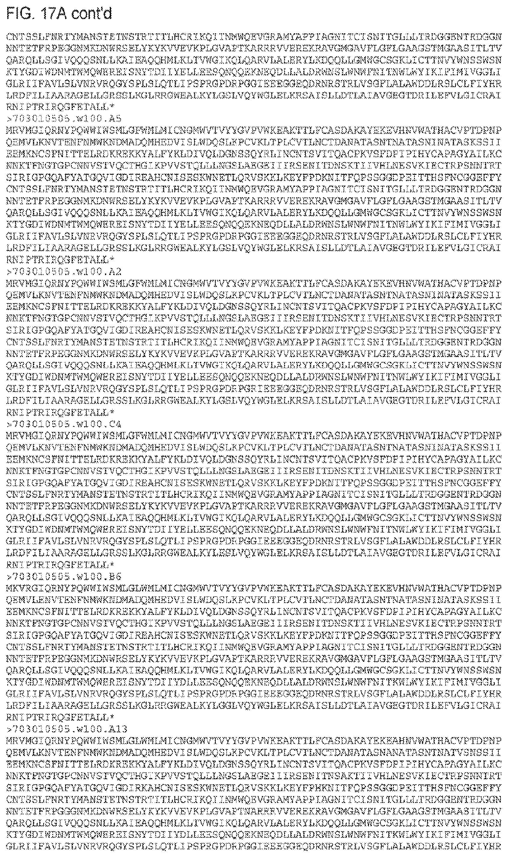
D00085

D00086

D00087

D00088

D00089

D00090

D00091

D00092

D00093

D00094

D00095

D00096

D00097

D00098

D00099

D00100

D00101

D00102

D00103
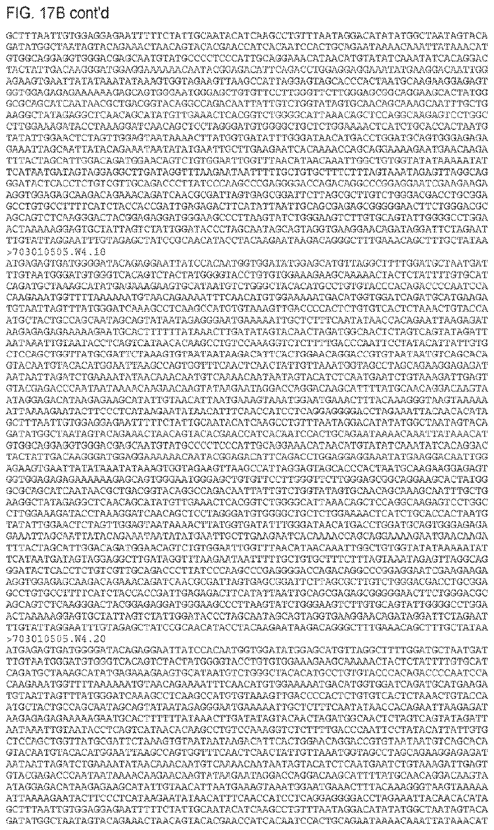
D00104

D00105

D00106
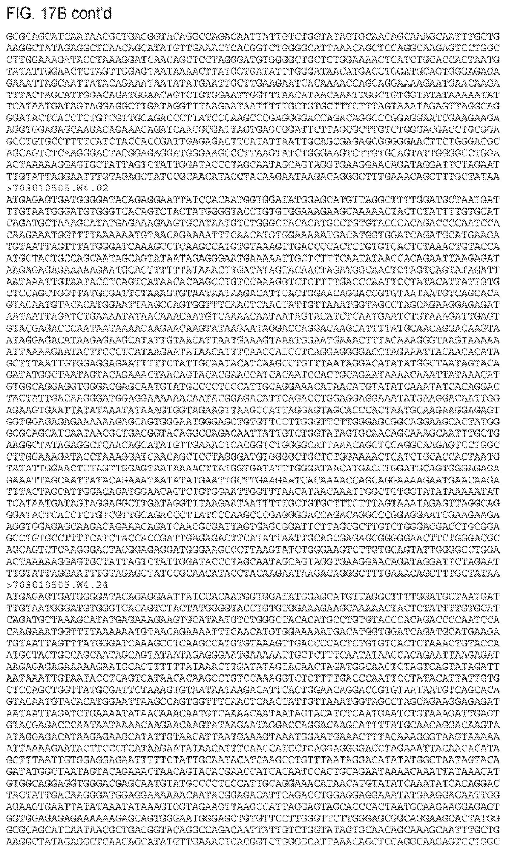
D00107

D00108

D00109

D00110

D00111
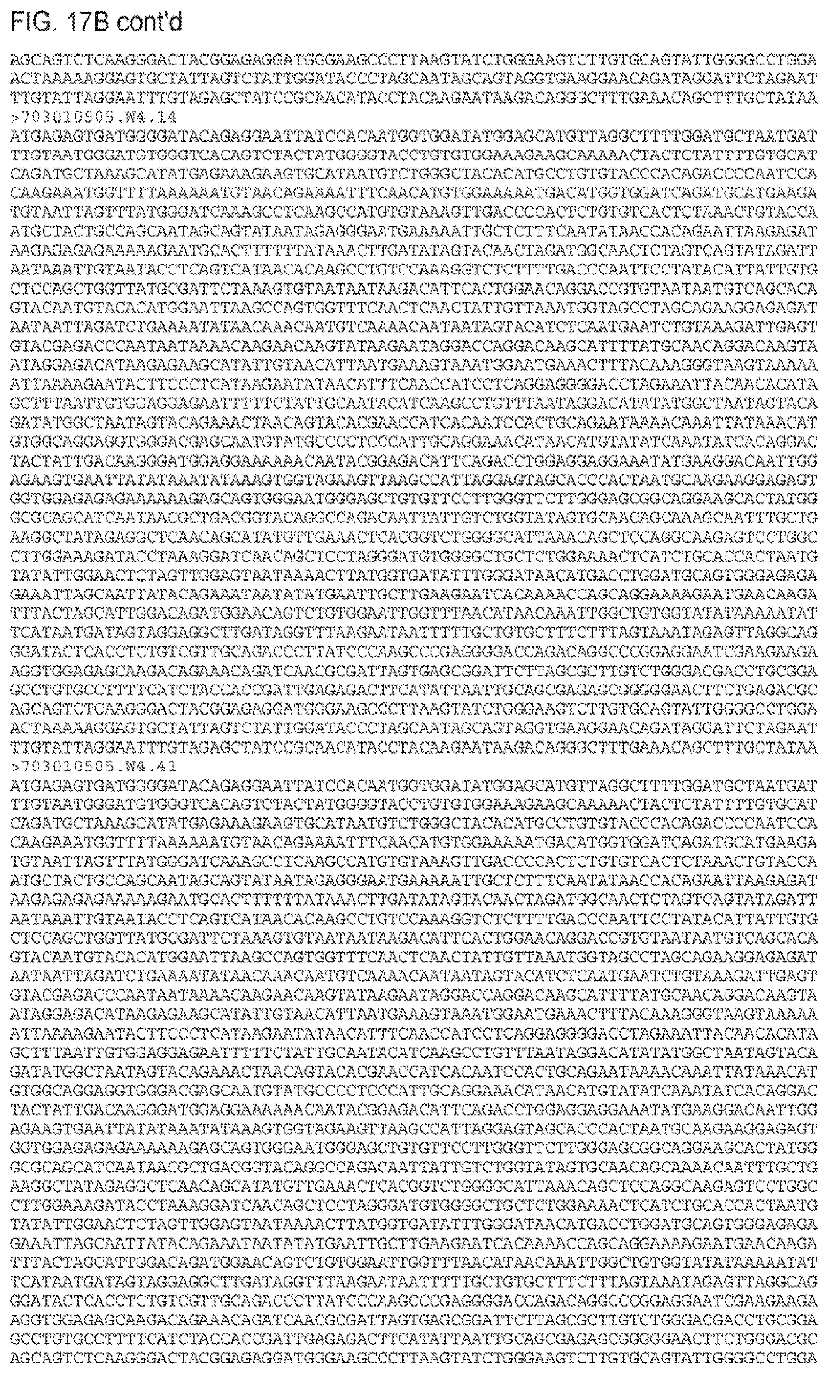
D00112
D00113

D00114

D00115

D00116

D00117

D00118

D00119

D00120

D00121

D00122

D00123

D00124

D00125

D00126

D00127
D00128

D00129

D00130

D00131

D00132

D00133
D00134

D00135

D00136

D00137

D00138

D00139

D00140

D00141

D00142

D00143

D00144

D00145

D00146

D00147

D00148

D00149

D00150

D00151

D00152

D00153

D00154

D00155

D00156

D00157

D00158

D00159

D00160

D00161

D00162

D00163

D00164

D00165

D00166

D00167

D00168

D00169

D00170

D00171

D00172

D00173

D00174

D00175

D00176

D00177

D00178

D00179

D00180

D00181

D00182

D00183

D00184

D00185

D00186

D00187

D00188

D00189

D00190

D00191

D00192

D00193

D00194

D00195

D00196

D00197
D00198

D00199

D00200

D00201

D00202

D00203

D00204

D00205

D00206

D00207

D00208

D00209
D00210

D00211

D00212

D00213

D00214

D00215

D00216

D00217

D00218

D00219
D00220

D00221

D00222

D00223

D00224

D00225

D00226

D00227

D00228

D00229

D00230

D00231

D00232

D00233

D00234

D00235

D00236

D00237

D00238

D00239

D00240

D00241

D00242

D00243

D00244

D00245

D00246

D00247

D00248
D00249

D00250

D00251
D00252

D00253

D00254

D00255
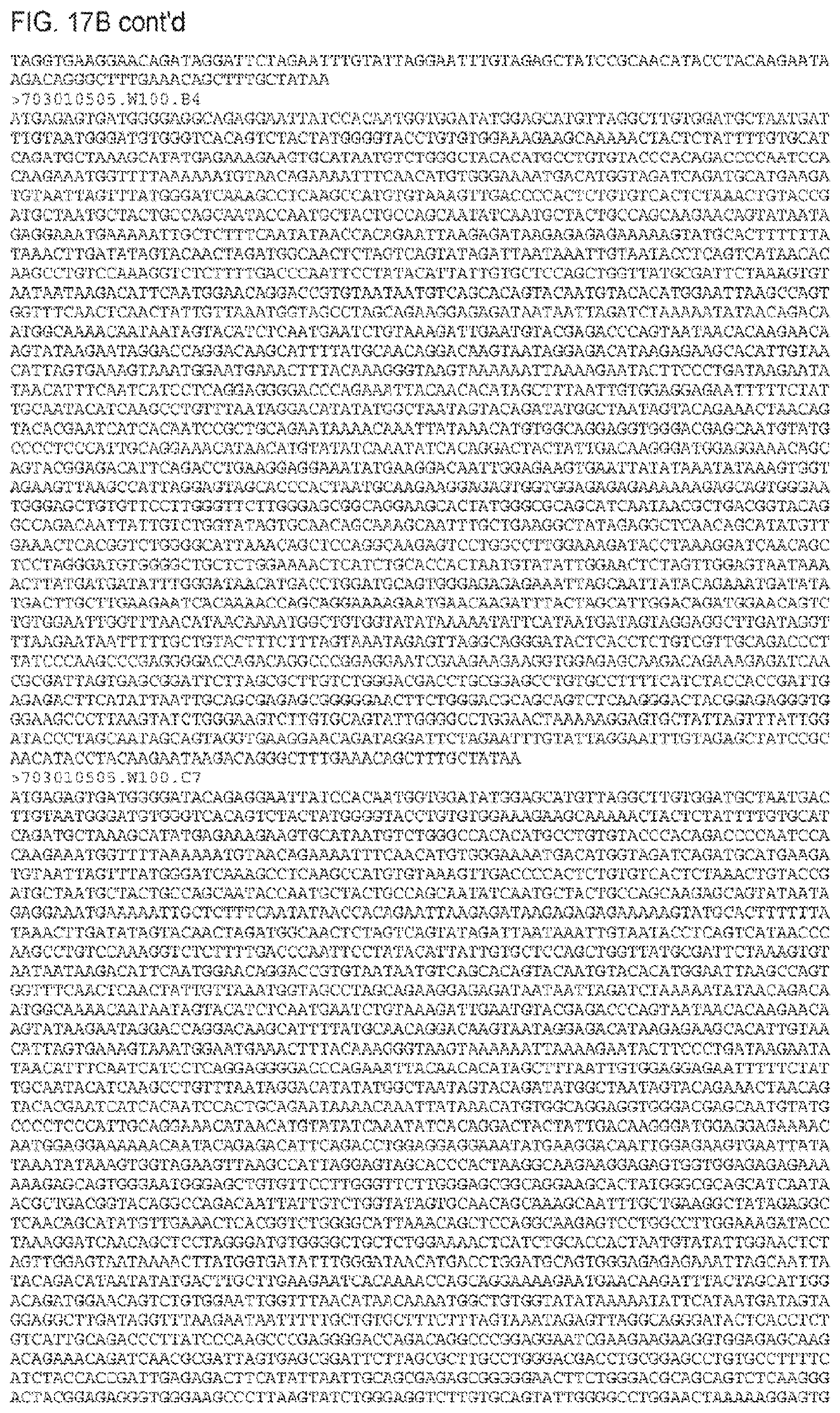
D00256

D00257

D00258

D00259

D00260

D00261

D00262

D00263

D00264

D00265

D00266

D00267

D00268

D00269

D00270

D00271

D00272

D00273

D00274

D00275

D00276

D00277

D00278

D00279

D00280

D00281

D00282

D00283

D00284

D00285

D00286

D00287

D00288

D00289

D00290

D00291

D00292

D00293

D00294

D00295

D00296

D00297

D00298

D00299

D00300

D00301

D00302

D00303

D00304

D00305

D00306

D00307

D00308

D00309

D00310

D00311

D00312

D00313

D00314

D00315

D00316

D00317

D00318

D00319

D00320

D00321

D00322

D00323

D00324

D00325

D00326

D00327

D00328

D00329

D00330

D00331

D00332

D00333

D00334

D00335

D00336

D00337

D00338

D00339

D00340

D00341

D00342

D00343

D00344

D00345

D00346

D00347

D00348

D00349

D00350

D00351

D00352

D00353

D00354

D00355

D00356

D00357

D00358

D00359

D00360

D00361

D00362

D00363

D00364

D00365

D00366

D00367

D00368

D00369

D00370

D00371

D00372

D00373

D00374

D00375

D00376

D00377

D00378

D00379

D00380

D00381

D00382

D00383

D00384

D00385

D00386

D00387

D00388

D00389

D00390

D00391

D00392

D00393

D00394

D00395

D00396

D00397

S00001
XML
uspto.report is an independent third-party trademark research tool that is not affiliated, endorsed, or sponsored by the United States Patent and Trademark Office (USPTO) or any other governmental organization. The information provided by uspto.report is based on publicly available data at the time of writing and is intended for informational purposes only.
While we strive to provide accurate and up-to-date information, we do not guarantee the accuracy, completeness, reliability, or suitability of the information displayed on this site. The use of this site is at your own risk. Any reliance you place on such information is therefore strictly at your own risk.
All official trademark data, including owner information, should be verified by visiting the official USPTO website at www.uspto.gov. This site is not intended to replace professional legal advice and should not be used as a substitute for consulting with a legal professional who is knowledgeable about trademark law.