18F--tagged inhibitors of prostate specific membrane antigen (PSMA) and their use as imaging agents for prostate cancer
Cardinale , et al. October 27, 2
U.S. patent number 10,815,200 [Application Number 15/915,978] was granted by the patent office on 2020-10-27 for 18f--tagged inhibitors of prostate specific membrane antigen (psma) and their use as imaging agents for prostate cancer. This patent grant is currently assigned to DEUTSCHES KREBSFORSCHUNGSZENTRUM, RUPRECHT-KARLS-UNIVERSITAET HEIDELBERG. The grantee listed for this patent is DEUTSCHES KREBSFORSCHUNGSZENTRUM, RUPRECHT-KARLS-UNIVERSITAET HEIDELBERG. Invention is credited to Ulrike Bauder-Wuest, Martina Benesova, Jens Cardinale, Matthias Eder, Michael Eisenhut, Frederik L. Giesel, Uwe Haberkorn, Klaus Kopka, Martin Schaefer.


View All Diagrams
| United States Patent | 10,815,200 |
| Cardinale , et al. | October 27, 2020 |
18F--tagged inhibitors of prostate specific membrane antigen (PSMA) and their use as imaging agents for prostate cancer
Abstract
The present invention generally relates to the field of radiopharmaceuticals and their use in nuclear medicine as tracers and imaging agents for various disease states of prostate cancer.
| Inventors: | Cardinale; Jens (Vienna, AT), Schaefer; Martin (Neckarsteinach, DE), Kopka; Klaus (Dossenheim, DE), Eder; Matthias (Freiburg, DE), Bauder-Wuest; Ulrike (Schriesheim, DE), Eisenhut; Michael (Heidelberg, DE), Benesova; Martina (Neckarsteinach, DE), Haberkorn; Uwe (Schwetzingen, DE), Giesel; Frederik L. (Heidelberg, DE) | ||||||||||
|---|---|---|---|---|---|---|---|---|---|---|---|
| Applicant: |
|
||||||||||
| Assignee: | DEUTSCHES
KREBSFORSCHUNGSZENTRUM (Heidelberg, DE) RUPRECHT-KARLS-UNIVERSITAET HEIDELBERG (Heidelberg, DE) |
||||||||||
| Family ID: | 1000005140965 | ||||||||||
| Appl. No.: | 15/915,978 | ||||||||||
| Filed: | March 8, 2018 |
Prior Publication Data
| Document Identifier | Publication Date | |
|---|---|---|
| US 20180194729 A1 | Jul 12, 2018 | |
Related U.S. Patent Documents
| Application Number | Filing Date | Patent Number | Issue Date | ||
|---|---|---|---|---|---|
| PCT/EP2016/001573 | Sep 19, 2016 | ||||
Foreign Application Priority Data
| Sep 30, 2015 [EP] | 15002800 | |||
| Apr 6, 2016 [EP] | 16164090 | |||
| Aug 4, 2016 [EP] | 16182764 | |||
| Current U.S. Class: | 1/1 |
| Current CPC Class: | G01N 33/57434 (20130101); A61P 35/00 (20180101); C07B 59/002 (20130101); C07K 7/02 (20130101); A61K 51/0455 (20130101); C07D 213/61 (20130101); A61K 51/04 (20130101); A61K 38/00 (20130101); C07B 2200/05 (20130101) |
| Current International Class: | A61K 38/00 (20060101); G01N 33/574 (20060101); C07K 7/02 (20060101); A61P 35/00 (20060101); C07D 213/61 (20060101); C07B 59/00 (20060101); A61K 51/04 (20060101) |
| Field of Search: | ;424/1.11,1.65,1.69,1.81,1.85,1.89,9.1,9.2,9.3 ;530/300 ;514/1.1 |
References Cited [Referenced By]
U.S. Patent Documents
| 10016519 | July 2018 | Kopka |
| 2015/0110715 | April 2015 | Eder et al. |
| 1177200 | Jun 2005 | EP | |||
| 1472541 | Sep 2009 | EP | |||
| 2136788 | Oct 2011 | EP | |||
| 2170075 | Mar 2014 | EP | |||
| 2759535 | Jul 2014 | EP | |||
| 2823826 | Jan 2015 | EP | |||
| 2097111 | Jul 2015 | EP | |||
| 2921482 | Sep 2015 | EP | |||
| 2117604 | Jul 2017 | EP | |||
| 2942065 | Jun 2018 | EP | |||
| 3335736 | Jun 2018 | EP | |||
| 03000201 | Jan 2003 | WO | |||
| 2006093991 | Sep 2006 | WO | |||
| 2008057437 | May 2008 | WO | |||
| 2009026177 | Feb 2009 | WO | |||
| 2009070302 | Jun 2009 | WO | |||
| 2010014933 | Feb 2010 | WO | |||
| 2010065899 | Jun 2010 | WO | |||
| 2010065902 | Jun 2010 | WO | |||
| 2010108125 | Sep 2010 | WO | |||
| 2010114723 | Oct 2010 | WO | |||
| 2012072728 | Jun 2012 | WO | |||
| 2012078534 | Jun 2012 | WO | |||
| 2012166923 | Dec 2012 | WO | |||
| 2013022797 | Feb 2013 | WO | |||
| 2013028664 | Feb 2013 | WO | |||
| 2014106208 | Jul 2014 | WO | |||
| 2014127365 | Aug 2014 | WO | |||
| 2015004029 | Jan 2015 | WO | |||
| 2015/055318 | Apr 2015 | WO | |||
| 2015057250 | Apr 2015 | WO | |||
| 2015073678 | May 2015 | WO | |||
| 2015143029 | Sep 2015 | WO | |||
| 2015171792 | Nov 2015 | WO | |||
| 2016030329 | Mar 2016 | WO | |||
| 2016040179 | Mar 2016 | WO | |||
| 2016062370 | Apr 2016 | WO | |||
| 2016065142 | Apr 2016 | WO | |||
| 2016065145 | Apr 2016 | WO | |||
Other References
|
International Search Report dated Jan. 5, 2017 in International Application No. PCT/EP2016/001573. cited by applicant. |
Primary Examiner: Jones; D. L.
Attorney, Agent or Firm: Perkins Coie LLP Kung; Viola
Parent Case Text
This application is a continuation of PCT/EP2016/001573, filed Sep. 19, 2016, which claims priority of EP 15002800.9, filed Sep. 30, 2015, EP 16164090.9, filed Apr. 6, 2016, and EP16182764.7, filed Aug. 4, 2016. The contents of the above-identified applications are incorporated herein by reference in their entirety.
The present invention generally relates to the field of radiopharmaceuticals and their use in nuclear medicine as tracers and imaging agents and for the various disease states of prostate cancer.
Claims
The invention claimed is:
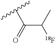
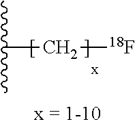
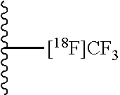
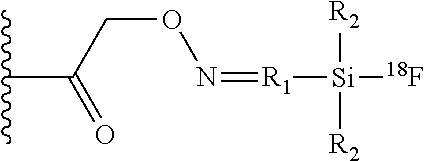
1. A compound of Formula I: ##STR00056## with: TABLE-US-00005 i, j 0, 1 m 1-5 n 0-3 R H, CH.sub.3 AS Natural or non-natural amino acid Z: --CO.sub.2H, --SO.sub.2H, --SO.sub.3H, --SO.sub.4H, --PO.sub.2H, --PO.sub.3H, --PO.sub.4H.sub.2 X: Naphthyl, Phenyl, Biphenyl, Indolyl (.dbd.2,3-benzopyrrolyl), Benzothiazolyl, Quinoyl Y: Aryl, Alkylaryl, Cyclopentyl, Cyclohexyl, Cycloheptyl, N-Piperidyl and N-methylated Piperidyl salt .sup.18F-Tag: ##STR00057## ##STR00058## ##STR00059## ##STR00060## ##STR00061## ##STR00062## ##STR00063## ##STR00064## ##STR00065## ##STR00066## ##STR00067## ##STR00068## ##STR00069## With: x = 1-5 Carboydrate: ##STR00070## ##STR00071## ##STR00072## ##STR00073## ##STR00074## ##STR00075## ##STR00076## ##STR00077## ##STR00078## ##STR00079## ##STR00080## ##STR00081## ##STR00082## ##STR00083## ##STR00084## ##STR00085## ##STR00086## ##STR00087## R.sub.1: Any alkyl, aryl or arylalkyl linker especially methyl, 2-ethyl, 3-propyl, 2-,3-,4-phenyl, 2-,3-,4-phenylmethyl, 2-,3-,4-phenylpropyl R.sub.2: Any alkyl or aryl group especially methyl isopropyl, tert-butyl, phenyl or 1-naphtyl ##STR00088## R.sub.1: Any alkyl, aryl or arylalkyl linker especially methyl, 2-ethyl, 3-propyl, 2-,3-,4-phenyl, 2-,3-,4-phenylmethyl, 2-,3-,4-phenylpropyl R.sub.2: Any alkyl or aryl group especially methyl isopropyl, tert-butyl, phenyl or 1-naphtyl ##STR00089## R.sub.1: Any alkyl, aryl or arylalkyl linker especially methyl, 2-ethyl, 3-propyl, 2-,3-,4-phenyl, 2-,3-,4-phenylmethyl, 2-,3-,4-phenylpropyl R.sub.2: Any alkyl or aryl group especially methyl isopropyl, tert-butyl, phenyl or 1-naphtyl ##STR00090## R.sub.1: Any alkyl, aryl or arylalkyl linker especially methyl, 2-ethyl, 3-propyl, 2-,3-,4-phenyl, 2-,3-,4-phenylmethyl, 2-,3-,4-phenylpropyl R.sub.2: Any alkyl or aryl group especially methyl isopropyl, tert-butyl, phenyl or 1-naphtyl ##STR00091## R.sub.1: Any alkyl, aryl or arylalkyl linker especially methyl, 2-ethyl, 3-propyl, 2-,3-,4-phenyl, 2-,3-,4-phenylmethyl, 2-,3-,4-phenylpropyl R.sub.2: Any alkyl or aryl group especially methyl isopropyl, tert-butyl, phenyl or 1-naphtyl ##STR00092## R.sub.1: Any alkyl, aryl or arylalkyl linker especially methyl, 2-ethyl, 3-propyl, 2-,3-,4-phenyl, 2-,3-,4-phenylmethyl, 2-,3-,4-phenylpropyl R.sub.2: Any alkyl or aryl group especially methyl isopropyl, tert-butyl, phenyl or 1-naphtyl ##STR00093## R.sub.1: Any alkyl, aryl or arylalkyl linker especially methyl, 2-ethyl, 3-propyl, 2-,3-,4-phenyl, 2-,3-,4-phenylmethyl, 2-,3-,4-phenylpropyl R.sub.2: Any alkyl or aryl group especially methyl isopropyl, tert-butyl, phenyl or 1-naphtyl.
2. The compound of claim 1, wherein the non-natural amino acid has the formula ##STR00094## and R'.dbd.H, CO.sub.2H, CH.sub.2CO.sub.2H, C.sub.2H.sub.4CO.sub.2H, CH(CO.sub.2H).sub.2, CH(CH.sub.2CO.sub.2H).sub.2, CH(CO.sub.2H)(CH.sub.2CO.sub.2H), CH.sub.2CH(CO.sub.2H).sub.2, SO.sub.3H; o=1-3; R.dbd.H, CH.sub.3.
3. A pharmaceutical composition comprising the compound of claim 1, or a pharmaceutically acceptable salt or ester thereof, and a pharmaceutically acceptable carrier.
4. The compound of claim 1 having the structure; ##STR00095## wherein the linker A is selected from the group consisting of: ##STR00096## wherein R.sup.1=(Glu)-(Urea)-(Lys) and R.sup.2=(Linker B).sub.m-.sup.18F Tag, m=1-5, and linker B is selected from the group consisting of: ##STR00097##
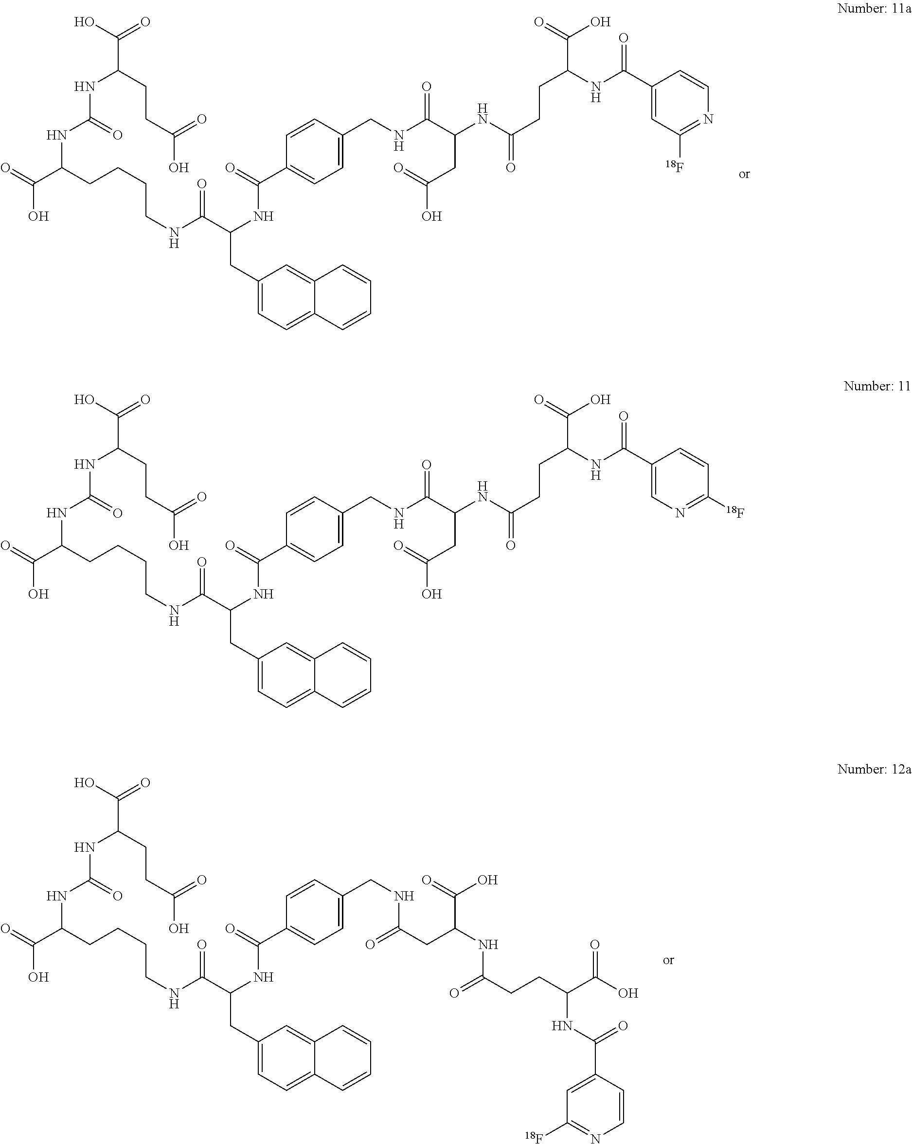
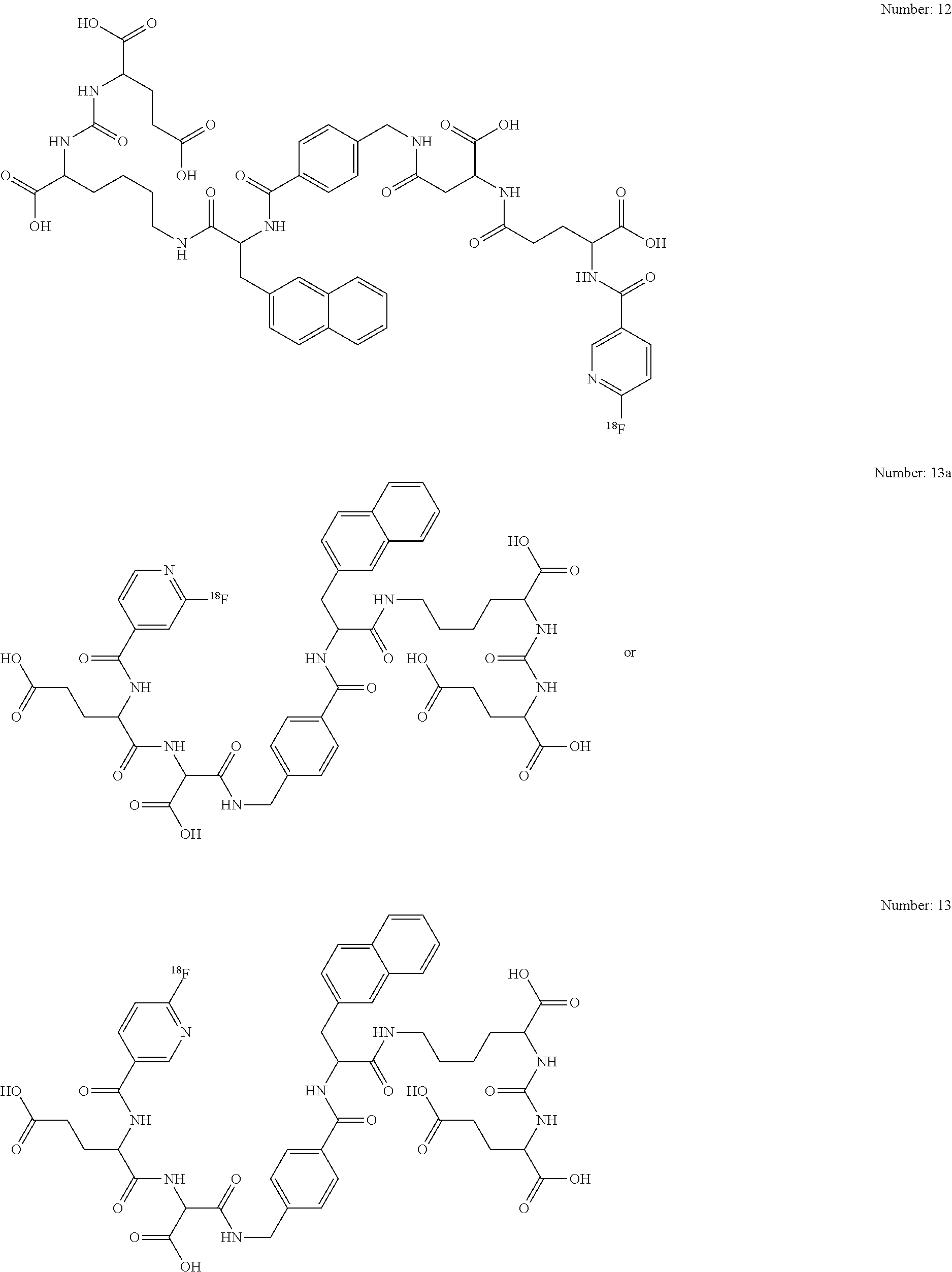
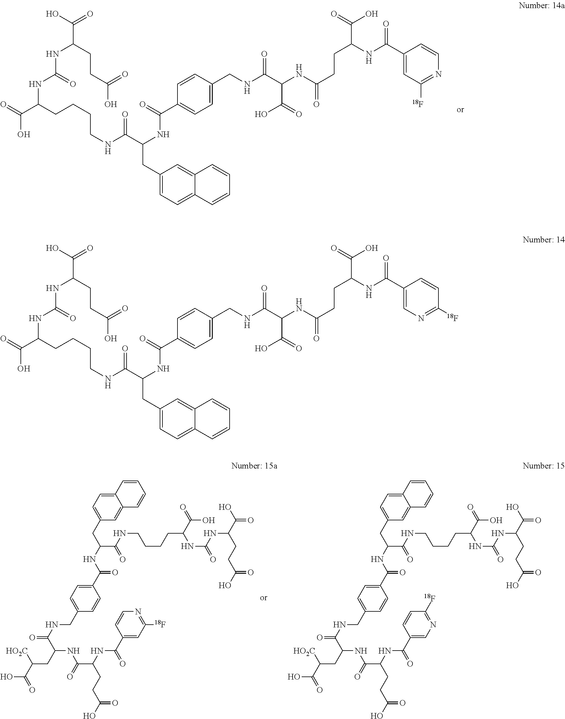
5. The compound of claim 1, selected from the following ##STR00098## ##STR00099## ##STR00100## ##STR00101## ##STR00102## ##STR00103## ##STR00104## ##STR00105## ##STR00106## ##STR00107## ##STR00108## ##STR00109##
6. The compound of claim 1, wherein R.sub.1 is selected from the group consisting of: methyl, 2-ethyl, 3-propyl, 2-,3-,4-phenyl, 2-,3-,4-phenylmethyl, and 2,3-,4-phenylpropyl.
7. The compound of claim 1, wherein R.sub.2 is selected from the group consisting of methyl, isopropyl, tert-butyl, phenyl, and 1-naphtyl.
8. A method of imaging a prostate region in a patient comprising the steps of: (i) administering to a patient a diagnostically effective amount of the compound of claim 1; (ii) exposing the prostate region of the patient to a scanning device to detect the 18F-tag; and (iii) obtaining an image of the prostate region of the patient by the scanning device.
Description
BACKGROUND OF THE INVENTION
Prostate cancer (PCa) is the leading cancer in the US and European population. At least 1-2 million men in the western hemisphere suffer from prostate cancer and it is estimated that the disease will strike one in six men between the ages of 55 and 85. There are more than 300.000 new cases of prostate cancer diagnosed each year in USA. The mortality from the disease is second only to lung cancer. Currently anatomic methods, such as computed tomography (CT), magnetic resonance (MR) imaging and ultrasound, predominate for clinical imaging of prostate cancer. An estimated $2 billion is currently spent worldwide on surgical, radiation, drug therapy and minimally invasive treatments. However, there is presently no effective therapy for relapsing, metastatic, androgen-independent prostate cancer.
A variety of experimental low molecular weight PCa imaging agents are currently being pursued clinically, including radiolabeled choline analogs ([.sup.11C]Choline, [.sup.18F]FECh, [.sup.18F] FMC, [.sup.18F]fluorodihydrotestosterone ([.sup.18F]FDHT), anti-1-amino-3-[.sup.18F]fluorocyclobutyl-1-carboxylic acid (anti[.sup.18F]F-FACBC, [.sup.11C]acetate and 1-(2-deoxy-2-[.sup.18F]fluoro-L-arabinofuranosyl)-5-methyluracil (-[.sup.18F]FMAU) (Scher, B.; et al. Eur J Nucl Med Mol Imaging 2007, 34, 45-53; Rinnab, L.; et al. BJU Int 2007, 100, 786,793; Reske, S. N.; et al. J Nucl Med 2006, 47, 1249-1254; Zophel, K.; Kotzerke, J. Eur J Nucl Med Mol Imaging 2004, 31, 756-759; Vees, H.; et al. BJU Int 2007, 99, 1415-1420; Larson, S. M.; et al. J Nucl Med 2004, 45, 366-373; Schuster, D. M.; et al. J Nucl Med 2007, 48, 56-63; Tehrani, O. S.; et al. J Nucl Med 2007, 48, 1436-1441). Each operates by a different mechanism and has certain advantages, e.g., low urinary excretion for [.sup.11C]choline, and disadvantages, such as the short physical half-life of positron-emitting radionuclides.
It is well known that tumors may express unique proteins associated with their malignant phenotype or may over-express normal constituent proteins in greater number than normal cells. The expression of distinct proteins on the surface of tumor cells offers the opportunity to diagnose and characterize disease by probing the phenotypic identity and biochemical composition and activity of the tumor. Radioactive molecules that selectively bind to specific tumor cell surface proteins provide an attractive route for imaging and treating tumors under non-invasive conditions. A promising new series of low molecular weight imaging agents targets the prostate-specific membrane antigen (PSMA) (Mease R. C. et al. Clin Cancer Res. 2008, 14, 3036-3043; Foss, C. A.; et al. Clin Cancer Res 2005, 11, 4022-4028; Pomper, M. G.; et al. Mol Imaging 2002, 1, 96-101; Zhou, J.; etr al. Nat Rev Drug Discov 2005, 4, 1015-1026; WO 2013/022797).
PSMA is a trans-membrane, 750 amino acid type II glycoprotein that has abundant and restricted expression on the surface of PCa, particularly in androgen-independent, advanced and metastatic disease (Schulke, N.; et al. Proc Natl Acad Sci USA 2003, 100, 12590-12595). The latter is important since almost all PCa become androgen independent over the time. PSMA possesses the criteria of a promising target for therapy, i.e., abundant and restricted (to prostate) expression at all stages of the disease, presentation at the cell surface but not shed into the circulation and association with enzymatic or signaling activity (Schulke, N.; et al. Proc. Natl. Acad. Sci. USA 2003, 100, 12590-12595). The PSMA gene is located on the short arm of chromosome 11 and functions both as a folate hydrolase and neuropeptidase. It has neuropeptidase function that is equivalent to glutamate carboxypeptidase II (GCPII), which is referred to as the "brain PSMA", and may modulate glutamatergic transmission by cleaving N-acetylaspartylglutamate (NAAG) to N-acetylaspartate (NAA) and glutamate (Nan, F.; et al. J Med Chem 2000, 43, 772-774). There are up to 10.sup.6 PSMA molecules per cancer cell, further suggesting it as an ideal target for imaging and therapy with radionuclide-based techniques (Tasch, J.; et al. Crit Rev Immunol 2001, 21, 249-261).
The radio-immunoconjugate of the anti-PSMA monoclonal antibody (mAb) 7E11, known as the PROSTASCINT.RTM. scan, is currently being used to diagnose prostate cancer metastasis and recurrence. However, this agent tends to produce images that are challenging to interpret (Lange, P. H. PROSTASCINT scan for staging prostate cancer. Urology 2001, 57, 402-406; Haseman, M. K.; et al. Cancer Biother Radiopharm 2000, 15, 131-140; Rosenthal, S. A.; et al. Tech Urol 2001, 7, 27-37). More recently, monoclonal antibodies have been developed that bind to the extracellular domain of PSMA and have been radiolabeled and shown to accumulate in PSMA-positive prostate tumor models in animals. However, diagnosis and tumor detection using monoclonal antibodies has been limited by the low permeability of the monoclonal antibody in solid tumors.
The selective targeting of cancer cells with radiopharmaceuticals for imaging or therapeutic purposes is challenging. A variety of radionuclides are known to be useful for radio-imaging or cancer radiotherapy, including .sup.11C, .sup.18F, .sup.111In, .sup.90Y, .sup.68Ga, .sup.177Lu, .sup.99mTc, .sup.123I and .sup.131I Recently it has been shown that some compounds containing a glutamate-urea-glutamate (GUG) or a glutamate-urea-lysine (GUL) recognition element linked to a radionuclide-ligand conjugate exhibit high affinity for PSMA.
Several .sup.18F-tagged compounds which show PSMA interaction and are suitable for the detection of prostate cancer are shown in EP 14 003 570.0. These compounds, however, show high lipophilic properties which make them, to a certain extent, difficult to handle and to administer.
New agents that will enable rapid visualization of prostate cancer are needed. Thus, the object of the present invention is to develop ligands that interact with PSMA and carry appropriate radionuclides which provide a promising and novel targeting option for the detection, treatment and management of prostate cancer.
SUMMARY OF THE INVENTION
The solution of said object is achieved by providing the embodiments characterized in the claims.
The inventors found new compounds which are useful radiopharmaceuticals and their use in nuclear medicine as tracers and imaging agents and for various disease states of prostate cancer.
The novel imaging agents with structural modifications in the linker region have improved tumor targeting properties and pharmacokinetics. The pharmacophore presents three carboxylic groups able to interact with the respective side chains of PSMA and an oxygen as part of zinc complexation in the active center. Besides these obligatory interactions, the inventors were able to optimize the lipophilic interactions in the linker region in comparison to the compounds described in EP 14003570.0. Moreover, the inventors added some hydrophilic building blocks to the linkler for an enhancement of the pharmakokinetics.
BRIEF DESCRIPTION OF THE FIGURES
FIG. 1: .sup.18F-Labelling of macromolecules
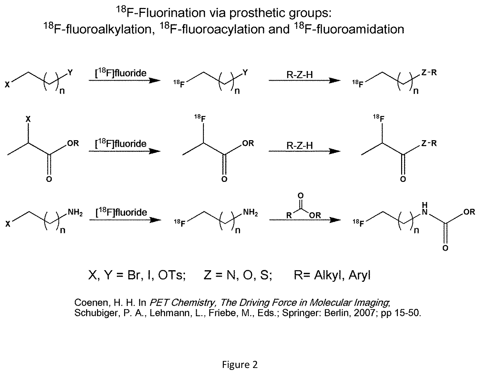
FIG. 2: .sup.18F-Fluorination via prosthetic groups
FIG. 3: .sup.18F-prosthetic groups for peptides
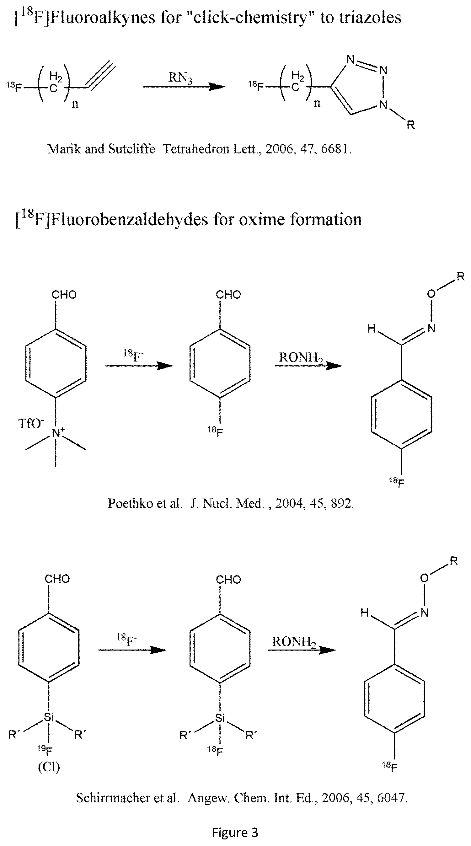
FIG. 4: .sup.18F-Labelled Prosthetic Groups using "Click Chemistry"
FIG. 5: Formation of [.sup.18F]aryltrifluoroboronates and fluoride sensors
FIG. 6: Complexes of .sup.18F-Fluorides
FIG. 7: .sup.18F-Labelling of RGD peptides
FIG. 8: Organ distribution of [.sup.18F]PSMA-1007 in PSMA-positive LNCaP mice (normal and blocked) and in PSMA negative PC3 mice vs. [.sup.18F]PSMA-1009 ([.sup.18F]DCFPyL) in PSMA positive LNCaP mice
FIG. 9: Organ distribution of [.sup.18F]PSMA-1007 in PSMA-positive LNCaP mice (non-blocked and blocked) vs. [.sup.18F]PSMA1003 in PSMA positive LNCaP mice (non-blocked)
FIG. 10: MIP of [.sup.18F]PSMA-1007 in a PSMA-positive LNCaP mouse 120-140 min p.i.
FIG. 11: Time-activity curve of [.sup.18F]PSMA-1007 in a PSMA-positive LNCaP mouse, including SUV values 120-140 min p.i.
FIG. 12: Structure of PSMA-1003
FIG. 13: Structure of PSMA-1009
FIG. 14: MIPs of [.sup.18F]PSMA-1007 in a healthy volunteer
FIG. 15: Blood (black) and serum (gray) time-activity-curves expressed as percent injected dose in a healthy volunteer
FIG. 16: Time-activity-curves of normal organs with PET-delineable volume-of-interest in a healthy volunteer
FIG. 17: Organ distribution [.sup.18F]PSMA-1007 in ten patients suffering from prostate cancer expressed as SUV.sub.max
FIG. 18: Tumor-to-Background ratios of [.sup.18F]PSMA-1007 in ten patients suffering from prostate cancer calculated from the respective SUV.sub.max values
FIG. 19: MIP of a 77-year old prostate cancer patient (PSA 40 ng/ml) scanned with [.sup.18F]PSMA-1007 1 and 3 h p.i. showing a large tumor mass on the mid and apex area of prostate and several lymph node metastases. Outside of the pelvic region there was no metastasis found
FIG. 20: MIP of a 72-year old patient (PSA 15 ng/ml) diagnosed with Gleason 9 (5+4) prostate cancer scanned with [.sup.18F]PSMA-1007 1 and 3 h p.i. Patient presents a large tumor mass in the whole prostate gland with infiltration in the left seminal vesicles and several lymph nodes in the pelvic region. Two metastatic lymph nodes are located outside the pelvic region, both paraaortic at level L3/4 and L5.
FIG. 21: Transaxial PET/CT-scan of a patient (a,b) and corresponding histopathology of the subsequent prostatectomy specimen; H&E staining (c); PSMA-immunostaining with outlined tumor contours circumscribed by the broken line (d).
FIG. 22: Structure of preferred compounds of the present invention.
DETAILED DESCRIPTION OF THE INVENTION
The present invention relates to radiopharmaceuticals and their use in nuclear medicine as tracers and imaging agents for the various disease states of prostate cancer.
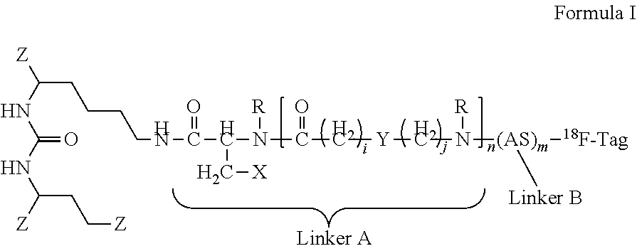
Thus, the present invention concerns compounds that are represented by the general
##STR00001##
Formula I
with:
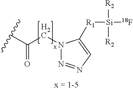
TABLE-US-00001 i, j 0, 1 m 1-5 n 0-3 R H, CH.sub.3 AS Natural or non-natural amino acid Z: --CO.sub.2H, --SO.sub.2H, --SO.sub.3H, --SO.sub.4H, --PO.sub.2H, --PO.sub.3H, --PO.sub.4H.sub.2 X: Naphthyl, Phenyl, Biphenyl, Indolyl (.dbd.2,3-benzopyrrolyl), Benzothiazolyl, Quinoyl Y: Aryl, Alkylaryl, Cyclopentyl, Cyclohexyl, Cycloheptyl, N-Piperidyl and N-methylated Piperidyl salt .sup.18F-Tag: ##STR00002## ##STR00003## ##STR00004## ##STR00005## ##STR00006## ##STR00007## ##STR00008## ##STR00009## ##STR00010## ##STR00011## ##STR00012## ##STR00013## ##STR00014## With: x = 1-5 Carboydrate: ##STR00015## ##STR00016## ##STR00017## ##STR00018## ##STR00019## ##STR00020## ##STR00021## ##STR00022## ##STR00023## ##STR00024## ##STR00025## ##STR00026## ##STR00027## ##STR00028## ##STR00029## ##STR00030## ##STR00031## ##STR00032## R.sub.1: Any alkyl, aryl or arylalkyl linker especially methyl, 2-ethyl, 3-propyl, 2-,3-,4-phenyl, 2-,3-,4-phenylmethyl, 2-,3-,4-phenylpropyl R.sub.2: Any alkyl or aryl group especially methyl isopropyl, tert-butyl, phenyl or 1-naphtyl ##STR00033## R.sub.1: Any alkyl, aryl or arylalkyl linker especially methyl, 2-ethyl, 3-propyl, 2-,3-,4-phenyl, 2-,3-,4-phenylmethyl, 2-,3-,4-phenylpropyl R.sub.2: Any alkyl or aryl group especially methyl isopropyl, tert-butyl, phenyl or 1-naphtyl ##STR00034## R.sub.1: Any alkyl, aryl or arylalkyl linker especially methyl, 2-ethyl, 3-propyl, 2-,3-,4-phenyl, 2-,3-,4-phenylmethyl, 2-,3-,4-phenylpropyl R.sub.2: Any alkyl or aryl group especially methyl isopropyl, tert-butyl, phenyl or 1-naphtyl ##STR00035## R.sub.1: Any alkyl, aryl or arylalkyl linker especially methyl, 2-ethyl, 3-propyl, 2-,3-,4-phenyl, 2-,3-,4-phenylmethyl, 2-,3-,4-phenylpropyl R.sub.2: Any alkyl or aryl group especially methyl isopropyl, tert-butyl, phenyl or 1-naphtyl ##STR00036## R.sub.1: Any alkyl, aryl or arylalkyl linker especially methyl, 2-ethyl, 3-propyl, 2-,3-,4-phenyl, 2-,3-,4-phenylmethyl, 2-,3-,4-phenylpropyl R.sub.2: Any alkyl or aryl group especially methyl isopropyl, tert-butyl, phenyl or 1-naphtyl ##STR00037## R.sub.1: Any alkyl, aryl or arylalkyl linker especially methyl, 2-ethyl, 3-propyl, 2-,3-,4-phenyl, 2-,3-,4-phenylmethyl, 2-,3-,4-phenylpropyl R.sub.2: Any alkyl or aryl group especially methyl isopropyl, tert-butyl, phenyl or 1-naphtyl ##STR00038## R.sub.1: Any alkyl, aryl or arylalkyl linker especially methyl, 2-ethyl, 3-propyl, 2-,3-,4-phenyl, 2-,3-,4-phenylmethyl, 2-,3-,4-phenylpropyl R.sub.2: Any alkyl or aryl group especially methyl isopropyl, tert-butyl, phenyl or 1-naphtyl
If not stated otherwise, in the present invention the term "alkyl" by itself or as part of another molecule, means a straight or branched chain, or cyclic hydrocarbon radical, or combination thereof, which may be fully saturated, mono- or polyunsaturated and can include di- and multivalent radicals, The "alkyl" residue is preferably C.sub.1 to C.sub.10 and may be unsubstituted or substituted (e.g with halogen). Preferred alkyl residues are methyl, ethyl, n-propyl, iso-propyl, n-butyl, tert-butyl, n-pentyl, n-hexyl, n-hepyl or n-octyl or the like. The same also applies to the corresponding cycloalkyl compounds having preferably 3 to 10 carbon atoms, e.g. cycloproyl, cyclobutyl, cyclopentyl, cyclohexyl, cycloheptyl, cyclooctyl, etc. An unsaturated alkyl group is one having one or more double bonds or triple bonds. Examples of unsaturated alkyl groups include, but are not limited to, vinyl, 2-propenyl, crotyl, 2-isopentenyl, 2-(butadienyl), 2,4-pentadienyl, 3-(1,4-pentadienyl), ethynyl, 1- and 3-propynyl, 3-butynyl, and the higher homologs and isomers. The term "alkyl," unless otherwise noted, is also meant to include those derivatives of alkyl, such as "heteroalkyl", "haloalkyl" and "homoalkyl".
The term "aryl", as used herein, refers to a closed ring structure which has at least one ring having a conjugated pi electron system and includes both carbocyclic aryl and heterocyclic aryl (or "heteroaryl" or "heteroaromatic") groups. The carbocyclic or heterocyclic aromatic group may contain from 5 to 20 ring atoms. The term includes monocyclic rings linked covalently or fused-ring polycyclic (i.e., rings which share adjacent pairs of carbon atoms) groups. An aromatic group can be unsubstituted or substituted. Non-limiting examples of "aromatic" or "aryl", groups include phenyl, 1-naphthyl, 2-naphthyl, 2-biphenyl, 3-biphenyl, 4-biphenyl, anthracenyl, and phenanthracenyl. Substituents for each of the above noted aryl and heteroaryl ring systems are selected from the group of acceptable substituents (e.g. alkyl, carbonyl, carboxyl or halogen) described herein. The term "aryl" when used in combination with other terms (including but not limited to, aryloxy, arylthioxy, aralkyl) includes both aryl and heteroaryl rings. Thus, the term "aralkyl" or "alkaryl" is meant to include those radicals in which an aryl group is attached to an alkyl group (including but not limited to, benzyl, phenethyl, pyridylmethyl and the like) including those alkyl groups in which a carbon atom (including but not limited to, a methylene group) has been replaced by a heteroatom, by way of example only, by an oxygen atom. Examples of such aryl groups include, but are not limited to, phenoxymethyl, 2-pyridyloxymethyl, 3-(1-naphthyloxy)propyl, and the like.
"Heteroaryl" refers to aryl groups which contain at least one heteroatom selected from N, O, and S; wherein the nitrogen and sulfur atoms may be optionally oxidized, and the nitrogen atom(s) may be optionally quaternized. Heteroaryl groups may be substituted or unsubstituted. A heteroaryl group may be attached to the remainder of the molecule through a heteroatom. Non-limiting examples of suitable groups include 1-pyrrolyl, 2-pyrrolyl, 3-pyrrolyl, 3-pyrazolyl, 2-imidazolyl, 4-imidazolyl, pyrazinyl, 2-oxazolyl, 4-oxazolyl, 2-phenyl-4-oxazolyl, 5-oxazolyl, 3-isoxazolyl, 4-isoxazolyl, 5-isoxazolyl, 2-thiazolyl, 4-thiazolyl, 5-thiazolyl, 2-furyl, 3-furyl, 2-thienyl, 3-thienyl, 2-pyridyl, 3-pyridyl, 4-pyridyl, 2-pyrimidyl, 4-pyrimidyl, 4-benzothiazolyl, 5-benzothiazolyl, 6-benzothiazolyl, 7-benzothiazolyl, purinyl, 2-benzimidazolyl, 4-indolyl, 5-indolyl, 6-indolyl, 7-indolyl, 1-isoquinolyl, 5-isoquinolyl, 2-quinoxalinyl, 5-quinoxalinyl, 2-quinolyl, 3-quinolyl, 4-quinolyl, 5-quinolyl, 6-quinolyl, 7-quinolyl or 8-quinolyl.
The term "amino acid" refers to naturally occurring and non-natural amino acids, as well as amino acid analogs and amino acid mimetics that function in a manner similar to the naturally occurring amino acids. Naturally amino acids are the 20 common amino acids in their D- or L-form (alanine, arginine, asparagine, aspartic acid, cysteine, glutamine, glutamic acid, glycine, histidine, isoleucine, leucine, lysine, methionine, phenylalanine, proline, serine, threonine, tryptophan, tyrosine, and valine) and pyrolysine and selenocysteine. Amino acid analogs refers to compounds that have the same basic chemical structure as a naturally occurring amino acid, by way of example only, an ex-carbon that is bound to a hydrogen, a carboxyl group, an amino group, and an R group. Such analogs may have modified R groups (by way of example, norleucine) or may have modified peptide backbones, while still retaining the same basic chemical structure as a naturally occurring amino acid. Non-limiting examples of amino acid analogs include homoserine, norleucine, methionine sulfoxide, methionine methyl sulfonium. Amino acids may be referred to herein by either their name, their commonly known three letter symbols or by the one-letter symbols recommended by the IUPAC-IUB Biochemical Nomenclature Commission. A "non-natural amino acid" refers to an amino acid that is not one of the 20 common amino acids or pyrolysine or selenocysteine. Other terms that may be used synonymously with the term "non-natural amino acid" is "non-naturally encoded amino acid," "unnatural amino acid," "non-naturally-occurring amino acid" or "artificial amino acid. The term "non-natural amino acid" includes, but is not limited to, amino acids which occur by modification of a naturally encoded amino acid in their backbone or side chains. In some embodiments the non-natural amino acid comprises a carbonyl group, an acetyl group, an aminooxy group, a hydrazine group, a hydrazide group, a semicarbazide group, an azide group or an alkyne group. In a preferred embodiment the non-natural amino acid has the formula
##STR00039##
and R'.dbd.H, CO.sub.2H, CH.sub.2CO.sub.2H, C.sub.2H.sub.4CO.sub.2H, CH(CO.sub.2H).sub.2, CH(CH.sub.2CO.sub.2H).sub.2, CH(CO.sub.2H)(CH.sub.2CO.sub.2H), CH.sub.2CH(CO.sub.2H).sub.2, SO.sub.3H; o=1-3; R.dbd.H, CH.sub.3
Preferred are those amino acids which bring a hydrophilic element into the compound of Formula I.
Some of the residues herein (including, but not limited to non-natural amino acids), may exist in several tautomeric forms. All such tautomeric forms are considered as part of the compounds described herein. Also, for example all enol-keto forms of any compounds herein are considered as part of the compositions described herein.
The linker B, i.e. the natural amino acids and/or non-naturally occurring amino acids, may be bound within the molecule via a peptide or amide linkage. In case of acidic amino acids (e.g. glutamic acid, aspartic acid) however, the binding may be alternatively via the .alpha.-, .beta. or .gamma.-position.
Although it is preferred that the Z-Group is --CO.sub.2H it may be easily replaced with biosteric replacements such as --SO.sub.2H, --SO.sub.3H, --SO.sub.4H, --PO.sub.2H, --PO.sub.3H, --PO.sub.4H.sub.2, see e.g. "The Practice of Medicinal Chemistry" (Academic Press New York, 1996), page 203.
Within the meaning of the invention, all residues are considered combinable unless stated otherwise in the definition of the residues. All conceivable subgroupings thereof are considered to be disclosed.
The .sup.18F-Tags of the above Table comprising triazoles exist in two isomeric forms which belong both to the invention and are illustrated by the given formulas.
Thus, preferred molecules of the present invention consist of three principle components (as shown in FIG. 22): the hydrophilic PSMA binding motif (Glu-Urea-Lys=Glu-NH--CO--NH-Lys), two variable linkers (Linker A and Linker B) and the .sup.18F-Tag.
Some preferred lipophilic linkers (linker A) are shown below, wherein R.sup.1=Glu-urea-Lys (PSMA binding motif) and R.sup.2=(Linker B).sub.m-[.sup.18F-Tag] with m=1-5,
##STR00040##
The different preferred building blocks for hydrophilic linkers (linker B) are shown below with their preferred connectivity exemplified on the basis of the respective single amino acids (m=1 in the generic structure)
##STR00041##
The preferred hydrophilic linkers may also be formed from two or more building blocks (m=2-5 in the generic formula), preferably selected from the acidic building blocks listed above. Preferred are 1-3 building blocks.
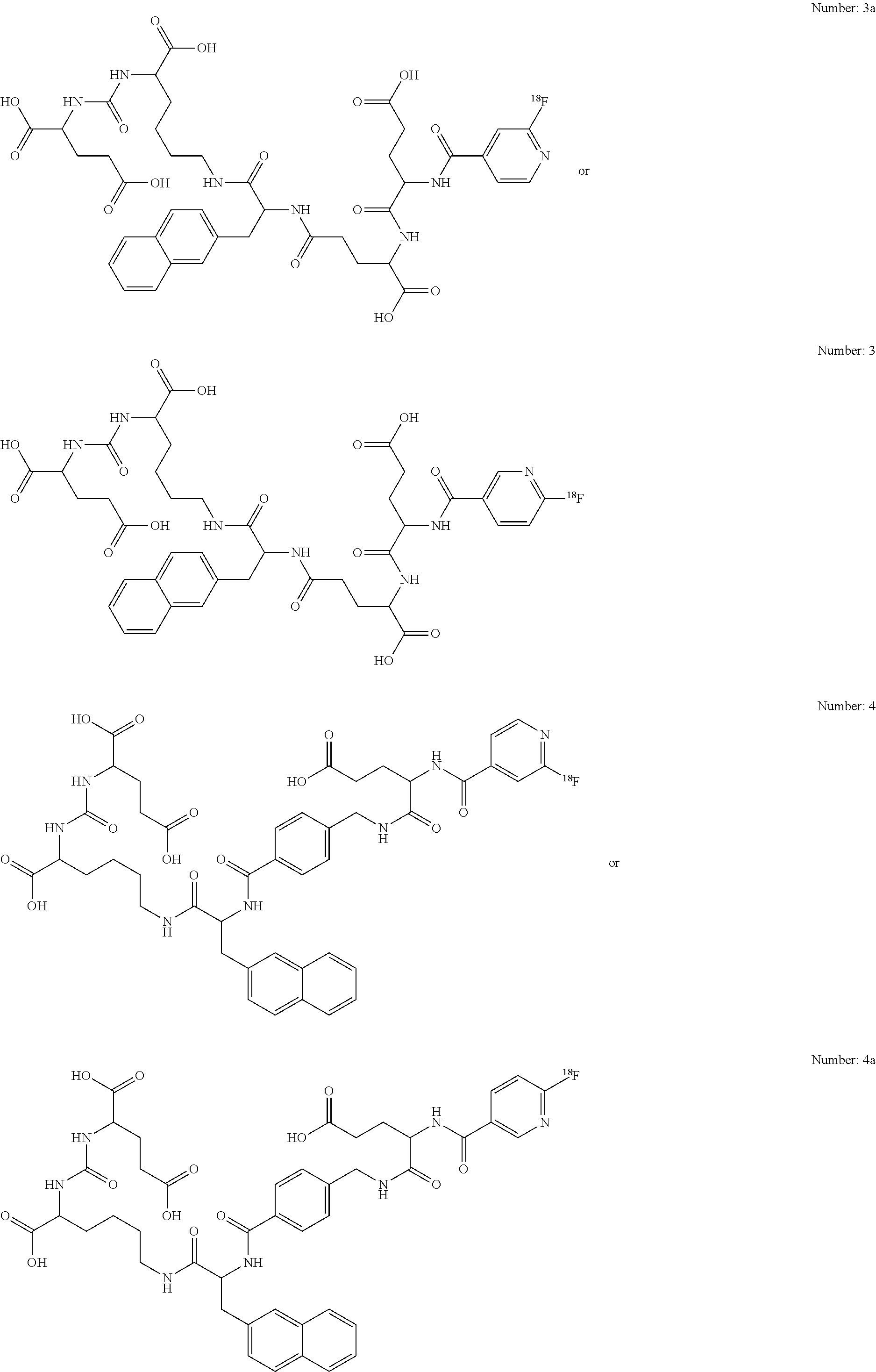
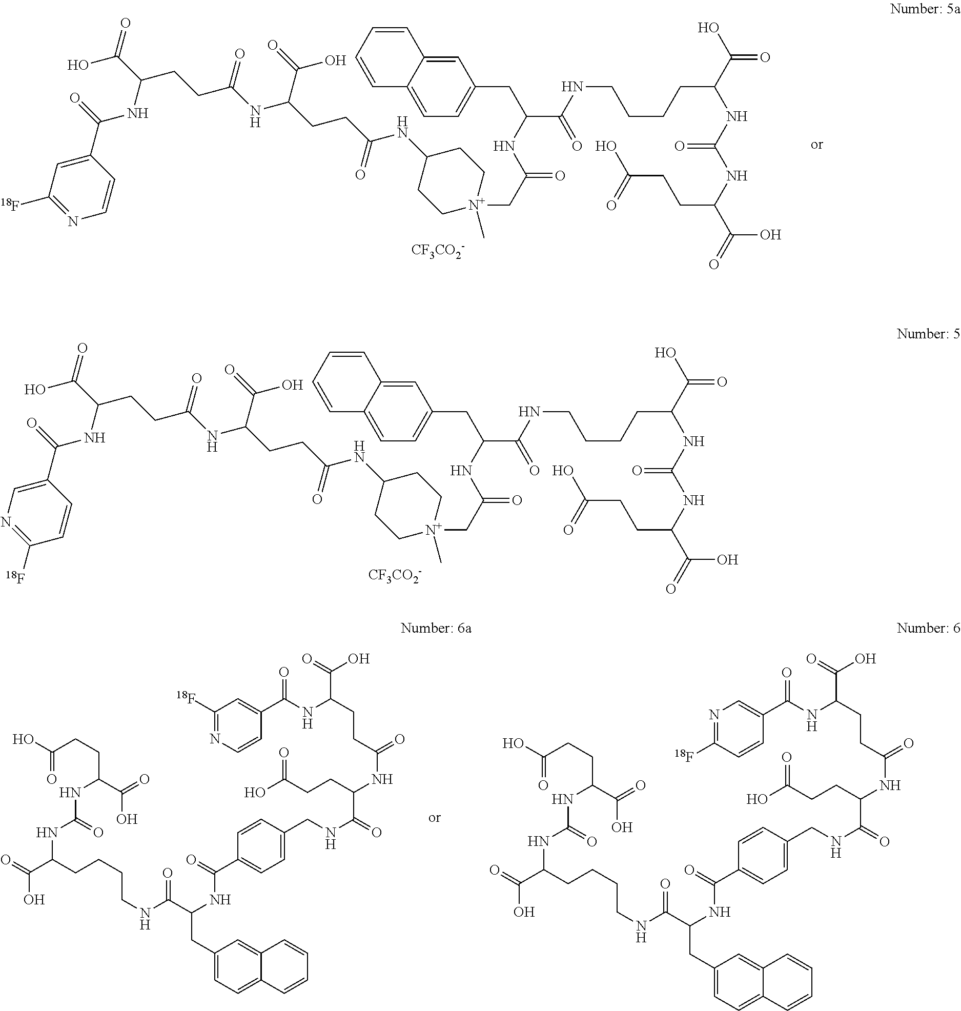
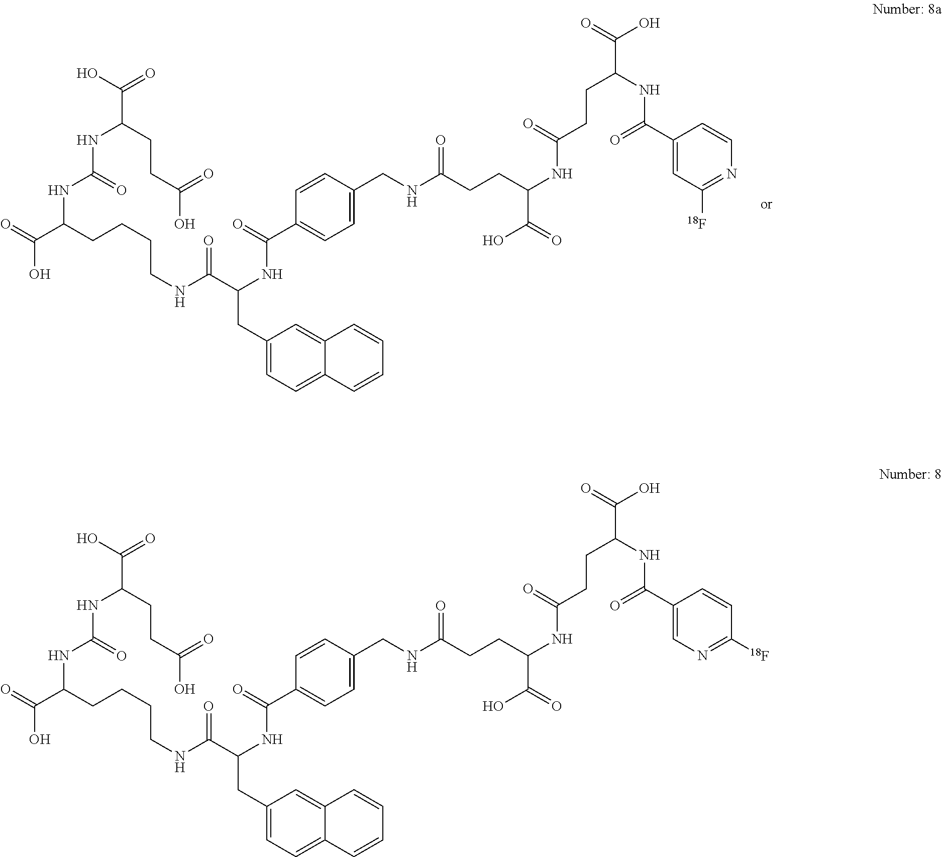
A number of preferred structures are listed in the table below:
TABLE-US-00002 I.ipophilic Hydrophilic Linker I.inker (=Linker B) Number (=Linker A) Pos. 1 Pos.2 Pos. 3 .sup.18F-Tag 1 (2-Nal)-(Bn) (Glu) (Glu) -- [.sup.18F]FN 2 (2-Nal) (Glu) (Glu) -- [.sup.18F]FN 3 (2-Nal) (.gamma.Glu) (Glu) -- [.sup.18F]FN 4 (2-Nal)-(Bn) (Glu) -- -- [.sup.18F]FN 5 (2-Nal)-(AcAMP) (.gamma.Glu) (.gamma.Glu) -- [.sup.18F]FN 6 (2-Nal)-(Bn) (Glu) (.gamma.Glu) -- [.sup.18F]FN 7 (2-Nal)-(Bn) (.gamma.Glu) (Glu) -- [.sup.18F]FN 8 (2-Nal)-(Bn) (.gamma.Glu) (.gamma.Glu) -- [.sup.18F]FN 9 (2-Nal)-(Bn) (Asp) (Glu) -- [.sup.18F]FN 10 (2-Nal)-(Bn) (.beta.Asp) (Glu) -- [.sup.18F]FN 11 (2-Nal)-(Bn) (Asp) (.gamma.Glu) -- [.sup.18F]FN 12 (2-Nal)-(Bn) (.beta.Asp) (.gamma.Glu) -- [.sup.18F]FN 13 (2-Nal)-(Bn) (Mal) (Glu) -- [.sup.18F]FN 14 (2-Nal)-(Bn) (Mal) (.gamma.Glu) -- [.sup.18F]FN 15 (2-Nal)-(Bn) (Gla) (Glu) -- [.sup.18F]FN 16 (2-Nal)-(Bn) (.gamma.Gla) (Glu) -- [.sup.18F]FN 17 (2-Nal)-(Bn) (Gla) (.gamma.Glu) -- [.sup.18F]FN 18 (2-Nal)-(Bn) (.gamma.Gla) (.gamma.Glu) -- [.sup.18F]FN
The structures of the preferred compounds 1-18 as exemplified in the table above are shown below:
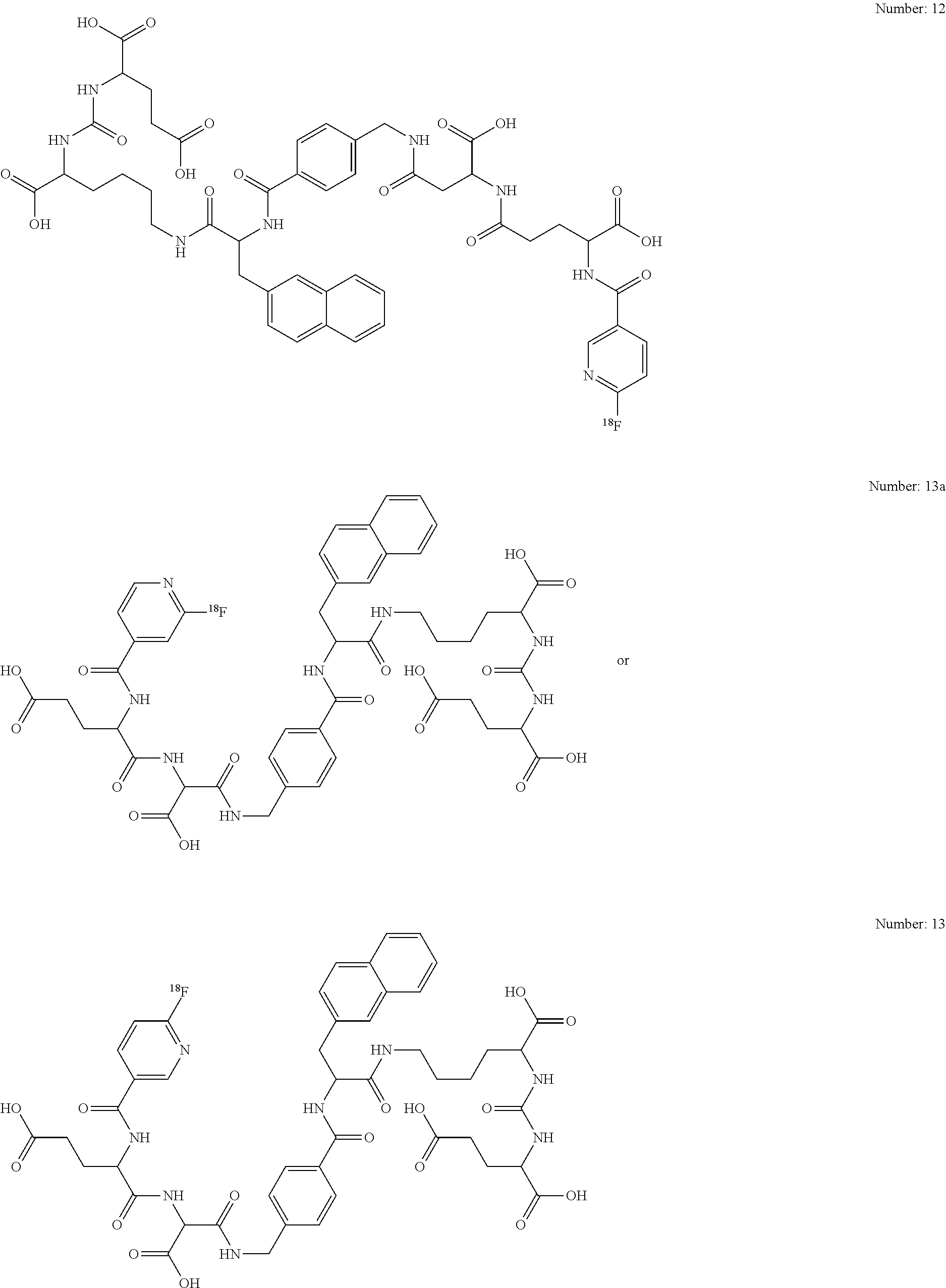
##STR00042## ##STR00043## ##STR00044## ##STR00045## ##STR00046## ##STR00047## ##STR00048## ##STR00049## ##STR00050## ##STR00051## ##STR00052##
or
As may be understood by a person skilled in the art, the above mentioned preferred compounds 1-18 and 1a-18a are not limited to the .sup.18F-Tags as shown but the .sup.18F-Tags are easily interchangeable by standard techniques and any of the .sup.18F-Tags exemplified in connection with Formula I may be used instead.
The invention also relates to precursors or pharmaceutically acceptable salts of the compounds of general formula I. The invention also relates to solvates of the compounds, including the salts as well as the active metabolites thereof and, where appropriate, the tautomers thereof according to general formula I including prodrug formulations.
A "pharmaceutically acceptable salt" is a pharmaceutically acceptable, organic or inorganic acid or base salt of a compound of the invention. Representative pharmaceutically acceptable salts include, e.g., alkali metal salts, alkali earth salts, ammonium salts, water-soluble and water-insoluble salts, such as the acetate, carbonate, chloride, gluconate, glutamate, lactate, laurate, malate or tartrate.
The term "prodrug" refers to a precursor of a drug that is a compound which upon administration to a patient, must undergo conversion by metabolic processes before becoming an active pharmacological agent. Prodrugs are generally drug precursors that, following administration to a subject and subsequent absorption, are converted to an active, or a more active species via some process, such as conversion by a metabolic pathway. Some prodrugs have a chemical group present on the prodrug that renders it less active and/or confers solubility or some other property to the drug. Once the chemical group has been cleaved and/or modified from the prodrug the active drug is generated. Prodrugs are converted into active drug within the body through enzymatic or non-enzymatic reactions. Prodrugs may provide improved physiochemical properties such as better solubility, enhanced delivery characteristics, such as specifically targeting a particular cell, tissue, organ or ligand, and improved therapeutic value of the drug.
Illustrative prodrugs of compounds in accordance with general formula I are esters and amides, preferably alkyl esters of fatty acid esters. Prodrug formulations here comprise all substances which are formed by simple transformation including hydrolysis, oxidation or reduction either enzymatically, metabolically or in any other way. A suitable prodrug contains e.g. a substance of general formula I bound via an enzymatically cleavable linker (e.g. carbamate, phosphate, N-glycoside or a disulfide group) to a dissolution-improving substance (e.g. tetraethylene glycol, saccharides, formic acids or glucuronic acid, etc.). Such a prodrug of a compound according to the invention can be applied to a patient, and this prodrug can be transformed into a substance of general formula I so as to obtain the desired pharmacological effect.
Some compounds of general formula I are encompassed in form of the racemates, their enantiomers and optionally in form of their diastereomers and all possible mixtures thereof.
According to the invention all chiral C-atoms shall have D- and/or L-configuration; also combinations within one compound shall be possible, i.e. some of the chiral C-atoms may be D- and others may be L-configuration.
The obtained compounds can be optionally separated by known methods (e.g. Allinger, N. L. and Elliel E. L. in "Topics in Stereochemistry" Vol. 6, Wiley Interscience, 1971) in their enantiomers and/or diastereomers. One possible method of enantiomeric separation is the use of chromatography.
The invention encompasses also precursors of the compounds of general formula I. The term "precursor" refers to any compound which can be used to produce the compounds of Formula I. An exemplary precursor may be a compound having no .sup.18F-tag which is added at a later stage to provide the complete compound.
The invention also relates to pharmaceutical preparations which contain a diagnostically or therapeutically effective amount of the active ingredients (compound according to the invention of formula I) together with organic or inorganic solid or liquid, pharmaceutically acceptable carriers which are suited for the intended administration and which interact with the active ingredients without drawbacks.
The phrase "pharmaceutically acceptable" is employed herein to refer to those compounds, material, compositions, and/or dosage forms which are, within the scope of sound medical judgment, suitable for use in contact with the tissues of a patient without excessive toxicity, irritation, allergic response, or other problem or complication, commensurate with a reasonable benefit/risk ratio.
A "patient" includes an animal, such as a human, monkey, cow, horse, cat or dog. The animal can be a mammal such as a non-primate and a primate (e.g., monkey and human). In one embodiment, a patient is a human being.
In general, the formula I compound or pharmaceutical compositions thereof, may be administered orally or via a parenteral route, usually injection or infusion.
A "parenteral administration route" means modes of administration other than enteral and topical administration, usually by injection, and includes, without limitation, intravenous, intramuscular, intraarterial, intrathecal, intracapsular, intraorbital, intracardiac, intradermal, intraperitoneal, transtracheal, subcutaneous, subcuticular, intraarticluare, subcapsular, subarachnoid, intraspinal and intrasternal injection and infusion.
The dosage of the compounds according to the invention is determined by the physician on the basis of the patient-specific parameters, such as age, weight, sex, severity of the disease, etc. Corresponding to the kind of administration, the medicament is suitably formulated, e.g. in the form of solutions or suspensions, simple tablets or dragees, hard or soft gelatine capsules, suppositories, ovules, preparations for injection, which are prepared according to common galenic methods.
The compounds according to the invention can be formulated, where appropriate, together with further active substances and with excipients and carriers common in pharmaceutical compositions, e.g.--depending on the preparation to be produced--talcum, gum arabic, lactose, starch, magnesium stearate, cocoa butter, aqueous and non-aqueous carriers, fatty bodies of animal or vegetable origin, paraffin derivatives, glycols (in particular polyethylene glycol), various plasticizers, dispersants or emulsifiers, pharmaceutically compatible gases (e.g. air, oxygen, carbon dioxide, etc.), preservatives. In order to produce liquid preparations, additives, such as sodium chloride solution, ethanol, sorbitol, glycerine, olive oil, almond oil, propylene glycol or ethylene glycol, can be used.
When solutions for infusion or injection are used, they are preferably aqueous solutions or suspensions, it being possible to produce them prior to use, e.g. from lyophilized preparations which contain the active substance as such or together with a carrier, such as mannitol, lactose, glucose, albumin and the like. The ready made solutions are sterilized and, where appropriate, mixed with excipients, e.g. with preservatives, stabilizers, emulsifiers, solubilizers, buffers and/or salts for regulating the osmotic pressure. The sterilization can be obtained by sterile filtration using filters having a small pore size according to which the composition can be lyophilized, where appropriate. Small amounts of antibiotics can also be added to ensure the maintenance of sterility.
The phrases "effective amount" or "therapeutically-effective amount" as used herein means that amount of a compound, material, or composition comprising a compound of the invention, or other active ingredient which is effective for producing some desired therapeutic effect in at least a sub-population of cells in an animal at a reasonable benefit/risk ratio applicable to any medical treatment. A therapeutically effective amount with respect to a compound of the invention means that amount of therapeutic agent alone, or in combination with other therapies, that provides a therapeutic benefit in the treatment of prevention of a disease. Used in connection with a compound of the invention, the term can encompass an amount that improves overall therapy, reduces or avoids symptoms or causes of disease, or enhances the therapeutic efficacy of or synergies with another therapeutic agent.
As used herein, the terms "treating" or "treatment" is intended to encompass also diagnosis, prophylaxis, prevention, therapy and cure.
The terms "prevent", "preventing," and "prevention" refer to the prevention of the onset, recurrence, or spread of the disease in a patient resulting from the administration of a prophylactic or therapeutic agent.
As noted above, compounds according general formula I are suitable for use as radio-imaging agents or as therapeutics for the treatment of rapidly proliferating cells, for example, PSMA expressing prostate cancer cells. According to the present invention they are called "radiopharmaceuticals".
Preferred imaging methods are positron emission tomography (PET) or single photon emission computed tomography (SPECT).
Accordingly, in one embodiment, a pharmaceutical composition is provided including a compound of formula I, a salt, solvate, stereoisomer, or tautomer thereof, and a pharmaceutically acceptable carrier. Accordingly, a pharmaceutical composition is provided, which is suitable for in vivo imaging and radiotherapy. Suitable pharmaceutical compositions may contain the compound of formula I in an amount sufficient for imaging, together with a pharmaceutically acceptable radiological vehicle. The radiological vehicle should be suitable for injection or aspiration, such as human serum albumin; aqueous buffer solutions, e.g., tris(hydromethyl) aminomethane (and its salts), phosphate, citrate, bicarbonate, etc; sterile water physiological saline; and balanced ionic solutions containing chloride and or dicarbonate salts or normal blood plasma cautions such as calcium potassium, sodium and magnesium.
The concentration of the imaging agent or the therapeutic agent in the radiological vehicle should be sufficient to provide satisfactory imaging. For example, when using an aqueous solution, the dosage is about 1.0 to 100 millicuries. The actual dose administered to a patient for imaging or therapeutic purposes, however, is determined by the physician administering treatment. The imaging agent or therapeutic agent should be administered so as to remain in the patient for about 1 hour to 10 days, although both longer and shorter time periods are acceptable. Therefore, convenient ampoules/vials containing 1 to 10 mL of aqueous solution may be prepared.
Imaging may be carried out in the normal manner, for example by injecting a sufficient amount of the imaging composition to provide adequate imaging and then scanning with a suitable imaging or scanning machine, such as a tomograph or gamma camera. In certain embodiments, a method of imaging a region in a patient includes the steps of: (i) administering to a patient a diagnostically effective amount of a compound labeled with a radionuclide; exposing a region of the patient to the scanning device; and (ii) obtaining an image of the region of the patient.
The amount of the compound of the present invention, or its salt, solvate, stereoisomer, or tautomer that is administered to a patient depends on several physiological factors that are routinely used by the physician, including the nature of imaging to be carried out, tissue to be targeted for imaging or therapy and the body weight and medical history of the patient to be imaged or treated using a radiopharmaceutical.
Specifically, the cell proliferative disease or disorder to be treated or imaged using a compound, pharmaceutical composition or radiopharmaceutical in accordance with this invention is a cancer, for example, prostate cancer and/or prostate cancer metastasis in e.g. lung, liver, kidney, bones, brain, spinal cord, bladder, etc.
The synthesis of the compounds of the present invention is carried out according to methods well known in the prior art (e.g. Hugenberg et al., J. Med. Chem. 2013, 56, pp. 6858-6870). General methods for .sup.18F-labelling of various macromolecules are shown in FIG. 1-7. In addition, reference is made to Schubiger et al., PET Chemistry: The Driving Force in Molecular Imaging, Ernst Schering Research Foundation, Workshop 62, Springer Verlag, ISSN 0947-6075; Ross et al., Current Radiopharmaceuticals, 2010, 3, 202-223; Kuhnast et al., Current Radiopharm 3, 2010, 174; Bernard-Gauthier et al., BioMed Research International, 2014, 1; Maschauer and Prante, BioMed Research International, 2014, 1; Olberg et al., J. Med. Chem., 2010, 53, 1732; Rostovtsev et al., Angew. Chem., 2002, 114, 2708; Smith and Greaney, Org. Lett., 2013, 15, 4826. Thus, a person skilled in the art would be able to choose the right .sup.18F-labelling depending on the starting molecule. The synthesis of the specific linker molecules is shown in EP 13004991 to which reference is made.
The synthesized compounds are chemically characterized by RP-HPLC, MS, and/or NMR.
The novel .sup.18F-tagged imaging agents with structural modifications in the linker region have improved tumor targeting properties and pharmacokinetics. The pharmacophore presents three carboxylic group able to interact with the respective side chains of PSMA and an oxygen as part of zinc complexation in the active center. Besides these obligatory interactions, the inventors were able to optimize the lipophilic interactions in the linker region compared to the compounds as described in EP 14003570.0. Further hydrophilic building blocks have been added to the linker of the compounds of the present invention (linker B), leading to another enhancement of the pharmacokinetics. Those compounds were evaluated in in vitro assays (affinity, internalization) and in vivo experiments (pPET screening and organ distribution).
Four preferred compounds with particularly promising results are .sup.18F-PSMA1007, .sup.18F-PSMA1011, .sup.18F-PSMA 1012 and .sup.18F-PSMA 1015:
##STR00053## ##STR00054## ##STR00055##
All compounds were labelled with fluorine-18 via 2-[.sup.18F]fluoronicotinic acid TFP ester in good radiochemical yields. Table A shows that the binding affinity of the PSMA inhibitors prepared so far are essentially the same and in the typical range. Further, all compounds were specifically internalized at 37.degree. C. with rather high cell uptake and internalization values (Table B). Thus, the compounds investigated exhibit optimal in vitro characteristics for a high contrast PET imaging.
The compounds of the present invention were investigated in organ distribution studies in mice carrying a LNCaP tumor (PSMA positive) with and without PMPA block. The results which have been obtained exemplary with PSMA1007 (this does not mean that the invention is limited in any way to this specific compound only) are summarised in the FIGS. 8 and 9. The tumor uptake was about 8.0.+-.2.4% ID/g. Since a quantitative blocking of the binding was not observed in the blocking experiment, the organ distribution experiment was repeated with mice carrying a PC3-tumor (PSMA negative; Results shown FIG. 8). Here, practically no tumor uptake was observed. Thus, the tumor uptake is considered specifically. Additionally, compared to the organ distribution observed with the control compound PSMA-1003 which has been described in EP 14 003 570.0 and is shown in FIG. 12, the novel compound PSMA-1007 showed a significantly reduced uptake in non-target tissue such as the liver and the small intestine. Thus, PSMA-1007 was further evaluated in microPET experiment. The results are shown in FIGS. 10 and 11. The LNCaP tumor was clearly visualised in this experiment (SUV.sub.max=3.1 at 120-140 min p.i.). The undesired uptake in the gallbladder may be an indicator for metabolites. Overall the novel class of fluorine-18 labelled PSMA inhibitors showed a great potential as possible tracer for the detection of prostate cancer and its metastases.
By the application of [.sup.18F]PSMA-1007 to a healthy volunteer several important insights were gained. First, the effective dose of a PET/CT scan with 200-250 MBq is with 4.3-5.4 mSv (2.15 mSv/MBq; FIG. 14). More than 95% of the blood pool activity is found in the serum (no or negligible infiltration of red blood cells) and more than 90% clearance from the blood pool within the first 3 h after injection. So far, those results are comparable to other important PSMA-tracers. However, in comparison to the other known PSMA-tracers the compounds of the present invention, particularly [.sup.18F]PSMA-1007, provide a very unique hepatobiliary clearance with very small clearance via the renal pathway. This outstanding low urinary clearance enables an excellent assessment of the prostatic bed. Thus, the tracers according to the present invention are perfectly suited for the primary diagnosis of prostate cancer and local recurrence.
This was further demonstrated by the results of the first-in-man study. Here, excellent tumor-to-background ratios of up to 10 were observed in the primary tumor without any interference from tracer accumulation in the bladder. Further, lymph node metastases with a diameter of down to 1 mm could be detected, which is in the range of the resolution of PET-Scans with F-18. Correlation with the samples gained by pelvic lymphadenectomy analysis revealed a specificity of 95%. Moreover, the samples gained from the prostatectomy were analyzed by histopathology revealing a nearly perfect correlation with the images acquired by PET. Those results clearly demonstrate the feasibility of prostate cancer imaging with the compounds of the present invention, particularly with [.sup.18F]PSMA-1007.
The below example explains the invention in more detail but are not construed to limit the invention in any way to the exemplified embodiments only.
EXAMPLES
Example 1: Synthesis of Precursors and Cold Reference Compounds of the .sup.18F-Conjugated Inhibitors
The isocyanate of the glutamyl moiety was generated in situ by adding a mixture of 3 mmol of bis(tert-butyl) L-glutamate hydrochloride and 1.5 mL of N-ethyldiisopropylamine (DIPEA) in 200 mL of dry CH.sub.2C.sub.12 to a solution of 1 mmol triphosgene in 10 mL of dry CH.sub.2C.sub.12 at 0.degree. C. over 4 h. After agitation of the reaction mixture for 1 h at 25.degree. C., 0.5 mmol of the resin-immobilized (2-chloro-tritylresin) .epsilon.-allyloxycarbonyl protected lysine in 4 mL DCM was added and reacted for 16 h with gentle agitation. The resin was filtered off and the allyloxy-protecting group was removed using 30 mg tetrakis(triphenyl)palladium(0) and 400 .mu.L morpholine in 4 mL CH.sub.2C.sub.12 for 3 hours.
The following coupling of the "linker amino acids" (as defined in Formula I and Scheme 1 as shown above) was performed stepwise using 2 mmol of the Fmoc-protected acid, 1.96 mmol of HBTU and 2 mmol of N-ethyldiisopropylamine in a final volume of 4 mL DMF.
The product was cleaved from the resin in a 2 mL mixture consisting of trifluoroacetic acid, triisopropylsilane, and water (95:2.5:2.5). Purification was performed using RP-HPLC and the purified product was analysed by analytical RP-HPLC and MALDI-MS.
For the preparation of the non-radioactive reference compounds 50 mg of HBTU/DIPEA (0.98 and 1 Eq.) activated 6-Fluoronicotinic-3-acid was coupled in a final volume of 4 ml DMF and agitated for 1 h at room temperature and the product was the cleaved of the resin as described above.
In some preparations a .sup.18F-tag reactive moieties (e.g. pent-4-ynoic acid or (Boc-aminooxy)acetic acid) were attached to the terminal amino-group for subsequent .sup.18F labeling.
Example 2: Production and Activation of the [.sup.18F]Fluoride
Preparation and Activation of the [.sup.18F]Fluoride
Fluorine-18 was produced by the irradiation of .sup.18O-enriched water with 16.5 MeV protons using the .sup.18O(p,n).sup.18F nuclear reaction. Irradiations were performed with the Scanditronix MC32NI cyclotron at the department of Radiopharmaceutical Chemistry (E030) at the German Cancer Research Center Heidelberg.
After transfer of the irradiated water to an automated system (Trasis All In One 32) the [.sup.18F]F.sup.- was separated from the [.sup.18O]H.sub.2O by passing through an previously conditioned (5 ml 1 M K.sub.2CO.sub.3 and 10 ml water) anion exchange cartridge (Waters Accel.TM. Plus QMA Cartridge light) and subsequently eluted with a mixture of 800 .mu.l acetonitrile and 150 .mu.l tetrabutylammonium bicarbonate solution (320 mM in water). The mixture was evaporated to dryness an a temperature of 100.degree. C. under a stream of nitrogen. This distillation was subsequently repeated two times by the adding 1.8 ml of acetonitrile for each step. After applying maximum achievable vacuum to the residue for 5 minutes at 100.degree. C. and subsequent cooling to 50.degree. C. the dry residue was dissolved in 2 ml of tert-butanol/acetonitrile (8:2) and used for the labelling reactions.
Alternatively, the system was also used with the classical Kryptofix.RTM. 2.2.2/K.sub.2CO.sub.3 activation system (20 mg 2.2.2+28 .mu.l 1 M K.sub.2CO.sub.3) in acetonitrile. Further, the solvent for dissolving the activated [.sup.18F]F.sup.- was changed to dry DMF, DMSO or acetonitrile for some experiments.
Example 3: Radiosynthesis of 6-[.sup.18F]Fluoronicotinic Acid Tetrafluorophenyl Ester ([.sup.18F]FN-TFP)
To 10 mg N,N,N-Trimethyl-5-((2,3,5,6-tetrafluorophenoxy)-carbonyl)pyridin- -2-aminium trifluoromethanesulfonate (prepared as described by Olberg et al. J. Med. Chem., 2010, 53, 1732) 1 ml of tert-butanol/Acetonitrile (8:2) containing the dried [.sup.18F]KF-Kryptofix 2.2.2 komplex (0.1-10 GBq .sup.18F) was added and the mixture was heated at 40.degree. C. After 10 minutes the mixture was diluted with 3 ml of water and the product loaded on a preconditioned Oasis MCX Plus Sep-Pak (Waters). The cartridge was rinsed with 10 mL of water and the purified 6-[.sup.18F]Fluoronicotinic acid tetrafluorophenyl ester was eluted back to the reaction vessel using 2 mL of water/acetonitrile (7:13). For achieving a higher activity concentration a fractionized elution was done in some cases. Therefore the loaded cartridge was rinsed with 500 .mu.l of solvent, which were discarded, and then eluted with 400-800 .mu.l of solvent for further reactions. Usually more than 50% of the initial activity were eluted with the second fraction.
Example 4: PSMA-1007
The synthesis of the precursor and the cold reference was performed as described under example 1.
Radiosynthesis of [.sup.18F]PSMA-1007 200 .mu.l of ([.sup.18F]FN-TFP) were added to 50 .mu.l of a 2 mg/ml solution of PSMA-1007-VL in DMSO. Then 50 .mu.l of buffer (0.2 M phosphate buffer, pH 8.0) were added and the mixture heated at 60.degree. C. for 20 minutes. The products were separated by semipreparative radio-HPLC and identified by analytical radio-HPLC and comparison of the retention times with the respective non-radioactive reference compounds.
PSMA1007-VL (Precursor):
(Glu)-(Urea)-(Lys)-(2-Nal)-(Bn)-(Glu)-(Glu); (C.sub.43H.sub.53N.sub.7O.sub.15; 907.93 g/mol) particularly (Glu)-(Urea)-(Lys)-(2-Nal)-(Bn)-(L-Glu)-(L-Glu)
MS (MALDI): m/z=908.7 [M+H.sup.+].sup.+
PSMA-1007
(Glu)-(Urea)-(Lys)-(2-Nal)-(Bn)-(Glu)-(Glu)-FN; C.sub.49H.sub.55FN.sub.8O.sub.16; (1031 g/mol) particularly (Glu)-(Urea)-(Lys)-(2-Nal)-(Bn)-(L-Glu)-(L-Glu)-FN;
MS (MALDI): m/z=1032.1 [M+H].sup.+
[.sup.18F]PSMA-1007
RCA: ca. 6%
HPLC (Gradient: 5% A/95% B-95% A/5% B in 12.5 min; Flow: 3 ml/min; Column: Merck Chromolith.RTM. Performance RP-18e 100-4.6 mm; Solvent A: Acetonitrile, Solvent B: 0.1% aqueous TFA): t.sub.ret: 4.56 min (t.sub.dead: 0.56 min)
Example 5: PSMA-1011
The synthesis of the precursor and the cold reference was performed as described under example 1. The synthesis [.sup.18F]PSMA-1011 was performed as described under example 4.
PSMA-1011-VL (precursor):
(Glu)-(Urea)-(Lys)-(2-Naphthylalanine)-(Glu)-(Glu); C.sub.35H.sub.46N.sub.8O.sub.14 (774.78 g/mol)
MS (MALDI): m/z=775.3 [M+H.sup.+].sup.+
PSMA-1011:
(Glu)-(Urea)-(Lys)-(2-Naphthylalanine)-(Glu)-(Glu)-FN; C.sub.41H.sub.48FN.sub.7O.sub.15 (897.86 g/mol)
MS (MALDI): m/z=898.3 [M+H.sup.+].sup.+
[.sup.18F]PSMA-1011:
RCA: ca. 7%
HPLC (Gradient: 5% A/95% B-50% A/50% B in 10.0 min; Flow: 3 ml/min; Column: Merck Chromolith.RTM. Performance RP-18e 100-4.6 mm; Solvent A: Acetonitrile, Solvent B: 0.1% aqueous TFA): t.sub.ret: 5.72 min (t.sub.dead: 0.56 min)
Example 6: PSMA-1012
The synthesis of the precursor and the cold reference was performed as described under example 1. The synthesis [.sup.18F]PSMA-1012 was performed as described under example 4.
PSMA-1012-VL (precursor):
(Glu)-(Urea)-(Lys)-(2-Naphthylalanine)-(.gamma.Glu)-(Glu); C.sub.35H.sub.46N.sub.8O.sub.14 (774.78 g/mol)
MS (MALDI): m/z=775.0 [M+H.sup.+].sup.+
PSMA-1012:
(Glu)-(Urea)-(Lys)-(2-Naphthylalanine)-(.gamma.Glu)-(Glu)-FN; C.sub.41H.sub.48FN.sub.7O.sub.15 (897.86 g/mol) MS (MALDI): m/z=897.9 [M+H.sup.+].sup.+
[.sup.18F]PSMA-1012:
RCA: ca. 4%
HPLC (Gradient: 5% A/95% B-50% A/50% B in 10.0 min; Flow: 3 ml/min; Column: Merck Chromolith.RTM. Performance RP-18e 100-4.6 mm; Solvent A: Acetonitrile, Solvent B: 0.1% aqueous TFA): t.sub.ret: 5.61 min (t.sub.dead: 0.56 min)
Example 7: PSMA-1015
The synthesis of the precursor and the cold reference was performed as described under example 1. The synthesis [.sup.18F]PSMA-1015 was performed as described under example 4.
PSMA-1015-VL (precursor):
(Glu)-(Urea)-(Lys)-(2-Nal)-(Bn)-(Glu); C.sub.38H.sub.46N.sub.6O.sub.12 (778.80 g/mol)
MS (MALDI): m/z=779.4 [M+H.sup.+].sup.+
PSMA-1015:
(Glu)-(Urea)-(Lys)-(2-Nal)-(Bn)-(Glu)-FN; C.sub.44H.sub.48FN.sub.7O.sub.13 (901.89 g/mol)
MS (MALDI): m/z=902.5 [M+H.sup.+].sup.+
[.sup.18F]PSMA-1015:
RCA: ca. 7%
HPLC (Gradient: 5% A/95% B-50% A/50% B in 10.0 min; Flow: 3 ml/min; Column: Merck Chromolith.RTM. Performance RP-18e 100-4.6 mm; Solvent A: Acetonitrile,
Solvent B: 0.1% aqueous TFA): t.sub.ret: 6.48 min (t.sub.dead: 0.56 min)
Example 8: Synthesis of Dry [.sup.18F]FN-TFP
The radiosynthesis was performed as described under example 3 until elution of the [.sup.18F]FN-TFP from the cartridge. After the washing the cartridge with 10 ml water (example 3) the cartridge was blown dry with 20-40 ml air. Then the MCX cartridge was connected to a SepPak SodSulf drying cartridge and the product was eluted with 2 ml of dry acetonitrile. For achieving a higher activity concentration a fractionized elution was done in some cases. Therefore the loaded MCX cartridge was rinsed with 500 .mu.l of solvent (after blowing the cartridge dry), which were discarded, and then the drying cartridge was connected to the MCX cartridge and the product was eluted with 0.8-1.2 ml of acetonitrile for further reactions. Usually more than 50% of the initial activity were eluted with the second fraction.
Example 9: PSMA-1018
The synthesis of the precursor and the cold reference was performed as described under example 1.
Radiosynthesis of [.sup.18F]PSMA-1018 200 .mu.l of dry [.sup.18F]FN-TFP (example 8) were added to 50 .mu.l of a 4 mg/ml solution of PSMA-1018-VL in DMSO. Then 10 .mu.l of DIPEA were added and the mixture heated at 60.degree. C. for 20 minutes. Then the reaction mixture was acidified by the addition of 10 .mu.l TFA, the products separated by semipreparative radio-HPLC and identified by analytical radio-HPLC and comparison of the retention times with the respective non-radioactive reference compounds.
PSMA-1018-VL (precursor):
(Glu)-(Urea)-(Lys)-(2-Nal)-(Bn)-(Glu)-(Glu)-(Glu); C.sub.48H.sub.60N.sub.8O.sub.18 (1037.03 g/mol)
MS (MALDI): m/z=1037.6 [M+H.sup.+].sup.+
PSMA-1018:
(Glu)-(Urea)-(Lys)-(2-Nal)-(Bn)-(Glu)-(Glu)-(Glu)-FN; C.sub.54H.sub.62FN.sub.9O.sub.19 (1160.12 g/mol)
MS (MALDI): m/z=1160.8 [M+H.sup.+].sup.+
[.sup.18F]PSMA-1018:
RCA: ca. 20%
HPLC (Gradient: 5% A/95% B-95% A/5% B in 10.0 min; Flow: 3 ml/min; Column: Merck Chromolith.RTM. Performance RP-18e 100-4.6 mm; Solvent A: Acetonitrile, Solvent B: 0.1% aqueous TFA): t.sub.ret: 3.87 min (t.sub.dead: 0.56 min)
Example 10: PSMA-1019
The synthesis of the precursor and the cold reference was performed as described under example 1. The synthesis [.sup.18F]PSMA-1019 was performed as described under example 9.
PSMA-1019-VL (precursor):
(Glu)-(Urea)-(Lys)-(2-Nal)-(Chx)-(Glu)-(Glu); C.sub.43H.sub.59N.sub.7O.sub.15 (913.97 g/mol)
MS (MALDI): m/z=914.4 [M+H.sup.+].sup.+
PSMA-1019:
(Glu)-(Urea)-(Lys)-(2-Nal)-(Chx)-(Glu)-(Glu)-FN; C.sub.49H.sub.61FN.sub.8O.sub.16 (1037.05 g/mol)
MS (MALDI): m/z=1037.7 [M+H.sup.+].sup.+
[.sup.18F]PSMA-1019:
RCA: ca. 47%
HPLC (Gradient: 5% A/95% B-95% A/5% B in 12.5 min; Flow: 3 ml/min; Column: Merck Chromolith.RTM. Performance RP-18e 100-4.6 mm; Solvent A: Acetonitrile, Solvent B: 0.1% aqueous TFA): t.sub.ret: 4.43 min (t.sub.dead: 0.56 min)
Example 11: PSMA-1020
The synthesis of the precursor and the cold reference was performed as described under example 1. The synthesis [.sup.18F]PSMA-1020 was performed as described under example 9.
PSMA-1020-VL (precursor):
(Glu)-(Urea)-(Lys)-(2-Nal)-(.gamma.Glu)-(Glu)-(Glu); C.sub.40H.sub.53N.sub.7O.sub.17 (903.89 g/mol)
MS (MALDI): m/z=904.9 [M+H.sup.+].sup.+
PSMA-1020:
(Glu)-(Urea)-(Lys)-(2-Nal)-(.gamma.Glu)-(Glu)-(Glu)-FN; C.sub.46H.sub.55FN.sub.8O.sub.18 (1026.97 g/mol)
MS (MALDI): m/z=1027.9 [M+H.sup.+].sup.+
[.sup.18F]PSMA-1020:
RCA: ca. 60%
HPLC (Gradient: 5% A/95% B-95% A/5% B in 12.5 min; Flow: 3 ml/min; Column: Merck Chromolith.RTM. Performance RP-18e 100-4.6 mm; Solvent A: Acetonitrile, Solvent
B: 0.1% aqueous TFA): t.sub.ret: 6.48 min (t.sub.dead: 0.56 min)
Example 12: PSMA-1022
The synthesis of the precursor and the cold reference was performed as described under example 1. The synthesis [.sup.18F]PSMA-1022 was performed as described under example 9.
PSMA-1022-VL (precursor):
(Glu)-(Urea)-(Lys)-(2-Nal)-(.gamma.Glu)-(.gamma.Glu); C.sub.35H.sub.46N.sub.8O.sub.14 (774.78 g/mol)
MS (MALDI): m/z=775.4 [M+H.sup.+].sup.+
PSMA-1022:
(Glu)-(Urea)-(Lys)-(2-Nal)-(.gamma.Glu)-(.gamma.Glu)-FN; C.sub.41H.sub.48FN.sub.7O.sub.15 (897.86 g/mol)
MS (MALDI): m/z=898.8 [M+H.sup.+].sup.+
[.sup.18F]PSMA-1022:
RCA: ca. 29%
HPLC (Gradient: 5% A/95% B-95% A/5% B in 10.0 min; Flow: 3 ml/min; Column: Merck Chromolith.RTM. Performance RP-18e 100-4.6 mm; Solvent A: Acetonitrile, Solvent B: 0.1% aqueous TFA): t.sub.ret: 3.92 min (t.sub.dead: 0.56 min)
Example 13: PSMA-1023
The synthesis of the precursor and the cold reference was performed as described under example 1. The synthesis [.sup.18F]PSMA-1023 was performed as described under example 9
PSMA-1023-VL (precursor):
(Glu)-(Urea)-(Lys)-(2-Nal)-(Bn)-(D-Glu)-(D-Glu); C.sub.43H.sub.53N.sub.7O.sub.15 (907.93 g/mol)
MS (MALDI): m/z=907.8 [M+H.sup.+].sup.+
PSMA-1023:
(Glu)-(Urea)-(Lys)-(2-Nal)-(Bn)-(D-Glu)-(D-Glu)-FN; C.sub.49H.sub.55FN.sub.8O.sub.16 (1031.00 g/mol)
MS (MALDI): m/z=1031.8 [M+H.sup.+].sup.+
[.sup.18F]PSMA-1023:
RCA: ca. 24%
HPLC (Gradient: 5% A/95% B-50% A/50% B in 10.0 min; Flow: 3 ml/min; Column: Merck Chromolith.RTM. Performance RP-18e 100-4.6 mm; Solvent A: Acetonitrile, Solvent B: 0.1% aqueous TFA): t.sub.ret: 3.87 min (t.sub.dead: 0.56 min)
Example 14: PSMA-1024
The synthesis of the precursor and the cold reference was performed as described under example 1. The synthesis [.sup.18F]PSMA-1024 was performed as described under example 9.
PSMA-1024-VL (precursor):
(Glu)-(Urea)-(Lys)-(2-Nal)-(Bn)-(D-Glu)-(Glu); C.sub.43H.sub.53N.sub.7O.sub.15 (907.93 g/mol)
MS (MALDI): m/z=908.6 [M+H.sup.+].sup.+
PSMA-1024:
(Glu)-(Urea)-(Lys)-(2-Nal)-(Bn)-(D-Glu)-(Glu)-FN; C.sub.49H.sub.55FN.sub.8O.sub.16 (1031.00 g/mol)
MS (MALDI): m/z=1031.5 [M+H.sup.+].sup.+
[.sup.18F]PSMA-1024:
RCA: ca. 27%
HPLC (Gradient: 5% A/95% B-95% A/5% B in 10.0 min; Flow: 3 ml/min; Column: Merck Chromolith.RTM. Performance RP-18e 100-4.6 mm; Solvent A: Acetonitrile, Solvent B: 0.1% aqueous TFA): t.sub.ret: 3.85 min (t.sub.dead: 0.56 min)
Example 15: PSMA-1025
The synthesis of the precursor and the cold reference was performed as described under example 1. The synthesis [.sup.18F]PSMA-1025 was performed as described under example 9.
PSMA-1025-VL (precursor):
(Glu)-(Urea)-(Lys)-(2-Nal)-(Bn)-(Gla); C.sub.39H.sub.46N.sub.6O.sub.14 (822.21 g/mol)
MS (MALDI): m/z=822.8 [M+H.sup.+].sup.+
PSMA-1025:
(Glu)-(Urea)-(Lys)-(2-Nal)-(Bn)-(Gla)-FN; C.sub.49H.sub.55FN.sub.8O.sub.16 (945.32 g/mol)
MS (MALDI): m/z=946.0 [M+H.sup.+].sup.+
[.sup.18F]PSMA-1025:
RCA: ca. 24%
HPLC (Gradient: 5% A/95% B-95% A/5% B in 12.5 min; Flow: 3 ml/min; Column: Merck Chromolith.RTM. Performance RP-18e 100-4.6 mm; Solvent A: Acetonitrile, Solvent B: 0.1% aqueous TFA): t.sub.ret: 4.51 min (t.sub.dead: 0.56 min)
Example 16: PSMA-1026
The synthesis of the precursor and the cold reference was performed as described under example 1. The synthesis [.sup.18F]PSMA-1026 was performed as described under example 9.
PSMA-1026-VL (precursor):
(Glu)-(Urea)-(Lys)-(2-Nal)-(Bn)-(Sala); C.sub.36H.sub.44N.sub.6O.sub.13S (800.83 g/mol)
MS (MALDI): m/z=801.8 [M+H.sup.+].sup.+
PSMA-1026:
(Glu)-(Urea)-(Lys)-(2-Nal)-(Bn)-(Sala)-FN; C.sub.42H.sub.46FN.sub.7O.sub.14S (923.92 g/mol)
MS (MALDI): m/z=924.7 [M+H.sup.+].sup.+
[.sup.18F]PSMA-1026:
RCA: ca. 57%
HPLC (Gradient: 5% A/95% B-95% A/5% B in 12.5 min; Flow: 3 ml/min; Column: Merck Chromolith.RTM. Performance RP-18e 100-4.6 mm; Solvent A: Acetonitrile, Solvent B: 0.1% aqueous TFA): t.sub.ret: 4.03 min (t.sub.dead: 0.56 min)
Example 17: PSMA-1027
The synthesis of the precursor and the cold reference was performed as described under example 1. The synthesis [.sup.18F]PSMA-1027 was performed as described under example 9.
PSMA-1027-VL (precursor):
(Glu)-(Urea)-(Lys)-(2-Nal)-(Bn)-(Sala)-(Sala); C.sub.39H.sub.49N.sub.7O.sub.17S.sub.2 (951.97 g/mol)
MS (MALDI): m/z=952.7 [M+H.sup.+].sup.+
PSMA-1027:
(Glu)-(Urea)-(Lys)-(2-Nal)-(Bn)-(Sala)-(Sala)-FN; C.sub.45H.sub.51FN.sub.8O.sub.18S.sub.2 (1075.06 g/mol)
MS (MALDI): m/z=1075.8 [M+H.sup.+].sup.+
[.sup.18F]PSMA-1027:
RCA: ca. 62%
HPLC (Gradient: 5% A/95% B-95% A/5% B in 10.0 min; Flow: 3 ml/min; Column: Merck Chromolith.RTM. Performance RP-18e 100-4.6 mm; Solvent A: Acetonitrile, Solvent B: 0.1% aqueous TFA): t.sub.ret: 3.10 min (t.sub.dead: 0.56 min)
Example 18: Cell Culture
For binding studies and in vivo experiments LNCaP cells (metastatic lesion of human prostatic adenocarcinoma, ATCC CRL-1740) were cultured in RPMI medium supplemented with 10% fetal calf serum and Glutamax (PAA, Austria). During cell culture, cells were grown at 37.degree. C. in an incubator with humidified air, equilibrated with 5% CO.sub.2. The cells were harvested using trypsin-ethylenediaminetetraacetic acid (trypsin-1.5 EDTA; 0.25% trypsin, 0.02% EDTA, all from PAA, Austria) and washed with PBS.
Example 19: Cell Binding and Internalization
The competitive cell binding assay and internalization experiments were performed as described previously (Eder et al. Bioconjugate Chem. 2012, 23 (4), 688-697). Briefly, the respective cells (10.sup.5 per well) were incubated with the radioligand (.sup.68Ga-labeled [Glu-urea-Lys(Ahx)]2-HBED-CC (Schafer et al. EJNMMI Research 2012, 2:23 doi:10.1186/2191-219X-2-23))) in the presence of 12 different concentrations of analyte (0-5000 nM, 100 .mu.L/well). After incubation, washing was carried out using a multiscreen vacuum manifold (Millipore, Billerica, Mass.). Cell-bound radioactivity was measured using a gamma counter (Packard Cobra II, GMI, Minnesota, USA). The 50% inhibitory concentration (IC.sub.50) was calculated by fitting the data using a nonlinear regression algorithm (GraphPad Software). Experiments were performed three times. Reference is made to Table A below.
To determine the specific cell uptake and internalization, 10.sup.5 cells were seeded in poly-L-lysine coated 24-well cell culture plates 24 h before incubation. After washing, the cells were incubated with 30 nM of the radiolabeled compounds for 45 min at 37.degree. C. Specific cellular uptake was determined by competitive blocking with 2-(phosphonomethyl)pentanedioic acid (500 .mu.M final concentration, PMPA, Axxora, Loerrach, Germany). Cellular uptake was terminated by washing 3 times with 1 mL of ice-cold PBS. Cells were subsequently incubated twice with 0.5 mL glycine-HCl in PBS (50 mM, pH=2.8) for 5 min to remove the surface-bound fraction. The cells were washed with 1 mL of ice-cold PBS and lysed using 0.3 N NaOH (0.5 mL). The surface-bound and the internalized fractions were measured in a gamma counter. The cell uptake was calculated as percent of the initially added radioactivity bound to 10.sup.5 cells [% ID/10.sup.5 cells]. The main results are given in Table B.
TABLE-US-00003 TABLE A Binding Affinity Assay Compound IC.sub.50 [nM] PSMA-1007 5 PSMA-1011 7 PSMA-1012 7 PSMA-1015 4 PSMA-1018 14 PSMA-1019 12 PSMA-1020 9 PSMA-1022 8 PSMA-1023 8 PSMA-1024 8 PSMA-1025 9 PSMA-1026 14 PSMA-1027 7
TABLE-US-00004 TABLE B Specific Internalization Cell surface Internalised Internalized Compound [% ID/10.sup.5 cells] [% ID/10.sup.5 cells] fraction [%]* PSMA-1007 2.7 6.5 71 PSMA-1011 4.1 0.7 15 PSMA-1012 6.9 2.7 28 PSMA-1015 14.3 6.0 30 PSMA-1018 1.1 0.9 45 PSMA-1019 5.5 1.9 26 PSMA-1020 6.8 1.5 18 PSMA-1022 3.5 1.0 22 PSMA-1023 6.5 3.2 33 PSMA-1024 5.3 2.1 28 PSMA-1025 4.3 3.1 42 PSMA-1026 1.3 1.3 50 PSMA-1027 0.9 1.4 60 *(Internalized activity/total activity)*l00
Example 20: MicroPET
For the microPET studies, 10-25 MBq of the radiolabeled compounds in a volume of 0.10 ml (60 pmol) were injected via a lateral tail vein into mice bearing LNCaP tumor xenografts. The anesthetized animals (2% sevoflurane, Abbott, Wiesbaden, Germany) were placed in prone position into the Inveon small animal PET scanner (Siemens, Knoxville, Tenn., USA) to perform dynamic microPET scans. The results are shown in FIGS. 10 and 11.
Example 21: Plasma Binding and Stability
For the determination of the plasma binding 3 .mu.l of 6 .mu.molar c.a. [.sup.18F]PSMA solution was added to 300 .mu.l human serum AB and incubated at 37.degree. C. for 1 h. Subsequently the product mixture was analyzed by size-exclusion chromatography.
No plasma binding was observed with any of the compounds.
For the determination of the plasma stability 50 .mu.l of 6 .mu.molar c.a. [.sup.18F]PSMA solution was added to 450 .mu.l human serum AB and incubated at 37.degree. C. At 1, 2 and 4 h samples were prepared. Therefore 100 .mu.l of the tracer/plasma mixture were added to 100 .mu.l of acetonitrile. Subsequently the mixture was centrifuged at 13000 rpm for 3 minutes. 100 .mu.l of the supernatant were added to 100 .mu.l of acetonitrile, centrifuged at 13000 rpm for 5 minutes, the liquid separated from any residual solids and analyzed by HPLC.
All of the compounds were stable in human plasma at 37.degree. C. for at least 4 hours.
Example 22: In Vivo Experiments
For in vivo experiments, 8 week old BALB/c nu/nu mice were subcutaneously inoculated into the right trunk with 5.times.10.sup.6 LNCaP- or PC3-cells in 50% Matrigel. When the size of tumor was approximately 1 cm.sup.3, the radiolabeled compound was injected via the tail vein (ca. 30 MBq, 60 pmol for .mu.PET imaging; ca. 1 MBq, 60 pmol for organ distribution).
Organ Distribution
The F-18 labeled compounds were injected via tail vein (1-2 MBq per mouse; 60 pmol). At 1 h after injection, the animals were sacrificed. Organs of interest were dissected, blotted dry, and weighed. The radioactivity was measured with a gamma counter (Packard Cobra II, GMI, Minnesota, USA) and calculated as % ID/g. The main results are given in FIGS. 8 and 9.
The compounds PSMA 1003 and PSMA 1009 are for comparison and are shown in FIGS. 12 and 13.
Example 23: [.sup.18F]PSMA-1007 for Human Application
[.sup.18F]PSMA-1007 was produced by conjugation of dry [.sup.18F]FN-TFP (Example 8) to PSMA1007-VL (Example 4) under dry conditions (analogously Example 9) on an automated synthesis module (Trasis AllInOne) and purified by semi-preparative HPLC. Radio-HPLC was performed to determine the chemical identity and the chemical and radiochemical purity of [.sup.18F]PSMA-1007. Residual solvents were determined by gas chromatography. The radionuclide purity was controlled by half-life measurement. The product solution was tested for sterility, bacterial endotoxins (LAL-test), pH, colorlessness and particles. The integrity of the sterile filter after filtration was examined using the bubble-point test.
Example 24: Application of [.sup.18F]PSMA-1007 to a Healthy Volunteer
The healthy subject was injected a total activity of 300 MBq and subsequently PET scans were performed on a Biograph mCT Flow scanner (Siemens, Erlangen, Germany) in three blocks. Block 1 contained PET-1 (start 5 min p.i.) to PET-7 (ending 140 min p.i.), block 2 contained PET-8 (start 180 min p.i.) and PET-9 (240-270 min p.i.), block 3 contained PET-10 (440-480 min p.i.). A non-enhanced low-dose CT (estimate 1.43 mSv, respectively) for attenuation correction was performed at the beginning of each block, followed by serial emission scans without moving the volunteer in between.
Kidneys, liver, spleen, whole heart, upper and lower large intestine, parotid glands, submandibular glands and urinary bladder were segmented into volumes of interest (VOIs) using a percentage of maximum threshold between 20% and 30% using the corresponding CT as guidance and then time activity curves (TACs) were calculated for all organs. The TAC for red marrow was derived from the venous blood samples and then the dose for the red marrow calculated (S. Shen et al., JNM 2002, 43, 1245-1253; G. Sgouros et al., JNM 1993, 34, 689-694.). The TAC for the urinary bladder content was a combination of estimated activity in the urinary bladder VOI in PET and activity measured in the voided urine. Curve fitting was applied to all TACs. Kidneys, salivary glands, upper and lower large intestine and heart were fitted with a bi-exponential function. For liver, spleen and urinary bladder content a mono-exponential fit to the last three time points was performed.
Absorbed and effective dose calculations were performed using the ICRP endorsed IDAC 1.0 package which is integrated in QDOSE. Additionally, residence times of all included source organs and remainder body were exported as an OLINDA case file for dose calculation (OLINDA 1.1). The absorbed doses to the salivary glands (parotid and submandibular glands) were determined using the spherical model. The organ masses for the salivary glands were estimated with 25 g for a parotid and 12.5 g for a submandibular gland (ICRP publication 89). The results are summarized in FIGS. 14-16.
Example 25: Application of [.sup.18F]-PSMA-1007 in a Patients Suffering from Prostate Cancer
Ten patients (age 60-80 years) suffering from a newly diagnosed high-risk prostate cancer with a gleason score of 7-9 and initial PSA levels of 5-90 ng/ml were injected 100-360 MBq [.sup.18F]-PSMA-1007 and subsequently PET scans were performed on a Biograph mCT Flow scanner (Siemens, Erlangen, Germany) at 1 and 3 h p.i. The results are depicted in FIGS. 17 and 18 as mean values of the SUV.sub.mean and SUV.sub.max, respectively. The pictures gained with the PET scans are exemplified in FIGS. 19 and 20 as maximum intensity projections.
Eight of the patients underwent radical prostatectomy with extended pelvic lymphadenectomy. Analyses of prostatectomy specimen were performed blinded to PET-data under the supervision of dedicated uropathologists, according to International Society of Urological Pathology standards (T. H. van der Kwast et al. Mod. Phathol. 2011, 24, 16-25). Representative sections were stained by immunohistochemistry. The sections were deparaffinized in xylene and rehydrated in a graded ethanol series. Antigen retrieval was performed with a steam cooker using retrieval buffer (Target Retrieval Solution, Dako). A mouse monoclonal antibody against PSMA (clone 3E6, Dako) was used at a 1:100 dilution and incubated overnight at 4.degree. C. ad subsequently immunodetection was performed using the Histostain-Plus detection kit (Invitrogen). Stained sections were scanned using a Nanozoomer 2.0-HT Scansystem (Hamamatsu Photonics) to generate digital whole slide images. The staining revealed a nearly perfect correlation with the PET scan, as exemplified in FIG. 21.
* * * * *
C00001
C00002
C00003
C00004
C00005
C00006
C00007
C00008
C00009
C00010
C00011
C00012
C00013

C00014

C00015

C00016

C00017

C00018
C00019

C00020

C00021

C00022
C00023

C00024

C00025

C00026

C00027
C00028

C00029

C00030

C00031

C00032

C00033

C00034

C00035
C00036

C00037

C00038

C00039
C00040
C00041

C00042

C00043
C00044

C00045

C00046

C00047

C00048
C00049
C00050
C00051

C00052

C00053

C00054

C00055

C00056
C00057

C00058

C00059

C00060
C00061
C00062
C00063

C00064

C00065
C00066

C00067
C00068
C00069

C00070

C00071
C00072

C00073

C00074

C00075
C00076
C00077

C00078

C00079

C00080

C00081
C00082
C00083

C00084

C00085

C00086

C00087

C00088

C00089
C00090

C00091

C00092
C00093

C00094

C00095

C00096

C00097

C00098

C00099
C00100
C00101

C00102
C00103

C00104

C00105

C00106
C00107

C00108

C00109

D00001

D00002
D00003
D00004

D00005

D00006

D00007

D00008

D00009

D00010

D00011

D00012

D00013

D00014
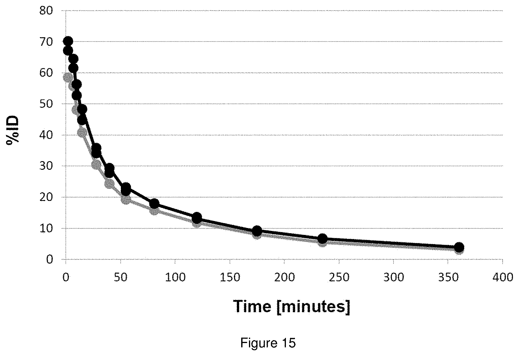
D00015

D00016

D00017
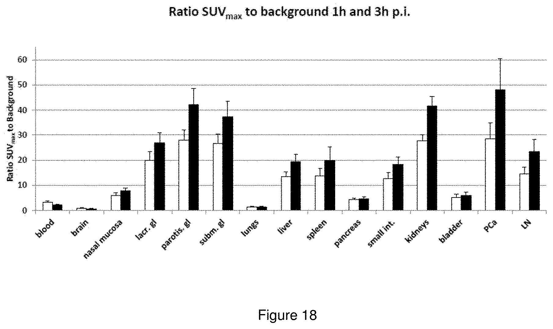
D00018

D00019

D00020

D00021

XML
uspto.report is an independent third-party trademark research tool that is not affiliated, endorsed, or sponsored by the United States Patent and Trademark Office (USPTO) or any other governmental organization. The information provided by uspto.report is based on publicly available data at the time of writing and is intended for informational purposes only.
While we strive to provide accurate and up-to-date information, we do not guarantee the accuracy, completeness, reliability, or suitability of the information displayed on this site. The use of this site is at your own risk. Any reliance you place on such information is therefore strictly at your own risk.
All official trademark data, including owner information, should be verified by visiting the official USPTO website at www.uspto.gov. This site is not intended to replace professional legal advice and should not be used as a substitute for consulting with a legal professional who is knowledgeable about trademark law.