Imaging system for combine full-color reflectance and near-infrared imaging
Fengler , et al. Sept
U.S. patent number 10,779,734 [Application Number 15/584,405] was granted by the patent office on 2020-09-22 for imaging system for combine full-color reflectance and near-infrared imaging. This patent grant is currently assigned to Stryker European Operations Limited. The grantee listed for this patent is Stryker European Operations Limited. Invention is credited to Aurther E. Bailey, Paul Cottle, John Fengler, Paul R. Westwick.


| United States Patent | 10,779,734 |
| Fengler , et al. | September 22, 2020 |
Imaging system for combine full-color reflectance and near-infrared imaging
Abstract
An imaging system for acquisition of NIR and full-color images includes a light source providing visible light and NIR light to an area under observation, such as living tissue, a camera having one or more image sensors configured to separately detect blue reflectance light, green reflectance light, and combined red reflectance light/detected NIR light returned from the area under observation. A controller in signal communication with the light source and the camera is configured to control the light source to continuously illuminate area under observation with temporally continuous blue/green illumination light and with red illumination light and NIR excitation light. At least one of the red illumination light and NIR excitation light are switched on and off periodically in synchronism with the acquisition of red and NIR light images in the camera.
| Inventors: | Fengler; John (North Vancouver, CA), Westwick; Paul R. (Vancouver, CA), Bailey; Aurther E. (North Vancouver, CA), Cottle; Paul (Vancouver, CA) | ||||||||||
|---|---|---|---|---|---|---|---|---|---|---|---|
| Applicant: |
|
||||||||||
| Assignee: | Stryker European Operations
Limited (Carrigtwohill, IE) |
||||||||||
| Family ID: | 1000005066877 | ||||||||||
| Appl. No.: | 15/584,405 | ||||||||||
| Filed: | May 2, 2017 |
Prior Publication Data
| Document Identifier | Publication Date | |
|---|---|---|
| US 20170273567 A1 | Sep 28, 2017 | |
Related U.S. Patent Documents
| Application Number | Filing Date | Patent Number | Issue Date | ||
|---|---|---|---|---|---|
| 14873842 | Oct 2, 2015 | 9642532 | |||
| 12933512 | Nov 3, 2015 | 9173554 | |||
| PCT/US2009/037506 | Mar 18, 2009 | ||||
| 61037514 | Mar 18, 2008 | ||||
| Current U.S. Class: | 1/1 |
| Current CPC Class: | H04N 5/332 (20130101); A61B 1/045 (20130101); A61B 5/7425 (20130101); H04N 5/33 (20130101); A61B 1/04 (20130101); A61B 1/00186 (20130101); A61B 1/0638 (20130101); A61B 1/042 (20130101); A61B 1/043 (20130101); G02B 27/1013 (20130101); A61B 1/0005 (20130101); A61B 5/0086 (20130101); A61B 1/00045 (20130101); A61B 1/0646 (20130101); A61B 5/0071 (20130101); A61B 5/0062 (20130101); A61B 1/00009 (20130101); A61B 5/418 (20130101); A61B 1/06 (20130101); G06T 2207/10048 (20130101); A61B 1/0669 (20130101); A61B 5/0075 (20130101); A61B 1/00043 (20130101); A61B 1/00163 (20130101) |
| Current International Class: | A61B 5/00 (20060101); H04N 5/33 (20060101); A61B 1/045 (20060101); A61B 1/04 (20060101); A61B 1/00 (20060101); A61B 1/06 (20060101); G02B 27/10 (20060101) |
References Cited [Referenced By]
U.S. Patent Documents
| 1290744 | January 1919 | Hollander |
| 2453336 | November 1948 | Orser |
| 2857523 | October 1958 | Corso |
| 3215029 | November 1965 | Woodcock |
| 3582178 | June 1971 | Boughton et al. |
| 3671098 | June 1972 | Rotter |
| 3749494 | July 1973 | Hodges |
| 3790248 | February 1974 | Kellow |
| 3931593 | January 1976 | Marshall |
| 3970373 | July 1976 | Pledger |
| 3971068 | July 1976 | Gerhardt et al. |
| 4037866 | July 1977 | Price |
| 4066330 | January 1978 | Jones |
| 4115812 | September 1978 | Akatsu |
| 4149190 | April 1979 | Wessler et al. |
| 4158504 | June 1979 | de Ponteves et al. |
| 4200801 | April 1980 | Schuresko |
| 4260217 | April 1981 | Traeger et al. |
| 4318395 | March 1982 | Tawara |
| 4355325 | October 1982 | Nakamura et al. |
| 4378571 | March 1983 | Handy |
| 4449535 | May 1984 | Renault |
| 4471766 | September 1984 | Terayama |
| 4532918 | August 1985 | Wheeler |
| 4556057 | December 1985 | Hiruma et al. |
| 4575632 | March 1986 | Lange |
| 4597630 | July 1986 | Brandstetter et al. |
| 4611888 | September 1986 | Prenovitz et al. |
| 4638365 | January 1987 | Kato |
| 4656508 | April 1987 | Yokota |
| 4660982 | April 1987 | Okada |
| 4688905 | August 1987 | Okamura |
| 4717952 | January 1988 | Kohayakawa et al. |
| 4742388 | May 1988 | Cooper et al. |
| 4768513 | September 1988 | Suzuki |
| 4786813 | November 1988 | Svanberg et al. |
| 4799104 | January 1989 | Hosoya et al. |
| 4806005 | February 1989 | Schneider et al. |
| 4821117 | April 1989 | Sekiguchi |
| 4837625 | June 1989 | Douziech et al. |
| 4852985 | August 1989 | Fujihara et al. |
| 4856495 | August 1989 | Tohjoh et al. |
| 4885634 | December 1989 | Yabe |
| 4895145 | January 1990 | Joffe et al. |
| 4930516 | June 1990 | Alfano et al. |
| 4930883 | June 1990 | Salzman |
| 4951135 | August 1990 | Sasagawa et al. |
| 4953539 | September 1990 | Nakamura et al. |
| 4954897 | September 1990 | Ejima et al. |
| 4974936 | December 1990 | Ams et al. |
| 5001556 | March 1991 | Nakamura et al. |
| 5007408 | April 1991 | Ieoka |
| 5028128 | July 1991 | Onuki |
| 5034888 | July 1991 | Uehara et al. |
| 5041852 | August 1991 | Misawa et al. |
| 5115308 | May 1992 | Onuki |
| 5121220 | June 1992 | Nakamoto |
| 5128803 | July 1992 | Sprafke |
| 5132837 | July 1992 | Kitajima |
| 5134662 | July 1992 | Bacus et al. |
| 5159398 | October 1992 | Maekewa et al. |
| 5165079 | November 1992 | Schulz-Hennig |
| 5205280 | April 1993 | Dennison, Jr. et al. |
| 5208651 | May 1993 | Buican |
| 5214503 | May 1993 | Chiu et al. |
| 5225883 | July 1993 | Carter et al. |
| 5255087 | October 1993 | Nakamura et al. |
| 5278642 | January 1994 | Danna et al. |
| 5282082 | January 1994 | Espie et al. |
| 5295017 | March 1994 | Brown |
| RE34622 | May 1994 | Ledley |
| 5365057 | November 1994 | Morley et al. |
| 5371355 | December 1994 | Wodecki |
| 5377686 | January 1995 | O'Rourke et al. |
| 5379756 | January 1995 | Pileski et al. |
| 5408263 | April 1995 | Kikuchi et al. |
| 5410363 | April 1995 | Capen et al. |
| 5419323 | May 1995 | Kittrell et al. |
| 5420628 | May 1995 | Poulsen et al. |
| 5421337 | June 1995 | Richards-Kortum et al. |
| 5424841 | June 1995 | Van Gelder et al. |
| 5426530 | June 1995 | Copenhaver et al. |
| 5430476 | July 1995 | Hafele et al. |
| 5481401 | January 1996 | Kita et al. |
| 5485203 | January 1996 | Nakamura et al. |
| 5490015 | February 1996 | Umeyama et al. |
| 5507287 | April 1996 | Palcic et al. |
| 5515449 | May 1996 | Tsuruoka et al. |
| 5535052 | July 1996 | Jorgens |
| 5536236 | July 1996 | Yabe et al. |
| 5557451 | September 1996 | Copenhaver et al. |
| 5585846 | December 1996 | Kim |
| 5590660 | January 1997 | MacAulay et al. |
| 5596654 | January 1997 | Tanaka |
| 5646680 | July 1997 | Yajima |
| 5647368 | July 1997 | Zeng et al. |
| 5647840 | July 1997 | D'Amelio et al. |
| 5667472 | September 1997 | Finn et al. |
| 5677724 | October 1997 | Takizawa et al. |
| 5682567 | October 1997 | Spruck et al. |
| 5689354 | November 1997 | Orino |
| 5695049 | December 1997 | Bauman |
| 5697373 | December 1997 | Richards-Kortum et al. |
| 5713364 | February 1998 | DeBaryshe et al. |
| 5729382 | March 1998 | Morita et al. |
| 5749830 | May 1998 | Kaneko et al. |
| 5769792 | June 1998 | Palcic et al. |
| 5772355 | June 1998 | Ross et al. |
| 5772580 | June 1998 | Utsui et al. |
| 5827190 | October 1998 | Palcic et al. |
| 5833617 | November 1998 | Hayashi |
| 5838001 | November 1998 | Minakuchi et al. |
| 5840017 | November 1998 | Furuswaba et al. |
| 5852498 | December 1998 | Youvan et al. |
| 5891016 | April 1999 | Utsui et al. |
| 5897269 | April 1999 | Ross et al. |
| 5971918 | October 1999 | Zanger |
| 5973315 | October 1999 | Saldana et al. |
| 5984861 | November 1999 | Crowley |
| 5986271 | November 1999 | Lazarev et al. |
| 5986642 | November 1999 | Ueda et al. |
| 5990996 | November 1999 | Sharp |
| 5999240 | December 1999 | Sharp et al. |
| 6002137 | December 1999 | Hayashi |
| 6004263 | December 1999 | Nakaichi et al. |
| 6008889 | December 1999 | Zeng et al. |
| 6021344 | February 2000 | Lui et al. |
| 6028622 | February 2000 | Suzuki |
| 6030339 | February 2000 | Tatsuno et al. |
| 6059719 | May 2000 | Yamamoto et al. |
| 6059720 | May 2000 | Furusawa et al. |
| 6061591 | May 2000 | Freitag et al. |
| 6069689 | May 2000 | Zeng et al. |
| 6070096 | May 2000 | Hayashi |
| 6095982 | August 2000 | Richards-Kortum et al. |
| 6099466 | August 2000 | Sano et al. |
| 6110106 | August 2000 | MacKinnon et al. |
| 6120435 | September 2000 | Eino |
| 6147705 | November 2000 | Krauter et al. |
| 6148227 | November 2000 | Wagnieres et al. |
| 6161035 | December 2000 | Furusawa |
| 6181414 | January 2001 | Raz et al. |
| 6192267 | February 2001 | Scherninski et al. |
| 6212425 | April 2001 | Irion et al. |
| 6226126 | May 2001 | Conemac |
| 6258576 | July 2001 | Richards-Kortum et al. |
| D446524 | August 2001 | Bontly et al. |
| 6280378 | August 2001 | Kazuhiro et al. |
| 6293911 | September 2001 | Imaizumi et al. |
| 6315712 | November 2001 | Rovegno |
| 6332092 | December 2001 | Deckert et al. |
| 6364829 | April 2002 | Fulghum |
| 6364831 | April 2002 | Crowley |
| D456809 | May 2002 | Schieffers |
| 6419628 | July 2002 | Rudischhauser et al. |
| 6422994 | July 2002 | Kaneko et al. |
| 6462770 | October 2002 | Cline et al. |
| 6510338 | January 2003 | Irion et al. |
| 6526213 | February 2003 | Ilenda et al. |
| 6529239 | March 2003 | Dyck et al. |
| 6529768 | March 2003 | Hakamata |
| 6537211 | March 2003 | Wang et al. |
| 6544102 | April 2003 | Schafer et al. |
| 6571119 | May 2003 | Hayashi |
| 6596996 | July 2003 | Stone et al. |
| 6603552 | August 2003 | Cline et al. |
| 6639664 | October 2003 | Haan et al. |
| 6652452 | November 2003 | Seifert et al. |
| 6750971 | June 2004 | Overbeck et al. |
| 6772003 | August 2004 | Kaneko et al. |
| 6773392 | August 2004 | Kikuchi et al. |
| 6786865 | September 2004 | Dhindsa |
| 6821245 | November 2004 | Cline et al. |
| 6826424 | November 2004 | Zeng et al. |
| 6898458 | May 2005 | Zeng et al. |
| 6899675 | May 2005 | Cline et al. |
| 6922583 | July 2005 | Perelman et al. |
| 6958862 | October 2005 | Joseph |
| 6960165 | November 2005 | Ueno et al. |
| 7043291 | May 2006 | Sendai |
| D524985 | July 2006 | Lukan et al. |
| D524987 | July 2006 | Lukan et al. |
| 7150552 | December 2006 | Weidel |
| 7179222 | February 2007 | Imaizumi et al. |
| 7235045 | June 2007 | Wang et al. |
| 7236815 | June 2007 | Richards-Kortum et al. |
| 7253894 | August 2007 | Zeng et al. |
| 7324674 | January 2008 | Ozawa et al. |
| 7333270 | February 2008 | Pochapsky et al. |
| 7341557 | March 2008 | Cline et al. |
| 7385772 | June 2008 | Forkey et al. |
| 7420151 | September 2008 | Fengler et al. |
| 7479990 | January 2009 | Imaizumi et al. |
| D599799 | September 2009 | Di Bari et al. |
| D603408 | November 2009 | Fitch |
| D606544 | December 2009 | Di Bari et al. |
| 7697975 | April 2010 | Zeng |
| 7704206 | April 2010 | Suzuki et al. |
| 7722534 | May 2010 | Cline et al. |
| 7777191 | August 2010 | Olcott et al. |
| 7798955 | September 2010 | Ishihara et al. |
| 7811229 | October 2010 | Sugimoto |
| 7928352 | April 2011 | Toda |
| 8035067 | October 2011 | Toda |
| D653811 | February 2012 | BenZion |
| 8140147 | March 2012 | Maynard et al. |
| 8285015 | October 2012 | Demos |
| 8337400 | December 2012 | Mizuyoshi |
| 8361775 | January 2013 | Flower |
| D677258 | March 2013 | Mistkawi |
| 8408269 | April 2013 | Fengler et al. |
| 8408772 | April 2013 | Li |
| D682277 | May 2013 | Tasselli et al. |
| 8448867 | May 2013 | Liu et al. |
| 8473035 | June 2013 | Frangioni |
| 8498695 | July 2013 | Westwick et al. |
| D692004 | October 2013 | Man |
| 8630698 | January 2014 | Fengler et al. |
| 8721532 | May 2014 | Takei et al. |
| 8736748 | May 2014 | Takita |
| 8759243 | June 2014 | Coffy et al. |
| 8773756 | July 2014 | Tesar et al. |
| 8790253 | July 2014 | Sunagawa et al. |
| 8830339 | September 2014 | Velarde et al. |
| D719574 | December 2014 | Alegiani et al. |
| 8961403 | February 2015 | Cline et al. |
| D723563 | March 2015 | Alegiani |
| 8979301 | March 2015 | Moore |
| D726186 | April 2015 | Jenkins et al. |
| D734339 | July 2015 | Zhou et al. |
| 9125552 | September 2015 | Dunki-Jacobs et al. |
| 9143746 | September 2015 | Westwick et al. |
| 9173554 | November 2015 | Fengler et al. |
| 9282305 | March 2016 | Kikuchi |
| 9294691 | March 2016 | Ooki |
| 9295392 | March 2016 | Douplik et al. |
| 9386909 | July 2016 | Fengler et al. |
| 9435496 | September 2016 | Moore |
| 9577012 | February 2017 | Ooki |
| 9642532 | May 2017 | Fengler et al. |
| D791137 | July 2017 | Wang et al. |
| 9814378 | November 2017 | Moore |
| D815928 | April 2018 | Rummel et al. |
| D826234 | August 2018 | Zhou et al. |
| D835284 | December 2018 | Barker et al. |
| D835285 | December 2018 | Barker et al. |
| 2001/0016679 | August 2001 | Futatsugi et al. |
| 2001/0028458 | October 2001 | Xiao |
| 2001/0049473 | December 2001 | Hayashi |
| 2002/0013937 | January 2002 | Ostanevich et al. |
| 2002/0016533 | February 2002 | Marchitto et al. |
| 2002/0021355 | February 2002 | Utsui et al. |
| 2002/0035330 | March 2002 | Cline et al. |
| 2002/0076480 | June 2002 | Hsieh et al. |
| 2002/0138008 | September 2002 | Tsujita et al. |
| 2002/0143243 | October 2002 | Geordakoudi et al. |
| 2002/0148902 | October 2002 | Schlieffers |
| 2002/0155619 | October 2002 | Kurihara et al. |
| 2002/0156380 | October 2002 | Feld et al. |
| 2002/0161282 | October 2002 | Fulghum |
| 2002/0161283 | October 2002 | Sendai |
| 2002/0161284 | October 2002 | Tanaka |
| 2002/0168096 | November 2002 | Hakamata et al. |
| 2002/0175993 | November 2002 | Ueno et al. |
| 2002/0177778 | November 2002 | Averback et al. |
| 2002/0186478 | December 2002 | Watanabe et al. |
| 2002/0196335 | December 2002 | Ozawa |
| 2003/0002036 | January 2003 | Haan et al. |
| 2003/0042493 | March 2003 | Kazakevich |
| 2003/0080193 | May 2003 | Ryan et al. |
| 2003/0117491 | June 2003 | Avni et al. |
| 2003/0135092 | July 2003 | Cline et al. |
| 2003/0153811 | August 2003 | Muckner |
| 2003/0191368 | October 2003 | Wang et al. |
| 2003/0229270 | December 2003 | Suzuki et al. |
| 2004/0006276 | January 2004 | Demos et al. |
| 2004/0010183 | January 2004 | Dhindsa |
| 2004/0020990 | February 2004 | Haven et al. |
| 2004/0021859 | February 2004 | Cunningham |
| 2004/0037454 | February 2004 | Ozawa et al. |
| 2004/0044275 | March 2004 | Hakamata |
| 2004/0046865 | March 2004 | Ueno et al. |
| 2004/0133073 | July 2004 | Berci et al. |
| 2004/0134990 | July 2004 | Fitch et al. |
| 2004/0143162 | July 2004 | Krattiger et al. |
| 2004/0148141 | July 2004 | Tsujita et al. |
| 2004/0149998 | August 2004 | Henson et al. |
| 2004/0156124 | August 2004 | Okada |
| 2004/0186351 | September 2004 | Imaizumi et al. |
| 2004/0218115 | November 2004 | Kawana et al. |
| 2004/0225222 | November 2004 | Zeng |
| 2004/0245350 | December 2004 | Zeng |
| 2004/0263643 | December 2004 | Imaizumi et al. |
| 2005/0027166 | February 2005 | Matsumoto et al. |
| 2005/0096505 | May 2005 | Imaizumi et al. |
| 2005/0140270 | June 2005 | Henson et al. |
| 2005/0143627 | June 2005 | Cline et al. |
| 2005/0154319 | July 2005 | Cline et al. |
| 2005/0171440 | August 2005 | Maki et al. |
| 2005/0182291 | August 2005 | Hirata |
| 2005/0182321 | August 2005 | Frangioni |
| 2005/0203421 | September 2005 | Zeng et al. |
| 2005/0225656 | October 2005 | Ihama |
| 2005/0256373 | November 2005 | Bar-Or et al. |
| 2005/0273011 | December 2005 | Hattery et al. |
| 2005/0280783 | December 2005 | Yamasaki et al. |
| 2005/0288593 | December 2005 | Geordakoudi et al. |
| 2006/0002141 | January 2006 | Ouderkirk et al. |
| 2006/0004292 | January 2006 | Beylin |
| 2006/0017913 | January 2006 | Kawamata et al. |
| 2006/0089554 | April 2006 | Ishihara et al. |
| 2006/0094109 | May 2006 | Trainer |
| 2006/0146322 | July 2006 | Komachi et al. |
| 2006/0149133 | July 2006 | Sugimoto et al. |
| 2006/0155166 | July 2006 | Takahashi et al. |
| 2006/0211915 | September 2006 | Takeuchi et al. |
| 2006/0215406 | September 2006 | Thrailkill |
| 2006/0217594 | September 2006 | Ferguson |
| 2006/0241496 | October 2006 | Fengler et al. |
| 2006/0247537 | November 2006 | Matsumoto |
| 2006/0250696 | November 2006 | McGuire |
| 2006/0258910 | November 2006 | Stefanchik et al. |
| 2007/0041195 | February 2007 | Chen |
| 2007/0091634 | April 2007 | Sakurada |
| 2007/0177152 | August 2007 | Tearney et al. |
| 2007/0203413 | August 2007 | Frangioni |
| 2007/0213593 | September 2007 | Nakaoka |
| 2007/0229309 | October 2007 | Tomita et al. |
| 2008/0021274 | January 2008 | Bayer et al. |
| 2008/0024868 | January 2008 | Okamura |
| 2008/0027280 | January 2008 | Fengler et al. |
| 2008/0039697 | February 2008 | Morishita |
| 2008/0074752 | March 2008 | Chaves et al. |
| 2008/0177140 | July 2008 | Cline et al. |
| 2008/0208006 | August 2008 | Farr |
| 2008/0217411 | September 2008 | Ledwith et al. |
| 2008/0246920 | October 2008 | Buczek et al. |
| 2009/0012361 | January 2009 | MacKinnon et al. |
| 2009/0021739 | January 2009 | Tsujita et al. |
| 2009/0036734 | February 2009 | Dunki-Jacobs et al. |
| 2009/0040754 | February 2009 | Brukilacchio et al. |
| 2009/0052185 | February 2009 | Toriyama et al. |
| 2009/0114799 | May 2009 | Maeda |
| 2009/0114803 | May 2009 | Yamaguchi |
| 2009/0122135 | May 2009 | Matsui |
| 2009/0122152 | May 2009 | Yamaguchi et al. |
| 2009/0124854 | May 2009 | Yamaguchi et al. |
| 2009/0153797 | June 2009 | Allon et al. |
| 2009/0181339 | July 2009 | Liang et al. |
| 2009/0201577 | August 2009 | LaPlante et al. |
| 2009/0218405 | September 2009 | Joseph et al. |
| 2009/0290149 | November 2009 | Roth |
| 2010/0065641 | March 2010 | Liu et al. |
| 2010/0087741 | April 2010 | Douplik et al. |
| 2010/0094136 | April 2010 | Nakaoka et al. |
| 2010/0110168 | May 2010 | Avni et al. |
| 2010/0110393 | May 2010 | Chen et al. |
| 2010/0121146 | May 2010 | Sugimoto |
| 2010/0125164 | May 2010 | LaBombard |
| 2010/0155487 | June 2010 | Liu et al. |
| 2010/0157039 | June 2010 | Sugai |
| 2010/0168588 | July 2010 | Matsumoto et al. |
| 2010/0198010 | August 2010 | Cline et al. |
| 2010/0208487 | August 2010 | Li |
| 2010/0277817 | November 2010 | Durell |
| 2010/0308116 | December 2010 | Sani et al. |
| 2011/0032350 | February 2011 | Kikuchi et al. |
| 2011/0073658 | March 2011 | Vassura et al. |
| 2011/0235017 | September 2011 | Iwasaki |
| 2011/0244506 | October 2011 | Sutter et al. |
| 2011/0270092 | November 2011 | Kang et al. |
| 2011/0290889 | December 2011 | Tamburini et al. |
| 2012/0006897 | January 2012 | Barkan et al. |
| 2012/0044462 | February 2012 | Kaji |
| 2012/0150046 | June 2012 | Watson et al. |
| 2012/0256002 | October 2012 | O'Donnell et al. |
| 2012/0319645 | December 2012 | O'Donnell et al. |
| 2013/0008964 | January 2013 | Hawley et al. |
| 2013/0237762 | September 2013 | Fengler et al. |
| 2014/0071328 | March 2014 | Miesak |
| 2014/0078378 | March 2014 | Demers et al. |
| 2014/0139893 | May 2014 | Sugiyama et al. |
| 2014/0187967 | July 2014 | Wood et al. |
| 2014/0194687 | July 2014 | Fengler et al. |
| 2015/0184811 | July 2015 | Moore |
| 2015/0230698 | August 2015 | Cline et al. |
| 2015/0320296 | November 2015 | Morita |
| 2015/0381909 | December 2015 | Butte et al. |
| 2016/0041098 | February 2016 | Hirawake et al. |
| 2016/0044253 | February 2016 | Dainty et al. |
| 2016/0100763 | April 2016 | Fengler et al. |
| 2016/0249019 | August 2016 | Westwick et al. |
| 2016/0360956 | December 2016 | Moore |
| 2017/0064257 | March 2017 | Westwick et al. |
| 2017/0064258 | March 2017 | Westwick et al. |
| 2017/0142314 | May 2017 | Moore et al. |
| 2017/0167980 | June 2017 | Dimitriadis et al. |
| 2017/0209050 | July 2017 | Fengler et al. |
| 2017/0354392 | December 2017 | Fengler et al. |
| 101726980 | Jun 2010 | CN | |||
| 101828139 | Sep 2010 | CN | |||
| 201974160 | Sep 2011 | CN | |||
| 19535114 | Mar 1996 | DE | |||
| 19608027 | Sep 1996 | DE | |||
| 0512965 | Nov 1992 | EP | |||
| 0672379 | Sep 1995 | EP | |||
| 0774865 | May 1997 | EP | |||
| 0792618 | Sep 1997 | EP | |||
| 0671706 | Jun 1999 | EP | |||
| 1374755 | Jan 2004 | EP | |||
| 1883337 | Feb 2008 | EP | |||
| 2051603 | Apr 2009 | EP | |||
| 2859837 | Apr 2015 | EP | |||
| 2671405 | Jul 1992 | FR | |||
| S60-246733 | Dec 1985 | JP | |||
| S61-159936 | Jul 1986 | JP | |||
| H01-135349 | May 1989 | JP | |||
| 03-97439 | Apr 1991 | JP | |||
| 03-97441 | Apr 1991 | JP | |||
| 03-97442 | Apr 1991 | JP | |||
| 05-115435 | May 1993 | JP | |||
| 06-125911 | May 1994 | JP | |||
| H07-155285 | Jun 1995 | JP | |||
| H07-155286 | Jun 1995 | JP | |||
| H07-155290 | Jun 1995 | JP | |||
| H07-155291 | Jun 1995 | JP | |||
| H07-155292 | Jun 1995 | JP | |||
| H07-204156 | Aug 1995 | JP | |||
| H07-222712 | Aug 1995 | JP | |||
| H07-250804 | Oct 1995 | JP | |||
| H07-250812 | Oct 1995 | JP | |||
| H07-327913 | Dec 1995 | JP | |||
| H08-126605 | May 1996 | JP | |||
| 08-140928 | Jun 1996 | JP | |||
| 08-140929 | Jun 1996 | JP | |||
| H08-224208 | Sep 1996 | JP | |||
| H08-224209 | Sep 1996 | JP | |||
| H08-224210 | Sep 1996 | JP | |||
| H08-224240 | Sep 1996 | JP | |||
| H08-252218 | Oct 1996 | JP | |||
| H09-19408 | Jan 1997 | JP | |||
| 09-066023 | Mar 1997 | JP | |||
| 09-070384 | Mar 1997 | JP | |||
| H10-127563 | May 1998 | JP | |||
| H10-151104 | Jun 1998 | JP | |||
| 10-225427 | Aug 1998 | JP | |||
| H10-201700 | Aug 1998 | JP | |||
| H10-201707 | Aug 1998 | JP | |||
| H10-225426 | Aug 1998 | JP | |||
| H10-243915 | Sep 1998 | JP | |||
| H10-243920 | Sep 1998 | JP | |||
| H10-308114 | Nov 1998 | JP | |||
| H10-309281 | Nov 1998 | JP | |||
| H10-309282 | Nov 1998 | JP | |||
| H10-321005 | Dec 1998 | JP | |||
| H10-328129 | Dec 1998 | JP | |||
| H11-47079 | Feb 1999 | JP | |||
| 11-089789 | Apr 1999 | JP | |||
| H11-104059 | Apr 1999 | JP | |||
| H11-104060 | Apr 1999 | JP | |||
| H11-104061 | Apr 1999 | JP | |||
| H11-104070 | Apr 1999 | JP | |||
| H11-155812 | Jun 1999 | JP | |||
| H11-113839 | Jul 1999 | JP | |||
| H11-244220 | Sep 1999 | JP | |||
| H11-332819 | Dec 1999 | JP | |||
| 2000-504968 | Apr 2000 | JP | |||
| 2000-245693 | Sep 2000 | JP | |||
| 2000-354583 | Dec 2000 | JP | |||
| 2001-78205 | Mar 2001 | JP | |||
| 2002-000560 | Jan 2002 | JP | |||
| 2002-049302 | Feb 2002 | JP | |||
| 2002-244122 | Aug 2002 | JP | |||
| 2003-045210 | Feb 2003 | JP | |||
| 2004-024611 | Jan 2004 | JP | |||
| 2004-094043 | Mar 2004 | JP | |||
| 2004-163902 | Jun 2004 | JP | |||
| 2004-520105 | Jul 2004 | JP | |||
| 2004-247156 | Sep 2004 | JP | |||
| 2004-289545 | Oct 2004 | JP | |||
| 2004-292722 | Oct 2004 | JP | |||
| 2005-010315 | Jan 2005 | JP | |||
| 2005-058618 | Mar 2005 | JP | |||
| 2005-058619 | Mar 2005 | JP | |||
| 2005-058620 | Mar 2005 | JP | |||
| 2005-080819 | Mar 2005 | JP | |||
| 2005-081079 | Mar 2005 | JP | |||
| 2005-149996 | Jun 2005 | JP | |||
| 2005-292404 | Oct 2005 | JP | |||
| 2006-073767 | Mar 2006 | JP | |||
| 2006-087764 | Apr 2006 | JP | |||
| 2006-525494 | Nov 2006 | JP | |||
| 2007-029453 | Feb 2007 | JP | |||
| 2007-072392 | Mar 2007 | JP | |||
| 2007-089840 | Apr 2007 | JP | |||
| 2010-107751 | May 2010 | JP | |||
| 2010-117442 | May 2010 | JP | |||
| 2010-524194 | Jul 2010 | JP | |||
| 2011-500921 | Jan 2011 | JP | |||
| 2011-072424 | Apr 2011 | JP | |||
| 2011-169819 | Sep 2011 | JP | |||
| 2011-528918 | Dec 2011 | JP | |||
| 5231625 | Jul 2013 | JP | |||
| 5859578 | Feb 2016 | JP | |||
| 99592 | Nov 2010 | RU | |||
| WO-1993/04648 | Mar 1993 | WO | |||
| WO-1994/13191 | Jun 1994 | WO | |||
| WO-1995/26673 | Oct 1995 | WO | |||
| WO-1998/24360 | Jun 1998 | WO | |||
| WO-1999/01749 | Jan 1999 | WO | |||
| WO-1999/53832 | Oct 1999 | WO | |||
| WO-2000/42910 | Jul 2000 | WO | |||
| WO-2000/54652 | Sep 2000 | WO | |||
| WO-2002/007587 | Jan 2002 | WO | |||
| WO-2002/50518 | Jun 2002 | WO | |||
| WO-2003/059159 | Jul 2003 | WO | |||
| WO-2003/059159 | Jul 2003 | WO | |||
| WO-2006/116847 | Nov 2006 | WO | |||
| WO-2007/081707 | Jul 2007 | WO | |||
| WO-2008/011722 | Jan 2008 | WO | |||
| WO-2008/0071240 | Jun 2008 | WO | |||
| WO-2009/033021 | Mar 2009 | WO | |||
| WO-2013/160279 | Oct 2013 | WO | |||
| WO-2014/176375 | Oct 2014 | WO | |||
| WO-2016/055837 | Apr 2016 | WO | |||
Other References
|
US 6,692,429 B1, 02/2004, Imaizumi et al. (withdrawn) cited by applicant . R.F. Lyon & P.M Hubel, "Eyeing the Camera: Into the Next Century", 10 Color and Imaging Conference Final Program & Proceedings 349-355 (2002). cited by examiner . Australian Examination Report No. 1 dated Jun. 28, 2018 for Australian Application No. 2016351730 filed on Nov. 10, 2016, five pages. cited by applicant . European Decision to Grant dated Jul. 12, 2018 for EP Application No. 12754208.2 filed Oct. 4, 2013, two pages. cited by applicant . European Decision to Grant dated May 25, 2018 for EP Patent Application No. 13180297.7 filed Aug. 13, 2013, two pages. cited by applicant . Indian Office Action dated Jun. 26, 2018 for Indian Patent Application No. 8678/DELNP/2013 filed on Mar. 8, 2012, five pages. cited by applicant . International Preliminary Report on Patentability dated May 24, 2018 for International Application No. PCT/CA2016/051315 filed on Nov. 10, 2016, nine pages. cited by applicant . U.S. Non Final Office Action dated Jun. 5, 2018, for U.S. Appl. No. 14/860,687, filed Sep. 21, 2015, eighteen pages. cited by applicant . U.S. Non Final Office Action dated Jun. 8, 2018, for U.S. Appl. No. 15/343,034, filed Nov. 3, 2016, thirteen pages. cited by applicant . U.S. Non Final Office Action dated May 25, 2018, for U.S. Appl. No. 15/343,038, filed Nov. 3, 2016, eleven pages. cited by applicant . Australian Office Action dated May 10, 2019 for Australian Patent Application No. 2016351730 filed Nov. 10, 2016, ten pages. cited by applicant . Canadian Office Action dated Feb. 19, 2019 for CA Patent Application No. 2,998,920 filed Mar. 16, 2018, four pages. cited by applicant . Chinese Office Action dated Sep. 26, 2018 for Chinese Patent Application No. 2018092001857100, filed on Sep. 4, 2017, nineteen pages. cited by applicant . European Notice of Allowance dated Mar. 18, 2019 for EP Patent Application No. 09819758.5, filed on May 4, 2011, seven pages. cited by applicant . International Preliminary Report on Patentability dated Dec. 27, 2018 for International Patent Application No. PCT/CA2017/050734 filed on Jun. 14, 2017, six pages. cited by applicant . U.S. Final Office Action dated Jan. 11, 2019 for U.S. Appl. No. 15/343,038, filed Nov. 3, 2016, twelve pages. cited by applicant . U.S. Final Office Action dated Jan. 14, 2019 for U.S. Appl. No. 14/860,687, filed Sep. 21, 2015, sixteen pages. cited by applicant . U.S. Final Office Action dated Jan. 22, 2019 for U.S. Appl. No. 15/343,034, filed Nov. 3, 2016, twelve pages. cited by applicant . U.S. Non Final Office Action dated Apr. 3, 2019 for U.S. Appl. No. 15/416,876, filed Jan. 26, 2017, thirteen pages. cited by applicant . U.S. Non Final Office Action dated Aug. 15, 2018 for U.S. Appl. No. 15/348,664, filed Nov. 10, 2016, eleven pages. cited by applicant . U.S. Non Final Office Action dated Feb. 5, 2019 for U.S. Appl. No. 15/623,100, filed Jun. 14, 2017, ten pages. cited by applicant . U.S. Appl. No. 15/810,911, filed Nov. 13, 2017. (Copy not submitted herewith pursuant to the waiver of 37 C.F.R. .sctn. 1.98(a)(2)(iii) issued by the Office on Sep. 21, 2004). cited by applicant . U.S. Restriction Requirement dated Feb. 7, 2019 for U.S. Appl. No. 29/562,795, filed Apr. 28, 2016, seven pages. cited by applicant . Australian Notice of Allowance dated Jun. 26, 2019 for Patent Application No. 2016351730 filed on Nov. 10, 2016, three pages. cited by applicant . Brazilian Office Action dated Aug. 5, 2019, for Patent Application No. BR1120130229977, filed Mar. 8, 2012, 4 pages (including English translation). cited by applicant . Canadian Office Action dated Nov. 5, 2019, for Canadian Patent Application No. 3027592, filed on Jun. 14, 2017, four pages. cited by applicant . European Extended Search Report dated Oct. 16, 2019, for Patent Application No. 17743524.5, filed Jan. 26, 2017, 4 pages. cited by applicant . European Extended Search Report dated May 7, 2019, for Patent Application No. 16863277.6, filed Nov. 10, 2016, 3 pages. cited by applicant . Japanese Office Action dated Jul. 12, 2019, for Patent Application No. 2018-51661, filed Nov. 10, 2016, 21 pages (including English translation). cited by applicant . Sensitization (photography), definition from Wikipedia, original language German, 6 pages (Machine Translation). cited by applicant . U.S. Final Office Action dated Jul. 25, 2019 for U.S. Appl. No. 15/416,876, filed Jan. 26, 2017, 13 pages. cited by applicant . U.S. Non-Final Office Action dated Sep. 27, 2019, for U.S. Appl. No. 29/562,795, filed Apr. 28, 2019, 6 pages. cited by applicant . European Notice of Allowance dated Feb. 28, 2018 for EP Patent Application No. 12754208.2 filed Oct. 4, 2013, six pages. cited by applicant . European Notice of Allowance dated Mar. 6, 2018 for EP Patent Application No. 13180297.7 filed Aug. 13, 2013, seven pages. cited by applicant . Indian Office Action dated Jan. 31, 2018 for Indian Patent Application No. 6532/DELNP/2010 filed on Sep. 16, 2010, five pages. cited by applicant . Japanese Notice of Allowance dated Apr. 2, 2018 for Japanese Patent Application No. 2017-018858 filed on Feb. 3, 2017, six pages. cited by applicant . Alfano, R.R. et al. (Oct. 1987). "Fluorescence Spectra From Cancerous and Normal Human Breast and Lung Tissues," IEEE Journal of Quantum Electronics QE-23(10):1806-1811. cited by applicant . Andersson-Engels, S. et al. (Mar. 1989). "Tissue Diagnostics Using Laser Induced Fluorescence," Ber. Bunsenges Physical Chemistry 93(3):335-342. cited by applicant . Bhunchet, E. et al. (Apr. 2002). "Fluorescein Electronic Endoscopy: A Novel Method for Detection of Early Stage Gastric Cancer Not Evident to Routine Endoscopy," Gastrointestinal Endoscopy 55(4):562-571. cited by applicant . Dawson, J.B. et al. (Jul. 1980). "A Theoretical and Experimental Study of Light Absorption and Scattering by In Vivo Skin," Phys. Med. Biol. 25(4):695-709. cited by applicant . Georgakoudi, I et al. (2003). "Quantitative Characterization of Biological Tissue Using Optical Spectroscopy," in Chapter 31 of Biomedical Photonics Handbook, Tuan Vo-Dinh (ed.), CRC Press, New York, thirty three pages. cited by applicant . Georgakoudi, I et al. (Apr. 2005). "Characterization of Dysplastic Tissue Morphology and Biochemistry in Barrett's Esophagus using Diffuse Reflectance and Light Scattering Spectroscopy," Techniques in Gastrointestinal Endoscopy 7(2):100-105. cited by applicant . Hubel, P.M. et al. (2004). "Spatial Frequency Response of Color Image Sensors: Bayer Color Filters and Foveon X3," Proceedings of SPIE 5301:402-406. cited by applicant . Hung, J. et al. (1991). "Autofluorescence of Normal and Malignant Bronchial Tissue," Lasers in Surgery and Medicine 11(2):99-105. cited by applicant . Torok, B. et al. (May 1996). "Simultane digitale Indocyaningrun--und Fluoreszeinangiographie (Simultaneous Digital ICG and Fluorescein Angiography)," Klin Monatsbl Augenheilkd 208(5):333-336, (with English Translation of the Introduction). cited by applicant . Canadian Examiner's Report for Registration of an Industrial Design dated Feb. 1, 2017 for Canadian Application No. 171282, filed on Oct. 27, 2016, two pages. cited by applicant . Chinese Notice of Allowance dated Jun. 19, 2017 for Chinese Application No. 201280022284.3, filed on Nov. 7, 2013, four pages. cited by applicant . Chinese Office action dated Jul. 29, 2016 for application No. 2012800222843 filed on Mar. 8, 2012, eight pages. cited by applicant . Chinese Office action dated Nov. 24, 2015 for application No. 2012800222843 filed on Mar. 8, 2012, sixteen pages. cited by applicant . Chinese Third Office Action dated Mar. 14, 2017 for Chinese Patent Application No. 201280022284.3, filed on Nov. 7, 2013, seven pages. cited by applicant . European Communication Pursuant to Article 94(3) EPC dated Apr. 13, 2017, filed on Oct. 4, 2013, five pages. cited by applicant . European Communication pursuant to Rules 70(2) and 70a(2) EPC and Reference to Rule 39(1) EPC dated Jan. 23, 2017 for European Application No. 16186321.2 filed on Aug. 30, 2016, two pages. cited by applicant . European Communication under Rule 71(3) EPC dated Nov. 25, 2016 for EP Application No. 08706262.6 filed on Aug. 21, 2009, eight pages. cited by applicant . European Decision to Grant a European Patent Pursuant to Article 97(1) EPC dated Jun. 22, 2017, for EP Application No. 08706262.6 filed on Aug. 21, 2009, two pages. cited by applicant . European Extended Search Report dated Jul. 17, 2014, for EP Application No. 09721252.6 filed Mar. 18, 2009; eleven pages. cited by applicant . European Extended Search Report dated Sep. 20, 2013, for EP Application No. 08706262.6 filed on Jan. 23, 2008, five pages. cited by applicant . European Invitation Pursuant to Article 94(3) and Rule 71(1) EPC dated Apr. 6, 2017, for EP Application No. 09819758.5, filed on May 4, 2011, five pages. cited by applicant . European Office Action dated Dec. 3, 2015, for EP Application No. 08706262.6 filed on Jan. 23, 2008; fifteen pages. cited by applicant . European Office Action dated Nov. 19, 2015, for EP Application No. 07 785 001.4, filed on Jul. 30, 2007, four pages. cited by applicant . European Office Action dated Nov. 3, 2015 for EP Patent Application No. 12754208.2 filed Oct. 4, 2013, four pages. cited by applicant . European Office Action dated Sep. 29, 2015, for EP Application No. 09721252.6 filed Mar. 18, 2009; five pages. cited by applicant . European Search Report and Written Opinion dated Dec. 21, 2016 for European Application No. 16186321.2 filed on Aug. 30, 2016, nine pages. cited by applicant . European Supplemental Search Report dated Jan. 24, 2012, for European Patent Application No. 07785001.4 filed on Jul. 30, 2007, seven pages. cited by applicant . European Supplemental Search Report dated Oct. 1, 2014 for EP Application No. 12754208.2 filed on Mar. 8, 2012, five pages. cited by applicant . European Supplemental Search Report dated Oct. 9, 2013, for European Patent Application No. 06721854.5, filed on May 4, 2005, six pages. cited by applicant . Extended European Search Report dated Jan. 24, 2012 for EP Application No. 07 785 001.4, filed on Jul. 30, 2007, seven pages. cited by applicant . International Preliminary Report on Patentability dated Feb. 3, 2009, for International Application No. PCT/CA2007/001335 filed on Jul. 30, 2007, five pages. cited by applicant . International Preliminary Report on Patentability dated Nov. 6, 2007, for International Application No. PCT/CA2006/000669, filed on Apr. 27, 2006, nine pages. cited by applicant . International Preliminary Report on Patentability dated Sep. 21, 2010, for International Application No. PCT/US2009/037506, filed on Mar. 18, 2009, seven pages. cited by applicant . International Search Report and written Opinion dated Apr. 24, 2017, for International Application No. PCT/CA2017/050083, filed on Jan. 26, 2017, seven pages. cited by applicant . International Search Report and Written Opinion dated Sep. 18, 2017, for International Application No. PCT/CA2017/050734, filed on Jun. 14, 2017, eight pages. cited by applicant . International Search Report and Written Opinion of the International Searching Authority dated Feb. 10, 2017, for International Application No. PCT/CA2016/051315 filed on Nov. 10, 2016, thirteen pages. cited by applicant . International Search Report dated Aug. 3, 2006, for International Application No. PCT/CA2006/000669, filed on Apr. 27, 2006, three pages. cited by applicant . International Search Report dated Aug. 3, 2012, for International Application No. PCT/162012/000601, filed on Mar. 8, 2012, three pages. cited by applicant . International Search Report dated Dec. 7, 2007, for International Application No. PCT/CA2007/001335, filed on Jul. 30, 2007, two pages. cited by applicant . International Search Report dated Jan. 21, 2002, for International Application No. PCT/US2001/022198, filed on Jul. 13, 2001, three pages. cited by applicant . International Search Report dated Jul. 22, 2009, for International Application No. PCT/US09/37506, filed on Mar. 18, 2009, two pages. cited by applicant . International Search Report dated May 13, 2008 for Intentional Application No. PCT/CA2008/00015, filed on Jan. 8, 2008, one page. cited by applicant . Invitation to Pay additional Fees and, where Applicable, Protest Fee, dated Dec. 22, 2016 for International Application No. PCT/CA2016/051315, filed on Nov. 10, 2016, two pages. cited by applicant . Japanese Final Office Action dated Aug. 2, 2013, for Japanese Patent Application No. 2008-509275, filed on Apr. 27, 2006, four pages. cited by applicant . Japanese Office Action dated Dec. 8, 2017 for Japanese Patent Application No. 2017-018858 filed on Feb. 3, 2017, six pages. cited by applicant . Japanese Notice of Allowance dated Jan. 5, 2017 in Japanese Patent Application No. 2015-238784, filed on Dec. 7, 2015, six pages. cited by applicant . Japanese Notice of Allowance dated Nov. 17, 2017, for Japanese Patent Application No. 2016-253736 filed on Dec. 27, 2016, six pages. cited by applicant . Japanese Notice of Allowance dated Nov. 28, 2016 for Japanese Patent Application No. 2015-245598, filed on Mar. 8, 2012, six pages. cited by applicant . Japanese Office Action dated Apr. 20, 2012, issued in counterpart Japanese Application No. 2011-500921, filed Mar. 18, 2009, four pages. cited by applicant . Japanese Office Action dated Apr. 3, 2015 in Japanese Application No. 2013-058356 filed Mar. 18, 2009, four pages. cited by applicant . Japanese Office Action dated Feb. 17, 2012, for Japanese Patent Application No. 2008-509275, filed on Apr. 27, 2006, six pages. cited by applicant . Japanese Office Action dated Jul. 22, 2014 for Japanese Patent Application No. 2013- 557187 filed Mar. 8, 2012, seven pages. cited by applicant . Japanese Office Action dated Mar. 9, 2015 for Japanese Patent Application No. 2013- 557187, filed Mar. 8, 2012, five pages. cited by applicant . Japanese Office Action dated Nov. 11, 2011, for Japanese Patent Application No. 2009-521077, filed on Jul. 30, 2007, four pages. cited by applicant . Japanese Office Action dated Sep. 14, 2012, for Japanese Patent Application No. 2008-509275, filed on Apr. 27, 2006, seven pages. cited by applicant . Japanese Office Action dated Sep. 19, 2014, for Japanese Patent Application No. 2013-246636, filed on Apr. 27, 2006, six pages. cited by applicant . Japanese Office dated Dec. 26, 2012 for Japanese Patent Application No. 2011-500921, filed on Mar. 18, 2009, two pages. cited by applicant . Japanese Patent Office Action dated May 26, 2014 in Japanese Patent Application No. 2013-058356, filed on Mar. 18, 2009, three pages. cited by applicant . Korean Decision of Refusal Action dated Aug. 30, 2016 for patent application No. 10-2015-7033310 filed on Mar. 8, 2012, seven pages. cited by applicant . Korean Decision on the Trial Against Final Rejection from the Intellectual Property Tribunal (IPT) dated Sep. 25, 2017, for Korean Patent Application No. 2013-7026479, filed on Oct. 7, 2013, seventeen pages. cited by applicant . Korean Notice of Allowance dated Jan. 2, 2017 for Korean Application No. 10-2015-7033310, filed on Nov. 20, 2015, three pages. cited by applicant . Korean Office Action dated Aug. 20, 2015 for patent application No. 20137026479 filed on Mar. 8, 2012, three pages. cited by applicant . Korean Office Action dated Dec. 8, 2015 for patent application No. 20157033310 filed on Mar. 8, 2012, seven pages. cited by applicant . Korean Office Action dated Jun. 27, 2017 for Korean Patent Application No. 2017-7008654, filed on Mar. 29, 2017, ten pages. cited by applicant . Korean Notice of Allowance dated Dec. 13, 2017 for Korean Patent Application No. 10-2017-7008654, filed on Mar. 29, 2017, three pages. cited by applicant . Russian Office Action--Decision to Grant dated Aug. 19, 2016 for Russian Patent Application No. 2013144845/07, filed on Mar. 8, 2012, thirteen pages. cited by applicant . U.S. Final Office Action dated Apr. 24, 2015 for U.S. Appl. No. 12/933,512, filed Nov. 24, 2010, nineteen pages. cited by applicant . U.S. Final Office Action dated Aug. 10, 2017, for U.S. Appl. No. 15/343,034, filed Nov. 3, 2016, twelve pages. cited by applicant . U.S. Final Office Action dated Aug. 11, 2017, for U.S. Appl. No. 14/860,687, filed Sep. 21, 2015, seventeen pages. cited by applicant . U.S. Final Office Action dated Aug. 7, 2017, for U.S. Appl. No. 15/343,038, filed Nov. 3, 2016, eleven pages. cited by applicant . U.S. Final Office Action dated Feb. 27, 2017 for U.S. Appl. No. 15/247,419 filed Aug. 25, 2016, ten pages. cited by applicant . U.S. Final Office Action dated Jul. 23, 2008, for U.S. Appl. No. 11/122,267 filed May 4, 2016, six pages. cited by applicant . U.S. Final Office Action dated Jun. 18, 2015, for U.S. Appl. No. 14/154,177, filed Jan. 13, 2014, eight pages. cited by applicant . U.S. Final Office Action dated Jun. 5, 2014, for U.S. Appl. No. 12/761,462, filed Apr. 16, 2010, fourteen pages. cited by applicant . U.S. Final Office Action dated Mar. 22, 2016 for U.S. Appl. No. 14/873,842, filed Oct. 2, 2015, eighteen pages. cited by applicant . U.S. Final Office Action dated May 11, 2011, for U.S. Appl. No. 11/412,715, filed Apr. 26, 2006, eight pages. cited by applicant . U.S. Final Office Action dated May 21, 2012, for U.S. Appl.No. 11/964,330, filed Dec. 26, 2007, twelve pages. cited by applicant . U.S. Final Office Action dated Nov. 24, 2009, for U.S. Appl. No. 11/009,965, filed Dec. 10, 2004, fourteen pages. cited by applicant . U.S. Non Final Office Action dated Apr. 2, 2009, for U.S. Appl. No. 11/009,965, filed Dec. 10, 2004, thirteen pages. cited by applicant . U.S. Non Final Office Action dated Aug. 16, 2013, for U.S. Appl. No. 12/761,462, filed Apr. 16, 2010, ten pages. cited by applicant . U.S. Non Final Office Action dated Aug. 16, 2013, for U.S. Appl. No. 12/761,523, filed Apr. 16, 2010, nine pages. cited by applicant . U.S. Non Final Office Action dated Dec. 10, 2010, for U.S. Appl. No. 11/412,715, filed Apr. 26, 2006, ten pages. cited by applicant . U.S. Non Final Office Action dated Dec. 14, 2011, for U.S. Appl. No. 11/412,715, filed Apr. 26, 2006, eight pages. cited by applicant . U.S. Non Final Office Action dated Feb. 1, 2017, for U.S. Appl. No. 14/860,687, filed Sep. 21, 2015, sixteen pages. cited by applicant . U.S. Non Final Office Action dated Feb. 3, 2010, for U.S. Appl. No. 11/626,308, filed Jan. 23, 2007, eleven pages. cited by applicant . U.S. Non Final Office Action dated Jan. 2, 2008, for U.S. Appl. No. 11/122,267, filed May 4, 2005, five pages. cited by applicant . U.S. Non Final Office Action dated Jan. 20, 2016, for U.S. Appl. No. 14/629,473, filed Feb. 23, 2015, fifteen pages. cited by applicant . U.S. Non Final Office Action dated Jan. 26, 2017, for U.S. Appl. No. 15/343,034, filed Nov. 3, 2016, seventeen pages. cited by applicant . U.S. Non Final Office Action dated Jan. 27, 2017, for U.S. Appl. No. 15/343,038, filed Nov. 3, 2016, fifteen pages. cited by applicant . U.S. Non Final Office Action dated Jul. 17, 2003, for U.S. Appl. No. 09/905,642, filed Jul. 13, 2001, six pages. cited by applicant . U.S. Non Final Office Action dated Jul. 2, 2013 for U.S. Appl. No. 12/933,512, filed Nov. 24, 2010, twelve pages. cited by applicant . U.S. Non Final Office Action dated Jun. 1, 2007, for U.S. Appl. No. 10/899,648, filed Jul. 26, 2004, seven pages. cited by applicant . U.S. Non Final Office Action dated Jun. 20, 2008, for U.S. Appl. No. 11/009,398, filed Dec. 10, 2004, fifteen pages. cited by applicant . U.S. Non Final Office Action dated Jun. 23, 2010, for U.S. Appl. No. 11/009,965, filed Dec. 10, 2004, fifteen pages. cited by applicant . U.S. Non Final Office Action dated Jun. 27, 2014 for U.S. Appl. No. 13/415,561, filed Mar. 3, 2012, fourteen pages. cited by applicant . U.S. Non Final Office Action dated Jun. 9, 2011, for U.S. Appl. No. 11/830,323, filed Jul. 30, 2007, five pages. cited by applicant . U.S. Non Final Office Action dated May 18, 2004, for U.S. Appl. No. 10/050,601, filed Jan. 15, 2002, eight pages. cited by applicant . U.S. Non Final Office Action dated Nov. 23, 2009, for U.S. Appl. No. 11/969,974, filed Jan. 7, 2008, seven pages. cited by applicant . U.S. Non Final Office Action dated Nov. 5, 2014, for U.S. Appl. No. 13/930,225, filed Jun. 28, 2013, six pages. cited by applicant . U.S. Non Final Office Action dated Oct. 23, 2013 for U.S. Appl. No. 13/415,561, filed Mar. 8, 2012, ten pages. cited by applicant . U.S. Non Final Office Action dated Oct. 5, 2016 for U.S. Appl. No. 15/247,419, filed Aug. 25, 2016, eight pages. cited by applicant . U.S. Non Final Office Action dated Oct. 7, 2011, for U.S. Appl. No. 11/964,330, filed Dec. 26, 2007; ten pages. cited by applicant . U.S. Non Final Office Action dated Sep. 12, 2014, for U.S. Appl. No. 14/154,177, filed On Jan. 13, 2014, four pages. cited by applicant . U.S. Non Final Office Action dated Sep. 6, 2016 for U.S. Appl. No. 14/873,842, filed Oct. 2, 2015, seven pages. cited by applicant . U.S. Non Final Office Action with Restriction Requirement dated Mar. 4, 2011, for U.S. Appl. No. 11/830,323, filed Jul. 30, 2007, nine pages. cited by applicant . U.S. Notice of Allowance dated Dec. 30, 2016, for U.S. Appl. No. 14/873,842, filed Oct. 2, 2015, eleven pages. cited by applicant . U.S. Notice of Allowance dated Apr. 7, 2004, for U.S. Appl. No. 09/905,642, filed Jul. 13, 2001, six pages. cited by applicant . U.S. Notice of Allowance dated Aug. 26, 2004, for U.S. Appl. No. 10/050,601, filed Jan. 15, 2002, eight pages. cited by applicant . U.S. Notice of Allowance dated Aug. 6, 2015, for U.S. Appl. No. 13/853,656, filed Mar. 29, 2013, seven pages. cited by applicant . U.S. Notice of Allowance dated Dec. 10, 2012, for U.S. Appl. No. 11/964,330, filed Dec. 26, 2007, seven pages. cited by applicant . U.S. Notice of Allowance dated Feb. 25, 2010, for U.S. Appl. No. 11/969,974, filed Jan. 7, 2008, four pages. cited by applicant . U.S. Notice of Allowance dated Jan. 2, 2008, for U.S. Appl. No. 10/899,648, filed Jul. 26, 2004, three pages. cited by applicant . U.S. Notice of Allowance dated Jul. 10, 2017 for U.S. Appl. No. 15/247,419 filed Aug. 25, 2016, eight pages. cited by applicant . U.S. Notice of Allowance dated Jun. 25, 2015, for U.S. Appl. No. 12/933,512, filed Nov. 24, 2010 fourteen pages. cited by applicant . U.S. Notice of Allowance dated Mar. 22, 2013, for U.S. Appl. No. 11/964,330, filed Dec. 26, 2007, eight pages. cited by applicant . U.S. Notice of Allowance dated Mar. 28, 2016, for U.S. Appl. No. 13/853,656, filed Mar. 29, 2013, eight pages. cited by applicant . U.S. Notice of Allowance dated May 18, 2015, for U.S. Appl. No. 13/930,225, filed Jun. 28, 2013, nine pages. cited by applicant . U.S. Notice of Allowance dated Nov. 23, 2015, for U.S. Appl. No. 13/853,656, filed Mar. 29, 2013, seven pages. cited by applicant . U.S. Notice of Allowance dated Oct. 10, 2014, for U.S. Appl. No. 12/761,462, filed Apr. 16, 2010, ten pages. cited by applicant . U.S. Notice of Allowance dated Oct. 5, 2007, for U.S. Appl. No. 10/899,648, filed Jul. 26, 2004, six pages. cited by applicant . U.S. Notice of Allowance dated Sep. 10, 2013, for U.S. Appl. No. 11/412,715, filed Apr. 26, 2006, eight pages. cited by applicant . U.S. Notice of Allowance dated Sep. 14, 2012, for U.S. Appl. No. 11/830,323, filed Jul. 30, 2007, eight pages. cited by applicant . U.S. Supplemental Notice of Allowability dated Mar. 10, 2005, for U.S. Appl. No. 10/050,601, filed Jan. 15, 2002, five pages. cited by applicant . Written Opinion of the International Searching Authority dated Aug. 3, 2006, for International Application No. PCT/CA2006/000669, filed on Apr. 27, 2006, eight pages. cited by applicant . Written Opinion of the International Searching Authority dated Dec. 7, 2007, for International Application No. PCT/CA2007/001335, filed on Jul. 30, 2007, four pages. cited by applicant. |
Primary Examiner: Werner; David N
Attorney, Agent or Firm: Morrison & Foerster LLP
Parent Case Text
CROSS-REFERENCE TO RELATED APPLICATIONS
This application is a continuation of U.S. application Ser. No. 14/873,842, filed Oct. 2, 2015, which is a continuation of U.S. application Ser. No. 12/933,512, filed Nov. 24, 2010, now U.S. Pat. No. 9,173,554, which is the U.S. national phase application of PCT/US2009/037506, having an international filing date of Mar. 18, 2009, which claims the benefit of U.S. Provisional Application No. 61/037,514, filed Mar. 18, 2008, each of which are incorporated herein by reference in their entirety.
Claims
What is claimed is:
1. A medical imaging system for acquisition of NIR images and full-color images comprising: a light source configured to provide visible light and NIR excitation light to a sample area; and a camera having an image sensor, the image sensor comprising: a barrier filter to block NIR excitation light, and sensor pixels arranged in a stacked array, the sensor pixels including: first sensor pixels located at a first depth in the image sensor, the first sensor pixels configured to detect blue reflectance light, second sensor pixels located at a second depth in the image sensor that is different from the first depth, the second sensor pixels configured to detect green reflectance light, third sensor pixels located at a third depth in the image sensor that is different from the first and second depths, the third sensor pixels configured to detect red reflectance light, and fourth sensor pixels located at a fourth depth in the image sensor that is different from the first, second, and third depths, the fourth sensor pixels configured to detect NIR fluorescence light received from the sample area.
2. The imaging system of claim 1, wherein the image sensor is a CMOS sensor.
3. The imaging system of claim 1, wherein the system is configured to generate NIR images and full-color images of the sample area.
4. The imaging system of claim 1, wherein the visible light provided by the light source comprises blue illumination light, green illumination light, and red illumination light, the blue illumination light being reflected from the tissue as blue reflectance light, the green illumination light being reflected from the tissue as green reflectance light, and the red illumination light being reflected from the tissue as red reflectance light.
5. The imaging system of claim 4, comprising a controller in signal communication with the light source and the camera, the controller being configured to: control the light source to illuminate the area under observation with the blue illumination light continuously and illuminate the area under observation with the red illumination light and the NIR illumination light, wherein at least one of the red illumination light and NIR illumination light is switched on and off periodically according to a predetermined timing scheme; simultaneously acquire a first image signal corresponding to the blue illumination light, and a second image signal corresponding to the red illumination light and the NIR illumination light; and determine the red reflectance light and detected NIR light from the second image signal, based on the predetermined timing scheme.
6. The imaging system of claim 5, wherein the predetermined timing scheme includes alternating the red illumination light and NIR illumination light.
7. The imaging system of claim 1, wherein the light source comprises an illuminator configured to emit a substantially constant intensity of visible light and NIR light over a continuous spectral range, and a plurality of filters disposed between the illuminator and the area under observation for transmitting temporally continuous blue light and temporally discontinuous red light and discontinuous NIR light.
8. The imaging system of claim 1, wherein the light source comprises one or more solid state sources.
9. The imaging system of claim 1, wherein the blue, green, and red illumination light are produced by blue, green, and red LEDs, respectively.
10. The imaging system of claim 1, wherein the imaging system is configured as an endoscope.
11. The imaging system of claim 1, wherein the NIR light detected by the camera is fluorescent light.
Description
FIELD OF THE INVENTION
The invention is directed to medical imaging, in particular to a system and method for obtaining visible light images and near infrared light images from an area under observation, such as living tissue, and in particular for use in endoscopy.
BACKGROUND OF THE INVENTION
Near-infrared (NIR) imaging has been described in the literature for various clinical applications. Typically such an imaging modality utilizes a contrast agent (e.g. indocyanine green) that absorbs and/or fluoresces in the NIR. Such contrast agents may be conjugated to targeting molecules (e.g. antibodies) for disease detection. The contrast agents may be introduced into tissue intravenously or subcutaneously to image tissue structure and function (e.g. flow of blood/lymph/bile in vessels) that is not easily seen with standard visible light imaging technology.
Independently of the clinical application, endoscopic NIR imaging devices typically include multiple imaging modes as a practical feature. For example, endoscopists utilize visible spectrum color for both visualization and navigation, and an endoscopic imaging device that offers NIR imaging typically provides a concurrent color image. Such concurrent imaging devices can be realized, for example, as follows: One conventional configuration utilizes spectral separation of the visible and the NIR light, with full color and NIR image signals acquired using separate sensors for the different color (e.g. red, green, and blue) and NIR spectral bands or a single color sensor with an integrated filter with filter elements transparent to the different spectral bands (e.g. red, green, blue and NIR). Thus, such multi-modality color and NIR imaging devices provide dedicated sensors or sensor pixels for each of the two imaging modes. Disadvantageously, this increases the number of image sensors in multi-sensor implementations or compromises image resolution when on the same sensor, specific sensor pixels are dedicated for NIR imaging while others are utilized for color imaging. Another conventional configuration utilizes a single monochrome image sensor for sequential imaging of the visible and NIR light. The object is hereby sequentially illuminated with light in the red, green, blue and NIR spectral bands, with separate image frames being acquired for each spectral band and composite color and NIR images being generated from the acquired image frames. However, this approach, where image frames are acquired sequentially at different times, can generate objectionable motion artifacts (i.e. color fringing and "rainbow effects") in the composite color and NIR images. These artifacts can be mitigated by increasing the acquisition or frame rate to more than, for example, 15 frames/second (fps), for example to 90 fps, or even 180 fps. Because of the high data transfer rate, high frame rates are difficult to implement for high definition images (e.g. 2 million pixels), or images having a large dynamic range (>10 bits), thus limiting image size and/or resolution.
It would therefore be desirable to provide a system and a method for simultaneous acquisition of full-color visible light and NIR light images, which obviates the aforementioned disadvantages and does not compromise image resolution and/or introduce objectionable motion artifacts.
SUMMARY OF THE INVENTION
According to one aspect of the invention, a method for acquisition of NIR images and full-color images includes the steps of illuminating an area under observation with continuous blue/green light, and illuminating the area under observation with red light and NIR light, wherein at least one of the red light and NIR light are switched on and off periodically. The blue, green, red and NIR light returning from the area under observation is directed to one or more sensors which are configured to separately detect the blue light, the green light, and the combined red light /NIR light. The red light spectral component and the NIR light spectral component are determined separately from image signals of the combined red light /NIR light, in synchronism with the switched red and NIR light. A full-color reflectance image of the area under observation is rendered and displayed from the blue, green, and red light and an NIR image is likewise rendered and displayed from the NIR light.
According to another aspect of the invention, an imaging system for acquisition of NIR and full-color images includes a light source providing visible light and NIR light to an area under observation, a camera having one or more image sensors configured to separately detect blue and green light, and combined red and NIR light returned from the area under observation, and a controller in signal communication with the light source and the camera. The controller is configured to control the light source to continuously illuminate tissue with blue/green light and to illuminate the area under observation with red light and NIR light, wherein at least one of the red light and NIR light are switched on and off periodically in synchronism with the acquisition of the red and NIR images in the camera.
The controller is further configured to determine from sensor signals representing the combined red light and NIR light separately the red light spectral component and the NIR light spectral component. The imaging system further includes a display receiving image signals corresponding to the blue light, the green light, and the separately determined red light spectral component and rendering therefrom a full-color visible light image of the area under observation. The display also receives the separately determined NIR light spectral component and renders therefrom an NIR image of the area under observation.
The video imaging system may use a three-sensor color camera configured to continuously image the blue and green wavebands and intermittently image the red waveband, thus providing continuous, high quality luma information and a sufficiently continuous complete chroma to produce high quality video images of the area under observation, such as living tissue. In such a configuration, the red image sensor can be time-multiplexed to acquire both red and NIR images (i.e. the red image sensor alternately, and in rapid succession, images both red light for the color information required for the color image and NIR light for image information required for the NIR image). Such time-multiplexing may be coupled to (and synchronized with) the illumination source used to provide the NIR illumination (excitation for fluorescence) and the red light for color imaging. Image processing is then utilized to separate and process the resulting image signals appropriately.
Embodiments of the invention may include one or more of the following features. The area under observation may be alternatingly illuminated with red light and NIR light, wherein the duration of red light may be different from, preferably longer than, the duration of illumination with NIR light. The illumination may be switched at video field or frame rates.
Fields captured by the image sensor and lacking the red light spectral component or the NIR light spectral component may be interpolated from temporally adjacent image fields that include a corresponding red light spectral component or NIR light spectral component. In one embodiment, the NIR light spectral component obtained in the absence of red light may be subtracted from the combined red light /NIR light to obtain the separate red light spectral component. This is advantageous in particular when the detected NIR signal has an intensity comparable to that of the red signal.
In one embodiment, the light source may include an illuminator emitting a substantially constant intensity of visible light and NIR light over a continuous spectral range, and a plurality of movable filters disposed between the illuminator and the area under observation for transmitting temporally continuous blue/green light and temporally discontinuous red light and NIR light.
In another embodiment, the light source may include an illuminator emitting a substantially constant intensity of visible light and NIR light over a continuous spectral range, first dichroic means for separating the visible light and NIR light into blue/green and red light and NIR light, shutter means for transforming the separated red light and NIR light into temporally discontinuous red light and discontinuous NIR light, and second dichroic means for combining the blue/green light, the temporally discontinuous red light and the temporally discontinuous NIR light for transmission to the area under observation.
In yet another embodiment, the light source may include a first illuminator emitting a substantially constant intensity of green and blue light, a second illuminator producing switched red light, a third illuminator producing switched NIR excitation light, and dichroic means for combining the switched red light and the switched NIR light with the green and blue light for transmission to the area under observation. The switched red light and the NIR light may be produced by interrupting a continuous intensity light beam of the red light and the NIR light by a shutter or chopper. Alternatively, the switched red light and the NIR light may be produced by electrically switching the second illuminator and the third illuminator on and off.
The image sensors may employ an interlaced scan or a progressive scan.
The imaging system may include an endoscope.
BRIEF DESCRIPTION OF THE DRAWINGS
The following figures depict certain illustrative embodiments of the invention which are to be understood as illustrative of the invention and not as limiting in any way.
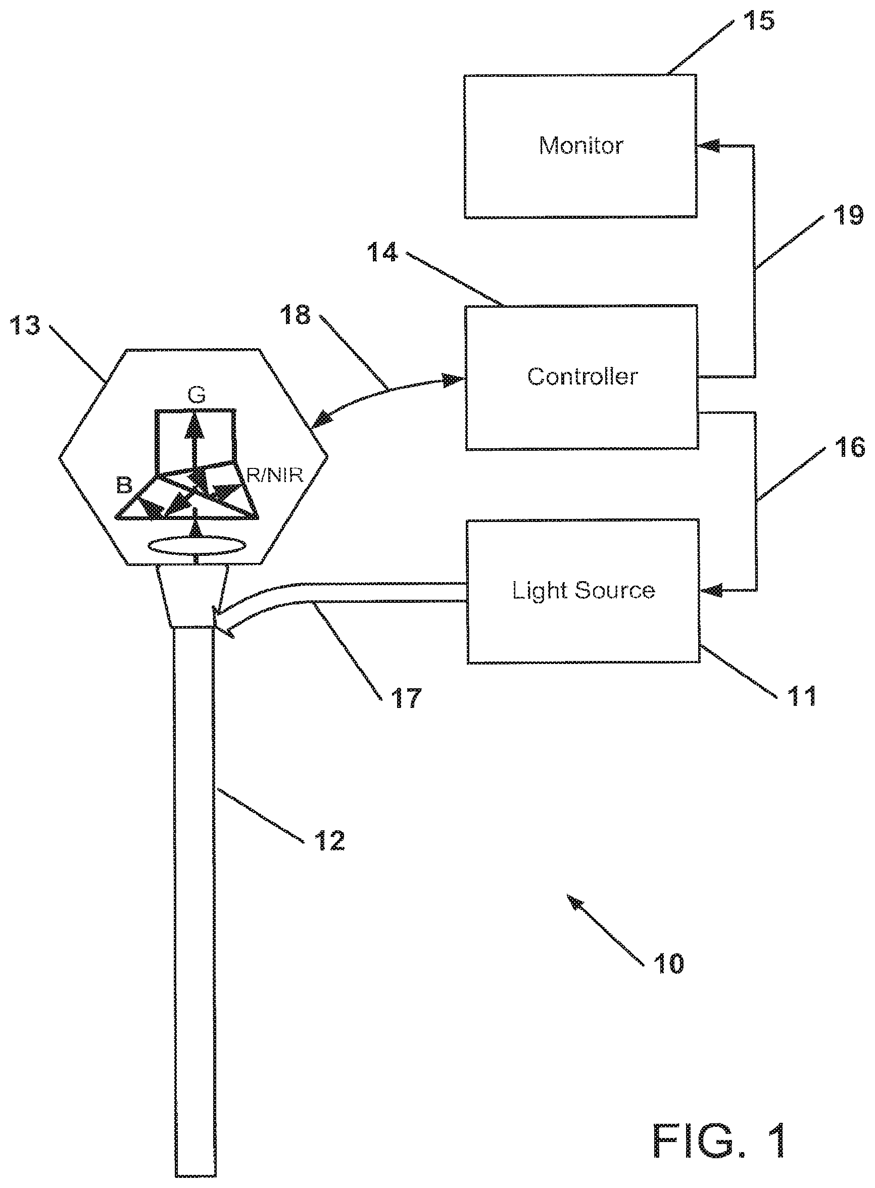
FIG. 1 shows an endoscopic system according to one embodiment of the invention;
FIGS. 2a-2d show various exemplary embodiments of a multimode light source to be used with the endoscopic system of FIG. 1;
FIG. 3a shows an exemplary dichroic prism employed by a 3-sensor color camera;
FIG. 3b shows the optical transmission ranges for the spectral components separated by the dichroic prism of FIG. 3a;
FIG. 3c shows the optical transmission range of a notch filter that blocks excitation light from entering the camera;
FIG. 4 shows a timing diagram of a first embodiment for continuous illumination with green/blue light and alternating illumination with red/NIR light;
FIG. 5 shows a timing diagram of a second embodiment for continuous illumination with green/blue light and alternating illumination with red/NIR light;
FIG. 6 shows a timing diagram of a third embodiment for continuous illumination with green/blue/NIR light and alternating illumination with red light;
FIG. 7 shows an exemplary CMOS sensor having stacked imaging layers and the corresponding spectral sensitivity of these layers; and
FIG. 8 shows four stacked imaging layers of an exemplary sensor.
DESCRIPTION OF CERTAIN ILLUSTRATED EMBODIMENTS
Color video images are generally obtained with three-sensor color cameras where separate red, green and blue image sensors provide simultaneous contiguous arrays of red, green and blue pixel information. Full color video images are generated by combining the image information from all three sensors. Color fidelity (i.e. a true color rendition) is extremely important in medical imaging applications and all three sensors are used to provide complete color information.
To understand the relative importance of color and spatial information in video images of human tissue, however, it is useful to consider information in such video images in terms of luma and chroma. Luma refers to the brightness information in the image and it is this information that provides the spatial detail that enables the viewer to recognize shapes. The spatial and temporal resolution of luma is consequently crucial to the perception of video image quality. Chroma refers to the color information in the video image. It is a property of human vision that fine detail variations in the chroma of image features are not easily perceived and that such variations are consequently less critical than fine detail variations in luma, in an overall assessment of image quality. It is for this reason that video encoding of chroma information is often sub-sampled.
In video images of human tissue obtained with visible light, the structural details of the tissue are largely contained in the blue and green wavelength regions of the imaged light. Blue and green light tends to be reflected from the tissue surface, whereas red light tends to be highly scattered within the tissue. As a consequence, there is very little fine structural detail in the red light that reaches the red image sensor. It is also known from color science that human vision receives most of the spatial information from the green portion of the visible spectrum--i.e. green light information contributes disproportionately to the luma. The standard formula for calculating luma from gamma-corrected color components is Y'=0.2126 R'+0.7152 G'+0.0722 8'. For this reason, spatial and/or temporal interpolation of the red component of video images of human tissue does not significantly affect perception of fine detail in those images.
Similarly to red light, NIR light tends to be scattered in tissue causing NIR image features to be diffusely, rather than sharply defined. Furthermore, because the NIR image highlights areas of interest (i.e. the areas in which the contrast agent is localized), but does not provide the overall visualization or navigational information, it is desirable for a NIR endoscopic imaging device to provide a continuous color image and either a superimposed or side-by-side display of the NIR image information. In such a display the NIR light would also contribute less to the spatial information presented to observer.
FIG. 1 shows schematically an exemplary embodiment of a NIR endoscopic imaging system 10 which includes a multimode light source 11 that provides both visible and NIR illumination, connected to an endoscope 12 by way of an illumination guide, for example a fiber optic cable 17, suitable for transmission of both color and NIR illumination, a color camera 13, illustrated here as having three different sensors 34, 36, 38 (see FIG. 3a) for blue, green and red/NIR imaging, respectively, mounted to the endoscope image guide, and a camera controller 14 connected to the camera 13 and the light source 11 for controlling and synchronizing illumination and image acquisition. Controller 14 can also process the acquired visible and NIR images for display on a monitor 15 connected to the controller 14, for example, by a cable 19. Images can be acquired in real time at selectable frame rates, such as video rates.
FIGS. 2a-2d show schematic diagrams of exemplary embodiments of various light sources 11. The illustrated light sources are constructed to supply in normal color imaging mode visible illumination light yielding a substantially continuous spectral distribution. The light source maybe an arc lamp, a halogen lamp, one or more solid state sources (e.g. LEDs, semiconductor lasers) or any combination thereof and may be spectrally filtered or shaped (e.g. with bandpass filters, IR filters, etc.). The continuous spectrum may be produced as primary colors (RGB) either concurrently or sequentially, for example, using a rotating filter wheel.
In systems according to the present invention, light sources to be used with the system of the invention and described in detail below are configured to provide continuous, uninterrupted illumination in the blue and green parts of the visible spectrum and discontinuous red and/or NIR light. The blue and green parts of the visible spectrum may be optically filtered from the emission produced by a continuous source or produced directly by a narrow-band source (e.g. blue and green LEDs). The red and NJR light may also be produced by an arc lamp, a halogen lamp, a solid state source (e.g., red and NIR LEDs or lasers), or any combination thereof.
Turning now to FIG. 2a, in one embodiment a light source 11a includes an illuminator 202 producing visible and NIR light emission, a collimating lens 204, a filter wheel or reciprocating filter holder 208 that alternatingly transmits red and NIR light and continuously transmits green and blue light. Alternatively, a tunable electro-optic or acousto-optic filter may be used. The filtered light is focused by lens 206 onto light guide 17.
Another embodiment of a light source 11b is schematically illustrated in FIG. 2b. The light source 11b includes an illuminator 202 producing visible and NIR light emission and a collimating lens 204. A dichroic mirror 212 transmits green/blue light and reflects red/NIR light to another dichroic mirror 214 which transmits NIR light to NIR mirror 215 and reflects red light, or vice versa. The green/blue light can be further bandpass-filtered by filter 213. The reflected red and NIR light is chopped, for example, by chopper wheels 219a, 219b (which can be combined into a single chopper wheel) to produce temporally discontinuous illumination, which is then reflected by mirrors 216, 217 and combined with the green/blue light by dichroic mirror 218. The combined light is then focused by lens 206 onto light guide 17, as before.
In another embodiment of a light source 11c schematically illustrated in FIG. 2c, an illuminator 202a produces green and blue light emission which is collimated by a collimating lens 204a. Likewise, separate illuminators 202b, 202c produce respective red and NIR light emissions which are collimated by corresponding collimating lenses 204b and 204c. As in the embodiment of FIG. 2b, the red and NIR light is chopped, for example, by chopper wheels 219a, 219b (which may also be combined into a single chopper wheel) to produce temporally discontinuous illumination, which is then combined with the green/blue illumination by dichroic mirrors 222, 228. The combined light is then focused by lens 206 onto light guide 17, as before.
In yet another embodiment of a light source 11d schematically illustrated in FIG. 2d, an illuminator 202a produces green and blue light emission which is collimated by a collimating lens 204a, as before. However, unlike in the embodiment of FIG. 2c, the separate illuminators 202d, 202e are here switched electrically to produce red and NIR light emissions with controlled timing. For example, the red and NIR light sources 202d, 202e may be solid state light sources, such as LEDs or semiconductor lasers, which can be rapidly turned on and off with suitable, preferably electronic, switches. As described above with reference to FIG. 2c, the red and NIR illumination is collimated by corresponding collimating lenses 204b and 204c and combined with the green/blue illumination by dichroic mirrors 222, 228. The combined light is then focused by lens 206 onto light guide 17, as before.
The alternating red and NIR illumination is synchronized with the image acquisition of the three-sensor camera such that red and NIR images are acquired by the camera synchronously with the red and NIR illumination of the endoscope.
FIG. 3a shows in more detail the three-sensor camera 13 of FIG. 1, in particular the optical beam splitter used to direct red/NIR, green, and blue light to the three different image sensors 34, 36 and 38, respectively. For NIR fluorescence applications, the camera preferably also includes an excitation band blocking filter 32. The beam splitter may be made, for example, of a plurality of dichroic prisms, cube splitters, plate splitters or pellicle splitters. FIG. 3b shows the spectral composition of the light received from the endoscope according to FIG. 3a. FIG. 3c illustrates the spectral composition of the light transmitted through the excitation band blocking filter 32 implemented as a notch filter 31 which blocks transmission of excitation light, while transmitting the other wavelengths in the visible and NIR spectral range. The transmission characteristic of this filter 32 may be designed to also block undesired NIR wavelengths interfering with the visible spectrum that may degrade the color image.
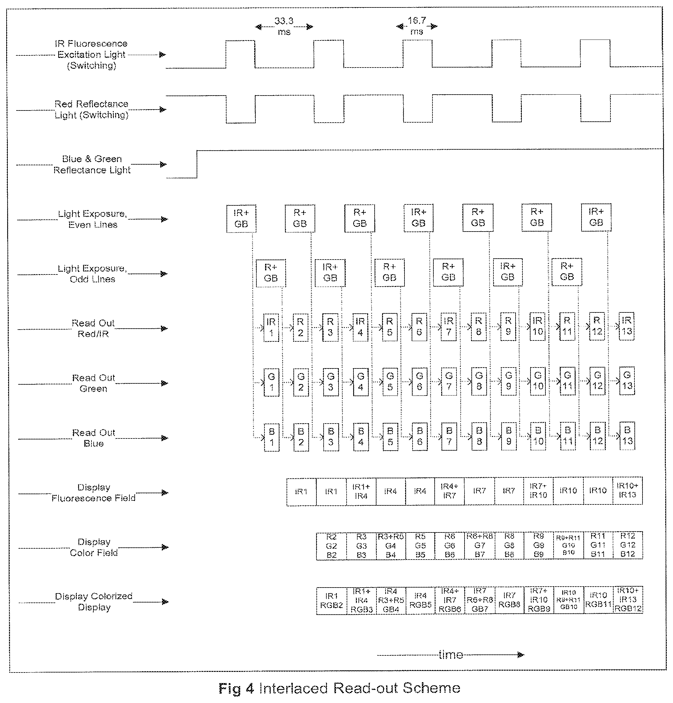
FIG. 4 shows a timing diagram for a first exemplary embodiment of a simultaneous color and NIR imaging mode using, for example, a three-sensor camera. In this embodiment, the camera sensors utilize an interlaced read-out format which represents an advantageous combination of spatial and temporal resolution for smooth display of motion. Any of the light sources illustrated in FIGS. 2a-2d can be used with this embodiment. The light source provides continuous blue/green illumination and alternating red and NIR illumination. Half-frames are alternatingly exposed on the image sensors, i.e., a first field (half-frame) with even lines alternating with a second field (half-frame) with odd lines. In the timing diagram of FIG. 4 depicting a full frame rate of 30 fps, one field period (16.7 ms) provides NIR illumination, followed by two field periods (33.3 ms) of red illumination. Stated differently, the sample or tissue is illuminated with full-spectrum color (RGB) during two field periods (33.3 ms) and with GB and NIR during a third field period. For reconstructing the full-color visible image, the missing red information is interpolated between the fields adjacent to the field with the NIR illumination. The blue and green image information is always available, thereby providing optimum and continuous luma information. The NIR image is generated from every sixth field in each half frame, wherein the missing lines are spatially interpolated. When the fluorescence field is displayed, the image is updated every three fields, with the displayed image interpolated between even and odd lines.
In all the figures, the term "IR" is used instead of or interchangeably with "NIR."
Once the color and NIR image data have been processed, the signal is outputted to a video monitor and may be displayed as two separate, simultaneous views (one color and one fluorescence) or as combined color and fluorescence image signals (e.g. by assigning the fluorescence signal a color that contrasts with the naturally occurring colors in the tissue).
FIG. 5 shows a timing diagram for a second exemplary embodiment of a simultaneous color and NIR imaging mode. In this embodiment, the camera sensors utilize a progressive scan sensor read-out format wherein a complete frame (G/B/R alternating with G/B/NIR) is read out during each field period. Any of the light sources illustrated in FIGS. 2a-2d can be used with this embodiment. The light source provides continuous blue/green illumination and alternating red and NIR illumination. In the timing diagram of FIG. 5, one field period (16.7 ms) provides NIR illumination, followed by one field period (16.7 ms) of red illumination. Stated differently, the sample or tissue is illuminated with full-spectrum color (RGB) during one field period (16.7 ms) and with GB and NIR during a third field period. In this case, a full visible spectrum color image is available at every pixel, in every other frame. In the alternate frames, the blue and green information is acquired directly, whereas the red information is interpolated between adjacent frames. Unlike with the embodiment of FIG. 4, no spatial interpolation is required. Further image processing and display can be implemented in a manner similar to that described in previous embodiments.
FIG. 6 shows a timing diagram for a third exemplary embodiment, wherein both the green/blue illumination and the NIR illumination are continuous, while only the red illumination is modulated. Like in the embodiment of FIG. 4, half-frames are alternatingly exposed on the image sensors, i.e., a first field (half-frame) with even lines alternating with a second field (half-frame) with odd lines. In the timing diagram of FIG. 6 depicting a full frame rate of 30 fps, one field period (16.7 ms) provides (NIR+GB) illumination (red illumination switched off), followed by two field periods (33.3 ms) of (NIR+RGB). If the NIR image signal is small compared to the red reflected signal, it will not significantly affect the overall visible (RGB) image, so that the color image may be generated by conventional color image processing without correction. Otherwise the NIR contribution obtained in the red image channel when the red illumination is switched off may be subtracted from the (NIR+R) image data by spatial and temporal interpolation to obtain the red image signal, as shown in the second to last lien in the timing diagram of FIG. 6. Alternatively, sensors with a progressive scan image sensor readout similar to those illustrated in FIG. 5 could be used with RGB and (RGB+IR) image acquisition in alternate frames.
In yet another exemplary embodiment (not illustrated in the drawings), the green/blue illumination as well as the red illumination are continuous, whereas the NIR illumination is modulated. This timing scheme can be best applied if the red and NIR image signals have approximately the same magnitude. In this embodiment, the light source provides uninterrupted illumination with full visible spectrum and intermittent illumination with NIR light. The timing diagram is essentially the same as that depicted in FIG. 6, with the NIR and the red illumination interchanged. The intermittent NIR illumination is synchronized to coincide with every 3.sup.rd field with interlaced cameras and with every other field in progressive scan cameras. For every field in which NIR illumination is provided, the red image sensor will acquire a (R+NIR) image signal. The NIR image signal can be extracted from the (R+NIR) image signal by interpolation of the red signal value from the appropriate preceding and subsequent "red only" image fields and subtracting the red image signal from the (R+NIR) signal. Since the red and NIR image signals are of similar magnitude, such interpolation and subtraction will provide a reasonably accurate NIR image signal value. The color image is processed by using the acquired and interpolated values for the red image signal in combination with the blue and green image signals. The resulting color and NIR image information can then be displayed or recorded as described before.
In any of the aforementioned embodiments, the NIR endoscopic imaging system can also be operated such that the light sources provides continuous illumination with either the full visible spectrum or the NIR spectrum and the camera acquires the corresponding color image or NIR (absorbance or fluorescence) image in a continuous fashion to provide high spatial resolution. The resulting video image of either individual illumination/imaging mode--color or NIR--can be subsequently displayed and/or recorded.
By implementing color and NIR imaging as described in the aforementioned embodiments, it is possible to acquire and display full-color visible light and NIR light images at video rates without compromising image resolution and/or introducing objectionable motion artifacts. Furthermore, should any residual color fringing occur as a consequence of sharp edges moving rapidly across the visual field (e.g. with the discontinuous acquisition of red or NIR images), these relatively minor effects can be mitigated by temporal interpolation of the missing (red/NIR) video fields with minimum additional processing time.
While the invention has been disclosed in connection with the preferred embodiments shown and described in detail, various modifications and improvements thereon will become readily apparent to those skilled in the art. For example, instead of using separate image sensors for G/B and R/NIR, or a single color sensor for RGB images and NIR fluorescence images, a single direct three-color RGB sensor image sensor with a stacked pixel design implemented in CMOS technology and commercially available from Foveon, Inc., San Jose, Calif., may be used. Such sensor is schematically illustrated in FIG. 7. It will be understood that this sensor design can be extended to four colors by adding an NIR-sensitive layer. The red, green, blue and NIR images are hereby acquired at different depths in the image sensor. With a 4-layer sensor, such as a sensor having layers 802, 804, 806, 808 shown in FIG. 8, multiplexing of the red and NIR illumination would be unnecessary. However, with a 3-layer sensor, the red and NIR illumination would still need to be multiplexed, as described above for a 3-sensor conventional camera. An appropriate barrier filter to block the NIR excitation light would also be required for fluorescence imaging applications.
While the invention has been illustrated and described in connection with currently preferred embodiments shown and described in detail, it is not intended to be limited to the details shown since various modifications and structural changes may be made without departing in any way from the spirit and scope of the present invention. The embodiments were chosen and described in order to explain the principles of the invention and practical application to thereby enable a person skilled in the art to best utilize the invention and various embodiments with various modifications as are suited to the particular use contemplated.
What is claimed as new and desired to be protected by Letters Patent is set forth in the appended claims and includes equivalents of the elements recited therein:
* * * * *
D00000
D00001
D00002
D00003
D00004
D00005
D00006
D00007
D00008
D00009
XML
uspto.report is an independent third-party trademark research tool that is not affiliated, endorsed, or sponsored by the United States Patent and Trademark Office (USPTO) or any other governmental organization. The information provided by uspto.report is based on publicly available data at the time of writing and is intended for informational purposes only.
While we strive to provide accurate and up-to-date information, we do not guarantee the accuracy, completeness, reliability, or suitability of the information displayed on this site. The use of this site is at your own risk. Any reliance you place on such information is therefore strictly at your own risk.
All official trademark data, including owner information, should be verified by visiting the official USPTO website at www.uspto.gov. This site is not intended to replace professional legal advice and should not be used as a substitute for consulting with a legal professional who is knowledgeable about trademark law.