Agonist antibodies that bind human CD137 and uses thereof
Bobrowicz , et al.
U.S. patent number 10,716,851 [Application Number 16/123,742] was granted by the patent office on 2020-07-21 for agonist antibodies that bind human cd137 and uses thereof. This patent grant is currently assigned to Compass Therapeutics LLC. The grantee listed for this patent is Compass Therapeutics LLC. Invention is credited to Piotr Bobrowicz, Ugur Eskiocak, Jason M. Lajoie, Cheuk Lun Leung, Michael March Schmidt, Robert V. Tighe, III, Paul Widboom.


View All Diagrams
| United States Patent | 10,716,851 |
| Bobrowicz , et al. | July 21, 2020 |
Agonist antibodies that bind human CD137 and uses thereof
Abstract
The present disclosure relates to, inter alia, compounds (e.g., antibodies, or antigen-binding fragments thereof) that bind to an epitope of CD137 and agonize CD137, and to use of the compounds in methods for treating, or ameliorating one or more symptoms of, cancer.
| Inventors: | Bobrowicz; Piotr (Cambridge, MA), Widboom; Paul (Lebanon, NH), Schmidt; Michael March (Cambridge, MA), Lajoie; Jason M. (Cambridge, MA), Tighe, III; Robert V. (Cambridge, MA), Leung; Cheuk Lun (Cambridge, MA), Eskiocak; Ugur (Cambridge, MA) | ||||||||||
|---|---|---|---|---|---|---|---|---|---|---|---|
| Applicant: |
|
||||||||||
| Assignee: | Compass Therapeutics LLC
(Cambridge, MA) |
||||||||||
| Family ID: | 63036491 | ||||||||||
| Appl. No.: | 16/123,742 | ||||||||||
| Filed: | September 6, 2018 |
Prior Publication Data
| Document Identifier | Publication Date | |
|---|---|---|
| US 20190015508 A1 | Jan 17, 2019 | |
Related U.S. Patent Documents
| Application Number | Filing Date | Patent Number | Issue Date | ||
|---|---|---|---|---|---|
| 16032639 | Jul 11, 2018 | ||||
| 62577257 | Oct 26, 2017 | ||||
| 62577259 | Oct 26, 2017 | ||||
| 62568231 | Oct 4, 2017 | ||||
| 62531190 | Jul 11, 2017 | ||||
| 62531259 | Jul 11, 2017 | ||||
| Current U.S. Class: | 1/1 |
| Current CPC Class: | A61K 35/17 (20130101); C12N 15/79 (20130101); C07K 16/28 (20130101); A61K 39/39558 (20130101); C07K 16/2818 (20130101); A61K 39/001117 (20180801); A61P 35/00 (20180101); C07K 16/2803 (20130101); C07K 2317/565 (20130101); C07K 2317/34 (20130101); A61K 2039/572 (20130101); C07K 2317/75 (20130101); C07K 2317/33 (20130101) |
| Current International Class: | C07K 16/28 (20060101); C12N 15/79 (20060101); A61K 39/395 (20060101); A61K 35/17 (20150101); A61K 39/00 (20060101); A61P 35/00 (20060101) |
References Cited [Referenced By]
U.S. Patent Documents
| 5928893 | July 1999 | Kang et al. |
| 6210669 | April 2001 | Aruffo et al. |
| 6303121 | October 2001 | Kwon |
| 6569997 | May 2003 | Kwon |
| 6818749 | November 2004 | Kashmiri |
| 6887673 | May 2005 | Kunkel et al. |
| 6905685 | June 2005 | Kwon |
| 6974863 | December 2005 | Kwon |
| 7138500 | November 2006 | Goodwin et al. |
| 7288638 | October 2007 | Jure-Kunkel et al. |
| 7387271 | June 2008 | Noelle et al. |
| 7651686 | January 2010 | Chen et al. |
| 7829088 | November 2010 | Kwon |
| 8337850 | December 2012 | Ahrens et al. |
| 8475790 | July 2013 | Jure-Kunkel |
| 8716452 | May 2014 | Jure-Kunkel et al. |
| 8772026 | July 2014 | Chen et al. |
| 8821867 | September 2014 | Ahrens et al. |
| 9005619 | April 2015 | Kohrt et al. |
| 9758589 | September 2017 | Kohrt et al. |
| 9861621 | January 2018 | Saha |
| 2003/0118588 | June 2003 | Diehl et al. |
| 2004/0109847 | June 2004 | Chen |
| 2006/0029595 | February 2006 | Kwon |
| 2008/0008716 | January 2008 | Kwon |
| 2008/0152655 | June 2008 | Liu et al. |
| 2009/0196877 | August 2009 | Chen |
| 2011/0104049 | May 2011 | Strome et al. |
| 2012/0045414 | February 2012 | Delucia |
| 2012/0076722 | March 2012 | Strome et al. |
| 2014/0017836 | January 2014 | Wei et al. |
| 2014/0178368 | June 2014 | Sharp et al. |
| 2015/0313965 | November 2015 | Pogue |
| 2016/0152722 | June 2016 | Sharp et al. |
| 2016/0264670 | September 2016 | Graziano et al. |
| 2016/0304607 | October 2016 | Sadineni et al. |
| 2017/0088627 | March 2017 | Lin et al. |
| 2017/0174773 | June 2017 | Davis et al. |
| 2108401 | Mar 1995 | CA | |||
| 2005/035584 | Apr 2005 | WO | |||
| 2012/032433 | Mar 2012 | WO | |||
| 2015/119923 | Aug 2015 | WO | |||
| 2015179236 | Nov 2015 | WO | |||
| 2015/188047 | Dec 2015 | WO | |||
| 2016/029073 | Feb 2016 | WO | |||
| 2016/134358 | Aug 2016 | WO | |||
| 2016/185016 | Nov 2016 | WO | |||
| 2017005845 | Jan 2017 | WO | |||
| 2017130076 | Aug 2017 | WO | |||
| 2017181034 | Oct 2017 | WO | |||
| 2017/205745 | Nov 2017 | WO | |||
| 2018/191502 | Oct 2018 | WO | |||
Other References
|
Almagro & Fransson, Frontiers in Bioscience 2008; 13:1619-33. cited by examiner . Chester et al. (J Clin. Oncol. 2014, 32 (15 suppl), p. 3017. cited by examiner . Bartkowiak, T. et al., "Activation of 4-1BB on Liver Myeloid Cells Triggers Hepatitis via an Interleukin-27-Dependent Pathway.," Clin Cancer Res., vol. 24(5):1138-1151 (2018). cited by applicant . Bitra, A. et al., "Crystal structures of the human 4-1BB receptor bound to its ligand 4-1BBL reveal covalent receptor dimerization as a potential signaling amplifier.," J Biol Chem., vol. 293(26):9958-9969 (2018). cited by applicant . Bitra, A. et al., "Crystal structures of murine 4-1BB and its interaction with 4-1BBL support a role for galectin-9 in 4-1BB signaling," J Biol Chem., (2017). cited by applicant . Chen, S. et al. "Combination of 4-1BB Agonist and PD-1 Antagonist Promotes Antitumor Effector/Memory CD8 T Cells in a Poorly Immunogenic Tumor Model" Cancer Immunology Research: Nov. 11, 2014: 149-161. cited by applicant . Duraiswamy, J. et al., "Therapeutic PD-1 Pathway Blockade Augments with Other Modalities of Immunotherapy T-Cell Function to Prevent Immune Decline in Ovarian Cancer" Cancer Res; 73(23) Dec. 1, 2013: 6900-6912. cited by applicant . Dubrot, J. et al., "Treatment with anti-CD137 mAbs causes intense accumulations of liver T cells without selective antitumor immunotherapeutic effects in this organ.," Cancer Immunology, Immunotherapy, vol. 59(8):1223-1233 (2010). cited by applicant . Fisher, T. S. et al., "Targeting of 4-1BB by monoclonal antibody PF-05082566 enhances T-cell function and promotes anti-tumor activity" Cancer Immunol Immunother (2012) 61:1721-1733. cited by applicant . Gauttier, V. et al., "Agonistic anti-CD137 antibody treatment leads to antitumor response in mice with liver cancer" International Journal of Cancer: 135, 2857-2867 (2014). cited by applicant . Gilbreth, R. N., et al., "Crystal structure of the human 4-1BB/4-1BBL complex" J Biol Chem., (2018). cited by applicant . Houot, R., et al., "Therapeutic effect of CD137 immunomodulation in lymphoma and its enhancement by Treg depletion" Blood, Oct. 15, 2009; vol. 114 (16): 3431-3438. cited by applicant . Kohrt, H. E., et al., "CD137 stimulation enhances the antilymphoma activity of anti-CD20 antibodies" Blood, Feb. 24, 2011; vol. 117 (8): 2423-2432. cited by applicant . Kohrt, H. E., et al., "Stimulation of natural killer cells with a CD137-specific antibody enhances trastuzumab efficacy in xenotransplant models of breast cancer" The Journal of Clinical Investigation, Mar. 2012; vol. 122 (3): 1066-1075. cited by applicant . Kohrt, H. E., et al., "Targeting CD137 enhances the efficacy of cetuximab" The Journal of Clinical Investigation, Jun. 2014; vol. 124 (6):2668-2682. cited by applicant . Kroon, P. et al., "Concomitant targeting of programmed death-1 (PD-1) and CD137 improves the efficacy of radiotherapy in a mouse model of human BRAFV600-mutant melanoma" Cancer Immunol Immunother (2016) 65:753-763. cited by applicant . Madireddi, S. et al., "Galectin-9 controls the therapeutic activity of 4-1BB-targeting antibodies" J. Exp Med. 2014: vol. 211 (7); 1433-1448. cited by applicant . Melero, I. et al., "Monoclonal antibodies against the 4-1BB T-cell activation molecule eradicate established tumors," Nature Medicine, vol. 3(6):682-685 (1997). cited by applicant . Mittler, R. S. et al., "Anti-4-1BB Monoclonal Antibodies Abrogate T Cell-dependent Humoral Immune Responses In Vivo through the Induction of Helper T Cell Anergy" J. Exp. Med., Nov. 15, 1999; vol. 190 (10): 1535-1540. cited by applicant . Qian, Y. et al., "CD137 ligand-mediated reverse signaling inhibits proliferation and induces apoptosis in non-small cell lung cancer" Med. Oncol. (2015) 32:44. cited by applicant . Sanchez-Paulete A. R. et al., "Cancer Immunotherapy with Immunomodulatory Anti-CD137 and Anti-PD-1 Monoclonal Antibodies Requires BATF3-Dependent Dendritic Cells" Jan. 2016; Cancer Discovery: 71-79. cited by applicant . Segal, N.H. et al., "Results from an Integrated Safety Analysis of Urelumab, an Agonist Anti-CD137 Monoclonal Antibody.," Clin Cancer Res., vol. 23(8):1929-1936(2016) . cited by applicant . Shi, W. et al., "Augmented Antitumor Effects of Radiation Therapy by 4-1BB Antibody (BMS-469492) Treatment" Anticancer Research 26: 3445-3454 (2006). cited by applicant . Shindo, Y. et al., "Combination Immunotherapy with 4-1BB Activation and PD-1 Blockade Enhances Antitumor Efficacy in a Mouse Model of Subcutaneous Tumor" Anticancer Research 35: 129-136 (2015). cited by applicant . Shuford W. W. et al., "4-1BB Costimulatory Signals Preferentially Induce CD8+ T Cell Proliferation and Lead to the Amplification In Vivo of Cytotoxic T Cell Responses" J. Exp. Med Jul. 7, 1997: vol. 186 (1): 47-55. cited by applicant . Souza-Fonseca-Guimaraes, F. et al., "Anti-CD137 enhances anti-CD20 therapy of systemic B-cell lymphoma with altered immune homeostasis but negligible toxicity" Oncolmmunology: Jun. 2016. cited by applicant . Srivastava, R. M. et al., "CD137 stimulation enhances cetuximab induced natural killer (NK): dendritic cell (DC) priming of anti-tumor T cell immunity in head and neck cancer patients" Clin Cancer Res. Feb. 1, 2017; 23(3): 707-716. cited by applicant . Stagg, J. et al., "Anti-ErbB-2 mAb therapy requires type I and II interferons and synergizes with anti-PD-1 or anti-CD137 mAb therapy" 7142-7147: PNAS: Apr. 26, 2011: vol. 108 (17). cited by applicant . Tolcher, A. et al., "Phase Ib Study of Utomilumab (PF-05082566), a 4-1BB/CD137 Agonist, in Combination with Pembrolizumab (MK-3475) in Patients with Advanced Solid Tumors," Clin Cancer Res., vol. 23(18): 5349-5357(2017). cited by applicant . Uno, T. et al., "Eradication of established tumors in mice by a combination antibody-based therapy," Nature Medicine, vol. 12(6):693-698 (2006) . cited by applicant . Ye, Q. et al., "CD137 accurately identifies and enriches for naturally occurring tumor-reactive T cells in tumor.," Clin Cancer Res., vol. 20(1):44-55 (2014). cited by applicant . Wei, H. et al., "Combinatorial PD-1 Blockade and CD137 Activation Has Therapeutic Efficacy in Murine Cancer Models and Synergizes with Cisplatin" PLOS One: Dec. 2013: vol. 8 (12). cited by applicant . Wei, H. et al., "Dual targeting of CD137 co-stimulatory and PD-1 co-inhibitory molecules for ovarian cancer immunotherapy" Oncolmmunology (3); Mar. 2014. cited by applicant . Wilcox, R. et al., "Provision of antigen and CD137 signaling breaks immunological ignorance, promoting regression of poorly immunogenic tumors" The Journal of Clinical Investigation: Mar. 2002: vol. 109 (5): 651-659. cited by applicant . U.S. Appl. No. 16/032,639, filed Jul. 11, 2018, Piotr Bobrowicz. cited by applicant . U.S. Appl. No. 16/185,398, filed Nov. 9, 2018, Piotr Bobrowicz. cited by applicant . U.S. Appl. No. 16/219,117, filed Dec. 13, 2018, Piotr Bobrowicz. cited by applicant . Invitation to Pay Additional Fees, and where applicable, Protest Fee, PCT/US2018/041612, dated Nov. 19, 2018, 16 pages. cited by applicant. |
Primary Examiner: Sang; Hong
Attorney, Agent or Firm: DT Ward, PC Ward; Donna T. Bryan; Jennifer F.
Parent Case Text
RELATED APPLICATIONS
This application is a Continuation of U.S. patent application Ser. No. 16/032,639, filed Jul. 11, 2018, now pending, which application claims the benefit of U.S. Provisional Patent Application Ser. No. 62/531,259 filed on Jul. 11, 2017; U.S. Provisional Patent Application Ser. No. 62/531,190 filed on Jul. 11, 2017; U.S. Provisional Patent Application Ser. No. 62/568,231 filed on Oct. 4, 2017; U.S. Provisional Patent Application Ser. No. 62/577,257 filed on Oct. 26, 2017; and U.S. Provisional Patent Application Ser. No. 62/577,259 filed on Oct. 26, 2017 The entire contents of the above-referenced applications are incorporated herein by this reference.
Claims
The invention claimed is:
1. A kit comprising a container comprising an isolated monoclonal antibody or antigen binding portion thereof that specifically binds human CD137, wherein the antibody or antigen binding portion thereof comprises heavy and light chain variable regions comprising amino acid sequences selected from the group consisting of: (a) SEQ ID NOs: 8 and 6, respectively; (b) SEQ ID NOs: 101 and 6, respectively; (c) SEQ ID NOs: 103 and 6, respectively; (d) SEQ ID NOs: 26 and 6, respectively; (e) SEQ ID NOs: 4 and 28, respectively; and (f) SEQ ID NOs: 4 and 105, respectively; and a package insert comprising instructions for administration of the antibody for treating or delaying progression of cancer or reducing or inhibiting tumor growth in a subject in need thereof.
2. A kit comprising a container comprising an isolated monoclonal antibody or antigen binding portion thereof that specifically binds human CD137 and induces or enhances T cell activation, wherein the antibody or antigen binding portion thereof comprises heavy and light chain variable regions comprising amino acid sequences selected from the group consisting of: (a) SEQ ID NOs: 8 and 6, respectively; (b) SEQ ID NOs: 101 and 6, respectively; (c) SEQ ID NOs: 103 and 6, respectively; (d) SEQ ID NOs: 26 and 6, respectively; (e) SEQ ID NOs: 4 and 28, respectively; and (f) SEQ ID NOs: 4 and 105, respectively; and a package insert comprising instructions for administration of the antibody for treating or delaying progression of cancer or reducing or inhibiting tumor growth in a subject in need thereof.
3. A kit comprising a container comprising an isolated monoclonal antibody or antigen binding portion thereof that specifically binds human CD137 and induces or enhances a cytotoxic T cell response, wherein the antibody or antigen binding portion thereof comprises heavy and light chain variable regions comprising amino acid sequences selected from the group consisting of: (a) SEQ ID NOs: 8 and 6, respectively; (b) SEQ ID NOs: 101 and 6, respectively; (c) SEQ ID NOs: 103 and 6, respectively; (d) SEQ ID NOs: 26 and 6, respectively; (e) SEQ ID NOs: 4 and 28, respectively; and (f) SEQ ID NOs: 4 and 105, respectively; and a package insert comprising instructions for administration of the antibody for treating or delaying progression of cancer or reducing or inhibiting tumor growth in a subject in need thereof.
4. A kit comprising a container comprising an isolated monoclonal antibody or antigen binding portion thereof that specifically binds human CD137 and induces or enhances T cell proliferation, wherein the antibody or antigen binding portion thereof comprises heavy and light chain variable regions comprising amino acid sequences selected from the group consisting of: (a) SEQ ID NOs: 8 and 6, respectively; (b) SEQ ID NOs: 101 and 6, respectively; (c) SEQ ID NOs: 103 and 6, respectively; (d) SEQ ID NOs: 26 and 6, respectively; (e) SEQ ID NOs: 4 and 28, respectively; and (f) SEQ ID NOs: 4 and 105, respectively; and a package insert comprising instructions for administration of the antibody for treating or delaying progression of cancer or reducing or inhibiting tumor growth in a subject in need thereof.
5. A kit comprising a container comprising an isolated monoclonal antibody or antigen binding portion thereof that specifically binds human CD137 and induces or enhances cell cytokine production, wherein the antibody or antigen binding portion thereof comprises heavy and light chain variable regions comprising amino acid sequences selected from the group consisting of: (a) SEQ ID NOs: 8 and 6, respectively; (b) SEQ ID NOs: 101 and 6, respectively; (c) SEQ ID NOs: 103 and 6, respectively; (d) SEQ ID NOs: 26 and 6, respectively; (e) SEQ ID NOs: 4 and 28, respectively; and (f) SEQ ID NOs: 4 and 105, respectively; and a package insert comprising instructions for administration of the antibody for treating or delaying progression of cancer or reducing or inhibiting tumor growth in a subject in need thereof.
Description
SEQUENCE LISTING
The instant application contains a Sequence Listing which has been submitted electronically in ASCII format and is hereby incorporated by reference in its entirety. Said ASCII copy, created Sep. 6, 2018, is named "CTN-006CN_Sequence-Listing.txt" and is 92629 Kilobytes in size. The Sequence Listing is being submitted by EFS Web and is hereby incorporated by reference into the specification.
BACKGROUND
In recent years, an increasing body of evidence suggests the immune system operates as a significant barrier to tumor formation and progression. The principle that naturally-occurring T cells with anti-tumor potential or activity exist in a patient with cancer has rationalized the development of immunotherapeutic approaches in oncology. Immune cells, such as T cells, macrophages, and natural killer cells, can exhibit anti-tumor activity and effectively control the occurrence and growth of malignant tumors. Tumor-specific or -associated antigens can induce immune cells to recognize and eliminate malignancies (Chen & Mellman, (2013) Immunity 39(1):1-10). In spite of the existence of tumor-specific immune responses, malignant tumors often evade or avoid immune attack through a variety of immunomodulatory mechanisms resulting in the failure to control tumor occurrence and progression (Motz & Coukos, (2013) Immunity 39(1):61-730). Indeed, an emerging hallmark of cancer is the exploitation of these immunomodulatory mechanisms and the disablement of anti-tumor immune responses, resulting in tumor evasion and escape from immunological killing (Hanahan and Weinberg (2011) Cell 144(5):646-674).
Novel approaches in the immunotherapy of cancer involve counteracting these immune evasion and escape mechanisms and inducing the endogenous immune system to reject tumors. CD137 (alternatively known as "tumor necrosis factor receptor superfamily member 9" (TNFRSF9), 4-1BB, and "induced by lymphocyte activation" (ILA)) is a transmembrane co-stimulatory receptor protein belonging to the tumor necrosis factor superfamily. CD137 is a T cell co-stimulatory receptor induced upon TCR activation (Nam et al., (2005) Curr Cancer Drug Targets 5:357-363; Watts et al., (2005) Annu Rev Immunol 23:23-68). In addition to its expression on activated CD4+ and CD8+ T cells, CD137 is also expressed on CD4+CD25+ regulatory T cells, activated natural killer (NK) and NK-T cells, monocytes, neutrophils, and dendritic cells.
Under physiological conditions, CD137 is ligated by CD137 ligand (CD137L), an agonist membrane molecule present on antigen-presenting cells including B cells, monocytes, macrophages, and dendritic cells (Watts et al., (2005) Annu Rev Immunol 23:23-68). Upon interaction with its ligand, CD137 leads to increased TCR-induced T-cell proliferation, cytokine production, functional maturation, and prolonged CD8+ T-cell survival. The potential of CD137 co-stimulation using various agonists (e.g. agonistic antibodies, recombinant CD137L protein, and CD137-specific aptamers) in enabling the immune system to attack tumors has been documented in numerous models (Dharmadhikari et al., (2016) Oncoimmunology 5(4):e1113367 and references therein). A recent report on the clinical evaluation of an agonistic CD137 antibody (Urelumab, BMS-663513; Bristol-Myers Squibb) documented the observation of treatment-related adverse events in human subjects, including indications of severe hepatotoxicity (transaminitis) correlating with antibody dose (Segal et al., (2016) Clin Cancer Res 23(8):1929-1936). In contrast, a different agonistic CD137 antibody (Utomilumab, PF-05082566; Pfizer) tested in combination with an anti-PD-1 antibody (pembrolizumab), though not resulting in any dose-limiting toxicities, showed comparable results to anti-PD-1 antibody therapy alone (Tolcher, A. et al., (2017) Clin Cancer Res 23(18): 5349-5357). These results highlight that for patients with various diseases and conditions, including cancer, that are amenable to treatment with a CD137 agonist, there continues to be an unmet need for novel agonistic antibodies that bind to human CD137 and exhibit characteristics sufficient for the development of a safe and efficacious therapeutic.
SUMMARY OF THE DISCLOSURE
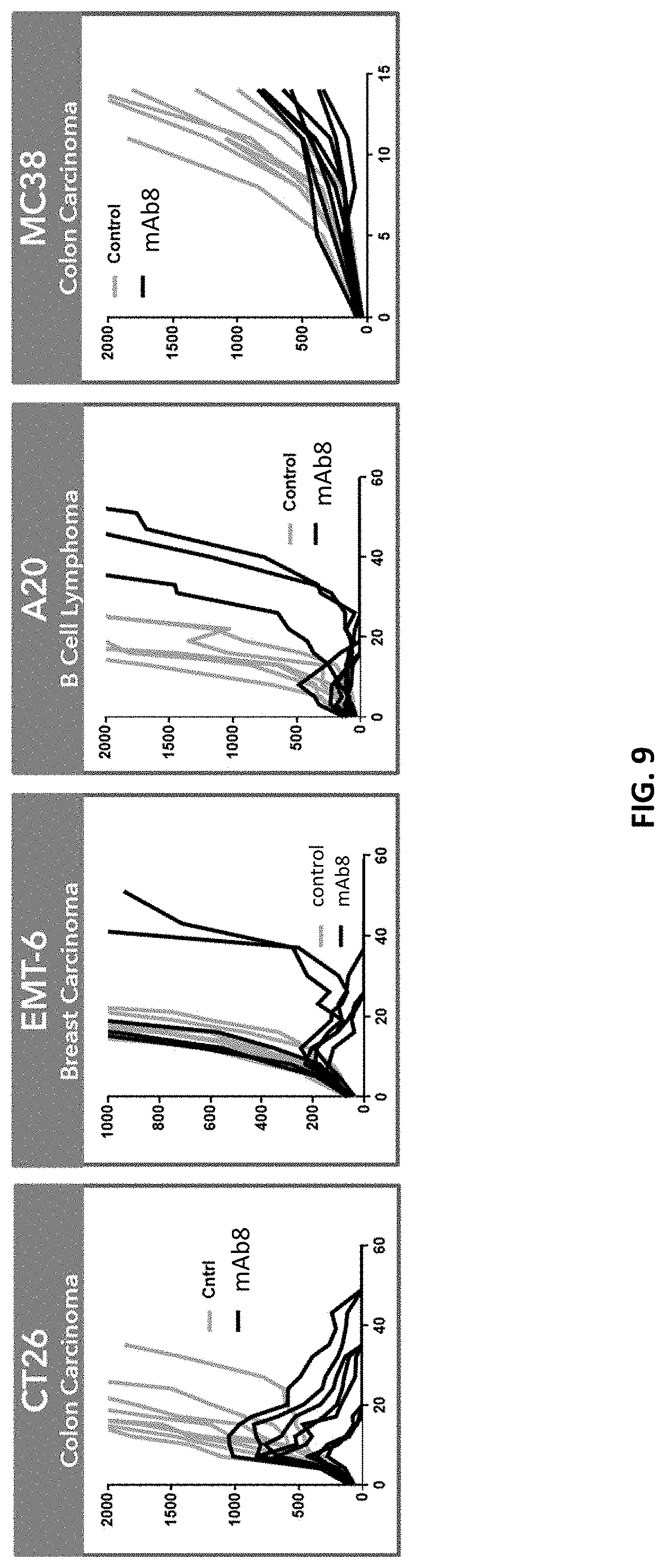
The present disclosure is based, at least in part, on the discovery of novel agonist anti-CD137 antibodies exhibiting protective anti-tumor immunity in animals. Notably, the antibodies described herein are efficacious against diverse tumor types, and over a wide dose range. Moreover, as exemplified in the working examples, the antibodies described herein are therapeutically effective against very large tumors. For example, treatment of tumor-bearing mice with agonist anti-CD137 antibodies described herein resulted in complete regression of tumors as large as 1,800 mm.sup.3. As set forth in FIG. 15, treatment of such mice also resulted in protective immunity. And coincident with the observed efficacy were positive immunophenotypic changes in the tumor microenvironment, such as increased immune cell infiltration with concomitant reductions in regulatory T cell and exhausted T cell populations (see, e.g., FIGS. 22A-22D).
As described above, agonism of CD137 has been associated with certain adverse events, including hepatotoxicity-related deaths in humans (see, e.g., Segal et al. (2017) Clin Cancer Res 23(8): 1929-1935). Similar toxicities resulting from treatment with agonist anti-CD137 antibodies (such as the 3H3 antibody) have also been observed in animal models (see, e.g., Bartkowiak et al. (2018) Clin Cancer Res 24(5):1138-1151). Yet, the agonist anti-CD137 antibodies described herein have minimal effects on the liver, as determined by, e.g., plasma levels of liver enzymes (e.g., alanine aminotransferase (ALT)) and immune cell infiltration. For example, there was no evidence of increased intrahepatic or intrasplenic immune cell infiltration in mice treated with the antibodies. Thus, the antibodies described herein are not only highly efficacious, but also sparing of certain toxicities associated with CD137 agonism.
While the disclosure is not bound by any particular theory or mechanism of action, the superior therapeutic and toxicity-sparing properties of the antibodies described herein are believed to derive in part from one or both of their affinity and the novel epitope to which they bind. That is, the antibodies described herein share a common, novel epitope that is distinct from that of other agonist anti-CD137 antibodies. And, as exemplified in the working examples, engagement of this epitope by the antibodies described herein gives rise to differentiated in vitro activity, such as effects on regulatory T cell proliferation, cytokine production by CD8.sup.+ T cells and macrophages, and intracellular signaling, as compared to agonist antibodies that bind to different epitopes of CD137. Furthermore, it has been demonstrated that an affinity range (a "sweet spot") for antibodies is particularly optimal for anti-tumor activity. For example, antibodies of intermediate affinity were shown to be more efficacious against large tumors as compared to antibodies with higher or lower affinity.
In view of the foregoing, in some aspects, the disclosure provides isolated monoclonal antibody, or antigen binding portion thereof, that specifically binds human CD137, wherein the antibody or antigen binding portion binds human CD137 with an affinity (K.sub.D) of between about 40 nM to about 100 nM. In some aspects, the disclosure provides an isolated monoclonal antibody, or antigen binding portion thereof, that specifically binds to human CD137, wherein the antibody or antigen binding portion binds human CD137 with an affinity (K.sub.D) of about 30-100 nM (e.g., between about 30 nM and about 110 nM). In some aspects, the affinity of the anti-CD137 antibody to human CD137 is at least two (e.g., at least three, four, five, six, seven, eight, nine, or 10) fold higher than the affinity of mAb10 for mouse CD137. In some aspects, the affinity of the anti-CD137 antibody is no greater than 500, 450, 400, 350, 300, 250, 200, 250, 200, 175, 150, 125, 110, or 100 nM. In some aspects, the affinity of the anti-CD137 antibody to human CD137 is at least two (e.g., at least three, four, five, six, seven, eight, nine, or 10) fold higher than the affinity of mAb10 for mouse CD137, but no greater than 500, 450, 400, 350, 300, 250, 200, 250, 200, 175, 150, 125, 110, or 100 nM.
In some aspects, the disclosure provides an isolated monoclonal antibody, or antigen binding portion thereof, that specifically binds to human CD137, wherein the antibody or antigen binding portion binds to an epitope on human CD137 comprising one or more (e.g., one, two, three, four, five, six, seven, eight, nine, 10, 11, 12, 13, 14, 15, 16, 17, 18, 19, 20, 21, 22, 23, 24, or all 25) of amino acids 111-132 of SEQ ID NO:3. In some aspects, the disclosure provides an isolated monoclonal antibody, or antigen binding portion thereof, that specifically binds to human CD137, wherein the antibody or antigen binding portion binds to an epitope within amino acids 111-132 of SEQ ID NO:3. In some aspects, the disclosure provides an isolated monoclonal antibody, or antigen binding portion thereof, that specifically binds to human CD137, wherein the antibody or antigen binding portion binds to all or a portion of amino acids 111-132 of SEQ ID NO:3. In some aspects, the epitope comprises K114 of SEQ ID NO: 3. In some aspects, the epitope comprises residues E111, T113, and K114 of SEQ ID NO: 3. In some aspects, the epitope comprises residues E111, T113, K114, N126 and 1132 of SEQ ID NO: 3. In some aspects, the epitope comprises residues E111, T113, K114 and P135 of SEQ ID NO: 3. In some aspects, the epitope comprises residues E111, T113, K114, N126, I132 and P135 of SEQ ID NO: 3. In some aspects, the antibody or antigen binding portion thereof binds to human CD137 with an affinity of between about 30 nM and about 100 nM (e.g., between about 30 nM and about 110 nM).
In some aspects, the disclosure provides an isolated monoclonal antibody, or antigen binding portion thereof, that specifically binds to human CD137, wherein the antibody or antigen binding portion binds human CD137 with an affinity (K.sub.D) of about 40-100 nM (e.g., between about 40 nM and about 100 nM) and binds to an epitope on human CD137 comprising K114 of SEQ ID NO: 3. In some aspects, the disclosure provides an isolated monoclonal antibody, or antigen binding portion thereof, that specifically binds to human CD137, wherein the antibody or antigen binding portion binds human CD137 with an affinity (K.sub.D) of about 30-100 nM (e.g., between about 30 nM and about 100 nM) and binds to an epitope on human CD137 comprising K114 of SEQ ID NO: 3. In some aspects, the epitope comprises residues E111, T113, and K114 of SEQ ID NO: 3. In some aspects, the epitope comprises residues E111, T113, K114, N126 and 1132 of SEQ ID NO: 3. In some aspects, the epitope comprises residues E111, T113, K114 and P135 of SEQ ID NO: 3. In some aspects, the epitope comprises residues E111, T113, K114, N126, I132 and P135 of SEQ ID NO: 3.
In some aspects, the disclosure provides an isolated monoclonal antibody, or antigen binding portion thereof, that specifically binds to human CD137, wherein the antibody or antigen binding portion binds human CD137 with an affinity (K.sub.D) of about 30-100 nM (e.g., about 30 nM to about 100 nM) and binds to an epitope on human CD137 comprising a sequence of one or more amino acid residues corresponding to amino acid positions 111 to 135 of SEQ ID NO: 3. In some aspects, the epitope comprises 2, 3, 4, 5, 6, 7, 8, 9, 10, 11, 12, 13, 14, 15, 16, 17, 18, 19, 20, 21, 22, 23, 24, or 25 amino acid residues corresponding to amino acid positions 111 to 135 of SEQ ID NO: 3.
In some aspects, the disclosure provides an isolated monoclonal antibody, or antigen binding portion thereof, that specifically binds to human CD137, wherein the antibody or antigen binding portion binds human CD137 with an affinity (K.sub.D) of about 30-100 nM (e.g., between about 30 nM and about 100 nM) and binds to an epitope on human CD137 located within amino acid residues 111-135 of SEQ ID NO: 3. In some aspects, the epitope is at least 3, 4, 5, 6, 7, 8, 9, 10, 11, 12, 13, 14 or 15 amino acids. In some aspects, the epitope is fewer than 25, 24, 23, 22, 21, 20, 19, 18, 17, 16, 15, 14, 13, 12, 11, 10, 9, 8, 7, 6 or 5 amino acids.
In some aspects, the disclosure provides an isolated monoclonal antibody, or antigen binding portion thereof, that specifically binds to human CD137, wherein the antibody or antigen binding portion binds human CD137 with an affinity (K.sub.D) of about 30-100 nM (e.g., between about 30 nM and about 100 nM) and binds to an epitope on human CD137 comprising ELTK (corresponding to amino acid residues 111-114 of SEQ ID NO: 3). In some aspects, the epitope further comprises one or more residues N126, I132 and P135 of SEQ ID NO: 3.
In any of the foregoing aspects, the epitope is a non-linear epitope. In any of the foregoing aspects, mutation of residue K114 of SEQ ID NO: 3 abrogates binding of the antibody or antigen binding portion thereof to human CD137.
In any of the foregoing aspects, the antibody or antigen binding portion described herein binds human CD137 with an affinity (K.sub.D) of about 30-100 nM, 30-95 nM, 45-95 nM, 50-90 nM, 55-85 nM, 60-80 nM, 65-75 nM, 55-75 nM, 40-70 nM, 50-80 nM, or 60-90 nM. In some aspects, the antibody or antigen binding portion binds to a non-ligand binding region of the extracellular domain of human CD137. In some aspects, the antibody or antigen binding portion does not inhibit the interaction between CD137 and CD137L. In some aspects, the non-ligand binding region spans cysteine rich domain (CRD) III and CRD IV. In any of the foregoing aspects, the antibody or antigen binding portion does not inhibit the formation of a trimer of CD137:CD137L monomers.
In some aspects, the disclosure provides an isolated monoclonal antibody, or antigen binding portion thereof, that specifically binds human CD137, wherein the antibody or antigen binding portion:
(i) binds human CD137 with an affinity (K.sub.D) of about 30-100 nM (e.g., between about 30 nM and about 100 nM);
(ii) binds to a non-ligand binding region of the extracellular domain of human CD137; and
(iii) binds to an epitope on human CD137 comprising K114 of SEQ ID NO: 3.
In some aspects, the disclosure provides an isolated monoclonal antibody, or antigen binding portion thereof, that specifically binds human CD137, wherein the antibody or antigen binding portion:
(i) binds human CD137 with an affinity (K.sub.D) of about 30-100 nM (e.g., between about 30 nM and about 100 nM);
(ii) does not inhibit the interaction between human CD137 and human CD137 ligand; and
(iii) binds to an epitope on human CD137 comprising K114 of SEQ ID NO: 3.
In some aspects, the disclosure features an isolated monoclonal antibody, or antigen binding portion thereof, that specifically binds human CD137, wherein the antibody or antigen binding portion: (i) binds human CD137 with an affinity (K.sub.D) of about 30-100 nM (e.g., between about 30 nM and about 100 nM) and (ii) does not inhibit the formation of a trimer of CD137:CD137L monomers (that is, a CD137:CD137L trimer:trimer complex). In some aspects, the disclosure features an isolated monoclonal antibody, or antigen binding portion thereof, that specifically binds human CD137, wherein the antibody or antigen binding portion: (i) binds human CD137 with an affinity (K.sub.D) of about 30-100 nM (e.g., between about 30 nM and about 100 nM) and (ii) binds to a non-ligand binding region of the extracellular domain of human CD137. In some aspects, the disclosure features an isolated monoclonal antibody, or antigen binding portion thereof, that specifically binds human CD137, wherein the antibody or antigen binding portion: (i) binds human CD137 with an affinity (K.sub.D) of about 30-100 nM (e.g., between about 30 nM and about 100 nM) and (ii) does not inhibit the interaction between human CD137 and CD137 ligand.
In any of the foregoing aspects, the antibody or antigen binding portion comprises a heavy chain CDR3 comprising the amino acid sequence DXXXXLXXXXYXYYX (SEQ ID NO: 126), wherein X is any amino acid. In some aspects, the antibody or antigen binding portion comprises a heavy chain CDR3 comprising the amino acid sequence DXPFXLDXXYYYYYX (SEQ ID NO: 127), wherein X is any amino acid. In any of the foregoing aspects, mutation of residues D95, L100, Y100E, Y100G, Y100H, or combinations thereof, of the heavy chain CDR3, to alanine results in loss of binding to human CD137. In any of the foregoing aspects, mutation of residues P97, F98, D100A, Y100D, Y100F, or combinations thereof, to alanine results in reduction of binding to human CD137. In any of the foregoing aspects, the antibody or antigen binding portion comprises heavy and light chain CDRs, wherein heavy chain CDR3 comprises the amino acid sequence set forth in SEQ ID NO: 68.
In other aspects, the disclosure provides an isolated monoclonal antibody, or antigen binding portion thereof, that specifically binds to human CD137, wherein
(i) the antibody or antigen binding portion binds human CD137 with an affinity (K.sub.D) of about 30-100 nM (e.g., between about 30 nM and about 100 nM); and
(ii) the antibody or antigen binding portion comprises a heavy chain CDR3 comprising the amino acid sequence DXXXXLXXXXYXYYX (SEQ ID NO: 126), wherein X is any amino acid. In some aspects, X is any amino acid except alanine.
In another aspect, the disclosure provides an isolated monoclonal antibody, or antigen binding portion thereof, that specifically binds to human CD137, wherein
(i) the antibody or antigen binding portion binds human CD137 with an affinity (K.sub.D) of about 30-100 nM (e.g., between about 30 nM and about 100 nM); and
(ii) the antibody or antigen binding portion comprises a heavy chain CDR3 comprising the amino acid sequence DX.sub.1X.sub.2X.sub.3X.sub.4LX.sub.5X.sub.6X.sub.7X.sub.8YX.sub.9YYX.sub- .10 (SEQ ID NO: 128), wherein X.sub.1 is any amino acid, wherein X.sub.2 is a non-polar amino acid, wherein X.sub.3 is a non-polar amino acid, wherein X.sub.4 is any amino acid, wherein X.sub.5 is a polar amino acid, wherein X.sub.6 is any amino acid, wherein X.sub.7 is any amino acid, wherein X.sub.8 is a polar amino acid, wherein X.sub.9 is a polar amino acid, and wherein X.sub.10 is any amino acid. In some aspects, X.sub.2 is proline, X.sub.3 is phenylalanine or tryptophan, X.sub.5 is aspartic acid or glutamic acid, X.sub.8 is tyrosine, and X.sub.9 is tyrosine.
In other aspects, the disclosure provides an isolated monoclonal antibody, or antigen binding portion thereof, that specifically binds to human CD137, wherein
(i) the antibody or antigen binding portion binds human CD137 with an affinity (K.sub.D) of about 30-100 nM (e.g., between about 30 nM and about 100 nM); and
(ii) the antibody or antigen binding portion thereof specifically binds to an epitope on human CD137 comprising one or more residues E111, T113, K114, N126, I132 and P135 of SEQ ID NO: 3.
In other aspects, the disclosure provides an isolated monoclonal antibody, or antigen binding portion thereof, that specifically binds to human CD137, wherein
(i) the antibody or antigen binding portion binds human CD137 with an affinity (K.sub.D) of about 30-100 nM (e.g., between about 30 nM and about 100 nM);
(ii) the antibody or antigen binding portion thereof specifically binds to an epitope on human CD137 comprising one or more residues E111, T113, K114, N126, I132 and P135 of SEQ ID NO: 3;
(iii) the antibody or antigen binding portion comprises a heavy chain CDR3 comprising the amino acid sequence DXXXXLXXXXYXYYX (SEQ ID NO: 126), wherein X is any amino acid; or
(iv) combinations thereof. In some aspects, X is any amino acid except alanine.
In other aspects, the disclosure provides an isolated monoclonal antibody, or antigen binding portion thereof, that specifically binds to human CD137, wherein
(i) the antibody or antigen binding portion binds human CD137 with an affinity (K.sub.D) of about 30-100 nM (e.g., between about 30 nM and about 100 nM);
(ii) the antibody or antigen binding portion thereof specifically binds to an epitope on human CD137 comprising one or more residues E111, T113, K114, N126, I132 and P135 of SEQ ID NO: 3;
(iii) the antibody or antigen binding portion comprises a heavy chain CDR3 comprising the amino acid sequence DX.sub.1X.sub.2X.sub.3X.sub.4LX.sub.5X.sub.6X.sub.7X.sub.8YX.sub.9YYX.sub- .10 (SEQ ID NO: 128), wherein X.sub.1 is any amino acid, wherein X.sub.2 is a non-polar amino acid, wherein X.sub.3 is a non-polar amino acid, wherein X.sub.4 is any amino acid, wherein X.sub.5 is a polar amino acid, wherein X.sub.6 is any amino acid, wherein X.sub.7 is any amino acid, wherein X.sub.8 is a polar amino acid, wherein X.sub.9 is a polar amino acid, and wherein X.sub.10 is any amino acid; or
(iv) combinations thereof. In some aspects, X.sub.2 is proline, X.sub.3 is phenylalanine or tryptophan, X.sub.5 is aspartic acid or glutamic acid, X.sub.8 is tyrosine, and X.sub.9 is tyrosine.
In other aspects, the disclosure provides an isolated monoclonal antibody, or antigen binding portion thereof, that specifically binds to human CD137, wherein
(i) the antibody or antigen binding portion binds human CD137 with an affinity (K.sub.D) of about 30-100 nM (e.g., between about 30 nM and about 100 nM);
(ii) the antibody or antigen binding portion thereof specifically binds to an epitope on human CD137 comprising one or more residues E111, T113, K114, N126, I132 and P135 of SEQ ID NO: 3; and
(iii) the antibody or antigen binding portion comprises a heavy chain CDR3 comprising the amino acid sequence DXXXXLXXXXYXYYX (SEQ ID NO: 126), wherein X is any amino acid. In some aspects, X is any amino acid except alanine.
In other aspects, the disclosure provides an isolated monoclonal antibody, or antigen binding portion thereof, that specifically binds to human CD137, wherein
(i) the antibody or antigen binding portion binds human CD137 with an affinity (K.sub.D) of about 30-100 nM (e.g., between about 30 nM and about 100 nM);
(ii) the antibody or antigen binding portion thereof specifically binds to an epitope on human CD137 comprising one or more residues E111, T113, K114, N126, I132 and P135 of SEQ ID NO: 3; and
(iii) the antibody or antigen binding portion comprises a heavy chain CDR3 comprising the amino acid sequence DX.sub.1X.sub.2X.sub.3X.sub.4LX.sub.5X.sub.6X.sub.7X.sub.8YX.sub.9YYX.sub- .10 (SEQ ID NO: 128), wherein X.sub.1 is any amino acid, wherein X.sub.2 is a non-polar amino acid, wherein X.sub.3 is a non-polar amino acid, wherein X.sub.4 is any amino acid, wherein X.sub.5 is a polar amino acid, wherein X.sub.6 is any amino acid, wherein X.sub.7 is any amino acid, wherein X.sub.8 is a polar amino acid, wherein X.sub.9 is a polar amino acid, and wherein X.sub.10 is any amino acid. In some aspects, X.sub.2 is proline, X.sub.3 is phenylalanine or tryptophan, X.sub.5 is aspartic acid or glutamic acid, X.sub.8 is tyrosine, and X.sub.9 is tyrosine.
In any of the foregoing aspects, the epitope comprises K114. In any of the foregoing aspects, the epitope comprises E1111, T113 and K114. In any of the foregoing aspects, the epitope comprises E111, T113, K114, N126 and 1132. In any of the foregoing aspects, the epitope comprises residues E111, T113, K114, N126, I132 and P135 of SEQ ID NO: 3.
In another aspects, the disclosure provides an isolated monoclonal antibody, or antigen binding portion thereof, that specifically binds to human CD137, wherein
(i) the antibody or antigen binding portion binds human CD137 with an affinity (K.sub.D) of about 30-100 nM (e.g., between about 30 nM and about 100 nM); and
(ii) the antibody or antigen binding portion thereof specifically binds to an epitope comprising a sequence of one or more amino acid residues corresponding to amino acid positions 111 to 135 of SEQ ID NO: 3.
In another aspects, the disclosure provides an isolated monoclonal antibody, or antigen binding portion thereof, that specifically binds to human CD137, wherein
(i) the antibody or antigen binding portion binds human CD137 with an affinity (K.sub.D) of about 30-100 nM (e.g., between about 30 nM and about 100 nM);
(ii) the antibody or antigen binding portion thereof specifically binds to an epitope comprising a sequence of one or more amino acid residues corresponding to amino acid positions 111 to 135 of SEQ ID NO: 3;
(iii) the antibody or antigen binding portion comprises a heavy chain CDR3 comprising the amino acid sequence DXXXXLXXXXYXYYX (SEQ ID NO: 126), wherein X is any amino acid; or
(iv) combinations thereof. In some aspects, X is any amino acid except alanine.
In another aspects, the disclosure provides an isolated monoclonal antibody, or antigen binding portion thereof, that specifically binds to human CD137, wherein
(i) the antibody or antigen binding portion binds human CD137 with an affinity (K.sub.D) of about 30-100 nM (e.g., between about 30 nM and about 100 nM);
(ii) the antibody or antigen binding portion thereof specifically binds to an epitope comprising a sequence of one or more amino acid residues corresponding to amino acid positions 111 to 135 of SEQ ID NO: 3;
(iii) the antibody or antigen binding portion comprises a heavy chain CDR3 comprising the amino acid sequence DX.sub.1X.sub.2X.sub.3X.sub.4LX.sub.5X.sub.6X.sub.7X.sub.8YX.sub.9YYX.sub- .10 (SEQ ID NO: 128), wherein X.sub.1 is any amino acid, wherein X.sub.2 is a non-polar amino acid, wherein X.sub.3 is a non-polar amino acid, wherein X.sub.4 is any amino acid, wherein X.sub.5 is a polar amino acid, wherein X.sub.6 is any amino acid, wherein X.sub.7 is any amino acid, wherein X.sub.8 is a polar amino acid, wherein X.sub.9 is a polar amino acid, and wherein X.sub.10 is any amino acid; or
(iv) combinations thereof. In some aspects, X.sub.2 is proline, X.sub.3 is phenylalanine or tryptophan, X.sub.5 is aspartic acid or glutamic acid, X.sub.8 is tyrosine, and X.sub.9 is tyrosine.
In another aspects, the disclosure provides an isolated monoclonal antibody, or antigen binding portion thereof, that specifically binds to human CD137, wherein
(i) the antibody or antigen binding portion binds human CD137 with an affinity (K.sub.D) of about 30-100 nM (e.g., between about 30 nM and about 100 nM);
(ii) the antibody or antigen binding portion thereof specifically binds to an epitope comprising a sequence of one or more amino acid residues corresponding to amino acid positions 111 to 135 of SEQ ID NO: 3; and
(iii) the antibody or antigen binding portion comprises a heavy chain CDR3 comprising the amino acid sequence DXXXXLXXXXYXYYX (SEQ ID NO: 126), wherein X is any amino acid. In some aspects, X is any amino acid except alanine.
In another aspects, the disclosure provides an isolated monoclonal antibody, or antigen binding portion thereof, that specifically binds to human CD137, wherein
(i) the antibody or antigen binding portion binds human CD137 with an affinity (K.sub.D) of about 30-100 nM (e.g., between about 30 nM and about 100 nM);
(ii) the antibody or antigen binding portion thereof specifically binds to an epitope comprising a sequence of one or more amino acid residues corresponding to amino acid positions 111 to 135 of SEQ ID NO: 3; and
(iii) the antibody or antigen binding portion comprises a heavy chain CDR3 comprising the amino acid sequence DX.sub.1X.sub.2X.sub.3X.sub.4LX.sub.5X.sub.6X.sub.7X.sub.8YX.sub.9YYX.sub- .10 (SEQ ID NO: 128), wherein X.sub.1 is any amino acid, wherein X.sub.2 is a non-polar amino acid, wherein X.sub.3 is a non-polar amino acid, wherein X.sub.4 is any amino acid, wherein X.sub.5 is a polar amino acid, wherein X.sub.6 is any amino acid, wherein X.sub.7 is any amino acid, wherein X.sub.8 is a polar amino acid, wherein X.sub.9 is a polar amino acid, and wherein X.sub.10 is any amino acid. In some aspects, X.sub.2 is proline, X.sub.3 is phenylalanine or tryptophan, X.sub.5 is aspartic acid or glutamic acid, X.sub.8 is tyrosine, and X.sub.9 is tyrosine.
In any of the foregoing aspects, the epitope comprises 2, 3, 4, 5, 6, 7, 8, 9, 10, 11, 12, 13, 14, 15, 16, 17, 18, 19, 20, 21, 22, 23, 24, or 25 amino acid residues corresponding to amino acid positions 111 to 135 of SEQ ID NO: 3.
In some aspects, the disclosure provides an isolated monoclonal antibody, or antigen binding portion thereof, that specifically binds to human CD137, wherein
(i) the antibody or antigen binding portion binds human CD137 with an affinity of about 30-100 nM (e.g., between about 30 nM and about 100 nM); and
(ii) the antibody or antigen binding portion thereof specifically binds to an epitope comprising ELTK (corresponding to amino acid residues 111-114 of SEQ ID NO: 3).
In some aspects, the disclosure provides an isolated monoclonal antibody, or antigen binding portion thereof, that specifically binds to human CD137, wherein
(i) the antibody or antigen binding portion binds human CD137 with an affinity (K.sub.D) of about 30-100 nM (e.g., between about 30 nM and about 100 nM);
(ii) the antibody or antigen binding portion thereof specifically binds to an epitope comprising ELTK (corresponding to amino acid residues 111-114 of SEQ ID NO: 3);
(iii) the antibody or antigen binding portion comprises a heavy chain CDR3 comprising the amino acid sequence DXXXXLXXXXYXYYX (SEQ ID NO: 126), wherein X is any amino acid; or
(iv) combinations thereof. In some aspects, X is any amino acid except alanine.
In some aspects, the disclosure provides an isolated monoclonal antibody, or antigen binding portion thereof, that specifically binds to human CD137, wherein
(i) the antibody or antigen binding portion binds human CD137 with an affinity (K.sub.D) of about 30-100 nM (e.g., between about 30 nM and about 100 nM);
(ii) the antibody or antigen binding portion thereof specifically binds to an epitope comprising ELTK (corresponding to amino acid residues 111-114 of SEQ ID NO: 3);
(iii) the antibody or antigen binding portion comprises a heavy chain CDR3 comprising the amino acid sequence DX.sub.1X.sub.2X.sub.3X.sub.4LX.sub.5X.sub.6X.sub.7X.sub.8YX.sub.9YYX.sub- .10 (SEQ ID NO: 128), wherein X.sub.1 is any amino acid, wherein X.sub.2 is a non-polar amino acid, wherein X.sub.3 is a non-polar amino acid, wherein X.sub.4 is any amino acid, wherein X.sub.5 is a polar amino acid, wherein X.sub.6 is any amino acid, wherein X.sub.7 is any amino acid, wherein X.sub.8 is a polar amino acid, wherein X.sub.9 is a polar amino acid, and wherein X.sub.10 is any amino acid; or
(iv) combinations thereof. In some aspects, X.sub.2 is proline, X.sub.3 is phenylalanine or tryptophan, X.sub.5 is aspartic acid or glutamic acid, X.sub.8 is tyrosine, and X.sub.9 is tyrosine.
In some aspects, the disclosure provides an isolated monoclonal antibody, or antigen binding portion thereof, that specifically binds to human CD137, wherein
(i) the antibody or antigen binding portion binds human CD137 with an affinity (K.sub.D) of about 30-100 nM (e.g., between about 30 nM and about 100 nM);
(ii) the antibody or antigen binding portion thereof specifically binds to an epitope comprising ELTK (corresponding to amino acid residues 111-114 of SEQ ID NO: 3); and
(iii) the antibody or antigen binding portion comprises a heavy chain CDR3 comprising the amino acid sequence DXXXXLXXXXYXYYX (SEQ ID NO: 126), wherein X is any amino acid. In some aspects, X is any amino acid except alanine.
In some aspects, the disclosure provides an isolated monoclonal antibody, or antigen binding portion thereof, that specifically binds to human CD137, wherein
(i) the antibody or antigen binding portion binds human CD137 with an affinity (K.sub.D) of about 30-100 nM (e.g., between about 30 nM and about 100 nM);
(ii) the antibody or antigen binding portion thereof specifically binds to an epitope comprising ELTK (corresponding to amino acid residues 111-114 of SEQ ID NO: 3); and
(iii) the antibody or antigen binding portion comprises a heavy chain CDR3 comprising the amino acid sequence DX.sub.1X.sub.2X.sub.3X.sub.4LX.sub.5X.sub.6X.sub.7X.sub.8YX.sub.9YYX.sub- .10 (SEQ ID NO: 128), wherein X.sub.1 is any amino acid, wherein X.sub.2 is a non-polar amino acid, wherein X.sub.3 is a non-polar amino acid, wherein X.sub.4 is any amino acid, wherein X.sub.5 is a polar amino acid, wherein X.sub.6 is any amino acid, wherein X.sub.7 is any amino acid, wherein X.sub.8 is a polar amino acid, wherein X.sub.9 is a polar amino acid, and wherein X.sub.10 is any amino acid. In some aspects, X.sub.2 is proline, X.sub.3 is phenylalanine or tryptophan, X.sub.5 is aspartic acid or glutamic acid, X.sub.8 is tyrosine, and X.sub.9 is tyrosine
In any of the foregoing aspects, the epitope comprises the residues ELTK of SEQ ID NO: 3 (corresponding to amino acid residues 111-114 of SEQ ID NO: 3). In some aspects, the epitope comprises ELTK of SEQ ID NO: 3 (corresponding to amino acid residues 111-114 of SEQ ID NO: 3) and residues N126, I132 and P135 of SEQ ID NO: 3.
In any of the foregoing aspects, the epitope is a non-linear epitope. In some aspects, mutation of residue K114 of human CD137 (SEQ ID NO: 3) abrogates binding of the antibody or antigen binding portion thereof to human CD137.
In any of the foregoing aspects, the antibody or antigen binding portion thereof comprises a heavy chain CDR3 comprising the amino acid sequence DXPFXLDXXYYYYYX (SEQ ID NO: 128), wherein X is any amino acid. In some aspects, mutation of residues D95, L100, Y100E, Y100G, Y100H, or combinations thereof, of the heavy chain CDR3 of the antibody or antigen binding portion described herein, results in loss of binding to human CD137. In some aspects, mutation of residues P97, F98, D100A, Y100D, Y100F, or combinations thereof, of the heavy chain CDR3 of the antibody or antigen binding portion described herein, to alanine results in reduction of binding to human CD137. In other aspects, mutation of residues P97, F98, D100A, Y100D, Y100F, or combinations thereof, of the heavy chain CDR3 of the antibody or antigen binding portion described herein, to any residue except alanine, results in an increase in binding to human CD137.
In any of the foregoing aspects, the antibody or antigen binding portion thereof, binds human CD137 with an (K.sub.D) of about 45-95 nM, 50-90 nM, 55-85 nM, 60-80 nM, 65-75 nM, 55-75 nM, 40-70 nM, 50-80 nM, or 60-90 nM. In any of the foregoing aspects, the antibody or antigen binding portion thereof, binds human CD137 with an (K.sub.D) of about 45 nM to about 95 nM, about 50 to about 90 nM, about 55 to about 85 nM, about 60 to about 80 nM, about 65 to about 75 nM, about 55 to about 75 nM, about 40 to about 70 nM, about 50 to about 80 nM, or about 60 to about 90 nM.
In any of the foregoing aspects, the antibody or antigen binding portion thereof comprises heavy and light chain CDRs, wherein heavy chain CDR3 comprises the amino acid sequence set forth in SEQ ID NO: 68.
In any of the foregoing aspects, the antibody or antigen binding portion thereof comprises heavy and light chain CDRs selected from the group consisting of:
(a) heavy chain CDR1, CDR2 and CDR3 sequences set forth in SEQ ID NOs: 48, 56 and 68, respectively, and light chain CDR1, CDR2 and CDR3 sequences set forth in SEQ ID NOs: 69, 78 and 89, respectively; and
(b) heavy chain CDR1, CDR2 and CDR3 sequences set forth in SEQ ID NOs: 51, 108 and 68, respectively, and light chain CDR1, CDR2 and CDR3 sequences set forth in SEQ ID NOs: 69, 78 and 89, respectively.
In any of the foregoing aspects, the antibody or antigen binding portion thereof comprises heavy and light chain variable regions, wherein the heavy chain variable region comprises an amino acid sequence selected from the group consisting of SEQ ID NOs: 4 and 101; and wherein the light chain variable region comprises an amino acid sequence of SEQ ID NO: 6.
In any of the foregoing aspects, the antibody or antigen binding portion thereof comprises heavy and light chain variable regions, comprising amino acid sequences selected from the group consisting of:
(a) SEQ ID NO: 4 and 6, respectively; and
(b) SEQ ID NO: 101 and 6, respectively.
In any of the foregoing aspects, the antibody or antigen binding portion thereof comprises heavy and light chain variable regions, wherein the heavy chain variable region comprises an amino acid sequence which is at least 90% identical to the amino acid sequence selected from the group consisting of SEQ ID NOs: 4 and 101; and wherein the light chain variable region comprises an amino acid sequence which is at least 90% identical to the amino acid sequence of SEQ ID NO: 6.
In any of the foregoing aspects, the antibody or antigen binding portion thereof comprises heavy and light chain variable regions comprising amino acid sequences at least 90% identical to the amino acid sequences selected from the group consisting of:
(a) SEQ ID NO: 4 and 6, respectively; and
(b) SEQ ID NO: 101 and 6, respectively.
In any of the foregoing aspects, the antibody or antigen binding portion of any one of claims 1-27, wherein the antibody or antigen binding portion comprises heavy and light chains comprising amino acid sequences selected from the group consisting of:
(a) SEQ ID NOs: 129 and 133, respectively; and
(b) SEQ ID NOs: 131 and 133, respectively.
In any of the foregoing aspects, the isolated monoclonal antibody, or antigen binding portion thereof described herein, is an agonist of human CD137 activity.
In any of the foregoing aspects, the isolated monoclonal antibody, or antigen binding portion thereof described herein, competes with mAb1 or an antigen binding fragment of mAb1, for binding to the epitope of human CD137.
In some aspects, the disclosure provides an isolated monoclonal antibody that specifically binds CD137, or an antigen binding portion thereof, wherein the antibody or antigen binding portion thereof comprises heavy and light chain CDRs selected from the group consisting of:
(a) heavy chain CDR1, CDR2 and CDR3 sequences set forth in SEQ ID NOs: 48, 56 and 68, respectively, and light chain CDR1, CDR2 and CDR3 sequences set forth in SEQ ID NOs: 69, 78 and 89, respectively;
(b) heavy chain CDR1, CDR2 and CDR3 sequences set forth in SEQ ID NOs: 48, 56 and 68, respectively, and light chain CDR1, CDR2 and CDR3 sequences set forth in SEQ ID NOs: 70, 79 and 90, respectively;
(c) heavy chain CDR1, CDR2 and CDR3 sequences set forth in SEQ ID NOs: 48, 56 and 68, respectively, and light chain CDR1, CDR2 and CDR3 sequences set forth in SEQ ID NOs: 71, 80 and 91, respectively;
(d) heavy chain CDR1, CDR2 and CDR3 sequences set forth in SEQ ID NOs: 48, 56 and 68, respectively, and light chain CDR1, CDR2 and CDR3 sequences set forth in SEQ ID NOs: 72, 81 and 92, respectively;
(e) heavy chain CDR1, CDR2 and CDR3 sequences set forth in SEQ ID NOs: 48, 56 and 68, respectively, and light chain CDR1, CDR2 and CDR3 sequences set forth in SEQ ID NOs: 73, 82 and 91, respectively;
(f) heavy chain CDR1, CDR2 and CDR3 sequences set forth in SEQ ID NOs: 48, 56 and 68, respectively, and light chain CDR1, CDR2 and CDR3 sequences set forth in SEQ ID NOs: 74, 83 and 93, respectively;
(g) heavy chain CDR1, CDR2 and CDR3 sequences set forth in SEQ ID NOs: 48, 56 and 68, respectively, and light chain CDR1, CDR2 and CDR3 sequences set forth in SEQ ID NOs: 75, 84 and 91, respectively;
(h) heavy chain CDR1, CDR2 and CDR3 sequences set forth in SEQ ID NOs: 48, 56 and 68, respectively, and light chain CDR1, CDR2 and CDR3 sequences set forth in SEQ ID NOs: 74, 85 and 94, respectively;
(i) heavy chain CDR1, CDR2 and CDR3 sequences set forth in SEQ ID NOs: 48, 56 and 68, respectively, and light chain CDR1, CDR2 and CDR3 sequences set forth in SEQ ID NOs: 76, 86 and 95, respectively;
(j) heavy chain CDR1, CDR2 and CDR3 sequences set forth in SEQ ID NOs: 48, 56 and 68, respectively, and light chain CDR1, CDR2 and CDR3 sequences set forth in SEQ ID NOs: 77, 87 and 93, respectively;
(k) heavy chain CDR1, CDR2 and CDR3 sequences set forth in SEQ ID NOs: 48, 56 and 68, respectively, and light chain CDR1, CDR2 and CDR3 sequences set forth in SEQ ID NOs: 69, 88 and 90, respectively;
(l) heavy chain CDR1, CDR2 and CDR3 sequences set forth in SEQ ID NOs: 49, 57 and 68, respectively, and light chain CDR1, CDR2 and CDR3 sequences set forth in SEQ ID NOs: 69, 78 and 89, respectively;
(m) heavy chain CDR1, CDR2 and CDR3 sequences set forth in SEQ ID NOs: 49, 58 and 68, respectively, and light chain CDR1, CDR2 and CDR3 sequences set forth in SEQ ID NOs: 69, 78 and 89, respectively;
(n) heavy chain CDR1, CDR2 and CDR3 sequences set forth in SEQ ID NOs: 49, 59 and 68, respectively, and light chain CDR1, CDR2 and CDR3 sequences set forth in SEQ ID NOs: 69, 78 and 89, respectively;
(o) heavy chain CDR1, CDR2 and CDR3 sequences set forth in SEQ ID NOs: 49, 60 and 68, respectively, and light chain CDR1, CDR2 and CDR3 sequences set forth in SEQ ID NOs: 69, 78 and 89, respectively;
(p) heavy chain CDR1, CDR2 and CDR3 sequences set forth in SEQ ID NOs: 50, 61 and 68, respectively, and light chain CDR1, CDR2 and CDR3 sequences set forth in SEQ ID NOs: 69, 78 and 89, respectively;
(q) heavy chain CDR1, CDR2 and CDR3 sequences set forth in SEQ ID NOs: 50, 58 and 68, respectively, and light chain CDR1, CDR2 and CDR3 sequences set forth in SEQ ID NOs: 69, 78 and 89, respectively;
(r) heavy chain CDR1, CDR2 and CDR3 sequences set forth in SEQ ID NOs: 51, 62 and 68, respectively, and light chain CDR1, CDR2 and CDR3 sequences set forth in SEQ ID NOs: 69, 78 and 89, respectively;
(s) heavy chain CDR1, CDR2 and CDR3 sequences set forth in SEQ ID NOs: 52, 63 and 68, respectively, and light chain CDR1, CDR2 and CDR3 sequences set forth in SEQ ID NOs: 69, 78 and 89, respectively;
(t) heavy chain CDR1, CDR2 and CDR3 sequences set forth in SEQ ID NOs: 50, 64 and 68, respectively, and light chain CDR1, CDR2 and CDR3 sequences set forth in SEQ ID NOs: 69, 78 and 89, respectively;
(u) heavy chain CDR1, CDR2 and CDR3 sequences set forth in SEQ ID NOs: 50, 65 and 68, respectively, and light chain CDR1, CDR2 and CDR3 sequences set forth in SEQ ID NOs: 69, 78 and 89, respectively;
(v) heavy chain CDR1, CDR2 and CDR3 sequences set forth in SEQ ID NOs: 51, 108 and 68, respectively, and light chain CDR1, CDR2 and CDR3 sequences set forth in SEQ ID NOs: 69, 78 and 89, respectively;
(w) heavy chain CDR1, CDR2 and CDR3 sequences set forth in SEQ ID NOs: 107, 56 and 68, respectively, and light chain CDR1, CDR2 and CDR3 sequences set forth in SEQ ID NOs: 69, 78 and 89, respectively; and
(x) heavy chain CDR1, CDR2 and CDR3 sequences set forth in SEQ ID NOs: 48, 56 and 68, respectively, and light chain CDR1, CDR2 and CDR3 sequences set forth in SEQ ID NOs: 109, 110 and 92, respectively.
In other aspects, the disclosure provides an isolated monoclonal antibody, or antigen binding portion thereof, that specifically binds human CD137, wherein the antibody or antigen binding portion thereof comprises heavy and light chain variable regions, wherein the heavy chain variable region comprises an amino acid sequence selected from the group consisting of SEQ ID NOs: 4, 8, 10, 12, 14, 16, 18, 20, 22, 24, 26, 101 and 103; and wherein the light chain variable region comprises an amino acid sequence selected from the group consisting of SEQ ID NOs: 6, 28, 30, 32, 34, 36, 38, 40, 42, 44, 46 and 105.
In other aspects, the disclosure provides an isolated monoclonal antibody, or antigen binding portion thereof, that specifically binds human CD137, wherein the antibody or antigen binding portion comprises heavy and light chain variable regions encoded by nucleotide sequences selected from the group consisting of:
(a) SEQ ID NOs: 5 and 7, respectively; and
(b) SEQ ID NOs: 102 and 7, respectively.
In other aspects, the disclosure provides an isolated monoclonal antibody, or antigen binding portion thereof, that specifically binds human CD137, wherein the antibody or antigen binding portion thereof comprises heavy and light chain variable regions encoded by nucleotide sequences selected from the group consisting of:
(a) SEQ ID NO: 5 and 7, respectively;
(b) SEQ ID NO: 5 and 29, respectively;
(c) SEQ ID NO: 5 and 31, respectively;
(d) SEQ ID NO: 5 and 33, respectively;
(e) SEQ ID NO: 5 and 35, respectively;
(f) SEQ ID NO: 5 and 37, respectively;
(g) SEQ ID NO: 5 and 39, respectively;
(h) SEQ ID NO: 5 and 41, respectively;
(i) SEQ ID NO: 5 and 43, respectively;
(j) SEQ ID NO: 5 and 45, respectively;
(k) SEQ ID NO: 5 and 47, respectively;
(l) SEQ ID NO: 9 and 7, respectively;
(m) SEQ ID NO: 11 and 7, respectively;
(n) SEQ ID NO: 13 and 7, respectively;
(o) SEQ ID NO: 15 and 7, respectively;
(p) SEQ ID NO: 17 and 7, respectively;
(q) SEQ ID NO: 19 and 7, respectively;
(r) SEQ ID NO: 21 and 7, respectively;
(s) SEQ ID NO: 23 and 7, respectively;
(t) SEQ ID NO: 25 and 7, respectively;
(u) SEQ ID NO: 27 and 7, respectively;
(v) SEQ ID NO: 102 and 7, respectively;
(w) SEQ ID NO: 104 and 7, respectively; and
(x) SEQ ID NO: 5 and 106, respectively.
In yet other aspects, the disclosure provides an isolated monoclonal antibody, or antigen binding portion thereof, that specifically binds human CD137, wherein the antibody or antigen binding portion thereof comprises heavy and light chain CDRs, wherein heavy chain CDR3 comprises the amino acid sequence set forth in SEQ ID NO: 68.
In another aspect, the disclosure provides an isolated monoclonal antibody, or antigen binding portion thereof, that specifically binds human CD137, wherein the antibody or antigen binding portion thereof comprises heavy and light chain CDRs, wherein heavy chain CDR3 comprises the amino acid sequence DXXXXLXXXXYXYYX (SEQ ID NO: 126), wherein X is any amino acid. In some aspects, X is any amino acid except for alanine.
In some aspects, the disclosure provides an isolated monoclonal antibody, or antigen binding portion thereof, that specifically binds human CD137, wherein the antibody or antigen binding portion thereof comprises heavy and light chain CDRs, wherein heavy chain CDR3 comprises the amino acid sequence DXPFXLDXXYYYYYX (SEQ ID NO: 127), wherein X is any amino acid. In some aspects, X is any amino acid except for alanine.
In yet other aspects, the disclosure provides an isolated monoclonal antibody, or antigen binding portion thereof, that specifically binds human CD137, wherein the antibody or antigen binding portion thereof comprises heavy and light chain CDRs, wherein heavy chain CDR3 comprises the amino acid sequence DXXXXLXXXXYXYYX (SEQ ID NO: 126), wherein X is any amino acid, and wherein mutation of residues D95, L100, Y100E, Y100G, Y100H, or combinations thereof, results in loss of binding to human CD137.
In other aspects, the disclosure provides an isolated monoclonal antibody, or antigen binding portion thereof, that specifically binds human CD137, wherein the antibody or antigen binding portion thereof comprises heavy and light chain CDRs, wherein heavy chain CDR3 comprises the amino acid sequence DXPFXLDXXYYYYYX (SEQ ID NO: 127), wherein X is any amino acid, and wherein mutation of residues P97, F98, D100A, Y100D, Y100F, or combinations thereof to alanine results in reduction of binding to human CD137.
In some aspects, the disclosure provides an isolated monoclonal antibody, or antigen binding portion thereof, that specifically binds human CD137, wherein the antibody or antigen binding portion thereof comprises heavy and light chain CDRs, wherein heavy chain CDR3 comprises the amino acid sequence DXPFXLDXXYYYYYX (SEQ ID NO: 127), wherein X is any amino acid, and wherein mutation of residues P97, F98, D100A, Y100D, Y100F, or combinations thereof to any residue except alanine, results in an increase in binding to human CD137.
In yet other aspects, the disclosure provides an isolated monoclonal antibody, or antigen binding portion thereof, that specifically binds human CD137, wherein the antibody or antigen binding portion thereof comprises heavy and light chain CDRs, wherein heavy chain CDR3 comprises the amino acid sequence DX1X2X3X4LX5X6X7X8YX9YYX10 (SEQ ID NO: 128) wherein X1 is any amino acid, wherein X2 is a non-polar amino acid, wherein X3 is a non-polar amino acid, wherein X4 is any amino acid, wherein X5 is a polar amino acid, wherein X6 is any amino acid, wherein X7 is any amino acid, wherein X8 is a polar amino acid, wherein X9 is a polar amino acid, and wherein X10 is any amino acid. In some aspects, wherein X2 is proline, wherein X3 is phenylalanine or tryptophan, wherein X5 is aspartic acid or glutamic acid wherein X8 is tyrosine, and wherein X9 is tyrosine.
In some aspects, the disclosure provides an isolated monoclonal antibody, or antigen binding portion thereof, that specifically binds human CD137, wherein the antibody or antigen binding portion thereof comprises heavy and light chain variable regions comprising amino acid sequences selected from the group consisting of:
(a) SEQ ID NO: 4 and 6, respectively;
(b) SEQ ID NO: 4 and 28, respectively;
(c) SEQ ID NO: 4 and 30, respectively;
(d) SEQ ID NO: 4 and 32, respectively;
(e) SEQ ID NO: 4 and 34, respectively;
(f) SEQ ID NO: 4 and 36, respectively;
(g) SEQ ID NO: 4 and 38, respectively;
(h) SEQ ID NO: 4 and 40, respectively;
(i) SEQ ID NO: 4 and 42, respectively;
(j) SEQ ID NO: 4 and 44, respectively;
(k) SEQ ID NO: 4 and 46, respectively;
(l) SEQ ID NO: 8 and 6, respectively;
(m) SEQ ID NO: 10 and 6, respectively;
(n) SEQ ID NO: 12 and 6, respectively;
(o) SEQ ID NO: 14 and 6, respectively;
(p) SEQ ID NO: 16 and 6, respectively;
(q) SEQ ID NO: 18 and 6, respectively;
(r) SEQ ID NO: 20 and 6, respectively;
(s) SEQ ID NO: 22 and 6, respectively;
(t) SEQ ID NO: 24 and 6, respectively;
(u) SEQ ID NO: 26 and 6, respectively;
(v) SEQ ID NO: 101 and 6, respectively;
(w) SEQ ID NO: 103 and 6, respectively; and
(x) SEQ ID NO: 4 and 105, respectively.
In other aspects, the disclosure provides, an isolated monoclonal antibody, or antigen binding portion thereof, that specifically binds human CD137 wherein the antibody or antigen binding portion thereof comprises heavy and light chain variable regions, wherein the heavy chain variable region comprises an amino acid sequence which is at least 90% identical to the amino acid sequence selected from the group consisting of SEQ ID NOs: 4, 8, 10, 12, 14, 16, 18, 20, 22, 24, 26, 101 and 103; and wherein the light chain variable region comprises an amino acid sequence which is at least 90% identical to the amino acid sequence selected from the group consisting of SEQ ID NOs: 6, 28, 30, 32, 34, 36, 38, 40, 42, 44, 46 and 105.
In some aspects, the disclosure provides an isolated monoclonal antibody, or antigen binding portion thereof, that specifically binds human CD137, wherein the antibody or antigen binding portion thereof comprises heavy and light chain variable regions comprising amino acid sequences at least 90% identical to the amino acid sequences selected from the group consisting of:
(a) SEQ ID NO: 4 and 6, respectively;
(b) SEQ ID NO: 4 and 28, respectively;
(c) SEQ ID NO: 4 and 30, respectively;
(d) SEQ ID NO: 4 and 32, respectively;
(e) SEQ ID NO: 4 and 34, respectively;
(f) SEQ ID NO: 4 and 36, respectively;
(g) SEQ ID NO: 4 and 38, respectively;
(h) SEQ ID NO: 4 and 40, respectively;
(i) SEQ ID NO: 4 and 42, respectively;
(j) SEQ ID NO: 4 and 44, respectively;
(k) SEQ ID NO: 4 and 46, respectively;
(l) SEQ ID NO: 8 and 6, respectively;
(m) SEQ ID NO: 10 and 6, respectively;
(n) SEQ ID NO: 12 and 6, respectively;
(o) SEQ ID NO: 14 and 6, respectively;
(p) SEQ ID NO: 16 and 6, respectively;
(q) SEQ ID NO: 18 and 6, respectively;
(r) SEQ ID NO: 20 and 6, respectively;
(s) SEQ ID NO: 22 and 6, respectively;
(t) SEQ ID NO: 24 and 6, respectively;
(u) SEQ ID NO: 26 and 6, respectively;
(v) SEQ ID NO: 101 and 6, respectively;
(w) SEQ ID NO: 103 and 6, respectively; and
(x) SEQ ID NO: 4 and 105, respectively.
In some aspects, the disclosure provides an isolated monoclonal antibody, or antigen binding portion thereof, that specifically binds human CD137, wherein the antibody or antigen binding portion thereof comprises heavy and light chain sequences comprising amino acid sequences selected from the group consisting of:
(a) SEQ ID NOs: 129 and 133, respectively; and
(b) SEQ ID NOs: 131 and 133, respectively.
In some aspects, the disclosure provides an isolated monoclonal antibody, or antigen binding portion thereof, that specifically binds human CD137, wherein the antibody or antigen binding portion thereof comprises heavy and light chain sequences having amino acid sequences set forth in SEQ ID NOs: 129 and 133, respectively.
In some aspects, the disclosure provides an isolated monoclonal antibody, or antigen binding portion thereof, that specifically binds human CD137, wherein the antibody or antigen binding portion thereof comprises heavy and light chain sequences having amino acid sequences set forth in SEQ ID NOs: 131 and 133, respectively
In any of the foregoing aspects, the antibody or antigen binding portion specifically binds to and agonizes human CD137.
In any of the foregoing aspects, the isolated monoclonal antibody or antigen binding portion thereof, exhibits at least one or more of the following properties selected from the group consisting of:
(a) induces or enhances dimerization of CD137 trimers;
(b) induces or enhances multimerization of CD137 trimers;
(c) induces or enhances T cell activation;
(d) induces or enhances a cytotoxic T cell response;
(e) induces or enhances T cell proliferation;
(f) induces or enhances cytokine production; and
(g) any combination of properties (a)-(f).
In any of the foregoing aspects, the isolated monoclonal antibody or antigen binding portion thereof, exhibits at least one or more of the following properties relative to a reference antibody that binds human CD137, selected from the group consisting of:
(a) does not induce or enhance intrahepatic T cell activation;
(b) does not induce or enhance intrahepatic T cell proliferation;
(c) does not induce or enhance intrasplenic T cell activation;
(d) does not induce or enhance intrasplenic T cell proliferation;
(e) does not induce or enhance macrophage activation;
(f) does not induce or enhance macrophage differentiation;
(g) does not induce or enhance alanine aminotransferase (ALT) activity; and
(h) any combination of properties (a)-(g). In some aspects, the reference antibody is urelumab.
In any of the foregoing aspects, the isolated monoclonal antibody or antigen binding portion thereof, induces or enhances human CD137-mediated T cell activation in the tumor microenvironment, but does not significantly induce or enhance human CD137-mediated T cell activation in the spleen and/or liver.
In any of the foregoing aspects, the isolated monoclonal antibody or antigen binding portion thereof, induces or enhances T cell activation in the tumor microenvironment, but does not significantly induce or enhance T cell activation in the spleen and/or liver.
In any of the foregoing aspects, the isolated monoclonal antibody or antigen binding portion thereof, induces or enhances human CD137-mediated cytotoxic T cell response in the tumor microenvironment, but does not significantly induce or enhance human CD137-mediated cytotoxic T cell response in the spleen and/or liver.
In any of the foregoing aspects, the isolated monoclonal antibody or antigen binding portion thereof, induces or enhances a cytotoxic T cell response in the tumor microenvironment, but does not significantly induce or enhance a T cell response in the spleen and/or liver.
In any of the foregoing aspects, the isolated monoclonal antibody or antigen binding portion thereof, induces human CD137-mediated T cell proliferation in the tumor microenvironment, but does not significantly induce human CD137-mediated T cell proliferation in the spleen and/or liver.
In any of the foregoing aspects, the isolated monoclonal antibody or antigen binding portion thereof, induces T cell proliferation in the tumor microenvironment, but does not significantly induce T cell proliferation in the spleen and/or liver.
In any of the foregoing aspects, the isolated monoclonal antibody or antigen binding portion thereof, induces human CD137-mediated T cell infiltration in the tumor microenvironment, but does not significantly induce human CD137-mediated T cell infiltration in the spleen and/or liver.
In any of the foregoing aspects, the isolated monoclonal antibody or antigen binding portion thereof, induces T cell infiltration in the tumor microenvironment, but does not significantly induce T cell infiltration in the spleen and/or liver.
In any of the foregoing aspects, the isolated monoclonal antibody or antigen binding fragment thereof, induces or enhances human CD137-mediated cytokine production in the tumor microenvironment, but does not significantly induce or enhance human CD137-mediated cytokine production in the spleen and/or liver.
In any of the foregoing aspects, the properties of the antibody or antigen binding portion described herein, are not Fc gamma receptor binding dependent. In some aspects, the properties of the antibody or antigen binding portion described herein, are enhanced by Fc gamma receptor binding.
In any of the foregoing aspects, the isolated monoclonal antibody or antigen binding portion thereof cross competes with mAb1 (i.e., an antibody comprising the heavy and light chain variable sequences of SEQ ID NOs: 4 and 6, respectively). In some aspects, the isolated monoclonal antibody or antigen binding portion thereof cross competes with mAb1 (i.e., an antibody comprising the heavy and light chain variable sequences of SEQ ID NOs: 4 and 6, respectively), mab8 (i.e., an antibody comprising the heavy and light chain variable sequences of SEQ ID NOs: 101 and 6, respectively) or mAb10 (i.e., an antibody comprising the heavy and light chain variable sequences of SEQ ID NOs: 26 and 6, respectively). In some aspects, the isolated monoclonal antibody or antigen binding portion thereof cross competes with mab8 (i.e., an antibody comprising the heavy and light chain variable sequences of SEQ ID NOs: 101 and 6, respectively). In some aspects, the isolated monoclonal antibody or antigen binding portion thereof cross competes with mAb10 (i.e., an antibody comprising the heavy and light chain variable sequences of SEQ ID NOs: 26 and 6, respectively).
In any of the foregoing aspects, the isolated monoclonal antibody or antigen binding portion thereof comprises at least the functional properties of mAb1 (i.e., an antibody comprising the heavy and light chain variable sequences of SEQ ID NOs: 4 and 6, respectively). In some aspects, the isolated monoclonal antibody or antigen binding portion thereof comprises at least the functional properties of mAb1 (i.e., an antibody comprising the heavy and light chain variable sequences of SEQ ID NOs: 4 and 6, respectively), mab8 (i.e., an antibody comprising the heavy and light chain variable sequences of SEQ ID NOs: 101 and 6, respectively) or mAb10 (i.e., an antibody comprising the heavy and light chain variable sequences of SEQ ID NOs: 26 and 6, respectively). In some aspects, the isolated monoclonal antibody or antigen binding portion thereof comprises at least the functional properties of mab8 (i.e., an antibody comprising the heavy and light chain variable sequences of SEQ ID NOs: 101 and 6, respectively). In some aspects, the isolated monoclonal antibody or antigen binding portion thereof comprises at least the functional properties of mAb10 (i.e., an antibody comprising the heavy and light chain variable sequences of SEQ ID NOs: 26 and 6, respectively).
In any of the foregoing aspects, the isolated monoclonal antibody or antigen binding portion thereof has a K.sub.D value at least equivalent to mAb1 (i.e., an antibody comprising the heavy and light chain variable sequences of SEQ ID NOs: 4 and 6, respectively). In some aspects, the isolated monoclonal antibody or antigen binding portion thereof has a K.sub.D value at least equivalent to mAb1 (i.e., an antibody comprising the heavy and light chain variable sequences of SEQ ID NOs: 4 and 6, respectively), mab8 (i.e., an antibody comprising the heavy and light chain variable sequences of SEQ ID NOs: 101 and 6, respectively) or mAb10 (i.e., an antibody comprising the heavy and light chain variable sequences of SEQ ID NOs: 26 and 6, respectively). In some aspects, the isolated monoclonal antibody or antigen binding portion thereof has a K.sub.D value at least equivalent to mab8 (i.e., an antibody comprising the heavy and light chain variable sequences of SEQ ID NOs: 101 and 6, respectively). In some aspects, the isolated monoclonal antibody or antigen binding portion thereof has a K.sub.D value at least equivalent to mAb10 (i.e., an antibody comprising the heavy and light chain variable sequences of SEQ ID NOs: 26 and 6, respectively).
In any of the foregoing aspects, the isolated monoclonal antibody or antigen binding portion thereof, cross-reacts with cynomolgus CD137 and/or mouse CD137.
In any of the foregoing aspects, the isolated monoclonal antibody, or antigen binding portion thereof, is selected from the group consisting of an IgG1, an IgG2, and IgG3, an IgG4, and IgM, and IgA1, and IgA2, and IgD, and an IgE antibody. In some aspects, the isolated monoclonal antibody, or antigen binding portion thereof, is an IgG1 antibody or IgG4 antibody.
In any of the foregoing aspects, the isolated monoclonal antibody comprises a wild-type IgG1 or wild-type IgG4 heavy chain constant region. In some aspects, the isolated monoclonal antibody comprises a mutant IgG1 heavy chain constant region. In some aspects, the isolated monoclonal antibody comprises a mutant IgG4 heavy chain constant region. In some aspects, the mutant IgG4 heavy chain constant region comprises a substitution at Ser228. In some aspects, the mutant IgG4 heavy chain constant region comprises substitution S228P.
In any of the foregoing aspects, the isolated monoclonal antibody, or antigen binding portion thereof, binds to an epitope of CD137, wherein the amino acid residues comprising the epitope bound by the antibody are located within 4 angstroms of the amino acid residues comprising the paratope of the mAb1 antibody, described herein.
In any of the foregoing aspects, the isolated monoclonal antibody, or antigen binding portion thereof, binds to an epitope of CD137, wherein a mutation of the epitope bound by the antibody inhibits, reduces, or blocks binding to both the antibody and to antibody mAb1.
In any of the foregoing aspects, the isolated antibody, or antigen binding portion thereof, is fully human or humanized (i.e., a fully human or humanized antibody or antigen binding portion thereof).
In some aspects, the disclosure provides a pharmaceutical composition comprising an isolated monoclonal antibody or antigen binding portion thereof, as described herein, and a pharmaceutically acceptable carrier.
In other aspects, the disclosure provides a nucleic acid comprising a nucleotide sequence encoding the light chain, heavy chain, or both light and heavy chains of an isolated monoclonal antibody, or antigen binding portion thereof, described herein. In some aspects, the nucleic acid comprises SEQ ID NOs: 5 and 7. In some aspects, the nucleic acid comprises SEQ ID NOs: 102 and 7. In some aspects, the disclosure provides an expression vector comprising the nucleic acid described herein. In other aspects, the disclosure provides a cell transformed with an expression vector described herein.
In another aspect, the disclosure provides a method for producing an isolated monoclonal antibody, or antigen binding portion thereof, that specifically binds human CD137, the method comprising maintaining a cell described herein under conditions permitting expression of the monoclonal antibody or antigen binding portion thereof. In some aspects, the method for producing the monoclonal antibody that specifically binds human CD137, or antigen binding portion thereof, further comprises obtaining the monoclonal antibody or antigen binding portion thereof.
In yet another aspect, the disclosure provides a method for inducing or enhancing dimerization of human CD137 trimers in a subject, comprising administering to a subject in need thereof, an effective amount of an isolated monoclonal antibody, or antigen binding portion thereof, as described herein, or a pharmaceutical composition described herein.
In another aspect, the disclosure provides a method for inducing or enhancing multimerization of human CD137 trimers in a subject, comprising administering to a subject in need thereof, an effective amount of an isolated monoclonal antibody, or antigen binding portion thereof, as described herein, or a pharmaceutical composition described herein.
In other aspects, the disclosure provides a method for inducing or enhancing T cell activation mediated by human CD137 in a subject, comprising administering to a subject in need thereof, an effective amount of an isolated monoclonal antibody, or antigen binding portion thereof, as described herein, or a pharmaceutical composition described herein. In some aspects, T cell activation occurs in a tumor microenvironment. In other aspects, T cell activation does not significantly occur in the spleen and/or liver of the subject.
In another aspect, the disclosure provides a method for inducing or enhancing a cytotoxic T cell response mediated by human CD137 in a subject, comprising administering to a subject in need thereof, an effective amount of an isolated monoclonal antibody, or antigen binding portion thereof, as described herein, or a pharmaceutical composition described herein. In some aspects, the cytotoxic T cell response occurs in a tumor microenvironment. In other aspects, the cytotoxic T cell response does not significantly occur in the spleen and/or liver of the subject.
In some aspects, the disclosure provides a method for inducing or enhancing cytokine production mediated by human CD137 in a subject, comprising administering to a subject in need thereof, an effective amount of an isolated monoclonal antibody, or antigen binding portion thereof, as described herein, or a pharmaceutical composition described herein. In some aspects, the cytokine produced is IL-2, TNF.alpha., IL-13, IFN.gamma., or combinations thereof. In some aspects, the cytokine produced is IL-2. In some aspects, the cytokine produced is TNF.alpha.. In some aspects, the cytokine produced is IL-13. In some aspects, the cytokine produced is IFN.gamma.. In some aspects, the cytokine produced is IL-2 and TNF.alpha.. In some aspects, the cytokine produced is IL-2 and IL-13. In some aspects, the cytokine produced is IL-2 and IFN.gamma.. In some aspects, the cytokine produced is TNF.alpha. and IL-13. In some aspects, the cytokine produced is TNF.alpha. and IFN.gamma.. In some aspects, the cytokine produced is IL-13 and IFN.gamma.. In some aspects, the cytokine produced is IL-2, TNF.alpha. and IL-13. In some aspects, the cytokine produced is IL-2, TNF.alpha. and IFN.gamma.. In some aspects, the cytokine produced is IFN.gamma. TNF.alpha. and IL-13. In other aspects, cytokine production occurs in a tumor microenvironment. In yet other aspects, cytokine production does not significantly occur in the spleen and/or liver of the subject.
In another aspect, the disclosure provides a method for inducing or enhancing T cell proliferation mediated by human CD137 in a subject, comprising administering to a subject in need thereof, an effective amount of an isolated monoclonal antibody, or antigen binding portion thereof, as described herein, or a pharmaceutical composition described herein. In some aspects, T cell proliferation occurs in a tumor microenvironment. In other aspects, T cell proliferation does not significantly occur in the spleen and/or liver of the subject.
In another aspect, the disclosure provides a method for reducing or inhibiting tumor growth, comprising administering to a subject in need thereof, an effective amount of an isolated monoclonal antibody, or antigen binding portion thereof, as described herein, or a pharmaceutical composition described herein.
In yet another aspect, the disclosure provides a method for treating a disorder mediated by human CD137 in a subject, comprising administering to a subject in need thereof, an effective amount of an isolated monoclonal antibody, or antigen binding portion thereof, as described herein, or a pharmaceutical composition described herein.
In some aspects, the disclosure provides a method for treating cancer in a subject, comprising administering to a subject in need thereof, an effective amount of an isolated monoclonal antibody, or antigen binding portion thereof, as described herein, or a pharmaceutical composition described herein. In some aspects, the cancer is selected from the group consisting of melanoma, glioma, renal, breast, hematological and head and neck cancer. In some aspects, the hematological cancer is a B cell lymphoma.
In some aspects, the disclosure provides a method of inducing an anti-tumor memory immune response, comprising administering to a subject in need thereof, an effective amount of an isolated monoclonal antibody, or antigen binding portion thereof, as described herein, or a pharmaceutical composition described herein.
In any of the foregoing aspects, infiltration of immune cells into a tumor microenvironment is increased after administration of an antibody or antigen binding portion. In some aspects, immune cells express CD45.
In any of the foregoing aspects, quantity of T regulatory (Treg) cells is reduced in a tumor microenvironment after administration of an antibody or antigen binding portion. In some aspects, Treg cells express CD4, FOXP-3 and CD24.
In any of the foregoing aspects, quantity of macrophages cells is reduced in a tumor microenvironment after administration of a monoclonal antibody or antigen binding portion. In some aspects, macrophages express CD45 and CD1 lb.
In any of the foregoing aspects, T cell exhaustion is reduced after administration of an antibody or antigen binding portion. In some aspects, reduction of T cell exhaustion comprises a decrease in expression of TIGIT, PD-1, LAG-3 or a combination thereof. In some aspects, reduction of T cell exhaustion comprises a decrease in expression of TIGIT and PD-1.
In any of the foregoing aspects, depletion of CD4+ T cells, CD8+ T cells, Natural Killer cells, or combinations thereof, reduces the efficacy of the antibody or antigen binding portion thereof.
In another aspect, the disclosure provides a method for detecting the presence or absence of human CD137 in a biological sample, comprising:
(a) contacting a biological sample with an antibody or antigen-binding portion described herein, wherein the antibody or antigen-binding portion is labeled with a detectable substance; and
(b) detecting the antibody or antigen-binding portion bound to human CD137 to thereby detect the presence or absence of human CD137 in the biological sample.
In another aspect, the disclosure provides a kit comprising a container comprising an antibody or antigen-binding portion described herein, and an optional pharmaceutically acceptable carrier, or a pharmaceutical composition described herein, and a package insert comprising instructions for administration of the antibody or pharmaceutical composition, for treating or delaying progression of cancer or reducing or inhibiting tumor growth in a subject in need thereof.
In another aspect, the disclosure provides a kit comprising a container comprising an antibody or antigen-binding portion described herein, and an optional pharmaceutically acceptable carrier, or a pharmaceutical composition described herein, and a package insert comprising instructions for administration of the antibody or pharmaceutical composition alone or in combination with another agent, for treating or delaying progression of cancer or reducing or inhibiting tumor growth in a subject in need thereof.
In another aspect, the disclosure provides use of an isolated monoclonal antibody, or antigen binding portion thereof, as described herein, to induce or enhance T cell activation mediated by human CD137 in a subject. In other aspects, the disclosure provides use of an isolated monoclonal antibody, or antigen binding portion thereof, as described herein, to induce or enhance multimerization of human CD137 trimers in a subject. In another aspect, the disclosure provides use of an isolated monoclonal antibody, or antigen binding portion thereof, as described herein, to induce or enhance a cytotoxic T cell response mediated by human CD137 in a subject. In other aspects, the disclosure provides use of an isolated monoclonal antibody, or antigen binding portion thereof, as described herein, to induce or enhance cytokine production mediated by human CD137 in a subject. In another aspect, the disclosure provides use of an isolated monoclonal antibody, or antigen binding portion thereof, as described herein, to induce or enhance T cell proliferation mediated by human CD137 in a subject.
In another aspect, the disclosure provides use of an isolated monoclonal antibody, or antigen binding portion thereof, as described herein, to reduce or inhibit tumor growth in a subject in need thereof. In other aspects, the disclosure provides use of an isolated monoclonal antibody, or antigen binding portion thereof, as described herein, to treat a disorder mediated by human CD137 in a subject in need thereof. In another aspect, the disclosure provides use of an isolated monoclonal antibody, or antigen binding portion thereof, as described herein, to treat cancer in a subject in need thereof.
In another aspect, the disclosure provides use of an isolated monoclonal antibody, or antigen binding portion thereof, as described herein, for the manufacture of a medicament for treating or delaying progression of cancer or reducing or inhibiting tumor growth in a subject in need thereof. In other aspects, the disclosure provides an isolated monoclonal antibody, or antigen binding portion thereof, as described herein, in the manufacture of a medicament for treating or delaying progression of cancer or reducing or inhibiting tumor growth in a subject in need thereof. In another aspect, the disclosure provides an isolated monoclonal antibody or antigen binding portion thereof, as described herein, for use as a medicament.
BRIEF DESCRIPTION OF THE DRAWINGS
The patent or application file contains at least one drawing executed in color. Copies of this patent or patent application publication with color drawing(s) will be provided by the Office upon request and payment of the necessary fee.
FIG. 1 provides graphs depicting the distribution of binding affinities of affinity matured clones of the parental anti-CD137 antibody mAb1.
FIG. 2 provides a schematic showing the results of mAb1 CDRH3 alanine scanning, as measured by binding affinity (K.sub.D) to human or mouse CD137.
FIG. 3A shows the amino acid sequence of human CD137 (residues 24-159 of SEQ ID NO: 3) wherein residues comprising an epitope bound by mAb1, mAb4 or mAb5 are indicated in bold.
FIG. 3B is a graph depicting kinetic binding data of mAb1 to the extracellular domain of mouse and rat CD137 as determined by surface plasmon resonance.
FIG. 3C provides x-ray crystallography images of human CD137 bound to CD137L (shown in grey) and residues E111, T113, K114 and P135 shown as spheres.
FIG. 3D provides x-ray crystallography images of human CD137 bound to CD137L (shown in grey) in trimeric formation, and residues E111, T113, K114 and P135 shown as spheres.
FIG. 4A provides a scatterplot of flow cytometric data depicting an increase in TIGIT (top) or PD-1 (bottom) expression on CD44+ T cells in response to anti-CD137 antibodies.
FIG. 4B provides graphs depicting the quantification of CD8+ CD44+ T cells expressing TIGIT (top) or PD-1 (bottom) in the spleen of mice after treatment with anti-CD137 antibodies.
FIG. 4C provides graphs depicting the quantification of CD8+ T cells in the spleen of mice after treatment with anti-CD137 antibodies, as percentage of CD45+ cells (left) or cell number per spleen (right).
FIG. 5A provides graphs showing individual CT26 tumor volumes in mice after treatment with anti-CD137 antibodies at indicated dosages.
FIG. 5B is a graph showing the mean tumor volumes provided in FIG. 5A.
FIG. 5C is a Kaplan-Meier graph showing overall survival of mice with tumors after treatment with anti-CD137 antibodies.
FIG. 5D is a graph showing tumor volume in mice re-challenged with tumorigenic CT26 cells.
FIG. 6A provides graphs showing individual CT26 tumor volumes in mice after treatment with parental and affinity-matured anti-CD137 antibodies.
FIG. 6B is a graph providing the mean tumor volumes provided in FIG. 6A.
FIG. 7 provides graphs depicting the percentage of CD8+ or CD4+ T cells, from splenic T cells (top) and tumor infiltrating leukocytes (bottom) after treatment with anti-CD137 antibodies at indicated dosages.
FIG. 8 provides graphs showing individual tumor volumes when mice were treated with mAb1, with or without lymphocyte depleting antibodies. CD4+ T cells were depleted with GK1.5 (middle graph), CD8+ T cells were depleted with YTS 169.4 (second graph from the right), and NK cells were depleted with an anti-asialo-GM1 antibody (last graph on the right).
FIG. 9 provides graphs showing individual tumor volumes in mice having either CT26 tumors (colon carcinoma), EMT-6 tumors (breast carcinoma), A20 tumors (B cell lymphoma), or MC38 tumors (colon carcinoma) and treated with mAb8 or isotype control antibody.
FIGS. 10A-10C show the in vivo anti-tumor efficacy of anti-CD137 antibodies administered at 150 .mu.g/mouse. Individual tumor volumes are shown in 10A, mean tumor volumes are shown in 10B and percent survival is shown in 10C.
FIGS. 11A-11C show the in vivo anti-tumor efficacy of anti-CD137 antibodies administered at 20 .mu.g/mouse. Individual tumor volumes are shown in 11A, mean tumor volumes are shown in 11B and percent survival is shown in 11C.
FIG. 12 provides graphs showing individual tumor volumes in mice having CT26 tumors and treated with varying doses of mAb1 (i.e., 12.5, 25, 50, 100 or 200 .mu.g) or isotype control.
FIGS. 13A and 13B show the contribution of Fc binding in the anti-tumor efficacy of mAb1. FIG. 13A shows mAb1 as an IgG4 isotype or an IgG4 aglycosylated isotype. Mean tumor volumes are shown on the top and individual tumor volumes are shown on the bottom. FIG. 13B shows mAb1 as an IgG4 isotype or an IgG1 aglycosylated isotype. Mean tumor volumes are shown on the top and individual tumor volumes are shown on the bottom.
FIGS. 14A-14D show the in vivo anti-tumor efficacy of anti-CD137 antibodies in mice with large established tumors (i.e., 500 mm.sup.3) prior to receiving treatment. Individual tumor volumes are shown in 14A and 14D, mean tumor volumes are shown in 14B and percent survival is shown in 14C.
FIG. 15 provides a Kaplan-Meier survival graph showing protective anti-tumor immunity in mice previously treated with mAb1, mAb8 or isotype control from FIGS. 14A-14C and considered cured, re-challenged with CT26 cells in an opposing flank.
FIG. 16A provides scatterplots of flow cytometric data showing the expansion of CD45+ intrahepatic T cells following treatment with anti-CD137 antibodies at indicated dosages.
FIG. 16B provides graphs depicting the quantification of intrahepatic CD8+ T cells (left) and CD4+ T cells (right) following treatment with anti-CD137 antibodies at indicated dosages.
FIG. 17A provides graphs depicting the percentage of CD3+, CD4+, or CD8+ T cells, from splenic T cells after treatment of mice with affinity-matured anti-CD137 antibodies.
FIG. 17B provides graphs depicting the percentage of CD3+, CD4+, or CD8+ T cells from liver T cells after treatment of mice with affinity-matured anti-CD137 antibodies.
FIG. 18A provides graphs depicting the percentage of splenic CD8+CD44+ T cells expressing TIGIT, PD-1, or LAG3 after treatment of mice with affinity-matured anti-CD137 antibodies.
FIG. 18B provides graphs depicting the percentage of liver CD8+CD44+ T cells expressing TIGIT, PD-1, or LAG3 after treatment of mice with affinity-matured anti-CD137 antibodies.
FIG. 19A provides graphs depicting the percentage of splenic CD4+CD44+ T cells expressing TIGIT, PD-1, or LAG3 after treatment of mice with affinity-matured anti-CD137 antibodies.
FIG. 19B provides graphs depicting the percentage of liver CD4+CD44+ T cells expressing TIGIT, PD-1, or LAG3 after treatment of mice with affinity-matured anti-CD137 antibodies.
FIGS. 20A-20C provide graphs of in vivo indicators of toxicity resulting from multiple administrations of anti-CD137 antibodies mAb1, mAb8 or 3H3 at varying doses. FIG. 20A is a graph showing percentage of CD8+ T cells in the liver after administration of the anti-CD137 antibodies. FIG. 20B is a graph showing alanine aminotransferase (ALT) activity in the plasma of mice administered anti-CD137 antibodies. FIG. 20C is a graph showing the levels of TNF.alpha. in the plasma of mice administered anti-CD137 antibodies.
FIG. 21 provides representative images of sectioned livers stained with hematoxylin and eosin (H&E) from mice treated with mAb1, mAb8, 3H3 or isotype control as described in FIGS. 20A-20C. Arrows indicate infiltration of immune cells.
FIGS. 22A-22D provide representative FACS plots showing immune cell reprogramming in the tumor microenvironment. Mice having CT26 tumors were administered multiple doses of mAb8 or isotype control (days 0, 3, 6 and 9). FIG. 22A shows overall immune cell infiltration based on CD45 expression. FIG. 22B shows reduction in Treg cells as measured by FOXP-3 and CD25 expression. FIG. 22C shows reduction of T-cell exhaustion as measured by PD-1 and TIGIT expression. FIG. 22D shows reduction of tumor-associated macrophages as measured by F4/80 and CD11b expression.
FIG. 23 shows immunophenotyping analysis of spleens from mice having CT26 tumors and treated with either anti-CD137 antibodies mAb1 and 3H3, or isotype control.
FIG. 24 is a graph showing the concentration of IL-2 (pg/ml) produced by murine T cells in an OVA stimulation assay, when stimulated with the anti-CD137 antibodies indicated. Along with Atezolizumab (anti-PD-L1 antibody), a murine anti-PD-1 (RMP1-14) was used as a comparator.
FIGS. 25A and 25B are graphs showing the percentage of murine CD8+ T cells expressing either CD25 (25A) or TIGIT (25B) when stimulated with the anti-CD137 antibodies indicated, in an OVA stimulation assay. Along with Atezolizumab (anti-PD-L1 antibody), a murine anti-PD-1 (RMP1-14) and murine anti-CD137 (3H3) were used as comparators.
FIG. 26 provides bar graphs depicting the quantification of cytokines (IL-2, TNF.alpha., IL-13, and IFN.gamma.) produced by CD3+ T cells following incubation with plate-bound anti-CD137 antibodies. Cytokine levels are shown as fold increase over baseline activation by an anti-CD3 antibody.
FIGS. 27A-27C provide graphs depicting the dose-response of IFN.gamma. production in a mixed lymphocyte reaction following treatment with anti-CD137 antibodies. An anti-PD1 antibody (Keytruda; Merck) was used as a control.
FIG. 28 is a graph showing IFN.gamma. production from human T cells co-cultured with CHO cells engineered to express CD32 (CHO-CD32 cells) in the presence of anti-CD137 antibodies mAb1, mAb8, mAb4 or mAb5, or isotype control.
FIG. 29 is a graph showing proliferation of Treg cells when co-cultured with CHO cells engineered to express CD32 (CHO-CD32 cells) in the presence or absence of anti-CD137 antibodies mAb1, mAb8, mAb4 or mAb5, isotype control.
FIG. 30 provides graphs showing NF.kappa..beta. and SRF signaling in CCL-119 cells transduced with luciferase reporters for NF.kappa..beta. or SRF in the presence of mAb1, mAB8, mAb4 or mAb5 at varying concentrations.
FIG. 31 provides graphs showing induction of IL-6, TNF.alpha., or IL-27 by bone marrow-derived mouse macrophages stimulated with TLR9 agonist CpG in the presence of anti-CD137 antibodies mAb1, 3H3 or LOB 12.3, or isotype control.
FIG. 32 provides a graph showing induction of TNF.alpha. by human monocyte derived macrophages stimulated with LPS in the presence of anti-CD137 antibodies mAb1, mAb4 or mAb5, or isotype control.
FIG. 33 provides a graph showing effect of anti-CD137 antibodies on macrophage differentiation as determined by CD64 expression of THP1 monocytes cultured with PMA in the presence of anti-CD137 antibodies mAb1, mAb4 or mAb5, or isotype control.
FIGS. 34A-34C provides graphs showing percentage of hCD45+, hCD8+ or hCD4+ from immunocompetent mice that received human PBMCs and anti-CD137 antibodies mAb, mAb4 or mAb5, or isotype control.
DETAILED DESCRIPTION
Cancer therapy with agonist anti-CD137 antibodies has been shown to induce immune-mediated tumor rejections in mice, and analogous agents of this kind are currently being tested in cancer patients. Previous reports have indicated that administration of anti-CD137 antibodies can induce significant accumulations of polyclonal infiltrates of T lymphocytes in the liver (Dubrot et al., (2010) Cancer Immunology, Immunotherapy 59(8):1223-1233), suggestive of hepatic inflammation and the potential for drug-induced liver toxicity. A recent report on the clinical evaluation of an agonistic anti-CD137 antibody (Urelumab, BMS-663513; Bristol-Myers Squibb) documented the observation of treatment-related adverse events in human subjects, including indications of severe hepatotoxicity (transaminitis) correlating with antibody dose (Segal et al., (2016) Clin Cancer Res 23(8):1929-1936).
The present disclosure provides isolated monoclonal antibodies, or antigen binding portions thereof, that specifically bind to an epitope of human CD137 and agonize human CD137. In some embodiments, the antibody or antigen binding portion thereof competes with mAb1 for binding to the epitope of human CD137. In some aspects, the anti-CD137 agonist antibodies of the disclosure induce cytokine production and expansion of CD8+ T cells in the tumor microenvironment, and protective anti-tumor immunity in vivo with a concomitant reduction in the potential for toxicity-related events, as compared to the anti-mouse CD137 3H3 antibody (Melero et al. (1997) Nature Medicine 3(6):682-685; Uno et al. (2006) Nature Medicine 12(6):693-696) and to at least two anti-human CD137 antibodies in clinical development (BMS-663513/Urelumab, Bristol-Meyers Squibb, and PF-05082566/Utomilumab, Pfizer).
Definitions
Terms used in the claims and specification are defined as set forth below unless otherwise specified.
It must be noted that, as used in the specification and the appended claims, the singular forms "a," "an" and "the" include plural referents unless the context clearly dictates otherwise. Further, unless otherwise required by context, singular terms shall include pluralities and plural terms shall include the singular.
As used herein, "about" will be understood by persons of ordinary skill and will vary to some extent depending on the context in which it is used. If there are uses of the term which are not clear to persons of ordinary skill given the context in which it is used, "about" will mean up to plus or minus 10% of the particular value.
As used herein, the term "agonist" refers to any molecule that partially or fully promotes, induces, increases, and/or activates a biological activity of a native polypeptide disclosed herein (e.g., CD137). Suitable agonist molecules specifically include agonist antibodies or antibody fragments, fragments or amino acid sequence variants of native polypeptides, peptides, antisense oligonucleotides, small organic molecules, etc. In some embodiments, activation in the presence of the agonist is observed in a dose-dependent manner. In some embodiments, the measured signal (e.g., biological activity) is at least about 5%, at least about 10%, at least about 15%, at least about 20%, at least about 25%, at least about 30%, at least about 35%, at least about 40%, at least about 45%, at least about 50%, at least about 55%, at least about 60%, at least about 65%, at least about 70%, at least about 75%, at least about 80%, at least about 85%, at least about 90%, at least about 95%, or at least about 100% higher than the signal measured with a negative control under comparable conditions. Also disclosed herein, are methods of identifying agonists suitable for use in the methods of the disclosure. For example, these methods include, but are not limited to, binding assays such as enzyme-linked immuno-absorbent assay (ELISA), Forte Bio.COPYRGT. systems, and radioimmunoassay (RIA). These assays determine the ability of an agonist to bind the polypeptide of interest (e.g., a receptor or ligand, e.g., CD137) and therefore indicate the ability of the agonist to promote, increase or activate the activity of the polypeptide. Efficacy of an agonist can also be determined using functional assays, such as the ability of an agonist to activate or promote the function of the polypeptide. For example, a functional assay may comprise contacting a polypeptide with a candidate agonist molecule and measuring a detectable change in one or more biological activities normally associated with the polypeptide. The potency of an agonist is usually defined by its EC.sub.50 value (concentration required to activate 50% of the agonist response). The lower the EC.sub.50 value the greater the potency of the agonist and the lower the concentration that is required to activate the maximum biological response.
As used herein, the term "alanine scanning" refers to a technique used to determine the contribution of a specific wild-type residue to the stability or function(s) (e.g., binding affinity) of a given protein or polypeptide. The technique involves the substitution of an alanine residue for a wild-type residue in a polypeptide, followed by an assessment of the stability or function(s) (e.g., binding affinity) of the alanine-substituted derivative or mutant polypeptide and comparison to the wild-type polypeptide. Techniques to substitute alanine for a wild-type residue in a polypeptide are known in the art.
The term "ameliorating" refers to any therapeutically beneficial result in the treatment of a disease state, e.g., cancer, including prophylaxis, lessening in the severity or progression, remission, or cure thereof.
As used herein, the term "amino acid" refers to naturally occurring and synthetic amino acids, as well as amino acid analogs and amino acid mimetics that function in a manner similar to the naturally occurring amino acids. Naturally occurring amino acids are those encoded by the genetic code, as well as those amino acids that are later modified, e.g., hydroxyproline, .gamma.-carboxyglutamate, and O-phosphoserine. Amino acid analogs refers to compounds that have the same basic chemical structure as a naturally occurring amino acid, i.e., a carbon that is bound to a hydrogen, a carboxyl group, an amino group, and an R group, e.g., homoserine, norleucine, methionine sulfoxide, methionine methyl sulfonium. Such analogs have modified R groups (e.g., norleucine) or modified peptide backbones, but retain the same basic chemical structure as a naturally occurring amino acid. Amino acid mimetics refers to chemical compounds that have a structure that is different from the general chemical structure of an amino acid, but that function in a manner similar to a naturally occurring amino acid.
Amino acids can be referred to herein by either their commonly known three letter symbols or by the one-letter symbols recommended by the IUPAC-IUB Biochemical Nomenclature Commission. Nucleotides, likewise, can be referred to by their commonly accepted single-letter codes. As used here, a "polar amino acid" refers to an amino acid comprising a side chain that prefers to reside in an aqueous environment. In some embodiments, a polar amino acid is selected from the group consisting of: arginine, asparagine, aspartic acid, glutamic acid, glutamine, histidine, lysine, serine, theronine and tyrosine. Polar amino acids can be positive, negatively or neutrally charged. As used herein, a "non-polar amino acid" refers to an amino acid selected from the group consisting of: alanine, cysteine, glycine, isoleucine, leucine, methionine, phenylalanine, proline, tryptophan and valine.
As used herein, an "amino acid substitution" refers to the replacement of at least one existing amino acid residue in a predetermined amino acid sequence (an amino acid sequence of a starting polypeptide) with a second, different "replacement" amino acid residue. An "amino acid insertion" refers to the incorporation of at least one additional amino acid into a predetermined amino acid sequence. While the insertion will usually consist of the insertion of one or two amino acid residues, larger "peptide insertions," can also be made, e.g. insertion of about three to about five or even up to about ten, fifteen, or twenty amino acid residues. The inserted residue(s) may be naturally occurring or non-naturally occurring as disclosed above. An "amino acid deletion" refers to the removal of at least one amino acid residue from a predetermined amino acid sequence.
As used herein, the term "amount" or "level" refers to a detectable quantity, level or abundance of a substance (e.g., a protein). When referring to a polypeptide, such as those described herein, the terms "level of expression" or "expression level" in general are used interchangeably and generally refer to a detectable amount of a polypeptide in a biological sample (e.g., on the surface of a cell).
As used herein, the term "anti-CD137 agonist antibody" (used interchangeably with the term "anti-CD137 antibody") refers to an antibody that specifically binds to CD137 and partially or fully promotes, induces, increases, and/or activates CD137 biological activity, response, and/or downstream pathway(s) mediated by CD137 signaling or other CD137-mediated function. In some embodiments, an anti-CD137 agonist antibody binds to CD137 and allows binding of CD137L. In some embodiments, an anti-CD137 agonist antibody binds to CD137 and induces multimerization of CD137. In some embodiments, an anti-CD137 agonist antibody binds to CD137 and induces the dimerization of CD137 trimers. In some embodiments, an anti-CD137 agonist antibody binds to CD137 and induces the multimerization of CD137 trimers. Examples of anti-CD137 agonist antibodies are provided herein. Methods for detecting formation of a trimer:trimer complex are known to those of skill in the art. For example, electron microscopy has been shown to detect such complexes, see, e.g., Won, E. The Journal of Biological Chemistry, Vol. 285 (12): 9202-9210 (2010) As used herein, the term "anti-CD137 mAb1" (used interchangeably with "mAb1") refers to an exemplary anti-CD137 agonist antibody that comprises the variable heavy chain (V.sub.H) amino acid sequence:
TABLE-US-00001 (SEQ ID NO: 4) EVQLLESGGGLVQPGGSLRLSCAASGFTFSSYAMSWVRQAPGKGLEWVS AISGSGGSTYYADSVKGRFTISRDNSKNTLYLQMNSLRAEDTAVYYCAK DSPFLLDDYYYYYYMDVWGKGTTVTVSS,
and the variable light chain (V.sub.L) amino acid sequence:
TABLE-US-00002 (SEQ ID NO: 6) DIQMTQSPSSVSASVGDRVTITCRASQGISSWLAWYQQKPGKAPKLLIY AASSLQSGVPSRFSGSGSGTDFTLTISSLQPEDFATYYCQQGHLFPITF GGGTKVEIK.
As used herein, the term "anti-CD137 mAb8" (used interchangeably with "mAb8") refers to an exemplary anti-CD137 agonist antibody that comprises the variable heavy chain ((V.sub.H) amino acid sequence:
TABLE-US-00003 (SEQ ID NO: 101) EVQLLESGGGLVQPGGSLRLSCAASGFTFRNYAMSWVRQAPGKGLEWVS AISGSGDTTYYADSVKGRFTISRDNSKNTLYLQMNSLRAEDTAVYYCAK DSPFLLDDYYYYYYMDVWGKGTTVTVSS;
and the variable light chain (V.sub.L) amino acid sequence:
TABLE-US-00004 (SEQ ID NO: 6) DIQMTQSPSSVSASVGDRVTITCRASQGISSWLAWYQQKPGKAPKLLIY AASSLQSGVPSRFSGSGSGTDFTLTISSLQPEDFATYYCQQGHLFPITF GGGTKVEIK.
As used herein, the term "anti-CD137 mAb10" (used interchangeably with "mAb10") refers to an exemplary anti-CD137 agonist antibody that comprises the variable heavy chain ((V.sub.H) amino acid sequence:
TABLE-US-00005 (SEQ ID NO: 26) EVQLLESGGGLVQPGGSLRLSCAASGFTFYGYAMSWVRQAPGKGLEWVA AISGSGDSTYYADSVKGRFTISRDNSKNTLYLQMNSLRAEDTAVYYCAK DSPFLLDDYYYYYYMDVWGKGTTVTVSS;
and the variable light chain (V.sub.L) amino acid sequence:
TABLE-US-00006 (SEQ ID NO: 6) DIQMTQSPSSVSASVGDRVTITCRASQGISSWLAWYQQKPGKAPKLLIY AASSLQSGVPSRFSGSGSGTDFTLTISSLQPEDFATYYCQQGHLFPITF GGGTKVEIK.
As used herein, the term "antibody" refers to a whole antibody comprising two light chain polypeptides and two heavy chain polypeptides. Whole antibodies include different antibody isotypes including IgM, IgG, IgA, IgD, and IgE antibodies. The term "antibody" includes a polyclonal antibody, a monoclonal antibody, a chimerized or chimeric antibody, a humanized antibody, a primatized antibody, a deimmunized antibody, and a fully human antibody. The antibody can be made in or derived from any of a variety of species, e.g., mammals such as humans, non-human primates (e.g., orangutan, baboons, or chimpanzees), horses, cattle, pigs, sheep, goats, dogs, cats, rabbits, guinea pigs, gerbils, hamsters, rats, and mice. The antibody can be a purified or a recombinant antibody.
As used herein, the terms "antibody fragment," "antigen-binding fragment," "antigen binding portion" or similar terms refer to a fragment of an antibody that retains the ability to bind to a target antigen (e.g., CD137) and inhibit the activity of the target antigen. Such fragments include, e.g., a single chain antibody, a single chain Fv fragment (scFv), an Fd fragment, a Fab fragment, a Fab' fragment, or an F(ab').sub.2 fragment. An scFv fragment is a single polypeptide chain that includes both the heavy and light chain variable regions of the antibody from which the scFv is derived. In addition, intrabodies, minibodies, triabodies, and diabodies are also included in the definition of antibody and are compatible for use in the methods described herein. See, e.g., Todorovska et al., (2001) J. Immunol. Methods 248(1):47-66; Hudson and Kortt, (1999) J. Immunol. Methods 231(1):177-189; Poljak, (1994) Structure 2(12):1121-1123; Rondon and Marasco, (1997) Annu. Rev. Microbiol. 51:257-283, the disclosures of each of which are incorporated herein by reference in their entirety.
As used herein, the term "antibody fragment" also includes, e.g., single domain antibodies such as camelized single domain antibodies. See, e.g., Muyldermans et al., (2001) Trends Biochem. Sci. 26:230-235; Nuttall et al., (2000) Curr. Pharm. Biotech. 1:253-263; Reichmann et al., (1999) J. Immunol. Meth. 231:25-38; PCT application publication nos. WO 94/04678 and WO 94/25591; and U.S. Pat. No. 6,005,079, all of which are incorporated herein by reference in their entireties. In some embodiments, the disclosure provides single domain antibodies comprising two VH domains with modifications such that single domain antibodies are formed.
In some embodiment, an antigen-binding fragment includes the variable region of a heavy chain polypeptide and the variable region of a light chain polypeptide. In some embodiments, an antigen-binding fragment described herein comprises the CDRs of the light chain and heavy chain polypeptide of an antibody.
The term "antigen presenting cell" or "APC" is a cell that displays foreign antigen complexed with MHC on its surface. T cells recognize this complex using T cell receptor (TCR). Examples of APCs include, but are not limited to, dendritic cells (DCs), peripheral blood mononuclear cells (PBMC), monocytes (such as THP-1), B lymphoblastoid cells (such as C1R.A2, 1518 B-LCL) and monocyte-derived dendritic cells (DCs). Some APCs internalize antigens either by phagocytosis or by receptor-mediated endocytosis.
The term "antigen presentation" refers to the process by which APCs capture antigens and enables their recognition by T cells, e.g., as a component of an MHC-I and/or MHC-II conjugate.
As used herein, the term "apoptosis" refers to the process of programmed cell death that occurs in multicellular organisms (e.g. humans). The highly-regulated biochemical and molecular events that result in apoptosis can lead to observable and characteristic morphological changes to a cell, including membrane blebbing, cell volume shrinkage, chromosomal DNA condensation and fragmentation, and mRNA decay. A common method to identify cells, including T cells, undergoing apoptosis is to expose cells to a fluorophore-conjugated protein (Annexin V). Annexin V is commonly used to detect apoptotic cells by its ability to bind to phosphatidylserine on the outer leaflet of the plasma membrane, which is an early indicator that the cell is undergoing the process of apoptosis.
As used herein, the term "binds to immobilized CD137," refers to the ability of a human antibody of the disclosure to bind to CD137, for example, expressed on the surface of a cell or which is attached to a solid support.
As used herein, the term "bispecific" or "bifunctional antibody" refers to an artificial hybrid antibody having two different heavy/light chain pairs and two different binding sites. Bispecific antibodies can be produced by a variety of methods including fusion of hybridomas or linking of Fab' fragments. See, e.g., Songsivilai & Lachmann, (1990) Clin. Exp. Immunol. 79:315-321; Kostelny et al., (1992) J. Immunol. 148:1547-1553.
Traditionally, the recombinant production of bispecific antibodies is based on the co-expression of two immunoglobulin heavy-chain/light-chain pairs, where the two heavy chain/light-chain pairs have different specificities (Milstein and Cuello, (1983) Nature 305:537-539). Antibody variable domains with the desired binding specificities (antibody-antigen combining sites) can be fused to immunoglobulin constant domain sequences. The fusion of the heavy chain variable region is preferably with an immunoglobulin heavy-chain constant domain, including at least part of the hinge, CH2, and CH3 regions. For further details of illustrative currently known methods for generating bispecific antibodies see, e.g., Suresh et al., (1986) Methods Enzymol. 121:210; PCT Publication No. WO 96/27011; Brennan et al., (1985) Science 229:81; Shalaby et al., J. Exp. Med. (1992) 175:217-225; Kostelny et al., (1992) J. Immunol. 148(5):1547-1553; Hollinger et al., (1993) Proc. Natl. Acad. Sci. USA 90:6444-6448; Gruber et al., (1994) J. Immunol. 152:5368; and Tutt et al., (1991) J. Immunol. 147:60. Bispecific antibodies also include cross-linked or heteroconjugate antibodies. Heteroconjugate antibodies may be made using any convenient cross-linking methods. Suitable cross-linking agents are well known in the art, and are disclosed in U.S. Pat. No. 4,676,980, along with a number of cross-linking techniques.
Various techniques for making and isolating bispecific antibody fragments directly from recombinant cell culture have also been described. For example, bispecific antibodies have been produced using leucine zippers. See, e.g., Kostelny et al. (1992) J Immunol 148(5):1547-1553. The leucine zipper peptides from the Fos and Jun proteins may be linked to the Fab' portions of two different antibodies by gene fusion. The antibody homodimers may be reduced at the hinge region to form monomers and then re-oxidized to form the antibody heterodimers. This method can also be utilized for the production of antibody homodimers. The "diabody" technology described by Hollinger et al. (1993) Proc Natl Acad Sci USA 90:6444-6448 has provided an alternative mechanism for making bispecific antibody fragments. The fragments comprise a heavy-chain variable domain (VH) connected to a light-chain variable domain (VL) by a linker which is too short to allow pairing between the two domains on the same chain. Accordingly, the VH and VL domains of one fragment are forced to pair with the complementary VL and VH domains of another fragment, thereby forming two antigen-binding sites. Another strategy for making bispecific antibody fragments by the use of single-chain Fv (scFv) dimers has also been reported. See, e.g., Gruber et al. (1994) J Immunol 152:5368. Alternatively, the antibodies can be "linear antibodies" as described in, e.g., Zapata et al. (1995) Protein Eng. 8(10):1057-1062. Briefly, these antibodies comprise a pair of tandem Fd segments (VH-CH1-VH-CH1) which form a pair of antigen binding regions. Linear antibodies can be bispecific or monospecific.
Antibodies with more than two valencies (e.g., trispecific antibodies) are contemplated and described in, e.g., Tutt et al. (1991) J Immunol 147:60.
The disclosure also embraces variant forms of multi-specific antibodies such as the dual variable domain immunoglobulin (DVD-Ig) molecules described in Wu et al. (2007) Nat Biotechnol 25(11): 1290-1297. The DVD-Ig molecules are designed such that two different light chain variable domains (VL) from two different parent antibodies are linked in tandem directly or via a short linker by recombinant DNA techniques, followed by the light chain constant domain. Similarly, the heavy chain comprises two different heavy chain variable domains (VH) linked in tandem, followed by the constant domain CH1 and Fc region. Methods for making DVD-Ig molecules from two parent antibodies are further described in, e.g., PCT Publication Nos. WO 08/024188 and WO 07/024715. In some embodiments, the bispecific antibody is a Fabs-in-Tandem immunoglobulin, in which the light chain variable region with a second specificity is fused to the heavy chain variable region of a whole antibody. Such antibodies are described in, e.g., International Patent Application Publication No. WO 2015/103072.
As used herein, "cancer antigen" refers to (i) tumor-specific antigens, (ii) tumor-associated antigens, (iii) cells that express tumor-specific antigens, (iv) cells that express tumor-associated antigens, (v) embryonic antigens on tumors, (vi) autologous tumor cells, (vii) tumor-specific membrane antigens, (viii) tumor-associated membrane antigens, (ix) growth factor receptors, (x) growth factor ligands, and (xi) any other type of antigen or antigen-presenting cell or material that is associated with a cancer.
The term "carcinoma" is art recognized and refers to malignancies of epithelial or endocrine tissues including respiratory system carcinomas, gastrointestinal system carcinomas, genitourinary system carcinomas, testicular carcinomas, breast carcinomas, prostatic carcinomas, endocrine system carcinomas, and melanomas. The anti-CD137 antibodies described herein can be used to treat patients who have, who are suspected of having, or who may be at high risk for developing any type of cancer, including renal carcinoma or melanoma. Exemplary carcinomas include those forming from tissue of the cervix, lung, prostate, breast, head and neck, colon and ovary. The term also includes carcinosarcomas, which include malignant tumors composed of carcinomatous and sarcomatous tissues. An "adenocarcinoma" refers to a carcinoma derived from glandular tissue or in which the tumor cells form recognizable glandular structures.
As used herein the term "compete", when used in the context of antigen-binding proteins (e.g., immunoglobulins, antibodies, or antigen-binding fragments thereof) that compete for binding to the same epitope, refers to a interaction between antigen-binding proteins as determined by an assay (e.g., a competitive binding assay; a cross-blocking assay), wherein a test antigen-binding protein (e.g., a test antibody) inhibits (e.g., reduces or blocks) specific binding of a reference antigen-binding protein (e.g., a reference antibody, such as mAb1) to a common antigen (e.g., CD137 or a fragment thereof). In some embodiments, the antibodies described herein cross compete with mAb1 (i.e., an antibody comprising the heavy and light chain variable sequences of SEQ ID NOs: 4 and 6, respectively), mab8 (i.e., an antibody comprising the heavy and light chain variable sequences of SEQ ID NOs: 101 and 6, respectively) or mAb10 (i.e., an antibody comprising the heavy and light chain variable sequences of SEQ ID NOs: 26 and 6, respectively).
A polypeptide or amino acid sequence "derived from" a designated polypeptide or protein refers to the origin of the polypeptide. Preferably, the polypeptide or amino acid sequence which is derived from a particular sequence has an amino acid sequence that is essentially identical to that sequence or a portion thereof, wherein the portion consists of at least 10-20 amino acids, preferably at least 20-30 amino acids, more preferably at least 30-50 amino acids, or which is otherwise identifiable to one of ordinary skill in the art as having its origin in the sequence. Polypeptides derived from another peptide may have one or more mutations relative to the starting polypeptide, e.g., one or more amino acid residues which have been substituted with another amino acid residue or which has one or more amino acid residue insertions or deletions.
A polypeptide can comprise an amino acid sequence which is not naturally occurring. Such variants necessarily have less than 100% sequence identity or similarity with the starting molecule. In certain embodiments, the variant will have an amino acid sequence from about 75% to less than 100% amino acid sequence identity or similarity with the amino acid sequence of the starting polypeptide, more preferably from about 80% to less than 100%, more preferably from about 85% to less than 100%, more preferably from about 90% to less than 100% (e.g., 91%, 92%, 93%, 94%, 95%, 96%, 97%, 98%, 99%) and most preferably from about 95% to less than 100%, e.g., over the length of the variant molecule.
In certain embodiments, there is one amino acid difference between a starting polypeptide sequence and the sequence derived there from. Identity or similarity with respect to this sequence is defined herein as the percentage of amino acid residues in the candidate sequence that are identical (i.e., same residue) with the starting amino acid residues, after aligning the sequences and introducing gaps, if necessary, to achieve the maximum percent sequence identity. In certain embodiments, a polypeptide consists of, consists essentially of, or comprises an amino acid sequence selected from a sequence set forth in Table 3 or Table 4. In certain embodiments, a polypeptide includes an amino acid sequence at least 80%, 81%, 82%, 83%, 84%, 85%, 86%, 87%, 88%, 89%, 90%, 91%, 92%, 93%, 94%, 95%, 96%, 97%, 98%, or 99% identical to an amino acid sequence selected from a sequence set forth in Table 3 or Table 4. In certain embodiments, a polypeptide includes a contiguous amino acid sequence at least 80%, 81%, 82%, 83%, 84%, 85%, 86%, 87%, 88%, 89%, 90%, 91%, 92%, 93%, 94%, 95%, 96%, 97%, 98%, or 99% identical to a contiguous amino acid sequence selected from a sequence set forth in Table 3 or Table 4. In certain embodiments, a polypeptide includes an amino acid sequence having at least 10, 15, 20, 25, 30, 35, 40, 45, 50, 55, 60, 65, 70, 75, 80, 85, 90, 95, 100, 200, 300, 400, or 500 (or any integer within these numbers) contiguous amino acids of an amino acid sequence selected from a sequence set forth in Table 3 or Table 4.
In certain embodiments, the antibodies of the disclosure are encoded by a nucleotide sequence. Nucleotide sequences of the invention can be useful for a number of applications, including: cloning, gene therapy, protein expression and purification, mutation introduction, DNA vaccination of a host in need thereof, antibody generation for, e.g., passive immunization, PCR, primer and probe generation, and the like. In certain embodiments, the nucleotide sequence of the invention comprises, consists of, or consists essentially of, a nucleotide sequence selected from a sequence set forth in Table 3 or Table 4. In certain embodiments, a nucleotide sequence includes a nucleotide sequence at least 80%, 81%, 82%, 83%, 84%, 85%, 86%, 87%, 88%, 89%, 90%, 91%, 92%, 93%, 94%, 95%, 96%, 97%, 98%, or 99% identical to a nucleotide sequence selected from a sequence set forth in Table 3 or Table 4. In certain embodiments, a nucleotide sequence includes a contiguous nucleotide sequence at least 80%, 81%, 82%, 83%, 84%, 85%, 86%, 87%, 88%, 89%, 90%, 91%, 92%, 93%, 94%, 95%, 96%, 97%, 98%, or 99% identical to a contiguous nucleotide sequence selected from a sequence set forth in Table 3 or Table 4. In certain embodiments, a nucleotide sequence includes a nucleotide sequence having at least 10, 15, 20, 25, 30, 35, 40, 45, 50, 55, 60, 65, 70, 75, 80, 85, 90, 95, 100, 200, 300, 400, or 500 (or any integer within these numbers) contiguous nucleotides of a nucleotide sequence selected from a sequence set forth in Table 3 or Table 4.
It will also be understood by one of ordinary skill in the art that the antibodies suitable for use in the methods disclosed herein may be altered such that they vary in sequence from the naturally occurring or native sequences from which they were derived, while retaining the desirable activity of the native sequences. For example, nucleotide or amino acid substitutions leading to conservative substitutions or changes at "non-essential" amino acid residues may be made. Mutations may be introduced by standard techniques, such as site-directed mutagenesis and PCR-mediated mutagenesis.
The antibodies suitable for use in the methods disclosed herein may comprise conservative amino acid substitutions at one or more amino acid residues, e.g., at essential or non-essential amino acid residues. A "conservative amino acid substitution" is one in which the amino acid residue is replaced with an amino acid residue having a similar side chain. Families of amino acid residues having similar side chains have been defined in the art, including basic side chains (e.g., lysine, arginine, histidine), acidic side chains (e.g., aspartic acid, glutamic acid), uncharged polar side chains (e.g., glycine, asparagine, glutamine, serine, threonine, tyrosine, cysteine), nonpolar side chains (e.g., alanine, valine, leucine, isoleucine, proline, phenylalanine, methionine, tryptophan), beta-branched side chains (e.g., threonine, valine, isoleucine) and aromatic side chains (e.g., tyrosine, phenylalanine, tryptophan, histidine). Thus, a nonessential amino acid residue in a binding polypeptide is preferably replaced with another amino acid residue from the same side chain family. In certain embodiments, a string of amino acids can be replaced with a structurally similar string that differs in order and/or composition of side chain family members. Alternatively, in certain embodiments, mutations may be introduced randomly along all or part of a coding sequence, such as by saturation mutagenesis, and the resultant mutants can be incorporated into binding polypeptides of the invention and screened for their ability to bind to the desired target.
As used herein, the term antigen "cross-presentation" refers to presentation of exogenous protein antigens to T cells via MHC class I and class II molecules on APCs.
As used herein, the term "cross-reacts" refers to the ability of an antibody of the disclosure to bind to CD137 from a different species. For example, an antibody of the present disclosure which binds human CD137 may also bind another species of CD137. As used herein, cross-reactivity is measured by detecting a specific reactivity with purified antigen in binding assays (e.g., SPR, ELISA) or binding to, or otherwise functionally interacting with, cells physiologically expressing CD137. Methods for determining cross-reactivity include standard binding assays as described herein, for example, by Biacore.TM. surface plasmon resonance (SPR) analysis using a Biacore.TM. 2000 SPR instrument (Biacore AB, Uppsala, Sweden), or flow cytometric techniques.
As used herein, the term "cytotoxic T lymphocyte (CTL) response" refers to an immune response induced by cytotoxic T cells. CTL responses are mediated primarily by CD8.sup.+ T cells.
As used herein, the term "dimerization" refers to the formation of a macromolecular complex by two, usually non-covalently bound, macromolecules, such as proteins or multimers of proteins. Homodimerization refers to the process of dimerization when the macromolecules (e.g., proteins) are identical in nature. Heterodimerization refers to the process of dimerization when the macromolecules (e.g., proteins) are non-identical in nature. Methods for determining dimerization are known to those of skill in the art. For example, such methods include, but are not limited to, yeast two-hybrid assay, fluorescence resonance energy transfer (FRET), bioluminescence resonance energy transfer (BRET), protein mass spectrometry, evanescent wave methods, size exclusion chromatography, analytical ultracentrifugation, scattering techniques, NMR spectroscopy, isothermal titration calorimetry, fluorescence anisotropy, fluorescence correlation spectroscopy (FCS), fluorescence recovery after photobleaching (FRAP), proximity imaging (PRIM) and bimolecular fluorescence complementation (BiFC) (see e.g., Gell D. A., Grant R. P., Mackay J. P. (2012) The Detection and Quantitation of Protein Oligomerization. In: Matthews J. M. (eds) Protein Dimerization and Oligomerization in Biology. Advances in Experimental Medicine and Biology, vol 747. Springer, New York, N.Y.; and Xie, Q. et al. Methods Mol Biol, 2011; 680: 3-28).
As used herein, the terms "dimerization of CD137" refers to the dimerization of two CD137 trimers. In some embodiments, the anti-CD137 agonist antibodies described herein induce or enhance dimerization of CD137. In some embodiments, the anti-CD137 agonist antibodies described herein induce or enhance dimerization of CD137 relative to the amount of dimerization in the absence of an anti-CD137 agonist antibody. In some embodiments, the anti-CD137 agonist antibodies described herein induce or enhance dimerization of CD137 relative to the amount of dimerization in the presence of a reference anti-CD137 agonist antibody. In some embodiments, dimerization is increased by at least 5%, 10%, 15%, 20%, 25%, 30%, 35%, 40%, 45%, 50%, 55%, 60%, 65%, 70%, 75%, 80%, 85%, 90%, 95%, or 100%.
As used herein, the term "EC.sub.50" refers to the concentration of an antibody or an antigen-binding portion thereof, which induces a response, either in an in vitro or an in vivo assay, which is 50% of the maximal response, i.e., halfway between the maximal response and the baseline.
As used herein, the term "effective dose" or "effective dosage" is defined as an amount sufficient to achieve or at least partially achieve the desired effect. The term "therapeutically effective dose" is defined as an amount sufficient to cure or at least partially arrest the disease and its complications in a patient already suffering from the disease. Amounts effective for this use will depend upon the severity of the disorder being treated and the general state of the patient's own immune system.
As used herein, the term "epitope" or "antigenic determinant" refers to a determinant or site on an antigen (e.g., CD137) to which an antigen-binding protein (e.g., an immunoglobulin, antibody, or antigen-binding fragment) specifically binds. The epitopes of protein antigens can be demarcated into "linear epitopes" and "conformational epitopes". As used herein, the term "linear epitope" refers to an epitope formed from a contiguous, linear sequence of linked amino acids. Linear epitopes of protein antigens are typically retained upon exposure to chemical denaturants (e.g., acids, bases, solvents, cross-linking reagents, chaotropic agents, disulfide bond reducing agents) or physical denaturants (e.g. thermal heat, radioactivity, or mechanical shear or stress). In some embodiments, an epitope is non-linear, also referred to as an interrupted epitope. As used herein, the term "conformational epitope" or "non-linear epitope" refers to an epitope formed from noncontiguous amino acids juxtaposed by tertiary folding of a polypeptide. Conformational epitopes are typically lost upon treatment with denaturants. An epitope typically includes at least 3, 4, 5, 6, 7, 8, 9, 10, 11, 12, 13, 14 or 15 amino acids in a unique spatial conformation. In some embodiments, an epitope includes fewer than 25, 24, 23, 22, 21, 20, 19, 18, 17, 16, 15, 14, 13, 12, 11, 10, 9, 8, 7, 6 or 5 amino acids in a unique spatial conformation. Generally, an antibody, or antigen-binding fragment thereof, specific for a particular target molecule will preferentially recognize and bind to a specific epitope on the target molecule within a complex mixture of proteins and/or macromolecules. In some embodiments, an epitope does not include all amino acids of the extracellular domain of human CD137.
Also encompassed by the present disclosure are antibodies that bind to an epitope on CD137 which comprises all or a portion of an epitope recognized by the particular antibodies described herein (e.g., the same or an overlapping region or a region between or spanning the region).
As used herein, the term "epitope mapping" refers to a process or method of identifying the binding site, or epitope, of an antibody, or antigen binding fragment thereof, on its target protein antigen. Epitope mapping methods and techniques are provided herein.
As used herein, the term "CD137" refers to a specific member of the tumor necrosis factor receptor (TNFR) family of transmembrane proteins. Alternative names and acronyms for CD137 in the art include "tumor necrosis factor receptor superfamily member 9" (TNFRSF9), 4-1BB and "induced by lymphocyte activation" (ILA) (Alderson et al., (1994) Eur J Immunol 24(9):2219-2227; Schwarz et al., (1993) Gene 134(2):295-298). An exemplary amino acid sequence of full-length human CD137, including leader, transmembrane, and cytoplasmic domains is set forth in Table 4 (SEQ ID NO: 3) and here:
TABLE-US-00007 MGNSCYNIVATLLLVLNFERTRSLQDPCSNCPAGTFCDNNRNQICSPCP PNSFSSAGGQRTCDICRQCKGVFRTRKECSSTSNAECDCTPGFHCLGAG CSMCEQDCKQGQELTKKGCKDCCFGTFNDQKRGICRPWTNCSLDGKSVL VNGTKERDVVCGPSPADLSPGASSVTPPAPAREPGHSPQIISFFLALTS TALLFLLFFLTLRFSVVKRGRKKLLYIFKQPFMRPVQTTQEEDGCSCRF PEEEEGGCEL.
As used herein, the term "CD137L" or "CD137 ligand" refers to a member of the tumor necrosis factor (TNF) family of transmembrane proteins. Alternative names and acronyms for CD137L in the art include "tumor necrosis factor superfamily member 9" (TNFSF9) and 4-1BB ligand (4-1BBL) (Alderson et al., (1994) Eur J Immunol 24(9):2219-2227). An exemplary amino acid sequence of full-length CD137L is set forth in Table 4 (SEQ ID NO: 97).
As used herein, the terms "Fc-mediated effector functions" or "Fc effector functions" refer to the biological activities of an antibody other than the antibody's primary function and purpose. For example, the effector functions of a therapeutic agnostic antibody are the biological activities other than the activation of the target protein or pathway. Examples of antibody effect functions include C1q binding and complement dependent cytotoxicity; Fc receptor binding; antibody-dependent cell-mediated cytotoxicity (ADCC); phagocytosis; down regulation of cell surface receptors (e.g., B cell receptor); lack of activation of platelets that express Fc receptor; and B cell activation. Many effector functions begin with Fc binding to an Fc.gamma. receptor.
As used herein, the term "Fc receptor" refers to a polypeptide found on the surface of immune effector cells, which is bound by the Fc region of an antibody. In some embodiments, the Fc receptor is an Fc.gamma. receptor. There are three subclasses of Fc.gamma. receptors, Fc.gamma.RI (CD64), Fc.gamma.RII (CD32) and F.gamma.cRIII (CD16). All four IgG isotypes (IgG1, IgG2, IgG3 and IgG4) bind and activate Fc receptors Fc.gamma.RI, Fc.gamma.RIIA and Fc.gamma.RIIIA. Fc.gamma.RIIB is an inhibitory receptor, and therefore antibody binding to this receptor does not activate complement and cellular responses. Fc.gamma.RI is a high affinity receptor that binds to IgG in monomeric form, whereas Fc.gamma.RIIA and Fc.gamma.RIIA are low affinity receptors that bind IgG only in multimeric form and have slightly lower affinity. The binding of an antibody to an Fc receptor and/or C1q is governed by specific residues or domains within the Fc regions. Binding also depends on residues located within the hinge region and within the CH2 portion of the antibody. In some embodiments, the agonistic and/or therapeutic activity of the antibodies described herein is dependent on binding of the Fc region to the Fc receptor (e.g., Fc.gamma.R). In some embodiments, the agonistic and/or therapeutic activity of the antibodies described herein is enhanced by binding of the Fc region to the Fc receptor (e.g., Fc.gamma.R).
As used herein, the term "glycosylation pattern" is defined as the pattern of carbohydrate units that are covalently attached to a protein, more specifically to an immunoglobulin protein. A glycosylation pattern of a heterologous antibody can be characterized as being substantially similar to glycosylation patterns which occur naturally on antibodies produced by the species of the nonhuman transgenic animal, when one of ordinary skill in the art would recognize the glycosylation pattern of the heterologous antibody as being more similar to said pattern of glycosylation in the species of the nonhuman transgenic animal than to the species from which the CH genes of the transgene were derived.
As used herein, the term "hematological cancer" includes a lymphoma, leukemia, myeloma or a lymphoid malignancy, as well as a cancer of the spleen and lymph nodes. Exemplary lymphomas include both B cell lymphomas (a B-cell hematological cancer) and T cell lymphomas. B-cell lymphomas include both Hodgkin's lymphomas and most non-Hodgkin's lymphomas. Non-limiting examples of B cell lymphomas include diffuse large B-cell lymphoma, follicular lymphoma, mucosa-associated lymphatic tissue lymphoma, small cell lymphocytic lymphoma (overlaps with chronic lymphocytic leukemia), mantle cell lymphoma (MCL), Burkitt's lymphoma, mediastinal large B cell lymphoma, Waldenstrom macroglobulinemia, nodal marginal zone B cell lymphoma, splenic marginal zone lymphoma, intravascular large B-cell lymphoma, primary effusion lymphoma, lymphomatoid granulomatosis. Non-limiting examples of T cell lymphomas include extranodal T cell lymphoma, cutaneous T cell lymphomas, anaplastic large cell lymphoma, and angioimmunoblastic T cell lymphoma. Hematological malignancies also include leukemia, such as, but not limited to, secondary leukemia, chronic lymphocytic leukemia, acute myelogenous leukemia, chronic myelogenous leukemia, and acute lymphoblastic leukemia. Hematological malignancies further include myelomas, such as, but not limited to, multiple myeloma and smoldering multiple myeloma. Other hematological and/or B cell- or T-cell-associated cancers are encompassed by the term hematological malignancy.
As used herein, the term "human antibody" includes antibodies having variable and constant regions (if present) of human germline immunoglobulin sequences. Human antibodies of the disclosure can include amino acid residues not encoded by human germline immunoglobulin sequences (e.g., mutations introduced by random or site-specific mutagenesis in vitro or by somatic mutation in vivo) (See, e.g., Lonberg et al., (1994) Nature 368(6474): 856-859); Lonberg, (1994) Handbook of Experimental Pharmacology 113:49-101; Lonberg & Huszar, (1995) Intern. Rev. Immunol. 13:65-93, and Harding & Lonberg, (1995) Ann. N.Y. Acad. Sci. 764:536-546). However, the term "human antibody" does not include antibodies in which CDR sequences derived from the germline of another mammalian species, such as a mouse, have been grafted onto human framework sequences (i.e. humanized antibodies).
As used herein, the term "heterologous antibody" is defined in relation to the transgenic non-human organism producing such an antibody. This term refers to an antibody having an amino acid sequence or an encoding nucleic acid sequence corresponding to that found in an organism not consisting of the transgenic non-human animal, and generally from a species other than that of the transgenic non-human animal.
The terms "inducing an immune response" and "enhancing an immune response" are used interchangeably and refer to the stimulation of an immune response (i.e., either passive or adaptive) to a particular antigen. The term "induce" as used with respect to inducing CDC or ADCC refer to the stimulation of particular direct cell killing mechanisms.
As used herein, a subject "in need of prevention," "in need of treatment," or "in need thereof," refers to one, who by the judgment of an appropriate medical practitioner (e.g., a doctor, a nurse, or a nurse practitioner in the case of humans; a veterinarian in the case of non-human mammals), would reasonably benefit from a given treatment (such as treatment with a composition comprising an anti-CD137 antibody).
The term "in vivo" refers to processes that occur in a living organism.
As used herein, the term "isolated antibody" is intended to refer to an antibody which is substantially free of other antibodies having different antigenic specificities (e.g., an isolated antibody that specifically binds to human CD137 is substantially free of antibodies that specifically bind antigens other than CD137). An isolated antibody that specifically binds to an epitope may, however, have cross-reactivity to other CD137 proteins from different species. However, the antibody continues to display specific binding to human CD137 in a specific binding assay as described herein. In addition, an isolated antibody is typically substantially free of other cellular material and/or chemicals. In some embodiments, a combination of "isolated" antibodies having different CD137 specificities is combined in a well-defined composition.
As used herein, the term "isolated nucleic acid molecule" refers to nucleic acids encoding antibodies or antibody portions (e.g., V.sub.H, V.sub.L, CDR3) that bind to CD137, is intended to refer to a nucleic acid molecule in which the nucleotide sequences encoding the antibody or antibody portion are free of other nucleotide sequences encoding antibodies or antibody portions that bind antigens other than CD137, which other sequences may naturally flank the nucleic acid in human genomic DNA. For example, a sequence selected from a sequence set forth in Table 3 or Table 4 corresponds to the nucleotide sequences comprising the heavy chain (V.sub.H) and light chain (V.sub.L) variable regions of anti-CD137 antibody monoclonal antibodies described herein.
As used herein, "isotype" refers to the antibody class (e.g., IgM or IgG1) that is encoded by heavy chain constant region genes. In some embodiments, a human monoclonal antibody of the disclosure is of the IgG1 isotype. In some embodiments, a human monoclonal antibody of the disclosure is of the IgG1 isotype and comprises a mutation. In some embodiments, a human monoclonal antibody of the disclosure is of the IgG2 isotype. In some embodiments, a human monoclonal antibody of the disclosure is of the IgG3 isotype. In some embodiments, a human monoclonal antibody of the disclosure is of the IgG4 isotype. In some embodiments, a human monoclonal antibody of the disclosure is of the IgG4 isotype and comprises a mutation. In some embodiments, the mutation is a substitution at Ser228. In some embodiments, the substitution at Ser228 is S228P.
As used herein, the term "isotype switching" refers to the phenomenon by which the class, or isotype, of an antibody changes from one Ig class to one of the other Ig classes.
As used herein the term "KD" or "K.sub.D" refers to the equilibrium dissociation constant of a binding reaction between an antibody and an antigen. The value of K.sub.D is a numeric representation of the ratio of the antibody off-rate constant (kd) to the antibody on-rate constant (ka). The value of K.sub.D is inversely related to the binding affinity of an antibody to an antigen. The smaller the K.sub.D value the greater the affinity of the antibody for its antigen. Affinity is the strength of binding of a single molecule to its ligand and is typically measured and reported by the equilibrium dissociation constant (K.sub.D), which is used to evaluate and rank order strengths of bimolecular interactions.
As used herein, the term "kd" or "k.sub.d" (alternatively "koff" or "k.sub.off") is intended to refer to the off-rate constant for the dissociation of an antibody from an antibody/antigen complex. The value of kd is a numeric representation of the fraction of complexes that decay or dissociate per second, and is expressed in units sec.sup.-1.
As used herein, the term "ka" or "k.sub.a" (alternatively "kon" or "k.sub.on") is intended to refer to the on-rate constant for the association of an antibody with an antigen. The value of ka is a numeric representation of the number of antibody/antigen complexes formed per second in a 1 molar (1M) solution of antibody and antigen, and is expressed in units M.sup.-1 sec.sup.-1.
As used herein, the terms "linked," "fused", or "fusion", are used interchangeably. These terms refer to the joining together of two more elements or components or domains, by whatever means including chemical conjugation or recombinant means. Methods of chemical conjugation (e.g., using heterobifunctional crosslinking agents) are known in the art.
As used herein, "local administration" or "local delivery," refers to delivery that does not rely upon transport of the composition or agent to its intended target tissue or site via the vascular system. For example, the composition may be delivered by injection or implantation of the composition or agent or by injection or implantation of a device containing the composition or agent. Following local administration in the vicinity of a target tissue or site, the composition or agent, or one or more components thereof, may diffuse to the intended target tissue or site.
As used herein, "MHC molecules" refers to two types of molecules, MHC class I and MHC class II. MHC class I molecules present antigen to specific CD8+ T cells and MHC class II molecules present antigen to specific CD4+ T cells. Antigens delivered exogenously to APCs are processed primarily for association with MHC class II. In contrast, antigens delivered endogenously to APCs are processed primarily for association with MHC class I.
As used herein, the term "monoclonal antibody" refers to an antibody which displays a single binding specificity and affinity for a particular epitope. Accordingly, the term "human monoclonal antibody" refers to an antibody which displays a single binding specificity and which has variable and optional constant regions derived from human germline immunoglobulin sequences. In some embodiments, human monoclonal antibodies are produced by a hybridoma which includes a B cell obtained from a transgenic non-human animal, e.g., a transgenic mouse, having a genome comprising a human heavy chain transgene and a light chain transgene fused to an immortalized cell.
As used herein, the term "multimerization" refers to the formation of a macromolecular complex comprising more than two macromolecules such as proteins, typically bound by non-covalent interactions. Methods for determining multimerization are known to those of skill in the art and are described supra for dimerization. In some embodiments, the anti-CD137 agonist antibodies described herein induce or enhance multimerization of CD137. In some embodiments, the anti-CD137 agonist antibodies described herein induce or enhance multimerization of CD137 relative to the amount of multimerization in the absence of an anti-CD137 agonist antibody. In some embodiments, the anti-CD137 agonist antibodies described herein induce or enhance multimerization of CD137 relative to the amount of multimerization in the presence of a reference anti-CD137 agonist antibody. In some embodiments, multimerization is increased by at least 5%, 10%, 15%, 20%, 25%, 30%, 35%, 40%, 45%, 50%, 55%, 60%, 65%, 70%, 75%, 80%, 85%, 90%, 95%, or 100%.
As used herein, the term "naturally-occurring" as applied to an object refers to the fact that an object can be found in nature. For example, a polypeptide or polynucleotide sequence that is present in an organism (including viruses) that can be isolated from a source in nature and which has not been intentionally modified by man in the laboratory is naturally-occurring.
As used herein, the term "nonswitched isotype" refers to the isotypic class of heavy chain that is produced when no isotype switching has taken place; the CH gene encoding the nonswitched isotype is typically the first CH gene immediately downstream from the functionally rearranged VDJ gene. Isotype switching has been classified as classical or non-classical isotype switching. Classical isotype switching occurs by recombination events which involve at least one switch sequence region in the transgene. Non-classical isotype switching may occur by, for example, homologous recombination between human .sigma..sub..mu. and human .SIGMA..sub..mu. (.delta.-associated deletion). Alternative non-classical switching mechanisms, such as intertransgene and/or interchromosomal recombination, among others, may occur and effectuate isotype switching.
As used herein, the term "nucleic acid" refers to deoxyribonucleotides or ribonucleotides and polymers thereof in either single- or double-stranded form. Unless specifically limited, the term encompasses nucleic acids containing known analogues of natural nucleotides that have similar binding properties as the reference nucleic acid and are metabolized in a manner similar to naturally occurring nucleotides. Unless otherwise indicated, a particular nucleic acid sequence also implicitly encompasses conservatively modified variants thereof (e.g., degenerate codon substitutions) and complementary sequences and as well as the sequence explicitly indicated. Specifically, degenerate codon substitutions can be achieved by generating sequences in which the third position of one or more selected (or all) codons is substituted with mixed-base and/or deoxyinosine residues (Batzer et al., Nucleic Acid Res. 19:5081, 1991; Ohtsuka et al., Biol. Chem. 260:2605-2608, 1985; and Cassol et al, 1992; Rossolini et al, Mol. Cell. Probes 8:91-98, 1994). For arginine and leucine, modifications at the second base can also be conservative. The term nucleic acid is used interchangeably with gene, cDNA, and mRNA encoded by a gene.
Polynucleotides used herein can be composed of any polyribonucleotide or polydeoxribonucleotide, which can be unmodified RNA or DNA or modified RNA or DNA. For example, polynucleotides can be composed of single- and double-stranded DNA, DNA that is a mixture of single- and double-stranded regions, single- and double-stranded RNA, and RNA that is mixture of single- and double-stranded regions, hybrid molecules comprising DNA and RNA that can be single-stranded or, more typically, double-stranded or a mixture of single- and double-stranded regions. In addition, the polynucleotide can be composed of triple-stranded regions comprising RNA or DNA or both RNA and DNA. A polynucleotide can also contain one or more modified bases or DNA or RNA backbones modified for stability or for other reasons. "Modified" bases include, for example, tritylated bases and unusual bases such as inosine. A variety of modifications can be made to DNA and RNA; thus, "polynucleotide" embraces chemically, enzymatically, or metabolically modified forms.
A nucleic acid is "operably linked" when it is placed into a functional relationship with another nucleic acid sequence. For instance, a promoter or enhancer is operably linked to a coding sequence if it affects the transcription of the sequence. With respect to transcription regulatory sequences, operably linked means that the DNA sequences being linked are contiguous and, where necessary to join two protein coding regions, contiguous and in reading frame. For switch sequences, operably linked indicates that the sequences are capable of effecting switch recombination.
As used herein, the term "paratope", also "antigen-binding site" refers to a portion of an antibody, or antigen-binding fragment thereof, which recognizes and binds to an epitope on an antigen, comprising the set of complementarity determining regions (CDRs) located within variable heavy and light chains.
As used herein, "parenteral administration," "administered parenterally," and other grammatically equivalent phrases, refer to modes of administration other than enteral and topical administration, usually by injection, and include, without limitation, intravenous, intranasal, intraocular, intramuscular, intraarterial, intrathecal, intracapsular, intraorbital, intracardiac, intradermal, intraperitoneal, transtracheal, subcutaneous, subcuticular, intraarticular, subcapsular, subarachnoid, intraspinal, epidural, intracerebral, intracranial, intracarotid and intrasternal injection and infusion.
As used herein, the term "patient" includes human and other mammalian subjects that receive either prophylactic or therapeutic treatment.
The term "percent identity," in the context of two or more nucleic acid or polypeptide sequences, refer to two or more sequences or subsequences that have a specified percentage of nucleotides or amino acid residues that are the same, when compared and aligned for maximum correspondence, as measured using one of the sequence comparison algorithms described below (e.g., BLASTP and BLASTN or other algorithms available to persons of skill) or by visual inspection. Depending on the application, the "percent identity" can exist over a region of the sequence being compared, e.g., over a functional domain, or, alternatively, exist over the full length of the two sequences to be compared. For sequence comparison, typically one sequence acts as a reference sequence to which test sequences are compared. When using a sequence comparison algorithm, test and reference sequences are input into a computer, subsequence coordinates are designated, if necessary, and sequence algorithm program parameters are designated. The sequence comparison algorithm then calculates the percent sequence identity for the test sequence(s) relative to the reference sequence, based on the designated program parameters.
Optimal alignment of sequences for comparison can be conducted, e.g., by the local homology algorithm of Smith & Waterman, Adv. Appl. Math. 2:482 (1981), by the homology alignment algorithm of Needleman & Wunsch, J. Mol. Biol. 48:443 (1970), by the search for similarity method of Pearson & Lipman, Proc. Nat'l. Acad. Sci. USA 85:2444 (1988), by computerized implementations of these algorithms (GAP, BESTFIT, FASTA, and TFASTA in the Wisconsin Genetics Software Package, Genetics Computer Group, 575 Science Dr., Madison, Wis.), or by visual inspection (see generally Ausubel et al., infra).
One example of an algorithm that is suitable for determining percent sequence identity and sequence similarity is the BLAST algorithm, which is described in Altschul et al., J. Mol. Biol. 215:403-410 (1990). Software for performing BLAST analyses is publicly available through the National Center for Biotechnology Information website.
As generally used herein, "pharmaceutically acceptable" refers to those compounds, materials, compositions, and/or dosage forms which are, within the scope of sound medical judgment, suitable for use in contact with the tissues, organs, and/or bodily fluids of human beings and animals without excessive toxicity, irritation, allergic response, or other problems or complications commensurate with a reasonable benefit/risk ratio.
As used herein, a "pharmaceutically acceptable carrier" refers to, and includes, any and all solvents, dispersion media, coatings, antibacterial and antifungal agents, isotonic and absorption delaying agents, and the like that are physiologically compatible. The compositions can include a pharmaceutically acceptable salt, e.g., an acid addition salt or a base addition salt (see, e.g., Berge et al. (1977) J Pharm Sci 66:1-19).
As used herein, the terms "polypeptide," "peptide", and "protein" are used interchangeably to refer to a polymer of amino acid residues. The terms apply to amino acid polymers in which one or more amino acid residue is an artificial chemical mimetic of a corresponding naturally occurring amino acid, as well as to naturally occurring amino acid polymers and non-naturally occurring amino acid polymer.
As used herein, the term "preventing" when used in relation to a condition, refers to administration of a composition which reduces the frequency of, or delays the onset of, symptoms of a medical condition in a subject relative to a subject which does not receive the composition.
As used herein, the term "purified" or "isolated" as applied to any of the proteins (antibodies or fragments) described herein refers to a polypeptide that has been separated or purified from components (e.g., proteins or other naturally-occurring biological or organic molecules) which naturally accompany it, e.g., other proteins, lipids, and nucleic acid in a prokaryote expressing the proteins. Typically, a polypeptide is purified when it constitutes at least 60 (e.g., at least 65, 70, 75, 80, 85, 90, 92, 95, 97, or 99) %, by weight, of the total protein in a sample.
As used herein, the term "rearranged" refers to a configuration of a heavy chain or light chain immunoglobulin locus wherein a V segment is positioned immediately adjacent to a D-J or J segment in a conformation encoding essentially a complete V.sub.H or V.sub.L domain, respectively. A rearranged immunoglobulin gene locus can be identified by comparison to germline DNA; a rearranged locus will have at least one recombined heptamer/nonamer homology element.
As used herein, the term "receptor clustering" refers to a cellular process that results in grouping or local accumulation of a set of receptors at a particular cellular location, often to induce or amplify a signaling response. Many protein receptors bind cognate ligands and cluster, i.e., form dimers, trimers, oligomers or multimers, upon binding their cognate ligands. For example, the PDGF receptor and TNF receptor superfamily members form dimers and trimers upon ligand binding, respectively. Cognate ligand-induced clustering (e.g., dimerization, multimerization) induces signal transduction through the receptor. Accordingly, the antibodies, or antigen-binding fragments thereof, of the present disclosure can activate a receptor by binding to more than one receptor and induce or stabilize dimerization, trimerization, and/or multimerization with or without cognate ligand binding.
Receptor clustering and multimerization is needed for TNFR signaling (Wajant (2015) Cell Death Differ 22(11): 1727-1741), and in particular for TNFRSF activation. 4-1BB (CD137), CD40, GITR, CD27, DR3, DR5, and Fas are some of the TNFSF receptors known to require clustering in order to trigger downstream signaling. Experimental evidence that the 4-1BB receptor must be cross-linked to signal comes from Rabu et al. These authors reported that a 1-trimer form of human 4-1BBL had no activating effects on human T cells whereas cross-linking the protein into 2- or more trimers led to a strongly activating protein (Rabu et al., (2005) J Biol Chem 280:41472-41481). Accordingly, in some embodiments, an anti-CD137 agonist antibody induces the multimerization of 2 or more trimers of CD137.
As used herein, the term "recombinant host cell" (or simply "host cell") is intended to refer to a cell into which a recombinant expression vector has been introduced. It should be understood that such terms are intended to refer not only to the particular subject cell but to the progeny of such a cell. Because certain modifications may occur in succeeding generations due to either mutation or environmental influences, such progeny may not, in fact, be identical to the parent cell, but are still included within the scope of the term "host cell" as used herein.
As used herein, the term "recombinant human antibody" includes all human antibodies that are prepared, expressed, created or isolated by recombinant means, such as (a) antibodies isolated from an animal (e.g., a mouse) that is transgenic or transchromosomal for human immunoglobulin genes or a hybridoma prepared therefrom, (b) antibodies isolated from a host cell transformed to express the antibody, e.g., from a transfectoma, (c) antibodies isolated from a recombinant, combinatorial human antibody library, and (d) antibodies prepared, expressed, created or isolated by any other means that involve splicing of human immunoglobulin gene sequences to other DNA sequences. Such recombinant human antibodies comprise variable and constant regions that utilize particular human germline immunoglobulin sequences are encoded by the germline genes, but include subsequent rearrangements and mutations which occur, for example, during antibody maturation. As known in the art (see, e.g., Lonberg (2005) Nature Biotech. 23(9):1117-1125), the variable region contains the antigen binding domain, which is encoded by various genes that rearrange to form an antibody specific for a foreign antigen. In addition to rearrangement, the variable region can be further modified by multiple single amino acid changes (referred to as somatic mutation or hypermutation) to increase the affinity of the antibody to the foreign antigen. The constant region will change in further response to an antigen (i.e., isotype switch). Therefore, the rearranged and somatically mutated nucleic acid molecules that encode the light chain and heavy chain immunoglobulin polypeptides in response to an antigen may not have sequence identity with the original nucleic acid molecules, but instead will be substantially identical or similar (i.e., have at least 80% identity).
As used herein, the term "reference antibody" (used interchangeably with "reference mAb") or "reference antigen-binding protein" refers to an antibody, or an antigen-binding fragment thereof, that binds to a specific epitope on human CD137 and is used to establish a relationship between itself and one or more distinct antibodies. In some embodiments, the relationship is the binding of the reference antibody and the one or more distinct antibodies to the same epitope on CD137. As used herein, the term connotes an anti-CD137 antibody that is useful in a test or assay, such as those described herein, (e.g., a competitive binding assay), as a competitor, wherein the assay is useful for the discovery, identification or development, of one or more distinct antibodies that bind to the same epitope. The variable heavy (V.sub.H) and light chain (V.sub.L) amino acid sequences of an exemplary reference antibody (mAb1) are provided in Table 4 (V.sub.H1, SEQ ID NO. 4; V.sub.H2, SEQ ID NO. 6). In some embodiments, the term connotes an anti-CD137 antibody that is useful in a test or assay, as a comparator, wherein the assay is useful for distinguishing characteristics of the antibodies (e.g., hepatotoxicity, anti-tumor efficacy). In some embodiments, the reference antibody is urelumab. In some embodiments, the reference antibody is utomilumab.
As used herein, the terms "specific binding," "selective binding," "selectively binds," and "specifically binds," refer to antibody binding to an epitope on a predetermined antigen. Typically, the antibody binds with an equilibrium dissociation constant (K.sub.D) of approximately less than 10.sup.-6M, such as approximately less than 10.sup.-7, 10.sup.-8 M, 10.sup.-9 M or 10.sup.-10 M or even lower when determined by surface plasmon resonance (SPR) technology in a BIACORE 2000 instrument using recombinant human CD137 as the analyte and the antibody as the ligand and binds to the predetermined antigen with an affinity that is at least two-fold greater than its affinity for binding to a non-specific antigen (e.g., BSA, casein) other than the predetermined antigen or a closely-related antigen. The phrases "an antibody recognizing an antigen" and "an antibody specific for an antigen" are used interchangeably herein with the term "an antibody which binds specifically to an antigen."
As used herein, the term "switch sequence" refers to those DNA sequences responsible for switch recombination. A "switch donor" sequence, typically a .mu. switch region, will be 5' (i.e., upstream) of the construct region to be deleted during the switch recombination. The "switch acceptor" region will be between the construct region to be deleted and the replacement constant region (e.g., .gamma., .epsilon., etc.). As there is no specific site where recombination always occurs, the final gene sequence will typically not be predictable from the construct.
As used herein, the term "subject" includes any human or non-human animal. For example, the methods and compositions of the present invention can be used to treat a subject with an immune disorder. The term "non-human animal" includes all vertebrates, e.g., mammals and non-mammals, such as non-human primates, sheep, dog, cow, chickens, amphibians, reptiles, etc.
For nucleic acids, the term "substantial homology" indicates that two nucleic acids, or designated sequences thereof, when optimally aligned and compared, are identical, with appropriate nucleotide insertions or deletions, in at least about 80% of the nucleotides, usually at least about 90% to 95%, and more preferably at least about 98% to 99.5% of the nucleotides. Alternatively, substantial homology exists when the segments will hybridize under selective hybridization conditions, to the complement of the strand.
The percent identity between two sequences is a function of the number of identical positions shared by the sequences (i.e., % homology=# of identical positions/total # of positions.times.100), taking into account the number of gaps, and the length of each gap, which need to be introduced for optimal alignment of the two sequences. The comparison of sequences and determination of percent identity between two sequences can be accomplished using a mathematical algorithm, as described in the non-limiting examples below.
The percent identity between two nucleotide sequences can be determined using the GAP program in the GCG software package (available at www dot gcg dot com), using a NWSgapdna.CMP matrix and a gap weight of 40, 50, 60, 70, or 80 and a length weight of 1, 2, 3, 4, 5, or 6. The percent identity between two nucleotide or amino acid sequences can also be determined using the algorithm of E. Meyers and W. Miller (CABIOS, 4:11-17 (1989)) which has been incorporated into the ALIGN program (version 2.0), using a PAM120 weight residue table, a gap length penalty of 12 and a gap penalty of 4. In addition, the percent identity between two amino acid sequences can be determined using the Needleman and Wunsch (J. Mol. Biol. (48):444-453 (1970)) algorithm which has been incorporated into the GAP program in the GCG software package (available at www dot gcg dot com), using either a Blossum 62 matrix or a PAM250 matrix, and a gap weight of 16, 14, 12, 10, 8, 6, or 4 and a length weight of 1, 2, 3, 4, 5, or 6.
The nucleic acid and protein sequences of the present disclosure can further be used as a "query sequence" to perform a search against public databases to, for example, identify related sequences. Such searches can be performed using the NBLAST and XBLAST programs (version 2.0) of Altschul, et al. (1990) J. Mol. Biol. 215:403-10. BLAST nucleotide searches can be performed with the NBLAST program, score=100, wordlength=12 to obtain nucleotide sequences homologous to the nucleic acid molecules of the invention. BLAST protein searches can be performed with the XBLAST program, score=50, wordlength=3 to obtain amino acid sequences homologous to the protein molecules of the invention. To obtain gapped alignments for comparison purposes, Gapped BLAST can be utilized as described in Altschul et al., (1997) Nucleic Acids Res. 25(17):3389-3402. When utilizing BLAST and Gapped BLAST programs, the default parameters of the respective programs (e.g., XBLAST and NBLAST) can be used. See www dot ncbi dot nlm dot nih dot gov.
The nucleic acids may be present in whole cells, in a cell lysate, or in a partially purified or substantially pure form. A nucleic acid is "isolated" or "rendered substantially pure" when purified away from other cellular components or other contaminants, e.g., other cellular nucleic acids or proteins, by standard techniques, including alkaline/SDS treatment, CsCl banding, column chromatography, agarose gel electrophoresis and others well known in the art. See, F. Ausubel, et al., ed. Current Protocols in Molecular Biology, Greene Publishing and Wiley Interscience, New York (1987).
The nucleic acid compositions of the present disclosure, while often in a native sequence (except for modified restriction sites and the like), from either cDNA, genomic or mixtures thereof may be mutated, in accordance with standard techniques to provide gene sequences. For coding sequences, these mutations, may affect amino acid sequence as desired. In particular, DNA sequences substantially homologous to or derived from native V, D, J, constant, switches and other such sequences described herein are contemplated (where "derived" indicates that a sequence is identical or modified from another sequence).
As used herein, the term "tumor microenvironment" (alternatively "cancer microenvironment"; abbreviated TME) refers to the cellular environment or milieu in which the tumor or neoplasm exists, including surrounding blood vessels as well as non-cancerous cells including, but not limited to, immune cells, fibroblasts, bone marrow-derived inflammatory cells, and lymphocytes. Signaling molecules and the extracellular matrix also comprise the TME. The tumor and the surrounding microenvironment are closely related and interact constantly. Tumors can influence the microenvironment by releasing extracellular signals, promoting tumor angiogenesis and inducing peripheral immune tolerance, while the immune cells in the microenvironment can affect the growth and evolution of tumor cells.
The term "T cell" refers to a type of white blood cell that can be distinguished from other white blood cells by the presence of a T cell receptor on the cell surface. There are several subsets of T cells, including, but not limited to, T helper cells (a.k.a. T.sub.H cells or CD4.sup.+ T cells) and subtypes, including T.sub.H1, T.sub.H2, T.sub.H3, T.sub.H17, T.sub.H9, and T.sub.FH cells, cytotoxic T cells (i.e., T.sub.C cells, CD8.sup.+ T cells, cytotoxic T lymphocytes, T-killer cells, killer T cells), memory T cells and subtypes, including central memory T cells (T.sub.CM cells), effector memory T cells (T.sub.EM and T.sub.EMRA cells), and resident memory T cells (T.sub.RM cells), regulatory T cells (a.k.a. T.sub.reg cells or suppressor T cells) and subtypes, including CD4.sup.+ FOXP3.sup.+ T.sub.reg cells, CD4.sup.+FOXP3.sup.- T.sub.reg cells, Tr1 cells, Th3 cells, and T.sub.reg.sup.17 cells, natural killer T cells (a.k.a. NKT cells), mucosal associated invariant T cells (MAITs), and gamma delta T cells (.gamma..delta. T cells), including V.gamma.9/V.delta.2 T cells. Any one or more of the aforementioned or unmentioned T cells may be the target cell type for a method of use of the invention.
As used herein, the term "T cell activation" or "activation of T cells" refers to a cellular process in which mature T cells, which express antigen-specific T cell receptors on their surfaces, recognize their cognate antigens and respond by entering the cell cycle, secreting cytokines or lytic enzymes, and initiating or becoming competent to perform cell-based effector functions. T cell activation requires at least two signals to become fully activated. The first occurs after engagement of the T cell antigen-specific receptor (TCR) by the antigen-major histocompatibility complex (MHC), and the second by subsequent engagement of co-stimulatory molecules (e.g., CD28). These signals are transmitted to the nucleus and result in clonal expansion of T cells, upregulation of activation markers on the cell surface, differentiation into effector cells, induction of cytotoxicity or cytokine secretion, induction of apoptosis, or a combination thereof.
As used herein, the term "T cell-mediated response" refers to any response mediated by T cells, including, but not limited to, effector T cells (e.g., CD8.sup.+ cells) and helper T cells (e.g., CD4.sup.+ cells). T cell mediated responses include, for example, T cell cytotoxicity and proliferation.
As used herein, the terms "therapeutically effective amount" or "therapeutically effective dose," or similar terms used herein are intended to mean an amount of an agent (e.g., an anti-CD137 antibody or an antigen-binding fragment thereof) that will elicit the desired biological or medical response (e.g., an improvement in one or more symptoms of a cancer).
The terms "treat," "treating," and "treatment," as used herein, refer to therapeutic or preventative measures described herein. The methods of "treatment" employ administration to a subject, in need of such treatment, a human antibody of the present disclosure, for example, a subject in need of an enhanced immune response against a particular antigen or a subject who ultimately may acquire such a disorder, in order to prevent, cure, delay, reduce the severity of, or ameliorate one or more symptoms of the disorder or recurring disorder, or in order to prolong the survival of a subject beyond that expected in the absence of such treatment.
As used herein, the term "unrearranged" or "germline configuration" refers to the configuration wherein the V segment is not recombined so as to be immediately adjacent to a D or J segment.
As used herein, the term "vector" is intended to refer to a nucleic acid molecule capable of transporting another nucleic acid to which it has been linked. One type of vector is a "plasmid," which refers to a circular double stranded DNA loop into which additional DNA segments may be ligated. Another type of vector is a viral vector, wherein additional DNA segments may be ligated into the viral genome. Certain vectors are capable of autonomous replication in a host cell into which they are introduced (e.g., bacterial vectors having a bacterial origin of replication and episomal mammalian vectors). Other vectors (e.g., non-episomal mammalian vectors) can be integrated into the genome of a host cell upon introduction into the host cell, and thereby are replicated along with the host genome. Moreover, certain vectors are capable of directing the expression of genes to which they are operatively linked. Such vectors are referred to herein as "recombinant expression vectors" (or simply, "expression vectors"). In general, expression vectors of utility in recombinant DNA techniques are often in the form of plasmids. In the present specification, "plasmid" and "vector" may be used interchangeably as the plasmid is the most commonly used form of vector. However, the invention is intended to include such other forms of expression vectors, such as viral vectors (e.g., replication defective retroviruses, adenoviruses and adeno-associated viruses), which serve equivalent functions.
Unless otherwise defined, all technical and scientific terms used herein have the same meaning as commonly understood by one of ordinary skill in the art to which this disclosure pertains. Preferred methods and materials are described below, although methods and materials similar or equivalent to those described herein can also be used in the practice or testing of the presently disclosed methods and compositions. All publications, patent applications, patents, and other references mentioned herein are incorporated by reference in their entirety.
Anti-CD137 Antibodies and Antigen-Binding Fragments Thereof
The present disclosure provides antibodies that specifically bind to and agonize CD137. In some aspects, the disclosure provides anti-CD137 agonist antibodies that are useful for the treatment of cancer. In some embodiments, the anti-CD137 agonist antibodies induce cytokine production. In some embodiments, the anti-CD137 agonist antibodies increase the number of CD8+ T cells in the tumor microenvironment. In some embodiments, the anti-CD137 agonist antibodies induce protective anti-tumor immunity. The disclosure also provides anti-CD137 agonist antibodies that, upon administration in vivo, do not substantially increase intrasplenic or intrahepatic CD4+ and/or CD8+ T cell populations.
Human CD137 is a 255 amino acid transmembrane polypeptide (SEQ ID NO: 3; Accession No. NM_001561; NP_001552) and a member of the phylogenetically-conserved tumor necrosis factor receptor (TNFR) superfamily. CD137 (alternatively 4-1BB, TNFR superfamily 9) and its ligand (CD137L) are involved in the regulation of a wide range of immune activities. CD137 ligand cross-links its receptor, CD137, which is expressed on activated T cells, and co-stimulates T cell activities. CD137 is an activation-induced co-stimulatory molecule. Recent studies have revealed that CD137-mediated anti-cancer effects are largely based on its ability to activate T cells, in particular, to induce a cytotoxic T lymphocyte (CTL) response, and induce cytokine production, in particular, high amounts of IFN.gamma. (Ye et al., (2014) Clin Cancer Res 20(1):44-55). CD137 ligand is a transmembrane protein on the cell surface and transmit signals into the cells on which it is expressed, a phenomenon referred to as "reverse signaling" or "back signaling"). CD137 ligand expression is found on most types of leukocytes and on some nonimmune cells. In monocytic cells (monocytes, macrophages, and DCs), CD137 ligand signaling induces activation, migration, survival, and differentiation.
Accordingly, in some embodiments, an isolated anti-CD137 agonist antibody, or antigen-binding fragment thereof, described herein, binds to and agonizes CD137 and allows or promotes CD137L binding. In some embodiments, an isolated anti-CD137 agonist antibody, or antigen-binding fragment thereof, described herein, binds to and agonizes CD137. In some embodiments, the anti-CD137 antibodies provided by the disclosure bind to and agonize CD137 and co-stimulate activation of T cells.
In some embodiments, an isolated anti-CD137 agonist antibody, or antigen-binding fragment thereof, described herein, has one or more of the following properties or characteristics:
a) specifically binds to human CD137;
b) binds to human and cynomolgus CD137; and
c) binds to human and mouse CD137.
In some embodiments, an anti-CD137 agonist antibody, or antigen-binding fragment thereof, described herein, binds to CD137 and co-stimulates T cell activities. In some embodiments, an anti-CD137 agonist antibody, or antigen-binding fragment thereof, described herein, binds to CD137 and induces or enhances T cell activation, a cytotoxic T lymphocyte (CTL) response, T cell proliferation, cytokine production, or a combination thereof. In some embodiments, an anti-CD137 agonist antibody, or antigen-binding fragment thereof, described herein, binds to CD137 and induces or enhances T cell activation, a cytotoxic T lymphocyte (CTL) response, T cell proliferation, cytokine production, or a combination thereof, in a tumor microenvironment. In some embodiments, an anti-CD137 antibody, or antigen-binding fragment thereof, described herein, does not significantly induce or enhance intrahepatic and/or intrasplenic T cell activation and/or T cell proliferation. In some embodiments, an anti-CD137 antibody, described herein, binds to CD137 and induces the production of IFN.gamma.. In some embodiments, the antibodies provided by the disclosure bind to CD137 and induce the production of IL-2, TNF-.alpha., IL-13, or a combination thereof.
In some embodiments, the anti-CD137 antibodies described herein specifically bind to and agonize CD137. In some embodiments, agonism of CD137 is measured by determining the concentration of cytokines produced by immune cells. Methods for analyzing cytokine production are known in the art and utilized in the Examples. In some embodiments, an increase in cytokine production by immune cells indicates CD137 agonism. In some embodiments, agonism of CD137 is measured by analyzing T cell proliferation. In some embodiments, an increase in T cell proliferation indicates CD137 agonism. In some embodiments, agonism of CD137 is measured by measuring the level of cell signaling either through quantitation of phosphorylation of relevant molecules or expression of a gene reporter after a relevant promoter. In some embodiments, an increase in cell signaling indicates CD137 agonism. In some embodiments, agonism of CD137 is measured by measuring the volume of a tumor. In some embodiments, a decrease in the volume of a tumor indicates CD137 agonism.
In some embodiments, the anti-CD137 antibodies described herein induce, increase or stabilize oligomerization, multimerization, or other higher order clustering of CD137. In some embodiments, the clustering of CD137 on the cell surface is observed via fluorescence microscopy.
Provided herein are isolated monoclonal antibodies or antigen binding fragments thereof, that bind to and agonize CD137. In some embodiments, the antibodies or antigen binding fragments thereof, (i) bind human CD137 with an affinity (K.sub.D) of about 30-100 nM (e.g., between about 30 nM and about 100 nM); (ii) bind an epitope on human CD137 described herein; and/or (iii) comprise a heavy chain CDR3 comprising the amino acid sequence DXXXXLXXXXYXYYX (SEQ ID NO: 126).
Affinity for CD137
In some embodiments, an isolated anti-CD137 agonist antibody, or antigen binding fragment thereof, described herein, binds human CD137 with an affinity (K.sub.D) of about 30-100 nM (e.g., between about 30 nM and about 100 nM or between about 40 nM and about 100 nM). In some embodiments, the affinity of the anti-CD137 antibody to human CD137 is at least two (e.g., at least three, four, five, six, seven, eight, nine, or 10) fold higher than the affinity of mAb10 for mouse CD137. In some embodiments, the affinity of the anti-CD137 antibody is no greater than 500, 450, 400, 350, 300, 250, 200, 250, 200, 175, 150, 125, 110, or 100 nM. In some embodiments, the affinity of the anti-CD137 antibody to human CD137 is at least two (e.g., at least three, four, five, six, seven, eight, nine, or 10) fold higher than the affinity of mAb10 for mouse CD137, but no greater than 500, 450, 400, 350, 300, 250, 200, 250, 200, 175, 150, 125, 110, or 100 nM. The affinity of the antibody is the strength of binding to a single CD137 polypeptide. In some embodiments, affinity is indicated by the equilibrium dissociation constant (K.sub.D). The value of K.sub.D is inversely related to the binding affinity of an antibody to an antigen. Accordingly, the smaller the K.sub.D value, the greater the affinity of the antibody for its antigen.
Methods for determining the affinity of an antibody for its antigen are known in the art. An exemplary method for determining binding affinity employs surface plasmon resonance. Surface plasmon resonance is an optical phenomenon that allows for the analysis of realtime biospecific interactions by detection of alterations in protein concentrations within a biosensor matrix, for example using the BIAcore system (Pharmacia Biosensor AB, Uppsala, Sweden and Piscataway, N.J.). For further descriptions, see Jonsson, U., et al. (1993) Ann. Biol. Clin. 51: 19-26; Jonsson, U., i (1991) Biotechniques 11:620-627; Johnsson, B., et al. (1995) J. Mol. Recognit. 8: 125-131; and Johnsson, B., et al. (1991) Anal. Biochem. 198:268-277.
In some embodiments, the anti-CD137 antibodies described herein bind human CD137 with an affinity (K.sub.D) of about 30-100 nM (e.g., between about 30 nM and about 100 nM). In some embodiments, the anti-CD137 antibodies described herein bind human CD137 with an affinity (K.sub.D) of about 40-100 nM. In some embodiments, the anti-CD137 antibodies described herein bind human CD137 with an affinity (K.sub.D) of about 30-40 nM, 40-50 nM, 50-60 nM, 60-70 nM, 70-80 nM, 80-90 nM, 90-100 nM, 45-55 nM, 55-65 nM, 75-85 nM, 85-95 nM, 45-95 nM, 50-90 nM, 55-85 nM, 60-80 nM, 65-75 nM, 55-75 nM, 40-70 nM, 50-80 nM, or 60-90 nM. In some embodiments, the anti-CD137 antibodies described herein bind human CD137 with an affinity (K.sub.D) of about 60-80 nM. In some embodiments, the anti-CD137 antibodies described herein bind human CD137 with an affinity (K.sub.D) of about 60-75 nM.
In some embodiments, the anti-CD137 antibodies described herein bind human CD137 with an affinity (K.sub.D) of about 60-90 nM. In some embodiments, the anti-CD137 antibodies described herein bind human CD137 with an affinity (K.sub.D) of about 50-80 nM. In some embodiments, the anti-CD137 antibodies described herein bind human CD137 with an affinity (K.sub.D) of about 40-70 nM. In some embodiments, the anti-CD137 antibodies described herein bind human CD137 with an affinity (K.sub.D) of about 55-75 nM. In some embodiments, the anti-CD137 antibodies described herein bind human CD137 with an affinity (K.sub.D) of about 65-75 nM. In some embodiments, the anti-CD137 antibodies described herein bind human CD137 with an affinity (K.sub.D) of about 60-80 nM. In some embodiments, the anti-CD137 antibodies described herein bind human CD137 with an affinity (K.sub.D) of about 55-85 nM. In some embodiments, the anti-CD137 antibodies described herein bind human CD137 with an affinity (K.sub.D) of about 50-90 nM. In some embodiments, the anti-CD137 antibodies described herein bind human CD137 with an affinity (K.sub.D) of about 45-95 nM. In some embodiments, the anti-CD137 antibodies described herein bind human CD137 with an affinity (K.sub.D) of about 85-95 nM. In some embodiments, the anti-CD137 antibodies described herein bind human CD137 with an affinity (K.sub.D) of about 75-85 nM. In some embodiments, the anti-CD137 antibodies described herein bind human CD137 with an affinity (K.sub.D) of about 75-85 nM. In some embodiments, the anti-CD137 antibodies described herein bind human CD137 with an affinity (K.sub.D) of about 55-65 nM. In some embodiments, the anti-CD137 antibodies described herein bind human CD137 with an affinity (K.sub.D) of about 45-55 nM. In some embodiments, the anti-CD137 antibodies described herein bind human CD137 with an affinity (K.sub.D) of about 80-90 nM. In some embodiments, the anti-CD137 antibodies described herein bind human CD137 with an affinity (K.sub.D) of about 70-80 nM. In some embodiments, the anti-CD137 antibodies described herein bind human CD137 with an affinity (K.sub.D) of about 60-70 nM. In some embodiments, the anti-CD137 antibodies described herein bind human CD137 with an affinity (K.sub.D) of about 50-60 nM. In some embodiments, the anti-CD137 antibodies described herein bind human CD137 with an affinity (K.sub.D) of about 40-50 nM. In some embodiments, the anti-CD137 antibodies described herein bind human CD137 with an affinity (K.sub.D) of about 30-40 nM. In some embodiments, the anti-CD137 antibodies described herein bind human CD137 with an affinity (K.sub.D) of about 30 nM, about 31 nM, about 32 nM, about 33 nM, about 34 nM, about 35 nM, about 36 nM, about 37 nM, about 38 nM, about 39 nM, about 40 nM, about 41 nM, about 42 nM, about 43 nM, about 44 nM, about 45 nM, about 46 nM, about 47 nM, about 48 nM, about 49 nM, about 50 nM, about 51 nM, about 52 nM, about 53 nM, about 54 nM, about 55 nM, about 56 nM, about 57 nM, about 58 nM, about 59 nM, about 60 nM, about 61 nM, about 62 nM, about 63 nM, about 64 nM, about 65 nM, about 66 nM, about 67 nM, about 68 nM, about 69 nM, about 70 nM, about 71 nM, about 72 nM, about 73 nM, about 74 nM, about 75 nM, about 76 nM, about 77 nM, about 78 nM, about 79 nM, about 80 nM, about 81 nM, about 82 nM, about 83 nM, about 84 nM, about 85 nM, about 86 nM, about 87 nM, about 88 nM, about 89 nM, about 90 nM, about 91 nM, about 92 nM, about 93 nM, about 94 nM, about 95 nM, about 96 nM, about 97 nM, about 98 nM, about 99 nM, about 100 nM, about 101 nM, about 102 nM, about 103 nM, about 104 nM, about 105 nM, about 106 nM, about 107 nM, about 108 nM, about 109 nM or about 110 nM.
In some embodiments, the anti-CD137 antibodies described herein bind human CD137 with an affinity (K.sub.D) of at least 30 nM but less than about 110 nM, at least 31 nM but less than about 109 nM, at least 32 nM but less than about 108 nM, at least 33 nM but less than about 107 nM, at least 34 nM but less than about 106 nM, at least 35 nM but less than about 105 nM, at least 36 nM but less than about 104 nM, at least 37 nM but less than about 103 nM at least 38 nM but less than about 102 nM, at least 39 nM but less than about 101 nM, at least 40 nM but less than about 100 nM; at least 41 nM but less than about 99 nM; least 42 nM but less than about 98 nM; least 43 nM but less than about 97 nM; at least 44 nM but less than about 96 nM; at least 45 nM but less than about 95 nM; at least 46 nM but less than about 94 nM; at least 47 nM but less than about 93 nM; at least 48 nM but less than about 92 nM; at least 49 nM but less than about 91 nM; at least 50 nM but less than about 90 nM; at least 51 nM but less than about 89 nM; at least 52 nM but less than about 88 nM; at least 53 nM but less than about 87 nM; at least 54 nM but less than about 86 nM; at least 55 nM but less than about 85 nM; at least 56 nM but less than about 84 nM; at least 57 nM but less than about 83 nM; at least 58 nM but less than about 82 nM; at least 59 nM but less than about 81 nM; at least 60 nM but less than about 80 nM; at least 61 nM but less than about 79 nM; at least 62 nM but less than about 78 nM; at least 63 nM but less than about 77 nM; at least 64 nM but less than about 76 nM; or at least 65 nM but less than about 75 nM. In some embodiments, the anti-CD137 antibodies described herein bind human CD137 with an affinity (K.sub.D) of at least 40 nM but less than about 100 nM.
In some embodiments, the anti-CD137 antibodies described herein cross-react with CD137 polypeptides from more than one species. In some embodiments, the anti-CD137 antibodies described herein bind cynomolgus CD137 and human CD137. In some embodiments, the anti-CD137 antibodies described herein bind mouse CD137 and human CD137. In some embodiments, the anti-CD137 antibodies described herein bind human CD137, mouse CD137 and cynomolgus CD137.
CD137 Epitope Binding
In some embodiments, the isolated monoclonal antibody, or antigen binding portion thereof, that specifically binds to human CD137, binds to an epitope on human CD137 comprising one or more (e.g., one, two, three, four, five, six, seven, eight, nine, 10, 11, 12, 13, 14, 15, 16, 17, 18, 19, 20, 21, 22, 23, 24, or all 25) of amino acids 111-132 of SEQ ID NO:3. In some embodiments, the isolated monoclonal antibody, or antigen binding portion thereof, that specifically binds to human CD137, binds to an epitope within amino acids 111-132 of SEQ ID NO:3. In some aspects, the disclosure provides an isolated monoclonal antibody, or antigen binding portion thereof, that specifically binds to human CD137, binds to all or a portion of amino acids 111-132 of SEQ ID NO:3. In some embodiments, an isolated anti-CD137 agonist antibody, or antigen binding fragment thereof, described herein, binds to an epitope of human CD137 comprising residue K114 of SEQ ID NO: 3. In some embodiments, an isolated anti-CD137 agonist antibody, or antigen binding fragment thereof, described herein, binds to an epitope of human CD137 comprising residues E111, T113 and K114 of SEQ ID NO: 3. In some embodiments, an isolated anti-CD137 agonist antibody, or antigen binding fragment thereof, described herein, binds to an epitope of human CD137 comprising residues E111, T113, K114, N126 and 1132 of SEQ ID NO: 3. In some embodiments, an isolated anti-CD137 agonist antibody, or antigen binding fragment thereof, described herein, binds to an epitope of human CD137 comprising E111, T113, K114, N126, I132 and P135 of SEQ ID NO: 3. In some embodiments, an isolated anti-CD137 agonist antibody, or antigen binding fragment thereof, described herein, binds to an epitope of human CD137 comprising one or more residues E1111, T113, K114, N126, I132 and P135 of SEQ ID NO: 3.
In some embodiments, an isolated anti-CD137 agonist antibody, or antigen binding fragment thereof, described herein, binds to an epitope of human CD137 comprising a sequence of one or more amino acid residues corresponding to amino acid positions 100 to 135, 101 to 135, 102 to 135, 103 to 135, 104 to 135, 105 to 135, 106 to 135, 107 to 135, 108 to 135, 109 to 135, 110 to 135, or 111 to 135 of SEQ ID NO: 3. In some embodiments, an isolated anti-CD137 agonist antibody, or antigen binding fragment thereof, described herein, binds to an epitope of human CD137 comprising a sequence of one or more amino acid residues corresponding to amino acid positions 111 to 135 of SEQ ID NO: 3. In some embodiments, the epitope comprises 2, 3, 4, 5, 6, 7, 8, 9, 10, 11, 12, 13, 14, 15, 16, 17, 18, 19, 20, 21, 22, 23, 24, or 25 amino acid residues corresponding to amino acid positions 111 to 135 of SEQ ID NO: 3.
In some embodiments, an isolated anti-CD137 agonist antibody, or antigen binding fragment thereof, described herein, binds to an epitope of human CD137 within amino acid positions 100 to 135, 101 to 135, 102 to 135, 103 to 135, 104 to 135, 105 to 135, 106 to 135, 107 to 135, 108 to 135, 109 to 135, 110 to 135, or 111 to 135 of SEQ ID NO: 3. In some embodiments, an isolated anti-CD137 agonist antibody, or antigen binding fragment thereof, described herein, binds to an epitope of human CD137 within amino acid positions 111 to 135 of SEQ ID NO: 3. In some embodiments, the epitope comprises 2, 3, 4, 5, 6, 7, 8, 9, 10, 11, 12, 13, 14, 15, 16, 17, 18, 19, 20, 21, 22, 23, 24, or 25 amino acid residues corresponding to amino acid positions 111 to 135 of SEQ ID NO: 3.
In some embodiments, an isolated anti-CD137 agonist antibody, or antigen binding fragment thereof, described herein, binds to an epitope of human CD137 comprising ELTK (corresponding to amino acid residues 111-114 of SEQ ID NO: 3). In some embodiments, amino acid residue L112 can be another amino acid residue.
In some embodiments, the epitope is a non-linear epitope. In some embodiments, mutation of amino acid residue K114 abrogates bindings of an isolated anti-CD137 agonist antibody, or antigen binding fragment thereof, described herein, to human CD137.
In some embodiments, isolated anti-CD137 agonist antibody, or antigen binding fragment thereof, described herein, binds to an epitope of human CD137 comprising a sequence of one or more amino acid residues corresponding to amino acid positions 111 to 135 of SEQ ID NO: 3, wherein the epitope comprises at least amino acid K114, and wherein the antibody or antigen binding portion thereof binds mouse CD137 and does not bind rat CD137. In some embodiments, the epitope is a non-linear epitope. In some embodiments, the antibody or antigen binding portion thereof binds mouse CD137 and cynomolgus CD137 and does not bind rat CD137. In some embodiments, binding of an isolated anti-CD137 agonist antibody, or antigen binding fragment thereof, described herein, to human, mouse, rat and cynomolgus CD137 is determined by surface plasmon resonance (SPR).
In some embodiments, the antibody or antigen binding portion thereof binds to mouse, cynomolgus or human CD137 with an affinity that is at least 10, 20, 30, 40, 50, 100, 200, 500 or 1000 times greater than the antibody's affinity for rat CD137. In some embodiments, the antibody or antigen binding portion thereof binds to mouse, cynomolgus or human CD137 with an affinity that is at least 10, 20, 30, 40, 50, 100, 200, 500 or 1000 times greater than the antibody's affinity for a CD137 polypeptide that does not comprise a lysine at position 114 relative to human CD137 of SEQ ID NO: 3.
In some embodiments, an isolated anti-CD137 agonist antibody, or antigen-binding fragment thereof, described herein, binds to an epitope of human CD137 and competes with mAb1 for binding to the epitope of human CD137. In some embodiments, an isolated anti-CD137 agonist antibody, or antigen-binding fragment thereof, described herein, binds to and agonizes CD137. In some embodiments, the anti-CD137 antibodies provided by the disclosure bind to and agonize CD137 and co-stimulate activation of T cells.
The present disclosure provides antibodies that compete for binding to an epitope on CD137 which comprises all or a portion of an epitope recognized by one or more particular reference antibodies described herein (e.g., mAb1). In some embodiments, the anti-CD137 antibodies bind to an epitope of human CD137 and compete with a reference antibody (e.g., mAb1) for binding to the epitope of human CD137 and wherein the antibody, or antigen binding fragment thereof, binds human CD137 with an equilibrium dissociation constant K.sub.D of 1.times.10.sup.-6 or less. In some embodiments, the anti-CD137 antibodies bind to an epitope on CD137, wherein one or more mutations to the epitope inhibit, reduce, or block binding to both the antibodies and a reference antibody (e.g., mAb1). In some embodiments, the reference antibody is the mAb1 antibody, described herein. In some embodiments, the reference antibody is any one antibody provided in Table 3 or Table 4.
Accordingly, the anti-CD137 antibodies provided by the disclosure may be assessed through x-ray crystallographic analysis of a crystal structure comprising an antibody bound to CD137, or a fragment or portion thereof. In some aspects, the epitopes that bound by the antibodies provided by the disclosure are identified by determining the residues on the human CD137 antigen that reside or are located within 4 angstroms (.ANG.) of an antibody paratope residue, e.g., mAb1.
In some embodiments, the epitope bound by the anti-CD137 antibodies described herein is at least 3 amino acid residues. In some embodiments, the epitope bound by the anti-CD137 antibodies described herein is at least 4 amino acid residues. In some embodiments, the epitope bound by the anti-CD137 antibodies described herein is at least 5 amino acid residues. In some embodiments, the epitope bound by the anti-CD137 antibodies described herein is at least 6 amino acid residues. In some embodiments, the epitope bound by the anti-CD137 antibodies described herein is at least 7 amino acid residues. In some embodiments, the epitope bound by the anti-CD137 antibodies described herein is at least 8 amino acid residues. In some embodiments, the epitope bound by the anti-CD137 antibodies described herein is at least 9 amino acid residues. In some embodiments, the epitope bound by the anti-CD137 antibodies described herein is at least 10 amino acid residues. In some embodiments, the epitope bound by the anti-CD137 antibodies described herein is at least 12 amino acid residues. In some embodiments, the epitope bound by the anti-CD137 antibodies described herein is at least 3 amino acid residues. In some embodiments, the epitope bound by the anti-CD137 antibodies described herein is at least 13 amino acid residues. In some embodiments, the epitope bound by the anti-CD137 antibodies described herein is at least 14 amino acid residues. In some embodiments, the epitope bound by the anti-CD137 antibodies described herein is at least 15 amino acid residues.
In some embodiments, the epitope bound by the anti-CD137 antibodies described herein is fewer than 25 amino acid residues. In some embodiments, the epitope bound by the anti-CD137 antibodies described herein is fewer than 24 amino acid residues. In some embodiments, the epitope bound by the anti-CD137 antibodies described herein is fewer than 23 amino acid residues. In some embodiments, the epitope bound by the anti-CD137 antibodies described herein is fewer than 22 amino acid residues. In some embodiments, the epitope bound by the anti-CD137 antibodies described herein is fewer than 21 amino acid residues. In some embodiments, the epitope bound by the anti-CD137 antibodies described herein is fewer than 20 amino acid residues. In some embodiments, the epitope bound by the anti-CD137 antibodies described herein is fewer than 19 amino acid residues. In some embodiments, the epitope bound by the anti-CD137 antibodies described herein is fewer than 18 amino acid residues. In some embodiments, the epitope bound by the anti-CD137 antibodies described herein is fewer than 17 amino acid residues. In some embodiments, the epitope bound by the anti-CD137 antibodies described herein is fewer than 16 amino acid residues. In some embodiments, the epitope bound by the anti-CD137 antibodies described herein is fewer than 15 amino acid residues. In some embodiments, the epitope bound by the anti-CD137 antibodies described herein is fewer than 14 amino acid residues. In some embodiments, the epitope bound by the anti-CD137 antibodies described herein is fewer than 13 amino acid residues. In some embodiments, the epitope bound by the anti-CD137 antibodies described herein is fewer than 12 amino acid residues. In some embodiments, the epitope bound by the anti-CD137 antibodies described herein is fewer than 11 amino acid residues. In some embodiments, the epitope bound by the anti-CD137 antibodies described herein is fewer than 10 amino acid residues. In some embodiments, the epitope bound by the anti-CD137 antibodies described herein is fewer than 9 amino acid residues. In some embodiments, the epitope bound by the anti-CD137 antibodies described herein is fewer than 8 amino acid residues. In some embodiments, the epitope bound by the anti-CD137 antibodies described herein is fewer than 7 amino acid residues. In some embodiments, the epitope bound by the anti-CD137 antibodies described herein is fewer than 6 amino acid residues. In some embodiments, the epitope bound by the anti-CD137 antibodies described herein is fewer than 5 amino acid residues.
In some embodiments, the anti-CD137 antibodies described herein bind to an epitope of fewer than 25, 24, 23, 22, 21, 20, 19, 18, 17, 16, 15, 14, 13, 12, 11, 10, 9, 8, 7, 6 or 5 amino acids and comprises amino acid residue K114 of SEQ ID NO: 3.
Variable Regions
In some embodiments, provided herein are isolated monoclonal antibodies or antigen binding fragments thereof, comprising heavy and light chain variable sequences as set forth in Tables 3 and 4.
In some embodiments, the anti-CD137 antibodies described herein comprise heavy and light chain CDRs selected from the group consisting of:
(a) heavy chain CDR1, CDR2 and CDR3 sequences set forth in SEQ ID NOs: 48, 56 and 68, respectively, and light chain CDR1, CDR2 and CDR3 sequences set forth in SEQ ID NOs: 69, 78 and 89, respectively;
(b) heavy chain CDR1, CDR2 and CDR3 sequences set forth in SEQ ID NOs: 48, 56 and 68, respectively, and light chain CDR1, CDR2 and CDR3 sequences set forth in SEQ ID NOs: 70, 79 and 90, respectively;
(c) heavy chain CDR1, CDR2 and CDR3 sequences set forth in SEQ ID NOs: 48, 56 and 68, respectively, and light chain CDR1, CDR2 and CDR3 sequences set forth in SEQ ID NOs: 71, 80 and 91, respectively;
(d) heavy chain CDR1, CDR2 and CDR3 sequences set forth in SEQ ID NOs: 48, 56 and 68, respectively, and light chain CDR1, CDR2 and CDR3 sequences set forth in SEQ ID NOs: 72, 81 and 92, respectively;
(e) heavy chain CDR1, CDR2 and CDR3 sequences set forth in SEQ ID NOs: 48, 56 and 68, respectively, and light chain CDR1, CDR2 and CDR3 sequences set forth in SEQ ID NOs: 73, 82 and 91, respectively;
(f) heavy chain CDR1, CDR2 and CDR3 sequences set forth in SEQ ID NOs: 48, 56 and 68, respectively, and light chain CDR1, CDR2 and CDR3 sequences set forth in SEQ ID NOs: 74, 83 and 93, respectively;
(g) heavy chain CDR1, CDR2 and CDR3 sequences set forth in SEQ ID NOs: 48, 56 and 68, respectively, and light chain CDR1, CDR2 and CDR3 sequences set forth in SEQ ID NOs: 75, 84 and 91, respectively;
(h) heavy chain CDR1, CDR2 and CDR3 sequences set forth in SEQ ID NOs: 48, 56 and 68, respectively, and light chain CDR1, CDR2 and CDR3 sequences set forth in SEQ ID NOs: 74, 85 and 94, respectively;
(i) heavy chain CDR1, CDR2 and CDR3 sequences set forth in SEQ ID NOs: 48, 56 and 68, respectively, and light chain CDR1, CDR2 and CDR3 sequences set forth in SEQ ID NOs: 76, 86 and 95, respectively;
(j) heavy chain CDR1, CDR2 and CDR3 sequences set forth in SEQ ID NOs: 48, 56 and 68, respectively, and light chain CDR1, CDR2 and CDR3 sequences set forth in SEQ ID NOs: 77, 87 and 93, respectively;
(k) heavy chain CDR1, CDR2 and CDR3 sequences set forth in SEQ ID NOs: 48, 56 and 68, respectively, and light chain CDR1, CDR2 and CDR3 sequences set forth in SEQ ID NOs: 69, 88 and 90, respectively;
(l) heavy chain CDR1, CDR2 and CDR3 sequences set forth in SEQ ID NOs: 49, 57 and 68, respectively, and light chain CDR1, CDR2 and CDR3 sequences set forth in SEQ ID NOs: 69, 78 and 89, respectively;
(m) heavy chain CDR1, CDR2 and CDR3 sequences set forth in SEQ ID NOs: 49, 58 and 68, respectively, and light chain CDR1, CDR2 and CDR3 sequences set forth in SEQ ID NOs: 69, 78 and 89, respectively;
(n) heavy chain CDR1, CDR2 and CDR3 sequences set forth in SEQ ID NOs: 49, 59 and 68, respectively, and light chain CDR1, CDR2 and CDR3 sequences set forth in SEQ ID NOs: 69, 78 and 89, respectively;
(o) heavy chain CDR1, CDR2 and CDR3 sequences set forth in SEQ ID NOs: 49, 60 and 68, respectively, and light chain CDR1, CDR2 and CDR3 sequences set forth in SEQ ID NOs: 69, 78 and 89, respectively;
(p) heavy chain CDR1, CDR2 and CDR3 sequences set forth in SEQ ID NOs: 50, 61 and 68, respectively, and light chain CDR1, CDR2 and CDR3 sequences set forth in SEQ ID NOs: 69, 78 and 89, respectively;
(q) heavy chain CDR1, CDR2 and CDR3 sequences set forth in SEQ ID NOs: 50, 58 and 68, respectively, and light chain CDR1, CDR2 and CDR3 sequences set forth in SEQ ID NOs: 69, 78 and 89, respectively;
(r) heavy chain CDR1, CDR2 and CDR3 sequences set forth in SEQ ID NOs: 51, 62 and 68, respectively, and light chain CDR1, CDR2 and CDR3 sequences set forth in SEQ ID NOs: 69, 78 and 89, respectively;
(s) heavy chain CDR1, CDR2 and CDR3 sequences set forth in SEQ ID NOs: 52, 63 and 68, respectively, and light chain CDR1, CDR2 and CDR3 sequences set forth in SEQ ID NOs: 69, 78 and 89, respectively;
(t) heavy chain CDR1, CDR2 and CDR3 sequences set forth in SEQ ID NOs: 50, 64 and 68, respectively, and light chain CDR1, CDR2 and CDR3 sequences set forth in SEQ ID NOs: 69, 78 and 89, respectively;
(u) heavy chain CDR1, CDR2 and CDR3 sequences set forth in SEQ ID NOs: 50, 65 and 68, respectively, and light chain CDR1, CDR2 and CDR3 sequences set forth in SEQ ID NOs: 69, 78 and 89, respectively;
(v) heavy chain CDR1, CDR2 and CDR3 sequences set forth in SEQ ID NOs: 51, 108 and 68, respectively, and light chain CDR1, CDR2 and CDR3 sequences set forth in SEQ ID NOs: 69, 78 and 89, respectively;
(w) heavy chain CDR1, CDR2 and CDR3 sequences set forth in SEQ ID NOs: 107, 56 and 68, respectively, and light chain CDR1, CDR2 and CDR3 sequences set forth in SEQ ID NOs: 69, 78 and 89, respectively; and
(x) heavy chain CDR1, CDR2 and CDR3 sequences set forth in SEQ ID NOs: 48, 56 and 68, respectively, and light chain CDR1, CDR2 and CDR3 sequences set forth in SEQ ID NOs: 109, 110 and 92, respectively.
In some embodiments, the anti-CD137 antibodies described herein comprise heavy and light chain variable regions, wherein the heavy chain variable region comprises an amino acid sequence selected from the group consisting of SEQ ID NOs: 4, 8, 10, 12, 14, 16, 18, 20, 22, 24, 26, 101 and 103; and wherein the light chain variable region comprises an amino acid sequence selected from the group consisting of SEQ ID NOs: 6, 28, 30, 32, 34, 36, 38, 40, 42, 44, 46 and 105.
In some embodiments, the anti-CD137 antibodies described herein comprise heavy and light chain CDRs, wherein heavy chain CDR3 comprises the amino acid sequence set forth in SEQ ID NO: 68.
In some embodiments, the anti-CD137 antibodies described herein comprise heavy and light chain variable regions comprising amino acid sequences selected from the group consisting of:
(a) SEQ ID NO: 4 and 6, respectively;
(b) SEQ ID NO: 4 and 28, respectively;
(c) SEQ ID NO: 4 and 30, respectively;
(d) SEQ ID NO: 4 and 32, respectively;
(e) SEQ ID NO: 4 and 34, respectively;
(f) SEQ ID NO: 4 and 36, respectively;
(g) SEQ ID NO: 4 and 38, respectively;
(h) SEQ ID NO: 4 and 40, respectively;
(i) SEQ ID NO: 4 and 42, respectively;
(j) SEQ ID NO: 4 and 44, respectively;
(k) SEQ ID NO: 4 and 46, respectively;
(l) SEQ ID NO: 8 and 6, respectively;
(m) SEQ ID NO: 10 and 6, respectively;
(n) SEQ ID NO: 12 and 6, respectively;
(o) SEQ ID NO: 14 and 6, respectively;
(p) SEQ ID NO: 16 and 6, respectively;
(q) SEQ ID NO: 18 and 6, respectively;
(r) SEQ ID NO: 20 and 6, respectively;
(s) SEQ ID NO: 22 and 6, respectively;
(t) SEQ ID NO: 24 and 6, respectively;
(u) SEQ ID NO: 26 and 6, respectively;
(v) SEQ ID NO: 101 and 6, respectively;
(w) SEQ ID NO: 103 and 6, respectively; and
(x) SEQ ID NO: 4 and 105, respectively.
In some embodiments, the anti-CD137 antibodies described herein comprise heavy and light chain variable regions, wherein the heavy chain variable region comprises an amino acid sequence which is at least 90% identical to the amino acid sequence selected from the group consisting of SEQ ID NOs: 4, 8, 10, 12, 14, 16, 18, 20, 22, 24, 26, 101 and 103; and wherein the light chain variable region comprises an amino acid sequence which is at least 90% identical to the amino acid sequence selected from the group consisting of SEQ ID NOs: 6, 28, 30, 32, 34, 36, 38, 40, 42, 44, 46 and 105.
In some embodiments, the anti-CD137 antibodies described herein comprise heavy and light chain variable regions comprising amino acid sequences at least 90% identical to the amino acid sequences selected from the group consisting of:
(a) SEQ ID NO: 4 and 6, respectively;
(b) SEQ ID NO: 4 and 28, respectively;
(c) SEQ ID NO: 4 and 30, respectively;
(d) SEQ ID NO: 4 and 32, respectively;
(e) SEQ ID NO: 4 and 34, respectively;
(f) SEQ ID NO: 4 and 36, respectively;
(g) SEQ ID NO: 4 and 38, respectively;
(h) SEQ ID NO: 4 and 40, respectively;
(i) SEQ ID NO: 4 and 42, respectively;
(j) SEQ ID NO: 4 and 44, respectively;
(k) SEQ ID NO: 4 and 46, respectively;
(l) SEQ ID NO: 8 and 6, respectively;
(m) SEQ ID NO: 10 and 6, respectively;
(n) SEQ ID NO: 12 and 6, respectively;
(o) SEQ ID NO: 14 and 6, respectively;
(p) SEQ ID NO: 16 and 6, respectively;
(q) SEQ ID NO: 18 and 6, respectively;
(r) SEQ ID NO: 20 and 6, respectively;
(s) SEQ ID NO: 22 and 6, respectively;
(t) SEQ ID NO: 24 and 6, respectively;
(u) SEQ ID NO: 26 and 6, respectively;
(v) SEQ ID NO: 101 and 6, respectively;
(w) SEQ ID NO: 103 and 6, respectively; and
(x) SEQ ID NO: 4 and 105, respectively.
In some embodiments, provided herein are antibodies that specifically bind human CD137 comprising heavy chain and light chain variable regions encoded by nucleotide sequences selected from the group consisting of:
(a) SEQ ID NO: 5 and 7, respectively;
(b) SEQ ID NO: 5 and 29, respectively;
(c) SEQ ID NO: 5 and 31, respectively;
(d) SEQ ID NO: 5 and 33, respectively;
(e) SEQ ID NO: 5 and 35, respectively;
(f) SEQ ID NO: 5 and 37, respectively;
(g) SEQ ID NO: 5 and 39, respectively;
(h) SEQ ID NO: 5 and 41, respectively;
(i) SEQ ID NO: 5 and 43, respectively;
(j) SEQ ID NO: 5 and 45, respectively;
(k) SEQ ID NO: 5 and 47, respectively;
(l) SEQ ID NO: 9 and 7, respectively;
(m) SEQ ID NO: 11 and 7, respectively;
(n) SEQ ID NO: 13 and 7, respectively;
(o) SEQ ID NO: 15 and 7, respectively;
(p) SEQ ID NO: 17 and 7, respectively;
(q) SEQ ID NO: 19 and 7, respectively;
(r) SEQ ID NO: 21 and 7, respectively;
(s) SEQ ID NO: 23 and 7, respectively;
(t) SEQ ID NO: 25 and 7, respectively;
(u) SEQ ID NO: 27 and 7, respectively;
(v) SEQ ID NO: 102 and 7, respectively;
(w) SEQ ID NO: 104 and 7, respectively; and
(x) SEQ ID NO: 5 and 106, respectively.
In some embodiments, provided herein are antibodies that specifically bind human CD137 comprising heavy chain and light chain variable regions encoded by nucleotide sequences having at least 90% identity to the nucleotide sequences selected from the group consisting of:
(a) SEQ ID NO: 5 and 7, respectively;
(b) SEQ ID NO: 5 and 29, respectively;
(c) SEQ ID NO: 5 and 31, respectively;
(d) SEQ ID NO: 5 and 33, respectively;
(e) SEQ ID NO: 5 and 35, respectively;
(f) SEQ ID NO: 5 and 37, respectively;
(g) SEQ ID NO: 5 and 39, respectively;
(h) SEQ ID NO: 5 and 41, respectively;
(i) SEQ ID NO: 5 and 43, respectively;
(j) SEQ ID NO: 5 and 45, respectively;
(k) SEQ ID NO: 5 and 47, respectively;
(l) SEQ ID NO: 9 and 7, respectively;
(m) SEQ ID NO: 11 and 7, respectively;
(n) SEQ ID NO: 13 and 7, respectively;
(o) SEQ ID NO: 15 and 7, respectively;
(p) SEQ ID NO: 17 and 7, respectively;
(q) SEQ ID NO: 19 and 7, respectively;
(r) SEQ ID NO: 21 and 7, respectively;
(s) SEQ ID NO: 23 and 7, respectively;
(t) SEQ ID NO: 25 and 7, respectively;
(u) SEQ ID NO: 27 and 7, respectively;
(v) SEQ ID NO: 102 and 7, respectively;
(w) SEQ ID NO: 104 and 7, respectively; and
(x) SEQ ID NO: 5 and 106, respectively.
In some embodiments, the anti-CD137 antibodies described herein comprise heavy and light chain variable regions, wherein the heavy chain variable region is encoded by a nucleotide sequence selected from the group consisting of SEQ ID NOs: 5, 9, 11, 13, 15, 17, 19, 21, 23, 25, 27, 102 and 104; and wherein the light chain variable region is encoded by a nucleotide sequence selected from the group consisting of SEQ ID NOs: 7, 29, 31, 33, 35, 37, 39, 41, 43, 45, 47 and 106.
In some embodiments, the anti-CD137 antibodies described herein comprise heavy and light chain variable regions, wherein the heavy chain variable region is encoded by a nucleotide sequence having at least 90% identity to a nucleotide sequence selected from the group consisting of SEQ ID NOs: 5, 9, 11, 13, 15, 17, 19, 21, 23, 25, 27, 102 and 104; and wherein the light chain variable region is encoded by a nucleotide sequence having at least 90% identity to a nucleotide sequence selected from the group consisting of SEQ ID NOs: 7, 29, 31, 33, 35, 37, 39, 41, 43, 45, 47 and 106.
In some embodiments, provided herein are anti-CD137 antibodies that specifically bind to human CD137 and comprise a heavy chain CDR3 having the amino acid sequence DXXXXLXXXXYXYYX (SEQ ID NO: 126), wherein X is any amino acid. In some embodiments, X is any amino acid except for alanine. In some embodiments, mutation of residues D95, L100, Y100E, Y100G, and/or Y100H of SEQ ID NO: 126, results in loss of binding to human CD137.
In some embodiments, provided herein are anti-CD137 antibodies that specifically bind to human CD137 and comprise a heavy chain CDR3 having the amino acid sequence DXPFXLDXXYYYYYX (SEQ ID NO: 127), wherein X is any amino acid. In some embodiments, mutation of residues F98, D100A, Y100D, and/or Y100F, and/or Y100H of SEQ ID NO: 126, to alanine results in loss of binding to human CD137. In some embodiments, mutation of residues F98, D100A, Y100D, and/or Y100F, and/or Y100H of SEQ ID NO: 126, to any residue except for alanine, results in an increase in binding to human CD137.
In some embodiments, provided herein are anti-CD137 antibodies that specifically bind to human CD137 and comprise a heavy chain CDR3 having the amino acid sequence DX.sub.1X.sub.2X.sub.3X.sub.4LX.sub.5X.sub.6X.sub.7X.sub.8YX.sub.9YYX.sub- .10 (SEQ ID NO: 128), wherein X.sub.1 is any amino acid, wherein X.sub.2 is a non-polar amino acid, wherein X.sub.3 is a non-polar amino acid, wherein X.sub.4 is any amino acid, wherein X.sub.5 is a polar amino acid, wherein X.sub.6 is any amino acid, wherein X.sub.7 is any amino acid, wherein X.sub.8 is a polar amino acid, wherein X.sub.9 is a polar amino acid, and wherein X.sub.10 is any amino acid. In some embodiments, X.sub.2 is proline, wherein X.sub.3 is phenylalanine or tryptophan, wherein X.sub.5 is aspartic acid or glutamic acid, wherein X.sub.8 is tyrosine, and wherein X.sub.9 is tyrosine.
The role of an amino acid residue within the heavy chain CDR3 of an antibody or antigen binding portion thereof, in binding to a specified target (e.g., CD137) can be determined by methods known to one of skill in the art. In some embodiments, an initial analysis using alanine scanning is completed to determine the critical residues for antigen binding. As described herein, alanine scanning is a technique used to determine the contribution of a specific wild-type residue to the stability or function(s) (e.g., binding affinity) of given protein or polypeptide. The technique involves the substitution of an alanine residue for a wild-type residue in a polypeptide, followed by an assessment of the stability or function(s) (e.g., binding affinity) of the alanine-substituted derivative or mutant polypeptide and comparison to the wild-type polypeptide. In some embodiments, the residues identified as not critical are further evaluated to modulate the binding of the antibody to the antigen (e.g., increase or decrease binding). A non-limiting example of such analysis is deep mutational scanning. This method allows for the evaluation of large numbers of mutations. In some embodiments, each amino acid residue within the heavy chain CDR3 is mutated to every amino acid residue (except for alanine), and binding is assessed. Other methods for analyzing the effect of amino acid residue mutations are known in the art. In some embodiments, these methods are utilized to assess the role of residues in all of the heavy chain and light chain CDRs in binding to human CD137.
Exemplary CD137 Binding Antibodies
In some embodiments, the anti-CD137 antibodies described herein bind human CD137 with an affinity (K.sub.D) of about 30-100 nM (e.g., between about 30 nM and about 100 nM). In some embodiments, the anti-CD137 antibodies described herein bind human CD137 with an affinity (K.sub.D) of about 40-100 nM (e.g., between about 40 nM and about 100 nM). In some embodiments, the anti-CD137 antibodies described herein bind an epitope on human CD137 described supra (e.g., comprising K114). In some embodiments, the anti-CD137 antibodies described herein comprise a heavy chain CDR3 comprising the amino acid sequence DXXXXLXXXXYXYYX (SEQ ID NO: 126). In some embodiments, the anti-CD137 antibodies described herein bind human CD137 with an affinity (K.sub.D) of 30-100 nM (e.g., between about 30 nM and about 100 nM) and bind an epitope on human CD137 described supra (e.g., comprising K114). In some embodiments, the anti-CD137 antibodies described herein bind human CD137 with an affinity (K.sub.D) of 30-100 nM (e.g., between about 30 nM and about 100 nM) and comprise a heavy chain CDR3 comprising the amino acid sequence DXXXXLXXXXYXYYX (SEQ ID NO: 126). In some embodiments, the anti-CD137 antibodies described herein bind an epitope on human CD137 described supra (e.g., comprising K114) and comprise a heavy chain CDR3 comprising the amino acid sequence DXXXXLXXXXYXYYX (SEQ ID NO: 126). In some embodiments, the anti-CD137 antibodies described herein bind human CD137 with an affinity (K.sub.D) of 30-100 nM (e.g., between about 30 nM and about 100 nM), bind an epitope on human CD137 described supra (e.g., comprising K114), and comprise a heavy chain CDR3 comprising the amino acid sequence DXXXXLXXXXYXYYX (SEQ ID NO: 126).
In some embodiments, the anti-CD137 antibodies
(i) bind human CD137 with an affinity (K.sub.D) of about 30-100 nM (e.g., between about 30 nM and about 100 nM); and
(ii) comprise a heavy chain CDR3 comprising the amino acid sequence DX.sub.1X.sub.2X.sub.3X.sub.4LX.sub.5X.sub.6X.sub.7X.sub.8YX.sub.9YYX.sub- .10 (SEQ ID NO: 128), wherein X.sub.1 is any amino acid, wherein X.sub.2 is a non-polar amino acid, wherein X.sub.3 is a non-polar amino acid, wherein X.sub.4 is any amino acid, wherein X.sub.5 is a polar amino acid, wherein X.sub.6 is any amino acid, wherein X.sub.7 is any amino acid, wherein X.sub.8 is a polar amino acid, wherein X.sub.9 is a polar amino acid, and wherein X.sub.10 is any amino acid.
In some embodiments, the anti-CD137 antibodies
(i) bind human CD137 with an affinity (K.sub.D) of about 30-100 nM (e.g., between about 30 nM and about 100 nM); and
(ii) bind to an epitope on human CD137 comprising one or more residues E111, T113, K114, N126, I132 and P135 of SEQ ID NO: 3.
In some embodiments, the anti-CD137 antibodies
(i) bind human CD137 with an affinity (K.sub.D) of about 30-100 nM (e.g., between about 30 nM and about 100 nM);
(ii) bind to an epitope on human CD137 comprising one or more residues E111, T113, K114, N126, I132 and P135 of SEQ ID NO: 3;
(iii) comprise a heavy chain CDR3 comprising the amino acid sequence DXXXXLXXXXYXYYX (SEQ ID NO: 126), wherein X is any amino acid; or
(iv) combinations thereof.
In some embodiments, the anti-CD137 antibodies
(i) bind human CD137 with an affinity (K.sub.D) of about 30-100 nM (e.g., between about 30 nM and about 100 nM);
(ii) bind to an epitope on human CD137 comprising one or more residues E111, T113, K114, N126, I132 and P135 of SEQ ID NO: 3;
(iii) comprise a heavy chain CDR3 comprising the amino acid sequence DX.sub.1X.sub.2X.sub.3X.sub.4LX.sub.5X.sub.6X.sub.7X.sub.8YX.sub.9YYX.sub- .10 (SEQ ID NO: 128), wherein X.sub.1 is any amino acid, wherein X.sub.2 is a non-polar amino acid, wherein X.sub.3 is a non-polar amino acid, wherein X.sub.4 is any amino acid, wherein X.sub.5 is a polar amino acid, wherein X.sub.6 is any amino acid, wherein X.sub.7 is any amino acid, wherein X.sub.8 is a polar amino acid, wherein X.sub.9 is a polar amino acid, and wherein X.sub.10 is any amino acid; or
(iv) combinations thereof.
In some embodiments, the anti-CD137 antibodies
(i) bind human CD137 with an affinity (K.sub.D) of about 30-100 nM (e.g., between about 30 nM and about 100 nM);
(ii) specifically bind to an epitope on human CD137 comprising one or more residues E111, T113, K114, N126, I132 and P135 of SEQ ID NO: 3; and
(iii) comprise a heavy chain CDR3 comprising the amino acid sequence DXXXXLXXXXYXYYX (SEQ ID NO: 126), wherein X is any amino acid.
In some embodiments, the anti-CD137 antibodies
(i) bind human CD137 with an affinity (K.sub.D) of about 30-100 nM (e.g., between about 30 nM and about 100 nM);
(ii) bind to an epitope on human CD137 comprising one or more residues E111, T113, K114, N126, I132 and P135 of SEQ ID NO: 3; and
(iii) comprise a heavy chain CDR3 comprising the amino acid sequence DX.sub.1X.sub.2X.sub.3X.sub.4LX.sub.5X.sub.6X.sub.7X.sub.8YX.sub.9YYX.sub- .10 (SEQ ID NO: 128), wherein X.sub.1 is any amino acid, wherein X.sub.2 is a non-polar amino acid, wherein X.sub.3 is a non-polar amino acid, wherein X.sub.4 is any amino acid, wherein X.sub.5 is a polar amino acid, wherein X.sub.6 is any amino acid, wherein X.sub.7 is any amino acid, wherein X.sub.8 is a polar amino acid, wherein X.sub.9 is a polar amino acid, and wherein X.sub.10 is any amino acid.
In some embodiments, the anti-CD137 antibodies
(i) bind human CD137 with an affinity (K.sub.D) of about 30-100 nM (e.g., between about 30 nM and about 100 nM); and
(ii) bind to an epitope comprising a sequence of one or more amino acid residues corresponding to amino acid positions 111 to 135 of SEQ ID NO: 3.
In some embodiments, the anti-CD137 antibodies
(i) bind human CD137 with an affinity (K.sub.D) of about 30-100 nM (e.g., between about 30 nM and about 100 nM);
(ii) bind to an epitope comprising a sequence of one or more amino acid residues corresponding to amino acid positions 111 to 135 of SEQ ID NO: 3;
(iii) comprise a heavy chain CDR3 comprising the amino acid sequence DXXXXLXXXXYXYYX (SEQ ID NO: 126), wherein X is any amino acid; or
(iv) combinations thereof.
In some embodiments, the anti-CD137 antibodies
(i) bind human CD137 with an affinity (K.sub.D) of about 30-100 nM (e.g., between about 30 nM and about 100 nM);
(ii) bind to an epitope comprising a sequence of one or more amino acid residues corresponding to amino acid positions 111 to 135 of SEQ ID NO: 3;
(iii) comprise a heavy chain CDR3 comprising the amino acid sequence DX.sub.1X.sub.2X.sub.3X.sub.4LX.sub.5X.sub.6X.sub.7X.sub.8YX.sub.9YYX.sub- .10 (SEQ ID NO: 128), wherein X.sub.1 is any amino acid, wherein X.sub.2 is a non-polar amino acid, wherein X.sub.3 is a non-polar amino acid, wherein X.sub.4 is any amino acid, wherein X.sub.5 is a polar amino acid, wherein X.sub.6 is any amino acid, wherein X.sub.7 is any amino acid, wherein X.sub.8 is a polar amino acid, wherein X.sub.9 is a polar amino acid, and wherein X.sub.10 is any amino acid; or
(iv) combinations thereof.
In some embodiments, the anti-CD137 antibodies
(i) bind human CD137 with an affinity (K.sub.D) of about 30-100 nM (e.g., between about 30 nM and about 100 nM);
(ii) bind to an epitope comprising a sequence of one or more amino acid residues corresponding to amino acid positions 111 to 135 of SEQ ID NO: 3; and
(iii) comprise a heavy chain CDR3 comprising the amino acid sequence DXXXXLXXXXYXYYX (SEQ ID NO: 126), wherein X is any amino acid.
In some embodiments, the anti-CD137 antibodies
(i) bind human CD137 with an affinity (K.sub.D) of about 30-100 nM (e.g., between about 30 nM and about 100 nM);
(ii) bind to an epitope comprising a sequence of one or more amino acid residues corresponding to amino acid positions 111 to 135 of SEQ ID NO: 3; and
(iii) comprise a heavy chain CDR3 comprising the amino acid sequence DX.sub.1X.sub.2X.sub.3X.sub.4LX.sub.5X.sub.6X.sub.7X.sub.8YX.sub.9YYX.sub- .10 (SEQ ID NO: 128), wherein X.sub.1 is any amino acid, wherein X.sub.2 is a non-polar amino acid, wherein X.sub.3 is a non-polar amino acid, wherein X.sub.4 is any amino acid, wherein X.sub.5 is a polar amino acid, wherein X.sub.6 is any amino acid, wherein X.sub.7 is any amino acid, wherein X.sub.8 is a polar amino acid, wherein X.sub.9 is a polar amino acid, and wherein X.sub.10 is any amino acid.
In some embodiments, the anti-CD137 antibodies
(i) bind human CD137 with an affinity of about 30-100 nM (e.g., between about 30 nM and about 100 nM); and
(ii) bind to an epitope comprising ELTK (corresponding to amino acid residues 111-114 of SEQ ID NO: 3).
In some embodiments, the anti-CD137 antibodies
(i) bind human CD137 with an affinity (K.sub.D) of about 30-100 nM (e.g., between about 30 nM and about 100 nM);
(ii) bind to an epitope comprising ELTK (corresponding to amino acid residues 111-114 of SEQ ID NO: 3);
(iii) comprise a heavy chain CDR3 comprising the amino acid sequence DXXXXLXXXXYXYYX (SEQ ID NO: 126), wherein X is any amino acid; or
(iv) combinations thereof.
In some embodiments, the anti-CD137 antibodies
(i) bind human CD137 with an affinity (K.sub.D) of about 30-100 nM (e.g., between about 30 nM and about 100 nM);
(ii) bind to an epitope comprising ELTK (corresponding to amino acid residues 111-114 of SEQ ID NO: 3);
(iii) comprise a heavy chain CDR3 comprising the amino acid sequence DX.sub.1X.sub.2X.sub.3X.sub.4LX.sub.5X.sub.6X.sub.7X.sub.8YX.sub.9YYX.sub- .10 (SEQ ID NO: 128), wherein X.sub.1 is any amino acid, wherein X.sub.2 is a non-polar amino acid, wherein X.sub.3 is a non-polar amino acid, wherein X.sub.4 is any amino acid, wherein X.sub.5 is a polar amino acid, wherein X.sub.6 is any amino acid, wherein X.sub.7 is any amino acid, wherein X.sub.8 is a polar amino acid, wherein X.sub.9 is a polar amino acid, and wherein X.sub.10 is any amino acid; or
(iv) combinations thereof.
In some embodiments, the anti-CD137 antibodies
(i) bind human CD137 with an affinity (K.sub.D) of about 30-100 nM (e.g., between about 30 nM and about 100 nM);
(ii) bind to an epitope comprising ELTK (corresponding to amino acid residues 111-114 of SEQ ID NO: 3); and
(iii) comprise a heavy chain CDR3 comprising the amino acid sequence DXXXXLXXXXYXYYX (SEQ ID NO: 126), wherein X is any amino acid.
In some embodiments, the anti-CD137 antibodies
(i) bind human CD137 with an affinity (K.sub.D) of about 30-100 nM (e.g., between about 30 nM and about 100 nM);
(ii) bind to an epitope comprising ELTK (corresponding to amino acid residues 111-114 of SEQ ID NO: 3); and
(iii) comprise a heavy chain CDR3 comprising the amino acid sequence DX.sub.1X.sub.2X.sub.3X.sub.4LX.sub.5X.sub.6X.sub.7X.sub.8YX.sub.9YYX.sub- .10 (SEQ ID NO: 128), wherein X.sub.1 is any amino acid, wherein X.sub.2 is a non-polar amino acid, wherein X.sub.3 is a non-polar amino acid, wherein X.sub.4 is any amino acid, wherein X.sub.5 is a polar amino acid, wherein X.sub.6 is any amino acid, wherein X.sub.7 is any amino acid, wherein X.sub.8 is a polar amino acid, wherein X.sub.9 is a polar amino acid, and wherein X.sub.10 is any amino acid.
In some embodiments, the anti-CD137 antibodies described supra comprise heavy and light chain CDRs selected from the group consisting of:
(a) heavy chain CDR1, CDR2 and CDR3 sequences set forth in SEQ ID NOs: 48, 56 and 68, respectively, and light chain CDR1, CDR2 and CDR3 sequences set forth in SEQ ID NOs: 69, 78 and 89, respectively; and
(b) heavy chain CDR1, CDR2 and CDR3 sequences set forth in SEQ ID NOs: 51, 108 and 68, respectively, and light chain CDR1, CDR2 and CDR3 sequences set forth in SEQ ID NOs: 69, 78 and 89, respectively.
In some embodiments, the anti-CD137 antibodies comprise heavy and light chain variable regions, wherein the heavy chain variable region comprises an amino acid sequence selected from the group consisting of SEQ ID NOs: 4 and 101; and wherein the light chain variable region comprises an amino acid sequence of SEQ ID NO: 6.
In some embodiments, the anti-CD137 antibodies comprise heavy and light chain variable regions comprising amino acid sequences selected from the group consisting of:
(a) SEQ ID NOs: 4 and 6, respectively; and
(b) SEQ ID NOs: 101 and 6, respectively.
In some embodiments, the anti-CD137 antibodies comprise heavy and light chain variable regions comprising amino acid sequences selected from the group consisting of:
(a) SEQ ID NOs: 4 and 6, respectively;
(b) SEQ ID NOs: 101 and 6, respectively; and
(c) SEQ ID NOs: 26 and 6, respectively.
In some embodiments, the anti-CD137 antibodies comprise heavy and light chain variable regions encoded by nucleotide sequences selected from the group consisting of:
(a) SEQ ID NOs: 5 and 7, respectively; and
(b) SEQ ID NOs: 102 and 7, respectively.
In some embodiments, the anti-CD137 antibodies comprise heavy and light chain variable regions encoded by nucleotide sequences selected from the group consisting of:
(a) SEQ ID NOs: 5 and 7, respectively;
(b) SEQ ID NOs: 102 and 7, respectively; and
(c) SEQ ID NOs: 27 and 7, respectively.
In some embodiments, the anti-CD137 antibodies comprise heavy and light chain variable regions, wherein the heavy chain variable region comprises an amino acid sequence which is at least 90% identical to the amino acid sequence selected from the group consisting of SEQ ID NOs: 4 and 101; and wherein the light chain variable region comprises an amino acid sequence which is at least 90% identical to the amino acid sequence of SEQ ID NO: 6.
In some embodiments, the anti-CD137 antibodies comprise heavy and light chain variable regions, wherein the heavy chain variable region comprises an amino acid sequence which is at least 90% identical to the amino acid sequence selected from the group consisting of SEQ ID NOs: 4, 26 and 101; and wherein the light chain variable region comprises an amino acid sequence which is at least 90% identical to the amino acid sequence of SEQ ID NO: 6.
In some embodiments, the anti-CD137 antibodies comprise heavy and light chain variable regions, wherein the heavy chain variable region is encoded by a nucleotide sequence which is least 90% identical to the nucleotide sequence selected from the group consisting of SEQ ID NOs: 5 and 102; and wherein the light chain variable region is encoded by a nucleotide sequence which is at least 90% identical to the nucleotide sequence of SEQ ID NO: 7.
In some embodiments, the anti-CD137 antibodies comprise heavy and light chain variable regions, wherein the heavy chain variable region is encoded by a nucleotide sequence which is least 90% identical to the nucleotide sequence selected from the group consisting of SEQ ID NOs: 5, 27 and 102; and wherein the light chain variable region is encoded by a nucleotide sequence which is at least 90% identical to the nucleotide sequence of SEQ ID NO: 7.
In some embodiments, the anti-CD137 antibodies comprise heavy and light chain variable regions comprising amino acid sequences at least 90% identical to the amino acid sequences selected from the group consisting of:
(a) SEQ ID NOs: 4 and 6, respectively; and
(b) SEQ ID NOs: 101 and 6, respectively.
In some embodiments, the anti-CD137 antibodies comprise heavy and light chain variable regions comprising amino acid sequences at least 90% identical to the amino acid sequences selected from the group consisting of:
(a) SEQ ID NOs: 4 and 6, respectively;
(b) SEQ ID NOs: 101 and 6, respectively; and
(c) SEQ ID NOs: 26 and 6, respectively.
In some embodiments, the anti-CD137 antibodies comprise heavy and light chain variable regions encoded by nucleotide sequences at least 90% identical to the nucleotide sequences selected from the group consisting of:
(a) SEQ ID NOs: 5 and 7, respectively; and
(b) SEQ ID NOs: 102 and 7, respectively.
In some embodiments, the anti-CD137 antibodies comprise heavy and light chain variable regions encoded by nucleotide sequences at least 90% identical to the nucleotide sequences selected from the group consisting of:
(a) SEQ ID NOs: 5 and 7, respectively;
(b) SEQ ID NOs: 102 and 7, respectively; and
(c) SEQ ID NOs: 27 and 7, respectively.
In some embodiments, the anti-CD137 antibodies described herein have at least the functional properties of mAb1 (i.e., an antibody comprising the heavy and light chain variable sequences of SEQ ID NOs: 4 and 6, respectively), mab8 (i.e., an antibody comprising the heavy and light chain variable sequences of SEQ ID NOs: 101 and 6, respectively) or mAb10 (i.e., an antibody comprising the heavy and light chain variable sequences of SEQ ID NOs: 26 and 6, respectively). In some embodiments, the functional properties of an antibody described herein include but are not limited to: induction or enhancement of dimerization of CD137; induction or enhancement of multimerization of CD137; induction or enhancement of CD137-mediated T cell activation; induction or enhancement of CD137-mediated cytotoxic T cell response; induction or enhancement of CD137-mediated T cell proliferation; induction or enhancement of CD137-mediated cytokine production; lack of induction or enhancement of intrahepatic and/or intrasplenic T cell activation and/or T cell proliferation; and reduction or inhibition of tumor growth.
In some embodiments, the anti-CD137 antibodies described herein bind human CD137 with an equilibrium dissociation constant K.sub.D at least equivalent to that of mAb1 (i.e., an antibody comprising the heavy and light chain variable sequences of SEQ ID NOs: 4 and 6, respectively), mab8 (i.e., an antibody comprising the heavy and light chain variable sequences of SEQ ID NOs: 101 and 6, respectively) or mAb10 (i.e., an antibody comprising the heavy and light chain variable sequences of SEQ ID NOs: 26 and 6, respectively).
In some embodiments, the anti-CD137 antibodies described herein comprise a human IgG1 heavy chain constant region or a human IgG4 heavy chain constant region. In some embodiments, the anti-CD137 antibodies described herein comprise a human wild-type IgG1 heavy chain constant region or a human wild-type IgG4 heavy chain constant region. In some embodiments, the anti-CD137 antibodies described herein comprise a human wild-type IgG1 heavy chain constant region as set forth in SEQ ID NO: 1. In some embodiments, the anti-CD137 antibodies described herein comprise a mutant IgG1 heavy chain constant region or a mutant IgG4 heavy chain constant region. In some embodiments, the anti-CD137 antibodies described herein comprise a mutant IgG4 heavy chain constant region, wherein the mutant IgG4 heavy chain constant region comprises an amino acid substitution at residue Ser228. In some embodiments, the amino acid substitution at residue Ser228 is S228P. In some embodiments, the anti-CD137 antibodies described herein comprise an IgG4 heavy chain constant region, wherein the c-terminal lysine residue is removed. In some embodiments, the anti-CD137 antibodies described herein comprise an IgG4 heavy chain constant region wherein the c-terminal lysine residue is removed and comprises the S228P amino acid substitution. In some embodiments, the anti-CD137 antibodies described herein comprise an IgG4 heavy chain constant region as set forth in SEQ ID NO: 2.
In some embodiments, the anti-CD137 antibodies described herein comprise heavy and light chains comprising the amino acid sequences set forth in SEQ ID NOs: 129 and 133, respectively. In some embodiments, the anti-CD137 antibodies described herein comprise heavy and light chains comprising the amino acid sequences set forth in SEQ ID NOs: 130 and 133, respectively. In some embodiments, the anti-CD137 antibodies described herein comprise heavy and light chains comprising the amino acid sequences set forth in SEQ ID NOs: 131 and 133, respectively. In some embodiments, the anti-CD137 antibodies described herein comprise heavy and light chains comprising the amino acid sequences set forth in SEQ ID NOs: 132 and 133, respectively.
In some embodiments, the anti-CD137 antibodies described herein comprise heavy and light chains comprising amino acid sequences having at least 80%, at least 85%, at least 90%, at least 95%, at least, 96%, at least 97%, at least 98%, or at least 99% identity to SEQ ID NOs: 129 and 133, respectively. In some embodiments, the anti-CD137 antibodies described herein comprise heavy and light chains comprising amino acid sequences having at least 80%, at least 85%, at least 90%, at least 95%, at least, 96%, at least 97%, at least 98%, or at least 99% identity to SEQ ID NOs: 130 and 133, respectively. In some embodiments, the anti-CD137 antibodies described herein comprise heavy and light chains comprising amino acid sequences having at least 80%, at least 85%, at least 90%, at least 95%, at least, 96%, at least 97%, at least 98%, or at least 99% identity to SEQ ID NOs: 131 and 133, respectively. In some embodiments, the anti-CD137 antibodies described herein comprise heavy and light chains comprising amino acid sequences having at least 80%, at least 85%, at least 90%, at least 95%, at least, 96%, at least 97%, at least 98%, or at least 99% identity to SEQ ID NOs: 132 and 133, respectively.
Characterization and Functions of CD137 Binding Antibodies
I. Affinity
In some embodiments, an anti-CD137 antibody described herein binds human CD137 with an affinity (KD) of about 40-100 nM (e.g., between about 40 nM and about 100 nM) as determined by an antigen-binding assay. In some embodiments, an anti-CD137 antibody described herein binds human CD137 with an affinity (KD) of about 30-100 nM (e.g., between about 30 nM and about 100 nM) as determined by an antigen-binding assay. In some embodiments, an anti-CD137 antibody described herein binds human CD137 with an affinity (KD) of about 45-95 nM, 50-90 nM, 55-85 nM, 60-80 nM, 65-75 nM, 55-75 nM, 40-70 nM, 50-80 nM, or 60-90 nM as determined by an antigen-binding assay.
In some embodiments, the antigen-binding assay determines a binding affinity of the anti-CD137 antibody for a CD137 polypeptide. In some embodiments, the antigen binding assay is surface plasmon resonance. Accordingly, in some embodiments an anti-CD137 antibody described herein binds human CD137 with an affinity (KD) of about 40-100 nM (e.g., between about 40 nM and about 100 nM) as determined using surface plasmon resonance. In some embodiments, an anti-CD137 antibody described herein binds human CD137 with an affinity (KD) of about 30-100 nM (e.g., between about 30 nM and about 100 nM) as determined using surface plasmon resonance. In some embodiments, an anti-CD137 antibody described herein binds human CD137 with an affinity (KD) of about 45-95 nM, 50-90 nM, 55-85 nM, 60-80 nM, 65-75 nM, 55-75 nM, 40-70 nM, 50-80 nM, or 60-90 nM as determined using surface plasmon resonance.
The phrase "surface plasmon resonance" includes an optical phenomenon that allows for the analysis of real-time biospecific interactions by detection of alterations in protein concentrations within a biosensor matrix, for example using the BIAcore system (Pharmacia Biosensor AB, Uppsala, Sweden and Piscataway, N.J.). For further descriptions, see Josson, U., et al. (1993) Ann. Biol. Clin. 51:19-26; Jonsson, U., et al. (1991) Biotechniques 11:620-627; Johnsson, B., et al. (1995) J. Mol. Recognit. 8:125-131; and Johnnson, B., et al. (1991) Anal. Biochem. 198:268-277. In some embodiments, the antigen binding assay is biolayer interferometry (BLI). Accordingly, in some embodiments an anti-CD137 antibody described herein binds human CD137 with an affinity (KD) of about 40-100 nM (e.g., between about 40 nM and about 100 nM) as determined using biolayer interferometry. In some embodiments, an anti-CD137 antibody described herein binds human CD137 with an affinity (KD) of about 30-100 nM (e.g., between about 30 nM and about 100 nM) as determined using biolayer interferometry. In some embodiments, an anti-CD137 antibody described herein binds human CD137 with an affinity (KD) of about 45-95 nM, 50-90 nM, 55-85 nM, 60-80 nM, 65-75 nM, 55-75 nM, 40-70 nM, 50-80 nM, or 60-90 nM as determined using biolayer interferometry.
The phrase "biolayer interferometry" or "BLI" includes an optical phenomenon that allows for the measurement of sub-nanometer changes in the thickness of its optical layer detection surface. In some embodiments, biomolecules binds at a sensor surface and change the optical layer thickness. The magnitude of the optical layer thickness change is proportional to the mass or molecular weight of the binding molecule. In some embodiments, CD137 is immobilized to the sensor surface to measure binding by an antibody, wherein binding creates a changes in the molecular weight to produce a corresponding change in the optical layer thickness. In some embodiments, BLI is performed with an OCTET system (ForteBio).
II. Immune Cell Effects
In some embodiments, an anti-CD137 antibody described herein induces or enhances cytokine production by an immune cell as determined by a cytokine assay. In some embodiments, the cytokine assay determines an amount of at least one cytokine secreted from an immune cell contacted with the anti-CD137 antibody, wherein an increase in the amount of the at least one cytokine indicates induction or enhancement of cytokine production by the anti-CD137 antibody.
In some embodiments, an increase in cytokine production is at least 1 fold, 2 fold, 3 fold, 4 fold or 5 fold more compared to a control antibody (e.g., an antibody that does not bind to CD137 and does not induce cytokine production).
In some embodiments, an anti-CD137 antibody described herein induces or enhances cytokine production by an immune cell as determined by a cytokine assay, wherein the cytokine assay comprises the following steps:
(i) contacting the immune cell with the anti-CD137 antibody; and
(ii) determining an amount of at least one cytokine produced by the immune cell,
wherein an increase in the amount of the at least one cytokine indicates the anti-CD137 antibody induces or enhances cytokine production by the immune cell.
In some embodiments, an anti-CD137 antibody described herein induces or enhances cytokine production by an immune cell as determined by a cytokine assay, wherein the cytokine assay comprises the following steps:
(i) contacting the immune cell with an anti-CD137 antibody; and
(ii) determining an amount of at least one cytokine produced by the immune cell, and
(iii) comparing the amount of the at least one cytokine produced by the immune cell to an amount secreted from a reference immune cell,
wherein the reference immune cell is contacted with a control antibody, and wherein an increase in the amount of the at least one cytokine produced from the immune cell relative to the reference immune cell indicates induction or enhancement of human CD137-mediated cytokine production.
In some embodiments, an anti-CD137 antibody described herein induces or enhances cytokine production by an immune cell as determined by a cytokine assay, wherein the cytokine assay comprises the following steps:
(i) contacting an immune cell with an anti-CD137 antibody;
(ii) determining an amount of at least one cytokine produced by the immune cell, and
(iii) comparing the amount of the at least one cytokine produced by the immune cell to an amount or level secreted from a reference immune cell,
wherein the reference immune cell is not contacted with the anti-CD137 antibody, and wherein an increase in the amount of the at least one cytokine produced from the immune cell relative to the reference immune cell indicates induction or enhancement of human CD137-mediated cytokine production by the immune cell.
In some embodiments, the at least one cytokine is selected from a group consisting of IL-2, IFN.gamma., TNF.alpha., IL-13, and combinations thereof. In some embodiments, the cytokine is IL-2. In some embodiments, the cytokine is IFN.gamma.. In some embodiments, the cytokine is TNF.alpha.. In some embodiments, the cytokine is IL-13. In some embodiments, an anti-CD137 antibody induces or enhances IL-2 production. In some embodiments, an anti-CD137 antibody induces or enhances TNF.alpha. production. In some embodiments, an anti-CD137 antibody induces or enhances IL-13 production. In some aspects, the cytokine produced is IL-2. In some aspects, the cytokine produced is TNF.alpha.. In some aspects, the cytokine produced is IL-13. In some aspects, the cytokine produced is IFN.gamma.. In some aspects, the cytokine produced is IL-2 and TNF.alpha.. In some aspects, the cytokine produced is IL-2 and IL-13. In some aspects, the cytokine produced is IL-2 and IFN.gamma.. In some aspects, the cytokine produced is TNF.alpha. and IL-13. In some aspects, the cytokine produced is TNF.alpha. and IFN.gamma.. In some aspects, the cytokine produced is IL-13 and IFN.gamma.. In some aspects, the cytokine produced is IL-2, TNF.alpha. and IL-13. In some aspects, the cytokine produced is IL-2, TNF.alpha. and IFN.gamma.. In some aspects, the cytokine produced is IFN.gamma., TNF.alpha. and IL-13.
In some embodiments, the immune cell is a T cell. In some embodiments, the reference immune cell is a T cell. In some embodiments the T cell is a CD8+ T cell.
In some embodiments, the cytokine assay is a cytokine bead array assay. A cytokine bead array assay is a bead-based immunoassay that allows for multianalyte flow cytometric determination of multiple cytokines in a sample. The use of microspheres of different size or color is the basis of a cytokine bead array assay, wherein each microsphere (or "bead") is coated with an antibody that specifically binds to an antigen (e.g., a cytokine). Antibody-coated beads are then introduced to a sample in combination with detector antibodies. The bead:antigen:detector antibody complexes are then analyzed by flow cytometry. Commercially available cytokine bead array assays include, but are not limited to, BD.TM. Cytometric Bead Array Systems (BD Biosciences) and Luminex.RTM. Assays (R&D Systems). In some embodiments, induction or enhancement of human CD137-mediated cytokine production is determined by a cytokine bead array assay. In some embodiments, induction or enhancement of human CD137-mediated cytokine production is determined by a Luminex.RTM. Assay.
In some embodiments, the cytokine assay is a Meso Scale Discovery (MSD) assay (Meso Scale Diagnostics; Rockville, Md.). An MSD assay is a commercially available assay based on detection of electrochemiluminescent-labeled antibodies that specifically bind to an antigen (e.g., a cytokine) of interest. An MSD assay comprises high binding carbon electrodes in the bottom of microplate wells that allow for attachment of biological reagents (e.g., capture antibodies specific for a cytokine). MSD assays use electrochemiluminescent labels that are conjugated to detection antibodies. A sample is added to the microplate wells and electricity is applied to the plate electrodes by an MSD instrument leading to light emission by the electrochemiluminescent labels. Light intensity is measured to quantify analytes (e.g., cytokines) in the sample. In some embodiments, induction or enhancement of human CD137-mediated cytokine production is determined by a Meso Scale Discovery (MSD) assay.
In some embodiments, an anti-CD137 antibody described herein induces or enhances T cell activation as determined by a T cell activation assay. In some embodiments, the T cell activation assay determines an amount of at least one cytokine secreted from T cells contacted with an anti-CD137 antibody described herein, wherein an increase in the amount of the at least one cytokine indicates induction or enhancement of T cell activation. In some embodiments, an increase in cytokine production is at least 1 fold, 2 fold, 3 fold, 4 fold or 5 fold more compared to a control antibody (e.g., an antibody that does not bind to CD137 and does not induce cytokine production).
In some embodiments, an anti-CD137 antibody described herein induces or enhances T cell activation as determined by a T cell activation assay, wherein the T cell activation assay comprises the following steps:
(i) isolating T cells from a subject;
(ii) contacting the T cells with an anti-CD137 antibody; and
(iii) determining an amount of at least one cytokine secreted by the T cells after (ii),
wherein an increase in the level of the at least one cytokine indicates the anti-CD137 antibody induces or enhances T cell activation.
In some embodiments, an anti-CD137 antibody described herein induces or enhances T cell activation as determined by a T cell activation assay, wherein the T cell activation assay comprises the following steps:
(i) isolating T cells from a subject;
(ii) contacting the T cells with an anti-CD137 antibody;
(iii) determining an amount of at least one cytokine secreted by the T cells; and
(iv) comparing the amount of the at least one cytokines produced by the T cells to an amount or level secreted from reference T cells,
wherein the reference T cells are not contacted with the anti-CD137 antibody, and wherein an increase in the amount of the at least one cytokine produced from the T cells relative to the reference T cells indicates the anti-CD137 antibody induces or enhances T cell activation.
In some embodiments, an anti-CD137 antibody described herein induces or enhances T cell activation as determined by a T cell activation assay, wherein the T cell activation assay comprises the following steps:
(i) isolating T cells from a subject;
(ii) contacting the T cells with an anti-CD137 antibody;
(iii) determining an amount of at least one cytokine secreted by the T cells; and
(iv) comparing the amount of the at least one cytokine produced by the T cells to an amount secreted from reference T cells,
wherein the reference T cells are contacted with a control antibody, and wherein an increase in the amount of the at least one cytokine produced from the T cells relative to the reference T cells indicates the anti-CD137 antibody induces or enhances T cell activation.
In some embodiments, the T cell activation assay comprises determining the level of at least one cytokine secreted by the T cells after contact with an anti-CD137 antibody described herein, wherein the at least one cytokine is selected from the group consisting of IL-2, IFN.gamma., TNF.alpha. and IL-13. In some embodiments, the cytokine is IL-2. In some embodiments, the cytokine is IFN.gamma.. In some embodiments, the cytokine is TNF.alpha.. In some embodiments, the cytokine is IL-13. In some embodiments, the T cell activation assay comprises a cytokine assay, such as those described herein, to determine the amount of the at least one cytokine. In some aspects, the cytokine produced is IL-2. In some aspects, the cytokine produced is TNF.alpha.. In some aspects, the cytokine produced is IL-13. In some aspects, the cytokine produced is IFN.gamma.. In some aspects, the cytokine produced is IL-2 and TNF.alpha.. In some aspects, the cytokine produced is IL-2 and IL-13. In some aspects, the cytokine produced is IL-2 and IFN.gamma.. In some aspects, the cytokine produced is TNF.alpha. and IL-13. In some aspects, the cytokine produced is TNF.alpha. and IFN.gamma.. In some aspects, the cytokine produced is IL-13 and IFN.gamma.. In some aspects, the cytokine produced is IL-2, TNF.alpha. and IL-13. In some aspects, the cytokine produced is IL-2, TNF.alpha. and IFN.gamma.. In some aspects, the cytokine produced is IFN.gamma., TNF.alpha. and IL-13.
In some embodiments, an anti-CD137 antibody described herein induces or enhances T cell activation as determined by a T cell activation assay, wherein the T cell activation assay comprises detecting surface expression of at least one activation marker on T cells, and wherein an increase in the expression level of the at least one activation marker indicates induction or enhancement of T cell activation. In some embodiments, "increase in surface expression" refers to at least a 5%, 10%, 15%, 20%, 25%, 30%, 35%, 40%, 45%, 50%, 55%, 60%, 65%, 70%, 75%, 80%, 85%, 90%, 95% or 100% increase in surface expression relative to surface expression in the presence of a control antibody or in the absence of an antibody.
In some embodiments, an anti-CD137 antibody described herein induces or enhances T cell activation as determined by a T cell activation assay in vitro, wherein the T cell activation assay comprises the following steps:
(i) isolating T cells from a subject;
(ii) contacting the T cells with an anti-CD137 antibody; and
(iii) detecting surface expression of at least one activation marker on the T cells,
wherein an increase in surface expression of at least one activation marker indicates the anti-CD137 antibody induces or enhances T cell activation.
In some embodiments, an anti-CD137 antibody described herein induces or enhances T cell activation as determined by a T cell activation assay, wherein the T cell activation assay comprises the following steps:
(i) isolating T cells from a subject;
(ii) contacting the T cells with an anti-CD137 antibody;
(iii) determining surface expression of at least one activation marker on the T cells; and
(iv) comparing surface expression of at least one activation marker on the T cells to surface expression of the at least one activation marker on reference T cells,
wherein the reference T cells are not contacted with the anti-CD137 antibody, and wherein an increase in surface expression of at least one activation marker on the T cells relative to the reference T cells indicates the anti-CD137 antibody induces or enhances T cell activation.
In some embodiments, an anti-CD137 antibody described herein induces or enhances T cell activation as determined by a T cell activation assay, wherein the T cell activation assay comprises the following steps:
(i) isolating T cells from a subject;
(ii) contacting the T cells with an anti-CD137 antibody;
(iii) determining surface expression of at least one activation marker on the T cells,
(iv) comparing the surface expression of the at least one activation marker on the T cells to surface expression of the at least one activation marker on reference T cells,
wherein the reference T cells are contacted with a control antibody, and wherein an increase in surface expression of the at least one activation marker on the T cells relative to surface expression of the at least one activation marker on the reference T cells indicates the anti-CD137 antibody induces or enhances T cell activation.
In some embodiments, an anti-CD137 antibody described herein induces or enhances T cell activation as determined by a T cell activation assay in vivo, wherein the T cell activation assay comprises the following steps:
(i) administering the anti-CD137 antibody to a subject;
(ii) isolating T cells from the subject; and
(iii) detecting surface expression of at least one activation marker on the T cells,
wherein an increase in surface expression of at least one activation marker indicates the anti-CD137 antibody induces or enhances CD137-mediated T cell activation.
In some embodiments, an anti-CD137 antibody described herein induces or enhances T cell activation as determined by a T cell activation assay, wherein the T cell activation assay comprises the following steps:
(i) administering the anti-CD137 antibody to a subject;
(ii) isolating T cells from the subject;
(iii) determining surface expression of at least one activation marker on the T cells after; and
(iv) comparing surface expression of the at least one activation marker on the T cells to surface expression of the at least one activation marker on reference T cells,
wherein the reference T cells are isolated from a subject not administered the anti-CD137 antibody, and wherein an increase in surface expression of the at least one activation marker on the T cells relative to the reference T cells indicates the anti-CD137 antibody induces or enhances T cell activation.
In some embodiments, an anti-CD137 antibody described herein induces or enhances T cell activation as determined by a T cell activation assay, wherein the T cell activation assay comprises the following steps:
(i) administering the anti-CD137 antibody to a subject;
(ii) isolating T cells from the subject;
(iii) determining surface expression of at least one activation marker on the T cells; and
(iv) comparing surface expression of the at least one activation marker on the T cells to surface expression of the at least one activation marker on reference T cells,
wherein the reference T cells are isolated from a subject contacted with a control antibody, and wherein an increase in surface expression of the at least one activation marker on the T cells relative to surface expression of the at least one activation marker on the reference T cells indicates the anti-CD137 antibody induces or enhances T cell activation.
In some embodiments, an anti-CD137 antibody described herein does not induce or enhance intrahepatic T cell activation as determined by a T cell activation assay in vivo, wherein the T cell activation assay comprises the following steps:
(i) administering the anti-CD137 to a subject;
(ii) isolating T cells from the liver of the subject;
(iii) detecting surface expression of at least one activation marker on the T cells; and
(iv) comparing the surface expression of the at least one activation marker on the T cells to surface expression of the at least one activation marker on reference T cells,
wherein the reference T cells are isolated from a subject not administered the anti-CD137 antibody, optionally, wherein the reference T cells are isolated from a subject administered a control antibody, and wherein an absence of an increase in surface expression of the at least one activation marker on the T cells relative to surface expression of the at least one activation marker on the reference T cells indicates the anti-CD137 antibody induces or enhances T cell activation.
In some embodiments, an anti-CD137 antibody described herein does not induce or enhance intrasplenic T cell activation as determined by a T cell activation assay in vivo, wherein the T cell activation assay comprises the following steps:
(i) administering the anti-CD137 to a subject;
(ii) isolating T cells from the spleen of the subject;
(iii) detecting surface expression of at least one activation marker on the T cells; and
(iv) comparing surface expression of the at least one activation marker on the T cells to surface expression of the at least one activation marker on reference T cells,
wherein the reference T cells are isolated from a subject not administered the anti-CD137 antibody, optionally, wherein the reference T cells are isolated from a subject administered a control antibody, and wherein an absence in an increase in surface expression of the at least one activation marker on the T cells relative to surface expression of the at least one activation marker on the reference T cells indicates the anti-CD137 antibody induces or enhances T cell activation.
In some embodiments "does not induce or enhance" is intended to refer to the absence of an activity (e.g., T cell activation) or a lack of increase of an activity relative to an increase by a reference antibody.
In some embodiments, a surface expression of a T cell activation marker is equivalent to the surface expression in the absence of an antibody. In some embodiments a surface expression of a T cell activation marker is less than the surface expression in the presence of a reference antibody that induces or enhance surface expression at least 1 fold, 5 fold, 10 fold, 50 fold, or 100 fold higher compared to surface expression in the absence of an antibody.
In some embodiments, the at least one activation marker is selected from the group consisting of CD25, CD69 and CD40L. In some embodiments, the one or more activation markers is CD25.
In some embodiments, T cells are isolated from a subject having a tumor. In some embodiments, the T cells are isolated from the tumor. In some embodiments, the control antibody is an isotype control antibody.
In some embodiments, an anti-CD137 antibody described herein induces or enhances infiltration of one or more immune cells into a tumor microenvironment as determined by an immune cell infiltration assay. In some embodiments, an anti-CD137 antibody described herein decreases infiltration of one or more immune cells into a tumor microenvironment as determined by an immune cell infiltration assay.
In some embodiments, the immune cell infiltration assay determines a quantity of immune cells expressing one or more immune cell markers in a tumor. In some embodiments, the one or more immune cell markers is labeled with an antibody. In some embodiments, the one or more immune cell markers is selected from the group consisting of CD45, CD25, FOXP3, CD4, CD8, F4/80, CD11b, TIGIT and PD-1. In some embodiments, the quantity of immune cells expressing the one or more immune cell markers in a tumor is determined by flow cytometry. Methods of quantifying immune cells expressing one or more immune cell markers by flow cytometry are known in the art.
In some embodiments, the anti-CD137 antibody induces or enhances infiltration of one or more immune cells into a tumor microenvironment relative to a reference antibody, as determined by an immune cell infiltration assay. In some embodiments, the reference antibody is an antibody comprising the same isotype as the anti-CD137 antibody and does not specifically bind to CD137.
In some embodiments, the reference antibody is an antibody comprising the same isotype as the anti-CD137 antibody and specifically binds to CD137. In some embodiments, the reference antibody is an antibody comprising the different isotype as the anti-CD137 antibody and does not specifically bind to CD137. In some embodiments, the reference antibody is an antibody comprising a different isotype as the anti-CD137 antibody and specifically binds to CD137.
In some embodiments, an anti-CD137 antibody described herein increases infiltration of immune cells expressing CD45 into a tumor microenvironment in a subject as determined by an immune cell infiltration assay, wherein the assay comprises the following steps:
(i) administering the anti-CD137 antibody to a subject having a tumor;
(ii) obtaining a sample of the tumor;
(iii) contacting the sample with an fluorescently-labeled detection antibody that specifically binds to CD45, wherein the detection antibody fluorescently-labels the immune cells expressing CD45; and
(iv) determining a quantity of the fluorescently-labeled immune cells expressing CD45 by flow cytometry,
wherein an increase in the quantity of fluorescently-immune cells expressing CD45 in the tumor indicates the anti-CD137 antibody induces or enhances infiltration of immune cells into the tumor microenvironment. In some embodiments, an increase in the quantity of immune cells expressing CD45 is at least 25%, 30%, 35%, 40%, 45%, 50%, 55%, 60%, 65%, 70%, 75%, or 80% of total cells in the tumor microenvironment.
In some embodiments, an anti-CD137 antibody described herein reduces or inhibits infiltration of one or more immune cells into a tumor microenvironment as determined by an immune cell infiltration assay. In some embodiments, the anti-CD137 antibody decreases infiltration of one or more immune cells into a tumor microenvironment relative to a reference antibody, as determined by an immune cell infiltration assay. In some embodiments, the reference antibody is an antibody comprising the same isotype as the anti-CD137 antibody and does not specifically bind to CD137. In some embodiments, the reference antibody is an antibody comprising the same isotype as the anti-CD137 antibody and specifically binds to CD137. In some embodiments, the reference antibody is an antibody comprising the different isotype as the anti-CD137 antibody and does not specifically bind to CD137. In some embodiments, the reference antibody is an antibody comprising a different isotype as the anti-CD137 antibody and specifically binds to CD137. In some embodiments, a decrease in immune cells is less than 40%, 35%, 30%, 25%, 20%, 15%, 10%, or 5% of total cells in a tumor microenvironment.
In some embodiments, an anti-CD137 antibody described herein decreases infiltration of tumor associated macrophages into a tumor microenvironment in a subject as determined by an immune cell infiltration assay, wherein the assay comprises the following steps:
(i) obtaining a sample of the tumor;
(ii) contacting the sample with one or more antibodies that label the tumor associated macrophage, wherein the one or more antibodies specifically bind to an immune cell marker selected from the group consisting of F4/80, CD11b, CD45 and a combination thereof; and
(iii) determining a quantity of the labeled tumor associated macrophages by flow cytometry. In some embodiments, tumor-associated macrophages are less than 40%, 35%, 30%, 25%, 20%, 15%, 10%, or 5% of immune cells in the tumor microenvironment. In some embodiments, tumor-associated macrophages express F4/80, CD11b and CD45.
In some embodiments, an anti-CD137 antibody described herein decreases infiltration of T regulatory cells (Tregs) into a tumor microenvironment in a subject as determined by an immune cell infiltration assay, wherein the assay comprises the following steps:
(i) obtaining a sample of the tumor;
(ii) contacting the sample with one or more antibodies that label the tumor associated macrophage, wherein the one or more antibodies specifically bind to an immune cell marker selected from the group consisting of CD25, FOXP-3, CD4 and a combination thereof; and
(iii) determining a quantity of the labeled Treg cells by flow cytometry. In some embodiments, Treg cells are less than 35%, 30%, 25%, 20%, 15%, 10%, or 5% of CD4+ T cells in the tumor microenvironment. In some embodiments, Treg cells express CD4, FOXP-3 and CD25.
In some embodiments, an anti-CD137 antibody described herein protects T cells from T cell exhaustion and/or reverses T cell exhaustion as determined by a T cell exhaustion assay. Exhausted T cells can be distinguished from other T cell dysfunctions such as anergy and senescence based on their underlying molecular mechanisms (Crespo et al., (2013) Curr Opin Immunol 25(2):241-221). Whereas anergy occurs during priming due to the absence of costimulatory signals and senescence is growth arrest after extensive proliferation, exhausted T cells arise from T cells which initially gained and provided T cell effector function, but that exhibit a gradual deterioration of T cell effector function due to continuous T cell receptor (TCR) stimulation from persistent antigen and inflammatory mediators, both of which commonly occur in tumors (Wherry & Kurachi (2015) Nat Rev Immunol 15(8):486-99). Hallmarks of T cell exhaustion include, but are not limited to, continuous deterioration of in vivo and/or ex vivo T cell function, an increased expression of multiple inhibitory receptors (IRs) (e.g., PD-1, CTLA-4, LAG-3, TIM-3, CD244, CD160, TIGIT), progressive loss or decrease of effector cytokine secretion (e.g., IL-2, interferon gamma (IFN.gamma.), tumor necrosis factor alpha (TNF.alpha.)), loss or decrease of CC chemokine (.beta.-chemokine) production, poor responsiveness to IL-7 and IL-15, loss or decrease of proliferative capacity, loss or decrease of in vivo and/or ex vivo cytolytic activity, altered cell metabolism and a different transcriptional profile relative to non-exhausted T cells. Severely exhausted T cells can succumb to deletion (Yi et al., (2010) Immunology 129(4):474-481).
In some embodiments, an anti-CD137 antibody described herein protects T cells from T cell exhaustion and/or reverses T cell exhaustion as determined by a T cell exhaustion assay wherein the T cell exhaustion assay determines an amount or level of one or more effector cytokines secreted from T cells treated with an anti-CD137 antibody described herein, wherein the amount or level of the one or more effector cytokines indicates protection from or reversion of T cell exhaustion. In some embodiments, the T cell exhaustion assay comprises the following steps:
(i) isolating of T cells from a subject (e.g., a human subject);
(ii) contacting the T cells with an antigen that induces T cell exhaustion;
(iii) contacting the T cells with the anti-CD137 antibody;
(iv) determining an amount of one or more effector cytokines secreted from the T cells; and;
(v) comparing the amount or level of the one or more effector cytokines secreted from the T cells to an amount or level secreted from reference T cells,
wherein the reference T cells are not contacted with the antigen that induces T cell exhaustion, and wherein the difference in the amount or level of the one or more effector cytokines secreted from the T cells and reference T cells indicates protection from or reversion of T cell exhaustion.
In some embodiments, the one or more effector cytokines is selected from IL-2, IFN.gamma., and TNF.alpha.. In some embodiments, the amount or level of the one or more effector cytokines is determined by ELISA. ELISAs suitable for the determination of the amount or level of the one or more effector cytokines are known in the art. In some embodiments, the amount or level of the one or more effector cytokines is determined by Meso Scale Discovery. In some embodiments, the amount or level of the one or more effector cytokines is determined by any one of the cytokine production assays described herein.
The gradual dysfunction of exhausted T cells is accompanied by the expression of IRs, which transmit inhibitory signals to the nucleus upon interaction with ligands on target cells. Accordingly, in some embodiments, an anti-CD137 antibody described herein protects T cells from T cell exhaustion and/or reverses T cell exhaustion as determined by a T cell exhaustion assay wherein the T cell exhaustion assay determines an expression level of one or more inhibitory receptors on T cells treated with an anti-CD137 antibody described herein, wherein the expression level of the one or more inhibitory receptors indicates protection from or reversion of T cell exhaustion. In some embodiments, the T cell exhaustion assay comprises the following steps:
(i) isolating of T cells from a subject (e.g., a human subject);
(ii) contacting the T cells with an antigen that induces T cell exhaustion;
(iii) contacting the T cells with the anti-CD137 antibody;
(iv) determining an expression level of one or more inhibitory receptors on T cells; and
(v) comparing the expression level of one or more inhibitory receptors on T cells to an amount or level secreted from reference T cells, wherein the reference T cells are not contacted with the antigen that induces T cell exhaustion, and wherein the difference in the expression level of one or more inhibitory receptors on T cells and reference T cells indicates protection from or reversion of T cell exhaustion.
In some embodiments, the one or more inhibitory receptors is selected from TIGIT and PD-1 In some embodiments, the expression level of the one or more inhibitory receptors is determined by flow cytometry. Methods to determine expression levels of inhibitory receptors on immune cells (e.g. T cells) by flow cytometry are known in the art.
In some embodiments, the amount of exhausted T cells is less than 20%, 15%, 10% or 5% of total CD8+ or CD4+ T cells in a tumor microenvironment.
Where the assays described herein refer to `isolating T cells from a subject`; it is to be understood that the assay may suitably be performed on T cells previously isolated from a subject.
Where the assays described herein refer to (i) administering the anti-CD137 antibody to a subject and (ii) isolating T cells from the subject; it is to be understood that the assay may suitably be performed on T cells previously isolated from a subject to whom the anti-CD137 antibody has been administered.
Where the assays described herein refer to `obtaining a sample of the tumor`; it is to be understood that the assay may suitably be performed on a sample of a tumor previously isolated from a subject.
Where the assays described herein refer to (i) administering the anti-CD137 antibody to a subject having a tumor and (ii) obtaining a sample of the tumor; it is to be understood that the assay may suitably be performed a sample of a tumor previously isolated from a subject to whom the anti-CD137 antibody has been administered.
III. Non-Ligand Binding
In some embodiments, an anti-CD137 antibody described herein binds to a non-ligand binding region of CD137, as determined by a ligand binding assay. A ligand binding assay (LBA) is an assay, or an analytic procedure, that provides a measure of the interactions that occur between two reactant molecules (e.g., a receptor and ligand polypeptides). Suitably, the LBA provides a measure of the degree of affinity between the two reactant molecules (e.g., a receptor and ligand polypeptides). For example, in some embodiments a ligand binding assay is used to determine the presence, rate, extent of binding, or combinations thereof, of a ligand molecule (e.g., CD137L) to a receptor (e.g., CD137). In some embodiments, to determine the presence, rate and/or extent of ligand binding to a receptor, a ligand binding assay comprises detecting the formation of a ligand:receptor complex. In some embodiments, to determine the presence, rate and/or extent of ligand binding to a receptor, a ligand binding assay comprises determining the dissociation of a ligand:receptor complex.
In some embodiments, the formation and/or dissociation of a ligand:receptor complex is determined by detection of a fluorescently-labeled ligand in complex with a receptor. In some embodiments, the formation and/or dissociation of a ligand:receptor complex is determined by detection and/or quantification of an amount of fluorescently-labeled receptor in complex with a ligand. In some embodiments, the formation and/or dissociation of a ligand:receptor complex is determined by detection and/or quantification of an amount of a fluorescently-labeled antibody that specifically binds to the ligand:receptor complex. Methods of detecting and quantifying fluorescence are known in the art and include, but are not limited to, fluorescence polarization (FP) and fluorescence anisotropy (FA).
In some embodiments, the formation and/or dissociation of a ligand:receptor complex is determined by detection and/or quantification of an amount of a radioactively-labeled ligand in complex with a receptor. In some embodiments, the formation and/or dissociation of a ligand:receptor complex is determined by detection and/or quantification of an amount of radioactively-labeled receptor in complex with a ligand. In some embodiments, the formation and/or dissociation of a ligand:receptor complex is determined by detection and/or quantification of an amount of a radioactively-labeled antibody that specifically binds to the ligand:receptor complex. Methods of detecting and quantifying radioactivity are known in the art and include, but are not limited to, quantitative autoradiography and scintillation counting.
In some embodiments, the formation and/or dissociation of a ligand:receptor complex is determined by detection and/or quantification of an amount of a bioluminescently-labeled ligand in complex with a receptor. In some embodiments, the formation and/or dissociation of a ligand:receptor complex is determined by detection and/or quantification of an amount of bioluminescently-labeled receptor in complex with a ligand. In some embodiments, the formation and/or dissociation of a ligand:receptor complex is determined by detection and/or quantification of an amount of a bioluminescently-labeled antibody that specifically binds to the ligand:receptor complex. Methods of detecting and quantifying bioluminescence are known in the art and include, but are not limited to, luminometry.
In some embodiment, formation and/or dissociation of the ligand:receptor complex is determined by surface plasmon resonance (SPR) as described supra.
In some embodiments, a ligand binding assay determines if an antibody that specifically binds to a receptor (e.g., an anti-CD137 antibody) affects the formation of a ligand:receptor complex by determining the presence, rate and/or extent of ligand binding to the receptor in the presence of the antibody. In some embodiments, an antibody (e.g., an anti-CD137 antibody) that specifically binds to a receptor (e.g., CD137) and decreases, disrupts or blocks the formation of a ligand:receptor complex (e.g., a CD137:CD137L complex) is known as a "ligand blocking antibody". In some embodiments, a "ligand blocking antibody" may decrease the formation of a ligand:receptor complex (e.g., a CD137:CD137L complex) by at least 10%, at least 20%, at least 30%, at least 40% or at least 50% compared to the formation of the ligand:receptor complex (e.g., the CD137:CD137L complex) which occurs in the absence of the ligand blocking antibody. In some embodiments, an antibody (e.g., an anti-CD137 antibody) that specifically binds to a receptor (e.g., CD137) and does not decrease, disrupt or block the formation of a ligand:receptor complex (e.g., a CD137:CD137L complex) is referred to as a "non-ligand blocking antibody". In some embodiments, a "non-ligand blocking antibody" may decrease the formation of a ligand:receptor complex (e.g., a CD137:CD137L complex) by less than 10%, less than 5%, less than 2% or less than 1% compared to the formation of the ligand:receptor complex (e.g., the CD137:CD137L complex) which occurs in the absence of the non-ligand blocking antibody. Accordingly, in some embodiments a ligand binding assay characterizes an antibody that binds to a receptor as a "ligand blocking antibody" or a "non-ligand blocking antibody".
In some embodiments, a ligand binding assay characterizes an antibody that specifically binds to a receptor and promotes the formation of a ligand:receptor complex. In some embodiments, a ligand binding assay characterizes an antibody that specifically binds to a receptor and stabilizes the formation of a ligand:receptor complex. In some embodiments, the induction and/or stabilization of the formation of a ligand:receptor complex by an antibody contributes to the antibody's agonistic effect. In some embodiments, an anti-CD137 antibody described herein agonizes CD137, as determined by a ligand binding assay.
In some embodiments, an isolated anti-CD137 antibody, or antigen-binding fragment thereof, described herein, binds to CD137 and induces CD137L binding as determined by a ligand binding assay (LBA).
In some embodiments, an isolated anti-CD137 antibody, or antigen-binding fragment thereof, described herein, binds to CD137 and induces CD137L binding as determined by a ligand binding assay, wherein the ligand binding assay comprises the following steps:
(i) combining an anti-CD137 antibody with CD137 and CD137L at various concentrations, wherein CD137 and CD137L form a CD137:CD137L complex, and
(ii) detecting the CD137:CD137L complex in the presence of the anti-CD137 antibody over time,
wherein an increase in CD137:CD137L complex in the presence of the anti-CD137 antibody indicates the anti-CD137 antibody induces CD137L binding to CD137. The increase in CD137:CD137L complex in the presence of the anti-CD137 antibody may be at least 1.5-fold, at least 2-fold, at least 5-fold, at least 10-fold, or at least 20-fold greater that the amount CD137:CD137L complex in the absence of the anti-CD137 antibody.
In some embodiments, an isolated anti-CD137 antibody, or antigen-binding fragment thereof, described herein, binds to a non-ligand binding region of CD137 as determined by a ligand binding assay, wherein the ligand binding assay comprises the following steps:
(i) combining an anti-CD137 antibody with CD137 and CD137L at various concentrations, wherein CD137 and CD137L form a CD137:CD137L complex, and
(ii) detecting the CD137:CD137L complex in the presence of the anti-CD137 antibody over time,
wherein no change in the CD137:CD137L complex in the presence of the anti-CD137 antibody indicates the anti-CD137 antibody binds to a non-ligand binding region of CD137. In some embodiments, less than a 2% change in CD137:CD137L complex indicates the anti-CD137 antibody binds to a non-ligand binding region of CD137. In some embodiments, less than a 5% change in CD137:CD137L complex indicates the anti-CD137 antibody binds to a non-ligand binding region of CD137. In some embodiments, less than a 10% change in CD137:CD137L complex indicates the anti-CD137 antibody binds to a non-ligand binding region of CD137.
In some embodiments, an anti-CD137 antibody described herein binds to a non-ligand binding region of CD137, as determined by biolayer interferometry. In some embodiments, an anti-CD137 antibody described herein binds to a non-ligand binding region of CD137, as determined by surface plasmon resonance imaging (SPRi). In some embodiments, CD137 and CD137L is sequentially applied to a sensor pre-loaded with an anti-CD137 antibody (i.e., the antibody is captured on a sensor). In some embodiments, the binding of an anti-CD137 antibody to a non-ligand binding region is indicated by an increase in response upon exposure to CD137L.
IV. Functions of CD137 Binding Antibodies
In some embodiments, the anti-CD137 agonist antibodies described herein bind to human CD137 with an affinity (K.sub.D) of about 30-100 nM (e.g., between about 30 nM and about 100 nM) and inhibit or reduce T cell exhaustion. In some embodiments, the anti-CD137 agonist antibodies described herein bind to human CD137 with an affinity (K.sub.D) of about 30-100 nM (e.g., between about 30 nM and about 100 nM) and induce or enhance T cell activation. In some embodiments, the anti-CD137 agonist antibodies described herein bind to human CD137 with an affinity (K.sub.D) of about 30-100 nM (e.g., between about 30 nM and about 100 nM) and induce or enhance cytokine production by immune cells. In some embodiments, the anti-CD137 agonist antibodies described herein bind to human CD137 with an affinity (K.sub.D) of about 30-100 nM (e.g., between about 30 nM and about 100 nM) and induce or enhance T cell proliferation. In some embodiments, the anti-CD137 agonist antibodies described herein bind to human CD137 with an affinity (K.sub.D) of about 30-100 nM (e.g., between about 30 nM and about 100 nM) and exhibit anti-tumor efficacy. In some embodiments, the anti-CD137 agonist antibodies described herein bind to human CD137 with an affinity (K.sub.D) of about 30-100 nM (e.g., between about 30 nM and about 100 nM) and inhibit or reduce macrophage differentiation and/or activation. In some embodiments, the anti-CD137 agonist antibodies described herein bind to human CD137 with an affinity (K.sub.D) of about 30-100 nM (e.g., between about 30 nM and about 100 nM) and induce or enhance NF.kappa..beta. signaling. In some embodiments, the anti-CD137 agonist antibodies described herein bind to human CD137 with an affinity (K.sub.D) of about 30-100 nM (e.g., between about 30 nM and about 100 nM) and induce or enhance immune cell infiltration into a tumor microenvironment. In some embodiments, the anti-CD137 agonist antibodies described herein bind to human CD137 with an affinity (K.sub.D) of about 30-100 nM (e.g., between about 30 nM and about 100 nM) and do not induce hepatotoxicity. In some embodiments, the anti-CD137 agonist antibodies described herein bind to human CD137 with an affinity (K.sub.D) of about 30-100 nM (e.g., between about 30 nM and about 100 nM) and bind to a non-ligand binding domain on extracellular CD137. In some embodiments, the anti-CD137 agonist antibodies described herein bind to human CD137 with an affinity (K.sub.D) of about 30-100 nM (e.g., between about 30 nM and about 100 nM) and do not inhibit CD137 and CD137L interaction. In some embodiments, the anti-CD137 agonist antibodies described herein bind to human CD137 with an affinity (K.sub.D) of about 30-100 nM (e.g., between about 30 nM and about 100 nM) and bind to an epitope comprising K114 of SEQ ID NO: 3.
In some embodiments, the anti-CD137 agonist antibodies described herein inhibit or reduce T cell exhaustion and induce or enhance T cell activation. In some embodiments, the anti-CD137 agonist antibodies described herein inhibit or reduce T cell exhaustion and induce or enhance cytokine production by immune cells. In some embodiments, the anti-CD137 agonist antibodies described herein inhibit or reduce T cell exhaustion and induce or enhance T cell proliferation. In some embodiments, the anti-CD137 agonist antibodies described herein inhibit or reduce T cell exhaustion and exhibit anti-tumor efficacy. In some embodiments, the anti-CD137 agonist antibodies described herein inhibit or reduce T cell exhaustion and inhibit or reduce macrophage differentiation and/or activation. In some embodiments, the anti-CD137 agonist antibodies described herein inhibit or reduce T cell exhaustion and induce or enhance NF.kappa..beta. signaling. In some embodiments, the anti-CD137 agonist antibodies described herein inhibit or reduce T cell exhaustion and induce or enhance immune cell infiltration into a tumor microenvironment. In some embodiments, the anti-CD137 agonist antibodies described herein inhibit or reduce T cell exhaustion and do not induce hepatotoxicity. In some embodiments, the anti-CD137 agonist antibodies described herein inhibit or reduce T cell exhaustion and bind to a non-ligand binding domain on extracellular CD137. In some embodiments, the anti-CD137 agonist antibodies described herein inhibit or reduce T cell exhaustion and do not inhibit CD137 and CD137L interaction. In some embodiments, the anti-CD137 agonist antibodies described herein inhibit or reduce T cell exhaustion and bind to an epitope comprising K114 of SEQ ID NO: 3.
In some embodiments, the anti-CD137 agonist antibodies described herein induce or enhance T cell activation and induce or enhance cytokine production by immune cells. In some embodiments, the anti-CD137 agonist antibodies described herein induce or enhance T cell activation and induce or enhance T cell proliferation. In some embodiments, the anti-CD137 agonist antibodies described herein induce or enhance T cell activation and exhibit anti-tumor efficacy. In some embodiments, the anti-CD137 agonist antibodies described herein induce or enhance T cell activation and inhibit or reduce macrophage differentiation and/or activation. In some embodiments, the anti-CD137 agonist antibodies described herein induce or enhance T cell activation and induce or enhance NF.kappa..beta. signaling. In some embodiments, the anti-CD137 agonist antibodies described herein induce or enhance T cell activation and induce or enhance immune cell infiltration into a tumor microenvironment. In some embodiments, the anti-CD137 agonist antibodies described herein induce or enhance T cell activation and do not induce hepatotoxicity. In some embodiments, the anti-CD137 agonist antibodies described herein induce or enhance T cell activation and bind to a non-ligand binding domain on extracellular CD137. In some embodiments, the anti-CD137 agonist antibodies described herein induce or enhance T cell activation and do not inhibit CD137 and CD137L interaction. In some embodiments, the anti-CD137 agonist antibodies described herein induce or enhance T cell activation and bind to an epitope comprising K114 of SEQ ID NO: 3.
In some embodiments, the anti-CD137 agonist antibodies described herein induce or enhance cytokine production by immune cells and induce or enhance T cell proliferation. In some embodiments, the anti-CD137 agonist antibodies described herein induce or enhance cytokine production by immune cells and exhibit anti-tumor efficacy. In some embodiments, the anti-CD137 agonist antibodies described herein induce or enhance cytokine production by immune cells and inhibit or reduce macrophage differentiation and/or activation. In some embodiments, the anti-CD137 agonist antibodies described herein induce or enhance cytokine production by immune cells and induce or enhance NF.kappa..beta. signaling. In some embodiments, the anti-CD137 agonist antibodies described herein induce or enhance cytokine production by immune cells and induce or enhance immune cell infiltration into a tumor microenvironment. In some embodiments, the anti-CD137 agonist antibodies described herein induce or enhance cytokine production by immune cells and do not induce hepatotoxicity. In some embodiments, the anti-CD137 agonist antibodies described herein induce or enhance cytokine production by immune cells and bind to a non-ligand binding domain on extracellular CD137. In some embodiments, the anti-CD137 agonist antibodies described herein induce or enhance cytokine production by immune cells and do not inhibit CD137 and CD137L interaction. In some embodiments, the anti-CD137 agonist antibodies described herein induce or enhance cytokine production by immune cells and bind to an epitope comprising K114 of SEQ ID NO: 3.
In some embodiments, the anti-CD137 agonist antibodies described herein induce or enhance T cell proliferation and exhibit anti-tumor efficacy. In some embodiments, the anti-CD137 agonist antibodies described herein induce or enhance T cell proliferation and inhibit or reduce macrophage differentiation and/or activation. In some embodiments, the anti-CD137 agonist antibodies described herein induce or enhance T cell proliferation and induce or enhance NF.kappa..beta. signaling. In some embodiments, the anti-CD137 agonist antibodies described herein induce or enhance T cell proliferation and induce or enhance immune cell infiltration into a tumor microenvironment. In some embodiments, the anti-CD137 agonist antibodies described herein induce or enhance T cell proliferation and do not induce hepatotoxicity. In some embodiments, the anti-CD137 agonist antibodies described herein induce or enhance T cell proliferation and bind to a non-ligand binding domain on extracellular CD137. In some embodiments, the anti-CD137 agonist antibodies described herein induce or enhance T cell proliferation and do not inhibit CD137 and CD137L interaction. In some embodiments, the anti-CD137 agonist antibodies described herein induce or enhance T cell proliferation and bind to an epitope comprising K114 of SEQ ID NO: 3.
In some embodiments, the anti-CD137 agonist antibodies described herein exhibit anti-tumor efficacy and inhibit or reduce macrophage differentiation and/or activation. In some embodiments, the anti-CD137 agonist antibodies described herein exhibit anti-tumor efficacy and induce or enhance NF.kappa..beta. signaling. In some embodiments, the anti-CD137 agonist antibodies described herein exhibit anti-tumor efficacy and induce or enhance immune cell infiltration into a tumor microenvironment. In some embodiments, the anti-CD137 agonist antibodies described herein exhibit anti-tumor efficacy and do not induce hepatotoxicity. In some embodiments, the anti-CD137 agonist antibodies described herein exhibit anti-tumor efficacy and bind to a non-ligand binding domain on extracellular CD137. In some embodiments, the anti-CD137 agonist antibodies described herein exhibit anti-tumor efficacy and do not inhibit CD137 and CD137L interaction. In some embodiments, the anti-CD137 agonist antibodies described herein exhibit anti-tumor efficacy and bind to an epitope comprising K114 of SEQ ID NO: 3.
In some embodiments, the anti-CD137 agonist antibodies described herein inhibit or reduce macrophage differentiation and/or activation and induce or enhance NF.kappa..beta. signaling. In some embodiments, the anti-CD137 agonist antibodies described herein inhibit or reduce macrophage differentiation and/or activation and induce or enhance immune cell infiltration into a tumor microenvironment. In some embodiments, the anti-CD137 agonist antibodies described herein inhibit or reduce macrophage differentiation and/or activation and do not induce hepatotoxicity. In some embodiments, the anti-CD137 agonist antibodies described herein inhibit or reduce macrophage differentiation and/or activation and bind to a non-ligand binding domain on extracellular CD137. In some embodiments, the anti-CD137 agonist antibodies described herein inhibit or reduce macrophage differentiation and/or activation and do not inhibit CD137 and CD137L interaction. In some embodiments, the anti-CD137 agonist antibodies described herein inhibit or reduce macrophage differentiation and/or activation and bind to an epitope comprising K114 of SEQ ID NO: 3.
In some embodiments, the anti-CD137 agonist antibodies described herein induce or enhance NF.kappa..beta. signaling and induce or enhance immune cell infiltration into a tumor microenvironment. In some embodiments, the anti-CD137 agonist antibodies described herein induce or enhance NF.kappa..beta. signaling and do not induce hepatotoxicity. In some embodiments, the anti-CD137 agonist antibodies described herein induce or enhance NF.kappa..beta. signaling and bind to a non-ligand binding domain on extracellular CD137. In some embodiments, the anti-CD137 agonist antibodies described herein induce or enhance NF.kappa..beta. signaling and do not inhibit CD137 and CD137L interaction. In some embodiments, the anti-CD137 agonist antibodies described herein induce or enhance NF.kappa..beta. signaling and bind to an epitope comprising K114 of SEQ ID NO: 3.
In some embodiments, the anti-CD137 agonist antibodies described herein induce or enhance immune cell infiltration into a tumor microenvironment and do not induce hepatotoxicity. In some embodiments, the anti-CD137 agonist antibodies described herein induce or enhance immune cell infiltration into a tumor microenvironment and bind to a non-ligand binding domain on extracellular CD137. In some embodiments, the anti-CD137 agonist antibodies described herein induce or enhance immune cell infiltration into a tumor microenvironment and do not inhibit CD137 and CD137L interaction. In some embodiments, the anti-CD137 agonist antibodies described herein induce or enhance immune cell infiltration into a tumor microenvironment and bind to an epitope comprising K114 of SEQ ID NO: 3. In some embodiments, the anti-CD137 agonist antibodies described herein do not induce hepatotoxicity and bind to a non-ligand binding domain on extracellular CD137. In some embodiments, the anti-CD137 agonist antibodies described herein do not induce hepatotoxicity and do not inhibit CD137 and CD137L interaction. In some embodiments, the anti-CD137 agonist antibodies described herein do not induce hepatotoxicity and bind to an epitope comprising K114 of SEQ ID NO: 3.
In some embodiments, the anti-CD137 agonist antibodies described herein bind to a non-ligand binding domain on extracellular CD137 and do not inhibit CD137 and CD137L interaction. In some embodiments, the anti-CD137 agonist antibodies described herein bind to a non-ligand binding domain on extracellular CD137 and bind to an epitope comprising K114 of SEQ ID NO: 3. In some embodiments, the anti-CD137 agonist antibodies described herein do not inhibit CD137 and CD137L interaction and bind to an epitope comprising K114 of SEQ ID NO: 3.
Epitope Mapping
The disclosure provides anti-CD137 antibodies, or antigen binding fragments thereof, that specifically bind to an epitope of human CD137 and compete with a reference mAb1 (e.g., mAb1) for binding to the epitope of human CD137. Methods to characterize, map, or otherwise elucidate the epitope of an anti-CD137 antibody can be grouped into structural, functional, or computational methods. A particularly suitable structural method to determine the precise molecular architecture of the interaction between an antibody and the corresponding antigen to which it binds is x-ray crystallography (alternatively "x-ray co-crystallography). A crystal structure of a bonded antibody-antigen pair enables very accurate determination of key interactions between individual amino acids from both side chains and main chain atoms in both the epitope of the antigen and the paratope of the antibody. Amino acids that are within 4 angstroms (.ANG.) of each other are generally considered to be contacting residues. The methodology typically involves purification of antibody and antigen, formation and purification of the complex, followed by successive rounds of crystallization screens and optimization to obtain diffraction-quality crystals. Structural solution is obtained following x-ray crystallography frequently at a synchrotron source. Other structural methods for epitope mapping include, but are not limited to, hydrogen-deuterium exchange coupled to mass spectrometry, crosslinking-coupled mass spectrometry, and nuclear magnetic resonance (NMR) (see, e.g., Epitope Mapping Protocols in Methods in Molecular Biology, Vol. 66, G. E. Morris, Ed. (1996); Abbott et al., (2014) Immunology 142(4):526-535).
Functional methods for epitope mapping are well known in the art and typically involve an assessment or quantification of antibody binding to whole proteins, protein fragments or peptides. Functional methods for epitope mapping can be used, for example, to identify linear or conformational epitopes and/or can be used to infer when two or more distinct antibodies bind to the same or similar epitopes. Functional methods for epitope mapping include, for example, immunoblotting and immunoprecipitation assays, wherein overlapping or contiguous peptides from CD137 are tested for reactivity with an anti-CD137 antibody, e.g., mAb1. Other functional methods for epitope mapping include array-based oligopeptide scanning (alternatively known as "overlapping peptide scanning" or "pepscan analysis"), site-directed mutagenesis (e.g., alanine-scanning mutagenesis), and high-throughput mutagenesis mapping (e.g., shotgun mutagenesis mapping).
Numerous types of competitive binding assays are known, for example: solid phase direct or indirect radioimmunoassay (RIA), solid phase direct or indirect enzyme immunoassay (EIA), sandwich competition assay (see Stahli et al., Methods in Enzymology 9:242 (1983)); solid phase direct biotin-avidin EIA (see Kirkland et al., J. Immunol. 137:3614 (1986)); solid phase direct labeled assay, solid phase direct labeled sandwich assay (see Harlow and Lane, Antibodies: A Laboratory Manual, Cold Spring Harbor Press (1988)); solid phase direct label RIA using I-125 label (see Morel et al., Mol. Immunol. 25(1):7 (1988)); solid phase direct biotin-avidin EIA (Cheung et al., Virology 176:546 (1990)); and direct labeled RIA. (Moldenhauer et al., Scand. J. Immunol. 32:77 (1990)). Typically, such assays involve the use of purified antigen bound to a solid surface or cells and either 1) an unlabeled test antigen-binding protein and a labeled reference antigen-binding protein, or 2) a labeled test antigen-binding protein and an unlabeled reference antigen-binding protein. Competitive inhibition is measured by determining the amount of label bound to the solid surface or cells in the presence of the test antigen-binding protein. Usually the test antigen-binding protein is present in excess. Antigen-binding proteins identified by competition assay (competing antigen-binding proteins) include antigen-binding proteins binding to the same epitope as the reference antigen-binding proteins (e.g., mAb1) and antigen-binding proteins binding to an adjacent epitope sufficiently proximal to the epitope bound by the reference antigen-binding protein (e.g., mAb1) for steric hindrance to occur. Additional details regarding methods for determining competitive binding are provided in the examples herein. Usually, when a competing antigen-binding protein is present in excess (e.g., about 1-, about 5-, about 10-, about 20- about 50-, or about 100-fold excess), it will inhibit (e.g., reduce or block) specific binding of a reference antigen-binding protein to a common antigen by at least about 40-45%, about 45-50%, about 50-55%, about 55-60%, about 60-65%, about 65-70%, about 70-75% or about 75% or more. In some instances, binding is inhibited by at least about 80-85%, about 85-90%, about 90-95%, about 95-97%, or about 97% or more.
The site-directed mutagenesis method involves targeted site-directed mutagenesis where critical amino acids are identified by systematically introducing substitutions along the protein sequence and then determining the effects of each substitution on antibody binding. This may be done by "alanine scanning mutagenesis" (Cunningham and Wells (1989) Science 244:1081-085), or some other form of point mutagenesis of amino acid residues in CD137. Without being bound by theory, two or more antibodies (e.g., a test antibody and a reference antibody, e.g., mAb1) have the same epitope if essentially all amino acid mutations in the antigen that reduce or eliminate binding of the first antibody reduce or eliminate binding of the second or more antibodies.
Shotgun mutagenesis mapping utilizes a comprehensive plasmid-mutation library for the target gene, with each clone in the library bearing a unique amino acid mutation and the entire library covering every amino acid in the target protein. The clones that constitute the mutation library are individually arranged in microplates, expressed within living mammalian cells, and tested for immunoreactivity with antibodies of interest. Amino acids critical for antibody epitopes are identified by a loss of reactivity and are then mapped onto a protein structure to visualize epitopes. Expression of the target protein antigen within mammalian cells often provides the native structure of the target protein antigen, which allows both linear and conformational epitope structures to be mapped on complex proteins. (Paes et al., J. Am. Chem. Soc. 131 (20): 6952-6954 (2009); Banik and Doranz, Genetic Engineering and Biotechnology News 3(2): 25-28 (2010)).
The epitope bound by an anti-CD137 antibody may also be determined using peptide scanning methods. In peptide scanning, libraries of short peptide sequences from overlapping segments of the target protein, CD137 are tested for their ability to bind antibodies of interest. The peptides are synthesized and screened for binding, e.g. using ELISA or BIACORE, or on a chip, by any of the multiple methods for solid-phase screening (Reineke et al, Curr. Opin. Biotechnol. 12: 59-64, 2001) as in the "pepscan" methodology (WO 84/03564; WO 93/09872).
A recently developed technology termed CLIPS (chemical linkage of peptides onto scaffolds) may be used to map conformational epitopes. The loose ends of the peptides are affixed onto synthetic scaffolds, so that the scaffolded peptide may be able to adopt the same spatial structure as the corresponding sequence in the intact protein. CLIPS technology is used to fix linear peptides into cyclic structures (`single-loop` format), and to bring together different parts of a protein binding site (`double-loop`, `triple-loop`, etc. format), so as to create conformational epitopes that may be assayed for antibody binding. (U.S. Pat. No. 7,972,993).
The epitopes bound by antibodies provided by the disclosure may also be mapped using computational methods. In these methods, for example, libraries of peptide fragments are displayed on the surface of the phage or cell. Epitopes are then mapped by screening antibodies against these fragments using selective binding assays. A number of computational tools have been developed which allow the prediction of conformational epitopes based upon linear affinity-selected peptides obtained using phage display (Mayrose et al., (2007) Bioinformatics 23:3244-3246). Methods are also available for the detection of conformational epitopes by phage display. Microbial display systems may also be used to express properly folded antigenic fragments on the cell surface for identification of conformational epitopes (Cochran et al., J. Immunol. Meth. 287: 147-158, 2004; Rockberg et al., Nature Methods 5: 1039-1045, 2008).
Methods involving proteolysis and mass spectroscopy may also be used to determine antibody epitopes (Baerga-Ortiz et al., Protein Sci. 2002 June; 11 (6): 1300-1308). In limited proteolysis, the antigen is cleaved by different proteases, in the presence and in the absence of the antibody, and the fragments are identified by mass spectrometry. The epitope is the region of the antigen that becomes protected from proteolysis upon binding of the antibody (Suckau et al., Proc. Natl. Acad. Sci. USA 87: 9848-9852, 1990). Additional proteolysis based methods include, for example, selective chemical modification (Fiedler et al., Bioconjugate Chemistry 1998, 9(2): 236-234, 1998), epitope excision (Van de Water et al., Clin. Immunol. Immunopathol. 1997, 85(3): 229-235, 1997), and the recently developed method of hydrogen-deuterium (H/D) exchange (Flanagan, N., Genetic Engineering and Biotechnology News 3(2): 25-28, 2010).
In some embodiments, the anti-CD137 antibodies described herein bind to an epitope located within amino acid residues 111-135 of SEQ ID NO: 3 as determined by mutagenesis and mammalian display. In some embodiments, the anti-CD137 antibodies described herein bind to an epitope comprising K114 of SEQ ID NO: 3 as determined by mutagenesis and mammalian display. In some embodiments, the anti-CD137 antibodies described herein bind to an epitope comprising E111, T113 and K114 of SEQ ID NO: 3 as determined by mutagenesis and mammalian display. In some embodiments, the anti-CD137 antibodies described herein bind to an epitope comprising E111, T113, K114 and P135 of SEQ ID NO: 3 as determined by mutagenesis and mammalian display. In some embodiments, the anti-CD137 antibodies described herein bind to an epitope comprising Elli, T113, K114, N126, I132 and P135 of SEQ ID NO: 3 as determined by mutagenesis and mammalian display.
Methods for Producing the Anti-CD137 Antibodies and Antigen-Binding Fragments Thereof
The disclosure also features methods for producing any of the anti-CD137 antibodies or antigen-binding fragments thereof described herein. In some embodiments, methods for preparing an antibody described herein can include immunizing a subject (e.g., a non-human mammal) with an appropriate immunogen. Suitable immunogens for generating any of the antibodies described herein are set forth herein. For example, to generate an antibody that binds to CD137, a skilled artisan can immunize a suitable subject (e.g., a non-human mammal such as a rat, a mouse, a gerbil, a hamster, a dog, a cat, a pig, a goat, a horse, or a non-human primate) with a full-length CD137 polypeptide such as a full-length human CD137 polypeptide comprising the amino acid sequence depicted in SEQ ID NO. 3.
A suitable subject (e.g., a non-human mammal) can be immunized with the appropriate antigen along with subsequent booster immunizations a number of times sufficient to elicit the production of an antibody by the mammal. The immunogen can be administered to a subject (e.g., a non-human mammal) with an adjuvant. Adjuvants useful in producing an antibody in a subject include, but are not limited to, protein adjuvants; bacterial adjuvants, e.g., whole bacteria (BCG, Corynebacterium parvum or Salmonella minnesota) and bacterial components including cell wall skeleton, trehalose dimycolate, monophosphoryl lipid A, methanol extractable residue (MER) of tubercle bacillus, complete or incomplete Freund's adjuvant; viral adjuvants; chemical adjuvants, e.g., aluminum hydroxide, and iodoacetate and cholesteryl hemisuccinate. Other adjuvants that can be used in the methods for inducing an immune response include, e.g., cholera toxin and parapoxvirus proteins. See also Bieg et al. (1999) Autoimmunity 31(1):15-24. See also, e.g., Lodmell et al. (2000) Vaccine 18:1059-1066; Johnson et al. (1999) J Med Chem 42:4640-4649; Baldridge et al. (1999) Methods 19:103-107; and Gupta et al. (1995) Vaccine 13(14): 1263-1276.
In some embodiments, the methods include preparing a hybridoma cell line that secretes a monoclonal antibody that binds to the immunogen. For example, a suitable mammal such as a laboratory mouse is immunized with a CD137 polypeptide as described above. Antibody-producing cells (e.g., B cells of the spleen) of the immunized mammal can be isolated two to four days after at least one booster immunization of the immunogen and then grown briefly in culture before fusion with cells of a suitable myeloma cell line. The cells can be fused in the presence of a fusion promoter such as, e.g., vaccinia virus or polyethylene glycol. The hybrid cells obtained in the fusion are cloned, and cell clones secreting the desired antibodies are selected. For example, spleen cells of Balb/c mice immunized with a suitable immunogen can be fused with cells of the myeloma cell line PAI or the myeloma cell line Sp2/0-Ag 14. After the fusion, the cells are expanded in suitable culture medium, which is supplemented with a selection medium, for example HAT medium, at regular intervals in order to prevent normal myeloma cells from overgrowing the desired hybridoma cells. The obtained hybridoma cells are then screened for secretion of the desired antibodies, e.g., an antibody that binds to CD137.
In some embodiments, a skilled artisan can identify an anti-CD137 antibody from a non-immune biased library as described in, e.g., U.S. Pat. No. 6,300,064 (to Knappik et al.; Morphosys AG) and Schoonbroodt et al. (2005) Nucleic Acids Res 33(9):e81.
In some embodiments, the methods described herein can involve, or be used in conjunction with, e.g., phage display technologies, bacterial display, yeast surface display, eukaryotic viral display, mammalian cell display, and cell-free (e.g., ribosomal display) antibody screening techniques (see, e.g., Etz et al. (2001) J Bacteriol 183:6924-6935; Cornelis (2000) Curr Opin Biotechnol 11:450-454; Klemm et al. (2000) Microbiology 146:3025-3032; Kieke et al. (1997) Protein Eng 10:1303-1310; Yeung et al. (2002) Biotechnol Prog 18:212-220; Boder et al. (2000) Methods Enzymology 328:430-444; Grabherr et al. (2001) Comb Chem High Throughput Screen 4:185-192; Michael et al. (1995) Gene Ther 2:660-668; Pereboev et al. (2001) J Virol 75:7107-7113; Schaffitzel et al. (1999) J Immunol Methods 231:119-135; and Hanes et al. (2000) Nat Biotechnol 18:1287-1292).
Methods for identifying antibodies using various phage display methods are known in the art. In phage display methods, functional antibody domains are displayed on the surface of phage particles which carry the polynucleotide sequences encoding them. Such phage can be utilized to display antigen-binding domains of antibodies, such as Fab, Fv, or disulfide-bond stabilized Fv antibody fragments, expressed from a repertoire or combinatorial antibody library (e.g., human or murine). Phage used in these methods are typically filamentous phage such as fd and M13. The antigen binding domains are expressed as a recombinantly-fused protein to any of the phage coat proteins pIII, pVIII, or pIX. See, e.g., Shi et al. (2010) JMB 397:385-396. Examples of phage display methods that can be used to make the immunoglobulins, or fragments thereof, described herein include those disclosed in Brinkman et al. (1995) J Immunol Methods 182:41-50; Ames et al. (1995) J Immunol Methods 184:177-186; Kettleborough et al. (1994) Eur J Immunol 24:952-958; Persic et al. (1997) Gene 187:9-18; Burton et al. (1994) Advances in Immunology 57:191-280; and PCT publication nos. WO 90/02809, WO 91/10737, WO 92/01047, WO 92/18619, WO 93/11236, WO 95/15982, and WO 95/20401. Suitable methods are also described in, e.g., U.S. Pat. Nos. 5,698,426; 5,223,409; 5,403,484; 5,580,717; 5,427,908; 5,750,753; 5,821,047; 5,571,698; 5,427,908; 5,516,637; 5,780,225; 5,658,727; 5,733,743 and 5,969,108.
In some embodiments, the phage display antibody libraries can be generated using mRNA collected from B cells from the immunized mammals. For example, a splenic cell sample comprising B cells can be isolated from mice immunized with CD137 polypeptide as described above. mRNA can be isolated from the cells and converted to cDNA using standard molecular biology techniques. See, e.g., Sambrook et al. (1989) "Molecular Cloning: A Laboratory Manual, 2.sup.nd Edition," Cold Spring Harbor Laboratory Press, Cold Spring Harbor, N.Y.; Harlow and Lane (1988), supra; Benny K. C. Lo (2004), supra; and Borrebaek (1995), supra. The cDNA coding for the variable regions of the heavy chain and light chain polypeptides of immunoglobulins are used to construct the phage display library. Methods for generating such a library are described in, e.g., Merz et al. (1995) J Neurosci Methods 62(1-2):213-9; Di Niro et al. (2005) Biochem J 388(Pt 3):889-894; and Engberg et al. (1995) Methods Mol Biol 51:355-376.
In some embodiments, a combination of selection and screening can be employed to identify an antibody of interest from, e.g., a population of hybridoma-derived antibodies or a phage display antibody library. Suitable methods are known in the art and are described in, e.g., Hoogenboom (1997) Trends in Biotechnology 15:62-70; Brinkman et al. (1995), supra; Ames et al. (1995), supra; Kettleborough et al. (1994), supra; Persic et al. (1997), supra; and Burton et al. (1994), supra. For example, a plurality of phagemid vectors, each encoding a fusion protein of a bacteriophage coat protein (e.g., pIII, pVIII, or pIX of M13 phage) and a different antigen-combining region are produced using standard molecular biology techniques and then introduced into a population of bacteria (e.g., E. coli). Expression of the bacteriophage in bacteria can, in some embodiments, require use of a helper phage. In some embodiments, no helper phage is required (see, e.g., Chasteen et al., (2006) Nucleic Acids Res 34(21):e145). Phage produced from the bacteria are recovered and then contacted to, e.g., a target antigen bound to a solid support (immobilized). Phage may also be contacted to antigen in solution, and the complex is subsequently bound to a solid support.
A subpopulation of antibodies screened using the above methods can be characterized for their specificity and binding affinity for a particular antigen (e.g., human CD137) using any immunological or biochemical based method known in the art. For example, specific binding of an antibody to CD137, may be determined for example using immunological or biochemical based methods such as, but not limited to, an ELISA assay, SPR assays, immunoprecipitation assay, affinity chromatography, and equilibrium dialysis as described above. Immunoassays which can be used to analyze immunospecific binding and cross-reactivity of the antibodies include, but are not limited to, competitive and non-competitive assay systems using techniques such as Western blots, RIA, ELISA (enzyme linked immunosorbent assay), "sandwich" immunoassays, immunoprecipitation assays, immunodiffusion assays, agglutination assays, complement-fixation assays, immunoradiometric assays, fluorescent immunoassays, and protein A immunoassays. Such assays are routine and well known in the art.
It is understood that the above methods can also be used to determine if, e.g., an anti-CD137 antibody does not bind to full-length, human CD137 and/or CD137 proteins.
In embodiments where the selected CDR amino acid sequences are short sequences (e.g., fewer than 10-15 amino acids in length), nucleic acids encoding the CDRs can be chemically synthesized as described in, e.g., Shiraishi et al. (2007) Nucleic Acids Symposium Series 51(1):129-130 and U.S. Pat. No. 6,995,259. For a given nucleic acid sequence encoding an acceptor antibody, the region of the nucleic acid sequence encoding the CDRs can be replaced with the chemically synthesized nucleic acids using standard molecular biology techniques. The 5' and 3' ends of the chemically synthesized nucleic acids can be synthesized to comprise sticky end restriction enzyme sites for use in cloning the nucleic acids into the nucleic acid encoding the variable region of the donor antibody. Alternatively, fragments of chemically synthesized nucleic acids, together capable of encoding an antibody, can be joined together using DNA assembly techniques known in the art (e.g. Gibson Assembly).
In some embodiments, the anti-CD137 antibodies described herein comprise an altered heavy chain constant region that has reduced (or no) effector function relative to its corresponding unaltered constant region. Effector functions involving the constant region of the anti-CD137 antibody may be modulated by altering properties of the constant or Fc region. Altered effector functions include, for example, a modulation in one or more of the following activities: antibody-dependent cellular cytotoxicity (ADCC), complement-dependent cytotoxicity (CDC), apoptosis, binding to one or more Fc-receptors, and pro-inflammatory responses. Modulation refers to an increase, decrease, or elimination of an effector function activity exhibited by a subject antibody containing an altered constant region as compared to the activity of the unaltered form of the constant region. In particular embodiments, modulation includes situations in which an activity is abolished or completely absent.
An altered constant region with altered FcR binding affinity and/or ADCC activity and/or altered CDC activity is a polypeptide which has either an enhanced or diminished FcR binding activity and/or ADCC activity and/or CDC activity compared to the unaltered form of the constant region. An altered constant region which displays increased binding to an FcR binds at least one FcR with greater affinity than the unaltered polypeptide. An altered constant region which displays decreased binding to an FcR binds at least one FcR with lower affinity than the unaltered form of the constant region. Such variants which display decreased binding to an FcR may possess little or no appreciable binding to an FcR, e.g., 0 to 50% (e.g., less than 50, 49, 48, 47, 46, 45, 44, 43, 42, 41, 40, 39, 38, 37, 36, 35, 34, 33, 32, 31, 30, 29, 28, 27, 26, 25, 24, 23, 22, 21, 20, 19, 18, 17, 16, 15, 14, 13, 12, 11, 10, 9, 8, 7, 6, 5, 4, 3, 2, or 1%) of the binding to the FcR as compared to the level of binding of a native sequence immunoglobulin constant or Fc region to the FcR. Similarly, an altered constant region that displays modulated ADCC and/or CDC activity may exhibit either increased or reduced ADCC and/or CDC activity compared to the unaltered constant region. For example, in some embodiments, the anti-CD137 antibody comprising an altered constant region can exhibit approximately 0 to 50% (e.g., less than 50, 49, 48, 47, 46, 45, 44, 43, 42, 41, 40, 39, 38, 37, 36, 35, 34, 33, 32, 31, 30, 29, 28, 27, 26, 25, 24, 23, 22, 21, 20, 19, 18, 17, 16, 15, 14, 13, 12, 11, 10, 9, 8, 7, 6, 5, 4, 3, 2, or 1%) of the ADCC and/or CDC activity of the unaltered form of the constant region. An anti-CD137 antibody described herein comprising an altered constant region displaying reduced ADCC and/or CDC may exhibit reduced or no ADCC and/or CDC activity.
In some embodiments, an anti-CD137 antibody described herein exhibits reduced or no effector function. In some embodiments, an anti-CD137 antibody comprises a hybrid constant region, or a portion thereof, such as a G2/G4 hybrid constant region (see e.g., Burton et al. (1992) Adv Immun 51:1-18; Canfield et al. (1991) J Exp Med 173:1483-1491; and Mueller et al. (1997) Mol Immunol 34(6):441-452). See above.
In some embodiments, an anti-CD137 antibody may contain an altered constant region exhibiting enhanced or reduced complement dependent cytotoxicity (CDC). Modulated CDC activity may be achieved by introducing one or more amino acid substitutions, insertions, or deletions in an Fc region of the antibody. See, e.g., U.S. Pat. No. 6,194,551. Alternatively or additionally, cysteine residue(s) may be introduced in the Fc region, thereby allowing interchain disulfide bond formation in this region. The homodimeric antibody thus generated may have improved or reduced internalization capability and/or increased or decreased complement-mediated cell killing. See, e.g., Caron et al. (1992) J Exp Med 176:1191-1195 and Shopes (1992) Immunol 148:2918-2922; PCT publication nos. WO 99/51642 and WO 94/29351; Duncan and Winter (1988) Nature 322:738-40; and U.S. Pat. Nos. 5,648,260 and 5,624,821.
Recombinant Antibody Expression and Purification
The antibodies or antigen-binding fragments thereof described herein can be produced using a variety of techniques known in the art of molecular biology and protein chemistry. For example, a nucleic acid encoding one or both of the heavy and light chain polypeptides of an antibody can be inserted into an expression vector that contains transcriptional and translational regulatory sequences, which include, e.g., promoter sequences, ribosomal binding sites, transcriptional start and stop sequences, translational start and stop sequences, transcription terminator signals, polyadenylation signals, and enhancer or activator sequences. The regulatory sequences include a promoter and transcriptional start and stop sequences. In addition, the expression vector can include more than one replication system such that it can be maintained in two different organisms, for example in mammalian or insect cells for expression and in a prokaryotic host for cloning and amplification.
Several possible vector systems are available for the expression of cloned heavy chain and light chain polypeptides from nucleic acids in mammalian cells. One class of vectors relies upon the integration of the desired gene sequences into the host cell genome. Cells which have stably integrated DNA can be selected by simultaneously introducing drug resistance genes such as E. coli gpt (Mulligan and Berg (1981) Proc Natl Acad Sci USA 78:2072) or Tn5 neo (Southern and Berg (1982) Mol Appl Genet 1:327). The selectable marker gene can be either linked to the DNA gene sequences to be expressed, or introduced into the same cell by co-transfection (Wigler et al. (1979) Cell 16:77). A second class of vectors utilizes DNA elements which confer autonomously replicating capabilities to an extrachromosomal plasmid. These vectors can be derived from animal viruses, such as bovine papillomavirus (Sarver et al. (1982) Proc Natl Acad Sci USA, 79:7147), cytomegalovirus, polyoma virus (Deans et al. (1984) Proc Natl Acad Sci USA 81:1292), or SV40 virus (Lusky and Botchan (1981) Nature 293:79).
The expression vectors can be introduced into cells in a manner suitable for subsequent expression of the nucleic acid. The method of introduction is largely dictated by the targeted cell type, discussed below. Exemplary methods include CaPO.sub.4 precipitation, liposome fusion, cationic liposomes, electroporation, viral infection, dextran-mediated transfection, polybrene-mediated transfection, protoplast fusion, and direct microinjection.
Appropriate host cells for the expression of antibodies or antigen-binding fragments thereof include yeast, bacteria, insect, plant, and mammalian cells. Of particular interest are bacteria such as E. coli, fungi such as Saccharomyces cerevisiae and Pichia pastoris, insect cells such as SF9, mammalian cell lines (e.g., human cell lines), as well as primary cell lines.
In some embodiments, an antibody or fragment thereof can be expressed in, and purified from, transgenic animals (e.g., transgenic mammals). For example, an antibody can be produced in transgenic non-human mammals (e.g., rodents) and isolated from milk as described in, e.g., Houdebine (2002) Curr Opin Biotechnol 13(6):625-629; van Kuik-Romeijn et al. (2000) Transgenic Res 9(2): 155-159; and Pollock et al. (1999) J Immunol Methods 231(1-2):147-157.
The antibodies and fragments thereof can be produced from the cells by culturing a host cell transformed with the expression vector containing nucleic acid encoding the antibodies or fragments, under conditions, and for an amount of time, sufficient to allow expression of the proteins. Such conditions for protein expression will vary with the choice of the expression vector and the host cell, and will be easily ascertained by one skilled in the art through routine experimentation. For example, antibodies expressed in E. coli can be refolded from inclusion bodies (see, e.g., Hou et al. (1998) Cytokine 10:319-30). Bacterial expression systems and methods for their use are well known in the art (see Current Protocols in Molecular Biology, Wiley & Sons, and Molecular Cloning--A Laboratory Manual--3rd Ed., Cold Spring Harbor Laboratory Press, New York (2001)). The choice of codons, suitable expression vectors and suitable host cells will vary depending on a number of factors, and may be easily optimized as needed. An antibody (or fragment thereof) described herein can be expressed in mammalian cells or in other expression systems including but not limited to yeast, baculovirus, and in vitro expression systems (see, e.g., Kaszubska et al. (2000) Protein Expression and Purification 18:213-220).
Following expression, the antibodies and fragments thereof can be isolated. An antibody or fragment thereof can be isolated or purified in a variety of ways known to those skilled in the art depending on what other components are present in the sample. Standard purification methods include electrophoretic, molecular, immunological, and chromatographic techniques, including ion exchange, hydrophobic, affinity, and reverse-phase HPLC chromatography. For example, an antibody can be purified using a standard anti-antibody column (e.g., a protein-A or protein-G column). Ultrafiltration and diafiltration techniques, in conjunction with protein concentration, are also useful. See, e.g., Scopes (1994) "Protein Purification, 3.sup.rd edition," Springer-Verlag, New York City, N.Y. The degree of purification necessary will vary depending on the desired use. In some instances, no purification of the expressed antibody or fragments thereof will be necessary.
Methods for determining the yield or purity of a purified antibody or fragment thereof are known in the art and include, e.g., Bradford assay, UV spectroscopy, Biuret protein assay, Lowry protein assay, amido black protein assay, high pressure liquid chromatography (HPLC), mass spectrometry (MS), and gel electrophoretic methods (e.g., using a protein stain such as Coomassie Blue or colloidal silver stain).
Modification of the Antibodies or Antigen-Binding Fragments Thereof
The antibodies or antigen-binding fragments thereof can be modified following their expression and purification. The modifications can be covalent or non-covalent modifications. Such modifications can be introduced into the antibodies or fragments by, e.g., reacting targeted amino acid residues of the polypeptide with an organic derivatizing agent that is capable of reacting with selected side chains or terminal residues. Suitable sites for modification can be chosen using any of a variety of criteria including, e.g., structural analysis or amino acid sequence analysis of the antibodies or fragments.
In some embodiments, the antibodies or antigen-binding fragments thereof can be conjugated to a heterologous moiety. The heterologous moiety can be, e.g., a heterologous polypeptide, a therapeutic agent (e.g., a toxin or a drug), or a detectable label such as, but not limited to, a radioactive label, an enzymatic label, a fluorescent label, a heavy metal label, a luminescent label, or an affinity tag such as biotin or streptavidin. Suitable heterologous polypeptides include, e.g., an antigenic tag (e.g., FLAG (DYKDDDDK; SEQ ID NO: 98), polyhistidine (6-His; HHHHHH; SEQ ID NO: 99), hemagglutinin (HA; YPYDVPDYA; SEQ ID NO: 100), glutathione-S-transferase (GST), or maltose-binding protein (MBP)) for use in purifying the antibodies or fragments. Heterologous polypeptides also include polypeptides (e.g., enzymes) that are useful as diagnostic or detectable markers, for example, luciferase, a fluorescent protein (e.g., green fluorescent protein (GFP)), or chloramphenicol acetyl transferase (CAT). Suitable radioactive labels include, e.g., .sup.32P, .sup.33P, .sup.14C, .sup.125I, .sup.131I, .sup.35S, and .sup.3H. Suitable fluorescent labels include, without limitation, fluorescein, fluorescein isothiocyanate (FITC), green fluorescent protein (GFP), DyLight.TM. 488, phycoerythrin (PE), propidium iodide (PI), PerCP, PE-Alexa Fluor.RTM. 700, Cy5, allophycocyanin, and Cy7. Luminescent labels include, e.g., any of a variety of luminescent lanthanide (e.g., europium or terbium) chelates. For example, suitable europium chelates include the europium chelate of diethylene triamine pentaacetic acid (DTPA) or tetraazacyclododecane-1,4,7,10-tetraacetic acid (DOTA). Enzymatic labels include, e.g., alkaline phosphatase, CAT, luciferase, and horseradish peroxidase.
Two proteins (e.g., an antibody and a heterologous moiety) can be cross-linked using any of a number of known chemical cross linkers. Examples of such cross linkers are those which link two amino acid residues via a linkage that includes a "hindered" disulfide bond. In these linkages, a disulfide bond within the cross-linking unit is protected (by hindering groups on either side of the disulfide bond) from reduction by the action, for example, of reduced glutathione or the enzyme disulfide reductase. One suitable reagent, 4-succinimidyloxycarbonyl-.alpha.-methyl-.alpha.(2-pyridyldithio) toluene (SMPT), forms such a linkage between two proteins utilizing a terminal lysine on one of the proteins and a terminal cysteine on the other. Heterobifunctional reagents that cross-link by a different coupling moiety on each protein can also be used. Other useful cross-linkers include, without limitation, reagents which link two amino groups (e.g., N-5-azido-2-nitrobenzoyloxysuccinimide), two sulfhydryl groups (e.g., 1,4-bis-maleimidobutane), an amino group and a sulfhydryl group (e.g., m-maleimidobenzoyl-N-hydroxysuccinimide ester), an amino group and a carboxyl group (e.g., 4-[p-azidosalicylamido]butylamine), and an amino group and a guanidinium group that is present in the side chain of arginine (e.g., p-azidophenyl glyoxal monohydrate).
In some embodiments, a radioactive label can be directly conjugated to the amino acid backbone of the antibody. Alternatively, the radioactive label can be included as part of a larger molecule (e.g., .sup.125I in meta-[.sup.125]iodophenyl-N-hydroxysuccinimide ([.sup.125]mIPNHS) which binds to free amino groups to form meta-iodophenyl (mIP) derivatives of relevant proteins (see, e.g., Rogers et al. (1997) J Nucl Med 38:1221-1229) or chelate (e.g., to DOTA or DTPA) which is in turn bound to the protein backbone. Methods of conjugating the radioactive labels or larger molecules/chelates containing them to the antibodies or antigen-binding fragments described herein are known in the art. Such methods involve incubating the proteins with the radioactive label under conditions (e.g., pH, salt concentration, and/or temperature) that facilitate binding of the radioactive label or chelate to the protein (see, e.g., U.S. Pat. No. 6,001,329).
Methods for conjugating a fluorescent label (sometimes referred to as a "fluorophore") to a protein (e.g., an antibody) are known in the art of protein chemistry. For example, fluorophores can be conjugated to free amino groups (e.g., of lysines) or sulfhydryl groups (e.g., cysteines) of proteins using succinimidyl (NHS) ester or tetrafluorophenyl (TFP) ester moieties attached to the fluorophores. In some embodiments, the fluorophores can be conjugated to a heterobifunctional cross-linker moiety such as sulfo-SMCC. Suitable conjugation methods involve incubating an antibody protein, or fragment thereof, with the fluorophore under conditions that facilitate binding of the fluorophore to the protein. See, e.g., Welch and Redvanly (2003) "Handbook of Radiopharmaceuticals: Radiochemistry and Applications," John Wiley and Sons (ISBN 0471495603).
In some embodiments, the antibodies or fragments can be modified, e.g., with a moiety that improves the stabilization and/or retention of the antibodies in circulation, e.g., in blood, serum, or other tissues. For example, the antibody or fragment can be PEGylated as described in, e.g., Lee et al. (1999) Bioconjug Chem 10(6): 973-8; Kinstler et al. (2002) Advanced Drug Deliveries Reviews 54:477-485; and Roberts et al. (2002) Advanced Drug Delivery Reviews 54:459-476 or HESylated (Fresenius Kabi, Germany; see, e.g., Pavisic et al. (2010) Int J Pharm 387(1-2): 110-119). The stabilization moiety can improve the stability, or retention of, the antibody (or fragment) by at least 1.5 (e.g., at least 2, 5, 10, 15, 20, 25, 30, 40, or 50 or more) fold.
In some embodiments, the antibodies or antigen-binding fragments thereof described herein can be glycosylated. In some embodiments, an antibody or antigen-binding fragment thereof described herein can be subjected to enzymatic or chemical treatment, or produced from a cell, such that the antibody or fragment has reduced or absent glycosylation. Methods for producing antibodies with reduced glycosylation are known in the art and described in, e.g., U.S. Pat. No. 6,933,368; Wright et al. (1991) EMBO J 10(10):2717-2723; and Co et al. (1993) Mol Immunol 30:1361.
Pharmaceutical Compositions and Formulations
In certain embodiments, the invention provides for a pharmaceutical composition comprising an anti-CD137 antibody with a pharmaceutically acceptable diluent, carrier, solubilizer, emulsifier, preservative and/or adjuvant.
In certain embodiments, acceptable formulation materials preferably are nontoxic to recipients at the dosages and concentrations employed. In certain embodiments, the formulation material(s) are for s.c. and/or I.V. administration. In certain embodiments, the pharmaceutical composition can contain formulation materials for modifying, maintaining or preserving, for example, the pH, osmolality, viscosity, clarity, color, isotonicity, odor, sterility, stability, rate of dissolution or release, adsorption or penetration of the composition. In certain embodiments, suitable formulation materials include, but are not limited to, amino acids (such as glycine, glutamine, asparagine, arginine or lysine); antimicrobials; antioxidants (such as ascorbic acid, sodium sulfite or sodium hydrogen-sulfite); buffers (such as borate, bicarbonate, Tris-HCl, citrates, phosphates or other organic acids); bulking agents (such as mannitol or glycine); chelating agents (such as ethylenediamine tetraacetic acid (EDTA)); complexing agents (such as caffeine, polyvinylpyrrolidone, beta-cyclodextrin or hydroxypropyl-beta-cyclodextrin); fillers; monosaccharides; disaccharides; and other carbohydrates (such as glucose, mannose or dextrins); proteins (such as serum albumin, gelatin or immunoglobulins); coloring, flavoring and diluting agents; emulsifying agents; hydrophilic polymers (such as polyvinylpyrrolidone); low molecular weight polypeptides; salt-forming counterions (such as sodium); preservatives (such as benzalkonium chloride, benzoic acid, salicylic acid, thimerosal, phenethyl alcohol, methylparaben, propylparaben, chlorhexidine, sorbic acid or hydrogen peroxide); solvents (such as glycerin, propylene glycol or polyethylene glycol); sugar alcohols (such as mannitol or sorbitol); suspending agents; surfactants or wetting agents (such as pluronics, PEG, sorbitan esters, polysorbates such as polysorbate 20, polysorbate 80, triton, tromethamine, lecithin, cholesterol, tyloxapal); stability enhancing agents (such as sucrose or sorbitol); tonicity enhancing agents (such as alkali metal halides, preferably sodium or potassium chloride, mannitol sorbitol); delivery vehicles; diluents; excipients and/or pharmaceutical adjuvants. (Remington's Pharmaceutical Sciences, 18th Edition, A. R. Gennaro, ed., Mack Publishing Company (1995). In certain embodiments, the formulation comprises PBS; 20 mM NaOAC, pH 5.2, 50 mM NaCl; and/or 10 mM NAOAC, pH 5.2, 9% Sucrose. In certain embodiments, the optimal pharmaceutical composition will be determined by one skilled in the art depending upon, for example, the intended route of administration, delivery format and desired dosage. See, for example, Remington's Pharmaceutical Sciences, supra. In certain embodiments, such compositions may influence the physical state, stability, rate of in vivo release and/or rate of in vivo clearance of the anti-CD137 antibody.
In certain embodiments, the primary vehicle or carrier in a pharmaceutical composition can be either aqueous or non-aqueous in nature. For example, in certain embodiments, a suitable vehicle or carrier can be water for injection, physiological saline solution or artificial cerebrospinal fluid, possibly supplemented with other materials common in compositions for parenteral administration. In certain embodiments, the saline comprises isotonic phosphate-buffered saline. In certain embodiments, neutral buffered saline or saline mixed with serum albumin are further exemplary vehicles. In certain embodiments, pharmaceutical compositions comprise Tris buffer of about pH 7.0-8.5, or acetate buffer of about pH 4.0-5.5, which can further include sorbitol or a suitable substitute therefore. In certain embodiments, a composition comprising an anti-CD137 antibody can be prepared for storage by mixing the selected composition having the desired degree of purity with optional formulation agents (Remington's Pharmaceutical Sciences, supra) in the form of a lyophilized cake or an aqueous solution. Further, in certain embodiments, a composition comprising an anti-CD137 antibody can be formulated as a lyophilizate using appropriate excipients such as sucrose.
In certain embodiments, the pharmaceutical composition can be selected for parenteral delivery. In certain embodiments, the compositions can be selected for inhalation or for delivery through the digestive tract, such as orally. The preparation of such pharmaceutically acceptable compositions is within the ability of one skilled in the art.
In certain embodiments, the formulation components are present in concentrations that are acceptable to the site of administration. In certain embodiments, buffers are used to maintain the composition at physiological pH or at a slightly lower pH, typically within a pH range of from about 5 to about 8.
In certain embodiments, when parenteral administration is contemplated, a therapeutic composition can be in the form of a pyrogen-free, parenterally acceptable aqueous solution comprising an anti-CD137 antibody, in a pharmaceutically acceptable vehicle. In certain embodiments, a vehicle for parenteral injection is sterile distilled water in which an anti-CD137 antibody is formulated as a sterile, isotonic solution, and properly preserved. In certain embodiments, the preparation can involve the formulation of the desired molecule with an agent, such as injectable microspheres, bio-erodible particles, polymeric compounds (such as polylactic acid or polyglycolic acid), beads or liposomes, that can provide for the controlled or sustained release of the product which can then be delivered via a depot injection. In certain embodiments, hyaluronic acid can also be used, and can have the effect of promoting sustained duration in the circulation. In certain embodiments, implantable drug delivery devices can be used to introduce the desired molecule.
In certain embodiments, a pharmaceutical composition can be formulated for inhalation. In certain embodiments, an anti-CD137 antibody can be formulated as a dry powder for inhalation. In certain embodiments, an inhalation solution comprising an anti-CD137 antibody can be formulated with a propellant for aerosol delivery. In certain embodiments, solutions can be nebulized. Pulmonary administration is further described in PCT application No. PCT/US94/001875, which describes pulmonary delivery of chemically modified proteins.
In certain embodiments, it is contemplated that formulations can be administered orally. In certain embodiments, an anti-CD137 antibody that is administered in this fashion can be formulated with or without those carriers customarily used in the compounding of solid dosage forms such as tablets and capsules. In certain embodiments, a capsule can be designed to release the active portion of the formulation at the point in the gastrointestinal tract when bioavailability is maximized and pre-systemic degradation is minimized. In certain embodiments, at least one additional agent can be included to facilitate absorption of an anti-CD137 antibody. In certain embodiments, diluents, flavorings, low melting point waxes, vegetable oils, lubricants, suspending agents, tablet disintegrating agents, and binders can also be employed.
In certain embodiments, a pharmaceutical composition can involve an effective quantity of an anti-CD137 antibody in a mixture with non-toxic excipients which are suitable for the manufacture of tablets. In certain embodiments, by dissolving the tablets in sterile water, or another appropriate vehicle, solutions can be prepared in unit-dose form. In certain embodiments, suitable excipients include, but are not limited to, inert diluents, such as calcium carbonate, sodium carbonate or bicarbonate, lactose, or calcium phosphate; or binding agents, such as starch, gelatin, or acacia; or lubricating agents such as magnesium stearate, stearic acid, or talc.
Additional pharmaceutical compositions will be evident to those skilled in the art, including formulations involving an anti-CD137 antibody in sustained- or controlled-delivery formulations. In certain embodiments, techniques for formulating a variety of other sustained- or controlled-delivery means, such as liposome carriers, bio-erodible microparticles or porous beads and depot injections, are also known to those skilled in the art. See for example, PCT Application No. PCT/US93/00829 which describes the controlled release of porous polymeric microparticles for the delivery of pharmaceutical compositions. In certain embodiments, sustained-release preparations can include semipermeable polymer matrices in the form of shaped articles, e.g. films, or microcapsules. Sustained release matrices can include polyesters, hydrogels, polylactides (U.S. Pat. No. 3,773,919 and EP 058,481), copolymers of L-glutamic acid and gamma ethyl-L-glutamate (Sidman et al., Biopolymers, 22:547-556 (1983)), poly (2-hydroxyethyl-methacrylate) (Langer et al., J. Biomed. Mater. Res., 15: 167-277 (1981) and Langer, Chem. Tech., 12:98-105 (1982)), ethylene vinyl acetate (Langer et al., supra) or poly-D(-)-3-hydroxybutyric acid (EP 133,988). In certain embodiments, sustained release compositions can also include liposomes, which can be prepared by any of several methods known in the art. See, e.g., Eppstein et al, Proc. Natl. Acad. Sci. USA, 82:3688-3692 (1985); EP 036,676; EP 088,046 and EP 143,949.
The pharmaceutical composition to be used for in vivo administration typically is sterile. In certain embodiments, this can be accomplished by filtration through sterile filtration membranes. In certain embodiments, where the composition is lyophilized, sterilization using this method can be conducted either prior to or following lyophilization and reconstitution. In certain embodiments, the composition for parenteral administration can be stored in lyophilized form or in a solution. In certain embodiments, parenteral compositions generally are placed into a container having a sterile access port, for example, an intravenous solution bag or vial having a stopper pierceable by a hypodermic injection needle.
In certain embodiments, once the pharmaceutical composition has been formulated, it can be stored in sterile vials as a solution, suspension, gel, emulsion, solid, or as a dehydrated or lyophilized powder. In certain embodiments, such formulations can be stored either in a ready-to-use form or in a form (e.g., lyophilized) that is reconstituted prior to administration.
In certain embodiments, kits are provided for producing a single-dose administration unit. In certain embodiments, the kit can contain both a first container having a dried protein and a second container having an aqueous formulation. In certain embodiments, kits containing single and multi-chambered pre-filled syringes (e.g., liquid syringes and lyosyringes) are included.
In certain embodiments, the effective amount of a pharmaceutical composition comprising an anti-CD137 antibody to be employed therapeutically will depend, for example, upon the therapeutic context and objectives. One skilled in the art will appreciate that the appropriate dosage levels for treatment, according to certain embodiments, will thus vary depending, in part, upon the molecule delivered, the indication for which an anti-CD137 antibody is being used, the route of administration, and the size (body weight, body surface or organ size) and/or condition (the age and general health) of the patient. In certain embodiments, the clinician can titer the dosage and modify the route of administration to obtain the optimal therapeutic effect.
In certain embodiments, the frequency of dosing will take into account the pharmacokinetic parameters of an anti-CD137 antibody in the formulation used. In certain embodiments, a clinician will administer the composition until a dosage is reached that achieves the desired effect. In certain embodiments, the composition can therefore be administered as a single dose or as two or more doses (which may or may not contain the same amount of the desired molecule) over time, or as a continuous infusion via an implantation device or catheter. Further refinement of the appropriate dosage is routinely made by those of ordinary skill in the art and is within the ambit of tasks routinely performed by them. In certain embodiments, appropriate dosages can be ascertained through use of appropriate dose-response data.
In certain embodiments, the route of administration of the pharmaceutical composition is in accord with known methods, e.g. orally, through injection by intravenous, intraperitoneal, intracerebral (intra-parenchymal), intracerebroventricular, intramuscular, subcutaneously, intra-ocular, intraarterial, intraportal, or intralesional routes; by sustained release systems or by implantation devices. In certain embodiments, the compositions can be administered by bolus injection or continuously by infusion, or by implantation device. In certain embodiments, individual elements of the combination therapy may be administered by different routes.
In certain embodiments, the composition can be administered locally via implantation of a membrane, sponge or another appropriate material onto which the desired molecule has been absorbed or encapsulated. In certain embodiments, where an implantation device is used, the device can be implanted into any suitable tissue or organ, and delivery of the desired molecule can be via diffusion, timed-release bolus, or continuous administration. In certain embodiments, it can be desirable to use a pharmaceutical composition comprising an anti-CD137 antibody in an ex vivo manner. In such instances, cells, tissues and/or organs that have been removed from the patient are exposed to a pharmaceutical composition comprising an anti-CD137 antibody after which the cells, tissues and/or organs are subsequently implanted back into the patient.
In certain embodiments, an anti-CD137 antibody can be delivered by implanting certain cells that have been genetically engineered, using methods such as those described herein, to express and secrete the polypeptides. In certain embodiments, such cells can be animal or human cells, and can be autologous, heterologous, or xenogeneic. In certain embodiments, the cells can be immortalized. In certain embodiments, in order to decrease the chance of an immunological response, the cells can be encapsulated to avoid infiltration of surrounding tissues. In certain embodiments, the encapsulation materials are typically biocompatible, semi-permeable polymeric enclosures or membranes that allow the release of the protein product(s) but prevent the destruction of the cells by the patient's immune system or by other detrimental factors from the surrounding tissues.
Applications
The compositions described herein can be used in diagnostic and therapeutic applications. For example, detectably-labeled antigen-binding molecules can be used in assays to detect the presence or amount of the target antigens in a sample (e.g., a biological sample). The compositions can be used in in vitro assays for studying inhibition of target antigen function (e.g. CD137-mediated cellular signaling or response). In some embodiments, e.g., in which the compositions bind to and activate a target antigen (e.g. a protein or polypeptide), the compositions can be used as positive controls in assays designed to identify additional novel compounds that also induce activity of the target protein or polypeptide and/or are otherwise are useful for treating a disorder associated with the target protein or polypeptide. For example, a CD137-activating composition can be used as a positive control in an assay to identify additional compounds (e.g., small molecules, aptamers, or antibodies) that induce, increase, or stimulate CD137 function. The compositions can also be used in therapeutic methods as elaborated on below.
Kits
In some embodiments, the disclosure provides a kit comprising an anti-CD137 antibody described herein. In some embodiments, a kit includes an anti-CD137 antibody as disclosed herein, and instructions for use. The kits may comprise, in a suitable container, an anti-CD137 antibody, one or more controls, and various buffers, reagents, enzymes and other standard ingredients well known in the art.
The container can include at least one vial, well, test tube, flask, bottle, syringe, or other container means, into which an anti-CD137 antibody may be placed, and in some instances, suitably aliquoted. Where an additional component is provided, the kit can contain additional containers into which this component may be placed. The kits can also include a means for containing an anti-CD137 antibody and any other reagent containers in close confinement for commercial sale. Such containers may include injection or blow-molded plastic containers into which the desired vials are retained. Containers and/or kits can include labeling with instructions for use and/or warnings.
In some embodiments, a kit comprises a containing comprising an anti-CD137 antibody and a pharmaceutically acceptable carrier, or a pharmaceutical composition comprising the anti-CD137 antibody, and instructions for treating or delaying progression of cancer or reducing or inhibiting tumor growth in a subject in need thereof. In some embodiments, a kit comprises a containing comprising an anti-CD137 antibody and a pharmaceutically acceptable carrier, or a pharmaceutical composition comprising the anti-CD137 antibody, and instructions for administering the anti-CD137 antibody to a subject in need thereof, alone or in combination with another agent, for treating or delaying progression of cancer or reducing or inhibiting tumor growth in the subject.
Methods of Use
The compositions of the present invention have numerous in vitro and in vivo utilities involving the detection and/or quantification of CD137 and/or the agonism of CD137 function.
The above-described compositions are useful in, inter alia, methods for treating or preventing a variety of cancers in a subject. The compositions can be administered to a subject, e.g., a human subject, using a variety of methods that depend, in part, on the route of administration. The route can be, e.g., intravenous injection or infusion (IV), subcutaneous injection (SC), intraperitoneal (IP) injection, intramuscular injection (IM), or intrathecal injection (IT). The injection can be in a bolus or a continuous infusion.
Administration can be achieved by, e.g., local infusion, injection, or by means of an implant. The implant can be of a porous, non-porous, or gelatinous material, including membranes, such as sialastic membranes, or fibers. The implant can be configured for sustained or periodic release of the composition to the subject. See, e.g., U.S. Patent Application Publication No. 20080241223; U.S. Pat. Nos. 5,501,856; 4,863,457; and 3,710,795; EP488401; and EP 430539, the disclosures of each of which are incorporated herein by reference in their entirety. The composition can be delivered to the subject by way of an implantable device based on, e.g., diffusive, erodible, or convective systems, e.g., osmotic pumps, biodegradable implants, electrodiffusion systems, electroosmosis systems, vapor pressure pumps, electrolytic pumps, effervescent pumps, piezoelectric pumps, erosion-based systems, or electromechanical systems.
In some embodiments, an anti-CD137 antibody or antigen-binding fragment thereof is therapeutically delivered to a subject by way of local administration.
A suitable dose of an antibody or fragment thereof described herein, which dose is capable of treating or preventing cancer in a subject, can depend on a variety of factors including, e.g., the age, sex, and weight of a subject to be treated and the particular inhibitor compound used. For example, a different dose of a whole anti-CD137 antibody may be required to treat a subject with cancer as compared to the dose of a CD137-binding Fab' antibody fragment required to treat the same subject. Other factors affecting the dose administered to the subject include, e.g., the type or severity of the cancer. For example, a subject having metastatic melanoma may require administration of a different dosage of an anti-CD137 antibody than a subject with glioblastoma. Other factors can include, e.g., other medical disorders concurrently or previously affecting the subject, the general health of the subject, the genetic disposition of the subject, diet, time of administration, rate of excretion, drug combination, and any other additional therapeutics that are administered to the subject. It should also be understood that a specific dosage and treatment regimen for any particular subject will also depend upon the judgment of the treating medical practitioner (e.g., doctor or nurse). Suitable dosages are described herein. In some embodiments, the anti-CD137 antibodies described herein are effective at both high and low doses.
A pharmaceutical composition can include a therapeutically effective amount of an anti-CD137 antibody or antigen-binding fragment thereof described herein. Such effective amounts can be readily determined by one of ordinary skill in the art based, in part, on the effect of the administered antibody, or the combinatorial effect of the antibody and one or more additional active agents, if more than one agent is used. A therapeutically effective amount of an antibody or fragment thereof described herein can also vary according to factors such as the disease state, age, sex, and weight of the individual, and the ability of the antibody (and one or more additional active agents) to elicit a desired response in the individual, e.g., reduction in tumor growth. For example, a therapeutically effective amount of an anti-CD137 antibody can inhibit (lessen the severity of or eliminate the occurrence of) and/or prevent a particular disorder, and/or any one of the symptoms of the particular disorder known in the art or described herein. A therapeutically effective amount is also one in which any toxic or detrimental effects of the composition are outweighed by the therapeutically beneficial effects.
Suitable human doses of any of the antibodies or fragments thereof described herein can further be evaluated in, e.g., Phase I dose escalation studies. See, e.g., van Gurp et al. (2008) Am J Transplantation 8(8):1711-1718; Hanouska et al. (2007) Clin Cancer Res 13(2, part 1):523-531; and Hetherington et al. (2006) Antimicrobial Agents and Chemotherapy 50(10): 3499-3500.
In some embodiments, the composition contains any of the antibodies or antigen-binding fragments thereof described herein and one or more (e.g., two, three, four, five, six, seven, eight, nine, 10, or 11 or more) additional therapeutic agents such that the composition as a whole is therapeutically effective. For example, a composition can contain an anti-CD137 antibody described herein and an alkylating agent, wherein the antibody and agent are each at a concentration that when combined are therapeutically effective for treating or preventing a cancer (e.g., melanoma) in a subject.
Toxicity and therapeutic efficacy of such compositions can be determined by known pharmaceutical procedures in cell cultures or experimental animals (e.g., animal models of any of the cancers described herein). These procedures can be used, e.g., for determining the LD.sub.50 (the dose lethal to 50% of the population) and the ED.sub.50 (the dose therapeutically effective in 50% of the population). The dose ratio between toxic and therapeutic effects is the therapeutic index and it can be expressed as the ratio LD.sub.50/ED.sub.50. An antibody or antigen-binding fragment thereof that exhibits a high therapeutic index is preferred. While compositions that exhibit toxic side effects may be used, care should be taken to design a delivery system that targets such compounds to the site of affected tissue and to minimize potential damage to normal cells and, thereby, reduce side effects.
The data obtained from the cell culture assays and animal studies can be used in formulating a range of dosage for use in humans. The dosage of such antibodies or antigen-binding fragments thereof lies generally within a range of circulating concentrations of the antibodies or fragments that include the ED.sub.50 with little or no toxicity. The dosage may vary within this range depending upon the dosage form employed and the route of administration utilized. For an anti-CD137 antibody described herein, the therapeutically effective dose can be estimated initially from cell culture assays. A dose can be formulated in animal models to achieve a circulating plasma concentration range that includes the EC.sub.50 (i.e., the concentration of the antibody which achieves a half-maximal inhibition of symptoms) as determined in cell culture. Such information can be used to more accurately determine useful doses in humans. Levels in plasma may be measured, for example, by high performance liquid chromatography. In some embodiments, e.g., where local administration (e.g., to the eye or a joint) is desired, cell culture or animal modeling can be used to determine a dose required to achieve a therapeutically effective concentration within the local site.
In some embodiments, the methods can be performed in conjunction with other therapies for cancer. For example, the composition can be administered to a subject at the same time, prior to, or after, radiation, surgery, targeted or cytotoxic chemotherapy, chemoradiotherapy, hormone therapy, immunotherapy, gene therapy, cell transplant therapy, precision medicine, genome editing therapy, or other pharmacotherapy.
As described above, the compositions described herein (e.g., anti-CD137 compositions) can be used to treat a variety of cancers such as but not limited to: Kaposi's sarcoma, leukemia, acute lymphocytic leukemia, acute myelocytic leukemia, myeloblasts promyelocyte myelomonocytic monocytic erythroleukemia, chronic leukemia, chronic myelocytic (granulocytic) leukemia, chronic lymphocytic leukemia, mantle cell lymphoma, primary central nervous system lymphoma, Burkitt's lymphoma and marginal zone B cell lymphoma, Polycythemia vera Lymphoma, Hodgkin's disease, non-Hodgkin's disease, multiple myeloma, Waldenstrom's macroglobulinemia, heavy chain disease, solid tumors, sarcomas, and carcinomas, fibrosarcoma, myxosarcoma, liposarcoma, chrondrosarcoma, osteogenic sarcoma, osteosarcoma, chordoma, angiosarcoma, endotheliosarcoma, lymphangiosarcoma, lymphangioendotheliosarcoma, synovioma, mesothelioma, Ewing's tumor, leiomyosarcoma, rhabdomyosarcoma, colon sarcoma, colorectal carcinoma, pancreatic cancer, breast cancer, ovarian cancer, prostate cancer, squamous cell carcinoma, basal cell carcinoma, adenocarcinoma, sweat gland carcinoma, sebaceous gland carcinoma, papillary carcinoma, papillary adenocarcinomas, cystadenocarcinoma, medullary carcinoma, bronchogenic carcinoma, renal cell carcinoma, hepatoma, bile duct carcinoma, choriocarcinoma, seminoma, embryonal carcinoma, Wilm's tumor, cervical cancer, uterine cancer, testicular tumor, lung carcinoma, small cell lung carcinoma, non-small cell lung carcinoma, bladder carcinoma, epithelial carcinoma, glioma, astrocytoma, medulloblastoma, craniopharyngioma, ependymoma, pinealoma, hemangioblastoma, acoustic neuroma, oligodendroglioma, menangioma, melanoma, neuroblastoma, retinoblastoma, nasopharyngeal carcinoma, esophageal carcinoma, basal cell carcinoma, biliary tract cancer, bladder cancer, bone cancer, brain and central nervous system (CNS) cancer, cervical cancer, choriocarcinoma, colorectal cancers, connective tissue cancer, cancer of the digestive system, endometrial cancer, esophageal cancer, eye cancer, head and neck cancer, gastric cancer, intraepithelial neoplasm, kidney cancer, larynx cancer, liver cancer, lung cancer (small cell, large cell), melanoma, neuroblastoma; oral cavity cancer (for example lip, tongue, mouth and pharynx), ovarian cancer, pancreatic cancer, retinoblastoma, rhabdomyosarcoma, rectal cancer; cancer of the respiratory system, sarcoma, skin cancer, stomach cancer, testicular cancer, thyroid cancer, uterine cancer, and cancer of the urinary system.
In some embodiments, an anti-CD137 antibody or an antigen-binding fragment thereof described herein can be administered to a subject as a monotherapy. Alternatively, as described above, the antibody or fragment thereof can be administered to a subject as a combination therapy with another treatment, e.g., another treatment for a cancer. For example, the combination therapy can include administering to the subject (e.g., a human patient) one or more additional agents that provide a therapeutic benefit to a subject who has, or is at risk of developing, cancer. Chemotherapeutic agents suitable for co-administration with compositions of the present invention include, for example: taxol, cytochalasin B, gramicidin D, ethidium bromide, emetine, mitomycin, etoposide, tenoposide, vincristine, vinblastine, colchicin, doxorubicin, daunorubicin, dihydroxyanthrancindione, mitoxantrone, mithramycin, actinomycin D, 1-dehydrotestosterone, glucocorticoids, procaine, tetracaine, lidocaine, propranolol, and puromycin and analogs or homologs thereof. Further agents include, for example, antimetabolites (e.g., methotrexate, 6-mercaptopurine, 6-thioguanine, cytarabine, 5-fluorouracil decarbazine), alkylating agents (e.g. mechlorethamine, thioTEPA, chlorambucil, melphalan, carmustine (BSNU), lomustine (CCNU), cyclophosphamide, busulfan, dibromomannitol, streptozotocin, mitomycin C, cis-dichlordiamine platinum (II)(DDP), procarbazine, altretamine, cisplatin, carboplatin, oxaliplatin, nedaplatin, satraplatin, or triplatin tetranitrate), anthracycline (e.g. daunorubicin (formerly daunomycin) and doxorubicin), antibiotics (e.g. dactinomcin (formerly actinomycin), bleomycin, mithramycin, and anthramycin (AMC)), and anti-mitotic agents (e.g. vincristine and vinblastine) and temozolomide. In some embodiments, an anti-CD137 antibody and the one or more additional active agents are administered at the same time. In other embodiments, the anti-CD137 antibody is administered first in time and the one or more additional active agents are administered second in time. In some embodiments, the one or more additional active agents are administered first in time and the anti-CD137 antibody is administered second in time.
An anti-CD137 antibody or an antigen-binding fragment thereof described herein can replace or augment a previously or currently administered therapy. For example, upon treating with an anti-CD137 antibody or antigen-binding fragment thereof, administration of the one or more additional active agents can cease or diminish, e.g., be administered at lower levels or dosages. In some embodiments, administration of the previous therapy can be maintained. In some embodiments, a previous therapy will be maintained until the level of the anti-CD137 antibody reaches a level sufficient to provide a therapeutic effect. The two therapies can be administered in combination.
Monitoring a subject (e.g., a human patient) for an improvement in a cancer, as defined herein, means evaluating the subject for a change in a disease parameter, e.g., a reduction in tumor growth. In some embodiments, the evaluation is performed at least one (l) hour, e.g., at least 2, 4, 6, 8, 12, 24, or 48 hours, or at least 1 day, 2 days, 4 days, 10 days, 13 days, 20 days or more, or at least 1 week, 2 weeks, 4 weeks, 10 weeks, 13 weeks, 20 weeks or more, after an administration. The subject can be evaluated in one or more of the following periods: prior to beginning of treatment; during the treatment; or after one or more elements of the treatment have been administered. Evaluation can include evaluating the need for further treatment, e.g., evaluating whether a dosage, frequency of administration, or duration of treatment should be altered. It can also include evaluating the need to add or drop a selected therapeutic modality, e.g., adding or dropping any of the treatments for a cancer described herein.
In some embodiments, an anti-CD137 antibody or an antigen-binding fragment thereof described herein is administered to modulate a T-cell response in a patient, for example, by increasing T-cell activation and/or proliferation. Crosslinking of CD137 strongly enhances T cell proliferation, IFN.gamma. production and secretion, and the cytolytic activity of T cells. Accordingly, in some embodiments, an anti-CD137 agonist antibody, or an antigen-binding fragment thereof, of the present disclosure is administered to a patent in need thereof to induce or increase T-cell activation, enhance T cell proliferation, induce the production and/or secretion of IFN.gamma., and/or induce a cytolytic T cell response.
In some embodiments, an anti-CD137 antibody or an antigen-binding fragment thereof described herein is useful to modulate or shift the T-cell population in a patient from a T.sub.H2/T.sub.reg T cell population to a T.sub.H1/T.sub.H17 T cell population to thereby improve or enhance an anti-tumor response in the patient. Studies have shown that while CD137 is expressed in both T-cell subsets, Th1 and Th2 T cells, CD137 is expressed at higher levels on CD8+ T cells than on CD4+ T cells. Accordingly, CD137 mainly co-stimulates CD8+ T cells. Accordingly, an anti-CD137 antibody, or an antigen-binding fragment thereof, as described herein, is administered to a patient to enhance an anti-tumor response, for example, by modulating or shifting the T-cell response and/or T cell population in the patient from a T.sub.H2/T.sub.reg T cell response and or T cell population to a T.sub.H1/T.sub.H17 T cell response and/or T cell population in the patient.
In some cancers (e.g. melanoma and ovarian cancer), natural tumor-infiltrating lymphocytes (TILs) can be enriched through optimized cell culture methods and provide a source of tumor-reactive lymphocytes useful for adoptive immunotherapy. Adoptive TIL therapy can result in durable tumor regression for some types of cancer, which warrants the development and optimization of TIL-based approaches for cancer. Currently, the identification and expansion of natural tumor-reactive TILs remains challenging due to low level and/or rarity of antigen-specific CD8+ T cells. CD137 expression by T cells is activation dependent, which provides an opportunity to capture CD137-expressing activated T cells from circulation or from tumor samples. Accordingly, an anti-CD137 antibody, or an antigen-binding fragment thereof, as described herein, can be employed for the selective enrichment of activated, antigen-specific T cells.
In some embodiments, the efficacy of the anti-CD137 antibodies described herein is dependent on a competent immune system. Specifically, in some embodiments, depletion of CD4+ T cells, CD8+ T cells and/or Natural Killer cells reduces the efficacy of the anti-CD137 antibodies. In some embodiments, depletion of CD4+ T cells, CD8+ T cells and/or Natural Killer cells reduces the inhibition or reduction of tumor growth by the anti-CD137 antibodies described herein. In some embodiments, depletion of CD4+ T cells, CD8+ T cells and/or Natural Killer cells reduces the inhibition or reduction of tumor growth by the anti-CD137 antibodies described herein. In some embodiments, the efficacy of the anti-CD137 antibodies described herein is dependent on an infiltration of immune cells into a tumor microenvironment. In some embodiments, the infiltration of immune cells into a tumor microenvironment is coupled with a lack of infiltration into the spleen and/or liver.
In some embodiments, the anti-CD137 antibodies described herein induce a protective anti-tumor memory immune response. Memory T cells are a subset of antigen-specific T cells that persist long-term after having encountered and responded to their cognate antigen. Such cells quickly expand to large numbers of effector cells upon re-exposure to their cognate antigen. Accordingly, in some embodiments the anti-CD137 antibodies described herein stimulate the production of memory T cells to a cancer antigen. In some embodiments, a subject that has received an anti-CD137 antibody described herein to treat or cure a cancer, develops memory T cells specific to the cancer. In some embodiments, a subject that has received an anti-CD137 antibody described herein to treat or cure a cancer, develops an anti-tumor memory immune response upon re-exposure to the cancer. In some embodiments, the anti-tumor memory immune response comprises stimulating memory T cells to become effector cells. In some embodiments, a subject that has received an anti-CD137 antibody described herein to treat or cure a cancer, develops an anti-tumor memory immune response to a cancer antigen.
In some embodiments, the anti-CD137 antibodies described herein induce immune re-programming with a tumor microenvironment. Specifically, in some embodiments, the anti-CD137 antibodies induce immune infiltration; reduce, inhibit or prevent Treg proliferation; reduce, inhibit or prevent tumor-associated macrophage proliferation; and protect or reverse T cell exhaustion.
In some embodiments, the anti-CD137 antibodies induce infiltration of immune cells into a tumor microenvironment relative. In some embodiments, the anti-CD137 antibodies increase immune cell infiltration by at least 5%, at least 10%, at least 15%, at least 20%, at least 25%, at least 30%, at least 35%, at least 40%, at least 45%, at least 50%, at least 55%, at least 60%, at least 65%, at least 70%, at least 75%, at least 80%, at least 85%, at least 90%, at least 95%, at least 100%, at least 105%, at least 110%, at least 115%, at least 120%, at least 125%, at least 130%, at least 135%, at least 140%, at least 145%, or at least 150%. In some embodiments, immune cell infiltration is determined by measuring the level of CD45 expression on cells isolated from a tumor microenvironment. Methods for measuring protein expression are known to those of skill in the art and described herein.
In some embodiments, the anti-CD137 antibodies prevent or inhibit an increase in Treg cells in a tumor microenvironment. In some embodiments, prevention or inhibition is relative to the amount of Treg cells in a tumor microenvironment in the absence of an anti-CD137 antibody. In some embodiments, prevention or inhibition of an increase in Treg cells is relative to a reference antibody. In some embodiments, Treg cells are detected by expression of CD25 and FOX-3P on CD4+ T cells isolated from a tumor microenvironment. Methods for measuring protein expression are known to those of skill in the art and described herein.
In some embodiments, the anti-CD137 antibodies prevent or inhibit an increase in tumor-associated macrophages in a tumor microenvironment. In some embodiments, prevention or inhibition is relative to the amount of tumor-associated macrophages in a tumor microenvironment in the absence of an anti-CD137 antibody. In some embodiments, prevention or inhibition of an increase in tumor-associated macrophages is relative to a reference antibody. In some embodiments, tumor-associated macrophages are detected by expression of CD11b and F4/80 on CD45+ immune cells isolated from a tumor microenvironment. Methods for measuring protein expression are known to those of skill in the art and described herein.
In some embodiments, the anti-CD137 antibodies protect T cells from T cell exhaustion in a tumor microenvironment. In some embodiments, the anti-CD137 antibodies reverse T cell exhaustion in a tumor microenvironment. In some embodiments, T cell exhaustion in a tumor microenvironment is reduced in the presence of an anti-CD137 antibody described herein, relative to a tumor microenvironment in the absence of the anti-CD137 antibody. In some embodiments, T cell exhaustion is determined by analyzing CD8+ T cells or CD4+ T cells for expression of co-inhibitory receptors (e.g., PD-1, TIGIT or LAG-3). In some embodiments, T cell exhaustion is detected by expression of PD-1 and TIGIT on CD4+ or CD8+ T cells isolated from a tumor microenvironment.
In some embodiments, an anti-CD137 antibody, or an antigen-binding fragment thereof, described herein, can be employed in methods of detection and/or quantification of human CD137 in a biological sample. Accordingly, an anti-CD137 antibodies, or an antigen-binding fragment thereof, as described herein, is used to diagnose, prognose, and/or determine progression of disease (e.g., cancer) in a patient.
OTHER EMBODIMENTS
E1. An isolated monoclonal antibody that specifically binds human CD137, or antigen binding portion thereof, wherein the antibody or antigen binding portion thereof comprises heavy and light chain CDRs selected from the group consisting of:
(a) heavy chain CDR1, CDR2 and CDR3 sequences set forth in SEQ ID NOs: 48, 56 and 68, respectively, and light chain CDR1, CDR2 and CDR3 sequences set forth in SEQ ID NOs: 69, 78 and 89, respectively;
(b) heavy chain CDR1, CDR2 and CDR3 sequences set forth in SEQ ID NOs: 48, 56 and 68, respectively, and light chain CDR1, CDR2 and CDR3 sequences set forth in SEQ ID NOs: 70, 79 and 90, respectively;
(c) heavy chain CDR1, CDR2 and CDR3 sequences set forth in SEQ ID NOs: 48, 56 and 68, respectively, and light chain CDR1, CDR2 and CDR3 sequences set forth in SEQ ID NOs: 71, 80 and 91, respectively;
(d) heavy chain CDR1, CDR2 and CDR3 sequences set forth in SEQ ID NOs: 48, 56 and 68, respectively, and light chain CDR1, CDR2 and CDR3 sequences set forth in SEQ ID NOs: 72, 81 and 92, respectively;
(e) heavy chain CDR1, CDR2 and CDR3 sequences set forth in SEQ ID NOs: 48, 56 and 68, respectively, and light chain CDR1, CDR2 and CDR3 sequences set forth in SEQ ID NOs: 73, 82 and 91, respectively;
(f) heavy chain CDR1, CDR2 and CDR3 sequences set forth in SEQ ID NOs: 48, 56 and 68, respectively, and light chain CDR1, CDR2 and CDR3 sequences set forth in SEQ ID NOs: 74, 83 and 93, respectively;
(g) heavy chain CDR1, CDR2 and CDR3 sequences set forth in SEQ ID NOs: 48, 56 and 68, respectively, and light chain CDR1, CDR2 and CDR3 sequences set forth in SEQ ID NOs: 75, 84 and 91, respectively;
(h) heavy chain CDR1, CDR2 and CDR3 sequences set forth in SEQ ID NOs: 48, 56 and 68, respectively, and light chain CDR1, CDR2 and CDR3 sequences set forth in SEQ ID NOs: 74, 85 and 94, respectively;
(i) heavy chain CDR1, CDR2 and CDR3 sequences set forth in SEQ ID NOs: 48, 56 and 68, respectively, and light chain CDR1, CDR2 and CDR3 sequences set forth in SEQ ID NOs: 76, 86 and 95, respectively;
(j) heavy chain CDR1, CDR2 and CDR3 sequences set forth in SEQ ID NOs: 48, 56 and 68, respectively, and light chain CDR1, CDR2 and CDR3 sequences set forth in SEQ ID NOs: 77, 87 and 93, respectively;
(k) heavy chain CDR1, CDR2 and CDR3 sequences set forth in SEQ ID NOs: 48, 56 and 68, respectively, and light chain CDR1, CDR2 and CDR3 sequences set forth in SEQ ID NOs: 69, 88 and 90, respectively;
(l) heavy chain CDR1, CDR2 and CDR3 sequences set forth in SEQ ID NOs: 49, 57 and 68, respectively, and light chain CDR1, CDR2 and CDR3 sequences set forth in SEQ ID NOs: 69, 78 and 89, respectively;
(m) heavy chain CDR1, CDR2 and CDR3 sequences set forth in SEQ ID NOs: 49, 58 and 68, respectively, and light chain CDR1, CDR2 and CDR3 sequences set forth in SEQ ID NOs: 69, 78 and 89, respectively;
(n) heavy chain CDR1, CDR2 and CDR3 sequences set forth in SEQ ID NOs: 49, 59 and 68, respectively, and light chain CDR1, CDR2 and CDR3 sequences set forth in SEQ ID NOs: 69, 78 and 89, respectively;
(o) heavy chain CDR1, CDR2 and CDR3 sequences set forth in SEQ ID NOs: 49, 60 and 68, respectively, and light chain CDR1, CDR2 and CDR3 sequences set forth in SEQ ID NOs: 69, 78 and 89, respectively;
(p) heavy chain CDR1, CDR2 and CDR3 sequences set forth in SEQ ID NOs: 50, 61 and 68, respectively, and light chain CDR1, CDR2 and CDR3 sequences set forth in SEQ ID NOs: 69, 78 and 89, respectively;
(q) heavy chain CDR1, CDR2 and CDR3 sequences set forth in SEQ ID NOs: 50, 58 and 68, respectively, and light chain CDR1, CDR2 and CDR3 sequences set forth in SEQ ID NOs: 69, 78 and 89, respectively;
(r) heavy chain CDR1, CDR2 and CDR3 sequences set forth in SEQ ID NOs: 51, 62 and 68, respectively, and light chain CDR1, CDR2 and CDR3 sequences set forth in SEQ ID NOs: 69, 78 and 89, respectively;
(s) heavy chain CDR1, CDR2 and CDR3 sequences set forth in SEQ ID NOs: 52, 63 and 68, respectively, and light chain CDR1, CDR2 and CDR3 sequences set forth in SEQ ID NOs: 69, 78 and 89, respectively;
(t) heavy chain CDR1, CDR2 and CDR3 sequences set forth in SEQ ID NOs: 50, 64 and 68, respectively, and light chain CDR1, CDR2 and CDR3 sequences set forth in SEQ ID NOs: 69, 78 and 89, respectively;
(u) heavy chain CDR1, CDR2 and CDR3 sequences set forth in SEQ ID NOs: 50, 65 and 68, respectively, and light chain CDR1, CDR2 and CDR3 sequences set forth in SEQ ID NOs: 69, 78 and 89, respectively;
(v) heavy chain CDR1, CDR2 and CDR3 sequences set forth in SEQ ID NOs: 51, 108 and 68, respectively, and light chain CDR1, CDR2 and CDR3 sequences set forth in SEQ ID NOs: 69, 78 and 89, respectively;
(w) heavy chain CDR1, CDR2 and CDR3 sequences set forth in SEQ ID NOs: 107, 56 and 68, respectively, and light chain CDR1, CDR2 and CDR3 sequences set forth in SEQ ID NOs: 69, 78 and 89, respectively; and
(x) heavy chain CDR1, CDR2 and CDR3 sequences set forth in SEQ ID NOs: 48, 56 and 68, respectively, and light chain CDR1, CDR2 and CDR3 sequences set forth in SEQ ID NOs: 109, 110 and 92, respectively.
E2. An isolated monoclonal antibody that specifically binds human CD137, or antigen binding portion thereof, wherein the antibody or antigen binding portion thereof comprises heavy and light chain variable regions, wherein the heavy chain variable region comprises an amino acid sequence selected from the group consisting of SEQ ID NOs: 4, 8, 10, 12, 14, 16, 18, 20, 22, 24, 26, 101 and 103; and wherein the light chain variable region comprises an amino acid sequence selected from the group consisting of SEQ ID NOs: 6, 28, 30, 32, 34, 36, 38, 40, 42, 44, 46 and 105. E3. An isolated monoclonal antibody that specifically binds human CD137, or antigen binding portion thereof, wherein the antibody or antigen binding portion thereof comprises heavy and light chain CDRs, wherein heavy chain CDR3 comprises the amino acid sequence set forth in SEQ ID NO: 68. E4. An isolated monoclonal antibody that specifically binds human CD137, or antigen binding portion thereof, wherein the antibody or antigen binding portion thereof comprises heavy and light chain variable regions comprising amino acid sequences selected from the group consisting of:
(a) SEQ ID NO: 4 and 6, respectively;
(b) SEQ ID NO: 4 and 28, respectively;
(c) SEQ ID NO: 4 and 30, respectively;
(d) SEQ ID NO: 4 and 32, respectively;
(e) SEQ ID NO: 4 and 34, respectively;
(f) SEQ ID NO: 4 and 36, respectively;
(g) SEQ ID NO: 4 and 38, respectively;
(h) SEQ ID NO: 4 and 40, respectively;
(i) SEQ ID NO: 4 and 42, respectively;
(j) SEQ ID NO: 4 and 44, respectively;
(k) SEQ ID NO: 4 and 46, respectively;
(l) SEQ ID NO: 8 and 6, respectively;
(m) SEQ ID NO: 10 and 6, respectively;
(n) SEQ ID NO: 12 and 6, respectively;
(o) SEQ ID NO: 14 and 6, respectively;
(p) SEQ ID NO: 16 and 6, respectively;
(q) SEQ ID NO: 18 and 6, respectively;
(r) SEQ ID NO: 20 and 6, respectively;
(s) SEQ ID NO: 22 and 6, respectively;
(t) SEQ ID NO: 24 and 6, respectively;
(u) SEQ ID NO: 26 and 6, respectively;
(v) SEQ ID NO: 101 and 6, respectively;
(w) SEQ ID NO: 103 and 6, respectively; and
(x) SEQ ID NO: 4 and 105, respectively.
E5. An isolated monoclonal antibody that specifically binds human CD137, or antigen binding portion thereof, wherein the antibody or antigen binding portion thereof comprises heavy and light chain variable regions, wherein the heavy chain variable region comprises an amino acid sequence which is at least 90% identical to the amino acid sequence selected from the group consisting of SEQ ID NOs: 4, 8, 10, 12, 14, 16, 18, 20, 22, 24, 26, 101 and 103; and wherein the light chain variable region comprises an amino acid sequence which is at least 90% identical to the amino acid sequence selected from the group consisting of SEQ ID NOs: 6, 28, 30, 32, 34, 36, 38, 40, 42, 44, 46 and 105. E6. An isolated monoclonal antibody that specifically binds human CD137, or antigen binding portion thereof, wherein the antibody or antigen binding portion thereof comprises heavy and light chain variable regions comprising amino acid sequences at least 90% identical to the amino acid sequences selected from the group consisting of:
(a) SEQ ID NO: 4 and 6, respectively;
(b) SEQ ID NO: 4 and 28, respectively;
(c) SEQ ID NO: 4 and 30, respectively;
(d) SEQ ID NO: 4 and 32, respectively;
(e) SEQ ID NO: 4 and 34, respectively;
(f) SEQ ID NO: 4 and 36, respectively;
(g) SEQ ID NO: 4 and 38, respectively;
(h) SEQ ID NO: 4 and 40, respectively;
(i) SEQ ID NO: 4 and 42, respectively;
(j) SEQ ID NO: 4 and 44, respectively;
(k) SEQ ID NO: 4 and 46, respectively;
(l) SEQ ID NO: 8 and 6, respectively;
(m) SEQ ID NO: 10 and 6, respectively;
(n) SEQ ID NO: 12 and 6, respectively;
(o) SEQ ID NO: 14 and 6, respectively;
(p) SEQ ID NO: 16 and 6, respectively;
(q) SEQ ID NO: 18 and 6, respectively;
(r) SEQ ID NO: 20 and 6, respectively;
(s) SEQ ID NO: 22 and 6, respectively;
(t) SEQ ID NO: 24 and 6, respectively;
(u) SEQ ID NO: 26 and 6, respectively;
(v) SEQ ID NO: 101 and 6, respectively;
(w) SEQ ID NO: 103 and 6, respectively; and
(x) SEQ ID NO: 4 and 105, respectively.
E7. An isolated monoclonal antibody that specifically binds human CD137, or antigen binding portion thereof, wherein the antibody or antigen binding portion thereof comprises heavy and light chain CDRs, wherein heavy chain CDR3 comprises the amino acid sequence set forth in SEQ ID NO: 68. E8. An isolated monoclonal antibody that specifically binds human CD137, or antigen binding portion thereof, wherein the antibody or antigen binding portion thereof comprises heavy and light chain CDRs, wherein heavy chain CDR3 comprises the amino acid sequence DXXXXLXXXXYXYYX (SEQ ID NO: 126), wherein X is any amino acid. E9. An isolated monoclonal antibody that specifically binds human CD137, or antigen binding portion thereof, wherein the antibody or antigen binding portion thereof comprises heavy and light chain CDRs, wherein heavy chain CDR3 comprises the amino acid sequence DXPFXLDXXYYYYYX (SEQ ID NO: 127), wherein X is any amino acid. E10. An isolated monoclonal antibody that specifically binds human CD137, or antigen binding portion thereof, wherein the antibody or antigen binding portion thereof comprises heavy and light chain CDRs, wherein heavy chain CDR3 comprises the amino acid sequence DXXXXLXXXXYXYYX (SEQ ID NO: 126), wherein X is any amino acid except for alanine. E11. An isolated monoclonal antibody that specifically binds human CD137, or antigen binding portion thereof, wherein the antibody or antigen binding portion thereof comprises heavy and light chain CDRs, wherein heavy chain CDR3 comprises the amino acid sequence DXPFXLDXXYYYYYX (SEQ ID NO: 127), wherein X is any amino acid except for alanine. E12. An isolated monoclonal antibody that specifically binds human CD137, or antigen binding portion thereof, wherein the antibody or antigen binding portion thereof comprises heavy and light chain CDRs, wherein heavy chain CDR3 comprises the amino acid sequence DXXXXLXXXXYXYYX (SEQ ID NO: 126), wherein X is any amino acid, and wherein mutation of residues D95, L100, Y100E, Y100G, Y100H, or combinations thereof, results in loss of binding to human CD137. E13. An isolated monoclonal antibody that specifically binds human CD137, or antigen binding portion thereof, wherein the antibody or antigen binding portion thereof comprises heavy and light chain CDRs, wherein heavy chain CDR3 comprises the amino acid sequence DXPFXLDXXYYYYYX (SEQ ID NO: 127), wherein X is any amino acid, and wherein mutation of residues P97, F98, D100A, Y100D, Y100F, or combinations thereof to alanine results in reduction of binding to human CD137. E14. An isolated monoclonal antibody that specifically binds human CD137, or antigen binding portion thereof, wherein the antibody or antigen binding portion thereof comprises heavy and light chain CDRs, wherein heavy chain CDR3 comprises the amino acid sequence DXPFXLDXXYYYYYX (SEQ ID NO: 127), wherein X is any amino acid, and wherein mutation of residues P97, F98, D100A, Y100D, Y100F, or combinations thereof to any residue except alanine, results in an increase in binding to human CD137. E15. An isolated monoclonal antibody that specifically binds human CD137, or antigen binding portion thereof, wherein the antibody or antigen binding portion thereof comprises heavy and light chain CDRs, wherein heavy chain CDR3 comprises the amino acid sequence DX.sub.1X.sub.2X.sub.3X.sub.4LX.sub.5X.sub.6X.sub.7X.sub.8YX.sub- .9YYX.sub.10 (SEQ ID NO: 128), wherein X.sub.1 is any amino acid, wherein X.sub.2 is a non-polar amino acid, wherein X.sub.3 is a non-polar amino acid, wherein X.sub.4 is any amino acid, wherein X.sub.5 is a polar amino acid, wherein X.sub.6 is any amino acid, wherein X.sub.7 is any amino acid, wherein X.sub.8 is a polar amino acid, wherein X.sub.9 is a polar amino acid, and wherein X.sub.10 is any amino acid. E16. The isolated monoclonal antibody of embodiment 15, wherein X.sub.2 is proline, wherein X.sub.3 is phenylalanine or tryptophan, wherein X.sub.5 is aspartic acid or glutamic acid, wherein X.sub.8 is tyrosine, and wherein X.sub.9 is tyrosine. E17. The isolated monoclonal antibody or antigen binding portion thereof of any one of embodiments 8-16, wherein the antibody or antigen binding portion thereof cross competes with mAb1. E18. The isolated monoclonal antibody or antigen binding portion thereof of any one of embodiments 8-16, wherein the antibody or antigen binding portion thereof cross competes with mAb1, mAb8 or mAb10. E19. The isolated monoclonal antibody or antigen binding portion thereof of any one of embodiments 8-18, wherein the antibody or antigen binding portion thereof comprises at least the functional properties of mAb1. E20. The isolated monoclonal antibody or antigen binding portion thereof of any one of embodiments 8-18, wherein the antibody or antigen binding portion thereof comprises at least the functional properties of mAb1, mAb8 or mAb10. E21. The isolated monoclonal antibody or antigen binding portion thereof of any one of embodiments 8-20, wherein the antibody or antigen binding portion thereof has a K.sub.D value at least equivalent to mAb1. E22. The isolated monoclonal antibody or antigen binding portion thereof of any one of embodiments 8-20, wherein the antibody or antigen binding portion thereof has a K.sub.D value at least equivalent to mAb1, mAb8 or mAb10. E23. An isolated monoclonal antibody, or antigen binding portion thereof, that specifically binds to human CD137, wherein, when bound to human CD137, the isolated monoclonal antibody, or antigen binding portion thereof, binds to at least one of the amino acid residues bound by mAb1, or an antigen binding fragment of mAb1. E24. An isolated monoclonal antibody, or antigen binding portion thereof, that specifically binds to human CD137, wherein, when bound to human CD137, the isolated monoclonal antibody, or antigen binding portion thereof: (i) binds to at least one of the amino acid residues bound by mAb1, or an antigen binding fragment of mAb1, and (ii) agonizes human CD137. E25. The isolated monoclonal antibody, or antigen binding portion thereof, of any one of embodiments 23-24, wherein the amino acid residues comprising the epitope bound by the antibody are located within 4 angstroms of the amino acid residues comprising the paratope of the mAb1 antibody. E26. The isolated monoclonal antibody, or antigen binding portion thereof, of any one of the embodiments 23-25, wherein a mutation of the epitope bound by the antibody inhibits, reduces, or blocks binding to both the antibody and to antibody mAb1. E27. An isolated monoclonal antibody that specifically binds human CD137, or antigen binding portion thereof, wherein the antibody or antigen binding portion binds human CD137 with an affinity (K.sub.D) of about 40-100 nM. E28. An isolated monoclonal antibody that specifically binds human CD137, or antigen binding portion thereof, wherein
(i) the antibody or antigen binding portion binds human CD137 with an affinity (K.sub.D) of about 40-100 nM; and
(ii) the antibody or antigen binding portion comprises a heavy chain CDR3 comprising the amino acid sequence DXXXXLXXXXYXYYX (SEQ ID NO: 126), wherein X is any amino acid.
E29. An isolated monoclonal antibody that specifically binds human CD137, or antigen binding portion thereof, wherein
(i) the antibody or antigen binding portion binds human CD137 with an affinity (K.sub.D) of about 40-100 nM; and
(ii) the antibody or antigen binding portion comprises a heavy chain CDR3 comprising the amino acid sequence DX.sub.1X.sub.2X.sub.3X.sub.4LX.sub.5X.sub.6X.sub.7X.sub.8YX.sub.9YYX.sub- .10 (SEQ ID NO: 128), wherein X.sub.1 is any amino acid, wherein X.sub.2 is a non-polar amino acid, wherein X.sub.3 is a non-polar amino acid, wherein X.sub.4 is any amino acid, wherein X.sub.5 is a polar amino acid, wherein X.sub.6 is any amino acid, wherein X.sub.7 is any amino acid, wherein X.sub.8 is a polar amino acid, wherein X.sub.9 is a polar amino acid, and wherein X.sub.10 is any amino acid.
E30. The isolated monoclonal antibody or antigen binding portion thereof of embodiment 27, wherein the antibody or antigen binding portion thereof comprises a heavy chain CDR3 comprising the amino acid sequence DXPFXLDXXYYYYYX (SEQ ID NO: 127), wherein X is any amino acid. E31. The isolated monoclonal antibody or antigen binding portion thereof of any one of embodiments 28-30, wherein mutation of residues D95, L100, Y100E, Y100G, Y100H, or combinations thereof, of the heavy chain CDR3, results in loss of binding to human CD137. E32. The isolated monoclonal antibody or antigen binding portion thereof of any one of embodiments 28-31, wherein mutation of residues P97, F98, D100A, Y100D, Y100F, or combinations thereof to alanine results in reduction of binding to human CD137. E33. The isolated monoclonal antibody or antigen binding portion thereof of any one of embodiments 28-31, wherein mutation of residues P97, F98, D100A, Y100D, Y100F, or combinations thereof to any residue except alanine, results in an increase in binding to human CD137. E34. The isolated monoclonal antibody or antigen binding portion thereof, of any one of embodiments 28 and 29-33, wherein X is any amino acid except for alanine. E35. The isolated monoclonal antibody or antigen binding portion thereof, of any one of embodiments 29, and 31-33, wherein X.sub.2 is proline, wherein X.sub.3 is phenylalanine or tryptophan,
wherein X.sub.5 is aspartic acid or glutamic acid wherein X.sub.8 is tyrosine, and wherein X.sub.9 is tyrosine E36. The isolated monoclonal antibody or antigen binding portion thereof of any one of the preceding embodiments, wherein the antibody or antigen binding portion binds human CD137 with an affinity (K.sub.D) of about 45-95 nM, 50-90 nM, 55-85 nM, 60-80 nM, 65-75 nM, 55-75 nM, 40-70 nM, 50-80 nM, or 60-90 nM.
E37. The isolated monoclonal antibody or antigen binding portion thereof of any one of embodiments 27-36, wherein the antibody or antigen binding portion thereof comprises heavy and light chain CDRs, wherein heavy chain CDR3 comprises the amino acid sequence set forth in SEQ ID NO: 68. E38. The isolated monoclonal antibody or antigen binding portion thereof of any one of embodiments 27-37, wherein the antibody or antigen binding portion thereof comprises heavy and light chain CDRs selected from the group consisting of:
(a) heavy chain CDR1, CDR2 and CDR3 sequences set forth in SEQ ID NOs: 48, 56 and 68, respectively, and light chain CDR1, CDR2 and CDR3 sequences set forth in SEQ ID NOs: 69, 78 and 89, respectively; and
(b) heavy chain CDR1, CDR2 and CDR3 sequences set forth in SEQ ID NOs: 51, 108 and 68, respectively, and light chain CDR1, CDR2 and CDR3 sequences set forth in SEQ ID NOs: 69, 78 and 89, respectively.
E39. The isolated monoclonal antibody or antigen binding portion thereof of any one of embodiments 27-37, wherein the antibody or antigen binding portion thereof comprises heavy and light chain variable regions, wherein the heavy chain variable region comprises an amino acid sequence selected from the group consisting of SEQ ID NOs: 4 and 101; and wherein the light chain variable region comprises an amino acid sequence of SEQ ID NO: 6. E40. The isolated monoclonal antibody or antigen binding portion thereof of any one of embodiments 27-37, wherein the antibody or antigen binding portion thereof comprises heavy and light chain variable regions comprising amino acid sequences selected from the group consisting of:
(a) SEQ ID NO: 4 and 6, respectively; and
(b) SEQ ID NO: 101 and 6, respectively.
E41. The isolated monoclonal antibody or antigen binding portion thereof of any one of embodiments 27-37, wherein the antibody or antigen binding portion thereof comprises heavy and light chain variable regions, wherein the heavy chain variable region comprises an amino acid sequence which is at least 90% identical to the amino acid sequence selected from the group consisting of SEQ ID NOs: 4 and 101; and wherein the light chain variable region comprises an amino acid sequence which is at least 90% identical to the amino acid sequence of SEQ ID NO: 6. E42. The isolated monoclonal antibody or antigen binding portion thereof of any one of embodiments 27-37, wherein the antibody or antigen binding portion thereof comprises heavy and light chain variable regions comprising amino acid sequences at least 90% identical to the amino acid sequences selected from the group consisting of:
(a) SEQ ID NO: 4 and 6, respectively; and
(b) SEQ ID NO: 101 and 6, respectively.
E43. The isolated monoclonal antibody, or antigen binding portion thereof of any one of the preceding embodiments, wherein the antibody or antigen binding portion thereof specifically binds to and agonizes human CD137.
E44. The isolated monoclonal antibody or antigen binding portion thereof of any one of the preceding embodiments, wherein the antibody or antigen binding portion thereof exhibits at least one or more of the following properties:
(a) induces or enhances dimerization of CD137 trimers;
(b) induces or enhances multimerization of CD137 trimers;
(c) induces or enhances human CD137-mediated T cell activation;
(d) induces or enhances a human CD137-mediated cytotoxic T cell response;
(e) induces or enhances human CD137-mediated T cell proliferation;
(f) induces or enhances human CD137-mediated cytokine production;
(g) does not significantly induce or enhance intrahepatic and/or intrasplenic T cell activation and/or T cell proliferation;
(h) binds to human CD137 with an equilibrium dissociation constant K.sub.D of 1.times.10.sup.-6 or less; or
(i) any combination of properties (a)-(h).
E45. The isolated monoclonal antibody or antigen binding portion thereof of embodiment 44, wherein the antibody or antigen binding portion thereof induces or enhances human CD137-mediated T cell activation in the tumor microenvironment, but does not significantly induce or enhance human CD137-mediated T cell activation in the spleen and/or liver. E46. The isolated monoclonal antibody or antigen binding portion thereof of embodiment 44, wherein the antibody or antigen binding portion thereof induces or enhances human CD137-mediated cytotoxic T cell response in the tumor microenvironment, but does not significantly induce or enhance human CD137-mediated cytotoxic T cell response in the spleen and/or liver. E47. The isolated monoclonal antibody or antigen binding portion thereof of embodiment 44, wherein the antibody or antigen binding portion thereof induces human CD137-mediated T cell proliferation in the tumor microenvironment, but does not significantly induce human CD137-mediated T cell proliferation in the spleen and/or liver. E48. The isolated monoclonal antibody or antigen binding portion thereof of embodiment 44, wherein the antibody or antigen binding fragment thereof induces or enhances human CD137-mediated cytokine production in the tumor microenvironment, but does not significantly induce or enhance human CD137-mediated cytokine production in the spleen and/or liver. E49. The isolated monoclonal antibody or antigen binding portion thereof of any one of embodiments 44-48, wherein the properties of the antibody or antigen binding portion thereof are not Fc receptor binding dependent. E50. The isolated monoclonal antibody or antigen binding portion thereof of any one of embodiments 44-49, wherein the properties of the antibody or antigen binding portion thereof are enhanced by Fc receptor binding. E51. The isolated monoclonal antibody, or antigen binding portion thereof, of any one of the preceding embodiments, wherein the antibody or antigen binding portion thereof cross-reacts with cynomolgus CD137 and/or mouse CD137. E52. An agonistic isolated monoclonal antibody that binds to human CD137 and exhibits at least one of the following properties:
(a) induces or enhances dimerization of human CD137 trimers;
(b) induces or enhances multimerization of human CD137 trimers;
(c) induces or enhances human CD137-mediated T cell activation in the tumor microenvironment, but does not significantly induce or enhance human CD137-mediated T cell activation in the spleen and/or liver;
(d) induces or enhances a human CD137-mediated cytotoxic T cell response in the tumor microenvironment, but does not significantly induce or enhance human CD137-mediated cytotoxic T cell response in the spleen and/or liver;
(e) induces or enhances human CD137-mediated cytokine production in the tumor microenvironment, but does not significantly induce or enhance human CD137-mediated cytokine production in the spleen and/or liver;
(f) induces or enhances human CD137-mediated T cell proliferation in the tumor microenvironment, but does not significantly induce or enhance human CD137-mediated T cell proliferation in the spleen and/or liver;
(g) binds to human CD137 with an equilibrium dissociation constant K.sub.D of 1.times.10.sup.-6 or less; or
(h) any combination of properties (a)-(g).
E53. The isolated monoclonal antibody, or antigen binding portion thereof, according to any one of the preceding embodiments, wherein the antibody is selected from the group consisting of an IgG1, an IgG2, and IgG3, an IgG4, and IgM, and IgA1, and IgA2, and IgD, and an IgE antibody. E54. The isolated monoclonal antibody, or antigen binding portion thereof, of embodiment 53, wherein the antibody is an IgG antibody or IgG4 antibody. E55. A pharmaceutical composition comprising an isolated monoclonal antibody or antigen binding portion thereof, of any one of the preceding embodiments, and a pharmaceutically acceptable carrier. E56. A nucleic acid comprising a nucleotide sequence encoding the light chain, heavy chain, or both light and heavy chains of the isolated monoclonal antibody, or antigen binding portion thereof, of any one of embodiments 1-54. E57. An expression vector comprising the nucleic acid of embodiment 56. E58. A cell transformed with an expression vector of embodiment 57. E59. A method for producing a monoclonal antibody that specifically binds human CD137, or an antigen binding portion thereof, the method comprising maintaining a cell according to embodiment 58 under conditions permitting expression of the monoclonal antibody or antigen binding portion thereof. E60. The method of embodiment 59, further comprising obtaining the monoclonal antibody or antigen binding portion thereof. E61. A method for inducing or enhancing dimerization of human CD137 trimers in a subject, comprising administering to a subject in need thereof, an effective amount of an isolated monoclonal antibody, or antigen binding portion thereof, of any one of embodiments 1-54, or the pharmaceutical composition of embodiment 55. E62. A method for inducing or enhancing multimerization of human CD137 trimers in a subject, comprising administering to a subject in need thereof, an effective amount of an isolated monoclonal antibody, or antigen binding portion thereof, of any one of embodiments 1-54, or the pharmaceutical composition of embodiment 55. E63. A method for inducing or enhancing T cell activation mediated by human CD137 in a subject, comprising administering to a subject in need thereof, an effective amount of an isolated monoclonal antibody, or antigen binding portion thereof, of any one of embodiments 1-54, or the pharmaceutical composition of embodiment 55. E64. The method of embodiment 63, wherein the T cell activation occurs in a tumor microenvironment. E65. The method of embodiment 63, wherein the T cell activation does not significantly occur in the spleen and/or liver of the subject. E66. A method for inducing or enhancing a cytotoxic T cell response mediated by human CD137 in a subject, comprising administering to a subject in need thereof, an effective amount of an isolated monoclonal antibody, or antigen binding portion thereof, of any one of embodiments 1-54, or the pharmaceutical composition of embodiment 55. E67. The method of embodiment 66, wherein the cytotoxic T cell response occurs in a tumor microenvironment. E68. The method of embodiment 66, wherein the cytotoxic T cell response does not significantly occur in the spleen and/or liver of the subject. E69. A method for inducing or enhancing cytokine production mediated by human CD137 in a subject, comprising administering to a subject in need thereof, an effective amount of an isolated monoclonal antibody, or antigen binding portion thereof, of any one of embodiments 1-54, or the pharmaceutical composition of embodiment 55. E70. The method of embodiment 69, wherein the cytokine produced is IL-2, TNF.alpha., IL-13, IFN.gamma., or combinations thereof. E71. The method of embodiment 69 or embodiment 70, wherein the cytokine production occurs in a tumor microenvironment. E72. The method of embodiment 69 or embodiment 70, wherein the cytokine production does not significantly occur in the spleen and/or liver of the subject. E73. A method for inducing or enhancing T cell proliferation mediated by human CD137 in a subject, comprising administering to a subject in need thereof, an effective amount of an isolated monoclonal antibody, or antigen binding portion thereof, of any one of embodiments 1-54, or the pharmaceutical composition of embodiment 55. E74. The method of embodiment 73, wherein the T cell proliferation occurs in a tumor microenvironment. E75. The method of embodiment 73, wherein the T cell proliferation does not significantly occur in the spleen and/or liver of the subject. E76. A method for reducing or inhibiting tumor growth, comprising administering to a subject in need thereof, an effective amount of an isolated monoclonal antibody, or antigen binding portion thereof, of any one of embodiments 1-54, or the pharmaceutical composition of embodiment 55. E77. A method for treating a disorder mediated by human CD137 in a subject, comprising administering to a subject in need thereof, an effective amount of an isolated monoclonal antibody, or antigen binding portion thereof, of any one of embodiments 1-54, or the pharmaceutical composition of embodiment 55. E78. A method for treating cancer in a subject, comprising administering to a subject in need thereof, an effective amount of an isolated monoclonal antibody, or antigen binding portion thereof, of any one of embodiments 1-54, or the pharmaceutical composition of embodiment 55. E79. The method of embodiment 78, wherein the cancer is selected from the group consisting of melanoma, glioma, renal, and head and neck cancer. E80. The method of any one of embodiments 76-79, wherein the antibody or antigen binding portion thereof binds Fc gamma receptor. E81. The method of any one of embodiments 76-80, wherein depletion of CD4+ T cells, CD8+ T cells, Natural Killer cells, or combinations thereof, reduces the efficacy of the antibody or antigen binding portion thereof. E82. A method for detecting the presence or absence of human CD137 in a biological sample, comprising:
(i) contacting a biological sample with the antibody of any one of embodiments 1-54, wherein the antibody is labeled with a detectable substance; and
(ii) detecting the antibody bound to human CD137 to thereby detect the presence or absence of human CD137 in the biological sample.
EXAMPLES
While the present disclosure has been described with reference to the specific embodiments thereof, it should be understood by those skilled in the art that various changes may be made and equivalents may be substituted without departing from the true spirit and scope of the disclosure. In addition, many modifications may be made to adapt a particular situation, material, composition of matter, process, process step or steps, to the objective, spirit and scope of the present disclosure. All such modifications are intended to be within the scope of the disclosure.
Example 1: Synthetic Human Monoclonal Antibodies Produced in Yeast Exhibit Binding to Recombinant Human CD137
Purified CD137 protein antigen was biotinylated using the EZ-Link Sulfo-NHS-Biotinylation Kit (Thermo Scientific). CD137 antigens were concentrated to .about.1 mg/mL and buffer exchanged into PBS before addition of 1:7.5 molar ratio biotinylation reagent (EZ-Link Sulfo-NHS-Biotinylation Kit, Thermo Scientific, Cat #21425). The mixture was held at 4.degree. C. overnight prior to another buffer exchange to remove free biotin in the solution. Biotinylation was confirmed through Streptavidin sensor binding of the labeled proteins on a ForteBio. Successful biotinylation of the CD137 protein antigen was confirmed via detectable binding to a streptavidin-linked biosensor installed on ForteBio Octet.TM. Red384 Interferometer (Pall ForteBio, Menlo Park, Calif.) according to the manufacturer's guidelines (data not shown).
Eight naive human synthetic yeast-based antibody libraries each of .about.-10.sup.9 diversity were designed, generated, and propagated as described previously (see, e.g., WO2009036379; WO2010105256; WO2012009568; Xu et al., Protein Eng Des Sel. 2013 October; 26(10):663-70). Eight parallel selections were performed, using the eight naive libraries against biotinylated human CD137-Fc fusion.
For the first two rounds of selection, a magnetic bead sorting technique utilizing the Miltenyi MACS system was performed, essentially as described (Siegel et al., J Immunol Methods. 2004 March; 286(1-2):141-53). Briefly, yeast cells (.about.10.sup.10 cells/library) were incubated with 10 mL of 10 nM biotinylated human CD137-Fc fusion antigen for 15 minutes at room temperature in FACS wash buffer PBS with 0.1% BSA. After washing once with 50 mL ice-cold wash buffer, the cell pellet was resuspended in 40 mL wash buffer, and 500 .mu.l Streptavidin MicroBeads (Miltenyi Biotec, Bergisch Gladbach, Germany. Cat #130-048-101) were added to the yeast and incubated for 15 minutes at 4.degree. C. Next, the yeast were pelleted, resuspended in 5 mL wash buffer, and loaded onto a MACS LS column (Miltenyi Biotec, Bergisch Gladbach, Germany. Cat. #130-042-401). After the 5 mL was loaded, the column was washed three times with 3 mL FACS wash buffer. The column was then removed from the magnetic field, and the yeast were eluted with 5 mL of growth media and then grown overnight.
Subsequent to the two rounds of MACS, three rounds of sorting were performed using flow cytometry (FACS), which are described in the following three paragraphs.
Selection Strategy Employing 8 Parallel Selections with Fc Antigen
The eight libraries from the MACS selections were taken through three rounds of FACS selections. Approximately 1.times.10.sup.8 yeast per library were pelleted, washed three times with wash buffer, and incubated with 10 nM of biotinylated human CD137-Fc fusion and 10 nM of biotinylated murine CD137-Fc fusion antigen separately for 10 minutes at room temperature. Yeast were then washed twice and stained with goat anti-human F(ab').sub.2 kappa-FITC diluted 1:100 (Southern Biotech, Birmingham, Ala., Cat #2062-02) and either streptavidin-Alexa Fluor 633 (Life Technologies, Grand Island, N.Y., Cat # S21375) diluted 1:500, or Extravidin-phycoerthyrin (Sigma-Aldrich, St Louis, Cat # E4011) diluted 1:50, secondary reagents for 15 minutes at 4.degree. C. After washing twice with ice-cold wash buffer, the cell pellets were resuspended in 0.4 mL wash buffer and transferred to strainer-capped sort tubes. Sorting was performed using a FACS ARIA sorter (BD Biosciences) and sort gates were determined to select only CD137 binding. The murine- and human-selected populations from the first round of FACS were brought forward into the next round.
The second and third round of FACS for the above selected populations involved positive sorts for binders to human and/or murine CD137 reagents; or negative sorts to decrease polyspecific reagent binders (Xu et al., PEDS. 2013 October; 26(10):663-70). Depending on the amount of polyspecific binding or target binding of a specific selection output, a positive sort followed a negative sort or vice versa, to enrich for a full binding population with limited amount of polyspecific binding. Competition selections were also performed with control mAbs from the literature. For competition selections, mAb4 (urelumab; Bristol-Myers Squibb; CAS Number: 934823-49-1) and mAb5 (utomilumab; Pfizer; CAS Number: 1417318-27-4) were pre-complexed to biotinylated human CD137-Fc fusion. Antibodies that bind and do not bind in the presence of the control mAbs were selected for on FACS. The outputs of these rounds were plated and isolates were picked for sequencing and characterization.
Affinity Maturation of Clones Identified in Naive Selections
Heavy chains from the first FACS sorting round against biotinylated human CD137 Fc fusion outputs were used to prepare light chain diversification libraries used for four additional selection rounds. The first of these selection rounds utilized Miltenyi MACs beads conjugated with 10 nM biotinylated human CD137-Fc fusion as antigen.
Subsequent to the MACs bead selections, three rounds of FACS sorting were performed. The first of these rounds used biotinylated human CD137-Fc fusion at 10 nM. The second FACS round for the above involved positive sorts for binders to mouse CD137 reagents, competition sorts with previously mentioned control mAbs or negative sorts to decrease polyspecific reagent binders as described above. The third and final round of FACS selection was done using either biotinylated murine CD137 Fc fusion at 10 nM or biotinylated human monomeric CD137 at 50 nM. Individual colonies from each FACS selection round described above were picked for sequencing characterization.
IgG and Fab Production and Purification
Yeast clones were grown to saturation and then induced for 48 hours at 30.degree. C. with shaking. After induction, yeast cells were pelleted and the supernatants were harvested for purification. IgGs were purified using a Protein A column and eluted with acetic acid, pH 2.0. Fab fragments were generated by papain digestion and purified over CaptureSelect IgG-CH1 affinity matrix (LifeTechnologies, Cat #1943200250).
Example 2: Epitope Binning and Determination of Human Anti-CD137 Antibody Affinity to Recombinant CD137
Epitope binning of the antibodies isolated in Example 1 was performed on a Forte Bio Octet Red384 system (Pall Forte Bio Corporation, Menlo Park, Calif.) using a standard sandwich format binning assay. CD137 control antibody IgGs were loaded onto AHQ sensors and unoccupied Fc-binding sites on the sensor were blocked with a non-relevant human IgG1 antibody. The sensors were then exposed to 100 nM target antigen followed by exposure to the isolated antibodies identified as described in Example 1. Data were processed using ForteBio's Data Analysis Software 7.0. Additional binding by the second antibody after antigen association indicates an unoccupied epitope (non-competitor), while no binding indicates epitope blocking (competitor) (data not shown).
Affinity of the CD137 antibodies was determined by measuring their kinetic constants (ka, kd, K.sub.D) on ForteBio Octet. ForteBio affinity measurements were performed generally as previously described (Estep et al., MAbs. 2013 5(2):270-8). Briefly, ForteBio affinity measurements were performed by loading antibodies (IgGs) on-line onto AHQ sensors. Sensors were equilibrated off-line in assay buffer for 30 minutes and then monitored on-line for 60 seconds for baseline establishment. For avid binding measurement, sensors with loaded IgGs were exposed to 100 nM antigen (human, cyno, or murine CD137) for 3 minutes, afterwards they were transferred to assay buffer for 3 minutes for off-rate measurement. Monovalent binding measurements were obtained by loading human CD137-Fc fusion on AHQ sensors followed by exposure to 200 nM antibody Fab in solution. Kinetics data were fit using a 1:1 binding model in the data analysis software provided by ForteBio (data not shown).
Determination of whether antibodies were ligand blocking was also assessed. Specifically, ligand blocking experiments were performed both on Octet HTX (ForteBio) and on label-free MX96 SPRi (Caterra). mAb1 was captured on Octet sensor or MX96 chip sensor. CD137 and CD137L were sequentially applied to the sensors pre-loaded with mAb1. An increase in response upon exposure to CD137L indicated non-competition between mAb1 and CD137L for binding to CD137. On the other hand, a lack of change in the signal indicated competition, which was the case for control antibody mAb5. mAb1 did not inhibit binding of CD137L to CD137 (data not show), and therefore was considered a non-ligand blocking antibody.
Example 3: Distribution of Binding Affinities of Affinity-Matured Anti-CD137 Antibodies
Affinity matured anti-CD137 antibodies were generated using 2 mutant libraries. The first library contained mutations in the heavy chain and the second library contained mutations in the light chain, wherein donor diversity in light chain CDR1, CDR2 and CDR3 was created. The mutant libraries went through 3 rounds of phage panning aimed at increasing affinity and maintaining cross-reactivity with mouse CD137. In each round, an off-rate competition step was employed after initial binding to biotinylated antigens (i.e., 1 hour incubation with excess unlabeled antigen or parental IgG at 37.degree. C.).
The resulting anti-CD137 antibodies from different selection rounds were plotted on kd/ka double log plots. Apparent association and dissociation kinetic rate constants (k.sub.a and k.sub.d values) were determined on an SPRi reader (MX96, Carterra) in a running buffer of PBS-T 0.01%. Anti-human CD137 antibodies were covalently printed on a Carboxymethyldextran hydrogel 50 L chip (Xantec bioanalytics) on a CFM (Carterra). Freshly mixed activating reagents (150 ml 0.4 M EDC and 150 ml 0.1 M sulfo-NHS in H2O) were used to activate the surface of the SPR substrate for 7 minutes. Antibodies at 10 mg/ml in acetic acid buffer pH 4.5 were used for printing for 15 minutes. The printed chip was then quenched on SPRi reader (MX96, Carterra) with 1 M ethanolamine for 15 minutes. For kinetics analysis, purified recombinant his tagged human CD137 (0, 2.05, 5.12, 12.8, 32, 80, 200, 500 nM) was injected sequentially. For each concentration, there was 5 minutes of association followed by 10 minutes of dissociation. Data were processed and analyzed in SPR Inspection Tool and Scrubber softwares. The kinetic data were referenced with the interstitial reference spots and double-referenced to a buffer cycle, and then fit globally to a 1:1 binding model to determine their apparent association and dissociation kinetic rate constants (k.sub.a and k.sub.d values). The ratio k.sub.d/k.sub.a was used to derive the K.sub.D value of each antigen/mAb1 interaction, i.e. K.sub.D=k.sub.d/k.sub.a.
Antibodies with K.sub.D (kd/ka) between 10-20 nM are shown as upright triangles, while the ones with K.sub.D lower than 10 nM are shown as upside down triangles (FIG. 1). Affinity maturation of only the heavy chains (top panels) or only the light chains (bottom panels) both resulted in the isolation of anti-CD137 antibodies with higher binding affinities than the parental antibody (mAb1) (FIG. 1). The heavy chain and light chain variable regions of mAb1 are set forth in SEQ ID NOs: 4 and 6, respectively.
Example 4: Identification of Critical Binding Residues Comprising Heavy Chain CDR3 (CDRH3) of Anti-CD137 Antibodies
To determine which amino acid residues within CDRH3 are critical for the binding of mAb1 to mouse and human CD137 polypeptides, alanine scanning was performed. A set of polynucleotides encoding derivatives of the mAb1 open reading frame was generated, wherein each derivative contained a single alanine residue substitution at a wild-type amino acid residue position comprising CDRH3. Positions D95 through M100I of SEQ ID NO: 4 were each mutated to alanine by replacing the wild-type codon with the alanine codon GCC. The amino acid sequences of each CDRH3 of each mAb1 alanine-substituted derivative are set forth in SEQ ID NOs: 111-125. The polynucleotides encoding each of the 15 mAb1 alanine-substituted derivatives were individually cloned into an expression vector (aglyco-IgG1, DID-2600) via Gibson Assembly. Each mAb1 alanine-substituted derivative was expressed and purified using standard techniques known in the art. Binding affinities of each mAb1 alanine-substituted derivative for human and mouse CD137 were determined via Wasatch SPR kinetics measurements for human CD137 (huCD137) or equilibrium cell-binding assays for mouse CD137 (mCD137).
Table 1 provides the calculated dissociation constants (K.sub.D) for each mutant. When "Weak" is noted in the table there was measurable binding above background but not enough confidence in the curve fitting to assign an accurate K.sub.D value. In Table 1, "NB" signifies that no binding was observed during the determination of binding affinities and indicates which alanine substitutions in CDRH3 resulted in an antibody that did not bind to CD137.
TABLE-US-00008 TABLE 1 Binding affinity (K.sub.D) of alanine scanning clones for human and mouse CD137 Sub- stitution huCD137 mCD137 Substitution huCD137 mCD137 D95A NB NB Y(100C)A 1 nM 25 nM S96A 1.8 nM 40 nM Y(100D)A Weak 170 nM P97A Weak Weak Y(100E)A NB NB F98A Weak Weak Y(100F)A Weak Weak L99A 2.7 nM 33 nM Y(100G)A NB NB L100A NB NB Y(100H)A NB NB D(100A)A Weak Weak M(100I)A 1.8 nM 21 nM D(100B)A 1.3 nM 54 nM WT K.sub.D 1 nM 11 nM
The retention, weakening, or loss of binding affinity resulting from mutations to alanine informed the determination of which residues were required for CD137 binding and which residues tolerated mutations. FIG. 2 summarizes the binding data for alanine scanning of CDRH3 with wild-type amino acid identity indicated at each position. CDRH3 positions are color-coded based on the effects of mutating the position to alanine, as shown. This analysis resulted in the following consensus sequence: DXPFXLDXXYYYYYX (SEQ ID NO: 127). When bolded residues in the consensus sequence were mutated to alanine there was a complete loss of binding and these residues were therefore necessary for mAb1 binding to CD137. When italicized residues in the consensus sequence were mutated to alanine the antibody was still able to bind CD137 but with a weaker affinity indicating these residues played a partial role in binding but were not absolutely required. When residue positions denoted with an X in the consensus sequence were mutated to alanine there was little to no change in binding affinity. Thus, these residues tolerated mutations and were not critical to the binding interaction.
Example 5: Epitope Mapping by Scanning Saturation Mutagenesis and Homolog Comparison
Functional mapping of the CD137 epitope by scanning saturation mutagenesis library and homology comparison were performed to identify residues important for antibody binding to CD137. Combinatorial libraries of CD137 mutants with single point mutations at all residue positions to every possible amino acid substitution except cysteine were generated and tested for their ability to bind to mAb1, mAb4, and mAb5. A library consisting of genes encoding each point mutant of CD137 were synthesized from a commercial supplier and cloned into a mammalian display expression vector. Mammalian display was used to present a library of variant human CD137 extracellular domains, with each variant having at least one point mutation relative to wild type human CD137.
The library of cells displaying CD137 variants was stained with non-overlapping antibodies (i) mAb4 and mAb1 or (ii) mAb4 and mAb5. Populations of cells with reduced binding to one antibody but not the other were enriched by FACS. Each population was sequenced by Illumina sequencing to identify mutations in positions that specifically disrupted binding to each antibody but did not affect correct folding of CD137 or binding to the non-overlapping antibody.
For mAb1, K114 was identified as the most important residue important for binding to CD137, with 34% of all mutations observed occurring in that position, and all amino acid substitutions observed. E111, T113, and P135 are also important for binding, with 10% of mutations observed in each of those positions. Additionally, N126 and 1132 was observed in the population that had partial decrease in binding for mAb1. FIG. 3A shows the residues comprising the epitope for mAb1, mAb4 and mAb5. mAb4 and mAb5 had binding epitopes that were distinct from mAb1. For mAb4, N42 was the most important residue with 50% of all mutations observed in that position, followed by R41 and D38. For mAb5, I1132 was the most important with 32% of all mutations occurring in that position, followed by N126, G96, K114, and L95.
Point mutants isolated from the library screen were expressed as soluble proteins and tested for binding to mAb1. All 4 mutations tested at K114 (R, E, N, T) abolished binding to mAb1.
Mutations at T113 and P135 also disrupted binding. 1/2 point mutants at E111, 1/3 mutants at N126, and 1/4 mutants at 1132 showed no binding. Likewise, 3/3 mutants at N42 did not bind to mAb4, and 3/4 mutants at 1132 did not bind to mAb5.
Additionally, CD137 homologs were tested for their binding to mAb1. mAb1 was able to bind to mouse CD137, but not to rat CD137, as shown in FIG. 3B. To determine if there was a difference in the residues comprising the epitope for mAb1 between mouse CD137 and rat CD137, the amino acid sequences of CD137 homologs from human, cynomolgus monkey, rat, and mouse were aligned for comparison. All of the amino acid residues comprising the mAb1 epitope are present in human, cynomolgus monkey, and mouse, but not in rat. Lysine 114 (K114) of the human CD137 sequence, as well as the corresponding lysine in the cynomolgus monkey and mouse CD137 sequences, is glutamic acid (E) in the rat CD137 sequence, further indicating that K114 of the human CD137 sequence is at least one of the critical binding residues for mAb1.
FIGS. 3C and 3D show the crystal structure of human CD137 bound to CD137L (Bitra A et al., J Biol Chem 2018, 293(26):9958-9969), wherein residues E111, T113, K114 and P135 are shown as spheres. As can be seen, these residues are located away from the CD137 ligand (CD137L) binding domain, shown in grey.
Example 6: Effect of Anti-CD137 Antibodies on Immune Regulators and CD8+ T Cells in Mice
Three anti-CD137 antibodies generated in Example 1, mAb1, mAb2 and mAb3, were further analyzed for their efficacy. These antibodies were mouse cross-reactive and comprised the constant regions of the human IgG4 isotype containing the S228P mutation to prevent Fab shuffling. The 3H3 monoclonal antibody, known to stimulate mouse CD137 signaling in vivo and elicit anti-tumor immunity (Melero et al. (1997) Nature Medicine 3(6):682-685; Uno et al. (2006) Nature Medicine 12(6):693-696), was used as a comparator (BioXcell cat # BE0239; lot number 5926/1115). Notably, antibody 3H3 has similar properties to that of urelumab (Bristol-Myers Squibb; CAS Number: 934823-49-1), a fully human IgG4-S228P agonistic antibody that targets the extracellular domain of CD137, but does not block ligand binding. In addition, anti-Rat IgG4 was used as an isotype control (BioXcell cat # BE0089; lot number 5533/5679-316J1). Dilutions were made in PBS to achieve desired dose per mice, as indicated, in 100 .mu.L injection volume.
The antibodies (100 .mu.g) were administered intraperitoneally on days 0, 3, 6 to non-tumor bearing female Balb/c mice and spleens were harvested on day 9. Levels of PD-1 and TIGIT expression on CD8+CD44+ T cells were measured by flow cytometry. Specifically, single cell suspensions from the spleens were obtained by mechanical disruption and passing through a 40 .mu.m cell strainer. Red blood cells were lysed using ACK buffer. The cell suspensions were stained with the following antibodies: CD45 (clone 30-F11, eBioscience), CD8 (clone 53-6.7, BD Biosciences), CD4 (clone RM-45, BD Biosciences), CD44 (clone IM7, eBioscience), PD-1 (RMP1-30, eBioscience) and TIGIT (GIGD7, eBioscience). Data acquisition was carried out on the MACSQuant Analyzer flow cytometer (Milenyi) and data were analyzed using the FlowJo software, version 10.
Antibody 3H3 caused a significant increase in expression of both PD-1 and TIGIT, whereas only antibody mAb1 increased expression compared to mAb2 and mAb3 (FIGS. 4A and 4B). In addition, expansion of CD8+ T cells was assessed by analyzing the percentage of splenic CD45+ cells or number of CD8+ T cells per spleen. Similarly, antibody 3H3 caused the highest expansion of CD8+ T cells, with mAb1 resulting in the highest levels of CD8+ T cell expansion relative to mAb2 and mAb3 (FIG. 4C). Accordingly, mAb1 was selected for further testing.
Example 7: Efficacy of Anti-CD137 Antibodies in Tumor-Bearing Mice
Given the ability of mAb1 to enhance CD8+ T cell expansion, as shown in Example 6, mAb1 was further analyzed for anti-tumor activity using a subcutaneous model of syngeneic colon cancer. Specifically, CT26 tumor cells (passage 3) were maintained under aseptic conditions in DMEM Medium (Gibco cat #11965-092), containing 10% 56.degree. C.-heat inactivated FBS (Gibco 10438-034), 1 mM sodium pyruvate (Gibco cat. #11360-070), 1.times.NEAA (Gibco cat #11140-050) and 1.times.MEM Vitamin solution (Gibco cat #11120-052). Cells were maintained at 37.degree. C. and 5% CO.sub.2. Upon reaching 50-70% confluence, cells were passaged at a ratio of 1:10, for a total of two passages, prior to in vivo implantation. Cells were harvested and counted using a Hemacytometer (Hausser Scientific Bright-Line #1492).
Balb/c female mice were purchased from Charles River Laboratories and were nine weeks old at the start of study. CT26 tumor cells (1.times.10.sup.5 cells per mouse in 0.1 mL PBS) were injected subcutaneously into the right flank of each mouse, and tumor volume was calculated twice weekly (Length*(Width{circumflex over ( )}2)/2) using dial calipers. On day 7 post-tumor inoculation, animals were sorted into groups of eight, and treatments were initiated. Body weights were recorded three times per week for the duration of the study.
mAb1 was administered at three different dosages (100, 50 or 25 .mu.g/mouse), 3H3 at two different dosages (50 or 10 .mu.g/mouse) and the isotype control antibody at a dosage of 50 .mu.g/mouse. All mice were dosed intraperitoneally at days 0, 3, 6 and 9.
Expansion of CD8+ T cells in the tumors was confirmed in vivo for both mAb1 and 3H3 antibodies (data not shown). Individual tumor volumes are shown in FIG. 5A and mean tumor volumes are shown in FIG. 5B. mAb1 treatment resulted in inhibition of tumor growth compared to the control group at all three dosages. Moreover, treatment with mAb1 resulted in the complete regressions in 6 out of 8 mice at the 25 .mu.g dose level, 5 out of 8 mice at the 50 .mu.g dose level and 3 out of 8 mice at the 100 .mu.g dose level.
Overall survival in each treatment group is shown in FIG. 5C. Strong anti-tumor activity of mAb1 against CT26 tumors was reflected as extended overall survival. Long term survival (>60 days) were observed in 80% of the mice at the 25 .mu.g dose level, 62% of the mice at the 50 .mu.g dose level and 38% of the mice at the 100 .mu.g dose level.
Mice with no palpable tumor at day 70 were considered cured and re-challenged with subcutaneous injection of CT26 cells in the opposite flank. Specifically, mice with eradicated tumors were injected again with 1.times.10.sup.5 CT26 cells in the left flank and tumor volume was calculated twice weekly (Length*(Width{circumflex over ( )}2)/2) using dial calipers. Five non-immunized (naive) mice were injected in the same manner as a control, respectively. Results of the re-challenge experiment are shown in FIG. 5D. Twenty-two days after the subcutaneous injection of CT26 cells, none of the re-challenged mice formed tumors. In contrast, all of the naive mice that were injected with the same cells formed tumors. Therefore, all mice that were considered cured rejected CT26 tumors suggesting that mAb1 can induce long-term protective memory.
Example 8: Efficacy of Affinity-Matured Anti-CD137 Antibodies in Tumor-Bearing Mice
The affinity-matured monoclonal antibodies generated in Example 4 were analyzed for anti-tumor activity using the same subcutaneous model of syngeneic colon cancer (CT26) essentially as described in Example 7. Specifically, 6 affinity-matured clones (mAb7-mAb12) were generated with IgG4 constant regions and tested accordingly. The sequences of the heavy chain and light chain variable regions are provided in the chart below, along with their K.sub.D values to mouse CD137 (determined by ForteBio Octet, described in Example 2) and human CD137 (determined by Carterra, described in Example 4).
TABLE-US-00009 Binding Binding to to Human Mouse CD137 CD137 K.sub.D Antibody V.sub.H Chain V.sub.L Chain K.sub.D (nM) (nM) mAb7 SEQ ID NO: 8 SEQ ID NO: 6 1.2 6.8 mAb8 SEQ ID NO: 101 SEQ ID NO: 6 72 3.2 mAb9 SEQ ID NO: 103 SEQ ID NO: 6 6.9 41.4 mAb10 SEQ ID NO: 26 SEQ ID NO: 6 8.4 20 mAb11 SEQ ID NO: 4 SEQ ID NO: 28 4.8 4.1 mAb12 SEQ ID NO: 4 SEQ ID NO: 105 25.8 12.1
Parental mAb1, the 3H3 antibody (data not shown), and an IgG4 isotype antibody were used as controls. All mice were dosed with 50 .mu.g of mAb/mouse intraperitoneally at days 0, 3, 7 and 10. Spleens and livers were harvested on day 13 after therapy initiation.
Individual tumor volumes are shown in FIG. 6A and mean tumor volumes are shown in FIG. 6B. Consistent with the results from Example 7, treatment with parental mAb1 resulted in a reduction in tumor volume. Further, administration of all affinity-matured clones derived from mAb1 (mAb7-mAb12) to tumor-bearing mice resulted in an inhibition of tumor growth compared to mice treated with the isotype control antibody.
Example 9: Effect of Anti-CD137 Antibodies on T Cells in Tumor-Bearing Mice
To determine the effect of anti-CD137 antibodies (i.e., 3H3 and mAb1) on the level of T cells in tumor-bearing mice, Balb/c mice with CT26 tumors, as described in Example 7, were intraperitoneally injected with antibodies on days 0 and 3, and tissues were harvested on day 7. mAb1 was administered at three different dosages (100, 50 or 25 .mu.g/mouse), 3H3 at two different dosages (50 or 10 .mu.g/mouse) and the isotype control antibody at a dosage of 50 .mu.g/mouse.
Single cell suspensions from the spleen were obtained as described in Example 6 and tumor cell suspensions were obtained by enzymatic and mechanical digestion using tumor dissociation kit (Miltenyi cat #130-096-730). Cell suspensions were treated with complete medium to inactivate the enzymes and then passed through a 40 .mu.m cell strainer. Red blood cells were lysed using ACK buffer. Cells were stained with antibodies against CD45, CD8 and CD4, and analyzed as described in Example 6.
FIG. 7 shows the number of CD4+ and CD8+ T cells, as a percentage of CD45+ cells, found in the spleen and tumor. These results indicated that mAb1 selectively expands tumor-infiltrating CD8+ T cells as compared to splenic CD8+ T cells.
Example 10: Effect of CD4+, CD8+, or NK Lymphocytes Depletion on Anti-Tumor Efficacy of Anti-CD137 Antibodies In Vivo
To assess the mechanism of action of anti-CD137 antibodies, Balb/c mice with CT26 tumors, as described in Example 7, were intraperitoneally injected with mAb1 alone or in combination with anti-CD4 (GK1.5), anti-CD8 (YTS 169.4), or anti-asialo-GM1 (targets NK cells) antibodies to deplete these specific lymphocyte subsets from the animals. Mice treated only with the mAb1 antibody were administered 150 .mu.g of antibody on days 6, 9, 12, 19, and 26. The mice treated with 150 .mu.g mAb1 in combination with 500 .mu.g anti-CD4, anti-CD8, or 50 uL of anti-asialo-GM1 antibodies administered on days -1, 0, 5, 10, 15, and 20. Effective depletion was confirmed by FACS analysis (data not shown).
Individual tumor volumes are shown in FIG. 8. Consistent with the results from Example 7, treatment with parental mAb1 resulted in a reduction in tumor volume. Further, administration of mAb1 in combination with lymphocyte-depleting anti-CD4, anti-CD8, or anti-asialo-GM1 antibodies reduced the anti-tumor activity of the mAb1 antibody. These results indicated cooperation between innate and adaptive immunity for anti-tumor efficacy of the anti-CD137 antibodies described herein.
Example 11: Anti-Tumor Efficacy of Anti-CD137 Antibodies in Various Tumor Models
To determine whether an anti-CD137 antibody had anti-tumor efficacy in different tumor models, mAb8 was administered to mice having either CT26 tumors (colon carcinoma; as described above), EMT-6 tumors (breast carcinoma), A20 tumors (B cell lymphoma) or MC38 tumors (colon carcinoma).
For all tumor models, female mice were purchased from Charles River Laboratories and were 7-9 weeks old at the start of study. For each tumor type appropriate syngeneic mouse strain was used (Balb/c for CT26, EMT-6 and A20; C57BL/6 for MC38). EMT6 tumor cells (5.times.10.sup.4) cells per mouse in 0.05 mL PBS) were injected into the right mammary fat pad of each mouse. CT26 tumor cells (1.times.10.sup.5 cells per mouse), A20 tumor cells (5.times.10.sup.6 cells per mouse) and MC38 tumor cells (5.times.10.sup.5 cells per mouse) were injected subcutaneously into the right flank of each mouse, and tumor volume was calculated twice weekly (Length*(Width{circumflex over ( )}2)/2) using dial calipers. Upon reaching 50-100 mm.sup.3 sized tumors, the mice were randomized to receive mAb8 or isotype control (day 0). Mice with orthoptic EMT6 tumors received 12.5 .mu.g on days 0, 3, 6 and 9. Mice with A20 (200 .mu.g/mouse) and MC38 (12.5 .mu.g/mouse) tumors received 5 doses once a week. All mice were dosed intraperitoneally.
As shown in FIG. 9, mAb8 was effective in all four tumor models tested, indicating a wide range of efficacy for varying cancer types. Treatment with mAb8 resulted in tumor regressions in mice carrying 8/8 CT26, 3/8 EMT6, 5/8 A20 tumors and delayed growth in majority of the remaining mice carrying EMT6, A20 and MC38.
Example 12: Effect of Dosage of Anti-CD137 Antibodies
To further characterize the anti-tumor efficacy of the anti-CD137 antibodies, a dosage study was performed using the same subcutaneous model of syngeneic colon cancer (CT26) essentially as described in Example 7. Specifically, parental mAb1 and affinity matured antibodies mAb8 and mAb10 were administered intraperitoneally at doses of either 150 .mu.g (high dose) or 20 .mu.g (low dose) per mouse on days 0, 3, 6 and 9, with 8 mice per treatment group. One group of mice (n=8) was administered an IgG4 isotype control at a dose of 150 .mu.g.
Individual tumor volumes, mean tumor volume and percent survival of mice treated at the 150 .mu.g are shown in FIG. 10A, FIG. 10B, and FIG. 10C, respectively. Individual tumor volumes, mean tumor volume and percent survival of mice treated at the 20 .mu.g are shown in FIG. 11A, FIG. 11B, and FIG. 11C, respectively. These results indicated that treatment with the parental mAb1 and the affinity-matured mAb8 and mAb10 antibodies resulted in a reduction in tumor volume and an increase in mouse survival at both high and low doses.
In a separate dosage study utilizing the CT26 tumor model, additional doses of parental mAb1 were tested. Specifically, mAb1 was administered intraperitoneally at the following doses: 12.5 .mu.g, 25 .mu.g, 50 .mu.g, 100 .mu.g and 200 .mu.g. FIG. 12 shows the results of the dosage study, indicating efficacy over a wide dose range. Treatment with mAb1 resulted in tumor regressions in at least 3/8 mice in each dose level with optimum dose range (50-100 .mu.g/mouse) leading to 7/8 mice with eradicated tumors.
Example 13: Effect of Fc-Receptor Binding on Anti-Tumor Efficacy of Anti-CD137 Antibodies
To determine the contribution of Fc-receptor binding on the anti-tumor activity of anti-CD137 antibodies, aglycosylated IgG and IgG4 versions of mAb1 were generated. CT26 tumors were established in mice as described in Example 7. Mice received 150 ug of either (a) isotype control; (b) mAb1 as IgG4; (c) aglycosylated mAb1 as IgG4; or (d) aglycosylated mAb1 as IgG1.
As shown in FIGS. 13A and 13B, aglycosylated IgG4 and IgG isotypes of the parental mAb1 antibody had reduced effect on tumor volume in comparison to mAb1. However, efficacy was not completely abolished. Accordingly, these results indicated that while the anti-tumor efficacy of mAb1 is not entirely Fc-dependent, it is enhanced by Fc receptor binding.
Example 14: Cross-Species Affinity of Anti-CD137 Antibodies
The anti-CD137 antibodies were further tested for their binding to CD137 from multiple species. Specifically, mAb1, mAb8 and mAb10 were analyzed for binding to human, mouse, cynomolgus and canine CD137. Kinetic experiments were performed on Octet HTX (ForteBio) in kinetics buffer (1.times.PBS, pH 7.4, 0.1 mg/ml BSA, and 0.002% Tween 20). Fc-, mouse IgG2a-, or His-tagged CD137 (human, mouse, cyno or canine) were loaded for 5 minutes on pre-hydrated biosensors, AHC, AMC or NTA respectively. The sensors were then dipped into Fabs (0, 5.12, 12.8, 32, 80, 200 and 500 nM) for 5 minutes of association, following by 15 minutes of dissociation. Results were analyzed with ForteBio Data Analysis 9.0 and fit globally to a 1:1 binding model to determine the apparent K.sub.D. K.sub.D for human and mouse CD137 binding were confirmed by using antigens from different sources (ACRO Biosystems, Sino Biological and internal). The results are shown in Table 2 below.
TABLE-US-00010 TABLE 2 Cross-Species Affinity Species of CD137 mAb1 mAb8 mAb10 Human 50-70 nM 3-5 nM 0.9 nM Mouse 300-500 nM 50-90 nM 10-30 nM Cynomolgus 30-100 nM 3-7 nM 1.8 nM Canine Poor fit Poor fit Poor fit
Example 15: Effect of Size of Tumor on Anti-Tumor Efficacy of Anti-CD137 Antibodies
To further characterize the anti-tumor efficacy of the anti-CD137 antibodies, the anti-tumor efficacy against large tumors was assessed. CT26 tumors were allowed to grow to approximately 500 mm.sup.3 prior to treatment. Parental mAb1, and affinity matured mAb8 and mAb10 antibodies were administered at 150 .mu.g/mouse (n=6 mice/treatment group) on days 0, 3, 6 and 9 post tumor-establishment. The IgG4 isotype control antibody was used as a comparator.
As shown in FIGS. 14A-14C, the parental mAb1 as well as the affinity-matured mAb8 and mAb10 reduced tumor volume (FIGS. 14A-14B) and increased mouse survival (FIG. 14C) relative to the isotype control. mAb8 resulted in significantly greater anti-tumor efficacy compared to mAb1 and mAb10. A separate study was conducted comparing the efficacy of mAb8 and 3H3 against large tumors using the same study design, except 25 .mu.g of the antibodies were administered on days 0, 7 and 14. FIG. 14D provides the results, showing 3H3 had no efficacy against large tumors, whereas mAb8 induced tumor regression.
As described in Example 14, mAb8 has an affinity for mouse CD137 that is comparable with the affinity of mAb1 for human CD137. While the disclosure is not bound by any particular theory or mechanism of action, it is believed that agonist anti-CD137 antibodies with intermediate affinity may be even more useful for treating cancer.
Mice with no palpable tumor at day 70 were considered cured and re-challenged with subcutaneous injection of CT26 cells in the opposite flank. Specifically, mice with eradicated tumors were injected again with 1.times.10.sup.5 CT26 cells in the left flank and tumor volume was calculated twice weekly (Length*(Width{circumflex over ( )}2)/2) using dial calipers. Five non-immunized (naive) mice were injected in the same manner as a control, respectively. Results of the re-challenge experiment are shown in FIG. 15. Eighty days after the subcutaneous injection of CT26 cells, none of the re-challenged mice formed tumors. In contrast, all of the naive mice that were injected with the same cells formed tumors. Therefore, all mice that were considered cured rejected CT26 tumors suggesting that mAb1 can induce long-term protective memory immunity.
Example 16: Toxicity of Anti-CD137 Antibodies in Tumor-Bearing Mice
To determine the effect of anti-CD137 antibodies (i.e., 3H3 and mAb1) on the level of intrahepatic T cells in tumor-bearing mice, mice from Example 7 were analyzed. Liver lymphocytes were collected and analyzed via flow cytometry. Specifically, single cell suspensions from the liver were obtained using the liver dissociation kit (Miltenyi cat #130-105-807) and the gentle MACS Dissociator (Miltenyi). Cell suspensions were treated with complete medium to inactivate the enzymes and then passed through a 40 .mu.m cell strainer. Red blood cells were lysed using ACK buffer. Cells were stained with antibodies against CD45, CD8 and CD4, and analyzed as described in Example 3.
FIGS. 16A and 16B show the number of CD4+ and CD8+ T cells, as a percentage of CD45+ cells, found in the livers of treated mice. The results indicated mAb1 did not induce infiltration of intrahepatic T cells, demonstrating lower toxicity relative to antibody 3H3.
Example 17: Toxicity of Affinity-Matured Anti-CD137 Antibodies in Tumor-Bearing Mice
To assess toxicity-related effects mediated by anti-CD137 antibodies (i.e., 3H3, mAb1, and mAb7-mAb12), the cellular composition of spleens and livers of tumor-bearing mice from Example 8 were analyzed following antibody administration.
Intrahepatic (liver) and intrasplenic (spleen) T cells in tumor-bearing mice from Example 8 were collected and analyzed via flow cytometry. CD45+ cells from livers and spleens were assessed for CD3+, CD4+, or CD8+ expression following administration of anti-CD137 antibodies or the isotype control antibody, as indicated. Results are shown in FIGS. 17A (splenic) and 17B (liver). The results indicated that the administration of parental mAb1 as well as the affinity-matured antibodies (mAb7-mAb12) had little to no effect on the percentage of intrahepatic or intrasplenic T cells relative to administration of the isotype control antibody. In contrast, administration of the 3H3 antibody resulted in elevated T cells in both the spleens and livers relative to the isotype control antibody, particularly CD3+ T cells and CD8+ T cells.
Further, CD45+ CD8+ T cells and CD45+CD4+ T cells from the livers and spleens of treated mice were assessed for expression of TIGIT, PD-1, or LAG-3 co-inhibitory receptors, as indicators of T cell activation or exhaustion, following administration of anti-CD137 antibodies or the isotype control antibody. Levels of TIGIT, PD-1, and LAG-3 expression on CD8+ T cells and CD4+ T cells were measured by flow cytometry as described in previous Examples. FIGS. 18A-18B and 19A-19B show that administration of the 3H3 antibody caused a significant increase in expression of these co-inhibitory receptor in both CD8+ T cells and CD4+ T cells, whereas administration of the parental mAb1 or affinity-matured mAb7-mAb12 antibodies resulted in expression of TIGIT, PD-1, or LAG-3 to a similar extent as seen after administration of the isotype control antibody. These results indicated the affinity matured antibodies did not induce systemic CD8+ T cell or CD4+ T cell activation.
Taken together, these results indicate that the parental mAb1 and affinity-matured mAb7-mAb12 antibodies exhibit lower potential for in vivo toxicity relative to the 3H3 comparator antibody. Absence of systemic T cell activation and expansion, particularly in the liver, after treatment with mAb1 and affinity-matured mAb7-mAb12 antibodies might translate into lower possibility of hepatotoxicity (transaminitis) in patients.
Example 18: Toxicity of Multiple Doses of Anti-CD137 Antibodies in Tumor-Bearing Mice
To confirm the lack of toxicity induced by mAb1, a repeated-dose toxicity study was conducted. Specifically, mice were administered anti-CD137 antibodies mAb1, mAb8, or 3H3 weekly, for 4 weeks. mAb1 and mAb8 were administered at either 10, 20, 40 or 80 mg/kg, whereas 3H3 was administered at either 10 or 80 mg/kg. On day 35, alanine aminotransaminase (ALT) levels in the plasma was determined using a fluorometric activity assay (Sigma, cat # MAK052), CD8+ T cells in the liver was determined using flowcytometry (as described above), and concentration of TNF.alpha. in the plasma was determined using an electrochemiluminscence assay (Meso Scale Discovery, custom kit) according to manufacturer's instructions.
FIG. 20A shows low levels of CD8+ T cells in the livers of mice administered mAb1 and mAb8 at all 4 doses, whereas 3H3 induced high levels of CD8+ T cells at both the low (10 mg/kg) and high (80 mg/kg) doses. FIG. 20B shows low levels of ALT activity in the plasma of mice administered mAb1 and mAb8 at all 4 doses, whereas 3H3 induced high levels of ALT at the 80 mg/kg dose. FIG. 20C shows low levels of TNF.alpha. in the plasma of mice administered mAb1 mAb8 at both low (10 mg/kg) and high (80 mg/kg) doses, whereas 3H3 induced high levels of TNF.alpha. at both low (10 mg/kg) and high (80 mg/kg) doses.
In addition, livers from treated mice that received 80 mg/kg of the anti-CD137 agonistic antibodies were sectioned and stained with H&E. From each animal, half of a liver lobe was embedded in OCT and fresh frozen in liquid nitrogen. Sectioning and H&E staining was performed by a histopathology laboratory (Mass Histology Service, Inc) according to standard procedures. FIG. 21 provides the results, which show inflammatory centrilobular foci in mice that received 3H3 (see arrows), but not mAb1 or affinity-matured mAb1.
Example 19: Immune Reprogramming with Anti-CD137 Antibodies
To determine the role of anti-CD137 antibodies on immune cells in the tumor microenvironment, the CT26 tumor model was utilized. Specifically, CT26 tumors were established as described in Example 7. mAb8 was administered to mice on days 0, 3, 6 and 9 at a dose of 25 ug. Tumors were analyzed on day 11 as described in Example 16.
Overall infiltration of immune cells into the tumor microenvironment was determined by measuring the quantity of CD45+ live cells. As shown in FIG. 22A, mAb8 significantly increased infiltration of immune cells into the tumor microenvironment.
The level of Treg cells in the tumor microenvironment was determined by measuring the quantity of CD25+ FOXP-3+ CD4+ tumor infiltrating lymphocytes. As shown in FIG. 22B, mAb8 significantly reduced the level of Tregs in the tumor microenvironment.
The effect of mAb8 on T-cell exhaustion was determined by measuring the level of PD-1+TIGIT+ expression on CD8+ or CD4+ tumor infiltrating lymphocytes (TILs). FIG. 22C shows the results for CD8+ TILs, wherein PD-1+TIGIT+ cells were reduced in the tumor microenvironment when mAb8 was administered. Similar results were observed for CD4+ TILs (data not shown). These results indicate mAb8 protects and/or reverses T-cell exhaustion.
In addition, the effect of mAb8 on tumor-associated macrophages was analyzed. Specifically, F4/80+CD11b+CD45+ cells were measured and a reduction in tumor-associated macrophages was observed with treatment of mAb8.
In a separate study, the effect of anti-CD137 antibodies (i.e., mAb1 and 3H3) on peripheral immune cells was assessed. Specifically, spleens from CT26 tumor-bearing mice treated with mAb1 or 3H3 on days 0 and 3 at a dose of 150 ug, were analyzed on day 7. As shown in FIG. 23, anti-CD137 antibody 3H3 induced TIGIT and PD-1 expression on CD8+ and CD4+ T cells, as well as increased CD8+CD25+ and CD4+Foxp3+ cells. In addition, 3H3 induced both CD8+ and CD4+ effector memory cells. In contrast, anti-CD137 antibody mAb1 did not significantly induce CD8+TIGIT+PD-1+, CD8+CD25+, and CD4+Foxp3+ T cells. Further, mAb1 did not induce CD8+ or CD4+ effector memory cells.
Overall, these results indicate anti-CD137 antibodies mAb1 and mAb8 induce dramatic immune reprogramming within the tumor microenvironment and has less of an effect, if any, on peripheral immune cells.
Example 20: Enhancement of Murine T Cell Activation by Anti-CD137 Antibodies
The agonistic activity of the anti-CD137 antibodies was further analyzed by assessing the stimulation of IL-2 production in a murine ovalbumin stimulation assay. In a 96-well plate, JAWS-II dendritic cell-like cells were plated at 10.sup.4 cells/well and incubated overnight in the presence of murine IFN.gamma. (10 ng/mL). Cells were incubated with 2 .mu.g/mL OVA/A2 peptide and incubated for 2 hours at 37.degree. C., followed by incubation with 10.sup.5 CD8+ T cells isolated from OT-I mouse spleen, which express OVA. Antibodies were added simultaneously. Atezolizumab (anti-PD-L1 antibody) and a mouse anti-PD-1 antibody (RMP1-14), along with an IgG4 isotype control, were used as comparators. IL-2 concentration was determined by Meso Scale Discovery (MSD).
As shown in FIG. 24, mAb8 and mAb10 significantly enhanced IL-2 production.
In addition to measuring IL-2 production, the percentages of CD25+CD8+ T cells and TIGIT+CD8+ T cells were analyzed using the same murine ovalbumin stimulation assay. Antibody 3H3 was included as a comparator. FIGS. 25A and 25B show that mAb8 and mAb10 enhanced the expression of CD25, an activation marker, and spared the induction of TIGIT, an exhaustion marker. In contrast, 3H3 enhanced the expression of TIGIT.
Example 21: Effect of Anti-CD137 Antibodies on Cytokine Induction
To determine the effect of anti-CD137 antibodies on cytokine induction by T cells, plate-bound antibodies were utilized. Three antibodies were used as comparators: mAb4, corresponding to urelumab (Bristol-Myers Squibb; CAS Number: 934823-49-1), a fully human IgG4-S228P agonistic antibody that targets the extracellular domain of CD137, but does not block ligand binding; mAb5, corresponding to utomilumab (Pfizer; CAS Number: 1417318-27-4), a fully human IgG2-S228P agonistic antibody that targets the extracellular domain of CD137 and blocks ligand binding; and mAb6, a fully human IgG4-S228P agonistic antibody selected from the same library as mAb1 and targets the extracellular domain of CD137. The mAb6 antibody does not block ligand binding.
Human CD3+ T cells were isolated via negative selection and added to plates bound with anti-CD137 antibodies and 1 .mu.g/ml of anti-CD3. Anti-CD137 antibodies were added at either 1 nM, 10 nM, 50 nM or 100 nM. Antibodies were coated overnight at 4.degree. C.
72 hours after addition of the T cells, levels of IL-2, IFN.gamma., TNF.alpha. and IL-13 were assessed by Luminex kits (Luminex Corporation, Austin, Tex.) following the manufacturer's instructions. Soluble anti-CD28 (2 .mu.g/mL) was used as a T cell activation control and the activation baseline was set using the plate bound anti-CD3. FIG. 26 shows the fold change in each cytokine level as it relates to the activation baseline. mAb4 (urelumab) showed the highest level of induction of each cytokine, with mAb1 showing a lower level of induction but higher relative to mAb5 (utomilumab) and mAb6. These results indicate mAb1 agonizes CD137 less than mAb4 (urelumab) at the same concentrations.
Example 22: Induction of Interferon-Gamma (IFN.gamma.) by Anti-CD137 Antibodies
To further assess the agonistic activity of the anti-CD137 antibodies, IFN.gamma. production was analyzed in a mixed lymphocyte reaction (MLR). mAb2, mAb4 (urelumab), mAb5 (utomilumab) and Keytruda, a humanized antibody that blocks PD-1 (Merck) and is known to induce IFN.gamma. production, were used as comparators.
Peripheral blood mononuclear cells (PBMCs) were isolated from leukopaks (HemaCare, Van Nuys, Calif.) derived from three independent human donors (D985, D7603, and D5004). Total T cells were enriched from PBMC by negative selection using immunomagnetic cell separation (EasySep.TM.; Stemcell Technologies, Vancouver BC). Monocytes were isolated from PBMCs using immunomagnetic cell separation (EasySep.TM.; Stemcell Technologies, Vancouver BC). T cells were resuspended in complete RPMI at 1.times.10.sup.6 cells/ml concentration and monocytes were adjusted to 5.times.10.sup.5 cells/ml respectively. In a 96-well plate, 100 .mu.l of media containing T cells were plated at 1.times.10.sup.5 cells/well density followed by adding 100 .mu.l of monocyte cell suspension (E:T ratio 2:1). Next, 50 .mu.l of media containing various dilutions of CD137 antibodies was added. Plates were incubated at 37.degree. C. in a C02 incubator for five days. At the end of incubation period, culture supernatants were collected and IFN.gamma. levels were analyzed by MSD assay (Mesoscale Diagnostics, Rockville, Md.). FIGS. 27A-27C show the concentration of IFN.gamma. as pg/mL at the final concentrations of antibodies tested, as indicated. These results indicated mAb1 agonizes CD137 less than mAb4 (urelumab), but to a similar extent as mAb5 (utomilumab) at the same concentrations.
In a separate study, IFN.gamma. induction was measured by utilizing CHO cells engineered to express CD32 (FCyRIIb) (CHO-CD32 cells). Specifically, CHO-CD32 cells were co-cultured with human T cells in the presence of soluble anti-CD3 and anti-CD137 antibodies mAb1, mAb8, mAb4 and mAb5.
Frozen PBMCs were thawed and rested overnight in T cell media (TCM) in a humidified 37.degree. C. 5% CO2 incubator. The following day, CD3+ T cells were isolated with an untouched CD3 T cell isolation kit (Stemcell #17951) before being mixed together with CHO cells (Gibco # A29127) transduced to express human CD32 (CHO-CD32), 250 ng/ml anti-CD3 (clone OKT3), and the anti-CD137 or control antibodies. 100,000 T cells were mixed together with 50,000 CHO-CD32 cells. After incubation at 37.degree. C. for 3 days, supernatants were collected for analysis of secreted interferon-gamma (IFN.gamma.) via MesoScale Discovery (MSD).
FIG. 28 provides the results, showing mAb4 induced IFN.gamma. to the highest level and at low doses. In contrast, mAb5 induced almost no product of IFN.gamma.. Notably, mAb1 and mAb8 provided a dose-dependent response and induced IFN.gamma. production between the levels induced by mAb4 and mAb5. Overall, these results indicate that mAb4 has superagonist activity, mAb5 has weaker activity, and mAb1 and mAb8 have an intermediate activity compared to mAb4 and mAb5.
Example 23: Effect of Anti-CD137 Antibodies on Treg Cells
To further characterize the mechanism of action for anti-CD137 antibodies, the effect of the antibodies on Treg cells was determined. Human Tregs were isolated using EasySep.TM. Human CD4+CD127lowCD25+ Regulatory T Cell Isolation Kit (Stemcell Technologies, Cat #18063) and expanded for 13 days by immunocult anti-CD3/28 (Stemcell #10971) in complete T cell media with 10% FBS. Specifically, the CHO-CD32 cells described in Example 21 were co-cultured with expanded human Treg cells, which were labeled with Cell-trace violet dye (Thermo Fisher, Cat #C34557) in the presence of soluble anti-CD3 (clone OKT3) and anti-CD137 antibodies mAb1, mAb8, mAb4, mAb5 and isotype control. Proliferation of Treg cells was determined on Day 4.
FIG. 29 provides the results, showing mAb4 strongly induced Treg proliferation, even at low concentrations. In contrast, mAb5 had a very weak effect on Treg proliferation. Notably, mAb1 and mAb8 showed moderate increases in Treg proliferation. Overall, these results confirm that mAb4 has superagonist activity, mAb5 has weak activity, and mAb1 and mAb8 have an intermediate activity.
Example 24: Effect of Anti-CD137 Antibodies on Intracellular Signaling
To further assess the differences between anti-CD137 agonistic antibodies, intracellular signaling was assessed in vitro. Specifically, CCL-119 T cells (ATCC; Cat # ATCC CCL-119) lentifected with NFk.beta. (Qiagen; Cat # CLS-013L-1) or SRF (Qiagen; Cat # CLS-010L-1) were stimulated with 250 ng/mL of plate-bound anti-CD3 (clone OKT3) in conjunction with varying concentrations of plate-bound mAb1, mAb8, mAb4, mAb5 and isotype control. After stimulation for 16 hours in RPMI media without additives, cells were lysed in luciferase buffer (Promega; Cat # E263B) and relative light units (RLUs) were acquired on a BioTek Synergy H1 microplate reader (Cat #11-120-533). Raw RLU data was then exported to Microsoft Excel and fold-induction was calculated by dividing RLUs from stimulated conditions over unstimulated controls.
FIG. 30 provides the results, showing minimal NFk.beta. and SRF activity of mAb4 and mAb5 relative to mAb1 and its affinity-matured variant, mAb8. Overall, these results indicate mAb1 induces intracellular signaling differently than mAb4 and mAb5.
Example 25: Effect of Anti-CD137 Antibodies on Macrophage Activation and Differentiation
It has previously been shown the hepatotoxicity induced by anti-mCD137 agonistic antibody 3H3 was associated with expansion of macrophages and CD8+ T cells in the livers, and increased cytokine levels and ALT activity in the serum. Further, antibody 3H3 has been characterized as having similar properties as urelumab. As described herein, mAb1 does not induce hepatotoxicity. Accordingly, anti-CD137 agonistic antibodies were analyzed for their effect on macrophage activation in vitro.
Specifically, murine bone marrow-derived mouse macrophages were established from 10-week old female C57BL/6 mice (Charles River Laboratories). The femur and tibia bones were extracted from the musculature of the mice and bone marrow was flushed with PBS into 15 mL conical tubes on ice. The cells were centrifuged at 1500 rpm for 5 minutes and the supernatant was discarded. The cell pellet was broken and culture media (RPMI, 20% FBS, 50 .mu.g/mL M-CSF (Shenandoah Biotechnology, Inc.; Cat #200-08-100), and pen/strep) was added. Cells were filtered on 40-micron mesh filter and plated into non-tissue culture treated petri dishes. After 3 days 10 mL of media was added to each petri dish. On day 7 of culture, media was removed and cells were washed with PBS (10 mL) twice. MACS buffer (PBS, 2 .mu.M EDTA, and 0.5% FBS) was added to each dish and incubated at 37.degree. C. for 10 minutes. Cells were collected from the petri dishes and centrifuged at 1500 rpm for 5 minutes. These bone marrow derived macrophages were then stimulated with TLR9 agonist CpG in the presence of 50 nm of anti-CD137 antibodies mAb1, 3H3, or LOB 12.3 (mouse specific CD137 agonist antibody). Production of IL-6, TNF.alpha. and IL-27 by murine bone marrow-derived macrophages was assessed from culture supernatants after 48 hours using an electrochemiluminscence assay (Meso Scale Discovery, custom kit) according to manufacturer's instructions. FIG. 31 provides the results, which indicate mAb1 did not induce secretion of proinflammatory cytokines by macrophages, whereas antibodies 3H3 and LOB 12.3 did.
The human monocyte-derived macrophages were generated by magnetically separating CD14.sup.+ cells using anti-CD14 microbeads (Miltenyi Biotech, Cat #130-050-201) and maturing 7 days in the presence of 50 ng/mL m-CSF. Human monocyte-derived macrophages were than stimulated with 10 ng/mL LPS in the presence of 5 nm of anti-CD137 antibodies mAb1, mAb4 or mAb5. Production of TNF.alpha. was assessed after 48 hours using an electrochemiluminscence assay (Meso Scale Discovery, custom kit) according to manufacturer's instructions. FIG. 32 provides the results, which indicate mAb4 and mAb5 induced macrophage activation significantly more than mAb1.
Further, THP1 monocytes were differentiated to macrophages with 2 .mu.M phorbol 12-myristate 13-acetate (PMA; Sigma; P1585) overnight. The macrophages were than cultured in the presence of 50 nm of anti-CD137 antibodies mAb1, mAb4 or mAb5 and CD64 expression was measured 48 hours later using flow cytometry (APC anti-human CD64 antibody clone 10.1; BioLegend; Cat #305013). FIG. 33 provides the results, which indicate mAb4 and mAb5 induced macrophage differentiation significantly more than mAb1.
While the disclosure is not bound by any particular theory or mechanism of action, overall, these results suggest mAb1 spares hepatic toxicity due to reduced potential for macrophage activation.
Example 26: Expansion of Human CD8+ T Cells In Vivo by Anti-CD137 Agonistic Antibodies
To test the effect of CD137 agonistic antibodies on human cells in vivo, human PBMCs (7.times.10.sup.6) were intravenously injected to immunocompromised NSG mice (NOD.Cg-Prkdc.sup.scid Il2rg.sup.tm1Wjl/SzJ; Jackson Laboratory; Cat #005557). The mice were randomized to groups of 8 and received CD137 antibodies (200 .mu.g/mouse) or vehicle control on days 0, 7 and 14. Peripheral blood from each mouse was collected on days 10, 20 and 29 for determination of human CD45.sup.+ (FITC anti-human CD45 clone HI30; BioLegend; Cat #304038), CD8.sup.+ (Alexa Fluor.RTM. 647 anti-human CD8a clone HIT8a; BioLegend; Cat #300918), and CD4.sup.+ (APC-Cy7 anti-human CD4 clone RPA-T4; Bd; Cat #557871) engraftment using flow cytometry.
FIGS. 34A-34C show overall increase in numbers of hCD45.sup.+ cells and systemic hyper expansion of human CD8+ T cells in mice that received mAb4 at the expense of human CD4+ T cells. Notably, mAb1 did not induce over activation of human T cells. Reduced potential of mAb1 to activate human T cells in the periphery might contribute to reduced potential for toxicity.
TABLE-US-00011 TABLE 3 ANTIBODY COMBINATION TABLE V.sub.H CDR V.sub.L CDR V.sub.H V.sub.L CDR1 CDR2 CDR3 CDR1 CDR2 CDR3 4 6 48 56 68 69 78 89 4 28 48 56 68 70 79 90 4 30 48 56 68 71 80 91 4 32 48 56 68 72 81 92 4 34 48 56 68 73 82 91 4 36 48 56 68 74 83 93 4 38 48 56 68 75 84 91 4 40 48 56 68 74 85 94 4 42 48 56 68 76 86 95 4 44 48 56 68 77 87 93 4 46 48 56 68 69 88 90 4 105 48 56 68 109 110 92 8 6 49 57 68 69 78 89 10 6 49 58 68 69 78 89 12 6 49 59 68 69 78 89 14 6 49 60 68 69 78 89 16 6 50 61 68 69 78 89 18 6 50 58 68 69 78 89 20 6 51 62 68 69 78 89 22 6 52 63 68 69 78 89 24 6 50 64 68 69 78 89 26 6 50 65 68 69 78 89 101 6 51 108 68 69 78 89 103 6 107 56 68 69 78 89 8 28 49 57 68 70 79 90 8 30 49 57 68 71 80 91 8 32 49 57 68 72 81 92 8 34 49 57 68 73 82 91 8 36 49 57 68 74 83 93 8 38 49 57 68 75 84 91 8 40 49 57 68 74 85 94 8 42 49 57 68 76 86 95 8 44 49 57 68 77 87 93 8 46 49 57 68 69 88 90 8 105 49 57 68 109 110 92 10 28 49 58 68 70 79 90 10 30 49 58 68 71 80 91 10 32 49 58 68 72 81 92 10 34 49 58 68 73 82 91 10 36 49 58 68 74 83 93 10 38 49 58 68 75 84 91 10 40 49 58 68 74 85 94 10 42 49 58 68 76 86 95 10 44 49 58 68 77 87 93 10 46 49 58 68 69 88 90 10 105 49 58 68 109 110 92 12 28 49 59 68 70 79 90 12 30 49 59 68 71 80 91 12 32 49 59 68 72 81 92 12 34 49 59 68 73 82 91 12 36 49 59 68 74 83 93 12 38 49 59 68 75 84 91 12 40 49 59 68 74 85 94 12 42 49 59 68 76 86 95 12 44 49 59 68 77 87 93 12 46 49 59 68 69 88 90 12 105 49 59 68 109 110 92 14 28 49 60 68 70 79 90 14 30 49 60 68 71 80 91 14 32 49 60 68 72 81 92 14 34 49 60 68 73 82 91 14 36 49 60 68 74 83 93 14 38 49 60 68 75 84 91 14 40 49 60 68 74 85 94 14 42 49 60 68 76 86 95 14 44 49 60 68 77 87 93 14 46 49 60 68 69 88 90 14 105 49 60 68 109 110 92 16 28 50 61 68 70 79 90 16 30 50 61 68 71 80 91 16 32 50 61 68 72 81 92 16 34 50 61 68 73 82 91 16 36 50 61 68 74 83 93 16 38 50 61 68 75 84 91 16 40 50 61 68 74 85 94 16 42 50 61 68 76 86 95 16 44 50 61 68 77 87 93 16 46 50 61 68 69 88 90 16 105 50 61 68 109 110 92 18 28 50 58 68 70 79 90 18 30 50 58 68 71 80 91 18 32 50 58 68 72 81 92 18 34 50 58 68 73 82 91 18 36 50 58 68 74 83 93 18 38 50 58 68 75 84 91 18 40 50 58 68 74 85 94 18 42 50 58 68 76 86 95 18 44 50 58 68 77 87 93 18 46 50 58 68 69 88 90 18 105 50 58 68 109 110 92 20 28 51 62 68 70 79 90 20 30 51 62 68 71 80 91 20 32 51 62 68 72 81 92 20 34 51 62 68 73 82 91 20 36 51 62 68 74 83 93 20 38 51 62 68 75 84 91 20 40 51 62 68 74 85 94 20 42 51 62 68 76 86 95 20 44 51 62 68 77 87 93 20 46 51 62 68 69 88 90 20 105 51 62 68 109 110 92 22 28 52 63 68 70 79 90 22 30 52 63 68 71 80 91 22 32 52 63 68 72 81 92 22 34 52 63 68 73 82 91 22 36 52 63 68 74 83 93 22 38 52 63 68 75 84 91 22 40 52 63 68 74 85 94 22 42 52 63 68 76 86 95 22 44 52 63 68 77 87 93 22 46 52 63 68 69 88 90 22 105 52 63 68 109 110 92 24 28 50 64 68 70 79 90 24 30 50 64 68 71 80 91 24 32 50 64 68 72 81 92 24 34 50 64 68 73 82 91 24 36 50 64 68 74 83 93 24 38 50 64 68 75 84 91 24 40 50 64 68 74 85 94 24 42 50 64 68 76 86 95 24 44 50 64 68 77 87 93 24 46 50 64 68 69 88 90 24 105 50 64 68 109 110 92 26 28 50 65 68 70 79 90 26 30 50 65 68 71 80 91 26 32 50 65 68 72 81 92 26 34 50 65 68 73 82 91 26 36 50 65 68 74 83 93 26 38 50 65 68 75 84 91 26 40 50 65 68 74 85 94 26 42 50 65 68 76 86 95 26 44 50 65 68 77 87 93 26 46 50 65 68 69 88 90 26 105 50 65 68 109 110 92 101 28 51 108 68 70 79 90 101 30 51 108 68 71 80 91 101 32 51 108 68 72 81 92 101 34 51 108 68 73 82 91 101 36 51 108 68 74 83 93 101 38 51 108 68 75 84 91 101 40 51 108 68 74 85 94 101 42 51 108 68 76 86 95 101 44 51 108 68 77 87 93 101 46 51 108 68 69 88 90 101 105 51 108 68 109 110 92 103 28 107 56 68 70 79 90 103 30 107 56 68 71 80 91 103 32 107 56 68 72 81 92 103 34 107 56 68 73 82 91 103 36 107 56 68 74 83 93 103 38 107 56 68 75 84 91 103 40 107 56 68 74 85 94 103 42 107 56 68 76 86 95 103 44 107 56 68 77 87 93 103 46 107 56 68 69 88 90 103 105 107 56 68 109 110 92
TABLE-US-00012 TABLE 4 SEQUENCE LISTING SEQ ID NO Description Sequence 1 Human IgG1 ASTKGPSVFPLAPSSKSTSGGTAALGCLVKDYFPEPVTVSWNSGAL TSGVHTFPAVLQSSGLYSLSSVVTVPSSSLGTQTYICNVNHKPSNT KVDKKVEPKSCDKTHTCPPCPAPELLGGPSVFLFPPKPKDTLMISR TPEVTCVVVDVSHEDPEVKFNWYVDGVEVHNAKTKPREEQYNSTYR VVSVLTVLHQDWLNGKEYKCKVSNKALPAPIEKTISKAKGQPREPQ VYTLPPSRDELTKNQVSLTCLVKGFYPSDIAVEWESNGQPENNYKT TPPVLDSDGSFFLYSKLTVDKSRWQQGNVFSCSVMHEALHNHYTQK SLSLSPGK 2 Human IgG4 ASTKGPSVFPLAPCSRSTSESTAALGCLVKDYFPEPVTVSWNSGAL mutant TSGVHTFPAVLQSSGLYSLSSVVTVPSSSLGTKTYTCNVDHKPSNT (S228P/C- KVDKRVESKYGPPCPPCPAPEFLGGPSVFLFPPKPKDTLMISRTPE terminal K VTCVVVDVSQEDPEVQFNWYVDGVEVHNAKTKPREEQFNSTYRVVS truncation) VLTVLHQDWLNGKEYKCKVSNKGLPSSIEKTISKAKGQPREPQVYT LPPSQEEMTKNQVSLTCLVKGFYPSDIAVEWESNGQPENNYKTTPP VLDSDGSFFLYSRLTVDKSRWQEGNVFSCSVMHEALHNHYTQKSLS LSLG 3 Human MGNSCYNIVATLLLVLNFERTRSLQDPCSNCPAGTFCDNNRNQICS CD137 PCPPNSFSSAGGQRTCDICRQCKGVFRTRKECSSTSNAECDCTPGF (Accession # HCLGAGCSMCEQDCKQGQELTKKGCKDCCFGTFNDQKRGICRPWTN NP_001552) CSLDGKSVLVNGTKERDVVCGPSPADLSPGASSVTPPAPAREPGHS PQIISFFLALTSTALLFLLFFLTLRFSVVKRGRKKLLYIFKQPFMR PVQTTQEEDGCSCRFPEEEEGGCEL 4 V.sub.H1 amino EVQLLESGGGLVQPGGSLRLSCAASGFTFSSYAMSWVRQAPGKGLE acid sequence WVSAISGSGGSTYYADSVKGRFTISRDNSKNTLYLQMNSLRAEDTA VYYCAKDSPFLLDDYYYYYYMDVWGKGTTVTVSS 5 V.sub.H1 GAAGTGCAATTATTGGAATCCGGCGGCGGTTTAGTTCAGCCAGGTG nucleic acid GCTCTTTGAGGCTGAGTTGCGCAGCCTCTGGATTCACTTTTAGTTC sequence GTATGCAATGTCGTGGGTTCGCCAGGCGCCCGGTAAGGGTCTGGAG TGGGTGAGTGCTATTTCCGGCTCTGGCGGATCTACCTATTACGCCG ACTCTGTGAAAGGTCGTTTTACCATAAGCCGCGACAATTCTAAGAA TACTTTATATCTTCAAATGAATTCGCTGCGGGCAGAAGACACGGCC GTCTATTACTGCGCAAAGGACTCACCTTTTCTATTAGACGACTACT ACTACTACTACTACATGGACGTATGGGGCAAGGGTACAACTGTCAC CGTCTCCTCAGCTAGC 6 V.sub.L1 amino DIQMTQSPSSVSASVGDRVTITCRASQGISSWLAWYQQKPGKAPKL acid sequence LIYAASSLQSGVPSRFSGSGSGTDFTLTISSLQPEDFATYYCQQGH LFPITFGGGTKVEIK 7 V.sub.L1 nucleic GATATTCAGATGACACAGAGCCCGTCATCAGTAAGTGCAAGCGTCG acid sequence GAGATCGGGTTACAATAACATGTCGTGCCTCGCAAGGAATTTCCTC CTGGTTGGCCTGGTATCAGCAGAAACCTGGCAAAGCCCCCAAATTA CTAATTTATGCCGCAAGCTCTCTGCAATCGGGTGTTCCTTCGCGGT TTTCTGGCTCTGGAAGTGGCACCGACTTCACGCTTACTATCTCTAG CCTTCAGCCGGAGGATTTTGCTACCTACTACTGCCAACAAGGCCAT TTATTCCCTATTACCTTTGGGGGCGGTACAAAAGTCGAGATCAAGC GTACG 8 V.sub.H2 amino EVQLLESGGGLVQPGGSLRLSCAASGFTFNYYAMSWVRQAPGKGLE acid sequence WVSAIDGSGDNTTYADSVKGRFTISRDNSKNTLYLQMNSLRAEDTA VYYCAKDSPFLLDDYYYYYYMDVWGKGTTVTVSS 9 V.sub.H2 GAAGTGCAATTATTGGAATCCGGCGGCGGTTTAGTTCAGCCAGGTG nucleic acid GCTCTTTGAGGCTGAGTTGCGCAGCCTCTGGATTCACCTTTAACTA sequence TTACGCAATGTCTTGGGTTCGCCAGGCGCCCGGTAAGGGTCTGGAG TGGGTGTCTGCAATCGATGGTTCTGGTGATAACACTACTTACGCCG ACTCTGTGAAAGGTCGTTTTACCATAAGCCGCGACAATTCTAAGAA TACTTTATATCTTCAAATGAATTCGCTGCGGGCAGAAGACACGGCC GTCTATTACTGCGCAAAGGACTCACCTTTTCTATTAGACGACTACT ACTACTACTACTACATGGACGTATGGGGCAAGGGTACAACTGTCAC CGTCTCCTCAGCTAGC 10 V.sub.H3 amino EVQLLESGGGLVQPGGSLRLSCAASGFTFNYYAMSWVRQAPGKGLE acid sequence WVAAISGSGDGTYYADSVKGRFTISRDNSKNTLYLQMNSLRAEDTA VYYCAKDSPFLLDDYYYYYYMDVWGKGTTVTVSS 11 V.sub.H3 GAAGTGCAATTATTGGAATCCGGCGGCGGTTTAGTTCAGCCAGGTG nucleic acid GCTCTTTGAGGCTGAGTTGCGCAGCCTCTGGATTCACCTTTAACTA sequence TTACGCAATGTCTTGGGTTCGCCAGGCGCCCGGTAAGGGTCTGGAG TGGGTGGCAGCAATCTCTGGTTCTGGTGATGGTACTTACTACGCCG ACTCTGTGAAAGGTCGTTTTACCATAAGCCGCGACAATTCTAAGAA TACTTTATATCTTCAAATGAATTCGCTGCGGGCAGAAGACACGGCC GTCTATTACTGCGCAAAGGACTCACCTTTTCTATTAGACGACTACT ACTACTACTACTACATGGACGTATGGGGCAAGGGTACAACTGTCAC CGTCTCCTCAGCTAGC 12 V.sub.H4 EVQLLESGGGLVQPGGSLRLSCAASGFTFNYYAMSWVRQAPGKGLE amino acid WVSAISGSGDSTYYADSVKGRFTISRDNSKNTLYLQMNSLRAEDTA sequence VYYCAKDSPFLLDDYYYYYYMDVWGKGTTVTVSS 13 V.sub.H4 GAAGTGCAATTATTGGAATCCGGCGGCGGTTTAGTTCAGCCAGGTG nucleic acid GCTCTTTGAGGCTGAGTTGCGCAGCCTCTGGATTCACCTTTAACTA sequence TTACGCAATGTCTTGGGTTCGCCAGGCGCCCGGTAAGGGTCTGGAG TGGGTGTCTGCAATCTCTGGTTCTGGTGATTCTACTTACTACGCCG ACTCTGTGAAAGGTCGTTTTACCATAAGCCGCGACAATTCTAAGAA TACTTTATATCTTCAAATGAATTCGCTGCGGGCAGAAGACACGGCC GTCTATTACTGCGCAAAGGACTCACCTTTTCTATTAGACGACTACT ACTACTACTACTACATGGACGTATGGGGCAAGGGTACAACTGTCAC CGTCTCCTCAGCTAGC 14 V.sub.H5 amino EVQLLESGGGLVQPGGSLRLSCAASGFTFNYYAMSWVRQAPGKGLE acid sequence WVAAISGGGDATYYADSVKGRFTISRDNSKNTLYLQMNSLRAEDTA VYYCAKDSPFLLDDYYYYYYMDVWGKGTTVTVSS 15 V.sub.H5 GAAGTGCAATTATTGGAATCCGGCGGCGGTTTAGTTCAGCCAGGTG nucleic acid GCTCTTTGAGGCTGAGTTGCGCAGCCTCTGGATTCACCTTTAACTA sequence TTACGCAATGTCTTGGGTTCGCCAGGCGCCCGGTAAGGGTCTGGAG TGGGTGGCAGCAATCTCTGGTGGTGGTGATGCAACTTACTACGCCG ACTCTGTGAAAGGTCGTTTTACCATAAGCCGCGACAATTCTAAGAA TACTTTATATCTTCAAATGAATTCGCTGCGGGCAGAAGACACGGCC GTCTATTACTGCGCAAAGGACTCACCTTTTCTATTAGACGACTACT ACTACTACTACTACATGGACGTATGGGGCAAGGGTACAACTGTCAC CGTCTCCTCAGCTAGC 16 V.sub.H6 amino EVQLLESGGGLVQPGGSLRLSCAASGFTFYGYAMSWVRQAPGKGLE acid sequence WVSSISGSGDVTYYADSVKGRFTISRDNSKNTLYLQMNSLRAEDTA VYYCAKDSPFLLDDYYYYYYMDVWGKGTTVTVSS 17 V.sub.H6 GAAGTGCAATTATTGGAATCCGGCGGCGGTTTAGTTCAGCCAGGTG nucleic acid GCTCTTTGAGGCTGAGTTGCGCAGCCTCTGGATTCACCTTTTATGG sequence TTACGCAATGTCTTGGGTTCGCCAGGCGCCCGGTAAGGGTCTGGAG TGGGTGTCTTCTATCTCTGGTTCTGGTGATGTTACTTACTACGCCG ACTCTGTGAAAGGTCGTTTTACCATAAGCCGCGACAATTCTAAGAA TACTTTATATCTTCAAATGAATTCGCTGCGGGCAGAAGACACGGCC GTCTATTACTGCGCAAAGGACTCACCTTTTCTATTAGACGACTACT ACTACTACTACTACATGGACGTATGGGGCAAGGGTACAACTGTCAC CGTCTCCTCAGCTAGC 18 V.sub.H7 amino EVQLLESGGGLVQPGGSLRLSCAASGFTFYGYAMSWVRQAPGKGLE acid sequence WVAAISGSGDGTYYADSVKGRFTISRDNSKNTLYLQMNSLRAEDTA VYYCAKDSPFLLDDYYYYYYMDVWGKGTTVTVSS 19 V.sub.H7 nucleic GAAGTGCAATTATTGGAATCCGGCGGCGGTTTAGTTCAGCCAGGTG acid sequence GCTCTTTGAGGCTGAGTTGCGCAGCCTCTGGATTCACCTTTTATGG TTACGCAATGTCTTGGGTTCGCCAGGCGCCCGGTAAGGGTCTGGAG TGGGTGGCAGCAATCTCTGGTTCTGGTGATGGTACTTACTACGCCG ACTCTGTGAAAGGTCGTTTTACCATAAGCCGCGACAATTCTAAGAA TACTTTATATCTTCAAATGAATTCGCTGCGGGCAGAAGACACGGCC GTCTATTACTGCGCAAAGGACTCACCTTTTCTATTAGACGACTACT ACTACTACTACTACATGGACGTATGGGGCAAGGGTACAACTGTCAC CGTCTCCTCAGCTAGC 20 V.sub.H8 amino EVQLLESGGGLVQPGGSLRLSCAASGFTFRNYAMSWVRQAPGKGLE acid sequence WVSAISGFGESTYYADSVKGRFTISRDNSKNTLYLQMNSLRAEDTA VYYCAKDSPFLLDDYYYYYYMDVWGKGTTVTVSS 21 V.sub.H8 nucleic GAAGTGCAATTATTGGAATCCGGCGGCGGTTTAGTTCAGCCAGGTG acid sequence GCTCTTTGAGGCTGAGTTGCGCAGCCTCTGGATTCACCTTTAGAAA CTACGCAATGTCTTGGGTTCGCCAGGCGCCCGGTAAGGGTCTGGAG TGGGTGTCTGCAATCTCTGGTTTTGGTGAATCTACTTACTACGCCG ACTCTGTGAAAGGTCGTTTTACCATAAGCCGCGACAATTCTAAGAA TACTTTATATCTTCAAATGAATTCGCTGCGGGCAGAAGACACGGCC GTCTATTACTGCGCAAAGGACTCACCTTTTCTATTAGACGACTACT ACTACTACTACTACATGGACGTATGGGGCAAGGGTACAACTGTCAC CGTCTCCTCAGCTAGC 22 V.sub.H9 amino EVQLLESGGGLVQPGGSLRLSCAASGFTFNYYAMNWVRQAPGKGLE acid sequence WVAAISGSGGRTYYADSVKGRFTISRDNSKNTLYLQMNSLRAEDTA VYYCAKDSPFLLDDYYYYYYMDVWGKGTTVTVSS 23 V.sub.H9 nucleic GAAGTGCAATTATTGGAATCCGGCGGCGGTTTAGTTCAGCCAGGTG acid sequence GCTCTTTGAGGCTGAGTTGCGCAGCCTCTGGATTCACCTTTAACTA TTACGCAATGAACTGGGTTCGCCAGGCGCCCGGTAAGGGTCTGGAG TGGGTGGCAGCAATCTCTGGTTCTGGTGGTAGAACTTACTACGCCG ACTCTGTGAAAGGTCGTTTTACCATAAGCCGCGACAATTCTAAGAA TACTTTATATCTTCAAATGAATTCGCTGCGGGCAGAAGACACGGCC GTCTATTACTGCGCAAAGGACTCACCTTTTCTATTAGACGACTACT ACTACTACTACTACATGGACGTATGGGGCAAGGGTACAACTGTCAC CGTCTCCTCAGCTAGC 24 V.sub.H10 amino EVQLLESGGGLVQPGGSLRLSCAASGFTFYGYAMSWVRQAPGKGLE acid sequence WVSAISGSGGNTSYADSVKGRFTISRDNSKNTLYLQMNSLRAEDTA VYYCAKDSPFLLDDYYYYYYMDVWGKGTTVTVSS 25 V.sub.H10 nucleic GAAGTGCAATTATTGGAATCCGGCGGCGGTTTAGTTCAGCCAGGTG acid sequence GCTCTTTGAGGCTGAGTTGCGCAGCCTCTGGATTCACCTTTTATGG TTACGCAATGTCTTGGGTTCGCCAGGCGCCCGGTAAGGGTCTGGAG TGGGTGTCTGCAATCTCTGGTTCTGGTGGTAACACTTCTTACGCCG ACTCTGTGAAAGGTCGTTTTACCATAAGCCGCGACAATTCTAAGAA TACTTTATATCTTCAAATGAATTCGCTGCGGGCAGAAGACACGGCC GTCTATTACTGCGCAAAGGACTCACCTTTTCTATTAGACGACTACT ACTACTACTACTACATGGACGTATGGGGCAAGGGTACAACTGTCAC CGTCTCCTCAGCTAGC 26 V.sub.H11 amino EVQLLESGGGLVQPGGSLRLSCAASGFTFYGYAMSWVRQAPGKGLE acid sequence WVAAISGSGDSTYYADSVKGRFTISRDNSKNTLYLQMNSLRAEDTA VYYCAKDSPFLLDDYYYYYYMDVWGKGTTVTVSS 27 V.sub.H11 nucleic GAAGTGCAATTATTGGAATCCGGCGGCGGTTTAGTTCAGCCAGGTG acid sequence GCTCTTTGAGGCTGAGTTGCGCAGCCTCTGGATTCACCTTTTATGG TTACGCAATGTCTTGGGTTCGCCAGGCGCCCGGTAAGGGTCTGGAG TGGGTGGCAGCAATCTCTGGTTCTGGTGATTCTACTTACTACGCCG ACTCTGTGAAAGGTCGTTTTACCATAAGCCGCGACAATTCTAAGAA TACTTTATATCTTCAAATGAATTCGCTGCGGGCAGAAGACACGGCC GTCTATTACTGCGCAAAGGACTCACCTTTTCTATTAGACGACTACT ACTACTACTACTACATGGACGTATGGGGCAAGGGTACAACTGTCAC CGTCTCCTCAGCTAGC 28 V.sub.L2 amino DIQMTQSPSTLSASVGDRVTITCRASQNIHNWLAWYQQKPGKAPKL acid sequence LIYKASGLESGVPSRFSGSGSGTEFTLTISSLQPDDFATYYCQQGD RFPLTFGGGTKVEIK 29 V.sub.L2 nucleic GACATCCAGATGACCCAGTCTCCTTCCACCCTGTCTGCATCTGTAG acid sequence GAGACAGAGTCACCATCACTTGCCGGGCCAGTCAGAATATTCATAA CTGGTTGGCCTGGTATCAGCAAAAACCAGGGAAAGCCCCTAAGCTC CTGATCTATAAGGCGTCTGGTTTGGAAAGTGGGGTCCCATCAAGAT TCAGCGGCAGTGGATCTGGGACAGAGTTCACTCTCACCATCAGCAG CCTGCAACCTGATGATTTTGCAACTTACTACTGTCAACAGGGTGAC AGATTCCCTCTCACTTTCGGCGGAGGGACCAAGGTGGAGATCAAAC GTACG 30 V.sub.L3 amino DIQMTQSPSILSASVGDRVTITCRASQSISRWLAWYQQKPGKPPKL acid sequence LIFKASALESGVPSRFSGSGYGTDFTLTISNLQPEDFATYFCQQGN SFPLTFGGGTKVDIK 31 V.sub.L3 nucleic GACATCCAGATGACCCAGTCTCCTTCCATCCTGTCTGCATCTGTAG acid sequence GAGACAGAGTCACTATCACTTGCCGGGCCAGTCAGAGTATCAGTAG GTGGTTGGCCTGGTATCAGCAGAAGCCAGGGAAACCCCCTAAACTC CTGATCTTTAAGGCGTCTGCTTTAGAAAGTGGGGTCCCATCGAGGT TCAGCGGCAGTGGATATGGGACAGATTTCACTCTCACCATCAGCAA CCTGCAGCCTGAAGACTTTGCAACTTACTTCTGTCAACAGGGTAAT AGTTTCCCTCTCACTTTCGGCGGAGGGACCAAAGTGGATATCAAAC GTACG 32 V.sub.L4 amino DIQMTQSPSTLSASVGDRVTITCRASQNIDIWLAWYQWKPGKAPKL acid sequence LIYKASGLETGVPSRFSGSGSGTEFTLTISSLQPEDFATYYCQQGN QFPLTFGQGTRLEIK 33 V.sub.L4 nucleic GACATCCAGATGACCCAGTCTCCTTCCACCCTGTCTGCATCTGTAG acid sequence GAGACAGAGTCACCATCACTTGCCGGGCCAGTCAGAATATTGATAT CTGGTTGGCCTGGTATCAGTGGAAACCAGGGAAGGCCCCTAAACTC CTGATCTATAAGGCGTCTGGTTTAGAAACTGGGGTCCCTTCAAGGT TCAGCGGCAGTGGATCTGGGACAGAATTCACTCTCACTATCAGCAG CCTGCAGCCAGAGGATTTTGCGACTTACTATTGTCAACAGGGTAAC CAGTTCCCGCTCACCTTCGGCCAAGGGACACGACTGGAGATTAAAC GTACG 34 V.sub.L5 amino DIQMTQSPSSLSASVGDRVTITCRASQSIGRWLAWYQQKPGKAPKL acid sequence LIFKASALEVGVPSRFSGSGSGTDFTLTISSLQPEDFATYYCQQGN SFPLTFGGGTKVDIK 35 V.sub.L5 nucleic GACATCCAGATGACCCAGTCTCCTTCCTCCCTGTCTGCATCTGTAG acid sequence GAGACAGAGTCACCATCACTTGCCGGGCCAGTCAGAGTATCGGTAG GTGGTTGGCCTGGTATCAGCAGAAACCAGGGAAAGCCCCTAAACTC CTGATCTTTAAGGCGTCTGCTTTAGAAGTTGGGGTCCCATCAAGGT TCAGCGGCAGTGGGTCTGGGACAGATTTCACTCTCACCATCAGCAG CCTGCAGCCTGAAGATTTTGCAACTTACTATTGTCAACAGGGTAAC AGTTTCCCGCTCACTTTCGGCGGAGGGACCAAAGTGGATATCAAAC GTACG
36 V.sub.L6 amino DIQLTQSPSTLSASVGDRVTITCRASQSISSWLAWYQQKPGKAPKL acid sequence LIYAASALQSGVPSRFSGSGSGTDFTLTISSLQPEDFATYYCQQGD SFPLTFGGGTKVEIK 37 V.sub.L6 nucleic GACATCCAGTTGACCCAGTCTCCTTCCACCCTGTCTGCATCTGTAG acid sequence GAGACAGAGTCACCATCACTTGCCGGGCCAGTCAGAGTATTAGTAG CTGGTTGGCCTGGTATCAGCAGAAACCAGGGAAAGCCCCTAAACTC CTGATCTATGCTGCATCCGCTTTGCAAAGTGGGGTCCCATCAAGGT TCAGCGGCAGCGGATCTGGGACAGATTTCACTCTCACTATCAGCAG CCTGCAGCCCGAAGATTTTGCAACTTACTATTGTCAACAGGGTGAC AGTTTCCCTCTCACTTTCGGCGGAGGGACCAAGGTGGAGATCAAAC GTACG 38 V.sub.L7 amino DIQMTQSPSTLSASVGDTVTFSCRASQSINTWLAWYQQKPGKAPKL acid sequence LIYKASALENGVPSRFSGSGSGTEFTLTISSLQPEDFATYYCQQGN SFPLTFGGGTKVEIK 39 V.sub.L7 nucleic GACATCCAGATGACCCAGTCTCCTTCCACCCTGTCTGCATCTGTAG acid sequence GAGACACAGTCACCTTCAGTTGCCGGGCCAGTCAGAGTATTAACAC CTGGTTGGCCTGGTATCAGCAAAAGCCAGGGAAAGCCCCTAAACTC CTTATCTATAAGGCGTCTGCTTTAGAAAATGGGGTCCCATCAAGGT TCAGCGGCAGTGGATCTGGGACAGAGTTCACTCTCACAATCAGCAG CCTGCAGCCTGAAGATTTTGCAACTTACTATTGTCAACAGGGGAAC AGTTTCCCTCTCACTTTCGGCGGAGGGACCAAGGTGGAGATCAAAC GTACG 40 V.sub.L8 amino DIQMTQSPSSLSASVGDRVTITCRASQSISSWLAWYQQKPGKAPKL acid sequence LIYKASALESGVPSRFSGGGSGTEFTLTISSLQPEDFATYYCQQGH SFPLTFGGGTKLEIK 41 V.sub.L8 nucleic GACATCCAGATGACCCAGTCTCCATCCTCCCTGTCTGCATCTGTAG acid sequence GAGACAGAGTCACCATCACTTGCCGGGCCAGTCAGAGTATTAGTAG CTGGTTGGCCTGGTATCAGCAGAAACCAGGGAAAGCCCCTAAACTC CTCATCTATAAGGCGTCTGCTTTAGAAAGTGGGGTCCCATCAAGGT TCAGCGGCGGTGGATCTGGGACAGAATTCACTCTCACCATCAGCAG CCTGCAGCCTGAAGATTTTGCAACTTACTATTGTCAACAGGGTCAC AGTTTCCCTCTCACTTTCGGCGGAGGGACCAAGCTGGAGATCAAAC GTACG 42 V.sub.L9 amino DIQLTQSPSSLSASVGDRVTITCRASQSISDWLAWYQQKPGKAPKL acid sequence LIFKASALEGGVPSRFSGSGSGTDFTLTISSLQPEDFATYYCQQGN SFPITFGQGTRLEIK 43 V.sub.L9 nucleic GACATCCAGTTGACCCAGTCTCCATCCTCCCTGTCTGCATCTGTAG acid sequence GAGACAGAGTCACCATCACTTGCCGGGCCAGTCAGAGTATTAGTGA CTGGTTGGCCTGGTATCAGCAGAAGCCAGGTAAAGCCCCTAAACTC CTGATCTTTAAGGCTTCTGCTTTAGAAGGTGGGGTCCCATCAAGGT TCAGCGGCAGTGGATCTGGGACAGATTTCACTCTCACCATCAGCAG CCTGCAGCCTGAAGATTTTGCAACTTACTATTGTCAACAGGGTAAC AGTTTCCCGATCACCTTCGGCCAAGGGACACGACTGGAGATTAAAC GTACG 44 V.sub.L10 amino DIQMTQSPATLSASVGDRVTITCRASQSVDRWLAWYQQKPGKAPNL acid sequence LIYEASALQGGVPSRFSGSGSGTDFTLTISSLQPEDFATYYCQQGD SFPLTFGGGTKVEIK 45 V.sub.L10 nucleic GACATCCAGATGACCCAGTCTCCAGCCACCCTGTCTGCATCTGTTG acid sequence GAGACAGGGTCACCATCACTTGCCGGGCCAGTCAGAGTGTTGATAG GTGGTTGGCCTGGTACCAGCAGAAACCAGGGAAAGCCCCTAACCTC CTAATCTATGAGGCGTCTGCCTTACAAGGTGGGGTCCCGTCAAGGT TCAGCGGCAGTGGATCTGGGACAGATTTCACTCTCACCATCAGCAG CCTGCAGCCTGAAGATTTTGCAACTTACTATTGTCAACAGGGTGAT AGTTTCCCTCTCACTTTCGGCGGAGGGACCAAGGTGGAGATCAAAC GTACG 46 V.sub.L11 amino DIQLTQSPSSVSASVGDRVTITCRASQGISSWLAWYQQKPGKAPKL acid sequence LIYAASGLQNGVPSRFSGSGSGTDFTLTISSLQPEDFATYYCQQGD RFPLTFGGGTKVEIK 47 V.sub.L11 nucleic GACATCCAGTTGACCCAGTCTCCATCTTCCGTGTCTGCATCTGTAG acid sequence GAGACAGAGTCACCATCACTTGTCGGGCGAGTCAGGGTATTAGCAG CTGGTTAGCCTGGTATCAGCAGAAACCAGGGAAAGCCCCTAAGCTC CTGATCTATGCTGCATCCGGTTTGCAAAATGGGGTCCCATCAAGGT TCAGCGGCAGTGGATCTGGGACAGATTTCACTCTCACCATCAGCAG CCTGCAGCCTGAAGATTTTGCAACTTACTATTGTCAACAGGGTGAC AGGTTCCCGCTCACTTTCGGCGGAGGGACCAAGGTGGAGATCAAAC GTACG 48 V.sub.H CDR1 FTFSSYAMS 49 V.sub.H CDR1.1 FTFNYYAMS 50 V.sub.H CDR1.2 FTFYGYAMS 51 V.sub.H CDR1.3 FTFRNYAMS 52 V.sub.H CDR1.4 FTFNYYAMN 53 V.sub.H CDR1.5 FTFNYYAMXaa.sub.1, wherein Xaa.sub.1 = S or N 54 V.sub.H CDR1.6 FTFXaa.sub.1Xaa.sub.2YAMS, wherein Xaa1 = S, N, Y, R; Xaa2 = S, N, Y, G 55 V.sub.H CDR1.7 FTFXaa.sub.1Xaa.sub.2YAMXaa.sub.3, wherein Xaa1 = S, N, Y, R; Xaa2 = S, N, Y, G; Xaa3 = S or N. 56 V.sub.H CDR2 SAISGSGGSTYY 57 V.sub.H CDR2.1 SAIDGSGDNTTY 58 V.sub.H CDR2.2 AAISGSGDGTYY 59 V.sub.H CDR2.3 SAISGSGDSTYY 60 V.sub.H CDR2.4 AAISGGGDATYY 61 V.sub.H CDR2.5 SSISGSGDVTYY 62 V.sub.H CDR2.6 SAISGFGESTYY 63 V.sub.H CDR2.7 AAISGSGGRTYY 64 V.sub.H CDR2.8 SAISGSGGNTSY 65 V.sub.H CDR2.9 AAISGSGDSTYY 66 V.sub.H CDR2.10 AAISGXaa1GXaa2Xaa3TYY, wherein Xaa1 = S or G; Xaa2 = D or G, Xaa3 = S, R, G, A 67 V.sub.H CDR2.11 Xaa.sub.1Xaa.sub.2IXaa.sub.3GXaa.sub.4GXaa.sub.5Xaa.sub- .6TXaa.sub.7Y 68 V.sub.H CDR3 AKDSPFLLDDYYYYYYMD 69 V.sub.L CDR1 RASQGISSWLAW 70 V.sub.L CDR1.1 RASQNIHNWLAW 71 V.sub.L CDR1.2 RASQSISRWLAW 72 V.sub.L CDR1.3 RASQNIDIWLAW 73 V.sub.L CDR1.4 RASQSIGRWLAW 74 V.sub.L CDR1.5 RASQSISSWLAW 75 V.sub.L CDR1.6 RASQSINTWLAW 76 V.sub.L CDR1.7 RASQSISDWLAW 77 V.sub.L CDR1.8 RASQSVDRWLAW 78 V.sub.L CDR2 YAASSLQS 79 V.sub.L CDR2.1 YKASGLES 80 V.sub.L CDR2.2 FKASALES 81 V.sub.L CDR2.3 YKASGLET 82 V.sub.L CDR2.4 FKASALEV 83 V.sub.L CDR2.5 YAASALQS 84 V.sub.L CDR2.6 YKASALEN 85 V.sub.L CDR2.7 YKASALES 86 V.sub.L CDR2.8 FKASALEG 87 V.sub.L CDR2.9 YEASALQG 88 V.sub.L CDR2.10 YAASGLQN 89 V.sub.L CDR3 QQGHLFPITF 90 V.sub.L CDR3.1 QQGDRFPLTF 91 V.sub.L CDR3.2 QQGNSFPLTF 92 V.sub.L CDR3.3 QQGNQFPLTF 93 V.sub.L CDR3.4 QQGDSFPLTF 94 V.sub.L CDR3.5 QQGHSFPLTF 95 V.sub.L CDR3.6 QQGNSFPITF 96 V.sub.L CDR3.7 QQGXaa.sub.1Xaa.sub.2FPXaa.sub.3TF 97 Human MEYASDASLDPEAPWPPAPRARACRVLPWALVAGLLLLLLLAAACA CD137L VFLACPWAVSGARASPGSAASPRLREGPELSPDDPAGLLDLRQGMF Uniprot ID- AQLVAQNVLLIDGPLSWYSDPGLAGVSLTGGLSYKEDTKELVVAKA P41273 GVYYVFFQLELRRVVAGEGSGSVSLALHLQPLRSAAGAAALALTVD LPPASSEARNSAFGFQGRLLHLSAGQRLGVHLHTEARARHAWQLTQ GATVLGLFRVTPEIPAGLPSPRSE 98 FLAG DYKDDDDK 99 6-His HHHHHH 100 HA YPYDVPDYA 101 V.sub.H12 amino EVQLLESGGGLVQPGGSLRLSCAASGFTFRNYAMSWVRQAPGKGLE acid sequence WVSAISGSGDTTYYADSVKGRFTISRDNSKNTLYLQMNSLRAEDTA VYYCAKDSPFLLDDYYYYYYMDVWGKGTTVTVSS 102 V.sub.H12 nucleic GAAGTGCAATTATTGGAATCCGGCGGCGGTTTAGTTCAGCCAGGTG acid sequence GCTCTTTGAGGCTGAGTTGCGCAGCCTCTGGATTCACCTTTAGAAA CTACGCAATGTCTTGGGTTCGCCAGGCGCCCGGTAAGGGTCTGGAG TGGGTGTCTGCAATCTCTGGTTCTGGTGATACTACTTACTACGCCG ACTCTGTGAAAGGTCGTTTTACCATAAGCCGCGACAATTCTAAGAA TACTTTATATCTTCAAATGAATTCGCTGCGGGCAGAAGACACGGCC GTCTATTACTGCGCAAAGGACTCACCTTTTCTATTAGACGACTACT ACTACTACTACTACATGGACGTATGGGGCAAGGGTACAACTGTCAC CGTCTCCTCAGCTAGC 103 V.sub.H13 amino EVQLLESGGGLVQPGGSLRLSCAASGFTFGSYAMSWVRQAPGKGLE acid sequence WVSAISGSGGSTYYADSVKGRFTISRDNSKNTLYLQMNSLRAEDTA VYYCAKDSPFLLDDYYYYYYMDVWGKGTTVTVSS 104 V.sub.H13 nucleic GAAGTGCAATTATTGGAATCCGGCGGCGGTTTAGTTCAGCCAGGTG acid sequence GCTCTTTGAGGCTGAGTTGCGCAGCCTCTGGATTCACCTTTGGTTC TTACGCAATGTCTTGGGTTCGCCAGGCGCCCGGTAAGGGTCTGGAG TGGGTGTCTGCAATCTCTGGTTCTGGTGGTTCTACTTACTACGCCG ACTCTGTGAAAGGTCGTTTTACCATAAGCCGCGACAATTCTAAGAA TACTTTATATCTTCAAATGAATTCGCTGCGGGCAGAAGACACGGCC GTCTATTACTGCGCAAAGGACTCACCTTTTCTATTAGACGACTACT ACTACTACTACTACATGGACGTATGGGGCAAGGGTACAACTGTCAC CGTCTCCTCAGCTAGC 105 V.sub.L12 amino DIQLTQSPSSLSASVGDRVTITCRASQDIGDWLAWYQQKPGKAPKL acid sequence LIYKASGLQSGVPSRFSGSGSGTEFTLTISNLQPEDFATYYCQQGN QFPLTFGQGTRLE 106 V.sub.L12 nucleic GACATCCAGTTGACCCAGTCTCCATCCTCCCTGTCTGCATCTGTAG acid sequence GAGACAGAGTAACCATCACTTGCCGGGCAAGTCAGGATATTGGTGA CTGGTTGGCCTGGTATCAGCAGAAGCCTGGGAAAGCCCCTAAGCTC CTGATCTATAAGGCGTCTGGTTTACAAAGTGGGGTCCCATCAAGAT TCAGTGGCAGTGGATCTGGGACAGAATTCACTCTCACTATCAGCAA CCTGCAGCCAGAGGATTTTGCGACTTACTATTGTCAACAGGGTAAC CAGTTCCCGCTCACCTTCGGCCAAGGGACACGACTGGAG 107 V.sub.H CDR1.8 FTFGWYAMS 108 V.sub.H CDR2.12 SAISGSGDTTYY 109 V.sub.L CDR1.9 RASQDIGDWLAW 110 V.sub.L CDR2.11 YKASGLQS 111 V.sub.H CDR3.1 AKASPFLLDDYYYYYYMD 112 V.sub.H CDR3.2 AKDAPFLLDDYYYYYYMD 113 V.sub.H CDR3.3 AKDSAFLLDDYYYYYYMD 114 V.sub.H CDR3.4 AKDSPALLDDYYYYYYMD 115 V.sub.H CDR3.5 AKDSPFALDDYYYYYYMD
116 V.sub.H CDR3.6 AKDSPFLADDYYYYYYMD 117 V.sub.H CDR3.7 AKDSPFLLADYYYYYYMD 118 V.sub.H CDR3.8 AKDSPFLLDAYYYYYYMD 119 V.sub.H CDR3.9 AKDSPFLLDDAYYYYYMD 120 V.sub.H CDR3.10 AKDSPFLLDDYAYYYYMD 121 V.sub.H CDR3.11 AKDSPFLLDDYYAYYYMD 122 V.sub.H CDR3.12 AKDSPFLLDDYYYAYYMD 123 V.sub.H CDR3.13 AKDSPFLLDDYYYYAYMD 124 V.sub.H CDR3.14 AKDSPFLLDDYYYYYAMD 125 V.sub.H CDR3.15 AKDSPFLLDDYYYYYYAD 126 V.sub.H CDR3.16 DXXXXLXXXXYXYYX 127 V.sub.H CDR3.17 DXPFXLDXXYYYYYX 128 V.sub.H CDR3.18 DX.sub.1X.sub.2X.sub.3X.sub.4LX.sub.5X.sub.6X.sub.7X.sub.8YX.sub.- 9YYX.sub.10 129 mAb1 heavy EVQLLESGGGLVQPGGSLRLSCAASGFTFSSYAMSWVRQAPGKGLE chain WVSAISGSGGSTYYADSVKGRFTISRDNSKNTLYLQMNSLRAEDTA VYYCAKDSPFLLDDYYYYYYMDVWGKGTTVTVSSASTKGPSVFPLA PCSRSTSESTAALGCLVKDYFPEPVTVSWNSGALTSGVHTFPAVLQ SSGLYSLSSVVTVPSSSLGTKTYTCNVDHKPSNTKVDKRVESKYGP PCPPCPAPEFLGGPSVFLFPPKPKDTLMISRTPEVTCVVVDVSQED PEVQFNWYVDGVEVHNAKTKPREEQFNSTYRVVSVLTVLHQDWLNG KEYKCKVSNKGLPSSIEKTISKAKGQPREPQVYTLPPSQEEMTKNQ VSLTCLVKGFYPSDIAVEWESNGQPENNYKTTPPVLDSDGSFFLYS RLTVDKSRWQEGNVFSCSVMHEALHNHYTQKSLSLSLG 130 mAb8 heavy GTEVQLLESGGGLVQPGGSLRLSCAASGFTFRNYAMSWVRQAPGKG chain V1 LEWVSAISGSGDTTYYADSVKGRFTISRDNSKNTLYLQMNSLRAED TAVYYCAKDSPFLLDDYYYYYYMDVWGKGTTVTVSSASTKGPSVFP LAPCSRSTSESTAALGCLVKDYFPEPVTVSWNSGALTSGVHTFPAV LQSSGLYSLSSVVTVPSSSLGTKTYTCNVDHKPSNTKVDKRVESKY GPPCPPCPAPEFLGGPSVFLFPPKPKDTLMISRTPEVTCVVVDVSQ EDPEVQFNWYVDGVEVHNAKTKPREEQFNSTYRVVSVLTVLHQDWL NGKEYKCKVSNKGLPSSIEKTISKAKGQPREPQVYTLPPSQEEMTK NQVSLTCLVKGFYPSDIAVEWESNGQPENNYKTTPPVLDSDGSFFL YSRLTVDKSRWQEGNVFSCSVMHEALHNHYTQKSLSLSLG 131 mAb8 heavy EVQLLESGGGLVQPGGSLRLSCAASGFTFRNYAMSWVRQAPGKGLE chain V2 WVSAISGSGDTTYYADSVKGRFTISRDNSKNTLYLQMNSLRAEDTA VYYCAKDSPFLLDDYYYYYYMDVWGKGTTVTVSSASTKGPSVFPLA PCSRSTSESTAALGCLVKDYFPEPVTVSWNSGALTSGVHTFPAVLQ SSGLYSLSSVVTVPSSSLGTKTYTCNVDHKPSNTKVDKRVESKYGP PCPPCPAPEFLGGPSVFLFPPKPKDTLMISRTPEVTCVVVDVSQED PEVQFNWYVDGVEVHNAKTKPREEQFNSTYRVVSVLTVLHQDWLNG KEYKCKVSNKGLPSSIEKTISKAKGQPREPQVYTLPPSQEEMTKNQ VSLTCLVKGFYPSDIAVEWESNGQPENNYKTTPPVLDSDGSFFLYS RLTVDKSRWQEGNVFSCSVMHEALHNHYTQKSLSLSLG 132 mAb10 heavy EVQLLESGGGLVQPGGSLRLSCAASGFTFYGYAMSWVRQAPGKGLE chain WVAAISGSGDSTYYADSVKGRFTISRDNSKNTLYLQMNSLRAEDTA VYYCAKDSPFLLDDYYYYYYMDVWGKGTTVTVSSASTKGPSVFPLA PCSRSTSESTAALGCLVKDYFPEPVTVSWNSGALTSGVHTFPAVLQ SSGLYSLSSVVTVPSSSLGTKTYTCNVDHKPSNTKVDKRVESKYGP PCPPCPAPEFLGGPSVFLFPPKPKDTLMISRTPEVTCVVVDVSQED PEVQFNWYVDGVEVHNAKTKPREEQFNSTYRVVSVLTVLHQDWLNG KEYKCKVSNKGLPSSIEKTISKAKGQPREPQVYTLPPSQEEMTKNQ VSLTCLVKGFYPSDIAVEWESNGQPENNYKTTPPVLDSDGSFFLYS RLTVDKSRWQEGNVFSCSVMHEALHNHYTQKSLSLSLG 133 mAb1, mAb8 DIQMTQSPSSVSASVGDRVTITCRASQGISSWLAWYQQKPGKAPKL and mAb10 LIYAASSLQSGVPSRFSGSGSGTDFTLTISSLQPEDFATYYCQQGH light chain LFPITFGGGTKVEIKRTVAAPSVFIFPPSDEQLKSGTASVVCLLNN FYPREAKVQWKVDNALQSGNSQESVTEQDSKDSTYSLSSTLTLSKA DYEKHKVYACEVTHQGLSSPVTKSFNRGEC
SEQUENCE LISTINGS
1
1331330PRTHomo sapiensmisc_feature(1)..(330)Human IgG1 1Ala Ser Thr Lys Gly Pro Ser Val Phe Pro Leu Ala Pro Ser Ser Lys1 5 10 15Ser Thr Ser Gly Gly Thr Ala Ala Leu Gly Cys Leu Val Lys Asp Tyr 20 25 30Phe Pro Glu Pro Val Thr Val Ser Trp Asn Ser Gly Ala Leu Thr Ser 35 40 45Gly Val His Thr Phe Pro Ala Val Leu Gln Ser Ser Gly Leu Tyr Ser 50 55 60Leu Ser Ser Val Val Thr Val Pro Ser Ser Ser Leu Gly Thr Gln Thr65 70 75 80Tyr Ile Cys Asn Val Asn His Lys Pro Ser Asn Thr Lys Val Asp Lys 85 90 95Lys Val Glu Pro Lys Ser Cys Asp Lys Thr His Thr Cys Pro Pro Cys 100 105 110Pro Ala Pro Glu Leu Leu Gly Gly Pro Ser Val Phe Leu Phe Pro Pro 115 120 125Lys Pro Lys Asp Thr Leu Met Ile Ser Arg Thr Pro Glu Val Thr Cys 130 135 140Val Val Val Asp Val Ser His Glu Asp Pro Glu Val Lys Phe Asn Trp145 150 155 160Tyr Val Asp Gly Val Glu Val His Asn Ala Lys Thr Lys Pro Arg Glu 165 170 175Glu Gln Tyr Asn Ser Thr Tyr Arg Val Val Ser Val Leu Thr Val Leu 180 185 190His Gln Asp Trp Leu Asn Gly Lys Glu Tyr Lys Cys Lys Val Ser Asn 195 200 205Lys Ala Leu Pro Ala Pro Ile Glu Lys Thr Ile Ser Lys Ala Lys Gly 210 215 220Gln Pro Arg Glu Pro Gln Val Tyr Thr Leu Pro Pro Ser Arg Asp Glu225 230 235 240Leu Thr Lys Asn Gln Val Ser Leu Thr Cys Leu Val Lys Gly Phe Tyr 245 250 255Pro Ser Asp Ile Ala Val Glu Trp Glu Ser Asn Gly Gln Pro Glu Asn 260 265 270Asn Tyr Lys Thr Thr Pro Pro Val Leu Asp Ser Asp Gly Ser Phe Phe 275 280 285Leu Tyr Ser Lys Leu Thr Val Asp Lys Ser Arg Trp Gln Gln Gly Asn 290 295 300Val Phe Ser Cys Ser Val Met His Glu Ala Leu His Asn His Tyr Thr305 310 315 320Gln Lys Ser Leu Ser Leu Ser Pro Gly Lys 325 3302326PRTHomo sapiensmisc_feature(1)..(326)Human IgG4 mutant (S228P/C-terminal K truncation) 2Ala Ser Thr Lys Gly Pro Ser Val Phe Pro Leu Ala Pro Cys Ser Arg1 5 10 15Ser Thr Ser Glu Ser Thr Ala Ala Leu Gly Cys Leu Val Lys Asp Tyr 20 25 30Phe Pro Glu Pro Val Thr Val Ser Trp Asn Ser Gly Ala Leu Thr Ser 35 40 45Gly Val His Thr Phe Pro Ala Val Leu Gln Ser Ser Gly Leu Tyr Ser 50 55 60Leu Ser Ser Val Val Thr Val Pro Ser Ser Ser Leu Gly Thr Lys Thr65 70 75 80Tyr Thr Cys Asn Val Asp His Lys Pro Ser Asn Thr Lys Val Asp Lys 85 90 95Arg Val Glu Ser Lys Tyr Gly Pro Pro Cys Pro Pro Cys Pro Ala Pro 100 105 110Glu Phe Leu Gly Gly Pro Ser Val Phe Leu Phe Pro Pro Lys Pro Lys 115 120 125Asp Thr Leu Met Ile Ser Arg Thr Pro Glu Val Thr Cys Val Val Val 130 135 140Asp Val Ser Gln Glu Asp Pro Glu Val Gln Phe Asn Trp Tyr Val Asp145 150 155 160Gly Val Glu Val His Asn Ala Lys Thr Lys Pro Arg Glu Glu Gln Phe 165 170 175Asn Ser Thr Tyr Arg Val Val Ser Val Leu Thr Val Leu His Gln Asp 180 185 190Trp Leu Asn Gly Lys Glu Tyr Lys Cys Lys Val Ser Asn Lys Gly Leu 195 200 205Pro Ser Ser Ile Glu Lys Thr Ile Ser Lys Ala Lys Gly Gln Pro Arg 210 215 220Glu Pro Gln Val Tyr Thr Leu Pro Pro Ser Gln Glu Glu Met Thr Lys225 230 235 240Asn Gln Val Ser Leu Thr Cys Leu Val Lys Gly Phe Tyr Pro Ser Asp 245 250 255Ile Ala Val Glu Trp Glu Ser Asn Gly Gln Pro Glu Asn Asn Tyr Lys 260 265 270Thr Thr Pro Pro Val Leu Asp Ser Asp Gly Ser Phe Phe Leu Tyr Ser 275 280 285Arg Leu Thr Val Asp Lys Ser Arg Trp Gln Glu Gly Asn Val Phe Ser 290 295 300Cys Ser Val Met His Glu Ala Leu His Asn His Tyr Thr Gln Lys Ser305 310 315 320Leu Ser Leu Ser Leu Gly 3253255PRTHomo sapiensmisc_feature(1)..(255)Human CD137 3Met Gly Asn Ser Cys Tyr Asn Ile Val Ala Thr Leu Leu Leu Val Leu1 5 10 15Asn Phe Glu Arg Thr Arg Ser Leu Gln Asp Pro Cys Ser Asn Cys Pro 20 25 30Ala Gly Thr Phe Cys Asp Asn Asn Arg Asn Gln Ile Cys Ser Pro Cys 35 40 45Pro Pro Asn Ser Phe Ser Ser Ala Gly Gly Gln Arg Thr Cys Asp Ile 50 55 60Cys Arg Gln Cys Lys Gly Val Phe Arg Thr Arg Lys Glu Cys Ser Ser65 70 75 80Thr Ser Asn Ala Glu Cys Asp Cys Thr Pro Gly Phe His Cys Leu Gly 85 90 95Ala Gly Cys Ser Met Cys Glu Gln Asp Cys Lys Gln Gly Gln Glu Leu 100 105 110Thr Lys Lys Gly Cys Lys Asp Cys Cys Phe Gly Thr Phe Asn Asp Gln 115 120 125Lys Arg Gly Ile Cys Arg Pro Trp Thr Asn Cys Ser Leu Asp Gly Lys 130 135 140Ser Val Leu Val Asn Gly Thr Lys Glu Arg Asp Val Val Cys Gly Pro145 150 155 160Ser Pro Ala Asp Leu Ser Pro Gly Ala Ser Ser Val Thr Pro Pro Ala 165 170 175Pro Ala Arg Glu Pro Gly His Ser Pro Gln Ile Ile Ser Phe Phe Leu 180 185 190Ala Leu Thr Ser Thr Ala Leu Leu Phe Leu Leu Phe Phe Leu Thr Leu 195 200 205Arg Phe Ser Val Val Lys Arg Gly Arg Lys Lys Leu Leu Tyr Ile Phe 210 215 220Lys Gln Pro Phe Met Arg Pro Val Gln Thr Thr Gln Glu Glu Asp Gly225 230 235 240Cys Ser Cys Arg Phe Pro Glu Glu Glu Glu Gly Gly Cys Glu Leu 245 250 2554126PRTArtificial SequenceSynthetic VH1 amino acid sequence 4Glu Val Gln Leu Leu Glu Ser Gly Gly Gly Leu Val Gln Pro Gly Gly1 5 10 15Ser Leu Arg Leu Ser Cys Ala Ala Ser Gly Phe Thr Phe Ser Ser Tyr 20 25 30Ala Met Ser Trp Val Arg Gln Ala Pro Gly Lys Gly Leu Glu Trp Val 35 40 45Ser Ala Ile Ser Gly Ser Gly Gly Ser Thr Tyr Tyr Ala Asp Ser Val 50 55 60Lys Gly Arg Phe Thr Ile Ser Arg Asp Asn Ser Lys Asn Thr Leu Tyr65 70 75 80Leu Gln Met Asn Ser Leu Arg Ala Glu Asp Thr Ala Val Tyr Tyr Cys 85 90 95Ala Lys Asp Ser Pro Phe Leu Leu Asp Asp Tyr Tyr Tyr Tyr Tyr Tyr 100 105 110Met Asp Val Trp Gly Lys Gly Thr Thr Val Thr Val Ser Ser 115 120 1255384DNAArtificial SequenceSynthetic VH1 nucleic acid sequence 5gaagtgcaat tattggaatc cggcggcggt ttagttcagc caggtggctc tttgaggctg 60agttgcgcag cctctggatt cacttttagt tcgtatgcaa tgtcgtgggt tcgccaggcg 120cccggtaagg gtctggagtg ggtgagtgct atttccggct ctggcggatc tacctattac 180gccgactctg tgaaaggtcg ttttaccata agccgcgaca attctaagaa tactttatat 240cttcaaatga attcgctgcg ggcagaagac acggccgtct attactgcgc aaaggactca 300ccttttctat tagacgacta ctactactac tactacatgg acgtatgggg caagggtaca 360actgtcaccg tctcctcagc tagc 3846107PRTArtificial SequenceSynthetic VL1 amino acid sequence 6Asp Ile Gln Met Thr Gln Ser Pro Ser Ser Val Ser Ala Ser Val Gly1 5 10 15Asp Arg Val Thr Ile Thr Cys Arg Ala Ser Gln Gly Ile Ser Ser Trp 20 25 30Leu Ala Trp Tyr Gln Gln Lys Pro Gly Lys Ala Pro Lys Leu Leu Ile 35 40 45Tyr Ala Ala Ser Ser Leu Gln Ser Gly Val Pro Ser Arg Phe Ser Gly 50 55 60Ser Gly Ser Gly Thr Asp Phe Thr Leu Thr Ile Ser Ser Leu Gln Pro65 70 75 80Glu Asp Phe Ala Thr Tyr Tyr Cys Gln Gln Gly His Leu Phe Pro Ile 85 90 95Thr Phe Gly Gly Gly Thr Lys Val Glu Ile Lys 100 1057327DNAArtificial SequenceSynthetic VL1 nucleic acid sequence 7gatattcaga tgacacagag cccgtcatca gtaagtgcaa gcgtcggaga tcgggttaca 60ataacatgtc gtgcctcgca aggaatttcc tcctggttgg cctggtatca gcagaaacct 120ggcaaagccc ccaaattact aatttatgcc gcaagctctc tgcaatcggg tgttccttcg 180cggttttctg gctctggaag tggcaccgac ttcacgctta ctatctctag ccttcagccg 240gaggattttg ctacctacta ctgccaacaa ggccatttat tccctattac ctttgggggc 300ggtacaaaag tcgagatcaa gcgtacg 3278126PRTArtificial SequenceSynthetic VH2 amino acid sequence 8Glu Val Gln Leu Leu Glu Ser Gly Gly Gly Leu Val Gln Pro Gly Gly1 5 10 15Ser Leu Arg Leu Ser Cys Ala Ala Ser Gly Phe Thr Phe Asn Tyr Tyr 20 25 30Ala Met Ser Trp Val Arg Gln Ala Pro Gly Lys Gly Leu Glu Trp Val 35 40 45Ser Ala Ile Asp Gly Ser Gly Asp Asn Thr Thr Tyr Ala Asp Ser Val 50 55 60Lys Gly Arg Phe Thr Ile Ser Arg Asp Asn Ser Lys Asn Thr Leu Tyr65 70 75 80Leu Gln Met Asn Ser Leu Arg Ala Glu Asp Thr Ala Val Tyr Tyr Cys 85 90 95Ala Lys Asp Ser Pro Phe Leu Leu Asp Asp Tyr Tyr Tyr Tyr Tyr Tyr 100 105 110Met Asp Val Trp Gly Lys Gly Thr Thr Val Thr Val Ser Ser 115 120 1259384DNAArtificial SequenceSynthetic VH2 nucleic acid sequence 9gaagtgcaat tattggaatc cggcggcggt ttagttcagc caggtggctc tttgaggctg 60agttgcgcag cctctggatt cacctttaac tattacgcaa tgtcttgggt tcgccaggcg 120cccggtaagg gtctggagtg ggtgtctgca atcgatggtt ctggtgataa cactacttac 180gccgactctg tgaaaggtcg ttttaccata agccgcgaca attctaagaa tactttatat 240cttcaaatga attcgctgcg ggcagaagac acggccgtct attactgcgc aaaggactca 300ccttttctat tagacgacta ctactactac tactacatgg acgtatgggg caagggtaca 360actgtcaccg tctcctcagc tagc 38410126PRTArtificial SequenceSynthetic VH3 amino acid sequence 10Glu Val Gln Leu Leu Glu Ser Gly Gly Gly Leu Val Gln Pro Gly Gly1 5 10 15Ser Leu Arg Leu Ser Cys Ala Ala Ser Gly Phe Thr Phe Asn Tyr Tyr 20 25 30Ala Met Ser Trp Val Arg Gln Ala Pro Gly Lys Gly Leu Glu Trp Val 35 40 45Ala Ala Ile Ser Gly Ser Gly Asp Gly Thr Tyr Tyr Ala Asp Ser Val 50 55 60Lys Gly Arg Phe Thr Ile Ser Arg Asp Asn Ser Lys Asn Thr Leu Tyr65 70 75 80Leu Gln Met Asn Ser Leu Arg Ala Glu Asp Thr Ala Val Tyr Tyr Cys 85 90 95Ala Lys Asp Ser Pro Phe Leu Leu Asp Asp Tyr Tyr Tyr Tyr Tyr Tyr 100 105 110Met Asp Val Trp Gly Lys Gly Thr Thr Val Thr Val Ser Ser 115 120 12511384DNAArtificial SequenceSynthetic VH3 nucleic acid sequence 11gaagtgcaat tattggaatc cggcggcggt ttagttcagc caggtggctc tttgaggctg 60agttgcgcag cctctggatt cacctttaac tattacgcaa tgtcttgggt tcgccaggcg 120cccggtaagg gtctggagtg ggtggcagca atctctggtt ctggtgatgg tacttactac 180gccgactctg tgaaaggtcg ttttaccata agccgcgaca attctaagaa tactttatat 240cttcaaatga attcgctgcg ggcagaagac acggccgtct attactgcgc aaaggactca 300ccttttctat tagacgacta ctactactac tactacatgg acgtatgggg caagggtaca 360actgtcaccg tctcctcagc tagc 38412126PRTArtificial SequenceSynthetic VH4 amino acid sequence 12Glu Val Gln Leu Leu Glu Ser Gly Gly Gly Leu Val Gln Pro Gly Gly1 5 10 15Ser Leu Arg Leu Ser Cys Ala Ala Ser Gly Phe Thr Phe Asn Tyr Tyr 20 25 30Ala Met Ser Trp Val Arg Gln Ala Pro Gly Lys Gly Leu Glu Trp Val 35 40 45Ser Ala Ile Ser Gly Ser Gly Asp Ser Thr Tyr Tyr Ala Asp Ser Val 50 55 60Lys Gly Arg Phe Thr Ile Ser Arg Asp Asn Ser Lys Asn Thr Leu Tyr65 70 75 80Leu Gln Met Asn Ser Leu Arg Ala Glu Asp Thr Ala Val Tyr Tyr Cys 85 90 95Ala Lys Asp Ser Pro Phe Leu Leu Asp Asp Tyr Tyr Tyr Tyr Tyr Tyr 100 105 110Met Asp Val Trp Gly Lys Gly Thr Thr Val Thr Val Ser Ser 115 120 12513384DNAArtificial SequenceSynthetic VH4 nucleic acid sequence 13gaagtgcaat tattggaatc cggcggcggt ttagttcagc caggtggctc tttgaggctg 60agttgcgcag cctctggatt cacctttaac tattacgcaa tgtcttgggt tcgccaggcg 120cccggtaagg gtctggagtg ggtgtctgca atctctggtt ctggtgattc tacttactac 180gccgactctg tgaaaggtcg ttttaccata agccgcgaca attctaagaa tactttatat 240cttcaaatga attcgctgcg ggcagaagac acggccgtct attactgcgc aaaggactca 300ccttttctat tagacgacta ctactactac tactacatgg acgtatgggg caagggtaca 360actgtcaccg tctcctcagc tagc 38414126PRTArtificial SequenceSynthetic VH5 amino acid sequence 14Glu Val Gln Leu Leu Glu Ser Gly Gly Gly Leu Val Gln Pro Gly Gly1 5 10 15Ser Leu Arg Leu Ser Cys Ala Ala Ser Gly Phe Thr Phe Asn Tyr Tyr 20 25 30Ala Met Ser Trp Val Arg Gln Ala Pro Gly Lys Gly Leu Glu Trp Val 35 40 45Ala Ala Ile Ser Gly Gly Gly Asp Ala Thr Tyr Tyr Ala Asp Ser Val 50 55 60Lys Gly Arg Phe Thr Ile Ser Arg Asp Asn Ser Lys Asn Thr Leu Tyr65 70 75 80Leu Gln Met Asn Ser Leu Arg Ala Glu Asp Thr Ala Val Tyr Tyr Cys 85 90 95Ala Lys Asp Ser Pro Phe Leu Leu Asp Asp Tyr Tyr Tyr Tyr Tyr Tyr 100 105 110Met Asp Val Trp Gly Lys Gly Thr Thr Val Thr Val Ser Ser 115 120 12515384DNAArtificial SequenceSynthetic VH5 nucleic acid sequence 15gaagtgcaat tattggaatc cggcggcggt ttagttcagc caggtggctc tttgaggctg 60agttgcgcag cctctggatt cacctttaac tattacgcaa tgtcttgggt tcgccaggcg 120cccggtaagg gtctggagtg ggtggcagca atctctggtg gtggtgatgc aacttactac 180gccgactctg tgaaaggtcg ttttaccata agccgcgaca attctaagaa tactttatat 240cttcaaatga attcgctgcg ggcagaagac acggccgtct attactgcgc aaaggactca 300ccttttctat tagacgacta ctactactac tactacatgg acgtatgggg caagggtaca 360actgtcaccg tctcctcagc tagc 38416126PRTArtificial SequenceSynthetic VH6 amino acid sequence 16Glu Val Gln Leu Leu Glu Ser Gly Gly Gly Leu Val Gln Pro Gly Gly1 5 10 15Ser Leu Arg Leu Ser Cys Ala Ala Ser Gly Phe Thr Phe Tyr Gly Tyr 20 25 30Ala Met Ser Trp Val Arg Gln Ala Pro Gly Lys Gly Leu Glu Trp Val 35 40 45Ser Ser Ile Ser Gly Ser Gly Asp Val Thr Tyr Tyr Ala Asp Ser Val 50 55 60Lys Gly Arg Phe Thr Ile Ser Arg Asp Asn Ser Lys Asn Thr Leu Tyr65 70 75 80Leu Gln Met Asn Ser Leu Arg Ala Glu Asp Thr Ala Val Tyr Tyr Cys 85 90 95Ala Lys Asp Ser Pro Phe Leu Leu Asp Asp Tyr Tyr Tyr Tyr Tyr Tyr 100 105 110Met Asp Val Trp Gly Lys Gly Thr Thr Val Thr Val Ser Ser 115 120 12517384DNAArtificial SequenceSynthetic VH6 nucleic acid sequence 17gaagtgcaat tattggaatc cggcggcggt ttagttcagc caggtggctc tttgaggctg 60agttgcgcag cctctggatt caccttttat ggttacgcaa tgtcttgggt tcgccaggcg 120cccggtaagg gtctggagtg ggtgtcttct atctctggtt ctggtgatgt tacttactac 180gccgactctg tgaaaggtcg ttttaccata agccgcgaca attctaagaa tactttatat 240cttcaaatga attcgctgcg ggcagaagac acggccgtct attactgcgc aaaggactca 300ccttttctat tagacgacta ctactactac tactacatgg acgtatgggg caagggtaca 360actgtcaccg tctcctcagc tagc 38418126PRTArtificial SequenceSynthetic VH7 amino acid sequence 18Glu Val Gln Leu Leu Glu Ser Gly Gly Gly Leu Val Gln Pro Gly Gly1 5 10 15Ser Leu Arg Leu Ser Cys Ala Ala Ser Gly Phe Thr Phe Tyr Gly Tyr 20 25 30Ala Met Ser Trp Val Arg Gln Ala Pro Gly Lys Gly Leu Glu Trp Val 35 40 45Ala Ala Ile Ser Gly Ser Gly Asp Gly Thr Tyr Tyr Ala Asp Ser Val 50 55
60Lys Gly Arg Phe Thr Ile Ser Arg Asp Asn Ser Lys Asn Thr Leu Tyr65 70 75 80Leu Gln Met Asn Ser Leu Arg Ala Glu Asp Thr Ala Val Tyr Tyr Cys 85 90 95Ala Lys Asp Ser Pro Phe Leu Leu Asp Asp Tyr Tyr Tyr Tyr Tyr Tyr 100 105 110Met Asp Val Trp Gly Lys Gly Thr Thr Val Thr Val Ser Ser 115 120 12519384DNAArtificial SequenceSynthetic VH7 nucleic acid sequence 19gaagtgcaat tattggaatc cggcggcggt ttagttcagc caggtggctc tttgaggctg 60agttgcgcag cctctggatt caccttttat ggttacgcaa tgtcttgggt tcgccaggcg 120cccggtaagg gtctggagtg ggtggcagca atctctggtt ctggtgatgg tacttactac 180gccgactctg tgaaaggtcg ttttaccata agccgcgaca attctaagaa tactttatat 240cttcaaatga attcgctgcg ggcagaagac acggccgtct attactgcgc aaaggactca 300ccttttctat tagacgacta ctactactac tactacatgg acgtatgggg caagggtaca 360actgtcaccg tctcctcagc tagc 38420126PRTArtificial SequenceSynthetic VH8 amino acid sequence 20Glu Val Gln Leu Leu Glu Ser Gly Gly Gly Leu Val Gln Pro Gly Gly1 5 10 15Ser Leu Arg Leu Ser Cys Ala Ala Ser Gly Phe Thr Phe Arg Asn Tyr 20 25 30Ala Met Ser Trp Val Arg Gln Ala Pro Gly Lys Gly Leu Glu Trp Val 35 40 45Ser Ala Ile Ser Gly Phe Gly Glu Ser Thr Tyr Tyr Ala Asp Ser Val 50 55 60Lys Gly Arg Phe Thr Ile Ser Arg Asp Asn Ser Lys Asn Thr Leu Tyr65 70 75 80Leu Gln Met Asn Ser Leu Arg Ala Glu Asp Thr Ala Val Tyr Tyr Cys 85 90 95Ala Lys Asp Ser Pro Phe Leu Leu Asp Asp Tyr Tyr Tyr Tyr Tyr Tyr 100 105 110Met Asp Val Trp Gly Lys Gly Thr Thr Val Thr Val Ser Ser 115 120 12521384DNAArtificial SequenceSynthetic VH8 nucleic acid sequence 21gaagtgcaat tattggaatc cggcggcggt ttagttcagc caggtggctc tttgaggctg 60agttgcgcag cctctggatt cacctttaga aactacgcaa tgtcttgggt tcgccaggcg 120cccggtaagg gtctggagtg ggtgtctgca atctctggtt ttggtgaatc tacttactac 180gccgactctg tgaaaggtcg ttttaccata agccgcgaca attctaagaa tactttatat 240cttcaaatga attcgctgcg ggcagaagac acggccgtct attactgcgc aaaggactca 300ccttttctat tagacgacta ctactactac tactacatgg acgtatgggg caagggtaca 360actgtcaccg tctcctcagc tagc 38422126PRTArtificial SequenceSynthetic VH9 amino acid sequence 22Glu Val Gln Leu Leu Glu Ser Gly Gly Gly Leu Val Gln Pro Gly Gly1 5 10 15Ser Leu Arg Leu Ser Cys Ala Ala Ser Gly Phe Thr Phe Asn Tyr Tyr 20 25 30Ala Met Asn Trp Val Arg Gln Ala Pro Gly Lys Gly Leu Glu Trp Val 35 40 45Ala Ala Ile Ser Gly Ser Gly Gly Arg Thr Tyr Tyr Ala Asp Ser Val 50 55 60Lys Gly Arg Phe Thr Ile Ser Arg Asp Asn Ser Lys Asn Thr Leu Tyr65 70 75 80Leu Gln Met Asn Ser Leu Arg Ala Glu Asp Thr Ala Val Tyr Tyr Cys 85 90 95Ala Lys Asp Ser Pro Phe Leu Leu Asp Asp Tyr Tyr Tyr Tyr Tyr Tyr 100 105 110Met Asp Val Trp Gly Lys Gly Thr Thr Val Thr Val Ser Ser 115 120 12523384DNAArtificial SequenceSynthetic VH9 nucleic acid sequence 23gaagtgcaat tattggaatc cggcggcggt ttagttcagc caggtggctc tttgaggctg 60agttgcgcag cctctggatt cacctttaac tattacgcaa tgaactgggt tcgccaggcg 120cccggtaagg gtctggagtg ggtggcagca atctctggtt ctggtggtag aacttactac 180gccgactctg tgaaaggtcg ttttaccata agccgcgaca attctaagaa tactttatat 240cttcaaatga attcgctgcg ggcagaagac acggccgtct attactgcgc aaaggactca 300ccttttctat tagacgacta ctactactac tactacatgg acgtatgggg caagggtaca 360actgtcaccg tctcctcagc tagc 38424126PRTArtificial SequenceSynthetic VH10 amino acid sequence 24Glu Val Gln Leu Leu Glu Ser Gly Gly Gly Leu Val Gln Pro Gly Gly1 5 10 15Ser Leu Arg Leu Ser Cys Ala Ala Ser Gly Phe Thr Phe Tyr Gly Tyr 20 25 30Ala Met Ser Trp Val Arg Gln Ala Pro Gly Lys Gly Leu Glu Trp Val 35 40 45Ser Ala Ile Ser Gly Ser Gly Gly Asn Thr Ser Tyr Ala Asp Ser Val 50 55 60Lys Gly Arg Phe Thr Ile Ser Arg Asp Asn Ser Lys Asn Thr Leu Tyr65 70 75 80Leu Gln Met Asn Ser Leu Arg Ala Glu Asp Thr Ala Val Tyr Tyr Cys 85 90 95Ala Lys Asp Ser Pro Phe Leu Leu Asp Asp Tyr Tyr Tyr Tyr Tyr Tyr 100 105 110Met Asp Val Trp Gly Lys Gly Thr Thr Val Thr Val Ser Ser 115 120 12525384DNAArtificial SequenceSynthetic VH10 nucleic acid sequence 25gaagtgcaat tattggaatc cggcggcggt ttagttcagc caggtggctc tttgaggctg 60agttgcgcag cctctggatt caccttttat ggttacgcaa tgtcttgggt tcgccaggcg 120cccggtaagg gtctggagtg ggtgtctgca atctctggtt ctggtggtaa cacttcttac 180gccgactctg tgaaaggtcg ttttaccata agccgcgaca attctaagaa tactttatat 240cttcaaatga attcgctgcg ggcagaagac acggccgtct attactgcgc aaaggactca 300ccttttctat tagacgacta ctactactac tactacatgg acgtatgggg caagggtaca 360actgtcaccg tctcctcagc tagc 38426126PRTArtificial SequenceSynthetic VH11 amino acid sequence 26Glu Val Gln Leu Leu Glu Ser Gly Gly Gly Leu Val Gln Pro Gly Gly1 5 10 15Ser Leu Arg Leu Ser Cys Ala Ala Ser Gly Phe Thr Phe Tyr Gly Tyr 20 25 30Ala Met Ser Trp Val Arg Gln Ala Pro Gly Lys Gly Leu Glu Trp Val 35 40 45Ala Ala Ile Ser Gly Ser Gly Asp Ser Thr Tyr Tyr Ala Asp Ser Val 50 55 60Lys Gly Arg Phe Thr Ile Ser Arg Asp Asn Ser Lys Asn Thr Leu Tyr65 70 75 80Leu Gln Met Asn Ser Leu Arg Ala Glu Asp Thr Ala Val Tyr Tyr Cys 85 90 95Ala Lys Asp Ser Pro Phe Leu Leu Asp Asp Tyr Tyr Tyr Tyr Tyr Tyr 100 105 110Met Asp Val Trp Gly Lys Gly Thr Thr Val Thr Val Ser Ser 115 120 12527384DNAArtificial SequenceSynthetic VH11 nucleic acid sequence 27gaagtgcaat tattggaatc cggcggcggt ttagttcagc caggtggctc tttgaggctg 60agttgcgcag cctctggatt caccttttat ggttacgcaa tgtcttgggt tcgccaggcg 120cccggtaagg gtctggagtg ggtggcagca atctctggtt ctggtgattc tacttactac 180gccgactctg tgaaaggtcg ttttaccata agccgcgaca attctaagaa tactttatat 240cttcaaatga attcgctgcg ggcagaagac acggccgtct attactgcgc aaaggactca 300ccttttctat tagacgacta ctactactac tactacatgg acgtatgggg caagggtaca 360actgtcaccg tctcctcagc tagc 38428107PRTArtificial SequenceSynthetic VL2 amino acid sequence 28Asp Ile Gln Met Thr Gln Ser Pro Ser Thr Leu Ser Ala Ser Val Gly1 5 10 15Asp Arg Val Thr Ile Thr Cys Arg Ala Ser Gln Asn Ile His Asn Trp 20 25 30Leu Ala Trp Tyr Gln Gln Lys Pro Gly Lys Ala Pro Lys Leu Leu Ile 35 40 45Tyr Lys Ala Ser Gly Leu Glu Ser Gly Val Pro Ser Arg Phe Ser Gly 50 55 60Ser Gly Ser Gly Thr Glu Phe Thr Leu Thr Ile Ser Ser Leu Gln Pro65 70 75 80Asp Asp Phe Ala Thr Tyr Tyr Cys Gln Gln Gly Asp Arg Phe Pro Leu 85 90 95Thr Phe Gly Gly Gly Thr Lys Val Glu Ile Lys 100 10529327DNAArtificial SequenceSynthetic VL2 nucleic acid sequence 29gacatccaga tgacccagtc tccttccacc ctgtctgcat ctgtaggaga cagagtcacc 60atcacttgcc gggccagtca gaatattcat aactggttgg cctggtatca gcaaaaacca 120gggaaagccc ctaagctcct gatctataag gcgtctggtt tggaaagtgg ggtcccatca 180agattcagcg gcagtggatc tgggacagag ttcactctca ccatcagcag cctgcaacct 240gatgattttg caacttacta ctgtcaacag ggtgacagat tccctctcac tttcggcgga 300gggaccaagg tggagatcaa acgtacg 32730107PRTArtificial SequenceSynthetic VL3 amino acid sequence 30Asp Ile Gln Met Thr Gln Ser Pro Ser Ile Leu Ser Ala Ser Val Gly1 5 10 15Asp Arg Val Thr Ile Thr Cys Arg Ala Ser Gln Ser Ile Ser Arg Trp 20 25 30Leu Ala Trp Tyr Gln Gln Lys Pro Gly Lys Pro Pro Lys Leu Leu Ile 35 40 45Phe Lys Ala Ser Ala Leu Glu Ser Gly Val Pro Ser Arg Phe Ser Gly 50 55 60Ser Gly Tyr Gly Thr Asp Phe Thr Leu Thr Ile Ser Asn Leu Gln Pro65 70 75 80Glu Asp Phe Ala Thr Tyr Phe Cys Gln Gln Gly Asn Ser Phe Pro Leu 85 90 95Thr Phe Gly Gly Gly Thr Lys Val Asp Ile Lys 100 10531327DNAArtificial SequenceSynthetic VL3 nucleic acid sequence 31gacatccaga tgacccagtc tccttccatc ctgtctgcat ctgtaggaga cagagtcact 60atcacttgcc gggccagtca gagtatcagt aggtggttgg cctggtatca gcagaagcca 120gggaaacccc ctaaactcct gatctttaag gcgtctgctt tagaaagtgg ggtcccatcg 180aggttcagcg gcagtggata tgggacagat ttcactctca ccatcagcaa cctgcagcct 240gaagactttg caacttactt ctgtcaacag ggtaatagtt tccctctcac tttcggcgga 300gggaccaaag tggatatcaa acgtacg 32732107PRTArtificial SequenceSynthetic VL4 amino acid sequence 32Asp Ile Gln Met Thr Gln Ser Pro Ser Thr Leu Ser Ala Ser Val Gly1 5 10 15Asp Arg Val Thr Ile Thr Cys Arg Ala Ser Gln Asn Ile Asp Ile Trp 20 25 30Leu Ala Trp Tyr Gln Trp Lys Pro Gly Lys Ala Pro Lys Leu Leu Ile 35 40 45Tyr Lys Ala Ser Gly Leu Glu Thr Gly Val Pro Ser Arg Phe Ser Gly 50 55 60Ser Gly Ser Gly Thr Glu Phe Thr Leu Thr Ile Ser Ser Leu Gln Pro65 70 75 80Glu Asp Phe Ala Thr Tyr Tyr Cys Gln Gln Gly Asn Gln Phe Pro Leu 85 90 95Thr Phe Gly Gln Gly Thr Arg Leu Glu Ile Lys 100 10533327DNAArtificial SequenceSynthetic VL4 nucleic acid sequence 33gacatccaga tgacccagtc tccttccacc ctgtctgcat ctgtaggaga cagagtcacc 60atcacttgcc gggccagtca gaatattgat atctggttgg cctggtatca gtggaaacca 120gggaaggccc ctaaactcct gatctataag gcgtctggtt tagaaactgg ggtcccttca 180aggttcagcg gcagtggatc tgggacagaa ttcactctca ctatcagcag cctgcagcca 240gaggattttg cgacttacta ttgtcaacag ggtaaccagt tcccgctcac cttcggccaa 300gggacacgac tggagattaa acgtacg 32734107PRTArtificial SequenceSynthetic VL5 amino acid sequence 34Asp Ile Gln Met Thr Gln Ser Pro Ser Ser Leu Ser Ala Ser Val Gly1 5 10 15Asp Arg Val Thr Ile Thr Cys Arg Ala Ser Gln Ser Ile Gly Arg Trp 20 25 30Leu Ala Trp Tyr Gln Gln Lys Pro Gly Lys Ala Pro Lys Leu Leu Ile 35 40 45Phe Lys Ala Ser Ala Leu Glu Val Gly Val Pro Ser Arg Phe Ser Gly 50 55 60Ser Gly Ser Gly Thr Asp Phe Thr Leu Thr Ile Ser Ser Leu Gln Pro65 70 75 80Glu Asp Phe Ala Thr Tyr Tyr Cys Gln Gln Gly Asn Ser Phe Pro Leu 85 90 95Thr Phe Gly Gly Gly Thr Lys Val Asp Ile Lys 100 10535327DNAArtificial SequenceSynthetic VL5 nucleic acid sequence 35gacatccaga tgacccagtc tccttcctcc ctgtctgcat ctgtaggaga cagagtcacc 60atcacttgcc gggccagtca gagtatcggt aggtggttgg cctggtatca gcagaaacca 120gggaaagccc ctaaactcct gatctttaag gcgtctgctt tagaagttgg ggtcccatca 180aggttcagcg gcagtgggtc tgggacagat ttcactctca ccatcagcag cctgcagcct 240gaagattttg caacttacta ttgtcaacag ggtaacagtt tcccgctcac tttcggcgga 300gggaccaaag tggatatcaa acgtacg 32736107PRTArtificial SequenceSynthetic VL6 amino acid sequence 36Asp Ile Gln Leu Thr Gln Ser Pro Ser Thr Leu Ser Ala Ser Val Gly1 5 10 15Asp Arg Val Thr Ile Thr Cys Arg Ala Ser Gln Ser Ile Ser Ser Trp 20 25 30Leu Ala Trp Tyr Gln Gln Lys Pro Gly Lys Ala Pro Lys Leu Leu Ile 35 40 45Tyr Ala Ala Ser Ala Leu Gln Ser Gly Val Pro Ser Arg Phe Ser Gly 50 55 60Ser Gly Ser Gly Thr Asp Phe Thr Leu Thr Ile Ser Ser Leu Gln Pro65 70 75 80Glu Asp Phe Ala Thr Tyr Tyr Cys Gln Gln Gly Asp Ser Phe Pro Leu 85 90 95Thr Phe Gly Gly Gly Thr Lys Val Glu Ile Lys 100 10537327DNAArtificial SequenceSynthetic VL6 nucleic acid sequence 37gacatccagt tgacccagtc tccttccacc ctgtctgcat ctgtaggaga cagagtcacc 60atcacttgcc gggccagtca gagtattagt agctggttgg cctggtatca gcagaaacca 120gggaaagccc ctaaactcct gatctatgct gcatccgctt tgcaaagtgg ggtcccatca 180aggttcagcg gcagcggatc tgggacagat ttcactctca ctatcagcag cctgcagccc 240gaagattttg caacttacta ttgtcaacag ggtgacagtt tccctctcac tttcggcgga 300gggaccaagg tggagatcaa acgtacg 32738107PRTArtificial SequenceSynthetic VL7 amino acid sequence 38Asp Ile Gln Met Thr Gln Ser Pro Ser Thr Leu Ser Ala Ser Val Gly1 5 10 15Asp Thr Val Thr Phe Ser Cys Arg Ala Ser Gln Ser Ile Asn Thr Trp 20 25 30Leu Ala Trp Tyr Gln Gln Lys Pro Gly Lys Ala Pro Lys Leu Leu Ile 35 40 45Tyr Lys Ala Ser Ala Leu Glu Asn Gly Val Pro Ser Arg Phe Ser Gly 50 55 60Ser Gly Ser Gly Thr Glu Phe Thr Leu Thr Ile Ser Ser Leu Gln Pro65 70 75 80Glu Asp Phe Ala Thr Tyr Tyr Cys Gln Gln Gly Asn Ser Phe Pro Leu 85 90 95Thr Phe Gly Gly Gly Thr Lys Val Glu Ile Lys 100 10539327DNAArtificial SequenceSynthetic VL7 nucleic acid sequence 39gacatccaga tgacccagtc tccttccacc ctgtctgcat ctgtaggaga cacagtcacc 60ttcagttgcc gggccagtca gagtattaac acctggttgg cctggtatca gcaaaagcca 120gggaaagccc ctaaactcct tatctataag gcgtctgctt tagaaaatgg ggtcccatca 180aggttcagcg gcagtggatc tgggacagag ttcactctca caatcagcag cctgcagcct 240gaagattttg caacttacta ttgtcaacag gggaacagtt tccctctcac tttcggcgga 300gggaccaagg tggagatcaa acgtacg 32740107PRTArtificial SequenceSynthetic VL8 amino acid sequence 40Asp Ile Gln Met Thr Gln Ser Pro Ser Ser Leu Ser Ala Ser Val Gly1 5 10 15Asp Arg Val Thr Ile Thr Cys Arg Ala Ser Gln Ser Ile Ser Ser Trp 20 25 30Leu Ala Trp Tyr Gln Gln Lys Pro Gly Lys Ala Pro Lys Leu Leu Ile 35 40 45Tyr Lys Ala Ser Ala Leu Glu Ser Gly Val Pro Ser Arg Phe Ser Gly 50 55 60Gly Gly Ser Gly Thr Glu Phe Thr Leu Thr Ile Ser Ser Leu Gln Pro65 70 75 80Glu Asp Phe Ala Thr Tyr Tyr Cys Gln Gln Gly His Ser Phe Pro Leu 85 90 95Thr Phe Gly Gly Gly Thr Lys Leu Glu Ile Lys 100 10541327DNAArtificial SequenceSynthetic VL8 nucleic acid sequence 41gacatccaga tgacccagtc tccatcctcc ctgtctgcat ctgtaggaga cagagtcacc 60atcacttgcc gggccagtca gagtattagt agctggttgg cctggtatca gcagaaacca 120gggaaagccc ctaaactcct catctataag gcgtctgctt tagaaagtgg ggtcccatca 180aggttcagcg gcggtggatc tgggacagaa ttcactctca ccatcagcag cctgcagcct 240gaagattttg caacttacta ttgtcaacag ggtcacagtt tccctctcac tttcggcgga 300gggaccaagc tggagatcaa acgtacg 32742107PRTArtificial SequenceSynthetic VL9 amino acid sequence 42Asp Ile Gln Leu Thr Gln Ser Pro Ser Ser Leu Ser Ala Ser Val Gly1 5 10 15Asp Arg Val Thr Ile Thr Cys Arg Ala Ser Gln Ser Ile Ser Asp Trp 20 25 30Leu Ala Trp Tyr Gln Gln Lys Pro Gly Lys Ala Pro Lys Leu Leu Ile 35 40 45Phe Lys Ala Ser Ala Leu Glu Gly Gly Val Pro Ser Arg Phe Ser Gly 50 55 60Ser Gly Ser Gly Thr Asp Phe Thr Leu Thr Ile Ser Ser Leu Gln Pro65 70 75 80Glu Asp Phe Ala Thr Tyr Tyr Cys Gln Gln Gly Asn Ser Phe Pro Ile 85 90 95Thr Phe Gly Gln Gly Thr Arg Leu Glu Ile Lys 100 10543327DNAArtificial SequenceSynthetic VL9 nucleic acid sequence 43gacatccagt tgacccagtc tccatcctcc ctgtctgcat ctgtaggaga cagagtcacc 60atcacttgcc gggccagtca gagtattagt gactggttgg cctggtatca gcagaagcca 120ggtaaagccc ctaaactcct gatctttaag gcttctgctt tagaaggtgg ggtcccatca 180aggttcagcg gcagtggatc tgggacagat ttcactctca ccatcagcag cctgcagcct 240gaagattttg caacttacta ttgtcaacag ggtaacagtt tcccgatcac cttcggccaa 300gggacacgac tggagattaa acgtacg
32744107PRTArtificial SequenceSynthetic VL10 amino acid sequence 44Asp Ile Gln Met Thr Gln Ser Pro Ala Thr Leu Ser Ala Ser Val Gly1 5 10 15Asp Arg Val Thr Ile Thr Cys Arg Ala Ser Gln Ser Val Asp Arg Trp 20 25 30Leu Ala Trp Tyr Gln Gln Lys Pro Gly Lys Ala Pro Asn Leu Leu Ile 35 40 45Tyr Glu Ala Ser Ala Leu Gln Gly Gly Val Pro Ser Arg Phe Ser Gly 50 55 60Ser Gly Ser Gly Thr Asp Phe Thr Leu Thr Ile Ser Ser Leu Gln Pro65 70 75 80Glu Asp Phe Ala Thr Tyr Tyr Cys Gln Gln Gly Asp Ser Phe Pro Leu 85 90 95Thr Phe Gly Gly Gly Thr Lys Val Glu Ile Lys 100 10545327DNAArtificial SequenceSynthetic VL10 nucleic acid sequence 45gacatccaga tgacccagtc tccagccacc ctgtctgcat ctgttggaga cagggtcacc 60atcacttgcc gggccagtca gagtgttgat aggtggttgg cctggtacca gcagaaacca 120gggaaagccc ctaacctcct aatctatgag gcgtctgcct tacaaggtgg ggtcccgtca 180aggttcagcg gcagtggatc tgggacagat ttcactctca ccatcagcag cctgcagcct 240gaagattttg caacttacta ttgtcaacag ggtgatagtt tccctctcac tttcggcgga 300gggaccaagg tggagatcaa acgtacg 32746107PRTArtificial SequenceSynthetic VL11 amino acid sequence 46Asp Ile Gln Leu Thr Gln Ser Pro Ser Ser Val Ser Ala Ser Val Gly1 5 10 15Asp Arg Val Thr Ile Thr Cys Arg Ala Ser Gln Gly Ile Ser Ser Trp 20 25 30Leu Ala Trp Tyr Gln Gln Lys Pro Gly Lys Ala Pro Lys Leu Leu Ile 35 40 45Tyr Ala Ala Ser Gly Leu Gln Asn Gly Val Pro Ser Arg Phe Ser Gly 50 55 60Ser Gly Ser Gly Thr Asp Phe Thr Leu Thr Ile Ser Ser Leu Gln Pro65 70 75 80Glu Asp Phe Ala Thr Tyr Tyr Cys Gln Gln Gly Asp Arg Phe Pro Leu 85 90 95Thr Phe Gly Gly Gly Thr Lys Val Glu Ile Lys 100 10547327DNAArtificial SequenceSynthetic VL11 nucleic acid sequence 47gacatccagt tgacccagtc tccatcttcc gtgtctgcat ctgtaggaga cagagtcacc 60atcacttgtc gggcgagtca gggtattagc agctggttag cctggtatca gcagaaacca 120gggaaagccc ctaagctcct gatctatgct gcatccggtt tgcaaaatgg ggtcccatca 180aggttcagcg gcagtggatc tgggacagat ttcactctca ccatcagcag cctgcagcct 240gaagattttg caacttacta ttgtcaacag ggtgacaggt tcccgctcac tttcggcgga 300gggaccaagg tggagatcaa acgtacg 327489PRTArtificial SequenceSynthetic VH CDR1 48Phe Thr Phe Ser Ser Tyr Ala Met Ser1 5499PRTArtificial SequenceSynthetic VH CDR1.1 49Phe Thr Phe Asn Tyr Tyr Ala Met Ser1 5509PRTArtificial SequenceSynthetic VH CDR1.2 50Phe Thr Phe Tyr Gly Tyr Ala Met Ser1 5519PRTArtificial SequenceSynthetic VH CDR1.3 51Phe Thr Phe Arg Asn Tyr Ala Met Ser1 5529PRTArtificial SequenceSynthetic VH CDR1.4 52Phe Thr Phe Asn Tyr Tyr Ala Met Asn1 5539PRTArtificial SequenceSynthetic VH CDR1.5misc_feature(9)..(9)X = S or N 53Phe Thr Phe Asn Tyr Tyr Ala Met Xaa1 5549PRTArtificial SequenceSynthetic VH CDR1.6misc_feature(4)..(4)X = S, N, Y, R;misc_feature(5)..(5)X = S, N, Y, G 54Phe Thr Phe Xaa Xaa Tyr Ala Met Ser1 5559PRTArtificial SequenceSynthetic VH CDR1.7misc_feature(4)..(4)X = S, N, Y, Rmisc_feature(5)..(5)X = S, N, Y, Gmisc_feature(9)..(9)X = S or N 55Phe Thr Phe Xaa Xaa Tyr Ala Met Xaa1 55612PRTArtificial SequenceSynthetic VH CDR2 56Ser Ala Ile Ser Gly Ser Gly Gly Ser Thr Tyr Tyr1 5 105712PRTArtificial SequenceSynthetic VH CDR2.1 57Ser Ala Ile Asp Gly Ser Gly Asp Asn Thr Thr Tyr1 5 105812PRTArtificial SequenceSynthetic VH CDR2.2 58Ala Ala Ile Ser Gly Ser Gly Asp Gly Thr Tyr Tyr1 5 105912PRTArtificial SequenceSynthetic VH CDR2.3 59Ser Ala Ile Ser Gly Ser Gly Asp Ser Thr Tyr Tyr1 5 106012PRTArtificial SequenceSynthetic VH CDR2.4 60Ala Ala Ile Ser Gly Gly Gly Asp Ala Thr Tyr Tyr1 5 106112PRTArtificial SequenceSynthetic VH CDR2.5 61Ser Ser Ile Ser Gly Ser Gly Asp Val Thr Tyr Tyr1 5 106212PRTArtificial SequenceSynthetic VH CDR2.6 62Ser Ala Ile Ser Gly Phe Gly Glu Ser Thr Tyr Tyr1 5 106312PRTArtificial SequenceSynthetic VH CDR2.7 63Ala Ala Ile Ser Gly Ser Gly Gly Arg Thr Tyr Tyr1 5 106412PRTArtificial SequenceSynthetic VH CDR2.8 64Ser Ala Ile Ser Gly Ser Gly Gly Asn Thr Ser Tyr1 5 106512PRTArtificial SequenceSynthetic VH CDR2.9 65Ala Ala Ile Ser Gly Ser Gly Asp Ser Thr Tyr Tyr1 5 106612PRTArtificial SequenceSynthetic VH CDR2.10misc_feature(6)..(6)X = S or Gmisc_feature(8)..(8)X = D or Gmisc_feature(9)..(9)X = S, R, G, A 66Ala Ala Ile Ser Gly Xaa Gly Xaa Xaa Thr Tyr Tyr1 5 106712PRTArtificial SequenceSynthetic VH CDR2.11misc_feature(1)..(2)Xaa can be any naturally occurring amino acidmisc_feature(4)..(4)Xaa can be any naturally occurring amino acidmisc_feature(6)..(6)Xaa can be any naturally occurring amino acidmisc_feature(8)..(9)Xaa can be any naturally occurring amino acidmisc_feature(11)..(11)Xaa can be any naturally occurring amino acid 67Xaa Xaa Ile Xaa Gly Xaa Gly Xaa Xaa Thr Xaa Tyr1 5 106818PRTArtificial SequenceSynthetic VH CDR3 68Ala Lys Asp Ser Pro Phe Leu Leu Asp Asp Tyr Tyr Tyr Tyr Tyr Tyr1 5 10 15Met Asp6912PRTArtificial SequenceSynthetic VL CDR1 69Arg Ala Ser Gln Gly Ile Ser Ser Trp Leu Ala Trp1 5 107012PRTArtificial SequenceSynthetic VL CDR1.1 70Arg Ala Ser Gln Asn Ile His Asn Trp Leu Ala Trp1 5 107112PRTArtificial SequenceSynthetic VL CDR1.2 71Arg Ala Ser Gln Ser Ile Ser Arg Trp Leu Ala Trp1 5 107212PRTArtificial SequenceSynthetic VL CDR1.3 72Arg Ala Ser Gln Asn Ile Asp Ile Trp Leu Ala Trp1 5 107312PRTArtificial SequenceSynthetic VL CDR1.4 73Arg Ala Ser Gln Ser Ile Gly Arg Trp Leu Ala Trp1 5 107412PRTArtificial SequenceSynthetic VL CDR1.5 74Arg Ala Ser Gln Ser Ile Ser Ser Trp Leu Ala Trp1 5 107512PRTArtificial SequenceSynthetic VL CDR1.6 75Arg Ala Ser Gln Ser Ile Asn Thr Trp Leu Ala Trp1 5 107612PRTArtificial SequenceSynthetic VL CDR1.7 76Arg Ala Ser Gln Ser Ile Ser Asp Trp Leu Ala Trp1 5 107712PRTArtificial SequenceSynthetic VL CDR1.8 77Arg Ala Ser Gln Ser Val Asp Arg Trp Leu Ala Trp1 5 10788PRTArtificial SequenceSynthetic VL CDR2 78Tyr Ala Ala Ser Ser Leu Gln Ser1 5798PRTArtificial SequenceSynthetic VL CDR2.1 79Tyr Lys Ala Ser Gly Leu Glu Ser1 5808PRTArtificial SequenceSynthetic VL CDR2.2 80Phe Lys Ala Ser Ala Leu Glu Ser1 5818PRTArtificial SequenceSynthetic VL CDR2.3 81Tyr Lys Ala Ser Gly Leu Glu Thr1 5828PRTArtificial SequenceSynthetic VL CDR2.4 82Phe Lys Ala Ser Ala Leu Glu Val1 5838PRTArtificial SequenceSynthetic VL CDR2.5 83Tyr Ala Ala Ser Ala Leu Gln Ser1 5848PRTArtificial SequenceSynthetic VL CDR2.6 84Tyr Lys Ala Ser Ala Leu Glu Asn1 5858PRTArtificial SequenceSynthetic VL CDR2.7 85Tyr Lys Ala Ser Ala Leu Glu Ser1 5868PRTArtificial SequenceSynthetic VL CDR2.8 86Phe Lys Ala Ser Ala Leu Glu Gly1 5878PRTArtificial SequenceSynthetic VL CDR2.9 87Tyr Glu Ala Ser Ala Leu Gln Gly1 5888PRTArtificial SequenceSynthetic VL CDR2.10 88Tyr Ala Ala Ser Gly Leu Gln Asn1 58910PRTArtificial SequenceSynthetic VL CDR3 89Gln Gln Gly His Leu Phe Pro Ile Thr Phe1 5 109010PRTArtificial SequenceSynthetic VL CDR3.1 90Gln Gln Gly Asp Arg Phe Pro Leu Thr Phe1 5 109110PRTArtificial SequenceSynthetic VL CDR3.2 91Gln Gln Gly Asn Ser Phe Pro Leu Thr Phe1 5 109210PRTArtificial SequenceSynthetic VL CDR3.3 92Gln Gln Gly Asn Gln Phe Pro Leu Thr Phe1 5 109310PRTArtificial SequenceSynthetic VL CDR3.4 93Gln Gln Gly Asp Ser Phe Pro Leu Thr Phe1 5 109410PRTArtificial SequenceSynthetic VL CDR3.5 94Gln Gln Gly His Ser Phe Pro Leu Thr Phe1 5 109510PRTArtificial SequenceSynthetic VL CDR3.6 95Gln Gln Gly Asn Ser Phe Pro Ile Thr Phe1 5 109610PRTArtificial SequenceSynthetic VL CDR3.7misc_feature(4)..(5)Xaa can be any naturally occurring amino acidmisc_feature(8)..(8)Xaa can be any naturally occurring amino acid 96Gln Gln Gly Xaa Xaa Phe Pro Xaa Thr Phe1 5 1097254PRTHomo sapiensmisc_feature(1)..(254)Human CD137L Uniprot ID P41273 97Met Glu Tyr Ala Ser Asp Ala Ser Leu Asp Pro Glu Ala Pro Trp Pro1 5 10 15Pro Ala Pro Arg Ala Arg Ala Cys Arg Val Leu Pro Trp Ala Leu Val 20 25 30Ala Gly Leu Leu Leu Leu Leu Leu Leu Ala Ala Ala Cys Ala Val Phe 35 40 45Leu Ala Cys Pro Trp Ala Val Ser Gly Ala Arg Ala Ser Pro Gly Ser 50 55 60Ala Ala Ser Pro Arg Leu Arg Glu Gly Pro Glu Leu Ser Pro Asp Asp65 70 75 80Pro Ala Gly Leu Leu Asp Leu Arg Gln Gly Met Phe Ala Gln Leu Val 85 90 95Ala Gln Asn Val Leu Leu Ile Asp Gly Pro Leu Ser Trp Tyr Ser Asp 100 105 110Pro Gly Leu Ala Gly Val Ser Leu Thr Gly Gly Leu Ser Tyr Lys Glu 115 120 125Asp Thr Lys Glu Leu Val Val Ala Lys Ala Gly Val Tyr Tyr Val Phe 130 135 140Phe Gln Leu Glu Leu Arg Arg Val Val Ala Gly Glu Gly Ser Gly Ser145 150 155 160Val Ser Leu Ala Leu His Leu Gln Pro Leu Arg Ser Ala Ala Gly Ala 165 170 175Ala Ala Leu Ala Leu Thr Val Asp Leu Pro Pro Ala Ser Ser Glu Ala 180 185 190Arg Asn Ser Ala Phe Gly Phe Gln Gly Arg Leu Leu His Leu Ser Ala 195 200 205Gly Gln Arg Leu Gly Val His Leu His Thr Glu Ala Arg Ala Arg His 210 215 220Ala Trp Gln Leu Thr Gln Gly Ala Thr Val Leu Gly Leu Phe Arg Val225 230 235 240Thr Pro Glu Ile Pro Ala Gly Leu Pro Ser Pro Arg Ser Glu 245 250988PRTArtificial SequenceSynthetic FLAG 98Asp Tyr Lys Asp Asp Asp Asp Lys1 5996PRTArtificial SequenceSynthetic 6-His 99His His His His His His1 51009PRTArtificial SequenceSynthetic HA 100Tyr Pro Tyr Asp Val Pro Asp Tyr Ala1 5101126PRTArtificial SequenceSynthetic VH12 amino acid sequence 101Glu Val Gln Leu Leu Glu Ser Gly Gly Gly Leu Val Gln Pro Gly Gly1 5 10 15Ser Leu Arg Leu Ser Cys Ala Ala Ser Gly Phe Thr Phe Arg Asn Tyr 20 25 30Ala Met Ser Trp Val Arg Gln Ala Pro Gly Lys Gly Leu Glu Trp Val 35 40 45Ser Ala Ile Ser Gly Ser Gly Asp Thr Thr Tyr Tyr Ala Asp Ser Val 50 55 60Lys Gly Arg Phe Thr Ile Ser Arg Asp Asn Ser Lys Asn Thr Leu Tyr65 70 75 80Leu Gln Met Asn Ser Leu Arg Ala Glu Asp Thr Ala Val Tyr Tyr Cys 85 90 95Ala Lys Asp Ser Pro Phe Leu Leu Asp Asp Tyr Tyr Tyr Tyr Tyr Tyr 100 105 110Met Asp Val Trp Gly Lys Gly Thr Thr Val Thr Val Ser Ser 115 120 125102384DNAArtificial SequenceSynthetic VH12 nucleic acid sequence 102gaagtgcaat tattggaatc cggcggcggt ttagttcagc caggtggctc tttgaggctg 60agttgcgcag cctctggatt cacctttaga aactacgcaa tgtcttgggt tcgccaggcg 120cccggtaagg gtctggagtg ggtgtctgca atctctggtt ctggtgatac tacttactac 180gccgactctg tgaaaggtcg ttttaccata agccgcgaca attctaagaa tactttatat 240cttcaaatga attcgctgcg ggcagaagac acggccgtct attactgcgc aaaggactca 300ccttttctat tagacgacta ctactactac tactacatgg acgtatgggg caagggtaca 360actgtcaccg tctcctcagc tagc 384103126PRTArtificial SequenceSynthetic VH13 amino acid sequence 103Glu Val Gln Leu Leu Glu Ser Gly Gly Gly Leu Val Gln Pro Gly Gly1 5 10 15Ser Leu Arg Leu Ser Cys Ala Ala Ser Gly Phe Thr Phe Gly Ser Tyr 20 25 30Ala Met Ser Trp Val Arg Gln Ala Pro Gly Lys Gly Leu Glu Trp Val 35 40 45Ser Ala Ile Ser Gly Ser Gly Gly Ser Thr Tyr Tyr Ala Asp Ser Val 50 55 60Lys Gly Arg Phe Thr Ile Ser Arg Asp Asn Ser Lys Asn Thr Leu Tyr65 70 75 80Leu Gln Met Asn Ser Leu Arg Ala Glu Asp Thr Ala Val Tyr Tyr Cys 85 90 95Ala Lys Asp Ser Pro Phe Leu Leu Asp Asp Tyr Tyr Tyr Tyr Tyr Tyr 100 105 110Met Asp Val Trp Gly Lys Gly Thr Thr Val Thr Val Ser Ser 115 120 125104384DNAArtificial SequenceSynthetic VH13 nucleic acid sequence 104gaagtgcaat tattggaatc cggcggcggt ttagttcagc caggtggctc tttgaggctg 60agttgcgcag cctctggatt cacctttggt tcttacgcaa tgtcttgggt tcgccaggcg 120cccggtaagg gtctggagtg ggtgtctgca atctctggtt ctggtggttc tacttactac 180gccgactctg tgaaaggtcg ttttaccata agccgcgaca attctaagaa tactttatat 240cttcaaatga attcgctgcg ggcagaagac acggccgtct attactgcgc aaaggactca 300ccttttctat tagacgacta ctactactac tactacatgg acgtatgggg caagggtaca 360actgtcaccg tctcctcagc tagc 384105105PRTArtificial SequenceSynthetic VL12 amino acid sequence 105Asp Ile Gln Leu Thr Gln Ser Pro Ser Ser Leu Ser Ala Ser Val Gly1 5 10 15Asp Arg Val Thr Ile Thr Cys Arg Ala Ser Gln Asp Ile Gly Asp Trp 20 25 30Leu Ala Trp Tyr Gln Gln Lys Pro Gly Lys Ala Pro Lys Leu Leu Ile 35 40 45Tyr Lys Ala Ser Gly Leu Gln Ser Gly Val Pro Ser Arg Phe Ser Gly 50 55 60Ser Gly Ser Gly Thr Glu Phe Thr Leu Thr Ile Ser Asn Leu Gln Pro65 70 75 80Glu Asp Phe Ala Thr Tyr Tyr Cys Gln Gln Gly Asn Gln Phe Pro Leu 85 90 95Thr Phe Gly Gln Gly Thr Arg Leu Glu 100 105106315DNAArtificial SequenceSynthetic VL12 nucleic acid sequence 106gacatccagt tgacccagtc tccatcctcc ctgtctgcat ctgtaggaga cagagtaacc 60atcacttgcc gggcaagtca ggatattggt gactggttgg cctggtatca gcagaagcct 120gggaaagccc ctaagctcct gatctataag gcgtctggtt tacaaagtgg ggtcccatca 180agattcagtg gcagtggatc tgggacagaa ttcactctca ctatcagcaa cctgcagcca 240gaggattttg cgacttacta ttgtcaacag ggtaaccagt tcccgctcac cttcggccaa 300gggacacgac tggag 3151079PRTArtificial SequenceSynthetic VH CDR1.8 107Phe Thr Phe Gly Trp Tyr Ala Met Ser1 510812PRTArtificial SequenceSynthetic VH CDR2.12 108Ser Ala Ile Ser Gly Ser Gly Asp Thr Thr Tyr Tyr1 5 1010912PRTArtificial SequenceSynthetic VL CDR1.9 109Arg Ala Ser Gln Asp Ile Gly Asp Trp Leu Ala Trp1 5 101108PRTArtificial SequenceSynthetic VL CDR2.11 110Tyr Lys Ala Ser Gly Leu Gln Ser1 511118PRTArtificial SequenceSynthetic VH CDR3.1 111Ala Lys Ala Ser Pro Phe Leu Leu Asp Asp Tyr Tyr Tyr Tyr Tyr Tyr1 5 10 15Met Asp11218PRTArtificial SequenceSynthetic VH CDR3.2 112Ala Lys Asp Ala Pro Phe Leu Leu Asp Asp Tyr Tyr Tyr Tyr Tyr Tyr1 5 10 15Met Asp11318PRTArtificial SequenceSynthetic VH CDR3.3 113Ala Lys Asp Ser Ala Phe Leu Leu Asp Asp Tyr Tyr Tyr Tyr Tyr Tyr1 5 10 15Met Asp11418PRTArtificial SequenceSynthetic VH CDR3.4 114Ala Lys Asp Ser Pro Ala Leu Leu Asp Asp Tyr Tyr Tyr Tyr Tyr Tyr1 5 10 15Met Asp11518PRTArtificial SequenceSynthetic VH CDR3.5 115Ala Lys Asp Ser Pro Phe Ala Leu Asp Asp Tyr Tyr Tyr Tyr Tyr Tyr1 5 10 15Met Asp11618PRTArtificial SequenceSynthetic VH CDR3.6 116Ala Lys Asp Ser Pro Phe Leu Ala Asp Asp Tyr Tyr Tyr Tyr Tyr Tyr1 5 10 15Met Asp11718PRTArtificial SequenceSynthetic VH CDR3.7 117Ala Lys Asp Ser Pro Phe Leu Leu Ala Asp Tyr Tyr Tyr Tyr Tyr Tyr1 5 10 15Met
Asp11818PRTArtificial SequenceSynthetic VH CDR3.8 118Ala Lys Asp Ser Pro Phe Leu Leu Asp Ala Tyr Tyr Tyr Tyr Tyr Tyr1 5 10 15Met Asp11918PRTArtificial SequenceSynthetic VH CDR3.9 119Ala Lys Asp Ser Pro Phe Leu Leu Asp Asp Ala Tyr Tyr Tyr Tyr Tyr1 5 10 15Met Asp12018PRTArtificial SequenceSynthetic VH CDR3.10 120Ala Lys Asp Ser Pro Phe Leu Leu Asp Asp Tyr Ala Tyr Tyr Tyr Tyr1 5 10 15Met Asp12118PRTArtificial SequenceSynthetic VH CDR3.11 121Ala Lys Asp Ser Pro Phe Leu Leu Asp Asp Tyr Tyr Ala Tyr Tyr Tyr1 5 10 15Met Asp12218PRTArtificial SequenceSynthetic VH CDR3.12 122Ala Lys Asp Ser Pro Phe Leu Leu Asp Asp Tyr Tyr Tyr Ala Tyr Tyr1 5 10 15Met Asp12318PRTArtificial SequenceSynthetic VH CDR3.13 123Ala Lys Asp Ser Pro Phe Leu Leu Asp Asp Tyr Tyr Tyr Tyr Ala Tyr1 5 10 15Met Asp12418PRTArtificial SequenceSynthetic VH CDR3.14 124Ala Lys Asp Ser Pro Phe Leu Leu Asp Asp Tyr Tyr Tyr Tyr Tyr Ala1 5 10 15Met Asp12518PRTArtificial SequenceSynthetic VH CDR3.15 125Ala Lys Asp Ser Pro Phe Leu Leu Asp Asp Tyr Tyr Tyr Tyr Tyr Tyr1 5 10 15Ala Asp12615PRTArtificial SequenceSynthetic VH CDR3.16misc_feature(2)..(5)Xaa can be any naturally occurring amino acidmisc_feature(7)..(10)Xaa can be any naturally occurring amino acidmisc_feature(12)..(12)Xaa can be any naturally occurring amino acidmisc_feature(15)..(15)Xaa can be any naturally occurring amino acid 126Asp Xaa Xaa Xaa Xaa Leu Xaa Xaa Xaa Xaa Tyr Xaa Tyr Tyr Xaa1 5 10 1512715PRTArtificial SequenceSynthetic VH CDR3.17misc_feature(2)..(2)Xaa can be any naturally occurring amino acidmisc_feature(5)..(5)Xaa can be any naturally occurring amino acidmisc_feature(8)..(9)Xaa can be any naturally occurring amino acidmisc_feature(15)..(15)Xaa can be any naturally occurring amino acid 127Asp Xaa Pro Phe Xaa Leu Asp Xaa Xaa Tyr Tyr Tyr Tyr Tyr Xaa1 5 10 1512815PRTArtificial SequenceSynthetic VH CDR3.18misc_feature(2)..(2)Xaa can be any naturally occurring amino acidmisc_feature(3)..(3)Xaa is a non-polar amino acid or prolinemisc_feature(4)..(4)Xaa is a non-polar amino acid or phenylalanine or tryptophanmisc_feature(5)..(5)Xaa can be any naturally occurring amino acidmisc_feature(7)..(7)Xaa is a polar amino acid or aspartic acid or glutamic acidmisc_feature(8)..(8)Xaa can be any naturally occurring amino acidmisc_feature(9)..(9)Xaa can be any naturally occurring amino acidmisc_feature(10)..(10)Xaa is a polar amino acid or tyrosinemisc_feature(12)..(12)Xaa is a polar amino acid or tyrosinemisc_feature(15)..(15)Xaa can be any naturally occurring amino acid 128Asp Xaa Xaa Xaa Xaa Leu Xaa Xaa Xaa Xaa Tyr Xaa Tyr Tyr Xaa1 5 10 15129452PRTArtificial SequenceSynthetic mAb1 heavy chain 129Glu Val Gln Leu Leu Glu Ser Gly Gly Gly Leu Val Gln Pro Gly Gly1 5 10 15Ser Leu Arg Leu Ser Cys Ala Ala Ser Gly Phe Thr Phe Ser Ser Tyr 20 25 30Ala Met Ser Trp Val Arg Gln Ala Pro Gly Lys Gly Leu Glu Trp Val 35 40 45Ser Ala Ile Ser Gly Ser Gly Gly Ser Thr Tyr Tyr Ala Asp Ser Val 50 55 60Lys Gly Arg Phe Thr Ile Ser Arg Asp Asn Ser Lys Asn Thr Leu Tyr65 70 75 80Leu Gln Met Asn Ser Leu Arg Ala Glu Asp Thr Ala Val Tyr Tyr Cys 85 90 95Ala Lys Asp Ser Pro Phe Leu Leu Asp Asp Tyr Tyr Tyr Tyr Tyr Tyr 100 105 110Met Asp Val Trp Gly Lys Gly Thr Thr Val Thr Val Ser Ser Ala Ser 115 120 125Thr Lys Gly Pro Ser Val Phe Pro Leu Ala Pro Cys Ser Arg Ser Thr 130 135 140Ser Glu Ser Thr Ala Ala Leu Gly Cys Leu Val Lys Asp Tyr Phe Pro145 150 155 160Glu Pro Val Thr Val Ser Trp Asn Ser Gly Ala Leu Thr Ser Gly Val 165 170 175His Thr Phe Pro Ala Val Leu Gln Ser Ser Gly Leu Tyr Ser Leu Ser 180 185 190Ser Val Val Thr Val Pro Ser Ser Ser Leu Gly Thr Lys Thr Tyr Thr 195 200 205Cys Asn Val Asp His Lys Pro Ser Asn Thr Lys Val Asp Lys Arg Val 210 215 220Glu Ser Lys Tyr Gly Pro Pro Cys Pro Pro Cys Pro Ala Pro Glu Phe225 230 235 240Leu Gly Gly Pro Ser Val Phe Leu Phe Pro Pro Lys Pro Lys Asp Thr 245 250 255Leu Met Ile Ser Arg Thr Pro Glu Val Thr Cys Val Val Val Asp Val 260 265 270Ser Gln Glu Asp Pro Glu Val Gln Phe Asn Trp Tyr Val Asp Gly Val 275 280 285Glu Val His Asn Ala Lys Thr Lys Pro Arg Glu Glu Gln Phe Asn Ser 290 295 300Thr Tyr Arg Val Val Ser Val Leu Thr Val Leu His Gln Asp Trp Leu305 310 315 320Asn Gly Lys Glu Tyr Lys Cys Lys Val Ser Asn Lys Gly Leu Pro Ser 325 330 335Ser Ile Glu Lys Thr Ile Ser Lys Ala Lys Gly Gln Pro Arg Glu Pro 340 345 350Gln Val Tyr Thr Leu Pro Pro Ser Gln Glu Glu Met Thr Lys Asn Gln 355 360 365Val Ser Leu Thr Cys Leu Val Lys Gly Phe Tyr Pro Ser Asp Ile Ala 370 375 380Val Glu Trp Glu Ser Asn Gly Gln Pro Glu Asn Asn Tyr Lys Thr Thr385 390 395 400Pro Pro Val Leu Asp Ser Asp Gly Ser Phe Phe Leu Tyr Ser Arg Leu 405 410 415Thr Val Asp Lys Ser Arg Trp Gln Glu Gly Asn Val Phe Ser Cys Ser 420 425 430Val Met His Glu Ala Leu His Asn His Tyr Thr Gln Lys Ser Leu Ser 435 440 445Leu Ser Leu Gly 450130454PRTArtificial SequenceSynthetic mAb8 heavy chain V1 130Gly Thr Glu Val Gln Leu Leu Glu Ser Gly Gly Gly Leu Val Gln Pro1 5 10 15Gly Gly Ser Leu Arg Leu Ser Cys Ala Ala Ser Gly Phe Thr Phe Arg 20 25 30Asn Tyr Ala Met Ser Trp Val Arg Gln Ala Pro Gly Lys Gly Leu Glu 35 40 45Trp Val Ser Ala Ile Ser Gly Ser Gly Asp Thr Thr Tyr Tyr Ala Asp 50 55 60Ser Val Lys Gly Arg Phe Thr Ile Ser Arg Asp Asn Ser Lys Asn Thr65 70 75 80Leu Tyr Leu Gln Met Asn Ser Leu Arg Ala Glu Asp Thr Ala Val Tyr 85 90 95Tyr Cys Ala Lys Asp Ser Pro Phe Leu Leu Asp Asp Tyr Tyr Tyr Tyr 100 105 110Tyr Tyr Met Asp Val Trp Gly Lys Gly Thr Thr Val Thr Val Ser Ser 115 120 125Ala Ser Thr Lys Gly Pro Ser Val Phe Pro Leu Ala Pro Cys Ser Arg 130 135 140Ser Thr Ser Glu Ser Thr Ala Ala Leu Gly Cys Leu Val Lys Asp Tyr145 150 155 160Phe Pro Glu Pro Val Thr Val Ser Trp Asn Ser Gly Ala Leu Thr Ser 165 170 175Gly Val His Thr Phe Pro Ala Val Leu Gln Ser Ser Gly Leu Tyr Ser 180 185 190Leu Ser Ser Val Val Thr Val Pro Ser Ser Ser Leu Gly Thr Lys Thr 195 200 205Tyr Thr Cys Asn Val Asp His Lys Pro Ser Asn Thr Lys Val Asp Lys 210 215 220Arg Val Glu Ser Lys Tyr Gly Pro Pro Cys Pro Pro Cys Pro Ala Pro225 230 235 240Glu Phe Leu Gly Gly Pro Ser Val Phe Leu Phe Pro Pro Lys Pro Lys 245 250 255Asp Thr Leu Met Ile Ser Arg Thr Pro Glu Val Thr Cys Val Val Val 260 265 270Asp Val Ser Gln Glu Asp Pro Glu Val Gln Phe Asn Trp Tyr Val Asp 275 280 285Gly Val Glu Val His Asn Ala Lys Thr Lys Pro Arg Glu Glu Gln Phe 290 295 300Asn Ser Thr Tyr Arg Val Val Ser Val Leu Thr Val Leu His Gln Asp305 310 315 320Trp Leu Asn Gly Lys Glu Tyr Lys Cys Lys Val Ser Asn Lys Gly Leu 325 330 335Pro Ser Ser Ile Glu Lys Thr Ile Ser Lys Ala Lys Gly Gln Pro Arg 340 345 350Glu Pro Gln Val Tyr Thr Leu Pro Pro Ser Gln Glu Glu Met Thr Lys 355 360 365Asn Gln Val Ser Leu Thr Cys Leu Val Lys Gly Phe Tyr Pro Ser Asp 370 375 380Ile Ala Val Glu Trp Glu Ser Asn Gly Gln Pro Glu Asn Asn Tyr Lys385 390 395 400Thr Thr Pro Pro Val Leu Asp Ser Asp Gly Ser Phe Phe Leu Tyr Ser 405 410 415Arg Leu Thr Val Asp Lys Ser Arg Trp Gln Glu Gly Asn Val Phe Ser 420 425 430Cys Ser Val Met His Glu Ala Leu His Asn His Tyr Thr Gln Lys Ser 435 440 445Leu Ser Leu Ser Leu Gly 450131452PRTArtificial SequenceSynthetic mAb8 heavy chain V2 131Glu Val Gln Leu Leu Glu Ser Gly Gly Gly Leu Val Gln Pro Gly Gly1 5 10 15Ser Leu Arg Leu Ser Cys Ala Ala Ser Gly Phe Thr Phe Arg Asn Tyr 20 25 30Ala Met Ser Trp Val Arg Gln Ala Pro Gly Lys Gly Leu Glu Trp Val 35 40 45Ser Ala Ile Ser Gly Ser Gly Asp Thr Thr Tyr Tyr Ala Asp Ser Val 50 55 60Lys Gly Arg Phe Thr Ile Ser Arg Asp Asn Ser Lys Asn Thr Leu Tyr65 70 75 80Leu Gln Met Asn Ser Leu Arg Ala Glu Asp Thr Ala Val Tyr Tyr Cys 85 90 95Ala Lys Asp Ser Pro Phe Leu Leu Asp Asp Tyr Tyr Tyr Tyr Tyr Tyr 100 105 110Met Asp Val Trp Gly Lys Gly Thr Thr Val Thr Val Ser Ser Ala Ser 115 120 125Thr Lys Gly Pro Ser Val Phe Pro Leu Ala Pro Cys Ser Arg Ser Thr 130 135 140Ser Glu Ser Thr Ala Ala Leu Gly Cys Leu Val Lys Asp Tyr Phe Pro145 150 155 160Glu Pro Val Thr Val Ser Trp Asn Ser Gly Ala Leu Thr Ser Gly Val 165 170 175His Thr Phe Pro Ala Val Leu Gln Ser Ser Gly Leu Tyr Ser Leu Ser 180 185 190Ser Val Val Thr Val Pro Ser Ser Ser Leu Gly Thr Lys Thr Tyr Thr 195 200 205Cys Asn Val Asp His Lys Pro Ser Asn Thr Lys Val Asp Lys Arg Val 210 215 220Glu Ser Lys Tyr Gly Pro Pro Cys Pro Pro Cys Pro Ala Pro Glu Phe225 230 235 240Leu Gly Gly Pro Ser Val Phe Leu Phe Pro Pro Lys Pro Lys Asp Thr 245 250 255Leu Met Ile Ser Arg Thr Pro Glu Val Thr Cys Val Val Val Asp Val 260 265 270Ser Gln Glu Asp Pro Glu Val Gln Phe Asn Trp Tyr Val Asp Gly Val 275 280 285Glu Val His Asn Ala Lys Thr Lys Pro Arg Glu Glu Gln Phe Asn Ser 290 295 300Thr Tyr Arg Val Val Ser Val Leu Thr Val Leu His Gln Asp Trp Leu305 310 315 320Asn Gly Lys Glu Tyr Lys Cys Lys Val Ser Asn Lys Gly Leu Pro Ser 325 330 335Ser Ile Glu Lys Thr Ile Ser Lys Ala Lys Gly Gln Pro Arg Glu Pro 340 345 350Gln Val Tyr Thr Leu Pro Pro Ser Gln Glu Glu Met Thr Lys Asn Gln 355 360 365Val Ser Leu Thr Cys Leu Val Lys Gly Phe Tyr Pro Ser Asp Ile Ala 370 375 380Val Glu Trp Glu Ser Asn Gly Gln Pro Glu Asn Asn Tyr Lys Thr Thr385 390 395 400Pro Pro Val Leu Asp Ser Asp Gly Ser Phe Phe Leu Tyr Ser Arg Leu 405 410 415Thr Val Asp Lys Ser Arg Trp Gln Glu Gly Asn Val Phe Ser Cys Ser 420 425 430Val Met His Glu Ala Leu His Asn His Tyr Thr Gln Lys Ser Leu Ser 435 440 445Leu Ser Leu Gly 450132452PRTArtificial SequenceSynthetic mAb10 heavy chain 132Glu Val Gln Leu Leu Glu Ser Gly Gly Gly Leu Val Gln Pro Gly Gly1 5 10 15Ser Leu Arg Leu Ser Cys Ala Ala Ser Gly Phe Thr Phe Tyr Gly Tyr 20 25 30Ala Met Ser Trp Val Arg Gln Ala Pro Gly Lys Gly Leu Glu Trp Val 35 40 45Ala Ala Ile Ser Gly Ser Gly Asp Ser Thr Tyr Tyr Ala Asp Ser Val 50 55 60Lys Gly Arg Phe Thr Ile Ser Arg Asp Asn Ser Lys Asn Thr Leu Tyr65 70 75 80Leu Gln Met Asn Ser Leu Arg Ala Glu Asp Thr Ala Val Tyr Tyr Cys 85 90 95Ala Lys Asp Ser Pro Phe Leu Leu Asp Asp Tyr Tyr Tyr Tyr Tyr Tyr 100 105 110Met Asp Val Trp Gly Lys Gly Thr Thr Val Thr Val Ser Ser Ala Ser 115 120 125Thr Lys Gly Pro Ser Val Phe Pro Leu Ala Pro Cys Ser Arg Ser Thr 130 135 140Ser Glu Ser Thr Ala Ala Leu Gly Cys Leu Val Lys Asp Tyr Phe Pro145 150 155 160Glu Pro Val Thr Val Ser Trp Asn Ser Gly Ala Leu Thr Ser Gly Val 165 170 175His Thr Phe Pro Ala Val Leu Gln Ser Ser Gly Leu Tyr Ser Leu Ser 180 185 190Ser Val Val Thr Val Pro Ser Ser Ser Leu Gly Thr Lys Thr Tyr Thr 195 200 205Cys Asn Val Asp His Lys Pro Ser Asn Thr Lys Val Asp Lys Arg Val 210 215 220Glu Ser Lys Tyr Gly Pro Pro Cys Pro Pro Cys Pro Ala Pro Glu Phe225 230 235 240Leu Gly Gly Pro Ser Val Phe Leu Phe Pro Pro Lys Pro Lys Asp Thr 245 250 255Leu Met Ile Ser Arg Thr Pro Glu Val Thr Cys Val Val Val Asp Val 260 265 270Ser Gln Glu Asp Pro Glu Val Gln Phe Asn Trp Tyr Val Asp Gly Val 275 280 285Glu Val His Asn Ala Lys Thr Lys Pro Arg Glu Glu Gln Phe Asn Ser 290 295 300Thr Tyr Arg Val Val Ser Val Leu Thr Val Leu His Gln Asp Trp Leu305 310 315 320Asn Gly Lys Glu Tyr Lys Cys Lys Val Ser Asn Lys Gly Leu Pro Ser 325 330 335Ser Ile Glu Lys Thr Ile Ser Lys Ala Lys Gly Gln Pro Arg Glu Pro 340 345 350Gln Val Tyr Thr Leu Pro Pro Ser Gln Glu Glu Met Thr Lys Asn Gln 355 360 365Val Ser Leu Thr Cys Leu Val Lys Gly Phe Tyr Pro Ser Asp Ile Ala 370 375 380Val Glu Trp Glu Ser Asn Gly Gln Pro Glu Asn Asn Tyr Lys Thr Thr385 390 395 400Pro Pro Val Leu Asp Ser Asp Gly Ser Phe Phe Leu Tyr Ser Arg Leu 405 410 415Thr Val Asp Lys Ser Arg Trp Gln Glu Gly Asn Val Phe Ser Cys Ser 420 425 430Val Met His Glu Ala Leu His Asn His Tyr Thr Gln Lys Ser Leu Ser 435 440 445Leu Ser Leu Gly 450133214PRTArtificial SequenceSynthetic mAb1, mAb8 and mAb10 light chain 133Asp Ile Gln Met Thr Gln Ser Pro Ser Ser Val Ser Ala Ser Val Gly1 5 10 15Asp Arg Val Thr Ile Thr Cys Arg Ala Ser Gln Gly Ile Ser Ser Trp 20 25 30Leu Ala Trp Tyr Gln Gln Lys Pro Gly Lys Ala Pro Lys Leu Leu Ile 35 40 45Tyr Ala Ala Ser Ser Leu Gln Ser Gly Val Pro Ser Arg Phe Ser Gly 50 55 60Ser Gly Ser Gly Thr Asp Phe Thr Leu Thr Ile Ser Ser Leu Gln Pro65 70 75 80Glu Asp Phe Ala Thr Tyr Tyr Cys Gln Gln Gly His Leu Phe Pro Ile 85 90 95Thr Phe Gly Gly Gly Thr Lys Val Glu Ile Lys Arg Thr Val Ala Ala 100 105 110Pro Ser Val Phe Ile Phe Pro Pro Ser Asp Glu Gln Leu Lys Ser Gly 115 120 125Thr Ala Ser Val Val Cys Leu Leu Asn Asn Phe Tyr Pro Arg Glu Ala 130 135 140Lys Val Gln Trp Lys Val Asp Asn Ala Leu Gln Ser Gly Asn Ser Gln145 150 155 160Glu Ser Val Thr Glu Gln Asp Ser Lys Asp Ser Thr Tyr Ser Leu Ser 165 170 175Ser Thr Leu Thr Leu Ser Lys Ala Asp Tyr Glu Lys His Lys Val Tyr 180 185 190Ala Cys Glu Val Thr His Gln Gly Leu Ser Ser Pro Val Thr
Lys Ser 195 200 205Phe Asn Arg Gly Glu Cys 210
* * * * *
D00001
D00002
D00003
D00004
D00005
D00006
D00007
D00008
D00009
D00010
D00011
D00012
D00013

D00014

D00015
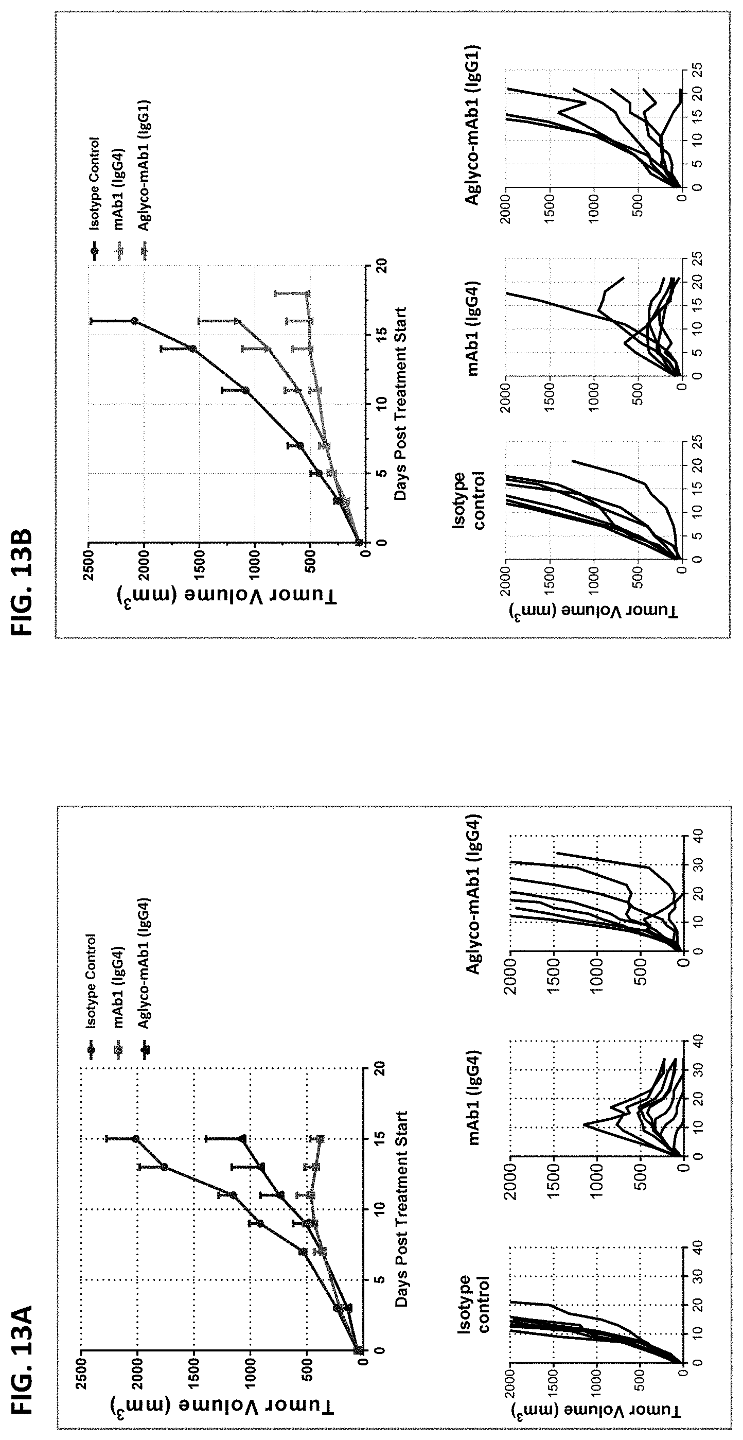
D00016
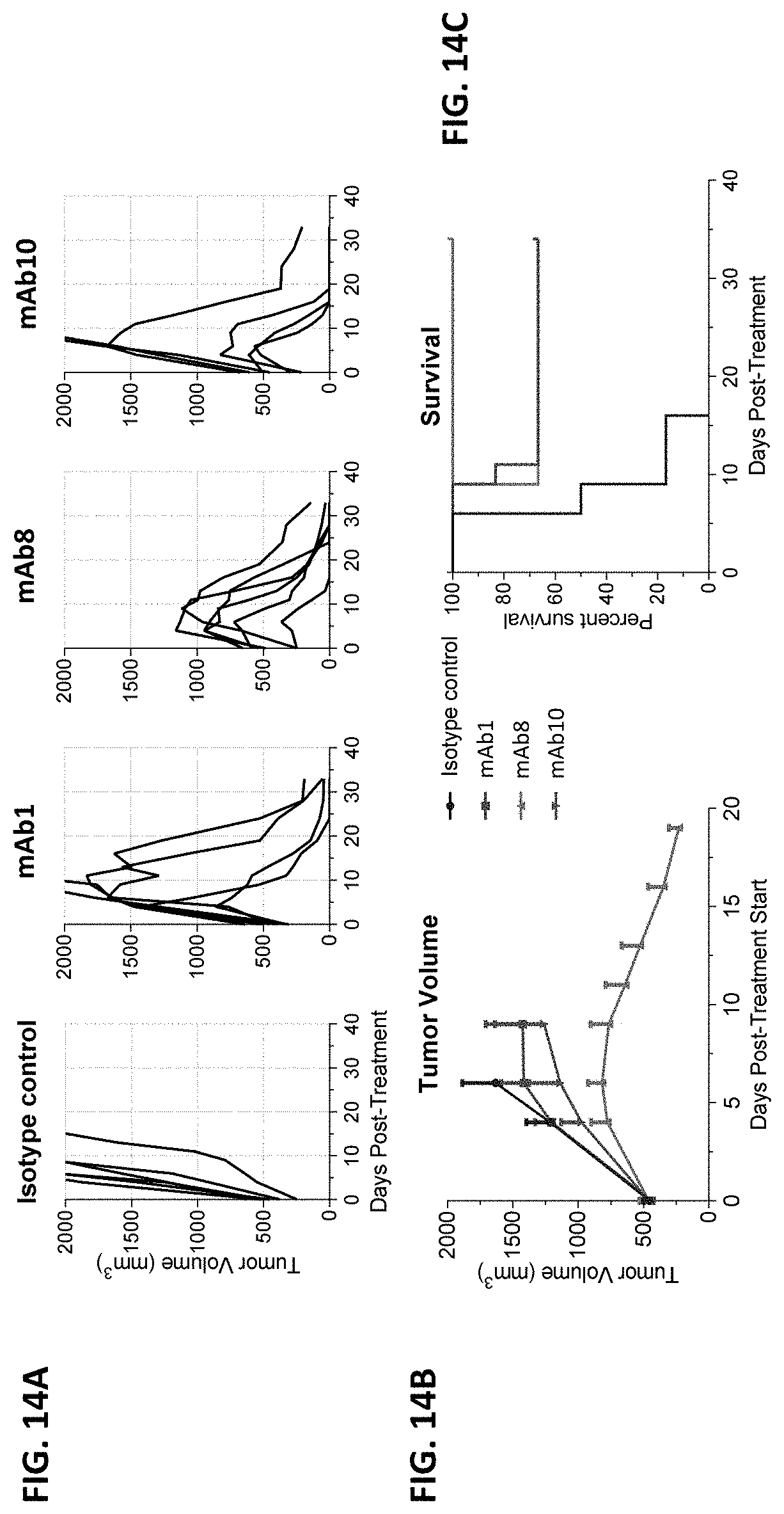
D00017

D00018

D00019

D00020

D00021

D00022

D00023

D00024

D00025
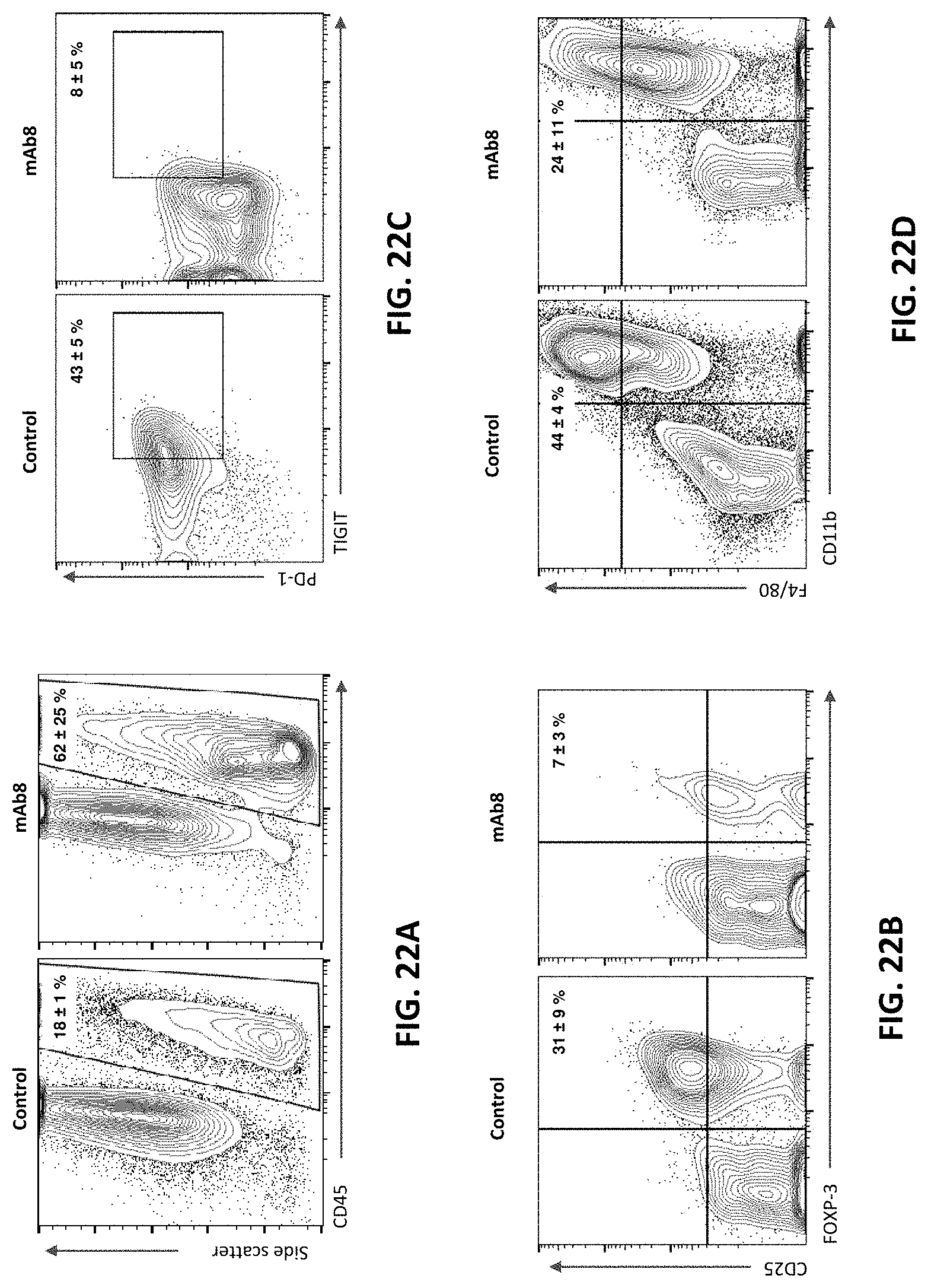
D00026

D00027

D00028

D00029

D00030

D00031

D00032

D00033
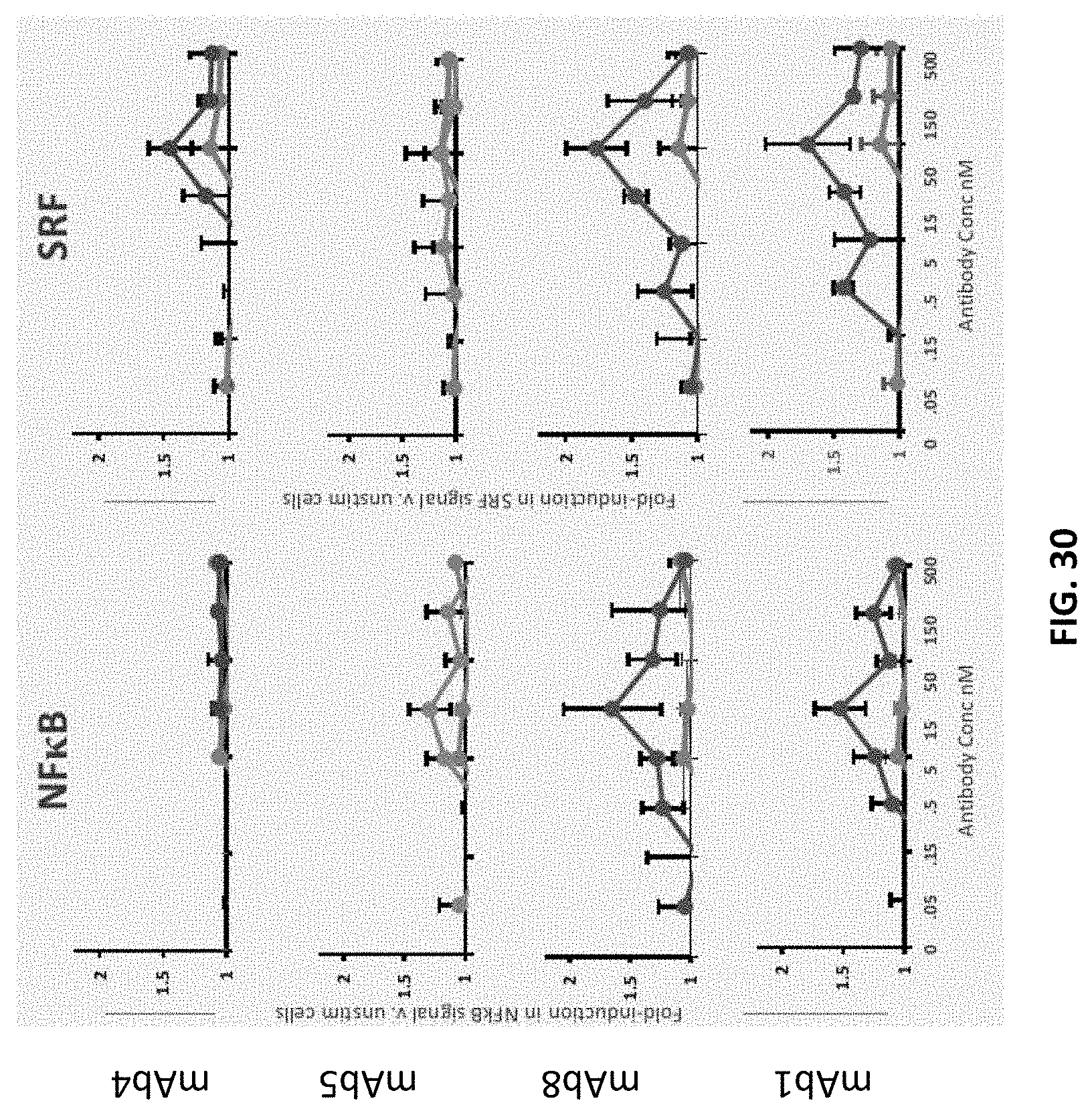
D00034

D00035

D00036
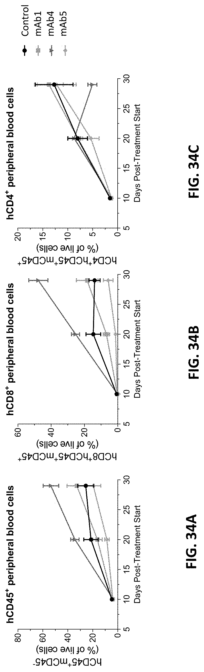
S00001
XML
uspto.report is an independent third-party trademark research tool that is not affiliated, endorsed, or sponsored by the United States Patent and Trademark Office (USPTO) or any other governmental organization. The information provided by uspto.report is based on publicly available data at the time of writing and is intended for informational purposes only.
While we strive to provide accurate and up-to-date information, we do not guarantee the accuracy, completeness, reliability, or suitability of the information displayed on this site. The use of this site is at your own risk. Any reliance you place on such information is therefore strictly at your own risk.
All official trademark data, including owner information, should be verified by visiting the official USPTO website at www.uspto.gov. This site is not intended to replace professional legal advice and should not be used as a substitute for consulting with a legal professional who is knowledgeable about trademark law.