Anti-PD-L1 antibodies
Zheng , et al. June 30, 2
U.S. patent number 10,696,745 [Application Number 15/580,281] was granted by the patent office on 2020-06-30 for anti-pd-l1 antibodies. The grantee listed for this patent is WUXI BIOLOGICS (SHANGHAI) CO. LTD.. Invention is credited to Zhisheng Chen, Jing Li, Yong Zheng.


View All Diagrams
| United States Patent | 10,696,745 |
| Zheng , et al. | June 30, 2020 |
Anti-PD-L1 antibodies
Abstract
The present disclosure provides monoclonal antibodies against protein programmed cell death 1 ligand (PD-L1), which can block the binding of PD-L1 to PD-1, and therefore block the inhibitory function of PD-L1 on PD-1 expressing T cells. The antibodies of disclosure provide very potent agents for the treatment of multiple cancers via modulating human immune function.
| Inventors: | Zheng; Yong (Shanghai, CN), Li; Jing (Lexington, MA), Chen; Zhisheng (Shanghai, CN) | ||||||||||
|---|---|---|---|---|---|---|---|---|---|---|---|
| Applicant: |
|
||||||||||
| Family ID: | 57502993 | ||||||||||
| Appl. No.: | 15/580,281 | ||||||||||
| Filed: | June 11, 2015 | ||||||||||
| PCT Filed: | June 11, 2015 | ||||||||||
| PCT No.: | PCT/CN2015/081256 | ||||||||||
| 371(c)(1),(2),(4) Date: | December 07, 2017 | ||||||||||
| PCT Pub. No.: | WO2016/197367 | ||||||||||
| PCT Pub. Date: | December 15, 2016 |
Prior Publication Data
| Document Identifier | Publication Date | |
|---|---|---|
| US 20180162944 A1 | Jun 14, 2018 | |
| Current U.S. Class: | 1/1 |
| Current CPC Class: | G01N 33/566 (20130101); C07K 16/2827 (20130101); A61P 35/00 (20180101); C07K 2317/565 (20130101); G01N 2333/70596 (20130101); C07K 2317/24 (20130101); G01N 2800/52 (20130101); C07K 2317/76 (20130101); C07K 2317/74 (20130101); C07K 2317/92 (20130101) |
| Current International Class: | C07K 16/20 (20060101); G01N 33/566 (20060101); C07K 16/28 (20060101); C07K 16/40 (20060101); C07K 16/30 (20060101) |
| Field of Search: | ;424/133.1,136.1,178.1 |
References Cited [Referenced By]
U.S. Patent Documents
| 30985 | December 1860 | Pye |
| 4560655 | December 1985 | Baker |
| 4657866 | April 1987 | Kumar |
| 4767704 | August 1988 | Cleveland et al. |
| 4927762 | May 1990 | Darfler |
| 5122469 | June 1992 | Mather et al. |
| 6005079 | December 1999 | Casterman et al. |
| 7943743 | May 2011 | Korman et al. |
| 8779108 | July 2014 | Queva et al. |
| 2014/0341917 | November 2014 | Nastri et al. |
| 2019/0202917 | July 2019 | Campbell |
| 0404097 | Dec 1990 | EP | |||
| 8700195 | Jan 1987 | WO | |||
| 9003430 | Apr 1990 | WO | |||
| 9311161 | Jun 1993 | WO | |||
| 94/04678 | Mar 1994 | WO | |||
| 94/25591 | Nov 1994 | WO | |||
| 2010077634 | Jul 2010 | WO | |||
| 2013181634 | Dec 2013 | WO | |||
| 2014055897 | Apr 2014 | WO | |||
| 2014100079 | Jun 2014 | WO | |||
| 2015/036499 | Mar 2015 | WO | |||
| 2016/023875 | Feb 2016 | WO | |||
Other References
|
Li et al. (MABS 2017, vol. 9, No. 4, 628-637). cited by examiner . Antje Habicht et al., A Link between PDL1 and T Regulatory Cells in Fetomaternal Tolerance, J Immunol 2007; 179:5211-5219; doi: 10.4049/jimmunol.179.8.5211. cited by applicant . Jun Konishi et al., B7-H1 Expression on Non-Small Cell Lung Cancer Cells and Its Relationship with Tumor-Infiltrating Lymphocytes and Their PD-1 Expression, clinical cancer research (2004), Vo.10, 5094-5100. cited by applicant . Haidong Dong et al., B7-H1 pathway and its role in the evasion of tumor immunity, J Mol Med (2003) 81:281-287; DOI 10.1007/s00109-003-0430-2. cited by applicant . Yvette Latchman et al., PD-L2 is a second ligand for PD-1 and inhibits T cell activation, Nature Immunology Mar. 2001, 2(3):261-8, DOI: 10.1038/85330. cited by applicant . Yan Luan et al., A fully human monoclonal antibody targeting PD-L1 with potent anti-tumor activity, International Immunopharmacology 31 (2016) 248-256. cited by applicant . Christian Blank et al., Interaction of PD-L1 on tumor cells with PD-1 on tumor-specific T cells as a mechanism of immune evasion: implications for tumor immunotherapy, Cancer Immunol Immunother (2005) 54: 307-314, DOI 10.1007/s00262-004-0593-x. cited by applicant . Taku Okazaki et al., PD-1 and PD-1 ligands: from discovery to clinical application, International Immunology, 2007, vol. 19, No. 7, pp. 813-824, doi:10.1093/intimm/dxm057. cited by applicant . Manish J. Butte et al., PD-L1 interacts specifically with B7-1 to inhibit T cell proliferation, Immunity. Jul. 2007 ; 27(1): 111-122. doi:10.1016/j.immuni.2007.05.016. cited by applicant . Loise M. Francisco et al., The PD-1 Pathway in Tolerance and Autoimmunity, Immunol Rev. Jul. 2010 ; 236: 219-242. doi:10.1111/j.1600-065X.2010.00923.x. cited by applicant . Al-Lazikani, B., Chothia, C., Lesk, A. M., Standard Conformations for the Canonical Structures of Immunoglobulins, J. Mol. Biol., 273(4), 927-948 (1997). cited by applicant . Chothia, C. et al., Domain Association in Immunoglobulin Molecules, J. Mol. Biol., Dec. 5;186(3):651-63 (1985). cited by applicant . Chothia, C. and Lesk, A.M., Canonical Structures for the Hypervariable Regions of Immunoglobulins, J. Mol. Biol., 196,901-917 (1987). cited by applicant . Chothia, C. et al., Conformations of Immunoglobulin Hypervariable Regions, Nature, Dec. 21-28;342(6252):877-83 (1989). cited by applicant . Kabat E.A. et al., National Institutes of Health, Bethesda, Md. (1991). cited by applicant . Riechmann L. and Muyldermans S., Single domain antibodies: comparison of camel VH and camelised human VH domains, J Immunol Methods. Dec. 10;231(1-2):25-38 (1999). cited by applicant . Muyldermans S., Single domain camel antibodies: current status, Rev Mol Biotechnol. Jun.;74(4):277-302 (2001). cited by applicant . Hamers-Casterman C. et al., Naturally occurring antibodies devoid of light chains, Nature. Jun. 3;363(6428):446-8 (1993). cited by applicant . Nguyen VK. et al. "Heavy-chain antibodies in Camelidae; a case of evolutionary innovation," Immunogenetics. Apr.;54 (1):39-47 (2002). cited by applicant . Nguyen VK. et al., Heavy-chain only antibodies derived from dromedary are secreted and displayed by mouse B cells, Immunology. May;109(1):93-101 (2003). cited by applicant . Koch-Nolte F. et al., Single domain antibodies from llama effectively and specifically block T cell ecto-ADP-ribosyltransferase ART2.2 in vivo, FASEB J. Nov.;21(13):3490-8. Epub Jun. 15, 2007 (2007). cited by applicant . Holliger P. et al., "Diabodies": Small bivalent and bispecific antibody fragments, Proc Natl Acad Sci U S A. Jul. 15;90 (14):6444-8 (1993). cited by applicant . Gordon J. Freeman et al., Engagement of the PD-1 immunoinhibitory receptor by a novel B7 family member leads to negative regulation of lymphocyte activation, vol. 192, No. 7, Oct. 2, 2000 1027-1034. cited by applicant . Altschul S.F. et al, Basic Local Alignment Search Tool, J. Mol. Biol., 215:403-410 (1990). cited by applicant . Stephen F. et al, Gapped BLAST and PSI-BLAST: a new generation of protein database search programs, Nucleic Acids Res., 25:3389-3402 (1997). cited by applicant . Higgins D.G. et al, Using CLUSTAL for Multiple Sequence Alignments, Methods in Enzymology, 266:383-402 (1996). cited by applicant . Larkin M.A. et al, Clustal W and Clustal X version 2.0, Bioinformatics (Oxford, England), 23(21): 2947-8 (2007). cited by applicant . Graham et al., Characteristics of a Human Cell Line Transformed by DNA from Human Adenovirus Type 5, J. Gen Virol. 36:59 (1977). cited by applicant . Urlaub et al., Isolation of Chinese Hamster Cell Mutants Deficient in Dihydrofolate Reductase Activity, Proc. Natl. Acad. Sci. USA 77:4216 (1980). cited by applicant . J. P. Mather, Establishment and Characterization of Two Distinct Mouse Testicular Epithelial Cell Lines, Biol. Reprod. 23:243-251 (1980). cited by applicant . Mather et al., Culture of testicular cells in hormone-supplemented serum-free medium, Annals N.Y. Acad. Sci. 383:44-68 (1982). cited by applicant . Barnes et al., Methods for Growth of Cultured Cells in Serum-Free Medium, Anal. Biochem. 102:255 (1980). cited by applicant . Carter et al., High Level Escherichia coli Expression and Production of a Bivalent Humanized Antibody Fragment, Bio/Technology 10:163-167 (1992). cited by applicant . Lindmark et al., Binding of Immunoglobulins to Protein A and Immunoglobulin Levels in Mammalian Sera, J. Immunol. Meth. 62:1-13 (1983). cited by applicant . Guss et al., Structure of the IgG-binding regions of streptococcal protein G, EMBO J. 5:1567-1575 (1986). cited by applicant . International Search Report of PCT application No. PCT/CN2015/081256 dated Mar. 21, 2016. cited by applicant . Cristina Teixido Niki Karachaliou Maria Gonzalez-Cao Daniela Morales-Espinosa Rafael Rosell: "Assays for predicting and monitoring responses to lung cancer immunotherapy", Cancer Biol Med, Jan. 1, 2015, pp. 87-95, XP55528318, China, Retrieved from the Internet: URL: https://www.ncbi.nlm.nih.gov/pmc/articles/PMC4493376/pdf/cbm-12-02-087.pd- f. cited by applicant . Kathleen M. Mahoney et al: "The Next Immune-Checkpoint Inhibitors: PD-1/PD-L1 Blockade in Melanoma", Clinical Therapeutics., vol. 37, No. 4, Apr. 1, 2015, pp. 764-782, XP055285031, US. cited by applicant . The extended European search report in EP application No. 15894637.6, dated Jan. 15, 2019. cited by applicant . Perez De La Lastra et al: "Epitope mapping of 10 monoclonal antibodies against the pig analogue of human membrane cofactor protein (MCP)", Immunology, vol. 96, No. 4, Apr. 1, 1999 (Apr. 1, 1999), pp. 663-670, XP55572134, GB. ISSN: 0019-2805, DOI: 10.1046/j.1365-2567.1999.00732.x. cited by applicant . Cristina Caldas et al: "Humanization of the anti-CD18 antibody 6.7: an unexpected effect of a framework residue in binding to antigen.", Molecular immunology, vol. 39, No. 15,May 1, 2003 (May 1, 2003), pp. 941-952, XP55025334, ISSN: 0161-5890, DOI: 10.1016/S0161-5890(03)00022-1. cited by applicant . Du J et al: "Molecular basis of recognition of human osteopontin by 23C3, a potential therapeutic antibody for treatment of rheumatoid arthritis", Journal of Molecular Biology, Academic Press, United Kingdom, vol. 382, No. 4,Oct. 17, 2008 (Oct. 17, 2008), pp. 835-842, XP026805063, ISSN: 0022-2836, DOI: 10.1016/J.JMB.2008.07.075 [retrieved on Jul. 31, 2008]. cited by applicant . EPO communication in European Patent Application No. 15894637.6, dated Feb. 6, 2020. cited by applicant. |
Primary Examiner: Bristol; Lynn A
Attorney, Agent or Firm: Jun He Law Offices P.C. Zhu; James J.
Claims
What is claimed is:
1. An isolated monoclonal antibody or an antigen binding fragment thereof, comprising a heavy chain variable region comprising: SEQ ID NOs: 1, 2, and 3, and a light chain variable region comprising: SEQ ID NOs: 4, 5 and 6, wherein the monoclonal antibody or an antigen binding fragment thereof specifically binds to human or monkey PD-L1.
2. The antibody or an antigen binding fragment thereof of claim 1, comprising: a) a heavy chain variable region comprising SEQ ID NO: 7; and a light chain variable region comprising SEQ ID NO: 9; b) a heavy chain variable region comprising SEQ ID NO: 11; and a light chain variable region comprising SEQ ID NO: 13; c) a heavy chain variable region comprising SEQ ID NO: 11; and a light chain variable region comprising SEQ ID NO: 16; d) a heavy chain variable region comprising SEQ ID NO: 18; and a light chain variable region comprising SEQ ID NO: 13; or e) a heavy chain variable region comprising SEQ ID NO: 18; and a light chain variable region comprising SEQ ID NO:22.
3. The antibody or antigen-binding fragment thereof of claim 1, which is a diabody, a scFv, an scFv dimer, a BsFv, a dsFv, a (dsFv).sub.2, a dsFv-dsFv', an Fv fragment, a Fab, a Fab', a F(ab').sub.2, or a ds diabody.
4. The antibody or antigen-binding fragment thereof of claim 1, further comprising a conjugate.
5. An isolated polynucleotide encoding the antibody or an antigen binding fragment thereof of claim 1.
6. A vector comprising the isolated polynucleotide of claim 5.
7. A host cell comprising the vector of claim 6.
8. A method of expressing the antibody or antigen-binding fragment thereof of claim 1, comprising culturing the host cell comprising a vector comprising an isolated polynucleotide encoding the antibody or an antigen binding fragment thereof of claim 1, under the condition at which the polynucleotide is expressed.
9. A kit comprising the antibody or antigen-binding fragment thereof of claim 1.
10. A method of detecting presence or level of human or monkey PD-L1 in a biological sample, comprising exposing the biological sample to the antibody or antigen-binding fragment thereof of claim 1, and determining the presence or level of human or monkey PD-L1 in the sample.
11. A pharmaceutical composition comprising the antibody or antigen-binding fragment thereof of claim 1 and one or more pharmaceutically acceptable carriers.
Description
FIELD OF THE INVENTION
The present disclosure generally relates to novel anti-PD-L1 antibodies.
BACKGROUND
Increasing evidences from preclinical and clinical results have shown that targeting immune checkpoints is becoming the most promising approach to treat patients with cancers. Programmed cell death 1, one of immune-checkpoint proteins, play a major role in limiting the activity of T cells that provide a major immune resistance mechanism by which tumor cells escaped immune surveillance. The interaction of PD-1 expressed on activated T cells, and PD-L1 expressed on tumor cells negatively regulate immune response and damp anti-tumor immunity. Expression of PD-L1 on tumors is correlated with reduced survival in esophageal, pancreatic and other types of cancers, highlighting this pathway as a new promising target for tumor immunotherapy. Multiple agents targeting PD-1 pathway have been developed by pharmaceutical companies, such as Bristol-Myers Squibb (BMS), Merck, Roche and GlaxoSmithKline (GSK). Data from clinical trials demonstrated early evidence of durable clinical activity and an encouraging safety profile in patients with various tumor types. Nivolumab, a PD-1 drug developed by BMS, is being put at center stage of the next-generation field. Now in 6 late-stage studies, the treatment spurred tumor shrinkage in three of 5 cancer groups studied, including 18% of 72 lung cancer patients, close to a third of 98 melanoma patients and 27% of 33 patients with kidney cancer. Developed by Merck, lambrolizumab is a humanized monoclonal IgG4 antibody that acts against PD-1, which grabbed the FDA's new breakthrough designation after impressive IB data came through for skin cancer. The results from a phase IB study have shown an objective anti-tumor response in 51% of 85 cancer patients, and a complete response in 9% of patients. Roche's experimental MPDL3280A demonstrated an ability to shrink tumors in 29 of 140 (21%) advanced cancer patients with various tumor sizes.
However, the existing therapies may not be all satisfactory and therefore new anti-PD-L1 antibodies are still needed.
BRIEF SUMMARY OF THE INVENTION
The present disclosure provides novel monoclonal anti-PD-L1 antibodies, polynucleotides encoding the same, and methods of using the same.
In one aspect, the present disclosure provides isolated monoclonal antibodies or antigen binding fragments thereof, which are capable of specifically binding to human PD-L1 at a Kd value no more than 10.sup.-9M (e.g. no more than .ltoreq.9.times.10.sup.-10 M, .ltoreq.8.times.10.sup.-10 M, .ltoreq.7.times.10.sup.-10 M, .ltoreq.6.times.10.sup.-10 M, .ltoreq.5.times.10.sup.-10 M, .ltoreq.4.times.10.sup.-10 M, .ltoreq.3.times.10.sup.-10 M, .ltoreq.2.times.10.sup.-10 M, or .ltoreq.10.sup.-10 M) as measured by plasmon resonance binding assay.
In certain embodiments, the antibodies or antigen binding fragments thereof bind to monkey PD-L1 at an EC50 of no more than 10 nM (e.g. no more than 1 nM, 0.9 nM, 0.8 nM, 0.7 nM, 0.6 nM, 0.5 nM, 0.4 nM, 0.3 nM, 0.2 nM, 0.1 nM, 0.09 nM, 0.08 nM, 0.07 nM, 0.06 nM, 0.05 nM, 0.04 nM, 0.03 nM, 0.02 nM, or 0.01 nM. In certain embodiments, the antibodies and antigen-binding fragments thereof do not bind to mouse PD-L1 but bind to monkey PD-L1 with a binding affinity similar to that of human PD-L1. In certain embodiments, the antibodies or antigen binding fragments thereof potently inhibit binding of human or monkey PD-L1 to its receptor (e.g. PD-1), at an IC50 of no more than 100 nM (e.g. no more than 50 nM, 40 nM, 30 nM, 20 nM, 10 nM, 9 nM, 8 nM, 7 nM, 6 nM, 5 nM, 4 nM, 3 nM, 2 nM, 1 nM, 0.9 nM, 0.8 nM, 0.7 nM, 0.6 nM, 0.5 nM, 0.4 nM, 0.3 nM, 0.2 nM, or 0.1 nM). In certain embodiments, the EC50 or IC50 is measured by fluorescence-activated cell sorting (FACS) analysis.
In certain embodiments, the antibodies or antigen binding fragments thereof have substantially reduced effector function. In certain embodiments, the antibodies or antigen binding fragments thereof do not mediate ADCC or CDC or both.
In certain embodiments, the antibodies or antigen binding fragments thereof provided herein comprise a heavy chain CDR sequences selected from the group consisting of: SEQ ID NOs: 1, 2, and 3.
In one aspect, the antibodies or an antigen binding fragments thereof provided herein comprise a light chain CDR sequences selected from the group consisting of: SEQ ID NOs: 4, 5 and 6.
In certain embodiments, the antibodies or antigen binding fragments thereof provided herein comprise at least one, two, three, four, five or six CDRs selected from the group consisting of: SEQ ID NOs: 1, 2, 3, 4, 5, and 6.
In certain embodiments, the antibodies or antigen binding fragments thereof provided herein comprise a heavy chain variable region comprising SEQ ID NO: 1, SEQ ID NO: 2, and/or SEQ ID NO: 3.
In certain embodiments, the antibodies or antigen binding fragments thereof provided herein comprise a light chain variable region comprising SEQ ID NO: 4, SEQ ID NO: 5, and/or SEQ ID NO: 6.
In certain embodiments, the antibodies or antigen binding fragments thereof provided herein comprise a heavy chain variable region comprising SEQ ID NO: 1, SEQ ID NO: 2, and/or SEQ ID NO: 3; and a light chain variable region comprising SEQ ID NO: 4, SEQ ID NO: 5, and/or SEQ ID NO: 6.
In certain embodiments, the antibodies or antigen binding fragments thereof provided herein comprise a heavy chain variable region selected from the group consisting of: SEQ ID NO: 7, SEQ ID NO: 11, and SEQ ID NO: 18.
In certain embodiments, the antibodies or antigen binding fragments provided herein comprise a light chain variable region selected from the group consisting of: SEQ ID NO: 9, SEQ ID NO: 13, SEQ ID NO: 16, and SEQ ID NO: 22.
In certain embodiments, the antibodies or antigen binding fragments thereof provided herein comprise: a) a heavy chain variable region comprising SEQ ID NO: 7; and a light chain variable region comprising SEQ ID NO: 9; b) a heavy chain variable region comprising SEQ ID NO: 11; and a light chain variable region comprising SEQ ID NO: 13; c) a heavy chain variable region comprising SEQ ID NO: 11; and a light chain variable region comprising SEQ ID NO: 16; d) a heavy chain variable region comprising SEQ ID NO: 18; and a light chain variable region comprising SEQ ID NO: 13; or e) a heavy chain variable region comprising SEQ ID NO: 18; and a light chain variable region comprising SEQ ID NO: 22.
In certain embodiments, the antibodies provided herein include, for example, 2.74.15, 2.74.15.hAb4, 2.74.15.hAb5, 2.74.15.hAb6, 2.74.15.hAb7, and 2.74.15.hAb8.
In certain embodiments, the antibodies or antigen binding fragments thereof provided herein compete for the same epitope with antibodies 2.74.15, 2.74.15.hAb4, 2.74.15.hAb5, 2.74.15.hAb6, 2.74.15.hAb7, or 2.74.15.hAb8.
In certain embodiments, the antibodies or antigen binding fragments thereof are capable of blocking binding of human PD-L1 to its receptor and thereby providing at least one of the following activities: a) inducing production of IL-2 in CD4+ T cells; b) inducing production of IFN.gamma. in CD4+ T cells; c) inducing proliferation of CD4+ T cells and d) reversing T reg's suppressive function.
In certain embodiments, the antibodies provided herein are a monoclonal antibody, humanized antibody, chimeric antibody, recombinant antibody, bispecific antibody, labeled antibody, bivalent antibody, or anti-idiotypic antibody.
In certain embodiments, the antigen-binding fragments thereof provided herein are a camelized single domain antibody, a diabody, a scFv, an scFv dimer, a BsFv, a dsFv, a (dsFv)2, a dsFv-dsFv', an Fv fragment, a Fab, a Fab', a F(ab')2, a ds diabody, a nanobody, a domain antibody, or a bivalent domain antibody.
In certain embodiments, the antibodies or antigen-binding fragments thereof provided herein further comprise an immunoglobulin constant region.
In certain embodiments, the antibodies or antigen-binding fragments thereof provided herein, further comprise a conjugate.
In certain embodiments, the conjugate can be a detectable label, a pharmacokinetic modifying moiety, or a purification moiety.
In another aspect, the present disclosure provides isolated polynucleotides encoding the antibodies or antigen binding fragments thereof provided herein. In certain embodiments, polynucleotides are provided that encode the amino acid sequences of the antibodies or antigen-binding fragments disclosed herein. In certain other embodiments, vectors are provided that comprise these polynucleotides, and in certain other embodiments, host cells are provided that comprise these vectors. In certain embodiments, methods are provided for expressing one or more of the antibodies or antigen-binding fragments disclosed herein by culturing these host cells under conditions in which the antibodies or antigen-binding fragments encoded by the polynucleotides are expressed from a vector. In certain embodiments, the polynucleotides provided herein are operably associated with a promoter such as a SV40 promoter in a vector. In certain embodiments, host cells comprising the vectors provided herein are Chinese hamster ovary cell, or 293F cell.
In another aspect, the present disclosure provides kits comprising the antibody or antigen-binding fragment thereof.
In another aspect, the present disclosure provides methods of detecting presence or level of PD-L1 (e.g. human or monkey) in a biological sample, comprising exposing the biological sample to the antibody or antigen-binding fragment thereof provided herein, and determining the presence or level of the PD-L1 in the sample.
In another aspect, the present disclosure provides methods of identifying an individual having a disorder or condition likely to respond to a PD-L1 antagonist, comprising: determining presence or level of PD-L1 (e.g. human or monkey) in a test biological sample from the individual with the antibody or antigen-binding fragment thereof provided herein, wherein presence or upregulated level of the PD-L1 in the test biological sample indicates likelihood of responsiveness. In certain embodiments, the methods further comprising administering an effective amount of the antibody or antigen-binding fragment thereof provided herein to the individual who has been identified as having a disorder or condition likely to respond to a PD-L1 antagonist.
The present disclosure further provides methods of monitoring therapeutic response or disease progression in a subject treated with a PD-L1 antagonist, comprising determining presence or level of PD-L1 (e.g. human or monkey) in a test biological sample from the individual with the anti-PD-L1 antibody or antigen-binding fragment thereof provided herein.
In another aspect, the present disclosure provides pharmaceutical compositions comprising the antibody or antigen-binding fragment thereof provided herein and one or more pharmaceutically acceptable carriers. In certain of these embodiments, the pharmaceutical carriers may be, for example, diluents, antioxidants, adjuvants, excipients, or non-toxic auxiliary substances.
In another aspect, the present disclosure provides methods of treating a condition in a subject that would benefit from upregulation of immune response, comprising administering an effective amount of the antibody or antigen-binding fragment thereof provided herein to the subject. In certain embodiments, the subject has upregulated expression of PD-L1.
Use of the antibody or antigen-binding fragment thereof provided herein in the manufacture of a medicament for treating a condition that would benefit from upregulation of immune response. In certain embodiments, the condition is cancer or chronic viral infection.
BRIEF DESCRIPTION OF FIGURES
FIG. 1 presents the binding of murine anti-human PD-L1 antibodies to PD-L1 expressing CHO cell as measured by FACS analysis. EC50 is provided in nM.
FIG. 2 presents the binding of humanized PD-L1 antibodies to PD-L1 expressing CHO cell as measured by FACS analysis.
FIG. 3 is the binding of humanized PD-L1 antibodies to PD-L1 expressed on activated dendritic cells (DC) as measured by FACS analysis. EC50 is provided in nM.
FIG. 4 shows that humanized PD-L1 antibodies block the binding of PD-1 to PD-L1 transfected CHO cells as measured by FACS analysis. IC50 is provided in nM.
FIG. 5 shows that humanized PD-L1 antibodies specifically bind to PD-L1, but do not bind to PD-L2, as measured by FACS analysis.
FIG. 6 shows that the PD-L1 antibodies bind to cynomolgus monkey PD-L1. EC50 is provided in nM.
FIG. 7 is the full kinetics of binding affinity of PD-L1 antibodies against human PD-L1.
FIG. 8 illustrates the effect of humanized anti-PD-L1 antibodies on IL-2 production in mixed lymphocyte reaction (MLR).
FIG. 9 illustrates the effect of humanized anti-PD-L1 antibodies on IFN.gamma. production in MLR.
FIG. 10 shows that humanized anti-PD-L1 antibodies promote T cell proliferation in MLR.
FIG. 11 shows that humanized PD-L1 antibodies enhance IFN.gamma. production in specific T cell response generated by co-culture of cytomegalovirus (CMV)-specific T-cells with CMV-pp65 peptide loaded DCs for 5 days.
FIG. 12 shows that humanized PD-L1 antibodies promote T cell proliferation in specific T cell response.
FIG. 13 shows that anti-PD-L1 antibodies reverse Treg's suppressive function.
FIG. 14 shows the anti-PD-L1 antibodies lacks ADCC on mDCs.
FIG. 15 shows the anti-PD-L1 antibodies lacks CDC on activated T cells.
DETAILED DESCRIPTION OF THE INVENTION
The following description of the disclosure is merely intended to illustrate various embodiments of the disclosure. As such, the specific modifications discussed are not to be construed as limitations on the scope of the disclosure. It will be apparent to one skilled in the art that various equivalents, changes, and modifications may be made without departing from the scope of the disclosure, and it is understood that such equivalent embodiments are to be included herein. All references cited herein, including publications, patents and patent applications are incorporated herein by reference in their entirety.
Definitions
The term "antibody" as used herein includes any immunoglobulin, monoclonal antibody, polyclonal antibody, multispecific antibody, or bispecific (bivalent) antibody that binds to a specific antigen. A native intact antibody comprises two heavy chains and two light chains. Each heavy chain consists of a variable region and a first, second, and third constant region, while each light chain consists of a variable region and a constant region. Mammalian heavy chains are classified as .alpha., .delta., .epsilon., .gamma., and .mu., and mammalian light chains are classified as .lamda., or .kappa.. The antibody has a "Y" shape, with the stem of the Y consisting of the second and third constant regions of two heavy chains bound together via disulfide bonding. Each arm of the Y includes the variable region and first constant region of a single heavy chain bound to the variable and constant regions of a single light chain. The variable regions of the light and heavy chains are responsible for antigen binding. The variables region in both chains generally contain three highly variable loops called the complementarity determining regions (CDRs) (light (L) chain CDRs including LCDR1, LCDR2, and LCDR3, heavy (H) chain CDRs including HCDR1, HCDR2, HCDR3). CDR boundaries for the antibodies and antigen-binding fragments disclosed herein may be defined or identified by the conventions of Kabat, Chothia, or Al-Lazikani (Al-Lazikani, B., Chothia, C., Lesk, A. M., J. Mol. Biol., 273(4), 927 (1997); Chothia, C. et al., J Mol Biol. December 5; 186(3):651-63 (1985); Chothia, C. and Lesk, A. M., J. Mol. Biol., 196, 901 (1987); Chothia, C. et al., Nature. December 21-28; 342(6252):877-83 (1989); Kabat E. A. et al., National Institutes of Health, Bethesda, Md. (1991)). The three CDRs are interposed between flanking stretches known as framework regions (FRs), which are more highly conserved than the CDRs and form a scaffold to support the hypervariable loops. The constant regions of the heavy and light chains are not involved in antigen binding, but exhibit various effector functions. Antibodies are assigned to classes based on the amino acid sequence of the constant region of their heavy chain. The five major classes or isotypes of antibodies are IgA, IgD, IgE, IgG, and IgM, which are characterized by the presence of .alpha., .delta., .epsilon., .gamma., and .mu. heavy chains, respectively. Several of the major antibody classes are divided into subclasses such as IgG1 (.gamma.1 heavy chain), IgG2 (.gamma.2 heavy chain), IgG3 (.gamma.3 heavy chain), IgG4 (.gamma.4 heavy chain), IgA1 (.alpha.1 heavy chain), or IgA2 (.alpha.2 heavy chain).
The term "antigen-binding fragment" as used herein refers to an antibody fragment formed from a portion of an antibody comprising one or more CDRs, or any other antibody fragment that binds to an antigen but does not comprise an intact native antibody structure. Examples of antigen-binding fragment include, without limitation, a diabody, a Fab, a Fab', a F(ab').sub.2, an Fv fragment, a disulfide stabilized Fv fragment (dsFv), a (dsFv).sub.2, a bispecific dsFv (dsFv-dsFv'), a disulfide stabilized diabody (ds diabody), a single-chain antibody molecule (scFv), an scFv dimer (bivalent diabody), a multispecific antibody, a camelized single domain antibody, a nanobody, a domain antibody, and a bivalent domain antibody. An antigen-binding fragment is capable of binding to the same antigen to which the parent antibody binds. In certain embodiments, an antigen-binding fragment may comprise one or more CDRs from a particular human antibody grafted to a framework region from one or more different human antibodies.
"Fab" with regard to an antibody refers to that portion of the antibody consisting of a single light chain (both variable and constant regions) bound to the variable region and first constant region of a single heavy chain by a disulfide bond.
"Fab'" refers to a Fab fragment that includes a portion of the hinge region.
"F(ab').sub.2" refers to a dimer of Fab'.
"Fc" with regard to an antibody refers to that portion of the antibody consisting of the second and third constant regions of a first heavy chain bound to the second and third constant regions of a second heavy chain via disulfide bonding. The Fc portion of the antibody is responsible for various effector functions such as ADCC, and CDC, but does not function in antigen binding.
"Fv" with regard to an antibody refers to the smallest fragment of the antibody to bear the complete antigen binding site. An Fv fragment consists of the variable region of a single light chain bound to the variable region of a single heavy chain.
"Single-chain Fv antibody" or "scFv" refers to an engineered antibody consisting of a light chain variable region and a heavy chain variable region connected to one another directly or via a peptide linker sequence (Huston J S et al. Proc Natl Acad Sci USA, 85:5879 (1988)).
"Single-chain Fv-Fc antibody" or "scFv-Fc" refers to an engineered antibody consisting of a scFv connected to the Fc region of an antibody.
"Camelized single domain antibody," "heavy chain antibody," or "HCAb" refers to an antibody that contains two V.sub.H domains and no light chains (Riechmann L. and Muyldermans S., J Immunol Methods. December 10; 231(1-2):25-38 (1999); Muyldermans S., J Biotechnol. June; 74(4):277-302 (2001); WO94/04678; WO94/25591; U.S. Pat. No. 6,005,079). Heavy chain antibodies were originally derived from Camelidae (camels, dromedaries, and llamas). Although devoid of light chains, camelized antibodies have an authentic antigen-binding repertoire (Hamers-Casterman C. et al., Nature. June 3; 363(6428):446-8 (1993); Nguyen V K. et al. "Heavy-chain antibodies in Camelidae; a case of evolutionary innovation," Immunogenetics. April; 54(1):39-47 (2002); Nguyen V K. et a/Immunology. May; 109(1):93-101 (2003)). The variable domain of a heavy chain antibody (VHH domain) represents the smallest known antigen-binding unit generated by adaptive immune responses (Koch-Nolte F. et al., FASEB J. November; 21(13):3490-8. Epub 2007 Jun. 15 (2007)).
A "nanobody" refers to an antibody fragment that consists of a VHH domain from a heavy chain antibody and two constant domains, CH2 and CH3.
"Diabodies" include small antibody fragments with two antigen-binding sites, wherein the fragments comprise a V.sub.H domain connected to a V.sub.L domain in the same polypeptide chain (V.sub.H-V.sub.L or V.sub.H-V.sub.L) (see, e.g., Holliger P. et al., Proc Natl Acad Sci USA. July 15; 90(14):6444-8 (1993); EP404097; WO93/11161). By using a linker that is too short to allow pairing between the two domains on the same chain, the domains are forced to pair with the complementary domains of another chain, thereby creating two antigen-binding sites. The antigen-binding sites may target the same of different antigens (or epitopes).
A "domain antibody" refers to an antibody fragment containing only the variable region of a heavy chain or the variable region of a light chain. In certain instances, two or more V.sub.H domains are covalently joined with a peptide linker to create a bivalent or multivalent domain antibody. The two V.sub.H domains of a bivalent domain antibody may target the same or different antigens.
In certain embodiments, a "(dsFv).sub.2" comprises three peptide chains: two V.sub.H moieties linked by a peptide linker and bound by disulfide bridges to two V.sub.L moieties.
In certain embodiments, a "bispecific ds diabody" comprises V.sub.H1-V.sub.L2 (linked by a peptide linker) bound to V.sub.L1-V.sub.H2 (also linked by a peptide linker) via a disulfide bridge between V.sub.H1 and V.sub.L1.
In certain embodiments, a "bispecific dsFv" or dsFv-dsFv'" comprises three peptide chains: a V.sub.H1-V.sub.H2 moiety wherein the heavy chains are linked by a peptide linker (e.g., a long flexible linker) and bound to V.sub.L1 and V.sub.L2 moieties, respectively, via disulfide bridges, wherein each disulfide paired heavy and light chain has a different antigen specificity.
In certain embodiments, an "scFv dimer" is a bivalent diabody or bivalent ScFv (BsFv) comprising V.sub.H-V.sub.L (linked by a peptide linker) dimerized with another V.sub.H-V.sub.L moiety such that V.sub.H's of one moiety coordinate with the V.sub.L's of the other moiety and form two binding sites which can target the same antigens (or epitopes) or different antigens (or epitopes). In other embodiments, an "scFv dimer" is a bispecific diabody comprising V.sub.H1-V.sub.L2 (linked by a peptide linker) associated with V.sub.L1-V.sub.H2 (also linked by a peptide linker) such that V.sub.H1 and V.sub.L1 coordinate and V.sub.H2 and V.sub.L2 coordinate and each coordinated pair has a different antigen specificity.
The term "humanized" as used herein, with reference to antibody or antigen-binding fragment, means that the antibody or the antigen-binding fragment comprises CDRs derived from non-human animals, FR regions derived from human, and when applicable, the constant regions derived from human. A humanized antibody or antigen-binding fragment is useful as human therapeutics in certain embodiments because it has reduced immunogenicity in human. In some embodiments, the non-human animal is a mammal, for example, a mouse, a rat, a rabbit, a goat, a sheep, a guinea pig, or a hamster. In some embodiments, the humanized antibody or antigen-binding fragment is composed of substantially all human sequences except for the CDR sequences which are non-human. In some embodiments, the FR regions derived from human may comprise the same amino acid sequence as the human antibody from which it is derived, or it may comprise some amino acid changes, for example, no more than 10, 9, 8, 7, 6, 5, 4, 3, 2, or 1 changes of amino acid. In some embodiments, such change in amino acid could be present in heavy chain FR regions only, in light chain FR regions only, or in both chains. In some preferable embodiments, the humanized antibodies comprise human FR1-3 and human JH and J.kappa..
The term "chimeric" as used herein, means an antibody or antigen-binding fragment, having a portion of heavy and/or light chain derived from one species, and the rest of the heavy and/or light chain derived from a different species. In an illustrative example, a chimeric antibody may comprise a constant region derived from human and a variable region from a non-human species, such as from mouse.
"PD-L1" as used herein refers to programmed cell death ligand 1 (PD-L1, see, for example, Freeman et al. (2000) J. Exp. Med. 192:1027). Representative amino acid sequence of human PD-L1 is disclosed under the NCBI accession number: NP_054862.1, and the representative nucleic acid sequence encoding the human PD-L1 is shown under the NCBI accession number: NM_014143.3. PD-L1 is expressed in placenta, spleen, lymph nodes, thymus, heart, fetal liver, and is also found on many tumor or cancer cells. PD-L1 binds to its receptor PD-1 or B7-1, which is expressed on activated T cells, B cells and myeloid cells. The binding of PD-L1 and its receptor induces signal transduction to suppress TCR-mediated activation of cytokine production and T cell proliferation. Accordingly, PD-L1 plays a major role in suppressing immune system during particular events such as pregnancy, autoimmune diseases, tissue allografts, and is believed to allow tumor or cancer cells to circumvent the immunological checkpoint and evade the immune response.
"Anti-PD-L1 antibody" as used herein refers to an antibody that is capable of specific binding to PD-L1 (e.g. human or monkey PD-L1) with an affinity which is sufficient to provide for diagnostic and/or therapeutic use.
The term "specific binding" or "specifically binds" as used herein refers to a non-random binding reaction between two molecules, such as for example between an antibody and an antigen. In certain embodiments, the antibodies or antigen-binding fragments provided herein specifically bind human PD-L1 with a binding affinity (K.sub.D) of .ltoreq.10.sup.-6 M (e.g., .ltoreq.5.times.10.sup.-7 M, .ltoreq.2.times.10.sup.-7 M, .ltoreq.10.sup.-7 M, .ltoreq.5.times.10.sup.-8 M, .ltoreq.2.times.10.sup.-8 M, .ltoreq.10.sup.-8 M, .ltoreq.5.times.10.sup.-9 M, .ltoreq.2.times.10.sup.-9 M, .ltoreq.10.sup.-9 M, 10.sup.-10 M). K.sub.D as used herein refers to the ratio of the dissociation rate to the association rate (k.sub.off/k.sub.on), may be determined using surface plasmon resonance methods for example using instrument such as Biacore.
The ability to "block binding" or "compete for the same epitope" as used herein refers to the ability of an antibody or antigen-binding fragment to inhibit the binding interaction between two molecules (e.g. human PD-L1 and an anti-PD-L1 antibody) to any detectable degree. In certain embodiments, an antibody or antigen-binding fragment that blocks binding between two molecules inhibits the binding interaction between the two molecules by at least 50%. In certain embodiments, this inhibition may be greater than 60%, greater than 70%, greater than 80%, or greater than 90%.
The term "epitope" as used herein refers to the specific group of atoms or amino acids on an antigen to which an antibody binds. Two antibodies may bind the same epitope within an antigen if they exhibit competitive binding for the antigen. For example, if an antibody or antigen-binding fragment as disclosed herein blocks binding of the exemplary antibodies such as 2.74.15, 2.74.15.hAb4, 2.74.15.hAb5, 2.74.15.hAb6, 2.74.15.hAb7, and 2.74.15.hAb8 to human PD-L1, then the antibody or antigen-binding fragment may be considered to bind the same epitope as those exemplary antibodies.
"2.74.15" as used herein refers to a murine monoclonal antibody comprising three heavy chain CDRs consisting of SEQ ID NOs: 1, 2 and 3 respectively, and three light chain CDRs consisting of SEQ ID NOs: 4, 5 and 6 respectively.
"2.74.15.hAb4" as used herein refers to a humanized antibody having a heavy chain variable region of SEQ ID NO: 7, light chain variable region of SEQ ID NO: 9, and a human constant region of IgG4 isotype.
"2.74.15.hAb5" as used herein refers to a humanized antibody having a heavy chain variable region of SEQ ID NO: 11, light chain variable region of SEQ ID NO: 13, and a human constant region, and a human constant region of IgG4 isotype.
"2.74.15.hAb6" as used herein refers to a humanized antibody having a heavy chain variable region of SEQ ID NO: 11, light chain variable region of SEQ ID NO: 16, and a human constant region, and a human constant region of IgG4 isotype.
"2.74.15.hAb7" as used herein refers to a humanized antibody having a heavy chain variable region of SEQ ID NO: 18, light chain variable region of SEQ ID NO: 13, and a human constant region, and a human constant region of IgG4 isotype.
"2.74.15.hAb8" as used herein refers to a humanized antibody having a heavy chain variable region of SEQ ID NO: 18, light chain variable region of SEQ ID NO: 22, and a human constant region, and a human constant region of IgG4 isotype.
A "conservative substitution" with reference to amino acid sequence refers to replacing an amino acid residue with a different amino acid residue having a side chain with similar physiochemical properties. For example, conservative substitutions can be made among amino acid residues with hydrophobic side chains (e.g. Met, Ala, Val, Leu, and Ile), among residues with neutral hydrophilic side chains (e.g. Cys, Ser, Thr, Asn and Gln), among residues with acidic side chains (e.g. Asp, Glu), among amino acids with basic side chains (e.g. His, Lys, and Arg), or among residues with aromatic side chains (e.g. Trp, Tyr, and Phe). As known in the art, conservative substitution usually does not cause significant change in the protein conformational structure, and therefore could retain the biological activity of a protein.
"Percent (%) sequence identity" with respect to amino acid sequence (or nucleic acid sequence) is defined as the percentage of amino acid (or nucleic acid) residues in a candidate sequence that are identical to the amino acid (or nucleic acid) residues in a reference sequence, after aligning the sequences and, if necessary, introducing gaps, to achieve the maximum number of identical amino acids (or nucleic acids). Conservative substitution of the amino acid residues may or may not be considered as identical residues. Alignment for purposes of determining percent amino acid (or nucleic acid) sequence identity can be achieved, for example, using publicly available tools such as BLASTN, BLASTp (available on the website of U.S. National Center for Biotechnology Information (NCBI), see also, Altschul S. F. et al, J. Mol. Biol., 215:403-410 (1990); Stephen F. et al, Nucleic Acids Res., 25:3389-3402 (1997)), ClustalW2 (available on the website of European Bioinformatics Institute, see also, Higgins D. G. et al, Methods in Enzymology, 266:383-402 (1996); Larkin M. A. et al, Bioinformatics (Oxford, England), 23(21): 2947-8 (2007)), and ALIGN or Megalign (DNASTAR) software. Those skilled in the art may use the default parameters provided by the tool, or may customize the parameters as appropriate for the alignment, such as for example, by selecting a suitable algorithm.
"T cell" as used herein includes CD4.sup.+ T cells, CD8.sup.+ T cells, T helper 1 type T cells, T helper 2 type T cells, T helper 17 type T cells and inhibitory T cells.
"Effector functions" as used herein refer to biological activities attributable to the binding of Fc region of an antibody to its effectors such as C1 complex and Fc receptor. Exemplary effector functions include: complement dependent cytotoxicity (CDC) induced by interaction of antibodies and C1q on the C1 complex; antibody-dependent cell-mediated cytotoxicity (ADCC) induced by binding of Fc region of an antibody to Fc receptor on an effector cell; and phagocytosis.
"Cancer" or "cancerous condition" as used herein refers to any medical condition mediated by neoplastic or malignant cell growth, proliferation, or metastasis, and includes both solid cancers and non-solid cancers such as leukemia. "Tumor" as used herein refers to a solid mass of neoplastic and/or malignant cells.
"Treating" or "treatment" of a condition as used herein includes preventing or alleviating a condition, slowing the onset or rate of development of a condition, reducing the risk of developing a condition, preventing or delaying the development of symptoms associated with a condition, reducing or ending symptoms associated with a condition, generating a complete or partial regression of a condition, curing a condition, or some combination thereof. With regard to cancer, "treating" or "treatment" may refer to inhibiting or slowing neoplastic or malignant cell growth, proliferation, or metastasis, preventing or delaying the development of neoplastic or malignant cell growth, proliferation, or metastasis, or some combination thereof. With regard to a tumor, "treating" or "treatment" includes eradicating all or part of a tumor, inhibiting or slowing tumor growth and metastasis, preventing or delaying the development of a tumor, or some combination thereof.
An "isolated" substance has been altered by the hand of man from the natural state. If an "isolated" composition or substance occurs in nature, it has been changed or removed from its original environment, or both. For example, a polynucleotide or a polypeptide naturally present in a living animal is not "isolated," but the same polynucleotide or polypeptide is "isolated" if it has been sufficiently separated from the coexisting materials of its natural state so as to exist in a substantially pure state. In certain embodiments, the antibodies and antigen-binding fragments have a purity of at least 90%, 93%, 95%, 96%, 97%, 98%, 99% as determined by electrophoretic methods (such as SDS-PAGE, isoelectric focusing, capillary electrophoresis), or chromatographic methods (such as ion exchange chromatography or reverse phase HPLC).
The term "vector" as used herein refers to a vehicle into which a polynucleotide encoding a protein may be operably inserted so as to bring about the expression of that protein. A vector may be used to transform, transduce, or transfect a host cell so as to bring about expression of the genetic element it carries within the host cell. Examples of vectors include plasmids, phagemids, cosmids, artificial chromosomes such as yeast artificial chromosome (YAC), bacterial artificial chromosome (BAC), or P1-derived artificial chromosome (PAC), bacteriophages such as lambda phage or M13 phage, and animal viruses. Categories of animal viruses used as vectors include retrovirus (including lentivirus), adenovirus, adeno-associated virus, herpesvirus (e.g., herpes simplex virus), poxvirus, baculovirus, papillomavirus, and papovavirus (e.g., SV40). A vector may contain a variety of elements for controlling expression, including promoter sequences, transcription initiation sequences, enhancer sequences, selectable elements, and reporter genes. In addition, the vector may contain an origin of replication. A vector may also include materials to aid in its entry into the cell, including but not limited to a viral particle, a liposome, or a protein coating.
The phrase "host cell" as used herein refers to a cell into which an exogenous polynucleotide and/or a vector has been introduced.
A "disease associated with or related to PD-L1" as used herein refers to any condition that is caused by, exacerbated by, or otherwise linked to increased or decreased expression or activities of PD-L1 (e.g. a human PD-L1).
The term "therapeutically effective amount" or "effective dosage" as used herein refers to the dosage or concentration of a drug effective to treat a disease or condition associated with human PD-L1. For example, with regard to the use of the antibodies or antigen-binding fragments disclosed herein to treat cancer, a therapeutically effective amount is the dosage or concentration of the antibody or antigen-binding fragment capable of eradicating all or part of a tumor, inhibiting or slowing tumor growth, inhibiting growth or proliferation of cells mediating a cancerous condition, inhibiting tumor cell metastasis, ameliorating any symptom or marker associated with a tumor or cancerous condition, preventing or delaying the development of a tumor or cancerous condition, or some combination thereof.
The term "pharmaceutically acceptable" indicates that the designated carrier, vehicle, diluent, excipient(s), and/or salt is generally chemically and/or physically compatible with the other ingredients comprising the formulation, and physiologically compatible with the recipient thereof.
Anti-PD-L1 Antibody
In one aspect, the present disclosure provides anti-PD-L1 antibodies and the antigen-binding fragments thereof. PD-1, also called as B7-1, is known as a key immune-checkpoint receptor expressed by activated T cells, which mediates immunosuppression. PD-1 ligand 1 (PD-L1) is a 40 kDa transmembrane protein expressed on various tumor cells, stromal cells or both, and binds to PD-1. Inhibition of the interaction between PD-1 and PD-L1 can enhance T-cell responses and thus mediates anti-cancer activity.
In certain embodiments, the present disclosure provides exemplary murine monoclonal antibody 2.74.15, whose CDR sequences are shown in the below Table 1.
TABLE-US-00001 TABLE 1 Chain CDR1 CDR2 CDR3 2.74. Heavy SEQ ID NO: SEQ ID NO: SEQ ID NO: 15 chain 1 2 3 SGYIWH YIHYSGTTDFNPSLES EGRSYGGFAY Light SEQ ID NO: SEQ ID NO: SEQ ID NO: chain 4 5 6 KASQSVSNGVA YASNRFT QQDYSSPFT
In certain embodiments, the present disclosure provides exemplary humanized antibodies of 2.74.15, including 2.74.15.hAb4, 2.74.15.hAb5, 2.74.15.hAb6, 2.74.15.hAb7, and 2.74.15.hAb8, whose heavy or light chain variable region amino acid sequences and encoding nucleic acid sequences are also shown below.
TABLE-US-00002 2.74.15.hAb4-VH: SEQ ID NO: 7 is for amino acid and SEQ ID NO: 8 is for nucleic acid. V segment: IGHV4-59*01 D segment: 1GHD3-16*01 J segment: IGHJ1*01 Q V Q L Q E S G P G L V K P S 1 CAG GTG CAG CTG CAG GAG AGC GGA CCA GGC CTG GTC AAG CCC TCT E T L S L T C T V S G Y S I T 46 GAA ACA CTG AGT CTG ACT TGC ACC GTG TCT GGA TAC AGT ATC ACC ##STR00001## 91 TCA GGC TAT ATC TGG CAC TGG ATT AGG CAG CCC CCT GGG AAA GGA ##STR00002## 136 CTG GAG TGG ATC GGG TAC ATT CAT TAT AGT GGC ACC ACA GAC TTC ##STR00003## 181 AAC CCC AGC CTG GAA TCC CGG GTG ACT ATT TCA GTC GAT ACC AGC K N Q F S L K L S S V T A A D 226 AAG AAT CAG TTT TCT CTG AAA CTG AGC TCC GTG ACC GCC GCT GAT ##STR00004## 271 ACA GCA GTC TAC TAT TGT GCC CGG GAG GGC AGA TCC TAC GGC GGA ##STR00005## 316 TTC GCT TAT TGG GGC CAG GGG ACA CTG GTG ACT GTC TCT GCC (SEQ ID NO: 8) 2.74.15.hAb4-VL: SEQ ID NO: 9 is for amino acid and SEQ ID NO: 10 is for nucleic acid. NT segment: ICKV1-27*01 J segment ICKJ2*01 S F V M T Q S P S S L S A S V 1 TCA TTC GTG ATG ACT CAG AGT CCT AGC TCC CTG TCC GCT TCT GTG ##STR00006## 46 GGG GAC AGG GTC ACC ATC ACA TGC AAA GCA AGT CAG TCA GTG AGC ##STR00007## 91 AAC GGA GTC GCC TGG TAC CAG CAG AAG CCC GGC AAA GTG CCT AAG ##STR00008## 136 CTG CTG ATC TAC TAC GCT TCC AAT CGG TTC ACC GGC GTC CCC TCT R F S G S G S G T D F T L T I 181 AGA TTT TCC GGC TCT GGG AGT GGA ACA GAC TTC ACC CTG ACC ATC ##STR00009## 226 TCT AGT CTG CAG CCA GAG GAC GTG GCC ACA TAC TAT TGT CAG CAG ##STR00010## 271 GAT TAC TCA AGC CCC TTC ACA TTT GGC AGC GGG ACT GAG CTG GAA I K (SEQ ID NO: 9) 316 ATT AAG (SEQ ID NO: 10) 2.74.15 hAb5-VH: SEQ ID NO: 11 is for amino acid and SEQ ID NO: 12 is for nucleic acid. V segment: IGHV4-59*06 D segment: IGHD1-26*01 J segment: IGHJ1*01 Q V Q L Q E S G P G L V K P S 1 CAG GTG CAG CTG CAG GAG TCT GGA CCT GGA CTG GTC AAG CCT TCC E T L S L T C T V T G Y S I T 46 GAA ACT CTG TCT CTG ACT TGC ACC GTG ACA GGA TAC TCT ATC ACC ##STR00011## 91 AGT GGC TAT ATC TGG CAC TGG ATT AGG CAG CCA GCC GGG AAA GGA ##STR00012## 136 CTG GAG TGG ATC GGG TAC ATT CAT TAT TCT GGC ACC ACA GAC TTC ##STR00013## 181 AAC CCC TCA CTG GAA AGC CGG GTG ACC ATT AGT GTC GAT ACA TCA K N Q F S L K L S S V T A A D 226 AAG AAT CAG TTT TCC CTG AAA CTG AGC TCC GTG ACA GCC GCT GAT ##STR00014## 271 ACT GCA GTC TAC TAT TGT GCC CGG GAG GGC AGA AGC TAC GGC GGA ##STR00015## 316 TTC GCT TAT TGG GGA CAG GGG ACT CTG GTG ACC GTC TCC AGT (SEQ ID NO: 12) 2:74.15 hAb5-VL: SEQ ID NO: 13 is for amino acid and SEQ ID NO: 14 is for nucleic acid. V segment: IGHV1-9*01 J segment IGHJ2*01 D I Q L T Q S P S F L S A S V 1 GAC ATC CAG CTG ACT CAG TCA CCT AGC TTT CTG TCC GCA TCT GTG ##STR00016## 46 GGG GAC AGG GTC ACC ATC ACA TGC AAA GCA AGT CAG TCA GTG AGC ##STR00017## 91 AAC GGA GTC GCC TGG TAC CAG CAG AAG CCC GGC AAA GCT CCT AAG ##STR00018## 136 CTG CTG ATC TAC TAC GCT TCT AAT CGG TTC ACC GGC GTC CCC AGC R F S G S G S G T E F T L T I 181 AGA TTT TCC GGC TCT GGG AGT GGA ACA GAG TTC ACT CTG ACC ATC ##STR00019## 226 AGC TCC CTG CAG CCA GAA GAC TTT GCC ACA TAC TAT TGT CAG CAG ##STR00020## 271 GAT TAC TCT AGT CCC TTC ACA TTT GGC CAG GGG ACT AAA CTG GAA I K (SEQ ID NO: 13) 316 ATT AAG (SEQ ID NO: 14) 2:74.15 hAb6-VH: SEQ ID NO: 11 is for amino acid and SEQ ID NO: 15 is for nucleic acid. V segment: IGHV4-59*06 D segment: IGHD1-26*01 J segment: IGHJ1*01 Q V Q L Q E S G P G L V K P S 1 CAG GTG CAG CTG CAG GAG TCT GGA CCT GGA CTG GTC AAG CCT TCC E T L S L T C T V T G Y S I T 46 GAA ACT CTG TCT CTG ACT TGC ACC GTG ACA GGA TAC TCT ATC ACC ##STR00021## 91 AGT GGC TAT ATC TGG CAC TGG ATT AGG CAG CCA GCC GGG AAA GGA ##STR00022## 136 CTG GAG TGG ATC GGG TAC ATT CAT TAT TCT GGC ACC ACA GAC TTC ##STR00023## 181 AAC CCC TCA CTG GAA AGC CGG GTG ACC ATT AGT GTC GAT ACA TCA K N Q F S L K L S S V T A A D 226 AAG AAT CAG TTT TCC CTG AAA CTG AGC TCC GTG ACA GCC GCT GAT ##STR00024## 271 ACT GCA GTC TAC TAT TGT GCC CGG GAG GGC AGA AGC TAC GGC GGA ##STR00025## 316 TTC GCT TAT TGG GGA CAG GGG ACT CTG GTG ACC GTC TCC AGT (SEQ ID NO: 15) 2:74.15 hAb6-VL: SEQ ID NO: 16 is for amino acid and SEQ ID NO: 17 is for nucleic acid. V segment: IGHV1-27*01 J segment IGHJ2*01 D I Q M T Q S P S S L S A S V 1 GAC ATC CAG ATG ACT CAG AGT CCT AGC TCC CTG TCC GCT TCT GTG ##STR00026## 46 GGG GAC AGG GTC ACC ATC ACA TGC AAA GCA AGT CAG TCA GTG AGC ##STR00027## 91 AAC GGA GTC GCC TGG TAC CAG CAG AAG CCC GGC AAA GTG CCT AAG ##STR00028## 136 CTG CTG ATC TAC TAC GCT TCC AAT CGG TTC ACC GGC GTC CCC TCT R F S G S G S G T D F T L T I 181 AGA TTT TCC GGC TCT GGG AGT GGA ACA GAC TTC ACC CTG ACC ATC ##STR00029## 226 TCT AGT CTG CAG CCA GAG GAC GTG GCC ACA TAC TAT TGT CAG CAG ##STR00030## 271 GAT TAC TCA AGC CCC TTC ACA TTT GGC CAG GGG ACT AAA CTG GAA I K (SEQ ID NO: 16) 316 ATT AAG (SEQ ID NO: 17) 2:74.15 hAb7-VH: SEQ ID NO: 18 is for amino acid and SEQ ID NO: 19 is for nucleic acid. V segment: IGHV4-59*01 D segment: IGHD3-16*01 J segment IGHJ1*01 Q V Q L Q E S G P G L V K P S 1 CAG GTG CAG CTG CAG GAG AGC GGA CCA GGC CTG GTC AAG CCC TCT E T L S L T C T V S G Y S I T 46 GAA ACA CTG AGT CTG ACT TGC ACC GTG TCT GGA TAC AGT ATC ACC ##STR00031## 91 TCA GGC TAT ATC TGG CAC TGG ATT AGG CAG CCC CCT GGG AAA GGA ##STR00032## 136 CTG GAG TGG ATC GGG TAC ATT CAT TAT AGT GGC ACC ACA GAC TTC ##STR00033## 181 AAC CCC AGC CTG GAA TCC CGG GTG ACT ATT TCA GTC GAT ACC AGC K N Q F S L K L S S V T A A D 226 AAG AAT CAG TTT TCT CTG AAA CTG AGC TCC GTG ACC GCC GCT GAT ##STR00034## 271 ACA GCA GTC TAC TAT TGT GCC CGG GAG GGC AGA TCC TAC GGC GGA ##STR00035## 316 TTC GCT TAT TGG GGC CAG GGG ACA CTG GTG ACT GTC TCT AGT (SEQ ID NO: 19) 2:74.15 hAb7-171L: SEQ ID NO: 13 is for amino acid and SEQ ID NO: 20 is for nucleic acid. V segment: IGHV1-9*01 J segment IGHJ2*01 D I Q L T Q S P S F L S A S V 1 GAC ATC CAG CTG ACT CAG TCA CCT AGC TTT CTG TCC GCA TCT GTG ##STR00036## 46 GGG GAC AGG GTC ACC ATC ACA TGC AAA GCA AGT CAG TCA GTG AGC ##STR00037## 91 AAC GGA GTC GCC TGG TAC CAG CAG AAG CCC GGC AAA GCT CCT AAG ##STR00038## 136 CTG CTG ATC TAC TAC GCT TCT AAT CGG TTC ACC GGC GTC CCC AGC R F S G S G S G T E F T L T I 181 AGA TTT TCC GGC TCT GGG AGT GGA ACA GAG TTC ACT CTG ACC ATC ##STR00039## 226 AGC TCC CTG CAG CCA GAA GAC TTT GCC ACA TAC TAT TGT CAG CAG ##STR00040## 271 GAT TAC TCT AGT CCC TTC ACA TTT GGC CAG GGG ACT AAA CTG GAA I K (SEQ ID NO: 13) 316 ATT AAG (SEQ ID NO: 20) 2:74.15 hAb8-VH: SEQ ID NO: 18 is for amino acid and SEQ ID NO: 21 is for nucleic acid. V segment IGHV4-59*01 D segment IGHD3-16*01 J segment IGHJ1*01 Q V Q L Q E S G P G L V K P S 1 CAG GTG CAG CTG CAG GAG AGC GGA CCA GGC CTG GTC AAG CCC TCT
E T L S L T C T V S G Y S I T 46 GAA ACA CTG AGT CTG ACT TGC ACC GTG TCT GGA TAC AGT ATC ACC ##STR00041## 91 TCA GGC TAT ATC TGG CAC TGG ATT AGG CAG CCC CCT GGG AAA GGA ##STR00042## 136 CTG GAG TGG ATC GGG TAC ATT CAT TAT AGT GGC ACC ACA GAC TTC ##STR00043## 181 AAC CCC AGC CTG GAA TCC CGG GTG ACT ATT TCA GTC GAT ACC AGC K N Q F S L K L S S V T A A D 226 AAG AAT CAG TTT TCT CTG AAA CTG AGC TCC GTG ACC GCC GCT GAT ##STR00044## 271 ACA GCA GTC TAC TAT TGT GCC CGG GAG GGC AGA TCC TAC GGC GGA ##STR00045## 316 TTC GCT TAT TGG GGC CAG GGG ACA CTG GTG ACT GTC TCT AGT (SEQ ID NO: 21) 2.74.15 hAb8-VL: SEQ ID NO: 22 for amino acid and SEQ ID NO: 23 for nucleic acid. V segment: IGHV1-27*01 J segment: IGHJ2*01 D I Q M T Q S P S S L S A S V 1 GAC ATC CAG ATG ACT CAG AGT CCT AGC TCC CTG TCC GCT TCT GTG ##STR00046## 46 GGG GAC AGG GTC ACC ATC ACA TGC AAA GCA AGT CAG TCA GTG AGC ##STR00047## 91 AAC GGA GTC GCC TGG TAC CAG CAG AAG CCC GGC AAA GTG CCT AAG ##STR00048## 136 CTG CTG ATC TAC TAC GCT TCC AAT CGG TTC ACC GGC GTC CCC TCT R F S G S G S G T D F T L T I 181 AGA TTT TCC GGC TCT GGG AGT GGA ACA GAC TTC ACC CTG ACC ATC ##STR00049## 226 TCT AGT CTG CAG CCA GAG GAC GTG GCC ACA TAC TAT TGT CAG CAG ##STR00050## 271 GAT TAC TCA AGC CCC TTC ACA TTT GGC CAG GGG ACT AAA CTG GAA I K (SEQ ID NO: 22) 316 ATT AAG (SEQ ID NO: 23)
In some embodiments, the anti-PD-L1 antibodies and the antigen-binding fragments thereof comprise a heavy chain CDR sequences selected from the group consisting of: SEQ ID NOs: 1, 2 and 3. In some embodiments, the anti-PD-L1 antibodies and the antigen-binding fragments thereof comprise a light chain CDR sequences selected from the group consisting of: SEQ ID NOs: 4, 5 and 6.
In some embodiments, the anti-PD-L1 antibodies and the antigen-binding fragments thereof comprise a heavy chain variable region comprising SEQ ID NO: 1, SEQ ID NO: 2, and/or SEQ ID NO: 3.
In some embodiments, the anti-PD-L1 antibodies and the antigen-binding fragments thereof comprise a light chain variable region selected from the group consisting of: a light chain variable region comprising SEQ ID NO: 4, SEQ ID NO: 5, and/or SEQ ID NO: 6.
In some embodiments, the anti-PD-L1 antibodies and the antigen-binding fragments thereof comprising: a heavy chain variable region comprising SEQ ID NO: 1, SEQ ID NO: 2, and/or SEQ ID NO: 3; and a light chain variable region comprising SEQ ID NO: 4, SEQ ID NO: 5, and/or SEQ ID NO: 6.
A skilled artisan will understand that the CDR sequences provided in Table 1 can be modified to contain one or more substitutions of amino acids, so as to provide for an improved biological activity such as improved binding affinity to human PD-L1. For example, a library of antibody variants (such as Fab or scFv variants) can be generated and expressed with phage display technology, and then screened for the binding affinity to human PD-L1. For another example, computer software can be used to virtually simulate the binding of the antibodies to human PD-L1, and identify the amino acid residues on the antibodies which form the binding interface. Such residues may be either avoided in the substitution so as to prevent reduction in binding affinity, or targeted for substitution to provide for a stronger binding. In certain embodiments, at least one (or all) of the substitution(s) in the CDR sequences is conservative substitution.
In certain embodiments, the antibodies and the antigen-binding fragments thereof comprise one or more CDR sequences having at least 80% (e.g. at least 85%, 88%, 90%, 91%, 92%, 93%, 94%, 95%, 96%, 97%, 98%, 99%) sequence identity to that (or those) listed in Table 1, and in the meantime retain the binding affinity to human PD-L1 at a level similar to or even higher than its parental antibody having substantially the same sequence except that the corresponding CDR sequence is in 100% sequence identity to that (or those) listed in Table 1.
In certain embodiments, the anti-PD-L1 antibodies and the antigen-binding fragments thereof are humanized. The humanized antibodies have reduced immunogenicity in human relative to the parental antibody before humanization, but retain the binding affinity of the parent antibody. In certain embodiments, the humanized anti-PD-L1 antibodies and the antigen-binding fragments thereof comprise non-human CDR sequences, human framework regions, and optionally human constant regions. In certain embodiments, the human framework regions are substituted with one or more amino acid residues from the non-human antibody (e.g. mouse framework region) from which the CDR sequences are derived, for example, to improve or retain the binding affinity.
In some embodiments, the humanized anti-PD-L1 antibodies and the antigen-binding fragments thereof comprise a heavy chain variable region selected from the group consisting of: SEQ ID NO: 7, SEQ ID NO: 11, SEQ ID NO: 18, and a homologous sequence thereof having at least 80% (e.g. at least 85%, 88%, 90%, 91%, 92%, 93%, 94%, 95%, 96%, 97%, 98%, 99%) sequence identity; and/or a light chain variable region selected from the group consisting of: SEQ ID NO: 9, SEQ ID NO: 13, SEQ ID NO: 16, SEQ ID NO: 22, and a homologous sequence thereof having at least 80% (e.g. at least 85%, 88%, 90%, 91%, 92%, 93%, 94%, 95%, 96%, 97%, 98%, 99%) sequence identity. Theses humanized antibodies retain the binding affinity to human PD-L1, preferably at a level similar to one of the exemplary antibodies: 2.74.15, 2.74.15.hAb4, 2.74.15.hAb5, 2.74.15.hAb6, 2.74.15.hAb7, and 2.74.15.hAb8.
In some embodiments, the humanized anti-PD-L1 antibodies and the antigen-binding fragments thereof comprise: a) a heavy chain variable region comprising SEQ ID NO: 7; and a light chain variable region comprising SEQ ID NO: 9; b) a heavy chain variable region comprising SEQ ID NO: 11; and a light chain variable region comprising SEQ ID NO: 13; c) a heavy chain variable region comprising SEQ ID NO: 11; and a light chain variable region comprising SEQ ID NO: 16; d) a heavy chain variable region comprising SEQ ID NO: 18; and a light chain variable region comprising SEQ ID NO: 13; or e) a heavy chain variable region comprising SEQ ID NO: 18; and a light chain variable region comprising SEQ ID NO: 22.
Also contemplated herein are antibodies and the antigen-binding fragments that compete for the same epitope with the anti-PD-L1 antibodies and the antigen-binding fragments thereof provided herein. In certain embodiments, the antibodies block binding of 2.74.15, 2.74.15.hAb4, 2.74.15.hAb5, 2.74.15.hAb6, 2.74.15.hAb7, or 2.74.15.hAb8 to human or monkey PD-L1, for example, at an IC.sub.50 value (i.e. 50% inhibition concentration) of below 10.sup.-6M, below 10.sup.-7 M, below 10.sup.-7.5 M, below 10.sup.-8 M, below 10.sup.-8.5 M, below 10.sup.-9 M, or below 10.sup.-10 M. The IC.sub.50 values are determined based on a competition assay such as ELISA assays, radioligand competition binding assays, and FACS analysis.
In some embodiments, the anti-PD-L1 antibodies and the antigen-binding fragments thereof provided herein are capable of specifically binding to human PD-L1 with a binding affinity (Kd) of .ltoreq.10.sup.-6 M (e.g., .ltoreq.5.times.10.sup.-7 M, .ltoreq.2.times.10.sup.-7 M, .ltoreq.10.sup.-7 M, .ltoreq.5.times.10.sup.-8 M, .ltoreq.2.times.10.sup.-8 M, .ltoreq.10.sup.-8 M, .ltoreq.5.times.10.sup.-9 M, .ltoreq.2.times.10.sup.-9 M, .ltoreq.10.sup.-9 M, 10.sup.-10 M) as measured by plasmon resonance binding assay. The binding affinity can be represented by K.sub.D value, which is calculated as the ratio of dissociation rate to association rate (k.sub.off/k.sub.on) when the binding between the antigen and the antigen-binding molecule reaches equilibrium. The antigen-binding affinity (e.g. K.sub.D) can be appropriately determined using suitable methods known in the art, including, for example, plasmon resonance binding assay using instruments such as Biacore (see, for example, Murphy, M. et al, Current protocols in protein science, Chapter 19, unit 19.14, 2006).
In certain embodiments, the antibodies and the fragments thereof provided herein binds to human PD-L1 with an EC.sub.50 (i.e. 50% binding concentration) of 0.02 nM-100 nM (e.g. 0.02 nM-50 nM, 0.02 nM-30 nM, 0.02 nM-20 nM, 0.02 nM-10 nM, 0.02 nM-1 nM or 0.02 nM-0.1 nM). Binding of the antibodies to human PD-L1 can be measured by methods known in the art, for example, sandwich assay such as ELISA, Western Blot, FACS or other binding assay. In an illustrative example, the test antibody (i.e. first antibody) is allowed to bind to immobilized human PD-L1 or cells expressing human PD-L1, after washing away the unbound antibody, a labeled secondary antibody is introduced which can bind to and thus allow detection of the bound first antibody. The detection can be conducted with a microplate reader when immobilized PD-L1 is used, or by using FACS analysis when cells expressing human PD-L1 are used.
In certain embodiments, the antibodies and the fragments thereof provided herein inhibit the binding of human PD-L1 to its receptor at an IC.sub.50 of 0.2 nM-100 nM (e.g. 0.2 nM-50 nM, 0.2 nM-30 nM, 0.2 nM-20 nM, or 0.2 nM-10 nM), as measured in a competition assay.
In certain embodiments, the antibodies and the fragments thereof provided herein block binding of human PD-L1 to its receptor and thereby providing biological activity including, for example, inducing cytokine production from the activated T cells (such as CD4.sup.+ T cells and CD8.sup.+ T cells), inducing proliferation of activated T cells (such as CD4.sup.+ T cells and CD8.sup.+ T cells), and reversing T reg's suppressive function. Exemplary cytokines include IL-2 and IFN.gamma.. The term "IL-2" refers to interleukin 2, a type of cytokine signaling molecule in the immune system that regulates the activities of white blood cells (e.g. leukocytes). The term "Interferon gamma (IFN.gamma.)" is a cytokine that is produced by natural killer (NK), NK T cells, CD4.sup.+ and CD8.sup.+ T cells, which is a critical activator of macrophages and inducer of major histocompatibility complex (MHC) molecule expression. The cytokine production can be determined using methods known in the art, for example, by ELISA. Methods can also be used to detect proliferation of T cells, including [.sup.3H] thymidine incorporation assay.
The anti-PD-L1 antibodies and the antigen-binding fragments thereof are specific for human PD-L1. In certain embodiments, the antibodies and antigen-binding fragments thereof do not bind to PD-L2 (e.g. human PD-L2). For example, the binding affinity with PD-L2 is less than 15%, 10%, 9%, 8%, 7%, 6%, 5%, 4%, 3%, 2%, or 1% of that with human PD-L1.
In certain embodiments, the antibodies and antigen-binding fragments thereof bind to monkey PD-L1 at an EC50 of no more than 10 nM, for example, no more than 1 nM, 0.9 nM, 0.8 nM, 0.7 nM, 0.6 nM, 0.5 nM, 0.4 nM, 0.3 nM, 0.2 nM, 0.1 nM, 0.09 nM, 0.08 nM, 0.07 nM, 0.06 nM, 0.05 nM, 0.04 nM, 0.03 nM, 0.02 nM, or 0.01 nM, as measured by ELISA.
In certain embodiments, the antibodies and antigen-binding fragments thereof do not bind to mouse PD-L1 but bind to monkey PD-L1 with a binding affinity similar to that of human PD-L1. For example, binding of the exemplary antibodies 2.74.15, 2.74.15.hAb4, 2.74.15.hAb5, 2.74.15.hAb6, 2.74.15.hAb7, and 2.74.15.hAb8 to mouse PD-L1 is not detectable in conventional binding assays such as ELISA, or FACS analysis, whereas the binding of these antibodies to monkey PD-L1 is at a similar affinity or EC50 value to that of human PD-L1 as measured by ELISA or FACS.
In some embodiments, the anti-PD-L1 antibodies and the antigen-binding fragments thereof have reduced or depleted effector function. In some embodiments, the anti-PD-L1 antibodies and the antigen-binding fragments thereof have a constant region of IgG4 isotype, which has reduced or depleted effector function. Effector functions such as ADCC and CDC can lead to cytotoxicity to cells expressing PD-L1. Many cells including healthy or normal cells could express PD-L1. In order to avoid potential unwanted toxicity to those healthy or normal cells, certain embodiments of the antibodies and antigen-binding fragments provided herein can possess reduced or even depleted effector functions. Various assays are known to evaluate ADCC or CDC activities, for example, Fc receptor binding assay, C1q binding assay, and cell lysis assay, and can be readily selected by people in the art. Without wishing to be bound to theory, but it is believed that antibodies with reduced or depleted effector functions such as ADCC or CDC would cause no or minimal cytotoxicity to PD-L1-expressing cells, for example those healthy or normal cells, and therefore spare them from unwanted side effects, whereas in the meantime, tumor cells expressing PD-L1 would be bound by the anti-PD-L1 antibodies and therefore cannot escape from the immune checkpoint and hence can be recognized and eliminated by the immune system.
The anti-PD-L1 antibodies or antigen-binding fragments thereof provided herein can be a monoclonal antibody, polyclonal antibody, humanized antibody, chimeric antibody, recombinant antibody, bispecific antibody, labeled antibody, bivalent antibody, or anti-idiotypic antibody. A recombinant antibody is an antibody prepared in vitro using recombinant methods rather than in animals. A bispecific or bivalent antibody is an artificial antibody having fragments of two different monoclonal antibodies and can bind two different antigens. An antibody or antigen-binding fragment thereof that is "bivalent" comprises two antigen-binding sites. The two antigen binding sites may bind to the same antigen, or they may each bind to a different antigen, in which case the antibody or antigen-binding fragment is characterized as "bispecific."
In some embodiments, the anti-PD-L1 antibodies and the antigen-binding fragments thereof is a camelized single domain antibody, a diabody, a scFv, an scFv dimer, a BsFv, a dsFv, a (dsFv)2, a dsFv-dsFv', an Fv fragment, a Fab, a Fab', a F(ab')2, a ds diabody, a nanobody, a domain antibody, or a bivalent domain antibody.
In some embodiments, the anti-PD-L1 antibodies and the antigen-binding fragments thereof further comprise an immunoglobulin constant region. In some embodiments, an immunoglobulin constant region comprises a heavy chain and/or a light chain constant region. The heavy chain constant region comprises CH1, CH1-CH2, or CH1-CH3 regions. In some embodiments, the constant region may further comprise one or more modifications to confer desirable properties. For example, the constant region may be modified to reduce or deplete one or more effector functions, to improve FcRn receptor binding, or to introduce one or more cysteines.
In some embodiments, the anti-PD-L1 antibodies and the antigen-binding fragments thereof further comprise a conjugate. It is contemplated that a variety of conjugates may be linked to the antibodies or antigen-binding fragments provided herein (see, for example, "Conjugate Vaccines", Contributions to Microbiology and Immunology, J. M. Cruse and R. E. Lewis, Jr. (eds.), Carger Press, New York, (1989)). These conjugates may be linked to the antibodies or antigen-binding fragments by covalent binding, affinity binding, intercalation, coordinate binding, complexation, association, blending, or addition, among other methods. In certain embodiments, the antibodies and antigen-binding fragments disclosed herein may be engineered to contain specific sites outside the epitope binding portion that may be utilized for binding to one or more conjugates. For example, such a site may include one or more reactive amino acid residues, such as for example cysteine or histidine residues, to facilitate covalent linkage to a conjugate. In certain embodiments, the antibodies may be linked to a conjugate indirectly, or through another conjugate. For example, the antibody or antigen-binding fragments may be conjugated to biotin, then indirectly conjugated to a second conjugate that is conjugated to avidin. The conjugate can be a detectable label, a pharmacokinetic modifying moiety, a purification moiety, or a cytotoxic moiety. Examples of detectable label may include a fluorescent labels (e.g. fluorescein, rhodamine, dansyl, phycoerythrin, or Texas Red), enzyme-substrate labels (e.g. horseradish peroxidase, alkaline phosphatase, luceriferases, glucoamylase, lysozyme, saccharide oxidases or .beta.-D-galactosidase), radioisotopes (e.g. .sup.123I, .sup.124I, .sup.125I, .sup.131I, .sup.35S, .sup.3H, .sup.111In, .sup.112In, .sup.14C .sup.64Cu, .sup.67Cu, .sup.86Y, .sup.88Y, .sup.90Y, .sup.177Lu, .sup.211At, .sup.186Re, .sup.188Re, .sup.153Sm, .sup.212Bi, and .sup.32P, other lanthanides, luminescent labels), chromophoric moiety, digoxigenin, biotin/avidin, a DNA molecule or gold for detection. In certain embodiments, the conjugate can be a pharmacokinetic modifying moiety such as PEG which helps increase half-life of the antibody. Other suitable polymers include, such as, carboxymethylcellulose, dextran, polyvinyl alcohol, polyvinyl pyrrolidone, copolymers of ethylene glycol/propylene glycol, and the like. In certain embodiments, the conjugate can be a purification moiety such as a magnetic bead. A "cytotoxic moiety" can be any agent that is detrimental to cells or that can damage or kill cells. Examples of cytotoxic moiety include, without limitation, taxol, cytochalasin B, gramicidin D, ethidium bromide, emetine, mitomycin, etoposide, tenoposide, vincristine, vinblastine, colchicin, doxorubicin, daunorubicin, dihydroxy anthracin dione, mitoxantrone, mithramycin, actinomycin D, 1-dehydrotestosterone, glucocorticoids, procaine, tetracaine, lidocaine, propranolol, puromycin and analogs thereof, antimetabolites (e.g., methotrexate, 6-mercaptopurine, 6-thioguanine, cytarabine, 5-fluorouracil decarbazine), alkylating agents (e.g., mechlorethamine, thioepa chlorambucil, melphalan, carmustine (BSNU) and lomustine (CCNU), cyclothosphamide, busulfan, dibromomannitol, streptozotocin, mitomycin C, and cis-dichlorodiamine platinum (II) (DDP) cisplatin), anthracyclines (e.g., daunorubicin (formerly daunomycin) and doxorubicin), antibiotics (e.g., dactinomycin (formerly actinomycin), bleomycin, mithramycin, and anthramycin (AMC)), and anti-mitotic agents (e.g., vincristine and vinblastine).
Polynucleotides and Recombinant Methods
The present disclosure provides isolated polynucleotides that encode the anti-PD-L1 antibodies and the antigen-binding fragments thereof. In certain embodiments, the isolated polynucleotides comprise one or more nucleotide sequences encoding the CDR sequences provided in Table 1, for example, such as those provided in the polynucleotide sequences encoding the variable regions.
In some embodiments, the isolated polynucleotides encodes a heavy chain variable region and comprise a sequence selected from the group consisting of: SEQ ID NO: 8, SEQ ID NO: 12, SEQ ID NO: 15, SEQ ID NO: 19, SEQ ID NO: 21, and a homologous sequence thereof having at least 80% (e.g. at least 85%, 88%, 90%, 91%, 92%, 93%, 94%, 95%, 96%, 97%, 98%, 99%) sequence identity. In some embodiments, the isolated polynucleotides encodes a light chain variable region and comprise a sequence selected from the group consisting of: SEQ ID NO: 10, SEQ ID NO: 14, SEQ ID NO: 17, SEQ ID NO: 20, SEQ ID NO: 22, and a homologous sequence thereof having at least 80% (e.g. at least 85%, 88%, 90%, 91%, 92%, 93%, 94%, 95%, 96%, 97%, 98%, 99%) sequence identity. In certain embodiments, the percentage identity is due to genetic code degeneracy, while the encoded protein sequence remains unchanged.
The isolated polynucleotide that encodes the anti-PD-L1 antibodies and the antigen-binding fragments thereof (e.g. including the sequences in Table 1) can be inserted into a vector for further cloning (amplification of the DNA) or for expression, using recombinant techniques known in the art. In another embodiment, the antibody may be produced by homologous recombination known in the art. DNA encoding the monoclonal antibody is readily isolated and sequenced using conventional procedures (e.g., by using oligonucleotide probes that are capable of binding specifically to genes encoding the heavy and light chains of the antibody). Many vectors are available. The vector components generally include, but are not limited to, one or more of the following: a signal sequence, an origin of replication, one or more marker genes, an enhancer element, a promoter (e.g. SV40, CMV, EF-1.alpha.), and a transcription termination sequence.
In some embodiments, the vector system includes mammalian, bacterial, yeast systems, etc, and comprises plasmids such as, but not limited to, pALTER, pBAD, pcDNA, pCal, pL, pET, pGEMEX, pGEX, pCI, pCMV, pEGFP, pEGFT, pSV2, pFUSE, pVITRO, pVIVO, pMAL, pMONO, pSELECT, pUNO, pDUO, Psg5L, pBABE, pWPXL, pBI, p15TV-L, pPro18, pTD, pRS420, pLexA, pACT2.2 etc, and other laboratorial and commercially available vectors. Suitable vectors may include, plasmid, or viral vectors (e.g., replication defective retroviruses, adenoviruses and adeno-associated viruses).
Vectors comprising the polynucleotide sequence encoding the antibody or antigen-binding fragment can be introduced to a host cell for cloning or gene expression. Suitable host cells for cloning or expressing the DNA in the vectors herein are the prokaryote, yeast, or higher eukaryote cells described above. Suitable prokaryotes for this purpose include eubacteria, such as Gram-negative or Gram-positive organisms, for example, Enterobacteriaceae such as Escherichia, e.g., E. coli, Enterobacter, Erwinia, Klebsiella, Proteus, Salmonella, e.g., Salmonella typhimurium, Serratia, e.g., Serratia marcescans, and Shigella, as well as Bacilli such as B. subtilis and B. licheniformis, Pseudomonas such as P. aeruginosa, and Streptomyces.
In addition to prokaryotes, eukaryotic microbes such as filamentous fungi or yeast are suitable cloning or expression hosts for anti-PD-L1 antibody-encoding vectors. Saccharomyces cerevisiae, or common baker's yeast, is the most commonly used among lower eukaryotic host microorganisms. However, a number of other genera, species, and strains are commonly available and useful herein, such as Schizosaccharomyces pombe; Kluyveromyces hosts such as, e.g., K. lactis, K. fragilis (ATCC 12,424), K. bulgaricus (ATCC 16,045), K. wickeramii (ATCC 24,178), K. waltii (ATCC 56,500), K. drosophilarum (ATCC 36,906), K. thermotolerans, and K. marxianus; yarrowia (EP 402,226); Pichia pastoris (EP 183,070); Candida; Trichoderma reesia (EP 244,234); Neurospora crassa; Schwanniomyces such as Schwanniomyces occidentalis; and filamentous fungi such as, e.g., Neurospora, Penicillium, Tolypocladium, and Aspergillus hosts such as A. nidulans and A. niger.
Suitable host cells for the expression of glycosylated antibodies or antigen-fragment provided here are derived from multicellular organisms. Examples of invertebrate cells include plant and insect cells. Numerous baculoviral strains and variants and corresponding permissive insect host cells from hosts such as Spodoptera frugiperda (caterpillar), Aedes aegypti (mosquito), Aedes albopictus (mosquito), Drosophila melanogaster (fruiffly), and Bombyx mori have been identified. A variety of viral strains for transfection are publicly available, e.g., the L-1 variant of Autographa californica NPV and the Bm-5 strain of Bombyx mori NPV, and such viruses may be used as the virus herein according to the present invention, particularly for transfection of Spodoptera frugiperda cells. Plant cell cultures of cotton, corn, potato, soybean, petunia, tomato, and tobacco can also be utilized as hosts.
However, interest has been greatest in vertebrate cells, and propagation of vertebrate cells in culture (tissue culture) has become a routine procedure. Examples of useful mammalian host cell lines are monkey kidney CV1 line transformed by SV40 (COS-7, ATCC CRL 1651); human embryonic kidney line (293 or 293 cells subcloned for growth in suspension culture, Graham et al., J. Gen Virol. 36:59 (1977)); baby hamster kidney cells (BHK, ATCC CCL 10); Chinese hamster ovary cells/-DHFR (CHO, Urlaub et al., Proc. Natl. Acad. Sci. USA 77:4216 (1980)); mouse sertoli cells (TM4, Mather, Biol. Reprod. 23:243-251 (1980)); monkey kidney cells (CV1 ATCC CCL 70); African green monkey kidney cells (VERO-76, ATCC CRL-1587); human cervical carcinoma cells (HELA, ATCC CCL 2); canine kidney cells (MDCK, ATCC CCL 34); buffalo rat liver cells (BRL 3A, ATCC CRL 1442); human lung cells (W138, ATCC CCL 75); human liver cells (Hep G2, HB 8065); mouse mammary tumor (MMT 060562, ATCC CCL51); TRI cells (Mather et al., Annals N.Y. Acad. Sci. 383:44-68 (1982)); MRC 5 cells; FS4 cells; and a human hepatoma line (Hep G2). In some preferable embodiments, the host cell is 293F cell.
Host cells are transformed with the above-described expression or cloning vectors for anti-PD-L1 antibody production and cultured in conventional nutrient media modified as appropriate for inducing promoters, selecting transformants, or amplifying the genes encoding the desired sequences.
The host cells used to produce the antibodies or antigen-binding fragments provided herein may be cultured in a variety of media. Commercially available media such as Ham's F10 (Sigma), Minimal Essential Medium (MEM), (Sigma), RPMI-1640 (Sigma), and Dulbecco's Modified Eagle's Medium (DMEM), Sigma) are suitable for culturing the host cells. In addition, any of the media described in Ham et al., Meth. Enz. 58:44 (1979), Barnes et al., Anal. Biochem. 102:255 (1980), U.S. Pat. No. 4,767,704; 4,657,866; 4,927,762; 4,560,655; or 5,122,469; WO 90/03430; WO 87/00195; or U.S. Pat. Re. 30,985 may be used as culture media for the host cells. Any of these media may be supplemented as necessary with hormones and/or other growth factors (such as insulin, transferrin, or epidermal growth factor), salts (such as sodium chloride, calcium, magnesium, and phosphate), buffers (such as HEPES), nucleotides (such as adenosine and thymidine), antibiotics (such as GENTAMYCIN.TM. drug), trace elements (defined as inorganic compounds usually present at final concentrations in the micromolar range), and glucose or an equivalent energy source. Any other necessary supplements may also be included at appropriate concentrations that would be known to those skilled in the art. The culture conditions, such as temperature, pH, and the like, are those previously used with the host cell selected for expression, and will be apparent to the ordinarily skilled artisan.
When using recombinant techniques, the antibody can be produced intracellularly, in the periplasmic space, or directly secreted into the medium. If the antibody is produced intracellularly, as a first step, the particulate debris, either host cells or lysed fragments, is removed, for example, by centrifugation or ultrafiltration. Carter et al., Bio/Technology 10:163-167 (1992) describe a procedure for isolating antibodies which are secreted to the periplasmic space of E. coli. Briefly, cell paste is thawed in the presence of sodium acetate (pH 3.5), EDTA, and phenylmethylsulfonylfluoride (PMSF) over about 30 min. Cell debris can be removed by centrifugation. Where the antibody is secreted into the medium, supernatants from such expression systems are generally first concentrated using a commercially available protein concentration filter, for example, an Amicon or Millipore Pellicon ultrafiltration unit. A protease inhibitor such as PMSF may be included in any of the foregoing steps to inhibit proteolysis and antibiotics may be included to prevent the growth of adventitious contaminants.
The antibody prepared from the cells can be purified using, for example, hydroxylapatite chromatography, gel electrophoresis, dialysis, DEAE-cellulose ion exchange chromatography, ammonium sulfate precipitation, salting out, and affinity chromatography, with affinity chromatography being the preferred purification technique. The suitability of protein A as an affinity ligand depends on the species and isotype of any immunoglobulin Fc domain that is present in the antibody. Protein A can be used to purify antibodies that are based on human .gamma.1, .gamma.2, or .gamma.4 heavy chains (Lindmark et al., J. Immunol. Meth. 62:1-13 (1983)). Protein G is recommended for all mouse isotypes and for human .gamma.3 (Guss et al., EMBO J. 5:1567 1575 (1986)). The matrix to which the affinity ligand is attached is most often agarose, but other matrices are available. Mechanically stable matrices such as controlled pore glass or poly(styrenedivinyl)benzene allow for faster flow rates and shorter processing times than can be achieved with agarose. Where the antibody comprises a CH3 domain, the Bakerbond ABX.TM. resin (J. T. Baker, Phillipsburg, N.J.) is useful for purification. Other techniques for protein purification such as fractionation on an ion-exchange column, ethanol precipitation, Reverse Phase HPLC, chromatography on silica, chromatography on heparin SEPHAROSE.TM. chromatography on an anion or cation exchange resin (such as a polyaspartic acid column), chromatofocusing, SDS-PAGE, and ammonium sulfate precipitation are also available depending on the antibody to be recovered.
Following any preliminary purification step(s), the mixture comprising the antibody of interest and contaminants may be subjected to low pH hydrophobic interaction chromatography using an elution buffer at a pH between about 2.5-4.5, preferably performed at low salt concentrations (e.g., from about 0-0.25M salt).
Kits
The present disclosure provides kits comprising the anti-PD-L1 antibodies or the antigen-binding fragments thereof. In some embodiments, the kits are useful for detecting the presence or level of PD-L1 in a biological sample. The biological sample can comprise a cell or a tissue.
In some embodiments, the kit comprises an anti-PD-L1 antibody or the antigen-binding fragment thereof which is conjugated with a detectable label. In certain other embodiments, the kit comprises an unlabeled anti-PD-L1 antibody or antigen-binding fragment, and further comprises a secondary labeled antibody which is capable of binding to the unlabeled anti-PD-L1 antibody. The kit may further comprise an instruction of use, and a package that separates each of the components in the kit.
In certain embodiments, the anti-PD-L1 antibody or the antigen-binding fragment thereof are associated with a substrate or a device useful in a sandwich assay such as ELISA, or in an immunographic assay. Useful substrate or device can be, for example, microtiter plate and test strip.
Pharmaceutical Composition and Method of Treatment
The present disclosure further provides pharmaceutical compositions comprising the anti-PD-L1 antibodies or the antigen-binding fragments thereof and one or more pharmaceutically acceptable carriers.
Pharmaceutical acceptable carriers for use in the pharmaceutical compositions disclosed herein may include, for example, pharmaceutically acceptable liquid, gel, or solid carriers, aqueous vehicles, nonaqueous vehicles, antimicrobial agents, isotonic agents, buffers, antioxidants, anesthetics, suspending/dispending agents, sequestering or chelating agents, diluents, adjuvants, excipients, or non-toxic auxiliary substances, other components known in the art, or various combinations thereof.
Suitable components may include, for example, antioxidants, fillers, binders, disintegrants, buffers, preservatives, lubricants, flavorings, thickeners, coloring agents, emulsifiers or stabilizers such as sugars and cyclodextrins. Suitable antioxidants may include, for example, methionine, ascorbic acid, EDTA, sodium thiosulfate, platinum, catalase, citric acid, cysteine, thioglycerol, thioglycolic acid, thiosorbitol, butylated hydroxanisol, butylated hydroxytoluene, and/or propyl gallate. As disclosed herein, inclusion of one or more antioxidants such as methionine in a composition comprising an antibody or antigen-binding fragment and conjugates as provided herein decreases oxidation of the antibody or antigen-binding fragment. This reduction in oxidation prevents or reduces loss of binding affinity, thereby improving antibody stability and maximizing shelf-life. Therefore, in certain embodiments compositions are provided that comprise one or more antibodies or antigen-binding fragments as disclosed herein and one or more antioxidants such as methionine. Further provided are methods for preventing oxidation of, extending the shelf-life of, and/or improving the efficacy of an antibody or antigen-binding fragment as provided herein by mixing the antibody or antigen-binding fragment with one or more antioxidants such as methionine.
To further illustrate, pharmaceutical acceptable carriers may include, for example, aqueous vehicles such as sodium chloride injection, Ringer's injection, isotonic dextrose injection, sterile water injection, or dextrose and lactated Ringer's injection, nonaqueous vehicles such as fixed oils of vegetable origin, cottonseed oil, corn oil, sesame oil, or peanut oil, antimicrobial agents at bacteriostatic or fungistatic concentrations, isotonic agents such as sodium chloride or dextrose, buffers such as phosphate or citrate buffers, antioxidants such as sodium bisulfate, local anesthetics such as procaine hydrochloride, suspending and dispersing agents such as sodium carboxymethylcelluose, hydroxypropyl methylcellulose, or polyvinylpyrrolidone, emulsifying agents such as Polysorbate 80 (TWEEN-80), sequestering or chelating agents such as EDTA (ethylenediaminetetraacetic acid) or EGTA (ethylene glycol tetraacetic acid), ethyl alcohol, polyethylene glycol, propylene glycol, sodium hydroxide, hydrochloric acid, citric acid, or lactic acid. Antimicrobial agents utilized as carriers may be added to pharmaceutical compositions in multiple-dose containers that include phenols or cresols, mercurials, benzyl alcohol, chlorobutanol, methyl and propyl p-hydroxybenzoic acid esters, thimerosal, benzalkonium chloride and benzethonium chloride. Suitable excipients may include, for example, water, saline, dextrose, glycerol, or ethanol. Suitable non-toxic auxiliary substances may include, for example, wetting or emulsifying agents, pH buffering agents, stabilizers, solubility enhancers, or agents such as sodium acetate, sorbitan monolaurate, triethanolamine oleate, or cyclodextrin.
The pharmaceutical compositions can be a liquid solution, suspension, emulsion, pill, capsule, tablet, sustained release formulation, or powder. Oral formulations can include standard carriers such as pharmaceutical grades of mannitol, lactose, starch, magnesium stearate, polyvinyl pyrollidone, sodium saccharine, cellulose, magnesium carbonate, etc.
In embodiments, the pharmaceutical compositions are formulated into an injectable composition. The injectable pharmaceutical compositions may be prepared in any conventional form, such as for example liquid solution, suspension, emulsion, or solid forms suitable for generating liquid solution, suspension, or emulsion. Preparations for injection may include sterile and/or non-pyretic solutions ready for injection, sterile dry soluble products, such as lyophilized powders, ready to be combined with a solvent just prior to use, including hypodermic tablets, sterile suspensions ready for injection, sterile dry insoluble products ready to be combined with a vehicle just prior to use, and sterile and/or non-pyretic emulsions. The solutions may be either aqueous or nonaqueous.
In certain embodiments, unit-dose parenteral preparations are packaged in an ampoule, a vial or a syringe with a needle. All preparations for parenteral administration should be sterile and not pyretic, as is known and practiced in the art.
In certain embodiments, a sterile, lyophilized powder is prepared by dissolving an antibody or antigen-binding fragment as disclosed herein in a suitable solvent. The solvent may contain an excipient which improves the stability or other pharmacological components of the powder or reconstituted solution, prepared from the powder. Excipients that may be used include, but are not limited to, water, dextrose, sorbital, fructose, corn syrup, xylitol, glycerin, glucose, sucrose or other suitable agent. The solvent may contain a buffer, such as citrate, sodium or potassium phosphate or other such buffer known to those of skill in the art at, in one embodiment, about neutral pH. Subsequent sterile filtration of the solution followed by lyophilization under standard conditions known to those of skill in the art provides a desirable formulation. In one embodiment, the resulting solution will be apportioned into vials for lyophilization. Each vial can contain a single dosage or multiple dosages of the anti-PD-L1 antibody or antigen-binding fragment thereof or composition thereof. Overfilling vials with a small amount above that needed for a dose or set of doses (e.g., about 10%) is acceptable so as to facilitate accurate sample withdrawal and accurate dosing. The lyophilized powder can be stored under appropriate conditions, such as at about 4.degree. C. to room temperature.
Reconstitution of a lyophilized powder with water for injection provides a formulation for use in parenteral administration. In one embodiment, for reconstitution the sterile and/or non-pyretic water or other liquid suitable carrier is added to lyophilized powder. The precise amount depends upon the selected therapy being given, and can be empirically determined.
Therapeutic methods are also provided, comprising: administering a therapeutically effective amount of the antibody or antigen-binding fragment as provided herein to a subject in need thereof, thereby treating or preventing a condition or a disorder associated with related to PD-L1. In another aspect, methods are provided to treat a condition in a subject that would benefit from upregulation of immune response, comprising administering a therapeutically effective amount of the antibody or antigen-binding fragment as provided herein to a subject in need thereof.
The therapeutically effective amount of an antibody or antigen-binding fragment as provided herein will depend on various factors known in the art, such as for example body weight, age, past medical history, present medications, state of health of the subject and potential for cross-reaction, allergies, sensitivities and adverse side-effects, as well as the administration route and extent of tumor development. Dosages may be proportionally reduced or increased by one of ordinary skill in the art (e.g., physician or veterinarian) as indicated by these and other circumstances or requirements.
In certain embodiments, an antibody or antigen-binding fragment as provided herein may be administered at a therapeutically effective dosage of about 0.01 mg/kg to about 100 mg/kg (e.g., about 0.01 mg/kg, about 0.5 mg/kg, about 1 mg/kg, about 2 mg/kg, about 5 mg/kg, about 10 mg/kg, about 15 mg/kg, about 20 mg/kg, about 25 mg/kg, about 30 mg/kg, about 35 mg/kg, about 40 mg/kg, about 45 mg/kg, about 50 mg/kg, about 55 mg/kg, about 60 mg/kg, about 65 mg/kg, about 70 mg/kg, about 75 mg/kg, about 80 mg/kg, about 85 mg/kg, about 90 mg/kg, about 95 mg/kg, or about 100 mg/kg). In certain of these embodiments, the antibody or antigen-binding fragment is administered at a dosage of about 50 mg/kg or less, and in certain of these embodiments the dosage is 10 mg/kg or less, 5 mg/kg or less, 1 mg/kg or less, 0.5 mg/kg or less, or 0.1 mg/kg or less. In certain embodiments, the administration dosage may change over the course of treatment. For example, in certain embodiments the initial administration dosage may be higher than subsequent administration dosages. In certain embodiments, the administration dosage may vary over the course of treatment depending on the reaction of the subject.
Dosage regimens may be adjusted to provide the optimum desired response (e.g., a therapeutic response). For example, a single dose may be administered, or several divided doses may be administered over time.
The antibodies and antigen-binding fragments disclosed herein may be administered by any route known in the art, such as for example parenteral (e.g., subcutaneous, intraperitoneal, intravenous, including intravenous infusion, intramuscular, or intradermal injection) or non-parenteral (e.g., oral, intranasal, intraocular, sublingual, rectal, or topical) routes.
Conditions and disorders associated with PD-L1 can be immune related disease or disorder. Such conditions and disorders include, tumors and cancers, for example, non-small cell lung cancer, small cell lung cancer, renal cell cancer, colorectal cancer, ovarian cancer, breast cancer, pancreatic cancer, gastric carcinoma, bladder cancer, esophageal cancer, mesothelioma, melanoma, head and neck cancer, thyroid cancer, sarcoma, prostate cancer, glioblastoma, cervical cancer, thymic carcinoma, leukemia, lymphomas, myelomas, mycoses fungoids, merkel cell cancer, and other hematologic malignancies, such as classical Hodgkin lymphoma (CHL), primary mediastinal large B-cell lymphoma, T-cell/histiocyte-rich B-cell lymphoma, EBV-positive and -negative PTLD, and EBV-associated diffuse large B-cell lymphoma (DLBCL), plasmablastic lymphoma, extranodal NK/T-cell lymphoma, nasopharyngeal carcinoma, and HHV8-associated primary effusion lymphoma, Hodgkin's lymphoma; autoimmune diseases, such as systemic lupus erythematosus (SLE), autoimmune diabetes and the like, and chronic viral infection such as such as viral infection of hepatitis B, hepatitis C, herpes virus, Epstein-Barr virus, HIV, cytomegalovirus, herpes simplex virus type I, herpes simplex virus type 2, human papilloma virus, adenovirus, Kaposi West sarcoma associated herpes virus epidemics, thin ring virus (Torquetenovirus), JC virus or BK virus.
Methods of Use
The present disclosure further provides methods of using the anti-PD-L1 antibodies or the antigen-binding fragments thereof.
In some embodiments, the present disclosure provides methods of detecting the presence or level of PD-L1 in a biological sample, comprising exposing the biological sample to the anti-PD-L1 antibody or antigen-binding fragment thereof, and determining the presence or level of PD-L1 in the sample. The PD-L1 containing sample can be derived from a cell or a tissue or a body fluid from a subject. In certain embodiments, the PD-L1 containing sample is a sample derived from tumor or cancer cells or tissues, for example, circulating tumor cells, tumor mass, or tissues suspected of containing tumor cells. In certain embodiments, the PD-L1 containing sample is tumor infiltrating immune cells, for example, intratumoral immune cells, peritumoral immune cells, or other tumor stroma cells such as fibroblasts. The tumor infiltrating immune cells can be T cells (e.g. CD4+ T cells or CD8+ T cells), B cells, or other bone-marrow lineage cells for example, macrophages, Natural Killer cells, dendritic cells, monocytes, and the like. The presence or level of PD-L1 in the sample can be determined by, for example, detecting the binding of the antibody to PD-L1, using methods such as immunohistochemistry (IHC), ELISA, FACS, or any other suitable methods known in the art.
The presence or level of PD-L1 on an interested biological sample can be indicative of whether the individual from whom the biological sample is derived could likely respond to a PD-L1 antagonist. Accordingly, the present disclosure further provides methods of identifying an individual having a disorder or condition likely to respond to a PD-L1 antagonist, comprising determining presence or level of PD-L1 in a test biological sample from the individual with the anti-PD-L1 antibody or antigen-binding fragment thereof, wherein presence or upregulated level of the PD-L1 in the test biological sample indicates likelihood of responsiveness. In some embodiments, the test sample is derived from a cancer cell or tissue, or tumor infiltrating immune cells. The term "upregulated" as used herein, refers to an overall increase of no less than 10%, 15%, 20%, 25%, 30%, 35%, 40%, 45%, 50%, 55%, 60%, 65%, 70%, 75%, 80% or greater, in the protein level of PD-L1 in the test sample as detected using the antibodies or antigen-binding fragments provided herein, as compared to the PD-L1 protein level in a reference sample as detected using the same antibody. The reference sample can be a control sample obtained from a healthy or non-diseased individual, or a healthy or non-diseased sample obtained from the same individual from whom the test sample is obtained. For example, the reference sample can be a non-diseased sample adjacent to or in the neighborhood of the test sample (e.g. tumor). In some embodiments, the methods further comprise administering a therapeutically effective amount of the anti-PD-L1 antibody or antigen-binding fragment thereof to the individual who has been identified as having a disorder or condition likely to respond to a PD-L1 antagonist.
The antibodies or antigen-binding fragments disclosed herein may be administered alone or in combination with one or more additional therapeutic means or agents. For example, the antibodies or antigen-binding fragments disclosed herein may be administered in combination with chemotherapy, radiation therapy, surgery for the treatment of cancer (e.g., tumorectomy), one or more anti-emetics or other treatments for complications arising from chemotherapy, or any other therapeutic agent for use in the treatment of cancer or any medical disorder mediated by PD-L1. In certain of these embodiments, an antibody or antigen-binding fragment as disclosed herein that is administered in combination with one or more additional therapeutic agents may be administered simultaneously with the one or more additional therapeutic agents, and in certain of these embodiments the antibody or antigen-binding fragment and the additional therapeutic agent(s) may be administered as part of the same pharmaceutical composition. However, an antibody or antigen-binding fragment administered "in combination" with another therapeutic agent does not have to be administered simultaneously with or in the same composition as the agent. An antibody or antigen-binding fragment administered prior to or after another agent is considered to be administered "in combination" with that agent as the phrase is used herein, even if the antibody or antigen-binding fragment and second agent are administered via different routes. Where possible, additional therapeutic agents administered in combination with the antibodies or antigen-binding fragments disclosed herein are administered according to the schedule listed in the product information sheet of the additional therapeutic agent, or according to the Physicians' Desk Reference 2003 (Physicians' Desk Reference, 57th Ed; Medical Economics Company; ISBN: 1563634457; 57th edition (November 2002)) or protocols well known in the art.
In certain embodiments, the therapeutic agents can induce or boost immune response against cancer. For example, a tumor vaccine can be used to induce immune response to certain tumor or cancer. Cytokine therapy can also be used to enhance tumor antigen presentation to the immune system. Examples of cytokine therapy include, without limitation, interferons such as interferon-.alpha., -.beta., and -.gamma., colony stimulating factors such as macrophage-CSF, granulocyte macrophage CSF, and granulocyte-CSF, interleukins such IL-1, IL-1.alpha., IL-2, IL-3, IL-4, IL-5, IL-6, IL-7, IL-8, IL-9, IL-10, IL-11, and IL-12, tumor necrosis factors such as TNF-.alpha. and TNF-.beta.. Agents that inactivate immunosuppressive targets can also be used, for example, TGF-beta inhibitors, IL-10 inhibitors, and Fas ligand inhibitors. Another group of agents include those that activate immune responsiveness to tumor or cancer cells, for example, those enhance T cell activation (e.g. agonist of T cell costimulatory molecules such as CTLA-4, ICOS and OX-40), and those enhance dendritic cell function and antigen presentation.
The present disclosure further provides methods of monitoring therapeutic response or disease progression in a subject treated with PD-L1 antagonist, comprising determining presence or level of PD-L1 in a test biological sample from the individual with the anti-PD-L1 antibody or antigen-binding fragment thereof. In certain embodiments, the methods further comprise comparing the PD-L1 level in the test biological sample with the PD-L1 level in a comparable sample previously obtained from the same individual, wherein reduction, or slowed or halted increase in the PD-L1 level in the test biological sample indicates positive therapeutic response or controlled disease progression. The comparable sample can be the same type of sample as the test sample, but has been obtained from the same individual before treatment, or during an earlier stage of the treatment.
The following examples are provided to better illustrate the claimed invention and are not to be interpreted as limiting the scope of the invention. All specific compositions, materials, and methods described below, in whole or in part, fall within the scope of the present invention. These specific compositions, materials, and methods are not intended to limit the invention, but merely to illustrate specific embodiments falling within the scope of the invention. One skilled in the art may develop equivalent compositions, materials, and methods without the exercise of inventive capacity and without departing from the scope of the invention. It will be understood that many variations can be made in the procedures herein described while still remaining within the bounds of the present invention. It is the intention of the inventors that such variations are included within the scope of the invention.
Example 1: Antibody Hybridoma Generation
1.1. Immunization: female Balb/c mice, .about.8 weeks of age, were primed with 10 .mu.g of human PD-L1 ECD protein in TiterM ax via footpad injection, and then boosted every 3 days with PD-L1 ECD protein in Aluminium Phosphate Gel Adjuvant via footpad injection, until ready for fusion. Anti-PD-L1 antibody serum titers were examined by ELISA or FACS every other week.
1.2. Cell fusion: Three days prior to fusion, animals received a final boost with 10 g of human PD-L1 ECD protein in PBS through intraperitoneal injection. On the day of fusion, lymph nodes were harvested and prepared to get single cell suspension. Obtained lymphocytes were mixed with myeloma cells (P3) at 1:1 ratio. Cell mixture was washed and re-suspended at 2.0.times.10.sup.6 cells/mL in ECF solution. The fusion was performed by using the BTX.RTM. 2000 electricity instrument.
1.3. Primary and secondary screen of hybridoma supernatants: after culturing for 7-14 days at 37.degree. C., a portion of the hybridoma supernatant was examined by using Mirrorball analysis. Briefly, the hybridoma supernatant was diluted 5 times in 1.times.PBS. PD-L1 expressing CHO-K1 cells (1.25.times.10.sup.5 cells/mL) were mixed with the secondary fluorochrome labeled antibody (400 ng/mL) and DraQ5 (1:5000). In each well of the 384-well plate, 20 .mu.L of the cell mixture and 20 .mu.L of the diluted hybridoma supernatant sample were added and incubated for at least 2 hours at room temperature in the dark, until ready for analysis on a Mirrorball.RTM. high sensitivity microplate cytometer. The positive hits were confirmed by FACS using the PD-L1 expressing CHO-K1 cells. The cells were stained with the hybridoma supernatant samples, followed by 2.sup.nd antibody staining with FITC conjugated Goat Anti-Mouse IgG Fc. Corresponding parental cell lines were used as negative controls. The stained cells were analyzed by using a BD Biosciences FACSCanto II.TM. and FlowJo Version software.
1.4. Subclone: the hybridoma cell lines with confirmed positive binding to PD-L1 were used for subcloning. Briefly, for each hybridoma cell line, cells were counted and diluted to give 5 or 1 cells/well in cloning medium. Plate 200 .mu.L/well into the 96-well plates. Plates were incubated at 37.degree. C., 5% CO.sub.2 until ready for following analysis.
1.5. Isotype test: the ELISA plates were coated with 50 .mu.L/well of goat anti-mouse IgG1, IgG2a, IgG2b, IgG3, IgA and IgM antibodies at 1 .mu.g/mL, respectively. After blocking, 504, of hybridoma supernatant samples were added into each well, and incubated at room temperature for 2 hours. The goat anti-mouse kappa light chain-HRP was used as the detecting antibody. The color reaction was developed using TMB substrate for 10 minutes, and stopped by 2M HCl. The plates were then read at 450 nm on an ELISA microplate reader.
1.6. Cell based binding assay: To examine the binding activity of the lead antibodies to target, CHO-K1 cells that express human PD-L1 or mDCs were stained with the lead antibodies, followed by 2.sup.nd antibody staining with FITC conjugated goat anti-human IgG Fc. Corresponding parental cell lines were used as negative controls. The stained cells were analyzed by using a BD Biosciences FACSCanto II.TM. and FlowJo Version software.
The CHO cells transfected with full-length human PD-L1 were stained with antibodies against human PD-L1 from murine hybridoma, followed by 2.sup.nd antibody staining with FITC conjugated goat anti-rat IgG Fc and analyzed by FACS. As shown in FIG. 1, antibodies 2.27.3, 2.74.15 and 2.17.42 specifically bound to PD-L1 expressed on CHO cells with EC50 values of about 1 nM.
Example 2: Humanization and Purification
Humanization: Two mouse anti-human PD-L1 antibodies (named as 2.27.3 and 2.74.15 respectively) from hybridoma were chosen for humanization based on their ability to bind to PD-L1 with high affinity and specificity, to enhance T cell proliferation and increase cytokine IFN.gamma. and IL-2 secretion in MLR. The antibody humanization was done using a technique which is called CDR grafting. The FR and CDR were defined according to Kabat system and IMGT system. Combining the results of sequences homology with structural similarity alignments, the closest human FR1-3 genes was used to replace mouse FR1-3 genes and the closest human JH and JK genes used to replace mouse FR4 genes. After confirming the template sequences and codon optimization, the heavy chain variable region and light chain variable regions were amplified and cloned into expression vectors, which allowed for expression of humanized antibodies. The resulting humanized antibodies for 2.74.15 were named 2.74.15.hAb1, 2.74.15.hAb2, 2.74.15.hAb3, 2.74.15.hAb4, 2.74.15.hAb5, 2.74.15.hAb6, 2.74.15.hAb7, and 2.74.15.hAb8. Humanized antibodies for 2.27.3 were also prepared.
Antibody purification: the DNA vectors containing humanized antibodies were used to transfect 293F cell for the antibody expression and production. The antibodies in the 293F cell culture supernatant were then purified by using the protein A affinity chromatography.
Example 3: Humanized Antibody Characterization
Humanized antibodies for 2.74.15 were selected for further characterization. The humanized antibodies 2.74.15.hAb1, 2.74.15.hAb2, 2.74.15.hAb3, 2.74.15.hAb4, 2.74.15.hAb5, 2.74.15.hAb6, 2.74.15.hAb7, and 2.74.15.hAb8 were analyzed by FACS for binding with CHO cells transfected with full-length human PD-L1, following the same procedure as described in Example 1 section 1.6.
FIG. 2 shows the humanized antibodies against human PD-L1 were used to stain the PD-L1 transfected CHO cells and the FACS analysis show that humanized PD-L1 antibodies specially bind to PD-L1 with EC50 about 1 nM. FIG. 3 shows the human dendritic cell were generated from monocytes cultured in GM-CSF and IL4 for 7 days and were stained with humanized PD-L1 antibodies, namely, 2.74.15.hAb4, 2.74.15.hAb5, 2.74.15.hAb6, 2.74.15.hAb7, and 2.74.15.hAb8. FACS analysis show that humanized antibodies specially bind to native PD-L1 expressed on DCs.
3.1. Competition assay by FACS: to examine whether the humanized antibodies can block the binding of PD-L1 to PD-1, the CHO-K1 cells expressing human PD-L1 were incubated with different concentrations of the humanized antibodies at 4.degree. C. for 1 hour. The unbound antibodies were washed away, and then the mouse Fc-tagged human PD-1 was added to the cells. The binding of human PD-1 to PD-L1 expressing cell was detected by using FITC-conjugated goat anti-mouse IgG, followed by the analysis using a BD Biosciences FACSCanto II.TM. and FlowJo Version software.
CHO cells expressing human PD-L1 were incubated with different concentrations of the humanized antibodies (2.74.15.hAb4, 2.74.15.hAb5, 2.74.15.hAb6, 2.74.15.hAb7, and 2.74.15.hAb8), or controls. Then the mouse Fc-tagged human PD-1 was added to the cells. The binding of human PD-1 to PD-L1 expressing cell was detected by using FITC-conjugated goat anti-mouse IgG, followed by the FACS analysis. As shown in FIG. 4, all the tested humanized PD-L1 antibodies blocked the PD-1 binding to PD-L1 expressed on transfected CHO cells.
3.2. Affinity test by surface Plasmon resonance (SPR): Antibodies (2.74, 2.74.15.cAb (chimeric antibody of 2.74.15), 2.74.15.hAb4, 2.74.15.hAb5, 2.74.15.hAb6, 2.74.15.hAb7, and 2.74.15.hAb8) were characterized for affinity and binding kinetics to PD-L1 by SPR assay using ProteOn.TM. XPR36 (Bio-Rad). Protein A protein (Sigma) was immobilized to a GLM sensor chip (Bio-Rad) through amine coupling. Purified antibodies were flowed over the sensor chip and captured by the Protein A. The chip was rotated 90.degree. and washed with running buffer (1.times.PBS/0.01% Tween20, Bio-Rad) until the baseline is stable. Five concentrations of human PD-L1 and running buffer were flowed against the antibody flow cell at a flow rate 100 .mu.L/min for an association phase of 240 s, followed by 600 s dissociation. The chip was regenerated with pH 1.7H.sub.3PO.sub.4 after each run. The association and dissociation curve was fit to a 1:1 Langmiur binding model using ProteOn.TM. software.
As shown in FIG. 7, the affinities of humanized PD-L1 antibodies for recombinant human PD-L1 were from 1.55E-10 to 1.18E-11 mol/L, as measured by surface plasmon resonance.
3.3. Affinity test by FACS: antibody binding affinity to cell surface PD-L1 was performed by FACS analysis using CHO-K1 cells expressing human PD-L1. Tested antibodies were 1:2 serially diluted in wash buffer (1.times.PBS/1% BSA) and incubated with cells at 4.degree. C. for 1 h. The secondary antibody goat anti-human IgG Fc FITC (3.0 moles FITC per mole IgG, (Jackson Immunoresearch Lab) was added and incubated at 4.degree. C. in the dark for 1 h. The cells were then washed once and resuspended in 1.times.PBS/1% BSA, and analyzed by flow cytometery (BD). Fluorescence intensity will be converted to bound molecules/cell based on the quantitative beads Quantum.TM. MESF Kits, Bangs Laboratories, Inc.). K.sub.D was calculated using Graphpad Prism5.
3.4. In vitro functional assay: to evaluate the ability of the humanized antibodies in modulating T cell responsiveness, including the cytokine production and cell proliferation, the following three assays were performed using antibodies 2.74.15.hAb4, 2.74.15.hAb5, 2.74.15.hAb6, 2.74.15.hAb7, and 2.74.15.hAb8.
3.4.1 Allogeneic MLR: monocytes were isolated from healthy donors using Human Monocyte Enrichment kit according to the manufacturer's instruction. Cells were cultured for 5-7 days to be induced into dendritic cells (DCs). 18 to 24 hours before usage, 1 .mu.g/mL LPS was added to the cell culture to induce the maturation of the DCs.
To assess the effect of the humanized anti-PD-L1 antibodies on IL-2 production in MLR, human CD4.sup.+ T cells were isolated using Human CD4.sup.+ T Cell Enrichment kit according to the manufacturer's protocol, and then were stimulated with the mature or immature allogenenic DCs in the presence or absence of the humanized anti-PD-L1 antibodies or a control Ab. The levels of IL-2 in the culture supernatant were measured by ELISA on Day 3. As shown in FIG. 8, all the tested humanized PD-L1 antibodies (2.74.15.hAb4, 2.74.15.hAb5, 2.74.15.hAb6, 2.74.15.hAb7, and 2.74.15.hAb8) increased IL-2 secretion in a dose manner.
To assess the effect of the humanized anti-PD-L1 antibodies on IFN.gamma. production in MLR, human CD4.sup.+ T cells were isolated using Human CD4.sup.+ T Cell Enrichment kit according to the manufacturer's protocol, and then were stimulated with the mature or immature allogenenic DCs in the presence or absence of the humanized anti-PD-L1 antibodies or control Ab. The levels of IFN.gamma. in the culture supernatant were measured by ELISA on Day 5. As shown in FIG. 9, all the tested humanized PD-L1 antibodies (2.74.15.hAb4, 2.74.15.hAb5, 2.74.15.hAb6, 2.74.15.hAb7, and 2.74.15.hAb8) increased IFN.gamma. secretion in a dose manner.
To assess the effect of the humanized anti-PD-L1 antibodies to promote T cell proliferation in MLR, human CD4.sup.+ T cells were isolated using Human CD4.sup.+ T Cell Enrichment kit according to the manufacturer's protocol, and then were stimulated with the mature or immature allogenenic DCs in the presence or absence of the humanized anti-PD-L1 antibodies or control Ab. The proliferation of CD4.sup.+ T cells were assessed by [.sup.3H] thymidine incorporation. As shown in FIG. 10, all the tested humanized PD-L1 antibodies (2.74.15.hAb4, 2.74.15.hAb5, 2.74.15.hAb6, 2.74.15.hAb7, and 2.74.15.hAb8) enhanced concentration dependent T cell proliferation.
3.4.2 Autologous Ag-specific immune response: CD4.sup.+ T cells and monocytes were isolated from the same CMV donor. CD4.sup.+ T cells were cultured in the presence of CMV pp65 peptide and low dose of IL2 (20 U/ml). At the meantime, DCs were generated by culturing monocytes as previously mentioned. After 5 days, the DCs were pulsed with pp65 peptide and then added to the CD4.sup.+ T cells in the presence or absence of the humanized antibodies or control Ab. The levels of IL-2 and IFN.gamma. in the culture supernatant were measured by ELISA on Day 3 and Day 5, respectively. The proliferation of CMVpp65-specific CD4.sup.+ T cells were assessed by [.sup.3H] thymidine incorporation.
As shown in FIG. 11, the IFN.gamma. production in specific T cell response was enhanced by the humanized PD-L1 antibodies (2.74.15.hAb4, 2.74.15.hAb5, 2.74.15.hAb6, 2.74.15.hAb7, and 2.74.15.hAb8). FIG. 12 shows that humanized PD-L1 antibodies enhanced concentration dependent CMV.sup.+-CD4.sup.+ T cell proliferation stimulated with CMV pp65 peptide-loaded autologous DCs.
3.4.3 Treg suppression assay: regulatory T cells (Tregs), a sub-population of T cells, are a key immune modulator and play key roles in maintaining self-tolerance. CD4.sup.+CD25.sup.+ Tregs are associated with tumors because increased numbers of Tregs were found in patients with multiple cancers and are associated with a poorer prognosis. To directly assess the effect of human PD-L1 humanized antibodies on suppressing Tregs' inhibitory function, we compared the Treg's function in the presence or absence of humanized antibodies or control Ab. Briefly, CD4.sup.+CD25.sup.+ Tregs and CD4.sup.+CD25.sup.- T cells were separated by MACS.RTM.. CD4.sup.+CD25.sup.+ Tregs and CD4.sup.+CD25.sup.- T cells (Treg:Teff 1:1 ratio) were co-cultured with allogeneic mature DCs in the presence or absence of the humanized antibodies or control Ab at different concentrations. Either no antibody or isotype antibody was used as negative control. The cytokine production and T cell proliferation were measured as previously mentioned.
FIG. 13 has shown that CD4.sup.+CD25.sup.+ Tregs and CD4.sup.+CD25.sup.- T cells were separated by MACS.RTM.. CD4.sup.+CD25.sup.+ Tregs and CD4.sup.+CD25.sup.- T cells (Treg:Teff 1:1 ratio) were co-cultured with allogeneic DCs in the presence or absence of humanized PD-L1 antibodies or a control Ab at different concentrations. The results show that PD-L1 antibodies abrogated Treg's suppressive function and restored responding T cell proliferation and IFN.gamma. secretion.
3.5. ADCC/CDC assay: as the human PD-L1 is expressed in a variety of cell types, and on both healthy and tumor cells, to minimize the undesired toxicity on healthy PD-L1.sup.+ cells, the selected anti-PD-L1 humanized antibodies were confirmed to have no ADCC and CDC function.
3.5.1 ADCC: target cells (mDCs) and various concentrations of humanized antibodies were pre-incubated in 96-well plates for 30 min, then PBMCs (effector) were added at the effector/target ratio of 50:1. The plates were incubated for 6 hours at 37.degree. C. in a 5% CO.sub.2 incubator. Target cell lysis was determined by cytotoxicity detection kit (Roche). Optical density was measured by Molecular Devices SpectraMax.RTM. M5e Plate Reader. Herceptin.RTM. (Roche) and human breast cancer cell line BT474 (her2 positive) were used as positive control.
Using IL-2-activated PBMCs as a source of natural killer (NK) cells and mDC expressing high levels of cell surface PD-L1 as target cells, humanized PD-L1 antibodies (2.74.15.hAb4, 2.74.15.hAb5, 2.74.15.hAb6, 2.74.15.hAb7, and 2.74.15.hAb8) did not mediate ADCC (FIG. 14).
3.5.2 CDC: target cells (mDC), diluted human serum complement (Quidel-A112) and various concentrations of humanized antibodies were mixed in a 96-well plate. The plate was incubated for 4 h at 37.degree. C. in a 5% CO.sub.2 incubator. Target cell lysis was determined by CellTiter Glo.RTM. (Promega-G7573). Rituxan.RTM. (Roche) and human B lymphoma cell line Raji (CD20 positive) were used as positive control.
Target cells (mDC), diluted human serum complement and various concentrations of humanized PD-L1 antibodies were mixed and incubated for 4 hours. Target cell lysis was determined by CellTiter glo.sup.2. The data show that humanized PD-L1 antibodies did not mediated CDC (FIG. 15).
3.6. Binning test by FACS: This experiment is conducted in order to find out whether the antibodies bind the same, closed or total different epitope. Therefore, to examine whether the humanized antibodies were in the same or different epitope binding as the control Ab, the CHO-K1 cells expressing human PD-L1 were incubated with different concentrations of the humanized antibodies at 4.degree. C. for 1 hour. The unbound antibodies were washed away, and then the biotin-tagged positive control Ab was added to the cells. The binding of the biotin-tagged control Ab to the PD-L1 expressing cells was detected by using PE-conjugated streptavidin, followed by the analysis using a BD Biosciences FACSCanto II.TM. and FlowJo Version software.
3.7. Cross-species binding assay: the cross-reactivity of the Ab to cynomolgus and murine PD-L1 was measured by ELISA. Human, cyno and mouse PD-L1 were coated on ELISA plates, respectively. After blocking, the humanized antibodies were added into the plate and incubated at room temperature for at least 2 hours. The binding of the antibodies to the coated antigens was detected by using goat anti-human IgG Fc-HRP. The color reaction was developed using TMB substrate and stopped by 2M HCl. The ELISA plates were analyzed at 450 nm using a Molecular Device M5e microplate reader.
As shown in FIG. 6, the result of ELISA experiment demonstrated that the tested humanized PD-L1 bound to cynomolgus monkey PD-L1 in a dose dependent manner. However, none of the tested antibodies (2.74.15.hAb4, 2.74.15.hAb5, 2.74.15.hAb6, 2.74.15.hAb7, and 2.74.15.hAb8) bound to murine PD-L1 (data not shown). In addition, the humanized antibodies for 2.27.3 were also tested, but the results showed these humanized antibodies lost the binding affinity to monkey PD-L1 (data not shown).
3.8. Cross-family binding assay by FACS: to examine the cross-family binding activity of the humanized antibodies, cells lines that express PD-L2 were stained with the humanized antibodies, followed by 2.sup.nd antibody staining with FITC conjugated goat anti-human IgG Fc. PD-L1 expressing cells were used as positive control. Corresponding parental cell lines were used as negative controls. The stained cells were analyzed by using a BD Biosciences FACSCanto II.TM. and FlowJo Version software.
CHO cells transfected with PD-L1 or PD-L2 were stained with humanized PD-L1 antibodies and analysis by FACS. As shown in FIG. 5, the humanized PD-L1 antibodies bound specifically to PD-L1, but not to PD-L2 of PD-1 ligand family.
While the disclosure has been particularly shown and described with reference to specific embodiments (some of which are preferred embodiments), it should be understood by those having skill in the art that various changes in form and detail may be made therein without departing from the spirit and scope of the present disclosure as disclosed herein.
SEQUENCE LISTINGS
1
2316PRTMus musculus 1Ser Gly Tyr Ile Trp His1 5216PRTMus musculus 2Tyr Ile His Tyr Ser Gly Thr Thr Asp Phe Asn Pro Ser Leu Glu Ser1 5 10 15310PRTMus musculus 3Glu Gly Arg Ser Tyr Gly Gly Phe Ala Tyr1 5 10411PRTMus musculus 4Lys Ala Ser Gln Ser Val Ser Asn Gly Val Ala1 5 1057PRTMus musculus 5Tyr Ala Ser Asn Arg Phe Thr1 569PRTMus musculus 6Gln Gln Asp Tyr Ser Ser Pro Phe Thr1 57119PRTArtificial SequenceHumanized 7Gln Val Gln Leu Gln Glu Ser Gly Pro Gly Leu Val Lys Pro Ser Glu1 5 10 15Thr Leu Ser Leu Thr Cys Thr Val Ser Gly Tyr Ser Ile Thr Ser Gly 20 25 30Tyr Ile Trp His Trp Ile Arg Gln Pro Pro Gly Lys Gly Leu Glu Trp 35 40 45Ile Gly Tyr Ile His Tyr Ser Gly Thr Thr Asp Phe Asn Pro Ser Leu 50 55 60Glu Ser Arg Val Thr Ile Ser Val Asp Thr Ser Lys Asn Gln Phe Ser65 70 75 80Leu Lys Leu Ser Ser Val Thr Ala Ala Asp Thr Ala Val Tyr Tyr Cys 85 90 95Ala Arg Glu Gly Arg Ser Tyr Gly Gly Phe Ala Tyr Trp Gly Gln Gly 100 105 110Thr Leu Val Thr Val Ser Ala 1158357DNAArtificial SequenceHumanized 8caggtgcagc tgcaggagag cggaccaggc ctggtcaagc cctctgaaac actgagtctg 60acttgcaccg tgtctggata cagtatcacc tcaggctata tctggcactg gattaggcag 120ccccctggga aaggactgga gtggatcggg tacattcatt atagtggcac cacagacttc 180aaccccagcc tggaatcccg ggtgactatt tcagtcgata ccagcaagaa tcagttttct 240ctgaaactga gctccgtgac cgccgctgat acagcagtct actattgtgc ccgggagggc 300agatcctacg gcggattcgc ttattggggc caggggacac tggtgactgt ctctgcc 3579107PRTArtificial SequenceHumanized 9Ser Phe Val Met Thr Gln Ser Pro Ser Ser Leu Ser Ala Ser Val Gly1 5 10 15Asp Arg Val Thr Ile Thr Cys Lys Ala Ser Gln Ser Val Ser Asn Gly 20 25 30Val Ala Trp Tyr Gln Gln Lys Pro Gly Lys Val Pro Lys Leu Leu Ile 35 40 45Tyr Tyr Ala Ser Asn Arg Phe Thr Gly Val Pro Ser Arg Phe Ser Gly 50 55 60Ser Gly Ser Gly Thr Asp Phe Thr Leu Thr Ile Ser Ser Leu Gln Pro65 70 75 80Glu Asp Val Ala Thr Tyr Tyr Cys Gln Gln Asp Tyr Ser Ser Pro Phe 85 90 95Thr Phe Gly Ser Gly Thr Glu Leu Glu Ile Lys 100 10510321DNAArtificial SequenceHumanized 10tcattcgtga tgactcagag tcctagctcc ctgtccgctt ctgtggggga cagggtcacc 60atcacatgca aagcaagtca gtcagtgagc aacggagtcg cctggtacca gcagaagccc 120ggcaaagtgc ctaagctgct gatctactac gcttccaatc ggttcaccgg cgtcccctct 180agattttccg gctctgggag tggaacagac ttcaccctga ccatctctag tctgcagcca 240gaggacgtgg ccacatacta ttgtcagcag gattactcaa gccccttcac atttggcagc 300gggactgagc tggaaattaa g 32111119PRTArtificial SequenceHumanized 11Gln Val Gln Leu Gln Glu Ser Gly Pro Gly Leu Val Lys Pro Ser Glu1 5 10 15Thr Leu Ser Leu Thr Cys Thr Val Thr Gly Tyr Ser Ile Thr Ser Gly 20 25 30Tyr Ile Trp His Trp Ile Arg Gln Pro Ala Gly Lys Gly Leu Glu Trp 35 40 45Ile Gly Tyr Ile His Tyr Ser Gly Thr Thr Asp Phe Asn Pro Ser Leu 50 55 60Glu Ser Arg Val Thr Ile Ser Val Asp Thr Ser Lys Asn Gln Phe Ser65 70 75 80Leu Lys Leu Ser Ser Val Thr Ala Ala Asp Thr Ala Val Tyr Tyr Cys 85 90 95Ala Arg Glu Gly Arg Ser Tyr Gly Gly Phe Ala Tyr Trp Gly Gln Gly 100 105 110Thr Leu Val Thr Val Ser Ser 11512357DNAArtificial SequenceHumanized 12caggtgcagc tgcaggagtc tggacctgga ctggtcaagc cttccgaaac tctgtctctg 60acttgcaccg tgacaggata ctctatcacc agtggctata tctggcactg gattaggcag 120ccagccggga aaggactgga gtggatcggg tacattcatt attctggcac cacagacttc 180aacccctcac tggaaagccg ggtgaccatt agtgtcgata catcaaagaa tcagttttcc 240ctgaaactga gctccgtgac agccgctgat actgcagtct actattgtgc ccgggagggc 300agaagctacg gcggattcgc ttattgggga caggggactc tggtgaccgt ctccagt 35713107PRTArtificial SequenceHumanized 13Asp Ile Gln Leu Thr Gln Ser Pro Ser Phe Leu Ser Ala Ser Val Gly1 5 10 15Asp Arg Val Thr Ile Thr Cys Lys Ala Ser Gln Ser Val Ser Asn Gly 20 25 30Val Ala Trp Tyr Gln Gln Lys Pro Gly Lys Ala Pro Lys Leu Leu Ile 35 40 45Tyr Tyr Ala Ser Asn Arg Phe Thr Gly Val Pro Ser Arg Phe Ser Gly 50 55 60Ser Gly Ser Gly Thr Glu Phe Thr Leu Thr Ile Ser Ser Leu Gln Pro65 70 75 80Glu Asp Phe Ala Thr Tyr Tyr Cys Gln Gln Asp Tyr Ser Ser Pro Phe 85 90 95Thr Phe Gly Gln Gly Thr Lys Leu Glu Ile Lys 100 10514321DNAArtificial SequenceHumanized 14gacatccagc tgactcagtc acctagcttt ctgtccgcat ctgtggggga cagggtcacc 60atcacatgca aagcaagtca gtcagtgagc aacggagtcg cctggtacca gcagaagccc 120ggcaaagctc ctaagctgct gatctactac gcttctaatc ggttcaccgg cgtccccagc 180agattttccg gctctgggag tggaacagag ttcactctga ccatcagctc cctgcagcca 240gaagactttg ccacatacta ttgtcagcag gattactcta gtcccttcac atttggccag 300gggactaaac tggaaattaa g 32115357DNAArtificial SequenceHumanized 15caggtgcagc tgcaggagtc tggacctgga ctggtcaagc cttccgaaac tctgtctctg 60acttgcaccg tgacaggata ctctatcacc agtggctata tctggcactg gattaggcag 120ccagccggga aaggactgga gtggatcggg tacattcatt attctggcac cacagacttc 180aacccctcac tggaaagccg ggtgaccatt agtgtcgata catcaaagaa tcagttttcc 240ctgaaactga gctccgtgac agccgctgat actgcagtct actattgtgc ccgggagggc 300agaagctacg gcggattcgc ttattgggga caggggactc tggtgaccgt ctccagt 35716107PRTArtificial SequenceHumanized 16Asp Ile Gln Met Thr Gln Ser Pro Ser Ser Leu Ser Ala Ser Val Gly1 5 10 15Asp Arg Val Thr Ile Thr Cys Lys Ala Ser Gln Ser Val Ser Asn Gly 20 25 30Val Ala Trp Tyr Gln Gln Lys Pro Gly Lys Val Pro Lys Leu Leu Ile 35 40 45Tyr Tyr Ala Ser Asn Arg Phe Thr Gly Val Pro Ser Arg Phe Ser Gly 50 55 60Ser Gly Ser Gly Thr Asp Phe Thr Leu Thr Ile Ser Ser Leu Gln Pro65 70 75 80Glu Asp Val Ala Thr Tyr Tyr Cys Gln Gln Asp Tyr Ser Ser Pro Phe 85 90 95Thr Phe Gly Gln Gly Thr Lys Leu Glu Ile Lys 100 10517321DNAArtificial SequenceHumanized 17gacatccaga tgactcagag tcctagctcc ctgtccgctt ctgtggggga cagggtcacc 60atcacatgca aagcaagtca gtcagtgagc aacggagtcg cctggtacca gcagaagccc 120ggcaaagtgc ctaagctgct gatctactac gcttccaatc ggttcaccgg cgtcccctct 180agattttccg gctctgggag tggaacagac ttcaccctga ccatctctag tctgcagcca 240gaggacgtgg ccacatacta ttgtcagcag gattactcaa gccccttcac atttggccag 300gggactaaac tggaaattaa g 32118119PRTArtificial SequenceHumanized 18Gln Val Gln Leu Gln Glu Ser Gly Pro Gly Leu Val Lys Pro Ser Glu1 5 10 15Thr Leu Ser Leu Thr Cys Thr Val Ser Gly Tyr Ser Ile Thr Ser Gly 20 25 30Tyr Ile Trp His Trp Ile Arg Gln Pro Pro Gly Lys Gly Leu Glu Trp 35 40 45Ile Gly Tyr Ile His Tyr Ser Gly Thr Thr Asp Phe Asn Pro Ser Leu 50 55 60Glu Ser Arg Val Thr Ile Ser Val Asp Thr Ser Lys Asn Gln Phe Ser65 70 75 80Leu Lys Leu Ser Ser Val Thr Ala Ala Asp Thr Ala Val Tyr Tyr Cys 85 90 95Ala Arg Glu Gly Arg Ser Tyr Gly Gly Phe Ala Tyr Trp Gly Gln Gly 100 105 110Thr Leu Val Thr Val Ser Ser 11519357DNAArtificial SequenceHumanized 19caggtgcagc tgcaggagag cggaccaggc ctggtcaagc cctctgaaac actgagtctg 60acttgcaccg tgtctggata cagtatcacc tcaggctata tctggcactg gattaggcag 120ccccctggga aaggactgga gtggatcggg tacattcatt atagtggcac cacagacttc 180aaccccagcc tggaatcccg ggtgactatt tcagtcgata ccagcaagaa tcagttttct 240ctgaaactga gctccgtgac cgccgctgat acagcagtct actattgtgc ccgggagggc 300agatcctacg gcggattcgc ttattggggc caggggacac tggtgactgt ctctagt 35720107PRTArtificial SequenceHumanized 20Asp Ile Gln Leu Thr Gln Ser Pro Ser Phe Leu Ser Ala Ser Val Gly1 5 10 15Asp Arg Val Thr Ile Thr Cys Lys Ala Ser Gln Ser Val Ser Asn Gly 20 25 30Val Ala Trp Tyr Gln Gln Lys Pro Gly Lys Ala Pro Lys Leu Leu Ile 35 40 45Tyr Tyr Ala Ser Asn Arg Phe Thr Gly Val Pro Ser Arg Phe Ser Gly 50 55 60Ser Gly Ser Gly Thr Glu Phe Thr Leu Thr Ile Ser Ser Leu Gln Pro65 70 75 80Glu Asp Phe Ala Thr Tyr Tyr Cys Gln Gln Asp Tyr Ser Ser Pro Phe 85 90 95Thr Phe Gly Gln Gly Thr Lys Leu Glu Ile Lys 100 10521357PRTArtificial SequenceHumanized 21Cys Ala Gly Gly Thr Gly Cys Ala Gly Cys Thr Gly Cys Ala Gly Gly1 5 10 15Ala Gly Ala Gly Cys Gly Gly Ala Cys Cys Ala Gly Gly Cys Cys Thr 20 25 30Gly Gly Thr Cys Ala Ala Gly Cys Cys Cys Thr Cys Thr Gly Ala Ala 35 40 45Ala Cys Ala Cys Thr Gly Ala Gly Thr Cys Thr Gly Ala Cys Thr Thr 50 55 60Gly Cys Ala Cys Cys Gly Thr Gly Thr Cys Thr Gly Gly Ala Thr Ala65 70 75 80Cys Ala Gly Thr Ala Thr Cys Ala Cys Cys Thr Cys Ala Gly Gly Cys 85 90 95Thr Ala Thr Ala Thr Cys Thr Gly Gly Cys Ala Cys Thr Gly Gly Ala 100 105 110Thr Thr Ala Gly Gly Cys Ala Gly Cys Cys Cys Cys Cys Thr Gly Gly 115 120 125Gly Ala Ala Ala Gly Gly Ala Cys Thr Gly Gly Ala Gly Thr Gly Gly 130 135 140Ala Thr Cys Gly Gly Gly Thr Ala Cys Ala Thr Thr Cys Ala Thr Thr145 150 155 160Ala Thr Ala Gly Thr Gly Gly Cys Ala Cys Cys Ala Cys Ala Gly Ala 165 170 175Cys Thr Thr Cys Ala Ala Cys Cys Cys Cys Ala Gly Cys Cys Thr Gly 180 185 190Gly Ala Ala Thr Cys Cys Cys Gly Gly Gly Thr Gly Ala Cys Thr Ala 195 200 205Thr Thr Thr Cys Ala Gly Thr Cys Gly Ala Thr Ala Cys Cys Ala Gly 210 215 220Cys Ala Ala Gly Ala Ala Thr Cys Ala Gly Thr Thr Thr Thr Cys Thr225 230 235 240Cys Thr Gly Ala Ala Ala Cys Thr Gly Ala Gly Cys Thr Cys Cys Gly 245 250 255Thr Gly Ala Cys Cys Gly Cys Cys Gly Cys Thr Gly Ala Thr Ala Cys 260 265 270Ala Gly Cys Ala Gly Thr Cys Thr Ala Cys Thr Ala Thr Thr Gly Thr 275 280 285Gly Cys Cys Cys Gly Gly Gly Ala Gly Gly Gly Cys Ala Gly Ala Thr 290 295 300Cys Cys Thr Ala Cys Gly Gly Cys Gly Gly Ala Thr Thr Cys Gly Cys305 310 315 320Thr Thr Ala Thr Thr Gly Gly Gly Gly Cys Cys Ala Gly Gly Gly Gly 325 330 335Ala Cys Ala Cys Thr Gly Gly Thr Gly Ala Cys Thr Gly Thr Cys Thr 340 345 350Cys Thr Ala Gly Thr 35522107PRTArtificial SequenceHumanized 22Asp Ile Gln Met Thr Gln Ser Pro Ser Ser Leu Ser Ala Ser Val Gly1 5 10 15Asp Arg Val Thr Ile Thr Cys Lys Ala Ser Gln Ser Val Ser Asn Gly 20 25 30Val Ala Trp Tyr Gln Gln Lys Pro Gly Lys Val Pro Lys Leu Leu Ile 35 40 45Tyr Tyr Ala Ser Asn Arg Phe Thr Gly Val Pro Ser Arg Phe Ser Gly 50 55 60Ser Gly Ser Gly Thr Asp Phe Thr Leu Thr Ile Ser Ser Leu Gln Pro65 70 75 80Glu Asp Val Ala Thr Tyr Tyr Cys Gln Gln Asp Tyr Ser Ser Pro Phe 85 90 95Thr Phe Gly Gln Gly Thr Lys Leu Glu Ile Lys 100 10523321DNAArtificial SequenceHumanized 23gacatccaga tgactcagag tcctagctcc ctgtccgctt ctgtggggga cagggtcacc 60atcacatgca aagcaagtca gtcagtgagc aacggagtcg cctggtacca gcagaagccc 120ggcaaagtgc ctaagctgct gatctactac gcttccaatc ggttcaccgg cgtcccctct 180agattttccg gctctgggag tggaacagac ttcaccctga ccatctctag tctgcagcca 240gaggacgtgg ccacatacta ttgtcagcag gattactcaa gccccttcac atttggccag 300gggactaaac tggaaattaa g 321
* * * * *
References
C00001
C00002
C00003
C00004
C00005
C00006
C00007
C00008
C00009
C00010
C00011
C00012
C00013

C00014

C00015

C00016

C00017

C00018

C00019

C00020

C00021

C00022

C00023

C00024

C00025

C00026

C00027

C00028

C00029

C00030

C00031

C00032

C00033

C00034

C00035

C00036

C00037

C00038

C00039

C00040

C00041

C00042

C00043

C00044

C00045

C00046

C00047

C00048

C00049

C00050

D00001

D00002

D00003

D00004

D00005

D00006

D00007

D00008

D00009

D00010

D00011
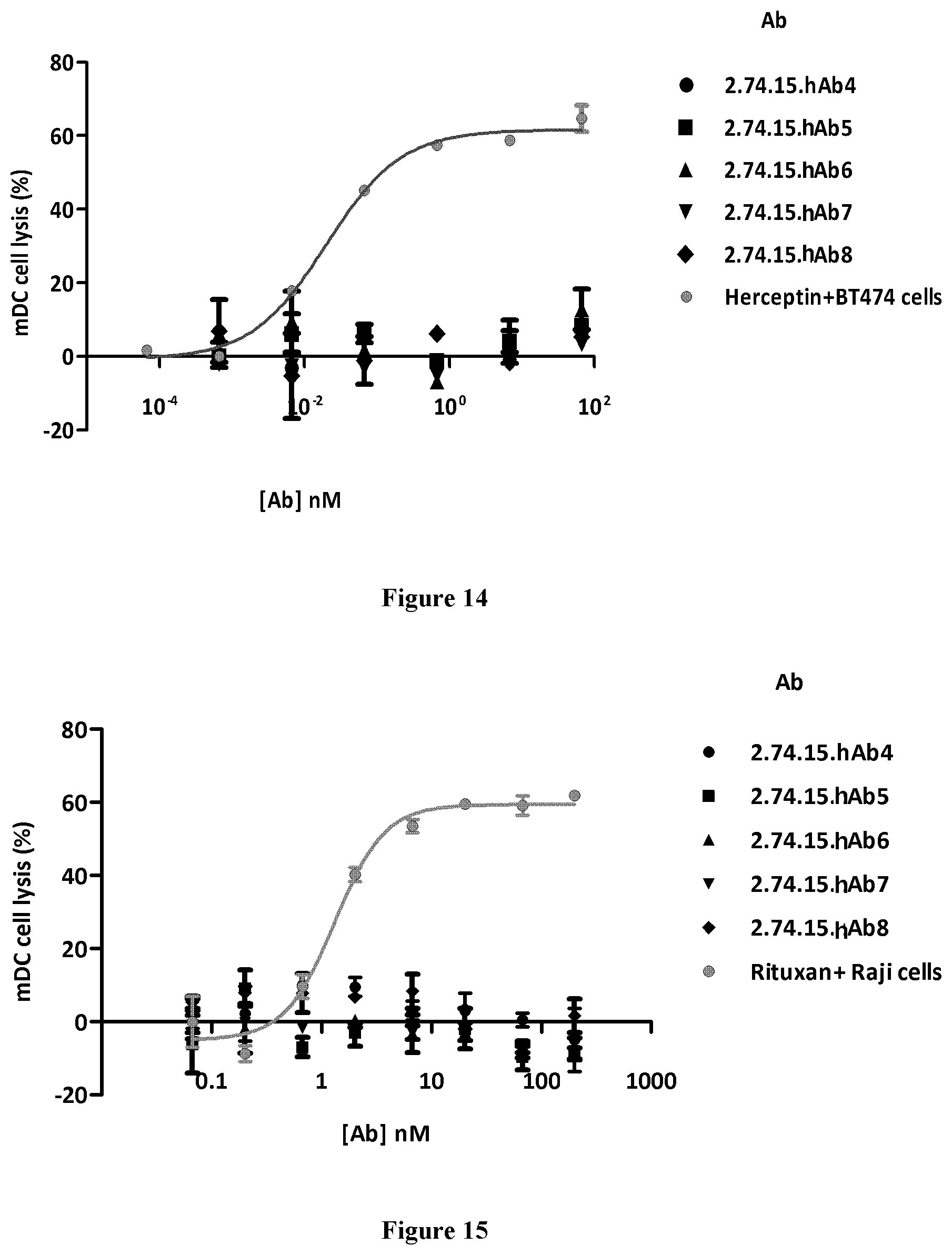
S00001
XML
uspto.report is an independent third-party trademark research tool that is not affiliated, endorsed, or sponsored by the United States Patent and Trademark Office (USPTO) or any other governmental organization. The information provided by uspto.report is based on publicly available data at the time of writing and is intended for informational purposes only.
While we strive to provide accurate and up-to-date information, we do not guarantee the accuracy, completeness, reliability, or suitability of the information displayed on this site. The use of this site is at your own risk. Any reliance you place on such information is therefore strictly at your own risk.
All official trademark data, including owner information, should be verified by visiting the official USPTO website at www.uspto.gov. This site is not intended to replace professional legal advice and should not be used as a substitute for consulting with a legal professional who is knowledgeable about trademark law.