Ligation assays in liquid phase
Stevens , et al.
U.S. patent number 10,683,534 [Application Number 15/387,650] was granted by the patent office on 2020-06-16 for ligation assays in liquid phase. This patent grant is currently assigned to BioSpyder Technologies, Inc.. The grantee listed for this patent is BioSpyder Technologies, Inc.. Invention is credited to Joel McComb, Bruce Seligmann, Anthony Stevens, Joanne M. Yeakley.


View All Diagrams
| United States Patent | 10,683,534 |
| Stevens , et al. | June 16, 2020 |
Ligation assays in liquid phase
Abstract
Ligation assays in liquid phase for detecting nucleic acid sequences.
| Inventors: | Stevens; Anthony (San Diego, CA), Seligmann; Bruce (Tuscon, AZ), Yeakley; Joanne M. (Encinitas, CA), McComb; Joel (Rancho Santa Fe, CA) | ||||||||||
|---|---|---|---|---|---|---|---|---|---|---|---|
| Applicant: |
|
||||||||||
| Assignee: | BioSpyder Technologies, Inc.
(Carlsbad, CA) |
||||||||||
| Family ID: | 58499706 | ||||||||||
| Appl. No.: | 15/387,650 | ||||||||||
| Filed: | December 22, 2016 |
Prior Publication Data
| Document Identifier | Publication Date | |
|---|---|---|
| US 20170101671 A1 | Apr 13, 2017 | |
Related U.S. Patent Documents
| Application Number | Filing Date | Patent Number | Issue Date | ||
|---|---|---|---|---|---|
| PCT/US2016/014999 | Jan 26, 2016 | ||||
| 14788670 | Jun 30, 2015 | ||||
| 15387650 | |||||
| 14788670 | Jun 30, 2015 | ||||
| 62108161 | Jan 27, 2015 | ||||
| Current U.S. Class: | 1/1 |
| Current CPC Class: | C12Q 1/6827 (20130101); C12Q 1/6827 (20130101); C12Q 2521/325 (20130101); C12Q 2525/155 (20130101); C12Q 2525/161 (20130101); C12Q 2525/185 (20130101); C12Q 2525/186 (20130101); C12Q 2533/101 (20130101); C12Q 2533/107 (20130101); C12Q 2535/122 (20130101); C12Q 2565/629 (20130101); C12Q 1/6827 (20130101); C12Q 2521/307 (20130101); C12Q 2521/325 (20130101); C12Q 2525/155 (20130101); C12Q 2525/161 (20130101); C12Q 2525/185 (20130101); C12Q 2525/186 (20130101); C12Q 2533/101 (20130101); C12Q 2533/107 (20130101); C12Q 2535/122 (20130101); C12Q 2565/629 (20130101); C12Q 1/6827 (20130101); C12Q 2521/319 (20130101); C12Q 2525/186 (20130101); C12Q 2525/197 (20130101); C12Q 2525/207 (20130101); C12Q 2561/125 (20130101); C12Q 2563/149 (20130101) |
| Current International Class: | C12Q 1/68 (20180101); C07H 21/00 (20060101); C12Q 1/6827 (20180101) |
References Cited [Referenced By]
U.S. Patent Documents
| 4988617 | January 1991 | Landegren |
| 5225324 | July 1993 | McFadden |
| 5256775 | October 1993 | Froehler |
| 5691146 | November 1997 | Mayrand |
| 5728527 | March 1998 | Singer |
| 5871921 | February 1999 | Landegren |
| 6027889 | February 2000 | Barany et al. |
| 6368801 | April 2002 | Faruqi |
| 7312039 | December 2007 | Barany et al. |
| 7320865 | January 2008 | Barany et al. |
| 7429453 | September 2008 | Barany et al. |
| 8597891 | December 2013 | Barany et al. |
| 8741564 | June 2014 | Seligmann |
| 2002/0034762 | March 2002 | Muller |
| 2002/0102591 | August 2002 | Sorge |
| 2003/0083273 | May 2003 | Woolf |
| 2003/0186234 | October 2003 | Kurn |
| 2004/0137484 | July 2004 | Zhang |
| 2005/0026166 | February 2005 | Bi |
| 2006/0099615 | May 2006 | Horiuchi |
| 2006/0246475 | November 2006 | Peterson et al. |
| 2006/0281098 | December 2006 | Miao et al. |
| 2007/0065816 | March 2007 | Dong |
| 2007/0224620 | September 2007 | Hartzell |
| 2007/0275375 | November 2007 | Van Eijk |
| 2008/0108073 | May 2008 | Nautiyal |
| 2008/0182239 | July 2008 | Mullinax |
| 2008/0305478 | December 2008 | Chun |
| 2009/0203085 | August 2009 | Kurn |
| 2010/0190167 | July 2010 | Getts |
| 2013/0059784 | March 2013 | Wilson |
| 2013/0137094 | May 2013 | Espina |
| 2013/0210078 | August 2013 | Nelson |
| 2013/0338022 | December 2013 | Kuersten et al. |
| 2014/0141418 | May 2014 | Park |
| 2014/0171338 | June 2014 | Terbrueggen et al. |
| 2014/0227683 | August 2014 | Cobb |
| 2014/0227691 | August 2014 | May |
| 2014/0243240 | August 2014 | Soldin |
| 2014/0287468 | September 2014 | Richard |
| 2015/0038336 | February 2015 | Barany |
| 2015/0360193 | December 2015 | Fan |
| 2016/0046984 | February 2016 | Nguyen |
| 2016/0068886 | March 2016 | Yeakley |
| 2016/0068907 | March 2016 | Shepard et al. |
| 2016/0222447 | August 2016 | Stevens et al. |
| 2018/0111959 | April 2018 | Chiou |
| 2914367 | Dec 2014 | CA | |||
| 1500710 | Jan 2005 | EP | |||
| 1975254 | Jan 2008 | EP | |||
| 2 542 929 | Feb 2017 | GB | |||
| WO 97/23647 | Jul 1997 | WO | |||
| WO 2008/094902 | Aug 2008 | WO | |||
| WO 2016/123154 | Aug 2016 | WO | |||
| WO 2017/019481 | Feb 2017 | WO | |||
Other References
|
Ahern , H. The Scientist 9(15) : 20 (1995). cited by examiner . Hwang et al., BioTechniques 35 (6) : 1180 (2003). cited by examiner . Larman et al., Nucleic Acids Research 42 (14) : 9146 (Aug. 2014 (2014). cited by examiner . Li et al. Current Protocols in Molecular Biology 4.13.1-4.13.9 (Apr. 2012). cited by examiner . Nakahara et al., J. of Virological Methods 77: 47 (1999). cited by examiner . Nanostring Technology Technical Note (2009). cited by examiner . Nilsson et al., Science 265 :2085 (1994). cited by examiner . Armitage , B. Photocleavage of Nucleic Acids.Chem. Rev. 98 :1171 (Year: 1998). cited by examiner . Xu et al.,Nonenzymatic autoligation in direct three-color detection of RNA and DNA point mutations. Nature Biotechnology 19:148 (Year: 2001). cited by examiner . Yoshimura et al., Ultrafast Reversible Photo-Cross-Linking Reaction: Toward in Situ DNA Manipulation. Organic Letters 10(15) :3227 (Year: 2008). cited by examiner . International Search Report and Written Opinion mailed by ISA/US dated Apr. 14, 2016 for related application PCT/US16/14999. cited by applicant . Barany, F. The ligase chain reaction in a PCR world. PCR Methods and Applications 1 : 5-16 (1991). cited by applicant . Cai et al., Human microRNAs are processed from capped, polyadenylated transcripts that can also function as mRNAs.RNA 10 : 1957 (2004). cited by applicant . Hsuih et al., Novel, Ligation-Dependent PCR Assay for Detection of Hepatitis C Virus in Serum. J. of Clinical Microbiology34 (3) : 501 (1996). cited by applicant . Pritchard et al., MicroRNA profiling : approaches and considerations. Nature Reviews Genetics 13 :358 (2012). cited by applicant . Schouten et al., Relative quantification of 40 nucleic acid sequences by multiplex ligation-dependent probe amplification. Nucleic Acids Research 30 (12) : e57 (2002). cited by applicant . Shevelev et al., The 3'-5' Exonucleases. Nature Reviews Molecular Cell Biology 3 :1 (2002). cited by applicant . Shi et al.,Facile means for quantifying microRNA expression by real-time for PCR. BioTechniques 39 (4) : 519 (2005). cited by applicant . Vallone et al., qPCR Workshop hald Jul. 26-27, 2006.at NFSTC--Presentation slides. cited by applicant . Vrettou et al., Real-Time PCR for Single-Cell Genotyping in Sickle Cell and Thalassemia Syndromes as a Rapid, Accurate, Reliable, and Widely Applicable Protocol for Preimplantation Genetic Diagnosis. Human Mutation 23 : 513 (2004). cited by applicant . Wiedmann et al., Ligase chain reaction (LCR)--overview and applications. PCR Methods and Applications 1 : 5-16 (1991). cited by applicant . Replacement claims for foreign counterpart GB 1 614 871.0, filed in UK IPO, Aug. 31, 2016. cited by applicant . First Examination Report, dated Oct. 14, 2016. cited by applicant . Response to first Examination Report, dated Dec. 13, 2016, with 2nd set of replacement claims. cited by applicant . Second Examination Report, dated Feb. 27, 2017. cited by applicant . Search & Exam. Report dated Jun. 12, 2017 in UK App. GB 1707052.5. cited by applicant . Response dated Dec. 12, 2017 in UK App. GB 1707052.5. cited by applicant . Search & Exam. Report dated Jan. 25, 2019 in UK App. GB 1707052.4. cited by applicant . Response dated May 23, 2019 in UK App. GB 1707052.5. cited by applicant . Search & Exam. Report dated Jun. 21, 2019 in UK App. GB 1707052.5. cited by applicant . Communication in EP App. 16 744 000.7 dated Jun. 28, 2018. cited by applicant. |
Primary Examiner: Whisenant; Ethan C
Government Interests
STATEMENT OF GOVERNMENT SUPPORT
This invention was made with government support under grant 1R43HG007815 awarded by the National Institutes of Health. The government has certain rights in the invention.
Parent Case Text
CROSS-REFERENCE TO RELATED APPLICATIONS
This application is a continuation-in-part of international application PCT/US16/14999, filed Jan. 26, 2016 and published as WO 2016/123154, which is a continuation-in-part of Ser. No. 14/788,670, filed Jun. 30, 2015 and published as US 2016 0222447, which claims the benefit of priority of U.S. provisional application Ser. 62/108,161, filed Jan. 27, 2015.
This application is also a continuation-in-part of Ser. No. 14/788,670, filed Jun. 30, 2015 and published as US 2016 0222447, which claims the benefit of priority of U.S. provisional application Ser. 62/108,161, filed Jan. 27, 2015.
Claims
We claim:
1. A method for detecting target nucleic acid sequences in a sample, wherein a target sequence has a downstream region (DR) and an upstream region (UR), comprising (a) contacting the sample with a pair of detector oligos, which pair comprises a downstream detector oligo (DD) having a complementary downstream region (DR') and a separate upstream detector oligo (UD) having a complementary upstream region (UR'), wherein at least one of the DD or UD contains DNA and RNA, thereby allowing the pair of detectors to hybridize specifically to the target nucleic acids; (b1) ligating the DR' and UR' if both are specifically hybridized to the DR and UR of a target sequence, wherein the ligase efficiently joins DNA to a ribonucleotide or RNA while both are hybridized to an RNA template; (b2) exposing hybridization complexes to at least one nuclease; and whereby the ligation product indicates the presence of the target sequence in the sample.
2. The method of claim 1, downstream region (DR) and an upstream region (UR), comprising wherein at least one of the DD or UD has a second complementary region (DR2' or UR2') separated from the DR' or UR' by a noncomplementary region (CP1) that does not hybridize to the target nucleic acid, whereby the DR2' or UR2' can specifically hybridize to a DR2 or UR2 of the target nucleic acid.
3. The method of claim 1, wherein step (a), (b1), or both are performed in liquid phase.
4. The method of claim 1, wherein step (a), (b1) or both are performed while the target nucleic acid is attached to a solid surface.
5. The method of claim 1, wherein sets of DDs and UDs, having different DRs or URs, provide measurement of a portion of the target sequence that potentially harbors multiple mutations.
6. The method of claim 1, wherein the target nucleic acid is an RNA selected from the group comprising mRNA, siRNA, antisense RNA, long noncoding RNA, circular RNA and microRNA, or portion thereof.
7. The method of claim 6, wherein the microRNA or portion thereof is 3'-polyadenylated prior to step (a) and the DD has a poly-T region adjacent to the DR'.
8. The method of claim 1, wherein the sample is a tissue sample, mounted on a slide, fixed or a formalin-fixed, paraffin-embedded (FFPE) sample.
9. The method of claim 1, further comprising the step of permeabilizing the cell walls, cell membranes, or subcellular structures.
10. The method of claim 1, wherein the target nucleic acid is in situ.
11. The method of claim 10, wherein the ligation product is cross-linked in situ.
12. The method of claim 11, wherein the cross-linking is reversible.
13. The method of claim 12, wherein the reversible cross-linking is photocleavable.
14. The method of claim 1, wherein the DD has a first amplification region (P1) downstream of the DR', and the UD has a second amplification region (P2') upstream of the UR'.
15. The method of claim 1, wherein a detector has a photocleavable site.
16. The method of claim 1, wherein the method further comprises any of (a1) imaging or detecting another analyte at or near the location of the target nucleic acid in situ; (a2) before the ligation step, performing a wash step; (b3) performing a photocleavage step; (c1) after the ligation step, adding an EDTA solution; (c2) after the ligation step, performing a wash step; (d) amplifying the ligation products; where the DR and UR are separated by at least one nucleotide, (b0) extending the DR' using the sample as template; or (e) detecting the ligation or amplification product.
17. The method of claim 1, further comprising the step of eluting the ligation product.
18. The method of claim 17, wherein the elution is effected by addition of a base.
19. The method of claim 17, wherein the elution is effected by low salt concentration.
20. The method of claim 1, wherein the sample is from a tissue culture.
21. The method of claim 1, wherein the 3' end of the DD has one or more ribonucleotides.
22. The method of claim 1, wherein a UD is phosphorylated at the 5' end.
23. The method of claim 1, further comprising the step of (b3) releasing ligation products from RNA targets.
24. The method of claim 21, wherein a UD is phosphorylated at the 5' end.
25. The method of claim 21, further comprising the step of (b3) releasing ligation products from RNA targets.
26. The method of claim 22, further comprising the step of (b3) releasing ligation products from RNA targets.
27. The method of claim 26, wherein the 3' end of the DD has one or more ribonucleotides.
Description
The contents of the aforementioned applications are incorporated herein in their entirety.
TECHNICAL FIELD
This invention relates to molecular biology, and more particularly to assays for detecting nucleic acid sequences in samples.
SUMMARY OF THE INVENTION
This invention provides methods for detecting target sequences of nucleic acid sequences of interest in a sample, and also provides kits for performing the method.
In a typical ligation assay, the sample is contacted with a pool of detector oligos, where a downstream detector (DD) and an upstream detector (UD) are provided for each target sequence. A portion (DR') of the DD is complementary to a region of the target sequence designated as a downstream region (DR). The upstream detector has a portion (UR') complementary to an upstream region (UR) of the target sequence.
The downstream and upstream detectors are contacted with the sample and allowed to hybridize to the corresponding regions of target sequence present in the sample. When the detectors are specifically hybridized to a target sequence, they can be ligated at the junction between adjacent detectors, whether directly or after an optional extension step. Formation of a ligation product thus serves as evidence that the target sequence was present in the sample, and the ligation product can be detected by various methods such as detectable labels, microarrays, qPCR, flow-through counters, and sequencing.
The invention provides assays where one or more nucleases are provided during steps in the method to selectively degrade unused or excess detectors, or detectors that are not specifically hybridized to target sequences. Accordingly, the detectors and other components of the assay are configured in a number of embodiments to resist the nucleases while detecting target sequences. The configurations enable sensitive detection of nucleic acids, such as mRNAs and miRNAs, at whole-transcriptome or -miRNome multiplexing and at the level of single cells. Moreover, the steps can be performed in a single well or container without the need for transfers, separation, or solid-phase immobilization, and are therefore ideal for microfluidic platforms.
BRIEF DESCRIPTION OF THE DRAWINGS
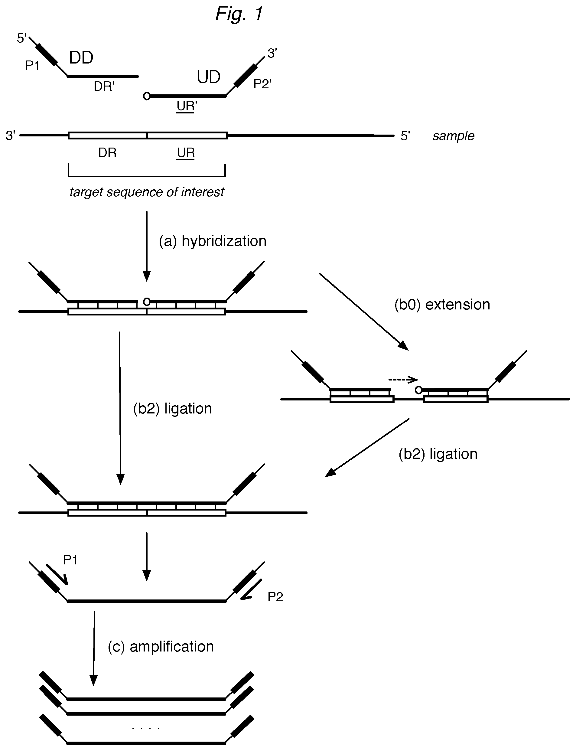
FIG. 1 illustrates a representative ligation assay for detecting target nucleic acid sequences. Briefly, downstream detector (DD) and upstream detector (UD) probe oligonucleotides are allowed to (a) hybridize to a target sequence, having DR and UR regions, in a sample. For convenience of identification, upstream regions are often underlined herein. While hybridized to the DR and UR of the target sequence, the DD is (b2) ligated selectively to the UR. Optionally, the DD is (b0) extended prior to (b2) ligation. The ligation product is optionally (c) amplified via amplification regions P1 and P2' by one or more primers, such as P1 and P2.
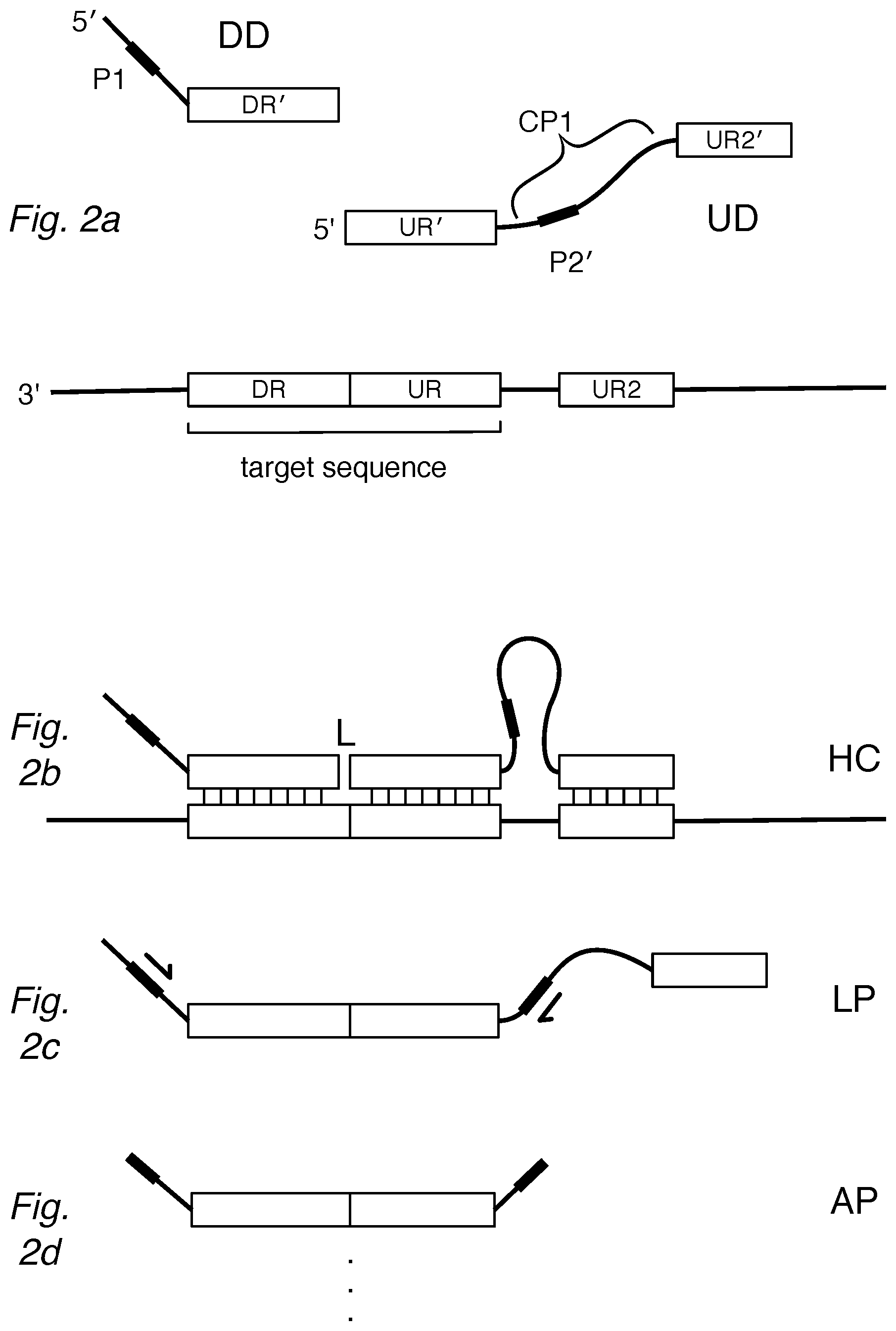
FIG. 2a shows an "anchored" assay design of the invention where the UD is configured with a second complementary region (UR2' or "anchor") separated by a noncomplementary region (CP1). The DD and UD can hybridize to a target sequence as in FIG. 2b, forming a hybridization complex (HC) providing a substrate for ligation at the junction (L) between DR' and UR'. After ligation, FIG. 2c shows the ligation product (LP) can be amplified by primers to yield amplification products (AP) in FIG. 2d.
Treatment with an exonuclease, such as an exonuclease with single-stranded 3'-to-5' activity, can be used at various stages of the method to remove undesired components, such as nonbound or excess DD and UD detectors as in FIG. 2e. Detectors that are nonspecifically or incompletely hybridized to target sequences can be degraded by the exonuclease or will not result in ligation or amplification product, as in FIG. 2f.
As shown in FIG. 2g, it may be desirable to provide predetermined quantities of attenuator oligonucleotides such as UR2' (or alternatively UR2) to lessen the formation of product resulting from certain high-abundance target sequences (HATs).
FIG. 2h shows a pair of detectors that are configured to have a modification at one end to resist exonucleases that degrade single-stranded (ss) DNA. The UD has a modification at the 3' end that resists degradation of the detector by an exonuclease having 3' activity on DNA single strands. Alternatively, the DD can have a 5' modification to resist degradation by a 5'-ss-exonuclease.
FIGS. 2i and 2j illustrate detectors that are configured to resist exonucleases by being hybridized to a protector oligo, such as ones having sequence DR2 or UR2 that bind to corresponding DR2' and UR2' sequences of the detectors, presenting double-stranded structures at either end. The protectors can themselves be 5'- or 3'-modified to resist exonucleases, as shown. FIG. 2j also illustrates a target sequence (3'-DR-UR-5') that is relatively short, such as a microRNA, where the target has been polyadenylated at its 3' end. The DD features a complementary poly-T portion adjacent to the DR'.
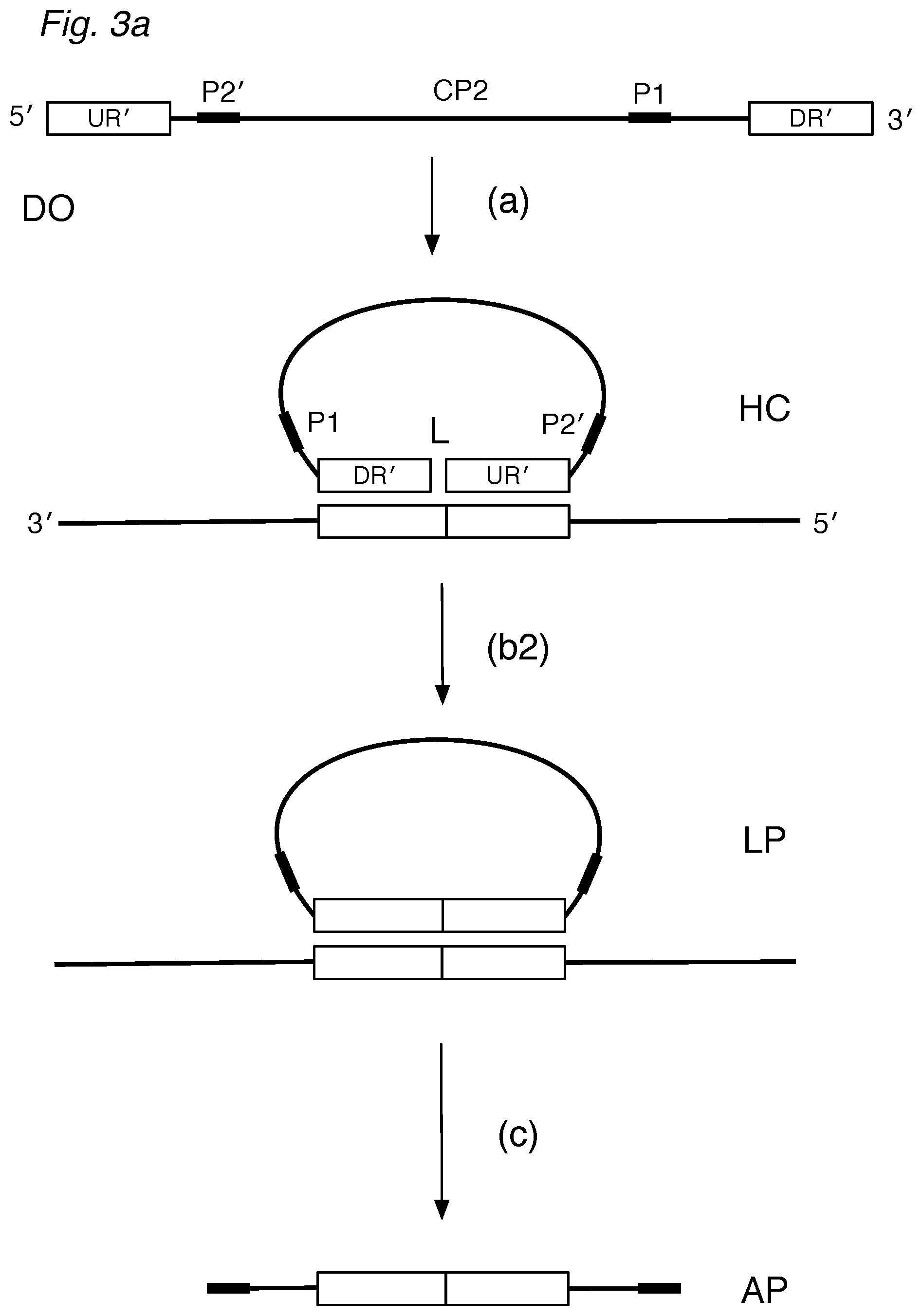
FIG. 3a depicts a circularizable assay design of the invention using a detector oligo probe (DO) that can (a) hybridize via DR' and UR' regions to a target sequence, forming a (noncovalently) circularized structure. After treatment with a nuclease and ligase, a circularized ligation product can then be (c) amplified. FIGS. 3b, 3c, and 3d illustrate partially hybridized DO detectors, detectors hybridized to non-target sequences, or nonspecifically hybridized detectors, which can be digested by nucleases or be unsuitable for exponential amplification.
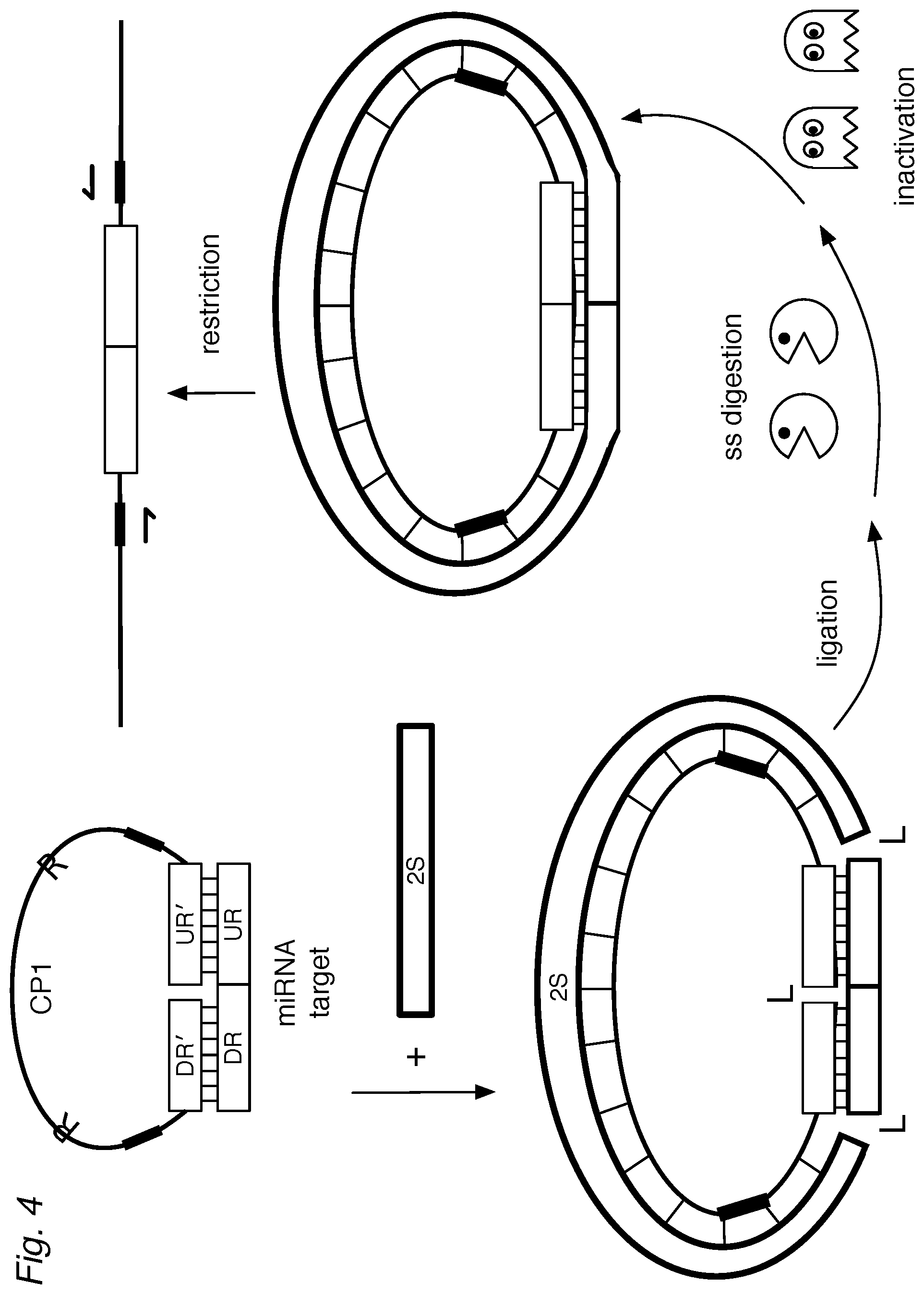
FIG. 4 shows an assay of the invention where a (universal) second strand (2S) is provided during hybridization so that the target (DR-UR), DO, and the 2S form a circularized, double-stranded structure. Treatment with ligase results in a covalently circularized ligation product. Optionally, ss-nucleases can be used to degrade excess detectors and hybridization complexes that are not specific for the target. The nucleases can be inactivated. If desired, the circularized structure can be linearized, for example by a restriction endonuclease.
FIG. 5a shows a detailed view of a hybridization complex using a variant circularizable DO having a short noncomplementary flap (CP5) on its 5' end, and optionally a short noncomplementary sequence (CP3) on the 3' end. FIG. 5b shows the hybridization complex after the CP5 is removed by a flap nuclease, such as Fen-1. If desired, the 5' end can be phosphorylated, as in FIG. 5c. FIG. 5d illustrates how CP3 can fill in the gap left by Fen-1, so that the DO can be ligated into circularized form as in FIG. 5e. The noncomplementary CP5 and/or CP3 flaps can be incorporated in any of the DD and UD designs.
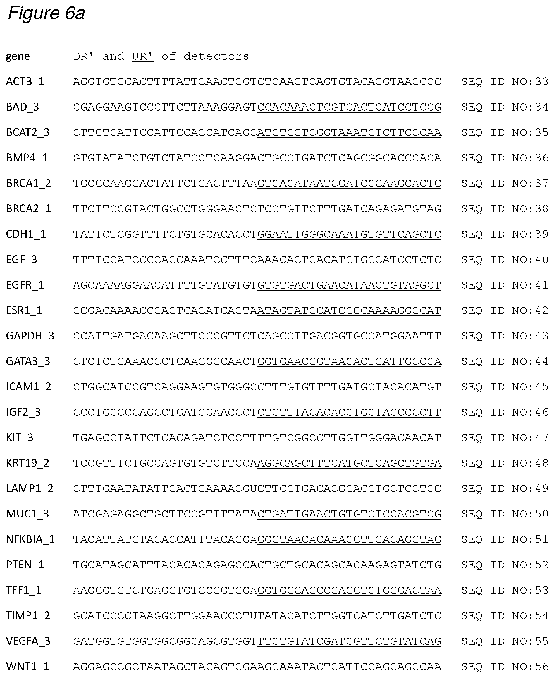
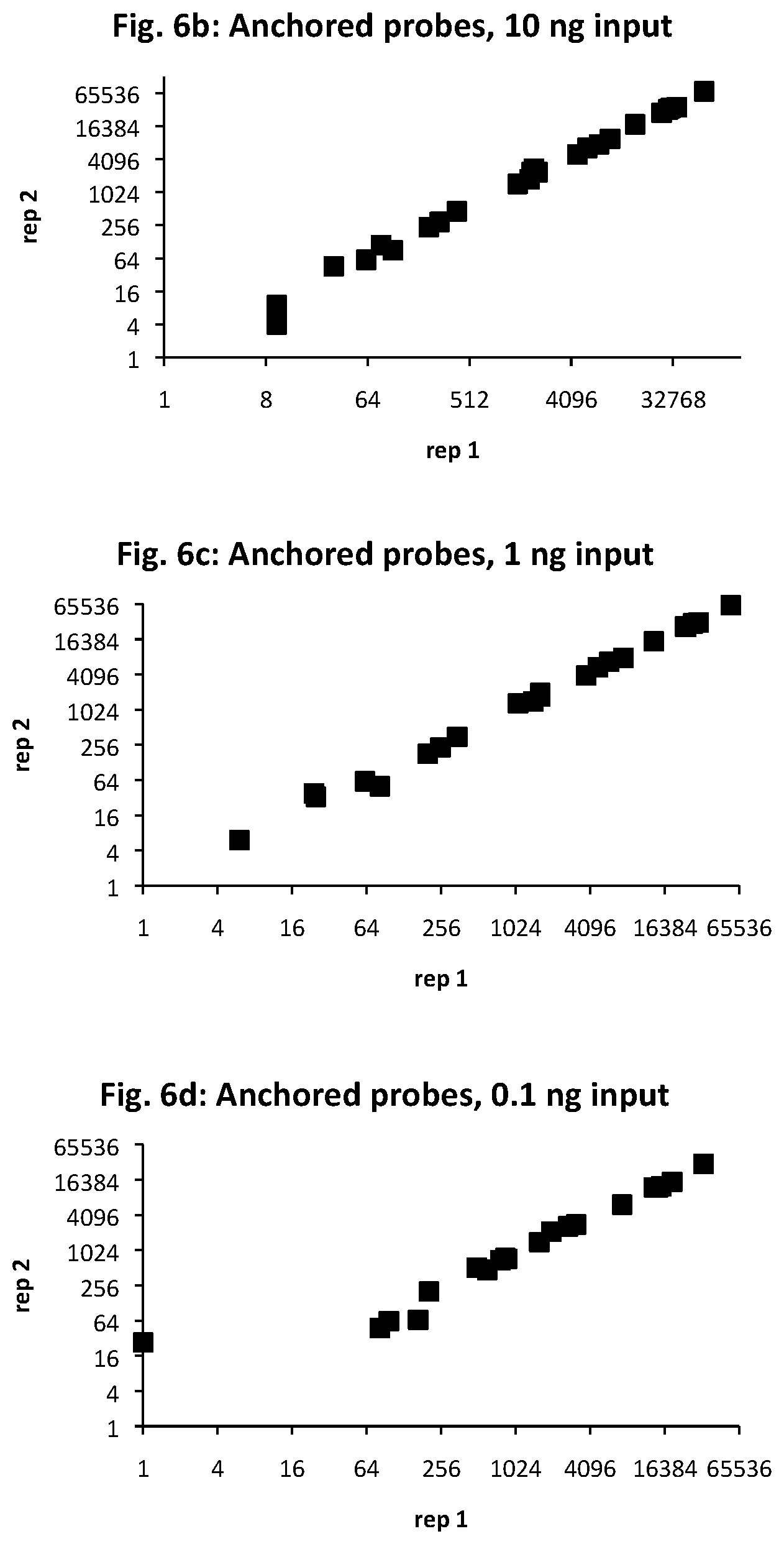
FIG. 6a provides target sequences (SEQ ID NOs: 33-56) used to design detectors for mRNA expression products for 24 human genes of interest. The genes were selected to demonstrate detection over an expected range of 6 orders of magnitude in abundance, with 10, 1, and 0.1 ng sample RNA input. The number of amplified ligation products, confirmed by sequencing, are shown for anchored detector designs (FIGS. 6b, 6c, and 6d) and circularizable designs (FIGS. 6e, 6f, and 6g). The x-axis is for the first technical replicate; the y-axis is for the second replicate.
DETAILED DESCRIPTION OF THE INVENTION
Ligation Assays, Generally
A typical ligation assay is illustrated schematically in FIG. 1, which is discussed in more detail in Example 1. A sample that may contain target sequences is contacted with a pool of detector oligonucleotide probes ("probes" or "detectors"). For each target sequence, a pair of detectors is provided: a downstream detector (DD) and an upstream detector (UD). A downstream detector can have a portion (DR') that is complementary to a region of the target sequence designated as a downstream region (DR). An upstream detector can have a portion (UR') that is complementary to a region of the target sequence designated as the upstream region (UR). Here, the terms "downstream" and "upstream" are used relative to the 5'-to-3' direction of transcription when the target sequence is a portion of an mRNA, and for convenience the regions designated as upstream are often shown underlined.
As shown in FIG. 1, the DR' of the DD and the UR' of the UD for each target sequence are allowed to hybridize to the corresponding DR and UR of the target sequence, if present in the sample. When the DR and UR of a target sequence are adjacent and the DR' and UR' of the pair of detector oligos are specifically hybridized to the target sequence to form a hybridization complex, the adjacent detectors DD and UD can be ligated. Thus, formation of a DD-UD ligation product serves as evidence that the target sequence (DR-UR) was present in the sample. In cases where the DR and UR of a target sequence are separated by at least one nucleotide, the ligation step can be preceded by (b0) extending the DR' using the sample as a template so the extended DR' and UR' become adjacent and can be ligated. The ligation product can then be detected by a variety of means; if desired, the products can be amplified prior to detection.
The present invention provides methods where hybridization complexes are exposed at one or more steps to at least one nuclease that can degrade single strands of nucleic acid. As discussed in more detail below, the invention provides detectors and other components of the assay that are configured to selectively resist the nucleases when detecting target sequences. The nucleases can degrade excess or unused detectors, or detectors that are nonspecifically or nonproductively bound to components in the sample that are not of interest. The strategic use of nucleases enables the ligation assay to be performed by adding one reagent after another in a single reaction container, starting with the sample.
Samples
The samples used in the method can be any substance where it is desired to detect whether a target sequence of a nucleic acid of interest is present. Such substances are typically biological in origin, but can be from artificially created or environmental samples. Biological samples can be from living or dead animals, plants, yeast and other microorganisms, prokaryotes, or cell lines thereof. Particular examples of animals include human, primates, dog, rat, mouse, zebrafish, fruit flies (such as Drosophila melanogaster), various worms (such as Caenorhabditis elegans) and any other animals studied in laboratories or as animal models of disease. The samples can be in the form of whole organisms or systems, tissue samples, cell samples, subcellular organelles or processes, or samples that are cell-free, including but not limited to solids, fluids, exosomes and other particles. Particular examples are cancer cells, induced pluripotent stem cells (iPSCs), primary hepatocytes, and lymphocytes and subpopulations thereof. The samples can be provided in liquid phase, such as cell-free homogenates or liquid media from tissue cultures, or nonadherent cells in suspension, tissue fragments or homogenates, or in solid phase, such as when the sample is mounted on a slide or in the form of formalin-fixed paraffin-embedded (FFPE) tissue or cells, as a fixed sample of any type, or when cells are grown on or in a surface, as long as detectors can be put into contact for potential hybridization with the sample nucleic acids.
Nucleic Acids
The nucleic acids of interest to be detected in samples include the genome, transcriptome, and other functional sets of nucleic acids, and subsets and fractions thereof. The nucleic acids of interest can be DNA, such as nuclear or mitochondrial DNA, or cDNA that is reverse transcribed from RNA. The sequence of interest can also be from RNA, such as mRNA, rRNA, tRNA, siRNAs (e.g., small interfering RNAs, small inhibitory RNAs, and synthetic inhibitory RNAs), antisense RNAs, circular RNAs, or long noncoding RNAs, circular RNA, or modified RNA, and can include unnatural or nonnaturally occurring bases. The nucleic acids can include modified bases, such as by methylation, and the assay is designed to detect such modifications. The nucleic acid of interest can be a microRNA (miRNA) at any stage of processing, such as a primary microRNA (pri-miRNA), precursor microRNA (pre-miRNA), a hairpin-forming microRNA variant (miRNA*), or a mature miRNA. Detection of microRNAs is discussed in Example 3a.
Relatively short nucleic acids of interest, such as mature miRNAs, can be lengthened to enhance hybridization to the detectors. For example, many microRNAs are phosphorylated at one end, and can be lengthened by chemical or enzymatic ligation with a supplementary oligo. The supplemental oligo can be single-stranded, double-stranded, or partially double-stranded, depending on the ligation method to be used. If desired, the supplemental oligo can be unique to each target sequence, or can be generic to some or all of the target sequences being ligated. The detectors can then be designed with extended DR' and/or UR' regions that include a portion that hybridizes to the supplemental sequence. A target sequence can also be supplemented by adding nucleotides, such as by polyadenylation, where the extended detectors include at least a portion to hybridize to the supplemental polyA tail. Detection of a family of mature miRNA sequences using extended detectors is discussed in Example 3b and illustrated in FIG. 2j.
The amount of nucleic acid in the sample will vary on the type of sample, the complexity, and relative purity of the sample. Because of the sensitivity of the assay, the sample can be taken from a small number of cells, for example from fewer than 100,000, 10,000, 1000, 100, 50, 20, 10, 5, or even from a single cell or a subcellular portion of a cell. The total amount of nucleic acid in the sample can also be quite small: less than 100, 50, 20, 10, 5, 2, 1 micrograms, 500, 200, 100, 50, 20, 10, 5, 2, 1, 0.5, 0.2, 0.1 nanogram, 50, 20, 10, 5, 2, 1 picogram or less of nucleic acid (see FIG. 6d), or less than 10, 1, 0.1, 0.01, 0.001 picograms of nucleic acid, or amount of a lysate containing equivalent amounts of nucleic acid. The copy number of a particular target sequence can be less than 100,000, 10,000, 1000, 100, 50, 20, 10, 5, or even a single copy present in the sample, particularly when coupled with representative amplification of the ligation product for detection. The amount of input nucleic acid will also vary, of course, depending on the complexity of the sample and the number of target sequences to be detected.
Detectors
Based on the particular target sequences, the invention provides pools of detector oligos where a target sequence has a pair of upstream and downstream detectors (UD and DD) that correspond to DR and UR, which are typically subsequences of the entire nucleic acid sequence of interest. Detector oligos can be designed to hybridize to the target sequence so a single-stranded sequence portion of the target sequence remains between the detectors, which can then be filled in, such as by reverse transcriptase or polymerase, thereby extending a detector to bring it effectively together with the other detector so they can be ligated. Detectors can be provided to detect targets that contain mutations including individual single-nucleotide polymorphisms (SNPs), gene fusions, and exon-splicing variants, or modifications such as methylation. Detectors can contain blocking groups, modified linkages between bases, unnatural or nonnaturally occurring bases or other unnatural or nonnaturally occurring components. An individual target sequence can have more than one set of DRs and URs, which can be selected by the user to optimize the performance of the assay. Multiple sets of DRs and URs can provide multiple measurements of the same target sequence or of different portions of the target sequence, such as different exons or exon junctions, or provide measurement of a portion of sequence that is not mutated versus a portion of sequence that may harbor a mutation.
Target Sequences
The target sequences can be selected from any combination of sequences or subsequences in the genome or transcriptome of a species or an environment, or modified nucleic acids or nucleic acid mimics to which the detector oligos can bind or hybridize. The set can be specific for a sample type, such as a cell or tissue type. For some sample types, the number of target sequences can range in any combination of upper and lower limits of 1, 2, 5, 10, 20, 50, 100, 200, 500, 1000, 2000, 5000, 10,000, 20,000, 23,000, 30,000, 38,000, 40,000, 50,000, or more. The number of target sequences can also be expressed as a percentage of the total number of a defined set of sequences, such as the RNAs in the human transcriptome or genes in the human genome, ranging in any combination of upper and lower limits of 0.1%, 0.2%, 0.5%, 1%, 2%, 5%, 10%, 20%, 25%, 30%, 35%, 40%, 45%, 50%, 65%, 60%, 70%, 75%, 80%, 85%, 90%, 95%, and 100%. Where large sets of detector oligos are used, it can be useful to check the full sequence of each oligo for potential cross-hybridization to other oligos in the set, where, for example, one oligo may inadvertently serve as an template to other detectors. While such non-specific artifacts can be identified by sequence, and are typically discarded from detection results, they may represent noninformative hybridization events that compete for reaction resources.
The detector oligos themselves can be DNA, RNA, or a mixture or hybrid of both. If desired, they can have a modified nucleotide such as dideoxy nucleotides, deoxyUridine (dU), 5-methylCytosine (5mC), 5-hydroxymethylCytosine (5hmC), 5-formylCytosine (5fC), 5-carboxylCytosine (5caC), and Inosine. Yet other modifications to detector oligos include modified bases such as 2,6-diaminopurine, 2-aminopurine, 2-fluro bases, 5-bromoUracil, or 5-nitroindole. Other detector oligos can have a modified sugar-phosphate backbone at one or more positions. Such modifications include a 3'-3' or 5'-5' linkage inversion, a locked nucleic acid (LNA), or a peptide nucleic acid (PNA) backbone. LNAs can be useful for their stronger hybridization properties to complementary bases, enhancing the selectivity or the overall binding affinity for the detector oligo as a whole. The modified bases or bonds can also be used at positions 1, 2, or 3 away from the point of ligation.
As shown schematically in FIG. 1, a downstream detector (DD) has a complementary downstream region (DR'), which can be at least 4, 6, 8, 10, 12, 14, 16, 18, 20, 22, 24, 26, 28, 30, 35, 40, 45, or 50 nucleotides in length. Similarly, an upstream detector (UD) has a complementary upstream region (UR'), which can be at least 4, 6, 8, 10, 12, 14, 16, 18, 20, 22, 24, 26, 28, 30, 35, 40, 45, or 50 nucleotides in length. In a given pair of DD and UD for a target sequence, the DR' and UR' need not be exactly the same length, but will typically be similar so they can hybridize to the target under similar conditions and stringency.
As discussed in more detail below, the detectors can be optimized for ligation, such as by providing a 5'-phosphate on the UD, although this is not necessary, depending on the selection of ligase or other ligation methods. Ribonucleotides can also be substituted at the ligatable ends of the DD and UD to increase the specificity and efficiency of ligation, as when an RNA ligase is used.
Detector Labels
Where the ligation assay proceeds directly to a detection step, either or both detectors can be designed to be labeled appropriately for detection. For example, the detector can be conjugated to any number of molecular or physical entities, labeled with a crosslinker, activatable crosslinker, activatable cleavage group or enzymatically cleavable group, optical, color or fluorescent dye, latex or other beads, quantum dots, or nanodots, or nanoparticles. Any of these entities can also be further modified or conjugated to other entities. The label can also take the form of an additional nucleotide sequence that serves to enable detection and identification, such as a barcode sequence. For example, a useful barcode sequence can uniquely identify the specific gene or target sequence, or a group of select genes or target sequences within the sample that are being measured. Such sequences can be positioned between the UR' and P2' sequence, and/or between the DR' and P1 sequence, so they are amplified when using flanking primers. This sequence can also be a random sequence, useful for identifying the number of copies of the target gene in the sample, independent of the particular efficiency of any amplification step.
Hybridization
Returning to the steps of the assay, the detectors are provided so that they contact the sample to allow the detectors to hybridize specifically to the target nucleic acids. Hybridization conditions can be selected by the skilled artisan to allow and optimize for hybridization between the polynucleotides with the desired degree of specificity or mismatches, and such conditions will vary with the lengths and compositions of sequences present in the hybridization reaction, the nature of any modifications, as well as conditions such as the concentrations of the polynucleotides and ionic strength. Particular hybridization temperatures include 30.degree., 32.5.degree., 35.degree., 37.5.degree., 40.degree., 42.5.degree., 45.degree., 47.5.degree., 50.degree., 52.5.degree., 55.degree., 57.5.degree., 60.degree., 62.5.degree., 65.degree., 67.5.degree., 70.degree., 72.5.degree., 75.degree., 77.5.degree., 80.degree., 82.5.degree., 85.degree., 87.5.degree., and/or 90.degree.. Particular hybridization temperatures can be achieved by ramping the temperature up or down at various rates and profiles, such as timed temperature plateaus, one or more incremental increases or decreases of 5.degree. C., 10.degree. C., or 15.degree. C., and repeated cycling between two or more temperatures. Ions such as Li.sup.+, Na.sup.+, K.sup.+, Ca.sup.2+, Mg.sup.2+ and/or Mn.sup.2+ can also be present from 0, 1, 2, 5, 10, 20, 50, 100, 200, and 500 mM, and such ions can affect the selection of the other hybridization conditions. Hybridization is also affected by steric crowding components such as branched polysaccharides, glycerol, and polyethylene glycol. Further additives can be present in the hybridization (and subsequent) reactions, such as DMSO, non-ionic detergents, betaine, ethylene glycol, 1,2-propanediol, formamide, tetramethyl ammonium chloride (TMAC), and/or proteins such as bovine serum albumin (BSA), according to the desired specificity.
Optionally, the conditions for hybridization can be adjusted or fine-tuned to permit other steps to be performed in the same environment. For example, the same buffers used for hybridization can be used for lysing cells in a sample, promoting hybridization of certain cell types, facilitating removal or permeation of cell walls, cell membranes, or subcellular fractions, as desired. Depending on the ligation method used in the assay, hybridization conditions can be selected to be compatible with conditions for ligation as is, or with the addition of one or more components and preferably without requiring a change of the reaction container when transitioning from hybridization to ligation steps.
Ligation
The ligation reaction can occur by chemical ligation or by using a ligase enzyme or a ligation-facilitating co-factor. A variety of nick-repairing ligases are commercially available to catalyze the formation of a phosphodiester bond between adjacent single-stranded polynucleotides when hybridized to another single-stranded template, such as to join DNA to RNA when hybridized to template. An example is bacteriophage T4 DNA ligase, which is generally understood to use ATP as a co-factor. The ATP can be supplied during the ligase reaction. In other reactions, the ligase can be pre-adenylated. In yet other reactions, the UD must be pre-adenylated at the 5' end, as with a 5' App DNA/RNA ligase. The UD in a typical reaction will have a 5'-phosphate to facilitate ligation to the DD, although this is not necessary, depending on the selection of ligase and ligation conditions. (Where a 5'-phosphate on the DD is required for efficient ligation, using a comparable oligonucleotide without 5'-phosphorylation can be used to inhibit or reduce undesired ligation.) Preferred ligation conditions include 10, 25, 50, 100 mM Tris-HCl (pH 7.5, 8.0, or 8.5); at least 10 mM, 5 mM, 2 mM, 1 mM MgCl.sub.2; at least or at most 2 mM, 1 mM, 0.7 mM, 0.5 mM, 0.2 mM, 0.1 mM, 0.05 mM, 0.02 mM, 0.01 mM, 0.005 mM, 0.002 mM, or 0.001 mM ATP; or at least 10 mM, 7 mM, 5 mM, 2 mM, 1 mM, 0.5 mM DTT or other antioxidant. T3 DNA ligase can also be used, which can ligate a broader range of substrates and has a wider tolerance for salt concentration. As with other steps, the temperature can be selected according to the characteristics of the reaction components and conditions such as ionic strength.
As discussed above, the ligation step can be preceded by an optional extension step, as in FIG. 1, step (b0). The ligation step can also be preceded by an optional cleavage step, such as by a nuclease, to remove any overhangs. In other cases, a portion of the DD can overlap with the UR sequence to which the UD hybridizes, so that after hybridization of the UD and the DD, there is an overhang sequence of 1, 2, 3, or more bases. A useful enzyme for removing an overhang is a Flap endonuclease, such as Fen-1, which cleavage leaves a ligatable 5'-phosphate.
Amplification
If desired, the ligation product can be amplified (for example by PCR or qPCR) to facilitate detection. Amplification methods and instruments are commercially available, including PCR plate and droplet formats, and the amplification enzymes (such as Taq and its commercial variants) and reaction conditions can be selected and tailored to the particular platform. Optionally, the polymerase selected for amplification can have strand-displacing activity. As illustrated in Figure. 1, the detectors can have additional sequences ("tails") including primer hybridization sequences (e.g. P1, P2') or complements thereof, that serve as amplification sequences, so that after ligation, the ligation product can be amplified with a pair of amplification primers (P1, P2). An exemplary downstream amplification sequence (P1) is
TABLE-US-00001 5'-CAAGCAGAAGACGGCATACGAG-3', (SEQ ID NO: 1)
which can be used with a primer having the same sequence (P1). An exemplary upstream amplification sequence (P2') is
TABLE-US-00002 5'-ATCTCGGTGGTCGCCGTATCATT-3', (SEQ ID NO: 2)
which can be used with primer P2 (shown in 3'-to-5' orientation):
TABLE-US-00003 (SEQ ID NO: 3) 3'-TAGAGCCACCAGCGGCATAGTAA-5'.
Amplification can also be linear, or achieved by any number of methods other than PCR. If desired, the amplification primer can incorporate a barcode sequence, for example a barcode sequence that uniquely identifies the sample in a multi-sample experiment, and optionally has redundant and/or error-correction features. In some experiments, for example, different sample barcodes can be used for 96, 384, 1536, or more generally 2.sup.n or 4.sup.n different samples that are prepared with different barcodes separately for some steps, such as hybridization, ligation, and amplification, and combined for others, such as detection. The barcode sequence can be incorporated into the primer, such as 3' to the amplification sequence, so that the barcode becomes part of the amplified strand. In other instances, the amplification sequence of the primer can be extended by an additional sequence to provide a primer hybridization sequence that can be used for use in subsequent sequencing steps. The barcode may also be interposed between the amplification sequence, and if desired, the extended amplification sequence, and another sequence that can be used for capture, such as capture onto a surface as part of a sequencing process, and/or for yet another primer hybridization sequence that is used for sequencing. In each case the barcode will be amplified with the rest of the detector sequences, for instance forming a single amplified, elongated molecule that contains sequencing primer hybridization sequences, sample barcode, and a gene-specific sequence, which may include a gene-specific barcode or a target molecule-specific barcode as well as sequence or complement to the sequence of the target gene. In the case where the targeted oligo is a cDNA, a gene-specific sequence or a sample-specific sequence can be added as part of the primer used for reverse transcription, and be a part of the sequence targeted by the UD and DD.
In other instances, methods known in the art can be used to amplify the ligated DD and UD sequences, such as by repetitive cycles of (1) ligation, (2) heating to melt off the ligated product, (3) cooling to permit hybridization of DD and UD to the target, (4) ligation, then repeating the heating (2), cooling (3), and ligation (4) steps. These additional amplification steps can be performed before amplification step (c), during which the sample barcodes and other sequences are added to the ligated UD and DD sequence. The target of the UD and DD hybridization may also be amplified by whole transcriptome amplification of RNA or amplification of cDNA.
Detection
The ligation product (or its amplicons) can optionally be detected by methods such as sequencing, qPCR, end point PCR, enzymatic, optical, or labeling for detection on an array or other molecule detection. Other detection methods include flow-through systems for counting labeled molecules. Depending on the detection method, the skilled user will be able to modify the design of the detectors and amplification primers to include functional features that are appropriate, such as for bridge amplification on a sequencing flow cell. The experimental resources used for amplification and detection can be limited and are often among the most expensive, and their consumption can be optimized by reducing the number of non-informative assay components present at various stages of the assay.
Nucleases
Accordingly, the invention provides nucleases and assay components that are configured to resist degradation to enable more efficient use of resources and more sensitive detection. As a further advantage, the invention enables a simpler assay workflow that can be performed in a single reaction container or entirely in liquid phase.
The nuclease can be an enzyme that digests or degrades single strands of nucleic acids. Preferably the nuclease does not digest (or has significantly less activity on) double strands, including DNA:RNA hybrids. For example, the nuclease can have less than 10%, 5%, 2%, 1%, 0.5%, 0.2%, or 0.1% the activity on double strands compared to single-strands on a molar substrate ratio under the same conditions. Similarly, the nuclease can be selected so it does not appreciably digest at single-stranded nicks in a double-strand. The nuclease can be an endonuclease that degrades single strands, such as mung bean nuclease under certain conditions. The nuclease can also be an exonuclease that degrades single strands, which can be single strands of DNA. For example, a nuclease having single-stranded 3'-to-5' exonuclease (3' exo) activity includes Exonuclease I from E. coli (exo I) and T3 exonuclease. Enzymes such as exonuclease T (RNase T), which has 3' exo activity on DNA and RNA single strands, can be used as long as the detectors have been ligated and the RNA strands are no longer needed in the assay. Nucleases having single-stranded 5'-to-3' exonuclease activity include exonuclease VIII and RecJ.sub.f. The nuclease can be an enzyme that digests 5' overhangs or flaps, such as Flap endonuclease 1. Nucleases can be used singly or in a cocktail of nucleases, such as a pair of 3' and 5' exonucleases.
The nucleases can be used at various stages of the assay. For example, a nuclease can be provided (b2) after the ligation step (b1) to remove unligated or excess detectors, as in FIG. 2e. The nuclease can also degrade detectors that are only partially or nonspecifically hybridized to target sequences, as in FIG. 2f. If compatible with the ligation conditions used, the nuclease can also be provided during the ligation step (b1 and b2 together), or even before the ligation step (b2, then b1) as long as it does not interfere with the intended detection of target sequences. Depending on the assay design, the nuclease can be provided before, during, or after the optional (b0) extension and (d) amplification steps, or at multiple steps to effect the desired purpose of removing undesired target, detectors, other oligos, or any products.
When the nuclease activity is no longer desired, the nucleases can be removed or inactivated, such as after the ligation step. Nucleases can be inactivated by methods selected for a particular nuclease but will not substantially interfere with the rest of the assay. For some nucleases, a nuclease inhibitor (as in FIG. 4, lower right) or chelating agent, such as EDTA, can be added as long as it does not interfere with (or can be removed prior to) a subsequent step that may require Mg.sup.++ for example. Other nucleases can be inactivated by heat, for example single or repeated incubation at 70.degree. C., 75.degree. C., 80.degree. C., 85.degree. C., 90.degree. C., 95.degree. C. or 98.degree. C., for 1, 2, 5, 10, 15, 20, 25, 30, 45 minutes, or 1 hour. If more than one nuclease is used, either or both may be inactivated individually or by the same means. To resist the activity of nucleases provided at one or more steps of the invention, components of the assay are provided by the invention in various configurations that permit detection of target sequences. Selection of the configuration method will depend, of course, on the particular nuclease being used.
Anchored Detectors
In one configuration, the upstream detector has a second region (UR2') that is complementary to a second region of the target sequence (UR2), as illustrated in FIG. 2a. Because the tail of the UD can hybridize to a separate portion of the target, this configuration can be described as an "anchored" detector, as in FIG. 2b. The anchor at the 3' end of the UD hybridizes with the target to form a double-strand and is thus configured to resist digestion to nucleases that degrade single strands, such as 3' exonucleases like exo I.
As a separate target-binding region, the anchor UR2' can be used to provide additional discrimination between similar sequences, such as isoforms of a family of genes where sequence differences between isoforms are found beyond the range of the DR and UR target sequence.
The UR2' can be at least 4, 6, 8, 10, 12, 14, 16, 18, 20, 22, 24, 26, 28, 30, 35, 40, 45, or 50 nucleotides in length. The UR2' can be separated from the UR' by a noncomplementary region (CP1), which can be at least 8, 10, 12, 14, 16, 18, 20, 22, 24, 26, 28, 30, 35, 40, 45, 50, 60, 70, 80, 90, or 100 nucleotides in length. In general, the UR2' will be upstream relative to the UR'. If an amplification region (such as P2') is present, it can be upstream of the UR', such as within the CP1 or part of UR2' to allow amplification of the UR' portion as shown in FIG. 2c to generate the amplification products (AP) in FIG. 2d.
In a mirror-image configuration, it is the downstream detector that has the anchor region (DR2') complementary to a second region of the target sequence. The DR2' anchor hybridizes to a DR2 on the target so that the configuration resists the action of 5' ss-exonucleases. The DR2' of the DD will generally be downstream relative to the DR'. If an amplification region (such as P1) is present, it can be downstream of the DR' to allow amplification of the DR' after ligation. Anchored DDs and UDs can be used separately or in combination to resist a cocktail of nucleases.
Because the separate anchor region of the detector can affect the hybridization characteristics of the detector via monomolecular kinetics, the compositions and relative lengths of the DR2', CP1(s), DR', UR' and UR2' can be tuned to optimize target selectivity between the detector pair and among the pairs of the detector pool.
Detectors that are not used in the ligation reaction can be degraded as shown in FIG. 2e. Moreover, incompletely bound detectors, such as those in FIG. 2f, can also be degraded, for example when the UR' of a UD binds to the UR of a target, but the UR2' does not bind, whether because the UR' is bound to a non-target sequence or to a target that was related to the intended target UR but lacked a UR2. Similarly, an anchored DD that binds a DR2 but not the DR of a target will be susceptible to a 3' ss-exonuclease (or will not generate a valid ligation product with a corresponding UD). Other detectors will fail to be amplified, for example detectors in excess of target sequence in the sample or detectors that are bound nonspecifically to nontarget sequences. The use of anchored detectors can therefore increase the specificity of the ligation assay for target sequences while allowing nucleases to degrade excess or unused detectors.
Blocked Detectors
Another configuration has detectors that are nuclease-resistant by having a nuclease-blocking group at or adjacent to one end. FIG. 2h shows a DD, having a 5'-blocking group, that can be used in combination with a 5' exonuclease. Also shown is a UD having a 3'-blocking group for use with a 3' exonuclease. Preferably when a 5' or 3' exonuclease is used where there are multiple targets and pairs of detectors, all of the downstream or upstream detectors have a 5' or 3' block, respectively.
Useful configurations for resisting nucleases include termination with an inverted nucleotide such as deoxythymidine (idT), a dideoxynucleotide such as dideoxythymidine (ddT or iddT), or 2'/3'-O-acetyation of the terminal nucleotide. Depending on the substrate preferences of the nuclease selected, one or more of the other modified nucleotides described earlier can be used as a blocking group. Alternatively, one or more of the terminal nucleotides are attached to the rest of the oligo via one or more phosphorothioate bonds instead of naturally occurring phosphodiester bonds. Other modifications that may resist a nuclease include the LNA or PNA backbones discussed earlier. In some configurations, a hairpin loop or other secondary structure on the detector can serve as the nuclease-blocking group for a detector. One end of the hairpin can have a blocking group. In other configurations, prior to hybridization, a protein or other component can be bound the 5' end of a DD or the 3' end of a UD, such as a sequence-specific single-strand-binding protein like a far upstream element (FUSE) binding protein (FUBP) via a ssFUSE sequence incorporated into a detector. If the 5' end of a DD or the 3' end of a UD detector is configured to be immobilized, whether permanently or reversibly, to a solid phase, the solid phase itself can serve as a block against nuclease activity on the detector. It can be useful to combine any of the preceding features in a single detector or both detectors to resist the action of the nuclease selected and to provide other advantages, such as stability and hybridization properties.
Protectors
Yet another configuration provides one or more oligos that protect the detectors by hybridizing to the DD or UD at a region that will not interfere with hybridization of the DR' or UR' regions complementary to the target sequence. For example in FIG. 2i, a DR2 protector oligo is provided to hybridize to a DR2' region at the 5' end of the DD, forming a double-stranded configuration (indicated by a brace) that is resistant to 5' exonucleases. If a 3' exonuclease is to be used, then a UR2 protector can be provided to form a double-strand at the 3' end of the UD. The protector oligos can themselves be protected from exonuclease activity by a blocking group or bond as described above. For example, a 3 `-blocked UR2 protector is shown in FIG. 2i, and a 5`-blocked DR2 protector is shown in FIG. 2j. If a cocktail of 5' and 3' exonucleases is to be used, then both DR2 and UR2 protectors can be provided, optionally with 5'- or 3'-blocking groups, respectively.
Circularizable Detectors
In a circularizable configuration with one detector, the upstream complementary region (UR') and downstream complementary region (DR') are on a single, circularizable detector oligo (DO), as shown in FIG. 3a. The DO can have in the 5'-to-3' direction: (B) an upstream complementary region (UR'); (C) an optional amplification region (P2'); (D) a noncomplementary region (CP2) having a sequence that is not complementary to the target sequence; (F) a downstream complementary region (DR'); and (E) an optional amplification region (P1). The DO can be at least 50, 60, 70, 80, 90, 100, 110, 120, 130, 140, 150, 200 bases in length to allow the molecule flexibility to circularize.
An alternate circularizable configuration with two detectors has a DD with a CS portion at the 5' end, and an UD with a reverse complementary CS' portion at the 3' end, so that the DD and UD are partially hybridized to each other via the CS and CS' portions. Optionally there are blocking groups at the 5' end of the CS portion or the 3' end of the CS' portion. Another circularizable configuration has three oligos: two detectors and a bridge oligo: the DD has a CS1 portion at the 5' end; the bridge oligo has a CS1' portion and a CS2' portion; and the UD has a CS2 portion at the 3' end. The bridge oligo optionally has blocking groups at the 5' end and/or the 3' end.
In the presence of a target sequence DR-UR, the circularizable detector(s) can
(a) circularize on the target, forming a hybridization complex (HC) that is resistant to single-stranded exonucleases and that can be (b2) ligated.
If the amplification regions are provided in the appropriate orientation, the ligation product (LP) can be (c) amplified with P1 and P2 primers to form amplification product (AP) that contains the joined DR' and UR' regions.
The DOs that are not specifically hybridized to the target or are bound incompletely to the target are susceptible to degradation by nucleases (FIG. 3d) or the P1 and P2' amplification regions will not be in the correct orientations for primer amplification, as illustrated in FIG. 3b or 3c. In some instances, the detector may be amplified, but it will be amplified linearly, rather than exponentially. In such cases, the minor sequences can be detected and discounted or removed from the detection results computationally.
Second Single-Strand (2S)
Still another configuration provides a single-stranded DNA oligonucleotide (2S) to hybridize to the single-stranded portion of the detector to form a double-stranded hybridization complex, as illustrated in FIG. 4. The 2S oligo can be complementary to the CP1 so that the entire structure becomes double-stranded. Where the assay is intended to detect multiple target sequences, the same 2S can be used generically to form the circular structure since it does not rely on hybridization to target sequences. The structure can then be ligated, completing the circular, double-stranded structure and resistant to exonucleases, ss-endonucleases, and nick-endonucleases.
Optionally, the circular structure can be deliberately nicked or cut, for example by a nicking endonuclease. The DO can have a restriction endonuclease recognition site so the circular structure can be linearized if desired. To avoid digesting target sequences or detectors, the recognition site selected for CP1 can be a relatively rare site such as for AscI, FseI, AsiSI. If desired, linearized structures can be separated from circular structures by conventional methods.
Flaps
The circularizable DO can be configured so that it has a (A) a noncomplementary region (CP5) in the 5' direction of the UR' and (G) an optional noncomplementary region (CP3) in the 3' direction of the DR', as shown in FIG. 5a and discussed in Example 4. A second strand can be provided that has, in the 5'-to-3' direction: P2, CP2', P1' so that, together, the target nucleic acid, a detector oligo, and the second strand form a hybridization complex having a 5' flap. A nuclease, such as Fen-1 can be used to remove the 5' flap (FIG. 5b). The 5' end of the circularizable detector can be phosphorylated (FIG. 5c). If desired, the optional CP3 region can then hybridize to the target sequence, forming a 3' terminus that can be ligated (FIG. 5d) to the adjacent UR' to form a ligated product (FIG. 5e).
Steps in Solid, Liquid Phases
In some embodiments, the hybridization, ligation, or extension steps can be performed while the target sequence is in situ. This can be particularly useful, for example, when the sample is on a histological slide, so that the ligation is known to occur at a recordable location and can be compared to similar reactions at other locations on the slide. It useful for any sample where the target sequence is part of a nucleic acid is fixed to the tissue. The ligated probes can remain at the location while other steps are performed, such as imaging or detection of other analytes at or near the location. If desired, the ligated probes can remain in situ more securely by a variety of chemical or enzymatic methods for cross-linking to the site, which can be permanent or reversible, such as by a photocleavable link as with using a cyanovinylcarbazole nucleoside analog (.sup.CNVK). In a particular embodiment, the ligation products can be eluted from the sample in situ for collection and further processing, preferably eluting from small areas to preserve the location information and morphological context of the ligation reaction products. Elution can simply be by heat in low salt, effected by the PCR process, or by addition of base. In a particular embodiment, samples are fixed, optionally permeabalized, and optionally processed prior to or during the assay. In yet another embodiment, samples are simply preserved by fixation before the assay.
In other embodiments, one or more of the steps can be performed in liquid phase, such as in a microfluidic system, so that one or more of the steps does not involve capture to a solid phase, such as to a bead or a plate surface. For example, any one or combination of the hybridization, extension, ligation, nuclease digestion, amplification, or detection steps can be performed in liquid phase. In a mixed phase assay, a solid phase can be used to immobilize one or more of the sample, the detector oligos, the hybridization complex, the extension product, the ligation product, or the amplification product. In particular, the target nucleic acid can be attached to a solid surface during the hybridization step, the ligation step, or both. The solid surface can be a bead, such as a magnetic, nonmagnetic, polymeric, reversible immobilization, or latex bead, or compound beads thereof, or a relatively flat surface such as a plate or flowcell surface, optionally with coatings of similar materials. The mixed phase format allows the components to be transferred from one reaction environment to another, or the conditions to be changed as the components remain in one container.
Adding Successively to the Same Reaction Container
Alternatively, the reactions can be optimized so that at least one of steps is performed by adding reagent, such as an enzyme or buffer component, successively, so that a reaction takes place in the same container as the preceding step, optionally without requiring an intervening wash or transfer step. Preferably, the sequence of additions does not require significant additions of liquid volumes to dilute the components for the next reaction, for example no more than 1-, 1.5-, 2-, 2.5-, 3-, 5-, 10-, 15-, or 20-fold dilution between the initial sample and preparation for detection. The components to be added can be provided in a kit, as described below.
Attenuators
In cases where there is more than one target sequence in a given sample, it is likely that they will be present in different amounts. Moreover, the amount of a target sequence can vary among similar samples. Ideally, a detection assay will have sufficient dynamic range to measure the presence of the different target sequences quantitatively in a single experiment. For some types of samples, however, the range of abundance for various target sequences can span several orders of magnitude. For example, when profiling the RNA expression products of a cell, individual sequences of particular interest may be present in very few copies, while others are highly abundant target sequences (HATs). The HATs can be present in a sample in such large numbers that they may diminish the ability of a method to detect the presence of less abundant target sequences.
Depending on the cell or tissue type, such highly abundant HATs can include sequences encoding what are generally referred to as housekeeping genes. Examples of HATs include sequences that encode all or a portion of myoglobins, actins, tubulins, ubiquitins, heat-shock proteins (HSPs), ribosomal proteins, ribosomal RNAs (rRNAs), micro-RNAs (miRNAs), or small nuclear RNAs (snRNAs). Other examples of HATs can encode all or a portion of cytochrome c, glyceraldehyde 3-phosphate dehydrogenase (GAPDH), ribosomal protein L7 (RPL7), ribosomal protein S6 (rpS6), snRNA RNUs, phosphoglycerokinase (PGK), tyrosine 3-monooxygenase/tryptophan 5-moonoxygenase activation protein zeta (YWHAZ), .beta.-actin, or .beta.-tubulin. Further examples include sequences encoding all or a portion of .alpha.-2-microglobulin, vimentin, and fibronectins. Yet other examples of HATs encode all or part of a cytochrome such as mitochondrially encoded cytochrome b (MT-CYB), outer mitochondrial membrane cytochrome b5 type B, microsomal cytochrome b5 type A (ACYB5A), and ascorbate-dependent cytochrome b3 (CYBASC3). Because which sequences are highly abundant can differ from one sample type to another, such as between different tissues or cell types, certain target sequences can be designated as a predetermined set of potential HATs based on a search of the literature for that type of sample, or can be determined by performing preliminary assays to determine the more abundant sequences in the sample type. Various attenuator oligonucleotides ("attenuators") can be used to attenuate the overall number of HAT-related ligation products to be detected. Some attenuators are provided that can to provide positive detection of the HAT in the sample, but at a lower level of signal.
An attenuator useful in the invention is shown in FIG. 2g, where a UR2' oligo is provided to hybridize to UR2 targets in competition with detectors. Similarly, UR2, DR2', and DR2 oligos can be provided to compete with the binding of portions of anchored detectors to HATs, thereby attenuating the total number of detectors that form HAT-related ligation products. Particularly useful attenuators can have a portion of DR2 and a portion of DR; or have a portion of UR and a portion of UR2, thereby competing for two portions of the same anchored detector.
For circularizable detector designs, an attenuator can be an oligonucleotide that has a portion that is identical or complementary to UR or DR, or both. Attenuators can also take the form of oligos that fill a gap, such as shown in FIG. 5b, but are blocked from yielding a ligatable product.
Cleavable Detectors
It can be desirable for a detector oligo to contain one or other modifications that can be selectively cleaved by treatment after the ligation or optional amplification step. For example, a detector oligo can have a dU located so that it will not interfere with hybridization or ligation steps. After ligation, however, products incorporating the dU oligo can then be cleaved by dU-specific enzymes, such as uracil-DNA glycosylase followed by endonuclease VIII. Another selectively cleavable site can be a restriction enzyme cleavage site that is not present in the target sequences to be detected. Yet another cleavage site is a photocleavable site. It may also be useful to incorporate a moiety that can be crosslinked before or after ligation, such as a photoactivatable or chemically activatable crosslinker.
Kits
The invention provides kits for performing the methods described above, comprising detector oligos, and optionally a nuclease, a ligase, and/or a polymerase. The kits can further provide reaction buffers for the enzymes in the kit or buffer components to be added to reactions suitable for the enzymes. The component can be suitable for addition to a container for an enzyme reaction to prepare a suitable reaction buffer for the enzyme. The component can also be selected to be compatible with the reaction buffer for the preceding step of the method so that the component can be added to the same container to form a reaction buffer for the next enzyme to be used. Thus, the components can be selected to enable an "add-add-add" strategy for multiple steps of the assay to minimize transfers of sample, oligos, enzymes and/or solutions between separate containers.
The kits can also have eluent solutions suitable for removing oligonucleotides, such as ligated oligonucleotides, from a tissue sample for further analysis. The kits can further have amplification primers suitable for use with the detectors of the kit.
EXAMPLES
Example 1: Representative Ligation Assay
A representative method is provided to illustrate ligation assays. Here, over 100 RNA expression products were detected in a sample of cells using a multiplex assay format. For each expression product, the assay was designed to detect one or more target sequences within the full sequence of the product. For example, in human cells, a GAPDH gene of interest encodes the enzyme glyceraldehyde 3-phosphate dehydrogenase; three different portions within the RNA transcript of the GAPDH gene were independently detected as target sequences. One such RNA target sequence, identified here as GAPDH_2, was
TABLE-US-00004 (SEQ ID NO: 4) 5'-CGACCACUUUGUCAAGCUCAUUUCCUGGUA UGACAACGAAUUUGGCUACA-3'
where a 5' end was designated "upstream" (underlined) and the 3' end was designated "downstream" for the direction of transcription and translation. The same GAPDH_2 target sequence can be shown in the 3'-to-5' direction for later convenience of discussion:
TABLE-US-00005 (SEQ ID NO: 5) 3'-ACAUCGGUUUAAGCAACAGUAUGGUCCUUU ACUCGAACUGUUUCACCAGC-5'
A downstream region (DR) was defined as the downstream 25 bases of GAPDH_2:
TABLE-US-00006 (SEQ ID NO: 6) 3'-ACAUCGGUUUAAGCAACAGUAUGGU-5'
which has a complementary DNA sequence of DR':
TABLE-US-00007 (SEQ ID NO: 7) 5'-TGTAGCCAAATTCGTTGTCATACCA-3'
The upstream region (UR) was defined as the upstream 25 bases of GAPDH_2:
TABLE-US-00008 (SEQ ID NO: 8) 3'-CCUUUACUCGAACUGUUUCACCAGC-5'
which has a complementary DNA sequence of UR':
TABLE-US-00009 (SEQ ID NO: 9) 5'-GGAAATGAGCTTGACAAAGTGGTCG-3'
For GAPDH 2, a pair of detectors was designed: a downstream detector (DD) having the DR' sequence, and an upstream detector (UD) having the UR' sequence. Similar pairs were designed for each of the target sequences to provide a pool of detectors for the assay. In this example, all the upstream detectors were phosphorylated at the 5' end.
In this particular example, an amplification step was to be performed later in the experiment using two primers, P1 and P2, so all UDs in the experiment included a primer sequence (P1) and all URs included a complementary primer sequence (P2'). Because amplification is not necessary to the practice of the invention, however, the sequence of the specific primers and primer sequences is a matter of selection to suit the particular amplification method, if used.
At least 10 ng of RNA isolated from human kidney or liver cell lines was placed in a well of a microtiter plate for each assay experiment. To each well was added 20 .mu.L of 2.times. Binding Cocktail, which contained 5 nM of each detector (providing a final input of 0.1 pmoles per oligo), 100 nM biotinylated oligo(dT).sub.25, and 5 .mu.L streptavidin-coated magnetic beads in a Wash Buffer (40 mM Tris-Cl pH 7.6, 1 M NaCl, 2 mM EDTA disodium, 0.2% SDS).
The plate was heated for 10 min at 65.degree. C. to denature the RNA, then the temperature was ramped down over 40 min to 45.degree. C. to allow the detectors to anneal to the target sequences in the RNA sample. The plate was then transferred to a magnetic base to immobilize the beads, allowing the supernatant, containing unbound and excess detectors, to be aspirated from the wells. The beads were washed at least three times with 50 .mu.L Wash Buffer.
To each well was added 5 Weiss units of T4 DNA ligase in 20 .mu.L of 1.times. ligation buffer, as provided by the supplier. After the beads were resuspended by pipette, the plates were incubated for 60 min at 37.degree. C. to allow target-dependent ligation of DDs to UDs as appropriate. After the ligation reaction, the beads were immobilized and washed twice with 50 .mu.L Wash Buffer. To release the ligated detectors from their RNA targets, the beads were resuspended in 30 .mu.L and incubated for 5 min at 65.degree. C. After incubation, the beads were immobilized, and the supernatant was removed and transferred to a storage plate.
For the optional amplification step, 5 .mu.L of the supernatant, containing the ligation products, was transferred to a well of a PCR plate. Then 10 .mu.L of a PCR cocktail was added, containing 0.45 U Taq polymerase, 0.6 .mu.M P1 primer, 0.6 .mu.M P2 primer, 1.5 mM MgCl.sub.2, and 200 .mu.M dNTPs. The thermocycler used the following program: 10 min at 94.degree. C., followed by 20 to 25 cycles of 30 sec at 94.degree. C., 30 sec at 58.degree. C., and 30 sec at 72.degree. C. The amplification products were then sequenced according to manufacturer's instructions. This representative ligation assay can be modified as in the following examples.
Example 2: Anchored and Circularizable Detector Designs
Upstream and downstream detector probe oligonucleotides were prepared as in FIGS. 2a and 3a for 24 target sequences identified as breast cancer targets: ACTB_1, TFF1_1, GATA3_3, GAPDH_3, CDH1_1, KRT19_2, TIMP1_2, NFKBIA_1, ESR1_1, VEGFA_3, LAMP1_2, MUC1_3, BAD_3, PTEN_1, BRCA2_1, BCAT2_3, ICAM1_2, IGF2_3, BRCA1_2, EGFR_1, BMP4_1, KIT_3, WNT1_1, and EGF_3 (in descending order of expected counts). The targets were selected for a range of expression covering 6 orders of magnitude from ACTB_1 to EGF_3. The target sequences used for the DRs and URs are shown in FIG. 6a.
The assay was performed in triplicate with 100, 10, 1, and 0.1 and 0 (control) nanograms of MCF7 total RNA as sample. The detectors were added to the sample in a volume of 1 or 2 .mu.L and allowed to hybridize by incubating at 65.degree. C. for 10 minutes, ramping down over 20 minutes from 65.degree. to 45.degree. C., then held for 20 minutes at 45.degree. C. Exonuclease I (E. coli) was added to the hybridization mixture in 6 .mu.L of 0.5 Units and incubated for 1 hour at 37.degree. C. T4 ligase was added to the mixture in 6 .mu.L of 5 Units and incubated for 1 hour at 37.degree. C. A heat step was performed for 30 minutes at 80.degree. C. The mixture was amplified by adding 2.times.PCR master mix. The amplification products corresponding to the target sequences were detected and quantificated by qPCR and sequencing. The results are provided in FIGS. 6b-6g.
Example 3a: Circularizable Detector Design for microRNAs
Circularizable DO detectors were designed for the Let-7 family of miRNAs. These miRNAs are initially transcribed as relatively long transcripts (pri-miRNAs), but are processed into pre-miRNAs, and subsequently processed into a relatively short mature form. In mature form, the highly homologous Let-7 family is shown 5'-to-3', with variants from the let-7a sequence bolded).
TABLE-US-00010 Hsa let-7a SEQ ID NO: 10 ugagguaguagguuguauaguu Hsa let-7b SEQ ID NO: 11 ugagguaguagguugugugguu Hsa let-7c SEQ ID NO: 12 ugagguaguagguuguaugguu Hsa let-7d SEQ ID NO: 13 agagguaguagguugcauaguu Hsa let-7e SEQ ID NO: 14 ugagguaggagguuguauaguu Hsa let-7f SEQ ID NO: 15 ugagguaguagauuguauaguu Hsa let-7g SEQ ID NO: 16 ugagguaguaguuuguacaguu Hsa let-7h SEQ ID NO: 17 ugagguaguaguuugugcuguu
Using Hsa let-7a as an example, the DR' was 5'-AACTATACAAC-3' (SEQ ID NO:18) and the UR' was 5'-CTACTACCTCA-3' (SEQ ID NO:19). A single-stranded DNA oligonucleotide (2S), about 80 nucleotides, is provided to hybridize to the single-stranded portion of the DO to form a double-stranded hybridization complex, as illustrated in FIG. 4.
After hybridization, the region of the DR and UR can be represented as
TABLE-US-00011 5'-...TAAGAG-AACTATACAAC CTACTACCTCA-CGGAAC...-3' SEQ ID NO: 20 ||||||||| ||||||||||| ||||||||||| ||||||||| 3'-... uugauauguug-gaugauggagu ...-5' SEQ ID NO: 21
where the target miRNA is in lowercase. Part of the DO is shown as the upper sequence, with the DR' in roman and the UR' underlined roman, flanked by sequence, partially shown, in italics, such as P1 or P2'. The bases in bolded italics represent the 3' end (on the left) and the 5' end (on the right) of the same 2S oligonucleotide.
After ligation, the portion shown forms a double-stranded structure without any nicks
TABLE-US-00012 5'-...TAAGAG-AACTATACAAC-CTACTACCTCA-CGGAAC...-3' SEQ ID NO: 22 ||||||||| ||||||||||| ||||||||||| ||||||||| 3'-... -uugauauguug-gaugauggagu- ...-5' SEQ ID NO: 23
which is resistant to attack by exonucleases.
If the DO for let-7a becomes hybridized to similar let-7c, the following structure is formed:
TABLE-US-00013 5'-...TAAGAG-AACTATACAAC-CTACTACCTCA-CGGAAC...-3' SEQ ID NO: 24 ||||||||| ||| ||||||| ||||||||||| ||||||||| 3'-... -uugguauguug-gaugauggagu- ...-5' SEQ ID NO: 25
The complex, which contains a mismatch, can be nicked with a variety of enzymes, such as T4 endonuclease VII, T7 endonuclease I, or in combinations of exonuclease I and E. coli exonuclease III, S1 nuclease, or nuclease BAL-31. The nicked complex can then be degraded by treatment with a nuclease in step (b1) so that no ligation product is formed.
As illustrated, the covalently circularized, double-stranded structure can be linearized by treatment with a restriction endonuclease, if desired, where the 2S contains an appropriate restriction site. The linearized product can be amplified with primers.
Example 3b: Extended Detector Design for microRNAs
Extended detectors were designed for Let-7 family microRNAs that have been polyadenylated. The microRNAs are extended using polynucleotide adenylyltransferase to add a 3' polyadenine tail. For a Hsa let-7a microRNA (SEQ ID NO:10), a polyadenylated sequence is shown below (SEQ ID NO:28) in italics. An upstream detector is provided having SEQ ID NO:27 and an extended downstream detector is provided having SEQ ID NO:26, which has an italicized poly-T region (usually poly-dT if the detector is DNA).
TABLE-US-00014 5'-...TTTTTTTTAACTATAC AACCTACTACCTCA...-3' SEQ ID NO: 26, 27 ||||||||||||| |||||||||||||| 3'-aaaaauugauaug-uuggaugauggagu-5' SEQ ID NO: 28
The combination of the supplemental 3' polyadenine tail and the extended poly-T region provides a longer complementary region for hybridization of the target to the detector, and allows greater freedom of designing DRs and URs for the target. For instance, the lengths of the complementary regions for the DD and UD can be more similar in length. When a family of related target sequences is being detected, a DD or UD can be used to detect more than one family member (a "generic detector"). Thus for Hsa let-7b,
TABLE-US-00015 5'-...TTTTTTTTAACCACAC AACCTACTACCTCA...-3' SEQ ID NO: 29, 27 ||||||||||||| |||||||||||||| 3'-aaaaauuggugug-uuggaugauggagu-5' SEQ ID NO: 30
the same upstream detector can be used to detect let-7a and let-7b (and let-7c), since the 14 bases in the 5' direction are identical. Skilled artisans will be able to design various combinations of specific and generic detectors for related sequences, such as the let-7 family, depending on the number of detectors and hybridization properties desired.
After the extended detectors are allowed to hybridize to the polyadenylated microRNAs, the detectors are ligated to form the ligation product for detection or optional amplification. If the number of supplemental adenosines added is fewer than the number of dTs in the DD, this does not interfere with the ligation and subsequent steps. If the number of supplemental As is greater, then excess portion of the 3' tail need not hybridize entirely to the remaining 5' portion of the DD for specific and target-valid ligation to occur.
Example 4: Flap Design
Circularizable detector oligos were designed as in Example 3a, but where the UD has an additional poly-A CP5 sequence at the 5' end:
TABLE-US-00016 SEQ ID NO: 31 5'-AAAAA-CTACTACCTCA-CGGAAC...-3' ||||||||||| |||||||||
After hybridization of the DO to the target sequence, the UR' (underlined above) of the DO is hybridized to the target UR, but the poly-A sequence remains an unhybridized flap, as shown in FIG. 5a. The complex can be treated with a flap endonuclease, such as Fen-1, to remove the poly-A and the adjacent hybridized base. A DR' hybridized to an adjacent DR can be extended as in step (b0) of FIG. 1 and then ligated to the UR' region.
Alternatively, the DR' can have a noncomplementary portion (CP3), such as the single C underlined below:
TABLE-US-00017 SEQ ID NO: 32 5'-...TAAGAG-AACTATACAAC-C-3' ||||||||| |||||||||||
that can hybridize and fill the gap left by the endonuclease, as shown in FIG. 5d. After ligation, a nickless double-stranded complex is formed as in FIG. 5e. The circularized structure can be linearized, if desired, and amplified, as illustrated earlier in FIG. 4.
The headings provided above are intended only to facilitate navigation within the document and should not be used to characterize the meaning of one portion of text compared to another. Skilled artisans will appreciate that additional embodiments are within the scope of the invention. The invention is defined only by the following claims; limitations from the specification or its examples should not be imported into the claims.
SEQUENCE LISTINGS
1
56122DNAArtificial Sequencesynthetic downstream amplification sequence (P1) 1caagcagaag acggcatacg ag 22223DNAArtificial Sequencesynthetic upstream amplification sequence (P2') 2atctcggtgg tcgccgtatc att 23323DNAArtificial Sequencesynthetic amplification primer (P2) 3aatgatacgg cgaccaccga gat 23450RNAHomo sapiens 4cgaccacuuu gucaagcuca uuuccuggua ugacaacgaa uuuggcuaca 50550RNAHomo sapiens 5cgaccacuuu gucaagcuca uuuccuggua ugacaacgaa uuuggcuaca 50625RNAHomo sapiens 6ugguaugaca acgaauuugg cuaca 25725DNAHomo sapiens 7tgtagccaaa ttcgttgtca tacca 25825RNAHomo sapiens 8cgaccacuuu gucaagcuca uuucc 25925DNAHomo sapiens 9ggaaatgagc ttgacaaagt ggtcg 251022RNAHomo sapiens 10ugagguagua gguuguauag uu 221122RNAHomo sapiens 11ugagguagua gguugugugg uu 221222RNAHomo sapiens 12ugagguagua gguuguaugg uu 221322RNAHomo sapiens 13agagguagua gguugcauag uu 221422RNAHomo sapiens 14ugagguagga gguuguauag uu 221522RNAHomo sapiens 15ugagguagua gauuguauag uu 221622RNAHomo sapiens 16ugagguagua guuuguacag uu 221722RNAHomo sapiens 17ugagguagua guuugugcug uu 221811DNAHomo sapiens 18aactatacaa c 111911DNAHomo sapiens 19ctactacctc a 112034DNAArtificial SequenceDNA reverse complement for human let-7a flanked by synthetic 2S sequences, as hybridized 20taagagaact atacaaccta ctacctcacg gaac 342134DNAArtificial Sequencehuman let-7a microRNA flanked by synthetic 2S DNA sequences, as hybridized 21gttccgugag guaguagguu guauaguuct ctta 342234DNAArtificial SequenceDNA reverse complement for human let-7a flanked by synthetic 2S sequences, as ligated detector oligo (DO) 22taagagaact atacaaccta ctacctcacg gaac 342334DNAArtificial Sequencehuman let-7a microRNA flanked by synthetic 2S DNA sequences, as ligated 23gttccgugag guaguagguu guauaguuct ctta 342434DNAArtificial SequenceDNA reverse complement of human let-7a flanked by synthetic 2S sequences, as ligated detector oligo (DO) 24taagagaact atacaaccta ctacctcacg gaac 342534DNAArtificial Sequencehuman let-7c microRNA flanked by synthetic DNA 2S sequences, as ligated 25gttccgugag guaguagguu guaugguuct ctta 342616DNAArtificial SequenceDNA complement of downstream region (DR') for human let-7a, extended with synthetic poly-dT region 26ttttttttaa ctatac 162714DNAHomo sapiens 27aacctactac ctca 142827RNAArtificial Sequencehuman let-7a microRNA supplemented by synthetic poly-A tail 28ugagguagua gguuguauag uuaaaaa 272916DNAArtificial SequenceDNA complement of downstream region (DR') for human let-7b, extended with synthetic poly-dT region 29ttttttttaa ccacac 163027RNAArtificial Sequencehuman let-7b microRNA supplemented by synthetic poly-A tail 30ugagguagua gguugugugg uuaaaaa 273122DNAArtificial Sequencepartial upstream portion of synthetic DNA detector (DO) with poly-A region (CP5) 31aaaaactact acctcacgga ac 223218DNAArtificial Sequencepartial downstream portion of synthetic DNA detector (DO) with noncomplementary region (CP3) 32taagagaact atacaacc 183350DNAHomo sapiens 33aggtgtgcac ttttattcaa ctggtctcaa gtcagtgtac aggtaagccc 503450DNAHomo sapiens 34cgaggaagtc ccttcttaaa ggagtccaca aactcgtcac tcatcctccg 503550DNAHomo sapiens 35cttgtcattc cattccacca tcagcatgtg gtcggtaaat gtcttcccaa 503650DNAHomo sapiens 36gtgtatatct gtctatcctc aaggactgcc tgatctcagc ggcacccaca 503750DNAHomo sapiens 37tgcccaagga ctattctgac tttaagtcac ataatcgatc ccaagcactc 503850DNAHomo sapiens 38ttcttccgta ctggcctggg aactctcctg ttctttgatc agagatgtag 503950DNAHomo sapiens 39tattctcggt tttctgtgca cacctggaat tgggcaaatg tgttcagctc 504050DNAHomo sapiens 40ttttccatcc ccagcaaatc ctttcaaaca ctgacatgtg gcatcctctc 504150DNAHomo sapiens 41agcaaaagga acattttgta tgtgtgtgtg actgaacata actgtaggct 504250DNAHomo sapiens 42gcgacaaaac cgagtcacat cagtaatagt atgcatcggc aaaagggcat 504350DNAHomo sapiens 43ccattgatga caagcttccc gttctcagcc ttgacggtgc catggaattt 504450DNAHomo sapiens 44ctctctgaaa ccctcaacgg caactggtga acggtaacac tgattgccca 504550DNAHomo sapiens 45ctggcatccg tcaggaagtg tgggcctttg tgttttgatg ctacacatgt 504650DNAHomo sapiens 46ccctgcccca gcctgatgga accctctgtt tacacacctg ctagcccctt 504750DNAHomo sapiens 47tgagcctatt ctcacagatc tccttttgtc ggccttggtt gggacaacat 504850DNAHomo sapiens 48tccgtttctg ccagtgtgtc ttccaaggca gctttcatgc tcagctgtga 504950DNAHomo sapiens 49ctttgaatat attgactgaa aacgtcttcg tgacacggac gtgctcctcc 505050DNAHomo sapiens 50atcgagaggc tgcttccgtt ttatactgat tgaactgtgt ctccacgtcg 505150DNAHomo sapiens 51tacattatgt acaccattta caggagggta acacaaacct tgacaggtag 505250DNAHomo sapiens 52tgcatagcat ttacacacag agccactgct gcacagcaca agagtatctg 505350DNAHomo sapiens 53aagcgtgtct gaggtgtccg gtggaggtgg cagccgagct ctgggactaa 505450DNAHomo sapiens 54gcatccccta aggcttggaa ccctttatac atcttggtca tcttgatctc 505550DNAHomo sapiens 55gatggtgtgg tggcggcagc gtggtttctg tatcgatcgt tctgtatcag 505650DNAHomo sapiens 56aggagccgct aatagctaca gtggaaggaa atactgattc caggaggcaa 50
* * * * *
D00001
D00002
D00003
D00004
D00005
D00006
D00007
D00008
D00009
D00010
D00011
P00001
P00002

P00003

P00004

P00005

P00006

S00001
XML
uspto.report is an independent third-party trademark research tool that is not affiliated, endorsed, or sponsored by the United States Patent and Trademark Office (USPTO) or any other governmental organization. The information provided by uspto.report is based on publicly available data at the time of writing and is intended for informational purposes only.
While we strive to provide accurate and up-to-date information, we do not guarantee the accuracy, completeness, reliability, or suitability of the information displayed on this site. The use of this site is at your own risk. Any reliance you place on such information is therefore strictly at your own risk.
All official trademark data, including owner information, should be verified by visiting the official USPTO website at www.uspto.gov. This site is not intended to replace professional legal advice and should not be used as a substitute for consulting with a legal professional who is knowledgeable about trademark law.