High-purity steviol glycosides
Markosyan , et al.
U.S. patent number 10,683,526 [Application Number 15/694,524] was granted by the patent office on 2020-06-16 for high-purity steviol glycosides. This patent grant is currently assigned to PureCircle Sdn Bhd. The grantee listed for this patent is THE COCA-COLA COMPANY, PURECIRCLE SDN BHD. Invention is credited to Aurelien Badie, Cynthia Bunders, Jarrin Cyrille, Avetik Markosyan, Indra Prakash, Pankaj Soni, Robert ter Halle.


View All Diagrams
| United States Patent | 10,683,526 |
| Markosyan , et al. | June 16, 2020 |
High-purity steviol glycosides
Abstract
Methods of preparing highly purified steviol glycosides, particularly rebaudiosides A, D and M are described. The methods include utilizing recombinant microorganisms for converting various staring compositions to target steviol glycosides. In addition, novel steviol glycosides reb D2 and reb M2 are disclosed, as are methods of preparing the same. The highly purified rebaudiosides are useful as non-caloric sweetener in edible and chewable compositions such as any beverages, confectioneries, bakery products, cookies, and chewing gums.
| Inventors: | Markosyan; Avetik (Yerevan, AM), Prakash; Indra (Alpharetta, GA), Bunders; Cynthia (Atlanta, GA), Soni; Pankaj (Kennesaw, GA), Cyrille; Jarrin (Muret, FR), Badie; Aurelien (Lebege, FR), ter Halle; Robert (Baziege, FR) | ||||||||||
|---|---|---|---|---|---|---|---|---|---|---|---|
| Applicant: |
|
||||||||||
| Assignee: | PureCircle Sdn Bhd (Kuala
Lumpur, MY) |
||||||||||
| Family ID: | 51985807 | ||||||||||
| Appl. No.: | 15/694,524 | ||||||||||
| Filed: | September 1, 2017 |
Prior Publication Data
| Document Identifier | Publication Date | |
|---|---|---|
| US 20180100175 A1 | Apr 12, 2018 | |
Related U.S. Patent Documents
| Application Number | Filing Date | Patent Number | Issue Date | ||
|---|---|---|---|---|---|
| 14287837 | Sep 5, 2017 | 9752174 | |||
| 15400325 | Jan 6, 2017 | 10485257 | |||
| 15694524 | Sep 1, 2017 | ||||
| 14954213 | Nov 30, 2015 | ||||
| 14469076 | Jan 26, 2016 | 9243273 | |||
| PCT/US2013/030439 | Mar 12, 2013 | ||||
| 61827922 | May 28, 2013 | ||||
| 61939855 | Feb 14, 2014 | ||||
| 61649978 | May 22, 2012 | ||||
| 61843544 | Jul 8, 2013 | ||||
| 61913482 | Dec 9, 2013 | ||||
| 61861528 | Aug 2, 2013 | ||||
| 61881166 | Sep 23, 2013 | ||||
| 61885084 | Oct 1, 2013 | ||||
| 61904751 | Nov 15, 2013 | ||||
| 61921635 | Dec 30, 2013 | ||||
| 61925329 | Jan 9, 2014 | ||||
| Current U.S. Class: | 1/1 |
| Current CPC Class: | C12P 19/56 (20130101); A23L 27/33 (20160801); A23L 2/60 (20130101); A24B 15/302 (20130101); A24B 15/10 (20130101); C07H 15/256 (20130101); Y02P 20/582 (20151101); A23V 2002/00 (20130101); A23V 2002/00 (20130101); A23V 2250/258 (20130101) |
| Current International Class: | C12P 19/56 (20060101); A23L 27/30 (20160101); C07H 15/256 (20060101); A24B 15/10 (20060101); A23L 2/60 (20060101); A24B 15/30 (20060101) |
References Cited [Referenced By]
U.S. Patent Documents
| 9752174 | September 2017 | Markosyan |
| 2017/0055554 | March 2017 | Suzuri |
| 2011/153378 | Dec 2011 | WO | |||
| 2014/122227 | Aug 2014 | WO | |||
Other References
|
Masaya Ohta, "Characterization of Novel Stevia Glycosides from Leaves of Stevia rebaudiana Morita", pp. 199-209, J. Appl. Glycosci. (vol. 57), The Japanese Society of Applied Glycoscience. cited by third party. |
Primary Examiner: Chowdhury; Iqbal H
Attorney, Agent or Firm: Taft Stettinius & Hollister LLP Babcock; Audrey J.
Parent Case Text
RELATED APPLICATIONS
The present application is a continuation application of U.S. patent application Ser. No. 14/287,837, filed on May 27, 2014, which issued as U.S. Pat. No. 9,752,174 on Sep. 5, 2017, and which claims the benefit of priority from U.S. Provisional Application No. 61/827,922, filed on May 28, 2013, U.S. Provisional Application No. 61/843,544, filed on Jul. 8, 2013, U.S. Provisional Application No. 61/861,528, filed on Aug. 2, 2013, U.S. Provisional Application No. 61/881,166, filed on Sep. 23, 2013, U.S. Provisional Application No. 61/885,084, filed on Oct. 1, 2013, U.S. Provisional Application No. 61/904,751, filed on Nov. 15, 2013, U.S. Provisional Application No. 61/913,482, filed on Dec. 9, 2013, U.S. Provisional Application No. 61/921,635, filed on Dec. 30, 2013, U.S. Provisional Application No. 61/925,329, filed on Jan. 9, 2014, and U.S. Provisional Application No. 61/939,855, filed on Feb. 14, 2014. The present application is also a continuation-in-part application of U.S. patent application Ser. No. 15/400,325, filed on Jan. 6, 2017, which is a continuation application of U.S. patent application Ser. No. 14/954,213, filed on Nov. 30, 2015, now abandoned, which is a divisional application of U.S. patent application Ser. No. 14/469,076, filed on Aug. 26, 2014, which issued as U.S. Pat. No. 9,243,273 on Jan. 26, 2016, and which is a continuation application of International Application No. PCT/US2013/030439, filed on Mar. 12, 2013, which claims the benefit of priority from U.S. Provisional Application No. 61/649,978, filed on May 22, 2012.
Claims
We claim:
1. A method for producing highly purified target steviol glycoside rebaudioside M2, having the following structure: ##STR00016## comprising the steps of: a. providing an aqueous solution comprising a starting composition comprising steviol glycosides; b. providing a microorganism selected from the group consisting of E. coli, Saccharomyces species, Aspergillus species, Pichia species, Bacillus species, and Yarrowia species; said microorganism comprising at least one exogenous gene-encoded steviol biosynthesis enzyme selected from the group consisting of: geranylgeranyl diphosphate synthase, copalyl diphosphate synthase, kaurene synthase, kaurene oxidase, kaurenoic acid 13-hydroxylase, steviol synthetase, deoxyxylulo se 5-phosphate synthase, D-1-deoxyxylulose 5-phosphate reductoisomerase, 4-diphosphocytidyl-2-C-methyl-D-erythritol synthase, 4-diphosphocytidyl-2-C-methyl-D-erythritol kinase, 4-diphosphocytidyl-2-C-methyl-D-erythritol 2,4-cyclodiphosphate synthase, 1-hydroxy-2-methyl-2(E)-butenyl 4-diphosphate synthase, 1-hydroxy-2-methyl-2(E)-butenyl 4-diphosphate reductase, acetoacetyl-CoA thiolase, truncated HMG-CoA reductase, mevalonate kinase, phosphomevalonate kinase, mevalonate pyrophosphate decarboxylase, and cytochrome P450 reductase, and a combination thereof; said microorganism further comprising an exogenous gene-encoded uridine diphosphate (UDP)-glycosyltransferases capable of adding at least one glucose unit to the steviol glycoside to provide the target steviol glycoside; said microorganism further optionally comprising an exogenous gene-encoded UDP-glucose recycling enzymes; and c. contacting the microorganism with a medium containing the starting composition to produce a medium comprising at least one target steviol glycoside.
2. The method of claim 1 further comprising the step of: d. separating the target steviol glycoside from the medium to provide a highly purified target steviol glycoside composition.
3. The method of claim 1, wherein the steviol biosynthesis enzyme is provided as a biocatalyst selected from the group consisting of whole cell suspension, crude lysate or purified enzymes in free or immobilized form.
4. The method of claim 1, wherein the target steviol glycoside is produced within a cell or in the medium and is separated using crystallization, separation by membranes, centrifugation, extraction, chromatographic separation or a combination thereof.
5. Rebaudioside M2 having the structure: ##STR00017##
6. The method of claim 1, wherein the UDP-glycosyltransferase (UGT) is selected from the group consisting of: UGT of Solanum lycoperiscum origin (UGTSL); UGTSL2; UGTSL produced in Saccharomyces cerevisiae (UGTSL Sc); UGT74G1; UGT85C2; UGT76G1; UGT91D2; and isolated nucleic acid molecules that code for UGTSL, UGTSL2, UGTSL Sc, UGT74G1, UGT85C2, UGT76G1, or UGT91D2.
7. The method of claim 1, wherein said microorganism further comprises exogenous gene-encoded UDP-glucose recycling enzymes.
Description
SEQUENCE LISTING
The text file entitled "PureCircle_35_Sequences_ST25.txt," created on Nov. 18, 2015, having 54 kilobytes of data, and filed concurrently herewith, is hereby incorporated by reference in its entirety in this application.
TECHNICAL FIELD
The present invention relates to a biocatalytic process for preparing compositions comprising steviol glycosides, including highly purified steviol glycoside compositions. The present invention also relates to novel steviol glycosides, methods for isolation of the same and uses for the novel steviol glycosides.
BACKGROUND OF THE INVENTION
High intensity sweeteners possess a sweetness level that is many times greater than the sweetness level of sucrose. They are essentially non-caloric and are commonly used in diet and reduced-calorie products, including foods and beverages. High intensity sweeteners do not elicit a glycemic response, making them suitable for use in products targeted to diabetics and others interested in controlling for their intake of carbohydrates.
Steviol glycosides are a class of compounds found in the leaves of Stevia rebaudiana Bertoni, a perennial shrub of the Asteraceae (Compositae) family native to certain regions of South America. They are characterized structurally by a single base, steviol, differing by the presence of carbohydrate residues at positions C13 and C19. They accumulate in Stevia leaves, composing approximately 10%-20% of the total dry weight. On a dry weight basis, the four major glycosides found in the leaves of Stevia typically include stevioside (9.1%), rebaudioside A (3.8%), rebaudioside C (0.6-1.0%) and dulcoside A (0.3%). Other known steviol glycosides include rebaudioside B, C, D, E, F and M, steviolbioside and rubusoside.
Although methods are known for preparing steviol glycosides from Stevia rebaudiana, many of these methods are unsuitable for use commercially.
Accordingly, there remains a need for simple, efficient, and economical methods for preparing compositions comprising steviol glycosides, including highly purified steviol glycoside compositions.
Additionally, there remains a need for novel steviol glycosides and methods of preparing and isolating the same.
SUMMARY OF THE INVENTION
The present invention provides a biocatalytic process for preparing a composition comprising a target steviol glycoside by contacting a starting composition comprising an organic substrate with a microorganism and/or biocatalyst, thereby producing a composition comprising a target steviol glycoside.
The starting composition can be any organic compound comprising at least one carbon atom. In one embodiment, the starting composition is selected from the group consisting of polyols or sugar alcohols, various carbohydrates.
The target steviol glycoside can be any steviol glycoside. In one embodiment, the target steviol glycoside is steviolmonoside, steviolbioside, rubusoside, dulcoside B, dulcoside A, rebaudioside B, rebaudioside G, stevioside, rebaudioside C, rebaudioside F, rebaudioside A, rebaudioside I, rebaudioside E, rebaudioside H, rebaudioside L, rebaudioside K, rebaudioside J, rebaudioside M, rebaudioside M2, rebaudioside D, rebaudioside D2, rebaudioside N, rebaudioside O or a synthetic steviol glycoside.
In one embodiment, the target steviol glycoside is stevioside.
In another embodiment, the target steviol glycoside is rebaudioside A.
In still another embodiment, the target steviol glycoside is rebaudioside D.
In yet another embodiment, the target steviol glycoside is rebaudioside M (also known as rebaudioside X).
The microorganism can be any microorganism possessing the necessary enzymes for converting the starting composition to target steviol glycosides.
The biocatalysts will comprise at least one enzyme for converting the starting composition to target steviol glycosides.
The biocatalysts can be located on the surface and/or inside the cell of the microorganism or can be secreted out of the microorganism.
The biocatalyst can be whole cell suspension, crude lysate or purified enzymes.
The biocatalyst can be in free form or immobilized to a solid support made from inorganic or organic materials.
The enzymes necessary for converting the starting composition to target steviol glycosides include the steviol biosynthesis enzymes, UDP-glycosyltransferases (UGTs) and/or UDP-recycling enzyme.
In one embodiment the steviol biosynthesis enzymes include mevalonate (MVA) pathway enzymes.
In another embodiment the steviol biosynthesis enzymes include non-mevalonate 2-C-methyl-D-erythritol-4-phosphate pathway (MEP/DOXP) enzymes.
In one embodiment the steviol biosynthesis enzymes are selected from the group including geranylgeranyl diphosphate synthase, copalyl diphosphate synthase, kaurene synthase, kaurene oxidase, kaurenoic acid 13-hydroxylase (KAH), steviol synthetase, deoxyxylulose 5-phosphate synthase (DXS), D-1-deoxyxylulose 5-phosphate reductoisomerase (DXR), 4-diphosphocytidyl-2-C-methyl-D-erythritol synthase (CMS), 4-diphosphocytidyl-2-C-methyl-D-erythritol kinase (CMK), 4-diphosphocytidyl-2-C-methyl-D-erythritol 2,4-cyclodiphosphate synthase (MCS), 1-hydroxy-2-methyl-2(E)-butenyl 4-diphosphate synthase (HDS), 1-hydroxy-2-methyl-2(E)-butenyl 4-diphosphate reductase (HDR), acetoacetyl-CoA thiolase, truncated HMG-CoA reductase, mevalonate kinase, phosphomevalonate kinase, mevalonate pyrophosphate decarboxylase, cytochrome P450 reductase etc.
The UDP-glucosyltransferase can be any UDP-glucosyltransferase capable of adding at least one glucose unit to the steviol and or steviol glycoside substrate to provide the target steviol glycoside.
In one embodiment, steviol biosynthesis enzymes and UDP-glucosyltransferases are produced in a microorganism. The microorganism may be, for example, E. coli, Saccharomyces sp., Aspergillus sp., Pichia sp., Bacillus sp., Yarrowia sp. etc. In another embodiment, the UDP-glucosyltransferases are synthesized.
In one embodiment, the UDP-glucosyltransferase is selected from group including UGT74G1, UGT85C2, UGT76G1, UGT91D2 and UGTs having substantial (>85%) identity to these polypeptides as well as isolated nucleic acid molecules that code for these UGTs.
In one embodiment, steviol biosynthesis enzymes, UGTs and UDP-glucose recycling system are present in one microorganism. The microorganism may be for example, E. coli, Saccharomyces sp., Aspergillus sp., Pichia sp., Bacillus sp., Yarrowia sp.
In one embodiment, the UDP-glucosyltransferase is any UDP-glucosyltransferase capable of adding at least one glucose unit to rubusoside to form stevioside. In a particular embodiment, the UDP-glucosyltransferase is UGT91D2.
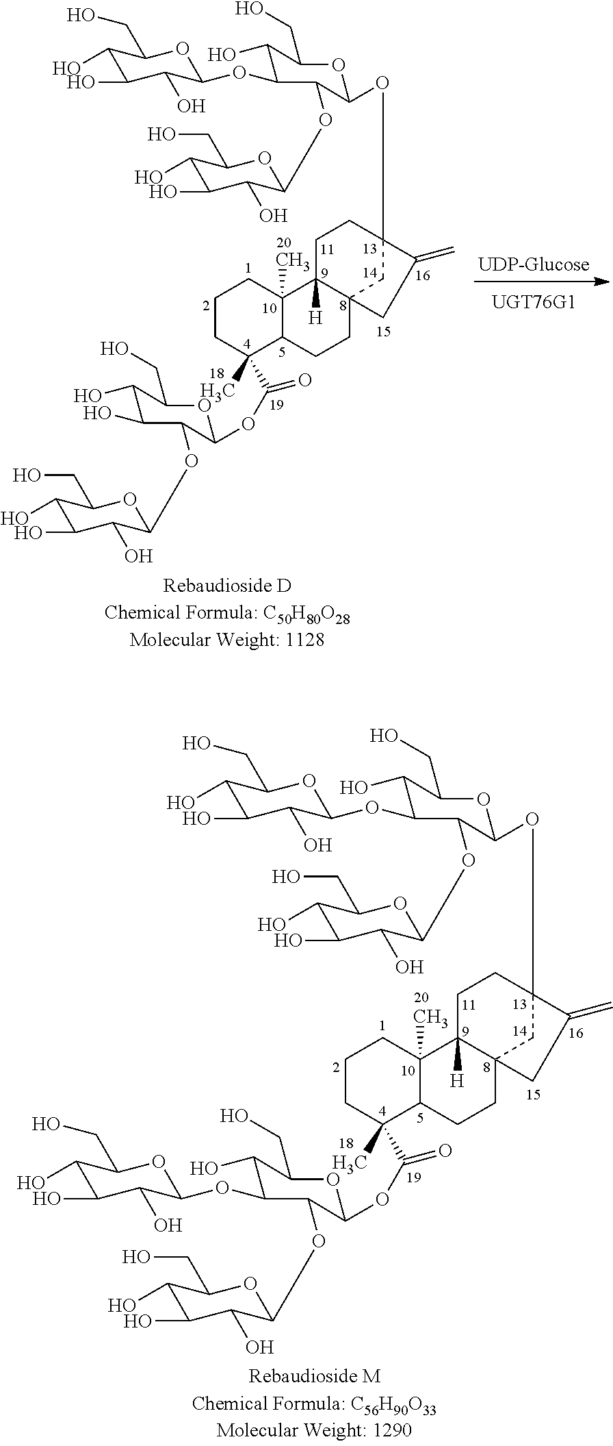
In one embodiment, the UDP-glucosyltransferase is any UDP-glucosyltransferase capable of adding at least one glucose unit to stevioside to form rebaudioside A. In a particular embodiment, the UDP-glucosyltransferase is UGT76G1.
In another embodiment, the UDP-glucosyltransferase is any UDP-glucosyltransferase capable of adding at least one glucose unit to rebaudioside A to form rebaudioside D. In a particular embodiment, the UDP-glucosyltransferase is UGT91D2. In another embodiment, the UGT is an improved variant of UGT91D2 with higher activity and/or selectivity produced by directed evolution.
In yet another embodiment, the UDP-glucosyltransferase is any UDP-glucosyltransferase capable of adding at least one glucose unit to rebaudioside D to form rebaudioside M. In a particular embodiment, the UDP-glucosyltransferase is UGT76G1. In another embodiment, the UGT is an improved variant of UGT76G1 with higher activity and/or selectivity produced by directed evolution.
Optionally, the method of the present invention further comprises recycling UDP to provide UDP-glucose. In one embodiment, the method comprises recycling UDP by providing a recycling catalyst and a recycling substrate, such that the biotransformation of the steviol glycoside substrate to the target steviol glycoside is carried out using catalytic amounts of UDP-glucosyltransferase and UDP-glucose (FIG. 3).
In one embodiment, the recycling catalyst is sucrose synthase.
In one embodiment, the recycling substrate is sucrose.
Optionally, the method of the present invention further comprises separating the target steviol glycoside from the starting composition. The target steviol glycoside can be separated by at least one suitable method, such as, for example, crystallization, separation by membranes, centrifugation, extraction, chromatographic separation or a combination of such methods.
In one embodiment, the target steviol glycoside can be produced within the microorganism. In another embodiment, the target steviol glycoside can be secreted out in the medium. In one another embodiment, the released steviol glycoside can be continuously removed from the medium. In yet another embodiment, the target steviol glycoside is separated after the completion of the reaction.
In one embodiment, separation produces a composition comprising greater than about 80% by weight of the target steviol glycoside on an anhydrous basis, i.e., a highly purified steviol glycoside composition. In another embodiment, separation produces a composition comprising greater than about 90% by weight of the target steviol glycoside. In particular embodiments, the composition comprises greater than about 95% by weight of the target steviol glycoside. In other embodiments, the composition comprises greater than about 99% by weight of the target steviol glycoside.
The target steviol glycoside can be in any polymorphic or amorphous form, including hydrates, solvates, anhydrous or combinations thereof.
Purified target steviol glycosides can be used in consumable products as a sweetener. Suitable consumer products include, but are not limited to, food, beverages, pharmaceutical compositions, tobacco products, nutraceutical compositions, oral hygiene compositions, and cosmetic compositions.
The present invention also provides novel steviol glycosides rebaudioside D2 (reb D2, isomer of rebaudioside D) and rebaudioside M2 (reb M2, isomer of rebaudioside M), which are isomers of reb D and reb M, respectively. In one embodiment, isolated and purified reb D2 is provided. In another embodiment, isolated and purified reb M2 is provided. Reb D2 and reb M2 may also be present in any consumable products disclosed herein. In a particular embodiment, beverages comprising reb D2 and/or reb M2 are provided.
Methods of preparing reb D2 and reb M2 are also provided herein. Both are formed during the biotransformation of reb A to reb D. Reb M2 is believed to form from biotransformation of reb D2 in situ.
In one embodiment, the present invention is a method for the preparation of a composition comprising reb D2 comprising: (a) contacting a starting composition comprising reb A with an enzyme capable of transforming reb A to reb D2, UDP-glucose, and optionally UDP-glucose recycling enzymes, to produce a composition comprising reb D2, and (b) isolating the composition comprising reb D2.
In another embodiment, the present invention is a method for the preparation of a composition comprising reb M2 comprising (a) contacting a starting composition comprising reb D2 with an enzyme capable of transforming reb D2 to reb M2, UDP-glucose, and optionally UDP-glucose recycling enzymes, to produce a composition comprising reb M2, and (b) and isolating the composition comprising reb M2.
A further method for the preparation of a composition comprising reb M2 comprises (a) contacting a starting composition comprising reb A with an enzyme capable of transforming reb A to reb D2, UDP-glucose, and optionally UDP-glucose recycling enzymes, to produce a composition comprising reb D2, (b) optionally, isolating the composition comprising reb D2, (c) contacting the composition comprising reb D2 with an enzyme capable of transforming reb D2 to reb M2, UDP-glucose, and optionally UDP-glucose recycling enzymes to produce a composition comprising reb M2, and (d) isolating the composition comprising reb M2.
The composition can be further purified to provide reb D2 or reb M2 with purities greater than about 95% by weight on a dry basis.
BRIEF DESCRIPTION OF THE DRAWINGS
The accompanying drawings are included to provide a further understanding of the invention. The drawings illustrate embodiments of the invention and together with the description serve to explain the principles of the embodiments of the invention.
FIG. 1 shows the structure of reb M.
FIG. 2 shows the biocatalytic production of reb M from stevioside.
FIG. 3 shows the biocatalytic production of reb A from stevioside using the enzyme UGT76G1 and concomitant recycling of UDP to UDP glucose via sucrose synthase.
FIG. 4 shows the IR spectrum of reb M.
FIG. 5. shows the HPLC chromatogram of the product of the biocatalytic production of reb M from reb D, as detailed in Example 14. The peak with retention time of 24.165 minutes corresponds to unreacted reb D. The peak with retention time of 31.325 minutes corresponds to reb M.
FIG. 6. shows the HPLC chromatogram of purified reb M produced by biocatalysis from reb D.
FIG. 7 shows the HPLC chromatogram of a reb M standard.
FIG. 8 shows the HPLC chromatogram of co-injection of a reb M standard and reb M purified from biotransformation from reb D.
FIG. 9 shows an overlay of the .sup.1H NMR spectra of a reb M standard and reb M purified following biosynthesis from reb D.
FIG. 10 shows the HRMS spectrum of reb M purified following biocatalytic production from reb D.
FIG. 11 shows LC-MS analysis of semi-synthetic steviol glycoside mixture, Lot number CB-2977-106, showing TIC (A), MS of peak at 1.8 min (B), MS of reb M2 peak at 4.1 min (C), MS of reb D peak at 6.0 min (D), MS of reb D2 peak at 7.7 min (E), MS of peak at 9.4 min (F), MS of rebaudioside Apeak at 15.2 min (G), MS of peak at 16.5 min (H), and MS of peak at 18.3 min (I).
FIG. 12 shows the trace of semi-synthetic steviol glycoside mixture, Lot number CB-2977-106. Chromatogram gridlines are not homogeneous as the detector was re-calibrated 14 min following injection.
FIG. 13 shows HPLC analysis of semi-synthetic steviol glycoside mixture, Lot number CB-2977-106 (A), Isolated reb M2 (B), isolated reb D (C) and isolated reb D2 (D).
FIG. 14 shows the .sup.1H NMR spectrum of reb D2 (500 MHz, pyridine-d.sub.5).
FIG. 15 shows the .sup.13C NMR spectrum of reb D2(125 MHz, pyridine-d.sub.5).
FIG. 16 shows an expansion of the .sup.13C NMR spectrum of reb D2 (125 MHz, pyridine-d.sub.5).
FIG. 17 shows the .sup.1H-.sup.1H COSY Spectrum of reb D2 (500 MHz, pyridine-d.sub.5).
FIG. 18 shows the HSQC-DEPT spectrum of reb D2(500 MHz, pyridine-d.sub.5).
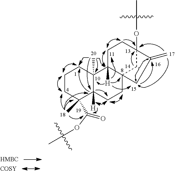
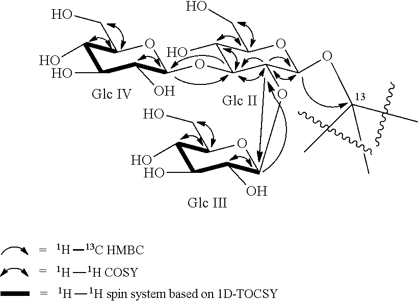
FIG. 19 shows the HMBC spectrum of reb D2.
FIG. 20 shows an expansion of HMBC spectrum of reb D2 (500 MHz, pyridine-d.sub.5).
FIG. 21 shows the .sup.1H NMR spectrum of reb M2(500 MHz, D.sub.2O).
FIG. 22 shows the .sup.13C NMR spectrum of reb M2 (125 MHz, D.sub.2O/TSP).
FIG. 23 shows an expansion of the .sup.13C NMR spectrum of reb M2 (125 MHz, D.sub.2O/TSP).
FIG. 24 shows the .sup.1H-.sup.1H COSY spectrum of reb M2 (500 MHz, D.sub.2O).
FIG. 25 shows the HSQC-DEPT spectrum of reb M2(500 MHz, D.sub.2O).
FIG. 26 shows the HMBC spectrum of reb M2 (500 MHz, D.sub.2O).
FIG. 27 shows an expansion of HMBC spectrum of reb M2 (500 MHz, D.sub.2O).
FIG. 28 shows another HMBC spectrum of reb M2.
FIG. 29 shows a .sup.1H NMR spectrum of reb M2.
FIG. 30 shows a .sup.13C NMR spectrum of reb M2.
FIG. 31 shows another .sup.13C NMR spectrum of reb M2.
FIG. 32 shows a .sup.1H-.sup.1H COSY spectrum of reb M2.
FIG. 33 shows a HSQC-DEPT spectrum of reb M2.
FIG. 34 shows an HMBC spectrum of reb M2.
FIG. 35 shows another HMBC spectrum of reb M2.
FIG. 36 shows a 1D-TOCSY spectrum of reb M2.
FIG. 37 shows a 1D-TOCSY spectrum of reb M2.
FIG. 38 shows a 1D-TOCSY spectrum of reb M2.
FIG. 39 shows a 1D-TOCSY spectrum of reb M2.
FIG. 40 shows an HPLC (CAD) analysis.
FIG. 41 shows an HPLC (CAD) analysis.
FIG. 42 shows an HPLC (CAD) analysis.
FIG. 43 shows an HPLC (CAD) analysis.
FIG. 44 shows an HPLC (CAD) analysis.
FIG. 45 shows an HPLC (CAD) analysis.
FIG. 46 shows an HPLC (CAD) analysis.
FIG. 47 shows an HPLC (CAD) analysis.
FIG. 48 shows an HPLC (CAD) analysis.
FIG. 49 shows an HPLC (CAD) analysis.
FIG. 50 shows an HPLC (CAD) analysis.
FIG. 51 shows an HPLC (CAD) analysis.
FIG. 52 shows an HPLC (CAD) analysis.
FIG. 53 shows an LCMS chromatogram.
FIG. 54 shows an LCMS chromatogram.
FIG. 55 shows an LCMS chromatogram.
FIG. 56 shows an LCMS chromatogram.
FIG. 57 shows a reaction profile.
FIG. 58 shows an HPLC (CAD) analysis.
FIG. 59 shows an HPLC (CAD) analysis.
FIG. 60 shows an HPLC (CAD) analysis.
FIG. 61 shows an HPLC (CAD) analysis.
FIG. 62 shows an HPLC (CAD) analysis.
FIG. 63 shows an LCMS chromatogram.
FIG. 64 shows an HPLC (CAD) analysis.
FIG. 65 shows an HPLC (CAD) analysis.
FIG. 66 shows an HPLC (CAD) analysis.
FIG. 67 shows an HPLC (CAD) analysis.
FIG. 68 shows an HPLC (CAD) analysis.
FIG. 69 shows the results of an HPLC analysis.
DETAILED DESCRIPTION
The present invention provides a biocatalytic process for preparing a composition comprising a target steviol glycoside by contacting a starting composition, comprising an organic substrate, with a microorganism and/or biocatalyst, thereby producing a composition comprising a target steviol glycoside.
One object of the invention is to provide an efficient biocatalytic method for preparing steviol glycosides, particularly stevioside, reb E, reb A, reb D, reb D2, reb M, and reb M2 from various starting compositions.
As used herein, "biocatalysis" or "biocatalytic" refers to the use of natural or genetically engineered biocatalysts, such as cells, protein enzymes, to perform single or multiple step chemical transformations on organic compounds. Biocatalysis include fermentation, biosynthesis and biotransformation processes. Both, isolated enzyme and whole-cell biocatalysis methods, using biocatalysts in free as well as immobilized forms, are known in the art. Biocatalyst protein enzymes can be naturally occurring or recombinant proteins.
As used herein, the term "steviol glycoside(s)" refers to a glycoside of steviol, including, but not limited to, naturally occurring steviol glycosides, e.g. steviolmonoside, steviolbioside, rubusoside, dulcoside B, dulcoside A, rebaudioside B, rebaudioside G, stevioside, rebaudioside C, rebaudioside F, rebaudioside A, rebaudioside I, rebaudioside E, rebaudioside H, rebaudioside L, rebaudioside K, rebaudioside J, rebaudioside M, rebaudioside M2, rebaudioside D, rebaudioside D2, rebaudioside N, rebaudioside O, synthetic steviol glycosides, e.g. enzymatically glucosylated steviol glycosides and combinations thereof.
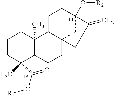
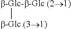
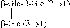
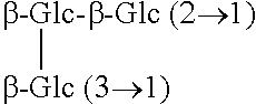
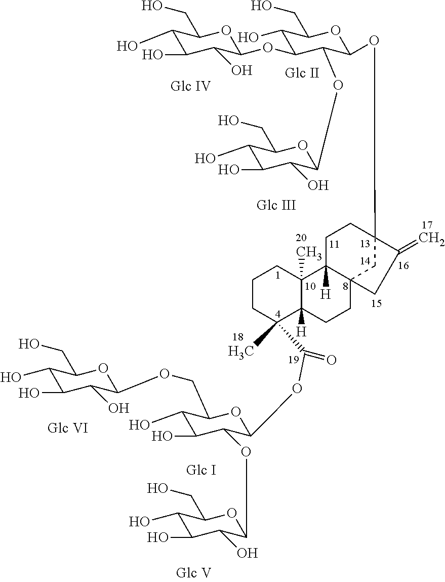
TABLE-US-00001 Chemical structures of steviol and its glycosides ##STR00001## Compound R.sub.1 R.sub.2 Steviol H H Steviolmonoside H .beta.-Glc Steviol monoglucosyl ester .beta.-Glc H Rubusoside .beta.-Glc .beta.-Glc Steviolbioside H .beta.-Glc-.beta.-Glc (2.fwdarw.1) Stevioside .beta.-Glc .beta.-Glc-.beta.-Glc (2.fwdarw.1) Rebaudioside A .beta.-Glc ##STR00002## Rebaudioside D .beta.-Glc-.beta.-Glc (2.fwdarw.1) ##STR00003## Rebaudioside E .beta.-Glc-.beta.-Glc (2.fwdarw.1) .beta.-Glc-.beta.-Glc (2.fwdarw.1) Rebaudioside M ##STR00004## ##STR00005## (Glc = glucose)
Starting Composition
As used herein, "starting composition" refers to any composition (generally an aqueous solution) containing one or more organic compound comprising at least one carbon atom.
In one embodiment, the starting composition is selected from the group consisting of polyols and various carbohydrates.
The term "polyol" refers to a molecule that contains more than one hydroxyl group. A polyol may be a diol, triol, or a tetraol which contain 2, 3, and 4 hydroxyl groups, respectively. A polyol also may contain more than four hydroxyl groups, such as a pentaol, hexaol, heptaol, or the like, which contain 5, 6, or 7 hydroxyl groups, respectively. Additionally, a polyol also may be a sugar alcohol, polyhydric alcohol, or polyalcohol which is a reduced form of carbohydrate, wherein the carbonyl group (aldehyde or ketone, reducing sugar) has been reduced to a primary or secondary hydroxyl group. Examples of polyols include, but are not limited to, erythritol, maltitol, mannitol, sorbitol, lactitol, xylitol, inositol, isomalt, propylene glycol, glycerol, threitol, galactitol, hydrogenated isomaltulose, reduced isomalto-oligosaccharides, reduced xylo-oligosaccharides, reduced gentio-oligosaccharides, reduced maltose syrup, reduced glucose syrup, hydrogenated starch hydrolyzates, polyglycitols and sugar alcohols or any other carbohydrates capable of being reduced.
The term "carbohydrate" refers to aldehyde or ketone compounds substituted with multiple hydroxyl groups, of the general formula (CH.sub.2O).sub.n, wherein n is 3-30, as well as their oligomers and polymers. The carbohydrates of the present invention can, in addition, be substituted or deoxygenated at one or more positions. Carbohydrates, as used herein, encompass unmodified carbohydrates, carbohydrate derivatives, substituted carbohydrates, and modified carbohydrates. As used herein, the phrases "carbohydrate derivatives", "substituted carbohydrate", and "modified carbohydrates" are synonymous. Modified carbohydrate means any carbohydrate wherein at least one atom has been added, removed, or substituted, or combinations thereof. Thus, carbohydrate derivatives or substituted carbohydrates include substituted and unsubstituted monosaccharides, disaccharides, oligosaccharides, and polysaccharides. The carbohydrate derivatives or substituted carbohydrates optionally can be deoxygenated at any corresponding C-position, and/or substituted with one or more moieties such as hydrogen, halogen, haloalkyl, carboxyl, acyl, acyloxy, amino, amido, carboxyl derivatives, alkylamino, dialkylamino, arylamino, alkoxy, aryloxy, nitro, cyano, sulfo, mercapto, imino, sulfonyl, sulfenyl, sulfinyl, sulfamoyl, carboalkoxy, carboxamido, phosphonyl, phosphinyl, phosphoryl, phosphino, thioester, thioether, oximino, hydrazino, carbamyl, phospho, phosphonato, or any other viable functional group provided the carbohydrate derivative or substituted carbohydrate functions to improve the sweet taste of the sweetener composition.
Examples of carbohydrates which may be used in accordance with this invention include, but are not limited to, tagatose, trehalose, galactose, rhamnose, various cyclodextrins, cyclic oligosaccharides, various types of maltodextrins, dextran, sucrose, glucose, ribulose, fructose, threose, arabinose, xylose, lyxose, allose, altrose, mannose, idose, lactose, maltose, invert sugar, isotrehalose, neotrehalose, isomaltulose, erythrose, deoxyribose, gulose, idose, talose, erythrulose, xylulose, psicose, turanose, cellobiose, amylopectin, glucosamine, mannosamine, fucose, glucuronic acid, gluconic acid, glucono-lactone, abequose, galactosamine, beet oligosaccharides, isomalto-oligosaccharides (isomaltose, isomaltotriose, panose and the like), xylo-oligosaccharides (xylotriose, xylobiose and the like), xylo-terminated oligosaccharides, gentio-oligosaccharides (gentiobiose, gentiotriose, gentiotetraose and the like), sorbose, nigero-oligosaccharides, palatinose oligosaccharides, fructooligosaccharides (kestose, nystose and the like), maltotetraol, maltotriol, malto-oligosaccharides (maltotriose, maltotetraose, maltopentaose, maltohexaose, maltoheptaose and the like), starch, inulin, inulo-oligosaccharides, lactulose, melibiose, raffinose, ribose, isomerized liquid sugars such as high fructose corn syrups, coupling sugars, and soybean oligosaccharides. Additionally, the carbohydrates as used herein may be in either the D- or L-configuration.
The starting composition may be synthetic or purified (partially or entirely), commercially available or prepared.
In one embodiment, the starting composition is glycerol.
In another embodiment, the starting composition is glucose.
In still another embodiment, the starting composition is sucrose.
In yet another embodiment, the starting composition is starch.
In another embodiment, the starting composition is maltodextrin.
The organic compound(s) of starting composition serve as a substrate(s) for the production of the target steviol glycoside(s), as described herein.
Target Steviol Glycoside
The target steviol glycoside of the present method can be any steviol glycoside that can be prepared by the process disclosed herein. In one embodiment, the target steviol glycoside is selected from the group consisting of steviolmonoside, steviolbioside, rubusoside, dulcoside B, dulcoside A, rebaudioside B, rebaudioside G, stevioside, rebaudioside C, rebaudioside F, rebaudioside A, rebaudioside I, rebaudioside E, rebaudioside H, rebaudioside L, rebaudioside K, rebaudioside J, rebaudioside M, rebaudioside M2, rebaudioside D, rebaudioside D2, rebaudioside N or rebaudioside O, or other glycoside of steviol.
In one embodiment, the target steviol glycoside is stevioside. In another embodiment, the target steviol glycoside is reb A. In still another embodiment, the target steviol glycoside is reb E. In yet another embodiment, the target steviol glycoside is reb D. In yet another embodiment, the target steviol glycoside is reb D2. In a further embodiment, the target steviol glycoside is reb M. In a still further another embodiment, the target steviol glycoside is reb M2.
The target steviol glycoside can be in any polymorphic or amorphous form, including hydrates, solvates, anhydrous or combinations thereof.
In one embodiment, the present invention is a biocatalytic process for the production of reb D.
In yet another embodiment, the present invention is a biocatalytic process for the production of reb D2.
In still another embodiment, the present invention is a biocatalytic process for the production of reb M
In a further embodiment, the present invention is a biocatalytic process for the production of reb M2.
Optionally, the method of the present invention further comprises separating the target steviol glycoside from the starting composition. The target steviol glycoside can be separated by any suitable method, such as, for example, crystallization, separation by membranes, centrifugation, extraction, chromatographic separation or a combination of such methods.
In particular embodiments, the process described herein results in a highly purified target steviol glycoside composition. The term "highly purified", as used herein, refers to a composition having greater than about 80% by weight of the target steviol glycoside on an anhydrous basis. In one embodiment, the highly purified target steviol glycoside composition contains greater than about 90% by weight of the target steviol glycoside on an anhydrous basis, such as, for example, greater than about 91%, greater than about 92%, greater than about 93%, greater than about 94%, greater than about 95%, greater than about 96%, greater than about 97%, greater than about 98% or greater than about 99% target steviol glycoside content on a dry basis.
In one embodiment, when the target steviol glycoside is reb M, the process described herein provides a composition having greater than about 90% reb M content by weight on a dry basis. In another particular embodiment, when the target steviol glycoside is reb M, the process described herein provides a composition comprising greater than about 95% reb M content by weight on a dry basis.
In another embodiment, when the target steviol glycoside is reb M2, the process described herein provides a composition having greater than about 90% reb M2 content by weight on a dry basis. In another particular embodiment, when the target steviol glycoside is reb M2, the process described herein provides a composition comprising greater than about 95% reb M2 content by weight on a dry basis.
In yet another embodiment, when the target steviol glycoside is reb D, the process described herein provides a composition greater than about 90% reb D content by weight on a dry basis. In another particular embodiment, when the target steviol glycoside is reb D, the process described herein provides a composition comprising greater than about 99% reb D content by weight on a dry basis.
In still another embodiment, when the target steviol glycoside is reb D2, the process described herein provides a composition greater than about 90% reb D2 content by weight on a dry basis. In another particular embodiment, when the target steviol glycoside is reb D2, the process described herein provides a composition comprising greater than about 95% reb D2 content by weight on a dry basis.
In a further embodiment, when the target steviol glycoside is reb A, the process described herein provides a composition comprising greater than about 90% reb A content by weight on a dry basis. In another particular embodiment, when the target steviol glycoside is reb A, the process described herein provides a composition comprising greater than about 95% reb A content by weight on a dry basis.
In a still further embodiment, when the target steviol glycoside is reb E, the process described herein provides a composition comprising greater than about 90% reb E content by weight on a dry basis. In another particular embodiment, when the target steviol glycoside is reb E, the process described herein provides a composition comprising greater than about 95% reb E content by weight on a dry basis.
In a still further embodiment, when the target steviol glycoside is reb I, the process described herein provides a composition comprising greater than about 90% reb I content by weight on a dry basis. In another particular embodiment, when the target steviol glycoside is reb I, the process described herein provides a composition comprising greater than about 95% reb I content by weight on a dry basis.
In yet a further embodiment, when the target steviol glycoside is stevioside, the process described herein provides a composition comprising greater than about 90% stevioside content by weight on a dry basis. In another particular embodiment, when the target steviol glycoside is stevioside, the process described herein provides a composition comprising greater than about 95% stevioside content by weight on a dry basis.
Microorganism and Biocatalysts
In one embodiment of present invention, a microorganism or biocatalyst is contacted with the starting composition to produce target steviol glycosides. The microorganism can be any microorganism possessing the necessary enzymes for converting the starting composition to target steviol glycosides. These enzymes are encoded within the microorganism's genome.
In one embodiment the microorganism may be, for example, E. coli, Saccharomyces sp., Aspergillus sp., Pichia sp., Bacillus sp., Yarrowia sp. etc.
The enzymes can be located on the surface and/or inside the cell of the microorganism and/or can be secreted out in the medium by the microorganism.
The biocatalyst comprises at least one enzyme and can be whole cell suspension, crude lysate or purified enzyme.
The enzymes necessary for converting the starting composition to target steviol glycosides include the steviol biosynthesis enzymes and UDP-glycosyltransferases (UGTs). Optionally it may include UDP recycling enzyme(s). The UDP recycling enzyme can be sucrose synthase and the recycling substrate can be sucrose.
In one embodiment the steviol biosynthesis enzymes include mevalonate (MVA) pathway enzymes.
In another embodiment the steviol biosynthesis enzymes include non-mevalonate 2-C-methyl-D-erythritol-4-phosphate pathway (MEP/DOXP) enzymes.
In one embodiment the steviol biosynthesis enzymes are selected from the group including geranylgeranyl diphosphate synthase, copalyl diphosphate synthase, kaurene synthase, kaurene oxidase, kaurenoic acid 13-hydroxylase (KAH), steviol synthetase, deoxyxylulose 5-phosphate synthase (DXS), D-1-deoxyxylulose 5-phosphate reductoisomerase (DXR), 4-diphosphocytidyl-2-C-methyl-D-erythritol synthase (CMS), 4-diphosphocytidyl-2-C-methyl-D-erythritol kinase (CMK), 4-diphosphocytidyl-2-C-methyl-D-erythritol 2,4-cyclodiphosphate synthase (MCS), 1-hydroxy-2-methyl-2(E)-butenyl 4-diphosphate synthase (HDS), 1-hydroxy-2-methyl-2(E)-butenyl 4-diphosphate reductase (HDR), acetoacetyl-CoA thiolase, truncated HMG-CoA reductase, mevalonate kinase, phosphomevalonate kinase, mevalonate pyrophosphate decarboxylase, cytochrome P450 reductase etc.
The UDP-glucosyltransferase can be any UDP-glucosyltransferase capable of adding at least one glucose unit to the steviol and or steviol glycoside substrate to provide the target steviol glycoside.
In one embodiment, the microorganism is free. In another embodiment, the microorganism is immobilized. For example, the microorganism may be immobilized to a solid support made from inorganic or organic materials. Non-limiting examples of solid supports suitable to immobilize the microorganism include derivatized cellulose or glass, ceramics, metal oxides or membranes. The microorganism may be immobilized to the solid support, for example, by covalent attachment, adsorption, cross-linking, entrapment or encapsulation.
In one embodiment the microorganism is in aqueous medium, comprising water, and various components selected form group including carbon sources, energy sources, nitrogen sources, microelements, vitamins, nucleosides, nucleoside phosphates, nucleoside diphosphates, nucleoside triphosphates, organic and inorganic salts, organic and mineral acids, bases etc. Carbon sources include glycerol, glucose, carbon dioxide, carbonates, bicarbonates. Nitrogen sources can include nitrates, nitrites, amino acids, peptides, peptones, or proteins.
In a particular embodiment, the medium comprises buffer. Suitable buffers include, but are not limited to, PIPES buffer, acetate buffer and phosphate buffer. In a particular embodiment, the medium comprises phosphate buffer.
In one embodiment, the medium can also include an organic solvent.
In one embodiment, the UDP-glucosyltransferase is any UDP-glucosyltransferase capable of adding at least one glucose unit to rubusoside, thereby producing stevioside. The UDP-glucosyltransferase may be, for example, UGT91D2.
In another embodiment, the UDP-glucosyltransferase is any UDP-glucosyltransferase capable of adding at least one glucose unit to rubusoside, thereby producing rebaudioside E. The UDP-glucosyltransferase may be, for example, UGTSL2.
In still another embodiment, the UDP-glucosyltransferase is any UDP-glucosyltransferase capable of adding at least one glucose unit to rebaudioside E, thereby producing rebaudioside D. The UDP-glucosyltransferase may be, for example, UGT76G1.
In yet embodiment, the UDP-glucosyltransferase is any UDP-glucosyltransferase capable of adding at least one glucose unit to stevioside, thereby producing rebaudioside A. The UDP-glucosyltransferase may be, for example, UGT76G1.
In a further embodiment, the UDP-glucosyltransferase is any UDP-glucosyltransferase capable of adding at least one glucose unit to rebaudioside A, thereby producing rebaudioside D and/or rebaudioside D2 and/or rebaudioside M2. The UDP-glucosyltransferase may be, for example, UGT91D2 or UGTSL2.
In another embodiment, the UDP-glucosyltransferase capable of adding at least one glucose unit to rebaudioside A is selected from the following listing of GenInfo identifier numbers, preferably from the group presented in Table 1, and more preferably the group presented in Table 2.
TABLE-US-00002 397567 30680413 115480946 147798902 218193594 225443294 454245 32816174 116310259 147811764 218193942 225444853 1359905 32816178 116310985 147827151 219885307 225449296 1685003 34393978 116788066 147836230 222615927 225449700 1685005 37993665 116788606 147839909 222619587 225454338 2191136 37993671 116789315 147846163 222623142 225454340 2501497 37993675 119394507 147855977 222625633 225454342 2911049 39104603 119640480 148905778 222625635 225454473 4218003 41469414 122209731 148905999 222636620 225454475 4314356 41469452 125526997 148906835 222636621 225458362 13492674 42566366 125534279 148907340 222636628 225461551 13492676 42570280 125534461 148908935 222636629 225461556 15217773 42572855 125540090 148909182 224053242 225461558 15217796 44890129 125541516 148909920 224053386 225469538 15223396 46806235 125545408 148910082 224055535 225469540 15223589 50284482 125547340 148910154 224056138 226316457 15227766 51090402 125547520 148910612 224056160 226492603 15230017 51090594 125554547 148910769 224067918 226494221 15231757 52839682 125557592 156138791 224072747 226495389 15234056 56550539 125557593 156138797 224080189 226495945 15234195 62734263 125557608 156138799 224091845 226502400 15234196 62857204 125559566 156138803 224094703 226507980 15238503 62857206 125563266 165972256 224100653 226531147 15239523 62857210 125571055 168016721 224100657 226532094 15239525 62857212 125579728 171674071 224101569 238477377 15239543 75265643 125588307 171906258 224103105 240254512 15239937 75285934 125589492 183013901 224103633 242032615 15240305 75288884 125599469 183013903 224103637 242032621 15240534 77550661 125601477 186478321 224109218 242038423 15982889 77556148 126635837 187373030 224114583 242043290 18086351 82791223 126635845 187373042 224116284 242044836 18418378 83778990 126635847 190692175 224120552 242051252 18418380 89953335 126635863 194701936 224121288 242056217 18418382 110741436 126635867 195620060 224121296 242056219 19743740 110743955 126635883 209954691 224121300 242056663 19911201 115438196 126635887 209954719 224130358 242059339 20149064 115438785 133874210 209954725 224140703 242059341 20260654 115441237 133874212 209954733 224143404 242060922 21435782 115454819 145358033 210063105 224143406 242067411 21553613 115456047 147772508 210063107 224144306 242067413 21593514 115457492 147776893 212275846 224285244 242076258 22759895 115459312 147776894 216296854 225431707 242076396 23955910 115464719 147776895 217074506 225435532 242084750 26452040 115471069 147786916 218185693 225436321 242091005 28393204 115471071 147798900 218187075 225440041 242095206 30679796 115474009 147798901 218189427 225441116 242345159 242345161 297724601 326492035 356523945 357140904 359486938 255536859 297725463 326493430 356523957 357165849 359487055 255538228 297728331 326500410 356523959 357165852 359488135 255541676 297738632 326506816 356523961 357168415 359488708 255547075 297745347 326507826 356523963 357437837 359493630 255552620 297745348 326508394 356524387 357442755 359493632 255552622 297795735 326509445 356524403 357442757 359493634 255555343 297796253 326511261 356527181 357445729 359493636 255555361 297796257 326511866 356533209 357445731 359493815 255555363 297796261 326512412 356533852 357445733 359495856 255555365 297797587 326517673 356534718 357446799 359495858 255555369 297798502 326518800 356535480 357446805 359495869 255555373 297799226 326521124 356542996 357452779 359495871 255555377 297805988 326525567 356543136 357452781 359497638 255556812 297807499 326525957 356543932 357452783 359807261 255556818 297809125 326526607 356549841 357452787 374256637 255563008 297809127 326527141 356549843 357452789 377655465 255564074 297811403 326530093 356554358 357452791 378405177 255564531 297820040 326534036 356554360 357452797 378829085 255572878 297821483 326534312 356558606 357452799 387135070 255577901 297825217 332071132 356560333 357470367 387135072 255583249 297832276 339715876 356560599 357472193 387135078 255583253 297832280 342306012 356560749 357472195 387135092 255583255 297832518 342306016 356566018 357474295 387135094 255585664 297832520 343457675 356566169 357474493 387135098 255585666 297840825 343457677 356566173 357474497 387135100 255634688 297840827 350534960 356567761 357474499 387135134 255644801 297847402 356498085 356574704 357490035 387135136 255645821 297849372 356499771 356576401 357493567 387135174 255647456 300078590 356499777 356577660 357497139 387135176 255648275 300669727 356499779 357114993 357497581 387135184 260279126 302142947 356501328 357115447 357497671 387135186 260279128 302142948 356502523 357115451 357500579 387135188 261343326 302142950 356503180 357115453 357504663 387135190 283132367 302142951 356503184 357116080 357504691 387135192 283362112 302765302 356503295 357116928 357504699 387135194 289188052 302796334 356504436 357117461 357504707 387135282 295841350 302811470 356504523 357117463 357505859 387135284 296088529 302821107 356504765 357117829 357510851 387135294 296090415 302821679 356511113 357117839 357516975 387135298 296090524 319759260 356515120 357125059 359477003 387135300 296090526 319759266 356517088 357126015 359477998 387135302 297599503 320148814 356520732 357134488 359478043 387135304 297601531 326489963 356522586 357135657 359478286 387135312 297611791 326490273 356522588 357138503 359484299 387135314 297722841 326491131 356522590 357139683 359486936 387135316 387135318 449440433 460376293 460413408 462423864 475546199 387135320 449445896 460378310 460416351 470101924 475556485 387135322 449446454 460380744 462394387 470102280 475559699 387135324 449447657 460381726 462394433 470102858 475578293 387135326 449449002 460382093 462394557 470104211 475591753 387135328 449449004 460382095 462395646 470104264 475593742 388493506 449449006 460382754 462395678 470104266 475612072 388495496 449451379 460384935 462396388 470106317 475622476 388498446 449451589 460384937 462396389 470106357 475622507 388499220 449451591 460385076 462396419 470115448 475623787 388502176 449451593 460385872 462396542 470130404 482550481 388517521 449453712 460386018 462397507 470131550 482550499 388519407 449453714 460389217 462399998 470136482 482550740 388521413 449453716 460394872 462400798 470136484 482550999 388827901 449453732 460396139 462401217 470136488 482552352 388827903 449457075 460397862 462402118 470136492 482554970 388827907 449467555 460397864 462402237 470137933 482555336 388827909 449468742 460398541 462402284 470137937 482555478 388827913 449495638 460403139 462402416 470140422 482556454 393887637 449495736 460403141 462404228 470140426 482557289 393887646 449499880 460403143 462406358 470140908 482558462 393887649 449502786 460403145 462408262 470141232 482558508 393990627 449503471 460405998 462409325 470142008 482558547 397746860 449503473 460407578 462409359 470142010 482561055 397789318 449515857 460407590 462409777 470142012 482561555 413924864 449518643 460409128 462411467 470143607 482562795 414590349 449519559 460409134 462414311 470143939 482562850 414590661 449522783 460409136 462414416 470145404 482565074 414591157 449524530 460409459 462414476 473923244 482566269 414879558 449524591 460409461 462415526 474114354 482566296 414879559 449528823 460409463 462415603 474143634 482566307 414879560 449528825 460409465 462415731 474202268 482568689 414888074 449534021 460409467 462416307 474299266 482570049 431812559 460365546 460410124 462416920 474363119 482570572 449432064 460366882 460410126 462416922 474366157 482575121 449432066 460369823 460410128 462416923 474429346 449433069 460369829 460410130 462416924 475432777 449436944 460369831 460410132 462417401 475473002 449438665 460369833 460410134 462419769 475489790 449438667 460370755 460410213 462420317 475511330 449440431 460374714 460411200 462423366 475516200
TABLE-US-00003 TABLE 1 GI number Accession Origin 190692175 ACE87855.1 Stevia rebaudiana 41469452 AAS07253.1 Oryza sativa 62857204 BAD95881.1 Ipomoea nil 62857206 BAD95882.1 Ipomoea purperea 56550539 BAD77944.1 Bellis perennis 115454819 NP_001051010.1 Oryza sativa Japonica Group 115459312 NP_001053256.1 Oryza sativa Japonica Group 115471069 NP_001059133.1 Oryza sativa Japonica Group 115471071 NP_001059134.1 Oryza sativa Japonica Group 116310985 CAH67920.1 Oryza sativa Japonica Indica Group 116788066 ABK24743.1 Picea sitchensis 122209731 Q2V6J9.1 Fragaria .times. ananassa 125534461 EAY81009.1 Oryza sativa Indica Group 125559566 EAZ05102.1 Oryza sativa Indica Group 125588307 EAZ28971.1 Oryza sativa Japonica Group 148907340 ABR16806.1 Picea sitchensis 148910082 ABR18123.1 Picea sitchensis 148910612 ABR18376.1 Picea sitchensis 15234195 NP_194486.1 Arabidopsis thaliana 15239523 NP_200210.1 Arabidopsis thaliana 15239937 NP_196793.1 Arabidopsis thaliana 1685005 AAB36653.1 Nicotiana tabacum 183013903 ACC38471.1 Medicago truncatula 186478321 NP_172511.3 Arabidopsis thaliana 187373030 ACD03249.1 Avena strigosa 194701936 ACF85052.1 Zea mays 19743740 AAL92461.1 Solanum lycopersicum 212275846 NP_001131009.1 Zea mays 222619587 EEE55719.1 Oryza sativa Japonica Group 224055535 XP_002298527.1 Populus trichocarpa 224101569 XP_002334266.1 Populus trichocarpa 224120552 XP_002318358.1 Populus trichocarpa 224121288 XP_002330790.1 Populus trichocarpa 225444853 XP_002281094 Vitis vinifera 225454342 XP_002275850.1 Vitis vinifera 225454475 XP_002280923.1 Vitis vinifera 225461556 XP_002285222 Vitis vinifera 225469540 XP_002270294.1 Vitis vinifera 226495389 NP_001148083.1 Zea mays 226502400 NP_001147674.1 Zea mays 238477377 ACR43489.1 Triticum aestivum 240254512 NP_565540.4 Arabidopsis thaliana 2501497 Q43716.1 Petunia .times. hybrida 255555369 XP_002518721.1 Ricinus communis 26452040 BAC43110.1 Arabidopsis thaliana 296088529 CBI37520.3 Vitis vinifera 297611791 NP_001067852.2 Oryza sativa Japonica Group 297795735 XP_002865752.1 Arabidopsis lyrata subsp. lyrata 297798502 XP_002867135.1 Arabidopsis lyrata subsp. lyrata 297820040 XP_002877903.1 Arabidopsis lyrata subsp. lyrata 297832276 XP_002884020.1 Arabidopsis lyrata subsp. lyrata 302821107 XP_002992218.1 Selaginella moellendorffii 30680413 NP_179446.2 Arabidopsis thaliana 319759266 ADV71369.1 Pueraria montana var. lobata 326507826 BAJ86656.1 Hordeum vulgare subsp. Vulgare 343457675 AEM37036.1 Brassica rapa subsp. oleifera 350534960 NP_001234680.1 Solanum lycopersicum 356501328 XP_003519477.1 Glycine max 356522586 XP_003529927.1 Glycine max 356535480 XP_003536273.1 Glycine max 357445733 XP_003593144.1 Medicago truncatula 357452783 XP_003596668.1 Medicago truncatula 357474493 XP_003607531.1 Medicago truncatula 357500579 XP_003620578.1 Medicago truncatula 357504691 XP_003622634.1 Medicago truncatula 359477998 XP_003632051.1 Vitis vinifera 359487055 XP_002271587 Vitis vinifera 359495869 XP_003635104.1 Vitis vinifera 387135134 AFJ52948.1 Linum usitatissimum 387135176 AFJ52969.1 Linum usitatissimum 387135192 AFJ52977.1 Linum usitatissimum 387135282 AFJ53022.1 Linum usitatissimum 387135302 AFJ53032.1 Linum usitatissimum 387135312 AFJ53037.1 Linum usitatissimum 388519407 AFK47765.1 Medicago truncatula 393887646 AFN26668.1 Barbarea vulgaris subsp. arcuata 414888074 DAA64088.1 Zea mays 42572855 NP_974524.1 Arabidopsis thaliana 449440433 XP_004137989.1 Cucumis sativus 449446454 XP_004140986.1 Cucumis sativus 449449004 XP_004142255.1 Cucumis sativus 449451593 XP_004143546.1 Cucumis sativus 449515857 XP_004164964.1 Cucumis sativus 460382095 XP_004236775.1 Solanum lycopersicum 460409128 XP_004249992.1 Solanum lycopersicum 460409461 XP_004250157.1 Solanum lycopersicum 460409465 XP_004250159.1 Solanum lycopersicum 462396388 EMJ02187.1 Prunus persica 462402118 EMJ07675.1 Prunus persica 462409359 EMJ14693.1 Prunus persica 462416923 EMJ21660.1 Prunus persica 46806235 BAD17459.1 Oryza sativa Japonica Group 470104266 XP_004288529.1 Fragaria vesca subsp. vesca 470142008 XP_004306714.1 Fragaria vesca subsp. vesca 475432777 EMT01232.1 Aegilops tauschii 51090402 BAD35324.1 Oryza sativa Japonica Group
TABLE-US-00004 TABLE 2 Internal GI number Accession Origin reference 460409128 XP.004249992.1 Solanum lycopersicum UGTSL 460386018 XP.004238697.1 Solanum lycopersicum -- 460409134 XP.004249995.1 Solanum lycopersicum -- 460410132 XP.004250485.1 Solanum lycopersicum UGTSL2 460410130 XP.004250484.1 Solanum lycopersicum -- 460410128 XP.004250483.1 Solanum lycopersicum -- 460378310 XP.004234916.1 Solanum lycopersicum -- 209954733 BAG80557.1 Lycium barbarum UGTLB 209954725 BAG80553.1 Lycium barbarum --
In yet another embodiment, the UDP-glucosyltransferase is any UDP-glucosyltransferase capable of adding at least one glucose unit to rebaudioside D to form rebaudioside M and/or rebaudioside M2. The UDP-glucosyltransferase may be, for example, UGT76G1.
Optionally, the method of the present invention further comprises recycling UDP to provide UDP-glucose. In one embodiment, the method comprises recycling UDP by providing a recycling catalyst, i.e., a biocatalyst capable of UDP-glucose overproduction, and a recycling substrate, such that the conversion of the substrate steviol glycoside to the target steviol glycoside is carried out using catalytic amounts of UDP-glucosyltransferase and UDP-glucose (FIG. 3).
In one embodiment, the UDP-glucose recycling catalyst is sucrose synthase.
In one embodiment, the recycling substrate is sucrose.
In one embodiment the biocatalyst comprises more than one UDP-glucosyltransferase
In embodiment the biocatalyst comprises more than one UDP-glucosyltransferase and UDP-glucose recycling catalyst.
The target steviol glycoside is optionally purified from the resulting composition. Purification of the target steviol glycoside from the reaction medium can be achieved by at least one suitable method to provide a highly purified target steviol glycoside composition. Suitable methods include crystallization, separation by membranes, centrifugation, extraction (liquid or solid phase), chromatographic separation, HPLC (preparative or analytical) or a combination of such methods.
Compounds and Methods
The present invention also provides isolated and highly purified reb D2. Reb D2 is an isomer of reb D and has the following structure:
##STR00006##
13-[(2-O-.beta.-D-glucopyranosyl-3-O-.beta.-D-glucopyranosyl-.beta.-D-gluc- opyranosyl)oxy]ent-kaur-16-en-19-oic acid-[(6-O-.beta.-D-glucopyranosyl-.beta.-D-glucopyranosyl)ester]
In another embodiment, the present invention provides reb D2 having a purity greater than about 95% by weight on an anhydrous basis, such as, for example, greater than about 96% by weight, greater than about 97% by weight, greater than about 98% by weight or greater than about 99% by weight.
In still another embodiment, the present invention provides reb D2 having a purity greater than about 95% by weight in a steviol glycoside mixture, such as, for example, greater than about 96% by weight, greater than about 97% by weight, greater than about 98% by weight or greater than about 99% by weight.
The present invention also provides compositions comprising reb D2.
In one embodiment, the present invention provides a method for preparing reb D2 comprising: a. contacting a starting composition comprising reb A with an enzyme capable of transforming reb A to reb D2, UDP-glucose, and optionally UDP-glucose recycling enzymes, to produce a composition comprising reb D2; and b. isolating the composition comprising reb D2.
In some embodiments, the enzyme capable of transforming reb A to reb D2 is a UDP-glucosyltransferase, such as, for example, UGT91D2, UGTSL, UGTSL_Sc, UGTSL2 (GI No. 460410132 version XP_004250485.1), GI No. 460409128 (UGTSL) version XP_004249992.1, GI No. 115454819 version NP_001051010.1, GI No. 187373030, version ACD03249.1. GI No. 222619587 version EEE55719.1, GI No. 297795735 version XP_002865752.1 or EUGT11.
The enzyme capable of transforming reb A to reb D2 can be immobilized or in a recombinant microorganism.
In one embodiment, the enzyme is immobilized. In another embodiment, the enzyme is in a recombinant microorganism.
In one embodiment, the microorganism is free. In another embodiment, the microorganism is immobilized. For example, the microorganism may be immobilized to a solid support made from inorganic or organic materials. Non-limiting examples of solid supports suitable to immobilize the microorganism include derivatized cellulose or glass, ceramics, metal oxides or membranes. The microorganism may be immobilized to the solid support, for example, by covalent attachment, adsorption, cross-linking, entrapment or encapsulation.
Suitable microorganisms include, but are not limited to, E. coli, Saccharomyces sp., Aspergillus sp., Pichia sp., Bacillus sp., Yarrowia sp.
In one embodiment the microorganism is in an aqueous medium, comprising water, and various components selected form group including carbon sources, energy sources, nitrogen sources, microelements, vitamins, nucleosides, nucleoside phosphates, nucleoside diphosphates, nucleoside triphosphates, organic and inorganic salts, organic and mineral acids, bases etc. Carbon sources include glycerol, glucose, carbon dioxide, carbonates, bicarbonates. Nitrogen sources can include nitrates, nitrites, amino acids, peptides, peptones, or proteins.
In a particular embodiment, the medium comprises buffer. Suitable buffers include, but are not limited to, PIPES buffer, acetate buffer and phosphate buffer. In a particular embodiment, the medium comprises phosphate buffer.
In one embodiment the medium can also include an organic solvent.
In a particular embodiment, the enzyme is a UDP-glucosyltransferase capable of transforming reb A to reb D2.
In a more particular embodiment, the enzyme is selected from UGT91D2, UGTSL, UGTSL_Sc, UGTSL2 (GI No. 460410132 version XP_004250485.1), GI No. 460409128 (UGTSL) version XP_004249992.1, GI No. 115454819 version NP_001051010.1, GI No. 187373030, version ACD03249.1. GI No. 222619587 version EEE55719.1, GI No. 297795735 version XP_002865752.1 or EUGT11 and UGTs having substantial (>85%) sequence identity to these.
In a still more particular embodiment, the enzyme is UGTSL2 or its improved variant produced by directed evolution and having higher activity.
In one embodiment, the target steviol glycoside can be produced within the microorganism. In another embodiment, the target steviol glycoside can be secreted out in the medium. In one another embodiment, the released steviol glycoside can be continuously removed from the medium. In yet another embodiment, the target steviol glycoside is separated after the completion of the reaction.
Isolation of reb D2 from the reaction medium can be achieved by any suitable method to provide a composition comprising reb D2. Suitable methods include, but are not limited to, lysis, crystallization, separation by membranes, centrifugation, extraction (liquid or solid phase), chromatographic separation, HPLC (preparative or analytical) or a combination of such methods. In a particular embodiment, isolation can be achieved by lysis and centrifugation.
In some embodiments, isolation may result in a reb D2 purity less than about 95% by weight on an anhydrous basis, and the composition may contain, e.g., steviol glycosides and/or residual reaction products. The composition comprising reb D2 can be further purified to provide highly purified reb D2, i.e. reb D2 having a purity greater than about 95% by weight on an anhydrous basis. In some embodiments, the compositions comprising reb D2 can be further purified to provide reb D2 having a purity greater than about 96%, greater than about 97%, greater than about 98% or greater than about 99% by weight on an anhydrous basis.
Purification can be affected by any means known to one of skill in the art including, but not limited to, crystallization, separation by membranes, centrifugation, extraction (liquid or solid phase), chromatographic separation, HPLC (preparative or analytical) or a combination of such methods. In a particular embodiment, HPLC is used to purify reb D2. In a more particular embodiment, semi-preparative HPLC is used to purify reb D2.
For example, a two-step semi-preparative HPLC purification can be used. The first step utilizes a C18 column with a mobile phase containing A (25% MeCN in water) and B (30% MeCN in water) with the following gradient:
TABLE-US-00005 Time (min) % A % B 0.0-5.0 100 0 20 20 80 25 20 80 30 100 0
The secondary step utilizes the same column and conditions, but with only an isocratic mobile phase: 20% MeCN in water.
Those of skill in the art will recognize that the particular column, mobile phases, injection volumes and other HPLC parameters can vary.
In one embodiment, the present invention provides isolated and highly purified reb M2. Reb M2 is an isomer of reb M and has the following structure:
##STR00007##
13-[(2-O-.beta.-D-glucopyranosyl-3-O-.beta.-D-glucopyranosyl-.beta.-D-gluc- opyranosyl)oxy]ent-kaur-16-en-19-oic acid-[(2-O-.beta.-D-glucopyranosyl-6-O-.beta.-D-glucopyranosyl-.beta.-D-g- lucopyranosyl)ester]
In another embodiment, the present invention provides reb M2 having a purity greater than about 95% by weight on an anhydrous basis, such as, for example, greater than about 96% by weight, greater than about 97% by weight, greater than about 98% by weight or greater than about 99% by weight.
In still another embodiment, the present invention provides reb M2 having a purity greater than about 95% by weight in a steviol glycoside mixture, such as, for example, greater than about 96% by weight, greater than about 97% by weight, greater than about 98% by weight or greater than about 99% by weight.
In yet another embodiment, the present invention provides reb M2 having a purity greater than about 95% by weight in a stevia extract, such as, for example, greater than about 96% by weight, greater than about 97% by weight, greater than about 98% by weight or greater than about 99% by weight.
The present invention also provides compositions comprising reb M2.
It has been found that reb M2 is produced during biotransformation of reb A to reb D. As noted above, biotransformation of reb A to reb D also produces reb D2. Accordingly, in one embodiment, the present invention provides a method for preparing reb M2 comprising: a. contacting a starting composition comprising reb A and/or reb D2 with an enzyme capable of transforming reb A and/or reb D2 to reb M2, UDP-glucose, and optionally UDP-glucose recycling enzymes to produce a composition comprising reb M2; and b. isolating a composition comprising reb M2.
Not wishing to be bound by theory, it is currently believed that the pathway begins with transformation of reb A to reb D2, followed by transformation of reb D2 to reb M2. Accordingly, In one embodiment, the present invention provides a method for preparing reb M2 comprising: a. contacting a starting composition comprising reb D2 with an enzyme capable of transforming reb D2 to reb M2, UDP-glucose, and optionally UDP-glucose recycling enzymes to produce a composition comprising reb M2; and b. isolating a composition comprising reb M2.
In yet another embodiment, a method for preparing reb M2 comprises: a. contacting a starting composition comprising reb A with an enzyme capable of transforming reb A to reb D2, UDP-glucose, and optionally UDP-glucose recycling enzymes to produce a composition comprising reb D2; b. optionally, isolating a composition comprising reb D2; c. contacting the composition comprising reb D2 with an enzyme capable of transforming reb D2 to reb M2, UDP-glucose, and optionally UDP-glucose recycling enzymes to produce a composition comprising reb M2; and d. isolating a composition comprising reb M2.
The enzyme can be a UDP-glucosyltransferase, such as, for example, UGT91D2, UGTSL, UGTSL_Sc, UGTSL2 (GI No. 460410132 version XP_004250485.1), GI No. 460409128 (UGTSL) version XP_004249992.1, GI No. 115454819 version NP_001051010.1, GI No. 187373030, version ACD03249.1. GI No. 222619587 version EEE55719.1, GI No. 297795735 version XP_002865752.1 or EUGT11.
The enzyme can be immobilized or in a recombinant microorganism.
In one embodiment, the enzyme is immobilized. In another embodiment, the enzyme is in a recombinant microorganism.
In one embodiment, the microorganism is free. In another embodiment, the microorganism is immobilized. For example, the microorganism may be immobilized to a solid support made from inorganic or organic materials. Non-limiting examples of solid supports suitable to immobilize the microorganism include derivatized cellulose or glass, ceramics, metal oxides or membranes. The microorganism may be immobilized to the solid support, for example, by covalent attachment, adsorption, cross-linking, entrapment or encapsulation.
Suitable microorganisms include, but are not limited to, E. coli, Saccharomyces sp., Aspergillus sp., Pichia sp., Bacillus sp., Yarrowia sp.
In one embodiment the microorganism is in aqueous medium, comprising water, and various components selected form group including carbon sources, energy sources, nitrogen sources, microelements, vitamins, nucleosides, nucleoside phosphates, nucleoside diphosphates, nucleoside triphosphates, organic and inorganic salts, organic and mineral acids, bases etc. Carbon sources include glycerol, glucose, carbon dioxide, carbonates, bicarbonates. Nitrogen sources can include nitrates, nitrites, amino acids, peptides, peptones, or proteins.
In a particular embodiment, the medium comprises buffer. Suitable buffers include, but are not limited to, PIPES buffer, acetate buffer and phosphate buffer. In a particular embodiment, the medium comprises phosphate buffer.
In one embodiment the medium can also include an organic solvent.
In a particular embodiment, the enzyme is a UDP-glucosyltransferase capable of transforming reb A and/or reb D2 to reb M2 and is contained in E. coli.
In a more particular embodiment, the enzyme is selected from UGT91D2, UGTSL, UGTSL_Sc, UGTSL2 (GI No. 460410132 version XP_004250485.1), GI No. 460409128 (UGTSL) version XP_004249992.1, GI No. 115454819 version NP_001051010.1, GI No. 187373030, version ACD03249.1. GI No. 222619587 version EEE55719.1, GI No. 297795735 version XP_002865752.1 or EUGT11.
In a still more particular embodiment, the enzyme is UGTSL2 or its improved variant produced by directed evolution and having higher activity.
In one embodiment, the target steviol glycoside reb M2 can be produced within the microorganism. In another embodiment, the target steviol glycoside can be secreted out in the medium. In one another embodiment, the released steviol glycoside can be continuously removed from the medium. In yet another embodiment, the target steviol glycoside is separated after the completion of the reaction.
Isolation of reb M2 from the reaction medium can be achieved by any suitable method to provide a composition comprising reb M2. Suitable methods include, but are not limited to, lysis, crystallization, separation by membranes, centrifugation, extraction (liquid or solid phase), chromatographic separation, HPLC (preparative or analytical) or a combination of such methods. In a particular embodiment, isolation can be achieved by lysis and centrifugation.
In some embodiments, isolation may result in a reb M2 purity less than about 95% by weight on an anhydrous basis, and the composition may contain, e.g., steviol glycosides and/or residual reaction products.
The composition comprising reb M2 can be further purified to provide highly purified reb M2, i.e. reb M2 having a purity greater than about 95% by weight on an anhydrous basis. In some embodiments, the compositions comprising reb M2 can be further purified to provide reb M2 having a purity greater than about 96%, greater than about 97%, greater than about 98% or greater than about 99% by weight on an anhydrous basis.
Purification can be affected by any means known to one of skill in the art including, but not limited to, crystallization, separation by membranes, centrifugation, extraction (liquid or solid phase), chromatographic separation, HPLC (preparative or analytical) or a combination of such methods. In a particular embodiment, HPLC is used to purify reb M2. In a more particular embodiment, semi-preparative HPLC is used to purify reb M2.
For example, a two-step semi-preparative HPLC purification can be used. The first step utilizes a C18 column with a mobile phase containing A (25% MeCN in water) and B (30% MeCN in water) with the following gradient:
TABLE-US-00006 Time (min) % A % B 0.0-5.0 100 0 20 20 80 25 20 80 30 100 0
The secondary step utilizes the same column and conditions, but with only an isocratic mobile phase: 20% MeCN in water.
Those of skill in the art will recognize that the particular column, mobile phases, injection volumes and other HPLC parameters can vary.
Purified steviol glycosides, prepared in accordance with the present invention, may be used in a variety of consumable products including, but not limited to, foods, beverages, pharmaceutical compositions, tobacco products, nutraceutical compositions, oral hygiene compositions, and cosmetic compositions.
The high purity reb M obtained in this invention, having a molecular weight of 1291.29, a molecular formula of C.sub.56H.sub.90O.sub.33, CAS registry number 1220616-44-3, and the structure presented in FIG. 1, is in the form of a white and odorless powder. The compound is about 200 times sweeter than sugar when compared to a 10% sucrose solution. The infrared absorption spectrum is shown in FIG. 4.
Other properties of the pure reb M compound include a melting point of 249-250.degree. C., and a specific rotation of [.alpha.].sub.D.sup.25 -19.0.degree. in 50% ethanol (C=1.0). The solubility of reb M in water is around 0.3%, and increases with an increase in temperature.
Reb M is soluble in diluted solutions of methanol, ethanol, n-propanol, and isopropanol. However, it is insoluble in acetone, benzene, chloroform, and ether.
Reb M obtained in accordance with the present invention is heat and pH-stable.
Highly purified target glycoside(s) particularly, reb D, reb D2, reb M and/or reb M2 obtained according to this invention can be used "as-is" or in combination with at least one sweetener, flavor, food ingredient and/or combination thereof.
Non-limiting examples of flavors include lime, lemon, orange, fruit, banana, grape, pear, pineapple, mango, berry, bitter almond, cola, cinnamon, sugar, cotton candy and vanilla flavors and/or combination thereof.
Non-limiting examples of other food ingredients include at least one selected from flavors, acidulants, organic and amino acids, coloring agents, bulking agents, modified starches, gums, texturizers, preservatives, antioxidants, emulsifiers, stabilizers, thickeners and gelling agents and/or combination thereof.
Highly purified target glycoside(s) particularly, reb D, reb D2, reb M and/or reb M2 obtained according to this invention can be prepared in various polymorphic forms, including but not limited to hydrates, solvates, anhydrous, amorphous forms and/or combination thereof.
Highly purified target steviol glycoside(s), particularly, reb D, reb D2, reb M and/or reb M2 obtained according to this invention may be incorporated as a high intensity natural sweetener in foodstuffs, beverages, pharmaceutical compositions, cosmetics, chewing gums, table top products, cereals, dairy products, toothpastes and other oral cavity compositions, etc.
Highly purified target steviol glycoside(s), particularly, reb D, reb D2, reb M and/or reb M2 as a sweetening compound may be employed as the sole sweetener, or it may be used together with at least one naturally occurring high intensity sweeteners such as stevioside, reb A, reb B, reb C, reb D, reb E, reb F, steviolbioside, dulcoside A, rubusoside, mogrosides, brazzein, neohesperidin dihydrochalcone, glycyrrhizic acid and its salts, thaumatin, perillartine, pernandulcin, mukuroziosides, baiyunoside, phlomisoside-I, dimethyl-hexahydrofluorene-dicarboxylic acid, abrusosides, periandrin, carnosiflosides, cyclocarioside, pterocaryosides, polypodoside A, brazilin, hernandulcin, phillodulcin, glycyphyllin, phlorizin, trilobatin, dihydroflavonol, dihydroquercetin-3-acetate, neoastilibin, trans-cinnamaldehyde, monatin and its salts, selligueain A, hematoxylin, monellin, osladin, pterocaryoside A, pterocaryoside B, mabinlin, pentadin, miraculin, curculin, neoculin, chlorogenic acid, cynarin, Luo Han Guo sweetener, mogroside V, siamenoside and/or combination thereof.
In a particular embodiment, reb D2 and/or reb M2 can be used together in a sweetener composition comprising a compound selected from the group consisting of reb A, reb B, reb D, NSF-02, Mogroside V, erythritol and/or combinations thereof.
Highly purified target steviol glycoside(s), particularly, reb D, reb D2, reb M and/or reb M2 may also be used in combination with synthetic high intensity sweeteners such as sucralose, potassium acesulfame, aspartame, alitame, saccharin, neohesperidin dihydrochalcone, cyclamate, neotame, dulcin, suosan advantame, salts thereof, and the like.
Moreover, highly purified target steviol glycoside(s), particularly, reb D, reb D2, reb M and/or reb M2 can be used in combination with natural sweetener suppressors such as gymnemic acid, hodulcin, ziziphin, lactisole, and others. Reb D, reb D2, reb M and/or reb M2 may also be combined with various umami taste enhancers. Reb D, reb D2, reb M and/or reb M2 can be mixed with umami tasting and sweet amino acids such as glutamate, aspartic acid, glycine, alanine, threonine, proline, serine, glutamate, lysine and tryptophan.
Highly purified target steviol glycoside(s), particularly, reb D, reb D2, reb M can be used in combination with one or more additive selected from the group consisting of carbohydrates, polyols, amino acids and their corresponding salts, poly-amino acids and their corresponding salts, sugar acids and their corresponding salts, nucleotides, organic acids, inorganic acids, organic salts including organic acid salts and organic base salts, inorganic salts, bitter compounds, flavorants and flavoring ingredients, astringent compounds, proteins or protein hydrolysates, surfactants, emulsifiers, flavonoids, alcohols, polymers and combinations thereof.
Highly purified target steviol glycoside(s), particularly, reb D, reb D2, reb M and/or reb M2 may be combined with polyols or sugar alcohols. The term "polyol" refers to a molecule that contains more than one hydroxyl group. A polyol may be a diol, triol, or a tetraol which contain 2, 3, and 4 hydroxyl groups, respectively. A polyol also may contain more than four hydroxyl groups, such as a pentaol, hexaol, heptaol, or the like, which contain 5, 6, or 7 hydroxyl groups, respectively. Additionally, a polyol also may be a sugar alcohol, polyhydric alcohol, or polyalcohol which is a reduced form of carbohydrate, wherein the carbonyl group (aldehyde or ketone, reducing sugar) has been reduced to a primary or secondary hydroxyl group. Examples of polyols include, but are not limited to, erythritol, maltitol, mannitol, sorbitol, lactitol, xylitol, inositol, isomalt, propylene glycol, glycerol, threitol, galactitol, hydrogenated isomaltulose, reduced isomalto-oligosaccharides, reduced xylo-oligosaccharides, reduced gentio-oligosaccharides, reduced maltose syrup, reduced glucose syrup, hydrogenated starch hydrolyzates, polyglycitols and sugar alcohols or any other carbohydrates capable of being reduced which do not adversely affect the taste of the sweetener composition.
Highly purified target steviol glycoside(s), particularly, reb D, reb D2, reb M and/or reb M2 may be combined with reduced calorie sweeteners such as D-tagatose, allulose, allose, L-sugars, L-sorbose, L-arabinose, and others.
Highly purified target steviol glycoside(s), particularly, reb D, reb D2, reb M and/or reb M2 may also be combined with various carbohydrates. The term "carbohydrate" generally refers to aldehyde or ketone compounds substituted with multiple hydroxyl groups, of the general formula (CH.sub.2O).sub.n, wherein n is 3-30, as well as their oligomers and polymers. The carbohydrates of the present invention can, in addition, be substituted or deoxygenated at one or more positions. Carbohydrates, as used herein, encompass unmodified carbohydrates, carbohydrate derivatives, substituted carbohydrates, and modified carbohydrates. As used herein, the phrases "carbohydrate derivatives", "substituted carbohydrate", and "modified carbohydrates" are synonymous. Modified carbohydrate means any carbohydrate wherein at least one atom has been added, removed, or substituted, or combinations thereof. Thus, carbohydrate derivatives or substituted carbohydrates include substituted and unsubstituted monosaccharides, disaccharides, oligosaccharides, and polysaccharides. The carbohydrate derivatives or substituted carbohydrates optionally can be deoxygenated at any corresponding C-position, and/or substituted with one or more moieties such as hydrogen, halogen, haloalkyl, carboxyl, acyl, acyloxy, amino, amido, carboxyl derivatives, alkylamino, dialkylamino, arylamino, alkoxy, aryloxy, nitro, cyano, sulfo, mercapto, imino, sulfonyl, sulfenyl, sulfinyl, sulfamoyl, carboalkoxy, carboxamido, phosphonyl, phosphinyl, phosphoryl, phosphino, thioester, thioether, oximino, hydrazino, carbamyl, phospho, phosphonato, or any other viable functional group provided the carbohydrate derivative or substituted carbohydrate functions to improve the sweet taste of the sweetener composition.
Examples of carbohydrates which may be used in accordance with this invention include, but are not limited to, Psicose, turanose, allulose, allose, D-tagatose, trehalose, galactose, rhamnose, various cyclodextrins, cyclic oligosaccharides, various types of maltodextrins, dextran, sucrose, glucose, ribulose, fructose, threose, arabinose, xylose, lyxose, allose, altrose, mannose, idose, lactose, maltose, invert sugar, isotrehalose, neotrehalose, isomaltulose, erythrose, deoxyribose, gulose, idose, talose, erythrulose, xylulose, psicose, turanose, cellobiose, amylopectin, glucosamine, mannosamine, fucose, glucuronic acid, gluconic acid, glucono-lactone, abequose, galactosamine, beet oligosaccharides, isomalto-oligosaccharides (isomaltose, isomaltotriose, panose and the like), xylo-oligosaccharides (xylobiose, xylobiose and the like), xylo-terminated oligosaccharides, gentio-oligosaccharides (gentiobiose, gentiotriose, gentiotetraose and the like), sorbose, nigero-oligosaccharides, palatinose oligosaccharides, fructooligosaccharides (kestose, nystose and the like), maltotetraol, maltotriol, malto-oligosaccharides (maltotriose, maltotetraose, maltopentaose, maltohexaose, maltoheptaose and the like), starch, inulin, inulo-oligosaccharides, lactulose, melibiose, raffinose, ribose, isomerized liquid sugars such as high fructose corn syrups, coupling sugars, and soybean oligosaccharides. Additionally, the carbohydrates as used herein may be in either the D- or L-configuration.
Highly purified target steviol glycoside(s), particularly, reb D, reb D2, reb M and/or reb M2 obtained according to this invention can be used in combination with various physiologically active substances or functional ingredients. Functional ingredients generally are classified into categories such as carotenoids, dietary fiber, fatty acids, saponins, antioxidants, nutraceuticals, flavonoids, isothiocyanates, phenols, plant sterols and stanols (phytosterols and phytostanols); polyols; prebiotics, probiotics; phytoestrogens; soy protein; sulfides/thiols; amino acids; proteins; vitamins; and minerals. Functional ingredients also may be classified based on their health benefits, such as cardiovascular, cholesterol-reducing, and anti-inflammatory. Exemplary functional ingredients are provided in WO2013/096420, the contents of which is hereby incorporated by reference.
Highly purified target steviol glycoside(s), particularly, reb D, reb D2, reb M and/or reb M2 obtained according to this invention may be applied as a high intensity sweetener to produce zero calorie, reduced calorie or diabetic beverages and food products with improved taste characteristics. It may also be used in drinks, foodstuffs, pharmaceuticals, and other products in which sugar cannot be used. In addition, highly purified target steviol glycoside(s), particularly, reb D, reb D2, reb M and/or reb M2 can be used as a sweetener not only for drinks, foodstuffs, and other products dedicated for human consumption, but also in animal feed and fodder with improved characteristics.
Examples of consumable products in which highly purified target steviol glycoside(s), particularly, reb D, reb D2, reb M and/or reb M2 may be used as a sweetening compound include, but are not limited to, alcoholic beverages such as vodka, wine, beer, liquor, and sake, etc.; natural juices; refreshing drinks; carbonated soft drinks; diet drinks; zero calorie drinks; reduced calorie drinks and foods; yogurt drinks; instant juices; instant coffee; powdered types of instant beverages; canned products; syrups; fermented soybean paste; soy sauce; vinegar; dressings; mayonnaise; ketchups; curry; soup; instant bouillon; powdered soy sauce; powdered vinegar; types of biscuits; rice biscuit; crackers; bread; chocolates; caramel; candy; chewing gum; jelly; pudding; preserved fruits and vegetables; fresh cream; jam; marmalade; flower paste; powdered milk; ice cream; sorbet; vegetables and fruits packed in bottles; canned and boiled beans; meat and foods boiled in sweetened sauce; agricultural vegetable food products; seafood; ham; sausage; fish ham; fish sausage; fish paste; deep fried fish products; dried seafood products; frozen food products; preserved seaweed; preserved meat; tobacco; medicinal products; and many others. In principle it can have unlimited applications.
During the manufacturing of products such as foodstuffs, drinks, pharmaceuticals, cosmetics, table top products, and chewing gum, the conventional methods such as mixing, kneading, dissolution, pickling, permeation, percolation, sprinkling, atomizing, infusing and other methods may be used.
Moreover, the highly purified target steviol glycoside(s), particularly, reb D, reb D2, reb M and/or reb M2 obtained in this invention may be used in dry or liquid forms. In one embodiment, a tabletop sweetener comprising reb D2 is provided. In another embodiment, a tabletop sweetener comprising reb M2 is provided.
The highly purified target steviol glycoside can be added before or after heat treatment of food products. The amount of the highly purified target steviol glycoside(s), particularly, reb D, reb D2, reb M and/or reb M2 depends on the purpose of usage. As discussed above, it can be added alone or in combination with other compounds.
The present invention is also directed to sweetness enhancement in beverages using reb D2. The present invention is also directed to sweetness enhancement in beverages using reb M2. Accordingly, the present invention provides a beverage comprising a sweetener and reb D2 and/or reb M2 as a sweetness enhancer, wherein reb D2 and/or reb M2 is present in a concentration at or below their respective sweetness recognition thresholds.
As used herein, the term "sweetness enhancer" refers to a compound capable of enhancing or intensifying the perception of sweet taste in a composition, such as a beverage. The term "sweetness enhancer" is synonymous with the terms "sweet taste potentiator," "sweetness potentiator," "sweetness amplifier," and "sweetness intensifier."
The term "sweetness recognition threshold concentration," as generally used herein, is the lowest known concentration of a sweet compound that is perceivable by the human sense of taste, typically around 1.0% sucrose equivalence (1.0% SE). Generally, the sweetness enhancers may enhance or potentiate the sweet taste of sweeteners without providing any noticeable sweet taste by themselves when present at or below the sweetness recognition threshold concentration of a given sweetness enhancer; however, the sweetness enhancers may themselves provide sweet taste at concentrations above their sweetness recognition threshold concentration. The sweetness recognition threshold concentration is specific for a particular enhancer and can vary based on the beverage matrix. The sweetness recognition threshold concentration can be easily determined by taste testing increasing concentrations of a given enhancer until greater than 1.0% sucrose equivalence in a given beverage matrix is detected. The concentration that provides about 1.0% sucrose equivalence is considered the sweetness recognition threshold.
In some embodiments, sweetener is present in the beverage in an amount from about 0.5% to about 12% by weight, such as, for example, about 1.0% by weight, about 1.5% by weight, about 2.0% by weight, about 2.5% by weight, about 3.0% by weight, about 3.5% by weight, about 4.0% by weight, about 4.5% by weight, about 5.0% by weight, about 5.5% by weight, about 6.0% by weight, about 6.5% by weight, about 7.0% by weight, about 7.5% by weight, about 8.0% by weight, about 8.5% by weight, about 9.0% by weight, about 9.5% by weight, about 10.0% by weight, about 10.5% by weight, about 11.0% by weight, about 11.5% by weight or about 12.0% by weight.
In a particular embodiment, the sweetener is present in the beverage in an amount from about 0.5% of about 10%, such as for example, from about 2% to about 8%, from about 3% to about 7% or from about 4% to about 6% by weight. In a particular embodiment, the sweetener is present in the beverage in an amount from about 0.5% to about 8% by weight. In another particular embodiment, the sweetener is present in the beverage in an amount from about 2% to about 8% by weight.
In one embodiment, the sweetener is a traditional caloric sweetener. Suitable sweeteners include, but are not limited to, sucrose, fructose, glucose, high fructose corn syrup and high fructose starch syrup.
In another embodiment, the sweetener is erythritol.
In still another embodiment, the sweetener is a rare sugar. Suitable rare sugars include, but are not limited to, D-allose, D-psicose, L-ribose, D-tagatose, L-glucose, L-fucose, L-arbinose, D-turanose, D-leucrose and combinations thereof.
It is contemplated that a sweetener can be used alone, or in combination with other sweeteners.
In one embodiment, the rare sugar is D-allose. In a more particular embodiment, D-allose is present in the beverage in an amount of about 0.5% to about 10% by weight, such as, for example, from about 2% to about 8%.
In another embodiment, the rare sugar is D-psicose. In a more particular embodiment, D-psicose is present in the beverage in an amount of about 0.5% to about 10% by weight, such as, for example, from about 2% to about 8%.
In still another embodiment, the rare sugar is D-ribose. In a more particular embodiment, D-ribose is present in the beverage in an amount of about 0.5% to about 10% by weight, such as, for example, from about 2% to about 8%.
In yet another embodiment, the rare sugar is D-tagatose. In a more particular embodiment, D-tagatose is present in the beverage in an amount of about 0.5% to about 10% by weight, such as, for example, from about 2% to about 8%.
In a further embodiment, the rare sugar is L-glucose. In a more particular embodiment, L-glucose is present in the beverage in an amount of about 0.5% to about 10% by weight, such as, for example, from about 2% to about 8%.
In one embodiment, the rare sugar is L-fucose. In a more particular embodiment, L-fucose is present in the beverage in an amount of about 0.5% to about 10% by weight, such as, for example, from about 2% to about 8%.
In another embodiment, the rare sugar is L-arabinose. In a more particular embodiment, L-arabinose is present in the beverage in an amount of about 0.5% to about 10% by weight, such as, for example, from about 2% to about 8%.
In yet another embodiment, the rare sugar is D-turanose. In a more particular embodiment, D-turanose is present in the beverage in an amount of about 0.5% to about 10% by weight, such as, for example, from about 2% to about 8%.
In yet another embodiment, the rare sugar is D-leucrose. In a more particular embodiment, D-leucrose is present in the beverage in an amount of about 0.5% to about 10% by weight, such as, for example, from about 2% to about 8%.
The addition of the sweetness enhancer at a concentration at or below its sweetness recognition threshold increases the detected sucrose equivalence of the beverage comprising the sweetener and the sweetness enhancer compared to a corresponding beverage in the absence of the sweetness enhancer. Moreover, sweetness can be increased by an amount more than the detectable sweetness of a solution containing the same concentration of the at least one sweetness enhancer in the absence of any sweetener.
Accordingly, the present invention also provides a method for enhancing the sweetness of a beverage comprising a sweetener comprising providing a beverage comprising a sweetener and adding a sweetness enhancer selected from reb D2, reb M2 or a combination thereof, wherein reb D2 and reb M2 are present in a concentration at or below their sweetness recognition thresholds.
Addition of reb D2 and/or reb M2 in a concentration at or below the sweetness recognition threshold to a beverage containing a sweetener may increase the detected sucrose equivalence from about 1.0% to about 5.0%, such as, for example, about 1.0%, about 1.5%, about 2.0%, about 2.5%, about 3.0%, about 3.5%, about 4.0%, about 4.5% or about 5.0%.
The following examples illustrate preferred embodiments of the invention for the preparation of highly purified target steviol glycoside(s), particularly, reb D, reb D2, reb M and/or reb M2. It will be understood that the invention is not limited to the materials, proportions, conditions and procedures set forth in the examples, which are only illustrative.
Example 1
In-Vivo Production of UGT76G1
NcoI and NdeI restriction sides were added to the original nucleic sequence as described in Genbank accession no. AAR06912.1. After codon optimization the following nucleic sequence was obtained (SEQ ID 1):
TABLE-US-00007 CCATGGCCCATATGGAAAACAAAACCGAAACCACCGTTCGTCGTCGTCGC CGTATTATTCTGTTTCCGGTTCCGTTTCAGGGTCATATTAATCCGATTCT GCAGCTGGCAAATGTGCTGTATAGCAAAGGTTTTAGCATTACCATTTTTC ATACCAATTTTAACAAACCGAAAACCAGCAATTATCCGCATTTTACCTTT CGCTTTATTCTGGATAATGATCCGCAGGATGAACGCATTAGCAATCTGCC GACACATGGTCCGCTGGCAGGTATGCGTATTCCGATTATTAACGAACATG GTGCAGATGAACTGCGTCGTGAACTGGAACTGCTGATGCTGGCAAGCGAA GAAGATGAAGAAGTTAGCTGTCTGATTACCGATGCACTGTGGTATTTTGC ACAGAGCGTTGCAGATAGCCTGAATCTGCGTCGTCTGGTTCTGATGACCA GCAGCCTGTTTAACTTTCATGCACATGTTAGCCTGCCGCAGTTTGATGAA CTGGGTTATCTGGATCCGGATGATAAAACCCGTCTGGAAGAACAGGCAAG CGGTTTTCCGATGCTGAAAGTGAAAGATATCAAAAGCGCCTATAGCAATT GGCAGATTCTGAAAGAAATTCTGGGCAAAATGATTAAACAGACCAAAGCA AGCAGCGGTGTTATTTGGAATAGCTTTAAAGAACTGGAAGAAAGCGAACT GGAAACCGTGATTCGTGAAATTCCGGCACCGAGCTTTCTGATTCCGCTGC CGAAACATCTGACCGCAAGCAGCAGCAGCCTGCTGGATCATGATCGTACC GTTTTTCAGTGGCTGGATCAGCAGCCTCCGAGCAGCGTTCTGTATGTTAG CTTTGGTAGCACCAGCGAAGTTGATGAAAAAGATTTTCTGGAAATTGCCC GTGGTCTGGTTGATAGCAAACAGAGCTTTCTGTGGGTTGTTCGTCCGGGT TTTGTTAAAGGTAGCACCTGGGTTGAACCGCTGCCGGATGGTTTTCTGGG TGAACGTGGTCGTATTGTTAAATGGGTTCCGCAGCAAGAAGTTCTGGCAC ACGGCGCAATTGGTGCATTTTGGACCCATAGCGGTTGGAATAGCACCCTG GAAAGCGTTTGTGAAGGTGTTCCGATGATTTTTAGCGATTTTGGTCTGGA TCAGCCGCTGAATGCACGTTATATGAGTGATGTTCTGAAAGTGGGTGTGT ATCTGGAAAATGGTTGGGAACGTGGTGAAATTGCAAATGCAATTCGTCGT GTTATGGTGGATGAAGAAGGTGAATATATTCGTCAGAATGCCCGTGTTCT GAAACAGAAAGCAGATGTTAGCCTGATGAAAGGTGGTAGCAGCTATGAAA GCCTGGAAAGTCTGGTTAGCTATATTAGCAGCCTGTAATAACTCGAG
After synthesis of the gene and subcloning into pET30A+ vector using NdeI and XhoI cloning sites, the UGT76G1_pET30a+ plasmid was introduced in E. coli B121(DE3) and E. coli EC100 by electroporation. The obtained cells were grown in petri-dishes in the presence of Kanamycin and suitable colonies were selected and allowed to grow in liquid LB medium (erlenmeyer flasks). Glycerol was added to the suspension as cryoprotectant and 400 .mu.L aliquots were stored at -20.degree. C. and at -80.degree. C.
The storage aliquots of E. coli BL21(DE3) containing the pET30A+_UGT76G1 plasmid were thawed and added to 30 mL of LBGKP medium (20 g/L Luria Broth Lennox; 50 mM PIPES buffer pH 7.00; 50 mM Phosphate buffer pH 7.00; 2.5 g/L glucose and 50 mg/L of Kanamycin). This culture was allowed to shake at 135 rpm at 30.degree. C. for 8 h.
The production medium contained 60 g/L of overnight express instant TB medium (Novagen), 10 g/L of glycerol and 50 mg/L of Kanamycin. The medium was allowed to stir at 20.degree. C. while taking samples to measure the OD and pH. The cultures gave significant growth and a good OD was obtained. After 40 h, the cells were harvested by centrifugation and frozen to yield 12.7 g of cell wet weight.
Lysis was performed by addition of Bugbuster Master mix (Novagen) and the lysate was recovered by centrifugation and kept frozen. Activity tests were performed with thawed lysate.
Example 2
In-Vitro Production of UGT76G1
The S30 T7 High Yield Protein expression system kit from Promega was used. 4 .mu.g of UGT76G1_pET30a+ plasmid from E. coli EC100 was mixed with 80 .mu.L of S30 premix plus and 72 .mu.L of S30 T7 extract was added. Nuclease-free water was added in order to obtain a total volume of 200 .mu.L and the resulting solution was incubated for 2 h at 30.degree. C. 180 .mu.L was used in the catalytic test reaction.
Example 3
In-Vitro Production of UGT91D2
NcoI and NdeI restriction sides were added to the original nucleic sequence as described in Genbank accession no. ACE87855.1. After codon optimization the following nucleic sequence was obtained (SEQ ID 2):
TABLE-US-00008 CCATGGCACATATGGCAACCAGCGATAGCATTGTTGATGATCGTAAACAG CTGCATGTTGCAACCTTTCCGTGGCTGGCATTTGGTCATATTCTGCCGTA TCTGCAGCTGAGCAAACTGATTGCAGAAAAAGGTCATAAAGTGAGCTTTC TGAGCACCACCCGTAATATTCAGCGTCTGAGCAGCCATATTAGTCCGCTG ATTAATGTTGTTCAGCTGACCCTGCCTCGTGTTCAAGAACTGCCGGAAGA TGCCGAAGCAACCACCGATGTTCATCCGGAAGATATTCCGTATCTGAAAA AAGCAAGTGATGGTCTGCAGCCGGAAGTTACCCGTTTTCTGGAACAGCAT AGTCCGGATTGGATCATCTATGATTATACCCATTATTGGCTGCCGAGCAT TGCAGCAAGCCTGGGTATTAGCCGTGCACATTTTAGCGTTACCACCCCGT GGGCAATTGCATATATGGGTCCGAGCGCAGATGCAATGATTAATGGTAGT GATGGTCGTACCACCGTTGAAGATCTGACCACCCCTCCGAAATGGTTTCC GTTTCCGACCAAAGTTTGTTGGCGTAAACATGATCTGGCACGTCTGGTTC CGTATAAAGCACCGGGTATTAGTGATGGTTATCGTATGGGTCTGGTTCTG AAAGGTAGCGATTGTCTGCTGAGCAAATGCTATCATGAATTTGGCACCCA GTGGCTGCCGCTGCTGGAAACCCTGCATCAGGTTCCGGTTGTTCCGGTGG GTCTGCTGCCTCCGGAAGTTCCGGGTGATGAAAAAGATGAAACCTGGGTT AGCATCAAAAAATGGCTGGATGGTAAACAGAAAGGTAGCGTGGTTTATGT TGCACTGGGTAGCGAAGTTCTGGTTAGCCAGACCGAAGTTGTTGAACTGG CACTGGGTCTGGAACTGAGCGGTCTGCCGTTTGTTTGGGCATATCGTAAA CCGAAAGGTCCGGCAAAAAGCGATAGCGTTGAACTGCCGGATGGTTTTGT TGAACGTACCCGTGATCGTGGTCTGGTTTGGACCAGCTGGGCACCTCAGC TGCGTATTCTGAGCCATGAAAGCGTTTGTGGTTTTCTGACCCATTGTGGT AGCGGTAGCATTGTGGAAGGTCTGATGTTTGGTCATCCGCTGATTATGCT GCCGATTTTTGGTGATCAGCCGCTGAATGCACGTCTGCTGGAAGATAAAC AGGTTGGTATTGAAATTCCGCGTAATGAAGAAGATGGTTGCCTGACCAAA GAAAGCGTTGCACGTAGCCTGCGTAGCGTTGTTGTTGAAAAAGAAGGCGA AATCTATAAAGCCAATGCACGTGAACTGAGCAAAATCTATAATGATACCA AAGTGGAAAAAGAATATGTGAGCCAGTTCGTGGATTATCTGGAAAAAAAC ACCCGTGCAGTTGCCATTGATCACGAAAGCTAATGACTCGAG
After synthesis of the gene and subcloning into pET30A+ vector using NcoI and XhoI cloning sites, the UGT91D2_pET30a+ plasmid was introduced into E. coli EC100 by electroporation. The obtained cells were grown in the presence of Kanamycin and suitable colonies were selected and allowed to grow in liquid LB medium (erlenmeyer flasks). Glycerol was added to the suspension as cryoprotectant and 400 .mu.L aliquots were stored at -20.degree. C. and at -80.degree. C.
The S30 T7 High Yield Protein expression system kit from Promega was used for the in-vitro synthesis of the protein.
4 .mu.g of UGT91D2_pET30a+ plasmid was mixed with 80 .mu.L of S30 premix plus and 72 .mu.L of S30 T7 extract was added. Nuclease-free water was added in order to obtain a total volume of 200 .mu.L and the resulting solution was incubated for 2 h at 30.degree. C. 5 .mu.L was used for SDS-page analysis while the remaining 45 .mu.L was used in the catalytic test reaction.
Example 4
Catalytic Reaction with In-Vivo Produced UGT76G1
The total volume of the reaction was 5.0 mL with the following composition: 50 mM sodium phosphate buffer pH 7.2, 3 mM MgCl.sub.2, 2.5 mM UDP-glucose, 0.5 mM Stevioside and 500 .mu.L of UGT76G1 thawed lysate. The reactions were run at 30.degree. C. on an orbitary shaker at 135 rpm. For each sample, 460 .mu.L of the reaction mixture was quenched with 40 .mu.L of 2N H.sub.2SO.sub.4 and 420 .mu.L of methanol/water (6/4). The samples were immediately centrifuged and kept at 10.degree. C. before analysis by HPLC (CAD). HPLC indicated almost complete conversion of stevioside to rebaudioside A as seen in FIG. 40.
Example 5
Catalytic Reaction with In-Vitro Produced UGT91D2
The total volume of the reaction was 0.5 mL with the following composition: 50 mM sodium phosphate buffer pH 7.2, 3 mM MgCl.sub.2, 3.8 mM UDP-glucose, 0.1 mM Rebaudioside A and 180 .mu.L of in-vitro produced UGT91D2. The reactions were run at 30.degree. C. on an orbitary shaker at 135 rpm. For each sample, 450 .mu.L of reaction mixture was quenched with 45 .mu.L of 2N H.sub.2SO.sub.4 and 405 .mu.L of 60% MeOH. After centrifugation, the supernatant was analyzed by HPLC (CAD). HPLC indicated a 4.7% conversion of rebaudioside A to rebaudioside D after 120 h.
Example 6
Catalytic Reaction with In-Vitro Produced UGT76G1
The total volume of the reaction was 2 mL with the following composition: 50 mM sodium phosphate buffer pH 7.2, 3 mM MgCl.sub.2, 3.8 mM UDP-glucose, 0.5 mM Rebaudioside D and 180 .mu.L of in-vitro produced UGT76G1. The reactions were run at 30.degree. C. on an orbitary shaker at 135 rpm. For each sample, 400 .mu.L of reaction mixture was quenched with 40 .mu.L of 2N H.sub.2SO.sub.4 and 360 .mu.L of 60% MeOH. After centrifugation, the supernatant was analyzed by HPLC (CAD). HPLC indicated 80% conversion of rebaudioside D to rebaudioside M after 120 h as seen in FIG. 41.
For examples 7 to 12, the following abbreviations were used:
LBGKP medium: 20 g/L Luria Broth Lennox; 50 mM PIPES buffer pH 7.00; 50 mM Phosphate buffer pH 7.00; 2.5 g/L glucose and 50 mg/L of Kanamycin or Ampicillin LB medium: (20 g/L Luria Broth Lennox)
Example 7
Preparation and Activity of UGT76G1 Prepared by pET30a+ Plasmid and BL21 (DE3) Expression Strain
The pET30a+_UGT76G1 plasmid was transformed into BL21(DE3) expression strain (Lucigen E. Cloni.RTM. EXPRESS Electrocompetent Cells). The obtained cells were grown on LB Agar medium in petri-dishes in the presence of Kanamycin. Suitable colonies were selected and allowed to grow in liquid LBGKP medium containing Kanamycin. Glycerol was added and 400 .mu.L aliquots were stored at -20.degree. C. and at -80.degree. C.
A storage aliquot was thawed and added to 30 mL of LBGKP medium. This culture was allowed to shake at 30.degree. C. for 8 h. and subsequently used to inoculate 400 mL of production medium containing 60 g/L of "Overnight express instant TB medium" (Novagen, reference 71491-5), 10 g/L of glycerol and 50 mg/L of Kanamycin. The medium was allowed to stir at 20.degree. C. while taking samples to measure the OD (600 nm) and pH. After 40 h, the cells were harvested by centrifugation and frozen. The obtained cell wet weight was 10.58 g.
3.24 g of obtained pellet was lysed by addition of 8.1 mL of "Bugbuster Master mix" (Novagen, reference 71456) and 3.5 mL of water. The lysate was recovered by centrifugation and kept frozen.
Example 8
Preparation and Activity of UGT76G1 Prepared by pET30a+ Plasmid and Tuner (DE3) Expression Strain
The pET30a+_UGT76G1 plasmid was transformed into Tuner (DE3) expression strain (Novagen Tuner.TM. (DE3) Competent cells) by heat shock treatment. The obtained cells were grown on LB Agar medium in petri-dishes in the presence of Kanamycin. Suitable colonies were selected and allowed to grow in liquid LBGKP medium containing Kanamycin). Glycerol was added and 400 .mu.L aliquots were stored at -20.degree. C. and at -80.degree. C.
A storage aliquot was thawed and added to 100 mL of LB medium containing 50 mg/L of Kanamycin. This culture allowed to shake at 30.degree. C. for 15 h. 4.4 mL of this culture was used to inoculate 200 mL of production medium containing LB. This medium was allowed to stir at 37.degree. C. until an OD (600 nm) of 0.9 was obtained, after which 400 .mu.L of a 100 mM IPTG solution was added and the medium was allowed to stir at 30.degree. C. for 4 h. The cells were harvested by centrifugation and frozen. The obtained cell wet weight was 1.38 g.
The obtained pellet was lysed by addition of 4.9 mL of "Bugbuster Master mix" (Novagen, reference 71456) and 2.1 mL of water. The lysate was recovered by centrifugation and kept frozen.
Example 9
Preparation and Activity of UGT76G1 Prepared by pMAL Plasmid and BL21 Expression Strain
After subcloning the synthetic UGT76G1 gene into the pMAL plasmid using NdeI and SalI cloning sites, the pMAL_UGT76G1 plasmid was transformed into BL21 expression strain (New England Biolabs BL21 Competent E. coli) by heat shock treatment. The obtained cells were grown on LB Agar medium in petri-dishes in the presence of Ampicillin. Suitable colonies were selected and allowed to grow in liquid LBGKP medium containing Ampicillin). Glycerol was added and 400 .mu.L aliquots were stored at -20.degree. C. and at -80.degree. C.
A storage aliquot was thawed and added to 30 mL of LBGKP medium. This culture was allowed to shake at 30.degree. C. for 8 h. and subsequently used to inoculate 400 mL of production medium containing 60 g/L of "Overnight express instant TB medium" (Novagen, reference 71491-5), 10 g/L of glycerol and 50 mg/L of Ampicillin. The medium was allowed to stir at 20.degree. C. while taking samples to measure the OD and pH. After 40 h, the cells were harvested by centrifugation and frozen. The obtained cell wet weight was 5.86 g.
2.74 g of obtained pellet was lysed by addition of 9.6 mL of "Bugbuster Master Mix" (Novagen, reference 71456) and 4.1 mL of water. The lysate was recovered by centrifugation and kept frozen.
Example 10
Preparation and Activity of UGT76G1 Prepared by pMAL Plasmid and ArcticExpress Expression Strain
The pMAL_UGT76G1 plasmid was transformed into ArcticExpress expression strain (Agilent ArcticExpress competent cells) by heat shock treatment. The obtained cells were grown on LB Agar medium in petri-dishes in the presence of Ampicillin and Geneticin. Suitable colonies were selected and allowed to grow in liquid LBGKP medium containing of Ampicillin and Geneticin. Glycerol was added and 400 .mu.L aliquots were stored at -20.degree. C. and at -80.degree. C.
A storage aliquot was thawed and added to 30 mL of LBGKP medium (containing Ampicillin and Geneticin). This culture was allowed to shake at 30.degree. C. for 8 h. and subsequently used to inoculate 400 mL of production medium containing 60 g/L of "Overnight express instant TB medium" (Novagen, reference 71491-5), 10 g/L of glycerol and 50 mg/L of Ampicillin. The medium was allowed to stir at 12.degree. C. while taking samples to measure the OD (600 nm) and pH. After 68 h, the cells were harvested by centrifugation and frozen. The obtained cell wet weight was 8.96 g.
2.47 g of the obtained pellet was lysed by addition of 8.73 mL of "Bugbuster Master Mix" (Novagen, reference 71456) and 3.79 mL of water. The lysate was recovered by centrifugation and kept frozen.
Example 11
Preparation and Activity of UGT76G1 Prepared by pCOLDIII Plasmid and ArcticExpress Expression Strain
After subcloning the synthetic UGT76G1 gene into the pCOLDIII plasmid using NdeI and XhoI cloning sites, the pCOLDIII_UGT76G1 plasmid was transformed into ArcticExpress expression strain (Agilent ArcticExpress competent cells) by heat shock treatment. The obtained cells were grown on LB Agar medium in petri-dishes in the presence of Ampicillin and Geneticin. Suitable colonies were selected and allowed to grow in liquid LBGKP medium containing Ampicillin and Geneticin. Glycerol was added and 400 .mu.L aliquots were stored at -20.degree. C. and at -80.degree. C.
A storage aliquot was thawed and added to 30 mL of LBGKP medium (containing Ampicillin and Geneticin). This culture was allowed to shake at 30.degree. C. for 8 h. and subsequently used to inoculate 400 mL of production medium containing 60 g/L of "Overnight express instant TB medium" (Novagen, reference 71491-5), 10 g/L of glycerol and 50 mg/L of Kanamycin. The medium was allowed to stir at 12.degree. C. while taking samples to measure the OD (600 nm) and pH. After 63 h, the cells were harvested by centrifugation and frozen. The obtained cell wet weight was 6.54 g.
2.81 g of the obtained pellet was lysed by addition of 9.8 mL of "Bugbuster Master Mix" (Novagen, reference 71456) and 4.2 mL of water. The lysate was recovered by centrifugation and kept frozen.
Example 12
Preparation and Activity of UGT76G1 Prepared by pCOLDIII Plasmid and Origami2 (DE3) Expression Strain
The pCOLDIII_UGT76G1 plasmid was transformed into Origami2 (DE3) expression strain (Novagen Origami.TM. 2 (DE3) Competent Cells) by heat shock treatment. The obtained cells were grown on LB Agar medium in petri-dishes in the presence of Ampicillin. Suitable colonies were selected and allowed to grow in liquid LBGKP medium containing Ampicillin. Glycerol was added and 400 .mu.L aliquots were stored at -20.degree. C. and at -80.degree. C.
A storage aliquot was thawed and added to 30 mL of LBGKP medium (containing Ampicillin). This culture was allowed to shake at 30.degree. C. for 8 h. and subsequently used to inoculate 400 mL of production medium containing 60 g/L of "Overnight express instant TB medium" (Novagen, reference 71491-5), 10 g/L of glycerol and 50 mg/L of Kanamycin. The medium was allowed to stir at 12.degree. C. while taking samples to measure the OD (600 nm) and pH. After 68 h, the cells were harvested by centrifugation and frozen. The obtained cell wet weight was 2.53 g.
1.71 g of the obtained pellet was lysed by addition of 6.0 mL of "Bugbuster Master mix" (Novagen, reference 71456) and 1.9 mL of water. The lysate was recovered by centrifugation and kept frozen.
Example 13
Determination of Activity
Activity tests were performed on a 5 mL scale with 500 .mu.L of thawed lysate for the transformation of Stevioside to Rebaudioside A and Rebaudioside D to Rebaudioside M using 0.5 mM of substrate, 2.5 mM of UDP-Glucose and 3 mM MgCl.sub.2 in 50 mM Sodium Phosphate buffer at pH 7.2. Samples were taken and analyzed by HPLC. The results for the different preparations of UGT76G1 are summarized in the following table.
TABLE-US-00009 Transformation activity* Expression Stevioside to Rebaudioside D to Example Plasmid strain Rebaudioside A Rebaudioside M 7 pET30a+ BL21 (DE3) 29 U mL.sup.-1 0.31 U mL.sup.-1 8 pET30a+ Tuner (DE3) 33 U mL.sup.-1 0.40 U mL.sup.-1 9 pMAL BL21 20 U mL.sup.-1 0.15 U mL.sup.-1 10 pMAL ArticExpress 15 U mL.sup.-1 0.25 U mL.sup.-1 11 pCOLDIII ArticExpress 15 U mL.sup.-1 0.11 U mL.sup.-1 12 pCOLDIII Origami2 (DE3) 37 U mL.sup.-1 0.20 U mL.sup.-1 *Note The activities for the transformation of Stevioside and Rebaudioside M are mentioned per mL of lysate. 1 U will transform 1 .mu.mol of substrate in 1 hour at 30.degree. C. and pH 7.2
Example 14
50 mL Scale Reaction for the Transformation of Rebaudioside D to Rebaudioside M
5 mL of the lysate of Example 12 was used to transform Rebaudioside D to Rebaudioside M on a 50 mL scale. The reaction medium consisted of 50 mM Sodium Phosphate buffer pH 7.2, 3 mM of MgCl.sub.2, 2.5 mM of UDP-Glucose and 0.5 mM of Rebaudioside D. After allowing the reaction to be shaken at 30.degree. C. for 90 h. 50 mL of ethanol was added and the resulting mixture was allowed to stir at -20.degree. C. for 1 h. After centrifugation at 5000 g for 10 min. the supernatant was purified via ultrafiltration (Vivaflow MWCO 30000). 78 mL of permeate was obtained and the 9 mL of retentate was diluted with 9 mL of ethanol and resubjected to Ultrafiltration (Vivaflow MWCO 30000). Another 14 mL of filtrate was obtained, which was combined with the first permeate. The combined permeates were concentrated under reduced pressure at 30.degree. C. until 32 mL of a clear solution was obtained.
The HPLC trace of the product mixture is shown in FIG. 5. HPLC was carried out on an Agilent 1200 series equipped with a binary pump, auto sampler, and thermostat column compartment. The method was isocratic, with a mobile phase composed of 70% water (0.1% formic acid): 30% acetonitrile. The flow rate was 0.1 .mu.L/min. The column used was Phenomenex Prodigy 5.mu. ODS (3) 100 A; 250.times.2 mm. The column temperature was maintained at 40.degree. C. The injection volume was 20-40 .mu.l.
Example 15
Preparation of UGT91D2 Using pMAL Plasmid and BL21 Expression Strain
After subcloning the synthetic UGT91D2 gene into the pMAL plasmid using NdeI and SalI cloning sites, the pMAL_UGT91D2 plasmid was transformed into BL21 expression strain (New England Biolabs BL21 Competent E. coli) by heat shock treatment. The obtained cells were grown on LB Agar medium in petri-dishes in the presence of Ampicillin. Suitable colonies were selected and allowed to grow in liquid LBGKP medium containing Ampicillin). Glycerol was added and 400 .mu.L aliquots were stored at -20.degree. C. and at -80.degree. C.
A storage aliquot was thawed and added to 30 mL of LBGKP medium. This culture was allowed to shake at 30.degree. C. for 8 h. and subsequently used to inoculate 400 mL of production medium containing 60 g/L of "Overnight express instant TB medium" (Novagen, reference 71491-5), 10 g/L of glycerol and 50 mg/L of Ampicillin. The medium was allowed to stir at 20.degree. C. while taking samples to measure the OD and pH. After 40 h, the cells were harvested by centrifugation and frozen. The obtained cell wet weight is 12.32 g.
2.18 g of obtained pellet was lysed by addition of 7.7 mL of "Bugbuster Master Mix" (Novagen, reference 71456) and 3.2 mL of water. The lysate was recovered by centrifugation and used directly for activity testing.
Example 16
Preparation of UGT91D2 Using pMAL Plasmid and ArcticExpress Expression Strain
The pMAL_UGT91D2 plasmid was transformed into ArcticExpress expression strain (Agilent ArcticExpress competent cells) by heat shock treatment. The obtained cells were grown on LB Agar medium in petri-dishes in the presence of Ampicillin and Geneticin. Suitable colonies were selected and allowed to grow in liquid LBGKP medium containing Ampicillin and Geneticin. Glycerol was added and 400 .mu.L aliquots were stored at -20.degree. C. and at -80.degree. C.
A storage aliquot was thawed and added to 30 mL of LBGKP medium (containing Ampicillin and Geneticin). This culture was allowed to shake at 30.degree. C. for 8 h. and subsequently used to inoculate 400 mL of production medium containing 60 g/L of "Overnight express instant TB medium" (Novagen, reference 71491-5), 10 g/L of glycerol and 50 mg/L of Ampicillin. The medium was allowed to stir at 20.degree. C. for 16 h. followed by another 50 h. at 12.degree. C. while taking samples to measure the OD (600 nm) and pH. The cells were harvested by centrifugation and frozen. The obtained cell wet weight is 15.77 g.
2.57 g of the obtained pellet was lysed by addition of 9.0 mL of "Bugbuster Master Mix" (Novagen, reference 71456) and 3.8 mL of water. The lysate was recovered by centrifugation and used directly for activity testing.
Example 17
Preparation of UGT91D2 Using pET30a+ Plasmid and Tuner (DE3) Expression Strain
The pET30a+_UGT91D2 plasmid was transformed into Tuner (DE3) expression strain (Novagen Tuner.TM. (DE3) Competent cells) by heat shock treatment. The obtained cells were grown on LB Agar medium in petri-dishes in the presence of Kanamycin. Suitable colonies were selected and allowed to grow in liquid LBGKP medium (containing Kanamycin). Glycerol was added and 400 .mu.L aliquots were stored at -20.degree. C. and at -80.degree. C.
A storage aliquot was thawed and added to 100 mL of LB medium containing 50 mg/L of Kanamycin. This culture allowed to shake at 30.degree. C. for 15 h. 6.2 mL of this culture was used to inoculate 500 mL of production medium containing LB. This medium was allowed to stir at 37.degree. C. until an OD (600 nm) of 0.9 was obtained after which 500 .mu.L of a 100 mM IPTG solution was added (IPTG concentration in medium is 100 .mu.M) and the medium was allowed to stir at 30.degree. C. for 4 h, the cells were harvested by centrifugation and frozen. The obtained cell wet weight is 4.02 g.
1.92 g of the obtained pellet was lysed by addition of 6.8 mL of "Bugbuster Master mix" (Novagen, reference 71456) and 2.8 mL of water. The lysate was recovered by centrifugation and tested directly for activity.
Example 18
Preparation of UGT91D2 Using pET30a+ Plasmid and ArcticExpress Expression Strain
The pET30a+_UGT91D2 plasmid was transformed into ArcticExpress (DE3) expression strain (Agilent ArcticExpress competent cells) by heat shock treatment. The obtained cells were grown on LB Agar medium in petri-dishes in the presence of Kanamycin and Geneticin. Suitable colonies were selected and allowed to grow in liquid LBGKP medium containing of Kanamycin and Geneticin. Glycerol was added and 400 .mu.L aliquots were stored at -20.degree. C. and at -80.degree. C.
A storage aliquot was thawed and added to 30 mL of LBGKP medium (containing Kanamycin and Geneticin). This culture was allowed to shake at 30.degree. C. for 8 h. and subsequently used to inoculate 400 mL of production medium containing 60 g/L of "Overnight express instant TB medium" (Novagen, reference 71491-5), 10 g/L of glycerol and 50 mg/L of Ampicillin. The medium was allowed to stir at 20.degree. C. for 16 h. followed by another 50 h. at 12.degree. C. while taking samples to measure the OD (600 nm) and pH. After 60 h, the cells were harvested by centrifugation and frozen. The obtained cell wet weight is 16.07 g.
3.24 g of the obtained pellet was lysed by addition of 11.4 mL of "Bugbuster Master Mix" (Novagen, reference 71456) and 4.8 mL of water. The lysate was recovered by centrifugation and used directly for activity testing.
Example 19
Determination of Activity of In-Vivo Preparations of UGT91D2
Activity tests were performed at 5 mL scale with 1000 .mu.L of lysate for the transformation of Rubusoside to Stevioside using 0.5 mM of substrate, 2.5 mM of UDP-Glucose and 3 mM MgCl.sub.2 in 50 mM Sodium Phosphate buffer at pH 7.2. Samples were taken and analyzed by HPLC. The results for the different preparations of UGT91D2 are summarized in the following table.
TABLE-US-00010 Transformation activity* Example Plasmid Expression strain Rubusoside to Stevioside 15 pMAL BL21 9 mU mL.sup.-1 16 pMAL ArcticExpress 60 mU mL.sup.-1 17 pET30a+ Tuner (DE3) 28 mU mL.sup.-1 18 pET30a+ ArcticExpress (DE3) 21 mU mL.sup.-1 *Note: The activities are mentioned per mL of lysate. 1 U will transform 1 .mu.mol of substrate in 1 hour at 30.degree. C. and pH 7.2
Example 20
Other Enzymes for Rebaudioside A to Rebaudioside D Conversion
The following genes of UDP-glucosyltransferases were identified from public databases, synthesized by DNA2.0 and subsequently subcloned in pET30a+ vector.
TABLE-US-00011 Internal Conversion Microplate Position Gene Name reference RebA to RebD C908201 A1 gi115454819_NP_001051010.1 S115N01 A1 Active C908201 G2 gi187373030_ACD03249.1 S115N01 G2 Active C908201 A7 gi460409128_XP_004249992.1 S115N05 A7 Active C912666 E1 gi222619587_EEE55719.1 S115N06 E1 Active C912666 C2 gi297795735_XP_002865752.1 S115N06 C2 Active
The amino acid sequences are as follows:
TABLE-US-00012 >gi|115454819|ref|NP_001051010.1| Os03g0702500 [Oryza sativa Japonica Group] SEQ ID 3 MDDAHSSQSPLHVVIFPWLAFGHLLPCLDLAERLAARGHRVSFVSTPRNL ARLPPVRPELAELVDLVALPLPRVDGLPDGAEATSDVPFDKFELHRKAFD GLAAPFSAFLDTACAGGKRPDWVLADLMHHWVALASQERGVPCAMILPCS AAVVASSAPPTESSADQREAIVRSMGTAAPSFEAKRATEEFATEGASGVS IMTRYSLTLQRSKLVAMRSCPELEPGAFTILTRFYGKPVVPFGLLPPRPD GARGVSKNGKHDAIMQWLDAQPAKSVVYVALGSEAPMSADLLRELAHGLD LAGTRFLWAMRKPAGVDADSVLPAGFLGRTGERGLVTTRWAPQVSILAHA AVCAFLTHCGWGSVVEGLQFGHPLIMLPILGDQGPNARILEGRKLGVAVP RNDEDGSFDRGGVAGAVRAVVVEEEGKTFFANARKLQEIVADREREERCI DEFVQHLTSWNELKNNSDGQYP >gi|187373030|gb|ACD03249.1| UDP- glycosyltransferase [Avena strigosa] SEQ ID 4 MAVKDEQQSPLHILLFPFLAPGHLIPIADMAALFASRGVRCTILTTPVNA AIIRSAVDRANDAFRGSDCPAIDISVVPFPDVGLPPGVENGNALTSPADR LKFFQAVAELREPFDRFLADNHPDAVVSDSFFHWSTDAAAEHGVPRLGFL GSSMFAGSCNESTLHNNPLETAADDPDALVSLPGLPHRVELRRSQMMDPK KRPDHWALLESVNAADQKSFGEVFNSFHELEPDYVEHYQTTLGRRTWLVG PVALASKDMAGRGSTSARSPDADSCLRWLDTKQPGSVVYVSFGTLIRFSP AELHELARGLDLSGKNFVWVLGRAGPDSSEWMPQGFADLITPRGDRGFII RGWAPQMLILNHRALGGFVTHCGWNSTLESVSAGVPMVTWPRFADQFQNE KLIVEVLKVGVSIGAKDYGSGIENHDVIRGEVIAESIGKLMGSSEESDAI QRKAKDLGAEARSAVENGGSSYNDVGRLMDELMARRSSVKVGEDIIPTND GL >gi|460409128|ref|XP_004249992.1| PREDICTED: cyanidin-3-O-glucoside 2-O-glucuronosyl- transferase-like [Solanum lycopersicum] SEQ ID 5 MSPKLHKELFFHSLYKKTRSNHTMATLKVLMFPFLAYGHISPYLNVAKKL ADRGFLIYFCSTPINLKSTIEKIPEKYADSIHLIELHLPELPQLPPHYHT TNGLPPNLNQVLQKALKMSKPNFSKILQNLKPDLVIYDILQRWAKHVANE QNIPAVKLLTSGAAVFSYFFNVLKKPGVEFPFPGIYLRKIEQVRLSEMMS KSDKEKELEDDDDDDDLLVDGNMQIMLMSTSRTIEAKYIDFCTALTNWKV VPVGPPVQDLITNDVDDMELIDWLGTKDENSTVFVSFGSEYFLSKEDMEE VAFALELSNVNFIWVARFPKGEERNLEDALPKGFLERIGERGRVLDKFAP QPRILNHPSTGGFISHCGWNSAMESIDFGVPIIAMPMHLDQPMNARLIVE LGVAVEIVRDDDGKIHRGEIAETLKGVITGKTGEKLRAKVRDISKNLKTI RDEEMDAAAEELIQLCRNGN >gi|222619587|gb|EEE55719.1| hypothetical protein OsJ_04191 [Oryza sativa Japonica Group] SEQ ID 6 MHVVMLPWLAFGHILPFAEFAKRVARQGHRVTLFSTPRNTRRLIDVPPSL AGRIRVVDIPLPRVEHLPEHAEATIDLPSNDLRPYLRRAYDEAFSRELSR LLQETGPSRPDWVLADYAAYWAPAAASRHGVPCAFLSLFGAAALCFFGPA ETLQGRGPYAKTEPAHLTAVPEYVPFPTTVAFRGNEARELFKPSLIPDES GVSESYRFSQSIEGCQLVAVRSNQEFEPEWLELLGELYQKPVIPIGMFPP PPPQDVAGHEETLRWLDRQEPNSVVYAAFGSEVKLTAEQLQRIALGLEAS ELPFIWAFRAPPDAGDGDGLPGGFKERVNGRGVVCRGWVPQVKFLAHASV GGFLTHAGWNSIAEGLANGVRLVLLPLMFEQGLNARQLAEKKVAVEVARD EDDGSFAANDIVDALRRVMVGEEGDEFGVKVKELAKVFGDDEVNDRYVRD FLKCLSEYKMQRQG >gi|297795735|ref|XP_002865752.1| UDP- glucoronosyl/UDP-glucosyl transferase family protein [Arabidopsis lyrata subsp. lyrata] SEQ ID 7 MDDKKEEVMHIAMFPWLAMGHLLPFLRLSKLLAQKGHKISFISTPRNILR LPKLPSNLSSSITFVSFPLPSISGLPPSSESSMDVPYNKQQSLKAAFDLL QPPLTEFLRLSSPDWIIYDYASHWLPSIAKELGISKAFFSLFNAATLCFM GPSSSLIEESRSTPEDFTVVPPWVPFKSTIVFRYHEVSRYVEKTDEDVTG VSDSVRFGYTIDGSDAVFVRSCPEFEPEWFSLLQDLYRKPVFPIGFLPPV IEDDDDDTTWVRIKEWLDKQRVNSVVYVSLGTEASLRREELTELALGLEK SETPFFWVLRNEPQIPDGFEERVKGRGMVHVGWVPQVKILSHESVGGFLT HCGWNSVVEGIGFGKVPIFLPVLNEQGLNTRLLQGKGLGVEVLRDERDGS FGSDSVADSVRLVMIDDAGEEIREKVKLMKGLFGNMDENIRYVDELVGFM RNDESSQLKEEEEEDDCSDDQSSEVSSETDEKELNLDLKEEKRRISVYKS LSSEFDDYVANEKMG
The tested plasmids were received in a microtiterplate containing a plasmid as freeze-dried solid in each separate well.
Suspension of plasmids. To each well was added 24 .mu.L of ultra-pure sterile water and the microtiter plate was shaken for 30 minutes at Room Temperature. Subsequently, the plate was incubated at 4.degree. C. for 1 hour. The content of each well were further mixed by pipetting up and down. The plasmid quantification was performed by Qubit2.0 analysis using 1 .mu.L of suspension. Determined quantities of plasmids were:
TABLE-US-00013 Microtiter plate Position Internal reference [Plasmid] ng/.mu.L C908201 A1 S115N01 A1 32.8 C908201 G2 S115N01 G2 41.0 C908201 A7 S115N05 A7 56.6 C912666 El S115N06 E1 64.0 C912666 C2 S115N06 C2 31.4
Transformation of competent cells with plasmids. Aliquots of chemically competent EC100 cells were taken from freezer at -80.degree. C. and stored on ice. The cells were allowed to thaw on ice for 10 minutes. 10 .mu.L of a dilution of above described plasmid solution was added to a sterile microtube of 1.5 mL (in order to transform each cell with 50 pg of DNA) and stored on ice. 100 .mu.L of chemically competent cells was added to each microtube. After incubation of the chemically competent cells plasmid mixtures on ice for 20 min a thermal shock of 30 seconds at 42.degree. C. was performed.
Further incubation was performed on ice for 2 minutes. To each microtube 300 .mu.L of SOC medium was added and the resulting mixture was transferred to a sterile 15 mL tube. After incubate for 1 hour at 37.degree. C. while shaking at 135 rpm, the mixture is spread on solid Luria Broth medium containing Kanamycin 50 .mu.g/mL. The petri-dishes are allowed to incubate for 16 hours at 37.degree. C.
Preparation of stock solutions in glycerol and purification of plasmids. To a 50 mL sterile Falcon Tube 10 mL of Luria Broth medium containing 50 .mu.g/mL of Kanamycin was added. The medium was seeded with an isolated colony from the above described Petri dish and the cultures were allowed to incubate for 16 hours at 37.degree. C. while shaking at 135 rpm.
To sterile microtube of 1.5 mL containing 300 .mu.L of a 60% sterile glycerol solution, 600 .mu.L of the culture was added. The stock solution was stored at -80.degree. C.
The remainder of the culture was centrifuged at 5,525 g for 10 minutes at 10.degree. C. and after removal of the supernatant, the pellet was stored on ice. The produced plasmids were purified according to the Qiagen Qiaprep Spin Miniprep kit (ref: 27106) and the plasmid yield was measured at 260 nm. The plasmid solution was stored at 4.degree. C. Plasmid quantities were determined as follows:
TABLE-US-00014 Microtiter plate Position Internal reference of test [Plasmid] ng/.mu.L C908201 A1 S115N01 A1 115.7 C908201 G2 S115N01 G2 120.4 C908201 A7 S115N05 A7 293.8 C912666 E1 S115N06 E1 126.1 C912666 C2 S115N06 C2 98.8
In-vitro Expression of enzymes. 18 .mu.L of plasmid solution (containing approximately 1.5 .mu.g of plasmid) was used for in-vitro expression according to the Promega S30 T7 High-Yield Protein Expression System (ref: L1110) kit. The expression medium was produced as follows:
TABLE-US-00015 S30 Premix Plus T7 S30 Extract Total Trials 30 .mu.L 27 .mu.L 57 .mu.L reference 20 .mu.L 18 .mu.L 38 .mu.L
The prepared expression medium mix was added to the plasmid solution and the solution was allowed to incubate at 30.degree. C. for 3 hours while mixing the mixture every 45 minutes. 5 .mu.L of the mixture was frozen whereas the remainder was used for the catalytic test for the conversion of Rebaudioside A to Rebaudioside D.
Catalytic test for transformation of Rebaudioside A to Rebaudioside D. 430 .mu.L of a reaction mixture containing 0.5 mM Rebaudioside A, 3 mM MgCl.sub.2, 50 mM phosphate buffer (pH7.2) and 2.5 mM UDP-glucose was added to a 1.5 mL sterile microtube. 52 .mu.L of the enzyme expression medium was added and the resulting mixture was allowed to react at 30.degree. C. for 24 hours. 125 .mu.L samples were taken after 2 hours, 16 hours and 24 hours and added to a 115 .mu.L of 60% methanol and 10 .mu.L of 2 N H.sub.2SO.sub.4. The quenched sample was centrifuged at 18,000 g for 2 minutes at RT. 200 .mu.L was transferred to an HPLC vial and analyzed.
HPLC Analysis The HPLC assay was performed as follows:
Apparatus
TABLE-US-00016 Equipment Supplier Reference Lot# Elite Hitachi L-2130 NA Photodiode Array Hitachi L-2455 NA Corona CAD detector ESA 70-6186A CO-2044 Injector 100 .mu.L Hitachi NA Column Synergy 4u Phenomenex 00G-4375-E0 588582-12 Hydro-RP 80A (250 .times. 4.60 mm)
Instrument Conditions
TABLE-US-00017 Column Temperature 55.degree. C. Detection UV 205 nm; bw 400 nm CAD detection Analysis duration 15 min Injected volume 10 .mu.L Flow rate 1 mL/min
Mobile Phase Gradient Program
TABLE-US-00018 % Water containing Time (min) 0.04% acetic acid % methanol 0 40 60 8 25 75 10 25 75 11 40 60 15 40 60
The HPLC assay results are provided below:
TABLE-US-00019 Internal Steviol glycoside conversion in reaction mixture (% area) reference Reb D Reb UNK Reb A S115N01 A1 2.1 ND 96.7 S115N01 G2 0.6 ND 99.4 S115N05 A7 22.4 23.3 46.7 S115N06 E1 0.14 7.0 92.8 S115N06 C2 0.28 3.9 95.8
The enzyme S115N05 A7 had the highest activity for Reb A to Reb D conversion (ca. 22.4%)
At least three enzymes produced a significant amount of an unknown glycoside (marked as Reb UNK; later identified as reb D2) along with reb D, as seen in FIGS. 42-46.
The below table accompanies FIG. 42.
TABLE-US-00020 Sample: 12400 S115N01A1 T24 h 130621ABA gi|115454819|ref|NP_001051010.1| Os03g0702500 [Oryza sativa Japonica Group] Filename: 130621_12400_011.dat CAD Ch 1 Results Compound Retention time Integration (area) Rebaudioside D 5.797 13,532,277 Unknown@RT7.890 7.890 7,094,778 Rebaudioside A 9.157 613,483,011 Total 634,110,066
The below table accompanies FIG. 43.
TABLE-US-00021 Sample: 12400 S115N01G2 T24 h 130621ABA >gi|187373030|gb|ACD03249.1| UDP-glycosyltransferase [Avena strigosa] Filename: 130621_12400_020.dat CAD Ch 1 Results Compound Retention time Integration (area) Rebaudioside D 5.788 3,547,834 Rebaudioside A 9.148 585,285,463 Total 588,833,297
The below table accompanies FIG. 44.
TABLE-US-00022 Sample: 12400 S115N05A7 T24 h 130627ABA >gi|460409128|ref|XP_004249992.1| PREDICTED: cyanidin-3-O-glucoside 2-O-glucuronosyltransferase-like [Solanum lycopersicum] Filename: 130627_12400_027.dat CAD Ch 1 Results Compound Retention time Integration (area) Unknown@RT4.508 4.508 64,361,822 Rebaudioside D 5.761 191,273,935 Rebaudioside UNK 6.685 198,934,644 Rebausioside A 9.106 398,115,681 Total 852,686,082
The below table accompanies FIG. 45.
TABLE-US-00023 Sample: 12400 S115N06E1 T24 h 130627ABA >gi|222619587|gb|EEE55719.1| hypothetical protein OsJ_04191 [Oryza sativa Japonica Group] Filename: 130627_12400_046.dat CAD Ch 1 Results Compound Retention time Integration (area) Rebaudioside D 5.737 964,715 Rebaudioside UNK 6.685 46,027,361 Rebausioside A 9.113 606,312,523 Total 653,304,599
The below table accompanies FIG. 46.
TABLE-US-00024 Sample: 12400 S115N06C2 T24 h 130627ABA >gi|297795735|ref|XP_002865752.1| UDP-glucoronosyl/UDP-glucosyl transferase family protein [Arabidopsis lyrata subsp. lyrata] Filename: 130627_12400_052.dat CAD Ch 1 Results Compound Retention time Integration (area) Rebaudioside D 5.757 1,852,407 Rebaudioside UNK 6.708 26,033,636 Rebausioside A 9.136 633,014,654 Total 660,900,697
Example 21
Activity of In-Vitro Produced EUGT11
EUGT11 gene as was described in the Patent application WO/2013/022989A2 was synthesized by DNA2.0 and subsequently subcloned in pET30a+ vector.
TABLE-US-00025 Conversion Micro- Posi- GI Internal RebA to plate tion number Version reference RebD C912666 G4 41469452 AAS07253.1 S115N08 G4 Active
The amino-acid sequence is as follows:
TABLE-US-00026 >gi|41469452|gb|AAS07253.1| putative UDP- glucoronosyl and UDP-glucosyl transferase [Oryza sativa Japonica Group] EUGT11 enzyme from patent application WO/2013/022989A2 SEQ ID 8 MHVVICPLLAFGHLLPCLDLAQRLACGHRVSFVSTPRNISRLPPVRPSLA PLVSFVALPLPRVEGLPNGAESTHNVPHDRPDMVELHLRAFDGLAAPFSE FLGTACADWVMPTSSAPRQTLSSNIHRNSSRPGTPAPSGRLLCPITPHSN TLERAAEKLVRSSRQNARARSLLAFTSPPLPYRDVFRSLLGLQMGRKQLN IAHETNGRRTGTLPLNLCRWMWKQRRCGKLRPSDVEFNTSRSNEAISPIG ASLVNLQSIQSPNPRAVLPIASSGVRAVFIGRARTSTPTPPHAKPARSAA PRAHRPPSSVMDSGYSSSYAAAAGMHVVICPWLAFGHLLPCLDLAQRLAS RGHRVSFVSTPRNISRLPPVRPALAPLVAFVALPLPRVEGLPDGAESTND VPHDRPDMVELHRRAFDGLAAPFSEFLGTACADWVIVDVFHHWAAAAALE HKVPCAMMLLGSAHMIASIADRRLERAETESPAAAGQGRPAAAPTFEVAR MKLIRTKGSSGMSLAERFSLTLSRSSLVVGRSCVEFEPETVPLLSTLRGK PITFLGLMPPLHEGRREDGEDATVRWLDAQPAKSVVYVALGSEVPLGVEK VHELALGLELAGTRFLWALRKPTGVSDADLLPAGFEERTRGRGVVATRWV PQMSILAHAAVGAFLTHCGWNSTIEGLMFGHPLIMLPIFGDQGPNARLIE AKNAGLQVARNDGDGSFDREGVAAAIRAVAVEEESSKVFQAKAKKLQEIV ADMACHERYIDGFIQQLRSYKD
The tested plasmid was received in a microtiterplate containing a plasmid as freeze-dried solid in a separate well.
Suspension of plasmid To the well was added 24 .mu.L of ultra-pure sterile water and the microtiter plate was shaken for 30 minutes at Room Temperature. Subsequently, the plate was incubated at 4.degree. C. for 1 hour. The content of the well was further mixed by pipetting up and down. The plasmid quantification was performed by Qubit2.0 analysis using 1 .mu.L of suspension. Plasmid quantity was determined as follows:
TABLE-US-00027 Internal reference Microtiter plate Position of test [Plasmid] ng/.mu.L C912666 G4 S115N08 G4 19.2
Transformation of competent cells with plasmid. An aliquot of chemically competent EC100 cells was taken from freezer at -80.degree. C. and stored on ice. The cells were allowed to thaw on ice for 10 minutes. 10 .mu.L of a dilution of above described plasmid solution was added to a sterile microtube of 1.5 mL (in order to transform each cell with 50 pg of DNA) and stored on ice. 100 .mu.L of chemically competent cells was added to the microtube. After incubation of the chemically competent cells/plasmid mixture on ice for 20 min a thermal shock of 30 seconds at 42.degree. C. was performed.
Further incubation was performed on ice for 2 minutes. To the microtube 300 .mu.L of SOC medium was added and the resulting mixture was transferred to a sterile 15 mL tube. After incubate for 1 hour at 37.degree. C. while shaking at 135 rpm, the mixture is spread on solid Luria Broth medium containing Kanamycin 50 .mu.g/mL. The Petri dish is allowed to incubate for 16 hours at 37.degree. C.
Preparation of stock solutions in glycerol and purification of plasmid. To a 50 mL sterile Falcon Tube 10 mL of Luria Broth medium containing 50 .mu.g/mL of Kanamycin was added. The medium was seeded with an isolated colony from the above described Petri dish and the cultures were allowed to incubate for 16 hours at 37.degree. C. while shaking at 135 rpm.
To sterile microtube of 1.5 mL containing 300 .mu.L of a 60% sterile glycerol solution, 600 .mu.L of the culture was added. The stock solution was stored at -80.degree. C.
The remainder of the culture was centrifuged at 5,525 g for 10 minutes at 10.degree. C. and after removal of the supernatant, the pellet was stored on ice. The produced plasmids were purified according to the Qiagen Qiaprep Spin Miniprep kit (ref: 27106) and the plasmid yield was measured at 260 nm. The plasmid solution was stored at 4.degree. C. Plasmid quantity was determined as follows:
TABLE-US-00028 Internal reference Microtiter plate Position of test [Plasmid] ng/.mu.L C912666 G4 S115N08 G4 38.4
In-vitro Expression of EUGT11. 18 .mu.L of a diluted plasmid solution (containing approximately 1.5 .mu.g of plasmid) was used for in-vitro expression according to the Promega S30 T7 High-Yield Protein Expression System (ref: L1110) kit. The expression medium was produced as follows:
TABLE-US-00029 S30 Premix Plus T7 S30 Extract DNA template Total Trials 30 .mu.L 27 .mu.L 18 .mu.L (~1.5 .mu.g) 75 .mu.L reference 20 .mu.L 18 .mu.L 12 .mu.L (~1.0 .mu.g) 50 .mu.L
The prepared expression medium mix was added to the plasmid solution and the solution was allowed to incubate at 30.degree. C. for 3 hours while mixing the mixture every 45 minutes. 5 .mu.L of the mixture was frozen whereas the remainder was used for the catalytic test for the conversion of Rebaudioside A to Rebaudioside D.
Catalytic test for transformation of Rebaudioside A to Rebaudioside D. 430 .mu.L of a reaction mixture containing 0.5 mM Rebaudioside A, 3 mM MgCl.sub.2, 50 mM phosphate buffer (pH7.2) and 2.5 mM UDP-glucose was added to a 1.5 mL sterile microtube. 52 .mu.L of the enzyme expression medium was added and the resulting mixture was allowed to react at 30.degree. C. for 24 hours. 125 .mu.L it samples were taken after 2 hours, 16 hours and 24 hours and added to a 115 .mu.L of 60% methanol and 10 .mu.L of 2 N H.sub.2SO.sub.4. The quenched sample was centrifuged at 18,000 g for 2 minutes at RT. 200 .mu.L was transferred to HPLC vial and analyzed as seen in FIG. 47.
HPLC Analysis. The HPLC assay was performed as described in EXAMPLE 20.
The HPLC assay results are provided below:
TABLE-US-00030 Sample: 12400 S115N08G4 T24 h 130702CJA >gi|41469452|gb|AAS07253.1| putative UDP-glucoronosyl and UDP-glucosyl transferase [Oryza sativa Japonica Group] (EUGT11 enzyme from Patent application WO/2013/022989A2) Filename: 130702_12400_026.dat CAD Ch 1 Results Compound Retention time Integration (area) Rebaudioside D 5.797 54,654,810 Rebaudioside A 9.157 633,926,835 Total 688,581,645
Example 22
In-Vivo Production of Enzymes
The enzymes described in EXAMPLE 20 were produced in vivo.
The pET30A+ vector containing the gene corresponding to the enzyme was introduced in E. coli BL21(DE3) by heat shock. The obtained cells were grown in Petri dishes in the presence of Kanamycin and suitable colonies were selected and allowed to grow in liquid LB medium (Erlenmeyer flasks). Glycerol was added to the suspension as cryoprotector and 400 .mu.L aliquots were stored at -20.degree. C. and at -80.degree. C.
The storage aliquots of E. coli BL21(DE3) containing the pET30A+_UGT plasmids were thawed and added to 30 mL of LBGKP medium (20 g/L Luria Broth Lennox; 50 mM PIPES buffer pH 7.00; 50 mM Phosphate buffer pH 7.00; 2.5 g/L glucose and 50 mg/L of Kanamycine). This culture was allowed to shake at 135 rpm at 30.degree. C. for 8 hrs.
The production medium contained 60 g/L of overnight express instant TB medium (Novagen), 10 g/L of glycerol and 50 mg/L of Kanamycine. The preculture was added to 400 mL of this medium and the solution was allowed to stir at 20.degree. C. while taking samples to measure the OD and pH. The cultures gave significant growth and a good OD was obtained. After 40 hrs, the cells were harvested by centrifugation and frozen. The following yields of cell wet weights (CWW) are mentioned below.
TABLE-US-00031 GI number Version CWW 115454819 NP_001051010.1 9.2 g 187373030 ACD03249.1 7.4 g 460409128 XP_004249992.1 6.8 g 222619587 EEE55719.1 7.5 g 297795735 XP_002865752.1 8.8 g
Lysis was performed by addition of Bugbuster Master mix (Novagen) and the lysate was recovered by centrifugation and used fresh.
Determination of activity. Activity tests were performed at 5 mL scale with 1,000 .mu.L of thawed lysate for the transformation of Rebaudioside A using 0.5 mM of substrate, 2.5 mM of UDP-Glucose and 3 mM MgCl.sub.2 in 50 mM Sodium Phosphate buffer at pH 7.2. Samples were taken and analyzed by HPLC as seen in FIGS. 48-52.
HPLC Analysis. The HPLC assay was performed as described in EXAMPLE 20.
The results for the different enzymes are provided below.
TABLE-US-00032 Conversion GI Number Version after 45 hrs. Reb D selectivity 115454819 NP_001051010.1 1.1% 100% 187373030 ACD03249.1 0.8% 100% 460409128 XP_004249992.1 62.1% 43.6% 222619587 EEE55719.1 2.9% Reb D Not detected 297795735 XP_002865752.1 0.0% Reb D Not detected
The below table accompanies FIG. 48.
TABLE-US-00033 SAMPLE: 12400 S129N01 T45 h 130712ABA >gi115454819/NP_001051010.1 Filename: 130712_12400_003.dat CAD Ch 1 Results Compound Retention time Integration (area) Rebaudioside D 5.775 3,264,475 Unknown@RT6.986 6.986 4,110,607 Unknown@RT7.330 7.330 564,033,104 Unknown@RT7.700 7.700 328,710,539 Unknown@RT8.158 8.158 6,344,796 Rebaudioside A 9.135 673,271,863 Unknown@RT9.653 9.653 616,489,141 Total 2,196,224,525
The below table accompanies FIG. 49.
TABLE-US-00034 Sample: 12400 S129N02 T45 h 130712ABA >gi187373030/ACD03249.1 Filename: 130712_12400_004.dat CAD Ch 1 Results Compound Retention time Integration (area) Rebaudioside D 5.772 1,997,401 Unknown@RT6.977 6.977 3,341,419 Unknown@RT7.252 7.252 10,576,676 Unknown@RT7.687 7.687 298,862,034 Rebaudioside A 9.117 675,210,845 Unknown@RT9.639 9.639 874,680,345 Total 1,864,668,720
The below table accompanies FIG. 50.
TABLE-US-00035 Sample: 12400 S129N04 T45 h 130712ABA >gi460409128/XP_004249992.1 Filename: 130712_12400_006.dat CAD Ch 1 Results Compound Retention time Integration (area) Unknown@RT4.526 4.526 89,809,997 Rebaudioside D 5.777 217,830,913 Rebaudioside UNK 6.711 192,129,243 Unknown@RT6.986 6.986 10,241,417 Unknown@RT7.331 7.331 41,195,765 Unknown@RT7.701 7.701 310,640,254 Unknown@RT8.162 8.162 7,950,609 Rebaudioside A 9.137 304,611,760 Unknown@RT9.656 9.656 863,496,704 Total 2,037,906,662
The below table accompanies FIG. 51.
TABLE-US-00036 Sample: 12400 S129N05 T45 h 130712ABA >gi222619587/EEE55719.1 Filename: 130712_12400_007.dat CAD Ch 1 Results Compound Retention time Integration (area) Rebaudioside UNK 6.708 20,047,847 Unknown@RT6.999 6.999 598,924,958 Unknown@RT7.699 7.699 303,182,042 Rebaudioside A 9.133 672,777,773 Unknown@RT9.655 9.655 606,371,969 Total 2,201,304,589
The below table accompanies FIG. 52.
TABLE-US-00037 Sample: 12400 S129N06 T45h 130712ABA >gi297795735/XP_002865752.1 Filename: 130712_12400_008.dat CAD Ch 1 Results Compound Retention time Integration (area) Unknown@RT6.998 6.998 920,620,332 Unknown@RT7.696 7.696 314,421,575 Rebaudioside A 9.128 688,195,594 Unknown@RT9.645 9.645 308,115,680 Total 2,231,353,181
Example 23
Identification of Glycosides
The reaction mixtures representing GI No. 460409128, particularly the sample "12400 S115N05A7 T24h 130627ABA" of EXAMPLE 20 (hereinafter S115N05A7), and the sample "12400 S129N04 T45h 130712ABA" of EXAMPLE 22 (hereinafter S129N04) were additionally assayed by LC-MS to identify the unknown glycosides. An Agilent 1200 series HPLC system, equipped with binary pump (G1312B), autosampler (G1367D), thermostatted column compartment (G1316B), DAD detector (G1315C), connected with Agilent 6110A MSD, and interfaced with "LC/MSD Chemstation" software, was used.
Instrument Conditions
TABLE-US-00038 Column Phenomenex Kinetex 2.6 u C18 100 A, 4.6 mm .times. 150 mm, 2.6 .mu.m Column Temperature 55.degree. C. Detection DAD at 210 nm bw 360 nm MSD (Scan and SIM mode) Mode: ES-API, Negative Polarity Drying gas flow: 13.0 L/min Nebulizer pressure: 30 psig Drying gas temperature: 270.degree. C. Analysis duration 25 min Injected volume 2 .mu.L Flow rate 1 mL/min
Mobile Phase Gradient Program
TABLE-US-00039 Time (min) A (%): Formic acid 0.1% B (%): Acetonitrile 0 75 25 8.5 75 25 10.0 71 29 16.5 70 30
The compound observed on LCMS system at 3.5 min, corresponds to compound "Unknown@4.508" in sample "S115N05A7" (EXAMPLE 20), and compound "Unknown@RT4.526" in sample "S129N04" (EXAMPLE 22). The LCMS data suggests that this compound has six glucosidic residues (C.sub.56H.sub.90O.sub.33) in its structure, and was found to be an isomer form of reb M, namely reb M2 (see Example 40 for discussion).
Whereas the compound observed on LCMS system at 7.6 min, corresponds with compound "reb UNK" in sample "S115N05A7" (EXAMPLE 20), and compound "reb UNK" in sample "S129N04" (EXAMPLE 22), The LCMS data suggests that "reb UNK" has five glucosidic residues (C.sub.50H.sub.80O.sub.28) in its structure, and was found to be an isomer form of reb D, namely reb D2 (see Example 39 for discussion). The ratio of these compounds and the LCMS chromatograms are provided below and as shown in FIGS. 53-54.
TABLE-US-00040 Steviol glycoside conversion in reaction mixture (% area) Sample Unknown@RT3.5 Reb D Reb UNK Reb A S115N05A7 6.47 20.35 19.93 53.24 S129N04 6.05 23.73 21.22 49.00
The below table accompanies FIG. 53.
TABLE-US-00041 Sample: 12400 S115N05A7 T24 h 130627ABA >gi|460409128/XP_004249992.1 MSD SIM Results Compound Retention time MW Integration (area) Unknown@RT3.567 3.567 1,291 79,060 Rebaudioside D 5.654 1,129 248,604 Rebaudioside UNK 7.659 1,129 243,469 Rebausioside A 13.793 967 650,372 Total 1,221,505
The below table accompanies FIG. 54.
TABLE-US-00042 Sample: 12400 S129N04 T45 h 130712ABA >gi460409128/XP_004249992.1 MSD SIM Results Compound Retention time MW Integration (area) Unknown@RT3.550 3.550 1,291 151,414 Rebaudioside D 5.633 1,129 593,709 Rebaudioside UNK 7.638 1,129 530,836 Rebaudioside A 13.782 967 1,225,970 Total 2,501,929
Example 24
Identification of Glycosides
The reaction mixture representing GI No. 460409128, particularly the sample "12400 S129N04 T45h 130712ABA" of EXAMPLE 22 (hereinafter S129N04) were additionally assayed by LC-MS, as seen in FIGS. 55-56, along with Stevia rebaudiana Bertoni leaf extract "MLD1" produced by PureCircle Sdn Bhd (Malaysia) to determine the occurrence of S129N04 glycosides in nature.
The below table accompanies FIG. 55.
TABLE-US-00043 Samples: 1) 12400 S129N04 T45 h 130712ABA >gi460409128/XP_004249992.1 2) MLD1 Stevia rebaudiana Bertoni extract MSD SIM 1,129 Results Compound MW Rebaudioside D 1,129 Rebaudioside UNK 1,129
The below table accompanies FIG. 56.
TABLE-US-00044 Samples: 1) 12400 S129N04 T45 h 130712ABA >gi460409128/XP_004249992.1 2) MLD1 Stevia rebaudiana Bertoni extract MSD SIM 1,291 Results Compound MW Unknown@RT3.550 1,291 Rebaudioside M 1,291
The assay shows that the compound observed on LCMS system at 3.5 min, in EXAMPLE 23 (C.sub.56H.sub.90O.sub.33; later confirmed as reb M2), and the compound observed on LCMS system at 7.6 min, in EXAMPLE 23 (C.sub.50H.sub.80O.sub.28; reb UNK; later confirmed as reb D2) occur in the extract of Stevia rebaudiana Bertoni plant.
Example 25
Conversion of Rebaudioside E to Rebaudioside D
The total volume of the reaction was 5.0 mL with the following composition: 100 mM potassium phosphate buffer pH 7.5, 3 mM MgCl.sub.2, 2.5 mM UDP-glucose, 0.5 mM Rebaudioside E and 500 .mu.L of UGT76G1 thawed lysate (UGT76G1 gene was cloned in pET30a+ vector and expressed in E. coli BL21 (DE3)). The reactions were run at 30.degree. C. on an orbitary shaker at 135 rpm. For sampling 300 .mu.L of the reaction mixture was quenched with 30 .mu.L of 2N H.sub.2SO.sub.4 and 270 .mu.L of methanol/water (6/4). The samples were immediately centrifuged and kept at 10.degree. C. before analysis by HPLC (CAD detection). The following reaction profile was obtained corresponding to a complete conversion of Rebaudioside E to Rebaudioside D as seen in FIG. 57.
Example 26
Directed Evolution of UGT76G1 for the Conversion of Rebaudioside D to Rebaudioside M
Starting from the amino acid sequence of UGT76G1, as is described in Genbank (AAR06912.1), different mutations at various amino acid positions were identified that could alter the activity of the enzyme for the transformation of Rebaudioside D (Reb to Rebaudioside M (Reb M). This list of mutations, designed by DNA2.0 ProteinGPS.TM. strategy, was subsequently used to synthesize 96 variant genes that contained 3, 4 or 5 of these mutations that were codon-optimized for expression in E. coli. The genes were subcloned in the pET30a+ plasmid and used for transformation of E. coli BL21 (DE3) chemically competent cells. The obtained cells were grown in Petri-dishes on solid LB medium in the presence of Kanamycin. Suitable colonies were selected and allowed to grow in liquid LB medium in tubes. Glycerol was added to the suspension as cryoprotectant and 400 .mu.L aliquots were stored at -20.degree. C. and at -80.degree. C.
These storage aliquots of E. coli BL21(DE3) containing the pET30a+_UGT76G1var plasmids were thawed and added to LBGKP medium (20 g/L Luria Broth Lennox; 50 mM PIPES buffer pH 7.00; 50 mM Phosphate buffer pH 7.00; 2.5 g/L glucose and 50 mg/L of Kanamycine). This culture was allowed to shake in a 96 microtiter plate at 135 rpm at 30.degree. C. for 8 h.
3.95 mL of production medium containing 60 g/L of Overnight Express.TM. Instant TB medium (Novagen.RTM.), 10 g/L of glycerol and 50 mg/L of Kanamycin was inoculated with 50 .mu.L of above described culture. In a 48 deepwell plate the resulting culture was allowed to stir at 20.degree. C. The cultures gave significant growth and a good OD (600 nm; 1 cm) was obtained. After 44 h, the cells were harvested by centrifugation and frozen.
Lysis was performed by addition of Bugbuster.RTM. Master mix (Novagen.RTM.) to the thawed cells and the lysate was recovered by centrifugation. Activity tests were performed with 100 .mu.L of fresh lysate that was added to a solution of Rebaudioside D (final concentration 0.5 mM), MgCl.sub.2 (final concentration 3 mM) and UDP-Glucose (final concentration 2.5 mM) in 50 mM phosphate buffer pH 7.2.
The reaction was allowed to run at 30.degree. C. and samples were taken after 2, 4, 7 and 24 h. to determine conversion and initial rate by HPLC (CAD detection) using the analytical method that was described above for the transformation of Rebaudioside D to Rebaudioside M. The results are depicted in the following table.
TABLE-US-00045 conversion Reb D initial rate to Reb M after 24 h (Reb M Clone Mutations* (%) area/min) UGT76G1var1 E224A_F314S_R334K 51.8 5.5E+07 UGT76G1var2 S274G_T284I_L379G 49.3 4.7E+07 UGT76G1var3 I295T_S357C_V366I 9.6 1.6E+06 UGT76G1var4 E224D_E231A_F265I 14.7 8.6E+06 UGT76G1var5 F22Y_I373L_P382M 3.5 2.3E+06 UGT76G1var6 Q266S_S357N_I373L 0.5 1.8E+06 UGT76G1var7 F22L_I43V_A239V 0.2 -6.0E+04 UGT76G1var8 E224A_Q266S_Q342E 0.5 2.3E+04 UGT76G1var9 E231A_D301N_G348P 52.0 4.9E+07 UGT76G1var10 A33G_L246F_Q342E 0.3 -7.7E+02 UGT76G1var11 F22L_A33G_V310I 0.4 3.8E+04 UGT76G1var12 L243P_K303G_A352G 0.5 8.7E+04 UGT76G1var13 L243A_S357C_A385T 0.2 -3.3E+04 UGT76G1var14 A239I_F265I_V396F 5.3 1.5E+06 UGT76G1var15 F41L_L246F_Q425E 5.6 1.5E+06 UGT76G1var16 F265I_P272A_I335V 18.6 5.8E+06 UGT76G1var17 F265L_Q266E_Q342K 0.7 7.2E+05 UGT76G1var18 L243P_S274G_N409R 1.9 5.0E+05 UGT76G1var19 E224D_E229A_Q432E 10.5 5.5E+06 UGT76G1var20 S375M_K393G_Y397E 1.8 1.9E+06 UGT76G1var21 A239V_V300A_K303G 41.9 3.3E+07 UGT76G1var22 E231A_V310I_R334K 34.4 2.4E+07 UGT76G1var23 T263S_G348P_A352G 47.8 4.1E+07 UGT76G1var24 A239I_P272A_Q425E 31.0 2.1E+07 UGT76G1var25 T284L_Q342K_Y397Q 0.9 6.3E+04 UGT76G1var26 S241I_F265L_F377C 1.8 7.5E+05 UGT76G1var27 A239I_L379A_V394I 29.0 1.5E+07 UGT76G1var28 L243A_S274G_P382M 6.1 2.4E+06 UGT76G1var29 F22Y_V279I_N409R 41.0 2.9E+07 UGT76G1var30 I43V_E224A_S241I 13.6 5.6E+06 UGT76G1var31 E224D_L243P_V300A 0.4 2.4E+05 UGT76G1var32 A239V_L243A_S375M 0.0 -4.4E+04 UGT76G1var33 A33G_R334H_Y397Q 1.0 7.5E+06 UGT76G1var34 I43V_T284I_I295T 3.4 1.5E+06 UGT76G1var35 T284L_F314S_S357N 0.5 1.8E+05 UGT76G1var36 F265L_L379A_V396F 20.0 8.8E+06 UGT76G1var37 E229A_L379G_I407V 39.1 2.8E+07 UGT76G1var38 F41L_I295M_F377C 8.2 3.7E+06 UGT76G1var39 F22Y_F41L_V366I 7.2 3.3E+06 UGT76G1var40 T263S_Q266E_S375R 47.6 3.3E+07 UGT76G1var41 L246F_A385T_K393G 0.8 1.4E+06 UGT76G1var42 T263S_Q266S_R334H 34.6 2.2E+07 UGT76G1var43 S241I_P272A_V279I 19.9 9.4E+06 UGT76G1var44 I335V_S375R_I407V 35.3 2.3E+07 UGT76G1var45 V279I_D301N_S389E 38.6 2.3E+07 UGT76G1var46 F22L_Q266E_I295M 0.6 9.8E+05 UGT76G1var47 E229A_T284I_S389E 4.8 2.7E+06 UGT76G1var48 V394I_Y397E_Q432E 47.6 3.8E+07 UGT76G1var49 F41L_Q266E_T284I_Y397Q 2.6 1.1E+06 UGT76G1var50 F22Y_V310I_S375M_F377C 1.9 7.9E+05 UGT76G1var51 K303G_S357C_S389E_V396F 18.7 9.5E+06 UGT76G1var52 D301N_I373L_F377C_I407V 12.9 4.6E+06 UGT76G1var53 R334K_A352G_P382M_S389E 9.3 4.1E+06 UGT76G1var54 E229A_T284L_R334K_Q342E 0.7 4.3E+05 UGT76G1var55 I295M_Q342E_V366I_N409R 1.0 2.2E+05 UGT76G1var56 L246F_A352G_S357N_Q432E 0.4 4.1E+04 UGT76G1var57 S241I_T263S_L379G_A385T 0.8 1.5E+05 UGT76G1var58 S357C_S375M_N409R_Q425E 7.5 2.2E+06 UGT76G1var59 I335V_K393G_V394I_Y397Q 33.0 2.7E+07 UGT76G1var60 E231A_L243A_V279I_S357N 0.5 9.5E+04 UGT76G1var61 I43V_F265I_Q266S_L379A 6.4 2.0E+06 UGT76G1var62 L243P_P272A_V394I_V396F 0.1 3.4E+04 UGT76G1var63 F314S_R334H_Q342K_L379G 3.4 1.2E+06 UGT76G1var64 F22L_A239I_R334H_I407V 0.3 3.1E+04 UGT76G1var65 A33G_A239V_P382M_Q425E 1.2 3.3E+05 UGT76G1var66 F265L_V310I_V366I_A385T 0.8 3.7E+05 UGT76G1var67 E224D_F314S_S375R_Y397E -2.1 -5.6E+05 UGT76G1var68 Q342K_G348P_I373L_Y397E -1.4 -1.1E+05 UGT76G1var69 S274G_I295T_I335V_L379A 24.7 8.3E+06 UGT76G1var70 E224A_I295T_V300A_G348P 24.0 8.4E+06 UGT76G1var71 I295M_V300A_K393G_Q432E 42.9 2.1E+07 UGT76G1var72 T284L_D301N_K303G_S375R 19.2 9.1E+06 UGT76G1var73 F22Y_D301N_R334H_Q342E_V396F 0.8 8.7E+05 UGT76G1var74 I295T_I373L_S375R_Y397Q_Q432E 0.6 9.6E+04 UGT76G1var75 F41L_A239I_Q266S_S375M_P382M 0.8 -1.3E+05 UGT76G1var76 F22Y_A239I_L246F_I295M_R334K 2.6 7.2E+05 UGT76G1var77 A239V_F265I_I295T_D301N_K393G 1.9 4.4E+05 UGT76G1var78 V279I_V300A_V310I_I335V_S357C 3.2 8.2E+05 UGT76G1var79 E224D_T284I_V366I_I373L_K393G 8.5 3.8E+06 UGT76G1var80 L243P_L379A_S389E_Q425E_Q432E 1.0 2.1E+05 UGT76G1var81 A33G_T263S_S274G_V279I_Y397E 15.0 6.5E+06 UGT76G1var82 E224D_L243A_F265L_R334H_A352G 1.1 2.5E+05 UGT76G1var83 I43V_Q342E_S357N_S375R_L379G 0.5 4.3E+04 UGT76G1var84 F22L_Q266S_F314S_A352G_S357C 1.2 2.3E+05 UGT76G1var85 T284L_G348P_F377C_P382M_N409R 1.8 4.0E+05 UGT76G1var86 E224A_T284L_V396F_Y397E_I407V 1.6 3.8E+05 UGT76G1var87 S241I_L243A_V300A_F314S_N409R 35.7 2.1E+07 UGT76G1var88 A239V_T284I_V310I_Q342K_L379A 1.6 3.8E+05 UGT76G1var89 F41L_E229A_E231A_F265L_P272A 1.2 2.1E+05 UGT76G1var90 E231A_S241I_S274G_Y397Q_Q425E 34.5 1.9E+07 UGT76G1var91 E224A_L246F_T263S_F265I_Q342K 1.2 2.3E+05 UGT76G1var92 K303G_S357N_V366I_V394I_I407V 1.6 3.6E+05 UGT76G1var93 I43V_Q266E_S375M_S389E_V394I 1.8 4.5E+05 UGT76G1var94 Q266E_P272A_R334K_G348P_L379G 72.0 7.9E+07 UGT76G1var95 A33G_I295M_K303G_I335V_A385T -1.3 -1.7E+05 UGT76G1var96 F22L_E229A_L243P_F377C_A385T 1.2 2.7E+05 *Mutations are noted as follows: original amino acid-position-new amino acid: For example the mutation of an alanine at position 33 to a glycine is noted as A33G.
Example 27
In-Vivo Production of UGTSL2
TABLE-US-00046 UGTSL2 (GI_460410132/XP_004250485.1) amino acid sequence: SEQ ID 9 MATNLRVLMFPWLAYGHISPFLNIAKQLADRGFLIYLCSTRINLESIIKK IPEKYADSIHLIELQLPELPELPPHYHTTNGLPPHLNPTLHKALKMSKPN FSRILQNLKPDLLIYDVLQPWAEHVANEQNIPAGKLLTSCAAVFSYFFSF RKNPGVEFPFPAIHLPEVEKVKIREILAKEPEEGGRLDEGNKQMMLMCTS RTIEAKYIDYCTELCNWKVVPVGPPFQDLITNDADNKELIDWLGTKHENS TVFVSFGSEYFLSKEDMEEVAFALELSNVNFIWVARFPKGEERNLEDALP KGFLERIGERGRVLDKFAPQPRILNHPSTGGFISHCGWNSAMESIDFGVP IIAMPIHNDQPINAKLMVELGVAVEIVRDDDGKIHRGEIAETLKSVVTGE TGEILRAKVREISKNLKSIRDEEMDAVAEELIQLCRNSNKSK
The pET30A+ vector containing the UGTSL2 gene was introduced in E. coli B121(DE3) by heat shock. The obtained cells were grown in petri-dishes in the presence of Kanamycin and suitable colonies were selected and allowed to grow in liquid LB medium (erlenmeyer flasks). Glycerol was added to the suspension as cryoprotecteur and 400 .mu.L aliquots were stored at -20.degree. C. and at -80.degree. C.
The storage aliquots of E. coli BL21(DE3) containing the pET30A+_UGTSL2 plasmids were thawed and added to 30 mL of LBGKP medium (20 g/L Luria Broth Lennox; 50 mM PIPES buffer pH 7.00; 50 mM Phosphate buffer pH 7.00; 2.5 g/L glucose and 50 mg/L of Kanamycin). This culture was allowed to shake at 135 rpm at 30.degree. C. for 8 h.
The production medium contained 60 g/L of overnight express instant TB medium (Novagen), 10 g/L of glycerol and 50 mg/L of Kanamycin. The preculture was added to 200 mL of this medium and the solution was allowed to stir at 20.degree. C. while taking samples to measure the OD and pH. The culture gave significant growth and a good OD was obtained. After 40 h, the cells were harvested by centrifugation and frozen to obtain 6.22 g of cell wet weight.
Lysis was performed on 1.4 g of cells by addition of Bugbuster Master mix (Novagen) and the lysate was recovered by centrifugation and used fresh.
Example 28
Determination of Activity for Stevioside to Rebaudioside E Conversion with UGTSL and UGTSL2
UGTSL was prepared according to EXAMPLE 22, and UGTSL2 was prepared according to EXAMPLE 27.
Activity tests were performed at 3 mL scale with 600 .mu.L of lysate for the transformation of Stevioside using 0.5 mM of substrate, 2.5 mM of UDP-Glucose and 3 mM MgCl.sub.2 in 50 mM Sodium Phosphate buffer at pH 7.2. Samples were taken and analyzed by HPLC. HPLC Analysis as shown in FIGS. 58-59. The HPLC assay was performed as described in EXAMPLE 20.
The results for the different enzymes and the corresponding chromatograms are provided below.
TABLE-US-00047 Enzyme internal Stevioside conv..sup.1 Rebaudioside E reference GI Number Version (reaction time) formation.sup.1 UGTSL 460409128 XP_004249992.1 74% (22 h.) 46% UGTSL2 460410132 XP_004250485.1 77% (2 h.) 50% Note: .sup.1Based on initial concentration of Stevioside
The below table accompanies FIG. 58.
TABLE-US-00048 SAMPLE: 12400 S151N33 T22 h 130926ABA Gene references: UGTSL (XP_004249992.1) Filename: 130926_12400_042.dat CAD Ch 1 Results Compound Retention time Integration (area) Unknown @RT4.27 4.270 45,634,692 Rebaudioside E 5.398 215,079,800 Unknown @RT6.79 6.790 11,0326,212 Unknown @RT7.32 7.320 33,855,010 Unknown @RT7.69 7.689 271,186,269 Unknown @RT8.18 8.178 6,003,490 Unknown @RT8.78 8.779 20,739,231 Stevioside 9.201 114,734,548 Unknown @RT9.65 9.648 779,225,521 Total 1,596,784,773
The below table accompanies FIG. 59.
TABLE-US-00049 SAMPLE: 12400 S151N26 T2 h 130927ABA Gene references: UGTSL2 (XP_004250485.1) Filename: 130927_12400_093.dat CAD Ch 1 Results Compound Retention time Integration (area) Unknown @RT3.84 3.841 16,182,482 Unknown @RT4.25 4.255 20,078,830 Unknown @RT4.91 4.910 27,630,795 Rebaudioside E 5.389 203,768,956 Unknown @RT5.75 5.751 8,018,638 Unknown @RT6.82 6.817 200,959,602 Unknown @RT7.31 7.310 370188401 Unknown @RT7.68 7.680 294,963,186 Stevioside 9.186 101,729,292 Unknown @RT9.63 9.635 727,903,255 Total 1,971,423,437
Example 29
Determination of Activity for Rubusoside to Rebaudioside E Conversion with UGTSL and UGTSL2
UGTSL was prepared according to EXAMPLE 22, and UGTSL2 was prepared according to EXAMPLE 27.
Activity tests were performed at 3 mL scale with 600 .mu.L of lysate for the transformation of Rubusoside using 0.5 mM of substrate, 2.5 mM of UDP-Glucose and 3 mM MgCl.sub.2 in 50 mM Sodium Phosphate buffer at pH 7.2. Samples were taken and analyzed by HPLC as shown in FIGS. 60-61. The HPLC assay was performed as described in EXAMPLE 20.
The results for the different enzymes and the corresponding chromatograms are provided below.
TABLE-US-00050 Enzyme internal Rubusoside conv..sup.1 Rebaudioside E reference GI Number Version (reactiontime) formation UGTSL 460409128 XP_004249992.1 70% (45 h.) 27% UGTSL2 460410132 XP_004250485.1 80% (2 h.) 55% Note: .sup.1Based on initial concentration of Rubusoside
The below table accompanies FIG. 60.
TABLE-US-00051 SAMPLE: 12400 S151N22 T45 h 130927ABA Gene references: UGTSL (XP_004249992.1) Filename: 130927_12400_092.dat CAD Ch 1 Results Compound Retention time Integration (area) Rebaudioside E 5.499 135,984,743 Unknown @RT7.03 7.027 54,448,761 Unknown @RT7.30 7.302 41,308,528 Unknown @RT7.68 7.682 283,852,603 Unknown @RT8.14 8.145 5,484,731 Unknown @RT8.74 8.742 290,946,055 Stevioside 9.178 8,774,098 Unknown @RT9.64 9.637 761,299,117 Unknown @RT10.54 10.542 18,276,224 Rubusoside 11.233 155,492,389 Total 1,755,867,249
The below table accompanies FIG. 61.
TABLE-US-00052 SAMPLE: 12400 S151N15 T2 h 130927ABA Gene references: UGTSL2 (XP_004250485.1) Filename: 130927_12400_080.dat CAD Ch 1 Results Compound Retention time Integration (area) Unknown @RT5.14 5.138 5,555,472 Rebaudioside E 5.505 278,529,547 Unknown @RT6.64 6.643 23,812,633 Unknown @RT7.01 7.012 84,543,823 Unknown @RT7.31 7.315 283,724,517 Unknown @RT7.69 7.687 264,400,008 Unknown @RT8.78 8.767 188,634,123 Stevioside 9.193 9,365,107 Unknown @RT9.64 9.643 700,878,865 Rubusoside 11.238 102,484,386 Totals 1,941,928,481
Example 30
Determination of Activity for Rebaudioside A to Rebaudioside D Conversion with UGTSL2
UGTSL2 was prepared according to EXAMPLE 27.
Activity tests were performed at 3 mL scale with 60 .mu.L of lysate for the transformation of Rebaudioside A using 0.5 mM of substrate, 2.5 mM of UDP-Glucose and 3 mM MgCl.sub.2 in 50 mM Sodium Phosphate buffer at pH 7.2. Samples were taken and analyzed by HPLC as shown in FIG. 62. The HPLC assay was performed as described in EXAMPLE 20.
The result after 23 h. of reaction and the corresponding chromatogram is provided below.
TABLE-US-00053 Enzyme internal Rebaudioside A conv..sup.1 Rebaudioside D reference GI Number Version (reaction time) formation.sup.1 UGTSL2 460410132 XP_004250485.1 78% (23 h.) 75% Note: .sup.1Based on initial concentration of Rebaudioside A
The below table accompanies FIG. 62.
TABLE-US-00054 SAMPLE: 12400 S154N14 T23h 131003ABA Gene references: UGTSL2 (XP_004250485.1) Filename: 131003_12400_086.dat CAD Ch 1 Results Compound Retention time Integration (area) Unknown @RT4.53 4.530 55,894,278 Rebaudioside D 5.788 461,768,318 Unknown @RT6.71 6.713 7,942,480 Unknown @RT6.99 6.993 11,192,896 Unknown @RT7.33 7.327 120,255,606 Unknown @RT7.70 7.700 38,994,186 Rebaudioside A 9.140 137,037,966 Unknown @RT9.65 9.652 314,468,535 Total 1,147,554,265
Example 31
Identification of Glycosides
The reaction mixtures prepared according to EXAMPLE 30 and incubated for 45 hrs was analyzed by LC-MS, along with Stevia rebaudiana Bertoni leaf extract "MLD1" produced by PureCircle Sdn Bhd (Malaysia), to determine the occurrence of formed glycosides in nature.
An Agilent 1200 series HPLC system, equipped with binary pump (G1312B), autosampler (G1367D), thermostatted column compartment (G1316B), DAD detector (G1315C), connected with Agilent 6110A MSD, and interfaced with "LC/MSD Chemstation" software, was used, and the chromatogram is shown in FIG. 63.
Instrument Conditions
TABLE-US-00055 Column Phenomenex Prodigy 3u C18 100A, 4.6 mm .times. 250 mm, 3 .mu.m Column Temperature 55.degree. C. Detection DAD at 210 nm bw 360 nm MSD (Scan and SIM mode) Mode: ES-API, Negative Polarity Drying gas flow: 13.0 L/min Nebulizer pressure: 30 psig Drying gas temperature: 270.degree. C. Analysis duration 75 min Injected volume 10 .mu.L Flow rate 0.5 mL/min
Mobile Phase Gradient Program
TABLE-US-00056 Time (min) A (%): Formic acid 0.1% B (%): Acetonitrile 0 75 25 30 75 25 33 68 32 75 68 32
The assay shows that the compound observed on LC-MS system at 11.77 min is the same as the compound at 3.5 min, in EXAMPLE 23 (C.sub.56H.sub.90O.sub.33; later confirmed as reb M2), and the compound observed at 26.64 min is the same as the compound at 7.6 min, in EXAMPLE 23 (C.sub.50H.sub.80O.sub.28; reb UNK; later confirmed as reb D2). Other isomers of reb M were observed at 13.96 min and also another isomer form of reb D was observed at 25.06 min. All observed compounds occurred in the extract of Stevia rebaudiana Bertoni plant.
The below table accompanies FIG. 63.
TABLE-US-00057 SAMPLE: UGTSL2 T45h Gene references: UGTSL2 (XP_004250485.1) MSD SIM Results Compound Retention time MW Unknown@RT11.77 11.775 1,291 Unknown@RT13.96 13.965 1,291 Rebaudioside D 19.598 1,129 Unknown@RT25.06 25.061 1,129 Unknown@RT26.64 26.637 1,129 Rebausioside A 52.258 967
Example 32
In Vivo Preparation and Activity Determination of UGTLB
TABLE-US-00058 UGTLB (GI_209954733/BAG80557.1) amino acid sequence SEQ ID 10 MGTEVTVHKNTLRVLMFPWLAYGHISPFLNVAKKLVDRGFLIYLCSTAIN LKSTIKKIPEKYSDSIQLIELHLPELPELPPHYHTTNGLPPHLNHTLQKA LKMSKPNFSKILQNLKPDLVIYDLLQQWAEGVANEQNIPAVKLLTSGAAV LSYFFNLVKKPGVEFPFPAIYLRKNELEKMSELLAQSAKDKEPDGVDPFA DGNMQVMLMSTSRIIEAKYIDYFSGLSNWKVVPVGPPVQDPIADDADEME LIDWLGKKDENSTVFVSFGSEYFLSKEDREEIAFGLELSNVNFIWVARFP KGEEQNLEDALPKGFLERIGDRGRVLDKFAPQPRILNHPSTGGFISHCGW NSVMESVDFGVPIIAMPIHLDQPMNARLIVELGVAVEIVRDDYGKIHREE IAEILKDVIAGKSGENLKAKMRDISKNLKSIRDEEMDTAAEELIQLCKNS PKLK
The pET30A+ vector containing the UGTLB gene was introduced in E. coli B121(DE3) by heat shock. The obtained cells were grown in petri-dishes in the presence of Kanamycin and suitable colonies were selected and allowed to grow in liquid LB medium (erlenmeyer flasks). Glycerol was added to the suspension as cryoprotecteur and 400 .mu.L aliquots were stored at -20.degree. C. and at -80.degree. C.
The storage aliquots of E. coli BL21(DE3) containing the pET30A+_UGTLB plasmids were thawed and added to 30 mL of LBGKP medium (20 g/L Luria Broth Lennox; 50 mM PIPES buffer pH 7.00; 50 mM Phosphate buffer pH 7.00; 2.5 g/L glucose and 50 mg/L of Kanamycine). This culture was allowed to shake at 135 rpm at 30.degree. C. for 8 h.
The production medium contained 60 g/L of overnight express instant TB medium (Novagen), 10 g/L of glycerol and 50 mg/L of Kanamycine. The preculture was added to 200 mL of this medium and the solution was allowed to stir at 20.degree. C. while taking samples to measure the OD and pH. The culture gave significant growth and a good OD was obtained. After 40 h, the cells were harvested by centrifugation and frozen to obtain 5.7 g of cell wet weight.
Lysis was performed on 1.2 g of cells by addition of 6 mL Bugbuster Master mix (Novagen) and the lysate was recovered by centrifugation and used fresh.
Determination of Activity for Stevioside to Rebaudioside E Conversion with UGTLB
Activity tests were performed at 3 mL scale with 600 .mu.L of lysate for the transformation of Stevioside using 0.5 mM of substrate, 2.5 mM of UDP-Glucose and 3 mM MgCl.sub.2 in 50 mM Sodium Phosphate buffer at pH 7.2. Samples were taken and analyzed by HPLC as shown in FIGS. 64-66. The corresponding chromatograms are depicted in FIGS. 64-66.
TABLE-US-00059 Enzyme Stevioside internal conv..sup.1 Rebaudioside E reference GI Number Version (reaction time) formation.sup.1 UGTLB 209954733 BAG80557.1 89% (22 h.) 3% Note: .sup.1Based on initial concentration of Stevioside
The below table accompanies FIG. 64.
TABLE-US-00060 SAMPLE: 12400 S151N28 T22h 130926ABA Gene references: UGTLB (BAG80557.1) Filename: 130926_12400_037.dat CAD Ch 1 Results Compound Retention time Integration (area) Unknown@4.27 4.270 101,580,340 Unknown@4.88 4.884 2,979,482 Rebaudioside E 5.397 13,747,837 Unknown@6.80 6.796 378,936,196 Unknown@7.32 7.319 54,838,779 Unknown@7.69 7.693 291,189,747 Unknown@8.78 8.784 21,079,018 Stevioside 9.200 50,143,248 Unknown@9.65 9.650 888,211,556 Unknown@10.70 10.697 5,878,160 Totals 1,808,584,363
Determination of Activity for Rubusoside to Rebaudioside E Conversion with UGTLB
Activity tests were performed at 3 mL scale with 600 .mu.L of lysate for the transformation of Rubusoside using 0.5 mM of substrate, 2.5 mM of UDP-Glucose and 3 mM MgCl.sub.2 in 50 mM Sodium Phosphate buffer at pH 7.2. Samples were taken and analyzed by HPLC. The corresponding chromatogram is depicted in FIG. 65.
TABLE-US-00061 Enzyme internal Rubusoside Rebaudioside refer- conv..sup.1 E ence GI Number Version (reaction time) formation.sup.1 UGTLB 209954733 BAG80557.1 65% (5 h.) 4% Note: .sup.1Based on initial concentration of Rubusoside
The below table accompanies FIG. 65.
TABLE-US-00062 SAMPLE: 12400 S151N17 T5h 130927ABA Gene references: UGTLB (BAG80557.1) Filename: 130927_12400_084.dat CAD Ch 1 Results Compound Retention time Integration (area) Rebaudioside E 5.491 21921232 Unknown@7.01 7.010 9764063 Unknown@7.29 7.295 12510947 Unknown@7.68 7.677 283386906 Unknown@8.73 8.728 402240506 Unknown@9.63 9.630 878990745 Rubusoside 11.227 176000085 Totals 1784814484
Determination of Activity for Rebaudioside A to Rebaudioside D Conversion with UGTLB
Activity tests were performed at 3 mL scale with 600 .mu.L of lysate for the transformation of Rebaudioside A using 0.5 mM of substrate, 2.5 mM of UDP-Glucose and 3 mM MgCl.sub.2 in 50 mM Sodium Phosphate buffer at pH 7.2. Samples were taken and analyzed by HPLC. The corresponding chromatogram after 23 h. of reaction is depicted in FIG. 66.
TABLE-US-00063 Rebaudioside Enzyme A Rebaudioside internal conv..sup.1 D reference GI Number Version (reaction time) formation.sup.1 UGTLB 209954733 BAG80557.1 72% (22 h.) 10% Note: .sup.1Based on initial concentration of Rebaudioside A
The below table accompanies FIG. 66.
TABLE-US-00064 SAMPLE: 12400 S154N6 T22h 130926ABA Gene references: UGTLB (BAG80557.1) Filename: 130926_12400_015.dat CAD Ch 1 Results Compound Retention time Integration (area) Unknown@4.42 4.522 137,916,950 Unknown@4.90 4.903 2,015,271 Rebaudioside D 5.762 59,876,764 Unknown@6.69 6.689 364,185,331 Unknown@6.97 6.973 26,368,965 Unknown@7.32 7.318 110,284,197 Unknown@7.69 7.689 294,579,799 Unknown@8.29 8.293 7,867,452 Unknown@8.78 8.779 15,928,550 Rebausioside A 9.118 165,602,247 Unknown@9.64 9.642 868,327,712 Totals 2,052,953,238
Example 33
Determination of Reaction Products for Rubusoside and Stevioside Conversion with UGTSL, UGTSL2, and UGTLB
Conversion of stevioside with UGTSL and UGTSL2 was conducted in similar manner to Example 28, and the conversion of rubusoside with UGTSL and UGTSL2 was conducted similarly to Example 29. Conversions of rubusoside and stevioside with UGTLB was conducted similarly to Example 32.
The reaction mixtures were analyzed by LCMS to determine all reaction products.
TABLE-US-00065 Rubusoside conversion products LC-MS, peak area ratio (%) Unknown peak Unknown peak Unknown peak UGT (reaction #1 (MW804) #2 (MW804) #3 (MW804) Sample ID time) Rub Stev Reb E Reb D RT 30.70 min RT 49.50 min RT 50.40 min S151N15 UGTSL2 (2 hrs) 3.54 2.12 52.88 6.73 12.02 9.94 12.77 S151N17 UGTLB (5 hrs) 13.49 ND 9.21 1.29 4.07 66.67 5.27 S151N22 UGTSL (45 hrs) 7.82 2.37 35.88 3.45 20.38 27.75 2.35
TABLE-US-00066 Stevioside conversion products LC-MS, peak area ratio (%) Unknown peak Unknown peak Unknown peak UGT (reaction #1 (MW966) #2 (MW966) #3 (MW966) Sample ID time) Stev Reb E Reb D RT = 22.60 min RT = 26.50 min RT = 29.50 min S151N26 UGTSL2 (2 hrs) 20.01 42.56 1.70 4.48 5.56 25.70 S151N28 UGTLB (2 hrs) 43.11 3.12 ND ND 53.78 ND S151N33 UGTSL (22 hrs) 25.24 49.68 0.54 3.97 20.56 ND
It can be seen that amongst Rubusoside conversion products, besides Stevioside, Reb E and Reb D, there are at least 3 additional compounds with Molecular Weight of 804. The retention time of these compounds do not match with Reb B which is known to have same Molecular Weight as Stevioside. Since these compounds have same molecular weight with Stevioside it can be assumed that these novel steviol glycosides are isomers of Stevioside. On the other hand amongst Stevioside conversion products, besides Reb E and Reb D, there are at least 3 additional compounds with Molecular Weight of 966. The retention time of these compounds do not match with Reb A which is known to have same Molecular Weight as Reb E. Since these compounds have same molecular weight with Reb A and Reb E it can be assumed that these novel steviol glycosides are isomers of Reb A (Reb E).
Example 34
In Vivo Production of UGT76G1 in S. Cerevisiae
TABLE-US-00067 UGT76G1 [Stevia rebaudiana] (gi_37993653/ gb_AAR06912.1) SEQ ID 11 MENKTETTVRRRRRIILFPVPFQGHINPILQLANVLYSKGFSITIFHTNF NKPKTSNYPHFTFRFILDNDPQDERISNLPTHGPLAGMRIPIINEHGADE LRRELELLMLASEEDEEVSCLITDALWYFAQSVADSLNLRRLVLMTSSLF NFHAHVSLPQFDELGYLDPDDKTRLEEQASGFPMLKVKDIKSAYSNWQIL KEILGKMIKQTKASSGVIWNSFKELEESELETVIREIPAPSFLIPLPKHL TASSSSLLDHDRTVFQWLDQQPPSSVLYVSFGSTSEVDEKDFLEIARGLV DSKQSFLWVVRPGFVKGSTWVEPLPDGFLGERGRIVKWVPQQEVLAHGAI GAFWTHSGWNSTLESVCEGVPMIFSDFGLDQPLNARYMSDVLKVGVYLEN GWERGEIANAIRRVMVDEEGEYIRQNARVLKQKADVSLMKGGSSYESLES LVSYISSL
The above mentioned amino acid sequence was codon optimized for expression in S. cerevisiae. Furthermore the yeast consensus sequence AACACA was added before the ATG start codon. The synthetic gene was subcloned in the pYES2 vector using Hind III and Xba I restriction sites. The pYES2_UGT76G1_Sc vector was used to transform chemically competent S. cerevisiae INVSc1 cells (Invitrogen).
The cells were grown on a solid synthetic minimal medium containing 2% glucose lacking Uracil and a single colony was picked and allowed to grow in liquid synthetic minimal medium lacking Uracil (SC-U containing 2% glucose). After centrifugation, the cells were suspended with SC-U (containing 2% glucose) and 60% glycerol/water. Aliquots were stored at -80.degree. C. and one aliquot was used to start a culture in SC-U (containing 2% glucose) for 43 h at 30.degree. C. Part of this culture was centrifuged and suspended in induction medium (SC-U containing 2% galactose) for 19 h30 at 30.degree. C.
Cells were obtained by centrifugation and lysis with five volumes of CelLytic.TM. Y Cell Lysis Reagent (Sigma). The lysates were used directly for activity testing (UGT76G1_Sc).
Example 35
Determination of Activity of UGT76G1_Sc for the Conversion of Rebaudioside D to Rebaudioside M
UGT76G1_Sc was prepared according to EXAMPLE 34. Activity tests were performed at 2 mL scale with 200 .mu.L of lysate for the transformation of Rebaudioside D using 0.5 mM of substrate, 2.5 mM of UDP-Glucose and 3 mM MgCl.sub.2 in 50 mM Sodium Phosphate buffer at pH 7.2. Samples were taken and analyzed by HPLC as shown in FIG. 67. The corresponding chromatogram is depicted in FIG. 67.
TABLE-US-00068 Enzyme internal Rebaudioside D Rebaudioside M reference conv..sup.1 (reaction time) selectivity.sup.1 UGT76G1_Sc 85% (21 h.) 100% Note: .sup.1Based on initial concentration of Rebaudioside D
The below table accompanies FIG. 67.
TABLE-US-00069 SAMPLE: 12400 S169N10 T21h 131119CJA Gene references: UGT76G1_Sc Filename: 131122_12400_238.dat CAD Ch 1 Results Compound Retention time Integration (area) Rebaudioside D 5.750 112,094,430 Unknown@6.23 6.235 17,886,043 Rebaudioside M 6.700 616,583,935 Rebaudioside A 9.095 11,183,884 Unknown@10.27 10.272 62,863,156 Unknown@11.31 11.310 35,839,478 Total 856,450,926
Example 36
In Vivo Production of UGTSL in S. Cerevisiae
TABLE-US-00070 UGTSL [Solanum lycopersicum] (gi_460409128/ XP_004249992.1 SEQ ID 12 MSPKLHKELFFHSLYKKTRSNHTMATLKVLMFPFLAYGHISPYLNVAKKL ADRGFLIYFCSTPINLKSTIEKIPEKYADSIHLIELHLPELPQLPPHYHT TNGLPPNLNQVLQKALKMSKPNFSKILQNLKPDLVIYDILQRWAKHVANE QNIPAVKLLTSGAAVFSYFFNVLKKPGVEFPFPGIYLRKIEQVRLSEMMS KSDKEKELEDDDDDDDLLVDGNMQIMLMSTSRTIEAKYIDFCTALTNWKV VPVGPPVQDLITNDVDDMELIDWLGTKDENSTVFVSFGSEYFLSKEDMEE VAFALELSNVNFIWVARFPKGEERNLEDALPKGFLERIGERGRVLDKFAP QPRILNHPSTGGFISHCGWNSAMESIDFGVPIIAMPMHLDQPMNARLIVE LGVAVEIVRDDDGKIHRGEIAETLKGVITGKTGEKLRAKVRDISKNLKTI RDEEMDAAAEELIQLCRNGN
The above mentioned amino acid sequence was codon optimized for expression in S. cerevisiae. Furthermore the yeast consensus sequence AACACA was added before the ATG start codon. The synthetic gene was subcloned in the pYES2 vector using Hind III and Xba I restriction sites. The pYES2_UGTSL_Sc vector was used to transform chemically competent S. cerevisiae INVSc1 cells (Invitrogen).
The cells were grown on a solid synthetic minimal medium containing 2% glucose, lacking Uracil and a single colony was picked and allowed to grow in liquid synthetic minimal medium lacking Uracil (SC-U containing 2% glucose). After centrifugation, the cells were suspended with SC-U (containing 2% glucose) and 60% glycerol/water. Aliquots were stored at -80.degree. C. and one aliquot was used to start a culture in SC-U (containing 2% glucose) for 43 h at 30.degree. C. Part of this culture was centrifuged and suspended in induction medium (SC-U containing 2% galactose) for 19 h30 at 30.degree. C. Cells were obtained by centrifugation and lysis with five volumes of CelLytic.TM. Y Cell Lysis Reagent (Sigma). The lysates were used directly for activity testing (UGTSL_Sc).
Example 37
Determination of Activity of UGTSL_Sc for the Conversion of Rebaudioside A to Rebaudioside D
UGTSL_Sc was prepared according to EXAMPLE 36. Activity tests were performed at 2 mL scale with 200 .mu.L of lysate for the transformation of Rebaudioside A using 0.5 mM of substrate, 2.5 mM of UDP-Glucose and 3 mM MgCl.sub.2 in 50 mM Sodium Phosphate buffer at pH 7.2. Samples were taken and analyzed by HPLC as shown in FIG. 68. The corresponding chromatogram is depicted in FIG. 68.
TABLE-US-00071 Enzyme internal Rebaudioside A Rebaudioside D reference conv..sup.1 (reaction time) selectivity.sup.1 UGTSL_Sc 46% (4 h) 42% Note: .sup.1Based on initial concentration of Rebaudioside A
The below table accompanies FIG. 68.
TABLE-US-00072 SAMPLE: 12400 S169N02 T4h 131119CJA Gene references: UGTSL_Sc Filename: 131122_12400_203.dat CAD Ch 1 Results Compound Retention time Integration (area) Unknown@4.50 4.500 75,046,986 Rebaudioside D 5.731 223,409,643 Unknown@6.66 6.658 228,651,278 Rebaudioside A 9.084 404,642,305 Unknown@10.08 10.079 43,992,253 Unknown@11.21 11.211 29,776,761 Unknown@11.90 11.905 2,185,316 Total 1,007,704,542
Example 38
Isolation of Rebaudioside M
The amount of the product mixture of Example 14 was not large enough to separate via preparative HPLC methods. Accordingly, analytical HPLC with a series of injections was used to separate the components of the mixture. Separation was conducted according to the method described above in Example 14 to provide two fractions corresponding to the two main peaks in the HPLC trace of FIG. 5: Fraction A (retention time 24.165 minutes) and Fraction B (retention time 31.325 minutes).
The retention time of Fraction A was consistent with reb D, indicating unreacted starting material from the biotransformation reaction.
The retention time of purified Fraction B (FIG. 6) was consistent with reb M, indicating successful biotransformation from reb D. The identity of the material collected in Fraction B as reb M was confirmed by co-injection of purified Fraction B with a reb M standard (available from PureCircle, HPLC trace of reb M standard shown in FIG. 7). Both Fraction B and the reb M standard were found to elute at the same retention time (FIG. 8), indicating Fraction B was reb M.
The identity of Fraction B as reb M was also separately confirmed by NMR and HRMS. For sampling, Fraction B was concentrated under rotary evaporator, freeze dried and dried for 40 h at 40.degree. C.
The NMR sample was dissolved in deuterated pyridine (C.sub.5D.sub.5N) and spectra were acquired on a Varian Unity Plus 600 MHz instrument using standard pulse sequences. The NMR spectra of Fraction B was compared to the NMR spectra of reb M. An overlay of the two spectra (FIG. 9) showed consistency of peaks of Fraction B with reb M. A table of the NMR assignments for reb M is shown below:
TABLE-US-00073 .sup.1H and .sup.13C NMR spectral data for Rebaudioside M in C.sub.5D.sub.5N .sup.a-c. Position .sup.13C NMR .sup.1H NMR 1 40.3 0.75 t (13.2) 1.76 m 2 19.6 1.35 m 2.24 m 3 38.4 1.01 m 2.30 d (13.3) 4 44.3 -- 5 57.4 1.06 d (12.8) 6 23.5 2.23 m 2.41 q (13.2) 7 42.6 1.41 m 1.80 m 8 41.2 -- 9 54.3 0.91 d (7.7) 10 39.7 -- 11 20.2 1.65 m 1.75 m 12 38.5 1.86 m 2.73 m 13 87.6 -- 14 43.3 2.02 m 2.74 m 15 46.5 1.88 d (16.4) 2.03 m 16 153.3 -- 17 104.9 4.90 s 5.69 s 18 28.2 1.32 s 19 176.9 -- 20 16.8 1.38 s 1' 94.9 6.39 d (8.2) 2' 76.9 4.51 t (8.5) 3' 88.6 5.09 t (8.5) 4' 70.1 4.18 m 5' 78.4 4.13 m 6' 61.8 4.20 m 4.31 m 1'' 96.2 5.46 d (7.1) 2'' 81.4 4.13 m 3'' 87.9 4.98 t (8.5) 4'' 70.4 4.07 t (9.6) 5'' 77.7 3.94 m 6'' 62.6 4.19 m 4.32 m 1''' 104.8 5.48 d (7.7) 2''' 75.8 4.15 m 3''' 78.6 4.13 m 4''' 73.2 3.98 m 5''' 77.6 3.74 ddd (2.8, 6.4, 9.9) 6''' 64.0 4.27 m 4.51 m 1'''' 103.9 5.45 d (7.5) 2'''' 75.6 3.98 m 3'''' 77.8 4.50 t (7.8) 4'''' 71.3 4.14 m 5'''' 78.0 3.99 m 6'''' 62.1 4.20 m 4.32 m 1''''' 104.2 5.81 d (7.2) 2''''' 75.5 4.20 m 3''''' 78.4 4.20 m 4''''' 73.6 4.10 m 5''''' 77.8 3.90 ddd (2.8, 6.4, 9.9) 6''''' 64.0 4.32 m 4.64 d (10.3) 1'''''' 104.1 5.31 d (8.0) 2'''''' 75.5 3.95 m 3'''''' 78.0 4.37 t (9.1) 4'''''' 71.1 4.10 m 5'''''' 78.1 3.85 ddd (1.7, 6.1, 9.9) 6'''''' 62.1 4.10 m 4.32 m .sup.a assignments made on the basis of COSY, HMQC and HMBC correlations; .sup.b Chemical shift values are in .delta. (ppm); .sup.c Coupling constants are in Hz.
HRMS (FIG. 10) was generated with a Waters Premier Quadropole Time-of-Flight (Q-TOF) mass spectrometer equipped with an electrospray ionization source operated in the positive-ion mode. The sample was dissolved in methanol and eluted in 2:2:1 methanol: acetonitrile: water and introduced via infusion using the onboard syringe pump. The presence of reb M was confirmed by a [M+Na].sup.+ adduct at m/z 1313.5265, which corresponds to a molecular formula of C.sub.56H.sub.90O.sub.33
##STR00008##
Example 39
Isolation and Characterization of Reb D2
Crude Reaction Sample. The sample, Lot CB-2977-106, used for isolation, was prepared according to Example 22 with UGTSL (GI #460409128).
HPLC Analysis. Preliminary HPLC analyses of samples were performed using a Waters 2695 Alliance System with the following method: Phenomenex Synergi Hydro-RP, 4.6.times.250 mm, 4 .mu.m (p/n 00G-4375-E0); Column Temp: 55.degree. C.; Mobile Phase A: 0.0284% ammonium acetate (NH.sub.4OAc) and 0.0116% acetic acid (HOAc) in water; Mobile Phase B: Acetonitrile (MeCN); Flow Rate: 1.0 mL/min; Injection volume: 10 L. Detection was by UV (210 nm) and CAD.
Gradient:
TABLE-US-00074 Time (min) % A % B 0.0-8.5 75 25 10.0 71 29 16.5 70 30 18.5-24.5 66 34 26.5-29.0 48 52 31-37 30 70 38 75 25
Analyses of semi-preparative purification fractions were performed with the following method: Waters Atlantis dC18, 4.6.times.100 mm, 5 .mu.in (p/n 186001340); Mobile Phase A: 25% MeCN in water; Mobile Phase B: 30% MeCN in water; Flow Rate: 1.0 mL/min; Injection volume: 10 .mu.L. Detection was by CAD.
Gradient:
TABLE-US-00075 Time (min) % A % B 0.0-5.0 100 0 20 20 80 25 20 80 30 100 0
LC-MS. Preliminary analysis of the semi-synthetic steviol glycoside mixture was carried out on a Waters AutoPurification HPLC/MS System with a Waters 3100 Mass Detector operating in negative ion mode. Analysis of the sample was performed using the following method: Phenomenex Synergi Hydro-RP, 4.6.times.250 mm, 4 .mu.m (p/n 00G-4375-E0); Column Temp: 55.degree. C.; Mobile Phase A: 0.0284% NH.sub.4OAc and 0.0116% HOAc in water; Mobile Phase B: Acetonitrile; Flow kate: 1.0 mL/min; Injection volume: 10 .mu.L. Detection was by UV (210 nm), and MSD (-ESI m/z 500-2000). Gradient conditions were as listed above.
Isolation by HPLC. The purification was performed in two steps. The first method used for the semi-preparative purification is summarized below. Column: Waters Atlantis dC18, 30.times.100 mm, 5 .mu.m (p/n 186001375); Mobile Phase A: 25% MeCN in water; Mobile Phase B: 30% MeCN in water; Flow Rate: 45 mL/min; Injection load: 160 mg dissolved in 20 mL of water. Detection was by UV (205 nm).
Gradient:
TABLE-US-00076 Time (min) % A % B 0.0-5.0 100 0 20 20 80 25 20 80 30 100 0
The secondary purification used the same column and conditions, but isocratic mobile phase: 20% MeCN in water.
Purification from Natural Extracts. The purification was performed in three steps. The first method used for the preparative purification is summarized below. Primary Process: Waters Symmetry C18, 50.times.250 mm, 7 .mu.m (p/n WAT248000); Isocratic mobile phase: 50% methanol (MeOH) in water with 0.05% HOAc; Flow Rate: 85 mL/min; Injection load: 6 g crude extract dissolved in 50 mL of mobile phase. Detection was by UV (210 nm). Following the elution of target analytes, the column was flushed with 85% MeOH in water.
Secondary Process: Waters Symmetry Shield RP18, 50.times.250 mm, 7 .mu.m (p/n WAT248000); Isocratic mobile phase: 20% MeCN in water; Flow Rate: 100 mL/min; Injection load: 0.5 g primary fraction dissolved in 30 mL of water. Detection was by UV (210 nm).
Tertiary Process: Waters Symmetry Shield RP18, 50.times.250 mm, 7 .mu.m (p/n WAT248000); Isocratic mobile phase: 20% MeCN in water; Flow Rate: 100 mL/min; Injection load: 0.5 g secondary fraction dissolved in 30 mL of water. Detection was by UV (210 nm).
MS and MS/MS. MS and MS/MS data were generated with a Waters QT of Premier mass spectrometer equipped with an electrospray ionization source. Samples were analyzed by negative ESI. Samples were diluted with H.sub.2O:acetonitrile (1:1) by 50 fold and introduced via infusion using the onboard syringe pump. The samples were diluted to yield good s/n which occurred at an approximate concentration of 0.01 mg/mL.
NMR. The sample was prepared by dissolving 1-2 mg in 150 .mu.L of pyridine-d.sub.5 and NMR data were acquired on a Bruker Avance 500 MHz instrument with a 2.5 mm inverse detection probe. The .sup.1H NMR spectrum was referenced to the residual solvent signal (.delta..sub.H 8.74 and .delta..sub.C 150.35 for pyridine-d.sub.5).
Results and Discussion
Isolation and Purification. Isolation was performed on steviol glycoside mixture, Lot number CB-2977-106, prepared according to Example 22 with UGTSL (GI #460409128) The material was analyzed by LC-MS using the method described above and results are provided in FIG. 11. The targeted peak of interest was that at 7.7 min in the TIC chromatogram. The mass spectrum of this peak provided a [M-H].sup.- ion at m/z 1127.6. The provided sample was preliminarily processed in a single injection (160 mg) using the first method condition provided above. This method fractionated the material into `polar` and `non-polar` mixtures of glycosides. The `polar` mixture was then reprocessed using the second-step conditions above. The semi-preparative HPLC trace is provided in FIG. 12. From this semi-preparative collection, the compound was isolated with a purity >99% (CAD, AUC). The fraction analysis is provided in FIG. 13. Following the purification, the combined fractions were concentrated by rotary evaporation at 35.degree. C. and lyophilized. Approximately 1-2 mg was obtained for characterization.
Mass Spectrometry. The ESI- TOF mass spectrum acquired by infusing a sample showed a [M-H].sup.- ion at m/z 1127.4709. The mass of the [M-H].sup.- ion was in good agreement with the molecular formula C.sub.50H.sub.80O.sub.28 (calcd for C.sub.50H.sub.79O.sub.28: 1127.4758, error: -4.3 ppm). The MS data confirmed a nominal mass of 1128 Daltons with the molecular formula, C.sub.50H.sub.80O.sub.28.
The MS/MS spectrum (selecting the [M-H].sup.- ion at m/z 1127.5 for fragmentation) indicated the loss of two glucose units and sequential loss of three glucose moieties at m/z 641.3187, 479.2655 and 317.2065.
NMR Spectroscopy. A series of NMR experiments including .sup.1H NMR (FIG. 14), .sup.13C NMR (FIGS. 15 and 16), .sup.1H-.sup.1H COSY (FIG. 17), HSQC-DEPT (FIG. 18), HMBC (FIGS. 19 and 20), NOESY (FIG. 21) and 1D-TOCSY (FIG. 22-26) were performed to allow assignment of the compound. In the .sup.1H NMR acquired after .about.46 hrs of sample preparation (FIGS. 27-28), the anomeric resonance at .delta..sub.H 5.04 is resolved which was obscured by the solvent (HOD) in the original spectrum (FIG. 14)
The .sup.1H, .sup.1H-.sup.1H COSY, .sup.1H-.sup.13C HSQC-DEPT and .sup.1H-.sup.13C HMBC NMR data indicated that the central core of the glycoside is a diterpene. The presence of five anomeric protons observed in the .sup.1H and .sup.1H-.sup.13C HSQC-DEPT spectra confirm five sugar units in the structure. The methylene .sup.13C resonance at .delta..sub.C 69.9 in the .sup.1H-.sup.13C HSQC-DEPT spectrum indicated the presence of a 1.fwdarw.6 sugar linkage in the structure. The linkages of sugar units were assigned using .sup.1H-.sup.13C HMBC and 1D-TOCSY correlations.
A HMBC correlation from the methyl protons at .delta..sub.H 1.29 to the carbonyl at .delta..sub.C 177.7 allowed assignment of one of the tertiary methyl groups (C-18) as well as C-19 and provided a starting point for the assignment of the rest of the aglycone. Additional HMBC correlations from the methyl protons (H-18) to carbons at .delta..sub.C 38.9, 45.0, and 57.8 allowed assignment of C-3, C-4, and C-5. Analysis of the .sup.1H-.sup.13C HSQC-DEPT data indicated that the carbon at .delta..sub.C 38.9 was a methylene group and the carbon at .delta..sub.C 57.8 was a methine which were assigned as C-3 and C-5, respectively. This left the carbon at .delta..sub.C 45.0, which did not show a correlation in the HSQC-DEPT spectrum, to be assigned as the quaternary carbon, C-4. The .sup.1H chemical shifts for C-3 (.delta..sub.H 0.98 and 2j.36) and C-5 (.delta..sub.H 1.04) were assigned using the HSQC-DEPT data. A COSY correlation between one of the H-3 protons (.delta..sub.H 0.98) and a proton at .delta..sub.H 1.43 allowed assignment of one of the H-2 protons which in turn showed a correlation with a proton at .delta..sub.H 0.75 which was assigned to C-1. The remaining .sup.1H and .sup.13C chemical shifts for C-1 and C-2 were then assigned on the basis of additional COSY and HSQC-DEPT correlations and are summarized in the table below.
TABLE-US-00077 .sup.1H and .sup.13C NMR (500 and 125 MHz, pyridine-d.sub.5), Assignments of Reb D2. Reb D2 Position .sup.13C .sup.1H 1 41.3 0.75 t (11.0) 1.76 m 2 19.9 1.43 m 2.20 m 3 38.9 0.98 m 2.36 d (12.1) 4 45.0 -- 5 57.8 1.04 d (12.5) 6 22.7 1.92 m 2.43 m 7 42.2 1.22 m 1.30 m 8 43.1 -- 9 54.5 0.88 brs 10 40.3 -- 11 21.1 1.65 m 1.69 m 12 37.5 1.99 m 2.25 m 13 87.1 -- 14 44.5 1.80 d (11.7) 2.65 d (11.7) 15 48.3 1.31 m 2.04 brs 16 154.7 -- 17 105.2 5.01 s 5.64 s 18 28.8 1.29 s 19 177.7 -- 20 16.0 1.30 s
The other tertiary methyl singlet, observed at .delta..sub.H 1.30 showed HMBC correlations to C-1 and C-5 and was assigned as C-20. The methyl protons showed additional HMBC correlations to a quaternary carbon (.delta..sub.C 40.3) and a methine carbon (.delta..sub.C 54.5) which were assigned as C-10 and C-9, respectively. COSY correlations between H-5 (.delta..sub.H 1.04) and protons at .delta..sub.H 1.92 and 2.43 then allowed assignment of the H-6 protons which in turn showed correlations to protons at .delta..sub.H 1.22 and 1.30 which were assigned to C-7. The .sup.13C chemical shifts for C-6 (.delta..sub.C 22.7) and C-7 (.delta..sub.C 42.2) were then determined from the HSQC-DEPT data. COSY correlations between H-9 (.delta..sub.H 0.88) and protons at .delta..sub.H 1.65 and 1.69 allowed assignment of the H-11 protons which in turn showed COSY correlations to protons at .delta..sub.H 1.99 and 2.25 which were assigned as the H-12 protons. The HSQC-DEPT data was then used to assign C-11 (.delta..sub.C 21.1) and C-12 (.delta..sub.C 37.5). HMBC correlations from the H-12 proton (.delta..sub.H 2.25) to carbons at .delta..sub.C 87.1 and 154.7 allowed assignment of C-13 and C-16, respectively. The olefinic protons observed at .delta..sub.H 5.01 and 5.64 showed HMBC correlations to C-13 and were assigned to C-17 (.delta..sub.C 105.2 via HSQC-DEPT). The olefinic protons H-17 and the methine proton H-9 showed HMBC correlations to a carbon at .delta..sub.C 48.3 which was assigned as C-15. An additional HMBC correlation from H-9 to a methylene carbon at .delta..sub.C 44.5 then allowed assignment of C-14. The .sup.1H chemical shifts at C-14 (.delta..sub.H 1.80 and 2.65) and C-15 (.delta..sub.H 1.31 and 2.04) were assigned using the HSQC-DEPT data.
Correlations observed in the NOESY spectrum were used to assign the relative stereochemistry of the central diterpene core. In the NOESY spectrum, NOE correlations were observed between H-14 and H-20 indicating that H-14 and H-20 are on the same face of the rings. Similarly, NOE correlations were observed between H-9 and H-5; H-9 and H-18 as well as H-5 and H-18 but NOE correlations were not observed between H-9 and H-14 indicating that H-5, H-9 and H-18 were on the opposite face of the rings compared to H-14 and H-20 as presented in FIG. 21. These data thus indicated that the relative stereochemistry in the central core was retained during the glycosylation step.
The key HMBC and COSY correlations used to assign the aglycone region are provided below:
##STR00009##
Analysis of the .sup.1H-.sup.13C HSQC-DEPT data confirmed the presence of five anomeric protons. Three of the anomeric protons were well resolved at .delta..sub.H 6.02 (.delta..sub.C 96.1), 5.57 (.delta..sub.C 105.3), and 5.34 (.delta..sub.C 105.3) in the .sup.1H NMR spectrum. The remaining two anomeric protons observed at .delta..sub.H 5.04 (.delta..sub.C 105.6) and 5.07 (.delta..sub.C 98.7) which were obscured by solvent (HOD) resonance in the .sup.1H spectrum were identified by .sup.1H-.sup.13C HSQC-DEPT data. The anomeric proton observed at .delta..sub.H 6.02 showed HMBC correlation to C-19 which indicated that it corresponds to the anomeric proton of Glc.sub.I. Similarly, the anomeric proton observed at .delta..sub.H 5.07 showed an HMBC correlation to C-13 allowing it to be assigned as the anomeric proton of Glc.sub.II.
The Glc.sub.I anomeric proton (.delta..sub.H 6.02) showed a COSY correlation to a proton at .delta..sub.H 4.07 was assigned as Glc.sub.I H-2 which in turn showed a COSY correlation to a proton at .delta..sub.H 4.22 (Glc.sub.I H-3) which showed a COSY correlation with a proton at .delta..sub.H 4.12 (Glc.sub.I H-4). Due to data overlap, the COSY spectrum did not allow assignment of H-5 or the H-6 protons. Therefore, a series of 1D-TOCSY experiments were performed using selective irradiation of the Glc.sub.I anomeric proton with several different mixing times. In addition to confirming the assignments for Glc.sub.I H-2 through H-4, the 1D-TOCSY data showed a proton at .delta..sub.H 4.04 assigned as Glc.sub.I H-5 and a proton at .delta..sub.H 4.68 assigned as one of the Glc.sub.I H-6 protons. The latter proton was also used for 1D-TOCSY experiments. The selective irradiation of H-6 with several different mixing times also confirmed the assignment of Glc.sub.I H-1 to H-5 as well as the remaining methylene proton of H-6 (.delta..sub.H 4.30). Assignment of the .sup.13C chemical shifts for Glc.sub.I C-2 (.delta..sub.C 74.2), C-3 (.delta..sub.C 79.1), C-4 (.delta..sub.C 72.1), C-5 (.delta..sub.C 78.5), and C-6 (.delta..sub.C 69.9) was determined using the .sup.1H-.sup.13C HSQC-DEPT data to complete the assignment of Glc.sub.I. Furthermore, the presence of a methylene .sup.13C resonance at .delta..sub.C 69.9 in the .sup.1H-.sup.13C HSQC-DEPT spectrum indicated a 1.fwdarw.6 sugar linkage of Glc.sub.I in the structure.
Out of four remaining unassigned glucose moieties, one was assigned as a substituent at C-6 of Glc.sub.I on the basis of .sup.1H-.sup.13C HSQC-DEPT, HMBC, and 1D-TOCSY correlations. The relatively downfield shift of a methylene .sup.13C resonance of Glc, at .delta..sub.C 69.9 in the HSQC-DEPT spectrum indicated a 1.fwdarw.6 sugar linkage of Glc.sub.I. The anomeric proton observed at .delta..sub.H 5.04 showed HMBC correlation to Glc, C-6 and was assigned as the anomeric proton of Glc.sub.V. Similarly, methylene protons of Glc.sub.I showed HMBC correlations to anomeric carbon of Glc.sub.V confirming the presence of a 1.fwdarw.6 sugar linkage between Glc.sub.I and Glc.sub.V. The Glc.sub.V anomeric proton showed a COSY correlation to a proton at .delta..sub.H 4.00 which was assigned as Glc.sub.V H-2 which in turn showed a COSY correlation to a proton at .delta..sub.H 4.22 (Glc.sub.V H-3). Due to data overlap, the COSY spectrum did not allow assignment of Glc.sub.V H-4 based on the COSY correlation of Glc.sub.V H-3. However, in the HMBC spectrum, Glc.sub.V H-3 showed a correlation to Glc.sub.V C-5 (.delta..sub.C 78.9). In HSQC-DEPT spectrum, Glc.sub.V C-5 showed a correlation to .delta..sub.H 3.89 (Glc.sub.V H-5). The Glc.sub.V H-5 showed COSY correlations to .delta..sub.H 4.21, 4.37, and 4.48. In the HSQC-DEPT spectrum, .delta..sub.H 4.21 showed a correlation to .delta..sub.C 71.4 (Glc.sub.V H-4), while .delta..sub.H 4.37 and 4.48 showed a correlation to .delta..sub.C 63.1 and were assigned to Glc.sub.V H-6a and H-6b, respectively. Assignment of the .sup.13C chemical shifts for Glc.sub.V C-2 (.delta..sub.C 75.7) and C-3 (.delta..sub.C 79.1) was determined using the .sup.1H-.sup.13C HSQC-DEPT data to complete the assignment of Glc.sub.V.
A summary of the .sup.1H and .sup.13C chemical shifts for the glycoside at C-19 are shown in the following table:
TABLE-US-00078 .sup.1H and .sup.13C NMR (500 and 125 MHz, pyridine-d.sub.5), Assignments of the reb D2 C-19 glycoside. Reb D2 Position .sup.13C .sup.1H Glc.sub.I-1 96.1 6.02 d (8.1) Glc.sub.I-2 74.2 4.07 m Glc.sub.I-3 79.1.sup.# 4.22 m.sup.# Glc.sub.I-4 72.1 4.12 m Glc.sub.I-5 78.5 4.04 m Glc.sub.I-6 69.9 4.30 m 4.68 d (10.7) Glc.sub.V-1 105.6 5.04 (8.1) Glc.sub.V-2 75.7 4.00 m Glc.sub.V-3 79.1.sup.# 4.22 m.sup.# Glc.sub.V-4 71.4 4.21 m Glc.sub.V-5 78.9 3.89 m Glc.sub.V-6 63.1 4.37 m 4.48 m .sup.#1H and .sup.13C values can be exchangeable between positions Glc.sub.1-3, Glc.sub.V-3 and Glc.sub.IV-3.
A summary of the key HMBC, COSY, and 1D-TOCSY correlations used to assign the C-19 glycoside region are provided below.
##STR00010##
Assignment of Glc.sub.II was carried out in a similar manner. The Glc.sub.II anomeric proton (.delta..sub.H 5.07) showed a COSY correlation to a proton at .delta..sub.H 4.37, assigned as Glc.sub.II H-2, which in turn showed a COSY correlation to a proton at .delta..sub.H 4.18 (Glc.sub.II H-3). This latter proton showed an additional correlation with a proton at .delta..sub.H 3.88 (Glc.sub.II H-4) which also showed a COSY correlation to a proton at .delta..sub.H 3.79 (Glc.sub.II H-5). Glc.sub.II H-5 also showed a COSY correlation to Glc.sub.II H-6 protons (.delta..sub.H 4.08 and 4.46). Assignment of the .sup.13C chemical shifts for Glc.sub.II C-2 (.delta..sub.C 81.3), C-3 (.delta..sub.C 88.4), C-4 (.delta..sub.C 71.1), C-5 (.delta..sub.C 77.9), and C-6 (.delta..sub.C 63.2) was determined using the HSQC-DEPT data. HMBC correlations from Glc.sub.II H-3 to C-2 and C-4 and also from Glc.sub.II H-4 to C-2 and C-5 confirmed the assignments made above. Additional HMBC correlations of Glc.sub.II H-4 to Glc.sub.II C-6 further support to complete the assignment of Glc.sub.II.
Two of the remaining unassigned glucose moieties were assigned as substituents at C-2 and C-3 of Glc.sub.II on the basis of HMBC correlations. The anomeric proton observed at .delta..sub.H 5.57 showed a HMBC correlation to Glc.sub.II C-2 and was assigned as the anomeric proton of Glc.sub.III. The anomeric proton observed at .delta..sub.H 5.34 showed a HMBC correlation to Glc.sub.II C-3 and was assigned as the anomeric proton of Glc.sub.IV. The reciprocal HMBC correlations from Glc.sub.II H-2 to the anomeric carbon of Glc.sub.III and from Glc.sub.II H-3 to the anomeric carbon of Glc.sub.IV were also observed.
The anomeric proton of Glc.sub.III (.delta..sub.H 5.57) showed a COSY correlation with a proton at .delta..sub.H 4.19 which was assigned as Glc.sub.III H-2. Due to data overlap, the COSY spectrum did not allow assignment of H-3 to H-6 protons. Therefore, a series of 1D-TOCSY experiments were performed using selective irradiation of the Glc.sub.III anomeric proton with several different mixing times. In addition to confirming the assignments for Glc.sub.III H-2, the 1D-TOCSY data showed protons at .delta..sub.H 4.24 (Glc.sub.III H-3), .delta..sub.H 4.27 (Glc.sub.III H-4), and .delta..sub.H 3.94 (Glc.sub.III H-5). Once H-4 was assigned using 1D-TOCSY data, COSY correlations from H-4 to H-5 and in turn to H-6 were used to assign H-6. In the COSY spectrum, Glc.sub.III H-4 showed a correlation to Glc.sub.III H-5, which in turn showed COSY correlations to (.delta..sub.H 4.41 and 4.50 of Glc.sub.III H-6a and H-6b, respectively. The .sup.13C chemical shifts for Glc.sub.III C-2 (.delta..sub.C 76.8), C-3 (.delta..sub.C 78.9), C-4 (.delta..sub.C 72.4), C-5 (.delta..sub.C 78.8), and C-6 (.delta..sub.C 63.5) were then determined using the .sup.1H-.sup.13C HSQC-DEPT correlations to complete the assignment of Glc.sub.III.
The anomeric proton of Glc.sub.IV (.delta..sub.H 5.34) showed a COSY correlation with a proton at .delta..sub.H 4.06 which was assigned as Glc.sub.IV H-2. Due to data overlap, the COSY spectrum did not allow assignment of H-3 to H-6 protons. Therefore, a series of 1D-TOCSY experiments were performed using selective irradiation of the Glc.sub.IV anomeric proton with several different mixing times. In addition to confirming the assignments for Glc.sub.IV H-2, the 1D-TOCSY data showed protons at (.delta..sub.H 4.22 (Glc.sub.IV H-3), .delta..sub.H 4.18 (Glc.sub.IV H-4), and .delta..sub.H 4.10 (Glc.sub.IV H-5). Once H-4 was assigned using 1D-TOCSY data, COSY correlations from H-4 to H-5 and in turn to H-6 were used to assign H-6. In the COSY spectrum, Glc.sub.IV H-4 showed a correlation to Glc.sub.IV H-5, which in turn showed COSY correlations to .delta..sub.H 4.32 and 4.58, Glc.sub.IV H-6a and H-6b, respectively. The .sup.13C chemical shifts for Glc.sub.IV C-2 (.delta..sub.C 75.8), C-3 (.delta..sub.C 78.9), C-4 (.delta..sub.C 72.0), C-5 (.delta..sub.C 79.3), and C-6 (.delta..sub.C 62.9) were then determined using the .sup.1H-.sup.13C HSQC-DEPT correlations to complete the assignment of Glc.sub.IV.
The large coupling constants observed for the anomeric protons of the glucose moieties at .delta..sub.H 6.02 (d, J=8.1 Hz), 5.57 (d, J=7.6 Hz), 5.34 (d, J=7.9 Hz) and .delta..sub.H 5.04 (d, J=8.1 Hz), suggested their .beta.-orientation (FIGS. 14, 27, and 28). While the remaining anomeric proton at .delta..sub.H 5.07 was obscured by the solvent resonance (HDO) it's coupling constant (J=.about.8 Hz) evident from 1D TOCSY data (FIG. 24) also indicated .beta.-orientation.
A summary of the .sup.1H and .sup.13C chemical shifts for the glycoside at C-13 are shown in the table below:
TABLE-US-00079 .sup.1H and .sup.13C NMR (500 and 125 MHz, pyridine-d.sub.5), Assignments of the Reb D2 C-13 glycoside. Reb D2 Position .sup.13C .sup.1H Glc.sub.II-1 98.7 5.07 (~8)* Glc.sub.II-2 81.3 4.37 m Glc.sub.II-3 88.4 4.18 m Glc.sub.II-4 71.1 3.88 m Glc.sub.II-5 77.9 3.79 m Glc.sub.II-6 63.2 4.08 m 4.47 m Glc.sub.III-1 105.3 5.57 d (7.6) Glc.sub.III-2 76.8 4.19 m Glc.sub.III-3 78.9 4.24 m Glc.sub.III-4 72.4 4.27 m Glc.sub.III-5 78.8 3.94 m Glc.sub.III-6 63.5 4.41 m 4.50 m Glc.sub.IV-1 105.3 5.34 d (7.9) Glc.sub.IV-2 75.8 4.06 m Glc.sub.IV-3 78.9.sup.# 4.22 m.sup.# Glc.sub.IV-4 72.0 4.18 m Glc.sub.IV-5 79.3 4.10 m Glc.sub.IV-6 62.9 4.32 m 4.58 m *Anomeric proton was obscured by solvent (HDO) resonance, coupling constant value obtained from 1D-TOCSY data. .sup.#1H and .sup.13C values can be exchangeable between Glc.sub.1-3, Glc.sub.V-3 and Glc.sub.IV-3.
A summary of the key HMBC, COSY, and 1D-TOCSY correlations used to assign the C-13 glycoside region are provided below:
##STR00011##
NMR and MS analyses allowed a full assignment of structure, shown below. The chemical name of the compound is 13-[(2-O-.beta.-D-glucopyranosyl-3-O-.beta.-D-glucopyranosyl-.beta.-D-glu- copyranosyl)oxy] ent-kaur-16-en-19-oic acid-[(6-O-.beta.-D-glucopyranosyl-.beta.-D-glucopyranosyl) ester] (rebaudioside D2 or reb D2). The compound is an isomer of rebaudioside D.
##STR00012##
Example 40
Isolation and Characterization of Reb M2
Crude Reaction Sample. The sample, Lot CB-2977-106, used for isolation was prepared according to Example 22 with UGTSL (GI #460409128).
HPLC Analysis. Preliminary HPLC analyses was performed using a Waters 2695 Alliance System with the following method: Phenomenex Synergi Hydro-RP, 4.6.times.250 mm, 4 .mu.m (p/n 00G-4375-E0); Column Temp: 55.degree. C.; Mobile Phase A: 0.0284% NH.sub.4OAc and 0.0116% HOAc in water; Mobile Phase B: Acetonitrile (MeCN); Flow Rate: 1.0 mL/min; Injection volume: 10 .mu.L. Detection was by UV (210 nm) and CAD.
Gradient:
TABLE-US-00080 Time (min) % A % B 0.0-5.0 100 0 20 20 80 25 20 80 30 100 0
Analyses of semi-preparative purification fractions were performed with the following method: Waters Atlantis dC18, 4.6.times.100 mm, 5 .mu.m (p/n 186001340); Mobile Phase A: 25% MeCN in water; Mobile Phase B: 30% MeCN in water; Flow Rate: 1.0 mL/min; Injection volume: 10 .mu.L. Detection was by CAD.
Gradient:
TABLE-US-00081 Time (min) % A % B 0.0-8.5 75 25 10.0 71 29 16.5 70 30 18.5-24.5 66 34 26.5-29.0 48 52 31-37 30 70 38 75 25
LC-MS. Preliminary analysis of the semi-synthetic steviol glycoside mixture was carried out on a Waters AutoPurification HPLC/MS System with a Waters 3100 Mass Detector operating in negative ion mode. Analysis of the sample was performed using the following method: Phenomenex Synergi Hydro-RP, 4.6.times.250 mm, 4 .mu.m (p/n 00G-4375-E0); Column Temp: 55.degree. C.; Mobile Phase A: 0.0284% NH.sub.4OAc and 0.0116% HOAc in water; Mobile Phase B: MeCN; Flow Rate: 1.0 mL/min; Injection volume: 10 .mu.L. Detection was by UV (210 nm), and MSD (-ESI m/z 500-2000). Gradient conditions were as listed above.
Isolation by HPLC. The purification was performed in two steps. The first method used for the semi-preparative purification is summarized below. Column: Waters Atlantis dC18, 30.times.100 mm, 5 (p/n 186001375); Mobile Phase A: 25% MeCN in water; Mobile Phase B: 30% MeCN in water; Flow Rate: 45 mL/min; Injection load: 160 mg dissolved in 20 mL of water. Detection was by UV (205 nm).
Gradient:
TABLE-US-00082 Time (min) % A % B 0.0-5.0 100 0 20 20 80 25 20 80 30 100 0
The secondary purification used the same column and conditions, but isocratic mobile phase: 20% MeCN in water.
MS and MS/MS. MS and MS/MS data were generated with a Waters QT of Premier mass spectrometer equipped with an electrospray ionization source. Samples were analyzed by negative ESI. Samples were diluted with H.sub.2O:MeCN (1:1) by 50 fold and introduced via infusion using the onboard syringe pump. The samples were diluted to yield good s/n which occurred at an approximate concentration of 0.01 mg/mL.
NMR. The sample was prepared by dissolving .about.1.0 mg in 150 .mu.L of D.sub.2O and NMR data were acquired on a Bruker Avance 500 MHz instrument with a 2.5 mm inverse detection probe. The .sup.1H NMR and .sup.13C NMR spectra were referenced to the residual solvent signal HDO (.delta..sub.H 4.79 ppm) and TSP (.delta..sub.C 0.00 ppm), respectively.
Results and Discussion
Isolation and Purification. Isolation was performed using on a steviol glycoside mixture, Lot number CB-2977-106, prepared according to Example 22 with UGTSL (GI #460409128). The material was analyzed by LC-MS using the method described above (FIG. 11). The targeted peak of interest was that at 4.1 min in the TIC chromatogram. The mass spectrum of this peak provided a [M-H].sup.- ion at m/z 1289.7. The provided sample was preliminarily processed in a single injection (160 mg) using the first method condition provided above. This method fractionated the material into `polar` and `non-polar` mixtures of glycosides. The `polar` mixture was then reprocessed using the second-step conditions provided above. The semi-preparative HPLC trace is shown in FIG. 12. From this semi-preparative collection, the peak was isolated with a purity >99% (CAD, AUC). The fraction analysis is provided in FIG. 13. Following the purification, the fractions were concentrated by rotary evaporation at 35.degree. C. and lyophilized. Approximately 1 mg was obtained.
Mass Spectrometry. The ESI- TOF mass spectrum acquired by infusing a sample of CC-00300 showed a [M-H].sup.- ion at m/z 1289.5266. The mass of the [M-H].sup.- ion was in good agreement with the molecular formula C.sub.56H.sub.90O.sub.33 (calcd for C.sub.56H.sub.89O.sub.33: 1289.5286, error: -1.6 ppm) expected for reb M2. The MS data confirmed that CC-00300 has a nominal mass of 1290 Daltons with the molecular formula, C.sub.56H.sub.90O.sub.33.
The MS/MS spectrum (selecting the [M-H].sup.- ion at m/z 1289.5 for fragmentation) indicated the loss of three glucose units at m/z 803.3688 and sequential loss of three glucose moieties at m/z 641.3165, 479.2633 and 317.2082.
NMR Spectroscopy. A series of NMR experiments including .sup.1H NMR (FIG. 29), .sup.13C NMR (FIGS. 30 and 31), .sup.1H-.sup.1H COSY (FIG. 32), HSQC-DEPT (FIG. 33), HMBC (FIGS. 34 and 35), and 1D-TOCSY (FIG. 36-39) were performed to allow assignment of reb M2.
The .sup.1H, .sup.1H-.sup.1H COSY, .sup.1H-.sup.13C HSQC-DEPT and .sup.1H-.sup.13C HMBC NMR data indicated that the central core of the glycoside is a diterpene. The presence of six anomeric protons observed in the .sup.1H and .sup.1H-.sup.13C HSQC-DEPT spectra confirm six sugar units in the structure. The methylene .sup.13C resonance at .delta..sub.C 70.9 in the .sup.1H-.sup.13C HSQC-DEPT spectrum indicated the presence of a 1.fwdarw.6 sugar linkage in the structure. The linkages of sugar units were assigned using .sup.1H-.sup.13C HMBC and 1D-TOCSY correlations.
A HMBC correlation from the methyl protons at .delta..sub.H 1.29 to the carbonyl at .delta..sub.C 181.5 allowed assignment of one of the tertiary methyl groups (C-18) as well as C-19 and provided a starting point for the assignment of the rest of the aglycone. Additional HMBC correlations from the methyl protons (H-18) to carbons at .delta..sub.C 39.8, 43.7, and 59.2 allowed assignment of C3, C4, and C5. Analysis of the .sup.1H-.sup.13C HSQC-DEPT data indicated that the carbon at .delta..sub.C 39.8 was a methylene group and the carbon at .delta..sub.C 59.2 was a methine which were assigned as C-3 and C-5, respectively. This left the carbon at .delta..sub.C 43.7, which did not show a correlation in the HSQC-DEPT spectrum, to be assigned as the quaternary carbon, C-4. The .sup.1H chemical shifts for C-3 ((.delta..sub.H 1.16 and 2.28) and C-5 (.delta..sub.H 1.24) were assigned using the HSQC-DEPT data. A COSY correlation between one of the H-3 protons (.delta..sub.H 1.16) and a proton at .delta..sub.H 1.49 allowed assignment of one of the H-2 protons which in turn showed a correlation with a proton at .delta..sub.H 0.92 which was assigned to C-1. The remaining .sup.1H and .sup.13C chemical shifts for C-1 and C-2 were then assigned on the basis of additional COSY and HSQC-DEPT correlations and are summarized in the table below.
TABLE-US-00083 .sup.1H NMR (500 MHz, D.sub.2O) and .sup.13C NMR (125 MHz, D.sub.2O/TSP) Assignments of the Reb M2 aglycone. Position .sup.13C .sup.1H 1 41.9 0.92 m 1.93 m 2 21.8 1.49 m 1.86 m 3 39.8 1.16 m 2.28 d (13.4) 4 43.7 -- 5 59.2 1.24 d (12.1) 6 24.4 1.73 m 1.94 m 7 44.2 1.49 m 1.56 m 8 46.9 -- 9 55.5 1.09 d (7.7) 10 42.4 -- 11 22.6 1.66 m 1.70 m 12 39.9 1.60 m 2.00 m 13 90.9 -- 14 46.9 1.53 d (12.6) 2.21 d (13.6) 15 49.4 2.15 d (17.2) 2.18 d (18.1) 16 164.0 -- 17 107.0 4.98 s 5.16 s 18 31.0 1.29 s 19 181.5 -- 20 19.1 0.92 s
The other tertiary methyl singlet, observed at .delta..sub.H 0.92 showed HMBC correlations to C-1 and C-5 and was assigned as C-20. The methyl protons showed additional HMBC correlations to a quaternary carbon (.delta..sub.C 42.4) and a methine (.delta..sub.C 55.5) which were assigned as C-10 and C-9, respectively. COSY correlations between H-5 (.delta..sub.H 1.24) and protons at .delta..sub.H 1.73 and 1.94 then allowed assignment of the H-6 protons which in turn showed correlations to protons at .delta..sub.H 1.49 and 1.56 which were assigned to C-7. The .sup.13C chemical shifts for C-6 (.delta..sub.C 24.4) and C-7 (.delta..sub.C 44.2) were then determined from the HSQC-DEPT data. COSY correlations between H-9 (.delta..sub.H 1.09) and protons at .delta..sub.H 1.66 and 1.70 allowed assignment of the H-11 protons which in turn showed COSY correlations to protons at .delta..sub.H 1.60 and 2.00 which were assigned as the H-12 protons. The HSQC-DEPT data was then used to assign C-11 (.delta..sub.C 22.6) and C-12 (.delta..sub.C 39.9). The olefinic protons observed at .delta..sub.H 4.98 and 5.16 showed HMBC correlations to C-13 (.delta..sub.C 90.9) and were assigned to C-17 (.delta..sub.C 107.0 via HSQC-DEPT). The olefinic protons H-17 showed HMBC correlations to a carbon at .delta..sub.C 49.4 which was assigned as C-15. An additional HMBC correlation from H-9 to a methylene carbon at .delta..sub.C 46.9 then allowed assignment of C-14. The .sup.1H chemical shifts at C-14 (.delta..sub.H 1.53 and 2.21) and C-15 (.delta..sub.H 2.15 and 2.18) were assigned using the HSQC-DEPT data.
A summary of the key HMBC and COSY correlations used to assign the aglycone region are provided below:
##STR00013##
Analysis of the .sup.1H-.sup.13C HSQC-DEPT data confirmed the presence of six anomeric protons. Three of the anomeric protons were well resolved at .delta..sub.H 5.65 (.delta..sub.C 95.5), 4.92 (.delta..sub.C 104.9), and 4.50 (.delta..sub.C 105.7) in the .sup.1H NMR spectrum. The remaining three anomeric protons observed at .delta..sub.H 4.85 (.delta..sub.C 98.4), 4.84 (.delta..sub.C 105.0), and 4.83 (.delta..sub.C 105.3) were overlapped by the residual solvent resonance in the .sup.1H spectrum. The anomeric proton observed at .delta..sub.H 5.65 showed a HMBC correlation to C-19 which indicated that it corresponds to the anomeric proton of Glc.sub.I. Similarly, the anomeric proton observed at .delta..sub.H 4.85 showed a HMBC correlation to C-13 allowing it to be assigned as the anomeric proton of Glc.sub.II.
The Glc.sub.I anomeric proton (.delta..sub.H 5.65) showed a COSY correlation to a proton at .delta..sub.H 3.96 which was assigned as Glc.sub.I H-2 which in turn showed a COSY correlation to a proton at .delta..sub.H 3.89 (Glc.sub.I H-3) which showed a COSY correlation with a proton at .delta..sub.H 3.71 (Glc.sub.I H-4). Due to data overlap, the COSY spectrum did not allow assignment of the H-5 or H-6 protons. Therefore, a series of 1D-TOCSY experiments were performed using selective irradiation of the Glc.sub.I anomeric proton with several different mixing times. In addition to confirming the assignments for Glc.sub.I H-2 through H-4, the 1D-TOCSY data showed a proton at .delta..sub.H 3.73 assigned as Glc.sub.I H-5 and a proton at .delta..sub.H 4.15 assigned as one of the Glc.sub.I H-6 protons. The latter proton was also used for 1D-TOCSY experiments. The selective irradiation of H-6 with several different mixing times also confirmed the assignment of Glc.sub.I H-1 to H-5 as well as the remaining methylene proton of H-6 (.delta..sub.H 4.00). Assignment of the .sup.13C chemical shifts for Glc.sub.I C-2 (.delta..sub.C 80.5), C-3 (.delta..sub.C 79.0), C-4 (.delta..sub.C 71.5), C-5 (.delta..sub.C 79.0), and C-6 (.delta..sub.C 70.9) was determined using the .sup.1H-.sup.13C HSQC-DEPT data to complete the assignment of Glc.sub.I. Furthermore, the presence of a methylene .sup.13C resonance at .delta..sub.C 70.9 in the .sup.1H-.sup.13C HSQC-DEPT spectrum indicated a 1.fwdarw.6 sugar linkage of Glc.sub.I in the structure.
Two of the unassigned glucose moieties were assigned as substituents at C-2 and C-6 of Glc.sub.I on the basis of HMBC correlations. The anomeric proton observed at .delta..sub.H 4.83 showed an HMBC correlation to Glc.sub.I C-2 and was assigned as the anomeric proton of Glc.sub.V. The anomeric proton observed at .delta..sub.H 4.50 showed a HMBC correlation to Glc.sub.I C-6 and was assigned as the anomeric proton of Glc.sub.VI. The reciprocal HMBC correlations from Glc.sub.I H-2 to the anomeric carbon of Glc.sub.V and from Glc.sub.I H-6 to the anomeric carbon of Glc.sub.VI were also observed.
The anomeric proton of Glc.sub.V (.delta..sub.H 4.83) showed a COSY correlation with a proton at .delta..sub.H 3.32 which was assigned as Glc.sub.V H-2. The Glc.sub.V H-2 in turn showed a COSY correlation to a proton at .delta..sub.H 3.51 (Glc.sub.V H-3). This latter proton showed an additional correlation with a proton at .delta..sub.H 3.38 (Glc.sub.V H-4 also showed a COSY correlation to a proton at .delta..sub.H 3.55 (Glc.sub.V H-5) and Glc.sub.V H-5 in turn showed a COSY correlation to Glc.sub.V H-6 protons (.delta..sub.H 3.76 and 3.97). Assignment of the .sup.13C chemical shifts for Glc.sub.V C-2 (.delta..sub.C 78.5), C-3 (.delta..sub.C 78.7), C-4 (.delta..sub.C 72.9), C-5 (.delta..sub.C 78.8), and C-6 (.delta..sub.C 63.6) was determined using the HSQC-DEPT data. HMBC correlations from Glc.sub.V H-3 to C-2 and C-4 and also from Glc.sub.V H-4 to C-3 and C-6 confirmed the assignments made above to complete the assignment of Glc.sub.V.
Another glucose moiety was assigned as a substituent at C-6 of Glc.sub.I on the basis of .sup.1H-.sup.13C HSQC-DEPT and HMBC correlations. The relatively downfield shift of a methylene .sup.13C resonance of Glc.sub.I at .delta..sub.C 70.9 in the HSQC-DEPT spectrum indicated a 1.fwdarw.6 sugar linkage of Glc.sub.I. The anomeric proton observed at .delta..sub.H 4.50 showed a HMBC correlation to Glc.sub.I C-6 and was assigned as the anomeric proton of Glc.sub.VI. Similarly, methylene protons of Glc.sub.I showed HMBC correlations to the anomeric carbon of Glc.sub.VI and this confirmed the presence of a 1.fwdarw.6 sugar linkage between Glc.sub.I and Glc.sub.VI. The Glc.sub.VI anomeric proton showed a COSY correlation to a proton at .delta..sub.H 3.33 which was assigned as Glc.sub.VI H-2 which in turn showed a COSY correlation to a proton at .delta..sub.H 3.49 (Glc.sub.VI H-3). Due to data overlap, the COSY spectrum did not allow assignment of Glc.sub.V H-4 to H-6 based on the COSY correlations. Therefore, a series of 1D-TOCSY experiments were performed using selective irradiation of the Glc.sub.VI anomeric proton with different mixing times. In addition to confirming the assignments for Glc.sub.VI H-2 through H-3, the 1D-TOCSY data showed protons at .delta..sub.H 3.45 (Glc.sub.VI H-4) and .delta..sub.H 3.48 (Glc.sub.VI H-5) and protons at .delta..sub.H 3.92 and 3.94 assigned for Glc.sub.VI H-6 protons. Assignment of the .sup.13C chemical shifts for Glc.sub.VI C-2 (.delta..sub.C 78.1), C-3 (.delta..sub.C 78.6), C-4 (.delta..sub.C 72.3), C-5 (.delta..sub.C 78.8), and C-6 (.delta..sub.C 64.1) was determined using the .sup.1H-.sup.13C HSQC-DEPT data to complete the assignment of Glc.sub.VI.
A summary of the .sup.1H and .sup.13C chemical shifts for the glycoside at C-19 are found in the table below:
TABLE-US-00084 H NMR (500 MHz, D.sub.2O) and .sup.13C NMR (125 MHz, D.sub.2O/TSP) Assignments of the Reb M2 glycoside. Position .sup.13C .sup.1H Glc.sub.I-1 95.5 5.65 d (7.6) Glc.sub.I-2 80.5 3.96 m Glc.sub.I-3 79.0 3.89 m Glc.sub.I-4 71.5 3.71 m Glc.sub.I-5 79.0 3.73 m Glc.sub.I-6 70.9 4.00 m 4.15 d (11.7) Glc.sub.V-1 105.3* 4.83* d (8.0) Glc.sub.V-2 78.5 3.32 m Glc.sub.V-3 78.7 3.51 m Glc.sub.V-4 72.9 3.38 m Glc.sub.V-5 78.8 3.55 m Glc.sub.V-6 63.6 3.76 m 3.97 m Glc.sub.VI-1 105.7 4.50 d (7.9) Glc.sub.VI-2 78.1 3.33 m Glc.sub.VI-3 78.6 3.49 m Glc.sub.VI-4 72.3 3.45 m Glc.sub.VI-5 78.8 3.48 m Glc.sub.VI-6 64.1 3.92 m 3.94 m *.sup.1H and .sup.13C values can be exchangeable with Glc.sub.IV-1 of the following table.
A summary of the key HMBC, COSY, and 1D-TOCSY correlations used to assign the C-19 glycoside region are provided below:
TABLE-US-00085 .sup.1H NMR (500 MHz, D.sub.2O) and .sup.13C NMR (125 MHz, D.sub.2O/TSP) Assignments of the Reb M2 glycoside. Position .sup.13C.sup.# .sup.1H Glc.sub.II-1 98.4 4.85 d (7.8) Glc.sub.II-2 81.7 3.75 m Glc.sub.II-3 88.0 3.98 m Glc.sub.II-4 71.3 3.54 m Glc.sub.II-5 80.5 3.96 m Glc.sub.II-6 63.6 3.45 m 3.77 m Glc.sub.III-1 104.9 4.92 d (7.9) Glc.sub.III-2 76.3 3.32 m Glc.sub.III-3 78.8 3.51 m Glc.sub.III-4 73.3 3.26 t (9.5) Glc.sub.III-5 78.8 3.44 m Glc.sub.III-6 64.4 3.75 m 3.94 m Glc.sub.IV-1 105.0 4.84 d (7.8) Glc.sub.IV-2 76.1 3.41 m Glc.sub.IV-3 78.8 3.46 m Glc.sub.IV-4 72.5 3.45 m Glc.sub.IV-5 81.7 3.75 m Glc.sub.IV-6 65.8 3.55 m 3.78 m
Assignment of Glc.sub.II was carried out in a similar manner. The Glc.sub.II anomeric proton (.delta..sub.H 4.85) showed a COSY correlation to a proton at .delta..sub.H 3.75 which was assigned as Glc.sub.II H-2 which in turn showed a COSY correlation to a proton at .delta..sub.H 3.98 (Glc.sub.II H-3). This latter proton showed an additional correlation with a proton at .delta..sub.H 3.54 (Glc.sub.II H-4). H-4 also showed a COSY correlation to a proton at .delta..sub.H 3.96 (Glc.sub.II H-5). Glc.sub.II H-5 also showed a COSY correlation to Glc.sub.II H-6 protons (.delta..sub.H 3.77 and 3.45). Assignment of the .sup.13C chemical shifts for Glc.sub.II C-2 (.delta..sub.C 81.7), C-3 (.delta..sub.C 88.0), C-4 (.delta..sub.C 71.3), C-5 (.delta..sub.C 80.5), and C-6 (.delta..sub.C 63.6) was determined using the HSQC-DEPT data. HMBC correlations from Glc.sub.II H-3 to C-2 and C-4 and also from Glc.sub.II H-4 to C-3 and C-6 confirmed the assignments made above to complete the assignment of Glc.sub.II.
Two of the remaining unassigned glucose moieties were assigned as substituents at C-2 and C-3 of Glc.sub.II on the basis of HMBC correlations. The anomeric proton observed at .delta..sub.H 4.92 showed a HMBC correlation to Glc.sub.II C-2 and was assigned as the anomeric proton of Glc.sub.III. The anomeric proton observed at .delta..sub.H 4.84 showed a HMBC correlation to Glc.sub.II C-3 and was assigned as the anomeric proton of Glc.sub.IV. The reciprocal HMBC correlations between Glc.sub.II H-2 and the anomeric carbon of Glc.sub.III and between Glc.sub.II H-3 and the anomeric carbon of Glc.sub.IV were also observed.
The anomeric proton of Glc.sub.III (.delta..sub.H 4.92) showed a COSY correlation with a proton at .delta..sub.H 3.32 which was assigned as Glc.sub.III H-2. Due to data overlap, the COSY spectrum did not allow assignment of H-3 to H-6 protons. Therefore, a series of 1D-TOCSY experiments were performed using selective irradiation of the Glc.sub.III anomeric proton with different mixing times. In addition to confirming the assignments for Glc.sub.III H-2, the 1D-TOCSY data showed protons at .delta..sub.H 3.51 (Glc.sub.III H-3), .delta..sub.H 3.26 (Glc.sub.III H-4), and .delta..sub.H 3.44 (Glc.sub.III H-5). Once H-4 was assigned using 1D-TOCSY data, COSY correlations from H-4 to H-5 and in turn to H-6 were used to assign H-6. In the COSY spectrum, Glc.sub.III H-4 showed a correlation to Glc.sub.III H-5, which in turn showed COSY correlations to .delta..sub.H 3.94 and 3.75 of Glc.sub.III H-6a and H-6b, respectively. The .sup.13C chemical shifts for Glc.sub.III C-2 (.delta..sub.C 76.3), C-3 (.delta..sub.C 78.8), C-4 (.delta..sub.C 73.3), C-5 (.delta..sub.C 78.8), and C-6 (.delta..sub.C 64.4) were then determined using the .sup.1H-.sup.13C HSQC-DEPT correlations to complete the assignment of Glc.sub.III.
The anomeric proton of Glc.sub.IV (.delta..sub.H 4.84) which showed a COSY correlation to a proton at .delta..sub.H 3.41 was assigned as Glc.sub.II, H-2 which in turn showed a COSY correlation to a proton at .delta..sub.H 3.46 (Glc.sub.IV H-3). This latter proton showed an additional correlation with a proton at .delta..sub.H 3.45 (Glc.sub.IV H-4) which also showed a COSY correlation to a proton at .delta..sub.H 3.75 (Glc.sub.IV H-5). Glc.sub.IV H-5 also showed a COSY correlation to Glc.sub.IV H-6 protons (.delta..sub.H 3.55 and 3.78). Assignment of the .sup.13C chemical shifts for Glc.sub.II, C-2 (.delta..sub.C 76.1), C-3 (.delta..sub.C 78.8), C-4 (.delta..sub.C 72.5), C-5 (.delta..sub.C 81.7), and C-6 (.delta..sub.C 65.8) was determined using the HSQC-DEPT data. HMBC correlations from Glc.sub.II, H-3 to C-4 and C-5 and also from Glc.sub.IV H-4 to C-3 and C-6 confirmed the assignments made above to complete the assignment of Glc.sub.IV.
A summary of the .sup.1H and .sup.13C chemical shifts for the glycoside at C-13 are found in the following table:
TABLE-US-00086 .sup.1H NMR (500 MHz, D.sub.2O) and .sup.13C NMR (125 MHz, D.sub.2O/TSP) Assignments of the Reb M2 glycoside. Position .sup.13C.sup.# .sup.1H Glc.sub.II-1 98.4 4.85 d (7.8) Glc.sub.II-2 81.7 3.75 m Glc.sub.II-3 88.0 3.98 m Glc.sub.II-4 71.3 3.54 m Glc.sub.II-5 80.5 3.96 m Glc.sub.II-6 63.6 3.45 m 3.77 m Glc.sub.III-1 104.9 4.92 d (7.9) Glc.sub.III-2 76.3 3.32 m Glc.sub.III-3 78.8 3.51 m Glc.sub.III-4 73.3 3.26 t (9.5) Glc.sub.III-5 78.8 3.44 m Glc.sub.III-6 64.4 3.75 m 3.94 m Glc.sub.IV-1 105.0 4.84 d (7.8) Glc.sub.IV-2 76.1 3.41 m Glc.sub.IV-3 78.8 3.46 m Glc.sub.IV-4 72.5 3.45 m Glc.sub.IV-5 81.7 3.75 m Glc.sub.IV-6 65.8 3.55 m 3.78 m
A summary of the key HMBC, COSY, and 1D-TOCSY correlations used to assign the C-13 glycoside region are provided below:
##STR00014##
NMR and MS analyses allowed a full assignment of its structure, shown below. The chemical name of the compound is 13-[(2-O-.beta.-D-glucopyranosyl-3-O-.beta.-D-glucopyranosyl-.beta.-D-glu- copyranosyl)oxy] ent-kaur-16-en-19-oic acid-[(2-O-.beta.-D-glucopyranosyl-6-O-.beta.-D-glucopyranosyl-.beta.-D-g- lucopyranosyl) ester] (rebaudioside M2 or reb M2). The compound is an isomer of rebaudioside M.
##STR00015##
Example 41
Directed Evolution of UGT76G1 for the Conversion of Rebaudioside D to Rebaudioside M (Round 2)
The most active clone from the first round of directed evolution of UGT76G1 (see EXAMPLE 26 UGT76G 1 var94 containing mutations: Q266E_P272A_R334K_G348P_L379G) was chosen as baseline clone for round 2. A list of 53 mutations was established containing different identified positive mutations from the first round and new mutations obtained by DNA2.0 ProteinGPS.TM. strategy. This list of mutations was subsequently used to design 92 variant genes that contained each 3 different mutations. After codon-optimized for expression in E. coli the genes were synthesized, subcloned in the pET30a+ plasmid and used for transformation of E. coli BL21 (DE3) chemically competent cells. The obtained cells were grown in Petri-dishes on solid LB medium in the presence of Kanamycin. Suitable colonies were selected and allowed to grow in liquid LB medium in tubes. Glycerol was added to the suspension as cryoprotectant and 400 .mu.L aliquots were stored at -20.degree. C. and at -80.degree. C.
These storage aliquots of E. coli BL21(DE3) containing the pET30a+_UGT76G1var plasmids were thawed and added to LBGKP medium (20 g/L Luria Broth Lennox; 50 mM PIPES buffer pH 7.00; 50 mM Phosphate buffer pH 7.00; 2.5 g/L glucose and 50 mg/L of Kanamycine). This culture was allowed to shake in a 96 microtiter plate at 30.degree. C. for 8 h.
3.95 mL of production medium containing 60 g/L of Overnight Express.TM. Instant TB medium (Novagen.RTM.), 10 g/L of glycerol and 50 mg/L of Kanamycin was inoculated with 50 .mu.L of above described culture. In a 48 deepwell plate the resulting culture was allowed to stir at 20.degree. C. The cultures gave significant growth and a good OD (600 nm) was obtained. After 44 h, the cells were harvested by centrifugation and frozen.
Lysis was performed by addition of Bugbuster.RTM. Master mix (Novagen.RTM.) to the thawed cells and the lysate was recovered by centrifugation. Activity tests were performed with 100 .mu.L of fresh lysate that was added to a solution of Rebaudioside D (final concentration 0.5 mM), MgCl.sub.2 (final concentration 3 mM) and UDP-Glucose (final concentration 2.5 mM) in 50 mM phosphate buffer pH 7.2.
The reaction was allowed to run at 30.degree. C. and samples were taken after 2, 4, 7 and 24 h. to determine conversion and initial rate by HPLC (CAD detection) using the analytical method that was described above for the transformation of Rebaudioside D to Rebaudioside M. In parallel the experiments were performed with baseline clone, Round1-Var94. The conversion after 22 h. and initial rate for this baseline clone was defined as 100% and the normalized conversions and initial rates for the round 2 clones are depicted in the following table:
TABLE-US-00087 Normalized conversion Normalized initial Clone Mutations* Reb D to Reb Mafter 22 h. rate (0-4 h) Round1-Var94 UGT76G1 100% 100% (Q266E_P272A_R334K_G348P_L379G) baseline clone Round2-Var1 Round1-Var94 (A213N_P348G_I411V) 70% 86% Round2-Var2 Round1-Var94 (K303G_I423M_Q425E) 120% 134% Round2-Var3 Round1-Var94 (V20L_N138K_S147G) 14% 15% Round2-Var4 Round1-Var94 (I16V_V133A_L299I) 37% 43% Round2-Var5 Round1-Var94 (S241V_S274G_Q432E) 75% 72% Round2-Var6 Round1-Var94 (I16V_L139V_I218V) 62% 68% Round2-Var7 Round1-Var94 (K334R_N409K_Q432E) 104% 92% Round2-Var8 Round1-Var94 (I15L_R141T_I407V) 17% 26% Round2-Var9 Round1-Var94 (R141T_K303G_G379L) 31% 42% Round2-Var10 Round1-Var94 (I190L_K303G_P348G) 131% 149% Round2-Var11 Round1-Var94 (E266Q_F314S_N409R) 106% 132% Round2-Var12 Round1-Var94 (V133A_I295V_K303G) 43% 49% Round2-Var13 Round1-Var94 (I16V_S241V_N409R) 80% 79% Round2-Var14 Round1-Var94 (A239V_K334R_G379L) 58% 55% Round2-Var15 Round1-Var94 (I190L_K393R_V396L) 118% 126% Round2-Var16 Round1-Var94 (L101F_I295M_K393R) 84% 89% Round2-Var17 Round1-Var94 (A239V_E266Q_Q425E) 96% 101% Round2-Var18 Round1-Var94 (V20L_I190L_I423M) 98% 98% Round2-Var19 Round1-Var94 (V20L_G379L_S456L) 84% 81% Round2-Var20 Round1-Var94 (K334R_P348G_N409R) 73% 73% Round2-Var21 Round1-Var94 (E231A_S241V_E449D) 53% 50% Round2-Var22 Round1-Var94 (K188R_L299I_V394I) 56% 59% Round2-Var23 Round1-Var94 (E231A_S274G_V394I) 110% 124% Round2-Var24 Round1-Var94 (S42A_I295V_Q432E) 71% 78% Round2-Var25 Round1-Var94 (A213N_A272P_K334R) 95% 80% Round2-Var26 Round1-Var94 (L158Y_S274K_N409K) 80% 50% Round2-Var27 Round1-Var94 (K188R_I295M_Q425E) 132% 116% Round2-Var28 Round1-Var94 (I15L_I295M_V394I) 53% 36% Round2-Var29 Round1-Var94 (V133A_A239V_V394I) 47% 30% Round2-Var30 Round1-Var94 (L158Y_F314S_K316R) 107% 72% Round2-Var31 Round1-Var94 (L158Y_A239V_A272P) 54% 30% Round2-Var32 Round1-Var94 (F46I_D301N_V396L) 109% 101% Round2-Var33 Round1-Var94 (L101F_I218V_Q432E) 78% 54% Round2-Var34 Round1-Var94 (I16V_F46I_I295M) 110% 95% Round2-Var35 Round1-Var94 (A213N_E266S_I407V) 98% 79% Round2-Var36 Round1-Var94 (A239V_S274K_I295M) 102% 89% Round2-Var37 Round1-Var94 (A239V_F314S_S450K) 105% 99% Round2-Var38 Round1-Var94 (L139V_K188R_D301N) 66% 51% Round2-Var39 Round1-Var94 (I45V_I218V_S274K) 87% 58% Round2-Var40 Round1-Var94 (S241V_K303G_V394I) 78% 57% Round2-Var41 Round1-Var94 (R141T_S274G_K334R) 41% 28% Round2-Var42 Round1-Var94 (V217L_S274G_L299I) 47% 34% Round2-Var43 Round1-Var94 (S274G_D301N_P348G) 98% 91% Round2-Var44 Round1-Var94 (E231A_N409R_S450K) 87% 65% Round2-Var45 Round1-Var94 (R64H_E231A_K316R) 88% 64% Round2-Var46 Round1-Var94 (V394I_N409K_I411V) 110% 100% Round2-Var47 Round1-Var94 (I45V_I295M_K303G) 113% 88% Round2-Var48 Round1-Var94 (L101F_V396L_L398V) 46% 43% Round2-Var49 Round1-Var94 (N27S_L101F_S447A) 54% 37% Round2-Var50 Round1-Var94 (S274G_F314S_L398V) 129% 156% Round2-Var51 Round1-Var94 (E266Q_L299I_K393R) 70% 51% Round2-Var52 Round1-Var94 (V217L_E266S_V394I) 62% 48% Round2-Var53 Round1-Var94 (N138K_A272P_N409R) 118% 102% Round2-Var54 Round1-Var94 (E266S_F314S_Q432E) 124% 146% Round2-Var55 Round1-Var94 (D301N_G379L_L398V) 56% 45% Round2-Var56 Round1-Var94 (F46I_E266S_K334R) 123% 142% Round2-Var57 Round1-Var94 (A272P_V394I_Q432E) 133% 142% Round2-Var58 Round1-Var94 (V394I_I407V_S456L) 118% 114% Round2-Var59 Round1-Var94 (I218V_E266Q_I423M) 106% 98% Round2-Var60 Round1-Var94 (A272P_G379L_I407V) 80% 63% Round2-Var61 Round1-Var94 (E231A_K303G_S456L) 113% 110% Round2-Var62 Round1-Var94 (I190L_E266Q_I407V) 150% 167% Round2-Var63 Round1-Var94 (N27S_L139V_I295V) 43% 25% Round2-Var64 Round1-Var94 (V217L_I423M_S447A) 67% 51% Round2-Var65 Round1-Var94 (L158Y_E266S_E449D) 68% 43% Round2-Var66 Round1-Var94 (S42A_F46I_I407V) 160% 203% Round2-Var67 Round1-Var94 (N138K_E231A_D301N) 118% 93% Round2-Var68 Round1-Var94 (K188R_G379L_N409R) 52% 35% Round2-Var69 Round1-Var94 (I15L_E231A_V396L) 38% 22% Round2-Var70 Round1-Var94 (E231A_Q425E_Q432E) 115% 119% Round2-Var71 Round1-Var94 (D301N_K316R_Q425E) 126% 121% Round2-Var72 Round1-Var94 (L139V_I295M_F314S) 76% 91% Round2-Var73 Round1-Var94 (S147G_E266S_D301N) 30% 18% Round2-Var74 Round1-Var94 (R64H_S147G_S447A) 23% 12% Round2-Var75 Round1-Var94 (S42A_K303G_L398V) 95% 110% Round2-Var76 Round1-Var94 (I45V_D301N_E449D) 62% 60% Round2-Var77 Round1-Var94 (V133A_E266S_I411V) 37% 28% Round2-Var78 Round1-Var94 (I45V_N409R_Q425E) 63% 59% Round2-Var79 Round1-Var94 (R141T_A272P_F314S) 23% 10% Round2-Var80 Round1-Var94 (E266S_S274G_N409R) 81% 91% Round2-Var81 Round1-Var94 (N409K_Q425E_S450K) 81% 84% Round2-Var82 Round1-Var94 (N27S_R64H_K393R) 47% 37% Round2-Var83 Round1-Var94 (S42A_A213N_V217L) 62% 46% Round2-Var84 Round1-Var94 (N27S_S274K_I407V) 49% 44% Round2-Var85 Round1-Var94 (I411V_Q425E_S456L) 75% 81% Round2-Var86 Round1-Var94 (A239V_K316R_E449D) 83% 72% Round2-Var87 Round1-Var94 (S147G_A239V_P348G) 18% 7% Round2-Var88 Round1-Var94 (V20L_S274G_S450K) 71% 68% Round2-Var89 Round1-Var94 (F314S_V394I_S447A) 88% 123% Round2-Var90 Round1-Var94 (R64H_E266Q_I295M) 45% 47% Round2-Var91 Round1-Var94 (N138K_I295V_I407V) 50% 51% Round2-Var92 Round1-Var94 (I15L_P348G_Q432E) 18% 13% *Mutations are noted as follows: reference gene-original amino acid-position-new amino acid: For example the mutation of an alanine at position 33 to a glycine for variant 94 from the first round of directed evolution of UGT76G1 is noted as Round1-Var94 (A33G)
Modeling of these results allowed to obtain a ranking of the effect of each mutation. The following mutations were determined as being beneficial for activity: S42A, F46I, I190L, S274G, I295M, K303G, F314S, K316R, K393R, V3941, 1407V, N409K, N409R, Q425E, Q432E, S447A, S456L.
Example 42
In Vivo Production of AtSUS
TABLE-US-00088 AtSUS >gi|79328294|ref|NP_001031915.1| sucrose synthase 1 [Arabidopsis thaliana] SEQ ID 13 MANAERMITRVHSQRERLNETLVSERNEVLALLSRVEAKGKGILQQNQII AEFEALPEQTRKKLEGGPFFDLLKSTQEAIVLPPWVALAVRPRPGVWEYL RVNLHALVVEELQPAEFLHFKEELVDGVKNGNFTLELDFEPFNASIPRPT LHKYIGNGVDFLNRHLSAKLFHDKESLLPLLKFLRLHSHQGKNLMLSEKI QNLNTLQHTLRKAEEYLAELKSETLYEEFEAKFEEIGLERGWGDNAERVL DMIRLLLDLLEAPDPCTLETFLGRVPMVFNVVILSPHGYFAQDNVLGYPD TGGQVVYILDQVRALEIEMLQRIKQQGLNIKPRILILTRLLPDAVGTTCG ERLERVYDSEYCDILRVPFRTEKGIVRKWISRFEVWPYLETYTEDAAVEL SKELNGKPDLIIGNYSDGNLVASLLAHKLGVTQCTIAHALEKTKYPDSDI YWKKLDDKYHFSCQFTADIFAMNHTDFIITSTFQEIAGSKETVGQYESHT AFTLPGLYRVVHGIDVFDPKFNIVSPGADMSIYFPYTEEKRRLTKFHSEI EELLYSDVENKEHLCVLKDKKKPILFTMARLDRVKNLSGLVEWYGKNTRL RELANLVVVGGDRRKESKDNEEKAEMKKMYDLIEEYKLNGQFRWISSQMD RVRNGELYRYICDTKGAFVQPALYEAFGLTVVEAMTCGLPTFATCKGGPA EIIVHGKSGFHIDPYHGDQAADTLADFFTKCKEDPSHWDEISKGGLQRIE EKYTWQIYSQRLLTLTGVYGFWKHVSNLDRLEARRYLEMFYALKYRPLAQ AVPLAQDD
The synthetic gene of AtSuS that was codon optimized for expression in E. coli and subcloned in the pET30a+ plasmid using the NdeI and XhoI restriction sites. The pET30A+ vector containing the AtSUS gene was used to transform electrocompetent E. coli B121(DE3) cells. The obtained cells were grown in petri-dishes in the presence of Kanamycin and suitable colonies were selected and allowed to grow in liquid LB medium (erlenmeyer flasks). Glycerol was added to the suspension as cryoprotectant and 400 .mu.L aliquots were stored at -20.degree. C. and at -80.degree. C.
The storage aliquots of E. coli BL21(DE3) containing the pET30A+_AtSUS plasmids were thawed and added to 30 mL of LBGKP medium (20 g/L Luria Broth Lennox; 50 mM PIPES buffer pH 7.00; 50 mM Phosphate buffer pH 7.00; 2.5 g/L glucose and 50 mg/L of Kanamycine). This culture was allowed to shake at 135 rpm at 30.degree. C. for 8 h.
The production medium contained 60 g/L of overnight express instant TB medium (Novagen), 10 g/L of glycerol and 50 mg/L of Kanamycine. The preculture was added to 800 mL of this medium and the solution was allowed to stir at 20.degree. C. while taking samples to measure the OD and pH. The culture gave significant growth and a good OD was obtained. After 40 h, the cells were harvested by centrifugation and frozen to obtain 30.1 g of cell wet weight.
Lysis was performed by Fastprep (MP Biomedicals, Lysing matrix B, speed 6.0, 3.times.40 sec) with a cell suspension of 200 mg of cells in 1.0 mL of 50 mM Tris buffer pH 7.5. The lysate was recovered by centrifugation and used fresh.
Example 43
Conversion of Rebaudioside A to Rebaudioside M with In Situ Prepared UDP-Glucose Using UGTSL2, UGT76G1-R1-F12 and AtSUS
The reaction was performed at 1 mL scale using 100 mM of sucrose, 3 mM of MgCl.sub.2, 0.25 mM of UDP and 0.5 mM of Rebaudioside A in potassium phosphate buffer (50 mM final concentration, pH 7.5). The reaction was started by adding 15 .mu.L of UGTSL2 (see EXAMPLE 27) lysate (2 U/mL), 150 .mu.L, of UGT76G1var94 (see EXAMPLE 26) (2.5 U/mL) and 15 .mu.L of AtSUS (see EXAMPLE 42) (400 U/mL). The reaction was followed by HPLC after quenching 125 .mu.L samples with 10 .mu.L of 2 N H.sub.2SO.sub.4 and 115 .mu.L of 60% methanol. 68% of Rebaudioside M and 26% of Rebaudioside M2 was obtained after 21 h of reaction time. The results are presented in FIG. 69.
Although various embodiments of the present invention have been disclosed in the foregoing description for purposes of illustration, it should be understood that a variety of changes, modifications and substitutions may be incorporated without departing from either the spirit of scope of the present invention.
SEQUENCE LISTINGS
1
1311397DNAEscherichia coli 1ccatggccca tatggaaaac aaaaccgaaa ccaccgttcg tcgtcgtcgc cgtattattc 60tgtttccggt tccgtttcag ggtcatatta atccgattct gcagctggca aatgtgctgt 120atagcaaagg ttttagcatt accatttttc ataccaattt taacaaaccg aaaaccagca 180attatccgca ttttaccttt cgctttattc tggataatga tccgcaggat gaacgcatta 240gcaatctgcc gacacatggt ccgctggcag gtatgcgtat tccgattatt aacgaacatg 300gtgcagatga actgcgtcgt gaactggaac tgctgatgct ggcaagcgaa gaagatgaag 360aagttagctg tctgattacc gatgcactgt ggtattttgc acagagcgtt gcagatagcc 420tgaatctgcg tcgtctggtt ctgatgacca gcagcctgtt taactttcat gcacatgtta 480gcctgccgca gtttgatgaa ctgggttatc tggatccgga tgataaaacc cgtctggaag 540aacaggcaag cggttttccg atgctgaaag tgaaagatat caaaagcgcc tatagcaatt 600ggcagattct gaaagaaatt ctgggcaaaa tgattaaaca gaccaaagca agcagcggtg 660ttatttggaa tagctttaaa gaactggaag aaagcgaact ggaaaccgtg attcgtgaaa 720ttccggcacc gagctttctg attccgctgc cgaaacatct gaccgcaagc agcagcagcc 780tgctggatca tgatcgtacc gtttttcagt ggctggatca gcagcctccg agcagcgttc 840tgtatgttag ctttggtagc accagcgaag ttgatgaaaa agattttctg gaaattgccc 900gtggtctggt tgatagcaaa cagagctttc tgtgggttgt tcgtccgggt tttgttaaag 960gtagcacctg ggttgaaccg ctgccggatg gttttctggg tgaacgtggt cgtattgtta 1020aatgggttcc gcagcaagaa gttctggcac acggcgcaat tggtgcattt tggacccata 1080gcggttggaa tagcaccctg gaaagcgttt gtgaaggtgt tccgatgatt tttagcgatt 1140ttggtctgga tcagccgctg aatgcacgtt atatgagtga tgttctgaaa gtgggtgtgt 1200atctggaaaa tggttgggaa cgtggtgaaa ttgcaaatgc aattcgtcgt gttatggtgg 1260atgaagaagg tgaatatatt cgtcagaatg cccgtgttct gaaacagaaa gcagatgtta 1320gcctgatgaa aggtggtagc agctatgaaa gcctggaaag tctggttagc tatattagca 1380gcctgtaata actcgag 139721442DNAEscherichia coli 2ccatggcaca tatggcaacc agcgatagca ttgttgatga tcgtaaacag ctgcatgttg 60caacctttcc gtggctggca tttggtcata ttctgccgta tctgcagctg agcaaactga 120ttgcagaaaa aggtcataaa gtgagctttc tgagcaccac ccgtaatatt cagcgtctga 180gcagccatat tagtccgctg attaatgttg ttcagctgac cctgcctcgt gttcaagaac 240tgccggaaga tgccgaagca accaccgatg ttcatccgga agatattccg tatctgaaaa 300aagcaagtga tggtctgcag ccggaagtta cccgttttct ggaacagcat agtccggatt 360ggatcatcta tgattatacc cattattggc tgccgagcat tgcagcaagc ctgggtatta 420gccgtgcaca ttttagcgtt accaccccgt gggcaattgc atatatgggt ccgagcgcag 480atgcaatgat taatggtagt gatggtcgta ccaccgttga agatctgacc acccctccga 540aatggtttcc gtttccgacc aaagtttgtt ggcgtaaaca tgatctggca cgtctggttc 600cgtataaagc accgggtatt agtgatggtt atcgtatggg tctggttctg aaaggtagcg 660attgtctgct gagcaaatgc tatcatgaat ttggcaccca gtggctgccg ctgctggaaa 720ccctgcatca ggttccggtt gttccggtgg gtctgctgcc tccggaagtt ccgggtgatg 780aaaaagatga aacctgggtt agcatcaaaa aatggctgga tggtaaacag aaaggtagcg 840tggtttatgt tgcactgggt agcgaagttc tggttagcca gaccgaagtt gttgaactgg 900cactgggtct ggaactgagc ggtctgccgt ttgtttgggc atatcgtaaa ccgaaaggtc 960cggcaaaaag cgatagcgtt gaactgccgg atggttttgt tgaacgtacc cgtgatcgtg 1020gtctggtttg gaccagctgg gcacctcagc tgcgtattct gagccatgaa agcgtttgtg 1080gttttctgac ccattgtggt agcggtagca ttgtggaagg tctgatgttt ggtcatccgc 1140tgattatgct gccgattttt ggtgatcagc cgctgaatgc acgtctgctg gaagataaac 1200aggttggtat tgaaattccg cgtaatgaag aagatggttg cctgaccaaa gaaagcgttg 1260cacgtagcct gcgtagcgtt gttgttgaaa aagaaggcga aatctataaa gccaatgcac 1320gtgaactgag caaaatctat aatgatacca aagtggaaaa agaatatgtg agccagttcg 1380tggattatct ggaaaaaaac acccgtgcag ttgccattga tcacgaaagc taatgactcg 1440ag 14423472PRTOryza sativa 3Met Asp Asp Ala His Ser Ser Gln Ser Pro Leu His Val Val Ile Phe1 5 10 15Pro Trp Leu Ala Phe Gly His Leu Leu Pro Cys Leu Asp Leu Ala Glu 20 25 30Arg Leu Ala Ala Arg Gly His Arg Val Ser Phe Val Ser Thr Pro Arg 35 40 45Asn Leu Ala Arg Leu Pro Pro Val Arg Pro Glu Leu Ala Glu Leu Val 50 55 60Asp Leu Val Ala Leu Pro Leu Pro Arg Val Asp Gly Leu Pro Asp Gly65 70 75 80Ala Glu Ala Thr Ser Asp Val Pro Phe Asp Lys Phe Glu Leu His Arg 85 90 95Lys Ala Phe Asp Gly Leu Ala Ala Pro Phe Ser Ala Phe Leu Asp Thr 100 105 110Ala Cys Ala Gly Gly Lys Arg Pro Asp Trp Val Leu Ala Asp Leu Met 115 120 125His His Trp Val Ala Leu Ala Ser Gln Glu Arg Gly Val Pro Cys Ala 130 135 140Met Ile Leu Pro Cys Ser Ala Ala Val Val Ala Ser Ser Ala Pro Pro145 150 155 160Thr Glu Ser Ser Ala Asp Gln Arg Glu Ala Ile Val Arg Ser Met Gly 165 170 175Thr Ala Ala Pro Ser Phe Glu Ala Lys Arg Ala Thr Glu Glu Phe Ala 180 185 190Thr Glu Gly Ala Ser Gly Val Ser Ile Met Thr Arg Tyr Ser Leu Thr 195 200 205Leu Gln Arg Ser Lys Leu Val Ala Met Arg Ser Cys Pro Glu Leu Glu 210 215 220Pro Gly Ala Phe Thr Ile Leu Thr Arg Phe Tyr Gly Lys Pro Val Val225 230 235 240Pro Phe Gly Leu Leu Pro Pro Arg Pro Asp Gly Ala Arg Gly Val Ser 245 250 255Lys Asn Gly Lys His Asp Ala Ile Met Gln Trp Leu Asp Ala Gln Pro 260 265 270Ala Lys Ser Val Val Tyr Val Ala Leu Gly Ser Glu Ala Pro Met Ser 275 280 285Ala Asp Leu Leu Arg Glu Leu Ala His Gly Leu Asp Leu Ala Gly Thr 290 295 300Arg Phe Leu Trp Ala Met Arg Lys Pro Ala Gly Val Asp Ala Asp Ser305 310 315 320Val Leu Pro Ala Gly Phe Leu Gly Arg Thr Gly Glu Arg Gly Leu Val 325 330 335Thr Thr Arg Trp Ala Pro Gln Val Ser Ile Leu Ala His Ala Ala Val 340 345 350Cys Ala Phe Leu Thr His Cys Gly Trp Gly Ser Val Val Glu Gly Leu 355 360 365Gln Phe Gly His Pro Leu Ile Met Leu Pro Ile Leu Gly Asp Gln Gly 370 375 380Pro Asn Ala Arg Ile Leu Glu Gly Arg Lys Leu Gly Val Ala Val Pro385 390 395 400Arg Asn Asp Glu Asp Gly Ser Phe Asp Arg Gly Gly Val Ala Gly Ala 405 410 415Val Arg Ala Val Val Val Glu Glu Glu Gly Lys Thr Phe Phe Ala Asn 420 425 430Ala Arg Lys Leu Gln Glu Ile Val Ala Asp Arg Glu Arg Glu Glu Arg 435 440 445Cys Ile Asp Glu Phe Val Gln His Leu Thr Ser Trp Asn Glu Leu Lys 450 455 460Asn Asn Ser Asp Gly Gln Tyr Pro465 4704502PRTAvena strigosa 4Met Ala Val Lys Asp Glu Gln Gln Ser Pro Leu His Ile Leu Leu Phe1 5 10 15Pro Phe Leu Ala Pro Gly His Leu Ile Pro Ile Ala Asp Met Ala Ala 20 25 30Leu Phe Ala Ser Arg Gly Val Arg Cys Thr Ile Leu Thr Thr Pro Val 35 40 45Asn Ala Ala Ile Ile Arg Ser Ala Val Asp Arg Ala Asn Asp Ala Phe 50 55 60Arg Gly Ser Asp Cys Pro Ala Ile Asp Ile Ser Val Val Pro Phe Pro65 70 75 80Asp Val Gly Leu Pro Pro Gly Val Glu Asn Gly Asn Ala Leu Thr Ser 85 90 95Pro Ala Asp Arg Leu Lys Phe Phe Gln Ala Val Ala Glu Leu Arg Glu 100 105 110Pro Phe Asp Arg Phe Leu Ala Asp Asn His Pro Asp Ala Val Val Ser 115 120 125Asp Ser Phe Phe His Trp Ser Thr Asp Ala Ala Ala Glu His Gly Val 130 135 140Pro Arg Leu Gly Phe Leu Gly Ser Ser Met Phe Ala Gly Ser Cys Asn145 150 155 160Glu Ser Thr Leu His Asn Asn Pro Leu Glu Thr Ala Ala Asp Asp Pro 165 170 175Asp Ala Leu Val Ser Leu Pro Gly Leu Pro His Arg Val Glu Leu Arg 180 185 190Arg Ser Gln Met Met Asp Pro Lys Lys Arg Pro Asp His Trp Ala Leu 195 200 205Leu Glu Ser Val Asn Ala Ala Asp Gln Lys Ser Phe Gly Glu Val Phe 210 215 220Asn Ser Phe His Glu Leu Glu Pro Asp Tyr Val Glu His Tyr Gln Thr225 230 235 240Thr Leu Gly Arg Arg Thr Trp Leu Val Gly Pro Val Ala Leu Ala Ser 245 250 255Lys Asp Met Ala Gly Arg Gly Ser Thr Ser Ala Arg Ser Pro Asp Ala 260 265 270Asp Ser Cys Leu Arg Trp Leu Asp Thr Lys Gln Pro Gly Ser Val Val 275 280 285Tyr Val Ser Phe Gly Thr Leu Ile Arg Phe Ser Pro Ala Glu Leu His 290 295 300Glu Leu Ala Arg Gly Leu Asp Leu Ser Gly Lys Asn Phe Val Trp Val305 310 315 320Leu Gly Arg Ala Gly Pro Asp Ser Ser Glu Trp Met Pro Gln Gly Phe 325 330 335Ala Asp Leu Ile Thr Pro Arg Gly Asp Arg Gly Phe Ile Ile Arg Gly 340 345 350Trp Ala Pro Gln Met Leu Ile Leu Asn His Arg Ala Leu Gly Gly Phe 355 360 365Val Thr His Cys Gly Trp Asn Ser Thr Leu Glu Ser Val Ser Ala Gly 370 375 380Val Pro Met Val Thr Trp Pro Arg Phe Ala Asp Gln Phe Gln Asn Glu385 390 395 400Lys Leu Ile Val Glu Val Leu Lys Val Gly Val Ser Ile Gly Ala Lys 405 410 415Asp Tyr Gly Ser Gly Ile Glu Asn His Asp Val Ile Arg Gly Glu Val 420 425 430Ile Ala Glu Ser Ile Gly Lys Leu Met Gly Ser Ser Glu Glu Ser Asp 435 440 445Ala Ile Gln Arg Lys Ala Lys Asp Leu Gly Ala Glu Ala Arg Ser Ala 450 455 460Val Glu Asn Gly Gly Ser Ser Tyr Asn Asp Val Gly Arg Leu Met Asp465 470 475 480Glu Leu Met Ala Arg Arg Ser Ser Val Lys Val Gly Glu Asp Ile Ile 485 490 495Pro Thr Asn Asp Gly Leu 5005470PRTSolanum lycopersicum 5Met Ser Pro Lys Leu His Lys Glu Leu Phe Phe His Ser Leu Tyr Lys1 5 10 15Lys Thr Arg Ser Asn His Thr Met Ala Thr Leu Lys Val Leu Met Phe 20 25 30Pro Phe Leu Ala Tyr Gly His Ile Ser Pro Tyr Leu Asn Val Ala Lys 35 40 45Lys Leu Ala Asp Arg Gly Phe Leu Ile Tyr Phe Cys Ser Thr Pro Ile 50 55 60Asn Leu Lys Ser Thr Ile Glu Lys Ile Pro Glu Lys Tyr Ala Asp Ser65 70 75 80Ile His Leu Ile Glu Leu His Leu Pro Glu Leu Pro Gln Leu Pro Pro 85 90 95His Tyr His Thr Thr Asn Gly Leu Pro Pro Asn Leu Asn Gln Val Leu 100 105 110Gln Lys Ala Leu Lys Met Ser Lys Pro Asn Phe Ser Lys Ile Leu Gln 115 120 125Asn Leu Lys Pro Asp Leu Val Ile Tyr Asp Ile Leu Gln Arg Trp Ala 130 135 140Lys His Val Ala Asn Glu Gln Asn Ile Pro Ala Val Lys Leu Leu Thr145 150 155 160Ser Gly Ala Ala Val Phe Ser Tyr Phe Phe Asn Val Leu Lys Lys Pro 165 170 175Gly Val Glu Phe Pro Phe Pro Gly Ile Tyr Leu Arg Lys Ile Glu Gln 180 185 190Val Arg Leu Ser Glu Met Met Ser Lys Ser Asp Lys Glu Lys Glu Leu 195 200 205Glu Asp Asp Asp Asp Asp Asp Asp Leu Leu Val Asp Gly Asn Met Gln 210 215 220Ile Met Leu Met Ser Thr Ser Arg Thr Ile Glu Ala Lys Tyr Ile Asp225 230 235 240Phe Cys Thr Ala Leu Thr Asn Trp Lys Val Val Pro Val Gly Pro Pro 245 250 255Val Gln Asp Leu Ile Thr Asn Asp Val Asp Asp Met Glu Leu Ile Asp 260 265 270Trp Leu Gly Thr Lys Asp Glu Asn Ser Thr Val Phe Val Ser Phe Gly 275 280 285Ser Glu Tyr Phe Leu Ser Lys Glu Asp Met Glu Glu Val Ala Phe Ala 290 295 300Leu Glu Leu Ser Asn Val Asn Phe Ile Trp Val Ala Arg Phe Pro Lys305 310 315 320Gly Glu Glu Arg Asn Leu Glu Asp Ala Leu Pro Lys Gly Phe Leu Glu 325 330 335Arg Ile Gly Glu Arg Gly Arg Val Leu Asp Lys Phe Ala Pro Gln Pro 340 345 350Arg Ile Leu Asn His Pro Ser Thr Gly Gly Phe Ile Ser His Cys Gly 355 360 365Trp Asn Ser Ala Met Glu Ser Ile Asp Phe Gly Val Pro Ile Ile Ala 370 375 380Met Pro Met His Leu Asp Gln Pro Met Asn Ala Arg Leu Ile Val Glu385 390 395 400Leu Gly Val Ala Val Glu Ile Val Arg Asp Asp Asp Gly Lys Ile His 405 410 415Arg Gly Glu Ile Ala Glu Thr Leu Lys Gly Val Ile Thr Gly Lys Thr 420 425 430Gly Glu Lys Leu Arg Ala Lys Val Arg Asp Ile Ser Lys Asn Leu Lys 435 440 445Thr Ile Arg Asp Glu Glu Met Asp Ala Ala Ala Glu Glu Leu Ile Gln 450 455 460Leu Cys Arg Asn Gly Asn465 4706464PRTOryza sativa 6Met His Val Val Met Leu Pro Trp Leu Ala Phe Gly His Ile Leu Pro1 5 10 15Phe Ala Glu Phe Ala Lys Arg Val Ala Arg Gln Gly His Arg Val Thr 20 25 30Leu Phe Ser Thr Pro Arg Asn Thr Arg Arg Leu Ile Asp Val Pro Pro 35 40 45Ser Leu Ala Gly Arg Ile Arg Val Val Asp Ile Pro Leu Pro Arg Val 50 55 60Glu His Leu Pro Glu His Ala Glu Ala Thr Ile Asp Leu Pro Ser Asn65 70 75 80Asp Leu Arg Pro Tyr Leu Arg Arg Ala Tyr Asp Glu Ala Phe Ser Arg 85 90 95Glu Leu Ser Arg Leu Leu Gln Glu Thr Gly Pro Ser Arg Pro Asp Trp 100 105 110Val Leu Ala Asp Tyr Ala Ala Tyr Trp Ala Pro Ala Ala Ala Ser Arg 115 120 125His Gly Val Pro Cys Ala Phe Leu Ser Leu Phe Gly Ala Ala Ala Leu 130 135 140Cys Phe Phe Gly Pro Ala Glu Thr Leu Gln Gly Arg Gly Pro Tyr Ala145 150 155 160Lys Thr Glu Pro Ala His Leu Thr Ala Val Pro Glu Tyr Val Pro Phe 165 170 175Pro Thr Thr Val Ala Phe Arg Gly Asn Glu Ala Arg Glu Leu Phe Lys 180 185 190Pro Ser Leu Ile Pro Asp Glu Ser Gly Val Ser Glu Ser Tyr Arg Phe 195 200 205Ser Gln Ser Ile Glu Gly Cys Gln Leu Val Ala Val Arg Ser Asn Gln 210 215 220Glu Phe Glu Pro Glu Trp Leu Glu Leu Leu Gly Glu Leu Tyr Gln Lys225 230 235 240Pro Val Ile Pro Ile Gly Met Phe Pro Pro Pro Pro Pro Gln Asp Val 245 250 255Ala Gly His Glu Glu Thr Leu Arg Trp Leu Asp Arg Gln Glu Pro Asn 260 265 270Ser Val Val Tyr Ala Ala Phe Gly Ser Glu Val Lys Leu Thr Ala Glu 275 280 285Gln Leu Gln Arg Ile Ala Leu Gly Leu Glu Ala Ser Glu Leu Pro Phe 290 295 300Ile Trp Ala Phe Arg Ala Pro Pro Asp Ala Gly Asp Gly Asp Gly Leu305 310 315 320Pro Gly Gly Phe Lys Glu Arg Val Asn Gly Arg Gly Val Val Cys Arg 325 330 335Gly Trp Val Pro Gln Val Lys Phe Leu Ala His Ala Ser Val Gly Gly 340 345 350Phe Leu Thr His Ala Gly Trp Asn Ser Ile Ala Glu Gly Leu Ala Asn 355 360 365Gly Val Arg Leu Val Leu Leu Pro Leu Met Phe Glu Gln Gly Leu Asn 370 375 380Ala Arg Gln Leu Ala Glu Lys Lys Val Ala Val Glu Val Ala Arg Asp385 390 395 400Glu Asp Asp Gly Ser Phe Ala Ala Asn Asp Ile Val Asp Ala Leu Arg 405 410 415Arg Val Met Val Gly Glu Glu Gly Asp Glu Phe Gly Val Lys Val Lys 420 425 430Glu Leu Ala Lys Val Phe Gly Asp Asp Glu Val Asn Asp Arg Tyr Val 435 440 445Arg Asp Phe Leu Lys Cys Leu Ser Glu Tyr Lys Met Gln Arg Gln Gly 450 455 4607515PRTArabidopsis lyrata 7Met Asp Asp Lys Lys Glu Glu Val Met His Ile Ala Met Phe Pro Trp1 5 10 15Leu Ala Met Gly His Leu Leu Pro Phe Leu Arg Leu Ser Lys Leu Leu 20 25 30Ala Gln Lys Gly His Lys Ile Ser Phe Ile Ser Thr Pro Arg Asn Ile 35 40 45Leu Arg Leu Pro Lys Leu Pro Ser Asn Leu Ser Ser Ser Ile Thr Phe 50 55 60Val Ser Phe Pro Leu Pro Ser Ile Ser Gly Leu Pro Pro Ser
Ser Glu65 70 75 80Ser Ser Met Asp Val Pro Tyr Asn Lys Gln Gln Ser Leu Lys Ala Ala 85 90 95Phe Asp Leu Leu Gln Pro Pro Leu Thr Glu Phe Leu Arg Leu Ser Ser 100 105 110Pro Asp Trp Ile Ile Tyr Asp Tyr Ala Ser His Trp Leu Pro Ser Ile 115 120 125Ala Lys Glu Leu Gly Ile Ser Lys Ala Phe Phe Ser Leu Phe Asn Ala 130 135 140Ala Thr Leu Cys Phe Met Gly Pro Ser Ser Ser Leu Ile Glu Glu Ser145 150 155 160Arg Ser Thr Pro Glu Asp Phe Thr Val Val Pro Pro Trp Val Pro Phe 165 170 175Lys Ser Thr Ile Val Phe Arg Tyr His Glu Val Ser Arg Tyr Val Glu 180 185 190Lys Thr Asp Glu Asp Val Thr Gly Val Ser Asp Ser Val Arg Phe Gly 195 200 205Tyr Thr Ile Asp Gly Ser Asp Ala Val Phe Val Arg Ser Cys Pro Glu 210 215 220Phe Glu Pro Glu Trp Phe Ser Leu Leu Gln Asp Leu Tyr Arg Lys Pro225 230 235 240Val Phe Pro Ile Gly Phe Leu Pro Pro Val Ile Glu Asp Asp Asp Asp 245 250 255Asp Thr Thr Trp Val Arg Ile Lys Glu Trp Leu Asp Lys Gln Arg Val 260 265 270Asn Ser Val Val Tyr Val Ser Leu Gly Thr Glu Ala Ser Leu Arg Arg 275 280 285Glu Glu Leu Thr Glu Leu Ala Leu Gly Leu Glu Lys Ser Glu Thr Pro 290 295 300Phe Phe Trp Val Leu Arg Asn Glu Pro Gln Ile Pro Asp Gly Phe Glu305 310 315 320Glu Arg Val Lys Gly Arg Gly Met Val His Val Gly Trp Val Pro Gln 325 330 335Val Lys Ile Leu Ser His Glu Ser Val Gly Gly Phe Leu Thr His Cys 340 345 350Gly Trp Asn Ser Val Val Glu Gly Ile Gly Phe Gly Lys Val Pro Ile 355 360 365Phe Leu Pro Val Leu Asn Glu Gln Gly Leu Asn Thr Arg Leu Leu Gln 370 375 380Gly Lys Gly Leu Gly Val Glu Val Leu Arg Asp Glu Arg Asp Gly Ser385 390 395 400Phe Gly Ser Asp Ser Val Ala Asp Ser Val Arg Leu Val Met Ile Asp 405 410 415Asp Ala Gly Glu Glu Ile Arg Glu Lys Val Lys Leu Met Lys Gly Leu 420 425 430Phe Gly Asn Met Asp Glu Asn Ile Arg Tyr Val Asp Glu Leu Val Gly 435 440 445Phe Met Arg Asn Asp Glu Ser Ser Gln Leu Lys Glu Glu Glu Glu Glu 450 455 460Asp Asp Cys Ser Asp Asp Gln Ser Ser Glu Val Ser Ser Glu Thr Asp465 470 475 480Glu Lys Glu Leu Asn Leu Asp Leu Lys Glu Glu Lys Arg Arg Ile Ser 485 490 495Val Tyr Lys Ser Leu Ser Ser Glu Phe Asp Asp Tyr Val Ala Asn Glu 500 505 510Lys Met Gly 5158772PRTOryza sativa 8Met His Val Val Ile Cys Pro Leu Leu Ala Phe Gly His Leu Leu Pro1 5 10 15Cys Leu Asp Leu Ala Gln Arg Leu Ala Cys Gly His Arg Val Ser Phe 20 25 30Val Ser Thr Pro Arg Asn Ile Ser Arg Leu Pro Pro Val Arg Pro Ser 35 40 45Leu Ala Pro Leu Val Ser Phe Val Ala Leu Pro Leu Pro Arg Val Glu 50 55 60Gly Leu Pro Asn Gly Ala Glu Ser Thr His Asn Val Pro His Asp Arg65 70 75 80Pro Asp Met Val Glu Leu His Leu Arg Ala Phe Asp Gly Leu Ala Ala 85 90 95Pro Phe Ser Glu Phe Leu Gly Thr Ala Cys Ala Asp Trp Val Met Pro 100 105 110Thr Ser Ser Ala Pro Arg Gln Thr Leu Ser Ser Asn Ile His Arg Asn 115 120 125Ser Ser Arg Pro Gly Thr Pro Ala Pro Ser Gly Arg Leu Leu Cys Pro 130 135 140Ile Thr Pro His Ser Asn Thr Leu Glu Arg Ala Ala Glu Lys Leu Val145 150 155 160Arg Ser Ser Arg Gln Asn Ala Arg Ala Arg Ser Leu Leu Ala Phe Thr 165 170 175Ser Pro Pro Leu Pro Tyr Arg Asp Val Phe Arg Ser Leu Leu Gly Leu 180 185 190Gln Met Gly Arg Lys Gln Leu Asn Ile Ala His Glu Thr Asn Gly Arg 195 200 205Arg Thr Gly Thr Leu Pro Leu Asn Leu Cys Arg Trp Met Trp Lys Gln 210 215 220Arg Arg Cys Gly Lys Leu Arg Pro Ser Asp Val Glu Phe Asn Thr Ser225 230 235 240Arg Ser Asn Glu Ala Ile Ser Pro Ile Gly Ala Ser Leu Val Asn Leu 245 250 255Gln Ser Ile Gln Ser Pro Asn Pro Arg Ala Val Leu Pro Ile Ala Ser 260 265 270Ser Gly Val Arg Ala Val Phe Ile Gly Arg Ala Arg Thr Ser Thr Pro 275 280 285Thr Pro Pro His Ala Lys Pro Ala Arg Ser Ala Ala Pro Arg Ala His 290 295 300Arg Pro Pro Ser Ser Val Met Asp Ser Gly Tyr Ser Ser Ser Tyr Ala305 310 315 320Ala Ala Ala Gly Met His Val Val Ile Cys Pro Trp Leu Ala Phe Gly 325 330 335His Leu Leu Pro Cys Leu Asp Leu Ala Gln Arg Leu Ala Ser Arg Gly 340 345 350His Arg Val Ser Phe Val Ser Thr Pro Arg Asn Ile Ser Arg Leu Pro 355 360 365Pro Val Arg Pro Ala Leu Ala Pro Leu Val Ala Phe Val Ala Leu Pro 370 375 380Leu Pro Arg Val Glu Gly Leu Pro Asp Gly Ala Glu Ser Thr Asn Asp385 390 395 400Val Pro His Asp Arg Pro Asp Met Val Glu Leu His Arg Arg Ala Phe 405 410 415Asp Gly Leu Ala Ala Pro Phe Ser Glu Phe Leu Gly Thr Ala Cys Ala 420 425 430Asp Trp Val Ile Val Asp Val Phe His His Trp Ala Ala Ala Ala Ala 435 440 445Leu Glu His Lys Val Pro Cys Ala Met Met Leu Leu Gly Ser Ala His 450 455 460Met Ile Ala Ser Ile Ala Asp Arg Arg Leu Glu Arg Ala Glu Thr Glu465 470 475 480Ser Pro Ala Ala Ala Gly Gln Gly Arg Pro Ala Ala Ala Pro Thr Phe 485 490 495Glu Val Ala Arg Met Lys Leu Ile Arg Thr Lys Gly Ser Ser Gly Met 500 505 510Ser Leu Ala Glu Arg Phe Ser Leu Thr Leu Ser Arg Ser Ser Leu Val 515 520 525Val Gly Arg Ser Cys Val Glu Phe Glu Pro Glu Thr Val Pro Leu Leu 530 535 540Ser Thr Leu Arg Gly Lys Pro Ile Thr Phe Leu Gly Leu Met Pro Pro545 550 555 560Leu His Glu Gly Arg Arg Glu Asp Gly Glu Asp Ala Thr Val Arg Trp 565 570 575Leu Asp Ala Gln Pro Ala Lys Ser Val Val Tyr Val Ala Leu Gly Ser 580 585 590Glu Val Pro Leu Gly Val Glu Lys Val His Glu Leu Ala Leu Gly Leu 595 600 605Glu Leu Ala Gly Thr Arg Phe Leu Trp Ala Leu Arg Lys Pro Thr Gly 610 615 620Val Ser Asp Ala Asp Leu Leu Pro Ala Gly Phe Glu Glu Arg Thr Arg625 630 635 640Gly Arg Gly Val Val Ala Thr Arg Trp Val Pro Gln Met Ser Ile Leu 645 650 655Ala His Ala Ala Val Gly Ala Phe Leu Thr His Cys Gly Trp Asn Ser 660 665 670Thr Ile Glu Gly Leu Met Phe Gly His Pro Leu Ile Met Leu Pro Ile 675 680 685Phe Gly Asp Gln Gly Pro Asn Ala Arg Leu Ile Glu Ala Lys Asn Ala 690 695 700Gly Leu Gln Val Ala Arg Asn Asp Gly Asp Gly Ser Phe Asp Arg Glu705 710 715 720Gly Val Ala Ala Ala Ile Arg Ala Val Ala Val Glu Glu Glu Ser Ser 725 730 735Lys Val Phe Gln Ala Lys Ala Lys Lys Leu Gln Glu Ile Val Ala Asp 740 745 750Met Ala Cys His Glu Arg Tyr Ile Asp Gly Phe Ile Gln Gln Leu Arg 755 760 765Ser Tyr Lys Asp 7709442PRTEscherichia coli 9Met Ala Thr Asn Leu Arg Val Leu Met Phe Pro Trp Leu Ala Tyr Gly1 5 10 15His Ile Ser Pro Phe Leu Asn Ile Ala Lys Gln Leu Ala Asp Arg Gly 20 25 30Phe Leu Ile Tyr Leu Cys Ser Thr Arg Ile Asn Leu Glu Ser Ile Ile 35 40 45Lys Lys Ile Pro Glu Lys Tyr Ala Asp Ser Ile His Leu Ile Glu Leu 50 55 60Gln Leu Pro Glu Leu Pro Glu Leu Pro Pro His Tyr His Thr Thr Asn65 70 75 80Gly Leu Pro Pro His Leu Asn Pro Thr Leu His Lys Ala Leu Lys Met 85 90 95Ser Lys Pro Asn Phe Ser Arg Ile Leu Gln Asn Leu Lys Pro Asp Leu 100 105 110Leu Ile Tyr Asp Val Leu Gln Pro Trp Ala Glu His Val Ala Asn Glu 115 120 125Gln Asn Ile Pro Ala Gly Lys Leu Leu Thr Ser Cys Ala Ala Val Phe 130 135 140Ser Tyr Phe Phe Ser Phe Arg Lys Asn Pro Gly Val Glu Phe Pro Phe145 150 155 160Pro Ala Ile His Leu Pro Glu Val Glu Lys Val Lys Ile Arg Glu Ile 165 170 175Leu Ala Lys Glu Pro Glu Glu Gly Gly Arg Leu Asp Glu Gly Asn Lys 180 185 190Gln Met Met Leu Met Cys Thr Ser Arg Thr Ile Glu Ala Lys Tyr Ile 195 200 205Asp Tyr Cys Thr Glu Leu Cys Asn Trp Lys Val Val Pro Val Gly Pro 210 215 220Pro Phe Gln Asp Leu Ile Thr Asn Asp Ala Asp Asn Lys Glu Leu Ile225 230 235 240Asp Trp Leu Gly Thr Lys His Glu Asn Ser Thr Val Phe Val Ser Phe 245 250 255Gly Ser Glu Tyr Phe Leu Ser Lys Glu Asp Met Glu Glu Val Ala Phe 260 265 270Ala Leu Glu Leu Ser Asn Val Asn Phe Ile Trp Val Ala Arg Phe Pro 275 280 285Lys Gly Glu Glu Arg Asn Leu Glu Asp Ala Leu Pro Lys Gly Phe Leu 290 295 300Glu Arg Ile Gly Glu Arg Gly Arg Val Leu Asp Lys Phe Ala Pro Gln305 310 315 320Pro Arg Ile Leu Asn His Pro Ser Thr Gly Gly Phe Ile Ser His Cys 325 330 335Gly Trp Asn Ser Ala Met Glu Ser Ile Asp Phe Gly Val Pro Ile Ile 340 345 350Ala Met Pro Ile His Asn Asp Gln Pro Ile Asn Ala Lys Leu Met Val 355 360 365Glu Leu Gly Val Ala Val Glu Ile Val Arg Asp Asp Asp Gly Lys Ile 370 375 380His Arg Gly Glu Ile Ala Glu Thr Leu Lys Ser Val Val Thr Gly Glu385 390 395 400Thr Gly Glu Ile Leu Arg Ala Lys Val Arg Glu Ile Ser Lys Asn Leu 405 410 415Lys Ser Ile Arg Asp Glu Glu Met Asp Ala Val Ala Glu Glu Leu Ile 420 425 430Gln Leu Cys Arg Asn Ser Asn Lys Ser Lys 435 44010454PRTEscherichia coli 10Met Gly Thr Glu Val Thr Val His Lys Asn Thr Leu Arg Val Leu Met1 5 10 15Phe Pro Trp Leu Ala Tyr Gly His Ile Ser Pro Phe Leu Asn Val Ala 20 25 30Lys Lys Leu Val Asp Arg Gly Phe Leu Ile Tyr Leu Cys Ser Thr Ala 35 40 45Ile Asn Leu Lys Ser Thr Ile Lys Lys Ile Pro Glu Lys Tyr Ser Asp 50 55 60Ser Ile Gln Leu Ile Glu Leu His Leu Pro Glu Leu Pro Glu Leu Pro65 70 75 80Pro His Tyr His Thr Thr Asn Gly Leu Pro Pro His Leu Asn His Thr 85 90 95Leu Gln Lys Ala Leu Lys Met Ser Lys Pro Asn Phe Ser Lys Ile Leu 100 105 110Gln Asn Leu Lys Pro Asp Leu Val Ile Tyr Asp Leu Leu Gln Gln Trp 115 120 125Ala Glu Gly Val Ala Asn Glu Gln Asn Ile Pro Ala Val Lys Leu Leu 130 135 140Thr Ser Gly Ala Ala Val Leu Ser Tyr Phe Phe Asn Leu Val Lys Lys145 150 155 160Pro Gly Val Glu Phe Pro Phe Pro Ala Ile Tyr Leu Arg Lys Asn Glu 165 170 175Leu Glu Lys Met Ser Glu Leu Leu Ala Gln Ser Ala Lys Asp Lys Glu 180 185 190Pro Asp Gly Val Asp Pro Phe Ala Asp Gly Asn Met Gln Val Met Leu 195 200 205Met Ser Thr Ser Arg Ile Ile Glu Ala Lys Tyr Ile Asp Tyr Phe Ser 210 215 220Gly Leu Ser Asn Trp Lys Val Val Pro Val Gly Pro Pro Val Gln Asp225 230 235 240Pro Ile Ala Asp Asp Ala Asp Glu Met Glu Leu Ile Asp Trp Leu Gly 245 250 255Lys Lys Asp Glu Asn Ser Thr Val Phe Val Ser Phe Gly Ser Glu Tyr 260 265 270Phe Leu Ser Lys Glu Asp Arg Glu Glu Ile Ala Phe Gly Leu Glu Leu 275 280 285Ser Asn Val Asn Phe Ile Trp Val Ala Arg Phe Pro Lys Gly Glu Glu 290 295 300Gln Asn Leu Glu Asp Ala Leu Pro Lys Gly Phe Leu Glu Arg Ile Gly305 310 315 320Asp Arg Gly Arg Val Leu Asp Lys Phe Ala Pro Gln Pro Arg Ile Leu 325 330 335Asn His Pro Ser Thr Gly Gly Phe Ile Ser His Cys Gly Trp Asn Ser 340 345 350Val Met Glu Ser Val Asp Phe Gly Val Pro Ile Ile Ala Met Pro Ile 355 360 365His Leu Asp Gln Pro Met Asn Ala Arg Leu Ile Val Glu Leu Gly Val 370 375 380Ala Val Glu Ile Val Arg Asp Asp Tyr Gly Lys Ile His Arg Glu Glu385 390 395 400Ile Ala Glu Ile Leu Lys Asp Val Ile Ala Gly Lys Ser Gly Glu Asn 405 410 415Leu Lys Ala Lys Met Arg Asp Ile Ser Lys Asn Leu Lys Ser Ile Arg 420 425 430Asp Glu Glu Met Asp Thr Ala Ala Glu Glu Leu Ile Gln Leu Cys Lys 435 440 445Asn Ser Pro Lys Leu Lys 45011458PRTStevia rebaudiana 11Met Glu Asn Lys Thr Glu Thr Thr Val Arg Arg Arg Arg Arg Ile Ile1 5 10 15Leu Phe Pro Val Pro Phe Gln Gly His Ile Asn Pro Ile Leu Gln Leu 20 25 30Ala Asn Val Leu Tyr Ser Lys Gly Phe Ser Ile Thr Ile Phe His Thr 35 40 45Asn Phe Asn Lys Pro Lys Thr Ser Asn Tyr Pro His Phe Thr Phe Arg 50 55 60Phe Ile Leu Asp Asn Asp Pro Gln Asp Glu Arg Ile Ser Asn Leu Pro65 70 75 80Thr His Gly Pro Leu Ala Gly Met Arg Ile Pro Ile Ile Asn Glu His 85 90 95Gly Ala Asp Glu Leu Arg Arg Glu Leu Glu Leu Leu Met Leu Ala Ser 100 105 110Glu Glu Asp Glu Glu Val Ser Cys Leu Ile Thr Asp Ala Leu Trp Tyr 115 120 125Phe Ala Gln Ser Val Ala Asp Ser Leu Asn Leu Arg Arg Leu Val Leu 130 135 140Met Thr Ser Ser Leu Phe Asn Phe His Ala His Val Ser Leu Pro Gln145 150 155 160Phe Asp Glu Leu Gly Tyr Leu Asp Pro Asp Asp Lys Thr Arg Leu Glu 165 170 175Glu Gln Ala Ser Gly Phe Pro Met Leu Lys Val Lys Asp Ile Lys Ser 180 185 190Ala Tyr Ser Asn Trp Gln Ile Leu Lys Glu Ile Leu Gly Lys Met Ile 195 200 205Lys Gln Thr Lys Ala Ser Ser Gly Val Ile Trp Asn Ser Phe Lys Glu 210 215 220Leu Glu Glu Ser Glu Leu Glu Thr Val Ile Arg Glu Ile Pro Ala Pro225 230 235 240Ser Phe Leu Ile Pro Leu Pro Lys His Leu Thr Ala Ser Ser Ser Ser 245 250 255Leu Leu Asp His Asp Arg Thr Val Phe Gln Trp Leu Asp Gln Gln Pro 260 265 270Pro Ser Ser Val Leu Tyr Val Ser Phe Gly Ser Thr Ser Glu Val Asp 275 280 285Glu Lys Asp Phe Leu Glu Ile Ala Arg Gly Leu Val Asp Ser Lys Gln 290 295 300Ser Phe Leu Trp Val Val Arg Pro Gly Phe Val Lys Gly Ser Thr Trp305 310 315 320Val Glu Pro Leu Pro Asp Gly Phe Leu Gly Glu Arg Gly Arg Ile Val 325 330 335Lys Trp Val Pro Gln Gln Glu Val Leu Ala His Gly Ala Ile Gly Ala 340 345 350Phe
Trp Thr His Ser Gly Trp Asn Ser Thr Leu Glu Ser Val Cys Glu 355 360 365Gly Val Pro Met Ile Phe Ser Asp Phe Gly Leu Asp Gln Pro Leu Asn 370 375 380Ala Arg Tyr Met Ser Asp Val Leu Lys Val Gly Val Tyr Leu Glu Asn385 390 395 400Gly Trp Glu Arg Gly Glu Ile Ala Asn Ala Ile Arg Arg Val Met Val 405 410 415Asp Glu Glu Gly Glu Tyr Ile Arg Gln Asn Ala Arg Val Leu Lys Gln 420 425 430Lys Ala Asp Val Ser Leu Met Lys Gly Gly Ser Ser Tyr Glu Ser Leu 435 440 445Glu Ser Leu Val Ser Tyr Ile Ser Ser Leu 450 45512470PRTjSolanum lycopersicum 12Met Ser Pro Lys Leu His Lys Glu Leu Phe Phe His Ser Leu Tyr Lys1 5 10 15Lys Thr Arg Ser Asn His Thr Met Ala Thr Leu Lys Val Leu Met Phe 20 25 30Pro Phe Leu Ala Tyr Gly His Ile Ser Pro Tyr Leu Asn Val Ala Lys 35 40 45Lys Leu Ala Asp Arg Gly Phe Leu Ile Tyr Phe Cys Ser Thr Pro Ile 50 55 60Asn Leu Lys Ser Thr Ile Glu Lys Ile Pro Glu Lys Tyr Ala Asp Ser65 70 75 80Ile His Leu Ile Glu Leu His Leu Pro Glu Leu Pro Gln Leu Pro Pro 85 90 95His Tyr His Thr Thr Asn Gly Leu Pro Pro Asn Leu Asn Gln Val Leu 100 105 110Gln Lys Ala Leu Lys Met Ser Lys Pro Asn Phe Ser Lys Ile Leu Gln 115 120 125Asn Leu Lys Pro Asp Leu Val Ile Tyr Asp Ile Leu Gln Arg Trp Ala 130 135 140Lys His Val Ala Asn Glu Gln Asn Ile Pro Ala Val Lys Leu Leu Thr145 150 155 160Ser Gly Ala Ala Val Phe Ser Tyr Phe Phe Asn Val Leu Lys Lys Pro 165 170 175Gly Val Glu Phe Pro Phe Pro Gly Ile Tyr Leu Arg Lys Ile Glu Gln 180 185 190Val Arg Leu Ser Glu Met Met Ser Lys Ser Asp Lys Glu Lys Glu Leu 195 200 205Glu Asp Asp Asp Asp Asp Asp Asp Leu Leu Val Asp Gly Asn Met Gln 210 215 220Ile Met Leu Met Ser Thr Ser Arg Thr Ile Glu Ala Lys Tyr Ile Asp225 230 235 240Phe Cys Thr Ala Leu Thr Asn Trp Lys Val Val Pro Val Gly Pro Pro 245 250 255Val Gln Asp Leu Ile Thr Asn Asp Val Asp Asp Met Glu Leu Ile Asp 260 265 270Trp Leu Gly Thr Lys Asp Glu Asn Ser Thr Val Phe Val Ser Phe Gly 275 280 285Ser Glu Tyr Phe Leu Ser Lys Glu Asp Met Glu Glu Val Ala Phe Ala 290 295 300Leu Glu Leu Ser Asn Val Asn Phe Ile Trp Val Ala Arg Phe Pro Lys305 310 315 320Gly Glu Glu Arg Asn Leu Glu Asp Ala Leu Pro Lys Gly Phe Leu Glu 325 330 335Arg Ile Gly Glu Arg Gly Arg Val Leu Asp Lys Phe Ala Pro Gln Pro 340 345 350Arg Ile Leu Asn His Pro Ser Thr Gly Gly Phe Ile Ser His Cys Gly 355 360 365Trp Asn Ser Ala Met Glu Ser Ile Asp Phe Gly Val Pro Ile Ile Ala 370 375 380Met Pro Met His Leu Asp Gln Pro Met Asn Ala Arg Leu Ile Val Glu385 390 395 400Leu Gly Val Ala Val Glu Ile Val Arg Asp Asp Asp Gly Lys Ile His 405 410 415Arg Gly Glu Ile Ala Glu Thr Leu Lys Gly Val Ile Thr Gly Lys Thr 420 425 430Gly Glu Lys Leu Arg Ala Lys Val Arg Asp Ile Ser Lys Asn Leu Lys 435 440 445Thr Ile Arg Asp Glu Glu Met Asp Ala Ala Ala Glu Glu Leu Ile Gln 450 455 460Leu Cys Arg Asn Gly Asn465 47013808PRTEscherichia coli 13Met Ala Asn Ala Glu Arg Met Ile Thr Arg Val His Ser Gln Arg Glu1 5 10 15Arg Leu Asn Glu Thr Leu Val Ser Glu Arg Asn Glu Val Leu Ala Leu 20 25 30Leu Ser Arg Val Glu Ala Lys Gly Lys Gly Ile Leu Gln Gln Asn Gln 35 40 45Ile Ile Ala Glu Phe Glu Ala Leu Pro Glu Gln Thr Arg Lys Lys Leu 50 55 60Glu Gly Gly Pro Phe Phe Asp Leu Leu Lys Ser Thr Gln Glu Ala Ile65 70 75 80Val Leu Pro Pro Trp Val Ala Leu Ala Val Arg Pro Arg Pro Gly Val 85 90 95Trp Glu Tyr Leu Arg Val Asn Leu His Ala Leu Val Val Glu Glu Leu 100 105 110Gln Pro Ala Glu Phe Leu His Phe Lys Glu Glu Leu Val Asp Gly Val 115 120 125Lys Asn Gly Asn Phe Thr Leu Glu Leu Asp Phe Glu Pro Phe Asn Ala 130 135 140Ser Ile Pro Arg Pro Thr Leu His Lys Tyr Ile Gly Asn Gly Val Asp145 150 155 160Phe Leu Asn Arg His Leu Ser Ala Lys Leu Phe His Asp Lys Glu Ser 165 170 175Leu Leu Pro Leu Leu Lys Phe Leu Arg Leu His Ser His Gln Gly Lys 180 185 190Asn Leu Met Leu Ser Glu Lys Ile Gln Asn Leu Asn Thr Leu Gln His 195 200 205Thr Leu Arg Lys Ala Glu Glu Tyr Leu Ala Glu Leu Lys Ser Glu Thr 210 215 220Leu Tyr Glu Glu Phe Glu Ala Lys Phe Glu Glu Ile Gly Leu Glu Arg225 230 235 240Gly Trp Gly Asp Asn Ala Glu Arg Val Leu Asp Met Ile Arg Leu Leu 245 250 255Leu Asp Leu Leu Glu Ala Pro Asp Pro Cys Thr Leu Glu Thr Phe Leu 260 265 270Gly Arg Val Pro Met Val Phe Asn Val Val Ile Leu Ser Pro His Gly 275 280 285Tyr Phe Ala Gln Asp Asn Val Leu Gly Tyr Pro Asp Thr Gly Gly Gln 290 295 300Val Val Tyr Ile Leu Asp Gln Val Arg Ala Leu Glu Ile Glu Met Leu305 310 315 320Gln Arg Ile Lys Gln Gln Gly Leu Asn Ile Lys Pro Arg Ile Leu Ile 325 330 335Leu Thr Arg Leu Leu Pro Asp Ala Val Gly Thr Thr Cys Gly Glu Arg 340 345 350Leu Glu Arg Val Tyr Asp Ser Glu Tyr Cys Asp Ile Leu Arg Val Pro 355 360 365Phe Arg Thr Glu Lys Gly Ile Val Arg Lys Trp Ile Ser Arg Phe Glu 370 375 380Val Trp Pro Tyr Leu Glu Thr Tyr Thr Glu Asp Ala Ala Val Glu Leu385 390 395 400Ser Lys Glu Leu Asn Gly Lys Pro Asp Leu Ile Ile Gly Asn Tyr Ser 405 410 415Asp Gly Asn Leu Val Ala Ser Leu Leu Ala His Lys Leu Gly Val Thr 420 425 430Gln Cys Thr Ile Ala His Ala Leu Glu Lys Thr Lys Tyr Pro Asp Ser 435 440 445Asp Ile Tyr Trp Lys Lys Leu Asp Asp Lys Tyr His Phe Ser Cys Gln 450 455 460Phe Thr Ala Asp Ile Phe Ala Met Asn His Thr Asp Phe Ile Ile Thr465 470 475 480Ser Thr Phe Gln Glu Ile Ala Gly Ser Lys Glu Thr Val Gly Gln Tyr 485 490 495Glu Ser His Thr Ala Phe Thr Leu Pro Gly Leu Tyr Arg Val Val His 500 505 510Gly Ile Asp Val Phe Asp Pro Lys Phe Asn Ile Val Ser Pro Gly Ala 515 520 525Asp Met Ser Ile Tyr Phe Pro Tyr Thr Glu Glu Lys Arg Arg Leu Thr 530 535 540Lys Phe His Ser Glu Ile Glu Glu Leu Leu Tyr Ser Asp Val Glu Asn545 550 555 560Lys Glu His Leu Cys Val Leu Lys Asp Lys Lys Lys Pro Ile Leu Phe 565 570 575Thr Met Ala Arg Leu Asp Arg Val Lys Asn Leu Ser Gly Leu Val Glu 580 585 590Trp Tyr Gly Lys Asn Thr Arg Leu Arg Glu Leu Ala Asn Leu Val Val 595 600 605Val Gly Gly Asp Arg Arg Lys Glu Ser Lys Asp Asn Glu Glu Lys Ala 610 615 620Glu Met Lys Lys Met Tyr Asp Leu Ile Glu Glu Tyr Lys Leu Asn Gly625 630 635 640Gln Phe Arg Trp Ile Ser Ser Gln Met Asp Arg Val Arg Asn Gly Glu 645 650 655Leu Tyr Arg Tyr Ile Cys Asp Thr Lys Gly Ala Phe Val Gln Pro Ala 660 665 670Leu Tyr Glu Ala Phe Gly Leu Thr Val Val Glu Ala Met Thr Cys Gly 675 680 685Leu Pro Thr Phe Ala Thr Cys Lys Gly Gly Pro Ala Glu Ile Ile Val 690 695 700His Gly Lys Ser Gly Phe His Ile Asp Pro Tyr His Gly Asp Gln Ala705 710 715 720Ala Asp Thr Leu Ala Asp Phe Phe Thr Lys Cys Lys Glu Asp Pro Ser 725 730 735His Trp Asp Glu Ile Ser Lys Gly Gly Leu Gln Arg Ile Glu Glu Lys 740 745 750Tyr Thr Trp Gln Ile Tyr Ser Gln Arg Leu Leu Thr Leu Thr Gly Val 755 760 765Tyr Gly Phe Trp Lys His Val Ser Asn Leu Asp Arg Leu Glu Ala Arg 770 775 780Arg Tyr Leu Glu Met Phe Tyr Ala Leu Lys Tyr Arg Pro Leu Ala Gln785 790 795 800Ala Val Pro Leu Ala Gln Asp Asp 805
* * * * *
C00001
C00002
C00003
C00004
C00005
C00006
C00007
C00008
C00009
C00010
C00011
C00012
C00013

C00014
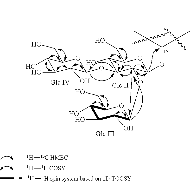
C00015

C00016
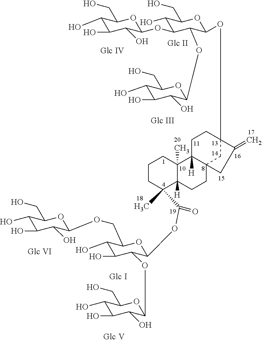
C00017
D00001
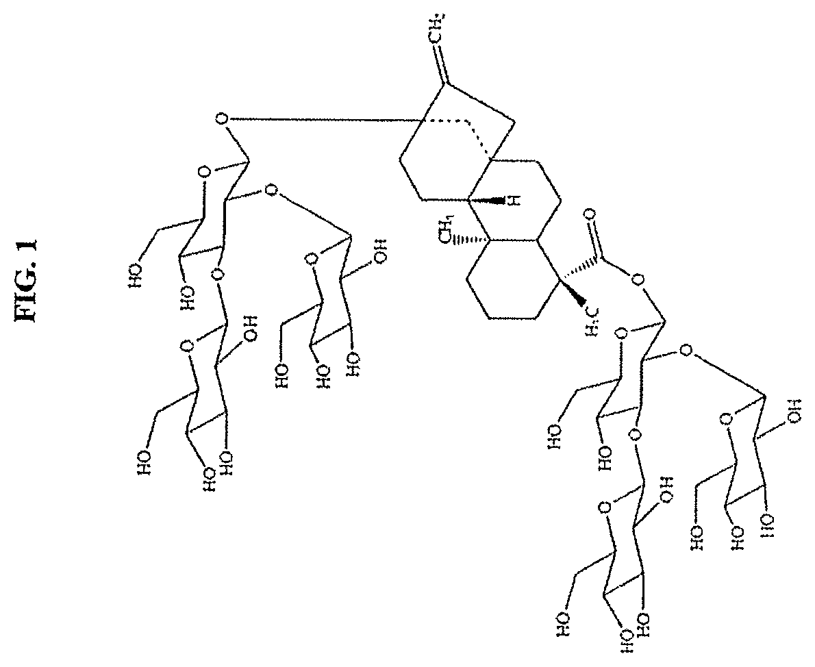
D00002
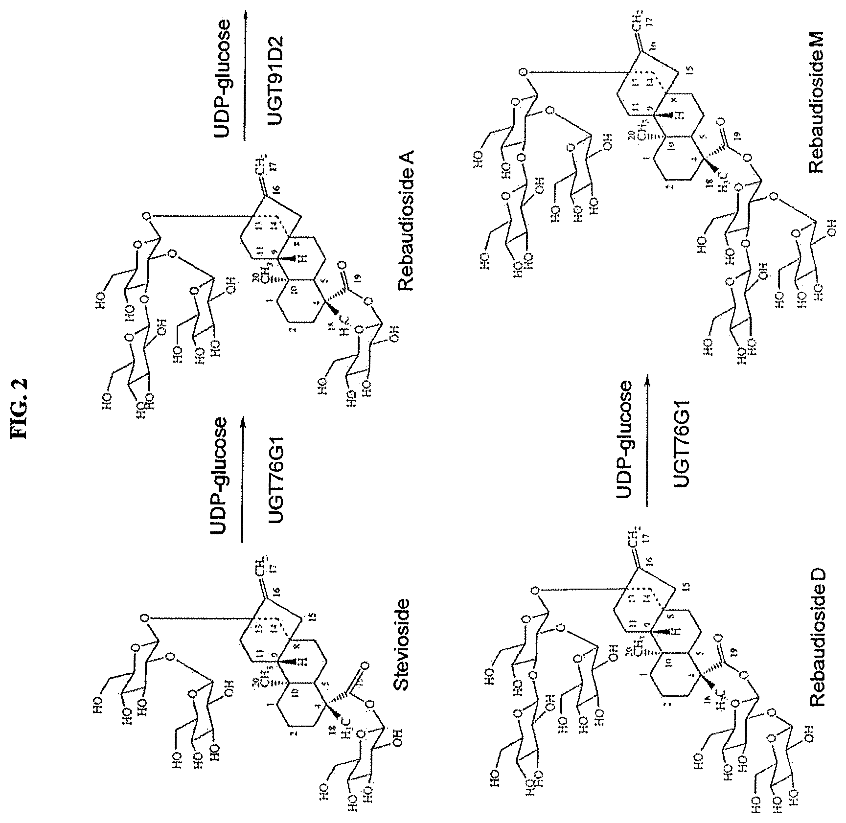
D00003
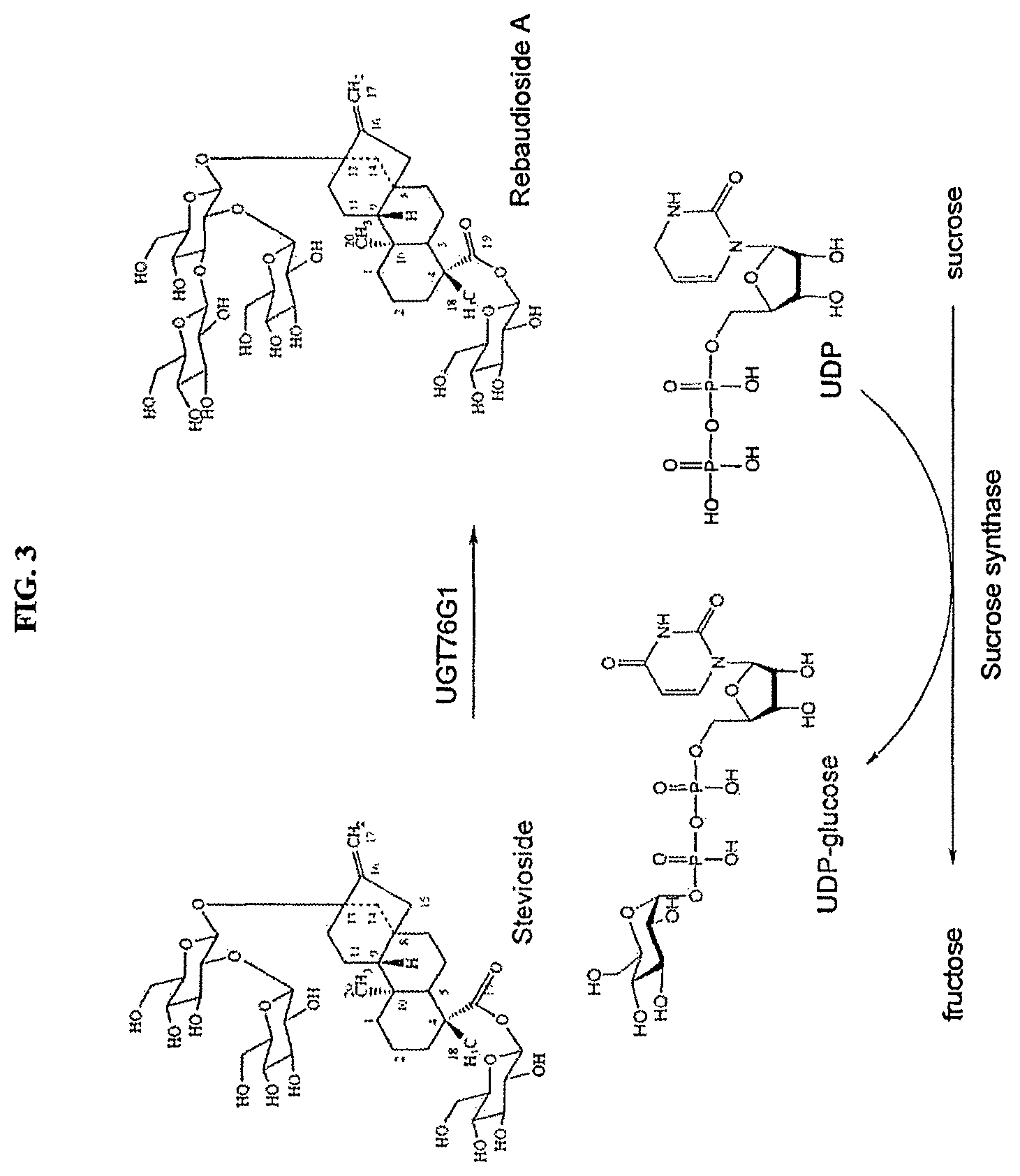
D00004
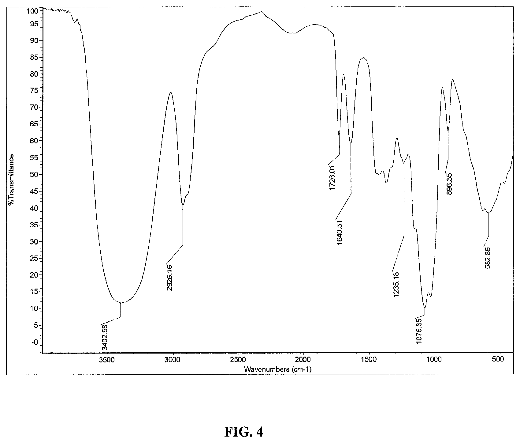
D00005

D00006
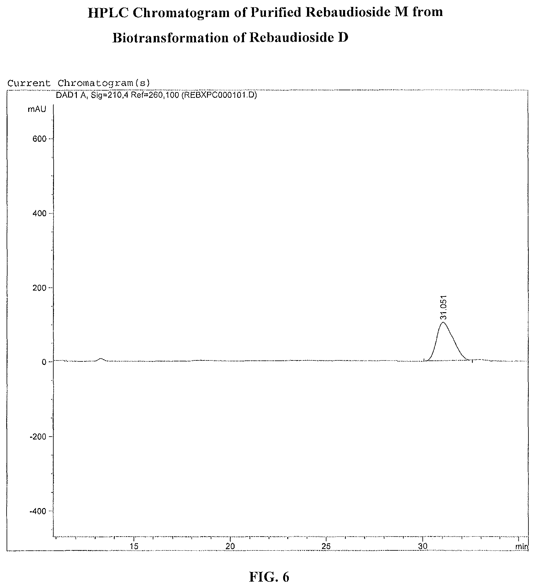
D00007
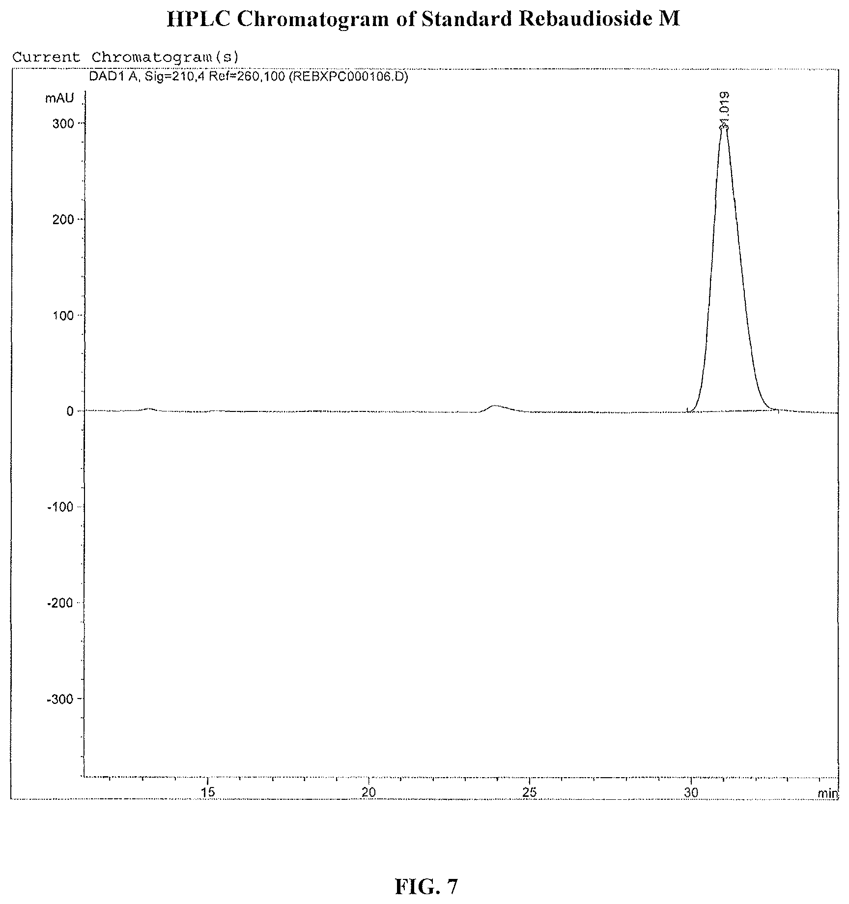
D00008
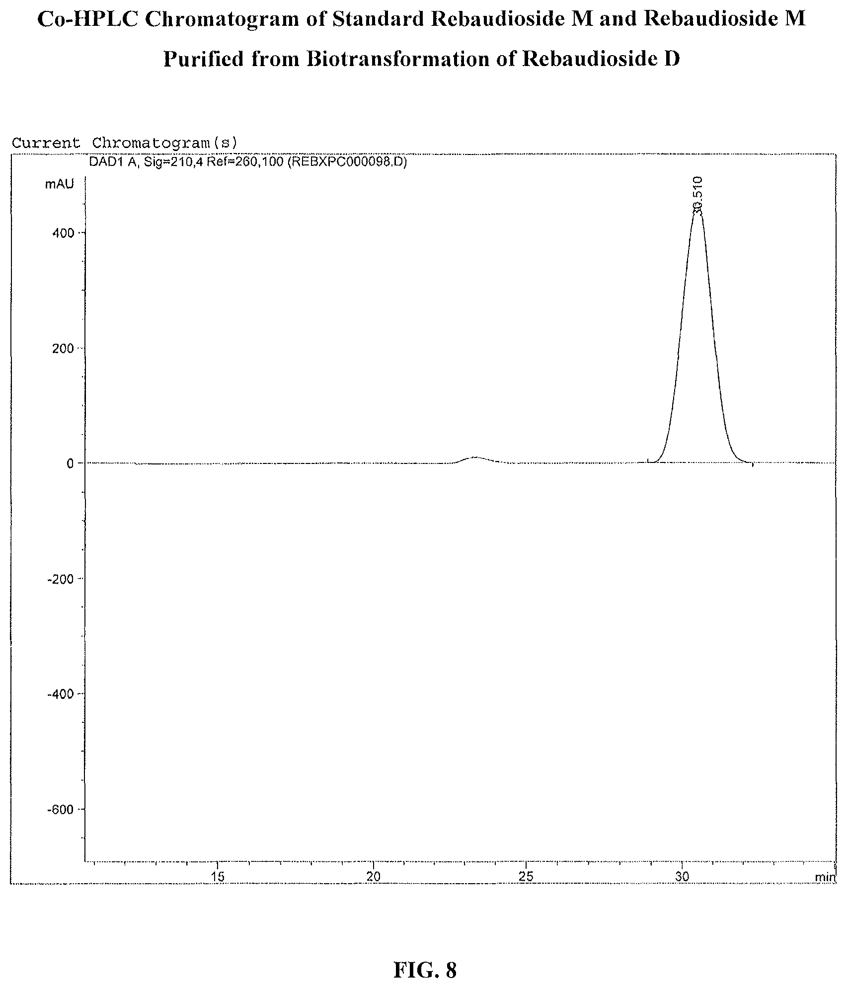
D00009
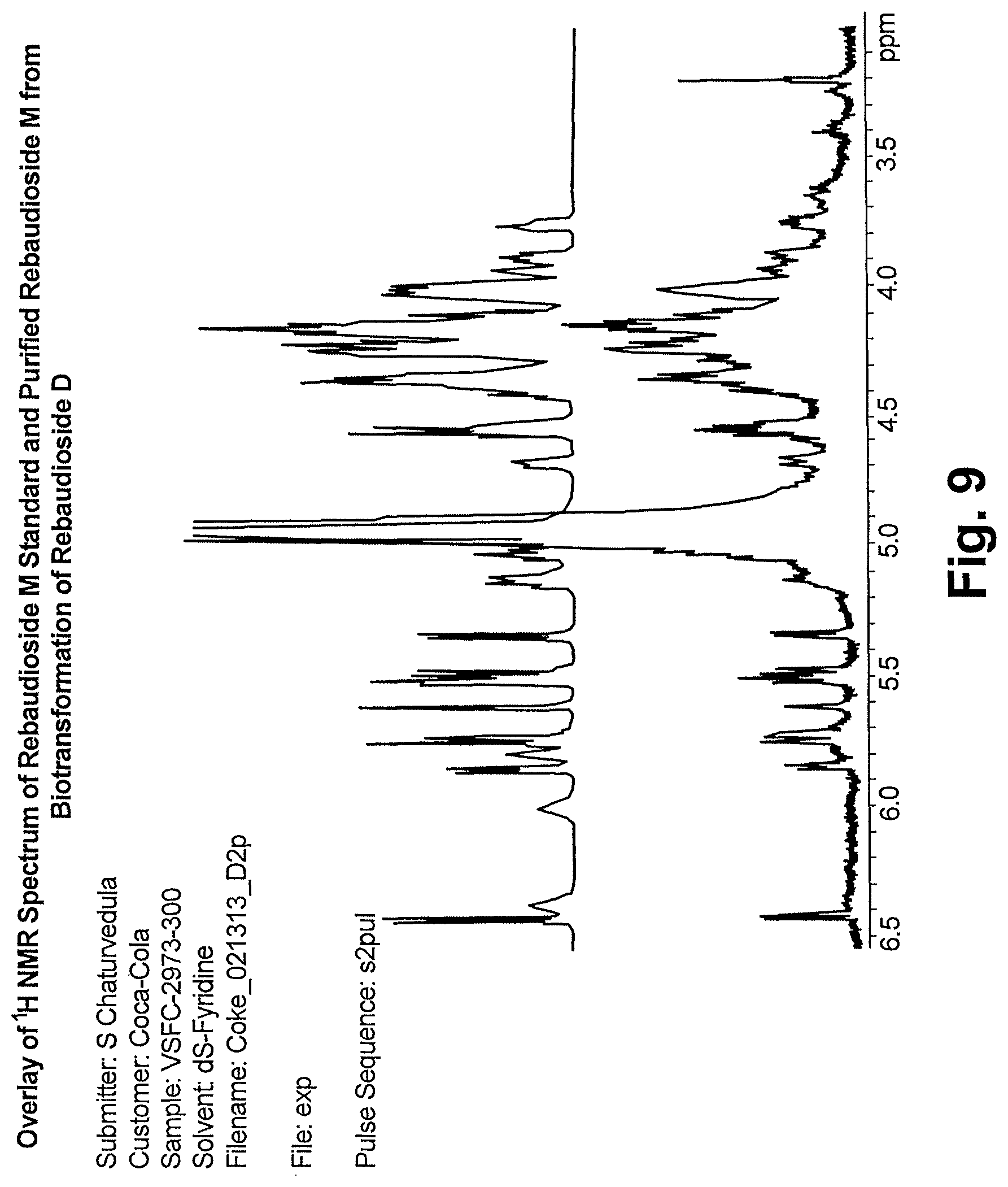
D00010

D00011
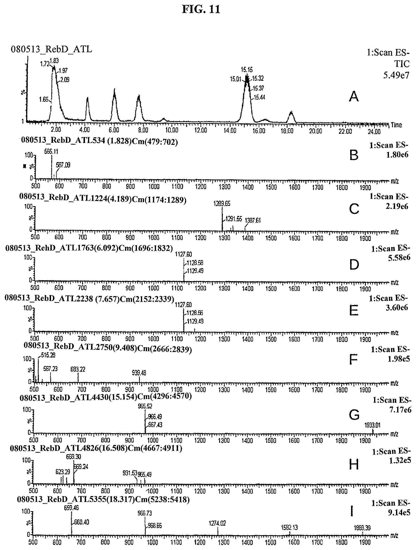
D00012

D00013
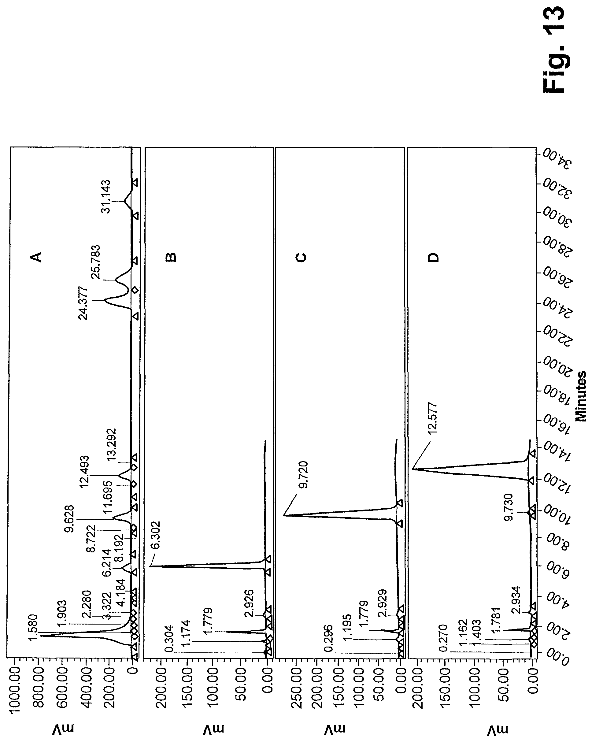
D00014
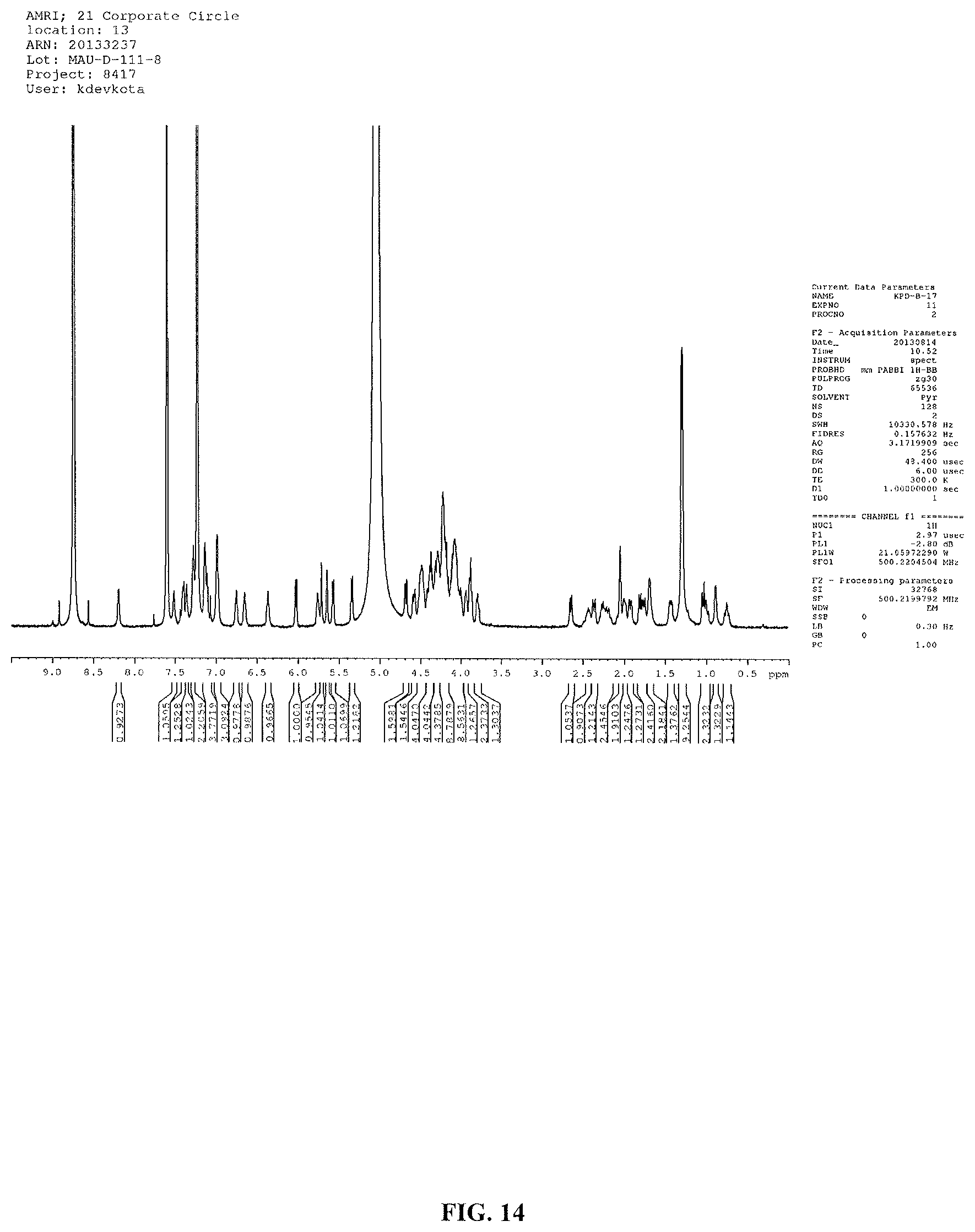
D00015
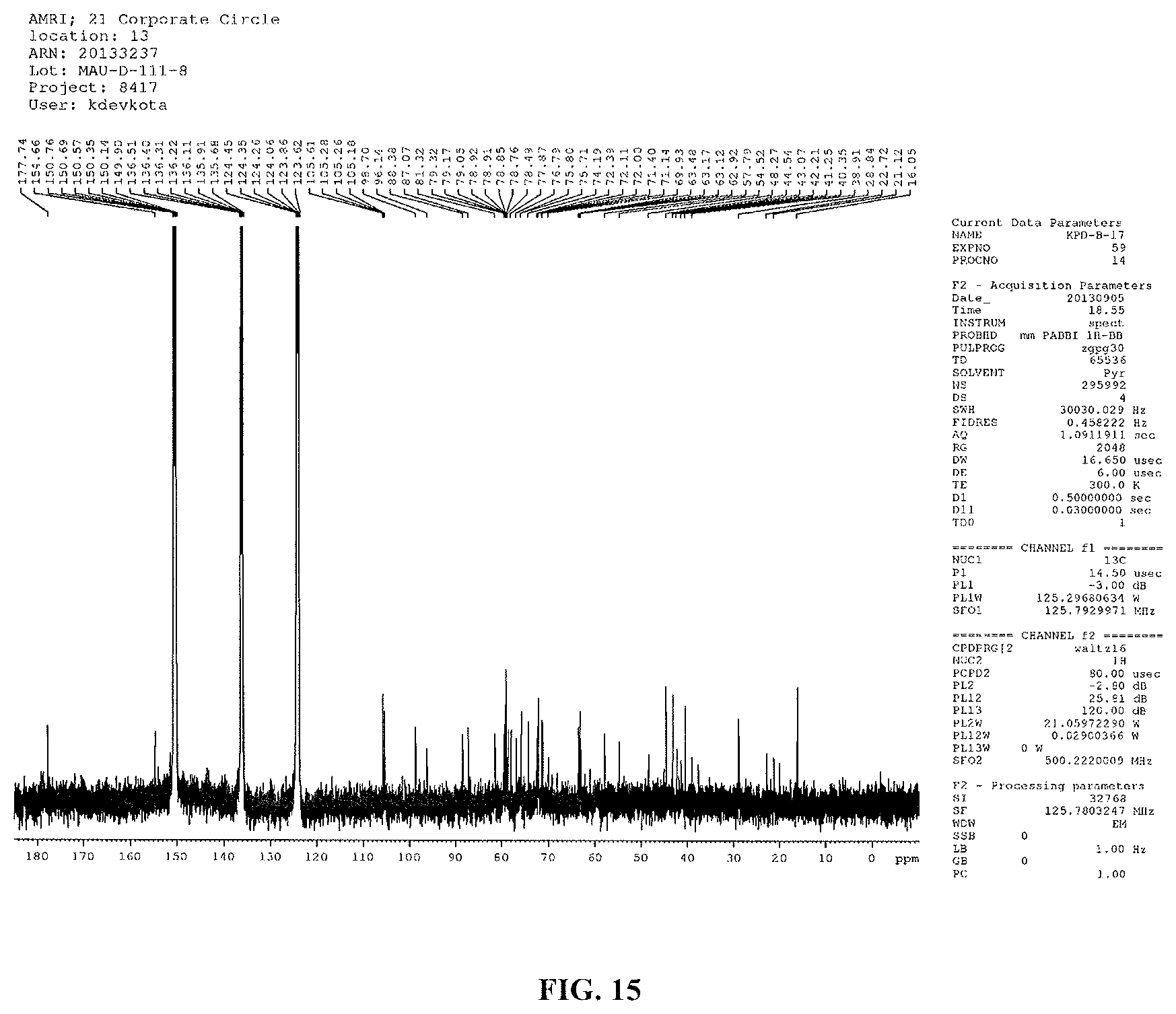
D00016
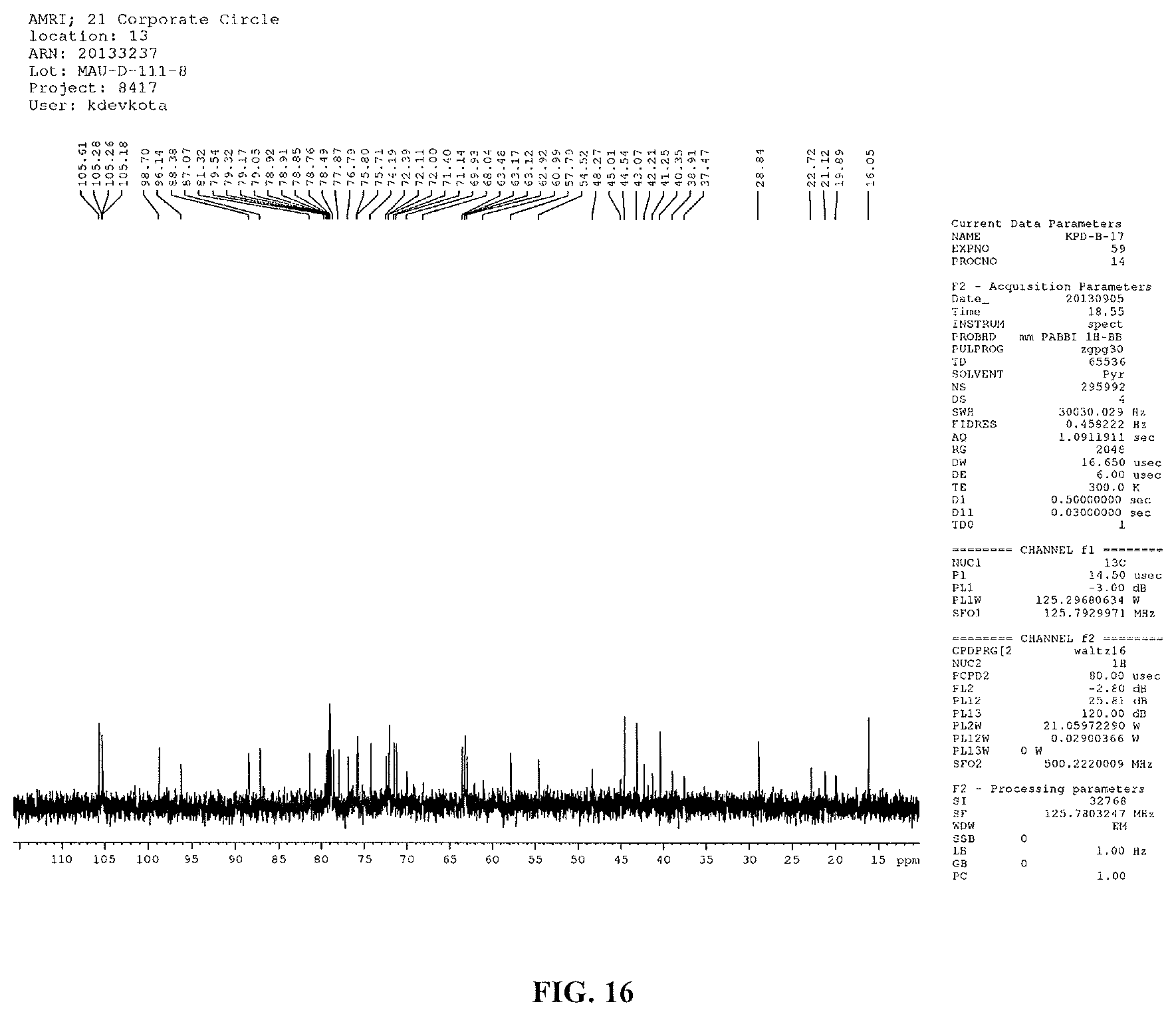
D00017

D00018
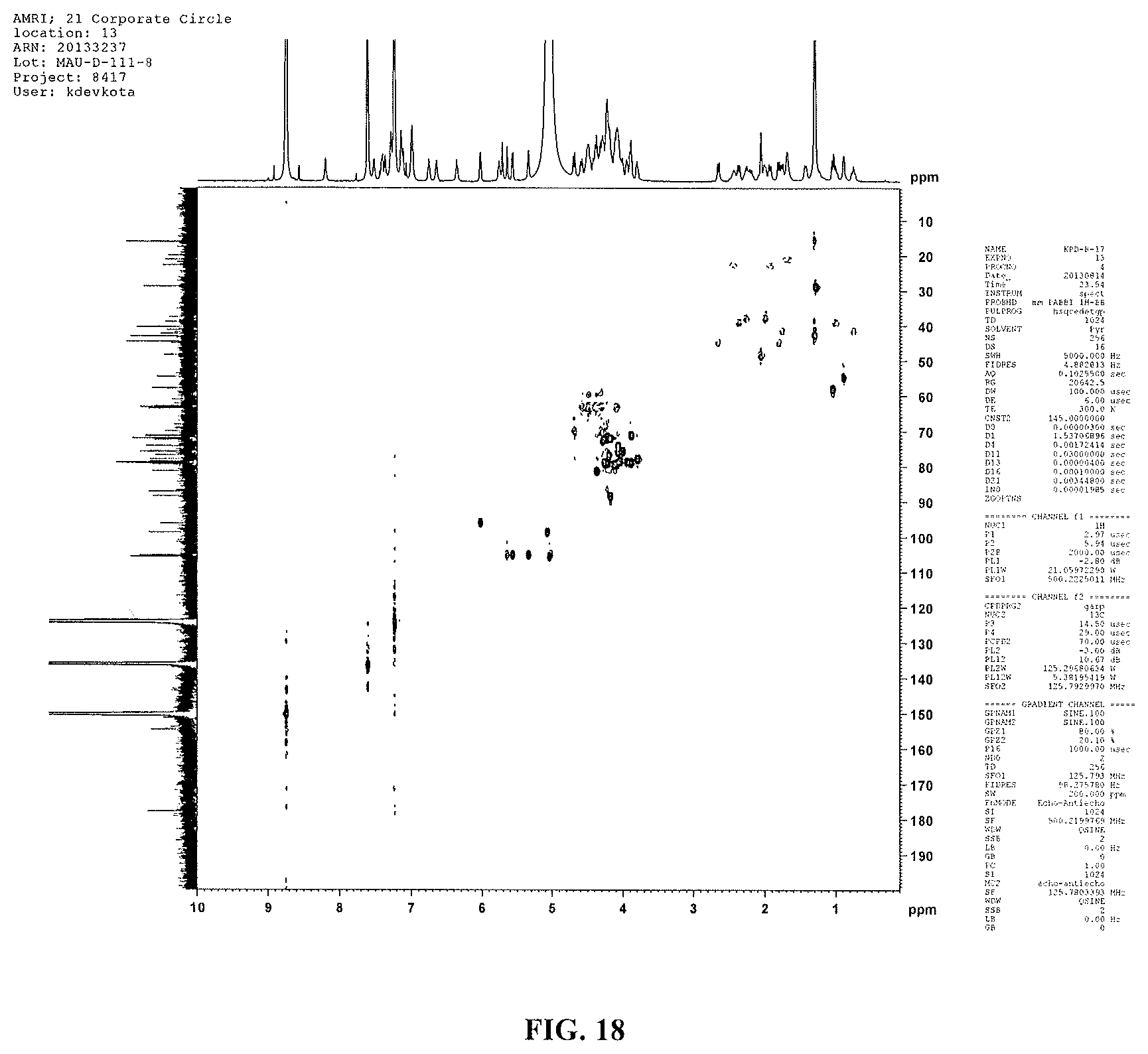
D00019
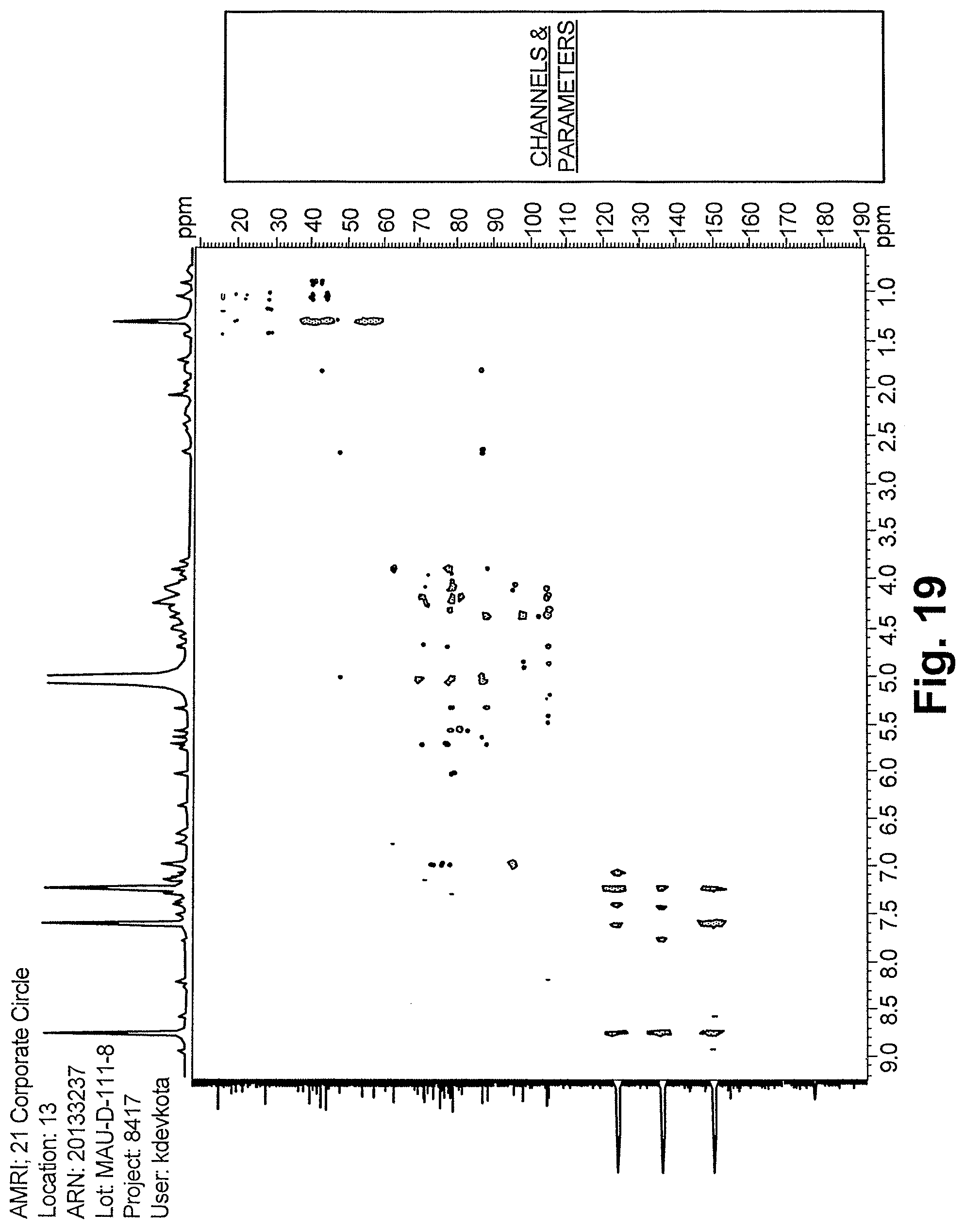
D00020
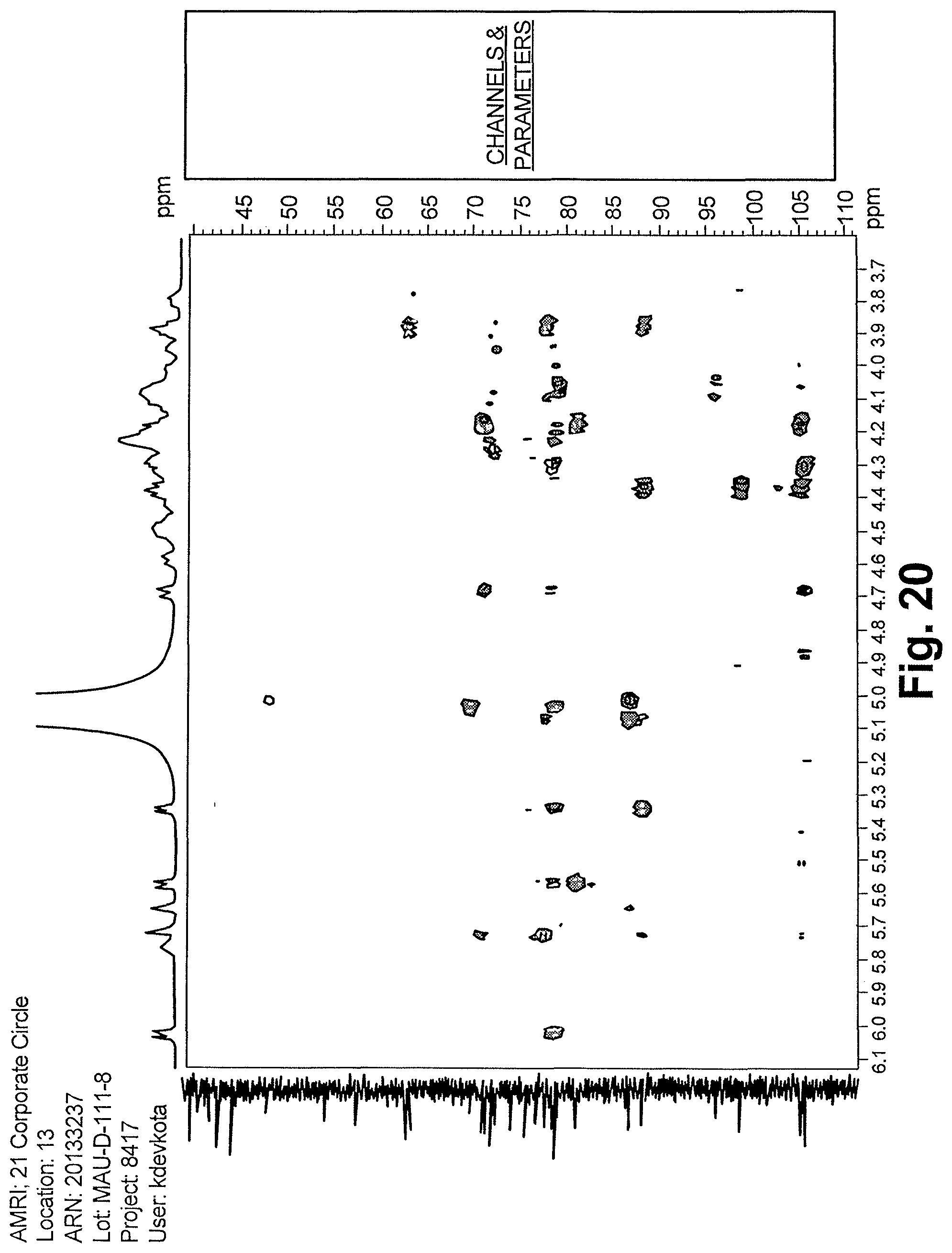
D00021
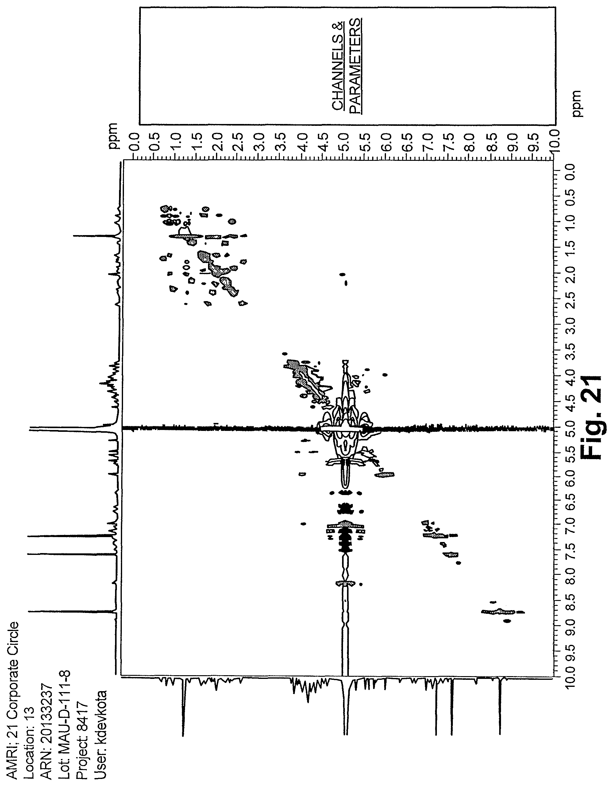
D00022

D00023

D00024

D00025

D00026
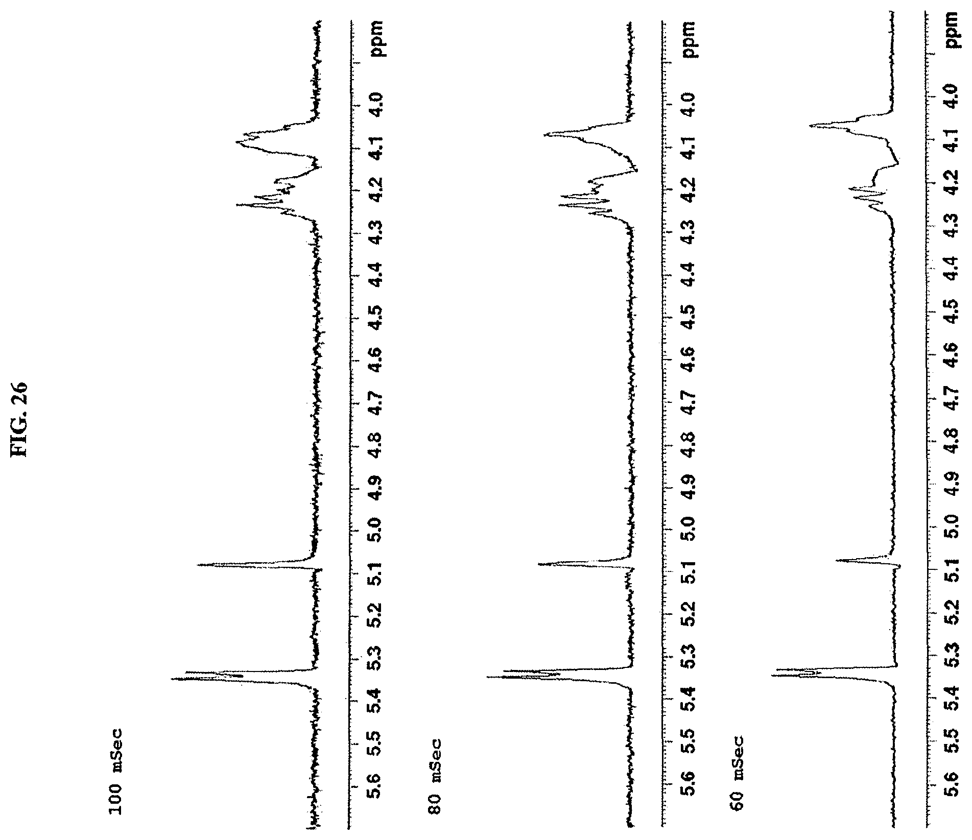
D00027
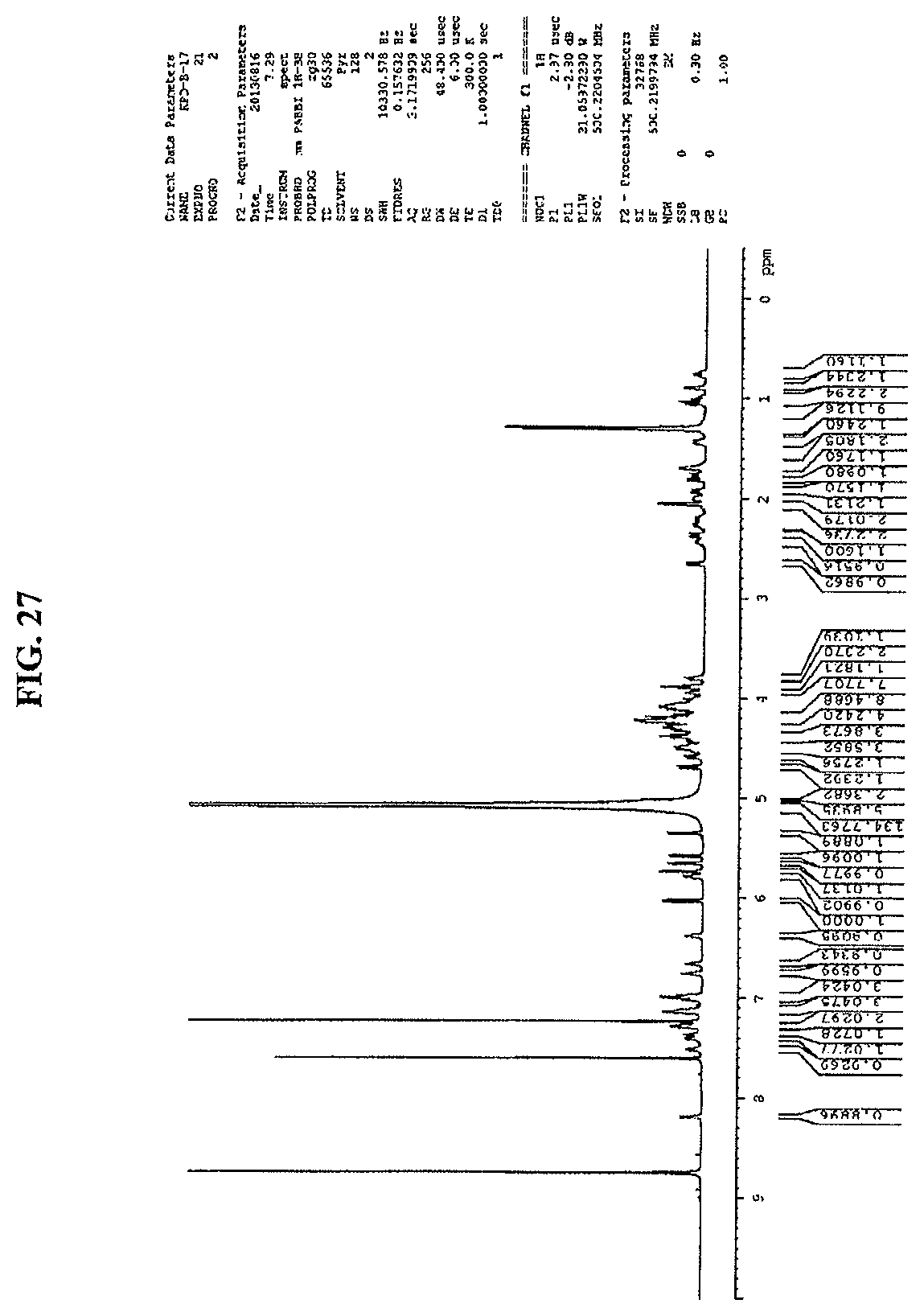
D00028
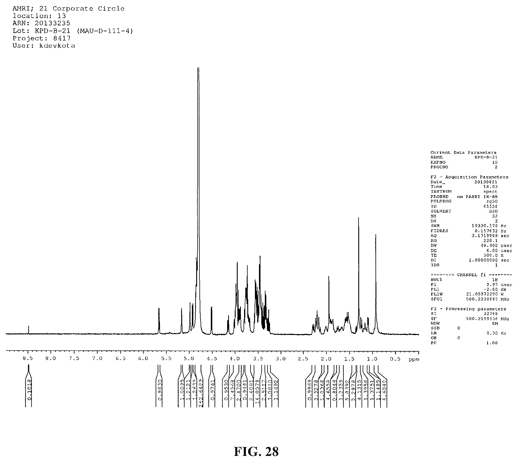
D00029
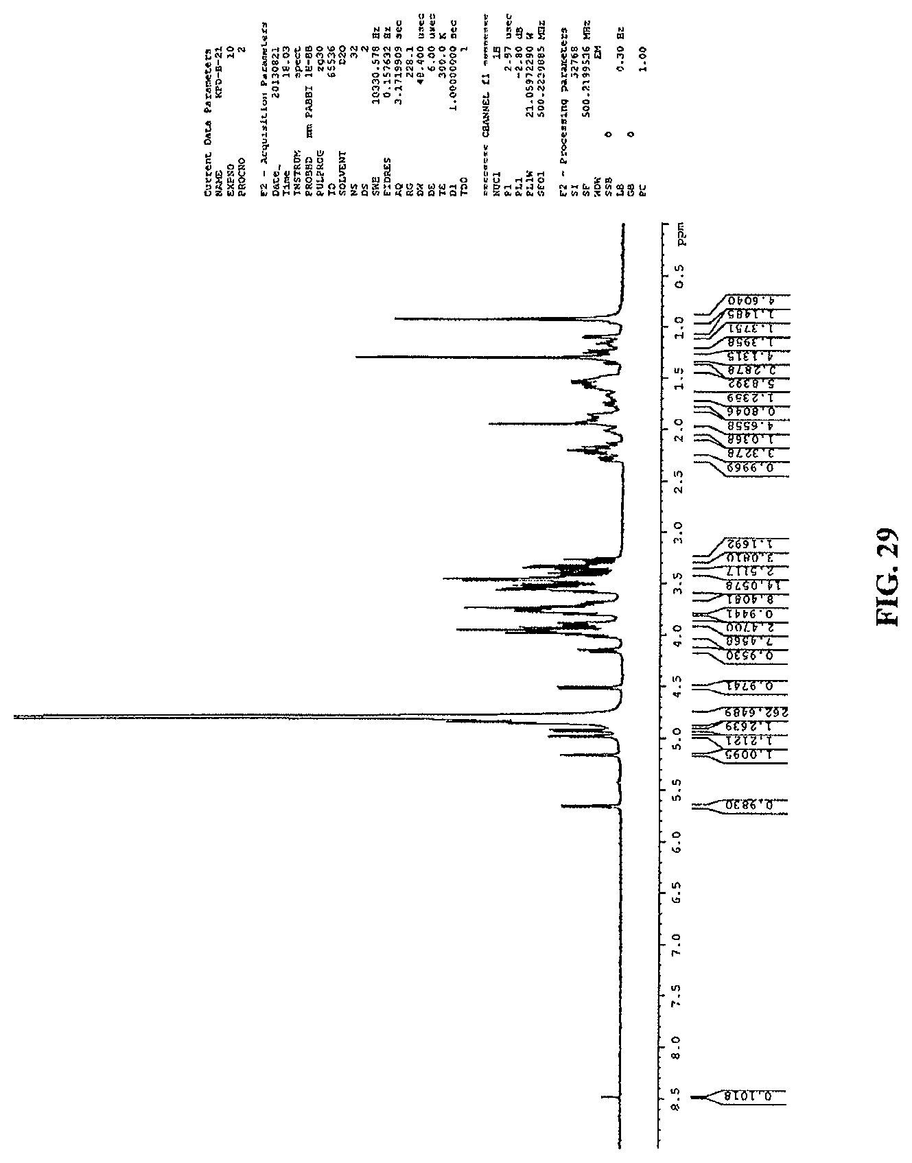
D00030

D00031

D00032
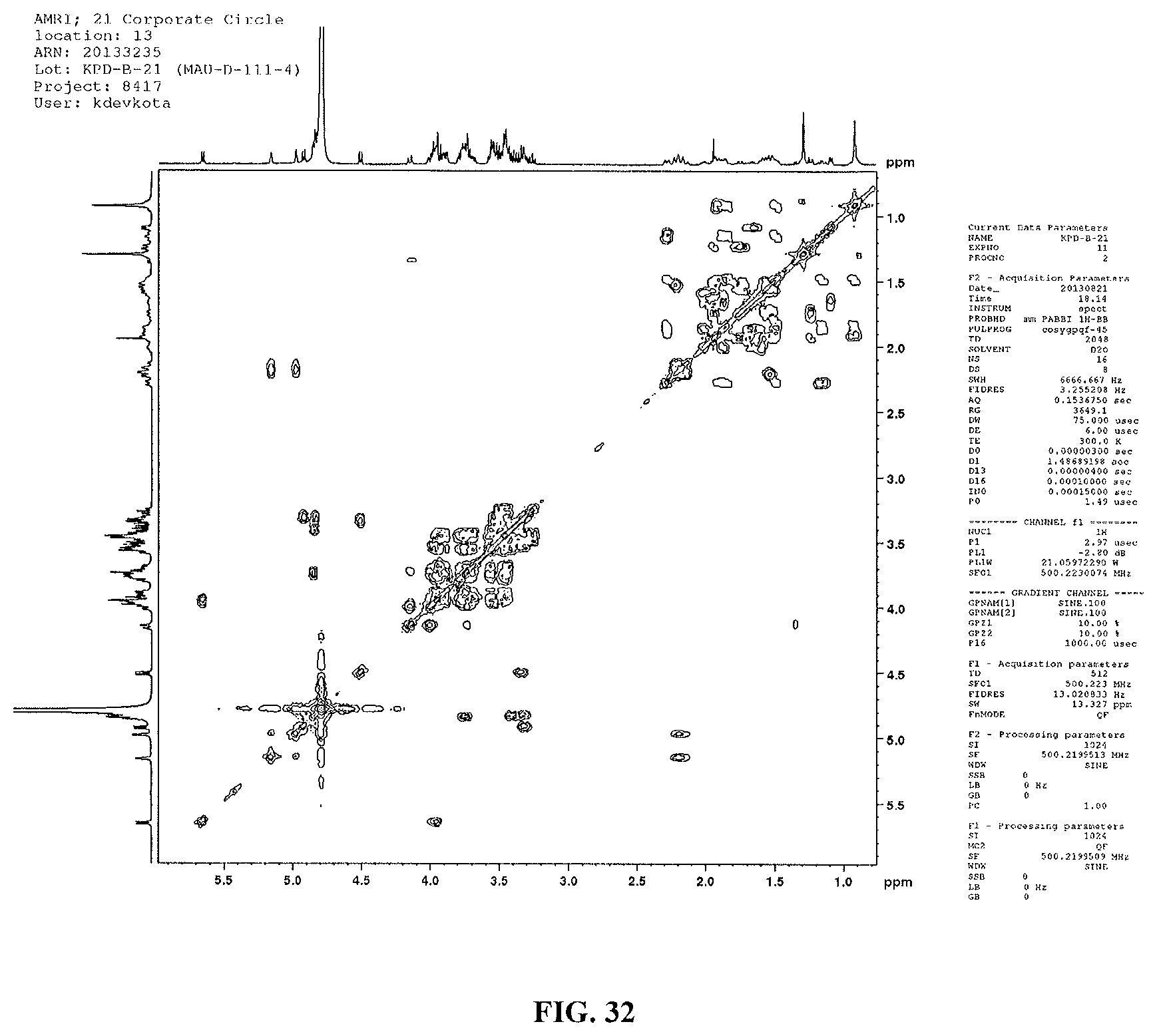
D00033

D00034
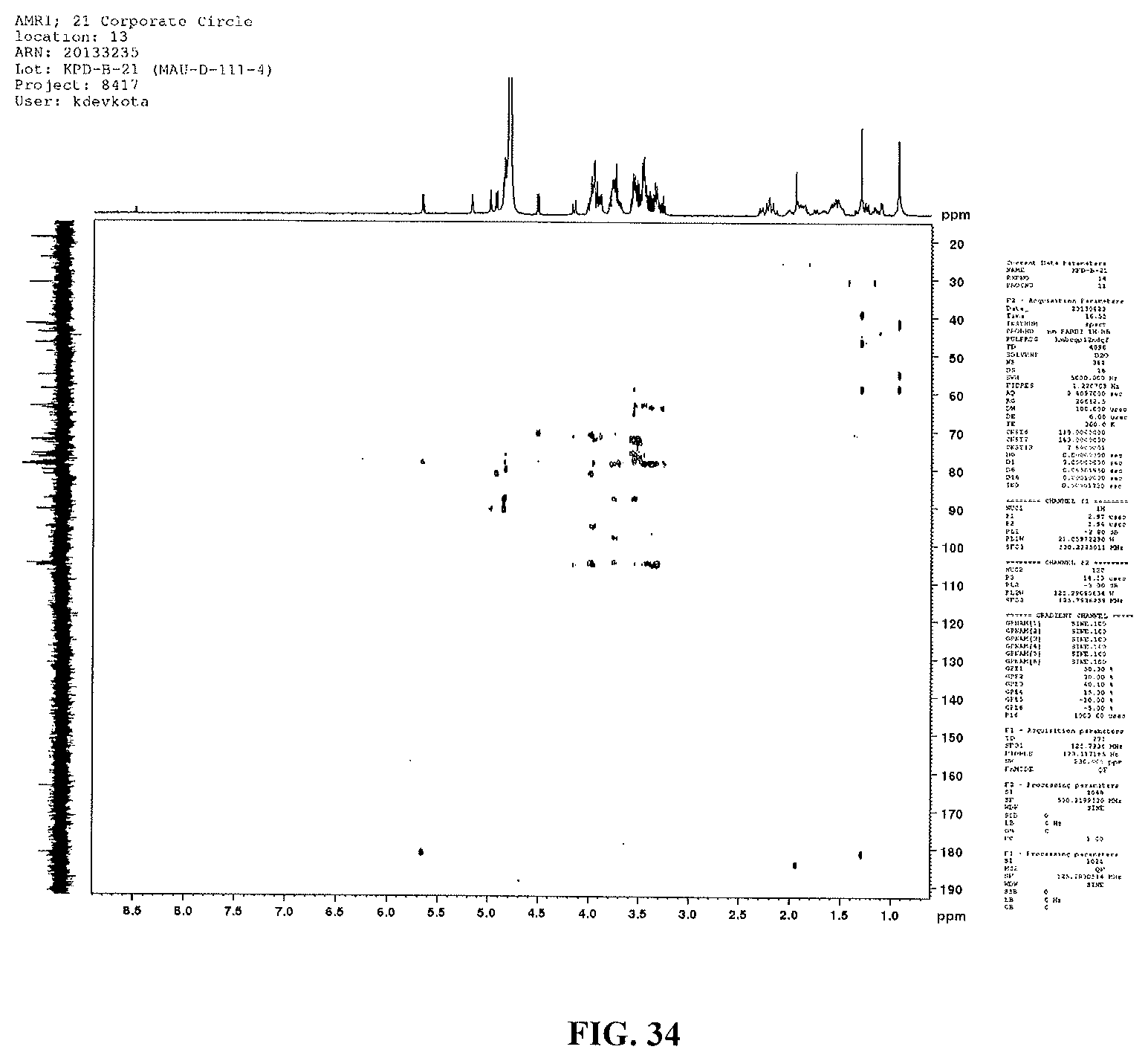
D00035
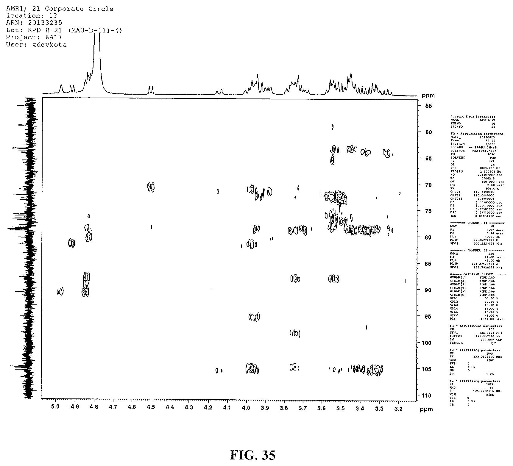
D00036
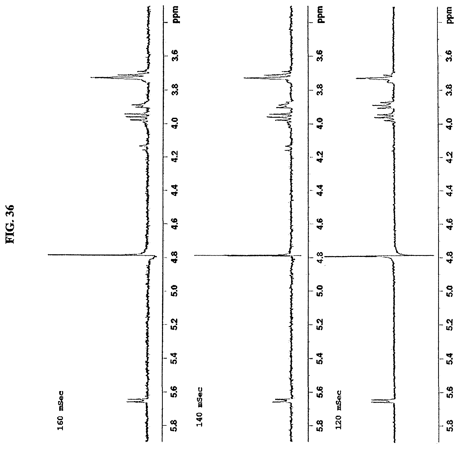
D00037
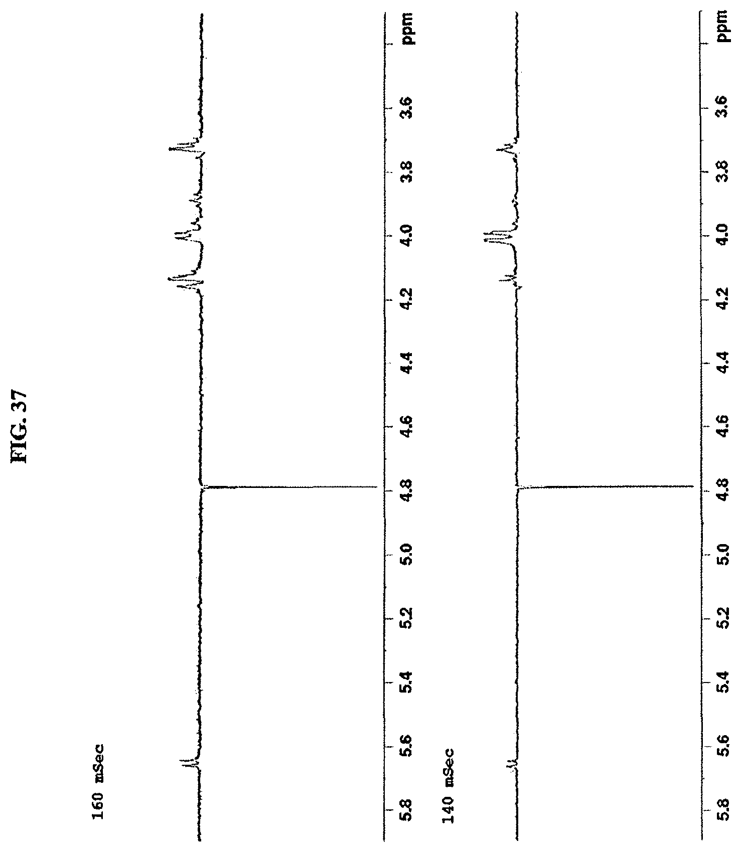
D00038
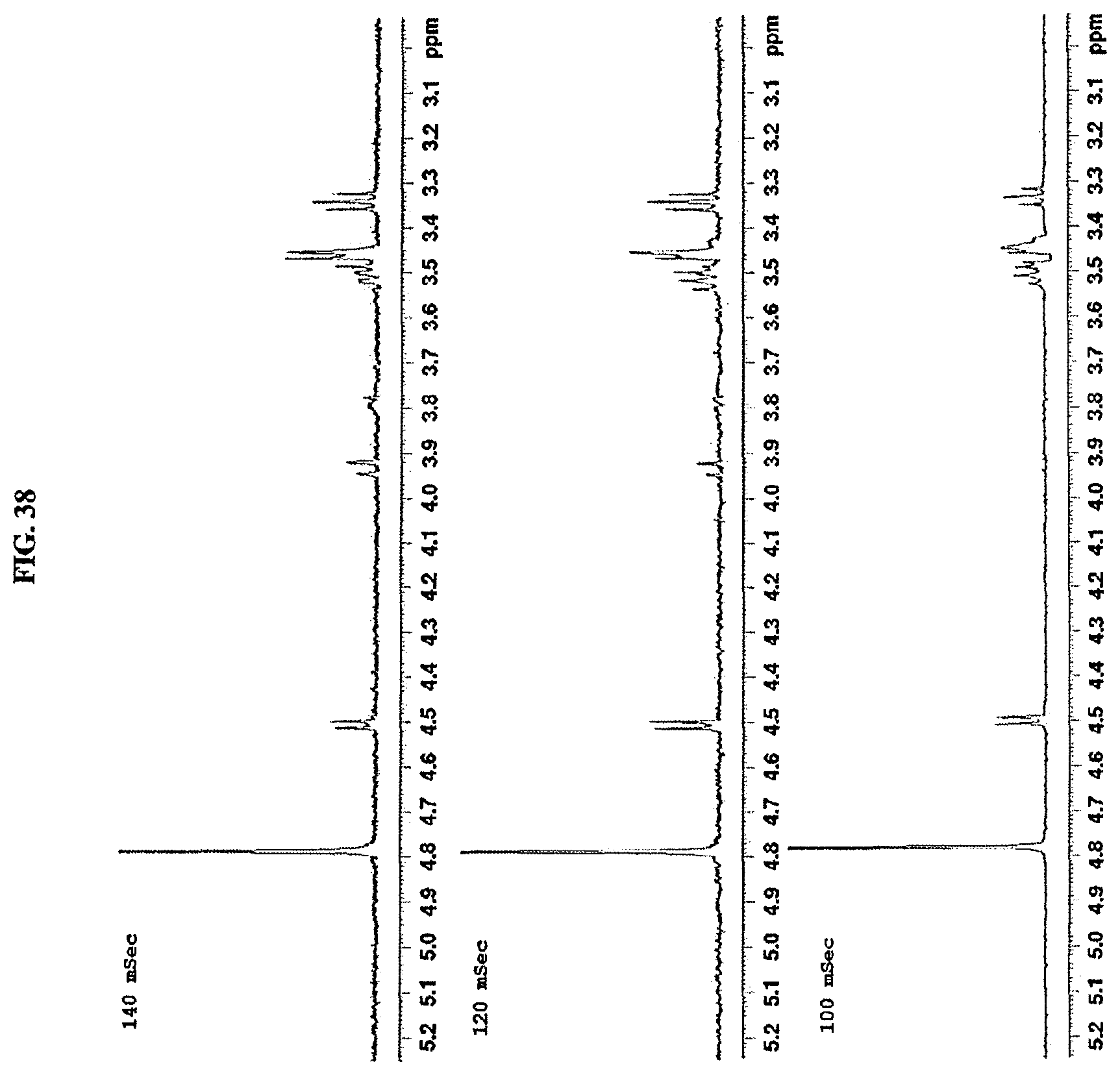
D00039

D00040
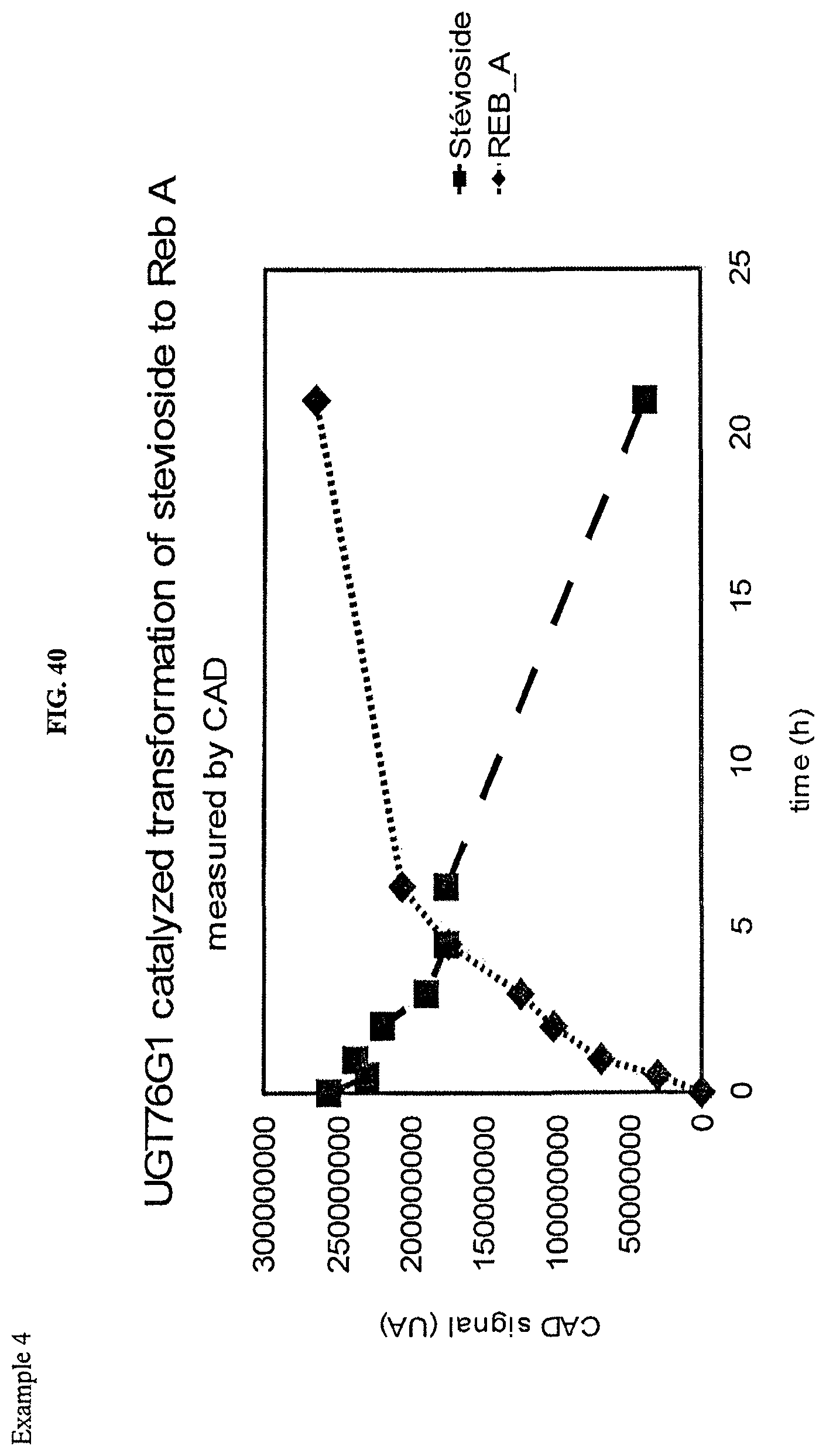
D00041
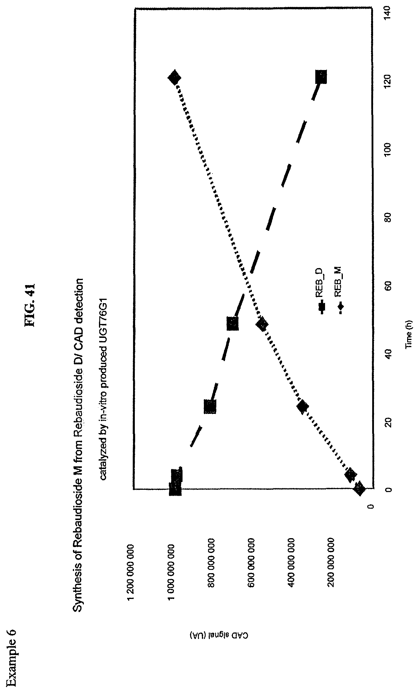
D00042

D00043
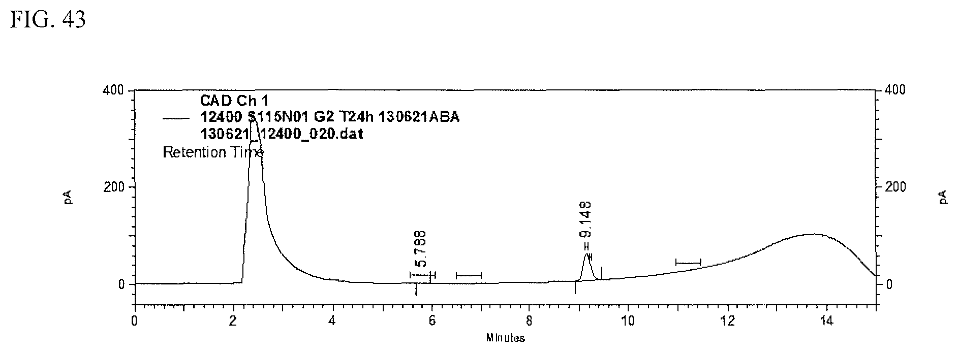
D00044
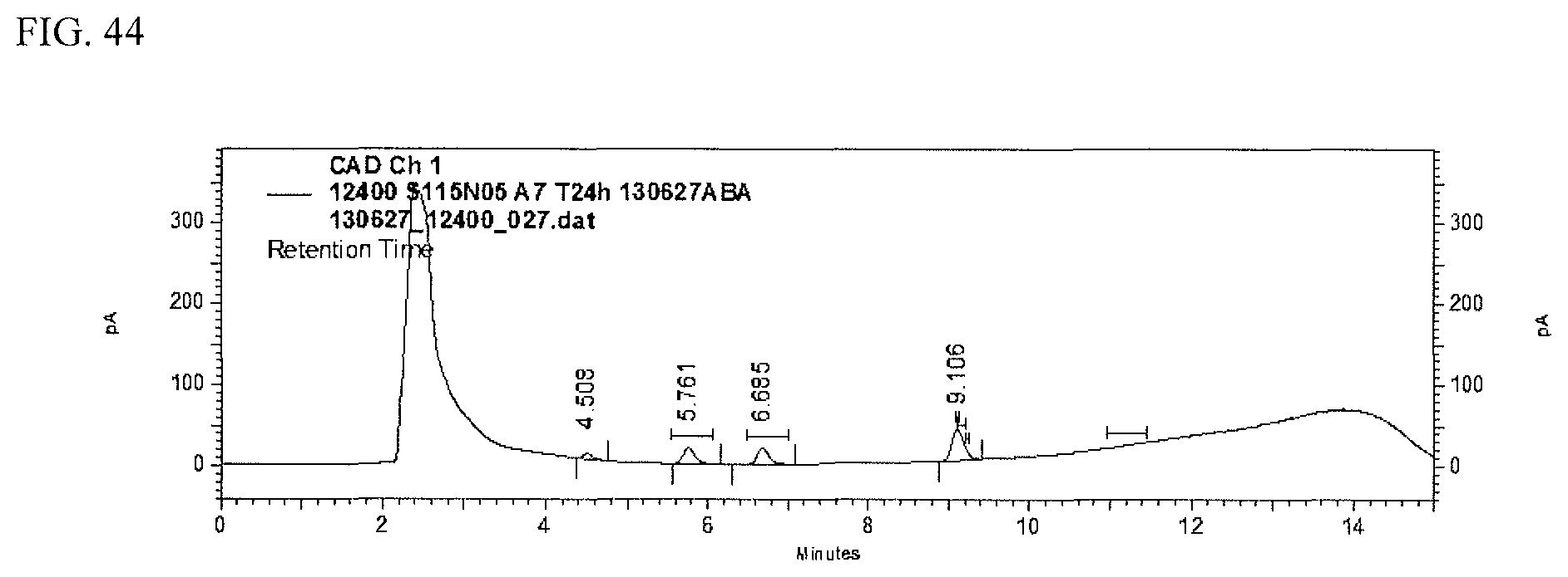
D00045
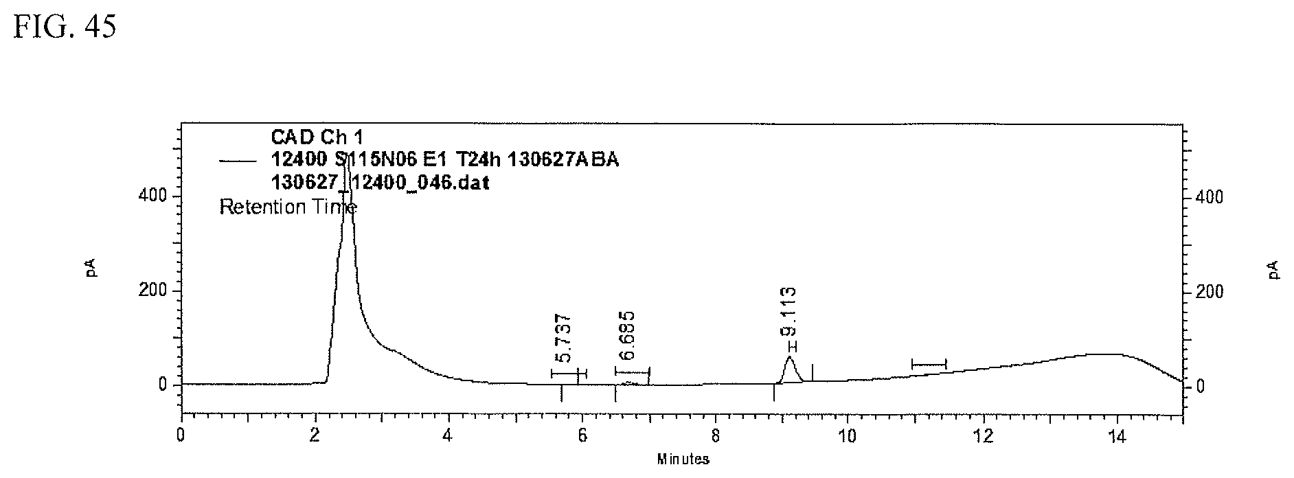
D00046

D00047

D00048

D00049

D00050

D00051

D00052
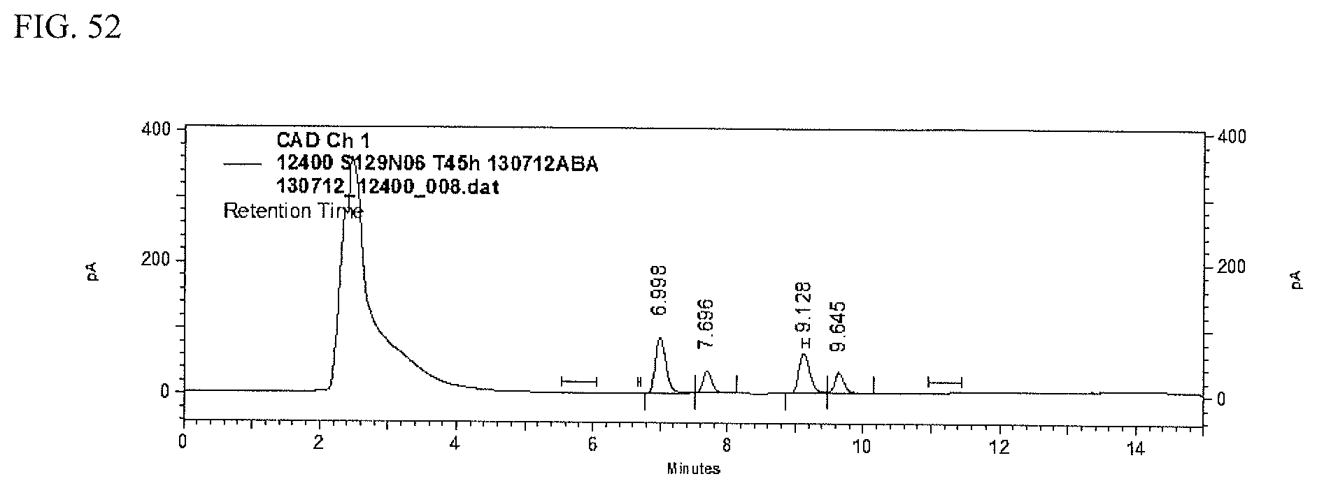
D00053
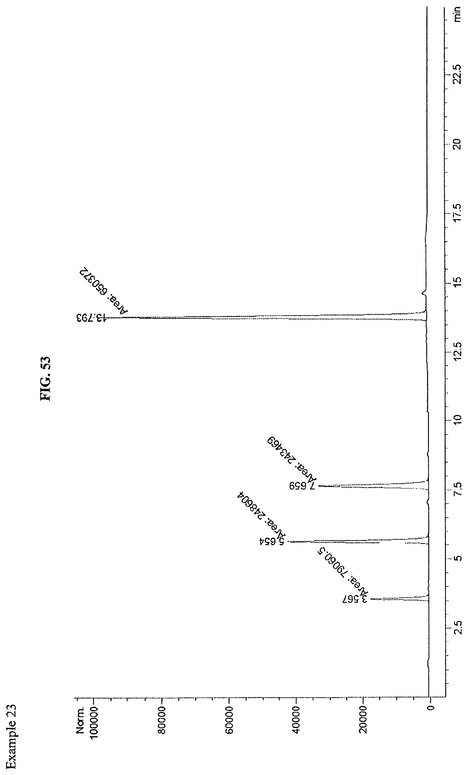
D00054

D00055

D00056
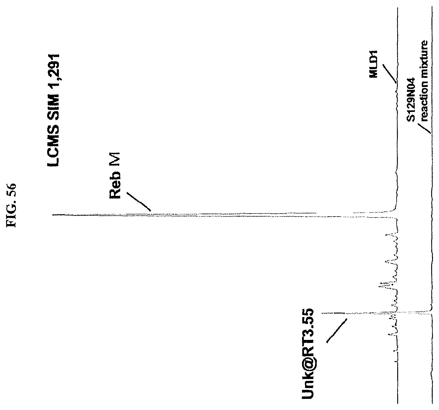
D00057
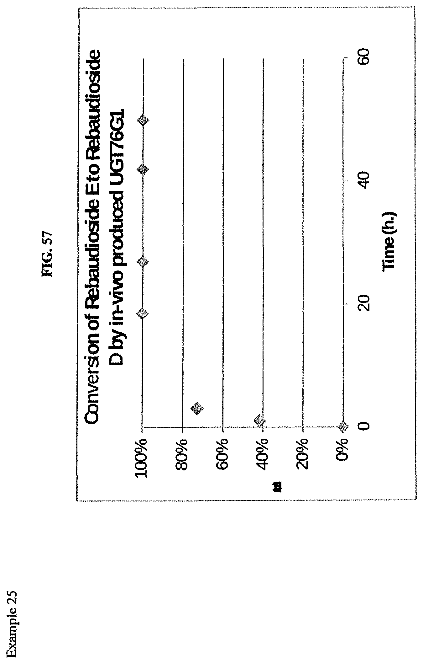
D00058
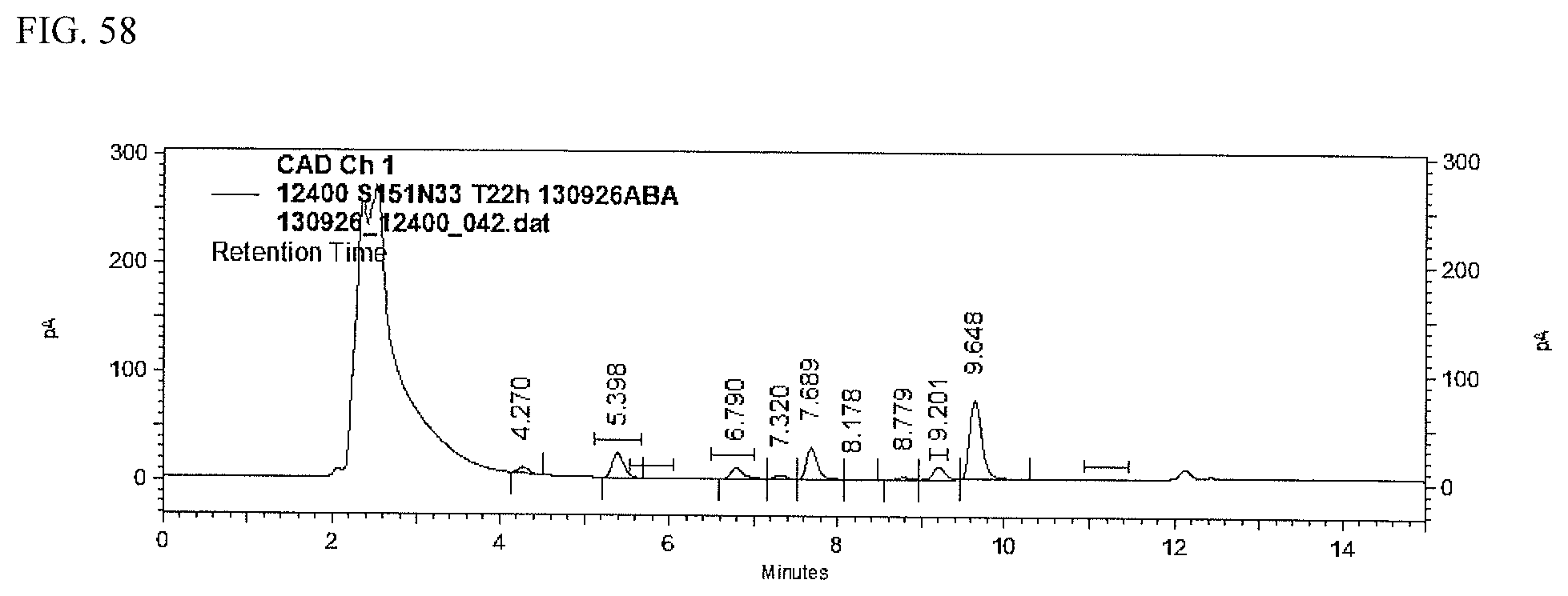
D00059
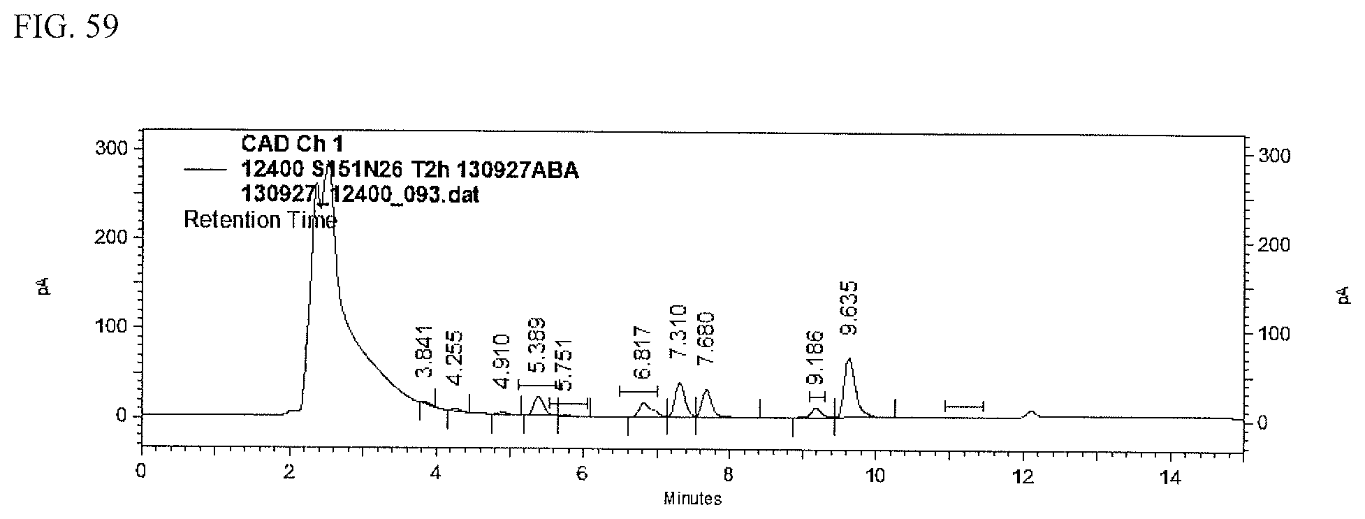
D00060

D00061
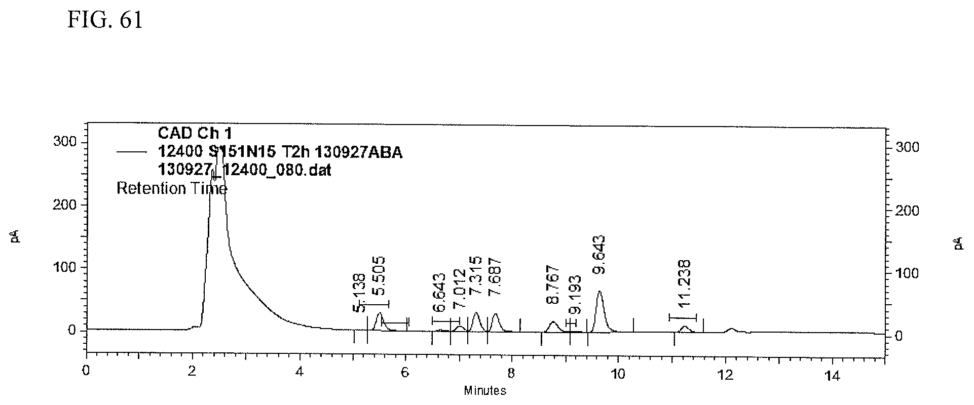
D00062
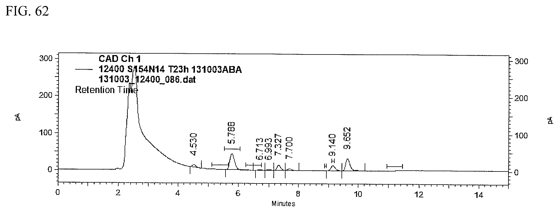
D00063

D00064

D00065

D00066
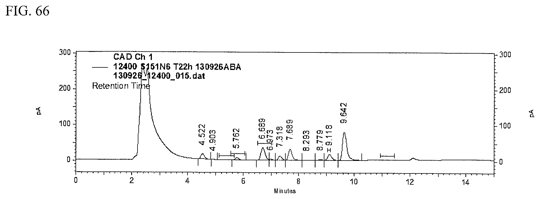
D00067
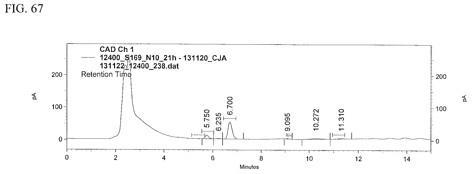
D00068
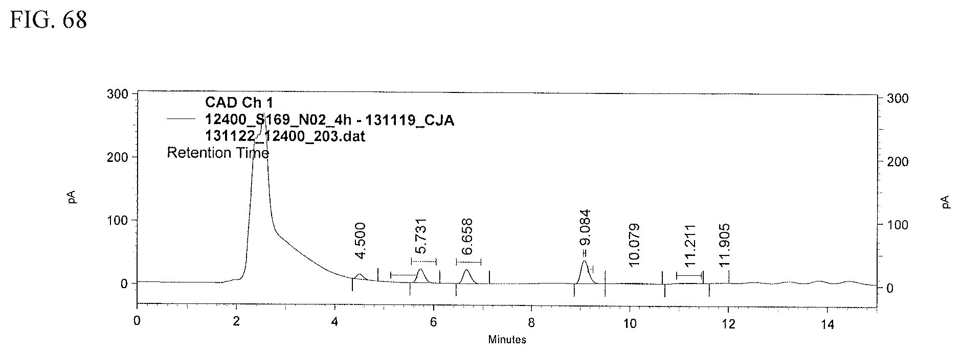
D00069

S00001
XML
uspto.report is an independent third-party trademark research tool that is not affiliated, endorsed, or sponsored by the United States Patent and Trademark Office (USPTO) or any other governmental organization. The information provided by uspto.report is based on publicly available data at the time of writing and is intended for informational purposes only.
While we strive to provide accurate and up-to-date information, we do not guarantee the accuracy, completeness, reliability, or suitability of the information displayed on this site. The use of this site is at your own risk. Any reliance you place on such information is therefore strictly at your own risk.
All official trademark data, including owner information, should be verified by visiting the official USPTO website at www.uspto.gov. This site is not intended to replace professional legal advice and should not be used as a substitute for consulting with a legal professional who is knowledgeable about trademark law.