A4B2 nicotinic acetylcholine receptors with reduced or increased nicotine sensitivity and transgenic mouse model for the same
Lasalde-Dominicci , et al.
U.S. patent number 10,676,518 [Application Number 15/141,689] was granted by the patent office on 2020-06-09 for a4b2 nicotinic acetylcholine receptors with reduced or increased nicotine sensitivity and transgenic mouse model for the same. This patent grant is currently assigned to University of Puerto Rico. The grantee listed for this patent is Emir Aviles-Pagan, Nilza M Biaggi-Labiosa, Daniel Caballero-Rivera, Jose A Lasalde-Dominicci. Invention is credited to Emir Aviles-Pagan, Nilza M Biaggi-Labiosa, Daniel Caballero-Rivera, Jose A Lasalde-Dominicci.

| United States Patent | 10,676,518 |
| Lasalde-Dominicci , et al. | June 9, 2020 |
A4B2 nicotinic acetylcholine receptors with reduced or increased nicotine sensitivity and transgenic mouse model for the same
Abstract
The .alpha.4.beta.2 neuronal nicotinic acetylcholine receptor (nAChR) plays a crucial role in nicotine addiction. The invention studied the effect of subunit phosphorylation on .alpha.4.beta.2 nAChR function and expression, and eleven residues located in the M3-M4 cytoplasmic loop were mutated to alanine and aspartic acid. When nicotine was used as an agonist, four mutations exhibited a statistically significant hypersensitivity to nicotine (S438D, S469A, Y576A, and S589A). Additionally, two mutations (S516D and T536A) that displayed normal activation with ACh displayed remarkable reductions in sensitivity to nicotine. The invention provides a knock-in mutant construct for the development of a transgenic mouse line with reduced nicotine sensitivity to be used in future studies.
| Inventors: | Lasalde-Dominicci; Jose A (San Juan, PR), Biaggi-Labiosa; Nilza M (San Juan, PR), Aviles-Pagan; Emir (San Juan, PR), Caballero-Rivera; Daniel (San Juan, PR) | ||||||||||
|---|---|---|---|---|---|---|---|---|---|---|---|
| Applicant: |
|
||||||||||
| Assignee: | University of Puerto Rico (San
Juan, PR) |
||||||||||
| Family ID: | 70973176 | ||||||||||
| Appl. No.: | 15/141,689 | ||||||||||
| Filed: | April 28, 2016 |
Related U.S. Patent Documents
| Application Number | Filing Date | Patent Number | Issue Date | ||
|---|---|---|---|---|---|
| 62153660 | Apr 28, 2015 | ||||
| Current U.S. Class: | 1/1 |
| Current CPC Class: | A01K 67/0275 (20130101); C07K 14/70571 (20130101); C12N 15/8509 (20130101); A01K 2267/03 (20130101); A01K 2217/054 (20130101); C12N 2015/8527 (20130101); A01K 2227/105 (20130101) |
| Current International Class: | C07K 14/705 (20060101); A01K 67/027 (20060101) |
References Cited [Referenced By]
U.S. Patent Documents
| 2009/0100532 | April 2009 | Drenan |
Other References
|
Gaj et al. Trends in Technology 31(7):397-405, Jul. 2013 (Year: 2013). cited by examiner . Lilico et al. Scientific Reports 3: Article No. 2847. Dio:10.1038/srep02847. pp. 1-4, Oct. 10, 2013 (Year: 2013). cited by examiner . Carlson et al. PNAS 109(43):17382-17387, Oct. 2012 (Year: 2012). cited by examiner . Ji et al. Transgenic Res. 24:227-235, 2015 (Year: 2015). cited by examiner . Liable et al. Biotechnology Journal 10:109-120, 2015 (Year: 2015). cited by examiner . Haruyama et al. Curr Protoc Cel Biol. Mar. 2009. Chapter Unit-19.10. doi:10.1002/0471143030.cb11910s42. pp. 1-12 (Year: 2009). cited by examiner . Roberts et al. Development 141:715-724, 2014 (Year: 2014). cited by examiner . Ruan et al. Scientific Reports 5:14253, DIO:10.1038/srep14253, pp. 1-10, 2015 (Year: 2015). cited by examiner . Leslie et al. Molecular Pharmacology 83:753-758, 2013. (Year: 2013). cited by examiner. |
Primary Examiner: Li; Ruixiang
Attorney, Agent or Firm: Hoglund & Pamias, PSC Rios; Roberto J.
Government Interests
GOVERNMENT INTEREST
The claimed invention was made with U.S. Government support under grant numbers R25 GM061151 and P20 GM103642 awarded by the National Institutes of Health (NIH). The government has certain rights in this invention.
Claims
We claim:
1. A mutant nicotinic acetylcholine receptor comprising a point mutation of threonine (T) to alanine (A) in the M3-M4 cytoplasmic loop of alpha 4 subunit at position 536 corresponding to the amino acid residue position 202 of the amino acid sequence set forth in SEQ ID NO: 1, said mutant nicotinic acetylcholine receptor has reduced nicotine sensitivity.
2. A mutant nicotinic acetylcholine receptor comprising a point mutation of serine (S) to aspartic acid (D) in the M3-M4 cytoplasmic loop of the alpha 4 subunit at the position 516 corresponding to the amino acid residue position 182 of the amino acid sequence set forth in SEQ ID NO: 1, said mutant nicotinic acetylcholine receptor has reduced nicotine sensitivity.
Description
SEQUENCE LISTING
The instant application contains a Sequence Listing which has been submitted electronically in ASCII format and is hereby incorporated by reference in its entirety. Said ASCII copy, created on Feb. 16, 2018, is named UPR-15238_SL.txt and is 7,473 bytes in size.
BACKGROUND OF THE INVENTION
Neuronal nicotinic acetylcholine receptors (nAChRs) are transmitted gated ion channels that belong to a gene super family of homologous receptors including .gamma.-aminobutyric acid (GABA), glycine, and serotonin receptors. The receptor is composed of five subunits with each having four transmembrane domains (M1-M4) and a large intracellular loop (C2). The genes for the subunits that have been cloned so far are divided into two subfamilies of nine .alpha. (.alpha.2-.alpha.10) and three .beta. (.beta.2-.beta.4) subunits, and are expressed in the nervous system, cochlea, and a number of non-neuronal tissues.
Neuronal nAChRs have a role in the mediation of tolerance and addiction to nicotine in chronic tobacco users and the symptoms of withdrawal experienced upon cessation of use. The complex activities of nicotine in the nervous system are due to its ability to mimic the activity of acetylcholine (ACh) on these receptors. At the molecular level, chronic nicotine exposure differentially affects the number (up-regulation), subunit composition, stoichiometry, and functional status (desensitization and inactivation) of some nAChR subtypes, leaving others substantially unaffected. Nicotine, the addictive component of tobacco, has a predominant effect in the brain mainly on the .alpha.4.beta.2 nAChR, the most abundant subtype.
Desensitization and up-regulation of nAChRs is thought to involve phosphorylation mediated by various kinases. For instance, protein kinase A (PKA) and protein kinase C (PKC) are examples of kinases that have been studied in the context of nAChR desensitization and up-regulation. Specifically, studies suggest that in the continuous presence of nicotine, receptors would be driven into inactive/desensitized conformations, a process likely influenced by the level of phosphorylation. There is evidence that activators of PKA and PKC increase nAChR binding sites and synergistically enhance nicotine-induced receptor up-regulation. These findings are validated by in vitro and in vivo studies, which have demonstrated that .alpha.4 nAChR subunits are phosphorylated and, more specifically, that they are targets of PKA phosphorylation. Studies using phosphopeptide mapping have provided evidence that residues S365, S472, and S491 of the rat .alpha.4 subunit, corresponding to positions S364, S471 and S490 on the current .alpha.4 NCBI reference sequence, are substrates for PKA phosphorylation. In addition, a recent study identified two major substrate sites for PKA phosphorylation on the human .alpha.4 subunit, namely S467 and S362, which are homologues to rat .alpha.4 positions S471 and S364 used in the current study.
Recent work from the inventors has shown that two-point mutations of a PKC phosphorylation residue, S336A and S336D, exhibit constitutive up-regulation when expressed in oocytes. In addition, we found that both of these PKC mutations changed the ACh affinity but exhibited no change in nicotine affinity, suggesting that the properties of agonist binding for .alpha.4.beta.2 channel activation have very distinct dynamic and/or structural requirements for ACh and nicotine.
SUMMARY OF THE INVENTION
On the basis of these findings, we examined various consensus positions in the M3-M4 cytoplasmic loop of the .alpha.4 subunit in an attempt to dissect the potential role of this domain in the functional response of the .alpha.4.beta.2 nAChR. This domain contains conserved consensus sites for various protein kinases (PKA, PKC, CKII, and TK) and has been recently mentioned as a possible target for allosteric sites. In the present study, eleven of these sites were mutated, to alanine and aspartic acid, to study the effect of .alpha.4 subunit phosphorylation on the .alpha.4.beta.2 nAChR activation and expression. The rationale for using alanine and aspartic acid substitutions is that alanine impairs phosphorylation, whereas aspartic acid mimics phosphorylation of the protein. We used ACh and nicotine as agonists to test the functionality of the mutations as compared with the wild-type receptor. The present results reveal a novel allosteric linkage between the M3-M4 cytoplasmic loop of the .alpha.4 neuronal nAChR subunit that can regulate an agonist's selectivity and functional expression. This domain could be considered as a structural target for the development of smoking cessation drugs given that point mutations at this domain can decreased nicotine sensitivity.
BRIEF DESCRIPTION OF THE DRAWINGS
Further features and advantages of the invention will become apparent from the following detailed description taken in conjunction with the accompanying figures showing illustrative embodiments of the invention, in which:
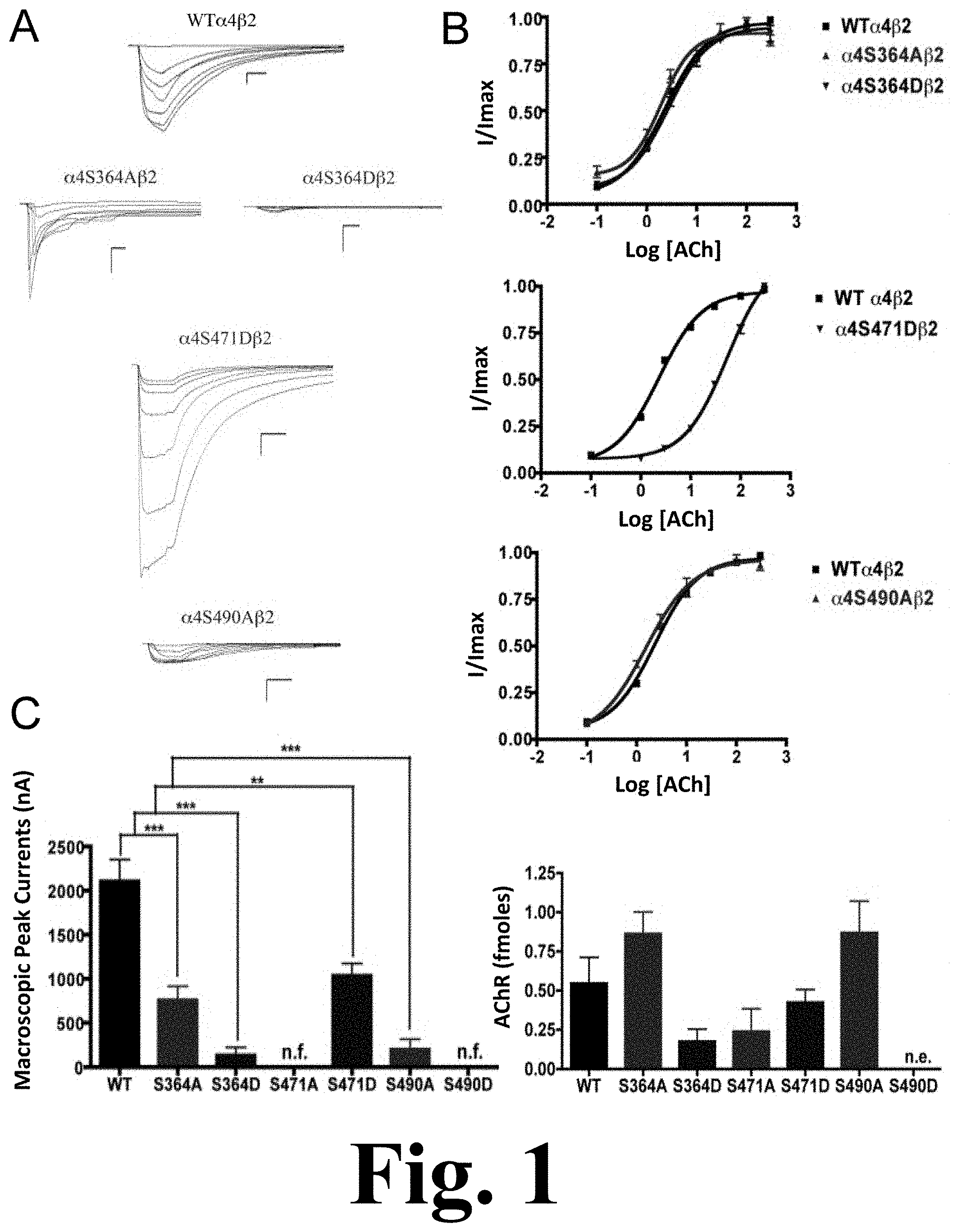
FIG. 1 shows the binding and functional characterization of mutations on PKA putative sites in the .alpha.4 subunit.
FIG. 2 shows the functional characterization of mutations on PKA putative sites in the .alpha.4 subunit with nicotine used as agonist.
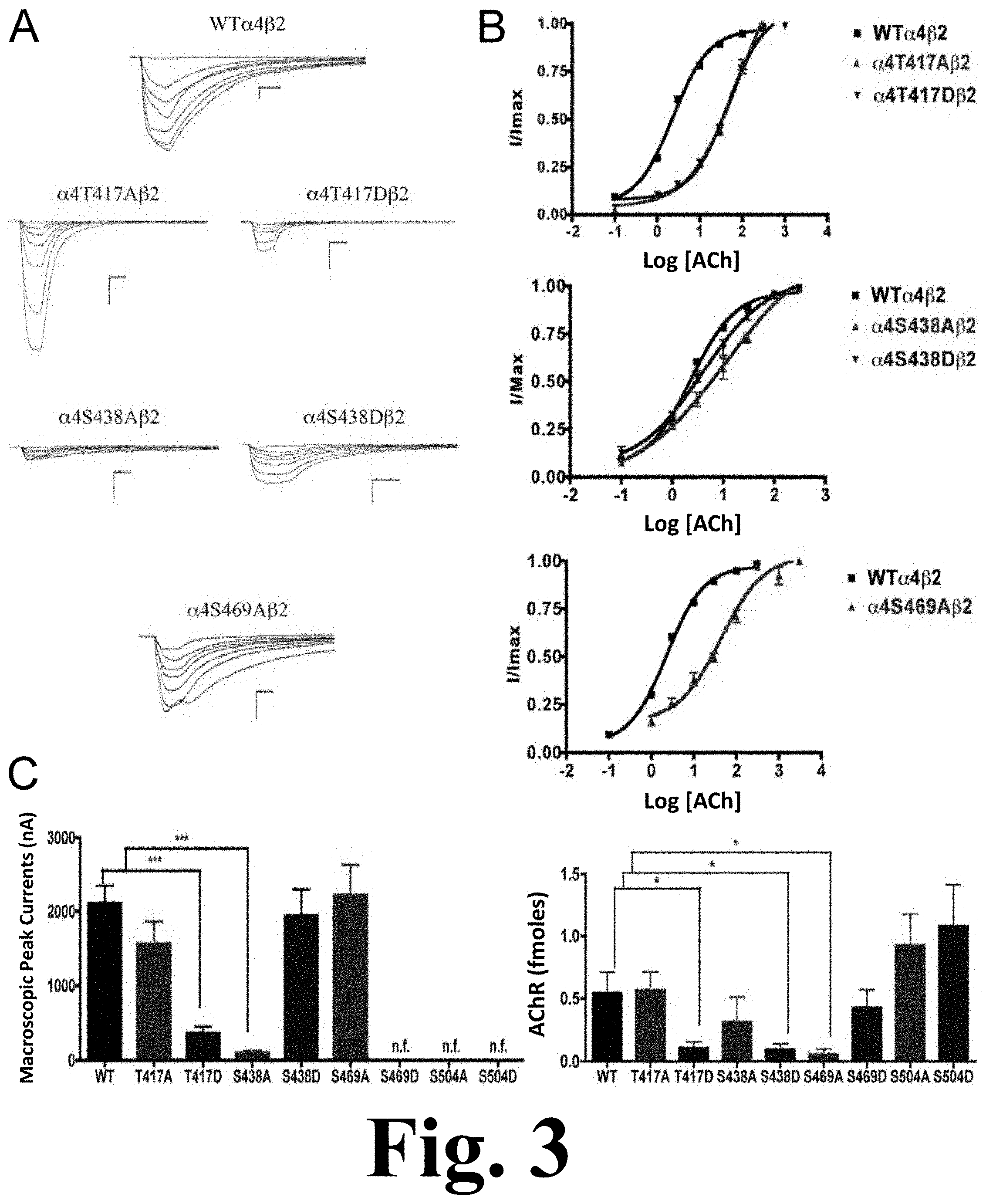
FIG. 3 shows the binding and functional characterization of mutations on CKII putative phosphorylation sites in the .alpha.4 subunit with ACh used as an agonist.
FIG. 4 shows the functional characterization of mutations on CKII putative phosphorylation sites in the .alpha.4 subunit with nicotine used as agonist.
FIG. 5 shows the binding and functional characterization of mutations on PKC putative phosphorylation sites in the .alpha.4 subunit with ACh used as an agonist.
FIG. 6 shows the functional characterization of mutations on PKC putative phosphorylation sites in the .alpha.4 subunit with nicotine used as an agonist.
FIG. 7 shows the binding and functional characterization of mutations on the TK putative phosphorylation site in the .alpha.4 subunit with ACh used as an agonist.
FIG. 8 shows the functional characterization of mutations on the TK putative phosphorylation site in the .alpha.4 subunit with nicotine used as an agonist.
FIG. 9 shows the efficacy of .alpha.4.beta.2 agonist A-85380 for stoichiometry determination.
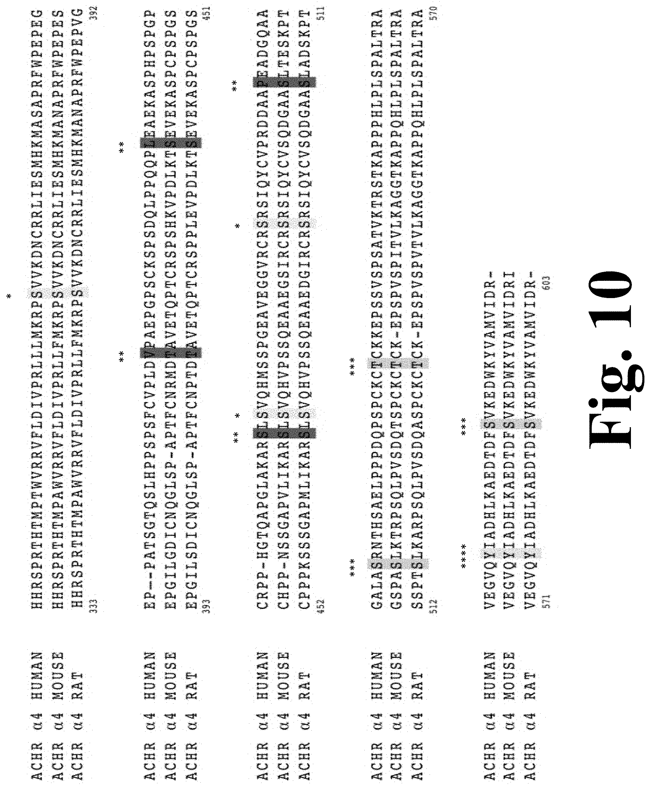
FIG. 10 shows the consensus phosphorylation sites for the rat .alpha.4 M3/M4 intracellular domain.
The Figure discloses SEQ ID NOS 1-3, respectively, in order of appearance.
DETAILED DESCRIPTION OF THE INVENTION
Materials and Methods
Site-Directed Mutagenesis
Single point mutations were prepared using the Quickchange.TM. Site-Directed Mutagenesis Kit (Stratagene, La Jolla, Calif.). The template used for the PCR reaction was the pGEMHE vector with Rattus norvegicus cDNA coding for the .alpha.4 neuronal nAChR subunit. DNA was purified using a QIAprep spin miniprep kit (Quiagen, Germantown, Md.) and then sequenced to confirm the incorporation of each mutation. A total of 11 residues in the cytoplasmic loop of the .alpha.4 subunit were chosen to be mutated: S364, T417, S438, S469, S471, S490, S504, S516, T536, Y576, and S589. Each residue was mutated to alanine (A) and aspartic acid (D).
In Vitro Synthesis of mRNA and Oocyte Microinjection
Each subunit encoding mRNA was synthesized in vitro from linearized pGEMHE plasmid templates of Rattus norvegicus cDNA coding for .alpha.4 and .beta.2 nAChR subunits using the mMessage mMachine RNA transcription kit (Ambion, Austin, Tex.). mRNA mixtures of .alpha.4 and .beta.2 subunits were prepared at 2 .mu.g:3 .mu.g ratio, and 32.2 nL of this mixture was microinjected into each oocyte. The mRNA mixture was microinjected, using a displacement injector (Drummond Instruments, Broomhall, Pa.), into stage V and VI oocytes that had been extracted, incubated in collagenase Type 1A (Sigma, St. Louis, Mo.), and defolliculated by manual dissection. The injected oocytes were incubated at 19.degree. C. for 3-4 days in ND-96 medium supplemented with albumin, gentamicin, tetracycline, and theophyline. Electrophysiological experiments were performed after the third or fourth day of mRNA injection.
Electrophysiological Characterization of .alpha.4.beta.2 nAChRs
Oocytes injected with the mRNA transcripts of .alpha.4 and .beta.2 subunits were characterized using a two-electrode voltage clamp. ACh- and nicotine-induced currents were recorded at 20.degree. C., 3-4 days after mRNA injection, with a GeneClamp 500B Amplifier (Axon Instruments, Foster City, Calif.). Electrodes were filled with 3M KCl and had a resistance of less than 5 megaohms. Impaled oocytes in the recording chamber were continuously perfused at a rate of 5 ml/min with MOR-2 buffer (115 mM NaCl, 2.5 mM KCl, 5 mM HEPES, 1 mM Na.sub.2HPO.sub.4, 0.2 mM CaCl.sub.2, 5 mM MgCl.sub.2, and 0.5 mM EGTA, pH 7.4). All the reagents used were purchased from Sigma-Aldrich, Co. (St. Louis, Mo.). For dose-response curves, each oocyte was held at a membrane potential of -70 mV. Membrane currents were digitized using the DigiData 1322A interface (Axon Instruments, Foster City, Calif.), filtered at 2 kHz during recording. The Clampex 10.0 software running on a Pentium 4-based computer was used for data acquisition. Data analysis was via Prism 3.0 (Graphpad Software, San Diego, Calif.). Dose-response data for the .alpha.4.beta.2 combination were collected using seven ACh doses (0.1, 1, 3, 10, 30, 100, and a seventh concentration ranging from 300 to 1000 .mu.M depending on the mutant) and seven nicotine concentrations (0.1, 1, 3, 10, 30, 100, 300 .mu.M). The data were fitted using a one-component sigmoidal dose-response equation, Y=I/I.sub.maxBottom+(I/I.sub.maxTop-I/I.sub.maxBottom)/(1+10{circumflex over ( )}((Log EC.sub.50-X).times.HillSlope)) where X is the logarithm of concentration and Y is the response.
Epibatidine Binding Assays
.sup.125I-labeled epibatidine (PerkinElmer Life Sciences, Boston, Mass.) binding assays were performed to determine membrane expression of nAChR in oocytes. The oocytes were incubated in 50 pM .sup.125I-labeled epibatidine with 5 mg/mL albumin serum bovine in MOR-2 without EGTA at room temperature for 2 h. Non-injected oocytes were also incubated in .sup.125I-labeled epibatidine to measure nonspecific binding (PerkinElmer 2470 WIZARD Gamma Counter). Excess epibatidine was removed by washing each oocyte with 60 mL of MOR-2 without EGTA. A standard linear regression was obtained by plotting the counts per minute (CPM) against .sup.125I-labeled epibatidine concentration (0.5-20 fmol).
Determination in Potential Changes in Subunit Stoichiometry of .alpha.4.beta.2 nAChRs Using A-85380
To determine whether the .alpha.4.beta.2 mutations .alpha.4S469A.beta.2, .alpha.4S471D.beta.2, .alpha.4T536A.beta.2 and .alpha.4Y576A.beta.2 expressed in Xenopus laevis oocytes assemble in the .alpha.4(2):.beta.2(3) or the .alpha.4(3):.beta.2(2) stoichiometry, we estimated the efficacy of agonist A-85380 when compared to ACh 300 .mu.M. The .alpha.4(2):.beta.2(3) stoichiometry was favored by microinjecting .alpha.4.beta.2 mRNA in a 1:10 ratio, and the .alpha.4(3):.beta.2(2) by microinjecting in a 10:1 ratio. The efficacy of A-85380 in WT.alpha.4(2):.beta.2(3) and WT.alpha.4(3):.beta.2(1) was determined by comparing the macroscopic current elicited by A-85380 100 nM and ACh 300 .mu.M in oocytes microinjected .alpha.4.beta.2 mRNA in 1:10 and 10:1 ratios, respectively. The efficacy of A-85380 in .alpha.4S469A.beta.2, .alpha.4S471D.beta.2, .alpha.4T536A.beta.2 and .alpha.4Y576A.beta.2 was determined by comparing the macroscopic current elicited by A-85380 100 nM and ACh 300 .mu.M in oocytes microinjected .alpha.4.beta.2 mRNA in 2:3 ratio. The changes in stoichiometry in these four mutants were assessed by comparing the efficacy of A-85380 in each mutant compared to WT.alpha.4(2):.beta.2(3) and WT.alpha.4(3):.beta.2(2).
Results
Functional Effects of Mutations at PKA Putative Phosphorylation Sites in the .alpha.4 Subunit when Using ACh as an Agonist
Mutations .alpha.4S364A, .alpha.4S364D, .alpha.4S471D, and .alpha.4S490A resulted in receptors with a significant decrease in macroscopic peak current as compared with the wild-type (WT) receptor as shown in FIG. 1. This figure shows the binding and functional characterization of mutations on PKA putative sites in the .alpha.4 subunit. Voltage-clamp recordings were used to determine the macroscopic response of mutations .alpha.4S364A.beta.2, .alpha.4S364D.beta.2, .alpha.4S471A.beta.2, .alpha.4S471D.beta.2, .alpha.4S490A.beta.2, .alpha.4S490D.beta.2 and wild-type .alpha.4.beta.2 nAChRs expressed in Xenopus laevis oocytes to several ACh and nicotine concentrations. (A) Family of ACh-induced macroscopic currents. Calibration bars are shown for all family of currents, horizontal bars indicate time (5 s) and vertical bars indicate the inward current (500 nA). (B) Dose-response relationships obtained by voltage-clamp using ACh as an agonist. ACh dose-response curves were determined using seven ACh concentrations (0.1, 1, 3, 10, 30, 100, and a seventh concentration ranging from 300 to 1000 .mu.M depending on the mutant). The responses were normalized to the maximum response (I/Imax). (C) Left panel, comparison of the macroscopic peak currents of all the mutations and wild-type receptor shown in nA. Right panel, results of the .sup.125I-labeled epibatidine binding experiments performed in Xenopus laevis oocytes expressing the mutations .alpha.4S364A.beta.2, .alpha.4S364D.beta.2, .alpha.4S471A.beta.2, .alpha.4S471D.beta.2, .alpha.4S490A.beta.2, .alpha.4S490D.beta.2 and wild-type .alpha.4.beta.2 nAChRs shown in fmoles (n=6-17) (*** p<0.0005, ** p<0.005).
Mutations .alpha.4S471A and .alpha.4S490D resulted in non-functional receptors. Statistical analysis using an unpaired t test showed no significant difference in the ACh EC.sub.50 value between mutations .alpha.4S364A, .alpha.4S364D, .alpha.4S490A, and the WT .alpha.4.beta.2 nAChR (2.55.+-.0.08, 2.71.+-.0.05, 1.51.+-.0.08, and 2.33.+-.0.03 .mu.M, respectively). On the other hand, mutation .alpha.4S471D exhibited a significant EC.sub.50 increase (i.e. 53.60.+-.0.05 .mu.M) as compared with the WT ACh EC.sub.50 value, as shown in Table 1 below.
TABLE-US-00001 TABLE 1 Electrophysiological characterization of .alpha.4.beta.2 nAChR mutations reveal different nicotine and ACh sensitivities ACh Nicotine Hill Peak Current Hill Peak Current Kinase Mutation EC.sub.50 (.mu.M) Coef. (nA) EC.sub.50 (.mu.M) Coef. (nA) -- WT .alpha.4.beta.2 2.33 .+-. 0.03 1.00 2110 .+-. 243 4.21 .+-. 0.41 1.80 387 .+-. 66 PKA .alpha.4S364A.beta.2 2.55 .+-. 0.08 1.22 759 .+-. 157 n/f n/f n/f .alpha.4S364D.beta.2 2.71 .+-. 0.05 1.07 135 .+-. 87*** n/f n/f n/f .alpha.4S471A.beta.2 n/f n/f n/f n/f n/f n/f .alpha.4S471D.beta.2 53.60 .+-. 0.05*** 1.01 1039 .+-. 139** 5.02 .+-. 0.62 2.00 142 .+-. 313** .alpha.4S490A.beta.2 1.51 .+-. 0.08 0.91 200 .+-. 115*** n/f n/f n/f .alpha.4S490D.beta.2 n/e n/e n/e n/e n/e n/e CKII .alpha.4T417A.beta.2 77.80 .+-. 0.12*** 0.76 1557 .+-. 306 2.12 .+-. 0.61* 1.33 293 .+-. 120 .alpha.4T417D.beta.2 46.09 .+-. 0.05*** 0.99 360 .+-. 89*** 2.11 .+-. 0.32** 1.78 582 .+-. 255 .alpha.4S438A.beta.2 11.00 .+-. 0.19*** 0.49 97 .+-. 29*** n/f n/f n/f .alpha.4S438D.beta.2.dagger-dbl. 3.96 .+-. 0.09* 0.68 1938 .+-. 367 2.17 .+-. 0.16** 1.29 2156 .+-. 475** .alpha.4S469A.beta.2.dagger-dbl. 43.59 .+-. 0.09*** 0.87 2222 .+-. 413 3.37 .+-. 0.20 1.84 1552 .+-. 313** .alpha.4S469D.beta.2 n/f n/f n/f n/f n/f n/f .alpha.4S504A.beta.2 n/f n/f n/f n/f n/f n/f .alpha.4S504D.beta.2 n/f n/f n/f n/f n/f n/f PKC .alpha.4S516A.beta.2 65.89 .+-. 0.06*** 1.04 1027 .+-. 169*** n/f n/f n/f .alpha.4S516D.beta.2 43.72 .+-. 0.12*** 0.79 3296 .+-. 581 3.80 .+-. 0.68 1.48 76 .+-. 16*** .alpha.4T536A.beta.2 38.25 .+-. 0.11*** 0.67 3345 .+-. 129*** 3.84 .+-. 0.61 1.80 110 .+-. 28*** .alpha.4T536D.beta.2 85 .+-. 17*** 0.69 216 .+-. 85*** n/f n/f n/f .alpha.4S589A.beta.2.dagger-dbl. 77 .+-. 10*** 0.80 1644 .+-. 192 4.67 .+-. 0.56 1.61 1882 .+-. 672* .alpha.4S589D.beta.2 n/f n/f n/f n/f n/f n/f TK .alpha.4Y576A.beta.2.dagger-dbl. 55 .+-. 8*** 0.91 4398 .+-. 450*** 3.76 .+-. 0.60 1.65 874 .+-. 221* .alpha.4Y576D.beta.2 35 .+-. 7*** 0.62 396 .+-. 65*** n/f n/f n/f Mutations highlighted in green (.dagger-dbl.) exhibit hypersensitivity to nicotine while those in red ( ) exhibit agonist selectivity evidenced by their remarkable reductions in sensitivity to nicotine. Legend: (n/f) non-functional; (n/e) no-expression; Error estimates are expressed as the mean .+-. SEM of 6-17 oocytes. *P <0.05, **P <0.005, ***P <0.0005
Functional Effects of Mutations at PKA Putative Phosphorylation Sites in the .alpha.4 Subunit when Using Nicotine as an Agonist.
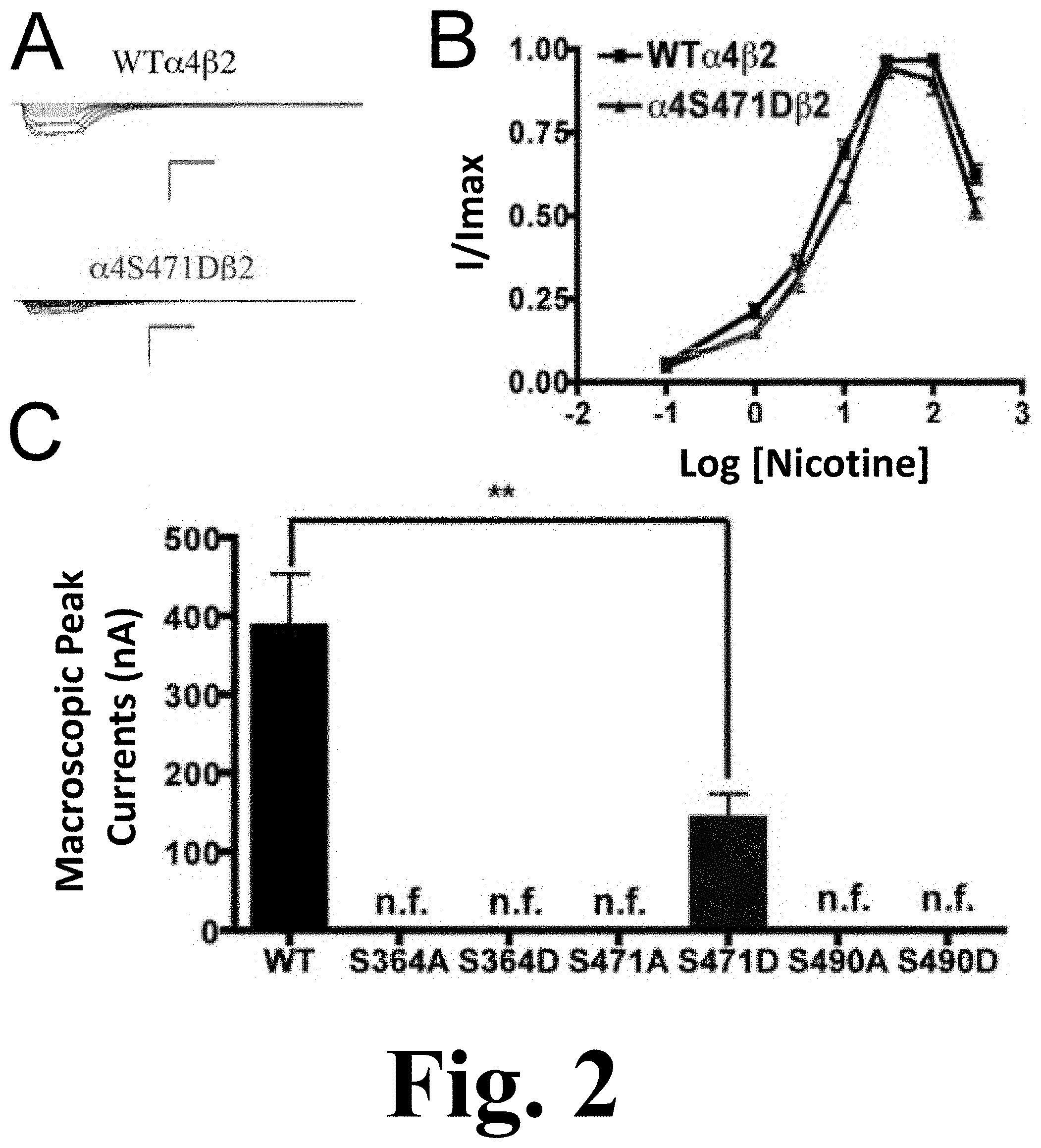
All of the PKA mutants were studied with the use of nicotine as an agonist. The only mutation that exhibited functional activation by nicotine was .alpha.4S471D, as shown in FIG. 2. This figure shows the functional characterization of mutations on PKA putative sites in the .alpha.4 subunit with nicotine used as agonist. (A) Family of nicotine-induced macroscopic currents for mutation .alpha.4S471D.beta.2 and the wild-type .alpha.4.beta.2 nAChR. Calibration bars are shown for all family of currents, horizontal bars indicate time (5 s) and vertical bars indicate the inward current (500 nA). (B) Dose-response relationships obtained by voltage-clamp using nicotine as agonist. Nicotine dose-response curves were determined using seven nicotine concentrations (0.1, 1, 3, 10, 30, 100 and 300 .mu.M). The responses were normalized to the maximum response (I/Imax). (C) Comparison of the nicotine-induced macroscopic peak currents of the mutations and wild-type receptor shown in nA (n=6-17) (** p<0.005).
This mutation exhibited a significant decrease in the macroscopic peak current as compared with that of the WT receptor. The nicotine EC.sub.50 values revealed no difference between the mutation and the WT receptor (Table 1). This was a remarkable observation as the .alpha.4S471D mutation shows a significant difference in ACh activation as compared with the WT; however, nicotine activation in this mutant was similar to that in the WT receptor. This kind of behavior was previously described in mutations of PKC residue S336.
Effects on nAChR Expression of Mutations at PKA Putative Phosphorylation Sites in the .alpha.4 Subunit
.sup.125I-labeled epibatidine binding assays revealed the expression patterns of the various mutations of PKA residues in the .alpha.4 subunit (FIG. 1C, right panel). Statistical analysis using an unpaired t test revealed no significant changes among any of the mutations as compared with the WT nAChR. Although mutation .alpha.4S490D did not exhibit any function during the voltage-clamp experiments (Table 1), it was still subjected to the binding assays which revealed no expression for mutation .alpha.4S490D.
Functional Effects of Mutations at CKII Putative Phosphorylation Sites in the .alpha.4 Subunit when Using ACh as an Agonist
Voltage-clamp data analysis revealed significant decreases in the macroscopic peak currents for mutations .alpha.4T417D and .alpha.4S438A, as shown in FIG. 3. This figure shows the binding and functional characterization of mutations on CKII putative phosphorylation sites in the .alpha.4 subunit with ACh used as an agonist. Mutations .alpha.4T417A.beta.2, .alpha.4T417D.beta.2, .alpha.4S438A.beta.2, .alpha.4S438D.beta.2, .alpha.4S469A.beta.2, .alpha.4S469D.beta.2, .alpha.4S504A.beta.2, and .alpha.4S504D.beta.2 were expressed in Xenopus laevis oocytes. (A) Family of ACh-induced macroscopic currents. Calibration bars are shown for all family of currents, horizontal bars indicate time (5 s) and vertical bars indicate the inward current (500 nA). (B) Dose-response curves obtained by voltage-clamp using ACh as agonist. ACh dose-response curves were determined using seven ACh concentrations (0.1, 1, 3, 10, 30, 100, and a seventh concentration ranging from 300 to 1000 .mu.M depending on the mutant). The responses were normalized to the maximum response (I/Imax). (C) Left panel. Comparison of the macroscopic peak currents of all the mutations compared with the wild-type receptor, shown in nA. Right panel. Results of the .sup.125I-labeled epibatidine binding experiments performed in Xenopus laevis oocytes expressing the mutations .alpha.4T417A.beta.2, .alpha.4T417D.beta.2, .alpha.4S438A.beta.2, .alpha.4S438D.beta.2, .alpha.4S469A.beta.2, .alpha.4S469D.beta.2, .alpha.4S504A.beta.2, and .alpha.4S504D.beta.2 and wild-type .alpha.4.beta.2 nAChRs, shown in fmoles (n=6-17) (* p<0.05, ***p<0.0001).
On the other hand, mutations .alpha.4T417A, .alpha.4S438D, and .alpha.4S469A showed no change in the macroscopic peak current (Table 1). At the same time, mutations .alpha.4S469D, .alpha.4S504A, and .alpha.4S504D resulted in non-functional receptors. The ACh EC.sub.50 values for all of the functional mutations of the CKII putative sites .alpha.4T417A, .alpha.4T417D, .alpha.4S438A, .alpha.4S438D, and .alpha.4S469A resulted in significant increases when compared with the WT nAChR (Table 1).
Functional Effects of Mutations at CKII Putative Phosphorylation Sites in the .alpha.4 Subunit when Using Nicotine as an Agonist
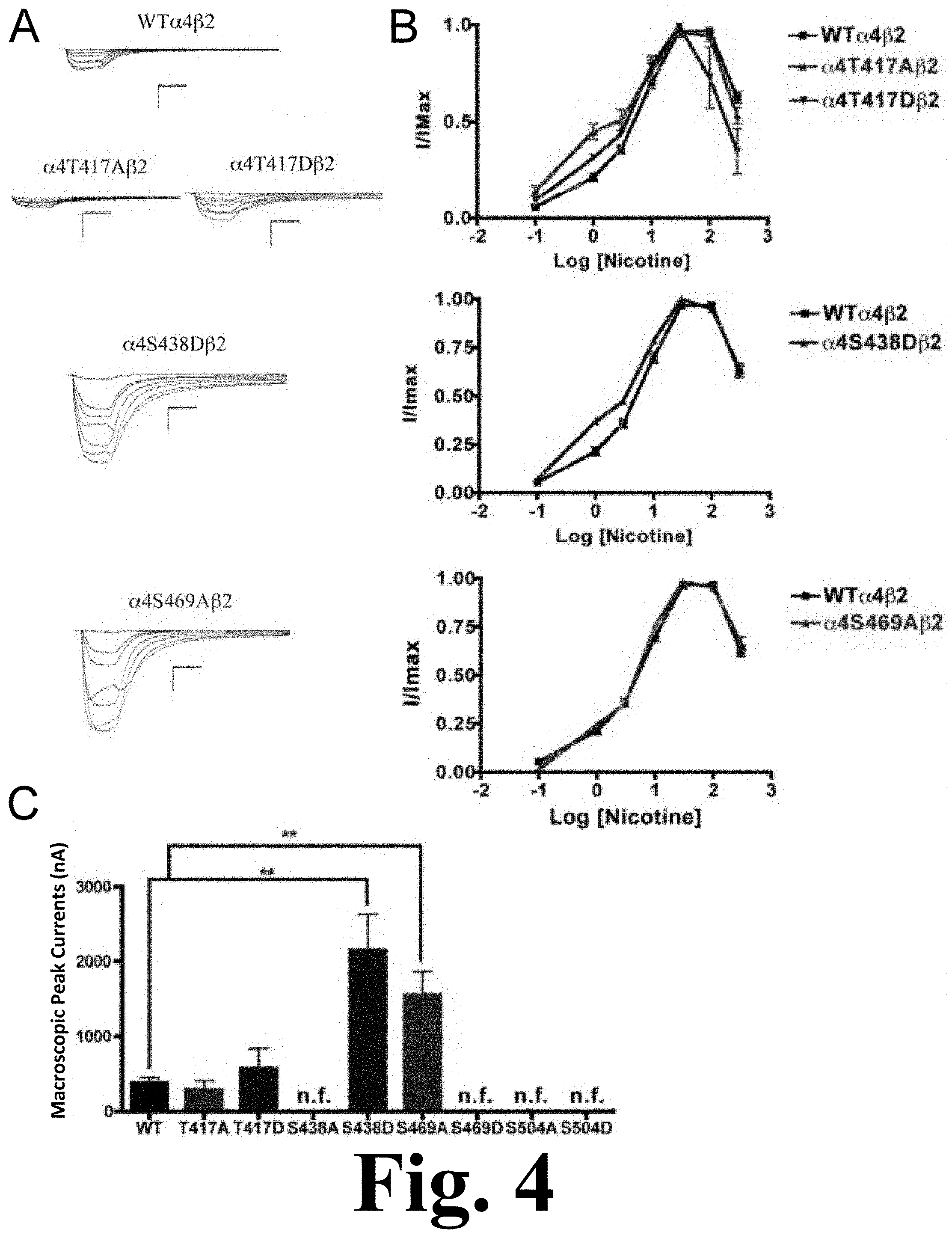
When nicotine was used as agonist, we found that mutations .alpha.4S469D, .alpha.4S504A, and .alpha.4S504D also resulted in non-functional receptors. However, although mutation .alpha.4S438A was a functional receptor when ACh was used as agonist, it resulted in a non-functional receptor when nicotine was used as an agonist. Mutations of residue .alpha.4T417 exhibited no significant change in macroscopic current when compared with the WT receptor; however, their nicotine EC.sub.50 values decreased significantly, as shown in FIG. 4. This figure shows the functional characterization of mutations on CKII putative phosphorylation sites in the .alpha.4 subunit with nicotine used as agonist. Voltage-clamp recordings were used to determine the macroscopic response to several nicotine concentrations of mutations of CKII putative sites and wild-type .alpha.4.beta.2 nAChRs expressed in Xenopus laevis oocytes. (A) Family of nicotine-induced macroscopic currents for mutations .alpha.4T417A.beta.2, .alpha.4T417D.beta.2, .alpha.4S438D.beta.2, .alpha.4S469A.beta.2, and the wild-type .alpha.4.beta.2 nAChR. Calibration bars are shown for all family of currents, horizontal bars indicate time (5 s) and vertical bars indicate the inward current (500 nA). (B) Dose-response relationships obtained by voltage-clamp experiments with nicotine used as an agonist. Nicotine dose-response curves were determined using seven nicotine concentrations (0.1, 1, 3, 10, 30, 100, and 300 .mu.M). The responses were normalized to the maximum response (I/Imax). (C) Comparison of the nicotine-induced macroscopic peak currents of the mutations and wild-type receptor, shown in nA (n=6-17) (**p<0.005).
These mutations displayed a higher potency for nicotine. Interestingly, mutations .alpha.4S438D and .alpha.4S469A exhibited significant increases in the macroscopic peak currents (2156.+-.475 and 1552.+-.313 nA, respectively) when compared with the WT receptor (387.+-.66 nA) (Table 1). The nicotine EC.sub.50 value for .alpha.4S438D exhibited a significant decrease whereas mutation .alpha.4S469A exhibited no change. These results suggest that mutations .alpha.4S438D and .alpha.4S469A enhance the sensitivity to nicotine.
Effects on nAChR Expression of Mutations at CKII Putative Phosphorylation Sites in the .alpha.4 Subunit.
.sup.125I-labeled epibatidine binding assays on mutations of the CKII residues revealed significant decreases in receptor expression for mutations .alpha.4T417D, .alpha.4S438D, and .alpha.4S469A (FIG. 3C, right panel). On the other hand, mutations .alpha.4T417A and .alpha.4S438A did not exhibit changes in expression when compared with the WT receptor. More interestingly, three mutations that resulted in non-functional receptors when using ACh and nicotine (.alpha.4S469D, .alpha.4S504A, and .alpha.4S504D) exhibited no change in receptor expression (FIG. 3C right panel, and Table 1).
Functional Effects of Mutations at PKC Putative Phosphorylation Sites in the .alpha.4 Subunit when Using ACh as an Agonist
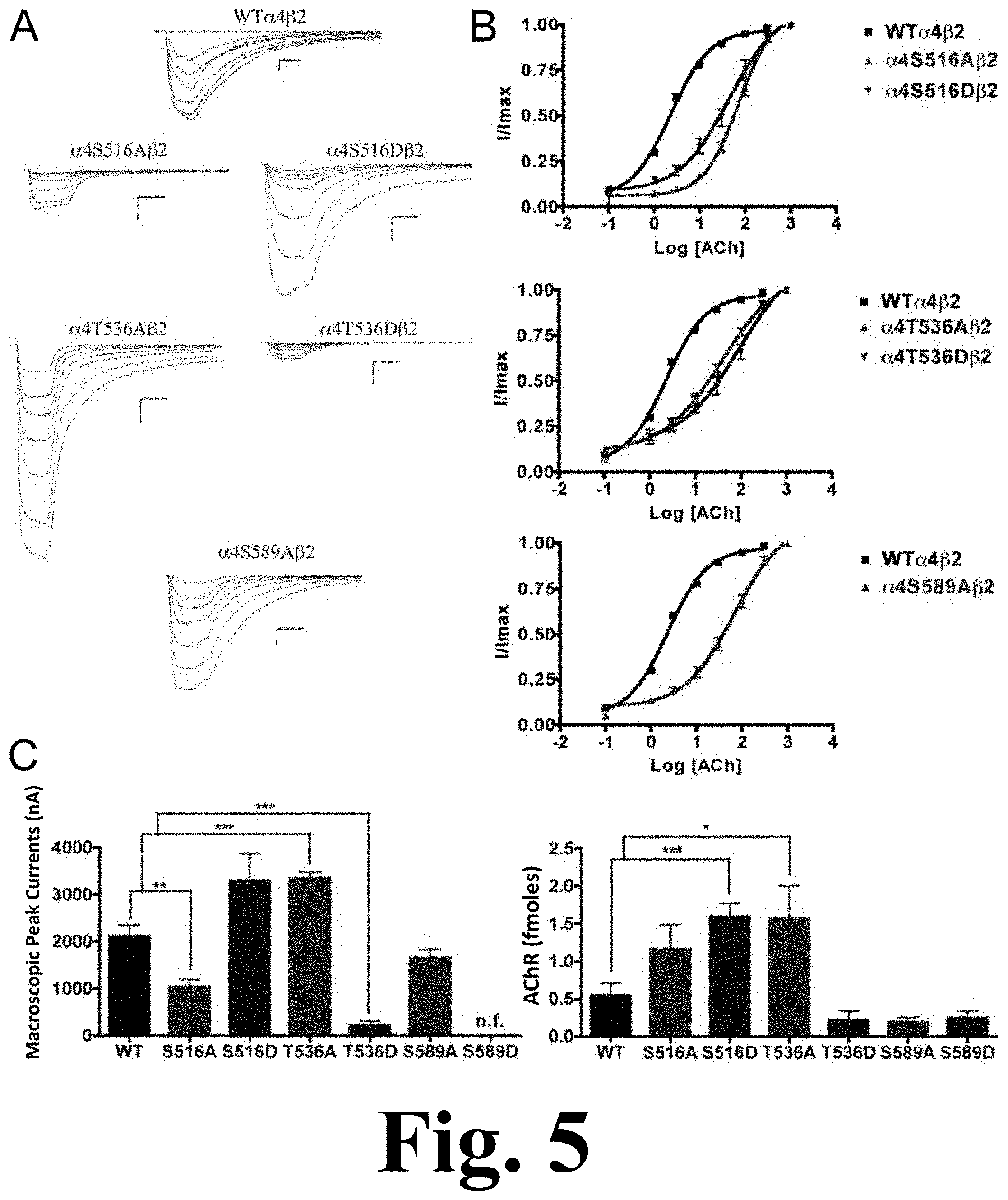
The alanine substitution at position .alpha.4S516 displayed a significant decrease in macroscopic peak current (1027.+-.169 nA), as shown in FIG. 5. This figure shows the binding and functional characterization of mutations on PKC putative phosphorylation sites in the .alpha.4 subunit with ACh used as an agonist. Mutations .alpha.4S516A.beta.2, .alpha.4S516D.beta.2, .alpha.4T536A.beta.2, .alpha.4T536D.beta.2, .alpha.4S589A.beta.2, and .alpha.4S589D.beta.2 were expressed in Xenopus laevis oocytes. (A) Family of ACh-induced macroscopic currents. Calibration bars are shown for all family of currents, horizontal bars indicate time (5 s) and vertical bars indicate the inward current (500 nA). (B) Dose-response curves obtained by voltage-clamp experiments with ACh used as an agonist. ACh dose-response curves were determined using seven ACh concentrations (0.1, 1, 3, 10, 30, 100, and a seventh concentration ranging from 300 to 1000 .mu.M depending on the mutant). The responses were normalized to the maximum response (I/Imax). (C) Left panel. Comparison of the macroscopic peak currents of all the mutations compared to the wild-type receptor shown in nA. Right panel. Results of the .sup.125I-labeled epibatidine binding experiments performed in Xenopus laevis oocytes expressing the mutations .alpha.4S516A.beta.2, .alpha.4S516D.beta.2, .alpha.4T536A.beta.2, .alpha.4T536D.beta.2, .alpha.4S589A.beta.2, and .alpha.4S589D.beta.2 and wild-type .alpha.4.beta.2 nAChRs, shown in fmoles (n=6-17) (*p<0.05, **p<0.005, ***p<0.0005).
Mutation .alpha.4T536A showed a significant increase in macroscopic peak current (3345.+-.129 nA), whereas mutation .alpha.4T536D showed a significant decrease in macroscopic peak current (216.+-.85 nA) when compared with the WT receptor (Table 1). Mutation .alpha.4S589A displayed a functional receptor with no change in macroscopic peak current (1644.+-.192 nA) when compared with the WT receptor. However, mutating the same residue to aspartic acid, .alpha.4S589D, resulted in a non-functional receptor (i.e., no significant current was detected). The five mutations that exhibited functional channels (.alpha.4S516A, .alpha.4S516D, .alpha.4T536A, .alpha.4T536D, and .alpha.4S589A) exhibited a significant increase in the ACh EC.sub.50 value as compared with the WT receptor (Table 1).
Functional Effects of Mutations at PKC Putative Phosphorylation Sites in the .alpha.4 Subunit when Using Nicotine as an Agonist
We performed voltage-clamp experiments on the mutations of PKC putative sites, this time using nicotine as an agonist. Mutations .alpha.4S516A, .alpha.4T536D, and .alpha.4S589D exhibited no nicotine-induced currents, an unsurprising outcome given that their response to ACh was significantly decreased. However, mutations .alpha.4S516D and .alpha.4T536A exhibited extremely low nicotine-induced currents, as shown in FIG. 6. This figure shows the functional characterization of mutations on PKC putative phosphorylation sites in the .alpha.4 subunit with nicotine used as an agonist. Voltage-clamp recordings were used to determine the macroscopic response to several nicotine concentrations of mutations of PKC putative sites and wild-type .alpha.4.beta.2 nAChRs expressed in Xenopus laevis oocytes. (A) Family of nicotine-induced macroscopic currents for the mutations and the wild-type .alpha.4.beta.2 nAChR. Calibration bars are shown for all family of currents, horizontal bars indicate time (5 s) and vertical bars indicate the inward current (500 nA). (B) Dose-response relationships obtained by voltage-clamp experiments with nicotine used as an agonist. Nicotine dose-response curves were determined using seven nicotine concentrations (0.1, 1, 3, 10, 30, 100, and 300 .mu.M). The responses were normalized to the maximum response (I/Imax). (C) Comparison of the nicotine-induced macroscopic peak currents, shown in nA (n=6-17) (*p<0.05, ***p<0.0005).
This was an interesting finding because both of these mutations exhibited either no change (.alpha.4S516D) or an increase (.alpha.4T536A) in the ACh-induced macroscopic peak current. On the other hand, mutation .alpha.4S589A displayed a significant increase in macroscopic peak current, while exhibiting no change in the nicotine EC.sub.50 value (Table 1).
Effects on nAChR Expression of Mutations at PKC Putative Phosphorylation Sites in the .alpha.4 Subunit
The binding assays, using .sup.125I-labeled epibatidine, to measure receptor expression showed significant increases in receptor expression when compared with the WT receptor. Mutations .alpha.4S516D, and .alpha.4T536A showed significant increases in receptor expression when compared with the WT .alpha.4.beta.2 nAChR (FIG. 5C, right panel). These mutations resulted in receptors that are constitutively up-regulated. Mutation .alpha.4S589D displayed a behavior similar to that seen previously in mutations .alpha.4S504A and .alpha.4S504D; it showed no signs of ACh- and nicotine-induced macroscopic currents (Table 1). However, binding assays revealed no change in receptor expression for mutation .alpha.4S589D when compared with the WT receptor (FIG. 5C, right panel).
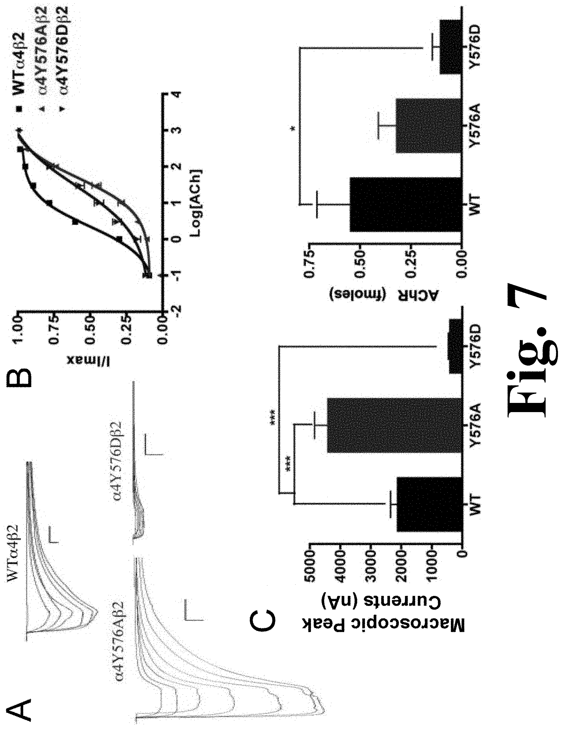
Functional Effects of Mutations of a TK Putative Phosphorylation Site in the .alpha.4 Subunit when Using ACh as an Agonist.
The alanine substitution in the TK putative site .alpha.4Y576 exhibited a significant increase in the macroscopic peak current (4398.+-.450 nA), and the aspartic acid substitution resulted in a significant decrease in macroscopic peak current (396.+-.65 nA) when compared with the .alpha.4.beta.2 nAChR as shown in FIG. 7. This figure shows the binding and functional characterization of mutations on the TK putative phosphorylation site in the .alpha.4 subunit with ACh used as an agonist. Mutations .alpha.4Y576A.beta.2 and .alpha.4Y576D.beta.2 were expressed in Xenopus laevis oocytes. (A) Family of ACh-induced macroscopic currents. Calibration bars are shown for all family of currents, horizontal bars indicate time (5 s) and vertical bars indicate the inward current (500 nA). (B) Dose-response curves obtained by voltage-clamp experiments with ACh used as an agonist. ACh dose-response curves were determined using seven ACh concentrations (0.1, 1, 3, 10, 30, 100, and a seventh concentration ranging from 300 to 1000 .mu.M depending on the mutant). The responses were normalized to the maximum response (I/Imax). (C) Left panel. Comparison of the macroscopic peak currents of all the mutations compared with the wild-type receptor, shown in nA. Right panel. Results of the .sup.125I-labeled epibatidine binding experiments performed in Xenopus laevis oocytes expressing the mutations .alpha.4Y576A.beta.2 and .alpha.4Y576D.beta.2 and wild-type .alpha.4.beta.2 nAChRs, shown in fmoles (n=6-17) (*p<0.05, ***p<0.0005).
The ACh EC.sub.50 values for .alpha.4Y576A (55.+-.8 .mu.M) and for .alpha.4Y576D (35.+-.7 .mu.M) increased significantly when compared with the WT receptor, which has an EC.sub.50 value of 2.33.+-.0.03 .mu.M (Table 1). These increases in ACh EC.sub.50 values have also been seen in other mutations.
Functional Effects of Mutations of a TK Putative Phosphorylation Site in the .alpha.4 Subunit when Using Nicotine as an Agonist
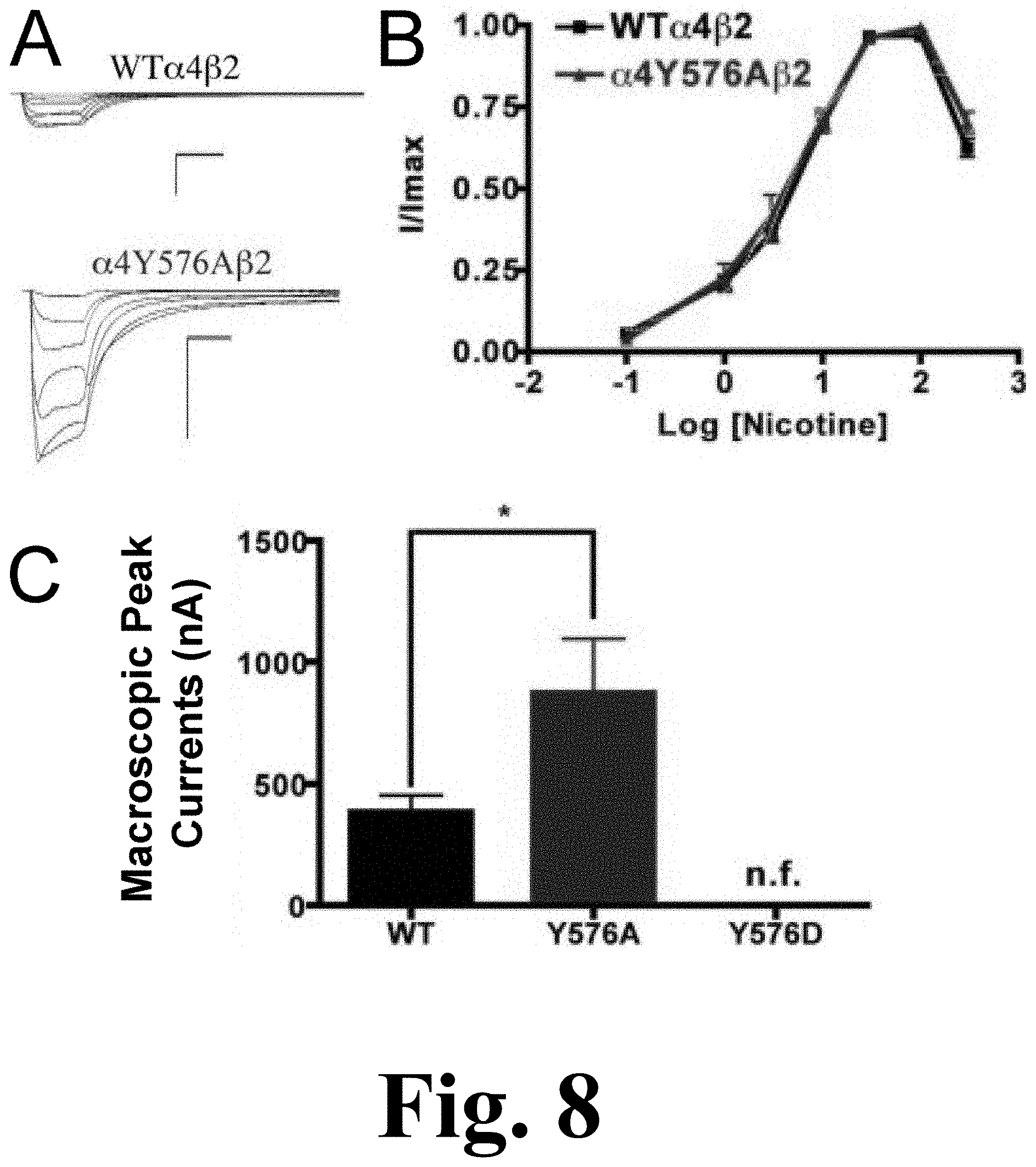
Mutation .alpha.4Y576A exhibited a significant increase in the macroscopic peak current whereas mutation .alpha.4Y576D exhibited no significant nicotine-induced current, resulting in a non-functional channel, as shown in FIG. 8. The figure shows the functional characterization of mutations on the TK putative phosphorylation site in the .alpha.4 subunit with nicotine used as an agonist. Voltage-clamp recordings were used to determine the macroscopic response to several nicotine concentrations of mutations of TK putative sites and wild-type .alpha.4.beta.2 nAChRs expressed in Xenopus laevis oocytes. (A) Family of nicotine-induced macroscopic currents for mutation .alpha.4Y576A.beta.2 and the wild-type .alpha.4.beta.2 nAChR. Calibration bars are shown for all family of currents, horizontal bars indicate time (5 s) and vertical bars indicate the inward current (500 nA). (B) Dose-response relationships obtained by voltage-clamp experiments with nicotine used as an agonist. Nicotine dose-response curves were determined using seven nicotine concentrations (0.1, 1, 3, 10, 30, 100, and 300 .mu.M). The responses were normalized to the maximum response (I/Imax). (C) Comparison of the nicotine-induced macroscopic peak currents, shown in nA (n=6-17) (*p<0.05).
Since mutation .alpha.4Y576D exhibited a significant decrease in the ACh-induced macroscopic peak current, it was not surprising that there was no significant nicotine-induced current. The nicotine EC.sub.50 value for mutation .alpha.4Y576A revealed no significant difference when compared with the WT receptor (Table 1). Our data indicate that mutation .alpha.4Y576A exhibits an enhanced sensitivity to nicotine.
Effects on nAChR Expression of Mutations of TK Putative Phosphorylation Sites in the .alpha.4 Subunit
The results of the binding assays performed on mutations .alpha.4Y576A and .alpha.4Y576D revealed that only mutation .alpha.4Y576D decreased receptor expression significantly, whereas mutation .alpha.4Y576A saw no change in receptor expression when compared with the .alpha.4.beta.2 nAChR (FIG. 7C, right panel).
DISCUSSION
Functional Implications of PKA Consensus Phosphorylation Residues in the .alpha.4 Subunit: Possible Inhibitory Role for PKA Residues.
PKA mutants that expressed functional receptors displayed significant decreases in macroscopic current, whereas receptor expression was unchanged. These results indicate that assembly of the .alpha.4.beta.2 nAChR was unaffected by PKA mutations while receptor function was significantly decreased. Also, when ACh was used in mutation .alpha.4S364A, it displayed faster decay rates as compared with WT. This result suggests that a residue far away from the binding site of the .alpha.4 subunit can affect agonist association and channel activation and inactivation kinetics via a complex network of allosteric interactions.
The mutant .alpha.4S490D resulted in no measurable expression levels suggesting that phosphorylation of this residue shuts down .alpha.4.beta.2 nAChR expression completely, by affecting trafficking or assembly. Thus, PKA residues may play an inhibitory role in vivo since the mutations mentioned above resulted in functional inhibition and, in one case, complete shutdown of .alpha.4.beta.2 nAChR expression.
Functional Implications of CKII Consensus Phosphorylation Residues in the .alpha.4 Subunit: Possible Modulatory Role for CKII Residues
Residues .alpha.4T417, .alpha.4S438, .alpha.4S469, and .alpha.4S504 may be phosphorylated by CKII due to their amino acid sequence, but they have not been extensively studied. We found that mutations in two of these positions, namely .alpha.4S438D and .alpha.4S469A, displayed significant increases in their nicotine-induced functional responses, whereas .alpha.4S438A and .alpha.4S469D exhibited no functional response to nicotine. These results suggest that a single mutation in the .alpha.4 intracellular domain can enhance nicotine sensitivity, and that consensus positions in the M3-M4 intracellular domain can influence the allosteric properties of .alpha.4.beta.2. FIG. 10 shows the consensus phosphorylation sites for the rat .alpha.4 M3/M4 intracellular domain. Sequence alignment of the M3/M4 intracellular loop of the neuronal nAChR .alpha.4 subunit from different species. Consensus phosphorylation sites are highlighted in color according to the governing kinase: (*) yellow for protein kinase A (PKA), (**) red for casein kinase II (CKII), (***) green for protein kinase C (PKC) and (****) cyan for tyrosine kinase (TK). Furthermore, because .alpha.4S469A exhibited decreased potency for ACh but retained the same potency for nicotine as the WT receptor further demonstrates that nicotine and ACh have different requirements for channel activation. Since .alpha.4S469A exhibited an increase in peak currents to nicotine, this mutation could be a novel model for the study of nicotine tolerance. Interestingly, .alpha.4S469A results from the substitution of only one nucleotide and could, therefore, occur in vivo via a single nucleotide polymorphism (SNP) of CHRNA4.
Functional Implications of PKC Consensus Phosphorylation Residues in the .alpha.4 Subunit: PKC Residues May Regulate Agonist Activation and Up-Regulation
It is known that PKC phosphorylates the .alpha.4 subunit, and studies using mutations of residues phosphorylated by PKC have demonstrated how phosphorylation affects receptor desensitization and up-regulation by chronic nicotine treatment. Two of the PKC mutations, .alpha.4S516D and .alpha.4T536A, resulted in an increase in receptor expression; no other kinase residue mutations exhibited this kind of behavior. Moreover, these two mutations exhibited either normal (.alpha.4S516D) or increased (.alpha.4T536A) functional activation by ACh. However, their response to nicotine was extremely reduced as compared with the WT. Moreover, the peak current elicited by a nicotine concentration found in a chronic smoker (i.e. 0.1 .mu.M) was almost undetected (i.e. 5 nA for .alpha.4S516D and 6 nA for .alpha.4T536A). This finding is remarkable: two novel mutations, far from the binding site, that can influence agonist activation. These two mutations produced .alpha.4.beta.2 nAChRs that can discriminate for nicotine activation without major effects on ACh activation. These mutations could provide a novel perspective for new therapies for smoking cessation--targeting a residue that can make .alpha.4.beta.2 nAChRs insensitive to nicotine without major effects on normal AChR function. At the same time, mutation .alpha.4S589A exhibited an enhanced sensitivity to nicotine as evidenced by a significant increase in macroscopic peak current. This result is noteworthy in that the identification of a position along the .alpha.4 subunit cytoplasmic loop that influences nicotine sensitivity in the .alpha.4.beta.2 receptor suggests that this domain could have a role in the design of novel smoking cessation drugs.
Binding assays revealed no change in receptor expression for mutation .alpha.4S589D when compared with WT. This result suggests that a negative charge at this position completely shuts down receptor function, regardless of receptor expression levels.
Functional Implications of the TK Consensus Phosphorylation Residue in the .alpha.4 Subunit: Possible Role for Regulating Nicotine Sensitivity and Expression
Residue .alpha.4Y576 is proposed to be phosphorylated by tyrosine kinase. The present data suggest that TK sites may play a role in regulating receptor expression and nicotine sensitivity. We base this assumption on the fact that mutation .alpha.4Y576A exhibits an increase in the peak current to ACh and nicotine. Mutation .alpha.4Y576D exhibits a decrease in the functional activation by ACh, fails to respond to nicotine, and exhibits a decrease in receptor expression. These results suggest that phosphorylation at the .alpha.4Y576 consensus position down-regulates both receptor function and expression and support our hypothesis for a role for this TK site in regulating agonist sensitivity and receptor expression.
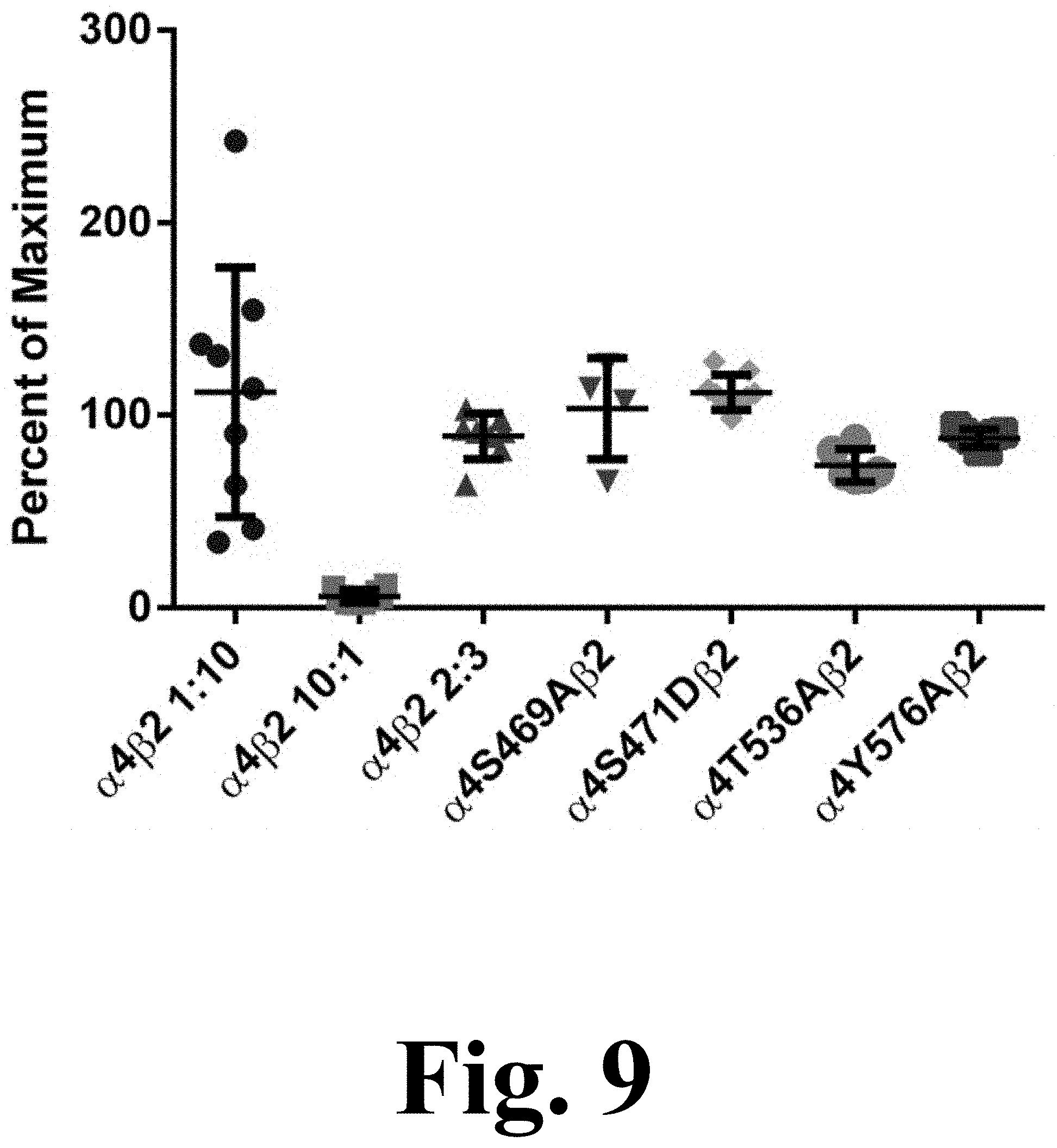
Changes in ACh Potency in the S469A, S471D, T536A and Y576A Mutations does not Involve Alterations in .alpha.4.beta.2 nAChRs Subunit Stoichiometry.
The decrease in potency for ACh exhibited by the majority of the mutations suggests potential changes in .alpha.4.beta.2 nAChR stoichiometry. Previous studies have shown that the .alpha.4(3):.beta.2(2) stoichiometry has a characteristic lower affinity for ACh whereas the .alpha.4(2): .beta.2(3) stoichiometry exhibits a higher affinity for ACh. Moreover, evidence suggests that phosphorylation of S467 (S471 in the present study) on free .alpha.4 subunits prior to their association with .beta.2 subunits has long-lasting consequences, increasing the stability of the .alpha.4 subunits. This increase in stability may result in an enhanced expression of .alpha.4.beta.2 in the low affinity .alpha.4(3):.beta.2(2) configuration. However, as FIG. 9 suggests, the observed decrease in ACh potency cannot be explained by alterations in the .alpha.4(2):.beta.2(3) stoichiometry. FIG. 9 shows the efficacy of .alpha.4.beta.2 agonist A-85380 for stoichiometry determination. Oocytes were injected with .alpha.4.beta.2 mRNA at either a ratio of 10:1, 1:10 or 2:3 for WT.alpha.4.beta.2 and in a 2:3 ratio for .alpha.4 mutants .alpha.4S469A.beta.2, .alpha.4S471D.beta.2, .alpha.4T536A.beta.2 and .alpha.4Y576A.beta.2. Current was measure for 300 .mu.M ACh and 100 nM A-85380. Efficacy was measured as the percent of maximum when compared to ACh. The efficacy was significantly lower in oocytes injected at a ratio of 10:1 than oocytes injected at a ratio of 1:10 (***p<0.0001). Oocytes injected at a 2:3 ratio show no significant difference from those injected at a 1:10 ratio. The four mutations show no significant difference from the WT injected 2:3 or 1:10 ratio, suggesting that the receptors maintain the same stoichiometry.
The efficacy of agonist A-85380 has been reported to be significantly reduced in the .alpha.4(3):.beta.2(2) stoichiometry. The efficacy of A-85380 in .alpha.4.beta.2 mutations S469A, S471D, T536A, and Y576A indicate that these are assembled according to the .alpha.4(2):.beta.2(3) stoichiometry.
Researchers are mostly in the dark about the structure and functional role of the intracellular domain of the AChR. The present data suggest a role for the cytoplasmic loop of the .alpha.4 subunit in .alpha.4.beta.2 function, expression, and agonist selectivity. Point mutations at consensus sites of the .alpha.4 nAChR subunit have remarkable effects on .alpha.4.beta.2 function and expression (Table 1). More specifically, these mutations affect the activation properties of ACh and nicotine; however, it is difficult to confirm a role for phosphorylation from the mutations' effects alone. Since our data show that targeting residues 5516 and T536 can render .alpha.4.beta.2 nAChRs almost insensitive to nicotine without major effects on normal AChR function, we propose that PKC could be a feasible target for smoking cessation drugs. Therefore, this invention provides the framework for further experiments with membrane-permeant cAMP analogs, antagonists, or other potential modulators of phosphorylation. Alternatively, it is possible that the mutations characterized in this study, besides affecting phosphorylation states, could also disrupt key point-to-point interactions essential for secondary structure and/or signal transduction. Twelve mutant receptors (S471A, S471D, T417A, S438A, S469D, S504A, S504D, S516A, T536D, S589A, S589D, and Y576A) exhibited significant changes in potency for ACh without affecting receptor expression levels.
We previously proposed a model that is consistent with an allosteric site for ACh activation, whereas nicotine activation displayed a biphasic curve. In the present invention, seven mutations displaced dose-response curves to higher ACh concentrations while displaying WT-like biphasic profiles for nicotine activation. Therefore, all of these mutations produced a negative allosteric effect on ACh activation without affecting the biphasic curve for nicotine activation. Furthermore, three other mutations showed only minor changes in the nicotine profile.
Overall, the effects that we observed in this study are fairly complex, for example, we observed decrease in peak response with both A and D mutations. In this particular case it is very likely that the mutation is not really mimicking a phosphorylation. Nonetheless, these results are consistent with previous reports of biphasic nicotine profiles for .alpha.4.beta.2 nAChRs. Most important, we discovered mutations that can provide novel perspectives for the design of nicotine smoking cessation medications that do not act directly or indirectly on the agonist's binding site. The invention provides a .alpha.4T536A knock-in mutant construct for the development of a transgenic mouse line with reduced nicotine sensitivity to be used in future studies. Overall, these findings provide a new perspective for developing smoking cessation drugs that can definitely cause changes in the pharmacology of nicotinic receptors.
SEQUENCE LISTINGS
1
31270PRTHomo sapiens 1His His Arg Ser Pro Arg Thr His Thr Met Pro Thr Trp Val Arg Arg1 5 10 15Val Phe Leu Asp Ile Val Pro Arg Leu Leu Leu Met Lys Arg Pro Ser 20 25 30Val Val Lys Asp Asn Cys Arg Arg Leu Ile Glu Ser Met His Lys Met 35 40 45Ala Ser Ala Pro Arg Phe Trp Pro Glu Pro Glu Gly Glu Pro Pro Ala 50 55 60Thr Ser Gly Thr Gln Ser Leu His Pro Pro Ser Pro Ser Phe Cys Val65 70 75 80Pro Leu Asp Val Pro Ala Glu Pro Gly Pro Ser Cys Lys Ser Pro Ser 85 90 95Asp Gln Leu Pro Pro Gln Gln Pro Leu Glu Ala Glu Lys Ala Ser Pro 100 105 110His Pro Ser Pro Gly Pro Cys Arg Pro Pro His Gly Thr Gln Ala Pro 115 120 125Gly Leu Ala Lys Ala Arg Ser Leu Ser Val Gln His Met Ser Ser Pro 130 135 140Gly Glu Ala Val Glu Gly Gly Val Arg Cys Arg Ser Arg Ser Ile Gln145 150 155 160Tyr Cys Val Pro Arg Asp Asp Ala Ala Pro Glu Ala Asp Gly Gln Ala 165 170 175Ala Gly Ala Leu Ala Ser Arg Asn Thr His Ser Ala Glu Leu Pro Pro 180 185 190Pro Asp Gln Pro Ser Pro Cys Lys Cys Thr Cys Lys Lys Glu Pro Ser 195 200 205Ser Val Ser Pro Ser Ala Thr Val Lys Thr Arg Ser Thr Lys Ala Pro 210 215 220Pro Pro His Leu Pro Leu Ser Pro Ala Leu Thr Arg Ala Val Glu Gly225 230 235 240Val Gln Tyr Ile Ala Asp His Leu Lys Ala Glu Asp Thr Asp Phe Ser 245 250 255Val Lys Glu Asp Trp Lys Tyr Val Ala Met Val Ile Asp Arg 260 265 2702271PRTMus musculus 2His His Arg Ser Pro Arg Thr His Thr Met Pro Ala Trp Val Arg Arg1 5 10 15Val Phe Leu Asp Ile Val Pro Arg Leu Leu Phe Met Lys Arg Pro Ser 20 25 30Val Val Lys Asp Asn Cys Arg Arg Leu Ile Glu Ser Met His Lys Met 35 40 45Ala Asn Ala Pro Arg Phe Trp Pro Glu Pro Glu Ser Glu Pro Gly Ile 50 55 60Leu Gly Asp Ile Cys Asn Gln Gly Leu Ser Pro Ala Pro Thr Phe Cys65 70 75 80Asn Arg Met Asp Thr Ala Val Glu Thr Gln Pro Thr Cys Arg Ser Pro 85 90 95Ser His Lys Val Pro Asp Leu Lys Thr Ser Glu Val Glu Lys Ala Ser 100 105 110Pro Cys Pro Ser Pro Gly Ser Cys His Pro Pro Asn Ser Ser Gly Ala 115 120 125Pro Val Leu Ile Lys Ala Arg Ser Leu Ser Val Gln His Val Pro Ser 130 135 140Ser Gln Glu Ala Ala Glu Gly Ser Ile Arg Cys Arg Ser Arg Ser Ile145 150 155 160Gln Tyr Cys Val Ser Gln Asp Gly Ala Ala Ser Leu Thr Glu Ser Lys 165 170 175Pro Thr Gly Ser Pro Ala Ser Leu Lys Thr Arg Pro Ser Gln Leu Pro 180 185 190Val Ser Asp Gln Thr Ser Pro Cys Lys Cys Thr Cys Lys Glu Pro Ser 195 200 205Pro Val Ser Pro Ile Thr Val Leu Lys Ala Gly Gly Thr Lys Ala Pro 210 215 220Pro Gln His Leu Pro Leu Ser Pro Ala Leu Thr Arg Ala Val Glu Gly225 230 235 240Val Gln Tyr Ile Ala Asp His Leu Lys Ala Glu Asp Thr Asp Phe Ser 245 250 255Val Lys Glu Asp Trp Lys Tyr Val Ala Met Val Ile Asp Arg Ile 260 265 2703271PRTRattus norvegicus 3His His Arg Ser Pro Arg Thr His Thr Met Pro Ala Trp Val Arg Arg1 5 10 15Val Phe Leu Asp Ile Val Pro Arg Leu Leu Phe Met Lys Arg Pro Ser 20 25 30Val Val Lys Asp Asn Cys Arg Arg Leu Ile Glu Ser Met His Lys Met 35 40 45Ala Asn Ala Pro Arg Phe Trp Pro Glu Pro Val Gly Glu Pro Gly Ile 50 55 60Leu Ser Asp Ile Cys Asn Gln Gly Leu Ser Pro Ala Pro Thr Phe Cys65 70 75 80Asn Pro Thr Asp Thr Ala Val Glu Thr Gln Pro Thr Cys Arg Ser Pro 85 90 95Pro Leu Glu Val Pro Asp Leu Lys Thr Ser Glu Val Glu Lys Ala Ser 100 105 110Pro Cys Pro Ser Pro Gly Ser Cys Pro Pro Pro Lys Ser Ser Ser Gly 115 120 125Ala Pro Met Leu Ile Lys Ala Arg Ser Leu Ser Val Gln His Val Pro 130 135 140Ser Ser Gln Glu Ala Ala Glu Asp Gly Ile Arg Cys Arg Ser Arg Ser145 150 155 160Ile Gln Tyr Cys Val Ser Gln Asp Gly Ala Ala Ser Leu Ala Asp Ser 165 170 175Lys Pro Thr Ser Ser Pro Thr Ser Leu Lys Ala Arg Pro Ser Gln Leu 180 185 190Pro Val Ser Asp Gln Ala Ser Pro Cys Lys Cys Thr Cys Lys Glu Pro 195 200 205Ser Pro Val Ser Pro Val Thr Val Leu Lys Ala Gly Gly Thr Lys Ala 210 215 220Pro Pro Gln His Leu Pro Leu Ser Pro Ala Leu Thr Arg Ala Val Glu225 230 235 240Gly Val Gln Tyr Ile Ala Asp His Leu Lys Ala Glu Asp Thr Asp Phe 245 250 255Ser Val Lys Glu Asp Trp Lys Tyr Val Ala Met Val Ile Asp Arg 260 265 270
* * * * *
D00001
D00002
D00003
D00004
D00005
D00006
D00007
D00008
D00009
D00010
S00001
XML
uspto.report is an independent third-party trademark research tool that is not affiliated, endorsed, or sponsored by the United States Patent and Trademark Office (USPTO) or any other governmental organization. The information provided by uspto.report is based on publicly available data at the time of writing and is intended for informational purposes only.
While we strive to provide accurate and up-to-date information, we do not guarantee the accuracy, completeness, reliability, or suitability of the information displayed on this site. The use of this site is at your own risk. Any reliance you place on such information is therefore strictly at your own risk.
All official trademark data, including owner information, should be verified by visiting the official USPTO website at www.uspto.gov. This site is not intended to replace professional legal advice and should not be used as a substitute for consulting with a legal professional who is knowledgeable about trademark law.