Thermophile-derived keratinase and use thereof
Lee , et al.
U.S. patent number 10,612,010 [Application Number 15/509,095] was granted by the patent office on 2020-04-07 for thermophile-derived keratinase and use thereof. This patent grant is currently assigned to Kyungpook National University Industry-Academic Cooperation Foundation. The grantee listed for this patent is KYUNGPOOK NATIONAL UNIVERSITY INDUSTRY-ACADEMIC COOPERATION FOUNDATION. Invention is credited to Hyun Su Jin, Ji Yeon Kim, Dong Woo Lee, Sang Jae Lee, Yong Jik Lee, Gae Won Nam.


| United States Patent | 10,612,010 |
| Lee , et al. | April 7, 2020 |
Thermophile-derived keratinase and use thereof
Abstract
The present invention relates to a novel thermophile-derived keratinase having keratin decomposition ability. Further, the present invention relates to a polynucleotide encoding the keratinase, a recombinant vector containing the same, host cells transformed by using the recombinant vector, and a method for preparing a keratinase including a step of culturing the host cells. Further, the present invention relates to a composition for decomposing keratin containing the enzyme; and a method for decomposing keratin by using the same. Further, the present invention relates to a keratin decomposed product decomposed by the enzyme. The keratinase according to the present invention rapidly and effectively decomposes hardly-decomposable keratin, and thus it is expected that the keratinase can be used for the effective treatment and the high value-added resource recovery of agricultural and livestock waste, which causes environmental problems (for example, a novel material for enzyme cosmetics), and can be used in an innovative enzymatic bioconversion technique utilizing various decomposition enzyme groups.
| Inventors: | Lee; Dong Woo (Daegu, KR), Lee; Yong Jik (Daegu, KR), Jin; Hyun Su (Daegu, KR), Kim; Ji Yeon (Daegu, KR), Nam; Gae Won (Cheongju, KR), Lee; Sang Jae (Busan, KR) | ||||||||||
|---|---|---|---|---|---|---|---|---|---|---|---|
| Applicant: |
|
||||||||||
| Assignee: | Kyungpook National University
Industry-Academic Cooperation Foundation (Daegu,
KR) |
||||||||||
| Family ID: | 55440158 | ||||||||||
| Appl. No.: | 15/509,095 | ||||||||||
| Filed: | September 7, 2015 | ||||||||||
| PCT Filed: | September 07, 2015 | ||||||||||
| PCT No.: | PCT/KR2015/009419 | ||||||||||
| 371(c)(1),(2),(4) Date: | March 06, 2017 | ||||||||||
| PCT Pub. No.: | WO2016/036222 | ||||||||||
| PCT Pub. Date: | March 10, 2016 |
Prior Publication Data
| Document Identifier | Publication Date | |
|---|---|---|
| US 20170355971 A1 | Dec 14, 2017 | |
Foreign Application Priority Data
| Sep 5, 2014 [KR] | 10-2014-0118995 | |||
| Current U.S. Class: | 1/1 |
| Current CPC Class: | C12N 9/0051 (20130101); C12N 9/13 (20130101); C12Y 108/01009 (20130101); C12Y 304/16005 (20130101); C12N 9/485 (20130101); C12N 15/52 (20130101); C12N 9/00 (20130101); C12N 9/48 (20130101); C12Y 208/01007 (20130101) |
| Current International Class: | C12N 9/00 (20060101); C12N 9/48 (20060101); C12N 15/52 (20060101); C12N 9/10 (20060101); C12N 9/02 (20060101) |
References Cited [Referenced By]
U.S. Patent Documents
| 5569599 | October 1996 | Antranikian |
Other References
|
Zhang et al., "Function of the in vitro Recombinant SufS--SufE Complex in the Fe--S Cluster Biosynthesis Pathway from the Thermus thermophilus", Chinese J. of Biochem. And Mol. Biol., Jun. 2014, 30(5):496-502. cited by examiner . Lee et al. Genome sequence of a native-feather degrading extremely thermophilic Eubacterium, Fervidobacterium islandicum AW-1Standards in Genomic Sciences, 2015, 10:71, pp. 1-9. DOI 10.1186/s40793-015-0063-4. cited by examiner . Frederich et al., "Keratin Degradation by Fervidobacterium pennavorans, a Novel Thermophilic Anaerobic Species of the Order Thermotogales", Applied and Environmental Microbiology, Aug. 1996, vol. 62, No. 8, p. 2875-2882. cited by examiner . Lucas et al., "Complete sequence of Fervidobacterium pennivorans DSM 9078." Submitted (Mar. 2012) to the EMBL/GenBank/D--retrived from < https://www.uniprot.org/proteomes/UP000007384 > on Jun. 28, 2019. cited by examiner . Yassin M. El-Ayouty, Amira El-Said, Ahmed M. Salama Purification and characterization of a keratinase from the feather-degrading cultures of Aspergillus flavipes African Journal of Biotechnology vol. 11(9), pp. 2313-2319, Jan. 31, 2012 ISSN 1684-5315 .COPYRGT. 2012 Academic Journals. cited by applicant . Sri Rahayu, Dahrul Syah, Maggy Thenawidjaja Suhartono Degradation of keratin by keratinase and disulfide reductase from Bacillus sp. MTS of Indonesian origin Biocatalysis and Agricultural Biotechnology 1 (2012) 152-158. cited by applicant . S Rahayu, D Syah, MT Suhartono Preliminary Study on Keratinase from Two Indonesian Isolates S Rahayu et al/Animal Production 12 (1):60-68, 2012. cited by applicant . Gae-Won Nam, Dong-Woo Lee, Han-Seoong Lee, Nam-Ju Lee, Byoung-Chan Kim, Eun-Ah Choe, Jae-Kwan Hwang, Maggy T. Suhartono, Yu-Ryang Pyun Native-feather degradation by Fervidobacterium islandicum AW-1, a newly isolated keratinase-producing thermophilic anaerobe Arch Microbiol (2002) 178 :538-547. cited by applicant . Alessandro Riffel, Adriano Brandelli, Cla'udia De M. Bellato, Gustavo H.M.F. Souza, Marcos N. Eberlin, Flavio C.A. Tavares Purification and characterization of a keratinolytic metalloprotease from Chryseobacterium sp. kr6 Journal of Biotechnology 128 (2007) 693-703. cited by applicant . BLAST .RTM. Basic Local Alignment Search Tool NCBI/BLAST/ blastp suite/ Formatting Results 1BGJJB9F016 Oct. 8, 2015. cited by applicant . BLAST .RTM. Basic Local Alignment Search Tool NCBI/ BLAST/ blastp suite/ Formatting Results 1BGPTZNJ01R Oct. 8, 2015. cited by applicant . BLAST .RTM. Basic Local Alignment Search Tool NCBI/ BLAST/ blastp suite/ Formatting Results 1BGT19SH014 Oct. 8, 2015. cited by applicant . BLAST .RTM. Basic Local Alignment Search Tool NCBI/ BLAST/ blastp suite/ Formatting Results 1BGWSZJB014 Oct. 8, 2015. cited by applicant. |
Primary Examiner: Noakes; Suzanne M
Attorney, Agent or Firm: Rao; Weisun Venture Partner, LLC
Claims
The invention claimed is:
1. A method for keratin decomposition comprising, a step of treating keratin with i) at least one Thermotogales order derived enzyme or protein selected from the group consisting of: an isolated cysteine desulfurase (CDS); an isolated thermostable carboxypeptidase 1 (CBP); an isolated thioredoxin-disulfide reductase (DSR); and an isolated iron-sulfur assembly scaffold protein (SufE); and also ii) a Thermotogales order-derived cell extract, wherein (i) and (ii) are not the same.
2. The method for keratin decomposition of claim 1, wherein the keratin is derived from poultry feathers.
3. The method for keratin decomposition of claim 1, wherein the isolated cysteine desulfurase (CDS) consists of the amino acid sequence of SEQ ID NO: 1; the isolated thermostable carboxypeptidase 1 (CBP) consists of the amino acid sequence of SEQ ID NO: 3; the isolated thioredoxin-disulfide reductase (DSR); consists of the amino acid sequence of SEQ ID NO: 5; and the isolated iron-sulfur assembly scaffold protein (SufE) consists of the amino acid sequence of SEQ ID NO: 7.
4. The method for keratin decomposition of claim 1, wherein the CDS, CBP, DSR, and SufE are encoded by a polynucleotide consisting of the base sequence of SEQ ID NO: 2; the base sequence of SEQ ID NO: 4; the base sequence of SEQ ID NO: 6; and the base sequence of SEQ ID NO: 8, respectively.
5. The method for keratin decomposition of claim 1, wherein the Thermotogales order includes Caldotoga, Mesotoga, Thermopallium, Thermotoga, Fervidobacterium, Thermosipho, Kosmotoga, Thermococcoides, Marinitoga, Geotoga, or Petrotoga genus.
6. The method for keratin decomposition of claim 1, further, comprising the step of treating the keratin with Dithiothreitol.
7. The method for keratin decomposition of claim 1, wherein the Thermotogales order is Fervidobacterium islandicum AW-1 strain.
Description
TECHNICAL FIELD
The present invention relates to novel thermophile-derived keratin degrading enzymes.
Further, the present invention relates to polynucleotides encoding the keratin degrading enzymes, recombinant vectors containing the same, and host cells transformed by the recombinant vectors.
Further, the present invention relates to a method for preparing keratin degrading enzymes including a step of culturing the host cells.
Further, the present invention relates to a composition for keratin degradation containing the enzymes; and a method for decomposing keratin by using the same.
Further, the present invention relates to a keratin byproducts decomposed by the enzyme.
BACKGROUND ART
Keratin is an animal protein which is nutritionally valuable, but not widely used and a fibrous component of skin, horns and hair and has been wasted in bulk in the slaughter and cattle industry (Kornillowicz-Kowalska and Bohacz, 2011).
Since the keratin contains many disulfide bonds in its structure, the keratin has a water-insoluble characteristic and is not decomposed by general proteases (Onifade et al., 1998).
Currently, keratin wastes are incinerated or after the keratin is decomposed by chemical methods, the degradation product thereof is recycled as feed additives for livestock. The keratin decomposition product generated by chemical treatment has a high content of nitrogen, fat, and the like, but has disadvantages in that the content of amino acids such as lysine and methionine which are required for livestock is low and simultaneously, digestibility is low (Papadoulos and Ketelaars, 1986). Further, high energy cost is required and environmental problems such as odor can occur (da Rosa Gioppo and the like 2009). Therefore, in order to solve the problems, a new treatment method has been required and recently, a keratin decomposition method by microorganisms has been actively studied as an eco-friendly alternative to solve the above problems.
A microbiological keratin treatment method started with the isolation of microorganisms producing keratin degrading enzymes, and various microorganisms such as Bacillus spp., actinomycetes and fungi were isolated from a natural environment and enzyme production characteristics and the nutritional value of keratin decomposition products treated with these microorganisms have been reported (Bertsch and Coello, 2005; Brandelli, et al., 2010). In addition, it was reported that the keratinase can be used for removal of proteinaceous organic materials in a wastewater treatment plant, improvement of fabric quality, hair removal of leather, exfoliating cosmetics, prion decomposition, and the like (Gupta and Ramnani, 2006; Langeveld, et al., 2003; Onifade, et al., 1998). Further, in order to pioneer novel applications, studies for isolating strains having unique properties such as plant growth promoting activity and antifungal activity, together with keratinase activity have been started (Jeong, et al, 2010).
Meanwhile, it is known that coenzyme is more advantageous than using a purified enzyme to decompose proteins at low cost and the coenzyme is more stable than the purified enzyme. Actually, the product in the coenzyme state is commercialized as an enzyme preparation for treating a large amount of proteins. Further, since the characteristics of the keratinase are strain-specific, physicochemical properties of the enzymes produced by each strain need to be investigated for efficient application of the enzyme.
Technical Problem
The inventors of the present invention made every possible effort to find keratin degrading enzymes for effective decomposition and application of keratin to verify that thermophile-derived keratin degrading enzymes effectively decomposed the keratin and completed the present invention.
Accordingly, an object of the present invention is to provide a novel thermophile-derived keratin degrading enzymes.
Another object of the present invention is to provide polynucleotides encoding the keratin degrading enzymes, recombinant vectors containing the same, and host cells transformed by using the recombinant vectors.
Yet another object of the present invention is to provide a method for preparing keratin degrading enzymes including a step of culturing the host cells.
Still another object of the present invention is to provide a composition for keratin degradation containing the enzymes; and a method for decomposing keratin by using the same.
Still yet another object of the present invention is to provide a keratin byproducts decomposed by the enzymes.
Technical Solution
An aspect of the present invention provides thermophile-derived keratin degrading enzymes which contain at least more than one selected from a group consisting of:
a cysteine desulfurase (CDS);
a thermostable carboxypeptidase 1 (CBP);
a thioredoxin-disulfide reductase (DSR); and
an iron-sulfur assembly scaffold protein (SufE).
The cysteine desulfurase (CDS) may consist of an amino acid sequence of SEQ ID NO: 1;
The thermostable carboxypeptidase 1 (CBP) may consist of an amino acid sequence of SEQ ID NO: 3;
The thioredoxin-disulfide reductase (DSR) may consist of an amino acid sequence of SEQ ID NO: 5; and
The iron-sulfur assembly scaffold protein (SufE) may consist of an amino acid sequence of SEQ ID NO: 7.
Further, the CDS, CBP, DSR, and SufE may be encoded by a polynucleotide consisting of
a base sequence of SEQ ID NO: 2;
a base sequence of SEQ ID NO: 4;
a base sequence of SEQ ID NO: 6; and
a base sequence of SEQ ID NO: 8.
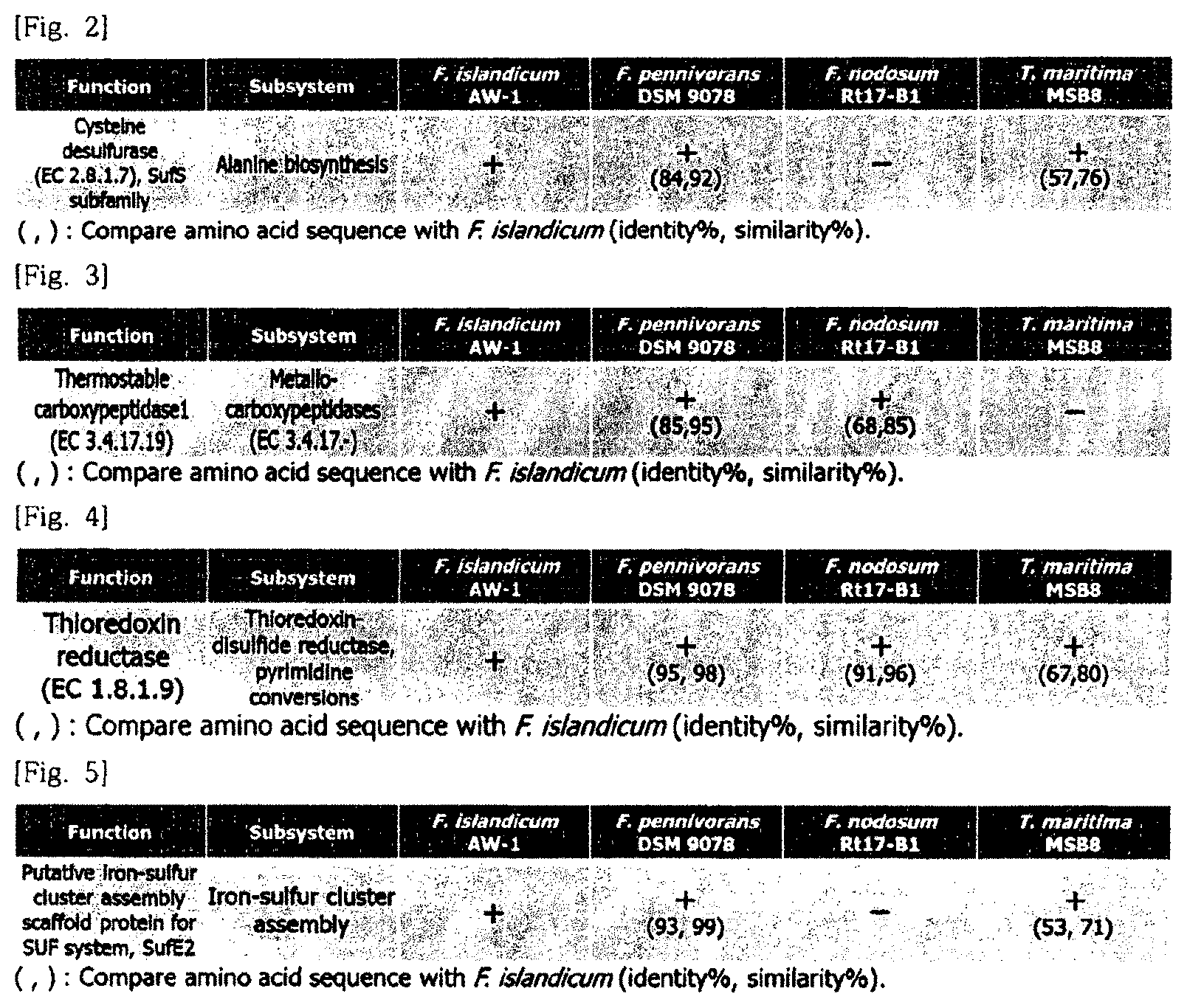
In an embodiment of the present invention, cysteine desulfurase (CDS) which is present only in Fervidobacterium islandicum AW-1 and expected to be involved in decomposition of a chicken feather may be selected. The result is illustrated in FIG. 2. Further, the amino acid sequence information is designated as SEQ ID NO: 1 and the base sequence information is designated as SEQ ID NO: 2.
In the case of the thermostable carboxypeptidase 1 (CBP), comparison and analysis of amino acid sequence identity between proteases of F. islandicum AW-1 and proteases of F. nodosum Rt17-B1 which do not have the decomposition ability of the chicken feather wastes were performed by using a NCBI website-based blastp (blast.st-va.ncbi.nlm.nih.gov) program. The result is illustrated in FIG. 3.
As illustrated in FIG. 3, a protease thermostable carboxypeptidase 1 (CBP) which is expected to be involved in decomposition of a chicken feather wastes having low identity with the F. nodosum Rt17-B1 was finally screened.
The amino acid sequence information is designated as SEQ ID NO: 3 and the base sequence information is designated as SEQ ID NO: 4.
In the case of the thioredoxin-disulfide reductase (DSR), as a result of comparing and analyzing a genome with a F. pennivorans strain, which is known to have keratin decomposition capability in addition to the F. islandicum AW-1 by using a RAST server and a Bioedit analysis program, thioredoxin-disulfide reductase which has the highest amino acid identity and is expected to be involved in decomposition of chicken feather wastes was selected as illustrated in FIG. 4.
The amino acid sequence information was designated as SEQ ID NO: 5 and the base sequence information was designated as SEQ ID NO: 6.
In the case of the iron-sulfur assembly scaffold protein (SufE), it is expected that a complex with the CDS is formed to improve keratin decomposition capability. As a result, an iron-sulfur assembly scaffold protein which is demonstrated as present in a downstream of the CDS in a Suf operon through the RAST server was selected as illustrated in FIG. 5.
The amino acid sequence information was designated as SEQ ID NO: 7 and the base sequence information was designated as SEQ ID NO: 8.
Further, the thermophilic bacteria may be Thermotogales order.
The thermophilic bacteria are not limited as long as the thermophilic bacteria are included in the Thermotogales order, but for example, the Thermotogales order may be Caldotoga, Mesotoga, Thermopallium, Thermotoga, Fervidobacterium, Thermosipho, Kosmotoga, Thermococcoides, Mariniloga, Geotoga, or Petrotoga genus.
In an embodiment of the present invention, the F. islandicum AW-1-derived keratin degrading enzymes were used.
Further, another aspect of the present invention provides a polynucleotide encoding a keratinase which is at least one selected from a group consisting of: a cysteine desulfurase (CDS) consisting of an amino acid sequence of SEQ ID NO: 1;
a thermostable carboxypeptidase 1 (CBP) consisting of an amino acid sequence of SEQ ID NO: 3;
a thioredoxin-disulfide reductase (DSR) consisting of an amino acid sequence of SEQ ID NO: 5: and
an iron-sulfur assembly scaffold protein (SufE) consisting of an amino acid sequence of SEQ ID NO: 7.
The polynucleotides encoding the CDS, CBP, DSR and SufE may be
a base sequence of SEQ ID NO: 2;
a base sequence of SEQ ID NO: 4;
a base sequence of SEQ ID NO: 6; and
a base sequence of SEQ ID NO: 8, respectively.
The "polynucleotide" is a polymer of a deoxyribonucleotide or a ribonucleotide which is present in a form of a single strand or a double strand. The polynucleotide includes DNAs (gDNA and cDNA) and RNA sequences transcribed therefrom and includes analogs of natural polynucleotides unless specifically stated otherwise.
The polynucleotide includes not only a nucleotide sequence encoding the aforementioned peptide but also a complementary sequence to the sequence. The complementary sequence includes not only a complete complementary sequence but also a substantial complementary sequence.
Further, the polynucleotide may be modified. The modification includes addition, deletion, or non-conservative substitution or conservative substitution of the nucleotide. It is interpreted that the polynucleotide encoding the amino acid sequence includes a nucleotide sequence having substantial identity with respect to the nucleotide sequence. The substantial identity may be a sequence having identity and similarity of at least 60% and 80% in the case of analyzing a sequence which is aligned to maximally correspond to any different sequence from the nucleotide sequence and aligned by using an algorithm which is generally used in the related art.
Another aspect of the present invention provides a recombinant vector including the polynucleotide.
The term "vector" means a DNA molecule for expressing a target gene in host cells. For example, the vector includes plasmid vectors, cosmide vectors, and virus vectors such as bacteriophage vectors, adenovirus vectors, retrovirus vectors, and adeno-associated virus vectors. The vector which may be used as the recombinant vector may be prepared by manipulating plasmids (for example, pSC101, pGV1106, pACYC177, ColE1, pKT230, pME290, pBR322, pUC8/9, pUC6, pBD9, pHC79, pIJ61, pLAFR1, pHV14, pGEX series, pET series, pUC19, etc.), phages (for example, .lamda.gt4.lamda.B, .lamda.-Charon, .lamda..DELTA.z1, M13, etc.) or virus (for example, CMV, SV40, etc.), which are frequently used in the related art.
In the recombinant vector, the polynucleotide encoding the amino acid sequence may be operatively linked with the promoter. The term "operatively linked" means a functional binding between a regulatory sequence (e.g, a promoter sequence) and a different nucleotide sequence. Accordingly, the regulatory sequence may regulate transcription and/or translation of the different nucleotide sequence by the functional binding.
The recombinant vector may be typically constructed as a vector for cloning or a vector for expression. The expression vector may use general vectors which are used to express foreign proteins in plants, animals, or microorganisms in the art. The recombinant vector may be constructed by various methods known in the art.
The recombinant vector may be constructed using prokaryotic cells or eukaryotic cells as a host. For example, the used vector is an expression vector and in the case of using the prokaryotic cells as a host, the vector generally includes a strong promoter capable of processing a transcription (for example, a pLX promoter, a trp promoter, a lac promoter, a tac promoter, a T7 promoter, etc.), a ribosome binding site for initiation of translation, and a transcription/translation termination sequence. In the case of using the eukaryotic cells as a host, a replication origin that functions in the eukaryotic cells contained in the vector includes an f1 replication origin, an SV40 replication origin, a pMB1 replication origin, an adeno replication origin, an AAV replication origin, a CMV replication origin, a BBV replication origin, and the like, but is not limited thereto. Further, a promoter derived from a genome of mammalian cells (e.g., a metallothionine promoter) or a promoter derived from a mammalian virus (e.g., an adenovirus late-phase promoter, a vaccinia virus 7.5K promoter, an SV40 promoter, a cytomegalovirus (CMV) promoter and a tk promoter of HSV) may be used, and generally has a polyadenylation sequence as a transcription termination sequence.
Meanwhile, the vector may express not only a peptide specifically binding to NRP1 of the present invention but also fragments or an antibody of the antibody to which the peptide is fused. In the case of the antibody to which the peptide is fused or the fragments of the antibody, the vector may include a vector system in which the peptide and the antibody or its fragment are expressed at the same time in one vector or expressed in separate vectors respectively. In the case of being expressed in separate vectors, two vectors may be introduced to the host cells through co-transformation and targeted transformation.
Yet another aspect of the present invention provides host cells transformed by the recombinant vector.
The host cells may use any host cells known in the art, and as prokaryotic cells, for example, E. coli genus strains such as E. coli JM109, BL21, RR1, LE392, X1776 and W3110, Bacillus genus strains such as Bacillus subtilis and Bacillus thuringiensis, and Enterobacteriaceae and strains such as Salmonella typhimurium, Serratia marcescens and various Pseudomonas species are included. In the case of transformation to eukaryotic cells, as host cells, yeast (Saccharomyce cerevisiae), insect cells, plant cells and animal cells, for example, SP2/0, CHO (Chinese hamster ovary) K1, CHO DG44, PER.C6, W138, BHK, COS-7, 293, HepG2, Huh7, 3T3, RIN, MDCK cell lines, and the like may be used.
Still another aspect of the present invention provides a method for preparing the keratin degrading enzymes including a step of culturing the host cells.
An insertion method well-known in the field may be used for the insertion into the host cells of the polynucleotide or the recombinant vector containing the polynucleotide. The transfer method may use a CaCl.sub.2) method or an electroporation method, or the like when the host cells are the prokaryotic cells, and use microinjection, calcium phosphate precipitation, electroporation, liposome-mediated transfection and gene bombardment when the host cells are eukaryotic cells, but is not limited thereto.
A method of screening the transformed host cells may be easily performed by using a phenotype expressed by a selection marker according to a method well-known in the art. For example, when the selection marker is a specific antibiotic resistance gene, a transformant may be easily screened by culturing the transformant in a medium containing the antibiotic.
Further, still yet another aspect of the present invention provides a composition for keratin decomposition including at least one thermophile-derived enzyme selected from a group consisting of:
a cysteine desulfurase (CDS);
a thermostable carboxypeptidase 1 (CBP);
a thioredoxin-disulfide reductase (DSR); and
an iron-sulfur assembly scaffold protein (SufE).
Further, still yet another aspect of the present invention provides a method for keratin decomposition including treating keratin with at least one thermophile-derived enzyme selected from a group consisting of:
a cysteine desulfurase (CDS);
a thermostable carboxypeptidase 1 (CBP);
a thioredoxin-disulfide reductase (DSR); and
an iron-sulfur assembly scaffold protein (SufE).
The cysteine desulfurase (CDS); the thermostable carboxypeptidase 1 (CBP); the thioredoxin-disulfide reductase (DSR); or the iron-sulfur assembly scaffold protein (SufE) may be supplementary or necessary in the decomposition of keratin.
The origin of the keratin is not limited. That is, the origin of the keratin may include hair, nails, animal hooves, skin, animal hair and feathers, and the like, and most preferably feathers.
Advantageous Effects
The keratin degrading enzymes according to the present invention rapidly and effectively decomposes hardly-decomposable keratin, and thus it is expected that the keratin degrading enzymes can be used for the effective wastes treatment and the transformation of agricultural and livestock wastes into high value-added resources, which causes environmental problems (for example, a novel material for enzyme cosmetics), and can be used in an innovative enzymatic bioconversion technique utilizing various decomposition enzyme groups.
DESCRIPTION OF DRAWINGS
FIG. 1 illustrates a result of analyzing genome information of F. islandicum AW-1 having keratin decomposition ability by using a RAST server.
FIG. 2 illustrates a cysteine desulfurase (CDS) screened as a keratin degrading enzymes group based on a result of comparing and analyzing protein function-based genomes.
FIG. 3 illustrates a thermostable carboxypeptidase 1 (CBP) screened as a keratin degrading enzymes group based on a result of comparing and analyzing amino acid sequence-based genomes.
FIG. 4 illustrates a thioredoxin-disulfide reductase (DSR) screened as a keratinase group based on a result of comparing and analyzing amino acid sequence-based genomes.
FIG. 5 illustrates an iron-sulfur assembly scaffold protein (SufE) screened as a keratinase group based on a result of comparing and analyzing amino acid sequence-based genomes.
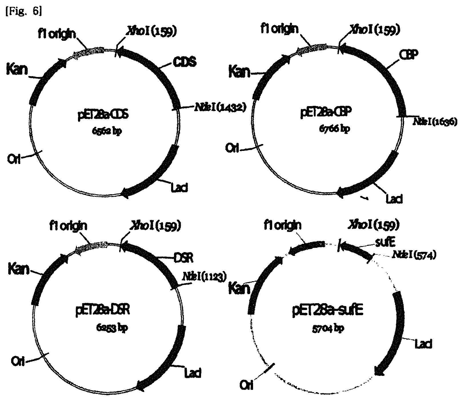
FIG. 6 illustrates a schematic diagram of expression vectors of key proteins expected as a keratinase group.
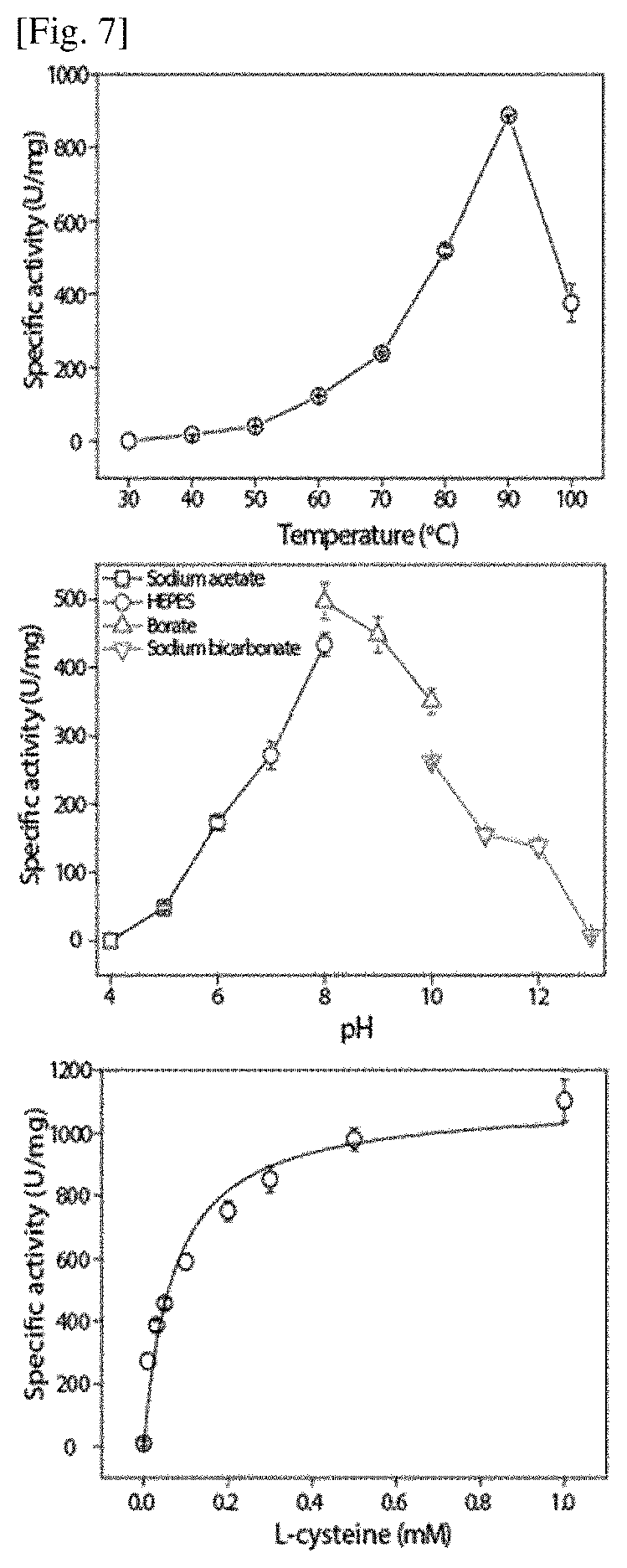
FIG. 7 illustrates a biophysicochemical characteristic of a cysteine desulfurase (CDS) recombinant protein expected as a keratinase of F. islandicum AW-1.
FIG. 8 illustrates a biophysicochemical characteristic of a thermostable carboxypeptidase 1 (CBP) recombinant protein expected as a keratinase of F. islandicum AW-1.
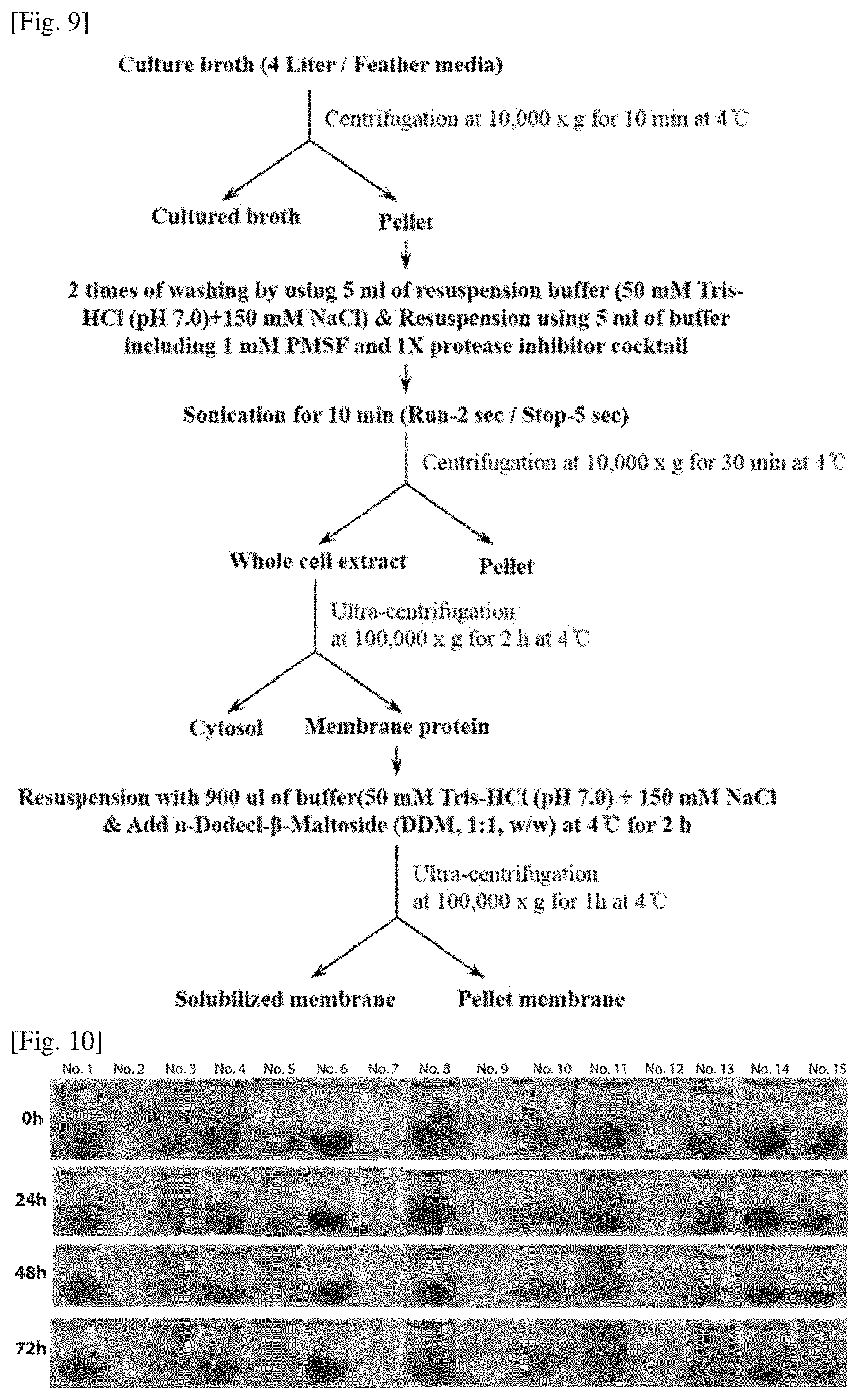
FIG. 9 is a schematic diagram illustrating a method of extracting a whole cell extract for obtaining an enzyme group having keratin decomposition ability from F. islandicum AW-1.
FIG. 10 illustrates a result of verifying decomposition of a chicken feather wastes by using a whole cell extract and a recombinant protein of F. islandicum AW-1 having keratin decomposition ability.
FIG. 11 illustrates quantitative analysis of free amino acids produced by a decomposition experiment of the chicken feather wastes of FIG. 10.
FIG. 12 is a schematic diagram illustrating a method for extracting a whole cell extract extract from colonies in which F. islandicum AW-1 having keratin decomposition ability is anaerobically cultured in a medium added with 0.5% (v/v) glucose as a unique carbon source.
FIG. 13 illustrates a merge treatment effect for decomposition of the chicken feather wastes by selectively adding cysteine desulfurase (CDS) and iron-sulfur assembly scaffold protein (SufE) recombinant proteins in the whole cell extract of F. islandicum AW-1.
FIG. 14 is a graph illustrating a result of amino acids quantitative analysis of the decomposition effect of the chicken feather wastes by FIG. 13.
MODES OF THE INVENTION
Hereinafter, the present invention will be described in more detail through Examples. However, these Examples are to exemplify the present invention and the scope of the present invention is not limited to these Examples.
Experimental Example 1: Genome Analysis of Fervidobacterium islandicum AW-1
Colonies were collected from a culture broth of F. islandicum AW-1 having keratin decomposition ability to extract a genomic DNA by using a genomic DNA extraction kit (Solgent), and then obtain a genome base sequence by using a single molecule real-time (SMRT) sequencing platform (PacBio RS II system).
The genome information was analyzed by using a hierarchical genome-assembly process (RS-HGAP) assembly protocol and a RAST server (rast.nmpdr.org) in an SMRT analysis pipeline v.2.2.0. As a result, it was verified that a genome DNA size of F. islandicum AW-1 was 2.35 Mb and had 2,259 coding genes.
Experimental Example 2: Analysis of Comparing Protein Function-Based and Amino Acid Sequence-Based Genomes Using Genome Information of Fervidobacterium islandicum AW-1
A target enzyme group involved in keratin decomposition was selected by comparing and analyzing genome information of F. islandicum AW-1. To this end, proteins expected to be in the keratinase group, cysteine desulfurase (CDS), thermostable carboxypeptidase 1 (CBP), and thioredoxin-disulfide reductase (DSR) were selected by directly comparing and analyzing genome information with F. islandicum AW-1 having excellent keratin decomposition ability and strains corresponding to the same genus.
Particularly, in the case of the CDS, genomes of Fervidobacterium islandicum AW-1 as a chicken feather wastes decomposition strain and F. nodosum Rt17-B1 as a strain without chicken feather wastes decomposition ability were compared and analyzed based on a protein function by using the RAST server (rast.nmpdr.org). The result is illustrated in FIG. 2.
As illustrated in FIG. 2, cysteine desulfurase (CDS) which was present only in F. islandicum AW-1 and expected to be involved in decomposition of the chicken feather wastes was screened. The amino acid sequence information was designated as SEQ ID NO: 1 and the base sequence information was designated as SEQ ID NO: 2.
In the case of the thermostable carboxypeptidase 1 (CBP), comparison and analysis of amino acid sequence identities between proteases of F. islandicum AW-1 and proteases of F. nodosum Rt17-B1 as a strain without chicken feather wastes decomposition ability were performed by using a NCBI website-based blastp (blast.st-va.ncbi.nlm.nih.gov) program. The result was illustrated in FIG. 3.
As illustrated in FIG. 3, a protease thermostable carboxypeptidase 1 (CBP) which was expected to be involved in decomposition of the chicken feather wastes having low identity with the F. nodosum Rt17-B1 was finally screened.
The amino acid sequence information was designated as SEQ ID NO: 3 and the base sequence information was designated as SEQ ID NO: 4.
In the case of the thioredoxin-disulfide reductase (DSR), as a result of comparing and analyzing a genome with a F. pennivorans strain, which was known to have keratin decomposition ability in addition to the F. islandicum AW-1 by using a RAST server and a Bioedit analysis program, thioredoxin-disulfide reductase which had the highest amino acid identity and was expected to be involved in decomposition of a chicken feather wastes was screened as illustrated in FIG. 4.
The amino acid sequence information was designated as SEQ ID NO: 5 and the base sequence information was designated as SEQ ID NO: 6.
In the case of the iron-sulfur assembly scaffold protein (SufE), it was expected that a complex with the CDS was formed to increase keratin decomposition ability. As a result, an iron-sulfur assembly scaffold protein which was verified as present in a downstream of the CDS in a Suf operon through the RAST server was screened as illustrated in FIG. 5.
The amino acid sequence information was designated as SEQ ID NO: 7 and the base sequence information was designated as SEQ ID NO: 8.
Experimental Example 3: Preparation of Expression Vectors of CDS, CBP, and DSR Proteins and E. coli Transformants
Expression vectors of proteins which were expected as a keratinase group screened by comparing and analyzing protein function-based and amino acid sequence-based genomes of F. islandicum AW-1 was prepared.
TABLE-US-00001 TABLE 1 Sequence Primer Primer base No. No. Primer name sequence (5'-3') 9 A F.aw1_cds_NdeI F CATATGCGCTCAACGG TGTTCTC 10 B F.aw1_cds_XhoI R CTCGAGTCATTCGAAC CACCTCC 11 C F.aw1_cbp_NdeI F CATATGGAAGAACTAA AAAGCTATTACAAACG 12 D F.aw1_cbp_XhoI R CTCGAGTTAAAGCTCT ATCTCATACACTTTG 13 E F.aw1_dsr_NdeI F CATATGAGCGGATTTG AATTCGACA 14 F F.aw1_dsr_XhoI R CTCGAGTTAGAAGTAT TTCTTTGCAGCG 15 G F.aw1_sufE_NdeI F GCGCATATGATATACT CTGAATTCATAATGG 16 H F.aw1_sufE_XhoI R CTCGAGTAATTCATTC TTTAAAGCAATCTCC
As shown in Table 1, in order to prepare expression vectors CDS, CBP, DSR, and SufE, first, forward primers and reverse primers containing respective restriction enzymes sequence (NdeI or XhoI) were used in pairs in PCR according to the preparation of the expression vectors.
A genomic DNA of the Fervidobacterium islandicum AW-1 strain was used as a template for the amplification of each gene by the PCR method, and the reaction was performed at 98.degree. C. for 5 min using Primestar HS DNA polymerase from Takara Corporation, performed at 98.degree. C. for 30 s, 55.degree. C. for 15 s, and 72.degree. C. for 1 min by 30 cycles, and then performed at 72.degree. C. for 10 min.
The amplified PCR product was electrophoresed and isolated and purified by using a gel extraction kit (QIAGEN), cloned by using a pTOP Blunt V2 cloning kit (Enzynomics), and then whether to insert a mutant of each gene was verified through DNA sequencing (Solgent).
A pET-28a (+) (Novagen) vector was treated with restriction enzymes NdeI and XhoI for the preparation of each gene expression vector after verifying that there was no mutant in three kinds of genes expected as a keratinase group obtained through the PCR. For the recovery of each gene cloned in the pTOP Blunt V2 vector, the restriction enzyme corresponding to the expression vector was treated and isolated and purified by using electrophoresis and the gel extraction kit (QIAGEN).
Four kinds of expression vectors pET28a-CDS, pET28a-CBP, pET28a-DSR, and pET28a-SufE were prepared by ligating the restriction enzyme-treated vector and the recovered genes by using a DNA ligation kit (Takara). A cleavage map of the expression vectors was illustrated in FIG. 6. For expression of protein genes expected as a keratinase group, E. coli transformants were prepared as follows. Each expression vector plasmid was introduced into an E. coli strain BL21 (DE3) (Enzynomics). In order to selectively culture the transformants, an LB solid medium containing kanamycin, was cultured at 37.degree. C. Kanamycin is an antibiotic suitable for the antibiotic resistance gene contained in the expression vector. The final concentration of kanamycin was 100 .mu.g/ml.
Experimental Example 4: Mass Expression and Purification of Proteins CDS, CBP, DSR, and SufE
For expression of protein genes expected as the keratinase group, transformants into which each expression vector was inserted was obtained and then used. Each transformant was pre-cultured and then 1% (5 ml) of pre-cultured cells were inoculated and cultured in 500 ml of a LB added with kanamycin (100 .mu.g/ml) in two 2 L flasks. An isopropyl .beta.-D-1-thiogalactopyranoside (IPTG) at a final concentration of 1 mM was added when absorbance (600 nm) was 0.4 to 0.6, expression of the CDS, CBP, DSR and SufE genes was induced at 37.degree. C. for 6 h.
Cells induced by protein expression were centrifuged after a culture broth with absorbance (600 nm) 1 was transferred to a 1.5 ml E-tube and then only cultured colonies were recovered. The cells were suspended in 100 .mu.l of a 1.times.SDS sample buffer and treated in boiled water for 5 min, modified and isolated by a 12% sodium dodecyl sulphate-polyacrylamide gel electrophoresis (SDS-PAGE) gel and then stained with coomassie brilliant blue (CBB) R-250, and then protein expression levels were confirmed. The cultured cells were recovered by centrifugation and resuspended in 50 mM Tris-HCl (pH 7.0) containing 150 mM NaCl and 1 mM phenylmethylsulfonyl fluoride (PMSF). The cells were broken by sonication at 4.degree. C. and the supernatant was recovered by centrifugation (10,000.times.g, 20 min).
The recovered supernatant was heat-treated (70.degree. C. for 30 min), centrifuged (10,000.times.g, 20 min) and 0.45 .mu.m-filtered to obtain a sample for His-tag purification. As a next step, in order to purify each expression protein, the expression protein was isolated and purified by using Ni.sup.2+-NTA agarose beads (Novagen). The expression protein containing a 6-histidine residue in an N-terminus was bound through the Ni.sup.2+-NTA agarose beads equilibrated with a binding buffer (0.5 M NaCl, 20 mM Tris-HCl (pH 7.9), 5 mM imidazole), nonspecific binding proteins were removed by using a washing buffer (0.5 M NaCl, 20 mM Tris-HCl (pH 7.9), 60 mM imidazole), and then finally, target proteins bound to beads were eluted with an elution buffer (0.5 M NaCl, 20 mM Tris-HCl (pH 7.9), 250 mM imidazole) and the expressed proteins were His-tag-purified.
Since all of the purified proteins were His-tagged at the N-terminus, thrombin (1.6 unit/.mu.l) as a protease was treated to recover the protein in which the tag was removed. After thrombin treatment, gel filtration was performed using a HiLoad 16/60 superdex 200 prep grade column (GE healthcare) and finally, the proteins were recovered and used in a decomposition experiment for the chicken feather wastes. In the case of the CBP, after the His-tag purification, except for the process of treating the protease, the gel filteration was directly performed and finally used for the decomposition experiment for the chicken feather wastes.
Experimental Example 5: Analysis of Biochemical Characteristics of Proteins CDS and CBP
As illustrated in FIGS. 7 and 8, characteristics of recombinant proteins CDS and CBP of F. islandicum AW-1 strains were analyzed. In the case of the CDS, an optimal enzyme addition amount, a substrate mass and an optimal reaction time were determined by using a methylene blue assay, and then maximum enzyme activity was shown at 90.degree. C. and pH 8.0, respectively, as illustrated in FIG. 7. As the result of verifying the kinetics of the enzyme, it was verified that V.sub.max was 1135.0.+-.11.0 (Unit/mg) and K.sub.m was 75.0.+-.1.7 .mu.mol. In the case of the CBP, an optimal enzyme addition amount, a substrate mass and an optimal reaction time were determined by using a ninhydrin assay, and then maximum enzyme activity was shown at 80.degree. C. and pH 7.0, respectively, as illustrated in FIG. 8. Further, considering that metal ions affected CBP activity, it was confirmed that the activity of the CBP was increased four times or more under presence of cobalt ion (Co.sup.2+).
Experimental Example 6: Protein Fractionation of Fervidobacterium Islandicum AW-1 Strain
As shown in FIG. 9, cells of F. islandicum AW-1 having keratin decomposition ability were recovered from a culture broth (4 liters) containing the chicken feather wastes and grinded, and then a whole cell extract (Table 2) was obtained through protein fractionation and used for the decomposition experiment of the chicken feather wastes.
TABLE-US-00002 TABLE 2 Protein conc. (mg/ml) Volume (ml) Total amount (mg) Whole cell extract 3.96 5 19.81 Pellet 2.39 5 11.95 Cytosol 2.33 4.3 9.997 Membrane protein 5.86 1 5.86 Solubilized 1.91 1.1 2.10 membrane Pellet membrane 3.68 1 3.68
Experimental Example 7: Decomposition Experiment of Chicken Feather Wastes Using Whole Cell Extract and Recombinant Protein
The decomposition experiment of the chicken feather wastes was performed as follows by using the whole cell extract and the recombinant proteins CDS, CBP, and DSR of F. islandicum AW-1. The chicken feather wastes (10 mg) was added in a Hungate tube and 5 ml of a 50 mM HEPES buffer (pH 8.0) containing 0.05 mg of resazurin was dispensed. After replacing nitrogen gas for 10 min, a tube was sealed with a rubber stopper and an aluminum seal, and sterilized at 121.degree. C. for 20 min, and then after cooling, 10 .mu.l of Na.sub.2S was added to each sterilized tube. The whole cell extract and the recombinant proteins CDS, CBP and DSR were diluted with a 50 mM HEPES buffer (pH 8.0) and then 0.2 mg was used. Dithiothreitol (DTT), a reducing agent, was used to be a final 10 mM. Each enzyme reaction (see Table 3) was shaken 2-3 times and then performed in a water bath at 80.degree. C.
TABLE-US-00003 TABLE 3 Reac- Reducing tion agent Whole cell extract Recombinant condi- DTT super- solublized protein tion No. (10 mM) natant cytosol membrane CDS CBP DSR 1 x x x x x x x 2 x .smallcircle. x x x x x 3 .smallcircle. .smallcircle. x x x x x 4 x x .smallcircle. x x x x 5 .smallcircle. x .smallcircle. x x x x 6 x x x .smallcircle. x x x 7 .smallcircle. x x .smallcircle. x x x 8 x x .smallcircle. x x x .smallcircle. 9 .smallcircle. x x x .smallcircle. .smallcircle. .smallcircle. 10 x x x x .smallcircle. .smallcircle. .smallcircle. 11 .smallcircle. x .smallcircle. x .smallcircle. x x 12 .smallcircle. x .smallcircle. x x x .smallcircle. 13 .smallcircle. x .smallcircle. x x .smallcircle. x 14 .smallcircle. x 0.05 mg x x x x 15 .smallcircle. x 0.1 mg x x x x (.smallcircle.: added enzyme, x: non-added enzyme)
As illustrated in FIG. 10, the decomposition forms of the chicken feather wastes was verified at a 24-hour unit, and finally, amino acid quantification was performed by a ninhydrin assay by using a culture medium for 72 h and the result was shown in FIG. 11. The ninhydrin assay was performed by adding the same amount of 10% trichloroacetic acid (TCA) solution to 50 .mu.l of a culture broth recovered under each reaction condition and then vortexing, and a supernatant recovered through centrifugation at 13,000 rpm for 10 min was added with 15 .mu.l of a 3% ninhydrin solution and 150 .mu.l of an acetate cyanide buffer. The supernatant was boiled for 15 min and added with 660 .mu.l of a pre-chilled isopropanol-water diluent, and then voltexing was performed and absorbance was measured at 570 nm by using an Ultraspec 8000 spectrophotometer (GE healthcare).
As the result of this experiment, the decomposition of the chicken feather wastes was verified in all of the whole cell extracts of the F. islandicum AW-1 strain having keratin decomposition ability, and the decomposition of the chicken feather wastes was confirmed in the reaction of adding both the whole cell extract and the recombinant protein. In addition, as a result of verifying a minimum amount of whole cell extract for the decomposition of 10 mg of chicken feather wastes (No. 14, 15), it was confirmed that a minimum of 0.05 mg of the whole cell extract was required.
Experimental Example 8: Decomposition Experiment for Chicken Feather Wastes Using Whole Cell Extract and Recombinant Proteins CDS and SufE of Colonies Cultured by Adding Glucose
As illustrated in FIG. 12, F. islandicum AW-1 cell having keratin decomposition ability was grinded in a culture broth added with 0.5% (v/v) glucose instead of the chicken feather wastes and then a whole cell extract was obtained and used in the decomposition experiment for the chicken feather wastes. The chicken feather wastes (10 mg) was added to a Hungate tube for the decomposition experiment of the chicken feather wastes by adding the whole cell extract and the recombinant protein SufE and a 50 mM HEPES buffer (pH 8.0) containing 50 mg of resazurin was dispensed by 5 ml. After replacing nitrogen gas for 10 min, a tube was sealed with a rubber stopper and an aluminum seal, and sterilized at 121.degree. C. for 20 min, and then after cooling, 10 .mu.l of Na.sub.2S was added to each sterilized tube. The whole cell extract, 0.5 mg of the recombinant protein CDS, and 2.5 mg of the recombinant protein SufE were used. Dithiothreitol (DTT), a reducing agent, was used to be a final 10 mM. The enzyme was added to each Hungate tube prepared in an anaerobic chamber under each condition (see Table 4), shaken several times, and then the reaction was started at 80.degree. C. in a water bath.
TABLE-US-00004 TABLE 4 Recombinant Reaction Reducing agent Whole cell extract protein condition No. DTT (10 mM) whole cell extract CDS SufE 1 x x x x 2 .smallcircle. x x x 3 .smallcircle. .smallcircle. x x 4 .smallcircle. .smallcircle. .smallcircle. x 5 .smallcircle. .smallcircle. .smallcircle. .smallcircle. 6 .smallcircle. .smallcircle. x .smallcircle. (.smallcircle.: added enzyme, x: non-added enzyme)
As illustrated in FIG. 13, the decomposition pattern of the chicken feather wastes was verified by a 3-day unit and finally, quantification of amino acid was performed by a ninhydrin assay using the sampled culture broth. The result was shown in FIG. 14. The ninhydrin assay was performed by adding the same amount of 10% TCA solution to 50 .mu.l of a culture solution recovered under each reaction condition and then vortexing, and 30 .mu.l of a supernatant recovered through centrifugation at 13,000 rpm for 10 min was added with 150 .mu.l of a 3% ninhydrin solution and 150 .mu.l of an acetate cyanide buffer. The supernatant was boiled for 15 min after voltexing and added with 660 .mu.l of a pre-chilled isopropanol-water diluent, and then voltexing was performed and absorbance was measured at 570 nm by using an Ultraspec 8000 spectrophotometer (GE healthcare).
As a result of this experiment, even in the whole cell extract of the F. islandicum AW-1 strain having keratin decomposition capability cultured in a medium added with glucose instead of the chicken feather, the chicken feather wastes decomposition was verified, and even in a reaction of adding both the whole cell extract and the recombinant protein, the decomposition of the chicken feather wastes was confirmed. In addition, in the case of using the whole cell extract derived from the glucose medium, compared with the case of using the whole cell extract derived from the chicken feather medium, it was demonstrated that the decomposition of the chicken feather wastes was slower and herein, it was demonstrated that when the recombinant proteins CDS and SufE were added, the chicken feather decomposition speed was faster.
SEQUENCE LISTINGS
1
161421PRTArtificial Sequenceamino acid sequences of Cysteine desulfurase (CDS) 1Met Arg Ser Thr Val Phe Ser Asp Glu Glu Phe Ser Asn Ile Leu Asn1 5 10 15Asp Phe Pro Ala Leu Lys Arg Asn Ile Asn Gly Lys Arg Leu Val Tyr 20 25 30Leu Asp Asn Ala Ala Ser Thr Leu Lys Cys Lys Ser Val Ile Glu Lys 35 40 45Met Thr Asp Phe Tyr Leu Tyr His Tyr Ser Asn Ile His Arg Ala Val 50 55 60His Thr Leu Ala Ser Glu Ala Thr Val Ala Tyr Glu Gln Ala Arg Glu65 70 75 80Lys Val Ala Asn Phe Leu Asn Ala Ser Ser Glu Glu Ile Ile Phe Thr 85 90 95Ser Gly Thr Thr Met Gly Ile Asn Phe Leu Val Asn Ser Leu Ala Lys 100 105 110Ser Gly Ile Leu Lys Thr Glu Asp Thr Val Leu Ile Ser Gln Val Glu 115 120 125His His Ala Asn Leu Val Pro Trp Val Arg Leu Ser Lys Phe Tyr Gly 130 135 140Phe Lys Val Ala Tyr Ile Thr Ala Asp Glu Lys Gly Val Ile Thr Asn145 150 155 160Glu Ser Ile Leu Lys Thr Lys Glu Ser Ile Pro Asn Pro Lys Val Val 165 170 175Ser Ile Thr Gly Gln Ser Asn Val Thr Gly Gln Glu Met Pro Ile Glu 180 185 190Leu Ile Arg Glu Thr Phe Lys Asn Ala Thr Leu Ile Val Asp Gly Ala 195 200 205Gln Leu Val Pro His Lys Lys Val Asp Val Lys Lys Leu Asp Val Asp 210 215 220Phe Leu Val Phe Ser Gly His Lys Ile Leu Gly Pro Thr Gly Ile Gly225 230 235 240Val Leu Tyr Gly Lys Lys Ala Leu Leu Glu Gln Leu Glu Pro Phe Leu 245 250 255Tyr Gly Gly Glu Met Ile Asp Lys Val Thr Phe Glu Asp Val Thr Phe 260 265 270Asn Val Leu Pro Tyr Arg Phe Glu Ala Gly Thr Gln His Ile Thr Gly 275 280 285Ala Val Gly Leu Gly Tyr Thr Ile Asp Tyr Leu Glu Ser Ile Gly Phe 290 295 300Glu Lys Val Glu Lys His Val Glu Glu Leu Ser Asn Tyr Leu Leu Glu305 310 315 320Lys Met Met Glu Leu Asp Phe Val Glu Val Tyr Gly Pro Ile Asp Ser 325 330 335Ser His Lys Ser Leu Val Ser Phe Asn Val Lys Gly Val His Pro His 340 345 350Asp Val Ser His Ile Leu Asp Glu Asn Phe Gly Val Ala Thr Arg Ser 355 360 365Gly His His Cys Ala Gln Pro Leu Met Gly Val Leu Ala Lys Gly Ser 370 375 380Lys Ile Asp Phe Pro Asn Ser Thr Val Arg Ala Ser Val Tyr Leu Tyr385 390 395 400Asn Thr Lys Glu Asp Ile Asp Val Leu Ile Glu Gly Leu Lys Tyr Ile 405 410 415Arg Arg Trp Phe Glu 42021266DNAArtificial Sequencenucleic acid sequences of Cysteine desulfurase (CDS) 2atgcgctcaa cggtgttctc ggatgaggag ttttcaaata tactgaacga ttttcctgca 60cttaaaagaa atatcaacgg caagaggtta gtttatttag ataatgcagc aagcacgctc 120aaatgcaaat ccgtcatcga aaagatgacg gatttttacc tttatcatta ttccaacatc 180cacagagctg ttcacacact cgcaagtgag gcaacagtcg cgtacgaaca agcaagggaa 240aaagtggcca attttttgaa cgcaagttcc gaagaaatca tcttcacaag tggaacaact 300atgggaataa atttcttagt gaactcgctc gcgaaaagcg ggattttgaa aacggaagat 360actgtattaa tttcacaagt agaacaccat gcgaatctcg taccttgggt gagactctca 420aaattctacg gctttaaagt agcttacata acggcagatg aaaaaggggt tatcacaaat 480gaatcaattt tgaaaactaa agaatctatt ccaaacccaa aagttgtttc aattacagga 540cagtcaaatg ttacaggtca agagatgcct atagagttga taagagagac tttcaaaaat 600gcaaccttga tagttgacgg tgctcagctt gtaccacata aaaaagttga tgtcaaaaag 660ctcgatgttg attttctcgt gttttcaggc cacaaaatac ttggcccgac gggaataggt 720gttctgtatg gaaaaaaggc acttcttgaa cagcttgagc cgtttttgta cggcggggag 780atgatagata aagttacatt cgaagatgtt acattcaatg ttctaccgta cagattcgaa 840gccggtactc aacacatcac aggtgccgtt gggcttggtt atacgataga ttatctggaa 900agtatcggat ttgagaaggt agaaaagcac gttgaagagt tatcaaatta cctgttagaa 960aagatgatgg aacttgattt tgtggaagtc tacggaccaa ttgattcttc tcacaaatct 1020ttggtatcat ttaatgtgaa aggtgtgcat ccgcatgatg tttcacacat actcgatgag 1080aattttggtg tagccacaag aagcgggcac cattgtgcac aaccgctaat gggcgtctta 1140gcgaaaggat caaagataga ttttcctaac tcaacagtta gagcgagcgt ttatctatac 1200aacacgaaag aagatataga tgtcttaata gaagggttaa aatacatccg gaggtggttc 1260gaatga 12663489PRTArtificial Sequenceamino acid sequences of Thermostable carboxypeptidase 1 (CBP1) 3Met Glu Glu Leu Lys Ser Tyr Tyr Lys Arg Val Ala Lys Tyr Tyr Ser1 5 10 15Ala Ala Ala Leu Leu Tyr Trp Asp Met Gln Thr Tyr Met Pro Lys Asp 20 25 30Ala Gly Pro Tyr Arg Ala Glu Val Leu Ser Glu Ile Gly Thr Tyr Ala 35 40 45Phe Lys Gln Ile Thr Asp Asp Ala Leu Gly Lys Leu Leu Glu Thr Ala 50 55 60Gln Pro Gln Ser Glu Ile Asp Glu Lys Leu Val Tyr Val Gly Lys Lys65 70 75 80Glu Tyr Tyr Lys Tyr Lys Lys Val Pro Pro Glu Leu Phe Gln Glu Ile 85 90 95Met Ile Thr Ser Thr Met Leu Glu Gln Lys Trp Glu Ile Ala Lys Pro 100 105 110Arg Gly Asp Phe Glu Glu Val Arg Pro Leu Leu Glu Lys Ile Val Asp 115 120 125Leu Ser Arg Lys Tyr Ala Asp Ile Leu Gly Tyr Glu Gly Glu Pro Tyr 130 135 140Asn Ala Leu Leu Asp Leu Tyr Glu Pro Gly Met Lys Ala Glu Glu Val145 150 155 160Asp Gln Ile Phe Ser Lys Val Arg Asp Phe Ile Val Glu Val Leu Glu 165 170 175Lys Ile Glu Arg Leu Pro Lys Ser Glu Asp Pro Phe Asn Arg Glu Ile 180 185 190Gly Val Asp Lys Gln Lys Glu Phe Ser Asn Trp Leu Leu His Tyr Leu 195 200 205Lys Tyr Asp Phe Thr Lys Gly Arg Leu Asp Val Ser Ala His Pro Phe 210 215 220Thr Asn Pro Ile Gly Leu Asn Asp Val Arg Ile Thr Thr Arg Tyr Ile225 230 235 240Val Asn Asp Ile Arg Asn Ser Ile Tyr Ser Thr Ile His Glu Phe Gly 245 250 255His Ala Leu Tyr Ala Leu Ser Ile Pro Thr Glu Phe Tyr Gly Leu Pro 260 265 270Ile Gly Ser Ser Ala Ser Tyr Gly Phe Asp Glu Ser Gln Ser Arg Phe 275 280 285Trp Glu Asn Val Val Gly Arg Ser Leu Ala Phe Trp Lys Gly Ile Tyr 290 295 300Ser Lys Phe Ile Glu Ile Val Pro Glu Met Arg Gly Tyr Ser Val Glu305 310 315 320Glu Leu Trp Arg Ala Val Asn Arg Val Gln Arg Ser Phe Ile Arg Thr 325 330 335Glu Ala Asp Glu Val Thr Tyr Asn Leu His Ile Ile Ile Arg Phe Glu 340 345 350Ile Glu Arg Glu Leu Ile Asn Gly Glu Leu Ser Val Lys Asp Val Pro 355 360 365Asp Lys Trp Asn Glu Leu Tyr Lys Lys Tyr Leu Gly Leu Asp Val Pro 370 375 380Asn Asn Thr Leu Gly Cys Met Gln Asp Pro His Trp Phe Gly Gly Asn385 390 395 400Phe Gly Tyr Phe Pro Thr Tyr Ala Leu Gly Asn Leu Tyr Ala Ala Gln 405 410 415Ile Phe Glu Lys Leu Lys Glu Glu Ile Asn Phe Glu Glu Val Val Ser 420 425 430Ala Gly Asn Phe Glu Ile Ile Lys Asn Phe Leu Lys Glu Lys Ile His 435 440 445Ser Lys Gly Lys Met Tyr Glu Pro Ser Asp Leu Ile Lys Ile Val Thr 450 455 460Gly Lys Pro Leu Ser Tyr Glu Ser Phe Val Arg Tyr Ile Lys Asp Lys465 470 475 480Tyr Ser Lys Val Tyr Glu Ile Glu Leu 48541470DNAArtificial Sequencenucleic acid sequences of Thermostable carboxypeptidase 1 (CBP1) 4atggaagaac taaaaagcta ttacaaacgt gtcgctaagt actacagcgc agccgcattg 60ctttactggg atatgcaaac gtatatgcca aaagatgcag gaccgtacag agcggaagtg 120ctatcggaaa ttggaacgta cgcttttaaa caaataactg acgacgctct cggaaaactg 180ttggaaacag ctcaaccaca aagtgaaatc gacgagaagc tagtctatgt aggaaagaag 240gaatactaca agtacaaaaa agttcctcct gaactatttc aagagattat gataacatca 300acaatgctag agcaaaagtg ggaaattgct aaaccgcgtg gtgatttcga agaagtaaga 360cctttgcttg aaaaaattgt cgatctcagt agaaaatacg cagacatttt aggttacgaa 420ggtgaaccgt acaacgcgtt actcgaccta tacgaacccg gaatgaaggc agaagaagtg 480gatcagatat tcagtaaagt cagagatttt atagtggaag tgctcgaaaa aatcgaaagg 540ttgccaaaat cagaggatcc gttcaataga gaaatcgggg tagataagca aaaagaattc 600agcaactggc tgcttcacta tctcaagtac gattttacca agggaaggtt ggatgtatca 660gcgcatcctt tcaccaatcc aataggtcta aacgatgtac gcataacaac aaggtacata 720gttaatgaca tcagaaattc catatactct acgatacacg aatttgggca cgcactgtac 780gcactttcaa ttcctaccga gttctacggt ctaccaattg gtagcagcgc ctcatacggt 840ttcgacgaga gtcaatcgcg cttttgggaa aatgtagttg gcagaagctt ggcattttgg 900aaagggattt atagcaaatt catagaaatt gttcccgaga tgcggggata ttcggttgaa 960gaattatgga gggcggtgaa cagagtccaa aggtcgttca ttagaaccga agcggacgaa 1020gttacgtaca acctgcacat aatcatccgt tttgagattg aacgcgagct aatcaacggt 1080gaactgagtg ttaaagacgt cccagacaag tggaacgagc tctacaaaaa atacctgggg 1140ctggatgtgc caaacaatac gcttggttgt atgcaagatc cacattggtt cggcgggaat 1200tttggttatt ttccaactta tgcacttggc aacctttatg ctgctcagat atttgaaaaa 1260ttaaaagaag aaataaactt tgaagaagtt gtttcagctg gtaattttga gatcattaaa 1320aacttcctga aagagaagat ccattcgaag ggtaaaatgt acgaaccaag cgacttgatt 1380aaaatcgtga ctggcaaacc gctgtcttac gaatctttcg tgagatacat taaggataag 1440tactccaaag tgtatgagat agagctttaa 14705318PRTArtificial Sequenceamino acid sequences of Thioredoxin-disulfide reductase (CDR) 5Met Ser Gly Phe Glu Phe Asp Met Gly Ser Phe Gly Gly Asn Leu Lys1 5 10 15Glu Tyr Tyr Asp Ile Ala Ile Ile Gly Gly Gly Pro Ala Gly Leu Thr 20 25 30Ala Ala Ile Tyr Ala Arg Arg Ala Gly Leu Thr Ala Leu Val Ile Glu 35 40 45Lys Ala Ile Glu Gly Gly Ala Val Thr Gln Thr His Val Val Glu Asn 50 55 60Trp Pro Gly Glu Ile Ser Ile Glu Gly Gln Ala Leu Gly Gln Lys Phe65 70 75 80Ala Asp His Ala Lys His Phe Gly Ala Glu Phe His Tyr Ala Phe Ala 85 90 95Gln Lys Val Tyr Val Glu Gly Asp Tyr Lys Val Val Glu Leu Asp Asp 100 105 110Gly Asn Lys Val Lys Ala Lys Val Val Ile Leu Ala Thr Gly Thr Glu 115 120 125Pro Arg Lys Leu Gly Val Pro Gly Glu Ala Glu Phe Arg Gly Arg Gly 130 135 140Val Thr Tyr Cys Ala Ala Cys Asp Gly Tyr Leu Phe Lys Asp Lys Asp145 150 155 160Val Val Val Val Gly Gly Gly Asp Ser Ala Cys Asp Glu Ser His Phe 165 170 175Leu Ser Lys Ile Val Lys Ser Ile Thr Met Val Gln Asn Leu Pro Tyr 180 185 190Leu Thr Ala Ala Lys Val Leu Gln Glu Arg Val Leu Asn Asn Pro Lys 195 200 205Ile Lys Val Ile Thr Asn His Ile Val Lys Glu Ile Arg Gly Thr Ser 210 215 220Lys Val Glu Glu Val Val Ile Val Asn Asn Asp Thr Lys Glu Glu Gln225 230 235 240Val Ile Lys Ala Glu Gly Val Phe Ile Tyr Val Gly Leu Val Pro Gln 245 250 255Thr Gln Ile Phe Lys Gly Leu Val Asp Met Asn Asp Tyr Gly Tyr Ile 260 265 270Ile Thr Asp Glu Asn Met Glu Thr Asn Val Pro Gly Ile Tyr Ala Val 275 280 285Gly Asp Ile Arg Thr Lys Asn Leu Arg Gln Ile Val Thr Ala Ala Ala 290 295 300Asp Gly Ala Ile Ala Val Glu His Ala Ala Lys Lys Tyr Phe305 310 3156957DNAArtificial Sequencenucleic acid sequences of Thioredoxin-disulfide reductase (CDR) 6atgagcggat ttgaattcga catgggaagc tttggtggaa atctaaagga atattacgac 60atagctatca tcggtggtgg tccagcagga cttacggctg ctatttacgc aagaagggct 120ggtttaacag ctctcgttat agaaaaggcc atcgaaggtg gtgcggttac tcaaacacat 180gttgttgaaa actggcctgg tgagataagt attgaagggc aagctttagg acaaaaattc 240gccgaccatg caaaacactt tggggctgaa ttccactatg cttttgcaca aaaagtgtat 300gttgaaggtg attacaaagt tgttgaactt gacgacggaa ataaagtaaa agccaaggtt 360gtaattcttg ccactggtac agaaccaagg aaacttggcg tacctggcga agcagagttc 420agaggtagag gagtcaccta ctgcgctgct tgtgatggtt atcttttcaa agataaggat 480gtagtagttg taggtggcgg agatagcgct tgcgatgaat cgcacttcct ttcaaagatt 540gttaaaagca tcaccatggt tcagaattta ccgtatttaa cagcggcgaa ggtcttacaa 600gagagggtgc tcaataatcc aaaaattaag gttatcacaa atcacattgt gaaagagatt 660agaggaacaa gcaaggttga agaggtcgtt atagtaaaca acgatacgaa agaagaacag 720gttataaaag cagaaggtgt gttcatatac gttggattag ttccacaaac gcaaatattc 780aaaggtcttg ttgatatgaa cgactacgga tacataatca ctgatgagaa tatggaaaca 840aacgtaccag gaatctacgc agttggtgac atcaggacca agaatttgag acaaatagtc 900accgcagctg cagacggtgc tatagccgtt gaacacgctg caaagaaata cttctaa 9577135PRTArtificial Sequenceamino acid sequences of Iron-sulfur assembly scaffold protein (SufE) 7Met Ile Tyr Ser Glu Phe Ile Met Asp Tyr Ser Lys Leu Lys Lys Phe1 5 10 15His Gly Lys Ile Glu Asn Ala His Lys Val Glu Glu Gly Lys Asn Leu 20 25 30Ser Cys Gly Asp Glu Val Thr Leu Tyr Phe Leu Phe Asp Gly Asp Lys 35 40 45Ile Val Asp Val Lys Phe Glu Gly His Gly Cys Ala Ile Ser Gln Ala 50 55 60Ser Thr Asn Val Met Ile Glu Gln Ile Ile Gly Lys Thr Lys Gln Glu65 70 75 80Ala Leu Glu Met Met Lys Asn Ala Glu Asn Met Met Leu Gly Lys Glu 85 90 95Phe Asp Glu Asn Val Leu Gly Pro Ile Ile Asn Phe Tyr Asp Val Lys 100 105 110Asn Tyr Pro Met Arg Val Lys Cys Phe Leu Leu Pro Trp Lys Thr Leu 115 120 125Glu Ile Ala Leu Lys Asn Glu 130 1358408DNAArtificial Sequencenucleic acid sequences of Iron-sulfur assembly scaffold protein (SufE) 8atgatatact ctgaattcat aatggactat tcaaaactaa agaaatttca tggaaagata 60gaaaacgctc acaaggtcga ggaaggtaaa aatctttcct gcggtgatga agtaacgctt 120tattttcttt ttgatggcga caagatcgtc gatgtgaaat ttgaagggca cggttgtgcg 180ataagtcagg catcaacaaa cgtgatgata gaacaaatca ttggaaaaac aaaacaagaa 240gcacttgaaa tgatgaaaaa cgcagagaat atgatgcttg gaaaagaatt tgatgagaat 300gttcttggac ctatcataaa tttttacgat gtgaagaatt atccaatgag ggttaaatgc 360tttctacttc catggaaaac cctggagatt gctttaaaga atgaataa 408923DNAArtificial SequenceF.aw1_cds_NdeI F 9catatgcgct caacggtgtt ctc 231023DNAArtificial SequenceF.aw1_cds_XhoI R 10ctcgagtcat tcgaaccacc tcc 231132DNAArtificial SequenceF.aw1_cbp_NdeI F 11catatggaag aactaaaaag ctattacaaa cg 321231DNAArtificial SequenceF.aw1_cbp_XhoI R 12ctcgagttaa agctctatct catacacttt g 311325DNAArtificial SequenceF.aw1_dsr_NdeI F 13catatgagcg gatttgaatt cgaca 251428DNAArtificial SequenceF.aw1_dsr_XhoI R 14ctcgagttag aagtatttct ttgcagcg 281531DNAArtificial SequenceF.aw1_sufE_NdeI F 15gcgcatatga tatactctga attcataatg g 311631DNAArtificial SequenceF.aw1_sufE_XhoI R 16ctcgagtaat tcattcttta aagcaatctc c 31
* * * * *
References
D00001
D00002
D00003
D00004
D00005
D00006
D00007
D00008
S00001
XML
uspto.report is an independent third-party trademark research tool that is not affiliated, endorsed, or sponsored by the United States Patent and Trademark Office (USPTO) or any other governmental organization. The information provided by uspto.report is based on publicly available data at the time of writing and is intended for informational purposes only.
While we strive to provide accurate and up-to-date information, we do not guarantee the accuracy, completeness, reliability, or suitability of the information displayed on this site. The use of this site is at your own risk. Any reliance you place on such information is therefore strictly at your own risk.
All official trademark data, including owner information, should be verified by visiting the official USPTO website at www.uspto.gov. This site is not intended to replace professional legal advice and should not be used as a substitute for consulting with a legal professional who is knowledgeable about trademark law.