Use of MVA or MVA.DELTA.E3L as immunotherapeutic agents against solid tumors
Deng , et al. Fe
U.S. patent number 10,548,930 [Application Number 15/565,609] was granted by the patent office on 2020-02-04 for use of mva or mva.delta.e3l as immunotherapeutic agents against solid tumors. This patent grant is currently assigned to Memorial Sloan Kettering Cancer Center. The grantee listed for this patent is Memorial Sloan Kettering Cancer Center. Invention is credited to Peihong Dai, Liang Deng, Taha Merghoub, Stewart Shuman, Weiyi Wang, Jedd Wolchok.


View All Diagrams
| United States Patent | 10,548,930 |
| Deng , et al. | February 4, 2020 |
Use of MVA or MVA.DELTA.E3L as immunotherapeutic agents against solid tumors
Abstract
The present disclosure relates to modified vaccinia Ankara (MVA) virus or MVAAE3L delivered intratumorally or systemically as an anticancer immunotherapeutic agent, alone, or in combination with one or more immune checkpoint blocking agents for the treatment of malignant solid tumors. Particular embodiments relate to mobilizing the host's immune system to mount an immune response against the tumor.
| Inventors: | Deng; Liang (New York, NY), Wolchok; Jedd (New York, NY), Merghoub; Taha (New York, NY), Shuman; Stewart (New York, NY), Dai; Peihong (New York, NY), Wang; Weiyi (New York, NY) | ||||||||||
|---|---|---|---|---|---|---|---|---|---|---|---|
| Applicant: |
|
||||||||||
| Assignee: | Memorial Sloan Kettering Cancer
Center (New York, NY) |
||||||||||
| Family ID: | 57126933 | ||||||||||
| Appl. No.: | 15/565,609 | ||||||||||
| Filed: | April 18, 2016 | ||||||||||
| PCT Filed: | April 18, 2016 | ||||||||||
| PCT No.: | PCT/US2016/028184 | ||||||||||
| 371(c)(1),(2),(4) Date: | October 10, 2017 | ||||||||||
| PCT Pub. No.: | WO2016/168862 | ||||||||||
| PCT Pub. Date: | October 20, 2016 |
Prior Publication Data
| Document Identifier | Publication Date | |
|---|---|---|
| US 20180078591 A1 | Mar 22, 2018 | |
Related U.S. Patent Documents
| Application Number | Filing Date | Patent Number | Issue Date | ||
|---|---|---|---|---|---|
| 62149484 | Apr 17, 2015 | ||||
| Current U.S. Class: | 1/1 |
| Current CPC Class: | A61P 1/04 (20180101); A61P 35/04 (20180101); C07K 16/2818 (20130101); A61K 35/768 (20130101); C07K 16/2827 (20130101); A61K 39/3955 (20130101); A61P 35/00 (20180101); A61K 39/12 (20130101); A61P 17/02 (20180101); A61P 43/00 (20180101); A61P 37/04 (20180101); A61K 39/3955 (20130101); A61K 2300/00 (20130101); A61K 2039/525 (20130101); C12N 2710/24132 (20130101); C12N 2710/24134 (20130101); A61K 2039/5254 (20130101); C12N 2710/24122 (20130101); A61K 2039/505 (20130101); A61K 2039/57 (20130101); C12N 2710/24171 (20130101); A61K 2039/545 (20130101); A61K 2039/54 (20130101) |
| Current International Class: | A61K 39/12 (20060101); A61K 39/395 (20060101); A61K 35/768 (20150101); C07K 16/28 (20060101); A61K 39/00 (20060101) |
References Cited [Referenced By]
U.S. Patent Documents
| 5494807 | February 1996 | Paoletti et al. |
| 5762938 | June 1998 | Paoletti et al. |
| 5766882 | June 1998 | Falkner et al. |
| 6004777 | December 1999 | Tartaglia et al. |
| 6265189 | July 2001 | Paoletti et al. |
| 6372455 | April 2002 | Jacobs et al. |
| 6475999 | November 2002 | Mastrangelo et al. |
| 6548068 | April 2003 | Schlom et al. |
| 6750043 | June 2004 | Jacobs et al. |
| 6761893 | July 2004 | Chaplin et al. |
| 6846652 | January 2005 | Jacobs et al. |
| 6942855 | September 2005 | Jacobs et al. |
| 7001718 | February 2006 | Jacobs et al. |
| 7049145 | May 2006 | Erfle et al. |
| 7208313 | April 2007 | McCart et al. |
| 7252817 | August 2007 | Coffey et al. |
| 7256037 | August 2007 | Ellenhorn et al. |
| 7306902 | December 2007 | Thompson et al. |
| 7431929 | October 2008 | Jacobs et al. |
| 7550147 | June 2009 | Howley et al. |
| 7588767 | September 2009 | Szalay et al. |
| 7807146 | October 2010 | Delcayre et al. |
| 8052968 | November 2011 | Chen et al. |
| 8105578 | January 2012 | Roberts et al. |
| 8377688 | February 2013 | Delcayre et al. |
| 8506947 | August 2013 | Mccart et al. |
| 8679509 | March 2014 | Evans et al. |
| 8747837 | June 2014 | Kirn et al. |
| 8778328 | July 2014 | Erbs et al. |
| 8852927 | October 2014 | Szalay et al. |
| 8859256 | October 2014 | Szalay et al. |
| 8865153 | October 2014 | Szalay et al. |
| 8871219 | October 2014 | Heeney et al. |
| 9101658 | August 2015 | Contag et al. |
| 9175057 | November 2015 | Schlom et al. |
| 9180150 | November 2015 | Erbs et al. |
| 9234197 | January 2016 | Chaput et al. |
| 9273327 | March 2016 | Cottingham |
| 9670506 | June 2017 | Pantaleo et al. |
| 9879281 | January 2018 | Son et al. |
| 9919062 | March 2018 | Kirn |
| 2002/0061298 | May 2002 | Coffey et al. |
| 2002/0155529 | October 2002 | Jacobs et al. |
| 2003/0113919 | June 2003 | Emtage et al. |
| 2004/0091995 | May 2004 | Schlom et al. |
| 2004/0208850 | October 2004 | Ellenhorn et al. |
| 2005/0287162 | December 2005 | Baier et al. |
| 2006/0088909 | April 2006 | Compans et al. |
| 2006/0099181 | May 2006 | Jacobs et al. |
| 2006/0216312 | September 2006 | Jacobs |
| 2007/0036758 | February 2007 | Jacobs et al. |
| 2007/0178065 | August 2007 | Lattime et al. |
| 2007/0275010 | November 2007 | Feinberg et al. |
| 2008/0075694 | March 2008 | Drexler et al. |
| 2008/0181870 | July 2008 | Lowenstein et al. |
| 2009/0162288 | June 2009 | Chen et al. |
| 2010/0247622 | September 2010 | Coffey et al. |
| 2010/0316609 | December 2010 | Dewhurst et al. |
| 2011/0064650 | March 2011 | Szalay |
| 2011/0142874 | June 2011 | Jacobs et al. |
| 2011/0206640 | August 2011 | Bell et al. |
| 2012/0308484 | December 2012 | Szalay et al. |
| 2012/0328649 | December 2012 | Falkner et al. |
| 2013/0295675 | November 2013 | Jacobs et al. |
| 2014/0086976 | March 2014 | Szalay et al. |
| 2014/0087362 | March 2014 | Szalay et al. |
| 2014/0193859 | July 2014 | Jacobs et al. |
| 2014/0271549 | September 2014 | Szalay |
| 2014/0377870 | December 2014 | Jacobs et al. |
| 2015/0037355 | February 2015 | Kirn et al. |
| 2015/0202272 | July 2015 | Lauterbach et al. |
| 2015/0240246 | August 2015 | Jacobs et al. |
| 2015/0250837 | September 2015 | Nolin et al. |
| 2015/0250869 | September 2015 | Sene et al. |
| 2015/0283220 | October 2015 | Mandl et al. |
| 2016/0130564 | May 2016 | Marais et al. |
| 2016/0185875 | June 2016 | Cheng et al. |
| 2016/0235793 | August 2016 | Thorne |
| 2016/0271239 | September 2016 | Foy et al. |
| 2016/0339090 | November 2016 | Hacohen et al. |
| 2017/0020938 | January 2017 | Wang et al. |
| 2017/0021009 | January 2017 | Jacobs et al. |
| 2017/0106065 | April 2017 | Foy et al. |
| 2017/0143780 | May 2017 | Zitvogel et al. |
| 2017/0157188 | June 2017 | Silvestre et al. |
| 2017/0246280 | August 2017 | Pantaleo et al. |
| 2017/0266270 | September 2017 | Foy et al. |
| 2017/0340687 | November 2017 | Nakao et al. |
| 2435967 | Jan 2005 | CA | |||
| 2436196 | Jan 2005 | CA | |||
| 105039269 | Nov 2015 | CN | |||
| 2 771 465 | Sep 2014 | EP | |||
| 2 136 633 | Oct 2015 | EP | |||
| 3 142 690 | Apr 2017 | EP | |||
| WO-03/023040 | Mar 2003 | WO | |||
| WO-2004/003987 | Jan 2004 | WO | |||
| WO-2004/024756 | Mar 2004 | WO | |||
| WO-2006/120474 | Nov 2006 | WO | |||
| WO-2007/119895 | Oct 2007 | WO | |||
| WO-2008/045346 | Apr 2008 | WO | |||
| WO-2009/152179 | Dec 2009 | WO | |||
| WO-2011/156470 | Dec 2011 | WO | |||
| WO-2012/009644 | Jan 2012 | WO | |||
| WO-2013/038066 | Mar 2013 | WO | |||
| WO-2014/081976 | May 2014 | WO | |||
| WO-2014/036412 | Jun 2014 | WO | |||
| WO-2015/066715 | May 2015 | WO | |||
| WO-2015/069571 | May 2015 | WO | |||
| WO-2015/084897 | Jun 2015 | WO | |||
| WO-2015/138741 | Sep 2015 | WO | |||
| WO-2016/046357 | Mar 2016 | WO | |||
| WO-2016/128542 | Aug 2016 | WO | |||
| WO-2016/144564 | Sep 2016 | WO | |||
| WO-2016/144564 | Sep 2016 | WO | |||
| WO-2016/168862 | Oct 2016 | WO | |||
| WO-2016/205429 | Dec 2016 | WO | |||
| WO-2017/024000 | Feb 2017 | WO | |||
| WO-2017/037523 | Mar 2017 | WO | |||
| WO-2017/043815 | Mar 2017 | WO | |||
| WO-2017/044780 | Mar 2017 | WO | |||
| WO-2017/075570 | May 2017 | WO | |||
| WO-2017/103291 | Jun 2017 | WO | |||
| WO-2017/129765 | Aug 2017 | WO | |||
| WO-2017/147554 | Aug 2017 | WO | |||
| WO-2017/156349 | Sep 2017 | WO | |||
| WO-2017/205674 | Nov 2017 | WO | |||
| WO-2018/016917 | Jan 2018 | WO | |||
| WO-2018/017747 | Jan 2018 | WO | |||
| WO-2018/031694 | Feb 2018 | WO | |||
| WO-2018/049248 | Mar 2018 | WO | |||
| WO-2018/057755 | Mar 2018 | WO | |||
| WO-2018/058258 | Apr 2018 | WO | |||
Other References
|
Greiner et al. The highly attenuated vaccinia virus strain modified virus Ankara induces apoptosis in melanoma cells and allows bystander dendritic cells to generate a potent anti-tumoral immunity. Clin Exp Immunol. Nov. 2006;146(2):344-53. (Year: 2006). cited by examiner . Nakayama et al. In vitro comparison between mouse B16 and human melanoma cell lines of the expression of ICAM-1 induced by cytokines and/or hyperthermia. J Dermatol. Jun. 1997;24(6):351-60. (Year: 1997). cited by examiner . McIntyre et al. Mouse models of colorectal cancer as preclinical models. Bioessays. Aug. 2015;37(8):909-20. (Year: 2015). cited by examiner . Kuzu et al. Current State of Animal (Mouse) Modeling in Melanoma Research. (Cancer Growth and Metastasis 8(S1):81-94 (2015)) (Year: 2015). cited by examiner . International Search Report and Written Opinion, PCT/US2016/028184, Memorial Sloan Kettering Cancer Center, 17 pages (dated Sep. 9, 2016). cited by applicant . Arsenio et al., "Antagonizing activity of vaccinia virus E3L against human interferons in Huh7 cells," Journal of Virology, vol. 377, No. 1, p. 124-132 (Jul. 20, 2008). cited by applicant . Backes et al., "Viral host-range factor C7 or K1 is essential for modified vaccinia virus Ankara late gene expression in human and murine cells, irrespective of their capacity to inhibit protein kinase R-mediated phosphorylation of eukaryotic translation initiation factor 2a," J. of General Virology, vol. 91, pp. 470-482 (Feb. 1, 2010). cited by applicant . Bommareddy et al., "MEK inhibition enhances oncolytic virus immunotherapy through increased tumor cell killing and T cell activation," Science Translational Medicine, vol. 10, Issue 471 (Dec. 12, 2018). cited by applicant . Brandt et al., "The N-terminal domain of the vaccinia virus E3L-protein is required for neurovirulence" Virology, vol. 333, No. 2, pp. 263-270 (2005) DOI: 10.1128/JVI.75.2.850-856.2001. cited by applicant . Brinkman et al., "Finholimod (FIY720): discovery and development of an oral drug to treat multiple sclerosis," Nature Reviews | Drug Discovery, vol. 9, pp. 883-897 (Nov. 2010). cited by applicant . Dai et al., "Abstract B031: Heat-inactivated modified vaccinia virus Ankara induces type I IFN and antitumor immunity via the cytosolic DNA-sensing pathway," retrieved from: http://www.cancerimmunolrres.aacrjournals.org/content/4/1_Supplement/B031 (Jun. 15, 2018). cited by applicant . Dai et al., "Intratumoral delivery of inactivated modified vaccinia virus Ankara (iMVA) induces systemic antitumor immunity via STING and Batf3-dependent dendritic cells" Science Immunology, vol. 2, No. 11, pp. 1-34 (May 19, 2017). cited by applicant . Dai et al., "Intratumoral delivery of inactivated modified vaccinia virus Ankara (iMVA) induces systemic antitumor immunity via STING and Batf3-dependent dendritic cells," Sci Immunol., vol. 2, No. 11 (May 19, 2017). cited by applicant . Dai, P et al, Modified Vaccinia Virus Ankara Triggers Type 1 IFN Production in Murine Conventional Dendritic Cells Via a cGAS/STING-Mediated Cytosolic DNA-Sensing Pathway, PLOS Pathogens, Apr. 2014, vol. 10, pp. 1-13. cited by applicant . Dai, P et al, Myxoma Virus Induces Type 1 Interferon Production in Murine Plasmacytoid Dendritic Cells Via A TLR9/MyD88-, IRF5/IRF7-, and IFNAR-Dependent Pathway. Journal of Virology, Oct. 2011, pp. 10814-10825. cited by applicant . Drexler et al., "Modified Vaccinia Virus Ankara for Delivery of Human Tyrosinase as Melanomaassociated Antigen: Induction of Tyrosinase- and Melanoma-specific Human Leukocyte Antigen A*0201-restricted Cytotoxic T Cells in Vitro and in Vivo1," Cancer Research, vol. 59, p. 4955-4963 (Oct. 1, 1999). cited by applicant . Drillien et al, Modified vaccination virus Ankara induces moderate activation of human dendritic cells, Journal of General Virology, Society for General Microbiology, vol. 85, No. Pt 8, Aug. 1 2004, pp. 2167-2175. cited by applicant . Gomez et al., "MVA and NYVAC as vaccines against emergent infectious diseases and cancer," Current Gene Therapy, vol. 11, No. 3, p. 189-217 (Jun. 2011). cited by applicant . Greiner et al. "The highly attenuated vaccinia virus strain modified virus Ankara induces apoptosis in melanoma cells and allows bystander dendritic cells to generate a potent anti tumoral immunity" Clinical and Experimental Immunology vol. 146. No. 2, Nov. 1 2006 pp. 344-353. cited by applicant . Guerra et al., "Distinct gene expression profiling after infection of immature human monocyte-derived dendritic cells by the attenuated poxvirus vectors MVA and NYVAC," J. of Virology, vol. 61, No. 16, pp. 8701-8721 (May 30, 2007). cited by applicant . Guerra et al., "Host-Range Restriction in Vaccinia Virus E3L Deletion Mutant Can Be Overcome In Vitro, but Not In Vivo, by Expression of the Influenza Virus NS1 Protein," PLoS ONE. vol. 6 No. 12, p. e28677 (2011). cited by applicant . Harrop et al., "Vaccination of Colorectal Cancer Patients with Modified Vaccinia Ankara Delivering the Tumor Antigen 5T4 (TroVax) Induces Immune Responses which Correlate with Disease Control: A Phase I/II Trial," Clinical Cancer Research, vol. 12, No. 11 Pt. 1, p. 3416-6424 (Jun. 1, 2006). cited by applicant . Hodge et al., "Modified Vaccinia Virus Ankara Recombinants Are as Potent as Vaccinia Recombinants in Diversified Prime and Boost Vaccine Regimens to Elicit Therapeutic Antitumor Responses," American Association for Cancer Research, vol. 63, No. 22, p. 7942-7949 (Nov. 15, 2003). cited by applicant . Hornemann et al., "Replication of Modified Vaccinia Virus Ankara in Primary Chicken Embryo Fibroblasts Requires Expression of the Interferon Resistance Gene E3L," Journal of Virology, vol. 77, No. 15, p. 8394-8407 (Aug. 2003). cited by applicant . Inman, "Immunotherapy/Targeted Therapy Combinations Show Promise in BRAF-Mutated Melanoma," Targeted Oncology, retrieved from: https://www.targetedonc.com/conference/smr-esmo-melanoma/immunotherapytar- geted-therapy-combinations-show-promise-in-brafmutated-melanoma (Oct. 20, 2017). cited by applicant . International Search Report and Written Opinion on PCT/US2017/019548, dated Aug. 8, 2017, 17 pages. cited by applicant . International Search Report and Written Opinion on PCT/US2017/019549, dated Aug. 14, 2017, 17 pages. cited by applicant . International Search Report and Written Opinion on PCT/US2018/032451, dated Aug. 23, 2018, 16 pages. cited by applicant . International Search Report and Written Opinion on PCT/US2018/059476, dated Feb. 14, 2019, 9 pages. cited by applicant . Langland et al., "Inhibition of PKR by vaccinia virus: role of the N- and C-terminal domains of E3L," Journal of Virology, vol. 324, No. 2, p. 419-429 (Jul. 1, 2004). cited by applicant . Lee et al., "The interferon-induced double-stranded RNA-activated protein kinase induces apoptosis," Journal of Virology, vol. 199, No. 2, p. 491-496 (Mar. 1994). cited by applicant . Liu et al., "Deletion of C7L and KlL genes leads to significantly decreased virulence of recombinant vaccinia cirus TianTian," PLoS One, vol. 8, No. 7:e68115, pp. 1-13 (Jul. 1, 2013). cited by applicant . Liu, "Cancer-killing virus plus PD-1 and MEK inhibitors make for a 3-pronged attack on melanoma," retrieved from: https://www.fiercebiotech.com/research/pd-1-mek-inhibitor-and-anti-cancer- -virus-a-3-pronged-attack-melanoma, 2 pages (Dec. 12, 2018). cited by applicant . Ludwig et al., "Role of Viral Factor E3L in Modified Vaccinia Virus Ankara Infection of Human HeLa Cells: Regulation of the Virus Life Cycle and Identification of Differentially Expressed Host Genes," Journal of Virology, vol. 79, No. 4, p. 2584-2596 (Feb. 2005). cited by applicant . Mandl, SJ et al, Immunotherapy With MVA-BN-HER2 Induces HER-2-specific Th1 Immunity and Alters the Intratumoral Balance of Effector and Regulatory T cells. Cancer Immunol Immunother, 2012, vol. 61, pp. 19-29. cited by applicant . Meng et al., "Vaccinia Virus K1L and C7L Inhibit Antiviral Activities Induced by Type I Interferons," Journal of Virology, vol. 83, No. 20, p. 10627-10636 (Oct. 2009). cited by applicant . Nagaria et al., "Combined targeting of RAF and MEK synergistically inhibits tumorigenesis in triple negative breast cancer model systems," Oncotarget, vol. 8, No. 46, pp. 80804-80819 (Aug. 24, 2017). cited by applicant . Pardoll, "The blockade of immune checkpoints in cancer immunotherapy," Nat. Rev. Cancer, 12(4), pp. 252-264 (Mar. 22, 2012). cited by applicant . Peihong et al., "Modified Vaccinia Virus Ankara Triggers Type I IFN Production in Murine Conventional Dendritic Cells via a cGAS/STING-Mediated Cytosolic DNA-Sensing Pathway," PLOS Pathogens, vol. 10, No. 4, p. e1003989 (Apr. 17, 2014). cited by applicant . Peihong, "P339 Intratumoral delivery of modified vaccinia virus Ankara expressing human Flt3L as cancer immunotherapy," 31st Annual Meeting and Associated Programs of the Society for Immunotherapy of Cancer, Pt. 2, p. 1-241 (2016). cited by applicant . Reddy et al., "Influences of BRAF Inhibitors on the Immune Microenvironment and the Rationale for Combined Molecular and Immune Targeted Therapy," Curr. Oncol. Rep., 18(7)15 pages (Jul. 2016). cited by applicant . Sabbatino et al., "Antitumor activity of BRAF inhibitor and IFN combination in BRAF-mutant melanoma," J. Natl. Cancer Inst., 108(7), 11 pages (Feb. 5, 2016). cited by applicant . Schaedler et al., "Sequential administration of a MVA-based MUC1 cancer vaccine and the ILR9 ligand Litenimod (Li28) improves local immune defense against tumors," Vaccine, vol. 35, No. 4, p. 577-585 (Jan. 23, 2017). cited by applicant . Vijaysri et al., "Vaccinia Viruses with Mutations in the E3L Gene as Potential Replication-Competent, Attenuated Vaccines: Intra-Nasal Vaccination," Vaccine, vol. 26, No. 5, p. 664-676 (Jan. 30, 2008). cited by applicant . Waibler et al. "Modified Vaccinia Virus Ankara Induces Tool Line Receptor Independent Type I Interferon Responses" Journal of Virology, vol. 181 No. 22, Nov. 15 2007 pp. 12102-12110. cited by applicant . Waibler et al., "Modified Vaccinia Virus Ankara Induces Toll-Like Receptor-Independent Type I Interferon Responses," Journal of Virology, vol. 81, No. 22, p. 12101-12110 (Nov. 2007). cited by applicant . Zurkova et al., "The expression of the soluble isoform of hFlt3 ligand by recombinant vaccinia virus enhances immunogenicity of the vector," vol. 21, No. 5, p. 1335-1343 (May 2009). cited by applicant . Cao et al., "Innate immune response of human plasmacytoid dendritic cells to poxvirus infection is subverted by vaccinia E3 via its Z-DNA/RNA binding domain," PLOS ONE, vol. 7, No. 5, p. e36823 (May 14, 2012). cited by applicant . International Search Report and Written Opinion, PCT/US2019/021853, Memorial Sloan Kettering Cancer Center (dated Jul. 16, 2019). cited by applicant . Wang et al., "034 recombinant replication competent attenuated vaccinia virus expressing human Flt3L for cancer immunotherapy," J. Invest. Derm., vol. 136, No. 5, p. S6 (May 2016). cited by applicant. |
Primary Examiner: Horning; Michelle S
Attorney, Agent or Firm: Foley & Lardner LLP
Government Interests
GOVERNMENT SUPPORT
This invention was made with government support under AI073736 and AI095692 awarded by the National Institutes of Health. The government has certain rights in the invention.
Parent Case Text
RELATED APPLICATIONS
This application is a U.S. National Stage Application of PCT/US2016/028184, filed Apr. 18, 2016, which claims the benefit of and priority to U.S. Provisional Application No. 62/149,484, filed Apr. 17, 2015, each of which is incorporated herein by reference herein in its entirety.
Claims
What is claimed is:
1. A method for treating a malignant solid tumor in a subject in need thereof, the method comprising delivering to the cells of the tumor a therapeutically effective amount of a modified vaccinia Ankara virus with deletion of vaccinia virulence factor E3 (MVA.DELTA.E3L), thereby resulting in the treatment of the tumor, wherein the tumor is melanoma or colon carcinoma.
2. The method of claim 1, wherein the treatment comprises one or more of the following: inducing the immune system of the subject to mount an immune response against the tumor or enhance an ongoing response by the immune system against the tumor; reducing the size of the tumor; eradicating the tumor; inhibiting growth of the tumor; inhibiting metastasis of the tumor; reducing or eradicating metastatic tumor; inducing apoptosis of the tumor cells; and prolonging survival of the subject as compared to an untreated control subject.
3. The method of claim 1, wherein the delivery of MVA.DELTA.E3L elicits an antitumor immune response comprising one or more of the following: increasing at least one of antitumor cytotoxic CD8.sup.+ T cells and effector CD4.sup.+ T cells within the tumor and/or in tumor-draining lymph nodes; inducing maturation of dendritic cells infiltrating the tumor through induction of type I IFN; reducing immune suppressive (regulatory) CD4.sup.+ T cells within the tumor; reducing immune suppressive tumor-associated macrophages (TAMs) within the tumor; and inducing type I IFN, inflammatory cytokine and chemokine production in immune cells and stromal fibroblasts as compared to an untreated control subject.
4. The method of claim 1, wherein the MVA.DELTA.E3L is not harboring nucleic acid encoding or expressing a tumor antigen.
5. The method of claim 1, wherein the tumor includes tumor located at the site MVA.DELTA.E3L delivery, or tumor located elsewhere in the body of the subject, or tumor located both at the site and elsewhere in the body of the subject.
6. The method of claim 1, wherein the MVA.DELTA.E3L delivered to the cells is effective to recruit and activate CD4.sup.+ effector T cells accompanied by a reduction of regulatory CD4.sup.+ cells in the tumor.
7. The method of claim 1, wherein the MVA.DELTA.E3L is delivered parenterally, intratumorally, intravenously, or intraperitoneally.
8. The method of claim 1, wherein the MVA.DELTA.E3L is delivered at a dosage per administration of about 10.sup.5 to about 10.sup.10 plaque-forming units (pfu).
9. The method of claim 1, wherein the delivery is repeated with a frequency within the range from once per month to once per week or more, and continues for several weeks, months, or years, or indefinitely until a maximum tolerated dose is reached.
10. The method of claim 1, wherein the MVA.DELTA.E3L induces type I interferon in infected tumor cells.
11. A method for treating a malignant tumor in a subject in need thereof, the method comprising delivering to tumor cells of the subject a therapeutically effective amount of a modified vaccinia Ankara virus with deletion of vaccinia virulence factor E3 (MVA.DELTA.E3L), and conjointly administering to the subject a therapeutically effective amount of an immune checkpoint blocking agent or an immune checkpoint agonist, wherein the tumor is melanoma or colon carcinoma.
12. The method of claim 11, wherein the MVA.DELTA.E3L is delivered parenterally, intratumorally, intravenously, and/or intraperitoneally to the subject, and wherein the immune checkpoint blocking agent or immune checkpoint agonist is administered parenterally, intratumorally, intravenously, and/or intraperitoneally to the subject.
13. The method of claim 11, wherein the immune checkpoint blocking agent is selected from the group consisting of PD-1 inhibitors, PD-L1 inhibitors, CTLA4 inhibitors, inhibitory antibodies against LAG-3 (lymphocyte activation gene 3), TIM3 (T cell Immunoglobulin and Mucin-3), B7-H3, and TIGIT (T-cell immunoreceptor with Ig and ITIM domains); and the immune checkpoint agonist is selected from the group consisting of anti-ICOS antibody, anti-OX40 antibody, agonist antibody against 4-1BB (CD137), and agonist antibody against GITR.
14. The method of claim 11, wherein the virus is delivered to the subject separately, sequentially, or simultaneously with the immune checkpoint blocking agent or immune checkpoint agonist.
15. The method of claim 11, wherein one or both of the virus and the immune checkpoint blocking agent or immune checkpoint agonist are respectively delivered and administered during a period of time of several weeks, months, or years, or indefinitely as long as benefits persist or a maximum tolerated dose is reached.
16. The method of claim 11, wherein the virus is delivered at a dosage per administration of about 10.sup.5 to about 10.sup.10 plaque-forming units (pfu).
17. A composition for use in treating a solid tumor in a subject in need thereof comprising a therapeutically effective amount of a modified vaccinia Ankara virus with deletion of vaccinia virulence factor E3 (MVA.DELTA.E3L), and a pharmaceutically acceptable carrier or diluent, wherein the MVA.DELTA.E3L does not comprise a heterologous nucleic acid encoding or expressing a tumor antigen, wherein the tumor is melanoma or colon carcinoma.
18. The method of claim 11, wherein the MVA.DELTA.E3L does not comprise a heterologous nucleic acid encoding or expressing a tumor antigen.
Description
SEQUENCE LISTING
The instant application contains a Sequence Listing which has been submitted electronically in ASCII format and is hereby incorporated by reference in its entirety. Said ASCII copy, created on Apr. 3, 2019, is named 115872-0732 SL.txt and is 3,526 bytes in size.
FIELD OF THE INVENTION
The present disclosure relates generally to the fields of oncology, virology and immunotherapy. It concerns the use of poxviruses, specifically the highly attenuated modified vaccinia virus Ankara (MVA), and a recombinant modified vaccinia Ankara virus with deletion of vaccinia virulence factor E3 (MVA.DELTA.E3L) as cancer immunotherapeutic agents as well as for the development of immunotherapeutic vectors. The foregoing poxviruses can also be used in combination with immune checkpoint blockade therapy.
BACKGROUND
Immune System and Cancer
Numerous studies support the importance of the differential presence of immune system components in cancer progression (1) (Jochems et al., Exp Biol Med, 236(5): 567-579 (2011)). Clinical data suggest that high densities of tumor-infiltrating lymphocytes are linked to improved clinical outcome (2) (Mlecnik et al., Cancer Metastasis Rev.; 30: 5-12, (2011)). The correlation between a robust lymphocyte infiltration and patient survival has been reported in various types of cancer, including melanoma, ovarian, head and neck, breast, urothelial, colorectal, lung, hepatocellular, gallbladder, and esophageal cancer (3) (Angell et al., Current Opinion in Immunology, 25:1-7, (2013)). Tumor immune infiltrates include macrophages, dendritic cells (DC), mast cells, natural killer (NK) cells, naive and memory lymphocytes, B cells and effector T cells (T lymphocytes), primarily responsible for the recognition of antigens expressed by tumor cells and subsequent destruction of the tumor cells by cytotoxic T cells.
Despite presentation of antigens by cancer cells and the presence of immune cells that could potentially react against tumor cells, in many cases the immune system does not get activated or is affirmatively suppressed. Key to this phenomenon is the ability of tumors to protect themselves from immune response by coercing cells of the immune system to inhibit other cells of the immune system. Tumors develop a number of immunomodulatory mechanisms to evade antitumor immune responses. For example, tumor cells secrete immune inhibitory cytokines (such as TGF-.beta.) or induce immune cells, such as CD4.sup.+ T regulatory cells and macrophages, in tumor lesions to secrete these cytokines. Tumors have also the ability to bias CD4.sup.+ T cells to express the regulatory phenotype. The overall result is impaired T-cell responses and induction of apoptosis or reduced anti-tumor immune capacity of CD8.sup.+ cytotoxic T cells. Additionally, tumor-associated altered expression of MHC class I on the surface of tumor cells makes them `invisible` to the immune response (4) (Garrido et al. Cancer Immunol. Immunother. 59(10), 1601-1606 (2010)). Inhibition of antigen-presenting functions and dendritic cell (DC) additionally contributes to the evasion of anti-tumor immunity (5) (Gerlini et al. Am. J. Pathol. 165(6), 1853-1863 (2004)).
Moreover, the local immunosuppressive nature of the tumor microenvironment, along with immune editing, can lead to the escape of cancer cell subpopulations that do not express the target antigens. Thus, finding an approach that would promote the preservation and/or restoration of anti-tumor activities of the immune system would be of considerable therapeutic benefit.
Immune checkpoints have been implicated in the tumor-mediated downregulation of anti-tumor immunity and used as therapeutic targets. It has been demonstrated that T cell dysfunction occurs concurrently with an induced expression of the inhibitory receptors, CTLA-4 and programmed death 1 polypeptide (PD-1), members of the CD28 family receptors. PD-1 is an inhibitory member of the CD28 family of receptors that in addition to PD-1 includes CD28, CTLA-4, ICOS and BTLA. However, while promise regarding the use of immunotherapy in the treatment of melanoma has been underscored by the clinical use and even regulatory approval of anti-CTLA-4 (ipilimumab) and anti-PD-1 drugs (for example pembrolizumab and nivolumab) the response of patients to these immunotherapies has been limited. Recent clinical trials, focused on blocking these inhibitory signals in T cells (e.g., CTLA-4, PD-1, and the ligand of PD-1 PD-L1), have shown that reversing T cell suppression is critical for successful immunotherapy (6, 7) (Sharma et al., Science 348(6230), 56-61 (2015); Topalian et al., Curr Opin Immunol. 24(2), 202-217 (2012)). These observations highlight the need for development of novel therapeutic approaches for harnessing the immune system against cancer.
Poxviruses
Poxviruses, such as engineered vaccinia viruses, are in the forefront as oncolytic therapy for metastatic cancers (8) (Kirn et al., Nature Review Cancer 9, 64-71 (2009)). Vaccinia viruses are large DNA viruses, which have a rapid life cycle and efficient hematogenous spread to distant tissues (9) (Moss, In Fields Virology (Lippincott Williams & Wilkins, 2007), pp. 2905-2946). Poxviruses are well-suited as vectors to express multiple transgenes in cancer cells and thus to enhance therapeutic efficacy (10) (Breitbach et al., Current pharmaceutical biotechnology 13, 1768-1772 (2012)). Preclinical studies and clinical trials have demonstrated efficacy of using oncolytic vaccinia viruses and other poxviruses for treatment of advanced cancers refractory to conventional therapy (11-13) (Park et al., Lacent Oncol 9, 533-542 (2008); Kim et al., PLoS Med 4, e353 (2007); Thorne et al., J Clin Invest 117, 3350-3358 (2007)). Poxvirus-based oncolytic therapy has the advantage of killing cancer cells through the combination of cell lysis, apoptosis, and necrosis. It also triggers innate immune sensing pathway that facilitates the recruitment of immune cells to the tumors and the development of anti-tumor adaptive immune responses. The current oncolytic vaccinia strains in clinical trials (JX-594, for example) use wild-type vaccinia with deletion of thymidine kinase to enhance tumor selectivity, and with expression of transgenes such as granulocyte macrophage colony stimulating factor (GM-CSF) to stimulate immune responses (10) (Breitbach et al., Curr Pharm Biotechnol 13, 1768-1772 (2012)). Many studies have shown however that wild-type vaccinia has immune suppressive effects on antigen presenting cells (APCs) (14-17) (Engelmayer et al., J Immunol 163, 6762-6768 (1999); Jenne et al., Gene therapy 7, 1575-1583 (2000); P. Li et al., J Immunol 175, 6481-6488 (2005); Deng et al., J Virol 80, 9977-9987 (2006)), and thus adds to the immunosuppressive and immunoevasive effects of tumors themselves. By contrast, modified vaccinia virus Ankara (MVA), a highly attenuated vaccinia stain has moderate immune activating effects (18, 19) (Drillien et al., J Gen Virol 85, 2167-75 (2004); Dai et al., PLoS Pathog 10(4), e1003989 (2014).
Modified vaccinia virus Ankara (MVA) is a highly attenuated vaccinia strain that is an important vaccine vector for infectious diseases and cancers. MVA was derived from vaccinia strain through more than 570 passages in chicken embryonic fibroblasts. MVA has a 31-kb deletion of the parental vaccinia genome and is non-replicative in most of mammalian cells. MVA was used in more than 120,000 people during WHO-sponsored smallpox vaccination, and was shown to be very safe for human use. Because of its safety and its ability to express foreign antigens, MVA has been investigated as a vaccine vector against HIV, tuberculosis, malaria, influenza, coronavirus, and CMV, as well as cancers (20-25) (Sutter et al., Current drug targets. Infectious disorders 3, 263-271 (2003); Gomez et al., Curr Gene Ther 8, 97-120 (2008); Gomez et al., Curr Gene Ther 11, 189-217 (2011); Goepfert et al., J Infect Dis 203, 610-619 (2011); Wyatt et al., Virology 372, 260-272 (2008); Garcia et al., Vaccine 29, 8309-8316 (2011)).
The investigation of MVA as cancer therapeutics has so far been limited to its use as a vaccine vector to express tumor antigens (26, 27) (Tagliamonte et al. Hum Vaccin Immunother 10, 3332-3346 (2014); Verardi et al., Hum Vaccin Immunother 8, 961-970 (2012)). Various tumor antigens have been expressed by MVA-based vectors, and some recombinant viruses are in various stages of clinical trials. For example, MVA-PSA-PAP expresses both prostate specific antigen (PSA) and prostate acid phosphatase (PAP) is in clinical trials for patients with metastatic prostate cancer. The recombinant virus MVA-brachyury-TRICOM expressing tumor antigen brachyury and T cell co-stimulatory molecules is also in clinical trials for patients with metastatic cancers. The recombinant virus MVA-p53 expressing p53 tumor suppressor, also in clinical trials, has been shown to be safe. Other tumor antigens that have been targeted include Her2, hMUC-1, TWIST, etc.
Although MVA is highly attenuated and moderately immunostimulatory, it retains multiple immune suppressive viral genes, including a key virulence factor, E3. MVA.DELTA.E3L, a recombinant MVA virus further attenuated by deletion of the vaccinia virulent factor E3, is unable to replicate in primary chicken embryo fibroblasts (CEFs), but retains its replication capacity in baby hamster kidney BHK-21 cells (28) (Hornemann et al., J Virol 77(15), 8394-07 (2003). MVA.DELTA.E3L is capable of replicating viral DNA genomes in CEFs and is deficient in viral late protein synthesis (28) (Hornemann et al., J Virol 77(15), 8394-07 (2003). It also induces apoptosis in CEF (28) (Hornemann et al., J Virol 77(15), 8394-07 (2003)). MVA.DELTA.E3L infection of HeLa cells had similar effects, with impaired viral replication, viral late gene transcription and translation (29) (Ludwig et al., J Virol 79(4), 2584-2596 (2005)). MVA.DELTA.E3L also induces apoptosis in HeLa cells, possibly through activating the mitochondrial pathway (29) (Ludwig et al., J Virol 79(4), 2584-2596 (2005)). dsRNA are produced during intermediate gene transcription, which can lead to the activation of 2'-5'-oligoadenylate synthase/RNase L and Protein Kinase R (PKR). In PKR-deficient MEFs, MVA.DELTA.E3L gains its ability to express intermediate and late proteins ((29) (Ludwig et al., J Virol 79(4), 2584-2596 (2005)).
One study suggests that pro-apoptotic protein Noxa plays a role in MVA.DELTA.E3L apoptosis induction (30) (Fischer et al., Cell Death Differ 13, 109-118 (2006)). Although an early study showed that MVA.DELTA.E3L induces higher levels of type I IFN in CEFs than MVA, the exact mechanism was not fully elucidated (28) (Hornemann et al., J Virol 77(15), 8394-07 (2003).
One MVA.DELTA.E3L has been described in U.S. Pat. No. 7,049,145 incorporated by reference. It is infection competent but nonreplicative in most mammalian cells including mouse and human.
This disclosure focuses on the intratumoral delivery of MVA or MVA.DELTA.E3L as anticancer immunotherapeutic agents. It was hoped that intratumoral delivery of MVA or MVA.DELTA.E3L would elicit innate immune responses from tumor infiltrating immune cells (e.g. leukocytes), tumor cells, and tumor associated stromal cells, and lead to induction of type I IFN and proinflammatory cytokines and chemokines, which would result in the alteration of the tumor immune suppressive microenvironment.
The recent discovery of tumor neoantigens in various solid tumors indicates that solid tumors harbor unique neoantigens that usually differ from person to person (31, 32) (Castle et al., Cancer Res 72, 1081-1091 (2012); Schumacher et al., Science 348, 69-74 (2015) The recombinant viruses disclosed in this invention do not work by expressing tumor antigens. Intratumoral delivery of the present recombinant MVA viruses allows efficient cross-presentation of tumor neoantigens and generation of anti-tumor adaptive immunity within the tumors (and also extending systemically), and therefore lead to "in situ cancer vaccination" utilizing tumor differentiation antigens and neoantigens expressed by the tumor cells in mounting an immune response against the tumor.
Despite the presence of neoantigens generated by somatic mutations within tumors, the functions of tumor antigen-specific T cells are often held in check by multiple inhibitory mechanisms (33) (Mellman et al., Nature 480, 480-489 (2011)). For example, the up-regulation of cytotoxic T lymphocyte antigen 4 (CTLA-4) on activated T cells can compete with T cell co-stimulator CD28 to interact with CD80 (B71)/CD86 (B7.2) on dendritic cells (DCs), and thereby inhibit T cell activation and proliferation. CTLA-4 is also expressed on regulatory T (Treg) cells and plays an important role in mediating the inhibitory function of Tregs (34, 35) (Wing et al., Science 322, 271-275 (2008); Peggs, et al., J Exp Med 206, 1717-1725 (2009)). In addition, the expression of PD-L/PD-L2 on tumor cells can lead to the activation of the inhibitory receptor of the CD28 family, PD-1, leading to T cell exhaustion. Immunotherapy utilizing antibodies against inhibitory receptors, such as CTLA-4 and programmed death 1 polypeptide (PD-1), have shown remarkable preclinical activities in animal studies and clinical responses in patients with metastatic cancers, and have been approved by the FDA for the treatment of metastatic melanoma, non-small cell lung cancer, as well as renal cell carcinoma (6, 36-39)(Leach et al., Science 271, 1734-1746 (1996); Hodi et al., NEJM 363, 711-723 (2010); Robert et al., NEJM 364, 2517-2526 (2011); Topalian et al., Cancer Cell 27, 450-461 (2012); Sharma et al., Science 348(6230), 56-61 (2015))
Melanoma
Melanoma, one of the deadliest cancers, is the fastest growing cancer in the US and worldwide. Its incidence has increased by 50% among young Caucasian women since 1980, primarily due to excess sun exposure and the use of tanning beds. According to the American Cancer Society, approximately 78,000 people in the US will be diagnosed with melanoma in 2015 and almost 10,000 people (or one person per hour) will die from melanoma. In most cases, advanced melanoma is resistant to conventional therapies, including chemotherapy and radiation. As a result, people with metastatic melanoma have a very poor prognosis, with a life expectancy of only 6 to 10 months. The discovery that about 50% of melanomas have mutations in BRAF (a key tumor-promoting gene) opened the door for targeted therapy in this disease. Early clinical trials with BRAF inhibitors showed remarkable, but unfortunately not sustainable, responses in patients with melanomas with BRAF mutations. Therefore, alternative treatment strategies for these patients, as well as others with melanoma without BRAF mutations, are urgently needed.
Human pathological data indicate that the presence of T-cell infiltrates within melanoma lesions correlates positively with longer patient survival (40) (Oble et al. Cancer Immun. 9, 3 (2009)). The importance of the immune system in protection against melanoma is further supported by partial success of immunotherapies, such as the immune activators IFN-.alpha.2b and IL-2 (41) (Lacy et al. Expert Rev Dermatol 7(1):51-68 (2012)) as well as the unprecedented clinical responses of patients with metastatic melanoma to immune checkpoint therapy, including anti-CTLA-4 and anti-PD-1/PD-L1 either agent alone or in combination therapy (6, 7, 37, 42-45) (Sharma and Allison, Science 348(6230), 56-61 (2015); Hodi et al., NEJM 363(8), 711-723 (2010); Wolchok et al., Lancet Oncol. 11(6), 155-164 (2010); Topalian et al., NEJM 366(26), 2443-2454 (2012); Wolchok et al., NEJM 369(2), 122-133 (2013); Hamid et al., NEJM 369(2), 134-144 (2013); Tumeh et al., Nature 515(7528), 568-571 (2014). However, many patients fail to respond to immune checkpoint blockade therapy alone. The addition of virotherapy might overcome resistance to immune checkpoint blockade, which is supported by animal tumor models (46) (Zamarin et al., Sci Transl Med 6(226), 2014).
Type I IFN and the Cytosolic DNA-Sensing Pathway in Tumor Immunity.
Type I IFN plays important roles in host antitumor immunity (47) (Fuertes et al., Trends Immunol 34, 67-73 (2013)). IFNAR1-deficient mice are more susceptible to develop tumors after implantation of tumor cells; Spontaneous tumor-specific T cell priming is also defective in IFNAR1-deficient mice (48, 49) (Diamond et al., J Exp Med 208, 1989-2003 (2011); Fuertes et al., J Exp Med 208, 2005-2016 (2011)). More recent studies have shown that the cytosolic DNA-sensing pathway is important in the innate immune sensing of tumor-derived DNA, which leads to the development of antitumor CD8.sup.+ T cell immunity (50) (Woo et al., Immunity 41, 830-842 (2014)). This pathway also plays a role in radiation-induced antitumor immunity (51) (Deng et al., Immunity 41, 843-852 (2014)). Although spontaneous anti-tumor T cell responses can be detected in patients with cancers, cancers eventually overcome host antitumor immunity in most patients. Novel strategies to alter the tumor immune suppressive microenvironment would be beneficial for cancer therapy.
SUMMARY
The present disclosure relates to the discovery that both MVA and MVA.DELTA.E3L have properties that can be used effectively in developing immunotherapies against cancers. Intratumoral injection of MVA or MVA.DELTA.E3L leads to tumor regression and even eradication, and to the generation of systemic antitumoral immunity. Therefore, both MVA and MVA.DELTA.E3L can be used as immunotherapy for the treatment of solid tumors. Moreover, the combination of intratumoral delivery of MVA-based virotherapy and immune checkpoint blockade (or checkpoint agonist therapy), delivered either systemically or intratumorally, is anticipated to lead to enhanced antitumoral activities in injected tumors as well as non-injected distant tumors.
The present inventors observed that MVA infection of conventional dendritic cells (cDCs) triggers type I IFN via the cytosolic DNA-sensing pathway mediated by the newly discovered cytosolic DNA sensor cGAS (cyclic GMP-AMP synthase) and its adaptor STING (stimulator of IFN genes). By contrast, wild-type vaccinia infection of cDCs fails to induce type I IFN. They also observed that a recombinant MVA virus with deletion of vaccinia virulence factor E3 (MVA.DELTA.E3L) infection of cDCs induces higher levels of type I IFN than MVA. It also activates the innate immune-sensing pathways for MVA.DELTA.E3L virus in cDCs, and induces type I IFN, inflammatory cytokines and chemokines, and apoptosis in cancer cells by MVA and MVA.DELTA.E3L.
These observations lead to the possibility of using these highly attenuated modified vaccinia viruses as immune activators to alter the tumor-induced immune suppressive microenvironment through induction of type I IFN and other inflammatory cytokines and chemokines in tumor cells as well as in immune cells (in other words, to induce antitumor immune responses in the host or to enhance antitumor responses that may already be ongoing and to reverse their suppression). This in turn leads to more efficient tumor antigen presentation, and to the generation and activation of anti-tumor cytotoxic CD8.sup.+ T cells, effector CD4.sup.+ T cells, as well as the reduction of immune suppressive CD4.sup.+ regulatory T cells and tumor-associated macrophages. Because MVA and MVA.DELTA.E3L are safe vaccine vectors the use of such viral vectors within the tumor allows tumor antigen release, efficient presentation, and the generation of antitumor effector and memory T cell responses and antitumor antibody production. Indeed, the inventors observed that MVA and MVA.DELTA.E3L used intratumorally lead to activation of dendritic cells and improved presentation of tumor antigens (including oncogenic viral antigens, tumor differentiation antigens, and tumor neoantigens).
The localized (e.g., intratumoral) injection of MVA and MVA.DELTA.E3L can be used for various stages of tumors. For early stage cancer, virotherapy can be used 2-3 weeks prior to surgical removal of the tumor. During that time frame, the host would have developed systemic anti-tumor adaptive immunity. For advanced cancer, virotherapy can be used in combination with other treatment modalities, including surgery, chemotherapy, targeted therapy, radiation, and immune checkpoint therapy, which will be detailed below.
Based on results obtained by the present inventors with inactivated MVA and described in PCT US2016/019663 filed Feb. 25, 2016, incorporated by reference in its entirety for all purposes, the present inventors hypothesize that intratumoral injection of MVA or MVA.DELTA.E3L would provide additional beneficial effects to a PD-1 or CTLA-4 targeting approach, through induction of type I IFN in immune cells and cancer cells, altering the tumor immune suppressive environment via the activation of immune cells including dendritic cells as well as facilitating tumor antigen presentation.
In one aspect, the disclosure is directed to a method for treating a solid malignant tumor in a subject comprising delivering to tumor cells of the subject an amount of MVA or MVA.DELTA.E3L effective to induce the immune system of the subject to mount an immune response against the tumor, for example as set forth above in this Summary so as to accomplish one or more of the following (regardless of order): reduce the size of the tumor, eradicate the tumor, inhibit growth of the tumor, or inhibit metastasis or metastatic growth of the tumor.
In another aspect, the disclosure is directed to a method for treating a malignant tumor comprising: delivering to tumor cells of the subject an amount of MVA or MVA.DELTA.E3L effective to induce the immune system of the subject to mount an immune response against the tumor.
In some embodiments one or more of the following specific features are also present: the recruitment and activation of effector T cells is accompanied by a reduction of regulatory CD4.sup.+ cells in the tumor; the tumor is melanoma or colon carcinoma; a regimen of periodic delivery of MVA or MVA.DELTA.E3L is continued until it induces tumor regression or eradication; a regimen of periodic delivery of the MVA or MVA.DELTA.E3L is continued for several weeks, months or years or indefinitely as long as benefits persist; a regimen of periodic delivery of the MVA or MVA.DELTA.E3L is continued indefinitely until the maximum tolerated dose is reached; delivery of the MVA or MVA.DELTA.E3L is by parenteral injection; delivery of the MVA or MVA.DELTA.E3L is by intratumoral injection; delivery of the MVA or MVA.DELTA.E3L is by intravenous injection; the subject is a human; the MVA or MVA.DELTA.E3L is delivered at a dosage per administration within the range of about 10.sup.5-10.sup.10 plaque-forming units (pfu); the MVA or MVA.DELTA.E3L is delivered at a dosage per administration within the range of about 10.sup.6 to about 10.sup.9 plaque-forming units (pfu); the amount delivered is sufficient to infect all tumor cells; the delivery is repeated with a frequency within the range from once per month to two times per week; the treatment continues for a period of weeks, months or years; the delivery is repeated with a frequency within the range from once per month to two times per week; the melanoma is metastatic melanoma.
Delivery of MVA or MVA.DELTA.E3L in the locale of the tumor induces the immune system of a subject afflicted with a malignant solid tumor to mount an immune response against the tumor. Stimulation of the subject's immune system against the tumor can be manifest (and may indeed be tested) by one or more of the following immunological effects: an increase in antitumor cytotoxic CD8.sup.+ and effector CD4.sup.+ T cells within the tumor and/or in tumor-draining lymph nodes; induction of maturation of dendritic cells infiltrating said tumor through induction of type I IFN; induction of activated antitumor effector T cells in the subject recognizing tumor cells within the tumor and/or in tumor draining lymph nodes; reduction of immune suppressive (regulatory) CD4.sup.+ T cells within the tumor; and induction of cells of the tumor to express MHC Class I on their surface and to produce Type I IFN.
More particularly, in one aspect, the present disclosure is directed to a method for treating a subject afflicted with a malignant solid tumor r, the method comprising delivering to the cells of the tumor a modified vaccinia virus selected from the group of MVA and MVA.DELTA.E3L and combinations thereof and thereby treating the tumor.
In some embodiments, the amount of said virus is effective to bring about one or more of the following: a. induce the immune system of the subject to mount an immune response against the tumor or enhance an ongoing response by the immune system against the tumor; b. reduce the size of the tumor; c. eradicate the tumor; d. inhibit growth of the tumor; e. inhibit metastasis of the tumor; and f. reduce or eradicate metastatic tumor.
In another aspect the disclosure provides a method for treating a solid malignant tumor in a subject comprising delivering to tumor cells of the subject an amount of MVA or MVA.DELTA.E3L or a combination thereof effective to induce the immune system of the subject to mount an immune response against the tumor or to enhance an ongoing immune response of said subject against the tumor, so as to accomplish one or more of the following: reduce the size of the tumor, eradicate the tumor, inhibit growth of the tumor, inhibit metastatic growth of the tumor, induce apoptosis of tumor cells or prolong survival of the subject.
In another aspect, the present disclosure is directed to A method for treating a solid malignant tumor in a subject comprising delivering to a tumor of the subject an amount of modified vaccinia virus Ankara (MVA) or MVA.DELTA.E3L or a combination of both effective to bring about at least one of the following immunologic effects: a. increase at least one of effector CD8.sup.+ T cells and effector CD4.sup.+ T cells within the tumor and/or in tumor-draining lymph nodes; b. induce maturation of dendritic cells infiltrating said tumor through induction of type I IFN; c. reduce immune suppressive (regulatory) CD4.sup.+ T cells within the tumor; d. reduce immune suppressive tumor-associated macrophages (TAM) within the tumor; e. induce type I IFN, inflammatory cytokine and chemokine production in immune cells and stromal fibroblasts. f.
In some embodiments of each of the foregoing aspects: the MVA or MVA.DELTA.E3L is not harboring nucleic acid encoding or expressing a tumor antigen; the tumor includes tumor located at the site of MVA or MVA.DELTA.E3L or tumor located elsewhere in the body of the subject; the recruitment and activation of CD4.sup.+ effector T cells is accompanied by a reduction of regulatory CD4.sup.+ cells in said tumor. the tumor is melanoma or colon carcinoma or another solid tumor; delivery of the MVA or MVA.DELTA.E3L is continued until it induces tumor regression or eradication; delivery of the MVA or MVA.DELTA.E3L is continued for several weeks, months or years or indefinitely as long as benefits persist or a maximum tolerated dose is reached; delivery of the MVA or MVA.DELTA.E3L is continued indefinitely until the maximum tolerated dose is reached; delivery of the MVA or MVA.DELTA.E3L is by parenteral, e.g., intratumoral or intravenous injection; the subject is a human; MVA or MVA.DELTA.E3L is delivered at a dosage per administration within the range of about 10.sup.5-10.sup.10 plaque-forming units (pfu); the MVA or MVA.DELTA.E3L is delivered at a dosage per administration within the range of about 10.sup.6 to about 10.sup.9 plaque-forming units (pfu); the amount delivered is sufficient to infect all tumor cells; the delivery is repeated with a frequency within the range from once per month to two times per week; the delivery is repeated once weekly; the melanoma is metastatic melanoma; the MVA is MVA.DELTA.E3L;
In still another aspect, the present disclosure provides a method for treating a malignant tumor in a subject, the method comprising delivering to tumor cells of the subject a virus selected from the group consisting of modified vaccinia Ankara (MVA), MVA.DELTA.E3L and a combination thereof in an amount effective to induce the immune system of the subject to ount an immune response against the tumor or to enhance an ongoing immune response f said subject against the tumor and conjointly administering to the subject a second amount of an immune checkpoint blocking agent or an immune checkpoint agonist effective to block immune suppressive mechanisms within the tumor.
In more specific embodiments: the immune suppressive mechanisms are elicited by tumor cells, stromal cells, or tumor infiltrating immune cells; the administration is by parenteral route; the delivery is by intratumoral injection and the administration is by intravenous route; both the delivery and the administration are by intravenous route; both the delivery and the administration are by intratumoral injection; the immune checkpoint blocking agent is selected from the group consisting of PD-1 inhibitors, PD-L1 inhibitors, CTLA4 inhibitors, inhibitory antibodies against LAG-3 (lymphocyte activation gene 3), TIM3 (T cell Immunoglobulin and Mucin-3), B7-H3, and TIGIT (T-cell immunoreceptor with Ig and ITIM domains); and the immune checkpoint agonist is selected from the group consisting of anti-ICOS antibody anti-OX40 antibody agonist antibody against 4-1BB (CD137) and against GITR; any one of said inhibitors or agonists is an antibody; the tumor is primary or metastatic melanoma or primary or metastatic colon carcinoma or another solid tumor. the virus is delivered and the immune checkpoint blocking agent is administered each according to its own administration schedule of spaced apart intervals; a first dose of the virus is delivered first and after a lapse of time a first dose of the immune checkpoint blocking agent is administered; the delivery and administration occur in parallel during the same overall period of time; one or both of the virus and the immune checkpoint blocking agent are respectively delivered and administered during a period of time of several weeks, months or years, or indefinitely as long as benefits persist and a maximum tolerated dose is not reached; the virus is delivered at a dosage per administration within the range of about 10.sup.5-10.sup.10 plaque-forming units (pfu); the virus is delivered at a dosage per administration within the range of about 10.sup.6 to about 10.sup.9 plaque-forming units (pfu); the virus delivery is repeated with a frequency within the range from once per month to two times per week; the virus delivery is repeated once weekly; the virus is MVA.DELTA.E3L; the subject is a human; the virus is MVA; the virus and the immune checkpoint blocking agent or agonist are administered simultaneously; the virus and the immune checkpoint blocking agent or agonist are administered in the same composition; the MVA and the immune checkpoint blocking agent are delivered intratumorally; the virus and the immune checkpoint blocking agent are administered sequentially; the inactivated MVA and the immune checkpoint blocking agent are delivered intratumorally.
In an additional aspect, the present disclosure provides a composition for use in treating a solid tumor comprising an amount of a modified vaccinia virus selected from the group consisting of MVA and MVA.DELTA.E3L and combinations thereof effective to induce the immune system of a host to whom said composition will be administered to mount an immune response against the tumor or to enhance an ongoing immune response of the host against the tumor; and a pharmaceutically acceptable carrier or diluent.
In more specific embodiments of the composition the effective amount is within the range of about 10.sup.5-10.sup.10 plaque-forming units (pfu) in a unit dosage form; or the effective amount is within the range of 10.sup.6 to 10.sup.9 pfu.
BRIEF DESCRIPTION OF THE DRAWINGS
FIG. 1 is a series of graphical representations of data showing that MVA induces type I IFN production in murine cDCs. FIG. 1A are graphs showing secretion levels of IFN-.alpha. and IFN-.beta. in GM-CSF-BMDCs at 1, 4, 8, 14, and 22 hours post infection with WT VAC or MVA. FIG. 1B are bar graphs showing mRNA levels of IFNA4 and IFNB in GM-CSF-BMDCs at 6 hours post infection with WT VAC or MVA.
FIG. 2 is a series of bar graphs showing that transcription factors IRF3/IRF7 and the type I IFN positive feedback loop mediated by IFNAR1 are required for the induction of type I IFN in murine cDCs by MVA. FIGS. 2A-2C are bar graphs of IFN-.alpha. and IFN-.beta. concentrations in GM-CSF-BMDCs generated from IRF3.sup.-/- (2A), IRF7-/- (2B), IFNAR1.sup.-/- (2C) mice, or their age-matched WT controls. Data are means.+-.SEM (n=3). A representative experiment is shown, repeated twice. *, p<0.05; **, p<0.01; ***, p<0.001.
FIG. 3 is a series of graphical representations showing that STING is required for the induction of type I IFN and IRF3 phosphorylation by MVA in BMDCs. FIG. 3A shows bar graphs of IFN-.alpha. and IFN-.beta. secretion levels in GM-CSF-BMDCs cell generated from Sting.sup.+/+ and Sting.sup.Gt/Gt mice, stimulated with LPS or infected with MVA. FIG. 3B shows mRNA expression levels of IFNA4 and IFNB in GM-CSF-BMDCs cell generated from Sting.sup.+/+ and Sting.sup.Gt/Gt mice and infected with MVA. FIG. 3C is a scanned image of immunoblot showing the proteins levels of phospho-TBK1, TBK1, phosphoserine-396 of IRF3, IRF3, and GAPDH. "hpi", hours post infection, "M", mock infection control. FIG. 3D are bar graphs showing the secretion levels of IFN-.alpha. and IFN-.beta. in Sting.sup.Gt/Gt, IRF3.sup.-/- and age-matched WT C57B/6 control mice infected with MVA. Data are means.+-.SD. Results shown are representative of two independent experiments.
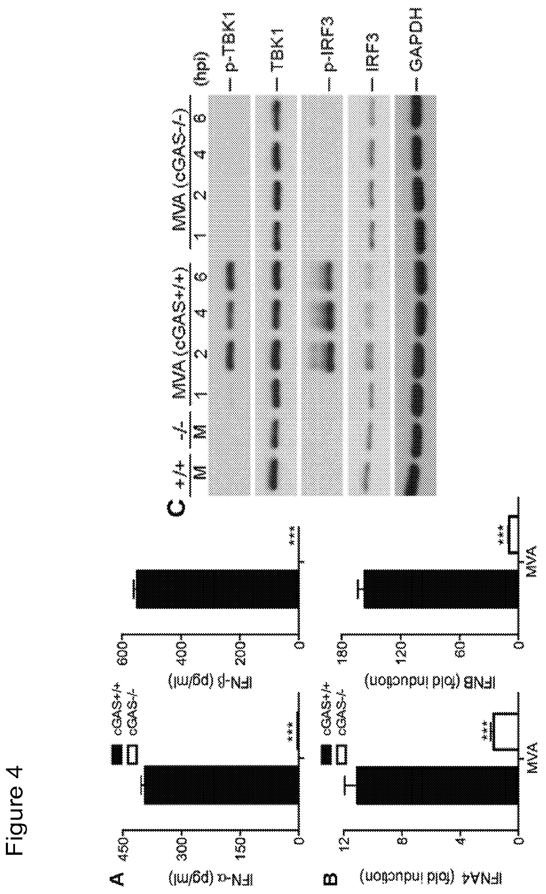
FIG. 4 is a series of graphical representations demonstrating that cGAS is the critical cytosolic DNA sensor for MVA infection of cDCs. FIG. 4A are bar graphs showing IFN-.alpha. and IFN-.beta. secretion levels in GM-CSF-BMDCs generated from cGAS.sup.-/- mice and its age-matched WT controls, and infected with MVA. Data are means.+-.SEM (n=3). A representative experiment is shown, repeated twice (***, p<0.001). FIG. 4B are bar graphs showing mRNA expression levels of IFNA4 and IFNB in GM-CSF-BMDCs cell generated from cGAS.sup.-/- mice and its age-matched WT controls, and infected with MVA. Data are means.+-.SEM (n=3). A representative experiment is shown, repeated twice (***, p<0.001). FIG. 4C is a scanned image of immunoblot showing the protein levels of phospho-TBK1, TBK1, phosphoserine-396 of IRF3, IRF3, and GAPDH in cGAS.sup.+/+ and cGAS.sup.-/- cDCs infected with MVA. "hpi", hours post infection.
FIG. 5 is a series of graphical representations showing that MVA.DELTA.E3L induces higher levels of type I IFN gene expression in BMDCs than MVA does. FIG. 5A is a scanned image of an immunoblot showing protein levels of E3 and .beta.-actin in GM-CSF-BMDCs infected with WT VAC, MVA, or MVA.DELTA.E3L. "hpi," hours post infection, "M", mock infection control. FIG. 5B are bar graphs showing mRNA levels of IFNA4 and IFNB in GM-CSF-BMDCs infected with MVA or with MVA.DELTA.E3L. Data are means.+-.SEM (n=3). A representative experiment is shown, repeated twice. ***, p<0.001; comparisons were made between MVA and MVA.DELTA.E3L infected cells. FIG. 5C are bar graphs showing mRNA levels of IFNA4 and IFNB in GM-CSF-BMDCs generated from IRF3.sup.-/- mice and age-matched WT C57B/6 mice and infected with MVA or with MVA.DELTA.E3L. Data are means.+-.SEM (n=3). A representative experiment is shown, repeated twice. ***, p<0.001; comparisons were made between MVA and MVA.DELTA.E3L infected cells. FIG. 5D is a scanned image of an immunoblot showing protein levels of p-IRF3 and .beta.-actin in GM-CSF-BMDCs infected with MVA or with MVA.DELTA.E3L.
FIG. 6 is a series of bar graphs showing that cGAS is required for the induction of type I IFN by MVA.DELTA.E3L in cDCs. FIG. 6A includes bar graphs showing mRNA levels of IFNA4 and IFNB in cGAS.sup.+/+ and cGAS.sup.-/- cDCs infected with MVA.DELTA.E3L. FIG. 6B includes bar graphs showing IFN-.alpha. and IFN-.beta. secretion levels in cGAS.sup.+/+ and cGAS.sup.-/- cDCs infected with MVA.DELTA.E3L or treated with cGAMP, an agonist for STING. FIG. 6C is a scanned image of an immunoblot showing protein levels of p-IRF3 and GAPDH in cGAS.sup.+/+ and cGAS.sup.-/- cDCs infected with MVA.DELTA.E3L.
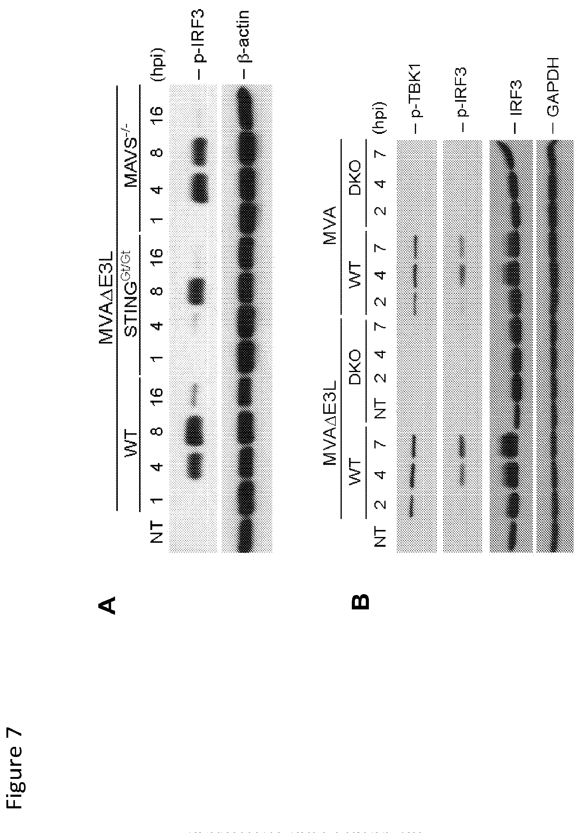
FIG. 7 is a series of scanned images of Western blot data showing that dsRNA-sensing pathway also plays a role in MVA.DELTA.E3L-induced phosphorylation of IRF3. FIG. 1A shows protein levels of p-IRF3 and .beta.-actin in cDCs generated from WT, STING.sup.Gt/Gt, or MAVS.sup.-/- mice, and infected with MVA.DELTA.E3L or not treated (NT). "hpi", hours post infection. FIG. 7B shows protein levels of phospho-TBK1, TBK1, phosphoserine-396 of IRF3, IRF3, and GAPDH in cDCs generated from WT or STING.sup.Gt/Gt/MDA5.sup.-/- (DKO) mice, and infected with MVA.DELTA.E3L or MVA. "hpi", hours post infection.
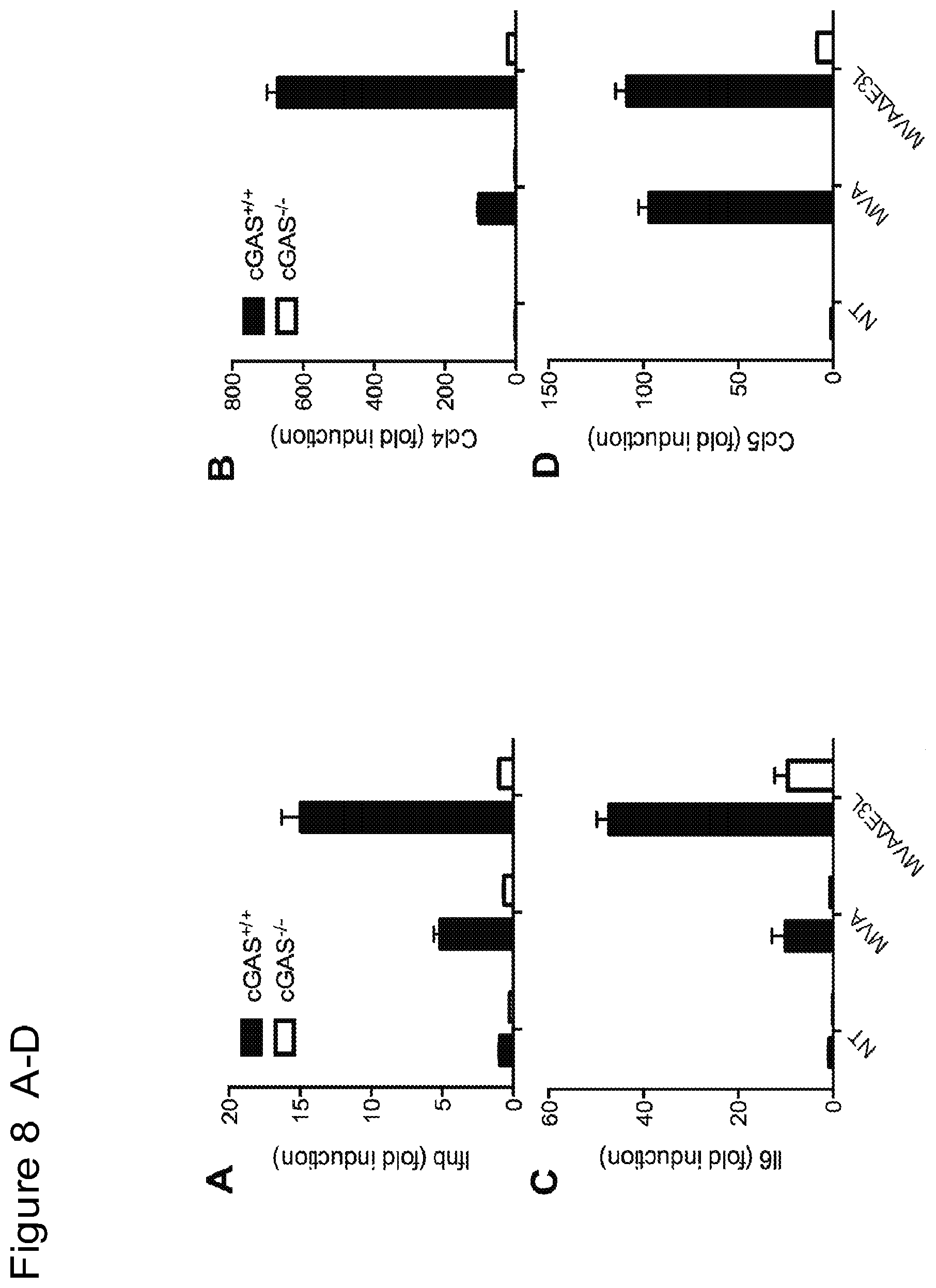
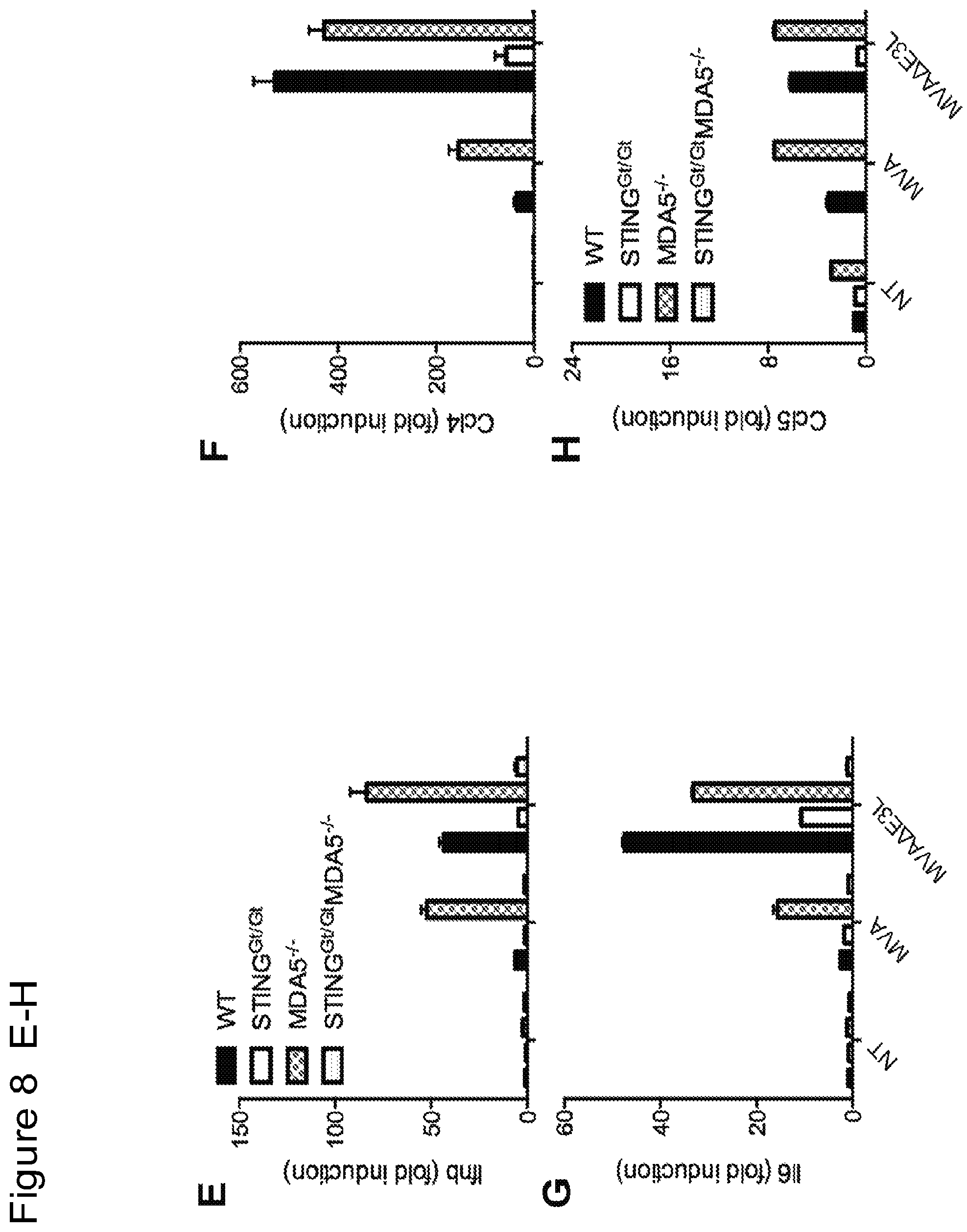
FIG. 8 is a series of bar graphs showing that MVA and MVA.DELTA.E3L infection of murine primary fibroblasts leads to induction of gene expression of Ifnb (FIG. 8A), Ccl4 (FIG. 8B), Il6 (FIG. 8C), and Ccl5 (FIG. 8D), which is largely dependent on cGAS. "NT", not treated. MVA.DELTA.E3L-induced expression of Ifnb (FIG. 8E), Ccl4 (FIG. 8F), Il6 (FIG. 8G), and Ccl5 (FIG. 8H) is completely abolished in STING and MDA5-double deficient murine primary fibroblasts.
FIG. 9 is a series of bar graphs showing that MVA and MVA.DELTA.E3L infection of murine primary fibroblasts leads to production of IFN-.beta. (FIG. 9A), CCL4 (FIG. 9B), IL-6 (FIG. 9C), and CCL5 (FIG. 9D), which is largely dependent on STING and with some contribution from MDA5.
FIG. 10 is a series of bar graphs showing that MVA.DELTA.E3L infection leads to higher secretion levels of Ifna4 (FIG. 10A), Ifnb (FIG. 10B), Il6 (FIG. 10C), Tnf (FIG. 10D), Ccl4 (FIG. 10E), and Ccl5 (FIG. 10F) than MVA in B16-F10 melanoma cells. "NT", not treated.
FIG. 11 is a series of scanned immunoblot images showing that infection of B16-F10 melanoma cells with MVA or MVA.DELTA.E3L induces apoptosis. FIG. 11A shows protein levels of PARP, cleaved PARP, and .beta.-actin in B16-F10 melanoma cells infected with MVA or MVA.DELTA.E3L. FIG. 11B shows protein levels of MCL-1, and .beta.-actin in B16-F10 melanoma cells infected with MVA or MVA.DELTA.E3L. "hpi", hours post infection. FIG. 11C shows levels of phosphorylated IRF3 and GAPDH in B16-F10 melanoma cells infected with MVA or MVA.DELTA.E3L. "hpi", hours post infection.
FIG. 12 is a series of graphs showing that intratumoral injection of MVA and MVA.DELTA.E3L leads to prolonged survival of tumor-bearing mice and eradication of tumors in some mice. FIGS. 12A-C are graphs of tumor volume over time in individual mice injected with PBS (A), MVA (B), and MVA.DELTA.E3L (C). FIG. 12D is a Kaplan-Meier survival curve of tumor-bearing mice injected with PBS, MVA, or MVA.DELTA.E3L. ****, p<0.0001 (MVA vs. PBS group); ***, p<0.001 (MVA.DELTA.E3L vs. PBS group). FIG. 12E is a Kaplan-Meier survival curve of tumor-free mice after successful treatment with MVA or MVA.DELTA.E3L, and challenged with B16-F10 melanoma cells at the contralateral side. Naive mice have never received any tumor cells or viruses in the past.
FIG. 13 is a series of graphical representations of data showing that intratumoral injection with MVA leads to immunological changes in the tumor microenvironment. FIG. 13A-B are dot-plots of flow cytometric analysis of CD4.sup.+ cells expressing FoxP3 in tumors treated with either PBS (13A) or MVA (13B). FIGS. 13D-E are dot-plots of flow cytometric analysis of CD8.sup.+ cells expressing Granzyme B in tumors treated with either PBS (13D) or MVA (13E). FIGS. 13G-H are scatterplots of flow cytometric analysis of CD8.sup.+ cells expressing Ki-67 in tumors treated with either PBS (13G) or MVA (13H). FIG. 13C is a graph depicting percentages of CD4.sup.+Foxp3.sup.+ in tumors treated with PBS or MVA. FIG. 13F is a graph depicting percentages of Granzyme B.sup.+CD8.sup.+ cells in tumors treated with PBS or MVA. FIG. 13 I is a graph depicting percentages of CD8.sup.+Ki-67.sup.+ in tumors treated with PBS or MVA.
FIG. 14 is a series of graphical representations of data showing that intratumoral injection with MVA induces immunological changes in the tumor draining lymph nodes (TDLNs). FIGS. 14A-B are dot-plots of flow cytometric analysis of Granzyme B.sup.+CD8.sup.+ cells in TDLNs of PBS (14A) or MVA (14B) treated mice. FIGS. 14C-D are dot-plots of flow cytometric analysis of Ki-67.sup.+CD8.sup.+ cells in TDLNs of PBS (14D) or MVA (14E) treated mice. FIG. 14C is a graph depicting percentages of Granzyme B.sup.+CD8.sup.+ cells in TDLNs from mice treated with PBS or MVA. FIG. 14F is a graph depicting percentages of CD8.sup.+Ki-67.sup.+ in TDLNs from mice treated with PBS or MVA.
FIG. 15 is a series of graphic representations showing that MVA.DELTA.E3L induces type I IFN and inflammatory cytokines/chemokines production in MC38 colon cancer cells. FIGS. 15A-D are bar graphs showing protein levels of IFN-.beta. (15A), IL-6 (15B), CCL4 (15C), and CCL5 (15D) in the supernatants of MC38 colon cancer cells infected with MVA or MVA.DELTA.E3L. FIGS. 15E-H are bar graphs showing the mRNA levels of Ifnb (15E), Il6 (15F), Ccl4 (15G), and Ccl5 (15H) in MC38 colon cancer cells at 6 h post infection with MVA or MVA.DELTA.E3L. FIG. 15I is a scanned image of a Western blot showing protein levels of PARP, cleaved-PARP, phosphor-IRF-3, IRF3, and .beta.-actin. "hpi", hours post infection.
FIG. 16 is a series of graphs showing that MVA.DELTA.E3L inhibits tumorigenesis in murine model of colon carcinoma. FIGS. 16A and 16B are plots of tumor volume v. time from injection of PBS or virus in mice showing that intratumoral injection of MVA.DELTA.E3L is effective for the treatment of murine colon adenocarcinoma (MC38 cells) implanted unilaterally in mice (C57B/6). The tumor volumes of mice treated with PBS or MVA.DELTA.E3L groups prior to treatment (day 0) and up to 45 days post-treatment are shown. FIG. 16C is a Kaplan-Meier survival curve for the treated mice (MVA.DELTA.E3L) versus control mice (PBS). ***, p<0.001 (MVA.DELTA.E3L vs. PBS group).
FIG. 17 is a series of graphical representations of data showing that intratumoral injection of MVA or MVA.DELTA.E3L induced antitumor effects in non-injected distant tumors in a murine B16-F10 melanoma bilateral implantation model. FIG. 17A-F are graphs of injected (A, C, E) and non-injected (B, D, F) tumor volume plotted against time (days) after PBS, MVA, or MVA.DELTA.E3L injection respectively. FIG. 17G is a Kaplan-Meier survival curve of tumor-bearing mice (B16-F10 cells) injected with PBS (filled circles), MVA (filled squares), or MVA.DELTA.E3L (filled triangles). ****, p<0.0001 (MVA.DELTA.E3L vs. PBS group); ***, p<0.001 (MVA vs. PBS group).
FIG. 18 is a series of graphical representations of data showing that intratumoral injection of MVA or MVA.DELTA.E3L induces activated effector CD8.sup.+ and CD4.sup.+ T cells and reduces regulator CD4.sup.+ T cells in both injected and non-injected tumors in a murine B16-F10 melanoma bilateral implantation model. FIG. 18A are dot-plots of flow cytometric analysis of CD3.sup.+CD8.sup.+ T cells in both injected and non-injected tumors treated with PBS, MVA or MVA.DELTA.E3L. FIG. 18B is a graph of % CD3.sup.+CD8.sup.+ T cells in both injected and non-injected tumors treated with PBS, MVA or MVA.DELTA.E3L. FIGS. 18C and E are dot-plots of flow cytometric analysis of CD8.sup.+ cells expressing Granzyme B.sup.+ (18C) or Ki-67 (18E). FIGS. 18D and F are graphs of % CD8.sup.+Granzyme B.sup.+ (18D), CD8.sup.+Ki-67.sup.+ (18F) T cells in both injected and non-injected tumors treated with PBS, MVA or MVA.DELTA.E3L. FIG. 18G are dot-plots of flow cytometric analysis of CD4.sup.+Foxp3.sup.+ T cells in both injected and non-injected tumors treated with PBS, MVA or MVA.DELTA.E3L. FIG. 18H is a graph of % CD4.sup.+Foxp3.sup.+ T cells in both injected and non-injected tumors treated with PBS, MVA or MVA.DELTA.E3L. FIGS. 18I and K are dot-plots of flow cytometric analysis of CD4.sup.+ cells expressing Granzyme B.sup.+ (18I) or Ki-67 (18K). FIGS. 18J and L are graphs of % CD4.sup.+Granzyme B.sup.+ (18J), CD8.sup.+Ki-67.sup.+ (18L) T cells in both injected and non-injected tumors treated with PBS, MVA or MVA.DELTA.E3L. (*, p<0.05; **, p<0.01; ***, p<0.001; ****, p<0.0001).
FIG. 19 is a series of graphical representations of data showing that intratumoral injection of MVA or MVA.DELTA.E3L reduces tumor-associated macrophages (TAMs) in a murine B16-F10 melanoma model. FIG. 19A are dot-plots of flow cytometric analysis of TAM cells (CD45.sup.+Ly6C.sup.-MHCII.sup.+CD24.sup.loF4/80.sup.+CD11b.sup.+CD11c.su- p.+) in tumors treated with PBS, MVA or MVA.DELTA.E3L. FIG. 19B-D are graphs of % TAM, TAM1 (CD11C.sup.loCD11b.sup.hi), and TAM2 (CD11C.sup.hiCD11b.sup.lo in CD45.sup.+ cells in tumors treated with PBS, MVA or MVA.DELTA.E3L. (*, p<0.05; ns: non-significant).
DETAILED DESCRIPTION
Definitions
As used herein the following terms shall have the meanings ascribed to them below unless the context clearly indicates otherwise: "Cancer" refers to a class of diseases of humans and animals characterized by uncontrolled cellular growth. Unless otherwise explicitly indicated, the term "cancer" may be used herein interchangeably with the terms "tumor," "malignancy," "hyperproliferation" and "neoplasm(s);" the term "cancer cell(s)" is interchangeable with the terms "tumor cell(s)," "malignant cell(s)," "hyperproliferative cell(s)," and "neoplastic cell(s)". "Melanoma" refers to a malignant neoplasm originating from cells that are capable of producing melanin. The term melanoma is synonymous with "malignant melanoma". Melanoma metastasizes widely, involving a patient's lymph nodes, skin, liver, lungs and brain tissues. "Solid tumor" refers to all neoplastic cell growth and proliferation, and all pre-cancerous and cancerous cells and tissues, except for hematologic cancers such as lymphomas, leukemias and multiple myeloma. Examples of solid tumors include, but are not limited to: soft tissue sarcoma, such as fibrosarcoma, myxosarcoma, liposarcoma, chondrosarcoma, osteogenic sarcoma, chordoma, angiosarcoma, endotheliosarcoma, lymphangiosarcoma, lymphangioendotheliosarcoma, synovioma, mesothelioma, Ewing's tumor and other bone tumors (e.g., osteosarcoma, malignant fibrous histiocytoma), leiomyosarcoma, rhabdomyosarcoma, colon carcinoma, pancreatic cancer, breast cancer, ovarian cancer, prostate cancer, squamous cell carcinoma, basal cell carcinoma, adenocarcinoma, sweat gland carcinoma, sebaceous gland carcinoma, papillary carcinoma, papillary adenocarcinomas, cystadenocarcinoma, medullary carcinoma, bronchogenic carcinoma, renal cell carcinoma, hepatoma, bile duct carcinoma, choriocarcinoma, seminoma, embryonal carcinoma, Wilms' tumor, cervical cancer, testicular tumor, lung carcinoma, small cell lung carcinoma, bladder carcinoma, epithelial carcinoma, brain/CNS tumors (e.g., astrocytoma, glioma, glioblastoma, childhood tumors, such as atypical teratoid/rhabdoid tumor, germ cell tumor, embryonal tumor, ependymoma) medulloblastoma, craniopharyngioma, ependymoma, pinealoma, hemangioblastoma, acoustic neuroma, oligodendroglioma, meningioma, melanoma, neuroblastoma, and retinoblastoma. Some of the most common solid tumors for which the compositions and methods of the present disclosure would be useful include: head-and-neck cancer, rectal adenocarcinoma, glioma, medulloblastoma, urothelial carcinoma, pancreatic adenocarcinoma, uterine (e.g., endometrial cancer, fallopian tube cancer,) ovarian cancer, cervical cancer prostate adenocarcinoma, non-small cell lung cancer (squamous and adenocarcinoma), small cell lung cancer, melanoma, breast carcinoma, ductal carcinoma in situ, renal cell carcinoma, and hepatocellular carcinoma. adrenal tumors (e.g., adrenocortical carcinoma), esophageal, eye (e.g., melanoma, retinoblastoma), gallbladder, gastrointestinal, Wilms' tumor, heart, head and neck, laryngeal and hypopharyngeal, oral (e.g., lip, mouth, salivary gland), nasopharyngeal, neuroblastoma, peritoneal, pituitary, Kaposi's sarcoma, small intestine, stomach, testicular, thymus, thyroid, parathyroid, vaginal tumor and the metastases of any of the foregoing. "Metastasis" refers to the spread of cancer from its primary site to neighboring tissues or distal locations in the body. Cancer cells can break away from a primary tumor, penetrate into lymphatic and blood vessels, circulate through the bloodstream, and grow in in normal tissues elsewhere in the body. Metastasis is a sequential process, contingent on tumor cells (or cancer stem cells) breaking off from the primary tumor, traveling through the bloodstream or lymphatics, and stopping at a distant site. Once at another site, cancer cells re-penetrate through the blood vessels or lymphatic walls, continue to multiply, and eventually form a new tumor (metastatic tumor). In some embodiments, this new tumor is referred to as a metastatic (or secondary) tumor. "Immune response" refers to the action of one or more of lymphocytes, antigen presenting cells, phagocytic cells, granulocytes, and soluble macromolecules produced by the above cells or the liver (including antibodies, cytokines, and complement) that results in selective damage to, destruction of, or elimination from the human body of cancerous cells, metastatic tumor cells, etc. An immune response may include a cellular response, such as a T cell response that is an alteration (modulation, e.g., significant enhancement, stimulation, activation, impairment, or inhibition) of cellular function that is a T cell function. A T cell response may include generation, proliferation or expansion, or stimulation of a particular type of T cell, or subset of T cells, for example, effector CD4+, CD4.sup.+ helper, effector CD8+, CD8.sup.+ cytotoxic, or natural killer (NK) cells. Such T cell subsets may be identified by detecting one or more cell receptors or cell surface molecules (e.g., CD or cluster of differentiation molecules). A T cell response may also include altered expression (statistically significant increase or decrease) of a cellular factor, such as a soluble mediator (e.g., a cytokine, lymphokine, cytokine binding protein, or interleukin) that influences the differentiation or proliferation of other cells. For example, Type I interferon (IFN-.alpha./.beta.) is a critical regulator of the innate immunity (52) (Huber et al. Immunology 132(4):466-474 (2011)). Animal and human studies have shown a role for IFN-.alpha./.beta. in directly influencing the fate of both CD4.sup.+ and CD8.sup.+ T cells during the initial phases of antigen recognition anti-tumor immune response. IFN Type I is induced in response to activation of dendritic cells, in turn a sentinel of the innate immune system. "Tumor immunity" refers to one or more processes by which tumors evade recognition and clearance by the immune system. Thus, as a therapeutic concept, tumor immunity is "treated" when such evasion is attenuated or eliminated, and the tumors are recognized and attacked by the immune system (the latter being termed herein "anti-tumor immunity"). An example of tumor recognition is tumor binding, and examples of tumor attack are tumor reduction (in number, size or both) and tumor clearance. "T cell" refers to a thymus derived lymphocyte that participates in a variety of cell-mediated adaptive immune reactions. "Helper T cell" refers to a CD4.sup.+ T cell; helper T cells recognize antigen bound to MHC Class II molecules. There are at least two types of helper T cells, Th1 and Th2, which produce different cytokines. "Cytotoxic T cell" refers to a T cell that usually bears CD8 molecular markers on its surface (CD8+) and that functions in cell-mediated immunity by destroying a target cell having a specific antigenic molecule on its surface. Cytotoxic T cells also release Granzyme, a serine protease that can enter target cells via the perforin-formed pore and induce apoptosis (cell death). Granzyme serves as a marker of cytotoxic phenotype. Other names for cytotoxic T cell include CTL, cytolytic T cell, cytolytic T lymphocyte, killer T cell, or killer T lymphocyte. Targets of cytotoxic T cells may include virus-infected cells, cells infected with bacterial or protozoal parasites, or cancer cells. Most cytotoxic T cells have the protein CD8 present on their cell surfaces. CD8 is attracted to portions of the Class I MHC molecule. Typically, a cytotoxic T cell is a CD8+ cell. "Tumor-infiltrating leukocytes" refers to white blood cells of a subject afflicted with a cancer (such as melanoma), that are resident in or otherwise have left the circulation (blood or lymphatic fluid) and have migrated into a tumor. "Immune checkpoint inhibitor" or "immune checkpoint blocking agent" refers to molecules that completely or partially reduce, inhibit, interfere with or modulate the activity of one or more checkpoint proteins. Checkpoint proteins regulate T-cell activation or function. Checkpoint proteins include, but are not limited to CD28 receptor family members, CTLA-4 and its ligands CD80 and CD86; PD-1 and its ligands PDL1 and PDL2; LAGS, B7-H3, B7-H4, TIM3, ICOS, and BTLA (53). "Parenteral" when used in the context of administration of a therapeutic substance includes any route of administration other than administration through the alimentary tract. Particularly relevant for the methods disclosed herein are intravenous (including for example through the hepatic portal vein), intratumoral or intrathecal administration. "Antibody" refers to an immunoglobulin molecule which specifically binds to an antigen or to an antigen-binding fragment of such a molecule. Thus, antibodies can be intact immunoglobulins derived from natural sources or from recombinant sources and can be immunoreactive (antigen-binding) fragments or portions of intact immunoglobulins. The antibodies may exist in a variety of forms including, for example, polyclonal antibodies, monoclonal antibodies, Fv, Fab and F(ab)2, as well as single chain antibodies (scFv) humanized antibodies, chimeric antibodies, human recombinant antibodies and bi- and tri-specific antibodies. "Oncolytic virus" refers to a virus that preferentially infects cancer cells, replicates in such cells, and induces lysis of the cancer cells through its replication process. Nonlimiting examples of naturally occurring oncolytic viruses include vesicular stomatitis virus, reovirus, as well as viruses engineered to be oncoselective such as adenovirus, Newcastle disease virus and herpes simplex virus (See, e.g., Nemunaitis, J. Invest New Drugs. 17(4):375-86 (1999); Kim, D H et al. Nat Rev Cancer. 9(1):64-71(2009); Kim et al. Nat. Med. 7:781 (2001); Coffey et al. Science 282:1332 (1998)) (8, 54-56). Vaccinia virus infects many types of cells but replicates preferentially in tumor cells due to the fact that tumor cells have a metabolism that favors replication, exhibit activation of certain pathways that also favor replication and create an environment that evades the innate immune system, which also favors viral replication. In the context of the present disclosure, MVA and MVA.DELTA.E3L do not fit the definition of oncolytic viruses as they do not produce an antitumor effect primarily by replicating inside tumor cells and causing apoptosis. (Nor do they fit the classic definition of vaccines as these viruses do not express tumor antigens. It can be said however, that they act as immunostimulatory molecules, akin to adjuvants, as they serve to enhance the host's immune response against the tumor.) "MVA" means "modified vaccinia Ankara" and refers to a highly attenuated strain of vaccinia derived from the Ankara strain and developed for use as a vaccine and vaccine adjuvant. The original MVA was isolated from the wild-type Ankara strain by successive passage through chicken embryonic cells, Treated thus, it lost about 15% of the genome of wild-type vaccinia including its ability to replicate efficiently in primate (including human) cells. (57) (Mayr et al., Zentralbl Bakteriol B167, 375-390 (1978)). The smallpox vaccination strain MVA: marker, genetic structure, experience gained with the parenteral vaccination and behavior in organisms with a debilitated defense mechanism. MVA is considered an appropriate candidate for development as a recombinant vector for gene or vaccination delivery against infectious diseases or tumors. (58) (Verheust et al., Vaccine 30(16), 2623-2632 (2012)). MVA has a genome of 178 kb in length and a sequence first disclosed in (59) (Antoine et al., Virol. 244(2): 365-396 (1998)). Sequences are also disclosed in Genbank U94848.1. Clinical grade MVA is commercially and publicly available from Bavarian Nordic A/S Kvistgaard, Denmark. Additionally, MVA is available from ATCC, Rockville, Md. and from CMCN (Institut Pasteur Collection Nationale des Microorganismes) Paris, France. "MVA.DELTA.E3L" means a deletion mutant of MVA which lacks a functional E3L gene and is infective but non replicative and it is further impaired in its ability to evade the host's immune system. It can be used as a vaccine vector. This mutant MVA E3L knockout and its preparation have been described for example in U.S. Pat. No. 7,049,145. "Subject" means any animal (mammalian, human or other) patient that can be afflicted with cancer and when thus afflicted is in need of treatment. "Therapeutically effective amount" or "effective amount" refers to a sufficient amount of an agent when administered at one or more dosages and for a period of time sufficient to provide a desired biological result in alleviating, curing or palliating a disease. In the present disclosure, an effective amount respectively of the MVA or MVA.DELTA.E3L is an amount that (administered for a suitable period of time and at a suitable frequency) reduces the number of cancer cells; or reduces the tumor size or eradicates the tumor; or inhibits (i.e., slows down or stops) cancer cell infiltration into peripheral organs; inhibits (i.e., slows down or stops) metastatic growth; inhibits (stabilizes or arrests) tumor growth; allows for treatment of the tumor, and/or induces an immune response against the tumor. An appropriate therapeutic amount in any individual case may be determined by one of ordinary skill in the art using routine experimentation in light of the present disclosure. Such determination will begin with amounts found effective in vitro and amounts found effective in animals. The therapeutically effective amount will be initially determined based on the concentration or concentrations found to confer a benefit to cells in culture. Effective amounts can be extrapolated from data within the cell culture and can be adjusted up or down based on factors such as detailed herein. An example of an effective amount range is from 10.sup.5 viral particles to about 10.sup.12 viral particles per administration.
With particular reference to the viral-based immunostimulatory agents disclosed herein, "therapeutically effective amount" or "effective amount" refers to an amount of a composition comprising MVA or MVA.DELTA.E3L sufficient to reduce, inhibit, or abrogate tumor cell growth, thereby reducing or eliminating the tumor, or sufficient to inhibit, reduce or abrogate metastatic spread either in vitro, ex vivo or in a subject or to elicit an immune response against the tumor that will eventually result in one or more of metastatic spread reduction, inhibition and/or abrogation as the case may be. The reduction, inhibition, or eradication of tumor cell growth may be the result of necrosis, apoptosis, or an immune response or a combination of two or more of the foregoing (however, the precipitation of apoptosis for example may not be due to the same factors as observed with oncolytic viruses). The amount that is therapeutically effective may vary depending on such factors as the particular MVA used in the composition, the age and condition of the subject being treated, the extent of tumor formation, the presence or absence of other therapeutic modalities, and the like. Similarly, the dosage of the composition to be administered and the frequency of its administration will depend on a variety of factors, such as the potency of the active ingredient, the duration of its activity once administered, the route of administration, the size, age, sex and physical condition of the subject, the risk of adverse reactions and the judgment of the medical practitioner. The compositions are administered in a variety of dosage forms, such as injectable solutions.
With particular reference to combination therapy with an immune checkpoint inhibitor, "therapeutically effective amount" for an immune checkpoint blocking agent" shall mean an amount of an immune checkpoint blocking agent sufficient to reverse or reduce immune suppression in the tumor microenvironment and to activate or enhance host immunity in the subject being treated. There are several immune checkpoint blocking agents approved, in clinical trials or still otherwise under development including inhibitory antibodies against CD28 inhibitor such as CTLA-4 (cytotoxic T lymphocyte antigen 4) (e.g., ipilimumab), anti-PD-1 (programmed Death 1) inhibitory antibodies (e.g., nivolumab, pembrolizumab, pidilizumab, lambrolizumab), and anti-PD-L1 (Programmed death ligand 1) inhibitory antibodies (MPDL3280A, BMS-936559, MEDI4736, MSB 00107180), as well as inhibitory antibodies against LAG-3 (lymphocyte activation gene 3), TIM3 (T cell Immunoglobulin and Mucin-3), B7-H3, and TIGIT (T-cell immunoreceptor with Ig and ITIM domains). Dosage ranges of the foregoing are known in or readily within the skill in the art as several dosing clinical trials have been completed, making extrapolation to other agents possible.
Preferably, the tumor expresses the particular checkpoint but this is not strictly necessary as immune checkpoint blocking agents block more generally immune suppressive mechanisms within the tumors, elicited by tumor cells, stromal cells, and tumor-infiltrating immune cells.
For example, the CTLA4 inhibitor ipilimumab, when administered as adjuvant therapy after surgery in melanoma is administered at 1-2 mg/mL over 90 minutes for a total infusion amount of 3 mg/kg every three weeks for a total of 4 doses. This therapy is often accompanied by severe even life-threatening immune-mediated adverse reactions, which limits the tolerated dose as well as the cumulative amount that can be administered. It is anticipated that it will be possible to reduce the dose and/or cumulative amount of ipilimumab when it is administered conjointly with MVA or MVA.DELTA.E3L. In particular, in light of the experimental results set forth below, it is anticipated that it will be further possible to reduce the CTLA4 inhibitor's dose if it is administered directly to the tumor simultaneously or sequentially with one or both the foregoing MVA viruses. Accordingly, the amounts provided above for ipilimumab will be a starting point for determining the particular dosage and cumulative amount to be given to a patient in conjoint administration but dosing studies will be required to determine optimum amounts.
Pembrolizumab is prescribed for administration as adjuvant therapy in melanoma diluted to 25 mg/mL. It is administered at a dosage of 2 mg/kg over 30 minutes every three weeks. Again, this would be a starting point for determining dosage and administration in the conjoint administration with MVA or MVA.DELTA.E3L.
Nivolumab is prescribed for administration at 3 mg/kg as an intravenous infusion over 60 minutes every two weeks, providing a similar starting point in determining dosage and administration of this checkpoint inhibitor conjointly with MVA or MVA.DELTA.E3L.
Immune stimulating agents such as agonist antibodies have also been explored as immunotherapy for cancers. For example, anti-ICOS antibody binds to the extracellular domain of ICOS leading to the activation of ICOS signaling and T cell activation. Anti-OX40 antibody can bind to OX40 and potentiate T cell receptor signaling leading to T cell activation, proliferation and survival. Other examples include agonist antibodies against 4-1BB (CD137), GITR. All of these agents are at various stages of clinical trials.
The immune stimulating agonist antibodies can be used systemically in combination with intratumoral injection of MVA or MVA.DELTA.E3L. Alternatively, the immune stimulating agonist antibodies can be used conjointly with MVA or MVA.DELTA.E3L via intratumorally delivery either simultaneously or sequentially. "Pharmaceutically acceptable carrier and/or diluent" or "pharmaceutically acceptable excipient" includes without limitation any and all solvents, dispersion media, coatings, antibacterial and antifungal agents, isotonic and absorption delaying agents and the like. The use of such media and agents for biologically active substances is well known in the art. Further details of excipients are provided below. Supplementary active ingredients, such as antimicrobials, for example antifungal agents, can also be incorporated into the compositions. "Delivering" used in connection with depositing the MVA or MVA.DELTA.E3L of the present disclosure in the tumor microenvironment whether this is done by local administration to the tumor or by for example intravenous route. The term focuses on MVA or MVA.DELTA.E3L that reaches the tumor itself. "Conjoint administration" herein refers to administration of a second therapeutic modality in combination with MVA or MVA.DELTA.E3L for example an immune checkpoint blocking agent administered and in close temporal proximity with MVA or MVA.DELTA.E3L. For example, a PD-1/PDL-1 inhibitor and/or a CTLA4 inhibitor (in more specific embodiments, an antibody) can be administered simultaneously with MVA or MVA.DELTA.E3L (by intravenous or intratumoral injection when the MVA or MVA.DELTA.E3L is administered intratumorally or systemically as stated above) or before or after the MVA or MVA.DELTA.E3L administration. If the MVA or MVA.DELTA.E3L administration and the immune checkpoint blocking agent are administered 1-7 days apart or even up to three weeks apart, this would be within "close temporal proximity" as stated herein.
In one embodiment, the present disclosure relates to a method for eliciting an antitumor immune response in subjects afflicted with tumors comprising delivering to the tumor an effective amount of MVA or MVA.DELTA.E3L. Stimulation of the immune system may be manifest by one or more of the following immunological effects: an increase in at least one of effector CD8.sup.+ T cells and effector CD4+ T cells within the tumor and/or in tumor-draining lymph nodes; induction of maturation of dendritic cells infiltrating said tumor through induction of type I IFN; induction of effector CD4.sup.+ T cells in the subject recognizing tumor cells within the tumor and/or in tumor draining lymph nodes; reduction of immune suppressive (regulatory) CD4.sup.+ T cells within the tumor; induction of cells of the tumor to produce one or more of Type I IFN or other inflammatory cytokines or chemokines; reduction of immune suppressive tumor-associated macrophages within the tumor.
The foregoing one or more immunological effects may serve as early indicators of response of the subject to the treatment and may serve as monitors of the continued effectiveness of same. Observation of these effects illustrates that the manner in which the present viruses treat tumor is different from that of vaccine vectors harboring tumor antigens (which are not delivered intratumorally but by intramuscular, subcutaneous or, rarely, intravenous route) and also different from that of oncolytic viruses (which cause cytopathy primarily due to viral replication in tumor cells). If apoptosis results pursuant to the present treatment, it is not due to the same mechanism as apoptosis that results or may result from these different modes of action.
The present inventors have explored the mechanism of the immune response and concluded that it is initiated by the cytosolic DNA-sensing pathway mediated by cGAS/STING which mediates production of Type 1 IFN. Further insights into the mechanism and the immune cells that are recruited are provided in the Examples. The conclusions presented therein are not confined to the specific experimental milieu where these mechanisms are being elucidated.
In one embodiment, the present disclosure provides a method of treating a subject diagnosed with a solid tumor comprising delivering to the tumor a therapeutic effective amount of the MVA or MVA.DELTA.E3L.
In one embodiment, the present disclosure provides a method for inducing anti-tumor immunity in a subject diagnosed with cancer comprising administering to the subject a therapeutically effective amount of MVA or MVA.DELTA.E3L. The methods of the present disclosure include induction of anti-tumor immunity that can reduce the size of the tumor, eradicate the tumor, inhibit growth of the tumor, inhibit metastasis or reduce metastatic growth of the tumor or eradicate metastatic growth of the tumor, induce apoptosis of tumor cells or prolong survival of the subject (compared to untreated or conventionally treated subjects).
In another embodiment, the present disclosure provides a method for enhancing, stimulating, or eliciting, in a subject diagnosed with a solid malignant tumor, an anti-tumor immune response that may include an innate immune response and/or an adaptive immune response such as a T cell response by exposing the tumor to MVA or MVA.DELTA.E3L in a therapeutically effective amount.
In specific embodiments, the present disclosure provides methods of eliciting an immune response that mediates adaptive immune responses both in terms of T-cell cytotoxicity directed against tumor cells and in terms of eliciting effector T cells also directed against tumor cells. The methods comprise administering to a subject afflicted with a solid tumor intratumorally or (as the present inventors anticipate) intravenously a composition comprising MVA or MVA.DELTA.E3L wherein administration of said composition results in a tumor-specific immune response against the tumor and, eventually, in reduction, inhibition or eradication of tumor growth, inhibition of metastatic growth, apoptosis of tumor cells and/or prolongation of the subject's survival. Indeed the present inventors have shown that cancer cells are being killed and that the immune response can migrate to remote locations, as would be the case with metastases.
In some embodiments, the present disclosure provides methods of eliciting an immune response that mediates adaptive immune responses both in terms of T-cell cytotoxicity directed against tumor cells and in terms of eliciting effector T cells also directed against tumor cells. The methods comprise administering to a subject parenterally a composition comprising MVA or MVA.DELTA.E3L wherein administration of said composition results in a tumor-specific immune response against the tumor and, eventually, in reduction, inhibition or eradication of tumor growth and/or in inhibition reduction or elimination of metastatic growth, apoptosis of tumor cells and/or prolongation of survival of the treated subject compared to conventional therapy or no treatment. For intraperitoneal metastases, the virus can be injected intraperitoneally.
Indeed the present inventors have shown that cancer cells are being killed and that the immune response can migrate to remote locations, as would be the case with metastases, and still exert an anti-tumor effect.
Because MVA and MVA.DELTA.E3L are substantially not replication competent in most mammalian cells, it does not exert its effect on the immune system the same way as replication competent vaccines or vectors. Thus, while it is believed that stimulation of the immune system is a barrier to efficacy for oncolysis (8) (Kirn et al., Nat Rev Cancer. (1), 64-71 (2009)), MVA and MVA.DELTA.E3L is able to harness the innate immune system to stimulate adaptive immunity, both in terms of cytotoxicity and more broadly in terms of effector T cell activation against the tumor.
The present disclosure thus provides a method for treating a solid malignant tumor, comprising delivering to a tumor of the subject an amount of MVA or MVA.DELTA.E3L effective to induce an immune response against the tumor in a subject diagnosed with solid tumor.
The present disclosure also provides a method for generating antitumor systemic immunity in a subject afflicted with a solid malignant tumor, comprising delivering to a tumor of the subject an amount of MVA or MVA.DELTA.E3L effective to bring about one or both of rejection of non-injected tumors in said subject and inhibition of tumor metastasis (which the present inventors test by tumor rechallenge).
As is shown herein, MVA induces type I IFN induction in conventional dendritic cells (cDCs) via a cytosolic DNA-sensing pathway mediated by cGAS/STING. Intravenous delivery of MVA in C57B/6 mice induced type I IFN in wild-type mice, but not in mice lacking STING or IRF3. It is also shown that MVA.DELTA.E3L induces higher levels of type I IFN gene expression and phosphorylation of IRF3 than MVA in cDCs. MVA.DELTA.E3L is detected by both the cytosolic DNA-sensing pathway mediated by cGAS/STING, and the dsRNA-sensing pathway mediated by MDA5/MAVS. In addition, MVA.DELTA.E3L infection of B16 melanoma cells and MC38 colon adenocarcinoma cells induces type I IFN and proinflammatory cytokines and chemokines, as well as activation of phosphorylation of IRF3. Both MVA and MVA.DELTA.E3L induce apoptosis in B16 and MC38 cells as demonstrated by the cleavage of PARP and Caspase-3. According to the present disclosure MVA or MVA.DELTA.E3L virus is used as direct anti-cancer therapy. Intratumoral injection of MVA or MVA.DELTA.E3L in a murine B16 melanoma model leads to apoptosis, prolonged survival and tumor eradication, as well as the generation of systemic anti-tumor immunity.
Based on current literature, and without wishing to be bound by theory, the following mechanisms are believed to contribute to anti-tumor effects of MVA and MVA.DELTA.E3L: (i) induction of type I IFN responses in immune cells including conventional dendritic cells and macrophages; (ii) induction of type I IFN and proinflammatory cytokines and chemokines in cancer cells; (iii) induction of apoptosis in cancer cells; and (iv) alteration of tumor immune suppressive environment to an immune activating one.
Modified Vaccinia Ankara (MVA)
Modified Vaccinia Ankara (MVA) virus is a member of the genera Orthopoxvirus in the family of Poxviridae. MVA was generated by approximately 570 serial passages on chicken embryo fibroblasts (CEF) of the Ankara strain of vaccinia virus (CVA) (60) (Mayr et al., Infection 3, 6-14 (1975)). As a consequence of these long-term passages, the resulting MVA virus contains extensive genome deletions and is highly host cell restricted to avian cells (61) (Meyer et al., J. Gen. Virol. 72, 1031-1038 (1991)). It was shown in a variety of animal models that the resulting MVA is significantly avirulent (57) (Mayr et al., Dev. Biol. Stand. 41, 225-34 (1978)).
The safety and immunogenicity of MVA has been extensively tested and documented in clinical trials, particularly against the human smallpox disease. These studies included over 120,000 individuals and have demonstrated excellent efficacy and safety in humans. Moreover, compared to other vaccinia based vaccines, MVA has weakened virulence (infectiousness) while it triggers a good specific immune response. Thus, MVA has been established as a safe vaccine vector, with the ability to induce a specific immune response.
Due to the above mentioned characteristics, MVA became an attractive candidate for the development of engineered MVA vectors, used for recombinant gene expression and vaccines. As a vaccine vector, MVA has been investigated against numerous pathological conditions, including HIV, tuberculosis and malaria, as well as cancer (20, 21) (Sutter et al., Curr Drug Targets Infect Disord 3: 263-271(2003); Gomez et al., Curr Gene Ther 8: 97-120 (2008)).
It has been demonstrated that MVA infection of human monocyte-derived dendritic cells (DC) causes DC activation, characterized by the upregulation of co-stimulatory molecules and secretion of proinflammatory cytokines (18) (Drillien et al., J Gen Virol 85: 2167-2175 (2004)). In this respect, MVA differs from standard wild type Vaccinia virus (WT-VAC), which fails to activate DCs. Dendritic cells can be classified into two main subtypes: conventional dendritic cells (cDCs) and plasmacytoid dendritic cells (pDCs). The former, especially the CD103+/CD8.alpha..sup.+ subtype, are particularly adapted to cross-presenting antigens to T cells; the latter are strong producers of Type I IFN.
Viral infection of human cells results in activation of an innate immune response (the first line of defense) mediated by type I interferons, notably interferon-alpha (a). This normally leads to activation of an immunological "cascade," with recruitment and proliferation of activated T cells (both CTL and helper) and eventually with antibody production. However viruses express factors that dampen immune responses of the host. MVA is a better immunogen than WT-VAC and replicates poorly in mammalian cells. (See, e.g., Brandler et al., J. Virol. 84, 5314-5328 (2010)) (62).
However, MVA is not entirely nonreplicative and as the present inventors show contains some residual immunosuppressive activity. Nevertheless, as shown herein MVA significantly prolonged survival of treated subjects. An implication of these findings is that by injecting a tumor with or systemically delivering MVA (or MVA.DELTA.E3L) it is possible to enhance a host's innate and adaptive immune responses and thereby overcome the tumor's ability to evade immune responses and to restore the ability of the host to mount an immune response against the tumor whether the response is native or induced or enhanced by another immunotherapeutic agent, such as a checkpoint inhibitor.
Modified Vaccinia Ankara with Deletion of E3 (MVA.DELTA.E3L)
The antitumor effects of MVA described in the immediately preceding section are also observed with MVA.DELTA.E3L. The latter is less immunosuppressive than MVA and even less replicative in most mammalian cells, and from that point of view preferred. In addition, the effects of MVA.DELTA.E3L have generally been qualitatively better than those with MVA as seen in the experiments described herein.
Immune Response
In addition to induction of the immune response by up-regulation of particular immune system activities (such as antibody and/or cytokine production, or activation of cell mediated immunity), immune responses may also include suppression, attenuation, or any other downregulation of detectable immunity, so as to reestablish homeostasis and prevent excessive damage to the host's own organs and tissues. In some embodiments, an immune response that is induced according to the methods of the present disclosure generates effector CD8.sup.+ (antitumor cytotoxic CD8.sup.+) T cells or activated T helper cells or both that can bring about directly or indirectly the death, or loss of the ability to propagate, of a tumor cell.
Induction of an immune response by the methods of the present disclosure may be determined by detecting any of a variety of well-known immunological parameters (63, 64) (Takaoka et al., Cancer Sci. 94:405-11 (2003); Nagorsen et al., Crit. Rev. Immunol. 22:449-62 (2002)). Induction of an immune response may therefore be established by any of a number of well-known assays, including immunological assays, Such assays include, but need not be limited to, in vivo, ex vivo, or in vitro determination of soluble immunoglobulins or antibodies; soluble mediators such as cytokines, chemokines, hormones, growth factors and the like as well as other soluble small peptide, carbohydrate, nucleotide and/or lipid mediators; cellular activation state changes as determined by altered functional or structural properties of cells of the immune system, for example cell proliferation, altered motility, altered intracellular cation gradient or concentration (such as calcium); phosphorylation or dephosphorylation of cellular polypeptides; induction of specialized activities such as specific gene expression or cytolytic behavior; cellular differentiation by cells of the immune system, including altered surface antigen expression profiles, or the onset of apoptosis (programmed cell death); or any other criterion by which the presence of an immune response may be detected. For example, cell surface markers that distinguish immune cell types may be detected by specific antibodies that bind to CD4+, CD8+, or NK cells. Other markers and cellular components that can be detected include but are not limited to interferon .gamma. (IFN-.gamma.), tumor necrosis factor (TNF), IFN-.alpha., IFN-.beta., IL-6, and CCL5. Common methods for detecting the immune response include, but are not limited to flow cytometry, ELISA, immunohistochemistry. Procedures for performing these and similar assays are widely known and may be found, for example in Letkovits (Immunology Methods Manual: The Comprehensive Sourcebook of Techniques, Current Protocols in Immunology, 1998).
Pharmaceutical Compositions and Preparations
Pharmaceutical compositions comprising MVA or MVA.DELTA.E3L may contain a carrier or diluent, which can be a solvent or dispersion medium containing, for example, water, polyol (for example, glycerol, propylene glycol, and liquid polyethylene glycol, and the like), suitable mixtures thereof, and vegetable oils. The proper fluidity can be maintained, for example, by the use of a coating such as lecithin, by the maintenance of the required particle size in the case of dispersion and by the use of surfactants. Prevention of the action of microorganisms can be effected by various antibacterial and antifungal agents, for example, parabens, chlorobutanol, phenol, sorbic acid, thimerosal, and the like. In many cases, it will be preferable to include isotonic agents, for example, sugars or sodium chloride. Prolonged absorption of the injectable compositions can be brought about by the use in the compositions of agents delaying absorption, for example, aluminum monostearate and gelatin. In general, excipients suitable for injectable preparations can be included as apparent to those skilled in the art.
Pharmaceutical compositions and preparations comprising MVA or MVA.DELTA.E3L may be manufactured by means of conventional mixing, dissolving, granulating, emulsifying, encapsulating, entrapping or lyophilizing processes. Pharmaceutical viral compositions may be formulated in conventional manner using one or more physiologically acceptable carriers, diluents, excipients or auxiliaries that facilitate formulating virus preparations suitable for in vitro, in vivo, or ex vivo use. The compositions can be combined with one or more additional biologically active agents (for example parallel administration of GM-CSF) and may be formulated with a pharmaceutically acceptable carrier, diluent or excipient to generate pharmaceutical (including biologic) or veterinary compositions of the instant disclosure suitable for parenteral or intra-tumoral administration.
Many types of formulation are possible as is appreciated by those skilled in the art. The particular type chosen is dependent upon the route of administration chosen, as is well-recognized in the art. For example, systemic formulations will generally be designed for administration by injection, e.g., intravenous, as well as those designed for intratumoral delivery. Preferably, the systemic or intratumoral formulation is sterile.
Sterile injectable solutions are prepared by incorporating MVA or MVA.DELTA.E3L in the required amount of the appropriate solvent with various other ingredients enumerated herein, as required, followed by suitable sterilization means. Generally, dispersions are prepared by incorporating the various sterilized active ingredients into a sterile vehicle that contains the basic dispersion medium and the required other ingredients from those enumerated above. In the case of sterile powders for the preparation of sterile injectable solutions, the preferred methods of preparation are vacuum drying and freeze-drying techniques, which yield a powder of the virus plus any additional desired ingredient from a previously sterile-filtered solution thereof.
In some embodiments, the MVA and MVA.DELTA.E3L compositions of the present disclosure may be formulated in aqueous solutions, or in physiologically compatible solutions or buffers such as Hanks's solution, Ringer's solution, mannitol solutions or physiological saline buffer. In certain embodiments, any of the MVA and MVA.DELTA.E3L compositions may contain formulator agents, such as suspending, stabilizing, penetrating or dispersing agents, buffers, lyoprotectants or preservatives such as polyethylene glycol, polysorbate 80, 1-dodecylhexahydro-2H-azepin-2-one (laurocapran), oleic acid, sodium citrate, Tris HCl, dextrose, propylene glycol, mannitol, polysorbate polyethylenesorbitan monolaurate (Tween.RTM.-20), isopropyl myristate, benzyl alcohol, isopropyl alcohol, ethanol sucrose, trehalose and other such generally known in the art may be used in any of the compositions of the instant disclosure. (Pramanick et al., Pharma Times 45(3), 65-76 (2013)) (65).
The biologic or pharmaceutical compositions of the present disclosure can be formulated to allow the virus contained therein to be available to infect tumor cells upon administration of the composition to a subject. The level of virus in serum, tumors, and if desired other tissues after administration can be monitored by various well-established techniques, such as antibody-based assays (e.g., ELISA, immunohistochemistry, etc.).
Dosage of MVA and MVA.DELTA.E3L
In general, the subject is administered a dosage of MVA and MVA.DELTA.E3L in the range of about 10.sup.5 to about 10.sup.10 plaque forming units (pfu), although a lower or higher dose may be administered. In a preferred embodiment, dosage is about 10.sup.6-10.sup.9 pfu. The equivalence of pfu to virus particles can differ according to the specific pfu titration method used. Generally, pfu is equal to about 5 to 100 virus particles. A therapeutically effective amount of MVA or MVA.DELTA.E3L can be administered in one or more divided doses for a prescribed period of time and at a prescribed frequency of administration.
For example, as is apparent to those skilled in the art, a therapeutically effective amount of MVA or MVA.DELTA.E3L in accordance with the present disclosure may vary according to factors such as the disease state, age, sex, weight, and general condition of the subject, and the ability of MVA or MVA.DELTA.E3L to elicit a desired immunological response in the particular subject (the subject's response to therapy). In delivering MVA or MVA.DELTA.E3L to a subject, the dosage will also vary depending upon such factors as the general medical condition, previous medical history, disease progression, tumor burden and the like.
In some embodiments, it may be advantageous to formulate compositions of present disclosure in dosage unit form for ease of administration and uniformity of dosage. Dosage unit form as used herein refers to physically discrete units suited as unitary dosages for the mammalian subjects to be treated; each unit containing a predetermined quantity of active material calculated to produce the desired therapeutic effect in association with the required pharmaceutically or veterinary acceptable carrier.
Administration and Therapeutic Regimen of MVA and MVA.DELTA.E3L
Administration of MVA and MVA.DELTA.E3L can be achieved using more than one route, including parenteral, for example intratumoral or intravenous, administration. In one embodiment, MVA or MVA.DELTA.E3L is administered directly into the tumor, e.g. by intratumoral injection, where a direct local reaction is desired. Additionally, administration routes of MVA and MVA.DELTA.E3L can vary, e.g., first administration using an intratumoral injection, and subsequent administration via an intravenous injection, or any combination thereof. A therapeutically effective amount of MVA or MVA.DELTA.E3L injection can be administered for a prescribed period of time and at a prescribed frequency of administration. In certain embodiments, MVA and MVA.DELTA.E3L can be used in conjunction with other therapeutic treatments. For example, MVA and MVA.DELTA.E3L can be administered in a neoadjuvant (preoperative) or adjuvant (postoperative) setting for subjects inflicted with bulky primary tumors. It is anticipated that such optimized therapeutic regimen will induce an immune response against the tumor, and reduce the tumor burden in a subject before or after primary therapy, such as surgery. Furthermore, MVA or MVA.DELTA.E3L can be administered in conjunction with other therapeutic treatments such as chemotherapy or radiation.
In certain embodiments, the MVA or MVA.DELTA.E3L virus is administered at least once weekly or monthly but can be administered more often if needed, such as two times weekly for several weeks, months, years or even indefinitely as long as benefits persist. More frequent administrations are contemplated if tolerated and if they result in sustained or increased benefits. Benefits of the present methods include but are not limited to the following: reduction of the number of cancer cells, reduction of the tumor size, eradication of tumor, inhibition of cancer cell infiltration into peripheral organs, inhibition or stabilization or eradication of metastatic growth, inhibition or stabilization of tumor growth, and stabilization or improvement of quality of life. Furthermore, the benefits may include induction of an immune response against the tumor, activation of effector CD4 T cells, an increase of effector CD8.sup.+ T cells, or reduction of regulatory CD4.sup.+ cells. For example, in the context of melanoma or, a benefit may be a lack of recurrences or metastasis within one, two, three, four, five or more years of the initial diagnosis of melanoma. Similar assessments can be made for colon cancer and other solid tumors.
In certain other embodiments, the tumor mass or tumor cells are treated with MVA or MVA.DELTA.E3L in vivo, ex vivo, or in vitro.
EXAMPLES
Materials and Methods
Viruses and Cell Lines
MVA and MVA.DELTA.E3L viruses were kindly provided by Gerd Sutter (University of Munich), and propagated in BHK-21 (baby hamster kidney cell, ATCC CCL-10) cells. MVA is commercially and/or publicly available. The method of generation of MVA.DELTA.E3L Viruses was described (28) (Hornemann et al., J Virol 77, 8394-8407 (2003)). The viruses were purified through a 36% sucrose cushion. BHK-21 were cultured in Eagle's Minimal Essential Medium (Eagle's MEM, can be purchased from Life Technologies, Cat #11095-080) containing 10% FBS, 0.1 mM nonessential amino acids (NEAA), and 50 mg/ml gentamycin. The murine melanoma cell line B16-F10 was originally obtained from I. Fidler (MD Anderson Cancer Center). B16-F10 cells were maintained in RPMI 1640 medium supplemented with 10% FBS, 100 Units/ml penicillin, 100 .mu.g/ml streptomycin, 0.1 mM NEAA, 2 mM L-glutamine, 1 mM sodium pyruvate, and 10 mM HEPES buffer. The MC38 colon adenocarcinoma cancer cells were maintained in Dulbecco's modified Eagle medium (DMEM, Invitrogen). All cells were grown at 37.degree. C. in a 5% CO.sub.2 incubator.
Cells and cell lines used herein are commercially or publicly available unless otherwise indicated.
Mice
Female C57BL/6J mice between 6 and 10 weeks of age were purchased from the Jackson Laboratory and were used for the preparation of bone marrow-derived dendritic cells and as control mice for in vivo experiments. These mice were maintained in the animal facility at the Sloan Kettering Institute. All procedures were performed in strict accordance with the recommendations in the Guide for the Care and Use of Laboratory Animals of the National Institute of Health. The protocol was approved by the Committee on the Ethics of Animal Experiments of Sloan-Kettering Cancer Institute. cGAS.sup.-/-, IRF3.sup.-/-, IRF7.sup.-/-, MAVS.sup.-/-, MDA5.sup.-/-, and STING.sup.Gt/Gt mice were generated in the laboratories of Drs. Zhijian Chen (University of Texas Southwestern Medical Center; cGAS.sup.-/- and MAVS.sup.-/-), Tadatsugu Taniguchi (University of Tokyo; IRF3.sup.-/- and IRF7.sup.-/-), Marco Colonna (Washington University, MDA5.sup.-/-), and Russell Vance (University of California, Berkeley; STING.sup.Gt/Gt).
Commercial sources for these or comparable animals are as follows:
TABLE-US-00001 Mice Source Commercial Source cGAS.sup.-/- Zhijian Jackson Stock# 026554 Chen MAVS.sup.-/- Zhijian Jackson stock# 008634 Chen MDA5.sup.-/- Marco Jackson stock# 015812 Colonna STING.sup.Gt/Gt Russell Jackson stock# 017537 Vance IRF3.sup.-/- T. Taniguchi lab Taniguchi www2.brc.riken.jp/lab/animal/detail.php?reg_no=RBRC00858 IRF7.sup.-/- T. Taniguchi lab Taniguchi www2.brc.riken.jp/lab/animal/detail.php?brc no=RBRC01420
Generation of Bone Marrow-Derived Dendritic Cells
The bone marrow cells from the tibia and femur of mice were collected by first removing muscles from the bones, and then flushing the cells out using 0.5 cc U-100 insulin syringes (Becton Dickinson) with RPMI with 10% FCS. After centrifugation, cells were re-suspended in ACK Lysing Buffer (Lonza) for red blood cells lysis by incubating the cells on ice for 1-3 min. Cells were then collected, re-suspended in fresh medium, and filtered through a 40-.mu.m cell strainer (BD Biosciences). The number of cells was counted. For the generation of GM-CSF-BMDCs, the bone marrow cells (5 million cells in each 15 cm cell culture dish) were cultured in CM in the presence of GM-CSF (30 ng/ml, produced by the Monoclonal Antibody Core facility at the Sloan Kettering Institute) for 10-12 days. CM is RPMI 1640 medium supplemented with 10% fetal bovine serum (FBS), 100 Units/ml penicillin, 100 .mu.g/ml streptomycin, 0.1 mM essential and nonessential amino acids, 2 mM L-glutamine, 1 mM sodium pyruvate, and 10 mM HEPES buffer. Cells were fed every 2 days by replacing 50% of the old medium with fresh medium and re-plated every 3-4 days to remove adherent cells. Only non-adherent cells were used for experiments.
RNA Isolation and Real-Time PCR
RNA was extracted from whole-cell lystates with an RNeasy Mini kit (Qiagen) and was reverse transcribed with a First Strand cDNA synthesis kit (Fermentas). Quantitative real-time PCR was performed in triplicate with SYBR Green PCR Mater Mix (Life Technologies) and Applied Biosystems 7500 Real-time PCR Instrument (Life Technologies) using gene-specific primers. Relative expression was normalized to the levels of glyceraldehyde-3-phosphate dehydrogenase (GAPDH).
The following primers were used for real-time PCR:
TABLE-US-00002 IFNA4 forward: (SEQ ID NO: 1) 5'-CCTGTGTGATGCAGGAACC-3', IFNA4 reverse: (SEQ ID NO: 2) 5'-TCACCTCCCAGGCACAGA-3'; IFNB forward: (SEQ ID NO: 3) 5'-TGGAGATGACGGAGAAGATG-3', IFNB reverse: (SEQ ID NO: 4) 5'-TTGGATGGCAAAGGCAGT-3'; CCL5 forward: (SEQ ID NO: 5) 5'-GCCCACGTCAAGGAGTATTTCTA-3', CCL5 reverse: (SEQ ID NO: 6) 5'-ACACACTTGGCGGTTCCTTC-3'; IL-6 forward: (SEQ ID NO: 7) 5'-AGGCATAACGCACTAGGTTT-3', IL-6 reverse: (SEQ ID NO: 8) 5'-AGCTGGAGTCACAGAAGGAG-3'; CXCL10 forward: (SEQ ID NO: 9) 5' ATTCTTTAAGGGCTGGTCTGA 3' CXCL10 reverse: (SEQ ID NO: 10) 5' CACCTCCACATAGCTTACAGT 3' TNF forward: (SEQ ID NO: 11) 5' GTCAGGTTGCCTCTGTCTCA 3' TNF reverse: (SEQ ID NO: 12) 5' TCAGGGAAGAGTCTGGAAAG 3' GAPDH forward: (SEQ ID NO: 13) 5'-ATCAAGAAGGTGGTGAAGCA-3', GAPDH reverse: (SEQ ID NO: 14) 5'-AGACAACCTGGTCCTCAGTGT-3'.
Relative expression was normalized to the levels of glyceraldehyde-3-phosphate dehydrogenase (GADPH).
Cytokine Assays
Cells were infected with various viruses at a MOI of 10 for 1 h or mock infected. The inoculum was removed and the cells were washed with PBS twice and incubated with fresh medium. Supernatants were collected at various times post infection. Cytokine levels were measured by using enzyme-linked immunosorbent essay (ELISA) kits for IFN-.alpha./.beta. (PBL Biomedical Laboratories), IL-6, CCL4, and CCL5 (R & D systems).
Western Blot Analysis
BMDCs, B16-F10, or MC38 cells (1.times.10.sup.6) were infected with MVA at a MOI (multiplicity of infection) of 10 or an equivalent amount of MVA or MVA.DELTA.E3L. At various times post-infection, the medium was removed and cells were collected. Whole-cell lysates were prepared. Equal amounts of proteins were subjected to sodium dodecyl sulfate-polyacrylamide gel electrophoresis and the polypeptides were transferred to a nitrocellulose membrane. Phosphorylation of IRF3 was determined using a rabbit polyclonal antibody specific for phosphoserine-396 of IRF3 (Cell Signaling). The level of IRF3 was determined using a rabbit polyclonal antibody against IRF3 (Cell Signaling). Anti-STING antibodies were purchased from Cell Signaling. Vaccinia E3 protein level was determined by using anti-E3 monoclonal antibody (MAb 3015B2) kindly provided by Dr. Stuart N. Isaacs (University of Pennsylvania) (66) (Weaver et al. Virus Res 130: 269-274 (2007)). Anti-PARP and anti-MCL-1 antibodies (Cell Signaling) were used to determine PARP cleavage and MCL-1 protein degradation. Anti-glyceraldehyde-3-phosphate dehydrogenase (GADPH) or anti-.beta.-actin antibodies (Cell Signaling) were used as loading controls.
Tumor Implantation and Intratumoral Injection with Viruses
B16-F10 melanoma cells (lx 10.sup.5) were implanted intradermally into the shaved skin on the right flank C57BL/6J mice. After 10 to 12 days post implantation, tumor sizes were measured and tumors that were 3 mm in diameter or larger were injected with MVA or MVA.DELTA.E3L (2.times.10.sup.7 pfu) or PBS when the mice were under anesthesia. Viruses were injected weekly or as specified in each experiment. Mice were monitored daily and tumor sizes were measured twice a week. Tumor volumes were calculated according to the following formula: l (length).times.w (width).times.h (height)/2. Mice were euthanized for signs of distress or when the diameter of the tumor reached 10 mm Serum was collected when the mice were euthanized.
MC38 murine colon adenocarcinoma cells (2.times.10.sup.5) were implanted intradermally into the shave skin on the right flank of C57BL/6J mice. After 7 days post implantation, tumor sizes were measured and tumors that are 2-3 mm in diameter were injected with PBS or MVA.DELTA.E3L (2.times.10.sup.7 pfu) when the mice were under anesthesia. Tumor sizes were measured at various days post viral injection.
Bilateral Tumor Implantation Model and Intratumoral Injection with MVA or MVA.DELTA.E3L
Briefly, B16-F10 melanoma cells were implanted intradermally to the left and right flanks of C57B/6 mice (5.times.10.sup.5 to the right flank and 1.times.10.sup.5 to the left flank). 8 days after tumor implantation, the larger tumors on the right flank were intratumorally injected with 2.times.10.sup.7 pfu of MVA or an equivalent amount of MVA.DELTA.E3L. The tumor sizes were measured and the tumors were re-injected twice a week. The survival of mice was monitored.
In some experiments, MC38 colon adenocarcinoma cells were implanted intradermally to the left and right flanks of C57B/6 mice (5.times.10.sup.5 to the right flank and 1.times.10.sup.5 to the left flank).
Flow Cytometry
To analyze immune cell phenotypes and characteristics in the tumors or tumor draining lymph nodes, we generated cell suspensions prior to FACS analysis according to the following protocol (46) (Zamarin et al., Science Translational Medicine 6, 226-232 (2014)). First we isolated tumors using forceps and surgical scissors three days post treatment with MVA or PBS. The tumors were then weighed. Tumors or tumor draining lymph nodes were minced prior to incubation with Liberase (1.67 Wunsch U/ml) and DNase (0.2 mg/ml) for 30 minutes at 37.degree. C. Cell suspensions were generated by repeated pipetting, filtered through a 70-.mu.m nylon filter, and then washed with complete RPMI prior to Ficoll purification to remove dead cells. Cells were processed for surface labeling with anti-CD3, CD45, CD4, and CD8 antibodies. Live cells are distinguished from dead cells by using fixable dye eFluor506 (eBioscience). They were further permeabilized using FoxP3 fixation and permeabilization kit (eBioscience), and stained for Ki-67, FoxP3, and Granzyme B. For the staining of the myeloid cell population, Fluorochromeconjugated antibodies against CD45.2 (104), CD11b (M1/70), Ly-6C (HK1.4), MHC II (M5/114.15.2), CD24 (M1/69), F4/80 (BM8), CD103 (2E7) and CD11c (N418) were purchased from eBioscience. All antibodies were tested with their respective isotype controls. Data were acquired using the LSRII Flow cytometer (BD Biosciences). Data were analyzed with FlowJo software (Treestar).
Reagents
The commercial sources for reagents were as follows: CpG oligodeoxynucleotide ODN2216 (Invitrogen); cGAMP was purchased from InvivoGen. Anti-PARP, anti-Mcl1 antibodies were obtained from Cell Signaling. Antibodies used for flow cytometry were purchased from eBioscience (CD45.2 Alexa Fluor 700, CD3 PE-Cy7, CD4 APC-efluor780, CD8 PerCP-efluor710, FOXP3 Alexa Fluor 700, CD45.2 eFluor 450, CD11b APC-eFluor 780, Ly-6C PE, MHC II PE-eFluor 610, CD24 APC, F4/80 PerCP-Cy5.5, CD103 FITC, CD11c Alexa Fluor 700). Invitrogen (CD4 QDot 605, Granzyme B PE-Texas Red, Granzyme B APC), BD Pharmingen (Ki-67-Alexa Fluor 488). Anti-phosphoserine-396 of IRF3 and anti-IRF3 antibodies were purchased from Cell signaling.
Statistics
Two-tailed unpaired Student's t test was used for comparisons of two groups in the studies. Survival data were analyzed by log-rank (Mantel-Cox) test. The p values deemed significant are indicated in the figures as follows: *, P<0.05; **, P<0.01; ***, P<0.001; ****, P<0.0001.
Tumor Infiltrating Lymphocytes (TIL) Preparation
Tumors were dissociated from mice and the total weight of tumor was evaluated before grinded with scissors. Tumors were then digested with 1.7 U/ml Liberase (Roche) and 100 .mu.g/ml DNAse (Sigma) in RPMI 1640 and incubated at 37.degree. C. shaker for 30 min. After digestion, the cell samples were diluted in RPMI 1640 and passed through a cell strainer. Next Cells were washed with RPMI 1640 and resuspended in FACS buffer. Single cells were kept on ice before staining for flow cytometric analysis.
Flow Cytometry of TIL
TILs were pre-incubated with 2.4G2 mAb to block Fc.gamma.R binding, and stained with panels of antibodies for 30 min on ice. Fluorochrome-conjugated antibodies against CD45.2 (104), CD11b (M1/70), Ly-6C (HK1.4), MHC II (M5/114.15.2), CD24 (M1/69), F4/80 (BM8), CD103 (2E7) and CD11c (N418) were purchased from eBioscience. All antibodies were tested with their respective isotype controls. Viability was assessed by staining with LIVE/DEAD kit (Invitrogen). All samples were acquired with a LSRII flow cytometer (Becton Dickinson) and analyzed with FlowJo software (Tree Star).
Protocol for the Generation of Primary Fibroblasts from Mouse Skin
Mice were euthanized by CO.sub.2 inhalation, shaved, and chemically depilated. They were submerged in 70% ethanol for 1-2 min. Truncal skins were cut out, and placed in PBS on the lid of a 100-mm tissue culture dish and spread it out with the epidermal side down. After removal of the subcutaneous tissue by scraping the dermal side using two pairs of forceps, skin samples were incubated with 500 .mu.l dispase (0.5 U/ml)/PBS for 45 min at 37.degree. C. Skin samples were placed on the lid of a 100-mm tissue culture dish with the epidermal side up, and the epidermis was removed mechanically using two pairs of forceps. The dermal sheets were washed with PBS four times before they were digested in type I collagenase (4 mg/ml in PBS with 1% BSA) for three hours at 37.degree. C. Cells were cultured in DME medium with 20% FBS for 1-2 weeks.
Example 1
MVA Induces Type I IFN Production in Murine cDCs
To test whether MVA can induce type I IFN induction, bone marrow-derived dendritic cells were cultured in the presence of GM-CSF (GM-CSF-BMDCs or cDCs) and infected with either WT VAC or MVA at a multiplicity of infection (MOI) of 10. Supernatants were collected at various time points post infection (1, 4, 8, 14, and 22 hours) and IFN-.alpha. and IFN-.beta. protein levels evaluated by ELISA. As shown in FIG. 1A, both IFN-.alpha. and IFN-.beta. were detected at 8 h post-infection with MVA and continued to accumulate up to 24 h post-infection.
To test whether WT VAC or MVA infection of cDCs affects type I IFN gene expression, quantitative real-time PCR analysis was performed using RNA isolated from GM-CSF-cultured cDCs infected with WT VAC or MVA at 6 h post-infection. Mock-infection controls were also included in the experiment. As demonstrated in FIG. 1B, MVA infection of cDCs increased IFNA4 and IFNB mRNA levels by 6-fold and 105-fold, respectively, when compared with untreated cells. By contrast, infection with WT VAC increased IFNA4 and IFNB mRNA levels by 2-fold and 6-fold, respectively (FIG. 1B). These results indicate that MVA is a substantially stronger inducer of IFNA4 and IFNB gene expression than WT VAC (p<0.001). This experiment illustrates that WT VAC and MVA have different effects on the host's immune system (here assessed by the effect on dendritic cells) starting with the ability of MVA but not WT VAC to induce expression of Type I interferons represented by IFN-.alpha. and IFN-.beta..
Example 2
MVA-Induced Type I IFN Production in Murine cDCs is Dependent on IRF3/IRF7/IFNAR1
Transcription factors IRF3 and IRF7 are key regulators of type I IFN induction and are critical for host defense against virus infections (67) (Sato et al., Immunity 13: 539-548, 2000). To test whether in MVA-induction of type I IFN requires IRF3 and IRF7, cDC were generated from IRF3.sup.-/-, IRF7.sup.-/- and WT mice (age-matched), and infected with MVA. MVA-induced IFN-.alpha./.beta. secretion was abolished in IRF3 deficient cDCs (FIG. 2A). IRF7.sup.-/- cells fail to produce IFN-.alpha. in response to MVA infection, while IFN-.beta. induction was reduced by 57% in MVA-infected IRF7.sup.-/- cells (FIG. 2B).
To assess whether the type I IFN positive feedback loop mediated by IFNAR1 is required for the induction of IFN, IFNAR1.sup.-/- cDCs and WT controls were infected with MVA at a MOI of 10 and IFN-.alpha./.beta. secretions levels evaluated by ELISA. IFN-.alpha. induction by MVA was abolished in IFNAR1.sup.-/- cells, whereas IFN-.beta. induction by MVA was reduced by 45% in IFNAR1.sup.-/- cells compared with WT controls (FIG. 2C). Collectively, these results indicate that: (i) IRF3 is the critical transcription factor for MVA-induced type I IFN production, and (ii) IRF7 and IFNAR1 play roles in amplifying type I IFN signaling induced by MVA infection. These results therefore confirm the ability of MVA to induce type I IFN via a mechanism relevant in the activation of the immune system.
Example 3
MVA-Induced Type I IFN Induction is Dependent on STING
STING is an endoplasmic reticulum-associated protein essential for type I IFN induction in response to intracellular DNA or DNA pathogens including bacteria and DNA viruses (68, 69) (Ishikawa et al. Nature 455: 674-678 (2008); Barber et al. Curr Opin Immunol 23: 10-20 (2011). To test whether STING is required for type I IFN induction in cDCs by MVA, cDCs were generated from the N-ethyl-N-nitrosourea (ENU)-induced Goldenticket (Gt) mutant mice (Sting.sup.Gt/Gt) harboring a single nucleotide variant of Sting resulting in a functionally null allele (70) (Sauer et al. Infect Immun 79: 688-694 (2011)). cDCs from age-matched WT mice were used as a control. Cells were either infected with MVA at a MOI of 10 or treated with lipopolysaccharide (LPS). MVA induction of IFN-.alpha./.beta. was abolished in Sting.sup.Gt/Gt cells, whereas LPS-induced IFN-.alpha./.beta. production was not affected (FIG. 3A). Induction of IFNA4 mRNA by MVA was reduced from 6-fold in WT cells to 2-fold in Sting.sup.Gt/Gt cells, whereas induction of IFNB mRNA by MVA was reduced from 133 fold in WT cells to 14 fold in Sting.sup.Gt/Gt cells (FIG. 3B). Furthermore, Western blot analysis demonstrated that MVA-induced IRF3 phosphorylation peaked at 4 and 6 h post infection in WT cDCs and was absent in Sting.sup.Gt/Gt cDCs (FIG. 3C). Together, these results demonstrate that STING is essential for MVA-induced type I IFN production and IRF3 phosphorylation in cDCs.
The ability of MVA to activate STING and its downstream signaling pathway including transcription factor IRF3 makes it different from replication competent WT VAC, which is unable to activate STING/IRF3. The inventors infer that the mechanism of MVA-mediated antitumor immunity would be different from the mechanism of oncolytic effects mediated by WT VAC or recombinant replication competent VAC with deletion of thymidine kinase.
Example 4
MVA Triggers Type I IFN Production In Vivo in a STING/IRF3-Dependent Manner
To test whether MVA triggers type I IFN production in vivo in a STING/IRF3-dependent manner, Sting.sup.+/+, Sting.sup.Gt/Gt, and IRF3.sup.-/- were infected with 2.times.10.sup.7 pfu via tail vein injection. Serum was collected at 6 h post-infection. MVA infection of Sting.sup.+/+ mice induced IFN-.alpha. and IFN-.beta. production to the levels of 798 pg/ml and 1017 pg/ml, respectively, which was abolished in Sting.sup.Gt/Gt, and IRF3.sup.-/- mice (FIG. 3D). These results indicate that MVA-induced type I IFN production in vivo is also dependent on STING and transcription factor IRF3.
The ability of MVA to induce type I IFN production in a STING/IRF3-dependent manner, through intravenous delivery, also points to IFN-dependent therapeutic effects mediated by MVA.
Example 5
cGAS is Required for the Induction of Type I IFN by MVA in cDCs
The STING/IRF3 pathway can be activated by cyclic GMP-AMP (cGAMP), a second messenger produced by cyclic GMP-AMP synthase (cGAS) in response to DNA virus infection (71) (Sun et al. Science 339: 786-791 (2013). To test whether MVA infection of cDCs triggers type I IFN induction via the cytosolic DNA-sensing pathway mediated by the cytosolic DNA sensor cGAS, cDCs were generated from cGAS.sup.-/- (72) (Li et al., Science 341(6152): 1390-1394 (2013)) mice and age-matched WT controls and infected with MVA. As shown in FIG. 4A, MVA-induced IFN-.alpha./.beta. production was abolished in cGAS.sup.-/- cells. Induction of IFNA4 and IFNB mRNA by MVA was also diminished in cGAS.sup.-/- cells compared with WT cells (FIG. 4B). Finally, Western blot analysis demonstrated that MVA-induced phosphorylation of TBK1 and IRF3 was absent in cGAS.sup.-/- cells (FIG. 4C). These results demonstrate that cGAS is a critical cytosolic DNA sensor for MVA.
Example 6
MVA.DELTA.E3L Induces Higher Levels of Type I IFN Production in Murine cDCs than MVA
E3 is a key virulence factor that attenuates various innate immune responses, including type I IFN induction. MVA retains the E3L gene. Western blot analysis showed that E3 protein was produced in WT VAC and MVA-infected BMDCs, but not in MVA.DELTA.E3L-infected cells (FIG. 5A). To test whether E3 plays an inhibitory role in MVA sensing in cDCs, the induction of type I IFN gene expression was compared between MVA and MVA.DELTA.E3L. It was found that MVA.DELTA.E3L induced higher levels of IFNA4 and IFNB mRNAs than MVA (FIG. 5B) (P<0.001). This induction was abolished in cells lacking transcription factor IRF3 (FIG. 5C). Furthermore, Western blot analysis demonstrated that MVA.DELTA.E3L infection induced higher level of phospho-IRF3 than MVA at both 4 and 8 h post infection (FIG. 5D). These results suggest that E3 dampens innate immune-sensing of MVA and that removing E3 from MVA results in enhanced activation of type I IFN gene expression. The inventors infer that MVA.DELTA.E3L may have stronger antitumor effects than MVA due to its superior ability to induce type I IFN compared with MVA.
Example 7
The Cytosolic DNA-Sensing Pathway Mediated by cGAS Plays an Important Role in MVA.DELTA.E3L-Induced Type I IFN Induction in cDCs
To test whether MVA.DELTA.E3L infection of cDCs triggers type I IFN induction via the cytosolic DNA-sensing pathway mediated by the cytosolic DNA sensor cGAS (cyclic GMP-AMP synthase) (71, 73) (Sun et al., Science 339(6121): 786-791 (2013); Wu et al., Science 339(6121): 826-830 (2013)), and its adaptor STING (68, 74) (Ishikawa et al., Nature 455(7213): 674-678 (2008); Gao et al., Cell 154(4): 748-762 (2013)), cDCs were generated from cGAS.sup.-/- (72) (Li et al., Science 341(6152): 1390-1394 (2013)) mice and age-matched WT controls and infected with MVA.DELTA.E3L. Using quantitative real-time PCR analysis, it was found that MVA.DELTA.E3L-induced IFNA4 and IFN-.beta. gene expression at 6 h post infection were both reduced in cGAS-deficient cells (FIG. 6A, P<0.001). ELISA analysis of supernatants collected at 22 h post infection also showed that MVA.DELTA.E3L-induced IFN-.alpha./.beta. secretion was significantly reduced in cGAS-deficient cells (FIG. 6B, P<0.001). By contrast, cGAMP treatment at 15 .mu.M final concentration induced IFN-.alpha./.beta. secretion in both WT and cGAS.sup.-/- cDCs at similar levels. Finally, Western blot analysis demonstrated that MVA.DELTA.E3L-induced phosphorylation of IRF3 was absent at 2 and 4 h post infection and significantly reduced at 6 and 8 h post-infection in cGAS.sup.-/- cells (FIG. 4C). These results demonstrate that cGAS is a critical cytosolic DNA sensor for MVA.DELTA.E3L. These results also imply that the ability of MVA.DELTA.E3L to induce cGAS/STING pathway in dendritic cells may contribute to its therapeutic benefits. Based on results obtained by the present inventors with inactivated MVA and described in PCT US2016/019663 filed Feb. 25, 2016, incorporated by reference in its entirety for all purposes, the inventors expect that the antitumor effects of MVA.DELTA.E3L will be diminished in mice that are deficient of cGAS or STING. WT VAC with deletion of E3 is unable to induce type I IFN in dendritic cells (data not shown), indicating that viral inhibitor(s) of the cGAS/STING pathway expressed by WT VAC might be missing in MVA.
Example 8
The Cytosolic dsRNA-Sensing Pathway Mediated by MDA5/MAVS Also Contributes MVA.DELTA.E3L-Induced Type I IFN Production in cDCs
Western blot analysis showed that MVA.DELTA.E3-induced phosphorylation of IRF3 was diminished at 4 h and reduced at 8 h post infection in STING-deficient bone marrow-derived cDCs generated from STING.sup.Gt/Gt mice compared with WT cells (FIG. 7A). MVA.DELTA.E3L-induced phosphorylation of IRF3 was also reduced at 8 h post infection in MAVS.sup.-/- cDCs compared with WT cells (FIG. 7A). These results suggest that MVA.DELTA.E3L infection of cDCs could be sensed by both the cytosolic DNA-sensing pathway mediated by cGAS/STING as well as the cytosolic dsRNA-sensing pathway mediated by MDA5/MAVS. To test that hypothesis, STING.sup.Gt/Gt/MDA5.sup.-/- mice were generated, and double-deficient cDCs isolated. MVA.DELTA.E3L-induced phosphorylation of TBK1 and IRF3 was abolished in STING.sup.Gt/Gt/MDA5.sup.-/- cells (FIG. 7B). These results indicate that the cytosolic dsRNA-sensing pathway also plays a role in sensing dsRNA produced by MVA.DELTA.E3L. Taken together, these results demonstrate that MVA.DELTA.E3L infection in cDCs leads to the cytosolic detection of viral DNA and dsRNA by the cGAS/STING and MDA5/MAVS signaling pathways, respectively, which results in the activation of TBK1 and IRF3 and the induction of type I IFN gene expression. It has been shown that intravenous delivery of synthetic dsRNA activates MDA5 and induces antitumor effects (Tormo et al., 2009). The ability of MVA.DELTA.E3L to induce activate MDA5 through the production of dsRNA in both immune cells, fibroblasts (see example) and tumor cells (see example) may also contribute to its therapeutic benefits.
Example 9
The Cytosolic DNA-Sensing Pathway Mediated by cGAS/STING Plays an Important Role in MVA.DELTA.E3L-Induced Ifnb Gene Expression in Murine Primary Fibroblasts
Skin dermal fibroblasts constitute an important cell type in melanoma stromal cells, contributing to melanoma progression and metastasis through the production of growth factors and other soluble mediators (Li et al., Oncogene 2003; Inada et al., 2015). To investigate whether skin dermal fibroblasts also respond to MVA or MVA.DELTA.E3L, the inventors generated primary skin fibroblasts from WT and cGAS.sup.-/- mice, and infected them with MVA or MVA.DELTA.E3L at a MOI of 10. Cells were collected at 6 h post infection. Using quantitative real-time PCR analysis, the inventors found that MVA.DELTA.E3L infection induced higher levels of Ifnb, Ccl4, and Il6 gene expression at 6 h post infection than MVA (FIG. 8 A-D). The induction of Ifnb, Ccl4, Ccl5, and Il6 gene expression by MVA and MVA.DELTA.E3L was diminished in cGAS-deficient fibroblasts (FIG. 8 A-D). These results demonstrate that MVA and MVA.DELTA.E3L infection of fibroblasts can be detected by the cytosolic DNA sensor cGAS, which leads to the induction of IFNB, and other proinflammatory cytokines and chemokines. STING is a critical adaptor for the cytosolic DNA-sensing pathway. MDA5 is a cytosolic dsRNA sensor (Gitlin et al., PNAS 2006). To test whether STING or MDA5, or both are also involved in sensing MVA or MVA.DELTA.E3L in skin fibroblasts, the inventors generated primary skin fibroblasts from WT, STING.sup.Gt/Gt, MDA5.sup.-/-, and STING.sup.Gt/Gt MDA5.sup.-/- mice, and infected them with MVA or MVA.DELTA.E3L. Quantitative real-time PCR analysis showed that MVA or MVA.DELTA.E3L induced Ifnb, Ccl4, Ccl5, and Il6 gene expression was largely diminished in STING-deficient cells, confirming that the cytosolic DNA-sensing pathway mediated by cGAS/STING is critical for detecting viral infection and inducing an antiviral innate immunity. There were some low residual levels of Il6 and Ccl4 gene expression induced by MVA.DELTA.E3L in STING-deficient cells, and those levels were gone in the STING and MDA5-double deficient cells, indicating that MDA5 plays a supporting role in detecting dsRNA produced by MVA.DELTA.E3L virus. The inventors conclude that in skin dermal primary fibroblasts, MVA is detected by the cGAS/STNG cytosolic DNA-sensing pathway to induce Ifnb, Ccl4, Ccl5, and Il6 gene expression, whereas MVA.DELTA.E3L activates both cGAS/STING and MDA5 pathways to induce innate immunity.
Example 10
The Cytosolic DNA-Sensing Pathway Mediated by cGAS/STING Plays an Important Role in MVA and MVA.DELTA.E3L-Induced Type I IFN, Inflammatory Cytokine and Chemokine Production in Murine Primary Fibroblasts
To correlate protein secretion from infected skin fibroblasts, the inventors collected supernatants from skin fibroblasts from WT, STING.sup.Gt/Gt, MDA5.sup.-/-, and STING.sup.Gt/Gt MDA5.sup.-/- mice infected with MVA or MVA.DELTA.E3L and performed ELISA analysis. It was observed that MVA.DELTA.E3L infection induced higher secretion levels of IFN-.beta., IL-6 and CCL4 than MVA. Similar to what it was observed with gene expression analysis, MVA-induced secretion of IFN-.beta., IL-6 and CCL5 was abolished in STING-deficient cells. Although MVA.DELTA.E3L-induced IFN-.beta. and CCL5 were diminished in STING-deficient cells, there were residual levels of IL-6 and CCL4 in the supernatants of STING-deficient cells infected with MVA.DELTA.E3L virus, which were abolished in the STING and MDA5-double deficient cells. Taken together, these results showed that skin fibroblasts are an important source for the production of IFN-.beta., IL-6, CCL4, and CCL5 in response to MVA or MVA.DELTA.E3L infection, which is dependent on the cytosolic DNA-sensing pathway. The inventors infer that tumor stromal fibroblasts are also capable of induction of type I IFN and proinflammatory cytokines and chemokines in response to immune activating viruses such as MVA or MVA.DELTA.E3L.
Example 11
MVA.DELTA.E3L Infection of B16 Melanoma Cells Induces Type I IFN and Inflammatory Cytokines/Chemokines Production
To test whether MVA.DELTA.E3L infection of B16 melanoma cells also induces type I IFN, and inflammatory cytokine/chemokine production, B16 melanoma cells were infected with MVA or MVA.DELTA.E3L at a MOI of 10, or mock-infected (NT). Cells were collected at 6 h post infection and real-time PCR analysis was performed to analyze gene expression. As shown in FIG. 10A-F, MVA.DELTA.E3L infection induced higher levels of IFNA4 (FIG. 10A), IFNb (FIG. 10B), Il6 (FIG. 10C), TNF (FIG. 10D), CCL5 (FIG. 10E) and CXCL-10 (FIG. 10F) gene expression than MVA.
Example 12
Infection of B16 Melanoma Cells with Either MVA or MVA.DELTA.E3L Induces Apoptosis
Proteolytic cleavage of poly(ADP-ribose) polymerase (PARP) by caspases is a hallmark of apoptosis. Vaccinia E3 has been shown to inhibit apoptosis induced by dsRNA in HeLa cells (75, 76) (Kibler et al., J Virol 71, 1992-2003 (1997); Lee et al., Virology 199, 491-496 (1994)). MVA.DELTA.E3L induces more apoptosis in chicken embryonic fibroblasts (CEFs) than MVA (28) (Hornemann et al., J Virol 77, 8394-8407 (2003)). To evaluate whether MVA and MVA.DELTA.E3L induce apoptosis in melanoma cells, B16 melanoma cells were infected with either MVA or MVA.DELTA.E3L at a MOI of 10 and cleavage of PARP was determined using Western blot analysis. As shown in FIG. 9A, both MVA and MVA.DELTA.E3L infection triggered cleavage of PARP from Il6-kDa full-length protein to 89-kDa fragment at 22 h post infection. Furthermore, MVA.DELTA.E3L induced PARP cleavage more efficiently than MVA (FIG. 11A).
In accordance with the observation that MVA and MVA.DELTA.E3L trigger apoptosis, MVA and MVA.DELTA.E3L infection caused the degradation of Myeloid cell leukemia-1 (Mcl-1) protein, an important anti-apoptotic Bcl-2 family member (FIG. 11B). Taken together, these results show that MVA.DELTA.E3L infection of melanoma cells triggers apoptosis to the same or even a greater extent than MVA.
Example 13
Intratumoral Injection of MVA or MVA.DELTA.E3L Leads to Prolonged Survival of Tumor-Bearing Mice and the Generation of Systemic Antitumor Immunity
The transplantable in vivo B16 melanoma model involves the intradermal implantation of murine B16F10 melanoma cells (1.times.10.sup.5) on one flank of C57B/6 mice. Twelve days following tumor implantation, when the tumors were approximately 3 mm in diameter, MVA or MVA.DELTA.E3L (2.times.10.sup.7 pfu) or PBS was injected to the tumors weekly. Intratumoral injection of MVA resulted in tumor eradication in 60% of treated mice and prolonged survival in the rest (FIG. 12B-D), whereas intratumoral injection of MVA.DELTA.E3L resulted in tumor eradication in 57% of treated mice and prolonged survival in the rest (FIG. 12B-D), demonstrating excellent therapeutic efficacy with both agents. By contrast, all of the mice with intratumoral injection of PBS had continued tumor growth and were euthanized at a median of 21 days post tumor implantation (FIGS. 12A and D). In the initial experiment, intratumoral injection of MVA seemed to have at least similar anti-tumor efficacy as MVA.DELTA.E3L virus.
To test whether mice whose tumors were eradicated after intratumoral injection of MVA or MVA.DELTA.E3L developed systemic anti-tumoral immunity in the animals, animals were challenged by intradermal injection of a lethal dose of B16 melanoma cells (5.times.10.sup.4) to the contralateral side 8 weeks after the eradication of initial tumors. Naive mice that were never exposed to B16 melanoma cells or viruses were used as a control in the challenge experiment. Animals were followed for 70 days after tumor challenge. 80% of MVA-treated mice and 100% of MVA.DELTA.E3L-treated mice survived the tumor challenge, whereas all of the naive mice developed growing tumors and were eventually euthanized (FIG. 10E). Collectively, these results suggest that intratumoral injection of MVA or MVA.DELTA.E3L leads to tumor eradication, prolonged survival, as well as to the development of systemic antitumor immunity. In addition, this experiment indicates efficacy of the disclosed treatments in inhibiting metastasis represented by the tumor challenge.
Example 14
Intratumoral Injection of MVA Leads to Immunological Changes in the Tumor Environment
To investigate the immunologic changes within the tumors induced by intratumoral injection of MVA, tumors were harvested at 3 days post intratumoral injection of MVA or PBS and the immune cell infiltrates were analyzed by FACS. We observed that the percentage of Foxp3.sup.+CD4.sup.+ T cells (i.e., regulatory CD4+ T cells) decreased from 34.7% in PBS-treated tumors to 7.0% in MVA-treated tumors (P<0.0001, FIG. 13A-C). We also observed an increase in the percentage of CD8.sup.+ T cells that express Granzyme B (i.e. expressing the cytotoxic phenotype) within the tumors treated with MVA (89%) compared those treated with PBS (31%) (P<0.0001, FIG. 13D-F). The percentage of Ki-67.sup.+CD8+ T cells (i.e., proliferating CD8+ T cells) was increased from 51% in the PBS-treated tumors to 76% in the MVA-treated tumors (P=0.0004, FIG. 11, G-I). These results indicate that intratumoral injection of MVA dramatically upregulates immune responses in the tumor microenvironment, including proliferation and activation cytotoxic CD8.sup.+ T cells and a concomitant reduction of CD4+ regulatory T cells within the tumors. We expect to see similar changes in immune cell infiltrates within tumors treated with MVA.DELTA.E3L. These experiments have been planned.
Example 15
Intratumoral Injection of MVA Also Induces Immunological Changes in the Tumor-Draining Lymph Nodes (TDLNs)
To test whether intratumoral injection of MVA also causes immunological changes in TDLNs, TDLNs were isolated from MVA- or PBS-injected mice at three days post treatment, and analyzed by FACS. The percentage of Granzyme B.sup.+CD8.sup.+ T cells in TDLNs increased from 0.15% in mice treated with PBS to 4.6% in mice treated with MVA (P=0.0002, FIG. 14A-C). In addition, the percentage of Ki-67.sup.+CD8.sup.+ T cells increased from 7.2% in mice treated with PBS to 15% in mice treated with MVA (P=0.0008, FIG. 14D-F). These results indicate that there are more activated and replicating CD8.sup.+ T cells in the TDLNs in MVA-treated mice than in PBS-treated mice. Taken together, these results indicate that intratumoral injection of MVA leads to the activation and proliferation of both CD8.sup.+ T cells not only locally within the tumor but also systemically in the host.
Example 16
MVA and MVA.DELTA.E3L Infection of MC38 Murine Colon Adenocarcinoma Cells Induces Type I IFN and Inflammatory Cytokines/Chemokines Production
To address whether MVA and MVA.DELTA.E3L also trigger similar responses in other types of solid tumor cells, the abilities of MVA and MVA.DELTA.E3L to induce type I IFN pathway were tested in the MC38 colon adenocarcinoma cells. MC38 cells were infected with MVA or MVA.DELTA.E3L at a MOI of 10, or mock-infection control. Supernatants were collected at 22 h post infection. Using ELISA, it was determined that MVA.DELTA.E3L induced higher levels of production of IFN-.beta., IL-6, CCL4 and CCL5 in MC38 cells than MVA (FIG. 15A-D). Similarly, real-time PCR analysis revealed that MVA.DELTA.E3L infection triggered higher levels of Ifnb, Il6, Ccl4, and Ccl5 gene expression in MC38 cells than MVA (FIG. 15E-H). Western blot analysis demonstrated that MVA.DELTA.E3L infection triggered higher levels of phosphorylation of IRF3 than MVA in MC38 cells at 22 h post infection (FIG. 15I). These results show that the efficacy of the present treatment is not confined to melanoma. Moreover, because the choice of colon carcinoma was arbitrary and because the two tumors are not related. Other than both being solid tumors and of basal cell origin (carcinomas), the present results can be extrapolated to all carcinomas. Moreover, because the present inventors have shown that MVA and MVA.DELTA.E3L exert their activity on the immune system systemically and in a tumor antigen-independent manner, the present findings can be further extrapolated to solid tumors.
Example 17
MVA.DELTA.E3L Infection of MC38 Murine Colon Adenocarcinoma Cells Induces Apoptosis
To investigate whether MVA and MVA.DELTA.E3L also trigger apoptosis in MC38 murine colon adenocarcinoma cells, MC38 cells were infected with MVA or MVA.DELTA.E3L at a MOI of 10, or mock-infection control. As observed in the B16 melanoma cells, Western blot analysis showed that both MVA and MVA.DELTA.E3L triggered cleavage of PARP from Il6-kDa full-length protein to 89-kDa fragment (at 22 hours, FIG. 15I). Together, Examples 16 and 17 indicate that each of MVA and MVA.DELTA.E3L has the capacity to induce type I IFN and inflammatory cytokines/chemokines production, as well as apoptosis in different types of cancer cells. MVA.DELTA.E3L seems to be a stronger inducer of both type I IFN and proinflammatory cytokine and chemokine production, as well as apoptosis compared with MVA. Nevertheless, both results indicate that the immune response elicited by the present viruses carries through to apoptosis, resulting in cancer cell death. This further establishes the presently disclosed treatments as a viable approach to therapy of melanoma, colon cancer, carcinomas in general and indeed solid tumors.
Example 18
MVA.DELTA.E3L Inhibits Tumorigenesis in Murine Model of Colon Carcinoma
Experimental studies disclosed in Example 11 showed that intratumoral injection of MVA or MVA.DELTA.E3L leads to tumor eradication and systemic anti-tumoral immunity in a murine transplantable B16 melanoma model. To test whether MVA.DELTA.E3L is capable of inhibiting tumor growth in other solid tumors, the inventors tested the anti-tumor effects of MVA.DELTA.E3L in a murine colon carcinoma implantation model. Colon carcinoma is representative of a tumor not related to melanoma but was otherwise a random choice. 2.times.10.sup.5 MC38 colon carcinoma cells were intradermally implanted into the right flank of C57B/6 mice. Tumors were allowed to form for 7 days, after which MVA.DELTA.E3L (2.times.10.sup.7) or PBS control were intratumorally injected into mice. Tumors were measured at prior to injection (day 0) and for up to 45 days post injection and tumor volume was calculated according the following formula: l (length).times.w (width).times.h (height)/2. As shown in FIG. 16 (A and B), tumors treated with MVA.DELTA.E3L were significantly smaller than PBS-treated tumors. Furthermore, mice treated with MVA.DELTA.E3L exhibited improved survival as demonstrated by the Kaplan-Meier survival curve of tumor-bearing mice injected with PBS or MVA.DELTA.E3L (FIG. 16C). Collectively, these findings reveal that in the context of colon cancer as well as melanoma, MVA.DELTA.E3L maintains the capacity to inhibit tumorigenesis and tumor growth. Collectively, results observed here as well as in Example 13 and Example 17 demonstrate and illustrate that MVA.DELTA.E3L is efficient in promoting anti-tumor effects in various solid tumors and that the findings described in this disclosure are not limited to melanoma but can be extrapolated to other solid tumors of diverse origins.
Example 19
Intratumoral Injection of MVA and MVA.DELTA.E3L Leads to Antitumor Effects in Both Injected and Non-Injected Distant Tumors and Enhanced Survival
Next, the inventors investigated the effects of intratumoral injection of MVA and MVA.DELTA.E3L on metastatic growth using a murine B16-F10 melanoma bilateral implantation model. Briefly, B16-F10 melanoma cells were implanted intradermally to the left and right flanks of C57B/6 mice (5.times.10.sup.5 to the right flank and 1.times.10.sup.5 to the left flank). 8 days after tumor implantation, the inventors intratumorally injected MVA or MVA.DELTA.E3L (2.times.10.sup.7 pfu) or PBS to the larger tumors on the right flank twice weekly. The tumor sizes were measured and the survival of mice was monitored (FIG. 17). Whereas the PBS-treated mice died quickly with increasing tumor growth over the next 10-14 days (FIGS. 17A and B), mice treated with MVA and MVA.DELTA.E3L exhibited delayed tumor growth of both the injected and non-injected tumors at the contralateral side (FIG. 17A-F).
The ability to control the growth of non-injected distant tumors correlated with the improved survival of animals treated with MVA and MVA.DELTA.E3L (FIG. 17G, ***, P<0.001 for MVA vs. PBS, ****, P<0.0001 for MVA.DELTA.E3L vs. PBS). Collectively, these results illustrate the soundness of a therapeutic approach for the treatment of metastatic solid tumors.
Example 20
Intratumoral Injection of MVA and MVA.DELTA.E3L Leads to Immunological Changes in Both Injected and Non-Injected Distant Tumors
To understand the immune mechanisms underlying the antitumor effects of MVA and MVA.DELTA.E3L, the inventors investigated the immune cell infiltrates in both injected and non-injected tumors in MVA or MVA.DELTA.E3L-treated mice compared with PBS control. Briefly, 2.5.times.10.sup.5 B16-F10 melanoma cells were intradermally implanted to the left flank and 5.times.10.sup.5 B16-F10 melanoma cells to the right flank of the mice. 7 days post implantation, 2.times.10.sup.7 pfu of MVA, or MVA.DELTA.E3L, or PBS were injected into the larger tumors on the right flank. The injection was repeated three days later. The non-injected tumors were harvested and cell suspensions were generated. The live immune cell infiltrates in the tumors were analyzed by flow cytometry (FACS). The inventors observed a dramatic increase of CD8.sup.+CD3.sup.+ immune cells in both injected and non-injected tumors of mice treated with MVA or MVA.DELTA.E3L compared with those in mice treated with PBS (FIGS. 18 A and B). MVA.DELTA.E3L is more effective than MVA in the recruitment of CD8.sup.+CD3.sup.+ in both injected and non-injected tumors (FIGS. 18 A and B). MVA or MVA.DELTA.E3L-treatment resulted in the increase of numbers of Granzyme B expressing CD8.sup.+ and CD4.sup.+ T cells in both injected and non-injected tumors (FIGS. 18 C, D, I, and J). In addition, MVA or MVA.DELTA.E3L-treatment resulted in the proliferation of effector CD8.sup.+ and CD4.sup.+ T cells in both injected and non-injected tumors as measured by the expression of proliferation marker Ki-67 (FIGS. 18 E, F, K, and L). Finally, intratumoral injection of MVA and MVA.DELTA.E3L resulted in the reduction of the percent of FoxP3-expressing CD4.sup.+ T cells in the injected and non-injected tumors (FIGS. 18 G and H). These results indicate that both MVA and MVA.DELTA.E3L are capable of the recruitment, activation, and induction of proliferation of effector CD8.sup.+ and CD4.sup.+' as well as the reduction of the immune suppressive regulatory CD4.sup.+ T cells in the injected and non-injected tumors. This correlates with their efficacies in eradicating or delaying the growth of injected and non-injected tumors and prolongation of survival. In most cases, MVA.DELTA.E3L is more potent than MVA in the induction of immunological changes within injected and non-injected tumors. This could be due to the enhanced abilities of MVA.DELTA.E3L in the induction of type I IFN in immune cells, tumor cells, as well as stromal cells compared with MVA.
Example 21
Intratumoral Injection of MVA and MVA.DELTA.E3L Leads to Dramatic Reduction of Tumor-Associated Macrophages in the Injected Tumors
To investigate whether tumor-associated macrophages (TAM) in melanoma tumors were influenced by MVA or MVA.DELTA.E3L therapy, the inventors applied panels of antibodies to define TAM based on the strategy reported previously (Broz et al., Cancer Cell 2014). Generally, live CD45.sup.+ and Ly6C.sup.- cells were first defined. Within MHCII.sup.+ cells, macrophages were distinguished from DCs based on CD24.sup.hi and F4/80.sup.lo expression. The TAM populations further revealed two types of macrophages (TAM1 and TAM2) by differential expression of CD11c and CD11b. After MVA or MVA.DELTA.E3L therapy, TAM population was decreased in melanoma tumor compared with control (FIGS. 19A and B). Meanwhile, both TAM1 and TAM2 populations were declined to low levels (FIGS. 19A, C, and D). These observations indicate that intratumor injection of MVA and MVA.DELTA.E3L leads to reduction of TAMs, which induce immune suppressive effects within tumor microenvironment.
Prophetic Example 22
The Combination of Intratumoral Injection of MVA or MVA.DELTA.E3L with Intraperitoneal Delivery of Immune Checkpoint Blockade Antibody in a Unilateral Melanoma Implantation Model
Intratumoral injection of the present viruses will be used to enhance therapeutic effects of current immunotherapies, such as the blockade of immune checkpoints (for example, anti-CTLA-4 antibody), tumor-bearing mice will be treated with intratumoral injection of MVA or MVA.DELTA.E3L in combination with intraperitoneal delivery of anti-CTLA-4 antibody. Briefly, B16-F10 melanoma cells (2.times.10.sup.5) will be implanted intradermally into the right flank of WT C57B/6 mice. Ten days following tumor implantation, when the tumors have grown larger than those in Example 11, mice will be treated with the following combinations: PBS+isotype control, PBS+anti-CTLA-4 antibody, MVA+isotype control, MVA+anti-CTLA-4, MVA.DELTA.E3L+isotype control, and MVA.DELTA.E3L+anti-CTLA. The inventors will ensure that the tumor volume is consistent among tested groups at the start of the virus injections. It is anticipated that the treatment with MVA and anti-CTLA-4 antibody, or MVA.DELTA.E3L and anti-CTLA-4 antibody will lead to superior therapeutic efficacy compared to either immune checkpoint blockade alone or MVA or MVA.DELTA.E3L treatment alone.
Prophetic Example 23
The Combination of Intratumoral Injection of MVA or MVA.DELTA.E3L with Intraperitoneal Delivery of Immune Checkpoint Blockade in a Bilateral Melanoma Implantation Model
The inventors will also intratumorally inject MVA or MVA.DELTA.E3L enhances therapeutic effects of immune checkpoint blockade therapy such as anti-CTLA-4, anti-PD-1, or anti-PD-L1 antibodies in a bilateral B16-F10 melanoma model, which also simulates an individual with metastatic disease. Briefly, B16-F10 melanoma cells will be implanted intradermally to the left and right flanks of C57B/6 mice (5.times.10.sup.5 to the right flank and 1.times.10.sup.5 to the left flank). 8 days after tumor implantation, MVA or MVA.DELTA.E3L will be intratumorally injected (2.times.10.sup.7 pfu of MVA or MVA.DELTA.E3L) or PBS to the larger tumors on the right flank twice weekly. Four groups of mice were treated with MVA and four groups with MVA.DELTA.E3L, with each group receiving intraperitoneal delivery of either isotype control, or anti-CTLA-4, or anti-PD-1, or anti-PD-L1 antibodies.
The inventors anticipate that the combination of intratumoral injection of MVA or MVA.DELTA.E3L and systemic delivery of checkpoint inhibitors (represented by anti-CTLcomA-4, anti-PD-1 and anti-PD-L1 antibodies) will further delay growth or eradicate the non-injected tumors compared to intratumoral injection of either checkpoint inhibitor alone or MVA or MVA.DELTA.E3L alone.
It is anticipated that the results will show that intratumoral delivery of MVA or MVA.DELTA.E3L overcomes treatment resistance to immune checkpoint blockade in a metastatic B16 melanoma model which portends well for transferring this approach to human therapy with beneficial results.
Prophetic Example 24
Combination of Intratumoral Injection of MVA or MVA.DELTA.E3L with Intraperitoneal Delivery of Immune Checkpoint Blockade in a Bilateral MC38 Colon Adenocarcinoma Implantation Model
The inventors will further perform experiments involving intratumoral injection of MVA or MVA.DELTA.E3L enhances therapeutic effects of immune checkpoint blockade therapy such as anti-CTLA-4, anti- or anti-PD-L1 antibodies in other bilateral tumor implantation model, which simulates an individual with metastatic disease. Briefly, MC38 colon adenocarcinoma cells will be implanted intradermally to the left and right flanks of C57B/6 mice (5.times.10.sup.5 to the right flank and 1.times.10.sup.5 to the left flank). 8 days after tumor implantation, MVA or MVA.DELTA.E3L will be intratumorally injected (2.times.10.sup.7 pfu of MVA or MVA.DELTA.E3L) or PBS to the larger tumors on the right flank twice weekly. Three groups of mice will be treated with PBS, with each group receiving intraperitoneal delivery of isotype control, or anti-CTLA-4, or anti-PD-L1 antibodies. There will be additional three groups of mice that will be treated with MVA, with each group receiving intraperitoneal delivery of either isotype control, or anti-CTLA-4, or anti-PD-L1 antibodies. Finally, mice treated with MVA.DELTA.E3L will be divided into three groups, with each group treated with either isotype control, or anti-CTLA-4, or anti-PD-L1 antibodies. Each group will then be divided into a subgroup also treated with MVA or MVA.DELTA.E3L. Controls treated with virus alone will also be provided.
Tumor volume of both injected and non-injected tumors of each group of mice will be monitored and evaluated. Additionally, the inventors will monitor the survival of each treatment group.
It is anticipated that the combination of intratumoral delivery of MVA or MVA.DELTA.E3L with checkpoint blockade represented by intraperitoneal delivery of anti-CTLA-4 antibody or intratumoral delivery of MVA or MVA.DELTA.E3L with intraperitoneal delivery of anti-PD-1/PD-L1 will lead to eradication of non-injected distant tumors at a higher efficiency than MVA or MVA.DELTA.E3L. Thus, it is anticipated that these results show improvement to the treatment of metastatic solid tumors using a combination of MVA or MVA.DELTA.E3L and immune checkpoint blockade compared to either checkpoint blockade alone or virus alone. More specifically, it is anticipated that both injected and noninjected tumors will be reduced in size and even eradicated to a degree greater than that achieved with either type of monotherapy and that the results will persist for at least 45 days an longer, thereby validating the combination approach for primary and metastatic solid tumor treatment.
Prophetic Example 25
Combination of Intratumoral Injection of MVA or MVA.DELTA.E3L with Intratumoral Delivery of Immune Checkpoint Blockade Anti-CTLA-4 Antibody in a Bilateral B16-F10 Implantation Model
In Prophetic Examples 22, 23, and 24, the inventors will test the combination of intratumoral injection of MVA or MVA.DELTA.E3L with systemic delivery of immune checkpoint blockade in both melanoma and colon adenocarcinoma models. In this Example, the inventors will test whether the co-administration of MVA or MVA.DELTA.E3L and checkpoint blockade represented by anti-CTLA-4 antibody (at 1/10 of dose used for intraperitoneal delivery) will achieve antitumor effects in a stringent bilateral tumor implantation model. Briefly, B16-F10 melanoma cells will be implanted intradermally to the left and right flanks of C57B/6 mice (5.times.10.sup.5 to the right flank and 1.times.10.sup.5 to the left flank). 8 days after tumor implantation, MVA or MVA.DELTA.E3L will be intratumorally injected (2.times.10.sup.7 pfu of MVA or MVA.DELTA.E3L or PBS) into the larger tumors on the right flank twice weekly. Three groups of mice will be treated with MVA, with each group receiving: (i) intraperitoneal delivery of anti-CTLA-4 (100 .mu.g/mouse) (ii) intratumoral delivery of isotype antibody (10 .mu.g/mouse), or (iii) intratumoral delivery of anti-CTLA-4 antibody (10 .mu.g/mouse). Additional three groups of mice will be treated with MVA.DELTA.E3L, with each group receiving: (i) intraperitoneal delivery of anti-CTLA-4 (100 .mu.g/mouse) (ii) intratumoral delivery of isotype antibody (10 .mu.g/mouse), or (iii) intratumoral delivery of anti-CTLA-4 antibody (10 .mu.g/mouse).
Tumor volumes of both injected and non-injected tumors will be monitored and evaluated. The inventors anticipate that the intratumoral co-injection of MVA or MVA.DELTA.E3L and checkpoint blockade (anti-CTLA-4 antibody at 10 .mu.g/mouse) will be comparable to the therapeutic effects of the combination of intratumoral injection of MVA or MVA.DELTA.E3L and intraperitoneal delivery of anti-CTLA-4 antibody (100 .mu.g/mouse). It is anticipated that co-administration of MVA or MVA.DELTA.E3L and an immune checkpoint blockade at a substantially lower dose can achieve similar systemic antitumor effects to the combination of intratumoral delivery of MVA or MVA.DELTA.E3L with systemic delivery of anti-CTLA-4 antibody at a higher dose.
The above specification, examples and data provide a complete description of the manufacture and use of the composition of the invention. However, these are illustrative and nonlimiting. The breadth of the present invention resides in the claims.
All patent and literature documents cited herein are incorporated by reference in their entirety for all purposes. Any embodiment or claim feature disclosed herein can be disclaimed in Applicant's discretion.
SEQUENCE LISTINGS
1
14119DNAArtificial SequenceDescription of Artificial Sequence Synthetic primer 1cctgtgtgat gcaggaacc 19218DNAArtificial SequenceDescription of Artificial Sequence Synthetic primer 2tcacctccca ggcacaga 18320DNAArtificial SequenceDescription of Artificial Sequence Synthetic primer 3tggagatgac ggagaagatg 20418DNAArtificial SequenceDescription of Artificial Sequence Synthetic primer 4ttggatggca aaggcagt 18523DNAArtificial SequenceDescription of Artificial Sequence Synthetic primer 5gcccacgtca aggagtattt cta 23620DNAArtificial SequenceDescription of Artificial Sequence Synthetic primer 6acacacttgg cggttccttc 20720DNAArtificial SequenceDescription of Artificial Sequence Synthetic primer 7aggcataacg cactaggttt 20820DNAArtificial SequenceDescription of Artificial Sequence Synthetic primer 8agctggagtc acagaaggag 20921DNAArtificial SequenceDescription of Artificial Sequence Synthetic primer 9attctttaag ggctggtctg a 211021DNAArtificial SequenceDescription of Artificial Sequence Synthetic primer 10cacctccaca tagcttacag t 211120DNAArtificial SequenceDescription of Artificial Sequence Synthetic primer 11gtcaggttgc ctctgtctca 201220DNAArtificial SequenceDescription of Artificial Sequence Synthetic primer 12tcagggaaga gtctggaaag 201320DNAArtificial SequenceDescription of Artificial Sequence Synthetic primer 13atcaagaagg tggtgaagca 201421DNAArtificial SequenceDescription of Artificial Sequence Synthetic primer 14agacaacctg gtcctcagtg t 21
* * * * *
References
D00000
D00001
D00002
D00003
D00004
D00005
D00006
D00007
D00008
D00009
D00010
D00011
D00012
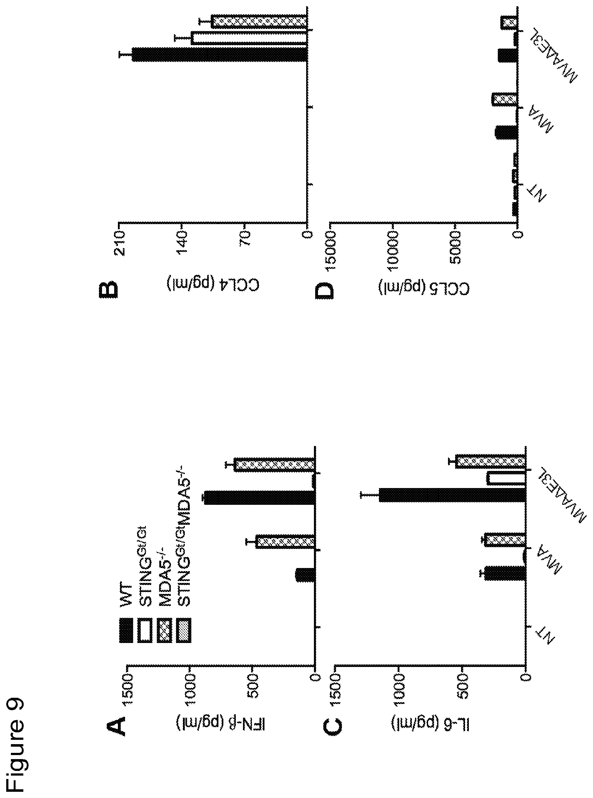
D00013

D00014

D00015
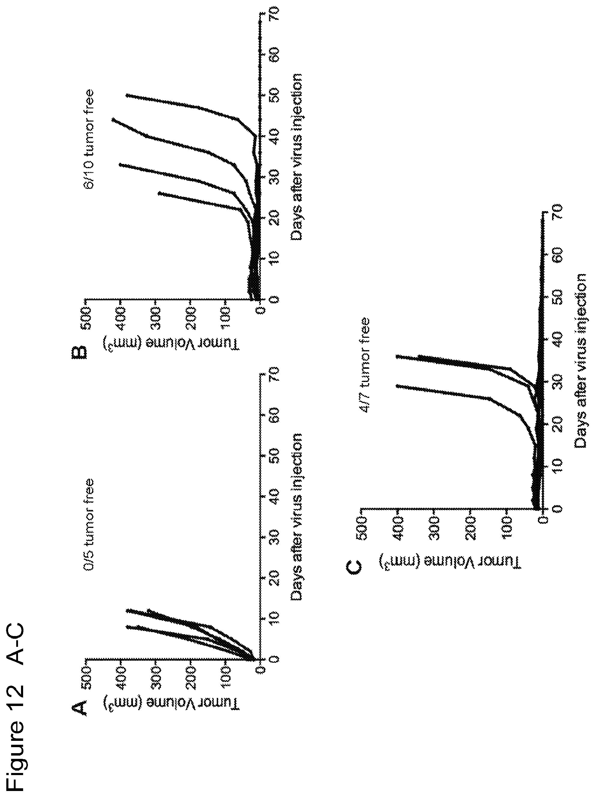
D00016
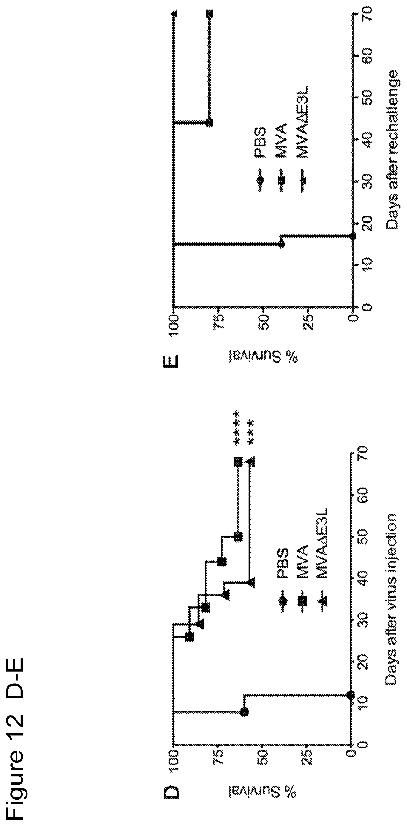
D00017

D00018
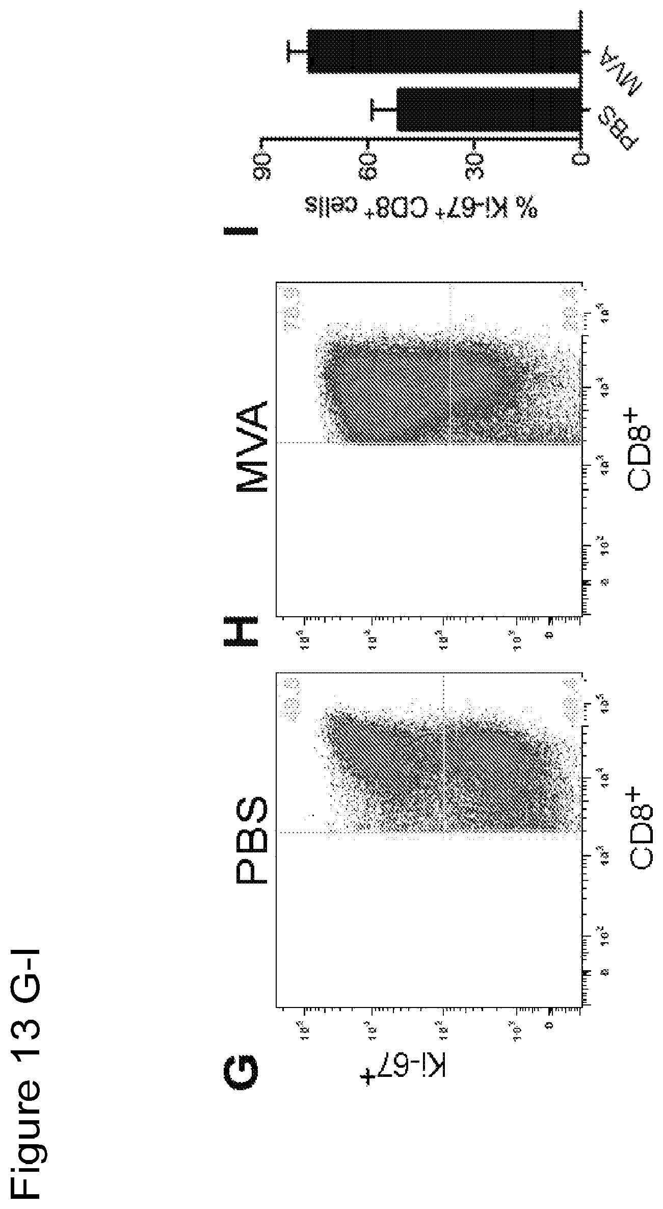
D00019
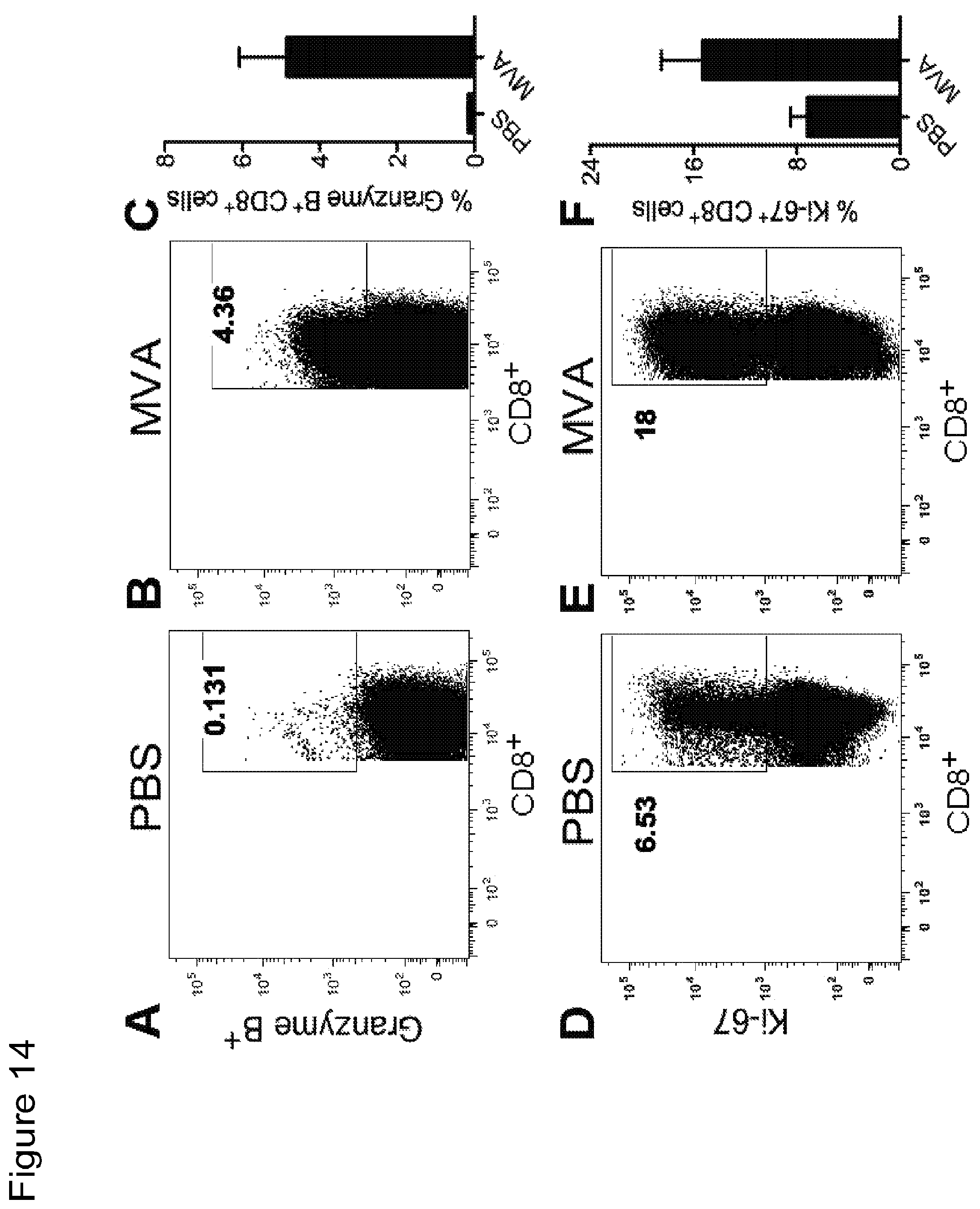
D00020

D00021

D00022
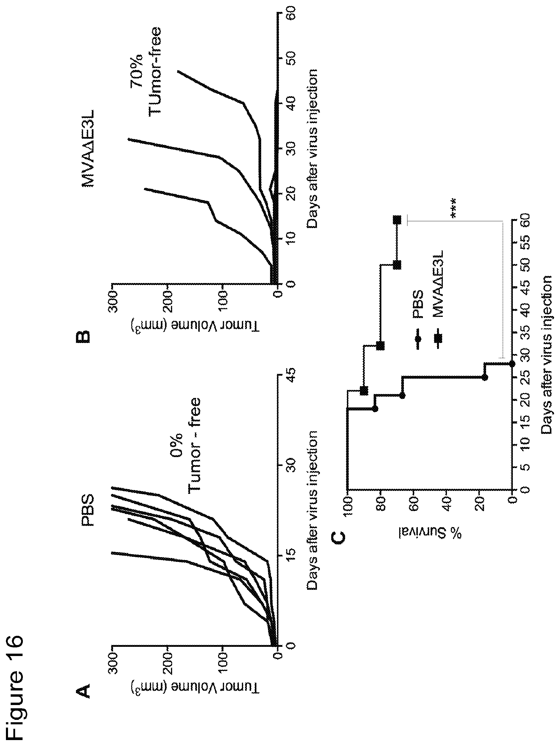
D00023

D00024

D00025

D00026
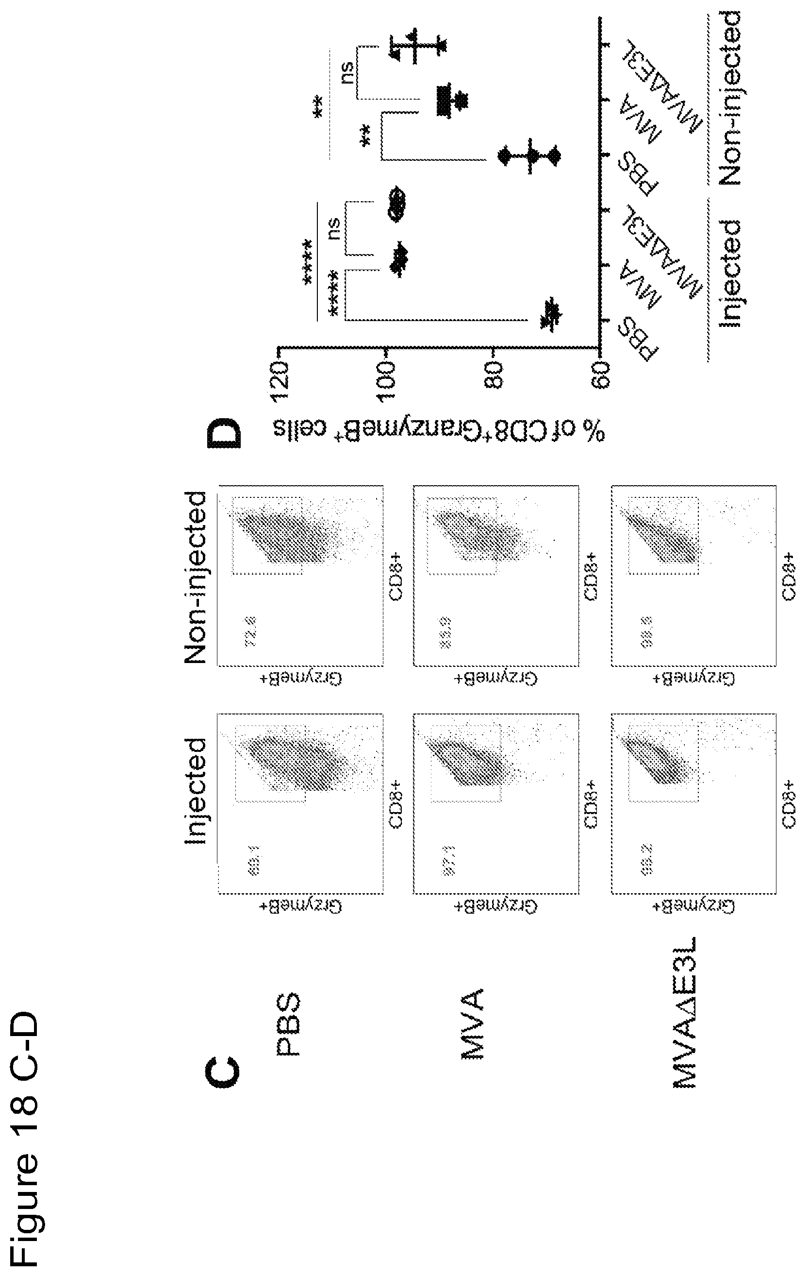
D00027

D00028
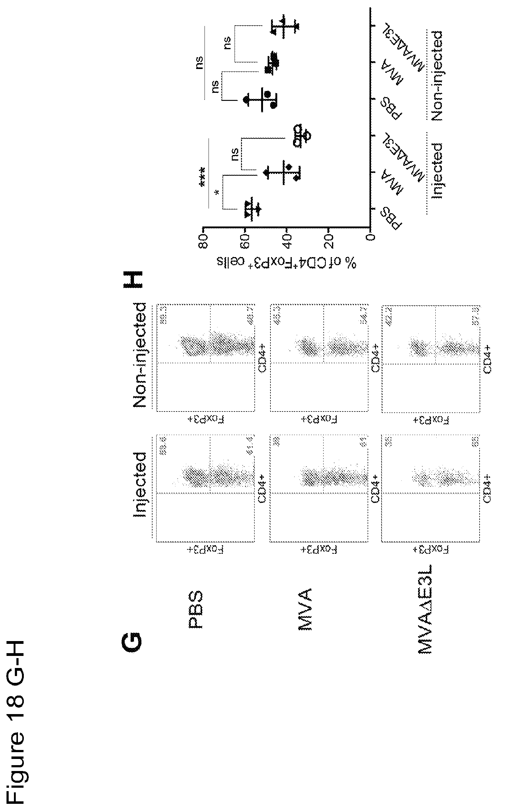
D00029

D00030

D00031

D00032

S00001
XML
uspto.report is an independent third-party trademark research tool that is not affiliated, endorsed, or sponsored by the United States Patent and Trademark Office (USPTO) or any other governmental organization. The information provided by uspto.report is based on publicly available data at the time of writing and is intended for informational purposes only.
While we strive to provide accurate and up-to-date information, we do not guarantee the accuracy, completeness, reliability, or suitability of the information displayed on this site. The use of this site is at your own risk. Any reliance you place on such information is therefore strictly at your own risk.
All official trademark data, including owner information, should be verified by visiting the official USPTO website at www.uspto.gov. This site is not intended to replace professional legal advice and should not be used as a substitute for consulting with a legal professional who is knowledgeable about trademark law.