Combined use of Fc gamma RIIb (CD32B) and CD20-specific antibodies
Cragg , et al. J
U.S. patent number 10,525,129 [Application Number 13/817,744] was granted by the patent office on 2020-01-07 for combined use of fc gamma riib (cd32b) and cd20-specific antibodies. This patent grant is currently assigned to University of Southampton. The grantee listed for this patent is Stephen Beers, Mark Cragg, Bjorn Frendeus, Martin Glennie, Peter Johnson, Sean Lim, Ali Roghanian, Ingrid Teige. Invention is credited to Stephen Beers, Mark Cragg, Bjorn Frendeus, Martin Glennie, Peter Johnson, Sean Lim, Ali Roghanian, Ingrid Teige.


View All Diagrams
| United States Patent | 10,525,129 |
| Cragg , et al. | January 7, 2020 |
Combined use of Fc gamma RIIb (CD32B) and CD20-specific antibodies
Abstract
The invention provides a method of treating a patient having target cells that express FcyRIIb, the method comprising administering (i) an antibody molecule that specifically binds a surface antigen of the target cell, which antibody molecule has an Fc domain capable of binding FcyRIIb; in combination with (ii) an agent that prevents or reduces binding between the Fc domain of the antibody molecule and FcyRIIb; characterized in that the patient is selected on the basis that their target cells express an elevated level of FcyRIIb.
| Inventors: | Cragg; Mark (Southampton, GB), Glennie; Martin (Southampton, GB), Roghanian; Ali (Southampton, GB), Beers; Stephen (Southampton, GB), Johnson; Peter (Southampton, GB), Lim; Sean (Southampton, GB), Frendeus; Bjorn (Landskorna, SE), Teige; Ingrid (Lund, SE) | ||||||||||
|---|---|---|---|---|---|---|---|---|---|---|---|
| Applicant: |
|
||||||||||
| Assignee: | University of Southampton
(Southampton, GB) |
||||||||||
| Family ID: | 42984443 | ||||||||||
| Appl. No.: | 13/817,744 | ||||||||||
| Filed: | August 19, 2011 | ||||||||||
| PCT Filed: | August 19, 2011 | ||||||||||
| PCT No.: | PCT/GB2011/051572 | ||||||||||
| 371(c)(1),(2),(4) Date: | May 31, 2013 | ||||||||||
| PCT Pub. No.: | WO2012/022985 | ||||||||||
| PCT Pub. Date: | February 23, 2012 |
Prior Publication Data
| Document Identifier | Publication Date | |
|---|---|---|
| US 20130251706 A1 | Sep 26, 2013 | |
Foreign Application Priority Data
| Aug 20, 2010 [GB] | 1013989.7 | |||
| Current U.S. Class: | 1/1 |
| Current CPC Class: | A61P 35/02 (20180101); G01N 33/6893 (20130101); A61P 43/00 (20180101); A61K 39/39558 (20130101); A61P 35/00 (20180101); A61P 37/00 (20180101); C07K 16/2887 (20130101); C07K 16/283 (20130101); A61P 29/00 (20180101); C07K 16/2803 (20130101); A61K 2039/507 (20130101) |
| Current International Class: | A61K 39/395 (20060101); A61K 39/40 (20060101); G01N 33/68 (20060101); C07K 16/28 (20060101); C07K 16/00 (20060101); A61K 39/00 (20060101) |
References Cited [Referenced By]
U.S. Patent Documents
| 4485045 | November 1984 | Regen |
| 4544545 | October 1985 | Ryan et al. |
| 4880078 | November 1989 | Inoue et al. |
| 5290540 | March 1994 | Prince et al. |
| 5855913 | January 1999 | Hanes et al. |
| 5874064 | February 1999 | Edwards et al. |
| 5934272 | August 1999 | Lloyd et al. |
| 5985309 | November 1999 | Edwards et al. |
| 6019968 | February 2000 | Platz et al. |
| 2004/0185045 | September 2004 | Koenig et al. |
| 2005/0215767 | September 2005 | Koenig et al. |
| 2005/0260213 | November 2005 | Koenig et al. |
| 2006/0013810 | January 2006 | Johnson et al. |
| 2006/0134709 | June 2006 | Stavenhagen et al. |
| 2008/0014141 | January 2008 | Huber et al. |
| 2009/0010920 | January 2009 | Lazar et al. |
| 2009/0191195 | July 2009 | Tuaillon et al. |
| 2526139 | Nov 2015 | GB | |||
| 9219244 | Nov 1992 | WO | |||
| 9732572 | Sep 1997 | WO | |||
| 9744013 | Nov 1997 | WO | |||
| 9831346 | Jul 1998 | WO | |||
| 9966903 | Dec 1999 | WO | |||
| 2004/016750 | Feb 2004 | WO | |||
| 2005/018669 | Mar 2005 | WO | |||
| 2006066078 | Jun 2006 | WO | |||
| 2008002933 | Jan 2008 | WO | |||
| 2008/140603 | Nov 2008 | WO | |||
| 2009062083 | May 2009 | WO | |||
| 2009083009 | Jul 2009 | WO | |||
| 2010080994 | Jul 2010 | WO | |||
| 2012/022985 | Feb 2012 | WO | |||
| 2015/173384 | Nov 2015 | WO | |||
Other References
|
Maccallum et al. "Antibody-antigen interactions: contact analysis and binding site topography", Journal of Molecular Biology, 1996. vol. 262, pp. 732-745. cited by examiner . De Pascalis et al. "Grafting of abbreviated complementarity determining regions containing specificity determining residues essential for ligand contact to engineer a less immunogenic humanzied monoclonal antibody", Journal of Immunology, 2002. vol. 169, pp. 3076-3084. cited by examiner . Stopforth et al. J. Clin. Immunol. 2016, 36(Suppl):S88-S94. (Year: 2016). cited by examiner . Michel et al., "Intracellular Accumulation of the Anti-CD20 Antibody 1F4 in B-Lymphoma Cells", Clin Cancer Res, 8:2701-2713 (2002). cited by applicant . Mitsudomi et al., "Epidermal growth factor receptor in relation to tumor development: EGFR gene and cancer", FEBS J., 277(2):301-308 (2010). cited by applicant . Mockridge et al., "Reversible anergy of slgM-mediated signaling in the two subsets of CLL defined by VH-gene mutational status", Blood, 109:4424-4431 (2007). cited by applicant . Mossner et al., "Increasing the efficacy of CD20 antibody therapy through the engineering of a new type II anti-CD20 antibody with enhanced direct and immune effector cell-mediated B-cell cytotoxicity", Blood, 115(22):4393-4402 (2010). cited by applicant . Neubig et al., "international Union of Pharmacology Committee on Receptor Nomenclature and Drug Classification. XXXVIII. Update on Terms and Symbols in Quantitative Pharmacology", Pharmacol. Rev., 55, 597-606 (2003). cited by applicant . Niederfellner et al., "Epitope characterization and crystal structure of GA101 provide insights into the molecular basis for type I/II distinction of CD20 antibodies", Blood, 118, 358-367 (2011). cited by applicant . Nimmerjahn et al., "Antibodies, Fc receptors and cancer", Curr Opin Immunol, 19:239-245 (2007). cited by applicant . Nimmerjahn et al., "FC.gamma. receptors as regulators of immune responses", Nat Rev Immunol, 8:34-47 (2008). cited by applicant . Polyak et al., "CD20 Homo-oligomers Physically Associate with the B Cell Antigen Receptor", J Biol Chem, 283:18545-18552 (2008). cited by applicant . Potter et al., "Structural and Functional Features of the B-Cell Receptor in IgG Positive Chronic Lymphocytic Leukemia", Clin Cancer Res, 12:1672-1679 (2006). cited by applicant . Rankin et al., "CD32B, the human inhibitory Fc-.gamma. receptor IIB, as a target for monoclonal antibody therapy of B-cell lymphoma", Blood, 108:2384-2391 (2006). cited by applicant . Ravetch et al., "IgG Rc Receptors", Annu Rev Immunol, 19:275-290 (2001). cited by applicant . Robak et al., "Rituximab Plus Fludarabine and Cyclophosphamide Prolongs Progression-Free Survival Compared with Fludarabine and Cyclophosphamide Alone in Previously Treated Chronic Lymphocytic Leukemia", J Clin Oncol., 28(10):1756-1765 (2010). cited by applicant . Sehn et al., "Introduction of Combined CHOP Plus Rituximab Therapy Dramatically Improved Outcome of Diffuse Large B-Cell Lymphoma in British Columbia", J Clin Oncol, 23:5027-5033 (2005). cited by applicant . Stolz et al., "Molecular Mechanisms of Resistance to Rituximab and pharmacologic strategies for its circumvention", Leuk Lymphoma, 50:873-885 (2009). cited by applicant . Teeling et al., "Characterization of new human CD20 monoclonal antibodies with potent cytolytic activity against non-Hodgkin lymphomas", Blood, 104:1793-1800 (2004). cited by applicant . Ternynck et al., "Comparison of Normal and CLL Lymphocyte Surface Ig Determinants Using Peroxidase-Labeled Antibodies. I. Detection and Quantitation of Light Chain Determinants", Blood, 43:789-795 (1974). cited by applicant . Treon et al., "Tumor Cell Expression of CD59 is Associated with Resistance to CD20 Serotherapy in Patients with B-Cell Malignancies", J Immunother, 24:263-271 (1991). cited by applicant . Tutt et al., "Monoclonal Antibody Therapy of B Cell Lymphoma: Signaling Activity on Tumor Cells Appears More Important Than Recruitment of Effectors", J Immunol, 161:3176-3185 (1998). cited by applicant . Uchida et al., "The Innate Mononuclear Phagocyte Network Depletes B Lymphocytes through Fc Receptor-dependent Mechanisms during Anti-CD20 Antibody Immunotherapy", J Exp Med, 199:1659-1669 (2004). cited by applicant . Walshe et al., "Induction of Cytosolic Calcium Flux by CD20 Is Dependent upon B Cell Antigen Receptor Signaling*", J Biol Chem, 283:16971-16984 (2008). cited by applicant . Weng et al., "Two Immunoglobulin G Fragment C Receptor Polymorphisms Independently Predict Response to Rituximab in Patients With Follicular Lymphoma", J Clin Oncol, 21:3940-3947 (2003). cited by applicant . Weng et al., "Genetic polymorphism of the inhibitory IgG Fc receptor Fc.gamma.RIIb is not associated with clinical outcome in patients with follicular lymphoma treated with rituximab", Leuk Lymphoma, 50:723-727 (2009). cited by applicant . Wiestner et al., "ZAP-70 expression identifies a chronic lymphocytic leukemia subtype with unmutated immunoglobulin genes, inferior clinical outcome, and distinct gene expression profile", Blood, 101:4944-4951 (2003). cited by applicant . Yang et al., "Down-Regulation of CD40 Gene Expression and Inhibition of Apoptosis with Danshensu in Endothelial Cells", Basic Clin Pharmacol Toxicol., 104(2):87-92 (2009). cited by applicant . Aman et al., "Fc.gamma.RIIB1/SHIP-mediated Inhibitory Signaling in B Cells Involves Lipid Rafts", J Biol Chem, 276:46371-46378 (2001). cited by applicant . Beers et al., "Type II (tositumomab) anti-CD20 monoclonal antibody out performs type I (rituximab-like) reagents in B-cell depletion regardless of complement activation", Blood, 112:4170-4177 (2008). cited by applicant . Beers et al., Antigenic modulation limits the efficacy of anti-CD20 antibodies: implications for antibody selection, Blood, 115(25): 5191-5201 (2010). cited by applicant . Beers et al., "CD20 as a Target for Therapeutic Type I and II Monoclonal Antiboides", Seminars in Hematology. 47(2): 107-114 (Apr. 2010). cited by applicant . Bradley et al., "Rules and regulation of Thy-1, a context-dependent modulator of cell phenotype", Biofactors;35(3):258-65 (2009). cited by applicant . Bricarello et al., "Ganglioside embedded in reconstituted lipoprotein binds cholera toxin with elevated affinity", J Lipid Res., 51(9):2731-2738 (2010). cited by applicant . Busillo et al., "Regulation of CXCR4 Signaling", Biochim Biophys Acta, 1768(4):952-963 (2007). cited by applicant . Callanan et al., The IgG Fc receptor, Fc.gamma.RIIB, is a target for deregulation by chromosomal translocation in malignant lymphoma, PNAS, 97:309-314 (2000). cited by applicant . Camilleri-Broet et al., "Fc.gamma.RIIB expression in diffuse large B-cell lymphomas does not alter the response to CHOP + rituximab (R-CHOP)", Leukemia, 18:2038-2040 (2004). cited by applicant . Camilleri-Broet et al., "Fc.gamma.RIIB is differentially expressed during B cell maturation and in B-cell lymphomas", Br J Haematol 124:55-62 (2004). cited by applicant . Chan et al., "CD20-induced Lymphoma Cell Death Is Independent of Both Caspases and Its Redistribution into Triton X-100 Insoluble Membrane Rafts", Cancer Res, 63:5480-5489 (2003). cited by applicant . Cheson et al., "Monoclonal Antibody Therapy for B-Cell Non-Hodgkin's Lymphoma", NEJM, 359(6): 613-626 (2008). cited by applicant . Clynes et al., "Inhibitory Rc receptors modulate in vivo cytoxicity against tumor targets", Nat Med, 6:443-446 (2000). cited by applicant . Cragg, Mark, "CD20 antibodies: doing the time warp", Blood, 118:219-220 (2011). cited by applicant . Cragg et al., "Antibody specificity controls in vivo effector mechanisms of anti-CD20 reagents", Blood, 103:2738-2743 (2004). cited by applicant . Cragg et al., "Complement-mediated lysis by anti-CD20 mAb correlates with segregation into lipid rafts", Blood, 101(3):1045-1052 (2003). cited by applicant . Crespo et al., "ZAP-70 Expression as a Surrogate for Immunoglobulin-Variable-Region Mutations in Chronic Lymphocytic Leukemia", N Engl J Med, 348:1764-1775 (2003). cited by applicant . Damle et al., "Ig V Gene Mutation Status and CD38 Expression As Novel Prognostic Indicators in Chronic Lymphocytic Leukemia", Blood, 94:1840-1847 (1999). cited by applicant . Davis et al., "Therapy of B-Cell Lymphoma with Anti-CD20 Antibodies Can Result in the Loss of CD20 Antigen Expression", Clin Cancer Res, 5:611-615 (1999). cited by applicant . Deans et al., "Rapid Redistribution of CD20 to a Low Density Detergent-insoluble Membrane Compartment", J. Biol. Chem., 273: 344-348 (1998). cited by applicant . De Rie et al., "Regulatory Role of CD19 Molecules in B-Cell Activation and Differentiation", Cell Immunol., 118(2):368-381 (1989). cited by applicant . Epstein et al., "Biological activity of liposome-encapsulated murine interferon .gamma.is mediated by a cell membrane receptor", Proc. Natl. Acad. Sci. USA, 82:3688-3692 (Jun. 1985). cited by applicant . Feugier et al., "Long-Term Results of the R-CHOP Study in the Treatment of Elderly Patients With Diffuse Large B-Cell Lymphoma: A Study by the Groupe d'Etude des Lymphomes de l'Adulte", J Clin Oncol, 23:4117-4126 (2005). cited by applicant . Fridman et al., "Soluble Fc.gamma. receptors", J Leukoc Biol, 54:504-512 (1993). cited by applicant . Glennie et al., "Preparation and Performance of Bispecific F(ab'.gamma.)2 Antibody Containing Thioether-Linked Fab'.gamma. Fragments", J Immunol, 139:2367-2375, (1987). cited by applicant . Golay et al., "CD20 levels determine the in vitro susceptibility to rituximab and complement of B-cell chronic lymphocytic leukemia: further regulation by CD55 and CD59", Blood, 98:3383-3389 (2001). cited by applicant . Greenman et al., "Characterization of a New Monoclonal Anti-Fc.gamma.RII Antibody, AT10 and Its Incorporation into a bispecific F(ab')2 Derivative for Recruitment of Cytotoxic Effectors", Mol Immunol, 28:1243-1254 (1991). cited by applicant . Hamblin et al., "Unmutated Ig VH Genes Are Associated With a More Aggressive Form of Chronic Lymphocytic Leukemia", Blood, 94:1848-1854 (1999). cited by applicant . Hamblin et al., "CD38 expression and immunoglobulin variable region mutations are independent prognostic variables in chronic lymphocytic leukemia, but CD38 expression may vary during the course of the disease", Blood, 99:1023-1029 (2002). cited by applicant . Hiraga et al., "Down-regulation of CD20 expression in B-cell lymphoma cells after treatment with rituximab-containing combination chemotherapies: its prevalence and clinical significance", Blood, 113:4885-4893 (2009). cited by applicant . Hwang et al., "Hepatic Uptake and Degradation of Unilamellar Sphingomyelin/Cholesterol Liposomes: A Kinetic Study", Proc. Natl. Acad. Sci. USA, 77: 4030-4034 (1980). cited by applicant . Ibrahim et al., "CD38 expression as an important prognostic factor in B-cell chronic lymphocytic leukemia", Blood, 98:181-186 (2001). cited by applicant . Ivanov et al., "Monoclonal Antibodies Directed to CD20 and HLA-DR can elicit homotypic adhesion followed by lysosome-mediated cell death in human lymphoma and leukemia cells", J Clin Invest, 119:2143-2159 (2009). cited by applicant . Jazirehi et al., "Development of Rituximab-Resistant Lymphoma Clones With Altered Cell Signaling and Cross-Resistance to Chemotherapy", Cancer Res, 67:1270-1281 (2007). cited by applicant . Kimberley et al., "Alternative Roles for CD59", Mol Immunol., 44(1-3):73-81 (2007). cited by applicant . Kono et al., "Fc.gamma.RIIB IIe232 Thr transmembrane polymorphism associated with human systemic lupus erythematosus decreases affinity to lipid rafts and attenuates inhibitory effects on B cell receptor signaling", Hum Mol Genet, 14:2881-2892 (2005). cited by applicant . Krober et al., "VH mutation status, CD38 expression level, genomic aberrations, and survival in chronic lymphocytic leukemia", Blood, 100:1410-1416 (2002). cited by applicant . Lenz et al., "Immunochemotherapy With Rituximab and Cyclophosphamide, Doxorubicin, Vincristine, and Prednisone Significantly Improves Response and Time to Treatment Failure, But Not Long-Term Outcome in Patients With Previously Untreated Mantle Cell Lymphoma: Results of a Prospective Randomized Trial of the German Low Grade Lymphoma Study Group (GLSG)", J Clin Oncol, 23:1984-1992 (2005). cited by applicant . Li et al., "A Novel Polymorphism in the Fc.gamma. Receptor IIB (CD32B) Transmembrane Region Alters Receptor Signaling", Arthritis Rheum, 48:3242-3252 (2003). cited by applicant . Lim et al., "Fc gamma receptor IIb on target B cells promotes rituximab internalization and reduces clinical efficacy", Blood, 118(9):2530-2540 (2011). cited by applicant . Marcus et al., "CVP chemotherapy plus rituximab compared with CVP as first-line treatment for advanced follicular lymphoma", Blood, 105:1417-1423 (2005). cited by applicant . Marcus et al., "Phase III Study of R-CVP Compared With Cyclophosphamide, Vincristine, and Prednisone Alone in Patients With Previously Untreated Advanced Follicular Lymphoma", J Clin Oncol, 26:4579-4586 (2008). cited by applicant . Shim et al., "One target, difference effects: a comparison of distinct therapeutic antibodies against the same targets", Experimental and Molecular Medicine, 43:539-549 (Oct. 2011). cited by applicant . Bournazos et al., "Association of FcyRIIa (CD32a) with Lipid Rafts Regulates Ligand Binding Activity", The Journal of Immunology, 182:8026-8036 (2009). cited by applicant . Smith, "Rituximab (monoclonal anti-CD20 antibody): mechanisms of action and resistance", Oncogene, 22:7359-7368 (2003). cited by applicant . Vervoordeldonk et al., "Fc gamma receptor II (CD32) on malignant B cells influences modulation induced by anti-CD19 monoclonal antibody", Blood, 83:1632-1639 (1994). cited by applicant . Vaughan et al., "Inhibitory FcyRIIb (CD32b) becomes activated by therapeutic mAb in both cis and trans and drives internalization according to antibody specificity", Blood, 123(5):669-677 (2014). cited by applicant . Tong et al., "Prospects for CD40-directed experimental therapy of human cancer", Cancer Gene Ther., 10(1):1-13 Jan. 2003, Abstract Only. cited by applicant. |
Primary Examiner: Dahle; Chun W
Attorney, Agent or Firm: Fuller; Rodney J. Booth Udall Fuller, PLC
Claims
The invention claimed is:
1. A method of treating a patient having target cells that express Fc.gamma.RIIb, the method comprising: selecting a patient having target cells that express an elevated level of Fc.gamma.RIIb relative to a control, wherein the control is the normal level of Fc.gamma.RIIb expression in cells of the same type as the target cells; and administering to the patient (i) a first antibody molecule that specifically binds a CD20 surface antigen of the target cell and the first antibody molecule has an Fc domain capable of binding Fc.gamma.RIIb; in combination with (ii) a second antibody that specifically binds Fc.gamma.RIIb comprising the following amino acid sequences: (i) the sequence of CDRH1 consists of SEQ ID NO: 29, the sequence of CDRH2 consists of SEQ ID NO: 30, the sequence of CDRH3 consists of SEQ ID NO: 31, the sequence of CDRL1 consists of SEQ ID NO: 32, the sequence of CDRL2 consists of SEQ ID NO: 33, and the sequence of CDRL3 consists of SEQ ID NO: 34; or (ii) the sequence of CDRH1 consists of SEQ ID NO: 35, the sequence of CDRH2 consists of SEQ ID NO: 36, the sequence of CDRH3 consists of SEQ ID NO: 37, the sequence of CDRL1 consists of SEQ ID NO: 38, the sequence of CDRL2 consists of SEQ ID NO: 39, and the sequence of CDRL3 consists of SEQ ID NO: 40; or (iii) the sequence of CDRH1 consists of SEQ ID NO: 41, the sequence of CDRH2 consists of SEQ ID NO: 42, the sequence of CDRH3 consists of SEQ ID NO: 43, the sequence of CDRL1 consists of SEQ ID NO: 44, the sequence of CDRL2 consists of SEQ ID NO: 45, and the sequence of CDRL3 consists of SEQ ID NO: 46; or (iv) the sequence of CDRH1 consists of SEQ ID NO: 47, the sequence of CDRH2 consists of SEQ ID NO: 48, the sequence of CDRH3 consists of SEQ ID NO: 49, the sequence of CDRL1 consists of SEQ ID NO: 50, the sequence of CDRL2 consists of SEQ ID NO: 51, and the sequence of CDRL3 consists of SEQ ID NO: 52; or (v) the sequence of CDRH1 consists of SEQ ID NO: 53, the sequence of CDRH2 consists of SEQ ID NO: 54, the sequence of CDRH3 consists of SEQ ID NO: 55, the sequence of CDRL1 consists of SEQ ID NO: 56, the sequence of CDRL2 consists of SEQ ID NO: 57, and the sequence of CDRL3 consists of SEQ ID NO: 58; or (vi) the sequence of CDRH1 consists of SEQ ID NO: 59, the sequence of CDRH2 consists of SEQ ID NO: 60, the sequence of CDRH3 consists of SEQ ID NO: 61, the sequence of CDRL1 consists of SEQ ID NO: 62, the sequence of CDRL2 consists of SEQ ID NO: 63, and the sequence of CDRL3 consists of SEQ ID NO: 64; or (vii) the sequence of CDRH1 consists of SEQ ID NO: 65, the sequence of CDRH2 consists of SEQ ID NO: 66, the sequence of CDRH3 consists of SEQ ID NO: 67, the sequence of CDRL1 consists of SEQ ID NO: 68, the sequence of CDRL2 consists of SEQ ID NO: 69, and the sequence of CDRL3 consists of SEQ ID NO: 70; or (viii) the sequence of CDRH1 consists of SEQ ID NO: 71, the sequence of CDRH2 consists of SEQ ID NO: 72, the sequence of CDRH3 consists of SEQ ID NO: 73, the sequence of CDRL1 consists of SEQ ID NO: 74, the sequence of CDRL2 consists of SEQ ID NO: 75, and the sequence of CDRL3 consists of SEQ ID NO: 76; or (ix) the sequence of CDRH1 consists of SEQ ID NO: 77, the sequence of CDRH2 consists of SEQ ID NO: 78, the sequence of CDRH3 consists of SEQ ID NO: 79, the sequence of CDRL1 consists of SEQ ID NO: 80, the sequence of CDRL2 consists of SEQ ID NO: 81, and the sequence of CDRL3 consists of SEQ ID NO: 82; or (x) the sequence of CDRH1 consists of SEQ ID NO: 83, the sequence of CDRH2 consists of SEQ ID NO: 84, the sequence of CDRH3 consists of SEQ ID NO: 85, the sequence of CDRL1 consists of SEQ ID NO: 86, the sequence of CDRL2 consists of SEQ ID NO: 87, and the sequence of CDRL3 consists of SEQ ID NO: 88; or (xi) the sequence of CDRH1 consists of SEQ ID NO: 89, the sequence of CDRH2 consists of SEQ ID NO: 90, the sequence of CDRH3 consists of SEQ ID NO: 91, the sequence of CDRL1 consists of SEQ ID NO: 92, the sequence of CDRL2 consists of SEQ ID NO: 93, and the sequence of CDRL3 consists of SEQ ID NO: 94; or (xii) the sequence of CDRH1 consists of SEQ ID NO: 95, the sequence of CDRH2 consists of SEQ ID NO: 96, the sequence of CDRH3 consists of SEQ ID NO: 97, the sequence of CDRL1 consists of SEQ ID NO: 98, the sequence of CDRL2 consists of SEQ ID NO: 99, and the sequence of CDRL3 consists of SEQ ID NO: 100; or (xiii) the sequence of CDRH1 consists of SEQ ID NO: 101, the sequence of CDRH2 consists of SEQ ID NO: 102, the sequence of CDRH3 consists of SEQ ID NO: 103, the sequence of CDRL1 consists of SEQ ID NO: 104, the sequence of CDRL2 consists of SEQ ID NO: 105, and the sequence of CDRL3 consists of SEQ ID NO: 106, wherein the second antibody prevents or reduces binding between the Fc domain of the first antibody molecule and Fc.gamma.RIIb, wherein the patient is treated for a B cell lymphoma or chronic lymphocytic leukaemia (CLL).
2. The method of claim 1, wherein the second antibody prevents or reduces Fc.gamma.RIIb present on the target cell from binding to the Fc domain of the first antibody molecule.
3. The method of claim 1, wherein the first antibody molecule has a Fc domain capable of binding Fc.gamma.RIIb and is capable of being internalized into the target cell in an Fc.gamma.RIIb-dependent manner.
4. The method of claim 1, wherein the second antibody that prevents or reduces Fc.gamma.RIIb binding to the Fc domain of the first antibody molecule additionally prevents or reduces internalization of the first antibody molecule into the target cell.
5. The method of claim 1, wherein the target cell is a B cell.
6. The method of claim 1, wherein the B cell lymphoma is non-Hodgkin lymphoma.
7. The method of claim 6, wherein the non-Hodgkin lymphoma is selected from the group consisting of: follicular lymphoma, diffuse large B cell lymphoma, and mantle cell lymphoma.
8. The method of claim 1, wherein the second antibody is one or more monoclonal antibody molecules that specifically bind Fc.gamma.RIIb and do not include a domain capable of recruiting an effector cell.
9. The method of claim 1, wherein the second antibody prevents or reduces Fc.gamma.RIIb signaling, prevents or reduces internalization of the first antibody molecule by the target cell, or both.
10. The method of claim 1, wherein the first antibody molecule is a Type I CD20 antibody.
11. The method of claim 1, wherein the B cell lymphoma is selected from the group consisting of: follicular lymphoma, diffuse large B cell lymphoma, small lymphocytic lymphoma, and mantle cell lymphoma.
12. The method of claim 1, wherein the sequence of CDRH1 consists of SEQ ID NO: 83, the sequence of CDRH2 consists of SEQ ID NO: 84, the sequence of CDRH3 consists of SEQ ID NO: 85, the sequence of CDRL1 consists of SEQ ID NO: 86, the sequence of CDRL2 consists of SEQ ID NO: 87, and the sequence of CDRL3 consists of SEQ ID NO: 88.
13. A method of treating a patient having target cells that express Fc.gamma.RIIb, the method comprising: selecting a patient having target cells that express an elevated level of Fc.gamma.RIIb relative to a control, wherein the control is the normal level of Fc.gamma.RIIb expression in cells of the same type as the target cells; and administering to the patient (i) a first antibody molecule that specifically binds a CD20 surface antigen of the target cell and the first antibody molecule has an Fc domain capable of binding Fc.gamma.RIIb; in combination with (ii) a second antibody that specifically binds Fc.gamma.RIIb comprising the following amino acid sequences as the VH and VL regions, respectively: (i) SEQ ID NO: 3 and SEQ ID NO: 16; or (ii) SEQ ID NO: 4 and SEQ ID NO: 17; or (iii) SEQ ID NO: 5 and SEQ ID NO: 18; or (iv) SEQ ID NO: 6 and SEQ ID NO: 19; or (v) SEQ ID NO: 7 and SEQ ID NO: 20; or (vi) SEQ ID NO: 8 and SEQ ID NO: 21; or (vii) SEQ ID NO: 9 and SEQ ID NO: 22; or (viii) SEQ ID NO: 10 and SEQ ID NO: 23; or (ix) SEQ ID NO: 11 and SEQ ID NO: 24; or (x) SEQ ID NO: 12 and SEQ ID NO: 25; or (xi) SEQ ID NO: 13 and SEQ ID NO: 26; or (xii) SEQ ID NO: 14 and SEQ ID NO: 27; or (xiii) SEQ ID NO: 15 and SEQ ID NO: 28, wherein the second antibody prevents or reduces binding between the Fc domain of the first antibody molecule and Fc.gamma.RIIb, wherein the patient is treated for a B cell lymphoma or chronic lymphocytic leukaemia (CLL).
14. The method of claim 13, wherein the second antibody prevents or reduces Fc.gamma.RIIb present on the target cell from binding to the Fc domain of the first antibody molecule.
15. The method of claim 13, wherein the first antibody molecule has a Fc domain capable of binding Fc.gamma.RIIb and is capable of being internalized into the target cell in an Fc.gamma.RIIb-dependent manner.
16. The method of claim 13, wherein the second antibody that prevents or reduces Fc.gamma.RIIb binding to the Fc domain of the first antibody molecule additionally prevents or reduces internalization of the first antibody molecule into the target cell.
17. The method of claim 13, wherein the B cell lymphoma is non-Hodgkin lymphoma.
18. The method of claim 13, wherein the second antibody is one or more monoclonal antibody molecules that specifically bind Fc.gamma.RIIb and do not include a domain capable of recruiting an effector cell.
19. The method of claim 13, wherein the second antibody prevents or reduces Fc.gamma.RIIb signaling, prevents or reduces internalization of the first antibody molecule by the target cell, or both.
20. The method of claim 13, wherein the first antibody molecule is a Type I CD20 antibody.
21. The method of claim 13, wherein the B cell lymphoma is selected from the group consisting of: follicular lymphoma, diffuse large B cell lymphoma, small lymphocytic lymphoma, and mantle cell lymphoma.
22. The method of claim 13, wherein the second antibody comprises SEQ ID NO: 12 as the VH region and SEQ ID NO: 25 as the VL region.
23. A method of treating B cell lymphoma or chronic lymphocytic leukaemia (CLL), the method comprising: selecting a patient having target cells that express an elevated level of Fc.gamma.RIIb relative to a control, wherein the control is the normal level of Fc.gamma.RIIb expression in cells of the same type as the target cells; and administering to the patient a composition comprising: (i) a first antibody molecule that specifically binds a CD20 cell surface antigen of the target cell and the first antibody has an Fc domain capable of binding Fc.gamma.RIIb; in combination with (ii) a second antibody that specifically binds Fc.gamma.RIIb comprising the following amino acid sequences: (i) the sequence of CDRH1 consists of SEQ ID NO: 29, the sequence of CDRH2 consists of SEQ ID NO: 30, the sequence of CDRH3 consists of SEQ ID NO: 31, the sequence of CDRL1 consists of SEQ ID NO: 32, the sequence of CDRL2 consists of SEQ ID NO: 33, and the sequence of CDRL3 consists of SEQ ID NO: 34; or (ii) the sequence of CDRH1 consists of SEQ ID NO: 35, the sequence of CDRH2 consists of SEQ ID NO: 36, the sequence of CDRH3 consists of SEQ ID NO: 37, the sequence of CDRL1 consists of SEQ ID NO: 38, the sequence of CDRL2 consists of SEQ ID NO: 39, and the sequence of CDRL3 consists of SEQ ID NO: 40; or (iii) the sequence of CDRH1 consists of SEQ ID NO: 41, the sequence of CDRH2 consists of SEQ ID NO: 42, the sequence of CDRH3 consists of SEQ ID NO: 43, the sequence of CDRL1 consists of SEQ ID NO: 44, the sequence of CDRL2 consists of SEQ ID NO: 45, and the sequence of CDRL3 consists of SEQ ID NO: 46; or (iv) the sequence of CDRH1 consists of SEQ ID NO: 47, the sequence of CDRH2 consists of SEQ ID NO: 48, the sequence of CDRH3 consists of SEQ ID NO: 49, the sequence of CDRL1 consists of SEQ ID NO: 50, the sequence of CDRL2 consists of SEQ ID NO: 51, and the sequence of CDRL3 consists of SEQ ID NO: 52; or (v) the sequence of CDRH1 consists of SEQ ID NO: 53, the sequence of CDRH2 consists of SEQ ID NO: 54, the sequence of CDRH3 consists of SEQ ID NO: 55, the sequence of CDRL1 consists of SEQ ID NO: 56, the sequence of CDRL2 consists of SEQ ID NO: 57, and the sequence of CDRL3 consists of SEQ ID NO: 58; or (vi) the sequence of CDRH1 consists of SEQ ID NO: 59, the sequence of CDRH2 consists of SEQ ID NO: 60, the sequence of CDRH3 consists of SEQ ID NO: 61, the sequence of CDRL1 consists of SEQ ID NO: 62, the sequence of CDRL2 consists of SEQ ID NO: 63, and the sequence of CDRL3 consists of SEQ ID NO: 64; or (vii) the sequence of CDRH1 consists of SEQ ID NO: 65, the sequence of CDRH2 consists of SEQ ID NO: 66, the sequence of CDRH3 consists of SEQ ID NO: 67, the sequence of CDRL1 consists of SEQ ID NO: 68, the sequence of CDRL2 consists of SEQ ID NO: 69, and the sequence of CDRL3 consists of SEQ ID NO: 70; or (viii) the sequence of CDRH1 consists of SEQ ID NO: 71, the sequence of CDRH2 consists of SEQ ID NO: 72, the sequence of CDRH3 consists of SEQ ID NO: 73, the sequence of CDRL1 consists of SEQ ID NO: 74, the sequence of CDRL2 consists of SEQ ID NO: 75, and the sequence of CDRL3 consists of SEQ ID NO: 76; or (ix) the sequence of CDRH1 consists of SEQ ID NO: 77, the sequence of CDRH2 consists of SEQ ID NO: 78, the sequence of CDRH3 consists of SEQ ID NO: 79, the sequence of CDRL1 consists of SEQ ID NO: 80, the sequence of CDRL2 consists of SEQ ID NO: 81, and the sequence of CDRL3 consists of SEQ ID NO: 82; or (x) the sequence of CDRH1 consists of SEQ ID NO: 83, the sequence of CDRH2 consists of SEQ ID NO: 84, the sequence of CDRH3 consists of SEQ ID NO: 85, the sequence of CDRL1 consists of SEQ ID NO: 86, the sequence of CDRL2 consists of SEQ ID NO: 87, and the sequence of CDRL3 consists of SEQ ID NO: 88; or (xi) the sequence of CDRH1 consists of SEQ ID NO: 89, the sequence of CDRH2 consists of SEQ ID NO: 90, the sequence of CDRH3 consists of SEQ ID NO: 91, the sequence of CDRL1 consists of SEQ ID NO: 92, the sequence of CDRL2 consists of SEQ ID NO: 93, and the sequence of CDRL3 consists of SEQ ID NO: 94; or (xii) the sequence of CDRH1 consists of SEQ ID NO: 95, the sequence of CDRH2 consists of SEQ ID NO: 96, the sequence of CDRH3 consists of SEQ ID NO: 97, the sequence of CDRL1 consists of SEQ ID NO: 98, the sequence of CDRL2 consists of SEQ ID NO: 99, and the sequence of CDRL3 consists of SEQ ID NO: 100; or (xiii) the sequence of CDRH1 consists of SEQ ID NO: 101, the sequence of CDRH2 consists of SEQ ID NO: 102, the sequence of CDRH3 consists of SEQ ID NO: 103, the sequence of CDRL1 consists of SEQ ID NO: 104, the sequence of CDRL2 consists of SEQ ID NO: 105, and the sequence of CDRL3 consists of SEQ ID NO: 106, wherein the second antibody prevents or reduces Fc.gamma.RIIb binding to the Fc domain of the first antibody molecule.
24. The method of claim 23, wherein the second antibody comprises the following amino acid sequences as the VH and VL regions, respectively: (i) SEQ ID NO: 3 and SEQ ID NO: 16; or (ii) SEQ ID NO: 4 and SEQ ID NO: 17; or (iii) SEQ ID NO: 5 and SEQ ID NO: 18; or (iv) SEQ ID NO: 6 and SEQ ID NO: 19; or (v) SEQ ID NO: 7 and SEQ ID NO: 20; or (vi) SEQ ID NO: 8 and SEQ ID NO: 21; or (vii) SEQ ID NO: 9 and SEQ ID NO: 22; or (viii) SEQ ID NO: 10 and SEQ ID NO: 23; or (ix) SEQ ID NO: 11 and SEQ ID NO: 24; or (x) SEQ ID NO: 12 and SEQ ID NO: 25; or (xi) SEQ ID NO: 13 and SEQ ID NO: 26; or (xii) SEQ ID NO: 14 and SEQ ID NO: 27; or (xiii) SEQ ID NO: 15 and SEQ ID NO: 28.
25. The method of claim 23, wherein the second antibody prevents or reduces Fc.gamma.RIIb present on the target cell from binding to the Fc domain of the first antibody molecule.
26. The method of claim 23, wherein the first antibody molecule has a Fc domain capable of binding Fc.gamma.RIIb and is capable of being internalized into the target cell in an Fc.gamma.RIIb-dependent manner.
27. The method of claim 25, wherein the second antibody that prevents or reduces Fc.gamma.RIIb binding to the Fc domain of the first antibody molecule additionally prevents or reduces internalization of the first antibody molecule into the target cell.
28. The method of claim 23, wherein the B cell lymphoma is non-Hodgkin lymphoma.
29. The method of claim 23, wherein the second antibody is one or more monoclonal antibody molecules that specifically bind Fc.gamma.RIIb and do not include a domain capable of recruiting an effector cell.
30. The method of claim 23, wherein the second antibody prevents or reduces Fc.gamma.RIIb signaling, prevents or reduces internalization of the first antibody molecule by the target cell, or both.
31. The method of claim 23, wherein the first antibody molecule is a Type I CD20 antibody.
32. The method of claim 23, wherein the B cell lymphoma is selected from the group consisting of: follicular lymphoma, diffuse large B cell lymphoma, small lymphocytic lymphoma, and mantle cell lymphoma.
Description
RELATED APPLICATION DATA
This application is the U.S. National Stage of International Application No. PCT/GB2011/051572, filed Aug. 19, 2011, which claims priority to Great Britain Patent Application No. 1013989.7, filed Aug. 20, 2010, the contents of each of which are herein expressly incorporated by reference for all purposes.
INCORPORATION-BY-REFERENCE OF MATERIAL ELECTRONICALLY FILED
Incorporated by reference in its entirety herein is a computer-readable nucleotide/amino acid sequence listing submitted concurrently herewith and identified as follows: One 48,052 byte ASCII (text) file named "Seq_List" created on Feb. 19, 2013.
The invention relates to agents that prevent binding between the Fc domain of an antibody and Fc.gamma.RIIb on a cell surface. The invention also relates to compositions which include those agents for use in treating patients having target cells such as cancer cells that are treated with antibody-based compositions.
Further, the invention relates to methods for predicting the response of target cells to antibody-based treatments, particularly where the antibody ligand is susceptible to Fc.gamma.RIIb-mediated internalization and, in particular to the use of Fc.gamma.RIIb expression levels and/or therapeutic antibody-mediated internalization via Fc.gamma.RIIb as a prognostic marker for the response of the target cells to such treatment.
A mechanism by which monoclonal antibodies (mAb) can exert therapeutic effects is by stimulating the removal of cancer and other unwanted cells through recruiting natural effector systems such as cytotoxic cells (e.g. macrophages) and enzymes (e.g. complement) which then target the cell to which the mAb is bound.
For example, Type I anti-CD20 mAb (such as the current market leader rituximab) work by binding to CD20 molecules on the surface of B cells, via the mAb's antigen binding domains, and deleting these target B cells. They do this through recruiting and activating effector cells which interact with the Fc domains of the mAb through FcgammaReceptors (Fc.gamma.R) expressed on the surface of these effector cells.
The anti-CD20 monoclonal antibody (mAb) rituximab has improved the overall survival (OS) of patients with follicular (FL) and diffuse large B-cell lymphoma (DLBCL) (1-4). In mantle cell lymphoma (MCL), only modest responses are seen (5) whilst in chronic lymphocytic leukemia (CLL), initial single-agent rituximab trials produced less striking responses than in other non-Hodgkin lymphoma (NHL) counterparts (reviewed in (6). A proportion of lymphomas show primary resistance to rituximab or eventually become resistant to rituximab-containing combination therapy (7). The molecular basis behind this treatment resistance and the observed sensitivities of different NHL-subtypes to rituximab treatment is currently unknown, but may include levels of CD20 expression (8-10), high expression of complement defense molecules (CD55 and CD59) (11, 12), development of apoptosis resistance (13) and sub-optimal Fc-gamma-receptor (Fc.gamma.R) interactions as a result of expression of low affinity alleles (14).
It is highly desirable to increase the effectiveness of such antibodies where treatment is not optimal or resistance is apparent.
It is generally accepted that Fc:Fc.gamma.R interactions are crucial to the efficacy of anti-CD20 mAb (15-18). In keeping with this, lymphoma patients bearing the higher affinity 158V allele in Fc.gamma.RIIIa respond better to rituximab compared with those with the low affinity 158F allotype (14), leading many investigators to focus on augmenting the interaction of mAb with Fc.gamma.RIIIa, for example via defucosylation (19). In contrast, less attention has been given to the potential effects of the inhibitory Fc.gamma.RIIb which acts as a negative regulator of stimulatory activity received by ITAM-bearing receptors such as the B-cell receptor for antigen (BCR) and activatory Fc.gamma.R. In B cells, this interaction serves to limit B-cell proliferation following binding of immune complexes, whereas in macrophages engagement of Fc.gamma.RIIb results in inhibition of cytotoxic activity (15).
Among B-cell malignancies, Fc.gamma.RIIb is expressed on CLL/SLL, MCL and FL, the latter particularly during transformation. In DLBCL, F.gamma.RIIb expression is weaker, thus explaining why no correlation was demonstrable between its expression and response to rituximab-CHOP (R-CHOP) chemotherapy (20, 21). As with activatory Fc.gamma.Rs, polymorphisms influencing the activity of Fc.gamma.RIIb have also been found (22, 23) with the 232I allele inhibiting BCR-mediated calcium flux more efficiently than the 232T allele. However, Weng and Levy (24) failed to establish a correlation between these polymorphisms and response to rituximab therapy in FL patients.
An increasing number of anti-CD20 mAb are becoming available for clinical investigation. These various anti-CD20 mAb may be classified as type I (e.g. rituximab, ofatumumab) or type II (e.g. tositumomab (B1), GA101, 11B8) according to their ability to redistribute CD20 in the plasma membrane and their activity in various effector assays (25-27).
We and others have shown that type II mAb are more potent at deleting B-cell targets in a number of model systems (18, 19). For example, in a human CD20 transgenic (Tg) model of normal B-cell depletion, in which the greater capacity of type II mAb to elicit lysosomal cell death is not evident (25, 27), we demonstrated that this potency correlated with their resistance to internalization (28). This is in contrast to type I mAb like rituximab, which internalize rapidly from the cell surface together with CD20 in a process which is energy and temperature dependent and involves actin redistribution (28). The rate of modulation differed markedly on cells from different origins (primary tumor versus cell-lines; CLL versus FL) although the molecular basis of this remained unexplained.
WO 2008/002933 describes Fc gamma RIIB (CD32B) and CD20--specific antibodies and methods of treating B cell-related diseases or disorders using a combination of both antibodies. However, there is no teaching or suggestion to recognize and/or treat a subset of patients, namely those whose target cells express elevated levels of Fc.gamma.RIIb, or to which type of antibody are suitable for combination treatment with Fc.gamma.RIIb antibodies.
Unexpectedly, we now show that modulation correlates strongly with Fc.gamma.RIIb surface expression, regardless of cell subtype, with over-expression being able to convert Ramos cells from slow to rapid modulators. Internalization of Fc.gamma.RIIb occurred alongside CD20 and was preceded by its activation. Altogether these data provide a clear molecular rationale for the previously observed heterogeneity of modulation rates both within and between different NHL subtypes.
Hence, we now show that, surprisingly, a key factor determining the effectiveness of antibodies to antigens such as CD20 is interaction with the inhibitory Fc.gamma.RIIb (also known as and including, CD32, CD32B, CD32B1, CD32B2, FcRII, Fc.gamma.RII or FcRIIB) on the surface of the same cell. This interaction leads to internalisation of the antibodies by) the target cell, thus removing their ability to interact with effector cell Fc receptors. We further demonstrate that agents such as anti-CD32 mAb are able to block this internalisation. We also demonstrate that such agents can be used in combination with antibodies (such as rituximab) to target-cell surface antigens and improve their activity in vivo to delete normal B cells or tumour cells.
According to the invention there is provided a composition comprising; (i) an antibody molecule that specifically binds a cell surface antigen of a target cell, which antibody has a Fc domain capable of binding Fc.gamma.RIIb; in combination with (ii) an agent that prevents or reduces Fc.gamma.RIIb binding to the Fc domain of the antibody molecule; and characterized in that the composition is for use in the treatment of a patient with target cells having an elevated level of Fc.gamma.RIIb expression.
According to another aspect, the invention provides use of an agent that prevents or reduces binding between an Fc domain of an antibody molecule and Fc.gamma.RIIb on a target cell; wherein the antibody molecule specifically binds a target cell surface antigen; and characterized in that the use is in the manufacture of a medicament for use in the treatment of a patient with target cells having an elevated level of expression of Fc.gamma.RIIb.
According to another aspect, the invention provides a method of treating a patient having target cells that express Fc.gamma.RIIb, the method comprising administering (i) an antibody molecule that specifically binds a surface antigen of the target cell, which antibody molecule has an Fc domain capable of binding Fc.gamma.RIIb; in combination with (ii) an agent that prevents or reduces binding between the Fc domain of the antibody molecule and Fc.gamma.RIIb; characterized in that the patient is selected on the basis that their target cells express an elevated level of Fc.gamma.RIIb.
In certain embodiments of the composition, uses or methods of the invention, the agent prevents or reduces Fc.gamma.RIIb present on the target cell from binding to the Fc domain of the antibody molecule.
According to another aspect, the invention provides use of Fc.gamma.RIIb expression on target cells as a prognostic marker for the response of the target cells to treatment with an antibody molecule that binds specifically to a surface antigen of the target cells, the antibody molecule having an Fc domain capable of binding Fc.gamma.RIIb; whereby elevated levels of Fc.gamma.RIIb are indicative of a reduction in or the absence of a response to treatment with the antibody molecules.
In one embodiment, the use of Fc.gamma.RIIb expression on target cells as a prognostic marker does not require the use of Fc.gamma.RIIc expression on target cells as a prognostic marker.
According to another aspect, the invention provides a method for predicting the response of target cells of a patient to treatment with an antibody molecule that specifically binds a target cell surface antigen and has an Fc domain capable of binding Fc.gamma.RIIb; characterized in that the method comprises determining the level of expression of Fc.gamma.RIIb on the target cells, whereby elevated levels of Fc.gamma.RIIb is predictive of a reduction in, or the absence of, a response to treatment with the antibody molecule.
In one embodiment, the method for predicting the response of target cells of a patient comprises determining the level of expression of Fc.gamma.RIIb on the target cells and does not additionally comprise determining the level of expression of Fc.gamma.RIIc on the target cells.
It has been demonstrated that not all antibodies, despite their common Fc-binding antibody constant domain and known Fc gamma receptor binding ability, are internalised in a Fc.gamma.RIIb-dependent manner, making identification of suitable antibodies critical for therapeutic success with combination therapies comprising Fc.gamma.RIIb-function modulating reagents and, conversely, for avoiding treatment that will not benefit patients.
In certain embodiments of the composition, use or methods of the invention, the antibody molecule that specifically binds a cell surface antigen of a target cell, which antibody has a Fc domain capable of binding Fc.gamma.RIIb is also capable of internalizing into the cell in an Fc.gamma.RIIb-dependent manner.
In certain embodiments of the composition, use or methods of the invention, the agent that prevents or reduces Fc.gamma.RIIb binding to the Fc domain of the antibody molecule additionally prevents or reduces subsequent internalization of the antibody molecule into the cell.
In certain embodiments of the composition, use or methods of the invention the target cell is a cancer cell. Conveniently the target cell is a B cell.
Advantageously, in accordance with the invention elevated Fc.gamma.RIIb expression on the target cells is determined relative to a control or reference. Preferably, the control is the normal level of Fc.gamma.RIIb expression in cells of the same type as the target cells.
`Elevated levels of Fc.gamma.RIIb expression` is defined below under `Definitions`. Fc.gamma.RIIb expression level can be measured as a ratio of the Geometric Mean Fluorescent Intensity (Geo MFI) of Fc.gamma.RIIb to isotype control. Alternatively, Fc.gamma.RIIb expression levels can be measured by immunohistochemistry of tumor biopsies. A person skilled in the art would understand that there are multiple techniques and methodologies for determining Fc.gamma.RIIb expression levels.
The current invention also teaches how to identify antibodies that are suitable for combination treatment with Fc.gamma.RIIb antibodies, namely those antibodies that are internalized from the target cell surface in an Fc.gamma.RIIb dependent manner. The current invention further provides means to identify patient subsets that are suitable for combination treatment with Fc.gamma.RIIb antibodies.
In another aspect the invention provides an assay for identifying agents that reduce or prevent binding between the Fc domain of an antibody to a target cell surface antigen and Fc.gamma.RIIb on the target cell, comprising determining the extent of binding between the Fc domain and Fc.gamma.RIIb in the presence and absence of a test agent. Useful agents are identified if the test agent reduces or prevents Fc domain binding to Fc.gamma.RIIb. Such assays are also useful for identifying which agents (for example antibody molecules) are suitable for combination therapy with anti-Fc.gamma.RIIb antibodies.
In another preferred embodiment the assay for identifying agents useful in the practice of the uses and methods of the invention involves screening for agents that block stimulation/signaling of Fc.gamma.RIIb, as indicated by phosphorylation of tyrosine-293 in the intracellular ITIM motif as detected by Western blotting. For example, Raji cells are cultured with an antibody to a cell surface antigen, e.g. the anti-CD20 mAb rituximab, in the presence or absence of the anti-Fc.gamma.RIIb test agent before immunoblotting for phosphorylated Fc.gamma.RIIb. The amount of phosphorylated Fc.gamma.RIIb becomes elevated in cells stimulated by rituximab, and should be inhibited by the addition of the test agents similar to that shown in FIG. 4A using AT10 as the blocking agent. In addition, these agents should preferably also block internalization of rituximab according to the quenching assay indicated in FIG. 1A. FIG. 2B shows a typical example of blocking by an anti-Fc.gamma.RIIb blocking entity, in this case AT10. Such assays are also useful for identifying which agents (for example antibody molecules) are suitable for combination therapy with anti-Fc.gamma.RIIb antibodies.
In a preferred embodiment the assay used to identify agents suitable for combination therapy with Fc.gamma.RIIb antibodies measures the percentage of agent (for example antibody molecule) internalization into Fc.gamma.RIIb-expressing cells. This assay is characterized in that the method comprises determining the percentage of agent (for example antibody molecules) retained at the cell surface following incubation with agent (for example antibody molecule) target-expressing and Fc.gamma.RIIb-expressing cells, whereby decreasing percentages of agent (for example antibody molecules) accessible at the cell surface (equivalent of increasing antibody molecule internalization) is predictive of, a response to treatment with the antibody molecule.
Neubig et al (2003) Pharmacol. Rev. 55, 597-606, incorporated herein by reference, describes various classes of ligands which may be screened to identify agents that prevent or reduce Fc.gamma.RIIb binding to an Fc domain of an antibody molecule according to the invention.
The above-mentioned ligands may be small organic or inorganic moieties, but they are preferably peptides or polypeptides. Typically, when the ligand is a small organic or organic moiety, it has a M.sub.r of from 50 to 2000, such as from 100 to 1000, for example from 100 to 500.
Typically, the ligand binds to Fc.gamma.RIIb with a K.sub.d of from mM to pM, such as in the range of from .mu.M (micromolar) to nM. Generally, the ligands with the lowest Kd are preferred.
The ligand may be a peptidomimetic, a nucleic acid, a peptide nucleic acid (PNA) or an aptamer. It may also be a lipid, or a carbohydrate.
The ligand may be a polypeptide which binds to Fc.gamma.RIIb. Such polypeptides (by which we include oligopeptides) are typically from M.sub.r 500 to M.sub.r 50,000, but may be larger.
The polypeptide may also be a binding protein based on a modular framework, such as ankyrin repeat proteins, armadillo repeat proteins, leucine rich proteins, tetratriopeptide repeat proteins or Designed Ankyrin Repeat Proteins (DARPins) or proteins based on lipocalin or fibronectin domains or Affilin scaffolds.
Conveniently, the test agent is a library of test compounds and preferably the library is any of a peptide library, a protein library, an antibody library, a recombinant combinatorial antibody library or a scFV or Fab phage display library.
Preferably, in a composition, use, or method according to the invention the agent (ii) is one or more antibody molecules that specifically bind Fc.gamma.RIIb. Conveniently, the one or more antibody molecules do not include a domain capable of recruiting an effector cell. of recruiting an effector cell.
Advantageously, the one or more antibody molecules are one or more monoclonal antibody molecules.
Preferably the agent prevents or reduces Fc.gamma.RIIb signaling. Even more preferably, the agent prevents or reduces internalization of the antibody molecule by the target cell.
In the following embodiments, the SEQ ID NOs refer to the sequences indicated in clones 1-13 below.
As the skilled person will be aware, three complementarity determining regions (CDRs) are present on the variable domains of both the heavy and light chains of immunoglobulins. The assignment of amino acids to each CDR described herein is in accordance with the definitions according to Kabat E A et al. 1991, In "Sequences of Proteins of Immulogical Interest" Fifth Edition, NIH Publication No. 91-3242, pp xv-xvii.).
As the skilled person would be aware, other methods also exist for assigning amino acids to each CDR. For example, the International ImMunoGeneTics information system (IMGT.RTM.) and Lefranc and Lefranc "The Immunoglobulin FactsBook" published by Academic Press, 2001).
In one embodiment the agent comprises a variable heavy chain (VH) comprising the following CDRs: (i) SEQ ID NO: 29 and SEQ ID NO: 30 and SEQ ID NO: 31; or (ii) SEQ ID NO: 35 and SEQ ID NO: 36 and SEQ ID NO: 37; or (iii) SEQ ID NO: 41 and SEQ ID NO: 42 and SEQ ID NO: 43; or (iv) SEQ ID NO: 47 and SEQ ID NO: 48 and SEQ ID NO: 49; or (v) SEQ ID NO: 53 and SEQ ID NO: 54 and SEQ ID NO: 55; or (vi) SEQ ID NO: 59 and SEQ ID NO: 60 and SEQ ID NO: 61; or (vii) SEQ ID NO: 65 and SEQ ID NO: 66 and SEQ ID NO: 67; or (viii) SEQ ID NO: 71 and SEQ ID NO: 72 and SEQ ID NO: 73; or (ix) SEQ ID NO: 77 and SEQ ID NO: 78 and SEQ ID NO: 79; or (x) SEQ ID NO: 83 and SEQ ID NO: 84 and SEQ ID NO: 85; or (xi) SEQ ID NO: 89 and SEQ ID NO: 90 and SEQ ID NO: 91; or (xii) SEQ ID NO: 95 and SEQ ID NO: 96 and SEQ ID NO: 97; or (xiii) SEQ ID NO: 101 and SEQ ID NO: 102 and SEQ ID NO: 103.
Preferably, the agent comprises a variable light chain (VL) comprising the following CDRs: (i) SEQ ID NO: 32 and SEQ ID NO: 33 and SEQ ID NO: 34; or (ii) SEQ ID NO: 38 and SEQ ID NO: 39 and SEQ ID NO: 40; or (iii) SEQ ID NO: 44 and SEQ ID NO: 45 and SEQ ID NO: 46; or (iv) SEQ ID NO: 50 and SEQ ID NO: 51 and SEQ ID NO: 52; or (v) SEQ ID NO: 56 and SEQ ID NO: 57 and SEQ ID NO: 58; or (vi) SEQ ID NO: 62 and SEQ ID NO: 63 and SEQ ID NO: 64; or (vii) SEQ ID NO: 68 and SEQ ID NO: 69 and SEQ ID NO: 70; or (viii) SEQ ID NO: 74 and SEQ ID NO: 75 and SEQ ID NO: 76; or (ix) SEQ ID NO: 80 and SEQ ID NO: 81 and SEQ ID NO: 82; or (x) SEQ ID NO: 86 and SEQ ID NO: 87 and SEQ ID NO: 88; or (xi) SEQ ID NO: 92 and SEQ ID NO: 93 and SEQ ID NO: 94; or (xii) SEQ ID NO: 98 and SEQ ID NO: 99 and SEQ ID NO: 100; or (xiii) SEQ ID NO: 104 and SEQ ID NO: 105 and SEQ ID NO: 106.
Optionally, the agent comprises a variable heavy chain (VH) amino acid sequence selected from the group consisting of: SEQ ID NO: 3; SEQ ID NO: 4; SEQ ID NO: 5, SEQ ID NO: 6; SEQ ID NO: 7; SEQ ID NO: 8; SEQ ID NO: 9; SEQ ID NO: 10; SEQ ID NO: 11; SEQ ID NO: 12; SEQ ID NO: 13; SEQ ID NO: 14; and SEQ ID NO: 15.
Optionally, the agent comprises a variable light chain (VL) amino acid sequence selected from the group consisting of: SEQ ID NO: 16; SEQ ID NO: 17; SEQ ID NO: 18; SEQ ID NO: 19; SEQ ID NO: 20; SEQ ID NO: 21; SEQ ID NO: 22; SEQ ID NO:23; SEQ ID NO: 24; SEQ ID NO: 25; SEQ ID NO: 26; SEQ ID NO: 27; and SEQ ID NO: 28.
Preferably, the agent comprises the following CDR amino acid sequences: (i) SEQ ID NO: 29 and SEQ ID NO: 30 and SEQ ID NO: 31 and SEQ ID NO: 32 and SEQ ID NO: 33 and SEQ ID NO: 34; or (ii) SEQ ID NO: 35 and SEQ ID NO: 36 and SEQ ID NO: 37 and SEQ ID NO: 38 and SEQ ID NO: 39 and SEQ ID NO: 40; or (iii) SEQ ID NO: 41 and SEQ ID NO: 42 and SEQ ID NO: 43 and SEQ ID NO: 44 and SEQ ID NO: 45 and SEQ ID NO: 46; or (iv) SEQ ID NO: 47 and SEQ ID NO: 48 and SEQ ID NO: 49 and SEQ ID NO: 50 and SEQ ID NO: 51 and SEQ ID NO: 52; or (v) SEQ ID NO: 53 and SEQ ID NO: 54 and SEQ ID NO: 55 and SEQ ID NO: 56 and SEQ ID NO: 57 and SEQ ID NO: 58; or (vi) SEQ ID NO: 59 and SEQ ID NO: 60 and SEQ ID NO: 61 and SEQ ID NO: 62 and SEQ ID NO: 63 and SEQ ID NO: 64; or (vii) SEQ ID NO: 65 and SEQ ID NO: 66 and SEQ ID NO: 67 and SEQ ID NO: 68 and SEQ ID NO: 69 and SEQ ID NO: 70; or (viii) SEQ ID NO: 71 and SEQ ID NO: 72 and SEQ ID NO: 73 and SEQ ID NO: 74 and SEQ ID NO: 75 and SEQ ID NO: 76; or (ix) SEQ ID NO: 77 and SEQ ID NO: 78 and SEQ ID NO: 79 and SEQ ID NO: 80 and SEQ ID NO: 81 and SEQ ID NO: 82; or (x) SEQ ID NO: 83 and SEQ ID NO: 84 and SEQ ID NO: 85 and SEQ ID NO: 86 and SEQ ID NO: 87 and SEQ ID NO: 88; or (xi) SEQ ID NO: 89 and SEQ ID NO: 90 and SEQ ID NO: 91 and SEQ ID NO: 92 and SEQ ID NO: 93 and SEQ ID NO: 94; or (xii) SEQ ID NO: 95 and SEQ ID NO: 96 and SEQ ID NO: 97 and SEQ ID NO: 98 and SEQ ID NO: 99 and SEQ ID NO: 100; or (xiii) SEQ ID NO: 101 and SEQ ID NO: 102 and SEQ ID NO: 103 and SEQ ID NO: 104 and SEQ ID NO: 105 and SEQ ID NO: 106.
Even more preferably, the agent comprises the following amino acid sequences: (i) SEQ ID NO: 3 and SEQ ID NO: 16; or (ii) SEQ IS NO: 4 and SEQ ID NO: 17; or (iii) SEQ IS NO: 5 and SEQ ID NO: 18; or (iv) SEQ ID NO: 6 and SEQ ID NO: 19; or (v) SEQ ID NO: 7 and SEQ ID NO: 20; or (vi) SEQ ID NO: 8 and SEQ ID NO: 21; or (vii) SEQ ID NO: 9 and SEQ ID NO: 22; or (viii) SEQ ID NO: 10 and SEQ ID NO: 23; or (ix) SEQ ID NO: 11 and SEQ ID NO: 24; or (x) SEQ ID NO: 12 and SEQ ID NO: 25; or (xi) SEQ ID NO: 13 and SEQ ID NO: 26; or (xii) SEQ ID NO: 14 and SEQ ID NO: 27; or (xiii) SEQ ID NO: 15 and SEQ ID NO: 28.
The agents of the invention may also comprise the constant regions (CH) and (CL) of SEQ ID NO 1 and SEQ ID NO 2.
In a further embodiment, the agent is capable of competing with the agents of the invention described herein, for example agents comprising the amino acid sequences set out in the embodiments above (for example SEQ ID NOs: 1-106), for preventing or reducing Fc.gamma.RIIb binding to the Fc domain of the antibody molecule.
By "capable of competing" for preventing or reducing Fc.gamma.RIIb binding to the Fc domain of the antibody molecule with an agent (such as an antigen molecule) as defined herein we mean that the tested agent is capable of inhibiting or otherwise interfering, at least in part, with the binding of an agent as defined herein to Fc.gamma.RIIb and preventing or reducing Fc.gamma.RIIb binding to the Fc domain of the antibody molecule.
For example, the agent may be capable of inhibiting the binding of an agent described herein by at least 10%, for example at least 20%, 30%, 40%, 50%, 60%, 70%, 80%, 90%, 95% or even by 100% and/or inhibiting the ability of the agent to prevent or reduce Fc.gamma.RIIb binding to the Fc domain of the antibody molecule by at least 10%, for example at least 20%, 30%, 40%, 50%, 60%, 70%, 80%, 90%, 95% or even by 100%.
Competitive binding may be determined by methods well known to those skilled in the art, such as Enzyme-linked immunosorbent assay (ELISA).
ELISA assays can be used to evaluate epitope-modifying or blocking antibodies. Additional methods suitable for identifying competing antibodies are disclosed in Antibodies: A Laboratory Manual, Harlow & Lane, which is incorporated herein by reference (for example, see pages 567 to 569, 574 to 576, 583 and 590 to 612, 1988, CSHL, NY, ISBN 0-87969-314-2).
The agents of the invention may comprise the following constant regions (CH and CL):
TABLE-US-00001 IgG1-CH [SEQ ID NO: 1] ASTKGPSVFPLAPSSKSTSGGTAALGCLVKDYFPEPVTVSWNSGALTSGV HTFPAVLQSSGLYSLSSVVTVPSSSLGTQTYICNVNHKPSNTKVDKKVEP KSCDKTHTCPPCPAPELLGGPSVFLFPPKPKDTLMISRTPEVTCVVVDVS HEDPEVKFNWYVDGVEVHNAKTKPREEQYNSTYRVVSVLTVLHQDWLNGK EYKCKVSNKALPAPIEKTISKAKGQPREPQVYTLPPSRDELTKNQVSLTC LVKGFYPSDIAVEWESNGQPENNYKTTPPVLDSDGSFFLYSKLTVDKSRW QQGNVFSCSVMHEALHNHYTQKSLSLSPGK .lamda.-CL [SEQ ID NO: 2] QPKAAPSVTLFPPSSEELQANKATLVCLISDFYPGAVTVAWKADSSPVKA GVETTTPSKQSNNKYAASSYLSLTPEQWKSHRSYSCQVTHEGSTVEKTVA PTECS
The agents of the invention may comprise one or more sequences of clones 1-14:
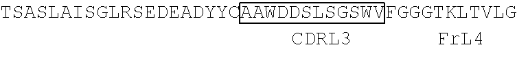
TABLE-US-00002 Clone 1 VH [SEQ ID NO: 3] ##STR00001## ##STR00002## -VL [SEQ ID NO: 16] ##STR00003## ##STR00004## CDR Regions CDRH1: NYGMH [SEQ ID NO: 29] CDRH2: VISYDGSNKYYADSVKG [SEQ ID NO: 30] CDRH3: EWRDAFDI [SEQ ID NO: 31] CDRL1: TGSSSNIGAGYDVH [SEQ ID NO: 32] CDRL2: SDNQRPS [SEQ ID NO: 33] CDRL3: AAWDDSLSGSWV [SEQ ID NO: 34] Clone 2 -VH [SEQ ID NO: 4] ##STR00005## ##STR00006## -VL [SEQ ID NO: 17] ##STR00007## ##STR00008## CDR Regions CDRH1: TYGMH [SEQ ID NO: 35] CDRH2: VIAYDGSKKDYADSVKG [SEQ ID NO: 36] CDRH3: EYRDAFDI [SEQ ID NO: 37] CDRL1: TGSSSNIGAGYDVH [SEQ ID NO: 38] CDRL2: GNSNRPS [SEQ ID NO: 39] CDRL3: AAWDDSVSGWM [SEQ ID NO: 40] Clone 3 -VH [SEQ ID NO: 5] ##STR00009## -VL [SEQ ID NO: 18] ##STR00010## ##STR00011## CDR Regions CDRH1: NYGMH [SEQ ID NO: 41] CDRH2: VISYDGSNRYYADSVKG [SEQ ID NO: 42] CDRH3: DRWNGMDV [SEQ ID NO: 43] CDRL1: SGSSSNIGAGYDVH [SEQ ID NO: 44] CDRL2: ANNQRPS [SEQ ID NO: 45] CDRL3: AAWDDSLNGPWV [SEQ ID NO: 46] Clone 4 -VH [SEQ ID NO: 6] ##STR00012## ##STR00013## -VL [SEQ ID NO: 19] ##STR00014## ##STR00015## CDR Regions CDRH1: SYGMH [SEQ ID NO: 47] CDRH2: VISYDGSDTAYADSVKG [SEQ ID NO: 48] CDRH3: DHSVIGAFDI [SEQ ID NO: 49] CDRL1: SGSSSNIGSNTVN [SEQ ID NO: 50] CDRL2: DNNKRPS [SEQ ID NO: 51] CDRL3: SSYAGSNNVV [SEQ ID NO: 52] Clone 5 -VH [SEQ ID NO: 7] ##STR00016## ##STR00017## -VL [SEQ ID NO: 20] ##STR00018## ##STR00019## CDR Regions CDRH1: NYGMH [SEQ ID NO: 53] CDRH2: VISYDGSNKYYADSVKG [SEQ ID NO: 54] CDRH3: DQLGEAFDI [SEQ ID NO: 55] CDRL1: TGSSSNIGAGYDVH [SEQ ID NO: 56] CDRL2: DNNKRPS [SEQ ID NO: 57] CDRL3: ATWDDSLSGPV [SEQ ID NO: 58] Clone 6 -VH [SEQ ID NO: 8] ##STR00020## ##STR00021## -VL [SEQ ID NO: 21] ##STR00022## ##STR00023## CDR Regions CDRH1: DYGMS [SEQ ID NO: 59] CDRH2: AISGSGSSTYYADSVKG [SEQ ID NO: 60] CDRH3: GDIDYFDY [SEQ ID NO: 61] CDRL1: TGSSSNFGAGYDVH [SEQ ID NO: 62] CDRL2: ENNKRPS [SEQ ID NO: 63] CDRL3: AAWDDSLNGPV [SEQ ID NO: 64] Clone 7 -VH [SEQ ID NO: 9] ##STR00024## ##STR00025## -VL [SEQ ID NO: 22] ##STR00026## ##STR00027## CDR Regions CDRH1: SYGMH [SEQ ID NO: 65] CDRH2: VISYDGSNKYYADSVKG [SEQ ID NO: 66] CDRH3: ERRDAFDI [SEQ ID NO: 67] CDRL1: TGSSSNIGAGYDVH [SEQ ID NO: 68] CDRL2: SDNQRPS [SEQ ID NO: 69] CDRL3: ATWDSDTPV [SEQ ID NO: 70] Clone 8 -VH [SEQ ID NO: 10] ##STR00028## ##STR00029## -VL [SEQ ID NO: 23] ##STR00030## ##STR00031## CDR Regions CDRH1: SYGMH [SEQ ID NO: 71] CDRH2: VISYDGSNKYYADSVKG [SEQ ID NO: 72] CDRH3: DHSAAGYFDY [SEQ ID NO: 73] CDRL1: SGSSSNIGSNTVN [SEQ ID NO: 74] CDRL2: GNSIRPS [SEQ ID NO: 75] CDRL3: ASWDDSLSSPV [SEQ ID NO: 76] Clone 9 -VH [SEQ ID NO: 11] ##STR00032## ##STR00033## -VL [SEQ ID NO: 24] ##STR00034## ##STR00035## CDR Regions CDRH1: SYGMH [SEQ ID NO: 77] CDRH2: GISWDSAIIDYAGSVKG [SEQ ID NO: 78] CDRH3: DEAAAGAFDI [SEQ ID NO: 79] CDRL1: TGSSSNIGAGYDVH [SEQ ID NO: 80] CDRL2: GNTDRPS [SEQ ID NO: 81] CDRL3: AAWDDSLSGPVV [SEQ ID NO: 82] Clone 13 -VH [SEQ ID NO: 15] ##STR00036## ##STR00037## -VL [SEQ ID NO: 28] ##STR00038## ##STR00039## CDR Regions CDRH1: SYGIS [SEQ ID NO: 101] CDRH2: GISGSGGNTYYADSVKG [SEQ ID NO: 102] CDRH3: SVGAYANDAFDI [SEQ ID NO: 103] CDRL1: TGSSSNIGAGYDVH [SEQ ID NO: 104] CDRL2: GDTNRPS [SEQ ID NO: 105] CDRL3: AAWDDSLNGPV [SEQ ID NO: 106] Clone 10 -VH [SEQ ID NO: 12] ##STR00040## ##STR00041## -VL [SEQ ID NO: 25] ##STR00042## ##STR00043## CDR Regions CDRH1: SYGMH [SEQ ID NO: 83] CDRH2: VISYDGSNKYYADSVKG [SEQ ID NO: 84] CDRH3: ELYDAFDI [SEQ ID NO: 85] CDRL1: TGSSSNIGAGYDVH [SEQ ID NO: 86] CDRL2: ADDHRPS [SEQ ID NO: 87] CDRL3: ASWDDSQRAVI [SEQ ID NO: 88] Clone 11 -VH [SEQ ID NO: 13] ##STR00044## ##STR00045## -VL [SEQ ID NO: 26] ##STR00046## ##STR00047## CDR Regions CDRH1: SYGMH [SEQ ID NO: 89] CDRH2: VISYDGSNKYYADSVKG [SEQ ID NO: 90] CDRH3: EFGYIILDY [SEQ ID NO: 91] CDRL1: SGSSSNIGSNTVN [SEQ ID NO: 92] CDRL2: RDYERPS [SEQ ID NO: 93] CDRL3: MAWDDSLSGVV [SEQ ID NO: 94] Clone 12 -VH [SEQ ID NO: 14] ##STR00048## ##STR00049## -VL [SEQ ID NO: 27] ##STR00050## ##STR00051## CDR Regions CDRH1: NHGMH [SEQ ID NO: 95]
CDRH2: VISYDGTNKYYADSVRG [SEQ ID NO: 96] CDRH3: ETWDAFDV [SEQ ID NO: 97] CDRL1: SGSSSNIGSNNAN [SEQ ID NO: 98] CDRL2: DNNKRPS [SEQ ID NO: 99] CDRL3: QAWDSSTVV [SEQ ID NO: 100]
Preferred target cell surface antigens may be selected from the following: CD20, Thy-1 (CD90, Cluster of Differentiation 90 (Biofactors. 2009 May-June; 35(3):258-65)); Ly-6 (Lymphocyte Antigen 6 (Mol Biol Rep. 2009 April; 36(4):697-703)); CD59 (Complement regulatory protein (Mol. Immunol. 2007 January; 44(1-3):73-81)); Fas (FS7-associated cell surface antigen, CD95, APO-1 or TNFRSF6 (Adv Exp Med. Biol. 2009; 647:64-93)); EGFR (Epidermal Growth Factor Receptor (FEBS J. 2010 January; 277(2):301-8)); Her2 (Human epidermal growth factor receptor 2 (Clin Breast Cancer. 2008 October; 8(5):392-401)); CXCR4 (Chemokine Receptor 4 (Biochim Biophys Acta. 2007 April; 1768(4):952-63)); CD19 (Cluster of Differentiation 19 (Cell Immunol. 1989 February; 118(2):368-81)); CD40 (Cluster of Differentiation 40 (Basic Clin Pharmacol Toxicol. 2009 February; 104(2):87-92)); HLA Molecules (Human Leukocyte Antigen molecules (Korean J Lab Med. 2010 June; 30(3):203)); GM1 (ganglioside, monosialotetrahexosylganglioside (J Lipid Res. 2010 September; 51(9):2731-8)); CD22 (Cheson (2008) NEJM 359(6): 613-26); CD23 (Cheson, 2008); CD80 (Cheson, 2008); CD74 (Cheson, 2008); DRD (Cheson, 2008).
Preferably, in a composition, use, or method according to the invention the surface antigen is selected from CD19, CD20, or CD40 and more preferably, human forms thereof. CD20, especially human CD20, is most preferred.
Advantageously, the antibody molecule that specifically binds the cell surface antigen is a monoclonal antibody, preferably a monoclonal antibody that upon binding to the target cell is removed from the cell surface and internalized into the target cell in an Fc.gamma.RIIb-dependent manner. Preferably the monoclonal antibody is an anti-CD19, anti-CD20 or anti-CD40 antibody. Most preferably, the monoclonal antibody is an anti-CD20 monoclonal antibody.
In a preferred embodiment, the antibody molecule specifically binding to a cell surface antigen is a Type I anti-CD20 antibody. In another preferred embodiment, the antibody molecule specifically binding to a cell surface antigen is not a Type II anti-CD20 antibody.
In one embodiment, the cell surface antigen is CD20 and the antibody molecule specifically binding to a cell surface antigen is a Type I antibody.
As mentioned above, there are two types of anti-CD20 monoclonal antibodies (mAb). Anti-CD20 mAb were first defined by the inventors as falling into different groupings in 2003 (43 and 25) and then subsequently defined as Type I and II mAbs in 2004 (26). Initially the basis for this was that anti-CD20 mAb fall into two distinct types of reagents based on their ability to eradicate lymphoma xenografts: type I (e.g. Rituximab and 1F5) utilize complement; and type II (e.g. B1), do not. Both types of mAb gave excellent prolongation of survival, but depleting complement activity, by administering CVF, considerably diminished the potency of Rituximab and 1F5, but had no effect on the activity of B1. These results clearly showed that different CD20 mAb operate different effector mechanisms in vivo. Furthermore, they are in complete accord with previous work showing that Rituximab and 1F5 are able to activate complement efficiently as a result of translocating CD20 to lipid rafts in the target cell membrane, something that B1-type mAb cannot do (43). There is an excellent correlation with the ability of mAb to engage complement and induce CD20 to move into lipid rafts (43, 26). Therefore Type I and II nature can be defined by their ability to move CD20 into lipid rafts. This can be determined as indicated below. There is also a correlation with Type II mAb being able to elicit more potent homotypic adhesion and direct cell death but these could not be used alone to define a Type I or II mAb (unlike the Tx-100 raft assays; see below).
Therefore, various anti-CD20 mAb may be classified as type I (e.g. rituximab, ofatumumab) or type II (e.g. tositumomab (B1), GA101, 11B8) according to their ability to redistribute CD20 in the plasma membrane and their activity in various effector assays (25-27). Type I (e.g. rituximab, ofatumumab) anti-CD20 monoclonal antibodies induce CD20 to redistribute into large detergent resistant microdomains (rafts), whereas type II (tositumomab-like) anti-CD20 monoclonal antibodies do not (50).
As discussed above, anti-CD20 mAbs can be designated as Type I or Type II by virtue of whether they redistribute CD20 into lipid rafts. This is done by the Tx-100 insolubility assay or by sucrose density gradient separation and western blotting. Both methods are described in Cragg et al Blood 2003 (43) as follows:
1. Assessment of Raft Associated Antigen by Triton X-100 Insolubility
As a rapid assessment of antigen presence in raft microdomains, we utilised a flow cytometry method based on Triton X-100 insolubility at low temperatures. In brief, cells were washed in RPMI/1% BSA and resuspended at 2.5.times.10.sup.6/ml. Cells were then incubated with 10 .mu.g/ml of an FITC conjugated mAb for 15 minutes at 37.degree. C., washed in cold PBS/1% BSA/20 mM sodium azide, and then the sample divided in half. One half was maintained on ice to allow calculation of 100% surface antigen levels, whilst the other was treated with 0.5% Triton X-100 for 15 minutes on ice to determine the proportion of antigens remaining in the insoluble raft fraction. Cells were then maintained at 4.degree. C. throughout the remainder of the assay, washed once in PBS/BSA/azide, resuspended and assessed by flow cytometry as detailed above. Similar results were obtained using indirect methods of detection. To determine the constitutive level of raft association of target antigens, cells were first treated with 0.5% Triton X-100 for 15 minutes on ice and washed in PBS/BSA/azide prior to binding of FITC-labeled mAb. To assess whether more antigen could be moved into the Triton X-100 insoluble fraction by additional cross-linking, cells were incubated with FITC-mAb as before, washed and then divided into four. Two of these samples were incubated with goat anti-mouse Ig F(ab').sub.2 fragments for 15 minutes on ice. After washing, one of the cross-linked and one of the non-cross-linked samples were lysed in Triton X-100 and washed as detailed above prior to flow cytometry.
2. Sucrose Density Gradient Separation and Western Blotting--Preparation of Lipid Raft Fractions and Western Blotting
Monoclonal Ab (1 .mu.g/10.sup.6 cells) was added to cells at 37.degree. C. Following 20 minutes incubation, cells were pelleted and lysed in ice-cold 1.0% Triton X-100 in MES-buffered saline (25 mM MES, pH 6.5, 150 mM NaCl, 1 mM phenylmethylsulfonyl fluoride, 5 .mu.g/ml aprotinin, 5 .mu.g/ml leupeptin, 10 mM EDTA). Lipid raft fractions were then prepared by sucrose density gradient centrifugation. Briefly, Lysates were mixed with an equal volume of 80% sucrose in lysis buffer, overlaid with a discontinuous 5-30% sucrose density gradient and then centrifuged at 200,000.times.g for 16 h. Fractions (0.5 ml) were collected and analysed by Western blotting. 15 ml aliquots of each fraction were diluted 1:1 in 2.times. loading buffer, heated to 95.degree. C. for 5 min and separated on 15% SDS-PAGE gels, before transfer onto PVDF membranes and incubated with primary antibody (for example mouse anti-CD20, clone 7D1 to detect CD20 or anti-Lyn rabbit polysera; Serotec, UK to identify the raft fractions), followed by HRP-conjugated secondary antibody (Amersham Biosciences UK Ltd). Blots were visualised using ECL+plus (Amersham Biosciences UK Ltd).
Anti-CD20 mAbs can require the AxP motif in the large loop of CD20. (Ofatumumab and other Genmab antibodies do not). However, (Niederfelner et al., (51)) indicates Type II mAb bind to a slightly different region of the CD20 loop compared to Type I.
Preferably, in a composition, use, or method according to the invention the target cell is a cancer cell. More preferably, a cancer selected from non-Hodgkin lymphoma, including but not limited to follicular lymphoma, diffuse large B cell lymphoma, mantle cell lymphoma, or chronic lymphocytic leukaemia.
In one embodiment the invention provides compositions, uses and methods for treating cancer, in particular a B cell malignancy which is preferably selected from lymphomas, chronic lymphocytic leukemias, acute lymphoblastic leukemias, multiple myeloma, Hodgkin's and non-Hodgkin's disease, diffuse large B cell lymphoma, follicular lymphoma with areas of diffuse large B cell lymphoma, small lymphocytic lymphoma, mantle cell lymphoma, and diffuse small cleaved cell lymphoma or combinations thereof. In certain embodiments, the B cell malignancy is a lymphoma, such as non-Hodgkin's (NHL).
In another embodiment the invention provides compositions, uses and methods for treating an inflammatory disease. This may be an autoimmune disease, such as Hashimoto's thyroiditis, pernicious anemia, Adison's disease, type 1 diabetes, rheumatoid arthritis, systemic lupus erythematosus, dermatomyositis, Sjogren's syndrome, dermatomyositis, lupus erythematosus, multiple sclerosis, autoimmune inner ear disease myasthenia gravis, Reiter's syndrome, Graves disease, autoimmune hepatitis, familial adenomatous polyposis and ulcerative colitis or combinations thereof. In specific embodiments, the autoimmune disease is rheumatoid arthritis or systemic lupus erythematosus.
In preferred embodiments the treatment is of diseases including, chronic lymphocytic leukemia (CLL), non-Hodgkin lymphoma (NHL), B cell malignancies, Rheumatoid arthritis, systemic lupus erythematosus, dermatomyositis, systemic sclerosis and autoimmune blistering diseases.
In a preferred embodiment the treatment that is enhanced by use of the invention is treatment with an anti-CD20 mAb, such as rituximab.
DEFINITIONS
By "elevated" we include the meaning that the cells in question express higher levels of Fc.gamma.RIIb on their surface than control or reference cells that express low or medium levels of Fc.gamma.RIIb on their surface. For example, if the cells in question are a type of B cell, "elevated" levels of Fc.gamma.RIIb expression would be recognized if the level of expression was higher than the normal (preferably median) level of expression of Fc.gamma.RIIb by B cells of the same cell type. Alternatively, "elevated" levels would be recognized if the cells in question expressed Fc.gamma.RIIb at a level higher than a different cell type which expresses Fc.gamma.RIIb at low or medium level.
According to the invention, the greater the degree of elevation in Fc.gamma.RIIb expression of target cells, the worse the predicted response of those cells to treatment with an antibody molecule that binds specifically to a surface antigen of the target cells and that has an Fc domain capable of binding Fc.gamma.RIIb. Hence, the greater the elevation in Fc.gamma.RIIb expression, the greater the benefit to be gained from the use of an agent that prevents or reduces binding of the Fc domain to Fc.gamma.RIIb according to the invention as demonstrated in FIGS. 2D and 3A. After measuring Fc.gamma.RIIb levels by IHC (FIG. 10B) and separating MCL samples into positive and negative for Fc.gamma.RIIb, a clear clinical difference in response was seen following rituximab-based therapy. (FIGS. 10C and 10D).
Skilled persons can readily determine levels of expression of Fc.gamma.RIIb on cells by a variety of known methods, such as the flow cytometry and immunohistochemical staining methods described in the accompanying Figures and examples.
A skilled person would appreciate that the "normal" and "elevated" expression level of Fc.gamma.RIIb will vary between different cell types and different disease states and would be capable of identifying a "normal" and an "elevated" expression level of Fc.gamma.RIIb for a given target cell or disease state using methods well known in the art and described herein. Exemplary levels of "normal" (or median) and "elevated" levels of Fc.gamma.RIIb expression on certain cell types are provided in FIG. 2C. In these particular examples, in Follicular Lymphoma (FL) "normal" levels are approximately 50 (ratio of Geo MFI Fc.gamma.RIIb to isotype control) and "elevated" levels are approximately 125 or 400 or more, whilst in Diffuse large B-cell lymphoma (DLBCL) "normal" levels are approximately 20 and "elevated" levels are approximately 80 or more, whilst in Mantle cell lymphoma (MCL) "normal" levels are approximately 60 and "elevated" levels are approximately 110 or 190 or more, and in chronic lymphoid leukemia (CLL) "normal" levels are approximately 100 and "elevated" levels are approximately 300 or more.
Preferably, the elevated Fc.gamma.RIIb expression level is at least 1.1 fold increased over the normal (preferably median) expression level of cells of the same cell type (or of cells of a different cell type), or at least 1.2, 1.3, 1.4, 1.5, 1.6, 1.7, 1.8, 1.9, 2.0, 2.1, 2.2, 2.3, 2.4, 2.5, 2.6, 2.7, 2.8, 2.9, 3.0, 3.1, 3.2, 3.3, 3.4, 3.5, 3.6, 3.7, 3.8, 3.9, 4.0, 4.1, 4.2, 4.3, 4.4, 4.5, 4.6, 4.7, 4.8, 4.9, 5.0, 5.1, 5.2, 5.3, 5.4, 5.5, 5.6, 5.7, 5.8, 5.9, 6.0, 6.1, 6.2, 6.3, 6.4, 6.5, 6.6, 6.7, 6.8, 6.9, 7.0, 7.1, 7.2, 7.3, 7.4, 7.5, 7.6, 7.7, 7.8, 7.9, 8.0, 8.1, 8.2, 8.3, 8.4, 8.5, 8.6, 8.7, 8.8, 8.9, 9.0, 9.1, 9.2, 9.3, 9.4, 9.5, 9.6, 9.7, 9.8, 9.9, 10.0, 10.5, 11.0, 11.5, 12.0, 12.5, 13.0, 13.5, 14.0, 14.5, 15.0, 15.5, 16.0, 16.5, 17.0, 17.5, 18.0, 18.5, 19.0, 19.5, 20.0. 21.0, 22.0, 23.0, 24.0 or 25.0 or more fold increased over the normal (or median) expression level of cells of the same cell type or of cells of a different cell type. Preferably they are at least 1.8 fold increased over the normal (or median) expression level of cells of the same cell type or of cells of a different cell type
As shown in the accompanying examples, the inventors have determined that the level of Fc.gamma.RIIb expression correlates with an increased modulation of mAb from the cell surface. Even higher levels of Fc.gamma.RIIb expression give an even higher modulation, i.e. there is a correlation between degree of elevation of expression and modulation as shown in FIGS. 2D and 3A. In FIG. 2D the expression level of Fc.gamma.RIIb is plotted versus the level of modulation observed. The correlation is such that the highest levels of Fc.gamma.RIIb (e.g.>400) results in the lowest level of surface CD20 (<20%) i.e. the greatest effect on modulating the mAb from the cell surface. In FIG. 3A, low (18), medium (70) and high (124) levels of Fc.gamma.RIIb were introduced into an Fc.gamma.RIIb-negative cell line (Ramos) and this was directly correlated with reducing levels of cell surface CD20 (60, 40, 30% for low medium and high) in proportion to the expression of the Fc.gamma.RIIb. Fc.gamma.RIIb expression levels are given as a ratio of the Geometric Mean Fluorescent Intensity (Geo MFI) of Fc.gamma.RIIb to isotype control
Modulation reduces the amount of mAb left on the cell surface. mAbs require the Fc to engage with immune effector mechanisms (ADCC, ADCP, CDC) in order to delete target cells. Therefore reducing modulation with Fc.gamma.RIIb blocking mAb will improve Fc-dependent effector functions. This is shown for ADCP (phagocytosis) in FIG. 8. Phagocytosis with rituximab alone was 40% but rose to 55% when the Fc.gamma.RIIb was blocked by AT10.
By "antibody molecule" we include monoclonal antibodies, synthetic antibodies, recombinantly produced antibodies, multispecific antibodies, human antibodies, chimeric antibodies, camelized antibodies, single-chain Fvs (scFv), single chain antibodies, Fab fragments, F(ab') fragments, disulfide-linked Fvs (sdFv), intrabodies, or epitope-binding fragments of any of the above. Preferably, the antibodies of the invention are monoclonal antibodies, and more preferably, humanized or human antibodies.
Methods for the preparation and characterization of antibody molecules which are useful in the compositions, use and methods of the present invention are well known to skilled persons. For example, WO 2008/002933, in Section 5.3 to 5.3.1 pages 74-91, describes the preparation and characterization of monoclonal antibodies that bind specifically to a target cell surface antigen such as CD20 or Fc.gamma.RIIb. Useful antibodies that specifically bind Fc.gamma.RIIb and CD20, including monoclonal antibodies produced by hybridomas deposited under the Budapest Treaty, are also disclosed on pages 15 to 21 of WO 2008/002933, the disclosure of which is incorporated herein by reference.
By "specifically binds" we include agents such as antibody molecules that bind to a target antigen but do not bind (cross-react) to other antigens or bind such antigens more weakly, i.e. with a lower affinity than the target antigen. For example, an antibody that specifically binds to Fc.gamma.RIIB may bind to other peptides or polypeptides with lower affinity as determined by, e.g., immunoassays, BIAcore, or other assays known in the art. Preferably, antibodies that specifically bind to Fc.gamma.RIIB or a fragment thereof do not cross-react with other antigens. Antibodies that specifically bind to Fc.gamma.RIIB can be identified, for example, by immunoassays, BIAcore, or other techniques known to those of skill in the art. An antibody or a fragment thereof binds specifically to a Fc.gamma.RIIB when it binds to Fc.gamma.RIIB with higher affinity than to any cross-reactive antigen as determined using experimental techniques, such as western blots, radioimmunoassay (RIA) and enzyme-linked immunosorbent assays (ELISAs) (See Fundamental Immunology Second Edition, Raven Press, New York at pages 332-336 (1989) for a discussion regarding antibody specificity) and binds Fc.gamma.RIIB, particularly human Fc.gamma.RIIB, more particularly native human Fc.gamma.RIIB, with a greater affinity than said antibody or a fragment thereof binds Fc.gamma.RIIA, particularly human Fc.gamma.RIIA, more particularly native human Fc.gamma.RIIA. Representative antibodies are disclosed in US Patent Application Nos. 2004-0185045; 2005-0260213; and 2006-0013810, which are all herein expressly incorporated by reference in their entireties.
Preferably certain Fc.gamma.RIIB antibodies used in combination with CD20 antibodies in the compositions and methods of the invention bind the extracellular domain of native human Fc.gamma.RIIB. In some embodiments, the antibody or a fragment thereof binds Fc.gamma.RIIB with at least 2 times greater affinity than said antibody or a fragment thereof binds Fc.gamma.RIIA. In other embodiments, the antibody or a fragment thereof binds Fc.gamma.RIIB with at least 4 times, at least 6 times, at least 8 times, at least 10 times, at least 100 times, at least 1000 times, at least 10.sup.4, at least 10.sup.5, at least 10.sup.6, at least 10.sup.7, or at least 10.sup.8 times greater affinity than said antibody or a fragment thereof binds Fc.gamma.RIIA. In a preferred embodiment, said antibody or a fragment thereof bind Fc.gamma.RIIB with 100 times, 1000 times, 10.sup.4 times, 10.sup.5 times, 10.sup.6 times, 10.sup.7 times, 10.sup.8 times greater affinity than said antibody or a fragment thereof binds Fc.gamma.RIIA.
BRIEF DESCRIPTION OF THE DRAWINGS
FIGS. 1A-1B. Type I mAb internalize from the cell surface of normal and malignant human B-cells.
FIG. 1A) Primary CLL cells were cultured with Tosit-488, GA101 gly-488, Ritux-488 or Ofatum-488 (all at 5 .mu.g/ml) for 2 or 6 h. Cells were then harvested and washed twice before addition of anti-Alexa-488 to one half of the sample for 30 min at 4.degree. C. to discriminate between internalized and non-internalized mAb. Surface accessible CD20(%) was determined using the following calculation: Surface accessible CD20+(Pre-quench Geo MFI-Post-quench GEO MFI)/(Pre-quench Geo MFI).times.100. Each point represents a sample from a different CLL patient. Statistical analysis was performed using the Wilcoxon paired test, **p value<0.001, and medians are shown.
FIG. 1B) A variety of primary B cell tumors and normal B cells from healthy volunteers was then examined in the same assay with Tosit-488 or Ritux-488. Statistical analysis was performed using the Mann Whitney test and medians are shown.
Supplementary FIGS. 1A-1E. Lack of correlation between CD20 modulation and CLL phenotypic/prognostic markers. Supplementary FIG. 1A. Correlation between modulation and known CLL prognostic factors: CLL cases were phenotyped for IgVH gene mutational status, Zap-70 and CD38 expression, and internalization assays were performed on these samples to assess modulation. Correlation between each prognostic feature and CD20 modulation was performed by Spearman's correlation analysis. No correlation was seen with each prognostic factor (p>0.05). Supplementary FIG. 1B. Similarly, sIg status, ability of cells to elicit calcium flux and viability of CLL cells were assessed, and compared with CD20 modulation. Again, no correlation was seen. Supplementary FIG. 1C. CD20 expression of CLL cells were assessed by FACS using Ritux-488, and compared with CD20 modulation. A weak correlation was seen (Spearman r value -0.34, p=0.038). Subsequent analysis using multivariate regression of CD20 and Fc.gamma.RIIb expression against CD20 modulation showed that the weak correlation with CD20 was not significant (p=0.638). Supplementary FIG. 1D. sIg expression of IgM-positive CLL cases were determined by FACS and the level of expression compared with CD20 modulation. No correlation was found (p>0.05). Supplementary FIG. 1E. CLL cases were cultured with Rit m2a-488 for 2 h and internalization assay performed as in FIG. 1A. Within a single CLL case, variation in CD38 expression was seen. The FACS plots show samples pre-quenching (left) and post-quenching (right). The corresponding histograms highlight that CD38.sup.+ve and CD38.sup.-ve cells within a single sample modulated at the same rate. CD38.sup.+ve and CD38.sup.-ve cells are represented by solid and hollow peaks respectively. These results are representative of 3 different cases.
FIGS. 2A-2D. Modulation is an Fc-dependent process. FIG. 2A. The internalization assay described in 1a) was repeated after culturing CLL cells for 6 h with Alexa-488-labelled fragments of rituximab; Fab', F(ab').sub.2 and IgG. Data represent the mean levels of modulation+/-SD for 3 different CLL samples. FIG. 2B. CLL cells were cultured with Tosit-488, Ritux-488+/-AT10 and Rit m2a-488 as in 1a) for 2 and 6 h. Mean+/-modulation are shown from 6 different CLL samples. Addition of anti-Fc.gamma.RII mAb AT10, to rituximab, reduced CD20 modulation to levels similar to Rit m2a, whereas addition of AT10 to Rit m2a made no significant difference to modulation. FIG. 2C A variety of normal and malignant B cell samples were stained for Fc.gamma.RIIb expression with AT10-PE. The histogram shows the diversity of Fc.gamma.RIIb expression in 3 different CLL cases, representing nominal high (black line), medium (dark gray line) and low expressors (light gray line). The scatterplot shows differences in Fc.gamma.RIIb expression across healthy B cells, CLL, SLL, MCL, FL and SLBCL. Fc.gamma.RIIb expression was expressed as a ratio of Fc.gamma.RIIb:isotype control Geo MFI to control small differences due to inter-experimental variation. Median values are shown. FIG. 2D. Fc.gamma.RIIb expression was plotted against CD20 modulation across all NHL subtypes and normal B cells (obtained from internalization assay described in 1A) and co-cultured with Ritux-488 for 6 h). Analysis was performed using a Spearman's correlation assuming a non-parametric distribution. A strong correlation was demonstrated. Spearman r value=-0.74, 95% confidence interval between -0.83 and -0.61 and p<0.0001.
FIGS. 3A-3B. Fc.gamma.RIIb expression is a major determinant of CD20 modulation. FIG. 3A. Ramos cells transfected with Fc.gamma.RIIb were sorted to express low, medium and high levels of Fc.gamma.RIIb and assessed in the internalization assay using Tosit-488 and Ritux-488 at the 6 h time-point alongside mock-transfected cells. The bars represent means values+/-SD from independent experiments. Geo MFI values for Fc.gamma.RIIb expression of the sorted cells are listed on the right. FIG. 3B. The internalization assay was repeated using Tosit-488 and Ritux-488 on normal Ramos cells, Rx3 cells (which lack BCR expression), mock-transfected Rx3 cells and Fc.gamma.RIIb-transfected Rx3 cells following incubation for 6 h. Data points from 5 independent experiments are shown along with the median value.
FIGS. 4A-4E. CD20 and Fc.gamma.RIIb co-ligation occurs predominantly in a cis fashion and leads to activation of Fc.gamma.RIIb. FIG. 4A. Raji cells were cultured with the specified mAb (10 .mu.g/ml) for 2 h at 37.degree. C. before harvesting, lysis and subsequent immunoblotting for phosphorylated Fc.gamma.RIIb. FIG. 4B Left panel; PKH26-labeled Ramos cells (Fc.gamma.RIIb.sup.-ve, R1) were mixed 1:1 with sorted high Fc.gamma.RIIb-expressing Ramos transfectants (R2) (described in FIG. 3A). Right panel; modulation of CD20 on Fc.gamma.RIIb.sup.+ve and Fc.gamma.RIIb.sup.-ve cells after culture with Ritux-488 for 6 h. As controls, both populations were also cultured alone. Data represent the mean levels of modulation+/-SD from 3 independent experiments. FIG. 4C. In a similar experiment, a low Fc.gamma.RIIb-expressing CLL was PKH26-labeled then mixed 1:1 with a higher Fc.gamma.RIIb-expressing CLL. The experiment was performed three times, each time with a different high Fc.gamma.RIIb-expressing CLL. Fc.gamma.RIIb levels (Geo MFI) were 42 (low) and 275, 306 and 165 for the high expressors. The internalization assay was then performed as in 4B. Data represent the mean levels of modulation+/-SD. FIG. 4D. Different CLL samples were cultured with Ritux-488 for 6 h at 20, 4 and 1.times.10.sup.5 cells/ml, and the internalization assay performed at 6 h as before. FIG. 4E. Raji cells were cultured at 20, 4, and 1.times.10.sup.5 cells/ml with the specified mAb (10 .mu.g/ml) for 2 h at 37.degree. C. Images were captured using a bright field microscope to demonstrate differences in cell proximity. Cells were then harvested and assessed by immunoblotting for phosphorylated Fc.gamma.RIIb as described in FIG. 4A.
FIGS. 5A-5E. Rituximab, CD20 and Fc.gamma.RIIb internalize together into lysosomes. FIG. 5A. CLL cells were incubated with either Tosit-488 or Ritux-488 for 2 h and then stained with anti-CD19-APC and AT10-PE. Data show the mean+/1 SD for Fc.gamma.RIIb expression as a % of untreated (n+6 CLL samples). Fc.gamma.RIIb expression after Ritux-488 was significantly lower than after Tosit-488, * p<0.05. FIG. 5B. CLL cells were washed, fixed and permeabilized before staining with AT10-647 (blue), washing and analysis with confocal microscopy. This image represents the Fc.gamma.RIIb staining pattern in unstimulated cells. FIG. 5C. The same CLL sample was cultured with Ritux-488 for 30 min then treated as described in 5b). Cells at this time-point displayed obvious co-localization between Ritux-488 (green) with AT10-647 (blue). FIG. 5D. CLL cells were incubated with Tosit-488 for 6 h before preparation for microscopy as in 5B. In addition, cells were also stained with biotinylated LAMP-1 and streptavidin-546 (red) to stain for lysosomes. Tosit-488 remained uniformly on the surface and AT 10-647 staining was unchanged from baseline seen in 5B. There was no co-localization with LAMP-1. FIG. 5E. CLL cells were treated with Ritux-488 for 6 h and assessed as in FIG. 5D. Two representative cells are shown here. The top cell shows unambiguous co-localization between Ritux-488 and AT10-647, but no co-localization with LAMP-1. The bottom cell shows co-localizaton of all three fluorochromes. In each case the bright field (BF) image is shown from the same cell. Scale bars represent 5 .mu.m.
FIG. 6. Lack of inhibitory receptor augments the depleting ability of anti-CD20 mAb. hCD20 Tg mice (WT) or hCD20 Tg mice lacking CD32 (CD32KO) were treated with rituximab variants harboring mouse IgG1 (m1) or mouse IgG2a (m2a); 250 mg iv and then b cell depletion monitored by low cytometry for 90 days through serial bleeding of the mice and staining with B220 and CD19 mAb.
FIG. 7. Blocking the inhibitory receptor CD32b (Fc.gamma.RIIb) augments the efficacy of anti-CD20 mAb in a human xenograft system. CD20 positive human tumour cells (Daudi or Raji) were inoculated into SCID mice and then treated with either rituximab, AT10 or both and survival of the mice or tumour growth monitored. Doses of mAb used are shown in the figure legends. In A) Daudi cells were inoculated subcutaneously and the tumour monitored by caliper measurements every 305 days. In B) and C) Raji cells were inoculated intravenously.
FIG. 8. Enhanced phagocytosis of CLL cells treated with rituximab by co-incubation with a Fc.gamma.RII blocking mAb. Monocytes were derived from healthy volunteers and differentiated into macrophages with M-CSF in a 6-well plate for a minimum period of 7 days prior to use. Macrophages were then harvested and allowed to adhere in a 96-well plate at 5.times.10.sup.5 cells/well for a minimum of 2 hours prior to addition of CFSE-labelled CLL cells. CSFE-labelled CLL cells were untreated or opsonised with 10 .mu.g/ml of rituximab (ritux) and the Fc.gamma.RIIb blocking mAb AT10 (fab').sub.2 for either 15 mins or 6 h before washing twice and adding to macrophages (1:1 ratio) for at least 30 mins. Anti-CD16 f(ab).sub.2-APC (5 .mu.g/ml) was then added to each well for 15 mins at Room Temperature (RT) to stain macrophages and then wells washed once with Facs wash (PBS BSA azide) at RT. Further ice cold FACS wash was added and the plate incubated on ice for 10 minutes prior to harvesting for analysis by Facs. % Double-positive macrophages represent % of CD16+CFSE+ positive cells, expressed as a total of % CD16+ cells. N=3 replicates, line represents mean. The data clearly show that phagocytosis of CLL cells is higher when the rituximab has been added for only 15 minutes, compared to 6 h. This decrease in efficacy is associated with modulation of the rituximab from the cell surface and can be reversed by treatment with the Fc.gamma.RIIb blocking mAb AT10. Importantly, the Fc.gamma.RIIb mAb is only added to the CLL cells and so has no effect on the macrophages themselves. Furthermore, only Fab2 fragments of AT10 are used and therefore increased phagocytosis cannot occur as a result on more mAb being bound to the CLL cell surface. This conclusion is also supported by the observation that no increase in phagocytosis is observed following incubation for only 15 minutes (a time at which v little rituximab modulation would have occurred).
FIG. 9. Fc.gamma.RIIb expression by IHC. Paraffin-embedded tissues were stained for Fc.gamma.RIIb expression using anti-CD32b specific mAb, EP888Y. Images from 4 different FL patients are shown (.times.40 total magnification, and .times.150 magnification inset). Fc.gamma.RIIb expression was also examined by flow cytometry by staining matching viable cells with AT10-PE. Fc.gamma.RIIb expression obtained by flow cytometry is shown on the top left corner of each image.
FIGS. 10A-10D. Fc.gamma.RIIb levels predict clinical outcome in rituximab-treated MCL patients. As proof-of-concept of our in vitro findings, we retrospectively examined the Fc.gamma.RIIb expression of a cohort of MCL patients who had received rituximab. Diagnostic paraffin-embedded tissue was stained by immunohistochemistry using an Fc.gamma.RIIb-specific mAb (FIG. 11). Strong membrane staining was seen in Fc.gamma.RIIb+ve but not Fc.gamma.RIIb-ve lymphoma samples. The Fc.gamma.RIIb staining shown in FIGS. 10A and 10B by IHC correlated with the Fc.gamma.RIIb expression shown by flow cytometry as shown in FIG. 2D (with the value determined by flow cytometry in 2D shown as the number inset in FIGS. 10A and 10B). These results correlated with Fc.gamma.RIIb expression of corresponding DMSO-frozen samples, obtained by flow cytometry (FIG. 2C, inset values). Despite studying only a small cohort of 16 MCL patients, patients with Fc.gamma.RIIb-ve lymphoma had significantly better median progression-free survival than those with Fc.gamma.RIIb+ve cells (median 852 and 189 days, respectively). FIG. 10C shows the differences in survival in the Fc.gamma.RIIb + and - subsets. The groups were comparable in terms of clinical features (MCL international prognostic index, data not shown), but there was heterogeneity in chemotherapy types used. In order to address this, we examined the results in those patients treated with either single-agent rituximab or fludarabine, cyclophosphamide and rituximab (FCR) for initial therapy, and similar results were observed. FIG. 10D shows the differences in survival in the Fc.gamma.RIIb + and - subsets after the patient cohorts were further controlled as discussed.
FIG. 11. Confirmation of specificity of anti-Fc.gamma.RIIb mAb used in immunohistochemistry
Fc.gamma.RIIa and Fc.gamma.RIIb transfected Ramos cells were cytospun and paraffin-embedded. Immunohistochemistry using mAb to human Fc.gamma.RIIb demonstrated strong membrane staining in Fc.gamma.RIIb-transfected Ramos but no staining in Fc.gamma.RIIa-transfected cells.
FIG. 12. CD32B specific clones inhibit Rituximab internalization.
Ability of anti-CD32b mAb to block modulation of rituximab. The Y axis shows surface accessible CD20(%). Rituximab-alexa 488 was added to Ramos cells transfected with CD32B in the presence or absence of different CD32b blocking mAb (WT or 297Q (nq) mutants) and modulation assessed after 1, 2, 6 and 24 h. As a control for the blocking ability of CD32 mAb we also included the dual CD32a and b specific mAb, AT10 (IgG and Fab2 fragments (Fab)), alongside a negative control, isotype matched irrelevant mAb (iso wt or nq). Finally, ALEXa 488-labeled B1 was included as a control mAb that does not modulate rapidly. The data clearly indicate that all 3 nCoDeR.COPYRGT. mAb (C1, C11 and C13) are able to block the modulation of rituximab in either the wt or 297q format. In particular the C11 mAb was extremely effective.
FIG. 13. Ability of anti-CD32b mAb to block modulation of rituximab. All 13 mAb (nq) and C11 as a wt
Rituximab-alexa 488 was added to Ramos cells transfected with CD32B in the presence or absence of different CD32b blocking mAb (WT or 297Q mutants) and modulation assessed after 1, 2, 6 and 24 h. The Y axis shows surface accessible CD20(%). As a control for the blocking ability of CD32 mAb we also included the dual CD32a and b specific mAb, AT10 (IgG and Fab2 fragments (Fab)), alongside a negative control, isotype matched irrelevant mAb (iso wt or nq). Finally, ALEXa 488-labeled B1 was included as a control mAb that does not modulate rapidly. In addition, control CD32 negative Ramos cells were included to allow estimation of the maximal effect of the CD32 blocking mAb. The data clearly indicate that all the majority of nCoDeR.COPYRGT. mAb were able to block the modulation of rituximab. In particular the C10 and C11 mAb were extremely effective and appeared to block modulation almost completely even at 24 h.
FIG. 14A. Correlation between affinity and the ability of ant-CD32b blocking mAb to prevent phosphorylation of CD32b after rituximab binding.
The relative affinity of the mAb was determined by a dose titration experiment measuring mAb binding to CD32B transfected CHO cells. Briefly, CD32B transfected adherent CHO K1 cell were seeded into FMAT plates. IgG were titrated in 1:2 dilutions from 30 nM to approximately 0.015 nM and left to bind for 1 h at room temperature. After washing bound IgG were detected with anti-human-IgG-APC. Finally, the plates were washed and read in the FMAT (Applied Biosystems). This gives an EC50 value for mAb binding to target expressing cells and can be translated to a relative affinity. This relative affinity was then correlated with the ability of anti-CD32b blocking mAb to prevent phosphorylation of CD32b after rituximab binding. This was determined by stimulating cells with rituximab in the presence or absence of anti-CD32b mAb and then performing western blotting for phospho-CD32b. The CD32b mAb were then ranked according to their ability to block the CD32 phosphorylation with 1 being the most effective. There was evidently a close correlation between the affinity of the mAb and the ability to block CD32b phosphorylation.
FIG. 14B. Correlation between affinity and the ability of ant-CD32b blocking mAb to prevent modulation of rituximab.
Correlation between affinity of the ant-CD32b blocking mAb and their ability to prevent rituximab modulation. The relative affinity of the mAb was determined by a dose titration experiment measuring mAb binding to CD32B transfected CHO cells. Briefly, CD32B transfected adherent CHO K1 cell were seeded into FMAT plates. IgG were titrated in 1:2 dilutions from 30 nM to approximately 0.015 nM and left to bind for 1 h at room temperature. After washing bound IgG were detected with anti-human-IgG-APC. Finally, the plates were washed and read in the FMAT (Applied Biosystems). This gives an EC50 value for mAb binding to target expressing cells and can be translated to a relative affinity. This relative affinity was then correlated with the ability of ant-CD32b blocking mAb to prevent modulation of rituximab on CD32b-transfected Ramos cells (shown in the previous figure). There was evidently a strong correlation between the affinity of the mAb and the ability to block rituximab modulation. This data confirms the central role of CD32B in accelerating the modulation of rituximab from the target cell surface.
FIG. 15. As mediated by clone 1. Dose dependent binding to hCD32B transfected cells and dose dependent binding and inhibition of immune complex to hCD32B transfected cells.
Circles show dose dependent binding to hCD32A transfected CHO K1 cells and black diamonds show dependent binding to hCD32B transfected CHO K1 cells. Crosses show dose dependent inhibition of immune complex to hCD32B transfected CHO K1 cells. As mediated by clone 1. Cells were seeded into FMAT plates. Immune complexes were prepared by coating FITC to BSA and thereafter mix this with a 10:1 molar ratio with a FITC specific hIgG1 antibody. The total intensity reflects binding, the higher intensity the higher binding. The binding is either immune complex (IC) or mAb's.
FIG. 16. As mediated by clone 2. Dose dependent binding to hCD32B transfected cells and dose dependent binding and inhibition of immune complex to hCD32B transfected cells.
Circles show dose dependent binding to hCD32A transfected CHO K1 cells and black diamonds show dependent binding to hCD32B transfected CHO K1 cells. Crosses show dose dependent inhibition of immune complex to hCD32B transfected CHO K1 cells. As mediated by clone 2. Cells were seeded into FMAT plates. Immune complexes were prepared by coating FITC to BSA and thereafter mix this with a 10:1 molar ratio with a FITC specific hIgG1 antibody. The total intensity reflects binding, the higher intensity the higher binding. The binding is either immune complex (IC) or mAb's.
FIG. 17. As mediated by clone 3. Dose dependent binding to hCD32B transfected cells and dose dependent binding and inhibition of immune complex to hCD32B transfected cells.
Circles show dose dependent binding to hCD32A transfected CHO K1 cells and black diamonds show dependent binding to hCD32B transfected CHO K1 cells. Crosses show dose dependent inhibition of immune complex to hCD32B transfected CHO K1 cells. As mediated by clone 3. Cells were seeded into FMAT plates. Immune complexes were prepared by coating FITC to BSA and thereafter mix this with a 10:1 molar ratio with a FITC specific hIgG1 antibody. The total intensity reflects binding, the higher intensity the higher binding. The binding is either immune complex (IC) or mAb's.
FIG. 18. As mediated by clone 4. Dose dependent binding to hCD32B transfected cells and dose dependent binding and inhibition of immune complex to hCD32B transfected cells.
Circles show dose dependent binding to hCD32A transfected CHO K1 cells and black diamonds show dependent binding to hCD32B transfected CHO K1 cells. Crosses show dose dependent inhibition of immune complex to hCD32B transfected CHO K1 cells. As mediated by clone 4. Cells were seeded into FMAT plates. Immune complexes were prepared by coating FITC to BSA and thereafter mix this with a 10:1 molar ratio with a FITC specific hIgG1 antibody. The total intensity reflects binding, the higher intensity the higher binding. The binding is either immune complex (IC) or mAb's.
FIG. 19. As mediated by clone 5. Dose dependent binding to hCD32B transfected cells and dose dependent binding and inhibition of immune complex to hCD32B transfected cells.
Circles show dose dependent binding to hCD32A transfected CHO K1 cells and black diamonds show dependent binding to hCD32B transfected CHO K1 cells. Crosses show dose dependent inhibition of immune complex to hCD32B transfected CHO K1 cells. As mediated by clone 5. Cells were seeded into FMAT plates. Immune complexes were prepared by coating FITC to BSA and thereafter mix this with a 10:1 molar ratio with a FITC specific hIgG1 antibody. The total intensity reflects binding, the higher intensity the higher binding. The binding is either immune complex (IC) or mAb's.
FIG. 20. As mediated by clone 6. Dose dependent binding to hCD32B transfected cells and dose dependent binding and inhibition of immune complex to hCD32B transfected cells.
Circles show dose dependent binding to hCD32A transfected CHO K1 cells and black diamonds show dependent binding to hCD32B transfected CHO K1 cells. Crosses show dose dependent inhibition of immune complex to hCD32B transfected CHO K1 cells. As mediated by clone 6. Cells were seeded into FMAT plates. Immune complexes were prepared by coating FITC to BSA and thereafter mix this with a 10:1 molar ratio with a FITC specific hIgG1 antibody. The total intensity reflects binding, the higher intensity the higher binding. The binding is either immune complex (IC) or mAb's.
FIG. 21. As mediated by clone 7. Dose dependent binding to hCD32B transfected cells and dose dependent binding and inhibition of immune complex to hCD32B transfected cells.
Circles show dose dependent binding to hCD32A transfected CHO K1 cells and black diamonds show dependent binding to hCD32B transfected CHO K1 cells. Crosses show dose dependent inhibition of immune complex to hCD32B transfected CHO K1 cells. As mediated by clone 7. Cells were seeded into FMAT plates. Immune complexes were prepared by coating FITC to BSA and thereafter mix this with a 10:1 molar ratio with a FITC specific hIgG1 antibody. The total intensity reflects binding, the higher intensity the higher binding. The binding is either immune complex (IC) or mAb's.
FIG. 22. As mediated by clone 8. Dose dependent binding to hCD32B transfected cells and dose dependent binding and inhibition of immune complex to hCD32B transfected cells.
Circles show dose dependent binding to hCD32A transfected CHO K1 cells and black diamonds show dependent binding to hCD32B transfected CHO K1 cells. Crosses show dose dependent inhibition of immune complex to hCD32B transfected CHO K1 cells. As mediated by clone 8. Cells were seeded into FMAT plates. Immune complexes were prepared by coating FITC to BSA and thereafter mix this with a 10:1 molar ratio with a FITC specific hIgG1 antibody. The total intensity reflects binding, the higher intensity the higher binding. The binding is either immune complex (IC) or mAb's.
FIG. 23. As mediated by clone 9. Dose dependent binding to hCD32B transfected cells and dose dependent binding and inhibition of immune complex to hCD32B transfected cells.
Circles show dose dependent binding to hCD32A transfected CHO K1 cells and black diamonds show dependent binding to hCD32B transfected CHO K1 cells. Crosses show dose dependent inhibition of immune complex to hCD32B transfected CHO K1 cells. As mediated by clone 9. Cells were seeded into FMAT plates. Immune complexes were prepared by coating FITC to BSA and thereafter mix this with a 10:1 molar ratio with a FITC specific hIgG1 antibody. The total intensity reflects binding, the higher intensity the higher binding. The binding is either immune complex (IC) or mAb's.
FIG. 24. As mediated by clone 10. Dose dependent binding to hCD32B transfected cells and dose dependent binding and inhibition of immune complex to hCD32B transfected cells.
Circles show dose dependent binding to hCD32A transfected CHO K1 cells and black diamonds show dependent binding to hCD32B transfected CHO K1 cells. Crosses show dose dependent inhibition of immune complex to hCD32B transfected CHO K1 cells. As mediated by clone 10. Cells were seeded into FMAT plates. Immune complexes were prepared by coating FITC to BSA and thereafter mix this with a 10:1 molar ratio with a FITC specific hIgG1 antibody. The total intensity reflects binding, the higher intensity the higher binding. The binding is either immune complex (IC) or mAb's.
FIG. 25. As mediated by clone 11. Dose dependent binding to hCD32B transfected cells and dose dependent binding and inhibition of immune complex to hCD32B transfected cells.
Circles show dose dependent binding to hCD32A transfected CHO K1 cells and black diamonds show dependent binding to hCD32B transfected CHO K1 cells. Crosses show dose dependent inhibition of immune complex to hCD32B transfected CHO K1 cells. As mediated by clone 11. Cells were seeded into FMAT plates. Immune complexes were prepared by coating FITC to BSA and thereafter mix this with a 10:1 molar ratio with a FITC specific hIgG1 antibody. The total intensity reflects binding, the higher intensity the higher binding. The binding is either immune complex (IC) or mAb's.
FIG. 26. As mediated by clone 12. Dose dependent binding to hCD32B transfected cells and dose dependent binding and inhibition of immune complex to hCD32B transfected cells.
Circles show dose dependent binding to hCD32A transfected CHO K1 cells and black diamonds show dependent binding to hCD32B transfected CHO K1 cells. Crosses show dose dependent inhibition of immune complex to hCD32B transfected CHO K1 cells. As mediated by clone 12. Cells were seeded into FMAT plates. Immune complexes were prepared by coating FITC to BSA and thereafter mix this with a 10:1 molar ratio with a FITC specific hIgG1 antibody. The total intensity reflects binding, the higher intensity the higher binding. The binding is either immune complex (IC) or mAb's.
FIG. 27. As mediated by clone 13. Dose dependent binding to hCD32B transfected cells and dose dependent binding and inhibition of immune complex to hCD32B transfected cells.
Circles show dose dependent binding to hCD32A transfected CHO K1 cells and black diamonds show dependent binding to hCD32B transfected CHO K1 cells. Crosses show dose dependent inhibition of immune complex to hCD32B transfected CHO K1 cells. As mediated by clone 13. Cells were seeded into FMAT plates. Immune complexes were prepared by coating FITC to BSA and thereafter mix this with a 10:1 molar ratio with a FITC specific hIgG1 antibody. The total intensity reflects binding, the higher intensity the higher binding. The binding is either immune complex (IC) or mAb's.
FIG. 28. Showing cell specificity of the anti-CD32B antibodies.
PBMCs isolated from peripheral blood was prepared using Ficoll density gradient. Cells were stained with cell specific markers and evaluated for binding of the CD32B specific antibodies. As shown in the figure, only B cells (CD19+ cells) stained positive with clone 1-13.
FIG. 29. Dose dependent staining of B cells by clone 1-13.
PBMCs isolated from peripheral blood was prepared using Ficoll density gradient. Cells were stained with CD19 and thereafter with 10, 1 or 0.1 mg/ml CD32B specific antibodies as indicated. In this figure, B cells (known to express CD32B) have been gated out using a CD19 specific mAb. This gate is called "M1". When the concentration of CD32B mAb is decreased, the number of B cells stained drops from nearly 100% down to much lower values, showing a specific and dose dependent staining of B cells, as expected from a CD32B specific mAb.
FIG. 30. Capacity of each mAb to inhibit Fc-mediated CD32B phosphorylation. Raji cells (CD32B positive) where treated with Rituximab (Rit), which caused phosphorylation of CD32B. This where done in absence or presence of CD32B specific mAb's 1-13 and the figure demonstrate each mAb's capacity to inhibit Fc mediated CD32B phosphorylation. "TUB"=tubulin control.
FIG. 31. The effect of CD32b on the rate of modulation of type I anti-CD20 mAbs. The ability of CD32b to precipitate the internalisation of other Type I anti-CD20 mAb. Alexa-488 labelled versions of each mAb were incubated with pcDNA3-transfected Ramos or CD32B-transfected Ramos cells for 1 or 6 hr and the extent of modulation determined as before. mAb used were rituximab (RTX), in-house produced ofatumumab (OFA) and tositumumomab (Tos). The data clearly show that the rate of internalisation of OFA is similar to that of RTX and is accelerated by the presence of CD32b.
FIG. 32. Anti-CD19 mAb also internalize from the cell surface of malignant human B-cells in a manner which is partially dependent upon CD32B.
Ramos huCD32b transfectants. Internalisation with other surface antigens can also be effected by CD32b expression. The modulation assay was performed as before with different mAb in the presence (+) or absence (-) of CD32 BLOCKING with AT10. Ramos CD32B TRANSFECTANTS were used in this 6 h assay. * p<0.05. f3.3=MHC Class II; RFB9=CD19; RTX=rituximab. If the mAb remains on the cell surface it can be quenched. If it is internalised it cannot be quenched. The lower the % quenched, the higher the level of internalisation. The data clearly show a significant reduction in surface modulation for RTX and RFB9 mAb, and less of a reduction for F3.3 mAb, after incubation with CD32 blocking. These data indicate that target antigens such as CD19 can also internalize from the cell surface of malignant human B-cells in a manner which is partially dependent upon CD32B and can be blocked by anti-CD32b mAb.
FIG. 33. Internalisation with other surface antigens can also be effected by CD32b expression. Alexa-488 labelled versions of each mAb were incubated with pcDNA3-transfected Ramos or CD32B-transfected Ramos cells for 24 h and the extent of modulation determined as before. *=p<0.05. The y axis shows modulation/internalisation or surface accessible antigen (%). The figure shows this because the amount of internalisation/modulation is increased in the presence of huCD32b. Note that for both anti-CD19 and anti-CD40 mAb there is a statistically significant (*, p<0.05) decrease in cell surface antigen in the presence of CD32b.
FIG. 34. Amino acid sequences of the constant regions of the 14 antibody clones directed against human CD32B in the IgG1-.lamda. format. Amino acid sequences of the IgG1-CH and .lamda.-CL regions are shown.
FIG. 35. Amino acid sequences of the variable regions of antibody clone 1 directed against human CD32B. Amino acid sequences are shown for the VH and VL regions. The labelled CDR sequences are indicated as boxed sequences separated by the labelled framework regions.
FIG. 36. Amino acid sequences of the variable regions of antibody clone 2 directed against human CD32B. Amino acid sequences are shown for the VH and VL regions. The boxed sequences represent the CDR sequences.
FIG. 37. Amino acid sequences of the variable regions of antibody clone 3 directed against human CD32B. Amino acid sequences are shown for the VH and VL regions. The boxed sequences represent the CDR sequences.
FIG. 38. Amino acid sequences of the variable regions of antibody clone 4 directed against human CD32B. Amino acid sequences are shown for the VH and VL regions. The boxed sequences represent the CDR sequences.
FIG. 39. Amino acid sequences of the variable regions of antibody clone 5 directed against human CD32B. Amino acid sequences are shown for the VH and VL regions. The boxed sequences represent the CDR sequences.
FIG. 40. Amino acid sequences of the variable regions of antibody clone 6 directed against human CD32B. Amino acid sequences are shown for the VH and VL regions. The boxed sequences represent the CDR sequences.
FIG. 41. Amino acid sequences of the variable regions of antibody clone 7 directed against human CD32B. Amino acid sequences are shown for the VH and VL regions. The boxed sequences represent the CDR sequences.
FIG. 42. Amino acid sequences of the variable regions of antibody clone 8 directed against human CD32B. Amino acid sequences are shown for the VH and VL regions. The boxed sequences represent the CDR sequences.
FIG. 43. Amino acid sequences of the variable regions of antibody clone 9 directed against human CD32B. Amino acid sequences are shown for the VH and VL regions. The boxed sequences represent the CDR sequences.
FIG. 44. Amino acid sequences of the variable regions of antibody clone 10 directed against human CD32B. Amino acid sequences are shown for the VH and VL regions. The boxed sequences represent the CDR sequences.
FIG. 45. Amino acid sequences of the variable regions of antibody clone 11 directed against human CD32B. Amino acid sequences are shown for the VH and VL regions. The boxed sequences represent the CDR sequences.
FIG. 46. Amino acid sequences of the variable regions of antibody clone 12 directed against human CD32B. Amino acid sequences are shown for the VH and VL regions. The boxed sequences represent the CDR sequences.
FIG. 47. Amino acid sequences of the variable regions of antibody clone 13 directed against human CD32B. Amino acid sequences are shown for the VH and VL regions. The boxed sequences represent the CDR sequences.
EXAMPLE 1
CD20 Modulation in Primary CLL and Other NHL Samples
We previously observed heterogeneity in the rate and extent of rituximab modulation in a cohort of CLL samples (28). To validate and extend these findings, we have increased this cohort to a total of 48 CLL samples (FIG. 1A). As before, we compared the ability of rituximab and tositumomab (clinically-relevant prototypes of type I and II mAb, respectively) to reduce the amount of surface accessible CD20 over 2, 6 and 24 h (FIG. 1A and data not shown). In addition, we also tested in-house generated ofatumumab (ofatum; type I) which has recently been FDA approved for use in relapsed CLL, and a non-glycomodified version of GA101 (GA101.sub.gly; type II) which is currently in clinical development for a range of NHL (19). In line with our previous observations, there was considerable heterogeneity between the samples but with the type I mAb resulting in significantly more modulation than type II mAb. At 6 h, modulation in the presence of rituximab was near maximal, with between 18 and 65% (median 37%) of the mAb remaining accessible in the quenching assay (FIG. 1A). Consistent with its type I nature, ofatumumab also resulted in a high degree of modulation (median 26% accessible at 6 h). In contrast, tositumomab and GA101.sub.gly showed far less modulation, with a median of 80% (range 61-89%) and 70% (range 57-81%) of bound mAb accessible at 6 h, respectively.
Within the CLL cohort, we examined a number of factors known to be important in the prognosis of CLL, including ZAP-70 expression (29, 30), CD38 positivity (31, 32) and IgVH gene mutation status (31, 33, 34). The results (Supplementary FIG. 1A) showed no correlation with any of these disease markers.
We previously demonstrated that despite each NHL subtype having a distinct modulation pattern, they also displayed considerable CD20 modulation heterogeneity (28). To explore this further, we extended the number of primary samples analyzed to include 8 healthy volunteers, 7 SLL, 7 MCL, 11 FL and 7 DLBCL (FIG. 1B). The difference in the ability of type I and II mAb to induce modulation persisted across all histological sub-types. We also observed that CD20 on B cells from the healthy volunteers modulated rapidly in the presence of rituximab, and also more uniformly than with the malignant B cells, suggesting that factors associated with malignancy contribute to the observed heterogeneity. The rate of rituximab-induced modulation with SLL and MCL cells was similar to that with CLL (FIG. 1A), while for DLBCL and FL the rate was somewhat slower (p<0.0001 and 0.0027 respectively, when compared to CLL from FIG. 1A). However, in FL we did observe 2/11 patient samples modulated rituximab very rapidly, leaving barely detectable levels of mAb or CD20 after 6 h culture.
EXAMPLE 2
Modulation of CD20 in B-NHL is an Fc-dependent Process
We and others have previously shown that the efficacy of anti-CD20 mAb in vivo is Fc-dependent (28). We hypothesized that modulation, even in the absence of effector cells, might also be Fc-dependent and tested this by repeating the internalization assay with Fab' and F(ab').sub.2 fragments of rituximab (FIG. 2A). Rituximab Fab' showed only a low level of modulation which could be explained either by the need for bivalent cross-linking of CD20 or by its low affinity univalent binding, as confirmed by cell binding assays (data not shown). In contrast, F(ab').sub.2 and IgG showed a similar binding profile (results not shown), but after 6 h culture the F(ab').sub.2 fragment had modulated significantly less (40% surface accessible CD20) than the intact IgG (20% surface accessible CD20). Since the assays were performed with highly enriched (>95% pure) B cells, the only FcR present in abundance would be the inhibitory Fc.gamma.RIIb. In the presence of a blocking anti-Fc.gamma.R11 mAb, AT10, the modulation with rituximab was reduced, and comparable with rituximab F(ab').sub.2 fragments or Rit m2a, which binds to Fc.gamma.RIIb with a lower affinity than human IgG1-bearing rituximab (FIG. 2B). As expected, co-incubation with AT10 resulted in very little influence on the modulation of Rit m2a.
EXAMPLE 3
Expression of Fc.gamma.RIIb on Normal B Cells and B-cell Tumors
Given the possibility that Fc.gamma.RIIb:Fc interactions could affect the rate of CD20 modulation, we examined the expression of Fc.gamma.RIIb on normal B cells and our panel of primary B-cell tumors. As shown in FIG. 2C, there was marked heterogeneity of Fc.gamma.RIIb expression within each group. Expression on CLL cells was relatively high, ranging from 20- to 300-fold over isotype control. DLBCL and the majority of FL displayed low Fc.gamma.RIIb expression. Two FL cases displayed very high Fc.gamma.RIIb expression, and strikingly, these were the same two cases that we had previously observed to modulate extremely rapidly. MCL and SLL expressed an intermediate albeit heterogeneous level of Fc.gamma.RIIb (FIG. 2C), again consistent with previous findings (21).
EXAMPLE 4
Fc.gamma.RIIb Expression Regulates CD20 Modulation
Altogether, these findings suggested that Fc.gamma.RIIb expression may be a major determinant of CD20 modulation from B cell targets. To test this hypothesis, we compared the Fc.gamma.RIIb expression and CD20 modulation rates of all of our available healthy B cell and primary NHL samples (FIG. 2D). Spearman's correlation analysis revealed a strong relationship between these parameters (Spearman r value -0.74, with 95% confidence intervals between -0.83 and -0.61 and p<0.0001). The data showed an inverse exponential curve with the majority of FL and DLBCL cases positioned at the top with CLL and MCL samples showing a widespread distribution. This graph also demonstrates that at low expression levels, small differences in Fc.gamma.RIIb expression could be responsible for relatively large changes in CD20 modulation, thereby underlining the capacity of this receptor in regulating the clearance of anti-CD20:CD20 complexes from the cell surface.
To directly address the role of Fc.gamma.RIIb in CD20 modulation, Fc.gamma.RIIb.sup.-ve Ramos cells were transfected with an Fc.gamma.RIIb-encoding plasmid. The resultant Fc.gamma.RIIb.sup.+ve cells displayed variable Fc.gamma.RIIb expression levels and were subsequently sorted into sub-clones expressing low, medium and high Fc.gamma.RIIb. These cells, along with parental Fc.gamma.RIIb.sup.- Ramos cells were then assessed in the internalization assay. In the presence of rituximab, CD20 modulation rates at 6 h correlated with Fc.gamma.RIIb expression with increasing modulation in the order Fc.gamma.RIIb.sup.-ve>Fc.gamma.RIIb.sup.+ve low>Fc.gamma.RIIb.sup.+ve medium>Fc.gamma.RIIb.sup.+ve high (FIG. 3A). Similarly, using B cells obtained from wild-type and Fc.gamma.RII knockout (KO) mice expressing transgenic human CD20, modulation in Fc.gamma.RIIb -/- mice cells was less than the wild-type counterparts (data not shown), although it should be noted that appreciable modulation was still observed in the absence of Fc.gamma.RII, indicating that in this transgenic model factors additional to Fc.gamma.RII are also involved in regulating CD20 modulation.
As Fc.gamma.RIIb is a negative regulator of BCR activation on B cells (reviewed in (35)) and CD20 becomes physically associated with the BCR after engagement by CD20 mAb (36, 37), we hypothesized that BCR expression or signaling activity could influence modulation. Therefore, to exclude differences in BCR expression as the cause of these findings, BCR-deficient Ramos cells (Rx3) were transfected with Fc.gamma.RIIb, and the modulation of CD20 compared with unmanipulated Ramos cells and mock Rx3 transfected cells (FIG. 3B). These data clearly indicate that Rx3 cells lacking BCR expression modulate more slowly than Ramos cells but that this defect can be overcome by expressing high levels of Fc.gamma.RIIb (Fc.gamma.RIIb.sup.+ve Rx3 cells). This dominant role of Fc.gamma.RIIb over BCR in regulating CD20 modulation is supported by the following: 1) that we observe high levels of modulation in CLL cells which characteristically express low levels of BCR (38); and 2) we failed to show any correlation between surface immunoglobulin (sIg) expression on CLL cells and CD20 modulation (Supplementary FIG. 1C).
EXAMPLE 5
Modulation of CD20 and Fc.gamma.RIIb is Preceded by Activation of Fc.gamma.RIIb
To further probe the interaction between anti-CD20 mAb and Fc.gamma.RIIb, we investigated antibody-mediated stimulation of Fc.gamma.RIIb, as indicated by phosphorylation of tyrosine-293 in the intracellular ITIM motif. Raji cells were cultured with tositumomab or rituximab in the presence or absence of anti-Fc.gamma.RIIb blocking mAb (AT10), before immunoblotting for phosphorylated Fc.gamma.RIIb. Phosphorylated Fc.gamma.RIIb was elevated in cells stimulated by rituximab, but not tositumomab, and was inhibited by the addition of AT10 (FIG. 4A). Similar results were observed with Daudi cells (data not shown).
EXAMPLE 6
CD20 and Fc.gamma.RIIb Cross-linking Occurs Predominantly in a Cis Fashion
Rituximab could be co-ligated by CD20 and Fc.gamma.RIIb on either the same (cis) or adjacent cells (trans). To investigate this, we co-cultured PKH26-labeled Fc.gamma.RIIb.sup.- Ramos cells with high Fc.gamma.RIIb-expressing Ramos transfectants (FIG. 4B), and then compared the level of modulation in each cell type, with both cell types cultured alone as a control. As shown previously, when cultured alone the Fc.gamma.RIIb.sup.+ve cells showed greater modulation than cells lacking Fc.gamma.RIIb (FIG. 4B). In the co-culture, the level of modulation in the Fc.gamma.RIIb.sup.-ve was slightly increased, but did not reach the level seen in the Fc.gamma.RIIb.sup.+ve cells. This result suggests that while a trans interaction may occur, modulation of CD20 mAb by Fc.gamma.RIIb is predominantly driven in a cis fashion.
To demonstrate that this finding was not specific to the Ramos cell-line, we co-cultured a CLL sample expressing low Fc.gamma.RIIb (distinguished by PKH26 labeling) with cells from three different CLL cases expressing high levels of Fc.gamma.RIIb (FIG. 4C). As seen in the previous assay with Ramos cells, in the mixed populations the modulation in the low Fc.gamma.RIIb B-cells did not approach that seen in the high Fc.gamma.RIIb population, again suggesting that modulation of CD20 mAb by Fc.gamma.RIIb is predominantly driven in a cis fashion. However, it is interesting to note that co-culture with the fastest modulating CLL cells resulted in the greatest increase in the modulation of the low Fc.gamma.RIIb-expressing CLL, but the increase was only modest (approximately 18%; data not shown).
In an additional experiment of this type, CLL cells were cultured at decreasing concentrations to reduce the potential for cell:cell interaction, with the result of a weak trend of less modulation with decreasing cell concentration (FIG. 4D). The same experiment was repeated with different concentrations of Raji cells and again little change in degree or extent of modulation was seen (data not shown). Importantly, bright field microscopy images taken during this experiment demonstrate that the likelihood of inter-cellular (trans) interaction was much less at 1.times.10.sup.5 compared with 2.times.10.sup.6 cells/ml. Furthermore, we observed that there was no marked difference in the levels of phosphorylated Fc.gamma.RIIb at the different cell densities (FIG. 4E). Altogether, these data indicate that Fc.gamma.RIIb mediates its effects on CD20 mAb modulation predominantly through events on the same cell with only a small contribution from neighboring Fc.gamma.RIIb-expressing cells.
EXAMPLE 7
Fc.gamma.RIIb is Endocytosed with CD20 into Lysosomes
To ascertain the fate of Fc.gamma.RIIb after engagement of rituximab at the cell surface we monitored its expression and location by flow cytometry and confocal microscopy. Using flow cytometry we assessed the surface expression of Fc.gamma.RIIb on B cells from six different cases of CLL and found that it declined within 2 h of incubation with rituximab but not tositumomab (FIG. 5A). These findings suggest that Fc.gamma.RIIb might be internalized along with CD20 and rituximab (but not tositumomab) as part of a tri-partite complex.
We and others have previously reported endocytosis of rituximab resulting in its trafficking to early endosomes and subsequent degradation in lysosomes (9, 28). To address whether the same process occurred with Fc.gamma.RIIb as part of an anti-CD20:CD20:Fc.gamma.RIIb complex in CLL cells we cultured them with either Tosit-488 or Ritux-488 before fixation and staining for Fc.gamma.RIIb (using Alexa 647-labeled F(ab').sub.2 from AT10) and the lysosomal marker LAMP-1. Prior to stimulation with mAb, Fc.gamma.RIIb staining was diffuse and non-localized in the plasma membrane (FIG. 5B). Following incubation with anti-CD20 mAb for 30 min, we observed a distinct difference in staining between Ritux-488 and Tosit-488, whereby Tosit-488 remained exclusively on the surface and Ritux-488 demonstrated intracellular punctate staining, consistent with our previous observations (FIG. 5C, data not shown and (28)). After 6 h stimulation, Tosit-488 remained evenly distributed across the cell surface, whilst AT10-647 was unchanged from its baseline appearance at 30 min, and there was no co-localization with LAMP-1 (FIG. 5D). In contrast, over the same time course, Ritux-488 showed a distinct punctate pattern with the majority of cells (58%) demonstrating co-localization between AT10-647 and Ritux-488 (FIG. 5E). Co-localization of Ritux-488 with both LAMP1 and AT10-647 was also observed in 33% of cells. Presumably, the lower degree of co-localization observed between all three stains, reflects the fact that Ritux-488 and Fc.gamma.RIIb internalize together and likely occupy other intracellular compartments prior to their appearance in lysosomes.
EXAMPLE 8
Fc.gamma.RIIb Inhibits Type I Anti-CD20 mAb In Vivo
To address whether Fc.gamma.RIIb might inhibit the efficacy of Type I anti-CD20 mAb in vivo, we performed B cell depletion experiments in hCD20 Tg wild-type mice and also in hCD20 Tg mice lacking Fc.gamma.RIIb (CD32 KO). In these experiments the mice were treated with rituximab variants (250 .mu.g, iv) harboring mouse IgG1 (m1) or mouse IgG2a (m2a) and then B cell depletion monitored by flow cytometry for 90 days through serial bleeding of the mice and staining with B220 and CD19 mAb (FIG. 6). These variants either bind strongly (m1) or weakly (m2a) to CD32b. The data clearly show that when the m1 isotype was used, depletion is sub-optimal (compared to the m2a) and that loss of CD32 results in a substantial improvement in depletion efficacy. In contrast, the m2a is largely similar in the presence or absence of CD32.
EXAMPLE 9
Fc.gamma.RIIb Enhances and Augments the Activity of Anti-CD20 mAb Against Human Tumours In Vivo
To examine the effect of CD32 on human tumour cells and the potential of augmenting current therapeutic mAbs, such as rituximab, we employed a xenograft system. In this system, only the human tumour cells express hCD32 and so any therapeutic effects derive from effects on the tumour cell, most likely by blocking modulation, not through any effects on the host effector cells. In these experiments CD20 positive CD32 positive human tumour cells (Daudi or Raji) were innoculated into SCID mice and then treated with either rituximab, AT 10 or both and survival of the mice or tumour growth monitored (FIG. 7). Doses of mAb used are shown in the figure legends. In A) Daudi cells were innoculated subcutaneously and the tumour monitored by caliper measurements every 3-5 days. In B and C Raji tumour cells were injected intravenously and animals monitored for survival. In both models, AT10 was shown to enhance and augment the activity of rituximab, demonstrating the potential of this combination in vivo.
EXAMPLE 10
Fc.gamma.RIIb Levels Predict Clinical Outcome in Rituximab-treated MCL Patients
As proof-of-concept of our in vitro findings, we retrospectively examined the Fc.gamma.RIIb expression of a cohort of MCL that had received rituximab. Diagnostic paraffin-embedded tissue was stained by immunohistochemistry using an Fc.gamma.RIIb-specific mAb (FIG. 11). Strong membrane staining was seen in Fc.gamma.RIIb+ve but not Fc.gamma.RIIb-ve lymphoma samples. These results correlated with Fc.gamma.RIIb expression of corresponding DMSO-frozen samples, obtained by flow cytometry. The Fc.gamma.RIIb staining shown in FIGS. 10A and 10B by IHC correlated with the Fc.gamma.RIIb expression shown by flow cytometry as shown in FIG. 2D (with the value determined by flow cytometry in 2D shown as the number inset in FIGS. 10A and 10B). Despite studying only a small cohort of 16 MCL patients, patients with Fc.gamma.RIIb-ve lymphoma had significantly better median progression-free survival than those with Fc.gamma.RIIb+ve cells (median 852 and 189 days, respectively). FIG. 10C shows the differences in survival in the Fc.gamma.RIIb + and - subsets. The groups were comparable in terms of clinical features (MCL international prognostic index, data not shown), but there was heterogeneity in chemotherapy types used. In order to address this, we examined the results in those patients treated with either single-agent rituximab or fludarabine, cyclophosphamide and rituximab (FCR) for initial therapy, and similar results were observed. FIG. 10D shows the differences in survival in the Fc.gamma.RIIb + and - subsets after the patient cohorts were further controlled as discussed.
The Rationale for the experiments in examples 10 and 11 is as follows. If cells express high levels of CD32b (Fc.gamma.RIIb), they will internalise rituximab more quickly (shown as reduced % surface accessible CD20). If there is less rituximab at the cell surface, then there will be less Fc-dependent effector activity (such as phagocytosis or ADCC) and therefore less tumour cell killing and hence less extensive therapeutic results. Therefore, we checked the Fc.gamma.RIIb expression in a cohort of patients treated with MCL and determined whether they were high or low expressors of CD32b. Clinical data was already available for this cohort and so the clinical results were then stratified according to whether they were high or low Fc.gamma.RIIb-expressing tumours. The hypothesis was that tumours expressing low levels of Fc.gamma.RIIb would be treated successfully with rituximab and those expressing high levels of Fc.gamma.RIIb would do less well. This is exactly what was shown in the clinical data. FIG. 11 shows the specificity of the mAb used for the Fc.gamma.RIIb staining. It stains only cells expressing Fc.gamma.RIIb NOT the closely related Fc.gamma.RIIa.
After measuring Fc.gamma.RIIb levels by IHC (FIG. 10B ) and separating MCL samples into positive and negative for Fc.gamma.RIIb, we saw a clear clinical difference in response following rituximab-based therapy (FIGS. 10C and 10D).
EXAMPLE 11
Selection of Anti-CD32b Monoclonal Antibodies
The amino acid sequences of the variable regions (VH and VL), together with the CDR regions of the 14 antibody clones are shown in FIGS. 37-50. In each case, the constant (CH and CL) regions are the same. The constant regions are shown in FIG. 36.
Selections against CD32B (Fc.gamma.RIIb) were performed using the n-CoDeR.RTM.scFv phage display library. Human CD32A was used as non-target. The extra cellular domains of CD32A and CD32B fused to mIgG.sub.3-Fc were produced in HEK293E and purified on protein A. Three consecutive protein selections were performed. Non-target was used as competitor in all selections. Resulting phages were converted to scFv/Fab producing format and transformed into E. coli Top10 bacteria for screening of individual clones. Screening determined the specificity for human CD32B and CD32A and was analyzed using coated proteins in ELISA as well as through transfected CHO cells in FMAT. For determination of IC inhibition properties, the IgGs were left to bind CD32B transfected CHO cells followed by addition of an IC in the form of IgG1 coated bovine serum albumin. Bound IC was then detected and inhibiting properties of the IgGs could be evaluated.
EXAMPLE 12
Ability of Anti-CD32b mAb to Block Modulation of Rituximab
Rituximab-alexa 488 was added to Ramos cells transfected with CD32B in the presence or absence of different CD32b blocking mAb (WT or 297Q mutants) and modulation assessed after 1, 2, 6 and 24 h. As a control for the blocking ability of CD32 mAb we also included the dual CD32a and b specific mAb, AT10 (IgG and Fab2 fragments (Fab)), alongside a negative control, isotype matched irrelevant mAb (iso wt or nq). The data in FIG. 12 clearly indicate that all 3 nCoder mAb (C1, C3 and C11) are able to block the modulation of rituximab in either the wt or 297q format.
FIG. 13 shows the ability of anti-CD32b mAb to block modulation of rituximab using all 13 mAb. In addition, control CD32 negative Ramos cells were included to allow estimation of the maximal effect of the CD32 blocking mAb. The data in FIG. 12 clearly indicate that all the nCoder mAb were able to block the modulation of rituximab.
The previous set of experiments had demonstrated that Fc.gamma.RIIb regulates the internalisation of rituximab. Therefore these experiments sought to examine whether blocking Fc.gamma.RIIb with anti-Fc.gamma.RIIb mAb would reduce the amount that rituximab is internalised.
EXAMPLE 13
Correlation Between Affinity and the Ability of Anti-CD32b Blocking mAb to Prevent Phosphorylation of CD32b after Rituximab Binding and to Prevent Modulation of Rituximab
The relative affinity of the mAb was determined by a dose titration experiment measuring mAb binding to CD32B transfected CHO cells. Briefly, CD32B transfected adherent CHO K1 cell were seeded into FMAT plates. IgG were titrated in 1:2 dilutions from 30 nM to approximately 0.015 nM and left to bind for 1 h at room temperature. After washing bound IgG were detected with anti-human-IgG-APC. Finally, the plates were washed and read in the FMAT (Applied Biosystems). This gives an EC50 value for mAb binding to target expressing cells and can be translated to a relative affinity. The relative affinitywas then correlated with the ability of ant-CD32b blocking mAb to prevent phosphorylation of CD32b after rituximab binding. This was determined by stimulating cells with rituximab in the presence or absence of anti-CD32b mAb and then performing western blotting for phospho-CD32b. The CD32b mAb were then ranked according to their ability to block the CD32 phosphorylation with 1 being the most effective. FIG. 14A shows that there was evidently a close correlation between the affinity of the mAb and the ability to block CD32b phosphorylation. FIG. 14B shows there was evidently a strong correlation between the affinity of the mAb and the ability to block rituximab modulation. This data confirms the central role of CD32B in accelerating the modulation of rituximab from the target cell surface.
The rationale was that the higher affinity of mAb would better block Fc.gamma.RIIb. Subsequently, the better the mAb blocked Fc.gamma.RIIb the better it would block modulation/internalisation of rituximab. This is exactly what was shown in FIGS. 14A-14B. The higher the affinity, the better it blocked Fc.gamma.RIIb activation (measured by the amount of phospho-Fc.gamma.RIIb staining by western blotting) induced by rituximab binding and also the better they blocked modulation.
EXAMPLE 14
Dose Dependent Binding to hCD32B Transfected Cells and Dose Dependent Binding and Inhibition of Immune Complex to hCD32B Transfected Cells
Cells were seeded into FMAT plates. Immune complexes were prepared by coating FITC to BSA and thereafter mix this with a 10:1 molar ratio with a FITC specific hIgG1 antibody. FIGS. 15-27 show the dose dependent binding to hCD32B transfected cells and dose dependent binding and inhibition of immune complex to hCD32B transfected cells mediated by clones 1-13, respectively. Circles show dose dependent binding to hCD32A transfected CHO K1 cells and black diamonds show dependent binding to hCD32B transfected CHO K1 cells. Crosses show dose dependent inhibition of immune complex to hCD32B transfected CHO K1 cells.
The experiments are designed to 1) determine specificity of the mAb's. CD32B and CD32A are very closely related molecules. However, while CD32B transmits an inhibitory signal, CD32A transmits a positive, hence it is essential that the antibody only binds CD32B for the desired effect. 2) Furthermore, to effectively block a signal through CD32B, the antibody does not only have to bind CD32B, but also to block binding of it's natural ligand, an immune complex (IC). Hence the figure shows binding to CD32A, CD32B and inhibition of IC binding. The figures demonstrate that all mAb's are specific for CD32B and does not bind CD32A and that they all inhibit IC binding.
EXAMPLE 15
Cell Specificity of the Anti-CD32B Antibodies
PBMCs isolated from peripheral blood was prepared using Ficoll density gradient. Cells were stained with cell specific markers and evaluated for binding of the CD32B specific antibodies. As shown in FIG. 28, only B cells (CD19+ cells) stained positive with clone 1-13.
In resting PBMC's, CD32B is only expressed on B cells while the closely related CD32A is expressed on monocytes and neutrophils. The previous figures show specificity on transfected CHO cells. FIG. 28 shows that the antibodies also binds B cells expressing CD32B in it's truly native form on B cells while they do not stain CD32A expressing neutrophils or monocytes. Hence this figure is a demonstration of antibody specificity when antigen is expressed in normal non-transfected PBMC's.
EXAMPLE 16
Dose Dependent Staining of B Cells by Anti-Fc.gamma.RIIb mAb Clone 1-13
PBMCs isolated from peripheral blood was prepared using Ficoll density gradient. Cells were stained with CD19 and thereafter with 10, 1 or 0.1 mg/ml CD32B specific antibodies as indicated. FIG. 29 shows how the cell staining of B cells by each clone is dose dependent.
In FIG. 29, B cells (known to express CD32B) have been gated out using a CD19 specific mAb. This gate is called M1. When the concentration of CD32B mAb is decreased, the number of B cells stained drops from nearly 100% down to much lower values, showing a specific and dose dependent staining of B cells, as expected from a CD32B specific mAb.
This is again a demonstration of the antibodies specificity. As already mentioned, CD32A and B are extremely closely related and obtaining specific antibodies is not trivial. Any specific antibody should show dose-dependent binding and this is what is demonstrated in FIG. 29, that lowering the antibody dose decreases the amount of B cells stained from the nearly 100% as observed in the highest dose. Hence this figure is a second demonstration of antibody specificity when antigen is expressed in normal non-transfected B cells.
EXAMPLE 17
Capacity of Each mAb to Inhibit Fc-mediated CD32B Phosphorylation
Raji cells (CD32B positive) where treated with Rituximab, which caused phosphorylation of CD32B. This was done in absence or presence of CD32B specific mAb's 1-13 and FIG. 30 demonstrates each mAb's capacity to inhibit Fc mediated CD32B phosphorylation.
The hypothesis behind examples 17 and 18 is that the Fc region of rituximab binds Fc.gamma.RIIb and that this causes activation of Fc.gamma.RIIb. This is measured by phosphorylation of the ITIM region of the Fc.gamma.RIIb. Blocking this interaction with anti-Fc.gamma.RIIb mAb should block phosphorylation (FIG. 30) and modulation (FIGS. 31 and 32). The wt Fc.gamma.RIIb IgG1 has the capacity to also bind the Fc.gamma.RIIb through its Fc region and so we examined whether the N297Q Mutant (which has an Fc that does not bind Fc.gamma.RIIb) also had similar activity. It had identical activity.
EXAMPLE 18
The Effect of CD32b on the Rate of Modulation of Type I Anti-CD20 mAbs
The ability of CD32b to precipitate the internalisation of other Type I anti-CD20 mAb is shown in FIG. 31. Alexa-488 labelled versions of each mAb were incubated with pcDNA3-transfected Ramos or CD32B-transfected Ramos cells for 1 or 6 hr and the extent of modulation determined as before. mAb used were rituximab (RTX), in-house produced ofatumumab (OFA) and tositumumomab (Tos). The data clearly show that the rate of internalisation of OFA is similar to that of RTX and is accelerated by the presence of CD32b.
These modulation effects were observed with rituximab (a Type I anti-CD20) but were less evident with tositumomab (a type II anti-CD20 mAb) Therefore we wanted to address whether this extended to other anti-CD20 mAb and so tested ofatumumab, another clinically relevant Type I mAb (Teeling, 2004 (52) Ofatumumab, like rituximab was rapidly internalised as expected.
EXAMPLE 19
Anti-CD19 mAb Also Internalize from the Cell Surface of Malignant Human B-cells in a Manner which is Partially Dependent Upon CD32B
Ramos huCD32b transfectants. Internalisation with other surface antigens can also be effected by CD32b expression. The modulation assay was performed as before with different mAb in the presence (+) or absence (-) of CD32 BLOCKING with AT10. Ramos CD32B transfectants were used in this 6 h assay. * p<0.05. f3.3=MHC Class II; RFBP=CD19; RTX=rituximab. FIG. 32 clearly shows a significant reduction in surface modulation for RTX and RFB9 mAb and less for F3.3 after incubation with CD32 blocking. These data indicate that target antigens such as CD19 can also internalize from the cell surface of malignant human B-cells in a manner which is partially dependent upon CD32B and can be blocked by anti-CD32b mAb.
We wanted to determine whether target antigens other than CD20 are also affected by Fc.gamma.RIIb expression. Therefore we examined mAb directed to other target antigens (CD19 and MHCII) and whether mAb blocking Fc.gamma.RIIb would reduce their internalisation. The data show that the modulation of CD19 mAb is also reduced by blocking Fc.gamma.RIIb.
EXAMPLE 20
Internalisation with Other Surface Antigens can Also be Effected by CD32b Expression
Ramos cells have no CD32b and so demonstrate the level of internalisation in the absence of CD32B. If the antigen is able to be internalised by CD32b then expressing it (on the Ramos-CD32B cells) will increase the level of internalisation.
Alexa-488 labelled versions of each mAb were incubated with pcDNA3-transfected Ramos or CD32B-transfected Ramos cells for 24 h and the extent of modulation determined as before. *=p<0.05. FIG. 33 shows that internalisation with other antigens is also effected by CD32b expression.
Materials and Methods
Cells
Human cell-lines (Daudi, Raji, Ramos) were obtained from ECACC and were maintained in RPMI (Invitrogen, UK) supplemented with 10% fetal calf serum (FCS) (Lonza, UK) and glutamine and pyruvate (both Invitrogen) and cultured at 37.degree. C., 5% CO.sub.2. Rx3 Ramos cells lacking BCR expression were generated previously (36). Ramos Fc.gamma.RIIb transfectants and control cells transfected with empty vectors (Fc.gamma.RIIb negative) were previously described (36), and were maintained in supplemented RPMI as above, with the addition of Geneticin (Invitrogen, UK). Rx3 cells transfected with Fc.gamma.RIIb and empty vectors were produced and maintained in the same way. Fc.gamma.RIIb surface expression was determined by flow cytometry using PE-labeled AT10 (described below). Populations of Ramos Fc.gamma.RIIb transfectants expressing low, medium or high levels of Fc.gamma.RIIb were sorted using a FACS Aria flow cytometer (BD Biosciences, USA).
Blood Donors
Normal human B cells were obtained from healthy volunteers with informed consent. Peripheral blood was taken in either K.sub.2E or LiH, lymphocytes separated using Lymphoprep (Axis-Shield, UK) as per the manufacturer's protocol, and B cells isolated by negative selection with the Human B-cell Isolation Kit II (Miltenyi Biotec, Germany).
Clinical Samples
CLL/SLL, FL, DLBCL and MCL samples were obtained with informed consent in accordance with the Declaration of Helsinki. Blood samples were collected in K.sub.2E or LiH with Lymphoprep and solid tissue was disaggregated through a sterile strainer and centrifuged. Cells were cryopreserved in RPMI supplemented with 50% human AB serum and 10% DMSO and stored in University of Southampton's Cancer Sciences Division Tumor Bank under Human Tissue Authority licensing. Ethical approval for the use of clinical samples was obtained by the Southampton University Hospitals NHS Trust from the Southampton and South West Hampshire Research Ethics Committee. For CLL cells, mutation status of IgVH genes (33) and CD38 positivity (44) was determined as detailed previously. Briefly, for IgVH analysis, a VH leader primer mix and a C.mu.100 primer were used to amplify heavy-chain genes from cDNA. All nucleotide sequences were aligned to the V-base directory, and mutational status was determined using a 98% cutoff. For CD38 analysis, anti-CD38 PE (clone HB7; BD Biosciences) was used. Determination of ZAP-70 status was carried out as described by Crespo et al. (30). Surface Ig expression of CLL cells was determined by flow cytometry as described previously (45, 46).
Viability Assay
Cells were assessed for viability by flow cytometry following staining with FITC-labeled annexin V and PI as detailed previously (25).
Antibodies and Reagents
Rituximab was gifted by Oncology Pharmacy, Southampton General Hospital. Rit m2a (rituximab with mouse IgG2a Fc region), and WR17 (anti-CD37), all mouse IgG2a, were produced as described previously (18). Anti-Fc.gamma.RII mAb (AT10) was produced in-house and has been described previously (47). Tositumomab was gifted by Prof Tim Illidge (Manchester, United Kingdom). Ofatumumab and GA101.sub.gly (glycosylated GA101 with unmodified Fc region) were produced in-house from patent published sequences. NB: These mAb were produced in CHO or 293F cells and so may differ (for example in their carbohydrate structures) from the mAb produced for clinical use. Alexa-488 and anti-Alexa 488 reagents were purchased from Invitrogen. Production of F(ab').sub.2 fragments has previously been described (48). Fab' fragments were generated by incubation with 20 mM 2-mercaptoethanol at 25.degree. C., for 30 min, followed by addition of excess iodoacetamide. Western blotting antibodies used were anti-actin (AC74, Sigma, UK) and anti-phospho-Fc.gamma.RIIb (Cell Signaling Technology, UK).
Flow Cytometry
Fluorochrome-labeled mAb were obtained from BD Biosciences or made in-house. mAb were conjugated with Alexa 488 (Invitrogen) as per the manufacturer's protocol. Flow cytometry has been described previously (49). Samples were assessed on either a FACScan or FACSCalibur and data analyzed with CellQuest Pro (all BD Biosciences) or FCS Express (DeNovo Software, USA). B cells were identified with APC-labeled anti-human CD19 (in-house) and Fc.gamma.RIIb expression determined using PE-labeled AT10 (in-house). To control for inter-experimental variation, Fc.gamma.RIIb expression was represented as the ratio of Fc.gamma.RIIb:isotype control Geo mean fluorescence intensity (MFI).
Internalization Assay
The internalization assay was performed as detailed previously (28). Briefly, 2-4.times.10.sup.5 cells per well were incubated with Alexa-488 labeled mAb at a final concentration of 5 .mu.g/ml. Samples were harvested after 1, 2, 6 and/or 24 h, washed twice, resuspended and incubated at 4.degree. C. for 30 min with APC-labeled anti-CD19, with or without the quenching antibody, anti-Alexa-488 (Invitrogen). Samples were then washed once and analyzed on a flow cytometer.
To investigate the interaction of the Fc region of cell-bound anti-CD20 mAb with Fc.gamma.RIIb on adjacent cells, Ramos cells, which are Fc.gamma.RIIb.sup.-ve, were labeled with PKH26 (Sigma Aldrich) as per the manufacturer's instructions. The PKH26-labeled cells were then co-cultured with equal numbers (2.5.times.10.sup.5 cells) of Ramos cells transfected with Fc.gamma.RIIb. Both cell types were cultured alone as controls. The internalization assay was then performed as described above, and the modulation compared on the PKH26-labeled and -unlabeled populations. Further variations of this co-culture assay are described in figure legends.
Western Blotting
The protocol has been described previously (36). Briefly, .about.2.times.10.sup.6 cells per well were incubated with mAb (5-10 .mu.g/ml). Samples were then separated by SDS PAGE and proteins transferred immediately onto PVDF membrane. Membranes were blocked with 5% w/v non-fat dried milk, incubated with the appropriately diluted primary antibodies, washed and then incubated with horseradish peroxidase-conjugated anti-rabbit or anti-mouse IgG (Sigma Aldrich) and visualized by enhanced chemiluminescence (ECL, GE Healthcare, UK or Pierce Biotechnology, UK) and exposure to light-sensitive film (Hyperfilm ECL, GE Healthcare, UK) or Biospectrum AC Imaging System (UVP, UK).
Light and Confocal Microscopy
To determine the intracellular trafficking of anti-CD20 mAb and Fc.gamma.RIIb, CLL cells were incubated with appropriate Alexa 488-labeled mAb for various times as described in the figure legends and then harvested, washed and fixed with 2% paraformaldehyde. For detection of Fc.gamma.RIIb and LAMP-1, respectively, cells were then permeabilized with 0.3% saponin and incubated with Alexa-647-labeled AT10 F(ab').sub.2 (labeling performed with Alexa Fluor-647 labeling kit (Invitrogen) as per the manufacturer's protocol), and/or biotin conjugated anti-human CD107a (LAMP-1) (eBioscience, UK). Cells were then washed, streptavidin-Alexa Fluor-547 (Invitrogen) added, followed by further washing. Cells were then transferred onto slides and images captured using LAS-AF v2 software on a TCS-SP5 laser scanning confocal microscope (Leica Microsystems, UK) (10.times. eye piece, 100.times. objective lens).
To determine cell proximity at different cell dilutions, cells were seeded at 1-20.times.10.sup.5/ml, stimulated with various mAb for 2 and/or 6 h and then their relative proximity assessed by light microscopy. Cells were viewed with an Olympus CKX21 inverted microscope (Olympus, UK) using a 10.times. or 20.times./0.25 PH lens. Images were acquired using a CCL2 digital cooled camera (Olympus) and were processed with Cell B (Olympus Soft imaging solutions) and Adobe Photoshop version CS2 software (Adobe, San Jose, Calif.).
Statistical Analysis
Statistical analysis was performed using GraphPadPrism (GraphPad Software, USA). Paired, non-parametric data was analyzed using the Wilcoxon's paired test whilst unpaired data was analyzed using the Mann-Whitney test.
Exemplary Compositions, Formulations and Modes of Administration
The invention provides methods of treatment, prophylaxis, and amelioration of one or more symptoms associated with a disease, disorder or infection by administering to a subject or patient an effective amount of a pharmaceutical composition of the invention.
In a specific embodiment, the subject or patient is an animal, preferably a mammal such as non-primate (e.g., cows, pigs, horses, cats, dogs, rats etc.) and a primate (e.g., monkey such as, a cynomolgous monkey and a human). In a preferred embodiment, the subject is a human.
Various delivery systems are known and can be used to administer a composition of the invention, e.g., encapsulation in liposomes, microparticles, microcapsules, recombinant cells capable of expressing the antibody etc.
In some embodiments, the compositions of the invention are formulated in liposomes for targeted delivery of the antibodies of the invention. Liposomes are vesicles comprised of concentrically ordered phospholipid bilayers which encapsulate an aqueous phase. Liposomes typically comprise various types of lipids, phospholipids, and/or surfactants. The components of liposomes are arranged in a bilayer configuration, similar to the lipid arrangement of biological membranes. Liposomes are particularly preferred delivery vehicles due, in part, to their biocompatibility, low immunogenicity, and low toxicity. Methods for preparation of liposomes are known in the art and are encompassed within the invention, see, e.g., Epstein et al, 1985, Proc. Natl. Acad. Sci. USA, 82: 3688; Hwang et al, 1980 Proc. Natl. Acad. Sci. USA, 77: 4030-4; U.S. Pat. Nos. 4,485,045 and 4,544,545; all of which are incorporated herein by reference in their entirety.
Methods of administering the compositions of the invention include, but are not limited to, parenteral administration (e.g., introdermal, intramuscular, introperitoneal, intravenous and subcutaneous), epidural, and mucosal (e.g., intranasal and oral routes). In a specific embodiment, the compositions of the invention are administered intramuscularly, intravenously, or subcutaneously. The compositions may be administered by any convenient route, for example, by infusion or bolus injection, by absorption through epithelial or mucocutaneous linings (e.g., oral mucosa, rectal and intestinal mucosa, etc). and may be administered together with other biologically active agents. Administration can be systemic or local. In addition, pulmonary administration can also be employed, e.g., by use of an inhaler or nebulizer, and formulation with an aerosolizing agent. See, e.g., U.S. Pat. Nos. 6,019,968; 5,985,309; 5,934,272; 5,874,064; 5,855,913; 5,290,540; and 4,880,078; and PCT Publication Nos. WO 92/19244; WO 97/32572; WO 97/44013; WO 98/31346; and WO 99/66903, each of which is incorporated herein by reference in its entirety.
The amount of the composition of the invention which will be effective in the treatment, prevention or amelioration of one or more symptoms associated with a disorder can be determined by standard clinical techniques. The precise dose to be employed in the formulation will also depend on the route of administration, and the seriousness of the condition, and should be decided according to the judgment of the practitioner and each patient's circumstances. Effective doses may be extrapolated from dose-response curves derived from in vitro or animal model test systems.
For antibodies encompassed by the invention, the dosage administered to a patient is typically 0.0001 mg/kg to 100 mg/kg of the patient's body weight independently for each antibody in the combination. Preferably, the dosage of each antibody administered to a patient is between 0.0001 mg/kg and 20 mg/kg, 0.0001 mg/kg and 10 mg/kg, 0.0001 mg/kg and 5 mg/kg, 0.0001 and 2 mg/kg, 0.0001 and 1 mg/kh, 0.0001 mg/kg and 0.75 mg/kg, 0.0001 mg/kg and 0.5 mg/kg, 0.0001 mg/kg to 0.25 mg/kg, 0.0001 to 0.15 mg/kg, 0.0001 to 0.10 mg/kg, 0.001 to 0.5 mg/kg to 0.5 mg/kg or 0.01 to 0.10 mg/kg of the patient's body weight. Generally, human antibodies have a longer half-life within the human body than antibodies from other species due to the immune response to the foreign polypeptides. Thus, lower dosages of human antibodies and less frequent administration is often possible. Further, the dosage and frequency of administration of antibodies of the invention or fragments thereof may be reduced by enhancing uptake and tissue penetration of the antibodies by modifications such as, for example, lipidation.
In one embodiment, the dosage of each of the antibodies of the compositions of the invention administered to a patient are 0.01 mg to 1000 mg/day.
The compositions of the inventions comprise a prophylactically or therapeutically effective amount of an agent and antibody as disclosed herein and a pharmaceutically acceptable carrier.
In a specific embodiment, the term "pharmaceutically acceptable" means approved by a regulatory agency or listed in the US Pharmacopeia or other generally recognized pharmacopeia for use in animals, and more particularly in humans. The term "carrier" refers to a diluent, adjuvant (e.g., Freund's adjuvant (complete and incomplete), excipient, or vehicle with which the therapeutic is administered. Such pharmaceutical carriers can be sterile liquids, such as water and oils, including those of petroleum, animal, vegetable or synthetic origin, such as peanut oil, soybean oil, mineral oil, sesame oil and the like. Water is a preferred carrier when the pharmaceutical composition is administered intravenously. Saline solutions and aqueous dextrose and glycerol solutions can also be employed a s liquid carriers, particularly for injectable solutions. Suitable pharmaceutical excipients include starch, glucose, lactose, sucrose, gelatin, malt, rice, flour, chalk, silica gel, sodium stearate, glycerol monostearate, talc, sodium chloride, dried skim milk, glycerol, propylene, glycol, water, ethanol and the like. The composition, if desired, can also contain minor amounts of wetting or emulsifying agents, or pH buffering agents. These compositions can take the form of solutions, suspensions, emulsion, tablets, pills, capsules, powders, sustained-release formulations and the like.
In various embodiments, an antibody and an agent can be administered simultaneously or less than 1 hour apart, at about 1 hour apart, at about 1 hour to about 2 hours apart, at about 2 hours to about 3 hours apart, at about 3 hours to about 4 hours apart, at about 4 hours to about 5 hours apart, at about 5 hours to about 6 hours apart, at about 6 hours to about 7 hours apart, at about 7 hours to about 8 hours apart, at about 8 hours to about 9 hours apart, at about 9 hours to about 10 hours apart, at about 10 hours to about 100 hours apart, at about 11 hours to about 12 hours apart, no more than 24 hours apart or no more than 48 hours apart. In preferred embodiments, two or more components are administered within the same patient visit.
The dosage amounts and frequencies of administration provided herein are encompassed by the terms therapeutically effective and phrophylactically effective. The dosage and frequency further will typically vary according to factors specific for each patient depending on the specific therapeutic or prophylactic agents administered, the severity and type of disease, the route of administration, as well as age, body weight, response, and the past medical history of the patient. Suitable regimens can be selected by one skilled in the art by considering such factors and by following, for example, dosages reported in the literature and recommended in the Physician's Desk Reference (56.sup.th ed., 2002).
Summary
Recently, we found that type I mAb like rituximab modulate from the surface of human CD20 Tg mouse B cells (in vitro and in vivo) and from certain primary tumor cells derived from patients with NHL, thereby limiting their capacity to recruit effectors and deplete target cells (28). We have considered a possible mechanism to explain the limitation of the therapeutic activity of rituximab and other type I CD20 mAb and importantly, provide an opportunity for the blocking or avoidance of this process and thereby developing more potent reagents. The present work provides a molecular rationale for CD20 modulation induced by rituximab and ofatumumab. Fc.gamma.RIIb expression should provide an important prognostic marker for response to type I anti-CD20 mAb. When primary CLL/SLL cells were cultured with type I anti-CD20 mAb, significant but heterogeneous modulation of CD20 was observed, and this heterogeneity could not be linked to known prognostic factors in CLL. Analysis of other B-NHL subtypes showed that MCL displayed similar heterogeneous modulation to CLL, but that FL and notably DLBCL showed significantly less modulation. Based on these results we now report a close correlation between the level of Fc.gamma.RIIb expression in these malignancies and the extent to which they modulate in a 6 hour culture. Furthermore, we suggest that this modulation could explain some of the heterogeneity in response to rituximab seen in these diseases. Rituximab is of most proven benefit in DLBCL and FL, where it is established first-line therapy, in combination with chemotherapy. By contrast it has proven harder to demonstrate an improvement in OS in CLL with rituximab (39), and its benefits in MCL are even more modest (5). Thus as a general finding, B-cell malignancies that express Fc.gamma.RIIb were more likely to modulate CD20 and tend to benefit less from rituximab treatment. However, even within DLBCL and FL, some cases do not respond to rituximab. As an example, transformed FL cases are generally poorly responsive to therapy, and express Fc.gamma.RIIb (21), an observation consistent with our own findings that one of the high Fc.gamma.RIIb-expressing samples (FIG. 2C) was identified as an FL and demonstrated correspondingly high rates of modulation. Although on a single case, we feel this could potentially provide an important means of resistance to rituximab.
CD20 modulation showed a strong correlation with Fc.gamma.RIIb expression level regardless of B-NHL disease subtype. It was previously suggested that Fc.gamma.RIIb could inhibit therapeutic mAb efficacy by competing with activatory Fc receptors on effector cells, thereby inhibiting cytotoxic signaling (40). Our in vitro investigations suggests that rituximab co-crosslinks CD20 and Fc.gamma.RIIb predominantly on the same cell, resulting in activation of Fc.gamma.RIIb, and the rapid paired internalization of both surface antigens together with bound mAb into lysosomes for degradation. The expression of Fc.gamma.RIIb appears to result in decreased effector-cell recruitment through its ability to down-regulate the surface expression of the mAb on the target cell.
We also showed that co-incubation with a blocking anti-Fc.gamma.RIIb mAb was able to prevent both Fc.gamma.RIIb activation and rapid internalization of rituximab. Altogether, these data confirm the direct link between activation of Fc.gamma.RIIb and rapid internalization of mAb from the cell surface.
The strong correlation between CD20 modulation induced by type I anti-CD20 mAb across different B-NHL subtypes and Fc.gamma.RIIb expression, along with our transfection studies suggest that Fc.gamma.RIIb is a key regulator of CD20 modulation.
Other groups have investigated the role of Fc.gamma.RIIb in lymphoma. Camilleri-Broet et al (20) failed to show any significant relationship between response to R-CHOP and Fc.gamma.RIIb expression in DLBCL, however only 18% (42/234 cases) were deemed Fc.gamma.RIIb positive by immunohistochemistry in the earlier series (21). Given the relatively low frequency of positivity, it is probable that the number of positive cases might have been insufficient to detect a difference. Rather than over-expression, Weng and Levy (24) investigated whether two alleles of Fc.gamma.RIIb (the 2321 allele, which is more efficient at BCR-mediated calcium regulation than the 232T allele in autoimmune disease (22, 23)), were linked with rituximab efficacy but failed to demonstrate any correlation between this polymorphism and response to single-agent rituximab therapy in FL patients. The main concern, raised by the authors themselves, was that only 17 patients possessed the 232T allele, again limiting the statistical power of the study. Additionally, the polymorphisms studied reflected efficiency of BCR inhibition in autoimmune disease, and there are no published observations indicating that these polymorphisms are relevant in lymphoma or influence Fc binding of human IgG1. Fc.gamma.RIIb expression, through its ability to regulate the rate of internalization will be an important prognostic indicator on the success of immunotherapy with type I mAb (including rituximab and ofatumumab). It may have a less pronounced effect upon type II mAb therapy.
Furthermore, in two different in vivo models we have demonstrated the ability of CD32 to limit mAb efficacy and the capacity of anti-CD32b mAb to overcome this limitation and augment rituximab therapy.
REFERENCES
1. Feugier, P., Van Hoof, A., Sebban, C., Solal-Celigny, P., Bouabdallah, R., Ferme, C., Christian, B., Lepage, E., Tilly, H., Morschhauser, F., et al. 2005. Long-term results of the R-CHOP study in the treatment of elderly patients with diffuse large B-cell lymphoma: a study by the Groupe d'Etude des Lymphomes de l'Adulte. J Clin Oncol 23:4117-4126.
2. Sehn, L. H., Donaldson, J., Chhanabhai, M., Fitzgerald, C., Gill, K., Klasa, R., MacPherson, N., O'Reilly, S., Spinelli, J. J., Sutherland, J., et al. 2005. Introduction of combined CHOP plus rituximab therapy dramatically improved outcome of diffuse large B-cell lymphoma in British Columbia. J Clin Oncol 23:5027-5033.
3. Marcus, R., Imrie, K., Belch, A., Cunningham, D., Flores, E., Catalano, J., Solal-Celigny, P., Offner, F., Walewski, J., Raposo, J., et al. 2005. CVP chemotherapy plus rituximab compared with CVP as first-line treatment for advanced follicular lymphoma. Blood 105:1417-1423.
4. Marcus, R., Imrie, K., Solal-Celigny, P., Catalano, J. V., Dmoszynska, A., Raposo, J. C., Offner, F. C., Gomez-Codina, J., Belch, A., Cunningham, D., et al. 2008. Phase III study of R-CVP compared with cyclophosphamide, vincristine, and prednisone alone in patients with previously untreated advanced follicular lymphoma. J Clin Oncol 26:4579-4586.
5. Lenz, G., Dreyling, M., Hoster, E., Wormann, B., Duhrsen, U., Metzner, B., Eimermacher, H., Neubauer, A., Wandt, H., Steinhauer, H., et al. 2005. Immunochemotherapy with rituximab and cyclophosphamide, doxorubicin, vincristine, and prednisone significantly improves response and time to treatment failure, but not long-term outcome in patients with previously untreated mantle cell lymphoma: results of a prospective randomized trial of the German Low Grade Lymphoma Study Group (GLSG). J Clin Oncol 23:1984-1992.
6. Kharfan-Dabaja, M. A., Fahed, R., Hussein, M., and Santos, E. S. 2007. Evolving role of monoclonal antibodies in the treatment of chronic lymphocytic leukemia. Expert Opin Investig Drugs 16:1799-1815.
7. Stolz, C., and Schuler, M. 2009. Molecular mechanisms of resistance to Rituximab and pharmacologic strategies for its circumvention. Leuk Lymphoma 50:873-885.
8. Davis, T. A., Czerwinski, D. K., and Levy, R. 1999. Therapy of B-cell lymphoma with anti-CD20 antibodies can result in the loss of CD20 antigen expression. Clin Cancer Res 5:611-615.
9. Michel, R. B., and Mattes, M. J. 2002. Intracellular accumulation of the anti-CD20 antibody IFS in B-lymphoma cells. Clin Cancer Res 8:2701-2713.
10. Hiraga, J., Tomita, A., Sugimoto, T., Shimada, K., Ito, M., Nakamura, S., Kiyoi, H., Kinoshita, T., and Naoe, T. 2009. Down-regulation of CD20 expression in B-cell lymphoma cells after treatment with rituximab-containing combination chemotherapies: its prevalence and clinical significance. Blood 113:4885-4893.
11. Treon, S. P., Mitsiades, C., Mitsiades, N., Young, G., Doss, D., Schlossman, R., and Anderson, K. C. 2001. Tumor Cell Expression of CD59 Is Associated With Resistance to CD20 Serotherapy in Patients With B-Cell Malignancies. J Immunother (1991) 24:263-271.
12. Golay, J Lazzari, M., Facchinetti, V., Bernasconi, S., Borleri, G., Barbui, T., Rambaldi, A., and Introna, M. 2001. CD20 levels determine the in vitro susceptibility to rituximab and complement of B-cell chronic lymphocytic leukemia: further regulation by CD55 and CD59. Blood 98:3383-3389.
13. Jazirehi, A. R., Vega, M. I., and Bonavida, B. 2007. Development of rituximab-resistant lymphoma clones with altered cell signaling and cross-resistance to chemotherapy. Cancer Res 67:1270-1281.
14. Weng, W. K., and Levy, R. 2003. Two immunoglobulin G fragment C receptor polymorphisms independently predict response to rituximab in patients with follicular lymphoma. J Clin Oncol 21:3940-3947.
15. Clynes, R. A., Towers, T. L., Presta, L. G., and Ravetch, J. V. 2000. Inhibitory Fc receptors modulate in vivo cytoxicity against tumor targets. Nat Med 6:443-446.
16. Uchida, J., Hamaguchi, Y., Oliver, J. A., Ravetch, J. V., Poe, J. C., Haas, K. M., and Tedder, T. F. 2004. The innate mononuclear phagocyte network depletes B lymphocytes through Fc receptor-dependent mechanisms during anti-CD20 antibody immunotherapy. J Exp Med 199:1659-1669.
17. Nimmerjahn, F., and Ravetch, J. V. 2007. Antibodies, Fc receptors and cancer. Curr Opin Immunol 19:239-245.
18. Beers, S. A., Chan, C. H., James, S., French, R. R., Attfield, K. E., Brennan, C. M., Ahuja, A., Shlomchik, M. J., Cragg, M. S., and Glennie, M. J. 2008. Type II (tositumomab) anti-CD20 monoclonal antibody out performs type I (rituximab-like) reagents in B-cell depletion regardless of complement activation. Blood 112:4170-4177.
19. Mossner, E., Brunker, P., Moser, S., Puntener, U., Schmidt, C., Herter, S., Grau, R., Gerdes, C., Nopora, A., van Puijenbroek, E., et al. Increasing the efficacy of CD20 antibody therapy through the engineering of a new type II anti-CD20 antibody with enhanced direct- and immune effector cell-mediated B-cell cytotoxicity. Blood.
20. Camilleri-Broet, S., Mounier, N., Delmer, A., Briere, J., Casasnovas, O., Cassard, L., Gaulard, P., Christian, B., Coiffier, B., and Sautes-Fridman, C. 2004. FcgammaRIIB expression in diffuse large B-cell lymphomas does not alter the response to CHOP+rituximab (R-CHOP). Leukemia 18:2038-2040.
21. Camilleri-Broet, S., Cassard, L., Broet, P., Delmer, A., Le Touneau, A., Diebold, J., Fridman, W. H., Molina, T. J., and Sautes-Fridman, C. 2004. FcgammaRIIB is differentially expressed during B cell maturation and in B-cell lymphomas. Br J Haematol 124:55-62.
22. Li, X., Wu, J., Carter, R. H., Edberg, J. C., Su, K., Cooper, G. S., and Kimberly, R. P. 2003. A novel polymorphism in the Fcgamma receptor IIB (CD32B) transmembrane region alters receptor signaling. Arthritis Rheum 48:3242-3252.
23. Kono, H., Kyogoku, C., Suzuki, T., Tsuchiya, N., Honda, H., Yamamoto, K., Tokunaga, K., and Honda, Z. 2005. FcgammaRIIB Ile232Thr transmembrane polymorphism associated with human systemic lupus erythematosus decreases affinity to lipid rafts and attenuates inhibitory effects on B cell receptor signaling. Hum Mol Genet. 14:2881-2892.
24. Weng, W. K., and Levy, R. 2009. Genetic polymorphism of the inhibitory IgG Fc receptor FcgammaRIIb is not associated with clinical outcome in patients with follicular lymphoma treated with rituximab. Leuk Lymphoma 50:723-727.
25. Chan, H. T., Hughes, D., French, R. R., Tutt, A. L., Walshe, C. A., Teeling, J. L., Glennie, M. J., and Cragg, M. S. 2003. CD20-induced lymphoma cell death is independent of both caspases and its redistribution into triton X-100 insoluble membrane rafts. Cancer Res 63:5480-5489.
26. Cragg, M. S., and Glennie, M. J. 2004. Antibody specificity controls in vivo effector mechanisms of anti-CD20 reagents. Blood 103:2738-2743.
27. Ivanov, A., Beers, S. A., Walshe, C. A., Honeychurch, J., Alduaij, W., Cox, K. L., Potter, K. N., Murray, S., Chan, C. H., Klymenko, T., et al. 2009. Monoclonal antibodies directed to CD20 and HLA-DR can elicit homotypic adhesion followed by lysosome-mediated cell death in human lymphoma and leukemia cells. J Clin Invest 119:2143-2159.
28. Beers, S. A., French, R. R., Chan, H. T., Lim, S. H., Jarrett, J. C., Vidal, R. M., Wijayaweera, S. S., Dixon, S. V., Kim, H. J., Cox, K. L., et al. 2010. Antigenic modulation limits the efficacy of anti-CD20 antibodies. Blood:In press.
29. Wiestner, A., Rosenwald, A., Barry, T. S., Wright, G., Davis, R. E., Henrickson, S. E., Zhao, H., Ibbotson, R. E., Orchard, J. A., Davis, Z., et al. 2003. ZAP-70 expression identifies a chronic lymphocytic leukemia subtype with unmutated immunoglobulin genes, inferior clinical outcome, and distinct gene expression profile. Blood 101:4944-4951.
30. Crespo, M., Bosch, F., Villamor, N., Bellosillo, B., Colomer, D., Rozman, M., Marce, S., Lopez-Guillermo, A., Campo, E., and Montserrat, E. 2003. ZAP-70 expression as a surrogate for immunoglobulin-variable-region mutations in chronic lymphocytic leukemia. N Engl J Med 348:1764-1775.
31. Damle, R. N., Wasil, T., Fais, F., Ghiotto, F., Valetto, A., Allen, S. L., Buchbinder, A., Budman, D., Dittmar, K., Kolitz, J., et al. 1999. Ig V gene mutation status and CD38 expression as novel prognostic indicators in chronic lymphocytic leukemia. Blood 94:1840-1847.
32. Ibrahim, S., Keating, M., Do, K. A., O'Brien, S., Huh, Y. O., Jilani, I., Lerner, S., Kantarjian, H. M., and Albitar, M. 2001. CD38 expression as an important prognostic factor in B-cell chronic lymphocytic leukemia. Blood 98:181-186.
33. Hamblin, T. J., Davis, Z., Gardiner, A., Oscier, D. G., and Stevenson, F. K. 1999. Unmutated Ig V(H) genes are associated with a more aggressive form of chronic lymphocytic leukemia. Blood 94:1848-1854.
34. Krober, A., Seiler, T., Benner, A., Bullinger, L., Bruckle, E., Lichter, P., Dohner, H., and Stilgenbauer, S. 2002. V(H) mutation status, CD38 expression level, genomic aberrations, and survival in chronic lymphocytic leukemia. Blood 100:1410-1416.
35. Ravetch, J. V., and Bolland, S. 2001. IgG Fc receptors. Annu Rev Immunol 19:275-290.
36. Walshe, C. A., Beers, S. A., French, R. R., Chan, C. H., Johnson, P. W., Packham, G. K., Glennie, M. J., and Cragg, M. S. 2008. Induction of cytosolic calcium flux by CD20 is dependent upon B Cell antigen receptor signaling. J Biol Chem 283:16971-16984.
37. Polyak, M J., Li, H., Shariat, N., and Deans, J. P. 2008. CD20 homo-oligomers physically associate with the B cell antigen receptor. Dissociation upon receptor engagement and recruitment of phosphoproteins and calmodulin-binding proteins. J Biol Chem 283:18545-18552.
38. Ternynck, T., Dighiero, G., Follezou, J., and Binet, J. L. 1974. Comparison of normal and CLL lymphocyte surface Ig determinants using peroxidase-labeled antibodies. I. Detection and quantitation of light chain determinants. Blood 43:789-795.
39. Robak, T., Dmoszynska, A., Solal-Celigny, P., Warzocha, K., Loscertales, J., Catalano, J., Afanasiev, B. V., Larratt, L., Geisler, C. H., Montillo, M., et al. Rituximab Plus Fludarabine and Cyclophosphamide Prolongs Progression-Free Survival Compared With Fludarabine and Cyclophosphamide Alone in Previously Treated Chronic Lymphocytic Leukemia. J Clin Oncol.
40. Fridman, W. H., Teillaud, J. L., Bouchard, C., Teillaud, C., Astier, A., Tartour, E., Galon, J., Mathiot, C., and Sautes, C. 1993. Soluble Fc gamma receptors. J Leukoc Biol 54:504-512.
41. Nimmerjahn, F., and Ravetch, J. V. 2008. Fcgamma receptors as regulators of immune responses. Nat Rev Immunol 8:34-47.
42. Aman, M. J., Tosello-Trampont, A. C., and Ravichandran, K. 2001. Fc gamma RIIB1/SHIP-mediated inhibitory signaling in B cells involves lipid rafts. J Biol Chem 276:46371-46378.
43. Cragg, M. S., Morgan, S. M., Chan, H. T., Morgan, B. P., Filatov, A. V., Johnson, P. W., French, R. R., and Glennie, M. J. 2003. Complement-mediated lysis by anti-CD20 mAb correlates with segregation into lipid rafts. Blood 101:1045-1052.
44. Hamblin, T. J., Orchard, J. A., Ibbotson, R. E., Davis, Z., Thomas, P. W., Stevenson, F. K., and Oscier, D. G. 2002. CD38 expression and immunoglobulin variable region mutations are independent prognostic variables in chronic lymphocytic leukemia, but CD38 expression may vary during the course of the disease. Blood 99:1023-1029.
45. Mockridge, C. I., Potter, K. N., Wheatley, I., Neville, L. A., Packham, G., and Stevenson, F. K. 2007. Reversible anergy of sIgM-mediated signaling in the two subsets of CLL defined by VH-gene mutational status. Blood 109:4424-4431.
46. Potter, K. N., Mockridge, C. I., Neville, L., Wheatley, I., Schenk, M., Orchard, J., Duncombe, A. S., Packham, G., and Stevenson, F. K. 2006. Structural and functional features of the B-cell receptor in IgG-positive chronic lymphocytic leukemia. Clin Cancer Res 12:1672-1679.
47. Greenman, J., Tutt, A. L., George, A. J., Pulford, K. A., Stevenson, G. T., and Glennie, M. J. 1991. Characterization of a new monoclonal anti-Fc gamma RII antibody, AT10, and its incorporation into a bispecific F(ab')2 derivative for recruitment of cytotoxic effectors. Mol Immunol 28:1243-1254.
48. Glennie, M. J., McBride, H. M., Worth, A. T., and Stevenson, G. T. 1987. Preparation and performance of bispecific F(ab' gamma)2 antibody containing thioether-linked Fab' gamma fragments. J Immunol 139:2367-2375.
49. Tutt, A. L., French, R. R., Illidge, T. M., Honeychurch, J., McBride, H. M., Penfold, C. A., Fearon, D. T., Parkhouse, R. M., Klaus, G. G., and Glennie, M. J. 1998. Monoclonal antibody therapy of B cell lymphoma: signaling activity on tumor cells appears more important than recruitment of effectors. J Immunol 161:3176-3185.
50. Beer, S. A. et al. 2010. Seminars in Haematology 47(2):pp 107-114
51. Niederfellner, G et al. 2011. Blood 118, 358-367.
52. Teeling, J. L. 2004. Blood 104, 1793-1800.
SEQUENCE LISTINGS
1
1061330PRTArtificial SequenceCH region 1Ala Ser Thr Lys Gly Pro Ser Val Phe Pro Leu Ala Pro Ser Ser Lys1 5 10 15Ser Thr Ser Gly Gly Thr Ala Ala Leu Gly Cys Leu Val Lys Asp Tyr 20 25 30Phe Pro Glu Pro Val Thr Val Ser Trp Asn Ser Gly Ala Leu Thr Ser 35 40 45Gly Val His Thr Phe Pro Ala Val Leu Gln Ser Ser Gly Leu Tyr Ser 50 55 60Leu Ser Ser Val Val Thr Val Pro Ser Ser Ser Leu Gly Thr Gln Thr65 70 75 80Tyr Ile Cys Asn Val Asn His Lys Pro Ser Asn Thr Lys Val Asp Lys 85 90 95Lys Val Glu Pro Lys Ser Cys Asp Lys Thr His Thr Cys Pro Pro Cys 100 105 110Pro Ala Pro Glu Leu Leu Gly Gly Pro Ser Val Phe Leu Phe Pro Pro 115 120 125Lys Pro Lys Asp Thr Leu Met Ile Ser Arg Thr Pro Glu Val Thr Cys 130 135 140Val Val Val Asp Val Ser His Glu Asp Pro Glu Val Lys Phe Asn Trp145 150 155 160Tyr Val Asp Gly Val Glu Val His Asn Ala Lys Thr Lys Pro Arg Glu 165 170 175Glu Gln Tyr Asn Ser Thr Tyr Arg Val Val Ser Val Leu Thr Val Leu 180 185 190His Gln Asp Trp Leu Asn Gly Lys Glu Tyr Lys Cys Lys Val Ser Asn 195 200 205Lys Ala Leu Pro Ala Pro Ile Glu Lys Thr Ile Ser Lys Ala Lys Gly 210 215 220Gln Pro Arg Glu Pro Gln Val Tyr Thr Leu Pro Pro Ser Arg Asp Glu225 230 235 240Leu Thr Lys Asn Gln Val Ser Leu Thr Cys Leu Val Lys Gly Phe Tyr 245 250 255Pro Ser Asp Ile Ala Val Glu Trp Glu Ser Asn Gly Gln Pro Glu Asn 260 265 270Asn Tyr Lys Thr Thr Pro Pro Val Leu Asp Ser Asp Gly Ser Phe Phe 275 280 285Leu Tyr Ser Lys Leu Thr Val Asp Lys Ser Arg Trp Gln Gln Gly Asn 290 295 300Val Phe Ser Cys Ser Val Met His Glu Ala Leu His Asn His Tyr Thr305 310 315 320Gln Lys Ser Leu Ser Leu Ser Pro Gly Lys 325 3302105PRTArtificial SequenceCL Region 2Gln Pro Lys Ala Ala Pro Ser Val Thr Leu Phe Pro Pro Ser Ser Glu1 5 10 15Glu Leu Gln Ala Asn Lys Ala Thr Leu Val Cys Leu Ile Ser Asp Phe 20 25 30Tyr Pro Gly Ala Val Thr Val Ala Trp Lys Ala Asp Ser Ser Pro Val 35 40 45Lys Ala Gly Val Glu Thr Thr Thr Pro Ser Lys Gln Ser Asn Asn Lys 50 55 60Tyr Ala Ala Ser Ser Tyr Leu Ser Leu Thr Pro Glu Gln Trp Lys Ser65 70 75 80His Arg Ser Tyr Ser Cys Gln Val Thr His Glu Gly Ser Thr Val Glu 85 90 95Lys Thr Val Ala Pro Thr Glu Cys Ser 100 1053117PRTArtificial SequenceClone 1 VH region 3Glu Val Gln Leu Leu Glu Ser Gly Gly Gly Leu Val Gln Pro Gly Gly1 5 10 15Ser Leu Arg Leu Ser Cys Ala Ala Ser Gly Phe Thr Phe Ser Asn Tyr 20 25 30Gly Met His Trp Val Arg Gln Ala Pro Gly Lys Gly Leu Glu Trp Val 35 40 45Ala Val Ile Ser Tyr Asp Gly Ser Asn Lys Tyr Tyr Ala Asp Ser Val 50 55 60Lys Gly Arg Phe Thr Ile Ser Arg Asp Asn Ser Lys Asn Thr Leu Tyr65 70 75 80Leu Gln Met Asn Ser Leu Arg Ala Glu Asp Thr Ala Val Tyr Tyr Cys 85 90 95Ala Arg Glu Trp Arg Asp Ala Phe Asp Ile Trp Gly Gln Gly Thr Leu 100 105 110Val Thr Val Ser Ser 1154117PRTArtificial SequenceClone 2 VH region 4Glu Val Gln Leu Leu Glu Ser Gly Gly Gly Leu Val Gln Pro Gly Gly1 5 10 15Ser Leu Arg Leu Ser Cys Ala Ala Ser Gly Phe Thr Phe Ser Thr Tyr 20 25 30Gly Met His Trp Val Arg Gln Ala Pro Gly Lys Gly Leu Glu Trp Val 35 40 45Ala Val Ile Ala Tyr Asp Gly Ser Lys Lys Asp Tyr Ala Asp Ser Val 50 55 60Lys Gly Arg Phe Thr Ile Ser Arg Asp Asn Ser Lys Asn Thr Leu Tyr65 70 75 80Leu Gln Met Asn Ser Leu Arg Ala Glu Asp Thr Ala Val Tyr Tyr Cys 85 90 95Ala Arg Glu Tyr Arg Asp Ala Phe Asp Ile Trp Gly Gln Gly Thr Leu 100 105 110Val Thr Val Ser Ser 1155117PRTArtificial SequenceClone 3 VH region 5Glu Val Gln Leu Leu Glu Ser Gly Gly Gly Leu Val Gln Pro Gly Gly1 5 10 15Ser Leu Arg Leu Ser Cys Ala Ala Ser Gly Phe Thr Phe Asn Asn Tyr 20 25 30Gly Met His Trp Val Arg Gln Ala Pro Gly Lys Gly Leu Glu Trp Val 35 40 45Ala Val Ile Ser Tyr Asp Gly Ser Asn Arg Tyr Tyr Ala Asp Ser Val 50 55 60Lys Gly Arg Phe Thr Met Ser Arg Asp Asn Ser Lys Asn Thr Leu Tyr65 70 75 80Leu Gln Met Asn Ser Leu Arg Ala Glu Asp Thr Ala Val Tyr Tyr Cys 85 90 95Ala Arg Asp Arg Trp Asn Gly Met Asp Val Trp Gly Gln Gly Thr Leu 100 105 110Val Thr Val Ser Ser 1156119PRTArtificial SequenceClone 4 VH region 6Glu Val Gln Leu Leu Glu Ser Gly Gly Gly Leu Val Gln Pro Gly Gly1 5 10 15Ser Leu Arg Leu Ser Cys Ala Ala Ser Gly Phe Thr Phe Ser Ser Tyr 20 25 30Gly Met His Trp Val Arg Gln Ala Pro Gly Lys Gly Leu Glu Trp Val 35 40 45Ala Val Ile Ser Tyr Asp Gly Ser Asp Thr Ala Tyr Ala Asp Ser Val 50 55 60Lys Gly Arg Phe Thr Ile Ser Arg Asp Asn Ser Lys Asn Thr Leu Tyr65 70 75 80Leu Gln Met Asn Ser Leu Arg Ala Glu Asp Thr Ala Val Tyr Tyr Cys 85 90 95Ala Arg Asp His Ser Val Ile Gly Ala Phe Asp Ile Trp Gly Gln Gly 100 105 110Thr Leu Val Thr Val Ser Ser 1157118PRTArtificial SequenceClone 5 VH region 7Glu Val Gln Leu Leu Glu Ser Gly Gly Gly Leu Val Gln Pro Gly Gly1 5 10 15Ser Leu Arg Leu Ser Cys Ala Ala Ser Gly Phe Thr Phe Ser Asn Tyr 20 25 30Gly Met His Trp Val Arg Gln Ala Pro Gly Lys Gly Leu Glu Trp Val 35 40 45Ala Val Ile Ser Tyr Asp Gly Ser Asn Lys Tyr Tyr Ala Asp Ser Val 50 55 60Lys Gly Arg Phe Thr Ile Ser Arg Asp Asn Ser Lys Asn Thr Leu Tyr65 70 75 80Leu Gln Met Asn Ser Leu Arg Ala Glu Asp Thr Ala Val Tyr Tyr Cys 85 90 95Ala Arg Asp Gln Leu Gly Glu Ala Phe Asp Ile Trp Gly Gln Gly Thr 100 105 110Leu Val Thr Val Ser Ser 1158117PRTArtificial SequenceClone 6 VH region 8Glu Val Gln Leu Leu Glu Ser Gly Gly Gly Leu Val Gln Pro Gly Gly1 5 10 15Ser Leu Arg Leu Ser Cys Ala Ala Ser Gly Phe Thr Phe Asp Asp Tyr 20 25 30Gly Met Ser Trp Val Arg Gln Ala Pro Gly Lys Gly Leu Glu Trp Val 35 40 45Ser Ala Ile Ser Gly Ser Gly Ser Ser Thr Tyr Tyr Ala Asp Ser Val 50 55 60Lys Gly Arg Phe Thr Ile Ser Arg Asp Asn Ser Lys Asn Thr Leu Tyr65 70 75 80Leu Gln Met Asn Ser Leu Arg Ala Glu Asp Thr Ala Val Tyr Tyr Cys 85 90 95Ala Gly Gly Asp Ile Asp Tyr Phe Asp Tyr Trp Gly Gln Gly Thr Leu 100 105 110Val Thr Val Ser Ser 1159117PRTArtificial SequenceClone 7 VH region 9Glu Val Gln Leu Leu Glu Ser Gly Gly Gly Leu Val Gln Pro Gly Gly1 5 10 15Ser Leu Arg Leu Ser Cys Ala Ala Ser Gly Phe Thr Phe Ser Ser Tyr 20 25 30Gly Met His Trp Val Arg Gln Ala Pro Gly Lys Gly Leu Glu Trp Val 35 40 45Ala Val Ile Ser Tyr Asp Gly Ser Asn Lys Tyr Tyr Ala Asp Ser Val 50 55 60Lys Gly Arg Phe Thr Ile Ser Arg Asp Asn Ser Lys Asn Thr Leu Tyr65 70 75 80Leu Gln Met Asn Ser Leu Arg Ala Glu Asp Thr Ala Val Tyr Tyr Cys 85 90 95Ala Arg Glu Arg Arg Asp Ala Phe Asp Ile Trp Gly Gln Gly Thr Leu 100 105 110Val Thr Val Ser Ser 11510119PRTArtificial SequenceClone 8 VH region 10Glu Val Gln Leu Leu Glu Ser Gly Gly Gly Leu Val Gln Pro Gly Gly1 5 10 15Ser Leu Arg Leu Ser Cys Ala Ala Ser Gly Phe Thr Phe Ser Ser Tyr 20 25 30Gly Met His Trp Val Arg Gln Ala Pro Gly Lys Gly Leu Glu Trp Val 35 40 45Ala Val Ile Ser Tyr Asp Gly Ser Asn Lys Tyr Tyr Ala Asp Ser Val 50 55 60Lys Gly Arg Phe Thr Ile Ser Arg Asp Asn Ser Lys Asn Thr Leu Tyr65 70 75 80Leu Gln Met Asn Ser Leu Arg Ala Glu Asp Thr Ala Met Tyr Tyr Cys 85 90 95Ala Arg Asp His Ser Ala Ala Gly Tyr Phe Asp Tyr Trp Gly Gln Gly 100 105 110Thr Leu Val Thr Val Ser Ser 11511119PRTArtificial SequenceClone 9 VH region 11Glu Val Gln Leu Leu Glu Ser Gly Gly Gly Leu Val Gln Pro Gly Gly1 5 10 15Ser Leu Arg Leu Ser Cys Ala Ala Ser Gly Phe Thr Phe Gly Ser Tyr 20 25 30Gly Met His Trp Val Arg Gln Ala Pro Gly Lys Gly Leu Glu Trp Val 35 40 45Ser Gly Ile Ser Trp Asp Ser Ala Ile Ile Asp Tyr Ala Gly Ser Val 50 55 60Lys Gly Arg Phe Thr Ile Ser Arg Asp Asn Ser Lys Asn Thr Leu Tyr65 70 75 80Leu Gln Met Asn Ser Leu Arg Ala Glu Asp Thr Ala Val Tyr Tyr Cys 85 90 95Ala Lys Asp Glu Ala Ala Ala Gly Ala Phe Asp Ile Trp Gly Gln Gly 100 105 110Thr Leu Val Thr Val Ser Ser 11512117PRTArtificial SequenceClone 10 VH region 12Glu Val Gln Leu Leu Glu Ser Gly Gly Gly Leu Val Gln Pro Gly Gly1 5 10 15Ser Leu Arg Leu Ser Cys Ala Ala Ser Gly Phe Thr Phe Ser Ser Tyr 20 25 30Gly Met His Trp Val Arg Gln Ala Pro Gly Lys Gly Leu Glu Trp Met 35 40 45Ala Val Ile Ser Tyr Asp Gly Ser Asn Lys Tyr Tyr Ala Asp Ser Val 50 55 60Lys Gly Arg Phe Thr Ile Ser Arg Asp Asn Ser Lys Asn Thr Leu Tyr65 70 75 80Leu Gln Met Asn Ser Leu Arg Ala Glu Asp Thr Ala Val Tyr Tyr Cys 85 90 95Ala Arg Glu Leu Tyr Asp Ala Phe Asp Ile Trp Gly Gln Gly Thr Leu 100 105 110Val Thr Val Ser Ser 11513118PRTArtificial SequenceClone 11 VH region 13Glu Val Gln Leu Leu Glu Ser Gly Gly Gly Leu Val Gln Pro Gly Gly1 5 10 15Ser Leu Arg Leu Ser Cys Ala Ala Ser Gly Phe Thr Phe Ser Ser Tyr 20 25 30Gly Met His Trp Val Arg Gln Ala Pro Gly Lys Gly Leu Glu Trp Val 35 40 45Ala Val Ile Ser Tyr Asp Gly Ser Asn Lys Tyr Tyr Ala Asp Ser Val 50 55 60Lys Gly Arg Phe Thr Ile Ser Arg Asp Asn Ser Gln Asn Thr Leu Tyr65 70 75 80Leu Gln Met Asn Ser Leu Arg Ala Glu Asp Thr Ala Val Tyr Tyr Cys 85 90 95Ala Arg Glu Phe Gly Tyr Ile Ile Leu Asp Tyr Trp Gly Gln Gly Thr 100 105 110Leu Val Thr Val Ser Ser 11514117PRTArtificial SequenceClone 12 VH region 14Glu Val Gln Leu Leu Glu Ser Gly Gly Gly Leu Val Gln Pro Gly Gly1 5 10 15Ser Leu Arg Leu Ser Cys Ala Ala Ser Gly Phe Thr Phe Ser Asn His 20 25 30Gly Met His Trp Val Arg Gln Ala Pro Gly Lys Gly Leu Glu Trp Val 35 40 45Ala Val Ile Ser Tyr Asp Gly Thr Asn Lys Tyr Tyr Ala Asp Ser Val 50 55 60Arg Gly Arg Phe Thr Ile Ser Arg Asp Asn Ser Lys Asn Thr Leu Tyr65 70 75 80Leu Gln Met Asn Ser Leu Arg Ala Glu Asp Thr Ala Val Tyr Tyr Cys 85 90 95Ala Arg Glu Thr Trp Asp Ala Phe Asp Val Trp Gly Gln Gly Thr Leu 100 105 110Val Thr Val Ser Ser 11515121PRTArtificial SequenceClone 13 VH region 15Glu Val Gln Leu Leu Glu Ser Gly Gly Gly Leu Val Gln Pro Gly Gly1 5 10 15Ser Leu Arg Leu Ser Cys Ala Ala Ser Gly Phe Thr Leu Ser Ser Tyr 20 25 30Gly Ile Ser Trp Val Arg Gln Ala Pro Gly Lys Gly Leu Glu Trp Val 35 40 45Ser Gly Ile Ser Gly Ser Gly Gly Asn Thr Tyr Tyr Ala Asp Ser Val 50 55 60Lys Gly Arg Phe Thr Ile Ser Arg Asp Asn Ser Lys Asn Thr Leu Tyr65 70 75 80Leu Gln Met Asn Ser Leu Arg Ala Glu Asp Thr Ala Val Tyr Tyr Cys 85 90 95Ala Ser Ser Val Gly Ala Tyr Ala Asn Asp Ala Phe Asp Ile Trp Gly 100 105 110Gln Gly Thr Leu Val Thr Val Ser Ser 115 12016113PRTArtificial SequenceClone 1 VL region 16Gln Ser Val Leu Thr Gln Pro Pro Ser Ala Ser Gly Thr Pro Gly Gln1 5 10 15Arg Val Thr Ile Ser Cys Thr Gly Ser Ser Ser Asn Ile Gly Ala Gly 20 25 30Tyr Asp Val His Trp Tyr Gln Gln Leu Pro Gly Thr Ala Pro Lys Leu 35 40 45Leu Ile Tyr Ser Asp Asn Gln Arg Pro Ser Gly Val Pro Asp Arg Phe 50 55 60Ser Gly Ser Lys Ser Gly Thr Ser Ala Ser Leu Ala Ile Ser Gly Leu65 70 75 80Arg Ser Glu Asp Glu Ala Asp Tyr Tyr Cys Ala Ala Trp Asp Asp Ser 85 90 95Leu Ser Gly Ser Trp Val Phe Gly Gly Gly Thr Lys Leu Thr Val Leu 100 105 110Gly17112PRTArtificial SequenceClone 2 VL region 17Gln Ser Val Leu Thr Gln Pro Pro Ser Ala Ser Gly Thr Pro Gly Gln1 5 10 15Arg Val Thr Ile Ser Cys Thr Gly Ser Ser Ser Asn Ile Gly Ala Gly 20 25 30Tyr Asp Val His Trp Tyr Gln Gln Leu Pro Gly Thr Ala Pro Lys Leu 35 40 45Leu Ile Tyr Gly Asn Ser Asn Arg Pro Ser Gly Val Pro Asp Arg Phe 50 55 60Ser Gly Ser Lys Ser Gly Thr Thr Ala Ser Leu Ala Ile Ser Gly Leu65 70 75 80Arg Ser Glu Asp Glu Ala Asp Tyr Tyr Cys Ala Ala Trp Asp Asp Ser 85 90 95Val Ser Gly Trp Met Phe Gly Gly Gly Thr Lys Leu Thr Val Leu Gly 100 105 11018113PRTArtificial SequenceClone 3 VL region 18Gln Ser Val Leu Thr Gln Pro Pro Ser Ala Ser Gly Thr Pro Gly Gln1 5 10 15Arg Val Thr Ile Ser Cys Ser Gly Ser Ser Ser Asn Ile Gly Ala Gly 20 25 30Tyr Asp Val His Trp Tyr Gln Gln Leu Pro Gly Thr Ala Pro Lys Leu 35 40 45Leu Ile Tyr Ala Asn Asn Gln Arg Pro Ser Gly Val Pro Asp Arg Phe 50 55 60Ser Gly Ser Lys Ser Gly Thr Ser Ala Ser Leu Ala Ile Ser Gly Leu65 70 75 80Arg Ser Glu Asp Glu Ala Asp Tyr Tyr Cys Ala Ala Trp Asp Asp Ser 85 90 95Leu Asn Gly Pro Trp Val Phe Gly Gly Gly Thr Lys Leu Thr Val Leu 100 105 110Gly19110PRTArtificial SequenceClone 4 VL region 19Gln Ser Val Leu Thr Gln Pro Pro Ser Ala Ser Gly Thr Pro Gly Gln1 5 10 15Arg Val Thr Ile Ser Cys Ser Gly Ser Ser Ser Asn Ile Gly Ser Asn 20 25 30Thr Val Asn Trp Tyr Gln Gln Leu Pro Gly Thr Ala Pro Lys Leu Leu 35 40 45Ile Tyr Asp Asn Asn Lys Arg Pro Ser Gly Val Pro Asp Arg Phe Ser 50 55
60Gly Ser Lys Ser Gly Thr Ser Ala Ser Leu Ala Ile Ser Gly Leu Arg65 70 75 80Ser Glu Asp Glu Ala Asp Tyr Tyr Cys Ser Ser Tyr Ala Gly Ser Asn 85 90 95Asn Val Val Phe Gly Gly Gly Thr Lys Leu Thr Val Leu Gly 100 105 11020112PRTArtificial SequenceClone 5 VL region 20Gln Ser Val Leu Thr Gln Pro Pro Ser Ala Ser Gly Thr Pro Gly Gln1 5 10 15Arg Val Thr Ile Ser Cys Thr Gly Ser Ser Ser Asn Ile Gly Ala Gly 20 25 30Tyr Asp Val His Trp Tyr Gln Gln Leu Pro Gly Thr Ala Pro Lys Leu 35 40 45Leu Ile Tyr Asp Asn Asn Lys Arg Pro Ser Gly Val Pro Asp Arg Phe 50 55 60Ser Gly Ser Lys Ser Gly Thr Ser Ala Ser Leu Ala Ile Ser Gly Leu65 70 75 80Arg Ser Glu Asp Glu Ala Asp Tyr Tyr Cys Ala Thr Trp Asp Asp Ser 85 90 95Leu Ser Gly Pro Val Phe Gly Gly Gly Thr Lys Leu Thr Val Leu Gly 100 105 11021112PRTArtificial SequenceClone 6 VL region 21Gln Ser Val Leu Thr Gln Pro Pro Ser Ala Ser Gly Thr Pro Gly Gln1 5 10 15Arg Val Thr Ile Ser Cys Thr Gly Ser Ser Ser Asn Phe Gly Ala Gly 20 25 30Tyr Asp Val His Trp Tyr Gln Gln Leu Pro Gly Thr Ala Pro Lys Leu 35 40 45Leu Ile Tyr Glu Asn Asn Lys Arg Pro Ser Gly Val Pro Asp Arg Phe 50 55 60Ser Gly Ser Lys Ser Gly Thr Ser Ala Ser Leu Ala Ile Ser Gly Leu65 70 75 80Arg Ser Glu Asp Glu Ala Asp Tyr Tyr Cys Ala Ala Trp Asp Asp Ser 85 90 95Leu Asn Gly Pro Val Phe Gly Gly Gly Thr Lys Leu Thr Val Leu Gly 100 105 11022110PRTArtificial SequenceClone 7 VL region 22Gln Ser Val Leu Thr Gln Pro Pro Ser Ala Ser Gly Thr Pro Gly Gln1 5 10 15Arg Val Thr Ile Ser Cys Thr Gly Ser Ser Ser Asn Ile Gly Ala Gly 20 25 30Tyr Asp Val His Trp Tyr Gln Gln Leu Pro Gly Thr Ala Pro Lys Leu 35 40 45Leu Ile Tyr Ser Asp Asn Gln Arg Pro Ser Gly Val Pro Asp Arg Phe 50 55 60Ser Gly Ser Lys Ser Gly Thr Ser Ala Ser Leu Ala Ile Ser Gly Leu65 70 75 80Arg Ser Glu Asp Glu Ala Asp Tyr Tyr Cys Ala Thr Trp Asp Ser Asp 85 90 95Thr Pro Val Phe Gly Gly Gly Thr Lys Leu Thr Val Leu Gly 100 105 11023111PRTArtificial SequenceClone 8 VL region 23Gln Ser Val Leu Thr Gln Pro Pro Ser Ala Ser Gly Thr Pro Gly Gln1 5 10 15Arg Val Thr Ile Ser Cys Ser Gly Ser Ser Ser Asn Ile Gly Ser Asn 20 25 30Thr Val Asn Trp Tyr Gln Gln Leu Pro Gly Thr Ala Pro Lys Leu Leu 35 40 45Ile Tyr Gly Asn Ser Ile Arg Pro Ser Gly Gly Pro Asp Arg Phe Ser 50 55 60Gly Ser Lys Ser Gly Thr Ser Ala Ser Leu Ala Ile Ser Gly Leu Arg65 70 75 80Ser Glu Asp Glu Ala Asp Tyr Tyr Cys Ala Ser Trp Asp Asp Ser Leu 85 90 95Ser Ser Pro Val Phe Gly Gly Gly Thr Lys Leu Thr Val Leu Gly 100 105 11024113PRTArtificial SequenceClone 9 VL region 24Gln Ser Val Leu Thr Gln Pro Pro Ser Ala Ser Gly Thr Pro Gly Gln1 5 10 15Arg Val Thr Ile Ser Cys Thr Gly Ser Ser Ser Asn Ile Gly Ala Gly 20 25 30Tyr Asp Val His Trp Tyr Gln Gln Leu Pro Gly Thr Ala Pro Lys Leu 35 40 45Leu Ile Tyr Gly Asn Thr Asp Arg Pro Ser Gly Val Pro Asp Arg Phe 50 55 60Ser Gly Ser Lys Ser Gly Thr Ser Ala Ser Leu Ala Ile Ser Gly Leu65 70 75 80Arg Ser Glu Asp Glu Ala Asp Tyr Tyr Cys Ala Ala Trp Asp Asp Ser 85 90 95Leu Ser Gly Pro Val Val Phe Gly Gly Gly Thr Lys Leu Thr Val Leu 100 105 110Gly25112PRTArtificial SequenceClone 10 VL region 25Gln Ser Val Leu Thr Gln Pro Pro Ser Ala Ser Gly Thr Pro Gly Gln1 5 10 15Arg Val Thr Ile Ser Cys Thr Gly Ser Ser Ser Asn Ile Gly Ala Gly 20 25 30Tyr Asp Val His Trp Tyr Gln Gln Leu Pro Gly Thr Ala Pro Lys Leu 35 40 45Leu Ile Tyr Ala Asp Asp His Arg Pro Ser Gly Val Pro Asp Arg Phe 50 55 60Ser Gly Ser Lys Ser Gly Thr Ser Ala Ser Leu Ala Ile Ser Gly Leu65 70 75 80Arg Ser Glu Asp Glu Ala Asp Tyr Tyr Cys Ala Ser Trp Asp Asp Ser 85 90 95Gln Arg Ala Val Ile Phe Gly Gly Gly Thr Lys Leu Thr Val Leu Gly 100 105 11026111PRTArtificial SequenceClone 11 VL region 26Gln Ser Val Leu Thr Gln Pro Pro Ser Ala Ser Gly Thr Pro Gly Gln1 5 10 15Arg Val Thr Ile Ser Cys Ser Gly Ser Ser Ser Asn Ile Gly Ser Asn 20 25 30Thr Val Asn Trp Tyr Gln Gln Leu Pro Gly Thr Ala Pro Lys Leu Leu 35 40 45Ile Tyr Arg Asp Tyr Glu Arg Pro Ser Gly Val Pro Asp Arg Phe Ser 50 55 60Gly Ser Lys Ser Gly Thr Ser Ala Ser Leu Ala Ile Ser Gly Leu Arg65 70 75 80Ser Glu Asp Glu Ala Asp Tyr Tyr Cys Met Ala Trp Asp Asp Ser Leu 85 90 95Ser Gly Val Val Phe Gly Gly Gly Thr Lys Leu Thr Val Leu Gly 100 105 11027109PRTArtificial SequenceClone 12 VL region 27Gln Ser Val Leu Thr Gln Pro Pro Ser Ala Ser Gly Thr Pro Gly Gln1 5 10 15Arg Val Thr Ile Ser Cys Ser Gly Ser Ser Ser Asn Ile Gly Ser Asn 20 25 30Asn Ala Asn Trp Tyr Gln Gln Leu Pro Gly Thr Ala Pro Lys Leu Leu 35 40 45Ile Tyr Asp Asn Asn Lys Arg Pro Ser Gly Val Pro Asp Arg Phe Ser 50 55 60Gly Ser Lys Ser Gly Thr Ser Ala Ser Leu Ala Ile Ser Gly Leu Arg65 70 75 80Ser Glu Asp Glu Ala Asp Tyr Tyr Cys Gln Ala Trp Asp Ser Ser Thr 85 90 95Val Val Phe Gly Gly Gly Thr Lys Leu Thr Val Leu Gly 100 10528112PRTArtificial SequenceClone 13 VL region 28Gln Ser Val Leu Thr Gln Pro Pro Ser Ala Ser Gly Thr Pro Gly Gln1 5 10 15Arg Val Thr Ile Ser Cys Thr Gly Ser Ser Ser Asn Ile Gly Ala Gly 20 25 30Tyr Asp Val His Trp Tyr Gln Gln Leu Pro Gly Thr Ala Pro Lys Leu 35 40 45Leu Ile Tyr Gly Asp Thr Asn Arg Pro Ser Gly Val Pro Asp Arg Phe 50 55 60Ser Gly Ser Lys Ser Gly Thr Ser Ala Ser Leu Ala Ile Ser Gly Leu65 70 75 80Arg Ser Glu Asp Glu Ala Asp Tyr Tyr Cys Ala Ala Trp Asp Asp Ser 85 90 95Leu Asn Gly Pro Val Phe Gly Gly Gly Thr Lys Leu Thr Val Leu Gly 100 105 110295PRTArtificial SequenceClone 1 CDRH1 29Asn Tyr Gly Met His1 53017PRTArtificial SequenceClone 1 CDRH2 30Val Ile Ser Tyr Asp Gly Ser Asn Lys Tyr Tyr Ala Asp Ser Val Lys1 5 10 15Gly318PRTArtificial SequenceClone 1 CDRH3 31Glu Trp Arg Asp Ala Phe Asp Ile1 53214PRTArtificial SequenceClone 1 CDRL1 32Thr Gly Ser Ser Ser Asn Ile Gly Ala Gly Tyr Asp Val His1 5 10337PRTArtificial SequenceClone 1 CDRL2 33Ser Asp Asn Gln Arg Pro Ser1 53412PRTArtificial SequenceClone 1 CDRL3 34Ala Ala Trp Asp Asp Ser Leu Ser Gly Ser Trp Val1 5 10355PRTArtificial SequenceClone 2 CDRH1 35Thr Tyr Gly Met His1 53617PRTArtificial SequenceClone 2 CDRH2 36Val Ile Ala Tyr Asp Gly Ser Lys Lys Asp Tyr Ala Asp Ser Val Lys1 5 10 15Gly378PRTArtificial SequenceClone 2 CDRH3 37Glu Tyr Arg Asp Ala Phe Asp Ile1 53814PRTArtificial SequenceClone 2 CDRL1 38Thr Gly Ser Ser Ser Asn Ile Gly Ala Gly Tyr Asp Val His1 5 10397PRTArtificial SequenceClone 2 CDRL2 39Gly Asn Ser Asn Arg Pro Ser1 54011PRTArtificial SequenceClone 2 CDRL3 40Ala Ala Trp Asp Asp Ser Val Ser Gly Trp Met1 5 10415PRTArtificial SequenceClone 3 CDRH1 41Asn Tyr Gly Met His1 54217PRTArtificial SequenceClone 3 CDRH2 42Val Ile Ser Tyr Asp Gly Ser Asn Arg Tyr Tyr Ala Asp Ser Val Lys1 5 10 15Gly438PRTArtificial SequenceClone 3 CDRH3 43Asp Arg Trp Asn Gly Met Asp Val1 54414PRTArtificial SequenceClone 3 CDRL1 44Ser Gly Ser Ser Ser Asn Ile Gly Ala Gly Tyr Asp Val His1 5 10457PRTArtificial SequenceClone 3 CDRL2 45Ala Asn Asn Gln Arg Pro Ser1 54612PRTArtificial SequenceClone 3 CDRL3 46Ala Ala Trp Asp Asp Ser Leu Asn Gly Pro Trp Val1 5 10475PRTArtificial SequenceClone 4 CDRH1 47Ser Tyr Gly Met His1 54817PRTArtificial SequenceClone 4 CDRH2 48Val Ile Ser Tyr Asp Gly Ser Asp Thr Ala Tyr Ala Asp Ser Val Lys1 5 10 15Gly4910PRTArtificial SequenceClone 4 CDRH3 49Asp His Ser Val Ile Gly Ala Phe Asp Ile1 5 105013PRTArtificial SequenceClone 4 CDRL1 50Ser Gly Ser Ser Ser Asn Ile Gly Ser Asn Thr Val Asn1 5 10517PRTArtificial SequenceClone 4 CDRL2 51Asp Asn Asn Lys Arg Pro Ser1 55210PRTArtificial SequenceClone 4 CDRL3 52Ser Ser Tyr Ala Gly Ser Asn Asn Val Val1 5 10535PRTArtificial SequenceClone 5 CDRH1 53Asn Tyr Gly Met His1 55417PRTArtificial SequenceClone 5 CDRH2 54Val Ile Ser Tyr Asp Gly Ser Asn Lys Tyr Tyr Ala Asp Ser Val Lys1 5 10 15Gly559PRTArtificial SequenceClone 5 CDRH3 55Asp Gln Leu Gly Glu Ala Phe Asp Ile1 55614PRTArtificial SequenceClone 5 CDRL1 56Thr Gly Ser Ser Ser Asn Ile Gly Ala Gly Tyr Asp Val His1 5 10577PRTArtificial SequenceClone 5 CDRL2 57Asp Asn Asn Lys Arg Pro Ser1 55811PRTArtificial SequenceClone 5 CDRL3 58Ala Thr Trp Asp Asp Ser Leu Ser Gly Pro Val1 5 10595PRTArtificial SequenceClone 6 CDRH1 59Asp Tyr Gly Met Ser1 56017PRTArtificial SequenceClone 6 CDRH2 60Ala Ile Ser Gly Ser Gly Ser Ser Thr Tyr Tyr Ala Asp Ser Val Lys1 5 10 15Gly618PRTArtificial SequenceClone 6 CDRH3 61Gly Asp Ile Asp Tyr Phe Asp Tyr1 56214PRTArtificial SequenceClone 6 CDRL1 62Thr Gly Ser Ser Ser Asn Phe Gly Ala Gly Tyr Asp Val His1 5 10637PRTArtificial SequenceClone 6 CDRL2 63Glu Asn Asn Lys Arg Pro Ser1 56411PRTArtificial SequenceClone 6 CDRL3 64Ala Ala Trp Asp Asp Ser Leu Asn Gly Pro Val1 5 10655PRTArtificial SequenceClone 7 CDRH1 65Ser Tyr Gly Met His1 56617PRTArtificial SequenceClone 7 CDRH2 66Val Ile Ser Tyr Asp Gly Ser Asn Lys Tyr Tyr Ala Asp Ser Val Lys1 5 10 15Gly678PRTArtificial SequenceClone 7 CDRH3 67Glu Arg Arg Asp Ala Phe Asp Ile1 56814PRTArtificial SequenceClone 7 CDRL1 68Thr Gly Ser Ser Ser Asn Ile Gly Ala Gly Tyr Asp Val His1 5 10697PRTArtificial SequenceClone 7 CDRL2 69Ser Asp Asn Gln Arg Pro Ser1 5709PRTArtificial SequenceClone 7 CDRL3 70Ala Thr Trp Asp Ser Asp Thr Pro Val1 5715PRTArtificial SequenceClone 8 CDRH1 71Ser Tyr Gly Met His1 57217PRTArtificial SequenceClone 8 CDRH2 72Val Ile Ser Tyr Asp Gly Ser Asn Lys Tyr Tyr Ala Asp Ser Val Lys1 5 10 15Gly7310PRTArtificial SequenceClone 8 CDRH3 73Asp His Ser Ala Ala Gly Tyr Phe Asp Tyr1 5 107413PRTArtificial SequenceClone 8 CDRL1 74Ser Gly Ser Ser Ser Asn Ile Gly Ser Asn Thr Val Asn1 5 10757PRTArtificial SequenceClone 8 CDRL2 75Gly Asn Ser Ile Arg Pro Ser1 57611PRTArtificial SequenceClone 8 CDRL3 76Ala Ser Trp Asp Asp Ser Leu Ser Ser Pro Val1 5 10775PRTArtificial SequenceClone 9 CDRH1 77Ser Tyr Gly Met His1 57817PRTArtificial SequenceClone 9 CDRH2 78Gly Ile Ser Trp Asp Ser Ala Ile Ile Asp Tyr Ala Gly Ser Val Lys1 5 10 15Gly7910PRTArtificial SequenceClone 9 CDRH3 79Asp Glu Ala Ala Ala Gly Ala Phe Asp Ile1 5 108014PRTArtificial SequenceClone 9 CDRL1 80Thr Gly Ser Ser Ser Asn Ile Gly Ala Gly Tyr Asp Val His1 5 10817PRTArtificial SequenceClone 9 CDRL2 81Gly Asn Thr Asp Arg Pro Ser1 58212PRTArtificial SequenceClone 9 CDRL3 82Ala Ala Trp Asp Asp Ser Leu Ser Gly Pro Val Val1 5 10835PRTArtificial SequenceClone 10 CDRH1 83Ser Tyr Gly Met His1 58417PRTArtificial SequenceClone 10 CDRH2 84Val Ile Ser Tyr Asp Gly Ser Asn Lys Tyr Tyr Ala Asp Ser Val Lys1 5 10 15Gly858PRTArtificial SequenceClone 10 CDRH3 85Glu Leu Tyr Asp Ala Phe Asp Ile1 58614PRTArtificial SequenceClone 10 CDRL1 86Thr Gly Ser Ser Ser Asn Ile Gly Ala Gly Tyr Asp Val His1 5 10877PRTArtificial SequenceClone 10 CDRL2 87Ala Asp Asp His Arg Pro Ser1 58811PRTArtificial SequenceClone 10 CDRL3 88Ala Ser Trp Asp Asp Ser Gln Arg Ala Val Ile1 5 10895PRTArtificial SequenceClone 11 CDRH1 89Ser Tyr Gly Met His1 59017PRTArtificial SequenceClone 11 CDRH2 90Val Ile Ser Tyr Asp Gly Ser Asn Lys Tyr Tyr Ala Asp Ser Val Lys1 5 10 15Gly919PRTArtificial SequenceClone 11 CDRH3 91Glu Phe Gly Tyr Ile Ile Leu Asp Tyr1 59213PRTArtificial SequenceClone 11 CDRL1 92Ser Gly Ser Ser Ser Asn Ile Gly Ser Asn Thr Val Asn1 5 10937PRTArtificial SequenceClone 11 CDRL2 93Arg Asp Tyr Glu Arg Pro Ser1 59411PRTArtificial SequenceClone 11 CDRL3 94Met Ala Trp Asp Asp Ser Leu Ser Gly Val Val1 5 10955PRTArtificial SequenceClone 12 CDRH1 95Asn His Gly Met His1 59617PRTArtificial SequenceClone 12 CDRH2 96Val Ile Ser Tyr Asp Gly Thr Asn Lys Tyr Tyr Ala Asp Ser Val Arg1 5 10 15Gly978PRTArtificial SequenceClone 12 CDRH3 97Glu Thr Trp Asp Ala Phe Asp Val1 59813PRTArtificial SequenceClone 12 CDRL1 98Ser Gly Ser Ser Ser Asn Ile Gly Ser Asn Asn Ala Asn1 5 10997PRTArtificial SequenceClone 12 CDRL2 99Asp Asn Asn Lys Arg Pro Ser1 51009PRTArtificial SequenceClone 12 CDRL3 100Gln Ala Trp Asp Ser Ser Thr Val Val1 51015PRTArtificial SequenceClone 13 CDRH1 101Ser Tyr Gly Ile Ser1 510217PRTArtificial SequenceClone 13 CDRH2 102Gly Ile Ser Gly Ser Gly Gly Asn Thr Tyr Tyr Ala Asp Ser Val Lys1 5 10 15Gly10312PRTArtificial SequenceClone 13 CDRH3 103Ser Val Gly Ala Tyr Ala Asn Asp Ala Phe Asp Ile1 5 1010414PRTArtificial SequenceClone 13 CDRL1 104Thr Gly Ser Ser Ser Asn Ile Gly Ala Gly Tyr Asp Val His1 5 101057PRTArtificial SequenceClone 13 CDRL2 105Gly Asp Thr Asn Arg Pro Ser1 510611PRTArtificial SequenceClone 13 CDRL3 106Ala Ala Trp Asp Asp Ser Leu Asn Gly Pro Val1 5 10
* * * * *
C00001
C00002
C00003
C00004
C00005
C00006
C00007
C00008
C00009
C00010
C00011
C00012
C00013

C00014

C00015

C00016

C00017

C00018

C00019

C00020

C00021

C00022

C00023

C00024

C00025

C00026

C00027

C00028

C00029

C00030

C00031

C00032

C00033

C00034

C00035

C00036

C00037

C00038

C00039

C00040

C00041

C00042

C00043

C00044

C00045

C00046

C00047

C00048

C00049

C00050

C00051

D00001
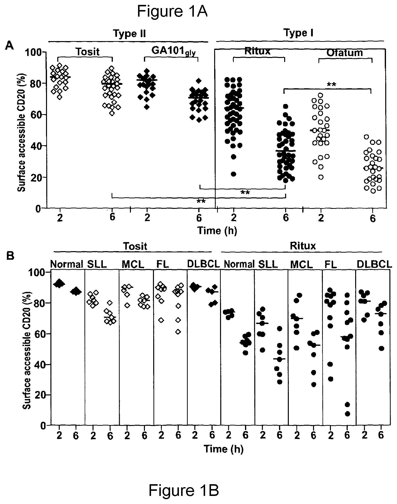
D00002

D00003

D00004

D00005

D00006

D00007

D00008
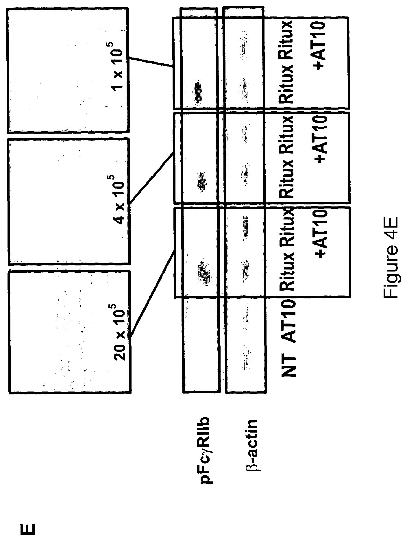
D00009
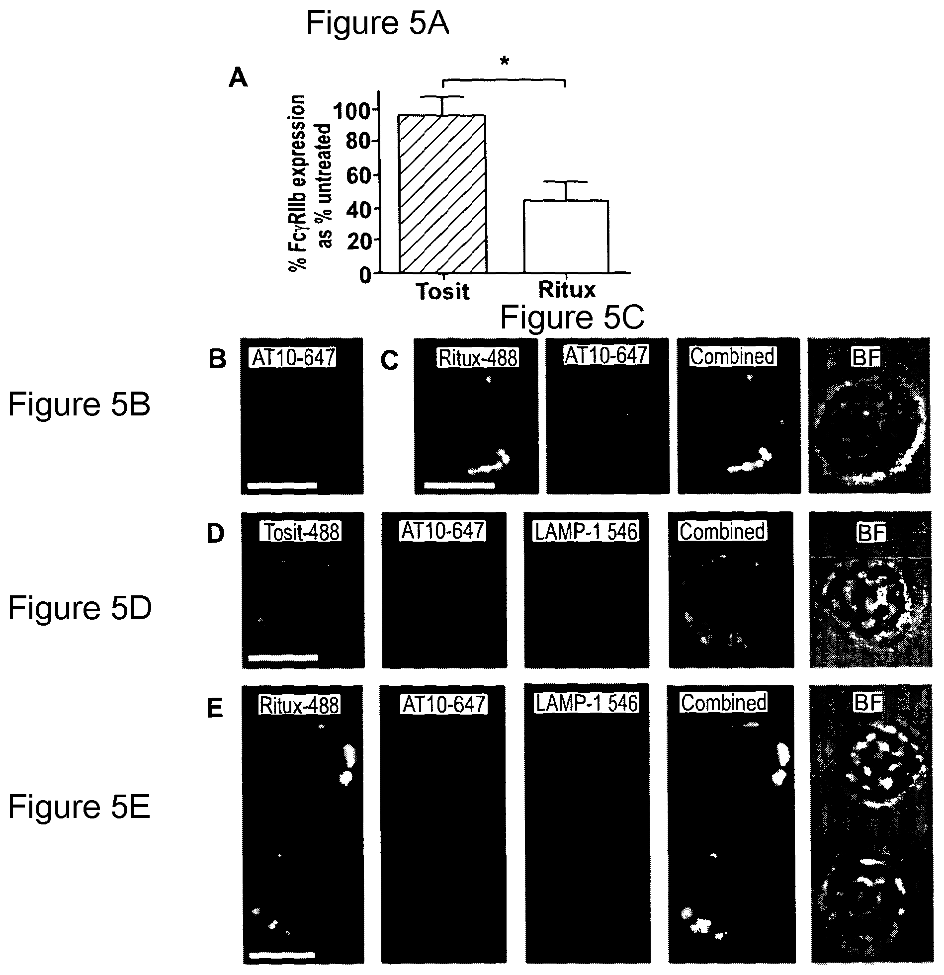
D00010
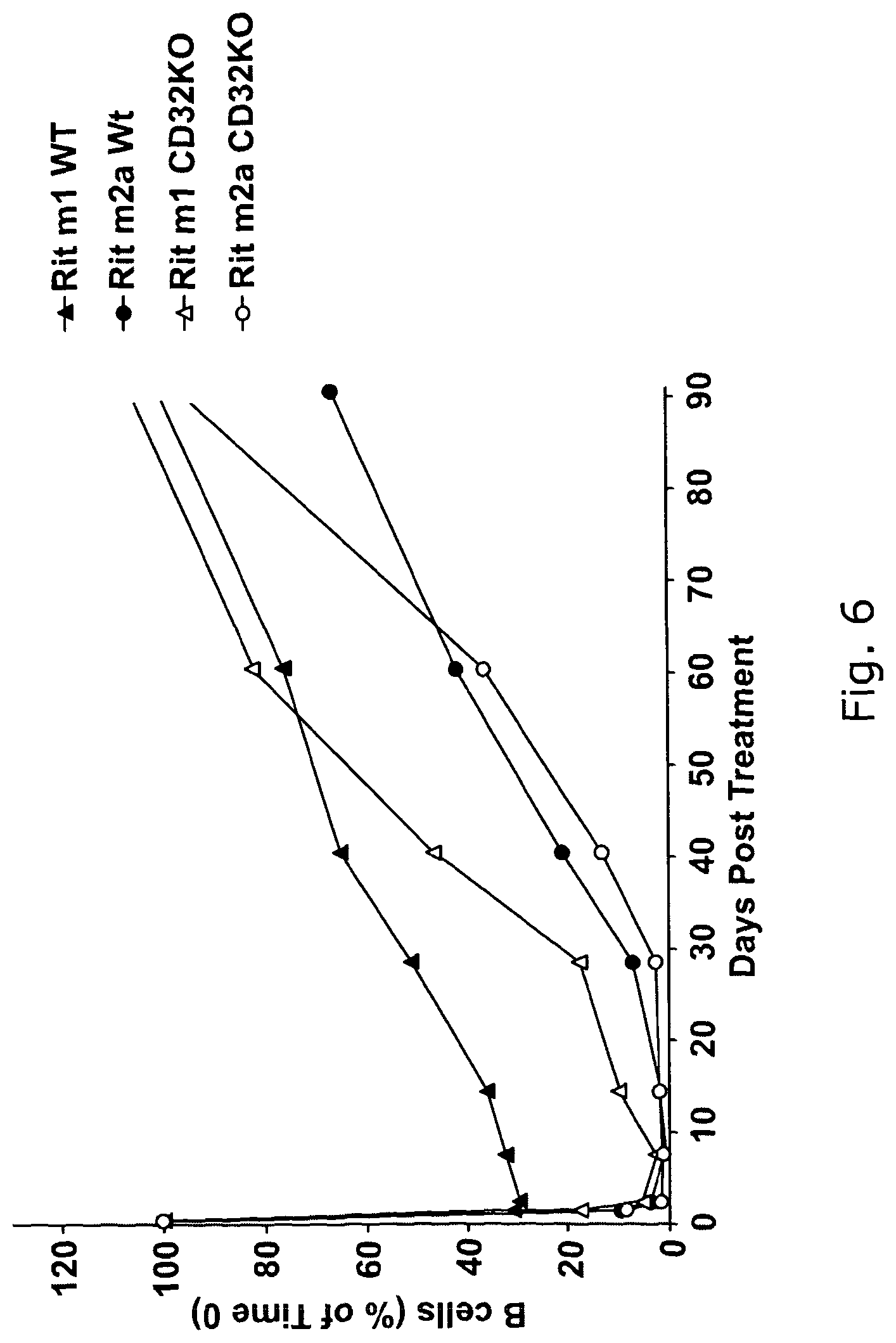
D00011

D00012
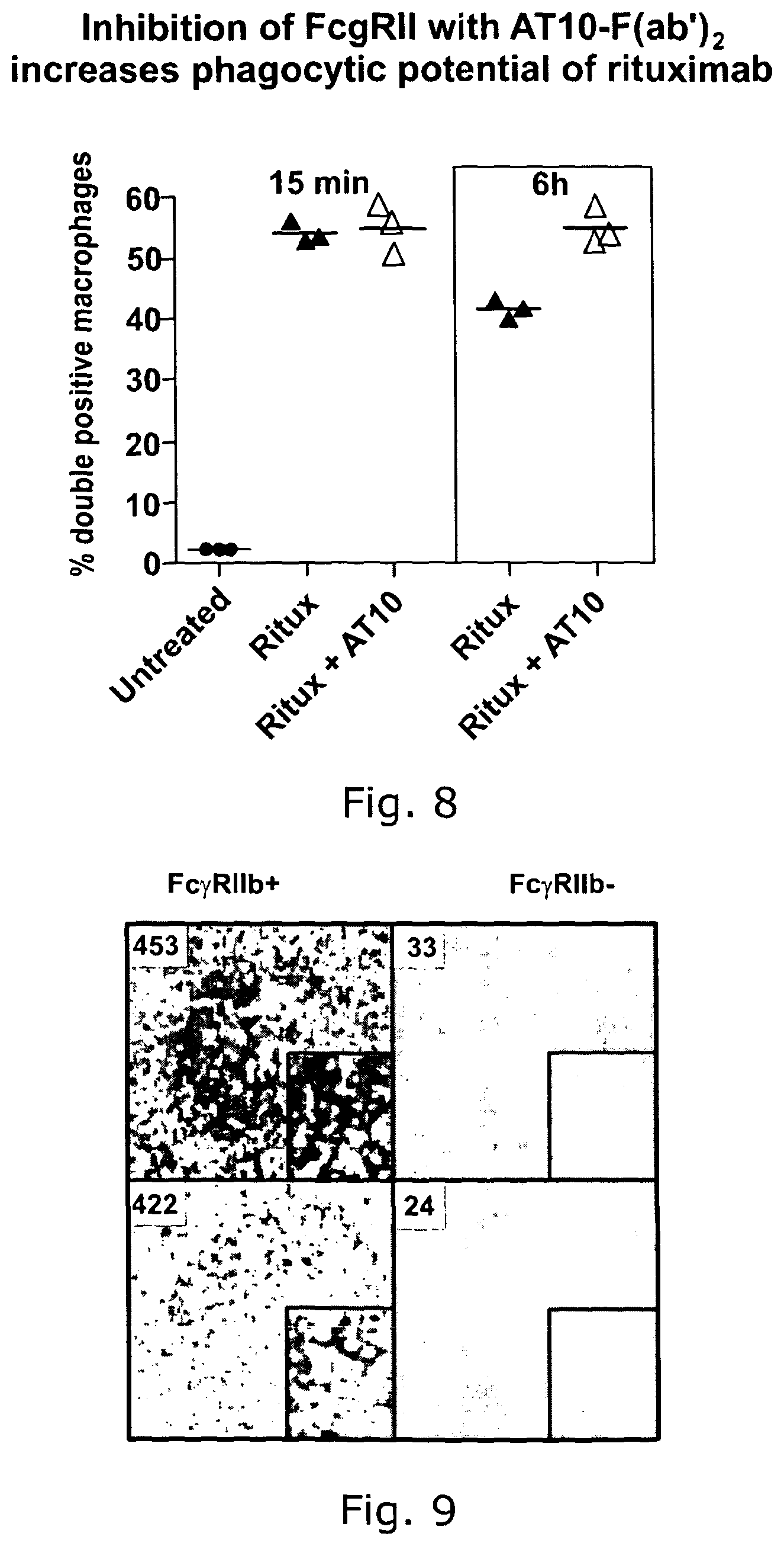
D00013

D00014

D00015

D00016

D00017
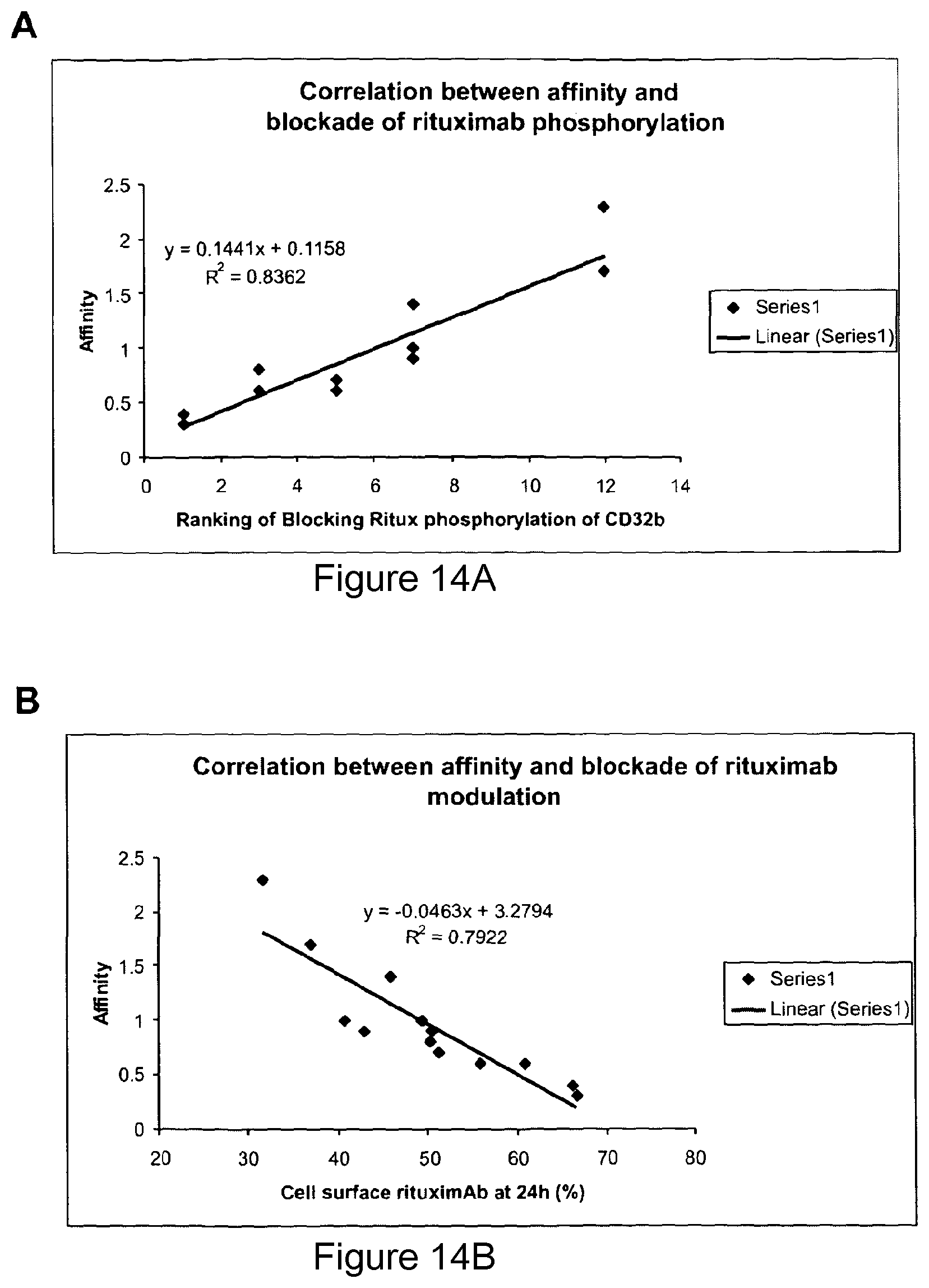
D00018

D00019

D00020

D00021
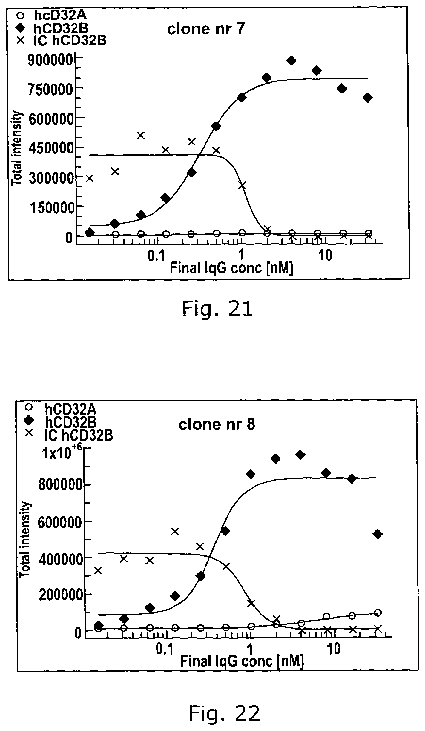
D00022

D00023

D00024

D00025
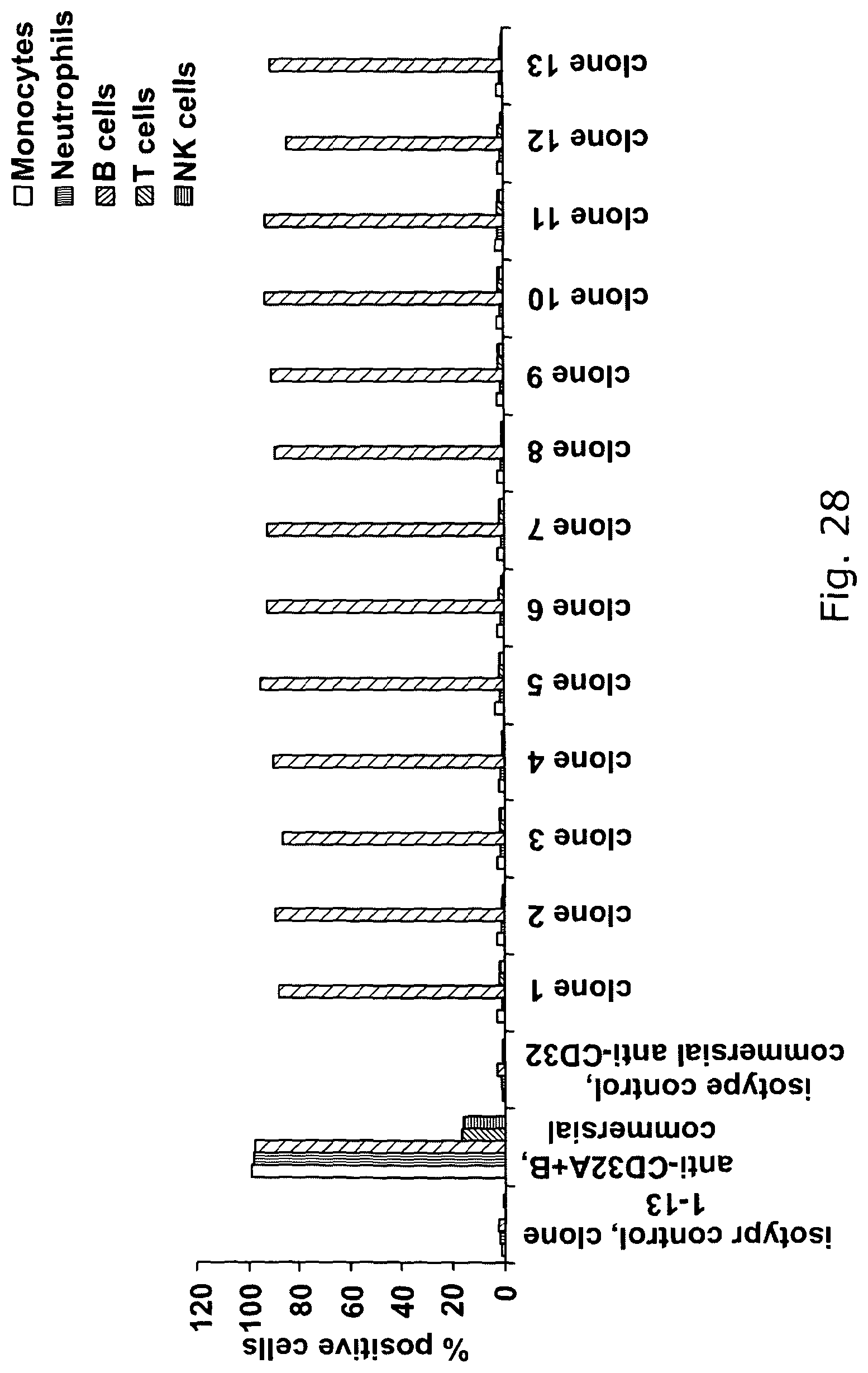
D00026

D00027
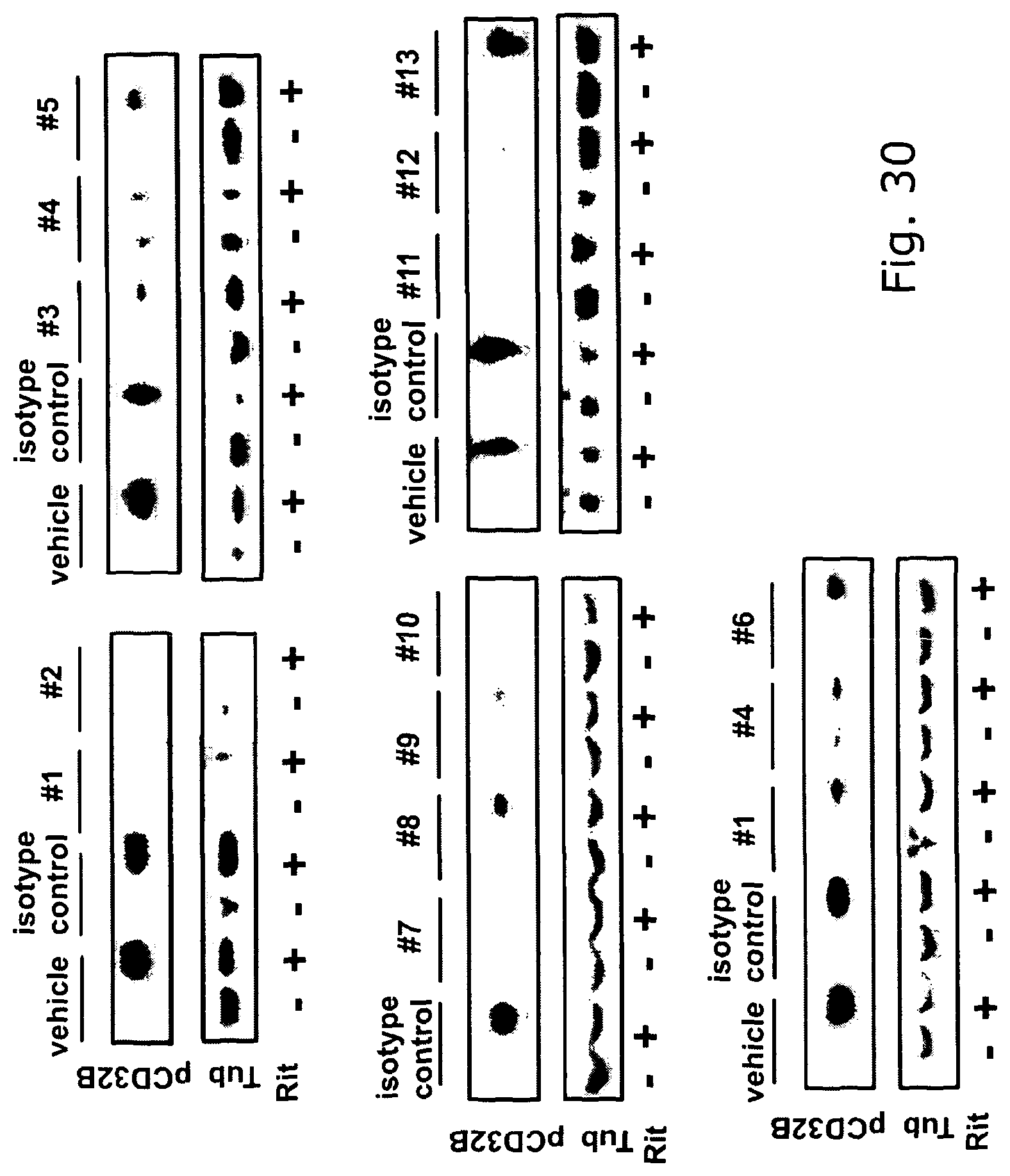
D00028

D00029

D00030
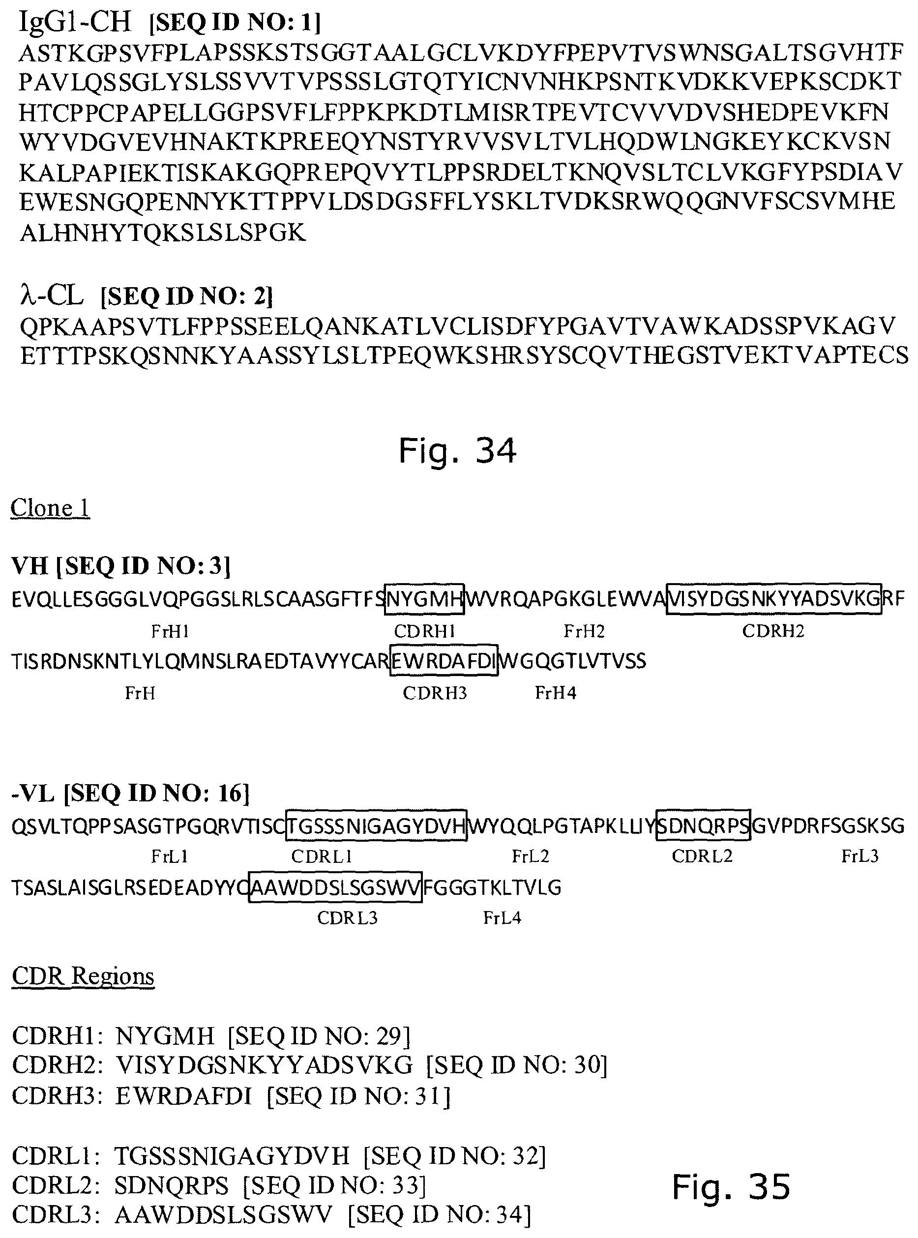
D00031

D00032

D00033

D00034

D00035
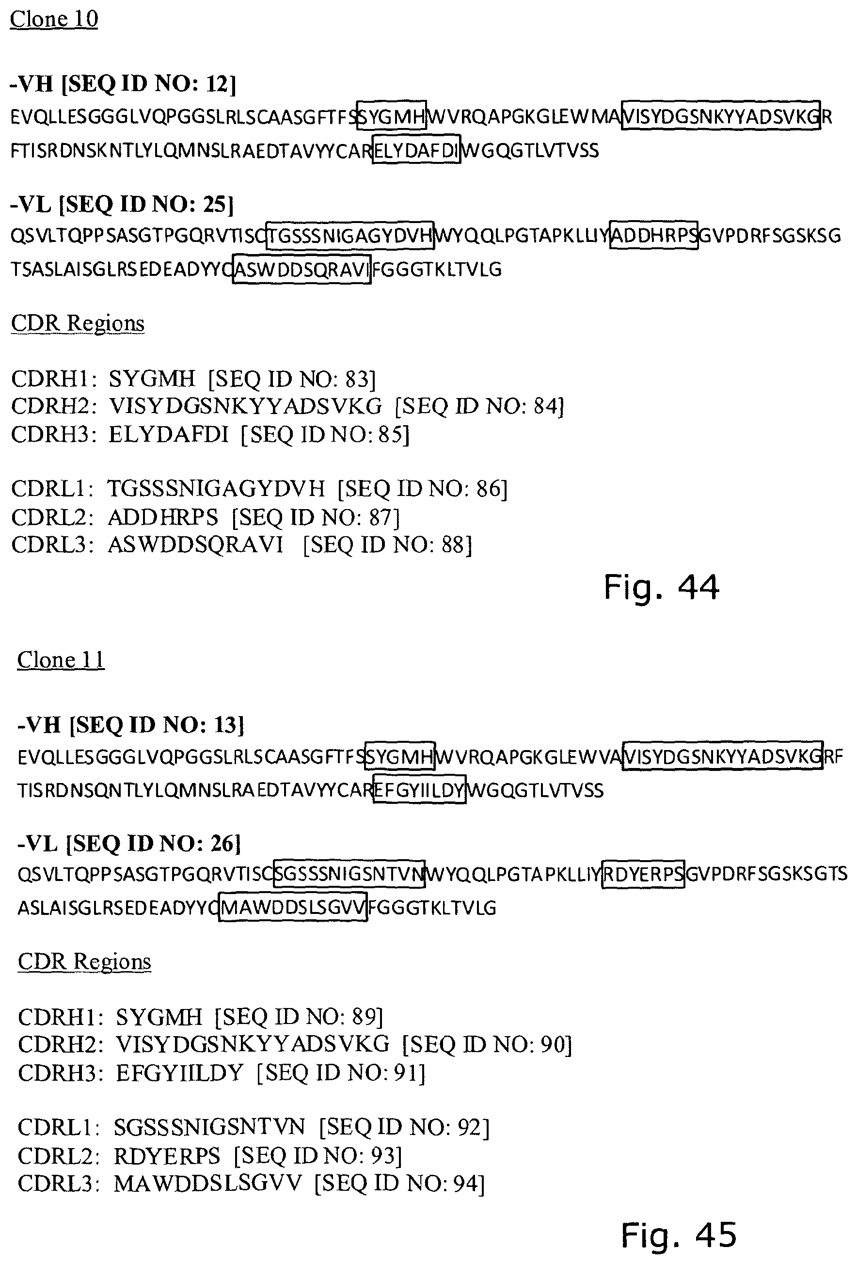
D00036

S00001
XML
uspto.report is an independent third-party trademark research tool that is not affiliated, endorsed, or sponsored by the United States Patent and Trademark Office (USPTO) or any other governmental organization. The information provided by uspto.report is based on publicly available data at the time of writing and is intended for informational purposes only.
While we strive to provide accurate and up-to-date information, we do not guarantee the accuracy, completeness, reliability, or suitability of the information displayed on this site. The use of this site is at your own risk. Any reliance you place on such information is therefore strictly at your own risk.
All official trademark data, including owner information, should be verified by visiting the official USPTO website at www.uspto.gov. This site is not intended to replace professional legal advice and should not be used as a substitute for consulting with a legal professional who is knowledgeable about trademark law.