Methods and systems for inhibiting foreign-body responses in diabetic patients
Chattaraj , et al. Dec
U.S. patent number 10,517,892 [Application Number 14/512,788] was granted by the patent office on 2019-12-31 for methods and systems for inhibiting foreign-body responses in diabetic patients. This patent grant is currently assigned to MEDTRONIC MINIMED, INC.. The grantee listed for this patent is Medtronic MiniMed, Inc.. Invention is credited to Sarnath Chattaraj, Kiem H. Dang, Hsi Chung Fusselman, Poonam S. Gulati, Lance P. Hoffman, Guangping Zhang.


View All Diagrams
| United States Patent | 10,517,892 |
| Chattaraj , et al. | December 31, 2019 |
Methods and systems for inhibiting foreign-body responses in diabetic patients
Abstract
Methods and devices are provided for reducing a diabetic patient's foreign body immune response, including infusion site-loss and/or occlusion. Such foreign body responses are associated with the treatment of the diabetic patient where the treatment requires subcutaneous implantation of a foreign body, such as a cannula or catheter. In certain embodiments of the invention, a response-inhibiting agent is administered to a patient at the site of cannula/catheter insertion, thereby facilitating delivery of insulin to the diabetic patient and mitigating site-loss and/or occlusion over a period of time.
| Inventors: | Chattaraj; Sarnath (Simi Valley, CA), Dang; Kiem H. (Thousand Oaks, CA), Fusselman; Hsi Chung (Simi Valley, CA), Gulati; Poonam S. (La Canada, CA), Hoffman; Lance P. (West Hollywood, CA), Zhang; Guangping (Calabasas, CA) | ||||||||||
|---|---|---|---|---|---|---|---|---|---|---|---|
| Applicant: |
|
||||||||||
| Assignee: | MEDTRONIC MINIMED, INC.
(Northridge, CA) |
||||||||||
| Family ID: | 52826808 | ||||||||||
| Appl. No.: | 14/512,788 | ||||||||||
| Filed: | October 13, 2014 |
Prior Publication Data
| Document Identifier | Publication Date | |
|---|---|---|
| US 20150112302 A1 | Apr 23, 2015 | |
Related U.S. Patent Documents
| Application Number | Filing Date | Patent Number | Issue Date | ||
|---|---|---|---|---|---|
| 61894088 | Oct 22, 2013 | ||||
| 61935010 | Feb 3, 2014 | ||||
| 62032101 | Aug 1, 2014 | ||||
| Current U.S. Class: | 1/1 |
| Current CPC Class: | A61K 31/727 (20130101); A61M 5/14248 (20130101); A61L 29/08 (20130101); A61L 29/16 (20130101); A61K 38/28 (20130101); A61P 43/00 (20180101); A61M 5/158 (20130101); A61P 5/50 (20180101); A61P 3/10 (20180101); A61M 2205/0205 (20130101); A61M 2205/0238 (20130101); A61M 5/16836 (20130101); A61M 5/16827 (20130101); A61M 2005/1586 (20130101); A61L 2300/236 (20130101); A61L 2300/42 (20130101); A61L 2300/41 (20130101) |
| Current International Class: | A61K 31/727 (20060101); A61L 29/08 (20060101); A61L 29/16 (20060101); A61K 38/28 (20060101); A61M 5/142 (20060101); A61M 5/158 (20060101); A61M 5/168 (20060101) |
References Cited [Referenced By]
U.S. Patent Documents
| 9522230 | December 2016 | Agarwal |
| 2008/0015494 | January 2008 | Santini, Jr. et al. |
| 2009/0131769 | May 2009 | Leach |
| 2009/0324748 | December 2009 | Dobson |
| 2010/0094111 | April 2010 | Heller |
| 2010/0217238 | August 2010 | DeJournett |
| 2011/0086081 | April 2011 | To |
| 2011/0313317 | December 2011 | Callicoat |
| 2012/0203089 | August 2012 | Rule |
| 2012/0213708 | August 2012 | Anderson |
| 2012/0226238 | September 2012 | Davies et al. |
| 2014/0155819 | June 2014 | Amirouche |
| 2014/0194815 | July 2014 | Kouyoumjian |
| 2014/0309619 | October 2014 | Agarwal |
| 2818974 | Jun 2012 | CA | |||
| 2255836 | Nov 2014 | EP | |||
| 2012-515016 | Jul 2012 | JP | |||
| 95/20416 | Aug 1995 | WO | |||
| 00/10385 | Mar 2000 | WO | |||
| 2008/114218 | Sep 2008 | WO | |||
| 20081114218 | Sep 2008 | WO | |||
| 2010080715 | Jul 2010 | WO | |||
| 2012/072561 | Jun 2012 | WO | |||
Other References
|
PCT International Search Report and Written Opinion dated Feb. 5, 2015 from PCT/US2014/061832. cited by applicant . JP Office Action dated Jun. 6, 2017, Application No. 2016-525553, with machine translation. cited by applicant . Tetsunori et al., "Effectiveness of Heparin during Long-Term Tocolysis", ISRN Obstet Gynecol, 2013, https://wwww.ncbi.nlm.nih.gov/pmc/articles/PMC3622413/#, Mar. 27, 2013 (Mar. 27, 2013). cited by applicant . Canadian Office Action dated Aug. 14, 2018 for Canadian Application No. 2927238. cited by applicant . Japanese Office Action dated Sep. 4, 2018 for JP application No. 2016-525553 with machine translation. cited by applicant. |
Primary Examiner: Farrar; Lauren P
Attorney, Agent or Firm: Gates & Cooper LLP
Parent Case Text
CROSS-REFERENCE TO RELATED APPLICATIONS
This application claims the benefit under 35 U.S.C. Section 119(e) of the following U.S. provisional patent applications, which are incorporated by reference herein:
Provisional Application Ser. No. 61/894,088, filed on Oct. 22, 2013, by Chattaraj et al., entitled "Methods and Systems for Inhibiting Foreign-Body Responses in Diabetic Patients,"; Provisional Application Ser. No. 61/935,010, filed on Feb. 3, 2014, by Chattaraj et al., entitled "Methods and Systems for Inhibiting Foreign-Body Responses in Diabetic Patients,", and Provisional Application Serial No. 62/032,101, filed on Aug. 1, 2014, by Chattaraj et al., entitled "Methods and Systems for Inhibiting Foreign-Body Responses in Diabetic Patients,".
Claims
The invention claimed is:
1. A system for delivering insulin to a diabetic patient at a single site of infusion over a period of time, the system comprising: a medication reservoir comprising an insulin solution; a cannula adapted for subcutaneous insertion into a tissue of a diabetic patient at the single site of infusion; a fluid conduit in operable contact with the medication reservoir and the cannula and adapted to deliver insulin from the medication reservoir to the single site of infusion; and a heparin composition in operable contact with the fluid conduit that inhibits inflammation at the single site of infusion so as to inhibit insulin site loss; and wherein the system delivers an amount of heparin sufficient to inhibit inflammation at the single site of infusion for a period of time of at least 4 days.
2. The system of claim 1, wherein the heparin composition: (a) is disposed within a depot and adapted to contact the insulin solution as the insulin solution flows from the medication reservoir to the single site of infusion; and/or (b) is pre-or in-situ mixed with insulin.
3. The system of claim 2, wherein the depot includes a membrane and/or a filter and/or a sponge and/or a polymeric material and/or a metal comprising heparin.
4. The system of claim 2, wherein the heparin is administered to the patient in an amount between 0.1 and 80 U/kg/day.
5. The system of claim 2, wherein the system further delivers heparin according to at least one delivery profile including: (a) an immediate release profile wherein the heparin is administered to the patient from 0 to 6 hours following insertion of the cannula; and (b) an extended release profile wherein the heparin is administered to the patient for at least 48 hours following insertion of the cannula.
6. The system of claim 1, wherein the system further comprises dextran adapted to contact the insulin solution.
7. The system of claim 6, wherein the dextran is administered to the patient at a concentration between 0.0005 and 0.4 mg/kg/day.
8. The system of claim 1, wherein the heparin: (a) coats the fluid path including cannula; and/or (b) coats a septum and/or the inner surface within the medication reservoir.
9. A method for mitigating a foreign body response in a diabetic patient receiving insulin at a single infusion site over a time period of at least three days, the method comprising: administering a site loss mitigating agent comprising heparin in combination with insulin at the single infusion site so as to inhibit insulin site loss, wherein the site loss mitigating agent mitigates inflammation at the single infusion site thereby inhibiting a foreign body response in a diabetic patient; and wherein the system delivers an amount of a site loss mitigating agent sufficient to inhibit inflammation at the single site of infusion for at least 4 days.
10. The method of claim 9, wherein the site loss mitigating agent is heparin administered at a concentration range of 40 U/ml to 8000 U/ml or 0.1 mg/ml to 20 mg/ml.
11. The method of claim 9, wherein the heparin is combined with the insulin prior to delivery of the insulin.
12. The method of claim 9, wherein the heparin: is disposed in a depot adapted to contact an insulin solution as the insulin solution flows from a medication reservoir to the single infusion site; is disposed on the cannula; is disposed in a transdermal patch that secures the infusion set to the patient; and/or is disposed in a drug-coated septum and/or inside surface within a reservoir of an insulin pump.
13. The method of claim 9, further including administering dextran, sirolimus, tacrolimus, or combination thereof.
14. A method for delivering insulin to a diabetic patient at a single site of infusion over a period of at least four days, the method comprising infusing the insulin at the single site of infusion using a system comprising: a medication reservoir comprising an insulin solution; a cannula adapted for subcutaneous insertion into a tissue of a diabetic patient at the single site of infusion; a fluid conduit in operable contact with the medication reservoir and the cannula and adapted to deliver insulin from the medication reservoir to the single site of infusion; heparin that inhibits-inflammation at the single site of infusion so as to inhibit insulin site loss; so that insulin is delivered to the diabetic patient; and wherein the heparin is administered according to an extended release profile that delivers heparin to the single site of infusion in a manner sufficient to inhibit inflammation at the single site of infusion for at least 6 days.
15. The method of claim 14, wherein the heparin is administered in an amount between 40 U/ml to 8000 U/ml and at a dose of 0.1 to 80 U/kg/day.
16. The method of claim 14, further comprising administering dextran in an amount between 0.0005-0.4 mg/kg/day.
17. The method of claim 14, wherein the heparin is disposed within a depot and adapted to contact the insulin solution as the insulin solution flows from the medication reservoir to the single site of infusion.
18. A system for delivering insulin to a diabetic patient at a single site of infusion over a period of time, the system comprising: a medication reservoir comprising an insulin solution; a cannula adapted for subcutaneous insertion into a tissue of a diabetic patient at the single site of infusion; a fluid conduit in operable contact with the medication reservoir and the cannula and adapted to deliver insulin from the medication reservoir to the single site of infusion; and a site loss mitigation composition disposed in operable contact with the fluid conduit inflammation at the single site of infusion so as to inhibit insulin site loss; and wherein the site loss mitigation composition comprises heparin in an amount sufficient to administer between 0.1 and 80 U/kg/day to the diabetic patient so as to inhibit site loss in the diabetic patient for at least 4 days.
19. The system of claim 18, wherein the site loss mitigation composition comprises heparin in an amount sufficient to inhibit site loss for at least 6 days.
20. The system of claim 19, wherein the site loss mitigation composition comprises dextran in an amount sufficient to administer between 0.0005 and 0.4 mg/kg/day to the diabetic patient.
21. The system of claim 20, wherein the system delivers at least 50% of the heparin administered in the first three days following insertion of the cannula at the single site of infusion.
22. The system of claim 19, wherein the system delivers heparin according to at least two delivery profiles including: (a) an immediate release profile wherein the heparin is administered to the patient from 0 to 6 hours following insertion of the cannula; and (b) an extended release profile wherein the heparin is administered to the patient for at least 48 hours following insertion of the cannula.
Description
TECHNICAL FIELD
This invention relates to methods and systems for inhibiting human foreign-body responses to implanted medical devices, and more particularly, methods and systems for inhibiting or reducing foreign-body responses (e.g. reducing site-loss/occlusion) in diabetic patients that result from implanted cannulas (including plastic catheters or metal needles) catheters.
BACKGROUND OF THE INVENTION
Infusion pumps are devices used to pump fluid medications into a patient in a controlled manner. One specific type of infusion pump is the insulin pump, which is used for the administration of insulin in treating patients with diabetes mellitus, a process also known as continuous subcutaneous insulin infusion (CSII) therapy. Typically, an infusion pump includes a pump (which includes controls, a processing module, and batteries), a reservoir containing fluid medication (e.g. insulin), an infusion set (which includes a cannula and/or catheter for subcutaneous insertion into the patient and a tubing system connecting the reservoir to the cannula/catheter. Upon insertion into a patient, the infusion set (more particularly the inserted cannula) is typically maintained in a transcutaneous position at the infusion site for multiple days to allow for continuous delivery of fluid medication. Cannulas and catheters provide passageways for delivering the medication to the patient.
A persistent problem associated with such devices is that the human body spontaneously reacts against foreign bodies which are introduced into the body, such as an implanted cannula (including plastic catheter or metal needle), (see, e.g. U.S. Pat. No. 5,219,361). Among the various responses of a body to foreign bodies, inflammation and the build-up of fibrous tissue at the infusion site significantly shortens the duration that an infusion set may be maintained at a single infusion site (i.e. "site-loss"). Moreover, tissue encapsulation and blockage of the implanted cannula or catheter (i.e. "occlusion") often occurs, thereby impeding or halting infusion of medication. Thus, frequent re-positioning of the infusion site for continued usage of the infusion pump is required.
Patients may also experience scar tissue buildup around an inserted cannula, resulting in a hard bump under the skin after the cannula is removed. The scar tissue does not heal particularly fast, so years of wearing an infusion pump and changing the infusion site will result in a decrease of viable infusion sites. Furthermore, for example with diabetic patients, the areas with scar tissue build-up generally have lower insulin sensitivity, which in turn may affect basal rates and bolus amounts. In some extreme cases, the delivery of insulin will appear to have little to no effect on lowering blood glucose levels and require a change in the infusion site location.
A patient's own natural defense systems can frustrate the controlled delivery of fluid medications to a patient's tissue. Thus, there is a need for methods and systems that can inhibit the human foreign-body response to implanted medical devices such as the inserted cannulas or catheters.
SUMMARY OF THE INVENTION
As noted above, foreign-body responses to cannulas (e.g. plastic catheters or metal needles) inserted in vivo can include coagulation, occlusion, inflammation, and/or encapsulation of the cannula/catheter. The invention disclosed herein is designed to address problems associated with such phenomena by using systems and methods that utilize agents identified as having an ability to inhibit foreign body responses at a cannula insertion site, thereby inhibiting such problematic phenomena. Typical embodiments of the invention are useful for diabetic patients that are infusing insulin via a cannula in order to regulate blood sugar levels.
Illustrative embodiments of the invention include systems for delivering insulin to a diabetic patient at a single site of infusion over a period of time (e.g. at least 7, 8 or 9 days). Typically these systems include a medication reservoir comprising an insulin solution, a cannula adapted for subcutaneous insertion into a tissue of a diabetic patient at the single site of infusion, and a fluid conduit in operable contact with the medication reservoir and the cannula, and adapted to deliver insulin from the medication reservoir to the single site of infusion. Such systems further include a site loss mitigating agent that inhibits at least one of: coagulation at the single site of infusion, inflammation at the single site of infusion, and encapsulation of the cannula at the single site of infusion. These systems are useful, for example, in methods for delivering insulin to a diabetic patient at a single site of infusion over a period of at least three or more (e.g. seven) days. These systems are also useful in methods for inhibiting a foreign body response in a diabetic patient receiving insulin at a single infusion site over a time period of at least three or more days.
Typical response-inhibiting agents can be selected from the group consisting of heparin, dextran, rapamycin (sirolimus), tacrolimus, or combinations thereof. In some of the illustrative working embodiments of the invention that are disclosed herein, the site loss mitigating agent comprises a heparin composition. Such compositions can be disposed at a number of different locations within these systems. For example, in certain embodiments, the heparin (or other agent) is disposed within a depot and adapted to contact the insulin solution as the insulin solution flows from the medication reservoir to the single site of infusion. In some embodiments of the invention, the depot includes a sponge, membrane or a filter impregnated with heparin that moves into the insulin solution upon contact. In certain working embodiments disclosed herein, the heparin (or other agent) is disposed within a composition that coats the cannula. Site loss mitigating agents can be disposed at a number of other locations and, for example, can coat a septum within the medication reservoir, or be disposed within a transdermal patch etc.
Related embodiments of the invention include methods for delivering insulin to a diabetic patient at a single site of infusion over a period of time (e.g. at least three or at least seven days), the method comprising infusing the insulin at the single site of infusion using a system as disclosed herein. Typically in these methods, the system that delivers insulin to the diabetic patient comprises a medication reservoir comprising an insulin solution, a cannula adapted for subcutaneous insertion into a tissue of a diabetic patient at the single site of infusion, a fluid conduit in operable contact with the medication reservoir and the cannula and adapted to deliver insulin from the medication reservoir to the single site of infusion, and a site loss mitigating agent that inhibits at least one of: coagulation at the single site of infusion, inflammation at the single site of infusion, and encapsulation of the cannula at the single site of infusion. In some embodiments of the invention, the response-inhibiting agent is heparin and is administered in an amount between 40 U/device to 8000 U/device and at a dose of 0.1 to 80 U/kg/day. In some embodiments of the invention, a response-inhibiting agent can comprise dextran (e.g. alone or in combination with another agent such as heparin) and is administered in an amount between 0.002-0.4 mg/kg/day.
Optionally the agent is disposed within a depot and adapted to contact an insulin solution as the insulin solution flows from the medication reservoir to the single site of infusion and/or within a composition that coats the cannula and is administered according to a specific delivery profile. In certain embodiments of the invention, the response-inhibiting agent is released in accordance to a plurality of delivery profiles. Such profiles can include, for example, an immediate release profile wherein the response-inhibiting agent is administered to the patient from 0 to 6 hours following insertion of the cannula and/or an extended release profile wherein the response-inhibiting agent is administered to the patient at least 48 hours or at least 72 hours following insertion of the cannula. In some embodiments of the invention, the response-inhibiting agent coats the cannula for an immediate release profile and/or the response-inhibiting agent is impregnated with a material that that coats the cannula for an extended release profile.
Other objects, features and advantages of the present invention will become apparent to those skilled in the art from the following detailed description. It is to be understood, however, that the detailed description and specific examples, while indicating some embodiments of the present invention, are given by way of illustration and not limitation. Many changes and modifications within the scope of the present invention may be made without departing from the spirit thereof, and the invention includes all such modifications.
BRIEF DESCRIPTION OF THE DRAWINGS
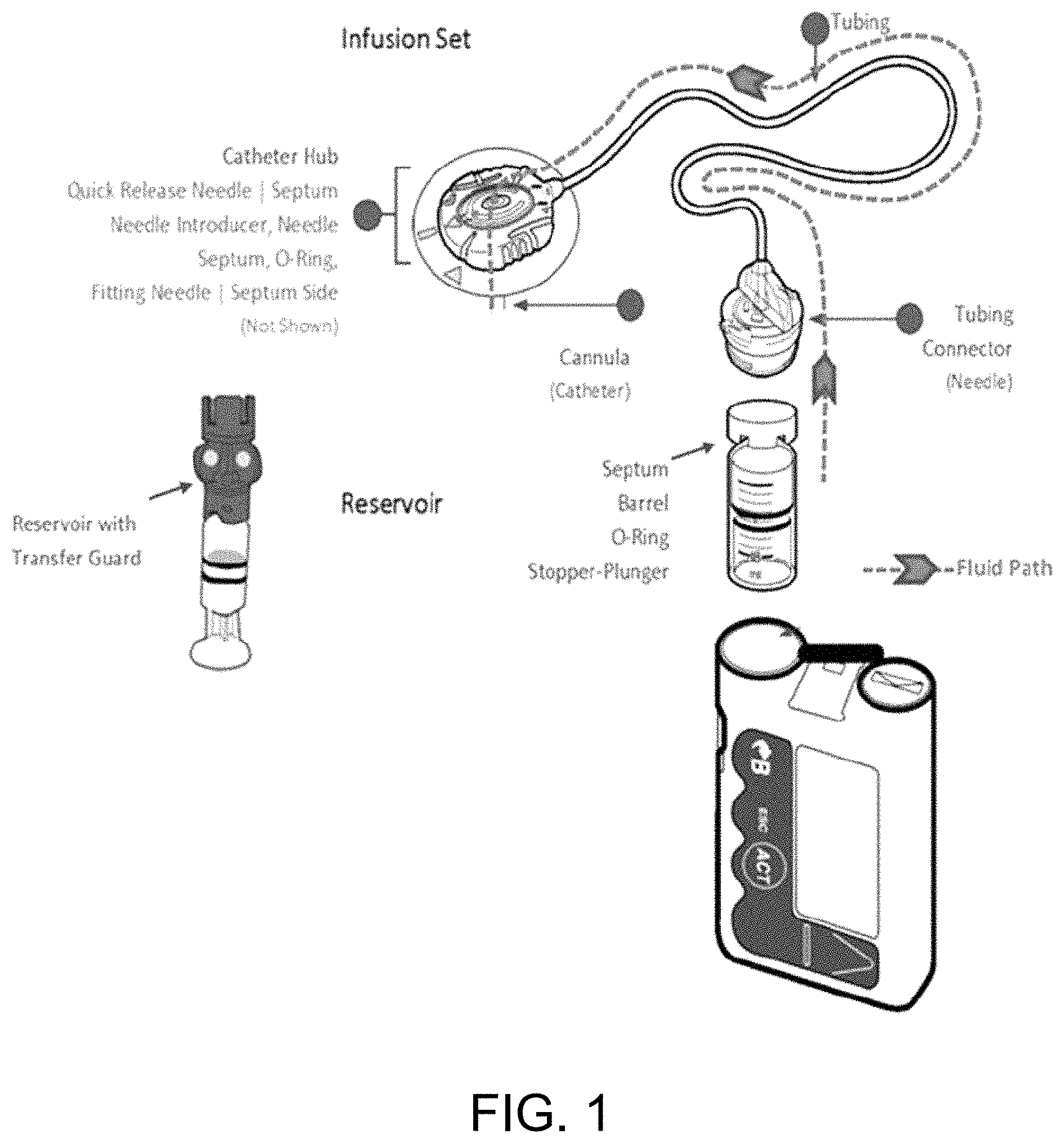
FIG. 1 is a schematic of the components included in an infusion pump and the fluid path for delivering a fluid medication.
FIG. 2A is a series of histologic evaluations at the cannula tip showing the inflammatory response (score) and size of the area with tissue reaction. "*" indicates the cannula location and "F" indicates normal fat. Inflammation resulted in localized fat loss. FIG. 2B is a series of graphs illustrating the inflammation at the cannula tip for diabetic and non-diabetic pigs. Localized inflammation loosely correlated with site-loss. FIG. 2C is a series of graphs illustrating the predominate inflammation cells at the cannula tip and cannula body (qualitative). To isolate the main inflammation contributors, a cannula, a cannula infused with insulin (Humalog.TM.), and a cannula infused with placebo were placed on a diabetic pig at the same time. Evaluated inflammation scores were found to be in the same order: insulin.gtoreq.placebo.gtoreq.cannula. FIG. 2D is a series of graphs illustrating the predominate inflammation cells at the cannula tip and cannula body of an insulin injection port (i-Port.TM.) To isolate the main inflammation contributors, a cannula, a cannula infused with insulin, and a cannula infused with placebo were placed on a diabetic pig. Compared to continuous infusion, the giant cells are missing in the i-Port.TM. study. Other cell types showed similar reaction to the cannula, cannula infused with insulin (Humalog.TM.), and cannula infused with placebo.
FIG. 3 shows a drug-adhesive patch comprising a matrix system without a rate-controlling membrane.
FIG. 4 is a schematic of an exemplary cannula in accordance with one or more embodiments of the invention. The cannula may comprise of (A) holes or (B) wells or a combination of both and can be loaded with or without drugs.
FIG. 5 shows a drug-coated cannula. The cannula material is FEP. Its wall thickness is 0.003''-0.005'', inner diameter (ID) is 1.015'', outer diameter (OD) at the base is 0.025'', and outer diameter at the tip is 0.022''.
FIG. 6 is a graph of blood glucose (BG) vs. time for normal site-loss. Based on the glucose reading for pig 2, the infusion site was lost after glucose level increased.
FIGS. 7A-D show an illustration of a cannula, in accordance with one or more embodiments of the invention. FIG. 7A is an enlarged, partial sectional view of a cannula inserted in a patient for directly infusing medication into the patient's tissue. FIG. 7B is an enlarged, partial longitudinal sectional view of the distal end of a drug-coated cannula. FIG. 7C is a view similar to that of FIG. 7B, but of another embodiment of the cannula according to the invention. FIG. 7D is a view similar to that of FIG. 7B, but of another embodiment of the cannula according to the invention.
FIG. 8 is an illustration of the infusion site and the biopsied tissue around the cannula.
FIG. 9 shows the infusion set and sensor placement in the set-up of this experiment (in the order of steps (a) to (d)).
FIG. 10 is a graph of blood glucose (BG) vs. time for Sof-Set.TM. with rapamycin dosed-insulin. The infusion site was recovered after glucose level increased, while rapamycin continued to be dosed. These results support a drug-eluting device that is sustained release.
FIG. 11 is a graph of the glucose monitoring results for pig IM3, which shows that no site-loss occurred in 6 days.
FIG. 12 is a graph of the glucose monitoring results for pig IM4, which shows that no site-loss occurred in 6 days.
FIG. 13 is a graph of the control results for pig IM1, which shows that site-loss occurred in 2 days.
FIG. 14 is an illustration of an embodiment of the device with a dual chamber reservoir for delivering heparin along with insulin.
FIG. 15 is an illustration of an in-line chamber (A) and in-line plug (B) for continuous heparin delivery.
FIG. 16 is an illustration of a heparin reservoir (A) and heparin depots (B-D), in accordance with one or more embodiments of the invention.
FIG. 17 is a graph of blood glucose (BG) vs. time for Sof-Set.TM. with heparin dosed insulin, which shows that the infusion site was active up to 13 days.
FIG. 18 is a graph of blood glucose (BG) vs. time for Sof-Set.TM. with heparin dosed insulin, which shows that the infusion site was active up to 13 days.
FIG. 19 is a graph of blood glucose (BG) vs. time for Sof-Set.TM. with a low dose heparin depot, which shows that the infusion site was active up to 6 days.
FIG. 20 a detailed structure for an illustrative heparin depot.
FIG. 21 is a graph of blood glucose (BG) vs. time for Sof-Set.TM. with a high dose heparin depot, which shows that the infusion site was active up to 6 days.
FIG. 22 is a graph of blood glucose (BG) vs. time for Sof-Set.TM. with dextran dosed insulin, which shows that the infusion site was active up to 6 days.
FIG. 23 is a graph of blood glucose (BG) vs. time for Sof-Set.TM. with rapamycin dosed insulin. The infusion site was recovered after glucose level increased, while rapamycin continued to be dosed. This supports a drug eluting device that is sustained release.
FIG. 24 is a graph of the glucose monitoring results for control Polyfin.TM. (IM2) showing site-loss at around 2.5 days.
FIG. 25 is a graph of the glucose monitoring results for rapamycin coated Polyfin.TM. (IM1) showing site-loss at day 5.
FIG. 26 is a graph of the glucose monitoring results for rapamycin coated Polyfin.TM. (IM4) showing site-loss at day 6.
DETAILED DESCRIPTION OF THE INVENTION
In the description of preferred embodiments, reference is made to the accompanying drawings which form a part hereof, and in which is shown by way of illustration a specific embodiment in which the invention may be practiced. It is to be understood that other embodiments may be utilized and structural changes may be made without departing from the scope of the present invention. Unless otherwise defined, all terms of art, notations and other scientific terms or terminology used herein are intended to have the meanings commonly understood by those of skill in the art to which this invention pertains. In some cases, terms with commonly understood meanings are defined herein for clarity and/or for ready reference, and the inclusion of such definitions herein should not necessarily be construed to represent a substantial difference over what is generally understood in the art. All publications mentioned herein are incorporated herein by reference to disclose and describe the methods and/or materials in connection with which the publications are cited. Publications cited herein are cited for their disclosure prior to the filing date of the present application. Nothing here is to be construed as an admission that the inventors are not entitled to antedate the publications by virtue of an earlier priority date or prior date of invention. Further, the actual publication dates may be different from those shown and require independent verification.
The invention described herein is primarily designed for use with an infusion pump for delivery of fluid medication comprising a combined fluid pump and reservoir and an infusion catheter. It is also within the scope of the invention to use a catheter access port or additional forms of implantable pump systems in place of a combined fluid pump and reservoir disclosed. An example of a suitable catheter access port is disclosed in U.S. Pat. No. 5,137,529 issued to David A. Watson, Mark J. Licata, Alfons Heindl and Edward C. Leicht on Aug. 11, 1992 entitled "Injection Port" and assigned to Medtronic-PS Medical, the disclosure of which is incorporated herein by reference in its entirety. Examples of additional implantable pump systems are disclosed in U.S. Pat. No. 4,588,394 issued to Rudolf R. Schultz, Gary P. East and Alfons Heindle on May 13, 1986 entitled "Infusion Reservoir and Pump System", U.S. Pat. No. 4,681,560 issued to Rudolf R. Schultz, Gary P. East and Alfons Heindle on Jul. 21, 1987 entitled "Subcutaneous Infusion Reservoir and Pump System", U.S. Pat. No. 4,761,158 issued to Rudolf R. Schultz, Gary P. East and Alfons Heindle on Aug. 2, 1988 entitled "Subcutaneous Infusion Reservoir and Pump System", U.S. Pat. No. 4,816,016 issued to Rudolf R. Schultz, Gary P. East and Alfons Heindle on Mar. 28, 1989 entitled "Subcutaneous Infusion Reservoir and Pump System", U.S. Pat. No. 4,867,740 issued to Gary P. East on Sep. 19, 1989 entitled "Multiple-Membrane Flow Control Valve and Implantable Shunt System", U.S. Pat. No. 5,085,644 issued to David A. Watson and Mark J. Licata on Feb. 4, 1992 entitled "Sterilizable Medication Infusion Device with Dose Recharge Restriction" and U.S. Pat. No. 5,152,753 issued to Stephen W. Laguette, Gary P. East, David A. Watson and Thomas J. Carlisle on Oct. 6, 1992 entitled "Medication Infusion Device with Dose Recharge Restriction", all of which are assigned to Medtronic-PS Medical, the disclosures of which are incorporated herein by reference in their entirety.
Additionally, the infusion pumps and systems described in U.S. Pat. No. 6,110,155, titled "Anti-inflammatory-agent-loaded catheter and method for preventing tissue fibrosis," U.S. patent application Ser. No. 11/897,106, titled "Combined sensor and infusion set using separated sites," U.S. patent application Ser. No. 12/184,046, titled "Analyte sensor apparatuses having improved electrode configurations and methods for making and using them," and U.S. patent application Ser. No. 13/010,640, titled "Layered enzyme compositions for use with analyte sensors" are incorporated herein by reference in their entirety.
Aspects and Embodiments of the Invention
Diabetes mellitus (DM) is the most common cause of hyperglycemia, a condition of high blood glucose that occurs when the body has too little insulin (type 1 and some type 2 DM) or is unable to utilize insulin properly (type 2 DM). One method of treating a diabetic patient is with the use of an infusion pump, in particular an insulin pump. An infusion pump provides for the infusion of a medication or drug composition, such as insulin or an insulin analog, to a patient. The infusion pump is typically worn by the patient, but may also be implanted in the patient. The infusion pump comprises any suitable means for conveying fluid medication to a targeted location (i.e. infusion site) on a patient's body by way of a cannula (e.g. a plastic catheter or a metal needle).
Typically, the infusion pump comprises a combined fluid pump and reservoir and an infusion set, which comprises a cannula/catheter. In one embodiment, as shown in FIG. 1, the infusion pump includes a self-contained reservoir for storing medication, a pump for drawing the fluid medication from the reservoir and advancing it by way of an infusion cannula to the tissue of the patient to be treated. A suitable power source, such as a battery, is used to energize the pump. The infusion pump may be programmed to deliver prescribed amounts of medication continuously (e.g. basal insulin rate), on demand (e.g. bolus of insulin) or at regularly scheduled intervals. The infusion pump also includes an infusion set which comprises components to be inserted into the patient, such as an insertion needle and a cannula (or catheter). The cannula is a thin tube used for the introduction of fluid medication to the target site. Generally, a proximal end of the cannula is attached via a tubing system and connector to the reservoir and fluid pump, located outside the patient's body. An opposite, distal end of the cannula is inserted into the patient trans/subcutaneously and adapted to be positioned in close proximity to the tissue intended to receive the fluid medication. A lumen extends from the proximal end to the distal end of the cannula to conduct the flow of fluid therebetween. The infusion set also includes an insertion needle, which is assembled with the soft cannula (catheter) and is adapted to pierce the patient's skin for trans/subcutaneous cannula placement. The insertion needle is left inside as hard cannula or thereafter withdrawn to leave the soft cannula in place for subcutaneous fluid infusion.
FIG. 7A illustrates a means for conducting fluid to the human body employing a cannula in accordance with one embodiment of the present invention. Here, the distal end 18 of the cannula 14 is received in an opening 22 formed in a patient's tissue and in a bore 24 formed in the tissue 28. In this embodiment, multiple fluid apertures 32 are provided in the cannula adjacent to the distal end 18, whereby fluid medication such as insulin can be conducted directly to the bore 24 in the tissue.
As noted above, an inherent problem with the implantation of a foreign body in human tissue is the foreign-body response from the patient's immune system. An injury is created at the site where the needle is inserted into a patient's tissue for cannula placement and medication infusion (the "single site of infusion"). Catheter/cannula insertion induces an acute inflammatory reaction within epidermis, dermis, and subcutaneous adipose tissue. Another problem is that tissues and cells may be damaged during the insertion process. This includes possible damage to cells and connective tissue along the path of needle/catheter infusion, as well as damage to basement membranes, the extracellular matrix, and structural proteins. Damaged lymphatic vessels, arterioles, capillaries, and venuoles may also cause blood/fluid to accumulate around the catheter shaft (e.g. clotting). A further problem is that there may be physiological debris that forms around the catheter, obstructing capillaries.
Infusion site-loss and site-reduction occur in part due to the encapsulation of the cannula by the tissue. In such instances, insulin absorption into the patient's circulation becomes variable and unreliable over time. Causes of site-loss/reduction are poorly understood and may be due to localized tissue inflammation, coagulation, occlusion, and/or tissue proliferation. Moreover, although the materials used for the cannula are flexible enough to provide comfort for the patient, the inevitable movement of the cannula that occurs when a patient moves leads to further tissue inflammation. Thus, an implanted cannula (i.e. a foreign body) elicits an exacerbated host response as a result of any cannula movement.
As an illustration, a surgeon implants a biomaterial in a surgical site (thereby creating an injury). Quickly, the implant adsorbs a layer of proteins, the normal process for a solid surface in biological fluids. Cells (neutrophils and then macrophages) interrogate and attack the "invader," i.e., the biomaterial. When the macrophages find they cannot digest the implant, they fuse into giant cells to engulf the object. However, it is too large to completely ingest. Thus, the giant cells send out chemical messengers (cytokines) to call in other cells to form a cellular capsule around the biomaterial. As a result, the presence of this capsule seriously degrades the performance of the biomaterial by preventing intimate contact between the biomaterial and tissue. The reaction associated with this foreign body response (long term, low level inflammation and macrophage activation) may also inhibit the luminal healing of vascular grafts.
Embodiments of the present invention include methods and devices for reducing a diabetic patient's foreign-body immune response, which is associated with the treatment of the diabetic patient where the treatment requires implantation of a foreign body. In particular instances, the invention mitigates infusion site-loss/occlusion caused by a short-term (e.g. 0 to 8 days) subcutaneous insertion of a cannula or catheter. The cannula or catheter is usually part of a subcutaneous infusion set and is attached to a reservoir or infusion pump intended to administer a fluid medication or drug formulation. As used herein, a response-inhibiting (and/or mitigating) agent refers to an active agent that inhibits, mitigates or reduces a foreign-body response of the patient's tissue (such as site-loss/occlusion of an inserted cannula).
As described in further detail below, various approaches are provided for inhibiting or mitigating site-loss/occlusion. A mechanical approach is provided that improves the mechanical design of the infusion set to mitigate injury to the insertion site. For example, the fluid path of infusion may be altered (side ports). In one or more embodiments, the cannula is modified with different structural configurations that incorporate holes and/or wells for loading one or more response-inhibiting agents (see Drug-coated cannula section below). A material approach is also provided that modifies the surface of the insertion cannula with anti-fouling biomaterials, such as PEG or immobilized heparin, to alleviate foreign body response. A drug approach is also provided that locally administers/releases response inhibiting agents, such as immuno-suppressants, anti-inflammatory agents or other bioactive molecules, to alleviate a body's response to the insertion of a cannula and insulin, improve local insulin absorption into blood stream, and/or prevent localized insulin. To address the issue of possible damage to connective tissue, anti-proliferative agents such as rapamycin may be used. To address the issue of possible blood/fluid accumulation or clotting, anti-coagulants such as heparin and dextransulfate may be used. To address the issue of physiological debris and obstruction of capillaries, a combination of anti-fouling and anti-coagulation agents may be used. Agents for breaking down hyaluronic acid may also be used. Other response-inhibiting agents that may also be used are described in the Response-Inhibiting Agents section below.
Insulin losses at a single site of infusion are frequent in diabetic patients and are a potential source of blood glucose variability. The physiological processes behind such site loss are complex, and unpredictable. For this reason, it is not possible to predict how a specific agent such as will affect site loss. For example, as disclosed in the examples below, formulations of insulin combined with anti-inflammatory agents heparin and/or dextran and/or rapamycin notably inhibited site loss, thereby extending the duration of cannula insertion, performing significantly better than the control. In contrast, formulations of insulin combined with anti-inflammatory agents betamethasone sodium phosphate (BSP) or Dexamethasone palmitate (DXP) actually resulted in the onset of site-loss much earlier, performing significantly worse than the control (as discussed in Example 6 below).
Embodiments of the invention include systems for delivering insulin to a diabetic patient at a single site of infusion over a period of time (e.g. at least 7, 8 or 9 days). Typically these systems include a medication reservoir comprising an insulin solution, a cannula adapted for subcutaneous insertion into a tissue of a diabetic patient at the single site of infusion, and a fluid conduit in operable contact with the medication reservoir and the cannula, and adapted to deliver insulin from the medication reservoir to the single site of infusion. Such systems further include a site loss mitigating agent that inhibits at least one of: coagulation at the single site of infusion, inflammation at the single site of infusion, and encapsulation of the cannula at the single site of infusion. These systems are useful, for example, in methods for delivering insulin to a diabetic patient at a single site of infusion over a period of at least three or more (e.g. seven) days. These systems are also useful in methods for inhibiting a foreign body response in a diabetic patient receiving insulin at a single infusion site over a time period of at least three or more days.
In some of the working embodiments of the invention that are disclosed herein, the site loss mitigating agent comprises a heparin composition. This heparin composition can be disposed at a number of different locations within these systems. In certain embodiments, the heparin (or other agent) is disposed within a depot and adapted to contact the insulin solution as the insulin solution flows from the medication reservoir to the single site of infusion. For example, in some embodiments of the invention, the depot includes a sponge, membrane or a filter impregnated with heparin that moves into the insulin solution upon contact. In some of the working embodiments disclosed herein, the heparin (or other agent) is disposed within a composition that coats the cannula. Site loss mitigating agents can be disposed at a number of other locations and, for example, can coat a septum within the medication reservoir, or be disposed within a transdermal patch etc.
In some embodiments of the invention, the heparin is administered to the patient in an amount between 40 U/device to 8000 U/device and at a dose of 0.1 to 80 U/kg/day. Optionally, the heparin is administered to the patient in an amount between 0.5 and 5 U/kg/day. In certain embodiments of the invention, the system delivers heparin according to a specific delivery profile. For example, embodiments of the invention include systems designed to deliver an immediate release profile, one where the majority of the heparin is administered to the patient from 0 to 6 hours following insertion of the cannula. Other embodiments of the invention include an extended release profile, one where the heparin is administered to the patient for at least 24 or 48 hours following insertion of the cannula. In some embodiments of the invention, the system is designed to deliver at least 50% of the total heparin administered in the first three days following insertion of the cannula.
Embodiments of the invention can further include dextran sulfate compositions, for example a dextran composition adapted to contact the insulin solution as the insulin solution flows from the medication reservoir to the single site of infusion. In typical embodiments of the invention, the dextran is administered to the patient in an amount between 0.002 and 0.4 mg/kg/day. In some embodiments of the invention, the dextran is administered to the patient in an amount between 0.005 and 0.015 mg/kg/day. In some embodiments of the invention designed to administer heparin and dextran, the heparin coats the cannula and the dextran is disposed in the depot. Embodiments of the invention can further include additional agents such as sirolimus, tacrolimus, or combination thereof. In some embodiments of the invention, the response-inhibiting agent is combined with insulin in the medication reservoir.
Other embodiments of the invention include methods for delivering insulin to a diabetic patient at a single site of infusion over a period of time (e.g. at least three or at least seven days), the method comprising infusing the insulin at the single site of infusion using a system as disclosed herein. Typically in these methods, the system that delivers insulin to the diabetic patient comprises a medication reservoir comprising an insulin solution, a cannula adapted for subcutaneous insertion into a tissue of a diabetic patient at the single site of infusion, a fluid conduit in operable contact with the medication reservoir and the cannula and adapted to deliver insulin from the medication reservoir to the single site of infusion, and a site loss mitigating agent that inhibits at least one of: coagulation at the single site of infusion, inflammation at the single site of infusion, and encapsulation of the cannula at the single site of infusion.
In some embodiments of the invention, the response-inhibiting agent is heparin. Heparin is well known in the art and pharmaceutical grade heparin useful in embodiments of the invention is readily available from a wide variety of sources (e.g. Heparin Sodium INJ available from Celsus and Pfizer). The source of the heparin sodium in the working embodiments of the invention that are disclosed herein was Fisher BioReagents. In typical embodiments of the invention, the heparin and is administered at a concentration range of 40 U/ml to 8000 U/ml or 0.1 mg/ml to 20 mg/ml. In some embodiments, the heparin is administered at a dose of 0.1 to 80 U/kg/day. In specific instances, the heparin is administered at a concentration of 800 U/ml and/or at a dose of 8 U/kg/day. Data from working embodiments of the invention where heparin is used as a response-inhibiting agent is discussed in the Examples below (e.g. Example 7) and shown in the Figure (e.g. FIGS. 11 and 12). These finding are unexpected in view of art that teaches that heparin is no better that a sodium chloride solution for maintenance of patency in peripheral intermittent intravenous devices (see, e.g. Tuten et al., Appl Nurs Res 1991 4(2): 63-72).
In certain embodiments of the invention, a response-inhibiting agent comprises dextran (e.g. alone or in combination with another agent such as heparin). Typically dextran that is administered to the patient in an amount between 0.002 and 0.4 mg/kg/day. Dextrans are well known in the art and pharmaceutical grade dextran useful in embodiments of the invention is readily available from a wide variety of sources (e.g. Dextran 70 pharmaceutical grade available from Sinus Biochemistry & Electrophoresis GmbH). The source of the dextran in the working embodiments was Dextran Sulfate Sodium Salt from Sigma-Aldrich. Data from working embodiments of the invention where dextran is used as a response-inhibiting agent is discussed in the Examples below (e.g. Example 9) and shown in the Figure (e.g. FIG. 22).
In certain embodiments of the invention, a response-inhibiting agent comprises rapamycin (e.g. alone or in combination with another agent such as heparin). Rapamycin is well known in the art and pharmaceutical grade rapamycin useful in embodiments of the invention is readily available from a wide variety of sources (e.g. Rapamune available from Wyeth Pharmaceuticals Company, a subsidiary of Pfizer Inc). In some embodiments, a response-inhibiting agent is rapamycin and is administered (either formulated, co-infused or coated) at a dose of 0.5-10 .mu.g/device at 0.02 to 1.5 .mu.g/day. The source of the rapamycin in the working embodiments was TSZCHEM. Data from working embodiments of the invention where rapamycin is used as a response-inhibiting agent is discussed in the Example below (e.g. Example 10).
In one or more embodiments of the invention, the response-inhibiting agent is provided in a depot in operable contact with section of the fluid conduit of the infusion cannula. In one or more other embodiments of the invention, the response-inhibiting agent is provided as a coating that coats a part of the infusion set or reservoir. In certain embodiments, the response-inhibiting agent is disposed on a cannula and/or a transdermal patch that secures the infusion set to the patient and/or a drug-coated septum within a reservoir of an insulin pump. In one or more other embodiments of the invention, the response-inhibiting agent is provided in a reservoir where the response-inhibiting agent is present in the infusate. In certain embodiments, the response-inhibiting agent is pre-mixed with the medication prior to infusion into a patient. In other embodiments, the response-inhibiting agent and medication are delivered from two different reservoirs and then mixed in-situ upon infusion.
Optionally an agent such as heparin is disposed within a depot and adapted to contact the insulin solution as the insulin solution flows from the medication reservoir to the single site of infusion and/or within a composition that coats the cannula and is administered according to a specific delivery profile. For example, the agent can be administered according to an immediate release profile wherein the heparin is administered to the patient from 0 to 6 hours following insertion of the cannula. Alternatively, the agent can be administered according to an extended release profile wherein the response-inhibiting agent is administered to the patient for at least 48 hours following insertion of the cannula.
Another embodiment of the invention is a method of facilitating delivery of insulin to a diabetic patient over a period of time at a single infusion site. In such embodiments, the method comprises inserting a cannula subcutaneously into a tissue of a diabetic patient at an insertion site and administering a response-inhibiting agent to the patient at the site of cannula insertion, wherein the response-inhibiting agent inhibits a foreign-body response of the patient's tissue (such as site-loss/occlusion of the cannula). In this way, the method facilitates the delivery of insulin to the diabetic patient over a period of time (e.g. at least 6, 7, 8, 9, 10, 11 or 12 days). In an illustrative embodiment of the invention, a method for reducing a foreign body response in a diabetic patient is provided, the method comprising inserting a drug-coated cannula subcutaneously into a tissue of a diabetic patient at an insertion site, the drug-coated cannula having an exterior surface coated with a response-inhibiting agent. Optionally the tip of the cannula is coated. The exterior surface of the drug-coated cannula can comprise a hole, well, groove, pore, indentation or combination thereof, and the response-inhibiting agent is at least partially contained within at least a portion of the hole, well, groove, pore, indentation or combination thereof.
Related embodiments of the invention include methods for inhibiting a foreign body response in a diabetic patient receiving insulin at a single infusion site over a time period of at least 3, 4, 5, 6, 7, 8, 9, 10, 11 or 12 days, the method comprising administering a site loss mitigating agent in combination with insulin at the single infusion site, wherein the site loss mitigating agent inhibits at least one of: coagulation at the single infusion site, inflammation at the single infusion site, and encapsulation of the cannula at the single infusion site, thereby inhibiting a foreign body response in a diabetic patient. Optionally, the site loss mitigating agent is heparin administered at a concentration range of 40 U/ml to 8000 U/ml or 0.1 mg/ml to 20 mg/ml. In certain embodiments of the invention, the response-inhibiting agent is disposed in a depot adapted to contact an insulin solution as the insulin solution flows from a medication reservoir to the single infusion site. In some embodiments, the response inhibiting agent is disposed on the cannula and/or is disposed in a transdermal patch that secures the infusion set to the patient (e.g. one comprising a substrate, a response-inhibiting agent, and an adhesive layered on the substrate); and/or is disposed in a drug-coated septum within a reservoir of an insulin pump. For example, the transdermal patch can. These methods can include administering additional agents such as sirolimus, tacrolimus, or combination thereof.
Another embodiment of the invention is a method comprising the steps of providing an infusion catheter, compounding a response-inhibiting agent disposed within a polymeric material, and incorporating the compound with the catheter in a manner whereby the response-inhibiting agent will be leached from the polymeric material when the catheter is in fluid contact with bodily tissue. The catheter is inserted into a body of a diabetic patient with at least a portion of the catheter disposed adjacent to bodily tissue and fluid medication is conducted through the catheter to the tissue, wherein a foreign body response of the body tissue adjacent to the catheter is reduced by the introduction of a response-inhibiting agent. In yet another embodiment of the invention, a drug infusion set as described herein is combined with a continuous glucose monitoring device on the same adhesive patch (i.e. "combo-set"). A response-inhibiting agent is administered along with the insulin to the patient. In this way, the combo-set delivers insulin and monitors glucose levels in the patient for at least 6, 7, 8, 9, 10, 11 or 12 days.
In a further aspect, a method for reducing a foreign body response in a diabetic patient is provided comprising applying a drug-coated septum patch to a fluid path of an insulin pump. The drug-coated septum patch is located within a reservoir of the insulin pump and comprises a response-inhibiting agent. The response-inhibiting agent is released into a medication flowing through the fluid path of the insulin pump. An anti-inflammatory agent may also be included with the response-inhibiting agent. The anti-inflammatory agent may be rapamycin (sirolimus), betamethasone sodium phosphate, dexamethasone sodium phosphate, beclomethasone dipropionate, tacrolimus, or combination thereof.
Embodiments of the invention include methods of facilitating delivery of insulin to a diabetic patient at a single infusion/insertion site at during a period of infusion that occurs at least 5 days following the initial insertion of a catheter and sensor combo-set, for example, facilitating delivery of insulin at day 2, 3, 4, 5, 6, 7, 8, 9, 10, 11 or 12 (or at days 6-12 etc.) at a single infusion site using a combo-set. In this embodiment, the method comprises inserting a cannula and a sensor subcutaneously into a tissue of a diabetic patient at an insertion site, and administering a response-inhibiting agent to the patient at the site of cannula insertion, wherein the response-inhibiting agent inhibits a foreign-body response of the patient's tissue such as site-loss/occlusion of the cannula. In this way the method facilitates delivery of insulin to the diabetic patient at day 6 and/or 7 and/or 8 and/or 9 and/or 10 and/or 11 and/or 12.
The invention provides many advantages, such as increased patient safety by reducing the site-loss phenomenon, and in particular, reducing hyperglycemic events for diabetic patients. Since the invention provides an infusion set that may be used longer than currently recommended durations of 2-3 days, there is also increased comfort and convenience for the patient due to the reduced frequency of inserting and re-inserting the cannula. In certain embodiments, the invention allows insulin to be effective beyond 6-days during continuous subcutaneous insulin infusion (CSII) therapy. In particular instances, the invention reduces coagulation in the insulin diffusion pathways, stabilizes insulin from aggregation, and/or improves vascular impact.
Further aspects and embodiments are discussed in the following sections.
Response-Inhibiting Agent Coating
In one or more other embodiments of the invention, the response-inhibiting agent is provided as a coating on a part of the infusion set or reservoir. The response-inhibiting agent may be formulated specifically for slow release (i.e. pre-dosed). In one embodiment, the method and device comprises application of a response-inhibiting agent-coated transdermal patch. In a further embodiment, the method and device comprises application of a response-inhibiting agent-coated cannula or catheter. In certain embodiments, the response-inhibiting agent is disposed on a cannula and/or a transdermal patch that secures the infusion set to the patient and/or a drug-coated septum within a reservoir of an infusion pump. In a still further embodiment, the method and device comprises application of a response-inhibiting agent-coated septum or a response-inhibiting agent-impregnated infusion set. The method and device for reducing a diabetic patient's foreign-body immune response may comprise of one or more of the embodiments in various combinations (e.g. a response-inhibiting agent-coated transdermal patch in addition to a response-inhibiting agent-coated cannula).
Transdermal Patch
In one aspect of the invention, the method and device for reducing site-loss/occlusion and/or coagulation in a diabetic patient comprises application of a response-inhibiting agent-coated transdermal patch. Preferably, topical administration of the response-inhibiting agent is by means of a transdermal patch, though the response-inhibiting agent may be administered as, without limitation, an ointment, gel, cream, powder or drops. An advantage of a transdermal patch is that the medicated adhesive patch can be placed on the skin for several days depending on the skin type. The medication can then continuously penetrate the skin to reduce the foreign body response at the subcutaneous infusion site. The medicated transdermal adhesive patch can further be packaged and sold separately to provide various options for infusion pump users.
The transdermal patch comprises a response-inhibiting agent for mitigating a foreign-body response and is applied near the site where a foreign object is subcutaneously inserted. In one or more embodiments, the transdermal patch comprises a substrate layered with an adhesive and response-inhibiting agent intended for local dermal absorption near an insertion site of a subcutaneous infusion set. The transdermal patch may be separate from or a part of the infusion set. While an infusion set is inserted in a patient, normally for multiple-days, the transdermal patch administers a local dose of a response-inhibiting agent near the infusion site of the cannula. This reduces foreign body responses such as site-loss and/or occlusion occurring during the subcutaneous delivery of fluid medication, such as insulin or an insulin analog.
In other embodiments, the invention may be combined with a continuous glucose monitoring device on the same adhesive patch (i.e. "combo-set"). Currently in the art, glucose sensors have a use-life of 6 days whereas infusion sets typically have a recommended use-life of only 2-3 days. The use of the invention disclosed herein enables both devices to be worn for the same duration on the same patch, thereby reducing product use cost. In certain embodiments, the continuous glucose monitoring performance of the combo-set is extended beyond 3 days, and in specific instances, 4, 5 or 6 days. In other instances the glucose monitoring performance of the combo-set is greater than or equal to 6 days.
More than one response-inhibiting agent, such as an anti-inflammatory agent and an anti-coagulation agent, may be administered simultaneously by the transdermal patch. The anti-inflammatory agent may be a steroidal, non-steroidal anti-inflammatory drug or anti-proliferative drug. For example, Table 5 below shows examples of steroids, immunosuppressant drugs, cox inhibitors, non-steroidal anti-inflammatory drugs (NSAIDS), and anti-proliferative agents that can be blended in the adhesive and penetrant to achieve an anti-inflammatory effect. In particular, the anti-inflammatory agent may be rapamycin (sirolimus), tacrolimus, or combination thereof. In specific embodiments, the anti-inflammatory agent is not a methasone (e.g. betamethasone sodium phosphate, dexamethasone sodium phosphate, beclomethasone dipropionate or the like).
Drug-Coated Cannula
In another aspect of the present invention, the method and device for reducing site-loss and/or occlusion in a diabetic patient comprises application of a response-inhibiting agent-coated/loaded cannula. At least a portion of the drug-coated cannula is coated with the response-inhibiting agent. In one or more embodiments, a response-inhibiting agent is coated or loaded on the exterior surface of the cannula. In one or more other embodiments, the response-inhibiting agent is coated or loaded on the interior surface or lumen of the cannula. The response-inhibiting agent-coated cannula provides a direct supply of a response-inhibiting agent to the tissue to combat the natural foreign-body response at the infusion site. In one embodiment, the response-inhibiting agent is directly delivered into a patient's internal tissue environment to achieve an anti-coagulation effect and/or prevent encapsulation of a subcutaneously inserted cannula.
More than one response-inhibiting agent, such as an anti-inflammatory agent and an anti-coagulation agent, may be administered simultaneously. Table 5 below shows examples of steroids, immunosuppressant drugs, cox inhibitors, non-steroidal anti-inflammatory drugs (NSAIDS), and anti-proliferative agents that can be blended in the coating to achieve an anti-inflammatory effect. In particular, the anti-inflammatory agent may be rapamycin (sirolimus), tacrolimus or combination thereof. In specific embodiments, the anti-inflammatory agent is not a methasone (e.g. betamethasone sodium phosphate, dexamethasone sodium phosphate, beclomethasone dipropionate or the like).
In other embodiments, the response-inhibiting agent coating may include only the response-inhibiting agent or may include a response-inhibiting agent in combination with another material such as a polymer, a metal, a metal alloy, a ceramic, a glass, or any combination thereof. The coating may be constructed or applied as multiple layers. The multiple layers may have different materials or compositions, different ratios of materials or compositions, or both in each layer.
FIG. 7B illustrates one embodiment of the invention where the surface 26 of the distal end 18 of a cannula is provided with a coating 27 of a response-inhibiting agent. As an illustrative implementation, a silicon-based cannula is dipped into a silicon adhesive in which a response-inhibiting agent has been placed into solution. The compound liquid coats the surface 26 of the cannula and, as it solidifies, encapsulates at least a portion of the distal end in the polymer/response-inhibiting agent compound. By use of this structure, the response-inhibiting agent is leached from the cannula when the cannula is in contact with body fluid and acts to combat site-loss and/or occlusion. An alternative to dipping the distal end of the cannula is to spray-coat the distal end of the cannula with a vaporized, compounded solution.
FIG. 7C depicts an alternative embodiment of the invention. In this embodiment, the response-inhibiting agent is provided throughout the body of the cannula by mixing and compounding the response-inhibiting agent directly into the cannula polymer melt before forming the cannula. For example, the response-inhibiting agent can be compounded into materials such as silicone rubber or urethane. The compounded material is then processed conventionally as by extrusion, transfer molding or casting, for example, to form a tubular configuration. The cannula 30 resulting from this process benefits by having the response-inhibiting agent dispersed throughout the entire cannula body. The response-inhibiting agent slowly leaches or diffuses into the patient's tissue from the cannula, thereby preventing or resisting site-loss and/or occlusion in and around the cannula 30.
FIG. 7D depicts another embodiment of the invention. In this embodiment, a thin layer 42 of a response-inhibiting agent has been covalently bonded to the exterior surface 26 of the cannula 14. The surface is prepared to molecularly receive the response-inhibiting agent. A binding agent may be needed between the response-inhibiting agent molecules and the polymer molecules on the surface 26. With this structure, the response-inhibiting agent is present on the exterior surface of the cannula and can be leached away to combat site-loss and/or occlusion.
In further embodiments, the structure of the cannula may include, without limitation, holes, grooves, pores, indentations, or a combination thereof on its surface where the response-inhibiting agent is partially or completely contained within at least a portion of the holes, grooves, pores, indentations or combinations thereof. In one or more embodiments, the invention provides a cannula modified with different structural configurations that incorporate holes and/or wells for loading one or more response-inhibiting agents (see FIG. 5 and Table 1 below). Table 1 illustrates six different configurations for a cannula comprising a combination of holes and/or wells. Other numbers and combinations of holes and wells may also be used.
TABLE-US-00001 TABLE 1 Config- Hole Well uration Description (A) (B) 1 1 hole (A) with diameter of 0.005'' .+-. 0.0005'' 1 None at distance 0.025'' from tip, on one side wall 2 2 hole (A) with diameter of 0.005'' .+-. 0.0005'' None at distance 0.025'' from tip, on both side 3 1 hole (A) with diameter of 0.005'' .+-. 0.0005'' 1 at distance 0.025'' from tip, on one side wall, one well below hole 4 2 hole (A) with diameter of 0.005'' .+-. 0.0005'' 2 at distance 0.025'' from tip, on both side, each well directly below each hole A 5 1 hole (A) with diameter of 0.005'' .+-. 0.0005'' 1 at distance 0.025'' from tip, on one side wall, one well 90.degree. hole A 6 2 hole (A) with diameter of 0.005'' .+-. 0.0005'' 2 at distance 0.025'' from tip, on both side, each well is 90.degree. below each hole A
The holes and/or wells incorporated within the cannula structure allow flexibility in coating and loading one or more response-inhibiting agents for controlled-release or instant-release. By introducing one or more response-inhibiting agents at the same time within one insertion, the development of a foreign body reaction in response to insertion in the subcutaneous tissue is prevented. In addition, a response-inhibiting agent can also be further impregnated into the cannula for controlled release of the response-inhibiting agent.
In another aspect of the invention, the cannula structure reduces the penetrating trauma on the tissue from insertion. The microarchitecture of the cannula, particularly at the surface, is an important parameter that influences the host response. Cannula structures found in existing art can result in densely packed, well-organized fibrous capsules, whereas the modified cannula disclosed herein (which incorporates holes and/or wells) leads to a less dense, more open, and disorganized fibrous capsule which can reduce the extent of the tissue injury at the insertion site.
Additionally, the incorporation of holes or ports in the cannula increases the number of infusion sites. This lowers the pressure from fluid medication (e.g. insulin) administration at each infusion site, thereby resulting in less tissue injury. The holes and wells also increase anchorage of the cannula so that movement of the cannula while the patient is moving is prevented. Less movement of the cannula results in reduced injury, blood clots, and infection for the patient.
In one or more embodiments, a response-inhibiting agent is delivered via a cannula coated with the response-inhibiting agent or the response-inhibiting agent and an anti-inflammatory agent and further infused with insulin. In another exemplary implementation, the response-inhibiting agent is continually infused with insulin to the patient. In a further exemplary implementation, the patient is pre-dosed with a response-inhibiting agent, followed by continued infusion of insulin.
Response-Inhibiting Agent Depot
In one or more embodiments of the invention, a response-inhibiting agent is provided in a depot attached to a section of the fluid path of the infusion pump. An in-line response-inhibiting agent depot or pre-filled cartridge is used for continuous response-inhibiting agent delivery. The in-line response-inhibiting agent depot may be in the form of an in-line response-inhibiting agent chamber or plug (see, e.g. an in-line heparin chamber or plug as shown in FIGS. 15A and 15B).
Response-Inhibiting Agent Reservoir
In one or more other embodiments of the invention, the response-inhibiting agent is provided in a reservoir where the response-inhibiting agent is present in the infusate. In certain embodiments, the response-inhibiting agent is pre-mixed with the medication prior to infusion into a patient. In other embodiments, the response-inhibiting agent is mixed in-situ along the fluid path of medication administration. An infusion pump may have a dual chamber reservoir with one reservoir for medication and another for a response-inhibiting agent (see, e.g. use of heparin as shown in FIG. 14). There may also be a dual line on the injection catheter or cannula. In certain embodiments, the response-inhibiting agent is co-infused with the insulin. The response-inhibiting agent and insulin are delivered from two different reservoirs and then mixed in-situ upon infusion.
Drug-Coated Septum and Drug-Impregnated Infusion Set
In another aspect of the present invention, the method and device for reducing site-loss and/or occlusion in a diabetic patient comprises application of a response-inhibiting agent-coated septum patch or a response-inhibiting agent-impregnated infusion set (see FIG. 1). The response-inhibiting agent-coated septum patch and response-inhibiting agent-impregnated infusion set reduce site-loss and/or occlusion resulting from the subcutaneous insertion of a foreign object, such as a cannula or catheter of an infusion set.
Embodiments of the invention include infusion sets and patch pump base-plates with active pharmaceutical ingredients such as antimicrobial, corticosteroid, and active time-release formulations intended for immediate or extended release via the distal end of the infusion pump. In one embodiment, the infusion pump comprises a dual reservoir for dual infusion of two drugs (e.g. insulin and heparin). In another embodiment, the tubing system and/or cannula is lined (impregnated) or coated with a response-inhibiting agent to reduce site-loss and/or occlusion. Table 6 below shows examples of steroids, immunosuppressant drugs, cox inhibitors, non-steroidal anti-inflammatory drugs (NSAIDS), and anti-proliferative agents that can be mixed with the fluid medication, e.g. insulin formulation, (either pre-mixed or delivered separately at the infusion site) to achieve further anti-inflammatory effect.
In other embodiments, a response-inhibiting agent-coated septum, such as a silicone rubber septum, is impregnated with a time-release response-inhibiting agent and housed within the reservoir or infusion set fluid path pass-through. Since the response-inhibiting agent-coated septum is positioned within the fluid path of the infusion pump, the response-inhibiting agent is thereby added to the fluid medication (e.g. insulin) upon administration of the medication. Delivery of the response-inhibiting agent along with the medication reduces coagulation at the infusion site and reduces the risks associated with site-loss and/or occlusion resulting from multiple-day subcutaneous therapeutic drug infusions and extended wear of infusion sets and baseplates delivering therapeutic fluids.
Response-Inhibiting Agents
In one aspect of the present invention, the response-inhibiting agent is an anti-coagulant. This includes heparin and derivatives such as low molecular weight heparin (e.g. Enoxaparin sodium (Lovenox.TM.), Dalteparin sodium (Fragmin.TM.)), Fondaparinux (Arixtra.TM.), and Idraparinux (in development by Sanofi-Aventis.TM., sub-cue). Fondaparinux is a synthetic sugar composed of the five sugars (pentasaccharide) in heparin that bind to antithrombin. It is a smaller molecule than low molecular weight heparin. Other anti-coagulants include coumarins (vitamin K antagonists) such as warfarin, acenocoumarol, phenprocoumon, atromentin, and phenindione. Warfarin (Coumadin.TM.) is an agent typically used in the US and UK. Acenocoumarol and phenprocoumon are used more commonly outside the US and the UK. Anti-coagulants also include direct factor Xa inhibitors (pills) such as rivaroxaban (Xarelto.TM.), apixaban (Eliquis.TM.), edoxaban ((INN, codenamed DU-176b, trade name Lixiana.TM.); direct thrombin inhibitors such as bivalent drugs (e.g. hirudin, lepirudin, and bivalirudin) and monovalent drugs (e.g. argatroban and dabigatran (Pradaxa.TM.)). They are often used for treatment of thrombosis in patients with heparin-induced thrombocytopenia (HIT). Anti-coagulants also include antithrombin protein (purified from human plasma or produced recombinantly).
In one or more embodiments of the invention, the response-inhibiting agent is heparin. Heparin is a member of the glycosaminoglycan family of carbohydrates and comprises a variably sulfated repeating disaccharide unit, such as .beta.-D-glucuronic acid-2-deoxy-2-acetamido-.alpha.-D-glucopyranosyl, .beta.-D-glucuronic acid-2-deoxy-2-sulfamido-.alpha.-D-glucopyranosyl, .alpha.-L-iduronic acid-2-deoxy-2-sulfamido-.alpha.-D-glucopyranosyl, 2-O-sulfo-.alpha.-L-iduronic acid-2-deoxy-2-sulfamido-.alpha.-D-glucopyranosyl, .alpha.-L-iduronic acid-2-deoxy-2-sulfamido-.alpha.-D-glucopyranosyl-6-O-sulfate or 2-O-sulfo-.alpha.-L-iduronic acid-2-deoxy-2-sulfamido-.alpha.-D-glucopyranosyl-6-O-sulfate. Although it is used principally in medicine for anticoagulation, its true physiological role in the body remains unclear, because blood anticoagulation is achieved mostly by heparan sulfate proteoglycans derived from endothelial cells.
Surprisingly, it was discovered that the heparin helps stabilize insulin in the solution, as well as facilitates insulin absorption and effectively lowers glucose in-vivo. This effectively lowers the local inflammatory response caused by insulin build-up/aggregation or debris accumulation. In various embodiments, an infusion set described herein can be used for at least 6 days. In other embodiments, the period of time is at least 6, 7, 8, 9, 10, 11 or 12 days. In one embodiment, heparin is directly added to an insulin formulation prior to and/or during administration or infusion of the insulin formulation to a diabetic patient. Preferably, the concentration range of heparin added to the insulin formulation is 40 U/ml to 8000 U/ml or 0.1 mg/ml to 20 mg/ml. In a specific instance, 800 U/ml of heparin is continuously infused along with the insulin to prevent site-loss for at least 6 days. Preferably, heparin is dosed 0.1 to 80 U/kg/day. In a specific instance, heparin is dosed 8 U/kg/day. Notably, this is significantly less than the heparin dosing used in other therapeutic treatments, which is typically 150 to 400 U/kg/day.
In another aspect of the present invention, the response-inhibiting agent is an anti-platelet agent. This includes irreversible cyclooxygenase inhibitors, aspirin, triflusal (Disgren.TM.), adenosine diphosphate (ADP) receptor inhibitors, clopidogrel (Plavix.TM.), prasugrel (Effient.TM.), ticagrelor (Brilinta.TM.), ticlopidine (Ticlid.TM.), phosphodiesterase inhibitors, cilostazol (Pletal).TM., glycoprotein IIB/IIIA inhibitors (intravenous use only), abciximab (ReoPro.TM.), eptifibatide (Integrilin.TM.), tirofiban (Aggrastat.TM.), adenosine reuptake inhibitors, dipyridamole (Persantine.TM.), thromboxane inhibitors, thromboxane synthase inhibitors, and thromboxane receptor antagonists (Terutroban.TM.).
For aspirin, a daily dose of aspirin that is commonly recommended by health care professionals in order to prevent platelets from clumping together and forming clots. Although new blood thinner medications are constantly emerging on the market, aspirin remains a commonly used preventative tool. Warfarin (Coumadin.TM.) is one of the most well known medications used to thin the blood. It is an anti-coagulant that is also used in some cases to prevent heart disease. Pradaxa.TM. is a newer medication that is used primarily in people who have an arterial fibrillation. It is geared towards preventing blood clots and strokes. Elequis.TM. essentially lowers the risk of both blood clots and strokes. Elequis.TM. is a relatively new drug that is thought to be a competitor to the side effect laden Coumadin.TM. Xarelto.TM. is especially useful in recipients of hip replacements and knee replacements. Xarelto.TM. is a newcomer amongst blood thinner medications and has also been approved for use in cases of DVT as well as pulmonary embolisms. Clopidogrel (Plavix.TM.) works by preventing coagulation of the platelets in the blood. It is especially suited for people who have certain medical conditions and heart conditions. It is also used as a preventative tool against the formation of clots in persons who have had a heart attack or stroke. Like aspirin, Prasugrel.TM. is an anti-platelet medication. In people who have been treated with angioplasty, Prasugrel.TM. may be used in conjunction with aspirin to prevent the formation of clots. Brilinta.TM. is typically prescribed following a heart attack and can be used in conjunction with aspirin. It has been proven effective at reducing the chance of recurring heart attacks in people who have had them before and the medication is thought to further reduce the risk of recurrent heart attacks with continued use. Cilostazol.TM. is used to improve the flow of blood to the legs and can help assist with reducing the symptoms of intermittent claudication. Like some of the other blood thinner medications on described herein, Cilostazol.TM. is an anti-platelet medication, whereby it is used to prevent the platelets in the blood from clumping together. Aggrenox.TM. is essentially a prescription super aspirin. It is a combination of two medicines, aspirin and dipyridamole. In people who have had blood clots, the Aggrenox.TM. medication.
Additionally, Table 5 below lists various anti-inflammatory agents and drugs that may be used in conjunction with the response-inhibiting agent in accordance with one or more embodiments of the invention. However, such a list is not exhaustive and additional examples of anti-inflammatory drugs include both steroidal and non-steroidal (NSAID) anti-inflammatories such as, without limitation, clobetasol, alclofenac, alclometasone dipropionate, algestone acetonide, alpha amylase, amcinafal, amcinafide, amfenac sodium, amiprilose hydrochloride, anakinra, anirolac, anitrazafen, apazone, balsalazide disodium, bendazac, benoxaprofen, benzydamine hydrochloride, bromelains, broperamole, budesonide, carprofen, cicloprofen, cintazone, cliprofen, clobetasol propionate, clobetasone butyrate, clopirac, cloticasone propionate, cortodoxone, deflazacort, desonide, desoximetasone, momentasone, cortisone, cortisone acetate, hydrocortisone, prednisone, prednisone acetate, diclofenac potassium, diclofenac sodium, diflorasone diacetate, diflumidone sodium, diflunisal, difluprednate, diftalone, dimethyl sulfoxide, drocinonide, endrysone, enlimomab, enolicam sodium, epirizole, etodolac, etofenamate, felbinac, fenamole, fenbufen, fenclofenac, fenclorac, fendosal, fenpipalone, fentiazac, flazalone, fluazacort, flufenamic acid, flumizole, flunisolide acetate, flunixin, flunixin meglumine, fluocortin butyl, fluorometholone acetate, fluquazone, flurbiprofen, fluretofen, fluticasone propionate, furaprofen, furobufen, halcinonide, halobetasol propionate, halopredone acetate, ibufenac, ibuprofen, ibuprofen aluminum, ibuprofen piconol, ilonidap, indomethacin, indomethacin sodium, indoprofen, indoxole, intrazole, isoflupredone acetate, isoxepac, isoxicam, ketoprofen, lofemizole hydrochloride, lomoxicam, loteprednol etabonate, meclofenamate sodium, meclofenamic acid, meclorisone dibutyrate, mefenamic acid, mesalamine, meseclazone, methylprednisolone suleptanate, morniflumate, nabumetone, naproxen, naproxen sodium, naproxol, nimazone, olsalazine sodium, orgotein, orpanoxin, oxaprozin, oxyphenbutazone, paranyline hydrochloride, pentosan polysulfate sodium, phenbutazone sodium glycerate, pirfenidone, piroxicam, piroxicam cinnamate, piroxicam olamine, pirprofen, prednazate, prifelone, prodolic acid, proquazone, proxazole, proxazole citrate, rimexolone, romazarit, salcolex, salnacedin, salsalate, sanguinarium chloride, seclazone, sermetacin, sudoxicam, sulindac, suprofen, talmetacin, talniflumate, talosalate, tebufelone, tenidap, tenidap sodium, tenoxicam, tesicam, tesimide, tetrydamine, tiopinac, tixocortol pivalate, tolmetin, tolmetin sodium, triclonide, triflumidate, zidometacin, zomepirac sodium, tacrolimus and pimecrolimus.
Additionally, examples of steroidal anti-inflammatory drugs include, without limitation, 21-acetoxypregnenolone, alclometasone, algestone, amcinonide, budesonide, chloroprednisone, clobetasol, clobetasone, clocortolone, cloprednol, corticosterone, cortisone, cortivazol, deflazacort, desonide, desoximetasone, diflorasone, diflucortolone, difluprednate, enoxolone, fluazacort, flucloronide, flunisolide, fluocinolone acetonide, fluocinonide, fluocortin butyl, fluocortolone, fluorometholone, fluperolone acetate, fluprednidene acetate, fluprednisolone, flurandrenolide, fluticasone propionate, formocortal, halcinonide, halobetasol propionate, halometasone, halopredone acetate, hydrocortamate, hydrocortisone, loteprednol etabonate, mazipredone, medrysone, meprednisone, methylprednisolone, mometasone furoate, prednicarbate, prednisolone, prednisolone 25-diethylamino-acetate, prednisolone sodium phosphate, prednisone, prednival, prednylidene, rimexolone, tixocortol, triamcinolone, triamcinolone acetonide, triamcinolone benetonide, triamcinolone hexacetonide, any of their derivatives, and combinations thereof.
Furthermore, examples of nonsteroidal anti-inflammatory drugs include, without limitation, COX-1 and COX nonspecific inhibitors (e.g., salicylic acid derivatives, aspirin, sodium salicylate, choline magnesium trisalicylate, salsalate, diflunisal, sulfasalazine and olsalazine; para-aminophenol derivatives such as acetaminophen; indole and indene acetic acids such as indomethacin and sulindac; heteroaryl acetic acids such as tolmetin, dicofenac and ketorolac; arylpropionic acids such as ibuprofen, naproxen, flurbiprofen, ketoprofen, fenoprofen and oxaprozin), and selective COX-2 inhibitors (e.g., diaryl-substituted furanones such as rofecoxib; diaryl-substituted pyrazoles such as celecoxib; indole acetic acids such as etodolac and sulfonanilides such as nimesulide), and combinations thereof.
Additionally, other naturally occurring or synthetic drugs, agents, molecules (e.g. hyaluronidase), and proteins may be included with the response-inhibiting agent to mitigate foreign-body responses and/or help facilitate the body in absorbing the medication.
EXAMPLES
Example 1: Understanding the Site-Loss Mechanism Using Animal Models
Causes of site reduction are poorly understood and can be due to localized inflammation or tissue proliferation. Understanding the cause through a time-based biopsy study allows for development of infusion sets that could be extended beyond three days, hence improving patient comfort and compliance. A diabetic animal model that shows site reduction 2-10 days after placement of transdermal insulin pumps is used so that the local tissue response to continuous subcutaneous insulin infusion (CSII) can be studied by in-situ skin biopsies.
FIG. 1 is an illustration of an exemplary medical device used for diabetes management which includes a fluid path schematic of an insulin pump. An illustrative infusion set comprises a three-layer tubing of polyethylene (PE), ethylene vinyl acetate (EVA), and polyvinyl chloride (PVC). The catheter material may be Teflon.TM. or stainless steel.
Continuous subcutaneous insulin infusion (CSII) is an effective method for diabetic care. Local site reaction/site-loss (at approximately 3 days) is often encountered clinically, yet poorly understood. A clinical manifestation of site-loss is the increase in blood glucose while a patient is on CSII. Existing theories include changes in insulin absorption, inflammation, and lipoatrophy (localized lost of fat). Better understanding of the site-loss phenomenon can provide guidance in making business decisions, such as if putting effort on improving an infusion set to extend site use is possible or necessary.
The goal of studying insulin infusion site-loss using a diabetic porcine model is to establish the methods and animal model necessary to reproduce the phenomenon of site-loss in Continuous Subcutaneous Insulin Infusion (CSII). Methodologies are established for evaluating infusion sites in healthy and diabetic pigs, including device placement and animal management, glucose monitoring, insulin detection, pharmacokinetics, site harvesting, and pathological evaluation. The host response to implanted functional devices is assessed in healthy and diabetic pigs. Parameters that are believed to impact site-loss are varied until site-loss is observed.
TABLE-US-00002 TABLE 2 Experimental Plan Study Purpose PK/PD Study To evaluate the blood glucose response in normal/dia- betic pigs to Humalog .TM. (a rapid-acting insulin) Placement of To establish proper methodology for infusion set place- Infusion Set ment and secure. Glucose To establish glucose testing methodology: using blood Monitoring glucose meter vs. continuous interstitial glucose sensor. Dosing/Animal 1. To evaluate the response of blood glucose level to Management basal rate. 2. Based on the dosing study results, manage glucose level. Site - Loss To evaluate if/when blood glucose level cannot be Evaluation managed by insulin delivered through the infusion set implanted at one injection site. Histopathology To assess the host response at infusion sites. Data To correlate the collected data and evaluate reasons for Correlation site-loss. Additional To evaluate if site-loss is caused by continuous infusion i-Port Study
Factors influencing insulin pK/pD properties include inflammation, infection, immune response, wound healing cascade, age of patients, type of insulin used, site of insertion, layer of scar tissue where the catheter tip resides, and several others.
6 Yucatan pigs were used in this study: 2 normal and 4 diabetic pigs, as shown in Table 3.
TABLE-US-00003 TABLE 3 Normal (Control) Diabetic Information 3413755-Kili 341376-Fili 341423-Mike 341424-Greg 341425-Peter - 341426-Bobby Gender Male Male Male Male Male Male Birth Date May 16, 2011 May 16, 2011 Jul. 1, 2011 Jul. 6, 2011 Jul. 3, 2011 Jul. 6, 2011 Arrival Date Dec. 22, 2011 Dec. 22, 2011 Feb. 14, 2012 Feb. 14, 2012 Feb. 14, 2012 Feb. 14, 2012 Weight (kg) 37.9-43.5 29.2-36.3 27.0-28.1 29.8-28.2 25.3-26.2 25.6-32.1
The experimental results show that insulin lispro can effectively lower blood glucose level in both normal and diabetic Yucatan pigs. Insulin lispro (e.g. marketed by Eli Lilly and Company.TM. as Humalog.TM.) is a fast acting insulin analogue. The infusion sets were able to be inserted into proper position underneath diabetic pig skin and served as the only means to deliver insulin to manage glucose level. Interstitial sensors were used to monitor porcine glucose level. No significant difference was detected between glucose values by sensors placed nearer to or farther away from the infusion site, or by blood meter. The degree of glucose-lowering was found to vary from pig to pig, even from site to site on one pig.
Site-loss was evaluated using a criteria of a prolonged high blood glucose level (greater than 350 mg/dl) as well as being ineffectively controlled by increasing insulin basal/bolus rate. Site-loss observed was pig dependent, sometimes site-dependent. The site-loss was observed in two of the pigs (#341423 and #341424) at approximately 3 days. The other two pigs (#341425 and #341426) had no site-loss in 7 days if a proper site was located. In general, the pigs with observed site-loss were less responsive to a Humalog.TM. dose. Histologically, the predominant tissue response was inflammation and, with longer infusion, fibrosis. Inflammation scores trended to be of higher grade in diabetic pigs compared to normal pigs, with a larger inflammation area around the catheter tip. Data indicated possible correlation between inflammation and responsiveness to Humalog.TM..
TABLE-US-00004 TABLE 4 Site-Loss Studies Conducted Days of Site-loss Observed Period 341423-Mike 341424-Greg 341425-Peter 341426-Bobby 3/19-3/26 ~3 ~3 No Site-loss No Site-loss 4/23-5/1 ~3 ~3 No Site-loss No Site-loss 5/3-5/7* N/A N/A 2-3 N/A 5/7-5/11 2-3 N/A N/A N/A 5/10-5/14 N/A N/A No Site-loss No Site-loss 5/14-5/18 N/A <1 <1 ~1.5 5/22-5/25 ~1 Data Varies <1 No Site-loss
FIG. 2A is a series of histologic evaluations at the catheter tip showing the inflammatory response (score), and size of the area with tissue reaction. "*" indicates the catheter location and "F" indicates normal fat. Inflammation resulted in localized fat loss. FIG. 2B is a series of graphs illustrating the inflammation at the catheter tip for diabetic and non-diabetic pigs. Localized inflammation loosely correlated with site-loss.
FIG. 2C is a series of graphs illustrating the predominate inflammation cells at the catheter tip and catheter body (qualitative). To isolate the main inflammation contributors, a catheter, a catheter infused with insulin (Humalog.TM.), and a catheter infused with placebo were placed on a diabetic pig at the same time. Evaluated inflammation scores were found to be in the same order: insulin.gtoreq.placebo.gtoreq.catheter. In this experiment, histological analyses indicated that the predominant tissue response to infusion was inflammation.
FIG. 2D is a series of graphs illustrating the predominate inflammation cells at the catheter tip and catheter body of an insulin injection port (i-Port.TM.). To isolate the main inflammation contributors, a catheter, a catheter infused with insulin, and a catheter infused with placebo were placed on a diabetic pig. Compared to continuous infusion, the giant cells are missing in the i-Port.TM. study. Other cell types showed similar reaction to the catheter, catheter infused with insulin (Humalog.TM.), and catheter infused with placebo. As observed by the pathologist, the pigs may have developed a hypersensitivity to the Humalog.TM. and such a reaction would contribute to the inflammation seen (the degree that hypersensitivity is influencing the inflammation is unclear but it could be one of the main drivers).
In conclusion, site-loss at approximately 3 days had been observed in some of the diabetic pigs (using both i-Port.TM. and CSII), similar to a human situation. The localized tissue inflammation trended more severe in the diabetic pigs than in the normal pigs. At a higher inflammation score (greater than or equal to 3) or a low inflammation score for a long time, the fat surrounding the catheter tip was replaced by fibrosis. Insulin (Humalog.TM.) was found to be associated with increased localized inflammation. Compared to a catheter and placebo, it is the main contributor to localized tissue fibrosis (the likely factor for site-loss).
By maintaining blood sugar levels with CSII beyond 3-days, possible site-loss mechanisms leading to inflammation were determined. CSII catheter insertion induces an acute inflammatory reaction within epidermis, dermis, and subcutaneous adipose tissue. Insulin absorption into the circulation becomes variable and unreliable over time. The cells and connective tissue along the path of needle/catheter infusion are possibly damaged. Insertion also possibly damages basement membranes, extracellular matrix, and the structural proteins, lymphatic vessels, arterioles, capillaries and venuoles causes blood to accumulate around the catheter shaft. As a result, a layer of physiological debris forms around the CSII catheter, obstructing capillaries.
Example 2: Drug-Adhesive Patch
Table 5 shows examples of steroids, immunosuppressant drugs, cox inhibitors, non-steroidal anti-inflammatory drugs (NSAIDS), and anti-proliferative agents that can be blended in an adhesive and/or penetrant to achieve an anti-inflammatory effect.
TABLE-US-00005 TABLE 5 Drug Drug Type Adhesive Penetrant Diclofenac Anti-inflammatory; Nonsteroid Duro-Tak 387-2287, Isopropyl palmitate, Celecoxib Anti-inflammatory; Nonsteroid Duro-Tak 87-2287, IPM (Isopropyl Myristate), Rofecoxib Anti-inflammatory; Nonsteroid Duro-Tak 87-4287, Lauryl lactate, Naproxen Anti-inflammatory; Nonsteroid Duro-Tak 87-2074 Triacetin, Piroxicam Anti-inflammatory; Nonsteroid Sorbitan oleate Rapamycin Immunosuppression, Anti-proliferative Span .RTM. 80 nonionic surfactant, (Sirolimus) Propylene Glycol, Triacetin
As shown for example in FIG. 3, a transdermal patch is a medicated adhesive patch that can be placed on the skin for several days depending on the skin type. The medication can penetrate the skin to reduce the inflammation at the subcutaneous injection site. The medicated transdermal adhesive patch can be packaged and sold separately to provide various options for infusion set users.
Example 3: Drug-Coated Cannula
A subcutaneous infusion set normally includes an insertion needle, which is assembled with the soft cannula and is adapted to pierce the patient's skin for transcutaneous cannula placement. The insertion needle is thereafter withdrawn to leave the cannula in place for subcutaneous fluid infusion. Although the materials used for the cannula are typically flexible enough to provide comfort for the patient, the inevitable movement of the cannula that occurs as a patient moves results in inflammation. Where a needle is inserted for cannula placement, an injury is created. The implanted cannula, a foreign body, elicits an exacerbated host response, while greater inflammation occurs as a result of any cannula movement. The situation may be even worse for the hard cannula, which may be the reason why the hard cannula infusion sets have a recommended use-life of 2 days.
A process/mechanism is also provided for reducing coagulation, inflammation, reducing/inhibiting scar tissue formation, and/or increasing insulin permeability through an obstructed capillary (capillaries that allows insulin diffusion). In particular, an innovative cannula design is provided that mitigates foreign body immune responses, such as site-loss and occlusion. The infusion cannula can be used in conjunction with an infusion set, for delivery of a substance into a subject's internal tissue environment. In one example, an infusion cannula is modified with six different configurations to incorporate holes and wells for loading one or more medicinal agents (see Table 1). The cannula may comprise of (A) holes or (B) wells or a combination of both. FIG. 4 is a schematic of a sample configuration of cannula that can be loaded with or without drugs.
There are various advantages to the cannula design. These cannula designs (incorporated holes and wells) allow providers to gain flexibility of coating and loading controlled release and/or instant release response-inhibiting agents at the same time within one insertion to prevent the development of a foreign body reaction in response to insertion in the subcutaneous tissue. These cannula designs can also reduce the impact of insertion. The microarchitecture of the cannula, particularly at the surface, is an important parameter that influences the host response. Existing cannula can result in densely packed, well-organized fibrous capsules, whereas modified cannula (incorporated holes and wells) lead to a less dense, more open and disorganized fibrous capsule which can reduce the extent of the injury at the insertion site. Furthermore, the cannula design increases infusion sites by increasing the number of holes or ports, thereby lowering pressure at each site which lessens injury. Also, the design increases anchorage so that it prevents movement of the cannula-less movement results in reduced injury and blood clots, infection. In addition, one or more response-inhibiting agents can also be impregnated into these cannula designs for a controlled release of the response-inhibiting agent.
In certain embodiments, a response-inhibiting agent is further coated onto the lumen or loaded into the well of a cannula to inhibit or reduce an immune response from subcutaneous cannula placement. Table 5 shows examples of steroids, immunosuppressant drugs, cox inhibitors, non-steroidal anti-inflammatory drugs (NSAIDS), and anti-proliferative agents that can be blended in the coating to achieve an anti-inflammatory effect. In another embodiment, the response-inhibiting agent provides an anti-coagulation effect. Additional medicinal agents, such as a steroid, immunosuppressant drug, cox inhibitor, NSAID or anti-proliferative drug, can be coated onto the lumen or loaded into the well of a cannula to achieve a further immune response inhibiting/reducing effect of subcutaneous cannula placement. FIG. 5 shows an example of a drug-coated cannula. In this exemplary implementation, the cannula material is FEP. Its wall thickness is 0.003''-0.005'', inner diameter (ID) is 1.015'', outer diameter (OD) at the base is 0.025'', and outer diameter at the tip is 0.022''.
Example 4: Reservoir and Infusion Tube Design
One or more embodiments of the invention include having a dual reservoir for dual infusion of two drugs, insulin and a response-inhibiting agent. In a separate design, the infusion tube is lined (impregnated) with a response-inhibiting agent to reduce site-loss and/or occlusion.
Drug formulation (in conjunction with insulin and an infusion set) is an important aspect to extending infusion set wear. Table 6 below shows examples of steroids, immunosuppressant drugs, cox inhibitors, non-steroidal anti-inflammatory drugs (NSAIDS), and anti-proliferative agents that can be mixed with the fluid medication, e.g. insulin formulation, (either pre-mixed or delivered separately at the infusion site) to achieve a further anti-inflammatory effect.
TABLE-US-00006 TABLE 6 Drug Formulation Type Rapamycin (Sirolimus) mixed with Insulin Formulation Immunosuppression (and anti-proliferative)/Insulin Diclofenac Sodium mixed with Insulin Formulation Anti-inflammatory; Nonsteroid/Insulin Celecoxib mixed with Insulin Formulation Anti-inflammatory; Nonsteroid/Insulin Rofecoxib mixed with Insulin Formulation Anti-inflammatory; Nonsteroid/Insulin Naproxen Sodium mixed with Insulin Formulation Anti-inflammatory; Nonsteroid/Insulin Piroxicam mixed with Insulin Formulation Anti-inflammatory; Nonsteroid/Insulin
Example 5: Extended Wear Infusion Set Study Using a Porcine Model
The objectives of this study include duplicating the site-loss phenomena observed in previous studies using a diabetic porcine model. Additionally, various infusion set configurations are evaluated for reducing site-loss and extending infusion set wear. Further, site-loss is mitigated using a response-inhibiting agent and/or infusion pump to increase reliability of wear for 3 days and extend infusion set wear beyond 3 days.
Reduction of foreign body response is accomplished by a drug impregnated infusion set (more specifically, at the cannula), through either continuous elution or drug depots. The cannula design provided reduces site-loss and/or occlusion through the use of a response-inhibiting agent. Table 7 shows the examples of various infusion configurations used in this study.
TABLE-US-00007 TABLE 7 Current Executed Site-Loss Study Design Trial Infusion Set Configuration Infusate T1 Sof-Set U100 HumalogTM T2 Sof-Set U100 HumalogTM T3 Polymeric Cannula Infusion Set U100 HumalogTM T4 Polymeric Cannula Infusion Set U100 HumalogTM T5 90.degree. Polyfin (Modified Polyfin) U100 HumalogTM T6 90.degree. Polyfin (Modified Polyfin) U100 HumalogTM T7 90.degree. Polyfin (Modified Polyfin) U100 HumalogTM formulated with BSP T8 Sof-Set U100 HumalogTM with Rapamycin T9 Drug Coated Sof-Set (Drilled hole) U100 HumalogTM T10 Sof-Set U100 HumalogTM formulated with DXP T11 Drug Coated Sof-Set (Direct Coat) U100 HumalogTM T12 Sof-Set U100 HumalogTM with Pre-dose Rapamycin T13 Sof-Set U100 HumalogTM with Pre-dosed/Pre-mixed Rapamycin T15 Sof-Set U100 HumalogTM with Pre-dosed/Pre-mixed Rapamycin T16 Sof-Set with Rapamycin Coated Cannula U100 HumalogTM T17 Sof-Set U100 HumalogTM with Pre-dosed/Pre-mixed Rapamycin T18 Sof-Set U100 HumalogTM with Pre-dosed/Pre-mixed Rapamycin T19 Sof-Set U100 HumalogTM with Pre-dosed hyaluronidase or Tacrolimus T20 Sof-Set with BDP or Rapamycin Coated U100 HumalogTM Cannula T21 Modified Polyfin Prototype U100 HumalogTM with or without Pre-mixed Rapamycin T22 Sof-Set U100 HumalogTM with Pre-dosed hyaluronidase or Rapamycin T23 Metallic cannula infusion set U100 HumalogTM with or without pre-dosed Rapamycin T24 Sof-Set with Silver Coated Cannula U100 HumalogTM T25 Sof-Set U100 HumalogTM with pre-mixed hyaluronidase T26 Sof-Set U100 HumalogTM with pre-mixed hyaluronidase T27 Sof-Set U100 HumalogTM with pre-dosed Rapamycin or pre-mixed Heparin T28 Sof-Set U100 HumalogTM with bolus-dosed Rapamycin T29 Sof-Set U100 HumalogTM with pre-mixed Heparin T30 Sof-Set U100 HumalogTM with bolus-dosed hyaluronidase T31 Modified Polyfin Prototype U100 HumalogTM T32 Sof-Set U100 HumalogTM with pre-mixed Heparin T33 Polyfin w/o Rapamycin Coated Cannula U100 HumalogTM T34 Sof-Set U100 HumalogTM with pre-mixed Heparin T35 Sof-Set U100 HumalogTM with pre-mixed Heparin T36 Sof-Set w PC-Rapamycin Coated Cannula U100 HumalogTM T37 Sof-Set w Heparin Coated Cannula U100 HumalogTM T38 Sof-Set with Low Dose Heparin Depot U100 HumalogTM T39 Polymeric Cannula Infusion Set with Anti- U100 HumalogTM or U100 HumalogTM with pre-mixed fouling Coating or Polymeric Cannula Dextran Sulfate Infusion Set T40 Polymeric Cannula Infusion Set with Anti- U100 HumalogTM or U100 HumalogTM with pre-mixed fouling Coating or Polymeric Cannula Dextran Sulfate Infusion Set T41 Polymeric Cannula Infusion Set/Polyfin U100 HumalogTM with Anti-fouling Coating T42 Polyfin with Anti-fouling Coating or Sof- U100 HumalogTM or U100 HumalogTM with bolus-dosed Set BSP/DSP T43 Metallic cannula infusion set w Silver U100 HumalogTM coated cannula or Polyfin w. Heparin Depot T44 Metallic cannula infusion set/Sof-Set with U100 HumalogTM Anti-fouling coating T45 Polymeric Cannula Infusion Set with U100 HumalogTM Heparin Depot T46 Polymeric Cannula Infusion Set w/o U100 NovologTM Heparin Depot T47 Polymeric Cannula Infusion Set with U100 NovologTM Heparin Depot T48 Polymeric Cannula Infusion Set U100 NovologTM T49 Polymeric Cannula Infusion Set U100 NovologTM T50 Polymeric Cannula Infusion Set U100 NovologTM BSP = Betamethasone Sodium phosphate; DXP = Dexamethasone Phosphate Rapamycin, also known as Sirolimus, has have potent immunosuppressive and antiproliferative properties
Site-loss phenomenon was duplicated for the Sof-Set.TM. infusion set at Sinclair using a diabetic porcine model. Compared to the Polymeric Cannula Infusion Set/Sof-Set.TM. infusion set (available from Medtronic.TM.), the 90.degree. Polyfin.TM. infusion set with stainless steel needle indicated site-loss much quicker (less than or equal to 3 days). Also, erythema and/or edema were observed on all the pigs. Three drugs (BSP, DXP, and Rapamycin) were evaluated for dosing with Humalog.TM.. Rapamycin indicated signs of improvement, indicated by recovery after around 6 days.
Example 6: Effect of Infusion Set Design and Pharmaceuticals on Site-Loss
Extended wear infusion sets are provided that increase primary therapy clinical outcomes by increasing the reliability of current label use to 3 days and increasing wear duration to 6 days. Continuous subcutaneous insulin infusion (CSII) is an effective method for diabetic care. Local site reaction/site-loss (at around 3 days) are often encountered clinically, yet poorly understood. Causes of site-loss are poorly understood, which may be due to localized coagulation, occlusion, inflammation or scar tissue formation. A diabetic porcine model was developed to understand the cause through a time-based biopsy study. The study results suggest that localized immune response to cannula insertion and insulin delivery might play an important role in site-loss. FIG. 6 is an example of site-loss in a diabetic porcine model, which shows that the infusion site was lost after glucose levels increased.
The objectives of this study include: duplicating the site-loss phenomena observed in previous studies using the diabetic porcine model; using the established diabetic porcine model to evaluate the effects of various infusion set designs on infusion site-loss; and testing if site-loss can be mitigated by direct pump infusing of immunosuppressant/anti-inflammatory drugs, and/or by modifying infusion set by drug coating. Table 8 shows various information regarding 4 diabetic pigs randomly assigned to wear one infusion set.
TABLE-US-00008 TABLE 8 Study Design Diabetic Pig ID (All Male) Information 4846-IM1 4706-IM2 4729-IM3 4856-IM4 Birth Date 11 Aug. 2012 1 Jul. 2012 5 Jul. 2012 12 Aug. 2012 Diet, g/meal (2 meals/day) 400 300 300 300 Initial Weight (kg, 5/13) 26.6 27.3 26.6 29.7 Weight (kg, 8/27) 31.6 31.5 30.3 31.7
This study is designed so that each set (inserted under anesthesia) is to be worn for 1 week unless there was set failure/dislodge. Glucose is monitored by a blood glucose meter and Enlite.TM. sensor. The criteria for determining site loss is that blood glucose is greater than 350 mg/dl and fails to decrease following an insulin dose correction. During biopsy pumps are removed but the cannula is maintained in-situ. Site skin is examined for edema/erythema (swell/redness) (see FIG. 8). FIG. 9 shows the infusion set and sensor placement in the set-up of this experiment.
Table 9 below shows the initial results for various immunosuppressant/anti-inflammatory formulations delivered by Sof-Set.TM.. Unexpectedly, not all immunosuppressant/anti-inflammatory formulations had a positive effect in extending the duration of wear before site-loss occurred. The formulation of insulin (Humalog.TM.) with BSP or DXP actually resulted in the onset of site-loss much earlier, performing worse than the control. However, the formulation of insulin (Humalog.TM.) with rapamycin notably extended the duration of wear to 5 days, unexpectedly performing better than the control.
TABLE-US-00009 TABLE 9 Initial Results for Various Formulations Formulation Effect on Site-Loss Other Observations U100 Humalog with Site-Loss after 1 day for Animal ID 200 .mu.g/mL BSP all pigs using 90.degree. Draize Score (Betamethasone Polyfin .TM. (erythema/edema) Sodium Phosphate) Worse than Control 1M1: 4846 0/3 1M2: 4706 0/1 1M3: 4729 1/0 1M4: 4856 1/0 U100 Humalog with Site-Loss after 1 day for Animal ID 200 .mu.g/mL DXP 2 pigs using Sof-Set .TM. Draize Score (Dexamethasone Worse than Control (erythema/edema) Phosphate) 1M1: 4846 1/0 1M2: 4706 1/0 1M3: 4729 1/0 1M4: 4856 1/0 U100 Humalog with Site-Loss after 5 days for No erythema 20* .mu.g/mL Rapamycin 1 pigs using Sof-Set .TM. (skin redness) Better than Control or edema (skin swell) was observed *Rapamycin was later found to be degraded by assaying solution after use.
Table 10 below shows the results of further studies regarding various rapamycin dosing regimen delivered by Sof-Set.TM.. Notably, not all rapamycin dosing strategies resulted in a delayed occurrence of site-loss. Formulations 3, 4, and 5 in Table 10 were equal or better than the control. No erythema/edema was observed in any formulation.
TABLE-US-00010 TABLE 10 Preliminary Results: Rapamycin Dosing by Sof-Set .TM. Formu- Site Loss lation Pre-Dosing Continued Infusing Observed 1 ~35 .mu.g of ~10 .mu.g/mL Rapamycin Site Loss Rapamycin in U100 Humalog 1-2 days 2 >7 .mu.g of ~5 .mu.g/mL Rapamycin Site Loss Rapamycin in U100 Humalog 2-3 days 3 Cannula coated with 50 .mu.g Rapamycin or 2.43 Site Loss .mu.g Rapamycin + 6.38 .mu.g BDP; infused with 3-5 days U100 Humalog 4 No <2 .mu.g/mL Rapamycin Site Loss .gtoreq. in U100 Humalog 5 days 5 ~3 .mu.g of Rapamycin U100 Humalog No Site Loss at Day 0 and Day 3 in 6 days
FIG. 6 is a graph of blood glucose (BG) vs. time for normal Sof-Set.TM. site-loss. The infusion site was lost after glucose level increased. FIG. 10 is a graph of blood glucose (BG) vs. time for Sof-Set.TM. with rapamycin dosed-insulin. The infusion site was recovered after glucose level increased, while rapamycin continued to be dosed. These results support a drug-eluting device that is sustained release.
Observations of site-loss at approximately 3 days for Sof-Set.TM./Polymeric Cannula Infusion Set in a diabetic porcine model have been repeated in this study. Compared to a Teflon.TM. cannula, a stainless steel cannula (Polyfin.TM.) had site-loss at a slightly shorter time. Pharmaceuticals have a large impact on insulin infusion site-loss in the diabetic porcine model. Whether directly infused with insulin or coated on the cannula, the immunosuppressant/anti-inflammatory drug rapamycin showed site-loss mitigation.
Example 7: Effect of Insulin/Heparin Infusion Set on Site-Loss
The objectives of this study include examining site-loss mitigation when heparin is used in continuous infusion along with insulin. U100 of an insulin Humalog was added with 4 mg/mL heparin sodium (purchased from Fisher.TM., 193 U/mg) and filtered. The actual heparin concentration was 3.55 mg/mL or 685 U/mL. The dosing scheme for pigs 3 and 4 (IM3 and IM4, respectively) are shown in Table 11 below. Based on the glucose monitoring results for IM3 and IM4 (shown in FIGS. 11 and 12, respectively), no site-loss occurred in 6 days. Control results for pig 1 (IM1) indicated site-loss in 2 days (FIG. 13).
TABLE-US-00011 TABLE 11 The Humalog dosing scheme Time Point 1M3: 4729 1M4: 4856 0:00 0.8 U/hr 0.8 U/hr 7:30 6.0 U/hr 6.0 U/hr 8:00 0.7 U/hr 0.7 U/hr 14:30 6.0 U/hr 6.0 U/hr 15:00 0.7 U/hr 0.7 U/hr Total 22.85 U 22.85 U
Example 8: Usage of Heparin for an Extended Wear Infusion Set
A heparin/insulin co-infusion system is developed to extend infusion set wear. With extended wear, the site of infusion is available for a longer period of time for insulin absorption to lower a blood glucose level. The co-infused heparin has various functions, including: 1) mitigating tissue immune-response to the insertion cannula/needle and infused insulin; 2) stabilizing insulin and preventing localized insulin aggregation; and 3) increasing insulin absorption into blood circulation.
In one or more embodiments, the heparin acts as an active response-inhibiting agent. In one embodiment, a heparin reservoir is provided, wherein the heparin is pre-filled with insulin as a reservoir and re-fill bottle (as shown, for example, in FIG. 16A). This may be a pre-mix (where heparin and insulin is mixed previously) or an in-situ mix (where heparin and insulin are delivered from two different reservoirs). Original testing using the prefilled heparin/insulin with a Sof-Set.TM. infusion set resulted in no site loss for greater than 13 days, with a heparin dosing of 100-800 U/mL insulin (U100). No local toxicity was observed. FIG. 6 is a graph of blood glucose (BG) vs. time for normal Sof-Set.TM. site-loss. FIGS. 17 and 18 are graphs of blood glucose (BG) vs. time for Sof-Set.TM. with heparin dosed insulin, which both show that the infusion site was active up to 13 days.
In another embodiment, an in-line heparin depot is provided for continuous heparin delivery, which can be attached to specific components (e.g. reservoir or each portion of the infusion set). The depot may be attached to various sections of the pumping fluid path in various forms. The attachment may be a filter, plug, sponge, etc. FIGS. 16B-D provide various examples of depots. In accordance with one aspect of the invention, FIG. 20 provides a detailed structure for a heparin depot. The function of the filter membrane is the provide structure support for heparin/matrix loading and to provide a filter to eliminate particulate matters (microbes or aggregates) in the insulin formulation. The function of the matrix is to control heparin release in the infused insulin solution. An initial heparin depot with 100-400 U/device demonstrated the efficacy in extending infusion set wear in diabetic porcine model from around 3 days (control) to 6 days. Table 12 below shows various heparin dosing attempted in this illustrative experiment. In certain embodiments, heparin dosing is 50 U-2000 U per device, 1 U-2000 U per day. A higher dose with different release profiles may also be used. FIG. 19 is a graph of blood glucose (BG) vs. time for Sof-Set.TM. with a low dose heparin depot, which shows that the infusion site was active up to 6 days. FIG. 21 is a graph of blood glucose (BG) vs. time for Sof-Set.TM. with a high dose heparin depot, which shows that the infusion site was active up to 6 days.
TABLE-US-00012 TABLE 12 Heparin Dosing Attempted Heparin Concentration Daily Dose 6-day Total Dose U/mL (U) (U) 769 231 1384 385 116 693 192 58 346 96 29 173 76 23 137 60 18 108 48 14 86 24 7 43
In one or more other embodiments, an immobilized heparin coating on an insertion cannula/needle (surface modification) is also developed to extend infusion set wear by mitigating tissue immune-response to the insertion cannula/needle. In one embodiment, the heparin is immobilized as a non-fouling coating. The preferably durable coating may be spray-coated, dip-coated, or chemically cross-linked on the cannula/needle or any part of the infusion set or the reservoir. In certain embodiments, the heparin coating improves extended wear beyond 6 days.
Example 9: Usage of Dextran for an Extended Wear Infusion Set
A dextran/insulin co-infusion system is developed to extend infusion set wear. With extended wear, the site of infusion is available for a longer period of time for insulin absorption to lower a blood glucose level. The co-infused dextran has various functions, including: 1) stabilizing insulin and preventing localized insulin aggregation; 2) increasing insulin absorption into blood circulation by being anti-thrombotic; and 3) assisting insulin action by interaction with lipoproteins, enzymes, and cells.
In one or more embodiments, the dextran acts as an active response-inhibiting agent. In one embodiment, a dextran reservoir is provided, wherein the dextran is pre-filled with insulin as a reservoir and re-fill bottle. In another embodiment, an in-line dextran depot is provided for continuous dextran delivery, which can be attached to specific components (e.g. reservoir or each portion of infusion set). The depot may be attached to various section of the fluid path of the pump/infusion set in various forms. The attachment may be filter, plug, sponge, etc. Difference release profiles may be used with the dextran depot. FIGS. 16B-D provide various examples of depots.
Illustrative experiments demonstrate the efficacy of dextran in extending infusion set wear in a diabetic porcine model. FIG. 22 is a graph of blood glucose (BG) vs. time for Sof-Set.TM. with dextran dosed insulin, which shows that the infusion site is active up to 6 days.
Example 10: Usage of Rapamycin for an Extended Wear Infusion Set
A rapamycin eluting coating for an insertion cannula or needle is developed to extend infusion set wear. The coating may also be used to coat the inner layer of infusion set to extend infusion set wear. The coating extends infusion set wear by 1) mitigating tissue immune-response to insertion cannula/needle and infused insulin; 2) reducing inflammation; 3) reducing/inhibiting scar tissue formation; and/or 4) preventing immune-response induced occlusion at the infusion cannula tip. With extended wear, the site of infusion is available for a longer period of time for insulin absorption to lower a blood glucose level.
In one or more embodiments, a coating on cannula/needle, typically of a polymer, holds and elutes (releases) the drug into the subcutaneous tissue by contact transfer. Coatings (likely durable) may be spray-coated or dip-coated. There can be one to three or more layers in the coating. In one example, there is a base layer for adhesion, a main layer for holding the drug, and a top coat to slow down the release of the drug and extend its effect. In other embodiments, the drug is loaded on the inner side of the infusion set tube.
Illustrative experiments, with rapamycin loading ranging from 0.5-10 .mu.g/cannula, have demonstrated the efficacy in extending infusion set wear in diabetic porcine model. In one embodiment, a Sof-Set.TM./Polymeric Cannula Infusion Set (teflon cannula) infusion set is uniformly coated with rapamycin with a dose of 2-5 .mu.g/cannula. An example test with a diabetic porcine model found that rapamycin spiked in insulin with a Sof-Set.TM. infusion set resulted in no site loss for more than 6 days, with rapamycin dosing in the pig at around 0.6 .mu.g/day. Higher rapamycin dosing (greater than 1.5 .mu.g/day) indicated local toxicity. FIG. 23 is a graph of blood glucose (BG) vs. time for Sof-Set.TM. with rapamycin dosed insulin. The infusion site was recovered after the glucose level increased while rapamycin continued to be dosed. This supports a drug eluting device that is sustained release. A rapamycin-coated Polyfin.TM. infusion set (stainless steel cannula) (at .about.1 .mu.g/cannula) demonstrated the efficacy in extending infusion set wear in diabetic porcine model from 2-3 days (control, FIG. 24) to 5-6 days (coated, FIGS. 25 and 26). FIG. 24 shows that the infusion site was lost after glucose levels increased. FIGS. 25 and 26 show that the infusion site was active up to 5 days and 6 days, respectively. Coating that matches dosing used in insulin-rapamycin liquid formulation, improves extended wear beyond 6 days.
CONCLUSION
This concludes the description of the preferred embodiment of the present invention. The foregoing description of one or more embodiments of the invention has been presented for the purposes of illustration and description. It is not intended to be exhaustive or to limit the invention to the precise form disclosed. Many modifications and variations are possible in light of the above teaching and the scope of the appended claims should be construed as broadly as the prior art will permit.
* * * * *
References
D00000
D00001
D00002
D00003
D00004
D00005
D00006
D00007
D00008
D00009
D00010
D00011
D00012
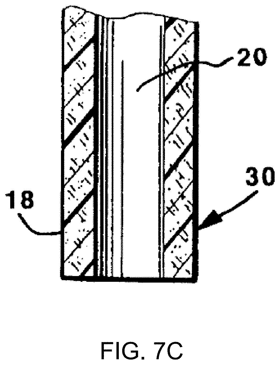
D00013

D00014

D00015

D00016

D00017

D00018

D00019

D00020
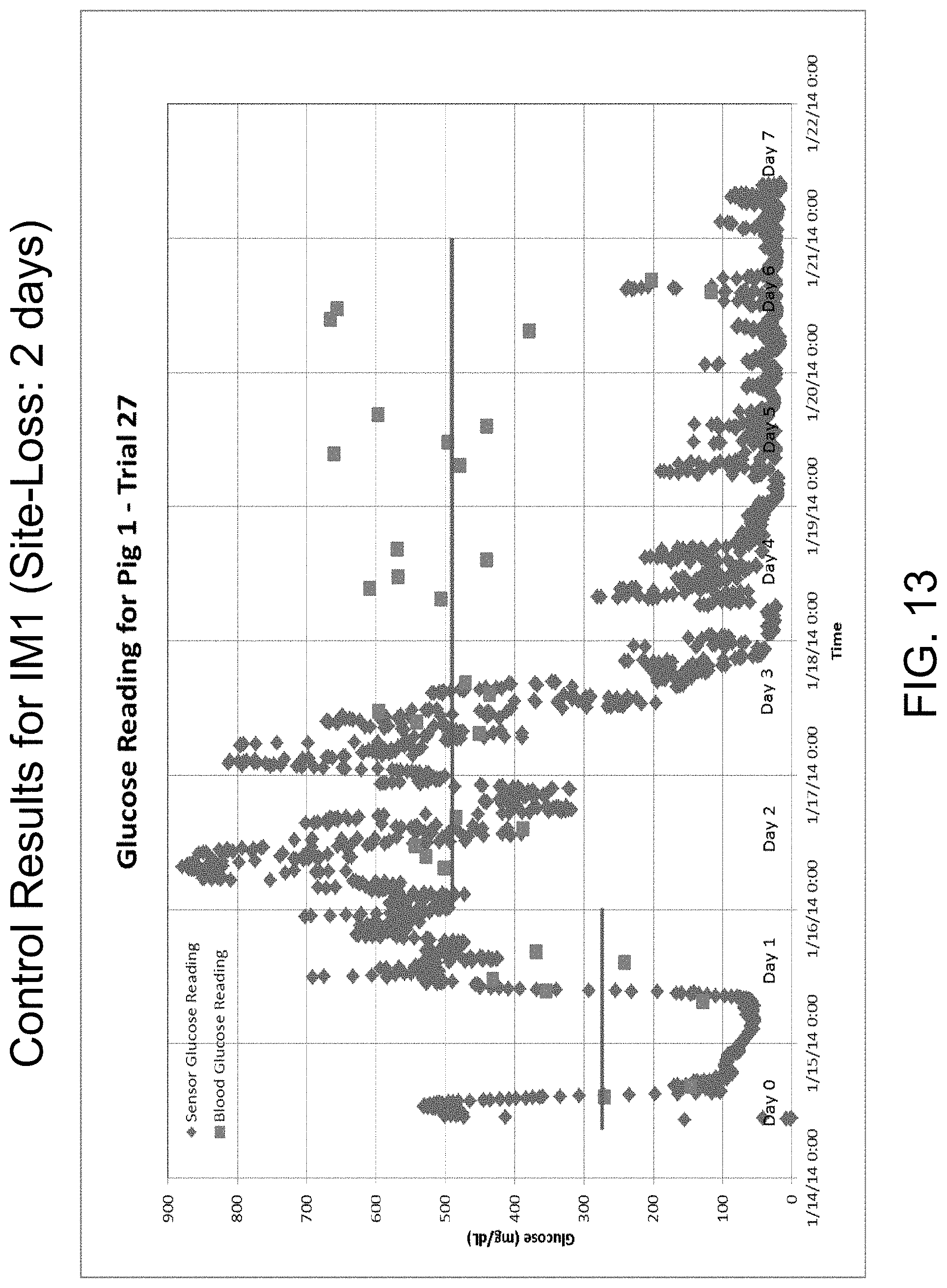
D00021

D00022

D00023

D00024

D00025

D00026

D00027

D00028
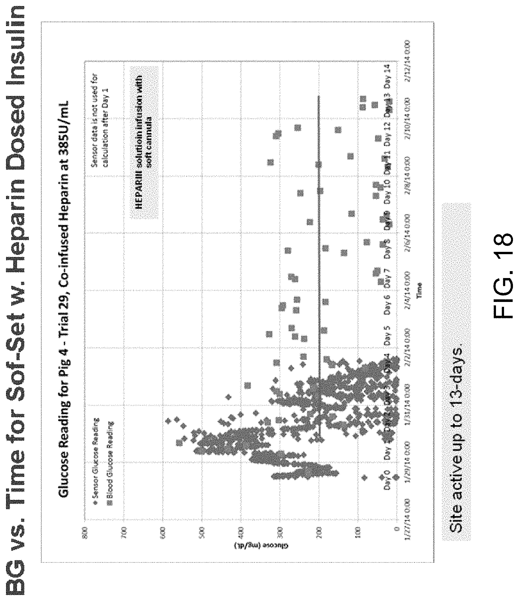
D00029

D00030

D00031

D00032

D00033

D00034
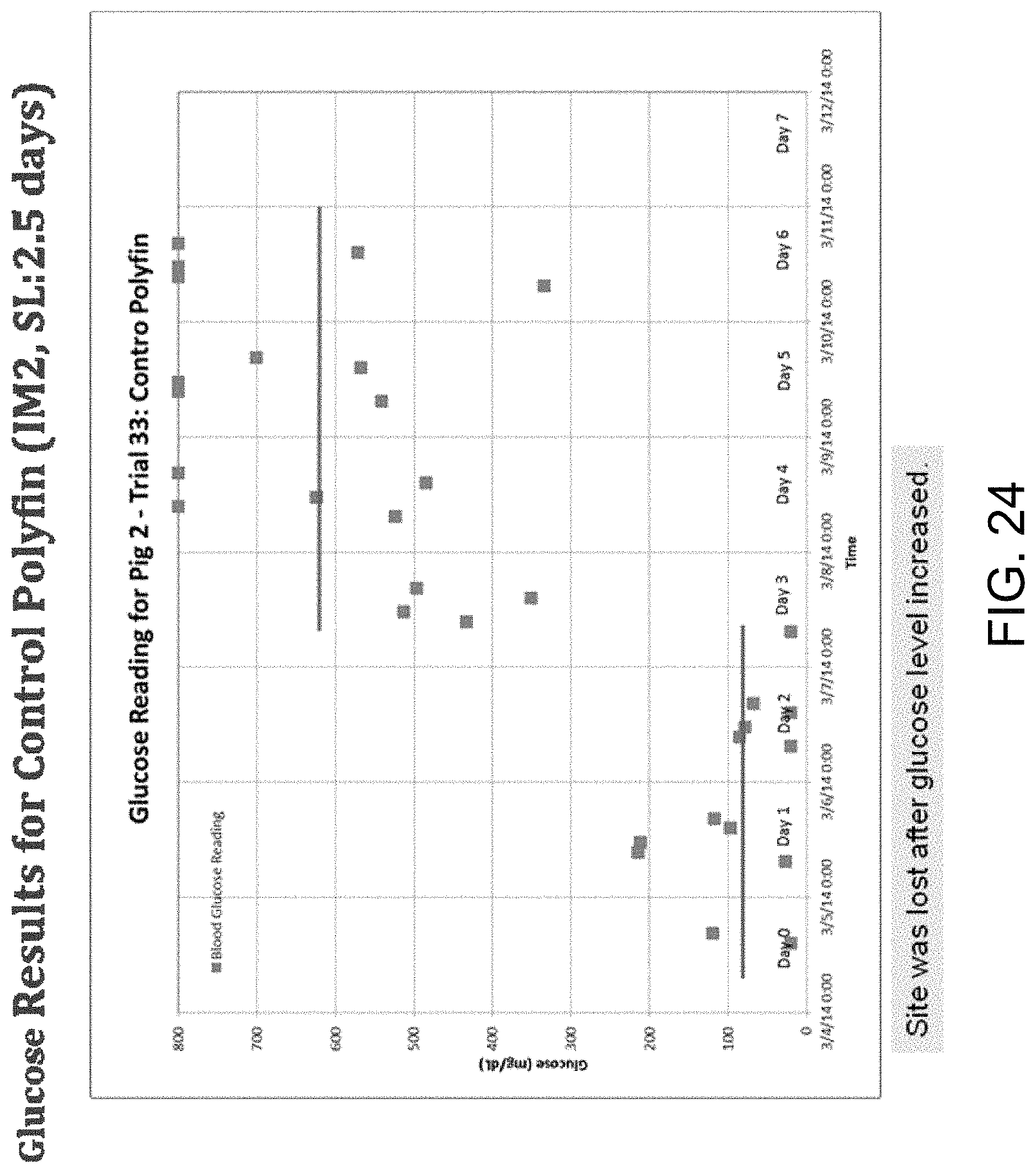
D00035
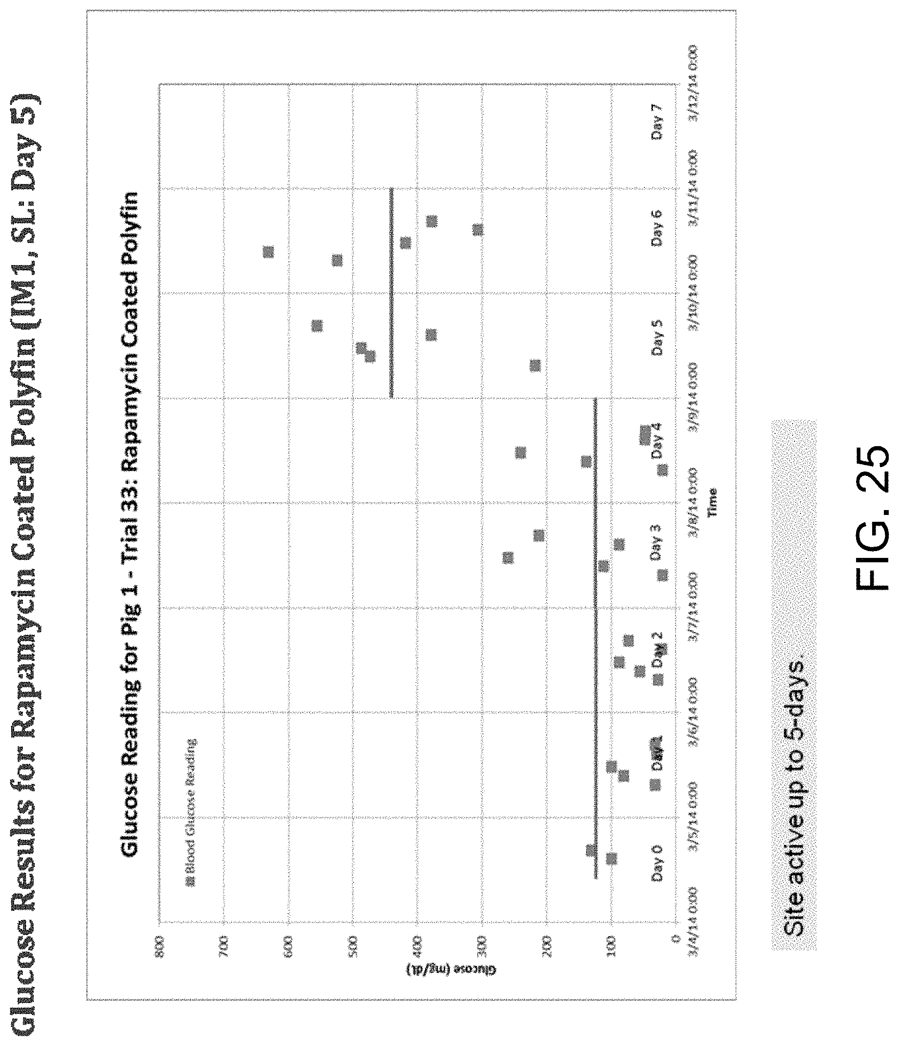
D00036

XML
uspto.report is an independent third-party trademark research tool that is not affiliated, endorsed, or sponsored by the United States Patent and Trademark Office (USPTO) or any other governmental organization. The information provided by uspto.report is based on publicly available data at the time of writing and is intended for informational purposes only.
While we strive to provide accurate and up-to-date information, we do not guarantee the accuracy, completeness, reliability, or suitability of the information displayed on this site. The use of this site is at your own risk. Any reliance you place on such information is therefore strictly at your own risk.
All official trademark data, including owner information, should be verified by visiting the official USPTO website at www.uspto.gov. This site is not intended to replace professional legal advice and should not be used as a substitute for consulting with a legal professional who is knowledgeable about trademark law.