In vivo production of proteins
de Fougerolles , et al. De
U.S. patent number 10,493,167 [Application Number 15/403,517] was granted by the patent office on 2019-12-03 for in vivo production of proteins. This patent grant is currently assigned to ModernaTX, Inc.. The grantee listed for this patent is ModernaTX, Inc.. Invention is credited to Antonin de Fougerolles, Justin Guild.


View All Diagrams
| United States Patent | 10,493,167 |
| de Fougerolles , et al. | December 3, 2019 |
In vivo production of proteins
Abstract
The invention relates to compositions including polynucleotides encoding polypeptides which have been chemically modified by replacing the uridines with 1-methyl-pseudouridine to improve one or more of the stability and/or clearance in tissues, receptor uptake and/or kinetics, cellular access by the compositions, engagement with translational machinery, mRNA half-life, translation efficiency, immune evasion, protein production capacity, secretion efficiency, accessibility to circulation, protein half-life and/or modulation of a cell's status, function, and/or activity.
| Inventors: | de Fougerolles; Antonin (Waterloo, BE), Guild; Justin (Framingham, MA) | ||||||||||
|---|---|---|---|---|---|---|---|---|---|---|---|
| Applicant: |
|
||||||||||
| Assignee: | ModernaTX, Inc. (Cambridge,
MA) |
||||||||||
| Family ID: | 49235350 | ||||||||||
| Appl. No.: | 15/403,517 | ||||||||||
| Filed: | January 11, 2017 |
Prior Publication Data
| Document Identifier | Publication Date | |
|---|---|---|
| US 20170348436 A1 | Dec 7, 2017 | |
Related U.S. Patent Documents
| Application Number | Filing Date | Patent Number | Issue Date | ||
|---|---|---|---|---|---|
| 14824247 | Aug 12, 2015 | 9572896 | |||
| 14390106 | Dec 9, 2015 | 9221891 | |||
| PCT/US2013/031821 | Mar 15, 2013 | ||||
| 61737174 | Dec 14, 2012 | ||||
| 61737184 | Dec 14, 2012 | ||||
| 61737191 | Dec 14, 2012 | ||||
| 61737203 | Dec 14, 2012 | ||||
| 61737213 | Dec 14, 2012 | ||||
| 61737155 | Dec 14, 2012 | ||||
| 61737152 | Dec 14, 2012 | ||||
| 61737147 | Dec 14, 2012 | ||||
| 61737139 | Dec 14, 2012 | ||||
| 61737135 | Dec 14, 2012 | ||||
| 61737134 | Dec 14, 2012 | ||||
| 61737130 | Dec 14, 2012 | ||||
| 61737160 | Dec 14, 2012 | ||||
| 61737168 | Dec 14, 2012 | ||||
| 61712490 | Oct 11, 2012 | ||||
| 61709303 | Oct 3, 2012 | ||||
| 61696381 | Sep 4, 2012 | ||||
| 61681658 | Aug 10, 2012 | ||||
| 61681654 | Aug 10, 2012 | ||||
| 61681650 | Aug 10, 2012 | ||||
| 61681649 | Aug 10, 2012 | ||||
| 61681648 | Aug 10, 2012 | ||||
| 61681647 | Aug 10, 2012 | ||||
| 61681645 | Aug 10, 2012 | ||||
| 61681661 | Aug 10, 2012 | ||||
| 61681742 | Aug 10, 2012 | ||||
| 61681720 | Aug 10, 2012 | ||||
| 61681712 | Aug 10, 2012 | ||||
| 61681704 | Aug 10, 2012 | ||||
| 61681696 | Aug 10, 2012 | ||||
| 61681687 | Aug 10, 2012 | ||||
| 61681675 | Aug 10, 2012 | ||||
| 61681667 | Aug 10, 2012 | ||||
| 61668157 | Jul 5, 2012 | ||||
| 61648244 | May 17, 2012 | ||||
| 61648286 | May 17, 2012 | ||||
| 61618945 | Apr 2, 2012 | ||||
| 61618935 | Apr 2, 2012 | ||||
| 61618922 | Apr 2, 2012 | ||||
| 61618862 | Apr 2, 2012 | ||||
| 61618866 | Apr 2, 2012 | ||||
| 61618868 | Apr 2, 2012 | ||||
| 61618870 | Apr 2, 2012 | ||||
| 61618873 | Apr 2, 2012 | ||||
| 61618878 | Apr 2, 2012 | ||||
| 61618885 | Apr 2, 2012 | ||||
| 61618896 | Apr 2, 2012 | ||||
| 61618953 | Apr 2, 2012 | ||||
| 61618957 | Apr 2, 2012 | ||||
| 61618961 | Apr 2, 2012 | ||||
| 61618911 | Apr 2, 2012 | ||||
Foreign Application Priority Data
| Mar 9, 2013 [WO] | PCT/US2013/030059 | |||
| Mar 9, 2013 [WO] | PCT/US2013/030060 | |||
| Mar 9, 2013 [WO] | PCT/US2013/030061 | |||
| Mar 9, 2013 [WO] | PCT/US2013/030062 | |||
| Mar 9, 2013 [WO] | PCT/US2013/030063 | |||
| Mar 9, 2013 [WO] | PCT/US2013/030064 | |||
| Mar 9, 2013 [WO] | PCT/US2013/030066 | |||
| Mar 9, 2013 [WO] | PCT/US2013/030067 | |||
| Mar 9, 2013 [WO] | PCT/US2013/030068 | |||
| Mar 9, 2013 [WO] | PCT/US2013/030070 | |||
| Current U.S. Class: | 1/1 |
| Current CPC Class: | A61K 48/0033 (20130101); A61P 29/00 (20180101); C07K 14/485 (20130101); A61P 35/02 (20180101); A61P 37/02 (20180101); C12N 9/6443 (20130101); A61P 19/10 (20180101); A61K 31/7115 (20130101); A61P 17/02 (20180101); C07K 16/40 (20130101); A61K 48/0066 (20130101); A61P 25/18 (20180101); A61P 31/14 (20180101); C07K 14/4713 (20130101); C07K 14/705 (20130101); A61P 1/00 (20180101); A61P 11/00 (20180101); A61P 25/30 (20180101); A61P 31/18 (20180101); C07K 14/4705 (20130101); C07K 14/745 (20130101); A61K 38/177 (20130101); A61P 7/04 (20180101); A61P 25/24 (20180101); A61P 13/00 (20180101); A61P 17/00 (20180101); A61P 17/06 (20180101); A61P 43/00 (20180101); C07K 14/505 (20130101); C07K 14/5418 (20130101); A61K 38/212 (20130101); A61P 25/00 (20180101); A61P 35/04 (20180101); C07H 21/02 (20130101); C12N 9/6437 (20130101); C12N 9/644 (20130101); A61P 17/08 (20180101); A61P 31/04 (20180101); C12N 9/1051 (20130101); A61K 38/1816 (20130101); A61P 17/04 (20180101); A61P 19/02 (20180101); A61P 35/00 (20180101); C07K 14/525 (20130101); C07K 16/2863 (20130101); C07K 16/2887 (20130101); C12Y 603/02019 (20130101); A61P 7/02 (20180101); A61P 9/10 (20180101); A61P 13/12 (20180101); C07K 14/535 (20130101); A61K 38/193 (20130101); A61K 38/45 (20130101); A61K 47/543 (20170801); A61P 31/10 (20180101); C07K 14/475 (20130101); A61P 7/06 (20180101); A61P 37/06 (20180101); C12N 9/93 (20130101); C12P 13/04 (20130101); C12P 21/00 (20130101); A61K 9/145 (20130101); A61K 9/5146 (20130101); A61P 21/04 (20180101); C07K 14/47 (20130101); C12N 9/0069 (20130101); C12N 9/6435 (20130101); C12P 21/005 (20130101); A61P 3/00 (20180101); A61P 9/00 (20180101); A61P 25/02 (20180101); A61P 37/00 (20180101); C07K 14/43595 (20130101); C12N 9/0091 (20130101); A61P 15/00 (20180101); A61P 27/02 (20180101); C12N 9/2445 (20130101); A61K 38/4846 (20130101); A61P 3/06 (20180101); A61P 5/00 (20180101); A61P 31/12 (20180101); C07K 14/495 (20130101); C12N 15/52 (20130101); A61K 9/1271 (20130101); C07K 14/4723 (20130101); C12N 9/6451 (20130101); C12N 15/11 (20130101); A61P 25/28 (20180101); C07K 14/005 (20130101); C07K 14/515 (20130101); A61P 21/00 (20180101); C12N 9/88 (20130101); C12N 9/2402 (20130101); C12N 15/85 (20130101); A61K 9/5153 (20130101); A61K 38/191 (20130101); A61P 7/00 (20180101); C12N 15/87 (20130101); A61P 31/00 (20180101); C07K 14/435 (20130101); C07K 14/56 (20130101); C07K 16/00 (20130101); A61P 19/00 (20180101); A61P 37/08 (20180101); C07K 14/62 (20130101); A61K 9/5123 (20130101); A61K 38/17 (20130101); A61P 3/10 (20180101); A61P 25/14 (20180101); A61P 17/14 (20180101); C07K 14/61 (20130101); C12N 9/16 (20130101); A61K 38/1891 (20130101); A61K 48/005 (20130101); A61K 48/0075 (20130101); C12N 9/1241 (20130101); A61P 1/04 (20180101); C07K 14/4746 (20130101); C12Y 113/12007 (20130101); Y02A 50/423 (20180101); A61K 38/00 (20130101); C12Y 304/21007 (20130101); C12Y 116/03001 (20130101); C12Y 403/02001 (20130101); C12Y 304/21022 (20130101); Y02A 50/30 (20180101); Y02A 50/401 (20180101); Y02A 50/415 (20180101); C12Y 304/21027 (20130101); Y02A 50/463 (20180101); Y02A 50/414 (20180101); Y02A 50/491 (20180101); C12Y 207/07012 (20130101); Y02A 50/411 (20180101) |
| Current International Class: | A61K 31/7115 (20060101); C12N 15/11 (20060101); C12N 9/00 (20060101); C12N 15/85 (20060101); C07K 14/47 (20060101); C07K 14/62 (20060101); C12N 9/88 (20060101); C07K 14/705 (20060101); C07K 14/505 (20060101); C07K 14/535 (20060101); C12N 9/64 (20060101); C12N 9/10 (20060101); C12N 9/42 (20060101); C12N 15/87 (20060101); C12P 13/04 (20060101); A61K 38/45 (20060101); C12N 9/16 (20060101); C12N 9/24 (20060101); C12N 15/52 (20060101); C07K 14/435 (20060101); C12P 21/00 (20060101); C07K 14/485 (20060101); C07H 21/02 (20060101); C07K 14/475 (20060101); C07K 14/495 (20060101); C07K 14/525 (20060101); C07K 14/54 (20060101); C07K 14/56 (20060101); C07K 14/61 (20060101); C07K 14/745 (20060101); C07K 16/28 (20060101); C07K 16/40 (20060101); C12N 9/02 (20060101); C12N 9/12 (20060101); C12N 9/68 (20060101); A61K 9/14 (20060101); A61K 9/51 (20060101); A61K 38/18 (20060101); A61K 38/19 (20060101); A61K 38/21 (20060101); A61K 38/48 (20060101); C07K 14/005 (20060101); A61K 9/127 (20060101); A61K 47/54 (20170101); A61K 38/17 (20060101); C07K 16/00 (20060101); A61K 48/00 (20060101); C07K 14/515 (20060101); A61K 38/00 (20060101) |
References Cited [Referenced By]
U.S. Patent Documents
| 7745391 | June 2010 | Mintz et al. |
| 2005/0143336 | June 2005 | Ramesh et al. |
| 2006/0257902 | November 2006 | Mendoza et al. |
| 2007/0224635 | September 2007 | Bouquin |
| 2008/0025944 | January 2008 | Hoerr et al. |
| 2009/0093433 | April 2009 | Woolf et al. |
| 2009/0171070 | July 2009 | Van Urk |
| 2009/0286852 | November 2009 | Kariko et al. |
| 2010/0261231 | October 2010 | Kore et al. |
| 2010/0297750 | November 2010 | Natsume et al. |
| 2011/0143397 | June 2011 | Kariko et al. |
| 2011/0269950 | November 2011 | Von Der Mulbe et al. |
| 2012/0027803 | February 2012 | Manoharan et al. |
| 2012/0046346 | February 2012 | Rossi et al. |
| 2012/0065252 | March 2012 | Schrum et al. |
| 2012/0195936 | August 2012 | Rudolph et al. |
| 2013/0115274 | May 2013 | Knopov |
| 2013/0259923 | October 2013 | Bancel et al. |
| 2113247 | Nov 2009 | EP | |||
| 2009-171861 | Aug 2009 | JP | |||
| WO-99/14346 | Mar 1999 | WO | |||
| WO-00/50586 | Aug 2000 | WO | |||
| WO-01/55306 | Aug 2001 | WO | |||
| WO-03/009806 | Feb 2003 | WO | |||
| WO-2004/064782 | Aug 2004 | WO | |||
| WO-2004/094345 | Nov 2004 | WO | |||
| WO-2005/014814 | Feb 2005 | WO | |||
| WO-2005/062854 | Jul 2005 | WO | |||
| WO-2005/121348 | Dec 2005 | WO | |||
| WO-2006/029023 | Mar 2006 | WO | |||
| WO-2006/034433 | Mar 2006 | WO | |||
| WO-2007/000668 | Jan 2007 | WO | |||
| WO-2007/024708 | Mar 2007 | WO | |||
| WO-2007/065957 | Jun 2007 | WO | |||
| WO-2007/100789 | Sep 2007 | WO | |||
| WO-2008/052770 | May 2008 | WO | |||
| WO-2008/061192 | May 2008 | WO | |||
| WO-2008/147824 | Dec 2008 | WO | |||
| WO-2009/051451 | Apr 2009 | WO | |||
| WO-2009/065600 | May 2009 | WO | |||
| WO-2009/127060 | Oct 2009 | WO | |||
| WO-2009/141146 | Nov 2009 | WO | |||
| WO-2009/146523 | Dec 2009 | WO | |||
| WO-2010/011895 | Jan 2010 | WO | |||
| WO-2010/022961 | Mar 2010 | WO | |||
| WO-2010/039548 | Apr 2010 | WO | |||
| WO-2010/042877 | Apr 2010 | WO | |||
| WO-2010/054401 | May 2010 | WO | |||
| WO-2010/061996 | Jun 2010 | WO | |||
| WO-2010/144740 | Dec 2010 | WO | |||
| WO-2011/000107 | Jan 2011 | WO | |||
| WO-2011/012316 | Feb 2011 | WO | |||
| WO-2011/068810 | Jun 2011 | WO | |||
| WO-2011/071931 | Jun 2011 | WO | |||
| WO-2011/090965 | Jul 2011 | WO | |||
| WO-2011/133890 | Oct 2011 | WO | |||
| WO-2011/140627 | Nov 2011 | WO | |||
| WO-2011/153493 | Dec 2011 | WO | |||
| WO-2012/004276 | Jan 2012 | WO | |||
| WO-2012/019168 | Feb 2012 | WO | |||
| WO-2012/031046 | Mar 2012 | WO | |||
| WO-2012/135805 | Oct 2012 | WO | |||
| WO-2012/170889 | Dec 2012 | WO | |||
| WO-2012/170930 | Dec 2012 | WO | |||
| WO-2013/039857 | Mar 2013 | WO | |||
| WO-2013/052523 | Apr 2013 | WO | |||
| WO-2013/090648 | Jun 2013 | WO | |||
| WO-2013/096709 | Jun 2013 | WO | |||
| WO-2013/106496 | Jul 2013 | WO | |||
| WO-2013/120497 | Aug 2013 | WO | |||
| WO-2013/151663 | Oct 2013 | WO | |||
| WO-2013/151664 | Oct 2013 | WO | |||
| WO-2013/151665 | Oct 2013 | WO | |||
| WO-2013/151666 | Oct 2013 | WO | |||
| WO-2013/151667 | Oct 2013 | WO | |||
| WO-2013/151668 | Oct 2013 | WO | |||
| WO-2013/151669 | Oct 2013 | WO | |||
| WO-2013/151670 | Oct 2013 | WO | |||
| WO-2013/151671 | Oct 2013 | WO | |||
| WO-2013/151672 | Oct 2013 | WO | |||
Other References
|
Blum et al., "Polyadenylation promotes degradation of 3'-structured RNA by the Escherichia coli mRNA degradosome in vitro," J Biol Chem. 274(7): 4009-16 (1999) (9 pages). cited by applicant . Communication pursuant to Artcile 94(3) EPC for European Application No. 13713306.2, dated Sep. 4, 2017 (4 pages). cited by applicant . Decision of Rejection for Japanese Application No. 2015-504587, dated Sep. 27, 2016 (7 pages). cited by applicant . Examination Report for Australian Application No. 2013243834, dated Sep. 22, 2016 (4 pages). cited by applicant . Examination Report for Australian Application No. 2017232121, dated Apr. 24, 2018 (4 pages). cited by applicant . He, "Grand challenge commentary: RNA epigenetics?" Nat Chem Biol. 6(12):863-5 (2010). cited by applicant . International Search Report and Written Opinion for International Application No. PCT/US2013/031821, dated Oct. 11, 2013 (26 pages). cited by applicant . Kuge et al., "Cap ribose methylation of c-mos mRNA stimulates translation and oocyte maturation in Xenopus laevis," Nucleic Acids Res. 26(13):3208-14 (1998). cited by applicant . Love et al., "Lipid-like materials for low-dose, in vivo gene silencing," Proc Natl Acad Sci USA. 107(5):1864-9 (2010). cited by applicant . Moretti et al., "Mechanism of translational regulation by miR-2 from sites in the 5' untranslated region or the open reading frame," RNA. 16(12):2493-502 (2010). cited by applicant . Notification of Reasons for Rejection for Japanese Application No. 2015-504587, dated Nov. 24, 2015 (16 pages). cited by applicant . Notification of Reasons for Rejection for Japanese Application No. 2017-012947, dated Jan. 9, 2018 (7 pages). cited by applicant . Office Action for Chinese Application No. 201380028186.5, dated Dec. 27, 2016 (15 pages). cited by applicant . Office Action for Chinese Application No. 201380028186.5, dated Nov. 17, 2017 (15 pages). cited by applicant . Squires et al., "Widespread occurence of 5-methylcytosine in human coding and non-coding RNA," Nucleic Acids Res. 40(11):5023-33 (2012). cited by applicant . Ylosmaki et al., "Generation of conditonally replicating adenovirus based on targeted destruction of E1A mRNA by a cell type-specific microRNA," J Virol. 82(22):11009-15 (2008). cited by applicant . Anonymous, "Messenger RNA", Internet: Wikipedia. XP002699196, Retrieved on Jun. 21, 2013. <http://en.wikipedia.org/wiki/Messenger_RNA> (10 pages). cited by applicant . Asaka et al., "Alteration of aldolase isozymes in serum and tissues of patients with cancer and other diseases," J Clin Lab Anal. 8(3):144-8 (1994). cited by applicant . Baba et al., "Treatment of neurological disorders by introducing mRNA in vivo using polyplex nanomicelles," J Control Release 201:41-8 (2015). cited by applicant . Bionaz et al., "ACSL1, AGPAT6, FABP3, LPIN1, and SLC27A6 are the most abundant isoforms in bovine mammary tissue and their expression is affected by stage of lactation," J Nutr. 138(6):1019-24 (2008). cited by applicant . Canete-Soler et al., "Aldolases a and C are ribonucleolytic components of a neuronal complex that regulates the stability of the light-neurofilament mRNA," J Neurosci. 25(17):4353-64 (2005). cited by applicant . Communication pursuant to Article 94(3) EPC for European Patent Application No. 13713306.2, dated Jun. 22, 2016 (12 pages). cited by applicant . Creusot et al., "A short pulse of IL-4 delivered by DCs electroporated with modified mRNA can both prevent and treat autoimmune diabetes in NOD mice," Mol Ther. 18(12):2112-20 (2010). cited by applicant . Didiot et al., "The G-quartet containing FMRP binding site in FMR1 mRNA is a potent exonic splicing enhancer," Nucleic Acids Res. 36(15):4902-12 (2008). cited by applicant . Esposito et al., "Human aldolase A natural mutants: relationship between flexibility of the C-terminal region and enzyme function," Biochem J. 380(Pt 1):51-6 (2004). cited by applicant . International Preliminary Report on Patentability for International Application No. PCT/US2013/030061, dated Oct. 7, 2014 (15 pages). cited by applicant . International Search Report and Written Opinion for International Application No. PCT/US2013/030061, dated Nov. 18, 2013 (24 pages). cited by applicant . Kariko et al., "Overexpression of urokinase receptor in mammalian cells following administration of the in vitro transcribed encoding mRNA," Gene Ther. 6(6):1092-100 (1999). cited by applicant . Kariko et al., "Incorporation of pseudouridine into mRNA yields superior nonimmunogenic vector with increased translational capacity and biological stability." Mol Ther. 16(11):1833-40 (2008). cited by applicant . Kormann et al., "Expression of therapeutic proteins after delivery of chemically modified mRNA in mice," Nat Biotechnol. 29(2):154-7 (2011) (6 pages). cited by applicant . Leung D.W., "The structure and functions of human lysophosphatidic acid acyltransferases," Front Biosci. 6:D944-53 (2001) (11 pages). cited by applicant . Lonez et al., "Cationic liposomal lipids: from gene carriers to cell signaling," Prog Lipid Res. 47(5):340-7 (2008). cited by applicant . Lorenz et al., "Protein expression from exogenous mRNA: uptake by receptor-mediated endocytosis and trafficking via the lysosomal pathway," RNA Biol. 8(4):627-36 (2011). cited by applicant . Lu et al., "Cloning and characterization of murine 1-acyl-sn-glycerol 3-phosphate acyltransferases and their regulation by PPARalpha in murine heart," Biochem J. 385(Pt 2):469-77 (2005). cited by applicant . Lu et al., "Optimization of methods to achieve mRNA-mediated transfection of tumor cells in vitro and in vivo employing cationic liposome vectors," Cancer Gene Ther. 1(4):245-52 (1994). cited by applicant . Mignone et al., "Untranslated regions of mRNAs," Genome Biol. 3(3):reviews0004.1-10 (2002). cited by applicant . Perche et al., "Enhancement of dendritic cells transfection in vivo and of vaccination against B16F10 melanoma with mannosylated histidylated lipopolyplexes loaded with tumor antigen messenger RNA," Nanomedicine. 7(4):445-53 (2011). cited by applicant . Pezza et al., "Spatial clustering of isozyme-specific residues reveals unlikely determinants of isozyme specificity in fructose-1,6-bisphosphate aldolase," J Biol Chem. 278(19):17307-13 (2003). cited by applicant . Probst et al., "Spontaneous cellular uptake of exogenous messenger RNA in vivo is nucleic acid-specific, saturable and ion dependent," Gene Therapy. 14: 1175-80 (2007). cited by applicant . Puigbo et al., "Optimizer: a web server for optimizing the codon usage of DNA sequences," Nucleic Acids Res. 35:W126-131 (2007). cited by applicant . Tavernier et al., "mRNA as gene therapeutic: how to control protein expression," J Control Release. 150(3):238-47 (2011). cited by applicant . Warren et al., "Highly efficient reprogramming to pluripotency and directed differentiation of human cells with synthetic modified mRNA," Cell Stem Cell. 7(5):618-30 (2010). cited by applicant . West et al., "Cloning and expression of two human lysophosphatidic acid acyltransferase cDNAs that enhance cytokine-induced signaling responses in cells," DNA Cell Biol. 16(6):691-701 (1997). cited by applicant . Yamamoto et al., "Current prospects for mRNA gene delivery," Eur J Pharm Biopharm. 71(3):484-9 (2009). cited by applicant . Zohra et al., "Drastic effect of nanoapatite particles on liposome-mediated mRNA delivery to mammalian cells," Anal Biochem. 345(1): 164-6 (2005). cited by applicant . Fath et al., "Multiparameter RNA and codon optimization: a standardized tool to assess and enhance autologous mammalian gene expression," PLoS One 6(3):e17596 (2011) (14 pages). cited by applicant . Dannull et al., "Enhancing the immunostimulatory function of dendritic cells by transfection with mRNA encoding OX40 ligand," Blood. 105(8):3206-13 (2005). cited by applicant . Henke et al., "microRNA-122 stimulates translation of hepatitis C virus RNA," EMBO J. 27(24):3300-10 (2008). cited by applicant . Examiner's Report for Canadian Patent Application No. 2,868,418, dated Nov. 19, 2018 (4 pages). cited by applicant . Probst et al., "Spontaneous cellular uptake of exogenous messenger RNA in vivo is nucleic acid-specific, saturable and ion dependent," Gene Ther. 14(15):1175-80 (2007). cited by applicant . Andries et al., "N1-methylpseudouridine-incorporated mRNA outperforms pseudouridine-incorporated mRNA by providing enhanced protein expression and reduced immunogenicity in mammalian cell lines and mice," J Control Release. 217:337-44 (2015). cited by applicant . Charette et al., "Pseudouridine in RNA: what, where, how, and why," IUBMB Life. 49(5):341-51 (2000). cited by applicant . Extended European Search Report for European Patent Application No. 18204317.4, dated Jul. 5, 2019 (13 pages). cited by applicant . Kormann et al., "Expression of therapeutic proteins after delivery of chemically modified mRNA in mice," Nat Biotechnol. 29(2):154-7 (2011). cited by applicant . Sullenger et al., "Emerging clinical applications of RNA," Nature. 418(6894):252-8 (2002). cited by applicant . Van Tendeloo et al., "mRNA-based gene transfer as a tool for gene and cell therapy," Curr Opin Mol Ther. 9(5):423-31 (2007). cited by applicant . Uzgun et al., "PEGylation Improves Nanoparticle Formation and Transfection Efficiency of Messenger RNA," Pharm. Res. 28(9): 2223-32 (2011). cited by applicant. |
Primary Examiner: Gonzalez; Antonio Galisteo
Attorney, Agent or Firm: Clark & Elbing LLP
Parent Case Text
CROSS REFERENCE TO RELATED APPLICATIONS
This application is a continuation of U.S. application Ser. No. 14/390,106, filed Oct. 2, 2014, which is a 35 U.S.C. .sctn. 371 U.S. National Stage Entry of International Application No. PCT/US2013/031821 filed Mar. 15, 2013 which claims priority of U.S. Provisional Patent Application No. 61/618,862, filed Apr. 2, 2012, entitled Modified Polynucleotides for the Production of Biologics, U.S. Provisional Patent Application No. 61/681,645, filed Aug. 10, 2012, entitled Modified Polynucleotides for the Production of Biologics, U.S. Provisional Patent Application No. 61/737,130, filed Dec. 14, 2012, entitled Modified Polynucleotides for the Production of Biologics, U.S. Provisional Patent Application No. 61/618,866, filed Apr. 2, 2012, entitled Modified Polynucleotides for the Production of Antibodies, U.S. Provisional Patent Application No. 61/681,647, filed Aug. 10, 2012, entitled Modified Polynucleotides for the Production of Antibodies, U.S. Provisional Patent Application No. 61/737,134, filed Dec. 14, 2012, entitled Modified Polynucleotides for the Production of Antibodies, U.S. Provisional Patent Application No. 61/618,868, filed Apr. 2, 2012, entitled Modified Polynucleotides for the Production of Vaccines, U.S. Provisional Patent Application No. 61/681,648, filed Aug. 10, 2012, entitled Modified Polynucleotides for the Production of Vaccines, U.S. Provisional Patent Application No. 61/737,135, filed Dec. 14, 2012, entitled Modified Polynucleotides for the Production of Vaccines, U.S. Provisional Patent Application No. 61/618,870, filed Apr. 2, 2012, entitled Modified Polynucleotides for the Production of Therapeutic Proteins and Peptides, U.S. Provisional Patent Application No. 61/681,649, filed Aug. 10, 2012, entitled Modified Polynucleotides for the Production of Therapeutic Proteins and Peptides, U.S. Provisional Patent Application No. 61/737,139, filed Dec. 14, 2012, Modified Polynucleotides for the Production of Therapeutic Proteins and Peptides, U.S. Provisional Patent Application No. 61/618,873, filed Apr. 2, 2012, entitled Modified Polynucleotides for the Production of Secreted Proteins, U.S. Provisional Patent Application No. 61/681,650, filed Aug. 10, 2012, entitled Modified Polynucleotides for the Production of Secreted Proteins, U.S. Provisional Patent Application No. 61/737,147, filed Dec. 14, 2012, entitled Modified Polynucleotides for the Production of Secreted Proteins, U.S. Provisional Patent Application No. 61/618,878, filed Apr. 2, 2012, entitled Modified Polynucleotides for the Production of Plasma Membrane Proteins, U.S. Provisional Patent Application No. 61/681,654, filed Aug. 10, 2012, entitled Modified Polynucleotides for the Production of Plasma Membrane Proteins, U.S. Provisional Patent Application No. 61/737,152, filed Dec. 14, 2012, entitled Modified Polynucleotides for the Production of Plasma Membrane Proteins, U.S. Provisional Patent Application No. 61/618,885, filed Apr. 2, 2012, entitled Modified Polynucleotides for the Production of Cytoplasmic and Cytoskeletal Proteins, U.S. Provisional Patent Application No. 61/681,658, filed Aug. 10, 2012, entitled Modified Polynucleotides for the Production of Cytoplasmic and Cytoskeletal Proteins, U.S. Provisional Patent Application No. 61/737,155, filed Dec. 14, 2012, entitled Modified Polynucleotides for the Production of Cytoplasmic and Cytoskeletal Proteins, U.S. Provisional Patent Application No. 61/618,896, filed Apr. 2, 2012, entitled Modified Polynucleotides for the Production of Intracellular Membrane Bound Proteins, U.S. Provisional Patent Application No. 61/668,157, filed Jul. 5, 2012, entitled Modified Polynucleotides for the Production of Intracellular Membrane Bound Proteins, U.S. Provisional Patent Application No. 61/681,661, filed Aug. 10, 2012, entitled Modified Polynucleotides for the Production of Intracellular Membrane Bound Proteins, U.S. Provisional Patent Application No. 61/737,160, filed Dec. 14, 2012, entitled Modified Polynucleotides for the Production of Intracellular Membrane Bound Proteins, U.S. Provisional Patent Application No. 61/618,911, filed Apr. 2, 2012, entitled Modified Polynucleotides for the Production of Nuclear Proteins, U.S. Provisional Patent Application No. 61/681,667, filed Aug. 10, 2012, entitled Modified Polynucleotides for the Production of Nuclear Proteins, U.S. Provisional Patent Application No. 61/737,168, filed Dec. 14, 2012, entitled Modified Polynucleotides for the Production of Nuclear Proteins, U.S. Provisional Patent Application No. 61/618,922, filed Apr. 2, 2012, entitled Modified Polynucleotides for the Production of Proteins, U.S. Provisional Patent Application No. 61/681,675, filed Aug. 10, 2012, entitled Modified Polynucleotides for the Production of Proteins, U.S. Provisional Patent Application No. 61/737,174, filed Dec. 14, 2012, entitled Modified Polynucleotides for the Production of Proteins, U.S. Provisional Patent Application No. 61/618,935, filed Apr. 2, 2012, entitled Modified Polynucleotides for the Production of Proteins Associated with Human Disease, U.S. Provisional Patent Application No. 61/681,687, filed Aug. 10, 2012, entitled Modified Polynucleotides for the Production of Proteins Associated with Human Disease, U.S. Provisional Patent Application No. 61/737,184, filed Dec. 14, 2012, entitled Modified Polynucleotides for the Production of Proteins Associated with Human Disease, U.S. Provisional Patent Application No. 61/618,945, filed Apr. 2, 2012, entitled Modified Polynucleotides for the Production of Proteins Associated with Human Disease, U.S. Provisional Patent Application No. 61/681,696, filed Aug. 10, 2012, entitled Modified Polynucleotides for the Production of Proteins Associated with Human Disease, U.S. Provisional Patent Application No. 61/737,191, filed Dec. 14, 2012, entitled Modified Polynucleotides for the Production of Proteins Associated with Human Disease, U.S. Provisional Patent Application No. 61/618,953, filed Apr. 2, 2012, entitled Modified Polynucleotides for the Production of Proteins Associated with Human Disease, U.S. Provisional Patent Application No. 61/681,704, filed Aug. 10, 2012, entitled Modified Polynucleotides for the Production of Proteins Associated with Human Disease, U.S. Provisional Patent Application No. 61/737,203, filed Dec. 14, 2012, entitled Modified Polynucleotides for the Production of Proteins Associated with Human Disease, U.S. Provisional Patent Application No. 61/681,720, filed Aug. 10, 2012, entitled Modified Polynucleotides for the Production of Cosmetic Proteins and Peptides, U.S. Provisional Patent Application No. 61/737,213, filed Dec. 14, 2012, entitled Modified Polynucleotides for the Production of Cosmetic Proteins and Peptides, U.S. Provisional Patent Application No. 61/681,742, filed, Aug. 10, 2012, entitled Modified Polynucleotides for the Production of Oncology-Related Proteins and Peptides, U.S. Provisional Patent Application No. 61/618,961, filed Apr. 2, 2012, entitled Dosing Methods for Modified mRNA, U.S. Provisional Patent Application No. 61/648,286, filed May 17, 2012, entitled Dosing Methods for Modified mRNA, U.S. Provisional Patent Application No. 61/618,957, filed Apr. 2, 2012, entitled Modified Nucleoside, Nucleotide, and Nucleic Acid Compositions, U.S. Provisional Patent Application No. 61/648,244, filed May 17, 2012, entitled Modified Nucleoside, Nucleotide, and Nucleic Acid Compositions, U.S. Provisional Patent Application No. 61/681,712, filed Aug. 10, 2012, entitled Modified Nucleoside, Nucleotide, and Nucleic Acid Compositions, U.S. Provisional Patent Application No. 61/696,381, filed Sep. 4, 2012, entitled Modified Nucleoside, Nucleotide, and Nucleic Acid Compositions, U.S. Provisional Patent Application No. 61/709,303, filed Oct. 3, 2012, entitled Modified Nucleoside, Nucleotide, and Nucleic Acid Compositions, U.S. Provisional Patent Application No. 61/712,490, filed Oct. 11, 2012, entitled Modified Nucleoside, Nucleotide, and Nucleic Acid Compositions, International Application No. PCT/US2013/030066 on Mar. 9, 2013, (PCT/US13/030062) entitled Modified Polynucleotides for the Production of Biologics and Proteins Associated with Human Disease; (PCT/US13/030064), entitled Modified Polynucleotides for the Production of Secreted Proteins; (PCT/US13/030059), entitled Modified Polynucleotides for the Production of Membrane Proteins; (PCT/US13/030063), entitled Modified Polynucleotides for the Production of Proteins; (PCT/US13/030067), entitled Modified Polynucleotides for the Production of Nuclear Proteins; (PCT/US13/030066), entitled Modified Polynucleotides for the Production of Proteins; (PCT/US13/030061), entitled Modified Polynucleotides for the Production of Proteins Associated with Human Disease; (PCT/US13/030060), entitled Modified Polynucleotides for the Production of Cosmetic Proteins and Peptides and (PCT/US13/030070), entitled Modified Polynucleotides for the Production of Oncology-Related Proteins and Peptides, the contents of each of which are herein incorporated by reference in its entirety.
This application is related to U.S. Provisional Patent Application No. 61/737,224, filed Dec. 14, 2012, entitled Terminally Optimized Modified RNAs, the contents of which are herein incorporated by reference in its entirety.
This application is also related to International Application No PCT/US2012/069610, filed Dec. 14, 2012, entitled Modified Nucleoside, Nucleotide, and Nucleic Acid Compositions, International Publication No. PCT/US2012/58519, filed Oct. 3, 2012, entitled Modified Nucleosides, Nucleotides, and Nucleic Acids, and Uses Thereof, the contents of each of which are herein incorporated by reference in its entirety.
Claims
We claim:
1. A method of expressing a secreted protein in a mammalian subject, the method comprising administering a pharmaceutical composition comprising a plurality of lipid nanoparticles encapsulating a polynucleotide, wherein the lipid nanoparticle comprises a biodegradable cationic lipid, a neutral lipid, cholesterol, and a PEGylated lipid and the plurality of lipid nanoparticles has a mean particle size of between 80 nm to 160 nm, and wherein the polynucleotide comprises: (a) an open reading frame encoding the secreted protein consisting of nucleotides selected from 1-methyl-pseudouridine, cytidine, adenosine, and guanosine; (b) a 5'-UTR; (c) at least one 5' cap structure; (d) a 3'-UTR; and (e) a 3' tailing sequence of linked nucleosides.
2. The method of claim 1, wherein the biodegradable cationic lipid comprises an ester linkage.
3. The method of claim 1, wherein the secreted protein is a cytokine.
4. The method of claim 1, wherein the method comprises administering about 0.05 to about 0.5 mg/kg of polynucleotide.
5. The method of claim 1, wherein the administration is intramuscular administration.
6. The method of claim 1, wherein the administration is intravenous administration.
7. The method of claim 1, wherein upon administration, expression of the secreted protein is maximal at 8-24 hours.
8. The method of claim 1, wherein the 3'-tailing sequence of linked nucleosides is selected from the group consisting of a poly-A tail and a polyA-G quartet.
9. The method of claim 8, wherein the poly-A comprises approximately 160 nucleotides.
10. The method of claim 1, wherein the at least one 5' cap structure is selected from the group consisting of Cap0, Cap1, ARCA, inosine, N1-methyl-guanosine, 2'fluoro-guanosine, 7-deaza-guanosine, 8-oxo-guanosine, 2-amino-guanosine, LNA-guanosine, and 2-azido-guanosine.
11. The method of claim 10, wherein the at least one 5'-cap structure is cap0, cap1, or ARCA.
12. The method of claim 1, wherein the plurality of lipid nanoparticles has a mean PDI of between 0.02 and 0.2.
13. The method of claim 1, wherein the plurality of lipid nanoparticles has a mean lipid to polynucleotide ratio (wt/wt) of between 10 and 20.
14. The method of claim 1, wherein the 3'-UTR comprises a miR binding site.
15. The method of claim 1, wherein the 5'-UTR comprises a Kozak sequence.
16. The method of claim 1, wherein the neutral lipid is a phospholipid.
17. The method of claim 1, wherein the open reading frame is codon optimized to bias GC content.
18. The method of claim 1, wherein the plurality of lipid nanoparticles comprise about 50 mol % biodegradable cationic lipid, about 38.5% cholesterol, about 10% neutral lipid and about 1.5% PEGylated lipid.
19. The method of claim 1, wherein the polynucleotide includes at least two stop codons before the 3' untranslated region (UTR).
Description
REFERENCE TO SEQUENCE LISTING
The present application is being filed along with a Sequence Listing in electronic format. The Sequence Listing file entitled M313USCONSQLST.txt, was created on Aug. 11, 2015 and is 241,198 bytes in size. The information in electronic format of the Sequence Listing is incorporated herein by reference in its entirety.
FIELD OF THE INVENTION
The invention relates to compositions, methods, processes, kits and devices for the design, preparation, manufacture and/or formulation of polynucleotides, primary constructs and modified mRNA molecules (mmRNA).
BACKGROUND OF THE INVENTION
There are multiple problems with prior methodologies of effecting protein expression. For example, introduced DNA can integrate into host cell genomic DNA at some frequency, resulting in alterations and/or damage to the host cell genomic DNA. Alternatively, the heterologous deoxyribonucleic acid (DNA) introduced into a cell can be inherited by daughter cells (whether or not the heterologous DNA has integrated into the chromosome) or by offspring. In addition, assuming proper delivery and no damage or integration into the host genome, there are multiple steps which must occur before the encoded protein is made. Once inside the cell, DNA must be transported into the nucleus where it is transcribed into RNA. The RNA transcribed from DNA must then enter the cytoplasm where it is translated into protein. Not only do the multiple processing steps from administered DNA to protein create lag times before the generation of the functional protein, each step represents an opportunity for error and damage to the cell. Further, it is known to be difficult to obtain DNA expression in cells as DNA frequently enters a cell but is not expressed or not expressed at reasonable rates or concentrations. This can be a particular problem when DNA is introduced into primary cells or modified cell lines.
In the early 1990's Bloom and colleagues successfully rescued vasopressin-deficient rats by injecting in vitro-transcribed vasopressin mRNA into the hypothalamus (Science 255: 996-998; 1992). However, the low levels of translation and the immunogenicity of the molecules hampered the development of mRNA as a therapeutic and efforts have since focused on alternative applications that could instead exploit these pitfalls, i.e. immunization with mRNAs coding for cancer antigens.
Others have investigated the use of mRNA to deliver a polypeptide of interest and shown that certain chemical modifications of mRNA molecules, particularly pseudouridine and 5-methyl-cytosine, have reduced immunostimulatory effect.
These studies are disclosed in, for example, Ribostem Limited in United Kingdom patent application serial number 0316089.2 filed on Jul. 9, 2003 now abandoned, PCT application number PCT/GB2004/002981 filed on Jul. 9, 2004 published as WO2005005622, U.S. patent application national phase entry Ser. No. 10/563,897 filed on Jun. 8, 2006 published as US20060247195 now abandoned, and European patent application national phase entry serial number EP2004743322 filed on Jul. 9, 2004 published as EP1646714 now withdrawn; Novozymes, Inc. in PCT application number PCT/US2007/88060 filed on Dec. 19, 2007 published as WO2008140615, U.S. patent application national phase entry Ser. No. 12/520,072 filed on Jul. 2, 2009 published as US20100028943 and European patent application national phase entry serial number EP2007874376 filed on Jul. 7, 2009 published as EP2104739; University of Rochester in PCT application number PCT/US2006/46120 filed on Dec. 4, 2006 published as WO2007064952 and U.S. patent application Ser. No. 11/606,995 filed on Dec. 1, 2006 published as US20070141030; BioNTech AG in European patent application serial number EP2007024312 filed Dec. 14, 2007 now abandoned, PCT application number PCT/EP2008/01059 filed on Dec. 12, 2008 published as WO2009077134, European patent application national phase entry serial number EP2008861423 filed on Jun. 2, 2010 published as EP2240572, U.S. patent application national phase entry Ser. No. 12/735,060 filed Nov. 24, 2010 published as US20110065103, German patent application serial number DE 10 2005 046 490 filed Sep. 28, 2005, PCT application PCT/EP2006/0448 filed Sep. 28, 2006 published as WO2007036366, national phase European patent EP1934345 published Mar. 21, 2012 and national phase U.S. patent application Ser. No. 11/992,638 filed Aug. 14, 2009 published as 20100129877; Immune Disease Institute Inc. in U.S. patent application Ser. No. 13/088,009 filed Apr. 15, 2011 published as US20120046346 and PCT application PCT/US2011/32679 filed Apr. 15, 2011 published as WO20110130624; Shire Human Genetic Therapeutics in U.S. patent application Ser. No. 12/957,340 filed on Nov. 20, 2010 published as US20110244026; Sequitur Inc. in PCT application PCT/US1998/019492 filed on Sep. 18, 1998 published as WO1999014346; The Scripps Research Institute in PCT application number PCT/US2010/00567 filed on Feb. 24, 2010 published as WO2010098861, and U.S. patent application national phase entry Ser. No. 13/203,229 filed Nov. 3, 2011 published as US20120053333; Ludwig-Maximillians University in PCT application number PCT/EP2010/004681 filed on Jul. 30, 2010 published as WO2011012316; Cellscript Inc. in U.S. Pat. No. 8,039,214 filed Jun. 30, 2008 and granted Oct. 18, 2011, U.S. patent application Ser. No. 12/962,498 filed on Dec. 7, 2010 published as US20110143436, Ser. No. 12/962,468 filed on Dec. 7, 2010 published as US20110143397, Ser. No. 13/237,451 filed on Sep. 20, 2011 published as US20120009649, and PCT applications PCT/US2010/59305 filed Dec. 7, 2010 published as WO2011071931 and PCT/US2010/59317 filed on Dec. 7, 2010 published as WO2011071936; The Trustees of the University of Pennsylvania in PCT application number PCT/US2006/32372 filed on Aug. 21, 2006 published as WO2007024708, and U.S. patent application national phase entry Ser. No. 11/990,646 filed on Mar. 27, 2009 published as US20090286852; Curevac GMBH in German patent application serial numbers DE10 2001 027 283.9 filed Jun. 5, 2001, DE10 2001 062 480.8 filed Dec. 19, 2001, and DE 20 2006 051 516 filed Oct. 31, 2006 all abandoned, European patent numbers EP1392341 granted Mar. 30, 2005 and EP1458410 granted Jan. 2, 2008, PCT application numbers PCT/EP2002/06180 filed Jun. 5, 2002 published as WO2002098443, PCT/EP2002/14577 filed on Dec. 19, 2002 published as WO2003051401, PCT/EP2007/09469 filed on Dec. 31, 2007 published as WO2008052770, PCT/EP2008/03033 filed on Apr. 16, 2008 published as WO2009127230, PCT/EP2006/004784 filed on May 19, 2005 published as WO2006122828, PCT/EP2008/00081 filed on Jan. 9, 2007 published as WO2008083949, and U.S. patent application Ser. No. 10/729,830 filed on Dec. 5, 2003 published as US20050032730, Ser. No. 10/870,110 filed on Jun. 18, 2004 published as US20050059624, Ser. No. 11/914,945 filed on Jul. 7, 2008 published as US20080267873, Ser. No. 12/446,912 filed on Oct. 27, 2009 published as US2010047261 now abandoned, Ser. No. 12/522,214 filed on Jan. 4, 2010 published as US20100189729, Ser. No. 12/787,566 filed on May 26, 2010 published as US20110077287, Ser. No. 12/787,755 filed on May 26, 2010 published as US20100239608, Ser. No. 13/185,119 filed on Jul. 18, 2011 published as US20110269950, and Ser. No. 13/106,548 filed on May 12, 2011 published as US20110311472 all of which are herein incorporated by reference in their entirety.
Notwithstanding these reports which are limited to a selection of chemical modifications including pseudouridine and 5-methyl-cytosine, there remains a need in the art for therapeutic modalities to address the myriad of barriers surrounding the efficacious modulation of intracellular translation and processing of nucleic acids encoding polypeptides or fragments thereof.
To this end, the inventors have shown that certain modified mRNA sequences have the potential as therapeutics with benefits beyond just evading, avoiding or diminishing the immune response. Such studies are detailed in published co-pending applications International Application PCT/US2011/046861 filed Aug. 5, 2011 and PCT/US2011/054636 filed Oct. 3, 2011, International Application number PCT/US2011/054617 filed Oct. 3, 2011, the contents of which are incorporated herein by reference in their entirety.
The present invention addresses this need by providing nucleic acid based compounds or polynucleotides which encode a polypeptide of interest (e.g., modified mRNA or mmRNA) and which have structural and/or chemical features that avoid one or more of the problems in the art, for example, features which are useful for optimizing formulation and delivery of nucleic acid-based therapeutics while retaining structural and functional integrity, overcoming the threshold of expression, improving expression rates, half life and/or protein concentrations, optimizing protein localization, and avoiding deleterious bio-responses such as the immune response and/or degradation pathways.
SUMMARY OF THE INVENTION
Described herein are compositions, methods, processes, kits and devices for the design, preparation, manufacture and/or formulation of modified mRNA (mmRNA) molecules.
The details of various embodiments of the invention are set forth in the description below. Other features, objects, and advantages of the invention will be apparent from the description and the drawings, and from the claims.
BRIEF DESCRIPTION OF THE DRAWINGS
The foregoing and other objects, features and advantages will be apparent from the following description of particular embodiments of the invention, as illustrated in the accompanying drawings in which like reference characters refer to the same parts throughout the different views. The drawings are not necessarily to scale, emphasis instead being placed upon illustrating the principles of various embodiments of the invention.
FIG. 1 is a schematic of a primary construct of the present invention.
FIG. 2 illustrates lipid structures in the prior art useful in the present invention. Shown are the structures for 98N12-5 (TETA5-LAP), DLin-DMA, DLin-K-DMA (2,2-Dilinoleyl-4-dimethylaminomethyl-[1,3]-dioxolane), DLin-KC2-DMA, DLin-MC3-DMA and C12-200.
FIG. 3 is a representative plasmid useful in the IVT reactions taught herein. The plasmid contains Insert 64818, designed by the instant inventors.
FIG. 4 is a gel profile of modified mRNA encapsulated in PLGA microspheres.
FIG. 5 is a histogram of Factor IX protein production PLGA formulation Factor IX modified mRNA.
FIGS. 6A, 6B, and 6C are histograms showing VEGF protein production in human keratinocyte cells after transfection of modified mRNA at a range of doses. FIG. 6A shows protein production after transfection of modified mRNA comprising natural nucleoside triphosphate (NTP). FIG. 6B shows protein production after transfection of modified mRNA fully modified with pseudouridine (Pseudo-U) and 5-methylcytosine (5mC). FIG. 6C shows protein production after transfection of modified mRNA fully modified with N1-methyl-pseudouridine (N1-methyl-Pseudo-U) and 5-methylcytosine (5mC).
FIG. 7 is a histogram of VEGF protein production in HEK293 cells.
FIGS. 8A and 8B are gel profiles of GLA protein production in mammals. FIG. 8A shows the expected size of GLA. FIG. 8B shows the expected size of GLA.
FIGS. 9A and 9B are gel profiles of ARSB protein production in mammals. FIG. 9A shows the expected size of ARSB. FIG. 9B shows the expected size of ARSB.
FIGS. 10A and 10B are gel profiles of IFNB1 protein production in mammals. FIG. 10A shows the expected size of IFNB1. FIG. 10B shows the expected size of IFNB1.
FIGS. 11A and 11B are gel profiles of Factor XI protein production in mammals. FIG. 11A shows the expected size of Factor XI. FIG. 11B shows the expected size of Factor XI.
FIGS. 12A and 12B are gel profiles of TP53 protein production in mammals. FIG. 12A shows the expected size of TP53. FIG. 12B shows the expected size of TP53.
FIGS. 13A and 13B are gel profiles of TGFbeta protein production in mammals. FIG. 13A shows the expected size of TGFbeta. FIG. 13B shows the expected size of TGFbeta.
FIGS. 14A and 14B are gel profiles of SIRT6 protein production in mammals. FIG. 14A shows the expected size of SIRT6 FIG. 14B shows the expected size of SIRT6.
FIGS. 15A and 15B are gel profiles of NAGS protein production in mammals. FIG. 15A shows the expected size of NAGS. FIG. 15B shows the expected size of NAGS.
FIGS. 16A and 16B are gel profiles of SORT1 protein production in mammals. FIG. 16A shows the expected size of SORT1. FIG. 16B shows the expected size of SORT1.
FIGS. 17A and 17B are gel profiles of GM-CSF protein production in mammals. FIG. 17A shows the expected size of GM-CSF. FIG. 17B shows the expected size of GM-CSF.
FIGS. 18A and 18B are gel profiles of Klotho protein production in mammals. FIG. 18A shows the expected size of Klotho. FIG. 18B shows the expected size of Klotho.
FIGS. 19A and 19B are gel profiles of GALK1 protein production in mammals. FIG. 19A shows the expected size of GALK1. FIG. 19B shows the expected size of GALK1.
FIGS. 20A and 20B are gel profiles of SERPINF2 protein production in mammals. FIG. 20A shows the expected size of SERPINF2. FIG. 20B shows the expected size of SERPINF2.
FIG. 21 is a gel profile of ALDOA protein production in mammals.
FIGS. 22A and 22B are gel profiles of TYR protein production in mammals. FIG. 22A shows the expected size of TYR. FIG. 22B shows the expected size of TYR.
FIGS. 23A and 23B are gel profiles of BMP7 protein production in mammals. FIG. 23A shows the expected size of BMP7. FIG. 23B shows the expected size of BMP7.
FIGS. 24A and 24B are gel profiles of NRG1 protein production in mammals. FIG. 24A shows the expected size of NRG1. FIG. 24B shows the expected size of NRG1.
FIGS. 25A and 25B are gel profiles of APCS protein production in mammals. FIG. 25A shows the expected size of APCS. FIG. 25B shows the expected size of APCS.
FIGS. 26A and 26B are gel profiles of LCAT protein production in mammals. FIG. 26A shows the expected size of LCAT. FIG. 26B shows the expected size of LCAT.
FIGS. 27A and 27B are gel profiles of ARTN protein production in mammals. FIG. 27A shows the expected size of ARTN. FIG. 27B shows the expected size of ARTN.
FIGS. 28A and 28B are gel profiles of HGF protein production in mammals. FIG. 28A shows the expected size of HGF. FIG. 28B shows the expected size of HGF.
FIGS. 29A and 29B are gel profiles of EPO protein production in mammals. FIG. 29A shows the expected size of EPO. FIG. 29B shows the expected size of EPO.
FIGS. 30A and 30B are gel profiles of IL-7 protein production in mammals. FIG. 30A shows the expected size of IL-7. FIG. 30B shows the expected size of IL-7.
FIGS. 31A and 31B are gel profiles of LIPA protein production in mammals. FIG. 31A shows the expected size of LIPA. FIG. 31B shows the expected size of LIPA.
FIGS. 32A and 32B are gel profiles of DNAse1 protein production in mammals. FIG. 32A shows the expected size of DNAse1. FIG. 32B shows the expected size of DNAse1.
FIGS. 33A and 33B are gel profiles of APOA1 Milano protein production in mammals. FIG. 33A shows the expected size of APOA1 Milano. FIG. 33B shows the expected size of APOA1 Milano.
FIGS. 34A and 34B are gel profiles of TUFT1 protein production in mammals. FIG. 34A shows the expected size of TUFT1. FIG. 34B shows the expected size of TUFT1.
FIGS. 35A and 35B are gel profiles of APOA1 Paris protein production in mammals. FIG. 35A shows the expected size of APOA1 Paris. FIG. 35B shows the expected size of APOA1 Paris.
FIGS. 36A and 36B are gel profiles of APOA1 protein production in mammals. FIG. 36A shows the expected size of APOA1. FIG. 36B shows the expected size of APOA1.
FIGS. 37A and 37B are gel profiles of UGT1A1 protein production in mammals. FIG. 37A shows the expected size of UGT1A1. FIG. 37B shows the expected size of UGT1A1.
FIGS. 38A and 38B are gel profiles of THPO protein production in mammals. FIG. 38A shows the expected size of THPO. FIG. 38B shows the expected size of THPO.
FIGS. 39A and 39B are gel profiles of ASL protein production in mammals. FIG. 39A shows the expected size of ASL. FIG. 39B shows the expected size of ASL.
FIG. 40 is a gel profile of FSHalpha protein production in mammals.
FIGS. 41A and 41B are gel profiles of BMP2 protein production in mammals. FIG. 41A shows the expected size of BMP2. FIG. 41B shows the expected size of BMP2.
FIGS. 42A and 42B are gel profiles of PLG protein production in mammals. FIG. 42A shows the expected size of PLG. FIG. 42B shows the expected size of PLG.
FIGS. 43A and 43B are gel profiles of FGA protein production in mammals. FIG. 43A shows the expected size of FGA. FIG. 43B shows the expected size of FGA.
FIGS. 44A and 44B are gel profiles of SERPINC1 protein production in mammals. FIG. 44A shows the expected size of SERPINC1. FIG. 44B shows the expected size of SERPINC1.
FIGS. 45A and 45B are gel profiles of MTTP protein production in mammals. FIG. 45A shows the expected size of MTTP. FIG. 45B shows the expected size of MTTP.
FIGS. 46A and 46B are gel profiles of SEPT4 protein production in mammals. FIG. 46A shows the expected size of SEPT4. FIG. 46B shows the expected size of SEPT4.
FIGS. 47A and 47B are gel profiles of XIAP protein production in mammals. FIG. 47A shows the expected size of XIAP. FIG. 47B shows the expected size of XIAP.
FIGS. 48A and 48B are gel profiles of SLC16A3 protein production in mammals. FIG. 48A shows the expected size of SLC16A3. FIG. 48B shows the expected size of SLC16A3.
FIGS. 49A and 49B are gel profiles of ANGPT1 protein production in mammals. FIG. 49A shows the expected size of ANGPT1. FIG. 49B shows the expected size of ANGPT1.
FIGS. 50A and 50B are gel profiles of IL-10 protein production in mammals. FIG. 50A shows the expected size of IL-10. FIG. 50B shows the expected size of IL-10.
FIG. 51 is a histogram showing Insulin protein production in mammals.
FIG. 52 is a histogram showing Factor XI protein production in HEK293.
FIG. 53 is a histogram showing Factor XI protein production in HeLa.
FIG. 54 is a histogram showing Factor XI protein production in HeLa.
FIG. 55 is a histogram showing Factor XI protein production in HeLa supernatant.
FIG. 56 is a histogram showing HGH protein production in HeLa.
DETAILED DESCRIPTION
It is of great interest in the fields of therapeutics, diagnostics, reagents and for biological assays to be able to deliver a nucleic acid, e.g., a ribonucleic acid (RNA) inside a cell, whether in vitro, in vivo, in situ or ex vivo, such as to cause intracellular translation of the nucleic acid and production of an encoded polypeptide of interest. Of particular importance is the delivery and function of a non-integrative polynucleotide.
Described herein are compositions (including pharmaceutical compositions) and methods for the design, preparation, manufacture and/or formulation of polynucleotides encoding one or more polypeptides of interest. Also provided are systems, processes, devices and kits for the selection, design and/or utilization of the polynucleotides encoding the polypeptides of interest described herein.
According to the present invention, these polynucleotides are preferably modified as to avoid the deficiencies of other polypeptide-encoding molecules of the art. Hence these polynucleotides are referred to as modified mRNA or mmRNA.
The use of modified polynucleotides in the fields of antibodies, viruses, veterinary applications and a variety of in vivo settings has been explored by the inventors and these studies are disclosed in for example, co-owned U.S. provisional patent application Ser. No. 61/470,451 filed Mar. 31, 2011 teaching in vivo applications of mmRNA; 61/517,784 filed on Apr. 26, 2011 teaching engineered nucleic acids for the production of antibody polypeptides; 61/519,158 filed May 17, 2011 teaching veterinary applications of mmRNA technology; 61/533,537 filed on Sep. 12, 2011 teaching antimicrobial applications of mmRNA technology; 61/533,554 filed on Sep. 12, 2011 teaching viral applications of mmRNA technology, 61/542,533 filed on Oct. 3, 2011 teaching various chemical modifications for use in mmRNA technology; 61/570,690 filed on Dec. 14, 2011 teaching mobile devices for use in making or using mmRNA technology; 61/570,708 filed on Dec. 14, 2011 teaching the use of mmRNA in acute care situations; 61/576,651 filed on Dec. 16, 2011 teaching terminal modification architecture for mmRNA; 61/576,705 filed on Dec. 16, 2011 teaching delivery methods using lipidoids for mmRNA; 61/578,271 filed on Dec. 21, 2011 teaching methods to increase the viability of organs or tissues using mmRNA; 61/581,322 filed on Dec. 29, 2011 teaching mmRNA encoding cell penetrating peptides; 61/581,352 filed on Dec. 29, 2011 teaching the incorporation of cytotoxic nucleosides in mmRNA and 61/631,729 filed on Jan. 10, 2012 teaching methods of using mmRNA for crossing the blood brain barrier; all of which are herein incorporated by reference in their entirety.
Provided herein, in part, are polynucleotides, primary constructs and/or mmRNA encoding polypeptides of interest which have been designed to improve one or more of the stability and/or clearance in tissues, receptor uptake and/or kinetics, cellular access by the compositions, engagement with translational machinery, mRNA half-life, translation efficiency, immune evasion, protein production capacity, secretion efficiency (when applicable), accessibility to circulation, protein half-life and/or modulation of a cell's status, function and/or activity.
I. Compositions of the Invention (mmRNA)
The present invention provides nucleic acid molecules, specifically polynucleotides, primary constructs and/or mmRNA which encode one or more polypeptides of interest. The term "nucleic acid," in its broadest sense, includes any compound and/or substance that comprise a polymer of nucleotides. These polymers are often referred to as polynucleotides. Exemplary nucleic acids or polynucleotides of the invention include, but are not limited to, ribonucleic acids (RNAs), deoxyribonucleic acids (DNAs), threose nucleic acids (TNAs), glycol nucleic acids (GNAs), peptide nucleic acids (PNAs), locked nucleic acids (LNAs, including LNA having a .beta.-D-ribo configuration, .alpha.-LNA having an .alpha.-L-ribo configuration (a diastereomer of LNA), 2'-amino-LNA having a 2'-amino functionalization, and 2'-amino-.alpha.-LNA having a 2'-amino functionalization) or hybrids thereof.
In preferred embodiments, the nucleic acid molecule is a messenger RNA (mRNA). As used herein, the term "messenger RNA" (mRNA) refers to any polynucleotide which encodes a polypeptide of interest and which is capable of being translated to produce the encoded polypeptide of interest in vitro, in vivo, in situ or ex vivo.
Traditionally, the basic components of an mRNA molecule include at least a coding region, a 5'UTR, a 3'UTR, a 5' cap and a poly-A tail. Building on this wild type modular structure, the present invention expands the scope of functionality of traditional mRNA molecules by providing polynucleotides or primary RNA constructs which maintain a modular organization, but which comprise one or more structural and/or chemical modifications or alterations which impart useful properties to the polynucleotide including, in some embodiments, the lack of a substantial induction of the innate immune response of a cell into which the polynucleotide is introduced. As such, modified mRNA molecules of the present invention are termed "mmRNA." As used herein, a "structural" feature or modification is one in which two or more linked nucleotides are inserted, deleted, duplicated, inverted or randomized in a polynucleotide, primary construct or mmRNA without significant chemical modification to the nucleotides themselves. Because chemical bonds will necessarily be broken and reformed to effect a structural modification, structural modifications are of a chemical nature and hence are chemical modifications. However, structural modifications will result in a different sequence of nucleotides. For example, the polynucleotide "ATCG" may be chemically modified to "AT-5meC-G". The same polynucleotide may be structurally modified from "ATCG" to "ATCCCG". Here, the dinucleotide "CC" has been inserted, resulting in a structural modification to the polynucleotide.
mmRNA Architecture
The mmRNA of the present invention are distinguished from wild type mRNA in their functional and/or structural design features which serve to, as evidenced herein, overcome existing problems of effective polypeptide production using nucleic acid-based therapeutics.
FIG. 1 shows a representative polynucleotide primary construct 100 of the present invention. As used herein, the term "primary construct" or "primary mRNA construct" refers to a polynucleotide transcript which encodes one or more polypeptides of interest and which retains sufficient structural and/or chemical features to allow the polypeptide of interest encoded therein to be translated. Primary constructs may be polynucleotides of the invention. When structurally or chemically modified, the primary construct may be referred to as an mmRNA.
Returning to FIG. 1, the primary construct 100 here contains a first region of linked nucleotides 102 that is flanked by a first flanking region 104 and a second flaking region 106. As used herein, the "first region" may be referred to as a "coding region" or "region encoding" or simply the "first region." This first region may include, but is not limited to, the encoded polypeptide of interest. The polypeptide of interest may comprise at its 5' terminus one or more signal sequences encoded by a signal sequence region 103. The flanking region 104 may comprise a region of linked nucleotides comprising one or more complete or incomplete 5' UTRs sequences. The flanking region 104 may also comprise a 5' terminal cap 108. The second flanking region 106 may comprise a region of linked nucleotides comprising one or more complete or incomplete 3' UTRs. The flanking region 106 may also comprise a 3' tailing sequence 110.
Bridging the 5' terminus of the first region 102 and the first flanking region 104 is a first operational region 105. Traditionally this operational region comprises a Start codon. The operational region may alternatively comprise any translation initiation sequence or signal including a Start codon.
Bridging the 3' terminus of the first region 102 and the second flanking region 106 is a second operational region 107. Traditionally this operational region comprises a Stop codon. The operational region may alternatively comprise any translation initiation sequence or signal including a Stop codon. According to the present invention, multiple serial stop codons may also be used.
Generally, the shortest length of the first region of the primary construct of the present invention can be the length of a nucleic acid sequence that is sufficient to encode for a dipeptide, a tripeptide, a tetrapeptide, a pentapeptide, a hexapeptide, a heptapeptide, an octapeptide, a nonapeptide, or a decapeptide. In another embodiment, the length may be sufficient to encode a peptide of 2-30 amino acids, e.g. 5-30, 10-30, 2-25, 5-25, 10-25, or 10-20 amino acids. The length may be sufficient to encode for a peptide of at least 11, 12, 13, 14, 15, 17, 20, 25 or 30 amino acids, or a peptide that is no longer than 40 amino acids, e.g. no longer than 35, 30, 25, 20, 17, 15, 14, 13, 12, 11 or 10 amino acids. Examples of dipeptides that the polynucleotide sequences can encode or include, but are not limited to, carnosine and anserine.
Generally, the length of the first region encoding the polypeptide of interest of the present invention is greater than about 30 nucleotides in length (e.g., at least or greater than about 35, 40, 45, 50, 55, 60, 70, 80, 90, 100, 120, 140, 160, 180, 200, 250, 300, 350, 400, 450, 500, 600, 700, 800, 900, 1,000, 1,100, 1,200, 1,300, 1,400, 1,500, 1,600, 1,700, 1,800, 1,900, 2,000, 2,500, and 3,000, 4,000, 5,000, 6,000, 7,000, 8,000, 9,000, 10,000, 20,000, 30,000, 40,000, 50,000, 60,000, 70,000, 80,000, 90,000 or up to and including 100,000 nucleotides). As used herein, the "first region" may be referred to as a "coding region" or "region encoding" or simply the "first region."
In some embodiments, the polynucleotide, primary construct, or mmRNA includes from about 30 to about 100,000 nucleotides (e.g., from 30 to 50, from 30 to 100, from 30 to 250, from 30 to 500, from 30 to 1,000, from 30 to 1,500, from 30 to 3,000, from 30 to 5,000, from 30 to 7,000, from 30 to 10,000, from 30 to 25,000, from 30 to 50,000, from 30 to 70,000, from 100 to 250, from 100 to 500, from 100 to 1,000, from 100 to 1,500, from 100 to 3,000, from 100 to 5,000, from 100 to 7,000, from 100 to 10,000, from 100 to 25,000, from 100 to 50,000, from 100 to 70,000, from 100 to 100,000, from 500 to 1,000, from 500 to 1,500, from 500 to 2,000, from 500 to 3,000, from 500 to 5,000, from 500 to 7,000, from 500 to 10,000, from 500 to 25,000, from 500 to 50,000, from 500 to 70,000, from 500 to 100,000, from 1,000 to 1,500, from 1,000 to 2,000, from 1,000 to 3,000, from 1,000 to 5,000, from 1,000 to 7,000, from 1,000 to 10,000, from 1,000 to 25,000, from 1,000 to 50,000, from 1,000 to 70,000, from 1,000 to 100,000, from 1,500 to 3,000, from 1,500 to 5,000, from 1,500 to 7,000, from 1,500 to 10,000, from 1,500 to 25,000, from 1,500 to 50,000, from 1,500 to 70,000, from 1,500 to 100,000, from 2,000 to 3,000, from 2,000 to 5,000, from 2,000 to 7,000, from 2,000 to 10,000, from 2,000 to 25,000, from 2,000 to 50,000, from 2,000 to 70,000, and from 2,000 to 100,000).
According to the present invention, the first and second flanking regions may range independently from 15-1,000 nucleotides in length (e.g., greater than 30, 40, 45, 50, 55, 60, 70, 80, 90, 100, 120, 140, 160, 180, 200, 250, 300, 350, 400, 450, 500, 600, 700, 800, and 900 nucleotides or at least 30, 40, 45, 50, 55, 60, 70, 80, 90, 100, 120, 140, 160, 180, 200, 250, 300, 350, 400, 450, 500, 600, 700, 800, 900, and 1,000 nucleotides).
According to the present invention, the tailing sequence may range from absent to 500 nucleotides in length (e.g., at least 60, 70, 80, 90, 120, 140, 160, 180, 200, 250, 300, 350, 400, 450, or 500 nucleotides). Where the tailing region is a polyA tail, the length may be determined in units of or as a function of polyA Binding Protein binding. In this embodiment, the polyA tail is long enough to bind at least 4 monomers of PolyA Binding Protein. PolyA Binding Protein monomers bind to stretches of approximately 38 nucleotides. As such, it has been observed that polyA tails of about 80 nucleotides and 160 nucleotides are functional.
According to the present invention, the capping region may comprise a single cap or a series of nucleotides forming the cap. In this embodiment the capping region may be from 1 to 10, e.g. 2-9, 3-8, 4-7, 1-5, 5-10, or at least 2, or 10 or fewer nucleotides in length. In some embodiments, the cap is absent.
According to the present invention, the first and second operational regions may range from 3 to 40, e.g., 5-30, 10-20, 15, or at least 4, or 30 or fewer nucleotides in length and may comprise, in addition to a Start and/or Stop codon, one or more signal and/or restriction sequences.
Cyclic mmRNA
According to the present invention, a primary construct or mmRNA may be cyclized, or concatemerized, to generate a translation competent molecule to assist interactions between poly-A binding proteins and 5'-end binding proteins. The mechanism of cyclization or concatemerization may occur through at least 3 different routes: 1) chemical, 2) enzymatic, and 3) ribozyme catalyzed. The newly formed 5'-/3'-linkage may be intramolecular or intermolecular.
In the first route, the 5'-end and the 3'-end of the nucleic acid contain chemically reactive groups that, when close together, form a new covalent linkage between the 5'-end and the 3'-end of the molecule. The 5'-end may contain an NHS-ester reactive group and the 3'-end may contain a 3'-amino-terminated nucleotide such that in an organic solvent the 3'-amino-terminated nucleotide on the 3'-end of a synthetic mRNA molecule will undergo a nucleophilic attack on the 5'-NHS-ester moiety forming a new 5'-/3'-amide bond.
In the second route, T4 RNA ligase may be used to enzymatically link a 5'-phosphorylated nucleic acid molecule to the 3'-hydroxyl group of a nucleic acid forming a new phosphorodiester linkage. In an example reaction, 1 .mu.g of a nucleic acid molecule is incubated at 37.degree. C. for 1 hour with 1-10 units of T4 RNA ligase (New England Biolabs, Ipswich, Mass.) according to the manufacturer's protocol. The ligation reaction may occur in the presence of a split oligonucleotide capable of base-pairing with both the 5'- and 3'-region in juxtaposition to assist the enzymatic ligation reaction.
In the third route, either the 5'- or 3'-end of the cDNA template encodes a ligase ribozyme sequence such that during in vitro transcription, the resultant nucleic acid molecule can contain an active ribozyme sequence capable of ligating the 5'-end of a nucleic acid molecule to the 3'-end of a nucleic acid molecule. The ligase ribozyme may be derived from the Group I Intron, Group I Intron, Hepatitis Delta Virus, Hairpin ribozyme or may be selected by SELEX (systematic evolution of ligands by exponential enrichment). The ribozyme ligase reaction may take 1 to 24 hours at temperatures between 0 and 37.degree. C.
mmRNA Multimers
According to the present invention, multiple distinct polynucleotides, primary constructs or mmRNA may be linked together through the 3'-end using nucleotides which are modified at the 3'-terminus. Chemical conjugation may be used to control the stoichiometry of delivery into cells. For example, the glyoxylate cycle enzymes, isocitrate lyase and malate synthase, may be supplied into HepG2 cells at a 1:1 ratio to alter cellular fatty acid metabolism. This ratio may be controlled by chemically linking polynucleotides, primary constructs or mmRNA using a 3'-azido terminated nucleotide on one polynucleotide, primary construct or mmRNA species and a C5-ethynyl or alkynyl-containing nucleotide on the opposite polynucleotide, primary construct or mmRNA species. The modified nucleotide is added post-transcriptionally using terminal transferase (New England Biolabs, Ipswich, Mass.) according to the manufacturer's protocol. After the addition of the 3'-modified nucleotide, the two polynucleotide, primary construct or mmRNA species may be combined in an aqueous solution, in the presence or absence of copper, to form a new covalent linkage via a click chemistry mechanism as described in the literature.
In another example, more than two polynucleotides may be linked together using a functionalized linker molecule. For example, a functionalized saccharide molecule may be chemically modified to contain multiple chemical reactive groups (SH--, NH.sub.2--, N.sub.3, etc. . . . ) to react with the cognate moiety on a 3'-functionalized mRNA molecule (i.e., a 3'-maleimide ester, 3'-NHS-ester, alkynyl). The number of reactive groups on the modified saccharide can be controlled in a stoichiometric fashion to directly control the stoichiometric ratio of conjugated polynucleotide, primary construct or mmRNA.
mmRNA Conjugates and Combinations
In order to further enhance protein production, primary constructs or mmRNA of the present invention can be designed to be conjugated to other polynucleotides, dyes, intercalating agents (e.g. acridines), cross-linkers (e.g. psoralene, mitomycin C), porphyrins (TPPC4, texaphyrin, Sapphyrin), polycyclic aromatic hydrocarbons (e.g., phenazine, dihydrophenazine), artificial endonucleases (e.g. EDTA), alkylating agents, phosphate, amino, mercapto, PEG (e.g., PEG-40K), MPEG, [MPEG].sub.2, polyamino, alkyl, substituted alkyl, radiolabeled markers, enzymes, haptens (e.g. biotin), transport/absorption facilitators (e.g., aspirin, vitamin E, folic acid), synthetic ribonucleases, proteins, e.g., glycoproteins, or peptides, e.g., molecules having a specific affinity for a co-ligand, or antibodies e.g., an antibody, that binds to a specified cell type such as a cancer cell, endothelial cell, or bone cell, hormones and hormone receptors, non-peptidic species, such as lipids, lectins, carbohydrates, vitamins, cofactors, or a drug.
Conjugation may result in increased stability and/or half life and may be particularly useful in targeting the polynucleotides, primary constructs or mmRNA to specific sites in the cell, tissue or organism.
According to the present invention, the mmRNA or primary constructs may be administered with, or further encode one or more of RNAi agents, siRNAs, shRNAs, miRNAs, miRNA binding sites, antisense RNAs, ribozymes, catalytic DNA, tRNA, RNAs that induce triple helix formation, aptamers or vectors, and the like.
Bifunctional mmRNA
In one embodiment of the invention are bifunctional polynucleotides (e.g., bifunctional primary constructs or bifunctional mmRNA). As the name implies, bifunctional polynucleotides are those having or capable of at least two functions. These molecules may also by convention be referred to as multi-functional.
The multiple functionalities of bifunctional polynucleotides may be encoded by the RNA (the function may not manifest until the encoded product is translated) or may be a property of the polynucleotide itself. It may be structural or chemical. Bifunctional modified polynucleotides may comprise a function that is covalently or electrostatically associated with the polynucleotides. Further, the two functions may be provided in the context of a complex of a mmRNA and another molecule.
Bifunctional polynucleotides may encode peptides which are anti-proliferative. These peptides may be linear, cyclic, constrained or random coil. They may function as aptamers, signaling molecules, ligands or mimics or mimetics thereof. Anti-proliferative peptides may, as translated, be from 3 to 50 amino acids in length. They may be 5-40, 10-30, or approximately 15 amino acids long. They may be single chain, multichain or branched and may form complexes, aggregates or any multi-unit structure once translated.
Noncoding Polynucleotides and Primary Constructs
As described herein, provided are polynucleotides and primary constructs having sequences that are partially or substantially not translatable, e.g., having a noncoding region. Such noncoding region may be the "first region" of the primary construct. Alternatively, the noncoding region may be a region other than the first region. Such molecules are generally not translated, but can exert an effect on protein production by one or more of binding to and sequestering one or more translational machinery components such as a ribosomal protein or a transfer RNA (tRNA), thereby effectively reducing protein expression in the cell or modulating one or more pathways or cascades in a cell which in turn alters protein levels. The polynucleotide or primary construct may contain or encode one or more long noncoding RNA (lncRNA, or lincRNA) or portion thereof, a small nucleolar RNA (sno-RNA), micro RNA (miRNA), small interfering RNA (siRNA) or Piwi-interacting RNA (piRNA).
Polypeptides of Interest
According to the present invention, the primary construct is designed to encode one or more polypeptides of interest or fragments thereof. A polypeptide of interest may include, but is not limited to, whole polypeptides, a plurality of polypeptides or fragments of polypeptides, which independently may be encoded by one or more nucleic acids, a plurality of nucleic acids, fragments of nucleic acids or variants of any of the aforementioned. As used herein, the term "polypeptides of interest" refer to any polypeptide which is selected to be encoded in the primary construct of the present invention. As used herein, "polypeptide" means a polymer of amino acid residues (natural or unnatural) linked together most often by peptide bonds. The term, as used herein, refers to proteins, polypeptides, and peptides of any size, structure, or function. In some instances the polypeptide encoded is smaller than about 50 amino acids and the polypeptide is then termed a peptide. If the polypeptide is a peptide, it will be at least about 2, 3, 4, or at least 5 amino acid residues long. Thus, polypeptides include gene products, naturally occurring polypeptides, synthetic polypeptides, homologs, orthologs, paralogs, fragments and other equivalents, variants, and analogs of the foregoing. A polypeptide may be a single molecule or may be a multi-molecular complex such as a dimer, trimer or tetramer. They may also comprise single chain or multichain polypeptides such as antibodies or insulin and may be associated or linked. Most commonly disulfide linkages are found in multichain polypeptides. The term polypeptide may also apply to amino acid polymers in which one or more amino acid residues are an artificial chemical analogue of a corresponding naturally occurring amino acid.
The term "polypeptide variant" refers to molecules which differ in their amino acid sequence from a native or reference sequence. The amino acid sequence variants may possess substitutions, deletions, and/or insertions at certain positions within the amino acid sequence, as compared to a native or reference sequence. Ordinarily, variants will possess at least about 50% identity (homology) to a native or reference sequence, and preferably, they will be at least about 80%, more preferably at least about 90% identical (homologous) to a native or reference sequence.
In some embodiments "variant mimics" are provided. As used herein, the term "variant mimic" is one which contains one or more amino acids which would mimic an activated sequence. For example, glutamate may serve as a mimic for phosphoro-threonine and/or phosphoro-serine. Alternatively, variant mimics may result in deactivation or in an inactivated product containing the mimic, e.g., phenylalanine may act as an inactivating substitution for tyrosine; or alanine may act as an inactivating substitution for serine.
"Homology" as it applies to amino acid sequences is defined as the percentage of residues in the candidate amino acid sequence that are identical with the residues in the amino acid sequence of a second sequence after aligning the sequences and introducing gaps, if necessary, to achieve the maximum percent homology. Methods and computer programs for the alignment are well known in the art. It is understood that homology depends on a calculation of percent identity but may differ in value due to gaps and penalties introduced in the calculation.
By "homologs" as it applies to polypeptide sequences means the corresponding sequence of other species having substantial identity to a second sequence of a second species.
"Analogs" is meant to include polypeptide variants which differ by one or more amino acid alterations, e.g., substitutions, additions or deletions of amino acid residues that still maintain one or more of the properties of the parent or starting polypeptide.
The present invention contemplates several types of compositions which are polypeptide based including variants and derivatives. These include substitutional, insertional, deletion and covalent variants and derivatives. The term "derivative" is used synonymously with the term "variant" but generally refers to a molecule that has been modified and/or changed in any way relative to a reference molecule or starting molecule.
As such, mmRNA encoding polypeptides containing substitutions, insertions and/or additions, deletions and covalent modifications with respect to reference sequences, in particular the polypeptide sequences disclosed herein, are included within the scope of this invention. For example, sequence tags or amino acids, such as one or more lysines, can be added to the peptide sequences of the invention (e.g., at the N-terminal or C-terminal ends). Sequence tags can be used for peptide purification or localization. Lysines can be used to increase peptide solubility or to allow for biotinylation. Alternatively, amino acid residues located at the carboxy and amino terminal regions of the amino acid sequence of a peptide or protein may optionally be deleted providing for truncated sequences. Certain amino acids (e.g., C-terminal or N-terminal residues) may alternatively be deleted depending on the use of the sequence, as for example, expression of the sequence as part of a larger sequence which is soluble, or linked to a solid support.
"Substitutional variants" when referring to polypeptides are those that have at least one amino acid residue in a native or starting sequence removed and a different amino acid inserted in its place at the same position. The substitutions may be single, where only one amino acid in the molecule has been substituted, or they may be multiple, where two or more amino acids have been substituted in the same molecule.
As used herein the term "conservative amino acid substitution" refers to the substitution of an amino acid that is normally present in the sequence with a different amino acid of similar size, charge, or polarity. Examples of conservative substitutions include the substitution of a non-polar (hydrophobic) residue such as isoleucine, valine and leucine for another non-polar residue. Likewise, examples of conservative substitutions include the substitution of one polar (hydrophilic) residue for another such as between arginine and lysine, between glutamine and asparagine, and between glycine and serine. Additionally, the substitution of a basic residue such as lysine, arginine or histidine for another, or the substitution of one acidic residue such as aspartic acid or glutamic acid for another acidic residue are additional examples of conservative substitutions. Examples of non-conservative substitutions include the substitution of a non-polar (hydrophobic) amino acid residue such as isoleucine, valine, leucine, alanine, methionine for a polar (hydrophilic) residue such as cysteine, glutamine, glutamic acid or lysine and/or a polar residue for a non-polar residue.
"Insertional variants" when referring to polypeptides are those with one or more amino acids inserted immediately adjacent to an amino acid at a particular position in a native or starting sequence. "Immediately adjacent" to an amino acid means connected to either the alpha-carboxy or alpha-amino functional group of the amino acid.
"Deletional variants" when referring to polypeptides are those with one or more amino acids in the native or starting amino acid sequence removed. Ordinarily, deletional variants will have one or more amino acids deleted in a particular region of the molecule.
"Covalent derivatives" when referring to polypeptides include modifications of a native or starting protein with an organic proteinaceous or non-proteinaceous derivatizing agent, and/or post-translational modifications. Covalent modifications are traditionally introduced by reacting targeted amino acid residues of the protein with an organic derivatizing agent that is capable of reacting with selected side-chains or terminal residues, or by harnessing mechanisms of post-translational modifications that function in selected recombinant host cells. The resultant covalent derivatives are useful in programs directed at identifying residues important for biological activity, for immunoassays, or for the preparation of anti-protein antibodies for immunoaffinity purification of the recombinant glycoprotein. Such modifications are within the ordinary skill in the art and are performed without undue experimentation.
Certain post-translational modifications are the result of the action of recombinant host cells on the expressed polypeptide. Glutaminyl and asparaginyl residues are frequently post-translationally deamidated to the corresponding glutamyl and aspartyl residues. Alternatively, these residues are deamidated under mildly acidic conditions. Either form of these residues may be present in the polypeptides produced in accordance with the present invention.
Other post-translational modifications include hydroxylation of proline and lysine, phosphorylation of hydroxyl groups of seryl or threonyl residues, methylation of the alpha-amino groups of lysine, arginine, and histidine side chains (T. E. Creighton, Proteins: Structure and Molecular Properties, W.H. Freeman & Co., San Francisco, pp. 79-86 (1983)).
"Features" when referring to polypeptides are defined as distinct amino acid sequence-based components of a molecule. Features of the polypeptides encoded by the mmRNA of the present invention include surface manifestations, local conformational shape, folds, loops, half-loops, domains, half-domains, sites, termini or any combination thereof.
As used herein when referring to polypeptides the term "surface manifestation" refers to a polypeptide based component of a protein appearing on an outermost surface.
As used herein when referring to polypeptides the term "local conformational shape" means a polypeptide based structural manifestation of a protein which is located within a definable space of the protein.
As used herein when referring to polypeptides the term "fold" refers to the resultant conformation of an amino acid sequence upon energy minimization. A fold may occur at the secondary or tertiary level of the folding process. Examples of secondary level folds include beta sheets and alpha helices. Examples of tertiary folds include domains and regions formed due to aggregation or separation of energetic forces. Regions formed in this way include hydrophobic and hydrophilic pockets, and the like.
As used herein the term "turn" as it relates to protein conformation means a bend which alters the direction of the backbone of a peptide or polypeptide and may involve one, two, three or more amino acid residues.
As used herein when referring to polypeptides the term "loop" refers to a structural feature of a polypeptide which may serve to reverse the direction of the backbone of a peptide or polypeptide. Where the loop is found in a polypeptide and only alters the direction of the backbone, it may comprise four or more amino acid residues. Oliva et al. have identified at least 5 classes of protein loops (J. Mol Biol 266 (4): 814-830; 1997). Loops may be open or closed. Closed loops or "cyclic" loops may comprise 2, 3, 4, 5, 6, 7, 8, 9, 10 or more amino acids between the bridging moieties. Such bridging moieties may comprise a cysteine-cysteine bridge (Cys-Cys) typical in polypeptides having disulfide bridges or alternatively bridging moieties may be non-protein based such as the dibromozylyl agents used herein.
As used herein when referring to polypeptides the term "half-loop" refers to a portion of an identified loop having at least half the number of amino acid resides as the loop from which it is derived. It is understood that loops may not always contain an even number of amino acid residues. Therefore, in those cases where a loop contains or is identified to comprise an odd number of amino acids, a half-loop of the odd-numbered loop will comprise the whole number portion or next whole number portion of the loop (number of amino acids of the loop/2+/-0.5 amino acids). For example, a loop identified as a 7 amino acid loop could produce half-loops of 3 amino acids or 4 amino acids (7/2=3.5+/-0.5 being 3 or 4).
As used herein when referring to polypeptides the term "domain" refers to a motif of a polypeptide having one or more identifiable structural or functional characteristics or properties (e.g., binding capacity, serving as a site for protein-protein interactions).
As used herein when referring to polypeptides the term "half-domain" means a portion of an identified domain having at least half the number of amino acid resides as the domain from which it is derived. It is understood that domains may not always contain an even number of amino acid residues. Therefore, in those cases where a domain contains or is identified to comprise an odd number of amino acids, a half-domain of the odd-numbered domain will comprise the whole number portion or next whole number portion of the domain (number of amino acids of the domain/2+/-0.5 amino acids). For example, a domain identified as a 7 amino acid domain could produce half-domains of 3 amino acids or 4 amino acids (7/2=3.5+/-0.5 being 3 or 4). It is also understood that subdomains may be identified within domains or half-domains, these subdomains possessing less than all of the structural or functional properties identified in the domains or half domains from which they were derived. It is also understood that the amino acids that comprise any of the domain types herein need not be contiguous along the backbone of the polypeptide (i.e., nonadjacent amino acids may fold structurally to produce a domain, half-domain or subdomain).
As used herein when referring to polypeptides the terms "site" as it pertains to amino acid based embodiments is used synonymously with "amino acid residue" and "amino acid side chain." A site represents a position within a peptide or polypeptide that may be modified, manipulated, altered, derivatized or varied within the polypeptide based molecules of the present invention.
As used herein the terms "termini" or "terminus" when referring to polypeptides refers to an extremity of a peptide or polypeptide. Such extremity is not limited only to the first or final site of the peptide or polypeptide but may include additional amino acids in the terminal regions. The polypeptide based molecules of the present invention may be characterized as having both an N-terminus (terminated by an amino acid with a free amino group (NH2)) and a C-terminus (terminated by an amino acid with a free carboxyl group (COOH)). Proteins of the invention are in some cases made up of multiple polypeptide chains brought together by disulfide bonds or by non-covalent forces (multimers, oligomers). These sorts of proteins will have multiple N- and C-termini. Alternatively, the termini of the polypeptides may be modified such that they begin or end, as the case may be, with a non-polypeptide based moiety such as an organic conjugate.
Once any of the features have been identified or defined as a desired component of a polypeptide to be encoded by the primary construct or mmRNA of the invention, any of several manipulations and/or modifications of these features may be performed by moving, swapping, inverting, deleting, randomizing or duplicating. Furthermore, it is understood that manipulation of features may result in the same outcome as a modification to the molecules of the invention. For example, a manipulation which involved deleting a domain would result in the alteration of the length of a molecule just as modification of a nucleic acid to encode less than a full length molecule would.
Modifications and manipulations can be accomplished by methods known in the art such as, but not limited to, site directed mutagenesis. The resulting modified molecules may then be tested for activity using in vitro or in vivo assays such as those described herein or any other suitable screening assay known in the art.
According to the present invention, the polypeptides may comprise a consensus sequence which is discovered through rounds of experimentation. As used herein a "consensus" sequence is a single sequence which represents a collective population of sequences allowing for variability at one or more sites.
As recognized by those skilled in the art, protein fragments, functional protein domains, and homologous proteins are also considered to be within the scope of polypeptides of interest of this invention. For example, provided herein is any protein fragment (meaning a polypeptide sequence at least one amino acid residue shorter than a reference polypeptide sequence but otherwise identical) of a reference protein 10, 20, 30, 40, 50, 60, 70, 80, 90, 100 or greater than 100 amino acids in length. In another example, any protein that includes a stretch of about 20, about 30, about 40, about 50, or about 100 amino acids which are about 40%, about 50%, about 60%, about 70%, about 80%, about 90%, about 95%, or about 100% identical to any of the sequences described herein can be utilized in accordance with the invention. In certain embodiments, a polypeptide to be utilized in accordance with the invention includes 2, 3, 4, 5, 6, 7, 8, 9, 10, or more mutations as shown in any of the sequences provided or referenced herein.
Encoded Polypeptides
The primary constructs or mmRNA of the present invention may be designed to encode polypeptides of interest selected from any of several target categories including, but not limited to, biologics, antibodies, vaccines, therapeutic proteins or peptides, cell penetrating peptides, secreted proteins, plasma membrane proteins, cytoplasmic or cytoskeletal proteins, intracellular membrane bound proteins, nuclear proteins, proteins associated with human disease, targeting moieties or those proteins encoded by the human genome for which no therapeutic indication has been identified but which nonetheless have utility in areas of research and discovery.
In one embodiment primary constructs or mmRNA may encode variant polypeptides which have a certain identity with a reference polypeptide sequence. As used herein, a "reference polypeptide sequence" refers to a starting polypeptide sequence. Reference sequences may be wild type sequences or any sequence to which reference is made in the design of another sequence. A "reference polypeptide sequence" may, e.g., be any one of the protein sequences listed in Table 6 of U.S. Provisional Patent Application No. 61/618,862, filed Apr. 2, 2012, entitled Modified Polynucleotides for the Production of Biologics; U.S. Provisional Patent Application No. 61/681,645, filed Aug. 10, 2012, entitled Modified Polynucleotides for the Production of Biologics; U.S. Provisional Patent Application No. 61/737,130, filed Dec. 14, 2012, entitled Modified Polynucleotides for the Production of Biologics; U.S. Provisional Patent Application No. 61/618,866, filed Apr. 2, 2012, entitled Modified Polynucleotides for the Production of Antibodies; U.S. Provisional Patent Application No. 61/681,647, filed Aug. 10, 2012, entitled Modified Polynucleotides for the Production of Antibodies; U.S. Provisional Patent Application No. 61/737,134, filed Dec. 14, 2012, entitled Modified Polynucleotides for the Production of Antibodies; U.S. Provisional Patent Application No. 61/618,868, filed Apr. 2, 2012, entitled Modified Polynucleotides for the Production of Vaccines; U.S. Provisional Patent Application No. 61/681,648, filed Aug. 10, 2012, entitled Modified Polynucleotides for the Production of Vaccines; U.S. Provisional Patent Application No. 61/737,135, filed Dec. 14, 2012, entitled Modified Polynucleotides for the Production of Vaccines; U.S. Provisional Patent Application No. 61/618,873, filed Apr. 2, 2012, entitled Modified Polynucleotides for the Production of Secreted Proteins; U.S. Provisional Patent Application No. 61/681,650, filed Aug. 10, 2012, entitled Modified Polynucleotides for the Production of Secreted Proteins; U.S. Provisional Patent Application No. 61/737,147, filed Dec. 14, 2012, entitled Modified Polynucleotides for the Production of Secreted Proteins; U.S. Provisional Patent Application No. 61/618,878, filed Apr. 2, 2012, entitled Modified Polynucleotides for the Production of Plasma Membrane Proteins; U.S. Provisional Patent Application No. 61/681,654, filed Aug. 10, 2012, entitled Modified Polynucleotides for the Production of Plasma Membrane Proteins; U.S. Provisional Patent Application No. 61/737,152, filed Dec. 14, 2012, entitled Modified Polynucleotides for the Production of Plasma Membrane Proteins; U.S. Provisional Patent Application No. 61/618,885, filed Apr. 2, 2012, entitled Modified Polynucleotides for the Production of Cytoplasmic and Cytoskeletal Proteins; U.S. Provisional Patent Application No. 61/681,658, filed Aug. 10, 2012, entitled Modified Polynucleotides for the Production of Cytoplasmic and Cytoskeletal Proteins; U.S. Provisional Patent Application No. 61/737,155, filed Dec. 14, 2012, entitled Modified Polynucleotides for the Production of Cytoplasmic and Cytoskeletal Proteins; U.S. Provisional Patent Application No. 61/618,896, filed Apr. 2, 2012, entitled Modified Polynucleotides for the Production of Intracellular Membrane Bound Proteins; U.S. Provisional Patent Application No. 61/668,157, filed Jul. 5, 2012, entitled Modified Polynucleotides for the Production of Intracellular Membrane Bound Proteins; U.S. Provisional Patent Application No. 61/681,661, filed Aug. 10, 2012, entitled Modified Polynucleotides for the Production of Intracellular Membrane Bound Proteins; U.S. Provisional Patent Application No. 61/737,160, filed Dec. 14, 2012, entitled Modified Polynucleotides for the Production of Intracellular Membrane Bound Proteins; U.S. Provisional Patent Application No. 61/618,911, filed Apr. 2, 2012, entitled Modified Polynucleotides for the Production of Nuclear Proteins; U.S. Provisional Patent Application No. 61/681,667, filed Aug. 10, 2012, entitled Modified Polynucleotides for the Production of Nuclear Proteins; U.S. Provisional Patent Application No. 61/737,168, filed Dec. 14, 2012, entitled Modified Polynucleotides for the Production of Nuclear Proteins; U.S. Provisional Patent Application No. 61/618,922, filed Apr. 2, 2012, entitled Modified Polynucleotides for the Production of Proteins; U.S. Provisional Patent Application No. 61/681,675, filed Aug. 10, 2012, entitled Modified Polynucleotides for the Production of Proteins; U.S. Provisional Patent Application No. 61/737,174, filed Dec. 14, 2012, entitled Modified Polynucleotides for the Production of Proteins; U.S. Provisional Patent Application No. 61/618,935, filed Apr. 2, 2012, entitled Modified Polynucleotides for the Production of Proteins Associated with Human Disease; U.S. Provisional Patent Application No. 61/681,687, filed Aug. 10, 2012, entitled Modified Polynucleotides for the Production of Proteins Associated with Human Disease; U.S. Provisional Patent Application No. 61/737,184, filed Dec. 14, 2012, entitled Modified Polynucleotides for the Production of Proteins Associated with Human Disease; U.S. Provisional Patent Application No. 61/618,945, filed Apr. 2, 2012, entitled Modified Polynucleotides for the Production of Proteins Associated with Human Disease; U.S. Provisional Patent Application No. 61/681,696, filed Aug. 10, 2012, entitled Modified Polynucleotides for the Production of Proteins Associated with Human Disease; U.S. Provisional Patent Application No. 61/737,191, filed Dec. 14, 2012, entitled Modified Polynucleotides for the Production of Proteins Associated with Human Disease; U.S. Provisional Patent Application No. 61/618,953, filed Apr. 2, 2012, entitled Modified Polynucleotides for the Production of Proteins Associated with Human Disease; U.S. Provisional Patent Application No. 61/681,704, filed Aug. 10, 2012, entitled Modified Polynucleotides for the Production of Proteins Associated with Human Disease; U.S. Provisional Patent Application No. 61/737,203, filed Dec. 14, 2012, entitled Modified Polynucleotides for the Production of Proteins Associated with Human Disease; International Application No PCT/US2013/030062, filed Mar. 9, 2013, entitled Modified Polynucleotides for the Production of Biologics and Proteins Associated with Human Disease; International Application No. PCT/US2013/030064, entitled Modified Polynucleotides for the Production of Secreted Proteins; International Application No PCT/US2013/030059, filed Mar. 9, 2013, entitled Modified Polynucleotides for the Production of Membrane Proteins; International Application No. PCT/US2013/030066, filed Mar. 9, 2013, entitled Modified Polynucleotides for the Production of Cytoplasmic and Cytoskeletal Proteins; International Application No. PCT/US2013/030067, filed Mar. 9, 2013, entitled Modified Polynucleotides for the Production of Nuclear Proteins; International Application No. PCT/US2013/030060, filed Mar. 9, 2013, entitled Modified Polynucleotides for the Production of Proteins; International Application No. PCT/US2013/030061, filed Mar. 9, 2013, entitled Modified Polynucleotides for the Production of Proteins Associated with Human Disease; in Tables 6 and 7 of U.S. Provisional Patent Application No. 61/681,720, filed Aug. 10, 2012, entitled Modified Polynucleotides for the Production of Cosmetic Proteins and Peptides; U.S. Provisional Patent Application No. 61/737,213, filed Dec. 14, 2012, entitled Modified Polynucleotides for the Production of Cosmetic Proteins and Peptides; U.S. Provisional Patent Application No. 61/681,742, filed Aug. 10, 2012, entitled Modified Polynucleotides for the Production of Oncology-Related Proteins and Peptides; International Application No. PCT/US2013/030070, filed Mar. 9, 2013, entitled Modified Polynucleotides for the Production of Oncology-Related Proteins and Peptides; in Tables 6, 178 and 179 of co-pending International Application No. PCT/US2013/030068, filed Mar. 9, 2013, entitled Modified Polynucleotides for the Production of Cosmetic Proteins and Peptides; in Tables 6, 28 and 29 of U.S. Provisional Patent Application No. 61/618,870, filed Apr. 2, 2012, entitled Modified Polynucleotides for the Production of Therapeutic Proteins and Peptides; in Tables 6, 56 and 57 of U.S. Provisional Patent Application No. 61/681,649, filed Aug. 10, 2012, entitled Modified Polynucleotides for the Production of Therapeutic Proteins and Peptides; in Tables 6, 186 and 187 of U.S. Provisional Patent Application No. 61/737,139, filed Dec. 14, 2012, Modified Polynucleotides for the Production of Therapeutic Proteins and Peptides; and in Tables 6, 185 and 186 of co-pending International Application No PCT/US2013/030063, filed Mar. 9, 2013, entitled Modified Polynucleotides; the contents of each of which are herein incorporated by reference in their entireties.
The term "identity" as known in the art, refers to a relationship between the sequences of two or more peptides, as determined by comparing the sequences. In the art, identity also means the degree of sequence relatedness between peptides, as determined by the number of matches between strings of two or more amino acid residues. Identity measures the percent of identical matches between the smaller of two or more sequences with gap alignments (if any) addressed by a particular mathematical model or computer program (i.e., "algorithms"). Identity of related peptides can be readily calculated by known methods. Such methods include, but are not limited to, those described in Computational Molecular Biology, Lesk, A. M., ed., Oxford University Press, New York, 1988; Biocomputing: Informatics and Genome Projects, Smith, D. W., ed., Academic Press, New York, 1993; Computer Analysis of Sequence Data, Part 1, Griffin, A. M., and Griffin, H. G., eds., Humana Press, New Jersey, 1994; Sequence Analysis in Molecular Biology, von Heinje, G., Academic Press, 1987; Sequence Analysis Primer, Gribskov, M. and Devereux, J., eds., M. Stockton Press, New York, 1991; and Carillo et al., SIAM J. Applied Math. 48, 1073 (1988).
In some embodiments, the polypeptide variant may have the same or a similar activity as the reference polypeptide. Alternatively, the variant may have an altered activity (e.g., increased or decreased) relative to a reference polypeptide. Generally, variants of a particular polynucleotide or polypeptide of the invention will have at least about 40%, 45%, 50%, 55%, 60%, 65%, 70%, 75%, 80%, 85%, 90%, 91%, 92%, 93%, 94%, 95%, 96%, 97%, 98%, 99% but less than 100% sequence identity to that particular reference polynucleotide or polypeptide as determined by sequence alignment programs and parameters described herein and known to those skilled in the art. Such tools for alignment include those of the BLAST suite (Stephen F. Altschul, Thomas L. Madden, Alejandro A. Schaffer, Jinghui Zhang, Zheng Zhang, Webb Miller, and David J. Lipman (1997), "Gapped BLAST and PSI-BLAST: a new generation of protein database search programs", Nucleic Acids Res. 25:3389-3402.) Other tools are described herein, specifically in the definition of "Identity."
Default parameters in the BLAST algorithm include, for example, an expect threshold of 10, Word size of 28, Match/Mismatch Scores 1, -2, Gap costs Linear. Any filter can be applied as well as a selection for species specific repeats, e.g., Homo sapiens.
Biologics
The polynucleotides, primary constructs or mmRNA disclosed herein, may encode one or more biologics. As used herein, a "biologic" is a polypeptide-based molecule produced by the methods provided herein and which may be used to treat, cure, mitigate, prevent, or diagnose a serious or life-threatening disease or medical condition. Biologics, according to the present invention include, but are not limited to, allergenic extracts (e.g. for allergy shots and tests), blood components, gene therapy products, human tissue or cellular products used in transplantation, vaccines, monoclonal antibodies, cytokines, growth factors, enzymes, thrombolytics, and immunomodulators, among others.
According to the present invention, one or more biologics currently being marketed or in development may be encoded by the polynucleotides, primary constructs or mmRNA of the present invention. While not wishing to be bound by theory, it is believed that incorporation of the encoding polynucleotides of a known biologic into the primary constructs or mmRNA of the invention will result in improved therapeutic efficacy due at least in part to the specificity, purity and/or selectivity of the construct designs.
Antibodies
The primary constructs or mmRNA disclosed herein, may encode one or more antibodies or fragments thereof. The term "antibody" includes monoclonal antibodies (including full length antibodies which have an immunoglobulin Fc region), antibody compositions with polyepitopic specificity, multispecific antibodies (e.g., bispecific antibodies, diabodies, and single-chain molecules), as well as antibody fragments. The term "immunoglobulin" (Ig) is used interchangeably with "antibody" herein. As used herein, the term "monoclonal antibody" refers to an antibody obtained from a population of substantially homogeneous antibodies, i.e., the individual antibodies comprising the population are identical except for possible naturally occurring mutations and/or post-translation modifications (e.g., isomerizations, amidations) that may be present in minor amounts. Monoclonal antibodies are highly specific, being directed against a single antigenic site.
The monoclonal antibodies herein specifically include "chimeric" antibodies (immunoglobulins) in which a portion of the heavy and/or light chain is identical with or homologous to corresponding sequences in antibodies derived from a particular species or belonging to a particular antibody class or subclass, while the remainder of the chain(s) is(are) identical with or homologous to corresponding sequences in antibodies derived from another species or belonging to another antibody class or subclass, as well as fragments of such antibodies, so long as they exhibit the desired biological activity. Chimeric antibodies of interest herein include, but are not limited to, "primatized" antibodies comprising variable domain antigen-binding sequences derived from a non-human primate (e.g., Old World Monkey, Ape etc.) and human constant region sequences.
An "antibody fragment" comprises a portion of an intact antibody, preferably the antigen binding and/or the variable region of the intact antibody. Examples of antibody fragments include Fab, Fab', F(ab').sub.2 and Fv fragments; diabodies; linear antibodies; nanobodies; single-chain antibody molecules and multispecific antibodies formed from antibody fragments.
Any of the five classes of immunoglobulins, IgA, IgD, IgE, IgG and IgM, may be encoded by the mmRNA of the invention, including the heavy chains designated alpha, delta, epsilon, gamma and mu, respectively. Also included are polynucleotide sequences encoding the subclasses, gamma and mu. Hence any of the subclasses of antibodies may be encoded in part or in whole and include the following subclasses: IgG1, IgG2, IgG3, IgG4, IgA1 and IgA2.
According to the present invention, one or more antibodies or fragments currently being marketed or in development may be encoded by the polynucleotides, primary constructs or mmRNA of the present invention. While not wishing to be bound by theory, it is believed that incorporation into the primary constructs of the invention will result in improved therapeutic efficacy due at least in part to the specificity, purity and selectivity of the mmRNA designs.
Antibodies encoded in the polynucleotides, primary constructs or mmRNA of the invention may be utilized to treat conditions or diseases in many therapeutic areas such as, but not limited to, blood, cardiovascular, CNS, poisoning (including antivenoms), dermatology, endocrinology, gastrointestinal, medical imaging, musculoskeletal, oncology, immunology, respiratory, sensory and anti-infective.
In one embodiment, primary constructs or mmRNA disclosed herein may encode monoclonal antibodies and/or variants thereof. Variants of antibodies may also include, but are not limited to, substitutional variants, conservative amino acid substitution, insertional variants, deletional variants and/or covalent derivatives. In one embodiment, the primary construct and/or mmRNA disclosed herein may encode an immunoglobulin Fc region. In another embodiment, the primary constructs and/or mmRNA may encode a variant immunoglobulin Fc region. As a non-limiting example, the primary constructs and/or mmRNA may encode an antibody having a variant immunoglobulin Fc region as described in U.S. Pat. No. 8,217,147 herein incorporated by reference in its entirety.
Vaccines
The primary constructs or mmRNA disclosed herein, may encode one or more vaccines. As used herein, a "vaccine" is a biological preparation that improves immunity to a particular disease or infectious agent. According to the present invention, one or more vaccines currently being marketed or in development may be encoded by the polynucleotides, primary constructs or mmRNA of the present invention. While not wishing to be bound by theory, it is believed that incorporation into the primary constructs or mmRNA of the invention will result in improved therapeutic efficacy due at least in part to the specificity, purity and selectivity of the construct designs.
Vaccines encoded in the polynucleotides, primary constructs or mmRNA of the invention may be utilized to treat conditions or diseases in many therapeutic areas such as, but not limited to, cardiovascular, CNS, dermatology, endocrinology, oncology, immunology, respiratory, and anti-infective.
Therapeutic Proteins or Peptides
The primary constructs or mmRNA disclosed herein, may encode one or more validated or "in testing" therapeutic proteins or peptides.
According to the present invention, one or more therapeutic proteins or peptides currently being marketed or in development may be encoded by the polynucleotides, primary constructs or mmRNA of the present invention. While not wishing to be bound by theory, it is believed that incorporation into the primary constructs or mmRNA of the invention will result in improved therapeutic efficacy due at least in part to the specificity, purity and selectivity of the construct designs.
Therapeutic proteins and peptides encoded in the polynucleotides, primary constructs or mmRNA of the invention may be utilized to treat conditions or diseases in many therapeutic areas such as, but not limited to, blood, cardiovascular, CNS, poisoning (including antivenoms), dermatology, endocrinology, genetic, genitourinary, gastrointestinal, musculoskeletal, oncology, and immunology, respiratory, sensory and anti-infective.
Cell-Penetrating Polypeptides
The primary constructs or mmRNA disclosed herein, may encode one or more cell-penetrating polypeptides. As used herein, "cell-penetrating polypeptide" or CPP refers to a polypeptide which may facilitate the cellular uptake of molecules. A cell-penetrating polypeptide of the present invention may contain one or more detectable labels. The polypeptides may be partially labeled or completely labeled throughout. The polynucleotide, primary construct or mmRNA may encode the detectable label completely, partially or not at all. The cell-penetrating peptide may also include a signal sequence. As used herein, a "signal sequence" refers to a sequence of amino acid residues bound at the amino terminus of a nascent protein during protein translation. The signal sequence may be used to signal the secretion of the cell-penetrating polypeptide.
In one embodiment, the polynucleotides, primary constructs or mmRNA may also encode a fusion protein. The fusion protein may be created by operably linking a charged protein to a therapeutic protein. As used herein, "operably linked" refers to the therapeutic protein and the charged protein being connected in such a way to permit the expression of the complex when introduced into the cell. As used herein, "charged protein" refers to a protein that carries a positive, negative or overall neutral electrical charge. Preferably, the therapeutic protein may be covalently linked to the charged protein in the formation of the fusion protein. The ratio of surface charge to total or surface amino acids may be approximately 0.1, 0.2, 0.3, 0.4, 0.5, 0.6, 0.7, 0.8 or 0.9.
The cell-penetrating polypeptide encoded by the polynucleotides, primary constructs or mmRNA may form a complex after being translated. The complex may comprise a charged protein linked, e.g. covalently linked, to the cell-penetrating polypeptide. "Therapeutic protein" refers to a protein that, when administered to a cell has a therapeutic, diagnostic, and/or prophylactic effect and/or elicits a desired biological and/or pharmacological effect.
In one embodiment, the cell-penetrating polypeptide may comprise a first domain and a second domain. The first domain may comprise a supercharged polypeptide. The second domain may comprise a protein-binding partner. As used herein, "protein-binding partner" includes, but is not limited to, antibodies and functional fragments thereof, scaffold proteins, or peptides. The cell-penetrating polypeptide may further comprise an intracellular binding partner for the protein-binding partner. The cell-penetrating polypeptide may be capable of being secreted from a cell where the polynucleotide, primary construct or mmRNA may be introduced. The cell-penetrating polypeptide may also be capable of penetrating the first cell.
In a further embodiment, the cell-penetrating polypeptide is capable of penetrating a second cell. The second cell may be from the same area as the first cell, or it may be from a different area. The area may include, but is not limited to, tissues and organs. The second cell may also be proximal or distal to the first cell.
In one embodiment, the polynucleotides, primary constructs or mmRNA may encode a cell-penetrating polypeptide which may comprise a protein-binding partner. The protein binding partner may include, but is not limited to, an antibody, a supercharged antibody or a functional fragment. The polynucleotides, primary constructs or mmRNA may be introduced into the cell where a cell-penetrating polypeptide comprising the protein-binding partner is introduced.
Secreted Proteins
Human and other eukaryotic cells are subdivided by membranes into many functionally distinct compartments. Each membrane-bounded compartment, or organelle, contains different proteins essential for the function of the organelle. The cell uses "sorting signals," which are amino acid motifs located within the protein, to target proteins to particular cellular organelles.
One type of sorting signal, called a signal sequence, a signal peptide, or a leader sequence, directs a class of proteins to an organelle called the endoplasmic reticulum (ER).
Proteins targeted to the ER by a signal sequence can be released into the extracellular space as a secreted protein. Similarly, proteins residing on the cell membrane can also be secreted into the extracellular space by proteolytic cleavage of a "linker" holding the protein to the membrane. While not wishing to be bound by theory, the molecules of the present invention may be used to exploit the cellular trafficking described above. As such, in some embodiments of the invention, polynucleotides, primary constructs or mmRNA are provided to express a secreted protein. The secreted proteins may be selected from those described herein or those in US Patent Publication, 20100255574, the contents of which are incorporated herein by reference in their entirety.
In one embodiment, these may be used in the manufacture of large quantities of valuable human gene products.
Plasma Membrane Proteins
In some embodiments of the invention, polynucleotides, primary constructs or mmRNA are provided to express a protein of the plasma membrane.
Cytoplasmic or Cytoskeletal Proteins
In some embodiments of the invention, polynucleotides, primary constructs or mmRNA are provided to express a cytoplasmic or cytoskeletal protein.
Intracellular Membrane Bound Proteins
In some embodiments of the invention, polynucleotides, primary constructs or mmRNA are provided to express an intracellular membrane bound protein.
Nuclear Proteins
In some embodiments of the invention, polynucleotides, primary constructs or mmRNA are provided to express a nuclear protein.
Proteins Associated with Human Disease
In some embodiments of the invention, polynucleotides, primary constructs or mmRNA are provided to express a protein associated with human disease.
Miscellaneous Proteins
In some embodiments of the invention, polynucleotides, primary constructs or mmRNA are provided to express a protein with a presently unknown therapeutic function.
Targeting Moieties
In some embodiments of the invention, polynucleotides, primary constructs or mmRNA are provided to express a targeting moiety. These include a protein-binding partner or a receptor on the surface of the cell, which functions to target the cell to a specific tissue space or to interact with a specific moiety, either in vivo or in vitro. Suitable protein-binding partners include, but are not limited to, antibodies and functional fragments thereof, scaffold proteins, or peptides. Additionally, polynucleotide, primary construct or mmRNA can be employed to direct the synthesis and extracellular localization of lipids, carbohydrates, or other biological moieties or biomolecules.
Polypeptide Libraries
In one embodiment, the polynucleotides, primary constructs or mmRNA may be used to produce polypeptide libraries. These libraries may arise from the production of a population of polynucleotides, primary constructs or mmRNA, each containing various structural or chemical modification designs. In this embodiment, a population of polynucleotides, primary constructs or mmRNA may comprise a plurality of encoded polypeptides, including but not limited to, an antibody or antibody fragment, protein binding partner, scaffold protein, and other polypeptides taught herein or known in the art. In a preferred embodiment, the polynucleotides are primary constructs of the present invention, including mmRNA which may be suitable for direct introduction into a target cell or culture which in turn may synthesize the encoded polypeptides.
In certain embodiments, multiple variants of a protein, each with different amino acid modification(s), may be produced and tested to determine the best variant in terms of pharmacokinetics, stability, biocompatibility, and/or biological activity, or a biophysical property such as expression level. Such a library may contain 10, 10.sup.2, 10.sup.3, 10.sup.4, 10.sup.5, 10.sup.6, 10.sup.7, 10.sup.8, 10.sup.9, or over 10.sup.9 possible variants (including, but not limited to, substitutions, deletions of one or more residues, and insertion of one or more residues).
Anti-Microbial and Anti-Viral Polypeptides
The polynucleotides, primary constructs and mmRNA of the present invention may be designed to encode on or more antimicrobial peptides (AMP) or antiviral peptides (AVP). AMPs and AVPs have been isolated and described from a wide range of animals such as, but not limited to, microorganisms, invertebrates, plants, amphibians, birds, fish, and mammals (Wang et al., Nucleic Acids Res. 2009; 37 (Database issue):D933-7). For example, anti-microbial polypeptides are described in Antimicrobial Peptide Database (aps.unmc.edu/AP/main.php; Wang et al., Nucleic Acids Res. 2009; 37 (Database issue):D933-7), CAMP: Collection of Anti-Microbial Peptides (www.bicnirrh.res.in/antimicrobial/); Thomas et al., Nucleic Acids Res. 2010; 38 (Database issue):D774-80), U.S. Pat. Nos. 5,221,732, 5,447,914, 5,519,115, 5,607,914, 5,714,577, 5,734,015, 5,798,336, 5,821,224, 5,849,490, 5,856,127, 5,905,187, 5,994,308, 5,998,374, 6,107,460, 6,191,254, 6,211,148, 6,300,489, 6,329,504, 6,399,370, 6,476,189, 6,478,825, 6,492,328, 6,514,701, 6,573,361, 6,573,361, 6,576,755, 6,605,698, 6,624,140, 6,638,531, 6,642,203, 6,653,280, 6,696,238, 6,727,066, 6,730,659, 6,743,598, 6,743,769, 6,747,007, 6,790,833, 6,794,490, 6,818,407, 6,835,536, 6,835,713, 6,838,435, 6,872,705, 6,875,907, 6,884,776, 6,887,847, 6,906,035, 6,911,524, 6,936,432, 7,001,924, 7,071,293, 7,078,380, 7,091,185, 7,094,759, 7,166,769, 7,244,710, 7,314,858, and 7,582,301, the contents of which are incorporated by reference in their entirety.
The anti-microbial polypeptides described herein may block cell fusion and/or viral entry by one or more enveloped viruses (e.g., HIV, HCV). For example, the anti-microbial polypeptide can comprise or consist of a synthetic peptide corresponding to a region, e.g., a consecutive sequence of at least about 5, 10, 15, 20, 25, 30, 35, 40, 45, 50, 55, or 60 amino acids of the transmembrane subunit of a viral envelope protein, e.g., HIV-1 gp120 or gp41. The amino acid and nucleotide sequences of HIV-1 gp120 or gp41 are described in, e.g., Kuiken et al., (2008). "HIV Sequence Compendium," Los Alamos National Laboratory.
In some embodiments, the anti-microbial polypeptide may have at least about 75%, 80%, 85%, 90%, 95%, 100% sequence homology to the corresponding viral protein sequence. In some embodiments, the anti-microbial polypeptide may have at least about 75%, 80%, 85%, 90%, 95%, or 100% sequence homology to the corresponding viral protein sequence.
In other embodiments, the anti-microbial polypeptide may comprise or consist of a synthetic peptide corresponding to a region, e.g., a consecutive sequence of at least about 5, 10, 15, 20, 25, 30, 35, 40, 45, 50, 55, or 60 amino acids of the binding domain of a capsid binding protein. In some embodiments, the anti-microbial polypeptide may have at least about 75%, 80%, 85%, 90%, 95%, or 100% sequence homology to the corresponding sequence of the capsid binding protein.
The anti-microbial polypeptides described herein may block protease dimerization and inhibit cleavage of viral proproteins (e.g., HIV Gag-pol processing) into functional proteins thereby preventing release of one or more enveloped viruses (e.g., HIV, HCV). In some embodiments, the anti-microbial polypeptide may have at least about 75%, 80%, 85%, 90%, 95%, 100% sequence homology to the corresponding viral protein sequence.
In other embodiments, the anti-microbial polypeptide can comprise or consist of a synthetic peptide corresponding to a region, e.g., a consecutive sequence of at least about 5, 10, 15, 20, 25, 30, 35, 40, 45, 50, 55, or 60 amino acids of the binding domain of a protease binding protein. In some embodiments, the anti-microbial polypeptide may have at least about 75%, 80%, 85%, 90%, 95%, 100% sequence homology to the corresponding sequence of the protease binding protein.
The anti-microbial polypeptides described herein can include an in vitro-evolved polypeptide directed against a viral pathogen.
Anti-Microbial Polypeptides
Anti-microbial polypeptides (AMPs) are small peptides of variable length, sequence and structure with broad spectrum activity against a wide range of microorganisms including, but not limited to, bacteria, viruses, fungi, protozoa, parasites, prions, and tumor/cancer cells. (See, e.g., Zaiou, J Mol Med, 2007; 85:317; herein incorporated by reference in its entirety). It has been shown that AMPs have broad-spectrum of rapid onset of killing activities, with potentially low levels of induced resistance and concomitant broad anti-inflammatory effects.
In some embodiments, the anti-microbial polypeptide (e.g., an anti-bacterial polypeptide) may be under 10 kDa, e.g., under 8 kDa, 6 kDa, 4 kDa, 2 kDa, or 1 kDa. In some embodiments, the anti-microbial polypeptide (e.g., an anti-bacterial polypeptide) consists of from about 6 to about 100 amino acids, e.g., from about 6 to about 75 amino acids, about 6 to about 50 amino acids, about 6 to about 25 amino acids, about 25 to about 100 amino acids, about 50 to about 100 amino acids, or about 75 to about 100 amino acids. In certain embodiments, the anti-microbial polypeptide (e.g., an anti-bacterial polypeptide) may consist of from about 15 to about 45 amino acids. In some embodiments, the anti-microbial polypeptide (e.g., an anti-bacterial polypeptide) is substantially cationic.
In some embodiments, the anti-microbial polypeptide (e.g., an anti-bacterial polypeptide) may be substantially amphipathic. In certain embodiments, the anti-microbial polypeptide (e.g., an anti-bacterial polypeptide) may be substantially cationic and amphipathic. In some embodiments, the anti-microbial polypeptide (e.g., an anti-bacterial polypeptide) may be cytostatic to a Gram-positive bacterium. In some embodiments, the anti-microbial polypeptide (e.g., an anti-bacterial polypeptide) may be cytotoxic to a Gram-positive bacterium. In some embodiments, the anti-microbial polypeptide (e.g., an anti-bacterial polypeptide) may be cytostatic and cytotoxic to a Gram-positive bacterium. In some embodiments, the anti-microbial polypeptide (e.g., an anti-bacterial polypeptide) may be cytostatic to a Gram-negative bacterium. In some embodiments, the anti-microbial polypeptide (e.g., an anti-bacterial polypeptide) may be cytotoxic to a Gram-negative bacterium. In some embodiments, the anti-microbial polypeptide (e.g., an anti-bacterial polypeptide) may be cytostatic and cytotoxic to a Gram-positive bacterium. In some embodiments, the anti-microbial polypeptide may be cytostatic to a virus, fungus, protozoan, parasite, prion, or a combination thereof. In some embodiments, the anti-microbial polypeptide may be cytotoxic to a virus, fungus, protozoan, parasite, prion, or a combination thereof. In certain embodiments, the anti-microbial polypeptide may be cytostatic and cytotoxic to a virus, fungus, protozoan, parasite, prion, or a combination thereof. In some embodiments, the anti-microbial polypeptide may be cytotoxic to a tumor or cancer cell (e.g., a human tumor and/or cancer cell). In some embodiments, the anti-microbial polypeptide may be cytostatic to a tumor or cancer cell (e.g., a human tumor and/or cancer cell). In certain embodiments, the anti-microbial polypeptide may be cytotoxic and cytostatic to a tumor or cancer cell (e.g., a human tumor or cancer cell). In some embodiments, the anti-microbial polypeptide (e.g., an anti-bacterial polypeptide) may be a secreted polypeptide.
In some embodiments, the anti-microbial polypeptide comprises or consists of a defensin. Exemplary defensins include, but are not limited to, .alpha.-defensins (e.g., neutrophil defensin 1, defensin alpha 1, neutrophil defensin 3, neutrophil defensin 4, defensin 5, defensin 6), .beta.-defensins (e.g., beta-defensin 1, beta-defensin 2, beta-defensin 103, beta-defensin 107, beta-defensin 110, beta-defensin 136), and .theta.-defensins. In other embodiments, the anti-microbial polypeptide comprises or consists of a cathelicidin (e.g., hCAP18).
Anti-Viral Polypeptides
Anti-viral polypeptides (AVPs) are small peptides of variable length, sequence and structure with broad spectrum activity against a wide range of viruses. See, e.g., Zaiou, J Mol Med, 2007; 85:317. It has been shown that AVPs have a broad-spectrum of rapid onset of killing activities, with potentially low levels of induced resistance and concomitant broad anti-inflammatory effects. In some embodiments, the anti-viral polypeptide is under 10 kDa, e.g., under 8 kDa, 6 kDa, 4 kDa, 2 kDa, or 1 kDa. In some embodiments, the anti-viral polypeptide comprises or consists of from about 6 to about 100 amino acids, e.g., from about 6 to about 75 amino acids, about 6 to about 50 amino acids, about 6 to about 25 amino acids, about 25 to about 100 amino acids, about 50 to about 100 amino acids, or about 75 to about 100 amino acids. In certain embodiments, the anti-viral polypeptide comprises or consists of from about 15 to about 45 amino acids. In some embodiments, the anti-viral polypeptide is substantially cationic. In some embodiments, the anti-viral polypeptide is substantially amphipathic. In certain embodiments, the anti-viral polypeptide is substantially cationic and amphipathic. In some embodiments, the anti-viral polypeptide is cytostatic to a virus. In some embodiments, the anti-viral polypeptide is cytotoxic to a virus. In some embodiments, the anti-viral polypeptide is cytostatic and cytotoxic to a virus. In some embodiments, the anti-viral polypeptide is cytostatic to a bacterium, fungus, protozoan, parasite, prion, or a combination thereof. In some embodiments, the anti-viral polypeptide is cytotoxic to a bacterium, fungus, protozoan, parasite, prion or a combination thereof. In certain embodiments, the anti-viral polypeptide is cytostatic and cytotoxic to a bacterium, fungus, protozoan, parasite, prion, or a combination thereof. In some embodiments, the anti-viral polypeptide is cytotoxic to a tumor or cancer cell (e.g., a human cancer cell). In some embodiments, the anti-viral polypeptide is cytostatic to a tumor or cancer cell (e.g., a human cancer cell). In certain embodiments, the anti-viral polypeptide is cytotoxic and cytostatic to a tumor or cancer cell (e.g., a human cancer cell). In some embodiments, the anti-viral polypeptide is a secreted polypeptide.
Cytotoxic Nucleosides
In one embodiment, the polynucleotides, primary constructs or mmRNA of the present invention may incorporate one or more cytotoxic nucleosides. For example, cytotoxic nucleosides may be incorporated into polynucleotides, primary constructs or mmRNA such as bifunctional modified RNAs or mRNAs. Cytotoxic nucleoside anticancer agents include, but are not limited to, adenosine arabinoside, cytarabine, cytosine arabinoside, 5-fluorouracil, fludarabine, floxuridine, FTORAFUR.RTM. (a combination of tegafur and uracil), tegafur ((RS)-5-fluoro-1-(tetrahydrofuran-2-yl)pyrimidine-2,4(1H,3H)-dione), and 6-mercaptopurine.
A number of cytotoxic nucleoside analogues are in clinical use, or have been the subject of clinical trials, as anticancer agents. Examples of such analogues include, but are not limited to, cytarabine, gemcitabine, troxacitabine, decitabine, tezacitabine, 2'-deoxy-2'-methylidenecytidine (DMDC), cladribine, clofarabine, 5-azacytidine, 4'-thio-aracytidine, cyclopentenylcytosine and 1-(2-C-cyano-2-deoxy-beta-D-arabino-pentofuranosyl)-cytosine. Another example of such a compound is fludarabine phosphate. These compounds may be administered systemically and may have side effects which are typical of cytotoxic agents such as, but not limited to, little or no specificity for tumor cells over proliferating normal cells.
A number of prodrugs of cytotoxic nucleoside analogues are also reported in the art. Examples include, but are not limited to, N4-behenoyl-1-beta-D-arabinofuranosylcytosine, N4-octadecyl-1-beta-D-arabinofuranosylcytosine, N4-palmitoyl-1-(2-C-cyano-2-deoxy-beta-D-arabino-pentofuranosyl) cytosine, and P-4055 (cytarabine 5'-elaidic acid ester). In general, these prodrugs may be converted into the active drugs mainly in the liver and systemic circulation and display little or no selective release of active drug in the tumor tissue. For example, capecitabine, a prodrug of 5'-deoxy-5-fluorocytidine (and eventually of 5-fluorouracil), is metabolized both in the liver and in the tumor tissue. A series of capecitabine analogues containing "an easily hydrolysable radical under physiological conditions" has been claimed by Fujiu et al. (U.S. Pat. No. 4,966,891) and is herein incorporated by reference. The series described by Fujiu includes N4 alkyl and aralkyl carbamates of 5'-deoxy-5-fluorocytidine and the implication that these compounds will be activated by hydrolysis under normal physiological conditions to provide 5'-deoxy-5-fluorocytidine.
A series of cytarabine N4-carbamates has been by reported by Fadl et al (Pharmazie. 1995, 50, 382-7, herein incorporated by reference) in which compounds were designed to convert into cytarabine in the liver and plasma. WO 2004/041203, herein incorporated by reference, discloses prodrugs of gemcitabine, where some of the prodrugs are N4-carbamates. These compounds were designed to overcome the gastrointestinal toxicity of gemcitabine and were intended to provide gemcitabine by hydrolytic release in the liver and plasma after absorption of the intact prodrug from the gastrointestinal tract. Nomura et al (Bioorg Med. Chem. 2003, 11, 2453-61, herein incorporated by reference) have described acetal derivatives of 1-(3-C-ethynyl-.beta.-D-ribo-pentofuranosyl) cytosine which, on bioreduction, produced an intermediate that required further hydrolysis under acidic conditions to produce a cytotoxic nucleoside compound.
Cytotoxic nucleotides which may be chemotherapeutic also include, but are not limited to, pyrazolo [3,4-D]-pyrimidines, allopurinol, azathioprine, capecitabine, cytosine arabinoside, fluorouracil, mercaptopurine, 6-thioguanine, acyclovir, ara-adenosine, ribavirin, 7-deaza-adenosine, 7-deaza-guanosine, 6-aza-uracil, 6-aza-cytidine, thymidine ribonucleotide, 5-bromodeoxyuridine, 2-chloro-purine, and inosine, or combinations thereof.
Flanking Regions: Untranslated Regions (UTRs)
Untranslated regions (UTRs) of a gene are transcribed but not translated. The 5'UTR starts at the transcription start site and continues to the start codon but does not include the start codon; whereas, the 3'UTR starts immediately following the stop codon and continues until the transcriptional termination signal. There is growing body of evidence about the regulatory roles played by the UTRs in terms of stability of the nucleic acid molecule and translation. The regulatory features of a UTR can be incorporated into the polynucleotides, primary constructs and/or mmRNA of the present invention to enhance the stability of the molecule. The specific features can also be incorporated to ensure controlled down-regulation of the transcript in case they are misdirected to undesired organs sites.
5' UTR and Translation Initiation
Natural 5'UTRs bear features which play roles in for translation initiation. They harbor signatures like Kozak sequences which are commonly known to be involved in the process by which the ribosome initiates translation of many genes. Kozak sequences have the consensus CCR(A/G)CCAUGG, where R is a purine (adenine or guanine) three bases upstream of the start codon (AUG), which is followed by another `G`. 5'UTR also have been known to form secondary structures which are involved in elongation factor binding.
By engineering the features typically found in abundantly expressed genes of specific target organs, one can enhance the stability and protein production of the polynucleotides, primary constructs or mmRNA of the invention. For example, introduction of 5' UTR of liver-expressed mRNA, such as albumin, serum amyloid A, Apolipoprotein A/B/E, transferrin, alpha fetoprotein, erythropoietin, or Factor VIII, could be used to enhance expression of a nucleic acid molecule, such as a mmRNA, in hepatic cell lines or liver. Likewise, use of 5' UTR from other tissue-specific mRNA to improve expression in that tissue is possible for muscle (MyoD, Myosin, Myoglobin, Myogenin, Herculin), for endothelial cells (Tie-1, CD36), for myeloid cells (C/EBP, AML1, G-CSF, GM-CSF, CD11b, MSR, Fr-1, i-NOS), for leukocytes (CD45, CD18), for adipose tissue (CD36, GLUT4, ACRP30, adiponectin) and for lung epithelial cells (SP-A/B/C/D).
Other non-UTR sequences may be incorporated into the 5' (or 3' UTR) UTRs. For example, introns or portions of introns sequences may be incorporated into the flanking regions of the polynucleotides, primary constructs or mmRNA of the invention. Incorporation of intronic sequences may increase protein production as well as mRNA levels.
3' UTR and the AU Rich Elements
3' UTRs are known to have stretches of Adenosines and Uridines embedded in them. These AU rich signatures are particularly prevalent in genes with high rates of turnover. Based on their sequence features and functional properties, the AU rich elements (AREs) can be separated into three classes (Chen et al, 1995): Class I AREs contain several dispersed copies of an AUUUA motif within U-rich regions. C-Myc and MyoD contain class I AREs. Class II AREs possess two or more overlapping UUAUUUA(U/A)(U/A) nonamers. Molecules containing this type of AREs include GM-CSF and TNF-a. Class III ARES are less well defined. These U rich regions do not contain an AUUUA motif. c-Jun and Myogenin are two well-studied examples of this class. Most proteins binding to the AREs are known to destabilize the messenger, whereas members of the ELAV family, most notably HuR, have been documented to increase the stability of mRNA. HuR binds to AREs of all the three classes. Engineering the HuR specific binding sites into the 3' UTR of nucleic acid molecules will lead to HuR binding and thus, stabilization of the message in vivo.
Introduction, removal or modification of 3' UTR AU rich elements (AREs) can be used to modulate the stability of polynucleotides, primary constructs or mmRNA of the invention. When engineering specific polynucleotides, primary constructs or mmRNA, one or more copies of an ARE can be introduced to make polynucleotides, primary constructs or mmRNA of the invention less stable and thereby curtail translation and decrease production of the resultant protein. Likewise, AREs can be identified and removed or mutated to increase the intracellular stability and thus increase translation and production of the resultant protein. Transfection experiments can be conducted in relevant cell lines, using polynucleotides, primary constructs or mmRNA of the invention and protein production can be assayed at various time points post-transfection. For example, cells can be transfected with different ARE-engineering molecules and by using an ELISA kit to the relevant protein and assaying protein produced at 6 hour, 12 hour, 24 hour, 48 hour, and 7 days post-transfection.
Incorporating MicroRNA Binding Sites
microRNAs (or miRNA) are 19-25 nucleotide long noncoding RNAs that bind to the 3'UTR of nucleic acid molecules and down-regulate gene expression either by reducing nucleic acid molecule stability or by inhibiting translation. The polynucleotides, primary constructs or mmRNA of the invention may comprise one or more microRNA target sequences, microRNA sequences, or microRNA seeds. Such sequences may correspond to any known microRNA such as those taught in US Publication US2005/0261218 and US Publication US2005/0059005, the contents of which are incorporated herein by reference in their entirety.
A microRNA sequence comprises a "seed" region, i.e., a sequence in the region of positions 2-8 of the mature microRNA, which sequence has perfect Watson-Crick complementarity to the miRNA target sequence. A microRNA seed may comprise positions 2-8 or 2-7 of the mature microRNA. In some embodiments, a microRNA seed may comprise 7 nucleotides (e.g., nucleotides 2-8 of the mature microRNA), wherein the seed-complementary site in the corresponding miRNA target is flanked by an adenine (A) opposed to microRNA position 1. In some embodiments, a microRNA seed may comprise 6 nucleotides (e.g., nucleotides 2-7 of the mature microRNA), wherein the seed-complementary site in the corresponding miRNA target is flanked by an adenine (A) opposed to microRNA position 1. See for example, Grimson A, Farh K K, Johnston W K, Garrett-Engele P, Lim L P, Bartel D P; Mol Cell. 2007 Jul. 6; 27(1):91-105; each of which is herein incorporated by reference in their entirety. The bases of the microRNA seed have complete complementarity with the target sequence. By engineering microRNA target sequences into the 3'UTR of polynucleotides, primary constructs or mmRNA of the invention one can target the molecule for degradation or reduced translation, provided the microRNA in question is available. This process will reduce the hazard of off target effects upon nucleic acid molecule delivery. Identification of microRNA, microRNA target regions, and their expression patterns and role in biology have been reported (Bonauer et al., Curr Drug Targets 2010 11:943-949; Anand and Cheresh Curr Opin Hematol 2011 18:171-176; Contreras and Rao Leukemia 2012 26:404-413 (2011 Dec. 20. doi: 10.1038/leu.2011.356); Bartel Cell 2009 136:215-233; Landgraf et al, Cell, 2007 129:1401-1414; each of which is herein incorporated by reference in its entirety).
For example, if the nucleic acid molecule is an mRNA and is not intended to be delivered to the liver but ends up there, then miR-122, a microRNA abundant in liver, can inhibit the expression of the gene of interest if one or multiple target sites of miR-122 are engineered into the 3' UTR of the polynucleotides, primary constructs or mmRNA. Introduction of one or multiple binding sites for different microRNA can be engineered to further decrease the longevity, stability, and protein translation of a polynucleotides, primary constructs or mmRNA.
As used herein, the term "microRNA site" refers to a microRNA target site or a microRNA recognition site, or any nucleotide sequence to which a microRNA binds or associates. It should be understood that "binding" may follow traditional Watson-Crick hybridization rules or may reflect any stable association of the microRNA with the target sequence at or adjacent to the microRNA site.
Conversely, for the purposes of the polynucleotides, primary constructs or mmRNA of the present invention, microRNA binding sites can be engineered out of (i.e. removed from) sequences in which they naturally occur in order to increase protein expression in specific tissues. For example, miR-122 binding sites may be removed to improve protein expression in the liver. Regulation of expression in multiple tissues can be accomplished through introduction or removal or one or several microRNA binding sites.
Examples of tissues where microRNA are known to regulate mRNA, and thereby protein expression, include, but are not limited to, liver (miR-122), muscle (miR-133, miR-206, miR-208), endothelial cells (miR-17-92, miR-126), myeloid cells (miR-142-3p, miR-142-5p, miR-16, miR-21, miR-223, miR-24, miR-27), adipose tissue (let-7, miR-30c), heart (miR-1d, miR-149), kidney (miR-192, miR-194, miR-204), and lung epithelial cells (let-7, miR-133, miR-126). MicroRNA can also regulate complex biological processes such as angiogenesis (miR-132) (Anand and Cheresh Curr Opin Hematol 2011 18:171-176; herein incorporated by reference in its entirety). In the polynucleotides, primary constructs or mmRNA of the present invention, binding sites for microRNAs that are involved in such processes may be removed or introduced, in order to tailor the expression of the polynucleotides, primary constructs or mmRNA expression to biologically relevant cell types or to the context of relevant biological processes. A listing of MicroRNA, miR sequences and miR binding sites is listed in Table 9 of U.S. Provisional Application No. 61/753,661 filed Jan. 17, 2013, in Table 9 of U.S. Provisional Application No. 61/754,159 filed Jan. 18, 2013, and in Table 7 of U.S. Provisional Application No. 61/758,921 filed Jan. 31, 2013, each of which are herein incorporated by reference in their entireties.
Lastly, through an understanding of the expression patterns of microRNA in different cell types, polynucleotides, primary constructs or mmRNA can be engineered for more targeted expression in specific cell types or only under specific biological conditions. Through introduction of tissue-specific microRNA binding sites, polynucleotides, primary constructs or mmRNA could be designed that would be optimal for protein expression in a tissue or in the context of a biological condition. Examples of use of microRNA to drive tissue or disease-specific gene expression are listed (Getner and Naldini, Tissue Antigens. 2012, 80:393-403; herein incorporated by reference in its entirety). In addition, microRNA seed sites can be incorporated into mRNA to decrease expression in certain cells which results in a biological improvement. An example of this is incorporation of miR-142 sites into a UGT1A1-expressing lentiviral vector. The presence of miR-142 seed sites reduced expression in hematopoietic cells, and as a consequence reduced expression in antigen-presentating cells, leading to the absence of an immune response against the virally expressed UGT1A1 (Schmitt et al., Gastroenterology 2010; 139:999-1007; Gonzalez-Asequinolaza et al. Gastroenterology 2010, 139:726-729; both herein incorporated by reference in its entirety). Incorporation of miR-142 sites into modified mRNA could not only reduce expression of the encoded protein in hematopoietic cells, but could also reduce or abolish immune responses to the mRNA-encoded protein. Incorporation of miR-142 seed sites (one or multiple) into mRNA would be important in the case of treatment of patients with complete protein deficiencies (UGT1A1 type I, LDLR-deficient patients, CRIM-negative Pompe patients, etc.).
Transfection experiments can be conducted in relevant cell lines, using engineered polynucleotides, primary constructs or mmRNA and protein production can be assayed at various time points post-transfection. For example, cells can be transfected with different microRNA binding site-engineering polynucleotides, primary constructs or mmRNA and by using an ELISA kit to the relevant protein and assaying protein produced at 6 hour, 12 hour, 24 hour, 48 hour, 72 hour and 7 days post-transfection. In vivo experiments can also be conducted using microRNA-binding site-engineered molecules to examine changes in tissue-specific expression of formulated polynucleotides, primary constructs or mmRNA.
5' Capping
The 5' cap structure of an mRNA is involved in nuclear export, increasing mRNA stability and binds the mRNA Cap Binding Protein (CBP), which is responsible for mRNA stability in the cell and translation competency through the association of CBP with poly(A) binding protein to form the mature cyclic mRNA species. The cap further assists the removal of 5' proximal introns removal during mRNA splicing.
Endogenous mRNA molecules may be 5'-end capped generating a 5'-ppp-5'-triphosphate linkage between a terminal guanosine cap residue and the 5'-terminal transcribed sense nucleotide of the mRNA molecule. This 5'-guanylate cap may then be methylated to generate an N7-methyl-guanylate residue. The ribose sugars of the terminal and/or anteterminal transcribed nucleotides of the 5' end of the mRNA may optionally also be 2'-O-methylated. 5'-decapping through hydrolysis and cleavage of the guanylate cap structure may target a nucleic acid molecule, such as an mRNA molecule, for degradation.
Modifications to the polynucleotides, primary constructs, and mmRNA of the present invention may generate a non-hydrolyzable cap structure preventing decapping and thus increasing mRNA half-life. Because cap structure hydrolysis requires cleavage of 5'-ppp-5' phosphorodiester linkages, modified nucleotides may be used during the capping reaction. For example, a Vaccinia Capping Enzyme from New England Biolabs (Ipswich, Mass.) may be used with .alpha.-thio-guanosine nucleotides according to the manufacturer's instructions to create a phosphorothioate linkage in the 5'-ppp-5' cap. Additional modified guanosine nucleotides may be used such as .alpha.-methyl-phosphonate and seleno-phosphate nucleotides.
Additional modifications include, but are not limited to, 2'-O-methylation of the ribose sugars of 5'-terminal and/or 5'-anteterminal nucleotides of the mRNA (as mentioned above) on the 2'-hydroxyl group of the sugar ring. Multiple distinct 5'-cap structures can be used to generate the 5'-cap of a nucleic acid molecule, such as an mRNA molecule.
Cap analogs, which herein are also referred to as synthetic cap analogs, chemical caps, chemical cap analogs, or structural or functional cap analogs, differ from natural (i.e. endogenous, wild-type or physiological) 5'-caps in their chemical structure, while retaining cap function. Cap analogs may be chemically (i.e. non-enzymatically) or enzymatically synthesized and/or linked to a nucleic acid molecule.
For example, the Anti-Reverse Cap Analog (ARCA) cap contains two guanines linked by a 5'-5'-triphosphate group, wherein one guanine contains an N7 methyl group as well as a 3'-O-methyl group (i.e., N7,3'-O-dimethyl-guanosine-5'-triphosphate-5'-guanosine (m.sup.7G-3'mppp-G; which may equivalently be designated 3' 0-Me-m7G(5')ppp(5')G). The 3'-O atom of the other, unmodified, guanine becomes linked to the 5'-terminal nucleotide of the capped nucleic acid molecule (e.g. an mRNA or mmRNA). The N7- and 3'-O-methylated guanine provides the terminal moiety of the capped nucleic acid molecule (e.g. mRNA or mmRNA).
Another exemplary cap is mCAP, which is similar to ARCA but has a 2'-O-methyl group on guanosine (i.e., N7,2'-O-dimethyl-guanosine-5'-triphosphate-5'-guanosine, m.sup.7Gm-ppp-G).
While cap analogs allow for the concomitant capping of a nucleic acid molecule in an in vitro transcription reaction, up to 20% of transcripts can remain uncapped. This, as well as the structural differences of a cap analog from an endogenous 5'-cap structures of nucleic acids produced by the endogenous, cellular transcription machinery, may lead to reduced translational competency and reduced cellular stability.
Polynucleotides, primary constructs and mmRNA of the invention may also be capped post-transcriptionally, using enzymes, in order to generate more authentic 5'-cap structures. As used herein, the phrase "more authentic" refers to a feature that closely mirrors or mimics, either structurally or functionally, an endogenous or wild type feature. That is, a "more authentic" feature is better representative of an endogenous, wild-type, natural or physiological cellular function and/or structure as compared to synthetic features or analogs, etc., of the prior art, or which outperforms the corresponding endogenous, wild-type, natural or physiological feature in one or more respects. Non-limiting examples of more authentic 5'cap structures of the present invention are those which, among other things, have enhanced binding of cap binding proteins, increased half life, reduced susceptibility to 5' endonucleases and/or reduced 5'decapping, as compared to synthetic 5'cap structures known in the art (or to a wild-type, natural or physiological 5'cap structure). For example, recombinant Vaccinia Virus Capping Enzyme and recombinant 2'-O-methyltransferase enzyme can create a canonical 5'-5'-triphosphate linkage between the 5'-terminal nucleotide of an mRNA and a guanine cap nucleotide wherein the cap guanine contains an N7 methylation and the 5'-terminal nucleotide of the mRNA contains a 2'-O-methyl. Such a structure is termed the Cap1 structure. This cap results in a higher translational-competency and cellular stability and a reduced activation of cellular pro-inflammatory cytokines, as compared, e.g., to other 5'cap analog structures known in the art. Cap structures include, but are not limited to, 7mG(5')ppp(5')N,pN2p (cap 0), 7mG(5')ppp(5')NlmpNp (cap 1), and 7mG(5')-ppp(5')NlmpN2mp (cap 2).
Because the polynucleotides, primary constructs or mmRNA may be capped post-transcriptionally, and because this process is more efficient, nearly 100% of the polynucleotides, primary constructs or mmRNA may be capped. This is in contrast to .about.80% when a cap analog is linked to an mRNA in the course of an in vitro transcription reaction.
According to the present invention, 5' terminal caps may include endogenous caps or cap analogs. According to the present invention, a 5' terminal cap may comprise a guanine analog. Useful guanine analogs include, but are not limited to, inosine, N1-methyl-guanosine, 2'fluoro-guanosine, 7-deaza-guanosine, 8-oxo-guanosine, 2-amino-guanosine, LNA-guanosine, and 2-azido-guanosine.
Viral Sequences
Additional viral sequences such as, but not limited to, the translation enhancer sequence of the barley yellow dwarf virus (BYDV-PAV), the Jaagsiekte sheep retrovirus (JSRV) and/or the Enzootic nasal tumor virus (See e.g., International Pub. No. WO2012129648; herein incorporated by reference in its entirety) can be engineered and inserted in the 3' UTR of the polynucleotides, primary constructs or mmRNA of the invention and can stimulate the translation of the construct in vitro and in vivo. Transfection experiments can be conducted in relevant cell lines at and protein production can be assayed by ELISA at 12 hr, 24 hr, 48 hr, 72 hr and day 7 post-transfection.
IRES Sequences
Further, provided are polynucleotides, primary constructs or mmRNA which may contain an internal ribosome entry site (IRES). First identified as a feature Picorna virus RNA, IRES plays an important role in initiating protein synthesis in absence of the 5' cap structure. An IRES may act as the sole ribosome binding site, or may serve as one of multiple ribosome binding sites of an mRNA. Polynucleotides, primary constructs or mmRNA containing more than one functional ribosome binding site may encode several peptides or polypeptides that are translated independently by the ribosomes ("multicistronic nucleic acid molecules"). When polynucleotides, primary constructs or mmRNA are provided with an IRES, further optionally provided is a second translatable region. Examples of IRES sequences that can be used according to the invention include without limitation, those from picornaviruses (e.g. FMDV), pest viruses (CFFV), polio viruses (PV), encephalomyocarditis viruses (ECMV), foot-and-mouth disease viruses (FMDV), hepatitis C viruses (HCV), classical swine fever viruses (CSFV), murine leukemia virus (MLV), simian immune deficiency viruses (SIV) or cricket paralysis viruses (CrPV).
Poly-A Tails
During RNA processing, a long chain of adenine nucleotides (poly-A tail) may be added to a polynucleotide such as an mRNA molecules in order to increase stability. Immediately after transcription, the 3' end of the transcript may be cleaved to free a 3' hydroxyl. Then poly-A polymerase adds a chain of adenine nucleotides to the RNA. The process, called polyadenylation, adds a poly-A tail that can be between, for example, approximately 100 and 250 residues long.
It has been discovered that unique poly-A tail lengths provide certain advantages to the polynucleotides, primary constructs or mmRNA of the present invention.
Generally, the length of a poly-A tail of the present invention is greater than 30 nucleotides in length. In another embodiment, the poly-A tail is greater than 35 nucleotides in length (e.g., at least or greater than about 35, 40, 45, 50, 55, 60, 70, 80, 90, 100, 120, 140, 160, 180, 200, 250, 300, 350, 400, 450, 500, 600, 700, 800, 900, 1,000, 1,100, 1,200, 1,300, 1,400, 1,500, 1,600, 1,700, 1,800, 1,900, 2,000, 2,500, and 3,000 nucleotides). In some embodiments, the polynucleotide, primary construct, or mmRNA includes from about 30 to about 3,000 nucleotides (e.g., from 30 to 50, from 30 to 100, from 30 to 250, from 30 to 500, from 30 to 750, from 30 to 1,000, from 30 to 1,500, from 30 to 2,000, from 30 to 2,500, from 50 to 100, from 50 to 250, from 50 to 500, from 50 to 750, from 50 to 1,000, from 50 to 1,500, from 50 to 2,000, from 50 to 2,500, from 50 to 3,000, from 100 to 500, from 100 to 750, from 100 to 1,000, from 100 to 1,500, from 100 to 2,000, from 100 to 2,500, from 100 to 3,000, from 500 to 750, from 500 to 1,000, from 500 to 1,500, from 500 to 2,000, from 500 to 2,500, from 500 to 3,000, from 1,000 to 1,500, from 1,000 to 2,000, from 1,000 to 2,500, from 1,000 to 3,000, from 1,500 to 2,000, from 1,500 to 2,500, from 1,500 to 3,000, from 2,000 to 3,000, from 2,000 to 2,500, and from 2,500 to 3,000).
In one embodiment, the poly-A tail is designed relative to the length of the overall polynucleotides, primary constructs or mmRNA. This design may be based on the length of the coding region, the length of a particular feature or region (such as the first or flanking regions), or based on the length of the ultimate product expressed from the polynucleotides, primary constructs or mmRNA.
In this context the poly-A tail may be 10, 20, 30, 40, 50, 60, 70, 80, 90, or 100% greater in length than the polynucleotides, primary constructs or mmRNA or feature thereof. The poly-A tail may also be designed as a fraction of polynucleotides, primary constructs or mmRNA to which it belongs. In this context, the poly-A tail may be 10, 20, 30, 40, 50, 60, 70, 80, or 90% or more of the total length of the construct or the total length of the construct minus the poly-A tail. Further, engineered binding sites and conjugation of polynucleotides, primary constructs or mmRNA for Poly-A binding protein may enhance expression.
Additionally, multiple distinct polynucleotides, primary constructs or mmRNA may be linked together to the PABP (Poly-A binding protein) through the 3'-end using modified nucleotides at the 3'-terminus of the poly-A tail. Transfection experiments can be conducted in relevant cell lines at and protein production can be assayed by ELISA at 12 hr, 24 hr, 48 hr, 72 hr and day 7 post-transfection.
In one embodiment, the polynucleotide primary constructs of the present invention are designed to include a polyA-G Quartet. The G-quartet is a cyclic hydrogen bonded array of four guanine nucleotides that can be formed by G-rich sequences in both DNA and RNA. In this embodiment, the G-quartet is incorporated at the end of the poly-A tail. The resultant mmRNA construct is assayed for stability, protein production and other parameters including half-life at various time points. It has been discovered that the polyA-G quartet results in protein production equivalent to at least 75% of that seen using a poly-A tail of 120 nucleotides alone.
Quantification
In one embodiment, the polynucleotides, primary constructs or mmRNA of the present invention may be quantified in exosomes derived from one or more bodily fluid. As used herein "bodily fluids" include peripheral blood, serum, plasma, ascites, urine, cerebrospinal fluid (CSF), sputum, saliva, bone marrow, synovial fluid, aqueous humor, amniotic fluid, cerumen, breast milk, bronchioalveolar lavage fluid, semen, prostatic fluid, cowper's fluid or pre-ejaculatory fluid, sweat, fecal matter, hair, tears, cyst fluid, pleural and peritoneal fluid, pericardial fluid, lymph, chyme, chyle, bile, interstitial fluid, menses, pus, sebum, vomit, vaginal secretions, mucosal secretion, stool water, pancreatic juice, lavage fluids from sinus cavities, bronchopulmonary aspirates, blastocyl cavity fluid, and umbilical cord blood. Alternatively, exosomes may be retrieved from an organ selected from the group consisting of lung, heart, pancreas, stomach, intestine, bladder, kidney, ovary, testis, skin, colon, breast, prostate, brain, esophagus, liver, and placenta.
In the quantification method, a sample of not more than 2 mL is obtained from the subject and the exosomes isolated by size exclusion chromatography, density gradient centrifugation, differential centrifugation, nanomembrane ultrafiltration, immunoabsorbent capture, affinity purification, microfluidic separation, or combinations thereof. In the analysis, the level or concentration of a polynucleotide, primary construct or mmRNA may be an expression level, presence, absence, truncation or alteration of the administered construct. It is advantageous to correlate the level with one or more clinical phenotypes or with an assay for a human disease biomarker. The assay may be performed using construct specific probes, cytometry, qRT-PCR, real-time PCR, PCR, flow cytometry, electrophoresis, mass spectrometry, or combinations thereof while the exosomes may be isolated using immunohistochemical methods such as enzyme linked immunosorbent assay (ELISA) methods. Exosomes may also be isolated by size exclusion chromatography, density gradient centrifugation, differential centrifugation, nanomembrane ultrafiltration, immunoabsorbent capture, affinity purification, microfluidic separation, or combinations thereof.
These methods afford the investigator the ability to monitor, in real time, the level of polynucleotides, primary constructs or mmRNA remaining or delivered. This is possible because the polynucleotides, primary constructs or mmRNA of the present invention differ from the endogenous forms due to the structural or chemical modifications.
II. Design and Synthesis of mmRNA
Polynucleotides, primary constructs or mmRNA for use in accordance with the invention may be prepared according to any available technique including, but not limited to chemical synthesis, enzymatic synthesis, which is generally termed in vitro transcription (IVT) or enzymatic or chemical cleavage of a longer precursor, etc. Methods of synthesizing RNAs are known in the art (see, e.g., Gait, M. J. (ed.) Oligonucleotide synthesis: a practical approach, Oxford [Oxfordshire], Washington, D.C.: IRL Press, 1984; and Herdewijn, P. (ed.) Oligonucleotide synthesis: methods and applications, Methods in Molecular Biology, v. 288 (Clifton, N.J.) Totowa, N.J.: Humana Press, 2005; both of which are incorporated herein by reference).
The process of design and synthesis of the primary constructs of the invention generally includes the steps of gene construction, mRNA production (either with or without modifications) and purification. In the enzymatic synthesis method, a target polynucleotide sequence encoding the polypeptide of interest is first selected for incorporation into a vector which will be amplified to produce a cDNA template. Optionally, the target polynucleotide sequence and/or any flanking sequences may be codon optimized. The cDNA template is then used to produce mRNA through in vitro transcription (IVT). After production, the mRNA may undergo purification and clean-up processes. The steps of which are provided in more detail below.
Gene Construction
The step of gene construction may include, but is not limited to gene synthesis, vector amplification, plasmid purification, plasmid linearization and clean-up, and cDNA template synthesis and clean-up.
Gene Synthesis
Once a polypeptide of interest, or target, is selected for production, a primary construct is designed. Within the primary construct, a first region of linked nucleosides encoding the polypeptide of interest may be constructed using an open reading frame (ORF) of a selected nucleic acid (DNA or RNA) transcript. The ORF may comprise the wild type ORF, an isoform, variant or a fragment thereof. As used herein, an "open reading frame" or "ORF" is meant to refer to a nucleic acid sequence (DNA or RNA) which is capable of encoding a polypeptide of interest. ORFs often begin with the start codon, ATG and end with a nonsense or termination codon or signal.
Further, the nucleotide sequence of the first region may be codon optimized. Codon optimization methods are known in the art and may be useful in efforts to achieve one or more of several goals. These goals include to match codon frequencies in target and host organisms to ensure proper folding, bias GC content to increase mRNA stability or reduce secondary structures, minimize tandem repeat codons or base runs that may impair gene construction or expression, customize transcriptional and translational control regions, insert or remove protein trafficking sequences, remove/add post translation modification sites in encoded protein (e.g. glycosylation sites), add, remove or shuffle protein domains, insert or delete restriction sites, modify ribosome binding sites and mRNA degradation sites, to adjust translational rates to allow the various domains of the protein to fold properly, or to reduce or eliminate problem secondary structures within the mRNA. Codon optimization tools, algorithms and services are known in the art, non-limiting examples include services from GeneArt (Life Technologies), DNA2.0 (Menlo Park Calif.) and/or proprietary methods. In one embodiment, the ORF sequence is optimized using optimization algorithms. Codon options for each amino acid are given in Table 1.
TABLE-US-00001 TABLE 1 Codon Options Single Amino Acid Letter Code Codon Options Isoleucine I ATT, ATC, ATA Leucine L CTT, CTC, CTA, CTG, TTA, TTG Valine V GTT, GTC, GTA, GTG Phenylalanine F TTT, TTC Methionine M ATG Cysteine C TGT, TGC Alanine A GCT, GCC, GCA, GCG Glycine G GGT, GGC, GGA, GGG Proline P CCT, CCC, CCA, CCG Threonine T ACT, ACC, ACA, ACG Serine S TCT, TCC, TCA, TCG, AGT, AGC Tyrosine Y TAT, TAC Tryptophan W TGG Glutamine Q CAA, CAG Asparagine N AAT, AAC Histidine H CAT, CAC Glutamic acid E GAA, GAG Aspartic acid D GAT, GAC Lysine K AAA, AAG Arginine R CGT, CGC, CGA, CGG, AGA, AGG Selenocysteine Sec UGA in mRNA in presence of Selenocystein insertion element (SECIS) Stop codons Stop TAA, TAG, TGA
Features, which may be considered beneficial in some embodiments of the present invention, may be encoded by the primary construct and may flank the ORF as a first or second flanking region. The flanking regions may be incorporated into the primary construct before and/or after optimization of the ORF. It is not required that a primary construct contain both a 5' and 3' flanking region. Examples of such features include, but are not limited to, untranslated regions (UTRs), Kozak sequences, an oligo(dT) sequence, and detectable tags and may include multiple cloning sites which may have XbaI recognition.
In some embodiments, a 5' UTR and/or a 3' UTR may be provided as flanking regions. Multiple 5' or 3' UTRs may be included in the flanking regions and may be the same or of different sequences. Any portion of the flanking regions, including none, may be codon optimized and any may independently contain one or more different structural or chemical modifications, before and/or after codon optimization. Combinations of features may be included in the first and second flanking regions and may be contained within other features. For example, the ORF may be flanked by a 5' UTR which may contain a strong Kozak translational initiation signal and/or a 3' UTR which may include an oligo(dT) sequence for templated addition of a poly-A tail. 5'UTR may comprise a first polynucleotide fragment and a second polynucleotide fragment from the same and/or different genes such as the 5'UTRs described in US Patent Application Publication No. 20100293625, herein incorporated by reference in its entirety.
Tables 2 and 3 provide a listing of exemplary UTRs which may be utilized in the primary construct of the present invention as flanking regions. Shown in Table 2 is a listing of a 5'-untranslated region of the invention. Variants of 5' UTRs may be utilized wherein one or more nucleotides are added or removed to the termini, including A, T, C or G.
TABLE-US-00002 TABLE 2 5'-Untranslated Regions SEQ 5' UTR Name/Des- ID Identifier cription Sequence NO. 5UTR-001 Upstream GGGAAATAAGAGAGAAAAGAAGAGTA 1 UTR AGAAGAAATATAAGAGCCACC 5UTR-002 Upstream GGGAGATCAGAGAGAAAAGAAGAGTA 2 UTR AGAAGAAATATAAGAGCCACC 5UTR-003 Upstream GGAATAAAAGTCTCAACACAACATATA 3 UTR CAAAACAAACGAATCTCAAGCAATCAA GCATTCTACTTCTATTGCAGCAATTTAA ATCATTTCTTTTAAAGCAAAAGCAATTT TCTGAAAATTTTCACCATTTACGAACGA TAGCAAC 5UTR-004 Upstream GGGAGACAAGCUUGGCAUUCCGGUAC 4 UTR UGUUGGUAAAGCCACC
Shown in Table 3 is a representative listing of 3'-untranslated regions of the invention. Variants of 3' UTRs may be utilized wherein one or more nucleotides are added or removed to the termini, including A, T, C or G.
TABLE-US-00003 TABLE 3 3'-Untranslated Regions 3' UTR Name/ SEQ Iden- Descrip- ID tifier tion Sequence NO. 3UTR- Creatine GCGCCTGCCCACCTGCCACCGACTGCTGGA 5 001 Kinase ACCCAGCCAGTGGGAGGGCCTGGCCCACC AGAGTCCTGCTCCCTCACTCCTCGCCCCGC CCCCTGTCCCAGAGTCCCACCTGGGGGCTC TCTCCACCCTTCTCAGAGTTCCAGTTTCAAC CAGAGTTCCAACCAATGGGCTCCATCCTCT GGATTCTGGCCAATGAAATATCTCCCTGGC AGGGTCCTCTTCTTTTCCCAGAGCTCCACCC CAACCAGGAGCTCTAGTTAATGGAGAGCTC CCAGCACACTCGGAGCTTGTGCTTTGTCTC CACGCAAAGCGATAAATAAAAGCATTGGT GGCCTTTGGTCTTTGAATAAAGCCTGAGTA GGAAGTCTAGA 3UTR- Myo- GCCCCTGCCGCTCCCACCCCCACCCATCTG 6 002 globin GGCCCCGGGTTCAAGAGAGAGCGGGGTCT GATCTCGTGTAGCCATATAGAGTTTGCTTC TGAGTGTCTGCTTTGTTTAGTAGAGGTGGG CAGGAGGAGCTGAGGGGCTGGGGCTGGGG TGTTGAAGTTGGCTTTGCATGCCCAGCGAT GCGCCTCCCTGTGGGATGTCATCACCCTGG GAACCGGGAGTGGCCCTTGGCTCACTGTGT TCTGCATGGTTTGGATCTGAATTAATTGTCC TTTCTTCTAAATCCCAACCGAACTTCTTCCA ACCTCCAAACTGGCTGTAACCCCAAATCCA AGCCATTAACTACACCTGACAGTAGCAATT GTCTGATTAATCACTGGCCCCTTGAAGACA GCAGAATGTCCCTTTGCAATGAGGAGGAG ATCTGGGCTGGGCGGGCCAGCTGGGGAAG CATTTGACTATCTGGAACTTGTGTGTGCCTC CTCAGGTATGGCAGTGACTCACCTGGTTTT AATAAAACAACCTGCAACATCTCATGGTCT TTGAATAAAGCCTGAGTAGGAAGTCTAGA 3UTR- .alpha.- ACACACTCCACCTCCAGCACGCGACTTCTC 7 003 actin AGGACGACGAATCTTCTCAATGGGGGGGC GGCTGAGCTCCAGCCACCCCGCAGTCACTT TCTTTGTAACAACTTCCGTTGCTGCCATCGT AAACTGACACAGTGTTTATAACGTGTACAT ACATTAACTTATTACCTCATTTTGTTATTTT TCGAAACAAAGCCCTGTGGAAGAAAATGG AAAACTTGAAGAAGCATTAAAGTCATTCTG TTAAGCTGCGTAAATGGTCTTTGAATAAAG CCTGAGTAGGAAGTCTAGA 3UTR- Albumin CATCACATTTAAAAGCATCTCAGCCTACCA 8 004 TGAGAATAAGAGAAAGAAAATGAAGATCA AAAGCTTATTCATCTGTTTTTCTTTTTCGTT GGTGTAAAGCCAACACCCTGTCTAAAAAAC ATAAATTTCTTTAATCATTTTGCCTCTTTTC TCTGTGCTTCAATTAATAAAAAATGGAAAG AATCTAATAGAGTGGTACAGCACTGTTATT TTTCAAAGATGTGTTGCTATCCTGAAAATT CTGTAGGTTCTGTGGAAGTTCCAGTGTTCT CTCTTATTCCACTTCGGTAGAGGATTTCTAG TTTCTTGTGGGCTAATTAAATAAATCATTA ATACTCTTCTAATGGTCTTTGAATAAAGCC TGAGTAGGAAGTCTAGA 3UTR- .alpha.- GCTGCCTTCTGCGGGGCTTGCCTTCTGGCC 9 005 globin ATGCCCTTCTTCTCTCCCTTGCACCTGTACC TCTTGGTCTTTGAATAAAGCCTGAGTAGGA AGGCGGCCGCTCGAGCATGCATCTAGA 3UTR- G-CSF GCCAAGCCCTCCCCATCCCATGTATTTATCT 10 006 CTATTTAATATTTATGTCTATTTAAGCCTCA TATTTAAAGACAGGGAAGAGCAGAACGGA GCCCCAGGCCTCTGTGTCCTTCCCTGCATTT CTGAGTTTCATTCTCCTGCCTGTAGCAGTG AGAAAAAGCTCCTGTCCTCCCATCCCCTGG ACTGGGAGGTAGATAGGTAAATACCAAGT ATTTATTACTATGACTGCTCCCCAGCCCTG GCTCTGCAATGGGCACTGGGATGAGCCGCT GTGAGCCCCTGGTCCTGAGGGTCCCCACCT GGGACCCTTGAGAGTATCAGGTCTCCCACG TGGGAGACAAGAAATCCCTGTTTAATATTT AAACAGCAGTGTTCCCCATCTGGGTCCTTG CACCCCTCACTCTGGCCTCAGCCGACTGCA CAGCGGCCCCTGCATCCCCTTGGCTGTGAG GCCCCTGGACAAGCAGAGGTGGCCAGAGC TGGGAGGCATGGCCCTGGGGTCCCACGAAT TTGCTGGGGAATCTCGTTTTTCTTCTTAAGA CTTTTGGGACATGGTTTGACTCCCGAACAT CACCGACGCGTCTCCTGTTTTTCTGGGTGG CCTCGGGACACCTGCCCTGCCCCCACGAGG GTCAGGACTGTGACTCTTTTTAGGGCCAGG CAGGTGCCTGGACATTTGCCTTGCTGGACG GGGACTGGGGATGTGGGAGGGAGCAGACA GGAGGAATCATGTCAGGCCTGTGTGTGAAA GGAAGCTCCACTGTCACCCTCCACCTCTTC ACCCCCCACTCACCAGTGTCCCCTCCACTG TCACATTGTAACTGAACTTCAGGATAATAA AGTGTTTGCCTCCATGGTCTTTGAATAAAG CCTGAGTAGGAAGGCGGCCGCTCGAGCAT GCATCTAGA 3UTR- Col1a2; ACTCAATCTAAATTAAAAAAGAAAGAAAT 11 007 colla- TTGAAAAAACTTTCTCTTTGCCATTTCTTCT gen, TCTTCTTTTTTAACTGAAAGCTGAATCCTTC type I, CATTTCTTCTGCACATCTACTTGCTTAAATT alpha 2 GTGGGCAAAAGAGAAAAAGAAGGATTGAT CAGAGCATTGTGCAATACAGTTTCATTAAC TCCTTCCCCCGCTCCCCCAAAAATTTGAATT TTTTTTTCAACACTCTTACACCTGTTATGGA AAATGTCAACCTTTGTAAGAAAACCAAAAT AAAAATTGAAAAATAAAAACCATAAACAT TTGCACCACTTGTGGCTTTTGAATATCTTCC ACAGAGGGAAGTTTAAAACCCAAACTTCC AAAGGTTTAAACTACCTCAAAACACTTTCC CATGAGTGTGATCCACATTGTTAGGTGCTG ACCTAGACAGAGATGAACTGAGGTCCTTGT TTTGTTTTGTTCATAATACAAAGGTGCTAAT TAATAGTATTTCAGATACTTGAAGAATGTT GATGGTGCTAGAAGAATTTGAGAAGAAAT ACTCCTGTATTGAGTTGTATCGTGTGGTGT ATTTTTTAAAAAATTTGATTTAGCATTCATA TTTTCCATCTTATTCCCAATTAAAAGTATGC AGATTATTTGCCCAAATCTTCTTCAGATTCA GCATTTGTTCTTTGCCAGTCTCATTTTCATC TTCTTCCATGGTTCCACAGAAGCTTTGTTTC TTGGGCAAGCAGAAAAATTAAATTGTACCT ATTTTGTATATGTGAGATGTTTAAATAAAT TGTGAAAAAAATGAAATAAAGCATGTTTG GTTTTCCAAAAGAACATAT 3UTR- Col6a2; CGCCGCCGCCCGGGCCCCGCAGTCGAGGGT 12 008 colla- CGTGAGCCCACCCCGTCCATGGTGCTAAGC gen, GGGCCCGGGTCCCACACGGCCAGCACCGCT type VI, GCTCACTCGGACGACGCCCTGGGCCTGCAC alpha 2 CTCTCCAGCTCCTCCCACGGGGTCCCCGTA GCCCCGGCCCCCGCCCAGCCCCAGGTCTCC CCAGGCCCTCCGCAGGCTGCCCGGCCTCCC TCCCCCTGCAGCCATCCCAAGGCTCCTGAC CTACCTGGCCCCTGAGCTCTGGAGCAAGCC CTGACCCAATAAAGGCTTTGAACCCAT 3UTR- RPN1: GGGGCTAGAGCCCTCTCCGCACAGCGTGGA 13 009 ribo- GACGGGGCAAGGAGGGGGGTTATTAGGAT phorin TGGTGGTTTTGTTTTGCTTTGTTTAAAGCCG I TGGGAAAATGGCACAACTTTACCTCTGTGG GAGATGCAACACTGAGAGCCAAGGGGTGG GAGTTGGGATAATTTTTATATAAAAGAAGT TTTTCCACTTTGAATTGCTAAAAGTGGCATT TTTCCTATGTGCAGTCACTCCTCTCATTTCT AAAATAGGGACGTGGCCAGGCACGGTGGC TCATGCCTGTAATCCCAGCACTTTGGGAGG CCGAGGCAGGCGGCTCACGAGGTCAGGAG ATCGAGACTATCCTGGCTAACACGGTAAAA CCCTGTCTCTACTAAAAGTACAAAAAATTA GCTGGGCGTGGTGGTGGGCACCTGTAGTCC CAGCTACTCGGGAGGCTGAGGCAGGAGAA AGGCATGAATCCAAGAGGCAGAGCTTGCA GTGAGCTGAGATCACGCCATTGCACTCCAG CCTGGGCAACAGTGTTAAGACTCTGTCTCA AATATAAATAAATAAATAAATAAATAAAT AAATAAATAAAAATAAAGCGAGATGTTGC CCTCAAA 3UTR- LRP1; GGCCCTGCCCCGTCGGACTGCCCCCAGAAA 14 010 low GCCTCCTGCCCCCTGCCAGTGAAGTCCTTC density AGTGAGCCCCTCCCCAGCCAGCCCTTCCCT lipo- GGCCCCGCCGGATGTATAAATGTAAAAATG protein AAGGAATTACATTTTATATGTGAGCGAGCA recep- AGCCGGCAAGCGAGCACAGTATTATTTCTC tor- CATCCCCTCCCTGCCTGCTCCTTGGCACCCC related CATGCTGCCTTCAGGGAGACAGGCAGGGA protein GGGCTTGGGGCTGCACCTCCTACCCTCCCA 1 CCAGAACGCACCCCACTGGGAGAGCTGGT GGTGCAGCCTTCCCCTCCCTGTATAAGACA CTTTGCCAAGGCTCTCCCCTCTCGCCCCATC CCTGCTTGCCCGCTCCCACAGCTTCCTGAG GGCTAATTCTGGGAAGGGAGAGTTCTTTGC TGCCCCTGTCTGGAAGACGTGGCTCTGGGT GAGGTAGGCGGGAAAGGATGGAGTGTTTT AGTTCTTGGGGGAGGCCACCCCAAACCCCA GCCCCAACTCCAGGGGCACCTATGAGATGG CCATGCTCAACCCCCCTCCCAGACAGGCCC TCCCTGTCTCCAGGGCCCCCACCGAGGTTC CCAGGGCTGGAGACTTCCTCTGGTAAACAT TCCTCCAGCCTCCCCTCCCCTGGGGACGCC AAGGAGGTGGGCCACACCCAGGAAGGGAA AGCGGGCAGCCCCGTTTTGGGGACGTGAAC GTTTTAATAATTTTTGCTGAATTCCTTTACA ACTAAATAACACAGATATTGTTATAAATAA AATTGT 3UTR- Nnt1; ATATTAAGGATCAAGCTGTTAGCTAATAAT 15 011 cardio- GCCACCTCTGCAGTTTTGGGAACAGGCAAA trophin- TAAAGTATCAGTATACATGGTGATGTACAT like CTGTAGCAAAGCTCTTGGAGAAAATGAAG cytokine ACTGAAGAAAGCAAAGCAAAAACTGTATA factor 1 GAGAGATTTTTCAAAAGCAGTAATCCCTCA ATTTTAAAAAAGGATTGAAAATTCTAAATG TCTTTCTGTGCATATTTTTTGTGTTAGGAAT CAAAAGTATTTTATAAAAGGAGAAAGAAC AGCCTCATTTTAGATGTAGTCCTGTTGGATT TTTTATGCCTCCTCAGTAACCAGAAATGTTT TAAAAAACTAAGTGTTTAGGATTTCAAGAC AACATTATACATGGCTCTGAAATATCTGAC ACAATGTAAACATTGCAGGCACCTGCATTT TATGTTTTTTTTTTCAACAAATGTGACTAAT TTGAAACTTTTATGAACTTCTGAGCTGTCCC CTTGCAATTCAACCGCAGTTTGAATTAATC ATATCAAATCAGTTTTAATTTTTTAAATTGT ACTTCAGAGTCTATATTTCAAGGGCACATT TTCTCACTACTATTTTAATACATTAAAGGA CTAAATAATCTTTCAGAGATGCTGGAAACA AATCATTTGCTTTATATGTTTCATTAGAATA CCAATGAAACATACAACTTGAAAATTAGTA ATAGTATTTTTGAAGATCCCATTTCTAATTG GAGATCTCTTTAATTTCGATCAACTTATAAT GTGTAGTACTATATTAAGTGCACTTGAGTG GAATTCAACATTTGACTAATAAAATGAGTT CATCATGTTGGCAAGTGATGTGGCAATTAT CTCTGGTGACAAAAGAGTAAAATCAAATAT TTCTGCCTGTTACAAATATCAAGGAAGACC TGCTACTATGAAATAGATGACATTAATCTG TCTTCACTGTTTATAATACGGATGGATTTTT TTTCAAATCAGTGTGTGTTTTGAGGTCTTAT GTAATTGATGACATTTGAGAGAAATGGTGG CTTTTTTTAGCTACCTCTTTGTTCATTTAAG CACCAGTAAAGATCATGTCTTTTTATAGAA GTGTAGATTTTCTTTGTGACTTTGCTATCGT GCCTAAAGCTCTAAATATAGGTGAATGTGT GATGAATACTCAGATTATTTGTCTCTCTATA TAATTAGTTTGGTACTAAGTTTCTCAAAAA ATTATTAACACATGAAAGACAATCTCTAAA CCAGAAAAAGAAGTAGTACAAATTTTGTTA CTGTAATGCTCGCGTTTAGTGAGTTTAAAA CACACAGTATCTTTTGGTTTTATAATCAGTT TCTATTTTGCTGTGCCTGAGATTAAGATCTG TGTATGTGTGTGTGTGTGTGTGTGCGTTTGT GTGTTAAAGCAGAAAAGACTTTTTTAAAAG TTTTAAGTGATAAATGCAATTTGTTAATTG ATCTTAGATCACTAGTAAACTCAGGGCTGA ATTATACCATGTATATTCTATTAGAAGAAA GTAAACACCATCTTTATTCCTGCCCTTTTTC TTCTCTCAAAGTAGTTGTAGTTATATCTAG AAAGAAGCAATTTTGATTTCTTGAAAAGGT AGTTCCTGCACTCAGTTTAAACTAAAAATA ATCATACTTGGATTTTATTTATTTTTGTCAT AGTAAAAATTTTAATTTATATATATTTTTAT
TTAGTATTATCTTATTCTTTGCTATTTGCCA ATCCTTTGTCATCAATTGTGTTAAATGAATT GAAAATTCATGCCCTGTTCATTTTATTTTAC TTTATTGGTTAGGATATTTAAAGGATTTTTG TATATATAATTTCTTAAATTAATATTCCAAA AGGTTAGTGGACTTAGATTATAAATTATGG CAAAAATCTAAAAACAACAAAAATGATTTT TATACATTCTATTTCATTATTCCTCTTTTTCC AATAAGTCATACAATTGGTAGATATGACTT ATTTTATTTTTGTATTATTCACTATATCTTT ATGATATTTAAGTATAAATAATTAAAAAAA TTTATTGTACCTTATAGTCTGTCACCAAAA AAAAAAAATTATCTGTAGGTAGTGAAATGC TAATGTTGATTTGTCTTTAAGGGCTTGTTAA CTATCCTTTATTTTCTCATTTGTCTTAAATT AGGAGTTTGTGTTTAAATTACTCATCTAAG CAAAAAATGTATATAAATCCCATTACTGGG TATATACCCAAAGGATTATAAATCATGCTG CTATAAAGACACATGCACACGTATGTTTAT TGCAGCACTATTCACAATAGCAAAGACTTG GAACCAACCCAAATGTCCATCAATGATAGA CTTGATTAAGAAAATGTGCACATATACACC ATGGAATACTATGCAGCCATAAAAAAGGA TGAGTTCATGTCCTTTGTAGGGACATGGAT AAAGCTGGAAACCATCATTCTGAGCAAACT ATTGCAAGGACAGAAAACCAAACACTGCA TGTTCTCACTCATAGGTGGGAATTGAACAA TGAGAACACTTGGACACAAGGTGGGGAAC ACCACACACCAGGGCCTGTCATGGGGTGG GGGGAGTGGGGAGGGATAGCATTAGGAGA TATACCTAATGTAAATGATGAGTTAATGGG TGCAGCACACCAACATGGCACATGTATACA TATGTAGCAAACCTGCACGTTGTGCACATG TACCCTAGAACTTAAAGTATAATTAAAAAA AAAAAGAAAACAGAAGCTATTTATAAAGA AGTTATTTGCTGAAATAAATGTGATCTTTC CCATTAAAAAAATAAAGAAATTTTGGGGTA AAAAAACACAATATATTGTATTCTTGAAAA ATTCTAAGAGAGTGGATGTGAAGTGTTCTC ACCACAAAAGTGATAACTAATTGAGGTAAT GCACATATTAATTAGAAAGATTTTGTCATT CCACAATGTATATATACTTAAAAATATGTT ATACACAATAAATACATACATTAAAAAATA AGTAAATGTA 3UTR- Col6a1; CCCACCCTGCACGCCGGCACCAAACCCTGT 16 012 colla- CCTCCCACCCCTCCCCACTCATCACTAAAC gen, AGAGTAAAATGTGATGCGAATTTTCCCGAC type VI, CAACCTGATTCGCTAGATTTTTTTTAAGGA alpha 1 AAAGCTTGGAAAGCCAGGACACAACGCTG CTGCCTGCTTTGTGCAGGGTCCTCCGGGGC TCAGCCCTGAGTTGGCATCACCTGCGCAGG GCCCTCTGGGGCTCAGCCCTGAGCTAGTGT CACCTGCACAGGGCCCTCTGAGGCTCAGCC CTGAGCTGGCGTCACCTGTGCAGGGCCCTC TGGGGCTCAGCCCTGAGCTGGCCTCACCTG GGTTCCCCACCCCGGGCTCTCCTGCCCTGC CCTCCTGCCCGCCCTCCCTCCTGCCTGCGCA GCTCCTTCCCTAGGCACCTCTGTGCTGCATC CCACCAGCCTGAGCAAGACGCCCTCTCGGG GCCTGTGCCGCACTAGCCTCCCTCTCCTCTG TCCCCATAGCTGGTTTTTCCCACCAATCCTC ACCTAACAGTTACTTTACAATTAAACTCAA AGCAAGCTCTTCTCCTCAGCTTGGGGCAGC CATTGGCCTCTGTCTCGTTTTGGGAAACCA AGGTCAGGAGGCCGTTGCAGACATAAATCT CGGCGACTCGGCCCCGTCTCCTGAGGGTCC TGCTGGTGACCGGCCTGGACCTTGGCCCTA CAGCCCTGGAGGCCGCTGCTGACCAGCACT GACCCCGACCTCAGAGAGTACTCGCAGGG GCGCTGGCTGCACTCAAGACCCTCGAGATT AACGGTGCTAACCCCGTCTGCTCCTCCCTC CCGCAGAGACTGGGGCCTGGACTGGACAT GAGAGCCCCTTGGTGCCACAGAGGGCTGTG TCTTACTAGAAACAACGCAAACCTCTCCTT CCTCAGAATAGTGATGTGTTCGACGTTTTA TCAAAGGCCCCCTTTCTATGTTCATGTTAGT TTTGCTCCTTCTGTGTTTTTTTCTGAACCAT ATCCATGTTGCTGACTTTTCCAAATAAAGG TTTTCACTCCTCTC 3UTR- Calr; AGAGGCCTGCCTCCAGGGCTGGACTGAGG 17 013 calre- CCTGAGCGCTCCTGCCGCAGAGCTGGCCGC ticulin GCCAAATAATGTCTCTGTGAGACTCGAGAA CTTTCATTTTTTTCCAGGCTGGTTCGGATTT GGGGTGGATTTTGGTTTTGTTCCCCTCCTCC ACTCTCCCCCACCCCCTCCCCGCCCTTTTTT TTTTTTTTTTTTAAACTGGTATTTTATCTTTG ATTCTCCTTCAGCCCTCACCCCTGGTTCTCA TCTTTCTTGATCAACATCTTTTCTTGCCTCT GTCCCCTTCTCTCATCTCTTAGCTCCCCTCC AACCTGGGGGGCAGTGGTGTGGAGAAGCC ACAGGCCTGAGATTTCATCTGCTCTCCTTCC TGGAGCCCAGAGGAGGGCAGCAGAAGGGG GTGGTGTCTCCAACCCCCCAGCACTGAGGA AGAACGGGGCTCTTCTCATTTCACCCCTCC CTTTCTCCCCTGCCCCCAGGACTGGGCCAC TTCTGGGTGGGGCAGTGGGTCCCAGATTGG CTCACACTGAGAATGTAAGAACTACAAAC AAAATTTCTATTAAATTAAATTTTGTGTCTC C 3UTR- Col1a1; CTCCCTCCATCCCAACCTGGCTCCCTCCCAC 18 014 colla- CCAACCAACTTTCCCCCCAACCCGGAAACA gen, GACAAGCAACCCAAACTGAACCCCCTCAA type I, AAGCCAAAAAATGGGAGACAATTTCACAT alpha 1 GGACTTTGGAAAATATTTTTTTCCTTTGCAT TCATCTCTCAAACTTAGTTTTTATCTTTGAC CAACCGAACATGACCAAAAACCAAAAGTG CATTCAACCTTACCAAAAAAAAAAAAAAA AAAAGAATAAATAAATAACTTTTTAAAAA AGGAAGCTTGGTCCACTTGCTTGAAGACCC ATGCGGGGGTAAGTCCCTTTCTGCCCGTTG GGCTTATGAAACCCCAATGCTGCCCTTTCT GCTCCTTTCTCCACACCCCCCTTGGGGCCTC CCCTCCACTCCTTCCCAAATCTGTCTCCCCA GAAGACACAGGAAACAATGTATTGTCTGCC CAGCAATCAAAGGCAATGCTCAAACACCC AAGTGGCCCCCACCCTCAGCCCGCTCCTGC CCGCCCAGCACCCCCAGGCCCTGGGGGACC TGGGGTTCTCAGACTGCCAAAGAAGCCTTG CCATCTGGCGCTCCCATGGCTCTTGCAACA TCTCCCCTTCGTTTTTGAGGGGGTCATGCCG GGGGAGCCACCAGCCCCTCACTGGGTTCGG AGGAGAGTCAGGAAGGGCCACGACAAAGC AGAAACATCGGATTTGGGGAACGCGTGTC AATCCCTTGTGCCGCAGGGCTGGGCGGGAG AGACTGTTCTGTTCCTTGTGTAACTGTGTTG CTGAAAGACTACCTCGTTCTTGTCTTGATGT GTCACCGGGGCAACTGCCTGGGGGCGGGG ATGGGGGCAGGGTGGAAGCGGCTCCCCAT TTTATACCAAAGGTGCTACATCTATGTGAT GGGTGGGGTGGGGAGGGAATCACTGGTGC TATAGAAATTGAGATGCCCCCCCAGGCCAG CAAATGTTCCTTTTTGTTCAAAGTCTATTTT TATTCCTTGATATTTTTCTTTTTTTTTTTTTT TTTTTGTGGATGGGGACTTGTGAATTTTTCT AAAGGTGCTATTTAACATGGGAGGAGAGC GTGTGCGGCTCCAGCCCAGCCCGCTGCTCA CTTTCCACCCTCTCTCCACCTGCCTCTGGCT TCTCAGGCCTCTGCTCTCCGACCTCTCTCCT CTGAAACCCTCCTCCACAGCTGCAGCCCAT CCTCCCGGCTCCCTCCTAGTCTGTCCTGCGT CCTCTGTCCCCGGGTTTCAGAGACAACTTC CCAAAGCACAAAGCAGTTTTTCCCCCTAGG GGTGGGAGGAAGCAAAAGACTCTGTACCT ATTTTGTATGTGTATAATAATTTGAGATGTT TTTAATTATTTTGATTGCTGGAATAAAGCA TGTGGAAATGACCCAAACATAATCCGCAGT GGCCTCCTAATTTCCTTCTTTGGAGTTGGGG GAGGGGTAGACATGGGGAAGGGGCTTTGG GGTGATGGGCTTGCCTTCCATTCCTGCCCTT TCCCTCCCCACTATTCTCTTCTAGATCCCTC CATAACCCCACTCCCCTTTCTCTCACCCTTC TTATACCGCAAACCTTTCTACTTCCTCTTTC ATTTTCTATTCTTGCAATTTCCTTGCACCTT TTCCAAATCCTCTTCTCCCCTGCAATACCAT ACAGGCAATCCACGTGCACAACACACACA CACACTCTTCACATCTGGGGTTGTCCAAAC CTCATACCCACTCCCCTTCAAGCCCATCCA CTCTCCACCCCCTGGATGCCCTGCACTTGG TGGCGGTGGGATGCTCATGGATACTGGGAG GGTGAGGGGAGTGGAACCCGTGAGGAGGA CCTGGGGGCCTCTCCTTGAACTGACATGAA GGGTCATCTGGCCTCTGCTCCCTTCTCACCC ACGCTGACCTCCTGCCGAAGGAGCAACGC AACAGGAGAGGGGTCTGCTGAGCCTGGCG AGGGTCTGGGAGGGACCAGGAGGAAGGCG TGCTCCCTGCTCGCTGTCCTGGCCCTGGGG GAGTGAGGGAGACAGACACCTGGGAGAGC TGTGGGGAAGGCACTCGCACCGTGCTCTTG GGAAGGAAGGAGACCTGGCCCTGCTCACC ACGGACTGGGTGCCTCGACCTCCTGAATCC CCAGAACACAACCCCCCTGGGCTGGGGTG GTCTGGGGAACCATCGTGCCCCCGCCTCCC GCCTACTCCTTTTTAAGCTT 3UTR- Plod1; TTGGCCAGGCCTGACCCTCTTGGACCTTTCT 19 015 procol- TCTTTGCCGACAACCACTGCCCAGCAGCCT lagen- CTGGGACCTCGGGGTCCCAGGGAACCCAGT lysine, CCAGCCTCCTGGCTGTTGACTTCCCATTGCT 2- CTTGGAGCCACCAATCAAAGAGATTCAAA oxoglu- GAGATTCCTGCAGGCCAGAGGCGGAACAC tarate ACCTTTATGGCTGGGGCTCTCCGTGGTGTT 5- CTGGACCCAGCCCCTGGAGACACCATTCAC dioxy- TTTTACTGCTTTGTAGTGACTCGTGCTCTCC genase AACCTGTCTTCCTGAAAAACCAAGGCCCCC 1 TTCCCCCACCTCTTCCATGGGGTGAGACTT GAGCAGAACAGGGGCTTCCCCAAGTTGCCC AGAAAGACTGTCTGGGTGAGAAGCCATGG CCAGAGCTTCTCCCAGGCACAGGTGTTGCA CCAGGGACTTCTGCTTCAAGTTTTGGGGTA AAGACACCTGGATCAGACTCCAAGGGCTG CCCTGAGTCTGGGACTTCTGCCTCCATGGC TGGTCATGAGAGCAAACCGTAGTCCCCTGG AGACAGCGACTCCAGAGAACCTCTTGGGA GACAGAAGAGGCATCTGTGCACAGCTCGA TCTTCTACTTGCCTGTGGGGAGGGGAGTGA CAGGTCCACACACCACACTGGGTCACCCTG TCCTGGATGCCTCTGAAGAGAGGGACAGA CCGTCAGAAACTGGAGAGTTTCTATTAAAG GTCATTTAAACCA 3UTR- Nucb1; TCCTCCGGGACCCCAGCCCTCAGGATTCCT 20 016 nucleo- GATGCTCCAAGGCGACTGATGGGCGCTGG bindin ATGAAGTGGCACAGTCAGCTTCCCTGGGGG 1 CTGGTGTCATGTTGGGCTCCTGGGGCGGGG GCACGGCCTGGCATTTCACGCATTGCTGCC ACCCCAGGTCCACCTGTCTCCACTTTCACA GCCTCCAAGTCTGTGGCTCTTCCCTTCTGTC CTCCGAGGGGCTTGCCTTCTCTCGTGTCCA GTGAGGTGCTCAGTGATCGGCTTAACTTAG AGAAGCCCGCCCCCTCCCCTTCTCCGTCTG TCCCAAGAGGGTCTGCTCTGAGCCTGCGTT CCTAGGTGGCTCGGCCTCAGCTGCCTGGGT TGTGGCCGCCCTAGCATCCTGTATGCCCAC AGCTACTGGAATCCCCGCTGCTGCTCCGGG CCAAGCTTCTGGTTGATTAATGAGGGCATG GGGTGGTCCCTCAAGACCTTCCCCTACCTT TTGTGGAACCAGTGATGCCTCAAAGACAGT GTCCCCTCCACAGCTGGGTGCCAGGGGCAG GGGATCCTCAGTATAGCCGGTGAACCCTGA TACCAGGAGCCTGGGCCTCCCTGAACCCCT GGCTTCCAGCCATCTCATCGCCAGCCTCCT CCTGGACCTCTTGGCCCCCAGCCCCTTCCC CACACAGCCCCAGAAGGGTCCCAGAGCTG ACCCCACTCCAGGACCTAGGCCCAGCCCCT CAGCCTCATCTGGAGCCCCTGAAGACCAGT CCCACCCACCTTTCTGGCCTCATCTGACACT GCTCCGCATCCTGCTGTGTGTCCTGTTCCAT GTTCCGGTTCCATCCAAATACACTTTCTGG AACAAA 3UTR- .alpha.- GCTGGAGCCTCGGTGGCCATGCTTCTTGCC 21 017 globin CCTTGGGCCTCCCCCCAGCCCCTCCTCCCCT TCCTGCACCCGTACCCCCGTGGTCTTTGAA TAAAGTCTGAGTGGGCGGC
It should be understood that those listed in the previous tables are examples and that any UTR from any gene may be incorporated into the respective first or second flanking region of the primary construct. Furthermore, multiple wild-type UTRs of any known gene may be utilized. It is also within the scope of the present invention to provide artificial UTRs which are not variants of wild type genes. These UTRs or portions thereof may be placed in the same orientation as in the transcript from which they were selected or may be altered in orientation or location. Hence a 5' or 3' UTR may be inverted, shortened, lengthened, made chimeric with one or more other 5' UTRs or 3' UTRs. As used herein, the term "altered" as it relates to a UTR sequence, means that the UTR has been changed in some way in relation to a reference sequence. For example, a 3' or 5' UTR may be altered relative to a wild type or native UTR by the change in orientation or location as taught above or may be altered by the inclusion of additional nucleotides, deletion of nucleotides, swapping or transposition of nucleotides. Any of these changes producing an "altered" UTR (whether 3' or 5') comprise a variant UTR.
In one embodiment, a double, triple or quadruple UTR such as a 5' or 3' UTR may be used. As used herein, a "double" UTR is one in which two copies of the same UTR are encoded either in series or substantially in series. For example, a double beta-globin 3' UTR may be used as described in US Patent publication 20100129877, the contents of which are incorporated herein by reference in its entirety.
It is also within the scope of the present invention to have patterned UTRs. As used herein "patterned UTRs" are those UTRs which reflect a repeating or alternating pattern, such as ABABAB or AABBAABBAABB or ABCABCABC or variants thereof repeated once, twice, or more than 3 times. In these patterns, each letter, A, B, or C represent a different UTR at the nucleotide level.
In one embodiment, flanking regions are selected from a family of transcripts whose proteins share a common function, structure, feature of property. For example, polypeptides of interest may belong to a family of proteins which are expressed in a particular cell, tissue or at some time during development. The UTRs from any of these genes may be swapped for any other UTR of the same or different family of proteins to create a new chimeric primary transcript. As used herein, a "family of proteins" is used in the broadest sense to refer to a group of two or more polypeptides of interest which share at least one function, structure, feature, localization, origin, or expression pattern.
After optimization (if desired), the primary construct components are reconstituted and transformed into a vector such as, but not limited to, plasmids, viruses, cosmids, and artificial chromosomes. For example, the optimized construct may be reconstituted and transformed into chemically competent E. coli, yeast, neurospora, maize, drosophila, etc. where high copy plasmid-like or chromosome structures occur by methods described herein.
The untranslated region may also include translation enhancer elements (TEE). As a non-limiting example, the TEE may include those described in US Application No. 20090226470, herein incorporated by reference in its entirety, and those known in the art.
Stop Codons
In one embodiment, the primary constructs of the present invention may include at least two stop codons before the 3' untranslated region (UTR). The stop codon may be selected from TGA, TAA and TAG. In one embodiment, the primary constructs of the present invention include the stop codon TGA and one additional stop codon. In a further embodiment the addition stop codon may be TAA. In another embodiment, the primary constructs of the present invention include three stop codons.
Vector Amplification
The vector containing the primary construct is then amplified and the plasmid isolated and purified using methods known in the art such as, but not limited to, a maxi prep using the Invitrogen PURELINK.TM. HiPure Maxiprep Kit (Carlsbad, Calif.).
Plasmid Linearization
The plasmid may then be linearized using methods known in the art such as, but not limited to, the use of restriction enzymes and buffers. The linearization reaction may be purified using methods including, for example Invitrogen's PURELINK.TM. PCR Micro Kit (Carlsbad, Calif.), and HPLC based purification methods such as, but not limited to, strong anion exchange HPLC, weak anion exchange HPLC, reverse phase HPLC (RP-HPLC), and hydrophobic interaction HPLC (HIC-HPLC) and Invitrogen's standard PURELINK.TM. PCR Kit (Carlsbad, Calif.). The purification method may be modified depending on the size of the linearization reaction which was conducted. The linearized plasmid is then used to generate cDNA for in vitro transcription (IVT) reactions.
cDNA Template Synthesis
A cDNA template may be synthesized by having a linearized plasmid undergo polymerase chain reaction (PCR). Table 4 is a listing of primers and probes that may be usefully in the PCR reactions of the present invention. It should be understood that the listing is not exhaustive and that primer-probe design for any amplification is within the skill of those in the art. Probes may also contain chemically modified bases to increase base-pairing fidelity to the target molecule and base-pairing strength. Such modifications may include 5-methyl-Cytidine, 2, 6-di-amino-purine, 2'-fluoro, phosphoro-thioate, or locked nucleic acids.
TABLE-US-00004 TABLE 4 Primers and Probes Primer/ Probe SEQ Identi- Hybridization ID fier Sequence (5'-3') target NO. UFP TTGGACCCTCGTACAGAAGCTAA cDNA Template 22 TACG URP T.sub.x160CTTCCTACTCAGGCTTTATTC cDNA Template 23 AAAGACCA GBA1 CCTTGACCTTCTGGAACTTC Acid gluco- 24 cerebrosidase GBA2 CCAAGCACTGAAACGGATAT Acid gluco- 25 cerebrosidase LUC1 GATGAAAAGTGCTCCAAGGA Luciferase 26 LUC2 AACCGTGATGAAAAGGTACC Luciferase 27 LUC3 TCATGCAGATTGGAAAGGTC Luciferase 28 GCSF1 CTTCTTGGACTGTCCAGAGG G-CSF 29 GCSF2 GCAGTCCCTGATACAAGAAC G-CSF 30 GCSF3 GATTGAAGGTGGCTCGCTAC G-CSF 31 *UFP is universal forward primer; URP is universal reverse primer.
In one embodiment, the cDNA may be submitted for sequencing analysis before undergoing transcription.
mRNA Production
The process of mRNA or mmRNA production may include, but is not limited to, in vitro transcription, cDNA template removal and RNA clean-up, and mRNA capping and/or tailing reactions.
In Vitro Transcription
The cDNA produced in the previous step may be transcribed using an in vitro transcription (IVT) system. The system typically comprises a transcription buffer, nucleotide triphosphates (NTPs), an RNase inhibitor and a polymerase. The NTPs may be manufactured in house, may be selected from a supplier, or may be synthesized as described herein. The NTPs may be selected from, but are not limited to, those described herein including natural and unnatural (modified) NTPs. The polymerase may be selected from, but is not limited to, T7 RNA polymerase, T3 RNA polymerase and mutant polymerases such as, but not limited to, polymerases able to incorporate modified nucleic acids.
RNA Polymerases
Any number of RNA polymerases or variants may be used in the design of the primary constructs of the present invention.
RNA polymerases may be modified by inserting or deleting amino acids of the RNA polymerase sequence. As a non-limiting example, the RNA polymerase may be modified to exhibit an increased ability to incorporate a 2'-modified nucleotide triphosphate compared to an unmodified RNA polymerase (see International Publication WO2008078180 and U.S. Pat. No. 8,101,385; herein incorporated by reference in their entireties).
Variants may be obtained by evolving an RNA polymerase, optimizing the RNA polymerase amino acid and/or nucleic acid sequence and/or by using other methods known in the art. As a non-limiting example, T7 RNA polymerase variants may be evolved using the continuous directed evolution system set out by Esvelt et al. (Nature (2011) 472(7344):499-503; herein incorporated by reference in its entirety) where clones of T7 RNA polymerase may encode at least one mutation such as, but not limited to, lysine at position 93 substituted for threonine (K93T), I4M, A7T, E63V, V64D, A65E, D66Y, T76N, C125R, S128R, A136T, N165S, G175R, H176L, Y178H, F182L, L196F, G198V, D208Y, E222K, S228A, Q239R, T243N, G259D, M267I, G280C, H300R, D351A, A354S, E356D, L360P, A383V, Y385C, D388Y, S397R, M401T, N410S, K450R, P451T, G452V, E484A, H523L, H524N, G542V, E565K, K577E, K577M, N601S, S684Y, L699I, K713E, N748D, Q754R, E775K, A827V, D851N or L864F. As another non-limiting example, T7 RNA polymerase variants may encode at least mutation as described in U.S. Pub. Nos. 20100120024 and 20070117112; herein incorporated by reference in their entireties. Variants of RNA polymerase may also include, but are not limited to, substitutional variants, conservative amino acid substitution, insertional variants, deletional variants and/or covalent derivatives. In one embodiment, the primary construct may be designed to be recognized by the wild type or variant RNA polymerases. In doing so, the primary construct may be modified to contain sites or regions of sequence changes from the wild type or parent primary construct.
In one embodiment, the primary construct may be designed to include at least one substitution and/or insertion upstream of an RNA polymerase binding or recognition site, downstream of the RNA polymerase binding or recognition site, upstream of the TATA box sequence, downstream of the TATA box sequence of the primary construct but upstream of the coding region of the primary construct, within the 5'UTR, before the 5'UTR and/or after the 5'UTR.
In one embodiment, the 5'UTR of the primary construct may be replaced by the insertion of at least one region and/or string of nucleotides of the same base. The region and/or string of nucleotides may include, but is not limited to, at least 3, at least 4, at least 5, at least 6, at least 7 or at least 8 nucleotides and the nucleotides may be natural and/or unnatural. As a non-limiting example, the group of nucleotides may include 5-8 adenine, cytosine, thymine, a string of any of the other nucleotides disclosed herein and/or combinations thereof.
In one embodiment, the 5'UTR of the primary construct may be replaced by the insertion of at least two regions and/or strings of nucleotides of two different bases such as, but not limited to, adenine, cytosine, thymine, any of the other nucleotides disclosed herein and/or combinations thereof. For example, the 5'UTR may be replaced by inserting 5-8 adenine bases followed by the insertion of 5-8 cytosine bases. In another example, the 5'UTR may be replaced by inserting 5-8 cytosine bases followed by the insertion of 5-8 adenine bases.
In one embodiment, the primary construct may include at least one substitution and/or insertion downstream of the transcription start site which may be recognized by an RNA polymerase. As a non-limiting example, at least one substitution and/or insertion may occur downstream the transcription start site by substituting at least one nucleic acid in the region just downstream of the transcription start site (such as, but not limited to, +1 to +6). Changes to region of nucleotides just downstream of the transcription start site may affect initiation rates, increase apparent nucleotide triphosphate (NTP) reaction constant values, and increase the dissociation of short transcripts from the transcription complex curing initial transcription (Brieba et al, Biochemistry (2002) 41: 5144-5149; herein incorporated by reference in its entirety). The modification, substitution and/or insertion of at least one nucleic acid may cause a silent mutation of the nucleic acid sequence or may cause a mutation in the amino acid sequence.
In one embodiment, the primary construct may include the substitution of at least 1, at least 2, at least 3, at least 4, at least 5, at least 6, at least 7, at least 8, at least 9, at least 10, at least 11, at least 12 or at least 13 guanine bases downstream of the transcription start site.
In one embodiment, the primary construct may include the substitution of at least 1, at least 2, at least 3, at least 4, at least 5 or at least 6 guanine bases in the region just downstream of the transcription start site. As a non-limiting example, if the nucleotides in the region are GGGAGA the guanine bases may be substituted by at least 1, at least 2, at least 3 or at least 4 adenine nucleotides. In another non-limiting example, if the nucleotides in the region are GGGAGA the guanine bases may be substituted by at least 1, at least 2, at least 3 or at least 4 cytosine bases. In another non-limiting example, if the nucleotides in the region are GGGAGA the guanine bases may be substituted by at least 1, at least 2, at least 3 or at least 4 thymine, and/or any of the nucleotides described herein.
In one embodiment, the primary construct may include at least one substitution and/or insertion upstream of the start codon. For the purpose of clarity, one of skill in the art would appreciate that the start codon is the first codon of the protein coding region whereas the transcription start site is the site where transcription begins. The primary construct may include, but is not limited to, at least 1, at least 2, at least 3, at least 4, at least 5, at least 6, at least 7 or at least 8 substitutions and/or insertions of nucleotide bases. The nucleotide bases may be inserted or substituted at 1, at least 1, at least 2, at least 3, at least 4 or at least 5 locations upstream of the start codon. The nucleotides inserted and/or substituted may be the same base (e.g., all A or all C or all T or all G), two different bases (e.g., A and C, A and T, or C and T), three different bases (e.g., A, C and T or A, C and T) or at least four different bases. As a non-limiting example, the guanine base upstream of the coding region in the primary construct may be substituted with adenine, cytosine, thymine, or any of the nucleotides described herein. In another non-limiting example the substitution of guanine bases in the primary construct may be designed so as to leave one guanine base in the region downstream of the transcription start site and before the start codon (see Esvelt et al. Nature (2011) 472(7344):499-503; herein incorporated by reference in its entirety). As a non-limiting example, at least 5 nucleotides may be inserted at 1 location downstream of the transcription start site but upstream of the start codon and the at least 5 nucleotides may be the same base type.
cDNA Template Removal and Clean-Up
The cDNA template may be removed using methods known in the art such as, but not limited to, treatment with Deoxyribonuclease I (DNase I). RNA clean-up may also include a purification method such as, but not limited to, AGENCOURT.RTM. CLEANSEQ.RTM. system from Beckman Coulter (Danvers, Mass.), HPLC based purification methods such as, but not limited to, strong anion exchange HPLC, weak anion exchange HPLC, reverse phase HPLC (RP-HPLC), and hydrophobic interaction HPLC (HIC-HPLC).
Capping and/or Tailing Reactions
The primary construct or mmRNA may also undergo capping and/or tailing reactions. A capping reaction may be performed by methods known in the art to add a 5' cap to the 5' end of the primary construct. Methods for capping include, but are not limited to, using a Vaccinia Capping enzyme (New England Biolabs, Ipswich, Mass.).
A poly-A tailing reaction may be performed by methods known in the art, such as, but not limited to, 2' O-methyltransferase and by methods as described herein. If the primary construct generated from cDNA does not include a poly-T, it may be beneficial to perform the poly-A-tailing reaction before the primary construct is cleaned.
mRNA Purification
Primary construct or mmRNA purification may include, but is not limited to, mRNA or mmRNA clean-up, quality assurance and quality control. mRNA or mmRNA clean-up may be performed by methods known in the arts such as, but not limited to, AGENCOURT.RTM. beads (Beckman Coulter Genomics, Danvers, Mass.), poly-T beads, LNA.TM. oligo-T capture probes (EXIQON.RTM. Inc, Vedbaek, Denmark) or HPLC based purification methods such as, but not limited to, strong anion exchange HPLC, weak anion exchange HPLC, reverse phase HPLC (RP-HPLC), and hydrophobic interaction HPLC (HIC-HPLC). The term "purified" when used in relation to a polynucleotide such as a "purified mRNA or mmRNA" refers to one that is separated from at least one contaminant. As used herein, a "contaminant" is any substance which makes another unfit, impure or inferior. Thus, a purified polynucleotide (e.g., DNA and RNA) is present in a form or setting different from that in which it is found in nature, or a form or setting different from that which existed prior to subjecting it to a treatment or purification method.
A quality assurance and/or quality control check may be conducted using methods such as, but not limited to, gel electrophoresis, UV absorbance, or analytical HPLC.
In another embodiment, the mRNA or mmRNA may be sequenced by methods including, but not limited to reverse-transcriptase-PCR.
In one embodiment, the mRNA or mmRNA may be quantified using methods such as, but not limited to, ultraviolet visible spectroscopy (UV/Vis). A non-limiting example of a UV/Vis spectrometer is a NANODROP.RTM. spectrometer (ThermoFisher, Waltham, Mass.). The quantified mRNA or mmRNA may be analyzed in order to determine if the mRNA or mmRNA may be of proper size, check that no degradation of the mRNA or mmRNA has occurred. Degradation of the mRNA and/or mmRNA may be checked by methods such as, but not limited to, agarose gel electrophoresis, HPLC based purification methods such as, but not limited to, strong anion exchange HPLC, weak anion exchange HPLC, reverse phase HPLC (RP-HPLC), and hydrophobic interaction HPLC (HIC-HPLC), liquid chromatography-mass spectrometry (LCMS), capillary electrophoresis (CE) and capillary gel electrophoresis (CGE).
Signal Sequences
The primary constructs or mmRNA may also encode additional features which facilitate trafficking of the polypeptides to therapeutically relevant sites. One such feature which aids in protein trafficking is the signal sequence. As used herein, a "signal sequence" or "signal peptide" is a polynucleotide or polypeptide, respectively, which is from about 9 to 200 nucleotides (3-60 amino acids) in length which is incorporated at the 5' (or N-terminus) of the coding region or polypeptide encoded, respectively. Addition of these sequences result in trafficking of the encoded polypeptide to the endoplasmic reticulum through one or more secretory pathways. Some signal peptides are cleaved from the protein by signal peptidase after the proteins are transported.
Table 5 is a representative listing of protein signal sequences which may be incorporated for encoding by the polynucleotides, primary constructs or mmRNA of the invention.
TABLE-US-00005 TABLE 5 Signal Sequences Des- SEQ SEQ crip- NUCLEOTIDE SEQUENCE ID ENCODED ID ID tion (5'-3') NO. PEPTIDE NO. SS- .alpha.-1- ATGATGCCATCCTCAGTCTCA 32 MMPSSVSW 94 001 anti- TGGGGTATTTTGCTCTTGGCG GILLAGLC tryp- GGTCTGTGCTGTCTCGTGCCG CLVPVSLA sin GTGTCGCTCGCA SS- G-CSF ATGGCCGGACCGGCGACTCAG 33 MAGPATQS 95 002 TCGCCCATGAAACTCATGGCC PMKLMALQ CTGCAGTTGTTGCTTTGGCAC LLLWHSAL TCAGCCCTCTGGACCGTCCAA WTVQEA GAGGCG SS- Factor ATGCAGAGAGTGAACATGATT 34 MQRVNMIM 96 003 IX ATGGCCGAGTCCCCATCGCTC AESPSLIT ATCACAATCTGCCTGCTTGGT ICLLGYLL ACCTGCTTTCCGCCGAATGCA SAECTVFL CTGTCTTTCTGGATCACGAGA DHENANKI ATGCGAATAAGATCTTGAACC LNRPKR GACCCAAACGG SS- Pro- ATGAAAGGATCATTGCTGTTG 35 MKGSLLLL 97 004 lac- CTCCTCGTGTCGAACCTTCTG LVSNLLLC tin CTTTGCCAGTCCGTAGCCCCC QSVAP SS- Albu- ATGAAATGGGTGACGTTCATC 36 MKWVTFIS 98 005 min TCACTGTTGTTTTTGTTCTCGT LLFLFSSA CCGCCTACTCCAGGGGAGTAT YSRGVFRR TCCGCCGA SS- HMMSP ATGTGGTGGCGGCTCTGGTGG 37 MWWRLWWL 99 006 38 CTGCTCCTGTTGCTCCTCTTGC LLLLLLLP TGTGGCCCATGGTGTGGGCA MWA MLS- orni- TGCTCTTTAACCTCCGCATCCT 38 MLFNLRIL 100 001 thine GTTGAATAACGCTGCGTTCCG LNNAAFRN carb- AAATGGGCATAACTTCATGGT GHNFMVRN amoyl- ACGCAACTTCAGATGCGGCCA FRCGQPLQ trans- GCCACTCCAG ferase MLS- Cyto- ATGTCCGTCTTGACACCCCTG 39 MSVLTPLL 101 002 chrome CTCTTGAGAGGGCTGACGGGG LRGLTGSA C Oxi- TCCGCTAGACGCCTGCCGGTA RRLPVPRA dase CCGCGAGCGAAGATCCACTCC KIHSL sub- CTG unit 8A MLS- Cyto- ATGAGCGTGCTCACTCCGTTG 40 MSVLTPLL 102 003 chrome CTTCTTCGAGGGCTTACGGGA LRGLTGSA C Oxi- TCGGCTCGGAGGTTGCCCGTC RRLPVPRA dase CCGAGAGCGAAGATCCATTCG KIHSL sub- TTG unit 8A SS- Type TGACAAAAATAACTTTATCTC 41 MVTKITLS 103 007 III, CCCAGAATTTTAGAATCCAAA PQNFRIQK bac- AACAGGAAACCACACTACTA QETTLLKE terial AAAGAAAAATCAACCGAGAA KSTEKNSL AAATTCTTTAGCAAAAAGTAT AKSILAVK TCTCGCAGTAAAAATCACTTC NHFIELRS ATCGAATTAAGGTCAAAATTA KLSERFIS TCGGAACGTTTTATTTCGCAT HKNT AAGAACACT SS- Viral ATGCTGAGCTTTGTGGATACC 42 MLSFVDTR 104 008 CGCACCCTGCTGCTGCTGGCG TLLLLAVT GTGACCAGCTGCCTGGCGACC SCLATCQ TGCCAG SS- viral ATGGGCAGCAGCCAGGCGCC 43 MGSSQAPR 105 009 GCGCATGGGCAGCGTGGGCG MGSVGGHG GCCATGGCCTGATGGCGCTGC LMALLMAG TGATGGCGGGCCTGATTCTGC LILPGILA CGGGCATTCTGGCG SS- Viral ATGGCGGGCATTTTTTATTTTC 44 MAGIFYFL 106 010 TGTTTAGCTTTCTGTTTGGCAT FSFLFGIC TTGCGAT D SS- Viral ATGGAAAACCGCCTGCTGCGC 45 MENRLLRV 107 011 GTGTTTCTGGTGTGGGCGGCG FLVWAALT CTGACCATGGATGGCGCGAGC MDGASA GCG SS- Viral ATGGCGCGCCAGGGCTGCTTT 46 MARQGCFG 108 012 GGCAGCTATCAGGTGATTAGC SYQVISLF CTGTTTACCTTTGCGATTGGC TFAIGVNL GTGAACCTGTGCCTGGGC CLG SS- Bacil- ATGAGCCGCCTGCCGGTGCTG 47 MSRLPVLL 109 013 lus CTGCTGCTGCAGCTGCTGGTG LLQLLVRP CGCCCGGGCCTGCAG GLQ SS- Bacil- ATGAAACAGCAGAAACGCCT 48 MKQQKRLY 110 014 lus GTATGCGCGCCTGCTGACCCT ARLLTLLF GCTGTTTGCGCTGATTTTTCTG ALIFLLPH CTGCCGCATAGCAGCGCGAGC SSASA GCG SS- Secre- ATGGCGACGCCGCTGCCTCCG 49 MATPLPPP 111 015 tion CCCTCCCCGCGGCACCTGCGG SPRHLRLL signal CTGCTGCGGCTGCTGCTCTCC RLLLSG GCCCTCGTCCTCGGC SS- Secre- ATGAAGGCTCCGGGTCGGCTC 50 MKAPGRLV 112 016 tion GTGCTCATCATCCTGTGCTCC LIILCSVV signal GTGGTCTTCTCT FS SS- Secre- ATGCTTCAGCTTTGGAAACTT 51 MLQLWKLL 113 017 tion GTTCTCCTGTGCGGCGTGCTC CGVLT signal ACT SS- Secre- ATGCTTTATCTCCAGGGTTGG 52 MLYLQGWS 114 018 tion AGCATGCCTGCTGTGGCA MPAVA signal SS- Secre- ATGGATAACGTGCAGCCGAA 53 MDNVQPKI 115 019 tion AATAAAACATCGCCCCTTCTG KHRPFCFS signal CTTCAGTGTGAAAGGCCACGT VKGHVKML GAAGATGCTGCGGCTGGATAT RLDIINSL TATCAACTCACTGGTAACAAC VTTVFMLI AGTATTCATGCTCATCGTATC VSVLALIP TGTGTTGGCACTGATACCA SS- Secre- ATGCCCTGCCTAGACCAACAG 54 MPCLDQQL 116 020 tion CTCACTGTTCATGCCCTACCCT TVHALPCP signal GCCCTGCCCAGCCCTCCTCTC AQPSSLAF TGGCCTTCTGCCAAGTGGGGT CQVGFLTA TCTTAACAGCA SS- Secre- ATGAAAACCTTGTTCAATCCA 55 MKTLFNPA 117 021 tion GCCCCTGCCATTGCTGACCTG PAIADLDP signal GATCCCCAGTTCTACACCCTC QFYTLSDV TCAGATGTGTTCTGCTGCAAT FCCNESEA GAAAGTGAGGCTGAGATTTTA EILTGLTV ACTGGCCTCACGGTGGGCAGC GSAADA GCTGCAGATGCT SS- Secre- ATGAAGCCTCTCCTTGTTGTG 56 MKPLLVVF 118 022 tion TTTGTCTTTCTTTTCCTTTGGG VFLFLWDP signal ATCCAGTGCTGGCA VLA SS- Secre- ATGTCCTGTTCCCTAAAGTTT 57 MSCSLKFT 119 023 tion ACTTTGATTGTAATTTTTTTTT LIVIFFTC signal ACTGTTGGCTTTCATCCAGC TLSSS SS- Secre- ATGGTTCTTACTAAACCTCTTC 58 MVLTKPLQ 120 024 tion AAAGAAATGGCAGCATGATG RNGSMMSF signal AGCTTTGAAAATGTGAAAGAA ENVKEKSR AAGAGCAGAGAAGGAGGGCC EGGPHAHT CCATGCACACACACCCGAAGA PEEELCFV AGAATTGTGTTTCGTGGTAAC VTHTPQVQ ACACTACCCTCAGGTTCAGAC TTLNLFFH CACACTCAACCTGTTTTTCCAT IFKVLTQP ATATTCAAGGTTCTTACTCAA LSLLWG CCACTTTCCCTTCTGTGGGGT SS- Secre- ATGGCCACCCCGCCATTCCGG 59 MATPPFRL 121 025 tion CTGATAAGGAAGATGTTTTCC IRKMFSFK signal TTCAAGGTGAGCAGATGGATG VSRWMGLA GGGCTTGCCTGCTTCCGGTCC CFRSLAAS CTGGCGGCATCC SS- Secre- ATGAGCTTTTTCCAACTCCTG 60 MSFFQLLM 122 026 tion ATGAAAAGGAAGGAACTCAT KRKELIPL signal TCCCTTGGTGGTGTTCATGAC VVFMTVAA TGTGGCGGCGGGTGGAGCCTC GGASS ATCT SS- Secre- ATGGTCTCAGCTCTGCGGGGA 61 MVSALRGA 123 027 tion GCACCCCTGATCAGGGTGCAC PLIRVHSS signal TCAAGCCCTGTTTCTTCTCCTT PVSSPSVS CTGTGAGTGGACCACGGAGGC GPAALVSC TGGTGAGCTGCCTGTCATCCC LSSQSSAL AAAGCTCAGCTCTGAGC S SS- Secre- ATGATGGGGTCCCCAGTGAGT 62 MMGSPVSH 124 028 tion CATCTGCTGGCCGGCTTCTGT LLAGFCVW signal GTGTGGGTCGTCTTGGGC VVLG SS- Secre- ATGGCAAGCATGGCTGCCGTG 63 MASMAAVL 125 029 tion CTCACCTGGGCTCTGGCTCTT TWALALLS signal CTTTCAGCGTTTTCGGCCACC AFSATQA CAGGCA SS- Secre- ATGGTGCTCATGTGGACCAGT 64 MVLMWTSG 126 030 tion GGTGACGCCTTCAAGACGGCC DAFKTAYF signal TACTTCCTGCTGAAGGGTGCC LLKGAPLQ CCTCTGCAGTTCTCCGTGTGC FSVCGLLQ GGCCTGCTGCAGGTGCTGGTG VLVDLAIL GACCTGGCCATCCTGGGGCAG GQATA GCCTACGCC SS- Secre- ATGGATTTTGTCGCTGGAGCC 65 MDFVAGAI 127 031 tion ATCGGAGGCGTCTGCGGTGTT GGVCGVAV signal GCTGTGGGCTACCCCCTGGAC GYPLDTVK ACGGTGAAGGTCAGGATCCA VRIQTEPL GACGGAGCCAAAGTACACAG YTGIWHCV GCATCTGGCACTGCGTCCGGG RDTYHRER ATACGTATCACCGAGAGCGCG VWGFYRGL TGTGGG SLPVCTVS GCTTCTACCGGGGCCTCTCGC LVSS TGCCCGTGTGCACGGTGTCCC TGGTATCTTCC SS- Secre- ATGGAGAAGCCCCTCTTCCCA 66 MEKPLFPL 128 032 tion TTAGTGCCTTTGCATTGGTTTG VPLHWFGF signal GCTTTGGCTACACAGCACTGG GYTALVVS TTGTTTCTGGTGGGATCGTTG GGIVGYVK GCTATGTAAAAACAGGCAGC TGSVPSLA GTGCCGTCCCTGGCTGCAGGG AGLLFGSL CTGCTCTTCGGCAGTCTAGCC A SS- Secre- ATGGGTCTGCTCCTTCCCCTG 67 MGLLLPLA 129 033 tion GCACTCTGCATCCTAGTCCTG LCILVLC signal TGC SS- Secre- ATGGGGATCCAGACGAGCCCC 68 MGIQTSPV 130 034 tion GTCCTGCTGGCCTCCCTGGGG LLASLGVG signal GTGGGGCTGGTCACTCTGCTC LVTLLGLA GGCCTGGCTGTGGGC VG SS- Secre- ATGTCGGACCTGCTACTACTG 69 MSDLLLLG 131 035 tion GGCCTGATTGGGGGCCTGACT LIGGLTLL signal CTCTTACTGCTGCTGACGCTG LLLTLLAF CTAGCCTTTGCC A SS- Secre- ATGGAGACTGTGGTGATTGTT 70 METVVIVA 132 036 tion GCCATAGGTGTGCTGGCCACC IGVLATIF signal ATGTTTCTGGCTTCGTTTGCAG LASFAALV CCTTGGTGCTGGTTTGCAGGC LVCRQ AG SS- Secre- ATGCGCGGCTCTGTGGAGTGC 71 MAGSVECT 133 037 tion ACCTGGGGTTGGGGGCACTGT WGWGHCAP signal GCCCCCAGCCCCCTGCTCCTT SPLLLWTL TGGACTCTACTTCTGTTTGCA LLFAAPFG GCCCCATTTGGCCTGCTGGGG LLG SS- Secre- ATGATGCCGTCCCGTACCAAC 72 MMPSRTNL 134 038 tion CTGGCTACTGGAATCCCCAGT ATGIPSSK signal AGTAAAGTGAAATATTCAAGG VKYSRLSS
CTCTCCAGCACAGACGATGGC TDDGYIDL TACATTGACCTTCAGTTTAAG QFKKTPPK AAAACCCCTCCTAAGATCCCT IPYKAIAL TATAAGGCCATCGCACTTGCC ATVLFLIG ACTGTGCTGTTTTTGATTGGC A GCC SS- Secre- ATGGCCCTGCCCCAGATGTGT 73 MALPQMCD 135 039 tion GACGGGAGCCACTTGGCCTCC GSHLASTL signal ACCCTCCGCTATTGCATGACA RYCMTVSG GTCAGCGGCACAGTGGTTCTG TVVLVAGT GTGGCCGGGACGCTCTGCTTC LCFA GCT SS- Vrg-6 TGAAAAAGTGGTTCGTTGCTG 74 MKKWFVAA 136 041 CCGGCATCGGCGCTGCCGGAC GIGAGLLM TCATGCTCTCCAGCGCCGCCA LSSAA SS- PhoA ATGAAACAGAGCACCATTGCG 75 MKQSTIAL 137 042 CTGGCGCTGCTGCCGCTGCTG ALLPLLFT TTTACCCCGGTGACCAAAGCG PVTKA SS- OmpA ATGAAAAAAACCGCGATTGC 76 MKKTAIAI 138 043 GATTGCGGTGGCGCTGGCGGG AVALAGFA CTTTGCGACCGTGGCGCAGGC TVAQA G SS- STI ATGAAAAAACTGATGCTGGCG 77 MKKLMLAI 139 044 ATTTTTTTTAGCGTGCTGAGCT FFSVLSFP TTCCGAGCTTTAGCCAGAGC SFSQS SS- STII ATGAAAAAAAACATTGCGTTT 78 MKKNIAFL 140 045 CTGCTGGCGAGCATGTTTGTG LASMFVFS TTTAGCATTGCGACCAACGCG IATNAYA TATGCG SS- Amyl- ATGTTTGCGAAACGCTTTAAA 79 MFAKRFKT 141 046 ase ACCAGCCTGCTGCCGCTGTTT SLLPLFAG GCGGGCTTTCTGCTGCTGTTTC FLLLFHLV ATCTGGTGCTGGCGGGCCCGG LAGPAAAS CGGCGGCGAGC SS- Alpha ATGCGCTTTCCGAGCATTTTT 80 MRFPSIFT 142 047 Factor ACCGCGGTGCTGTTTGCGGCG AVLFAASS AGCAGCGCGCTGGCG ALA SS- Alpha ATGCGCTTTCCGAGCATTTTT 81 MRFPSIFT 143 048 Factor ACCACCGTGCTGTTTGCGGCG TVLFAASS AGCAGCGCGCTGGCG ALA SS- Alpha ATGCGCTTTCCGAGCATTTTT 82 MRFPSIFT 144 049 Factor ACCAGCGTGCTGTTTGCGGCG SVLFAASS AGCAGCGCGCTGGCG ALA SS- Alpha ATGCGCTTTCCGAGCATTTTT 83 MRFPSIFT 145 050 Factor ACCCATGTGCTGTTTGCGGCG HVLFAASS AGCAGCGCGCTGGCG ALA SS- Alpha ATGCGCTTTCCGAGCATTTTT 84 MRFPSIFT 146 051 Factor ACCATTGTGCTGTTTGCGGCG IVLFAASS AGCAGCGCGCTGGCG ALA SS- Alpha ATGCGCTTTCCGAGCATTTTT 85 MRFPSIFT 147 052 Factor ACCTTTGTGCTGTTTGCGGCG FVLFAASS AGCAGCGCGCTGGCG ALA SS- Alpha ATGCGCTTTCCGAGCATTTTT 86 MRFPSIFT 148 053 Factor ACCGAAGTGCTGTTTGCGGCG EVLFAASS AGCAGCGCGCTGGCG ALA SS- Alpha ATGCGCTTTCCGAGCATTTTT 87 MRFPSIFT 149 054 Factor ACCGGCGTGCTGTTTGCGGCG GVLFAASS AGCAGCGCGCTGGCG ALA SS- Endo- ATGCGTTCCTCCCCCCTCCTCC 88 MRSSPLLR 150 055 glu- GCTCCGCCGTTGTGGCCGCCC SAVVAALP can- TGCCGGTGTTGGCCCTTGCC VLALA ase V SS- Secre- ATGGGCGCGGCGGCCGTGCGC 89 MGAAAVRW 151 056 tion TGGCACTTGTGCGTGCTGCTG HLCVLLAL signal GCCCTGGGCACACGCGGGCG GTRGRL GCTG SS- Fungal ATGAGGAGCTCCCTTGTGCTG 90 MRSSLVLF 152 057 TTCTTTGTCTCTGCGTGGACG FVSAWTAL GCCTTGGCCAG A SS- Fibro- ATGCTCAGGGGTCCGGGACCC 91 MLRGPGPG 153 058 nectin GGGCGGCTGCTGCTGCTAGCA RLLLLAVL GTCCTGTGCCTGGGGACATCG CLGTSVRC GTGCGCTGCACCGAAACCGGG TETGKSKR AAGAGCAAGAGG SS- Fibro- ATGCTTAGGGGTCCGGGGCCC 92 MLRGPGPG 154 059 nectin GGGCTGCTGCTGCTGGCCGTC LLLLAVQC CAGCTGGGGACAGCGGTGCCC LGTAVPST TCCACG GA SS- Fibro- ATGCGCCGGGGGGCCCTGACC 93 MRRGALTG 155 060 nectin GGGCTGCTCCTGGTCCTGTGC LLLVLCLS CTGAGTGTTGTGCTACGTGCA VVLRAAPS GCCCCCTCTGCAACAAGCAAG ATSKKRR AAGCGCAGG
In the table, SS is secretion signal and MLS is mitochondrial leader signal. The primary constructs or mmRNA of the present invention may be designed to encode any of the signal sequences of SEQ ID NOs 94-155, or fragments or variants thereof. These sequences may be included at the beginning of the polypeptide coding region, in the middle or at the terminus or alternatively into a flanking region. Further, any of the polynucleotide primary constructs of the present invention may also comprise one or more of the sequences defined by SEQ ID NOs 32-93. These may be in the first region or either flanking region.
Additional signal sequences which may be utilized in the present invention include those taught in, for example, databases such as those found at www.signalpeptide.de/ or proline.bic.nus.edu.sg/spdb/. Those described in U.S. Pat. Nos. 8,124,379; 7,413,875 and 7,385,034 are also within the scope of the invention and the contents of each are incorporated herein by reference in their entirety.
Target Selection
According to the present invention, the primary constructs comprise at least a first region of linked nucleosides encoding at least one polypeptide of interest. The polypeptides of interest or "targets" or proteins and peptides of the present invention are listed in Table 6 of U.S. Provisional Patent Application No. 61/618,862, filed Apr. 2, 2012, entitled Modified Polynucleotides for the Production of Biologics; U.S. Provisional Patent Application No. 61/681,645, filed Aug. 10, 2012, entitled Modified Polynucleotides for the Production of Biologics; U.S. Provisional Patent Application No. 61/737,130, filed Dec. 14, 2012, entitled Modified Polynucleotides for the Production of Biologics; U.S. Provisional Patent Application No. 61/618,866, filed Apr. 2, 2012, entitled Modified Polynucleotides for the Production of Antibodies; U.S. Provisional Patent Application No. 61/681,647, filed Aug. 10, 2012, entitled Modified Polynucleotides for the Production of Antibodies; U.S. Provisional Patent Application No. 61/737,134, filed Dec. 14, 2012, entitled Modified Polynucleotides for the Production of Antibodies; U.S. Provisional Patent Application No. 61/618,868, filed Apr. 2, 2012, entitled Modified Polynucleotides for the Production of Vaccines; U.S. Provisional Patent Application No. 61/681,648, filed Aug. 10, 2012, entitled Modified Polynucleotides for the Production of Vaccines; U.S. Provisional Patent Application No. 61/737,135, filed Dec. 14, 2012, entitled Modified Polynucleotides for the Production of Vaccines; U.S. Provisional Patent Application No. 61/618,873, filed Apr. 2, 2012, entitled Modified Polynucleotides for the Production of Secreted Proteins; U.S. Provisional Patent Application No. 61/681,650, filed Aug. 10, 2012, entitled Modified Polynucleotides for the Production of Secreted Proteins; U.S. Provisional Patent Application No. 61/737,147, filed Dec. 14, 2012, entitled Modified Polynucleotides for the Production of Secreted Proteins; U.S. Provisional Patent Application No. 61/618,878, filed Apr. 2, 2012, entitled Modified Polynucleotides for the Production of Plasma Membrane Proteins; U.S. Provisional Patent Application No. 61/681,654, filed Aug. 10, 2012, entitled Modified Polynucleotides for the Production of Plasma Membrane Proteins; U.S. Provisional Patent Application No. 61/737,152, filed Dec. 14, 2012, entitled Modified Polynucleotides for the Production of Plasma Membrane Proteins; U.S. Provisional Patent Application No. 61/618,885, filed Apr. 2, 2012, entitled Modified Polynucleotides for the Production of Cytoplasmic and Cytoskeletal Proteins; U.S. Provisional Patent Application No. 61/681,658, filed Aug. 10, 2012, entitled Modified Polynucleotides for the Production of Cytoplasmic and Cytoskeletal Proteins; U.S. Provisional Patent Application No. 61/737,155, filed Dec. 14, 2012, entitled Modified Polynucleotides for the Production of Cytoplasmic and Cytoskeletal Proteins; U.S. Provisional Patent Application No. 61/618,896, filed Apr. 2, 2012, entitled Modified Polynucleotides for the Production of Intracellular Membrane Bound Proteins; U.S. Provisional Patent Application No. 61/668,157, filed Jul. 5, 2012, entitled Modified Polynucleotides for the Production of Intracellular Membrane Bound Proteins; U.S. Provisional Patent Application No. 61/681,661, filed Aug. 10, 2012, entitled Modified Polynucleotides for the Production of Intracellular Membrane Bound Proteins; U.S. Provisional Patent Application No. 61/737,160, filed Dec. 14, 2012, entitled Modified Polynucleotides for the Production of Intracellular Membrane Bound Proteins; U.S. Provisional Patent Application No. 61/618,911, filed Apr. 2, 2012, entitled Modified Polynucleotides for the Production of Nuclear Proteins; U.S. Provisional Patent Application No. 61/681,667, filed Aug. 10, 2012, entitled Modified Polynucleotides for the Production of Nuclear Proteins; U.S. Provisional Patent Application No. 61/737,168, filed Dec. 14, 2012, entitled Modified Polynucleotides for the Production of Nuclear Proteins; U.S. Provisional Patent Application No. 61/618,922, filed Apr. 2, 2012, entitled Modified Polynucleotides for the Production of Proteins; U.S. Provisional Patent Application No. 61/681,675, filed Aug. 10, 2012, entitled Modified Polynucleotides for the Production of Proteins; U.S. Provisional Patent Application No. 61/737,174, filed Dec. 14, 2012, entitled Modified Polynucleotides for the Production of Proteins; U.S. Provisional Patent Application No. 61/618,935, filed Apr. 2, 2012, entitled Modified Polynucleotides for the Production of Proteins Associated with Human Disease; U.S. Provisional Patent Application No. 61/681,687, filed Aug. 10, 2012, entitled Modified Polynucleotides for the Production of Proteins Associated with Human Disease; U.S. Provisional Patent Application No. 61/737,184, filed Dec. 14, 2012, entitled Modified Polynucleotides for the Production of Proteins Associated with Human Disease; U.S. Provisional Patent Application No. 61/618,945, filed Apr. 2, 2012, entitled Modified Polynucleotides for the Production of Proteins Associated with Human Disease; U.S. Provisional Patent Application No. 61/681,696, filed Aug. 10, 2012, entitled Modified Polynucleotides for the Production of Proteins Associated with Human Disease; U.S. Provisional Patent Application No. 61/737,191, filed Dec. 14, 2012, entitled Modified Polynucleotides for the Production of Proteins Associated with Human Disease; U.S. Provisional Patent Application No. 61/618,953, filed Apr. 2, 2012, entitled Modified Polynucleotides for the Production of Proteins Associated with Human Disease; U.S. Provisional Patent Application No. 61/681,704, filed Aug. 10, 2012, entitled Modified Polynucleotides for the Production of Proteins Associated with Human Disease; U.S. Provisional Patent Application No. 61/737,203, filed Dec. 14, 2012, entitled Modified Polynucleotides for the Production of Proteins Associated with Human Disease; International Application No PCT/US2013/030062, filed Mar. 9, 2013, entitled Modified Polynucleotides for the Production of Biologics and Proteins Associated with Human Disease; International Application No. PCT/US2013/030064, entitled Modified Polynucleotides for the Production of Secreted Proteins; International Application No PCT/US2013/030059, filed Mar. 9, 2013, entitled Modified Polynucleotides for the Production of Membrane Proteins; International Application No. PCT/US2013/030066, filed Mar. 9, 2013, entitled Modified Polynucleotides for the Production of Cytoplasmic and Cytoskeletal Proteins; International Application No. PCT/US2013/030067, filed Mar. 9, 2013, entitled Modified Polynucleotides for the Production of Nuclear Proteins; International Application No. PCT/US2013/030060, filed Mar. 9, 2013, entitled Modified Polynucleotides for the Production of Proteins; International Application No. PCT/US2013/030061, filed Mar. 9, 2013, entitled Modified Polynucleotides for the Production of Proteins Associated with Human Disease; in Tables 6 and 7 of U.S. Provisional Patent Application No. 61/681,720, filed Aug. 10, 2012, entitled Modified Polynucleotides for the Production of Cosmetic Proteins and Peptides; U.S. Provisional Patent Application No. 61/737,213, filed Dec. 14, 2012, entitled Modified Polynucleotides for the Production of Cosmetic Proteins and Peptides; U.S. Provisional Patent Application No. 61/681,742, filed Aug. 10, 2012, entitled Modified Polynucleotides for the Production of Oncology-Related Proteins and Peptides; International Application No. PCT/US2013/030070, filed Mar. 9, 2013, entitled Modified Polynucleotides for the Production of Oncology-Related Proteins and Peptides; in Tables 6, 178 and 179 of co-pending International Application No. PCT/US2013/030068, filed Mar. 9, 2013, entitled Modified Polynucleotides for the Production of Cosmetic Proteins and Peptides; in Tables 6, 28 and 29 of U.S. Provisional Patent Application No. 61/618,870, filed Apr. 2, 2012, entitled Modified Polynucleotides for the Production of Therapeutic Proteins and Peptides; in Tables 6, 56 and 57 of U.S. Provisional Patent Application No. 61/681,649, filed Aug. 10, 2012, entitled Modified Polynucleotides for the Production of Therapeutic Proteins and Peptides; in Tables 6, 186 and 187 of U.S. Provisional Patent Application No. 61/737,139, filed Dec. 14, 2012, Modified Polynucleotides for the Production of Therapeutic Proteins and Peptides; and in Tables 6, 185 and 186 of co-pending International Application No PCT/US2013/030063, filed Mar. 9, 2013, entitled Modified Polynucleotides; the contents of each of which are herein incorporated by reference in their entireties.
As a non-limiting example, Targets of the present invention are shown in Table 6, in addition to the name and description of the gene encoding the polypeptide of interest are the ENSEMBL Transcript ID (ENST), the serial number of a referenced application and the target number in the referenced application where the target description is recited. The sequences related to the targets listed in Table 6, from co-pending International No PCT/US2013/030062, filed Mar. 9, 2013, entitled Modified Polynucleotides for the Production of Biologics and Proteins Associated with Human Disease; International Application No. PCT/US2013/030061, filed Mar. 9, 2013, entitled Modified Polynucleotides for the Production of Proteins Associated with Human Disease; International Application No. PCT/US2013/030068, filed Mar. 9, 2013, entitled Modified Polynucleotides for the Production of Cosmetic Proteins and Peptides; and International Application No. PCT/US2013/030070, filed Mar. 9, 2013, entitled Modified Polynucleotides for the Production of Oncology-Related Proteins and Peptides are herein incorporated by reference in their entirety. For any particular gene there may exist one or more variants or isoforms. Where these exist, they are shown in the table as well. It will be appreciated by those of skill in the art that disclosed in the Table are potential flanking regions. These are encoded in each ENST transcript either to the 5' (upstream) or 3' (downstream) of the ORF or coding region. The coding region is definitively and specifically disclosed by teaching the ENSP sequence. Consequently, the sequences taught flanking that encoding the protein are considered flanking regions. It is also possible to further characterize the 5' and 3' flanking regions by utilizing one or more available databases or algorithms. Databases have annotated the features contained in the flanking regions of the ENST transcripts and these are available in the art.
TABLE-US-00006 TABLE 6 Targets Target Reference No. in Application Referenced Target Description ENST Ser. No. Application aldolase A fructose-bisphosphate 563060 PCT/US2013/030062 21 aldolase A fructose-bisphosphate 564546 PCT/US2013/030062 22 aldolase A fructose-bisphosphate 564595 PCT/US2013/030062 23 aldolase A, fructose-bisphosphate 338110 PCT/US2013/030062 24 aldolase A, fructose-bisphosphate 395240 PCT/US2013/030062 25 aldolase A, fructose-bisphosphate 395248 PCT/US2013/030062 26 aldolase A, fructose-bisphosphate 412304 PCT/US2013/030062 27 alpha-methylacyl-CoA racemase 335606 PCT/US2013/030061 1556 alpha-methylacyl-CoA racemase 382072 PCT/US2013/030061 1557 alpha-methylacyl-CoA racemase 441713 PCT/US2013/030061 1558 alpha-methylacyl-CoA racemase 382085 PCT/US2013/030061 1559 amyloid P component, serum 255040 PCT/US2013/030062 44 angiopoietin 1 297450 PCT/US2013/030070 136 angiopoietin 1 395820 PCT/US2013/030070 137 angiopoietin 1 517746 PCT/US2013/030070 138 angiopoietin 1 520052 PCT/US2013/030070 139 angiopoietin 1 520734 PCT/US2013/030070 140 APOA1 Milano; apo A- PCT/US2013/030062 48 I(R173C)Milano, ETC-216 APOA1 Paris; apo A- PCT/US2013/030062 49 I(R151C)Paris ApoA-a optimized mRNA PCT/US2013/030062 50 apolipoprotein A-I 236850 PCT/US2013/030062 51 apolipoprotein A-I 359492 PCT/US2013/030062 52 apolipoprotein A-I 375320 PCT/US2013/030062 53 apolipoprotein A-I 375323 PCT/US2013/030062 54 argininosuccinate lyase 304874 PCT/US2013/030062 61 argininosuccinate lyase 380839 PCT/US2013/030062 62 argininosuccinate lyase 395331 PCT/US2013/030062 63 argininosuccinate lyase 395332 PCT/US2013/030062 64 argininosuccinate lyase 502022 PCT/US2013/030062 65 artemin 372354 PCT/US2013/030062 73 artemin 372359 PCT/US2013/030062 74 artemin 414809 PCT/US2013/030062 75 artemin 471394 PCT/US2013/030062 76 artemin 474592 PCT/US2013/030062 77 artemin 477048 PCT/US2013/030062 78 artemin 491846 PCT/US2013/030062 79 artemin 498139 PCT/US2013/030062 80 arylsulfatase B 264914 PCT/US2013/030062 81 arylsulfatase B 264914 PCT/US2013/030062 82 arylsulfatase B 396151 PCT/US2013/030062 83 arylsulfatase B 521117 PCT/US2013/030062 84 bone morphogenetic protein 2 378827 PCT/US2013/030062 111 bone morphogenetic protein 7 395863 PCT/US2013/030062 112 bone morphogenetic protein 7 395864 PCT/US2013/030062 113 branched chain keto acid 269980 PCT/US2013/030061 3520 dehydrogenase E1, alpha polypeptide branched chain keto acid 378196 PCT/US2013/030061 3521 dehydrogenase E1, alpha polypeptide coagulation factor II (thrombin) 311907 PCT/US2013/030062 131 coagulation factor II (thrombin) 446804 PCT/US2013/030062 132 coagulation factor II (thrombin) 530231 PCT/US2013/030062 133 coagulation factor III 334047 PCT/US2013/030062 134 (thromboplastin, tissue factor) coagulation factor III 370207 PCT/US2013/030062 135 (thromboplastin, tissue factor) coagulation factor IX 218099 PCT/US2013/030062 136 coagulation factor IX 394090 PCT/US2013/030062 137 coagulation factor VIII 330287 PCT/US2013/030062 144 procoagulant component coagulation factor VIII, 360256 PCT/US2013/030062 145 procoagulant component coagulation factor XI 264692 PCT/US2013/030062 149 coagulation factor XI 403665 PCT/US2013/030062 150 coagulation factor XI 452239 PCT/US2013/030062 151 colony stimulating factor 2 296871 PCT/US2013/030062 157 (granulocyte-macrophage) deoxyribonuclease I 246949 PCT/US2013/030062 186 deoxyribonuclease I 407479 PCT/US2013/030062 187 deoxyribonuclease I 414110 PCT/US2013/030062 188 erythropoietin 252723 PCT/US2013/030062 207 fibrinogen alpha chain 302053 PCT/US2013/030062 211 fibrinogen alpha chain 403106 PCT/US2013/030062 212 fibrinogen alpha chain 457487 PCT/US2013/030062 213 fibroblast growth factor 23 237837 PCT/US2013/030068 404 fibroblast growth factor 7 267843 PCT/US2013/030070 1529 fibroblast growth factor 7 560765 PCT/US2013/030070 1530 follistatin 256759 PCT/US2013/030061 10903 follistatin 396947 PCT/US2013/030061 10904 follistatin 511025 PCT/US2013/030061 10905 fumarylacetoacetate hydrolase 261755 PCT/US2013/030062 225 (fumarylacetoacetase) fumarylacetoacetate hydrolase 407106 PCT/US2013/030062 226 (fumarylacetoacetase) fumarylacetoacetate hydrolase 561421 PCT/US2013/030062 227 (fumarylacetoacetase) galactokinase 1 225614 PCT/US2013/030062 228 galactokinase 1 375188 PCT/US2013/030062 229 galactokinase 1 437911 PCT/US2013/030062 230 galactokinase 1 588479 PCT/US2013/030062 231 galactosidase, alpha 218516 PCT/US2013/030062 238 glucan (1,4-alpha-), branching 264326 PCT/US2013/030062 251 enzyme 1 glucan (1,4-alpha-), branching 429644 PCT/US2013/030062 252 enzyme 1 glucan (1,4-alpha-), branching 536832 PCT/US2013/030062 253 enzyme 1 glycoprotein hormones, alpha 369582 PCT/US2013/030062 265 polypeptide GTP cyclohydrolase 1 395514 PCT/US2013/030062 277 GTP cyclohydrolase 1 395524 PCT/US2013/030062 278 GTP cyclohydrolase 1 491895 PCT/US2013/030062 279 GTP cyclohydrolase 1 536224 PCT/US2013/030062 280 GTP cyclohydrolase 1 543643 PCT/US2013/030062 281 hemochromatosis type 2 (juvenile) 336751 PCT/US2013/030062 295 hemochromatosis type 2 (juvenile) 357836 PCT/US2013/030062 296 hemochromatosis type 2 (juvenile) 421822 PCT/US2013/030062 297 hemochromatosis type 2 (juvenile) 475797 PCT/US2013/030062 298 hemochromatosis type 2 (juvenile) 497365 PCT/US2013/030062 299 hemochromatosis type 2 (juvenile) 577520 PCT/US2013/030062 300 hemochromatosis type 2 (juvenile) 580693 PCT/US2013/030062 301 hemoglobin, beta 335295 PCT/US2013/030061 13279 hemoglobin, beta 380315 PCT/US2013/030061 13280 hepatocyte growth factor 222390 PCT/US2013/030062 302 (hepapoietin A; scatter factor) hepatocyte growth factor 354224 PCT/US2013/030062 303 (hepapoietin A; scatter factor) hepatocyte growth factor 394769 PCT/US2013/030062 304 (hepapoietin A; scatter factor) hepatocyte growth factor 412881 PCT/US2013/030062 305 (hepapoietin A; scatter factor) hepatocyte growth factor 421558 PCT/US2013/030062 306 (hepapoietin A; scatter factor) hepatocyte growth factor 423064 PCT/US2013/030062 307 (hepapoietin A; scatter factor) hepatocyte growth factor 444829 PCT/US2013/030062 308 (hepapoietin A; scatter factor) hepatocyte growth factor 453411 PCT/US2013/030062 309 (hepapoietin A; scatter factor) hepatocyte growth factor 457544 PCT/US2013/030062 310 (hepapoietin A; scatter factor) hepcidin antimicrobial peptide 222304 PCT/US2013/030062 311 hepcidin antimicrobial peptide 598398 PCT/US2013/030062 312 Insulin Aspart PCT/US2013/030062 334 Insulin Glargine PCT/US2013/030062 335 Insulin Glulisine PCT/US2013/030062 336 Insulin Lispro PCT/US2013/030062 337 interferon, alpha 2 380206 PCT/US2013/030062 343 interferon, beta 1, fibroblast 380232 PCT/US2013/030062 350 interleukin 10 423557 PCT/US2013/030062 351 interleukin 15 296545 PCT/US2013/030070 1986 interleukin 15 320650 PCT/US2013/030070 1987 interleukin 15 394159 PCT/US2013/030070 1988 interleukin 15 477265 PCT/US2013/030070 1989 interleukin 15 514653 PCT/US2013/030070 1990 interleukin 15 529613 PCT/US2013/030070 1991 interleukin 15 296545 PCT/US2013/030070 1986 interleukin 15 320650 PCT/US2013/030070 1987 interleukin 15 394159 PCT/US2013/030070 1988 interleukin 15 477265 PCT/US2013/030070 1989 interleukin 15 514653 PCT/US2013/030070 1990 interleukin 15 529613 PCT/US2013/030070 1991 interleukin 21 264497 PCT/US2013/030062 352 interleukin 7 263851 PCT/US2013/030062 353 interleukin 7 379113 PCT/US2013/030062 354 interleukin 7 379114 PCT/US2013/030062 355 interleukin 7 518982 PCT/US2013/030062 356 interleukin 7 520215 PCT/US2013/030062 357 interleukin 7 520269 PCT/US2013/030062 358 interleukin 7 520317 PCT/US2013/030062 359 interleukin 7 541183 PCT/US2013/030062 360 klotho 380099 PCT/US2013/030062 364 klotho 426690 PCT/US2013/030062 365 lecithin-cholesterol acyltransferase 264005 PCT/US2013/030062 370 lipase A, lysosomal acid, 282673 PCT/US2013/030062 374 cholesterol esterase lipase A, lysosomal acid, 336233 PCT/US2013/030062 375 cholesterol esterase lipase A, lysosomal acid, 354621 PCT/US2013/030062 376 cholesterol esterase lipase A, lysosomal acid, 371829 PCT/US2013/030062 377 cholesterol esterase lipase A, lysosomal acid, 371837 PCT/US2013/030062 378 cholesterol esterase lipase A, lysosomal acid, 425287 PCT/US2013/030062 379 cholesterol esterase lipase A, lysosomal acid, 428800 PCT/US2013/030062 380 cholesterol esterase lipase A, lysosomal acid, 456827 PCT/US2013/030062 381 cholesterol esterase lipase A, lysosomal acid, 540050 PCT/US2013/030062 382 cholesterol esterase lipase A, lysosomal acid, 541980 PCT/US2013/030062 383 cholesterol esterase lipase A, lysosomal acid, 542307 PCT/US2013/030062 384 cholesterol esterase lipoprotein lipase 311322 PCT/US2013/030062 385 lipoprotein lipase 520959 PCT/US2013/030062 386 lipoprotein lipase 522701 PCT/US2013/030062 387 lipoprotein lipase 524029 PCT/US2013/030062 388 lipoprotein lipase 535763 PCT/US2013/030062 389 lipoprotein lipase 538071 PCT/US2013/030062 390 low density lipoprotein receptor 455727 PCT/US2013/030062 396 low density lipoprotein receptor 535915 PCT/US2013/030062 397 low density lipoprotein receptor 545707 PCT/US2013/030062 398 low density lipoprotein receptor 558013 PCT/US2013/030062 399 low density lipoprotein receptor 558518 PCT/US2013/030062 400 low density lipoprotein receptor 561343 PCT/US2013/030062 401 low density lipoprotein receptor 252444 PCT/US2013/030062 402 mannosidase alpha class 2B 221363 PCT/US2013/030062 408 member 1 mannosidase, alpha, class 2B, 433513 PCT/US2013/030062 409 member 1 mannosidase, alpha, class 2B, 456935 PCT/US2013/030062 410 member 1 mannosidase, alpha, class 2B, 536796 PCT/US2013/030062 411 member 1 microsomal triglyceride transfer 265517 PCT/US2013/030062 416 protein microsomal triglyceride transfer 457717 PCT/US2013/030062 417 protein microsomal triglyceride transfer 506883 PCT/US2013/030062 418 protein microsomal triglyceride transfer 538053 PCT/US2013/030062 419 protein N-acetylglutamate synthase 293404 PCT/US2013/030062 422 N-acetylglutamate synthase 541745 PCT/US2013/030062 423 neuregulin 1 287840 PCT/US2013/030062 431 neuregulin 1 287842 PCT/US2013/030062 432 neuregulin 1 287845 PCT/US2013/030062 433 neuregulin 1 338921 PCT/US2013/030062 434 neuregulin 1 341377 PCT/US2013/030062 435 neuregulin 1 356819 PCT/US2013/030062 436 neuregulin 1 405005 PCT/US2013/030062 437 neuregulin 1 519301 PCT/US2013/030062 438 neuregulin 1 520407 PCT/US2013/030062 439 neuregulin 1 520502 PCT/US2013/030062 440 neuregulin 1 521670 PCT/US2013/030062 441 neuregulin 1 539990 PCT/US2013/030062 442 neuregulin 1 523079 PCT/US2013/030062 443 ornithine carbamoyltransferase 39007 PCT/US2013/030062 447 phosphorylase kinase, alpha 2 379942 PCT/US2013/030062 456 (liver) plasminogen 308192 PCT/US2013/030062 464 plasminogen 316325 PCT/US2013/030062 465 plasminogen 366924 PCT/US2013/030062 466 plasminogen activator, tissue 220809 PCT/US2013/030062 467 plasminogen activator, tissue 270189 PCT/US2013/030062 468 plasminogen activator, tissue 352041 PCT/US2013/030062 469
plasminogen activator, tissue 429089 PCT/US2013/030062 470 plasminogen activator, tissue 429710 PCT/US2013/030062 471 plasminogen activator, tissue 519510 PCT/US2013/030062 472 plasminogen activator, tissue 520523 PCT/US2013/030062 473 plasminogen activator, tissue 521694 PCT/US2013/030062 474 plasminogen activator, tissue 524009 PCT/US2013/030062 475 septin 4 317256 PCT/US2013/030070 3635 septin 4 317268 PCT/US2013/030070 3636 septin 4 393086 PCT/US2013/030070 3637 septin 4 412945 PCT/US2013/030070 3638 septin 4 426861 PCT/US2013/030070 3639 septin 4 457347 PCT/US2013/030070 3640 septin 4 583114 PCT/US2013/030070 3641 serpin peptidase inhibitor, clade C 351522 PCT/US2013/030062 528 (antithrombin), member 1 serpin peptidase inhibitor, clade C 367698 PCT/US2013/030062 529 (antithrombin), member 1 serpin peptidase inhibitor, clade F 324015 PCT/US2013/030062 530 (alpha-2 antiplasmin, pigment epithelium derived factor), member 2 serpin peptidase inhibitor, clade F 382061 PCT/US2013/030062 531 (alpha-2 antiplasmin, pigment epithelium derived factor), member 2 serpin peptidase inhibitor, clade F 450523 PCT/US2013/030062 532 (alpha-2 antiplasmin, pigment epithelium derived factor), member 2 serpin peptidase inhibitor, clade F 453066 PCT/US2013/030062 533 (alpha-2 antiplasmin, pigment epithelium derived factor), member 2 sirtuin 1 212015 PCT/US2013/030062 539 sirtuin 1 403579 PCT/US2013/030062 540 sirtuin 1 406900 PCT/US2013/030062 541 sirtuin 1 432464 PCT/US2013/030062 542 sirtuin 6 305232 PCT/US2013/030062 543 sirtuin 6 337491 PCT/US2013/030062 544 sirtuin 6 381935 PCT/US2013/030062 545 solute carrier family 16 member 3 581287 PCT/US2013/030070 3791 (monocarboxylic acid transporter 4) solute carrier family 16 member 3 582743 PCT/US2013/030070 3792 (monocarboxylic acid transporter 4) solute carrier family 16, member 3 392339 PCT/US2013/030070 3793 (monocarboxylic acid transporter 4) solute carrier family 16, member 3 392341 PCT/US2013/030070 3794 (monocarboxylic acid transporter 4) solute carrier family 2 (facilitated 372500 PCT/US2013/030070 3803 glucose transporter), member 1 solute carrier family 2 (facilitated 372501 PCT/US2013/030070 3804 glucose transporter), member 1 solute carrier family 2 (facilitated 397019 PCT/US2013/030070 3805 glucose transporter), member 1 solute carrier family 2 (facilitated 415851 PCT/US2013/030070 3806 glucose transporter), member 1 solute carrier family 2 (facilitated 426263 PCT/US2013/030070 3807 glucose transporter), member 1 solute carrier family 2 (facilitated 439722 PCT/US2013/030070 3808 glucose transporter), member 1 solute carrier family 2 (facilitated 314251 PCT/US2013/030062 546 glucose transporter), member 2 solute carrier family 2 (facilitated 382808 PCT/US2013/030062 547 glucose transporter), member 2 sortilin 1 256637 PCT/US2013/030062 557 sortilin 1 538502 PCT/US2013/030062 558 thrombopoietin 204615 PCT/US2013/030062 581 thrombopoietin 353488 PCT/US2013/030062 582 thrombopoietin 421442 PCT/US2013/030062 583 thrombopoietin 445696 PCT/US2013/030062 584 transforming growth factor, beta 1 221930 PCT/US2013/030062 590 tuftelin 1 353024 PCT/US2013/030062 598 tuftelin 1 368848 PCT/US2013/030062 599 tuftelin 1 368849 PCT/US2013/030062 600 tuftelin 1 507671 PCT/US2013/030062 601 tuftelin 1 538902 PCT/US2013/030062 602 tuftelin 1 544350 PCT/US2013/030062 603 tumor protein p53 269305 PCT/US2013/030062 604 tumor protein p53 359597 PCT/US2013/030062 605 tumor protein p53 396473 PCT/US2013/030062 606 tumor protein p53 413465 PCT/US2013/030062 607 tumor protein p53 414315 PCT/US2013/030062 608 tumor protein p53 419024 PCT/US2013/030062 609 tumor protein p53 420246 PCT/US2013/030062 610 tumor protein p53 445888 PCT/US2013/030062 611 tumor protein p53 455263 PCT/US2013/030062 612 tumor protein p53 503591 PCT/US2013/030062 613 tumor protein p53 508793 PCT/US2013/030062 614 tumor protein p53 509690 PCT/US2013/030062 615 tumor protein p53 514944 PCT/US2013/030062 616 tumor protein p53 545858 PCT/US2013/030062 617 tyrosinase (oculocutaneous 263321 PCT/US2013/030062 618 albinism IA) UDP glucuronosyltransferase 1 305208 PCT/US2013/030062 620 family, polypeptide A1 UDP glucuronosyltransferase 1 360418 PCT/US2013/030062 621 family, polypeptide A1 UDP glucuronosyltransferase 1 344644 PCT/US2013/030062 622 family, polypeptide A10 UDP glucuronosyltransferase 1 482026 PCT/US2013/030062 623 family, polypeptide A3 X-linked inhibitor of apoptosis 355640 PCT/US2013/030070 4424 X-linked inhibitor of apoptosis 371199 PCT/US2013/030070 4425 X-linked inhibitor of apoptosis 422098 PCT/US2013/030070 4426 X-linked inhibitor of apoptosis 430625 PCT/US2013/030070 4427 X-linked inhibitor of apoptosis 434753 PCT/US2013/030070 4428
Protein Cleavage Signals and Sites
In one embodiment, the polypeptides of the present invention may include at least one protein cleavage signal containing at least one protein cleavage site. The protein cleavage site may be located at the N-terminus, the C-terminus, at any space between the N- and the C-termini such as, but not limited to, half-way between the N- and C-termini, between the N-terminus and the half way point, between the half way point and the C-terminus, and combinations thereof.
The polypeptides of the present invention may include, but is not limited to, a proprotein convertase (or prohormone convertase), thrombin or Factor Xa protein cleavage signal. Proprotein convertases are a family of nine proteinases, comprising seven basic amino acid-specific subtilisin-like serine proteinases related to yeast kexin, known as prohormone convertase 1/3 (PC1/3), PC2, furin, PC4, PC5/6, paired basic amino-acid cleaving enzyme 4 (PACE4) and PC7, and two other subtilases that cleave at non-basic residues, called subtilisin kexin isozyme 1 (SKI-1) and proprotein convertase subtilisin kexin 9 (PCSK9). Non-limiting examples of protein cleavage signal amino acid sequences are listing in Table 7. In Table 7, "X" refers to any amino acid, "n" may be 0, 2, 4 or 6 amino acids and "*" refers to the protein cleavage site. In Table 7, SEQ ID NO: 158 refers to when n=4 and SEQ ID NO: 159 refers to when n=6.
TABLE-US-00007 TABLE 7 Protein Cleavage Site Sequences Protein SEQ Cleavage Signal Amino Acid Cleavage Sequence ID NO Proprotein R-X-X-R* 156 convertase R-X-K/R-R* 157 K/R-Xn-K/R* 158 or 159 Thrombin L-V-P-R*-G-S 160 L-V-P-R* 161 A/F/G/I/L/T/V/M-A/F/G/I/L/T/V/W-P-R* 162 Factor Xa I-E-G-R* 163 I-D-G-R* 164 A-E-G-R* 165 A/F/G/I/L/T/V/M-D/E-G-R* 166
In one embodiment, the primary constructs and the mmRNA of the present invention may be engineered such that the primary construct or mmRNA contains at least one encoded protein cleavage signal. The encoded protein cleavage signal may be located before the start codon, after the start codon, before the coding region, within the coding region such as, but not limited to, half way in the coding region, between the start codon and the half way point, between the half way point and the stop codon, after the coding region, before the stop codon, between two stop codons, after the stop codon and combinations thereof.
In one embodiment, the primary constructs or mmRNA of the present invention may include at least one encoded protein cleavage signal containing at least one protein cleavage site. The encoded protein cleavage signal may include, but is not limited to, a proprotein convertase (or prohormone convertase), thrombin and/or Factor Xa protein cleavage signal. One of skill in the art may use Table 1 above or other known methods to determine the appropriate encoded protein cleavage signal to include in the primary constructs or mmRNA of the present invention. For example, starting with the signal of Table 7 and considering the codons of Table 1 one can design a signal for the primary construct which can produce a protein signal in the resulting polypeptide.
In one embodiment, the polypeptides of the present invention include at least one protein cleavage signal and/or site.
As a non-limiting example, U.S. Pat. No. 7,374,930 and U.S. Pub. No. 20090227660, herein incorporated by reference in their entireties, use a furin cleavage site to cleave the N-terminal methionine of GLP-1 in the expression product from the Golgi apparatus of the cells. In one embodiment, the polypeptides of the present invention include at least one protein cleavage signal and/or site with the proviso that the polypeptide is not GLP-1.
In one embodiment, the primary constructs or mmRNA of the present invention includes at least one encoded protein cleavage signal and/or site.
In one embodiment, the primary constructs or mmRNA of the present invention includes at least one encoded protein cleavage signal and/or site with the proviso that the primary construct or mmRNA does not encode GLP-1.
In one embodiment, the primary constructs or mmRNA of the present invention may include more than one coding region. Where multiple coding regions are present in the primary construct or mmRNA of the present invention, the multiple coding regions may be separated by encoded protein cleavage sites. As a non-limiting example, the primary construct or mmRNA may be signed in an ordered pattern. On such pattern follows AXBY form where A and B are coding regions which may be the same or different coding regions and/or may encode the same or different polypeptides, and X and Y are encoded protein cleavage signals which may encode the same or different protein cleavage signals. A second such pattern follows the form AXYBZ where A and B are coding regions which may be the same or different coding regions and/or may encode the same or different polypeptides, and X, Y and Z are encoded protein cleavage signals which may encode the same or different protein cleavage signals. A third pattern follows the form ABXCY where A, B and C are coding regions which may be the same or different coding regions and/or may encode the same or different polypeptides, and X and Y are encoded protein cleavage signals which may encode the same or different protein cleavage signals.
In one embodiment, the polypeptides, primary constructs and mmRNA can also contain sequences that encode protein cleavage sites so that the polypeptides, primary constructs and mmRNA can be released from a carrier region or a fusion partner by treatment with a specific protease for said protein cleavage site.
In one embodiment, the polypeptides, primary constructs and mmRNA of the present invention may include a sequence encoding the 2A peptide. In one embodiment, this sequence may be used to separate the coding region of two or more polypeptides of interest. As a non-limiting example, the sequence encoding the 2A peptide may be between coding region A and coding region B (A-2Apep-B). The presence of the 2A peptide would result in the cleavage of one long protein into protein A, protein B and the 2A peptide. Protein A and protein B may be the same or different polypeptides of interest. In another embodiment, the 2A peptide may be used in the polynucleotides, primary constructs and/or mmRNA of the present invention to produce two, three, four, five, six, seven, eight, nine, ten or more proteins.
Incorporating Post Transcriptional Control Modulators
In one embodiment, the polynucleotides, primary constructs and/or mmRNA of the present invention may include at least one post transcriptional control modulator. These post transcriptional control modulators may be, but are not limited to, small molecules, compounds and regulatory sequences. As a non-limiting example, post transcriptional control may be achieved using small molecules identified by PTC Therapeutics Inc. (South Plainfield, N.J.) using their GEMS.TM. (Gene Expression Modulation by Small-Molecules) screening technology.
The post transcriptional control modulator may be a gene expression modulator which is screened by the method detailed in or a gene expression modulator described in International Publication No. WO2006022712, herein incorporated by reference in its entirety. Methods identifying RNA regulatory sequences involved in translational control are described in International Publication No. WO2004067728, herein incorporated by reference in its entirety; methods identifying compounds that modulate untranslated region dependent expression of a gene are described in International Publication No. WO2004065561, herein incorporated by reference in its entirety.
In one embodiment, the polynucleotides, primary constructs and/or mmRNA of the present invention may include at least one post transcriptional control modulator is located in the 5' and/or the 3' untranslated region of the polynucleotides, primary constructs and/or mmRNA of the present invention
In another embodiment, the polynucleotides, primary constructs and/or mmRNA of the present invention may include at least one post transcription control modulator to modulate premature translation termination. The post transcription control modulators may be compounds described in or a compound found by methods outlined in International Publication Nos. WO2004010106, WO2006044456, WO2006044682, WO2006044503 and WO2006044505, each of which is herein incorporated by reference in its entirety. As a non-limiting example, the compound may bind to a region of the 28S ribosomal RNA in order to modulate premature translation termination (See e.g., WO2004010106, herein incorporated by reference in its entirety).
In one embodiment, polynucleotides, primary constructs and/or mmRNA of the present invention may include at least one post transcription control modulator to alter protein expression. As a non-limiting example, the expression of VEGF may be regulated using the compounds described in or a compound found by the methods described in International Publication Nos. WO2005118857, WO2006065480, WO2006065479 and WO2006058088, each of which is herein incorporated by reference in its entirety.
The polynucleotides, primary constructs and/or mmRNA of the present invention may include at least one post transcription control modulator to control translation. In one embodiment, the post transcription control modulator may be a RNA regulatory sequence. As a non-limiting example, the RNA regulatory sequence may be identified by the methods described in International Publication No. WO2006071903, herein incorporated by reference in its entirety.
III. Modifications
Herein, in a polynucleotide (such as a primary construct or an mRNA molecule), the terms "modification" or, as appropriate, "modified" refer to modification with respect to A, G, U or C ribonucleotides. Generally, herein, these terms are not intended to refer to the ribonucleotide modifications in naturally occurring 5'-terminal mRNA cap moieties. In a polypeptide, the term "modification" refers to a modification as compared to the canonical set of 20 amino acids, moiety)
The modifications may be various distinct modifications. In some embodiments, the coding region, the flanking regions and/or the terminal regions may contain one, two, or more (optionally different) nucleoside or nucleotide modifications. In some embodiments, a modified polynucleotide, primary construct, or mmRNA introduced to a cell may exhibit reduced degradation in the cell, as compared to an unmodified polynucleotide, primary construct, or mmRNA.
The polynucleotides, primary constructs, and mmRNA can include any useful modification, such as to the sugar, the nucleobase, or the internucleoside linkage (e.g. to a linking phosphate/to a phosphodiester linkage/to the phosphodiester backbone). One or more atoms of a pyrimidine nucleobase may be replaced or substituted with optionally substituted amino, optionally substituted thiol, optionally substituted alkyl (e.g., methyl or ethyl), or halo (e.g., chloro or fluoro). In certain embodiments, modifications (e.g., one or more modifications) are present in each of the sugar and the internucleoside linkage. Modifications according to the present invention may be modifications of ribonucleic acids (RNAs) to deoxyribonucleic acids (DNAs), threose nucleic acids (TNAs), glycol nucleic acids (GNAs), peptide nucleic acids (PNAs), locked nucleic acids (LNAs) or hybrids thereof). Additional modifications are described herein.
As described herein, the polynucleotides, primary constructs, and mmRNA of the invention do not substantially induce an innate immune response of a cell into which the mRNA is introduced. Features of an induced innate immune response include 1) increased expression of pro-inflammatory cytokines, 2) activation of intracellular PRRs (RIG-I, MDA5, etc, and/or 3) termination or reduction in protein translation.
In certain embodiments, it may desirable to intracellularly degrade a modified nucleic acid molecule introduced into the cell. For example, degradation of a modified nucleic acid molecule may be preferable if precise timing of protein production is desired. Thus, in some embodiments, the invention provides a modified nucleic acid molecule containing a degradation domain, which is capable of being acted on in a directed manner within a cell. In another aspect, the present disclosure provides polynucleotides comprising a nucleoside or nucleotide that can disrupt the binding of a major groove interacting, e.g. binding, partner with the polynucleotide (e.g., where the modified nucleotide has decreased binding affinity to major groove interacting partner, as compared to an unmodified nucleotide).
The polynucleotides, primary constructs, and mmRNA can optionally include other agents (e.g., RNAi-inducing agents, RNAi agents, siRNAs, shRNAs, miRNAs, antisense RNAs, ribozymes, catalytic DNA, tRNA, RNAs that induce triple helix formation, aptamers, vectors, etc.). In some embodiments, the polynucleotides, primary constructs, or mmRNA may include one or more messenger RNAs (mRNAs) and one or more modified nucleoside or nucleotides (e.g., mmRNA molecules). Details for these polynucleotides, primary constructs, and mmRNA follow.
Polynucleotides and Primary Constructs
The polynucleotides, primary constructs, and mmRNA of the invention includes a first region of linked nucleosides encoding a polypeptide of interest, a first flanking region located at the 5' terminus of the first region, and a second flanking region located at the 3' terminus of the first region.
In some embodiments, the polynucleotide, primary construct, or mmRNA (e.g., the first region, first flanking region, or second flanking region) includes n number of linked nucleosides having Formula (Ia) or Formula (Ia-1):
##STR00001## or a pharmaceutically acceptable salt or stereoisomer thereof,
wherein
U is O, S, N(R.sup.U).sub.nu, or C(R.sup.U).sub.nu, wherein nu is an integer from 0 to 2 and each R.sup.U is, independently, H, halo, or optionally substituted alkyl;
- - - is a single bond or absent;
each of R.sup.1', R.sup.2', R.sup.1'', R.sup.2'', R.sup.1, R.sup.2, R.sup.3, R.sup.4, and R.sup.5 is, independently, if present, H, halo, hydroxy, thiol, optionally substituted alkyl, optionally substituted alkoxy, optionally substituted alkenyloxy, optionally substituted alkynyloxy, optionally substituted aminoalkoxy, optionally substituted alkoxyalkoxy, optionally substituted hydroxyalkoxy, optionally substituted amino, azido, optionally substituted aryl, optionally substituted aminoalkyl, optionally substituted aminoalkenyl, optionally substituted aminoalkynyl, or absent; wherein the combination of R3 with one or more of R1', R1'', R2', R2'', or R5 (e.g., the combination of R1' and R3, the combination of R1'' and R3, the combination of R2' and R3, the combination of R2'' and R3, or the combination of R5 and R3) can join together to form optionally substituted alkylene or optionally substituted heteroalkylene and, taken together with the carbons to which they are attached, provide an optionally substituted heterocyclyl (e.g., a bicyclic, tricyclic, or tetracyclic heterocyclyl); wherein the combination of R5 with one or more of R1', R1'', R2', or R2'' (e.g., the combination of R1' and R5, the combination of R1'' and R5, the combination of R2' and R5, or the combination of R2'' and R5) can join together to form optionally substituted alkylene or optionally substituted heteroalkylene and, taken together with the carbons to which they are attached, provide an optionally substituted heterocyclyl (e.g., a bicyclic, tricyclic, or tetracyclic heterocyclyl); and wherein the combination of R.sup.4 and one or more of R.sup.1', R.sup.1'', R.sup.2', R.sup.2'', R.sup.3, or R.sup.5 can join together to form optionally substituted alkylene or optionally substituted heteroalkylene and, taken together with the carbons to which they are attached, provide an optionally substituted heterocyclyl (e.g., a bicyclic, tricyclic, or tetracyclic heterocyclyl); each of m' and m'' is, independently, an integer from 0 to 3 (e.g., from 0 to 2, from 0 to 1, from 1 to 3, or from 1 to 2);
each of Y.sup.1, Y.sup.2, and Y.sup.3, is, independently, O, S, Se, --NR.sup.N1--, optionally substituted alkylene, or optionally substituted heteroalkylene, wherein R.sup.N1 is H, optionally substituted alkyl, optionally substituted alkenyl, optionally substituted alkynyl, optionally substituted aryl, or absent;
each Y.sup.4 is, independently, H, hydroxy, thiol, boranyl, optionally substituted alkyl, optionally substituted alkenyl, optionally substituted alkynyl, optionally substituted alkoxy, optionally substituted alkenyloxy, optionally substituted alkynyloxy, optionally substituted thioalkoxy, optionally substituted alkoxyalkoxy, or optionally substituted amino;
each Y.sup.5 is, independently, O, S, Se, optionally substituted alkylene (e.g., methylene), or optionally substituted heteroalkylene;
n is an integer from 1 to 100,000; and
B is a nucleobase (e.g., a purine, a pyrimidine, or derivatives thereof), wherein the combination of B and R.sup.1', the combination of B and R.sup.2', the combination of B and R.sup.1'', or the combination of B and R.sup.2'' can, taken together with the carbons to which they are attached, optionally form a bicyclic group (e.g., a bicyclic heterocyclyl) or wherein the combination of B, R.sup.1'', and R.sup.3 or the combination of B, R.sup.2'', and R.sup.3 can optionally form a tricyclic or tetracyclic group (e.g., a tricyclic or tetracyclic heterocyclyl, such as in Formula (IIo)-(IIp) herein),In some embodiments, the polynucleotide, primary construct, or mmRNA includes a modified ribose. In some embodiments, the polynucleotide, primary construct, or mmRNA (e.g., the first region, the first flanking region, or the second flanking region) includes n number of linked nucleosides having Formula (Ia-2)-(Ia-5) or a pharmaceutically acceptable salt or stereoisomer thereof.
##STR00002##
In some embodiments, the polynucleotide, primary construct, or mmRNA (e.g., the first region, the first flanking region, or the second flanking region) includes n number of linked nucleosides having Formula (Ib) or Formula (Ib-1):
##STR00003## or a pharmaceutically acceptable salt or stereoisomer thereof,
wherein
U is O, S, N(R.sup.U).sub.nu, or C(R.sup.U).sub.nu, wherein nu is an integer from 0 to 2 and each R.sup.U is, independently, H, halo, or optionally substituted alkyl;
- - - is a single bond or absent;
each of R.sup.1, R.sup.3', R.sup.3'', and R.sup.4 is, independently, H, halo, hydroxy, optionally substituted alkyl, optionally substituted alkoxy, optionally substituted alkenyloxy, optionally substituted alkynyloxy, optionally substituted aminoalkoxy, optionally substituted alkoxyalkoxy, optionally substituted hydroxyalkoxy, optionally substituted amino, azido, optionally substituted aryl, optionally substituted aminoalkyl, optionally substituted aminoalkenyl, optionally substituted aminoalkynyl, or absent; and wherein the combination of R.sup.1 and R.sup.3' or the combination of R.sup.1 and R.sup.3'' can be taken together to form optionally substituted alkylene or optionally substituted heteroalkylene (e.g., to produce a locked nucleic acid);
each R.sup.5 is, independently, H, halo, hydroxy, optionally substituted alkyl, optionally substituted alkoxy, optionally substituted alkenyloxy, optionally substituted alkynyloxy, optionally substituted aminoalkoxy, optionally substituted alkoxyalkoxy, or absent;
each of Y.sup.1, Y.sup.2, and Y.sup.3 is, independently, O, S, Se, --NR.sup.N1--, optionally substituted alkylene, or optionally substituted heteroalkylene, wherein R.sup.N1 is H, optionally substituted alkyl, optionally substituted alkenyl, optionally substituted alkynyl, or optionally substituted aryl;
each Y.sup.4 is, independently, H, hydroxy, thiol, boranyl, optionally substituted alkyl, optionally substituted alkenyl, optionally substituted alkynyl, optionally substituted alkoxy, optionally substituted alkenyloxy, optionally substituted alkynyloxy, optionally substituted alkoxyalkoxy, or optionally substituted amino;
n is an integer from 1 to 100,000; and
B is a nucleobase.
In some embodiments, the polynucleotide, primary construct, or mmRNA (e.g., the first region, first flanking region, or second flanking region) includes n number of linked nucleosides having Formula (Ic):
##STR00004## or a pharmaceutically acceptable salt or stereoisomer thereof,
wherein
U is O, S, N(R.sup.U).sub.nu, or C(R.sup.U).sub.nu, wherein nu is an integer from 0 to 2 and each R.sup.U is, independently, H, halo, or optionally substituted alkyl;
- - - is a single bond or absent;
each of B.sup.1, B.sup.2, and B.sup.3 is, independently, a nucleobase (e.g., a purine, a pyrimidine, or derivatives thereof, as described herein), H, halo, hydroxy, thiol, optionally substituted alkyl, optionally substituted alkoxy, optionally substituted alkenyloxy, optionally substituted alkynyloxy, optionally substituted aminoalkoxy, optionally substituted alkoxyalkoxy, optionally substituted hydroxyalkoxy, optionally substituted amino, azido, optionally substituted aryl, optionally substituted aminoalkyl, optionally substituted aminoalkenyl, or optionally substituted aminoalkynyl, wherein one and only one of B.sup.1, B.sup.2, and B.sup.3 is a nucleobase;
each of R.sup.b1, R.sup.b2, R.sup.b3, R.sup.3, and R.sup.5 is, independently, H, halo, hydroxy, thiol, optionally substituted alkyl, optionally substituted alkoxy, optionally substituted alkenyloxy, optionally substituted alkynyloxy, optionally substituted aminoalkoxy, optionally substituted alkoxyalkoxy, optionally substituted hydroxyalkoxy, optionally substituted amino, azido, optionally substituted aryl, optionally substituted aminoalkyl, optionally substituted aminoalkenyl or optionally substituted aminoalkynyl;
each of Y.sup.1, Y.sup.2, and Y.sup.3, is, independently, O, S, Se, --NR.sup.N1--, optionally substituted alkylene, or optionally substituted heteroalkylene, wherein R.sup.N1 is H, optionally substituted alkyl, optionally substituted alkenyl, optionally substituted alkynyl, or optionally substituted aryl;
each Y.sup.4 is, independently, H, hydroxy, thiol, boranyl, optionally substituted alkyl, optionally substituted alkenyl, optionally substituted alkynyl, optionally substituted alkoxy, optionally substituted alkenyloxy, optionally substituted alkynyloxy, optionally substituted thioalkoxy, optionally substituted alkoxyalkoxy, or optionally substituted amino;
each Y.sup.5 is, independently, O, S, Se, optionally substituted alkylene (e.g., methylene), or optionally substituted heteroalkylene;
n is an integer from 1 to 100,000; and
wherein the ring including U can include one or more double bonds.
In particular embodiments, the ring including U does not have a double bond between U-CB.sup.3R.sup.b3 or between CB.sup.3R.sup.b3--C.sup.B2R.sup.b2.
In some embodiments, the polynucleotide, primary construct, or mmRNA (e.g., the first region, first flanking region, or second flanking region) includes n number of linked nucleosides having Formula (Id):
##STR00005## or a pharmaceutically acceptable salt or stereoisomer thereof,
wherein
U is O, S, N(R.sup.U).sub.nu, or C(R.sup.U).sub.nu, wherein nu is an integer from 0 to 2 and each R.sup.U is, independently, H, halo, or optionally substituted alkyl;
each R.sup.3 is, independently, H, halo, hydroxy, thiol, optionally substituted alkyl, optionally substituted alkoxy, optionally substituted alkenyloxy, optionally substituted alkynyloxy, optionally substituted aminoalkoxy, optionally substituted alkoxyalkoxy, optionally substituted hydroxyalkoxy, optionally substituted amino, azido, optionally substituted aryl, optionally substituted aminoalkyl, optionally substituted aminoalkenyl, or optionally substituted aminoalkynyl;
each of Y.sup.1, Y.sup.2, and Y.sup.3, is, independently, O, S, Se, --NR.sup.N1--, optionally substituted alkylene, or optionally substituted heteroalkylene, wherein R.sup.N1 is H, optionally substituted alkyl, optionally substituted alkenyl, optionally substituted alkynyl, or optionally substituted aryl;
each Y.sup.4 is, independently, H, hydroxy, thiol, boranyl, optionally substituted alkyl, optionally substituted alkenyl, optionally substituted alkynyl, optionally substituted alkoxy, optionally substituted alkenyloxy, optionally substituted alkynyloxy, optionally substituted thioalkoxy, optionally substituted alkoxyalkoxy, or optionally substituted amino;
each Y.sup.5 is, independently, O, S, optionally substituted alkylene (e.g., methylene), or optionally substituted heteroalkylene;
n is an integer from 1 to 100,000; and
B is a nucleobase (e.g., a purine, a pyrimidine, or derivatives thereof).
In some embodiments, the polynucleotide, primary construct, or mmRNA (e.g., the first region, first flanking region, or second flanking region) includes n number of linked nucleosides having Formula (Ie):
##STR00006## or a pharmaceutically acceptable salt or stereoisomer thereof,
wherein
each of U' and U'' is, independently, O, S, N(R.sup.U).sub.nu, or C(R.sup.U).sub.nu, wherein nu is an integer from 0 to 2 and each R.sup.U is, independently, H, halo, or optionally substituted alkyl;
each R.sup.6 is, independently, H, halo, hydroxy, thiol, optionally substituted alkyl, optionally substituted alkoxy, optionally substituted alkenyloxy, optionally substituted alkynyloxy, optionally substituted aminoalkoxy, optionally substituted alkoxyalkoxy, optionally substituted hydroxyalkoxy, optionally substituted amino, azido, optionally substituted aryl, optionally substituted aminoalkyl, optionally substituted aminoalkenyl, or optionally substituted aminoalkynyl;
each Y.sup.5' is, independently, O, S, optionally substituted alkylene (e.g., methylene or ethylene), or optionally substituted heteroalkylene;
n is an integer from 1 to 100,000; and
B is a nucleobase (e.g., a purine, a pyrimidine, or derivatives thereof).
In some embodiments, the polynucleotide, primary construct, or mmRNA (e.g., the first region, first flanking region, or second flanking region) includes n number of linked nucleosides having Formula (If) or (If-1):
##STR00007## or a pharmaceutically acceptable salt or stereoisomer thereof,
wherein
each of U' and U'' is, independently, O, S, N, N(R.sup.U).sub.nu, or C(R.sup.U).sub.nu, wherein nu is an integer from 0 to 2 and each R.sup.U is, independently, H, halo, or optionally substituted alkyl (e.g., U' is O and U'' is N);
- - - is a single bond or absent;
each of R.sup.1', R.sup.2', R.sup.1'', R.sup.2'', R.sup.3, and R.sup.4 is, independently, H, halo, hydroxy, thiol, optionally substituted alkyl, optionally substituted alkoxy, optionally substituted alkenyloxy, optionally substituted alkynyloxy, optionally substituted aminoalkoxy, optionally substituted alkoxyalkoxy, optionally substituted hydroxyalkoxy, optionally substituted amino, azido, optionally substituted aryl, optionally substituted aminoalkyl, optionally substituted aminoalkenyl, optionally substituted aminoalkynyl, or absent; and wherein the combination of R.sup.1' and R.sup.3, the combination of R.sup.1'' and R.sup.3, the combination of R.sup.2' and R.sup.3, or the combination of R.sup.2'' and R.sup.3 can be taken together to form optionally substituted alkylene or optionally substituted heteroalkylene (e.g., to produce a locked nucleic acid); each of m' and m'' is, independently, an integer from 0 to 3 (e.g., from 0 to 2, from 0 to 1, from 1 to 3, or from 1 to 2);
each of Y.sup.1, Y.sup.2, and Y.sup.3, is, independently, O, S, Se, --NR.sup.N1--, optionally substituted alkylene, or optionally substituted heteroalkylene, wherein R.sup.N1 is H, optionally substituted alkyl, optionally substituted alkenyl, optionally substituted alkynyl, optionally substituted aryl, or absent;
each Y.sup.4 is, independently, H, hydroxy, thiol, boranyl, optionally substituted alkyl, optionally substituted alkenyl, optionally substituted alkynyl, optionally substituted alkoxy, optionally substituted alkenyloxy, optionally substituted alkynyloxy, optionally substituted thioalkoxy, optionally substituted alkoxyalkoxy, or optionally substituted amino;
each Y.sup.5 is, independently, O, S, Se, optionally substituted alkylene (e.g., methylene), or optionally substituted heteroalkylene;
n is an integer from 1 to 100,000; and
B is a nucleobase (e.g., a purine, a pyrimidine, or derivatives thereof).
In some embodiments of the polynucleotides, primary constructs, or mmRNA (e.g., Formulas (Ia), (Ia-1)-(Ia-3), (Ib)-(If), and (IIa)-(IIp)), the ring including U has one or two double bonds.
In some embodiments of the polynucleotides, primary constructs, or mmRNA (e.g., Formulas (Ia)-(Ia-5), (Ib)-(If-1), (IIa)-(IIp), (IIb-1), (IIb-2), (IIc-1)-(IIc-2), (IIn-1), (IIn-2), (IVa)-(IV1), and (IXa)-(IXr)), each of R.sup.1, R.sup.1', and R.sup.1'', if present, is H. In further embodiments, each of R.sup.2, R.sup.2', and R.sup.2'', if present, is, independently, H, halo (e.g., fluoro), hydroxy, optionally substituted alkoxy (e.g., methoxy or ethoxy), or optionally substituted alkoxyalkoxy. In particular embodiments, alkoxyalkoxy is --(CH.sub.2).sub.s2(OCH.sub.2CH.sub.2).sub.s1(CH.sub.2).sub.s3OR', wherein s1 is an integer from 1 to 10 (e.g., from 1 to 6 or from 1 to 4), each of s2 and s3, independently, is an integer from 0 to 10 (e.g., from 0 to 4, from 0 to 6, from 1 to 4, from 1 to 6, or from 1 to 10), and R' is H or C.sub.1-20 alkyl). In some embodiments, s2 is 0, s1 is 1 or 2, s3 is 0 or 1, and R' is C.sub.1-6 alkyl.
In some embodiments of the polynucleotides, primary constructs, or mmRNA (e.g., Formulas (Ia)-(Ia-5), (Ib)-(If-1), (IIa)-(IIp), (IIb-1), (IIb-2), (IIc-1)-(IIc-2), (IIn-1), (IIn-2), (IVa)-(IV1), and (IXa)-(IXr)), each of R.sup.2, R.sup.2', and R.sup.2'', if present, is H. In further embodiments, each of R.sup.1, R.sup.1', and R.sup.1'', if present, is, independently, H, halo (e.g., fluoro), hydroxy, optionally substituted alkoxy (e.g., methoxy or ethoxy), or optionally substituted alkoxyalkoxy. In particular embodiments, alkoxyalkoxy is --(CH.sub.2).sub.s2(OCH.sub.2CH.sub.2).sub.s1(CH.sub.2).sub.s3OR', wherein s1 is an integer from 1 to 10 (e.g., from 1 to 6 or from 1 to 4), each of s2 and s3, independently, is an integer from 0 to 10 (e.g., from 0 to 4, from 0 to 6, from 1 to 4, from 1 to 6, or from 1 to 10), and R' is H or C.sub.1-20 alkyl). In some embodiments, s2 is 0, s1 is 1 or 2, s3 is 0 or 1, and R' is C.sub.1-6 alkyl.
In some embodiments of the polynucleotides, primary constructs, or mmRNA (e.g., Formulas (Ia)-(Ia-5), (Ib)-(If-1), (IIa)-(IIp), (IIb-1), (IIb-2), (IIc-1)-(IIc-2), (IIn-1), (IVa)-(IV1), and (IXa)-(IXr)), each of R.sup.3, R.sup.4, and R.sup.5 is, independently, H, halo (e.g., fluoro), hydroxy, optionally substituted alkyl, optionally substituted alkoxy (e.g., methoxy or ethoxy), or optionally substituted alkoxyalkoxy. In particular embodiments, R.sup.3 is H, R.sup.4 is H, R.sup.5 is H, or R.sup.3, R.sup.4, and R.sup.5 are all H. In particular embodiments, R.sup.3 is C.sub.1-6 alkyl, R.sup.4 is C.sub.1-6 alkyl, R.sup.5 is C.sub.1-6 alkyl, or R.sup.3, R.sup.4, and R.sup.5 are all C.sub.1-6 alkyl. In particular embodiments, R.sup.3 and R.sup.4 are both H, and R.sup.5 is C.sub.1-6 alkyl.
In some embodiments of the polynucleotides, primary constructs, or mmRNA (e.g., Formulas (Ia)-(Ia-5), (Ib)-(If-1), (IIa)-(IIp), (IIb-1), (IIb-2), (IIc-1)-(IIc-2), (IIn-1), (IIn-2), (IVa)-(IV1), and (IXa)-(IXr)), R.sup.3 and R.sup.5 join together to form optionally substituted alkylene or optionally substituted heteroalkylene and, taken together with the carbons to which they are attached, provide an optionally substituted heterocyclyl (e.g., a bicyclic, tricyclic, or tetracyclic heterocyclyl, such as trans-3%4' analogs, wherein R.sup.3 and R.sup.5 join together to form heteroalkylene (e.g., --(CH.sub.2).sub.b1O(CH.sub.2).sub.b2O(CH.sub.2).sub.b3--, wherein each of b1, b2, and b3 are, independently, an integer from 0 to 3).
In some embodiments of the polynucleotides, primary constructs, or mmRNA (e.g., Formulas (Ia)-(Ia-5), (Ib)-(If-1), (IIa)-(IIp), (IIb-1), (IIb-2), (IIc-1)-(IIc-2), (IIn-1), (IIn-2), (IVa)-(IV1), and (IXa)-(IXr)), R.sup.3 and one or more of R1', R.sup.1'', R.sup.2', R.sup.2'', or R.sup.5 join together to form optionally substituted alkylene or optionally substituted heteroalkylene and, taken together with the carbons to which they are attached, provide an optionally substituted heterocyclyl (e.g., a bicyclic, tricyclic, or tetracyclic heterocyclyl, R.sup.3 and one or more of R.sup.1', R.sup.1'', R.sup.2', R.sup.2'', or R.sup.5 join together to form heteroalkylene (e.g., --(CH.sub.2).sub.b1O(CH.sub.2).sub.b2O(CH.sub.2).sub.b3--, wherein each of b1, b2, and b3 are, independently, an integer from 0 to 3).
In some embodiments of the polynucleotides, primary constructs, or mmRNA (e.g., Formulas (Ia)-(Ia-5), (Ib)-(If-1), (IIa)-(IIp), (IIb-1), (IIb-2), (IIc-1)-(IIc-2), (IIn-1), (IIn-2), (IVa)-(IV1), and (IXa)-(IXr)), R.sup.5 and one or more of R.sup.1', R.sup.1'', R.sup.2', or R.sup.2'' join together to form optionally substituted alkylene or optionally substituted heteroalkylene and, taken together with the carbons to which they are attached, provide an optionally substituted heterocyclyl (e.g., a bicyclic, tricyclic, or tetracyclic heterocyclyl, R.sup.5 and one or more of R.sup.1', R.sup.1'', R.sup.2', or R.sup.2'' join together to form heteroalkylene (e.g., --(CH.sub.2).sub.b1O(CH.sub.2).sub.b2O(CH.sub.2).sub.b3--, wherein each of b1, b2, and b3 are, independently, an integer from 0 to 3).
In some embodiments of the polynucleotides, primary constructs, or mmRNA (e.g., Formulas (Ia)-(Ia-5), (Ib)-(If-1), (IIa)-(IIp), (IIb-1), (IIb-2), (IIc-1)-(IIc-2), (IIn-1), (IIn-2), (IVa)-(IV1), and (IXa)-(IXr)), each Y.sup.2 is, independently, O, S, or --NR.sup.N1--, wherein R.sup.N1 is H, optionally substituted alkyl, optionally substituted alkenyl, optionally substituted alkynyl, or optionally substituted aryl. In particular embodiments, Y.sup.2 is NR.sup.N1--, wherein R.sup.N1 is H or optionally substituted alkyl (e.g., C.sub.1-6 alkyl, such as methyl, ethyl, isopropyl, or n-propyl).
In some embodiments of the polynucleotides, primary constructs, or mmRNA (e.g., Formulas (Ia)-(Ia-5), (Ib)-(If-1), (IIa)-(IIp), (IIb-1), (IIb-2), (IIc-1)-(IIc-2), (IIn-1), (IIn-2), (IVa)-(IV1), and (IXa)-(IXr)), each Y.sup.3 is, independently, O or S.
In some embodiments of the polynucleotides, primary constructs, or mmRNA (e.g., Formulas (Ia)-(Ia-5), (Ib)-(If-1), (IIa)-(IIp), (IIb-1), (IIb-2), (IIc-1)-(IIc-2), (IIn-1), (IIn-2), (IVa)-(IV1), and (IXa)-(IXr)), R.sup.1 is H; each R.sup.2 is, independently, H, halo (e.g., fluoro), hydroxy, optionally substituted alkoxy (e.g., methoxy or ethoxy), or optionally substituted alkoxyalkoxy (e.g., --(CH.sub.2).sub.s2(OCH.sub.2CH.sub.2).sub.s1(CH.sub.2).sub.s3OR', wherein s1 is an integer from 1 to 10 (e.g., from 1 to 6 or from 1 to 4), each of s2 and s3, independently, is an integer from 0 to 10 (e.g., from 0 to 4, from 0 to 6, from 1 to 4, from 1 to 6, or from 1 to 10), and R' is H or C.sub.1-20 alkyl, such as wherein s2 is 0, s1 is 1 or 2, s3 is 0 or 1, and R' is C.sub.1-6 alkyl); each Y.sup.2 is, independently, O or --NR.sup.N1--, wherein R.sup.N1 is H, optionally substituted alkyl, optionally substituted alkenyl, optionally substituted alkynyl, or optionally substituted aryl (e.g., wherein R.sup.N1 is H or optionally substituted alkyl (e.g., C.sub.1-6 alkyl, such as methyl, ethyl, isopropyl, or n-propyl)); and each Y.sup.3 is, independently, O or S (e.g., S). In further embodiments, R.sup.3 is H, halo (e.g., fluoro), hydroxy, optionally substituted alkyl, optionally substituted alkoxy (e.g., methoxy or ethoxy), or optionally substituted alkoxyalkoxy. In yet further embodiments, each Y.sup.1 is, independently, O or --NR.sup.N1--, wherein R.sup.N1 is H, optionally substituted alkyl, optionally substituted alkenyl, optionally substituted alkynyl, or optionally substituted aryl (e.g., wherein R.sup.N1 is H or optionally substituted alkyl (e.g., C.sub.1-6 alkyl, such as methyl, ethyl, isopropyl, or n-propyl)); and each Y.sup.4 is, independently, H, hydroxy, thiol, optionally substituted alkyl, optionally substituted alkoxy, optionally substituted thioalkoxy, optionally substituted alkoxyalkoxy, or optionally substituted amino.
In some embodiments of the polynucleotides, primary constructs, or mmRNA (e.g., Formulas (Ia)-(Ia-5), (Ib)-(If-1), (IIa)-(IIp), (IIb-1), (IIb-2), (IIc-1)-(IIc-2), (IIn-1), (IIn-2), (IVa)-(IV1), and (IXa)-(IXr)), each R.sup.1 is, independently, H, halo (e.g., fluoro), hydroxy, optionally substituted alkoxy (e.g., methoxy or ethoxy), or optionally substituted alkoxyalkoxy (e.g., --(CH.sub.2).sub.s2(OCH.sub.2CH.sub.2).sub.s1(CH.sub.2).sub.s3OR', wherein s1 is an integer from 1 to 10 (e.g., from 1 to 6 or from 1 to 4), each of s2 and s3, independently, is an integer from 0 to 10 (e.g., from 0 to 4, from 0 to 6, from 1 to 4, from 1 to 6, or from 1 to 10), and R' is H or C.sub.1-20 alkyl, such as wherein s2 is 0, s1 is 1 or 2, s3 is 0 or 1, and R' is C.sub.1-6 alkyl); R.sup.2 is H; each Y.sup.2 is, independently, O or --NR.sup.N1--, wherein R.sup.N1 is H, optionally substituted alkyl, optionally substituted alkenyl, optionally substituted alkynyl, or optionally substituted aryl (e.g., wherein R.sup.N1 is H or optionally substituted alkyl (e.g., C.sub.1-6 alkyl, such as methyl, ethyl, isopropyl, or n-propyl)); and each Y.sup.3 is, independently, O or S (e.g., S). In further embodiments, R.sup.3 is H, halo (e.g., fluoro), hydroxy, optionally substituted alkyl, optionally substituted alkoxy (e.g., methoxy or ethoxy), or optionally substituted alkoxyalkoxy. In yet further embodiments, each Y.sup.1 is, independently, O or --NR.sup.N1--, wherein R.sup.N1 is H, optionally substituted alkyl, optionally substituted alkenyl, optionally substituted alkynyl, or optionally substituted aryl (e.g., wherein R.sup.N1 is H or optionally substituted alkyl (e.g., C.sub.1-6 alkyl, such as methyl, ethyl, isopropyl, or n-propyl)); and each Y.sup.4 is, independently, H, hydroxy, thiol, optionally substituted alkyl, optionally substituted alkoxy, optionally substituted thioalkoxy, optionally substituted alkoxyalkoxy, or optionally substituted amino.
In some embodiments of the polynucleotides, primary constructs, or mmRNA (e.g., Formulas (Ia)-(Ia-5), (Ib)-(If-1), (IIa)-(IIp), (IIb-1), (IIb-2), (IIc-1)-(IIc-2), (IIn-1), (IIn-2), (IVa)-(IV1), and (IXa)-(IXr)), the ring including U is in the .beta.-D (e.g., .beta.-D-ribo) configuration.
In some embodiments of the polynucleotides, primary constructs, or mmRNA (e.g., Formulas (Ia)-(Ia-5), (Ib)-(If-1), (IIa)-(IIp), (IIb-1), (IIb-2), (IIc-1)-(IIc-2), (IIn-1), (IIn-2), (IVa)-(IV1), and (IXa)-(IXr)), the ring including U is in the .alpha.-L (e.g., .alpha.-L-ribo) configuration.
In some embodiments of the polynucleotides, primary constructs, or mmRNA (e.g., Formulas (Ia)-(Ia-5), (Ib)-(If-1), (IIa)-(IIp), (IIb-1), (IIb-2), (IIc-1)-(IIc-2), (IIn-1), (IIn-2), (IVa)-(IV1), and (IXa)-(IXr)), one or more B is not pseudouridine (.psi.) or 5-methyl-cytidine (m.sup.5C). In some embodiments, about 10% to about 100% of n number of B nucleobases is not .psi. or m.sup.5C (e.g., from 10% to 20%, from 10% to 35%, from 10% to 50%, from 10% to 60%, from 10% to 75%, from 10% to 90%, from 10% to 95%, from 10% to 98%, from 10% to 99%, from 20% to 35%, from 20% to 50%, from 20% to 60%, from 20% to 75%, from 20% to 90%, from 20% to 95%, from 20% to 98%, from 20% to 99%, from 20% to 100%, from 50% to 60%, from 50% to 75%, from 50% to 90%, from 50% to 95%, from 50% to 98%, from 50% to 99%, from 50% to 100%, from 75% to 90%, from 75% to 95%, from 75% to 98%, from 75% to 99%, and from 75% to 100% of n number of B is not .psi. or m.sup.5C). In some embodiments, B is not .psi. or m.sup.5C.
In some embodiments of the polynucleotides, primary constructs, or mmRNA (e.g., Formulas (Ia)-(Ia-5), (Ib)-(If-1), (IIa)-(IIp), (IIb-1), (IIb-2), (IIc-1)-(IIc-2), (IIn-1), (IIn-2), (IVa)-(IV1), and (IXa)-(IXr)), when B is an unmodified nucleobase selected from cytosine, guanine, uracil and adenine, then at least one of Y.sup.1, Y.sup.2, or Y.sup.3 is not 0.
In some embodiments, the polynucleotide, primary construct, or mmRNA includes a modified ribose. In some embodiments, the polynucleotide, primary construct, or mmRNA (e.g., the first region, the first flanking region, or the second flanking region) includes n number of linked nucleosides having Formula (IIa)-(IIc):
##STR00008## or a pharmaceutically acceptable salt or stereoisomer thereof. In particular embodiments, U is O or C(R.sup.U).sub.nu, wherein nu is an integer from 0 to 2 and each R.sup.U is, independently, H, halo, or optionally substituted alkyl (e.g., U is --CH.sub.2-- or --CH--). In other embodiments, each of R.sup.1, R.sup.2, R.sup.3, R.sup.4, and R.sup.5 is, independently, H, halo, hydroxy, thiol, optionally substituted alkyl, optionally substituted alkoxy, optionally substituted alkenyloxy, optionally substituted alkynyloxy, optionally substituted aminoalkoxy, optionally substituted alkoxyalkoxy, optionally substituted hydroxyalkoxy, optionally substituted amino, azido, optionally substituted aryl, optionally substituted aminoalkyl, optionally substituted aminoalkenyl, optionally substituted aminoalkynyl, or absent (e.g., each R.sup.1 and R.sup.2 is, independently, H, halo, hydroxy, optionally substituted alkyl, or optionally substituted alkoxy; each R.sup.3 and R.sup.4 is, independently, H or optionally substituted alkyl; and R.sup.5 is H or hydroxy), and is a single bond or double bond.
In particular embodiments, the polynucleotides or mmRNA includes n number of linked nucleosides having Formula (IIb-1)-(IIb-2):
##STR00009## or a pharmaceutically acceptable salt or stereoisomer thereof. In some embodiments, U is O or C(R.sup.U).sub.nu, wherein nu is an integer from 0 to 2 and each R.sup.U is, independently, H, halo, or optionally substituted alkyl (e.g., U is --CH.sub.2-- or --CH--). In other embodiments, each of R.sup.1 and R.sup.2 is, independently, H, halo, hydroxy, thiol, optionally substituted alkyl, optionally substituted alkoxy, optionally substituted alkenyloxy, optionally substituted alkynyloxy, optionally substituted aminoalkoxy, optionally substituted alkoxyalkoxy, optionally substituted hydroxyalkoxy, optionally substituted amino, azido, optionally substituted aryl, optionally substituted aminoalkyl, optionally substituted aminoalkenyl, optionally substituted aminoalkynyl, or absent (e.g., each R.sup.1 and R.sup.2 is, independently, H, halo, hydroxy, optionally substituted alkyl, or optionally substituted alkoxy, e.g., H, halo, hydroxy, alkyl, or alkoxy). In particular embodiments, R.sup.2 is hydroxy or optionally substituted alkoxy (e.g., methoxy, ethoxy, or any described herein).
In particular embodiments, the polynucleotide, primary construct, or mmRNA includes n number of linked nucleosides having Formula (IIc-1)-(IIc-4):
##STR00010## or a pharmaceutically acceptable salt or stereoisomer thereof. In some embodiments, U is O or C(R.sup.U).sub.nu, wherein nu is an integer from 0 to 2 and each R.sup.U is, independently, H, halo, or optionally substituted alkyl (e.g., U is --CH.sub.2-- or --CH--). In some embodiments, each of R.sup.1, R.sup.2, and R.sup.3 is, independently, H, halo, hydroxy, thiol, optionally substituted alkyl, optionally substituted alkoxy, optionally substituted alkenyloxy, optionally substituted alkynyloxy, optionally substituted aminoalkoxy, optionally substituted alkoxyalkoxy, optionally substituted hydroxyalkoxy, optionally substituted amino, azido, optionally substituted aryl, optionally substituted aminoalkyl, optionally substituted aminoalkenyl, optionally substituted aminoalkynyl, or absent (e.g., each R.sup.1 and R.sup.2 is, independently, H, halo, hydroxy, optionally substituted alkyl, or optionally substituted alkoxy, e.g., H, halo, hydroxy, alkyl, or alkoxy; and each R.sup.3 is, independently, H or optionally substituted alkyl)). In particular embodiments, R.sup.2 is optionally substituted alkoxy (e.g., methoxy or ethoxy, or any described herein). In particular embodiments, R.sup.1 is optionally substituted alkyl, and R.sup.2 is hydroxy. In other embodiments, R.sup.1 is hydroxy, and R.sup.2 is optionally substituted alkyl. In further embodiments, R.sup.3 is optionally substituted alkyl.
In some embodiments, the polynucleotide, primary construct, or mmRNA includes an acyclic modified ribose. In some embodiments, the polynucleotide, primary construct, or mmRNA (e.g., the first region, the first flanking region, or the second flanking region) includes n number of linked nucleosides having Formula (IId)-(IIf):
##STR00011## or a pharmaceutically acceptable salt or stereoisomer thereof.
In some embodiments, the polynucleotide, primary construct, or mmRNA includes an acyclic modified hexitol. In some embodiments, the polynucleotide, primary construct, or mmRNA (e.g., the first region, the first flanking region, or the second flanking region) includes n number of linked nucleosides Formula (IIg)-(IIj):
##STR00012## or a pharmaceutically acceptable salt or stereoisomer thereof.
In some embodiments, the polynucleotide, primary construct, or mmRNA includes a sugar moiety having a contracted or an expanded ribose ring. In some embodiments, the polynucleotide, primary construct, or mmRNA (e.g., the first region, the first flanking region, or the second flanking region) includes n number of linked nucleosides having Formula (IIk)-(IIm):
##STR00013## or a pharmaceutically acceptable salt or stereoisomer thereof, wherein each of R.sup.1', R.sup.1'', R.sup.2', and R.sup.2'' is, independently, H, halo, hydroxy, optionally substituted alkyl, optionally substituted alkoxy, optionally substituted alkenyloxy, optionally substituted alkynyloxy, optionally substituted aminoalkoxy, optionally substituted alkoxyalkoxy, or absent; and wherein the combination of R.sup.2' and R.sup.3 or the combination of R.sup.2'' and R.sup.3 can be taken together to form optionally substituted alkylene or optionally substituted heteroalkylene.
In some embodiments, the polynucleotide, primary construct, or mmRNA includes a locked modified ribose. In some embodiments, the polynucleotide, primary construct, or mmRNA (e.g., the first region, the first flanking region, or the second flanking region) includes n number of linked nucleosides having Formula (IIn):
##STR00014## or a pharmaceutically acceptable salt or stereoisomer thereof, wherein R.sup.3' is O, S, or --NR.sup.N1--, wherein R.sup.N1 is H, optionally substituted alkyl, optionally substituted alkenyl, optionally substituted alkynyl, or optionally substituted aryl and R.sup.3'' is optionally substituted alkylene (e.g., --CH.sub.2--, --CH.sub.2CH.sub.2--, or --CH.sub.2CH.sub.2CH.sub.2--) or optionally substituted heteroalkylene (e.g., --CH.sub.2NH--, --CH.sub.2CH.sub.2NH--, --CH.sub.2OCH.sub.2--, or --CH.sub.2CH.sub.2OCH.sub.2-)(e.g., R.sup.3' is O and R.sup.3'' is optionally substituted alkylene (e.g., --CH.sub.2--, --CH.sub.2CH.sub.2--, or --CH.sub.2CH.sub.2CH.sub.2--)).
In some embodiments, the polynucleotide, primary construct, or mmRNA includes n number of linked nucleosides having Formula (IIn-1)-(II-n2):
##STR00015## or a pharmaceutically acceptable salt or stereoisomer thereof, wherein R.sup.3' is O, S, or --NR.sup.N1--, wherein R.sup.N1 is H, optionally substituted alkyl, optionally substituted alkenyl, optionally substituted alkynyl, or optionally substituted aryl and R.sup.3'' is optionally substituted alkylene (e.g., --CH.sub.2--, --CH.sub.2CH.sub.2--, or --CH.sub.2CH.sub.2CH.sub.2--) or optionally substituted heteroalkylene (e.g., --CH.sub.2NH--, --CH.sub.2CH.sub.2NH--, --CH.sub.2OCH.sub.2--, or --CH.sub.2CH.sub.2OCH.sub.2--) (e.g., R.sup.3' is O and R.sup.3'' is optionally substituted alkylene (e.g., --CH.sub.2--, --CH.sub.2CH.sub.2--, or --CH.sub.2CH.sub.2CH.sub.2--)).
In some embodiments, the polynucleotide, primary construct, or mmRNA includes a locked modified ribose that forms a tetracyclic heterocyclyl. In some embodiments, the polynucleotide, primary construct, or mmRNA (e.g., the first region, the first flanking region, or the second flanking region) includes n number of linked nucleosides having Formula (IIo):
##STR00016## or a pharmaceutically acceptable salt or stereoisomer thereof, wherein R.sup.12a, R.sup.12c, T.sup.1', T.sup.1'', T.sup.2', T.sup.2'', V.sup.1, and V.sup.3 are as described herein.
Any of the formulas for the polynucleotides, primary constructs, or mmRNA can include one or more nucleobases described herein (e.g., Formulas (b1)-(b43)).
In one embodiment, the present invention provides methods of preparing a polynucleotide, primary construct, or mmRNA, wherein the polynucleotide comprises n number of nucleosides having Formula (Ia), as defined herein:
##STR00017## the method comprising reacting a compound of Formula (IIIa), as defined herein:
##STR00018## with an RNA polymerase, and a cDNA template.
In a further embodiment, the present invention provides methods of amplifying a polynucleotide, primary construct, or mmRNA comprising at least one nucleotide (e.g., mmRNA molecule), the method comprising: reacting a compound of Formula (IIIa), as defined herein, with a primer, a cDNA template, and an RNA polymerase.
In one embodiment, the present invention provides methods of preparing a polynucleotide, primary construct, or mmRNA comprising at least one nucleotide (e.g., mmRNA molecule), wherein the polynucleotide comprises n number of nucleosides having Formula (Ia), as defined herein:
##STR00019## the method comprising reacting a compound of Formula (IIIa-1), as defined herein:
##STR00020## with an RNA polymerase, and a cDNA template.
In a further embodiment, the present invention provides methods of amplifying a polynucleotide, primary construct, or mmRNA comprising at least one nucleotide (e.g., mmRNA molecule), the method comprising:
reacting a compound of Formula (IIIa-1), as defined herein, with a primer, a cDNA template, and an RNA polymerase.
In one embodiment, the present invention provides methods of preparing a modified mRNA comprising at least one nucleotide (e.g., mmRNA molecule), wherein the polynucleotide comprises n number of nucleosides having Formula (Ia-2), as defined herein:
##STR00021## the method comprising reacting a compound of Formula (IIIa-2), as defined herein:
##STR00022## with an RNA polymerase, and a cDNA template.
In a further embodiment, the present invention provides methods of amplifying a modified mRNA comprising at least one nucleotide (e.g., mmRNA molecule), the method comprising:
reacting a compound of Formula (IIIa-2), as defined herein, with a primer, a cDNA template, and an RNA polymerase.
In some embodiments, the reaction may be repeated from 1 to about 7,000 times. In any of the embodiments herein, B may be a nucleobase of Formula (b1)-(b43).
The polynucleotides, primary constructs, and mmRNA can optionally include 5' and/or 3' flanking regions, which are described herein.
Modified RNA (mmRNA) Molecules
The present invention also includes building blocks, e.g., modified ribonucleosides, modified ribonucleotides, of modified RNA (mmRNA) molecules. For example, these building blocks can be useful for preparing the polynucleotides, primary constructs, or mmRNA of the invention.
In some embodiments, the building block molecule has Formula (IIIa) or (IIIc-1):
##STR00023## or a pharmaceutically acceptable salt or stereoisomer thereof, wherein the substituents are as described herein (e.g., for Formula (Ia) and (Ia-1)), and wherein when B is an unmodified nucleobase selected from cytosine, guanine, uracil and adenine, then at least one of Y.sup.1, Y.sup.2, or Y.sup.3 is not O.
In some embodiments, the building block molecule, which may be incorporated into a polynucleotide, primary construct, or mmRNA, has Formula (IVa)-(IVb):
##STR00024## or a pharmaceutically acceptable salt or stereoisomer thereof, wherein B is as described herein (e.g., any one of (b1)-(b43)). In particular embodiments, Formula (IVa) or (IVb) is combined with a modified uracil (e.g., any one of formulas (b1)-(b9), (b21)-(b23), and (b28)-(b31), such as formula (b1), (b8), (b28), (b29), or (b30)). In particular embodiments, Formula (IVa) or (IVb) is combined with a modified cytosine (e.g., any one of formulas (b10)-(b14), (b24), (b25), and (b32)-(b36), such as formula (b10) or (b32)). In particular embodiments, Formula (IVa) or (IVb) is combined with a modified guanine (e.g., any one of formulas (b15)-(b17) and (b37)-(b40)). In particular embodiments, Formula (IVa) or (IVb) is combined with a modified adenine (e.g., any one of formulas (b18)-(b20) and (b41)-(b43)).
In some embodiments, the building block molecule, which may be incorporated into a polynucleotide, primary construct, or mmRNA, has Formula (IVc)-(IVk):
##STR00025## ##STR00026## or a pharmaceutically acceptable salt or stereoisomer thereof, wherein B is as described herein (e.g., any one of (b1)-(b43)). In particular embodiments, one of Formulas (IVc)-(IVk) is combined with a modified uracil (e.g., any one of formulas (b1)-(b9), (b21)-(b23), and (b28)-(b31), such as formula (b1), (b8), (b28), (b29), or (b30)). In particular embodiments, one of Formulas (IVc)-(IVk) is combined with a modified cytosine (e.g., any one of formulas (b10)-(b14), (b24), (b25), and (b32)-(b36), such as formula (b10) or (b32)). In particular embodiments, one of Formulas (IVc)-(IVk) is combined with a modified guanine (e.g., any one of formulas (b15)-(b17) and (b37)-(b40)). In particular embodiments, one of Formulas (IVc)-(IVk) is combined with a modified adenine (e.g., any one of formulas (b18)-(b20) and (b41)-(b43)).
In other embodiments, the building block molecule, which may be incorporated into a polynucleotide, primary construct, or mmRNA, has Formula (Va) or (Vb):
##STR00027## or a pharmaceutically acceptable salt or stereoisomer thereof, wherein B is as described herein (e.g., any one of (b1)-(b43)).
In other embodiments, the building block molecule, which may be incorporated into a polynucleotide, primary construct, or mmRNA, has Formula (IXa)-(IXd):
##STR00028## or a pharmaceutically acceptable salt or stereoisomer thereof, wherein B is as described herein (e.g., any one of (b1)-(b43)). In particular embodiments, one of Formulas (IXa)-(IXd) is combined with a modified uracil (e.g., any one of formulas (b1)-(b9), (b21)-(b23), and (b28)-(b31), such as formula (b1), (b8), (b28), (b29), or (b30)). In particular embodiments, one of Formulas (IXa)-(IXd) is combined with a modified cytosine (e.g., any one of formulas (b10)-(b14), (b24), (b25), and (b32)-(b36), such as formula (b10) or (b32)). In particular embodiments, one of Formulas (IXa)-(IXd) is combined with a modified guanine (e.g., any one of formulas (b15)-(b17) and (b37)-(b40)). In particular embodiments, one of Formulas (IXa)-(IXd) is combined with a modified adenine (e.g., any one of formulas (b18)-(b20) and (b41)-(b43)).
In other embodiments, the building block molecule, which may be incorporated into a polynucleotide, primary construct, or mmRNA, has Formula (IXe)-(IXg):
##STR00029## or a pharmaceutically acceptable salt or stereoisomer thereof, wherein B is as described herein (e.g., any one of (b1)-(b43)). In particular embodiments, one of Formulas (IXe)-(IXg) is combined with a modified uracil (e.g., any one of formulas (b1)-(b9), (b21)-(b23), and (b28)-(b31), such as formula (b1), (b8), (b28), (b29), or (b30)). In particular embodiments, one of Formulas (IXe)-(IXg) is combined with a modified cytosine (e.g., any one of formulas (b10)-(b14), (b24), (b25), and (b32)-(b36), such as formula (b10) or (b32)). In particular embodiments, one of Formulas (IXe)-(IXg) is combined with a modified guanine (e.g., any one of formulas (b15)-(b17) and (b37)-(b40)). In particular embodiments, one of Formulas (IXe)-(IXg) is combined with a modified adenine (e.g., any one of formulas (b18)-(b20) and (b41)-(b43)).
In other embodiments, the building block molecule, which may be incorporated into a polynucleotide, primary construct, or mmRNA, has Formula (IXh)-(IXk):
##STR00030## or a pharmaceutically acceptable salt or stereoisomer thereof, wherein B is as described herein (e.g., any one of (b1)-(b43)). In particular embodiments, one of Formulas (IXh)-(IXk) is combined with a modified uracil (e.g., any one of formulas (b1)-(b9), (b21)-(b23), and (b28)-(b31), such as formula (b1), (b8), (b28), (b29), or (b30)). In particular embodiments, one of Formulas (IXh)-(IXk) is combined with a modified cytosine (e.g., any one of formulas (b10)-(b14), (b24), (b25), and (b32)-(b36), such as formula (b10) or (b32)). In particular embodiments, one of Formulas (IXh)-(IXk) is combined with a modified guanine (e.g., any one of formulas (b15)-(b17) and (b37)-(b40)). In particular embodiments, one of Formulas (IXh)-(IXk) is combined with a modified adenine (e.g., any one of formulas (b18)-(b20) and (b41)-(b43)).
In other embodiments, the building block molecule, which may be incorporated into a polynucleotide, primary construct, or mmRNA, has Formula (IXl)-(IXr):
##STR00031## or a pharmaceutically acceptable salt or stereoisomer thereof, wherein each r1 and r2 is, independently, an integer from 0 to 5 (e.g., from 0 to 3, from 1 to 3, or from 1 to 5) and B is as described herein (e.g., any one of (b1)-(b43)). In particular embodiments, one of Formulas (IXl)-(IXr) is combined with a modified uracil (e.g., any one of formulas (b1)-(b9), (b21)-(b23), and (b28)-(b31), such as formula (b1), (b8), (b28), (b29), or (b30)). In particular embodiments, one of Formulas (IXl)-(IXr) is combined with a modified cytosine (e.g., any one of formulas (b10)-(b14), (b24), (b25), and (b32)-(b36), such as formula (b10) or (b32)). In particular embodiments, one of Formulas (IXl)-(IXr) is combined with a modified guanine (e.g., any one of formulas (b15)-(b17) and (b37)-(b40)). In particular embodiments, one of Formulas (IXl)-(IXr) is combined with a modified adenine (e.g., any one of formulas (b18)-(b20) and (b41)-(b43)).
In some embodiments, the building block molecule, which may be incorporated into a polynucleotide, primary construct, or mmRNA, can be selected from the group consisting of:
##STR00032## ##STR00033## ##STR00034## or a pharmaceutically acceptable salt or stereoisomer thereof, wherein each r is, independently, an integer from 0 to 5 (e.g., from 0 to 3, from 1 to 3, or from 1 to 5).
In some embodiments, the building block molecule, which may be incorporated into a polynucleotide, primary construct, or mmRNA, can be selected from the group consisting of:
##STR00035## ##STR00036## or a pharmaceutically acceptable salt or stereoisomer thereof, wherein each r is, independently, an integer from 0 to 5 (e.g., from 0 to 3, from 1 to 3, or from 1 to 5) and s1 is as described herein.
In some embodiments, the building block molecule, which may be incorporated into a nucleic acid (e.g., RNA, mRNA, polynucleotide, primary construct, or mmRNA), is a modified uridine (e.g., selected from the group consisting of:
##STR00037## ##STR00038## ##STR00039## ##STR00040## ##STR00041## ##STR00042## ##STR00043## ##STR00044## ##STR00045## ##STR00046## ##STR00047## ##STR00048## ##STR00049## ##STR00050## ##STR00051## ##STR00052## ##STR00053## ##STR00054## ##STR00055## ##STR00056## ##STR00057## or a pharmaceutically acceptable salt or stereoisomer thereof, wherein Y.sup.1, Y.sup.3, Y.sup.4, Y.sup.6, and r are as described herein (e.g., each r is, independently, an integer from 0 to 5, such as from 0 to 3, from 1 to 3, or from 1 to 5)).
In some embodiments, the building block molecule, which may be incorporated into a polynucleotide, primary construct, or mmRNA, is a modified cytidine (e.g., selected from the group consisting of:
##STR00058## ##STR00059## ##STR00060## ##STR00061## ##STR00062## ##STR00063## or a pharmaceutically acceptable salt or stereoisomer thereof, wherein Y.sup.1, Y.sup.3, Y.sup.4, Y.sup.6, and r are as described herein (e.g., each r is, independently, an integer from 0 to 5, such as from 0 to 3, from 1 to 3, or from 1 to 5)). For example, the building block molecule, which may be incorporated into a polynucleotide, primary construct, or mmRNA, can be:
##STR00064## or a pharmaceutically acceptable salt or stereoisomer thereof, wherein each r is, independently, an integer from 0 to 5 (e.g., from 0 to 3, from 1 to 3, or from 1 to 5).
In some embodiments, the building block molecule, which may be incorporated into a polynucleotide, primary construct, or mmRNA, is a modified adenosine (e.g., selected from the group consisting of:
##STR00065## ##STR00066## ##STR00067## ##STR00068## ##STR00069## ##STR00070## ##STR00071## ##STR00072## or a pharmaceutically acceptable salt or stereoisomer thereof, wherein Y.sup.1, Y.sup.3, Y.sup.4, Y.sup.6, and r are as described herein (e.g., each r is, independently, an integer from 0 to 5, such as from 0 to 3, from 1 to 3, or from 1 to 5)).
In some embodiments, the building block molecule, which may be incorporated into a polynucleotide, primary construct, or mmRNA, is a modified guanosine (e.g., selected from the group consisting of:
##STR00073## ##STR00074## ##STR00075## ##STR00076## ##STR00077## ##STR00078## ##STR00079## or a pharmaceutically acceptable salt or stereoisomer thereof, wherein Y.sup.1, Y.sup.3, Y.sup.4, Y.sup.6, and r are as described herein (e.g., each r is, independently, an integer from 0 to 5, such as from 0 to 3, from 1 to 3, or from 1 to 5)).
In some embodiments, the chemical modification can include replacement of C group at C-5 of the ring (e.g., for a pyrimidine nucleoside, such as cytosine or uracil) with N (e.g., replacement of the >CH group at C-5 with >NR.sup.N1 group, wherein R.sup.N1 is H or optionally substituted alkyl). For example, the building block molecule, which may be incorporated into a polynucleotide, primary construct, or mmRNA, can be:
##STR00080## or a pharmaceutically acceptable salt or stereoisomer thereof, wherein each r is, independently, an integer from 0 to 5 (e.g., from 0 to 3, from 1 to 3, or from 1 to 5).
In another embodiment, the chemical modification can include replacement of the hydrogen at C-5 of cytosine with halo (e.g., Br, Cl, F, or I) or optionally substituted alkyl (e.g., methyl). For example, the building block molecule, which may be incorporated into a polynucleotide, primary construct, or mmRNA, can be:
##STR00081## or a pharmaceutically acceptable salt or stereoisomer thereof, wherein each r is, independently, an integer from 0 to 5 (e.g., from 0 to 3, from 1 to 3, or from 1 to 5).
In yet a further embodiment, the chemical modification can include a fused ring that is formed by the NH.sub.2 at the C-4 position and the carbon atom at the C-5 position. For example, the building block molecule, which may be incorporated into a polynucleotide, primary construct, or mmRNA, can be:
##STR00082## or a pharmaceutically acceptable salt or stereoisomer thereof, wherein each r is, independently, an integer from 0 to 5 (e.g., from 0 to 3, from 1 to 3, or from 1 to 5). Modifications on the Sugar
The modified nucleosides and nucleotides (e.g., building block molecules), which may be incorporated into a polynucleotide, primary construct, or mmRNA (e.g., RNA or mRNA, as described herein), can be modified on the sugar of the ribonucleic acid. For example, the 2' hydroxyl group (OH) can be modified or replaced with a number of different substituents. Exemplary substitutions at the 2'-position include, but are not limited to, H, halo, optionally substituted C.sub.1-6 alkyl; optionally substituted C.sub.1-6 alkoxy; optionally substituted C.sub.6-10 aryloxy; optionally substituted C.sub.3-8 cycloalkyl; optionally substituted C.sub.3-8 cycloalkoxy; optionally substituted C.sub.6-10 aryloxy; optionally substituted C.sub.6-10 aryl-C.sub.1-6 alkoxy, optionally substituted C.sub.1-12 (heterocyclyl)oxy; a sugar (e.g., ribose, pentose, or any described herein); a polyethyleneglycol (PEG), --O(CH.sub.2CH.sub.2O).sub.nCH.sub.2CH.sub.2OR, where R is H or optionally substituted alkyl, and n is an integer from 0 to 20 (e.g., from 0 to 4, from 0 to 8, from 0 to 10, from 0 to 16, from 1 to 4, from 1 to 8, from 1 to 10, from 1 to 16, from 1 to 20, from 2 to 4, from 2 to 8, from 2 to 10, from 2 to 16, from 2 to 20, from 4 to 8, from 4 to 10, from 4 to 16, and from 4 to 20); "locked" nucleic acids (LNA) in which the 2'-hydroxyl is connected by a C.sub.1-6 alkylene or C.sub.1-6 heteroalkylene bridge to the 4'-carbon of the same ribose sugar, where exemplary bridges included methylene, propylene, ether, or amino bridges; aminoalkyl, as defined herein; aminoalkoxy, as defined herein; amino as defined herein; and amino acid, as defined herein
Generally, RNA includes the sugar group ribose, which is a 5-membered ring having an oxygen. Exemplary, non-limiting modified nucleotides include replacement of the oxygen in ribose (e.g., with S, Se, or alkylene, such as methylene or ethylene); addition of a double bond (e.g., to replace ribose with cyclopentenyl or cyclohexenyl); ring contraction of ribose (e.g., to form a 4-membered ring of cyclobutane or oxetane); ring expansion of ribose (e.g., to form a 6- or 7-membered ring having an additional carbon or heteroatom, such as for anhydrohexitol, altritol, mannitol, cyclohexanyl, cyclohexenyl, and morpholino that also has a phosphoramidate backbone); multicyclic forms (e.g., tricyclo; and "unlocked" forms, such as glycol nucleic acid (GNA) (e.g., R-GNA or S-GNA, where ribose is replaced by glycol units attached to phosphodiester bonds), threose nucleic acid (TNA, where ribose is replace with .alpha.-L-threofuranosyl-(3'.fwdarw.2')), and peptide nucleic acid (PNA, where 2-amino-ethyl-glycine linkages replace the ribose and phosphodiester backbone). The sugar group can also contain one or more carbons that possess the opposite stereochemical configuration than that of the corresponding carbon in ribose. Thus, a polynucleotide, primary construct, or mmRNA molecule can include nucleotides containing, e.g., arabinose, as the sugar.
Modifications on the Nucleobase
The present disclosure provides for modified nucleosides and nucleotides. As described herein "nucleoside" is defined as a compound containing a sugar molecule (e.g., a pentose or ribose) or a derivative thereof in combination with an organic base (e.g., a purine or pyrimidine) or a derivative thereof (also referred to herein as "nucleobase"). As described herein, "nucleotide" is defined as a nucleoside including a phosphate group. The modified nucleotides may by synthesized by any useful method, as described herein (e.g., chemically, enzymatically, or recombinantly to include one or more modified or non-natural nucleosides).
The modified nucleotide base pairing encompasses not only the standard adenosine-thymine, adenosine-uracil, or guanosine-cytosine base pairs, but also base pairs formed between nucleotides and/or modified nucleotides comprising non-standard or modified bases, wherein the arrangement of hydrogen bond donors and hydrogen bond acceptors permits hydrogen bonding between a non-standard base and a standard base or between two complementary non-standard base structures. One example of such non-standard base pairing is the base pairing between the modified nucleotide inosine and adenine, cytosine or uracil.
The modified nucleosides and nucleotides can include a modified nucleobase. Examples of nucleobases found in RNA include, but are not limited to, adenine, guanine, cytosine, and uracil. Examples of nucleobase found in DNA include, but are not limited to, adenine, guanine, cytosine, and thymine. These nucleobases can be modified or wholly replaced to provide polynucleotides, primary constructs, or mmRNA molecules having enhanced properties, e.g., resistance to nucleases through disruption of the binding of a major groove binding partner. Table 8 below identifies the chemical faces of each canonical nucleotide. Circles identify the atoms comprising the respective chemical regions.
TABLE-US-00008 TABLE 8 Watson-Crick Major Groove Minor Groove Base-pairing Face Face Face Pyrimidines Cyti- dine: ##STR00083## ##STR00084## ##STR00085## Uri- dine: ##STR00086## ##STR00087## ##STR00088## Purines Adeno-sine: ##STR00089## ##STR00090## ##STR00091## Guano- sine: ##STR00092## ##STR00093## ##STR00094##
In some embodiments, B is a modified uracil. Exemplary modified uracils include those having Formula (b1)-(b5):
##STR00095## or a pharmaceutically acceptable salt or stereoisomer thereof,
wherein
is a single or double bond;
each of T.sup.1', T.sup.1'', T.sup.2', and T.sup.2'' is, independently, H, optionally substituted alkyl, optionally substituted alkoxy, or optionally substituted thioalkoxy, or the combination of T.sup.1' and T.sup.1'' or the combination of T.sup.2' and T.sup.2'' join together (e.g., as in T.sup.2) to form O (oxo), S (thio), or Se (seleno);
each of V.sup.1 and V.sup.2 is, independently, O, S, N(R.sup.Vb).sub.nv, or C(R.sup.Vb).sub.nv, wherein nv is an integer from 0 to 2 and each R.sup.Vb is, independently, H, halo, optionally substituted amino acid, optionally substituted alkyl, optionally substituted haloalkyl, optionally substituted alkenyl, optionally substituted alkynyl, optionally substituted alkoxy, optionally substituted alkenyloxy, optionally substituted alkynyloxy, optionally substituted hydroxyalkyl, optionally substituted hydroxyalkenyl, optionally substituted hydroxyalkynyl, optionally substituted aminoalkyl (e.g., substituted with an N-protecting group, such as any described herein, e.g., trifluoroacetyl), optionally substituted aminoalkenyl, optionally substituted aminoalkynyl, optionally substituted acylaminoalkyl (e.g., substituted with an N-protecting group, such as any described herein, e.g., trifluoroacetyl), optionally substituted alkoxycarbonylalkyl, optionally substituted alkoxycarbonylalkenyl, optionally substituted alkoxycarbonylalkynyl, or optionally substituted alkynyloxy (e.g., optionally substituted with any substituent described herein, such as those selected from (1)-(21) for alkyl);
R.sup.10 is H, halo, optionally substituted amino acid, hydroxy, optionally substituted alkyl, optionally substituted alkenyl, optionally substituted alkynyl, optionally substituted aminoalkyl, optionally substituted hydroxyalkyl, optionally substituted hydroxyalkenyl, optionally substituted hydroxyalkynyl, optionally substituted aminoalkenyl, optionally substituted aminoalkynyl, optionally substituted alkoxy, optionally substituted alkoxycarbonylalkyl, optionally substituted alkoxycarbonylalkenyl, optionally substituted alkoxycarbonylalkynyl, optionally substituted alkoxycarbonylalkoxy, optionally substituted carboxyalkoxy, optionally substituted carboxyalkyl, or optionally substituted carbamoylalkyl;
R.sup.11 is H or optionally substituted alkyl;
R.sup.12a is H, optionally substituted alkyl, optionally substituted hydroxyalkyl, optionally substituted hydroxyalkenyl, optionally substituted hydroxyalkynyl, optionally substituted aminoalkyl, optionally substituted aminoalkenyl, or optionally substituted aminoalkynyl, optionally substituted carboxyalkyl (e.g., optionally substituted with hydroxy), optionally substituted carboxyalkoxy, optionally substituted carboxyaminoalkyl, or optionally substituted carbamoylalkyl; and
R.sup.12c is H, halo, optionally substituted alkyl, optionally substituted alkoxy, optionally substituted thioalkoxy, optionally substituted amino, optionally substituted hydroxyalkyl, optionally substituted hydroxyalkenyl, optionally substituted hydroxyalkynyl, optionally substituted aminoalkyl, optionally substituted aminoalkenyl, or optionally substituted aminoalkynyl.
Other exemplary modified uracils include those having Formula (b6)-(b9):
##STR00096## or a pharmaceutically acceptable salt or stereoisomer thereof,
wherein
is a single or double bond;
each of T.sup.1', T.sup.1'', T.sup.2', and T.sup.2'' is, independently, H, optionally substituted alkyl, optionally substituted alkoxy, or optionally substituted thioalkoxy, or the combination of T.sup.1' and T.sup.1'' join together (e.g., as in T.sup.1) or the combination of T.sup.2' and T.sup.2'' join together (e.g., as in T.sup.2) to form O (oxo), S (thio), or Se (seleno), or each T.sup.1 and T.sup.2 is, independently, O (oxo), S (thio), or Se (seleno);
each of W.sup.1 and W.sup.2 is, independently, N(R.sup.Wa).sub.nw or C(R.sup.Wa).sub.nw, wherein nw is an integer from 0 to 2 and each R.sup.Wa is, independently, H, optionally substituted alkyl, or optionally substituted alkoxy;
each V.sup.3 is, independently, O, S, N(R.sup.Va).sub.nv, or C(R.sup.Va).sub.nv, wherein nv is an integer from 0 to 2 and each R.sup.Va is, independently, H, halo, optionally substituted amino acid, optionally substituted alkyl, optionally substituted hydroxyalkyl, optionally substituted hydroxyalkenyl, optionally substituted hydroxyalkynyl, optionally substituted alkenyl, optionally substituted alkynyl, optionally substituted heterocyclyl, optionally substituted alkheterocyclyl, optionally substituted alkoxy, optionally substituted alkenyloxy, or optionally substituted alkynyloxy, optionally substituted aminoalkyl (e.g., substituted with an N-protecting group, such as any described herein, e.g., trifluoroacetyl, or sulfoalkyl), optionally substituted aminoalkenyl, optionally substituted aminoalkynyl, optionally substituted acylaminoalkyl (e.g., substituted with an N-protecting group, such as any described herein, e.g., trifluoroacetyl), optionally substituted alkoxycarbonylalkyl, optionally substituted alkoxycarbonylalkenyl, optionally substituted alkoxycarbonylalkynyl, optionally substituted alkoxycarbonylacyl, optionally substituted alkoxycarbonylalkoxy, optionally substituted carboxyalkyl (e.g., optionally substituted with hydroxy and/or an O-protecting group), optionally substituted carboxyalkoxy, optionally substituted carboxyaminoalkyl, or optionally substituted carbamoylalkyl (e.g., optionally substituted with any substituent described herein, such as those selected from (1)-(21) for alkyl), and wherein R.sup.Va and R.sup.12c taken together with the carbon atoms to which they are attached can form optionally substituted cycloalkyl, optionally substituted aryl, or optionally substituted heterocyclyl (e.g., a 5- or 6-membered ring);
R.sup.12a is H, optionally substituted alkyl, optionally substituted hydroxyalkyl, optionally substituted hydroxyalkenyl, optionally substituted hydroxyalkynyl, optionally substituted aminoalkyl, optionally substituted aminoalkenyl, optionally substituted aminoalkynyl, optionally substituted carboxyalkyl (e.g., optionally substituted with hydroxy and/or an O-protecting group), optionally substituted carboxyalkoxy, optionally substituted carboxyaminoalkyl, optionally substituted carbamoylalkyl, or absent;
R.sup.12b is H, optionally substituted alkyl, optionally substituted alkenyl, optionally substituted alkynyl, optionally substituted hydroxyalkyl, optionally substituted hydroxyalkenyl, optionally substituted hydroxyalkynyl, optionally substituted aminoalkyl, optionally substituted aminoalkenyl, optionally substituted aminoalkynyl, optionally substituted alkaryl, optionally substituted heterocyclyl, optionally substituted alkheterocyclyl, optionally substituted amino acid, optionally substituted alkoxycarbonylacyl, optionally substituted alkoxycarbonylalkoxy, optionally substituted alkoxycarbonylalkyl, optionally substituted alkoxycarbonylalkenyl, optionally substituted alkoxycarbonylalkynyl, optionally substituted alkoxycarbonylalkoxy, optionally substituted carboxyalkyl (e.g., optionally substituted with hydroxy and/or an O-protecting group), optionally substituted carboxyalkoxy, optionally substituted carboxyaminoalkyl, or optionally substituted carbamoylalkyl,
wherein the combination of R.sup.12b and T.sup.1' or the combination of R.sup.12b and R.sup.12c can join together to form optionally substituted heterocyclyl; and
R.sup.12c is H, halo, optionally substituted alkyl, optionally substituted alkoxy, optionally substituted thioalkoxy, optionally substituted amino, optionally substituted aminoalkyl, optionally substituted aminoalkenyl, or optionally substituted aminoalkynyl.
Further exemplary modified uracils include those having Formula (b28)-(b31):
##STR00097## or a pharmaceutically acceptable salt or stereoisomer thereof,
wherein
each of T.sup.1 and T.sup.2 is, independently, O (oxo), S (thio), or Se (seleno);
each R.sup.Vb' and R.sup.Vb'' is, independently, H, halo, optionally substituted amino acid, optionally substituted alkyl, optionally substituted haloalkyl, optionally substituted hydroxyalkyl, optionally substituted hydroxyalkenyl, optionally substituted hydroxyalkynyl, optionally substituted alkenyl, optionally substituted alkynyl, optionally substituted alkoxy, optionally substituted alkenyloxy, optionally substituted alkynyloxy, optionally substituted aminoalkyl (e.g., substituted with an N-protecting group, such as any described herein, e.g., trifluoroacetyl, or sulfoalkyl), optionally substituted aminoalkenyl, optionally substituted aminoalkynyl, optionally substituted acylaminoalkyl (e.g., substituted with an N-protecting group, such as any described herein, e.g., trifluoroacetyl), optionally substituted alkoxycarbonylalkyl, optionally substituted alkoxycarbonylalkenyl, optionally substituted alkoxycarbonylalkynyl, optionally substituted alkoxycarbonylacyl, optionally substituted alkoxycarbonylalkoxy, optionally substituted carboxyalkyl (e.g., optionally substituted with hydroxy and/or an O-protecting group), optionally substituted carboxyalkoxy, optionally substituted carboxyaminoalkyl, or optionally substituted carbamoylalkyl (e.g., optionally substituted with any substituent described herein, such as those selected from (1)-(21) for alkyl) (e.g., R.sup.Vb' is optionally substituted alkyl, optionally substituted alkenyl, or optionally substituted aminoalkyl, e.g., substituted with an N-protecting group, such as any described herein, e.g., trifluoroacetyl, or sulfoalkyl);
R.sup.12a is H, optionally substituted alkyl, optionally substituted carboxyaminoalkyl, optionally substituted aminoalkyl (e.g., e.g., substituted with an N-protecting group, such as any described herein, e.g., trifluoroacetyl, or sulfoalkyl), optionally substituted aminoalkenyl, or optionally substituted aminoalkynyl; and
R.sup.12b is H, optionally substituted alkyl, optionally substituted alkenyl, optionally substituted alkynyl, optionally substituted hydroxyalkyl, optionally substituted hydroxyalkenyl, optionally substituted hydroxyalkynyl, optionally substituted aminoalkyl, optionally substituted aminoalkenyl, optionally substituted aminoalkynyl (e.g., e.g., substituted with an N-protecting group, such as any described herein, e.g., trifluoroacetyl, or sulfoalkyl),
optionally substituted alkoxycarbonylacyl, optionally substituted alkoxycarbonylalkoxy, optionally substituted alkoxycarbonylalkyl, optionally substituted alkoxycarbonylalkenyl, optionally substituted alkoxycarbonylalkynyl, optionally substituted alkoxycarbonylalkoxy, optionally substituted carboxyalkoxy, optionally substituted carboxyalkyl, or optionally substituted carbamoylalkyl.
In particular embodiments, T.sup.1 is O (oxo), and T.sup.2 is S (thio) or Se (seleno). In other embodiments, T.sup.1 is S (thio), and T.sup.2 is O (oxo) or Se (seleno). In some embodiments, R.sup.Vb' is H, optionally substituted alkyl, or optionally substituted alkoxy.
In other embodiments, each R.sup.12a and R.sup.12b is, independently, H, optionally substituted alkyl, optionally substituted alkenyl, optionally substituted alkynyl, or optionally substituted hydroxyalkyl. In particular embodiments, R.sup.12a is H. In other embodiments, both R.sup.12a and R.sup.12b are H.
In some embodiments, each R.sup.Vb' of R.sup.12b is, independently, optionally substituted aminoalkyl (e.g., substituted with an N-protecting group, such as any described herein, e.g., trifluoroacetyl, or sulfoalkyl), optionally substituted aminoalkenyl, optionally substituted aminoalkynyl, or optionally substituted acylaminoalkyl (e.g., substituted with an N-protecting group, such as any described herein, e.g., trifluoroacetyl). In some embodiments, the amino and/or alkyl of the optionally substituted aminoalkyl is substituted with one or more of optionally substituted alkyl, optionally substituted alkenyl, optionally substituted sulfoalkyl, optionally substituted carboxy (e.g., substituted with an O-protecting group), optionally substituted hydroxy (e.g., substituted with an O-protecting group), optionally substituted carboxyalkyl (e.g., substituted with an O-protecting group), optionally substituted alkoxycarbonylalkyl (e.g., substituted with an O-protecting group), or N-protecting group. In some embodiments, optionally substituted aminoalkyl is substituted with an optionally substituted sulfoalkyl or optionally substituted alkenyl. In particular embodiments, R.sup.12a and R.sup.Vb'' are both H. In particular embodiments, T.sup.1 is O (oxo), and T.sup.2 is S (thio) or Se (seleno).
In some embodiments, R.sup.Vb' is optionally substituted alkoxycarbonylalkyl or optionally substituted carbamoylalkyl.
In particular embodiments, the optional substituent for R.sup.12a, R.sup.12b, R.sup.12c, or R.sup.Va, is a polyethylene glycol group (e.g., --(CH.sub.2).sub.s2(OCH.sub.2CH.sub.2).sub.s1(CH.sub.2).sub.s3OR', wherein s1 is an integer from 1 to 10 (e.g., from 1 to 6 or from 1 to 4), each of s2 and s3, independently, is an integer from 0 to 10 (e.g., from 0 to 4, from 0 to 6, from 1 to 4, from 1 to 6, or from 1 to 10), and R' is H or C.sub.1-20 alkyl); or an amino-polyethylene glycol group (e.g., --NR.sup.N1(CH.sub.2).sub.s2(CH.sub.2CH.sub.2O).sub.s1(CH.sub.2).sub.s3NR- .sup.N1, wherein s1 is an integer from 1 to 10 (e.g., from 1 to 6 or from 1 to 4), each of s2 and s3, independently, is an integer from 0 to 10 (e.g., from 0 to 4, from 0 to 6, from 1 to 4, from 1 to 6, or from 1 to 10), and each R.sup.N1 is, independently, hydrogen or optionally substituted C.sub.1-6 alkyl).
In some embodiments, B is a modified cytosine. Exemplary modified cytosines include compounds of Formula (b10)-(b14):
##STR00098## or a pharmaceutically acceptable salt or stereoisomer thereof,
wherein
each of T.sup.3' and T.sup.3'' is, independently, H, optionally substituted alkyl, optionally substituted alkoxy, or optionally substituted thioalkoxy, or the combination of T.sup.3' and T.sup.3'' join together (e.g., as in T.sup.3) to form O (oxo), S (thio), or Se (seleno);
each V.sup.4 is, independently, O, S, N(R.sup.Vc).sub.nv, or C(R.sup.Vc).sub.nv, wherein nv is an integer from 0 to 2 and each R.sup.Vc is, independently, H, halo, optionally substituted amino acid, optionally substituted alkyl, optionally substituted alkenyl, optionally substituted alkynyl, optionally substituted alkoxy, optionally substituted alkenyloxy, optionally substituted heterocyclyl, optionally substituted alkheterocyclyl, or optionally substituted alkynyloxy (e.g., optionally substituted with any substituent described herein, such as those selected from (1)-(21) for alkyl), wherein the combination of R.sup.13b and R.sup.Vc can be taken together to form optionally substituted heterocyclyl;
each V.sup.5 is, independently, N(R.sup.Va).sub.nv, or C(R.sup.Va).sub.nv, wherein nv is an integer from 0 to 2 and each R.sup.Va is, independently, H, halo, optionally substituted amino acid, optionally substituted alkyl, optionally substituted alkenyl, optionally substituted alkynyl, optionally substituted alkoxy, optionally substituted alkenyloxy, optionally substituted heterocyclyl, optionally substituted alkheterocyclyl, or optionally substituted alkynyloxy (e.g., optionally substituted with any substituent described herein, such as those selected from (1)-(21) for alkyl) (e.g., V.sup.5 is --CH or N);
each of R.sup.13a and R.sup.13b is, independently, H, optionally substituted acyl, optionally substituted acyloxyalkyl, optionally substituted alkyl, or optionally substituted alkoxy, wherein the combination of R.sup.13b and R.sup.14 can be taken together to form optionally substituted heterocyclyl;
each R.sup.14 is, independently, H, halo, hydroxy, thiol, optionally substituted acyl, optionally substituted amino acid, optionally substituted alkyl, optionally substituted haloalkyl, optionally substituted alkenyl, optionally substituted alkynyl, optionally substituted hydroxyalkyl (e.g., substituted with an O-protecting group), optionally substituted hydroxyalkenyl, optionally substituted hydroxyalkynyl, optionally substituted alkoxy, optionally substituted alkenyloxy, optionally substituted alkynyloxy, optionally substituted aminoalkoxy, optionally substituted alkoxyalkoxy, optionally substituted acyloxyalkyl, optionally substituted amino (e.g., --NHR, wherein R is H, alkyl, aryl, or phosphoryl), azido, optionally substituted aryl, optionally substituted heterocyclyl, optionally substituted alkheterocyclyl, optionally substituted aminoalkyl, optionally substituted aminoalkenyl, or optionally substituted aminoalkyl; and
each of R.sup.15 and R.sup.16 is, independently, H, optionally substituted alkyl, optionally substituted alkenyl, or optionally substituted alkynyl.
Further exemplary modified cytosines include those having Formula (b32)-(b35):
##STR00099## or a pharmaceutically acceptable salt or stereoisomer thereof,
wherein
each of T.sup.1 and T.sup.3 is, independently, O (oxo), S (thio), or Se (seleno);
each of R.sup.13a and R.sup.13b is, independently, H, optionally substituted acyl, optionally substituted acyloxyalkyl, optionally substituted alkyl, or optionally substituted alkoxy, wherein the combination of R.sup.13b and R.sup.14 can be taken together to form optionally substituted heterocyclyl;
each R.sup.14 is, independently, H, halo, hydroxy, thiol, optionally substituted acyl, optionally substituted amino acid, optionally substituted alkyl, optionally substituted haloalkyl, optionally substituted alkenyl, optionally substituted alkynyl, optionally substituted hydroxyalkyl (e.g., substituted with an O-protecting group), optionally substituted hydroxyalkenyl, optionally substituted hydroxyalkynyl, optionally substituted alkoxy, optionally substituted alkenyloxy, optionally substituted alkynyloxy, optionally substituted aminoalkoxy, optionally substituted alkoxyalkoxy, optionally substituted acyloxyalkyl, optionally substituted amino (e.g., --NHR, wherein R is H, alkyl, aryl, or phosphoryl), azido, optionally substituted aryl, optionally substituted heterocyclyl, optionally substituted alkheterocyclyl, optionally substituted aminoalkyl (e.g., hydroxyalkyl, alkyl, alkenyl, or alkynyl), optionally substituted aminoalkenyl, or optionally substituted aminoalkynyl; and
each of R.sup.15 and R.sup.16 is, independently, H, optionally substituted alkyl, optionally substituted alkenyl, or optionally substituted alkynyl (e.g., R.sup.15 is H, and R.sup.16 is H or optionally substituted alkyl).
In some embodiments, R.sup.15 is H, and R.sup.16 is H or optionally substituted alkyl. In particular embodiments, R.sup.14 is H, acyl, or hydroxyalkyl. In some embodiments, R.sup.14 is halo. In some embodiments, both R.sup.14 and R.sup.15 are H. In some embodiments, both R.sup.15 and R.sup.16 are H. In some embodiments, each of R.sup.14 and R.sup.15 and R.sup.16 is H. In further embodiments, each of R.sup.13a and R.sup.13b is independently, H or optionally substituted alkyl.
Further non-limiting examples of modified cytosines include compounds of Formula (b36):
##STR00100## or a pharmaceutically acceptable salt or stereoisomer thereof,
wherein
each R.sup.13b is, independently, H, optionally substituted acyl, optionally substituted acyloxyalkyl, optionally substituted alkyl, or optionally substituted alkoxy, wherein the combination of R.sup.13b and R.sup.14b can be taken together to form optionally substituted heterocyclyl;
each R.sup.14a and R.sup.14b is, independently, H, halo, hydroxy, thiol, optionally substituted acyl, optionally substituted amino acid, optionally substituted alkyl, optionally substituted haloalkyl, optionally substituted alkenyl, optionally substituted alkynyl, optionally substituted hydroxyalkyl (e.g., substituted with an O-protecting group), optionally substituted hydroxyalkenyl, optionally substituted alkoxy, optionally substituted alkenyloxy, optionally substituted alkynyloxy, optionally substituted aminoalkoxy, optionally substituted alkoxyalkoxy, optionally substituted acyloxyalkyl, optionally substituted amino (e.g., --NHR, wherein R is H, alkyl, aryl, phosphoryl, optionally substituted aminoalkyl, or optionally substituted carboxyaminoalkyl), azido, optionally substituted aryl, optionally substituted heterocyclyl, optionally substituted alkheterocyclyl, optionally substituted aminoalkyl, optionally substituted aminoalkenyl, or optionally substituted aminoalkynyl; and
each of R.sup.15 is, independently, H, optionally substituted alkyl, optionally substituted alkenyl, or optionally substituted alkynyl.
In particular embodiments, R.sup.14b is an optionally substituted amino acid (e.g., optionally substituted lysine). In some embodiments, R.sup.14a is H.
In some embodiments, B is a modified guanine. Exemplary modified guanines include compounds of Formula (b15)-(b17):
##STR00101## or a pharmaceutically acceptable salt or stereoisomer thereof,
wherein
each of T.sup.4', T.sup.4'', T.sup.5', T.sup.5'', T.sup.6', and T.sup.6'' is, independently, H, optionally substituted alkyl, or optionally substituted alkoxy, and wherein the combination of T.sup.4' and T.sup.4'' (e.g., as in T.sup.4) or the combination of T.sup.5' and T.sup.5'' (e.g., as in T.sup.5) or the combination of T.sup.6' and T.sup.6'' (e.g., as in T.sup.6) join together form O (oxo), S (thio), or Se (seleno);
each of V.sup.5 and V.sup.6 is, independently, O, S, N(R.sup.Vd).sub.nv, or C(R.sup.Vd).sub.nv, wherein nv is an integer from 0 to 2 and each R.sup.Vd is, independently, H, halo, thiol, optionally substituted amino acid, cyano, amidine, optionally substituted aminoalkyl, optionally substituted aminoalkenyl, optionally substituted aminoalkynyl, optionally substituted alkyl, optionally substituted alkenyl, optionally substituted alkynyl, optionally substituted alkoxy, optionally substituted alkenyloxy, or optionally substituted alkynyloxy (e.g., optionally substituted with any substituent described herein, such as those selected from (1)-(21) for alkyl), optionally substituted thioalkoxy, or optionally substituted amino; and
each of R.sup.17, R.sup.18, R.sup.19a, R.sup.19b, R.sup.21, R.sup.22, R.sup.23, and R.sup.24 is, independently, H, halo, thiol, optionally substituted alkyl, optionally substituted alkenyl, optionally substituted alkynyl, optionally substituted thioalkoxy, optionally substituted amino, or optionally substituted amino acid.
Exemplary modified guanosines include compounds of Formula (b37)-(b40):
##STR00102## or a pharmaceutically acceptable salt or stereoisomer thereof,
wherein
each of T.sup.4' is, independently, H, optionally substituted alkyl, or optionally substituted alkoxy, and each T.sup.4 is, independently, O (oxo), S (thio), or Se (seleno);
each of R.sup.18, R.sup.19a, R.sup.19b, and R.sup.21 is, independently, H, halo, thiol, optionally substituted alkyl, optionally substituted alkenyl, optionally substituted alkynyl, optionally substituted thioalkoxy, optionally substituted amino, or optionally substituted amino acid.
In some embodiments, R.sup.18 is H or optionally substituted alkyl. In further embodiments, T.sup.4 is oxo. In some embodiments, each of R.sup.19a and R.sup.19b is, independently, H or optionally substituted alkyl.
In some embodiments, B is a modified adenine. Exemplary modified adenines include compounds of Formula (b18)-(b20):
##STR00103## or a pharmaceutically acceptable salt or stereoisomer thereof,
wherein
each V.sup.7 is, independently, O, S, N(R.sup.Ve).sub.nv, or C(R.sup.Ve).sub.nv, wherein nv is an integer from 0 to 2 and each R.sup.Ve is, independently, H, halo, optionally substituted amino acid, optionally substituted alkyl, optionally substituted alkenyl, optionally substituted alkynyl, optionally substituted alkoxy, optionally substituted alkenyloxy, or optionally substituted alkynyloxy (e.g., optionally substituted with any substituent described herein, such as those selected from (1)-(21) for alkyl);
each R.sup.25 is, independently, H, halo, thiol, optionally substituted alkyl, optionally substituted alkenyl, optionally substituted alkynyl, optionally substituted thioalkoxy, or optionally substituted amino;
each of R.sup.26a and R.sup.26b is, independently, H, optionally substituted acyl, optionally substituted amino acid, optionally substituted carbamoylalkyl, optionally substituted alkyl, optionally substituted alkenyl, optionally substituted alkynyl, optionally substituted hydroxyalkyl, optionally substituted hydroxyalkenyl, optionally substituted hydroxyalkynyl, optionally substituted alkoxy, or polyethylene glycol group (e.g., --(CH.sub.2).sub.s2(OCH.sub.2CH.sub.2).sub.s1(CH.sub.2).sub.s3OR', wherein s1 is an integer from 1 to 10 (e.g., from 1 to 6 or from 1 to 4), each of s2 and s3, independently, is an integer from 0 to 10 (e.g., from 0 to 4, from 0 to 6, from 1 to 4, from 1 to 6, or from 1 to 10), and R' is H or C.sub.1-20 alkyl); or an amino-polyethylene glycol group (e.g., --NR.sup.N1(CH.sub.2).sub.s2(CH.sub.2CH.sub.2O).sub.s1(CH.sub.2).sub.s3NR- .sup.N1, wherein s1 is an integer from 1 to 10 (e.g., from 1 to 6 or from 1 to 4), each of s2 and s3, independently, is an integer from 0 to 10 (e.g., from 0 to 4, from 0 to 6, from 1 to 4, from 1 to 6, or from 1 to 10), and each R.sup.N1 is, independently, hydrogen or optionally substituted C.sub.1-6 alkyl);
each R.sup.27 is, independently, H, optionally substituted alkyl, optionally substituted alkenyl, optionally substituted alkynyl, optionally substituted alkoxy, optionally substituted thioalkoxy or optionally substituted amino;
each R.sup.28 is, independently, H, optionally substituted alkyl, optionally substituted alkenyl, or optionally substituted alkynyl; and
each R.sup.29 is, independently, H, optionally substituted acyl, optionally substituted amino acid, optionally substituted carbamoylalkyl, optionally substituted alkyl, optionally substituted alkenyl, optionally substituted alkynyl, optionally substituted hydroxyalkyl, optionally substituted hydroxyalkenyl, optionally substituted alkoxy, or optionally substituted amino.
Exemplary modified adenines include compounds of Formula (b41)-(b43):
##STR00104## or a pharmaceutically acceptable salt or stereoisomer thereof,
wherein
each R.sup.25 is, independently, H, halo, thiol, optionally substituted alkyl, optionally substituted alkenyl, optionally substituted alkynyl, optionally substituted thioalkoxy, or optionally substituted amino;
each of R.sup.26a and R.sup.26b is, independently, H, optionally substituted acyl, optionally substituted amino acid, optionally substituted carbamoylalkyl, optionally substituted alkyl, optionally substituted alkenyl, optionally substituted alkynyl, optionally substituted hydroxyalkyl, optionally substituted hydroxyalkenyl, optionally substituted hydroxyalkynyl, optionally substituted alkoxy, or polyethylene glycol group (e.g., --(CH.sub.2).sub.s2(OCH.sub.2CH.sub.2).sub.s1(CH.sub.2).sub.s3OR', wherein s1 is an integer from 1 to 10 (e.g., from 1 to 6 or from 1 to 4), each of s2 and s3, independently, is an integer from 0 to 10 (e.g., from 0 to 4, from 0 to 6, from 1 to 4, from 1 to 6, or from 1 to 10), and R' is H or C.sub.1-20 alkyl); or an amino-polyethylene glycol group (e.g., --NR.sup.N1(CH.sub.2).sub.s2(CH.sub.2CH.sub.2O).sub.s1(CH.sub.2).sub.s3NR- .sup.N1, wherein s1 is an integer from 1 to 10 (e.g., from 1 to 6 or from 1 to 4), each of s2 and s3, independently, is an integer from 0 to 10 (e.g., from 0 to 4, from 0 to 6, from 1 to 4, from 1 to 6, or from 1 to 10), and each R.sup.N1 is, independently, hydrogen or optionally substituted C.sub.1-6 alkyl); and
each R.sup.27 is, independently, H, optionally substituted alkyl, optionally substituted alkenyl, optionally substituted alkynyl, optionally substituted alkoxy, optionally substituted thioalkoxy, or optionally substituted amino.
In some embodiments, R.sup.26a is H, and R.sup.26b is optionally substituted alkyl. In some embodiments, each of R.sup.26a and R.sup.26b is, independently, optionally substituted alkyl. In particular embodiments, R.sup.27 is optionally substituted alkyl, optionally substituted alkoxy, or optionally substituted thioalkoxy. In other embodiments, R.sup.25 is optionally substituted alkyl, optionally substituted alkoxy, or optionally substituted thioalkoxy.
In particular embodiments, the optional substituent for R.sup.26a, R.sup.26b, or R.sup.29 is a polyethylene glycol group (e.g., --(CH.sub.2).sub.s2(OCH.sub.2CH.sub.2).sub.s1(CH.sub.2).sub.s3OR', wherein s1 is an integer from 1 to 10 (e.g., from 1 to 6 or from 1 to 4), each of s2 and s3, independently, is an integer from 0 to 10 (e.g., from 0 to 4, from 0 to 6, from 1 to 4, from 1 to 6, or from 1 to 10), and R' is H or C.sub.1-20 alkyl); or an amino-polyethylene glycol group (e.g., --NR.sup.N1(CH.sub.2).sub.s2(CH.sub.2CH.sub.2O).sub.s1(CH.sub.2).sub.s3NR- .sup.N1, wherein s1 is an integer from 1 to 10 (e.g., from 1 to 6 or from 1 to 4), each of s2 and s3, independently, is an integer from 0 to 10 (e.g., from 0 to 4, from 0 to 6, from 1 to 4, from 1 to 6, or from 1 to 10), and each R.sup.N1 is, independently, hydrogen or optionally substituted C.sub.1-6 alkyl).
In some embodiments, B may have Formula (b21):
##STR00105## wherein X.sup.12 is, independently, O, S, optionally substituted alkylene (e.g., methylene), or optionally substituted heteroalkylene, xa is an integer from 0 to 3, and R.sup.12a and T.sup.2 are as described herein.
In some embodiments, B may have Formula (b22):
##STR00106## wherein R.sup.10' is, independently, optionally substituted alkyl, optionally substituted alkenyl, optionally substituted alkynyl, optionally substituted aryl, optionally substituted heterocyclyl, optionally substituted aminoalkyl, optionally substituted aminoalkenyl, optionally substituted aminoalkynyl, optionally substituted alkoxy, optionally substituted alkoxycarbonylalkyl, optionally substituted alkoxycarbonylalkenyl, optionally substituted alkoxycarbonylalkynyl, optionally substituted alkoxycarbonylalkoxy, optionally substituted carboxyalkoxy, optionally substituted carboxyalkyl, or optionally substituted carbamoylalkyl, and R.sup.11, R.sup.12a, T.sup.1, and T.sup.2 are as described herein.
In some embodiments, B may have Formula (b23):
##STR00107## wherein R.sup.10 is optionally substituted heterocyclyl (e.g., optionally substituted furyl, optionally substituted thienyl, or optionally substituted pyrrolyl), optionally substituted aryl (e.g., optionally substituted phenyl or optionally substituted naphthyl), or any substituent described herein (e.g., for R.sup.10); and wherein R.sup.11 (e.g., H or any substituent described herein), R.sup.12a (e.g., H or any substituent described herein), T.sup.1 (e.g., oxo or any substituent described herein), and T.sup.2 (e.g., oxo or any substituent described herein) are as described herein.
In some embodiments, B may have Formula (b24):
##STR00108## wherein R.sup.14' is, independently, optionally substituted alkyl, optionally substituted alkenyl, optionally substituted alkynyl, optionally substituted aryl, optionally substituted heterocyclyl, optionally substituted alkaryl, optionally substituted alkheterocyclyl, optionally substituted aminoalkyl, optionally substituted aminoalkenyl, optionally substituted aminoalkynyl, optionally substituted alkoxy, optionally substituted alkoxycarbonylalkenyl, optionally substituted alkoxycarbonylalkynyl, optionally substituted alkoxycarbonylalkyl, optionally substituted alkoxycarbonylalkoxy, optionally substituted carboxyalkoxy, optionally substituted carboxyalkyl, or optionally substituted carbamoylalkyl, and R.sup.13a, R.sup.13b, R.sup.15, and T.sup.3 are as described herein.
In some embodiments, B may have Formula (b25):
##STR00109## wherein R.sup.14' is optionally substituted heterocyclyl (e.g., optionally substituted furyl, optionally substituted thienyl, or optionally substituted pyrrolyl), optionally substituted aryl (e.g., optionally substituted phenyl or optionally substituted naphthyl), or any substituent described herein (e.g., for R.sup.14 or R.sup.14'); and wherein R.sup.13a (e.g., H or any substituent described herein), R.sup.13b (e.g., H or any substituent described herein), R.sup.15 (e.g., H or any substituent described herein), and T.sup.3 (e.g., oxo or any substituent described herein) are as described herein.
In some embodiments, B is a nucleobase selected from the group consisting of cytosine, guanine, adenine, and uracil. In some embodiments, B may be:
##STR00110##
In some embodiments, the modified nucleobase is a modified uracil. Exemplary nucleobases and nucleosides having a modified uracil include pseudouridine (.psi.), pyridin-4-one ribonucleoside, 5-aza-uridine, 6-aza-uridine, 2-thio-5-aza-uridine, 2-thio-uridine (s.sup.2U), 4-thio-uridine (s.sup.4U), 4-thio-pseudouridine, 2-thio-pseudouridine, 5-hydroxy-uridine (ho.sup.5U), 5-aminoallyl-uridine, 5-halo-uridine (e.g., 5-iodo-uridine or 5-bromo-uridine), 3-methyl-uridine (m.sup.3U), 5-methoxy-uridine (mo.sup.5U), uridine 5-oxyacetic acid (cmo.sup.5U), uridine 5-oxyacetic acid methyl ester (mcmo.sup.5U), 5-carboxymethyl-uridine (cm.sup.5U), 1-carboxymethyl-pseudouridine, 5-carboxyhydroxymethyl-uridine (chm.sup.5U), 5-carboxyhydroxymethyl-uridine methyl ester (mchm.sup.5U), 5-methoxycarbonylmethyl-uridine (mcm.sup.5U), 5-methoxycarbonylmethyl-2-thio-uridine (mcm.sup.5s.sup.2U), 5-aminomethyl-2-thio-uridine (nm.sup.5s.sup.2U), 5-methylaminomethyl-uridine (mnm.sup.5U), 5-methylaminomethyl-2-thio-uridine (mnm.sup.5s.sup.2U), 5-methylaminomethyl-2-seleno-uridine (mnm.sup.5se.sup.2U), 5-carbamoylmethyl-uridine (ncm.sup.5U), 5-carboxymethylaminomethyl-uridine (cmnm.sup.5U), 5-carboxymethylaminomethyl-2-thio-uridine (cmnm.sup.5s.sup.2U), 5-propynyl-uridine, 1-propynyl-pseudouridine, 5-taurinomethyl-uridine (.tau.m.sup.5U), 1-taurinomethyl-pseudouridine, 5-taurinomethyl-2-thio-uridine(.tau.m.sup.5s.sup.2U), 1-taurinomethyl-4-thio-pseudouridine, 5-methyl-uridine (m.sup.5U, i.e., having the nucleobase deoxythymine), 1-methylpseudouridine (m.sup.1.psi.), 5-methyl-2-thio-uridine (m.sup.5s.sup.2U), 1-methyl-4-thio-pseudouridine (m.sup.1s.sup.4.psi.), 4-thio-1-methyl-pseudouridine, 3-methyl-pseudouridine (m.sup.3.psi.), 2-thio-1-methyl-pseudouridine, 1-methyl-1-deaza-pseudouridine, 2-thio-1-methyl-1-deaza-pseudouridine, dihydrouridine (D), dihydropseudouridine, 5,6-dihydrouridine, 5-methyl-dihydrouridine (m.sup.5D), 2-thio-dihydrouridine, 2-thio-dihydropseudouridine, 2-methoxy-uridine, 2-methoxy-4-thio-uridine, 4-methoxy-pseudouridine, 4-methoxy-2-thio-pseudouridine, N1-methyl-pseudouridine (also known as 1-methylpseudouridine (m.sup.1.psi.)), 3-(3-amino-3-carboxypropyl)uridine (acp.sup.3U), 1-methyl-3-(3-amino-3-carboxypropyl)pseudouridine (acp.sup.3.psi.), 5-(isopentenylaminomethyl)uridine (inm.sup.5U), 5-(isopentenylaminomethyl)-2-thio-uridine (inm.sup.5s.sup.2U), .alpha.-thio-uridine, 2'-O-methyl-uridine (Um), 5,2'-O-dimethyl-uridine (m.sup.5Um), 2'-O-methyl-pseudouridine (.psi.m), 2-thio-2'-O-methyl-uridine (s.sup.2Um), 5-methoxycarbonylmethyl-2'-O-methyl-uridine (mcm.sup.5Um), 5-carbamoylmethyl-2'-O-methyl-uridine (ncm.sup.5Um), 5-carboxymethylaminomethyl-2'-O-methyl-uridine (cmnm.sup.5Um), 3,2'-O-dimethyl-uridine (m.sup.3Um), 5-(isopentenylaminomethyl)-2'-O-methyl-uridine (inm.sup.5Um), 1-thio-uridine, deoxythymidine, 2'-F-ara-uridine, 2'-F-uridine, 2'-OH-ara-uridine, 5-(2-carbomethoxyvinyl) uridine, and 5-[3-(1-E-propenylamino)uridine.
In some embodiments, the modified nucleobase is a modified cytosine. Exemplary nucleobases and nucleosides having a modified cytosine include 5-aza-cytidine, 6-aza-cytidine, pseudoisocytidine, 3-methyl-cytidine (m.sup.3C), N4-acetyl-cytidine (ac.sup.4C), 5-formyl-cytidine (f.sup.5C), N4-methyl-cytidine (m.sup.4C), 5-methyl-cytidine (m.sup.5C), 5-halo-cytidine (e.g., 5-iodo-cytidine), 5-hydroxymethyl-cytidine (hm.sup.5C), 1-methyl-pseudoisocytidine, pyrrolo-cytidine, pyrrolo-pseudoisocytidine, 2-thio-cytidine (s.sup.2C), 2-thio-5-methyl-cytidine, 4-thio-pseudoisocytidine, 4-thio-1-methyl-pseudoisocytidine, 4-thio-1-methyl-1-deaza-pseudoisocytidine, 1-methyl-1-deaza-pseudoisocytidine, zebularine, 5-aza-zebularine, 5-methyl-zebularine, 5-aza-2-thio-zebularine, 2-thio-zebularine, 2-methoxy-cytidine, 2-methoxy-5-methyl-cytidine, 4-methoxy-pseudoisocytidine, 4-methoxy-1-methyl-pseudoisocytidine, lysidine (k.sub.2C), .alpha.-thio-cytidine, 2'-O-methyl-cytidine (Cm), 5,2'-O-dimethyl-cytidine (m.sup.5Cm), N4-acetyl-2'-O-methyl-cytidine (ac.sup.4Cm), N4,2'-O-dimethyl-cytidine (m.sup.4Cm), 5-formyl-2'-O-methyl-cytidine (f.sup.5Cm), N4,N4,2'-O-trimethyl-cytidine (m.sup.4.sub.2Cm), 1-thio-cytidine, 2'-F-ara-cytidine, 2'-F-cytidine, and 2'-OH-ara-cytidine.
In some embodiments, the modified nucleobase is a modified adenine. Exemplary nucleobases and nucleosides having a modified adenine include 2-amino-purine, 2, 6-diaminopurine, 2-amino-6-halo-purine (e.g., 2-amino-6-chloro-purine), 6-halo-purine (e.g., 6-chloro-purine), 2-amino-6-methyl-purine, 8-azido-adenosine, 7-deaza-adenine, 7-deaza-8-aza-adenine, 7-deaza-2-amino-purine, 7-deaza-8-aza-2-amino-purine, 7-deaza-2,6-diaminopurine, 7-deaza-8-aza-2,6-diaminopurine, 1-methyl-adenosine (m.sup.1A), 2-methyl-adenine (m.sup.2A), N6-methyl-adenosine (m.sup.6A), 2-methylthio-N6-methyl-adenosine (ms.sup.2 m.sup.6A), N6-isopentenyl-adenosine (i.sup.6A), 2-methylthio-N6-isopentenyl-adenosine (ms.sup.2i.sup.6A), N6-(cis-hydroxyisopentenyl)adenosine (io.sup.6A), 2-methylthio-N6-(cis-hydroxyisopentenyl)adenosine (ms.sup.2io.sup.6A), N6-glycinylcarbamoyladenosine (g.sup.6A), N6-threonylcarbamoyl-adenosine (t.sup.6A), N6-methyl-N6-threonylcarbamoyl-adenosine (m.sup.6t.sup.6A), 2-methylthio-N6-threonylcarbamoyl-adenosine (ms.sup.2g.sup.6A), N6,N6-dimethyl-adenosine (m.sup.6.sub.2A), N6-hydroxynorvalylcarbamoyl-adenosine (hn.sup.6A), 2-methylthio-N6-hydroxynorvalylcarbamoyl-adenosine (ms.sup.2hn.sup.6A), N6-acetyl-adenosine (ac.sup.6A), 7-methyl-adenine, 2-methylthio-adenine, 2-methoxy-adenine, .alpha.-thio-adenosine, 2'-O-methyl-adenosine (Am), N6,2'-O-dimethyl-adenosine (m.sup.6Am), N6,N6,2'-O-trimethyl-adenosine (m.sup.6.sub.2Am), 1,2'-O-dimethyl-adenosine (m.sup.1Am), 2'-O-ribosyladenosine (phosphate) (Ar(p)), 2-amino-N6-methyl-purine, 1-thio-adenosine, 8-azido-adenosine, 2'-F-ara-adenosine, 2'-F-adenosine, 2'-OH-ara-adenosine, and N6-(19-amino-pentaoxanonadecyl)-adenosine.
In some embodiments, the modified nucleobase is a modified guanine. Exemplary nucleobases and nucleosides having a modified guanine include inosine (I), 1-methyl-inosine (m.sup.1I), wyosine (imG), methylwyosine (mimG), 4-demethyl-wyosine (imG-14), isowyosine (imG2), wybutosine (yW), peroxywybutosine (o.sub.2yW), hydroxywybutosine (OHyW), undermodified hydroxywybutosine (OHyW*), 7-deaza-guanosine, queuosine (Q), epoxyqueuosine (oQ), galactosyl-queuosine (galQ), mannosyl-queuosine (manQ), 7-cyano-7-deaza-guanosine (preQ.sub.0), 7-aminomethyl-7-deaza-guanosine (preQ.sub.1), archaeosine (G.sup.+), 7-deaza-8-aza-guanosine, 6-thio-guanosine, 6-thio-7-deaza-guanosine, 6-thio-7-deaza-8-aza-guanosine, 7-methyl-guanosine (m.sup.7G), 6-thio-7-methyl-guanosine, 7-methyl-inosine, 6-methoxy-guanosine, 1-methyl-guanosine (m.sup.1G), N2-methyl-guanosine (m.sup.2G), N2,N2-dimethyl-guanosine (m.sup.2.sub.2 G), N2,7-dimethyl-guanosine (m.sup.2,7G), N2, N2,7-dimethyl-guanosine 8-oxo-guanosine, 7-methyl-8-oxo-guanosine, 1-methyl-6-thio-guanosine, N2-methyl-6-thio-guanosine, N2,N2-dimethyl-6-thio-guanosine, .alpha.-thio-guanosine, 2'-O-methyl-guanosine (Gm), N2-methyl-2'-O-methyl-guanosine (m.sup.2Gm), N2,N2-dimethyl-2'-O-methyl-guanosine (m.sup.2.sub.2 Gm), 1-methyl-2'-O-methyl-guanosine (m.sup.1Gm), N2,7-dimethyl-2'-O-methyl-guanosine (m.sup.2,7Gm), 2'-O-methyl-inosine (Im), 1,2'-O-dimethyl-inosine (m.sup.1Im), and 2'-O-ribosylguanosine (phosphate) (Gr(p)).
The nucleobase of the nucleotide can be independently selected from a purine, a pyrimidine, a purine or pyrimidine analog. For example, the nucleobase can each be independently selected from adenine, cytosine, guanine, uracil, or hypoxanthine. In another embodiment, the nucleobase can also include, for example, naturally-occurring and synthetic derivatives of a base, including pyrazolo[3,4-d]pyrimidines, 5-methylcytosine (5-me-C), 5-hydroxymethyl cytosine, xanthine, hypoxanthine, 2-aminoadenine, 6-methyl and other alkyl derivatives of adenine and guanine, 2-propyl and other alkyl derivatives of adenine and guanine, 2-thiouracil, 2-thiothymine and 2-thiocytosine, 5-propynyl uracil and cytosine, 6-azo uracil, cytosine and thymine, 5-uracil (pseudouracil), 4-thiouracil, 8-halo (e.g., 8-bromo), 8-amino, 8-thiol, 8-thioalkyl, 8-hydroxyl and other 8-substituted adenines and guanines, 5-halo particularly 5-bromo, 5-trifluoromethyl and other 5-substituted uracils and cytosines, 7-methylguanine and 7-methyladenine, 8-azaguanine and 8-azaadenine, deazaguanine, 7-deazaguanine, 3-deazaguanine, deazaadenine, 7-deazaadenine, 3-deazaadenine, pyrazolo[3,4-d]pyrimidine, imidazo[1,5-a]1,3,5 triazinones, 9-deazapurines, imidazo[4,5-d]pyrazines, thiazolo[4,5-d]pyrimidines, pyrazin-2-ones, 1,2,4-triazine, pyridazine; and 1,3,5 triazine. When the nucleotides are depicted using the shorthand A, G, C, T or U, each letter refers to the representative base and/or derivatives thereof, e.g., A includes adenine or adenine analogs, e.g., 7-deaza adenine).
Modifications on the Internucleoside Linkage
The modified nucleotides, which may be incorporated into a polynucleotide, primary construct, or mmRNA molecule, can be modified on the internucleoside linkage (e.g., phosphate backbone). Herein, in the context of the polynucleotide backbone, the phrases "phosphate" and "phosphodiester" are used interchangeably. Backbone phosphate groups can be modified by replacing one or more of the oxygen atoms with a different substituent. Further, the modified nucleosides and nucleotides can include the wholesale replacement of an unmodified phosphate moiety with another internucleoside linkage as described herein. Examples of modified phosphate groups include, but are not limited to, phosphorothioate, phosphoroselenates, boranophosphates, boranophosphate esters, hydrogen phosphonates, phosphoramidates, phosphorodiamidates, alkyl or aryl phosphonates, and phosphotriesters. Phosphorodithioates have both non-linking oxygens replaced by sulfur. The phosphate linker can also be modified by the replacement of a linking oxygen with nitrogen (bridged phosphoramidates), sulfur (bridged phosphorothioates), and carbon (bridged methylene-phosphonates).
The .alpha.-thio substituted phosphate moiety is provided to confer stability to RNA and DNA polymers through the unnatural phosphorothioate backbone linkages. Phosphorothioate DNA and RNA have increased nuclease resistance and subsequently a longer half-life in a cellular environment. Phosphorothioate linked polynucleotides, primary constructs, or mmRNA molecules are expected to also reduce the innate immune response through weaker binding/activation of cellular innate immune molecules.
In specific embodiments, a modified nucleoside includes an alpha-thio-nucleoside (e.g., 5'-O-(1-thiophosphate)-adenosine, 5'-O-(1-thiophosphate)-cytidine (.alpha.-thio-cytidine), 5'-O-(1-thiophosphate)-guanosine, 5'-O-(1-thiophosphate)-uridine, or 5'-O-(1-thiophosphate)-pseudouridine).
Other internucleoside linkages that may be employed according to the present invention, including internucleoside linkages which do not contain a phosphorous atom, are described herein below.
Combinations of Modified Sugars, Nucleobases, and Internucleoside Linkages
The polynucleotides, primary constructs, and mmRNA of the invention can include a combination of modifications to the sugar, the nucleobase, and/or the internucleoside linkage. These combinations can include any one or more modifications described herein. For examples, any of the nucleotides described herein in Formulas (Ia), (Ia-1)-(Ia-3), (Ib)-(If), (IIa)-(IIp), (IIb-1), (IIb-2), (IIc-1)-(IIc-2), (IIn-1), (IIn-2), (IVa)-(IV1), and (IXa)-(IXr) can be combined with any of the nucleobases described herein (e.g., in Formulas (b1)-(b43) or any other described herein).
Synthesis of Polypeptides, Primary Constructs, and mmRNA Molecules
The polypeptides, primary constructs, and mmRNA molecules for use in accordance with the invention may be prepared according to any useful technique, as described herein. The modified nucleosides and nucleotides used in the synthesis of polynucleotides, primary constructs, and mmRNA molecules disclosed herein can be prepared from readily available starting materials using the following general methods and procedures. Where typical or preferred process conditions (e.g., reaction temperatures, times, mole ratios of reactants, solvents, pressures, etc.) are provided, a skilled artisan would be able to optimize and develop additional process conditions. Optimum reaction conditions may vary with the particular reactants or solvent used, but such conditions can be determined by one skilled in the art by routine optimization procedures.
The processes described herein can be monitored according to any suitable method known in the art. For example, product formation can be monitored by spectroscopic means, such as nuclear magnetic resonance spectroscopy (e.g., .sup.1H or .sup.13C) infrared spectroscopy, spectrophotometry (e.g., UV-visible), or mass spectrometry, or by chromatography such as high performance liquid chromatography (HPLC) or thin layer chromatography.
Preparation of polypeptides, primary constructs, and mmRNA molecules of the present invention can involve the protection and deprotection of various chemical groups. The need for protection and deprotection, and the selection of appropriate protecting groups can be readily determined by one skilled in the art. The chemistry of protecting groups can be found, for example, in Greene, et al., Protective Groups in Organic Synthesis, 2d. Ed., Wiley & Sons, 1991, which is incorporated herein by reference in its entirety.
The reactions of the processes described herein can be carried out in suitable solvents, which can be readily selected by one of skill in the art of organic synthesis. Suitable solvents can be substantially nonreactive with the starting materials (reactants), the intermediates, or products at the temperatures at which the reactions are carried out, i.e., temperatures which can range from the solvent's freezing temperature to the solvent's boiling temperature. A given reaction can be carried out in one solvent or a mixture of more than one solvent. Depending on the particular reaction step, suitable solvents for a particular reaction step can be selected.
Resolution of racemic mixtures of modified nucleosides and nucleotides can be carried out by any of numerous methods known in the art. An example method includes fractional recrystallization using a "chiral resolving acid" which is an optically active, salt-forming organic acid. Suitable resolving agents for fractional recrystallization methods are, for example, optically active acids, such as the D and L forms of tartaric acid, diacetyltartaric acid, dibenzoyltartaric acid, mandelic acid, malic acid, lactic acid or the various optically active camphorsulfonic acids. Resolution of racemic mixtures can also be carried out by elution on a column packed with an optically active resolving agent (e.g., dinitrobenzoylphenylglycine). Suitable elution solvent composition can be determined by one skilled in the art.
Modified nucleosides and nucleotides (e.g., building block molecules) can be prepared according to the synthetic methods described in Ogata et al., J. Org. Chem. 74:2585-2588 (2009); Purmal et al., Nucl. Acids Res. 22(1): 72-78, (1994); Fukuhara et al., Biochemistry, 1(4): 563-568 (1962); and Xu et al., Tetrahedron, 48(9): 1729-1740 (1992), each of which are incorporated by reference in their entirety.
The polypeptides, primary constructs, and mmRNA of the invention may or may not be uniformly modified along the entire length of the molecule. For example, one or more or all types of nucleotide (e.g., purine or pyrimidine, or any one or more or all of A, G, U, C) may or may not be uniformly modified in a polynucleotide of the invention, or in a given predetermined sequence region thereof (e.g. one or more of the sequence regions represented in FIG. 1). In some embodiments, all nucleotides X in a polynucleotide of the invention (or in a given sequence region thereof) are modified, wherein X may any one of nucleotides A, G, U, C, or any one of the combinations A+G, A+U, A+C, G+U, G+C, U+C, A+G+U, A+G+C, G+U+C or A+G+C.
Different sugar modifications, nucleotide modifications, and/or internucleoside linkages (e.g., backbone structures) may exist at various positions in the polynucleotide, primary construct, or mmRNA. One of ordinary skill in the art will appreciate that the nucleotide analogs or other modification(s) may be located at any position(s) of a polynucleotide, primary construct, or mmRNA such that the function of the polynucleotide, primary construct, or mmRNA is not substantially decreased. A modification may also be a 5' or 3' terminal modification. The polynucleotide, primary construct, or mmRNA may contain from about 1% to about 100% modified nucleotides (either in relation to overall nucleotide content, or in relation to one or more types of nucleotide, i.e. any one or more of A, G, U or C) or any intervening percentage (e.g., from 1% to 20%, from 1% to 25%, from 1% to 50%, from 1% to 60%, from 1% to 70%, from 1% to 80%, from 1% to 90%, from 1% to 95%, from 10% to 20%, from 10% to 25%, from 10% to 50%, from 10% to 60%, from 10% to 70%, from 10% to 80%, from 10% to 90%, from 10% to 95%, from 10% to 100%, from 20% to 25%, from 20% to 50%, from 20% to 60%, from 20% to 70%, from 20% to 80%, from 20% to 90%, from 20% to 95%, from 20% to 100%, from 50% to 60%, from 50% to 70%, from 50% to 80%, from 50% to 90%, from 50% to 95%, from 50% to 100%, from 70% to 80%, from 70% to 90%, from 70% to 95%, from 70% to 100%, from 80% to 90%, from 80% to 95%, from 80% to 100%, from 90% to 95%, from 90% to 100%, and from 95% to 100%).
In some embodiments, the polynucleotide, primary construct, or mmRNA includes a modified pyrimidine (e.g., a modified uracil/uridine/U or modified cytosine/cytidine/C). In some embodiments, the uracil or uridine (generally: U) in the polynucleotide, primary construct, or mmRNA molecule may be replaced with from about 1% to about 100% of a modified uracil or modified uridine (e.g., from 1% to 20%, from 1% to 25%, from 1% to 50%, from 1% to 60%, from 1% to 70%, from 1% to 80%, from 1% to 90%, from 1% to 95%, from 10% to 20%, from 10% to 25%, from 10% to 50%, from 10% to 60%, from 10% to 70%, from 10% to 80%, from 10% to 90%, from 10% to 95%, from 10% to 100%, from 20% to 25%, from 20% to 50%, from 20% to 60%, from 20% to 70%, from 20% to 80%, from 20% to 90%, from 20% to 95%, from 20% to 100%, from 50% to 60%, from 50% to 70%, from 50% to 80%, from 50% to 90%, from 50% to 95%, from 50% to 100%, from 70% to 80%, from 70% to 90%, from 70% to 95%, from 70% to 100%, from 80% to 90%, from 80% to 95%, from 80% to 100%, from 90% to 95%, from 90% to 100%, and from 95% to 100% of a modified uracil or modified uridine). The modified uracil or uridine can be replaced by a compound having a single unique structure or by a plurality of compounds having different structures (e.g., 2, 3, 4 or more unique structures, as described herein). In some embodiments, the cytosine or cytidine (generally: C) in the polynucleotide, primary construct, or mmRNA molecule may be replaced with from about 1% to about 100% of a modified cytosine or modified cytidine (e.g., from 1% to 20%, from 1% to 25%, from 1% to 50%, from 1% to 60%, from 1% to 70%, from 1% to 80%, from 1% to 90%, from 1% to 95%, from 10% to 20%, from 10% to 25%, from 10% to 50%, from 10% to 60%, from 10% to 70%, from 10% to 80%, from 10% to 90%, from 10% to 95%, from 10% to 100%, from 20% to 25%, from 20% to 50%, from 20% to 60%, from 20% to 70%, from 20% to 80%, from 20% to 90%, from 20% to 95%, from 20% to 100%, from 50% to 60%, from 50% to 70%, from 50% to 80%, from 50% to 90%, from 50% to 95%, from 50% to 100%, from 70% to 80%, from 70% to 90%, from 70% to 95%, from 70% to 100%, from 80% to 90%, from 80% to 95%, from 80% to 100%, from 90% to 95%, from 90% to 100%, and from 95% to 100% of a modified cytosine or modified cytidine). The modified cytosine or cytidine can be replaced by a compound having a single unique structure or by a plurality of compounds having different structures (e.g., 2, 3, 4 or more unique structures, as described herein).
In some embodiments, the present disclosure provides methods of synthesizing a polynucleotide, primary construct, or mmRNA (e.g., the first region, first flanking region, or second flanking region) including n number of linked nucleosides having Formula (Ia-1):
##STR00111## comprising: a) reacting a nucleotide of Formula (IV-1):
##STR00112## with a phosphoramidite compound of Formula (V-1):
##STR00113## wherein Y.sup.9 is H, hydroxy, phosphoryl, pyrophosphate, sulfate, amino, thiol, optionally substituted amino acid, or a peptide (e.g., including from 2 to 12 amino acids); and each P.sup.1, P.sup.2, and P.sup.3 is, independently, a suitable protecting group; and
##STR00114## denotes a solid support; to provide a polynucleotide, primary construct, or mmRNA of Formula (VI-1):
##STR00115## and b) oxidizing or sulfurizing the polynucleotide, primary construct, or mmRNA of Formula (V) to yield a polynucleotide, primary construct, or mmRNA of Formula (VII-1):
##STR00116## and c) removing the protecting groups to yield the polynucleotide, primary construct, or mmRNA of Formula (Ia).
In some embodiments, steps a) and b) are repeated from 1 to about 10,000 times. In some embodiments, the methods further comprise a nucleotide (e.g., mmRNA molecule) selected from the group consisting of A, C, G and U adenosine, cytosine, guanosine, and uracil. In some embodiments, the nucleobase may be a pyrimidine or derivative thereof. In some embodiments, the polynucleotide, primary construct, or mmRNA is translatable.
Other components of polynucleotides, primary constructs, and mmRNA are optional, and are beneficial in some embodiments. For example, a 5' untranslated region (UTR) and/or a 3'UTR are provided, wherein either or both may independently contain one or more different nucleotide modifications. In such embodiments, nucleotide modifications may also be present in the translatable region. Also provided are polynucleotides, primary constructs, and mmRNA containing a Kozak sequence.
Exemplary syntheses of modified nucleotides, which are incorporated into a modified nucleic acid or mmRNA, e.g., RNA or mRNA, are provided below in Scheme 1 through Scheme 11. Scheme 1 provides a general method for phosphorylation of nucleosides, including modified nucleosides.
##STR00117##
Various protecting groups may be used to control the reaction. For example, Scheme 2 provides the use of multiple protecting and deprotecting steps to promote phosphorylation at the 5' position of the sugar, rather than the 2' and 3' hydroxyl groups.
##STR00118##
Modified nucleotides can be synthesized in any useful manner. Schemes 3, 4, and 7 provide exemplary methods for synthesizing modified nucleotides having a modified purine nucleobase; and Schemes 5 and 6 provide exemplary methods for synthesizing modified nucleotides having a modified pseudouridine or pseudoisocytidine, respectively.
##STR00119##
##STR00120##
##STR00121##
##STR00122##
##STR00123##
Schemes 8 and 9 provide exemplary syntheses of modified nucleotides. Scheme 10 provides a non-limiting biocatalytic method for producing nucleotides.
##STR00124##
##STR00125##
##STR00126##
Scheme 11 provides an exemplary synthesis of a modified uracil, where the N1 position is modified with R.sup.12b, as provided elsewhere, and the 5'-position of ribose is phosphorylated. T.sup.1, T.sup.2, R.sup.12a, R.sup.12b, and r are as provided herein. This synthesis, as well as optimized versions thereof, can be used to modify other pyrimidine nucleobases and purine nucleobases (see e.g., Formulas (b1)-(b43)) and/or to install one or more phosphate groups (e.g., at the 5' position of the sugar). This alkylating reaction can also be used to include one or more optionally substituted alkyl group at any reactive group (e.g., amino group) in any nucleobase described herein (e.g., the amino groups in the Watson-Crick base-pairing face for cytosine, uracil, adenine, and guanine).
##STR00127## Combinations of Nucleotides in mmRNA
Further examples of modified nucleotides and modified nucleotide combinations are provided below in Table 9. These combinations of modified nucleotides can be used to form the polypeptides, primary constructs, or mmRNA of the invention. Unless otherwise noted, the modified nucleotides may be completely substituted for the natural nucleotides of the modified nucleic acids or mmRNA of the invention. As a non-limiting example, the natural nucleotide uridine may be substituted with a modified nucleoside described herein. In another non-limiting example, the natural nucleotide uridine may be partially substituted (e.g., about 0.1%, 1%, 5%, 10%, 15%, 20%, 25%, 30%, 35%, 40%, 45%, 50%, 55%, 60%, 65%, 70%, 75%, 80%, 85%, 90%, 95% or 99.9%) with at least one of the modified nucleoside disclosed herein.
TABLE-US-00009 TABLE 9 Modified Nucleotide Modified Nucleotide Combination .alpha.-thio-cytidine .alpha.-thio-cytidine/5-iodo-uridine .alpha.-thio-cytidine/N1-methyl-pseudouridine .alpha.-thio-cytidine/.alpha.-thio-uridine .alpha.-thio-cytidine/5-methyl-uridine .alpha.-thio-cytidine/pseudo-uridine about 50% of the cytosines are .alpha.-thio-cytidine pseudoisocytidine pseudoisocytidine/5-iodo-uridine pseudoisocytidine/N1-methyl-pseudouridine pseudoisocytidine/.alpha.-thio-uridine pseudoisocytidine/5-methyl-uridine pseudoisocytidine/pseudouridine about 25% of cytosines are pseudoisocytidine pseudoisocytidine/about 50% of uridines are N1-methyl-pseudouridine and about 50% of uridines are pseudouridine pseudoisocytidine/about 25% of uridines are N1-methyl-pseudouridine and about 25% of uridines are pseudouridine pyrrolo-cytidine pyrrolo-cytidine/5-iodo-uridine pyrrolo-cytidine/N1-methyl-pseudouridine pyrrolo-cytidine/.alpha.-thio-uridine pyrrolo-cytidine/5-methyl-uridine pyrrolo-cytidine/pseudouridine about 50% of the cytosines are pyrrolo-cytidine 5-methyl-cytidine 5-methyl-cytidine/5-iodo-uridine 5-methyl-cytidine/N1-methyl-pseudouridine 5-methyl-cytidine/.alpha.-thio-uridine 5-methyl-cytidine/5-methyl-uridine 5-methyl-cytidine/pseudouridine about 25% of cytosines are 5-methyl-cytidine about 50% of cytosines are 5-methyl-cytidine 5-methyl-cytidine/5-methoxy-uridine 5-methyl-cytidine/5-bromo-uridine 5-methyl-cytidine/2-thio-uridine 5-methyl-cytidine/about 50% of uridines are 2-thio-uridine about 50% of uridines are 5-methyl-cytidine/ about 50% of uridines are 2-thio-uridine N4-acetyl-cytidine N4-acetyl-cytidine/5-iodo-uridine N4-acetyl-cytidine/N1-methyl-pseudouridine N4-acetyl-cytidine/.alpha.-thio-uridine N4-acetyl-cytidine/5-methyl-uridine N4-acetyl-cytidine/pseudouridine about 50% of cytosines are N4-acetyl-cytidine about 25% of cytosines are N4-acetyl-cytidine N4-acetyl-cytidine/5-methoxy-uridine N4-acetyl-cytidine/5-bromo-uridine N4-acetyl-cytidine/2-thio-uridine about 50% of cytosines are N4-acetyl-cytidine/ about 50% of uridines are 2-thio-uridine
Further examples of modified nucleotide combinations are provided below in Table 10. These combinations of modified nucleotides can be used to form the polypeptides, primary constructs, or mmRNA of the invention.
TABLE-US-00010 TABLE 10 Modified Nucleotide Modified Nucleotide Combination modified cytidine modified cytidine with (b10)/pseudouridine having one or more modified cytidine with (b10)/N1-methyl- nucleobases of pseudouridine Formula (b10) modified cytidine with (b10)/5-methoxy-uridine modified cytidine with (b10)/5-methyl-uridine modified cytidine with (b10)/5-bromo-uridine modified cytidine with (b10)/2-thio-uridine about 50% of cytidine substituted with modified cytidine (b10)/about 50% of uridines are 2-thio-uridine modified cytidine modified cytidine with (b32)/pseudouridine having one or more modified cytidine with (b32)/N1-methyl- nucleobases of pseudouridine Formula (b32) modified cytidine with (b32)/5-methoxy-uridine modified cytidine with (b32)/5-methyl-uridine modified cytidine with (b32)/5-bromo-uridine modified cytidine with (b32)/2-thio-uridine about 50% of cytidine substituted with modified cytidine (b32)/about 50% of uridines are 2-thio-uridine modified uridine modified uridine with (b1)/N4-acetyl-cytidine having one or more modified uridine with (b1)/5-methyl-cytidine nucleobases of Formula (b1) modified uridine modified uridine with (b8)/N4-acetyl-cytidine having one or more modified uridine with (b8)/5-methyl-cytidine nucleobases of Formula (b8) modified uridine modified uridine with (b28)/N4-acetyl-cytidine having one or more modified uridine with (b28)/5-methyl-cytidine nucleobases of Formula (b28) modified uridine modified uridine with (b29)/N4-acetyl-cytidine having one or more modified uridine with (b29)/5-methyl-cytidine nucleobases of Formula (b29) modified uridine modified uridine with (b30)/N4-acetyl-cytidine having one or more modified uridine with (b30)/5-methyl-cytidine nucleobases of Formula (b30)
In some embodiments, at least 25% of the cytosines are replaced by a compound of Formula (b10)-(b14) (e.g., at least about 30%, at least about 35%, at least about 40%, at least about 45%, at least about 50%, at least about 55%, at least about 60%, at least about 65%, at least about 70%, at least about 75%, at least about 80%, at least about 85%, at least about 90%, at least about 95%, or about 100%).
In some embodiments, at least 25% of the uracils are replaced by a compound of Formula (b1)-(b9) (e.g., at least about 30%, at least about 35%, at least about 40%, at least about 45%, at least about 50%, at least about 55%, at least about 60%, at least about 65%, at least about 70%, at least about 75%, at least about 80%, at least about 85%, at least about 90%, at least about 95%, or about 100%).
In some embodiments, at least 25% of the cytosines are replaced by a compound of Formula (b10)-(b14), and at least 25% of the uracils are replaced by a compound of Formula (b1)-(b9) (e.g., at least about 30%, at least about 35%, at least about 40%, at least about 45%, at least about 50%, at least about 55%, at least about 60%, at least about 65%, at least about 70%, at least about 75%, at least about 80%, at least about 85%, at least about 90%, at least about 95%, or about 100%).
IV. Pharmaceutical Compositions
Formulation, Administration, Delivery and Dosing
The present invention provides polynucleotides, primary constructs and mmRNA compositions and complexes in combination with one or more pharmaceutically acceptable excipients. Pharmaceutical compositions may optionally comprise one or more additional active substances, e.g. therapeutically and/or prophylactically active substances. General considerations in the formulation and/or manufacture of pharmaceutical agents may be found, for example, in Remington: The Science and Practice of Pharmacy 21.sup.st ed., Lippincott Williams & Wilkins, 2005 (incorporated herein by reference).
In some embodiments, compositions are administered to humans, human patients or subjects. For the purposes of the present disclosure, the phrase "active ingredient" generally refers to polynucleotides, primary constructs and mmRNA to be delivered as described herein.
Although the descriptions of pharmaceutical compositions provided herein are principally directed to pharmaceutical compositions which are suitable for administration to humans, it will be understood by the skilled artisan that such compositions are generally suitable for administration to any other animal, e.g., to non-human animals, e.g. non-human mammals. Modification of pharmaceutical compositions suitable for administration to humans in order to render the compositions suitable for administration to various animals is well understood, and the ordinarily skilled veterinary pharmacologist can design and/or perform such modification with merely ordinary, if any, experimentation. Subjects to which administration of the pharmaceutical compositions is contemplated include, but are not limited to, humans and/or other primates; mammals, including commercially relevant mammals such as cattle, pigs, horses, sheep, cats, dogs, mice, and/or rats; and/or birds, including commercially relevant birds such as poultry, chickens, ducks, geese, and/or turkeys.
Formulations of the pharmaceutical compositions described herein may be prepared by any method known or hereafter developed in the art of pharmacology. In general, such preparatory methods include the step of bringing the active ingredient into association with an excipient and/or one or more other accessory ingredients, and then, if necessary and/or desirable, dividing, shaping and/or packaging the product into a desired single- or multi-dose unit.
A pharmaceutical composition in accordance with the invention may be prepared, packaged, and/or sold in bulk, as a single unit dose, and/or as a plurality of single unit doses. As used herein, a "unit dose" is discrete amount of the pharmaceutical composition comprising a predetermined amount of the active ingredient. The amount of the active ingredient is generally equal to the dosage of the active ingredient which would be administered to a subject and/or a convenient fraction of such a dosage such as, for example, one-half or one-third of such a dosage.
Relative amounts of the active ingredient, the pharmaceutically acceptable excipient, and/or any additional ingredients in a pharmaceutical composition in accordance with the invention will vary, depending upon the identity, size, and/or condition of the subject treated and further depending upon the route by which the composition is to be administered. By way of example, the composition may comprise between 0.1% and 100%, e.g., between 0.5 and 50%, between 1-30%, between 5-80%, at least 80% (w/w) active ingredient.
Formulations
The polynucleotide, primary construct, and mmRNA of the invention can be formulated using one or more excipients to: (1) increase stability; (2) increase cell transfection; (3) permit the sustained or delayed release (e.g., from a depot formulation of the polynucleotide, primary construct, or mmRNA); (4) alter the biodistribution (e.g., target the polynucleotide, primary construct, or mmRNA to specific tissues or cell types); (5) increase the translation of encoded protein in vivo; and/or (6) alter the release profile of encoded protein in vivo. In addition to traditional excipients such as any and all solvents, dispersion media, diluents, or other liquid vehicles, dispersion or suspension aids, surface active agents, isotonic agents, thickening or emulsifying agents, preservatives, excipients of the present invention can include, without limitation, lipidoids, liposomes, lipid nanoparticles, polymers, lipoplexes, core-shell nanoparticles, peptides, proteins, cells transfected with polynucleotide, primary construct, or mmRNA (e.g., for transplantation into a subject), hyaluronidase, nanoparticle mimics and combinations thereof. Accordingly, the formulations of the invention can include one or more excipients, each in an amount that together increases the stability of the polynucleotide, primary construct, or mmRNA, increases cell transfection by the polynucleotide, primary construct, or mmRNA, increases the expression of polynucleotide, primary construct, or mmRNA encoded protein, and/or alters the release profile of polynucleotide, primary construct, or mmRNA encoded proteins. Further, the primary construct and mmRNA of the present invention may be formulated using self-assembled nucleic acid nanoparticles.
Formulations of the pharmaceutical compositions described herein may be prepared by any method known or hereafter developed in the art of pharmacology. In general, such preparatory methods include the step of associating the active ingredient with an excipient and/or one or more other accessory ingredients.
A pharmaceutical composition in accordance with the present disclosure may be prepared, packaged, and/or sold in bulk, as a single unit dose, and/or as a plurality of single unit doses. As used herein, a "unit dose" refers to a discrete amount of the pharmaceutical composition comprising a predetermined amount of the active ingredient. The amount of the active ingredient may generally be equal to the dosage of the active ingredient which would be administered to a subject and/or a convenient fraction of such a dosage including, but not limited to, one-half or one-third of such a dosage.
Relative amounts of the active ingredient, the pharmaceutically acceptable excipient, and/or any additional ingredients in a pharmaceutical composition in accordance with the present disclosure may vary, depending upon the identity, size, and/or condition of the subject being treated and further depending upon the route by which the composition is to be administered. For example, the composition may comprise between 0.1% and 99% (w/w) of the active ingredient.
In some embodiments, the formulations described herein may contain at least one mmRNA. As a non-limiting example, the formulations may contain 1, 2, 3, 4 or 5 mmRNA. In one embodiment the formulation may contain modified mRNA encoding proteins selected from categories such as, but not limited to, human proteins, veterinary proteins, bacterial proteins, biological proteins, antibodies, immunogenic proteins, therapeutic peptides and proteins, secreted proteins, plasma membrane proteins, cytoplasmic and cytoskeletal proteins, intracellular membrane bound proteins, nuclear proteins, proteins associated with human disease and/or proteins associated with non-human diseases. In one embodiment, the formulation contains at least three modified mRNA encoding proteins. In one embodiment, the formulation contains at least five modified mRNA encoding proteins.
Pharmaceutical formulations may additionally comprise a pharmaceutically acceptable excipient, which, as used herein, includes, but is not limited to, any and all solvents, dispersion media, diluents, or other liquid vehicles, dispersion or suspension aids, surface active agents, isotonic agents, thickening or emulsifying agents, preservatives, and the like, as suited to the particular dosage form desired. Various excipients for formulating pharmaceutical compositions and techniques for preparing the composition are known in the art (see Remington: The Science and Practice of Pharmacy, 21.sup.st Edition, A. R. Gennaro, Lippincott, Williams & Wilkins, Baltimore, Md., 2006; incorporated herein by reference in its entirety). The use of a conventional excipient medium may be contemplated within the scope of the present disclosure, except insofar as any conventional excipient medium may be incompatible with a substance or its derivatives, such as by producing any undesirable biological effect or otherwise interacting in a deleterious manner with any other component(s) of the pharmaceutical composition.
In some embodiments, the particle size of the lipid nanoparticle may be increased and/or decreased. The change in particle size may be able to help counter biological reaction such as, but not limited to, inflammation or may increase the biological effect of the modified mRNA delivered to mammals.
Pharmaceutically acceptable excipients used in the manufacture of pharmaceutical compositions include, but are not limited to, inert diluents, surface active agents and/or emulsifiers, preservatives, buffering agents, lubricating agents, and/or oils. Such excipients may optionally be included in the pharmaceutical formulations of the invention.
Lipidoids
The synthesis of lipidoids has been extensively described and formulations containing these compounds are particularly suited for delivery of polynucleotides, primary constructs or mmRNA (see Mahon et al., Bioconjug Chem. 2010 21:1448-1454; Schroeder et al., J Intern Med. 2010 267:9-21; Akinc et al., Nat Biotechnol. 2008 26:561-569; Love et al., Proc Natl Acad Sci USA. 2010 107:1864-1869; Siegwart et al., Proc Natl Acad Sci USA. 2011 108:12996-3001; all of which are incorporated herein in their entireties).
While these lipidoids have been used to effectively deliver double stranded small interfering RNA molecules in rodents and non-human primates (see Akinc et al., Nat Biotechnol. 2008 26:561-569; Frank-Kamenetsky et al., Proc Natl Acad Sci USA. 2008 105:11915-11920; Akinc et al., Mol Ther. 2009 17:872-879; Love et al., Proc Natl Acad Sci USA. 2010 107:1864-1869; Leuschner et al., Nat Biotechnol. 2011 29:1005-1010; all of which is incorporated herein in their entirety), the present disclosure describes their formulation and use in delivering single stranded polynucleotides, primary constructs, or mmRNA. Complexes, micelles, liposomes or particles can be prepared containing these lipidoids and therefore, can result in an effective delivery of the polynucleotide, primary construct, or mmRNA, as judged by the production of an encoded protein, following the injection of a lipidoid formulation via localized and/or systemic routes of administration. Lipidoid complexes of polynucleotides, primary constructs, or mmRNA can be administered by various means including, but not limited to, intravenous, intramuscular, or subcutaneous routes.
In vivo delivery of nucleic acids may be affected by many parameters, including, but not limited to, the formulation composition, nature of particle PEGylation, degree of loading, oligonucleotide to lipid ratio, and biophysical parameters such as, but not limited to, particle size (Akinc et al., Mol Ther. 2009 17:872-879; herein incorporated by reference in its entirety). As an example, small changes in the anchor chain length of poly(ethylene glycol) (PEG) lipids may result in significant effects on in vivo efficacy. Formulations with the different lipidoids, including, but not limited to penta[3-(1-laurylaminopropionyl)]-triethylenetetramine hydrochloride (TETA-5LAP; aka 98N12-5, see Murugaiah et al., Analytical Biochemistry, 401:61 (2010); herein incorporated by reference in its entirety), C12-200 (including derivatives and variants), and MD1, can be tested for in vivo activity.
The lipidoid referred to herein as "98N12-5" is disclosed by Akinc et al., Mol Ther. 2009 17:872-879 and is incorporated by reference in its entirety. (See FIG. 2)
The lipidoid referred to herein as "C12-200" is disclosed by Love et al., Proc Natl Acad Sci USA. 2010 107:1864-1869 (see FIG. 2) and Liu and Huang, Molecular Therapy. 2010 669-670 (see FIG. 2); both of which are herein incorporated by reference in their entirety. The lipidoid formulations can include particles comprising either 3 or 4 or more components in addition to polynucleotide, primary construct, or mmRNA. As an example, formulations with certain lipidoids, include, but are not limited to, 98N12-5 and may contain 42% lipidoid, 48% cholesterol and 10% PEG (C14 alkyl chain length). As another example, formulations with certain lipidoids, include, but are not limited to, C12-200 and may contain 50% lipidoid, 10% disteroylphosphatidyl choline, 38.5% cholesterol, and 1.5% PEG-DMG.
In one embodiment, a polynucleotide, primary construct, or mmRNA formulated with a lipidoid for systemic intravenous administration can target the liver. For example, a final optimized intravenous formulation using polynucleotide, primary construct, or mmRNA, and comprising a lipid molar composition of 42% 98N12-5, 48% cholesterol, and 10% PEG-lipid with a final weight ratio of about 7.5 to 1 total lipid to polynucleotide, primary construct, or mmRNA, and a C14 alkyl chain length on the PEG lipid, with a mean particle size of roughly 50-60 nm, can result in the distribution of the formulation to be greater than 90% to the liver. (see, Akinc et al., Mol Ther. 2009 17:872-879; herein incorporated by reference in its entirety). In another example, an intravenous formulation using a C12-200 (see U.S. provisional application 61/175,770 and published international application WO2010129709, each of which is herein incorporated by reference in their entirety) lipidoid may have a molar ratio of 50/10/38.5/1.5 of C12-200/disteroylphosphatidyl choline/cholesterol/PEG-DMG, with a weight ratio of 7 to 1 total lipid to polynucleotide, primary construct, or mmRNA, and a mean particle size of 80 nm may be effective to deliver polynucleotide, primary construct, or mmRNA to hepatocytes (see, Love et al., Proc Natl Acad Sci USA. 2010 107:1864-1869 herein incorporated by reference in its entirety). In another embodiment, an MD1 lipidoid-containing formulation may be used to effectively deliver polynucleotide, primary construct, or mmRNA to hepatocytes in vivo. The characteristics of optimized lipidoid formulations for intramuscular or subcutaneous routes may vary significantly depending on the target cell type and the ability of formulations to diffuse through the extracellular matrix into the blood stream. While a particle size of less than 150 nm may be desired for effective hepatocyte delivery due to the size of the endothelial fenestrae (see, Akinc et al., Mol Ther. 2009 17:872-879 herein incorporated by reference in its entirety), use of a lipidoid-formulated polynucleotide, primary construct, or mmRNA to deliver the formulation to other cells types including, but not limited to, endothelial cells, myeloid cells, and muscle cells may not be similarly size-limited. Use of lipidoid formulations to deliver siRNA in vivo to other non-hepatocyte cells such as myeloid cells and endothelium has been reported (see Akinc et al., Nat Biotechnol. 2008 26:561-569; Leuschner et al., Nat Biotechnol. 2011 29:1005-1010; Cho et al. Adv. Funct. Mater. 2009 19:3112-3118; 8.sup.th International Judah Folkman Conference, Cambridge, Mass. Oct. 8-9, 2010; each of which is herein incorporated by reference in its entirety). Effective delivery to myeloid cells, such as monocytes, lipidoid formulations may have a similar component molar ratio. Different ratios of lipidoids and other components including, but not limited to, disteroylphosphatidyl choline, cholesterol and PEG-DMG, may be used to optimize the formulation of the polynucleotide, primary construct, or mmRNA for delivery to different cell types including, but not limited to, hepatocytes, myeloid cells, muscle cells, etc. For example, the component molar ratio may include, but is not limited to, 50% C12-200, 10% disteroylphosphatidyl choline, 38.5% cholesterol, and %1.5 PEG-DMG (see Leuschner et al., Nat Biotechnol 2011 29:1005-1010; herein incorporated by reference in its entirety). The use of lipidoid formulations for the localized delivery of nucleic acids to cells (such as, but not limited to, adipose cells and muscle cells) via either subcutaneous or intramuscular delivery, may not require all of the formulation components desired for systemic delivery, and as such may comprise only the lipidoid and the polynucleotide, primary construct, or mmRNA.
Combinations of different lipidoids may be used to improve the efficacy of polynucleotide, primary construct, or mmRNA directed protein production as the lipidoids may be able to increase cell transfection by the polynucleotide, primary construct, or mmRNA; and/or increase the translation of encoded protein (see Whitehead et al., Mol. Ther. 2011, 19:1688-1694, herein incorporated by reference in its entirety).
Liposomes, Lipoplexes, and Lipid Nanoparticles
The polynucleotide, primary construct, and mmRNA of the invention can be formulated using one or more liposomes, lipoplexes, or lipid nanoparticles. In one embodiment, pharmaceutical compositions of polynucleotide, primary construct, or mmRNA include liposomes. Liposomes are artificially-prepared vesicles which may primarily be composed of a lipid bilayer and may be used as a delivery vehicle for the administration of nutrients and pharmaceutical formulations. Liposomes can be of different sizes such as, but not limited to, a multilamellar vesicle (MLV) which may be hundreds of nanometers in diameter and may contain a series of concentric bilayers separated by narrow aqueous compartments, a small unicellular vesicle (SUV) which may be smaller than 50 nm in diameter, and a large unilamellar vesicle (LUV) which may be between 50 and 500 nm in diameter. Liposome design may include, but is not limited to, opsonins or ligands in order to improve the attachment of liposomes to unhealthy tissue or to activate events such as, but not limited to, endocytosis. Liposomes may contain a low or a high pH in order to improve the delivery of the pharmaceutical formulations.
The formation of liposomes may depend on the physicochemical characteristics such as, but not limited to, the pharmaceutical formulation entrapped and the liposomal ingredients, the nature of the medium in which the lipid vesicles are dispersed, the effective concentration of the entrapped substance and its potential toxicity, any additional processes involved during the application and/or delivery of the vesicles, the optimization size, polydispersity and the shelf-life of the vesicles for the intended application, and the batch-to-batch reproducibility and possibility of large-scale production of safe and efficient liposomal products.
In one embodiment, pharmaceutical compositions described herein may include, without limitation, liposomes such as those formed from 1,2-dioleyloxy-N,N-dimethylaminopropane (DODMA) liposomes, DiLa2 liposomes from Marina Biotech (Bothell, Wash.), 1,2-dilinoleyloxy-3-dimethylaminopropane (DLin-DMA), 2,2-dilinoleyl-4-(2-dimethylaminoethyl)-[1,3]-dioxolane (DLin-KC2-DMA), and MC3 (US20100324120; herein incorporated by reference in its entirety) and liposomes which may deliver small molecule drugs such as, but not limited to, DOXIL.RTM. from Janssen Biotech, Inc. (Horsham, Pa.).
In one embodiment, pharmaceutical compositions described herein may include, without limitation, liposomes such as those formed from the synthesis of stabilized plasmid-lipid particles (SPLP) or stabilized nucleic acid lipid particle (SNALP) that have been previously described and shown to be suitable for oligonucleotide delivery in vitro and in vivo (see Wheeler et al. Gene Therapy. 1999 6:271-281; Zhang et al. Gene Therapy. 1999 6:1438-1447; Jeffs et al. Pharm Res. 2005 22:362-372; Morrissey et al., Nat Biotechnol. 2005 2:1002-1007; Zimmermann et al., Nature. 2006 441:111-114; Heyes et al. J Contr Rel. 2005 107:276-287; Semple et al. Nature Biotech. 2010 28:172-176; Judge et al. J Clin Invest. 2009 119:661-673; deFougerolles Hum Gene Ther. 2008 19:125-132; all of which are incorporated herein in their entireties). The original manufacture method by Wheeler et al. was a detergent dialysis method, which was later improved by Jeffs et al. and is referred to as the spontaneous vesicle formation method. The liposome formulations are composed of 3 to 4 lipid components in addition to the polynucleotide, primary construct, or mmRNA. As an example a liposome can contain, but is not limited to, 55% cholesterol, 20% disteroylphosphatidyl choline (DSPC), 10% PEG-S-DSG, and 15% 1,2-dioleyloxy-N,N-dimethylaminopropane (DODMA), as described by Jeffs et al. As another example, certain liposome formulations may contain, but are not limited to, 48% cholesterol, 20% DSPC, 2% PEG-c-DMA, and 30% cationic lipid, where the cationic lipid can be 1,2-distearloxy-N,N-dimethylaminopropane (DSDMA), DODMA, DLin-DMA, or 1,2-dilinolenyloxy-3-dimethylaminopropane (DLenDMA), as described by Heyes et al.
In one embodiment, pharmaceutical compositions may include liposomes which may be formed to deliver mmRNA which may encode at least one immunogen. The mmRNA may be encapsulated by the liposome and/or it may be contained in an aqueous core which may then be encapsulated by the liposome (see International Pub. Nos. WO2012031046, WO2012031043, WO2012030901 and WO2012006378; each of which is herein incorporated by reference in their entirety). In another embodiment, the mmRNA which may encode an immunogen may be formulated in a cationic oil-in-water emulsion where the emulsion particle comprises an oil core and a cationic lipid which can interact with the mmRNA anchoring the molecule to the emulsion particle (see International Pub. No. WO2012006380; herein incorporated by reference in its entirety). In yet another embodiment, the lipid formulation may include at least cationic lipid, a lipid which may enhance transfection and a least one lipid which contains a hydrophilic head group linked to a lipid moiety (International Pub. No. WO2011076807 and U.S. Pub. No. 20110200582; each of which is herein incorporated by reference in their entirety). In another embodiment, the polynucleotides, primary constructs and/or mmRNA encoding an immunogen may be formulated in a lipid vesicle which may have crosslinks between functionalized lipid bilayers (see U.S. Pub. No. 20120177724, herein incorporated by reference in its entirety).
In one embodiment, the polynucleotides, primary constructs and/or mmRNA may be formulated in a lipid vesicle which may have crosslinks between functionalized lipid bilayers.
In one embodiment, the polynucleotides, primary constructs and/or mmRNA may be formulated in a liposome comprising a cationic lipid. The liposome may have a molar ratio of nitrogen atoms in the cationic lipid to the phosphates in the RNA (N:P ratio) of between 1:1 and 20:1 as described in International Publication No. WO2013006825, herein incorporated by reference in its entirety. In another embodiment, the liposome may have a N:P ratio of greater than 20:1 or less than 1:1.
In one embodiment, the polynucleotides, primary constructs and/or mmRNA may be formulated in a lipid-polycation complex. The formation of the lipid-polycation complex may be accomplished by methods known in the art and/or as described in U.S. Pub. No. 20120178702, herein incorporated by reference in its entirety. As a non-limiting example, the polycation may include a cationic peptide or a polypeptide such as, but not limited to, polylysine, polyornithine and/or polyarginine and the cationic peptides described in International Pub. No. WO2012013326; herein incorporated by reference in its entirety. In another embodiment, the polynucleotides, primary constructs and/or mmRNA may be formulated in a lipid-polycation complex which may further include a neutral lipid such as, but not limited to, cholesterol or dioleoyl phosphatidylethanolamine (DOPE).
The liposome formulation may be influenced by, but not limited to, the selection of the cationic lipid component, the degree of cationic lipid saturation, the nature of the PEGylation, ratio of all components and biophysical parameters such as size. In one example by Semple et al. (Semple et al. Nature Biotech. 2010 28:172-176; herein incorporated by reference in its entirety), the liposome formulation was composed of 57.1% cationic lipid, 7.1% dipalmitoylphosphatidylcholine, 34.3% cholesterol, and 1.4% PEG-c-DMA. As another example, changing the composition of the cationic lipid could more effectively deliver siRNA to various antigen presenting cells (Basha et al. Mol Ther. 2011 19:2186-2200; herein incorporated by reference in its entirety).
In some embodiments, the ratio of PEG in the lipid nanoparticle (LNP) formulations may be increased or decreased and/or the carbon chain length of the PEG lipid may be modified from C14 to C18 to alter the pharmacokinetics and/or biodistribution of the LNP formulations. As a non-limiting example, LNP formulations may contain 1-5% of the lipid molar ratio of PEG-c-DOMG as compared to the cationic lipid, DSPC and cholesterol. In another embodiment the PEG-c-DOMG may be replaced with a PEG lipid such as, but not limited to, PEG-DSG (1,2-Distearoyl-sn-glycerol, methoxypolyethylene glycol) or PEG-DPG (1,2-Dipalmitoyl-sn-glycerol, methoxypolyethylene glycol). The cationic lipid may be selected from any lipid known in the art such as, but not limited to, DLin-MC3-DMA, DLin-DMA, C12-200 and DLin-KC2-DMA.
In one embodiment, the polynucleotides, primary constructs or mmRNA may be formulated in a lipid nanoparticle such as those described in International Publication No. WO2012170930, herein incorporated by reference in its entirety.
In one embodiment, the cationic lipid may be selected from, but not limited to, a cationic lipid described in International Publication Nos. WO2012040184, WO2011153120, WO2011149733, WO2011090965, WO2011043913, WO2011022460, WO2012061259, WO2012054365, WO2012044638, WO2010080724, WO201021865 and WO2008103276, U.S. Pat. Nos. 7,893,302, 7,404,969 and 8,283,333 and US Patent Publication No. US20100036115 and US20120202871; each of which is herein incorporated by reference in their entirety. In another embodiment, the cationic lipid may be selected from, but not limited to, formula A described in International Publication Nos. WO2012040184, WO2011153120, WO2011149733, WO2011090965, WO2011043913, WO2011022460, WO2012061259, WO2012054365 and WO2012044638; each of which is herein incorporated by reference in their entirety. In yet another embodiment, the cationic lipid may be selected from, but not limited to, formula CLI-CLXXIX of International Publication No. WO2008103276, formula CLI-CLXXIX of U.S. Pat. No. 7,893,302, formula CLI-CLXXXXII of U.S. Pat. No. 7,404,969 and formula I-VI of US Patent Publication No. US20100036115; each of which is herein incorporated by reference in their entirety. As a non-limiting example, the cationic lipid may be selected from (20Z,23Z)--N,N-dimethylnonacosa-20,23-dien-10-amine, (17Z,20Z)--N,N-dimemylhexacosa-17,20-dien-9-amine, (1Z,19Z)--N5N-dimethylpentacosa-16, 19-dien-8-amine, (13Z,16Z)--N,N-dimethyldocosa-13,16-dien-5-amine, (12Z,15Z)--N,N-dimethylhenicosa-12,15-dien-4-amine, (14Z,17Z)--N,N-dimethyltricosa-14,17-dien-6-amine, (15Z,18Z)--N,N-dimethyltetracosa-15,18-dien-7-amine, (18Z,21Z)--N,N-dimethylheptacosa-18,21-dien-10-amine, (15Z,18Z)--N,N-dimethyltetracosa-15,18-dien-5-amine, (14Z,17Z)--N,N-dimethyltricosa-14,17-dien-4-amine, (19Z,22Z)--N,N-dimeihyloctacosa-19,22-dien-9-amine, (18Z,21 Z)--N,N-dimethylheptacosa-18,21-dien-8-amine, (17Z,20Z)--N,N-dimethylhexacosa-17,20-dien-7-amine, (16Z,19Z)--N,N-dimethylpentacosa-16,19-dien-6-amine, (22Z,25Z)--N,N-dimethylhentriaconta-22,25-dien-10-amine, (21 Z,24Z)--N,N-dimethyltriaconta-21,24-dien-9-amine, (18Z)--N,N-dimetylheptacos-18-en-10-amine, (17Z)--N,N-dimethylhexacos-17-en-9-amine, (19Z,22Z)--N,N-dimethyloctacosa-19,22-dien-7-amine, N,N-dimethylheptacosan-10-amine, (20Z,23Z)--N-ethyl-N-methylnonacosa-20,23-dien-10-amine, 1-[(11Z,14Z)-1-nonylicosa-11,14-dien-1-yl] pyrrolidine, (20Z)--N,N-dimethylheptacos-20-en-10-amine, (15Z)--N,N-dimethyl eptacos-15-en-10-amine, (14Z)--N,N-dimethylnonacos-14-en-10-amine, (17Z)--N,N-dimethylnonacos-17-en-10-amine, (24Z)--N,N-dimethyltritriacont-24-en-10-amine, (20Z)--N,N-dimethylnonacos-20-en-10-amine, (22Z)--N,N-dimethylhentriacont-22-en-10-amine, (16Z)--N,N-dimethylpentacos-16-en-8-amine, (12Z,15Z)--N,N-dimethyl-2-nonylhenicosa-12,15-dien-1-amine, (13Z,16Z)--N,N-dimethyl-3-nonyldocosa-13,16-dien-1-amine, N,N-dimethyl-1-[(1S,2R)-2-octylcyclopropyl] eptadecan-8-amine, 1-[(1S,2R)-2-hexylcyclopropyl]-N,N-dimethylnonadecan-10-amine, N,N-dimethyl-1-[(1 S,2R)-2-octylcyclopropyl]nonadecan-10-amine, N,N-dimethyl-2-{[(1S,2R)-2-octylcyclopropyl]henicosan-10-amine,N,N-dimeth- yl-1-[(1 S,2S)-2-1 [(1R,2R)-2-pentylcyclopropyl]methyl}cyclopropyl]nonadecan-10-amine,N,N-di- methyl-1-[(1S,2R)-2-octylcyclopropyl]hexadecan-8-amine, N,N-dimethyl-[(1R,2S)-2-undecyIcyclopropyl]tetradecan-5-amine, N,N-dimethyl-3-{7-[(1S,2R)-2-octylcyclopropyl]heptyl} dodecan-1-amine, 1-[(1R,2S)-2-heptylcyclopropyl]-N,N-dimethyloctadecan-9-amine, 1-[(1S,2R)-2-decylcyclopropyl]-N,N-dimethylpentadecan-6-amine, N,N-dimethyl-1-[(1S,2R)-2-octylcyclopropyl]pentadecan-8-amine, R--N,N-dimethyl-1-[(9Z,12Z)-octadeca-9,12-dien-1-yloxy]-3-(octyloxy)propa- n-2-amine, S--N,N-dimethyl-1-[(9Z,12Z)-octadeca-9,12-dien-1-yloxy]-3-(octy- loxy)propan-2-amine, 1-{2-[(9Z,12Z)-octadeca-9,12-dien-1-yloxy]-1-[(octyloxy)methyl]ethyl}pyrr- olidine, (2S)--N,N-dimethyl-1-[(9Z,12Z)-octadeca-9,12-dien-1-yloxy]-3-[(5Z- )-oct-5-en-1-yloxy]propan-2-amine, 1-{2-[(9Z,12Z)-octadeca-9,12-dien-1-yloxy]-1-[(octyloxy)methyl]ethyl}azet- idine, (2S)-1-(hexyloxy)-N,N-dimethyl-3-[(9Z,12Z)-octadeca-9,12-dien-1-ylo- xy]propan-2-amine, (2S)-1-(heptyloxy)-N,N-dimethyl-3-[(9Z,12Z)-octadeca-9,12-dien-1-yloxy]pr- opan-2-amine, N,N-dimethyl-1-(nonyloxy)-3-[(9Z,12Z)-octadeca-9,12-dien-1-yloxy]propan-2- -amine, N,N-dimethyl-1-[(9Z)-octadec-9-en-1-yloxy]-3-(octyloxy)propan-2-am- ine; (2S)--N,N-dimethyl-1-[(6Z,9Z,12Z)-octadeca-6,9,12-trien-1-yloxy]-3-(o- ctyloxy)propan-2-amine, (2S)-1-[(11Z,14Z)-icosa-11,14-dien-1-yloxy]-N,N-dimethyl-3-(pentyloxy)pro- pan-2-amine, (2S)-1-(hexyloxy)-3-[(11Z,14Z)-icosa-11,14-dien-1-yloxy]-N,N-dimethylprop- an-2-amine, 1-[(11Z,14Z)-icosa-11,14-dien-1-yloxy]-N,N-dimethyl-3-(octyloxy)propan-2-- amine, 1-[(13 Z,16Z)-docosa-13,16-dien-1-yloxy]-N,N-dimethyl-3-(octyloxy)propan-2-amine- , (2S)-1-[(13Z,16Z)-docosa-13,16-dien-1-yloxy]-3-(hexyloxy)-N,N-dimethylpr- opan-2-amine, (2S)-1-[(13Z)-docos-13-en-1-yloxy]-3-(hexyloxy)-N,N-dimethylpropan-2-amin- e, 1-[(13Z)-docos-13-en-1-yloxy]-N,N-dimethyl-3-(octyloxy)propan-2-amine, 1-[(9Z)-hexadec-9-en-1-yloxy]-N,N-dimethyl-3-(octyloxy)propan-2-amine, (2R)--N,N-dimethyl-H(1-metoyloctyl)oxy]-3-[(9Z,12Z)-octadeca-9,12-dien-1-- yloxy]propan-2-amine, (2R)-- 1-[(3,7-dimethyloctyl)oxy]-N,N-dimethyl-3-R9Z,12Z)-octadeca-9,12-dien-1-y- loxylpropan-2-amine, N,N-dimethyl-1-(octyloxy)-3-({8-[(1S,2S)-2-{[(1R,2R)-2-pentylcyclopropyl]- methyl}cyclopropyl]octyl}oxy)propan-2-amine, N,N-dimethyl-1-{[8-(2-oclylcyclopropyl)octyl]oxy}-3-(octyloxy)propan-2-am- ine and (11E,20Z,23Z)--N,N-dimethylnonacosa-11,20,2-trien-10-amine or a pharmaceutically acceptable salt or stereoisomer thereof.
In one embodiment, the lipid may be a cleavable lipid such as those described in International Publication No. WO2012170889, herein incorporated by reference in its entirety.
In one embodiment, the cationic lipid may be synthesized by methods known in the art and/or as described in International Publication Nos. WO2012040184, WO2011153120, WO2011149733, WO2011090965, WO2011043913, WO2011022460, WO2012061259, WO2012054365, WO2012044638, WO2010080724 and WO201021865; each of which is herein incorporated by reference in their entirety.
In one embodiment, the LNP formulations of the polynucleotides, primary constructs and/or mmRNA may contain PEG-c-DOMG at 3% lipid molar ratio. In another embodiment, the LNP formulations polynucleotides, primary constructs and/or mmRNA may contain PEG-c-DOMG at 1.5% lipid molar ratio.
In one embodiment, the pharmaceutical compositions of the polynucleotides, primary constructs and/or mmRNA may include at least one of the PEGylated lipids described in International Publication No. 2012099755, herein incorporated by reference.
In one embodiment, the LNP formulation may contain PEG-DMG 2000 (1,2-dimyristoyl-sn-glycero-3-phosphoethanolamine-N-[methoxy(polyethylene glycol)-2000). In one embodiment, the LNP formulation may contain PEG-DMG 2000, a cationic lipid known in the art and at least one other component. In another embodiment, the LNP formulation may contain PEG-DMG 2000, a cationic lipid known in the art, DSPC and cholesterol. As a non-limiting example, the LNP formulation may contain PEG-DMG 2000, DLin-DMA, DSPC and cholesterol. As another non-limiting example the LNP formulation may contain PEG-DMG 2000, DLin-DMA, DSPC and cholesterol in a molar ratio of 2:40:10:48 (see e.g., Geall et al., Nonviral delivery of self-amplifying RNA vaccines, PNAS 2012; PMID: 22908294; herein incorporated by reference in its entirety). As another non-limiting example, modified RNA described herein may be formulated in a nanoparticle to be delivered by a parenteral route as described in U.S. Pub. No. 20120207845; herein incorporated by reference in its entirety.
In one embodiment, the LNP formulation may be formulated by the methods described in International Publication Nos. WO2011127255 or WO2008103276, each of which is herein incorporated by reference in their entirety. As a non-limiting example, modified RNA described herein may be encapsulated in LNP formulations as described in WO2011127255 and/or WO2008103276; each of which is herein incorporated by reference in their entirety.
In one embodiment, LNP formulations described herein may comprise a polycationic composition. As a non-limiting example, the polycationic composition may be selected from formula 1-60 of US Patent Publication No. US20050222064; herein incorporated by reference in its entirety. In another embodiment, the LNP formulations comprising a polycationic composition may be used for the delivery of the modified RNA described herein in vivo and/or in vitro.
In one embodiment, the LNP formulations described herein may additionally comprise a permeability enhancer molecule. Non-limiting permeability enhancer molecules are described in US Patent Publication No. US20050222064; herein incorporated by reference in its entirety.
In one embodiment, the pharmaceutical compositions may be formulated in liposomes such as, but not limited to, DiLa2 liposomes (Marina Biotech, Bothell, Wash.), SMARTICLES.RTM. (Marina Biotech, Bothell, Wash.), neutral DOPC (1,2-dioleoyl-sn-glycero-3-phosphocholine) based liposomes (e.g., siRNA delivery for ovarian cancer (Landen et al. Cancer Biology & Therapy 2006 5(12)1708-1713); herein incorporated by reference in its entirety) and hyaluronan-coated liposomes (Quiet Therapeutics, Israel).
The nanoparticle formulations may be a carbohydrate nanoparticle comprising a carbohydrate carrier and a modified nucleic acid molecule (e.g., mmRNA). As a non-limiting example, the carbohydrate carrier may include, but is not limited to, an anhydride-modified phytoglycogen or glycogen-type material, phtoglycogen octenyl succinate, phytoglycogen beta-dextrin, anhydride-modified phytoglycogen beta-dextrin. (See e.g., International Publication No. WO2012109121; herein incorporated by reference in its entirety).
Lipid nanoparticle formulations may be improved by replacing the cationic lipid with a biodegradable cationic lipid which is known as a rapidly eliminated lipid nanoparticle (reLNP). Ionizable cationic lipids, such as, but not limited to, DLinDMA, DLin-KC2-DMA, and DLin-MC3-DMA, have been shown to accumulate in plasma and tissues over time and may be a potential source of toxicity. The rapid metabolism of the rapidly eliminated lipids can improve the tolerability and therapeutic index of the lipid nanoparticles by an order of magnitude from a 1 mg/kg dose to a 10 mg/kg dose in rat. Inclusion of an enzymatically degraded ester linkage can improve the degradation and metabolism profile of the cationic component, while still maintaining the activity of the reLNP formulation. The ester linkage can be internally located within the lipid chain or it may be terminally located at the terminal end of the lipid chain. The internal ester linkage may replace any carbon in the lipid chain.
In one embodiment, the internal ester linkage may be located on either side of the saturated carbon. Non-limiting examples of reLNPs include,
##STR00128##
In one embodiment, an immune response may be elicited by delivering a lipid nanoparticle which may include a nanospecies, a polymer and an immunogen. (U.S. Publication No. 20120189700 and International Publication No. WO2012099805; each of which is herein incorporated by reference in their entirety). The polymer may encapsulate the nanospecies or partially encapsulate the nanospecies. The immunogen may be a recombinant protein, a modified RNA and/or a primary construct described herein. In one embodiment, the lipid nanoparticle may be formulated for use in a vaccine such as, but not limited to, against a pathogen.
Lipid nanoparticles may be engineered to alter the surface properties of particles so the lipid nanoparticles may penetrate the mucosal barrier. Mucus is located on mucosal tissue such as, but not limited to, oral (e.g., the buccal and esophageal membranes and tonsil tissue), ophthalmic, gastrointestinal (e.g., stomach, small intestine, large intestine, colon, rectum), nasal, respiratory (e.g., nasal, pharyngeal, tracheal and bronchial membranes), genital (e.g., vaginal, cervical and urethral membranes). Nanoparticles larger than 10-200 nm which are preferred for higher drug encapsulation efficiency and the ability to provide the sustained delivery of a wide array of drugs have been thought to be too large to rapidly diffuse through mucosal barriers. Mucus is continuously secreted, shed, discarded or digested and recycled so most of the trapped particles may be removed from the mucosal tissue within seconds or within a few hours. Large polymeric nanoparticles (200 nm-500 nm in diameter) which have been coated densely with a low molecular weight polyethylene glycol (PEG) diffused through mucus only 4 to 6-fold lower than the same particles diffusing in water (Lai et al. PNAS 2007 104(5):1482-487; Lai et al. Adv Drug Deliv Rev. 2009 61(2): 158-171; each of which is herein incorporated by reference in their entirety). The transport of nanoparticles may be determined using rates of permeation and/or fluorescent microscopy techniques including, but not limited to, fluorescence recovery after photobleaching (FRAP) and high resolution multiple particle tracking (MPT). As a non-limiting example, compositions which can penetrate a mucosal barrier may be made as described in U.S. Pat. No. 8,241,670, herein incorporated by reference in its entirety.
The lipid nanoparticle engineered to penetrate mucus may comprise a polymeric material (i.e. a polymeric core) and/or a polymer-vitamin conjugate and/or a tri-block co-polymer. The polymeric material may include, but is not limited to, polyamines, polyethers, polyamides, polyesters, polycarbamates, polyureas, polycarbonates, poly(styrenes), polyimides, polysulfones, polyurethanes, polyacetylenes, polyethylenes, polyethyeneimines, polyisocyanates, polyacrylates, polymethacrylates, polyacrylonitriles, and polyarylates. The polymeric material may be biodegradable and/or biocompatible. The polymeric material may additionally be irradiated. As a non-limiting example, the polymeric material may be gamma irradiated (See e.g., International App. No. WO201282165, herein incorporated by reference in its entirety). Non-limiting examples of specific polymers include poly(caprolactone) (PCL), ethylene vinyl acetate polymer (EVA), poly(lactic acid) (PLA), poly(L-lactic acid) (PLLA), poly(glycolic acid) (PGA), poly(lactic acid-co-glycolic acid) (PLGA), poly(L-lactic acid-co-glycolic acid) (PLLGA), poly(D,L-lactide) (PDLA), poly(L-lactide) (PLLA), poly(D,L-lactide-co-caprolactone), poly(D,L-lactide-co-caprolactone-co-glycolide), poly(D,L-lactide-co-PEO-co-D,L-lactide), poly(D,L-lactide-co-PPO-co-D,L-lactide), polyalkyl cyanoacralate, polyurethane, poly-L-lysine (PLL), hydroxypropyl methacrylate (HPMA), polyethyleneglycol, poly-L-glutamic acid, poly(hydroxy acids), polyanhydrides, polyorthoesters, poly(ester amides), polyamides, poly(ester ethers), polycarbonates, polyalkylenes such as polyethylene and polypropylene, polyalkylene glycols such as poly(ethylene glycol) (PEG), polyalkylene oxides (PEO), polyalkylene terephthalates such as poly(ethylene terephthalate), polyvinyl alcohols (PVA), polyvinyl ethers, polyvinyl esters such as poly(vinyl acetate), polyvinyl halides such as poly(vinyl chloride) (PVC), polyvinylpyrrolidone, polysiloxanes, polystyrene (PS), polyurethanes, derivatized celluloses such as alkyl celluloses, hydroxyalkyl celluloses, cellulose ethers, cellulose esters, nitro celluloses, hydroxypropylcellulose, carboxymethylcellulose, polymers of acrylic acids, such as poly(methyl(meth)acrylate) (PMMA), poly(ethyl(meth)acrylate), poly(butyl(meth)acrylate), poly(isobutyl(meth)acrylate), poly(hexyl(meth)acrylate), poly(isodecyl(meth)acrylate), poly(lauryl(meth)acrylate), poly(phenyl(meth)acrylate), poly(methyl acrylate), poly(isopropyl acrylate), poly(isobutyl acrylate), poly(octadecyl acrylate) and copolymers and mixtures thereof, polydioxanone and its copolymers, polyhydroxyalkanoates, polypropylene fumarate, polyoxymethylene, poloxamers, poly(ortho)esters, poly(butyric acid), poly(valeric acid), poly(lactide-co-caprolactone), and trimethylene carbonate, polyvinylpyrrolidone. The lipid nanoparticle may be coated or associated with a co-polymer such as, but not limited to, a block co-polymer (such as a branched polyether-polyamide block copolymer described in International Publication No. WO2013012476, herein incorporated by reference in its entirety), and (poly(ethylene glycol))-(poly(propylene oxide))-(poly(ethylene glycol)) triblock copolymer (see e.g., US Publication 20120121718 and US Publication 20100003337 and U.S. Pat. No. 8,263,665; each of which is herein incorporated by reference in their entirety). The co-polymer may be a polymer that is generally regarded as safe (GRAS) and the formation of the lipid nanoparticle may be in such a way that no new chemical entities are created. For example, the lipid nanoparticle may comprise poloxamers coating PLGA nanoparticles without forming new chemical entities which are still able to rapidly penetrate human mucus (Yang et al. Angew. Chem. Int. Ed. 2011 50:2597-2600; herein incorporated by reference in its entirety).
The vitamin of the polymer-vitamin conjugate may be vitamin E. The vitamin portion of the conjugate may be substituted with other suitable components such as, but not limited to, vitamin A, vitamin E, other vitamins, cholesterol, a hydrophobic moiety, or a hydrophobic component of other surfactants (e.g., sterol chains, fatty acids, hydrocarbon chains and alkylene oxide chains).
The lipid nanoparticle engineered to penetrate mucus may include surface altering agents such as, but not limited to, mmRNA, anionic proteins (e.g., bovine serum albumin), surfactants (e.g., cationic surfactants such as for example dimethyldioctadecylammonium bromide), sugars or sugar derivatives (e.g., cyclodextrin), nucleic acids, polymers (e.g., heparin, polyethylene glycol and poloxamer), mucolytic agents (e.g., N-acetylcysteine, mugwort, bromelain, papain, clerodendrum, acetylcysteine, bromhexine, carbocisteine, eprazinone, mesna, ambroxol, sobrerol, domiodol, letosteine, stepronin, tiopronin, gelsolin, thymosin .beta.4 dornase alfa, neltenexine, erdosteine) and various DNases including rhDNase. The surface altering agent may be embedded or enmeshed in the particle's surface or disposed (e.g., by coating, adsorption, covalent linkage, or other process) on the surface of the lipid nanoparticle. (see e.g., US Publication 20100215580 and US Publication 20080166414; each of which is herein incorporated by reference in their entirety).
The mucus penetrating lipid nanoparticles may comprise at least one mmRNA described herein. The mmRNA may be encapsulated in the lipid nanoparticle and/or disposed on the surface of the particle. The mmRNA may be covalently coupled to the lipid nanoparticle. Formulations of mucus penetrating lipid nanoparticles may comprise a plurality of nanoparticles. Further, the formulations may contain particles which may interact with the mucus and alter the structural and/or adhesive properties of the surrounding mucus to decrease mucoadhesion which may increase the delivery of the mucus penetrating lipid nanoparticles to the mucosal tissue.
In one embodiment, the polynucleotide, primary construct, or mmRNA is formulated as a lipoplex, such as, without limitation, the ATUPLEX.TM. system, the DACC system, the DBTC system and other siRNA-lipoplex technology from Silence Therapeutics (London, United Kingdom), STEMFECT.TM. from STEMGENT.RTM. (Cambridge, Mass.), and polyethylenimine (PEI) or protamine-based targeted and non-targeted delivery of nucleic acids (Aleku et al. Cancer Res. 2008 68:9788-9798; Strumberg et al. Int J Clin Pharmacol Ther 2012 50:76-78; Santel et al., Gene Ther 2006 13:1222-1234; Santel et al., Gene Ther 2006 13:1360-1370; Gutbier et al., Pulm Pharmacol. Ther. 2010 23:334-344; Kaufmann et al. Microvasc Res 2010 80:286-293Weide et al. J Immunother. 2009 32:498-507; Weide et al. J Immunother. 2008 31:180-188; Pascolo Expert Opin. Biol. Ther. 4:1285-1294; Fotin-Mleczek et al., 2011 J. Immunother. 34:1-15; Song et al., Nature Biotechnol. 2005, 23:709-717; Peer et al., Proc Natl Acad Sci USA. 2007 6; 104:4095-4100; deFougerolles Hum Gene Ther. 2008 19:125-132; all of which are incorporated herein by reference in its entirety).
In one embodiment such formulations may also be constructed or compositions altered such that they passively or actively are directed to different cell types in vivo, including but not limited to hepatocytes, immune cells, tumor cells, endothelial cells, antigen presenting cells, and leukocytes (Akinc et al. Mol Ther. 2010 18:1357-1364; Song et al., Nat Biotechnol. 2005 23:709-717; Judge et al., J Clin Invest. 2009 119:661-673; Kaufmann et al., Microvasc Res 2010 80:286-293; Santel et al., Gene Ther 2006 13:1222-1234; Santel et al., Gene Ther 2006 13:1360-1370; Gutbier et al., Pulm Pharmacol. Ther. 2010 23:334-344; Basha et al., Mol. Ther. 2011 19:2186-2200; Fenske and Cullis, Expert Opin Drug Deliv. 2008 5:25-44; Peer et al., Science. 2008 319:627-630; Peer and Lieberman, Gene Ther. 2011 18:1127-1133; all of which are incorporated herein by reference in its entirety). One example of passive targeting of formulations to liver cells includes the DLin-DMA, DLin-KC2-DMA and DLin-MC3-DMA-based lipid nanoparticle formulations which have been shown to bind to apolipoprotein E and promote binding and uptake of these formulations into hepatocytes in vivo (Akinc et al. Mol Ther. 2010 18:1357-1364; herein incorporated by reference in its entirety). Formulations can also be selectively targeted through expression of different ligands on their surface as exemplified by, but not limited by, folate, transferrin, N-acetylgalactosamine (GalNAc), and antibody targeted approaches (Kolhatkar et al., Curr Drug Discov Technol. 2011 8:197-206; Musacchio and Torchilin, Front Biosci. 2011 16:1388-1412; Yu et al., Mol Membr Biol. 2010 27:286-298; Patil et al., Crit Rev Ther Drug Carrier Syst. 2008 25:1-61; Benoit et al., Biomacromolecules. 2011 12:2708-2714; Zhao et al., Expert Opin Drug Deliv. 2008 5:309-319; Akinc et al., Mol Ther. 2010 18:1357-1364; Srinivasan et al., Methods Mol Biol. 2012 820:105-116; Ben-Arie et al., Methods Mol Biol. 2012 757:497-507; Peer 2010 J Control Release. 20:63-68; Peer et al., Proc Natl Acad Sci USA. 2007 104:4095-4100; Kim et al., Methods Mol Biol. 2011 721:339-353; Subramanya et al., Mol Ther. 2010 18:2028-2037; Song et al., Nat Biotechnol. 2005 23:709-717; Peer et al., Science. 2008 319:627-630; Peer and Lieberman, Gene Ther. 2011 18:1127-1133; all of which are incorporated herein by reference in its entirety).
In one embodiment, the polynucleotide, primary construct, or mmRNA is formulated as a solid lipid nanoparticle. A solid lipid nanoparticle (SLN) may be spherical with an average diameter between 10 to 1000 nm. SLN possess a solid lipid core matrix that can solubilize lipophilic molecules and may be stabilized with surfactants and/or emulsifiers. In a further embodiment, the lipid nanoparticle may be a self-assembly lipid-polymer nanoparticle (see Zhang et al., ACS Nano, 2008, 2 (8), pp 1696-1702; herein incorporated by reference in its entirety).
Liposomes, lipoplexes, or lipid nanoparticles may be used to improve the efficacy of polynucleotide, primary construct, or mmRNA directed protein production as these formulations may be able to increase cell transfection by the polynucleotide, primary construct, or mmRNA; and/or increase the translation of encoded protein. One such example involves the use of lipid encapsulation to enable the effective systemic delivery of polyplex plasmid DNA (Heyes et al., Mol Ther. 2007 15:713-720; herein incorporated by reference in its entirety). The liposomes, lipoplexes, or lipid nanoparticles may also be used to increase the stability of the polynucleotide, primary construct, or mmRNA.
In one embodiment, the polynucleotides, primary constructs, and/or the mmRNA of the present invention can be formulated for controlled release and/or targeted delivery. As used herein, "controlled release" refers to a pharmaceutical composition or compound release profile that conforms to a particular pattern of release to effect a therapeutic outcome. In one embodiment, the polynucleotides, primary constructs or the mmRNA may be encapsulated into a delivery agent described herein and/or known in the art for controlled release and/or targeted delivery. As used herein, the term "encapsulate" means to enclose, surround or encase. As it relates to the formulation of the compounds of the invention, encapsulation may be substantial, complete or partial. The term "substantially encapsulated" means that at least greater than 50, 60, 70, 80, 85, 90, 95, 96, 97, 98, 99, 99.9, 99.9 or greater than 99.999% of the pharmaceutical composition or compound of the invention may be enclosed, surrounded or encased within the delivery agent. "Partially encapsulation" means that less than 10, 10, 20, 30, 40 50 or less of the pharmaceutical composition or compound of the invention may be enclosed, surrounded or encased within the delivery agent. Advantageously, encapsulation may be determined by measuring the escape or the activity of the pharmaceutical composition or compound of the invention using fluorescence and/or electron micrograph. For example, at least 1, 5, 10, 20, 30, 40, 50, 60, 70, 80, 85, 90, 95, 96, 97, 98, 99, 99.9, 99.99 or greater than 99.99% of the pharmaceutical composition or compound of the invention are encapsulated in the delivery agent.
In one embodiment, the controlled release formulation may include, but is not limited to, tri-block co-polymers. As a non-limiting example, the formulation may include two different types of tri-block co-polymers (International Pub. No. WO2012131104 and WO2012131106; each of which is herein incorporated by reference in its entirety).
In another embodiment, the polynucleotides, primary constructs, or the mmRNA may be encapsulated into a lipid nanoparticle or a rapidly eliminated lipid nanoparticle and the lipid nanoparticles or a rapidly eliminated lipid nanoparticle may then be encapsulated into a polymer, hydrogel and/or surgical sealant described herein and/or known in the art. As a non-limiting example, the polymer, hydrogel or surgical sealant may be PLGA, ethylene vinyl acetate (EVAc), poloxamer, GELSITE.RTM. (Nanotherapeutics, Inc. Alachua, Fla.), HYLENEX.RTM. (Halozyme Therapeutics, San Diego Calif.), surgical sealants such as fibrinogen polymers (Ethicon Inc. Cornelia, Ga.), TISSELL.RTM. (Baxter International, Inc Deerfield, Ill.), PEG-based sealants, and COSEAL.RTM. (Baxter International, Inc Deerfield, Ill.).
In another embodiment, the lipid nanoparticle may be encapsulated into any polymer known in the art which may form a gel when injected into a subject. As another non-limiting example, the lipid nanoparticle may be encapsulated into a polymer matrix which may be biodegradable.
In one embodiment, the polynucleotide, primary construct, or mmRNA formulation for controlled release and/or targeted delivery may also include at least one controlled release coating. Controlled release coatings include, but are not limited to, OPADRY.RTM., polyvinylpyrrolidone/vinyl acetate copolymer, polyvinylpyrrolidone, hydroxypropyl methylcellulose, hydroxypropyl cellulose, hydroxyethyl cellulose, EUDRAGIT RL.RTM., EUDRAGIT RS.RTM. and cellulose derivatives such as ethylcellulose aqueous dispersions (AQUACOAT.RTM. and SURELEASE.RTM.).
In one embodiment, the controlled release and/or targeted delivery formulation may comprise at least one degradable polyester which may contain polycationic side chains. Degradable polyesters include, but are not limited to, poly(serine ester), poly(L-lactide-co-L-lysine), poly(4-hydroxy-L-proline ester), and combinations thereof. In another embodiment, the degradable polyesters may include a PEG conjugation to form a PEGylated polymer.
In one embodiment, the polynucleotides, primary constructs, and/or the mmRNA of the present invention may be encapsulated in a therapeutic nanoparticle. Therapeutic nanoparticles may be formulated by methods described herein and known in the art such as, but not limited to, International Pub Nos. WO2010005740, WO2010030763, WO2010005721, WO2010005723, WO2012054923, US Pub. Nos. US20110262491, US20100104645, US20100087337, US20100068285, US20110274759, US20100068286 and US20120288541 and U.S. Pat. Nos. 8,206,747, 8,293,276, 8,318,208 and 8,318,211 each of which is herein incorporated by reference in their entirety. In another embodiment, therapeutic polymer nanoparticles may be identified by the methods described in US Pub No. US20120140790, herein incorporated by reference in its entirety.
In one embodiment, the therapeutic nanoparticle may be formulated for sustained release. As used herein, "sustained release" refers to a pharmaceutical composition or compound that conforms to a release rate over a specific period of time. The period of time may include, but is not limited to, hours, days, weeks, months and years. As a non-limiting example, the sustained release nanoparticle may comprise a polymer and a therapeutic agent such as, but not limited to, the polynucleotides, primary constructs, and mmRNA of the present invention (see International Pub No. 2010075072 and US Pub No. US20100216804, US20110217377 and US20120201859, each of which is herein incorporated by reference in their entirety).
In one embodiment, the therapeutic nanoparticles may be formulated to be target specific. As a non-limiting example, the therapeutic nanoparticles may include a corticosteroid (see International Pub. No. WO2011084518; herein incorporated by reference in its entirety). In one embodiment, the therapeutic nanoparticles may be formulated to be cancer specific. As a non-limiting example, the therapeutic nanoparticles may be formulated in nanoparticles described in International Pub No. WO2008121949, WO2010005726, WO2010005725, WO2011084521 and US Pub No. US20100069426, US20120004293 and US20100104655, each of which is herein incorporated by reference in their entirety.
In one embodiment, the nanoparticles of the present invention may comprise a polymeric matrix. As a non-limiting example, the nanoparticle may comprise two or more polymers such as, but not limited to, polyethylenes, polycarbonates, polyanhydrides, polyhydroxyacids, polypropylfumerates, polycaprolactones, polyamides, polyacetals, polyethers, polyesters, poly(orthoesters), polycyanoacrylates, polyvinyl alcohols, polyurethanes, polyphosphazenes, polyacrylates, polymethacrylates, polycyanoacrylates, polyureas, polystyrenes, polyamines, polylysine, poly(ethylene imine), poly(serine ester), poly(L-lactide-co-L-lysine), poly(4-hydroxy-L-proline ester) or combinations thereof.
In one embodiment, the therapeutic nanoparticle comprises a diblock copolymer. In one embodiment, the diblock copolymer may include PEG in combination with a polymer such as, but not limited to, polyethylenes, polycarbonates, polyanhydrides, polyhydroxyacids, polypropylfumerates, polycaprolactones, polyamides, polyacetals, polyethers, polyesters, poly(orthoesters), polycyanoacrylates, polyvinyl alcohols, polyurethanes, polyphosphazenes, polyacrylates, polymethacrylates, polycyanoacrylates, polyureas, polystyrenes, polyamines, polylysine, poly(ethylene imine), poly(serine ester), poly(L-lactide-co-L-lysine), poly(4-hydroxy-L-proline ester) or combinations thereof.
As a non-limiting example the therapeutic nanoparticle comprises a PLGA-PEG block copolymer (see US Pub. No. US20120004293 and U.S. Pat. No. 8,236,330, each of which is herein incorporated by reference in their entirety). In another non-limiting example, the therapeutic nanoparticle is a stealth nanoparticle comprising a diblock copolymer of PEG and PLA or PEG and PLGA (see U.S. Pat. No. 8,246,968 and International Publication No. WO2012166923, each of which is herein incorporated by reference in its entirety).
In one embodiment, the therapeutic nanoparticle may comprise a multiblock copolymer (See e.g., U.S. Pat. Nos. 8,263,665 and 8,287,910; each of which is herein incorporated by reference in its entirety).
In one embodiment, the block copolymers described herein may be included in a polyion complex comprising a non-polymeric micelle and the block copolymer. (See e.g., U.S. Pub. No. 20120076836; herein incorporated by reference in its entirety).
In one embodiment, the therapeutic nanoparticle may comprise at least one acrylic polymer. Acrylic polymers include but are not limited to, acrylic acid, methacrylic acid, acrylic acid and methacrylic acid copolymers, methyl methacrylate copolymers, ethoxyethyl methacrylates, cyanoethyl methacrylate, amino alkyl methacrylate copolymer, poly(acrylic acid), poly(methacrylic acid), polycyanoacrylates and combinations thereof.
In one embodiment, the therapeutic nanoparticles may comprise at least one cationic polymer described herein and/or known in the art.
In one embodiment, the therapeutic nanoparticles may comprise at least one amine-containing polymer such as, but not limited to polylysine, polyethylene imine, poly(amidoamine) dendrimers, poly(beta-amino esters) (See e.g., U.S. Pat. No. 8,287,849; herein incorporated by reference in its entirety) and combinations thereof.
In one embodiment, the therapeutic nanoparticles may comprise at least one degradable polyester which may contain polycationic side chains. Degradable polyesters include, but are not limited to, poly(serine ester), poly(L-lactide-co-L-lysine), poly(4-hydroxy-L-proline ester), and combinations thereof. In another embodiment, the degradable polyesters may include a PEG conjugation to form a PEGylated polymer.
In another embodiment, the therapeutic nanoparticle may include a conjugation of at least one targeting ligand. The targeting ligand may be any ligand known in the art such as, but not limited to, a monoclonal antibody. (Kirpotin et al, Cancer Res. 2006 66:6732-6740; herein incorporated by reference in its entirety).
In one embodiment, the therapeutic nanoparticle may be formulated in an aqueous solution which may be used to target cancer (see International Pub No. WO2011084513 and US Pub No. US20110294717, each of which is herein incorporated by reference in their entirety).
In one embodiment, the polynucleotides, primary constructs, or mmRNA may be encapsulated in, linked to and/or associated with synthetic nanocarriers. Synthetic nanocarriers include, but are not limited to, those described in International Pub. Nos. WO2010005740, WO2010030763, WO201213501, WO2012149252, WO2012149255, WO2012149259, WO2012149265, WO2012149268, WO2012149282, WO2012149301, WO2012149393, WO2012149405, WO2012149411, WO2012149454 and WO2013019669, and US Pub. Nos. US20110262491, US20100104645, US20100087337 and US20120244222, each of which is herein incorporated by reference in their entirety. The synthetic nanocarriers may be formulated using methods known in the art and/or described herein. As a non-limiting example, the synthetic nanocarriers may be formulated by the methods described in International Pub Nos. WO2010005740, WO2010030763 and WO201213501 and US Pub. Nos. US20110262491, US20100104645, US20100087337 and US2012024422, each of which is herein incorporated by reference in their entirety. In another embodiment, the synthetic nanocarrier formulations may be lyophilized by methods described in International Pub. No. WO2011072218 and U.S. Pat. No. 8,211,473; each of which is herein incorporated by reference in their entirety.
In one embodiment, the synthetic nanocarriers may contain reactive groups to release the polynucleotides, primary constructs and/or mmRNA described herein (see International Pub. No. WO20120952552 and US Pub No. US20120171229, each of which is herein incorporated by reference in their entirety).
In one embodiment, the synthetic nanocarriers may contain an immunostimulatory agent to enhance the immune response from delivery of the synthetic nanocarrier. As a non-limiting example, the synthetic nanocarrier may comprise a Th1 immunostimulatory agent which may enhance a Th1-based response of the immune system (see International Pub No. WO2010123569 and US Pub. No. US20110223201, each of which is herein incorporated by reference in its entirety).
In one embodiment, the synthetic nanocarriers may be formulated for targeted release. In one embodiment, the synthetic nanocarrier is formulated to release the polynucleotides, primary constructs and/or mmRNA at a specified pH and/or after a desired time interval. As a non-limiting example, the synthetic nanoparticle may be formulated to release the polynucleotides, primary constructs and/or mmRNA after 24 hours and/or at a pH of 4.5 (see International Pub. Nos. WO2010138193 and WO2010138194 and US Pub Nos. US20110020388 and US20110027217, each of which is herein incorporated by reference in their entireties).
In one embodiment, the synthetic nanocarriers may be formulated for controlled and/or sustained release of the polynucleotides, primary constructs and/or mmRNA described herein. As a non-limiting example, the synthetic nanocarriers for sustained release may be formulated by methods known in the art, described herein and/or as described in International Pub No. WO2010138192 and US Pub No. 20100303850, each of which is herein incorporated by reference in their entirety.
In one embodiment, the synthetic nanocarrier may be formulated for use as a vaccine. In one embodiment, the synthetic nanocarrier may encapsulate at least one polynucleotide, primary construct and/or mmRNA which encode at least one antigen. As a non-limiting example, the synthetic nanocarrier may include at least one antigen and an excipient for a vaccine dosage form (see International Pub No. WO2011150264 and US Pub No. US20110293723, each of which is herein incorporated by reference in their entirety). As another non-limiting example, a vaccine dosage form may include at least two synthetic nanocarriers with the same or different antigens and an excipient (see International Pub No. WO2011150249 and US Pub No. US20110293701, each of which is herein incorporated by reference in their entirety). The vaccine dosage form may be selected by methods described herein, known in the art and/or described in International Pub No. WO2011150258 and US Pub No. US20120027806, each of which is herein incorporated by reference in their entirety).
In one embodiment, the synthetic nanocarrier may comprise at least one polynucleotide, primary construct and/or mmRNA which encodes at least one adjuvant. As non-limiting example, the adjuvant may comprise dimethyldioctadecylammonium-bromide, dimethyldioctadecylammonium-chloride, dimethyldioctadecylammonium-phosphate or dimethyldioctadecylammonium-acetate (DDA) and an apolar fraction or part of said apolar fraction of a total lipid extract of a mycobacterium (See e.g, U.S. Pat. No. 8,241,610; herein incorporated by reference in its entirety). In another embodiment, the synthetic nanocarrier may comprise at least one polynucleotide, primary construct and/or mmRNA and an adjuvant. As a non-limiting example, the synthetic nanocarrier comprising and adjuvant may be formulated by the methods described in International Pub No. WO2011150240 and US Pub No. US20110293700, each of which is herein incorporated by reference in its entirety.
In one embodiment, the synthetic nanocarrier may encapsulate at least one polynucleotide, primary construct and/or mmRNA which encodes a peptide, fragment or region from a virus. As a non-limiting example, the synthetic nanocarrier may include, but is not limited to, the nanocarriers described in International Pub No. WO2012024621, WO201202629, WO2012024632 and US Pub No. US20120064110, US20120058153 and US20120058154, each of which is herein incorporated by reference in their entirety.
In one embodiment, the synthetic nanocarrier may be coupled to a polynucleotide, primary construct or mmRNA which may be able to trigger a humoral and/or cytotoxic T lymphocyte (CTL) response (See e.g., International Publication No. WO2013019669, herein incorporated by reference in its entirety).
In one embodiment, the nanoparticle may be optimized for oral administration. The nanoparticle may comprise at least one cationic biopolymer such as, but not limited to, chitosan or a derivative thereof. As a non-limiting example, the nanoparticle may be formulated by the methods described in U.S. Pub. No. 20120282343; herein incorporated by reference in its entirety.
Polymers, Biodegradable Nanoparticles, and Core-Shell Nanoparticles
The polynucleotide, primary construct, and mmRNA of the invention can be formulated using natural and/or synthetic polymers. Non-limiting examples of polymers which may be used for delivery include, but are not limited to, DYNAMIC POLYCONJUGATE.RTM. (Arrowhead Research Corp., Pasadena, Calif.) formulations from MIRUS.RTM. Bio (Madison, Wis.) and Roche Madison (Madison, Wis.), PHASERX.TM. polymer formulations such as, without limitation, SMARTT POLYMER TECHNOLOGY.TM. (PHASERX.RTM., Seattle, Wash.), DMRI/DOPE, poloxamer, VAXFECTIN.RTM. adjuvant from Vical (San Diego, Calif.), chitosan, cyclodextrin from Calando Pharmaceuticals (Pasadena, Calif.), dendrimers and poly(lactic-co-glycolic acid) (PLGA) polymers. RONDEL.TM. (RNAi/Oligonucleotide Nanoparticle Delivery) polymers (Arrowhead Research Corporation, Pasadena, Calif.) and pH responsive co-block polymers such as, but not limited to, PHASERX.RTM. (Seattle, Wash.).
A non-limiting example of chitosan formulation includes a core of positively charged chitosan and an outer portion of negatively charged substrate (U.S. Pub. No. 20120258176; herein incorporated by reference in its entirety). Chitosan includes, but is not limited to N-trimethyl chitosan, mono-N-carboxymethyl chitosan (MCC), N-palmitoyl chitosan (NPCS), EDTA-chitosan, low molecular weight chitosan, chitosan derivatives, or combinations thereof.
In one embodiment, the polymers used in the present invention have undergone processing to reduce and/or inhibit the attachment of unwanted substances such as, but not limited to, bacteria, to the surface of the polymer. The polymer may be processed by methods known and/or described in the art and/or described in International Pub. No. WO2012150467, herein incorporated by reference in its entirety.
A non-limiting example of PLGA formulations include, but are not limited to, PLGA injectable depots (e.g., ELIGARD.RTM. which is formed by dissolving PLGA in 66% N-methyl-2-pyrrolidone (NMP) and the remainder being aqueous solvent and leuprolide. Once injected, the PLGA and leuprolide peptide precipitates into the subcutaneous space).
Many of these polymer approaches have demonstrated efficacy in delivering oligonucleotides in vivo into the cell cytoplasm (reviewed in deFougerolles Hum Gene Ther. 2008 19:125-132; herein incorporated by reference in its entirety). Two polymer approaches that have yielded robust in vivo delivery of nucleic acids, in this case with small interfering RNA (siRNA), are dynamic polyconjugates and cyclodextrin-based nanoparticles. The first of these delivery approaches uses dynamic polyconjugates and has been shown in vivo in mice to effectively deliver siRNA and silence endogenous target mRNA in hepatocytes (Rozema et al., Proc Natl Acad Sci USA. 2007 104:12982-12887; herein incorporated by reference in its entirety). This particular approach is a multicomponent polymer system whose key features include a membrane-active polymer to which nucleic acid, in this case siRNA, is covalently coupled via a disulfide bond and where both PEG (for charge masking) and N-acetylgalactosamine (for hepatocyte targeting) groups are linked via pH-sensitive bonds (Rozema et al., Proc Natl Acad Sci USA. 2007 104:12982-12887; herein incorporated by reference in its entirety). On binding to the hepatocyte and entry into the endosome, the polymer complex disassembles in the low-pH environment, with the polymer exposing its positive charge, leading to endosomal escape and cytoplasmic release of the siRNA from the polymer. Through replacement of the N-acetylgalactosamine group with a mannose group, it was shown one could alter targeting from asialoglycoprotein receptor-expressing hepatocytes to sinusoidal endothelium and Kupffer cells. Another polymer approach involves using transferrin-targeted cyclodextrin-containing polycation nanoparticles. These nanoparticles have demonstrated targeted silencing of the EWS-FL11 gene product in transferrin receptor-expressing Ewing's sarcoma tumor cells (Hu-Lieskovan et al., Cancer Res. 2005 65: 8984-8982; herein incorporated by reference in its entirety) and siRNA formulated in these nanoparticles was well tolerated in non-human primates (Heidel et al., Proc Natl Acad Sci USA 2007 104:5715-21; herein incorporated by reference in its entirety). Both of these delivery strategies incorporate rational approaches using both targeted delivery and endosomal escape mechanisms.
The polymer formulation can permit the sustained or delayed release of polynucleotide, primary construct, or mmRNA (e.g., following intramuscular or subcutaneous injection). The altered release profile for the polynucleotide, primary construct, or mmRNA can result in, for example, translation of an encoded protein over an extended period of time. The polymer formulation may also be used to increase the stability of the polynucleotide, primary construct, or mmRNA. Biodegradable polymers have been previously used to protect nucleic acids other than mmRNA from degradation and been shown to result in sustained release of payloads in vivo (Rozema et al., Proc Natl Acad Sci USA. 2007 104:12982-12887; Sullivan et al., Expert Opin Drug Deliv. 2010 7:1433-1446; Convertine et al., Biomacromolecules. 2010 Oct. 1; Chu et al., Acc Chem Res. 2012 Jan. 13; Manganiello et al., Biomaterials. 2012 33:2301-2309; Benoit et al., Biomacromolecules. 2011 12:2708-2714; Singha et al., Nucleic Acid Ther. 2011 2:133-147; deFougerolles Hum Gene Ther. 2008 19:125-132; Schaffert and Wagner, Gene Ther. 2008 16:1131-1138; Chaturvedi et al., Expert Opin Drug Deliv. 2011 8:1455-1468; Davis, Mol Pharm. 2009 6:659-668; Davis, Nature 2010 464:1067-1070; each of which is herein incorporated by reference in its entirety).
In one embodiment, the pharmaceutical compositions may be sustained release formulations. In a further embodiment, the sustained release formulations may be for subcutaneous delivery. Sustained release formulations may include, but are not limited to, PLGA microspheres, ethylene vinyl acetate (EVAc), poloxamer, GELSITE.RTM. (Nanotherapeutics, Inc. Alachua, Fla.), HYLENEX.RTM. (Halozyme Therapeutics, San Diego Calif.), surgical sealants such as fibrinogen polymers (Ethicon Inc. Cornelia, Ga.), TISSELL.RTM. (Baxter International, Inc Deerfield, Ill.), PEG-based sealants, and COSEAL.RTM. (Baxter International, Inc Deerfield, Ill.).
As a non-limiting example modified mRNA may be formulated in PLGA microspheres by preparing the PLGA microspheres with tunable release rates (e.g., days and weeks) and encapsulating the modified mRNA in the PLGA microspheres while maintaining the integrity of the modified mRNA during the encapsulation process. EVAc are non-biodegradable, biocompatible polymers which are used extensively in pre-clinical sustained release implant applications (e.g., extended release products Ocusert a pilocarpine ophthalmic insert for glaucoma or progestasert a sustained release progesterone intrauterine device; transdermal delivery systems Testoderm, Duragesic and Selegiline; catheters). Poloxamer F-407 NF is a hydrophilic, non-ionic surfactant triblock copolymer of polyoxyethylene-polyoxypropylene-polyoxyethylene having a low viscosity at temperatures less than 5.degree. C. and forms a solid gel at temperatures greater than 15.degree. C. PEG-based surgical sealants comprise two synthetic PEG components mixed in a delivery device which can be prepared in one minute, seals in 3 minutes and is reabsorbed within 30 days. GELSITE.RTM. and natural polymers are capable of in-situ gelation at the site of administration. They have been shown to interact with protein and peptide therapeutic candidates through ionic interaction to provide a stabilizing effect.
Polymer formulations can also be selectively targeted through expression of different ligands as exemplified by, but not limited by, folate, transferrin, and N-acetylgalactosamine (GalNAc) (Benoit et al., Biomacromolecules. 2011 12:2708-2714; Rozema et al., Proc Natl Acad Sci USA. 2007 104:12982-12887; Davis, Mol Pharm. 2009 6:659-668; Davis, Nature 2010 464:1067-1070; each of which is herein incorporated by reference in its entirety).
The modified nucleic acid, and mmRNA of the invention may be formulated with or in a polymeric compound. The polymer may include at least one polymer such as, but not limited to, polyethenes, polyethylene glycol (PEG), poly(l-lysine)(PLL), PEG grafted to PLL, cationic lipopolymer, biodegradable cationic lipopolymer, polyethyleneimine (PEI), cross-linked branched poly(alkylene imines), a polyamine derivative, a modified poloxamer, a biodegradable polymer, elastic biodegradable polymer, biodegradable block copolymer, biodegradable random copolymer, biodegradable polyester copolymer, biodegradable polyester block copolymer, biodegradable polyester block random copolymer, multiblock copolymers, linear biodegradable copolymer, poly[.alpha.-(4-aminobutyl)-L-glycolic acid) (PAGA), biodegradable cross-linked cationic multi-block copolymers, polycarbonates, polyanhydrides, polyhydroxyacids, polypropylfumerates, polycaprolactones, polyamides, polyacetals, polyethers, polyesters, poly(orthoesters), polycyanoacrylates, polyvinyl alcohols, polyurethanes, polyphosphazenes, polyacrylates, polymethacrylates, polycyanoacrylates, polyureas, polystyrenes, polyamines, polylysine, poly(ethylene imine), poly(serine ester), poly(L-lactide-co-L-lysine), poly(4-hydroxy-L-proline ester), acrylic polymers, amine-containing polymers, dextran polymers, dextran polymer derivatives or combinations thereof.
As a non-limiting example, the modified nucleic acid or mmRNA of the invention may be formulated with the polymeric compound of PEG grafted with PLL as described in U.S. Pat. No. 6,177,274; herein incorporated by reference in its entirety. The formulation may be used for transfecting cells in vitro or for in vivo delivery of the modified nucleic acid and mmRNA. In another example, the modified nucleic acid and mmRNA may be suspended in a solution or medium with a cationic polymer, in a dry pharmaceutical composition or in a solution that is capable of being dried as described in U.S. Pub. Nos. 20090042829 and 20090042825; each of which are herein incorporated by reference in their entireties.
As another non-limiting example the polynucleotides, primary constructs or mmRNA of the invention may be formulated with a PLGA-PEG block copolymer (see US Pub. No. US20120004293 and U.S. Pat. No. 8,236,330, herein incorporated by reference in their entireties) or PLGA-PEG-PLGA block copolymers (See U.S. Pat. No. 6,004,573, herein incorporated by reference in its entirety). As a non-limiting example, the polynucleotides, primary constructs or mmRNA of the invention may be formulated with a diblock copolymer of PEG and PLA or PEG and PLGA (see U.S. Pat. No. 8,246,968, herein incorporated by reference in its entirety).
A polyamine derivative may be used to deliver nucleic acids or to treat and/or prevent a disease or to be included in an implantable or injectable device (U.S. Pub. No. 20100260817 herein incorporated by reference in its entirety). As a non-limiting example, a pharmaceutical composition may include the modified nucleic acids and mmRNA and the polyamine derivative described in U.S. Pub. No. 20100260817 (the contents of which are incorporated herein by reference in its entirety. As a non-limiting example the polynucleotides, primary constructs and mmRNA of the present invention may be delivered using a polyamide polymer such as, but not limited to, a polymer comprising a 1,3-dipolar addition polymer prepared by combining a carbohydrate diazide monomer with a dilkyne unite comprising oligoimines (U.S. Pat. No. 8,236,280; herein incorporated by reference in its entirety).
In one embodiment, the polynucleotides, primary constructs or mmRNA of the present invention may be formulated with at least one polymer and/or derivatives thereof described in International Publication Nos. WO2011115862, WO2012082574 and WO2012068187 and U.S. Pub. No. 20120283427, each of which are herein incorporated by reference in their entireties. In another embodiment, the modified nucleic acid or mmRNA of the present invention may be formulated with a polymer of formula Z as described in WO2011115862, herein incorporated by reference in its entirety. In yet another embodiment, the modified nucleic acid or mmRNA may be formulated with a polymer of formula Z, Z' or Z'' as described in International Pub. Nos. WO2012082574 or WO2012068187 and U.S. Pub. No. 2012028342, each of which are herein incorporated by reference in their entireties. The polymers formulated with the modified RNA of the present invention may be synthesized by the methods described in International Pub. Nos. WO2012082574 or WO2012068187, each of which are herein incorporated by reference in their entireties.
The polynucleotides, primary constructs or mmRNA of the invention may be formulated with at least one acrylic polymer. Acrylic polymers include but are not limited to, acrylic acid, methacrylic acid, acrylic acid and methacrylic acid copolymers, methyl methacrylate copolymers, ethoxyethyl methacrylates, cyanoethyl methacrylate, amino alkyl methacrylate copolymer, poly(acrylic acid), poly(methacrylic acid), polycyanoacrylates and combinations thereof.
Formulations of polynucleotides, primary constructs or mmRNA of the invention may include at least one amine-containing polymer such as, but not limited to polylysine, polyethylene imine, poly(amidoamine) dendrimers or combinations thereof.
For example, the modified nucleic acid or mmRNA of the invention may be formulated in a pharmaceutical compound including a poly(alkylene imine), a biodegradable cationic lipopolymer, a biodegradable block copolymer, a biodegradable polymer, or a biodegradable random copolymer, a biodegradable polyester block copolymer, a biodegradable polyester polymer, a biodegradable polyester random copolymer, a linear biodegradable copolymer, PAGA, a biodegradable cross-linked cationic multi-block copolymer or combinations thereof. The biodegradable cationic lipopolymer may be made by methods known in the art and/or described in U.S. Pat. No. 6,696,038, U.S. App. Nos. 20030073619 and 20040142474 each of which is herein incorporated by reference in their entireties. The poly(alkylene imine) may be made using methods known in the art and/or as described in U.S. Pub. No. 20100004315, herein incorporated by reference in its entirety. The biodegradable polymer, biodegradable block copolymer, the biodegradable random copolymer, biodegradable polyester block copolymer, biodegradable polyester polymer, or biodegradable polyester random copolymer may be made using methods known in the art and/or as described in U.S. Pat. Nos. 6,517,869 and 6,267,987, the contents of which are each incorporated herein by reference in their entirety. The linear biodegradable copolymer may be made using methods known in the art and/or as described in U.S. Pat. No. 6,652,886. The PAGA polymer may be made using methods known in the art and/or as described in U.S. Pat. No. 6,217,912 herein incorporated by reference in its entirety. The PAGA polymer may be copolymerized to form a copolymer or block copolymer with polymers such as but not limited to, poly-L-lysine, polyargine, polyornithine, histones, avidin, protamines, polylactides and poly(lactide-co-glycolides). The biodegradable cross-linked cationic multi-block copolymers may be made my methods known in the art and/or as described in U.S. Pat. No. 8,057,821 or U.S. Pub. No. 2012009145 each of which are herein incorporated by reference in their entireties. For example, the multi-block copolymers may be synthesized using linear polyethyleneimine (LPEI) blocks which have distinct patterns as compared to branched polyethyleneimines. Further, the composition or pharmaceutical composition may be made by the methods known in the art, described herein, or as described in U.S. Pub. No. 20100004315 or U.S. Pat. Nos. 6,267,987 and 6,217,912 each of which are herein incorporated by reference in their entireties.
The polynucleotides, primary constructs, and mmRNA of the invention may be formulated with at least one degradable polyester which may contain polycationic side chains. Degradable polyesters include, but are not limited to, poly(serine ester), poly(L-lactide-co-L-lysine), poly(4-hydroxy-L-proline ester), and combinations thereof. In another embodiment, the degradable polyesters may include a PEG conjugation to form a PEGylated polymer.
The polynucleotides, primary construct, mmRNA of the invention may be formulated with at least one crosslinkable polyester. Crosslinkable polyesters include those known in the art and described in US Pub. No. 20120269761, herein incorporated by reference in its entirety.
In one embodiment, the polymers described herein may be conjugated to a lipid-terminating PEG. As a non-limiting example, PLGA may be conjugated to a lipid-terminating PEG forming PLGA-DSPE-PEG. As another non-limiting example, PEG conjugates for use with the present invention are described in International Publication No. WO2008103276, herein incorporated by reference in its entirety. The polymers may be conjugated using a ligand conjugate such as, but not limited to, the conjugates described in U.S. Pat. No. 8,273,363, herein incorporated by reference in its entirety.
In one embodiment, the modified RNA described herein may be conjugated with another compound. Non-limiting examples of conjugates are described in U.S. Pat. Nos. 7,964,578 and 7,833,992, each of which are herein incorporated by reference in their entireties. In another embodiment, modified RNA of the present invention may be conjugated with conjugates of formula 1-122 as described in U.S. Pat. Nos. 7,964,578 and 7,833,992, each of which are herein incorporated by reference in their entireties. The polynucleotides, primary constructs and/or mmRNA described herein may be conjugated with a metal such as, but not limited to, gold. (See e.g., Giljohann et al. Journ. Amer. Chem. Soc. 2009 131(6): 2072-2073; herein incorporated by reference in its entirety). In another embodiment, the polynucleotides, primary constructs and/or mmRNA described herein may be conjugated and/or encapsulated in gold-nanoparticles. (International Pub. No. WO201216269 and U.S. Pub. No. 20120302940; each of which is herein incorporated by reference in its entirety).
As described in U.S. Pub. No. 20100004313, herein incorporated by reference in its entirety, a gene delivery composition may include a nucleotide sequence and a poloxamer. For example, the modified nucleic acid and mmRNA of the present invention may be used in a gene delivery composition with the poloxamer described in U.S. Pub. No. 20100004313.
In one embodiment, the polymer formulation of the present invention may be stabilized by contacting the polymer formulation, which may include a cationic carrier, with a cationic lipopolymer which may be covalently linked to cholesterol and polyethylene glycol groups. The polymer formulation may be contacted with a cationic lipopolymer using the methods described in U.S. Pub. No. 20090042829 herein incorporated by reference in its entirety. The cationic carrier may include, but is not limited to, polyethylenimine, poly(trimethylenimine), poly(tetramethylenimine), polypropyleneimine, aminoglycoside-polyamine, dideoxy-diamino-b-cyclodextrin, spermine, spermidine, poly(2-dimethylamino)ethyl methacrylate, poly(lysine), poly(histidine), poly(arginine), cationized gelatin, dendrimers, chitosan, 1,2-Dioleoyl-3-Trimethylammonium-Propane(DOTAP), N-[1-(2,3-dioleoyloxy)propyl]-N,N,N-trimethylammonium chloride (DOTMA), 1-[2-(oleoyloxy)ethyl]-2-oleyl-3-(2-hydroxyethyl)imidazolinium chloride (DOTIM), 2,3-dioleyloxy-N-[2(sperminecarboxamido)ethyl]-N,N-dimethyl-1-pr- opanaminium trifluoroacetate (DOSPA), 3B--[N--(N',N'-Dimethylaminoethane)-carbamoyl]Cholesterol Hydrochloride (DC-Cholesterol HCl) diheptadecylamidoglycyl spermidine (DOGS), N,N-distearyl-N,N-dimethylammonium bromide (DDAB), N-(1,2-dimyristyloxyprop-3-yl)-N,N-dimethyl-N-hydroxyethyl ammonium bromide (DMRIE), N,N-dioleyl-N,N-dimethylammonium chloride DODAC) and combinations thereof.
The polynucleotides, primary constructs and/or mmRNA of the invention may be formulated in a polyplex of one or more polymers (U.S. Pub. No. 20120237565 and 20120270927; each of which is herein incorporated by reference in its entirety). In one embodiment, the polyplex comprises two or more cationic polymers. The cationic polymer may comprise a poly(ethylene imine) (PEI) such as linear PEI.
The polynucleotide, primary construct, and mmRNA of the invention can also be formulated as a nanoparticle using a combination of polymers, lipids, and/or other biodegradable agents, such as, but not limited to, calcium phosphate. Components may be combined in a core-shell, hybrid, and/or layer-by-layer architecture, to allow for fine-tuning of the nanoparticle so to delivery of the polynucleotide, primary construct and mmRNA may be enhanced (Wang et al., Nat Mater. 2006 5:791-796; Fuller et al., Biomaterials. 2008 29:1526-1532; DeKoker et al., Adv Drug Deliv Rev. 2011 63:748-761; Endres et al., Biomaterials. 2011 32:7721-7731; Su et al., Mol Pharm. 2011 Jun. 6; 8(3):774-87; herein incorporated by reference in its entirety). As a non-limiting example, the nanoparticle may comprise a plurality of polymers such as, but not limited to hydrophilic-hydrophobic polymers (e.g., PEG-PLGA), hydrophobic polymers (e.g., PEG) and/or hydrophilic polymers (International Pub. No. WO20120225129; herein incorporated by reference in its entirety).
Biodegradable calcium phosphate nanoparticles in combination with lipids and/or polymers have been shown to deliver polynucleotides, primary constructs and mmRNA in vivo. In one embodiment, a lipid coated calcium phosphate nanoparticle, which may also contain a targeting ligand such as anisamide, may be used to deliver the polynucleotide, primary construct and mmRNA of the present invention. For example, to effectively deliver siRNA in a mouse metastatic lung model a lipid coated calcium phosphate nanoparticle was used (Li et al., J Contr Rel. 2010 142: 416-421; Li et al., J Contr Rel. 2012 158:108-114; Yang et al., Mol Ther. 2012 20:609-615; herein incorporated by reference in its entirety). This delivery system combines both a targeted nanoparticle and a component to enhance the endosomal escape, calcium phosphate, in order to improve delivery of the siRNA.
In one embodiment, calcium phosphate with a PEG-polyanion block copolymer may be used to delivery polynucleotides, primary constructs and mmRNA (Kazikawa et al., J Contr Rel. 2004 97:345-356; Kazikawa et al., J Contr Rel. 2006 111:368-370; herein incorporated by reference in its entirety).
In one embodiment, a PEG-charge-conversional polymer (Pitella et al., Biomaterials. 2011 32:3106-3114) may be used to form a nanoparticle to deliver the polynucleotides, primary constructs and mmRNA of the present invention. The PEG-charge-conversional polymer may improve upon the PEG-polyanion block copolymers by being cleaved into a polycation at acidic pH, thus enhancing endosomal escape.
The use of core-shell nanoparticles has additionally focused on a high-throughput approach to synthesize cationic cross-linked nanogel cores and various shells (Siegwart et al., Proc Natl Acad Sci USA. 2011 108:12996-13001). The complexation, delivery, and internalization of the polymeric nanoparticles can be precisely controlled by altering the chemical composition in both the core and shell components of the nanoparticle. For example, the core-shell nanoparticles may efficiently deliver siRNA to mouse hepatocytes after they covalently attach cholesterol to the nanoparticle.
In one embodiment, a hollow lipid core comprising a middle PLGA layer and an outer neutral lipid layer containing PEG may be used to delivery of the polynucleotide, primary construct and mmRNA of the present invention. As a non-limiting example, in mice bearing a luciferase-expressing tumor, it was determined that the lipid-polymer-lipid hybrid nanoparticle significantly suppressed luciferase expression, as compared to a conventional lipoplex (Shi et al, Angew Chem Int Ed. 2011 50:7027-7031; herein incorporated by reference in its entirety).
In one embodiment, the lipid nanoparticles may comprise a core of the modified nucleic acid molecules disclosed herein and a polymer shell. The polymer shell may be any of the polymers described herein and are known in the art. In an additional embodiment, the polymer shell may be used to protect the modified nucleic acids in the core.
Core-shell nanoparticles for use with the modified nucleic acid molecules of the present invention are described and may be formed by the methods described in U.S. Pat. No. 8,313,777 herein incorporated by reference in its entirety.
In one embodiment, the core-shell nanoparticles may comprise a core of the modified nucleic acid molecules disclosed herein and a polymer shell. The polymer shell may be any of the polymers described herein and are known in the art. In an additional embodiment, the polymer shell may be used to protect the modified nucleic acid molecules in the core. As a non-limiting example, the core-shell nanoparticle may be used to treat an eye disease or disorder (See e.g. US Publication No. 20120321719, herein incorporated by reference in its entirety).
In one embodiment, the polymer used with the formulations described herein may be a modified polymer (such as, but not limited to, a modified polyacetal) as described in International Publication No. WO2011120053, herein incorporated by reference in its entirety.
Peptides and Proteins
The polynucleotide, primary construct, and mmRNA of the invention can be formulated with peptides and/or proteins in order to increase transfection of cells by the polynucleotide, primary construct, or mmRNA. In one embodiment, peptides such as, but not limited to, cell penetrating peptides and proteins and peptides that enable intracellular delivery may be used to deliver pharmaceutical formulations. A non-limiting example of a cell penetrating peptide which may be used with the pharmaceutical formulations of the present invention includes a cell-penetrating peptide sequence attached to polycations that facilitates delivery to the intracellular space, e.g., HIV-derived TAT peptide, penetratins, transportans, or hCT derived cell-penetrating peptides (see, e.g., Caron et al., Mol. Ther. 3(3):310-8 (2001); Langel, Cell-Penetrating Peptides: Processes and Applications (CRC Press, Boca Raton Fla., 2002); El-Andaloussi et al., Curr. Pharm. Des. 11(28):3597-611 (2003); and Deshayes et al., Cell. Mol. Life Sci. 62(16):1839-49 (2005), all of which are incorporated herein by reference in their entirety). The compositions can also be formulated to include a cell penetrating agent, e.g., liposomes, which enhance delivery of the compositions to the intracellular space. Polynucleotides, primary constructs, and mmRNA of the invention may be complexed to peptides and/or proteins such as, but not limited to, peptides and/or proteins from Aileron Therapeutics (Cambridge, Mass.) and Permeon Biologics (Cambridge, Mass.) in order to enable intracellular delivery (Cronican et al., ACS Chem. Biol. 2010 5:747-752; McNaughton et al., Proc. Natl. Acad. Sci. USA 2009 106:6111-6116; Sawyer, Chem Biol Drug Des. 2009 73:3-6; Verdine and Hilinski, Methods Enzymol. 2012; 503:3-33; all of which are herein incorporated by reference in its entirety).
In one embodiment, the cell-penetrating polypeptide may comprise a first domain and a second domain. The first domain may comprise a supercharged polypeptide. The second domain may comprise a protein-binding partner. As used herein, "protein-binding partner" includes, but are not limited to, antibodies and functional fragments thereof, scaffold proteins, or peptides. The cell-penetrating polypeptide may further comprise an intracellular binding partner for the protein-binding partner. The cell-penetrating polypeptide may be capable of being secreted from a cell where the polynucleotide, primary construct, or mmRNA may be introduced.
Formulations of the including peptides or proteins may be used to increase cell transfection by the polynucleotide, primary construct, or mmRNA, alter the biodistribution of the polynucleotide, primary construct, or mmRNA (e.g., by targeting specific tissues or cell types), and/or increase the translation of encoded protein. (See e.g., International Pub. No. WO2012110636; herein incorporated by reference in its entirety).
Cells
The polynucleotide, primary construct, and mmRNA of the invention can be transfected ex vivo into cells, which are subsequently transplanted into a subject. As non-limiting examples, the pharmaceutical compositions may include red blood cells to deliver modified RNA to liver and myeloid cells, virosomes to deliver modified RNA in virus-like particles (VLPs), and electroporated cells such as, but not limited to, from MAXCYTE.RTM. (Gaithersburg, Md.) and from ERYTECH.RTM. (Lyon, France) to deliver modified RNA. Examples of use of red blood cells, viral particles and electroporated cells to deliver payloads other than mmRNA have been documented (Godfrin et al., Expert Opin Biol Ther. 2012 12:127-133; Fang et al., Expert Opin Biol Ther. 2012 12:385-389; Hu et al., Proc Natl Acad Sci USA. 2011 108:10980-10985; Lund et al., Pharm Res. 2010 27:400-420; Huckriede et al., J Liposome Res. 2007; 17:39-47; Cusi, Hum Vaccin. 2006 2:1-7; de Jonge et al., Gene Ther. 2006 13:400-411; all of which are herein incorporated by reference in its entirety).
The polynucleotides, primary constructs and mmRNA may be delivered in synthetic VLPs synthesized by the methods described in International Pub No. WO2011085231 and US Pub No. 20110171248, each of which are herein incorporated by reference in their entireties.
Cell-based formulations of the polynucleotide, primary construct, and mmRNA of the invention may be used to ensure cell transfection (e.g., in the cellular carrier), alter the biodistribution of the polynucleotide, primary construct, or mmRNA (e.g., by targeting the cell carrier to specific tissues or cell types), and/or increase the translation of encoded protein.
A variety of methods are known in the art and suitable for introduction of nucleic acid into a cell, including viral and non-viral mediated techniques. Examples of typical non-viral mediated techniques include, but are not limited to, electroporation, calcium phosphate mediated transfer, nucleofection, sonoporation, heat shock, magnetofection, liposome mediated transfer, microinjection, microprojectile mediated transfer (nanoparticles), cationic polymer mediated transfer (DEAE-dextran, polyethylenimine, polyethylene glycol (PEG) and the like) or cell fusion.
The technique of sonoporation, or cellular sonication, is the use of sound (e.g., ultrasonic frequencies) for modifying the permeability of the cell plasma membrane. Sonoporation methods are known to those in the art and are used to deliver nucleic acids in vivo (Yoon and Park, Expert Opin Drug Deliv. 2010 7:321-330; Postema and Gilja, Curr Pharm Biotechnol. 2007 8:355-361; Newman and Bettinger, Gene Ther. 2007 14:465-475; all herein incorporated by reference in their entirety). Sonoporation methods are known in the art and are also taught for example as it relates to bacteria in US Patent Publication 20100196983 and as it relates to other cell types in, for example, US Patent Publication 20100009424, each of which are incorporated herein by reference in their entirety.
Electroporation techniques are also well known in the art and are used to deliver nucleic acids in vivo and clinically (Andre et al., Curr Gene Ther. 2010 10:267-280; Chiarella et al., Curr Gene Ther. 2010 10:281-286; Hojman, Curr Gene Ther. 2010 10:128-138; all herein incorporated by reference in their entirety). In one embodiment, polynucleotides, primary constructs or mmRNA may be delivered by electroporation as described in Example 8.
Hyaluronidase
The intramuscular or subcutaneous localized injection of polynucleotide, primary construct, or mmRNA of the invention can include hyaluronidase, which catalyzes the hydrolysis of hyaluronan. By catalyzing the hydrolysis of hyaluronan, a constituent of the interstitial barrier, hyaluronidase lowers the viscosity of hyaluronan, thereby increasing tissue permeability (Frost, Expert Opin. Drug Deliv. (2007) 4:427-440; herein incorporated by reference in its entirety). It is useful to speed their dispersion and systemic distribution of encoded proteins produced by transfected cells. Alternatively, the hyaluronidase can be used to increase the number of cells exposed to a polynucleotide, primary construct, or mmRNA of the invention administered intramuscularly or subcutaneously.
Nanoparticle Mimics
The polynucleotide, primary construct or mmRNA of the invention may be encapsulated within and/or absorbed to a nanoparticle mimic. A nanoparticle mimic can mimic the delivery function organisms or particles such as, but not limited to, pathogens, viruses, bacteria, fungus, parasites, prions and cells. As a non-limiting example the polynucleotide, primary construct or mmRNA of the invention may be encapsulated in a non-viron particle which can mimic the delivery function of a virus (see International Pub. No. WO2012006376 herein incorporated by reference in its entirety).
Nanotubes
The polynucleotides, primary constructs or mmRNA of the invention can be attached or otherwise bound to at least one nanotube such as, but not limited to, rosette nanotubes, rosette nanotubes having twin bases with a linker, carbon nanotubes and/or single-walled carbon nanotubes, The polynucleotides, primary constructs or mmRNA may be bound to the nanotubes through forces such as, but not limited to, steric, ionic, covalent and/or other forces.
In one embodiment, the nanotube can release one or more polynucleotides, primary constructs or mmRNA into cells. The size and/or the surface structure of at least one nanotube may be altered so as to govern the interaction of the nanotubes within the body and/or to attach or bind to the polynucleotides, primary constructs or mmRNA disclosed herein. In one embodiment, the building block and/or the functional groups attached to the building block of the at least one nanotube may be altered to adjust the dimensions and/or properties of the nanotube. As a non-limiting example, the length of the nanotubes may be altered to hinder the nanotubes from passing through the holes in the walls of normal blood vessels but still small enough to pass through the larger holes in the blood vessels of tumor tissue.
In one embodiment, at least one nanotube may also be coated with delivery enhancing compounds including polymers, such as, but not limited to, polyethylene glycol. In another embodiment, at least one nanotube and/or the polynucleotides, primary constructs or mmRNA may be mixed with pharmaceutically acceptable excipients and/or delivery vehicles.
In one embodiment, the polynucleotides, primary constructs or mmRNA are attached and/or otherwise bound to at least one rosette nanotube. The rosette nanotubes may be formed by a process known in the art and/or by the process described in International Publication No. WO2012094304, herein incorporated by reference in its entirety. At least one polynucleotide, primary construct and/or mmRNA may be attached and/or otherwise bound to at least one rosette nanotube by a process as described in International Publication No. WO2012094304, herein incorporated by reference in its entirety, where rosette nanotubes or modules forming rosette nanotubes are mixed in aqueous media with at least one polynucleotide, primary construct and/or mmRNA under conditions which may cause at least one polynucleotide, primary construct or mmRNA to attach or otherwise bind to the rosette nanotubes.
In one embodiment, the polynucleotides, primary constructs or mmRNA may be attached to and/or otherwise bound to at least one carbon nanotube. As a non-limiting example, the polynucleotides, primary constructs or mmRNA may be bound to a linking agent and the linked agent may be bound to the carbon nanotube (See e.g., U.S. Pat. No. 8,246,995; herein incorporated by reference in its entirety). The carbon nanotube may be a single-walled nanotube (See e.g., U.S. Pat. No. 8,246,995; herein incorporated by reference in its entirety).
Conjugates
The polynucleotides, primary constructs, and mmRNA of the invention include conjugates, such as a polynucleotide, primary construct, or mmRNA covalently linked to a carrier or targeting group, or including two encoding regions that together produce a fusion protein (e.g., bearing a targeting group and therapeutic protein or peptide).
The conjugates of the invention include a naturally occurring substance, such as a protein (e.g., human serum albumin (HSA), low-density lipoprotein (LDL), high-density lipoprotein (HDL), or globulin); an carbohydrate (e.g., a dextran, pullulan, chitin, chitosan, inulin, cyclodextrin or hyaluronic acid); or a lipid. The ligand may also be a recombinant or synthetic molecule, such as a synthetic polymer, e.g., a synthetic polyamino acid, an oligonucleotide (e.g. an aptamer). Examples of polyamino acids include polyamino acid is a polylysine (PLL), poly L-aspartic acid, poly L-glutamic acid, styrene-maleic acid anhydride copolymer, poly(L-lactide-co-glycolide) copolymer, divinyl ether-maleic anhydride copolymer, N-(2-hydroxypropyl)methacrylamide copolymer (HMPA), polyethylene glycol (PEG), polyvinyl alcohol (PVA), polyurethane, poly(2-ethylacrylic acid), N-isopropylacrylamide polymers, or polyphosphazine. Example of polyamines include: polyethylenimine, polylysine (PLL), spermine, spermidine, polyamine, pseudopeptide-polyamine, peptidomimetic polyamine, dendrimer polyamine, arginine, amidine, protamine, cationic lipid, cationic porphyrin, quaternary salt of a polyamine, or an alpha helical peptide.
Representative U.S. patents that teach the preparation of polynucleotide conjugates, particularly to RNA, include, but are not limited to, U.S. Pat. Nos. 4,828,979; 4,948,882; 5,218,105; 5,525,465; 5,541,313; 5,545,730; 5,552,538; 5,578,717, 5,580,731; 5,591,584; 5,109,124; 5,118,802; 5,138,045; 5,414,077; 5,486,603; 5,512,439; 5,578,718; 5,608,046; 4,587,044; 4,605,735; 4,667,025; 4,762,779; 4,789,737; 4,824,941; 4,835,263; 4,876,335; 4,904,582; 4,958,013; 5,082,830; 5,112,963; 5,214,136; 5,082,830; 5,112,963; 5,214,136; 5,245,022; 5,254,469; 5,258,506; 5,262,536; 5,272,250; 5,292,873; 5,317,098; 5,371,241, 5,391,723; 5,416,203, 5,451,463; 5,510,475; 5,512,667; 5,514,785; 5,565,552; 5,567,810; 5,574,142; 5,585,481; 5,587,371; 5,595,726; 5,597,696; 5,599,923; 5,599,928 and 5,688,941; 6,294,664; 6,320,017; 6,576,752; 6,783,931; 6,900,297; 7,037,646; each of which is herein incorporated by reference in their entireties.
In one embodiment, the conjugate of the present invention may function as a carrier for the modified nucleic acids and mmRNA of the present invention. The conjugate may comprise a cationic polymer such as, but not limited to, polyamine, polylysine, polyalkylenimine, and polyethylenimine which may be grafted to with poly(ethylene glycol). As a non-limiting example, the conjugate may be similar to the polymeric conjugate and the method of synthesizing the polymeric conjugate described in U.S. Pat. No. 6,586,524 herein incorporated by reference in its entirety.
The conjugates can also include targeting groups, e.g., a cell or tissue targeting agent, e.g., a lectin, glycoprotein, lipid or protein, e.g., an antibody, that binds to a specified cell type such as a kidney cell. A targeting group can be a thyrotropin, melanotropin, lectin, glycoprotein, surfactant protein A, Mucin carbohydrate, multivalent lactose, multivalent galactose, N-acetyl-galactosamine, N-acetyl-gulucosamine multivalent mannose, multivalent fucose, glycosylated polyaminoacids, multivalent galactose, transferrin, bisphosphonate, polyglutamate, polyaspartate, a lipid, cholesterol, a steroid, bile acid, folate, vitamin B12, biotin, an RGD peptide, an RGD peptide mimetic or an aptamer.
Targeting groups can be proteins, e.g., glycoproteins, or peptides, e.g., molecules having a specific affinity for a co-ligand, or antibodies e.g., an antibody, that binds to a specified cell type such as a cancer cell, endothelial cell, or bone cell. Targeting groups may also include hormones and hormone receptors. They can also include non-peptidic species, such as lipids, lectins, carbohydrates, vitamins, cofactors, multivalent lactose, multivalent galactose, N-acetyl-galactosamine, N-acetyl-gulucosamine multivalent mannose, multivalent fucose, or aptamers. The ligand can be, for example, a lipopolysaccharide, or an activator of p38 MAP kinase.
The targeting group can be any ligand that is capable of targeting a specific receptor. Examples include, without limitation, folate, GalNAc, galactose, mannose, mannose-6P, apatamers, integrin receptor ligands, chemokine receptor ligands, transferrin, biotin, serotonin receptor ligands, PSMA, endothelin, GCPII, somatostatin, LDL, and HDL ligands. In particular embodiments, the targeting group is an aptamer. The aptamer can be unmodified or have any combination of modifications disclosed herein.
In one embodiment, pharmaceutical compositions of the present invention may include chemical modifications such as, but not limited to, modifications similar to locked nucleic acids.
Representative U.S. Patents that teach the preparation of locked nucleic acid (LNA) such as those from Santaris, include, but are not limited to, the following: U.S. Pat. Nos. 6,268,490; 6,670,461; 6,794,499; 6,998,484; 7,053,207; 7,084,125; and 7,399,845, each of which is herein incorporated by reference in its entirety.
Representative U.S. patents that teach the preparation of PNA compounds include, but are not limited to, U.S. Pat. Nos. 5,539,082; 5,714,331; and 5,719,262, each of which is herein incorporated by reference. Further teaching of PNA compounds can be found, for example, in Nielsen et al., Science, 1991, 254, 1497-1500.
Some embodiments featured in the invention include polynucleotides, primary constructs or mmRNA with phosphorothioate backbones and oligonucleosides with other modified backbones, and in particular --CH.sub.2--NH--CH.sub.2--, --CH.sub.2--N(CH.sub.3)--O--CH.sub.2--[known as a methylene (methylimino) or MMI backbone], --CH.sub.2--O--N(CH.sub.3)--CH.sub.2--, --CH.sub.2--N(CH.sub.3)--N(CH.sub.3)--CH.sub.2-- and --N(CH.sub.3)--CH.sub.2--CH.sub.2--[wherein the native phosphodiester backbone is represented as --O--P(O).sub.2--O--CH.sub.2--] of the above-referenced U.S. Pat. No. 5,489,677, and the amide backbones of the above-referenced U.S. Pat. No. 5,602,240. In some embodiments, the polynucleotides featured herein have morpholino backbone structures of the above-referenced U.S. Pat. No. 5,034,506.
Modifications at the 2' position may also aid in delivery. Preferably, modifications at the 2' position are not located in a polypeptide-coding sequence, i.e., not in a translatable region. Modifications at the 2' position may be located in a 5'UTR, a 3'UTR and/or a tailing region. Modifications at the 2' position can include one of the following at the 2' position: H (i.e., 2'-deoxy); F; O--, S-, or N-alkyl; O--, S-, or N-alkenyl; O--, S- or N-alkynyl; or O-alkyl-O-alkyl, wherein the alkyl, alkenyl and alkynyl may be substituted or unsubstituted C.sub.1 to C.sub.10 alkyl or C.sub.2 to C.sub.10 alkenyl and alkynyl. Exemplary suitable modifications include O[(CH.sub.2).sub.nO].sub.mCH.sub.3, O(CH.sub.2)..sub.nOCH.sub.3, O(CH.sub.2).sub.nNH.sub.2, O(CH.sub.2).sub.nCH.sub.3, O(CH.sub.2).sub.nONH.sub.2, and O(CH.sub.2).sub.nON[(CH.sub.2).sub.nCH.sub.3)].sub.2, where n and m are from 1 to about 10. In other embodiments, the polynucleotides, primary constructs or mmRNA include one of the following at the 2' position: C.sub.1 to C.sub.10 lower alkyl, substituted lower alkyl, alkaryl, aralkyl, O-alkaryl or O-aralkyl, SH, SCH.sub.3, OCN, Cl, Br, CN, CF.sub.3, OCF.sub.3, SOCH.sub.3, SO.sub.2CH.sub.3, ONO.sub.2, NO.sub.2, N.sub.3, NH.sub.2, heterocycloalkyl, heterocycloalkaryl, aminoalkylamino, polyalkylamino, substituted silyl, an RNA cleaving group, a reporter group, an intercalator, a group for improving the pharmacokinetic properties, or a group for improving the pharmacodynamic properties, and other substituents having similar properties. In some embodiments, the modification includes a 2'-methoxyethoxy (2'-O--CH.sub.2CH.sub.2OCH.sub.3, also known as 2'-O-(2-methoxyethyl) or 2'-MOE) (Martin et al., Helv. Chin. Acta, 1995, 78:486-504) i.e., an alkoxy-alkoxy group. Another exemplary modification is 2'-dimethylaminooxyethoxy, i.e., a O(CH.sub.2).sub.2ON(CH.sub.3).sub.2 group, also known as 2'-DMAOE, as described in examples herein below, and 2'-dimethylaminoethoxyethoxy (also known in the art as 2'-O-dimethylaminoethoxyethyl or 2'-DMAEOE), i.e., 2'-O--CH.sub.2--O--CH.sub.2--N(CH.sub.2).sub.2, also described in examples herein below. Other modifications include 2'-methoxy (2'-OCH.sub.3), 2'-aminopropoxy (2'-OCH.sub.2CH.sub.2CH.sub.2NH.sub.2) and 2'-fluoro (2'-F). Similar modifications may also be made at other positions, particularly the 3' position of the sugar on the 3' terminal nucleotide or in 2'-5' linked dsRNAs and the 5' position of 5' terminal nucleotide. Polynucleotides of the invention may also have sugar mimetics such as cyclobutyl moieties in place of the pentofuranosyl sugar. Representative U.S. patents that teach the preparation of such modified sugar structures include, but are not limited to, U.S. Pat. Nos. 4,981,957; 5,118,800; 5,319,080; 5,359,044; 5,393,878; 5,446,137; 5,466,786; 5,514,785; 5,519,134; 5,567,811; 5,576,427; 5,591,722; 5,597,909; 5,610,300; 5,627,053; 5,639,873; 5,646,265; 5,658,873; 5,670,633; and 5,700,920 and each of which is herein incorporated by reference.
In still other embodiments, the polynucleotide, primary construct, or mmRNA is covalently conjugated to a cell penetrating polypeptide. The cell-penetrating peptide may also include a signal sequence. The conjugates of the invention can be designed to have increased stability; increased cell transfection; and/or altered the biodistribution (e.g., targeted to specific tissues or cell types).
In one embodiment, the polynucleotides, primary constructs or mmRNA may be conjugated to an agent to enhance delivery. As a non-limiting example, the agent may be a monomer or polymer such as a targeting monomer or a polymer having targeting blocks as described in International Publication No. WO2011062965, herein incorporated by reference in its entirety. In another non-limiting example, the agent may be a transport agent covalently coupled to the polynucleotides, primary constructs or mmRNA of the present invention (See e.g., U.S. Pat. Nos. 6,835.393 and 7,374,778, each of which is herein incorporated by reference in its entirety). In yet another non-limiting example, the agent may be a membrane barrier transport enhancing agent such as those described in U.S. Pat. Nos. 7,737,108 and 8,003,129, each of which is herein incorporated by reference in its entirety.
In another embodiment, polynucleotides, primary constructs or mmRNA may be conjugated to SMARTT POLYMER TECHNOLOGY.RTM. (PHASERX.RTM., Inc. Seattle, Wash.).
Self-Assembled Nanoparticles
Nucleic Acid Self-Assembled Nanoparticles
Self-assembled nanoparticles have a well-defined size which may be precisely controlled as the nucleic acid strands may be easily reprogrammable. For example, the optimal particle size for a cancer-targeting nanodelivery carrier is 20-100 nm as a diameter greater than 20 nm avoids renal clearance and enhances delivery to certain tumors through enhanced permeability and retention effect. Using self-assembled nucleic acid nanoparticles a single uniform population in size and shape having a precisely controlled spatial orientation and density of cancer-targeting ligands for enhanced delivery. As a non-limiting example, oligonucleotide nanoparticles were prepared using programmable self-assembly of short DNA fragments and therapeutic siRNAs. These nanoparticles are molecularly identical with controllable particle size and target ligand location and density. The DNA fragments and siRNAs self-assembled into a one-step reaction to generate DNA/siRNA tetrahedral nanoparticles for targeted in vivo delivery. (Lee et al., Nature Nanotechnology 2012 7:389-393; herein incorporated by reference in its entirety).
In one embodiment, the polynucleotides, primary constructs and/or mmRNA disclosed herein may be formulated as self-assembled nanoparticles. As a non-limiting example, nucleic acids may be used to make nanoparticles which may be used in a delivery system for the polynucleotides, primary constructs and/or mmRNA of the present invention (See e.g., International Pub. No. WO2012125987; herein incorporated by reference in its entirety).
In one embodiment, the nucleic acid self-assembled nanoparticles may comprise a core of the polynucleotides, primary constructs or mmRNA disclosed herein and a polymer shell. The polymer shell may be any of the polymers described herein and are known in the art. In an additional embodiment, the polymer shell may be used to protect the polynucleotides, primary constructs and mmRNA in the core.
Polymer-Based Self-Assembled Nanoparticles
Polymers may be used to form sheets which self-assembled into nanoparticles. These nanoparticles may be used to deliver the polynucleotides, primary constructs and mmRNA of the present invention. In one embodiment, these self-assembled nanoparticles may be microsponges formed of long polymers of RNA hairpins which form into crystalline `pleated` sheets before self-assembling into microsponges. These microsponges are densely-packed sponge like microparticles which may function as an efficient carrier and may be able to deliver cargo to a cell. The microsponges may be from 1 um to 300 nm in diameter. The microsponges may be complexed with other agents known in the art to form larger microsponges. As a non-limiting example, the microsponge may be complexed with an agent to form an outer layer to promote cellular uptake such as polycation polyethyleneime (PEI). This complex can form a 250-nm diameter particle that can remain stable at high temperatures (150.degree. C.) (Grabow and Jaegar, Nature Materials 2012, 11:269-269; herein incorporated by reference in its entirety). Additionally these microsponges may be able to exhibit an extraordinary degree of protection from degradation by ribonucleases.
In another embodiment, the polymer-based self-assembled nanoparticles such as, but not limited to, microsponges, may be fully programmable nanoparticles. The geometry, size and stoichiometry of the nanoparticle may be precisely controlled to create the optimal nanoparticle for delivery of cargo such as, but not limited to, polynucleotides, primary constructs and/or mmRNA.
In one embodiment, the polymer based nanoparticles may comprise a core of the polynucleotides, primary constructs and/or mmRNA disclosed herein and a polymer shell. The polymer shell may be any of the polymers described herein and are known in the art. In an additional embodiment, the polymer shell may be used to protect the polynucleotides, primary construct and/or mmRNA in the core.
In yet another embodiment, the polymer based nanoparticle may comprise a non-nucleic acid polymer comprising a plurality of heterogenous monomers such as those described in International Publication No. WO2013009736, herein incorporated by reference in its entirety.
Inorganic Nanoparticles
The polynucleotides, primary constructs and/or mmRNAs of the present invention may be formulated in inorganic nanoparticles (U.S. Pat. No. 8,257,745, herein incorporated by reference in its entirety). The inorganic nanoparticles may include, but are not limited to, clay substances that are water swellable. As a non-limiting example, the inorganic nanoparticle may include synthetic smectite clays which are made from simple silicates (See e.g., U.S. Pat. Nos. 5,585,108 and 8,257,745 each of which are herein incorporated by reference in their entirety).
In one embodiment, the inorganic nanoparticles may comprise a core of the modified nucleic acids disclosed herein and a polymer shell. The polymer shell may be any of the polymers described herein and are known in the art. In an additional embodiment, the polymer shell may be used to protect the modified nucleic acids in the core.
Semi-Conductive and Metallic Nanoparticles
The polynucleotides, primary constructs and/or mmRNAs of the present invention may be formulated in water-dispersible nanoparticle comprising a semiconductive or metallic material (U.S. Pub. No. 20120228565; herein incorporated by reference in its entirety) or formed in a magnetic nanoparticle (U.S. Pub. No. 20120265001 and 20120283503; each of which is herein incorporated by reference in its entirety). The water-dispersible nanoparticles may be hydrophobic nanoparticles or hydrophilic nanoparticles.
In one embodiment, the semi-conductive and/or metallic nanoparticles may comprise a core of the polynucleotides, primary constructs and/or mmRNA disclosed herein and a polymer shell. The polymer shell may be any of the polymers described herein and are known in the art. In an additional embodiment, the polymer shell may be used to protect the polynucleotides, primary constructs and/or mmRNA in the core.
Gels and Hydrogels
In one embodiment, the polynucleotides, primary constructs and/or mmRNA disclosed herein may be encapsulated into any hydrogel known in the art which may form a gel when injected into a subject. Hydrogels are a network of polymer chains that are hydrophilic, and are sometimes found as a colloidal gel in which water is the dispersion medium. Hydrogels are highly absorbent (they can contain over 99% water) natural or synthetic polymers. Hydrogels also possess a degree of flexibility very similar to natural tissue, due to their significant water content. The hydrogel described herein may used to encapsulate lipid nanoparticles which are biocompatible, biodegradable and/or porous.
As a non-limiting example, the hydrogel may be an aptamer-functionalized hydrogel. The aptamer-functionalized hydrogel may be programmed to release one or more polynucleotides, primary constructs and/or mmRNA using nucleic acid hybridization. (Battig et al., J. Am. Chem. Society. 2012 134:12410-12413; herein incorporated by reference in its entirety).
As another non-limiting example, the hydrogel may be a shaped as an inverted opal.
The opal hydrogels exhibit higher swelling ratios and the swelling kinetics is an order of magnitude faster as well. Methods of producing opal hydrogels and description of opal hydrogels are described in International Pub. No. WO2012148684, herein incorporated by reference in its entirety.
In yet another non-limiting example, the hydrogel may be an antibacterial hydrogel. The antibacterial hydrogel may comprise a pharmaceutical acceptable salt or organic material such as, but not limited to pharmaceutical grade and/or medical grade silver salt and aloe vera gel or extract. (International Pub. No. WO2012151438, herein incorporated by reference in its entirety).
In one embodiment, the modified mRNA may be encapsulated in a lipid nanoparticle and then the lipid nanoparticle may be encapsulated into a hydrogel.
In one embodiment, the polynucleotides, primary constructs and/or mmRNA disclosed herein may be encapsulated into any gel known in the art. As a non-limiting example the gel may be a fluorouracil injectable gel or a fluorouracil injectable gel containing a chemical compound and/or drug known in the art. As another example, the polynucleotides, primary constructs and/or mmRNA may be encapsulated in a fluorouracil gel containing epinephrine (See e.g., Smith et al. Cancer Chemotherapy and Pharmacology, 1999 44(4):267-274; herein incorporated by reference in its entirety).
In one embodiment, the polynucleotides, primary constructs and/or mmRNA disclosed herein may be encapsulated into a fibrin gel, fibrin hydrogel or fibrin glue. In another embodiment, the polynucleotides, primary constructs and/or mmRNA may be formulated in a lipid nanoparticle or a rapidly eliminated lipid nanoparticle prior to being encapsulated into a fibrin gel, fibrin hydrogel or a fibrin glue. In yet another embodiment, the polynucleotides, primary constructs and/or mmRNA may be formulated as a lipoplex prior to being encapsulated into a fibrin gel, hydrogel or a fibrin glue. Fibrin gels, hydrogels and glues comprise two components, a fibrinogen solution and a thrombin solution which is rich in calcium (See e.g., Spicer and Mikos, Journal of Controlled Release 2010. 148: 49-55; Kidd et al. Journal of Controlled Release 2012. 157:80-85; each of which is herein incorporated by reference in its entirety). The concentration of the components of the fibrin gel, hydrogel and/or glue can be altered to change the characteristics, the network mesh size, and/or the degradation characteristics of the gel, hydrogel and/or glue such as, but not limited to changing the release characteristics of the fibrin gel, hydrogel and/or glue. (See e.g., Spicer and Mikos, Journal of Controlled Release 2010. 148: 49-55; Kidd et al. Journal of Controlled Release 2012. 157:80-85; Catelas et al. Tissue Engineering 2008. 14:119-128; each of which is herein incorporated by reference in its entirety). This feature may be advantageous when used to deliver the modified mRNA disclosed herein. (See e.g., Kidd et al. Journal of Controlled Release 2012. 157:80-85; Catelas et al. Tissue Engineering 2008. 14:119-128; each of which is herein incorporated by reference in its entirety).
Cations and Anions
Formulations of polynucleotides, primary constructs and/or mmRNA disclosed herein may include cations or anions. In one embodiment, the formulations include metal cations such as, but not limited to, Zn2+, Ca2+, Cu2+, Mg+ and combinations thereof. As a non-limiting example, formulations may include polymers and a polynucleotides, primary constructs and/or mmRNA complexed with a metal cation (See e.g., U.S. Pat. Nos. 6,265,389 and 6,555,525, each of which is herein incorporated by reference in its entirety).
Molded Nanoparticles and Microparticles
The polynucleotides, primary constructs and/or mmRNA disclosed herein may be formulated in nanoparticles and/or microparticles. These nanoparticles and/or microparticles may be molded into any size shape and chemistry. As an example, the nanoparticles and/or microparticles may be made using the PRINT.RTM. technology by LIQUIDA TECHNOLOGIES.RTM. (Morrisville, N.C.) (See e.g., International Pub. No. WO2007024323; herein incorporated by reference in its entirety).
In one embodiment, the molded nanoparticles may comprise a core of the polynucleotides, primary constructs and/or mmRNA disclosed herein and a polymer shell. The polymer shell may be any of the polymers described herein and are known in the art. In an additional embodiment, the polymer shell may be used to protect the polynucleotides, primary construct and/or mmRNA in the core.
NanoJackets and NanoLiposomes
The polynucleotides, primary constructs and/or mmRNA disclosed herein may be formulated in NanoJackets and NanoLiposomes by Keystone Nano (State College, Pa.). NanoJackets are made of compounds that are naturally found in the body including calcium, phosphate and may also include a small amount of silicates. Nanojackets may range in size from 5 to 50 nm and may be used to deliver hydrophilic and hydrophobic compounds such as, but not limited to, polynucleotides, primary constructs and/or mmRNA.
NanoLiposomes are made of lipids such as, but not limited to, lipids which naturally occur in the body. NanoLiposomes may range in size from 60-80 nm and may be used to deliver hydrophilic and hydrophobic compounds such as, but not limited to, polynucleotides, primary constructs and/or mmRNA. In one aspect, the polynucleotides, primary constructs and/or mmRNA disclosed herein are formulated in a NanoLiposome such as, but not limited to, Ceramide NanoLiposomes.
Excipients
Pharmaceutical formulations may additionally comprise a pharmaceutically acceptable excipient, which, as used herein, includes any and all solvents, dispersion media, diluents, or other liquid vehicles, dispersion or suspension aids, surface active agents, isotonic agents, thickening or emulsifying agents, preservatives, solid binders, lubricants and the like, as suited to the particular dosage form desired. Remington's The Science and Practice of Pharmacy, 21.sup.st Edition, A. R. Gennaro (Lippincott, Williams & Wilkins, Baltimore, Md., 2006; incorporated herein by reference in its entirety) discloses various excipients used in formulating pharmaceutical compositions and known techniques for the preparation thereof. Except insofar as any conventional excipient medium is incompatible with a substance or its derivatives, such as by producing any undesirable biological effect or otherwise interacting in a deleterious manner with any other component(s) of the pharmaceutical composition, its use is contemplated to be within the scope of this invention.
In some embodiments, a pharmaceutically acceptable excipient is at least 95%, at least 96%, at least 97%, at least 98%, at least 99%, or 100% pure. In some embodiments, an excipient is approved for use in humans and for veterinary use. In some embodiments, an excipient is approved by United States Food and Drug Administration. In some embodiments, an excipient is pharmaceutical grade. In some embodiments, an excipient meets the standards of the United States Pharmacopoeia (USP), the European Pharmacopoeia (EP), the British Pharmacopoeia, and/or the International Pharmacopoeia.
Pharmaceutically acceptable excipients used in the manufacture of pharmaceutical compositions include, but are not limited to, inert diluents, dispersing and/or granulating agents, surface active agents and/or emulsifiers, disintegrating agents, binding agents, preservatives, buffering agents, lubricating agents, and/or oils. Such excipients may optionally be included in pharmaceutical compositions.
Exemplary diluents include, but are not limited to, calcium carbonate, sodium carbonate, calcium phosphate, dicalcium phosphate, calcium sulfate, calcium hydrogen phosphate, sodium phosphate lactose, sucrose, cellulose, microcrystalline cellulose, kaolin, mannitol, sorbitol, inositol, sodium chloride, dry starch, cornstarch, powdered sugar, etc., and/or combinations thereof.
Exemplary granulating and/or dispersing agents include, but are not limited to, potato starch, corn starch, tapioca starch, sodium starch glycolate, clays, alginic acid, guar gum, citrus pulp, agar, bentonite, cellulose and wood products, natural sponge, cation-exchange resins, calcium carbonate, silicates, sodium carbonate, cross-linked poly(vinyl-pyrrolidone) (crospovidone), sodium carboxymethyl starch (sodium starch glycolate), carboxymethyl cellulose, cross-linked sodium carboxymethyl cellulose (croscarmellose), methylcellulose, pregelatinized starch (starch 1500), microcrystalline starch, water insoluble starch, calcium carboxymethyl cellulose, magnesium aluminum silicate (VEEGUM.RTM.), sodium lauryl sulfate, quaternary ammonium compounds, etc., and/or combinations thereof.
Exemplary surface active agents and/or emulsifiers include, but are not limited to, natural emulsifiers (e.g. acacia, agar, alginic acid, sodium alginate, tragacanth, chondrux, cholesterol, xanthan, pectin, gelatin, egg yolk, casein, wool fat, cholesterol, wax, and lecithin), colloidal clays (e.g. bentonite [aluminum silicate] and VEEGUM.RTM. [magnesium aluminum silicate]), long chain amino acid derivatives, high molecular weight alcohols (e.g. stearyl alcohol, cetyl alcohol, oleyl alcohol, triacetin monostearate, ethylene glycol distearate, glyceryl monostearate, and propylene glycol monostearate, polyvinyl alcohol), carbomers (e.g. carboxy polymethylene, polyacrylic acid, acrylic acid polymer, and carboxyvinyl polymer), carrageenan, cellulosic derivatives (e.g. carboxymethylcellulose sodium, powdered cellulose, hydroxymethyl cellulose, hydroxypropyl cellulose, hydroxypropyl methylcellulose, methylcellulose), sorbitan fatty acid esters (e.g. polyoxyethylene sorbitan monolaurate [TWEEN.RTM.20], polyoxyethylene sorbitan [TWEENn.RTM.60], polyoxyethylene sorbitan monooleate [TWEEN.RTM.80], sorbitan monopalmitate [SPAN.RTM.40], sorbitan monostearate [SPAN.RTM.60], sorbitan tristearate [SPAN.RTM.65], glyceryl monooleate, sorbitan monooleate [SPAN.RTM.80]), polyoxyethylene esters (e.g. polyoxyethylene monostearate [MYRJ.RTM.45], polyoxyethylene hydrogenated castor oil, polyethoxylated castor oil, polyoxymethylene stearate, and SOLUTOL.RTM.), sucrose fatty acid esters, polyethylene glycol fatty acid esters (e.g. CREMOPHOR.RTM.), polyoxyethylene ethers, (e.g. polyoxyethylene lauryl ether [BRIJ.RTM.30]), poly(vinyl-pyrrolidone), diethylene glycol monolaurate, triethanolamine oleate, sodium oleate, potassium oleate, ethyl oleate, oleic acid, ethyl laurate, sodium lauryl sulfate, PLUORINC.RTM. F 68, POLOXAMER.RTM.188, cetrimonium bromide, cetylpyridinium chloride, benzalkonium chloride, docusate sodium, etc. and/or combinations thereof.
Exemplary binding agents include, but are not limited to, starch (e.g. cornstarch and starch paste); gelatin; sugars (e.g. sucrose, glucose, dextrose, dextrin, molasses, lactose, lactitol, mannitol); natural and synthetic gums (e.g. acacia, sodium alginate, extract of Irish moss, panwar gum, ghatti gum, mucilage of isapol husks, carboxymethylcellulose, methylcellulose, ethylcellulose, hydroxyethylcellulose, hydroxypropyl cellulose, hydroxypropyl methylcellulose, microcrystalline cellulose, cellulose acetate, poly(vinyl-pyrrolidone), magnesium aluminum silicate (Veegum.RTM.), and larch arabogalactan); alginates; polyethylene oxide; polyethylene glycol; inorganic calcium salts; silicic acid; polymethacrylates; waxes; water; alcohol; etc.; and combinations thereof.
Exemplary preservatives may include, but are not limited to, antioxidants, chelating agents, antimicrobial preservatives, antifungal preservatives, alcohol preservatives, acidic preservatives, and/or other preservatives. Exemplary antioxidants include, but are not limited to, alpha tocopherol, ascorbic acid, ascorbyl palmitate, butylated hydroxyanisole, butylated hydroxytoluene, monothioglycerol, potassium metabisulfite, propionic acid, propyl gallate, sodium ascorbate, sodium bisulfite, sodium metabisulfite, and/or sodium sulfite. Exemplary chelating agents include ethylenediaminetetraacetic acid (EDTA), citric acid monohydrate, disodium edetate, dipotassium edetate, edetic acid, fumaric acid, malic acid, phosphoric acid, sodium edetate, tartaric acid, and/or trisodium edetate. Exemplary antimicrobial preservatives include, but are not limited to, benzalkonium chloride, benzethonium chloride, benzyl alcohol, bronopol, cetrimide, cetylpyridinium chloride, chlorhexidine, chlorobutanol, chlorocresol, chloroxylenol, cresol, ethyl alcohol, glycerin, hexetidine, imidurea, phenol, phenoxyethanol, phenylethyl alcohol, phenylmercuric nitrate, propylene glycol, and/or thimerosal. Exemplary antifungal preservatives include, but are not limited to, butyl paraben, methyl paraben, ethyl paraben, propyl paraben, benzoic acid, hydroxybenzoic acid, potassium benzoate, potassium sorbate, sodium benzoate, sodium propionate, and/or sorbic acid. Exemplary alcohol preservatives include, but are not limited to, ethanol, polyethylene glycol, phenol, phenolic compounds, bisphenol, chlorobutanol, hydroxybenzoate, and/or phenylethyl alcohol. Exemplary acidic preservatives include, but are not limited to, vitamin A, vitamin C, vitamin E, beta-carotene, citric acid, acetic acid, dehydroacetic acid, ascorbic acid, sorbic acid, and/or phytic acid. Other preservatives include, but are not limited to, tocopherol, tocopherol acetate, deteroxime mesylate, cetrimide, butylated hydroxyanisol (BHA), butylated hydroxytoluened (BHT), ethylenediamine, sodium lauryl sulfate (SLS), sodium lauryl ether sulfate (SLES), sodium bisulfite, sodium metabisulfite, potassium sulfite, potassium metabisulfite, GLYDANT PLUS.RTM., PHENONIP.RTM., methylparaben, GERMALL.RTM.115, GERMABEN.RTM. II, NEOLONE.TM., KATHON.TM., and/or EUXYL.RTM..
Exemplary buffering agents include, but are not limited to, citrate buffer solutions, acetate buffer solutions, phosphate buffer solutions, ammonium chloride, calcium carbonate, calcium chloride, calcium citrate, calcium glubionate, calcium gluceptate, calcium gluconate, D-gluconic acid, calcium glycerophosphate, calcium lactate, propanoic acid, calcium levulinate, pentanoic acid, dibasic calcium phosphate, phosphoric acid, tribasic calcium phosphate, calcium hydroxide phosphate, potassium acetate, potassium chloride, potassium gluconate, potassium mixtures, dibasic potassium phosphate, monobasic potassium phosphate, potassium phosphate mixtures, sodium acetate, sodium bicarbonate, sodium chloride, sodium citrate, sodium lactate, dibasic sodium phosphate, monobasic sodium phosphate, sodium phosphate mixtures, tromethamine, magnesium hydroxide, aluminum hydroxide, alginic acid, pyrogen-free water, isotonic saline, Ringer's solution, ethyl alcohol, etc., and/or combinations thereof.
Exemplary lubricating agents include, but are not limited to, magnesium stearate, calcium stearate, stearic acid, silica, talc, malt, glyceryl behanate, hydrogenated vegetable oils, polyethylene glycol, sodium benzoate, sodium acetate, sodium chloride, leucine, magnesium lauryl sulfate, sodium lauryl sulfate, etc., and combinations thereof.
Exemplary oils include, but are not limited to, almond, apricot kernel, avocado, babassu, bergamot, black current seed, borage, cade, camomile, canola, caraway, carnauba, castor, cinnamon, cocoa butter, coconut, cod liver, coffee, corn, cotton seed, emu, eucalyptus, evening primrose, fish, flaxseed, geraniol, gourd, grape seed, hazel nut, hyssop, isopropyl myristate, jojoba, kukui nut, lavandin, lavender, lemon, litsea cubeba, macademia nut, mallow, mango seed, meadowfoam seed, mink, nutmeg, olive, orange, orange roughy, palm, palm kernel, peach kernel, peanut, poppy seed, pumpkin seed, rapeseed, rice bran, rosemary, safflower, sandalwood, sasquana, savoury, sea buckthorn, sesame, shea butter, silicone, soybean, sunflower, tea tree, thistle, tsubaki, vetiver, walnut, and wheat germ oils. Exemplary oils include, but are not limited to, butyl stearate, caprylic triglyceride, capric triglyceride, cyclomethicone, diethyl sebacate, dimethicone 360, isopropyl myristate, mineral oil, octyldodecanol, oleyl alcohol, silicone oil, and/or combinations thereof.
Excipients such as cocoa butter and suppository waxes, coloring agents, coating agents, sweetening, flavoring, and/or perfuming agents can be present in the composition, according to the judgment of the formulator.
Delivery
The present disclosure encompasses the delivery of polynucleotides, primary constructs or mmRNA for any of therapeutic, pharmaceutical, diagnostic or imaging by any appropriate route taking into consideration likely advances in the sciences of drug delivery. Delivery may be naked or formulated.
Naked Delivery
The polynucleotides, primary constructs or mmRNA of the present invention may be delivered to a cell naked. As used herein in, "naked" refers to delivering polynucleotides, primary constructs or mmRNA free from agents which promote transfection. For example, the polynucleotides, primary constructs or mmRNA delivered to the cell may contain no modifications. The naked polynucleotides, primary constructs or mmRNA may be delivered to the cell using routes of administration known in the art and described herein.
Formulated Delivery
The polynucleotides, primary constructs or mmRNA of the present invention may be formulated, using the methods described herein. The formulations may contain polynucleotides, primary constructs or mmRNA which may be modified and/or unmodified. The formulations may further include, but are not limited to, cell penetration agents, a pharmaceutically acceptable carrier, a delivery agent, a bioerodible or biocompatible polymer, a solvent, and a sustained-release delivery depot. The formulated polynucleotides, primary constructs or mmRNA may be delivered to the cell using routes of administration known in the art and described herein.
The compositions may also be formulated for direct delivery to an organ or tissue in any of several ways in the art including, but not limited to, direct soaking or bathing, via a catheter, by gels, powder, ointments, creams, gels, lotions, and/or drops, by using substrates such as fabric or biodegradable materials coated or impregnated with the compositions, and the like.
Administration
The polynucleotides, primary constructs or mmRNA of the present invention may be administered by any route which results in a therapeutically effective outcome. These include, but are not limited to enteral, gastroenteral, epidural, oral, transdermal, epidural (peridural), intracerebral (into the cerebrum), intracerebroventricular (into the cerebral ventricles), epicutaneous (application onto the skin), intradermal, (into the skin itself), subcutaneous (under the skin), nasal administration (through the nose), intravenous (into a vein), intraarterial (into an artery), intramuscular (into a muscle), intracardiac (into the heart), intraosseous infusion (into the bone marrow), intrathecal (into the spinal canal), intraperitoneal, (infusion or injection into the peritoneum), intravesical infusion, intravitreal, (through the eye), intracavernous injection, (into the base of the penis), intravaginal administration, intrauterine, extra-amniotic administration, transdermal (diffusion through the intact skin for systemic distribution), transmucosal (diffusion through a mucous membrane), insufflation (snorting), sublingual, sublabial, enema, eye drops (onto the conjunctiva), or in ear drops. In specific embodiments, compositions may be administered in a way which allows them cross the blood-brain barrier, vascular barrier, or other epithelial barrier. Non-limiting routes of administration for the polynucleotides, primary constructs or mmRNA of the present invention are described below.
Parenteral and Injectable Administration
Liquid dosage forms for parenteral administration include, but are not limited to, pharmaceutically acceptable emulsions, microemulsions, solutions, suspensions, syrups, and/or elixirs. In addition to active ingredients, liquid dosage forms may comprise inert diluents commonly used in the art such as, for example, water or other solvents, solubilizing agents and emulsifiers such as ethyl alcohol, isopropyl alcohol, ethyl carbonate, ethyl acetate, benzyl alcohol, benzyl benzoate, propylene glycol, 1,3-butylene glycol, dimethylformamide, oils (in particular, cottonseed, groundnut, corn, germ, olive, castor, and sesame oils), glycerol, tetrahydrofurfuryl alcohol, polyethylene glycols and fatty acid esters of sorbitan, and mixtures thereof. Besides inert diluents, oral compositions can include adjuvants such as wetting agents, emulsifying and suspending agents, sweetening, flavoring, and/or perfuming agents. In certain embodiments for parenteral administration, compositions are mixed with solubilizing agents such as CREMOPHOR.RTM., alcohols, oils, modified oils, glycols, polysorbates, cyclodextrins, polymers, and/or combinations thereof.
Injectable preparations, for example, sterile injectable aqueous or oleaginous suspensions may be formulated according to the known art using suitable dispersing agents, wetting agents, and/or suspending agents. Sterile injectable preparations may be sterile injectable solutions, suspensions, and/or emulsions in nontoxic parenterally acceptable diluents and/or solvents, for example, as a solution in 1,3-butanediol. Among the acceptable vehicles and solvents that may be employed are water, Ringer's solution, U.S.P., and isotonic sodium chloride solution. Sterile, fixed oils are conventionally employed as a solvent or suspending medium. For this purpose any bland fixed oil can be employed including synthetic mono- or diglycerides. Fatty acids such as oleic acid can be used in the preparation of injectables.
Injectable formulations can be sterilized, for example, by filtration through a bacterial-retaining filter, and/or by incorporating sterilizing agents in the form of sterile solid compositions which can be dissolved or dispersed in sterile water or other sterile injectable medium prior to use.
In order to prolong the effect of an active ingredient, it is often desirable to slow the absorption of the active ingredient from subcutaneous or intramuscular injection. This may be accomplished by the use of a liquid suspension of crystalline or amorphous material with poor water solubility. The rate of absorption of the drug then depends upon its rate of dissolution which, in turn, may depend upon crystal size and crystalline form. Alternatively, delayed absorption of a parenterally administered drug form is accomplished by dissolving or suspending the drug in an oil vehicle. Injectable depot forms are made by forming microencapsule matrices of the drug in biodegradable polymers such as polylactide-polyglycolide. Depending upon the ratio of drug to polymer and the nature of the particular polymer employed, the rate of drug release can be controlled. Examples of other biodegradable polymers include poly(orthoesters) and poly(anhydrides). Depot injectable formulations are prepared by entrapping the drug in liposomes or microemulsions which are compatible with body tissues.
Rectal and Vaginal Administration
Compositions for rectal or vaginal administration are typically suppositories which can be prepared by mixing compositions with suitable non-irritating excipients such as cocoa butter, polyethylene glycol or a suppository wax which are solid at ambient temperature but liquid at body temperature and therefore melt in the rectum or vaginal cavity and release the active ingredient.
Oral Administration
Liquid dosage forms for oral administration include, but are not limited to, pharmaceutically acceptable emulsions, microemulsions, solutions, suspensions, syrups, and/or elixirs. In addition to active ingredients, liquid dosage forms may comprise inert diluents commonly used in the art such as, for example, water or other solvents, solubilizing agents and emulsifiers such as ethyl alcohol, isopropyl alcohol, ethyl carbonate, ethyl acetate, benzyl alcohol, benzyl benzoate, propylene glycol, 1,3-butylene glycol, dimethylformamide, oils (in particular, cottonseed, groundnut, corn, germ, olive, castor, and sesame oils), glycerol, tetrahydrofurfuryl alcohol, polyethylene glycols and fatty acid esters of sorbitan, and mixtures thereof. Besides inert diluents, oral compositions can include adjuvants such as wetting agents, emulsifying and suspending agents, sweetening, flavoring, and/or perfuming agents. In certain embodiments for parenteral administration, compositions are mixed with solubilizing agents such as CREMOPHOR.RTM., alcohols, oils, modified oils, glycols, polysorbates, cyclodextrins, polymers, and/or combinations thereof.
Solid dosage forms for oral administration include capsules, tablets, pills, powders, and granules. In such solid dosage forms, an active ingredient is mixed with at least one inert, pharmaceutically acceptable excipient such as sodium citrate or dicalcium phosphate and/or fillers or extenders (e.g. starches, lactose, sucrose, glucose, mannitol, and silicic acid), binders (e.g. carboxymethylcellulose, alginates, gelatin, polyvinylpyrrolidinone, sucrose, and acacia), humectants (e.g. glycerol), disintegrating agents (e.g. agar, calcium carbonate, potato or tapioca starch, alginic acid, certain silicates, and sodium carbonate), solution retarding agents (e.g. paraffin), absorption accelerators (e.g. quaternary ammonium compounds), wetting agents (e.g. cetyl alcohol and glycerol monostearate), absorbents (e.g. kaolin and bentonite clay), and lubricants (e.g. talc, calcium stearate, magnesium stearate, solid polyethylene glycols, sodium lauryl sulfate), and mixtures thereof. In the case of capsules, tablets and pills, the dosage form may comprise buffering agents.
Topical or Transdermal Administration
As described herein, compositions containing the polynucleotides, primary constructs or mmRNA of the invention may be formulated for administration topically. The skin may be an ideal target site for delivery as it is readily accessible. Gene expression may be restricted not only to the skin, potentially avoiding nonspecific toxicity, but also to specific layers and cell types within the skin.
The site of cutaneous expression of the delivered compositions will depend on the route of nucleic acid delivery. Three routes are commonly considered to deliver polynucleotides, primary constructs or mmRNA to the skin: (i) topical application (e.g. for local/regional treatment and/or cosmetic applications); (ii) intradermal injection (e.g. for local/regional treatment and/or cosmetic applications); and (iii) systemic delivery (e.g. for treatment of dermatologic diseases that affect both cutaneous and extracutaneous regions). Polynucleotides, primary constructs or mmRNA can be delivered to the skin by several different approaches known in the art. Most topical delivery approaches have been shown to work for delivery of DNA, such as but not limited to, topical application of non-cationic liposome-DNA complex, cationic liposome-DNA complex, particle-mediated (gene gun), puncture-mediated gene transfections, and viral delivery approaches. After delivery of the nucleic acid, gene products have been detected in a number of different skin cell types, including, but not limited to, basal keratinocytes, sebaceous gland cells, dermal fibroblasts and dermal macrophages.
In one embodiment, the invention provides for a variety of dressings (e.g., wound dressings) or bandages (e.g., adhesive bandages) for conveniently and/or effectively carrying out methods of the present invention. Typically dressing or bandages may comprise sufficient amounts of pharmaceutical compositions and/or polynucleotides, primary constructs or mmRNA described herein to allow a user to perform multiple treatments of a subject(s).
In one embodiment, the invention provides for the polynucleotides, primary constructs or mmRNA compositions to be delivered in more than one injection.
In one embodiment, before topical and/or transdermal administration at least one area of tissue, such as skin, may be subjected to a device and/or solution which may increase permeability. In one embodiment, the tissue may be subjected to an abrasion device to increase the permeability of the skin (see U.S. Patent Publication No. 20080275468, herein incorporated by reference in its entirety). In another embodiment, the tissue may be subjected to an ultrasound enhancement device. An ultrasound enhancement device may include, but is not limited to, the devices described in U.S. Publication No. 20040236268 and U.S. Pat. Nos. 6,491,657 and 6,234,990; each of which are herein incorporated by reference in their entireties. Methods of enhancing the permeability of tissue are described in U.S. Publication Nos. 20040171980 and 20040236268 and U.S. Pat. No. 6,190,315; each of which are herein incorporated by reference in their entireties.
In one embodiment, a device may be used to increase permeability of tissue before delivering formulations of modified mRNA described herein. The permeability of skin may be measured by methods known in the art and/or described in U.S. Pat. No. 6,190,315, herein incorporated by reference in its entirety. As a non-limiting example, a modified mRNA formulation may be delivered by the drug delivery methods described in U.S. Pat. No. 6,190,315, herein incorporated by reference in its entirety.
In another non-limiting example tissue may be treated with a eutectic mixture of local anesthetics (EMLA) cream before, during and/or after the tissue may be subjected to a device which may increase permeability. Katz et al. (Anesth Analg (2004); 98:371-76; herein incorporated by reference in its entirety) showed that using the EMLA cream in combination with a low energy, an onset of superficial cutaneous analgesia was seen as fast as 5 minutes after a pretreatment with a low energy ultrasound.
In one embodiment, enhancers may be applied to the tissue before, during, and/or after the tissue has been treated to increase permeability. Enhancers include, but are not limited to, transport enhancers, physical enhancers, and cavitation enhancers. Non-limiting examples of enhancers are described in U.S. Pat. No. 6,190,315, herein incorporated by reference in its entirety.
In one embodiment, a device may be used to increase permeability of tissue before delivering formulations of modified mRNA described herein, which may further contain a substance that invokes an immune response. In another non-limiting example, a formulation containing a substance to invoke an immune response may be delivered by the methods described in U.S. Publication Nos. 20040171980 and 20040236268; each of which are herein incorporated by reference in their entireties.
Dosage forms for topical and/or transdermal administration of a composition may include ointments, pastes, creams, lotions, gels, powders, solutions, sprays, inhalants and/or patches. Generally, an active ingredient is admixed under sterile conditions with a pharmaceutically acceptable excipient and/or any needed preservatives and/or buffers as may be required.
Additionally, the present invention contemplates the use of transdermal patches, which often have the added advantage of providing controlled delivery of a compound to the body. Such dosage forms may be prepared, for example, by dissolving and/or dispensing the compound in the proper medium. Alternatively or additionally, rate may be controlled by either providing a rate controlling membrane and/or by dispersing the compound in a polymer matrix and/or gel.
Formulations suitable for topical administration include, but are not limited to, liquid and/or semi liquid preparations such as liniments, lotions, oil in water and/or water in oil emulsions such as creams, ointments and/or pastes, and/or solutions and/or suspensions.
Topically-administrable formulations may, for example, comprise from about 0.1% to about 10% (w/w) active ingredient, although the concentration of active ingredient may be as high as the solubility limit of the active ingredient in the solvent. Formulations for topical administration may further comprise one or more of the additional ingredients described herein.
Depot Administration
As described herein, in some embodiments, the composition is formulated in depots for extended release. Generally, a specific organ or tissue (a "target tissue") is targeted for administration.
In some aspects of the invention, the polynucleotides, primary constructs or mmRNA are spatially retained within or proximal to a target tissue. Provided are method of providing a composition to a target tissue of a mammalian subject by contacting the target tissue (which contains one or more target cells) with the composition under conditions such that the composition, in particular the nucleic acid component(s) of the composition, is substantially retained in the target tissue, meaning that at least 10, 20, 30, 40, 50, 60, 70, 80, 85, 90, 95, 96, 97, 98, 99, 99.9, 99.99 or greater than 99.99% of the composition is retained in the target tissue. Advantageously, retention is determined by measuring the amount of the nucleic acid present in the composition that enters one or more target cells. For example, at least 1, 5, 10, 20, 30, 40, 50, 60, 70, 80, 85, 90, 95, 96, 97, 98, 99, 99.9, 99.99 or greater than 99.99% of the nucleic acids administered to the subject are present intracellularly at a period of time following administration. For example, intramuscular injection to a mammalian subject is performed using an aqueous composition containing a ribonucleic acid and a transfection reagent, and retention of the composition is determined by measuring the amount of the ribonucleic acid present in the muscle cells.
Aspects of the invention are directed to methods of providing a composition to a target tissue of a mammalian subject, by contacting the target tissue (containing one or more target cells) with the composition under conditions such that the composition is substantially retained in the target tissue. The composition contains an effective amount of a polynucleotides, primary constructs or mmRNA such that the polypeptide of interest is produced in at least one target cell. The compositions generally contain a cell penetration agent, although "naked" nucleic acid (such as nucleic acids without a cell penetration agent or other agent) is also contemplated, and a pharmaceutically acceptable carrier.
In some circumstances, the amount of a protein produced by cells in a tissue is desirably increased. Preferably, this increase in protein production is spatially restricted to cells within the target tissue. Thus, provided are methods of increasing production of a protein of interest in a tissue of a mammalian subject. A composition is provided that contains polynucleotides, primary constructs or mmRNA characterized in that a unit quantity of composition has been determined to produce the polypeptide of interest in a substantial percentage of cells contained within a predetermined volume of the target tissue.
In some embodiments, the composition includes a plurality of different polynucleotides, primary constructs or mmRNA, where one or more than one of the polynucleotides, primary constructs or mmRNA encodes a polypeptide of interest. Optionally, the composition also contains a cell penetration agent to assist in the intracellular delivery of the composition. A determination is made of the dose of the composition required to produce the polypeptide of interest in a substantial percentage of cells contained within the predetermined volume of the target tissue (generally, without inducing significant production of the polypeptide of interest in tissue adjacent to the predetermined volume, or distally to the target tissue). Subsequent to this determination, the determined dose is introduced directly into the tissue of the mammalian subject.
In one embodiment, the invention provides for the polynucleotides, primary constructs or mmRNA to be delivered in more than one injection or by split dose injections.
In one embodiment, the invention may be retained near target tissue using a small disposable drug reservoir, patch pump or osmotic pump. Non-limiting examples of patch pumps include those manufactured and/or sold by BD.RTM. (Franklin Lakes, N.J.), Insulet Corporation (Bedford, Mass.), SteadyMed Therapeutics (San Francisco, Calif.), Medtronic (Minneapolis, Minn.) (e.g., MiniMed), UniLife (York, Pa.), Valeritas (Bridgewater, N.J.), and SpringLeaf Therapeutics (Boston, Mass.). A non-limiting example of an osmotic pump include those manufactured by DURECT.RTM. (Cupertino, Calif.) (e.g., DUROS.RTM. and ALZET.RTM.).
Pulmonary Administration
A pharmaceutical composition may be prepared, packaged, and/or sold in a formulation suitable for pulmonary administration via the buccal cavity. Such a formulation may comprise dry particles which comprise the active ingredient and which have a diameter in the range from about 0.5 nm to about 7 nm or from about 1 nm to about 6 nm. Such compositions are suitably in the form of dry powders for administration using a device comprising a dry powder reservoir to which a stream of propellant may be directed to disperse the powder and/or using a self propelling solvent/powder dispensing container such as a device comprising the active ingredient dissolved and/or suspended in a low-boiling propellant in a sealed container. Such powders comprise particles wherein at least 98% of the particles by weight have a diameter greater than 0.5 nm and at least 95% of the particles by number have a diameter less than 7 nm. Alternatively, at least 95% of the particles by weight have a diameter greater than 1 nm and at least 90% of the particles by number have a diameter less than 6 nm. Dry powder compositions may include a solid fine powder diluent such as sugar and are conveniently provided in a unit dose form.
Low boiling propellants generally include liquid propellants having a boiling point of below 65.degree. F. at atmospheric pressure. Generally the propellant may constitute 50% to 99.9% (w/w) of the composition, and active ingredient may constitute 0.1% to 20% (w/w) of the composition. A propellant may further comprise additional ingredients such as a liquid non-ionic and/or solid anionic surfactant and/or a solid diluent (which may have a particle size of the same order as particles comprising the active ingredient).
As a non-limiting example, the polynucleotides, primary constructs and/or mmRNA described herein may be formulated for pulmonary delivery by the methods described in U.S. Pat. No. 8,257,685; herein incorporated by reference in its entirety.
Pharmaceutical compositions formulated for pulmonary delivery may provide an active ingredient in the form of droplets of a solution and/or suspension. Such formulations may be prepared, packaged, and/or sold as aqueous and/or dilute alcoholic solutions and/or suspensions, optionally sterile, comprising active ingredient, and may conveniently be administered using any nebulization and/or atomization device. Such formulations may further comprise one or more additional ingredients including, but not limited to, a flavoring agent such as saccharin sodium, a volatile oil, a buffering agent, a surface active agent, and/or a preservative such as methylhydroxybenzoate. Droplets provided by this route of administration may have an average diameter in the range from about 0.1 nm to about 200 nm.
Intranasal, Nasal and Buccal Administration
Formulations described herein as being useful for pulmonary delivery are useful for intranasal delivery of a pharmaceutical composition. Another formulation suitable for intranasal administration is a coarse powder comprising the active ingredient and having an average particle from about 0.2 .mu.m to 500 .mu.m. Such a formulation is administered in the manner in which snuff is taken, i.e. by rapid inhalation through the nasal passage from a container of the powder held close to the nose.
Formulations suitable for nasal administration may, for example, comprise from about as little as 0.1% (w/w) and as much as 100% (w/w) of active ingredient, and may comprise one or more of the additional ingredients described herein. A pharmaceutical composition may be prepared, packaged, and/or sold in a formulation suitable for buccal administration. Such formulations may, for example, be in the form of tablets and/or lozenges made using conventional methods, and may, for example, 0.1% to 20% (w/w) active ingredient, the balance comprising an orally dissolvable and/or degradable composition and, optionally, one or more of the additional ingredients described herein. Alternately, formulations suitable for buccal administration may comprise a powder and/or an aerosolized and/or atomized solution and/or suspension comprising active ingredient. Such powdered, aerosolized, and/or aerosolized formulations, when dispersed, may have an average particle and/or droplet size in the range from about 0.1 nm to about 200 nm, and may further comprise one or more of any additional ingredients described herein.
Ophthalmic Administration
A pharmaceutical composition may be prepared, packaged, and/or sold in a formulation suitable for ophthalmic administration. Such formulations may, for example, be in the form of eye drops including, for example, a 0.1/1.0% (w/w) solution and/or suspension of the active ingredient in an aqueous or oily liquid excipient. Such drops may further comprise buffering agents, salts, and/or one or more other of any additional ingredients described herein. Other ophthalmically-administrable formulations which are useful include those which comprise the active ingredient in microcrystalline form and/or in a liposomal preparation. Ear drops and/or eye drops are contemplated as being within the scope of this invention. A multilayer thin film device may be prepared to contain a pharmaceutical composition for delivery to the eye and/or surrounding tissue.
Payload Administration: Detectable Agents and Therapeutic Agents
The polynucleotides, primary constructs or mmRNA described herein can be used in a number of different scenarios in which delivery of a substance (the "payload") to a biological target is desired, for example delivery of detectable substances for detection of the target, or delivery of a therapeutic agent. Detection methods can include, but are not limited to, both imaging in vitro and in vivo imaging methods, e.g., immunohistochemistry, bioluminescence imaging (BLI), Magnetic Resonance Imaging (MRI), positron emission tomography (PET), electron microscopy, X-ray computed tomography, Raman imaging, optical coherence tomography, absorption imaging, thermal imaging, fluorescence reflectance imaging, fluorescence microscopy, fluorescence molecular tomographic imaging, nuclear magnetic resonance imaging, X-ray imaging, ultrasound imaging, photoacoustic imaging, lab assays, or in any situation where tagging/staining/imaging is required.
The polynucleotides, primary constructs or mmRNA can be designed to include both a linker and a payload in any useful orientation. For example, a linker having two ends is used to attach one end to the payload and the other end to the nucleobase, such as at the C-7 or C-8 positions of the deaza-adenosine or deaza-guanosine or to the N-3 or C-5 positions of cytosine or uracil. The polynucleotide of the invention can include more than one payload (e.g., a label and a transcription inhibitor), as well as a cleavable linker. In one embodiment, the modified nucleotide is a modified 7-deaza-adenosine triphosphate, where one end of a cleavable linker is attached to the C7 position of 7-deaza-adenine, the other end of the linker is attached to an inhibitor (e.g., to the C5 position of the nucleobase on a cytidine), and a label (e.g., Cy5) is attached to the center of the linker (see, e.g., compound 1 of A*pCp C5 Parg Capless in FIG. 5 and columns 9 and 10 of U.S. Pat. No. 7,994,304, incorporated herein by reference). Upon incorporation of the modified 7-deaza-adenosine triphosphate to an encoding region, the resulting polynucleotide having a cleavable linker attached to a label and an inhibitor (e.g., a polymerase inhibitor). Upon cleavage of the linker (e.g., with reductive conditions to reduce a linker having a cleavable disulfide moiety), the label and inhibitor are released. Additional linkers and payloads (e.g., therapeutic agents, detectable labels, and cell penetrating payloads) are described herein.
Scheme 12 below depicts an exemplary modified nucleotide wherein the nucleobase, adenine, is attached to a linker at the C-7 carbon of 7-deaza adenine. In addition, Scheme 12 depicts the modified nucleotide with the linker and payload, e.g., a detectable agent, incorporated onto the 3' end of the mRNA. Disulfide cleavage and 1,2-addition of the thiol group onto the propargyl ester releases the detectable agent. The remaining structure (depicted, for example, as pApC5Parg in Scheme 12) is the inhibitor. The rationale for the structure of the modified nucleotides is that the tethered inhibitor sterically interferes with the ability of the polymerase to incorporate a second base. Thus, it is critical that the tether be long enough to affect this function and that the inhibitor be in a stereochemical orientation that inhibits or prohibits second and follow on nucleotides into the growing polynucleotide strand.
##STR00129##
For example, the polynucleotides, primary constructs or mmRNA described herein can be used in reprogramming induced pluripotent stem cells (iPS cells), which can directly track cells that are transfected compared to total cells in the cluster. In another example, a drug that may be attached to the polynucleotides, primary constructs or mmRNA via a linker and may be fluorescently labeled can be used to track the drug in vivo, e.g. intracellularly. Other examples include, but are not limited to, the use of a polynucleotides, primary constructs or mmRNA in reversible drug delivery into cells.
The polynucleotides, primary constructs or mmRNA described herein can be used in intracellular targeting of a payload, e.g., detectable or therapeutic agent, to specific organelle. Exemplary intracellular targets can include, but are not limited to, the nuclear localization for advanced mRNA processing, or a nuclear localization sequence (NLS) linked to the mRNA containing an inhibitor.
In addition, the polynucleotides, primary constructs or mmRNA described herein can be used to deliver therapeutic agents to cells or tissues, e.g., in living animals. For example, the polynucleotides, primary constructs or mmRNA described herein can be used to deliver highly polar chemotherapeutics agents to kill cancer cells. The polynucleotides, primary constructs or mmRNA attached to the therapeutic agent through a linker can facilitate member permeation allowing the therapeutic agent to travel into a cell to reach an intracellular target.
In one example, the linker is attached at the 2'-position of the ribose ring and/or at the 3' and/or 5' position of the polynucleotides, primary constructs mmRNA (See e.g., International Pub. No. WO2012030683, herein incorporated by reference in its entirety). The linker may be any linker disclosed herein, known in the art and/or disclosed in International Pub. No. WO2012030683, herein incorporated by reference in its entirety.
In another example, the polynucleotides, primary constructs or mmRNA can be attached to the polynucleotides, primary constructs or mmRNA a viral inhibitory peptide (VIP) through a cleavable linker. The cleavable linker can release the VIP and dye into the cell. In another example, the polynucleotides, primary constructs or mmRNA can be attached through the linker to an ADP-ribosylate, which is responsible for the actions of some bacterial toxins, such as cholera toxin, diphtheria toxin, and pertussis toxin. These toxin proteins are ADP-ribosyltransferases that modify target proteins in human cells. For example, cholera toxin ADP-ribosylates G proteins modifies human cells by causing massive fluid secretion from the lining of the small intestine, which results in life-threatening diarrhea.
In some embodiments, the payload may be a therapeutic agent such as a cytotoxin, radioactive ion, chemotherapeutic, or other therapeutic agent. A cytotoxin or cytotoxic agent includes any agent that may be detrimental to cells. Examples include, but are not limited to, taxol, cytochalasin B, gramicidin D, ethidium bromide, emetine, mitomycin, etoposide, teniposide, vincristine, vinblastine, colchicine, doxorubicin, daunorubicin, dihydroxyanthracinedione, mitoxantrone, mithramycin, actinomycin D, 1-dehydrotestosterone, glucocorticoids, procaine, tetracaine, lidocaine, propranolol, puromycin, maytansinoids, e.g., maytansinol (see U.S. Pat. No. 5,208,020 incorporated herein in its entirety), rachelmycin (CC-1065, see U.S. Pat. Nos. 5,475,092, 5,585,499, and 5,846,545, all of which are incorporated herein by reference), and analogs or homologs thereof. Radioactive ions include, but are not limited to iodine (e.g., iodine 125 or iodine 131), strontium 89, phosphorous, palladium, cesium, iridium, phosphate, cobalt, yttrium 90, samarium 153, and praseodymium. Other therapeutic agents include, but are not limited to, antimetabolites (e.g., methotrexate, 6-mercaptopurine, 6-thioguanine, cytarabine, 5-fluorouracil decarbazine), alkylating agents (e.g., mechlorethamine, thiotepa chlorambucil, rachelmycin (CC-1065), melphalan, carmustine (BSNU), lomustine (CCNU), cyclophosphamide, busulfan, dibromomannitol, streptozotocin, mitomycin C, and cis-dichlorodiamine platinum (II) (DDP) cisplatin), anthracyclines (e.g., daunorubicin (formerly daunomycin) and doxorubicin), antibiotics (e.g., dactinomycin (formerly actinomycin), bleomycin, mithramycin, and anthramycin (AMC)), and anti-mitotic agents (e.g., vincristine, vinblastine, taxol and maytansinoids).
In some embodiments, the payload may be a detectable agent, such as various organic small molecules, inorganic compounds, nanoparticles, enzymes or enzyme substrates, fluorescent materials, luminescent materials (e.g., luminol), bioluminescent materials (e.g., luciferase, luciferin, and aequorin), chemiluminescent materials, radioactive materials (e.g., .sup.18F, .sup.67Ga, .sup.81mKr, .sup.82Rb, .sup.111In, .sup.123I, .sup.133Xe, .sup.201Tl, .sup.125I, .sup.35S, .sup.14C, .sup.3H, or .sup.99mTc (e.g., as pertechnetate (technetate(VII), TcO.sub.4.sup.-)), and contrast agents (e.g., gold (e.g., gold nanoparticles), gadolinium (e.g., chelated Gd), iron oxides (e.g., superparamagnetic iron oxide (SPIO), monocrystalline iron oxide nanoparticles (MIONs), and ultrasmall superparamagnetic iron oxide (USPIO)), manganese chelates (e.g., Mn-DPDP), barium sulfate, iodinated contrast media (iohexol), microbubbles, or perfluorocarbons). Such optically-detectable labels include for example, without limitation, 4-acetamido-4'-isothiocyanatostilbene-2,2'disulfonic acid; acridine and derivatives (e.g., acridine and acridine isothiocyanate); 5-(2'-aminoethyl)aminonaphthalene-1-sulfonic acid (EDANS); 4-amino-N-[3-vinylsulfonyl)phenyl]naphthalimide-3,5 disulfonate; N-(4-anilino-1-naphthyl)maleimide; anthranilamide; BODIPY; Brilliant Yellow; coumarin and derivatives (e.g., coumarin, 7-amino-4-methylcoumarin (AMC, Coumarin 120), and 7-amino-4-trifluoromethylcoumarin (Coumarin 151)); cyanine dyes; cyanosine; 4',6-diaminidino-2-phenylindole (DAPI); 5' 5''-dibromopyrogallol-sulfonaphthalein (Bromopyrogallol Red); 7-diethylamino-3-(4'-isothiocyanatophenyl)-4-methylcoumarin; diethylenetriamine pentaacetate; 4,4'-diisothiocyanatodihydro-stilbene-2,2'-disulfonic acid; 4,4'-diisothiocyanatostilbene-2,2'-disulfonic acid; 5-[dimethylamino]-naphthalene-1-sulfonyl chloride (DNS, dansylchloride); 4-dimethylaminophenylazophenyl-4'-isothiocyanate (DABITC); eosin and derivatives (e.g., eosin and eosin isothiocyanate); erythrosin and derivatives (e.g., erythrosin B and erythrosin isothiocyanate); ethidium; fluorescein and derivatives (e.g., 5-carboxyfluorescein (FAM), 5-(4,6-dichlorotriazin-2-yl)aminofluorescein (DTAF), 2',7'-dimethoxy-4'5'-dichloro-6-carboxyfluorescein, fluorescein, fluorescein isothiocyanate, X-rhodamine-5-(and -6)-isothiocyanate (QFITC or XRITC), and fluorescamine); 2-[2-[3-[[1,3-dihydro-1,1-dimethyl-3-(3-sulfopropyl)-2H-benz[e]indol-2-yl- idene]ethylidene]-2-[4-(ethoxycarbonyl)-1-piperazinyl]-1-cyclopenten-1-yl]- ethenyl]-1,1-dimethyl-3-(3-sulforpropyl)-1H-benz[e]indolium hydroxide, inner salt, compound with n,n-diethylethanamine(1:1) (IR144); 5-chloro-2-[2-[3-[(5-chloro-3-ethyl-2(3H)-benzothiazol-ylidene)ethylidene- ]-2-(diphenylamino)-1-cyclopenten-1-yl]ethenyl]-3-ethyl benzothiazolium perchlorate (IR140); Malachite Green isothiocyanate; 4-methylumbelliferone orthocresolphthalein; nitrotyrosine; pararosaniline; Phenol Red; B-phycoerythrin; o-phthaldialdehyde; pyrene and derivatives(e.g., pyrene, pyrene butyrate, and succinimidyl 1-pyrene); butyrate quantum dots; Reactive Red 4 (CIBACRON.TM. Brilliant Red 3B-A); rhodamine and derivatives (e.g., 6-carboxy-X-rhodamine (ROX), 6-carboxyrhodamine (R6G), lissamine rhodamine B sulfonyl chloride rhodanine (Rhod), rhodamine B, rhodamine 123, rhodamine X isothiocyanate, sulforhodamine B, sulforhodamine 101, sulfonyl chloride derivative of sulforhodamine 101 (Texas Red), N,N,N',N'tetramethyl-6-carboxyrhodamine (TAMRA) tetramethyl rhodamine, and tetramethyl rhodamine isothiocyanate (TRITC)); riboflavin; rosolic acid; terbium chelate derivatives; Cyanine-3 (Cy3); Cyanine-5 (Cy5); cyanine-5.5 (Cy5.5), Cyanine-7 (Cy7); IRD 700; IRD 800; Alexa 647; La Jolta Blue; phthalo cyanine; and naphthalo cyanine.
In some embodiments, the detectable agent may be a non-detectable precursor that becomes detectable upon activation (e.g., fluorogenic tetrazine-fluorophore constructs (e.g., tetrazine-BODIPY FL, tetrazine-Oregon Green 488, or tetrazine-BODIPY TMR-X) or enzyme activatable fluorogenic agents (e.g., PROSENSE.RTM. (VisEn Medical))). In vitro assays in which the enzyme labeled compositions can be used include, but are not limited to, enzyme linked immunosorbent assays (ELISAs), immunoprecipitation assays, immunofluorescence, enzyme immunoassays (EIA), radioimmunoassays (RIA), and Western blot analysis.
Combinations
The polynucleotides, primary constructs or mmRNA may be used in combination with one or more other therapeutic, prophylactic, diagnostic, or imaging agents. By "in combination with," it is not intended to imply that the agents must be administered at the same time and/or formulated for delivery together, although these methods of delivery are within the scope of the present disclosure. Compositions can be administered concurrently with, prior to, or subsequent to, one or more other desired therapeutics or medical procedures. In general, each agent will be administered at a dose and/or on a time schedule determined for that agent. In some embodiments, the present disclosure encompasses the delivery of pharmaceutical, prophylactic, diagnostic, or imaging compositions in combination with agents that may improve their bioavailability, reduce and/or modify their metabolism, inhibit their excretion, and/or modify their distribution within the body. As a non-limiting example, the nucleic acids or mmRNA may be used in combination with a pharmaceutical agent for the treatment of cancer or to control hyperproliferative cells. In U.S. Pat. No. 7,964,571, herein incorporated by reference in its entirety, a combination therapy for the treatment of solid primary or metastasized tumor is described using a pharmaceutical composition including a DNA plasmid encoding for interleukin-12 with a lipopolymer and also administering at least one anticancer agent or chemotherapeutic. Further, the nucleic acids and mmRNA of the present invention that encodes anti-proliferative molecules may be in a pharmaceutical composition with a lipopolymer (see e.g., U.S. Pub. No. 20110218231, herein incorporated by reference in its entirety, claiming a pharmaceutical composition comprising a DNA plasmid encoding an anti-proliferative molecule and a lipopolymer) which may be administered with at least one chemotherapeutic or anticancer agent.
It will further be appreciated that therapeutically, prophylactically, diagnostically, or imaging active agents utilized in combination may be administered together in a single composition or administered separately in different compositions. In general, it is expected that agents utilized in combination with be utilized at levels that do not exceed the levels at which they are utilized individually. In some embodiments, the levels utilized in combination will be lower than those utilized individually. In one embodiment, the combinations, each or together may be administered according to the split dosing regimens described herein.
Dosing
The present invention provides methods comprising administering modified mRNAs and their encoded proteins or complexes in accordance with the invention to a subject in need thereof. Nucleic acids, proteins or complexes, or pharmaceutical, imaging, diagnostic, or prophylactic compositions thereof, may be administered to a subject using any amount and any route of administration effective for preventing, treating, diagnosing, or imaging a disease, disorder, and/or condition (e.g., a disease, disorder, and/or condition relating to working memory deficits). The exact amount required will vary from subject to subject, depending on the species, age, and general condition of the subject, the severity of the disease, the particular composition, its mode of administration, its mode of activity, and the like. Compositions in accordance with the invention are typically formulated in dosage unit form for ease of administration and uniformity of dosage. It will be understood, however, that the total daily usage of the compositions of the present invention may be decided by the attending physician within the scope of sound medical judgment. The specific therapeutically effective, prophylactically effective, or appropriate imaging dose level for any particular patient will depend upon a variety of factors including the disorder being treated and the severity of the disorder; the activity of the specific compound employed; the specific composition employed; the age, body weight, general health, sex and diet of the patient; the time of administration, route of administration, and rate of excretion of the specific compound employed; the duration of the treatment; drugs used in combination or coincidental with the specific compound employed; and like factors well known in the medical arts.
In certain embodiments, compositions in accordance with the present invention may be administered at dosage levels sufficient to deliver from about 0.0001 mg/kg to about 100 mg/kg, from about 0.001 mg/kg to about 0.05 mg/kg, from about 0.005 mg/kg to about 0.05 mg/kg, from about 0.001 mg/kg to about 0.005 mg/kg, from about 0.05 mg/kg to about 0.5 mg/kg, from about 0.01 mg/kg to about 50 mg/kg, from about 0.1 mg/kg to about 40 mg/kg, from about 0.5 mg/kg to about 30 mg/kg, from about 0.01 mg/kg to about 10 mg/kg, from about 0.1 mg/kg to about 10 mg/kg, or from about 1 mg/kg to about 25 mg/kg, of subject body weight per day, one or more times a day, to obtain the desired therapeutic, diagnostic, prophylactic, or imaging effect. The desired dosage may be delivered three times a day, two times a day, once a day, every other day, every third day, every week, every two weeks, every three weeks, or every four weeks. In certain embodiments, the desired dosage may be delivered using multiple administrations (e.g., two, three, four, five, six, seven, eight, nine, ten, eleven, twelve, thirteen, fourteen, or more administrations). When multiple administrations are employed, split dosing regimens such as those described herein may be used.
According to the present invention, it has been discovered that administration of mmRNA in split-dose regimens produce higher levels of proteins in mammalian subjects. As used herein, a "split dose" is the division of single unit dose or total daily dose into two or more doses, e.g, two or more administrations of the single unit dose. As used herein, a "single unit dose" is a dose of any therapeutic administered in one dose/at one time/single route/single point of contact, i.e., single administration event. As used herein, a "total daily dose" is an amount given or prescribed in 24 hr period. It may be administered as a single unit dose. In one embodiment, the mmRNA of the present invention are administered to a subject in split doses. The mmRNA may be formulated in buffer only or in a formulation described herein.
Dosage Forms
A pharmaceutical composition described herein can be formulated into a dosage form described herein, such as a topical, intranasal, intratracheal, or injectable (e.g., intravenous, intraocular, intravitreal, intramuscular, intracardiac, intraperitoneal, subcutaneous).
Liquid Dosage Forms
Liquid dosage forms for parenteral administration include, but are not limited to, pharmaceutically acceptable emulsions, microemulsions, solutions, suspensions, syrups, and/or elixirs. In addition to active ingredients, liquid dosage forms may comprise inert diluents commonly used in the art including, but not limited to, water or other solvents, solubilizing agents and emulsifiers such as ethyl alcohol, isopropyl alcohol, ethyl carbonate, ethyl acetate, benzyl alcohol, benzyl benzoate, propylene glycol, 1,3-butylene glycol, dimethylformamide, oils (in particular, cottonseed, groundnut, corn, germ, olive, castor, and sesame oils), glycerol, tetrahydrofurfuryl alcohol, polyethylene glycols and fatty acid esters of sorbitan, and mixtures thereof. In certain embodiments for parenteral administration, compositions may be mixed with solubilizing agents such as CREMOPHOR.RTM., alcohols, oils, modified oils, glycols, polysorbates, cyclodextrins, polymers, and/or combinations thereof.
Injectable
Injectable preparations, for example, sterile injectable aqueous or oleaginous suspensions may be formulated according to the known art and may include suitable dispersing agents, wetting agents, and/or suspending agents. Sterile injectable preparations may be sterile injectable solutions, suspensions, and/or emulsions in nontoxic parenterally acceptable diluents and/or solvents, for example, a solution in 1,3-butanediol. Among the acceptable vehicles and solvents that may be employed include, but are not limited to, water, Ringer's solution, U.S.P., and isotonic sodium chloride solution. Sterile, fixed oils are conventionally employed as a solvent or suspending medium. For this purpose any bland fixed oil can be employed including synthetic mono- or diglycerides. Fatty acids such as oleic acid can be used in the preparation of injectables.
Injectable formulations can be sterilized, for example, by filtration through a bacterial-retaining filter, and/or by incorporating sterilizing agents in the form of sterile solid compositions which can be dissolved or dispersed in sterile water or other sterile injectable medium prior to use.
In order to prolong the effect of an active ingredient, it may be desirable to slow the absorption of the active ingredient from subcutaneous or intramuscular injection. This may be accomplished by the use of a liquid suspension of crystalline or amorphous material with poor water solubility. The rate of absorption of the polynucleotide, primary construct or mmRNA then depends upon its rate of dissolution which, in turn, may depend upon crystal size and crystalline form. Alternatively, delayed absorption of a parenterally administered polynucleotide, primary construct or mmRNA may be accomplished by dissolving or suspending the polynucleotide, primary construct or mmRNA in an oil vehicle. Injectable depot forms are made by forming microencapsule matrices of the polynucleotide, primary construct or mmRNA in biodegradable polymers such as polylactide-polyglycolide. Depending upon the ratio of polynucleotide, primary construct or mmRNA to polymer and the nature of the particular polymer employed, the rate of polynucleotide, primary construct or mmRNA release can be controlled. Examples of other biodegradable polymers include, but are not limited to, poly(orthoesters) and poly(anhydrides). Depot injectable formulations may be prepared by entrapping the polynucleotide, primary construct or mmRNA in liposomes or microemulsions which are compatible with body tissues.
Pulmonary
Formulations described herein as being useful for pulmonary delivery may also be used for intranasal delivery of a pharmaceutical composition. Another formulation suitable for intranasal administration may be a coarse powder comprising the active ingredient and having an average particle from about 0.2 .mu.m to 500 .mu.m. Such a formulation may be administered in the manner in which snuff is taken, i.e. by rapid inhalation through the nasal passage from a container of the powder held close to the nose.
Formulations suitable for nasal administration may, for example, comprise from about as little as 0.1% (w/w) and as much as 100% (w/w) of active ingredient, and may comprise one or more of the additional ingredients described herein. A pharmaceutical composition may be prepared, packaged, and/or sold in a formulation suitable for buccal administration. Such formulations may, for example, be in the form of tablets and/or lozenges made using conventional methods, and may, for example, contain about 0.1% to 20% (w/w) active ingredient, where the balance may comprise an orally dissolvable and/or degradable composition and, optionally, one or more of the additional ingredients described herein. Alternately, formulations suitable for buccal administration may comprise a powder and/or an aerosolized and/or atomized solution and/or suspension comprising active ingredient. Such powdered, aerosolized, and/or aerosolized formulations, when dispersed, may have an average particle and/or droplet size in the range from about 0.1 nm to about 200 nm, and may further comprise one or more of any additional ingredients described herein.
General considerations in the formulation and/or manufacture of pharmaceutical agents may be found, for example, in Remington: The Science and Practice of Pharmacy 21.sup.st ed., Lippincott Williams & Wilkins, 2005 (incorporated herein by reference in its entirety).
Coatings or Shells
Solid dosage forms of tablets, dragees, capsules, pills, and granules can be prepared with coatings and shells such as enteric coatings and other coatings well known in the pharmaceutical formulating art. They may optionally comprise opacifying agents and can be of a composition that they release the active ingredient(s) only, or preferentially, in a certain part of the intestinal tract, optionally, in a delayed manner. Examples of embedding compositions which can be used include polymeric substances and waxes. Solid compositions of a similar type may be employed as fillers in soft and hard-filled gelatin capsules using such excipients as lactose or milk sugar as well as high molecular weight polyethylene glycols and the like.
Properties of Pharmaceutical Compositions
The pharmaceutical compositions described herein can be characterized by one or more of bioavailability, therapeutic window and/or volume of distribution.
Bioavailability
The polynucleotides, primary constructs or mmRNA, when formulated into a composition with a delivery agent as described herein, can exhibit an increase in bioavailability as compared to a composition lacking a delivery agent as described herein. As used herein, the term "bioavailability" refers to the systemic availability of a given amount of polynucleotides, primary constructs or mmRNA administered to a mammal. Bioavailability can be assessed by measuring the area under the curve (AUC) or the maximum serum or plasma concentration (C.sub.max) of the unchanged form of a compound following administration of the compound to a mammal. AUC is a determination of the area under the curve plotting the serum or plasma concentration of a compound along the ordinate (Y-axis) against time along the abscissa (X-axis). Generally, the AUC for a particular compound can be calculated using methods known to those of ordinary skill in the art and as described in G. S. Banker, Modern Pharmaceutics, Drugs and the Pharmaceutical Sciences, v. 72, Marcel Dekker, New York, Inc., 1996, herein incorporated by reference in its entirety.
The C.sub.max value is the maximum concentration of the compound achieved in the serum or plasma of a mammal following administration of the compound to the mammal. The C.sub.max value of a particular compound can be measured using methods known to those of ordinary skill in the art. The phrases "increasing bioavailability" or "improving the pharmacokinetics," as used herein mean that the systemic availability of a first polynucleotide, primary construct or mmRNA, measured as AUC, C.sub.max, or C.sub.min in a mammal is greater, when co-administered with a delivery agent as described herein, than when such co-administration does not take place. In some embodiments, the bioavailability of the polynucleotide, primary construct or mmRNA can increase by at least about 2%, at least about 5%, at least about 10%, at least about 15%, at least about 20%, at least about 25%, at least about 30%, at least about 35%, at least about 40%, at least about 45%, at least about 50%, at least about 55%, at least about 60%, at least about 65%, at least about 70%, at least about 75%, at least about 80%, at least about 85%, at least about 90%, at least about 95%, or about 100%.
Therapeutic Window
The polynucleotides, primary constructs or mmRNA, when formulated into a composition with a delivery agent as described herein, can exhibit an increase in the therapeutic window of the administered polynucleotide, primary construct or mmRNA composition as compared to the therapeutic window of the administered polynucleotide, primary construct or mmRNA composition lacking a delivery agent as described herein. As used herein "therapeutic window" refers to the range of plasma concentrations, or the range of levels of therapeutically active substance at the site of action, with a high probability of eliciting a therapeutic effect. In some embodiments, the therapeutic window of the polynucleotide, primary construct or mmRNA when co-administered with a delivery agent as described herein can increase by at least about 2%, at least about 5%, at least about 10%, at least about 15%, at least about 20%, at least about 25%, at least about 30%, at least about 35%, at least about 40%, at least about 45%, at least about 50%, at least about 55%, at least about 60%, at least about 65%, at least about 70%, at least about 75%, at least about 80%, at least about 85%, at least about 90%, at least about 95%, or about 100%.
Volume of Distribution
The polynucleotides, primary constructs or mmRNA, when formulated into a composition with a delivery agent as described herein, can exhibit an improved volume of distribution (V.sub.dist), e.g., reduced or targeted, relative to a composition lacking a delivery agent as described herein. The volume of distribution (Vdist) relates the amount of the drug in the body to the concentration of the drug in the blood or plasma. As used herein, the term "volume of distribution" refers to the fluid volume that would be required to contain the total amount of the drug in the body at the same concentration as in the blood or plasma: Vdist equals the amount of drug in the body/concentration of drug in blood or plasma. For example, for a 10 mg dose and a plasma concentration of 10 mg/L, the volume of distribution would be 1 liter. The volume of distribution reflects the extent to which the drug is present in the extravascular tissue. A large volume of distribution reflects the tendency of a compound to bind to the tissue components compared with plasma protein binding. In a clinical setting, Vdist can be used to determine a loading dose to achieve a steady state concentration. In some embodiments, the volume of distribution of the polynucleotide, primary construct or mmRNA when co-administered with a delivery agent as described herein can decrease at least about 2%, at least about 5%, at least about 10%, at least about 15%, at least about 20%, at least about 25%, at least about 30%, at least about 35%, at least about 40%, at least about 45%, at least about 50%, at least about 55%, at least about 60%, at least about 65%, at least about 70%.
Biological Effect
In one embodiment, the biological effect of the modified mRNA delivered to the animals may be categorized by analyzing the protein expression in the animals. The protein expression may be determined from analyzing a biological sample collected from a mammal administered the modified mRNA of the present invention. In one embodiment, the expression protein encoded by the modified mRNA administered to the mammal of at least 50 pg/ml may be preferred. For example, a protein expression of 50-200 pg/ml for the protein encoded by the modified mRNA delivered to the mammal may be seen as a therapeutically effective amount of protein in the mammal.
Detection of Modified Nucleic Acids by Mass Spectrometry
Mass spectrometry (MS) is an analytical technique that can provide structural and molecular mass/concentration information on molecules after their conversion to ions. The molecules are first ionized to acquire positive or negative charges and then they travel through the mass analyzer to arrive at different areas of the detector according to their mass/charge (m/z) ratio.
Mass spectrometry is performed using a mass spectrometer which includes an ion source for ionizing the fractionated sample and creating charged molecules for further analysis. For example ionization of the sample may be performed by electrospray ionization (ESI), atmospheric pressure chemical ionization (APCI), photoionization, electron ionization, fast atom bombardment (FAB)/liquid secondary ionization (LSIMS), matrix assisted laser desorption/ionization (MALDI), field ionization, field desorption, thermospray/plasmaspray ionization, and particle beam ionization. The skilled artisan will understand that the choice of ionization method can be determined based on the analyte to be measured, type of sample, the type of detector, the choice of positive versus negative mode, etc.
After the sample has been ionized, the positively charged or negatively charged ions thereby created may be analyzed to determine a mass-to-charge ratio (i.e., m/z). Suitable analyzers for determining mass-to-charge ratios include quadropole analyzers, ion traps analyzers, and time-of-flight analyzers. The ions may be detected using several detection modes. For example, selected ions may be detected (i.e., using a selective ion monitoring mode (SIM)), or alternatively, ions may be detected using a scanning mode, e.g., multiple reaction monitoring (MRM) or selected reaction monitoring (SRM).
Liquid chromatography-multiple reaction monitoring (LC-MS/MRM) coupled with stable isotope labeled dilution of peptide standards has been shown to be an effective method for protein verification (e.g., Keshishian et al., Mol Cell Proteomics 2009 8: 2339-2349; Kuhn et al., Clin Chem 2009 55:1108-1117; Lopez et al., Clin Chem 2010 56:281-290; each of which are herein incorporated by reference in its entirety). Unlike untargeted mass spectrometry frequently used in biomarker discovery studies, targeted MS methods are peptide sequence-based modes of MS that focus the full analytical capacity of the instrument on tens to hundreds of selected peptides in a complex mixture. By restricting detection and fragmentation to only those peptides derived from proteins of interest, sensitivity and reproducibility are improved dramatically compared to discovery-mode MS methods. This method of mass spectrometry-based multiple reaction monitoring (MRM) quantitation of proteins can dramatically impact the discovery and quantitation of biomarkers via rapid, targeted, multiplexed protein expression profiling of clinical samples.
In one embodiment, a biological sample which may contain at least one protein encoded by at least one modified mRNA of the present invention may be analyzed by the method of MRM-MS. The quantification of the biological sample may further include, but is not limited to, isotopically labeled peptides or proteins as internal standards.
According to the present invention, the biological sample, once obtained from the subject, may be subjected to enzyme digestion. As used herein, the term "digest" means to break apart into shorter peptides. As used herein, the phrase "treating a sample to digest proteins" means manipulating a sample in such a way as to break down proteins in a sample. These enzymes include, but are not limited to, trypsin, endoproteinase Glu-C and chymotrypsin. In one embodiment, a biological sample which may contain at least one protein encoded by at least one modified mRNA of the present invention may be digested using enzymes.
In one embodiment, a biological sample which may contain protein encoded by modified mRNA of the present invention may be analyzed for protein using electrospray ionization. Electrospray ionization (ESI) mass spectrometry (ESIMS) uses electrical energy to aid in the transfer of ions from the solution to the gaseous phase before they are analyzed by mass spectrometry. Samples may be analyzed using methods known in the art (e.g., Ho et al., Clin Biochem Rev. 2003 24(1):3-12; herein incorporated by reference in its entirety). The ionic species contained in solution may be transferred into the gas phase by dispersing a fine spray of charge droplets, evaporating the solvent and ejecting the ions from the charged droplets to generate a mist of highly charged droplets. The mist of highly charged droplets may be analyzed using at least 1, at least 2, at least 3 or at least 4 mass analyzers such as, but not limited to, a quadropole mass analyzer. Further, the mass spectrometry method may include a purification step. As a non-limiting example, the first quadrupole may be set to select a single m/z ratio so it may filter out other molecular ions having a different m/z ratio which may eliminate complicated and time-consuming sample purification procedures prior to MS analysis.
In one embodiment, a biological sample which may contain protein encoded by modified mRNA of the present invention may be analyzed for protein in a tandem ESIMS system (e.g., MS/MS). As non-limiting examples, the droplets may be analyzed using a product scan (or daughter scan) a precursor scan (parent scan) a neutral loss or a multiple reaction monitoring.
In one embodiment, a biological sample which may contain protein encoded by modified mRNA of the present invention may be analyzed using matrix-assisted laser desorption/ionization (MALDI) mass spectrometry (MALDIMS). MALDI provides for the nondestructive vaporization and ionization of both large and small molecules, such as proteins. In MALDI analysis, the analyte is first co-crystallized with a large molar excess of a matrix compound, which may also include, but is not limited to, an ultraviolet absorbing weak organic acid. Non-limiting examples of matrices used in MALDI are .alpha.-cyano-4-hydroxycinnamic acid, 3,5-dimethoxy-4-hydroxycinnamic acid and 2,5-dihydroxybenzoic acid. Laser radiation of the analyte-matrix mixture may result in the vaporization of the matrix and the analyte. The laser induced desorption provides high ion yields of the intact analyte and allows for measurement of compounds with high accuracy. Samples may be analyzed using methods known in the art (e.g., Lewis, Wei and Siuzdak, Encyclopedia of Analytical Chemistry 2000:5880-5894; herein incorporated by reference in its entirety). As non-limiting examples, mass analyzers used in the MALDI analysis may include a linear time-of-flight (TOF), a TOF reflectron or a Fourier transform mass analyzer.
In one embodiment, the analyte-matrix mixture may be formed using the dried-droplet method. A biologic sample is mixed with a matrix to create a saturated matrix solution where the matrix-to-sample ratio is approximately 5000:1. An aliquot (approximately 0.5-2.0 uL) of the saturated matrix solution is then allowed to dry to form the analyte-matrix mixture.
In one embodiment, the analyte-matrix mixture may be formed using the thin-layer method. A matrix homogeneous film is first formed and then the sample is then applied and may be absorbed by the matrix to form the analyte-matrix mixture.
In one embodiment, the analyte-matrix mixture may be formed using the thick-layer method. A matrix homogeneous film is formed with a nitro-cellulose matrix additive. Once the uniform nitro-cellulose matrix layer is obtained the sample is applied and absorbed into the matrix to form the analyte-matrix mixture.
In one embodiment, the analyte-matrix mixture may be formed using the sandwich method. A thin layer of matrix crystals is prepared as in the thin-layer method followed by the addition of droplets of aqueous trifluoroacetic acid, the sample and matrix. The sample is then absorbed into the matrix to form the analyte-matrix mixture.
V. Uses of Polynucleotides, Primary Constructs and mmRNA of the Invention
The polynucleotides, primary constructs and mmRNA of the present invention are designed, in preferred embodiments, to provide for avoidance or evasion of deleterious bio-responses such as the immune response and/or degradation pathways, overcoming the threshold of expression and/or improving protein production capacity, improved expression rates or translation efficiency, improved drug or protein half life and/or protein concentrations, optimized protein localization, to improve one or more of the stability and/or clearance in tissues, receptor uptake and/or kinetics, cellular access by the compositions, engagement with translational machinery, secretion efficiency (when applicable), accessibility to circulation, and/or modulation of a cell's status, function and/or activity.
Therapeutics
Therapeutic Agents
The polynucleotides, primary constructs or mmRNA of the present invention, such as modified nucleic acids and modified RNAs, and the proteins translated from them described herein can be used as therapeutic or prophylactic agents. They are provided for use in medicine. For example, a polynucleotide, primary construct or mmRNA described herein can be administered to a subject, wherein the polynucleotide, primary construct or mmRNA is translated in vivo to produce a therapeutic or prophylactic polypeptide in the subject. Provided are compositions, methods, kits, and reagents for diagnosis, treatment or prevention of a disease or condition in humans and other mammals. The active therapeutic agents of the invention include polynucleotides, primary constructs or mmRNA, cells containing polynucleotides, primary constructs or mmRNA or polypeptides translated from the polynucleotides, primary constructs or mmRNA.
In certain embodiments, provided herein are combination therapeutics containing one or more polynucleotide, primary construct or mmRNA containing translatable regions that encode for a protein or proteins that boost a mammalian subject's immunity along with a protein that induces antibody-dependent cellular toxicity. For example, provided herein are therapeutics containing one or more nucleic acids that encode trastuzumab and granulocyte-colony stimulating factor (G-CSF). In particular, such combination therapeutics are useful in Her2+ breast cancer patients who develop induced resistance to trastuzumab. (See, e.g., Albrecht, Immunotherapy. 2(6):795-8 (2010)).
Provided herein are methods of inducing translation of a recombinant polypeptide in a cell population using the polynucleotide, primary construct or mmRNA described herein. Such translation can be in vivo, ex vivo, in culture, or in vitro. The cell population is contacted with an effective amount of a composition containing a nucleic acid that has at least one nucleoside modification, and a translatable region encoding the recombinant polypeptide. The population is contacted under conditions such that the nucleic acid is localized into one or more cells of the cell population and the recombinant polypeptide is translated in the cell from the nucleic acid.
An "effective amount" of the composition is provided based, at least in part, on the target tissue, target cell type, means of administration, physical characteristics of the nucleic acid (e.g., size, and extent of modified nucleosides), and other determinants. In general, an effective amount of the composition provides efficient protein production in the cell, preferably more efficient than a composition containing a corresponding unmodified nucleic acid. Increased efficiency may be demonstrated by increased cell transfection (i.e., the percentage of cells transfected with the nucleic acid), increased protein translation from the nucleic acid, decreased nucleic acid degradation (as demonstrated, e.g., by increased duration of protein translation from a modified nucleic acid), or reduced innate immune response of the host cell.
Aspects of the invention are directed to methods of inducing in vivo translation of a recombinant polypeptide in a mammalian subject in need thereof. Therein, an effective amount of a composition containing a nucleic acid that has at least one structural or chemical modification and a translatable region encoding the recombinant polypeptide is administered to the subject using the delivery methods described herein. The nucleic acid is provided in an amount and under other conditions such that the nucleic acid is localized into a cell of the subject and the recombinant polypeptide is translated in the cell from the nucleic acid. The cell in which the nucleic acid is localized, or the tissue in which the cell is present, may be targeted with one or more than one rounds of nucleic acid administration.
In certain embodiments, the administered polynucleotide, primary construct or mmRNA directs production of one or more recombinant polypeptides that provide a functional activity which is substantially absent in the cell, tissue or organism in which the recombinant polypeptide is translated. For example, the missing functional activity may be enzymatic, structural, or gene regulatory in nature. In related embodiments, the administered polynucleotide, primary construct or mmRNA directs production of one or more recombinant polypeptides that increases (e.g., synergistically) a functional activity which is present but substantially deficient in the cell in which the recombinant polypeptide is translated.
In other embodiments, the administered polynucleotide, primary construct or mmRNA directs production of one or more recombinant polypeptides that replace a polypeptide (or multiple polypeptides) that is substantially absent in the cell in which the recombinant polypeptide is translated. Such absence may be due to genetic mutation of the encoding gene or regulatory pathway thereof. In some embodiments, the recombinant polypeptide increases the level of an endogenous protein in the cell to a desirable level; such an increase may bring the level of the endogenous protein from a subnormal level to a normal level or from a normal level to a super-normal level.
Alternatively, the recombinant polypeptide functions to antagonize the activity of an endogenous protein present in, on the surface of, or secreted from the cell. Usually, the activity of the endogenous protein is deleterious to the subject; for example, due to mutation of the endogenous protein resulting in altered activity or localization. Additionally, the recombinant polypeptide antagonizes, directly or indirectly, the activity of a biological moiety present in, on the surface of, or secreted from the cell. Examples of antagonized biological moieties include lipids (e.g., cholesterol), a lipoprotein (e.g., low density lipoprotein), a nucleic acid, a carbohydrate, a protein toxin such as shiga and tetanus toxins, or a small molecule toxin such as botulinum, cholera, and diphtheria toxins. Additionally, the antagonized biological molecule may be an endogenous protein that exhibits an undesirable activity, such as a cytotoxic or cytostatic activity.
The recombinant proteins described herein may be engineered for localization within the cell, potentially within a specific compartment such as the nucleus, or are engineered for secretion from the cell or translocation to the plasma membrane of the cell.
In some embodiments, modified mRNAs and their encoded polypeptides in accordance with the present invention may be used for treatment of any of a variety of diseases, disorders, and/or conditions, including but not limited to one or more of the following: autoimmune disorders (e.g. diabetes, lupus, multiple sclerosis, psoriasis, rheumatoid arthritis); inflammatory disorders (e.g. arthritis, pelvic inflammatory disease); infectious diseases (e.g. viral infections (e.g., HIV, HCV, RSV), bacterial infections, fungal infections, sepsis); neurological disorders (e.g. Alzheimer's disease, Huntington's disease; autism; Duchenne muscular dystrophy); cardiovascular disorders (e.g. atherosclerosis, hypercholesterolemia, thrombosis, clotting disorders, angiogenic disorders such as macular degeneration); proliferative disorders (e.g. cancer, benign neoplasms); respiratory disorders (e.g. chronic obstructive pulmonary disease); digestive disorders (e.g. inflammatory bowel disease, ulcers); musculoskeletal disorders (e.g. fibromyalgia, arthritis); endocrine, metabolic, and nutritional disorders (e.g. diabetes, osteoporosis); urological disorders (e.g. renal disease); psychological disorders (e.g. depression, schizophrenia); skin disorders (e.g. wounds, eczema); blood and lymphatic disorders (e.g. anemia, hemophilia); etc.
Diseases characterized by dysfunctional or aberrant protein activity include cystic fibrosis, sickle cell anemia, epidermolysis bullosa, amyotrophic lateral sclerosis, and glucose-6-phosphate dehydrogenase deficiency. The present invention provides a method for treating such conditions or diseases in a subject by introducing nucleic acid or cell-based therapeutics containing the polynucleotide, primary construct or mmRNA provided herein, wherein the polynucleotide, primary construct or mmRNA encode for a protein that antagonizes or otherwise overcomes the aberrant protein activity present in the cell of the subject. Specific examples of a dysfunctional protein are the mis sense mutation variants of the cystic fibrosis transmembrane conductance regulator (CFTR) gene, which produce a dysfunctional protein variant of CFTR protein, which causes cystic fibrosis.
Diseases characterized by missing (or substantially diminished such that proper (normal or physiological protein function does not occur) protein activity include cystic fibrosis, Niemann-Pick type C, .beta. thalassemia major, Duchenne muscular dystrophy, Hurler Syndrome, Hunter Syndrome, and Hemophilia A. Such proteins may not be present, or are essentially non-functional. The present invention provides a method for treating such conditions or diseases in a subject by introducing nucleic acid or cell-based therapeutics containing the polynucleotide, primary construct or mmRNA provided herein, wherein the polynucleotide, primary construct or mmRNA encode for a protein that replaces the protein activity missing from the target cells of the subject. Specific examples of a dysfunctional protein are the nonsense mutation variants of the cystic fibrosis transmembrane conductance regulator (CFTR) gene, which produce a nonfunctional protein variant of CFTR protein, which causes cystic fibrosis.
Thus, provided are methods of treating cystic fibrosis in a mammalian subject by contacting a cell of the subject with a polynucleotide, primary construct or mmRNA having a translatable region that encodes a functional CFTR polypeptide, under conditions such that an effective amount of the CTFR polypeptide is present in the cell. Preferred target cells are epithelial, endothelial and mesothelial cells, such as the lung, and methods of administration are determined in view of the target tissue; i.e., for lung delivery, the RNA molecules are formulated for administration by inhalation.
In another embodiment, the present invention provides a method for treating hyperlipidemia in a subject, by introducing into a cell population of the subject with a modified mRNA molecule encoding Sortilin, a protein recently characterized by genomic studies, thereby ameliorating the hyperlipidemia in a subject. The SORT1 gene encodes a trans-Golgi network (TGN) transmembrane protein called Sortilin. Genetic studies have shown that one of five individuals has a single nucleotide polymorphism, rs12740374, in the 1p13 locus of the SORT1 gene that predisposes them to having low levels of low-density lipoprotein (LDL) and very-low-density lipoprotein (VLDL). Each copy of the minor allele, present in about 30% of people, alters LDL cholesterol by 8 mg/dL, while two copies of the minor allele, present in about 5% of the population, lowers LDL cholesterol 16 mg/dL. Carriers of the minor allele have also been shown to have a 40% decreased risk of myocardial infarction. Functional in vivo studies in mice describes that overexpression of SORT1 in mouse liver tissue led to significantly lower LDL-cholesterol levels, as much as 80% lower, and that silencing SORT1 increased LDL cholesterol approximately 200% (Musunuru K et al. From noncoding variant to phenotype via SORT1 at the 1p13 cholesterol locus. Nature 2010; 466: 714-721).
In another embodiment, the present invention provides a method for treating hematopoietic disorders, cardiovascular disease, oncology, diabetes, cystic fibrosis, neurological diseases, inborn errors of metabolism, skin and systemic disorders, and blindness. The identity of molecular targets to treat these specific diseases has been described (Templeton ed., Gene and Cell Therapy: Therapeutic Mechanisms and Strategies, 3.sup.rd Edition, Bota Raton, Fla.: CRC Press; herein incorporated by reference in its entirety).
Provided herein, are methods to prevent infection and/or sepsis in a subject at risk of developing infection and/or sepsis, the method comprising administering to a subject in need of such prevention a composition comprising a polynucleotide, primary construct or mmRNA precursor encoding an anti-microbial polypeptide (e.g., an anti-bacterial polypeptide), or a partially or fully processed form thereof in an amount sufficient to prevent infection and/or sepsis. In certain embodiments, the subject at risk of developing infection and/or sepsis may be a cancer patient. In certain embodiments, the cancer patient may have undergone a conditioning regimen. In some embodiments, the conditioning regiment may include, but is not limited to, chemotherapy, radiation therapy, or both. As a non-limiting example, a polynucleotide, primary construct or mmRNA can encode Protein C, its zymogen or prepro-protein, the activated form of Protein C (APC) or variants of Protein C which are known in the art. The polynucleotides, primary constructs or mmRNA may be chemically modified and delivered to cells. Non-limiting examples of polypeptides which may be encoded within the chemically modified mRNAs of the present invention include those taught in U.S. Pat. Nos. 7,226,999; 7,498,305; 6,630,138 each of which is incorporated herein by reference in its entirety. These patents teach Protein C like molecules, variants and derivatives, any of which may be encoded within the chemically modified molecules of the present invention.
Further provided herein, are methods to treat infection and/or sepsis in a subject, the method comprising administering to a subject in need of such treatment a composition comprising a polynucleotide, primary construct or mmRNA precursor encoding an anti-microbial polypeptide (e.g., an anti-bacterial polypeptide), e.g., an anti-microbial polypeptide described herein, or a partially or fully processed form thereof in an amount sufficient to treat an infection and/or sepsis. In certain embodiments, the subject in need of treatment is a cancer patient. In certain embodiments, the cancer patient has undergone a conditioning regimen. In some embodiments, the conditioning regiment may include, but is not limited to, chemotherapy, radiation therapy, or both.
In certain embodiments, the subject may exhibits acute or chronic microbial infections (e.g., bacterial infections). In certain embodiments, the subject may have received or may be receiving a therapy. In certain embodiments, the therapy may include, but is not limited to, radiotherapy, chemotherapy, steroids, ultraviolet radiation, or a combination thereof. In certain embodiments, the patient may suffer from a microvascular disorder. In some embodiments, the microvascular disorder may be diabetes. In certain embodiments, the patient may have a wound. In some embodiments, the wound may be an ulcer. In a specific embodiment, the wound may be a diabetic foot ulcer. In certain embodiments, the subject may have one or more burn wounds. In certain embodiments, the administration may be local or systemic. In certain embodiments, the administration may be subcutaneous. In certain embodiments, the administration may be intravenous. In certain embodiments, the administration may be oral. In certain embodiments, the administration may be topical. In certain embodiments, the administration may be by inhalation. In certain embodiments, the administration may be rectal. In certain embodiments, the administration may be vaginal.
Other aspects of the present disclosure relate to transplantation of cells containing polynucleotide, primary construct, or mmRNA to a mammalian subject. Administration of cells to mammalian subjects is known to those of ordinary skill in the art, and include, but is not limited to, local implantation (e.g., topical or subcutaneous administration), organ delivery or systemic injection (e.g., intravenous injection or inhalation), and the formulation of cells in pharmaceutically acceptable carrier. Such compositions containing polynucleotide, primary construct, or mmRNA can be formulated for administration intramuscularly, transarterially, intraperitoneally, intravenously, intranasally, subcutaneously, endoscopically, transdermally, or intrathecally. In some embodiments, the composition may be formulated for extended release.
The subject to whom the therapeutic agent may be administered suffers from or may be at risk of developing a disease, disorder, or deleterious condition. Provided are methods of identifying, diagnosing, and classifying subjects on these bases, which may include clinical diagnosis, biomarker levels, genome-wide association studies (GWAS), and other methods known in the art.
Wound Management
The polynucleotides, primary constructs or mmRNA of the present invention may be used for wound treatment, e.g. of wounds exhibiting delayed healing. Provided herein are methods comprising the administration of polynucleotide, primary construct or mmRNA in order to manage the treatment of wounds. The methods herein may further comprise steps carried out either prior to, concurrent with or post administration of the polynucleotide, primary construct or mmRNA. For example, the wound bed may need to be cleaned and prepared in order to facilitate wound healing and hopefully obtain closure of the wound. Several strategies may be used in order to promote wound healing and achieve wound closure including, but not limited to: (i) debridement, optionally repeated, sharp debridement (surgical removal of dead or infected tissue from a wound), optionally including chemical debriding agents, such as enzymes, to remove necrotic tissue; (ii) wound dressings to provide the wound with a moist, warm environment and to promote tissue repair and healing.
Examples of materials that are used in formulating wound dressings include, but are not limited to: hydrogels (e.g., AQUASORB.RTM.; DUODERM.RTM.), hydrocolloids (e.g., AQUACEL.RTM.; COMFEEL.RTM.), foams (e.g., LYOFOAM.RTM.; SPYROSORB.RTM.), and alginates (e.g., ALGISITE.RTM.; CURASORB.RTM.); (iii) additional growth factors to stimulate cell division and proliferation and to promote wound healing e.g. becaplermin (REGRANEX GEL.RTM.), a human recombinant platelet-derived growth factor that is approved by the FDA for the treatment of neuropathic foot ulcers; (iv) soft-tissue wound coverage, a skin graft may be necessary to obtain coverage of clean, non-healing wounds. Examples of skin grafts that may be used for soft-tissue coverage include, but are not limited to: autologous skin grafts, cadaveric skin graft, bioengineered skin substitutes (e.g., APLIGRAF.RTM.; DERMAGRAFT.RTM.).
In certain embodiments, the polynucleotide, primary construct or mmRNA of the present invention may further include hydrogels (e.g., AQUASORB.RTM.; DUODERM.RTM.), hydrocolloids (e.g., AQUACEL.RTM.; COMFEEL.RTM.), foams (e.g., LYOFOAM.RTM.; SPYROSORB.RTM.), and/or alginates (e.g., ALGISITE.RTM.; CURASORB.RTM.). In certain embodiments, the polynucleotide, primary construct or mmRNA of the present invention may be used with skin grafts including, but not limited to, autologous skin grafts, cadaveric skin graft, or bioengineered skin substitutes (e.g., APLIGRAF.RTM.; DERMAGRAFT.RTM.). In some embodiments, the polynucleotide, primary construct or mmRNA may be applied with would dressing formulations and/or skin grafts or they may be applied separately but methods such as, but not limited to, soaking or spraying.
In some embodiments, compositions for wound management may comprise a polynucleotide, primary construct or mmRNA encoding for an anti-microbial polypeptide (e.g., an anti-bacterial polypeptide) and/or an anti-viral polypeptide. A precursor or a partially or fully processed form of the anti-microbial polypeptide may be encoded. The composition may be formulated for administration using a bandage (e.g., an adhesive bandage). The anti-microbial polypeptide and/or the anti-viral polypeptide may be intermixed with the dressing compositions or may be applied separately, e.g., by soaking or spraying.
Production of Antibodies
In one embodiment of the invention, the polynucleotides, primary constructs or mmRNA may encode antibodies and fragments of such antibodies. These may be produced by any one of the methods described herein. The antibodies may be of any of the different subclasses or isotypes of immunoglobulin such as, but not limited to, IgA, IgG, or IgM, or any of the other subclasses. Exemplary antibody molecules and fragments that may be prepared according to the invention include, but are not limited to, immunoglobulin molecules, substantially intact immunoglobulin molecules and those portions of an immunoglobulin molecule that may contain the paratope. Such portion of antibodies that contain the paratope include, but are not limited to Fab, Fab', F(ab').sub.2, F(v) and those portions known in the art.
The polynucleotides of the invention may encode variant antibody polypeptides which may have a certain identity with a reference polypeptide sequence, or have a similar or dissimilar binding characteristic with the reference polypeptide sequence.
Antibodies obtained by the methods of the present invention may be chimeric antibodies comprising non-human antibody-derived variable region(s) sequences, derived from the immunized animals, and human antibody-derived constant region(s) sequences. In addition, they can also be humanized antibodies comprising complementary determining regions (CDRs) of non-human antibodies derived from the immunized animals and the framework regions (FRs) and constant regions derived from human antibodies. In another embodiment, the methods provided herein may be useful for enhancing antibody protein product yield in a cell culture process.
Managing Infection
In one embodiment, provided are methods for treating or preventing a microbial infection (e.g., a bacterial infection) and/or a disease, disorder, or condition associated with a microbial or viral infection, or a symptom thereof, in a subject, by administering a polynucleotide, primary construct or mmRNA encoding an anti-microbial polypeptide. Said administration may be in combination with an anti-microbial agent (e.g., an anti-bacterial agent), e.g., an anti-microbial polypeptide or a small molecule anti-microbial compound described herein. The anti-microbial agents include, but are not limited to, anti-bacterial agents, anti-viral agents, anti-fungal agents, anti-protozoal agents, anti-parasitic agents, and anti-prion agents.
The agents can be administered simultaneously, for example in a combined unit dose (e.g., providing simultaneous delivery of both agents). The agents can also be administered at a specified time interval, such as, but not limited to, an interval of minutes, hours, days or weeks. Generally, the agents may be concurrently bioavailable, e.g., detectable, in the subject. In some embodiments, the agents may be administered essentially simultaneously, for example two unit dosages administered at the same time, or a combined unit dosage of the two agents. In other embodiments, the agents may be delivered in separate unit dosages. The agents may be administered in any order, or as one or more preparations that includes two or more agents. In a preferred embodiment, at least one administration of one of the agents, e.g., the first agent, may be made within minutes, one, two, three, or four hours, or even within one or two days of the other agent, e.g., the second agent. In some embodiments, combinations can achieve synergistic results, e.g., greater than additive results, e.g., at least 25, 50, 75, 100, 200, 300, 400, or 500% greater than additive results.
Conditions Associated with Bacterial Infection
Diseases, disorders, or conditions which may be associated with bacterial infections include, but are not limited to one or more of the following: abscesses, actinomycosis, acute prostatitis, aeromonas hydrophila, annual ryegrass toxicity, anthrax, bacillary peliosis, bacteremia, bacterial gastroenteritis, bacterial meningitis, bacterial pneumonia, bacterial vaginosis, bacterium-related cutaneous conditions, bartonellosis, BCG-oma, botryomycosis, botulism, Brazilian purpuric fever, Brodie abscess, brucellosis, Buruli ulcer, campylobacteriosis, caries, Carrion's disease, cat scratch disease, cellulitis, chlamydia infection, cholera, chronic bacterial prostatitis, chronic recurrent multifocal osteomyelitis, clostridial necrotizing enteritis, combined periodontic-endodontic lesions, contagious bovine pleuropneumonia, diphtheria, diphtheritic stomatitis, ehrlichiosis, erysipelas, piglottitis, erysipelas, Fitz-Hugh-Curtis syndrome, flea-borne spotted fever, foot rot (infectious pododermatitis), Garre's sclerosing osteomyelitis, Gonorrhea, Granuloma inguinale, human granulocytic anaplasmosis, human monocytotropic ehrlichiosis, hundred days' cough, impetigo, late congenital syphilitic oculopathy, legionellosis, Lemierre's syndrome, leprosy (Hansen's Disease), leptospirosis, listeriosis, Lyme disease, lymphadenitis, melioidosis, meningococcal disease, meningococcal septicaemia, methicillin-resistant Staphylococcus aureus (MRSA) infection, mycobacterium avium-intracellulare (MAI), mycoplasma pneumonia, necrotizing fasciitis, nocardiosis, noma (cancrum oris or gangrenous stomatitis), omphalitis, orbital cellulitis, osteomyelitis, overwhelming post-splenectomy infection (OPSI), ovine brucellosis, pasteurellosis, periorbital cellulitis, pertussis (whooping cough), plague, pneumococcal pneumonia, Pott disease, proctitis, pseudomonas infection, psittacosis, pyaemia, pyomyositis, Q fever, relapsing fever (typhinia), rheumatic fever, Rocky Mountain spotted fever (RMSF), rickettsiosis, salmonellosis, scarlet fever, sepsis, serratia infection, shigellosis, southern tick-associated rash illness, staphylococcal scalded skin syndrome, streptococcal pharyngitis, swimming pool granuloma, swine brucellosis, syphilis, syphilitic aortitis, tetanus, toxic shock syndrome (TSS), trachoma, trench fever, tropical ulcer, tuberculosis, tularemia, typhoid fever, typhus, urogenital tuberculosis, urinary tract infections, vancomycin-resistant Staphylococcus aureus infection, Waterhouse-Friderichsen syndrome, pseudotuberculosis (Yersinia) disease, and yersiniosis. Other diseases, disorders, and/or conditions associated with bacterial infections can include, for example, Alzheimer's disease, anorexia nervosa, asthma, atherosclerosis, attention deficit hyperactivity disorder, autism, autoimmune diseases, bipolar disorder, cancer (e.g., colorectal cancer, gallbladder cancer, lung cancer, pancreatic cancer, and stomach cancer), chronic fatigue syndrome, chronic obstructive pulmonary disease, Crohn's disease, coronary heart disease, dementia, depression, Guillain-Barre syndrome, metabolic syndrome, multiple sclerosis, myocardial infarction, obesity, obsessive-compulsive disorder, panic disorder, psoriasis, rheumatoid arthritis, sarcoidosis, schizophrenia, stroke, thromboangiitis obliterans (Buerger's disease), and Tourette syndrome.
Bacterial Pathogens
The bacterium described herein can be a Gram-positive bacterium or a Gram-negative bacterium. Bacterial pathogens include, but are not limited to, Acinetobacter baumannii, Bacillus anthracis, Bacillus subtilis, Bordetella pertussis, Borrelia burgdorferi, Brucella abortus, Brucella canis, Brucella melitensis, Brucella suis, Campylobacter jejuni, Chlamydia pneumoniae, Chlamydia trachomatis, Chlamydophila psittaci, Clostridium botulinum, Clostridium difficile, Clostridium perfringens, Clostridium tetani, coagulase Negative Staphylococcus, Corynebacterium diphtheria, Enterococcus faecalis, Enterococcus faecium, Escherichia coli, enterotoxigenic Escherichia coli (ETEC), enteropathogenic E. coli, E. coli O157:H7, Enterobacter sp., Francisella tularensis, Haemophilus influenzae, Helicobacter pylori, Klebsiella pneumoniae, Legionella pneumophila, Leptospira interrogans, Listeria monocytogenes, Moraxella catarralis, Mycobacterium leprae, Mycobacterium tuberculosis, Mycoplasma pneumoniae, Neisseria gonorrhoeae, Neisseria meningitides, Preteus mirabilis, Proteus sps., Pseudomonas aeruginosa, Rickettsia rickettsii, Salmonella typhi, Salmonella typhimurium, Serratia marcesens, Shigella flexneri, Shigella sonnei, Staphylococcus aureus, Staphylococcus epidermidis, Staphylococcus saprophyticus, Streptococcus agalactiae, Streptococcus mutans, Streptococcus pneumoniae, Streptococcus pyogenes, Treponema pallidum, Vibrio cholerae, and Yersinia pestis. Bacterial pathogens may also include bacteria that cause resistant bacterial infections, for example, clindamycin-resistant Clostridium difficile, fluoroquinolon-resistant Clostridium difficile, methicillin-resistant Staphylococcus aureus (MRSA), multidrug-resistant Enterococcus faecalis, multidrug-resistant Enterococcus faecium, multidrug-resistance Pseudomonas aeruginosa, multidrug-resistant Acinetobacter baumannii, and vancomycin-resistant Staphylococcus aureus (VRSA).
Antibiotic Combinations
In one embodiment, the modified mRNA of the present invention may be administered in conjunction with one or more antibiotics. These include, but are not limited to Aknilox, Ambisome, Amoxycillin, Ampicillin, Augmentin, Avelox, Azithromycin, Bactroban, Betadine, Betnovate, Blephamide, Cefaclor, Cefadroxil, Cefdinir, Cefepime, Cefix, Cefixime, Cefoxitin, Cefpodoxime, Cefprozil, Cefuroxime, Cefzil, Cephalexin, Cephazolin, Ceptaz, Chloramphenicol, Chlorhexidine, Chloromycetin, Chlorsig, Ciprofloxacin, Clarithromycin, Clindagel, Clindamycin, Clindatech, Cloxacillin, Colistin, Co-trimoxazole, Demeclocycline, Diclocil, Dicloxacillin, Doxycycline, Duricef, Erythromycin, Flamazine, Floxin, Framycetin, Fucidin, Furadantin, Fusidic, Gatifloxacin, Gemifloxacin, Gemifloxacin, Ilosone, Iodine, Levaquin, Levofloxacin, Lomefloxacin, Maxaquin, Mefoxin, Meronem, Minocycline, Moxifloxacin, Myambutol, Mycostatin, Neosporin, Netromycin, Nitrofurantoin, Norfloxacin, Norilet, Ofloxacin, Omnicef, Ospamox, Oxytetracycline, Paraxin, Penicillin, Pneumovax, Polyfax, Povidone, Rifadin, Rifampin, Rifaximin, Rifinah, Rimactane, Rocephin, Roxithromycin, Seromycin, Soframycin, Sparfloxacin, Staphlex, Targocid, Tetracycline, Tetradox, Tetralysal, tobramycin, Tobramycin, Trecator, Tygacil, Vancocin, Velosef, Vibramycin, Xifaxan, Zagam, Zitrotek, Zoderm, Zymar, and Zyvox.
Antibacterial Agents
Exemplary anti-bacterial agents include, but are not limited to, aminoglycosides (e.g., amikacin (AMIKIN.RTM.), gentamicin (GARAMYCIN.RTM.), kanamycin (KANTREX.RTM.), neomycin (MYCIFRADIN.RTM.), netilmicin (NETROMYCIN.RTM.), tobramycin (NEBCIN.RTM.), Paromomycin (HUMATIN.RTM.)), ansamycins (e.g., geldanamycin, herbimycin), carbacephem (e.g., loracarbef (LORABID.RTM.), Carbapenems (e.g., ertapenem (INVANZ.RTM.), doripenem (DORIBAX.RTM.), imipenem/cilastatin (PRIMAXIN.RTM.), meropenem (MERREM.RTM.), cephalosporins (first generation) (e.g., cefadroxil (DURICEF.RTM.), cefazolin (ANCEF.RTM.), cefalotin or cefalothin (KEFLIN.RTM.), cefalexin (KEFLEX.RTM.), cephalosporins (second generation) (e.g., cefaclor (CECLOR.RTM.), cefamandole (MANDOL.RTM.), cefoxitin (MEFOXIN.RTM.), cefprozil (CEFZIL.RTM.), cefuroxime (CEFTIN.RTM., ZINNAT.RTM.)), cephalosporins (third generation) (e.g., cefixime (SUPRAX.RTM.), cefdinir (OMNICEF.RTM., CEFDIEL.RTM.), cefditoren (SPECTRACEF.RTM.), cefoperazone (CEFOBID.RTM.), cefotaxime (CLAFORAN.RTM.), cefpodoxime (VANTIN.RTM.), ceftazidime (FORTAZ.RTM.), ceftibuten (CEDAX.RTM.), ceftizoxime (CEFIZOX.RTM.), ceftriaxone (ROCEPHIN.RTM.)), cephalosporins (fourth generation) (e.g., cefepime (MAXIPIME.RTM.)), cephalosporins (fifth generation) (e.g., ceftobiprole (ZEFTERA.RTM.)), glycopeptides (e.g., teicoplanin (TARGOCID.RTM.), vancomycin (VANCOCIN.RTM.), telavancin (VIBATIV.RTM.)), lincosamides (e.g., clindamycin (CLEOCIN.RTM.), lincomycin (LINCOCIN.RTM.)), lipopeptide (e.g., daptomycin (CUBICIN.RTM.)), macrolides (e.g., azithromycin (ZITHROMAX.RTM., SUMAMED.RTM., ZITROCIN.RTM.), clarithromycin (BIAXIN.RTM.), dirithromycin (DYNABAC.RTM.), erythromycin (ERYTHOCIN.RTM., ERYTHROPED.RTM.), roxithromycin, troleandomycin (TAO.RTM.), telithromycin (KETEK.RTM.), spectinomycin (TROBICIN.RTM.)), monobactams (e.g., aztreonam (AZACTAM.RTM.)), nitrofurans (e.g., furazolidone (FUROXONE.RTM.), nitrofurantoin (MACRODANTIN.RTM., MACROBID.RTM.), penicillins (e.g., amoxicillin (NOVAMOX.RTM., AMOXIL.RTM.), ampicillin (PRINCIPEN.RTM.), azlocillin, carbenicillin (GEOCILLIN.RTM.), cloxacillin (TEGOPEN.RTM.), dicloxacillin (DYNAPEN.RTM.), flucloxacillin (FLOXAPEN.RTM.), mezlocillin (MEZLIN.RTM.), methicillin (STAPHCILLIN.RTM.), nafcillin (UNIPEN.RTM.), oxacillin (PROSTAPHLIN.RTM.), penicillin G (PENTIDS.RTM.), penicillin V (PEN-VEE-K.RTM.), piperacillin (PIPRACIL.RTM.), temocillin (NEGABAN.RTM.), ticarcillin (TICAR.RTM.)), penicillin combinations (e.g., amoxicillin/clavulanate (AUGMENTIN.RTM.), ampicillin/sulbactam (UNASYN.RTM.), piperacillin/tazobactam (ZOSYN.RTM.), ticarcillin/clavulanate (TIMENTIN.RTM.)), polypeptides (e.g., bacitracin, colistin (COLY-MYCIN-S.RTM.), polymyxin B, quinolones (e.g., ciprofloxacin (CIPRO.RTM., CIPROXIN.RTM., CIPROBAY.RTM.), enoxacin (PENETREX.RTM.), gatifloxacin (TEQUIN.RTM.), levofloxacin (LEVAQUIN.RTM.), lomefloxacin (MAXAQUIN.RTM.), moxifloxacin (AVELOX.RTM.), nalidixic acid (NEGGRAM.RTM.), norfloxacin (NOROXIN.RTM.), ofloxacin (FLOXIN.RTM., OCUFLOX.RTM.), trovafloxacin (TROVAN.RTM.), grepafloxacin (RAXAR.RTM.), sparfloxacin (ZAGAM.RTM.), temafloxacin (OMNIFLOX.RTM.)), sulfonamides (e.g., mafenide (SULFAMYLON.RTM.), sulfonamidochrysoidine (PRONTOSIL.RTM.), sulfacetamide (SULAMYD.RTM., BLEPH-10.RTM.), sulfadiazine (MICRO-SULFON.RTM.), silver sulfadiazine (SILVADENE.RTM.), sulfamethizole (THIOSULFIL FORTE.RTM.), sulfamethoxazole (GANTANOL.RTM.), sulfanilimide, sulfasalazine (AZULFIDINE.RTM.), sulfisoxazole (GANTRISIN.RTM.), trimethoprim (PROLOPRIM.RTM.), TRIMPEX.RTM.), trimethoprim-sulfamethoxazole (co-trimoxazole) (TMP-SMX) (BACTRIM.RTM., SEPTRA.RTM.)), tetracyclines (e.g., demeclocycline (DECLOMYCIN.RTM.), doxycycline (VIBRAMYCIN.RTM.), minocycline (MINOCIN.RTM.), oxytetracycline (TERRAMYCIN.RTM.), tetracycline (SUMYCIN.RTM., ACHROMYCIN.RTM. V, STECLIN.RTM.)), drugs against mycobacteria (e.g., clofazimine (LAMPRENE.RTM.), dapsone (AVLOSULFON.RTM.), capreomycin (CAPASTAT.RTM.), cycloserine (SEROMYCIN.RTM.), ethambutol (MYAMBUTOL.RTM.), ethionamide (TRECATOR.RTM.), isoniazid pyrazinamide (ALDINAMIDE.RTM.), rifampin (RIFADIN.RTM., RIMACTANE.RTM.), rifabutin (MYCOBUTIN.RTM.), rifapentine (PRIFTIN.RTM.), streptomycin), and others (e.g., arsphenamine (SALVARSAN.RTM.), chloramphenicol (CHLOROMYCETIN.RTM.), fosfomycin (MONUROL.RTM.), fusidic acid (FUCIDIN.RTM.), linezolid (ZYVOX.RTM.), metronidazole (FLAGYL.RTM.), mupirocin (BACTROBAN.RTM.), platensimycin, quinupristin/dalfopristin (SYNERCID.RTM.), rifaximin (XIFAXAN.RTM.), thiamphenicol, tigecycline (TIGACYL.RTM.), tinidazole (TINDAMAX.RTM., FASIGYN.RTM.)).
Conditions Associated with Viral Infection
In another embodiment, provided are methods for treating or preventing a viral infection and/or a disease, disorder, or condition associated with a viral infection, or a symptom thereof, in a subject, by administering a polynucleotide, primary construct or mmRNA encoding an anti-viral polypeptide, e.g., an anti-viral polypeptide described herein in combination with an anti-viral agent, e.g., an anti-viral polypeptide or a small molecule anti-viral agent described herein.
Diseases, disorders, or conditions associated with viral infections include, but are not limited to, acute febrile pharyngitis, pharyngoconjunctival fever, epidemic keratoconjunctivitis, infantile gastroenteritis, Coxsackie infections, infectious mononucleosis, Burkitt lymphoma, acute hepatitis, chronic hepatitis, hepatic cirrhosis, hepatocellular carcinoma, primary HSV-1 infection (e.g., gingivostomatitis in children, tonsillitis and pharyngitis in adults, keratoconjunctivitis), latent HSV-1 infection (e.g., herpes labialis and cold sores), primary HSV-2 infection, latent HSV-2 infection, aseptic meningitis, infectious mononucleosis, Cytomegalic inclusion disease, Kaposi sarcoma, multicentric Castleman disease, primary effusion lymphoma, AIDS, influenza, Reye syndrome, measles, postinfectious encephalomyelitis, Mumps, hyperplastic epithelial lesions (e.g., common, flat, plantar and anogenital warts, laryngeal papillomas, epidermodysplasia verruciformis), cervical carcinoma, squamous cell carcinomas, croup, pneumonia, bronchiolitis, common cold, Poliomyelitis, Rabies, bronchiolitis, pneumonia, influenza-like syndrome, severe bronchiolitis with pneumonia, German measles, congenital rubella, Varicella, and herpes zoster.
Viral Pathogens
Viral pathogens include, but are not limited to, adenovirus, coxsackievirus, dengue virus, encephalitis virus, Epstein-Barr virus, hepatitis A virus, hepatitis B virus, hepatitis C virus, herpes simplex virus type 1, herpes simplex virus type 2, cytomegalovirus, human herpesvirus type 8, human immunodeficiency virus, influenza virus, measles virus, mumps virus, human papillomavirus, parainfluenza virus, poliovirus, rabies virus, respiratory syncytial virus, rubella virus, varicella-zoster virus, West Nile virus, and yellow fever virus. Viral pathogens may also include viruses that cause resistant viral infections.
Antiviral Agents
Exemplary anti-viral agents include, but are not limited to, abacavir (ZIAGEN.RTM.), abacavir/lamivudine/zidovudine (Trizivir.RTM.), aciclovir or acyclovir (CYCLOVIR.RTM., HERPEX.RTM., ACIVIR.RTM., ACIVIRAX.RTM., ZOVIRAX.RTM., ZOVIR.RTM.), adefovir (Preveon.RTM., Hepsera.RTM.), amantadine (SYMMETREL.RTM.), amprenavir (AGENERASE.RTM.), ampligen, arbidol, atazanavir (REYATAZ.RTM.), boceprevir, cidofovir, darunavir (PREZISTA.RTM.), delavirdine (RESCRIPTOR.RTM.), didanosine (VIDEX.RTM.), docosanol (ABREVA.RTM.), edoxudine, efavirenz (SUSTIVA.RTM., STOCRIN.RTM.), emtricitabine (EMTRIVA.RTM.), emtricitabine/tenofovir/efavirenz (ATRIPLA.RTM.), enfuvirtide (FUZEON.RTM.), entecavir (BARACLUDE.RTM., ENTAVIR.RTM.), famciclovir (FAMVIR.RTM.), fomivirsen (VITRAVENE.RTM.), fosamprenavir (LEXIVA.RTM., TELZIR.RTM.), foscarnet (FOSCAVIR.RTM.), fosfonet, ganciclovir (CYTOVENE.RTM., CYMEVENE.RTM., VITRASERT.RTM.), GS 9137 (ELVITEGRAVIR.RTM.), imiquimod (ALDARA.RTM., ZYCLARA.RTM., BESELNA.RTM.), indinavir (CRIXIVAN.RTM.), inosine, inosine pranobex (IMUNOVIR.RTM.), interferon type I, interferon type II, interferon type III, kutapressin (NEXAVIR.RTM.), lamivudine (ZEFFIX.RTM., HEPTOVIR.RTM., EPIVIR.RTM.), lamivudine/zidovudine (COMBIVIR.RTM.), lopinavir, loviride, maraviroc (SELZENTRY.RTM., CELSENTRI.RTM.), methisazone, MK-2048, moroxydine, nelfinavir (VIRACEPT.RTM.), nevirapine (VIRAMUNE.RTM.), oseltamivir (TAMIFLU.RTM.), peginterferon alfa-2a (PEGASYS.RTM.), penciclovir (DENAVIR.RTM.), peramivir, pleconaril, podophyllotoxin (CONDYLOX.RTM.), raltegravir (ISENTRESS.RTM.), ribavirin (COPEGUs.RTM., REBETOL.RTM., RIBASPHERE.RTM., VILONA.RTM. AND VIRAZOLE.RTM.), rimantadine (FLUMADINE.RTM.), ritonavir (NORVIR.RTM.), pyramidine, saquinavir (INVIRASE.RTM., FORTOVASE.RTM.), stavudine, tea tree oil (melaleuca oil), tenofovir (VIREAD.RTM.), tenofovir/emtricitabine (TRUVADA.RTM.), tipranavir (APTIVUS.RTM.), trifluridine (VIROPTIC.RTM.), tromantadine (VIRU-MERZ.RTM.), valaciclovir (VALTREX.RTM.), valganciclovir (VALCYTE.RTM.), vicriviroc, vidarabine, viramidine, zalcitabine, zanamivir (RELENZA.RTM.), and zidovudine (azidothymidine (AZT), RETROVIR.RTM., RETROVIS.RTM.).
Conditions Associated with Fungal Infections
Diseases, disorders, or conditions associated with fungal infections include, but are not limited to, aspergilloses, blastomycosis, candidasis, coccidioidomycosis, cryptococcosis, histoplasmosis, mycetomas, paracoccidioidomycosis, and tinea pedis. Furthermore, persons with immuno-deficiencies are particularly susceptible to disease by fungal genera such as Aspergillus, Candida, Cryptoccocus, Histoplasma, and Pneumocystis. Other fungi can attack eyes, nails, hair, and especially skin, the so-called dermatophytic fungi and keratinophilic fungi, and cause a variety of conditions, of which ringworms such as athlete's foot are common. Fungal spores are also a major cause of allergies, and a wide range of fungi from different taxonomic groups can evoke allergic reactions in some people.
Fungal Pathogens
Fungal pathogens include, but are not limited to, Ascomycota (e.g., Fusarium oxysporum, Pneumocystis jirovecii, Aspergillus spp., Coccidioides immitis/posadasii, Candida albicans), Basidiomycota (e.g., Filobasidiella neoformans, Trichosporon), Microsporidia (e.g., Encephalitozoon cuniculi, Enterocytozoon bieneusi), and Mucoromycotina (e.g., Mucor circinelloides, Rhizopus oryzae, Lichtheimia corymbifera).
Anti Fungal Agents
Exemplary anti-fungal agents include, but are not limited to, polyene antifungals (e.g., natamycin, rimocidin, filipin, nystatin, amphotericin B, candicin, hamycin), imidazole antifungals (e.g., miconazole (MICATIN.RTM., DAKTARIN.RTM.), ketoconazole (NIZORAL.RTM., FUNGORAL.RTM., SEBIZOLE.RTM.), clotrimazole (LOTRIMIN.RTM., LOTRIMIN.RTM. AF, CANESTEN.RTM.), econazole, omoconazole, bifonazole, butoconazole, fenticonazole, isoconazole, oxiconazole, sertaconazole (ERTACZO.RTM.), sulconazole, tioconazole), triazole antifungals (e.g., albaconazole fluconazole, itraconazole, isavuconazole, ravuconazole, posaconazole, voriconazole, terconazole), thiazole antifungals (e.g., abafungin), allylamines (e.g., terbinafine (LAMISIL.RTM.), naftifine (NAFTIN.RTM.), butenafine (LOTRIMIN.RTM. Ultra)), echinocandins (e.g., anidulafungin, caspofungin, micafungin), and others (e.g., polygodial, benzoic acid, ciclopirox, tolnaftate (TINACTIN.RTM., DESENEX.RTM., AFTATE.RTM.), undecylenic acid, flucytosine or 5-fluorocytosine, griseofulvin, haloprogin, sodium bicarbonate, allicin).
Conditions Associated with Protozoal Infection
Diseases, disorders, or conditions associated with protozoal infections include, but are not limited to, amoebiasis, giardiasis, trichomoniasis, African Sleeping Sickness, American Sleeping Sickness, leishmaniasis (Kala-Azar), balantidiasis, toxoplasmosis, malaria, acanthamoeba keratitis, and babesiosis.
Protozoan Pathogens
Protozoal pathogens include, but are not limited to, Entamoeba histolytica, Giardia lambila, Trichomonas vaginalis, Trypanosoma brucei, T. cruzi, Leishmania donovani, Balantidium coli, Toxoplasma gondii, Plasmodium spp., and Babesia microti.
Anti-Protozoan Agents
Exemplary anti-protozoal agents include, but are not limited to, eflornithine, furazolidone (FUROXONE.RTM., DEPENDAL-M.RTM.), melarsoprol, metronidazole (FLAGYL.RTM.), ornidazole, paromomycin sulfate (HUMATIN.RTM.), pentamidine, pyrimethamine (DARAPRIM.RTM.), and tinidazole (TINDAMAX.RTM., FASIGYN.RTM.).
Conditions Associated with Parasitic Infection
Diseases, disorders, or conditions associated with parasitic infections include, but are not limited to, acanthamoeba keratitis, amoebiasis, ascariasis, babesiosis, balantidiasis, baylisascariasis, chagas disease, clonorchiasis, cochliomyia, cryptosporidiosis, diphyllobothriasis, dracunculiasis, echinococcosis, elephantiasis, enterobiasis, fascioliasis, fasciolopsiasis, filariasis, giardiasis, gnathostomiasis, hymenolepiasis, isosporiasis, katayama fever, leishmaniasis, lyme disease, malaria, metagonimiasis, myiasis, onchocerciasis, pediculosis, scabies, schistosomiasis, sleeping sickness, strongyloidiasis, taeniasis, toxocariasis, toxoplasmosis, trichinosis, and trichuriasis.
Parasitic Pathogens
Parasitic pathogens include, but are not limited to, Acanthamoeba, Anisakis, Ascaris lumbricoides, botfly, Balantidium coli, bedbug, Cestoda, chiggers, Cochliomyia hominivorax, Entamoeba histolytica, Fasciola hepatica, Giardia lamblia, hookworm, Leishmania, Linguatula serrata, liver fluke, Loa boa, Paragonimus, pinworm, Plasmodium falciparum, Schistosoma, Strongyloides stercoralis, mite, tapeworm, Toxoplasma gondii, Trypanosoma, whipworm, Wuchereria bancrofti.
Anti-Parasitic Agents
Exemplary anti-parasitic agents include, but are not limited to, antinematodes (e.g., mebendazole, pyrantel pamoate, thiabendazole, diethylcarbamazine, ivermectin), anticestodes (e.g., niclosamide, praziquantel, albendazole), antitrematodes (e.g., praziquantel), antiamoebics (e.g., rifampin, amphotericin B), and antiprotozoals (e.g., melarsoprol, eflornithine, metronidazole, tinidazole).
Conditions Associated with Prion Infection
Diseases, disorders, or conditions associated with prion infections include, but are not limited to Creutzfeldt-Jakob disease (CJD), iatrogenic Creutzfeldt-Jakob disease (iCJD), variant Creutzfeldt-Jakob disease (vCJD), familial Creutzfeldt-Jakob disease (fCJD), sporadic Creutzfeldt-Jakob disease (sCJD), Gerstmann-Straussler-Scheinker syndrome (GSS), fatal familial insomnia (FFI), Kuru, Scrapie, bovine spongiform encephalopathy (BSE), mad cow disease, transmissible mink encephalopathy (TME), chronic wasting disease (CWD), feline spongiform encephalopathy (FSE), exotic ungulate encephalopathy (EUE), and spongiform encephalopathy.
Anti-Prion Agents
Exemplary anti-prion agents include, but are not limited to, flupirtine, pentosan polysuphate, quinacrine, and tetracyclic compounds.
Modulation of the Immune Response
Avoidance of the Immune Response
As described herein, a useful feature of the polynucleotides, primary constructs or mmRNA of the invention is the capacity to reduce, evade or avoid the innate immune response of a cell. In one aspect, provided herein are polynucleotides, primary constructs or mmRNA encoding a polypeptide of interest which when delivered to cells, results in a reduced immune response from the host as compared to the response triggered by a reference compound, e.g. an unmodified polynucleotide corresponding to a polynucleotide, primary construct or mmRNA of the invention, or a different polynucleotide, primary construct or mmRNA of the invention. As used herein, a "reference compound" is any molecule or substance which when administered to a mammal, results in an innate immune response having a known degree, level or amount of immune stimulation. A reference compound need not be a nucleic acid molecule and it need not be any of the polynucleotides, primary constructs or mmRNA of the invention. Hence, the measure of a polynucleotides, primary constructs or mmRNA avoidance, evasion or failure to trigger an immune response can be expressed in terms relative to any compound or substance which is known to trigger such a response.
The term "innate immune response" includes a cellular response to exogenous single stranded nucleic acids, generally of viral or bacterial origin, which involves the induction of cytokine expression and release, particularly the interferons, and cell death. As used herein, the innate immune response or interferon response operates at the single cell level causing cytokine expression, cytokine release, global inhibition of protein synthesis, global destruction of cellular RNA, upregulation of major histocompatibility molecules, and/or induction of apoptotic death, induction of gene transcription of genes involved in apoptosis, anti-growth, and innate and adaptive immune cell activation. Some of the genes induced by type I IFNs include PKR, ADAR (adenosine deaminase acting on RNA), OAS (2',5'-oligoadenylate synthetase), RNase L, and Mx proteins. PKR and ADAR lead to inhibition of translation initiation and RNA editing, respectively. OAS is a dsRNA-dependent synthetase that activates the endoribonuclease RNase L to degrade ssRNA.
In some embodiments, the innate immune response comprises expression of a Type I or Type II interferon, and the expression of the Type I or Type II interferon is not increased more than two-fold compared to a reference from a cell which has not been contacted with a polynucleotide, primary construct or mmRNA of the invention.
In some embodiments, the innate immune response comprises expression of one or more IFN signature genes and where the expression of the one of more IFN signature genes is not increased more than three-fold compared to a reference from a cell which has not been contacted with the polynucleotide, primary construct or mmRNA of the invention.
While in some circumstances, it might be advantageous to eliminate the innate immune response in a cell, the invention provides polynucleotides, primary constructs and mmRNA that upon administration result in a substantially reduced (significantly less) the immune response, including interferon signaling, without entirely eliminating such a response.
In some embodiments, the immune response is lower by 10%, 20%, 30%, 40%, 50%, 60%, 70%, 80%, 90%, 95%, 99%, 99.9%, or greater than 99.9% as compared to the immune response induced by a reference compound. The immune response itself may be measured by determining the expression or activity level of Type 1 interferons or the expression of interferon-regulated genes such as the toll-like receptors (e.g., TLR7 and TLR8). Reduction of innate immune response can also be measured by measuring the level of decreased cell death following one or more administrations to a cell population; e.g., cell death is 10%, 25%, 50%, 75%, 85%, 90%, 95%, or over 95% less than the cell death frequency observed with a reference compound. Moreover, cell death may affect fewer than 50%, 40%, 30%, 20%, 10%, 5%, 1%, 0.1%, 0.01% or fewer than 0.01% of cells contacted with the polynucleotide, primary construct or mmRNA.
In another embodiment, the polynucleotide, primary construct or mmRNA of the present invention is significantly less immunogenic than an unmodified in vitro-synthesized RNA molecule polynucleotide, or primary construct with the same sequence or a reference compound. As used herein, "significantly less immunogenic" refers to a detectable decrease in immunogenicity. In another embodiment, the term refers to a fold decrease in immunogenicity. In another embodiment, the term refers to a decrease such that an effective amount of the polynucleotide, primary construct or mmRNA can be administered without triggering a detectable immune response. In another embodiment, the term refers to a decrease such that the polynucleotide, primary construct or mmRNA can be repeatedly administered without eliciting an immune response sufficient to detectably reduce expression of the recombinant protein. In another embodiment, the decrease is such that the polynucleotide, primary construct or mmRNA can be repeatedly administered without eliciting an immune response sufficient to eliminate detectable expression of the recombinant protein.
In another embodiment, the polynucleotide, primary construct or mmRNA is 2-fold less immunogenic than its unmodified counterpart or reference compound. In another embodiment, immunogenicity is reduced by a 3-fold factor. In another embodiment, immunogenicity is reduced by a 5-fold factor. In another embodiment, immunogenicity is reduced by a 7-fold factor. In another embodiment, immunogenicity is reduced by a 10-fold factor. In another embodiment, immunogenicity is reduced by a 15-fold factor. In another embodiment, immunogenicity is reduced by a fold factor. In another embodiment, immunogenicity is reduced by a 50-fold factor. In another embodiment, immunogenicity is reduced by a 100-fold factor. In another embodiment, immunogenicity is reduced by a 200-fold factor. In another embodiment, immunogenicity is reduced by a 500-fold factor. In another embodiment, immunogenicity is reduced by a 1000-fold factor. In another embodiment, immunogenicity is reduced by a 2000-fold factor. In another embodiment, immunogenicity is reduced by another fold difference.
Methods of determining immunogenicity are well known in the art, and include, e.g. measuring secretion of cytokines (e.g. IL-12, IFNalpha, TNF-alpha, RANTES, MIP-1 alpha or beta, IL-6, IFN-beta, or IL-8), measuring expression of DC activation markers (e.g. CD83, HLA-DR, CD80 and CD86), or measuring ability to act as an adjuvant for an adaptive immune response.
The polynucleotide, primary construct or mmRNA of the invention, including the combination of modifications taught herein may have superior properties making them more suitable as therapeutic modalities.
It has been determined that the "all or none" model in the art is sorely insufficient to describe the biological phenomena associated with the therapeutic utility of modified mRNA. The present inventors have determined that to improve protein production, one may consider the nature of the modification, or combination of modifications, the percent modification and survey more than one cytokine or metric to determine the efficacy and risk profile of a particular modified mRNA.
In one aspect of the invention, methods of determining the effectiveness of a modified mRNA as compared to unmodified involves the measure and analysis of one or more cytokines whose expression is triggered by the administration of the exogenous nucleic acid of the invention. These values are compared to administration of an unmodified nucleic acid or to a standard metric such as cytokine response, PolyIC, R-848 or other standard known in the art.
One example of a standard metric developed herein is the measure of the ratio of the level or amount of encoded polypeptide (protein) produced in the cell, tissue or organism to the level or amount of one or more (or a panel) of cytokines whose expression is triggered in the cell, tissue or organism as a result of administration or contact with the modified nucleic acid. Such ratios are referred to herein as the Protein:Cytokine Ratio or "PC" Ratio. The higher the PC ratio, the more efficacious the modified nucleic acid (polynucleotide encoding the protein measured). Preferred PC Ratios, by cytokine, of the present invention may be greater than 1, greater than 10, greater than 100, greater than 1000, greater than 10,000 or more. Modified nucleic acids having higher PC Ratios than a modified nucleic acid of a different or unmodified construct are preferred.
The PC ratio may be further qualified by the percent modification present in the polynucleotide. For example, normalized to a 100% modified nucleic acid, the protein production as a function of cytokine (or risk) or cytokine profile can be determined.
In one embodiment, the present invention provides a method for determining, across chemistries, cytokines or percent modification, the relative efficacy of any particular modified the polynucleotide, primary construct or mmRNA by comparing the PC Ratio of the modified nucleic acid (polynucleotide, primary construct or mmRNA).
mmRNA containing varying levels of nucleobase substitutions could be produced that maintain increased protein production and decreased immunostimulatory potential. The relative percentage of any modified nucleotide to its naturally occurring nucleotide counterpart can be varied during the IVT reaction (for instance, 100, 50, 25, 10, 5, 2.5, 1, 0.1, 0.01% 5 methyl cytidine usage versus cytidine; 100, 50, 25, 10, 5, 2.5, 1, 0.1, 0.01% pseudouridine or N1-methyl-pseudouridine usage versus uridine). mmRNA can also be made that utilize different ratios using 2 or more different nucleotides to the same base (for instance, different ratios of pseudouridine and N1-methyl-pseudouridine). mmRNA can also be made with mixed ratios at more than 1 "base" position, such as ratios of 5 methyl cytidine/cytidine and pseudouridine/N1-methyl-pseudouridine/uridine at the same time. Use of modified mRNA with altered ratios of modified nucleotides can be beneficial in reducing potential exposure to chemically modified nucleotides. Lastly, positional introduction of modified nucleotides into the mmRNA which modulate either protein production or immunostimulatory potential or both is also possible. The ability of such mmRNA to demonstrate these improved properties can be assessed in vitro (using assays such as the PBMC assay described herein), and can also be assessed in vivo through measurement of both mmRNA-encoded protein production and mediators of innate immune recognition such as cytokines.
In another embodiment, the relative immunogenicity of the polynucleotide, primary construct or mmRNA and its unmodified counterpart are determined by determining the quantity of the polynucleotide, primary construct or mmRNA required to elicit one of the above responses to the same degree as a given quantity of the unmodified nucleotide or reference compound. For example, if twice as much polynucleotide, primary construct or mmRNA is required to elicit the same response, than the polynucleotide, primary construct or mmRNA is two-fold less immunogenic than the unmodified nucleotide or the reference compound.
In another embodiment, the relative immunogenicity of the polynucleotide, primary construct or mmRNA and its unmodified counterpart are determined by determining the quantity of cytokine (e.g. IL-12, IFNalpha, TNF-alpha, RANTES, MIP-1alpha or beta, IL-6, IFN-beta, or IL-8) secreted in response to administration of the polynucleotide, primary construct or mmRNA, relative to the same quantity of the unmodified nucleotide or reference compound. For example, if one-half as much cytokine is secreted, than the polynucleotide, primary construct or mmRNA is two-fold less immunogenic than the unmodified nucleotide. In another embodiment, background levels of stimulation are subtracted before calculating the immunogenicity in the above methods.
Provided herein are also methods for performing the titration, reduction or elimination of the immune response in a cell or a population of cells. In some embodiments, the cell is contacted with varied doses of the same polynucleotides, primary constructs or mmRNA and dose response is evaluated. In some embodiments, a cell is contacted with a number of different polynucleotides, primary constructs or mmRNA at the same or different doses to determine the optimal composition for producing the desired effect. Regarding the immune response, the desired effect may be to avoid, evade or reduce the immune response of the cell. The desired effect may also be to alter the efficiency of protein production.
The polynucleotides, primary constructs and/or mmRNA of the present invention may be used to reduce the immune response using the method described in International Publication No. WO2013003475, herein incorporated by reference in its entirety.
Activation of the Immune Response: Vaccines
Additionally, certain modified nucleosides, or combinations thereof, when introduced into the polynucleotides, primary constructs or mmRNA of the invention will activate the innate immune response. Such activating molecules are useful as adjuvants when combined with polypeptides and/or other vaccines. In certain embodiments, the activating molecules contain a translatable region which encodes for a polypeptide sequence useful as a vaccine, thus providing the ability to be a self-adjuvant.
In one embodiment, the polynucleotides, primary constructs and/or mmRNA of the invention may encode an immunogen. The delivery of the polynucleotides, primary constructs and/or mmRNA encoding an immunogen may activate the immune response. As a non-limiting example, the polynucleotides, primary constructs and/or mmRNA encoding an immunogen may be delivered to cells to trigger multiple innate response pathways (see International Pub. No. WO2012006377; herein incorporated by reference in its entirety). As another non-limiting example, the polynucleotides, primary constructs and mmRNA of the present invention encoding an immunogen may be delivered to a vertebrate in a dose amount large enough to be immunogenic to the vertebrate (see International Pub. No. WO2012006372 and WO2012006369; each of which is herein incorporated by reference in their entirety).
The polynucleotides, primary constructs or mmRNA of invention may encode a polypeptide sequence for a vaccine and may further comprise an inhibitor. The inhibitor may impair antigen presentation and/or inhibit various pathways known in the art. As a non-limiting example, the polynucleotides, primary constructs or mmRNA of the invention may be used for a vaccine in combination with an inhibitor which can impair antigen presentation (see International Pub. No. WO2012089225 and WO2012089338; each of which is herein incorporated by reference in their entirety).
In one embodiment, the polynucleotides, primary constructs or mmRNA of the invention may be self-replicating RNA. Self-replicating RNA molecules can enhance efficiency of RNA delivery and expression of the enclosed gene product. In one embodiment, the polynucleotides, primary constructs or mmRNA may comprise at least one modification described herein and/or known in the art. In one embodiment, the self-replicating RNA can be designed so that the self-replicating RNA does not induce production of infectious viral particles. As a non-limiting example the self-replicating RNA may be designed by the methods described in US Pub. No. US20110300205 and International Pub. No. WO2011005799, each of which is herein incorporated by reference in their entirety.
In one embodiment, the self-replicating polynucleotides, primary constructs or mmRNA of the invention may encode a protein which may raise the immune response. As a non-limiting example, the polynucleotides, primary constructs or mmRNA may be self-replicating mRNA may encode at least one antigen (see US Pub. No. US20110300205 and International Pub. Nos. WO2011005799, WO2013006838 and WO2013006842; each of which is herein incorporated by reference in their entirety).
In one embodiment, the self-replicating polynucleotides, primary constructs or mmRNA of the invention may be formulated using methods described herein or known in the art. As a non-limiting example, the self-replicating RNA may be formulated for delivery by the methods described in Geall et al (Nonviral delivery of self-amplifying RNA vaccines, PNAS 2012; PMID: 22908294).
In one embodiment, the polynucleotides, primary constructs or mmRNA of the present invention may encode amphipathic and/or immunogenic amphipathic peptides.
In on embodiment, a formulation of the polynucleotides, primary constructs or mmRNA of the present invention may further comprise an amphipathic and/or immunogenic amphipathic peptide. As a non-limiting example, the polynucleotides, primary constructs or mmRNA comprising an amphipathic and/or immunogenic amphipathic peptide may be formulated as described in US. Pub. No. US20110250237 and International Pub. Nos. WO2010009277 and WO2010009065; each of which is herein incorporated by reference in their entirety.
In one embodiment, the polynucleotides, primary constructs or mmRNA of the present invention may be immunostimulatory. As a non-limiting example, the polynucleotides, primary constructs or mmRNA may encode all or a part of a positive-sense or a negative-sense stranded RNA virus genome (see International Pub No. WO2012092569 and US Pub No. US20120177701, each of which is herein incorporated by reference in their entirety). In another non-limiting example, the immunostimulatory polynucleotides, primary constructs or mmRNA of the present invention may be formulated with an excipient for administration as described herein and/or known in the art (see International Pub No. WO2012068295 and US Pub No. US20120213812, each of which is herein incorporated by reference in their entirety).
In one embodiment, the response of the vaccine formulated by the methods described herein may be enhanced by the addition of various compounds to induce the therapeutic effect. As a non-limiting example, the vaccine formulation may include a MHC II binding peptide or a peptide having a similar sequence to a MHC II binding peptide (see International Pub Nos. WO2012027365, WO2011031298 and US Pub No. US20120070493, US20110110965, each of which is herein incorporated by reference in their entirety). As another example, the vaccine formulations may comprise modified nicotinic compounds which may generate an antibody response to nicotine residue in a subject (see International Pub No. WO2012061717 and US Pub No. US20120114677, each of which is herein incorporated by reference in their entirety).
Naturally Occurring Mutants
In another embodiment, the polynucleotides, primary construct and/or mmRNA can be utilized to express variants of naturally occurring proteins that have an improved disease modifying activity, including increased biological activity, improved patient outcomes, or a protective function, etc. Many such modifier genes have been described in mammals (Nadeau, Current Opinion in Genetics & Development 2003 13:290-295; Hamilton and Yu, PLoS Genet. 2012; 8:e1002644; Corder et al., Nature Genetics 1994 7:180-184; all herein incorporated by reference in their entireties). Examples in humans include Apo E2 protein, Apo A-I variant proteins (Apo A-I Milano, Apo A-I Paris), hyperactive Factor IX protein (Factor IX Padua Arg338Lys), transthyretin mutants (TTR Thr119Met). Expression of ApoE2 (cys112, cys158) has been shown to confer protection relative to other ApoE isoforms (ApoE3 (cys112, arg158), and ApoE4 (arg112, arg158)) by reducing susceptibility to Alzheimer's disease and possibly other conditions such as cardiovascular disease (Corder et al., Nature Genetics 1994 7:180-184; Seripa et al., Rejuvenation Res. 2011 14:491-500; Liu et al. Nat Rev Neurol. 2013 9:106-118; all herein incorporated by reference in their entireties). Expression of Apo A-I variants has been associated with reduced cholesterol (deGoma and Rader, 2011 Nature Rev Cardiol 8:266-271; Nissen et al., 2003 JAMA 290:2292-2300; all herein incorporated by reference in its entirety). The amino acid sequence of ApoA-I in certain populations has been changed to cysteine in Apo A-I Milano (Arg 173 changed to Cys) and in Apo A-I Paris (Arg 151 changed to Cys). Factor IX mutation at position R338L (FIX Padua) results in a Factor IX protein that has .about.10-fold increased activity (Simioni et al., N Engl J Med. 2009 361:1671-1675; Finn et al., Blood. 2012 120:4521-4523; Cantore et al., Blood. 2012 120:4517-20; all herein incorporated by reference in their entireties). Mutation of transthyretin at positions 104 or 119 (Arg104 His, Thr119Met) has been shown to provide protection to patients also harboring the disease causing Val30Met mutations (Saraiva, Hum Mutat. 2001 17:493-503; DATA BASE ON TRANSTHYRETIN MUTATIONS www.ibmc.up.pt/mjsaraiva/ttrmut.html; all herein incorporated by reference in its entirety). Differences in clinical presentation and severity of symptoms among Portuguese and Japanese Met 30 patients carrying respectively the Met 119 and the His104 mutations are observed with a clear protective effect exerted by the non pathogenic mutant (Coelho et al. 1996 Neuromuscular Disorders (Suppl) 6: S20; Terazaki et al. 1999. Biochem Biophys Res Commun 264: 365-370; all herein incorporated by reference in its entirety), which confer more stability to the molecule. A modified mRNA encoding these protective TTR alleles can be expressed in TTR amyloidosis patients, thereby reducing the effect of the pathogenic mutant TTR protein.
Major Groove Interacting Partners
As described herein, the phrase "major groove interacting partner" refers to RNA recognition receptors that detect and respond to RNA ligands through interactions, e.g. binding, with the major groove face of a nucleotide or nucleic acid. As such, RNA ligands comprising modified nucleotides or nucleic acids such as the polynucleotide, primary construct or mmRNA as described herein decrease interactions with major groove binding partners, and therefore decrease an innate immune response.
Example major groove interacting, e.g. binding, partners include, but are not limited to the following nucleases and helicases. Within membranes, TLRs (Toll-like Receptors) 3, 7, and 8 can respond to single- and double-stranded RNAs. Within the cytoplasm, members of the superfamily 2 class of DEX(D/H) helicases and ATPases can sense RNAs to initiate antiviral responses. These helicases include the RIG-I (retinoic acid-inducible gene I) and MDA5 (melanoma differentiation-associated gene 5). Other examples include laboratory of genetics and physiology 2 (LGP2), HIN-200 domain containing proteins, or Helicase-domain containing proteins.
Targeting of Pathogenic Organisms or Diseased Cells
Provided herein are methods for targeting pathogenic microorganisms, such as bacteria, yeast, protozoa, helminthes and the like, or diseased cells such as cancer cells using polynucleotides, primary constructs or mmRNA that encode cytostatic or cytotoxic polypeptides. Preferably the mRNA introduced contains modified nucleosides or other nucleic acid sequence modifications that are translated exclusively, or preferentially, in the target pathogenic organism, to reduce possible off-target effects of the therapeutic. Such methods are useful for removing pathogenic organisms or killing diseased cells found in any biological material, including blood, semen, eggs, and transplant materials including embryos, tissues, and organs.
Bioprocessing
The methods provided herein may be useful for enhancing protein product yield in a cell culture process. In a cell culture containing a plurality of host cells, introduction of a polynucleotide, primary construct or mmRNA described herein results in increased protein production efficiency relative to a corresponding unmodified nucleic acid. Such increased protein production efficiency can be demonstrated, e.g., by showing increased cell transfection, increased protein translation from the polynucleotide, primary construct or mmRNA, decreased nucleic acid degradation, and/or reduced innate immune response of the host cell. Protein production can be measured by enzyme-linked immunosorbent assay (ELISA), and protein activity can be measured by various functional assays known in the art. The protein production may be generated in a continuous or a batch-fed mammalian process.
Additionally, it is useful to optimize the expression of a specific polypeptide in a cell line or collection of cell lines of potential interest, particularly a polypeptide of interest such as a protein variant of a reference protein having a known activity. In one embodiment, provided is a method of optimizing expression of a polypeptide of interest in a target cell, by providing a plurality of target cell types, and independently contacting with each of the plurality of target cell types a polynucleotide, primary construct or mmRNA encoding a polypeptide of interest. The cells may be transfected with two or more polynucleotide, primary construct or mmRNA simultaneously or sequentially.
In certain embodiments, multiple rounds of the methods described herein may be used to obtain cells with increased expression of one or more nucleic acids or proteins of interest. For example, cells may be transfected with one or more polynucleotide, primary construct or mmRNA that encode a nucleic acid or protein of interest. The cells may be isolated according to methods described herein before being subjected to further rounds of transfections with one or more other nucleic acids which encode a nucleic acid or protein of interest before being isolated again. This method may be useful for generating cells with increased expression of a complex of proteins, nucleic acids or proteins in the same or related biological pathway, nucleic acids or proteins that act upstream or downstream of each other, nucleic acids or proteins that have a modulating, activating or repressing function to each other, nucleic acids or proteins that are dependent on each other for function or activity, or nucleic acids or proteins that share homology.
Additionally, culture conditions may be altered to increase protein production efficiency. Subsequently, the presence and/or level of the polypeptide of interest in the plurality of target cell types is detected and/or quantitated, allowing for the optimization of a polypeptide's expression by selection of an efficient target cell and cell culture conditions relating thereto. Such methods are particularly useful when the polypeptide contains one or more post-translational modifications or has substantial tertiary structure, situations which often complicate efficient protein production.
In one embodiment, the cells used in the methods of the present invention may be cultured. The cells may be cultured in suspension or as adherent cultures. The cells may be cultured in a varied of vessels including, but not limited to, bioreactors, cell bags, wave bags, culture plates, flasks and other vessels well known to those of ordinary skill in the art. Cells may be cultured in IMDM (Invitrogen, Catalog number 12440-53) or any other suitable media including, but not limited to, chemically defined media formulations. The ambient conditions which may be suitable for cell culture, such as temperature and atmospheric composition, are well known to those skilled in the art. The methods of the invention may be used with any cell that is suitable for use in protein production.
The invention provides for the repeated introduction (e.g., transfection) of modified nucleic acids into a target cell population, e.g., in vitro, ex vivo, in situ, or in vivo. For example, contacting the same cell population may be repeated one or more times (such as two, three, four, five or more than five times). In some embodiments, the step of contacting the cell population with the polynucleotides, primary constructs or mmRNA is repeated a number of times sufficient such that a predetermined efficiency of protein translation in the cell population is achieved. Given the often reduced cytotoxicity of the target cell population provided by the nucleic acid modifications, repeated transfections are achievable in a diverse array of cell types and within a variety of tissues, as provided herein.
In one embodiment, the bioprocessing methods of the present invention may be used to produce antibodies or functional fragments thereof. The functional fragments may comprise a Fab, Fab', F(ab').sub.2, an Fv domain, an scFv, or a diabody. They may be variable in any region including the complement determining region (CDR). In one embodiment, there is complete diversity in the CDR3 region. In another embodiment, the antibody is substantially conserved except in the CDR3 region.
Antibodies may be made which bind or associate with any biomolecule, whether human, pathogenic or non-human in origin. The pathogen may be present in a non-human mammal, a clinical specimen or from a commercial product such as a cosmetic or pharmaceutical material. They may also bind to any specimen or sample including clinical specimens or tissue samples from any organism.
In some embodiments, the contacting step is repeated multiple times at a frequency selected from the group consisting of: 6 hour, 12 hour, 24 hour, 36 hour, 48 hour, 72 hour, 84 hour, 96 hour, and 108 hour and at concentrations of less than 20 nM, less than 50 nM, less than 80 nM or less than 100 nM. Compositions may also be administered at less than 1 mM, less than 5 mM, less than 10 mM, less than 100 mM or less than 500 mM.
In some embodiments, the polynucleotides, primary constructs or mmRNA are added at an amount of 50 molecules per cell, 100 molecules/cell, 200 molecules/cell, 300 molecules/cell, 400 molecules/cell, 500 molecules/cell, 600 molecules/cell, 700 molecules/cell, 800 molecules/cell, 900 molecules/cell, 1000 molecules/cell, 2000 molecules/cell, or 5000 molecules/cell.
In other embodiments, the polynucleotides, primary constructs or mmRNA are added at a concentration selected from the group consisting of: 0.01 fmol/106 cells, 0.1 fmol/106 cells, 0.5 fmol/106 cells, 0.75 fmol/106 cells, 1 fmol/106 cells, 2 fmol/106 cells, 5 fmol/106 cells, 10 fmol/106 cells, 20 fmol/106 cells, 30 fmol/106 cells, 40 fmol/106 cells, 50 fmol/106 cells, 60 fmol/106 cells, 100 fmol/106 cells, 200 fmol/106 cells, 300 fmol/106 cells, 400 fmol/106 cells, 500 fmol/106 cells, 700 fmol/106 cells, 800 fmol/106 cells, 900 fmol/106 cells, and 1 pmol/106 cells.
In some embodiments, the production of a biological product upon is detected by monitoring one or more measurable bioprocess parameters, such as a parameter selected from the group consisting of: cell density, pH, oxygen levels, glucose levels, lactic acid levels, temperature, and protein production. Protein production can be measured as specific productivity (SP) (the concentration of a product, such as a heterologously expressed polypeptide, in solution) and can be expressed as mg/L or g/L; in the alternative, specific productivity can be expressed as pg/cell/day. An increase in SP can refer to an absolute or relative increase in the concentration of a product produced under two defined set of conditions (e.g., when compared with controls not treated with modified mRNA(s)).
Cells
In one embodiment, the cells are selected from the group consisting of mammalian cells, bacterial cells, plant, microbial, algal and fungal cells. In some embodiments, the cells are mammalian cells, such as, but not limited to, human, mouse, rat, goat, horse, rabbit, hamster or cow cells. In a further embodiment, the cells may be from an established cell line, including, but not limited to, HeLa, NS0, SP2/0, KEK 293T, Vero, Caco, Caco-2, MDCK, COS-1, COS-7, K562, Jurkat, CHO-K1, DG44, CHOK1SV, CHO--S, Huvec, CV-1, Huh-7, NIH3T3, HEK293, 293, A549, HepG2, IMR-90, MCF-7, U-20S, Per.C6, SF9, SF21 or Chinese Hamster Ovary (CHO) cells.
In certain embodiments, the cells are fungal cells, such as, but not limited to, Chrysosporium cells, Aspergillus cells, Trichoderma cells, Dictyostelium cells, Candida cells, Saccharomyces cells, Schizosaccharomyces cells, and Penicillium cells.
In certain embodiments, the cells are bacterial cells such as, but not limited to, E. coli, B. subtilis, or BL21 cells. Primary and secondary cells to be transfected by the methods of the invention can be obtained from a variety of tissues and include, but are not limited to, all cell types which can be maintained in culture. For examples, primary and secondary cells which can be transfected by the methods of the invention include, but are not limited to, fibroblasts, keratinocytes, epithelial cells (e.g., mammary epithelial cells, intestinal epithelial cells), endothelial cells, glial cells, neural cells, formed elements of the blood (e.g., lymphocytes, bone marrow cells), muscle cells and precursors of these somatic cell types. Primary cells may also be obtained from a donor of the same species or from another species (e.g., mouse, rat, rabbit, cat, dog, pig, cow, bird, sheep, goat, horse).
Purification and Isolation
Those of ordinary skill in the art should be able to make a determination of the methods to use to purify or isolate of a protein of interest from cultured cells. Generally, this is done through a capture method using affinity binding or non-affinity purification. If the protein of interest is not secreted by the cultured cells, then a lysis of the cultured cells should be performed prior to purification or isolation. One may use unclarified cell culture fluid containing the protein of interest along with cell culture media components as well as cell culture additives, such as anti-foam compounds and other nutrients and supplements, cells, cellular debris, host cell proteins, DNA, viruses and the like in the present invention. The process may be conducted in the bioreactor itself. The fluid may either be preconditioned to a desired stimulus such as pH, temperature or other stimulus characteristic or the fluid can be conditioned upon the addition of polymer(s) or the polymer(s) can be added to a carrier liquid that is properly conditioned to the required parameter for the stimulus condition required for that polymer to be solubilized in the fluid. The polymer may be allowed to circulate thoroughly with the fluid and then the stimulus may be applied (change in pH, temperature, salt concentration, etc) and the desired protein and polymer(s) precipitate can out of the solution. The polymer and the desired protein(s) can be separated from the rest of the fluid and optionally washed one or more times to remove any trapped or loosely bound contaminants. The desired protein may then be recovered from the polymer(s) by, for example, elution and the like. Preferably, the elution may be done under a set of conditions such that the polymer remains in its precipitated form and retains any impurities to it during the selected elution of the desired protein. The polymer and protein as well as any impurities may be solubilized in a new fluid such as water or a buffered solution and the protein may be recovered by a means such as affinity, ion exchanged, hydrophobic, or some other type of chromatography that has a preference and selectivity for the protein over that of the polymer or impurities. The eluted protein may then be recovered and may be subjected to additional processing steps, either batch like steps or continuous flow through steps if appropriate.
In another embodiment, it may be useful to optimize the expression of a specific polypeptide in a cell line or collection of cell lines of potential interest, particularly a polypeptide of interest such as a protein variant of a reference protein having a known activity. In one embodiment, provided is a method of optimizing expression of a polypeptide of interest in a target cell, by providing a plurality of target cell types, and independently contacting with each of the plurality of target cell types a modified mRNA encoding a polypeptide. Additionally, culture conditions may be altered to increase protein production efficiency. Subsequently, the presence and/or level of the polypeptide of interest in the plurality of target cell types is detected and/or quantitated, allowing for the optimization of a polypeptide of interest's expression by selection of an efficient target cell and cell culture conditions relating thereto. Such methods may be useful when the polypeptide of interest contains one or more post-translational modifications or has substantial tertiary structure, which often complicate efficient protein production.
Protein Recovery
The protein of interest may be preferably recovered from the culture medium as a secreted polypeptide, or it can be recovered from host cell lysates if expressed without a secretory signal. It may be necessary to purify the protein of interest from other recombinant proteins and host cell proteins in a way that substantially homogenous preparations of the protein of interest are obtained. The cells and/or particulate cell debris may be removed from the culture medium or lysate. The product of interest may then be purified from contaminant soluble proteins, polypeptides and nucleic acids by, for example, fractionation on immunoaffinity or ion-exchange columns, ethanol precipitation, reverse phase HPLC (RP-HPLC), SEPHADEX.RTM. chromatography, chromatography on silica or on a cation exchange resin such as DEAE. Methods of purifying a protein heterologous expressed by a host cell are well known in the art.
Methods and compositions described herein may be used to produce proteins which are capable of attenuating or blocking the endogenous agonist biological response and/or antagonizing a receptor or signaling molecule in a mammalian subject. For example, IL-12 and IL-23 receptor signaling may be enhanced in chronic autoimmune disorders such as multiple sclerosis and inflammatory diseases such as rheumatoid arthritis, psoriasis, lupus erythematosus, ankylosing spondylitis and Chron's disease (Kikly K, Liu L, Na S, Sedgwich J D (2006) Cur. Opin. Immunol. 18(6): 670-5). In another embodiment, a nucleic acid encodes an antagonist for chemokine receptors. Chemokine receptors CXCR-4 and CCR-5 are required for HIV entry into host cells (Arenzana-Seisdedos F et al, (1996) Nature. October 3; 383 (6599):400).
Gene Silencing
The polynucleotides, primary constructs and mmRNA described herein are useful to silence (i.e., prevent or substantially reduce) expression of one or more target genes in a cell population. A polynucleotide, primary construct or mmRNA encoding a polypeptide of interest capable of directing sequence-specific histone H3 methylation is introduced into the cells in the population under conditions such that the polypeptide is translated and reduces gene transcription of a target gene via histone H3 methylation and subsequent heterochromatin formation. In some embodiments, the silencing mechanism is performed on a cell population present in a mammalian subject. By way of non-limiting example, a useful target gene is a mutated Janus Kinase-2 family member, wherein the mammalian subject expresses the mutant target gene suffers from a myeloproliferative disease resulting from aberrant kinase activity.
Co-administration of polynucleotides, primary constructs and mmRNA and RNAi agents are also provided herein.
Modulation of Biological Pathways
The rapid translation polynucleotides, primary constructs and mmRNA introduced into cells provides a desirable mechanism of modulating target biological pathways. Such modulation includes antagonism or agonism of a given pathway. In one embodiment, a method is provided for antagonizing a biological pathway in a cell by contacting the cell with an effective amount of a composition comprising a polynucleotide, primary construct or mmRNA encoding a polypeptide of interest, under conditions such that the polynucleotides, primary constructs and mmRNA is localized into the cell and the polypeptide is capable of being translated in the cell from the polynucleotides, primary constructs and mmRNA, wherein the polypeptide inhibits the activity of a polypeptide functional in the biological pathway. Exemplary biological pathways are those defective in an autoimmune or inflammatory disorder such as multiple sclerosis, rheumatoid arthritis, psoriasis, lupus erythematosus, ankylosing spondylitis colitis, or Crohn's disease; in particular, antagonism of the IL-12 and IL-23 signaling pathways are of particular utility. (See Kikly K, Liu L, Na S, Sedgwick J D (2006) Curr. Opin. Immunol. 18 (6): 670-5).
Further, provided are polynucleotide, primary construct or mmRNA encoding an antagonist for chemokine receptors; chemokine receptors CXCR-4 and CCR-5 are required for, e.g., HIV entry into host cells (Arenzana-Seisdedos F et al, (1996) Nature. October 3; 383(6599):400).
Alternatively, provided are methods of agonizing a biological pathway in a cell by contacting the cell with an effective amount of a polynucleotide, primary construct or mmRNA encoding a recombinant polypeptide under conditions such that the nucleic acid is localized into the cell and the recombinant polypeptide is capable of being translated in the cell from the nucleic acid, and the recombinant polypeptide induces the activity of a polypeptide functional in the biological pathway. Exemplary agonized biological pathways include pathways that modulate cell fate determination. Such agonization is reversible or, alternatively, irreversible.
Expression of Ligand or Receptor on Cell Surface
In some aspects and embodiments of the aspects described herein, the polynucleotides, primary constructs or mmRNA described herein can be used to express a ligand or ligand receptor on the surface of a cell (e.g., a homing moiety). A ligand or ligand receptor moiety attached to a cell surface can permit the cell to have a desired biological interaction with a tissue or an agent in vivo. A ligand can be an antibody, an antibody fragment, an aptamer, a peptide, a vitamin, a carbohydrate, a protein or polypeptide, a receptor, e.g., cell-surface receptor, an adhesion molecule, a glycoprotein, a sugar residue, a therapeutic agent, a drug, a glycosaminoglycan, or any combination thereof. For example, a ligand can be an antibody that recognizes a cancer-cell specific antigen, rendering the cell capable of preferentially interacting with tumor cells to permit tumor-specific localization of a modified cell. A ligand can confer the ability of a cell composition to accumulate in a tissue to be treated, since a preferred ligand may be capable of interacting with a target molecule on the external face of a tissue to be treated. Ligands having limited cross-reactivity to other tissues are generally preferred.
In some cases, a ligand can act as a homing moiety which permits the cell to target to a specific tissue or interact with a specific ligand. Such homing moieties can include, but are not limited to, any member of a specific binding pair, antibodies, monoclonal antibodies, or derivatives or analogs thereof, including without limitation: Fv fragments, single chain Fv (scFv) fragments, Fab' fragments, F(ab').sub.2 fragments, single domain antibodies, camelized antibodies and antibody fragments, humanized antibodies and antibody fragments, and multivalent versions of the foregoing; multivalent binding reagents including without limitation: monospecific or bispecific antibodies, such as disulfide stabilized Fv fragments, scFv tandems ((SCFV)2 fragments), diabodies, tribodies or tetrabodies, which typically are covalently linked or otherwise stabilized (i.e., leucine zipper or helix stabilized) scFv fragments; and other homing moieties include for example, aptamers, receptors, and fusion proteins.
In some embodiments, the homing moiety may be a surface-bound antibody, which can permit tuning of cell targeting specificity. This is especially useful since highly specific antibodies can be raised against an epitope of interest for the desired targeting site. In one embodiment, multiple antibodies are expressed on the surface of a cell, and each antibody can have a different specificity for a desired target. Such approaches can increase the avidity and specificity of homing interactions.
A skilled artisan can select any homing moiety based on the desired localization or function of the cell, for example an estrogen receptor ligand, such as tamoxifen, can target cells to estrogen-dependent breast cancer cells that have an increased number of estrogen receptors on the cell surface. Other non-limiting examples of ligand/receptor interactions include CCRI (e.g., for treatment of inflamed joint tissues or brain in rheumatoid arthritis, and/or multiple sclerosis), CCR7, CCR8 (e.g., targeting to lymph node tissue), CCR6, CCR9,CCR10 (e.g., to target to intestinal tissue), CCR4, CCR10 (e.g., for targeting to skin), CXCR4 (e.g., for general enhanced transmigration), HCELL (e.g., for treatment of inflammation and inflammatory disorders, bone marrow), Alpha4beta7 (e.g., for intestinal mucosa targeting), VLA-4/VCAM-1 (e.g., targeting to endothelium). In general, any receptor involved in targeting (e.g., cancer metastasis) can be harnessed for use in the methods and compositions described herein.
Modulation of Cell Lineage
Provided are methods of inducing an alteration in cell fate in a target mammalian cell. The target mammalian cell may be a precursor cell and the alteration may involve driving differentiation into a lineage, or blocking such differentiation. Alternatively, the target mammalian cell may be a differentiated cell, and the cell fate alteration includes driving de-differentiation into a pluripotent precursor cell, or blocking such de-differentiation, such as the dedifferentiation of cancer cells into cancer stem cells. In situations where a change in cell fate is desired, effective amounts of mRNAs encoding a cell fate inductive polypeptide is introduced into a target cell under conditions such that an alteration in cell fate is induced. In some embodiments, the modified mRNAs are useful to reprogram a subpopulation of cells from a first phenotype to a second phenotype. Such a reprogramming may be temporary or permanent.
Optionally, the reprogramming induces a target cell to adopt an intermediate phenotype.
Additionally, the methods of the present invention are particularly useful to generate induced pluripotent stem cells (iPS cells) because of the high efficiency of transfection, the ability to re-transfect cells, and the tenability of the amount of recombinant polypeptides produced in the target cells. Further, the use of iPS cells generated using the methods described herein is expected to have a reduced incidence of teratoma formation.
Also provided are methods of reducing cellular differentiation in a target cell population. For example, a target cell population containing one or more precursor cell types is contacted with a composition having an effective amount of a polynucleotides, primary constructs and mmRNA encoding a polypeptide, under conditions such that the polypeptide is translated and reduces the differentiation of the precursor cell. In non-limiting embodiments, the target cell population contains injured tissue in a mammalian subject or tissue affected by a surgical procedure. The precursor cell is, e.g., a stromal precursor cell, a neural precursor cell, or a mesenchymal precursor cell.
In a specific embodiment, provided are polynucleotide, primary construct or mmRNA that encode one or more differentiation factors Gata4, Mef2c and Tbx4. These mRNA-generated factors are introduced into fibroblasts and drive the reprogramming into cardiomyocytes. Such a reprogramming can be performed in vivo, by contacting an mRNA-containing patch or other material to damaged cardiac tissue to facilitate cardiac regeneration. Such a process promotes cardiomyocyte genesis as opposed to fibrosis.
Mediation of Cell Death
In one embodiment, polynucleotides, primary constructs or mmRNA compositions can be used to induce apoptosis in a cell (e.g., a cancer cell) by increasing the expression of a death receptor, a death receptor ligand or a combination thereof. This method can be used to induce cell death in any desired cell and has particular usefulness in the treatment of cancer where cells escape natural apoptotic signals.
Apoptosis can be induced by multiple independent signaling pathways that converge upon a final effector mechanism consisting of multiple interactions between several "death receptors" and their ligands, which belong to the tumor necrosis factor (TNF) receptor/ligand superfamily. The best-characterized death receptors are CD95 ("Fas"), TNFRI (p55), death receptor 3 (DR3 or Apo3/TRAMO), DR4 and DR5 (apo2-TRAIL-R2). The final effector mechanism of apoptosis may be the activation of a series of proteinases designated as caspases. The activation of these caspases results in the cleavage of a series of vital cellular proteins and cell death. The molecular mechanism of death receptors/ligands-induced apoptosis is well known in the art. For example, Fas/FasL-mediated apoptosis is induced by binding of three FasL molecules which induces trimerization of Fas receptor via C-terminus death domains (DDs), which in turn recruits an adapter protein FADD (Fas-associated protein with death domain) and Caspase-8. The oligomerization of this trimolecular complex, Fas/FAIDD/caspase-8, results in proteolytic cleavage of proenzyme caspase-8 into active caspase-8 that, in turn, initiates the apoptosis process by activating other downstream caspases through proteolysis, including caspase-3. Death ligands in general are apoptotic when formed into trimers or higher order of structures. As monomers, they may serve as antiapoptotic agents by competing with the trimers for binding to the death receptors.
In one embodiment, the polynucleotides, primary constructs or mmRNA composition encodes for a death receptor (e.g., Fas, TRAIL, TRAMO, TNFR, TLR etc). Cells made to express a death receptor by transfection of polynucleotides, primary constructs and mmRNA become susceptible to death induced by the ligand that activates that receptor. Similarly, cells made to express a death ligand, e.g., on their surface, will induce death of cells with the receptor when the transfected cell contacts the target cell. In another embodiment, the polynucleotides, primary constructs and mmRNA composition encodes for a death receptor ligand (e.g., FasL, TNF, etc). In another embodiment, the polynucleotides, primary constructs and mmRNA composition encodes a caspase (e.g., caspase 3, caspase 8, caspase 9 etc). Where cancer cells often exhibit a failure to properly differentiate to a non-proliferative or controlled proliferative form, in another embodiment, the synthetic, polynucleotides, primary constructs and mmRNA composition encodes for both a death receptor and its appropriate activating ligand. In another embodiment, the synthetic, polynucleotides, primary constructs and mmRNA composition encodes for a differentiation factor that when expressed in the cancer cell, such as a cancer stem cell, will induce the cell to differentiate to a non-pathogenic or nonself-renewing phenotype (e.g., reduced cell growth rate, reduced cell division etc) or to induce the cell to enter a dormant cell phase (e.g., Go resting phase).
One of skill in the art will appreciate that the use of apoptosis-inducing techniques may require that the polynucleotides, primary constructs or mmRNA are appropriately targeted to e.g., tumor cells to prevent unwanted wide-spread cell death. Thus, one can use a delivery mechanism (e.g., attached ligand or antibody, targeted liposome etc) that recognizes a cancer antigen such that the polynucleotides, primary constructs or mmRNA are expressed only in cancer cells.
Cosmetic Applications
In one embodiment, the polynucleotides, primary constructs and/or mmRNA may be used in the treatment, amelioration or prophylaxis of cosmetic conditions. Such conditions include acne, rosacea, scarring, wrinkles, eczema, shingles, psoriasis, age spots, birth marks, dry skin, calluses, rash (e.g., diaper, heat), scabies, hives, warts, insect bites, vitiligo, dandruff, freckles, and general signs of aging.
VI. Kits and Devices
Kits
The invention provides a variety of kits for conveniently and/or effectively carrying out methods of the present invention. Typically kits will comprise sufficient amounts and/or numbers of components to allow a user to perform multiple treatments of a subject(s) and/or to perform multiple experiments.
In one aspect, the present invention provides kits comprising the molecules (polynucleotides, primary constructs or mmRNA) of the invention. In one embodiment, the kit comprises one or more functional antibodies or function fragments thereof.
Said kits can be for protein production, comprising a first polynucleotide, primary construct or mmRNA comprising a translatable region. The kit may further comprise packaging and instructions and/or a delivery agent to form a formulation composition. The delivery agent may comprise a saline, a buffered solution, a lipidoid or any delivery agent disclosed herein.
In one embodiment, the buffer solution may include sodium chloride, calcium chloride, phosphate and/or EDTA. In another embodiment, the buffer solution may include, but is not limited to, saline, saline with 2 mM calcium, 5% sucrose, 5% sucrose with 2 mM calcium, 5% Mannitol, 5% Mannitol with 2 mM calcium, Ringer's lactate, sodium chloride, sodium chloride with 2 mM calcium and mannose (See e.g., U.S. Pub. No. 20120258046; herein incorporated by reference in its entirety). In a further embodiment, the buffer solutions may be precipitated or it may be lyophilized. The amount of each component may be varied to enable consistent, reproducible higher concentration saline or simple buffer formulations. The components may also be varied in order to increase the stability of modified RNA in the buffer solution over a period of time and/or under a variety of conditions. In one aspect, the present invention provides kits for protein production, comprising: a polynucleotide, primary construct or mmRNA comprising a translatable region, provided in an amount effective to produce a desired amount of a protein encoded by the translatable region when introduced into a target cell; a second polynucleotide comprising an inhibitory nucleic acid, provided in an amount effective to substantially inhibit the innate immune response of the cell; and packaging and instructions.
In one aspect, the present invention provides kits for protein production, comprising a polynucleotide, primary construct or mmRNA comprising a translatable region, wherein the polynucleotide exhibits reduced degradation by a cellular nuclease, and packaging and instructions.
In one aspect, the present invention provides kits for protein production, comprising a polynucleotide, primary construct or mmRNA comprising a translatable region, wherein the polynucleotide exhibits reduced degradation by a cellular nuclease, and a mammalian cell suitable for translation of the translatable region of the first nucleic acid.
In one embodiment, the levels of Protein C may be measured by immunoassay. The assay may be purchased and is available from any number of suppliers including BioMerieux, Inc. (Durham, N.C.), Abbott Laboratories (Abbott Park, Ill.), Siemens Medical Solutions USA, Inc. (Malvern, Pa.), BIOPORTO.RTM. Diagnostics A/S (Gentofte, Denmark), USCN.RTM. Life Science Inc. (Houston, Tex.) or Roche Diagnostic Corporation (Indianapolis, Ind.). In this embodiment, the assay may be used to assess levels of Protein C or its activated form or a variant delivered as or in response to administration of a modified mRNA molecule.
Devices
The present invention provides for devices which may incorporate polynucleotides, primary constructs or mmRNA that encode polypeptides of interest. These devices contain in a stable formulation the reagents to synthesize a polynucleotide in a formulation available to be immediately delivered to a subject in need thereof, such as a human patient. Non-limiting examples of such a polypeptide of interest include a growth factor and/or angiogenesis stimulator for wound healing, a peptide antibiotic to facilitate infection control, and an antigen to rapidly stimulate an immune response to a newly identified virus.
Devices may also be used in conjunction with the present invention. In one embodiment, a device is used to assess levels of a protein which has been administered in the form of a modified mRNA. The device may comprise a blood, urine or other biofluidic test. It may be as large as to include an automated central lab platform or a small decentralized bench top device. It may be point of care or a handheld device. In this embodiment, for example, Protein C or APC may be quantitated before, during or after treatment with a modified mRNA encoding Protein C (its zymogen), APC or any variants thereof. Protein C, also known as autoprothrombin IIA and blood coagulation factor XIV is a zymogen, or precursor, of a serine protease which plays an important role in the regulation of blood coagulation and generation of fibrinolytic activity in vivo. It is synthesized in the liver as a single-chain polypeptide but undergoes posttranslational processing to give rise to a two-chain intermediate. The intermediate form of Protein C is converted via thrombin-mediated cleavage of a 12-residue peptide from the amino-terminus of the heavy chain to of the molecule to the active form, known as "activated protein C" (APC). The device may be useful in drug discovery efforts as a companion diagnostic test associated with Protein C, or APC treatment such as for sepsis or severe sepsis. In early studies it was suggested that APC had the ability to reduce mortality in severe sepsis. Following this line of work, clinical studies lead to the FDA approval of one compound, activated drotrecogin alfa (recombinant protein C). However, in late 2011, the drug was withdrawn from sale in all markets following results of the PROWESS-SHOCK study, which showed the study did not meet the primary endpoint of a statistically significant reduction in 28-day all-cause mortality in patients with septic shock. The present invention provides modified mRNA molecules which may be used in the diagnosis and treatment of sepsis, severe sepsis and septicemia which overcome prior issues or problems associated with increasing protein expression efficiencies in mammals.
In some embodiments the device is self-contained, and is optionally capable of wireless remote access to obtain instructions for synthesis and/or analysis of the generated polynucleotide, primary construct or mmRNA. The device is capable of mobile synthesis of at least one polynucleotide, primary construct or mmRNA and preferably an unlimited number of different polynucleotides, primary constructs or mmRNA. In certain embodiments, the device is capable of being transported by one or a small number of individuals. In other embodiments, the device is scaled to fit on a benchtop or desk. In other embodiments, the device is scaled to fit into a suitcase, backpack or similarly sized object. In another embodiment, the device may be a point of care or handheld device. In further embodiments, the device is scaled to fit into a vehicle, such as a car, truck or ambulance, or a military vehicle such as a tank or personnel carrier. The information necessary to generate a modified mRNA encoding polypeptide of interest is present within a computer readable medium present in the device.
In one embodiment, a device may be used to assess levels of a protein which has been administered in the form of a polynucleotide, primary construct or mmRNA. The device may comprise a blood, urine or other biofluidic test.
In some embodiments, the device is capable of communication (e.g., wireless communication) with a database of nucleic acid and polypeptide sequences. The device contains at least one sample block for insertion of one or more sample vessels. Such sample vessels are capable of accepting in liquid or other form any number of materials such as template DNA, nucleotides, enzymes, buffers, and other reagents. The sample vessels are also capable of being heated and cooled by contact with the sample block. The sample block is generally in communication with a device base with one or more electronic control units for the at least one sample block. The sample block preferably contains a heating module, such heating molecule capable of heating and/or cooling the sample vessels and contents thereof to temperatures between about -20 C and above +100 C. The device base is in communication with a voltage supply such as a battery or external voltage supply. The device also contains means for storing and distributing the materials for RNA synthesis.
Optionally, the sample block contains a module for separating the synthesized nucleic acids. Alternatively, the device contains a separation module operably linked to the sample block. Preferably the device contains a means for analysis of the synthesized nucleic acid. Such analysis includes sequence identity (demonstrated such as by hybridization), absence of non-desired sequences, measurement of integrity of synthesized mRNA (such has by microfluidic viscometry combined with spectrophotometry), and concentration and/or potency of modified RNA (such as by spectrophotometry).
In certain embodiments, the device is combined with a means for detection of pathogens present in a biological material obtained from a subject, e.g., the IBIS PLEX-ID system (Abbott, Abbott Park, Ill.) for microbial identification.
Suitable devices for use in delivering intradermal pharmaceutical compositions described herein include short needle devices such as those described in U.S. Pat. Nos. 4,886,499; 5,190,521; 5,328,483; 5,527,288; 4,270,537; 5,015,235; 5,141,496; and 5,417,662; each of which is herein incorporated by reference in their entirety. Intradermal compositions may be administered by devices which limit the effective penetration length of a needle into the skin, such as those described in PCT publication WO 99/34850 (herein incorporated by reference in its entirety) and functional equivalents thereof. Jet injection devices which deliver liquid compositions to the dermis via a liquid jet injector and/or via a needle which pierces the stratum corneum and produces a jet which reaches the dermis are suitable. Jet injection devices are described, for example, in U.S. Pat. Nos. 5,480,381; 5,599,302; 5,334,144; 5,993,412; 5,649,912; 5,569,189; 5,704,911; 5,383,851; 5,893,397; 5,466,220; 5,339,163; 5,312,335; 5,503,627; 5,064,413; 5,520,639; 4,596,556; 4,790,824; 4,941,880; 4,940,460; and PCT publications WO 97/37705 and WO 97/13537; each of which are herein incorporated by reference in their entirety. Ballistic powder/particle delivery devices which use compressed gas to accelerate vaccine in powder form through the outer layers of the skin to the dermis are suitable. Alternatively or additionally, conventional syringes may be used in the classical mantoux method of intradermal administration.
In some embodiments, the device may be a pump or comprise a catheter for administration of compounds or compositions of the invention across the blood brain barrier. Such devices include but are not limited to a pressurized olfactory delivery device, iontophoresis devices, multi-layered microfluidic devices, and the like. Such devices may be portable or stationary. They may be implantable or externally tethered to the body or combinations thereof.
Devices for administration may be employed to deliver the polynucleotides, primary constructs or mmRNA of the present invention according to single, multi- or split-dosing regimens taught herein. Such devices are described below.
Method and devices known in the art for multi-administration to cells, organs and tissues are contemplated for use in conjunction with the methods and compositions disclosed herein as embodiments of the present invention. These include, for example, those methods and devices having multiple needles, hybrid devices employing for example lumens or catheters as well as devices utilizing heat, electric current or radiation driven mechanisms.
According to the present invention, these multi-administration devices may be utilized to deliver the single, multi- or split doses contemplated herein.
A method for delivering therapeutic agents to a solid tissue has been described by Bahrami et al. and is taught for example in US Patent Publication 20110230839, the contents of which are incorporated herein by reference in their entirety. According to Bahrami, an array of needles is incorporated into a device which delivers a substantially equal amount of fluid at any location in said solid tissue along each needle's length.
A device for delivery of biological material across the biological tissue has been described by Kodgule et al. and is taught for example in US Patent Publication 20110172610, the contents of which are incorporated herein by reference in their entirety. According to Kodgule, multiple hollow micro-needles made of one or more metals and having outer diameters from about 200 microns to about 350 microns and lengths of at least 100 microns are incorporated into the device which delivers peptides, proteins, carbohydrates, nucleic acid molecules, lipids and other pharmaceutically active ingredients or combinations thereof.
A delivery probe for delivering a therapeutic agent to a tissue has been described by Gunday et al. and is taught for example in US Patent Publication 20110270184, the contents of each of which are incorporated herein by reference in their entirety. According to Gunday, multiple needles are incorporated into the device which moves the attached capsules between an activated position and an inactivated position to force the agent out of the capsules through the needles.
A multiple-injection medical apparatus has been described by Assaf and is taught for example in US Patent Publication 20110218497, the contents of which are incorporated herein by reference in their entirety. According to Assaf, multiple needles are incorporated into the device which has a chamber connected to one or more of said needles and a means for continuously refilling the chamber with the medical fluid after each injection.
In one embodiment, the polynucleotide, primary construct, or mmRNA is administered subcutaneously or intramuscularly via at least 3 needles to three different, optionally adjacent, sites simultaneously, or within a 60 minutes period (e.g., administration to 4, 5, 6, 7, 8, 9, or 10 sites simultaneously or within a 60 minute period). The split doses can be administered simultaneously to adjacent tissue using the devices described in U.S. Patent Publication Nos. 20110230839 and 20110218497, each of which is incorporated herein by reference in their entirety.
An at least partially implantable system for injecting a substance into a patient's body, in particular a penis erection stimulation system has been described by Forsell and is taught for example in US Patent Publication 20110196198, the contents of which are incorporated herein by reference in their entirety. According to Forsell, multiple needles are incorporated into the device which is implanted along with one or more housings adjacent the patient's left and right corpora cavernosa. A reservoir and a pump are also implanted to supply drugs through the needles.
A method for the transdermal delivery of a therapeutic effective amount of iron has been described by Berenson and is taught for example in US Patent Publication 20100130910, the contents of which are incorporated herein by reference in their entirety. According to Berenson, multiple needles may be used to create multiple micro channels in stratum corneum to enhance transdermal delivery of the ionic iron on an iontophoretic patch.
A method for delivery of biological material across the biological tissue has been described by Kodgule et al and is taught for example in US Patent Publication 20110196308, the contents of which are incorporated herein by reference in their entirety. According to Kodgule, multiple biodegradable microneedles containing a therapeutic active ingredient are incorporated in a device which delivers proteins, carbohydrates, nucleic acid molecules, lipids and other pharmaceutically active ingredients or combinations thereof.
A transdermal patch comprising a botulinum toxin composition has been described by Donovan and is taught for example in US Patent Publication 20080220020, the contents of which are incorporated herein by reference in their entirety. According to Donovan, multiple needles are incorporated into the patch which delivers botulinum toxin under stratum corneum through said needles which project through the stratum corneum of the skin without rupturing a blood vessel.
A small, disposable drug reservoir, or patch pump, which can hold approximately 0.2 to 15 mL of liquid formulations can be placed on the skin and deliver the formulation continuously subcutaneously using a small bore needed (e.g., 26 to 34 gauge). As non-limiting examples, the patch pump may be 50 mm by 76 mm by 20 mm spring loaded having a 30 to 34 gauge needle (BD.TM. Microinfuser, Franklin Lakes N.J.), 41 mm by 62 mm by 17 mm with a 2 mL reservoir used for drug delivery such as insulin (OMNIPOD.RTM., Insulet Corporation Bedford, Mass.), or 43-60 mm diameter, 10 mm thick with a 0.5 to 10 mL reservoir (PATCHPUMP.RTM., SteadyMed Therapeutics, San Francisco, Calif.). Further, the patch pump may be battery powered and/or rechargeable.
A cryoprobe for administration of an active agent to a location of cryogenic treatment has been described by Toubia and is taught for example in US Patent Publication 20080140061, the contents of which are incorporated herein by reference in their entirety. According to Toubia, multiple needles are incorporated into the probe which receives the active agent into a chamber and administers the agent to the tissue.
A method for treating or preventing inflammation or promoting healthy joints has been described by Stock et al and is taught for example in US Patent Publication 20090155186, the contents of which are incorporated herein by reference in their entirety. According to Stock, multiple needles are incorporated in a device which administers compositions containing signal transduction modulator compounds.
A multi-site injection system has been described by Kimmell et al. and is taught for example in US Patent Publication 20100256594, the contents of which are incorporated herein by reference in their entirety. According to Kimmell, multiple needles are incorporated into a device which delivers a medication into a stratum corneum through the needles.
A method for delivering interferons to the intradermal compartment has been described by Dekker et al. and is taught for example in US Patent Publication 20050181033, the contents of which are incorporated herein by reference in their entirety. According to Dekker, multiple needles having an outlet with an exposed height between 0 and 1 mm are incorporated into a device which improves pharmacokinetics and bioavailability by delivering the substance at a depth between 0.3 mm and 2 mm.
A method for delivering genes, enzymes and biological agents to tissue cells has described by Desai and is taught for example in US Patent Publication 20030073908, the contents of which are incorporated herein by reference in their entirety. According to Desai, multiple needles are incorporated into a device which is inserted into a body and delivers a medication fluid through said needles.
A method for treating cardiac arrhythmias with fibroblast cells has been described by Lee et al and is taught for example in US Patent Publication 20040005295, the contents of which are incorporated herein by reference in their entirety. According to Lee, multiple needles are incorporated into the device which delivers fibroblast cells into the local region of the tissue.
A method using a magnetically controlled pump for treating a brain tumor has been described by Shachar et al. and is taught for example in U.S. Pat. No. 7,799,012 (method) and 7799016 (device), the contents of which are incorporated herein by reference in their entirety. According Shachar, multiple needles were incorporated into the pump which pushes a medicating agent through the needles at a controlled rate.
Methods of treating functional disorders of the bladder in mammalian females have been described by Versi et al. and are taught for example in U.S. Pat. No. 8,029,496, the contents of which are incorporated herein by reference in their entirety. According to Versi, an array of micro-needles is incorporated into a device which delivers a therapeutic agent through the needles directly into the trigone of the bladder.
A micro-needle transdermal transport device has been described by Angel et al and is taught for example in U.S. Pat. No. 7,364,568, the contents of which are incorporated herein by reference in their entirety. According to Angel, multiple needles are incorporated into the device which transports a substance into a body surface through the needles which are inserted into the surface from different directions. The micro-needle transdermal transport device may be a solid micro-needle system or a hollow micro-needle system. As a non-limiting example, the solid micro-needle system may have up to a 0.5 mg capacity, with 300-1500 solid micro-needles per cm.sup.2 about 150-700 .mu.m tall coated with a drug. The micro-needles penetrate the stratum corneum and remain in the skin for short duration (e.g., 20 seconds to 15 minutes). In another example, the hollow micro-needle system has up to a 3 mL capacity to deliver liquid formulations using 15-20 microneedles per cm2 being approximately 950 .mu.m tall. The micro-needles penetrate the skin to allow the liquid formulations to flow from the device into the skin. The hollow micro-needle system may be worn from 1 to 30 minutes depending on the formulation volume and viscosity.
A device for subcutaneous infusion has been described by Dalton et al and is taught for example in U.S. Pat. No. 7,150,726, the contents of which are incorporated herein by reference in their entirety. According to Dalton, multiple needles are incorporated into the device which delivers fluid through the needles into a subcutaneous tissue.
A device and a method for intradermal delivery of vaccines and gene therapeutic agents through microcannula have been described by Mikszta et al. and are taught for example in U.S. Pat. No. 7,473,247, the contents of which are incorporated herein by reference in their entirety. According to Mitszta, at least one hollow micro-needle is incorporated into the device which delivers the vaccines to the subject's skin to a depth of between 0.025 mm and 2 mm.
A method of delivering insulin has been described by Pettis et al and is taught for example in U.S. Pat. No. 7,722,595, the contents of which are incorporated herein by reference in their entirety. According to Pettis, two needles are incorporated into a device wherein both needles insert essentially simultaneously into the skin with the first at a depth of less than 2.5 mm to deliver insulin to intradermal compartment and the second at a depth of greater than 2.5 mm and less than 5.0 mm to deliver insulin to subcutaneous compartment.
Cutaneous injection delivery under suction has been described by Kochamba et al. and is taught for example in U.S. Pat. No. 6,896,666, the contents of which are incorporated herein by reference in their entirety. According to Kochamba, multiple needles in relative adjacency with each other are incorporated into a device which injects a fluid below the cutaneous layer.
A device for withdrawing or delivering a substance through the skin has been described by Down et al and is taught for example in U.S. Pat. No. 6,607,513, the contents of which are incorporated herein by reference in their entirety. According to Down, multiple skin penetrating members which are incorporated into the device have lengths of about 100 microns to about 2000 microns and are about 30 to 50 gauge.
A device for delivering a substance to the skin has been described by Palmer et al and is taught for example in U.S. Pat. No. 6,537,242, the contents of which are incorporated herein by reference in their entirety. According to Palmer, an array of micro-needles is incorporated into the device which uses a stretching assembly to enhance the contact of the needles with the skin and provides a more uniform delivery of the substance.
A perfusion device for localized drug delivery has been described by Zamoyski and is taught for example in U.S. Pat. No. 6,468,247, the contents of which are incorporated herein by reference in their entirety. According to Zamoyski, multiple hypodermic needles are incorporated into the device which injects the contents of the hypodermics into a tissue as said hypodermics are being retracted.
A method for enhanced transport of drugs and biological molecules across tissue by improving the interaction between micro-needles and human skin has been described by Prausnitz et al. and is taught for example in U.S. Pat. No. 6,743,211, the contents of which are incorporated herein by reference in their entirety. According to Prausnitz, multiple micro-needles are incorporated into a device which is able to present a more rigid and less deformable surface to which the micro-needles are applied.
A device for intraorgan administration of medicinal agents has been described by Ting et al and is taught for example in U.S. Pat. No. 6,077,251, the contents of which are incorporated herein by reference in their entirety. According to Ting, multiple needles having side openings for enhanced administration are incorporated into a device which by extending and retracting said needles from and into the needle chamber forces a medicinal agent from a reservoir into said needles and injects said medicinal agent into a target organ.
A multiple needle holder and a subcutaneous multiple channel infusion port has been described by Brown and is taught for example in U.S. Pat. No. 4,695,273, the contents of which are incorporated herein by reference in their entirety. According to Brown, multiple needles on the needle holder are inserted through the septum of the infusion port and communicate with isolated chambers in said infusion port.
A dual hypodermic syringe has been described by Horn and is taught for example in U.S. Pat. No. 3,552,394, the contents of which are incorporated herein by reference in their entirety. According to Horn, two needles incorporated into the device are spaced apart less than 68 mm and may be of different styles and lengths, thus enabling injections to be made to different depths.
A syringe with multiple needles and multiple fluid compartments has been described by Hershberg and is taught for example in U.S. Pat. No. 3,572,336, the contents of which are incorporated herein by reference in their entirety. According to Hershberg, multiple needles are incorporated into the syringe which has multiple fluid compartments and is capable of simultaneously administering incompatible drugs which are not able to be mixed for one injection.
A surgical instrument for intradermal injection of fluids has been described by Eliscu et al. and is taught for example in U.S. Pat. No. 2,588,623, the contents of which are incorporated herein by reference in their entirety. According to Eliscu, multiple needles are incorporated into the instrument which injects fluids intradermally with a wider disperse.
An apparatus for simultaneous delivery of a substance to multiple breast milk ducts has been described by Hung and is taught for example in EP 1818017, the contents of which are incorporated herein by reference in their entirety. According to Hung, multiple lumens are incorporated into the device which inserts though the orifices of the ductal networks and delivers a fluid to the ductal networks.
A catheter for introduction of medications to the tissue of a heart or other organs has been described by Tkebuchava and is taught for example in WO2006138109, the contents of which are incorporated herein by reference in their entirety. According to Tkebuchava, two curved needles are incorporated which enter the organ wall in a flattened trajectory.
Devices for delivering medical agents have been described by Mckay et al. and are taught for example in WO2006118804, the content of which are incorporated herein by reference in their entirety. According to Mckay, multiple needles with multiple orifices on each needle are incorporated into the devices to facilitate regional delivery to a tissue, such as the interior disc space of a spinal disc.
A method for directly delivering an immunomodulatory substance into an intradermal space within a mammalian skin has been described by Pettis and is taught for example in WO2004020014, the contents of which are incorporated herein by reference in their entirety. According to Pettis, multiple needles are incorporated into a device which delivers the substance through the needles to a depth between 0.3 mm and 2 mm.
Methods and devices for administration of substances into at least two compartments in skin for systemic absorption and improved pharmacokinetics have been described by Pettis et al. and are taught for example in WO2003094995, the contents of which are incorporated herein by reference in their entirety. According to Pettis, multiple needles having lengths between about 300 .mu.m and about 5 mm are incorporated into a device which delivers to intradermal and subcutaneous tissue compartments simultaneously.
A drug delivery device with needles and a roller has been described by Zimmerman et al. and is taught for example in WO2012006259, the contents of which are incorporated herein by reference in their entirety. According to Zimmerman, multiple hollow needles positioned in a roller are incorporated into the device which delivers the content in a reservoir through the needles as the roller rotates.
A drug delivery device such as a stent is known in the art and is taught for example in U.S. Pat. No. 8,333,799, U.S. Pub. Nos. US20060020329, US20040172127 and US20100161032; the contents of each of which are herein incorporated by reference in their entirety. Formulations of the polynucleotides, primary constructs, mmRNA described herein may be delivered using stents. Additionally, stents used herein may be able to deliver multiple polynucleotides, primary constructs and/or mmRNA and/or formulations at the same or varied rates of delivery. Non-limiting examples of manufacturers of stents include CORDIS.RTM. (Miami, Fla.) (CYPHER.RTM.), Boston Scientific Corporation (Natick, Mass.) (TAXUS.RTM.), Medtronic (Minneapolis, Minn.) (ENDEAVOUR.RTM.) and Abbott (Abbott Park, Ill.) (XIENCE V.RTM.).
Methods and Devices Utilizing Catheters and/or Lumens
Methods and devices using catheters and lumens may be employed to administer the mmRNA of the present invention on a single, multi- or split dosing schedule. Such methods and devices are described below.
A catheter-based delivery of skeletal myoblasts to the myocardium of damaged hearts has been described by Jacoby et al and is taught for example in US Patent Publication 20060263338, the contents of which are incorporated herein by reference in their entirety. According to Jacoby, multiple needles are incorporated into the device at least part of which is inserted into a blood vessel and delivers the cell composition through the needles into the localized region of the subject's heart.
An apparatus for treating asthma using neurotoxin has been described by Deem et al and is taught for example in US Patent Publication 20060225742, the contents of which are incorporated herein by reference in their entirety. According to Deem, multiple needles are incorporated into the device which delivers neurotoxin through the needles into the bronchial tissue.
A method for administering multiple-component therapies has been described by Nayak and is taught for example in U.S. Pat. No. 7,699,803, the contents of which are incorporated herein by reference in their entirety. According to Nayak, multiple injection cannulas may be incorporated into a device wherein depth slots may be included for controlling the depth at which the therapeutic substance is delivered within the tissue.
A surgical device for ablating a channel and delivering at least one therapeutic agent into a desired region of the tissue has been described by McIntyre et al and is taught for example in U.S. Pat. No. 8,012,096, the contents of which are incorporated herein by reference in their entirety. According to McIntyre, multiple needles are incorporated into the device which dispenses a therapeutic agent into a region of tissue surrounding the channel and is particularly well suited for transmyocardial revascularization operations.
Methods of treating functional disorders of the bladder in mammalian females have been described by Versi et al and are taught for example in U.S. Pat. No. 8,029,496, the contents of which are incorporated herein by reference in their entirety. According to Versi, an array of micro-needles is incorporated into a device which delivers a therapeutic agent through the needles directly into the trigone of the bladder.
A device and a method for delivering fluid into a flexible biological barrier have been described by Yeshurun et al. and are taught for example in U.S. Pat. No. 7,998,119 (device) and U.S. Pat. No. 8,007,466 (method), the contents of which are incorporated herein by reference in their entirety. According to Yeshurun, the micro-needles on the device penetrate and extend into the flexible biological barrier and fluid is injected through the bore of the hollow micro-needles.
A method for epicardially injecting a substance into an area of tissue of a heart having an epicardial surface and disposed within a torso has been described by Bonner et al and is taught for example in U.S. Pat. No. 7,628,780, the contents of which are incorporated herein by reference in their entirety. According to Bonner, the devices have elongate shafts and distal injection heads for driving needles into tissue and injecting medical agents into the tissue through the needles.
A device for sealing a puncture has been described by Nielsen et al and is taught for example in U.S. Pat. No. 7,972,358, the contents of which are incorporated herein by reference in their entirety. According to Nielsen, multiple needles are incorporated into the device which delivers a closure agent into the tissue surrounding the puncture tract.
A method for myogenesis and angiogenesis has been described by Chiu et al. and is taught for example in U.S. Pat. No. 6,551,338, the contents of which are incorporated herein by reference in their entirety. According to Chiu, 5 to 15 needles having a maximum diameter of at least 1.25 mm and a length effective to provide a puncture depth of 6 to 20 mm are incorporated into a device which inserts into proximity with a myocardium and supplies an exogeneous angiogenic or myogenic factor to said myocardium through the conduits which are in at least some of said needles.
A method for the treatment of prostate tissue has been described by Bolmsj et al. and is taught for example in U.S. Pat. No. 6,524,270, the contents of which are incorporated herein by reference in their entirety. According to Bolmsj, a device comprising a catheter which is inserted through the urethra has at least one hollow tip extendible into the surrounding prostate tissue. An astringent and analgesic medicine is administered through said tip into said prostate tissue.
A method for infusing fluids to an intraosseous site has been described by Findlay et al. and is taught for example in U.S. Pat. No. 6,761,726, the contents of which are incorporated herein by reference in their entirety. According to Findlay, multiple needles are incorporated into a device which is capable of penetrating a hard shell of material covered by a layer of soft material and delivers a fluid at a predetermined distance below said hard shell of material.
A device for injecting medications into a vessel wall has been described by Vigil et al. and is taught for example in U.S. Pat. No. 5,713,863, the contents of which are incorporated herein by reference in their entirety. According to Vigil, multiple injectors are mounted on each of the flexible tubes in the device which introduces a medication fluid through a multi-lumen catheter, into said flexible tubes and out of said injectors for infusion into the vessel wall.
A catheter for delivering therapeutic and/or diagnostic agents to the tissue surrounding a bodily passageway has been described by Faxon et al. and is taught for example in U.S. Pat. No. 5,464,395, the contents of which are incorporated herein by reference in their entirety. According to Faxon, at least one needle cannula is incorporated into the catheter which delivers the desired agents to the tissue through said needles which project outboard of the catheter.
Balloon catheters for delivering therapeutic agents have been described by Orr and are taught for example in WO2010024871, the contents of which are incorporated herein by reference in their entirety. According to Orr, multiple needles are incorporated into the devices which deliver the therapeutic agents to different depths within the tissue. In another aspect, drug-eluting balloons may be used to deliver the formulations described herein. The drug-eluting balloons may be used in target lesion applications such as, but are not limited to, in-stent restenosis, treating lesion in tortuous vessels, bifurcation lesions, femoral/popliteal lesions and below the knee lesions.
A device for delivering therapeutic agents (e.g., polynucleotides, primary constructs or mmRNA) to tissue disposed about a lumin has been described by Perry et al. and is taught for example in U.S. Pat. Pub. US20100125239, the contents of which are herein incorporated by reference in their entirety. According to Perry, the catheter has a balloon which may be coated with a therapeutic agent by methods known in the art and described in Perry. When the balloon expands, the therapeutic agent will contact the surrounding tissue. The device may additionally have a heat source to change the temperature of the coating on the balloon to release the therapeutic agent to the tissue.
Methods and Devices Utilizing Electrical Current
Methods and devices utilizing electric current may be employed to deliver the mmRNA of the present invention according to the single, multi- or split dosing regimens taught herein. Such methods and devices are described below.
An electro collagen induction therapy device has been described by Marquez and is taught for example in US Patent Publication 20090137945, the contents of which are incorporated herein by reference in their entirety. According to Marquez, multiple needles are incorporated into the device which repeatedly pierce the skin and draw in the skin a portion of the substance which is applied to the skin first.
An electrokinetic system has been described by Etheredge et al. and is taught for example in US Patent Publication 20070185432, the contents of which are incorporated herein by reference in their entirety. According to Etheredge, micro-needles are incorporated into a device which drives by an electrical current the medication through the needles into the targeted treatment site.
An iontophoresis device has been described by Matsumura et al. and is taught for example in U.S. Pat. No. 7,437,189, the contents of which are incorporated herein by reference in their entirety. According to Matsumura, multiple needles are incorporated into the device which is capable of delivering ionizable drug into a living body at higher speed or with higher efficiency.
Intradermal delivery of biologically active agents by needle-free injection and electroporation has been described by Hoffmann et al and is taught for example in U.S. Pat. No. 7,171,264, the contents of which are incorporated herein by reference in their entirety. According to Hoffmann, one or more needle-free injectors are incorporated into an electroporation device and the combination of needle-free injection and electroporation is sufficient to introduce the agent into cells in skin, muscle or mucosa.
A method for electropermeabilization-mediated intracellular delivery has been described by Lundkvist et al. and is taught for example in U.S. Pat. No. 6,625,486, the contents of which are incorporated herein by reference in their entirety. According to Lundkvist, a pair of needle electrodes is incorporated into a catheter. Said catheter is positioned into a body lumen followed by extending said needle electrodes to penetrate into the tissue surrounding said lumen. Then the device introduces an agent through at least one of said needle electrodes and applies electric field by said pair of needle electrodes to allow said agent pass through the cell membranes into the cells at the treatment site.
A delivery system for transdermal immunization has been described by Levin et al. and is taught for example in WO2006003659, the contents of which are incorporated herein by reference in their entirety. According to Levin, multiple electrodes are incorporated into the device which applies electrical energy between the electrodes to generate micro channels in the skin to facilitate transdermal delivery.
A method for delivering RF energy into skin has been described by Schomacker and is taught for example in WO2011163264, the contents of which are incorporated herein by reference in their entirety. According to Schomacker, multiple needles are incorporated into a device which applies vacuum to draw skin into contact with a plate so that needles insert into skin through the holes on the plate and deliver RF energy.
VII. Definitions
At various places in the present specification, substituents of compounds of the present disclosure are disclosed in groups or in ranges. It is specifically intended that the present disclosure include each and every individual subcombination of the members of such groups and ranges. For example, the term "C.sub.1-6 alkyl" is specifically intended to individually disclose methyl, ethyl, C.sub.3 alkyl, C.sub.4 alkyl, C.sub.5 alkyl, and C.sub.6 alkyl.
About: As used herein, the term "about" means +/-10% of the recited value.
Administered in combination: As used herein, the term "administered in combination" or "combined administration" means that two or more agents are administered to a subject at the same time or within an interval such that there may be an overlap of an effect of each agent on the patient. In some embodiments, they are administered within about 60, 30, 15, 10, 5, or 1 minute of one another. In some embodiments, the administrations of the agents are spaced sufficiently closely together such that a combinatorial (e.g., a synergistic) effect is achieved.
Animal: As used herein, the term "animal" refers to any member of the animal kingdom. In some embodiments, "animal" refers to humans at any stage of development. In some embodiments, "animal" refers to non-human animals at any stage of development. In certain embodiments, the non-human animal is a mammal (e.g., a rodent, a mouse, a rat, a rabbit, a monkey, a dog, a cat, a sheep, cattle, a primate, or a pig). In some embodiments, animals include, but are not limited to, mammals, birds, reptiles, amphibians, fish, and worms. In some embodiments, the animal is a transgenic animal, genetically-engineered animal, or a clone.
Antigens of interest or desired antigens: As used herein, the terms "antigens of interest" or "desired antigens" include those proteins and other biomolecules provided herein that are immunospecifically bound by the antibodies and fragments, mutants, variants, and alterations thereof described herein. Examples of antigens of interest include, but are not limited to, insulin, insulin-like growth factor, hGH, tPA, cytokines, such as interleukins (IL), e.g., IL-1, IL-2, IL-3, IL-4, IL-5, IL-6, IL-7, IL-8, IL-9, IL-10, IL-11, IL-12, IL-13, IL-14, IL-15, IL-16, IL-17, IL-18, interferon (IFN) alpha, IFN beta, IFN gamma, IFN omega or IFN tau, tumor necrosis factor (TNF), such as TNF alpha and TNF beta, TNF gamma, TRAIL; G-CSF, GM-CSF, M-CSF, MCP-1 and VEGF.
Approximately: As used herein, the term "approximately" or "about," as applied to one or more values of interest, refers to a value that is similar to a stated reference value. In certain embodiments, the term "approximately" or "about" refers to a range of values that fall within 25%, 20%, 19%, 18%, 17%, 16%, 15%, 14%, 13%, 12%, 11%, 10%, 9%, 8%, 7%, 6%, 5%, 4%, 3%, 2%, 1%, or less in either direction (greater than or less than) of the stated reference value unless otherwise stated or otherwise evident from the context (except where such number would exceed 100% of a possible value).
Associated with: As used herein, the terms "associated with," "conjugated," "linked," "attached," and "tethered," when used with respect to two or more moieties, means that the moieties are physically associated or connected with one another, either directly or via one or more additional moieties that serves as a linking agent, to form a structure that is sufficiently stable so that the moieties remain physically associated under the conditions in which the structure is used, e.g., physiological conditions. An "association" need not be strictly through direct covalent chemical bonding. It may also suggest ionic or hydrogen bonding or a hybridization based connectivity sufficiently stable such that the "associated" entities remain physically associated.
Bifunctional: As used herein, the term "bifunctional" refers to any substance, molecule or moiety which is capable of or maintains at least two functions. The functions may effect the same outcome or a different outcome. The structure that produces the function may be the same or different. For example, bifunctional modified RNAs of the present invention may encode a cytotoxic peptide (a first function) while those nucleosides which comprise the encoding RNA are, in and of themselves, cytotoxic (second function). In this example, delivery of the bifunctional modified RNA to a cancer cell would produce not only a peptide or protein molecule which may ameliorate or treat the cancer but would also deliver a cytotoxic payload of nucleosides to the cell should degradation, instead of translation of the modified RNA, occur.
Biocompatible: As used herein, the term "biocompatible" means compatible with living cells, tissues, organs or systems posing little to no risk of injury, toxicity or rejection by the immune system.
Biodegradable: As used herein, the term "biodegradable" means capable of being broken down into innocuous products by the action of living things.
Biologically active: As used herein, the phrase "biologically active" refers to a characteristic of any substance that has activity in a biological system and/or organism. For instance, a substance that, when administered to an organism, has a biological effect on that organism, is considered to be biologically active. In particular embodiments, a polynucleotide, primary construct or mmRNA of the present invention may be considered biologically active if even a portion of the polynucleotide, primary construct or mmRNA is biologically active or mimics an activity considered biologically relevant.
Chemical terms: The following provides the definition of various chemical terms from "acyl" to "thiol."
The term "acyl," as used herein, represents a hydrogen or an alkyl group (e.g., a haloalkyl group), as defined herein, that is attached to the parent molecular group through a carbonyl group, as defined herein, and is exemplified by formyl (i.e., a carboxyaldehyde group), acetyl, propionyl, butanoyl and the like. Exemplary unsubstituted acyl groups include from 1 to 7, from 1 to 11, or from 1 to 21 carbons. In some embodiments, the alkyl group is further substituted with 1, 2, 3, or 4 substituents as described herein.
The term "acylamino," as used herein, represents an acyl group, as defined herein, attached to the parent molecular group though an amino group, as defined herein (i.e., --N(R.sup.N1)--C(O)--R, where R is H or an optionally substituted C.sub.1-6, C.sub.1-10, or C.sub.1-20 alkyl group and R.sup.N1 is as defined herein). Exemplary unsubstituted acylamino groups include from 1 to 41 carbons (e.g., from 1 to 7, from 1 to 13, from 1 to 21, from 2 to 7, from 2 to 13, from 2 to 21, or from 2 to 41 carbons). In some embodiments, the alkyl group is further substituted with 1, 2, 3, or 4 substituents as described herein, and/or the amino group is --NH.sub.2 or --NHR.sup.N1, wherein R.sup.N1 is, independently, OH, NO.sub.2, NH.sub.2, NR.sup.N2.sub.2, SO.sub.2OR.sup.N2, SO.sub.2R.sup.N2, SOR.sup.N2, alkyl, or aryl, and each R.sup.N2 can be H, alkyl, or aryl.
The term "acyloxy," as used herein, represents an acyl group, as defined herein, attached to the parent molecular group though an oxygen atom (i.e., --O--C(O)--R, where R is H or an optionally substituted C.sub.1-6, C.sub.1-10, or C.sub.1-20 alkyl group). Exemplary unsubstituted acyloxy groups include from 1 to 21 carbons (e.g., from 1 to 7 or from 1 to 11 carbons). In some embodiments, the alkyl group is further substituted with 1, 2, 3, or 4 substituents as described herein, and/or the amino group is --NH.sub.2 or --NHR.sup.N1, wherein R.sup.N1 is, independently, OH, NO.sub.2, NH.sub.2, NR.sup.N2.sub.2, SO.sub.2OR.sup.N2, SO.sub.2R.sup.N2, SOR.sup.N2, alkyl, or aryl, and each R.sup.N2 can be H, alkyl, or aryl.
The term "alkaryl," as used herein, represents an aryl group, as defined herein, attached to the parent molecular group through an alkylene group, as defined herein. Exemplary unsubstituted alkaryl groups are from 7 to 30 carbons (e.g., from 7 to 16 or from 7 to 20 carbons, such as C.sub.1-6 alk-C.sub.6-10 aryl, C.sub.1-10 alk-C.sub.6-10 aryl, or C.sub.1-20 alk-C.sub.6-10 aryl). In some embodiments, the alkylene and the aryl each can be further substituted with 1, 2, 3, or 4 substituent groups as defined herein for the respective groups. Other groups preceded by the prefix "alk-" are defined in the same manner, where "alk" refers to a C.sub.1-6 alkylene, unless otherwise noted, and the attached chemical structure is as defined herein.
The term "alkcycloalkyl" represents a cycloalkyl group, as defined herein, attached to the parent molecular group through an alkylene group, as defined herein (e.g., an alkylene group of from 1 to 4, from 1 to 6, from 1 to 10, or form 1 to 20 carbons). In some embodiments, the alkylene and the cycloalkyl each can be further substituted with 1, 2, 3, or 4 substituent groups as defined herein for the respective group.
The term "alkenyl," as used herein, represents monovalent straight or branched chain groups of, unless otherwise specified, from 2 to 20 carbons (e.g., from 2 to 6 or from 2 to 10 carbons) containing one or more carbon-carbon double bonds and is exemplified by ethenyl, 1-propenyl, 2-propenyl, 2-methyl-1-propenyl, 1-butenyl, 2-butenyl, and the like. Alkenyls include both cis and trans isomers. Alkenyl groups may be optionally substituted with 1, 2, 3, or 4 substituent groups that are selected, independently, from amino, aryl, cycloalkyl, or heterocyclyl (e.g., heteroaryl), as defined herein, or any of the exemplary alkyl substituent groups described herein.
The term "alkenyloxy" represents a chemical substituent of formula --OR, where R is a C.sub.2-20 alkenyl group (e.g., C.sub.2-6 or C.sub.2-10 alkenyl), unless otherwise specified. Exemplary alkenyloxy groups include ethenyloxy, propenyloxy, and the like. In some embodiments, the alkenyl group can be further substituted with 1, 2, 3, or 4 substituent groups as defined herein (e.g., a hydroxy group).
The term "alkheteroaryl" refers to a heteroaryl group, as defined herein, attached to the parent molecular group through an alkylene group, as defined herein. Exemplary unsubstituted alkheteroaryl groups are from 2 to 32 carbons (e.g., from 2 to 22, from 2 to 18, from 2 to 17, from 2 to 16, from 3 to 15, from 2 to 14, from 2 to 13, or from 2 to 12 carbons, such as C.sub.1-6 alk-C.sub.1-12 heteroaryl, C.sub.1-10 alk-C.sub.1-12 heteroaryl, or C.sub.1-20 alk-C.sub.1-12 heteroaryl). In some embodiments, the alkylene and the heteroaryl each can be further substituted with 1, 2, 3, or 4 substituent groups as defined herein for the respective group. Alkheteroaryl groups are a subset of alkheterocyclyl groups.
The term "alkheterocyclyl" represents a heterocyclyl group, as defined herein, attached to the parent molecular group through an alkylene group, as defined herein. Exemplary unsubstituted alkheterocyclyl groups are from 2 to 32 carbons (e.g., from 2 to 22, from 2 to 18, from 2 to 17, from 2 to 16, from 3 to 15, from 2 to 14, from 2 to 13, or from 2 to 12 carbons, such as C.sub.1-6 alk-C.sub.1-12 heterocyclyl, C.sub.1-10 alk-C.sub.1-12 heterocyclyl, or C.sub.1-20 alk-C.sub.1-12 heterocyclyl). In some embodiments, the alkylene and the heterocyclyl each can be further substituted with 1, 2, 3, or 4 substituent groups as defined herein for the respective group.
The term "alkoxy" represents a chemical substituent of formula --OR, where R is a C.sub.1-20 alkyl group (e.g., C.sub.1-6 or C.sub.1-10 alkyl), unless otherwise specified. Exemplary alkoxy groups include methoxy, ethoxy, propoxy (e.g., n-propoxy and isopropoxy), t-butoxy, and the like. In some embodiments, the alkyl group can be further substituted with 1, 2, 3, or 4 substituent groups as defined herein (e.g., hydroxy or alkoxy).
The term "alkoxyalkoxy" represents an alkoxy group that is substituted with an alkoxy group. Exemplary unsubstituted alkoxyalkoxy groups include between 2 to 40 carbons (e.g., from 2 to 12 or from 2 to 20 carbons, such as C.sub.1-6 alkoxy-C.sub.1-6 alkoxy, C.sub.1-10 alkoxy-C.sub.1-10 alkoxy, or C.sub.1-20 alkoxy-C.sub.1-20 alkoxy). In some embodiments, the each alkoxy group can be further substituted with 1, 2, 3, or 4 substituent groups as defined herein.
The term "alkoxyalkyl" represents an alkyl group that is substituted with an alkoxy group. Exemplary unsubstituted alkoxyalkyl groups include between 2 to 40 carbons (e.g., from 2 to 12 or from 2 to 20 carbons, such as C.sub.1-6 alkoxy-C.sub.1-6 alkyl, C.sub.1-10 alkoxy-C.sub.1-10 alkyl, or C.sub.1-20 alkoxy-C.sub.1-20 alkyl). In some embodiments, the alkyl and the alkoxy each can be further substituted with 1, 2, 3, or 4 substituent groups as defined herein for the respective group.
The term "alkoxycarbonyl," as used herein, represents an alkoxy, as defined herein, attached to the parent molecular group through a carbonyl atom (e.g., --C(O)--OR, where R is H or an optionally substituted C.sub.1-6, C.sub.1-10, or C.sub.1-20 alkyl group). Exemplary unsubstituted alkoxycarbonyl include from 1 to 21 carbons (e.g., from 1 to 11 or from 1 to 7 carbons). In some embodiments, the alkoxy group is further substituted with 1, 2, 3, or 4 substituents as described herein.
The term "alkoxycarbonylalkoxy," as used herein, represents an alkoxy group, as defined herein, that is substituted with an alkoxycarbonyl group, as defined herein (e.g., --O-alkyl-C(O)--OR, where R is an optionally substituted C.sub.1-6, C.sub.1-10, or C.sub.1-20 alkyl group). Exemplary unsubstituted alkoxycarbonylalkoxy include from 3 to 41 carbons (e.g., from 3 to 10, from 3 to 13, from 3 to 17, from 3 to 21, or from 3 to 31 carbons, such as C.sub.1-6 alkoxycarbonyl-C.sub.1-6 alkoxy, C.sub.1-10 alkoxycarbonyl-C.sub.1-10 alkoxy, or C.sub.1-20 alkoxycarbonyl-C.sub.1-20 alkoxy). In some embodiments, each alkoxy group is further independently substituted with 1, 2, 3, or 4 substituents, as described herein (e.g., a hydroxy group).
The term "alkoxycarbonylalkyl," as used herein, represents an alkyl group, as defined herein, that is substituted with an alkoxycarbonyl group, as defined herein (e.g., -alkyl-C(O)--OR, where R is an optionally substituted C.sub.1-20, C.sub.1-10, or C.sub.1-6 alkyl group). Exemplary unsubstituted alkoxycarbonylalkyl include from 3 to 41 carbons (e.g., from 3 to 10, from 3 to 13, from 3 to 17, from 3 to 21, or from 3 to 31 carbons, such as C.sub.1-6 alkoxycarbonyl-C.sub.1-6 alkyl, C.sub.1-10 alkoxycarbonyl-C.sub.1-10 alkyl, or C.sub.1-20 alkoxycarbonyl-C.sub.1-20 alkyl). In some embodiments, each alkyl and alkoxy group is further independently substituted with 1, 2, 3, or 4 substituents as described herein (e.g., a hydroxy group).
The term "alkyl," as used herein, is inclusive of both straight chain and branched chain saturated groups from 1 to 20 carbons (e.g., from 1 to 10 or from 1 to 6), unless otherwise specified. Alkyl groups are exemplified by methyl, ethyl, n- and isopropyl, n-, sec-, iso- and tert-butyl, neopentyl, and the like, and may be optionally substituted with one, two, three, or, in the case of alkyl groups of two carbons or more, four substituents independently selected from the group consisting of: (1) C.sub.1-6 alkoxy; (2) C.sub.1-6 alkylsulfinyl; (3) amino, as defined herein (e.g., unsubstituted amino (i.e., --NH.sub.2) or a substituted amino (i.e., --N(R.sup.N1).sub.2, where R.sup.N1 is as defined for amino); (4) C.sub.6-10 aryl-C.sub.1-6 alkoxy; (5) azido; (6) halo; (7) (C2-9 heterocyclyl)oxy; (8) hydroxy; (9) nitro; (10) oxo (e.g., carboxyaldehyde or acyl); (11) C.sub.1-7 spirocyclyl; (12) thioalkoxy; (13) thiol; (14) --CO.sub.2R.sup.A', where R.sup.A' is selected from the group consisting of (a) C.sub.1-20 alkyl (e.g., C.sub.1-6 alkyl), (b) C.sub.2-20 alkenyl (e.g., C.sub.2-6 alkenyl), (c) C.sub.6-10 aryl, (d) hydrogen, (e) C.sub.1-6 alk-C.sub.6-10 aryl, (f) amino-C.sub.1-20 alkyl, (g) polyethylene glycol of --(CH.sub.2).sub.s2(OCH.sub.2CH.sub.2)O(CH.sub.2).sub.s3OR', wherein s1 is an integer from 1 to 10 (e.g., from 1 to 6 or from 1 to 4), each of s2 and s3, independently, is an integer from 0 to 10 (e.g., from 0 to 4, from 0 to 6, from 1 to 4, from 1 to 6, or from 1 to 10), and R' is H or C.sub.1-20 alkyl, and (h) amino-polyethylene glycol of --NR.sup.N1(CH.sub.2).sub.s2(CH.sub.2CH.sub.2O).sub.s1(CH.sub.2).sub.s3NR- .sup.N1, wherein s1 is an integer from 1 to 10 (e.g., from 1 to 6 or from 1 to 4), each of s2 and s3, independently, is an integer from 0 to 10 (e.g., from 0 to 4, from 0 to 6, from 1 to 4, from 1 to 6, or from 1 to 10), and each R.sup.N1 is, independently, hydrogen or optionally substituted C.sub.1-6 alkyl; (15) --C(O)NR.sup.B'R.sup.C', where each of R.sup.B' and R.sup.C' is, independently, selected from the group consisting of (a) hydrogen, (b) C.sub.1-6 alkyl, (c) C.sub.6-10 aryl, and (d) C.sub.1-6 alk-C.sub.6-10 aryl; (16) --SO.sub.2R.sup.D', where R.sup.D' is selected from the group consisting of (a) C.sub.1-6 alkyl, (b) C.sub.6-10 aryl, (c) C.sub.1-6 alk-C.sub.6-10 aryl, and (d) hydroxy; (17) --SO.sub.2NR.sup.E'R.sup.F', where each of R.sup.E' and R.sup.F' is, independently, selected from the group consisting of (a) hydrogen, (b) C.sub.1-6 alkyl, (c) C.sub.6-10 aryl and (d) C.sub.1-6 alk-C.sub.6-10 aryl; (18) --C(O)R.sup.G', where R.sup.G' is selected from the group consisting of (a) C.sub.1-20 alkyl (e.g., C.sub.1-6 alkyl), (b) C.sub.2-20 alkenyl (e.g., C.sub.2-6 alkenyl), (c) C.sub.6-10 aryl, (d) hydrogen, (e) C.sub.1-6 alk-C.sub.6-10 aryl, (f) amino-C.sub.1-20 alkyl, (g) polyethylene glycol of --(CH.sub.2).sub.s2(OCH.sub.2CH.sub.2)s1(CH.sub.2).sub.s3OR', wherein s1 is an integer from 1 to 10 (e.g., from 1 to 6 or from 1 to 4), each of s2 and s3, independently, is an integer from 0 to 10 (e.g., from 0 to 4, from 0 to 6, from 1 to 4, from 1 to 6, or from 1 to 10), and R' is H or C.sub.1-20 alkyl, and (h) amino-polyethylene glycol of --NR.sup.N1(CH.sub.2).sub.s2(CH.sub.2CH.sub.2O).sub.s1(CH.sub.2).sub.s3NR- .sup.N1, wherein s1 is an integer from 1 to 10 (e.g., from 1 to 6 or from 1 to 4), each of s2 and s3, independently, is an integer from 0 to 10 (e.g., from 0 to 4, from 0 to 6, from 1 to 4, from 1 to 6, or from 1 to 10), and each R.sup.N1 is, independently, hydrogen or optionally substituted C.sub.1-6 alkyl; (19) --NR.sup.H'C(O)R.sup.I', wherein R.sup.H' is selected from the group consisting of (a1) hydrogen and (b1) C.sub.1-6 alkyl, and R.sup.I' is selected from the group consisting of (a2) C.sub.1-20 alkyl (e.g., C.sub.1-6 alkyl), (b2) C.sub.2-20 alkenyl (e.g., C.sub.2-6 alkenyl), (c2) C.sub.6-10 aryl, (d2) hydrogen, (e2) C.sub.1-6 alk-C.sub.6-10 aryl, (f2) amino-C.sub.1-20 alkyl, (g2) polyethylene glycol of --(CH.sub.2).sub.s2(OCH.sub.2CH.sub.2).sub.s1(CH.sub.2).sub.s3OR', wherein s1 is an integer from 1 to 10 (e.g., from 1 to 6 or from 1 to 4), each of s2 and s3, independently, is an integer from 0 to 10 (e.g., from 0 to 4, from 0 to 6, from 1 to 4, from 1 to 6, or from 1 to 10), and R' is H or C.sub.1-20 alkyl, and (h2) amino-polyethylene glycol of --NR.sup.N1(CH.sub.2).sub.s2(CH.sub.2CH.sub.2O).sub.s1(CH.sub.2).sub.s3NR- .sup.N1, wherein s1 is an integer from 1 to 10 (e.g., from 1 to 6 or from 1 to 4), each of s2 and s3, independently, is an integer from 0 to 10 (e.g., from 0 to 4, from 0 to 6, from 1 to 4, from 1 to 6, or from 1 to 10), and each R.sup.N1 is, independently, hydrogen or optionally substituted C.sub.1-6 alkyl; (20) --NR.sup.J'C(O)OR.sup.K', wherein R.sup.J' is selected from the group consisting of (a1) hydrogen and (b1) C.sub.1-6 alkyl, and R.sup.K' is selected from the group consisting of (a2) C.sub.1-20 alkyl (e.g., C.sub.1-6 alkyl), (b2) C.sub.2-20 alkenyl (e.g., C.sub.2-6 alkenyl), (c2) C.sub.6-10 aryl, (d2) hydrogen, (e2) C.sub.1-6 alk-C.sub.6-10 aryl, (f2) amino-C.sub.1-20 alkyl, (g2) polyethylene glycol of --(CH.sub.2).sub.s2(OCH.sub.2CH.sub.2).sub.s1(CH.sub.2).sub.s3OR', wherein s1 is an integer from 1 to 10 (e.g., from 1 to 6 or from 1 to 4), each of s2 and s3, independently, is an integer from 0 to 10 (e.g., from 0 to 4, from 0 to 6, from 1 to 4, from 1 to 6, or from 1 to 10), and R' is H or C.sub.1-20 alkyl, and (h2) amino-polyethylene glycol of --NR.sup.N1(CH.sub.2).sub.s2(CH.sub.2CH.sub.2O).sub.s1(CH.sub.2).sub.s3NR- .sup.N1, wherein s1 is an integer from 1 to 10 (e.g., from 1 to 6 or from 1 to 4), each of s2 and s3, independently, is an integer from 0 to 10 (e.g., from 0 to 4, from 0 to 6, from 1 to 4, from 1 to 6, or from 1 to 10), and each R.sup.N1 is, independently, hydrogen or optionally substituted C.sub.1-6 alkyl; and (21) amidine. In some embodiments, each of these groups can be further substituted as described herein. For example, the alkylene group of a C.sub.1-alkaryl can be further substituted with an oxo group to afford the respective aryloyl substituent.
The term "alkylene" and the prefix "alk-," as used herein, represent a saturated divalent hydrocarbon group derived from a straight or branched chain saturated hydrocarbon by the removal of two hydrogen atoms, and is exemplified by methylene, ethylene, isopropylene, and the like. The term "C.sub.x-y alkylene" and the prefix "C.sub.x-y alk-" represent alkylene groups having between x and y carbons. Exemplary values for x are 1, 2, 3, 4, 5, and 6, and exemplary values for y are 2, 3, 4, 5, 6, 7, 8, 9, 10, 12, 14, 16, 18, or 20 (e.g., C.sub.1-6, C.sub.1-10, C.sub.2-20, C.sub.2-6, C.sub.2-10, or C.sub.2-20 alkylene). In some embodiments, the alkylene can be further substituted with 1, 2, 3, or 4 substituent groups as defined herein for an alkyl group.
The term "alkylsulfinyl," as used herein, represents an alkyl group attached to the parent molecular group through an --S(O)-- group. Exemplary unsubstituted alkylsulfinyl groups are from 1 to 6, from 1 to 10, or from 1 to 20 carbons. In some embodiments, the alkyl group can be further substituted with 1, 2, 3, or 4 substituent groups as defined herein.
The term "alkylsulfinylalkyl," as used herein, represents an alkyl group, as defined herein, substituted by an alkylsulfinyl group. Exemplary unsubstituted alkylsulfinylalkyl groups are from 2 to 12, from 2 to 20, or from 2 to 40 carbons. In some embodiments, each alkyl group can be further substituted with 1, 2, 3, or 4 substituent groups as defined herein.
The term "alkynyl," as used herein, represents monovalent straight or branched chain groups from 2 to 20 carbon atoms (e.g., from 2 to 4, from 2 to 6, or from 2 to 10 carbons) containing a carbon-carbon triple bond and is exemplified by ethynyl, 1-propynyl, and the like. Alkynyl groups may be optionally substituted with 1, 2, 3, or 4 substituent groups that are selected, independently, from aryl, cycloalkyl, or heterocyclyl (e.g., heteroaryl), as defined herein, or any of the exemplary alkyl substituent groups described herein.
The term "alkynyloxy" represents a chemical substituent of formula --OR, where R is a C.sub.2-20 alkynyl group (e.g., C.sub.2-6 or C.sub.2-10 alkynyl), unless otherwise specified. Exemplary alkynyloxy groups include ethynyloxy, propynyloxy, and the like. In some embodiments, the alkynyl group can be further substituted with 1, 2, 3, or 4 substituent groups as defined herein (e.g., a hydroxy group).
The term "amidine," as used herein, represents a --C(.dbd.NH)NH.sub.2 group.
The term "amino," as used herein, represents --N(R.sup.N1).sub.2, wherein each R.sup.N1 is, independently, H, OH, NO.sub.2, N(R.sup.N2).sub.2, SO.sub.2OR.sup.N2, SO.sub.2R.sup.N2, SOR.sup.N2, an N-protecting group, alkyl, alkenyl, alkynyl, alkoxy, aryl, alkaryl, cycloalkyl, alkcycloalkyl, carboxyalkyl, sulfoalkyl, heterocyclyl (e.g., heteroaryl), or alkheterocyclyl (e.g., alkheteroaryl), wherein each of these recited R.sup.N1 groups can be optionally substituted, as defined herein for each group; or two R.sup.N1 combine to form a heterocyclyl or an N-protecting group, and wherein each R.sup.N2 is, independently, H, alkyl, or aryl. The amino groups of the invention can be an unsubstituted amino (i.e., --NH.sub.2) or a substituted amino (i.e., --N(R.sup.N1).sub.2). In a preferred embodiment, amino is --NH.sub.2 or --NHR.sup.N1, wherein R.sup.N1 is, independently, OH, NO.sub.2, NH.sub.2, NR.sup.N2.sub.2, SO.sub.2OR.sup.N2, SO.sub.2R.sup.N2, SOR.sup.N2, alkyl, carboxyalkyl, sulfoalkyl, or aryl, and each R.sup.N2 can be H, C.sub.1-20 alkyl (e.g., C.sub.1-6 alkyl), or C.sub.6-10 aryl.
The term "amino acid," as described herein, refers to a molecule having a side chain, an amino group, and an acid group (e.g., a carboxy group of --CO.sub.2H or a sulfo group of --SO.sub.3H), wherein the amino acid is attached to the parent molecular group by the side chain, amino group, or acid group (e.g., the side chain). In some embodiments, the amino acid is attached to the parent molecular group by a carbonyl group, where the side chain or amino group is attached to the carbonyl group. Exemplary side chains include an optionally substituted alkyl, aryl, heterocyclyl, alkaryl, alkheterocyclyl, aminoalkyl, carbamoylalkyl, and carboxyalkyl. Exemplary amino acids include alanine, arginine, asparagine, aspartic acid, cysteine, glutamic acid, glutamine, glycine, histidine, hydroxynorvaline, isoleucine, leucine, lysine, methionine, norvaline, ornithine, phenylalanine, proline, pyrolysine, selenocysteine, serine, taurine, threonine, tryptophan, tyrosine, and valine. Amino acid groups may be optionally substituted with one, two, three, or, in the case of amino acid groups of two carbons or more, four substituents independently selected from the group consisting of: (1) C.sub.1-6 alkoxy; (2) C.sub.1-6 alkylsulfinyl; (3) amino, as defined herein (e.g., unsubstituted amino (i.e., --NH.sub.2) or a substituted amino (i.e., --N(R.sup.N1).sub.2, where R.sup.N1 is as defined for amino); (4) C.sub.6-10 aryl-C.sub.1-6 alkoxy; (5) azido; (6) halo; (7) (C.sub.2-9 heterocyclyl)oxy; (8) hydroxy; (9) nitro; (10) oxo (e.g., carboxyaldehyde or acyl); (11) C.sub.1-7 spirocyclyl; (12) thioalkoxy; (13) thiol; (14) --CO.sub.2R.sup.A', where R.sup.A' is selected from the group consisting of (a) C.sub.1-20 alkyl (e.g., C.sub.1-6 alkyl), (b) C.sub.2-20 alkenyl (e.g., C.sub.2-6 alkenyl), (c) C.sub.6-10 aryl, (d) hydrogen, (e) C.sub.1-6 alk-C.sub.6-10 aryl, (f) amino-C.sub.1-20 alkyl, (g) polyethylene glycol of --(CH.sub.2).sub.s2(OCH.sub.2CH.sub.2).sub.s1(CH.sub.2).sub.s3OR', wherein s1 is an integer from 1 to 10 (e.g., from 1 to 6 or from 1 to 4), each of s2 and s3, independently, is an integer from 0 to 10 (e.g., from 0 to 4, from 0 to 6, from 1 to 4, from 1 to 6, or from 1 to 10), and R' is H or C.sub.1-20 alkyl, and (h) amino-polyethylene glycol of --NR.sup.N1(CH.sub.2).sub.s2(CH.sub.2CH.sub.2O).sub.s1(CH.sub.2).sub.s3NR- .sup.N1, wherein s1 is an integer from 1 to 10 (e.g., from 1 to 6 or from 1 to 4), each of s2 and s3, independently, is an integer from 0 to 10 (e.g., from 0 to 4, from 0 to 6, from 1 to 4, from 1 to 6, or from 1 to 10), and each R.sup.N1 is, independently, hydrogen or optionally substituted C.sub.1-6 alkyl; (15) --C(O)NR.sup.B'R.sup.C', where each of R.sup.B' and R.sup.C' is, independently, selected from the group consisting of (a) hydrogen, (b) C.sub.1-6 alkyl, (c) C.sub.6-10 aryl, and (d) C.sub.1-6 alk-C.sub.6-10 aryl; (16) --SO.sub.2R.sup.D', where R.sup.D' is selected from the group consisting of (a) C.sub.1-6 alkyl, (b) C.sub.6-10 aryl, (c) C.sub.1-6 alk-C.sub.6-10 aryl, and (d) hydroxy; (17) --SO.sub.2NR.sup.E'R.sup.F', where each of R.sup.E' and R.sup.F' is, independently, selected from the group consisting of (a) hydrogen, (b) C.sub.1-6 alkyl, (c) C.sub.6-10 aryl and (d) C.sub.1-6 alk-C.sub.6-10 aryl; (18) --C(O)R.sup.G', where R.sup.G' is selected from the group consisting of (a) C.sub.1-20 alkyl (e.g., C.sub.1-6 alkyl), (b) C.sub.2-20 alkenyl (e.g., C.sub.2-6 alkenyl), (c) C.sub.6-10 aryl, (d) hydrogen, (e) C.sub.1-6 alk-C.sub.6-10 aryl, (f) amino-C.sub.1-20 alkyl, (g) polyethylene glycol of --(CH.sub.2).sub.s2(OCH.sub.2CH.sub.2)s1(CH.sub.2).sub.s3OR', wherein s1 is an integer from 1 to 10 (e.g., from 1 to 6 or from 1 to 4), each of s2 and s3, independently, is an integer from 0 to 10 (e.g., from 0 to 4, from 0 to 6, from 1 to 4, from 1 to 6, or from 1 to 10), and R' is H or C.sub.1-20 alkyl, and (h) amino-polyethylene glycol of --NR.sup.N1(CH.sub.2).sub.s2(CH.sub.2CH.sub.2O).sub.s1(CH.sub.2).sub.s3NR- .sup.N1, wherein s1 is an integer from 1 to 10 (e.g., from 1 to 6 or from 1 to 4), each of s2 and s3, independently, is an integer from 0 to 10 (e.g., from 0 to 4, from 0 to 6, from 1 to 4, from 1 to 6, or from 1 to 10), and each R.sup.N1 is, independently, hydrogen or optionally substituted C.sub.1-6 alkyl; (19) --NR.sup.H'C(O)R.sup.I', wherein R.sup.H' is selected from the group consisting of (a1) hydrogen and (b1) C.sub.1-6 alkyl, and R.sup.I' is selected from the group consisting of (a2) C.sub.1-20 alkyl (e.g., C.sub.1-6 alkyl), (b2) C.sub.2-20 alkenyl (e.g., C.sub.2-6 alkenyl), (c2) C.sub.6-10 aryl, (d2) hydrogen, (e2) C.sub.1-6 alk-C.sub.6-10 aryl, (f2) amino-C.sub.1-20 alkyl, (g2) polyethylene glycol of --(CH.sub.2).sub.s2(OCH.sub.2CH.sub.2).sub.s1(CH.sub.2).sub.s3OR', wherein s1 is an integer from 1 to 10 (e.g., from 1 to 6 or from 1 to 4), each of s2 and s3, independently, is an integer from 0 to 10 (e.g., from 0 to 4, from 0 to 6, from 1 to 4, from 1 to 6, or from 1 to 10), and R' is H or C.sub.1-20 alkyl, and (h2) amino-polyethylene glycol of --NR.sup.N1(CH.sub.2).sub.s2(CH.sub.2CH.sub.2O).sub.s1(CH.sub.2).sub.s3NR- .sup.N1, wherein s1 is an integer from 1 to 10 (e.g., from 1 to 6 or from 1 to 4), each of s2 and s3, independently, is an integer from 0 to 10 (e.g., from 0 to 4, from 0 to 6, from 1 to 4, from 1 to 6, or from 1 to 10), and each R.sup.N1 is, independently, hydrogen or optionally substituted C.sub.1-6 alkyl; (20) --NR.sup.J'C(O)OR.sup.K', wherein R.sup.J' is selected from the group consisting of (a1) hydrogen and (b1) C.sub.1-6 alkyl, and R.sup.K' is selected from the group consisting of (a2) C.sub.1-20 alkyl (e.g., C.sub.1-6 alkyl), (b2) C.sub.2-20 alkenyl (e.g., C.sub.2-6 alkenyl), (c2) C.sub.6-10 aryl, (d2) hydrogen, (e2) C.sub.1-6 alk-C.sub.6-10 aryl, (f2) amino-C.sub.1-20 alkyl, (g2) polyethylene glycol of --(CH.sub.2).sub.s2(OCH.sub.2CH.sub.2).sub.s1(CH.sub.2).sub.s3OR', wherein s1 is an integer from 1 to 10 (e.g., from 1 to 6 or from 1 to 4), each of s2 and s3, independently, is an integer from 0 to 10 (e.g., from 0 to 4, from 0 to 6, from 1 to 4, from 1 to 6, or from 1 to 10), and R' is H or C.sub.1-20 alkyl, and (h2) amino-polyethylene glycol of --NR.sup.N1(CH.sub.2).sub.s2(CH.sub.2CH.sub.2O).sub.s1(CH.sub.2).sub.s3NR- .sup.N1, wherein s1 is an integer from 1 to 10 (e.g., from 1 to 6 or from 1 to 4), each of s2 and s3, independently, is an integer from 0 to 10 (e.g., from 0 to 4, from 0 to 6, from 1 to 4, from 1 to 6, or from 1 to 10), and each R.sup.N1 is, independently, hydrogen or optionally substituted C.sub.1-6 alkyl; and (21) amidine. In some embodiments, each of these groups can be further substituted as described herein.
The term "aminoalkoxy," as used herein, represents an alkoxy group, as defined herein, substituted by an amino group, as defined herein. The alkyl and amino each can be further substituted with 1, 2, 3, or 4 substituent groups as described herein for the respective group (e.g., CO.sub.2R.sup.A', where R.sup.A' is selected from the group consisting of (a) C.sub.1-6 alkyl, (b) C.sub.6-10 aryl, (c) hydrogen, and (d) C.sub.1-6 alk-C.sub.6-10 aryl, e.g., carboxy).
The term "aminoalkyl," as used herein, represents an alkyl group, as defined herein, substituted by an amino group, as defined herein. The alkyl and amino each can be further substituted with 1, 2, 3, or 4 substituent groups as described herein for the respective group (e.g., CO.sub.2R.sup.A', where R.sup.A' is selected from the group consisting of (a) C.sub.1-6 alkyl, (b) C.sub.6-10 aryl, (c) hydrogen, and (d) C.sub.1-6 alk-C.sub.6-10 aryl, e.g., carboxy).
The term "aryl," as used herein, represents a mono-, bicyclic, or multicyclic carbocyclic ring system having one or two aromatic rings and is exemplified by phenyl, naphthyl, 1,2-dihydronaphthyl, 1,2,3,4-tetrahydronaphthyl, anthracenyl, phenanthrenyl, fluorenyl, indanyl, indenyl, and the like, and may be optionally substituted with 1, 2, 3, 4, or 5 substituents independently selected from the group consisting of: (1) C.sub.1-7 acyl (e.g., carboxyaldehyde); (2) C.sub.1-20 alkyl (e.g., C.sub.1-6 alkyl, C.sub.1-6 alkoxy-C.sub.1-6 alkyl, C.sub.1-6 alkylsulfinyl-C.sub.1-6 alkyl, amino-C.sub.1-6 alkyl, azido-C.sub.1-6 alkyl, (carboxyaldehyde)-C.sub.1-6 alkyl, halo-C.sub.1-6 alkyl (e.g., perfluoroalkyl), hydroxy-C.sub.1-6 alkyl, nitro-C.sub.1-6 alkyl, or C.sub.1-6 thioalkoxy-C.sub.1-6 alkyl); (3) C.sub.1-20 alkoxy (e.g., C.sub.1-6 alkoxy, such as perfluoroalkoxy); (4) C.sub.1-6 alkylsulfinyl; (5) C.sub.6-10 aryl; (6) amino; (7) C.sub.1-6 alk-C.sub.6-10 aryl; (8) azido; (9) C.sub.3-8 cycloalkyl; (10) C.sub.1-6 alk-C.sub.3-8 cycloalkyl; (11) halo; (12) C.sub.1-12 heterocyclyl (e.g., C.sub.1-12 heteroaryl); (13) (C.sub.1-12 heterocyclyl)oxy; (14) hydroxy; (15) nitro; (16) C.sub.1-20 thioalkoxy (e.g., C.sub.1-6 thioalkoxy); (17) --(CH.sub.2).sub.qCO.sub.2R.sup.A', where q is an integer from zero to four, and R.sup.A' is selected from the group consisting of (a) C.sub.1-6 alkyl, (b) C.sub.6-10 aryl, (c) hydrogen, and (d) C.sub.1-6 alk-C.sub.6-10 aryl; (18) --(CH.sub.2).sub.qCONR.sup.B'R.sup.C', where q is an integer from zero to four and where R.sup.B' and R.sup.C' are independently selected from the group consisting of (a) hydrogen, (b) C.sub.1-6 alkyl, (c) C.sub.6-10 aryl, and (d) C.sub.1-6 alk-C.sub.6-10 aryl; (19) --(CH.sub.2).sub.qSO.sub.2R.sup.D', where q is an integer from zero to four and where R.sup.D' is selected from the group consisting of (a) alkyl, (b) C.sub.6-10 aryl, and (c) alk-C.sub.6-10 aryl; (20) --(CH.sub.2).sub.qSO.sub.2NR.sup.E'R.sup.F', where q is an integer from zero to four and where each of R.sup.E' and R.sup.F' is, independently, selected from the group consisting of (a) hydrogen, (b) C.sub.1-6 alkyl, (c) C.sub.6-10 aryl, and (d) C.sub.1-6 alk-C.sub.6-10 aryl; (21) thiol; (22) C.sub.6-10 aryloxy; (23) C.sub.3-8 cycloalkoxy; (24) C.sub.6-10 aryl-C.sub.1-6 alkoxy; (25) C.sub.1-6 alk-C.sub.1-12 heterocyclyl (e.g., C.sub.1-6 alk-C.sub.1-12 heteroaryl); (26) C.sub.2-20 alkenyl; and (27) C.sub.2-20 alkynyl. In some embodiments, each of these groups can be further substituted as described herein. For example, the alkylene group of a C.sub.1-alkaryl or a C.sub.1-alkheterocyclyl can be further substituted with an oxo group to afford the respective aryloyl and (heterocyclyl)oyl substituent group.
The term "arylalkoxy," as used herein, represents an alkaryl group, as defined herein, attached to the parent molecular group through an oxygen atom. Exemplary unsubstituted alkoxyalkyl groups include from 7 to 30 carbons (e.g., from 7 to 16 or from 7 to 20 carbons, such as C.sub.6-10 aryl-C.sub.1-6 alkoxy, C.sub.6-10 aryl-C.sub.1-10 alkoxy, or C.sub.6-10 aryl-C.sub.1-20 alkoxy). In some embodiments, the arylalkoxy group can be substituted with 1, 2, 3, or 4 substituents as defined herein
The term "aryloxy" represents a chemical substituent of formula --OR', where R' is an aryl group of 6 to 18 carbons, unless otherwise specified. In some embodiments, the aryl group can be substituted with 1, 2, 3, or 4 substituents as defined herein.
The term "aryloyl," as used herein, represents an aryl group, as defined herein, that is attached to the parent molecular group through a carbonyl group. Exemplary unsubstituted aryloyl groups are of 7 to 11 carbons. In some embodiments, the aryl group can be substituted with 1, 2, 3, or 4 substituents as defined herein.
The term "azido" represents an --N3 group, which can also be represented as --N.dbd.N.dbd.N.
The term "bicyclic," as used herein, refer to a structure having two rings, which may be aromatic or non-aromatic. Bicyclic structures include spirocyclyl groups, as defined herein, and two rings that share one or more bridges, where such bridges can include one atom or a chain including two, three, or more atoms. Exemplary bicyclic groups include a bicyclic carbocyclyl group, where the first and second rings are carbocyclyl groups, as defined herein; a bicyclic aryl groups, where the first and second rings are aryl groups, as defined herein; bicyclic heterocyclyl groups, where the first ring is a heterocyclyl group and the second ring is a carbocyclyl (e.g., aryl) or heterocyclyl (e.g., heteroaryl) group; and bicyclic heteroaryl groups, where the first ring is a heteroaryl group and the second ring is a carbocyclyl (e.g., aryl) or heterocyclyl (e.g., heteroaryl) group. In some embodiments, the bicyclic group can be substituted with 1, 2, 3, or 4 substituents as defined herein for cycloalkyl, heterocyclyl, and aryl groups.
The terms "carbocyclic" and "carbocyclyl," as used herein, refer to an optionally substituted C.sub.3-12 monocyclic, bicyclic, or tricyclic structure in which the rings, which may be aromatic or non-aromatic, are formed by carbon atoms. Carbocyclic structures include cycloalkyl, cycloalkenyl, and aryl groups.
The term "carbamoyl," as used herein, represents --C(O)--N(R.sup.N1).sub.2, where the meaning of each R.sup.N1 is found in the definition of "amino" provided herein.
The term "carbamoylalkyl," as used herein, represents an alkyl group, as defined herein, substituted by a carbamoyl group, as defined herein. The alkyl group can be further substituted with 1, 2, 3, or 4 substituent groups as described herein.
The term "carbamyl," as used herein, refers to a carbamate group having the structure --NR.sup.N1C(.dbd.O)OR or --OC(.dbd.O)N(R.sup.N1).sub.2, where the meaning of each R.sup.N1 is found in the definition of "amino" provided herein, and R is alkyl, cycloalkyl, alkcycloalkyl, aryl, alkaryl, heterocyclyl (e.g., heteroaryl), or alkheterocyclyl (e.g., alkheteroaryl), as defined herein.
The term "carbonyl," as used herein, represents a C(O) group, which can also be represented as C.dbd.O.
The term "carboxyaldehyde" represents an acyl group having the structure --CHO.
The term "carboxy," as used herein, means --CO.sub.2H.
The term "carboxyalkoxy," as used herein, represents an alkoxy group, as defined herein, substituted by a carboxy group, as defined herein. The alkoxy group can be further substituted with 1, 2, 3, or 4 substituent groups as described herein for the alkyl group.
The term "carboxyalkyl," as used herein, represents an alkyl group, as defined herein, substituted by a carboxy group, as defined herein. The alkyl group can be further substituted with 1, 2, 3, or 4 substituent groups as described herein.
The term "cyano," as used herein, represents an --CN group.
The term "cycloalkoxy" represents a chemical substituent of formula --OR, where R is a C.sub.3-8 cycloalkyl group, as defined herein, unless otherwise specified. The cycloalkyl group can be further substituted with 1, 2, 3, or 4 substituent groups as described herein. Exemplary unsubstituted cycloalkoxy groups are from 3 to 8 carbons. In some embodiment, the cycloalkyl group can be further substituted with 1, 2, 3, or 4 substituent groups as described herein.
The term "cycloalkyl," as used herein represents a monovalent saturated or unsaturated non-aromatic cyclic hydrocarbon group from three to eight carbons, unless otherwise specified, and is exemplified by cyclopropyl, cyclobutyl, cyclopentyl, cyclohexyl, cycloheptyl, bicyclo[2.2.1.]heptyl, and the like. When the cycloalkyl group includes one carbon-carbon double bond, the cycloalkyl group can be referred to as a "cycloalkenyl" group. Exemplary cycloalkenyl groups include cyclopentenyl, cyclohexenyl, and the like. The cycloalkyl groups of this invention can be optionally substituted with: (1) C.sub.1-7 acyl (e.g., carboxyaldehyde); (2) C.sub.1-20 alkyl (e.g., C.sub.1-6 alkyl, C.sub.1-6 alkoxy-C.sub.1-6 alkyl, C.sub.1-6 alkylsulfinyl-C.sub.1-6 alkyl, amino-C.sub.1-6 alkyl, azido-C.sub.1-6 alkyl, (carboxyaldehyde)-C.sub.1-6 alkyl, halo-C.sub.1-6 alkyl (e.g., perfluoroalkyl), hydroxy-C.sub.1-6 alkyl, nitro-C.sub.1-6 alkyl, or C.sub.1-6 thioalkoxy-C.sub.1-6 alkyl); (3) C.sub.1-20 alkoxy (e.g., C.sub.1-6 alkoxy, such as perfluoroalkoxy); (4) C.sub.1-6 alkylsulfinyl; (5) C.sub.6-10 aryl; (6) amino; (7) C.sub.1-6 alk-C.sub.6-10 aryl; (8) azido; (9) C.sub.3-8 cycloalkyl; (10) C.sub.1-6 alk-C.sub.3-8 cycloalkyl; (11) halo; (12) C.sub.1-12 heterocyclyl (e.g., C.sub.1-12 heteroaryl); (13) (C.sub.1-12 heterocyclyl)oxy; (14) hydroxy; (15) nitro; (16) C.sub.1-20 thioalkoxy (e.g., C.sub.1-6 thioalkoxy); (17) --(CH.sub.2).sub.qCO.sub.2R.sup.A', where q is an integer from zero to four, and R.sup.A' is selected from the group consisting of (a) C.sub.1-6 alkyl, (b) C.sub.6-10 aryl, (c) hydrogen, and (d) C.sub.1-6 alk-C.sub.6-10 aryl; (18) --(CH.sub.2).sub.qCONR.sup.B'R.sup.C', where q is an integer from zero to four and where R.sup.B' and R.sup.C' are independently selected from the group consisting of (a) hydrogen, (b) C.sub.6-10 alkyl, (c) C.sub.6-10 aryl, and (d) C.sub.1-6 alk-C.sub.6-10 aryl; (19) --(CH.sub.2).sub.qSO.sub.2R.sup.D', where q is an integer from zero to four and where R.sup.D' is selected from the group consisting of (a) C.sub.6-10 alkyl, (b) C.sub.6-10 aryl, and (c) C.sub.1-6 alk-C.sub.6-10 aryl; (20) (CH.sub.2).sub.qSO.sub.2NR.sup.E'R.sup.F', where q is an integer from zero to four and where each of R.sup.E' and RF, is, independently, selected from the group consisting of (a) hydrogen, (b) C.sub.6-10 alkyl, (c) C.sub.6-10 aryl, and (d) C.sub.1-6 alk-C.sub.6-10 aryl; (21) thiol; (22) C.sub.6-10 aryloxy; (23) C.sub.3-8 cycloalkoxy; (24) C.sub.6-10 aryl-C.sub.1-6 alkoxy; (25) C.sub.1-6 alk-C.sub.1-12 heterocyclyl (e.g., C.sub.1-6 alk-C.sub.1-12 heteroaryl); (26) oxo; (27) C.sub.2-20 alkenyl; and (28) C.sub.2-20 alkynyl. In some embodiments, each of these groups can be further substituted as described herein. For example, the alkylene group of a C.sub.1-alkaryl or a C.sub.1-alkheterocyclyl can be further substituted with an oxo group to afford the respective aryloyl and (heterocyclyl)oyl substituent group.
The term "diastereomer," as used herein means stereoisomers that are not mirror images of one another and are non-superimposable on one another.
The term "effective amount" of an agent, as used herein, is that amount sufficient to effect beneficial or desired results, for example, clinical results, and, as such, an "effective amount" depends upon the context in which it is being applied. For example, in the context of administering an agent that treats cancer, an effective amount of an agent is, for example, an amount sufficient to achieve treatment, as defined herein, of cancer, as compared to the response obtained without administration of the agent.
The term "enantiomer," as used herein, means each individual optically active form of a compound of the invention, having an optical purity or enantiomeric excess (as determined by methods standard in the art) of at least 80% (i.e., at least 90% of one enantiomer and at most 10% of the other enantiomer), preferably at least 90% and more preferably at least 98%.
The term "halo," as used herein, represents a halogen selected from bromine, chlorine, iodine, or fluorine.
The term "haloalkoxy," as used herein, represents an alkoxy group, as defined herein, substituted by a halogen group (i.e., F, Cl, Br, or I). A haloalkoxy may be substituted with one, two, three, or, in the case of alkyl groups of two carbons or more, four halogens. Haloalkoxy groups include perfluoroalkoxys (e.g., --OCF.sub.3), --OCHF.sub.2, --OCH.sub.2F, --OCCl.sub.3, --OCH.sub.2CH.sub.2Br, --OCH.sub.2CH(CH.sub.2CH.sub.2Br)CH.sub.3, and --OCHICH.sub.3. In some embodiments, the haloalkoxy group can be further substituted with 1, 2, 3, or 4 substituent groups as described herein for alkyl groups.
The term "haloalkyl," as used herein, represents an alkyl group, as defined herein, substituted by a halogen group (i.e., F, Cl, Br, or I). A haloalkyl may be substituted with one, two, three, or, in the case of alkyl groups of two carbons or more, four halogens. Haloalkyl groups include perfluoroalkyls (e.g., --CF.sub.3), --CHF.sub.2, --CH.sub.2F, --CCl.sub.3, --CH.sub.2CH.sub.2Br, --CH.sub.2CH(CH.sub.2CH.sub.2Br)CH.sub.3, and --CHICH.sub.3. In some embodiments, the haloalkyl group can be further substituted with 1, 2, 3, or 4 substituent groups as described herein for alkyl groups.
The term "heteroalkylene," as used herein, refers to an alkylene group, as defined herein, in which one or two of the constituent carbon atoms have each been replaced by nitrogen, oxygen, or sulfur. In some embodiments, the heteroalkylene group can be further substituted with 1, 2, 3, or 4 substituent groups as described herein for alkylene groups.
The term "heteroaryl," as used herein, represents that subset of heterocyclyls, as defined herein, which are aromatic: i.e., they contain 4n+2 pi electrons within the mono- or multicyclic ring system. Exemplary unsubstituted heteroaryl groups are of 1 to 12 (e.g., 1 to 11, 1 to 10, 1 to 9, 2 to 12, 2 to 11, 2 to 10, or 2 to 9) carbons. In some embodiment, the heteroaryl is substituted with 1, 2, 3, or 4 substituents groups as defined for a heterocyclyl group.
The term "heterocyclyl," as used herein represents a 5-, 6- or 7-membered ring, unless otherwise specified, containing one, two, three, or four heteroatoms independently selected from the group consisting of nitrogen, oxygen, and sulfur. The 5-membered ring has zero to two double bonds, and the 6- and 7-membered rings have zero to three double bonds. Exemplary unsubstituted heterocyclyl groups are of 1 to 12 (e.g., 1 to 11, 1 to 10, 1 to 9, 2 to 12, 2 to 11, 2 to 10, or 2 to 9) carbons. The term "heterocyclyl" also represents a heterocyclic compound having a bridged multicyclic structure in which one or more carbons and/or heteroatoms bridges two non-adjacent members of a monocyclic ring, e.g., a quinuclidinyl group. The term "heterocyclyl" includes bicyclic, tricyclic, and tetracyclic groups in which any of the above heterocyclic rings is fused to one, two, or three carbocyclic rings, e.g., an aryl ring, a cyclohexane ring, a cyclohexene ring, a cyclopentane ring, a cyclopentene ring, or another monocyclic heterocyclic ring, such as indolyl, quinolyl, isoquinolyl, tetrahydroquinolyl, benzofuryl, benzothienyl and the like. Examples of fused heterocyclyls include tropanes and 1,2,3,5,8,8.alpha.-hexahydroindolizine. Heterocyclics include pyrrolyl, pyrrolinyl, pyrrolidinyl, pyrazolyl, pyrazolinyl, pyrazolidinyl, imidazolyl, imidazolinyl, imidazolidinyl, pyridyl, piperidinyl, homopiperidinyl, pyrazinyl, piperazinyl, pyrimidinyl, pyridazinyl, oxazolyl, oxazolidinyl, isoxazolyl, isoxazolidiniyl, morpholinyl, thiomorpholinyl, thiazolyl, thiazolidinyl, isothiazolyl, isothiazolidinyl, indolyl, indazolyl, quinolyl, isoquinolyl, quinoxalinyl, dihydroquinoxalinyl, quinazolinyl, cinnolinyl, phthalazinyl, benzimidazolyl, benzothiazolyl, benzoxazolyl, benzothiadiazolyl, furyl, thienyl, thiazolidinyl, isothiazolyl, triazolyl, tetrazolyl, oxadiazolyl (e.g., 1,2,3-oxadiazolyl), purinyl, thiadiazolyl (e.g., 1,2,3-thiadiazolyl), tetrahydrofuranyl, dihydrofuranyl, tetrahydrothienyl, dihydrothienyl, dihydroindolyl, dihydroquinolyl, tetrahydroquinolyl, tetrahydroisoquinolyl, dihydroisoquinolyl, pyranyl, dihydropyranyl, dithiazolyl, benzofuranyl, isobenzofuranyl, benzothienyl, and the like, including dihydro and tetrahydro forms thereof, where one or more double bonds are reduced and replaced with hydrogens. Still other exemplary heterocyclyls include: 2,3,4,5-tetrahydro-2-oxo-oxazolyl; 2,3-dihydro-2-oxo-1H-imidazolyl; 2,3,4,5-tetrahydro-5-oxo-1H-pyrazolyl (e.g., 2,3,4,5-tetrahydro-2-phenyl-5-oxo-1H-pyrazolyl); 2,3,4,5-tetrahydro-2,4-dioxo-1H-imidazolyl (e.g., 2,3,4,5-tetrahydro-2,4-dioxo-5-methyl-5-phenyl-1H-imidazolyl); 2,3-dihydro-2-thioxo-1,3,4-oxadiazolyl (e.g., 2,3-dihydro-2-thioxo-5-phenyl-1,3,4-oxadiazolyl); 4,5-dihydro-5-oxo-1H-triazolyl (e.g., 4,5-dihydro-3-methyl-4-amino 5-oxo-1H-triazolyl); 1,2,3,4-tetrahydro-2,4-dioxopyridinyl (e.g., 1,2,3,4-tetrahydro-2,4-dioxo-3,3-diethylpyridinyl); 2,6-dioxo-piperidinyl (e.g., 2,6-dioxo-3-ethyl-3-phenylpiperidinyl); 1,6-dihydro-6-oxopyridiminyl; 1,6-dihydro-4-oxopyrimidinyl (e.g., 2-(methylthio)-1,6-dihydro-4-oxo-5-methylpyrimidin-1-yl); 1,2,3,4-tetrahydro-2,4-dioxopyrimidinyl (e.g., 1,2,3,4-tetrahydro-2,4-dioxo-3-ethylpyrimidinyl); 1,6-dihydro-6-oxo-pyridazinyl (e.g., 1,6-dihydro-6-oxo-3-ethylpyridazinyl); 1,6-dihydro-6-oxo-1,2,4-triazinyl (e.g., 1,6-dihydro-5-isopropyl-6-oxo-1,2,4-triazinyl); 2,3-dihydro-2-oxo-1H-indolyl (e.g., 3,3-dimethyl-2,3-dihydro-2-oxo-1H-indolyl and 2,3-dihydro-2-oxo-3,3'-spiropropane-1H-indol-1-yl); 1,3-dihydro-1-oxo-2H-iso-indolyl; 1,3-dihydro-1,3-dioxo-2H-iso-indolyl; 1H-benzopyrazolyl (e.g., 1-(ethoxycarbonyl)-1H-benzopyrazolyl); 2,3-dihydro-2-oxo-1H-benzimidazolyl (e.g., 3-ethyl-2,3-dihydro-2-oxo-1H-benzimidazolyl); 2,3-dihydro-2-oxo-benzoxazolyl (e.g., 5-chloro-2,3-dihydro-2-oxo-benzoxazolyl); 2,3-dihydro-2-oxo-benzoxazolyl; 2-oxo-2H-benzopyranyl; 1,4-benzodioxanyl; 1,3-benzodioxanyl; 2,3-dihydro-3-oxo,4H-1,3-benzothiazinyl; 3,4-dihydro-4-oxo-3H-quinazolinyl (e.g., 2-methyl-3,4-dihydro-4-oxo-3H-quinazolinyl); 1,2,3,4-tetrahydro-2,4-dioxo-3H-quinazolyl (e.g., 1-ethyl-1,2,3,4-tetrahydro-2,4-dioxo-3H-quinazolyl); 1,2,3,6-tetrahydro-2,6-dioxo-7H-purinyl (e.g., 1,2,3,6-tetrahydro-1,3-dimethyl-2,6-dioxo-7H-purinyl); 1,2,3,6-tetrahydro-2,6-dioxo-1H-purinyl (e.g., 1,2,3,6-tetrahydro-3,7-dimethyl-2,6-dioxo-1H-purinyl); 2-oxobenz[c,d]indolyl; 1,1-dioxo-2H-naphth[1,8-c,d]isothiazolyl; and 1,8-naphthylenedicarboxamido. Additional heterocyclics include 3,3a,4,5,6,6a-hexahydro-pyrrolo[3,4-b]pyrrol-(2H)-yl, and 2,5-diazabicyclo[2.2.1]heptan-2-yl, homopiperazinyl (or diazepanyl), tetrahydropyranyl, dithiazolyl, benzofuranyl, benzothienyl, oxepanyl, thiepanyl, azocanyl, oxecanyl, and thiocanyl. Heterocyclic groups also include groups of the formula
##STR00130## where
E' is selected from the group consisting of --N-- and --CH--; F' is selected from the group consisting of --N.dbd.CH--, --NH--CH.sub.2--, --NH--C(O)--, --NH--, --CH.dbd.N--, --CH.sub.2--NH--, --C(O)--NH--, --CH.dbd.CH--, --CH.sub.2--, --CH.sub.2CH.sub.2--, --CH.sub.2O--, --OCH.sub.2--, --O--, and --S--; and G' is selected from the group consisting of --CH-- and --N--. Any of the heterocyclyl groups mentioned herein may be optionally substituted with one, two, three, four or five substituents independently selected from the group consisting of: (1) C.sub.1-7 acyl (e.g., carboxyaldehyde); (2) C.sub.1-20 alkyl (e.g., C.sub.1-6 alkyl, C.sub.1-6 alkoxy-C.sub.1-6 alkyl, C.sub.1-6 alkylsulfinyl-C.sub.1-6 alkyl, amino-C.sub.1-6 alkyl, azido-C.sub.1-6 alkyl, (carboxyaldehyde)-C.sub.1-6 alkyl, halo-C.sub.1-6 alkyl (e.g., perfluoroalkyl), hydroxy-C.sub.1-6 alkyl, nitro-C.sub.1-6 alkyl, or C.sub.1-6 thioalkoxy-C.sub.1-6 alkyl); (3) C.sub.1-20 alkoxy (e.g., C.sub.1-6 alkoxy, such as perfluoroalkoxy); (4) C.sub.1-6 alkylsulfinyl; (5) C.sub.6-10 aryl; (6) amino; (7) C.sub.1-6 alk-C.sub.6-10 aryl; (8) azido; (9) C.sub.3-8 cycloalkyl; (10) C.sub.1-6 alk-C.sub.3-8 cycloalkyl; (11) halo; (12) C.sub.1-12 heterocyclyl (e.g., C.sub.2-12 heteroaryl); (13) (C.sub.1-12 heterocyclyl)oxy; (14) hydroxy; (15) nitro; (16) C.sub.1-20 thioalkoxy (e.g., C.sub.1-6 thioalkoxy); (17) --(CH.sub.2).sub.qCO.sub.2R.sup.A', where q is an integer from zero to four, and R.sup.A' is selected from the group consisting of (a) C.sub.1-6 alkyl, (b) C.sub.6-10 aryl, (c) hydrogen, and (d) C.sub.1-6 alk-C.sub.6-10 aryl; (18) --(CH.sub.2).sub.qCONR.sup.B'R.sup.C', where q is an integer from zero to four and where R.sup.B' and R.sup.C' are independently selected from the group consisting of (a) hydrogen, (b) C.sub.1-6 alkyl, (c) C.sub.6-10 aryl, and (d) C.sub.1-6 alk-C.sub.6-10 aryl; (19) --(CH.sub.2).sub.qSO.sub.2R.sup.D', where q is an integer from zero to four and where R.sup.D' is selected from the group consisting of (a) C.sub.1-6 alkyl, (b) C.sub.6-10 aryl, and (c) C.sub.1-6 alk-C.sub.6-10 aryl; (20) --(CH.sub.2).sub.qSO.sub.2NR.sup.E'R.sup.F', where q is an integer from zero to four and where each of R.sup.E' and R.sup.F' is, independently, selected from the group consisting of (a) hydrogen, (b) C.sub.1-6 alkyl, (c) C.sub.6-10 aryl, and (d) C.sub.1-6 alk-C.sub.6-10 aryl; (21) thiol; (22) C.sub.6-10 aryloxy; (23) C.sub.3-8 cycloalkoxy; (24) arylalkoxy; (25) C.sub.1-6 alk-C.sub.1-12 heterocyclyl (e.g., C.sub.1-6 alk-C.sub.1-12 heteroaryl); (26) oxo; (27) (C.sub.1-12 heterocyclyl)imino; (28) C.sub.2-20 alkenyl; and (29) C.sub.2-20 alkynyl. In some embodiments, each of these groups can be further substituted as described herein. For example, the alkylene group of a C.sub.1-alkaryl or a C.sub.1-alkheterocyclyl can be further substituted with an oxo group to afford the respective aryloyl and (heterocyclyl)oyl substituent group.
The term "(heterocyclyl)imino," as used herein, represents a heterocyclyl group, as defined herein, attached to the parent molecular group through an imino group.
In some embodiments, the heterocyclyl group can be substituted with 1, 2, 3, or 4 substituent groups as defined herein.
The term "(heterocyclyl)oxy," as used herein, represents a heterocyclyl group, as defined herein, attached to the parent molecular group through an oxygen atom. In some embodiments, the heterocyclyl group can be substituted with 1, 2, 3, or 4 substituent groups as defined herein.
The term "(heterocyclyl)oyl," as used herein, represents a heterocyclyl group, as defined herein, attached to the parent molecular group through a carbonyl group. In some embodiments, the heterocyclyl group can be substituted with 1, 2, 3, or 4 substituent groups as defined herein.
The term "hydrocarbon," as used herein, represents a group consisting only of carbon and hydrogen atoms.
The term "hydroxy," as used herein, represents an --OH group.
The term "hydroxyalkenyl," as used herein, represents an alkenyl group, as defined herein, substituted by one to three hydroxy groups, with the proviso that no more than one hydroxy group may be attached to a single carbon atom of the alkyl group, and is exemplified by dihydroxypropenyl, hydroxyisopentenyl, and the like.
The term "hydroxyalkyl," as used herein, represents an alkyl group, as defined herein, substituted by one to three hydroxy groups, with the proviso that no more than one hydroxy group may be attached to a single carbon atom of the alkyl group, and is exemplified by hydroxymethyl, dihydroxypropyl, and the like.
The term "isomer," as used herein, means any tautomer, stereoisomer, enantiomer, or diastereomer of any compound of the invention. It is recognized that the compounds of the invention can have one or more chiral centers and/or double bonds and, therefore, exist as stereoisomers, such as double-bond isomers (i.e., geometric E/Z isomers) or diastereomers (e.g., enantiomers (i.e., (+) or (-)) or cis/trans isomers). According to the invention, the chemical structures depicted herein, and therefore the compounds of the invention, encompass all of the corresponding stereoisomers, that is, both the stereomerically pure form (e.g., geometrically pure, enantiomerically pure, or diastereomerically pure) and enantiomeric and stereoisomeric mixtures, e.g., racemates. Enantiomeric and stereoisomeric mixtures of compounds of the invention can typically be resolved into their component enantiomers or stereoisomers by well-known methods, such as chiral-phase gas chromatography, chiral-phase high performance liquid chromatography, crystallizing the compound as a chiral salt complex, or crystallizing the compound in a chiral solvent. Enantiomers and stereoisomers can also be obtained from stereomerically or enantiomerically pure intermediates, reagents, and catalysts by well-known asymmetric synthetic methods.
The term "N-protected amino," as used herein, refers to an amino group, as defined herein, to which is attached one or two N-protecting groups, as defined herein.
The term "N-protecting group," as used herein, represents those groups intended to protect an amino group against undesirable reactions during synthetic procedures. Commonly used N-protecting groups are disclosed in Greene, "Protective Groups in Organic Synthesis," 3.sup.rd Edition (John Wiley & Sons, New York, 1999), which is incorporated herein by reference. N-protecting groups include acyl, aryloyl, or carbamyl groups such as formyl, acetyl, propionyl, pivaloyl, t-butylacetyl, 2-chloroacetyl, 2-bromoacetyl, trifluoroacetyl, trichloroacetyl, phthalyl, o-nitrophenoxyacetyl, .alpha.-chlorobutyryl, benzoyl, 4-chlorobenzoyl, 4-bromobenzoyl, 4-nitrobenzoyl, and chiral auxiliaries such as protected or unprotected D, L or D, L-amino acids such as alanine, leucine, phenylalanine, and the like; sulfonyl-containing groups such as benzenesulfonyl, p-toluenesulfonyl, and the like; carbamate forming groups such as benzyloxycarbonyl, p-chlorobenzyloxycarbonyl, p-methoxybenzyloxycarbonyl, p-nitrobenzyloxycarbonyl, 2-nitrobenzyloxycarbonyl, p-bromobenzyloxycarbonyl, 3,4-dimethoxybenzyloxycarbonyl, 3,5-dimethoxybenzyloxycarbonyl, 2,4-dimethoxybenzyloxycarbonyl, 4-methoxybenzyloxycarbonyl, 2-nitro-4,5-dimethoxybenzyloxycarbonyl, 3,4,5-trimethoxybenzyloxycarbonyl, 1-(p-biphenylyl)-1-methylethoxycarbonyl, .alpha.,.alpha.-dimethyl-3,5-dimethoxybenzyloxycarbonyl, benzhydryloxy carbonyl, t-butyloxycarbonyl, diisopropylmethoxycarbonyl, isopropyloxycarbonyl, ethoxycarbonyl, methoxycarbonyl, allyloxycarbonyl, 2,2,2,-trichloroethoxycarbonyl, phenoxycarbonyl, 4-nitrophenoxy carbonyl, fluorenyl-9-methoxycarbonyl, cyclopentyloxycarbonyl, adamantyloxycarbonyl, cyclohexyloxycarbonyl, phenylthiocarbonyl, and the like, alkaryl groups such as benzyl, triphenylmethyl, benzyloxymethyl, and the like and silyl groups, such as trimethylsilyl, and the like. Preferred N-protecting groups are formyl, acetyl, benzoyl, pivaloyl, t-butylacetyl, alanyl, phenylsulfonyl, benzyl, t-butyloxycarbonyl (Boc), and benzyloxycarbonyl (Cbz).
The term "nitro," as used herein, represents an --NO.sub.2 group.
The term "oxo" as used herein, represents .dbd.O.
The term "perfluoroalkyl," as used herein, represents an alkyl group, as defined herein, where each hydrogen radical bound to the alkyl group has been replaced by a fluoride radical. Perfluoroalkyl groups are exemplified by trifluoromethyl, pentafluoroethyl, and the like.
The term "perfluoroalkoxy," as used herein, represents an alkoxy group, as defined herein, where each hydrogen radical bound to the alkoxy group has been replaced by a fluoride radical. Perfluoroalkoxy groups are exemplified by trifluoromethoxy, pentafluoroethoxy, and the like.
The term "spirocyclyl," as used herein, represents a C.sub.2-7 alkylene diradical, both ends of which are bonded to the same carbon atom of the parent group to form a spirocyclic group, and also a C.sub.1-6 heteroalkylene diradical, both ends of which are bonded to the same atom. The heteroalkylene radical forming the spirocyclyl group can containing one, two, three, or four heteroatoms independently selected from the group consisting of nitrogen, oxygen, and sulfur. In some embodiments, the spirocyclyl group includes one to seven carbons, excluding the carbon atom to which the diradical is attached. The spirocyclyl groups of the invention may be optionally substituted with 1, 2, 3, or 4 substituents provided herein as optional substituents for cycloalkyl and/or heterocyclyl groups.
The term "stereoisomer," as used herein, refers to all possible different isomeric as well as conformational forms which a compound may possess (e.g., a compound of any formula described herein), in particular all possible stereochemically and conformationally isomeric forms, all diastereomers, enantiomers and/or conformers of the basic molecular structure. Some compounds of the present invention may exist in different tautomeric forms, all of the latter being included within the scope of the present invention.
The term "sulfoalkyl," as used herein, represents an alkyl group, as defined herein, substituted by a sulfo group of --SO.sub.3H. In some embodiments, the alkyl group can be further substituted with 1, 2, 3, or 4 substituent groups as described herein.
The term "sulfonyl," as used herein, represents an --S(O).sub.2-- group.
The term "thioalkaryl," as used herein, represents a chemical substituent of formula --SR, where R is an alkaryl group. In some embodiments, the alkaryl group can be further substituted with 1, 2, 3, or 4 substituent groups as described herein.
The term "thioalkheterocyclyl," as used herein, represents a chemical substituent of formula --SR, where R is an alkheterocyclyl group. In some embodiments, the alkheterocyclyl group can be further substituted with 1, 2, 3, or 4 substituent groups as described herein.
The term "thioalkoxy," as used herein, represents a chemical substituent of formula --SR, where R is an alkyl group, as defined herein. In some embodiments, the alkyl group can be further substituted with 1, 2, 3, or 4 substituent groups as described herein.
The term "thiol" represents an --SH group.
Compound: As used herein, the term "compound," is meant to include all stereoisomers, geometric isomers, tautomers, and isotopes of the structures depicted.
The compounds described herein can be asymmetric (e.g., having one or more stereocenters). All stereoisomers, such as enantiomers and diastereomers, are intended unless otherwise indicated. Compounds of the present disclosure that contain asymmetrically substituted carbon atoms can be isolated in optically active or racemic forms. Methods on how to prepare optically active forms from optically active starting materials are known in the art, such as by resolution of racemic mixtures or by stereoselective synthesis. Many geometric isomers of olefins, C.dbd.N double bonds, and the like can also be present in the compounds described herein, and all such stable isomers are contemplated in the present disclosure. Cis and trans geometric isomers of the compounds of the present disclosure are described and may be isolated as a mixture of isomers or as separated isomeric forms.
Compounds of the present disclosure also include tautomeric forms. Tautomeric forms result from the swapping of a single bond with an adjacent double bond and the concomitant migration of a proton. Tautomeric forms include prototropic tautomers which are isomeric protonation states having the same empirical formula and total charge. Examples prototropic tautomers include ketone-enol pairs, amide-imidic acid pairs, lactam-lactim pairs, amide-imidic acid pairs, enamine-imine pairs, and annular forms where a proton can occupy two or more positions of a heterocyclic system, such as, 1H- and 3H-imidazole, 1H-, 2H- and 4H-1,2,4-triazole, 1H- and 2H-isoindole, and 1H- and 2H-pyrazole. Tautomeric forms can be in equilibrium or sterically locked into one form by appropriate substitution.
Compounds of the present disclosure also include all of the isotopes of the atoms occurring in the intermediate or final compounds. "Isotopes" refers to atoms having the same atomic number but different mass numbers resulting from a different number of neutrons in the nuclei. For example, isotopes of hydrogen include tritium and deuterium.
The compounds and salts of the present disclosure can be prepared in combination with solvent or water molecules to form solvates and hydrates by routine methods.
Conserved: As used herein, the term "conserved" refers to nucleotides or amino acid residues of a polynucleotide sequence or polypeptide sequence, respectively, that are those that occur unaltered in the same position of two or more sequences being compared. Nucleotides or amino acids that are relatively conserved are those that are conserved amongst more related sequences than nucleotides or amino acids appearing elsewhere in the sequences.
In some embodiments, two or more sequences are said to be "completely conserved" if they are 100% identical to one another. In some embodiments, two or more sequences are said to be "highly conserved" if they are at least 70% identical, at least 80% identical, at least 90% identical, or at least 95% identical to one another. In some embodiments, two or more sequences are said to be "highly conserved" if they are about 70% identical, about 80% identical, about 90% identical, about 95%, about 98%, or about 99% identical to one another. In some embodiments, two or more sequences are said to be "conserved" if they are at least 30% identical, at least 40% identical, at least 50% identical, at least 60% identical, at least 70% identical, at least 80% identical, at least 90% identical, or at least 95% identical to one another. In some embodiments, two or more sequences are said to be "conserved" if they are about 30% identical, about 40% identical, about 50% identical, about 60% identical, about 70% identical, about 80% identical, about 90% identical, about 95% identical, about 98% identical, or about 99% identical to one another. Conservation of sequence may apply to the entire length of an oligonucleotide or polypeptide or may apply to a portion, region or feature thereof.
Controlled Release: As used herein, the term "controlled release" refers to a pharmaceutical composition or compound release profile that conforms to a particular pattern of release to effect a therapeutic outcome.
Cyclic or Cyclized: As used herein, the term "cyclic" refers to the presence of a continuous loop. Cyclic molecules need not be circular, only joined to form an unbroken chain of subunits. Cyclic molecules such as the engineered RNA or mRNA of the present invention may be single units or multimers or comprise one or more components of a complex or higher order structure.
Cytostatic: As used herein, "cytostatic" refers to inhibiting, reducing, suppressing the growth, division, or multiplication of a cell (e.g., a mammalian cell (e.g., a human cell)), bacterium, virus, fungus, protozoan, parasite, prion, or a combination thereof.
Cytotoxic: As used herein, "cytotoxic" refers to killing or causing injurious, toxic, or deadly effect on a cell (e.g., a mammalian cell (e.g., a human cell)), bacterium, virus, fungus, protozoan, parasite, prion, or a combination thereof.
Delivery: As used herein, "delivery" refers to the act or manner of delivering a compound, substance, entity, moiety, cargo or payload.
Delivery Agent: As used herein, "delivery agent" refers to any substance which facilitates, at least in part, the in vivo delivery of a polynucleotide, primary construct or mmRNA to targeted cells.
Destabilized: As used herein, the term "destable," "destabilize," or "destabilizing region" means a region or molecule that is less stable than a starting, wild-type or native form of the same region or molecule.
Detectable label: As used herein, "detectable label" refers to one or more markers, signals, or moieties which are attached, incorporated or associated with another entity that is readily detected by methods known in the art including radiography, fluorescence, chemiluminescence, enzymatic activity, absorbance and the like. Detectable labels include radioisotopes, fluorophores, chromophores, enzymes, dyes, metal ions, ligands such as biotin, avidin, streptavidin and haptens, quantum dots, and the like. Detectable labels may be located at any position in the peptides or proteins disclosed herein. They may be within the amino acids, the peptides, or proteins, or located at the N- or C-termini.
Digest: As used herein, the term "digest" means to break apart into smaller pieces or components. When referring to polypeptides or proteins, digestion results in the production of peptides.
Distal: As used herein, the term "distal" means situated away from the center or away from a point or region of interest.
Dosing regimen: As used herein, a "dosing regimen" is a schedule of administration or physician determined regimen of treatment, prophylaxis, or palliative care.
Dose splitting factor (DSF)-ratio of PUD of dose split treatment divided by PUD of total daily dose or single unit dose. The value is derived from comparison of dosing regimens groups.
Encapsulate: As used herein, the term "encapsulate" means to enclose, surround or encase.
Encoded protein cleavage signal: As used herein, "encoded protein cleavage signal" refers to the nucleotide sequence which encodes a protein cleavage signal.
Engineered: As used herein, embodiments of the invention are "engineered" when they are designed to have a feature or property, whether structural or chemical, that varies from a starting point, wild type or native molecule.
Exosome: As used herein, "exosome" is a vesicle secreted by mammalian cells or a complex involved in RNA degradation.
Expression: As used herein, "expression" of a nucleic acid sequence refers to one or more of the following events: (1) production of an RNA template from a DNA sequence (e.g., by transcription); (2) processing of an RNA transcript (e.g., by splicing, editing, 5' cap formation, and/or 3' end processing); (3) translation of an RNA into a polypeptide or protein; and (4) post-translational modification of a polypeptide or protein.
Feature: As used herein, a "feature" refers to a characteristic, a property, or a distinctive element.
Formulation: As used herein, a "formulation" includes at least a polynucleotide, primary construct or mmRNA and a delivery agent.
Fragment: A "fragment," as used herein, refers to a portion. For example, fragments of proteins may comprise polypeptides obtained by digesting full-length protein isolated from cultured cells.
Functional: As used herein, a "functional" biological molecule is a biological molecule in a form in which it exhibits a property and/or activity by which it is characterized.
Homology: As used herein, the term "homology" refers to the overall relatedness between polymeric molecules, e.g. between nucleic acid molecules (e.g. DNA molecules and/or RNA molecules) and/or between polypeptide molecules. In some embodiments, polymeric molecules are considered to be "homologous" to one another if their sequences are at least 25%, 30%, 35%, 40%, 45%, 50%, 55%, 60%, 65%, 70%, 75%, 80%, 85%, 90%, 95%, or 99% identical or similar. The term "homologous" necessarily refers to a comparison between at least two sequences (polynucleotide or polypeptide sequences). In accordance with the invention, two polynucleotide sequences are considered to be homologous if the polypeptides they encode are at least about 50%, 60%, 70%, 80%, 90%, 95%, or even 99% for at least one stretch of at least about 20 amino acids. In some embodiments, homologous polynucleotide sequences are characterized by the ability to encode a stretch of at least 4-5 uniquely specified amino acids. For polynucleotide sequences less than 60 nucleotides in length, homology is determined by the ability to encode a stretch of at least 4-5 uniquely specified amino acids. In accordance with the invention, two protein sequences are considered to be homologous if the proteins are at least about 50%, 60%, 70%, 80%, or 90% identical for at least one stretch of at least about 20 amino acids.
Identity: As used herein, the term "identity" refers to the overall relatedness between polymeric molecules, e.g., between oligonucleotide molecules (e.g. DNA molecules and/or RNA molecules) and/or between polypeptide molecules. Calculation of the percent identity of two polynucleotide sequences, for example, can be performed by aligning the two sequences for optimal comparison purposes (e.g., gaps can be introduced in one or both of a first and a second nucleic acid sequences for optimal alignment and non-identical sequences can be disregarded for comparison purposes). In certain embodiments, the length of a sequence aligned for comparison purposes is at least 30%, at least 40%, at least 50%, at least 60%, at least 70%, at least 80%, at least 90%, at least 95%, or 100% of the length of the reference sequence. The nucleotides at corresponding nucleotide positions are then compared. When a position in the first sequence is occupied by the same nucleotide as the corresponding position in the second sequence, then the molecules are identical at that position. The percent identity between the two sequences is a function of the number of identical positions shared by the sequences, taking into account the number of gaps, and the length of each gap, which needs to be introduced for optimal alignment of the two sequences. The comparison of sequences and determination of percent identity between two sequences can be accomplished using a mathematical algorithm. For example, the percent identity between two nucleotide sequences can be determined using methods such as those described in Computational Molecular Biology, Lesk, A. M., ed., Oxford University Press, New York, 1988; Biocomputing: Informatics and Genome Projects, Smith, D. W., ed., Academic Press, New York, 1993; Sequence Analysis in Molecular Biology, von Heinje, G., Academic Press, 1987; Computer Analysis of Sequence Data, Part I, Griffin, A. M., and Griffin, H. G., eds., Humana Press, New Jersey, 1994; and Sequence Analysis Primer, Gribskov, M. and Devereux, J., eds., M Stockton Press, New York, 1991; each of which is incorporated herein by reference. For example, the percent identity between two nucleotide sequences can be determined using the algorithm of Meyers and Miller (CABIOS, 1989, 4:11-17), which has been incorporated into the ALIGN program (version 2.0) using a PAM120 weight residue table, a gap length penalty of 12 and a gap penalty of 4. The percent identity between two nucleotide sequences can, alternatively, be determined using the GAP program in the GCG software package using an NWSgapdna.CMP matrix. Methods commonly employed to determine percent identity between sequences include, but are not limited to those disclosed in Carillo, H., and Lipman, D., SIAM J Applied Math., 48:1073 (1988); incorporated herein by reference. Techniques for determining identity are codified in publicly available computer programs. Exemplary computer software to determine homology between two sequences include, but are not limited to, GCG program package, Devereux, J., et al., Nucleic Acids Research, 12(1), 387 (1984)), BLASTP, BLASTN, and FASTA Altschul, S. F. et al., J. Molec. Biol., 215, 403 (1990)).
Inhibit expression of a gene: As used herein, the phrase "inhibit expression of a gene" means to cause a reduction in the amount of an expression product of the gene. The expression product can be an RNA transcribed from the gene (e.g., an mRNA) or a polypeptide translated from an mRNA transcribed from the gene. Typically a reduction in the level of an mRNA results in a reduction in the level of a polypeptide translated therefrom. The level of expression may be determined using standard techniques for measuring mRNA or protein.
In vitro: As used herein, the term "in vitro" refers to events that occur in an artificial environment, e.g., in a test tube or reaction vessel, in cell culture, in a Petri dish, etc., rather than within an organism (e.g., animal, plant, or microbe).
In vivo: As used herein, the term "in vivo" refers to events that occur within an organism (e.g., animal, plant, or microbe or cell or tissue thereof).
Isolated: As used herein, the term "isolated" refers to a substance or entity that has been separated from at least some of the components with which it was associated (whether in nature or in an experimental setting). Isolated substances may have varying levels of purity in reference to the substances from which they have been associated. Isolated substances and/or entities may be separated from at least about 10%, about 20%, about 30%, about 40%, about 50%, about 60%, about 70%, about 80%, about 90%, or more of the other components with which they were initially associated. In some embodiments, isolated agents are more than about 80%, about 85%, about 90%, about 91%, about 92%, about 93%, about 94%, about 95%, about 96%, about 97%, about 98%, about 99%, or more than about 99% pure. As used herein, a substance is "pure" if it is substantially free of other components. Substantially isolated: By "substantially isolated" is meant that the compound is substantially separated from the environment in which it was formed or detected. Partial separation can include, for example, a composition enriched in the compound of the present disclosure. Substantial separation can include compositions containing at least about 50%, at least about 60%, at least about 70%, at least about 80%, at least about 90%, at least about 95%, at least about 97%, or at least about 99% by weight of the compound of the present disclosure, or salt thereof. Methods for isolating compounds and their salts are routine in the art.
Linker: As used herein, a linker refers to a group of atoms, e.g., 10-1,000 atoms, and can be comprised of the atoms or groups such as, but not limited to, carbon, amino, alkylamino, oxygen, sulfur, sulfoxide, sulfonyl, carbonyl, and imine. The linker can be attached to a modified nucleoside or nucleotide on the nucleobase or sugar moiety at a first end, and to a payload, e.g., a detectable or therapeutic agent, at a second end. The linker may be of sufficient length as to not interfere with incorporation into a nucleic acid sequence. The linker can be used for any useful purpose, such as to form mmRNA multimers (e.g., through linkage of two or more polynucleotides, primary constructs, or mmRNA molecules) or mmRNA conjugates, as well as to administer a payload, as described herein. Examples of chemical groups that can be incorporated into the linker include, but are not limited to, alkyl, alkenyl, alkynyl, amido, amino, ether, thioether, ester, alkylene, heteroalkylene, aryl, or heterocyclyl, each of which can be optionally substituted, as described herein. Examples of linkers include, but are not limited to, unsaturated alkanes, polyethylene glycols (e.g., ethylene or propylene glycol monomeric units, e.g., diethylene glycol, dipropylene glycol, triethylene glycol, tripropylene glycol, tetraethylene glycol, or tetraethylene glycol), and dextran polymers and derivatives thereof. Other examples include, but are not limited to, cleavable moieties within the linker, such as, for example, a disulfide bond (--S--S--) or an azo bond (--N.dbd.N--), which can be cleaved using a reducing agent or photolysis. Non-limiting examples of a selectively cleavable bond include an amido bond can be cleaved for example by the use of tris(2-carboxyethyl)phosphine (TCEP), or other reducing agents, and/or photolysis, as well as an ester bond can be cleaved for example by acidic or basic hydrolysis.
MicroRNA (miRNA) binding site: As used herein, a microRNA (miRNA) binding site represents a nucleotide location or region of a nucleic acid transcript to which at least the "seed" region of a miRNA binds.
Modified: As used herein "modified" refers to a changed state or structure of a molecule of the invention. Molecules may be modified in many ways including chemically, structurally, and functionally. In one embodiment, the mRNA molecules of the present invention are modified by the introduction of non-natural nucleosides and/or nucleotides, e.g., as it relates to the natural ribonucleotides A, U, G, and C. Noncanonical nucleotides such as the cap structures are not considered "modified" although they differ from the chemical structure of the A, C, G, U ribonucleotides.
Mucus: As used herein, "mucus" refers to the natural substance that is viscous and comprises mucin glycoproteins.
Naturally occurring: As used herein, "naturally occurring" means existing in nature without artificial aid.
Non-human vertebrate: As used herein, a "non human vertebrate" includes all vertebrates except Homo sapiens, including wild and domesticated species. Examples of non-human vertebrates include, but are not limited to, mammals, such as alpaca, banteng, bison, camel, cat, cattle, deer, dog, donkey, gayal, goat, guinea pig, horse, llama, mule, pig, rabbit, reindeer, sheep water buffalo, and yak.
Off-target: As used herein, "off target" refers to any unintended effect on any one or more target, gene, or cellular transcript.
Open reading frame: As used herein, "open reading frame" or "ORF" refers to a sequence which does not contain a stop codon in a given reading frame.
Operably linked: As used herein, the phrase "operably linked" refers to a functional connection between two or more molecules, constructs, transcripts, entities, moieties or the like.
Optionally substituted: Herein a phrase of the form "optionally substituted X" (e.g., optionally substituted alkyl) is intended to be equivalent to "X, wherein X is optionally substituted" (e.g., "alkyl, wherein said alkyl is optionally substituted"). It is not intended to mean that the feature "X" (e.g. alkyl) per se is optional.
Peptide: As used herein, "peptide" is less than or equal to 50 amino acids long, e.g., about 5, 10, 15, 20, 25, 30, 35, 40, 45, or 50 amino acids long.
Paratope: As used herein, a "paratope" refers to the antigen-binding site of an antibody.
Patient: As used herein, "patient" refers to a subject who may seek or be in need of treatment, requires treatment, is receiving treatment, will receive treatment, or a subject who is under care by a trained professional for a particular disease or condition.
Pharmaceutically acceptable: The phrase "pharmaceutically acceptable" is employed herein to refer to those compounds, materials, compositions, and/or dosage forms which are, within the scope of sound medical judgment, suitable for use in contact with the tissues of human beings and animals without excessive toxicity, irritation, allergic response, or other problem or complication, commensurate with a reasonable benefit/risk ratio.
Pharmaceutically acceptable excipients: The phrase "pharmaceutically acceptable excipient," as used herein, refers any ingredient other than the compounds described herein (for example, a vehicle capable of suspending or dissolving the active compound) and having the properties of being substantially nontoxic and non-inflammatory in a patient. Excipients may include, for example: antiadherents, antioxidants, binders, coatings, compression aids, disintegrants, dyes (colors), emollients, emulsifiers, fillers (diluents), film formers or coatings, flavors, fragrances, glidants (flow enhancers), lubricants, preservatives, printing inks, sorbents, suspensing or dispersing agents, sweeteners, and waters of hydration. Exemplary excipients include, but are not limited to: butylated hydroxytoluene (BHT), calcium carbonate, calcium phosphate (dibasic), calcium stearate, croscarmellose, crosslinked polyvinyl pyrrolidone, citric acid, crospovidone, cysteine, ethylcellulose, gelatin, hydroxypropyl cellulose, hydroxypropyl methylcellulose, lactose, magnesium stearate, maltitol, mannitol, methionine, methylcellulose, methyl paraben, microcrystalline cellulose, polyethylene glycol, polyvinyl pyrrolidone, povidone, pregelatinized starch, propyl paraben, retinyl palmitate, shellac, silicon dioxide, sodium carboxymethyl cellulose, sodium citrate, sodium starch glycolate, sorbitol, starch (corn), stearic acid, sucrose, talc, titanium dioxide, vitamin A, vitamin E, vitamin C, and xylitol.
Pharmaceutically acceptable salts: The present disclosure also includes pharmaceutically acceptable salts of the compounds described herein. As used herein, "pharmaceutically acceptable salts" refers to derivatives of the disclosed compounds wherein the parent compound is modified by converting an existing acid or base moiety to its salt form (e.g., by reacting the free base group with a suitable organic acid). Examples of pharmaceutically acceptable salts include, but are not limited to, mineral or organic acid salts of basic residues such as amines; alkali or organic salts of acidic residues such as carboxylic acids; and the like. Representative acid addition salts include acetate, adipate, alginate, ascorbate, aspartate, benzenesulfonate, benzoate, bisulfate, borate, butyrate, camphorate, camphorsulfonate, citrate, cyclopentanepropionate, digluconate, dodecylsulfate, ethanesulfonate, fumarate, glucoheptonate, glycerophosphate, hemisulfate, heptonate, hexanoate, hydrobromide, hydrochloride, hydroiodide, 2-hydroxy-ethanesulfonate, lactobionate, lactate, laurate, lauryl sulfate, malate, maleate, malonate, methanesulfonate, 2-naphthalenesulfonate, nicotinate, nitrate, oleate, oxalate, palmitate, pamoate, pectinate, persulfate, 3-phenylpropionate, phosphate, picrate, pivalate, propionate, stearate, succinate, sulfate, tartrate, thiocyanate, toluenesulfonate, undecanoate, valerate salts, and the like. Representative alkali or alkaline earth metal salts include sodium, lithium, potassium, calcium, magnesium, and the like, as well as nontoxic ammonium, quaternary ammonium, and amine cations, including, but not limited to ammonium, tetramethylammonium, tetraethylammonium, methylamine, dimethylamine, trimethylamine, triethylamine, ethylamine, and the like. The pharmaceutically acceptable salts of the present disclosure include the conventional non-toxic salts of the parent compound formed, for example, from non-toxic inorganic or organic acids. The pharmaceutically acceptable salts of the present disclosure can be synthesized from the parent compound which contains a basic or acidic moiety by conventional chemical methods. Generally, such salts can be prepared by reacting the free acid or base forms of these compounds with a stoichiometric amount of the appropriate base or acid in water or in an organic solvent, or in a mixture of the two; generally, nonaqueous media like ether, ethyl acetate, ethanol, isopropanol, or acetonitrile are preferred. Lists of suitable salts are found in Remington's Pharmaceutical Sciences, 17.sup.th ed., Mack Publishing Company, Easton, Pa., 1985, p. 1418, Pharmaceutical Salts: Properties, Selection, and Use, P. H. Stahl and C. G. Wermuth (eds.), Wiley-VCH, 2008, and Berge et al., Journal of Pharmaceutical Science, 66, 1-19 (1977), each of which is incorporated herein by reference in its entirety.
Pharmaceutically acceptable solvate: The term "pharmaceutically acceptable solvate," as used herein, means a compound of the invention wherein molecules of a suitable solvent are incorporated in the crystal lattice. A suitable solvent is physiologically tolerable at the dosage administered. For example, solvates may be prepared by crystallization, recrystallization, or precipitation from a solution that includes organic solvents, water, or a mixture thereof. Examples of suitable solvents are ethanol, water (for example, mono-, di-, and tri-hydrates), N-methylpyrrolidinone (NMP), dimethyl sulfoxide (DMSO), N,N'-dimethylformamide (DMF), N,N'-dimethylacetamide (DMAC), 1,3-dimethyl-2-imidazolidinone (DMEU), 1,3-dimethyl-3,4,5,6-tetrahydro-2-(1H)-pyrimidinone (DMPU), acetonitrile (ACN), propylene glycol, ethyl acetate, benzyl alcohol, 2-pyrrolidone, benzyl benzoate, and the like. When water is the solvent, the solvate is referred to as a "hydrate."
Pharmacokinetic: As used herein, "pharmacokinetic" refers to any one or more properties of a molecule or compound as it relates to the determination of the fate of substances administered to a living organism. Pharmacokinetics is divided into several areas including the extent and rate of absorption, distribution, metabolism and excretion. This is commonly referred to as ADME where: (A) Absorption is the process of a substance entering the blood circulation; (D) Distribution is the dispersion or dissemination of substances throughout the fluids and tissues of the body; (M) Metabolism (or Biotransformation) is the irreversible transformation of parent compounds into daughter metabolites; and (E) Excretion (or Elimination) refers to the elimination of the substances from the body. In rare cases, some drugs irreversibly accumulate in body tissue.
Physicochemical: As used herein, "physicochemical" means of or relating to a physical and/or chemical property.
Polypeptide per unit drug (PUD): As used herein, a PUD or product per unit drug, is defined as a subdivided portion of total daily dose, usually 1 mg, pg, kg, etc., of a product (such as a polypeptide) as measured in body fluid or tissue, usually defined in concentration such as pmol/mL, mmol/mL, etc divided by the measure in the body fluid.
Preventing: As used herein, the term "preventing" refers to partially or completely delaying onset of an infection, disease, disorder and/or condition; partially or completely delaying onset of one or more symptoms, features, or clinical manifestations of a particular infection, disease, disorder, and/or condition; partially or completely delaying onset of one or more symptoms, features, or manifestations of a particular infection, disease, disorder, and/or condition; partially or completely delaying progression from an infection, a particular disease, disorder and/or condition; and/or decreasing the risk of developing pathology associated with the infection, the disease, disorder, and/or condition.
Prodrug: The present disclosure also includes prodrugs of the compounds described herein. As used herein, "prodrugs" refer to any substance, molecule or entity which is in a form predicate for that substance, molecule or entity to act as a therapeutic upon chemical or physical alteration. Prodrugs may by covalently bonded or sequestered in some way and which release or are converted into the active drug moiety prior to, upon or after administered to a mammalian subject. Prodrugs can be prepared by modifying functional groups present in the compounds in such a way that the modifications are cleaved, either in routine manipulation or in vivo, to the parent compounds. Prodrugs include compounds wherein hydroxyl, amino, sulfhydryl, or carboxyl groups are bonded to any group that, when administered to a mammalian subject, cleaves to form a free hydroxyl, amino, sulfhydryl, or carboxyl group respectively. Preparation and use of prodrugs is discussed in T. Higuchi and V. Stella, "Pro-drugs as Novel Delivery Systems," Vol. 14 of the A.C.S. Symposium Series, and in Bioreversible Carriers in Drug Design, ed. Edward B. Roche, American Pharmaceutical Association and Pergamon Press, 1987, both of which are hereby incorporated by reference in their entirety.
Proliferate: As used herein, the term "proliferate" means to grow, expand or increase or cause to grow, expand or increase rapidly. "Proliferative" means having the ability to proliferate. "Anti-proliferative" means having properties counter to or inapposite to proliferative properties.
Protein cleavage site: As used herein, "protein cleavage site" refers to a site where controlled cleavage of the amino acid chain can be accomplished by chemical, enzymatic or photochemical means.
Protein cleavage signal: As used herein "protein cleavage signal" refers to at least one amino acid that flags or marks a polypeptide for cleavage.
Protein of interest: As used herein, the terms "proteins of interest" or "desired proteins" include those provided herein and fragments, mutants, variants, and alterations thereof.
Proximal: As used herein, the term "proximal" means situated nearer to the center or to a point or region of interest.
Pseudouridine: As used herein, pseudouridine refers to the C-glycoside isomer of the nucleoside uridine. A "pseudouridine analog" is any modification, variant, isoform or derivative of pseudouridine. For example, pseudouridine analogs include but are not limited to 1-carboxymethyl-pseudouridine, 1-propynyl-pseudouridine, 1-taurinomethyl-pseudouridine, 1-taurinomethyl-4-thio-pseudouridine, 1-methylpseudouridine (m.sup.1.psi.), 1-methyl-4-thio-pseudouridine (m.sup.1s.sup.4.psi.), 4-thio-1-methyl-pseudouridine, 3-methyl-pseudouridine (m.sup.3.psi.), 2-thio-1-methyl-pseudouridine, 1-methyl-1-deaza-pseudouridine, 2-thio-1-methyl-1-deaza-pseudouridine, dihydropseudouridine, 2-thio-dihydropseudouridine, 2-methoxyuridine, 2-methoxy-4-thio-uridine, 4-methoxy-pseudouridine, 4-methoxy-2-thio-pseudouridine, N1-methyl-pseudouridine, 1-methyl-3-(3-amino-3-carboxypropyl)pseudouridine (acp.sup.3.psi.), and 2'-O-methyl-pseudouridine (.psi.m).
Purified: As used herein, "purify," "purified," "purification" means to make substantially pure or clear from unwanted components, material defilement, admixture or imperfection.
Sample: As used herein, the term "sample" or "biological sample" refers to a subset of its tissues, cells or component parts (e.g. body fluids, including but not limited to blood, mucus, lymphatic fluid, synovial fluid, cerebrospinal fluid, saliva, amniotic fluid, amniotic cord blood, urine, vaginal fluid and semen). A sample further may include a homogenate, lysate or extract prepared from a whole organism or a subset of its tissues, cells or component parts, or a fraction or portion thereof, including but not limited to, for example, plasma, serum, spinal fluid, lymph fluid, the external sections of the skin, respiratory, intestinal, and genitourinary tracts, tears, saliva, milk, blood cells, tumors, organs. A sample further refers to a medium, such as a nutrient broth or gel, which may contain cellular components, such as proteins or nucleic acid molecule.
Signal Sequences: As used herein, the phrase "signal sequences" refers to a sequence which can direct the transport or localization of a protein.
Single unit dose: As used herein, a "single unit dose" is a dose of any therapeutic administered in one dose/at one time/single route/single point of contact, i.e., single administration event.
Similarity: As used herein, the term "similarity" refers to the overall relatedness between polymeric molecules, e.g. between polynucleotide molecules (e.g. DNA molecules and/or RNA molecules) and/or between polypeptide molecules. Calculation of percent similarity of polymeric molecules to one another can be performed in the same manner as a calculation of percent identity, except that calculation of percent similarity takes into account conservative substitutions as is understood in the art.
Split dose: As used herein, a "split dose" is the division of single unit dose or total daily dose into two or more doses.
Stable: As used herein "stable" refers to a compound that is sufficiently robust to survive isolation to a useful degree of purity from a reaction mixture, and preferably capable of formulation into an efficacious therapeutic agent.
Stabilized: As used herein, the term "stabilize", "stabilized," "stabilized region" means to make or become stable.
Subject: As used herein, the term "subject" or "patient" refers to any organism to which a composition in accordance with the invention may be administered, e.g., for experimental, diagnostic, prophylactic, and/or therapeutic purposes. Typical subjects include animals (e.g., mammals such as mice, rats, rabbits, non-human primates, and humans) and/or plants.
Substantially: As used herein, the term "substantially" refers to the qualitative condition of exhibiting total or near-total extent or degree of a characteristic or property of interest. One of ordinary skill in the biological arts will understand that biological and chemical phenomena rarely, if ever, go to completion and/or proceed to completeness or achieve or avoid an absolute result. The term "substantially" is therefore used herein to capture the potential lack of completeness inherent in many biological and chemical phenomena.
Substantially equal: As used herein as it relates to time differences between doses, the term means plus/minus 2%.
Substantially simultaneously: As used herein and as it relates to plurality of doses, the term means within 2 seconds.
Suffering from: An individual who is "suffering from" a disease, disorder, and/or condition has been diagnosed with or displays one or more symptoms of a disease, disorder, and/or condition.
Susceptible to: An individual who is "susceptible to" a disease, disorder, and/or condition has not been diagnosed with and/or may not exhibit symptoms of the disease, disorder, and/or condition but harbors a propensity to develop a disease or its symptoms. In some embodiments, an individual who is susceptible to a disease, disorder, and/or condition (for example, cancer) may be characterized by one or more of the following: (1) a genetic mutation associated with development of the disease, disorder, and/or condition; (2) a genetic polymorphism associated with development of the disease, disorder, and/or condition; (3) increased and/or decreased expression and/or activity of a protein and/or nucleic acid associated with the disease, disorder, and/or condition; (4) habits and/or lifestyles associated with development of the disease, disorder, and/or condition; (5) a family history of the disease, disorder, and/or condition; and (6) exposure to and/or infection with a microbe associated with development of the disease, disorder, and/or condition. In some embodiments, an individual who is susceptible to a disease, disorder, and/or condition will develop the disease, disorder, and/or condition. In some embodiments, an individual who is susceptible to a disease, disorder, and/or condition will not develop the disease, disorder, and/or condition.
Sustained release: As used herein, the term "sustained release" refers to a pharmaceutical composition or compound release profile that conforms to a release rate over a specific period of time.
Synthetic: The term "synthetic" means produced, prepared, and/or manufactured by the hand of man. Synthesis of polynucleotides or polypeptides or other molecules of the present invention may be chemical or enzymatic.
Targeted Cells: As used herein, "targeted cells" refers to any one or more cells of interest. The cells may be found in vitro, in vivo, in situ or in the tissue or organ of an organism. The organism may be an animal, preferably a mammal, more preferably a human and most preferably a patient.
Therapeutic Agent: The term "therapeutic agent" refers to any agent that, when administered to a subject, has a therapeutic, diagnostic, and/or prophylactic effect and/or elicits a desired biological and/or pharmacological effect.
Therapeutically effective amount: As used herein, the term "therapeutically effective amount" means an amount of an agent to be delivered (e.g., nucleic acid, drug, therapeutic agent, diagnostic agent, prophylactic agent, etc.) that is sufficient, when administered to a subject suffering from or susceptible to an infection, disease, disorder, and/or condition, to treat, improve symptoms of, diagnose, prevent, and/or delay the onset of the infection, disease, disorder, and/or condition.
Therapeutically effective outcome: As used herein, the term "therapeutically effective outcome" means an outcome that is sufficient in a subject suffering from or susceptible to an infection, disease, disorder, and/or condition, to treat, improve symptoms of, diagnose, prevent, and/or delay the onset of the infection, disease, disorder, and/or condition.
Total daily dose: As used herein, a "total daily dose" is an amount given or prescribed in 24 hr period. It may be administered as a single unit dose.
Transcription factor: As used herein, the term "transcription factor" refers to a DNA-binding protein that regulates transcription of DNA into RNA, for example, by activation or repression of transcription. Some transcription factors effect regulation of transcription alone, while others act in concert with other proteins. Some transcription factor can both activate and repress transcription under certain conditions. In general, transcription factors bind a specific target sequence or sequences highly similar to a specific consensus sequence in a regulatory region of a target gene. Transcription factors may regulate transcription of a target gene alone or in a complex with other molecules.
Treating: As used herein, the term "treating" refers to partially or completely alleviating, ameliorating, improving, relieving, delaying onset of, inhibiting progression of, reducing severity of, and/or reducing incidence of one or more symptoms or features of a particular infection, disease, disorder, and/or condition. For example, "treating" cancer may refer to inhibiting survival, growth, and/or spread of a tumor. Treatment may be administered to a subject who does not exhibit signs of a disease, disorder, and/or condition and/or to a subject who exhibits only early signs of a disease, disorder, and/or condition for the purpose of decreasing the risk of developing pathology associated with the disease, disorder, and/or condition.
Unmodified: As used herein, "unmodified" refers to any substance, compound or molecule prior to being changed in any way. Unmodified may, but does not always, refer to the wild type or native form of a biomolecule. Molecules may undergo a series of modifications whereby each modified molecule may serve as the "unmodified" starting molecule for a subsequent modification.
Equivalents and Scope
Those skilled in the art will recognize, or be able to ascertain using no more than routine experimentation, many equivalents to the specific embodiments in accordance with the invention described herein. The scope of the present invention is not intended to be limited to the above Description, but rather is as set forth in the appended claims.
In the claims, articles such as "a," "an," and "the" may mean one or more than one unless indicated to the contrary or otherwise evident from the context. Claims or descriptions that include "or" between one or more members of a group are considered satisfied if one, more than one, or all of the group members are present in, employed in, or otherwise relevant to a given product or process unless indicated to the contrary or otherwise evident from the context. The invention includes embodiments in which exactly one member of the group is present in, employed in, or otherwise relevant to a given product or process. The invention includes embodiments in which more than one, or all of the group members are present in, employed in, or otherwise relevant to a given product or process.
It is also noted that the term "comprising" is intended to be open and permits but does not require the inclusion of additional elements or steps. When the term "comprising" is used herein, the term "consisting of" is thus also encompassed and disclosed.
Where ranges are given, endpoints are included. Furthermore, it is to be understood that unless otherwise indicated or otherwise evident from the context and understanding of one of ordinary skill in the art, values that are expressed as ranges can assume any specific value or subrange within the stated ranges in different embodiments of the invention, to the tenth of the unit of the lower limit of the range, unless the context clearly dictates otherwise.
In addition, it is to be understood that any particular embodiment of the present invention that falls within the prior art may be explicitly excluded from any one or more of the claims. Since such embodiments are deemed to be known to one of ordinary skill in the art, they may be excluded even if the exclusion is not set forth explicitly herein. Any particular embodiment of the compositions of the invention (e.g., any nucleic acid or protein encoded thereby; any method of production; any method of use; etc.) can be excluded from any one or more claims, for any reason, whether or not related to the existence of prior art.
All cited sources, for example, references, publications, databases, database entries, and art cited herein, are incorporated into this application by reference, even if not expressly stated in the citation. In case of conflicting statements of a cited source and the instant application, the statement in the instant application shall control.
Section and table headings are not intended to be limiting.
EXAMPLES
Example 1. Modified mRNA Production
Modified mRNAs (mmRNA) according to the invention may be made using standard laboratory methods and materials. The open reading frame (ORF) of the gene of interest may be flanked by a 5' untranslated region (UTR) which may contain a strong Kozak translational initiation signal and/or an alpha-globin 3' UTR which may include an oligo(dT) sequence for templated addition of a poly-A tail. The modified mRNAs may be modified to reduce the cellular innate immune response. The modifications to reduce the cellular response may include pseudouridine (.psi.) and 5-methyl-cytidine (5meC, 5mc or m.sup.5C). (See, Kariko K et al. Immunity 23:165-75 (2005), Kariko K et al. Mol Ther 16:1833-40 (2008), Anderson B R et al. NAR (2010); each of which are herein incorporated by reference in their entireties).
The ORF may also include various upstream or downstream additions (such as, but not limited to, .beta.-globin, tags, etc.) may be ordered from an optimization service such as, but limited to, DNA2.0 (Menlo Park, Calif.) and may contain multiple cloning sites which may have XbaI recognition. Upon receipt of the construct, it may be reconstituted and transformed into chemically competent E. coli.
For the present invention, NEB DH5-alpha Competent E. coli are used. Transformations are performed according to NEB instructions using 100 ng of plasmid. The protocol is as follows: 1 Thaw a tube of NEB 5-alpha Competent E. coli cells on ice for 10 minutes. 2 Add 1-5 .mu.l containing 1 pg-100 ng of plasmid DNA to the cell mixture. Carefully flick the tube 4-5 times to mix cells and DNA. Do not vortex. 3 Place the mixture on ice for 30 minutes. Do not mix. 4 Heat shock at 42.degree. C. for exactly 30 seconds. Do not mix. 5 Place on ice for 5 minutes. Do not mix. 6 Pipette 950 .mu.l of room temperature SOC into the mixture. 7 Place at 37.degree. C. for 60 minutes. Shake vigorously (250 rpm) or rotate. 8 Warm selection plates to 37.degree. C. 9 Mix the cells thoroughly by flicking the tube and inverting.
Spread 50-100 .mu.l of each dilution onto a selection plate and incubate overnight at 37.degree. C. Alternatively, incubate at 30.degree. C. for 24-36 hours or 25.degree. C. for 48 hours.
A single colony is then used to inoculate 5 ml of LB growth media using the appropriate antibiotic and then allowed to grow (250 RPM, 37.degree. C.) for 5 hours. This is then used to inoculate a 200 ml culture medium and allowed to grow overnight under the same conditions.
To isolate the plasmid (up to 850 .mu.g), a maxi prep is performed using the Invitrogen PURELINK.TM. HiPure Maxiprep Kit (Carlsbad, Calif.), following the manufacturer's instructions.
In order to generate cDNA for In Vitro Transcription (IVT), the plasmid (an Example of which is shown in FIG. 3) is first linearized using a restriction enzyme such as XbaI. A typical restriction digest with XbaI will comprise the following: Plasmid 1.0 .mu.g; 10.times. Buffer 1.0 .mu.l; XbaI 1.5 .mu.l; dH.sub.2O up to 10 .mu.l; incubated at 37.degree. C. for 1 hr. If performing at lab scale (<5 .mu.g), the reaction is cleaned up using Invitrogen's PURELINK.TM. PCR Micro Kit (Carlsbad, Calif.) per manufacturer's instructions. Larger scale purifications may need to be done with a product that has a larger load capacity such as Invitrogen's standard PURELINK.TM. PCR Kit (Carlsbad, Calif.). Following the cleanup, the linearized vector is quantified using the NanoDrop and analyzed to confirm linearization using agarose gel electrophoresis.
The methods described herein to make modified mRNA may be used to produce molecules of all sizes including long molecules. Modified mRNA using the described methods has been made for different sized molecules including glucosidase, alpha; acid (GAA) (3.2 kb), cystic fibrosis transmembrane conductance regulator (CFTR) (4.7 kb), Factor VII (7.3 kb), lysosomal acid lipase (45.4 kDa), glucocerebrosidase (59.7 kDa) and iduronate 2-sulfatase (76 kDa).
As a non-limiting example, G-CSF may represent the polypeptide of interest. Sequences used in the steps outlined in Examples 1-5 are shown in Table 11. It should be noted that the start codon (ATG) has been underlined in each sequence of Table 11.
TABLE-US-00011 TABLE 11 G-CSF Sequences SEQ ID NO Description 167 cDNA sequence: ATGGCTGGACCTGCCACCCAGAGCCCCATGAAGCTGATGGCCCTGC AGCTGCTGCTGTGGCACAGTGCACTCTGGACAGTGCAGGAAGCCAC CCCCCTGGGCCCTGCCAGCTCCCTGCCCCAGAGCTTCCTGCTCAAG TGCTTAGAGCAAGTGAGGAAGATCCAGGGCGATGGCGCAGCGCTCC AGGAGAAGCTGTGTGCCACCTACAAGCTGTGCCACCCCGAGGAGCT GGTGCTGCTCGGACACTCTCTGGGCATCCCCTGGGCTCCCCTGAGC AGCTGCCCCAGCCAGGCCCTGCAGCTGGCAGGCTGCTTGAGCCAAC TCCATAGCGGCCTTTTCCTCTACCAGGGGCTCCTGCAGGCCCTGGA AGGGATCTCCCCCGAGTTGGGTCCCACCTTGGACACACTGCAGCTG GACGTCGCCGACTTTGCCACCACCATCTGGCAGCAGATGGAAGAAC TGGGAATGGCCCCTGCCCTGCAGCCCACCCAGGGTGCCATGCCGGC CTTCGCCTCTGCTTTCCAGCGCCGGGCAGGAGGGGTCCTGGTTGCC TCCCATCTGCAGAGCTTCCTGGAGGTGTCGTACCGCGTTCTACGCC ACCTTGCCCAGCCCTGA 168 cDNA having T7 polymerase site, AfeI and Xba restriction site: TAATACGACTCACTATA GGGAAATAAGAGAGAAAAGAAGAGTAAGAAGAAATATAAGAGCCAC C ATGGCTGGACCTGCCACCCAGAGCCCCATGAAGCTGATGGCCCTGC AGCTGCTGCTGTGGCACAGTGCACTCTGGACAGTGCAGGAAGCCAC CCCCCTGGGCCCTGCCAGCTCCCTGCCCCAGAGCTTCCTGCTCAAG TGCTTAGAGCAAGTGAGGAAGATCCAGGGCGATGGCGCAGCGCTCC AGGAGAAGCTGTGTGCCACCTACAAGCTGTGCCACCCCGAGGAGCT GGTGCTGCTCGGACACTCTCTGGGCATCCCCTGGGCTCCCCTGAGC AGCTGCCCCAGCCAGGCCCTGCAGCTGGCAGGCTGCTTGAGCCAAC TCCATAGCGGCCTTTTCCTCTACCAGGGGCTCCTGCAGGCCCTGGA AGGGATCTCCCCCGAGTTGGGTCCCACCTTGGACACACTGCAGCTG GACGTCGCCGACTTTGCCACCACCATCTGGCAGCAGATGGAAGAAC TGGGAATGGCCCCTGCCCTGCAGCCCACCCAGGGTGCCATGCCGGC CTTCGCCTCTGCTTTCCAGCGCCGGGCAGGAGGGGTCCTGGTTGCC TCCCATCTGCAGAGCTTCCTGGAGGTGTCGTACCGCGTTCTACG CCACCTTGCCCAGCCCTGA AGCGCTGCCTTCTGCGGGGCTTGCCTTCTGGCCATGCCCTTCTTCT CTCCCTTGCACCTGTACCTCTTGGTCTTTGAATAAAGCCTGAGTAG GAAGGCGGCCGCTCGAGCATGCATCTAGA 169 Optimized sequence; containing T7 polymerase site, AfeI and Xba restriction site TAATACGACTCACTATA GGGAAATAAGAGAGAAAAGAAGAGTAAGAAGAAATATAAGAGCCAC C ATGGCCGGTCCCGCGACCCAAAGCCCCATGAAACTTATGGCCCTGC AGTTGCTGCTTTGGCACTCGGCCCTCTGGACAGTCCAAGAAGCGAC TCCTCTCGGACCTGCCTCATCGTTGCCGCAGTCATTCCTTTTGAAG TGTCTGGAGCAGGTGCGAAAGATTCAGGGCGATGGAGCCGCACTCC AAGAGAAGCTCTGCGCGACATACAAACTTTGCCATCCCGAGGAGCT CGTACTGCTCGGGCACAGCTTGGGGATTCCCTGGGCTCCTCTCTCG TCCTGTCCGTCGCAGGCTTTGCAGTTGGCAGGGTGCCTTTCCCAGC TCCACTCCGGTTTGTTCTTGTATCAGGGACTGCTGCAAGCCCTTGA GGGAATCTCGCCAGAATTGGGCCCGACGCTGGACACGTTGCAGCTC GACGTGGCGGATTTCGCAACAACCATCTGGCAGCAGATGGAGGAAC TGGGGATGGCACCCGCGCTGCAGCCCACGCAGGGGGCAATGCCGGC CTTTGCGTCCGCGTTTCAGCGCAGGGCGGGTGGAGTCCTCGTAGCG AGCCACCTTCAATCATTTTTGGAAGTCTCGTACCGGGTGCTGAGAC ATCTTGCGCAGCCGTGA AGCGCTGCCTTCTGCGGGGCTTGCCTTCTGGCCATGCCCTTCTTCT CTCCCTTGCACCTGTACCTCTTGGTCTTTGAATAAAGCCTGAGTAG GAAGGCGGCCGCTCGAGCATGCATCTAGA 170 mRNA sequence (transcribed) GGGAAAUAAGAGAGAAAAGAAGAGUAAGAAGAAAUAUAAGAGCCA CC AUGGCCGGUCCCGCGACCCAAAGCCCCAUGAAACUUAUGGCCCUGC AGUUGCUGCUUUGGCACUCGGCCCUCUGGACAGUCCAAGAAGCGAC UCCUCUCGGACCUGCCUCAUCGUUGCCGCAGUCAUUCCUUUUGAAG UGUCUGGAGCAGGUGCGAAAGAUUCAGGGCGAUGGAGCCGCACUCC AAGAGAAGCUCUGCGCGACAUACAAACUUUGCCAUCCCGAGGAGCU CGUACUGCUCGGGCACAGCUUGGGGAUUCCCUGGGCUCCUCUCUCG UCCUGUCCGUCGCAGGCUUUGCAGUUGGCAGGGUGCCUUUCCCAGC UCCACUCCGGUUUGUUCUUGUAUCAGGGACUGCUGCAAGCCCUUGA GGGAAUCUCGCCAGAAUUGGGCCCGACGCUGGACACGUUGCAGCUC GACGUGGCGGAUUUCGCAACAACCAUCUGGCAGCAGAUGGAGGAAC UGGGGAUGGCACCCGCGCUGCAGCCCACGCAGGGGGCAAUGCCGGC CUUUGCGUCCGCGUUUCAGCGCAGGGCGGGUGGAGUCCUCGUAGCG AGCCACCUUCAAUCAUUUUUGGAAGUCUCGUACCGGGUGCUGAGAC AUCUUGCGCAGCCGUGA AGCGCUGCCUUCUGCGGGGCUUGCCUUCUGGCCAUGCCCUUCUUCU CUCCCUUGCACCUGUACCUCUUGGUCUUUGAAUAAAGCCUGAGUAG GAAG
Example 2: PCR for cDNA Production
PCR procedures for the preparation of cDNA are performed using 2.times.KAPA HIFI.TM. HotStart ReadyMix by Kapa Biosystems (Woburn, Mass.). This system includes 2.times.KAPA ReadyMix12.5 .mu.l; Forward Primer (10 uM) 0.75 .mu.l; Reverse Primer (10 uM) 0.75 .mu.l; Template cDNA 100 ng; and dH.sub.2O diluted to 25.0 The reaction conditions are at 95.degree. C. for 5 min. and 25 cycles of 98.degree. C. for 20 sec, then 58.degree. C. for 15 sec, then 72.degree. C. for 45 sec, then 72.degree. C. for 5 min. then 4.degree. C. to termination.
The reverse primer of the instant invention incorporates a poly-T.sub.120 for a poly-A.sub.120 in the mRNA. Other reverse primers with longer or shorter poly(T) tracts can be used to adjust the length of the poly(A) tail in the mRNA.
The reaction is cleaned up using Invitrogen's PURELINK.TM. PCR Micro Kit (Carlsbad, Calif.) per manufacturer's instructions (up to 5 .mu.g). Larger reactions will require a cleanup using a product with a larger capacity. Following the cleanup, the cDNA is quantified using the NANODROP.TM. and analyzed by agarose gel electrophoresis to confirm the cDNA is the expected size. The cDNA is then submitted for sequencing analysis before proceeding to the in vitro transcription reaction.
Example 3: In Vitro Transcription (IVT)
The in vitro transcription reaction generates mRNA containing modified nucleotides or modified RNA. The input nucleotide triphosphate (NTP) mix is made in-house using natural and un-natural NTPs.
A typical in vitro transcription reaction includes the following:
TABLE-US-00012 1 Template cDNA 1.0 .mu.g 2 10x transcription buffer (400 mM Tris-HCl 2.0 .mu.l pH 8.0, 190 mM MgCl.sub.2, 50 mM DTT, 10 mM Spermidine) 3 Custom NTPs (25 mM each) 7.2 .mu.l 4 RNase Inhibitor 20 U 5 T7 RNA polymerase 3000 U 6 dH.sub.20 Up to 20.0 .mu.l. and 7 Incubation at 37.degree. C. for 3 hr-5 hrs.
The crude IVT mix may be stored at 4.degree. C. overnight for cleanup the next day. 1 U of RNase-free DNase is then used to digest the original template. After 15 minutes of incubation at 37.degree. C., the mRNA is purified using Ambion's MEGACLEAR.TM. Kit (Austin, Tex.) following the manufacturer's instructions. This kit can purify up to 500 .mu.g of RNA. Following the cleanup, the RNA is quantified using the NanoDrop and analyzed by agarose gel electrophoresis to confirm the RNA is the proper size and that no degradation of the RNA has occurred.
Example 4: Enzymatic Capping of mRNA
Capping of the mRNA is performed as follows where the mixture includes: IVT RNA 60 .mu.g-180 .mu.g and dH.sub.2O up to 72 .mu.l. The mixture is incubated at 65.degree. C. for 5 minutes to denature RNA, and then is transferred immediately to ice.
The protocol then involves the mixing of 10.times. Capping Buffer (0.5 M Tris-HCl (pH 8.0), 60 mM KCl, 12.5 mM MgCl.sub.2) (10.0 .mu.l); 20 mM GTP (5.0 .mu.l); 20 mM S-Adenosyl Methionine (2.5 RNase Inhibitor (100 U); 2'-O-Methyltransferase (400U); Vaccinia capping enzyme (Guanylyl transferase) (40 U); dH.sub.2O (Up to 28 .mu.l); and incubation at 37.degree. C. for 30 minutes for 60 .mu.g RNA or up to 2 hours for 180 .mu.g of RNA.
The mRNA is then purified using Ambion's MEGACLEAR.TM. Kit (Austin, Tex.) following the manufacturer's instructions. Following the cleanup, the RNA is quantified using the NANODROP.TM. (ThermoFisher, Waltham, Mass.) and analyzed by agarose gel electrophoresis to confirm the RNA is the proper size and that no degradation of the RNA has occurred. The RNA product may also be sequenced by running a reverse-transcription-PCR to generate the cDNA for sequencing.
Example 5: PolyA Tailing Reaction
Without a poly-T in the cDNA, a poly-A tailing reaction must be performed before cleaning the final product. This is done by mixing Capped IVT RNA (100 .mu.l); RNase Inhibitor (20 U); 10.times. Tailing Buffer (0.5 M Tris-HCl (pH 8.0), 2.5 M NaCl, 100 mM MgCl.sub.2)(12.0 .mu.l); 20 mM ATP (6.0 .mu.l); Poly-A Polymerase (20 U); dH.sub.2O up to 123.5 .mu.l and incubation at 37.degree. C. for 30 min. If the poly-A tail is already in the transcript, then the tailing reaction may be skipped and proceed directly to cleanup with Ambion's MEGACLEAR.TM. kit (Austin, Tex.) (up to 500 .mu.g). Poly-A Polymerase is preferably a recombinant enzyme expressed in yeast.
For studies performed and described herein, the poly-A tail is encoded in the IVT template to comprise 160 nucleotides in length. However, it should be understood that the processivity or integrity of the polyA tailing reaction may not always result in exactly 160 nucleotides. Hence polyA tails of approximately 160 nucleotides, e.g, about 150-165, 155, 156, 157, 158, 159, 160, 161, 162, 163, 164 or 165 are within the scope of the invention.
Example 6. Natural 5' Caps and 5' Cap Analogues
5'-capping of modified RNA may be completed concomitantly during the in vitro-transcription reaction using the following chemical RNA cap analogs to generate the 5'-guanosine cap structure according to manufacturer protocols: 3'-O-Me-m7G(5')ppp(5') G [the ARCA cap]; G(5')ppp(5')A; G(5')ppp(5')G; m7G(5')ppp(5')A; m7G(5')ppp(5')G (New England BioLabs, Ipswich, Mass.). 5'-capping of modified RNA may be completed post-transcriptionally using a Vaccinia Virus Capping Enzyme to generate the "Cap 0" structure: m7G(5')ppp(5')G (New England BioLabs, Ipswich, Mass.). Cap 1 structure may be generated using both Vaccinia Virus Capping Enzyme and a 2'-O methyl-transferase to generate: m7G(5')ppp(5')G-2'-O-methyl. Cap 2 structure may be generated from the Cap 1 structure followed by the 2'-O-methylation of the 5'-antepenultimate nucleotide using a 2'-O methyl-transferase. Cap 3 structure may be generated from the Cap 2 structure followed by the 2'-O-methylation of the 5'-preantepenultimate nucleotide using a 2'-O methyl-transferase. Enzymes are preferably derived from a recombinant source.
When transfected into mammalian cells, the modified mRNAs have a stability of between 12-18 hours or more than 18 hours, e.g., 24, 36, 48, 60, 72 or greater than 72 hours.
Example 7. Capping
A. Protein Expression Assay
Synthetic mRNAs encoding human G-CSF (cDNA shown in SEQ ID NO: 167; mRNA sequence fully modified with 5-methylcytosine at each cytosine and pseudouridine replacement at each uridine site shown in SEQ ID NO: 170 with a polyA tail approximately 160 nucleotides in length not shown in sequence) containing the ARCA (3' O-Me-m7G(5')ppp(5')G) cap analog or the Cap1 structure can be transfected into human primary keratinocytes at equal concentrations. 6, 12, 24 and 36 hours post-transfection the amount of G-CSF secreted into the culture medium can be assayed by ELISA. Synthetic mRNAs that secrete higher levels of G-CSF into the medium would correspond to a synthetic mRNA with a higher translationally-competent Cap structure.
B. Purity Analysis Synthesis
Synthetic mRNAs encoding human G-CSF (cDNA shown in SEQ ID NO: 167; mRNA sequence fully modified with 5-methylcytosine at each cytosine and pseudouridine replacement at each uridine site shown in SEQ ID NO: 170 with a polyA tail approximately 160 nucleotides in length not shown in sequence) containing the ARCA cap analog or the Cap1 structure crude synthesis products can be compared for purity using denaturing Agarose-Urea gel electrophoresis or HPLC analysis. Synthetic mRNAs with a single, consolidated band by electrophoresis correspond to the higher purity product compared to a synthetic mRNA with multiple bands or streaking bands. Synthetic mRNAs with a single HPLC peak would also correspond to a higher purity product. The capping reaction with a higher efficiency would provide a more pure mRNA population.
C. Cytokine Analysis
Synthetic mRNAs encoding human G-CSF (cDNA shown in SEQ ID NO: 167; mRNA sequence fully modified with 5-methylcytosine at each cytosine and pseudouridine replacement at each uridine site shown in SEQ ID NO: 170 with a polyA tail approximately 160 nucleotides in length not shown in sequence) containing the ARCA cap analog or the Cap1 structure can be transfected into human primary keratinocytes at multiple concentrations. 6, 12, 24 and 36 hours post-transfection the amount of pro-inflammatory cytokines such as TNF-alpha and IFN-beta secreted into the culture medium can be assayed by ELISA. Synthetic mRNAs that secrete higher levels of pro-inflammatory cytokines into the medium would correspond to a synthetic mRNA containing an immune-activating cap structure.
D. Capping Reaction Efficiency
Synthetic mRNAs encoding human G-CSF (cDNA shown in SEQ ID NO: 167; mRNA sequence fully modified with 5-methylcytosine at each cytosine and pseudouridine replacement at each uridine site shown in SEQ ID NO: 170 with a polyA tail approximately 160 nucleotides in length not shown in sequence) containing the ARCA cap analog or the Cap 1 structure can be analyzed for capping reaction efficiency by LC-MS after capped mRNA nuclease treatment. Nuclease treatment of capped mRNAs would yield a mixture of free nucleotides and the capped 5'-5-triphosphate cap structure detectable by LC-MS. The amount of capped product on the LC-MS spectra can be expressed as a percent of total mRNA from the reaction and would correspond to capping reaction efficiency. The cap structure with higher capping reaction efficiency would have a higher amount of capped product by LC-MS.
Example 8. Agarose Gel Electrophoresis of Modified RNA or RT PCR Products
Individual modified RNAs (200-400 ng in a 20 .mu.l volume) or reverse transcribed PCR products (200-400 ng) are loaded into a well on a non-denaturing 1.2% Agarose E-Gel (Invitrogen, Carlsbad, Calif.) and run for 12-15 minutes according to the manufacturer protocol.
Example 9. Nanodrop Modified RNA Quantification and UV Spectral Data
Modified RNAs in TE buffer (1 .mu.l) are used for Nanodrop UV absorbance readings to quantitate the yield of each modified RNA from an in vitro transcription reaction.
Example 10. Formulation of Modified mRNA Using Lipidoids
Modified mRNAs (mmRNA) are formulated for in vitro experiments by mixing the mmRNA with the lipidoid at a set ratio prior to addition to cells. In vivo formulation may require the addition of extra ingredients to facilitate circulation throughout the body. To test the ability of these lipidoids to form particles suitable for in vivo work, a standard formulation process used for siRNA-lipidoid formulations was used as a starting point. Initial mmRNA-lipidoid formulations may consist of particles composed of 42% lipidoid, 48% cholesterol and 10% PEG, with further optimization of ratios possible. After formation of the particle, mmRNA is added and allowed to integrate with the complex. The encapsulation efficiency is determined using a standard dye exclusion assays.
Materials and Methods for Examples 11-15
A. Lipid Synthesis
Six lipids, DLin-DMA, DLin-K-DMA, DLin-KC2-DMA, 98N12-5, C12-200 and DLin-MC3-DMA, were synthesized by methods outlined in the art in order to be formulated with modified RNA. DLin-DMA and precursors were synthesized as described in Heyes et. al, J. Control Release, 2005, 107, 276-287. DLin-K-DMA and DLin-KC2-DMA and precursors were synthesized as described in Semple et. al, Nature Biotechnology, 2010, 28, 172-176. 98N12-5 and precursor were synthesized as described in Akinc et. al, Nature Biotechnology, 2008, 26, 561-569.
C12-200 and precursors were synthesized according to the method outlined in Love et. al, PNAS, 2010, 107, 1864-1869. 2-epoxydodecane (5.10 g, 27.7 mmol, 8.2 eq) was added to a vial containing Amine 200 (0.723 g, 3.36 mmol, 1 eq) and a stirring bar. The vial was sealed and warmed to 80.degree. C. The reaction was stirred for 4 days at 80.degree. C. Then the mixture was purified by silica gel chromatography using a gradient from pure dichloromethane (DCM) to DCM:MeOH 98:2. The target compound was further purified by RP-HPLC to afford the desired compound.
DLin-MC3-DMA and precursors were synthesized according to procedures described in WO 2010054401 herein incorporated by reference in its entirety. A mixture of dilinoleyl methanol (1.5 g, 2.8 mmol, 1 eq), N,N-dimethylaminobutyric acid (1.5 g, 2.8 mmol, 1 eq), DIPEA (0.73 mL, 4.2 mmol, 1.5 eq) and TBTU(1.35 g, 4.2 mmol, 1.5 eq) in 10 mL of DMF was stirred for 10 h at room temperature. Then the reaction mixture was diluted in ether and washed with water. The organic layer was dried over anhydrous sodium sulfate, filtrated and concentrated under reduced pressure. The crude product was purified by silica gel chromatography using a gradient DCM to DCM:MeOH 98:2. Subsequently the target compound was subjected to an additional RP-HPLC purification which was done using a YMC-Pack C4 column to afford the target compound.
B. Formulation of Modified RNA Nanoparticles
Solutions of synthesized lipid, 1,2-distearoyl-3-phosphatidylcholine (DSPC) (Avanti Polar Lipids, Alabaster, Ala.), cholesterol (Sigma-Aldrich, Taufkirchen, Germany), and .alpha.-[3'-(1,2-dimyristoyl-3-propanoxy)-carboxamide-propyl]-w-methoxy-p- olyoxyethylene (PEG-c-DOMG) (NOF, Bouwelven, Belgium) were prepared at concentrations of 50 mM in ethanol and stored at -20.degree. C. The lipids were combined to yield molar ratio of 50:10:38.5:1.5 (Lipid: DSPC: Cholesterol: PEG-c-DOMG) and diluted with ethanol to a final lipid concentration of 25 mM. Solutions of modified mRNA at a concentration of 1-2 mg/mL in water were diluted in 50 mM sodium citrate buffer at a pH of 3 to form a stock modified mRNA solution. Formulations of the lipid and modified mRNA were prepared by combining the synthesized lipid solution with the modified mRNA solution at total lipid to modified mRNA weight ratio of 10:1, 15:1, 20:1 and 30:1. The lipid ethanolic solution was rapidly injected into aqueous modified mRNA solution to afford a suspension containing 33% ethanol. The solutions were injected either manually (MI) or by the aid of a syringe pump (SP) (Harvard Pump 33 Dual Syringe Pump Harvard Apparatus Holliston, Mass.).
To remove the ethanol and to achieve the buffer exchange, the formulations were dialyzed twice against phosphate buffered saline (PBS), pH 7.4 at volumes 200-times of the primary product using a Slide-A-Lyzer cassettes (Thermo Fisher Scientific Inc. Rockford, Ill.) with a molecular weight cutoff (MWCO) of 10 kD. The first dialysis was carried at room temperature for 3 hours and then the formulations were dialyzed overnight at 4.degree. C. The resulting nanoparticle suspension was filtered through 0.2 .mu.m sterile filter (Sarstedt, Numbrecht, Germany) into glass vials and sealed with a crimp closure.
C. Characterization of Formulations
A Zetasizer Nano ZS (Malvern Instruments Ltd, Malvern, Worcestershire, UK) was used to determine the particle size, the polydispersity index (PDI) and the zeta potential of the modified mRNA nanoparticles in 1.times.PBS in determining particle size and 15 mM PBS in determining zeta potential.
Ultraviolet-visible spectroscopy was used to determine the concentration of modified mRNA nanoparticle formulation. 100 .mu.L of the diluted formulation in 1.times.PBS was added to 900 .mu.L of a 4:1 (v/v) mixture of methanol and chloroform. After mixing, the absorbance spectrum of the solution was recorded between 230 nm and 330 nm on a DU 800 spectrophotometer (Beckman Coulter, Beckman Coulter, Inc., Brea, Calif.). The modified RNA concentration in the nanoparticle formulation was calculated based on the extinction coefficient of the modified RNA used in the formulation and on the difference between the absorbance at a wavelength of 260 nm and the baseline value at a wavelength of 330 nm.
QUANT-IT.TM. RIBOGREEN.RTM. RNA assay (Invitrogen Corporation Carlsbad, Calif.) was used to evaluate the encapsulation of modified RNA by the nanoparticle. The samples were diluted to a concentration of approximately 5 .mu.g/mL in TE buffer (10 mM Tris-HCl, 1 mM EDTA, pH 7.5). 50 .mu.L of the diluted samples were transferred to a polystyrene 96 well plate, then either 50 .mu.L of TE buffer or 50 .mu.L of a 2% Triton X-100 solution was added. The plate was incubated at a temperature of 37.degree. C. for 15 minutes. The RIBOGREEN.RTM. reagent was diluted 1:100 in TE buffer, 100 .mu.L of this solution was added to each well. The fluorescence intensity was measured using a fluorescence plate reader (Wallac Victor 1420 Multilablel Counter; Perkin Elmer, Waltham, Mass.) at an excitation wavelength of .about.480 nm and an emission wavelength of .about.520 nm. The fluorescence values of the reagent blank were subtracted from that of each of the samples and the percentage of free modified RNA was determined by dividing the fluorescence intensity of the intact sample (without addition of Triton X-100) by the fluorescence value of the disrupted sample (caused by the addition of Triton X-100).
D. In Vitro Incubation
Human embryonic kidney epithelial (HEK293) and hepatocellular carcinoma epithelial (HepG2) cells (LGC standards GmbH, Wesel, Germany) were seeded on 96-well plates (Greiner Bio-one GmbH, Frickenhausen, Germany) and plates for HEK293 cells were precoated with collagen type1. HEK293 were seeded at a density of 30,000 and HepG2 were seeded at a density of 35,000 cells per well in 100 .mu.l cell culture medium. For HEK293 the cell culture medium was DMEM, 10% FCS, adding 2 mM L-Glutamine, 1 mM Sodiumpyruvate and 1.times. non-essential amino acids (Biochrom AG, Berlin, Germany) and 1.2 mg/ml Sodiumbicarbonate (Sigma-Aldrich, Munich, Germany) and for HepG2 the culture medium was MEM (Gibco Life Technologies, Darmstadt, Germany), 10% FCS adding 2 mM L-Glutamine, 1 mM Sodiumpyruvate and 1.times. non-essential amino acids (Biochrom AG, Berlin, Germany. Formulations containing mCherry mRNA (mRNA sequence shown in SEQ ID NO: 171; polyA tail of approximately 160 nucleotides not shown in sequence; 5'cap, Cap1) were added in quadruplicates directly after seeding the cells and incubated. The mCherry cDNA with the T7 promoter, 5'untranslated region (UTR) and 3' UTR used in in vitro transcription (IVT) is given in SEQ ID NO: 172. The mCherry mRNA was modified with a 5meC at each cytosine and pseudouridine replacement at each uridine site.
Cells were harvested by transferring the culture media supernatants to a 96-well Pro-Bind U-bottom plate (Beckton Dickinson GmbH, Heidelberg, Germany). Cells were trypsinized with 1/2 volume Trypsin/EDTA (Biochrom AG, Berlin, Germany), pooled with respective supernatants and fixed by adding one volume PBS/2% FCS (both Biochrom AG, Berlin, Germany)/0.5% formaldehyde (Merck, Darmstadt, Germany). Samples then were submitted to a flow cytometer measurement with a 532 nm excitation laser and the 610/20 filter for PE-Texas Red in a LSRII cytometer (Beckton Dickinson GmbH, Heidelberg, Germany). The mean fluorescence intensity (MFI) of all events and the standard deviation of four independent wells are presented in for samples analyzed.
Example 11. Purification of Nanoparticle Formulations
Nanoparticle formulations of DLin-KC2-DMA and 98N12-5 in HEK293 and HepG2 were tested to determine if the mean fluorescent intensity (MFI) was dependent on the lipid to modified RNA ratio and/or purification. Three formulations of DLin-KC2-DMA and two formulations of 98N12-5 were produced using a syringe pump to the specifications described in Table 12. Purified samples were purified by SEPHADEX.TM. G-25 DNA grade (GE Healthcare, Sweden). Each formulation before and after purification (aP) was tested at concentration of 250 ng modified RNA per well in a 24 well plate. The percentage of cells that are positive for the marker for FL4 channel (% FL4-positive) when analyzed by the flow cytometer for each formulation and the background sample, and the MFI of the marker for the FL4 channel for each formulation and the background sample are shown in Table 13. The formulations which had been purified had a slightly higher MFI than those formulations tested before purification.
TABLE-US-00013 TABLE 12 Formulations Lipid/RNA Mean Formulation # Lipid wt/wt size (nm) NPA-001-1 DLin-KC2-DMA 10 155 nm PDI: 0.08 NPA-001-1 aP DLin-KC2-DMA 10 141 nm PDI: 0.14 NPA-002-1 DLin-KC2-DMA 15 140 nm PDI: 0.11 NPA-002-1 aP DLin-KC2-DMA 15 125 nm PDI: 0.12 NPA-003-1 DLin-KC2-DMA 20 114 nm PDI: 0.08 NPA-003-1 aP DLin-KC2-DMA 20 104 nm PDI: 0.06 NPA-005-1 98N12-5 15 127 nm PDI: 0.12 NPA-005-1 aP 98N12-5 15 134 nm PDI: 0.17 NPA-006-1 98N12 20 126 nm PDI: 0.08 NPA-006-1 aP 98N12 20 118 nm PDI: 0.13
TABLE-US-00014 TABLE 13 HEK293 and HepG2, 24-well, 250 ng Modified RNA/well % FL4-positive FL4 MFI Formulation HEK293 HepG2 HEK293 HepG2 Untreated 0.33 0.40 0.25 0.30 NPA-001-1 62.42 5.68 1.49 0.41 NPA-001-ap 87.32 9.02 3.23 0.53 NPA-002-1 91.28 9.90 4.43 0.59 NPA-002-ap 92.68 14.02 5.07 0.90 NPA-003-1 87.70 11.76 6.83 0.88 NPA-003-ap 88.88 15.46 8.73 1.06 NPA-005-1 50.60 4.75 1.83 0.46 NPA-005-ap 38.64 5.16 1.32 0.46 NPA-006-1 54.19 13.16 1.30 0.60 NPA-006-ap 49.97 13.74 1.27 0.61
Example 12. Concentration Response Curve
Nanoparticle formulations of 98N12-5 (NPA-005) and DLin-KC2-DMA (NPA-003) were tested at varying concentrations to determine the MFI of FL4 or mCherry (mRNA sequence shown in SEQ ID NO: 171; polyA tail of approximately 160 nucleotides not shown in sequence; 5'cap, Cap1; fully modified with 5-methylcytosine and pseudouridine) over a range of doses. The formulations tested are outlined in Table 14. To determine the optimal concentration of nanoparticle formulations of 98N12-5, varying concentrations of formulated modified RNA (100 ng, 10 ng, 1.0 ng, 0.1 ng and 0.01 ng per well) were tested in a 24-well plate of HEK293, and the results of the FL4 MFI of each dose are shown in Table 15. Likewise, to determine the optimal concentration of nanoparticle formulations of DLin-KC2-DMA, varying concentrations of formulated modified RNA (250 ng 100 ng, 10 ng, 1.0 ng, 0.1 ng and 0.01 ng per well) were tested in a 24-well plate of HEK293, and the results of the FL4 MFI of each dose are shown in Table 16. Nanoparticle formulations of DLin-KC2-DMA were also tested at varying concentrations of formulated modified RNA (250 ng, 100 ng and 30 ng per well) in a 24 well plate of HEK293, and the results of the FL4 MFI of each dose are shown in Table 17. A dose of 1 ng/well for 98N12-5 and a dose of 10 ng/well for DLin-KC2-DMA were found to resemble the FL4 MFI of the background.
To determine how close the concentrations resembled the background, we utilized a flow cytometer with optimized filter sets for detection of mCherry expression, and were able to obtain results with increased sensitivity relative to background levels. Doses of 25 ng/well, 0.25 ng/well, 0.025 ng/well and 0.0025 ng/well were analyzed for 98N12-5 (NPA-005) and DLin-KC2-DMA (NPA-003) to determine the MFI of mCherry. As shown in Table 18, the concentration of 0.025 ng/well and lesser concentrations are similar to the background MFI level of mCherry which is about 386.125.
TABLE-US-00015 TABLE 14 Formulations Formulation # NPA-003 NPA-005 Lipid DLin-KC2-DMA 98N12-5 Lipid/RNA 20 15 wt/wt Mean size 114 nm 106 nm PDI: 0.08 PDI: 0.12
TABLE-US-00016 TABLE 15 HEK293, NPA-005, 24-well, n = 4 Formulation FL4 MFI Untreated control 0.246 NPA-005 100 ng 2.2175 NPA-005 10 ng 0.651 NPA-005 1.0 ng 0.28425 NPA-005 0.1 ng 0.27675 NPA-005 0.01 ng 0.2865
TABLE-US-00017 TABLE 16 HEK293, NPA-003, 24-well, n = 4 Formulation FL4 MFI Untreated control 0.3225 NPA-003 250 ng 2.9575 NPA-003 100 ng 1.255 NPA-003 10 ng 0.40025 NPA-003 1 ng 0.33025 NPA-003 0.1 ng 0.34625 NPA-003 0.01 ng 0.3475
TABLE-US-00018 TABLE 17 HEK293, NPA-003, 24-well, n = 4 Formulation FL4 MFI Untreated control 0.27425 NPA-003 250 ng 5.6075 NPA-003 100 ng 3.7825 NPA-003 30 ng 1.5525
TABLE-US-00019 TABLE 18 Concentration and MFI MFI mCherry Formulation NPA-003 NPA-005 25 ng/well 11963.25 12256.75 0.25 ng/well 1349.75 2572.75 0.025 ng/well 459.50 534.75 0.0025 ng/well 310.75 471.75
Example 13. Manual Injection and Syringe Pump Formulations
Two formulations of DLin-KC2-DMA and 98N12-5 were prepared by manual injection (MI) and syringe pump injection (SP) and analyzed along with a background sample to compare the MFI of mCherry (mRNA sequence shown in SEQ ID NO: 171; polyA tail of approximately 160 nucleotides not shown in sequence; 5'cap, Cap1; fully modified with 5-methylcytosine and pseudouridine) of the different formulations. Table 19 shows that the syringe pump formulations had a higher MFI as compared to the manual injection formulations of the same lipid and lipid/RNA ratio.
TABLE-US-00020 TABLE 19 Formulations and MFI Lipid/ Mean Method Formu- RNA size of formu- lation # Lipid wt/wt (nm) lation MFI Untreated N/A N/A N/A N/A 674.67 Control NPA-002 DLin-KC2- 15 140 nm MI 10318.25 DMA PDI: 0.11 NPA-002-2 DLin-KC2- 15 105 nm SP 37054.75 DMA PDI: 0.04 NPA-003 DLin-KC2- 20 114 nm MI 22037.5 DMA PDI: 0.08 NPA-003-2 DLin-KC2- 20 95 nm SP 37868.75 DMA PDI: 0.02 NPA-005 98N12-5 15 127 nm MI 11504.75 PDI: 0.12 NPA-005-2 98N12-5 15 106 nm SP 9343.75 PDI: 0.07 NPA-006 98N12-5 20 126 nm MI 11182.25 PDI: 0.08 NPA-006-2 98N12-5 20 93 nm SP 5167 PDI: 0.08
Example 14. Lipid Nanoparticle Formulations
Formulations of DLin-DMA, DLin-K-DMA, DLin-KC2-DMA, 98N12-5, C12-200 and DLin-MC3-DMA were incubated at a concentration of 60 ng/well or 62.5 ng/well in a plate of HEK293 and 62.5 ng/well in a plate of HepG2 cells for 24 hours to determine the MFI of mCherry (mRNA sequence shown in SEQ ID NO: 171; polyA tail of approximately 160 nucleotides not shown in sequence; 5'cap, Cap1; fully modified with 5-methylcytosine and pseudouridine) for each formulation. The formulations tested are outlined in Table 20 below. As shown in Table 21 for the 60 ng/well and Tables 22, 23, 24 and 25 for the 62.5 ng/well, the formulation of NPA-003 and NPA-018 have the highest mCherry MFI and the formulations of NPA-008, NPA-010 and NPA-013 are most the similar to the background sample mCherry MFI value.
TABLE-US-00021 TABLE 20 Formulations Lipid/RNA Mean Formulation # Lipid wt/wt size (nm) NPA-001 DLin-KC2-DMA 10 155 nm PDI: 0.08 NPA-002 DLin-KC2-DMA 15 140 nm PDI: 0.11 NPA-002-2 DLin-KC2-DMA 15 105 nm PDI: 0.04 NPA-003 DLin-KC2-DMA 20 114 nm PDI: 0.08 NPA-003-2 DLin-KC2-DMA 20 95 nm PDI: 0.02 NPA-005 98N12-5 15 127 nm PDI: 0.12 NPA-006 98N12-5 20 126 nm PDI: 0.08 NPA-007 DLin-DMA 15 148 nm PDI: 0.09 NPA-008 DLin-K-DMA 15 121 nm PDI: 0.08 NPA-009 C12-200 15 138 nm PDI: 0.15 NPA-010 DLin-MC3-DMA 15 126 nm PDI: 0.09 NPA-012 DLin-DMA 20 86 nm PDI: 0.08 NPA-013 DLin-K-DMA 20 104 nm PDI: 0.03 NPA-014 C12-200 20 101 nm PDI: 0.06 NPA-015 DLin-MC3-DMA 20 109 nm PDI: 0.07
TABLE-US-00022 TABLE 21 HEK293, 96-well, 60 ng Modified RNA/well Formulation MFI mCherry Untreated 871.81 NPA-001 6407.25 NPA-002 14995 NPA-003 29499.5 NPA-005 3762 NPA-006 2676 NPA-007 9905.5 NPA-008 1648.75 NPA-009 2348.25 NPA-010 4426.75 NPA-012 11466 NPA-013 2098.25 NPA-014 3194.25 NPA-015 14524
TABLE-US-00023 TABLE 22 HEK293, 62.5 ng/well Formulation MFI mCherry Untreated 871.81 NPA-001 6407.25 NPA-002 14995 NPA-003 29499.5 NPA-005 3762 NPA-006 2676 NPA-007 9905.5 NPA-008 1648.75 NPA-009 2348.25 NPA-010 4426.75 NPA-012 11466 NPA-013 2098.25 NPA-014 3194.25 NPA-015 14524
TABLE-US-00024 TABLE 23 HEK293, 62.5 ng/well Formulation MFI mCherry Untreated 295 NPA-007 3504 NPA-012 8286 NPA-017 6128 NPA-003-2 17528 NPA-018 34142 NPA-010 1095 NPA-015 5859 NPA-019 3229
TABLE-US-00025 TABLE 24 HepG2, 62.5 ng/well Formulation MFI mCherry Untreated 649.94 NPA-001 6006.25 NPA-002 8705 NPA-002-2 15860.25 NPA-003 15059.25 NPA-003-2 28881 NPA-005 1676 NPA-006 1473 NPA-007 15678 NPA-008 2976.25 NPA-009 961.75 NPA-010 3301.75 NPA-012 18333.25 NPA-013 5853 NPA-014 2257 NPA-015 16225.75
TABLE-US-00026 TABLE 25 HepG2, 62.5 ng/well Formulation MFI mCherry Untreated control 656 NPA-007 16798 NPA-012 21993 NPA-017 20377 NPA-003-2 35651 NPA-018 40154 NPA-010 2496 NPA-015 19741 NPA-019 16373
Example 15. In Vivo Formulation Studies
Rodents (n=5) are administered intravenously, subcutaneously or intramuscularly a single dose of a formulation containing a modified mRNA and a lipid. The modified mRNA administered to the rodents is selected from G-CSF (mRNA sequence shown in SEQ ID NO: 170; polyA tail of approximately 160 nucleotides not shown in sequence; 5'cap, Cap1), erythropoietin (EPO) (mRNA sequence shown in SEQ ID NO: 173; polyA tail of approximately 160 nucleotides not shown in sequence; 5'cap, Cap1), Factor IX (mRNA sequence shown in SEQ ID NO: 174; polyA tail of approximately 160 nucleotides not shown in sequence; 5'cap, Cap1) or mCherry (mRNA sequence shown in SEQ ID NO: 171; polyA tail of approximately 160 nucleotides not shown in sequence; 5'cap, Cap1). The erythropoietin cDNA with the T7 promoter, 5'untranslated region (UTR) and 3' UTR used in in vitro transcription (IVT) is given in SEQ ID NO: 175 and SEQ ID NO: 176.
Each formulation also contains a lipid which is selected from one of DLin-DMA, DLin-K-DMA, DLin-KC2-DMA, 98N12-5, C12-200, DLin-MC3-DMA, reLNP, ATUPLEX.RTM., DACC and DBTC. The rodents are injected with 100 ug, 10 ug or 1 ug of the formulated modified mRNA and samples are collected at specified time intervals.
Serum from the rodents administered formulations containing human G-CSF modified mRNA are measured by specific G-CSF ELISA and serum from mice administered human factor IX modified RNA is analyzed by specific factor IX ELISA or chromogenic assay. The liver and spleen from the mice administered with mCherry modified mRNA are analyzed by immunohistochemistry (IHC) or fluorescence-activated cell sorting (FACS). As a control, a group of mice are not injected with any formulation and their serum and tissue are collected analyzed by ELISA, FACS and/or IHC.
A. Time Course
The rodents are administered formulations containing at least one modified mRNA to study the time course of protein expression for the administered formulation. The rodents are bled at specified time intervals prior to and after administration of the modified mRNA formulations to determine protein expression and complete blood count. Samples are also collected from the site of administration of rodents administered modified mRNA formulations subcutaneously and intramuscularly to determine the protein expression in the tissue.
B. Dose Response
The rodents are administered formulations containing at least one modified mRNA to determine dose response of each formulation. The rodents are bled at specified time intervals prior to and after administration of the modified mRNA formulations to determine protein expression and complete blood count. The rodents are also sacrificed to analyze the effect of the modified mRNA formulation on the internal tissue. Samples are also collected from the site of administration of rodents administered modified mRNA formulations subcutaneously and intramuscularly to determine the protein expression in the tissue.
C. Toxicity
The rodents are administered formulations containing at least one modified mRNA to study toxicity of each formulation. The rodents are bled at specified time intervals prior to and after administration of the modified mRNA formulations to determine protein expression and complete blood count. The rodents are also sacrificed to analyze the effect of the modified mRNA formulation on the internal tissue. Samples are also collected from the site of administration of rodents administered modified mRNA formulations subcutaneously and intramuscularly to determine the protein expression in the tissue.
Example 16. PLGA Microsphere Formulations
Optimization of parameters used in the formulation of PLGA microspheres may allow for tunable release rates and high encapsulation efficiencies while maintaining the integrity of the modified RNA encapsulated in the microspheres. Parameters such as, but not limited to, particle size, recovery rates and encapsulation efficiency may be optimized to achieve the optimal formulation.
A. Synthesis of PLGA Microspheres
Polylacticglycolic acid (PLGA) microspheres were synthesized using the water/oil/water double emulsification methods known in the art using PLGA (Lactel, Cat# B6010-2, inherent viscosity 0.55-0.75, 50:50 LA:GA), polyvinylalcohol (PVA) (Sigma, Cat#348406-25G, MW 13-23 k) dichloromethane and water. Briefly, 0.1 ml of water (W1) was added to 2 ml of PLGA dissolved in dichloromethane (DCM) (O1) at concentrations ranging from 50-200 mg/ml of PLGA. The W1/O1 emulsion was homogenized (IKA Ultra-Turrax Homogenizer, T18) for 30 seconds at speed 4 (.about.15,000 rpm). The W1/O1 emulsion was then added to 100 to 200 ml of 0.3 to 1% PVA (W2) and homogenized for 1 minute at varied speeds. Formulations were left to stir for 3 hours and then washed by centrifugation (20-25 min, 4,000 rpm, 4.degree. C.). The supernatant was discarded and the PLGA pellets were resuspended in 5-10 ml of water, which was repeated 2.times.. Average particle size (represents 20-30 particles) for each formulation was determined by microscopy after washing. Table 26 shows that an increase in the PLGA concentration led to larger sized microspheres. A PLGA concentration of 200 mg/mL gave an average particle size of 14.8 .mu.m, 100 mg/mL was 8.7 .mu.m, and 50 mg/mL of PLGA gave an average particle size of 4.0 .mu.m.
TABLE-US-00027 TABLE 26 Varied PLGA Concentration PLGA PVA O1 Concen- W2 Concen- Average Sample Volume tration Volume tration Size ID (mL) (mg/mL) (mL) (%) Speed (.mu.m) 1 2 200 100 0.3 5 14.8 2 2 100 100 0.3 5 8.7 3 2 50 100 0.3 5 4.0
Table 27 shows that decreasing the homogenization speed from 5 (.about.20,000 rpm) to speed 4 (.about.15,000 rpm) led to an increase in particle size from 14.8 .mu.m to 29.7 .mu.m.
TABLE-US-00028 TABLE 27 Varied Homogenization Speed PLGA PVA O1 Concen- W2 Concen- Average Sample Volume tration Volume tration Size ID (mL) (mg/mL) (mL) (%) Speed (.mu.m) 1 2 200 100 0.3 5 14.8 4 2 200 100 0.3 4 29.7
Table 28 shows that increasing the W2 volume (i.e. increasing the ratio of W2:01 from 50:1 to 100:1), decreased average particle size slightly. Altering the PVA concentration from 0.3 to 1 wt % had little impact on PLGA microsphere size.
TABLE-US-00029 TABLE 28 Varied W2 Volume and Concentration PLGA PVA O1 Concen- W2 Concen- Average Sample Volume tration Volume tration Size ID (mL) (mg/mL) (mL) (%) Speed (.mu.m) 1 2 200 100 0.3 5 14.8 5 2 200 200 0.3 5 11.7 6 2 200 190 0.3 5 11.4 7 2 200 190 1.0 5 12.3
B. Encapsulation of Modified mRNA
Modified G-CSF mRNA (mRNA sequence shown in SEQ ID NO: 170; polyA tail of approximately 160 nucleotides not shown in sequence; 5'cap, Cap1; fully modified with 5-methylcytosine and pseudouridine) was dissolved in water at a concentration of 2 mg/ml (W3). Three batches of PLGA microsphere formulations were made as described above with the following parameters: 0.1 ml of W3 at 2 mg/ml, 1.6 ml of 01 at 200 mg/ml, 160 ml of W2 at 1%, and homogenized at a speed of 4 for the first emulsion (W3/O1) and homogenized at a speed of 5 for the second emulsion (W3/O1/W2). After washing by centrifugation, the formulations were frozen in liquid nitrogen and then lyophilized for 3 days. To test the encapsulation efficiency of the formulations, the lyophilized material was deformulated in DCM for 6 hours followed by an overnight extraction in water. The modified RNA concentration in the samples was then determined by OD260. Encapsulation efficiency was calculated by taking the actual amount of modified RNA and dividing by the starting amount of modified RNA. In the three batches tested, there was an encapsulation efficiency of 59.2, 49.8 and 61.3.
C. Integrity of Modified mRNA Encapsulated in PLGA Microspheres
Modified Factor IX mRNA (mRNA sequence shown in SEQ ID NO: 174; polyA tail of approximately 160 nucleotides not shown in sequence; 5'cap, Cap1; fully modified with 5-methylcytosine and pseudouridine) was dissolved in water at varied concentrations (W4) to vary the weight percent loading in the formulation (mg modified RNA/mg PLGA*100) and to determine encapsulation efficiency. The parameters in Table 29 were used to make four different batches of PLGA microsphere formulations with a homogenization speed of 4 for the first emulsion (W4/O1) and a homogenization speed of 5 for the second emulsion (W4/O1/W2).
TABLE-US-00030 TABLE 29 Factor IX PLGA Microsphere Formulation Parameters Factor Factor Weight W4 IX IX O1 PLGA W2 PVA % Vol. Conc. Amount Vol. Conc. Vol. Conc. (wt %) ID (uL) (mg/ml) (ug) (ml) (mg/ml) (ml) (%) Loading A 100 2.0 200.0 2.0 200 200 1.0 0.05 B 100 4.0 400.0 2.0 200 200 1.0 0.10 C 400 2.0 800.0 2.0 200 200 1.0 0.20 D 400 4.0 1600.0 2.0 200 200 1.0 0.40
After lyophilization, PLGA microspheres were weighed out in 2 ml eppendorf tubes to correspond to .about.10 ug of modified RNA. Lyophilization was found to not destroy the overall structure of the PLGA microspheres. To increase weight percent loading (wt %) for the PLGA microspheres, increasing amounts of modified RNA were added to the samples. PLGA microspheres were deformulated by adding 1.0 ml of DCM to each tube and then shaking the samples for 6 hours. For modified RNA extraction, 0.5 ml of water was added to each sample and the samples were shaken overnight before the concentration of modified RNA in the samples was determined by OD260. To determine the recovery of the extraction process, unformulated Factor IX modified RNA (mRNA sequence shown in SEQ ID NO: 174; polyA tail of approximately 160 nucleotides not shown in sequence; 5'cap, Cap1; fully modified with 5-methylcytosine and pseudouridine) (deformulation control) was spiked into DCM and was subjected to the deformulation process. Table 30 shows the loading and encapsulation efficiency for the samples. All encapsulation efficiency samples were normalized to the deformulation control.
TABLE-US-00031 TABLE 30 Weight Percent Loading and Encapsulation Efficiency Theoretical Actual modified RNA modified RNA Encapsulation ID loading (wt %) loading (wt %) Efficiency (%) A 0.05 0.06 97.1 B 0.10 0.10 85.7 C 0.20 0.18 77.6 D 0.40 0.31 68.1 Control -- -- 100.0
D. Release Study of Modified mRNA Encapsulated in PLGA Microspheres
PLGA microspheres formulated with Factor IX modified RNA (mRNA sequence shown in SEQ ID NO: 174; polyA tail of approximately 160 nucleotides not shown in sequence; 5'cap, Cap1; fully modified with 5-methylcytosine and pseudouridine) were deformulated as described above and the integrity of the extracted modified RNA was determined by automated electrophoresis (Bio-Rad Experion). The extracted modified mRNA was compared against unformulated modified mRNA and the deformulation control in order to test the integrity of the encapsulated modified mRNA. As shown in FIG. 4, the majority of modRNA was intact for batch ID A, B, C and D, for the deformulated control (Deform control) and the unformulated control (Unform control).
E. Protein Expression of Modified mRNA Encapsulated in PLGA Microspheres
PLGA microspheres formulated with Factor IX modified RNA (mRNA sequence shown in SEQ ID NO: 174; polyA tail of approximately 160 nucleotides not shown in sequence; 5'cap, Cap1; fully modified with 5-methylcytosine and pseudouridine) were deformulated as described above and the protein expression of the extracted modified RNA was determined by an in vitro transfection assay. HEK293 cells were reverse transfected with 250 ng of Factor IX modified RNA complexed with RNAiMAX (Invitrogen) in triplicate.
Factor IX modified RNA was diluted in nuclease-free water to a concentration of 25 ng/.mu.l and RNAiMAX was diluted 13.3.times. in serum-free EMEM. Equal volumes of diluted modified RNA and diluted RNAiMAX were mixed together and were allowed to stand for 20 to 30 minutes at room temperature. Subsequently, 20 .mu.l of the transfection mix containing 250 ng of Factor IX modified RNA was added to 80 .mu.l of a cell suspension containing 30,000 cells. Cells were then incubated for 16 h in a humidified 37.degree. C./5% CO2 cell culture incubator before harvesting the cell culture supernatant. Factor IX protein expression in the cell supernatant was analyzed by an ELISA kit specific for Factor IX (Molecular Innovations, Cat # HFIXKT-TOT) and the protein expression is shown in Table 31 and FIG. 5. In all PLGA microsphere batches tested, Factor IX modified RNA remained active and expressed Factor IX protein after formulation in PLGA microspheres and subsequent deformulation.
TABLE-US-00032 TABLE 31 Protein Expression Factor IX Protein Sample Expression (ng/ml) Batch A 0.83 Batch B 1.83 Batch C 1.54 Batch D 2.52 Deformulated Control 4.34 Unformulated Control 3.35
F. Release Study of Modified mRNA Encapsulated in PLGA Microspheres
PLGA microspheres formulated with Factor IX modified RNA (mRNA sequence shown in SEQ ID NO: 174; polyA tail of approximately 160 nucleotides not shown in sequence; 5'cap, Cap1; fully modified with 5-methylcytosine and pseudouridine) were resuspended in water to a PLGA microsphere concentration of 24 mg/ml. After resuspension, 150 ul of the PLGA microsphere suspension was aliquoted into eppendorf tubes. Samples were kept incubating and shaking at 37.degree. C. during the course of the study. Triplicate samples were pulled at 0.2, 1, 2, 8, 14, and 21 days. To determine the amount of modified RNA released from the PLGA microspheres, samples were centrifuged, the supernatant was removed, and the modified RNA concentration in the supernatant was determined by OD 260. The percent release, shown in Table 32, was calculated based on the total amount of modified RNA in each sample. After 31 days, 96% of the Factor IX modified RNA was released from the PLGA microsphere formulations.
TABLE-US-00033 TABLE 32 Percent Release Time (days) % Release 0 0.0 0.2 27.0 1 37.7 2 45.3 4 50.9 8 57.0 14 61.8 21 75.5 31 96.4
G. Particle Size Reproducibility of PLGA Microspheres
Three batches of Factor IX modified RNA (mRNA sequence shown in SEQ ID NO: 174 polyA tail of approximately 160 nucleotides not shown in sequence; 5'cap, Cap1; fully modified with 5-methylcytosine and pseudouridine) PLGA microspheres were made using the same conditions described for Batch D, shown in Table 29, (0.4 ml of W4 at 4 mg/ml, 2.0 ml of 01 at 200 mg/ml, 200 ml of W2 at 1%, and homogenized at a speed of 5 for the W4/O1/W2 emulsion). To improve the homogeneity of the PLGA microsphere suspension, filtration was incorporated prior to centrifugation. After stirring for 3 hours and before centrifuging, all formulated material was passed through a 100 .mu.m nylon mesh strainer (Fisherbrand Cell Strainer, Cat #22-363-549) to remove larger aggregates. After washing and resuspension with water, 100-200 .mu.l of a PLGA microspheres sample was used to measure particle size of the formulations by laser diffraction (Malvern Mastersizer2000). The particle size of the samples is shown in Table 33.
TABLE-US-00034 TABLE 33 Particle Size Summary Volume Weighted ID D10 (.mu.m) D50 (.mu.m) D90 (.mu.m) Mean (um) Filtration Control 19.2 62.5 722.4 223.1 No A 9.8 31.6 65.5 35.2 Yes B 10.5 32.3 66.9 36.1 Yes C 10.8 35.7 79.8 41.4 Yes
Results of the 3 PLGA microsphere batches using filtration were compared to a PLGA microsphere batch made under the same conditions without filtration. The inclusion of a filtration step before washing reduced the mean particle size and demonstrated a consistent particle size distribution between 3 PLGA microsphere batches.
H. Serum Stability of Factor IX PLGA Microspheres
Factor IX mRNA RNA (mRNA sequence shown in SEQ ID NO: 174 polyA tail of approximately 160 nucleotides not shown in sequence; 5'cap, Cap1; fully modified with 5-methylcytosine and pseudouridine) in buffer (TE) or 90% serum (Se), or Factor IX mRNA in PLGA in buffer, 90% serum or 1% serum was incubated in buffer, 90% serum or 1% serum at an mRNA concentration of 50 ng/ul in a total volume of 70 ul. The samples were removed at 0, 30, 60 or 120 minutes. RNases were inactivated with proteinase K digestion for 20 minutes at 55.degree. C. by adding 25 ul of 4.times. proteinase K buffer (0.4 ml 1M TRIS-HCl pH 7.5, 0.1 ml 0.5M EDTA, 0.12 ml 5M NaCl, and 0.4 ml 10% SDS) and 8 ul of proteinase K at 20 mg/ml. The Factor IX mRNA was precipitated (add 250 ul 95% ethanol for 1 hour, centrifuge for 10 min at 13 k rpm and remove supernatant, add 200 ul 70% ethanol to the pellet, centrifuge again for 5 min at 13 k rpm and remove supernatant and resuspend the pellet in 70 ul water) or extracted from PLGA microspheres (centrifuge 5 min at 13 k rpm and remove supernatant, wash pellet with 1 ml water, centrifuge 5 min at 13 k rpm and remove supernatant, add 280 ul dichloromethane to the pellet and shake for 15 minutes, add 70 ul water and then shake for 2 hours and remove the aqueous phase) before being analyzed by bioanalyzer. PLGA microspheres protect Factor IX modified mRNA from degradation in 90% and 1% serum over 2 hours. Factor IX modified mRNA completely degrades in 90% serum at the initial time point.
Example 17. Lipid Nanoparticle In Vivo Studies
G-CSF (cDNA with the T7 promoter, 5' Untranslated region (UTR) and 3'UTR used in in vitro transcription is given in SEQ ID NO: 169. mRNA sequence shown in SEQ ID NO: 170; polyA tail of approximately 160 nucleotides not shown in sequence; 5'cap, Cap 1; fully modified with 5-methylcytosine and pseudouridine) and Factor IX (cDNA with the T7 promoter, 5' UTR and 3'UTR used in in vitro transcription is given in SEQ ID NO: 177. mRNA sequence shown in SEQ ID NO: 174; polyA tail of approximately 160 nucleotides not shown in sequence; 5'cap, Cap 1; fully modified with 5-methylcytosine and pseudouridine) modified mRNA were formulated as lipid nanoparticles (LNPs) using the syringe pump method. The LNPs were formulated at a 20:1 weight ratio of total lipid to modified mRNA with a final lipid molar ratio of 50:10:38.5:1.5 (DLin-KC2-DMA: DSPC: Cholesterol: PEG-c-DOMG). Formulations, listed in Table 34, were characterized by particle size, zeta potential, and encapsulation.
TABLE-US-00035 TABLE 34 Formulations Formulation # NPA-029-1 NPA-030-1 Modified mRNA Factor IX G-CSF Mean size 91 nm 106 nm PDI: 0.04 PDI: 0.06 Zeta at pH 7.4 1.8 mV 0.9 mV Encaps. (RiboGr) 92% 100%
LNP formulations were administered to mice (n=5) intravenously at a modified mRNA dose of 100, 10, or 1 ug. Mice were sacrificed at 8 hrs after dosing. Serum was collected by cardiac puncture from mice that were administered with G-CSF or Factor IX modified mRNA formulations. Protein expression was determined by ELISA.
There was no significant body weight loss (<5%) in the G-CSF or Factor IX dose groups. Protein expression for G-CSF or Factor IX dose groups was determined by ELISA from a standard curve. Serum samples were diluted (about 20-2500.times. for G-CSF and about 10-250.times. for Factor IX) to ensure samples were within the linear range of the standard curve. As shown in Table 35, G-CSF protein expression determined by ELISA was approximately 17, 1200, and 4700 ng/ml for the 1, 10, and 100 ug dose groups, respectively. As shown in Table 36, Factor IX protein expression determined by ELISA was approximately 36, 380, and 3000-11000 ng/ml for the 1, 10, and 100 ug dose groups, respectively.
TABLE-US-00036 TABLE 35 G-CSF Protein Expression Dose (ug) Conc (ng/ml) Dilution Factor Sample Volume 1 17.73 20x 5 ul 10 1204.82 2500x 0.04 ul 100 4722.20 2500x 0.04 ul
TABLE-US-00037 TABLE 36 Factor IX Protein Expression Dose (ug) Conc (ng/ml) Dilution Factor Sample Volume 1 36.05 10x 5 ul 10 383.04 10x 5 ul 100* 3247.75 50x 1 ul 100* 11177.20 250x 0.2 ul
As shown in Table 37, the LNP formulations described above have about a 10,000-100,000-fold increase in protein production compared to an administration of an intravenous (IV)-lipoplex formulation for the same dosage of modified mRNA and intramuscular (IM) or subcutaneous (SC) administration of the same dose of modified mRNA in saline. As used in Table 37, the symbol ".about." means about.
TABLE-US-00038 TABLE 37 Protein Production Serum Concentration (pg/ml) G-CSF Dose (ug) 8-12 hours after administration IM 100 ~20-80 SC 100 ~10-40 IV (Lipoplex) 100 ~30 IV (LNP) 100 ~5,000,000 IV (LNP) 10 ~1,000,000 IV (LNP) 1 ~20,000 Serum Concentration (ng/ml) Factor IX Dose (ug) 8-12 hours after administration IM 2 .times. 100 .sup. ~1.6 ng/ml IV (LNP) 100 ~3,000-10,000 ng/ml IV (LNP) 10 .sup. ~400 ng/ml IV (LNP) 1 .sup. ~40 ng/ml
Materials and Methods for Examples 18-23
G-CSF (mRNA sequence shown in SEQ ID NO: 170; polyA tail of approximately 160 nucleotides not shown in sequence; 5'cap, Cap 1; fully modified with 5-methylcytosine and pseudouridine) and EPO (mRNA sequence shown in SEQ ID NO: 173; polyA tail of approximately 160 nucleotides not shown in sequence; 5'cap, Cap 1; fully modified with 5-methylcytosine and pseudouridine) modified mRNA were formulated as lipid nanoparticles (LNPs) using the syringe pump method. The LNPs were formulated at a 20:1 weight ratio of total lipid to modified mRNA with a final lipid molar ratio of 50:10:38.5:1.5 (DLin-KC2-DMA: DSPC: Cholesterol: PEG-c-DOMG). Formulations, listed in Table 38, were characterized by particle size, zeta potential, and encapsulation.
TABLE-US-00039 TABLE 38 Formulations Formulation # NPA-030-2 NPA-060-1 Modified mRNA G-CSF EPO Mean size 84 nm 85 nm PDI: 0.04 PDI: 0.03 Zeta at pH 7.4 0.8 mV 1.5 mV Encapsulation 95% 98% (RiboGreen)
Example 18. Lipid Nanoparticle In Vivo Studies with Modified mRNA
LNP formulations, shown in Table 38 (above), were administered to rats (n=5) intravenously (IV), intramuscularly (IM) or subcutaneously (SC) at a single modified mRNA dose of 0.05 mg/kg. A control group of rats (n=4) was untreated. The rats were bled at 2 hours, 8 hours, 24 hours, 48 hours and 96 hours and after they were administered with G-CSF or EPO modified mRNA formulations to determine protein expression using ELISA. The rats administered EPO modified mRNA intravenously were also bled at 7 days.
As shown in Table 39, EPO protein expression in the rats intravenously administered modified EPO mRNA was detectable out to 5 days. G-CSF in the rats intravenously administered modified G-CSF mRNA was detectable to 7 days. Subcutaneous and intramuscular administration of EPO modified mRNA was detectable to at least 24 hours and G-CSF modified mRNA was detectable to at least 8 hours. In Table 39, "OSC" refers to values that were outside the standard curve and "NT" means not tested.
TABLE-US-00040 TABLE 39 G-CSF and EPO Protein Expression EPO Serum G-CSF Serum Concentration Concentration Route Time (pg/ml) (pg/ml) IV 2 hours 36,981.0 31,331.9 IV 8 hours 62,053.3 70,532.4 IV 24 hours 42,077.0 5,738.6 IV 48 hours 5,561.5 233.8 IV 5 days 0.0 60.4 IV 7 days 0.0 NT IM 2 hours 1395.4 1620.4 IM 8 hours 8974.6 7910.4 IM 24 hours 4678.3 893.3 IM 48 hours NT OSC IM 5 days NT OSC SC 2 hours 386.2 80.3 SC 8 hours 985.6 164.2 SC 24 hours 544.2 OSC SC 48 hours NT OSC SC 5 days NT OSC Untreated All bleeds 0 0
Example 19. Time Course In Vivo Study
LNP formulations, shown in Table 38 (above), were administered to mice (n=5) intravenously (IV) at a single modified mRNA dose of 0.5, 0.05 or 0.005 mg/kg. The mice were bled at 8 hours, 24 hours, 72 hours and 6 days after they were administered with G-CSF or EPO modified mRNA formulations to determine protein expression using ELISA.
As shown in Table 40, EPO and G-CSF protein expression in the mice administered with the modified mRNA intravenously was detectable out to 72 hours for the mice dosed with 0.005 mg/kg and 0.05 mg/kg of modified mRNA and out to 6 days for the mice administered the EPO modified mRNA. In Table 40, ">" means greater than and "ND" means not detected.
TABLE-US-00041 TABLE 40 Protein Expression EPO Serum G-CSF Serum Dose Concentration Concentration (mg/kg) Time (pg/ml) (pg/ml) 0.005 8 hours 12,508.3 11,550.6 0.005 24 hours 6,803.0 5,068.9 0.005 72 hours ND ND 0.005 6 days ND ND 0.05 8 hours 92,139.9 462,312.5 0.05 24 hours 54,389.4 80,903.8 0.05 72 hours ND ND 0.05 6 days ND ND 0.5 8 hours 498,515.3 >1,250,000 0.5 24 hours 160,566.3 495,812.5 0.5 72 hours 3,492.5 1,325.6 0.5 6 days 21.2 ND
Example 20. LNP Formulations In Vivo Study in Rodents
A. LNP Formulations in Mice
LNP formulations, shown in Table 38 (above), were administered to mice (n=4) intravenously (IV) at a single modified mRNA dose 0.05 mg/kg or 0.005 mg/kg. There was also 3 control groups of mice (n=4) that were untreated. The mice were bled at 2 hours, 8 hours, 24 hours, 48 hours and 72 hours after they were administered with G-CSF or EPO modified mRNA formulations to determine the protein expression. Protein expression of G-CSF and EPO were determined using ELISA.
As shown in Table 41, EPO and G-CSF protein expression in the mice was detectable at least out to 48 hours for the mice that received a dose of 0.005 mg/kg modified RNA and 72 hours for the mice that received a dose of 0.05 mg/kg modified RNA. In Table 41, "OSC" refers to values that were outside the standard curve and "NT" means not tested.
TABLE-US-00042 TABLE 41 Protein Expression in Mice EPO Serum G-CSF Serum Dose Concentration Concentration (mg/kg) Time (pg/ml) (pg/ml) 0.005 2 hours OSC 3,447.8 0.005 8 hours 1,632.8 11,454.0 0.005 24 hours 1,141.0 4,960.2 0.005 48 hours 137.4 686.4 0.005 72 hours 0 NT 0.05 2 hours 10,027.3 20,951.4 0.05 8 hours 56,547.2 70,012.8 0.05 24 hours 25,027.3 19,356.2 0.05 48 hours 1,432.3 1,963.0 0.05 72 hours 82.2 47.3
B. LNP Formulations in Rodents
LNP formulations, shown in Table 38 (above), are administered to rats (n=4) intravenously (IV) at a single modified mRNA dose 0.05 mg/kg. There is also a control group of rats (n=4) that are untreated. The rats are bled at 2 hours, 8 hours, 24 hours, 48 hours, 72 hours, 7 days and 14 days after they were administered with G-CSF or EPO modified mRNA formulations to determine the protein expression. Protein expression of G-CSF and EPO are determined using ELISA.
Example 21. Early Time Course Study of LNPs
LNP formulations, shown in Table 38 (above), are administered to mammals intravenously (IV), intramuscularly (IM) or subcutaneously (SC) at a single modified mRNA dose of 0.5 mg/kg, 0.05 mg/kg or 0.005 mg/kg. A control group of mammals are not treated. The mammals are bled at 5 minutes, 10 minutes, 20 minutes, 30 minutes, 45 minutes, 1 hour, 1.5 hours and/or 2 hours after they are administered with the modified mRNA LNP formulations to determine protein expression using ELISA. The mammals are also bled to determine the complete blood count such as the granulocyte levels and red blood cell count.
Example 22. Non-Human Primate In Vivo Study
LNP formulations, shown in Table 38 (above), were administered to non-human primates (NHP) (cynomolgus monkey) (n=2) as a bolus intravenous injection (IV) over approximately 30 seconds using a hypodermic needle, which may be attached to a syringe/abbocath or butterfly if needed. The NHP were administered a single modified mRNA IV dose of 0.05 mg/kg of EPO or G-CSF or 0.005 mg/kg of EPO in a dose volume of 0.5 mL/kg. The NHPs were bled 5-6 days before dosing with the modified mRNA LNP formulations to determine protein expression in the serum and a baseline complete blood count. After administration with the modified mRNA formulation the NHP were bled at 8, 24, 48 and 72 hours to determined protein expression. At 24 and 72 hours after administration the complete blood count of the NHP was also determined. Protein expression of G-CSF and EPO was determined by ELISA. Urine from the NHPs was collected over the course of the entire experiment and analyzed to evaluate clinical safety. Samples were collected from the NHPs after they were administered with G-CSF or EPO modified mRNA formulations to determine protein expression using ELISA. Clinical chemistry, hematology, urinalysis and cytokines of the non-human primates were also analyzed.
As shown in Table 42, EPO protein expression in the NHPs administered 0.05 mg/kg is detectable out to 72 hours and the 0.005 mg/kg dosing of the EPO formulation is detectable out to 48 hours. In Table 42, the "<" means less than a given value. G-CSF protein expression was seen out to 24 hours after administration with the modified mRNA formulation. Preliminarily, there was an increase in granulocytes and reticulocytes levels seen in the NHP after administration with the modified mRNA formulations.
TABLE-US-00043 TABLE 42 Protein Expression in Non-Human Primates Female NHP Male NHP Average Serum Serum Serum Concen- Concen- Concen- Modified Dose tration tration tration mRNA (mg/kg) Time (pg/ml) (pg/ml) (pg/ml) G-CSF 0.05 Pre-bleed 0 0 0 8 hours 3289 1722 2,506 24 hours 722 307 515 48 hours 0 0 0 72 hours 0 0 0 EPO 0.05 Pre-bleed 0 0 0 8 hours 19,858 7,072 13,465 24 hours 18,178 4,913 11,546 48 hours 5,291 498 2,895 72 hours 744 60 402 EPO 0.005 Pre-bleed 0 0 0 8 hours 523 250 387 24 hours 302 113 208 48 hours <7.8 <7.8 <7.8 72 hours 0 0 0
Example 23. Non-Human Primate In Vivo Study for G-CSF and EPO
LNP formulations, shown in Table 38 (above), were administered to non-human primates (NHP) (cynomolgus monkey) (n=2) as intravenous injection (IV). The NHP were administered a single modified mRNA IV dose of 0.5 mg/kg, 0.05 mg/kg or 0.005 mg/kg of G-CSF or EPO in a dose volume of 0.5 mL/kg. The NHPs were bled before dosing with the modified mRNA LNP formulations to determine protein expression in the serum and a baseline complete blood count. After administration with the G-CSF modified mRNA formulation the NHP were bled at 8, 24, 48 and 72 hours to determined protein expression. After administration with the EPO modified mRNA formulation the NHP were bled at 8, 24, 48, 72 hours and 7 days to determined protein expression.
Samples collected from the NHPs after they were administered with G-CSF or EPO modified mRNA formulations were analyzed by ELISA to determine protein expression. Neutrophil and reticulocyte count was also determined pre-dose, 24 hours, 3 days, 7 days, 14 days and 18 days after administration of the modified G-CSF or EPO formulation.
As shown in Table 43, G-CSF protein expression was not detected beyond 72 hours. In Table 43, "<39" refers to a value below the lower limit of detection of 39 pg/ml.
TABLE-US-00044 TABLE 43 G-CSF Protein Expression Female NHP Male NHP Serum G-CSF Serum G-CSF Modified Dose Concentration Concentration mRNA (mg/kg) Time (pg/ml) (pg/ml) G-CSF 0.5 Pre-bleed <39 <39 8 hours 43,525 43,594 24 hours 11,374 3,628 48 hours 1,100 833 72 hours <39 306 G-CSF 0.05 Pre-bleed <39 <39 8 hours 3,289 1,722 24 hours 722 307 48 hours <39 <39 72 hours <39 <39 G-CSF 0.005 Pre-bleed <39 <39 8 hours 559 700 24 hours 155 <39 48 hours <39 <39 72 hours <39 <39
As shown in Table 44, EPO protein expression was not detected beyond 7 days. In Table 44, "<7.8" refers to a value below the lower limit of detection of 7.8 pg/ml.
TABLE-US-00045 TABLE 44 EPO Protein Expression Female NHP Male NHP Serum EPO Serum EPO Modified Dose Concentration Concentration mRNA (mg/kg) Time (pg/ml) (pg/ml) EPO 0.5 Pre-bleed <7.8 <7.8 8 hours 158,771 119,086 24 hours 133,978 85,825 48 hours 45,250 64,793 72 hours 15,097 20,407 7 days <7.8 <7.8 EPO 0.05 Pre-bleed <7.8 <7.8 8 hours 19,858 7,072 24 hours 18,187 4,913 48 hours 5,291 498 72 hours 744 60 7 days <7.8 <7.8 EPO 0.005 Pre-bleed <7.8 <7.8 8 hours 523 250 24 hours 302 113 48 hours 11 29 72 hours <7.8 <7.8 7 days <7.8 <7.8
As shown in Table 45, there was an increase in neutrophils in all G-CSF groups relative to pre-dose levels.
TABLE-US-00046 TABLE 45 Pharmacologic Effect of G-CSF mRNA in NHP Male Female Male Female NHP NHP NHP NHP (G-CSF) (G-CSF) (EPO) (EPO) Neutro- Neutro- Neutro- Neutro- Dose phils phils phils phils (mg/kg) Time (10.sup.9/L) (10.sup.9/L) (10.sup.9/L) (10.sup.9/L) 0.5 Pre-dose 1.53 1.27 9.72 1.82 24 hours 14.92 13.96 7.5 11.85 3 days 9.76 13.7 11.07 5.22 7 days 2.74 3.81 11.8 2.85 14/18 days 2.58 1.98 7.16 2.36 0.05 Pre-dose 13.74 3.05 0.97 2.15 24 hours 19.92 29.91 2.51 2.63 3 days 7.49 10.77 1.73 4.08 7 days 4.13 3.8 1.23 2.77 14/18 days 3.59 1.82 1.53 1.27 0.005 Pre-dose 1.52 2.54 5.46 5.96 24 hours 16.44 8.6 5.37 2.59 3 days 3.74 1.78 6.08 2.83 7 days 7.28 2.27 3.51 2.23 14/18 days 4.31 2.28 1.52 2.54
As shown in Table 46, there was an increase in reticulocytes in all EPO groups 3 days to 14/18 days after dosing relative to reticulocyte levels 24 hours after dosing.
TABLE-US-00047 TABLE 46 Pharmacologic Effect of EPO mRNA on Neutrophil Count Male Female Male Female NHP NHP NHP NHP (G-CSF) (G-CSF) (EPO) (EPO) Neutro- Neutro- Neutro- Neutro- Dose phils phils phils phils (mg/kg) Time (10.sup.12/L) (10.sup.12/L) (10.sup.12/L) (10.sup.12/L) 0.5 Pre-dose 0.067 0.055 0.107 0.06 24 hours 0.032 0.046 0.049 0.045 3 days 0.041 0.017 0.09 0.064 7 days 0.009 0.021 0.35 0.367 14/18 days 0.029 0.071 0.066 0.071 0.05 Pre-dose 0.055 0.049 0.054 0.032 24 hours 0.048 0.046 0.071 0.04 3 days 0.101 0.061 0.102 0.105 7 days 0.157 0.094 0.15 0.241 14/18 days 0.107 0.06 0.067 0.055 0.005 Pre-dose 0.037 0.06 0.036 0.052 24 hours 0.037 0.07 0.034 0.061 3 days 0.037 0.054 0.079 0.118 7 days 0.046 0.066 0.049 0.087 14/18 days 0.069 0.057 0.037 0.06
As shown in Tables 47-49, the administration of EPO modified RNA had an effect on other erythropoietic parameters including hemoglobin (HGB), hematocrit (HCT) and red blood cell (RBC) count.
TABLE-US-00048 TABLE 47 Pharmacologic Effect of EPO mRNA on Hemoglobin Male Female Male Female NHP NHP NHP NHP (G-CSF) (G-CSF) (EPO) (EPO) Dose HGB HGB HGB HGB (mg/kg) Time (g/L) (g/L) (g/L) (g/L) 0.5 Pre-dose 133 129 134 123 24 hours 113 112 127 108 3 days 118 114 126 120 7 days 115 116 140 134 14/18 days 98 113 146 133 0.05 Pre-dose 137 129 133 133 24 hours 122 117 123 116 3 days 126 115 116 120 7 days 126 116 126 121 14/18 days 134 123 133 129 0.005 Pre-dose 128 129 132 136 24 hours 117 127 122 128 3 days 116 127 125 130 7 days 116 129 119 127 14/18 days 118 129 128 129
TABLE-US-00049 TABLE 48 Pharmacologic Effect of EPO mRNA on Hematocrit Male Female Male Female NHP NHP NHP NHP (G-CSF) (G-CSF) (EPO) (EPO) Dose HCT HCT HCT HCT (mg/kg) Time (L/L) (L/L) (L/L) (L/L) 0.5 Pre-dose 0.46 0.43 0.44 0.4 24 hours 0.37 0.38 0.4 0.36 3 days 0.39 0.38 0.41 0.39 7 days 0.39 0.38 0.45 0.45 14/18 days 0.34 0.37 0.48 0.46 0.05 Pre-dose 0.44 0.44 0.45 0.43 24 hours 0.39 0.4 0.43 0.39 3 days 0.41 0.39 0.38 0.4 7 days 0.42 0.4 0.45 0.41 14/18 days 0.44 0.4 0.46 0.43 0.005 Pre-dose 0.42 0.42 0.48 0.45 24 hours 0.4 0.42 0.42 0.43 3 days 0.4 0.41 0.44 0.42 7 days 0.39 0.42 0.41 0.42 14/18 days 0.41 0.42 0.42 0.42
TABLE-US-00050 TABLE 49 Pharmacologic Effect of EPO mRNA on Red Blood Cells Male Female Male Female NHP NHP NHP NHP (G-CSF) (G-CSF) (EPO) (EPO) Dose RBC RBC RBC RBC (mg/kg) Time (10.sup.12/L) (10.sup.12/L) (10.sup.12/L) (10.sup.12/L) 0.5 Pre-dose 5.57 5.57 5.43 5.26 24 hours 4.66 4.96 5.12 4.69 3 days 4.91 4.97 5.13 5.15 7 days 4.8 5.04 5.55 5.68 14/18 days 4.21 4.92 5.83 5.72 0.05 Pre-dose 5.68 5.64 5.57 5.84 24 hours 4.96 5.08 5.25 5.18 3 days 5.13 5.04 4.81 5.16 7 days 5.17 5.05 5.37 5.31 14/18 days 5.43 5.26 5.57 5.57 0.005 Pre-dose 5.67 5.36 6.15 5.72 24 hours 5.34 5.35 5.63 5.35 3 days 5.32 5.24 5.77 5.42 7 days 5.25 5.34 5.49 5.35 14/18 days 5.37 5.34 5.67 5.36
As shown in Tables 50 and 51, the administration of modified RNA had an effect on serum chemistry parameters including alanine transaminase (ALT) and aspartate transaminase (AST).
TABLE-US-00051 TABLE 50 Pharmacologic Effect of EPO mRNA on Alanine Transaminase Male Female Male Female NHP NHP NHP NHP (G-CSF) (G-CSF) (EPO) (EPO) Dose ALT ALT ALT ALT (mg/kg) Time (U/L) (U/L) (U/L) (U/L) 0.5 Pre-dose 29 216 50 31 2 days 63 209 98 77 4 days 70 98 94 87 7 days 41 149 60 59 14 days 43 145 88 44 0.05 Pre-dose 58 53 56 160 2 days 82 39 95 254 4 days 88 56 70 200 7 days 73 73 64 187 14 days 50 31 29 216 0.005 Pre-dose 43 51 45 45 2 days 39 32 62 48 4 days 48 58 48 50 7 days 29 55 21 48 14 days 44 46 43 51
TABLE-US-00052 TABLE 51 Pharmacologic Effect of EPO mRNA on Aspartate Transaminase Male Female Male Female NHP NHP NHP NHP (G-CSF) (G-CSF) (EPO) (EPO) Dose AST AST AST AST (mg/kg) Time (U/L) (U/L) (U/L) (U/L) 0.5 Pre-dose 32 47 59 20 2 days 196 294 125 141 4 days 67 63 71 60 7 days 53 68 56 47 14 days 47 67 82 44 0.05 Pre-dose 99 33 74 58 2 days 95 34 61 80 4 days 69 42 48 94 7 days 62 52 53 78 14 days 59 20 32 47 0.005 Pre-dose 35 54 39 40 2 days 70 34 29 25 4 days 39 36 43 55 7 days 28 31 55 31 14 days 39 20 35 54
As shown in Table 52, the administration of lipid nanoparticle-formulated modified RNA at high doses (0.5 mg/kg) caused an increase in cytokines, interferon-aloha (IFN-alpha) after administration of modified mRNA.
TABLE-US-00053 TABLE 52 Pharmacologic Effect of EPO mRNA on Alanine Transaminase Male Female Male Female NHP NHP NHP NHP (G-CSF) (G-CSF) (EPO) (EPO) Dose IFN-alpha IFN-alpha IFN-alpha IFN-alpha (mg/kg) Time (pg/mL) (pg/mL) (pg/mL) (pg/mL) 0.5 Pre-dose 0 0 0 0 Day 1 + 8 hr 503.8 529.2 16.79 217.5 4 days 0 0 0 0 0.05 Pre-dose 0 0 0 0 Day 1 + 8 hr 0 0 0 0 4 days 0 0 0 0 0.005 Pre-dose 0 0 0 0 Day 1 + 8 hr 0 0 0 0 4 days 0 0 0 0
Example 24. Study of Intramuscular and/or Subcutaneous Administration in Non-Human Primates
Formulations containing modified EPO mRNA (mRNA sequence shown in SEQ ID NO: 173; polyA tail of approximately 160 nucleotides not shown in sequence; 5'cap, Cap1; fully modified with 5-methylcytosine and pseudouridine) or G-CSF mRNA (mRNA sequence shown in SEQ ID NO: 170; polyA tail of approximately 160 nucleotides not shown in sequence; 5'cap, Cap1; fully modified with 5-methylcytosine and pseudouridine) in saline were administered to non-human primates (Cynomolgus monkey) (NHP) intramuscularly (IM) or subcutaneously (SC). The single modified mRNA dose of 0.05 mg/kg or 0.005 mg/kg was in a dose volume of 0.5 mL/kg. The non-human primates are bled 5-6 days prior to dosing to determine serum protein concentration and a baseline complete blood count. After administration with the modified mRNA formulation the NHP are bled at 8 hours, 24 hours, 48 hours, 72 hours, 7 days and 14 days to determined protein expression. Protein expression of G-CSF and EPO is determined by ELISA. At 24 hours, 72 hours, 7 days and 14 days after administration the complete blood count of the NHP is also determined. Urine from the NHPs is collected over the course of the entire experiment and analyzed to evaluate clinical safety. Tissue near the injection site is also collected and analyzed to determine protein expression.
Example 25. Modified mRNA Trafficking
In order to determine localization and/or trafficking of the modified mRNA, studies may be performed as follows.
LNP formulations of siRNA and modified mRNA are formulated according to methods known in the art and/or described herein. The LNP formulations may include at least one modified mRNA which may encode a protein such as G-CSF, EPO, Factor VII, and/or any protein described herein. The formulations may be administered locally into muscle of mammals using intramuscular or subcutaneous injection. The dose of modified mRNA and the size of the LNP may be varied to determine the effect on trafficking in the body of the mammal and/or to assess the impact on a biologic reaction such as, but not limited to, inflammation. The mammal may be bled at different time points to determine the expression of protein encoded by the modified mRNA administered present in the serum and/or to determine the complete blood count in the mammal.
For example, modified mRNA encoding Factor VII, expressed in the liver and secreted into the serum, may be administered intramuscularly and/or subcutaneously. Coincident or prior to modified mRNA administration, siRNA is administered to knock out endogenous Factor VII. Factor VII arising from the intramuscular and/or subcutaneous injection of modified mRNA is administered is measured in the blood. Also, the levels of Factor VII is measured in the tissues near the injection site. If Factor VII is expressed in blood then there is trafficking of the modified mRNA. If Factor VII is expressed in tissue and not in the blood than there is only local expression of Factor VII.
Example 26. Formulations of Multiple Modified mRNA
LNP formulations of modified mRNA are formulated according to methods known in the art and/or described herein or known in the art. The LNP formulations may include at least one modified mRNA which may encode a protein such as G-CSF, EPO, thrombopoietin and/or any protein described herein. The at least one modified mRNA may include 1, 2, 3, 4 or 5 modified mRNA molecules. The formulations containing at least one modified mRNA may be administered intravenously, intramuscularly or subcutaneously in a single or multiple dosing regimens. Biological samples such as, but not limited to, blood and/or serum may be collected and analyzed at different time points before and/or after administration of the at least one modified mRNA formulation. An expression of a protein in a biological sample of 50-200 pg/ml after the mammal has been administered a formulation containing at least one modified mRNA encoding said protein would be considered biologically effective.
Example 27. Polyethylene Glycol Ratio Studies
A. Formulation and Characterization of PEG LNPs
Lipid nanoparticles (LNPs) were formulated using the syringe pump method.
The LNPs were formulated at a 20:1 weight ratio of total lipid to modified G-CSF mRNA (mRNA sequence shown in SEQ ID NO: 170; polyA tail of approximately 160 nucleotides not shown in sequence; 5'cap, Cap1; fully modified with 5-methylcytosine and pseudouridine). The molar ratio ranges of the formulations are shown in Table 53.
TABLE-US-00054 TABLE 53 Molar Ratios DLin-KC2-DMA DSPC Cholesterol PEG-c-DOMG Mole Percent 50.0 10.0 37-38.5 1.5-3 (mol %)
Two types of PEG lipid, 1,2-Dimyristoyl-sn-glycerol, methoxypolyethylene Glycol (PEG-DMG, NOF Cat # SUNBRIGHT.RTM. GM-020) and 1,2-Distearoyl-sn-glycerol, methoxypolyethylene Glycol (PEG-DSG, NOF Cat # SUNBRIGHT.RTM. GS-020), were tested at 1.5 or 3.0 mol %. After the formation of the LNPs and the encapsulation of the modified G-CSF mRNA, the LNP formulations were characterized by particle size, zeta potential and encapsulation percentage and the results are shown in Table 54.
TABLE-US-00055 TABLE 54 Characterization of LNP Formulations Formulation No. NPA-071-1 NPA-072-1 NPA-073-1 NPA-074-1 Lipid PEG-DMG PEG-DMG PEG-DSA PEG-DSA 1.5% 3% 1.5% 3% Mean Size 95 nm 85 nm 95 nm 75 nm PDI: 0.01 PDI: 0.06 PDI: 0.08 PDI: 0.08 Zeta at pH 7.4 -1.1 mV -2.6 mV 1.7 mV 0.7 mV Encapsulation 88% 89% 98% 95% (RiboGreen)
B. In Vivo Screening of PEG LNPs
Formulations of the PEG LNPs described in Table 55 were administered to mice (n=5) intravenously at a dose of 0.5 mg/kg. Serum was collected from the mice at 2 hours, 8 hours, 24 hours, 48 hours, 72 hours and 8 days after administration of the formulation. The serum was analyzed by ELISA to determine the protein expression of G-CSF and the expression levels are shown in Table 55. LNP formulations using PEG-DMG gave substantially higher levels of protein expression than LNP formulations with PEG-DSA.
TABLE-US-00056 TABLE 55 Protein Expression Formulation Protein Expression Lipid No. Time (pg/ml) PEG-DMG, 1.5% NPA-071-1 2 hours 114,102 8 hours 357,944 24 hours 104,832 48 hours 6,697 72 hours 980 8 days 0 PEG-DMG, 3% NPA-072-1 2 hours 154,079 8 hours 354,994 24 hours 164,311 48 hours 13,048 72 hours 1,182 8 days 13 PEG-DSA, 1.5% NPA-073-1 2 hours 3,193 8 hours 6,162 24 hours 446 48 hours 197 72 hours 124 8 days 5 PEG-DSA, 3% NPA-074-1 2 hours 259 8 hours 567 24 hours 258 48 hours 160 72 hours 328 8 days 33
Example 28. Cationic Lipid Formulation Studies
A. Formulation and Characterization of Cationic Lipid Nanoparticles
Lipid nanoparticles (LNPs) were formulated using the syringe pump method. The LNPs were formulated at a 20:1 weight ratio of total lipid to modified mRNA. The final lipid molar ratio ranges of cationic lipid, DSPC, cholesterol and PEG-c-DOMG are outlined in Table 56.
TABLE-US-00057 TABLE 56 Molar Ratios Cationic Lipid DSPC Cholesterol PEG-c-DOMG Mole Percent 50.0 10.0 38.5 1.5 (mol %)
A 25 mM lipid solution in ethanol and modified RNA in 50 mM citrate at a pH of 3 were mixed to create spontaneous vesicle formation. The vesicles were stabilized in ethanol before the ethanol was removed and there was a buffer exchange by dialysis. The LNPs were then characterized by particle size, zeta potential, and encapsulation percentage. Table 57 describes the characterization of LNPs encapsulating EPO modified mRNA (mRNA sequence shown in SEQ ID NO: 173 polyA tail of approximately 160 nucleotides not shown in sequence; 5'cap, Cap1; fully modified with 5-methylcytosine and pseudouridine) or G-CSF modified mRNA (mRNA sequence shown in SEQ ID NO: 170; polyA tail of approximately 160 nucleotides not shown in sequence; 5'cap, Cap1; fully modified with 5-methylcytosine and pseudouridine) using DLin-MC3-DMA, DLin-DMA or C12-200 as the cationic lipid.
TABLE-US-00058 TABLE 57 Characterization of Cationic Lipid Formulations Formulation No. NPA-071-1 NPA-072-1 NPA-073-1 NPA-074-1 NPA-075-1 NPA-076-1 Lipid DLin- DLin- DLin- DLin- C12-200 C12-200 MC3-DMA MC3-DMA DMA DMA Modified RNA EPO G-CSF EPO G-CSF EPO G-CSF Mean Size 89 nm 96 nm 70 nm 73 nm 97 nm 103 nm PDI: 0.07 PDI: 0.08 PDI: 0.04 PDI: 0.06 PDI: 0.05 PDI: 0.09 Zeta at pH 7.4 -1.1 mV -1.4 mV -1.6 mV -0.4 mV 1.4 mV 0.9 mV Encapsulation 100% 100% 99% 100% 88% 98% (RiboGreen)
B. In Vivo Screening of Cationic LNP Formulations
Formulations of the cationic lipid formulations described in Table 57 were administered to mice (n=5) intravenously at a dose of 0.5 mg/kg. Serum was collected from the mice at 2 hours, 24 hours, 72 hours and/or 7 days after administration of the formulation. The serum was analyzed by ELISA to determine the protein expression of EPO or G-CSF and the expression levels are shown in Table 58.
TABLE-US-00059 TABLE 58 Protein Expression Protein Modified Expression mRNA Formulation No. Time (pg/ml) EPO NPA-071-1 2 hours 304,190.0 24 hours 166,811.5 72 hours 1,356.1 7 days 20.3 EPO NPA-073-1 2 hours 73,852.0 24 hours 75,559.7 72 hours 130.8 EPO NPA-075-1 2 hours 413,010.2 24 hours 56,463.8 G-CSF NPA-072-1 2 hours 62,113.1 24 hours 53,206.6 G-CSF NPA-074-1 24 hours 25,059.3 G-CSF NPA-076-1 2 hours 219,198.1 24 hours 8,470.0
Toxicity was seen in the mice administered the LNPs formulations with the cationic lipid C12-200 (NPA-075-1 and NPA-076-1) and they were sacrificed at 24 hours because they showed symptoms such as scrubby fur, cowering behavior and weight loss of greater than 10%. C12-200 was expected to be more toxic but also had a high level of expression over a short period. The cationic lipid DLin-DMA (NPA-073-1 and NPA-074-1) had the lowest expression out of the three cationic lipids tested. DLin-MC3-DMA (NPA-071-1 and NPA-072-1) showed good expression up to day three and was above the background sample out to day 7 for EPO formulations.
Example 29. Method of Screening for Protein Expression
A. Electrospray Ionization
A biological sample which may contain proteins encoded by modified RNA administered to the subject is prepared and analyzed according to the manufacturer protocol for electrospray ionization (ESI) using 1, 2, 3 or 4 mass analyzers. A biologic sample may also be analyzed using a tandem ESI mass spectrometry system.
Patterns of protein fragments, or whole proteins, are compared to known controls for a given protein and identity is determined by comparison.
B. Matrix-Assisted Laser Desorption/Ionization
A biological sample which may contain proteins encoded by modified RNA administered to the subject is prepared and analyzed according to the manufacturer protocol for matrix-assisted laser desorption/ionization (MALDI).
Patterns of protein fragments, or whole proteins, are compared to known controls for a given protein and identity is determined by comparison.
C. Liquid Chromatography-Mass Spectrometry-Mass Spectrometry
A biological sample, which may contain proteins encoded by modified RNA, may be treated with a trypsin enzyme to digest the proteins contained within. The resulting peptides are analyzed by liquid chromatography-mass spectrometry-mass spectrometry (LC/MS/MS). The peptides are fragmented in the mass spectrometer to yield diagnostic patterns that can be matched to protein sequence databases via computer algorithms. The digested sample may be diluted to achieve 1 ng or less starting material for a given protein. Biological samples containing a simple buffer background (e.g. water or volatile salts) are amenable to direct in-solution digest; more complex backgrounds (e.g. detergent, non-volatile salts, glycerol) require an additional clean-up step to facilitate the sample analysis.
Patterns of protein fragments, or whole proteins, are compared to known controls for a given protein and identity is determined by comparison.
Example 30. Lipid Nanoparticle In Vivo Studies
mCherry mRNA (mRNA sequence shown in SEQ ID NO: 178; polyA tail of approximately 160 nucleotides not shown in sequence; 5'cap, Cap1; fully modified with 5-methylcytosine and pseudouridine) was formulated as a lipid nanoparticle (LNP) using the syringe pump method. The LNP was formulated at a 20:1 weight ratio of total lipid to modified mRNA with a final lipid molar ratio of 50:10:38.5:1.5 (DLin-KC2-DMA: DSPC: Cholesterol: PEG-c-DOMG). The mCherry formulation, listed in Table 59, was characterized by particle size, zeta potential, and encapsulation.
TABLE-US-00060 TABLE 59 mCherry Formulation Formulation # NPA-003-5 Modified mRNA mCherry Mean size 105 nm PDI: 0.09 Zeta at pH 7.4 1.8 mV Encaps. (RiboGr) 100%
The LNP formulation was administered to mice (n=5) intravenously at a modified mRNA dose of 100 ug. Mice were sacrificed at 24 hrs after dosing. The liver and spleen from the mice administered with mCherry modified mRNA formulations were analyzed by immunohistochemistry (IHC), western blot, or fluorescence-activated cell sorting (FACS).
Histology of the liver showed uniform mCherry expression throughout the section, while untreated animals did not express mCherry. Western blots were also used to confirm mCherry expression in the treated animals, whereas mCherry was not detected in the untreated animals. Tubulin was used as a control marker and was detected in both treated and untreated mice, indicating that normal protein expression in hepatocytes was unaffected.
FACS and IHC were also performed on the spleens of mCherry and untreated mice. All leukocyte cell populations were negative for mCherry expression by FACS analysis. By IHC, there were also no observable differences in the spleen in the spleen between mCherry treated and untreated mice.
Example 31. Syringe Pump In Vivo Studies
mCherry modified mRNA (mRNA sequence shown in SEQ ID NO: 171; polyA tail of approximately 160 nucleotides not shown in sequence; 5'cap, Cap1) is formulated as a lipid nanoparticle (LNP) using the syringe pump method. The LNP is formulated at a 20:1 weight ratio of total lipid to modified mRNA with a final lipid molar ratio of 50:10:38.5:1.5 (DLin-KC2-DMA: DSPC: Cholesterol: PEG-c-DOMG). The mCherry formulation is characterized by particle size, zeta potential, and encapsulation.
The LNP formulation is administered to mice (n=5) intravenously at a modified mRNA dose of 10 or 100 ug. Mice are sacrificed at 24 hrs after dosing. The liver and spleen from the mice administered with mCherry modified mRNA formulations are analyzed by immunohistochemistry (IHC), western blot, and/or fluorescence-activated cell sorting (FACS).
Example 32. In Vitro and In Vivo Expression
A. In Vitro Expression in Human Cells Using Lipidoid Formulations
The ratio of mmRNA to lipidoid used to test for in vitro transfection is tested empirically at different lipidoid:mmRNA ratios. Previous work using siRNA and lipidoids have utilized 2.5:1, 5:1, 10:1, and 15:1 lipidoid:siRNA wt:wt ratios. Given the longer length of mmRNA relative to siRNA, a lower wt:wt ratio of lipidoid to mmRNA may be effective. In addition, for comparison mmRNA were also formulated using RNAIMAX.TM. (Invitrogen, Carlsbad, Calif.) or TRANSIT-mRNA (Mirus Bio, Madison, Wis.) cationic lipid delivery vehicles.
The ability of lipidoid-formulated Luciferase (IVT cDNA sequence as shown in SEQ ID NO: 179; mRNA sequence shown in SEQ ID NO: 180, polyA tail of approximately 160 nucleotides not shown in sequence, 5'cap, Cap1, fully modified with 5-methylcytosine at each cytosine and pseudouridine replacement at each uridine site), green fluorescent protein (GFP) (IVT cDNA sequence as shown in SEQ ID NO: 181; mRNA sequence shown in SEQ ID NO: 182, polyA tail of approximately 160 nucleotides not shown in sequence, 5'cap, Cap1, fully modified with 5-methylcytosine at each cytosine and pseudouridine replacement at each uridine site), G-CSF mRNA (mRNA sequence shown in SEQ ID NO: 171; polyA tail of approximately 160 nucleotides not shown in sequence; 5'cap, Cap1), and EPO mRNA (mRNA sequence shown in SEQ ID NO: 173; polyA tail of approximately 160 nucleotides not shown in sequence; 5'cap, Cap1) to express the desired protein product can be confirmed by luminescence for luciferase expression, flow cytometry for GFP expression, and by ELISA for G-CSF and Erythropoietin (EPO) secretion.
B. In Vivo Expression Following Intravenous Injection
Systemic intravenous administration of the formulations are created using various different lipidoids including, but not limited to, 98N12-5, C12-200, and MD1.
Lipidoid formulations containing mmRNA are injected intravenously into animals. The expression of the modified mRNA (mmRNA)-encoded proteins are assessed in blood and/or other organs samples such as, but not limited to, the liver and spleen collected from the animal. Conducting single dose intravenous studies will also allow an assessment of the magnitude, dose responsiveness, and longevity of expression of the desired product.
In one embodiment, lipidoid based formulations of 98N12-5, C12-200, MD1 and other lipidoids, are used to deliver luciferase, green fluorescent protein (GFP), mCherry fluorescent protein, secreted alkaline phosphatase (sAP), human G-CSF, human Factor IX, or human Erythropoietin (EPO) mmRNA into the animal. After formulating mmRNA with a lipid, as described previously, animals are divided into groups to receive either a saline formulation, or a lipidoid-formulation which contains one of a different mmRNA selected from luciferase, GFP, mCherry, sAP, human G-CSF, human Factor IX, and human EPO. Prior to injection into the animal, mmRNA-containing lipidoid formulations are diluted in PBS. Animals are then administered a single dose of formulated mmRNA ranging from a dose of 10 mg/kg to doses as low as 1 ng/kg, with a preferred range to be 10 mg/kg to 100 ng/kg, where the dose of mmRNA depends on the animal body weight such as a 20 gram mouse receiving a maximum formulation of 0.2 ml (dosing is based no mmRNA per kg body weight). After the administration of the mmRNA-lipidoid formulation, serum, tissues, and/or tissue lysates are obtained and the level of the mmRNA-encoded product is determined at a single and/or a range of time intervals. The ability of lipidoid-formulated Luciferase, GFP, mCherry, sAP, G-CSF, Factor IX, and EPO mmRNA to express the desired protein product is confirmed by luminescence for the expression of Luciferase, flow cytometry for the expression of GFP and mCherry expression, by enzymatic activity for sAP, or by ELISA for the section of G-CSF, Factor IX and/or EPO.
Further studies for a multi-dose regimen are also performed to determine the maximal expression of mmRNA, to evaluate the saturability of the mmRNA-driven expression (by giving a control and active mmRNA formulation in parallel or in sequence), and to determine the feasibility of repeat drug administration (by giving mmRNA in doses separated by weeks or months and then determining whether expression level is affected by factors such as immunogenicity). An assessment of the physiological function of proteins such as G-CSF and EPO are also determined through analyzing samples from the animal tested and detecting increases in granulocyte and red blood cell counts, respectively. Activity of an expressed protein product such as Factor IX, in animals can also be assessed through analysis of Factor IX enzymatic activity (such as an activated partial thromboplastin time assay) and effect of clotting times.
C. In Vitro Expression Following Intramuscular and/or Subcutaneous Injection
The use of lipidoid formulations to deliver oligonucleotides, including mRNA, via an intramuscular route or a subcutaneous route of injection needs to be evaluated as it has not been previously reported. Intramuscular and/or subcutaneous injection of mmRNA are evaluated to determine if mmRNA-containing lipidoid formulations are capable to produce both localized and systemic expression of a desired proteins.
Lipidoid formulations of 98N12-5, C12-200, and MD1 containing mmRNA selected from luciferase, green fluorescent protein (GFP), mCherry fluorescent protein, secreted alkaline phosphatase (sAP), human G-CSF, human factor IX, or human Erythropoietin (EPO) mmRNA are injected intramuscularly and/or subcutaneously into animals. The expression of mmRNA-encoded proteins are assessed both within the muscle or subcutaneous tissue and systemically in blood and other organs such as the liver and spleen. Single dose studies allow an assessment of the magnitude, dose responsiveness, and longevity of expression of the desired product.
Animals are divided into groups to receive either a saline formulation or a formulation containing modified mRNA. Prior to injection mmRNA-containing lipidoid formulations are diluted in PBS. Animals are administered a single intramuscular dose of formulated mmRNA ranging from 50 mg/kg to doses as low as 1 ng/kg with a preferred range to be 10 mg/kg to 100 ng/kg. A maximum dose for intramuscular administration, for a mouse, is roughly 1 mg mmRNA or as low as 0.02 ng mmRNA for an intramuscular injection into the hind limb of the mouse. For subcutaneous administration, the animals are administered a single subcutaneous dose of formulated mmRNA ranging from 400 mg/kg to doses as low as 1 ng/kg with a preferred range to be 80 mg/kg to 100 ng/kg. A maximum dose for subcutaneous administration, for a mouse, is roughly 8 mg mmRNA or as low as 0.02 ng mmRNA.
For a 20 gram mouse the volume of a single intramuscular injection is maximally 0.025 ml and a single subcutaneous injection is maximally 0.2 ml. The optimal dose of mmRNA administered is calculated from the body weight of the animal. At various points in time points following the administration of the mmRNA-lipidoid, serum, tissues, and tissue lysates is obtained and the level of the mmRNA-encoded product is determined. The ability of lipidoid-formulated luciferase, green fluorescent protein (GFP), mCherry fluorescent protein, secreted alkaline phosphatase (sAP), human G-CSF, human factor IX, or human Erythropoietin (EPO) mmRNA to express the desired protein product is confirmed by luminescence for luciferase expression, flow cytometry for GFP and mCherry expression, by enzymatic activity for sAP, and by ELISA for G-CSF, Factor IX and Erythropoietin (EPO) secretion.
Additional studies for a multi-dose regimen are also performed to determine the maximal expression using mmRNA, to evaluate the saturability of the mmRNA-driven expression (achieved by giving a control and active mmRNA formulation in parallel or in sequence), and to determine the feasibility of repeat drug administration (by giving mmRNA in doses separated by weeks or months and then determining whether expression level is affected by factors such as immunogenicity). Studies utilizing multiple subcutaneous or intramuscular injection sites at one time point, are also utilized to further increase mmRNA drug exposure and improve protein production. An assessment of the physiological function of proteins, such as GFP, mCherry, sAP, human G-CSF, human factor IX, and human EPO, are determined through analyzing samples from the tested animals and detecting a change in granulocyte and/or red blood cell counts. Activity of an expressed protein product such as Factor IX, in animals can also be assessed through analysis of Factor IX enzymatic activity (such as an activated partial thromboplastin time assay) and effect of clotting times.
Example 33. Bifunctional mmRNA
Using the teachings and synthesis methods described herein, modified RNAs are designed and synthesized to be bifunctional, thereby encoding one or more cytotoxic protein molecules as well as be synthesized using cytotoxic nucleosides.
Administration of the bifunctional modified mRNAs is effected using either saline or a lipid carrier. Once administered, the bifunctional modified mRNA is translated to produce the encoded cytotoxic peptide. Upon degradation of the delivered modified mRNA, the cytotoxic nucleosides are released which also effect therapeutic benefit to the subject.
Example 34. Modified mRNA Transfection
A. Reverse Transfection
For experiments performed in a 24-well collagen-coated tissue culture plate, Keratinocytes are seeded at a cell density of 1.times.10.sup.5. For experiments performed in a 96-well collagen-coated tissue culture plate, Keratinocytes are seeded at a cell density of 0.5.times.10.sup.5. For each modified mRNA (mmRNA) to be transfected, modified mRNA: RNAIMAX.TM. is prepared as described and mixed with the cells in the multi-well plate within a period of time, e.g., 6 hours, of cell seeding before cells had adhered to the tissue culture plate.
B. Forward Transfection
In a 24-well collagen-coated tissue culture plate, Keratinocytes are seeded at a cell density of 0.7.times.10.sup.5. For experiments performed in a 96-well collagen-coated tissue culture plate, Keratinocytes are seeded at a cell density of 0.3.times.10.sup.5. Keratinocytes are grown to a confluency of >70% for over 24 hours. For each modified mRNA (mmRNA) to be transfected, modified mRNA: RNAIMAX.TM. is prepared as described and transfected onto the cells in the multi-well plate over 24 hours after cell seeding and adherence to the tissue culture plate.
C. Modified mRNA Translation Screen: G-CSF ELISA
Keratinocytes are grown in EPILIFE medium with Supplement S7 from Invitrogen (Carlsbad, Calif.) at a confluence of >70%. One set of keratinocytes were reverse transfected with 300 ng of the chemically modified mRNA (mmRNA) complexed with RNAIMAX.TM. from Invitrogen. Another set of keratinocytes are forward transfected with 300 ng modified mRNA complexed with RNAIMAX.TM. from Invitrogen. The modified mRNA: RNAIMAX.TM. complex is formed by first incubating the RNA with Supplement-free EPILIFE.RTM. media in a 5.times. volumetric dilution for 10 minutes at room temperature.
In a second vial, RNAIMAX.TM. reagent was incubated with Supplement-free EPILIFE.RTM. Media in 10.times. volumetric dilution for 10 minutes at room temperature. The RNA vial was then mixed with the RNAIMAX.TM. vial and incubated for 20-30 minutes at room temperature before being added to the cells in a drop-wise fashion. Secreted human Granulocyte-Colony Stimulating Factor (G-CSF) concentration in the culture medium is measured at 18 hours post-transfection for each of the chemically modified mRNA in triplicate.
Secretion of Human G-CSF from transfected human keratinocytes is quantified using an ELISA kit from Invitrogen or R&D Systems (Minneapolis, Minn.) following the manufacturers recommended instructions.
D. Modified mRNA Dose and Duration: G-CSF ELISA
Keratinocytes are grown in EPILIFE.RTM. medium with Supplement S7 from Invitrogen at a confluence of >70%. Keratinocytes are reverse transfected with either 0 ng, 46.875 ng, 93.75 ng, 187.5 ng, 375 ng, 750 ng, or 1500 ng modified mRNA complexed with the RNAIMAX.TM. from Invitrogen (Carlsbad, Calif.). The modified mRNA:RNAIMAX.TM. complex is formed as described. Secreted human G-CSF concentration in the culture medium is measured at 0, 6, 12, 24, and 48 hours post-transfection for each concentration of each modified mRNA in triplicate. Secretion of human G-CSF from transfected human keratinocytes is quantified using an ELISA kit from Invitrogen or R&D Systems following the manufacturers recommended instructions.
Example 35. Split Dose Studies
Studies utilizing multiple subcutaneous or intramuscular injection sites at one time point were designed and performed to investigate ways to increase mmRNA drug exposure and improve protein production. In addition to detection of the expressed protein product, an assessment of the physiological function of proteins was also determined through analyzing samples from the animal tested.
Surprisingly, it has been determined that split dosing of mmRNA produces greater protein production and phenotypic responses than those produced by single unit dosing or multi-dosing schemes.
The design of a single unit dose, multi-dose and split dose experiment involved using human erythropoietin (EPO) mmRNA (mRNA sequence shown in SEQ ID NO: 173; polyA tail of approximately 160 nucleotides not shown in sequence; 5'cap, Cap1) administered in buffer alone. The dosing vehicle (F. buffer) consisted of 150 mM NaCl, 2 mM CaCl.sub.2, 2 mM Na.sup.+-phosphate (1.4 mM monobasic sodium phosphate; 0.6 mM dibasic sodium phosphate), and 0.5 mM EDTA, pH 6.5. The pH was adjusted using sodium hydroxide and the final solution was filter sterilized. The mmRNA was modified with 5meC at each cytosine and pseudouridine replacement at each uridine site.
Animals (n=5) were injected IM (intramuscular) for the single unit dose of 100 ug. For multi-dosing, two schedules were used, 3 doses of 100 ug and 6 doses of 100 ug. For the split dosing scheme, two schedules were used, 3 doses at 33.3 ug and 6 doses of 16.5 ug mmRNA. Control dosing involved use of buffer only at 6 doses. Control mmRNA involved the use of luciferase mmRNA (IVT cDNA sequence shown in SEQ ID NO: 179; mRNA sequence shown in SEQ ID NO: 180, polyA tail of approximately 160 nucleotides not shown in sequence, 5'cap, Cap1, fully modified with 5-methylcytosine at each cytosine and pseudouridine replacement at each uridine site) dosed 6 times at 100 ug. Blood and muscle tissue were evaluated 13 hrs post injection.
Human EPO protein was measured in mouse serum 13 h post I.M. single, multi- or split dosing of the EPO mmRNA in buffer. Seven groups of mice (n=5 mice per group) were treated and evaluated. The results are shown in Table 60.
TABLE-US-00061 TABLE 60 Split dose study Avg. Polypeptide Dose Dose of Total pmol/mL per unit Splitting Group Treatment mmRNA Dose human EPO drug (pmol/ug) Factor 1 Human EPO mmRNA 1 .times. 100 ug 100 ug 14.3 .14 1 2 Human EPO mmRNA 3 .times. 100 ug 300 ug 82.5 .28 2 3 Human EPO mmRNA 6 .times. 100 ug 600 ug 273.0 .46 3.3 4 Human EPO mmRNA 3 .times. 33.3 ug 100 ug 104.7 1.1 7.9 5 Human EPO mmRNA 6 .times. 16.5 ug 100 ug 127.9 1.3 9.3 6 Luciferase mmRNA 6 .times. 100 ug 600 ug 0 -- -- 7 Buffer Alone -- -- 0 -- --
The splitting factor is defined as the product per unit drug divided by the single dose product per unit drug (PUD). For example for treatment group 2 the value 0.28 or product (EPO) per unit drug (mmRNA) is divided by the single dose product per unit drug of 0.14. The result is 2. Likewise, for treatment group 4, the value 1.1 or product (EPO) per unit drug (mmRNA) is divided by the single dose product per unit drug of 0.14. The result is 7.9. Consequently, the dose splitting factor (DSF) may be used as an indicator of the efficacy of a split dose regimen. For any single administration of a total daily dose, the DSF should be equal to 1. Therefore any DSF greater than this value in a split dose regimen is an indication of increased efficacy.
To determine the dose response trends, impact of injection site and impact of injection timing, studies are performed. In these studies, varied doses of 1 ug, 5 ug, 10 ug, 25 ug, 50 ug, and values in between are used to determine dose response outcomes. Split dosing for a 100 ug total dose includes three or six doses of 1.6 ug, 4.2 ug, 8.3 ug, 16.6 ug, or values and total doses equal to administration of the total dose selected.
Injection sites are chosen from the limbs or any body surface presenting enough area suitable for injection. This may also include a selection of injection depth to target the dermis (Intradermal), epidermis (Epidermal), subcutaneous tissue (SC) or muscle (IM). Injection angle will vary based on targeted delivery site with injections targeting the intradermal site to be 10-15 degree angles from the plane of the surface of the skin, between 20-45 degrees from the plane of the surface of the skin for subcutaneous injections and angles of between 60-90 degrees for injections substantially into the muscle.
Example 36. Quantification in Exosomes
The quantity and localization of the mmRNA of the present invention can be determined by measuring the amounts (initial, timecourse, or residual basis) in isolated exosomes. In this study, since the mmRNA are typically codon-optimized and distinct in sequence from endogenous mRNA, the levels of mmRNA are quantitated as compared to endogenous levels of native or wild type mRNA by using the methods of Gibbings, PCT/IB2009/005878, the contents of which are incorporated herein by reference in their entirety.
In these studies, the method is performed by first isolating exosomes or vesicles preferably from a bodily fluid of a patient previously treated with a polynucleotide, primary construct or mmRNA of the invention, then measuring, in said exosomes, the polynucleotide, primary construct or mmRNA levels by one of mRNA microarray, qRT-PCR, or other means for measuring RNA in the art including by suitable antibody or immunohistochemical methods.
Example 37. Effect of Modified mRNA on Cellular Viability, Cytotoxicity and Apoptosis
This experiment demonstrates cellular viability, cytotoxicity and apoptosis for distinct modified mRNA in-vitro transfected Human Keratinocyte cells. Keratinocytes are grown in EPILIFE.RTM. medium with Human Keratinocyte Growth Supplement in the absence of hydrocortisone from Invitrogen (Carlsbad, Calif.) at a confluence of >70%. Keratinocytes are reverse transfected with 0 ng, 46.875 ng, 93.75 ng, 187.5 ng, 375 ng, 750 ng, 1500 ng, 3000 ng, or 6000 ng of modified mRNA complexed with RNAIMAX.TM. from Invitrogen. The modified mRNA:RNAIMAX.TM. complex is formed. Secreted human G-CSF concentration in the culture medium is measured at 0, 6, 12, 24, and 48 hours post-transfection for each concentration of each modified m RNA in triplicate. Secretion of human G-CSF from transfected human keratinocytes is quantified using an ELISA kit from Invitrogen or R&D Systems following the manufacturers recommended instructions.
Cellular viability, cytotoxicity and apoptosis is measured at 0, 12, 48, 96, and 192 hours post-transfection using the APOTOX-GLO.TM. kit from Promega (Madison, Wis.) according to manufacturer instructions.
Example 38. Detection of a Cellular Innate Immune Response to Modified mRNA Using an ELISA Assay
An enzyme-linked immunosorbent assay (ELISA) for Human Tumor Necrosis Factor-.alpha. (TNF-.alpha.), Human Interferon-.beta. (IFN-.beta.) and Human Granulocyte-Colony Stimulating Factor (G-CSF) secreted from in vitro-transfected Human Keratinocyte cells is tested for the detection of a cellular innate immune response. Keratinocytes are grown in EPILIFE.RTM. medium with Human Keratinocyte Growth Supplement in the absence of hydrocortisone from Invitrogen (Carlsbad, Calif.) at a confluence of >70%. Secreted TNF-a keratinocytes are reverse transfected with 0 ng, 93.75 ng, 1 87.5 ng, 375 ng, 750 ng, 1500 ng or 3000 ng of the chemically modified mRNA (mmRNA) complexed with RNAIMAX.TM. from Invitrogen as described in triplicate. Secreted TNF-.alpha. in the culture medium is measured 24 hours post-transfection for each of the chemically modified mRNA using an ELISA kit from Invitrogen according to the manufacturer protocols.
Secreted IFN-.beta. in the same culture medium is measured 24 hours post-transfection for each of the chemically modified mRNA using an ELISA kit from Invitrogen according to the manufacturer protocols. Secreted human G-CSF concentration in the same culture medium is measured at 24 hours post-transfection for each of the chemically modified mRNA. Secretion of human G-CSF from transfected human keratinocytes is quantified using an ELISA kit from Invitrogen or R&D Systems (Minneapolis, Minn.) following the manufacturers recommended instructions. These data indicate which modified mRNA (mmRNA) are capable eliciting a reduced cellular innate immune response in comparison to natural and other chemically modified polynucleotides or reference compounds by measuring exemplary type 1 cytokines TNF-.alpha. and IFN-.beta..
Example 39. Human Granulocyte-Colony Stimulating Factor (G-CSF) Modified mRNA-Induced Cell Proliferation Assay
Human keratinocytes are grown in EPILIFE.RTM. medium with Supplement S7 from Invitrogen at a confluence of >70% in a 24-well collagen-coated TRANSWELL.RTM. (Coming, Lowell, Mass.) co-culture tissue culture plate. Keratinocytes are reverse transfected with 750 ng of the indicated chemically modified mRNA (mmRNA) complexed with RNAIMAX from Invitrogen as described in triplicate. The modified mRNA:RNAIMAX complex is formed as described. Keratinocyte media is exchanged 6-8 hours post-transfection. 42-hours post-transfection, the 24-well TRANSWELL.RTM. plate insert with a 0.4 .mu.m-pore semi-permeable polyester membrane is placed into the human G-CSF modified mRNA-transfected keratinocyte containing culture plate
Human myeloblast cells, Kasumi-1 cells or KG-1 (0.2.times.10.sup.5 cells), are seeded into the insert well and cell proliferation is quantified 42 hours post-co-culture initiation using the CyQuant Direct Cell Proliferation Assay (Invitrogen, Carlsbad, Calif.) in a 100-120 .mu.l volume in a 96-well plate. Modified mRNA-encoding human G-CSF-induced myeloblast cell proliferation is expressed as a percent cell proliferation normalized to untransfected keratinocyte/myeloblast co-culture control wells. Secreted human G-CSF concentration in both the keratinocyte and myeloblast insert co-culture wells is measured at 42 hours post-co-culture initiation for each modified mRNA in duplicate. Secretion of human G-CSF is quantified using an ELISA kit from Invitrogen following the manufacturer recommended instructions.
Transfected human G-CSF modified mRNA in human keratinocyte feeder cells and untransfected human myeloblast cells are detected by RT-PCR. Total RNA from sample cells is extracted and lysed using RNEASY.RTM. kit (Qiagen, Valencia, Calif.) according to the manufacturer instructions. Extracted total RNA is submitted to RT-PCR for specific amplification of modified mRNA-G-CSF using PROTOSCRIPT.RTM. M-MuLV Taq RT-PCR kit (New England BioLabs, Ipswich, Mass.) according to the manufacturer instructions with human G-CSF-specific primers. RT-PCR products are visualized by 1.2% agarose gel electrophoresis.
Example 40. Co-Culture Assay
Modified mRNA comprised of chemically-distinct modified nucleotides encoding human Granulocyte-Colony Stimulating Factor (G-CSF) may stimulate the cellular proliferation of a transfection incompetent cell in a co-culture environment. The co-culture includes a highly transfectable cell type such as a human keratinocyte and a transfection incompetent cell type such as a white blood cell (WBC). The modified mRNA encoding G-CSF are transfected into the highly transfectable cell allowing for the production and secretion of G-CSF protein into the extracellular environment where G-CSF acts in a paracrine-like manner to stimulate the white blood cell expressing the G-CSF receptor to proliferate. The expanded WBC population may be used to treat immune-compromised patients or partially reconstitute the WBC population of an immunosuppressed patient and thus reduce the risk of opportunistic infections.
In another example, a highly transfectable cell such as a fibroblast are transfected with certain growth factors support and simulate the growth, maintenance, or differentiation of poorly transfectable embryonic stem cells or induced pluripotent stem cells.
Example 41: Detection Assays of Human IgG Antibodies
A. ELISA Detection of Human IgG Antibodies
This example describes an ELISA for Human IgG from Chinese Hamster Ovary's (CHO) and Human Embryonic Kidney (HEK, HER-2 Negative) 293 cells transfected with human IgG modified mRNA (mmRNA). The Human Embryonic Embryonic Kidney (HEK) 293 are grown in CD 293 Medium with Supplement of L-Glutamine from Invitrogen until they reach a confluence of 80-90%. The CHO cells are grown in CD CHO Medium with Supplement of L-Glutamine, Hypoxanthine and Thymidine. In one aspect, 2.times.106 cells are transfected with 24 .mu.g modified mRNA complexed with RNAIMAX.TM. from Invitrogen in a 75 cm2 culture flask from Corning in 7 ml of medium. In another aspect, 80,000 cells are transfected with 1 .mu.g modified mRNA complexed with RNAIMAX.TM. from Invitrogen in a 24-well plate. The modified mRNA:RNAIMAX.TM. complex is formed by incubating in a vial the mmRNA with either the CD 293 or CD CHO medium in a 5.times. volumetric dilution for 10 minutes at room temperature. In a second vial, RNAIMAX.TM. reagent is incubated with CD 293 medium or CD CHO medium in a 10.times. volumetric dilution for 10 minutes at room temperature. The mmRNA vial is then mixed with the RNAIMAX.TM. vial and incubated for 20-30 minutes at room temperature before it is added to the CHO or HEK cells in a drop-wise fashion. The culture supernatants are stored at 4 degrees Celsius. The concentration of the secreted human IgG in the culture medium in the 24 .mu.g mmRNA transfections is measured at 12, 24, 36 hours post-transfection and the 1 .mu.g mmRNA transfection is measured at 36 hours. Secretion of Trastuzumab from transfected HEK 293 cells is quantified using an ELISA kit from Abcam (Cambridge, Mass.) following the manufacturers recommended instructions. The data shows that a Humanized IgG antibody (such as Trastuzumab) mmRNA is capable of being translated in HEK Cells and that Trastuzumab is secreted out of the cells and released into the extracellular environment. Furthermore, the data demonstrate that transfection of cells with mmRNA encoding Trastuzumab for the production of secreted protein can be scaled up to a bioreactor or large cell culture conditions.
B. Western Detection of Modified mRNA Produced Human IgG Antibody
A Western Blot of CHO-K1 cells is co-transfected with 1 .mu.g each of Heavy and Light Chain of Trastuzumab modified mRNA (mmRNA). CHO cells are grown using standard protocols in 24-well plates. The cell supernatants or cell lysates are collected 24 hours post-transfection, separated on a 12% SDS-Page gel and transferred onto a nitrocellulose membrane using the IBOT.RTM. by Invitrogen (Carlsbad, Calif.). The cells are incubated with a first conjugation of a rabbit polyclonal antibody to Human IgG conjugated to DYLIGHT594 (ab96904, abcam, Cambridge, Mass.) and a second conjugation of a goat polyclonal antibody to Rb IgG which is conjugated to alkaline phosphatase. After incubation, the antibody is detected using Novex.RTM. alkaline phosphatase chromogenic substrate by Invitrogen (Carlsbad, Calif.).
C. Cell Immuno Staining of Modified mRNA Produced Trastuzumab and Rituximab
CHO-K1 cells are co-transfected with 500 ng each of Heavy and Light Chain of either Trastuzumab or Rituximab. Cells are grown in F-12K Medium from GIBCO.RTM. (Grand Island, N.Y.) and 10% FBS. Cells are fixed with 4% paraformaldehyde in PBS, permeabilized with 0.1% Triton X-100 in PBS for 5-10 minutes at room temperature and cells are washed 3 times with room temperature PBS. Trastuzumab and Rituximab staining is performed using rabbit polyclonal antibody to Human IgG conjugated to DYLIGHT.RTM.594 (ab96904, abcam, Cambridge, Mass.) according to the manufacture's recommended dilutions. Nuclear DNA staining is performed with DAPI dye from Invitrogen (Carlsbad, Calif.). The protein for Trastuzumab and Rituximab is translated and localized to the cytoplasm upon modified mRNA transfections. Pictures are taken 13 hours post-transfection.
D. Binding Immunoblot Assay for Modified mRNA Produced Trastuzumab and Rituximab
Trastuzumab and Rituximab are detected using a binding immunoblot detection assay. Varying concentrations (100 ng/ul to 0 ng/ul) of the ErB2 peptide (ab40048, abeam, Cambridge, Mass.), antigen for Trastuzumab and the CD20 peptide (ab97360, abeam, Cambridge, Mass.), antigen for Rituximab are run on a 12% SDS-Page gel and transferred onto a membrane using the iBlot from Invitrogen. The membranes are incubated for 1 hour with their respective cell supernatants from CHO-K1 cells which are co-transfected with 500 ng each of Heavy and Light Chain of either Trastuzumab or Rituximab. The membranes are blocked with 1% BSA and a secondary anti-human IgG antibody conjugated to alkaline phosphatase (abcam, Cambridge, Mass.) is added. Antibody detection is conducted using the NOVEX alkaline phosphatase chromogenic substrate by Invitrogen (Carlsbad, Calif.). The data shows that a humanized IgG antibodies generated from modified mRNA is capable of recognizing and binding to their respective antigens.
E. Cell Proliferation Assay
The SK-BR-3 cell line, an adherent cell line derived from a human breast adenocarcinoma, which overexpresses the HER2/neu receptor can be used to compare the anti-proliferative properties of modified mRNA (mmRNA) generated Trastuzumab. Varying concentrations of purified Trastuzumab generated from modified mRNA and trastuzumab are be added to cell cultures, and their effects on cell growth are be assessed in triplicate cytotoxicity and viability assays.
Example 42. Bulk Transfection of Modified mRNA into Cell Culture
A. Cationic Lipid Delivery Vehicles
RNA transfections are carried out using RNAIMAX.TM. (Invitrogen, Carlsbad, Calif.) or TRANSIT-mRNA (Mirus Bio, Madison, Wis.) cationic lipid delivery vehicles. RNA and reagent are first diluted in Opti-MEM basal media (Invitrogen, Carlsbad, Calif.). 100 ng/uL RNA is diluted 5.times. and 5 .mu.L of RNAIMax perm of RNA is diluted 10.times.. The diluted components are pooled and incubated 15 minutes at room temperature before they are dispensed to culture media. For TRANSIT-mRNA transfections, 100 ng/uL RNA is diluted 10.times. in Opti-MEM and BOOST reagent is added (at a concentration of 2 .mu.L per .mu.g of RNA), TRANSIT-mRNA is added (at a concentration of 2 .mu.L perm of RNA), and then the RNA-lipid complexes are delivered to the culture media after a 2-minute incubation at room temperature. RNA transfections are performed in Nutristem xenofree hES media (Stemgent, Cambridge, Mass.) for RiPS derivations, Dermal Cell Basal Medium plus Keratinocyte Growth Kit (ATCC) for keratinocyte experiments, and Opti-MEM plus 2% FBS for all other experiments. Successful introduction of a modified mRNA (mmRNA) into host cells can be monitored using various known methods, such as a fluorescent marker, such as Green Fluorescent Protein (GFP). Successful transfection of a modified mRNA can also be determined by measuring the protein expression level of the target polypeptide by e.g., Western Blotting or immunocytochemistry. Similar methods may be followed for large volume scale-up to multi-liter (5-10,000 L) culture format following similar RNA-lipid complex ratios.
B. Electroporation Delivery of Exogenous Synthetic mRNA Transcripts
Electroporation parameters are optimized by transfecting MRC-5 fibroblasts with in vitro synthetic modified mRNA (mmRNA) transcripts and measuring transfection efficiency by quantitative RT-PCR with primers designed to specifically detect the exogenous transcripts. Discharging a 150 uF capacitor charged to F into 2.5.times.10.sup.6 cells suspended in 50 .mu.l of Opti-MEM (Invitrogen, Carlsbad, Calif.) in a standard electroporation cuvette with a 2 mm gap is sufficient for repeated delivery in excess of 10,000 copies of modified mRNA transcripts per cell, as determined using the standard curve method, while maintaining high viability (>70%). Further experiments may reveal that the voltage required to efficiently transfect cells with mmRNA transcripts can depend on the cell density during electroporation. Cell density may vary from 1.times.10.sup.6 cell/50 .mu.l to a density of 2.5.times.10.sup.6 cells/50 W and require from 110V to 145V to transfect cells with similar efficiencies measured in transcript copies per cell. Large multi-liter (5-10,000 L) electroporation may be performed similar to large volume flow electroporation strategies similar to methods described with the above described constraints (Li et al., 2002; Geng et al., 2010).
Example 43. In Vivo Delivery Using Lipoplexes
A. Human EPO Modified RNA Lipoplex
A formulation containing 100 .mu.g of modified human erythropoietin mRNA (mRNA sequence shown in SEQ ID NO: 173; polyA tail of approximately 160 nucleotides not shown in sequence; 5'cap, Cap1) (EPO; fully modified 5-methylcytosine; N1-methyl-pseudouridine) was lipoplexed with 30% by volume of RNAIMAX.TM. (Lipoplex-h-Epo-46; Generation 2 or Gen2) in 50-70 uL delivered intramuscularly to four C57/BL6 mice. Other groups consisted of mice receiving an injection of the lipoplexed modified luciferase mRNA (Lipoplex-luc) (IVT cDNA sequence shown in SEQ ID NO: 179; mRNA sequence shown in SEQ ID NO: 180, polyA tail of approximately 160 nucleotides not shown in sequence, 5'cap, Cap1, fully modified with 5-methylcytosine at each cytosine and pseudouridine replacement at each uridine site) which served as a control containing 100 .mu.g of modified luciferase mRNA was lipoplexed with 30% by volume of RNAiMAX.TM. or mice receiving an injection of the formulation buffer as negative control at a dose volume of 65 ul. 13 hours after the intramuscular injection, serum was collected from each mouse to measure the amount of human EPO protein in the mouse serum by human EPO ELISA and the results are shown in Table 61.
TABLE-US-00062 TABLE 61 Human EPO Production (IM Injection Route) Formualtion Average Lipoplex-h-Epo-46 251.95 Lipoplex-Luc 0 Formulation Buffer 0
B. Human G-CSF Modified RNA Lipoplex
A formulation containing 100 .mu.g of one of two versions of modified human G-CSF mRNA(mRNA sequence shown in SEQ ID NO: 170; polyA tail of approximately 160 nucleotides not shown in sequence; 5'cap, Cap1) (G-CSF fully modified with 5-methylcytosine and pseudouridine (G-CSF) or G-CSF fully modified with 5-methylcytosine and N1-methyl-pseudouridine (G-CSF-N1) lipoplexed with 30% by volume of RNAIMAX.TM. and delivered in 150 uL intramuscularly (I.M), in 150 uL subcutaneously (S.C) and in 225 uL intravenously (I.V) to C57/BL6 mice.
Three control groups were administered either 100 .mu.g of modified luciferase mRNA (IVT cDNA sequence shown in SEQ ID NO: 179; mRNA sequence shown in SEQ ID NO: 180, polyA tail of approximately 160 nucleotides not shown in sequence, 5'cap, Cap1, fully modified with 5-methylcytosine at each cytosine and pseudouridine replacement at each uridine site) intramuscularly (Luc-unsp I.M.) or 150 .mu.g of modified luciferase mRNA intravenously (Luc-unsp I.V.) or 150 uL of the formulation buffer intramuscularly (Buffer I.M.). 6 hours after administration of a formulation, serum was collected from each mouse to measure the amount of human G-CSF protein in the mouse serum by human G-CSF ELISA and the results are shown in Table 62.
These results demonstrate that both 5-methylcytosine/pseudouridine and 5-methylcytosine/N1-methyl-pseudouridine modified human G-CSF mRNA can result in specific human G-CSF protein expression in serum when delivered via I.V. or I.M. route of administration in a lipoplex formulation.
TABLE-US-00063 TABLE 62 Human G-CSF in Serum (I.M., I.V., S.C. Injection Route) Formulation Route G-CSF (pg/ml) G-CSF I.M. 85.6 G-CSF N1 I.M. 40.1 G-CSF S.C. 3.9 G-CSF N1 S.C. 0.0 G-CSF I.V. 31.0 G-CSF N1 I.V. 6.1 Luc-unsp I.M. 0.0 Luc-unsp I.V. 0.0 Buffer I.M. 0.0
C. Human G-CSF Modified RNA Lipoplex Comparison
A formulation containing 100 .mu.g of either modified human G-CSF mRNA lipoplexed with 30% by volume of RNAIMAX.TM. with a 5-methylcytosine (5mc) and a pseudouridine (.psi.) modification (G-CSF-Gen1-Lipoplex), modified human G-CSF mRNA with a 5mc and .psi. modification in saline (G-CSF-Gen1-Saline), modified human G-CSF mRNA with a N1-5-methylcytosine (N1-5mc) and a .psi. modification lipoplexed with 30% by volume of RNAIMAX.TM. (G-CSF-Gen2-Lipoplex), modified human G-CSF mRNA with a N1-5mc and .psi. modification in saline (G-CSF-Gen2-Saline), modified luciferase with a 5mc and .psi. modification lipoplexed with 30% by volume of RNAIMAX.TM. (Luc-Lipoplex), or modified luciferase mRNA with a 5mc and .psi. modification in saline (Luc-Saline) was delivered intramuscularly (I.M.) or subcutaneously (S.C.) and a control group for each method of administration was giving a dose of 80 uL of the formulation buffer (F. Buffer) to C57/BL6 mice. 13 hours post injection serum and tissue from the site of injection were collected from each mouse and analyzed by G-CSF ELISA to compare human G-CSF protein levels. The results of the human G-CSF protein in mouse serum from the intramuscular administration, and the subcutaneous administration results are shown in Table 63.
These results demonstrate that 5-methylcytosine/pseudouridine and 5-methylcytosine/N1-methyl-pseudouridine modified human G-CSF mRNA can result in specific human G-CSF protein expression in serum when delivered via I.M. or S.C. route of administration whether in a saline formulation or in a lipoplex formulation. As shown in Table 63, 5-methylcytosine/N1-methyl-pseudouridine modified human G-CSF mRNA generally demonstrates increased human G-CSF protein production relative to 5-methylcytosine/pseudouridine modified human G-CSF mRNA.
TABLE-US-00064 TABLE 63 Human G-CSF Protein in Mouse Serum G-CSF (pg/ml) Formulation I.M. Injection Route S.C. Injenction Route G-CSF-Gen1-Lipoplex 13.988 42.855 G-CSF-Gen1-saline 9.375 4.614 G-CSF-Gen2-lipoplex 75.572 32.107 G-CSF-Gen2-saline 20.190 45.024 Luc lipoplex 0 3.754 Luc saline 0.0748 0 F. Buffer 4.977 2.156
D. mCherry Modified RNA Lipoplex Comparison
Intramuscular and Subcutaneous Administration
A formulation containing 100 .mu.g of either modified mCherry mRNA (mRNA sequence shown in SEQ ID NO: 171; polyA tail of approximately 160 nucleotides not shown in sequence; 5'cap, Cap1) lipoplexed with 30% by volume of RNAIMAX.TM. or modified mCherry mRNA in saline is delivered intramuscularly and subcutaneously to mice. A formulation buffer is also administered to a control group of mice either intramuscularly or subcutaneously. The site of injection on the mice may be collected 17 hours post injection for sectioning to determine the cell type(s) responsible for producing protein.
Intravitreal Administration
A formulation containing 10 .mu.g of either modified mCherry mRNA lipoplexed with RNAIMAX.TM., modified mCherry mRNA in a formulation buffer, modified luciferase mRNA lipoplexed with RNAMAX.TM., modified luciferase mRNA in a formulation buffer can be administered by intravitreal injection (IVT) in rats in a dose volume of 5 .mu.l/eye. A formulation buffer is also administered by IVT to a control group of rats in a dose volume of 5 .mu.l/eye. Eyes from treated rats can be collected after 18 hours post injection for sectioning and lysating to determine whether mmRNA can be effectively delivered in vivo to the eye and result in protein production, and to also determine the cell type(s) responsible for producing protein in vivo.
Intranasal Administration
A formulation containing 100 .mu.g of either modified mCherry mRNA lipoplexed with 30% by volume of RNAIMAX.TM., modified mCherry mRNA in saline, modified luciferase mRNA lipoplexed with 30% by volume of RNAIMAX.TM. or modified luciferase mRNA in saline is delivered intranasally. A formulation buffer is also administered to a control group intranasally. Lungs may be collected about 13 hours post instillation for sectioning (for those receiving mCherry mRNA) or homogenization (for those receiving luciferase mRNA). These samples will be used to determine whether mmRNA can be effectively delivered in vivo to the lungs and result in protein production, and to also determine the cell type(s) responsible for producing protein in vivo.
Example 44. In Vivo Delivery Using Varying Lipid Ratios
Modified mRNA was delivered to C57/BL6 mice to evaluate varying lipid ratios and the resulting protein expression. Formulations of 100 .mu.g modified human EPO mRNA (mRNA sequence shown in SEQ ID NO: 173; polyA tail of approximately 160 nucleotides not shown in sequence; 5'cap, Cap1; fully modified with 5-methylcytosine and pseudouridine) lipoplexed with 10%, 30% or 50% RNAIMAX.TM., 100 .mu.g modified luciferase mRNA (IVT cDNA sequence shown in SEQ ID NO: 179; mRNA sequence shown in SEQ ID NO: 180, polyA tail of approximately 160 nucleotides not shown in sequence, 5'cap, Cap1, fully modified with 5-methylcytosine at each cytosine and pseudouridine replacement at each uridine site) lipoplexed with 10%, 30% or 50% RNAIMAX.TM. or a formulation buffer were administered intramuscularly to mice in a single 70 .mu.l dose. Serum was collected 13 hours post injection to undergo a human EPO ELISA to determine the human EPO protein level in each mouse. The results of the human EPO ELISA, shown in Table 64, show that modified human EPO expressed in the muscle is secreted into the serum for each of the different percentage of RNAIMAX.TM..
TABLE-US-00065 TABLE 64 Human EPO Protein in Mouse Serum (IM Injection Route) Formulation EPO (pg/ml) Epo + 10% RNAiMAX 11.4 Luc + 10% RNAiMAX 0 Epo + 30% RNAiMAX 27.1 Luc + 30% RNAiMAX 0 Epo + 50% RNAiMAX 19.7 Luc + 50% RNAiMAX 0 F. Buffer 0
Example 45: Intramuscular and Subcutaneous In Vivo Delivery in Mammals
Modified human EPO mRNA (mRNA sequence shown in SEQ ID NO: 173; polyA tail of approximately 160 nucleotides not shown in sequence; 5'cap, Cap1; fully modified with 5-methylcytosine and pseudouridine) formulated in formulation buffer was delivered to either C57/BL6 mice or Sprague-Dawley rats to evaluate the dose dependency on human EPO production. Rats were intramuscularly injected with 50 .mu.l of the modified human EPO mRNA (h-EPO), modified luciferase mRNA (Luc) (IVT cDNA sequence shown in SEQ ID NO: 179; mRNA sequence shown in SEQ ID NO: 180, polyA tail of approximately 160 nucleotides not shown in sequence, 5'cap, Cap1, fully modified with 5-methylcytosine at each cytosine and pseudouridine replacement at each uridine site) or the formulation buffer (F.Buffer) as described in the dosing chart Table 65.
Mice were intramuscularly or subcutaneously injected with 50 .mu.l of the modified human EPO mRNA (h-EPO), modified luciferase mRNA (Luc) or the formulation buffer (F.Buffer) as described in the dosing chart Table 66. 13 hours post injection blood was collected and serum was analyzed to determine the amount human EPO for each mouse or rat. The average and geometric mean in pg/ml for the rat study are also shown in Table 65.
TABLE-US-00066 TABLE 65 Rat Study Geometric- Dose Avg. mean Group (ug) pg/ml pg/ml h-EPO G#1 150 67.7 67.1 h-EPO G#2 100 79.4 66.9 h-EPO G#3 50 101.5 85.4 h-EPO G#4 10 46.3 31.2 h-EPO G#5 1 28.7 25.4 Luc G#6 100 24.5 22.4 F. Buffer G#7 -- 18.7 18.5
TABLE-US-00067 TABLE 66 Mouse Study Average Level in serum Route Treatment Group Dose pg/ml IM h-EPO 1 100 .mu.g 96.2 IM h-EPO 2 50 .mu.g 63.5 IM h-EPO 3 25 .mu.g 18.7 IM h-EPO 4 10 .mu.g 25.9 IM h-EPO 5 1 .mu.g 2.6 IM Luc 6 100 .mu.g 0 IM F. Buffer 7 -- 1.0 SC h-EPO 1 100 .mu.g 72.0 SC Luc 2 100 .mu.g 26.7 SC F. Buffer 3 -- 17.4
Example 46: Duration of Activity after Intramuscular In Vivo Delivery
Modified human EPO mRNA (mRNA sequence shown in SEQ ID NO: 173; polyA tail of approximately 160 nucleotides not shown in sequence; 5'cap, Cap1; fully modified with 5-methylcytosine and pseudouridine) formulated in formulation buffer was delivered to Sprague-Dawley rats to determine the duration of the dose response. Rats were intramuscularly injected with 50 .mu.l of the modified human EPO mRNA (h-EPO), modified luciferase mRNA(IVT cDNA sequence shown in SEQ ID NO: 179; mRNA sequence shown in SEQ ID NO: 180, polyA tail of approximately 160 nucleotides not shown in sequence, 5'cap, Cap1, fully modified with 5-methylcytosine at each cytosine and pseudouridine replacement at each uridine site) (Luc) or the formulation buffer (F.Buffer) as described in the dosing chart Table 67. The rats were bled 2, 6, 12, 24, 48 and 72 hours after the intramuscular injection to determine the concentration of human EPO in serum at a given time. The average and geometric mean in pg/ml for this study are also shown in Table 67.
TABLE-US-00068 TABLE 67 Dosing Chart Geometric- Dose Avg. mean Group (ug) pg/ml (pg/ml) h-EPO 2 hour 100 59.6 58.2 h-EPO 6 hour 100 68.6 55.8 h-EPO 12 hour 100 87.4 84.5 h-EPO 24 hour 100 108.6 95.3 h-EPO 48 hour 100 77.9 77.0 h-EPO 72 hour 100 80.1 75.8 Luc 24, 48 100 37.2 29.2 and 72 hour F. Buffer 24, 48 -- 48.9 10.4 and 72 hour
Example 47: Routes of Administration
Further studies were performed to investigate dosing using different routes of administration. Following the protocol outlined in Example 35, 4 mice per group were dosed intramuscularly (I.M.), intravenously (IV) or subcutaneously (S.C.) by the dosing chart outlined in Table 68. Serum was collected 13 hours post injection from all mice, tissue was collected from the site of injection from the intramuscular and subcutaneous group and the spleen, liver and kidneys were collected from the intravenous group. The results from the intramuscular group and the subcutaneous group results are shown in Table 69.
TABLE-US-00069 TABLE 68 Dosing Chart Total Dosing Group Treatment Route Dose of mmRNA Dose Vehicle 1 Lipoplex-human EPO I.M. 4 .times. 100 ug + 30% 4 .times. 70 ul Lipoplex mmRNA Lipoplex 2 Lipoplex-human EPO I.M. 4 .times. 100 ug 4 .times. 70 ul Buffer mmRNA 3 Lipoplex-human EPO S.C. 4 .times. 100 ug + 30% 4 .times. 70 ul Lipoplex mmRNA Lipoplex 4 Lipoplex-human EPO S.C. 4 .times. 100 ug 4 .times. 70 ul Buffer mmRNA 5 Lipoplex-human EPO I.V. 200 ug + 30% 140 ul Lipoplex mmRNA Lipoplex 6 Lipoplexed-Luciferase I.M. 100 ug + 30% 4 .times. 70 ul Lipoplex mmRNA Lipoplex 7 Lipoplexed-Luciferase I.M. 100 ug 4 .times. 70 ul Buffer mmRNA 8 Lipoplexed-Luciferase S.C. 100 ug + 30% 4 .times. 70 ul Lipoplex mmRNA Lipoplex 9 Lipoplexed-Luciferase S.C. 100 ug 4 .times. 70 ul Buffer mmRNA 10 Lipoplexed-human EPO I.V. 200 ug + 30% 140 ul Lipoplex mmRNA Lipoplex 11 Formulation Buffer I.M. 4.times. multi dosing 4 .times. 70 ul Buffer
TABLE-US-00070 TABLE 69 Human EPO Protein in Mouse Serum (I.M. Injection Route) EPO (pg/ml) Formulation I.M. Injection Route S.C. Injection Route Epo-Lipoplex 67.115 2.154 Luc-Lipoplex 0 0 Epo-Saline 100.891 11.37 Luc-Saline 0 0 Formulation Buffer 0 0
Example 48: Rapidly Eliminated Lipid Nanoparticle (reLNP) Studies
A. Formulation of Modified RNA reLNPs
Solutions of synthesized lipid, 1,2-distearoyl-3-phosphatidylcholine (DSPC) (Avanti Polar Lipids, Alabaster, Ala.), cholesterol (Sigma-Aldrich, Taufkirchen, Germany), and .alpha.-[3'-(1,2-dimyristoyl-3-propanoxy)-carboxamide-propyl]-w-methoxy-p- olyoxyethylene (PEG-c-DOMG) (NOF, Bouwelven, Belgium) are prepared and stored at -20.degree. C. The synthesized lipid is selected from DLin-DMA with an internal ester, DLin-DMA with a terminal ester, DLin-MC3-DMA-internal ester, and DLin-MC3-DMA with a terminal ester. The reLNPs are combined to yield a molar ratio of 50:10:38.5:1.5 (reLNP: DSPC: Cholesterol: PEG-c-DOMG). Formulations of the reLNPs and modified mRNA are prepared by combining the lipid solution with the modified mRNA solution at total lipid to modified mRNA weight ratio of 10:1, 15:1, 20:1 and 30:1.
B. Characterization of Formulations
A Zetasizer Nano ZS (Malvern Instruments Ltd, Malvern, Worcestershire, UK) is used to determine the particle size, the polydispersity index (PDI) and the zeta potential of the modified mRNA nanoparticles in 1.times.PBS in determining particle size and 15 mM PBS in determining zeta potential.
Ultraviolet-visible spectroscopy is used to determine the concentration of modified mRNA nanoparticle formulation. After mixing, the absorbance spectrum of the solution is recorded between 230 nm and 330 nm on a DU 800 spectrophotometer (Beckman Coulter, Beckman Coulter, Inc., Brea, Calif.). The modified RNA concentration in the nanoparticle formulation is calculated based on the extinction coefficient of the modified RNA used in the formulation and on the difference between the absorbance at a wavelength of 260 nm and the baseline value at a wavelength of 330 nm.
QUANT-IT.TM. RIBOGREEN.RTM. RNA assay (Invitrogen Corporation Carlsbad, Calif.) is used to evaluate the encapsulation of modified RNA by the nanoparticle. The samples are diluted, transferred to a polystyrene 96 well plate, then either a TE buffer or a 2% Triton X-100 solution is added. The plate is incubated and the RIBOGREEN.RTM. reagent is diluted in TE buffer, and of this solution is added to each well. The fluorescence intensity is measured using a fluorescence plate reader (Wallac Victor 1420 Multilablel Counter; Perkin Elmer, Waltham, Mass.) The fluorescence values of the reagent blank are subtracted from each of the samples and the percentage of free modified RNA is determined by dividing the fluorescence intensity of the intact sample by the fluorescence value of the disrupted sample.
C. In Vitro Incubation
Human embryonic kidney epithelial (HEK293) and hepatocellular carcinoma epithelial (HepG2) cells (LGC standards GmbH, Wesel, Germany) are seeded on 96-well plates (Greiner Bio-one GmbH, Frickenhausen, Germany) and plates for HEK293 cells are precoated with collagen type1. HEK293 are seeded at a density of about 30,000 and HepG2 are seeded at a density of about 35,000 cells per well in 100 .mu.l cell culture medium. Formulations containing mCherry mRNA (mRNA sequence shown in SEQ ID NO: 171; polyA tail of approximately 160 nucleotides not shown in sequence; 5'cap, Cap1) are added directly after seeding the cells and incubated. The mCherry cDNA with the T7 promoter, 5'untranslated region (UTR) and 3' UTR used in in vitro transcription (IVT) is given in SEQ ID NO: 172.
Cells are harvested by transferring the culture media supernatants to a 96-well Pro-Bind U-bottom plate (Beckton Dickinson GmbH, Heidelberg, Germany). Cells are trypsinized with 1/2 volume Trypsin/EDTA (Biochrom AG, Berlin, Germany), pooled with respective supernatants and fixed by adding one volume PBS/2% FCS (both Biochrom AG, Berlin, Germany)/0.5% formaldehyde (Merck, Darmstadt, Germany). Samples are then submitted to a flow cytometer measurement with an excitation laser and a filter for PE-Texas Red in a LSRII cytometer (Beckton Dickinson GmbH, Heidelberg, Germany). The mean fluorescence intensity (MFI) of all events and the standard deviation of four independent wells are presented in for samples analyzed.
D. In Vivo Formulation Studies
Mice are administered intravenously a single dose of a formulation containing a modified mRNA and a reLNP. The modified mRNA administered to the mice is selected from G-CSF (mRNA sequence shown in SEQ ID NO: 170; polyA tail of approximately 160 nucleotides not shown in sequence; 5'cap, Cap1), Factor IX (mRNA shown in SEQ ID NO: 174; polyA tail of approximately 160 nucleotides not shown in sequence; 5'cap, Cap1) or mCherry (mRNA sequence shown in SEQ ID NO: 171; polyA tail of approximately 160 nucleotides not shown in sequence; 5'cap, Cap1).
The mice are injected with 100 ug, 10 ug or 1 ug of the formulated modified mRNA and are sacrificed 8 hours after they are administered the formulation. Serum from the mice administered formulations containing human G-CSF modified mRNA are measured by specific G-CSF ELISA and serum from mice administered human Factor IX modified RNA is analyzed by specific factor IX ELISA or chromogenic assay. The liver and spleen from the mice administered with mCherry modified mRNA are analyzed by immunohistochemistry (IHC) or fluorescence-activated cell sorting (FACS). As a control, a group of mice are not injected with any formulation and their serum and tissue are collected analyzed by ELISA, FACS and/or IHC.
Example 49. In Vitro Transfection of VEGF-A
Human vascular endothelial growth factor-isoform A (VEGF-A) modified mRNA (mRNA sequence shown in SEQ ID NO: 183; polyA tail of approximately 160 nucleotides not shown in sequence; 5'cap, Cap1) was transfected via reverse transfection in Human Keratinocyte cells in 24 multi-well plates. Human Keratinocytes cells were grown in EPILIFE.RTM. medium with Supplement S7 from Invitrogen (Carlsbad, Calif.) until they reached a confluence of 50-70%. The cells were transfected with 0, 46.875, 93.75, 187.5, 375, 750, and 1500 ng of modified mRNA (mmRNA) encoding VEGF-A which had been complexed with RNAIMAX.TM. from Invitrogen (Carlsbad, Calif.). The RNA:RNAIMAX.TM. complex was formed by first incubating the RNA with Supplement-free EPILIFE.RTM. media in a 5.times. volumetric dilution for 10 minutes at room temperature. In a second vial, RNAIMAX.TM. reagent was incubated with Supplement-free EPILIFE.RTM. Media in a 10.times. volumetric dilution for 10 minutes at room temperature. The RNA vial was then mixed with the RNAIMAX.TM. vial and incubated for 20-30 minutes at room temperature before being added to the cells in a drop-wise fashion.
The fully optimized mRNA encoding VEGF-A (mRNA sequence shown in SEQ ID NO: 183; polyA tail of approximately 160 nucleotides not shown in sequence; 5'cap, Cap1) transfected with the Human Keratinocyte cells included modifications during translation such as natural nucleoside triphosphates (NTP), pseudouridine at each uridine site and 5-methylcytosine at each cytosine site (pseudo-U/5mC), and N1-methyl-pseudouridine at each uridine site and 5-methylcytosine at each cytosine site (N1-methyl-Pseudo-U/5mC). Cells were transfected with the mmRNA encoding VEGF-A and secreted VEGF-A concentration (pg/ml) in the culture medium was measured at 6, 12, 24, and 48 hours post-transfection for each of the concentrations using an ELISA kit from Invitrogen (Carlsbad, Calif.) following the manufacturers recommended instructions. These data, shown in Table 70 and FIGS. 6A, 6B and 6C, show that modified mRNA encoding VEGF-A is capable of being translated in Human Keratinocyte cells and that VEGF-A is transported out of the cells and released into the extracellular environment.
TABLE-US-00071 TABLE 70 VEGF-A Dosing and Protein Secretion Dose 6 hours 12 hours 24 hours 48 hours (ng) (pg/ml) (pg/ml) (pg/ml) (pg/ml) VEGF-A Dose Containing Natural NTPs 46.875 10.37 18.07 33.90 67.02 93.75 9.79 20.54 41.95 65.75 187.5 14.07 24.56 45.25 64.39 375 19.16 37.53 53.61 88.28 750 21.51 38.90 51.44 61.79 1500 36.11 61.90 76.70 86.54 VEGF-A Dose Containing Pseudo-U/5mC 46.875 10.13 16.67 33.99 72.88 93.75 11.00 20.00 46.47 145.61 187.5 16.04 34.07 83.00 120.77 375 69.15 188.10 448.50 392.44 750 133.95 304.30 524.02 526.58 1500 198.96 345.65 426.97 505.41 VEGF-A Dose Containing N1-methyl-Pseudo-U/5mC 46.875 0.03 6.02 27.65 100.42 93.75 12.37 46.38 121.23 167.56 187.5 104.55 365.71 1025.41 1056.91 375 605.89 1201.23 1653.63 1889.23 750 445.41 1036.45 1522.86 1954.81 1500 261.61 714.68 1053.12 1513.39
Example 50. In Vivo Studies of Factor IX
Human Factor IX mmRNA (Gen1; fully modified 5-methylcytosine and pseudouridine) formulated in formulation buffer was delivered to mice via intramuscular injection. The results demonstrate that Factor IX protein was elevated in serum as measured 13 hours after administration.
In this study, mice (N=5 for Factor IX, N=3 for Luciferase or Buffer controls) were intramuscularly injected with 50 .mu.l of the Factor IX mmRNA (mRNA sequence shown in SEQ ID NO: 174; polyA tail of approximately 160 nucleotides not shown in sequence; 5'cap, Cap1), Luciferase (IVT cDNA sequence shown in SEQ ID NO: 179; mRNA sequence shown in SEQ ID NO: 180, polyA tail of approximately 160 nucleotides not shown in sequence, 5'cap, Cap1, fully modified with 5-methylcytosine at each cytosine and pseudouridine replacement at each uridine site) or the formulation buffer (F.Buffer) at 2.times.100 ug/mouse. The mice were bled at 13 hours after the intramuscular injection to determine the concentration of human the polypeptide in serum in pg/mL. The results revealed that administration of Factor IX mmRNA resulted in levels of 1600 pg/mL at 13 hours as compared to less than 100 pg/mL of Factor IX for either Luciferase or buffer control administration.
Example 51. Multi-Site Administration: Intramuscular and Subcutaneous
Human G-CSF modified mRNA (mRNA sequence shown in SEQ ID NO: 170; polyA tail of approximately 160 nucleotides not shown in sequence; 5'cap, Cap1) modified as either Gen1 or Gen2 (5-methylcytosine (5mc) and a pseudouridine (.psi.) modification, G-CSF-Gen1; or N1-5-methylcytosine (N1-5mc) and a .psi. modification, G-CSF-Gen2) and formulated in formulation buffer were delivered to mice via intramuscular (IM) or subcutaneous (SC) injection. Injection of four doses or 2.times.50 ug (two sites) daily for three days (24 hrs interval) was performed. The fourth dose was administered 6 hrs before blood collection and CBC analysis. Controls included Luciferase (IVT cDNA sequence shown in SEQ ID NO: 179; mRNA sequence shown in SEQ ID NO: 180, polyA tail of approximately 160 nucleotides not shown in sequence, 5'cap, Cap1, fully modified with 5-methylcytosine at each cytosine and pseudouridine replacement at each uridine site) or the formulation buffer (F.Buffer). The mice were bled at 72 hours after the first mRNA injection (6 hours after the last modified mRNA dose) to determine the effect of mRNA-encoded human G-CSF on the neutrophil count. The dosing regimen is shown in Table 71 as are the resulting neutrophil counts (thousands/uL). In Table 71, an asterisk(*) indicates statistical significance at p<0.05.
For intramuscular administration, the data reveal a four fold increase in neutrophil count above control at day 3 for the Gen1 G-CSF mRNA and a two fold increase for the Gen2 G-CSF mmRNA. For subcutaneous administration, the data reveal a two fold increase in neutrophil count above control at day 3 for the Gen2 G-CSF mRNA.
These data demonstrate that both 5-methylcytidine/pseudouridine and 5-methylcytidine/N1-methyl-pseudouridine-modified mRNA can be biologically active, as evidenced by specific increases in blood neutrophil counts.
TABLE-US-00072 TABLE 71 Dosing Regimen Dose Dose Vol. Dosing Neutrophil Gr. Treatment Route N= (.mu.g/mouse) (.mu.l/mouse) Vehicle Thous/uL 1 G-CSF (Gen1) I.M 5 2 .times. 50 ug 50 F. buffer 840* (four doses) 2 G-CSF (Gen1) S.C 5 2 .times. 50 ug 50 F. buffer 430 (four doses) 3 G-CSF (Gen2) I.M 5 2 .times. 50 ug 50 F. buffer 746* (four doses) 4 G-CSF (Gen2) S.C 5 2 .times. 50 ug 50 F. buffer 683 (four doses) 5 Luc (Gen1) I.M. 5 2 .times. 50 ug 50 F. buffer 201 (four doses) 6 Luc (Gen1) S.C. 5 2 .times. 50 ug 50 F. buffer 307 (four doses) 7 Luc (Gen2) I.M 5 2 .times. 50 ug 50 F. buffer 336 (four doses) 8 Luc (Gen2) S.C 5 2 .times. 50 ug 50 F. buffer 357 (four doses) 9 F. Buffer I.M 4 0 50 F. buffer 245 (four doses) 10 F. Buffer S.C. 4 0 50 F. buffer 509 (four doses) 11 Untreated -- 4 -- 312
Example 52. Intravenous Administration
Human G-CSF modified mRNA (mRNA sequence shown in SEQ ID NO: 170; polyA tail of approximately 160 nucleotides not shown in sequence; 5'cap, Cap1) modified with 5-methylcytosine (5mc) and a pseudouridine (.psi.) modification (Gen 1); or having no modifications and formulated in 10% lipoplex (RNAiMax) were delivered to mice at a dose of 50 ug RNA and in a volume of 100 ul via intravenous (IV) injection at days 0, 2 and 4. Neutrophils were measured at days 1, 5 and 8. Controls included non-specific mammalian RNA or the formulation buffer alone (F.Buffer). The mice were bled at days 1, 5 and 8 to determine the effect of modified mRNA-encoded human G-CSF to increase neutrophil count. The dosing regimen is shown in Table 72 as are the resulting neutrophil counts (thousands/uL; K/uL).
For intravenous administration, the data reveal a four to five fold increase in neutrophil count above control at day 5 with G-CSF modified mRNA but not with unmodified G-CSF mRNA or non-specific controls. Blood count returned to baseline four days after the final injection. No other changes in leukocyte populations were observed.
In Table 72, an asterisk (*) indicates statistical significance at p<0.001 compared to buffer.
These data demonstrate that lipoplex-formulated 5-methylcytidine/pseudouridine-modified mRNA can be biologically active, when delivered through an I.V. route of administration as evidenced by specific increases in blood neutrophil counts. No other cell subsets were significantly altered. Unmodified G-CSF mRNA similarly administered showed no pharmacologic effect on neutrophil counts.
TABLE-US-00073 TABLE 72 Dosing Regimen Dose Vol. Dosing Neutrophil Gr. Day Treatment N= (.mu.l/mouse) Vehicle K/uL 1 1 G-CSF (Gen1) 5 100 10% lipoplex 2.91 2 5 G-CSF (Gen1) 5 100 10% lipoplex 5.32* 3 8 G-CSF (Gen1) 5 100 10% lipoplex 2.06 4 1 G-CSF (no 5 100 10% lipoplex 1.88 modification) 5 5 G-CSF (no 5 100 10% lipoplex 1.95 modification) 6 8 G-CSF (no 5 100 10% lipoplex 2.09 modification) 7 1 RNA control 5 100 10% lipoplex 2.90 8 5 RNA control 5 100 10% lipoplex 1.68 9 8 RNA control 4 100 10% lipoplex 1.72 10 1 F. Buffer 4 100 10% lipoplex 2.51 11 5 F. Buffer 4 100 10% lipoplex 1.31 12 8 F. Buffer 4 100 10% lipoplex 1.92
Example 53. Saline Formulation: Intramuscular Administration
A. Protein Expression
Human G-CSF modified mRNA (mRNA sequence shown in SEQ ID NO: 170; polyA tail of approximately 160 nucleotides not shown in sequence; 5'cap, Cap1) and human EPO mmRNA (mRNA sequence shown in SEQ ID NO: 173; polyA tail of approximately 160 nucleotides not shown in sequence; 5'cap, Cap1); G-CSF modified mRNA (modified with 5-methylcytosine (5mc) and pseudouridine (.psi.)) and EPO modified mRNA (modified with N1-5-methylcytosine (N1-5mc) and .psi. modification), were formulated in formulation buffer (150 mM sodium chloride, 2 mM calcium chloride, 2 mM phosphate, 0.5 mM EDTA at a pH of 6.5) and delivered to mice via intramuscular (IM) injection at a dose of 100 ug.
Controls included Luciferase (IVT cDNA sequence shown in SEQ ID NO: 179; mRNA sequence shown in SEQ ID NO: 180, polyA tail of approximately 160 nucleotides not shown in sequence, 5'cap, Cap1, fully modified with 5-methylcytosine at each cytosine and pseudouridine replacement at each uridine site) or the formulation buffer (F.Buffer). The mice were bled at 13 hours after the injection to determine the concentration of the human polypeptide in serum in pg/mL. (G-CSF groups measured human G-CSF in mouse serum and EPO groups measured human EPO in mouse serum). The data are shown in Table 73.
TABLE-US-00074 TABLE 73 Dosing Regimen Average Protein Dose Product Vol. Dosing pg/mL, Group Treatment N= (.mu.l/mouse) Vehicle serum G-CSF G-CSF 5 50 Saline 19.8 G-CSF Luciferase 5 50 Saline 0.5 G-CSF F. buffer 5 50 F. buffer 0.5 EPO EPO 5 50 Saline 191.5 EPO Luciferase 5 50 Saline 15.0 EPO F. buffer F. buffer 4.8
B. Dose Response
Human EPO modified mRNA (mRNA sequence shown in SEQ ID NO: 173; polyA tail of approximately 160 nucleotides not shown in sequence; 5'cap, Cap1; fully modified with 5-methylcytosine and pseudouridine) were formulated in formulation buffer and delivered to mice via intramuscular (IM) injection.
Controls included Luciferase (mRNA sequence shown in SEQ ID NO: 180, polyA tail of approximately 160 nucleotides not shown in sequence, 5'cap, Cap1, fully modified with 5-methylcytosine and pseudouridine) or the formulation buffer (F.Buffer). The mice were bled at 13 hours after the injection to determine the concentration of the human polypeptide in serum in pg/mL. The dose and expression are shown in Table 74.
TABLE-US-00075 TABLE 74 Dosing Regimen and Expression Average Protein Dose Product Vol. pg/mL, Treatment (.mu.l/mouse) serum EPO 100 96.2 EPO 50 63.5 EPO 25 18.7 EPO 10 25.9 EPO 1 2.6 Luciferase 100 0.0 F. buffer 100 1.0
Example 54. EPO Multi-Dose/Multi-Administration
Studies utilizing multiple intramuscular injection sites at one time point were designed and performed.
The design of a single multi-dose experiment involved using human erythropoietin (EPO) mmRNA (mRNA sequence shown in SEQ ID NO: 173; polyA tail of approximately 160 nucleotides not shown in sequence; 5'cap, Cap1) or G-CSF mmRNA (mRNA sequence shown in SEQ ID NO: 170; polyA tail of approximately 160 nucleotides not shown in sequence; 5'cap, Cap1) administered in formulation buffer. The dosing vehicle (F. buffer) was used as a control. The EPO and G-CSF modified mRNA were modified with 5-methylcytosine at each cytosine and pseudouridine replacement at each uridine site.
Animals (n=5), Sprague-Dawley rats, were injected IM (intramuscular) for the single unit dose of 100 ug (delivered to one thigh). For multi-dosing 6 doses of 100 ug (delivered to two thighs) were used for both EPO and G-CSF mmRNA. Control dosing involved use of buffer at a single dose. Human EPO blood levels were evaluated 13 hrs post injection.
Human EPO protein was measured in rat serum 13 hrs post intramuscular injection. Five groups of rats were treated and evaluated. The results are shown in Table 75.
TABLE-US-00076 TABLE 75 Multi-dose study Avg. Pg/mL human Dose of Total EPO, Group Treatment mmRNA Dose serum 1 Human EPO mmRNA 1 .times. 100 ug 100 ug 143 2 Human EPO mmRNA 6 .times. 100 ug 600 ug 256 3 G-CSF mmRNA 1 .times. 100 ug 100 ug 43 4 G-CSF mmRNA 6 .times. 100 ug 600 ug 58 5 Buffer Alone -- -- 20
Example 55. Signal Sequence Exchange Study
Several variants of mmRNAs encoding human Granulocyte colony stimulating factor (G-CSF) (mRNA sequence shown in SEQ ID NO: 170; polyA tail of approximately 160 nucleotides not shown in sequence; 5'cap, Cap1) were synthesized using modified nucleotides pseudouridine and 5-methylcytosine (pseudo-U/5mC). These variants included the G-CSF constructs encoding either the wild-type N terminal secretory signal peptide sequence (MAGPATQSPMKLMALQLLLWHSALWTVQEA; SEQ ID NO: 95), no secretory signal peptide sequence, or secretory signal peptide sequences taken from other mRNAs. These included sequences where the wild type G-CSF signal peptide sequence was replaced with the signal peptide sequence of either: human .alpha.-1-anti trypsin (AAT) (MMPSSVSWGILLLAGLCCLVPVSLA; SEQ ID NO: 94), human Factor IX (FIX) (MQRVNMIMAESPSLITICLLGYLLSAECTVFLDHENANKILNRPKR; SEQ ID NO: 96), human Prolactin (Prolac) (MKGSLLLLLVSNLLLCQSVAP; SEQ ID NO: 97), or human Albumin (Alb) (MKWVTFISLLFLFSSAYSRGVFRR; SEQ ID NO: 98).
250 ng of modified mRNA encoding each G-CSF variant was transfected into HEK293A (293A in the table), mouse myoblast (MM in the table) (C2C12, CRL-1772, ATCC) and rat myoblast (RM in the table) (L6 line, CRL-1458, ATCC) cell lines in a 24 well plate using 1 ul of Lipofectamine 2000 (Life Technologies), each well containing 300,000 cells. The supernatants were harvested after 24 hrs and the secreted G-CSF protein was analyzed by ELISA using the Human G-CSF ELISA kit (Life Technologies). The data shown in Table 76 reveal that cells transfected with G-CSF mmRNA encoding the Albumin signal peptide secrete at least 12 fold more G-CSF protein than its wild type counterpart.
TABLE-US-00077 TABLE 76 Signal Peptide Exchange 293A MM RM Signal peptides (pg/ml) (pg/ml) (pg/ml) G-CSF Natural 9650 3450 6050 .alpha.-1-anti trypsin 9950 5000 8475 Factor IX 11675 6175 11675 Prolactin 7875 1525 9800 Albumin 122050 81050 173300 No Signal peptide 0 0 0
Example 56. Cytokine Study: PBMC
A. PBMC isolation and Culture
50 mL of human blood from two donors was received from Research Blood Components (lots KP30928 and KP30931) in sodium heparin tubes. For each donor, the blood was pooled and diluted to 70 mL with DPBS (SAFC Bioscience 59331C, lot 071M8408) and split evenly between two 50 mL conical tubes. 10 mL of Ficoll Paque (GE Healthcare 17-5442-03, lot 10074400) was gently dispensed below the blood layer. The tubes were centrifuged at 2000 rpm for 30 minutes with low acceleration and braking. The tubes were removed and the buffy coat PBMC layers were gently transferred to a fresh 50 mL conical and washed with DPBS. The tubes were centrifuged at 1450 rpm for 10 minutes.
The supernatant was aspirated and the PBMC pellets were resuspended and washed in 50 mL of DPBS. The tubes were centrifuged at 1250 rpm for 10 minutes. This wash step was repeated, and the PBMC pellets were resuspended in 19 mL of Optimem I (Gibco 11058, lot 1072088) and counted. The cell suspensions were adjusted to a concentration of 3.0.times.10.sup. 6 cells/mL live cells.
These cells were then plated on five 96 well tissue culture treated round bottom plates (Costar 3799) per donor at 50 uL per well. Within 30 minutes, transfection mixtures were added to each well at a volume of 50 uL per well. After 4 hours post transfection, the media was supplemented with 10 uL of Fetal Bovine Serum (Gibco 10082, lot 1012368)
B. Transfection Preparation
mmRNA encoding human G-CSF (mRNA sequence shown in SEQ ID NO: 170; polyA tail of approximately 160 nucleotides not shown in sequence; 5'cap, Cap1) (containing either (1) natural NTPs, (2) 100% substitution with 5-methyl cytidine and pseudouridine, or (3) 100% substitution with 5-methyl cytidine and N1-methyl-pseudouridine; mmRNA encoding luciferase (IVT cDNA sequence shown in SEQ ID NO: 179; mRNA sequence shown in SEQ ID NO: 180, polyA tail of approximately 160 nucleotides not shown in sequence, 5'cap, Cap1, fully modified with 5-methylcytosine at each cytosine and pseudouridine replacement at each uridine site) (containing either (1) natural NTPs or (2) 100% substitution with 5-methyl cytidine and pseudouridine) and TLR agonist R848 (Invivogen tlrl-r848) were diluted to 38.4 ng/uL in a final volume of 2500 uL Optimem I.
Separately, 432 uL of Lipofectamine 2000 (Invitrogen 11668-027, lot 1070962) was diluted with 13.1 mL Optimem I. In a 96 well plate nine aliquots of 135 uL of each mmRNA, positive control (R-848) or negative control (Optimem I) was added to 135 uL of the diluted Lipofectamine 2000. The plate containing the material to be transfected was incubated for 20 minutes. The transfection mixtures were then transferred to each of the human PBMC plates at 50 uL per well. The plates were then incubated at 37 C. At 2, 4, 8, 20, and 44 hours each plate was removed from the incubator, and the supernatants were frozen.
After the last plate was removed, the supernatants were assayed using a human G-CSF ELISA kit (Invitrogen KHC2032) and human IFN-alpha ELISA kit (Thermo Scientific 41105-2). Each condition was done in duplicate.
C. Results
The ability of unmodified and modified mRNA (mmRNAs) to produce the encoded protein was assessed (G-CSF production) over time as was the ability of the mRNA to trigger innate immune recognition as measured by interferon-alpha production. Use of in vitro PBMC cultures is an accepted way to measure the immunostimulatory potential of oligonucleotides (Robbins et al., Oligonucleotides 2009 19:89-102; herein incorporated by reference in its entirety).
Results were interpolated against the standard curve of each ELISA plate using a four parameter logistic curve fit. Shown in Tables 77 and 78 are the average from 2 separate PBMC donors of the G-CSF and IFN-alpha production over time as measured by specific ELISA.
In the G-CSF ELISA, background signal from the Lipofectamine 2000 untreated condition was subtracted at each timepoint. The data demonstrated specific production of human G-CSF protein by human peripheral blood mononuclear is seen with G-CSF mRNA containing natural NTPs, 100% substitution with 5-methyl cytidine and pseudouridine, or 100% substitution with 5-methyl cytidine and N1-methyl-pseudouridine. Production of G-CSF was significantly increased through the use of modified mRNA relative to unmodified mRNA, with the 5-methyl cytidine and N1-methyl-pseudouridine containing G-CSF mmRNA showing the highest level of G-CSF production. With regards to innate immune recognition, unmodified mRNA resulted in substantial IFN-alpha production, while the modified mRNA largely prevented interferon-alpha production. G-CSF mRNA fully modified with 5-methyl cytidine and N1-methyl-pseudouridine did not significantly increase cytokines whereas G-CSF mRNA fully modified with 5-methyl cytidine and pseudouridine induced IFN-alpha, TNF-alpha and IP10. Many other cytokines were not affected by either modification.
TABLE-US-00078 TABLE 77 G-CSF Signal G-CSF signal - 2 Donor Average pg/mL 2 Hr 4 Hr 8 Hr 20 Hr 44 Hr G-CSF (5mC/pseudouridine) 120.3 136.8 421.0 346.1 431.8 G-CSF (5mC/N1-methyl- 256.3 273.7 919.3 1603.3 1843.3 pseudouridine) G-CSF(Natural-no 63.5 92.6 129.6 258.3 242.4 modification) Luciferase 4.5 153.7 33.0 186.5 58.0 (5mC/pseudouridine)
TABLE-US-00079 TABLE 78 IFN-alpha signal IFN-alpha signal - 2 donor average pg/mL 2 Hr 4 Hr 8 Hr 20 Hr 44 Hr G-CSF (5mC/pseudouridine) 21.1 2.9 3.7 22.7 4.3 G-CSF (5mC/N1-methyl- 0.5 0.4 3.0 2.3 2.1 pseudouridine) G-CSF(Natural) 0.0 2.1 23.3 74.9 119.7 Luciferase (5mC/pseudouridine) 0.4 0.4 4.7 1.0 2.4 R-848 39.1 151.3 278.4 362.2 208.1 Lpf. 2000 control 0.8 17.2 16.5 0.7 3.1
Example 57. Chemical Modification Ranges of Modified mRNA
Modified nucleotides such as, but not limited to, the chemical modifications 5-methylcytosine and pseudouridine have been shown to lower the innate immune response and increase expression of RNA in mammalian cells. Surprisingly, and not previously known, the effects manifested by the chemical modifications can be titrated when the amount of chemical modification is less than 100%. Previously, it was believed that full modification was necessary and sufficient to elicit the beneficial effects of the chemical modifications and that less than 100% modification of an mRNA had little effect. However, it has now been shown that the benefits of chemical modification can be derived using less than complete modification and that the effects are target, concentration and modification dependent.
A. Modified RNA Transfected in PBMC
960 ng of G-CSF mRNA modified with 5-methylcytosine (5mC) and pseudouridine (pseudoU) or unmodified G-CSF mRNA was transfected with 0.8 uL of Lipofectamine 2000 into peripheral blood mononuclear cells (PBMC) from three normal blood donors (D1, D2, D3). The G-CSF mRNA (mRNA sequence shown in SEQ ID NO: 170; polyA tail of approximately 160 nucleotides not shown in sequence; 5'cap, Cap1) was completely modified with 5mC and pseudoU (100% modification), not modified with 5mC and pseudoU (0% modification) or was partially modified with 5mC and pseudoU so the mRNA would contain 50% modification, 25% modification, 10% modification, %5 modification, 1% modification or 0.1% modification. A control sample of Luciferase (mRNA sequence shown in SEQ ID NO: 180; polyA tail of approximately 160 nucleotides not shown in sequence; 5'cap, Cap1; fully modified 5meC and pseudoU) was also analyzed for G-CSF expression. For TNF-alpha and IFN-alpha control samples of Lipofectamine2000, LPS, R-848, Luciferase (mRNA sequence shown in SEQ ID NO: 180; polyA tail of approximately 160 nucleotides not shown in sequence; 5'cap, Cap1; fully modified 5mC and pseudo), and P(I)P(C) were also analyzed. The supernatant was harvested and run by ELISA 22 hours after transfection to determine the protein expression. The expression of G-CSF is shown in Table 79 and the expression of IFN-alpha and TNF-alpha is shown in Table 80. The expression of IFN-alpha and TNF-alpha may be a secondary effect from the transfection of the G-CSF mRNA. Tables 79 and 80 show that the amount of chemical modification of G-CSF, IFN-alpha and TNF-alpha is titratable when the mRNA is not fully modified and the titratable trend is not the same for each target.
TABLE-US-00080 TABLE 79 G-CSF Expression G-CSF Expression (pg/ml) D1 D2 D3 100% modification 270.3 151.6 162.2 50% modification 45.6 19.8 26.3 25% modification 23.6 10.8 8.9 10% modification 39.4 12.9 12.9 5% modification 70.9 26.8 26.3 1% modification 70.3 26.9 66.9 0.1% modification 67.5 25.2 28.7 Luciferase 14.5 3.1 10.0
TABLE-US-00081 TABLE 80 IFN-alpha and TNF-alpha Expression IFN-alpha Expression TNF-alpha Expression (pg/ml) (pg/ml) D1 D2 D3 D1 D2 D3 100% modification 76.8 6.8 15.1 5.6 1.4 21.4 50% modification 22.0 5.5 257.3 4.7 1.7 12.1 25% modification 64.1 14.9 549.7 3.9 0.7 10.1 10% modification 150.2 18.8 787.8 6.6 0.9 13.4 5% modification 143.9 41.3 1009.6 2.5 1.8 12.0 1% modification 189.1 40.5 375.2 9.1 1.2 25.7 0.1% modification 261.2 37.8 392.8 9.0 2. 13.7 0% modification 230.3 45.1 558.3 10.9 1.4 10.9 LF 200 0 0 1.5 45.8 2.8 53.6 LPS 0 0 1.0 114.5 70.0 227.0 R-848 39.5 11.9 183.5 389.3 256.6 410.6 Luciferase 9.1 0 3.9 4.5 2.7 13.6 P(I)P(C) 1498.1 216.8 238.8 61.2 4.4 69.1
B. Modified RNA Transfected in HEK293
Human embryonic kidney epithelial (HEK293) cells were seeded on 96-well plates at a density of 30,000 cells per well in 100 ul cell culture medium. 250 ng of modified G-CSF mRNA (mRNA sequence shown in SEQ ID NO: 170; polyA tail of approximately 160 nucleotides not shown in sequence; 5'cap, Cap1) formulated with RNAiMAX.TM. (Invitrogen, Carlsbad, Calif.) was added to a well. The G-CSF was completely modified with 5mC and pseudoU (100% modification), not modified with 5mC and pseudoU (0% modification) or was partially modified with 5mC and pseudoU so the mRNA would contain 75% modification, 50% modification or 25% modification. Control samples (AK 5/2, mCherry (mRNA sequence shown in SEQ ID NO: 171; polyA tail of approximately 160 nucleotides not shown in sequence; 5'cap, Cap1; fully modified 5mC and pseudoU) and untreated) were also analyzed. The half-life of G-CSF mRNA fully modified with 5-methylcytosine and pseudouridine is approximately 8-10 hours. The supernatants were harvested after 16 hours and the secreted G-CSF protein was analyzed by ELISA. Table 81 shows that the amount of chemical modification of G-CSF is titratable when the mRNA is not fully modified.
TABLE-US-00082 TABLE 81 G-CSF Expression G-CSF Expression (ng/ml) 100% modification 118.4 75% modification 101.9 50% modification 105.7 25% modification 231.1 0% modification 270.9 AK 5/2 166.8 mCherry 0 Untreated 0
Example 58: In Vivo Delivery of Modified mRNA (mmRNA)
Modified RNA was delivered to C57/BL6 mice intramuscularly, subcutaneously, or intravenously to evaluate the bio-distribution of modified RNA using luciferase. A formulation buffer used with all delivery methods contained 150 mM sodium chloride, 2 mM calcium chloride, 2 mM Na+-phosphate which included 1.4 mM monobasic sodium phosphate and 0.6 mM of dibasic sodium phosphate, and 0.5 mM ethylenediaminetetraacetic acid (EDTA) was adjusted using sodium hydroxide to reach a final pH of 6.5 before being filtered and sterilized. A 1.times. concentration was used as the delivery buffer. To create the lipoplexed solution delivered to the mice, in one vial 50 .mu.g of RNA was equilibrated for 10 minutes at room temperature in the delivery buffer and in a second vial 10 .mu.l RNAiMAX.TM. was equilibrated for 10 minutes at room temperature in the delivery buffer. After equilibrium, the vials were combined and delivery buffer was added to reach a final volume of 100 .mu.l which was then incubated for 20 minutes at room temperature. Luciferin was administered by intraperitoneal injection (IP) at 150 mg/kg to each mouse prior to imaging during the plateau phase of the luciferin exposure curve which was between 15 and 30 minutes. To create luciferin, 1 g of D-luciferin potassium or sodium salt was dissolved in 66.6 ml of distilled phosphate buffer solution (DPBS), not containing Mg2+ or Ca2+, to make a 15 mg/ml solution. The solution was gently mixed and passed through a 0.2 .mu.m syringe filter, before being purged with nitrogen, aliquoted and frozen at -80.degree. C. while being protected from light as much as possible. The solution was thawed using a waterbath if luciferin was not dissolved, gently mixed and kept on ice on the day of dosing.
Whole body images were taken of each mouse 2, 8 and 24 hours after dosing. Tissue images and serum was collected from each mouse 24 hours after dosing. Mice administered doses intravenously had their liver, spleen, kidneys, lungs, heart, peri-renal adipose tissue and thymus imaged. Mice administered doses intramuscularly or subcutaneously had their liver, spleen, kidneys, lungs, peri-renal adipose tissue, and muscle at the injection site. From the whole body images the bioluminescence was measured in photon per second for each route of administration and dosing regimen.
A. Intramuscular Administration
Mice were intramuscularly (I.M.) administered either modified luciferase mRNA fully modified with 5-methylcytosine and pseudouridine (Naked-Luc), lipoplexed modified luciferase mRNA fully modified with 5-methylcytosine and pseudouridine (Lipoplex-luc) (IVT cDNA sequence shown in SEQ ID NO: 179; mRNA sequence shown in SEQ ID NO: 180, polyA tail of approximately 160 nucleotides not shown in sequence, 5'cap, Cap1, fully modified with 5-methylcytosine at each cytosine and pseudouridine replacement at each uridine site), lipoplexed modified granulocyte colony-stimulating factor (G-CSF) mRNA (mRNA sequence shown in SEQ ID NO: 170; polyA tail of approximately 160 nucleotides not shown in sequence; 5'cap, Cap1; fully modified with 5-methylcytosine and pseudouridine) (Lipoplex-Cytokine) or the formation buffer at a single dose of 50 .mu.g of modified RNA in an injection volume of 50 .mu.l for each formulation in the right hind limb and a single dose of 5 .mu.g of modified RNA in an injection volume of 50 .mu.l in the left hind limb. The bioluminescence average for the luciferase expression signals for each group at 2, 8 and 24 hours after dosing are shown in Table 82. The bioluminescence showed a positive signal at the injection site of the 5 .mu.g and 50 .mu.g modified RNA formulations containing and not containing lipoplex.
TABLE-US-00083 TABLE 82 In vivo Biophotoic Imaging (I.M. Injection Route) Dose Bioluminescence (photon/sec) Formulation (ug) 2 hours 8 hours 24 hours Naked-Luc 5 224,000 683,000 927,000 Lipolplex-Luc 5 579,000 639,000 186,000 Lipoplex-G-CSF 5 64,600 85,600 75,100 Formulation Buffer 5 102,000 86,000 90,700 Naked-Luc 50 446,000 766,000 509,000 Lipolplex-Luc 50 374,000 501,000 332,000 Lipoplex-G-CSF 50 49,400 74,800 74,200 Formulation Buffer 50 59,300 69,200 63,600
B. Subcutaneous Administration
Mice were subcutaneously (S.C.) administered either modified luciferase mRNA (Naked-Luc), lipoplexed modified luciferase mRNA (Lipoplex-luc), lipoplexed modified G-CSF mRNA (Lipoplex-G-CSF) or the formation buffer at a single dose of 50 .mu.g of modified mRNA in an injection volume of 100 .mu.l for each formulation. The bioluminescence average for the luciferase expression signals for each group at 2, 8 and 24 hours after dosing are shown in Table 83. The bioluminescence showed a positive signal at the injection site of the 50 .mu.g modified mRNA formulations containing and not containing lipoplex.
TABLE-US-00084 TABLE 83 In vivo Biophotoic Imaging (S.C. Injection Route) Bioluminescence (photon/sec) Formulation 2 hours 8 hours 24 hours Naked-Luc 3,700,000 8,060,000 2,080,000 Lipolplex-Luc 3,960,000 1,700,000 1,290,000 Lipoplex-G-CSF 123,000 121,000 117,000 Formulation Buffer 116,000 127,000 123,000
C. Intravenous Administration
Mice were intravenously (I.V.) administered either modified luciferase mRNA (Naked-Luc), lipoplexed modified luciferase mRNA (Lipoplex-luc), lipoplexed modified G-CSF mRNA (Lipoplex-G-CSF) or the formation buffer at a single dose of 50 .mu.g of modified mRNA in an injection volume of 100 .mu.l for each formulation. The bioluminescence average for the luciferase expression signal in the spleen from each group at 2 hours after dosing is shown in Table 84. The bioluminescence showed a positive signal in the spleen of the 50 .mu.g modified mRNA formulations containing lipoplex.
TABLE-US-00085 TABLE 84 In vivo Biophotoic Imaging (I.V. Injection Route) Bioluminescence (photon/sec) Formulation of the Spleen Naked-Luc 58,400 Lipolplex-Luc 65,000 Lipoplex-G-CSF 57,100 Formulation Buffer 58,300
Example 59. Buffer Formulation Studies
G-CSF modified mRNA (mRNA sequence shown in SEQ ID NO: 170; polyA tail of approximately 160 nucleotides not shown in sequence; 5'cap, Cap1; fully modified with N1-pseudouridine and 5-methylcytosine) or Factor IX modified mRNA (mRNA sequence shown in SEQ ID NO: 174; polyA tail of approximately 160 nucleotides not shown in sequence; 5'cap, Cap1; fully modified with N1-pseudouridine and 5-methylcytosine) in a buffer solution is administered intramuscularly to rats in an injection volume of 50 .mu.l (n=5) at a modified mRNA dose of 200 ug per rat as described in Table 85. The modified mRNA is lyophilized in water for 1-2 days. It is then reconstituted in the buffers listed below to a target concentration of 6 mg/ml. Concentration is determined by OD 260. Samples are diluted to 4 mg/ml in the appropriate buffer before dosing.
To precipitate the modified mRNA, 3M sodium acetate, pH 5.5 and pure ethanol are added at 1/10.sup.th the total volume and 4 times the total volume of modified mRNA, respectively. The material is placed at -80 C for a minimum of 1 hour. The material is then centrifuged for 30 minutes at 4000 rpm, 4 C. The supernatant is removed and the pellet is centrifuged and washed 3.times. with 75% ethanol. Finally, the pellet is reconstituted with buffer to a target concentration of 6 mg/ml. Concentration is determined by OD 260. Samples are diluted to 4 mg/ml in the appropriate buffer before dosing. All samples are prepared by lyophilization unless noted below.
TABLE-US-00086 TABLE 85 Buffer Dosing Groups Group Treatment Buffer Dose (ug/rat) 1 G-CSF 0.9% Saline 200 Factor IX 0.9% Saline 200 2 G-CSF 0.9% Saline + 2 mM 200 Calcium Factor IX 0.9% Saline + 2 mM 200 Calcium 3 G-CSF Lactated Ringer's 200 Factor IX Lactated Ringer's 200 4 G-CSF 5% Sucrose 200 Factor IX 5% Sucrose 200 5 G-CSF 5% Sucrose + 2 mM 200 Calcium Factor IX 5% Sucrose + 2 mM 200 Calcium 6 G-CSF 5% Mannitol 200 Factor IX 5% Mannitol 200 7 G-CSF 5% Mannitol + 2 mM 200 Calcium Factor IX 5% Mannitol + 2 mM 200 Calcium 8 G-CSF 0.9% saline (precipitation) 200 Factor IX 0.9% saline (precipitation) 200
Serum samples are collected from the rats at various time intervals and analyzed for G-CSF or Factor IX protein expression using G-CSF or Factor IX ELISA.
Example 60. Multi-Dose Study
Sprague-Dawley rats (n=8) are injected intravenously eight times (twice a week) over 28 days. The rats are injected with 0.5 mg/kg, 0.05 mg/kg, 0.005 mg/kg or 0.0005 mg/kg of human G-CSF modified mRNA of luciferase modified mRNA formulated in a lipid nanoparticle, 0.5 mg/kg of human G-CSF modified mRNA in saline, 0.2 mg/kg of the human G-CSF protein Neupogen or non-translatable human G-CSF modified mRNA formulated in a lipid nanoparticle. Serum is collected during predetermined time intervals to evaluate G-CSF protein expression (8, 24 and 72 hours after the first dose of the week), complete blood count and white blood count (24 and 72 hours after the first dose of the week) and clinical chemistry (24 and 72 hours after the first dose of the week). The rats are sacrificed at day 29, 4 days after the final dosing, to determine the complete blood count, white blood count, clinical chemistry, protein expression and to evaluate the effect on the major organs by histopathology and necropsy. Further, an antibody assay is performed on the rats on day 29.
Example 61. LNP In Vivo Study
Luciferase modified mRNA (mRNA sequence shown in SEQ ID NO: 180; polyA tail of approximately 160 nucleotides not shown in sequence, 5' cap, Cap1; fully modified with 5-methylcytosine and pseudouridine was formulated as a lipid nanoparticle (LNP) using the syringe pump method. The LNP was formulated at a 20:1 weight ratio of total lipid to modified mRNA with a final lipid molar ratio of 50:10:38.5:1.5 (DLin-KC2-DMA: DSPC: Cholesterol: PEG-DMG). As shown in Table 86, the luciferase LNP formulation was characterized by particle size, zeta potential, and encapsulation.
TABLE-US-00087 TABLE 86 Luciferase Formulation Formulation NPA-098-1 Modified mRNA Luciferase Mean size 135 nm PDI: 0.08 Zeta at pH 7.4 -0.6 mV Encaps. 91% (RiboGr)
As outlined in Table 87, the luciferase LNP formulation was administered to Balb-C mice (n=3) intramuscularly, intravenously and subcutaneously and a luciferase modified RNA formulated in PBS was administered to mice intravenously.
TABLE-US-00088 TABLE 87 Luciferase Formulations Concen- Injection Amount of tration Volume modified Dose Formulation Vehicle Route (mg/ml) (ul) RNA (ug) (mg/kg) Luc-LNP PBS IV 0.20 50 10 0.50 Luc-LNP PBS IM 0.20 50 10 0.50 Luc-LNP PBS SC 0.20 50 10 0.50 Luc-PBS PBS IV 0.20 50 10 0.50
The mice administered the luciferase LNP formulation intravenously and intramuscularly were imaged at 2, 8, 24, 48, 120 and 192 hours and the mice administered the luciferase LNP formulation subcutaneously were imaged at 2, 8, 24, 48 and 120 hours to determine the luciferase expression as shown in Table 88. In Table 88, "NT" means not tested. Twenty minutes prior to imaging, mice were injected intraperitoneally with a D-luciferin solution at 150 mg/kg. Animals were then anesthetized and images were acquired with an IVIS Lumina II imaging system (Perkin Elmer). Bioluminescence was measured as total flux (photons/second) of the entire mouse.
TABLE-US-00089 TABLE 88 Luciferase Expression Route of Average Expression (photon/second) Form. Administration 2 hours 8 hours 24 hours 48 hours 120 hours 192 hours Luc-LNP IV 1.62E+08 3.00E+09 7.77E+08 4.98E+08 1.89E+08 6.08E+07 Luc-LNP IM 4.85E+07 4.92E+08 9.02E+07 3.17E+07 1.22E+07 2.38E+06 Luc-LNP SC 1.85E+07 9.79E+08 3.09E+08 4.94E+07 1.98E+06 NT Luc-PBS IV 3.61E+05 5.64E+05 3.19E+05 NT NT NT
One mouse administered the LNP formulation intravenously was sacrificed at 8 hours to determine the luciferase expression in the liver and spleen. Also, one mouse administered the LNP formulation intramuscular was sacrificed at 8 hours to determine the luciferase expression of the muscle around the injection site and in the liver and spleen. As shown in Table 89, expression was seen in the both the liver and spleen after intravenous and intramuscular administration and in the muscle around the intramuscular injection site.
TABLE-US-00090 TABLE 89 Luciferase Expression in Tissue Expression (photon/second) Luciferase LNP: IV Administration Liver 7.984E+08 Spleen 3.951E+08 Luciferase LNP: IM Administration Muscle around the 3.688E+07 injection site Liver 1.507E+08 Spleen 1.096E+07
Example 62. Cytokine Study: PBMC
A. PBMC Isolation and Culture
50 mL of human blood from two donors was received from Research Blood Components (lots KP30928 and KP30931) in sodium heparin tubes. For each donor, the blood was pooled and diluted to 70 mL with DPBS (SAFC Bioscience 59331C, lot 071M8408) and split evenly between two 50 mL conical tubes. 10 mL of Ficoll Paque (GE Healthcare 17-5442-03, lot 10074400) was gently dispensed below the blood layer. The tubes were centrifuged at 2000 rpm for 30 minutes with low acceleration and braking. The tubes were removed and the buffy coat PBMC layers were gently transferred to a fresh 50 mL conical and washed with DPBS. The tubes were centrifuged at 1450 rpm for 10 minutes.
The supernatant was aspirated and the PBMC pellets were resuspended and washed in 50 mL of DPBS. The tubes were centrifuged at 1250 rpm for 10 minutes. This wash step was repeated, and the PBMC pellets were resuspended in 19 mL of Optimem I (Gibco 11058, lot 1072088) and counted. The cell suspensions were adjusted to a concentration of 3.0.times.10.sup. 6 cells/mL live cells.
These cells were then plated on five 96 well tissue culture treated round bottom plates (Costar 3799) per donor at 50 uL per well. Within 30 minutes, transfection mixtures were added to each well at a volume of 50 uL per well. After 4 hours post transfection, the media was supplemented with 10 uL of Fetal Bovine Serum (Gibco 10082, lot 1012368).
B. Transfection Preparation
Modified mRNA encoding human G-CSF (mRNA sequence shown in SEQ ID NO: 170; polyA tail of approximately 160 nucleotides not shown in sequence; 5'cap, Cap1) (containing either (1) natural NTPs, (2) 100% substitution with 5-methyl cytidine and pseudouridine, or (3) 100% substitution with 5-methyl cytidine and N1-methyl-pseudouridine; mRNA encoding luciferase (IVT cDNA sequence shown in SEQ ID NO: 179; mRNA sequence shown in SEQ ID NO: 180, polyA tail of approximately 160 nucleotides not shown in sequence, 5'cap, Cap1, fully modified with 5-methylcytosine at each cytosine and pseudouridine replacement at each uridine site) (containing either (1) natural NTPs or (2) 100% substitution with 5-methyl cytidine and pseudouridine) and TLR agonist R848 (Invivogen tlrl-r848) were diluted to 38.4 ng/uL in a final volume of 2500 uL Optimem I.
Separately, 110 uL of Lipofectamine 2000 (Invitrogen 11668-027, lot 1070962) was diluted with 6.76 mL Optimem I. In a 96 well plate nine aliquots of 135 uL of each mRNA, positive control (R-848) or negative control (Optimem I) was added to 135 uL of the diluted Lipofectamine 2000. The plate containing the material to be transfected was incubated for 20 minutes. The transfection mixtures were then transferred to each of the human PBMC plates at 50 uL per well. The plates were then incubated at 37.degree. C. At 2, 4, 8, 20, and 44 hours each plate was removed from the incubator, and the supernatants were frozen.
After the last plate was removed, the supernatants were assayed using a human G-CSF ELISA kit (Invitrogen KHC2032) and human IFN-alpha ELISA kit (Thermo Scientific 41105-2). Each condition was done in duplicate.
C. Protein and Innate Immune Response Analysis
The ability of unmodified and modified mRNA to produce the encoded protein was assessed (G-CSF production) over time as was the ability of the mRNA to trigger innate immune recognition as measured by interferon-alpha production. Use of in vitro PBMC cultures is an accepted way to measure the immunostimulatory potential of oligonucleotides (Robbins et al., Oligonucleotides 2009 19:89-102).
Results were interpolated against the standard curve of each ELISA plate using a four parameter logistic curve fit. Shown in Tables 90 and 91 are the average from 3 separate PBMC donors of the G-CSF, interferon-alpha (IFN-alpha) and tumor necrosis factor alpha (TNF-alpha) production over time as measured by specific ELISA.
In the G-CSF ELISA, background signal from the Lipofectamine 2000 (LF2000) untreated condition was subtracted at each time point. The data demonstrated specific production of human G-CSF protein by human peripheral blood mononuclear is seen with G-CSF mRNA containing natural NTPs, 100% substitution with 5-methyl cytidine and pseudouridine, or 100% substitution with 5-methyl cytidine and N1-methyl-pseudouridine. Production of G-CSF was significantly increased through the use of 5-methyl cytidine and N1-methyl-pseudouridine modified mRNA relative to 5-methyl cytidine and pseudouridine modified mRNA.
With regards to innate immune recognition, while both modified mRNA chemistries largely prevented IFN-alpha and TNF-alpha production relative to positive controls (R848, p(I)p(C)), significant differences did exist between the chemistries. 5-methyl cytidine and pseudouridine modified mRNA resulted in low but detectable levels of IFN-alpha and TNF-alpha production, while 5-methyl cytidine and N1-methyl-pseudouridine modified mRNA resulted in no detectable IFN-alpha and TNF-alpha production.
Consequently, it has been determined that, in addition to the need to review more than one cytokine marker of the activation of the innate immune response, it has surprisingly been found that combinations of modifications provide differing levels of cellular response (protein production and immune activation). The modification, N1-methyl-pseudouridine, in this study has been shown to convey added protection over the standard combination of 5-methylcytidine/pseudouridine explored by others resulting in twice as much protein and almost 150 fold reduction in immune activation (TNF-alpha).
Given that PBMC contain a large array of innate immune RNA recognition sensors and are also capable of protein translation, it offers a useful system to test the interdependency of these two pathways. It is known that mRNA translation can be negatively affected by activation of such innate immune pathways (Kariko et al. Immunity (2005) 23:165-175; Warren et al. Cell Stem Cell (2010) 7:618-630). Using PBMC as an in vitro assay system it is possible to establish a correlation between translation (in this case G-CSF protein production) and cytokine production (in this case exemplified by IFN-alpha and TNF-alpha protein production). Better protein production is correlated with lower induction of innate immune activation pathway, and new chemistries can be judged favorably based on this ratio (Table 92).
In this study, the PC Ratio for the two chemical modifications, pseudouridine and N1-methyl-pseudouridine, both with 5-methy cytosine was 4742/141=34 as compared to 9944/1=9944 for the cytokine IFN-alpha. For the cytokine, TNF-alpha, the two chemistries had PC Ratios of 153 and 1243, respectively suggesting that for either cytokine, the N1-methyl-pseudouridine is the superior modification. In Tables 90 and 91, "NT" means not tested.
TABLE-US-00091 TABLE 90 G-CSF G-CSF: 3 Donor Average (pg/ml) G-CSF 4742 5-methylcytosine/ pseudouridine G-CSF 9944 5-methylcytosine/ N1-methyl-pseudouridine Luciferase 18 LF2000 16
TABLE-US-00092 TABLE 91 IFN-alpha and TNF-alpha IFN-alpha: 3 TNF-alpha: 3 Donor Average Donor Average (pg/ml) (pg/ml) G-CSF 141 31 5-methylcytosine/pseudouridine G-CSF 1 8 5-methylcytosine/ N1-methyl-pseudouridine P(I)P(C) 1104 NT R-848 NT 1477 LF2000 17 25
TABLE-US-00093 TABLE 92 G-CSF to Cytokine Ratios G-CSF/IFN-alpha (ratio) G-CSF/TNF-alpha (ratio) 5-methylcytosine/ 5-methyl 5-methylcytosine/ 5-methyl cytosine/ N1-methyl- cytosine/ N1-methyl- pseudouridine pseudouridine pseudouridine pseudouridine PC Ratio 34 9944 153 1243
Example 63. In Vitro PBMC Studies: Percent Modification
480 ng of G-CSF mRNA modified with 5-methylcytosine (5mC) and pseudouridine (pseudoU) or unmodified G-CSF mRNA was transfected with 0.4 uL of Lipofectamine 2000 into peripheral blood mononuclear cells (PBMC) from three normal blood donors (D1, D2, and D3). The G-CSF mRNA (mRNA sequence shown in SEQ ID NO: 170; polyA tail of approximately 160 nucleotides not shown in sequence; 5'cap, Cap1) was completely modified with 5mC and pseudoU (100% modification), not modified with 5mC and pseudoU (0% modification) or was partially modified with 5mC and pseudoU so the mRNA would contain 75% modification, 50% modification or 25% modification. A control sample of Luciferase (mRNA sequence shown in SEQ ID NO: 180; polyA tail of approximately 160 nucleotides not shown in sequence; 5'cap, Cap1; fully modified 5meC and pseudoU) was also analyzed for G-CSF expression. For TNF-alpha and IFN-alpha control samples of Lipofectamine2000, LPS, R-848, Luciferase (mRNA sequence shown in SEQ ID NO: 180; polyA tail of approximately 160 nucleotides not shown in sequence; 5'cap, Cap1; fully modified 5mC and pseudo), and P(I)P(C) were also analyzed. The supernatant was harvested and run by ELISA 22 hours after transfection to determine the protein expression. The expression of G-CSF is shown in Table 93 and the expression of IFN-alpha and TNF-alpha is shown in Table 94. The expression of IFN-alpha and TNF-alpha may be a secondary effect from the transfection of the G-CSF mRNA. Tables 93 and 94 show that the amount of chemical modification of G-CSF, interferon alpha (IFN-alpha) and tumor necrosis factor-alpha (TNF-alpha) is titratable when the mRNA is not fully modified and the titratable trend is not the same for each target.
By using PBMC as an in vitro assay system it is possible to establish a correlation between translation (in this case G-CSF protein production) and cytokine production (in this case exemplified by IFN-alpha protein production). Better protein production is correlated with lower induction of innate immune activation pathway, and the percentage modification of a chemistry can be judged favorably based on this ratio (Table 95). As calculated from Tables 93 and 94 and shown in Table 95, full modification with 5-methylcytidine and pseudouridine shows a much better ratio of protein/cytokine production than without any modification (natural G-CSF mRNA) (100-fold for IFN-alpha and 27-fold for TNF-alpha). Partial modification shows a linear relationship with increasingly less modification resulting in a lower protein/cytokine ratio.
TABLE-US-00094 TABLE 93 G-CSF Expression G-CSF Expression (pg/ml) D1 D2 D3 100% modification 1968.9 2595.6 2835.7 75% modification 566.7 631.4 659.5 50% modification 188.9 187.2 191.9 25% modification 139.3 126.9 102.0 0% modification 194.8 182.0 183.3 Luciferase 90.2 0.0 22.1
TABLE-US-00095 TABLE 94 IFN-alpha and TNF-alpha Expression IFN-alpha Expression TNF-alpha Expression (pg/ml) (pg/ml) D1 D2 D3 D1 D2 D3 100% modification 336.5 78.0 46.4 115.0 15.0 11.1 75% modification 339.6 107.6 160.9 107.4 21.7 11.8 50% modification 478.9 261.1 389.7 49.6 24.1 10.4 25% modification 564.3 400.4 670.7 85.6 26.6 19.8 0% modification 1421.6 810.5 1260.5 154.6 96.8 45.9 LPS 0.0 0.6 0.0 0.0 12.6 4.3 R-848 0.5 3.0 14.1 655.2 989.9 420.4 P(I)P(C) 130.8 297.1 585.2 765.8 2362.7 1874.4 Lipid only 1952.2 866.6 855.8 248.5 82.0 60.7
TABLE-US-00096 TABLE 95 PC Ratio and Effect of Percentage of Modification Average Average Average G-CSF/IFN- G-CSF/TNF- G-CSF IFN-a TNF-a alpha alpha % Modification (pg/ml) (pg/ml) (pg/ml) (PC ratio) (PC ratio) 100 2466 153 47 16 52 75 619 202 47 3.1 13 50 189 376 28 0.5 6.8 25 122 545 44 0.2 2.8 0 186 1164 99 0.16 1.9
Example 64. Modified RNA Transfected in PBMC
500 ng of G-CSF mRNA modified with 5-methylcytosine (5mC) and pseudouridine (pseudoU) or unmodified G-CSF mRNA was transfected with 0.4 uL of Lipofectamine 2000 into peripheral blood mononuclear cells (PBMC) from three normal blood donors (D1, D2, and D3). The G-CSF mRNA (mRNA sequence shown in SEQ ID NO: 170; polyA tail of approximately 160 nucleotides not shown in sequence; 5'cap, Cap1) was completely modified with 5mC and pseudoU (100% modification), not modified with 5mC and pseudoU (0% modification) or was partially modified with 5mC and pseudoU so the mRNA would contain 50% modification, 25% modification, 10% modification, %5 modification, 1% modification or 0.1% modification. A control sample of mCherry (mRNA sequence shown in SEQ ID NO: 171; polyA tail of approximately 160 nucleotides not shown in sequence; 5'cap, Cap1; fully modified 5meC and pseudouridine), G-CSF fully modified with 5-methylcytosine and pseudouridine (Control G-CSF) and an untreated control was also analyzed for expression of G-CSF, tumor necrosis factor-alpha (TNF-alpha) and interferon-alpha (IFN-alpha). The supernatant was harvested 6 hours and 18 hours after transfection and run by ELISA to determine the protein expression. The expression of G-CSF, IFN-alpha, and TNF-alpha for Donor 1 is shown in Table 96, Donor 2 is shown in Table 97 and Donor 3 is shown in Table 98.
Full 100% modification with 5-methylcytidine and pseudouridine resulted in the most protein translation (G-CSF) and the least amount of cytokine produced across all three human PBMC donors. Decreasing amounts of modification results in more cytokine production (IFN-alpha and TNF-alpha), thus further highlighting the importance of fully modification to reduce cytokines and to improve protein translation (as evidenced here by G-CSF production).
TABLE-US-00097 TABLE 96 Donor 1 G-CSF IFN-alpha TNF-alpha (pg/mL) (pg/mL) (pg/mL) 6 18 6 18 6 18 hours hours hours hours hours hours 100% Mod 1815 2224 1 13 0 0 75% Mod 591 614 0 89 0 0 50% Mod 172 147 0 193 0 0 25% Mod 111 92 2 219 0 0 10% Mod 138 138 7 536 18 0 1% Mod 199 214 9 660 18 3 0.1% Mod 222 208 10 597 0 6 0% Mod 273 299 10 501 10 0 Control 957 1274 3 123 18633 1620 G-CSF mCherry 0 0 0 10 0 0 Untreated N/A N/A 0 0 1 1
TABLE-US-00098 TABLE 97 Donor 2 G-CSF IFN-alpha TNF-alpha (pg/mL) (pg/mL) (pg/mL) 6 18 6 18 6 18 hours hours hours hours hours hours 100% Mod 2184 2432 0 7 0 11 75% Mod 935 958 3 130 0 0 50% Mod 192 253 2 625 7 23 25% Mod 153 158 7 464 6 6 10% Mod 203 223 25 700 22 39 1% Mod 288 275 27 962 51 66 0.1% Mod 318 288 33 635 28 5 0% Mod 389 413 26 748 1 253 Control 1461 1634 1 59 481 814 G-CSF mCherry 0 7 0 1 0 0 Untreated N/A N/A 1 0 0 0
TABLE-US-00099 TABLE 98 Donor 3 G-CSF IFN-alpha TNF-alpha (pg/mL) (pg/mL) (pg/mL) 6 18 6 18 6 18 hours hours hours hours hours hours 100% Mod 6086 7549 7 658 11 11 75% Mod 2479 2378 23 752 4 35 50% Mod 667 774 24 896 22 18 25% Mod 480 541 57 1557 43 115 10% Mod 838 956 159 2755 144 123 1% Mod 1108 1197 235 3415 88 270 0.1% Mod 1338 1177 191 2873 37 363 0% Mod 1463 1666 215 3793 74 429 Control 3272 3603 16 1557 731 9066 G-CSF mCherry 0 0 2 645 0 0 Untreated N/A N/A 1 1 0 8
Example 65. Innate Immune Response Study in BJ Fibroblasts
A. Single Transfection
Human primary foreskin fibroblasts (BJ fibroblasts) were obtained from American Type Culture Collection (ATCC) (catalog # CRL-2522) and grown in Eagle's Minimum Essential Medium (ATCC, catalog #30-2003) supplemented with 10% fetal bovine serum at 37.degree. C., under 5% CO.sub.2. BJ fibroblasts were seeded on a 24-well plate at a density of 300,000 cells per well in 0.5 ml of culture medium. 250 ng of modified G-CSF mRNA (mRNA sequence shown in SEQ ID NO: 170; polyA tail of approximately 140 nucleotides not shown in sequence; 5'cap, Cap1) fully modified with 5-methylcytosine and pseudouridine (Gen1) or fully modified with 5-methylcytosine and N1-methyl-pseudouridine (Gen2) having Cap0, Cap1 or no cap was transfected using Lipofectamine 2000 (Invitrogen, catalog #11668-019), following manufacturer's protocol. Control samples of poly I:C (PIC), Lipofectamine 2000 (Lipo), natural luciferase mRNA (mRNA sequence shown in SEQ ID NO: 180; polyA tail of approximately 160 nucleotides not shown in sequence; 5'cap, Cap1) and natural G-CSF mRNA were also transfected. The cells were harvested after 18 hours, the total RNA was isolated and DNASE.RTM. treated using the RNeasy micro kit (catalog #74004) following the manufacturer's protocol. 100 ng of total RNA was used for cDNA synthesis using High Capacity cDNA Reverse Transcription kit (catalog #4368814) following the manufacturer's protocol. The cDNA was then analyzed for the expression of innate immune response genes by quantitative real time PCR using SybrGreen in a Biorad CFX 384 instrument following manufacturer's protocol. Table 99 shows the expression level of innate immune response transcripts relative to house-keeping gene HPRT (hypoxanthine phosphoribosytransferase) and is expressed as fold-induction relative to HPRT. In the table, the panel of standard metrics includes: RIG-I is retinoic acid inducible gene 1, IL6 is interleukin-6, OAS-1 is oligoadenylate synthetase 1, IFNb is interferon-beta, AIM2 is absent in melanoma-2, IFIT-1 is interferon-induced protein with tetratricopeptide repeats 1, PKR is protein kinase R, TNFa is tumor necrosis factor alpha and IFNa is interferon alpha.
TABLE-US-00100 TABLE 99 Innate Immune Response Transcript Levels Form. RIG-I IL6 OAS-1 IFNb AIM2 IFIT-1 PKR TNFa IFNa Natural Luciferase 71.5 20.6 20.778 11.404 0.251 151.218 16.001 0.526 0.06- 7 Natural G-CSF 73.3 47.1 19.359 13.615 0.264 142.011 11.667 1.185 0.153 PIC 30.0 2.8 8.628 1.523 0.100 71.914 10.326 0.264 0.063 G-CSF Gen1-UC 0.81 0.22 0.080 0.009 0.008 2.220 1.592 0.090 0.027 G-CSF Gen1-Cap0 0.54 0.26 0.042 0.005 0.008 1.314 1.568 0.088 0.038 G-CSF Gen1-Cap1 0.58 0.30 0.035 0.007 0.006 1.510 1.371 0.090 0.040 G-CSF Gen2-UC 0.21 0.20 0.002 0.007 0.007 0.603 0.969 0.129 0.005 G-CSF Gen2-Cap0 0.23 0.21 0.002 0.0014 0.007 0.648 1.547 0.121 0.035 G-CSF Gen2-Cap1 0.27 0.26 0.011 0.004 0.005 0.678 1.557 0.099 0.037 Lipo 0.27 0.53 0.001 0 0.007 0.954 1.536 0.158 0.064
B. Repeat Transfection
Human primary foreskin fibroblasts (BJ fibroblasts) were obtained from American Type Culture Collection (ATCC) (catalog # CRL-2522) and grown in Eagle's Minimum Essential Medium (ATCC, catalog #30-2003) supplemented with 10% fetal bovine serum at 37.degree. C., under 5% CO.sub.2. BJ fibroblasts were seeded on a 24-well plate at a density of 300,000 cells per well in 0.5 ml of culture medium. 250 ng of modified G-CSF mRNA (mRNA sequence shown in SEQ ID NO: 170; polyA tail of approximately 140 nucleotides not shown in sequence; 5'cap, Cap1) unmodified, fully modified with 5-methylcytosine and pseudouridine (Gen1) or fully modified with 5-methylcytosine and N1-methyl-pseudouridine (Gen2) was transfected daily for 5 days following manufacturer's protocol. Control samples of Lipofectamine 2000 (L2000) and mCherry mRNA (mRNA sequence shown in SEQ ID NO: 171; polyA tail of approximately 160 nucleotides not shown in sequence; 5'cap, Cap1; fully modified with 5-methylcytidine and pseudouridine) were also transfected daily for 5 days. The results are shown in Table 100.
Unmodified mRNA showed a cytokine response in interferon-beta (IFN-beta) and interleukin-6 (IL-6) after one day. mRNA modified with at least pseudouridine showed a cytokine response after 2-3 days whereas mRNA modified with 5-methylcytosine and N1-methyl-pseudouridine showed a reduced response after 3-5 days.
TABLE-US-00101 TABLE 100 Cytokine Response Formulation Transfection IFN-beta (pg/ml) IL-6 (pg/ml) G-CSF unmodified 6 hours 0 3596 Day 1 1363 15207 Day 2 238 12415 Day 3 225 5017 Day 4 363 4267 Day 5 225 3094 G-CSF Gen 1 6 hours 0 3396 Day 1 38 3870 Day 2 1125 16341 Day 3 100 25983 Day 4 75 18922 Day 5 213 15928 G-CSF Gen 2 6 hours 0 3337 Day 1 0 3733 Day 2 150 974 Day 3 213 4972 Day 4 1400 4122 Day 5 350 2906 mCherry 6 hours 0 3278 Day 1 238 3893 Day 2 113 1833 Day 3 413 25539 Day 4 413 29233 Day 5 213 20178 L2000 6 hours 0 3270 Day 1 13 3933 Day 2 388 567 Day 3 338 1517 Day 4 475 1594 Day 5 263 1561
Example 66. In Vivo Detection of Innate Immune Response
In an effort to distinguish the importance of different chemical modification of mRNA on in vivo protein production and cytokine response in vivo, female BALB/C mice (n=5) are injected intramuscularly with G-CSF mRNA (G-CSF mRNA unmod) (mRNA sequence shown in SEQ ID NO: 170; polyA tail of approximately 160 nucleotides not shown in sequence) with a 5'cap of Cap1, G-CSF mRNA fully modified with 5-methylcytosine and pseudouridine (G-CSF mRNA 5mc/pU), G-CSF mRNA fully modified with 5-methylcytosine and N1-methyl-pseudouridine with (G-CSF mRNA 5mc/N1pU) or without a 5' cap (G-CSF mRNA 5mc/N1 pU no cap) or a control of either R848 or 5% sucrose as described in Table 101.
TABLE-US-00102 TABLE 101 Dosing Chart Formulation Route Dose (ug/mouse) Dose (ul) G-CSF mRNA unmod I.M. 200 50 G-CSF mRNA 5mc/pU I.M. 200 50 G-CSF mRNA I.M. 200 50 5mc/N1pU G-CSF mRNA I.M. 200 50 5mc/N1pU no cap R848 I.M. 75 50 5% sucrose I.M. -- 50 Untreated I.M. -- --
Blood is collected at 8 hours after dosing. Using ELISA the protein levels of G-CSF, TNF-alpha and IFN-alpha is determined by ELISA. 8 hours after dosing, muscle is collected from the injection site and quantitative real time polymerase chain reaction (QPCR) is used to determine the mRNA levels of RIG-I, PKR, AIM-2, IFIT-1, OAS-2, MDA-5, IFN-beta, TNF-alpha, IL-6, G-CSF, CD45 in the muscle.
Example 67. In Vivo Detection of Innate Immune Response Study
Female BALB/C mice (n=5) were injected intramuscularly with G-CSF mRNA (G-CSF mRNA unmod) (mRNA sequence shown in SEQ ID NO: 170; polyA tail of approximately 160 nucleotides not shown in sequence) with a 5'cap of Cap1, G-CSF mRNA fully modified with 5-methylcytosine and pseudouridine (G-CSF mRNA 5mc/pU), G-CSF mRNA fully modified with 5-methylcytosine and N1-methyl-pseudouridine with (G-CSF mRNA 5mc/N1pU) or without a 5' cap (G-CSF mRNA 5mc/N1 pU no cap) or a control of either R848 or 5% sucrose as described in Table 102. Blood is collected at 8 hours after dosing and using ELISA the protein levels of G-CSF and interferon-alpha (IFN-alpha) is determined by ELISA and are shown in Table 102.
As shown in Table 102, unmodified, 5mc/pU, and 5mc/N1pU modified G-CSF mRNA resulted in human G-CSF expression in mouse serum. The uncapped 5mC/N1pU modified G-CSF mRNA showed no human G-CSF expression in serum, highlighting the importance of having a 5' cap structure for protein translation.
As expected, no human G-CSF protein was expressed in the R848, 5% sucrose only, and untreated groups. Importantly, significant differences were seen in cytokine production as measured by mouse IFN-alpha in the serum. As expected, unmodified G-CSF mRNA demonstrated a robust cytokine response in vivo (greater than the R848 positive control). The 5mc/pU modified G-CSF mRNA did show a low but detectable cytokine response in vivo, while the 5mc/N1pU modified mRNA showed no detectable IFN-alpha in the serum (and same as vehicle or untreated animals).
Also, the response of 5mc/N1pU modified mRNA was the same regardless of whether it was capped or not. These in vivo results reinforce the conclusion that 1) that unmodified mRNA produce a robust innate immune response, 2) that this is reduced, but not abolished, through 100% incorporation of 5mc/pU modification, and 3) that incorporation of 5mc/N1pU modifications results in no detectable cytokine response.
Lastly, given that these injections are in 5% sucrose (which has no effect by itself), these result should accurately reflect the immunostimulatory potential of these modifications.
From the data it is evident that N1pU modified molecules produce more protein while concomitantly having little or no effect on IFN-alpha expression. It is also evident that capping is required for protein production for this chemical modification. The Protein: Cytokine Ratio of 748 as compared to the PC Ratio for the unmodified mRNA (PC=9) means that this chemical modification is far superior as related to the effects or biological implications associated with IFN-alpha.
TABLE-US-00103 TABLE 102 Human G-CSF and Mouse IFN-alpha in serum Dose G-CSF IFN-alpha (ug/ Dose protein expression PC Formulation Route mouse) (ul) (pg/ml) (pg/ml) Ratio G-CSF mRNA I.M. 200 50 605.6 67.01 9 unmod G-CSF mRNA I.M. 200 50 356.5 8.87 40 5mc/pU G-CSF mRNA I.M. 200 50 748.1 0 748 5mc/N1pU G-CSF mRNA I.M. 200 50 6.5 0 6.5 5mc/N1pU no cap R848 I.M. 75 50 3.4 40.97 .08 5% sucrose I.M. -- 50 0 1.49 0 Untreated I.M. -- -- 0 0 0
Example 68: In Vivo Delivery of Modified RNA
Protein production of modified mRNA was evaluated by delivering modified G-CSF mRNA or modified Factor IX mRNA to female Sprague Dawley rats (n=6). Rats were injected with 400 ug in 100 ul of G-CSF mRNA (mRNA sequence shown in SEQ ID NO: 170; polyA tail of approximately 160 nucleotides not shown in sequence; 5'cap, Cap1) fully modified with 5-methylcytosine and pseudouridine (G-CSF Gen1), G-CSF mRNA fully modified with 5-methylcytosine and N1-methyl-pseudouridine (G-CSF Gen2) or Factor IX mRNA (mRNA sequence shown in SEQ ID NO: 174; polyA tail of approximately 160 nucleotides not shown in sequence; 5'cap, Cap1) fully modified with 5-methylcytosine and pseudouridine (Factor IX Gen1) reconstituted from the lyophilized form in 5% sucrose. Blood was collected 8 hours after injection and the G-CSF protein level in serum was measured by ELISA. Table 103 shows the G-CSF protein levels in serum after 8 hours.
These results demonstrate that both G-CSF Gen 1 and G-CSF Gen 2 modified mRNA can produce human G-CSF protein in a rat following a single intramuscular injection, and that human G-CSF protein production is improved when using Gen 2 chemistry over Gen 1 chemistry.
TABLE-US-00104 TABLE 103 G-CSF Protein in Rat Serum (I.M. Injection Route) Formulation G-CSF protein (pg/ml) G-CSF Gen1 19.37 G-CSF Gen2 64.72 Factor IX Gen 1 2.25
Example 69. Chemical Modification: In Vitro Studies
A. In Vitro Screening in PBMC
500 ng of G-CSF (mRNA sequence shown in SEQ ID NO: 170; polyA tail of approximately 160 nucleotides not shown in sequence; 5'cap, Cap1) mRNA fully modified with the chemical modification outlined Tables 104 and 105 was transfected with 0.4 uL Lipofectamine 2000 into peripheral blood mononuclear cells (PBMC) from three normal blood donors. Control samples of LPS, R848, P(I)P(C) and mCherry (mRNA sequence shown in SEQ ID NO: 171; polyA tail of approximately 160 nucleotides not shown in sequence, 5'cap, Cap1; fully modified with 5-methylcytosine and pseudouridine) were also analyzed. The supernatant was harvested and stored frozen until analyzed by ELISA to determine the G-CSF protein expression, and the induction of the cytokines interferon-alpha (IFN-.alpha.) and tumor necrosis factor alpha (TNF-.alpha.). The protein expression of G-CSF is shown in Table 104, the expression of IFN-.alpha. and TNF-.alpha. is shown in Table 105.
The data in Table 104 demonstrates that many, but not all, chemical modifications can be used to productively produce human G-CSF in PBMC. Of note, 100% N1-methyl-pseudouridine substitution demonstrates the highest level of human G-CSF production (almost 10-fold higher than pseudouridine itself). When N1-methyl-pseudouridine is used in combination with 5-methylcytidine a high level of human G-CSF protein is also produced (this is also higher than when pseudouridine is used in combination with 5 methylcytidine).
Given the inverse relationship between protein production and cytokine production in PBMC, a similar trend is also seen in Table 105, where 100% substitution with N1-methyl-pseudouridine results no cytokine induction (similar to transfection only controls) and pseudouridine shows detectable cytokine induction which is above background.
Other modifications such as N6-methyladenosine and alpha-thiocytidine appear to increase cytokine stimulation.
TABLE-US-00105 TABLE 104 Chemical Modifications and G-CSF Protein Expression G-CSF Protein Expression (pg/ml) Donor Donor Donor Chemical Modifications 1 2 3 Pseudouridine 2477 1,909 1,498 5-methyluridine 318 359 345 N1-methyl-pseudouridine 21,495 16,550 12,441 2-thiouridine 932 1,000 600 4-thiouridine 5 391 218 5-methoxyuridine 2,964 1,832 1,800 5-methylcytosine and pseudouridine (1.sup.st set) 2,632 1,955 1,373 5-methylcytosine and 10,232 7,245 6,214 N1-methyl-pseudouridine (1.sup.st set) 2'Fluoroguanosine 59 186 177 2'Fluorouridine 118 209 191 5-methylcytosine and pseudouridine (2.sup.nd set) 1,682 1,382 1,036 5-methylcytosine and 9,564 8,509 7,141 N1-methyl-pseudouridine (2.sup.nd set) 5-bromouridine 314 482 291 5-(2-carbomethoxyvinyl)uridine 77 286 177 5-[3(1-E-propenylamino)uridine 541 491 550 .alpha.-thiocytidine 105 264 245 5-methylcytosine and pseudouridine (3.sup.rd set) 1,595 1,432 955 N1-methyladenosine 182 177 191 N6-methyladenosine 100 168 200 5-methylcytidine 291 211 359 N4-acetylcytidine 50 136 36 5-formylcytidine 18 205 23 5-methylcytosine and pseudouridine (4.sup.th set) 264 350 182 5-methylcytosine and 9,505 6,927 5,405 N1-methyl-pseudouridine (4.sup.th set) LPS 1,209 786 636 mCherry 5 168 164 R848 709 732 636 P(I)P(C) 5 186 182
TABLE-US-00106 TABLE 105 Chemical Modifications and Cytokine Expression Chemical IFN-.alpha. Expression (pg/ml) TNF-.alpha. Expression (pg/ml) Modifications Donor 1 Donor 2 Donor 3 Donor 1 Donor 2 Donor 3 Pseudouridine 120 77 171 36 81 126 5-methyluridine 245 135 334 94 100 157 N1-methyl- 26 75 138 101 106 134 pseudouridine 2-thiouridine 100 108 154 133 133 141 4-thiouridine 463 258 659 169 126 254 5-methoxyuridine 0 64 133 39 74 111 5-methylcytosine 88 94 148 64 89 121 and pseudouridine (1.sup.st set) 5-methylcytosine 0 60 136 54 79 126 and N1-methyl- pseudouridine (1.sup.st set) 2'Fluoroguanosine 107 97 194 91 94 141 2'Fluorouridine 158 103 178 164 121 156 5-methylcytosine 133 92 167 99 111 150 and pseudouridine (2.sup.nd set) 5-methylcytosine 0 66 140 54 97 149 and N1-methyl- pseudouridine (2.sup.nd set) 5-bromouridine 95 86 181 87 106 157 5-(2-carbomethoxyvinyl) 0 61 130 40 81 116 uridine 5-[3(1-E- 0 58 132 71 90 119 propenylamino)uridine .alpha.-thiocytidine 1,138 565 695 300 273 277 5-methylcytosine 88 75 150 84 89 130 and pseudouridine (3.sup.rd set) N1-methyladenosine 322 255 377 256 157 294 N6-methyladenosine 1,935 1,065 1,492 1,080 630 857 5-methylcytidine 643 359 529 176 136 193 N4-acetylcytidine 789 593 431 263 67 207 5-formylcytidine 180 93 88 136 30 40 5-methylcytosine and 131 28 18 53 24 29 pseudouridine (4.sup.th set) 5-methylcytosine 0 0 0 36 14 13 and N1-methyl- pseudouridine (4.sup.th set) LPS 0 67 146 7,004 3,974 4,020 mCherry 100 75 143 67 100 133 R848 674 619 562 11,179 8,546 9,907 P(I)P(C) 470 117 362 249 177 197
B. In Vitro Screening in HeLa Cells
The day before transfection, 20,000 HeLa cells (ATCC no. CCL-2; Manassas, Va.) were harvested by treatment with Trypsin-EDTA solution (LifeTechnologies, Grand Island, N.Y.) and seeded in a total volume of 100 ul EMEM medium (supplemented with 10% FCS and 1.times. Glutamax) per well in a 96-well cell culture plate (Corning, Manassas, Va.). The cells were grown at 37oG in 5% CO.sub.2 atmosphere overnight. Next day, 83 ng of Luciferase modified RNA (mRNA sequence shown in SEQ ID NO: 180; polyA tail of approximately 160 nucleotides not shown in sequence; 5'cap, Cap1) with the chemical modification described in Table 106, were diluted in 10 ul final volume of OPTI-MEM (LifeTechnologies, Grand Island, N.Y.).
Lipofectamine 2000 (LifeTechnologies, Grand Island, N.Y.) was used as transfection reagent and 0.2 ul were diluted in 10 ul final volume of OPTI-MEM. After 5 minutes of incubation at room temperature, both solutions were combined and incubated an additional 15 minute at room temperature. Then the 20 ul combined solution was added to the 100 ul cell culture medium containing the HeLa cells and incubated at room temperature.
After 18 to 22 hours of incubation cells expressing luciferase were lysed with 100 ul of Passive Lysis Buffer (Promega, Madison, Wis.) according to manufacturer instructions. Aliquots of the lysates were transferred to white opaque polystyrene 96-well plates (Corning, Manassas, Va.) and combined with 100 ul complete luciferase assay solution (Promega, Madison, Wis.). The lysate volumes were adjusted or diluted until no more than 2 mio relative light units (RLU) per well were detected for the strongest signal producing samples and the RLUs for each chemistry tested are shown in Table 106. The plate reader was a BioTek Synergy H1 (BioTek, Winooski, Vt.).The background signal of the plates without reagent was about 200 relative light units per well.
These results demonstrate that many, but not all, chemical modifications can be used to productively produce human G-CSF in HeLa cells. Of note, 100% N1-methyl-pseudouridine substitution demonstrates the highest level of human G-CSF production.
TABLE-US-00107 TABLE 106 Relative Light Units of Luciferase Chemical Modification RLU N6-methyladenosine (m6a) 534 5-methylcytidine (m5c) 138,428 N4-acetylcytidine (ac4c) 235,412 5-formylcytidine (f5c) 436 5-methylcytosine/pseudouridine, test A1 48,659 5-methylcytosine/N1-methyl-pseudouridine, test A1 190,924 Pseudouridine 655,632 1-methylpseudouridine (m1u) 1,517,998 2-thiouridine (s2u) 3387 5-methoxyuridine (mo5u) 253,719 5-methylcytosine/pseudouridine, test B1 317,744 5-methylcytosine/N1-methyl-pseudouridine, test B1 265,871 5-Bromo-uridine 43,276 5 (2 carbovinyl) uridine 531 5 (3-1E propenyl Amino) uridine 446 5-methylcytosine/pseudouridine, test A2 295,824 5-methylcytosine/N1-methyl-pseudouridine, test A2 233,921 5-methyluridine 50,932 .alpha.-Thio-cytidine 26,358 5-methylcytosine/pseudouridine, test B2 481,477 5-methylcytosine/N1-methyl-pseudouridine, test B2 271,989 5-methylcytosine/pseudouridine, test A3 438,831 5-methylcytosine/N1-methyl-pseudouridine, test A3 277,499 Unmodified Luciferase 234,802
C. In vitro Screening in Rabbit Reticulocyte Lysates
Luciferase mRNA (mRNA sequence shown in SEQ ID NO: 180; polyA tail of approximately 160 nucleotides not shown in sequence; 5'cap, Cap1) was modified with the chemical modification listed in Table 107 and were diluted in sterile nuclease-free water to a final amount of 250 ng in 10 ul. The diluted luciferase was added to 40 ul of freshly prepared Rabbit Reticulocyte Lysate and the in vitro translation reaction was done in a standard 1.5 mL polypropylene reaction tube (Thermo Fisher Scientific, Waltham, Mass.) at 30.degree. C. in a dry heating block. The translation assay was done with the Rabbit Reticulocyte Lysate (nuclease-treated) kit (Promega, Madison, Wis.) according to the manufacturer's instructions. The reaction buffer was supplemented with a one-to-one blend of provided amino acid stock solutions devoid of either Leucine or Methionine resulting in a reaction mix containing sufficient amounts of both amino acids to allow effective in vitro translation.
After 60 minutes of incubation, the reaction was stopped by placing the reaction tubes on ice. Aliquots of the in vitro translation reaction containing luciferase modified RNA were transferred to white opaque polystyrene 96-well plates (Corning, Manassas, Va.) and combined with 100 ul complete luciferase assay solution (Promega, Madison, Wis.). The volumes of the in vitro translation reactions were adjusted or diluted until no more than 2 mio relative light units (RLUs) per well were detected for the strongest signal producing samples and the RLUs for each chemistry tested are shown in Table 107. The plate reader was a BioTek Synergy H1 (BioTek, Winooski, Vt.).The background signal of the plates without reagent was about 200 relative light units per well.
These cell-free translation results very nicely correlate with the protein production results in HeLa, with the same modifications generally working or not working in both systems. One notable exception is 5-formylcytidine modified luciferase mRNA which worked in the cell-free translation system, but not in the HeLa cell-based transfection system. A similar difference between the two assays was also seen with 5-formylcytidine modified G-CSF mRNA.
TABLE-US-00108 TABLE 107 Relative Light Units of Luciferase Chemical Modification RLU N6-methyladenosine (m6a) 398 5-methylcytidine (m5c) 152,989 N4-acetylcytidine (ac4c) 60,879 5-formylcytidine (f5c) 55,208 5-methylcytosine/pseudouridine, test A1 349,398 5-methylcytosine/N1-methyl-pseudouridine, test A1 205,465 Pseudouridine 587,795 1-methylpseudouridine (m1u) 589,758 2-thiouridine (s2u) 708 5-methoxyuridine (mo5u) 288,647 5-methylcytosine/pseudouridine, test B1 454,662 5-methylcytosine/N1-methyl-pseudouridine, test B1 223,732 5-Bromo-uridine 221,879 5 (2 carbovinyl) uridine 225 5 (3-1E propenyl Amino) uridine 211 5-methylcytosine/pseudouridine, test A2 558,779 5-methylcytosine/N1-methyl-pseudouridine, test A2 333,082 5-methyluridine 214,680 .alpha.-Thio-cytidine 123,878 5-methylcytosine/pseudouridine, test B2 487,805 5-methylcytosine/N1-methyl-pseudouridine, test B2 154,096 5-methylcytosine/pseudouridine, test A3 413,535 5-methylcytosine/N1-methyl-pseudouridine, test A3 292,954 Unmodified Luciferase 225,986
Example 70. Chemical Modification: In Vivo Studies
A. In Vivo Screening of G-CSF Modified mRNA
Balb-C mice (n=4) are intramuscularly injected in each leg with modified G-CSF mRNA (mRNA sequence shown in SEQ ID NO: 170; polyA tail of approximately 160 nucleotides not shown in sequence; 5'cap, Cap1), fully modified with the chemical modifications outlined in Table 108, is formulated in 1.times.PBS. A control of luciferase modified mRNA (mRNA sequence shown in SEQ ID NO: 180; polyA tail of approximately 160 nucleotides not shown in sequence; 5'cap, Cap1; fully modified with pseudouridine and 5-methylcytosine) and a control of PBS are also tested. After 8 hours serum is collected to determine G-CSF protein levels cytokine levels by ELISA.
TABLE-US-00109 TABLE 108 G-CSF mRNA Chemical Modifications G-CSF Pseudouridine G-CSF 5-methyluridine G-CSF 2-thiouridine G-CSF 4-thiouridine G-CSF 5-methoxyuridine G-CSF 2'-fluorouridine G-CSF 5-bromouridine G-CSF 5-[3(1-E-propenylamino)uridine) G-CSF alpha-thio-cytidine G-CSF 5-methylcytidine G-CSF N4-acetylcytidine G-CSF Pseudouridine and 5-methylcytosine G-CSF N1-methyl-pseudouridine and 5-methylcytosine Luciferase Pseudouridine and 5-methylcytosine PBS None
B. In Vivo Screening of Luciferase Modified mRNA
Balb-C mice (n=4) were subcutaneously injected with 200 ul containing 42 to 103 ug of modified luciferase mRNA (mRNA sequence shown in SEQ ID NO: 180; polyA tail of approximately 160 nucleotides not shown in sequence; 5'cap, Cap1), fully modified with the chemical modifications outlined in Table 109, was formulated in 1.times.PBS. A control of PBS was also tested. The dosages of the modified luciferase mRNA is also outlined in Table 109. 8 hours after dosing the mice were imaged to determine luciferase expression. Twenty minutes prior to imaging, mice were injected intraperitoneally with a D-luciferin solution at 150 mg/kg. Animals were then anesthetized and images were acquired with an IVIS Lumina II imaging system (Perkin Elmer). Bioluminescence was measured as total flux (photons/second) of the entire mouse.
As demonstrated in Table 109, all luciferase mRNA modified chemistries demonstrated in vivo activity, with the exception of 2'-fluorouridine. In addition 1-methylpseudouridine modified mRNA demonstrated very high expression of luciferase (5-fold greater expression than pseudouridine containing mRNA).
TABLE-US-00110 TABLE 109 Luciferase Screening Luciferase Dose expression Chemical Dose (ug) volume (photon/ mRNA Modifications of mRNA (ml) second) Luciferase 5-methylcytidine 83 0.72 1.94E+07 Luciferase N4-acetylcytidine 76 0.72 1.11E07 Luciferase Pseudouridine 95 1.20 1.36E+07 Luciferase 1-methylpseudouridine 103 0.72 7.40E+07 Luciferase 5-methoxyuridine 95 1.22 3.32 + 07 Luciferase 5-methyluridine 94 0.86 7.42E+06 Luciferase 5-bromouridine 89 1.49 3.75E+07 Luciferase 2'-fluoroguanosine 42 0.72 5.88E+05 Luciferase 2'-fluorocytidine 47 0.72 4.21E+05 Luciferase 2'-flurorouridine 59 0.72 3.47E+05 PBS None -- 0.72 3.16E+05
Example 71. In Vivo Screening of Combination Luciferase Modified mRNA
Balb-C mice (n=4) were subcutaneously injected with 200 ul of 100 ug of modified luciferase mRNA (mRNA sequence shown in SEQ ID NO: 180; polyA tail of approximately 160 nucleotides not shown in sequence; 5'cap, Cap1), fully modified with the chemical modifications outlined in Table 110, was formulated in 1.times.PBS. A control of PBS was also tested. 8 hours after dosing the mice were imaged to determine luciferase expression. Twenty minutes prior to imaging, mice were injected intraperitoneally with a D-luciferin solution at 150 mg/kg. Animals were then anesthetized and images were acquired with an IVIS Lumina II imaging system (Perkin Elmer). Bioluminescence was measured as total flux (photons/second) of the entire mouse.
As demonstrated in Table 110, all luciferase mRNA modified chemistries (in combination) demonstrated in vivo activity. In addition the presence of N1-methyl-pseudouridine in the modified mRNA (with N4-acetylcytidine or 5 methylcytidine) demonstrated higher expression than when the same combinations where tested using with pseudouridine. Taken together, these data demonstrate that N1-methyl-pseudouridine containing luciferase mRNA results in improved protein expression in vivo whether used alone (Table 109) or when used in combination with other modified nucleotides (Table 110).
TABLE-US-00111 TABLE 110 Luciferase Screening Combinations Luciferase expression Chemical (photon/ mRNA Modifications second) Luciferase N4-acetylcytidine/pseudouridine 4.18E+06 Luciferase N4-acetylcytidine/N1-methyl-pseudouridine 2.88E+07 Luciferase 5-methylcytidine/5-methoxyuridine 3.48E+07 Luciferase 5-methylcytidine/5-methyluridine 1.44E+07 Luciferase 5-methylcytidine/where 50% of the uridine is 2.39E+06 replaced with 2-thiouridine Luciferase 5-methylcytidine/pseudouridine 2.36E+07 Luciferase 5-methylcytidine/N1-methyl-pseudouridine 4.15E+07 PBS None 3.59E+05
Example 72. Innate Immune Response in BJ Fibroblasts
Human primary foreskin fibroblasts (BJ fibroblasts) are obtained from American Type Culture Collection (ATCC) (catalog #CRL-2522) and grown in Eagle's Minimum Essential Medium (ATCC, cat#30-2003) supplemented with 10% fetal bovine serum at 37.degree. C., under 5% CO2. BJ fibroblasts are seeded on a 24-well plate at a density of 130,000 cells per well in 0.5 ml of culture medium. 250 ng of modified G-CSF mRNA (mRNA sequence shown in SEQ ID NO: 170; polyA tail of approximately 160 nucleotides not shown in sequence; 5'cap, Cap1) fully modified with 5-methylcytosine and pseudouridine (Gen1) or fully modified with 5-methylcytosine and N1-methyl-pseudouridine (Gen2) is transfected using Lipofectamine 2000 (Invitrogen, cat#11668-019), following manufacturer's protocol. Control samples of Lipofectamine 2000 and unmodified G-CSF mRNA (natural G-CSF) are also transfected. The cells are transfected for five consecutive days. The transfection complexes are removed four hours after each round of transfection.
The culture supernatant is assayed for secreted G-CSF (R&D Systems, catalog #DCS50), tumor necrosis factor-alpha (TNF-alpha) and interferon alpha (IFN-alpha by ELISA every day after transfection following manufacturer's protocols. The cells are analyzed for viability using CELL TITER GLO.RTM. (Promega, catalog #G7570) 6 hrs and 18 hrs after the first round of transfection and every alternate day following that. At the same time from the harvested cells, total RNA is isolated and treated with DNASE.RTM. using the RNAEASY micro kit (catalog #74004) following the manufacturer's protocol. 100 ng of total RNA is used for cDNA synthesis using the High Capacity cDNA Reverse Transcription kit (Applied Biosystems, cat #4368814) following the manufacturer's protocol. The cDNA is then analyzed for the expression of innate immune response genes by quantitative real time PCR using SybrGreen in a Biorad CFX 384 instrument following the manufacturer's protocol.
Example 73. In Vitro Transcription with Wild-Type T7 Polymerase
Luciferase mRNA (mRNA sequence shown in SEQ ID NO: 180; polyA tail of approximately 160 nucleotides not shown in sequence; 5'cap, Cap1) and G-CSF mRNA (mRNA sequence shown in SEQ ID NO: 170; polyA tail of approximately 160 nucleotides not shown in sequence; 5'cap, Cap1) were fully modified with different chemistries and chemistry combinations listed in Tables 111-114 using wild-type T7 polymerase as previously described.
The yield of the translation reactions was determined by spectrophometric measurement (OD260) and the yield for Luciferase is shown in Table 111 and G-CSF is shown in Table 113.
The luciferase and G-CSF modified mRNA were also subjected to an enzymatic capping reaction and each modified mRNA capping reaction was evaluated for yield by spectrophometic measurement (OD260) and correct size assessed using bioanalyzer. The yield from the capping reaction for luciferase is shown in Table 112 and G-CSF is shown in Table 113.
TABLE-US-00112 TABLE 111 In vitro transcription chemistry for Luciferase Yield Chemical Modification (mg) N6-methyladenosine 0.99 5-methylcytidine 1.29 N4-acetylcytidine 1.0 5-formylcytidine 0.55 Pseudouridine 2.0 N1-methyl-pseudouridine 1.43 2-thiouridine 1.56 5-methoxyuridine 2.35 5-methyluridine 1.01 .alpha.-Thio-cytidine 0.83 5-Br-uridine (5Bru) 1.96 5 (2 carbomethoxyvinyl) uridine 0.89 5 (3-1E propenyl Amino) uridine 2.01 N4-acetylcytidine/pseudouridine 1.34 N4-acetylcytidine/N1-methyl-pseudouridine 1.26 5-methylcytidine/5-methoxyuridine 1.38 5-methylcytidine/5-bromouridine 0.12 5-methylcytidine/5-methyluridine 2.97 5-methylcytidine/half of the 1.59 uridines are modified with 2-thiouridine 5-methylcytidine/2-thiouridine 0.90 5-methylcytidine/pseudouridine 1.83 5-methylcytidine/N1-methyl-pseudouridine 1.33
TABLE-US-00113 TABLE 112 Capping chemistry and yield for Luciferase modified mRNA Yield Chemical Modification (mg) 5-methylcytidine 1.02 N4-acetylcytidine 0.93 5-formylcytidine 0.55 Pseudouridine 2.07 N1-methyl-pseudouridine 1.27 2-thiouridine 1.44 5 -methoxyuridine 2 5-methyluridine 0.8 .alpha.-Thio-cytidine 0.74 5-Br-uridine (5Bru) 1.29 5 (2 carbomethoxyvinyl) uridine 0.54 5 (3-1E propenyl Amino) uridine 1.39 N4-acetylcytidine/pseudouridine 0.99 N4-acetylcytidine/N1-methyl-pseudouridine 1.08 5-methylcytidine/5-methoxyuridine 1.13 5-methylcytidine/5-methyluridine 1.08 5-methylcytidine/half of the 1.2 uridines are modified with 2-thiouridine 5-methylcytidine/2-thiouridine 1.27 5-methylcytidine/pseudouridine 1.19 5-methylcytidine/N1-methyl-pseudouridine 1.04
TABLE-US-00114 TABLE 113 In vitro transcription chemistry and yield for G-CSF modified mRNA Yield Chemical Modification (mg) N6-methyladenosine 1.57 5-methylcytidine 2.05 N4-acetylcytidine 3.13 5-formylcytidine 1.41 Pseudouridine 4.1 N1-methyl-pseudouridine 3.24 2-thiouridine 3.46 5-methoxyuridine 2.57 5-methyluridine 4.27 4-thiouridine 1.45 2'-F-uridine 0.96 .alpha.-Thio-cytidine 2.29 2'-F-guanosine 0.6 N-1-methyladenosine 0.63 5-Br-uridine (5Bru) 1.08 5 (2 carbomethoxyvinyl) uridine 1.8 5 (3-1E propenyl Amino) uridine 2.09 N4-acetylcytidine/pseudouridine 1.72 N4-acetylcytidine/N1-methyl-pseudouridine 1.37 5-methylcytidine/5-methoxyuridine 1.85 5-methylcytidine/5-methyluridine 1.56 5-methylcytidine/half of the 1.84 uridines are modified with 2-thiouridine 5-methylcytidine/2-thiouridine 2.53 5-methylcytidine/pseudouridine 0.63 N4-acetylcytidine/2-thiouridine 1.3 N4-acetylcytidine/5-bromouridine 1.37 5-methylcytidine/N1-methyl-pseudouridine 1.25 N4-acetylcytidine/pseudouridine 2.24
TABLE-US-00115 TABLE 114 Capping chemistry and yield for G-CSF modified mRNA Yield Chemical Modification (mg) N6-methyladenosine 1.04 5-methylcytidine 1.08 N4-acetylcytidine 2.73 5-formylcytidine 0.95 Pseudouridine 3.88 N1-methyl-pseudouridine 2.58 2-thiouridine 2.57 5-methoxyuridine 2.05 5-methyluridine 3.56 4-thiouridine 0.91 2'-F-uridine 0.54 .alpha.-Thio-cytidine 1.79 2'-F-guanosine 0.14 5-Br-uridine (5Bru) 0.79 5 (2 carbomethoxyvinyl) uridine 1.28 5 (3-1E propenyl Amino) uridine 1.78 N4-acetylcytidine/pseudouridine 0.29 N4-acetylcytidine/N1-methyl-pseudouridine 0.33 5-methylcytidine/5-methoxyuridine 0.91 5-methylcytidine/5-methyluridine 0.61 5-methylcytidine/half of the 1.24 uridines are modified with 2-thiouridine 5-methylcytidine/pseudouridine 1.08 N4-acetylcytidine/2-thiouridine 1.34 N4-acetylcytidine/5-bromouridine 1.22 5-methylcytidine/N1-methyl-pseudouridine 1.56
Example 74. In Vitro Transcription with Mutant T7 Polymerase
Luciferase mRNA (mRNA sequence shown in SEQ ID NO: 180; polyA tail of approximately 160 nucleotides not shown in sequence; 5'cap, Cap1) and G-CSF mRNA (mRNA sequence shown in SEQ ID NO: 170; polyA tail of approximately 160 nucleotides not shown in sequence; 5'cap, Cap1) were fully modified with different chemistries and chemistry combinations listed in Tables 115-118 using a mutant T7 polymerase (Durascribe.RTM. T7 Transcription kit (Cat. No. DS010925) (Epicentre.RTM., Madison, Wis.).
The yield of the translation reactions was determined by spectrophometric measurement (OD260) and the yield for Luciferase is shown in Tables 115 and G-CSF is shown in Tables 117.
The luciferase and G-CSF modified mRNA were also subjected to an enzymatic capping reaction and each modified mRNA capping reaction was evaluated for yield by spectrophometic measurement (OD260) and correct size assessed using bioanalyzer. The yield from the capping reaction for luciferase is shown in Table 116 and G-CSF is shown in Table 118.
TABLE-US-00116 TABLE 115 In vitro transcription chemistry and yield for Luciferase modified mRNA Yield Chemical Modification (ug) 2'Fluorocytosine 71.4 2'Fluorouridine 57.5 5-methylcytosine/pseudouridine, test A 26.4 5-methylcytosine/N1-methyl-pseudouridine, test A 73.3 N1-acetylcytidine/2-fluorouridine 202.2 5-methylcytidine/2-fluorouridine 131.9 2-fluorocytosine/pseudouridine 119.3 2-fluorocytosine/N1-methyl-pseudouridine 107.0 2-fluorocytosine/2-thiouridine 34.7 2-fluorocytosine/5-bromouridine 81.0 2-fluorocytosine/2-fluorouridine 80.4 2-fluoroguanine/5-methylcytosine 61.2 2-fluoroguanine/5-methylcytosine/pseudouridine 65.0 2-fluoroguanine/5-methylcytidine/N1-methyl-pseudouridine 41.2 2-fluoroguanine/pseudouridine 79.1 2-fluoroguanine/N1-methyl-pseudouridine 74.6 5-methylcytidine/pseudouridine, test B 91.8 5-methylcytidine/N1-methyl-pseudouridine, test B 72.4 2'fluoroadenosine 190.98
TABLE-US-00117 TABLE 116 Capping chemistry and yield for Luciferase modified mRNA Yield Chemical Modification (ug) 2'Fluorocytosine 19.2 2'Fluorouridine 16.7 5-methylcytosine/pseudouridine, test A 7.0 5-methylcytosine/N1-methyl-pseudouridine, test A 21.5 N1-acetylcytidine/2-fluorouridine 47.5 5-methylcytidine/2-fluorouridine 53.2 2-fluorocytosine/pseudouridine 58.4 2-fluorocytosine/N1-methyl-pseudouridine 26.2 2-fluorocytosine/2-thiouridine 12.9 2-fluorocytosine/5-bromouridine 26.5 2-fluorocytosine/2-fluorouridine 35.7 2-fluoroguanine/5-methylcytosine 24.7 2-fluoroguanine/5-methylcytosine/pseudouridine 32.3 2-fluoroguanine/5-methylcytidine/N1-methyl-pseudouridine 31.3 2-fluoroguanine/pseudouridine 20.9 2-fluoroguanine/N1-methyl-pseudouridine 29.8 5-methylcytidine/pseudouridine, test B 58.2 5-methylcytidine/N1-methyl-pseudouridine, test B 44.4
TABLE-US-00118 TABLE 117 In vitro transcription chemistry and yield for G-CSF modified mRNA Yield Chemical Modification (ug) 2'Fluorocytosine 56.5 2'Fluorouridine 79.4 5-methylcytosine/pseudouridine, test A 21.2 5-methylcytosine/N1-methyl-pseudouridine, test A 77.1 N1-acetylcytidine/2-fluorouridine 168.6 5-methylcytidine/2-fluorouridine 134.7 2-fluorocytosine/pseudouridine 97.8 2-fluorocytosine/N1-methyl-pseudouridine 103.1 2-fluorocytosine/2-thiouridine 58.8 2-fluorocytosine/5-bromouridine 88.8 2-fluorocytosine/2-fluorouridine 93.9 2-fluoroguanine/5-methylcytosine 97.3 2-fluoroguanine/5-methylcytosine/pseudouridine 96.0 2-fluoroguanine/5-methylcytidine/N1-methyl-pseudouridine 82.0 2-fluoroguanine/pseudouridine 68.0 2-fluoroguanine/N1-methyl-pseudouridine 59.3 5-methylcytidine/pseudouridine, test B 58.7 5-methylcytidine/N1-methyl-pseudouridine, test B 78.0
TABLE-US-00119 TABLE 118 Capping chemistry and yield for G-CSF modified mRNA Yield Chemical Modification (ug) 2'Fluorocytosine 16.9 2'Fluorouridine 17.0 5-methylcytosine/pseudouridine, test A 10.6 5-methylcytosine/N1-methyl-pseudouridine, test A 22.7 N1-acetylcytidine/2-fluorouridine 19.9 5-methylcytidine/2-fluorouridine 21.3 2-fluorocytosine/pseudouridine 65.2 2-fluorocytosine/N1-methyl-pseudouridine 58.9 2-fluorocytosine/2-thiouridine 41.2 2-fluorocytosine/5-bromouridine 35.8 2-fluorocytosine/2-fluorouridine 36.7 2-fluoroguanine/5-methylcytosine 36.6 2-fluoroguanine/5-methylcytosine/pseudouridine 37.3 2-fluoroguanine/5-methylcytidine/N1-methyl-pseudouridine 30.7 2-fluoroguanine/pseudouridine 29.0 2-fluoroguanine/N1-methyl-pseudouridine 22.7 5-methylcytidine/pseudouridine, test B 60.4 5-methylcytidine/N1-methyl-pseudouridine, test B 33.0
Example 75. 2'O-Methyl and 2'Fluoro Compounds
Luciferase mRNA (mRNA sequence shown in SEQ ID NO: 180; polyA tail of approximately 160 nucleotides not shown in sequence; 5'cap, Cap1) were produced as fully modified versions with the chemistries in Table 119 and transcribed using mutant T7 polymerase (Durascribe.RTM. T7 Transcription kit (Cat. No. DS010925) (Epicentre.RTM., Madison, Wis.). 2' fluoro-containing mRNA were made using Durascribe T7, however, 2'Omethyl-containing mRNA could not be transcribed using Durascribe T7.
Incorporation of 2'Omethyl modified mRNA might possibly be accomplished using other mutant T7 polymerases (Nat Biotechnol. (2004) 22:1155-1160; Nucleic Acids Res. (2002) 30:e138) or U.S. Pat. No. 7,309,570, the contents of each of which are incorporated herein by reference in their entirety. Alternatively, 2'OMe modifications could be introduced post-transcriptionally using enzymatic means.
Introduction of modifications on the 2' group of the sugar has many potential advantages. 2'OMe substitutions, like 2' fluoro substitutions are known to protect against nucleases and also have been shown to abolish innate immune recognition when incorporated into other nucleic acids such as siRNA and anti-sense (incorporated in its entirety, Crooke, ed. Antisense Drug Technology, 2.sup.nd edition; Boca Raton: CRC press).
The 2'Fluoro-modified mRNA were then transfected into HeLa cells to assess protein production in a cell context and the same mRNA were also assessed in a cell-free rabbit reticulocyte system. A control of unmodified luciferase (natural luciferase) was used for both transcription experiments, a control of untreated and mock transfected (Lipofectamine 2000 alone) were also analyzed for the HeLa transfection and a control of no RNA was analyzed for the rabbit reticulysates.
For the HeLa transfection experiments, the day before transfection, 20,000 HeLa cells (ATCC no. CCL-2; Manassas, Va.) were harvested by treatment with Trypsin-EDTA solution (LifeTechnologies, Grand Island, N.Y.) and seeded in a total volume of 100 ul EMEM medium (supplemented with 10% FCS and 1.times. Glutamax) per well in a 96-well cell culture plate (Corning, Manassas, Va.). The cells were grown at 37oG in 5% CO.sub.2 atmosphere overnight. Next day, 83 ng of the 2'fluoro-containing luciferase modified RNA (mRNA sequence shown in SEQ ID NO: 180; polyA tail of approximately 160 nucleotides not shown in sequence; 5'cap, Cap1) with the chemical modification described in Table 119, were diluted in 10 ul final volume of OPTI-MEM (LifeTechnologies, Grand Island, N.Y.). Lipofectamine 2000 (LifeTechnologies, Grand Island, N.Y.) was used as transfection reagent and 0.2 ul were diluted in 10 ul final volume of OPTI-MEM. After 5 minutes of incubation at room temperature, both solutions were combined and incubated an additional 15 minute at room temperature. Then the 20 ul combined solution was added to the 100 ul cell culture medium containing the HeLa cells and incubated at room temperature. After 18 to 22 hours of incubation cells expressing luciferase were lysed with 100 ul of Passive Lysis Buffer (Promega, Madison, Wis.) according to manufacturer instructions. Aliquots of the lysates were transferred to white opaque polystyrene 96-well plates (Corning, Manassas, Va.) and combined with 100 ul complete luciferase assay solution (Promega, Madison, Wis.). The lysate volumes were adjusted or diluted until no more than 2 mio relative light units (RLU) per well were detected for the strongest signal producing samples and the RLUs for each chemistry tested are shown in Table 119. The plate reader was a BioTek Synergy H1 (BioTek, Winooski, Vt.).The background signal of the plates without reagent was about 200 relative light units per well.
For the rabbit reticulocyte lysate assay, 2'-fluoro-containing luciferase mRNA were diluted in sterile nuclease-free water to a final amount of 250 ng in 10 ul and added to 40 ul of freshly prepared Rabbit Reticulocyte Lysate and the in vitro translation reaction was done in a standard 1.5 mL polypropylene reaction tube (Thermo Fisher Scientific, Waltham, Mass.) at 30.degree. C. in a dry heating block. The translation assay was done with the Rabbit Reticulocyte Lysate (nuclease-treated) kit (Promega, Madison, Wis.) according to the manufacturer's instructions. The reaction buffer was supplemented with a one-to-one blend of provided amino acid stock solutions devoid of either Leucine or Methionine resulting in a reaction mix containing sufficient amounts of both amino acids to allow effective in vitro translation. After 60 minutes of incubation, the reaction was stopped by placing the reaction tubes on ice.
Aliquots of the in vitro translation reaction containing luciferase modified RNA were transferred to white opaque polystyrene 96-well plates (Corning, Manassas, Va.) and combined with 100 ul complete luciferase assay solution (Promega, Madison, Wis.). The volumes of the in vitro translation reactions were adjusted or diluted until no more than 2 mio relative light units (RLUs) per well were detected for the strongest signal producing samples and the RLUs for each chemistry tested are shown in Table 120. The plate reader was a BioTek Synergy H1 (BioTek, Winooski, Vt.). The background signal of the plates without reagent was about 160 relative light units per well.
As can be seen in Table 119 and 120, multiple 2'Fluoro-containing compounds are active in vitro and produce luciferase protein.
TABLE-US-00120 TABLE 119 HeLa Cells Concentration Volume Yield Chemical Modification (ug/ml) (ul) (ug) RLU 2'Fluoroadenosine 381.96 500 190.98 388.5 2'Fluorocytosine 654.56 500 327.28 2420 2'Fluoroguanine 541.795 500 270.90 11,705.5 2'Flurorouridine 944.005 500 472.00 6767.5 Natural luciferase N/A N/A N/A 133,853.5 Mock N/A N/A N/A 340 Untreated N/A N/A N/A 238
TABLE-US-00121 TABLE 120 Rabbit Reticulysates Chemical Modification RLU 2'Fluoroadenosine 162 2'Fluorocytosine 208 2'Fluoroguanine 371,509 2'Flurorouridine 258 Natural luciferase 2,159,968 No RNA 156
Example 76. Luciferase in HeLa Cells Using a Combination of Modifications
To evaluate using of 2'fluoro-modified mRNA in combination with other modification a series of mRNA were transcribed using either wild-type T7 polymerase (non-fluoro-containing compounds) or using mutant T7 polymerases (fluyoro-containing compounds) as described in Example 75. All modified mRNA were tested by in vitro transfection in HeLa cells.
The day before transfection, 20,000 HeLa cells (ATCC no. CCL-2; Manassas, Va.) were harvested by treatment with Trypsin-EDTA solution (LifeTechnologies, Grand Island, N.Y.) and seeded in a total volume of 100 ul EMEM medium (supplemented with 10% FCS and 1.times. Glutamax) per well in a 96-well cell culture plate (Corning, Manassas, Va.). The cells were grown at 37oG in 5% CO.sub.2 atmosphere overnight. Next day, 83 ng of Luciferase modified RNA (mRNA sequence shown in SEQ ID NO: 180; polyA tail of approximately 160 nucleotides not shown in sequence; 5'cap, Cap1) with the chemical modification described in Table 121, were diluted in 10 ul final volume of OPTI-MEM (LifeTechnologies, Grand Island, N.Y.). Lipofectamine 2000 (LifeTechnologies, Grand Island, N.Y.) was used as transfection reagent and 0.2 ul were diluted in 10 ul final volume of OPTI-MEM. After 5 minutes of incubation at room temperature, both solutions were combined and incubated an additional 15 minute at room temperature. Then the 20 ul combined solution was added to the 100 ul cell culture medium containing the HeLa cells and incubated at room temperature.
After 18 to 22 hours of incubation cells expressing luciferase were lysed with 100 ul of Passive Lysis Buffer (Promega, Madison, Wis.) according to manufacturer instructions. Aliquots of the lysates were transferred to white opaque polystyrene 96-well plates (Corning, Manassas, Va.) and combined with 100 ul complete luciferase assay solution (Promega, Madison, Wis.). The lysate volumes were adjusted or diluted until no more than 2 mio relative light units (RLU) per well were detected for the strongest signal producing samples and the RLUs for each chemistry tested are shown in Table 121. The plate reader was a BioTek Synergy H1 (BioTek, Winooski, Vt.).The background signal of the plates without reagent was about 200 relative light units per well.
As evidenced in Table 121, most combinations of modifications resulted in mRNA which produced functional luciferase protein, including all the non-flouro containing compounds and many of the combinations containing 2'flouro modifications.
TABLE-US-00122 TABLE 121 Luciferase Chemical Modification RLU N4-acetylcytidine/pseudouridine 113,796 N4-acetylcytidine/N1-methyl-pseudouridine 316,326 5-methylcytidine/5-methoxyuridine 24,948 5-methylcytidine/5-methyluridine 43,675 5-methylcytidine/half of the uridines modified with 50% 41,601 2-thiouridine 5-methylcytidine/2-thiouridine 1,102 5-methylcytidine/pseudouridine 51,035 5-methylcytidine/N1-methyl-pseudouridine 152,151 N4-acetylcytidine/2'Fluorouridine triphosphate 288 5-methylcytidine/2'Fluorouridine triphosphate 269 2'Fluorocytosine triphosphate/pseudouridine 260 2'Fluorocytosine triphosphate/N1-methyl-pseudouridine 412 2'Fluorocytosine triphosphate/2-thiouridine 427 2'Fluorocytosine triphosphate/5-bromouridine 253 2'Fluorocytosine triphosphate/2'Fluorouridine triphosphate 184 2'Fluoroguanine triphosphate/5-methylcytidine 321 2'Fluoroguanine triphosphate/5-methylcytidine/Pseudouridine 207 2'Fluoroguanine/5-methylcytidine/N1-methyl-psuedouridine 235 2'Fluoroguanine/pseudouridine 218 2'Fluoroguanine/N1-methyl-psuedouridine 247 5-methylcytidine/pseudouridine, test A 13,833 5-methylcytidine/N1-methyl-pseudouridine, test A 598 2'Fluorocytosine triphosphate 201 2'F luorouridine triphosphate 305 5-methylcytidine/pseudouridine, test B 115,401 5-methylcytidine/N1-methyl-pseudouridine, test B 21,034 Natural luciferase 30,801 Untreated 344 Mock 262
Example 77. G-CSF In Vitro Transcription
To assess the activity of all our different chemical modifications in the context of a second open reading frame, we replicated experiments previously conducted using luciferase mRNA, with human G-CSF mRNA. G-CSF mRNA (mRNA sequence shown in SEQ ID NO: 170; polyA tail of approximately 160 nucleotides not shown in sequence; 5'cap, Cap1) were fully modified with the chemistries in Tables 122 and 123 using wild-type T7 polymerase (for all non-fluoro-containing compounds) or mutant T7 polymerase (for all fluoro-containing compounds). The mutant T7 polymerase was obtained commercially (Durascribe.RTM. T7 Transcription kit (Cat. No. DS010925) (Epicentre.RTM., Madison, Wis.).
The modified RNA in Tables 122 and 123 were transfected in vitro in HeLa cells or added to rabbit reticulysates (250 ng of modified mRNA) as indicated. A control of untreated, mock transfected (transfection reagent alone), G-CSF fully modified with 5-methylcytosine and N1-methyl-pseudouridine or luciferase control (mRNA sequence shown in SEQ ID NO: 180; polyA tail of approximately 160 nucleotides not shown in sequence; 5'cap, Cap1) fully modified with 5-methylcytosine and N1-methyl-pseudouridine were also analyzed. The expression of G-CSF protein was determined by ELISA and the values are shown in Tables 122 and 123. In Table 122, "NT" means not tested.
As shown in Table 123, many, but not all, chemical modifications resulted in human G-CSF protein production. These results from cell-based and cell-free translation systems correlate very nicely with the same modifications generally working or not working in both systems. One notable exception is 5-formylcytidine modified G-CSF mRNA which worked in the cell-free translation system, but not in the HeLa cell-based transfection system. A similar difference between the two assays was also seen with 5-formylcytidine modified luciferase mRNA.
As demonstrated in Table 123, many, but not all, G-CSF mRNA modified chemistries (when used in combination) demonstrated in vivo activity. In addition the presence of N1-methyl-pseudouridine in the modified mRNA (with N4-acetylcytidine or 5 methylcytidine) demonstrated higher expression than when the same combinations where tested using with pseudouridine. Taken together, these data demonstrate that N1-methyl-pseudouridine containing G-CSF mRNA results in improved protein expression in vitro.
TABLE-US-00123 TABLE 122 G-CSF Expression G-CSF protein G-CSF (pg/ml) protein Rabbit (pg/ml) reticulysates Chemical Modification HeLa cells cells Pseudouridine 1,150,909 147,875 5-methyluridine 347,045 147,250 2-thiouridine 417,273 18,375 N1-methyl-pseudouridine NT 230,000 4-thiouridine 107,273 52,375 5-methoxyuridine 1,715,909 201,750 5-methylcytosine/pseudouridine, Test A 609,545 119,750 5-methylcytosine/N1-methyl-pseudouridine, 1,534,318 110,500 Test A 2'-Fluoro-guanosine 11,818 0 2'-Fluoro-uridine 60,455 0 5-methylcytosine/pseudouridine, Test B 358,182 57,875 5-methylcytosine/N1-methyl-pseudouridine, 1,568,636 76,750 Test B 5-Bromo-uridine 186,591 72,000 5-(2carbomethoxyvinyl) uridine 1,364 0 5-[3(1-E-propenylamino) uridine 27,955 32,625 .alpha.-thio-cytidine 120,455 42,625 5-methylcytosine/pseudouridine, Test C 882,500 49,250 N1-methyl-adenosine 4,773 0 N6-methyl-adenosine 1,591 0 5-methyl-cytidine 646,591 79,375 N4-acetylcytidine 39,545 8,000 5-formyl-cytidine 0 24,000 5-methylcytosine/pseudouridine, Test D 87,045 47,750 5-methylcytosine/N1-methyl-pseudouridine, 1,168,864 97,125 Test D Mock 909 682 Untreated 0 0 5-methylcytosine/N1-methyl-pseudouridine, 1,106,591 NT Control Luciferase control NT 0
TABLE-US-00124 TABLE 123 Combination Chemistries in HeLa cells G-CSF protein (pg/ml) Chemical Modification HeLa cells N4-acetylcytidine/pseudouridine 537,273 N4-acetylcytidine/N1-methyl-pseudouridine 1,091,818 5-methylcytidine/5-methoxyuridine 516,136 5-methylcytidine/5-bromouridine 48,864 5-methylcytidine/5-methyluridine 207,500 5-methylcytidine/2-thiouridine 33,409 N4-acetylcytidine/5-bromouridine 211,591 N4-acetylcytidine/2-thiouridine 46,136 5-methylcytosine/pseudouridine 301,364 5-methylcytosine/N1-methyl-pseudouridine 1,017,727 N4-acetylcytidine/2'Fluorouridine triphosphate 62,273 5-methylcytidine/2'Fluorouridine triphosphate 49,318 2'Fluorocytosine triphosphate/pseudouridine 7,955 2'Fluorocytosine triphosphate/N1-methyl-pseudouridine 1,364 2'Fluorocytosine triphosphate/2-thiouridine 0 2'Fluorocytosine triphosphate/5-bromouridine 1,818 2'Fluorocytosine triphosphate/2'Fluorouridine triphosphate 909 2'Fluoroguanine triphosphate/5-methylcytidine 0 2'Fluoroguanine triphosphate/5-methylcytidine/pseudouridine 0 2'Fluoroguanine triphosphate/5-methylcytidine/N1 1,818 methylpseudouridine 2'Fluoroguanine triphosphate/pseudouridine 1,136 2'Fluoroguanine triphosphate/2'Fluorocytosine 0 triphosphate/N1-methyl-pseudouridine 5-methylcytidine/pseudouridine 617,727 5-methylcytidine/N1-methyl-pseudouridine 747,045 5-methylcytidine/pseudouridine 475,455 5-methylcytidine/N1-methyl-pseudouridine 689,091 5-methylcytosine/N1-methyl-pseudouridine, Control 1 848,409 5-methylcytosine/N1-methyl-pseudouridine, Control 2 581,818 Mock 682 Untreated 0 Luciferase 2'Fluorocytosine triphosphate 0 Luciferase 2'Fluorouridine triphosphate 0
Example 78. Screening of Chemistries
The tables listed in below (Tables 124-126) summarize much of the in vitro and in vitro screening data with the different compounds presented in the previous examples. A good correlation exists between cell-based and cell-free translation assays. The same chemistry substitutions generally show good concordance whether tested in the context of luciferase or G-CSF mRNA. Lastly, N1-methyl-pseudouridine containing mRNA show a very high level of protein expression with little to no detectable cytokine stimulation in vitro and in vivo, and is superior to mRNA containing pseudouridine both in vitro and in vivo.
Luciferase mRNA (mRNA sequence shown in SEQ ID NO: 180; polyA tail of approximately 160 nucleotides not shown in sequence; 5'cap, Cap1) and G-CSF mRNA (mRNA sequence shown in SEQ ID NO: 170; polyA tail of approximately 160 nucleotides not shown in sequence; 5'cap, Cap1) were modified with naturally and non-naturally occurring chemistries described in Tables 124 and 125 or combination chemistries described in Table 126 and tested using methods described herein.
In Tables 125 and 126, "*" refers to in vitro transcription reaction using a mutant T7 polymerase (Durascribe.RTM. T7 Transcription kit (Cat. No. DS010925) (Epicentre.RTM., Madison, Wis.); "**" refers to the second result in vitro transcription reaction using a mutant T7 polymerase (Durascribe.RTM. T7 Transcription kit (Cat. No. DS010925) (Epicentre.RTM., Madison, Wis.); "***" refers to production seen in cell free translations (rabbit reticulocyte lysates); the protein production of HeLa is judged by "+," "+/-" and "-"; when referring to G-CSF PBMC "++++" means greater than 6,000 pg/ml G-CSF, "+++" means greater than 3,000 pg/ml G-CSF, "++" means greater than 1,500 pg/ml G-CSF, "+" means greater than 300 pg/ml G-CSF, "+/-" means 150-300 pg/ml G-CSF and the background was about 110 pg/ml; when referring to cytokine PBMC "++++" means greater than 1,000 pg/ml interferon-alpha (IFN-alpha), "+++" means greater than 600 pg/ml IFN-alpha, "++" means greater than 300 pg/ml IFN-alpha, "+" means greater than 100 pg/ml IFN-alpha, "-" means less than 100 pg/ml and the background was about 70 pg/ml; and "NT" means not tested. In Table 125, the protein production was evaluated using a mutant T7 polymerase (Durascribe.RTM. T7 Transcription kit (Cat. No. DS010925) (Epicentre.RTM., Madison, Wis.).
TABLE-US-00125 TABLE 124 Naturally Occurring Protein Protein Protein Cytokines In Vivo In Vivo Common Name IVT IVT (Luc; (G-CSF; (G-CSF; (G-CSF; Protein Protein (symbol) (Luc) (G-CSF) HeLa) HeLa) PBMC) PBMC) (Luc) (G-CSF) 1-methyladenosine (m.sup.1A) Fail Pass NT - +/- ++ NT NT N.sup.6-methyladenosine (m.sup.6A) Pass Pass - - +/- ++++ NT NT 2'-O-methyladenosine (Am) Fail* Not Done NT NT NT NT NT NT 5-methylcytidine (m.sup.5C) Pass Pass + + + ++ + NT 2'-O-methylcytidine (Cm) Fail* Not Done NT NT NT NT NT NT 2-thiocytidine (s.sup.2C) Fail Fail NT NT NT NT NT NT N.sup.4-acetylcytidine (ac.sup.4C) Pass Pass + + +/- +++ + NT 5-formylcytidine (f.sup.5C) Pass Pass -*** -*** - + NT NT 2'-O-methylguanosine (Gm) Fail* Not Done NT NT NT NT NT NT inosine (I) Fail Fail NT NT NT NT NT NT pseudouridine (Y) Pass Pass + + ++ + + NT 5-methyluridine (m.sup.5U) Pass Pass + + +/- + NT NT 2'-O-methyluridine (Um) Fail* Not Done NT NT NT NT NT NT 1-methylpseudouridine (m.sup.1Y) Pass Pass + Not Done ++++ - + NT 2-thiouridine (s.sup.2U) Pass Pass - + + + NT NT 4-thiouridine (s.sup.4U) Fail Pass + +/- ++ NT NT 5-methoxyuridine (mo.sup.5U) Pass Pass + + ++ - + NT 3-methyluridine (m.sup.3U) Fail Fail NT NT NT NT NT NT
TABLE-US-00126 TABLE 125 Non-Naturally Occurring Protein Protein Protein Cytokines In Vivo In Vivo Common IVT IVT (Luc; (G-CSF; (G-CSF; (G-CSF; Protein Protein Name (Luc) (G-CSF) HeLa) HeLa) PBMC) PBMC) (Luc) (G-CSF) 2'-F-ara-guanosine Fail Fail NT NT NT NT NT NT 2'-F-ara-adenosine Fail Fail NT NT NT NT NT NT 2'-F-ara-cytidine Fail Fail NT NT NT NT NT NT 2'-F-ara-uridine Fail Fail NT NT NT NT NT NT 2'-F-guanosine Fail/Pass** Pass/Fail** +** +/- - + + NT 2'-F-adenosine Fail/Pass** Fail/Fail** -** NT NT NT NT NT 2'-F-cytidine Fail/Pass** Fail/Pass** +** NT NT NT + NT 2'-F-uridine Fail/Pass** Pass/Pass** +** + +/- + - NT 2'-OH-ara-guanosine Fail Fail NT NT NT NT NT NT 2'-OH-ara-adenosine Not Done Not Done NT NT NT NT NT NT 2'-OH-ara-cytidine Fail Fail NT NT NT NT NT NT 2'-OH-ara-uridine Fail Fail NT NT NT NT NT NT 5-Br-Uridine Pass Pass + + + + + 5-(2-carbomethoxyvinyl) Pass Pass - - +/- - Uridine 5-[3-(1-E- Pass Pass - + + - Propenylamino) Uridine (aka Chem 5) N6-(19-Amino- Fail Fail NT NT NT NT NT NT pentaoxanonadecyl) A 2-Dimethylamino Fail Fail NT NT NT NT NT NT guanosine 6-Aza-cytidine Fail Fail NT NT NT NT NT NT a-Thio-cytidine Pass Pass + + +/- +++ NT NT Pseudo-isocytidine NT NT NT NT NT NT NT NT 5-Iodo-uridine NT NT NT NT NT NT NT NT a-Thio-uridine NT NT NT NT NT NT NT NT 6-Aza-uridine NT NT NT NT NT NT NT NT Deoxy-thymidine NT NT NT NT NT NT NT NT a-Thio guanosine NT NT NT NT NT NT NT NT 8-Oxo-guanosine NT NT NT NT NT NT NT NT O6-Methyl-guanosine NT NT NT NT NT NT NT NT 7-Deaza-guanosine NT NT NT NT NT NT NT NT 6-Chloro-purine NT NT NT NT NT NT NT NT a-Thio-adenosine NT NT NT NT NT NT NT NT 7-Deaza-adenosine NT NT NT NT NT NT NT NT 5-iodo-cytidine NT NT NT NT NT NT NT NT
In Table 126, the protein production of HeLa is judged by "+," "+/-" and "-"; when referring to G-CSF PBMC "++++" means greater than 6,000 pg/ml G-CSF, "+++" means greater than 3,000 pg/ml G-CSF, "++" means greater than 1,500 pg/ml G-CSF, "+" means greater than 300 pg/ml G-CSF, "+/-" means 150-300 pg/ml G-CSF and the background was about 110 pg/ml; when referring to cytokine PBMC "++++" means greater than 1,000 pg/ml interferon-alpha (IFN-alpha), "+++" means greater than 600 pg/ml IFN-alpha, "++" means greater than 300 pg/ml IFN-alpha, "+" means greater than 100 pg/ml IFN-alpha, "-" means less than 100 pg/ml and the background was about 70 pg/ml; "WT" refers to the wild type T7 polymerase, "MT" refers to mutant T7 polymerase (Durascribe.RTM. T7 Transcription kit (Cat. No. DS010925) (Epicentre.RTM., Madison, Wis.) and "NT" means not tested.
TABLE-US-00127 TABLE 126 Combination Chemistry Protein Protein Protein Cytokines In Vivo Cytidine Uridine IVT IVT (Luc; (G-CSF; (G-CSF; (G-CSF; Protein analog analog Purine Luc (G-CSF) HeLa) HeLa) PBMC) PBMC) (Luc) N4-acetylcytidine pseudouridine A, G Pass WT Pass WT + + NT NT + N4-acetylcytidine N1-methyl- A, G Pass WT Pass WT + + NT NT + pseudouridine 5-methylcytidine 5-methoxy A, G Pass WT Pass WT + + NT NT + uridine 5-methylcytidine 5-bromouridine A, G Pass WT Pass WT Not Done + NT NT 5-methylcytidine 5-methyluridine A, G Pass WT Pass WT + + NT NT + 5-methylcytidine 50% 2- A, G Pass WT Pass WT + NT NT NT + thiouridine; 50% uridine 5-methylcytidine 100% 2- A, G Pass WT Pass WT - + NT NT thiouridine 5-methylcytidine pseudouridine A, G Pass WT Pass WT + + ++ + + 5-methylcytidine N1-methyl- A, G Pass WT Pass WT + + ++++ - + pseudouridine N4-acetylcytidine 2-thiouridine A, G Not Done Pass WT Not Done + NT NT NT N4-acetylcytidine 5-bromouridine A, G Not Done Pass WT Not Done + NT NT NT N4-acetylcytidine 2 Fluorouridine A, G Pass Pass - + NT NT NT triphosphate 5-methylcytidine 2 Fluorouridine A, G Pass Pass - + NT NT NT triphosphate 2 Fluorocytosine pseudouridine A, G Pass Pass - + NT NT NT triphosphate 2 Fluorocytosine N1-methyl- A, G Pass Pass - +/- NT NT NT triphosphte pseudouridine 2 Fluorocytosine 2-thiouridine A, G Pass Pass - - NT NT NT triphosphate 2 Fluorocytosine 5-bromouridine A, G Pass Pass - +/- NT NT NT triphosphate 2 Fluorocytosine 2 Fluorouridine A, G Pass Pass - +/- NT NT NT triphosphate triphosphate 5-methylcytidine uridine A, 2 Pass Pass - - NT NT NT Fluoro GTP 5-methylcytidine pseudouridine A, 2 Pass Pass - - NT NT NT Fluoro GTP 5-methylcytidine N1-methyl- A, 2 Pass Pass - +/- NT NT NT pseudouridine Fluoro GTP 2 Fluorocytosine pseudouridine A, 2 Pass Pass - +/- NT NT NT triphosphate Fluoro GTP 2 Fluorocytosine N1-methyl- A, 2 Pass Pass - - NT NT NT triphosphate pseudouridine Fluoro GTP
Example 79. 2'Fluoro Chemistries in PBMC
The ability of G-CSF modified mRNA (mRNA sequence shown in SEQ ID NO: 170; polyA tail of approximately 160 nucleotides not shown in sequence; 5'cap, Cap1) to trigger innate an immune response was determined by measuring interferon-alpha (IFN-alpha) and tumor necrosis factor-alpha (TNF-alpha) production. Use of in vitro PBMC cultures is an accepted way to measure the immunostimulatory potential of oligonucleotides (Robbins et al., Oligonucleotides 2009 19:89-102) and transfection methods are described herein. Shown in Table 127 are the average from 2 or 3 separate PBMC donors of the interferon-alpha (IFN-alpha) and tumor necrosis factor alpha (TNF-alpha) production over time as measured by specific ELISA. Controls of R848, P(I)P(C), LPS and Lipofectamine 2000 (L2000) were also analyzed.
With regards to innate immune recognition, while both modified mRNA chemistries largely prevented IFN-alpha and TNF-alpha production relative to positive controls (R848, P(I)P(.RTM.), 2'fluoro compounds reduce IFN-alpha and TNF-alpha production even lower than other combinations and N4-acetylcytidine combinations raised the cytokine profile.
TABLE-US-00128 TABLE 127 IFN-alpha and TNF-alpha IFN-alpha: TNF-alpha: 3 Donor 2 Donor Average Average (pg/ml) (pg/ml) L2000 1 361 P(I)P(C) 482 544 R848 45 8,235 LPS 0 6,889 N4-acetylcytidine/pseudouridine 694 528 N4-acetylcytidine/N1-methyl-pseudouridine 307 283 5-methylcytidine/5-methoxyuridine 0 411 5-methylcytidine/5-bromouridine 0 270 5-methylcytidine/5-methyluridine 456 428 5-methylcytidine/2-thiouridine 274 277 N4-acetylcytidine/2-thiouridine 0 285 N4-acetylcytidine/5-bromouridine 44 403 5-methylcytidine/pseudouridine 73 332 5-methylcytidine/N1-methyl-pseudouridine 31 280 N4-acetylcytidine/2'fluorouridine triphosphate 35 32 5-methylcytodine/2'fluorouridine triphosphate 24 0 2'fluorocytidine triphosphate/N1-methyl- 0 11 pseudouridine 2'fluorocytidine triphosphate/2-thiouridine 0 0 2'fluorocytidine/triphosphate5-bromouridine 12 2 2'fluorocytidine triphosphate/2'fluorouridine 11 0 triphosphate 2'fluorocytidine triphosphate/5-methylcytidine 14 23 2'fluorocytidine triphosphate/5- 6 21 methylcytidine/pseudouridine 2'fluorocytidine triphosphate/5- 3 15 methylcytidine/N1-methyl-pseudouridine 2'fluorocytidine triphosphate/pseudouridine 0 4 2'fluorocytidine triphosphate/N1-methyl- 6 20 pseudouridine 5-methylcytidine/pseudouridine 82 18 5-methylcytidine/N1-methyl-pseudouridine 35 3
Example 80. Modified mRNA with a Tobacco Etch Virus 5'UTR
A 5' untranslated region (UTR) may be provided as a flanking region. Multiple 5' UTRs may be included in the flanking region and may be the same or of different sequences. Any portion of the flanking regions, including none, may be codon optimized and any may independently contain one or more different structural or chemical modifications, before and/or after codon optimization.
The 5' UTR may comprise the 5'UTR from the tobacco etch virus (TEV). Variants of 5' UTRs may be utilized wherein one or more nucleotides are added or removed to the termini, including A, T, C or G.
Example 81. Expression of PLGA Formulated mRNA
A. Synthesis and Characterization of Luciferase PLGA Microspheres
Luciferase mRNA (mRNA sequence shown in SEQ ID NO: 180; polyA tail of approximately 140 nucleotides not shown in sequence; 5'cap, Cap1) fully modified with 5-methylcytosine and N1-methyl-pseudouridine, modified with 25% of uridine replaced with 2-thiouridine and 25% of cytosine replaced with 5-methylcytosine, fully modified with N1-methyl-pseudouridine, or fully modified with pseudouridine was reconstituted in 1.times.TE buffer and then formulated in PLGA microspheres. PLGA microspheres were synthesized using the water/oil/water double emulsification methods known in the art using PLGA-ester cap (Lactel, Cat# B6010-2, inherent viscosity 0.55-0.75, 50:50 LA:GA), polyvinylalcohol (PVA) (Sigma, Cat#348406-25G, MW 13-23 k) dichloromethane and water. Briefly, 0.4 ml of mRNA in TE buffer at 4 mg/ml (W1) was added to 2 ml of PLGA dissolved in dichloromethane (DCM) (01) at a concentration of 200 mg/ml of PLGA. The W1/O1 emulsion was homogenized (IKA Ultra-Turrax Homogenizer, T18) for 30 seconds at speed 5 (19,000 rpm). The W1/O1 emulsion was then added to 250 ml 1% PVA (W2) and homogenized for 1 minute at speed 5 (19,000 rpm). Formulations were left to stir for 3 hours, then passed through a 100 .mu.m nylon mesh strainer (Fisherbrand Cell Strainer, Cat #22-363-549) to remove larger aggregates, and finally washed by centrifugation (10 min, 9,250 rpm, 4.degree. C.). The supernatant was discarded and the PLGA pellets were resuspended in 5-10 ml of water, which was repeated 2.times.. After washing and resuspension with water, 100-200 .mu.l of a PLGA microspheres sample was used to measure particle size of the formulations by laser diffraction (Malvern Mastersizer2000). The washed formulations were frozen in liquid nitrogen and then lyophilized for 2-3 days.
After lyophilization, .about.10 mg of PLGA MS were weighed out in 2 ml eppendorf tubes and deformulated by adding 1 ml of DCM and letting the samples shake for 2-6 hrs. The mRNA was extracted from the deformulated PLGA microspheres by adding 0.5 ml of water and shaking the sample overnight. Unformulated luciferase mRNA in TE buffer (unformulated control) was spiked into DCM and went through the deformulation process (deformulation control) to be used as controls in the transfection assay. The encapsulation efficiency, weight percent loading and particle size are shown in Table 128. Encapsulation efficiency was calculated as mg of mRNA from deformulation of PLGA microspheres divided by the initial amount of mRNA added to the formulation. Weight percent loading in the formulation was calculated as mg of mRNA from deformulation of PLGA microspheres divided by the initial amount of PLGA added to the formulation.
TABLE-US-00129 TABLE 128 PLGA Characteristics Theo- Encapsu- retical Actual Particle lation mRNA mRNA Size Chemical Sample Efficiency Loading Loading D50, Modifications ID (%) (wt %) (wt %) (um) Fully modified with 5- 43-66A 45.8 0.4 0.18 33.4 methylcytosine and 43-66B 29.6 0.12 27.7 N1-methyl- 43-66C 25.5 0.10 27.1 pseudouridine 25% of uridine 43-67A 34.6 0.4 0.14 29.9 replaced with 2- 43-67B 22.8 0.09 30.2 thiouridine and 25% of 43-67C 23.9 0.10 25.1 cytosine replaced with 5-methylcytosine Fully modified with 43-69A 55.8 0.4 0.22 40.5 N1-methyl- 43-69B 31.2 0.12 41.1 pseudouridine 43-69C 24.9 0.10 46.1 Fully modified with 43-68-1 49.3 0.4 0.20 34.8 pseudouridine 43-68-2 37.4 0.15 35.9 43-68-3 45.0 0.18 36.5
B. Protein Expression of Modified mRNA Encapsulated in PLGA Microspheres
The day before transfection, 20,000 HeLa cells (ATCC no. CCL-2; Manassas, Va.) were harvested by treatment with Trypsin-EDTA solution (LifeTechnologies, Grand Island, N.Y.) and seeded in a total volume of 100 ul EMEM medium (supplemented with 10% FCS and 1.times. Glutamax) per well in a 96-well cell culture plate (Corning, Manassas, Va.). The cells were grown at 37.degree. C. in a 5% CO2 atmosphere overnight. The next day, 83 ng of the deformulated luciferase mRNA PLGA microsphere samples, deformulated luciferase mRNA control (Deform control), or unformulated luciferase mRNA control (Unfomul control) was diluted in a 10 ul final volume of OPTI-MEM (LifeTechnologies, Grand Island, N.Y.). Lipofectamine 2000 (LifeTechnologies, Grand Island, N.Y.) was used as a transfection reagent and 0.2 ul was diluted in a 10 ul final volume of OPTI-MEM. After 5 min of incubation at room temperature, both solutions were combined and incubated an additional 15 min at room temperature. Then 20 ul of the combined solution was added to 100 ul of cell culture medium containing the HeLa cells. The plates were then incubated as described before.
After an 18 to 22 hour incubation, cells expressing luciferase were lysed with 100 ul Passive Lysis Buffer (Promega, Madison, Wis.) according to manufacturer instructions. Aliquots of the lysates were transferred to white opaque polystyrene 96-well plates (Corning, Manassas, Va.) and combined with 100 ul complete luciferase assay solution (Promega, Madison, Wis.). The background signal of the plates without reagent was about 200 relative light units per well. The plate reader was a BioTek Synergy H1 (BioTek, Winooski, Vt.).
Cells were harvested and the bioluminescence (in relative light units, RLU) for each sample is shown in Table 129. Transfection of these samples confirmed that the varied chemistries of luciferase mRNA is still able to express luciferase protein after PLGA microsphere formulation.
TABLE-US-00130 TABLE 129 Chemical Modifications Biolum. Chemical Modifications Sample ID (RLU) Fully modified with Deform contol 164266.5 5-methylcytosine Unformul control 113714 and N1-methyl- 43-66A 25174 pseudouridine 43-66B 25359 43-66C 20060 25% of uridine Deform contol 90816.5 replaced with 2- Unformul control 129806 thiouridine and 25% 43-67A 38329.5 of cytosine replaced 43-67B 8471.5 with 5- 43-67C 10991.5 methylcytosine Fully modified with Deform contol 928093.5 N1-methyl- Unformul control 1512273.5 pseudouridine 43-69A 1240299.5 43-69B 748667.5 43-69C 1193314 Fully modified with Deform contol 154168 pseudouridine Unformul control 151581 43-68-1 120974.5 43-68-2 107669 43-68-3 97226
Example 82. In Vitro Studies of Factor IX
A. Serum-Free Media
Human Factor IX mRNA (mRNA sequence shown in SEQ ID NO: 174; polyA tail of approximately 160 nucleotides not shown in sequence; 5'cap, Cap1; fully modified with 5-methylcytosine and pseudouridine) was transfected in serum-free media. The cell culture supernatant was collected and subjected to trypsin digestion before undergoing 2-dimensional HPLC separation of the peptides. Matrix-assisted laser desorption/ionization was used to detect the peptides. 8 peptides were detected and 7 of the detected peptides are unique to Factor IX. These results indicate that the mRNA transfected in the serum-free media was able to express full-length Factor IX protein.
B. Human Embryonic Kidney (HEK) 293A Cells
250 ng of codon optimized Human Factor IX mRNA (mRNA sequence shown in SEQ ID NO: 174; fully modified with 5-methylcytosine and pseudouridine; polyA tail of approximately 160 nucleotides not shown in sequence; 5'cap, Cap1) was transfected into HEK 293A cells (150,000 cells/well) using Lipofectamine 2000 in DMEM in presence of 10% FBS. The transfection complexes were removed 3 hours after transfection. Cells were harvested at 3, 6, 9, 12, 24, 48 and 72 hours after transfection. Total RNA was isolated and used for cDNA synthesis. The cDNA was subjected to analysis by quantitative Real-Time PCR using codon optimized Factor IX specific primer set. Human hypoxanthine phosphoribosyltransfersase 1 (HPRT) level was used for normalization. The data is plotted as a percent of detectable mRNA considering the mRNA level as 100% at the 3 hour time point. The half-life of Factor IX modified mRNA fully modified with 5-methylcytosine and pseudouridine in human embryonic kidney 293 (HEK293) cells is about 8-10 hours.
Example 83. Saline Formulation: Subcutaneous Administration
Human G-CSF modified mRNA (mRNA sequence shown in SEQ ID NO: 170; polyA tail of approximately 160 nucleotides not shown in sequence; 5'cap, Cap1; fully modified with 5-methylcytosine and pseudouridine) and human EPO modified mRNA (mRNA sequence shown in SEQ ID NO: 173; polyA tail of approximately 160 nucleotides not shown in sequence; 5'cap, Cap1; fully modified with 5-methylcytosine and pseudouridine), were formulated in saline and delivered to mice via intramuscular (IM) injection at a dose of 100 ug.
Controls included Luciferase (mRNA sequence shown in SEQ ID NO: 180; polyA tail of approximately 160 nucleotides not shown in sequence; 5'cap, Cap1; fully modified with 5-methylcytosine and pseudouridine)) or the formulation buffer (F.Buffer). The mice were bled at 13 hours after the injection to determine the concentration of the human polypeptide in serum in pg/mL. (G-CSF groups measured human G-CSF in mouse serum and EPO groups measured human EPO in mouse serum). The data are shown in Table 130.
mRNA degrades rapidly in serum in the absence of formulation suggesting the best method to deliver mRNA to last longer in the system is by formulating the mRNA. As shown in Table 130, mRNA can be delivered subcutaneously using only a buffer formulation.
TABLE-US-00131 TABLE 130 Dosing Regimen Average Protein Dose Product Vol. Dosing pg/mL, Group Treatment (.mu.l/mouse) Vehicle serum G-CSF G-CSF 100 F. buffer 45 G-CSF Luciferase 100 F. buffer 0 G-CSF F. buffer 100 F. buffer 2.2 EPO EPO 100 F. buffer 72.03 EPO Luciferase 100 F. buffer 26.7 EPO F. buffer 100 F. buffer 13.05
Example 84. Intravitreal Delivery
mCherry modified mRNA (mRNA sequence shown in SEQ ID NO: 171; polyA tail of approximately 160 nucleotides not shown in sequence; 5'cap, Cap1; fully modified with 5-methylcytosine and pseudouridine) and luciferase modified mRNA (mRNA sequence shown in SEQ ID NO: 180; polyA tail of approximately 160 nucleotides not shown in sequence; 5'cap, Cap1; fully modified with 5-methylcytosine and pseudouridine) formulated in saline was delivered intravitreally in rats as described in Table 131. The sample was compared against a control of saline only delivered intravitreally.
TABLE-US-00132 TABLE 131 Dosing Chart Dose Level Dose Treatment (.mu.g modified volume Right Eye Left Eye Group No. RNA/eye) (.mu.L/eye) (OD) (OS) Control 0 5 Delivery Delivery buffer only buffer only Modified RNA in 10 5 mCherry Luciferase delivery buffer
The formulation will be administered to the left or right eye of each animal on day 1 while the animal is anesthetized. On the day prior to administration gentamicin ophthalmic ointment or solution was applied to both eyes twice. The gentamicin ophthalmic ointment or solution was also applied immediately following the injection and on the day following the injection. Prior to dosing, mydriatic drops (1% tropicamide and/or 2.5% phenylephrine) are applied to each eye.
18 hours post dosing the eyes receiving the dose of mCherry and delivery buffer are enucleated and each eye was separately placed in a tube containing 10 mL 4% paraformaldehyde at room temperature for overnight tissue fixation. The following day, eyes will be separately transferred to tubes containing 10 mL of 30% sucrose and stored at 21.degree. C. until they were processed and sectioned. The slides prepared from different sections were evaluated under F-microscopy. Positive expression was seen in the slides prepared with the eyes administered mCherry modified mRNA and the control showed no expression.
Example 85. In Vivo Cytokine Expression Study
Mice were injected intramuscularly with 200 ug of G-CSF modified mRNA (mRNA sequence shown in SEQ ID NO: 170; polyA tail of approximately 160 nucleotides not shown in sequence) which was unmodified with a 5'cap, Cap1 (unmodified), fully modified with 5-methylcytosine and pseudouridine and a 5'cap, Cap1 (Gen1) or fully modified with 5-methylcytosine and N1-methyl-pseudouridine and a 5'cap, Cap1 (Gen2 cap) or no cap (Gen2 uncapped). Controls of R-848, 5% sucrose and untreated mice were also analyzed. After 8 hours serum was collected from the mice and analyzed for interferon-alpha (IFN-alpha) expression. The results are shown in Table 132.
TABLE-US-00133 TABLE 132 IFN-alpha Expression IFN-alpha Formulation (pg/ml) G-CSF unmodified 67.012 G-CSF Gen1 8.867 G-CSF Gen2 cap 0 G-CSF Gen2 uncapped 0 R-848 40.971 5% sucrose 1.493 Untreated 0
Example 86. In Vitro Expression of VEGF Modified mRNA
HEK293 cells were transfected with modified mRNA (mmRNA) VEGF-A (mRNA sequence shown in SEQ ID NO: 183; polyA tail of approximately 160 nucleotides not shown in sequence; 5'cap, Cap1; fully modified with 5-methylcytosine and pseudouridine) which had been complexed with Lipofectamine2000 from Invitrogen (Carlsbad, Calif.) at the concentration shown in Table 133. The protein expression was detected by ELISA and the protein (pg/ml) is shown in Table 133 and FIG. 7.
TABLE-US-00134 TABLE 133 Protein Expression Amount Transfected 10 2.5 625 156 39 10 2 610 ng ng pg pg pg pg pg fg Protein 10495 10038 2321.23 189.6 0 0 0 0 (pg/ml)
Example 87. In Vitro Screening in HeLa Cells of GFP
The day before transfection, 20,000 HeLa cells (ATCC no. CCL-2; Manassas, Va.) were harvested by treatment with Trypsin-EDTA solution (LifeTechnologies, Grand Island, N.Y.) and seeded in a total volume of 100 ul EMEM medium (supplemented with 10% FCS and 1.times. Glutamax) per well in a 96-well cell culture plate (Corning, Manassas, Va.). The cells were grown at 37.degree. C. in 5% CO.sub.2 atmosphere overnight. Next day, 37.5 ng or 75 ng of Green Fluorescent protein (GFP) modified RNA (mRNA sequence shown in SEQ ID NO: 182; polyA tail of approximately 160 nucleotides not shown in sequence; 5'cap, Cap1) with the chemical modification described in Table 134, were diluted in 10 ul final volume of OPTI-MEM (LifeTechnologies, Grand Island, N.Y.). Lipofectamine 2000 (LifeTechnologies, Grand Island, N.Y.) was used as transfection reagent and 0.2 ul were diluted in 10 ul final volume of OPTI-MEM. After 5 minutes of incubation at room temperature, both solutions were combined and incubated an additional 15 minute at room temperature. Then the 20 ul combined solution was added to the 100 ul cell culture medium containing the HeLa cells and incubated at room temperature.
After an 18 to 22 hour incubation cells expressing luciferase were lysed with 100 ul of Passive Lysis Buffer (Promega, Madison, Wis.) according to manufacturer instructions. Aliquots of the lysates were transferred to white opaque polystyrene 96-well plates (Corning, Manassas, Va.) and combined with 100 ul complete luciferase assay solution (Promega, Madison, Wis.). The median fluorescence intensity (MFI) was determined for each chemistry and is shown in Table 134.
These results demonstrate that GFP fully modified with N1-methyl-pseudouridine and 5-methylcytosine produces more protein in HeLa cells compared to the other chemistry. Additionally the higher dose of GFP administered to the cells resulted in the highest MFI value
TABLE-US-00135 TABLE 134 Mean Fluorescence Intensity 37.5 ng 75 ng Chemistry MFI MFI No modifications 97400 89500 5-methylcytosine/pseudouridine 324000 715000 5-methylcytosine/N1-methyl-pseudouridine 643000 1990000
Example 88. Homogenization
Different luciferase mRNA solutions (as described in Table 135 where "X" refers to the solution containing that component) (mRNA sequence shown in SEQ ID NO: 180; polyA tail of approximately 160 nucleotides not shown in sequence; 5'cap, Cap1; fully modified with 5-methylcytosine and pseudouridine) were evaluated to test the percent yield of the different solutions, the integrity of the mRNA by bioanalyzer, and the protein expression of the mRNA by in vitro transfection. The mRNA solutions were prepared in water, 1.times.TE buffer at 4 mg/ml as indicated in Table 135, and added to either dichloromethane (DCM) or DCM containing 200 mg/ml of poly(lactic-co-glycolic acid) (PLGA) (Lactel, Cat# B6010-2, inherent viscosity 0.55-0.75, 50:50 LA:GA) to achieve a final mRNA concentration of 0.8 mg/ml. The solutions requiring homogenization were homogenized for 30 seconds at speed 5 (approximately 19,000 rpm) (IKA Ultra-Turrax Homogenizer, T18). The mRNA samples in water, dichloromethane and poly(lactic-co-glycolic acid) (PLGA) were not recoverable (NR). All samples, except the NR samples, maintained integrity of the mRNA as determined by bioanalyzer (Bio-rad Experion).
The day before transfection, 20,000 HeLa cells (ATCC no. CCL-2; Manassas, Va.) were harvested by treatment with Trypsin-EDTA solution (LifeTechnologies, Grand Island, N.Y.) and seeded in a total volume of 100 ul EMEM medium (supplemented with 10% FCS and 1.times. Glutamax) per well in a 96-well cell culture plate (Corning, Manassas, Va.). The cells were grown at 37.degree. C. in a 5% CO.sub.2 atmosphere overnight. The next day, 250 ng of luciferase mRNA from the recoverable samples was diluted in a 10 ul final volume of OPTI-MEM (LifeTechnologies, Grand Island, N.Y.). Lipofectamine 2000 (LifeTechnologies, Grand Island, N.Y.) was used as a transfection reagent and 0.2 ul was diluted in a 10 ul final volume of OPTI-MEM. After 5 minutes of incubation at room temperature, both solutions were combined and incubated an additional 15 minutes at room temperature. Then 20 ul of the combined solution was added to 100 ul of cell culture medium containing the HeLa cells. The plates were then incubated as described before. Controls luciferase mRNA (luciferase mRNA formulated in saline) (Control) and untreated cells (Untreat.) were also evaluated. Cells were harvested and the bioluminescence average (in photons/second) (biolum. (p/s)) for each signal is also shown in Table 135. The recoverable samples all showed activity of luciferase mRNA when analyzed.
After an 18 to 22 hour incubation, cells expressing luciferase were lysed with 100 ul Passive Lysis Buffer (Promega, Madison, Wis.) according to manufacturer instructions. Aliquots of the lysates were transferred to white opaque polystyrene 96-well plates (Corning, Manassas, Va.) and combined with 100 ul complete luciferase assay solution (Promega, Madison, Wis.). The background signal of the plates without reagent was about 200 relative light units per well. The plate reader was a BioTek Synergy H1 (BioTek, Winooski, Vt.).
Cells were harvested and the bioluminescence average (in relative light units, RLU) (biolum. (RLU)) for each signal is also shown in Table 135. The recoverable samples all showed activity of luciferase mRNA when analyzed.
TABLE-US-00136 TABLE 135 Solutions Solution 1x TE DCM/ Homog- Yield Biolum. No. Water Buffer DCM PLGA enizer (%) (RLU) 1 X 96 5423780 2 X X 95 4911950 3 X X 92 2367230 4 X X 90 4349410 5 X X X 66 4145340 6 X X X 71 3834440 7 X X X NR n/a 8 X X X 24 3182080 9 X X NR n/a 10 X X 79 3276800 11 X X 79 5563550 12 X X 79 4919100 Control 2158060 Untreat. 3530
Example 89. TE Buffer and Water Evaluation
Luciferase mRNA (mRNA sequence shown in SEQ ID NO: 180; polyA tail of approximately 160 nucleotides not shown in sequence; 5'cap, Cap1; fully modified with 5-methylcytosine and pseudouridine) was reconstituted in water or TE buffer as outlined in Table 136 and then formulated in PLGA microspheres. PLGA microspheres were synthesized using the water/oil/water double emulsification methods known in the art using PLGA (Lactel, Cat# B6010-2, inherent viscosity 0.55-0.75, 50:50 LA:GA), polyvinylalcohol (PVA) (Sigma, Cat#348406-25G, MW 13-23 k) dichloromethane and water. Briefly, 0.2 to 0.6 ml of mRNA in water or TE buffer at a concentration of 2 to 6 mg/ml (W1) was added to 2 ml of PLGA dissolved in dichloromethane (DCM) (01) at a concentration of 100 mg/ml of PLGA. The W1/O1 emulsion was homogenized (IKA Ultra-Turrax Homogenizer, T18) for 30 seconds at speed 5 (.about.19,000 rpm). The W1/O1 emulsion was then added to 250 ml 1% PVA (W2) and homogenized for 1 minute at speed 5 (19,000 rpm).
Formulations were left to stir for 3 hours, then passed through a 100 .mu.m nylon mesh strainer (Fisherbrand Cell Strainer, Cat #22-363-549) to remove larger aggregates, and finally washed by centrifugation (10 min, 9,250 rpm, 4.degree. C.). The supernatant was discarded and the PLGA pellets were resuspended in 5-10 ml of water, which was repeated 2.times.. The washed formulations were frozen in liquid nitrogen and then lyophilized for 2-3 days. After lyophilization, .about.10 mg of PLGA MS were weighed out in 2 ml eppendorf tubes and deformulated by adding 1 ml of DCM and letting the samples shake for 2-6 hrs. mRNA was extracted from the deformulated PLGA microspheres by adding 0.5 ml of water and shaking the sample overnight. Unformulated luciferase mRNA in water or TE buffer (deformulation controls) was spiked into DCM and went through the deformulation process to be used as controls in the transfection assay.
The day before transfection, 20,000 HeLa cells (ATCC no. CCL-2; Manassas, Va.) were harvested by treatment with Trypsin-EDTA solution (LifeTechnologies, Grand Island, N.Y.) and seeded in a total volume of 100 ul EMEM medium (supplemented with 10% FCS and 1.times. Glutamax) per well in a 96-well cell culture plate (Corning, Manassas, Va.). The cells were grown at 37.degree. C. in a 5% CO.sub.2 atmosphere overnight. The next day, 100 ng of the deformulated luciferase mRNA samples was diluted in a 10 ul final volume of OPTI-MEM (LifeTechnologies, Grand Island, N.Y.). Lipofectamine 2000 (LifeTechnologies, Grand Island, N.Y.) was used as a transfection reagent and 0.2 ul was diluted in a 10 ul final volume of OPTI-MEM. After 5 minutes of incubation at room temperature, both solutions were combined and incubated an additional 15 minutes at room temperature. Then 20 ul of the combined solution was added to 100 ul of cell culture medium containing the HeLa cells. The plates were then incubated as described before.
After 18 to 22 hour incubation, cells expressing luciferase were lysed with 100 ul Passive Lysis Buffer (Promega, Madison, Wis.) according to manufacturer instructions. Aliquots of the lysates were transferred to white opaque polystyrene 96-well plates (Corning, Manassas, Va.) and combined with 100 ul complete luciferase assay solution (Promega, Madison, Wis.). The background signal of the plates without reagent was about 200 relative light units per well. The plate reader was a BioTek Synergy H1 (BioTek, Winooski, Vt.). To determine the activity of the luciferase mRNA from each formulation, the relative light units (RLU) for each formulation was divided by the RLU of the appropriate mRNA deformulation control (mRNA in water or TE buffer). Table 136 shows the activity of the luciferase mRNA. The activity of the luciferase mRNA in the PLGA microsphere formulations (Form.) was substantially improved by formulating in TE buffer versus water.
TABLE-US-00137 TABLE 136 Formulations mRNA W1 Solvent Total Theoretical Actual mRNA Activity conc. volume mRNA mRNA Loading Loading W1 (% of deform. Form. (mg/ml) (ul) (ug) (wt %) (wt %) Solvent control) PLGA A 4 400 1600 0.80 0.14 Water 12.5% PLGA B 4 200 800 0.40 0.13 Water 1.3% PLGA C 4 600 2400 1.20 0.13 Water 12.1% PLGA D 2 400 800 0.40 0.07 Water 1.3% PLGA E 6 400 2400 1.20 0.18 TE Buffer 38.9% PLGA F 4 400 1600 0.80 0.16 TE Buffer 39.7% PLGA G 4 400 1600 0.80 0.10 TE Buffer 26.6%
Example 90. Chemical Modifications on mRNA
The day before transfection, 20,000 HeLa cells (ATCC no. CCL-2; Manassas, Va.) were harvested by treatment with Trypsin-EDTA solution (Life Technologies, Grand Island, N.Y.) and seeded in a total volume of 100 ul EMEM medium (supplemented with 10% FCS and 1.times. Glutamax) per well in a 96-well cell culture plate (Corning, Manassas, Va.). The cells were grown at 37.degree. C. in 5% CO.sub.2 atmosphere overnight. The next day, 83 ng of Luciferase modified RNA (mRNA sequence shown SEQ ID NO: 180; polyA tail of approximately 140 nucleotides not shown in sequence; 5'cap, Cap1) with the chemical modification described in Table 137, were diluted in 10 ul final volume of OPTI-MEM (LifeTechnologies, Grand Island, N.Y.). Lipofectamine 2000 (LifeTechnologies, Grand Island, N.Y.) was used as transfection reagent and 0.2 ul were diluted in 10 ul final volume of OPTI-MEM. After 5 minutes of incubation at room temperature, both solutions were combined and incubated an additional 15 minute at room temperature. Then the 20 ul combined solution was added to the 100 ul cell culture medium containing the HeLa cells and incubated at room temperature.
After 18 to 22 hours of incubation cells expressing luciferase were lysed with 100 ul of Passive Lysis Buffer (Promega, Madison, Wis.) according to manufacturer instructions. Aliquots of the lysates were transferred to white opaque polystyrene 96-well plates (Corning, Manassas, Va.) and combined with 100 ul complete luciferase assay solution (Promega, Madison, Wis.). The lysate volumes were adjusted or diluted until no more than 2 mio relative light units (RLU) per well were detected for the strongest signal producing samples and the RLUs for each chemistry tested are shown in Table 137. The plate reader was a BioTek Synergy H1 (BioTek, Winooski, Vt.).The background signal of the plates without reagent was about 200 relative light units per well.
TABLE-US-00138 TABLE 137 Chemical Modifications Sample RLU Untreated 336 Unmodified Luciferase 33980 5-methylcytosine and pseudouridine 1601234 5-methylcytosine and N1-methyl-pseudouridine 421189 25% cytosines replaced with 5-methylcytosine and 25% of 222114 uridines replaced with 2-thiouridine N1-methyl-pseudouridine 3068261 Pseudouridine 140234 N4-Acetylcytidine 1073251 5-methoxyuridine 219657 5-Bromouridine 6787 N4-Acetylcytidine and N1-methyl-pseudouridine 976219 5-methylcytosine and 5-methoxyuridine 66621 5-methylcytosine and 2'fluorouridine 11333
Example 91. Intramuscular and Subcutaneous Administration of Modified mRNA
Luciferase modified mRNA (mRNA sequence shown in SEQ ID NO: 180; polyA tail of approximately 140 nucleotides not shown in sequence; 5'cap, Cap1) fully modified with 5-methylcytosine and pseudouridine (5mC/pU), fully modified with 5-methylcytosine and N1-methyl-pseudouridine (5mC/N1mpU), fully modified with pseudouridine (pU), fully modified with N1-methyl-pseudouridine (N1mpU) or modified where 25% of the cytosines replaced with 5-methylcytosine and 25% of the uridines replaced with 2-thiouridine (5mC/s2U) formulated in PBS (pH 7.4) was administered to Balb-C mice intramuscularly or subcutaneously at a dose of 2.5 mg/kg. The mice were imaged at 2 hours, 8 hours, 24 hours, 48 hours, 72 hours, 96 hours, 120 hours and 144 hours for intramuscular delivery and 2 hours, 8 hours, 24 hours, 48 hours, 72 hours, 96 hours and 120 hours for subcutaneous delivery. Twenty minutes prior to imaging, mice were injected intraperitoneally with a D-luciferin solution at 150 mg/kg. Animals were then anesthetized and images were acquired with an IVIS Lumina II imaging system (Perkin Elmer). Bioluminescence was measured as total flux (photons/second) of the entire mouse. The average total flux (photons/second) for intramuscular administration is shown in Table 138 and the average total flux (photons/second) for subcutaneous administration is shown in Table 139. The background signal was 3.79E+05 (p/s). The peak expression for intramuscular administration was seen between 24 and 48 hours for all chemistry and expression was still detected at 144 hours. For subcutaneous delivery the peak expression was seen at 2-8 hours and expression was detected at 72 hours.
TABLE-US-00139 TABLE 138 Intramuscular Administration 5mC/ 5mC/pU N1mpU 5mC/s2U pU N1mpU Flux (p/s) Flux (p/s) Flux (p/s) Flux (p/s) Flux (p/s) 2 hours 1.98E+07 4.65E+06 4.68E+06 2.33E+06 3.66E+07 8 hours 1.42E+07 3.64E+06 3.78E+06 8.07E+06 7.21E+07 24 hours 2.92E+07 1.22E+07 3.35E+07 1.01E+07 1.75E+08 48 hours 2.64E+07 1.01E+07 5.06E+07 7.46E+06 3.42E+08 72 hours 2.18E+07 8.59E+06 3.42E+07 4.08E+06 5.83E+07 96 hours 2.75E+07 2.70E+06 2.38E+07 4.35E+06 7.15E+07 120 hours 2.19E+07 1.60E+06 1.54E+07 1.25E+06 3.87E+07 144 hours 9.17E+06 2.19E+06 1.14E+07 1.86E+06 5.04E+07
TABLE-US-00140 TABLE 139 Subcutaneous Administration 5mC/ 5mC/pU N1mpU 5mC/s2U pU N1mpU Flux (p/s) Flux (p/s) Flux (p/s) Flux (p/s) Flux (p/s) 2 hours 5.26E+06 4.54E+06 9.34E+06 2.43E+06 2.80E+07 8 hours 2.32E+06 8.75E+05 8.15E+06 2.12E+06 3.09E+07 24 hours 2.67E+06 5.49E+06 3.80E+06 2.24E+06 1.48E+07 48 hours 1.22E+06 1.77E+06 3.07E+06 1.58E+06 1.24E+07 72 hours 1.12E+06 8.00E+05 8.53E+05 4.80E+05 2.29E+06 96 hours 5.16E+05 5.33E+05 4.30E+05 4.30E+05 6.62E+05 120 hours 3.80E+05 4.09E+05 3.21E+05 6.82E+05 5.05E+05
Example 92. Osmotic Pump Study
Prior to implantation, an osmotic pump (ALZET.RTM. Osmotic Pump 2001D, DURECT Corp. Cupertino, Calif.) is loaded with the 0.2 ml of 1.times.PBS (pH 7.4) (PBS loaded pump) or 0.2 ml of luciferase modified mRNA (mRNA sequence shown in SEQ ID NO: 180; polyA tail of approximately 140 nucleotides not shown in sequence; 5'cap, Cap1; fully modified with 5-methylcytosine and N1-methyl-pseudouridine) at 1 mg/ml in 1.times.PBS (pH 7.4) (Luciferase loaded pump) and incubated overnight in 1.times.PBS (pH 7.4) at 37.degree. C.
Balb-C mice (n=3) are implanted subcutaneously with either the PBS loaded pump or the luciferase loaded pump and imaged at 2 hours, 8 hours and 24 hours. As a control a PBS loaded pump is implanted subcutaneously and the mice are injected subcutaneously with luciferase modified mRNA in 1.times.PBS (PBS loaded pump; SC Luciferase) or an osmotic pump is not implanted and the mice are injected subcutaneously with luciferase modified mRNA in 1.times.PBS (SC Luciferase). The luciferase formulations are outlined in Table 140.
TABLE-US-00141 TABLE 140 Luciferase Formulations Conc Inj. Vol. Amt Dose Group Vehicle (mg/ml) (ul) (ug) (mg/kg) PBS loaded pump; SC PBS 1.00 50 50 2.5 Luciferase Luciferase loaded pump PBS 1.00 -- 200 10.0 PBS loaded pump PBS -- -- -- -- SC Luciferase PBS 1.00 50 50 2.5
Example 93. External Osmotic Pump Study
An external osmotic pump (ALZET.RTM. Osmotic Pump 2001D, DURECT Corp. Cupertino, Calif.) is loaded with the 0.2 ml of 1.times.PBS (pH 7.4) (PBS loaded pump) or 0.2 ml of luciferase modified mRNA (mRNA sequence shown in SEQ ID NO: 180; polyA tail of approximately 140 nucleotides not shown in sequence; 5'cap, Cap1; fully modified with 5-methylcytosine and N1-methyl-pseudouridine) at 1 mg/ml in 1.times.PBS (pH 7.4) (luciferase loaded pump) and incubated overnight in 1.times.PBS (pH 7.4) at 37.degree. C.
Using a catheter connected to the external PBS loaded pump or the luciferase loaded pump Balb-C mice (n=3) are administered the formulation. The mice are imaged at 2 hours, 8 hours and 24 hours. As a control an external PBS loaded pump is used and the mice are injected subcutaneously with luciferase modified mRNA in 1.times.PBS (PBS loaded pump; SC Luciferase) or the external pump is not used and the mice are only injected subcutaneously with luciferase modified mRNA in 1.times.PBS (SC Luciferase). Twenty minutes prior to imaging, mice are injected intraperitoneally with a D-luciferin solution at 150 mg/kg. Animals are then anesthetized and images are acquired with an IVIS Lumina II imaging system (Perkin Elmer). Bioluminescence is measured as total flux (photons/second) of the entire mouse. The luciferase formulations are outlined in Table 141 and the average total flux (photons/second).
TABLE-US-00142 TABLE 141 Luciferase Formulations Conc Inj. Vol. Amt Dose Group Vehicle (mg/ml) (ul) (ug) (mg/kg) PBS loaded pump; SC PBS 1.00 50 50 2.5 Luciferase Luciferase loaded pump PBS 1.00 -- 200 10.0 PBS loaded pump PBS -- -- -- -- SC Luciferase PBS 1.00 50 50 2.5
Example 94. Fibrin Sealant Study
Fibrin sealant, such as Tisseel (Baxter Healthcare Corp., Deerfield, Ill.), is composed of fibrinogen and thrombin in a dual-barreled syringe. Upon mixing, fibrinogen is converted to fibrin to form a fibrin clot in about 10 to 30 seconds. This clot can mimic the natural clotting mechanism of the body. Additionally a fibrin hydrogel is a three dimensional structure that can potentially be used in sustained release delivery. Currently, fibrin sealant is approved for application in hemostasis and sealing to replace conventional surgical techniques such as suture, ligature and cautery.
The thrombin and fibrinogen components were loaded separately into a dual barreled syringe. Balb-C mice (n=3) were injected subcutaneously with 50 ul of fibrinogen, 50 ul of thrombin and they were also injected at the same site with modified luciferase mRNA (mRNA sequence shown in SEQ ID NO: 180; polyA tail of approximately 140 nucleotides not shown in sequence; 5'cap, Cap1; fully modified with 5-methylcytosine and N1-methyl-pseudouridine) (Tisseel+Luciferase), 50 ul of fibrinogen and 50 ul thrombin (Tisseel) or modified luciferase mRNA (Luciferase). The injection of fibrinogen and thrombin was done simultaneously using the dual-barreled syringe. The SC injection of luciferase was done 15 minutes after the fibrinogen/thrombin injection to allow the fibrin hydrogel to polymerize (Tisseel+Luciferase group). A control group of untreated mice were also evaluated. The mice were imaged at 5 hours and 24 hours. Twenty minutes prior to imaging, mice were injected intraperitoneally with a D-luciferin solution at 150 mg/kg. Animals were then anesthetized and images were acquired with an IVIS Lumina II imaging system (Perkin Elmer). Bioluminescence was measured as total flux (photons/second) of the entire mouse. The luciferase formulations are outlined in Table 142 and the average total flux (photons/second) is shown in Table 143. The fibrin sealant was found to not interfere with imaging and the injection of luciferase and Tisseel showed expression of luciferase.
TABLE-US-00143 TABLE 142 Luciferase Formulations Conc Inj. Vol. Amt Dose Group Vehicle (mg/ml) (ul) (ug) (mg/kg) Tisseel + Luciferase PBS 1.00 50 50 2.5 Tisseel -- -- -- -- -- Luciferase PBS 1.00 50 50 2.5 Untreated -- -- -- -- --
TABLE-US-00144 TABLE 143 Total Flux 5 Hours 24 Hours Group Flux (p/s) Flux (p/s) Tisseel + Luciferase 4.59E+05 3.39E+05 Tisseel 1.99E+06 1.06E+06 Luciferase 9.94E+05 7.44E+05 Untreated 3.90E+05 3.79E+05
Example 95. Fibrin Containing mRNA Sealant Study
A. Modified mRNA and Calcium Chloride
Prior to reconstitution, luciferase mRNA (mRNA sequence shown in SEQ ID NO: 180; polyA tail of approximately 140 nucleotides not shown in sequence; 5'cap, Cap1) fully modified with 5-methylcytosine and N1-methyl-pseudouridine or fully modified with N1-methyl-pseudouridine is added to calcium chloride. The calcium chloride is then used to reconstitute thrombin. Fibrinogen is reconstituted with fibrinolysis inhibitor solution per the manufacturer's instructions. The reconstituted thrombin containing modified mRNA and fibrinogen is loaded into a dual barreled syringe. Mice are injected subcutaneously with 50 ul of fibrinogen and 50 ul of thrombin containing modified mRNA or they were injected with 50 ul of PBS containing an equivalent dose of modified luciferase mRNA. A control group of untreated mice is also evaluated. The mice are imaged at predetermined intervals to determine the average total flux (photons/second).
B. Lipid Nanoparticle Formulated Modified mRNA and Calcium Chloride
Prior to reconstitution, luciferase mRNA (mRNA sequence shown in SEQ ID NO: 180; polyA tail of approximately 140 nucleotides not shown in sequence; 5'cap, Cap1) fully modified with 5-methylcytosine and N1-methyl-pseudouridine or fully modified with N1-methyl-pseudouridine is formulated in a lipid nanoparticle is added to calcium chloride. The calcium chloride is then used to reconstitute thrombin. Fibrinogen is reconstituted with fibrinolysis inhibitor solution per the manufacturer's instructions. The reconstituted thrombin containing modified mRNA and fibrinogen is loaded into a dual barreled syringe. Mice are injected subcutaneously with 50 ul of fibrinogen and 50 ul of thrombin containing modified mRNA or they were injected with 50 ul of PBS containing an equivalent dose of modified luciferase mRNA. A control group of untreated mice is also evaluated. The mice are imaged at predetermined intervals to determine the average total flux (photons/second).
C. Modified mRNA and Fibrinogen
Prior to reconstitution, luciferase mRNA (mRNA sequence shown in SEQ ID NO: 180; polyA tail of approximately 140 nucleotides not shown in sequence; 5'cap, Cap1) fully modified with 5-methylcytosine and N1-methyl-pseudouridine or fully modified with N1-methyl-pseudouridine is added to the fibrinolysis inhibitor solution. The fibrinolysis inhibitor solution is then used to reconstitute fibrinogen. Thrombin is reconstituted with the calcium chloride solution per the manufacturer's instructions. The reconstituted fibrinogen containing modified mRNA and thrombin is loaded into a dual barreled syringe. Mice are injected subcutaneously with 50 ul of thrombin and 50 ul of fibrinogen containing modified mRNA or they were injected with 50 ul of PBS containing an equivalent dose of modified luciferase mRNA. A control group of untreated mice is also evaluated. The mice are imaged at predetermined intervals to determine the average total flux (photons/second).
D. Lipid Nanoparticle Formulated Modified mRNA and Fibrinogen
Prior to reconstitution, luciferase mRNA (mRNA sequence shown in SEQ ID NO: 180; polyA tail of approximately 140 nucleotides not shown in sequence; 5'cap, Cap1) fully modified with 5-methylcytosine and N1-methyl-pseudouridine or fully modified with N1-methyl-pseudouridine is formulated in a lipid nanoparticle is added to the fibrinolysis inhibitor solution. The fibrinolysis inhibitor solution is then used to reconstitute fibrinogen. Thrombin is reconstituted with the calcium chloride solution per the manufacturer's instructions. The reconstituted fibrinogen containing modified mRNA and thrombin is loaded into a dual barreled syringe. Mice are injected subcutaneously with 50 ul of thrombin and 50 ul of fibrinogen containing modified mRNA or they were injected with 50 ul of PBS containing an equivalent dose of modified luciferase mRNA. A control group of untreated mice is also evaluated. The mice are imaged at predetermined intervals to determine the average total flux (photons/second).
E. Modified mRNA and Thrombin
Prior to reconstitution, luciferase mRNA (mRNA sequence shown in SEQ ID NO: 180; polyA tail of approximately 140 nucleotides not shown in sequence; 5'cap, Cap1) fully modified with 5-methylcytosine and N1-methyl-pseudouridine or fully modified with N1-methyl-pseudouridine is added to the reconstituted thrombin after it is reconstituted with the calcium chloride per the manufacturer's instructions. The fibrinolysis inhibitor solution is then used to reconstitute fibrinogen per the manufacturer's instructions. The reconstituted fibrinogen and thrombin containing modified mRNA is loaded into a dual barreled syringe. Mice are injected subcutaneously with 50 ul of thrombin containing modified mRNA and 50 ul of fibrinogen or they were injected with 50 ul of PBS containing an equivalent dose of modified luciferase mRNA. A control group of untreated mice is also evaluated. The mice are imaged at predetermined intervals to determine the average total flux (photons/second).
F. Lipid Nanoparticle Formulated Modified mRNA and Thrombin
Prior to reconstitution, luciferase mRNA (mRNA sequence shown in SEQ ID NO: 180; polyA tail of approximately 140 nucleotides not shown in sequence; 5'cap, Cap1) fully modified with 5-methylcytosine and N1-methyl-pseudouridine or fully modified with N1-methyl-pseudouridine is formulated in a lipid nanoparticle is added to the reconstituted thrombin after it is reconstituted with the calcium chloride per the manufacturer's instructions. The fibrinolysis inhibitor solution is then used to reconstitute fibrinogen per the manufacturer's instructions. The reconstituted fibrinogen and thrombin containing modified mRNA is loaded into a dual barreled syringe. Mice are injected subcutaneously with 50 ul of thrombin containing modified mRNA and 50 ul of fibrinogen or they were injected with 50 ul of PBS containing an equivalent dose of modified luciferase mRNA. A control group of untreated mice is also evaluated. The mice are imaged at predetermined intervals to determine the average total flux (photons/second).
Example 96. Cationic Lipid Formulation of 5-Methylcytosine and N1-Methyl-pseudouridine Modified mRNA
Luciferase mRNA (mRNA sequence shown in SEQ ID NO: 180; polyA tail of approximately 140 nucleotides not shown in sequence; 5'cap, Cap1) fully modified with 5-methylcytosine and N1-methyl-pseudouridine was formulated in the cationic lipids described in Table 144. The formulations were administered intravenously (I.V.), intramuscularly (I.M.) or subcutaneously (S.C.) to Balb-C mice at a dose of 0.05 mg/kg.
TABLE-US-00145 TABLE 144 Cationic Lipid Formulations Formulation NPA-126-1 NPA-127-1 NPA-128-1 NPA-129-1 111612-B Lipid DLin-MC3- DLin-KC2- C12-200 DLinDMA DODMA DMA DMA Lipid/mRNA 20:1 20:1 20:1 20:1 20:1 ratio (wt/wt) Mean Size 122 nm 114 nm 153 nm 137 nm 223.2 nm PDI: 0.13 PDI: 0.10 PDI: 0.17 PDI: 0.09 PDI: 0.142 Zeta at pH 7.4 -1.4 mV -0.5 mV -1.4 mV 2.0 mV -3.09 mV Encaps. 95% 77% 69% 80% 64% (RiboGr)
Twenty minutes prior to imaging, mice were injected intraperitoneally with a D-luciferin solution at 150 mg/kg. Animals were then anesthetized and images were acquired with an IVIS Lumina II imaging system (Perkin Elmer). Bioluminescence was measured as total flux (photons/second) of the entire mouse. The mice were imaged at 2 hours, 8 hours and 24 hours after dosing and the average total flux (photons/second) was measured for each route of administration and cationic lipid formulation. The background flux was about 4.17E+05 p/s. The results of the imaging are shown in Table 145. In Table 145, "NT" means not tested.
TABLE-US-00146 TABLE 145 Flux DLin- DLin- MC3-DMA KC2-DMA C12-200 DLinDMA DODMA Route Time Point Flux (p/s) Flux (p/s) Flux (p/s) Flux (p/s) Flux (p/s) I.V. 2 hrs 1.92E+08 2.91E+08 1.08E+08 2.53E+07 8.40E+06 I.V. 8 hrs 1.47E+08 2.13E+08 3.72E+07 3.82E+07 5.62E+06 I.V. 24 hrs 1.32E+07 2.41E+07 5.35E+06 4.20E+06 8.97E+05 I.M. 2 hrs 8.29E+06 2.37E+07 1.80E+07 1.51E+06 NT I.M. 8 hrs 5.83E+07 2.12E+08 2.60E+07 1.99E+07 NT I.M. 24 hrs 4.30E+06 2.64E+07 3.01E+06 9.46E+05 NT S.C. 2 hrs 1.90E+07 5.16E+07 8.91E+07 4.66E+06 9.61E+06 S.C. 8 hrs 7.74E+07 2.00E+08 4.58E+07 9.67E+07 1.90E+07 S.C. 24 hrs 7.49E+07 2.47E+07 6.96E+06 6.50E+06 1.28E+06
Example 97. Lipid Nanoparticle Intravenous Study
Luciferase mRNA (mRNA sequence shown in SEQ ID NO: 180; polyA tail of approximately 160 nucleotides not shown in sequence; 5'cap, Cap1; fully modified with 5-methylcytosine and pseudouridine) was formulated in a lipid nanoparticle containing 50% DLin-MC3-DMA OR DLin-KC2-DMA as described in Table 146, 38.5% cholesterol, 10% DSPC and 1.5% PEG. The formulation was administered intravenously (I.V.) to Balb-C mice at a dose of 0.5 mg/kg, 0.05 mg/kg, 0.005 mg/kg or 0.0005 mg/kg. Twenty minutes prior to imaging, mice were injected intraperitoneally with a D-luciferin solution at 150 mg/kg. Animals were then anesthetized and images were acquired with an IVIS Lumina II imaging system (Perkin Elmer). Bioluminescence was measured as total flux (photons/second) of the entire mouse.
TABLE-US-00147 TABLE 146 Formulations Formulation NPA-098-1 NPA-100-1 Lipid DLin-KC2-DMA DLin-MC3-DMA Lipid/mRNA ratio (wt/wt) 20:1 20:1 Mean Size 135 nm 152 nm PDI: 0.08 PDI: 0.08 Zeta at pH 7.4 -0.6 mV -1.2 mV Encaps. (RiboGr) 91% 94%
For DLin-KC2-DMA the mice were imaged at 2 hours, 8 hours, 24 hours, 72 hours, 96 hours and 168 hours after dosing and the average total flux (photons/second) was measured for each route of administration and cationic lipid formulation. The background flux was about 3.66E+05 p/s. The results of the imaging are shown in Table 147. Organs were imaged at 8 hours and the average total flux (photons/second) was measured for the liver, spleen, lung and kidney. A control for each organ was also analyzed. The results are shown in Table 148. The peak signal for all dose levels was at 8 hours after administration. Also, distribution to the various organs (liver, spleen, lung, and kidney) may be able to be controlled by increasing or decreasing the LNP dose.
TABLE-US-00148 TABLE 147 Flux 0.5 mg/kg 0.05 mg/kg 0.005 mg/kg 0.0005 mg/kg Time Point Flux (p/s) Flux (p/s) Flux (p/s) Flux (p/s) 2 hrs 3.54E+08 1.75E+07 2.30E+06 4.09E+05 8 hrs 1.67E+09 1.71E+08 9.81E+06 7.84E+05 24 hrs 2.05E+08 2.67E+07 2.49E+06 5.51E+05 72 hrs 8.17E+07 1.43E+07 1.01E+06 3.75E+05 96 hrs 4.10E+07 9.15E+06 9.58E+05 4.29E+05 168 hrs 3.42E+07 9.15E+06 1.47E+06 5.29E+05
TABLE-US-00149 TABLE 148 Organ Flux Liver Spleen Lung Kidney Flux (p/s) Flux (p/s) Flux (p/s) Flux (p/s) 0.5 mg/kg 1.42E+08 4.86E+07 1.90E+05 3.20E+05 0.05 mg/kg 7.45E+06 4.62E+05 6.86E+04 9.11E+04 0.005 mg/kg 3.32E+05 2.97E+04 1.42E+04 1.15E+04 0.0005 mg/kg 2.34E+04 1.08E+04 1.87E+04 9.78E+03 Untreated 1.88E+04 1.02E+04 1.41E+04 9.20E+03
For DLin-MC3-DMA the mice were imaged at 2 hours, 8 hours, 24 hours, 48 hours, 72 hours and 144 hours after dosing and the average total flux (photons/second) was measured for each route of administration and cationic lipid formulation. The background flux was about 4.51E+05 p/s. The results of the imaging are shown in Table 149. Organs were imaged at 8 hours and the average total flux (photons/second) was measured for the liver, spleen, lung and kidney. A control for each organ was also analyzed. The results are shown in Table 150. The peak signal for all dose levels was at 8 hours after administration. Also, distribution to the various organs (liver, spleen, lung, and kidney) may be able to be controlled by increasing or decreasing the LNP dose.
TABLE-US-00150 TABLE 149 Flux 0.5 mg/kg 0.05 mg/kg 0.005 mg/kg 0.0005 mg/kg Time Point Flux (p/s) Flux (p/s) Flux (p/s) Flux (p/s) 2 hrs 1.23E+08 7.76E+06 7.66E+05 4.88E+05 8 hrs 1.05E+09 6.79E+07 2.75E+06 5.61E+05 24 hrs 4.44E+07 1.00E+07 1.06E+06 5.71E+05 48 hrs 2.12E+07 4.27E+06 7.42E+05 4.84E+05 72 hrs 1.34E+07 5.84E+06 6.90E+05 4.38E+05 144 hrs 4.26E+06 2.25E+06 4.58E+05 3.99E+05
TABLE-US-00151 TABLE 150 Organ Flux Liver Spleen Lung Kidney Flux (p/s) Flux (p/s) Flux (p/s) Flux (p/s) 0.5 mg/kg 1.19E+08 9.66E+07 1.19E+06 1.85E+05 0.05 mg/kg 1.10E+07 1.79E+06 7.23E+04 5.82E+04 0.005 mg/kg 3.58E+05 6.04E+04 1.33E+04 1.33E+04 0.0005 mg/kg 2.25E+04 1.88E+04 2.05E+04 1.65E+04 Untreated 1.91E+04 1.66E+04 2.63E+04 2.14E+04
Example 98. Lipid Nanoparticle Subcutaneous Study
Luciferase mRNA (mRNA sequence shown in SEQ ID NO: 180; polyA tail of approximately 160 nucleotides not shown in sequence; 5'cap, Cap1; fully modified with 5-methylcytosine and pseudouridine) was formulated in a lipid nanoparticle containing 50% DLin-KC2-DMA as described in Table 151, 385% cholesterol, 10% DSPC and 1.5% PEG. The formulation was administered subcutaneously (S.C.) to Balb-C mice at a dose of 0.5 mg/kg, 0.05 mg/kg or 0.005 mg/kg.
TABLE-US-00152 TABLE 151 DLin-KC2-DMA Formulation Formulation NPA-098-1 Lipid DLin-KC2-DMA Lipid/mRNA ratio (wt/wt) 20:1 Mean Size 135 nm PDI: 0.08 Zeta at pH 7.4 -0.6 mV Encaps. (RiboGr) 91%
Twenty minutes prior to imaging, mice were injected intraperitoneally with a D-luciferin solution at 150 mg/kg. Animals were then anesthetized and images were acquired with an IVIS Lumina II imaging system (Perkin Elmer). Bioluminescence was measured as total flux (photons/second) of the entire mouse. The mice were imaged at 2 hours, 8 hours, 24 hours, 48 hours, 72 hours and 144 hours after dosing and the average total flux (photons/second) was measured for each route of administration and cationic lipid formulation. The lower limit of detection was about 3E+05 p/s. The results of the imaging are shown in Table 152. Organs were imaged at 8 hours and the average total flux (photons/second) was measured for the liver, spleen, lung and kidney. A control for each organ was also analyzed. The results are shown in Table 153. The peak signal for all dose levels was at 8 hours after administration. Also, distribution to the various organs (liver, spleen, lung, and kidney) may be able to be controlled by increasing or decreasing the LNP dose. At high doses, the LNP formulations migrates outside of the subcutaneous injection site, as high levels of luciferase expression are detected in the liver, spleen, lung, and kidney.
TABLE-US-00153 TABLE 152 Flux 0.5 mg/kg 0.05 mg/kg 0.005 mg/kg Time Point Flux (p/s) Flux (p/s) Flux (p/s) 2 hrs 3.18E+07 7.46E+06 8.94E+05 8 hrs 5.15E+08 2.18E+08 1.34E+07 24 hrs 1.56E+08 5.30E+07 7.16E+06 48 hrs 5.22E+07 8.75E+06 9.06E+05 72 hrs 8.87E+06 1.50E+06 2.98E+05 144 hrs 4.55E+05 3.51E+05 2.87E+05
TABLE-US-00154 TABLE 153 Organ Flux Liver Spleen Lung Kidney Flux (p/s) Flux (p/s) Flux (p/s) Flux (p/s) 0.5 mg/kg 1.01E+07 7.43E+05 9.75E+04 1.75E+05 0.05 mg/kg 1.61E+05 3.94E+04 4.04E+04 3.29E+04 0.005 mg/kg 2.84E+04 2.94E+04 2.42E+04 9.79E+04 Untreated 1.88E+04 1.02E+04 1.41E+04 9.20E+03
Example 99. Cationic Lipid Nanoparticle Subcutaneous Study
Luciferase mRNA (mRNA sequence shown in SEQ ID NO: 180; polyA tail of approximately 160 nucleotides not shown in sequence; 5'cap, Cap1; fully modified with 5-methylcytosine and pseudouridine) is formulated in a lipid nanoparticle containing 50% DLin-MC3-DMA, 38.5% cholesterol, 10% DSPC and 1.5% PEG. The formulation is administered subcutaneously (S.C.) to Balb-C mice at a dose of 0.5 mg/kg, 0.05 mg/kg or 0.005 mg/kg.
The mice are imaged at 2 hours, 8 hours, 24 hours, 48 hours, 72 hours and 144 hours after dosing and the average total flux (photons/second) was measured for each route of administration and cationic lipid formulation. Organs are imaged at 8 hours and the average total flux (photons/second) is measured for the liver, spleen, lung and kidney. A control for each organ is also analyzed.
Example 100. Luciferase Lipoplex Study
Lipoplexed luciferase mRNA (mRNA sequence shown in SEQ ID NO: 180; polyA tail of approximately 140 nucleotides not shown in sequence; 5'cap, Cap1) fully modified with 5-methylcytosine and pseudouridine (5mC/pU), fully modified with 5-methylcytosine and N1-methyl-pseudouridine (5mC/N1mpU) or modified where 25% of the cytosines replaced with 5-methylcytosine and 25% of the uridines replaced with 2-thiouridine (5mC/s2U). The formulation was administered intravenously (I.V.), intramuscularly (I.M.) or subcutaneously (S.C.) to Balb-C mice at a dose of 0.10 mg/kg.
Twenty minutes prior to imaging, mice were injected intraperitoneally with a D-luciferin solution at 150 mg/kg. Animals were then anesthetized and images were acquired with an IVIS Lumina II imaging system (Perkin Elmer). Bioluminescence was measured as total flux (photons/second) of the entire mouse. The mice were imaged at 8 hours, 24 hours and 48 hours after dosing and the average total flux (photons/second) was measured for each route of administration and chemical modification. The background signal was about 3.91E+05 p/s. The results of the imaging are shown in Table 154. Organs were imaged at 6 hours and the average total flux (photons/second) was measured for the liver, spleen, lung and kidney. A control for each organ was also analyzed. The results are shown in Table 155.
TABLE-US-00155 TABLE 154 Flux Time 5mC/pU 5mC/N1mpU 5mC/s2U Route Point Flux (p/s) Flux (p/s) Flux (p/s) I.V. 8 hrs 5.76E+06 1.78E+06 1.88E+06 I.V. 24 hrs 1.02E+06 7.13E+05 5.28E+05 I.V. 48 hrs 4.53E+05 3.76E+05 4.14E+05 I.M. 8 hrs 1.90E+06 2.53E+06 1.29E+06 I.M. 24 hrs 9.33E+05 7.84E+05 6.48E+05 I.M. 48 hrs 8.51E+05 6.59E+05 5.49E+05 S.C. 8 hrs 2.85E+06 6.48E+06 1.14E+06 S.C. 24 hrs 6.66E+05 7.15E+06 3.93E+05 S.C. 48 hrs 3.24E+05 3.20E+06 5.45E+05
TABLE-US-00156 TABLE 155 Organ Flux Liver Spleen Lung Kidney Inj. Site Route Chemistry Flux (p/s) Flux (p/s) Flux (p/s) Flux (p/s) Flux (p/s) I.V. 5mC/pU 5.26E+05 2.04E+07 4.28E+06 1.77E+04 n/a I.V. 5mC/N1mpU 1.48E+05 5.00E+06 1.93E+06 1.77E+04 n/a I.V. 5mC/s2U 2.14E+04 3.29E+06 5.48E+05 2.16E+04 n/a I.M. 5mC/pU 2.46E+04 1.38E+04 1.50E+04 1.44E+04 1.15E+06 I.M. 5mC/N1mpU 1.72E+04 1.76E+04 1.99E+04 1.56E+04 1.20E+06 I.M. 5mC/s2U 1.28E+04 1.36E+04 1.33E+04 1.07E+04 7.60E+05 S.C. 5mC/pU 1.55E+04 1.67E+04 1.45E+04 1.69E+04 4.46E+04 S.C. 5mC/N1mpU 1.20E+04 1.46E+04 1.38E+04 1.14E+04 8.29E+04 S.C. 5mC/s2U 1.22E+04 1.31E+04 1.45E+04 1.08E+04 5.62E+04 Untreated 2.59E+04 1.34E+04 1.26E+04 1.22E+04 n/a
Example 101. Cationic Lipid Formulation of Modified mRNA
Luciferase mRNA (mRNA sequence shown in SEQ ID NO: 180; polyA tail of approximately 140 nucleotides not shown in sequence; 5'cap, Cap1) modified where 25% of the cytosines replaced with 5-methylcytosine and 25% of the uridines replaced with 2-thiouridine (5mC/s2U) was formulated in the cationic lipids described in Table 156. The formulations were administered intravenously (I.V.), intramuscularly (I.M.) or subcutaneously (S.C.) to Balb-C mice at a dose of 0.05 mg/kg.
TABLE-US-00157 TABLE 156 Cationic Lipid Formulations Formulation NPA-130-1 NPA-131-1 NPA-132-1 NPA-133-1 111612-C Lipid DLin-MC3- DLin-KC2- C12-200 DLinDMA DODMA DMA DMA Lipid/mRNA 20:1 20:1 20:1 20:1 20:1 ratio (wt/wt) Mean Size 120 nm 105 nm 122 nm 105 nm 221.3 nm PDI: 0.10 PDI: 0.11 PDI: 0.13 PDI: 0.14 PDI: 0.063 Zeta at pH 7.4 0.2 mV -0.6 mV -0.5 mV -0.3 mV -3.10 mV Encaps. 100% 100% 93% 93% 60% (RiboGr)
Twenty minutes prior to imaging, mice were injected intraperitoneally with a D-luciferin solution at 150 mg/kg. Animals were then anesthetized and images were acquired with an IVIS Lumina II imaging system (Perkin Elmer). Bioluminescence was measured as total flux (photons/second) of the entire mouse. The mice were imaged at 2 hours, 8 hours and 24 hours after dosing and the average total flux (photons/second) was measured for each route of administration and cationic lipid formulation. The background flux was about 3.31E+05 p/s. The results of the imaging are shown in Table 157. In Table 157, "NT" means not tested. Untreated mice showed an average flux of 3.14E+05 at 2 hours, 3.33E+05 at 8 hours and 3.46E+05 at 24 hours. Peak expression was seen for all three routes tested at 8 hours. DLin-KC2-DMA has better expression than DLin-MC3-DMA and DODMA showed expression for all routes evaluated.
TABLE-US-00158 TABLE 157 Flux DLin- DLin- MC3-DMA KC2-DMA C12-200 DLinDMA DODMA Route Time Point Flux (p/s) Flux (p/s) Flux (p/s) Flux (p/s) Flux (p/s) I.V. 2 hrs 9.88E+06 6.98E+07 9.18E+06 3.98E+06 5.79E+06 I.V. 8 hrs 1.21E+07 1.23E+08 1.02E+07 5.98E+06 6.14E+06 I.V. 24 hrs 2.02E+06 1.05E+07 1.25E+06 1.35E+06 5.72E+05 I.M. 2 hrs 6.72E+05 3.66E+06 3.25E+06 7.34E+05 4.42E+05 I.M. 8 hrs 7.78E+06 2.85E+07 4.29E+06 2.22E+06 1.38E+05 I.M. 24 hrs 4.22E+05 8.79E+05 5.95E+05 8.48E+05 4.80E+05 S.C. 2 hrs 2.37E+06 4.77E+06 4.44E+06 1.07E+06 1.05E+06 S.C. 8 hrs 3.65E+07 1.17E+08 3.71E+06 9.33E+06 2.57E+06 S.C. 24 hrs 4.47E+06 1.28E+07 6.39E+05 8.89E+05 4.27E+05
Example 102. Formulation of 5-Methylcytosine and N1-Methyl-pseudouridine Modified mRNA
Luciferase mRNA (mRNA sequence shown in SEQ ID NO: 180; polyA tail of approximately 140 nucleotides not shown in sequence; 5'cap, Cap1) fully modified with 5-methylcytosine and N1-methyl-pseudouridine was formulated in PBS (pH of 7.4). The formulations were administered intramuscularly (I.M.) or subcutaneously (S.C.) to Balb-C mice at a dose of 2.5 mg/kg.
Twenty minutes prior to imaging, mice were injected intraperitoneally with a D-luciferin solution at 150 mg/kg. Animals were then anesthetized and images were acquired with an IVIS Lumina II imaging system (Perkin Elmer). Bioluminescence was measured as total flux (photons/second) of the entire mouse. The mice were imaged at 5 minutes, 30 minutes, 60 minutes and 120 minutes after dosing and the average total flux (photons/second) was measured for each route of administration and cationic lipid formulation. The background flux was about 3.78E+05 p/s. The results of the imaging are shown in Table 158. Expression of luciferase was already seen at 30 minutes with both routes of delivery. Peak expression from subcutaneous administration appears between 30 to 60 minutes. Intramuscular expression was still increasing at 120 minutes.
TABLE-US-00159 TABLE 158 Flux PBS (pH 7.4) Route Time Point Flux (p/s) I.M. 5 min 4.38E+05 I.M. 30 min 1.09E+06 I.M. 60 min 1.18E+06 I.M. 120 min 2.86E+06 S.C. 5 min 4.19E+05 S.C. 30 min 6.38E+06 S.C. 60 min 5.61E+06 S.C. 120 min 2.66E+06
Example 103. Intramuscular and Subcutaneous Administration of Chemically Modified mRNA
Luciferase modified mRNA (mRNA sequence shown in SEQ ID NO: 180; polyA tail of approximately 140 nucleotides not shown in sequence; 5'cap, Cap1) fully modified with N4-acetylcytidine, fully modified with 5-methoxyuridine, fully modified with N4-acetylcytidine and N1-methyl-pseudouridine or fully modified 5-methylcytosine and 5-methoxyuridine formulated in PBS (pH 7.4) was administered to Balb-C mice intramuscularly or subcutaneously at a dose of 2.5 mg/kg. Twenty minutes prior to imaging, mice were injected intraperitoneally with a D-luciferin solution at 150 mg/kg. Animals were then anesthetized and images were acquired with an IVIS Lumina II imaging system (Perkin Elmer). Bioluminescence was measured as total flux (photons/second) of the entire mouse. The mice were imaged at 2 hours, 8 hours and 24 hours. The average total flux (photons/second) for intramuscular administration is shown in Table 159 and the average total flux (photons/second) for subcutaneous administration is shown in Table 160. The background signal was 3.84E+05 (p/s). The peak expression for intramuscular administration was seen between 24 and 48 hours for all chemistry and expression was still detected at 120 hours. For subcutaneous delivery the peak expression was seen at 2-8 hours and expression was detected at 72 hours.
TABLE-US-00160 TABLE 159 Intramuscular Administration 2 hours 8 hours 24 hours Flux (p/s) Flux (p/s) Flux (p/s) N4-acetylcytidine 1.32E+07 2.15E+07 4.01E+07 5-methoxyuridine 4.93E+06 1.80E+07 4.53E+07 N4-acetylcytidine/ 2.02E+07 1.93E+07 1.63E+08 N1-methyl- pseudouridine 5-methylcytosine/5- 6.79E+06 4.55E+07 3.44E+07 methoxyuridine
TABLE-US-00161 TABLE 160 Subcutaneous Administration 2 hours 8 hours 24 hours Flux (p/s) Flux (p/s) Flux (p/s) N4-acetylcytidine 3.07E+07 1.23E+07 1.28E+07 5-methoxyuridine 7.10E+06 9.38E+06 1.32E+07 N4-acetylcytidine/ 7.12E+06 3.07E+06 1.03E+07 N1-methyl- pseudouridine 5-methylcytosine/5- 7.15E+06 1.25E+07 1.11E+07 methoxyuridine
Example 104. In Vivo Study
Luciferase modified mRNA containing at least one chemical modification is formulated as a lipid nanoparticle (LNP) using the syringe pump method and characterized by particle size, zeta potential, and encapsulation.
As outlined in Table 161, the luciferase LNP formulation is administered to Balb-C mice intramuscularly (I.M.), intravenously (I.V.) and subcutaneously (S.C.). As a control luciferase modified RNA formulated in PBS is administered intravenously to mice.
TABLE-US-00162 TABLE 161 Luciferase Formulations Concen- Injection Amount of tration Volume modified Dose Formulation Vehicle Route (mg/ml) (ul) RNA (ug) (mg/kg) Luc-LNP PBS S.C. 0.2000 50 10 0.5000 Luc-LNP PBS S.C. 0.0200 50 1 0.0500 Luc-LNP PBS S.C. 0.0020 50 0.1 0.0050 Luc-LNP PBS S.C. 0.0002 50 0.01 0.0005 Luc-LNP PBS I.V. 0.2000 50 10 0.5000 Luc-LNP PBS I.V. 0.0200 50 1 0.0500 Luc-LNP PBS I.V. 0.0020 50 0.1 0.0050 Luc-LNP PBS I.V. 0.0002 50 0.01 0.0005 Luc-LNP PBS I.M. 0.2000 50 10 0.5000 Luc-LNP PBS I.M. 0.0200 50 1 0.0500 Luc-LNP PBS I.M. 0.0020 50 0.1 0.0050 Luc-LNP PBS I.M. 0.0002 50 0.01 0.0005 Luc-PBS PBS I.V. 0.20 50 10 0.50
The mice are imaged at 2, 8, 24, 48, 120 and 192 hours to determine the bioluminescence (measured as total flux (photons/second) of the entire mouse). At 8 hours or 192 hours the liver, spleen, kidney and injection site for subcutaneous and intramuscular administration are imaged to determine the bioluminescence.
Example 105. Cationic Lipid Formulation Studies of Chemically Modified mRNA
Luciferase mRNA (mRNA sequence shown in SEQ ID NO: 180; polyA tail of approximately 140 nucleotides not shown in sequence; 5'cap, Cap1) fully modified with 5-methylcytosine and pseudouridine (5mC/pU), pseudouridine (pU) or N1-methyl-pseudouridine (N1mpU) was formulated in the cationic lipids described in Table 162. The formulations were administered intravenously (I.V.), intramuscularly (I.M.) or subcutaneously (S.C.) to Balb-C mice at a dose of 0.05 mg/kg.
TABLE-US-00163 TABLE 162 Cationic Lipid Formulations Formulation NPA-137-1 NPA-134-1 NPA-135-1 NPA-136-1 111612-A Lipid DLin-MC3- DLin- DLin-KC2- C12-200 DODMA DMA MC3-DMA DMA Lipid/mRNA 20:1 20:1 20:1 20:1 20:1 ratio (wt/wt) Mean Size 111 nm 104 nm 95 nm 143 nm 223.2 nm PDI: 0.15 PDI: 0.13 PDI: 0.11 PDI: 0.12 PDI: 0.142 Zeta at pH 7.4 -4.1 mV -1.9 mV -1.0 mV 0.2 mV -3.09 mV Encaps. 97% 100% 100% 78% 64% (RiboGr) Chemistry pU N1mpU N1mpU N1mpU 5mC/pU
Twenty minutes prior to imaging, mice were injected intraperitoneally with a D-luciferin solution at 150 mg/kg. Animals were then anesthetized and images were acquired with an IVIS Lumina II imaging system (Perkin Elmer). Bioluminescence was measured as total flux (photons/second) of the entire mouse. The mice were imaged at 2 hours, 8 hours and 24 hours after dosing and the average total flux (photons/second) was measured for each route of administration and cationic lipid formulation. The background flux was about 4.11E+05 p/s. The results of the imaging are shown in Table 163. Peak expression was seen for all three routes tested at 8 hours.
TABLE-US-00164 TABLE 163 Flux DLin- DLin- DLin- MC3-DMA MC3-DMA KC2-DMA C12-200 DODMA (pU) (N1mpU) (N1mpU) (N1mpU) (5mC/pU) Route Time Point Flux (p/s) Flux (p/s) Flux (p/s) Flux (p/s) Flux (p/s) I.V. 2 hrs 3.21E+08 1.24E+09 1.01E+09 9.00E+08 3.90E+07 I.V. 8 hrs 1.60E+09 3.22E+09 2.38E+09 1.11E+09 1.17E+07 I.V. 24 hrs 1.41E+08 3.68E+08 3.93E+08 8.06E+07 1.11E+07 I.M. 2 hrs 2.09E+07 3.29E+07 8.32E+07 9.43E+07 4.66E+06 I.M. 8 hrs 2.16E+08 6.14E+08 1.00E+09 8.77E+07 7.05E+06 I.M. 24 hrs 1.23E+07 1.40E+08 5.09E+08 1.36E+07 1.14E+06 S.C. 2 hrs 2.32E+07 3.60E+07 2.14E+08 1.01E+08 3.11E+07 S.C. 8 hrs 5.55E+08 9.80E+08 4.93E+09 1.01E+09 8.04E+07 S.C. 24 hrs 1.81E+08 2.74E+08 2.12E+09 4.74E+07 1.34E+07
Example 106. Studies of Chemical Modified mRNA
Luciferase mRNA (mRNA sequence shown in SEQ ID NO: 180; polyA tail of approximately 140 nucleotides not shown in sequence; 5'cap, Cap1) fully modified with N4-acetylcytidine (N4-acetyl), fully modified with 5-methoxyuridine (5meth), fully modified with N4-acetylcytidine and N1-methyl-pseudouridine (N4-acetyl/N1 mpU) or fully modified with 5-methylcytosine and 5-methoxyuridine (5mC/5-meth) was formulated in DLin-MC3-DMA as described in Table 164.
The formulations were administered intravenously (I.V.), intramuscularly (I.M.) or subcutaneously (S.C.) to Balb-C mice at a dose of 0.05 mg/kg.
TABLE-US-00165 TABLE 164 Cationic Lipid Formulations Formulation NPA-141-1 NPA-142-1 NPA-143-1 NPA-144-1 Lipid DLin- DLin- DLin- DLin- MC3-DMA MC3-DMA MC3-DMA MC3-DMA Lipid/mRNA 20:1 20:1 20:1 20:1 ratio (wt/wt) Mean Size 138 nm 116 nm 144 nm 131 nm PDI: 0.16 PDI: 0.15 PDI: 0.15 PDI: 0.15 Zeta at pH 7.4 -2.8 mV -2.8 mV -4.3 mV -5.0 mV Encaps. 97% 100% 75% 72% (RiboGr) Chemistry N4-acetyl 5meth N4-acetyl/ 5mC/5-meth N1mpU
Twenty minutes prior to imaging, mice were injected intraperitoneally with a D-luciferin solution at 150 mg/kg. Animals were then anesthetized and images were acquired with an IVIS Lumina II imaging system (Perkin Elmer). Bioluminescence was measured as total flux (photons/second) of the entire mouse. The mice were imaged at 2 hours, 6 hours and 24 hours after dosing and the average total flux (photons/second) was measured for each route of administration and cationic lipid formulation. The background flux was about 2.70E+05 p/s. The results of the imaging are shown in Table 165.
TABLE-US-00166 TABLE 165 Flux N4-acetyl/ 5mC/5- N4-acetyl 5meth N1mpU meth Route Time Point Flux (p/s) Flux (p/s) Flux (p/s) Flux (p/s) I.V. 2 hrs 9.17E+07 3.19E+06 4.21E+07 1.88E+06 I.V. 6 hrs 7.70E+08 9.28E+06 2.34E+08 7.75E+06 I.V. 24 hrs 6.84E+07 1.04E+06 3.55E+07 3.21E+06 I.M. 2 hrs 8.59E+06 7.86E+05 5.30E+06 5.11E+05 I.M. 6 hrs 1.27E+08 8.88E+06 3.82E+07 3.17E+06 I.M. 24 hrs 4.46E+07 1.38E+06 2.00E+07 1.39E+06 S.C. 2 hrs 1.83E+07 9.67E+05 4.45E+06 1.01E+06 S.C. 6 hrs 2.89E+08 1.78E+07 8.91E+07 1.29E+07 S.C. 24 hrs 6.09E+07 6.40E+06 2.08E+08 6.63E+06
Example 107. Lipid Nanoparticle Containing A Plurality of Modified mRNAs
EPO mRNA (mRNA sequence shown in SEQ ID NO: 173; polyA tail of approximately 140 nucleotides not shown in sequence; 5'cap, Cap1; fully modified with 5-methylcytosine and N1-methyl-pseudouridine), G-CSF mRNA (mRNA sequence shown in SEQ ID NO: 170; polyA tail of approximately 140 nucleotides not shown in sequence; 5'cap, Cap1; fully modified with 5-methylcytosine and N1-methyl-pseudouridine) and Factor IX mRNA (mRNA sequence shown in SEQ ID NO: 174; polyA tail of approximately 140 nucleotides not shown in sequence; 5'cap, Cap1; fully modified with 5-methylcytosine and N1-methyl-pseudouridine), is formulated in DLin-MC3-DMA as described in Table 166. The formulations are administered intravenously (I.V.), intramuscularly (I.M.) or subcutaneously (S.C.) to Balb-C mice at a dose of 0.05 mg/kg. Control LNP formulations containing only one mRNA are also administered at an equivalent dose.
TABLE-US-00167 TABLE 166 DLin-MC3-DMA Formulation Formulation NPA-157-1 Lipid DLin-MC3-DMA Lipid/mRNA 20:1 ratio (wt/wt) Mean Size 89 nm PDI: 0.08 Zeta at pH 7.4 1.1 mV Encaps. 97% (RiboGr)
Serum is collected from the mice at 8 hours, 24 hours, 72 hours and/or 7 days after administration of the formulation. The serum is analyzed by ELISA to determine the protein expression of EPO, G-CSF, and Factor IX.
Example 108. Cationic Lipid Formulation Studies of 5-Methylcytosine and N1-Methyl-Pseudouridine Modified mRNA
EPO mRNA (mRNA sequence shown in SEQ ID NO: 173; polyA tail of approximately 140 nucleotides not shown in sequence; 5'cap, Cap1; fully modified with 5-methylcytosine and N1-methyl-pseudouridine) or G-CSF mRNA (mRNA sequence shown in SEQ ID NO: 170; polyA tail of approximately 140 nucleotides not shown in sequence; 5'cap, Cap1; fully modified with 5-methylcytosine and N1-methyl-pseudouridine) is formulated in DLin-MC3-DMA and DLin-KC2-DMA as described in Table 167. The formulations are administered intravenously (I.V), intramuscularly (I.M.) or subcutaneously (S.C.) to Balb-C mice at a dose of 0.05 mg/kg.
TABLE-US-00168 TABLE 167 DLin-MC3-DMA and DLin-KC2-DMA Formulations Formulation NPA-147-1 NPA-148-1 NPA-150-1 NPA-151-1 mRNA EPO EPO G-CSF G-CSF Lipid DLin-MC3- DLin-KC2- DLin-MC3- DLin-KC2- DMA DMA DMA DMA Lipid/mRNA 20:1 20:1 20:1 20:1 ratio (wt/wt) Mean Size 117 nm 82 nm 119 nm 88 nm PDI: 0.14 PDI: 0.08 PDI: 0.13 PDI: 0.08 Zeta at pH 7.4 -1.7 mV 0.6 mV 3.6 mV 2.2 mV Encaps. 100% 96% 100% 100% (RiboGr)
Serum is collected from the mice at 8 hours, 24 hours, 72 hours and/or 7 days after administration of the formulation. The serum is analyzed by ELISA to determine the protein expression of EPO and G-CSF.
Example 109. In Vitro VEGF PBMC Study
500 ng of VEGF mRNA (mRNA sequence shown in SEQ ID NO: 183polyA tail of approximately 160 nucleotides not shown in sequence; 5'cap, Cap1) fully modified with 5-methylcytosine and pseudouridine (VEGF 5mC/pU), fully modified with 5-methylcytosine and N1-methyl-pseudouridine (VEGF 5mC/N1mpU) or unmodified (VEGF unmod) was transfected with 0.4 uL of Lipofectamine 2000 into peripheral blood mononuclear cells (PBMC) from three normal blood donors (D1, D2, and D3). Cells were also untreated for each donor as a control. The supernatant was harvested and run by ELISA 22 hours after transfection to determine the protein expression and cytokine induction. The expression of VEGF and IFN-alpha induction is shown in Table 168 and FIGS. 8A and 8B.
TABLE-US-00169 TABLE 168 Protein and Cytokine levels VEGF Expression IFN-alpha Induction (pg/ml) (pg/ml) D1 D2 D3 D1 D2 D3 VEGF unmod 2 0 0 5400 3537 4946 VEGF 5mC/pU 424 871 429 145 294 106 VEGF 5mC/N1mpU 5088 10331 6183 205 165 6
Example 110. In Vitro Expression of Modified mRNA
HEK293 cells were transfected EPO modified mRNA (mRNA sequence shown in SEQ ID NO: 173; polyA tail of approximately 160 nucleotides not shown in sequence; 5'cap, Cap1; fully modified with 5-methylcytosine and pseudouridine), HeLa cells were forward transfected with Transforming growth factor beta (TGF-beta) modified mRNA (mRNA sequence shown in SEQ ID NO: 184; poly A tail of approximately 160 nucleotides not shown in sequence; 5'cap, Cap1; fully modified with 5-methylcytosine and pseudouridine) and HepG2 cells were transfected with bactericidal/permeability-increasing protein (rBPI-21) modified mRNA (SEQ ID NO: 185; poly A tail of approximately 160 nucleotides not shown in sequence; 5'cap, Cap1; fully modified with 5-methylcytosine and pseudouridine) which had been complexed with Lipofectamine2000 from Invitrogen (Carlsbad, Calif.) at the concentrations shown in Table 169, 170 and 171 using the procedures described herein. The protein expression was detected by ELISA and the protein (pg/ml) is also shown in Table 169, 170 and 171. In Table 169, ">" means greater than. For TGF-beta a control of untreated cells and a mock transfection of Lipofectamine2000 was also tested.
TABLE-US-00170 TABLE 169 EPO Protein Expression Amount Transfected 5 1 200 40 8 1.6 320 64 ng ng pg pg pg pg fg fg Protein >2000 609.486 114.676 0 0 0 0 0 (pg/ml)
TABLE-US-00171 TABLE 170 TGF-beta Protein Expression Amount Transfected 750 ng 250 ng 83 ng Mock Untreated Protein 5058 4325 3210 2 0 (pg/ml)
TABLE-US-00172 TABLE 171 rBPI-21 Protein Expression Amount Transfected 2 400 80 16 3.2 640 128 26 ug ng ng ng ng pg pg pg Protein 20.683 9.269 4.768 0 0 0 0 0 (pg/ml)
Example 111. Bicistronic Modified mRNA
Human embryonic kidney epithelial (HEK293) were seeded on 96-well plates (Greiner Bio-one GmbH, Frickenhausen, Germany) HEK293 were seeded at a density of 30,000 in 100 .mu.l cell culture medium (DMEM, 10% FCS, adding 2 mM L-Glutamine, 1 mM Sodiumpyruvate and 1.times. non-essential amino acids (Biochrom AG, Berlin, Germany) and 1.2 mg/ml Sodiumbicarbonate (Sigma-Aldrich, Munich, Germany)) 75 ng of the bi-cistronic modified mRNA (mCherry-2A-GFP) (SEQ ID NO: 186; polyA tail of approximately 160 nucleotides not shown in sequence; 5'cap, Cap1; fully modified with 5-methylcytosine and pseudouridine), mCherry modified mRNA (mRNA SEQ ID NO: 171; polyA tail of approximately 160 nucleotides not shown in sequence; 5'cap, Cap1; fully modified with 5-methylcytosine and pseudouridine) or green fluorescent protein (GFP) modified mRNA (mRNA sequence shown in SEQ ID NO: 182; polyA tail of approximately 160 nucleotides not shown in sequence; 5'cap, Cap1; fully modified with 5-methylcytosine and pseudouridine) were added after seeding the cells and incubated. A control of untreated cells was also evaluated. mCherry-2A-GFP refers to a modified mRNA sequence comprising the coding region of mCherry, the 2A peptide and the coding region of GFP.
Cells were harvested by transferring the culture media supernatants to a 96-well Pro-Bind U-bottom plate (Beckton Dickinson GmbH, Heidelberg, Germany). Cells were trypsinized with 1/2 volume Trypsin/EDTA (Biochrom AG, Berlin, Germany), pooled with respective supernatants and fixed by adding one volume PBS/2% FCS (both Biochrom AG, Berlin, Germany)/0.5% formaldehyde (Merck, Darmstadt, Germany). Samples then were submitted to a flow cytometer measurement with a 532 nm excitation laser and the 610/20 filter for PE-Texas Red in a LSRII cytometer (Beckton Dickinson GmbH, Heidelberg, Germany). The mean fluorescence intensity (MFI) of all events is shown in Table 172. Cells transfected with the bi-cistronic modified mRNA were able to express both mCherry and GFP.
TABLE-US-00173 TABLE 172 MFI of Modified mRNA Modified mRNA mCherry MFI GFP MFI mCherry 17746 427 GFP 427 20019 mCherry-2A-GFP 5742 6783 Untreated 427 219
Example 112. Directed SAR of Pseudouridine and N1-Methyl-Pseudouridine
With the recent focus on the pyrimidine nucleoside pseudouridine, a series of structure-activity studies were designed to investigate mRNA containing modifications to pseudouridine or N1-methyl-pseudouridine.
The study was designed to explore the effect of chain length, increased lipophilicity, presence of ring structures, and alteration of hydrophobic or hydrophilic interactions when modifications were made at the N1 position, C6 position, the 2-position, the 4-position and on the phosphate backbone. Stability is also investigated.
To this end, modifications involving alkylation, cycloalkylation, alkyl-cycloalkylation, arylation, alkyl-arylation, alkylation moieties with amino groups, alkylation moieties with carboxylic acid groups, and alkylation moieties containing amino acid charged moieties are investigated. The degree of alkylation is generally C.sub.1-C.sub.6. Examples of the chemistry modifications include those listed in Table 173 and Table 174.
TABLE-US-00174 TABLE 173 Pseudouridine and N1-methylPseudouridine SAR Naturally Chemistry Modification Compound # occuring N1-Modifications N1-Ethyl-pseudo-UTP 1 N N1-Propyl-pseudo-UTP 2 N N1-iso-propyl-pseudo-UTP 3 N N1-(2,2,2-Trifluoroethyl)-pseudo-UTP 4 N N1-Cyclopropyl-pseudo-UTP 5 N N1-Cyclopropylmethyl-pseudo-UTP 6 N N1-Phenyl-pseudo-UTP 7 N N1-Benzyl-pseudo-UTP 8 N N1-Aminomethyl-pseudo-UTP 9 N Pseudo-UTP-N1-2-ethanoic acid 10 N N1-(3-Amino-3-carboxypropyl)pseudo-UTP 11 N N1-Methyl-3-(3-amino-3- 12 Y carboxypropyl)pseudo-UTP C-6 Modifications 6-Methyl-pseudo-UTP 13 N 6-Trifluoromethyl-pseudo-UTP 14 N 6-Methoxy-pseudo-UTP 15 N 6-Phenyl-pseudo-UTP 16 N 6-Iodo-pseudo-UTP 17 N 6-Bromo-pseudo-UTP 18 N 6-Chloro-pseudo-UTP 19 N 6-Fluoro-pseudo-UTP 20 N 2- or 4-position Modifications 4-Thio-pseudo-UTP 21 N 2-Thio-pseudo-UTP 22 N Phosphate backbone Modifications Alpha-thio-pseudo-UTP 23 N N1-Me-alpha-thio-pseudo-UTP 24 N
TABLE-US-00175 TABLE 174 Pseudouridine and N1-methyl-Pseudouridine SAR Compound Naturally Chemistry Modification # occurring N1-Methyl-pseudo-UTP 1 Y N1-Butyl-pseudo-UTP 2 N N1-tert-Butyl-pseudo-UTP 3 N N1-Pentyl-pseudo-UTP 4 N N1-Hexyl-pseudo-UTP 5 N N1-Trifluoromethyl-pseudo-UTP 6 Y N1-Cyclobutyl-pseudo-UTP 7 N N1-Cyclopentyl-pseudo-UTP 8 N N1-Cyclohexyl-pseudo-UTP 9 N N1-Cycloheptyl-pseudo-UTP 10 N N1-Cyclooctyl-pseudo-UTP 11 N N1-Cyclobutylmethyl-pseudo-UTP 12 N N1-Cyclopentylmethyl-pseudo-UTP 13 N N1-Cyclohexylmethyl-pseudo-UTP 14 N N1-Cycloheptylmethyl-pseudo-UTP 15 N N1-Cyclooctylmethyl-pseudo-UTP 16 N N1-p-tolyl-pseudo-UTP 17 N N1-(2,4,6-Trimethyl-phenyl)pseudo-UTP 18 N N1-(4-Methoxy-phenyl)pseudo-UTP 19 N N1-(4-Amino-phenyl)pseudo-UTP 20 N N1(4-Nitro-phenyl)pseudo-UTP 21 N Pseudo-UTP-N1-p-benzoic acid 22 N N1-(4-Methyl-benzyl)pseudo-UTP 24 N N1-(2,4,6-Trimethyl-benzyl)pseudo-UTP 23 N N1-(4-Methoxy-benzyl)pseudo-UTP 25 N N1-(4-Amino-benzyl)pseudo-UTP 26 N N1-(4-Nitro-benzyl)pseudo-UTP 27 N Pseudo-UTP-N1-methyl-p-benzoic acid 28 N N1-(2-Amino-ethyl)pseudo-UTP 29 N N1-(3-Amino-propyl)pseudo-UTP 30 N N1-(4-Amino-butyl)pseudo-UTP 31 N N1-(5-Amino-pentyl)pseudo-UTP 32 N N1-(6-Amino-hexyl)pseudo-UTP 33 N Pseudo-UTP-N1-3-propionic acid 34 N Pseudo-UTP-N1-4-butanoic acid 35 N Pseudo-UTP-N1-5-pentanoic acid 36 N Pseudo-UTP-N1-6-hexanoic acid 37 N Pseudo-UTP-N1-7-heptanoic acid 38 N N1-(2-Amino-2-carboxyethyl)pseudo-UTP 39 N N1-(4-Amino-4-carboxybutyl)pseudo-UTP 40 N N3-Alkyl-pseudo-UTP 41 N 6-Ethyl-pseudo-UTP 42 N 6-Propyl-pseudo-UTP 43 N 6-iso-Propyl-pseudo-UTP 44 N 6-Butyl-pseudo-UTP 45 N 6-tert-Butyl-pseudo-UTP 46 N 6-(2,2,2-Trifluoroethyl)-pseudo-UTP 47 N 6-Ethoxy-pseudo-UTP 48 N 6-Trifluoromethoxy-pseudo-UTP 49 N 6-Phenyl-pseudo-UTP 50 N 6-(Substituted-Phenyl)-pseudo-UTP 51 N 6-Cyano-pseudo-UTP 52 N 6-Azido-pseudo-UTP 53 N 6-Amino-pseudo-UTP 54 N 6-Ethylcarboxylate-pseudo-UTP 54b N 6-Hydroxy-pseudo-UTP 55 N 6-Methylamino-pseudo-UTP 55b N 6-Dimethylamino-pseudo-UTP 57 N 6-Hydroxyamino-pseudo-UTP 59 N 6-Formyl-pseudo-UTP 60 N 6-(4-Morpholino)-pseudo-UTP 61 N 6-(4-Thiomorpholino)-pseudo-UTP 62 N N1-Me-4-thio-pseudo-UTP 63 N N1-Me-2-thio-pseudo-UTP 64 N 1,6-Dimethyl-pseudo-UTP 65 N 1-Methyl-6-trifluoromethyl-pseudo-UTP 66 N 1-Methyl-6-ethyl-pseudo-UTP 67 N 1-Methyl-6-propyl-pseudo-UTP 68 N 1-Methyl-6-iso-propyl-pseudo-UTP 69 N 1-Methyl-6-butyl-pseudo-UTP 70 N 1-Methyl-6-tert-butyl-pseudo-UTP 71 N 1-Methyl-6-(2,2,2-Trifluoroethyl)pseudo-UTP 72 N 1-Methyl-6-iodo-pseudo-UTP 73 N 1-Methyl-6-bromo-pseudo-UTP 74 N 1-Methyl-6-chloro-pseudo-UTP 75 N 1-Methyl-6-fluoro-pseudo-UTP 76 N 1-Methyl-6-methoxy-pseudo-UTP 77 N 1-Methyl-6-ethoxy-pseudo-UTP 78 N 1-Methyl-6-trifluoromethoxy-pseudo-UTP 79 N 1-Methyl-6-phenyl-pseudo-UTP 80 N 1-Methyl-6-(substituted phenyl)pseudo-UTP 81 N 1-Methyl-6-cyano-pseudo-UTP 82 N 1-Methyl-6-azido-pseudo-UTP 83 N 1-Methyl-6-amino-pseudo-UTP 84 N 1-Methyl-6-ethylcarboxylate-pseudo-UTP 85 N 1-Methyl-6-hydroxy-pseudo-UTP 86 N 1-Methyl-6-methylamino-pseudo-UTP 87 N 1-Methyl-6-dimethylamino-pseudo-UTP 88 N 1-Methyl-6-hydroxyamino-pseudo-UTP 89 N 1-Methyl-6-formyl-pseudo-UTP 90 N 1-Methyl-6-(4-morpholino)-pseudo-UTP 91 N 1-Methyl-6-(4-thiomorpholino)-pseudo-UTP 92 N 1-Alkyl-6-vinyl-pseudo-UTP 93 N 1-Alkyl-6-allyl-pseudo-UTP 94 N 1-Alkyl-6-homoallyl-pseudo-UTP 95 N 1-Alkyl-6-ethynyl-pseudo-UTP 96 N 1-Alkyl-6-(2-propynyl)-pseudo-UTP 97 N 1-Alkyl-6-(1-propynyl)-pseudo-UTP 98 N
Example 113. Incorporation of Naturally and Non-Naturally Occurring Nucleosides
Naturally and non-naturally occurring nucleosides are incorporated into mRNA encoding a polypeptide of interest. Examples of these are given in Tables 175 and 176. Certain commercially available nucleoside triphosphates (NTPs) are investigated in the polynucleotides of the invention. A selection of these are given in Table 175. The resultant mRNA are then examined for their ability to produce protein, induce cytokines, and/or produce a therapeutic outcome.
TABLE-US-00176 TABLE 175 Naturally and non-naturally occurring nucleosides Compound Naturally Chemistry Modification # occuring N4-Methyl-Cytosine 1 Y N4,N4-Dimethyl-2'-OMe-Cytosine 2 Y 5-Oxyacetic acid-methyl ester-Uridine 3 Y N3-Methyl-pseudo-Uridine 4 Y 5-Hydroxymethyl-Cytosine 5 Y 5-Trifluoromethyl-Cytosine 6 N 5-Trifluoromethyl-Uridine 7 N 5-Methyl-amino-methyl-Uridine 8 Y 5-Carboxy-methyl-amino-methyl-Uridine 9 Y 5-Carboxymethylaminomethyl-2'-OMe-Uridine 10 Y 5-Carboxymethylaminomethyl-2-thio-Uridine 11 Y 5-Methylaminomethyl-2-thio-Uridine 12 Y 5-Methoxy-carbonyl-methyl-Uridine 13 Y 5-Methoxy-carbonyl-methyl-2'-OMe-Uridine 14 Y 5-Oxyacetic acid- Uridine 15 Y 3-(3-Amino-3-carboxypropyl)-Uridine 16 Y 5-(carboxyhydroxymethyl)uridine methyl ester 17 Y 5-(carboxyhydroxymethyl)uridine 18 Y
TABLE-US-00177 TABLE 176 Non-naturally occurring nucleoside triphosphates Compound Naturally Chemistry Modification # occuring N1-Me-GTP 1 N 2'-OMe-2-Amino-ATP 2 N 2'-OMe-pseudo-UTP 3 Y 2'-OMe-6-Me-UTP 4 N 2'-Azido-2'-deoxy-ATP 5 N 2'-Azido-2'-deoxy-GTP 6 N 2'-Azido-2'-deoxy-UTP 7 N 2'-Azido-2'-deoxy-CTP 8 N 2'-Amino-2'-deoxy-ATP 9 N 2'-Amino-2'-deoxy-GTP 10 N 2'-Amino-2'-deoxy-UTP 11 N 2'-Amino-2'-deoxy-CTP 12 N 2-Amino-ATP 13 N 8-Aza-ATP 14 N Xanthosine-5'-TP 15 N 5-Bromo-CTP 16 N 2'-F-5-Methyl-2'-deoxy-UTP 17 N 5-Aminoallyl-CTP 18 N 2-Amino-riboside-TP 19 N
Example 114. Incorporation of Modifications to the Nucleobase and Carbohydrate (Sugar)
Naturally and non-naturally occurring nucleosides are incorporated into mRNA encoding a polypeptide of interest. Commercially available nucleosides and NTPs having modifications to both the nucleobase and carbohydrate (sugar) are examined for their ability to be incorporated into mRNA and to produce protein, induce cytokines, and/or produce a therapeutic outcome. Examples of these nucleosides are given in Tables 177 and 178.
TABLE-US-00178 TABLE 177 Combination modifications Compound Chemistry Modification # 5-iodo-2'-fluoro-deoxyuridine 1 5-iodo-cytidine 6 2'-bromo-deoxyuridine 7 8-bromo-adenosine 8 8-bromo-guanosine 9 2,2'-anhydro-cytidine hydrochloride 10 2,2'-anhydro-uridine 11 2'-Azido-deoxyuridine 12 2-amino-adenosine 13 N4-Benzoyl-cytidine 14 N4-Amino-cytidine 15 2'-O-Methyl-N4-Acetyl-cytidine 16 2'Fluoro-N4-Acetyl-cytidine 17 2'Fluor-N4-Bz-cytidine 18 2'O-methyl-N4-Bz-cytidine 19 2'O-methyl-N6-Bz-deoxyadenosine 20 2'Fluoro-N6-Bz-deoxyadenosine 21 N2-isobutyl-guanosine 22 2'Fluro-N2-isobutyl-guanosine 23 2'O-methyl-N2-isobutyl-guanosine 24
TABLE-US-00179 TABLE 178 Naturally occuring combinations Compound Naturally Name # occurring 5-Methoxycarbonylmethyl-2-thiouridine TP 1 Y 5-Methylaminomethyl-2-thiouridine TP 2 Y 5-Crbamoylmethyluridine TP 3 Y 5-Carbamoylmethyl-2'-O-methyluridine TP 4 Y 1-Methyl-3-(3-amino-3-carboxypropyl) 5 Y pseudouridine TP 5-Methylaminomethyl-2-selenouridine TP 6 Y 5-Carboxymethyluridine TP 7 Y 5-Methyldihydrouridine TP 8 Y lysidine TP 9 Y 5-Taurinomethyluridine TP 10 Y 5-Taurinomethyl-2-thiouridine TP 11 Y 5-(iso-Pentenylaminomethyl)uridine TP 12 Y 5-(iso-Pentenylaminomethyl)-2-thiouridine TP 13 Y 5-(iso-Pentenylaminomethyl)-2'-O- 14 Y methyluridine TP N4-Acetyl-2'-O-methylcytidine TP 15 Y N4,2'-O-Dimethylcytidine TP 16 Y 5-Formyl-2'-O-methylcytidine TP 17 Y 2'-O-Methylpseudouridine TP 18 Y 2-Thio-2'-O-methyluridine TP 19 Y 3,2'-O-Dimethyluridine TP 20 Y
In the tables "UTP" stands for uridine triphosphate, "GTP" stands for guanosine triphosphate, "ATP" stands for adenosine triphosphate, "CTP" stands for cytosine triphosphate, "TP" stands for triphosphate and "Bz" stands for benzyl.
Example 115. Detection of Galactosidase, Alpha Protein: Western Blot
CD1 mice (Harlan Laboratories, South Easton, Mass.) were administered intravenously lipolexed galactosidase, alpha (GLA) mRNA (mRNA sequence shown in SEQ ID NO: 187; polyA tail of approximately 140 nucleotides not shown in sequence; 5'cap, Cap1) fully modified with 5-methylcytosine and pseudouridine (5mC/pU), fully modified with 5-methylcytosine and 1-methylpseudouridine (5mC/1mpU), 25% of uridine modified with 2-thiouridine and 25% of cytosine modified with 5-methylcytosine (s2U and 5mC), fully modified with pseudouridine (pU) or fully modified with 1-methylpseudouridine (1mpU). The mice were administered a dose of 2 ug of mRNA complexed with 2 ul Lipofectamine 2000 (LifeTechnologies, Grand Island, N.Y.) in 100 ul sterile basal DMEM medium (w/o additives, LifeTechnologies, Grand Island, N.Y.) or were untreated.
After 6 hours, the animals were sacrificed and serum & spleen are taken. Spleens were transferred to 6-well plates and kept on ice in presence of 1 ml PBS. One spleen was cut with a scalpel several times and with a rubber cell scraper and splenocytes were squeezed out until the PBS turns turbid due to cell release.
Leaving fibrous components behind, the cells were transferred to a 100 um cell strainer (BD Biosciences, San Jose, Calif.) sitting on a 12-well cell culture plate. By gravity the cells passed through the cell strainer and were collected beneath in the 12-well culture dish. 1 ml of PBS was transferred with the free-floating splenocytes to an Eppendorf tube and spun for 5 min at 2000 rpm. The PBS was discarded and the cell pellet combined with 500 ul fresh PBS. The spenocytes were resuspended by brief vortexing for 5 mins at 2000 rpm. The PBS was discarded and 1 ml BD Pharmlyse buffer was added to the cell pellet. The splenocytes were resuspended by brief vortexing. The cells were incubated at room temperature for 3 minutes and then spun at 2000 rpm for 5 minutes. The cells were washed twice with 500 ul PBS and spun as described above. The cells were again resuspended with 500 ul of PBS as described.
250 ul of these splenocytes were transferred into a fresh tube and then spun for 2 minutes at 2000 rpm.
In one tube, the cell pellet was resuspended in 500 ul RIPA buffer with protease inhibitor cocktail for mammalian cells (BostonBioproducts, Ashland, Mass.) and the lysate either frozen for storage or analyzed with BCA assay immediately. In a second tube, the cells were resuspended in 250 ul FACS staining kit fixation solution (4% formaldehyde; R and D Systems, Minneapolis, Minn.) and then incubate for 10 minutes at room temperature. The cells were washed twice with 500 ul PBS and spun as described above. The cell pellet was again resuspended in 500 PBS and stored at 4.degree. C.
Protein lysates were loaded on NuPage SDS-PAGE system (chambers and power supply) with 1.5 mm ready-to-use Bis-Tris gels and 4-12% acrylamide gradient with MOPS-buffer as running aid (all Life Technologies, Grand Island, N.Y.). Each lysate sample was prepared to 40 ul final volume. This sample contained 25 ug protein lysate in variable volume, RIPA buffer to make up volume to 26 ul, 4 ul of 10.times. reducing agent and 10 ul 4.times.SDS loading buffer (both from Life Technologies, Grand Island, N.Y.). Samples were heated at 95.degree. C. for 5 min and loaded on the gel. Standard settings were chosen by the manufacturer, 200V, 120 mA and max. 25 W. Run time was 60 min, but no longer than running dye reaching the lower end of the gel.
After the run was terminated, the plastic case was cracked and the encased gel transferred to a ready-to-use nitrocellulose membrane kit and power supply (iBLOT; LifeTechnologies, Grand Island, N.Y.). Using default settings, the protein lysate was transferred by high Ampere electricity from the gel to the membrane.
After the transfer, the membranes were incubated in 5% BSA in 1.times.TBS for 15 minutes then in 5% BSA in 1.times.TBS+0.1% Tween for another 15 minutes. Primary antibodies (GLA rabbit polyclonal antibody; Abcam, Cambridge, Mass.) against GLA protein were applied in 3 ml of 5% BSA in 1.times.TBS solution at a 1:500 to 1:2000 dilution for 3 hours at room temperature and gentle agitation on an orbital shaker. Membranes are washed 3 times with 1.times.TBS/0.1% Tween, 5 minutes each time with gentle agitation. The secondary antibody (Goat anti-rabbit HRP conjugate; Abcam, Cambridge, Mass.) was conjugated to horse radish peroxidase and binds to the primary antibody antibodies. The secondary antibody was diluted of 1:1000 to 1:5000 in 5% BSA in 1.times.TBS and incubated for 3 hrs at RT.
At the end of incubation time, the membranes were washed 3 times with 1.times.TBS/0.1% Tween, 5 minutes each time with gentle agitation. The membranes were developed in 5 ml Pierce WestPico Chemiluminescent Substrate (Thermo Fisher, Rockford, Ill.) as directed.
As shown in FIGS. 8A and 8B the Western Blot detected protein around the expected size of 49 kd for each of the 2 samples evaluated for each chemistry.
Example 116. Detection of Arylsulfatase B Protein: Western Blot
CD1 mice (Harlan Laboratories, South Easton, Mass.) were administered intravenously lipolexed Arylsulfatase B (ARSB) mRNA (mRNA sequence shown in SEQ ID NO: 188; polyA tail of approximately 140 nucleotides not shown in sequence; 5'cap, Cap1) fully modified with 5-methylcytosine and pseudouridine (5mC/pU), fully modified with 5-methylcytosine and 1-methylpseudouridine (5mC/1mpU), 25% of uridine modified with 2-thiouridine and 25% of cytosine modified with 5-methylcytosine (s2U and 5mC), fully modified with pseudouridine (pU) or fully modified with 1-methylpseudouridine (1mpU). The mice were administered a dose of 2 ug of mRNA complexed with 2 ul Lipofectamine 2000 (LifeTechnologies, Grand Island, N.Y.) in 100 ul sterile basal DMEM medium (w/o additives, LifeTechnologies, Grand Island, N.Y.).
After 6 hours, the animals were sacrificed and serum & spleen are taken. Spleens were transferred to 6-well plates and kept on ice in presence of 1 ml PBS. One spleen was cut with a scalpel several times and with a rubber cell scraper and splenocytes were squeezed out until the PBS turns turbid due to cell release.
Leaving fibrous components behind, the cells were transferred to a 100 um cell strainer (BD Biosciences, San Jose, Calif.) sitting on a 12-well cell culture plate. By gravity the cells passed through the cell strainer and were collected beneath in the 12-well culture dish. 1 ml of PBS was transferred with the free-floating splenocytes to an Eppendorf tube and spun for 5 min at 2000 rpm. The PBS was discarded and the cell pellet combined with 500 ul fresh PBS. The spenocytes were resuspended by brief vortexing for 5 mins at 2000 rpm. The PBS was discarded and 1 ml BD Pharmlyse buffer was added to the cell pellet. The splenocytes were resuspended by brief vortexing. The cells were incubated at room temperature for 3 minutes and then spun at 2000 rpm for 5 minutes. The cells were washed twice with 500 ul PBS and spun as described above. The cells were again resuspended with 500 ul of PBS as described.
250 ul of these splenocytes were transferred into a fresh tube and then spun for 2 minutes at 2000 rpm.
In one tube, the cell pellet was resuspended in 500 ul RIPA buffer with protease inhibitor cocktail for mammalian cells (BostonBioproducts, Ashland, Mass.) and the lysate either frozen for storage or analyzed with BCA assay immediately. In a second tube, the cells were resuspended in 250 ul FACS staining kit fixation solution (4% formaldehyde; R and D Systems, Minneapolis, Minn.) and then incubate for 10 minutes at room temperature. The cells were washed twice with 500 ul PBS and spun as described above. The cell pellet was again resuspended in 500 PBS and stored at 4.degree. C.
Protein lysates were loaded on NuPage SDS-PAGE system (chambers and power supply) with 1.5 mm ready-to-use Bis-Tris gels and 4-12% acrylamide gradient with MOPS-buffer as running aid (all Life Technologies, Grand Island, N.Y.). Each lysate sample was prepared to 40 ul final volume. This sample contained 25 ug protein lysate in variable volume, RIPA buffer to make up volume to 26 ul, 4 ul of 10.times. reducing agent and 10 ul 4.times.SDS loading buffer (both from Life Technologies, Grand Island, N.Y.). Samples were heated at 95.degree. C. for 5 min and loaded on the gel. Standard settings were chosen by the manufacturer, 200V, 120 mA and max. 25 W. Run time was 60 min, but no longer than running dye reaching the lower end of the gel.
After the run was terminated, the plastic case was cracked and the encased gel transferred to a ready-to-use nitrocellulose membrane kit and power supply (iBLOT; LifeTechnologies, Grand Island, N.Y.). Using default settings, the protein lysate was transferred by high Ampere electricity from the gel to the membrane.
After the transfer, the membranes were incubated in 5% BSA in 1.times.TBS for 15 minutes then in 5% BSA in 1.times.TBS+0.1% Tween for another 15 minutes. Primary antibodies (ARSB rabbit polyclonal antibody; Abcam, Cambridge, Mass.) against ARSB protein were applied in 3 ml of 5% BSA in 1.times.TBS solution at a 1:500 to 1:2000 dilution for 3 hours at room temperature and gentle agitation on an orbital shaker. Membranes are washed 3 times with 1.times.TBS/0.1% Tween, 5 minutes each time with gentle agitation. The secondary antibody (Goat anti-rabbit HRP conjugate, Abcam, Cambridge, Mass.) was conjugated to horse radish peroxidase and binds to the primary antibody antibodies. The secondary antibody was diluted of 1:1000 to 1:5000 in 5% BSA in 1.times.TBS and incubated for 3 hrs at RT.
At the end of incubation time, the membranes were washed 3 times with 1.times.TBS/0.1% Tween, 5 minutes each time with gentle agitation. The membranes were developed in 5 ml Pierce WestPico Chemiluminescent Substrate (Thermo Fisher, Rockford, Ill.) as directed.
As shown in FIGS. 9A and 9B the Western Blot detected protein around the expected size of 37 kd for each of the 2 samples evaluated for each chemistry.
Example 117. Detection of Interferon Beta 1 Protein: Western Blot
CD1 mice (Harlan Laboratories, South Easton, Mass.) were administered intravenously lipolexed interferon beta 1 (IFNB1) mRNA (mRNA sequence shown in SEQ ID NO: 189; polyA tail of approximately 140 nucleotides not shown in sequence; 5'cap, Cap1) fully modified with 5-methylcytosine and pseudouridine (5mC/pU), fully modified with 5-methylcytosine and 1-methylpseudouridine (5mC/1mpU), 25% of uridine modified with 2-thiouridine and 25% of cytosine modified with 5-methylcytosine (s2U and 5mC), fully modified with pseudouridine (pU) or fully modified with 1-methylpseudouridine (1mpU). The mice were administered a dose of 2 ug of mRNA complexed with 2 ul Lipofectamine 2000 (LifeTechnologies, Grand Island, N.Y.) in 100 ul sterile basal DMEM medium (w/o additives, LifeTechnologies, Grand Island, N.Y.).
After 6 hours, the animals were sacrificed and serum & spleen are taken. Spleens were transferred to 6-well plates and kept on ice in presence of 1 ml PBS. One spleen was cut with a scalpel several times and with a rubber cell scraper and splenocytes were squeezed out until the PBS turns turbid due to cell release.
Leaving fibrous components behind, the cells were transferred to a 100 um cell strainer (BD Biosciences, San Jose, Calif.) sitting on a 12-well cell culture plate. By gravity the cells passed through the cell strainer and were collected beneath in the 12-well culture dish. 1 ml of PBS was transferred with the free-floating splenocytes to an Eppendorf tube and spun for 5 min at 2000 rpm. The PBS was discarded and the cell pellet combined with 500 ul fresh PBS. The spenocytes were resuspended by brief vortexing for 5 mins at 2000 rpm. The PBS was discarded and 1 ml BD Pharmlyse buffer was added to the cell pellet. The splenocytes were resuspended by brief vortexing. The cells were incubated at room temperature for 3 minutes and then spun at 2000 rpm for 5 minutes. The cells were washed twice with 500 ul PBS and spun as described above. The cells were again resuspended with 500 ul of PBS as described.
250 ul of these splenocytes were transferred into a fresh tube and then spun for 2 minutes at 2000 rpm.
In one tube, the cell pellet was resuspended in 500 ul RIPA buffer with protease inhibitor cocktail for mammalian cells (BostonBioproducts, Ashland, Mass.) and the lysate either frozen for storage or analyzed with BCA assay immediately. In a second tube, the cells were resuspended in 250 ul FACS staining kit fixation solution (4% formaldehyde; R and D Systems, Minneapolis, Minn.) and then incubate for 10 minutes at room temperature. The cells were washed twice with 500 ul PBS and spun as described above. The cell pellet was again resuspended in 500 PBS and stored at 4.degree. C.
Protein lysates were loaded on NuPage SDS-PAGE system (chambers and power supply) with 1.5 mm ready-to-use Bis-Tris gels and 4-12% acrylamide gradient with MOPS-buffer as running aid (all Life Technologies, Grand Island, N.Y.). Each lysate sample was prepared to 40 ul final volume. This sample contained 25 ug protein lysate in variable volume, RIPA buffer to make up volume to 26 ul, 4 ul of 10.times. reducing agent and 10 ul 4.times.SDS loading buffer (both from Life Technologies, Grand Island, N.Y.). Samples were heated at 95.degree. C. for 5 min and loaded on the gel. Standard settings were chosen by the manufacturer, 200V, 120 mA and max. 25 W. Run time was 60 min, but no longer than running dye reaching the lower end of the gel.
After the run was terminated, the plastic case was cracked and the encased gel transferred to a ready-to-use nitrocellulose membrane kit and power supply (iBLOT; LifeTechnologies, Grand Island, N.Y.). Using default settings, the protein lysate was transferred by high Ampere electricity from the gel to the membrane.
After the transfer, the membranes were incubated in 5% BSA in 1.times.TBS for 15 minutes then in 5% BSA in 1.times.TBS+0.1% Tween for another 15 minutes. Primary antibodies (Interferon beta rabbit polyclonal antibody; Abcam, Cambridge, Mass.) against IFNB1 protein were applied in 3 ml of 5% BSA in 1.times.TBS solution at a 1:500 to 1:2000 dilution for 3 hours at room temperature and gentle agitation on an orbital shaker. Membranes are washed 3 times with 1.times.TBS/0.1% Tween, 5 minutes each time with gentle agitation. The secondary antibody (Goat ant-rabbit HRP conjugate; Abcam, Cambridge, Mass.) was conjugated to horse radish peroxidase and binds to the primary antibody antibodies. The secondary antibody was diluted of 1:1000 to 1:5000 in 5% BSA in 1.times.TBS and incubated for 3 hrs at RT.
At the end of incubation time, the membranes were washed 3 times with 1.times.TBS/0.1% Tween, 5 minutes each time with gentle agitation. The membranes were developed in 5 ml Pierce WestPico Chemiluminescent Substrate (Thermo Fisher, Rockford, Ill.) as directed.
As shown in FIGS. 10A and 10B the Western Blot detected protein around the expected size of 22 kd for each of the 2 samples evaluated for each chemistry. Bands of variable size in SDS-PAGE are described for IFNB1 (22 kd, 33/37 kd and 110 kd).
Example 118. Detection of Factor XI Protein: Western Blot
CD1 mice (Harlan Laboratories, South Easton, Mass.) were administered intravenously lipolexed Factor XI mRNA (mRNA sequence shown in SEQ ID NO: 190; polyA tail of approximately 140 nucleotides not shown in sequence; 5'cap, Cap1) fully modified with 5-methylcytosine and pseudouridine (5mC/pU), fully modified with 5-methylcytosine and 1-methylpseudouridine (5mC/1mpU), 25% of uridine modified with 2-thiouridine and 25% of cytosine modified with 5-methylcytosine (s2U and 5mC), fully modified with pseudouridine (pU) or fully modified with 1-methylpseudouridine (1mpU). The mice were administered a dose of 2 ug of mRNA complexed with 2 ul Lipofectamine 2000 (LifeTechnologies, Grand Island, N.Y.) in 100 ul sterile basal DMEM medium (w/o additives, LifeTechnologies, Grand Island, N.Y.).
After 6 hours, the animals were sacrificed and serum & spleen are taken. Spleens were transferred to 6-well plates and kept on ice in presence of 1 ml PBS. One spleen was cut with a scalpel several times and with a rubber cell scraper and splenocytes were squeezed out until the PBS turns turbid due to cell release.
Leaving fibrous components behind, the cells were transferred to a 100 um cell strainer (BD Biosciences, San Jose, Calif.) sitting on a 12-well cell culture plate. By gravity the cells passed through the cell strainer and were collected beneath in the 12-well culture dish. 1 ml of PBS was transferred with the free-floating splenocytes to an Eppendorf tube and spun for 5 min at 2000 rpm. The PBS was discarded and the cell pellet combined with 500 ul fresh PBS. The spenocytes were resuspended by brief vortexing for 5 mins at 2000 rpm. The PBS was discarded and 1 ml BD Pharmlyse buffer was added to the cell pellet. The splenocytes were resuspended by brief vortexing. The cells were incubated at room temperature for 3 minutes and then spun at 2000 rpm for 5 minutes. The cells were washed twice with 500 ul PBS and spun as described above. The cells were again resuspended with 500 ul of PBS as described.
250 ul of these splenocytes were transferred into a fresh tube and then spun for 2 minutes at 2000 rpm.
In one tube, the cell pellet was resuspended in 500 ul RIPA buffer with protease inhibitor cocktail for mammalian cells (BostonBioproducts, Ashland, Mass.) and the lysate either frozen for storage or analyzed with BCA assay immediately. In a second tube, the cells were resuspended in 250 ul FACS staining kit fixation solution (4% formaldehyde; R and D Systems, Minneapolis, Minn.) and then incubate for 10 minutes at room temperature. The cells were washed twice with 500 ul PBS and spun as described above. The cell pellet was again resuspended in 500 PBS and stored at 4.degree. C.
Protein lysates were loaded on NuPage SDS-PAGE system (chambers and power supply) with 1.5 mm ready-to-use Bis-Tris gels and 4-12% acrylamide gradient with MOPS-buffer as running aid (all Life Technologies, Grand Island, N.Y.). Each lysate sample was prepared to 40 ul final volume. This sample contained 25 ug protein lysate in variable volume, RIPA buffer to make up volume to 26 ul, 4 ul of 10.times. reducing agent and 10 ul 4.times.SDS loading buffer (both from Life Technologies, Grand Island, N.Y.). Samples were heated at 95.degree. C. for 5 min and loaded on the gel. Standard settings were chosen by the manufacturer, 200V, 120 mA and max. 25 W. Run time was 60 min, but no longer than running dye reaching the lower end of the gel.
After the run was terminated, the plastic case was cracked and the encased gel transferred to a ready-to-use nitrocellulose membrane kit and power supply (iBLOT; LifeTechnologies, Grand Island, N.Y.). Using default settings, the protein lysate was transferred by high Ampere electricity from the gel to the membrane.
After the transfer, the membranes were incubated in 5% BSA in 1.times.TBS for 15 minutes then in 5% BSA in 1.times.TBS+0.1% Tween for another 15 minutes. Primary antibodies (Factor XI rabbit polyclonal antibody; Abcam, Cambridge, Mass.) against Factor XI protein were applied in 3 ml of 5% BSA in 1.times.TBS solution at a 1:500 to 1:2000 dilution for 3 hours at room temperature and gentle agitation on an orbital shaker. Membranes are washed 3 times with 1.times.TBS/0.1% Tween, 5 minutes each time with gentle agitation. The secondary antibody (Goat anti-rabbit HRP conjugate; Abcam, Cambridge, Mass.) was conjugated to horse radish peroxidase and binds to the primary antibody antibodies. The secondary antibody was diluted of 1:1000 to 1:5000 in 5% BSA in 1.times.TBS and incubated for 3 hrs at RT.
At the end of incubation time, the membranes were washed 3 times with 1.times.TBS/0.1% Tween, 5 minutes each time with gentle agitation. The membranes were developed in 5 ml Pierce WestPico Chemiluminescent Substrate (Thermo Fisher, Rockford, Ill.) as directed.
As shown in FIGS. 11A and 11B the Western Blot detected protein around the expected size of 70 kd for each of the 2 samples evaluated for each chemistry.
Example 119. Detection of Tumor Protein 53 Protein: Western Blot
CD1 mice (Harlan Laboratories, South Easton, Mass.) were administered intravenously lipolexed tumor protein 53 (TP53 or p53) mRNA (mRNA sequence shown in SEQ ID NO: 191; polyA tail of approximately 140 nucleotides not shown in sequence; 5'cap, Cap1) fully modified with 5-methylcytosine and pseudouridine (5mC/pU), fully modified with 5-methylcytosine and 1-methylpseudouridine (5mC/1mpU), 25% of uridine modified with 2-thiouridine and 25% of cytosine modified with 5-methylcytosine (s2U and 5mC), fully modified with pseudouridine (pU) or fully modified with 1-methylpseudouridine (1mpU). The mice were administered a dose of 2 ug of mRNA complexed with 2 ul Lipofectamine 2000 (LifeTechnologies, Grand Island, N.Y.) in 100 ul sterile basal DMEM medium (w/o additives, LifeTechnologies, Grand Island, N.Y.).
After 6 hours, the animals were sacrificed and serum & spleen are taken. Spleens were transferred to 6-well plates and kept on ice in presence of 1 ml PBS. One spleen was cut with a scalpel several times and with a rubber cell scraper and splenocytes were squeezed out until the PBS turns turbid due to cell release.
Leaving fibrous components behind, the cells were transferred to a 100 um cell strainer (BD Biosciences, San Jose, Calif.) sitting on a 12-well cell culture plate. By gravity the cells passed through the cell strainer and were collected beneath in the 12-well culture dish. 1 ml of PBS was transferred with the free-floating splenocytes to an Eppendorf tube and spun for 5 min at 2000 rpm. The PBS was discarded and the cell pellet combined with 500 ul fresh PBS. The spenocytes were resuspended by brief vortexing for 5 mins at 2000 rpm. The PBS was discarded and 1 ml BD Pharmlyse buffer was added to the cell pellet. The splenocytes were resuspended by brief vortexing. The cells were incubated at room temperature for 3 minutes and then spun at 2000 rpm for 5 minutes. The cells were washed twice with 500 ul PBS and spun as described above. The cells were again resuspended with 500 ul of PBS as described.
250 ul of these splenocytes were transferred into a fresh tube and then spun for 2 minutes at 2000 rpm.
In one tube, the cell pellet was resuspended in 500 ul RIPA buffer with protease inhibitor cocktail for mammalian cells (BostonBioproducts, Ashland, Mass.) and the lysate either frozen for storage or analyzed with BCA assay immediately. In a second tube, the cells were resuspended in 250 ul FACS staining kit fixation solution (4% formaldehyde; R and D Systems, Minneapolis, Minn.) and then incubate for 10 minutes at room temperature. The cells were washed twice with 500 ul PBS and spun as described above. The cell pellet was again resuspended in 500 PBS and stored at 4.degree. C.
Protein lysates were loaded on NuPage SDS-PAGE system (chambers and power supply) with 1.5 mm ready-to-use Bis-Tris gels and 4-12% acrylamide gradient with MOPS-buffer as running aid (all Life Technologies, Grand Island, N.Y.). Each lysate sample was prepared to 40 ul final volume. This sample contained 25 ug protein lysate in variable volume, RIPA buffer to make up volume to 26 ul, 4 ul of 10.times. reducing agent and 10 ul 4.times.SDS loading buffer (both from Life Technologies, Grand Island, N.Y.). Samples were heated at 95.degree. C. for 5 min and loaded on the gel. Standard settings were chosen by the manufacturer, 200V, 120 mA and max. 25 W. Run time was 60 min, but no longer than running dye reaching the lower end of the gel.
After the run was terminated, the plastic case was cracked and the encased gel transferred to a ready-to-use nitrocellulose membrane kit and power supply (iBLOT; LifeTechnologies, Grand Island, N.Y.). Using default settings, the protein lysate was transferred by high Ampere electricity from the gel to the membrane.
After the transfer, the membranes were incubated in 5% BSA in 1.times.TBS for 15 minutes then in 5% BSA in 1.times.TBS+0.1% Tween for another 15 minutes. Primary antibodies (Tumor protein 53 mouse monoclonal antibody; Abcam, Cambridge, Mass.) against TP53 protein were applied in 3 ml of 5% BSA in 1.times.TBS solution at a 1:500 to 1:2000 dilution for 3 hours at room temperature and gentle agitation on an orbital shaker. Membranes are washed 3 times with 1.times.TBS/0.1% Tween, 5 minutes each time with gentle agitation. The secondary antibody (Donkey anti-mouse HRP conjugate; Abcam, Cambridge, Mass.) was conjugated to horse radish peroxidase and binds to the primary antibody. The secondary antibody was diluted of 1:1000 to 1:5000 in 5% BSA in 1.times.TBS and incubated for 3 hrs at RT.
At the end of incubation time, the membranes were washed 3 times with 1.times.TBS/0.1% Tween, 5 minutes each time with gentle agitation. The membranes were developed in 5 ml Pierce WestPico Chemiluminescent Substrate (Thermo Fisher, Rockford, Ill.) as directed.
As shown in FIGS. 12A and 12B the Western Blot detected protein around the expected size of 53 kd for each of the 2 samples evaluated for each chemistry.
Example 120. Detection of Transforming Growth Factor Beta 1 Protein: Western Blot
CD1 mice (Harlan Laboratories, South Easton, Mass.) were administered intravenously lipolexed transforming growth factor beta 1 (TGFB1) mRNA (mRNA sequence shown in SEQ ID NO: 184; polyA tail of approximately 140 nucleotides not shown in sequence; 5'cap, Cap1) fully modified with 5-methylcytosine and pseudouridine (5mC/pU), fully modified with 5-methylcytosine and 1-methylpseudouridine (5mC/1mpU), 25% of uridine modified with 2-thiouridine and 25% of cytosine modified with 5-methylcytosine (s2U and 5mC), fully modified with pseudouridine (pU) or fully modified with 1-methylpseudouridine (1mpU). The mice were administered a dose of 2 ug of mRNA complexed with 2 ul Lipofectamine 2000 (LifeTechnologies, Grand Island, N.Y.) in 100 ul sterile basal DMEM medium (w/o additives, LifeTechnologies, Grand Island, N.Y.).
After 6 hours, the animals were sacrificed and serum & spleen are taken. Spleens were transferred to 6-well plates and kept on ice in presence of 1 ml PBS. One spleen was cut with a scalpel several times and with a rubber cell scraper and splenocytes were squeezed out until the PBS turns turbid due to cell release.
Leaving fibrous components behind, the cells were transferred to a 100 um cell strainer (BD Biosciences, San Jose, Calif.) sitting on a 12-well cell culture plate. By gravity the cells passed through the cell strainer and were collected beneath in the 12-well culture dish. 1 ml of PBS was transferred with the free-floating splenocytes to an Eppendorf tube and spun for 5 min at 2000 rpm. The PBS was discarded and the cell pellet combined with 500 ul fresh PBS. The spenocytes were resuspended by brief vortexing for 5 mins at 2000 rpm. The PBS was discarded and 1 ml BD Pharmlyse buffer was added to the cell pellet. The splenocytes were resuspended by brief vortexing. The cells were incubated at room temperature for 3 minutes and then spun at 2000 rpm for 5 minutes. The cells were washed twice with 500 ul PBS and spun as described above. The cells were again resuspended with 500 ul of PBS as described.
250 ul of these splenocytes were transferred into a fresh tube and then spun for 2 minutes at 2000 rpm.
In one tube, the cell pellet was resuspended in 500 ul RIPA buffer with protease inhibitor cocktail for mammalian cells (BostonBioproducts, Ashland, Mass.) and the lysate either frozen for storage or analyzed with BCA assay immediately. In a second tube, the cells were resuspended in 250 ul FACS staining kit fixation solution (4% formaldehyde; R and D Systems, Minneapolis, Minn.) and then incubate for 10 minutes at room temperature. The cells were washed twice with 500 ul PBS and spun as described above. The cell pellet was again resuspended in 500 PBS and stored at 4.degree. C.
Protein lysates were loaded on NuPage SDS-PAGE system (chambers and power supply) with 1.5 mm ready-to-use Bis-Tris gels and 4-12% acrylamide gradient with MOPS-buffer as running aid (all Life Technologies, Grand Island, N.Y.). Each lysate sample was prepared to 40 ul final volume. This sample contained 25 ug protein lysate in variable volume, RIPA buffer to make up volume to 26 ul, 4 ul of 10.times. reducing agent and 10 ul 4.times.SDS loading buffer (both from Life Technologies, Grand Island, N.Y.). Samples were heated at 95.degree. C. for 5 min and loaded on the gel. Standard settings were chosen by the manufacturer, 200V, 120 mA and max. 25 W. Run time was 60 min, but no longer than running dye reaching the lower end of the gel.
After the run was terminated, the plastic case was cracked and the encased gel transferred to a ready-to-use nitrocellulose membrane kit and power supply (iBLOT; LifeTechnologies, Grand Island, N.Y.). Using default settings, the protein lysate was transferred by high Ampere electricity from the gel to the membrane.
After the transfer, the membranes were incubated in 5% BSA in 1.times.TBS for 15 minutes then in 5% BSA in 1.times.TBS+0.1% Tween for another 15 minutes. Primary antibodies (TGFB1 rabbit polyclonal antibody; Abcam, Cambridge, Mass.) against TGFB1 protein were applied in 3 ml of 5% BSA in 1.times.TBS solution at a 1:500 to 1:2000 dilution for 3 hours at room temperature and gentle agitation on an orbital shaker. Membranes are washed 3 times with 1.times.TBS/0.1% Tween, 5 minutes each time with gentle agitation. The secondary antibody (Goat anti-rabbit HRP conjugate; Abcam, Cambridge, Mass.) was conjugated to horse radish peroxidase and binds to the primary antibody antibodies. The secondary antibody was diluted of 1:1000 to 1:5000 in 5% BSA in 1.times.TBS and incubated for 3 hrs at RT.
At the end of incubation time, the membranes were washed 3 times with 1.times.TBS/0.1% Tween, 5 minutes each time with gentle agitation. The membranes were developed in 5 ml Pierce WestPico Chemiluminescent Substrate (Thermo Fisher, Rockford, Ill.) as directed.
As shown in FIGS. 13A and 13B the Western Blot detected protein around the expected size of 44 kd for each of the 2 samples evaluated for each chemistry.
Example 121. Detection of Sirtuin 6 Protein: Western Blot
CD1 mice (Harlan Laboratories, South Easton, Mass.) were administered intravenously lipolexed sirtuin 6 (SIRT6) mRNA (mRNA sequence shown in SEQ ID NO: 192; polyA tail of approximately 140 nucleotides not shown in sequence; 5'cap, Cap1) fully modified with 5-methylcytosine and pseudouridine (5mC/pU), fully modified with 5-methylcytosine and 1-methylpseudouridine (5mC/1mpU), 25% of uridine modified with 2-thiouridine and 25% of cytosine modified with 5-methylcytosine (s2U and 5mC), fully modified with pseudouridine (pU) or fully modified with 1-methylpseudouridine (1mpU). The mice were administered a dose of 2 ug of mRNA complexed with 2 ul Lipofectamine 2000 (LifeTechnologies, Grand Island, N.Y.) in 100 ul sterile basal DMEM medium (w/o additives, LifeTechnologies, Grand Island, N.Y.).
After 6 hours, the animals were sacrificed and serum & spleen are taken. Spleens were transferred to 6-well plates and kept on ice in presence of 1 ml PBS. One spleen was cut with a scalpel several times and with a rubber cell scraper and splenocytes were squeezed out until the PBS turns turbid due to cell release.
Leaving fibrous components behind, the cells were transferred to a 100 um cell strainer (BD Biosciences, San Jose, Calif.) sitting on a 12-well cell culture plate. By gravity the cells passed through the cell strainer and were collected beneath in the 12-well culture dish. 1 ml of PBS was transferred with the free-floating splenocytes to an Eppendorf tube and spun for 5 min at 2000 rpm. The PBS was discarded and the cell pellet combined with 500 ul fresh PBS. The spenocytes were resuspended by brief vortexing for 5 mins at 2000 rpm. The PBS was discarded and 1 ml BD Pharmlyse buffer was added to the cell pellet. The splenocytes were resuspended by brief vortexing. The cells were incubated at room temperature for 3 minutes and then spun at 2000 rpm for 5 minutes. The cells were washed twice with 500 ul PBS and spun as described above. The cells were again resuspended with 500 ul of PBS as described.
250 ul of these splenocytes were transferred into a fresh tube and then spun for 2 minutes at 2000 rpm.
In one tube, the cell pellet was resuspended in 500 ul RIPA buffer with protease inhibitor cocktail for mammalian cells (BostonBioproducts, Ashland, Mass.) and the lysate either frozen for storage or analyzed with BCA assay immediately. In a second tube, the cells were resuspended in 250 ul FACS staining kit fixation solution (4% formaldehyde; R and D Systems, Minneapolis, Minn.) and then incubate for 10 minutes at room temperature. The cells were washed twice with 500 ul PBS and spun as described above. The cell pellet was again resuspended in 500 PBS and stored at 4.degree. C.
Protein lysates were loaded on NuPage SDS-PAGE system (chambers and power supply) with 1.5 mm ready-to-use Bis-Tris gels and 4-12% acrylamide gradient with MOPS-buffer as running aid (all Life Technologies, Grand Island, N.Y.). Each lysate sample was prepared to 40 ul final volume. This sample contained 25 ug protein lysate in variable volume, RIPA buffer to make up volume to 26 ul, 4 ul of 10.times. reducing agent and 10 ul 4.times.SDS loading buffer (both from Life Technologies, Grand Island, N.Y.). Samples were heated at 95.degree. C. for 5 min and loaded on the gel. Standard settings were chosen by the manufacturer, 200V, 120 mA and max. 25 W. Run time was 60 min, but no longer than running dye reaching the lower end of the gel.
After the run was terminated, the plastic case was cracked and the encased gel transferred to a ready-to-use nitrocellulose membrane kit and power supply (iBLOT; LifeTechnologies, Grand Island, N.Y.). Using default settings, the protein lysate was transferred by high Ampere electricity from the gel to the membrane.
After the transfer, the membranes were incubated in 5% BSA in 1.times.TBS for 15 minutes then in 5% BSA in 1.times.TBS+0.1% Tween for another 15 minutes. Primary antibodies (SIRT6 rabbit polyclonal antibody; Abcam, Cambridge, Mass.) against SIRT6 protein were applied in 3 ml of 5% BSA in 1.times.TBS solution at a 1:500 to 1:2000 dilution for 3 hours at room temperature and gentle agitation on an orbital shaker. Membranes are washed 3 times with 1.times.TBS/0.1% Tween, 5 minutes each time with gentle agitation. The secondary antibody (Goat anti-rabbit HRP conjugate; Abcam, Cambridge, Mass.) was conjugated to horse radish peroxidase and binds to the primary antibody antibodies. The secondary antibody was diluted of 1:1000 to 1:5000 in 5% BSA in 1.times.TBS and incubated for 3 hrs at RT.
At the end of incubation time, the membranes were washed 3 times with 1.times.TBS/0.1% Tween, 5 minutes each time with gentle agitation. The membranes were developed in 5 ml Pierce WestPico Chemiluminescent Substrate (Thermo Fisher, Rockford, Ill.) as directed.
As shown in FIGS. 14A and 14B the Western Blot detected protein around the expected size of 39 kd for each of the 2 samples evaluated for each chemistry.
Example 122. Detection of N-Acetylglutamate Synthase Protein: Western Blot
CD1 mice (Harlan Laboratories, South Easton, Mass.) were administered intravenously lipolexed N-acetylglutamate synthase (NAGS) mRNA (mRNA sequence shown in SEQ ID NO: 193; polyA tail of approximately 140 nucleotides not shown in sequence; 5'cap, Cap1) fully modified with 5-methylcytosine and pseudouridine (5mC/pU), fully modified with 5-methylcytosine and 1-methylpseudouridine (5mC/1mpU), 25% of uridine modified with 2-thiouridine and 25% of cytosine modified with 5-methylcytosine (s2U and 5mC), fully modified with pseudouridine (pU) or fully modified with 1-methylpseudouridine (1mpU). The mice were administered a dose of 2 ug of mRNA complexed with 2 ul Lipofectamine 2000 (LifeTechnologies, Grand Island, N.Y.) in 100 ul sterile basal DMEM medium (w/o additives, LifeTechnologies, Grand Island, N.Y.).
After 6 hours, the animals were sacrificed and serum & spleen are taken. Spleens were transferred to 6-well plates and kept on ice in presence of 1 ml PBS. One spleen was cut with a scalpel several times and with a rubber cell scraper and splenocytes were squeezed out until the PBS turns turbid due to cell release.
Leaving fibrous components behind, the cells were transferred to a 100 um cell strainer (BD Biosciences, San Jose, Calif.) sitting on a 12-well cell culture plate. By gravity the cells passed through the cell strainer and were collected beneath in the 12-well culture dish. 1 ml of PBS was transferred with the free-floating splenocytes to an Eppendorf tube and spun for 5 min at 2000 rpm. The PBS was discarded and the cell pellet combined with 500 ul fresh PBS. The spenocytes were resuspended by brief vortexing for 5 mins at 2000 rpm. The PBS was discarded and 1 ml BD Pharmlyse buffer was added to the cell pellet. The splenocytes were resuspended by brief vortexing. The cells were incubated at room temperature for 3 minutes and then spun at 2000 rpm for 5 minutes. The cells were washed twice with 500 ul PBS and spun as described above. The cells were again resuspended with 500 ul of PBS as described.
250 ul of these splenocytes were transferred into a fresh tube and then spun for 2 minutes at 2000 rpm.
In one tube, the cell pellet was resuspended in 500 ul RIPA buffer with protease inhibitor cocktail for mammalian cells (BostonBioproducts, Ashland, Mass.) and the lysate either frozen for storage or analyzed with BCA assay immediately. In a second tube, the cells were resuspended in 250 ul FACS staining kit fixation solution (4% formaldehyde; R and D Systems, Minneapolis, Minn.) and then incubate for 10 minutes at room temperature. The cells were washed twice with 500 ul PBS and spun as described above. The cell pellet was again resuspended in 500 PBS and stored at 4.degree. C.
Protein lysates were loaded on NuPage SDS-PAGE system (chambers and power supply) with 1.5 mm ready-to-use Bis-Tris gels and 4-12% acrylamide gradient with MOPS-buffer as running aid (all Life Technologies, Grand Island, N.Y.). Each lysate sample was prepared to 40 ul final volume. This sample contained 25 ug protein lysate in variable volume, RIPA buffer to make up volume to 26 ul, 4 ul of 10.times. reducing agent and 10 ul 4.times.SDS loading buffer (both from Life Technologies, Grand Island, N.Y.). Samples were heated at 95.degree. C. for 5 min and loaded on the gel. Standard settings were chosen by the manufacturer, 200V, 120 mA and max. 25 W. Run time was 60 min, but no longer than running dye reaching the lower end of the gel.
After the run was terminated, the plastic case was cracked and the encased gel transferred to a ready-to-use nitrocellulose membrane kit and power supply (iBLOT; LifeTechnologies, Grand Island, N.Y.). Using default settings, the protein lysate was transferred by high Ampere electricity from the gel to the membrane.
After the transfer, the membranes were incubated in 5% BSA in 1.times.TBS for 15 minutes then in 5% BSA in 1.times.TBS+0.1% Tween for another 15 minutes. Primary antibodies (NAGS rabbit polyclonal antibody; Abcam, Cambridge, Mass.) NAGS protein were applied in 3 ml of 5% BSA in 1.times.TBS solution at a 1:500 to 1:2000 dilution for 3 hours at room temperature and gentle agitation on an orbital shaker. Membranes are washed 3 times with 1.times.TBS/0.1% Tween, 5 minutes each time with gentle agitation. The secondary antibody (Goat anti-rabbit HRP conjugate; Abcam, Cambridge, Mass.) was conjugated to horse radish peroxidase and binds to the primary antibody antibodies. The secondary antibody was diluted of 1:1000 to 1:5000 in 5% BSA in 1.times.TBS and incubated for 3 hrs at RT.
At the end of incubation time, the membranes were washed 3 times with 1.times.TBS/0.1% Tween, 5 minutes each time with gentle agitation. The membranes were developed in 5 ml Pierce WestPico Chemiluminescent Substrate (Thermo Fisher, Rockford, Ill.) as directed.
As shown in FIGS. 15A and 15B the Western Blot detected protein around 58 kd for each of the 2 samples evaluated for each chemistry.
Example 123. Detection of Sortilin 1 Protein: Western Blot
CD1 mice (Harlan Laboratories, South Easton, Mass.) were administered intravenously lipolexed Sortilin 1 (SORT1) mRNA (mRNA sequence shown in SEQ ID NO: 194; polyA tail of approximately 140 nucleotides not shown in sequence; 5'cap, Cap1) fully modified with 5-methylcytosine and pseudouridine (5mC/pU), fully modified with 5-methylcytosine and 1-methylpseudouridine (5mC/1mpU), 25% of uridine modified with 2-thiouridine and 25% of cytosine modified with 5-methylcytosine (s2U and 5mC), fully modified with pseudouridine (pU) or fully modified with 1-methylpseudouridine (1mpU). The mice were administered a dose of 2 ug of mRNA complexed with 2 ul Lipofectamine 2000 (LifeTechnologies, Grand Island, N.Y.) in 100 ul sterile basal DMEM medium (w/o additives, LifeTechnologies, Grand Island, N.Y.).
After 6 hours, the animals were sacrificed and serum & spleen are taken. Spleens were transferred to 6-well plates and kept on ice in presence of 1 ml PBS. One spleen was cut with a scalpel several times and with a rubber cell scraper and splenocytes were squeezed out until the PBS turns turbid due to cell release.
Leaving fibrous components behind, the cells were transferred to a 100 um cell strainer (BD Biosciences, San Jose, Calif.) sitting on a 12-well cell culture plate. By gravity the cells passed through the cell strainer and were collected beneath in the 12-well culture dish. 1 ml of PBS was transferred with the free-floating splenocytes to an Eppendorf tube and spun for 5 min at 2000 rpm. The PBS was discarded and the cell pellet combined with 500 ul fresh PBS. The spenocytes were resuspended by brief vortexing for 5 mins at 2000 rpm. The PBS was discarded and 1 ml BD Pharmlyse buffer was added to the cell pellet. The splenocytes were resuspended by brief vortexing. The cells were incubated at room temperature for 3 minutes and then spun at 2000 rpm for 5 minutes. The cells were washed twice with 500 ul PBS and spun as described above. The cells were again resuspended with 500 ul of PBS as described.
250 ul of these splenocytes were transferred into a fresh tube and then spun for 2 minutes at 2000 rpm.
In one tube, the cell pellet was resuspended in 500 ul RIPA buffer with protease inhibitor cocktail for mammalian cells (BostonBioproducts, Ashland, Mass.) and the lysate either frozen for storage or analyzed with BCA assay immediately. In a second tube, the cells were resuspended in 250 ul FACS staining kit fixation solution (4% formaldehyde; R and D Systems, Minneapolis, Minn.) and then incubate for 10 minutes at room temperature. The cells were washed twice with 500 ul PBS and spun as described above. The cell pellet was again resuspended in 500 PBS and stored at 4.degree. C.
Protein lysates were loaded on NuPage SDS-PAGE system (chambers and power supply) with 1.5 mm ready-to-use Bis-Tris gels and 4-12% acrylamide gradient with MOPS-buffer as running aid (all Life Technologies, Grand Island, N.Y.). Each lysate sample was prepared to 40 ul final volume. This sample contained 25 ug protein lysate in variable volume, RIPA buffer to make up volume to 26 ul, 4 ul of 10.times. reducing agent and 10 ul 4.times.SDS loading buffer (both from Life Technologies, Grand Island, N.Y.). Samples were heated at 95.degree. C. for 5 min and loaded on the gel. Standard settings were chosen by the manufacturer, 200V, 120 mA and max. 25 W. Run time was 60 min, but no longer than running dye reaching the lower end of the gel.
After the run was terminated, the plastic case was cracked and the encased gel transferred to a ready-to-use nitrocellulose membrane kit and power supply (iBLOT; LifeTechnologies, Grand Island, N.Y.). Using default settings, the protein lysate was transferred by high Ampere electricity from the gel to the membrane.
After the transfer, the membranes were incubated in 5% BSA in 1.times.TBS for 15 minutes then in 5% BSA in 1.times.TBS+0.1% Tween for another 15 minutes. Primary antibodies (SORT1 rabbit polyclonal antibody; Abcam, Cambridge, Mass.) against SORT1 protein were applied in 3 ml of 5% BSA in 1.times.TBS solution at a 1:500 to 1:2000 dilution for 3 hours at room temperature and gentle agitation on an orbital shaker. Membranes are washed 3 times with 1.times.TBS/0.1% Tween, 5 minutes each time with gentle agitation. The secondary antibody (Goat anti-rabbit HRP conjugate; Abcam, Cambridge, Mass.) was conjugated to horse radish peroxidase and binds to the primary antibody antibodies. The secondary antibody was diluted of 1:1000 to 1:5000 in 5% BSA in 1.times.TBS and incubated for 3 hrs at RT.
At the end of incubation time, the membranes were washed 3 times with 1.times.TBS/0.1% Tween, 5 minutes each time with gentle agitation. The membranes were developed in 5 ml Pierce WestPico Chemiluminescent Substrate (Thermo Fisher, Rockford, Ill.) as directed.
As shown with the box in FIGS. 16A and 16B the Western Blot detected protein around the expected size of 37 kd for each of the 2 samples evaluated for each chemistry.
Example 124. Detection of Colony Stimulating Factor 2 (Granulocyte-Macrophage) Protein: Western Blot
CD1 mice (Harlan Laboratories, South Easton, Mass.) were administered intravenously lipolexed colony stimulating factor 2 (granulocyte-macrophage) (GM-CSF) mRNA (mRNA sequence shown in SEQ ID NO: 195; polyA tail of approximately 140 nucleotides not shown in sequence; 5'cap, Cap1) fully modified with 5-methylcytosine and pseudouridine (5mC/pU), fully modified with 5-methylcytosine and 1-methylpseudouridine (5mC/1mpU), 25% of uridine modified with 2-thiouridine and 25% of cytosine modified with 5-methylcytosine (s2U and 5mC), fully modified with pseudouridine (pU) or fully modified with 1-methylpseudouridine (1mpU). The mice were administered a dose of 2 ug of mRNA complexed with 2 ul Lipofectamine 2000 (LifeTechnologies, Grand Island, N.Y.) in 100 ul sterile basal DMEM medium (w/o additives, LifeTechnologies, Grand Island, N.Y.).
After 6 hours, the animals were sacrificed and serum & spleen are taken. Spleens were transferred to 6-well plates and kept on ice in presence of 1 ml PBS. One spleen was cut with a scalpel several times and with a rubber cell scraper and splenocytes were squeezed out until the PBS turns turbid due to cell release.
Leaving fibrous components behind, the cells were transferred to a 100 um cell strainer (BD Biosciences, San Jose, Calif.) sitting on a 12-well cell culture plate. By gravity the cells passed through the cell strainer and were collected beneath in the 12-well culture dish. 1 ml of PBS was transferred with the free-floating splenocytes to an Eppendorf tube and spun for 5 min at 2000 rpm. The PBS was discarded and the cell pellet combined with 500 ul fresh PBS. The spenocytes were resuspended by brief vortexing for 5 mins at 2000 rpm. The PBS was discarded and 1 ml BD Pharmlyse buffer was added to the cell pellet. The splenocytes were resuspended by brief vortexing. The cells were incubated at room temperature for 3 minutes and then spun at 2000 rpm for 5 minutes. The cells were washed twice with 500 ul PBS and spun as described above. The cells were again resuspended with 500 ul of PBS as described.
250 ul of these splenocytes were transferred into a fresh tube and then spun for 2 minutes at 2000 rpm.
In one tube, the cell pellet was resuspended in 500 ul RIPA buffer with protease inhibitor cocktail for mammalian cells (BostonBioproducts, Ashland, Mass.) and the lysate either frozen for storage or analyzed with BCA assay immediately. In a second tube, the cells were resuspended in 250 ul FACS staining kit fixation solution (4% formaldehyde; R and D Systems, Minneapolis, Minn.) and then incubate for 10 minutes at room temperature. The cells were washed twice with 500 ul PBS and spun as described above. The cell pellet was again resuspended in 500 PBS and stored at 4.degree. C.
Protein lysates were loaded on NuPage SDS-PAGE system (chambers and power supply) with 1.5 mm ready-to-use Bis-Tris gels and 4-12% acrylamide gradient with MOPS-buffer as running aid (all Life Technologies, Grand Island, N.Y.). Each lysate sample was prepared to 40 ul final volume. This sample contained 25 ug protein lysate in variable volume, RIPA buffer to make up volume to 26 ul, 4 ul of 10.times. reducing agent and 10 ul 4.times.SDS loading buffer (both from Life Technologies, Grand Island, N.Y.). Samples were heated at 95.degree. C. for 5 min and loaded on the gel. Standard settings were chosen by the manufacturer, 200V, 120 mA and max. 25 W. Run time was 60 min, but no longer than running dye reaching the lower end of the gel.
After the run was terminated, the plastic case was cracked and the encased gel transferred to a ready-to-use nitrocellulose membrane kit and power supply (iBLOT; LifeTechnologies, Grand Island, N.Y.). Using default settings, the protein lysate was transferred by high Ampere electricity from the gel to the membrane.
After the transfer, the membranes were incubated in 5% BSA in 1.times.TBS for 15 minutes then in 5% BSA in 1.times.TBS+0.1% Tween for another 15 minutes. Primary antibodies (GM-CSF rabbit polyclonal antibody; Abcam, Cambridge, Mass.) against GM-CSF proteins were applied in 3 ml of 5% BSA in 1.times.TBS solution at a 1:500 to 1:2000 dilution for 3 hours at room temperature and gentle agitation on an orbital shaker. Membranes are washed 3 times with 1.times.TBS/0.1% Tween, 5 minutes each time with gentle agitation. The secondary antibody (Goat anti-rabbit HRP conjugate; Abcam, Cambridge, Mass.) was conjugated to horse radish peroxidase and binds to the primary antibody antibodies. The secondary antibody was diluted of 1:1000 to 1:5000 in 5% BSA in 1.times.TBS and incubated for 3 hrs at RT.
At the end of incubation time, the membranes were washed 3 times with 1.times.TBS/0.1% Tween, 5 minutes each time with gentle agitation. The membranes were developed in 5 ml Pierce WestPico Chemiluminescent Substrate (Thermo Fisher, Rockford, Ill.) as directed.
As shown in FIGS. 17A and 17B the Western Blot detected protein around the expected size of 15 kd for each of the 2 samples evaluated for each chemistry. As shown in FIGS. 17A and 17B, there were 3 bands very close to each other and only one band observed for the untreated animals.
Example 125. Detection of Klotho Protein: Western Blot
CD1 mice (Harlan Laboratories, South Easton, Mass.) were administered intravenously lipolexed klotho (KL) mRNA (mRNA sequence shown in SEQ ID NO: 196; polyA tail of approximately 140 nucleotides not shown in sequence; 5'cap, Cap1) fully modified with 5-methylcytosine and pseudouridine (5mC/pU), fully modified with 5-methylcytosine and 1-methylpseudouridine (5mC/1mpU), 25% of uridine modified with 2-thiouridine and 25% of cytosine modified with 5-methylcytosine (s2U and 5mC), fully modified with pseudouridine (pU) or fully modified with 1-methylpseudouridine (1mpU). The mice were administered a dose of 2 ug of mRNA complexed with 2 ul Lipofectamine 2000 (LifeTechnologies, Grand Island, N.Y.) in 100 ul sterile basal DMEM medium (w/o additives, LifeTechnologies, Grand Island, N.Y.).
After 6 hours, the animals were sacrificed and serum & spleen are taken. Spleens were transferred to 6-well plates and kept on ice in presence of 1 ml PBS. One spleen was cut with a scalpel several times and with a rubber cell scraper and splenocytes were squeezed out until the PBS turns turbid due to cell release.
Leaving fibrous components behind, the cells were transferred to a 100 um cell strainer (BD Biosciences, San Jose, Calif.) sitting on a 12-well cell culture plate. By gravity the cells passed through the cell strainer and were collected beneath in the 12-well culture dish. 1 ml of PBS was transferred with the free-floating splenocytes to an Eppendorf tube and spun for 5 min at 2000 rpm. The PBS was discarded and the cell pellet combined with 500 ul fresh PBS. The spenocytes were resuspended by brief vortexing for 5 mins at 2000 rpm. The PBS was discarded and 1 ml BD Pharmlyse buffer was added to the cell pellet. The splenocytes were resuspended by brief vortexing. The cells were incubated at room temperature for 3 minutes and then spun at 2000 rpm for 5 minutes. The cells were washed twice with 500 ul PBS and spun as described above. The cells were again resuspended with 500 ul of PBS as described.
250 ul of these splenocytes were transferred into a fresh tube and then spun for 2 minutes at 2000 rpm.
In one tube, the cell pellet was resuspended in 500 ul RIPA buffer with protease inhibitor cocktail for mammalian cells (BostonBioproducts, Ashland, Mass.) and the lysate either frozen for storage or analyzed with BCA assay immediately. In a second tube, the cells were resuspended in 250 ul FACS staining kit fixation solution (4% formaldehyde; R and D Systems, Minneapolis, Minn.) and then incubate for 10 minutes at room temperature. The cells were washed twice with 500 ul PBS and spun as described above. The cell pellet was again resuspended in 500 PBS and stored at 4.degree. C.
Protein lysates were loaded on NuPage SDS-PAGE system (chambers and power supply) with 1.5 mm ready-to-use Bis-Tris gels and 4-12% acrylamide gradient with MOPS-buffer as running aid (all Life Technologies, Grand Island, N.Y.). Each lysate sample was prepared to 40 ul final volume. This sample contained 25 ug protein lysate in variable volume, RIPA buffer to make up volume to 26 ul, 4 ul of 10.times. reducing agent and 10 ul 4.times.SDS loading buffer (both from Life Technologies, Grand Island, N.Y.). Samples were heated at 95.degree. C. for 5 min and loaded on the gel. Standard settings were chosen by the manufacturer, 200V, 120 mA and max. 25 W. Run time was 60 min, but no longer than running dye reaching the lower end of the gel.
After the run was terminated, the plastic case was cracked and the encased gel transferred to a ready-to-use nitrocellulose membrane kit and power supply (iBLOT; LifeTechnologies, Grand Island, N.Y.). Using default settings, the protein lysate was transferred by high Ampere electricity from the gel to the membrane.
After the transfer, the membranes were incubated in 5% BSA in 1.times.TBS for 15 minutes then in 5% BSA in 1.times.TBS+0.1% Tween for another 15 minutes. Primary antibodies (Klotho rabbit polyclonal antibody; Abcam, Cambridge, Mass.) against KL protein were applied in 3 ml of 5% BSA in 1.times.TBS solution at a 1:500 to 1:2000 dilution for 3 hours at room temperature and gentle agitation on an orbital shaker. Membranes are washed 3 times with 1.times.TBS/0.1% Tween, 5 minutes each time with gentle agitation. The secondary antibody (Goat anti-rabbit HRP conjugate; Abcam, Cambridge, Mass.) was conjugated to horse radish peroxidase and binds to the primary antibody antibodies. The secondary antibody was diluted of 1:1000 to 1:5000 in 5% BSA in 1.times.TBS and incubated for 3 hrs at RT.
At the end of incubation time, the membranes were washed 3 times with 1.times.TBS/0.1% Tween, 5 minutes each time with gentle agitation. The membranes were developed in 5 ml Pierce WestPico Chemiluminescent Substrate (Thermo Fisher, Rockford, Ill.) as directed.
As shown in FIGS. 18A and 18B the Western Blot detected protein for each of the 2 samples evaluated for each chemistry. As shown in FIGS. 18A and 18B there were bands seen at different sizes depending on if the animal was treated or untreated.
Example 126. Detection of Galactokinase 1 Protein: Western Blot
CD1 mice (Harlan Laboratories, South Easton, Mass.) were administered intravenously lipolexed tumor protein 53 (TP53 or p53) mRNA (mRNA sequence shown in SEQ ID NO: 197; polyA tail of approximately 140 nucleotides not shown in sequence; 5'cap, Cap1) fully modified with 5-methylcytosine and pseudouridine (5mC/pU), fully modified with 5-methylcytosine and 1-methylpseudouridine (5mC/1mpU), 25% of uridine modified with 2-thiouridine and 25% of cytosine modified with 5-methylcytosine (s2U and 5mC), fully modified with pseudouridine (pU) or fully modified with 1-methylpseudouridine (1mpU). The mice were administered a dose of 2 ug of mRNA complexed with 2 ul Lipofectamine 2000 (LifeTechnologies, Grand Island, N.Y.) in 100 ul sterile basal DMEM medium (w/o additives, LifeTechnologies, Grand Island, N.Y.).
After 6 hours, the animals were sacrificed and serum & spleen are taken. Spleens were transferred to 6-well plates and kept on ice in presence of 1 ml PBS. One spleen was cut with a scalpel several times and with a rubber cell scraper and splenocytes were squeezed out until the PBS turns turbid due to cell release.
Leaving fibrous components behind, the cells were transferred to a 100 um cell strainer (BD Biosciences, San Jose, Calif.) sitting on a 12-well cell culture plate. By gravity the cells passed through the cell strainer and were collected beneath in the 12-well culture dish. 1 ml of PBS was transferred with the free-floating splenocytes to an Eppendorf tube and spun for 5 min at 2000 rpm. The PBS was discarded and the cell pellet combined with 500 ul fresh PBS. The spenocytes were resuspended by brief vortexing for 5 mins at 2000 rpm. The PBS was discarded and 1 ml BD Pharmlyse buffer was added to the cell pellet. The splenocytes were resuspended by brief vortexing. The cells were incubated at room temperature for 3 minutes and then spun at 2000 rpm for 5 minutes. The cells were washed twice with 500 ul PBS and spun as described above. The cells were again resuspended with 500 ul of PBS as described.
250 ul of these splenocytes were transferred into a fresh tube and then spun for 2 minutes at 2000 rpm.
In one tube, the cell pellet was resuspended in 500 ul RIPA buffer with protease inhibitor cocktail for mammalian cells (BostonBioproducts, Ashland, Mass.) and the lysate either frozen for storage or analyzed with BCA assay immediately. In a second tube, the cells were resuspended in 250 ul FACS staining kit fixation solution (4% formaldehyde; R and D Systems, Minneapolis, Minn.) and then incubate for 10 minutes at room temperature. The cells were washed twice with 500 ul PBS and spun as described above. The cell pellet was again resuspended in 500 PBS and stored at 4.degree. C.
Protein lysates were loaded on NuPage SDS-PAGE system (chambers and power supply) with 1.5 mm ready-to-use Bis-Tris gels and 4-12% acrylamide gradient with MOPS-buffer as running aid (all Life Technologies, Grand Island, N.Y.). Each lysate sample was prepared to 40 ul final volume. This sample contained 25 ug protein lysate in variable volume, RIPA buffer to make up volume to 26 ul, 4 ul of 10.times. reducing agent and 10 ul 4.times.SDS loading buffer (both from Life Technologies, Grand Island, N.Y.). Samples were heated at 95.degree. C. for 5 min and loaded on the gel. Standard settings were chosen by the manufacturer, 200V, 120 mA and max. 25 W. Run time was 60 min, but no longer than running dye reaching the lower end of the gel.
After the run was terminated, the plastic case was cracked and the encased gel transferred to a ready-to-use nitrocellulose membrane kit and power supply (iBLOT; LifeTechnologies, Grand Island, N.Y.). Using default settings, the protein lysate was transferred by high Ampere electricity from the gel to the membrane.
After the transfer, the membranes were incubated in 5% BSA in 1.times.TBS for 15 minutes then in 5% BSA in 1.times.TBS+0.1% Tween for another 15 minutes. Primary antibodies (GALK1 mouse monoclonal antibody; Abcam, Cambridge, Mass.) against GALK1 proteins were applied in 3 ml of 5% BSA in 1.times.TBS solution at a 1:500 to 1:2000 dilution for 3 hours at room temperature and gentle agitation on an orbital shaker. Membranes are washed 3 times with 1.times.TBS/0.1% Tween, 5 minutes each time with gentle agitation. The secondary antibody (Donkey anti-mouse HRP conjugate; Abcam, Cambridge, Mass.) was conjugated to horse radish peroxidase and binds to the primary antibody antibodies. The secondary antibody was diluted of 1:1000 to 1:5000 in 5% BSA in 1.times.TBS and incubated for 3 hrs at RT.
At the end of incubation time, the membranes were washed 3 times with 1.times.TBS/0.1% Tween, 5 minutes each time with gentle agitation. The membranes were developed in 5 ml Pierce WestPico Chemiluminescent Substrate (Thermo Fisher, Rockford, Ill.) as directed.
As shown in FIGS. 19A and 19B the Western Blot detected protein around the expected size of 42 kd for each of the 2 samples evaluated for each chemistry.
Example 127. Detection of Serpin Peptidase Inhibitor, Clade F, Member 2 Protein: Western Blot
CD1 mice (Harlan Laboratories, South Easton, Mass.) were administered intravenously lipolexed serpin peptidase inhibitor, clade F, member 2 (SERPINF2) mRNA (mRNA sequence shown in SEQ ID NO: 198; polyA tail of approximately 140 nucleotides not shown in sequence; 5'cap, Cap1) fully modified with 5-methylcytosine and pseudouridine (5mC/pU), fully modified with 5-methylcytosine and 1-methylpseudouridine (5mC/1mpU), 25% of uridine modified with 2-thiouridine and 25% of cytosine modified with 5-methylcytosine (s2U and 5mC), fully modified with pseudouridine (pU) or fully modified with 1-methylpseudouridine (1mpU). The mice were administered a dose of 2 ug of mRNA complexed with 2 ul Lipofectamine 2000 (LifeTechnologies, Grand Island, N.Y.) in 100 ul sterile basal DMEM medium (w/o additives, LifeTechnologies, Grand Island, N.Y.).
After 6 hours, the animals were sacrificed and serum & spleen are taken. Spleens were transferred to 6-well plates and kept on ice in presence of 1 ml PBS. One spleen was cut with a scalpel several times and with a rubber cell scraper and splenocytes were squeezed out until the PBS turns turbid due to cell release.
Leaving fibrous components behind, the cells were transferred to a 100 um cell strainer (BD Biosciences, San Jose, Calif.) sitting on a 12-well cell culture plate. By gravity the cells passed through the cell strainer and were collected beneath in the 12-well culture dish. 1 ml of PBS was transferred with the free-floating splenocytes to an Eppendorf tube and spun for 5 min at 2000 rpm. The PBS was discarded and the cell pellet combined with 500 ul fresh PBS. The spenocytes were resuspended by brief vortexing for 5 mins at 2000 rpm. The PBS was discarded and 1 ml BD Pharmlyse buffer was added to the cell pellet. The splenocytes were resuspended by brief vortexing. The cells were incubated at room temperature for 3 minutes and then spun at 2000 rpm for 5 minutes. The cells were washed twice with 500 ul PBS and spun as described above. The cells were again resuspended with 500 ul of PBS as described.
250 ul of these splenocytes were transferred into a fresh tube and then spun for 2 minutes at 2000 rpm.
In one tube, the cell pellet was resuspended in 500 ul RIPA buffer with protease inhibitor cocktail for mammalian cells (BostonBioproducts, Ashland, Mass.) and the lysate either frozen for storage or analyzed with BCA assay immediately. In a second tube, the cells were resuspended in 250 ul FACS staining kit fixation solution (4% formaldehyde; R and D Systems, Minneapolis, Minn.) and then incubate for 10 minutes at room temperature. The cells were washed twice with 500 ul PBS and spun as described above. The cell pellet was again resuspended in 500 PBS and stored at 4.degree. C.
Protein lysates were loaded on NuPage SDS-PAGE system (chambers and power supply) with 1.5 mm ready-to-use Bis-Tris gels and 4-12% acrylamide gradient with MOPS-buffer as running aid (all Life Technologies, Grand Island, N.Y.). Each lysate sample was prepared to 40 ul final volume. This sample contained 25 ug protein lysate in variable volume, RIPA buffer to make up volume to 26 ul, 4 ul of 10.times. reducing agent and 10 ul 4.times.SDS loading buffer (both from Life Technologies, Grand Island, N.Y.). Samples were heated at 95.degree. C. for 5 min and loaded on the gel. Standard settings were chosen by the manufacturer, 200V, 120 mA and max. 25 W. Run time was 60 min, but no longer than running dye reaching the lower end of the gel.
After the run was terminated, the plastic case was cracked and the encased gel transferred to a ready-to-use nitrocellulose membrane kit and power supply (iBLOT; LifeTechnologies, Grand Island, N.Y.). Using default settings, the protein lysate was transferred by high Ampere electricity from the gel to the membrane.
After the transfer, the membranes were incubated in 5% BSA in 1.times.TBS for 15 minutes then in 5% BSA in 1.times.TBS+0.1% Tween for another 15 minutes. Primary antibodies (SERPINF2 rabbit polyclonal antibody; Abcam, Cambridge, Mass.) against SERPINF2 proteins were applied in 3 ml of 5% BSA in 1.times.TBS solution at a 1:500 to 1:2000 dilution for 3 hours at room temperature and gentle agitation on an orbital shaker. Membranes are washed 3 times with 1.times.TBS/0.1% Tween, 5 minutes each time with gentle agitation. The secondary antibody (Goat anti-rabbit HRP conjugate; Abcam, Cambridge, Mass.) was conjugated to horse radish peroxidase and binds to the primary antibody antibodies. The secondary antibody was diluted of 1:1000 to 1:5000 in 5% BSA in 1.times.TBS and incubated for 3 hrs at RT.
At the end of incubation time, the membranes were washed 3 times with 1.times.TBS/0.1% Tween, 5 minutes each time with gentle agitation. The membranes were developed in 5 ml Pierce WestPico Chemiluminescent Substrate (Thermo Fisher, Rockford, Ill.) as directed.
As shown in FIGS. 20A and 20B the Western Blot detected protein around the expected size of 55 kd for each of the 2 samples evaluated for each chemistry.
Example 128. Detection of Aldolase a, Fructose-Bisphosphate Protein: Western Blot
CD1 mice (Harlan Laboratories, South Easton, Mass.) were administered intravenously lipolexed aldolase A, fructose-bisphosphate (ALDOA) mRNA (mRNA sequence shown in SEQ ID NO: 199; polyA tail of approximately 140 nucleotides not shown in sequence; 5'cap, Cap1) fully modified with 5-methylcytosine and pseudouridine (5mC/pU), fully modified with 5-methylcytosine and 1-methylpseudouridine (5mC/1mpU), 25% of uridine modified with 2-thiouridine and 25% of cytosine modified with 5-methylcytosine (s2U and 5mC), fully modified with pseudouridine (pU) or fully modified with 1-methylpseudouridine (1mpU). The mice were administered a dose of 2 ug of mRNA complexed with 2 ul Lipofectamine 2000 (LifeTechnologies, Grand Island, N.Y.) in 100 ul sterile basal DMEM medium (w/o additives, LifeTechnologies, Grand Island, N.Y.).
After 6 hours, the animals were sacrificed and serum & spleen are taken. Spleens were transferred to 6-well plates and kept on ice in presence of 1 ml PBS. One spleen was cut with a scalpel several times and with a rubber cell scraper and splenocytes were squeezed out until the PBS turns turbid due to cell release.
Leaving fibrous components behind, the cells were transferred to a 100 um cell strainer (BD Biosciences, San Jose, Calif.) sitting on a 12-well cell culture plate. By gravity the cells passed through the cell strainer and were collected beneath in the 12-well culture dish. 1 ml of PBS was transferred with the free-floating splenocytes to an Eppendorf tube and spun for 5 min at 2000 rpm. The PBS was discarded and the cell pellet combined with 500 ul fresh PBS. The spenocytes were resuspended by brief vortexing for 5 mins at 2000 rpm. The PBS was discarded and 1 ml BD Pharmlyse buffer was added to the cell pellet. The splenocytes were resuspended by brief vortexing. The cells were incubated at room temperature for 3 minutes and then spun at 2000 rpm for 5 minutes. The cells were washed twice with 500 ul PBS and spun as described above. The cells were again resuspended with 500 ul of PBS as described.
250 ul of these splenocytes were transferred into a fresh tube and then spun for 2 minutes at 2000 rpm.
In one tube, the cell pellet was resuspended in 500 ul RIPA buffer with protease inhibitor cocktail for mammalian cells (BostonBioproducts, Ashland, Mass.) and the lysate either frozen for storage or analyzed with BCA assay immediately. In a second tube, the cells were resuspended in 250 ul FACS staining kit fixation solution (4% formaldehyde; R and D Systems, Minneapolis, Minn.) and then incubate for 10 minutes at room temperature. The cells were washed twice with 500 ul PBS and spun as described above. The cell pellet was again resuspended in 500 PBS and stored at 4.degree. C.
Protein lysates were loaded on NuPage SDS-PAGE system (chambers and power supply) with 1.5 mm ready-to-use Bis-Tris gels and 4-12% acrylamide gradient with MOPS-buffer as running aid (all Life Technologies, Grand Island, N.Y.). Each lysate sample was prepared to 40 ul final volume. This sample contained 25 ug protein lysate in variable volume, RIPA buffer to make up volume to 26 ul, 4 ul of 10.times. reducing agent and 10 ul 4.times.SDS loading buffer (both from Life Technologies, Grand Island, N.Y.). Samples were heated at 95.degree. C. for 5 min and loaded on the gel. Standard settings were chosen by the manufacturer, 200V, 120 mA and max. 25 W. Run time was 60 min, but no longer than running dye reaching the lower end of the gel.
After the run was terminated, the plastic case was cracked and the encased gel transferred to a ready-to-use nitrocellulose membrane kit and power supply (iBLOT; LifeTechnologies, Grand Island, N.Y.). Using default settings, the protein lysate was transferred by high Ampere electricity from the gel to the membrane.
After the transfer, the membranes were incubated in 5% BSA in 1.times.TBS for 15 minutes then in 5% BSA in 1.times.TBS+0.1% Tween for another 15 minutes. Primary antibodies (ALDOA goat polyclonal antibody; Abcam, Cambridge, Mass.) against ALDOA proteins were applied in 3 ml of 5% BSA in 1.times.TBS solution at a 1:500 to 1:2000 dilution for 3 hours at room temperature and gentle agitation on an orbital shaker. Membranes are washed 3 times with 1.times.TBS/0.1% Tween, 5 minutes each time with gentle agitation. The secondary antibody (Donkey anti-goat HRP conjugate; Abcam, Cambridge, Mass.) was conjugated to horse radish peroxidase and binds to the primary antibody antibodies. The secondary antibody was diluted of 1:1000 to 1:5000 in 5% BSA in 1.times.TBS and incubated for 3 hrs at RT.
At the end of incubation time, the membranes were washed 3 times with 1.times.TBS/0.1% Tween, 5 minutes each time with gentle agitation. The membranes were developed in 5 ml Pierce WestPico Chemiluminescent Substrate (Thermo Fisher, Rockford, Ill.) as directed.
As shown in FIG. 21 the Western Blot detected protein around the expected size of 37 kd for each of the 2 samples evaluated for ALDOA protein from ALDOA mRNA fully modified with pseudouridine (pU) or fully modified with 1-methylpseudouridine (1mpU).
Example 129. Detection of Tyrosinase Protein: Western Blot
CD1 mice (Harlan Laboratories, South Easton, Mass.) were administered intravenously lipolexed tyrosinase (TYR) mRNA (mRNA sequence shown in SEQ ID NO: 200; polyA tail of approximately 140 nucleotides not shown in sequence; 5'cap, Cap1) fully modified with 5-methylcytosine and pseudouridine (5mC/pU), fully modified with 5-methylcytosine and 1-methylpseudouridine (5mC/1mpU), 25% of uridine modified with 2-thiouridine and 25% of cytosine modified with 5-methylcytosine (s2U and 5mC), fully modified with pseudouridine (pU) or fully modified with 1-methylpseudouridine (1mpU). The mice were administered a dose of 2 ug of mRNA complexed with 2 ul Lipofectamine 2000 (LifeTechnologies, Grand Island, N.Y.) in 100 ul sterile basal DMEM medium (w/o additives, LifeTechnologies, Grand Island, N.Y.).
After 6 hours, the animals were sacrificed and serum & spleen are taken. Spleens were transferred to 6-well plates and kept on ice in presence of 1 ml PBS. One spleen was cut with a scalpel several times and with a rubber cell scraper and splenocytes were squeezed out until the PBS turns turbid due to cell release.
Leaving fibrous components behind, the cells were transferred to a 100 um cell strainer (BD Biosciences, San Jose, Calif.) sitting on a 12-well cell culture plate. By gravity the cells passed through the cell strainer and were collected beneath in the 12-well culture dish. 1 ml of PBS was transferred with the free-floating splenocytes to an Eppendorf tube and spun for 5 min at 2000 rpm. The PBS was discarded and the cell pellet combined with 500 ul fresh PBS. The spenocytes were resuspended by brief vortexing for 5 mins at 2000 rpm. The PBS was discarded and 1 ml BD Pharmlyse buffer was added to the cell pellet. The splenocytes were resuspended by brief vortexing. The cells were incubated at room temperature for 3 minutes and then spun at 2000 rpm for 5 minutes. The cells were washed twice with 500 ul PBS and spun as described above. The cells were again resuspended with 500 ul of PBS as described.
250 ul of these splenocytes were transferred into a fresh tube and then spun for 2 minutes at 2000 rpm.
In one tube, the cell pellet was resuspended in 500 ul RIPA buffer with protease inhibitor cocktail for mammalian cells (BostonBioproducts, Ashland, Mass.) and the lysate either frozen for storage or analyzed with BCA assay immediately. In a second tube, the cells were resuspended in 250 ul FACS staining kit fixation solution (4% formaldehyde; R and D Systems, Minneapolis, Minn.) and then incubate for 10 minutes at room temperature. The cells were washed twice with 500 ul PBS and spun as described above. The cell pellet was again resuspended in 500 PBS and stored at 4.degree. C.
Protein lysates were loaded on NuPage SDS-PAGE system (chambers and power supply) with 1.5 mm ready-to-use Bis-Tris gels and 4-12% acrylamide gradient with MOPS-buffer as running aid (all Life Technologies, Grand Island, N.Y.). Each lysate sample was prepared to 40 ul final volume. This sample contained 25 ug protein lysate in variable volume, RIPA buffer to make up volume to 26 ul, 4 ul of 10.times. reducing agent and 10 ul 4.times.SDS loading buffer (both from Life Technologies, Grand Island, N.Y.). Samples were heated at 95.degree. C. for 5 min and loaded on the gel. Standard settings were chosen by the manufacturer, 200V, 120 mA and max. 25 W. Run time was 60 min, but no longer than running dye reaching the lower end of the gel.
After the run was terminated, the plastic case was cracked and the encased gel transferred to a ready-to-use nitrocellulose membrane kit and power supply (iBLOT; LifeTechnologies, Grand Island, N.Y.). Using default settings, the protein lysate was transferred by high Ampere electricity from the gel to the membrane.
After the transfer, the membranes were incubated in 5% BSA in 1.times.TBS for 15 minutes then in 5% BSA in 1.times.TBS+0.1% Tween for another 15 minutes. Primary antibodies (TYR rabbit polyclonal antibody; Abcam, Cambridge, Mass.) against TYR proteins were applied in 3 ml of 5% BSA in 1.times.TBS solution at a 1:500 to 1:2000 dilution for 3 hours at room temperature and gentle agitation on an orbital shaker. Membranes are washed 3 times with 1.times.TBS/0.1% Tween, 5 minutes each time with gentle agitation. The secondary antibody (Goat anti-rabbit HRP conjugate; Abcam, Cambridge, Mass.) was conjugated to horse radish peroxidase and binds to the primary antibody antibodies. The secondary antibody was diluted of 1:1000 to 1:5000 in 5% BSA in 1.times.TBS and incubated for 3 hrs at RT.
At the end of incubation time, the membranes were washed 3 times with 1.times.TBS/0.1% Tween, 5 minutes each time with gentle agitation. The membranes were developed in 5 ml Pierce WestPico Chemiluminescent Substrate (Thermo Fisher, Rockford, Ill.) as directed.
As shown in FIGS. 22A and 22B the Western Blot detected protein around the expected size of 65 kd for each of the 2 samples evaluated for each chemistry.
Example 130. Detection of Bone Morphogenetic Protein 7 Protein: Western Blot
CD1 mice (Harlan Laboratories, South Easton, Mass.) were administered intravenously lipolexed Bone Morphogenetic Protein 7 (BMP7) mRNA (mRNA sequence shown in SEQ ID NO: 201; polyA tail of approximately 140 nucleotides not shown in sequence; 5'cap, Cap1) fully modified with 5-methylcytosine and pseudouridine (5mC/pU), fully modified with 5-methylcytosine and 1-methylpseudouridine (5mC/1mpU), 25% of uridine modified with 2-thiouridine and 25% of cytosine modified with 5-methylcytosine (s2U and 5mC), fully modified with pseudouridine (pU) or fully modified with 1-methylpseudouridine (1mpU). The mice were administered a dose of 2 ug of mRNA complexed with 2 ul Lipofectamine 2000 (LifeTechnologies, Grand Island, N.Y.) in 100 ul sterile basal DMEM medium (w/o additives, LifeTechnologies, Grand Island, N.Y.).
After 6 hours, the animals were sacrificed and serum & spleen are taken. Spleens were transferred to 6-well plates and kept on ice in presence of 1 ml PBS. One spleen was cut with a scalpel several times and with a rubber cell scraper and splenocytes were squeezed out until the PBS turns turbid due to cell release.
Leaving fibrous components behind, the cells were transferred to a 100 um cell strainer (BD Biosciences, San Jose, Calif.) sitting on a 12-well cell culture plate. By gravity the cells passed through the cell strainer and were collected beneath in the 12-well culture dish. 1 ml of PBS was transferred with the free-floating splenocytes to an Eppendorf tube and spun for 5 min at 2000 rpm. The PBS was discarded and the cell pellet combined with 500 ul fresh PBS. The spenocytes were resuspended by brief vortexing for 5 mins at 2000 rpm. The PBS was discarded and 1 ml BD Pharmlyse buffer was added to the cell pellet. The splenocytes were resuspended by brief vortexing. The cells were incubated at room temperature for 3 minutes and then spun at 2000 rpm for 5 minutes. The cells were washed twice with 500 ul PBS and spun as described above. The cells were again resuspended with 500 ul of PBS as described.
250 ul of these splenocytes were transferred into a fresh tube and then spun for 2 minutes at 2000 rpm.
In one tube, the cell pellet was resuspended in 500 ul RIPA buffer with protease inhibitor cocktail for mammalian cells (BostonBioproducts, Ashland, Mass.) and the lysate either frozen for storage or analyzed with BCA assay immediately. In a second tube, the cells were resuspended in 250 ul FACS staining kit fixation solution (4% formaldehyde; R and D Systems, Minneapolis, Minn.) and then incubate for 10 minutes at room temperature. The cells were washed twice with 500 ul PBS and spun as described above. The cell pellet was again resuspended in 500 PBS and stored at 4.degree. C.
Protein lysates were loaded on NuPage SDS-PAGE system (chambers and power supply) with 1.5 mm ready-to-use Bis-Tris gels and 4-12% acrylamide gradient with MOPS-buffer as running aid (all Life Technologies, Grand Island, N.Y.). Each lysate sample was prepared to 40 ul final volume. This sample contained 25 ug protein lysate in variable volume, RIPA buffer to make up volume to 26 ul, 4 ul of 10.times. reducing agent and 10 ul 4.times.SDS loading buffer (both from Life Technologies, Grand Island, N.Y.). Samples were heated at 95.degree. C. for 5 min and loaded on the gel. Standard settings were chosen by the manufacturer, 200V, 120 mA and max. 25 W. Run time was 60 min, but no longer than running dye reaching the lower end of the gel.
After the run was terminated, the plastic case was cracked and the encased gel transferred to a ready-to-use nitrocellulose membrane kit and power supply (iBLOT; LifeTechnologies, Grand Island, N.Y.). Using default settings, the protein lysate was transferred by high Ampere electricity from the gel to the membrane.
After the transfer, the membranes were incubated in 5% BSA in 1.times.TBS for 15 minutes then in 5% BSA in 1.times.TBS+0.1% Tween for another 15 minutes. Primary antibodies (BMP7 mouse monoclonal antibody; Abcam, Cambridge, Mass.) against BMP7 proteins were applied in 3 ml of 5% BSA in 1.times.TBS solution at a 1:500 to 1:2000 dilution for 3 hours at room temperature and gentle agitation on an orbital shaker. Membranes are washed 3 times with 1.times.TBS/0.1% Tween, 5 minutes each time with gentle agitation. The secondary antibody (Donkey anti-mouse HRP conjugate; Abcam, Cambridge, Mass.) was conjugated to horse radish peroxidase and binds to the primary antibody antibodies. The secondary antibody was diluted of 1:1000 to 1:5000 in 5% BSA in 1.times.TBS and incubated for 3 hrs at RT.
At the end of incubation time, the membranes were washed 3 times with 1.times.TBS/0.1% Tween, 5 minutes each time with gentle agitation. The membranes were developed in 5 ml Pierce WestPico Chemiluminescent Substrate (Thermo Fisher, Rockford, Ill.) as directed.
As shown in FIGS. 23A and 23B the Western Blot detected protein around the expected size of 50 kd for each of the 2 samples evaluated for each chemistry.
Example 131. Detection of Neuregulin 1 Protein: Western Blot
CD1 mice (Harlan Laboratories, South Easton, Mass.) were administered intravenously lipolexed neuregulin 1 (NRG1) mRNA (mRNA sequence shown in SEQ ID NO: 202; polyA tail of approximately 140 nucleotides not shown in sequence; 5'cap, Cap1) fully modified with 5-methylcytosine and pseudouridine (5mC/pU), fully modified with 5-methylcytosine and 1-methylpseudouridine (5mC/1mpU), 25% of uridine modified with 2-thiouridine and 25% of cytosine modified with 5-methylcytosine (s2U and 5mC), fully modified with pseudouridine (pU) or fully modified with 1-methylpseudouridine (1mpU). The mice were administered a dose of 2 ug of mRNA complexed with 2 ul Lipofectamine 2000 (LifeTechnologies, Grand Island, N.Y.) in 100 ul sterile basal DMEM medium (w/o additives, LifeTechnologies, Grand Island, N.Y.).
After 6 hours, the animals were sacrificed and serum & spleen are taken. Spleens were transferred to 6-well plates and kept on ice in presence of 1 ml PBS. One spleen was cut with a scalpel several times and with a rubber cell scraper and splenocytes were squeezed out until the PBS turns turbid due to cell release.
Leaving fibrous components behind, the cells were transferred to a 100 um cell strainer (BD Biosciences, San Jose, Calif.) sitting on a 12-well cell culture plate. By gravity the cells passed through the cell strainer and were collected beneath in the 12-well culture dish. 1 ml of PBS was transferred with the free-floating splenocytes to an Eppendorf tube and spun for 5 min at 2000 rpm. The PBS was discarded and the cell pellet combined with 500 ul fresh PBS. The spenocytes were resuspended by brief vortexing for 5 mins at 2000 rpm. The PBS was discarded and 1 ml BD Pharmlyse buffer was added to the cell pellet. The splenocytes were resuspended by brief vortexing. The cells were incubated at room temperature for 3 minutes and then spun at 2000 rpm for 5 minutes. The cells were washed twice with 500 ul PBS and spun as described above. The cells were again resuspended with 500 ul of PBS as described.
250 ul of these splenocytes were transferred into a fresh tube and then spun for 2 minutes at 2000 rpm.
In one tube, the cell pellet was resuspended in 500 ul RIPA buffer with protease inhibitor cocktail for mammalian cells (BostonBioproducts, Ashland, Mass.) and the lysate either frozen for storage or analyzed with BCA assay immediately. In a second tube, the cells were resuspended in 250 ul FACS staining kit fixation solution (4% formaldehyde; R and D Systems, Minneapolis, Minn.) and then incubate for 10 minutes at room temperature. The cells were washed twice with 500 ul PBS and spun as described above. The cell pellet was again resuspended in 500 PBS and stored at 4.degree. C.
Protein lysates were loaded on NuPage SDS-PAGE system (chambers and power supply) with 1.5 mm ready-to-use Bis-Tris gels and 4-12% acrylamide gradient with MOPS-buffer as running aid (all Life Technologies, Grand Island, N.Y.). Each lysate sample was prepared to 40 ul final volume. This sample contained 25 ug protein lysate in variable volume, RIPA buffer to make up volume to 26 ul, 4 ul of 10.times. reducing agent and 10 ul 4.times.SDS loading buffer (both from Life Technologies, Grand Island, N.Y.). Samples were heated at 95.degree. C. for 5 min and loaded on the gel. Standard settings were chosen by the manufacturer, 200V, 120 mA and max. 25 W. Run time was 60 min, but no longer than running dye reaching the lower end of the gel.
After the run was terminated, the plastic case was cracked and the encased gel transferred to a ready-to-use nitrocellulose membrane kit and power supply (iBLOT; LifeTechnologies, Grand Island, N.Y.). Using default settings, the protein lysate was transferred by high Ampere electricity from the gel to the membrane.
After the transfer, the membranes were incubated in 5% BSA in 1.times.TBS for 15 minutes then in 5% BSA in 1.times.TBS+0.1% Tween for another 15 minutes. Primary antibodies (NRG1 rabbit polyclonal antibody; Abcam, Cambridge, Mass.) against NRG1 proteins were applied in 3 ml of 5% BSA in 1.times.TBS solution at a 1:500 to 1:2000 dilution for 3 hours at room temperature and gentle agitation on an orbital shaker. Membranes are washed 3 times with 1.times.TBS/0.1% Tween, 5 minutes each time with gentle agitation. The secondary antibody (Goat anti-rabbit HRP conjugate; Abcam, Cambridge, Mass.) was conjugated to horse radish peroxidase and binds to the primary antibody antibodies. The secondary antibody was diluted of 1:1000 to 1:5000 in 5% BSA in 1.times.TBS and incubated for 3 hrs at RT.
At the end of incubation time, the membranes were washed 3 times with 1.times.TBS/0.1% Tween, 5 minutes each time with gentle agitation. The membranes were developed in 5 ml Pierce WestPico Chemiluminescent Substrate (Thermo Fisher, Rockford, Ill.) as directed.
As shown in FIGS. 24A and 24B the Western Blot detected protein around the expected size of 50 kd for each of the 2 samples evaluated for each chemistry.
Example 132. Detection of Amyloid P Component, Serum Protein: Western Blot
CD1 mice (Harlan Laboratories, South Easton, Mass.) were administered intravenously lipolexed amyloid P component, serum (APCS) mRNA (mRNA sequence shown in SEQ ID NO: 203; polyA tail of approximately 140 nucleotides not shown in sequence; 5'cap, Cap1) fully modified with 5-methylcytosine and pseudouridine (5mC/pU), fully modified with 5-methylcytosine and 1-methylpseudouridine (5mC/1mpU), 25% of uridine modified with 2-thiouridine and 25% of cytosine modified with 5-methylcytosine (s2U and 5mC), fully modified with pseudouridine (pU) or fully modified with 1-methylpseudouridine (1mpU). The mice were administered a dose of 2 ug of mRNA complexed with 2 ul Lipofectamine 2000 (LifeTechnologies, Grand Island, N.Y.) in 100 ul sterile basal DMEM medium (w/o additives, LifeTechnologies, Grand Island, N.Y.).
After 6 hours, the animals were sacrificed and serum & spleen are taken. Spleens were transferred to 6-well plates and kept on ice in presence of 1 ml PBS. One spleen was cut with a scalpel several times and with a rubber cell scraper and splenocytes were squeezed out until the PBS turns turbid due to cell release.
Leaving fibrous components behind, the cells were transferred to a 100 um cell strainer (BD Biosciences, San Jose, Calif.) sitting on a 12-well cell culture plate. By gravity the cells passed through the cell strainer and were collected beneath in the 12-well culture dish. 1 ml of PBS was transferred with the free-floating splenocytes to an Eppendorf tube and spun for 5 min at 2000 rpm. The PBS was discarded and the cell pellet combined with 500 ul fresh PBS. The spenocytes were resuspended by brief vortexing for 5 mins at 2000 rpm. The PBS was discarded and 1 ml BD Pharmlyse buffer was added to the cell pellet. The splenocytes were resuspended by brief vortexing. The cells were incubated at room temperature for 3 minutes and then spun at 2000 rpm for 5 minutes. The cells were washed twice with 500 ul PBS and spun as described above. The cells were again resuspended with 500 ul of PBS as described.
250 ul of these splenocytes were transferred into a fresh tube and then spun for 2 minutes at 2000 rpm.
In one tube, the cell pellet was resuspended in 500 ul RIPA buffer with protease inhibitor cocktail for mammalian cells (BostonBioproducts, Ashland, Mass.) and the lysate either frozen for storage or analyzed with BCA assay immediately. In a second tube, the cells were resuspended in 250 ul FACS staining kit fixation solution (4% formaldehyde; R and D Systems, Minneapolis, Minn.) and then incubate for 10 minutes at room temperature. The cells were washed twice with 500 ul PBS and spun as described above. The cell pellet was again resuspended in 500 PBS and stored at 4.degree. C.
Protein lysates were loaded on NuPage SDS-PAGE system (chambers and power supply) with 1.5 mm ready-to-use Bis-Tris gels and 4-12% acrylamide gradient with MOPS-buffer as running aid (all Life Technologies, Grand Island, N.Y.). Each lysate sample was prepared to 40 ul final volume. This sample contained 25 ug protein lysate in variable volume, RIPA buffer to make up volume to 26 ul, 4 ul of 10.times. reducing agent and 10 ul 4.times.SDS loading buffer (both from Life Technologies, Grand Island, N.Y.). Samples were heated at 95.degree. C. for 5 min and loaded on the gel. Standard settings were chosen by the manufacturer, 200V, 120 mA and max. 25 W. Run time was 60 min, but no longer than running dye reaching the lower end of the gel.
After the run was terminated, the plastic case was cracked and the encased gel transferred to a ready-to-use nitrocellulose membrane kit and power supply (iBLOT; LifeTechnologies, Grand Island, N.Y.). Using default settings, the protein lysate was transferred by high Ampere electricity from the gel to the membrane.
After the transfer, the membranes were incubated in 5% BSA in 1.times.TBS for 15 minutes then in 5% BSA in 1.times.TBS+0.1% Tween for another 15 minutes. Primary antibodies (APCS mouse monoclonal antibody; Abcam, Cambridge, Mass.) against APCS proteins were applied in 3 ml of 5% BSA in 1.times.TBS solution at a 1:500 to 1:2000 dilution for 3 hours at room temperature and gentle agitation on an orbital shaker. Membranes are washed 3 times with 1.times.TBS/0.1% Tween, 5 minutes each time with gentle agitation. The secondary antibody (Donkey anti-mouse HRP conjugate; Abcam, Cambridge, Mass.) was conjugated to horse radish peroxidase and binds to the primary antibody antibodies. The secondary antibody was diluted of 1:1000 to 1:5000 in 5% BSA in 1.times.TBS and incubated for 3 hrs at RT.
At the end of incubation time, the membranes were washed 3 times with 1.times.TBS/0.1% Tween, 5 minutes each time with gentle agitation. The membranes were developed in 5 ml Pierce WestPico Chemiluminescent Substrate (Thermo Fisher, Rockford, Ill.) as directed.
As shown in FIGS. 25A and 25B the Western Blot detected protein around the expected size of 27 kd for each of the 2 samples evaluated for each chemistry.
Example 133. Detection of Lecithin-Cholesterol Acyltransferase Protein: Western Blot
CD1 mice (Harlan Laboratories, South Easton, Mass.) were administered intravenously lipolexed lecithin-cholesterol acyltransferase (LCAT) mRNA (mRNA sequence shown in SEQ ID NO: 204; polyA tail of approximately 140 nucleotides not shown in sequence; 5'cap, Cap1) fully modified with 5-methylcytosine and pseudouridine (5mC/pU), fully modified with 5-methylcytosine and 1-methylpseudouridine (5mC/1mpU), 25% of uridine modified with 2-thiouridine and 25% of cytosine modified with 5-methylcytosine (s2U and 5mC), fully modified with pseudouridine (pU) or fully modified with 1-methylpseudouridine (1mpU). The mice were administered a dose of 2 ug of mRNA complexed with 2 ul Lipofectamine 2000 (LifeTechnologies, Grand Island, N.Y.) in 100 ul sterile basal DMEM medium (w/o additives, LifeTechnologies, Grand Island, N.Y.).
After 6 hours, the animals were sacrificed and serum & spleen are taken. Spleens were transferred to 6-well plates and kept on ice in presence of 1 ml PBS. One spleen was cut with a scalpel several times and with a rubber cell scraper and splenocytes were squeezed out until the PBS turns turbid due to cell release.
Leaving fibrous components behind, the cells were transferred to a 100 um cell strainer (BD Biosciences, San Jose, Calif.) sitting on a 12-well cell culture plate. By gravity the cells passed through the cell strainer and were collected beneath in the 12-well culture dish. 1 ml of PBS was transferred with the free-floating splenocytes to an Eppendorf tube and spun for 5 min at 2000 rpm. The PBS was discarded and the cell pellet combined with 500 ul fresh PBS. The spenocytes were resuspended by brief vortexing for 5 mins at 2000 rpm. The PBS was discarded and 1 ml BD Pharmlyse buffer was added to the cell pellet. The splenocytes were resuspended by brief vortexing. The cells were incubated at room temperature for 3 minutes and then spun at 2000 rpm for 5 minutes. The cells were washed twice with 500 ul PBS and spun as described above. The cells were again resuspended with 500 ul of PBS as described.
250 ul of these splenocytes were transferred into a fresh tube and then spun for 2 minutes at 2000 rpm.
In one tube, the cell pellet was resuspended in 500 ul RIPA buffer with protease inhibitor cocktail for mammalian cells (BostonBioproducts, Ashland, Mass.) and the lysate either frozen for storage or analyzed with BCA assay immediately. In a second tube, the cells were resuspended in 250 ul FACS staining kit fixation solution (4% formaldehyde; R and D Systems, Minneapolis, Minn.) and then incubate for 10 minutes at room temperature. The cells were washed twice with 500 ul PBS and spun as described above. The cell pellet was again resuspended in 500 PBS and stored at 4.degree. C.
Protein lysates were loaded on NuPage SDS-PAGE system (chambers and power supply) with 1.5 mm ready-to-use Bis-Tris gels and 4-12% acrylamide gradient with MOPS-buffer as running aid (all Life Technologies, Grand Island, N.Y.). Each lysate sample was prepared to 40 ul final volume. This sample contained 25 ug protein lysate in variable volume, RIPA buffer to make up volume to 26 ul, 4 ul of 10.times. reducing agent and 10 ul 4.times.SDS loading buffer (both from Life Technologies, Grand Island, N.Y.). Samples were heated at 95.degree. C. for 5 min and loaded on the gel. Standard settings were chosen by the manufacturer, 200V, 120 mA and max. 25 W. Run time was 60 min, but no longer than running dye reaching the lower end of the gel.
After the run was terminated, the plastic case was cracked and the encased gel transferred to a ready-to-use nitrocellulose membrane kit and power supply (iBLOT; LifeTechnologies, Grand Island, N.Y.). Using default settings, the protein lysate was transferred by high Ampere electricity from the gel to the membrane.
After the transfer, the membranes were incubated in 5% BSA in 1.times.TBS for 15 minutes then in 5% BSA in 1.times.TBS+0.1% Tween for another 15 minutes. Primary antibodies (LCAT rabbit polyclonal antibody; Abcam, Cambridge, Mass.) against LCAT proteins were applied in 3 ml of 5% BSA in 1.times.TBS solution at a 1:500 to 1:2000 dilution for 3 hours at room temperature and gentle agitation on an orbital shaker. Membranes are washed 3 times with 1.times.TBS/0.1% Tween, 5 minutes each time with gentle agitation. The secondary antibody (Goat anti-rabbit HRP conjugate, Abcam, Cambridge, Mass.) was conjugated to horse radish peroxidase and binds to the primary antibody antibodies. The secondary antibody was diluted of 1:1000 to 1:5000 in 5% BSA in 1.times.TBS and incubated for 3 hrs at RT.
At the end of incubation time, the membranes were washed 3 times with 1.times.TBS/0.1% Tween, 5 minutes each time with gentle agitation. The membranes were developed in 5 ml Pierce WestPico Chemiluminescent Substrate (Thermo Fisher, Rockford, Ill.) as directed.
As shown in FIGS. 26A and 26B the Western Blot detected protein around the expected size of 50 kd for each of the 2 samples evaluated for each chemistry.
Example 134. Detection of Artemin Protein: Western Blot
CD1 mice (Harlan Laboratories, South Easton, Mass.) were administered intravenously lipolexed artemin (ARTN) mRNA (mRNA sequence shown in SEQ ID NO: 205; polyA tail of approximately 140 nucleotides not shown in sequence; 5'cap, Cap1) fully modified with 5-methylcytosine and pseudouridine (5mC/pU), fully modified with 5-methylcytosine and 1-methylpseudouridine (5mC/1mpU), 25% of uridine modified with 2-thiouridine and 25% of cytosine modified with 5-methylcytosine (s2U and 5mC), fully modified with pseudouridine (pU) or fully modified with 1-methylpseudouridine (1mpU). The mice were administered a dose of 2 ug of mRNA complexed with 2 ul Lipofectamine 2000 (LifeTechnologies, Grand Island, N.Y.) in 100 ul sterile basal DMEM medium (w/o additives, LifeTechnologies, Grand Island, N.Y.).
After 6 hours, the animals were sacrificed and serum & spleen are taken. Spleens were transferred to 6-well plates and kept on ice in presence of 1 ml PBS. One spleen was cut with a scalpel several times and with a rubber cell scraper and splenocytes were squeezed out until the PBS turns turbid due to cell release.
Leaving fibrous components behind, the cells were transferred to a 100 um cell strainer (BD Biosciences, San Jose, Calif.) sitting on a 12-well cell culture plate. By gravity the cells passed through the cell strainer and were collected beneath in the 12-well culture dish. 1 ml of PBS was transferred with the free-floating splenocytes to an Eppendorf tube and spun for 5 min at 2000 rpm. The PBS was discarded and the cell pellet combined with 500 ul fresh PBS. The spenocytes were resuspended by brief vortexing for 5 mins at 2000 rpm. The PBS was discarded and 1 ml BD Pharmlyse buffer was added to the cell pellet. The splenocytes were resuspended by brief vortexing. The cells were incubated at room temperature for 3 minutes and then spun at 2000 rpm for 5 minutes. The cells were washed twice with 500 ul PBS and spun as described above. The cells were again resuspended with 500 ul of PBS as described.
250 ul of these splenocytes were transferred into a fresh tube and then spun for 2 minutes at 2000 rpm.
In one tube, the cell pellet was resuspended in 500 ul RIPA buffer with protease inhibitor cocktail for mammalian cells (BostonBioproducts, Ashland, Mass.) and the lysate either frozen for storage or analyzed with BCA assay immediately. In a second tube, the cells were resuspended in 250 ul FACS staining kit fixation solution (4% formaldehyde; R and D Systems, Minneapolis, Minn.) and then incubate for 10 minutes at room temperature. The cells were washed twice with 500 ul PBS and spun as described above. The cell pellet was again resuspended in 500 PBS and stored at 4.degree. C.
Protein lysates were loaded on NuPage SDS-PAGE system (chambers and power supply) with 1.5 mm ready-to-use Bis-Tris gels and 4-12% acrylamide gradient with MOPS-buffer as running aid (all Life Technologies, Grand Island, N.Y.). Each lysate sample was prepared to 40 ul final volume. This sample contained 25 ug protein lysate in variable volume, RIPA buffer to make up volume to 26 ul, 4 ul of 10.times. reducing agent and 10 ul 4.times.SDS loading buffer (both from Life Technologies, Grand Island, N.Y.). Samples were heated at 95.degree. C. for 5 min and loaded on the gel. Standard settings were chosen by the manufacturer, 200V, 120 mA and max. 25 W. Run time was 60 min, but no longer than running dye reaching the lower end of the gel.
After the run was terminated, the plastic case was cracked and the encased gel transferred to a ready-to-use nitrocellulose membrane kit and power supply (iBLOT; LifeTechnologies, Grand Island, N.Y.). Using default settings, the protein lysate was transferred by high Ampere electricity from the gel to the membrane.
After the transfer, the membranes were incubated in 5% BSA in 1.times.TBS for 15 minutes then in 5% BSA in 1.times.TBS+0.1% Tween for another 15 minutes. Primary antibodies (ARTN goal polyclonal antibody; Abcam, Cambridge, Mass.) against ARTN proteins were applied in 3 ml of 5% BSA in 1.times.TBS solution at a 1:500 to 1:2000 dilution for 3 hours at room temperature and gentle agitation on an orbital shaker. Membranes are washed 3 times with 1.times.TBS/0.1% Tween, 5 minutes each time with gentle agitation. The secondary antibody (Donkey anti-goat HRP conjugate; Abcam, Cambridge, Mass.) was conjugated to horse radish peroxidase and binds to the primary antibody antibodies. The secondary antibody was diluted of 1:1000 to 1:5000 in 5% BSA in 1.times.TBS and incubated for 3 hrs at RT.
At the end of incubation time, the membranes were washed 3 times with 1.times.TBS/0.1% Tween, 5 minutes each time with gentle agitation. The membranes were developed in 5 ml Pierce WestPico Chemiluminescent Substrate (Thermo Fisher, Rockford, Ill.) as directed.
As shown in FIGS. 27A and 27B the Western Blot detected protein around the expected size of 23 kd for each of the 2 samples evaluated for each chemistry.
Example 135. Detection of Herceptin Growth Factor Protein: Western Blot
CD1 mice (Harlan Laboratories, South Easton, Mass.) were administered intravenously lipolexed hercpetin growth factor (HGF) mRNA (mRNA sequence shown in SEQ ID NO: 206; polyA tail of approximately 140 nucleotides not shown in sequence; 5'cap, Cap1) fully modified with 5-methylcytosine and pseudouridine (5mC/pU), fully modified with 5-methylcytosine and 1-methylpseudouridine (5mC/1mpU), 25% of uridine modified with 2-thiouridine and 25% of cytosine modified with 5-methylcytosine (s2U and 5mC), fully modified with pseudouridine (pU) or fully modified with 1-methylpseudouridine (1mpU). The mice were administered a dose of 2 ug of mRNA complexed with 2 ul Lipofectamine 2000 (LifeTechnologies, Grand Island, N.Y.) in 100 ul sterile basal DMEM medium (w/o additives, LifeTechnologies, Grand Island, N.Y.).
After 6 hours, the animals were sacrificed and serum & spleen are taken. Spleens were transferred to 6-well plates and kept on ice in presence of 1 ml PBS. One spleen was cut with a scalpel several times and with a rubber cell scraper and splenocytes were squeezed out until the PBS turns turbid due to cell release.
Leaving fibrous components behind, the cells were transferred to a 100 um cell strainer (BD Biosciences, San Jose, Calif.) sitting on a 12-well cell culture plate. By gravity the cells passed through the cell strainer and were collected beneath in the 12-well culture dish. 1 ml of PBS was transferred with the free-floating splenocytes to an Eppendorf tube and spun for 5 min at 2000 rpm. The PBS was discarded and the cell pellet combined with 500 ul fresh PBS. The spenocytes were resuspended by brief vortexing for 5 mins at 2000 rpm. The PBS was discarded and 1 ml BD Pharmlyse buffer was added to the cell pellet. The splenocytes were resuspended by brief vortexing. The cells were incubated at room temperature for 3 minutes and then spun at 2000 rpm for 5 minutes. The cells were washed twice with 500 ul PBS and spun as described above. The cells were again resuspended with 500 ul of PBS as described.
250 ul of these splenocytes were transferred into a fresh tube and then spun for 2 minutes at 2000 rpm.
In one tube, the cell pellet was resuspended in 500 ul RIPA buffer with protease inhibitor cocktail for mammalian cells (BostonBioproducts, Ashland, Mass.) and the lysate either frozen for storage or analyzed with BCA assay immediately. In a second tube, the cells were resuspended in 250 ul FACS staining kit fixation solution (4% formaldehyde; R and D Systems, Minneapolis, Minn.) and then incubate for 10 minutes at room temperature. The cells were washed twice with 500 ul PBS and spun as described above. The cell pellet was again resuspended in 500 PBS and stored at 4.degree. C.
Protein lysates were loaded on NuPage SDS-PAGE system (chambers and power supply) with 1.5 mm ready-to-use Bis-Tris gels and 4-12% acrylamide gradient with MOPS-buffer as running aid (all Life Technologies, Grand Island, N.Y.). Each lysate sample was prepared to 40 ul final volume. This sample contained 25 ug protein lysate in variable volume, RIPA buffer to make up volume to 26 ul, 4 ul of 10.times. reducing agent and 10 ul 4.times.SDS loading buffer (both from Life Technologies, Grand Island, N.Y.). Samples were heated at 95.degree. C. for 5 min and loaded on the gel. Standard settings were chosen by the manufacturer, 200V, 120 mA and max. 25 W. Run time was 60 min, but no longer than running dye reaching the lower end of the gel.
After the run was terminated, the plastic case was cracked and the encased gel transferred to a ready-to-use nitrocellulose membrane kit and power supply (iBLOT; LifeTechnologies, Grand Island, N.Y.). Using default settings, the protein lysate was transferred by high Ampere electricity from the gel to the membrane.
After the transfer, the membranes were incubated in 5% BSA in 1.times.TBS for 15 minutes then in 5% BSA in 1.times.TBS+0.1% Tween for another 15 minutes. Primary antibodies (HGF rabbit polyclonal antibody; Abcam, Cambridge, Mass.) against HGF proteins were applied in 3 ml of 5% BSA in 1.times.TBS solution at a 1:500 to 1:2000 dilution for 3 hours at room temperature and gentle agitation on an orbital shaker. Membranes are washed 3 times with 1.times.TBS/0.1% Tween, 5 minutes each time with gentle agitation. The secondary antibody (Goat anti-rabbit HRP conjugate; Abcam, Cambridge, Mass.) was conjugated to horse radish peroxidase and binds to the primary antibody antibodies. The secondary antibody was diluted of 1:1000 to 1:5000 in 5% BSA in 1.times.TBS and incubated for 3 hrs at RT.
At the end of incubation time, the membranes were washed 3 times with 1.times.TBS/0.1% Tween, 5 minutes each time with gentle agitation. The membranes were developed in 5 ml Pierce WestPico Chemiluminescent Substrate (Thermo Fisher, Rockford, Ill.) as directed.
As shown in FIGS. 28A and 28B the Western Blot detected protein around the expected size of 83 kd for each of the 2 samples evaluated for each chemistry.
Example 136. Detection of Erythropoietin Protein: Western Blot
CD1 mice (Harlan Laboratories, South Easton, Mass.) were administered intravenously lipolexed erythropoietin (EPO) mRNA (mRNA sequence shown in SEQ ID NO: 173; polyA tail of approximately 140 nucleotides not shown in sequence; 5'cap, Cap1) fully modified with 5-methylcytosine and pseudouridine (5mC/pU), fully modified with 5-methylcytosine and 1-methylpseudouridine (5mC/1mpU), 25% of uridine modified with 2-thiouridine and 25% of cytosine modified with 5-methylcytosine (s2U and 5mC), fully modified with pseudouridine (pU) or fully modified with 1-methylpseudouridine (1mpU). The mice were administered a dose of 2 ug of mRNA complexed with 2 ul Lipofectamine 2000 (LifeTechnologies, Grand Island, N.Y.) in 100 ul sterile basal DMEM medium (w/o additives, LifeTechnologies, Grand Island, N.Y.).
After 6 hours, the animals were sacrificed and serum & spleen are taken. Spleens were transferred to 6-well plates and kept on ice in presence of 1 ml PBS. One spleen was cut with a scalpel several times and with a rubber cell scraper and splenocytes were squeezed out until the PBS turns turbid due to cell release.
Leaving fibrous components behind, the cells were transferred to a 100 um cell strainer (BD Biosciences, San Jose, Calif.) sitting on a 12-well cell culture plate. By gravity the cells passed through the cell strainer and were collected beneath in the 12-well culture dish. 1 ml of PBS was transferred with the free-floating splenocytes to an Eppendorf tube and spun for 5 min at 2000 rpm. The PBS was discarded and the cell pellet combined with 500 ul fresh PBS. The spenocytes were resuspended by brief vortexing for 5 mins at 2000 rpm. The PBS was discarded and 1 ml BD Pharmlyse buffer was added to the cell pellet. The splenocytes were resuspended by brief vortexing. The cells were incubated at room temperature for 3 minutes and then spun at 2000 rpm for 5 minutes. The cells were washed twice with 500 ul PBS and spun as described above. The cells were again resuspended with 500 ul of PBS as described.
250 ul of these splenocytes were transferred into a fresh tube and then spun for 2 minutes at 2000 rpm.
In one tube, the cell pellet was resuspended in 500 ul RIPA buffer with protease inhibitor cocktail for mammalian cells (BostonBioproducts, Ashland, Mass.) and the lysate either frozen for storage or analyzed with BCA assay immediately. In a second tube, the cells were resuspended in 250 ul FACS staining kit fixation solution (4% formaldehyde; R and D Systems, Minneapolis, Minn.) and then incubate for 10 minutes at room temperature. The cells were washed twice with 500 ul PBS and spun as described above. The cell pellet was again resuspended in 500 PBS and stored at 4.degree. C.
Protein lysates were loaded on NuPage SDS-PAGE system (chambers and power supply) with 1.5 mm ready-to-use Bis-Tris gels and 4-12% acrylamide gradient with MOPS-buffer as running aid (all Life Technologies, Grand Island, N.Y.). Each lysate sample was prepared to 40 ul final volume. This sample contained 25 ug protein lysate in variable volume, RIPA buffer to make up volume to 26 ul, 4 ul of 10.times. reducing agent and 10 ul 4.times.SDS loading buffer (both from Life Technologies, Grand Island, N.Y.). Samples were heated at 95.degree. C. for 5 min and loaded on the gel. Standard settings were chosen by the manufacturer, 200V, 120 mA and max. 25 W. Run time was 60 min, but no longer than running dye reaching the lower end of the gel.
After the run was terminated, the plastic case was cracked and the encased gel transferred to a ready-to-use nitrocellulose membrane kit and power supply (iBLOT; LifeTechnologies, Grand Island, N.Y.). Using default settings, the protein lysate was transferred by high Ampere electricity from the gel to the membrane.
After the transfer, the membranes were incubated in 5% BSA in 1.times.TBS for 15 minutes then in 5% BSA in 1.times.TBS+0.1% Tween for another 15 minutes. Primary antibodies (EPO rabbit polyclonal antibody; Abcam, Cambridge, Mass.) against EPO proteins were applied in 3 ml of 5% BSA in 1.times.TBS solution at a 1:500 to 1:2000 dilution for 3 hours at room temperature and gentle agitation on an orbital shaker. Membranes are washed 3 times with 1.times.TBS/0.1% Tween, 5 minutes each time with gentle agitation. The secondary antibody (Goat anti-rabbit HRP conjugate; Abcam, Cambridge, Mass.) was conjugated to horse radish peroxidase and binds to the primary antibody antibodies. The secondary antibody was diluted of 1:1000 to 1:5000 in 5% BSA in 1.times.TBS and incubated for 3 hrs at RT.
At the end of incubation time, the membranes were washed 3 times with 1.times.TBS/0.1% Tween, 5 minutes each time with gentle agitation. The membranes were developed in 5 ml Pierce WestPico Chemiluminescent Substrate (Thermo Fisher, Rockford, Ill.) as directed.
As shown in FIGS. 29A and 29B the Western Blot detected protein around the expected size of 28 kd for each of the 2 samples evaluated for each chemistry.
Example 137. Detection of Interleukin 7 Protein: Western Blot
CD1 mice (Harlan Laboratories, South Easton, Mass.) were administered intravenously lipolexed interleukin 7 (IL-7) mRNA (mRNA sequence shown in SEQ ID NO: 207; polyA tail of approximately 140 nucleotides not shown in sequence; 5'cap, Cap1) fully modified with 5-methylcytosine and pseudouridine (5mC/pU), fully modified with 5-methylcytosine and 1-methylpseudouridine (5mC/1mpU), 25% of uridine modified with 2-thiouridine and 25% of cytosine modified with 5-methylcytosine (s2U and 5mC), fully modified with pseudouridine (pU) or fully modified with 1-methylpseudouridine (1mpU). The mice were administered a dose of 2 ug of mRNA complexed with 2 ul Lipofectamine 2000 (LifeTechnologies, Grand Island, N.Y.) in 100 ul sterile basal DMEM medium (w/o additives, LifeTechnologies, Grand Island, N.Y.).
After 6 hours, the animals were sacrificed and serum & spleen are taken. Spleens were transferred to 6-well plates and kept on ice in presence of 1 ml PBS. One spleen was cut with a scalpel several times and with a rubber cell scraper and splenocytes were squeezed out until the PBS turns turbid due to cell release.
Leaving fibrous components behind, the cells were transferred to a 100 um cell strainer (BD Biosciences, San Jose, Calif.) sitting on a 12-well cell culture plate. By gravity the cells passed through the cell strainer and were collected beneath in the 12-well culture dish. 1 ml of PBS was transferred with the free-floating splenocytes to an Eppendorf tube and spun for 5 min at 2000 rpm. The PBS was discarded and the cell pellet combined with 500 ul fresh PBS. The spenocytes were resuspended by brief vortexing for 5 mins at 2000 rpm. The PBS was discarded and 1 ml BD Pharmlyse buffer was added to the cell pellet. The splenocytes were resuspended by brief vortexing. The cells were incubated at room temperature for 3 minutes and then spun at 2000 rpm for 5 minutes. The cells were washed twice with 500 ul PBS and spun as described above. The cells were again resuspended with 500 ul of PBS as described.
250 ul of these splenocytes were transferred into a fresh tube and then spun for 2 minutes at 2000 rpm.
In one tube, the cell pellet was resuspended in 500 ul RIPA buffer with protease inhibitor cocktail for mammalian cells (BostonBioproducts, Ashland, Mass.) and the lysate either frozen for storage or analyzed with BCA assay immediately. In a second tube, the cells were resuspended in 250 ul FACS staining kit fixation solution (4% formaldehyde; R and D Systems, Minneapolis, Minn.) and then incubate for 10 minutes at room temperature. The cells were washed twice with 500 ul PBS and spun as described above. The cell pellet was again resuspended in 500 PBS and stored at 4.degree. C.
Protein lysates were loaded on NuPage SDS-PAGE system (chambers and power supply) with 1.5 mm ready-to-use Bis-Tris gels and 4-12% acrylamide gradient with MOPS-buffer as running aid (all Life Technologies, Grand Island, N.Y.). Each lysate sample was prepared to 40 ul final volume. This sample contained 25 ug protein lysate in variable volume, RIPA buffer to make up volume to 26 ul, 4 ul of 10.times. reducing agent and 10 ul 4.times.SDS loading buffer (both from Life Technologies, Grand Island, N.Y.). Samples were heated at 95.degree. C. for 5 min and loaded on the gel. Standard settings were chosen by the manufacturer, 200V, 120 mA and max. 25 W. Run time was 60 min, but no longer than running dye reaching the lower end of the gel.
After the run was terminated, the plastic case was cracked and the encased gel transferred to a ready-to-use nitrocellulose membrane kit and power supply (iBLOT; LifeTechnologies, Grand Island, N.Y.). Using default settings, the protein lysate was transferred by high Ampere electricity from the gel to the membrane.
After the transfer, the membranes were incubated in 5% BSA in 1.times.TBS for 15 minutes then in 5% BSA in 1.times.TBS+0.1% Tween for another 15 minutes. Primary antibodies (IL-7 rabbit polyclonal; Abcam, Cambridge, Mass.) against IL-7 proteins were applied in 3 ml of 5% BSA in 1.times.TBS solution at a 1:500 to 1:2000 dilution for 3 hours at room temperature and gentle agitation on an orbital shaker. Membranes are washed 3 times with 1.times.TBS/0.1% Tween, 5 minutes each time with gentle agitation. The secondary antibody (Goat anti-rabbit HRP conjugate; Abcam, Cambridge, Mass.) was conjugated to horse radish peroxidase and binds to the primary antibody antibodies. The secondary antibody was diluted of 1:1000 to 1:5000 in 5% BSA in 1.times.TBS and incubated for 3 hrs at RT.
At the end of incubation time, the membranes were washed 3 times with 1.times.TBS/0.1% Tween, 5 minutes each time with gentle agitation. The membranes were developed in 5 ml Pierce WestPico Chemiluminescent Substrate (Thermo Fisher, Rockford, Ill.) as directed.
As shown in FIGS. 30A and 30B the Western Blot detected protein around the expected size of 20 kd for each of the 2 samples evaluated for each chemistry.
Example 138. Detection of Lipase A, Lysosomal Acid Protein: Western Blot
CD1 mice (Harlan Laboratories, South Easton, Mass.) were administered intravenously lipolexed lipase A, lysosomal acid (LIPA) mRNA (mRNA sequence shown in SEQ ID NO: 208; polyA tail of approximately 140 nucleotides not shown in sequence; 5'cap, Cap1) fully modified with 5-methylcytosine and pseudouridine (5mC/pU), fully modified with 5-methylcytosine and 1-methylpseudouridine (5mC/1mpU), 25% of uridine modified with 2-thiouridine and 25% of cytosine modified with 5-methylcytosine (s2U and 5mC), fully modified with pseudouridine (pU) or fully modified with 1-methylpseudouridine (1mpU). The mice were administered a dose of 2 ug of mRNA complexed with 2 ul Lipofectamine 2000 (LifeTechnologies, Grand Island, N.Y.) in 100 ul sterile basal DMEM medium (w/o additives, LifeTechnologies, Grand Island, N.Y.).
After 6 hours, the animals were sacrificed and serum & spleen are taken. Spleens were transferred to 6-well plates and kept on ice in presence of 1 ml PBS. One spleen was cut with a scalpel several times and with a rubber cell scraper and splenocytes were squeezed out until the PBS turns turbid due to cell release.
Leaving fibrous components behind, the cells were transferred to a 100 um cell strainer (BD Biosciences, San Jose, Calif.) sitting on a 12-well cell culture plate. By gravity the cells passed through the cell strainer and were collected beneath in the 12-well culture dish. 1 ml of PBS was transferred with the free-floating splenocytes to an Eppendorf tube and spun for 5 min at 2000 rpm. The PBS was discarded and the cell pellet combined with 500 ul fresh PBS. The spenocytes were resuspended by brief vortexing for 5 mins at 2000 rpm. The PBS was discarded and 1 ml BD Pharmlyse buffer was added to the cell pellet. The splenocytes were resuspended by brief vortexing. The cells were incubated at room temperature for 3 minutes and then spun at 2000 rpm for 5 minutes. The cells were washed twice with 500 ul PBS and spun as described above. The cells were again resuspended with 500 ul of PBS as described.
250 ul of these splenocytes were transferred into a fresh tube and then spun for 2 minutes at 2000 rpm.
In one tube, the cell pellet was resuspended in 500 ul RIPA buffer with protease inhibitor cocktail for mammalian cells (BostonBioproducts, Ashland, Mass.) and the lysate either frozen for storage or analyzed with BCA assay immediately. In a second tube, the cells were resuspended in 250 ul FACS staining kit fixation solution (4% formaldehyde; R and D Systems, Minneapolis, Minn.) and then incubate for 10 minutes at room temperature. The cells were washed twice with 500 ul PBS and spun as described above. The cell pellet was again resuspended in 500 PBS and stored at 4.degree. C.
Protein lysates were loaded on NuPage SDS-PAGE system (chambers and power supply) with 1.5 mm ready-to-use Bis-Tris gels and 4-12% acrylamide gradient with MOPS-buffer as running aid (all Life Technologies, Grand Island, N.Y.). Each lysate sample was prepared to 40 ul final volume. This sample contained 25 ug protein lysate in variable volume, RIPA buffer to make up volume to 26 ul, 4 ul of 10.times. reducing agent and 10 ul 4.times.SDS loading buffer (both from Life Technologies, Grand Island, N.Y.). Samples were heated at 95.degree. C. for 5 min and loaded on the gel. Standard settings were chosen by the manufacturer, 200V, 120 mA and max. 25 W. Run time was 60 min, but no longer than running dye reaching the lower end of the gel.
After the run was terminated, the plastic case was cracked and the encased gel transferred to a ready-to-use nitrocellulose membrane kit and power supply (iBLOT; LifeTechnologies, Grand Island, N.Y.). Using default settings, the protein lysate was transferred by high Ampere electricity from the gel to the membrane.
After the transfer, the membranes were incubated in 5% BSA in 1.times.TBS for 15 minutes then in 5% BSA in 1.times.TBS+0.1% Tween for another 15 minutes. Primary antibodies (LIPA mouse monoclonal antibody; Abcam, Cambridge, Mass.) against LIPA proteins were applied in 3 ml of 5% BSA in 1.times.TBS solution at a 1:500 to 1:2000 dilution for 3 hours at room temperature and gentle agitation on an orbital shaker. Membranes are washed 3 times with 1.times.TBS/0.1% Tween, 5 minutes each time with gentle agitation. The secondary antibody (Donkey anti-mouse HRP conjugate; Abcam, Cambridge, Mass.) was conjugated to horse radish peroxidase and binds to the primary antibody antibodies. The secondary antibody was diluted of 1:1000 to 1:5000 in 5% BSA in 1.times.TBS and incubated for 3 hrs at RT.
At the end of incubation time, the membranes were washed 3 times with 1.times.TBS/0.1% Tween, 5 minutes each time with gentle agitation. The membranes were developed in 5 ml Pierce WestPico Chemiluminescent Substrate (Thermo Fisher, Rockford, Ill.) as directed.
As shown in FIGS. 31A and 31B the Western Blot detected protein around the expected size of 46 kd for each of the 2 samples evaluated for each chemistry.
Example 139. Detection of DNAse1 Protein: Western Blot
CD1 mice (Harlan Laboratories, South Easton, Mass.) were administered intravenously lipolexed deoxyribonuclease I (DNAse 1) mRNA (mRNA sequence shown in SEQ ID NO: 209; polyA tail of approximately 140 nucleotides not shown in sequence; 5'cap, Cap1) fully modified with 5-methylcytosine and pseudouridine (5mC/pU), fully modified with 5-methylcytosine and 1-methylpseudouridine (5mC/1mpU), 25% of uridine modified with 2-thiouridine and 25% of cytosine modified with 5-methylcytosine (s2U and 5mC), fully modified with pseudouridine (pU) or fully modified with 1-methylpseudouridine (1mpU). The mice were administered a dose of 2 ug of mRNA complexed with 2 ul Lipofectamine 2000 (LifeTechnologies, Grand Island, N.Y.) in 100 ul sterile basal DMEM medium (w/o additives, LifeTechnologies, Grand Island, N.Y.).
After 6 hours, the animals were sacrificed and serum & spleen are taken. Spleens were transferred to 6-well plates and kept on ice in presence of 1 ml PBS. One spleen was cut with a scalpel several times and with a rubber cell scraper and splenocytes were squeezed out until the PBS turns turbid due to cell release.
Leaving fibrous components behind, the cells were transferred to a 100 um cell strainer (BD Biosciences, San Jose, Calif.) sitting on a 12-well cell culture plate. By gravity the cells passed through the cell strainer and were collected beneath in the 12-well culture dish. 1 ml of PBS was transferred with the free-floating splenocytes to an Eppendorf tube and spun for 5 min at 2000 rpm. The PBS was discarded and the cell pellet combined with 500 ul fresh PBS. The spenocytes were resuspended by brief vortexing for 5 mins at 2000 rpm. The PBS was discarded and 1 ml BD Pharmlyse buffer was added to the cell pellet. The splenocytes were resuspended by brief vortexing. The cells were incubated at room temperature for 3 minutes and then spun at 2000 rpm for 5 minutes. The cells were washed twice with 500 ul PBS and spun as described above. The cells were again resuspended with 500 ul of PBS as described.
250 ul of these splenocytes were transferred into a fresh tube and then spun for 2 minutes at 2000 rpm.
In one tube, the cell pellet was resuspended in 500 ul RIPA buffer with protease inhibitor cocktail for mammalian cells (BostonBioproducts, Ashland, Mass.) and the lysate either frozen for storage or analyzed with BCA assay immediately. In a second tube, the cells were resuspended in 250 ul FACS staining kit fixation solution (4% formaldehyde; R and D Systems, Minneapolis, Minn.) and then incubate for 10 minutes at room temperature. The cells were washed twice with 500 ul PBS and spun as described above. The cell pellet was again resuspended in 500 PBS and stored at 4.degree. C.
Protein lysates were loaded on NuPage SDS-PAGE system (chambers and power supply) with 1.5 mm ready-to-use Bis-Tris gels and 4-12% acrylamide gradient with MOPS-buffer as running aid (all Life Technologies, Grand Island, N.Y.). Each lysate sample was prepared to 40 ul final volume. This sample contained 25 ug protein lysate in variable volume, RIPA buffer to make up volume to 26 ul, 4 ul of 10.times. reducing agent and 10 ul 4.times.SDS loading buffer (both from Life Technologies, Grand Island, N.Y.). Samples were heated at 95.degree. C. for 5 min and loaded on the gel. Standard settings were chosen by the manufacturer, 200V, 120 mA and max. 25 W. Run time was 60 min, but no longer than running dye reaching the lower end of the gel.
After the run was terminated, the plastic case was cracked and the encased gel transferred to a ready-to-use nitrocellulose membrane kit and power supply (iBLOT; LifeTechnologies, Grand Island, N.Y.). Using default settings, the protein lysate was transferred by high Ampere electricity from the gel to the membrane.
After the transfer, the membranes were incubated in 5% BSA in 1.times.TBS for 15 minutes then in 5% BSA in 1.times.TBS+0.1% Tween for another 15 minutes. Primary antibodies (DNAse1 rabbit polyclonal antibody; Abcam, Cambridge, Mass.) against DNAse1 proteins were applied in 3 ml of 5% BSA in 1.times.TBS solution at a 1:500 to 1:2000 dilution for 3 hours at room temperature and gentle agitation on an orbital shaker. Membranes are washed 3 times with 1.times.TBS/0.1% Tween, 5 minutes each time with gentle agitation. The secondary antibody (Goat anti-rabbit HRP conjugate; Abcam, Cambridge, Mass.) was conjugated to horse radish peroxidase and binds to the primary antibody antibodies. The secondary antibody was diluted of 1:1000 to 1:5000 in 5% BSA in 1.times.TBS and incubated for 3 hrs at RT.
At the end of incubation time, the membranes were washed 3 times with 1.times.TBS/0.1% Tween, 5 minutes each time with gentle agitation. The membranes were developed in 5 ml Pierce WestPico Chemiluminescent Substrate (Thermo Fisher, Rockford, Ill.) as directed.
As shown in FIGS. 32A and 32B the Western Blot detected protein around the expected size of 31 kd for each of the 2 samples evaluated for each chemistry. In FIG. 32B there was a detection of a lower molecular weight band for 1mpU modified mRNA.
Example 140. Detection of Apolipoprotein A1 Milano Protein: Western Blot
CD1 mice (Harlan Laboratories, South Easton, Mass.) were administered intravenously lipolexed apolipoprotein A1 (APOA1) Milano mRNA (mRNA sequence shown in SEQ ID NO: 210; polyA tail of approximately 140 nucleotides not shown in sequence; 5'cap, Cap1) fully modified with 5-methylcytosine and pseudouridine (5mC/pU), fully modified with 5-methylcytosine and 1-methylpseudouridine (5mC/1mpU), 25% of uridine modified with 2-thiouridine and 25% of cytosine modified with 5-methylcytosine (s2U and 5mC), fully modified with pseudouridine (pU) or fully modified with 1-methylpseudouridine (1mpU). The mice were administered a dose of 2 ug of mRNA complexed with 2 ul Lipofectamine 2000 (LifeTechnologies, Grand Island, N.Y.) in 100 ul sterile basal DMEM medium (w/o additives, LifeTechnologies, Grand Island, N.Y.).
After 6 hours, the animals were sacrificed and serum & spleen are taken. Spleens were transferred to 6-well plates and kept on ice in presence of 1 ml PBS. One spleen was cut with a scalpel several times and with a rubber cell scraper and splenocytes were squeezed out until the PBS turns turbid due to cell release.
Leaving fibrous components behind, the cells were transferred to a 100 um cell strainer (BD Biosciences, San Jose, Calif.) sitting on a 12-well cell culture plate. By gravity the cells passed through the cell strainer and were collected beneath in the 12-well culture dish. 1 ml of PBS was transferred with the free-floating splenocytes to an Eppendorf tube and spun for 5 min at 2000 rpm. The PBS was discarded and the cell pellet combined with 500 ul fresh PBS. The spenocytes were resuspended by brief vortexing for 5 mins at 2000 rpm. The PBS was discarded and 1 ml BD Pharmlyse buffer was added to the cell pellet. The splenocytes were resuspended by brief vortexing. The cells were incubated at room temperature for 3 minutes and then spun at 2000 rpm for 5 minutes. The cells were washed twice with 500 ul PBS and spun as described above. The cells were again resuspended with 500 ul of PBS as described.
250 ul of these splenocytes were transferred into a fresh tube and then spun for 2 minutes at 2000 rpm.
In one tube, the cell pellet was resuspended in 500 ul RIPA buffer with protease inhibitor cocktail for mammalian cells (BostonBioproducts, Ashland, Mass.) and the lysate either frozen for storage or analyzed with BCA assay immediately. In a second tube, the cells were resuspended in 250 ul FACS staining kit fixation solution (4% formaldehyde; R and D Systems, Minneapolis, Minn.) and then incubate for 10 minutes at room temperature. The cells were washed twice with 500 ul PBS and spun as described above. The cell pellet was again resuspended in 500 PBS and stored at 4.degree. C.
Protein lysates were loaded on NuPage SDS-PAGE system (chambers and power supply) with 1.5 mm ready-to-use Bis-Tris gels and 4-12% acrylamide gradient with MOPS-buffer as running aid (all Life Technologies, Grand Island, N.Y.). Each lysate sample was prepared to 40 ul final volume. This sample contained 25 ug protein lysate in variable volume, RIPA buffer to make up volume to 26 ul, 4 ul of 10.times. reducing agent and 10 ul 4.times.SDS loading buffer (both from Life Technologies, Grand Island, N.Y.). Samples were heated at 95.degree. C. for 5 min and loaded on the gel. Standard settings were chosen by the manufacturer, 200V, 120 mA and max. 25 W. Run time was 60 min, but no longer than running dye reaching the lower end of the gel.
After the run was terminated, the plastic case was cracked and the encased gel transferred to a ready-to-use nitrocellulose membrane kit and power supply (iBLOT; LifeTechnologies, Grand Island, N.Y.). Using default settings, the protein lysate was transferred by high Ampere electricity from the gel to the membrane.
After the transfer, the membranes were incubated in 5% BSA in 1.times.TBS for 15 minutes then in 5% BSA in 1.times.TBS+0.1% Tween for another 15 minutes. Primary antibodies (APOA1 Milano rabbit polyclonal antibody; Abcam, Cambridge, Mass.) against APOA1 Milano proteins were applied in 3 ml of 5% BSA in 1.times.TBS solution at a 1:500 to 1:2000 dilution for 3 hours at room temperature and gentle agitation on an orbital shaker. Membranes are washed 3 times with 1.times.TBS/0.1% Tween, 5 minutes each time with gentle agitation. The secondary antibody (Goat anti-rabbit HRP conjugate; Abcam, Cambridge, Mass.) was conjugated to horse radish peroxidase and binds to the primary antibody antibodies. The secondary antibody was diluted of 1:1000 to 1:5000 in 5% BSA in 1.times.TBS and incubated for 3 hrs at RT.
At the end of incubation time, the membranes were washed 3 times with 1.times.TBS/0.1% Tween, 5 minutes each time with gentle agitation. The membranes were developed in 5 ml Pierce WestPico Chemiluminescent Substrate (Thermo Fisher, Rockford, Ill.) as directed.
As shown in FIGS. 33A and 33B the Western Blot detected protein around the expected size of 31 kd for each of the 3 samples evaluated for each chemistry.
Example 141. Detection of Tuftelin 1 Protein: Western Blot
CD1 mice (Harlan Laboratories, South Easton, Mass.) were administered intravenously lipolexed tuftelin 1 (TUFT1) mRNA (mRNA sequence shown in SEQ ID NO: 211; polyA tail of approximately 140 nucleotides not shown in sequence; 5'cap, Cap1) fully modified with 5-methylcytosine and pseudouridine (5mC/pU), fully modified with 5-methylcytosine and 1-methylpseudouridine (5mC/1mpU), 25% of uridine modified with 2-thiouridine and 25% of cytosine modified with 5-methylcytosine (s2U and 5mC), fully modified with pseudouridine (pU) or fully modified with 1-methylpseudouridine (1mpU). The mice were administered a dose of 2 ug of mRNA complexed with 2 ul Lipofectamine 2000 (LifeTechnologies, Grand Island, N.Y.) in 100 ul sterile basal DMEM medium (w/o additives, LifeTechnologies, Grand Island, N.Y.).
After 6 hours, the animals were sacrificed and serum & spleen are taken. Spleens were transferred to 6-well plates and kept on ice in presence of 1 ml PBS. One spleen was cut with a scalpel several times and with a rubber cell scraper and splenocytes were squeezed out until the PBS turns turbid due to cell release.
Leaving fibrous components behind, the cells were transferred to a 100 um cell strainer (BD Biosciences, San Jose, Calif.) sitting on a 12-well cell culture plate. By gravity the cells passed through the cell strainer and were collected beneath in the 12-well culture dish. 1 ml of PBS was transferred with the free-floating splenocytes to an Eppendorf tube and spun for 5 min at 2000 rpm. The PBS was discarded and the cell pellet combined with 500 ul fresh PBS. The spenocytes were resuspended by brief vortexing for 5 mins at 2000 rpm. The PBS was discarded and 1 ml BD Pharmlyse buffer was added to the cell pellet. The splenocytes were resuspended by brief vortexing. The cells were incubated at room temperature for 3 minutes and then spun at 2000 rpm for 5 minutes. The cells were washed twice with 500 ul PBS and spun as described above. The cells were again resuspended with 500 ul of PBS as described.
250 ul of these splenocytes were transferred into a fresh tube and then spun for 2 minutes at 2000 rpm.
In one tube, the cell pellet was resuspended in 500 ul RIPA buffer with protease inhibitor cocktail for mammalian cells (BostonBioproducts, Ashland, Mass.) and the lysate either frozen for storage or analyzed with BCA assay immediately. In a second tube, the cells were resuspended in 250 ul FACS staining kit fixation solution (4% formaldehyde; R and D Systems, Minneapolis, Minn.) and then incubate for 10 minutes at room temperature. The cells were washed twice with 500 ul PBS and spun as described above. The cell pellet was again resuspended in 500 PBS and stored at 4.degree. C.
Protein lysates were loaded on NuPage SDS-PAGE system (chambers and power supply) with 1.5 mm ready-to-use Bis-Tris gels and 4-12% acrylamide gradient with MOPS-buffer as running aid (all Life Technologies, Grand Island, N.Y.). Each lysate sample was prepared to 40 ul final volume. This sample contained 25 ug protein lysate in variable volume, RIPA buffer to make up volume to 26 ul, 4 ul of 10.times. reducing agent and 10 ul 4.times.SDS loading buffer (both from Life Technologies, Grand Island, N.Y.). Samples were heated at 95.degree. C. for 5 min and loaded on the gel. Standard settings were chosen by the manufacturer, 200V, 120 mA and max. 25 W. Run time was 60 min, but no longer than running dye reaching the lower end of the gel.
After the run was terminated, the plastic case was cracked and the encased gel transferred to a ready-to-use nitrocellulose membrane kit and power supply (iBLOT; LifeTechnologies, Grand Island, N.Y.). Using default settings, the protein lysate was transferred by high Ampere electricity from the gel to the membrane.
After the transfer, the membranes were incubated in 5% BSA in 1.times.TBS for 15 minutes then in 5% BSA in 1.times.TBS+0.1% Tween for another 15 minutes. Primary antibodies (TUFT1 mouse monoclonal antibody; Abcam, Cambridge, Mass.) against TUFT1 proteins were applied in 3 ml of 5% BSA in 1.times.TBS solution at a 1:500 to 1:2000 dilution for 3 hours at room temperature and gentle agitation on an orbital shaker. Membranes are washed 3 times with 1.times.TBS/0.1% Tween, 5 minutes each time with gentle agitation. The secondary antibody (Donkey anti-mouse HRP conjugate; Abcam, Cambridge, Mass.) was conjugated to horse radish peroxidase and binds to the primary antibody antibodies. The secondary antibody was diluted of 1:1000 to 1:5000 in 5% BSA in 1.times.TBS and incubated for 3 hrs at RT.
At the end of incubation time, the membranes were washed 3 times with 1.times.TBS/0.1% Tween, 5 minutes each time with gentle agitation. The membranes were developed in 5 ml Pierce WestPico Chemiluminescent Substrate (Thermo Fisher, Rockford, Ill.) as directed.
As shown in FIGS. 34A and 34B the Western Blot detected protein around the expected size of 44 kd for each of the 2 samples evaluated for each chemistry.
Example 142. Detection of Apolipoprotein A1 Paris Protein: Western Blot
CD1 mice (Harlan Laboratories, South Easton, Mass.) were administered intravenously lipolexed Apolipoprotein A1(APOA1) Paris mRNA (mRNA sequence shown in SEQ ID NO: 212; polyA tail of approximately 140 nucleotides not shown in sequence; 5'cap, Cap1) fully modified with 5-methylcytosine and pseudouridine (5mC/pU), fully modified with 5-methylcytosine and 1-methylpseudouridine (5mC/1mpU), 25% of uridine modified with 2-thiouridine and 25% of cytosine modified with 5-methylcytosine (s2U and 5mC), fully modified with pseudouridine (pU) or fully modified with 1-methylpseudouridine (1mpU). The mice were administered a dose of 2 ug of mRNA complexed with 2 ul Lipofectamine 2000 (LifeTechnologies, Grand Island, N.Y.) in 100 ul sterile basal DMEM medium (w/o additives, LifeTechnologies, Grand Island, N.Y.).
After 6 hours, the animals were sacrificed and serum & spleen are taken. Spleens were transferred to 6-well plates and kept on ice in presence of 1 ml PBS. One spleen was cut with a scalpel several times and with a rubber cell scraper and splenocytes were squeezed out until the PBS turns turbid due to cell release.
Leaving fibrous components behind, the cells were transferred to a 100 um cell strainer (BD Biosciences, San Jose, Calif.) sitting on a 12-well cell culture plate. By gravity the cells passed through the cell strainer and were collected beneath in the 12-well culture dish. 1 ml of PBS was transferred with the free-floating splenocytes to an Eppendorf tube and spun for 5 min at 2000 rpm. The PBS was discarded and the cell pellet combined with 500 ul fresh PBS. The spenocytes were resuspended by brief vortexing for 5 mins at 2000 rpm. The PBS was discarded and 1 ml BD Pharmlyse buffer was added to the cell pellet. The splenocytes were resuspended by brief vortexing. The cells were incubated at room temperature for 3 minutes and then spun at 2000 rpm for 5 minutes. The cells were washed twice with 500 ul PBS and spun as described above. The cells were again resuspended with 500 ul of PBS as described.
250 ul of these splenocytes were transferred into a fresh tube and then spun for 2 minutes at 2000 rpm.
In one tube, the cell pellet was resuspended in 500 ul RIPA buffer with protease inhibitor cocktail for mammalian cells (BostonBioproducts, Ashland, Mass.) and the lysate either frozen for storage or analyzed with BCA assay immediately. In a second tube, the cells were resuspended in 250 ul FACS staining kit fixation solution (4% formaldehyde; R and D Systems, Minneapolis, Minn.) and then incubate for 10 minutes at room temperature. The cells were washed twice with 500 ul PBS and spun as described above. The cell pellet was again resuspended in 500 PBS and stored at 4.degree. C.
Protein lysates were loaded on NuPage SDS-PAGE system (chambers and power supply) with 1.5 mm ready-to-use Bis-Tris gels and 4-12% acrylamide gradient with MOPS-buffer as running aid (all Life Technologies, Grand Island, N.Y.). Each lysate sample was prepared to 40 ul final volume. This sample contained 25 ug protein lysate in variable volume, RIPA buffer to make up volume to 26 ul, 4 ul of 10.times. reducing agent and 10 ul 4.times.SDS loading buffer (both from Life Technologies, Grand Island, N.Y.). Samples were heated at 95.degree. C. for 5 min and loaded on the gel. Standard settings were chosen by the manufacturer, 200V, 120 mA and max. 25 W. Run time was 60 min, but no longer than running dye reaching the lower end of the gel.
After the run was terminated, the plastic case was cracked and the encased gel transferred to a ready-to-use nitrocellulose membrane kit and power supply (iBLOT; LifeTechnologies, Grand Island, N.Y.). Using default settings, the protein lysate was transferred by high Ampere electricity from the gel to the membrane.
After the transfer, the membranes were incubated in 5% BSA in 1.times.TBS for 15 minutes then in 5% BSA in 1.times.TBS+0.1% Tween for another 15 minutes. Primary antibodies (APOA1 Paris rabbit polyclonal antibody; Abcam, Cambridge, Mass.) against APOA1 Paris proteins were applied in 3 ml of 5% BSA in 1.times.TBS solution at a 1:500 to 1:2000 dilution for 3 hours at room temperature and gentle agitation on an orbital shaker. Membranes are washed 3 times with 1.times.TBS/0.1% Tween, 5 minutes each time with gentle agitation. The secondary antibody (Goat anti-rabbit HRP conjugate; Abcam, Cambridge, Mass.) was conjugated to horse radish peroxidase and binds to the primary antibody antibodies. The secondary antibody was diluted of 1:1000 to 1:5000 in 5% BSA in 1.times.TBS and incubated for 3 hrs at RT.
At the end of incubation time, the membranes were washed 3 times with 1.times.TBS/0.1% Tween, 5 minutes each time with gentle agitation. The membranes were developed in 5 ml Pierce WestPico Chemiluminescent Substrate (Thermo Fisher, Rockford, Ill.) as directed.
As shown in FIGS. 35A and 35B the Western Blot detected protein around the expected size of 31 kd for each of the 3 samples evaluated for each chemistry.
Example 143. Detection of Apolipoprotein A1 Protein: Western Blot
CD1 mice (Harlan Laboratories, South Easton, Mass.) were administered intravenously lipolexed Apolipoprotein A1(APOA1) mRNA (mRNA sequence shown in SEQ ID NO: 213; polyA tail of approximately 140 nucleotides not shown in sequence; 5'cap, Cap1) fully modified with 5-methylcytosine and pseudouridine (5mC/pU), fully modified with 5-methylcytosine and 1-methylpseudouridine (5mC/1mpU), 25% of uridine modified with 2-thiouridine and 25% of cytosine modified with 5-methylcytosine (s2U and 5mC), fully modified with pseudouridine (pU) or fully modified with 1-methylpseudouridine (1mpU). The mice were administered a dose of 2 ug of mRNA complexed with 2 ul Lipofectamine 2000 (LifeTechnologies, Grand Island, N.Y.) in 100 ul sterile basal DMEM medium (w/o additives, LifeTechnologies, Grand Island, N.Y.).
After 6 hours, the animals were sacrificed and serum & spleen are taken. Spleens were transferred to 6-well plates and kept on ice in presence of 1 ml PBS. One spleen was cut with a scalpel several times and with a rubber cell scraper and splenocytes were squeezed out until the PBS turns turbid due to cell release.
Leaving fibrous components behind, the cells were transferred to a 100 um cell strainer (BD Biosciences, San Jose, Calif.) sitting on a 12-well cell culture plate. By gravity the cells passed through the cell strainer and were collected beneath in the 12-well culture dish. 1 ml of PBS was transferred with the free-floating splenocytes to an Eppendorf tube and spun for 5 min at 2000 rpm. The PBS was discarded and the cell pellet combined with 500 ul fresh PBS. The spenocytes were resuspended by brief vortexing for 5 mins at 2000 rpm. The PBS was discarded and 1 ml BD Pharmlyse buffer was added to the cell pellet. The splenocytes were resuspended by brief vortexing. The cells were incubated at room temperature for 3 minutes and then spun at 2000 rpm for 5 minutes. The cells were washed twice with 500 ul PBS and spun as described above. The cells were again resuspended with 500 ul of PBS as described.
250 ul of these splenocytes were transferred into a fresh tube and then spun for 2 minutes at 2000 rpm.
In one tube, the cell pellet was resuspended in 500 ul RIPA buffer with protease inhibitor cocktail for mammalian cells (BostonBioproducts, Ashland, Mass.) and the lysate either frozen for storage or analyzed with BCA assay immediately. In a second tube, the cells were resuspended in 250 ul FACS staining kit fixation solution (4% formaldehyde; R and D Systems, Minneapolis, Minn.) and then incubate for 10 minutes at room temperature. The cells were washed twice with 500 ul PBS and spun as described above. The cell pellet was again resuspended in 500 PBS and stored at 4.degree. C.
Protein lysates were loaded on NuPage SDS-PAGE system (chambers and power supply) with 1.5 mm ready-to-use Bis-Tris gels and 4-12% acrylamide gradient with MOPS-buffer as running aid (all Life Technologies, Grand Island, N.Y.). Each lysate sample was prepared to 40 ul final volume. This sample contained 25 ug protein lysate in variable volume, RIPA buffer to make up volume to 26 ul, 4 ul of 10.times. reducing agent and 10 ul 4.times.SDS loading buffer (both from Life Technologies, Grand Island, N.Y.). Samples were heated at 95.degree. C. for 5 min and loaded on the gel. Standard settings were chosen by the manufacturer, 200V, 120 mA and max. 25 W. Run time was 60 min, but no longer than running dye reaching the lower end of the gel.
After the run was terminated, the plastic case was cracked and the encased gel transferred to a ready-to-use nitrocellulose membrane kit and power supply (iBLOT; LifeTechnologies, Grand Island, N.Y.). Using default settings, the protein lysate was transferred by high Ampere electricity from the gel to the membrane.
After the transfer, the membranes were incubated in 5% BSA in 1.times.TBS for 15 minutes then in 5% BSA in 1.times.TBS+0.1% Tween for another 15 minutes. Primary antibodies (APOA1 rabbit polyclonal antibody; Abcam, Cambridge, Mass.) against APOA1 proteins were applied in 3 ml of 5% BSA in 1.times.TBS solution at a 1:500 to 1:2000 dilution for 3 hours at room temperature and gentle agitation on an orbital shaker. Membranes are washed 3 times with 1.times.TBS/0.1% Tween, 5 minutes each time with gentle agitation. The secondary antibody (Goat anti-rabbit HRP conjugate; Abcam, Cambridge, Mass.) was conjugated to horse radish peroxidase and binds to the primary antibody antibodies. The secondary antibody was diluted of 1:1000 to 1:5000 in 5% BSA in 1.times.TBS and incubated for 3 hrs at RT.
At the end of incubation time, the membranes were washed 3 times with 1.times.TBS/0.1% Tween, 5 minutes each time with gentle agitation. The membranes were developed in 5 ml Pierce WestPico Chemiluminescent Substrate (Thermo Fisher, Rockford, Ill.) as directed.
As shown in FIGS. 36A and 36B the Western Blot detected protein around the expected size of 31 kd for each of the 3 samples evaluated for each chemistry.
Example 144. Detection of UDP Glucuronosyltransferase 1 Family, Polypeptide A1 Protein: Western Blot
CD1 mice (Harlan Laboratories, South Easton, Mass.) were administered intravenously lipolexed UDP glucuronosyltransferase 1 family, polypeptide A1 (UGT1A1) mRNA (mRNA sequence shown in SEQ ID NO: 214; polyA tail of approximately 140 nucleotides not shown in sequence; 5'cap, Cap1) fully modified with 5-methylcytosine and pseudouridine (5mC/pU), fully modified with 5-methylcytosine and 1-methylpseudouridine (5mC/1mpU), 25% of uridine modified with 2-thiouridine and 25% of cytosine modified with 5-methylcytosine (s2U and 5mC), fully modified with pseudouridine (pU) or fully modified with 1-methylpseudouridine (1mpU). The mice were administered a dose of 2 ug of mRNA complexed with 2 ul Lipofectamine 2000 (LifeTechnologies, Grand Island, N.Y.) in 100 ul sterile basal DMEM medium (w/o additives, LifeTechnologies, Grand Island, N.Y.).
After 6 hours, the animals were sacrificed and serum & spleen are taken. Spleens were transferred to 6-well plates and kept on ice in presence of 1 ml PBS. One spleen was cut with a scalpel several times and with a rubber cell scraper and splenocytes were squeezed out until the PBS turns turbid due to cell release.
Leaving fibrous components behind, the cells were transferred to a 100 um cell strainer (BD Biosciences, San Jose, Calif.) sitting on a 12-well cell culture plate. By gravity the cells passed through the cell strainer and were collected beneath in the 12-well culture dish. 1 ml of PBS was transferred with the free-floating splenocytes to an Eppendorf tube and spun for 5 min at 2000 rpm. The PBS was discarded and the cell pellet combined with 500 ul fresh PBS. The spenocytes were resuspended by brief vortexing for 5 mins at 2000 rpm. The PBS was discarded and 1 ml BD Pharmlyse buffer was added to the cell pellet. The splenocytes were resuspended by brief vortexing. The cells were incubated at room temperature for 3 minutes and then spun at 2000 rpm for 5 minutes. The cells were washed twice with 500 ul PBS and spun as described above. The cells were again resuspended with 500 ul of PBS as described.
250 ul of these splenocytes were transferred into a fresh tube and then spun for 2 minutes at 2000 rpm.
In one tube, the cell pellet was resuspended in 500 ul RIPA buffer with protease inhibitor cocktail for mammalian cells (BostonBioproducts, Ashland, Mass.) and the lysate either frozen for storage or analyzed with BCA assay immediately. In a second tube, the cells were resuspended in 250 ul FACS staining kit fixation solution (4% formaldehyde; R and D Systems, Minneapolis, Minn.) and then incubate for 10 minutes at room temperature. The cells were washed twice with 500 ul PBS and spun as described above. The cell pellet was again resuspended in 500 PBS and stored at 4.degree. C.
Protein lysates were loaded on NuPage SDS-PAGE system (chambers and power supply) with 1.5 mm ready-to-use Bis-Tris gels and 4-12% acrylamide gradient with MOPS-buffer as running aid (all Life Technologies, Grand Island, N.Y.). Each lysate sample was prepared to 40 ul final volume. This sample contained 25 ug protein lysate in variable volume, RIPA buffer to make up volume to 26 ul, 4 ul of 10.times. reducing agent and 10 ul 4.times.SDS loading buffer (both from Life Technologies, Grand Island, N.Y.). Samples were heated at 95.degree. C. for 5 min and loaded on the gel. Standard settings were chosen by the manufacturer, 200V, 120 mA and max. 25 W. Run time was 60 min, but no longer than running dye reaching the lower end of the gel.
After the run was terminated, the plastic case was cracked and the encased gel transferred to a ready-to-use nitrocellulose membrane kit and power supply (iBLOT; LifeTechnologies, Grand Island, N.Y.). Using default settings, the protein lysate was transferred by high Ampere electricity from the gel to the membrane.
After the transfer, the membranes were incubated in 5% BSA in 1.times.TBS for 15 minutes then in 5% BSA in 1.times.TBS+0.1% Tween for another 15 minutes. Primary antibodies (UGT1A1 rabbit polyclonal antibody; Abcam, Cambridge, Mass.) against UGT1A1 proteins were applied in 3 ml of 5% BSA in 1.times.TBS solution at a 1:500 to 1:2000 dilution for 3 hours at room temperature and gentle agitation on an orbital shaker. Membranes are washed 3 times with 1.times.TBS/0.1% Tween, 5 minutes each time with gentle agitation. The secondary antibody (Goat anti-rabbit HRP conjugate; Abcam, Cambridge, Mass.) was conjugated to horse radish peroxidase and binds to the primary antibody antibodies. The secondary antibody was diluted of 1:1000 to 1:5000 in 5% BSA in 1.times.TBS and incubated for 3 hrs at RT.
At the end of incubation time, the membranes were washed 3 times with 1.times.TBS/0.1% Tween, 5 minutes each time with gentle agitation. The membranes were developed in 5 ml Pierce WestPico Chemiluminescent Substrate (Thermo Fisher, Rockford, Ill.) as directed.
As shown in FIGS. 37A and 37B the Western Blot detected protein around the expected size of 60 kd for each of the 2 samples evaluated for all chemistries except 5-methylcytosine and pseudouridine (5mC/pU).
Example 145. Detection of Thrombopoietin Protein: Western Blot
CD1 mice (Harlan Laboratories, South Easton, Mass.) were administered intravenously lipolexed thrombopoietin (THPO) mRNA (mRNA sequence shown in SEQ ID NO: 215; polyA tail of approximately 140 nucleotides not shown in sequence; 5'cap, Cap1) fully modified with 5-methylcytosine and pseudouridine (5mC/pU), fully modified with 5-methylcytosine and 1-methylpseudouridine (5mC/1mpU), 25% of uridine modified with 2-thiouridine and 25% of cytosine modified with 5-methylcytosine (s2U and 5mC), fully modified with pseudouridine (pU) or fully modified with 1-methylpseudouridine (1mpU). The mice were administered a dose of 2 ug of mRNA complexed with 2 ul Lipofectamine 2000 (LifeTechnologies, Grand Island, N.Y.) in 100 ul sterile basal DMEM medium (w/o additives, LifeTechnologies, Grand Island, N.Y.).
After 6 hours, the animals were sacrificed and serum & spleen are taken. Spleens were transferred to 6-well plates and kept on ice in presence of 1 ml PBS. One spleen was cut with a scalpel several times and with a rubber cell scraper and splenocytes were squeezed out until the PBS turns turbid due to cell release.
Leaving fibrous components behind, the cells were transferred to a 100 um cell strainer (BD Biosciences, San Jose, Calif.) sitting on a 12-well cell culture plate. By gravity the cells passed through the cell strainer and were collected beneath in the 12-well culture dish. 1 ml of PBS was transferred with the free-floating splenocytes to an Eppendorf tube and spun for 5 min at 2000 rpm. The PBS was discarded and the cell pellet combined with 500 ul fresh PBS. The spenocytes were resuspended by brief vortexing for 5 mins at 2000 rpm. The PBS was discarded and 1 ml BD Pharmlyse buffer was added to the cell pellet. The splenocytes were resuspended by brief vortexing. The cells were incubated at room temperature for 3 minutes and then spun at 2000 rpm for 5 minutes. The cells were washed twice with 500 ul PBS and spun as described above. The cells were again resuspended with 500 ul of PBS as described.
250 ul of these splenocytes were transferred into a fresh tube and then spun for 2 minutes at 2000 rpm.
In one tube, the cell pellet was resuspended in 500 ul RIPA buffer with protease inhibitor cocktail for mammalian cells (BostonBioproducts, Ashland, Mass.) and the lysate either frozen for storage or analyzed with BCA assay immediately. In a second tube, the cells were resuspended in 250 ul FACS staining kit fixation solution (4% formaldehyde; R and D Systems, Minneapolis, Minn.) and then incubate for 10 minutes at room temperature. The cells were washed twice with 500 ul PBS and spun as described above. The cell pellet was again resuspended in 500 PBS and stored at 4.degree. C.
Protein lysates were loaded on NuPage SDS-PAGE system (chambers and power supply) with 1.5 mm ready-to-use Bis-Tris gels and 4-12% acrylamide gradient with MOPS-buffer as running aid (all Life Technologies, Grand Island, N.Y.). Each lysate sample was prepared to 40 ul final volume. This sample contained 25 ug protein lysate in variable volume, RIPA buffer to make up volume to 26 ul, 4 ul of 10.times. reducing agent and 10 ul 4.times.SDS loading buffer (both from Life Technologies, Grand Island, N.Y.). Samples were heated at 95.degree. C. for 5 min and loaded on the gel. Standard settings were chosen by the manufacturer, 200V, 120 mA and max. 25 W. Run time was 60 min, but no longer than running dye reaching the lower end of the gel.
After the run was terminated, the plastic case was cracked and the encased gel transferred to a ready-to-use nitrocellulose membrane kit and power supply (iBLOT; LifeTechnologies, Grand Island, N.Y.). Using default settings, the protein lysate was transferred by high Ampere electricity from the gel to the membrane.
After the transfer, the membranes were incubated in 5% BSA in 1.times.TBS for 15 minutes then in 5% BSA in 1.times.TBS+0.1% Tween for another 15 minutes. Primary antibodies (THPO rabbit polyclonal antibody; Abcam, Cambridge, Mass.) against THPO proteins were applied in 3 ml of 5% BSA in 1.times.TBS solution at a 1:500 to 1:2000 dilution for 3 hours at room temperature and gentle agitation on an orbital shaker. Membranes are washed 3 times with 1.times.TBS/0.1% Tween, 5 minutes each time with gentle agitation. The secondary antibody (Goat anti-rabbit HRP conjugate; Abcam, Cambridge, Mass.) was conjugated to horse radish peroxidase and binds to the primary antibody antibodies. The secondary antibody was diluted of 1:1000 to 1:5000 in 5% BSA in 1.times.TBS and incubated for 3 hrs at RT.
At the end of incubation time, the membranes were washed 3 times with 1.times.TBS/0.1% Tween, 5 minutes each time with gentle agitation. The membranes were developed in 5 ml Pierce WestPico Chemiluminescent Substrate (Thermo Fisher, Rockford, Ill.) as directed.
As shown in FIGS. 38A and 38B the Western Blot detected protein around the expected size of 38 kd for each of the 2 samples evaluated for each chemistry.
Example 146. Detection of Argininosuccinate Lyase Protein: Western Blot
CD1 mice (Harlan Laboratories, South Easton, Mass.) were administered intravenously lipolexed argininosuccinate lyase (ASL) mRNA (mRNA sequence shown in SEQ ID NO: 216; polyA tail of approximately 140 nucleotides not shown in sequence; 5'cap, Cap1) fully modified with 5-methylcytosine and pseudouridine (5mC/pU), fully modified with 5-methylcytosine and 1-methylpseudouridine (5mC/1mpU), 25% of uridine modified with 2-thiouridine and 25% of cytosine modified with 5-methylcytosine (s2U and 5mC), fully modified with pseudouridine (pU) or fully modified with 1-methylpseudouridine (1mpU). The mice were administered a dose of 2 ug of mRNA complexed with 2 ul Lipofectamine 2000 (LifeTechnologies, Grand Island, N.Y.) in 100 ul sterile basal DMEM medium (w/o additives, LifeTechnologies, Grand Island, N.Y.).
After 6 hours, the animals were sacrificed and serum & spleen are taken. Spleens were transferred to 6-well plates and kept on ice in presence of 1 ml PBS. One spleen was cut with a scalpel several times and with a rubber cell scraper and splenocytes were squeezed out until the PBS turns turbid due to cell release.
Leaving fibrous components behind, the cells were transferred to a 100 um cell strainer (BD Biosciences, San Jose, Calif.) sitting on a 12-well cell culture plate. By gravity the cells passed through the cell strainer and were collected beneath in the 12-well culture dish. 1 ml of PBS was transferred with the free-floating splenocytes to an Eppendorf tube and spun for 5 min at 2000 rpm. The PBS was discarded and the cell pellet combined with 500 ul fresh PBS. The spenocytes were resuspended by brief vortexing for 5 mins at 2000 rpm. The PBS was discarded and 1 ml BD Pharmlyse buffer was added to the cell pellet. The splenocytes were resuspended by brief vortexing. The cells were incubated at room temperature for 3 minutes and then spun at 2000 rpm for 5 minutes. The cells were washed twice with 500 ul PBS and spun as described above. The cells were again resuspended with 500 ul of PBS as described.
250 ul of these splenocytes were transferred into a fresh tube and then spun for 2 minutes at 2000 rpm.
In one tube, the cell pellet was resuspended in 500 ul RIPA buffer with protease inhibitor cocktail for mammalian cells (BostonBioproducts, Ashland, Mass.) and the lysate either frozen for storage or analyzed with BCA assay immediately. In a second tube, the cells were resuspended in 250 ul FACS staining kit fixation solution (4% formaldehyde; R and D Systems, Minneapolis, Minn.) and then incubate for 10 minutes at room temperature. The cells were washed twice with 500 ul PBS and spun as described above. The cell pellet was again resuspended in 500 PBS and stored at 4.degree. C.
Protein lysates were loaded on NuPage SDS-PAGE system (chambers and power supply) with 1.5 mm ready-to-use Bis-Tris gels and 4-12% acrylamide gradient with MOPS-buffer as running aid (all Life Technologies, Grand Island, N.Y.). Each lysate sample was prepared to 40 ul final volume. This sample contained 25 ug protein lysate in variable volume, RIPA buffer to make up volume to 26 ul, 4 ul of 10.times. reducing agent and 10 ul 4.times.SDS loading buffer (both from Life Technologies, Grand Island, N.Y.). Samples were heated at 95.degree. C. for 5 min and loaded on the gel. Standard settings were chosen by the manufacturer, 200V, 120 mA and max. 25 W. Run time was 60 min, but no longer than running dye reaching the lower end of the gel.
After the run was terminated, the plastic case was cracked and the encased gel transferred to a ready-to-use nitrocellulose membrane kit and power supply (iBLOT; LifeTechnologies, Grand Island, N.Y.). Using default settings, the protein lysate was transferred by high Ampere electricity from the gel to the membrane.
After the transfer, the membranes were incubated in 5% BSA in 1.times.TBS for 15 minutes then in 5% BSA in 1.times.TBS+0.1% Tween for another 15 minutes. Primary antibodies (ASL rabbit polyclonal antibody; Abcam, Cambridge, Mass.) against ASL proteins were applied in 3 ml of 5% BSA in 1.times.TBS solution at a 1:500 to 1:2000 dilution for 3 hours at room temperature and gentle agitation on an orbital shaker. Membranes are washed 3 times with 1.times.TBS/0.1% Tween, 5 minutes each time with gentle agitation. The secondary antibody (Goat anti-rabbit HRP conjugate; Abcam, Cambridge, Mass.) was conjugated to horse radish peroxidase and binds to the primary antibody antibodies. The secondary antibody was diluted of 1:1000 to 1:5000 in 5% BSA in 1.times.TBS and incubated for 3 hrs at RT.
At the end of incubation time, the membranes were washed 3 times with 1.times.TBS/0.1% Tween, 5 minutes each time with gentle agitation. The membranes were developed in 5 ml Pierce WestPico Chemiluminescent Substrate (Thermo Fisher, Rockford, Ill.) as directed.
As shown in FIGS. 39A and 39B the Western Blot detected protein around the expected size of 55 kd for each of the 2 samples evaluated for each chemistry.
Example 147. Detection of Glycoprotein Hormones, Alpha Polypeptide Protein: Western Blot
CD1 mice (Harlan Laboratories, South Easton, Mass.) were administered intravenously lipolexed glycoprotein hormones, alpha polypeptide (FSH alpha or CGA) mRNA (mRNA sequence shown in SEQ ID NO: 217; polyA tail of approximately 140 nucleotides not shown in sequence; 5'cap, Cap1) fully modified with 5-methylcytosine and pseudouridine (5mC/pU), fully modified with 5-methylcytosine and 1-methylpseudouridine (5mC/1mpU), 25% of uridine modified with 2-thiouridine and 25% of cytosine modified with 5-methylcytosine (s2U and 5mC), fully modified with pseudouridine (pU) or fully modified with 1-methylpseudouridine (1mpU). The mice were administered a dose of 2 ug of mRNA complexed with 2 ul Lipofectamine 2000 (LifeTechnologies, Grand Island, N.Y.) in 100 ul sterile basal DMEM medium (w/o additives, LifeTechnologies, Grand Island, N.Y.).
After 6 hours, the animals were sacrificed and serum & spleen are taken. Spleens were transferred to 6-well plates and kept on ice in presence of 1 ml PBS. One spleen was cut with a scalpel several times and with a rubber cell scraper and splenocytes were squeezed out until the PBS turns turbid due to cell release.
Leaving fibrous components behind, the cells were transferred to a 100 um cell strainer (BD Biosciences, San Jose, Calif.) sitting on a 12-well cell culture plate. By gravity the cells passed through the cell strainer and were collected beneath in the 12-well culture dish. 1 ml of PBS was transferred with the free-floating splenocytes to an Eppendorf tube and spun for 5 min at 2000 rpm. The PBS was discarded and the cell pellet combined with 500 ul fresh PBS. The spenocytes were resuspended by brief vortexing for 5 mins at 2000 rpm. The PBS was discarded and 1 ml BD Pharmlyse buffer was added to the cell pellet. The splenocytes were resuspended by brief vortexing. The cells were incubated at room temperature for 3 minutes and then spun at 2000 rpm for 5 minutes. The cells were washed twice with 500 ul PBS and spun as described above. The cells were again resuspended with 500 ul of PBS as described.
250 ul of these splenocytes were transferred into a fresh tube and then spun for 2 minutes at 2000 rpm.
In one tube, the cell pellet was resuspended in 500 ul RIPA buffer with protease inhibitor cocktail for mammalian cells (BostonBioproducts, Ashland, Mass.) and the lysate either frozen for storage or analyzed with BCA assay immediately. In a second tube, the cells were resuspended in 250 ul FACS staining kit fixation solution (4% formaldehyde; R and D Systems, Minneapolis, Minn.) and then incubate for 10 minutes at room temperature. The cells were washed twice with 500 ul PBS and spun as described above. The cell pellet was again resuspended in 500 PBS and stored at 4.degree. C.
Protein lysates were loaded on NuPage SDS-PAGE system (chambers and power supply) with 1.5 mm ready-to-use Bis-Tris gels and 4-12% acrylamide gradient with MOPS-buffer as running aid (all Life Technologies, Grand Island, N.Y.). Each lysate sample was prepared to 40 ul final volume. This sample contained 25 ug protein lysate in variable volume, RIPA buffer to make up volume to 26 ul, 4 ul of 10.times. reducing agent and 10 ul 4.times.SDS loading buffer (both from Life Technologies, Grand Island, N.Y.). Samples were heated at 95.degree. C. for 5 min and loaded on the gel. Standard settings were chosen by the manufacturer, 200V, 120 mA and max. 25 W. Run time was 60 min, but no longer than running dye reaching the lower end of the gel.
After the run was terminated, the plastic case was cracked and the encased gel transferred to a ready-to-use nitrocellulose membrane kit and power supply (iBLOT; LifeTechnologies, Grand Island, N.Y.). Using default settings, the protein lysate was transferred by high Ampere electricity from the gel to the membrane.
After the transfer, the membranes were incubated in 5% BSA in 1.times.TBS for 15 minutes then in 5% BSA in 1.times.TBS+0.1% Tween for another 15 minutes. Primary antibodies (FSH alpha rabbit polyclonal antibody; Abcam, Cambridge, Mass.) against FSH alpha proteins were applied in 3 ml of 5% BSA in 1.times.TBS solution at a 1:500 to 1:2000 dilution for 3 hours at room temperature and gentle agitation on an orbital shaker. Membranes are washed 3 times with 1.times.TBS/0.1% Tween, 5 minutes each time with gentle agitation. The secondary antibody (Goat anti-rabbit HRP conjugate; Abcam, Cambridge, Mass.) was conjugated to horse radish peroxidase and binds to the primary antibody antibodies. The secondary antibody was diluted of 1:1000 to 1:5000 in 5% BSA in 1.times.TBS and incubated for 3 hrs at RT.
At the end of incubation time, the membranes were washed 3 times with 1.times.TBS/0.1% Tween, 5 minutes each time with gentle agitation. The membranes were developed in 5 ml Pierce WestPico Chemiluminescent Substrate (Thermo Fisher, Rockford, Ill.) as directed.
As shown in FIG. 40 the Western Blot detected protein around the expected size of 21 kd for each of the 2 samples evaluated for pseudouridine (pU) and 1-methylpseudouridine (1mpU) chemistry.
Example 148. Detection of Bone Morphogenetic Protein 2 Protein: Western Blot
CD1 mice (Harlan Laboratories, South Easton, Mass.) were administered intravenously lipolexed bone morphogenetic protein 2 (BMP2) mRNA (mRNA sequence shown in SEQ ID NO: 218; polyA tail of approximately 140 nucleotides not shown in sequence; 5'cap, Cap1) fully modified with 5-methylcytosine and pseudouridine (5mC/pU), fully modified with 5-methylcytosine and 1-methylpseudouridine (5mC/1mpU), 25% of uridine modified with 2-thiouridine and 25% of cytosine modified with 5-methylcytosine (s2U and 5mC), fully modified with pseudouridine (pU) or fully modified with 1-methylpseudouridine (1mpU). The mice were administered a dose of 2 ug of mRNA complexed with 2 ul Lipofectamine 2000 (LifeTechnologies, Grand Island, N.Y.) in 100 ul sterile basal DMEM medium (w/o additives, LifeTechnologies, Grand Island, N.Y.).
After 6 hours, the animals were sacrificed and serum & spleen are taken. Spleens were transferred to 6-well plates and kept on ice in presence of 1 ml PBS. One spleen was cut with a scalpel several times and with a rubber cell scraper and splenocytes were squeezed out until the PBS turns turbid due to cell release.
Leaving fibrous components behind, the cells were transferred to a 100 um cell strainer (BD Biosciences, San Jose, Calif.) sitting on a 12-well cell culture plate. By gravity the cells passed through the cell strainer and were collected beneath in the 12-well culture dish. 1 ml of PBS was transferred with the free-floating splenocytes to an Eppendorf tube and spun for 5 min at 2000 rpm. The PBS was discarded and the cell pellet combined with 500 ul fresh PBS. The spenocytes were resuspended by brief vortexing for 5 mins at 2000 rpm. The PBS was discarded and 1 ml BD Pharmlyse buffer was added to the cell pellet. The splenocytes were resuspended by brief vortexing. The cells were incubated at room temperature for 3 minutes and then spun at 2000 rpm for 5 minutes. The cells were washed twice with 500 ul PBS and spun as described above. The cells were again resuspended with 500 ul of PBS as described.
250 ul of these splenocytes were transferred into a fresh tube and then spun for 2 minutes at 2000 rpm.
In one tube, the cell pellet was resuspended in 500 ul RIPA buffer with protease inhibitor cocktail for mammalian cells (BostonBioproducts, Ashland, Mass.) and the lysate either frozen for storage or analyzed with BCA assay immediately. In a second tube, the cells were resuspended in 250 ul FACS staining kit fixation solution (4% formaldehyde; R and D Systems, Minneapolis, Minn.) and then incubate for 10 minutes at room temperature. The cells were washed twice with 500 ul PBS and spun as described above. The cell pellet was again resuspended in 500 PBS and stored at 4.degree. C.
Protein lysates were loaded on NuPage SDS-PAGE system (chambers and power supply) with 1.5 mm ready-to-use Bis-Tris gels and 4-12% acrylamide gradient with MOPS-buffer as running aid (all Life Technologies, Grand Island, N.Y.). Each lysate sample was prepared to 40 ul final volume. This sample contained 25 ug protein lysate in variable volume, RIPA buffer to make up volume to 26 ul, 4 ul of 10.times. reducing agent and 10 ul 4.times.SDS loading buffer (both from Life Technologies, Grand Island, N.Y.). Samples were heated at 95.degree. C. for 5 min and loaded on the gel. Standard settings were chosen by the manufacturer, 200V, 120 mA and max. 25 W. Run time was 60 min, but no longer than running dye reaching the lower end of the gel.
After the run was terminated, the plastic case was cracked and the encased gel transferred to a ready-to-use nitrocellulose membrane kit and power supply (iBLOT; LifeTechnologies, Grand Island, N.Y.). Using default settings, the protein lysate was transferred by high Ampere electricity from the gel to the membrane.
After the transfer, the membranes were incubated in 5% BSA in 1.times.TBS for 15 minutes then in 5% BSA in 1.times.TBS+0.1% Tween for another 15 minutes. Primary antibodies (BMP2 rabbit polyclonal antibody; Abcam, Cambridge, Mass.) against BMP2 proteins were applied in 3 ml of 5% BSA in 1.times.TBS solution at a 1:500 to 1:2000 dilution for 3 hours at room temperature and gentle agitation on an orbital shaker. Membranes are washed 3 times with 1.times.TBS/0.1% Tween, 5 minutes each time with gentle agitation. The secondary antibody (Goat anti-rabbit HRP conjugate; Abcam, Cambridge, Mass.) was conjugated to horse radish peroxidase and binds to the primary antibody antibodies. The secondary antibody was diluted of 1:1000 to 1:5000 in 5% BSA in 1.times.TBS and incubated for 3 hrs at RT.
At the end of incubation time, the membranes were washed 3 times with 1.times.TBS/0.1% Tween, 5 minutes each time with gentle agitation. The membranes were developed in 5 ml Pierce WestPico Chemiluminescent Substrate (Thermo Fisher, Rockford, Ill.) as directed.
As shown in FIGS. 41A and 41B the Western Blot detected protein around the expected size of 45 kd for each of the 2 samples evaluated for each chemistry.
Example 149. Detection of Plasminogen Protein: Western Blot
CD1 mice (Harlan Laboratories, South Easton, Mass.) were administered intravenously lipolexed plasminogen (PLG) mRNA (mRNA sequence shown in SEQ ID NO: 219; polyA tail of approximately 140 nucleotides not shown in sequence; 5'cap, Cap1) fully modified with 5-methylcytosine and pseudouridine (5mC/pU), fully modified with 5-methylcytosine and 1-methylpseudouridine (5mC/1mpU), 25% of uridine modified with 2-thiouridine and 25% of cytosine modified with 5-methylcytosine (s2U and 5mC), fully modified with pseudouridine (pU) or fully modified with 1-methylpseudouridine (1mpU). The mice were administered a dose of 2 ug of mRNA complexed with 2 ul Lipofectamine 2000 (LifeTechnologies, Grand Island, N.Y.) in 100 ul sterile basal DMEM medium (w/o additives, LifeTechnologies, Grand Island, N.Y.).
After 6 hours, the animals were sacrificed and serum & spleen are taken. Spleens were transferred to 6-well plates and kept on ice in presence of 1 ml PBS. One spleen was cut with a scalpel several times and with a rubber cell scraper and splenocytes were squeezed out until the PBS turns turbid due to cell release.
Leaving fibrous components behind, the cells were transferred to a 100 um cell strainer (BD Biosciences, San Jose, Calif.) sitting on a 12-well cell culture plate. By gravity the cells passed through the cell strainer and were collected beneath in the 12-well culture dish. 1 ml of PBS was transferred with the free-floating splenocytes to an Eppendorf tube and spun for 5 min at 2000 rpm. The PBS was discarded and the cell pellet combined with 500 ul fresh PBS. The spenocytes were resuspended by brief vortexing for 5 mins at 2000 rpm. The PBS was discarded and 1 ml BD Pharmlyse buffer was added to the cell pellet. The splenocytes were resuspended by brief vortexing. The cells were incubated at room temperature for 3 minutes and then spun at 2000 rpm for 5 minutes. The cells were washed twice with 500 ul PBS and spun as described above. The cells were again resuspended with 500 ul of PBS as described.
250 ul of these splenocytes were transferred into a fresh tube and then spun for 2 minutes at 2000 rpm.
In one tube, the cell pellet was resuspended in 500 ul RIPA buffer with protease inhibitor cocktail for mammalian cells (BostonBioproducts, Ashland, Mass.) and the lysate either frozen for storage or analyzed with BCA assay immediately. In a second tube, the cells were resuspended in 250 ul FACS staining kit fixation solution (4% formaldehyde; R and D Systems, Minneapolis, Minn.) and then incubate for 10 minutes at room temperature. The cells were washed twice with 500 ul PBS and spun as described above. The cell pellet was again resuspended in 500 PBS and stored at 4.degree. C.
Protein lysates were loaded on NuPage SDS-PAGE system (chambers and power supply) with 1.5 mm ready-to-use Bis-Tris gels and 4-12% acrylamide gradient with MOPS-buffer as running aid (all Life Technologies, Grand Island, N.Y.). Each lysate sample was prepared to 40 ul final volume. This sample contained 25 ug protein lysate in variable volume, RIPA buffer to make up volume to 26 ul, 4 ul of 10.times. reducing agent and 10 ul 4.times.SDS loading buffer (both from Life Technologies, Grand Island, N.Y.). Samples were heated at 95.degree. C. for 5 min and loaded on the gel. Standard settings were chosen by the manufacturer, 200V, 120 mA and max. 25 W. Run time was 60 min, but no longer than running dye reaching the lower end of the gel.
After the run was terminated, the plastic case was cracked and the encased gel transferred to a ready-to-use nitrocellulose membrane kit and power supply (iBLOT; LifeTechnologies, Grand Island, N.Y.). Using default settings, the protein lysate was transferred by high Ampere electricity from the gel to the membrane.
After the transfer, the membranes were incubated in 5% BSA in 1.times.TBS for 15 minutes then in 5% BSA in 1.times.TBS+0.1% Tween for another 15 minutes. Primary antibodies (PLG goat polyclonal antibody; Abcam, Cambridge, Mass.) against PLG proteins were applied in 3 ml of 5% BSA in 1.times.TBS solution at a 1:500 to 1:2000 dilution for 3 hours at room temperature and gentle agitation on an orbital shaker. Membranes are washed 3 times with 1.times.TBS/0.1% Tween, 5 minutes each time with gentle agitation. The secondary antibody (Donkey anti-goat HRP conjugate; Abcam, Cambridge, Mass.) was conjugated to horse radish peroxidase and binds to the primary antibody antibodies. The secondary antibody was diluted of 1:1000 to 1:5000 in 5% BSA in 1.times.TBS and incubated for 3 hrs at RT.
At the end of incubation time, the membranes were washed 3 times with 1.times.TBS/0.1% Tween, 5 minutes each time with gentle agitation. The membranes were developed in 5 ml Pierce WestPico Chemiluminescent Substrate (Thermo Fisher, Rockford, Ill.) as directed.
As shown in FIGS. 42A and 42B the Western Blot detected protein around the expected size of 95 kd for each of the 2 samples evaluated for each chemistry.
Example 150. Detection of Fibrinogen A Protein: Western Blot
CD1 mice (Harlan Laboratories, South Easton, Mass.) were administered intravenously lipolexed Fibrinogen A (FGA) mRNA (mRNA sequence shown in SEQ ID NO: 220; polyA tail of approximately 140 nucleotides not shown in sequence; 5'cap, Cap1) fully modified with 5-methylcytosine and pseudouridine (5mC/pU), fully modified with 5-methylcytosine and 1-methylpseudouridine (5mC/1mpU), 25% of uridine modified with 2-thiouridine and 25% of cytosine modified with 5-methylcytosine (s2U and 5mC), fully modified with pseudouridine (pU) or fully modified with 1-methylpseudouridine (1mpU). The mice were administered a dose of 2 ug of mRNA complexed with 2 ul Lipofectamine 2000 (LifeTechnologies, Grand Island, N.Y.) in 100 ul sterile basal DMEM medium (w/o additives, LifeTechnologies, Grand Island, N.Y.).
After 6 hours, the animals were sacrificed and serum & spleen are taken. Spleens were transferred to 6-well plates and kept on ice in presence of 1 ml PBS. One spleen was cut with a scalpel several times and with a rubber cell scraper and splenocytes were squeezed out until the PBS turns turbid due to cell release.
Leaving fibrous components behind, the cells were transferred to a 100 um cell strainer (BD Biosciences, San Jose, Calif.) sitting on a 12-well cell culture plate. By gravity the cells passed through the cell strainer and were collected beneath in the 12-well culture dish. 1 ml of PBS was transferred with the free-floating splenocytes to an Eppendorf tube and spun for 5 min at 2000 rpm. The PBS was discarded and the cell pellet combined with 500 ul fresh PBS. The spenocytes were resuspended by brief vortexing for 5 mins at 2000 rpm. The PBS was discarded and 1 ml BD Pharmlyse buffer was added to the cell pellet. The splenocytes were resuspended by brief vortexing. The cells were incubated at room temperature for 3 minutes and then spun at 2000 rpm for 5 minutes. The cells were washed twice with 500 ul PBS and spun as described above. The cells were again resuspended with 500 ul of PBS as described.
250 ul of these splenocytes were transferred into a fresh tube and then spun for 2 minutes at 2000 rpm.
In one tube, the cell pellet was resuspended in 500 ul RIPA buffer with protease inhibitor cocktail for mammalian cells (BostonBioproducts, Ashland, Mass.) and the lysate either frozen for storage or analyzed with BCA assay immediately. In a second tube, the cells were resuspended in 250 ul FACS staining kit fixation solution (4% formaldehyde; R and D Systems, Minneapolis, Minn.) and then incubate for 10 minutes at room temperature. The cells were washed twice with 500 ul PBS and spun as described above. The cell pellet was again resuspended in 500 PBS and stored at 4.degree. C.
Protein lysates were loaded on NuPage SDS-PAGE system (chambers and power supply) with 1.5 mm ready-to-use Bis-Tris gels and 4-12% acrylamide gradient with MOPS-buffer as running aid (all Life Technologies, Grand Island, N.Y.). Each lysate sample was prepared to 40 ul final volume. This sample contained 25 ug protein lysate in variable volume, RIPA buffer to make up volume to 26 ul, 4 ul of 10.times. reducing agent and 10 ul 4.times.SDS loading buffer (both from Life Technologies, Grand Island, N.Y.). Samples were heated at 95.degree. C. for 5 min and loaded on the gel. Standard settings were chosen by the manufacturer, 200V, 120 mA and max. 25 W. Run time was 60 min, but no longer than running dye reaching the lower end of the gel.
After the run was terminated, the plastic case was cracked and the encased gel transferred to a ready-to-use nitrocellulose membrane kit and power supply (iBLOT; LifeTechnologies, Grand Island, N.Y.). Using default settings, the protein lysate was transferred by high Ampere electricity from the gel to the membrane.
After the transfer, the membranes were incubated in 5% BSA in 1.times.TBS for 15 minutes then in 5% BSA in 1.times.TBS+0.1% Tween for another 15 minutes. Primary antibodies (FGA goat polyclonal antibody; Abcam, Cambridge, Mass.) against FGA proteins were applied in 3 ml of 5% BSA in 1.times.TBS solution at a 1:500 to 1:2000 dilution for 3 hours at room temperature and gentle agitation on an orbital shaker. Membranes are washed 3 times with 1.times.TBS/0.1% Tween, 5 minutes each time with gentle agitation. The secondary antibody (Donkey anti-goat HRP conjugate; Abcam, Cambridge, Mass.) was conjugated to horse radish peroxidase and binds to the primary antibody antibodies. The secondary antibody was diluted of 1:1000 to 1:5000 in 5% BSA in 1.times.TBS and incubated for 3 hrs at RT.
At the end of incubation time, the membranes were washed 3 times with 1.times.TBS/0.1% Tween, 5 minutes each time with gentle agitation. The membranes were developed in 5 ml Pierce WestPico Chemiluminescent Substrate (Thermo Fisher, Rockford, Ill.) as directed.
As shown in FIGS. 43A and 43B the Western Blot detected protein around the expected size of 95 kd for each of the 2 samples evaluated for each chemistry except 5-methylcytosine and pseudouridine (5mC/pU) and 5-methylcyostine and 1-methylpseudouridine (5mC/1mpU).
Example 151. Detection of Serpin Peptidase Inhibitor, Clade C Member 1 Protein: Western Blot
CD1 mice (Harlan Laboratories, South Easton, Mass.) were administered intravenously lipolexed serpin peptidase inhibitor, clade C (antithrombin), member 1 (SERPINC1) mRNA (mRNA sequence shown in SEQ ID NO: 221; polyA tail of approximately 140 nucleotides not shown in sequence; 5'cap, Cap1) fully modified with 5-methylcytosine and pseudouridine (5mC/pU), fully modified with 5-methylcytosine and 1-methylpseudouridine (5mC/1mpU), 25% of uridine modified with 2-thiouridine and 25% of cytosine modified with 5-methylcytosine (s2U and 5mC), fully modified with pseudouridine (pU) or fully modified with 1-methylpseudouridine (1mpU). The mice were administered a dose of 2 ug of mRNA complexed with 2 ul Lipofectamine 2000 (LifeTechnologies, Grand Island, N.Y.) in 100 ul sterile basal DMEM medium (w/o additives, LifeTechnologies, Grand Island, N.Y.).
After 6 hours, the animals were sacrificed and serum & spleen are taken. Spleens were transferred to 6-well plates and kept on ice in presence of 1 ml PBS. One spleen was cut with a scalpel several times and with a rubber cell scraper and splenocytes were squeezed out until the PBS turns turbid due to cell release.
Leaving fibrous components behind, the cells were transferred to a 100 um cell strainer (BD Biosciences, San Jose, Calif.) sitting on a 12-well cell culture plate. By gravity the cells passed through the cell strainer and were collected beneath in the 12-well culture dish. 1 ml of PBS was transferred with the free-floating splenocytes to an Eppendorf tube and spun for 5 min at 2000 rpm. The PBS was discarded and the cell pellet combined with 500 ul fresh PBS. The spenocytes were resuspended by brief vortexing for 5 mins at 2000 rpm. The PBS was discarded and 1 ml BD Pharmlyse buffer was added to the cell pellet. The splenocytes were resuspended by brief vortexing. The cells were incubated at room temperature for 3 minutes and then spun at 2000 rpm for 5 minutes. The cells were washed twice with 500 ul PBS and spun as described above. The cells were again resuspended with 500 ul of PBS as described.
250 ul of these splenocytes were transferred into a fresh tube and then spun for 2 minutes at 2000 rpm.
In one tube, the cell pellet was resuspended in 500 ul RIPA buffer with protease inhibitor cocktail for mammalian cells (BostonBioproducts, Ashland, Mass.) and the lysate either frozen for storage or analyzed with BCA assay immediately. In a second tube, the cells were resuspended in 250 ul FACS staining kit fixation solution (4% formaldehyde; R and D Systems, Minneapolis, Minn.) and then incubate for 10 minutes at room temperature. The cells were washed twice with 500 ul PBS and spun as described above. The cell pellet was again resuspended in 500 PBS and stored at 4.degree. C.
Protein lysates were loaded on NuPage SDS-PAGE system (chambers and power supply) with 1.5 mm ready-to-use Bis-Tris gels and 4-12% acrylamide gradient with MOPS-buffer as running aid (all Life Technologies, Grand Island, N.Y.). Each lysate sample was prepared to 40 ul final volume. This sample contained 25 ug protein lysate in variable volume, RIPA buffer to make up volume to 26 ul, 4 ul of 10.times. reducing agent and 10 ul 4.times.SDS loading buffer (both from Life Technologies, Grand Island, N.Y.). Samples were heated at 95.degree. C. for 5 min and loaded on the gel. Standard settings were chosen by the manufacturer, 200V, 120 mA and max. 25 W. Run time was 60 min, but no longer than running dye reaching the lower end of the gel.
After the run was terminated, the plastic case was cracked and the encased gel transferred to a ready-to-use nitrocellulose membrane kit and power supply (iBLOT; LifeTechnologies, Grand Island, N.Y.). Using default settings, the protein lysate was transferred by high Ampere electricity from the gel to the membrane.
After the transfer, the membranes were incubated in 5% BSA in 1.times.TBS for 15 minutes then in 5% BSA in 1.times.TBS+0.1% Tween for another 15 minutes. Primary antibodies (SERPINC1 mouse monoclonal antibody; Abcam, Cambridge, Mass.) against SERPINC1 proteins were applied in 3 ml of 5% BSA in 1.times.TBS solution at a 1:500 to 1:2000 dilution for 3 hours at room temperature and gentle agitation on an orbital shaker. Membranes are washed 3 times with 1.times.TBS/0.1% Tween, 5 minutes each time with gentle agitation. The secondary antibody (Donkey anti-mouse HRP conjugate; Abcam, Cambridge, Mass.) was conjugated to horse radish peroxidase and binds to the primary antibody antibodies. The secondary antibody was diluted of 1:1000 to 1:5000 in 5% BSA in 1.times.TBS and incubated for 3 hrs at RT.
At the end of incubation time, the membranes were washed 3 times with 1.times.TBS/0.1% Tween, 5 minutes each time with gentle agitation. The membranes were developed in 5 ml Pierce WestPico Chemiluminescent Substrate (Thermo Fisher, Rockford, Ill.) as directed.
As shown in FIGS. 44A and 44B the Western Blot detected protein around the expected size of 52 kd for each of the 2 samples evaluated for each chemistry.
Example 153. Detection of Microsomal Triglyceride Transfer Protein (MTTP) Protein: Western Blot
CD1 mice (Harlan Laboratories, South Easton, Mass.) were administered intravenously lipolexed microsomal triglyceride transfer protein (MTTP) mRNA (mRNA sequence shown in SEQ ID NO: 222; polyA tail of approximately 140 nucleotides not shown in sequence; 5'cap, Cap1) fully modified with 5-methylcytosine and pseudouridine (5mC/pU), fully modified with 5-methylcytosine and 1-methylpseudouridine (5mC/1mpU), 25% of uridine modified with 2-thiouridine and 25% of cytosine modified with 5-methylcytosine (s2U and 5mC), fully modified with pseudouridine (pU) or fully modified with 1-methylpseudouridine (1mpU). The mice were administered a dose of 2 ug of mRNA complexed with 2 ul Lipofectamine 2000 (LifeTechnologies, Grand Island, N.Y.) in 100 ul sterile basal DMEM medium (w/o additives, LifeTechnologies, Grand Island, N.Y.).
After 6 hours, the animals were sacrificed and serum & spleen are taken. Spleens were transferred to 6-well plates and kept on ice in presence of 1 ml PBS. One spleen was cut with a scalpel several times and with a rubber cell scraper and splenocytes were squeezed out until the PBS turns turbid due to cell release.
Leaving fibrous components behind, the cells were transferred to a 100 um cell strainer (BD Biosciences, San Jose, Calif.) sitting on a 12-well cell culture plate. By gravity the cells passed through the cell strainer and were collected beneath in the 12-well culture dish. 1 ml of PBS was transferred with the free-floating splenocytes to an Eppendorf tube and spun for 5 min at 2000 rpm. The PBS was discarded and the cell pellet combined with 500 ul fresh PBS. The spenocytes were resuspended by brief vortexing for 5 mins at 2000 rpm. The PBS was discarded and 1 ml BD Pharmlyse buffer was added to the cell pellet. The splenocytes were resuspended by brief vortexing. The cells were incubated at room temperature for 3 minutes and then spun at 2000 rpm for 5 minutes. The cells were washed twice with 500 ul PBS and spun as described above. The cells were again resuspended with 500 ul of PBS as described.
250 ul of these splenocytes were transferred into a fresh tube and then spun for 2 minutes at 2000 rpm.
In one tube, the cell pellet was resuspended in 500 ul RIPA buffer with protease inhibitor cocktail for mammalian cells (BostonBioproducts, Ashland, Mass.) and the lysate either frozen for storage or analyzed with BCA assay immediately. In a second tube, the cells were resuspended in 250 ul FACS staining kit fixation solution (4% formaldehyde; R and D Systems, Minneapolis, Minn.) and then incubate for 10 minutes at room temperature. The cells were washed twice with 500 ul PBS and spun as described above. The cell pellet was again resuspended in 500 PBS and stored at 4.degree. C.
Protein lysates were loaded on NuPage SDS-PAGE system (chambers and power supply) with 1.5 mm ready-to-use Bis-Tris gels and 4-12% acrylamide gradient with MOPS-buffer as running aid (all Life Technologies, Grand Island, N.Y.). Each lysate sample was prepared to 40 ul final volume. This sample contained 25 ug protein lysate in variable volume, RIPA buffer to make up volume to 26 ul, 4 ul of 10.times. reducing agent and 10 ul 4.times.SDS loading buffer (both from Life Technologies, Grand Island, N.Y.). Samples were heated at 95.degree. C. for 5 min and loaded on the gel. Standard settings were chosen by the manufacturer, 200V, 120 mA and max. 25 W. Run time was 60 min, but no longer than running dye reaching the lower end of the gel.
After the run was terminated, the plastic case was cracked and the encased gel transferred to a ready-to-use nitrocellulose membrane kit and power supply (iBLOT; LifeTechnologies, Grand Island, N.Y.). Using default settings, the protein lysate was transferred by high Ampere electricity from the gel to the membrane.
After the transfer, the membranes were incubated in 5% BSA in 1.times.TBS for 15 minutes then in 5% BSA in 1.times.TBS+0.1% Tween for another 15 minutes. Primary antibodies (MTTP rabbit polyclonal antibody; Abcam, Cambridge, Mass.) against MTTP proteins were applied in 3 ml of 5% BSA in 1.times.TBS solution at a 1:500 to 1:2000 dilution for 3 hours at room temperature and gentle agitation on an orbital shaker. Membranes are washed 3 times with 1.times.TBS/0.1% Tween, 5 minutes each time with gentle agitation. The secondary antibody (Goat anti-rabbit HRP conjugate; Abcam, Cambridge, Mass.) was conjugated to horse radish peroxidase and binds to the primary antibody antibodies. The secondary antibody was diluted of 1:1000 to 1:5000 in 5% BSA in 1.times.TBS and incubated for 3 hrs at RT.
At the end of incubation time, the membranes were washed 3 times with 1.times.TBS/0.1% Tween, 5 minutes each time with gentle agitation. The membranes were developed in 5 ml Pierce WestPico Chemiluminescent Substrate (Thermo Fisher, Rockford, Ill.) as directed.
As shown in FIGS. 45A and 45B the Western Blot detected protein around the expected size of 99 kd for each of the 2 samples evaluated for each chemistry.
Example 153. Detection of Septin 4 Protein: Western Blot
CD1 mice (Harlan Laboratories, South Easton, Mass.) were administered intravenously lipolexed Septin 4 (SEPT4) mRNA (mRNA sequence shown in SEQ ID NO: 223; polyA tail of approximately 140 nucleotides not shown in sequence; 5'cap, Cap1) fully modified with 5-methylcytosine and pseudouridine (5mC/pU), fully modified with 5-methylcytosine and 1-methylpseudouridine (5mC/1mpU), 25% of uridine modified with 2-thiouridine and 25% of cytosine modified with 5-methylcytosine (s2U and 5mC), fully modified with pseudouridine (pU) or fully modified with 1-methylpseudouridine (1mpU). The mice were administered a dose of 2 ug of mRNA complexed with 2 ul Lipofectamine 2000 (LifeTechnologies, Grand Island, N.Y.) in 100 ul sterile basal DMEM medium (w/o additives, LifeTechnologies, Grand Island, N.Y.).
After 6 hours, the animals were sacrificed and serum & spleen are taken. Spleens were transferred to 6-well plates and kept on ice in presence of 1 ml PBS. One spleen was cut with a scalpel several times and with a rubber cell scraper and splenocytes were squeezed out until the PBS turns turbid due to cell release.
Leaving fibrous components behind, the cells were transferred to a 100 um cell strainer (BD Biosciences, San Jose, Calif.) sitting on a 12-well cell culture plate. By gravity the cells passed through the cell strainer and were collected beneath in the 12-well culture dish. 1 ml of PBS was transferred with the free-floating splenocytes to an Eppendorf tube and spun for 5 min at 2000 rpm. The PBS was discarded and the cell pellet combined with 500 ul fresh PBS. The spenocytes were resuspended by brief vortexing for 5 mins at 2000 rpm. The PBS was discarded and 1 ml BD Pharmlyse buffer was added to the cell pellet. The splenocytes were resuspended by brief vortexing. The cells were incubated at room temperature for 3 minutes and then spun at 2000 rpm for 5 minutes. The cells were washed twice with 500 ul PBS and spun as described above. The cells were again resuspended with 500 ul of PBS as described.
250 ul of these splenocytes were transferred into a fresh tube and then spun for 2 minutes at 2000 rpm.
In one tube, the cell pellet was resuspended in 500 ul RIPA buffer with protease inhibitor cocktail for mammalian cells (BostonBioproducts, Ashland, Mass.) and the lysate either frozen for storage or analyzed with BCA assay immediately. In a second tube, the cells were resuspended in 250 ul FACS staining kit fixation solution (4% formaldehyde; R and D Systems, Minneapolis, Minn.) and then incubate for 10 minutes at room temperature. The cells were washed twice with 500 ul PBS and spun as described above. The cell pellet was again resuspended in 500 PBS and stored at 4.degree. C.
Protein lysates were loaded on NuPage SDS-PAGE system (chambers and power supply) with 1.5 mm ready-to-use Bis-Tris gels and 4-12% acrylamide gradient with MOPS-buffer as running aid (all Life Technologies, Grand Island, N.Y.). Each lysate sample was prepared to 40 ul final volume. This sample contained 25 ug protein lysate in variable volume, RIPA buffer to make up volume to 26 ul, 4 ul of 10.times. reducing agent and 10 ul 4.times.SDS loading buffer (both from Life Technologies, Grand Island, N.Y.). Samples were heated at 95.degree. C. for 5 min and loaded on the gel. Standard settings were chosen by the manufacturer, 200V, 120 mA and max. 25 W. Run time was 60 min, but no longer than running dye reaching the lower end of the gel.
After the run was terminated, the plastic case was cracked and the encased gel transferred to a ready-to-use nitrocellulose membrane kit and power supply (iBLOT; LifeTechnologies, Grand Island, N.Y.). Using default settings, the protein lysate was transferred by high Ampere electricity from the gel to the membrane.
After the transfer, the membranes were incubated in 5% BSA in 1.times.TBS for 15 minutes then in 5% BSA in 1.times.TBS+0.1% Tween for another 15 minutes. Primary antibodies (SEPT4 mouse monoclonal antibody; Abcam, Cambridge, Mass.) against SEPT4 proteins were applied in 3 ml of 5% BSA in 1.times.TBS solution at a 1:500 to 1:2000 dilution for 3 hours at room temperature and gentle agitation on an orbital shaker. Membranes are washed 3 times with 1.times.TBS/0.1% Tween, 5 minutes each time with gentle agitation. The secondary antibody (Donkey anti-mouse HRP conjugate; Abcam, Cambridge, Mass.) was conjugated to horse radish peroxidase and binds to the primary antibody antibodies. The secondary antibody was diluted of 1:1000 to 1:5000 in 5% BSA in 1.times.TBS and incubated for 3 hrs at RT.
At the end of incubation time, the membranes were washed 3 times with 1.times.TBS/0.1% Tween, 5 minutes each time with gentle agitation. The membranes were developed in 5 ml Pierce WestPico Chemiluminescent Substrate (Thermo Fisher, Rockford, Ill.) as directed.
As shown in FIGS. 46A and 46B the Western Blot detected protein around the expected size of 55 kd for each of the 2 samples evaluated for each chemistry.
Example 154. Detection of X-Linked Inhibitor of Apoptosis Protein: Western Blot
CD1 mice (Harlan Laboratories, South Easton, Mass.) were administered intravenously lipolexed X-linked Inhibitor of Apoptosis Protein (XIAP) mRNA (mRNA sequence shown in SEQ ID NO: 224; polyA tail of approximately 140 nucleotides not shown in sequence; 5'cap, Cap1) fully modified with 5-methylcytosine and pseudouridine (5mC/pU), fully modified with 5-methylcytosine and 1-methylpseudouridine (5mC/1mpU), 25% of uridine modified with 2-thiouridine and 25% of cytosine modified with 5-methylcytosine (s2U and 5mC), fully modified with pseudouridine (pU) or fully modified with 1-methylpseudouridine (1mpU). The mice were administered a dose of 2 ug of mRNA complexed with 2 ul Lipofectamine 2000 (LifeTechnologies, Grand Island, N.Y.) in 100 ul sterile basal DMEM medium (w/o additives, LifeTechnologies, Grand Island, N.Y.).
After 6 hours, the animals were sacrificed and serum & spleen are taken. Spleens were transferred to 6-well plates and kept on ice in presence of 1 ml PBS. One spleen was cut with a scalpel several times and with a rubber cell scraper and splenocytes were squeezed out until the PBS turns turbid due to cell release.
Leaving fibrous components behind, the cells were transferred to a 100 um cell strainer (BD Biosciences, San Jose, Calif.) sitting on a 12-well cell culture plate. By gravity the cells passed through the cell strainer and were collected beneath in the 12-well culture dish. 1 ml of PBS was transferred with the free-floating splenocytes to an Eppendorf tube and spun for 5 min at 2000 rpm. The PBS was discarded and the cell pellet combined with 500 ul fresh PBS. The spenocytes were resuspended by brief vortexing for 5 mins at 2000 rpm. The PBS was discarded and 1 ml BD Pharmlyse buffer was added to the cell pellet. The splenocytes were resuspended by brief vortexing. The cells were incubated at room temperature for 3 minutes and then spun at 2000 rpm for 5 minutes. The cells were washed twice with 500 ul PBS and spun as described above. The cells were again resuspended with 500 ul of PBS as described.
250 ul of these splenocytes were transferred into a fresh tube and then spun for 2 minutes at 2000 rpm.
In one tube, the cell pellet was resuspended in 500 ul RIPA buffer with protease inhibitor cocktail for mammalian cells (BostonBioproducts, Ashland, Mass.) and the lysate either frozen for storage or analyzed with BCA assay immediately. In a second tube, the cells were resuspended in 250 ul FACS staining kit fixation solution (4% formaldehyde; R and D Systems, Minneapolis, Minn.) and then incubate for 10 minutes at room temperature. The cells were washed twice with 500 ul PBS and spun as described above. The cell pellet was again resuspended in 500 PBS and stored at 4.degree. C.
Protein lysates were loaded on NuPage SDS-PAGE system (chambers and power supply) with 1.5 mm ready-to-use Bis-Tris gels and 4-12% acrylamide gradient with MOPS-buffer as running aid (all Life Technologies, Grand Island, N.Y.). Each lysate sample was prepared to 40 ul final volume. This sample contained 25 ug protein lysate in variable volume, RIPA buffer to make up volume to 26 ul, 4 ul of 10.times. reducing agent and 10 ul 4.times.SDS loading buffer (both from Life Technologies, Grand Island, N.Y.). Samples were heated at 95.degree. C. for 5 min and loaded on the gel. Standard settings were chosen by the manufacturer, 200V, 120 mA and max. 25 W. Run time was 60 min, but no longer than running dye reaching the lower end of the gel.
After the run was terminated, the plastic case was cracked and the encased gel transferred to a ready-to-use nitrocellulose membrane kit and power supply (iBLOT; LifeTechnologies, Grand Island, N.Y.). Using default settings, the protein lysate was transferred by high Ampere electricity from the gel to the membrane.
After the transfer, the membranes were incubated in 5% BSA in 1.times.TBS for 15 minutes then in 5% BSA in 1.times.TBS+0.1% Tween for another 15 minutes. Primary antibodies (XIAP rabbit polyclonal antibody; Abcam, Cambridge, Mass.) against XIAP proteins were applied in 3 ml of 5% BSA in 1.times.TBS solution at a 1:500 to 1:2000 dilution for 3 hours at room temperature and gentle agitation on an orbital shaker. Membranes are washed 3 times with 1.times.TBS/0.1% Tween, 5 minutes each time with gentle agitation. The secondary antibody (Goat anti-rabbit HRP conjugate; Abcam, Cambridge, Mass.) was conjugated to horse radish peroxidase and binds to the primary antibody antibodies. The secondary antibody was diluted of 1:1000 to 1:5000 in 5% BSA in 1.times.TBS and incubated for 3 hrs at RT.
At the end of incubation time, the membranes were washed 3 times with 1.times.TBS/0.1% Tween, 5 minutes each time with gentle agitation. The membranes were developed in 5 ml Pierce WestPico Chemiluminescent Substrate (Thermo Fisher, Rockford, Ill.) as directed.
As shown in FIGS. 47A and 47B the Western Blot detected protein around the expected size of 57 kd for each of the 2 samples evaluated for each chemistry.
Example 155. Detection of Solute Carrier Family 16, Member 3 Protein: Western Blot
CD1 mice (Harlan Laboratories, South Easton, Mass.) were administered intravenously lipolexed solute carrier family 16, member 3 (SLC16A3) mRNA (mRNA sequence shown in SEQ ID NO: 225; polyA tail of approximately 140 nucleotides not shown in sequence; 5'cap, Cap1) fully modified with 5-methylcytosine and pseudouridine (5mC/pU), fully modified with 5-methylcytosine and 1-methylpseudouridine (5mC/1mpU), 25% of uridine modified with 2-thiouridine and 25% of cytosine modified with 5-methylcytosine (s2U and 5mC), fully modified with pseudouridine (pU) or fully modified with 1-methylpseudouridine (1mpU). The mice were administered a dose of 2 ug of mRNA complexed with 2 ul Lipofectamine 2000 (LifeTechnologies, Grand Island, N.Y.) in 100 ul sterile basal DMEM medium (w/o additives, LifeTechnologies, Grand Island, N.Y.).
After 6 hours, the animals were sacrificed and serum & spleen are taken. Spleens were transferred to 6-well plates and kept on ice in presence of 1 ml PBS. One spleen was cut with a scalpel several times and with a rubber cell scraper and splenocytes were squeezed out until the PBS turns turbid due to cell release.
Leaving fibrous components behind, the cells were transferred to a 100 um cell strainer (BD Biosciences, San Jose, Calif.) sitting on a 12-well cell culture plate. By gravity the cells passed through the cell strainer and were collected beneath in the 12-well culture dish. 1 ml of PBS was transferred with the free-floating splenocytes to an Eppendorf tube and spun for 5 min at 2000 rpm. The PBS was discarded and the cell pellet combined with 500 ul fresh PBS. The spenocytes were resuspended by brief vortexing for 5 mins at 2000 rpm. The PBS was discarded and 1 ml BD Pharmlyse buffer was added to the cell pellet. The splenocytes were resuspended by brief vortexing. The cells were incubated at room temperature for 3 minutes and then spun at 2000 rpm for 5 minutes. The cells were washed twice with 500 ul PBS and spun as described above. The cells were again resuspended with 500 ul of PBS as described.
250 ul of these splenocytes were transferred into a fresh tube and then spun for 2 minutes at 2000 rpm.
In one tube, the cell pellet was resuspended in 500 ul RIPA buffer with protease inhibitor cocktail for mammalian cells (BostonBioproducts, Ashland, Mass.) and the lysate either frozen for storage or analyzed with BCA assay immediately. In a second tube, the cells were resuspended in 250 ul FACS staining kit fixation solution (4% formaldehyde; R and D Systems, Minneapolis, Minn.) and then incubate for 10 minutes at room temperature. The cells were washed twice with 500 ul PBS and spun as described above. The cell pellet was again resuspended in 500 PBS and stored at 4.degree. C.
Protein lysates were loaded on NuPage SDS-PAGE system (chambers and power supply) with 1.5 mm ready-to-use Bis-Tris gels and 4-12% acrylamide gradient with MOPS-buffer as running aid (all Life Technologies, Grand Island, N.Y.). Each lysate sample was prepared to 40 ul final volume. This sample contained 25 ug protein lysate in variable volume, RIPA buffer to make up volume to 26 ul, 4 ul of 10.times. reducing agent and 10 ul 4.times.SDS loading buffer (both from Life Technologies, Grand Island, N.Y.). Samples were heated at 95.degree. C. for 5 min and loaded on the gel. Standard settings were chosen by the manufacturer, 200V, 120 mA and max. 25 W. Run time was 60 min, but no longer than running dye reaching the lower end of the gel.
After the run was terminated, the plastic case was cracked and the encased gel transferred to a ready-to-use nitrocellulose membrane kit and power supply (iBLOT; LifeTechnologies, Grand Island, N.Y.). Using default settings, the protein lysate was transferred by high Ampere electricity from the gel to the membrane.
After the transfer, the membranes were incubated in 5% BSA in 1.times.TBS for 15 minutes then in 5% BSA in 1.times.TBS+0.1% Tween for another 15 minutes. Primary antibodies (SLC16A3 rabbit polyclonal antibody; Abcam, Cambridge, Mass.) against SLC16A3 proteins were applied in 3 ml of 5% BSA in 1.times.TBS solution at a 1:500 to 1:2000 dilution for 3 hours at room temperature and gentle agitation on an orbital shaker. Membranes are washed 3 times with 1.times.TBS/0.1% Tween, 5 minutes each time with gentle agitation. The secondary antibody (Goat anti-rabbit HRP conjugate; Abcam, Cambridge, Mass.) was conjugated to horse radish peroxidase and binds to the primary antibody antibodies. The secondary antibody was diluted of 1:1000 to 1:5000 in 5% BSA in 1.times.TBS and incubated for 3 hrs at RT.
At the end of incubation time, the membranes were washed 3 times with 1.times.TBS/0.1% Tween, 5 minutes each time with gentle agitation. The membranes were developed in 5 ml Pierce WestPico Chemiluminescent Substrate (Thermo Fisher, Rockford, Ill.) as directed.
As shown in FIGS. 48A and 48B the Western Blot detected protein around the expected size of 49 kd for each of the 2 samples evaluated for each chemistry.
Example 156. Detection of Angiopoietin 1 Protein: Western Blot
CD1 mice (Harlan Laboratories, South Easton, Mass.) were administered intravenously lipolexed angiopoietin 1 (ANGPT1) mRNA (mRNA sequence shown in SEQ ID NO: 226; polyA tail of approximately 140 nucleotides not shown in sequence; 5'cap, Cap1) fully modified with 5-methylcytosine and pseudouridine (5mC/pU), fully modified with 5-methylcytosine and 1-methylpseudouridine (5mC/1mpU), 25% of uridine modified with 2-thiouridine and 25% of cytosine modified with 5-methylcytosine (s2U and 5mC), fully modified with pseudouridine (pU) or fully modified with 1-methylpseudouridine (1mpU). The mice were administered a dose of 2 ug of mRNA complexed with 2 ul Lipofectamine 2000 (LifeTechnologies, Grand Island, N.Y.) in 100 ul sterile basal DMEM medium (w/o additives, LifeTechnologies, Grand Island, N.Y.).
After 6 hours, the animals were sacrificed and serum & spleen are taken. Spleens were transferred to 6-well plates and kept on ice in presence of 1 ml PBS. One spleen was cut with a scalpel several times and with a rubber cell scraper and splenocytes were squeezed out until the PBS turns turbid due to cell release.
Leaving fibrous components behind, the cells were transferred to a 100 um cell strainer (BD Biosciences, San Jose, Calif.) sitting on a 12-well cell culture plate. By gravity the cells passed through the cell strainer and were collected beneath in the 12-well culture dish. 1 ml of PBS was transferred with the free-floating splenocytes to an Eppendorf tube and spun for 5 min at 2000 rpm. The PBS was discarded and the cell pellet combined with 500 ul fresh PBS. The spenocytes were resuspended by brief vortexing for 5 mins at 2000 rpm. The PBS was discarded and 1 ml BD Pharmlyse buffer was added to the cell pellet. The splenocytes were resuspended by brief vortexing. The cells were incubated at room temperature for 3 minutes and then spun at 2000 rpm for 5 minutes. The cells were washed twice with 500 ul PBS and spun as described above. The cells were again resuspended with 500 ul of PBS as described.
250 ul of these splenocytes were transferred into a fresh tube and then spun for 2 minutes at 2000 rpm.
In one tube, the cell pellet was resuspended in 500 ul RIPA buffer with protease inhibitor cocktail for mammalian cells (BostonBioproducts, Ashland, Mass.) and the lysate either frozen for storage or analyzed with BCA assay immediately. In a second tube, the cells were resuspended in 250 ul FACS staining kit fixation solution (4% formaldehyde; R and D Systems, Minneapolis, Minn.) and then incubate for 10 minutes at room temperature. The cells were washed twice with 500 ul PBS and spun as described above. The cell pellet was again resuspended in 500 PBS and stored at 4.degree. C.
Protein lysates were loaded on NuPage SDS-PAGE system (chambers and power supply) with 1.5 mm ready-to-use Bis-Tris gels and 4-12% acrylamide gradient with MOPS-buffer as running aid (all Life Technologies, Grand Island, N.Y.). Each lysate sample was prepared to 40 ul final volume. This sample contained 25 ug protein lysate in variable volume, RIPA buffer to make up volume to 26 ul, 4 ul of 10.times. reducing agent and 10 ul 4.times.SDS loading buffer (both from Life Technologies, Grand Island, N.Y.). Samples were heated at 95.degree. C. for 5 min and loaded on the gel. Standard settings were chosen by the manufacturer, 200V, 120 mA and max. 25 W. Run time was 60 min, but no longer than running dye reaching the lower end of the gel.
After the run was terminated, the plastic case was cracked and the encased gel transferred to a ready-to-use nitrocellulose membrane kit and power supply (iBLOT; LifeTechnologies, Grand Island, N.Y.). Using default settings, the protein lysate was transferred by high Ampere electricity from the gel to the membrane.
After the transfer, the membranes were incubated in 5% BSA in 1.times.TBS for 15 minutes then in 5% BSA in 1.times.TBS+0.1% Tween for another 15 minutes. Primary antibodies (ANGPT1 rabbit polyclonal antibody; Abcam, Cambridge, Mass.) against ANGPT1 proteins were applied in 3 ml of 5% BSA in 1.times.TBS solution at a 1:500 to 1:2000 dilution for 3 hours at room temperature and gentle agitation on an orbital shaker. Membranes are washed 3 times with 1.times.TBS/0.1% Tween, 5 minutes each time with gentle agitation. The secondary antibody (Goat anti-rabbit HRP conjugate; Abcam, Cambridge, Mass.) was conjugated to horse radish peroxidase and binds to the primary antibody antibodies. The secondary antibody was diluted of 1:1000 to 1:5000 in 5% BSA in 1.times.TBS and incubated for 3 hrs at RT.
At the end of incubation time, the membranes were washed 3 times with 1.times.TBS/0.1% Tween, 5 minutes each time with gentle agitation. The membranes were developed in 5 ml Pierce WestPico Chemiluminescent Substrate (Thermo Fisher, Rockford, Ill.) as directed.
As shown in FIGS. 49A and 49B the Western Blot detected protein around the expected size of 58 kd for each of the 2 samples evaluated for each chemistry.
Example 157. Detection of Interleukin 10 Protein: Western Blot
CD1 mice (Harlan Laboratories, South Easton, Mass.) were administered intravenously lipolexed Interleukin 10 (IL-10) mRNA (mRNA sequence shown in SEQ ID NO: 227; polyA tail of approximately 140 nucleotides not shown in sequence; 5'cap, Cap1) fully modified with 5-methylcytosine and pseudouridine (5mC/pU), fully modified with 5-methylcytosine and 1-methylpseudouridine (5mC/1mpU), 25% of uridine modified with 2-thiouridine and 25% of cytosine modified with 5-methylcytosine (s2U and 5mC), fully modified with pseudouridine (pU) or fully modified with 1-methylpseudouridine (1mpU). The mice were administered a dose of 2 ug of mRNA complexed with 2 ul Lipofectamine 2000 (LifeTechnologies, Grand Island, N.Y.) in 100 ul sterile basal DMEM medium (w/o additives, LifeTechnologies, Grand Island, N.Y.).
After 6 hours, the animals were sacrificed and serum & spleen are taken. Spleens were transferred to 6-well plates and kept on ice in presence of 1 ml PBS. One spleen was cut with a scalpel several times and with a rubber cell scraper and splenocytes were squeezed out until the PBS turns turbid due to cell release.
Leaving fibrous components behind, the cells were transferred to a 100 um cell strainer (BD Biosciences, San Jose, Calif.) sitting on a 12-well cell culture plate. By gravity the cells passed through the cell strainer and were collected beneath in the 12-well culture dish. 1 ml of PBS was transferred with the free-floating splenocytes to an Eppendorf tube and spun for 5 min at 2000 rpm. The PBS was discarded and the cell pellet combined with 500 ul fresh PBS. The spenocytes were resuspended by brief vortexing for 5 mins at 2000 rpm. The PBS was discarded and 1 ml BD Pharmlyse buffer was added to the cell pellet. The splenocytes were resuspended by brief vortexing. The cells were incubated at room temperature for 3 minutes and then spun at 2000 rpm for 5 minutes. The cells were washed twice with 500 ul PBS and spun as described above. The cells were again resuspended with 500 ul of PBS as described.
250 ul of these splenocytes were transferred into a fresh tube and then spun for 2 minutes at 2000 rpm.
In one tube, the cell pellet was resuspended in 500 ul RIPA buffer with protease inhibitor cocktail for mammalian cells (BostonBioproducts, Ashland, Mass.) and the lysate either frozen for storage or analyzed with BCA assay immediately. In a second tube, the cells were resuspended in 250 ul FACS staining kit fixation solution (4% formaldehyde; R and D Systems, Minneapolis, Minn.) and then incubate for 10 minutes at room temperature. The cells were washed twice with 500 ul PBS and spun as described above. The cell pellet was again resuspended in 500 PBS and stored at 4.degree. C.
Protein lysates were loaded on NuPage SDS-PAGE system (chambers and power supply) with 1.5 mm ready-to-use Bis-Tris gels and 4-12% acrylamide gradient with MOPS-buffer as running aid (all Life Technologies, Grand Island, N.Y.). Each lysate sample was prepared to 40 ul final volume. This sample contained 25 ug protein lysate in variable volume, RIPA buffer to make up volume to 26 ul, 4 ul of 10.times. reducing agent and 10 ul 4.times.SDS loading buffer (both from Life Technologies, Grand Island, N.Y.). Samples were heated at 95.degree. C. for 5 min and loaded on the gel. Standard settings were chosen by the manufacturer, 200V, 120 mA and max. 25 W. Run time was 60 min, but no longer than running dye reaching the lower end of the gel.
After the run was terminated, the plastic case was cracked and the encased gel transferred to a ready-to-use nitrocellulose membrane kit and power supply (iBLOT; LifeTechnologies, Grand Island, N.Y.). Using default settings, the protein lysate was transferred by high Ampere electricity from the gel to the membrane.
After the transfer, the membranes were incubated in 5% BSA in 1.times.TBS for 15 minutes then in 5% BSA in 1.times.TBS+0.1% Tween for another 15 minutes. Primary antibodies (IL-10 rabbit polyclonal antibody; Abcam, Cambridge, Mass.) against IL-10 proteins were applied in 3 ml of 5% BSA in 1.times.TBS solution at a 1:500 to 1:2000 dilution for 3 hours at room temperature and gentle agitation on an orbital shaker. Membranes are washed 3 times with 1.times.TBS/0.1% Tween, 5 minutes each time with gentle agitation. The secondary antibody (Goat anti-rabbit HRP conjugate; Abcam, Cambridge, Mass.) was conjugated to horse radish peroxidase and binds to the primary antibody antibodies. The secondary antibody was diluted of 1:1000 to 1:5000 in 5% BSA in 1.times.TBS and incubated for 3 hrs at RT.
At the end of incubation time, the membranes were washed 3 times with 1.times.TBS/0.1% Tween, 5 minutes each time with gentle agitation. The membranes were developed in 5 ml Pierce WestPico Chemiluminescent Substrate (Thermo Fisher, Rockford, Ill.) as directed.
As shown in FIGS. 50A and 50B the Western Blot detected protein around the expected size of 20 kd for each of the 2 samples evaluated for each chemistry.
Example 153. Detection of Tumor Protein 53 Protein: Western Blot
CD1 mice (Harlan Laboratories, South Easton, Mass.) were administered intravenously lipolexed tumor protein 53 (TP53 or p53) mRNA (mRNA sequence shown in SEQ ID NO: 191; polyA tail of approximately 140 nucleotides not shown in sequence; 5'cap, Cap1) fully modified with 5-methylcytosine and pseudouridine (5mC/pU), fully modified with 5-methylcytosine and 1-methylpseudouridine (5mC/1mpU), 25% of uridine modified with 2-thiouridine and 25% of cytosine modified with 5-methylcytosine (s2U and 5mC), fully modified with pseudouridine (pU) or fully modified with 1-methylpseudouridine (1mpU). The mice were administered a dose of 2 ug of mRNA complexed with 2 ul Lipofectamine 2000 (LifeTechnologies, Grand Island, N.Y.) in 100 ul sterile basal DMEM medium (w/o additives, LifeTechnologies, Grand Island, N.Y.).
After 6 hours, the animals were sacrificed and serum & spleen are taken. Spleens were transferred to 6-well plates and kept on ice in presence of 1 ml PBS. One spleen was cut with a scalpel several times and with a rubber cell scraper and splenocytes were squeezed out until the PBS turns turbid due to cell release.
Leaving fibrous components behind, the cells were transferred to a 100 um cell strainer (BD Biosciences, San Jose, Calif.) sitting on a 12-well cell culture plate. By gravity the cells passed through the cell strainer and were collected beneath in the 12-well culture dish. 1 ml of PBS was transferred with the free-floating splenocytes to an Eppendorf tube and spun for 5 min at 2000 rpm. The PBS was discarded and the cell pellet combined with 500 ul fresh PBS. The spenocytes were resuspended by brief vortexing for 5 mins at 2000 rpm. The PBS was discarded and 1 ml BD Pharmlyse buffer was added to the cell pellet. The splenocytes were resuspended by brief vortexing. The cells were incubated at room temperature for 3 minutes and then spun at 2000 rpm for 5 minutes. The cells were washed twice with 500 ul PBS and spun as described above. The cells were again resuspended with 500 ul of PBS as described.
250 ul of these splenocytes were transferred into a fresh tube and then spun for 2 minutes at 2000 rpm.
In one tube, the cell pellet was resuspended in 500 ul RIPA buffer with protease inhibitor cocktail for mammalian cells (BostonBioproducts, Ashland, Mass.) and the lysate either frozen for storage or analyzed with BCA assay immediately. In a second tube, the cells were resuspended in 250 ul FACS staining kit fixation solution (4% formaldehyde; R and D Systems, Minneapolis, Minn.) and then incubate for 10 minutes at room temperature. The cells were washed twice with 500 ul PBS and spun as described above. The cell pellet was again resuspended in 500 PBS and stored at 4.degree. C.
Protein lysates were loaded on NuPage SDS-PAGE system (chambers and power supply) with 1.5 mm ready-to-use Bis-Tris gels and 4-12% acrylamide gradient with MOPS-buffer as running aid (all Life Technologies, Grand Island, N.Y.). Each lysate sample was prepared to 40 ul final volume. This sample contained 25 ug protein lysate in variable volume, RIPA buffer to make up volume to 26 ul, 4 ul of 10.times. reducing agent and 10 ul 4.times.SDS loading buffer (both from Life Technologies, Grand Island, N.Y.). Samples were heated at 95.degree. C. for 5 min and loaded on the gel. Standard settings were chosen by the manufacturer, 200V, 120 mA and max. 25 W. Run time was 60 min, but no longer than running dye reaching the lower end of the gel.
After the run was terminated, the plastic case was cracked and the encased gel transferred to a ready-to-use nitrocellulose membrane kit and power supply (iBLOT; LifeTechnologies, Grand Island, N.Y.). Using default settings, the protein lysate was transferred by high Ampere electricity from the gel to the membrane.
After the transfer, the membranes were incubated in 5% BSA in 1.times.TBS for 15 minutes then in 5% BSA in 1.times.TBS+0.1% Tween for another 15 minutes. Primary antibodies against target proteins were applied in 3 ml of 5% BSA in 1.times.TBS solution at a 1:500 to 1:2000 dilution for 3 hours at room temperature and gentle agitation on an orbital shaker. Membranes are washed 3 times with 1.times.TBS/0.1% Tween, 5 minutes each time with gentle agitation. The secondary antibody was conjugated to horse radish peroxidase and binds to the primary antibody antibodies. The secondary antibody was diluted of 1:1000 to 1:5000 in 5% BSA in 1.times.TBS and incubated for 3 hrs at RT.
At the end of incubation time, the membranes were washed 3 times with 1.times.TBS/0.1% Tween, 5 minutes each time with gentle agitation. The membranes were developed in 5 ml Pierce WestPico Chemiluminescent Substrate (Thermo Fisher, Rockford, Ill.) as directed.
As shown in FIGS. 43A and 43B the Western Blot detected protein around the expected size of 53 kd for each of the 2 samples evaluated for each chemistry.
Example 158. Confirmation of Peptide Identity
Proteins can be evaluated using liquid chromatography-mass spectrometry in tandem with mass spectrometry (LC-MS/MS) with quantitative LC-multiple reaction monitoring (MRM) in order to confirm the identity of the peptide.
The identity of any protein target described herein can be evaluated using the liquid chromatography-mass spectrometry in tandem with mass spectrometry (LC-MS/MS) with quantitative LC-multiple reaction monitoring (MRM) Assay (Biognosys AG, Schlieren Switzerland). HeLa cell lysates containing protein expressed from modified mRNA are evaluated using LC-MS/MS with quantitative LC-MRM Assay (Biognosys, Schlieren Switzerland) in order to confirm the identity of the peptides in the cell lysates. The identified peptide fragments are compared against known proteins including isoforms using methods known and/or described in the art.
A. Sample Preparation
Protein in each sample in lysis buffer is reduced by incubation for 1 hour at 37.degree. C. with 5 mM tris(2-carboxyethyl)phosphine (TCEP). Alkylation is carried out using 10 mM iodoacetamide for 30 minutes in the dark at room temperature. Proteins are digested to peptides using trypsin (sequence grade, PromegaCorporation, Madison, Wis.) at a protease: protein ratio of 1:50. Digestion is carried out overnight at 37.degree. C. (total digestion time is 12 hours). Peptides are cleaned up for mass spectrometric analysis using C18 spin columns (The Nest Group, Southborough, Mass.) according to the manufacturer's instructions. Peptides are dried down to complete dryness and resuspended in LC solvent A (1% acetonitrile, 0.1% formic acid (FA)). All solvents are HPLC-grade from SIGMA-ALDRICH.RTM. (St. Louis, Mo.) and all chemicals, where not stated otherwise, are obtained from SIGMA-ALDRICH.RTM. (St. Louis, Mo.).
B. LC-MS/MS and LC-MRM
Peptides are injected to a packed C18 column (Magic AQ, 3 um particle size, 200 .ANG. pore size, Michrom Bioresources, Inc (Auburn, Calif.); 11 cm column length, 75 um inner diameter, New Objective (Woburn, Mass.)) on a Proxeon Easy nLC nano-liquid chromatography system for all mass spectrometric analysis. LC solvents are A: 1% acetonitrile in water with 0.1% FA; B: 3% water in acetonitrile with 0.1% FA. The LC gradient for shotgun analysis is 5-35% solvent B in 120 minutes followed by 35-100% solvent B in 2 minutes and 100% solvent B for 8 minutes (total gradient length is 130 minutes). LC-MS/MS shotgun runs for peptide discovery are carried out on a Thermo Scientific (Thermo Fisher Scientific) (Billerica, Mass.) Q Exactive mass spectrometer equipped with a standard nano-electrospray source. The LC gradient for LC-MRM is 5-35% solvent B in 30 minutes followed by 35-100% solvent B in 2 minutes and 100% solvent B for 8 minutes (total gradient length is 40 minutes). The Thermo Scientific (Thermo Fisher Scientific) (Billerica, Mass.) TSQ Vantage triple quadrupole mass spectrometer is equipped with a standard nano-electrospray source. In unscheduled MRM mode for recalibration it is operated at a dwell time of 20 ms per transition. For relative quantification of the peptides across samples, the TSQ Vantage is operated in scheduled MRM mode with an acquisition window length of 4 minutes. The LC eluent is electrosprayed at 1.9 kV and MRM analysis is performed using a Q1 peak width of 0.7 Da. Collision energies are calculated for the TSQ Vantage by a linear regression according to the vendor's specifications.
C. Assay Design, Data Processing and Analysis
For the generation of LC-MRM assays, the 12 most intense fragment ions from LC-MS/MS analysis are measured in scheduled LC-MRM mode and data were processed using MQUEST.RTM. (Cluetec, Karlsruhe, Germany), the scoring part of mProphet (Reiter et al, mProphet: Automated data processing and statistical validation for large-scale SRM experiments, Nature Methods, 2011 (8), 430-435; the contents of which are herein incorporated by reference). Assays were validated manually, exact fragment intensities are determined and iRTs (indexed retention times) are assigned relative to Biognosys's iRT-peptides (Escher et al. Using iRT, a normalized retention time for more targeted measurement of peptides, Proteomics, 2012 (12), 1111-1121; the contents of which are herein incorporated by reference).
For the relative quantification of the peptides across the sample series the 8 most intense transitions of each assay are measured across the sample series. Data analysis is carried out using SpectroDive.TM. (Biognosys, Schlieren Switzerland). Total peak areas are compared for the selected peptides and a false discover rate of 0.05 is applied. Peptides with a Qvalue below 0.05 are excluded and considered not detected in the respective sample.
Example 159. Confirmation and of Peptide Identity from Chemically Modified mRNA
Cell lysates containing protein produced from alpha-galactosidase (GLA) modified mRNA (mRNA sequence shown in SEQ ID NO: 187; polyA tail of approximately 140 nucleotides not shown in sequence; 5'cap, Cap1), arylsulfatase B (ARSB) modified mRNA (mRNA sequence shown in SEQ ID NO: 188; polyA tail of approximately 140 nucleotides not shown in sequence; 5'cap, Cap1), Interleukin-7 (IL-7) modified mRNA (mRNA sequence shown in SEQ ID NO: 207; polyA tail of approximately 140 nucleotides not shown in sequence; 5'cap, Cap1), fibroblast growth factor 7 (FGF7) modified mRNA (mRNA sequence shown in SEQ ID NO: 228; polyA tail of approximately 140 nucleotides not shown in sequence; 5'cap, Cap1), lysosomal alpha-mannosidase (MAN2B1) modified mRNA (mRNA sequence shown in SEQ ID NO: 229; polyA tail of approximately 140 nucleotides not shown in sequence; 5'cap, Cap1), alpha-methylacyl-CoA racemase (AMACR) modified mRNA (mRNA sequence shown in SEQ ID NO: 230; polyA tail of approximately 140 nucleotides not shown in sequence; 5'cap, Cap1), Interleukin-10 (IL-10) modified mRNA (mRNA sequence shown in SEQ ID NO: 227; polyA tail of approximately 140 nucleotides not shown in sequence; 5'cap, Cap1), low-density lipoprotein receptor (LDLR) modified mRNA (mRNA sequence shown in SEQ ID NO: 231; polyA tail of approximately 140 nucleotides not shown in sequence; 5'cap, Cap1), 1,4-alpha-glucan-branching enzyme (GBE1) modified mRNA (mRNA sequence shown in SEQ ID NO: 232; polyA tail of approximately 140 nucleotides not shown in sequence; 5'cap, Cap1), solute carrier family 2, member 1 (GLUT1 or SLC2A1) modified mRNA (mRNA sequence shown in SEQ ID NO: 233; polyA tail of approximately 140 nucleotides not shown in sequence; 5'cap, Cap1), lipoprotein lipase (LPL) modified mRNA (mRNA sequence shown in SEQ ID NO: 234; polyA tail of approximately 140 nucleotides not shown in sequence; 5'cap, Cap1), fully modified with 5-methylcytosine and pseudouridine (5mC and pU), fully modified with 5-methylcytosine and 1-methylpsudouridine (5mC and 1mpU), modified where 25% of uridine modified with 2-thiouridine and 25% of cytosine modified with 5-methylcytosine (s2U and 5mC), fully modified with pseudouridine (pU), or fully modified with 1-methylpseudouridine (1mpU) were evaluated using the LC-MS/MS with quantitative LC-MRM as described in Example 158. Peptide fragments identified for the evaluated proteins are shown in Table 179.
TABLE-US-00180 TABLE 179 Proteins and Peptide Fragment Sequences Peptide 5mC 5mC s2U Fragment and and and SEQ ID NO pU 1mpU 5mC pU 1mpU ARSB ELIHISDWLPTLVK 235 -- YES YES YES YES GVGFVASPLLK 236 -- YES YES YES -- HSVPVYFPAQDPR 237 -- YES YES YES YES GLA FDGCYCDSLENLADGYK 238 YES YES YES YES YES NFADIDDSWK 239 YES YES YES YES YES SILDWTSFNQER 240 YES YES YES YES YES IL-7 EGMFLFR 241 -- -- -- YES YES EIGSNCLNNEFNNFFK 242 YES YES YES YES YES VSEGTTILLNCTGQVK 243 YES YES -- YES YES FGF7 GVESEFYLAMNK 244 -- -- -- YES YES SYSYMEGGDIR 245 YES YES YES YES YES TVAVGIVAIK 246 YES YES -- YES YES MAN2B1 FQVIVYNPLGR 247 YES YES YES YES YES HLVLLDTAQAAAAGHR 248 YES YES YES YES YES AMACR FADVFAEK 249 YES YES YES YES YES LQLGPEILQR 250 YES YES YES YES YES IL-10 AHVNSLGENLK 251 YES YES YES -- YES DQLDNLLLK 252 YES YES -- -- YES FLPCENK 253 YES YES YES YES YES LDLR AVGSIAYLFFTNR 254 YES YES -- YES YES SEYTSLIPPLR 255 -- YES -- -- YES GBE1 LLEIDPYLKPYAVDFQR 256 YES YES YES YES YES SVLVPHGSK 257 YES YES YES YES YES YGWLAAPQAYVSEK 258 YES YES YES YES YES SLC2A1 (GLUT1) TFDEIASGFR 259 YES YES -- YES YES VTILELFR 260 YES YES -- YES YES LPL LSPDDADFVDVLHTFTR 261 YES YES YES YES YES SIHLFIDSLLNEENPSK 262 YES YES YES YES YES
Example 160. Confirmation and of Peptide Identity from 1-methylpseudouridine Modified mRNA
Cell lysates containing protein produced from plasminogen activator (tissue) (PLAT) modified mRNA (mRNA sequence shown in SEQ ID NO: 263; polyA tail of approximately 140 nucleotides not shown in sequence; 5'cap, Cap1), sirtuin 1 (SIRT1) modified mRNA (mRNA sequence shown in SEQ ID NO: 264; polyA tail of approximately 140 nucleotides not shown in sequence; 5'cap, Cap1), fumarylacetoacetate hydrolase (FAH) modified mRNA (mRNA sequence shown in SEQ ID NO: 265; polyA tail of approximately 140 nucleotides not shown in sequence; 5'cap, Cap1), lysosomal alpha-mannosidase (MAN2B1) modified mRNA (mRNA sequence shown in SEQ ID NO: 229; polyA tail of approximately 140 nucleotides not shown in sequence; 5'cap, Cap1) (at double the normal transfection amount per well), fibrinogen A (FGA) modified mRNA (mRNA sequence shown in SEQ ID NO: 220; polyA tail of approximately 140 nucleotides not shown in sequence; 5'cap, Cap1), phosphorylase kinase, alpha 2 (liver) (PHKA2) modified mRNA (mRNA sequence shown in SEQ ID NO: 266; polyA tail of approximately 140 nucleotides not shown in sequence; 5'cap, Cap1), microsomal triglyceride transfer protein (MTTP) modified mRNA (mRNA sequence shown in SEQ ID NO: 222; polyA tail of approximately 140 nucleotides not shown in sequence; 5'cap, Cap1), solute carrier family 2, member 1 (GLUT1 or SLC2A1) modified mRNA (mRNA sequence shown in SEQ ID NO: 233; polyA tail of approximately 140 nucleotides not shown in sequence; 5'cap, Cap1), hemoglobin, beta (HBB) modified mRNA (mRNA sequence shown in SEQ ID NO: 267; polyA tail of approximately 140 nucleotides not shown in sequence; 5'cap, Cap1), Interleukin 15 (IL-15) modified mRNA (mRNA sequence shown in SEQ ID NO: 268; polyA tail of approximately 140 nucleotides not shown in sequence; 5'cap, Cap1), fibroblast growth factor 23 (FGF23) modified mRNA (mRNA sequence shown in SEQ ID NO: 269; polyA tail of approximately 140 nucleotides not shown in sequence; 5'cap, Cap1), fibroblast growth factor 18 (FGF18) modified mRNA (mRNA sequence shown in SEQ ID NO: 270; polyA tail of approximately 140 nucleotides not shown in sequence; 5'cap, Cap1), follistatin (FST) modified mRNA (mRNA sequence shown in SEQ ID NO: 30070; polyA tail of approximately 140 nucleotides not shown in sequence; 5'cap, Cap1), angiopoietin 1 (ANGPT1) modified mRNA (mRNA sequence shown in SEQ ID NO: 226; polyA tail of approximately 140 nucleotides not shown in sequence; 5'cap, Cap1), branched chain keto acid dehydrogenase El, alpha polypeptide (BCKDHA) modified mRNA (mRNA sequence shown in SEQ ID NO: 272; polyA tail of approximately 140 nucleotides not shown in sequence; 5'cap, Cap1), fully modified with 1-methylpseudouridine (1mpU) were evaluated using the LC-MS/MS with quantitative LC-MRM as described in Example 158. Peptide fragments identified for the evaluated proteins are shown in Table 180. In Table 180, "Uniprot ID" refers to the protein identifier from the UniProt database when the peptide fragment sequences were blasted against all review proteins in the database.
TABLE-US-00181 TABLE 180 Proteins and Peptide Fragment Sequences Peptide Fragment Uniprot SEQ ID NO ID PLAT VYTAQNPSAQALGLGK 273 P00750 YIVHKEFDDDTYDNDIALLQLK 274 P00750 IKGGLFADIASHPWQAAIFAK 275 P00750 SIRT1 TSPPDSSVIVTLLDQAAK 276 Q96EB6 GDIFNQVVPR 277 Q96EB6 FAH VFLQNLLSVSQAR 278 P16930 HLFTGPVLSK 279 P16930 HQDVFNQPTLNSFMGLGQAAWK 280 P16930 FLLDGDEVIITGYCQGDGYR 281 P16930 MAN2B1 GSSVHVLYSTPACYLWELNK 282 O00754 HLVLLDTAQAAAAGHR 283 O00754 FQVIVYNPLGR 284 O00754 FGA GLIDEVNQDFTNR 285 P02671 NSLFEYQK 286 P02671 RLEVDIDIK 287 P02671 PHKA2 AYELEQNVVK 288 P46019 MTTP LTYSTEVLLDR 289 P55157 FLYACGFASHPNEELLR 290 P55157 NFLAFIQHLR 291 P55157 SLC2A2 (GLUT1) SFEEIAAEFQK 292 P11168 HVLGVPLDDRK 293 P11168 HBB LLGNVLVCVLAHHFGK 294 P68871 VLGAFSDGLAHLDNLK 295 P68871 IL-15 TEANWVNVISDLKK 296 P40933 FGF23 VNTHAGGTGPEGCRPFAK 297 Q9GZV9 RNEIPLIHFNTPIPR 298 Q9GZV9 VNTHAGGTGPEGCRPFAK 299 Q9GZV9 FGF18 GEDGDKYAQLLVETDTFGSQVR 300 O76093 ENQQDVHFMK 301 O76093 VLENNYTALMSAK 302 O76093 FST LSTSWTEEDVNDNTLFK 303 P19883 SCEDIQCTGGKK 304 P19883 GPVCGLDGK 305 P19883 ANGPT1 ESTTDQYNTNALQR 306 Q15389 DAPHVEPDFSSQK 307 Q15389 AHLRDEEK 308 Q15389 BCKDHA HLQTYGEHYPLDHFDK 309 P12694 SVDEVNYWDKQDHPISR 310 P12694 HLQTYGEHYPLDHFDK 311 P12694
Example 161. Detection of Insulin Protein: ELISA
CD1 mice (Harlan Laboratories, South Easton, Mass.) were administered intravenously lipolexed insulin glulisine (INS GLU) modified mRNA (mRNA sequence shown in SEQ ID NO: 312; polyA tail of approximately 140 nucleotides not shown in sequence; 5'cap, Cap1) containing 25% of uridine modified with 2-thiouridine and 25% of cytosine modified with 5-methylcytosine (s2U and 5mC), fully modified with pseudouridine (pU) or fully modified with 1-methylpseudouridine (1mpU), insulin lispro (INS LIS) modified mRNA (mRNA sequence shown in SEQ ID NO: 313; polyA tail of approximately 140 nucleotides not shown in sequence; 5'cap, Cap1) fully modified with 1-methylpseudouridine (1mpU), insulin aspart (INS ASP) modified mRNA (mRNA sequence shown in SEQ ID NO: 314; polyA tail of approximately 140 nucleotides not shown in sequence; 5'cap, Cap1) fully modified with pseudouridine (pU) or insulin glargine (INS GLA) modified mRNA (mRNA sequence shown in SEQ ID NO: 315; polyA tail of approximately 140 nucleotides not shown in sequence; 5'cap, Cap1) fully modified with 5-methylcytosine and pseudouridine (5mC and pU), fully modified with pseudouridine (pU) or fully modified with 1-methylpseudouridine (1mpU). The mice were administered a dose of 2 ug of mRNA complexed with 2 ul Lipofectamine 2000 (LifeTechnologies, Grand Island, N.Y.) in 100 ul sterile basal DMEM medium (w/o additives, LifeTechnologies, Grand Island, N.Y.).
After 6 hours, the animals were sacrificed and serum & spleen are taken. Spleens were transferred to 6-well plates and kept on ice in presence of 1 ml PBS. One spleen was cut with a scalpel several times and with a rubber cell scraper and splenocytes were squeezed out until the PBS turns turbid due to cell release.
Leaving fibrous components behind, the cells were transferred to a 100 um cell strainer (BD Biosciences, San Jose, Calif.) sitting on a 12-well cell culture plate. By gravity the cells passed through the cell strainer and were collected beneath in the 12-well culture dish. 1 ml of PBS was transferred with the free-floating splenocytes to an Eppendorf tube and spun for 5 min at 2000 rpm. The PBS was discarded and the cell pellet combined with 500 ul fresh PBS. The spenocytes were resuspended by brief vortexing for 5 mins at 2000 rpm. The PBS was discarded and 1 ml BD Pharmlyse buffer was added to the cell pellet. The splenocytes were resuspended by brief vortexing. The cells were incubated at room temperature for 3 minutes and then spun at 2000 rpm for 5 minutes. The cells were washed twice with 500 ul PBS and spun as described above. The cells were again resuspended with 500 ul of PBS as described.
250 ul of these splenocytes were transferred into a fresh tube and then spun for 2 minutes at 2000 rpm.
In one tube, the cell pellet was resuspended in 500 ul RIPA buffer with protease inhibitor cocktail for mammalian cells (BostonBioproducts, Ashland, Mass.) and the lysate either frozen for storage or analyzed with BCA assay immediately. In a second tube, the cells were resuspended in 250 ul FACS staining kit fixation solution (4% formaldehyde; R and D Systems, Minneapolis, Minn.) and then incubate for 10 minutes at room temperature. The cells were washed twice with 500 ul PBS and spun as described above. The cell pellet was again resuspended in 500 PBS and stored at 4.degree. C.
Insulin expression was determined in cell lysates using an Insulin ELISA kit (Mercodia, Winston salem, NC) following the manufacturers recommended instructions. All samples were diluted until the determined values were within the linear range of the ELISA standard curve. The amount of protein produced is shown in Table 181 and FIG. 51.
TABLE-US-00182 TABLE 181 Protein Production Insulin Sample (pg/ml) Insulin Glulisine (s2U and 5mC) 1272 Insulin Glulisine (pU) 283 Insulin Glulisine (1mpU) 43 Insulin Lispro (1mpU) 148 Insulin Aspart (pU) 59 Insulin Glargine (5mC and pU) 110 Insulin Glargine (pU) 39 Insulin Glargine (1mpU) 866 Untreated 0
Example 162. Production of Chemically Modified Factor XI in HEK293 Cells
Human embryonic kidney epithelial (HEK293) (LGC standards GmbH, Wesel, Germany) are seeded on 24-well plates (Greiner Bio-one GmbH, Frickenhausen, Germany) precoated with collagen type1. HEK293 are seeded at a density of about 100,000 cells per well in 100 .mu.l cell culture medium. Formulations containing 350 ng or 750 of Factor XI mRNA (modRNA FXI) (mRNA sequence shown in SEQ ID NO: 190; polyA tail of approximately 160 nucleotides not shown in sequence; 5'cap, Cap1) are added directly after seeding the cells and incubated. A control of untreated cells was also evaluated.
Cells are harvested by transferring the culture media supernatants to a 96-well Pro-Bind U-bottom plate (Beckton Dickinson GmbH, Heidelberg, Germany). Cells are trypsinized with 1/2 volume Trypsin/EDTA (Biochrom AG, Berlin, Germany), pooled with respective supernatants and fixed by adding one volume PBS/2% FCS (both Biochrom AG, Berlin, Germany)/0.5% formaldehyde (Merck, Darmstadt, Germany).
After a 12 hour incubation, cell culture supernatants of cells expressing Factor XI were collected and centrifuged at 10.000 rcf for 2 minutes. The cleared supernatants were then analyzed with a Factor XI-specific ELISA kit (Innovative Research, Novi, Mich.) according to the manufacturer instructions. The amount of protein produced is shown in FIG. 52.
Example 163. Protein Production of Chemically Modified Factor XI in HeLa Cells
The day before transfection, 15,000 HeLa cells (ATCC no. CCL-2; Manassas, Va.) were harvested by treatment with Trypsin-EDTA solution (LifeTechnologies, Grand Island, N.Y.) and seeded in a total volume of 100 ul EMEM medium (supplemented with 10% FCS and 1.times. Glutamax) per well in a 96-well cell culture plate (Corning, Manassas, Va.). The cells were grown at 37.degree. C. in 5% CO.sub.2 atmosphere overnight. The next day, 250 ng of Factor XI modified RNA (mRNA sequence shown in SEQ ID NO: 190; polyA tail of approximately 140 nucleotides not shown in sequence; 5'cap, Cap1) with the chemical modification described in Table 182, was diluted in 10 ul final volume of OPTI-MEM (LifeTechnologies, Grand Island, N.Y.). Lipofectamine 2000 (LifeTechnologies, Grand Island, N.Y.) was used as transfection reagent and 0.2 ul of lipofectamine 2000 was diluted to 10 ul final volume of OPTI-MEM. After 5 minutes of incubation at room temperature, both solutions were combined and incubated an additional 15 minute at room temperature. Then the 20 ul combined solution was added to the 100 ul cell culture medium containing the HeLa cells and incubated at room temperature. A control of untreated cells was also analyzed.
After an 18 to 22 hour incubation cell culture supernatants of cells expressing factor XI were collected and centrifuged at 10.000rcf for 2 minutes. The cleared supernatants were then analyzed with a analyzed with a Factor XI-specific ELISA kit (Innovative Research, Novi, Mich.) according to the manufacturer instructions. All samples were diluted until the determined values were within the linear range of the ELISA standard curve. The amount of protein produced compared to the untreated samples is shown in Table 182 and FIG. 53. In Table 182, ">" means greater than.
TABLE-US-00183 TABLE 182 Protein Production Factor XI Protein Sample (ng/ml) 5-methylcytosine and pseudouridine (5mC and pU) >500 5-methylcytosine and 1-methylpsudouridine (5mC and 1mpU) >500 25% of uridine modified with 2-thiouridine and 25% of 42 cytosine modified with 5-methylcytosine (s2U and 5mC) Pseudouridine (pU) >500 1-methylpseudouridine (1mpU) >500 Untreated 9
Example 164. Protein Production of Factor XI in HeLa Cell Supernatant
The day before transfection, 20,000 HeLa cells (ATCC no. CCL-2; Manassas, Va.) were harvested by treatment with Trypsin-EDTA solution (LifeTechnologies, Grand Island, N.Y.) and seeded in a total volume of 100 ul EMEM medium (supplemented with 10% FCS and 1.times. Glutamax) per well in a 96-well cell culture plate (Corning, Manassas, Va.). The cells were grown at 37.degree. C. in 5% CO.sub.2 atmosphere overnight. Next day, 250 ng of Factor XI modified RNA (mRNA sequence shown in SEQ ID NO: 190; polyA tail of approximately 140 nucleotides not shown in sequence; 5'cap, Cap1) fully modified with 5-methylcytosine and 1-methylpseudouridine, was diluted in 10 ul final volume of OPTI-MEM (LifeTechnologies, Grand Island, N.Y.). Lipofectamine 2000 (LifeTechnologies, Grand Island, N.Y.) was used as transfection reagent and 0.2 ul of lipofectamine 2000 was diluted to 10 ul final volume of OPTI-MEM. After 5 minutes of incubation at room temperature, both solutions were combined and incubated an additional 15 minute at room temperature. Then the 20 ul combined solution was added to the 100 ul cell culture medium containing the HeLa cells and incubated at room temperature. A control of erythropoietin (EPO) modified mRNA (mRNA sequence shown in SEQ ID NO: 173; polyA tail of approximately 160 nucleotides not shown in sequence; cap, Cap1, fully modified with 5-methylcytosine and pseudouridine) and untreated cells was also analyzed.
After an 18 to 22 hour incubation cell culture supernatants of cells expressing plasminogen was collected and centrifuged at 10.000rcf for 2 minutes. The cleared supernatants were then analyzed with a Factor XI-specific ELISA kit (Innovative Research, Novi, Mich.) according to the manufacturer instructions. All samples were diluted until the determined values were within the linear range of the ELISA standard curve. The amount of protein produced is shown in Table 183 and FIG. 54.
TABLE-US-00184 TABLE 183 Protein Production Factor XI Protein Sample (ng/ml) Factor XI 105 EPO 0.3 Untreated 1.2
Example 165. Protein Production of Chemically Modified Factor IX in HeLa Cell Supernatant
The day before transfection, 15,000 HeLa cells (ATCC no. CCL-2; Manassas, Va.) were harvested by treatment with Trypsin-Ethylenediaminetetraacetic acid (EDTA) solution (LifeTechnologies, Grand Island, N.Y.) and seeded in a total volume of 100 ul Eagle's minimal essential medium (EMEM) (supplemented with 10% fetal calf serum (FCS) and 1.times. Glutamax) per well in a 24-well cell culture plate (Corning, Manassas, Va.). The cells were grown at 37.degree. C. in 5% CO.sub.2 atmosphere overnight. Next day, 250 ng of factor IX modified RNA (mRNA sequence shown in SEQ ID NO: 174; polyA tail of approximately 140 nucleotides not shown in sequence; 5'cap, Cap1) fully modified with 5-methylcytosine and pseudouridine (5mC/pU), fully modified with 5-methylcytosine and 1-methylpseudouridine (5mC/1mpU), modified where 25% of the cytosines replaced with 5-methylcytosine and 25% of the uridines replaced with 2-thiouridine (s2U/5mC), fully modified with 1-methylpseudouridine (1mpU) or fully modified with pseudouridine (pU) were diluted in 10 ul final volume of OPTI-MEM.RTM. (LifeTechnologies, Grand Island, N.Y.). Lipofectamine 2000 (LifeTechnologies, Grand Island, N.Y.) was used as transfection reagent and 0.2 ul of lipofectamine 2000 was diluted to 10 ul final volume of OPTI-MEM.RTM.. After 5 minutes of incubation at room temperature, both solutions were combined and incubated an additional 15 minute at room temperature. Then the 20 ul combined solution was added to the 100 ul cell culture medium containing the HeLa cells and incubated at room temperature. A control of untreated was also analyzed.
After an 18 hour incubation cell culture supernatants of cells expressing factor IX were collected by lysing cells with immuno-precipitation (IP) buffer from Pierce Biotechnology (Thermo Scientific, Rockford, Ill.) and centrifuged at 10.000rcf for 2 minutes. The cleared supernatants were diluted 1:2 (1 ml lysate/2 wells/24 well plate) or 1:5 (1 ml lysate/5 wells/24 well plate) then analyzed with a factor IX-specific ELISA kit according to the manufacturer instructions. All samples were diluted until the determined values were within the linear range of the ELISA standard curve. The amount of protein produced in both studies is shown in Table 184 and FIG. 55.
TABLE-US-00185 TABLE 184 Protein Production Factor IX Sample (ng/ml) 5-methylcytosine and pseudouridine (5mC/pU) 45.8 5-methylcytosine and 1-methylpseudouridine (5mC/1mpU) 147.1 25% of the cytosines replaced with 5-methylcytosine and 7.4 25% of the uridines replaced with 2-thiouridine (s2U/5mC) Pseudouridine (pU) 17.0 1-methylpseudouridine (1mpU) 32.0 Untreated 0
Example 166. Detection of Factor IX Protein: ELISA
CD1 mice (Harlan Laboratories, South Easton, Mass.) were administered intravenously lipolexed Factor IX (FIX) modified mRNA (mRNA sequence shown in SEQ ID NO: 174; polyA tail of approximately 140 nucleotides not shown in sequence; 5'cap, Cap1) fully modified with 5-methylcytosine and pseudouridine (5mC and pU), fully modified with 5-methylcytosine and 1-methylpseudouridine (5mC and 1mpU), fully modified with pseudouridine (pU) or fully modified with 1-methylpseudouridine (1mpU). The mice were administered a dose of 2 ug of mRNA complexed with 2 ul Lipofectamine 2000 (LifeTechnologies, Grand Island, N.Y.) in 100 ul sterile basal DMEM medium (w/o additives, LifeTechnologies, Grand Island, N.Y.).
After 6 hours, the animals were sacrificed and serum & spleen are taken. Spleens were transferred to 6-well plates and kept on ice in presence of 1 ml PBS. One spleen was cut with a scalpel several times and with a rubber cell scraper and splenocytes were squeezed out until the PBS turns turbid due to cell release.
Leaving fibrous components behind, the cells were transferred to a 100 um cell strainer (BD Biosciences, San Jose, Calif.) sitting on a 12-well cell culture plate. By gravity the cells passed through the cell strainer and were collected beneath in the 12-well culture dish. 1 ml of PBS was transferred with the free-floating splenocytes to an Eppendorf tube and spun for 5 min at 2000 rpm. The PBS was discarded and the cell pellet combined with 500 ul fresh PBS. The spenocytes were resuspended by brief vortexing for 5 mins at 2000 rpm. The PBS was discarded and 1 ml BD Pharmlyse buffer was added to the cell pellet. The splenocytes were resuspended by brief vortexing. The cells were incubated at room temperature for 3 minutes and then spun at 2000 rpm for 5 minutes. The cells were washed twice with 500 ul PBS and spun as described above. The cells were again resuspended with 500 ul of PBS as described.
250 ul of these splenocytes were transferred into a fresh tube and then spun for 2 minutes at 2000 rpm.
In one tube, the cell pellet was resuspended in 500 ul RIPA buffer with protease inhibitor cocktail for mammalian cells (BostonBioproducts, Ashland, Mass.) and the lysate either frozen for storage or analyzed with BCA assay immediately. In a second tube, the cells were resuspended in 250 ul FACS staining kit fixation solution (4% formaldehyde; R and D Systems, Minneapolis, Minn.) and then incubate for 10 minutes at room temperature. The cells were washed twice with 500 ul PBS and spun as described above. The cell pellet was again resuspended in 500 PBS and stored at 4.degree. C.
Factor IX expression was determined in cell lysates using a Factor IX ELISA kit (Innovative Research; Burlington, ON, Canada) following the manufacturers recommended instructions. All samples were diluted until the determined values were within the linear range of the ELISA standard curve. The average amount of protein produced is shown in Table 185.
TABLE-US-00186 TABLE 185 Protein Production Number of Average FIX Sample Mice (pg/ml) Factor IX (5mC and pU) 3 643 Factor IX (5mC and 1mpU) 2 210 Factor IX (pU) 1 70 Factor IX (1mpU) 2 192.5 Untreated -- 0
Example 167. Detection of Ornithine Carbamoyltransferase Protein: ELISA
CD1 mice (Harlan Laboratories, South Easton, Mass.) were administered intravenously lipolexed ornithine carbamoyltransferase (OTC) modified mRNA (mRNA sequence shown in SEQ ID NO: 316; polyA tail of approximately 140 nucleotides not shown in sequence; 5'cap, Cap1) fully modified with 5-methylcytosine and pseudouridine (5mC and pU), fully modified with 5-methylcytosine and 1-methylpseudouridine (5mC and 1mpU), containing 25% of uridine modified with 2-thiouridine and 25% of cytosine modified with 5-methylcytosine (s2U and 5mC), fully modified with pseudouridine (pU) or fully modified with 1-methylpseudouridine (1mpU). The mice were administered a dose of 2 ug of mRNA complexed with 2 ul Lipofectamine 2000 (LifeTechnologies, Grand Island, N.Y.) in 100 ul sterile basal DMEM medium (w/o additives, LifeTechnologies, Grand Island, N.Y.).
After 6 hours, the animals were sacrificed and serum & spleen are taken. Spleens were transferred to 6-well plates and kept on ice in presence of 1 ml PBS. One spleen was cut with a scalpel several times and with a rubber cell scraper and splenocytes were squeezed out until the PBS turns turbid due to cell release.
Leaving fibrous components behind, the cells were transferred to a 100 um cell strainer (BD Biosciences, San Jose, Calif.) sitting on a 12-well cell culture plate. By gravity the cells passed through the cell strainer and were collected beneath in the 12-well culture dish. 1 ml of PBS was transferred with the free-floating splenocytes to an Eppendorf tube and spun for 5 min at 2000 rpm. The PBS was discarded and the cell pellet combined with 500 ul fresh PBS. The spenocytes were resuspended by brief vortexing for 5 mins at 2000 rpm. The PBS was discarded and 1 ml BD Pharmlyse buffer was added to the cell pellet. The splenocytes were resuspended by brief vortexing. The cells were incubated at room temperature for 3 minutes and then spun at 2000 rpm for 5 minutes. The cells were washed twice with 500 ul PBS and spun as described above. The cells were again resuspended with 500 ul of PBS as described.
250 ul of these splenocytes were transferred into a fresh tube and then spun for 2 minutes at 2000 rpm.
In one tube, the cell pellet was resuspended in 500 ul RIPA buffer with protease inhibitor cocktail for mammalian cells (BostonBioproducts, Ashland, Mass.) and the lysate either frozen for storage or analyzed with BCA assay immediately. In a second tube, the cells were resuspended in 250 ul FACS staining kit fixation solution (4% formaldehyde; R and D Systems, Minneapolis, Minn.) and then incubate for 10 minutes at room temperature. The cells were washed twice with 500 ul PBS and spun as described above. The cell pellet was again resuspended in 500 PBS and stored at 4.degree. C.
OTC expression was determined in cell lysates using an OTC ELISA kit (BlueGene; Shanghai, China) following the manufacturers recommended instructions. All samples were diluted until the determined values were within the linear range of the ELISA standard curve. The average amount of protein produced is shown in Table 186.
TABLE-US-00187 TABLE 186 Protein Production Number of Average OTC Sample Mice (ug/ml) OTC (5mC and pU) 2 15.7 OTC (5mC and 1mpU) 2 10.2 OTC (s2U and 5mC) 2 6.3 OTC (pU) 2 6.8 OTC (1mpU) 2 8.2 Untreated 2 0.6
Example 168. Detection of Interferon, Alpha 2 Protein: ELISA
CD1 mice (Harlan Laboratories, South Easton, Mass.) were administered intravenously lipolexed interferon, alpha 2 (IFNA2) modified mRNA (mRNA sequence shown in SEQ ID NO: 317; polyA tail of approximately 140 nucleotides not shown in sequence; 5'cap, Cap1) fully modified with 5-methylcytosine and pseudouridine (5mC and pU), fully modified with 5-methylcytosine and 1-methylpseudouridine (5mC and 1mpU), containing 25% of uridine modified with 2-thiouridine and 25% of cytosine modified with 5-methylcytosine (s2U and 5mC), fully modified with pseudouridine (pU) or fully modified with 1-methylpseudouridine (1mpU). The mice were administered a dose of 2 ug of mRNA complexed with 2 ul Lipofectamine 2000 (LifeTechnologies, Grand Island, N.Y.) in 100 ul sterile basal DMEM medium (w/o additives, LifeTechnologies, Grand Island, N.Y.).
After 6 hours, the animals were sacrificed and serum & spleen are taken. Spleens were transferred to 6-well plates and kept on ice in presence of 1 ml PBS. One spleen was cut with a scalpel several times and with a rubber cell scraper and splenocytes were squeezed out until the PBS turns turbid due to cell release.
Leaving fibrous components behind, the cells were transferred to a 100 um cell strainer (BD Biosciences, San Jose, Calif.) sitting on a 12-well cell culture plate. By gravity the cells passed through the cell strainer and were collected beneath in the 12-well culture dish. 1 ml of PBS was transferred with the free-floating splenocytes to an Eppendorf tube and spun for 5 min at 2000 rpm. The PBS was discarded and the cell pellet combined with 500 ul fresh PBS. The spenocytes were resuspended by brief vortexing for 5 mins at 2000 rpm. The PBS was discarded and 1 ml BD Pharmlyse buffer was added to the cell pellet. The splenocytes were resuspended by brief vortexing. The cells were incubated at room temperature for 3 minutes and then spun at 2000 rpm for 5 minutes. The cells were washed twice with 500 ul PBS and spun as described above. The cells were again resuspended with 500 ul of PBS as described.
250 ul of these splenocytes were transferred into a fresh tube and then spun for 2 minutes at 2000 rpm.
In one tube, the cell pellet was resuspended in 500 ul RIPA buffer with protease inhibitor cocktail for mammalian cells (BostonBioproducts, Ashland, Mass.) and the lysate either frozen for storage or analyzed with BCA assay immediately. In a second tube, the cells were resuspended in 250 ul FACS staining kit fixation solution (4% formaldehyde; R and D Systems, Minneapolis, Minn.) and then incubate for 10 minutes at room temperature. The cells were washed twice with 500 ul PBS and spun as described above. The cell pellet was again resuspended in 500 PBS and stored at 4.degree. C.
IFNA2 expression was determined in cell lysates using an IFNA2 ELISA kit (PBL InterferonSource; Piscataway, N.J.) following the manufacturers recommended instructions. All samples were diluted until the determined values were within the linear range of the ELISA standard curve. The average amount of protein produced is shown in Table 187.
TABLE-US-00188 TABLE 187 Protein Production Number of Average IFNA2 Sample Mice (ug/ml) IFNA2 (5mC and pU) 3 52 IFNA2 (5mC and 1mpU) 3 47.7 IFNA2 (s2U and 5mC) 3 50 IFNA2 (pU) 3 30.7 IFNA2 (1mpU) 3 30 Untreated -- 0
Example 169. Detection of Factor VII Protein: ELISA
CD1 mice (Harlan Laboratories, South Easton, Mass.) were administered intravenously lipolexed Factor VII modified mRNA (mRNA sequence shown in SEQ ID NO: 318; polyA tail of approximately 140 nucleotides not shown in sequence; 5'cap, Cap1) fully modified with 5-methylcytosine and pseudouridine (5mC and pU), fully modified with 5-methylcytosine and 1-methylpseudouridine (5mC and 1mpU), containing 25% of uridine modified with 2-thiouridine and 25% of cytosine modified with 5-methylcytosine (s2U and 5mC), fully modified with pseudouridine (pU) or fully modified with 1-methylpseudouridine (1mpU). The mice were administered a dose of 2 ug of mRNA complexed with 2 ul Lipofectamine 2000 (LifeTechnologies, Grand Island, N.Y.) in 100 ul sterile basal DMEM medium (w/o additives, LifeTechnologies, Grand Island, N.Y.).
After 6 hours, the animals were sacrificed and serum & spleen are taken. Spleens were transferred to 6-well plates and kept on ice in presence of 1 ml PBS. One spleen was cut with a scalpel several times and with a rubber cell scraper and splenocytes were squeezed out until the PBS turns turbid due to cell release.
Leaving fibrous components behind, the cells were transferred to a 100 um cell strainer (BD Biosciences, San Jose, Calif.) sitting on a 12-well cell culture plate. By gravity the cells passed through the cell strainer and were collected beneath in the 12-well culture dish. 1 ml of PBS was transferred with the free-floating splenocytes to an Eppendorf tube and spun for 5 min at 2000 rpm. The PBS was discarded and the cell pellet combined with 500 ul fresh PBS. The spenocytes were resuspended by brief vortexing for 5 mins at 2000 rpm. The PBS was discarded and 1 ml BD Pharmlyse buffer was added to the cell pellet. The splenocytes were resuspended by brief vortexing. The cells were incubated at room temperature for 3 minutes and then spun at 2000 rpm for 5 minutes. The cells were washed twice with 500 ul PBS and spun as described above. The cells were again resuspended with 500 ul of PBS as described.
250 ul of these splenocytes were transferred into a fresh tube and then spun for 2 minutes at 2000 rpm.
In one tube, the cell pellet was resuspended in 500 ul RIPA buffer with protease inhibitor cocktail for mammalian cells (BostonBioproducts, Ashland, Mass.) and the lysate either frozen for storage or analyzed with BCA assay immediately. In a second tube, the cells were resuspended in 250 ul FACS staining kit fixation solution (4% formaldehyde; R and D Systems, Minneapolis, Minn.) and then incubate for 10 minutes at room temperature. The cells were washed twice with 500 ul PBS and spun as described above. The cell pellet was again resuspended in 500 PBS and stored at 4.degree. C.
Factor VII expression was determined in cell lysates using a Factor VII ELISA kit (Innovative Research; Burlington, ON) following the manufacturers recommended instructions. All samples were diluted until the determined values were within the linear range of the ELISA standard curve. The average amount of protein produced is shown in Table 188.
TABLE-US-00189 TABLE 188 Protein Production Number of Average Factor VII Sample Mice (pg/ml) Factor VII (5mC and pU) 1 606 Factor VII (5mC and 1mpU) 2 155 Factor VII (s2U and 5mC) 1 1043 Factor VII (pU) 2 289.5 Factor VII (1mpU) 1 1805 Untreated -- 0
Example 170. Detection of Human Growth Hormone 2 Protein: ELISA
CD1 mice (Harlan Laboratories, South Easton, Mass.) were administered intravenously lipolexed human growth hormone (HGH) modified mRNA (mRNA sequence shown in SEQ ID NO: 319; polyA tail of approximately 140 nucleotides not shown in sequence; 5'cap, Cap1) fully modified with 5-methylcytosine and pseudouridine (5mC and pU), fully modified with 5-methylcytosine and 1-methylpseudouridine (5mC and 1mpU), containing 25% of uridine modified with 2-thiouridine and 25% of cytosine modified with 5-methylcytosine (s2U and 5mC), fully modified with pseudouridine (pU) or fully modified with 1-methylpseudouridine (1mpU). The mice were administered a dose of 2 ug of mRNA complexed with 2 ul Lipofectamine 2000 (LifeTechnologies, Grand Island, N.Y.) in 100 ul sterile basal DMEM medium (w/o additives, LifeTechnologies, Grand Island, N.Y.).
After 6 hours, the animals were sacrificed and serum & spleen are taken. Spleens were transferred to 6-well plates and kept on ice in presence of 1 ml PBS. One spleen was cut with a scalpel several times and with a rubber cell scraper and splenocytes were squeezed out until the PBS turns turbid due to cell release.
Leaving fibrous components behind, the cells were transferred to a 100 um cell strainer (BD Biosciences, San Jose, Calif.) sitting on a 12-well cell culture plate. By gravity the cells passed through the cell strainer and were collected beneath in the 12-well culture dish. 1 ml of PBS was transferred with the free-floating splenocytes to an Eppendorf tube and spun for 5 min at 2000 rpm. The PBS was discarded and the cell pellet combined with 500 ul fresh PBS. The spenocytes were resuspended by brief vortexing for 5 mins at 2000 rpm. The PBS was discarded and 1 ml BD Pharmlyse buffer was added to the cell pellet. The splenocytes were resuspended by brief vortexing. The cells were incubated at room temperature for 3 minutes and then spun at 2000 rpm for 5 minutes. The cells were washed twice with 500 ul PBS and spun as described above. The cells were again resuspended with 500 ul of PBS as described.
250 ul of these splenocytes were transferred into a fresh tube and then spun for 2 minutes at 2000 rpm.
In one tube, the cell pellet was resuspended in 500 ul RIPA buffer with protease inhibitor cocktail for mammalian cells (BostonBioproducts, Ashland, Mass.) and the lysate either frozen for storage or analyzed with BCA assay immediately. In a second tube, the cells were resuspended in 250 ul FACS staining kit fixation solution (4% formaldehyde; R and D Systems, Minneapolis, Minn.) and then incubate for 10 minutes at room temperature. The cells were washed twice with 500 ul PBS and spun as described above. The cell pellet was again resuspended in 500 PBS and stored at 4.degree. C.
HGH expression was determined in cell lysates using an HGH ELISA kit (R&D systems; Minneapolis, Minn.) following the manufacturers recommended instructions. All samples were diluted until the determined values were within the linear range of the ELISA standard curve. The average amount of protein produced is shown in Table 189.
TABLE-US-00190 TABLE 189 Protein Production Number of Average HGH Sample Mice (pg/ml) HGH (5mC and pU) 3 133.7 HGH (5mC and 1mpU) 3 197.3 HGH (s2U and 5mC) 3 182 HGH (pU) 3 159.7 HGH (1mpU) 3 180 Untreated -- 0
Example 171. Confirmation of Protein Expression
Modified mRNA fully modified with 5-methylcytosine and pseudouridine (5mC/pU), fully modified with 5-methylcytosine and 1-methylpseudouridine (5mC/1mpU), modified where 25% of the cytosines replaced with 5-methylcytosine and 25% of the uridines replaced with 2-thiouridine (s2U/5mC), fully modified with 1-methylpseudouridine (1mpU) or fully modified with pseudouridine (pU) encoding the proteins described in Table 190 were analyzed by fluorescence and/or western blot in order to confirm the expression of protein and the length of the protein expressed. Table 190 describes the sequence of the modified mRNA (polyA tail of approximately 140 nucleotides not shown in sequence; 5'cap, Cap1) or the cDNA (the T7 promoter, 5'untranslated region (UTR) and 3' UTR used in in vitro transcription (IVT) shown in the sequence) the chemical modification evaluated and the length (in base pairs), if known.
TABLE-US-00191 TABLE 190 Protein and Expression Modified mRNA Protein SEQ ID NO 5mC/pU 5mC/1mpU s2U/5mC pU 1mpU Arylsulfatase B 188 -- Yes Yes Yes -- 1888 bp 1888 bp 1888 bp Glactosidase, 187 -- Yes Yes Yes -- alpha (GLA) 1576 bp 1576 bp 1576 bp Factor IX (F9) 174 Yes Yes Yes -- Yes 1672 bp 1672 bp 1672 bp 1672 bp Human Growth 319 Yes Yes Yes Yes Yes Hormone (hGH) 940 bp 940 bp 940 bp 940 bp 940 bp
Example 172. Protein Production of Human Growth Hormone in HeLa Cells
The day before transfection, 15,000 HeLa cells (ATCC no. CCL-2; Manassas, Va.) were harvested by treatment with Trypsin-EDTA solution (LifeTechnologies, Grand Island, N.Y.) and seeded in a total volume of 100 ul EMEM medium (supplemented with 10% FCS and 1.times. Glutamax) per well in a 96-well cell culture plate (Corning, Manassas, Va.). The cells were grown at 37.degree. C. in 5% CO.sub.2 atmosphere overnight. The next day, 250 ng of human growth hormone (hGH) modified RNA (mRNA sequence shown in SEQ ID NO: 319; polyA tail of approximately 160 nucleotides not shown in sequence; 5'cap, Cap1) with the chemical modification described in Table 192, was diluted in 10 ul final volume of OPTI-MEM (LifeTechnologies, Grand Island, N.Y.). Lipofectamine 2000 (LifeTechnologies, Grand Island, N.Y.) was used as transfection reagent and 0.2 ul of lipofectamine 2000 was diluted to 10 ul final volume of OPTI-MEM. After 5 minutes of incubation at room temperature, both solutions were combined and incubated an additional 15 minute at room temperature. Then the 20 ul combined solution was added to the 100 ul cell culture medium containing the HeLa cells and incubated at room temperature. A control of untreated cells was also analyzed.
After an 18 to 22 hour incubation cell culture supernatants of cells expressing human growth hormone were collected and centrifuged at 10.000rcf for 2 minutes. The cleared supernatants were then analyzed with a human growth hormone ELISA kit (Cat. No. DGH00; R&D SYSTEMS.RTM., Minneapolis, Minn.) according to the manufacturer instructions. All samples were diluted until the determined values were within the linear range of the ELISA standard curve. The amount of protein produced compared to the untreated samples is shown in Table 192 and FIG. 56. In Table 192, ">" means greater than.
TABLE-US-00192 TABLE 193 Protein Production hGH Sample (pg/ml) 5-methylcytosine and pseudouridine (5mC and pU) >8,000 5-methylcytosine and 1-methylpsudouridine (5mC and 1mpU) >8,000 25% of uridine modified with 2-thiouridine and 25% of >8,000 cytosine modified with 5-methylcytosine (s2U and 5mC) Pseudouridine (pU) >8,000 1-methylpseudouridine (1mpU) >8,000 Untreated 0
Other Embodiments
It is to be understood that the words which have been used are words of description rather than limitation, and that changes may be made within the purview of the appended claims without departing from the true scope and spirit of the invention in its broader aspects.
While the present invention has been described at some length and with some particularity with respect to the several described embodiments, it is not intended that it should be limited to any such particulars or embodiments or any particular embodiment, but it is to be construed with references to the appended claims so as to provide the broadest possible interpretation of such claims in view of the prior art and, therefore, to effectively encompass the intended scope of the invention.
All publications, patent applications, patents, and other references mentioned herein are incorporated by reference in their entirety. In case of conflict, the present specification, including definitions, will control. In addition, section headings, the materials, methods, and examples are illustrative only and not intended to be limiting.
SEQUENCE LISTINGS
1
319147DNAHomo sapiens 1gggaaataag agagaaaaga agagtaagaa gaaatataag agccacc 47247DNAHomo sapiens 2gggagatcag agagaaaaga agagtaagaa gaaatataag agccacc 473145DNAHomo sapiens 3ggaataaaag tctcaacaca acatatacaa aacaaacgaa tctcaagcaa tcaagcattc 60tacttctatt gcagcaattt aaatcatttc ttttaaagca aaagcaattt tctgaaaatt 120ttcaccattt acgaacgata gcaac 145442RNAHomo sapiens 4gggagacaag cuuggcauuc cgguacuguu gguaaagcca cc 425371DNAHomo sapiens 5gcgcctgccc acctgccacc gactgctgga acccagccag tgggagggcc tggcccacca 60gagtcctgct ccctcactcc tcgccccgcc ccctgtccca gagtcccacc tgggggctct 120ctccaccctt ctcagagttc cagtttcaac cagagttcca accaatgggc tccatcctct 180ggattctggc caatgaaata tctccctggc agggtcctct tcttttccca gagctccacc 240ccaaccagga gctctagtta atggagagct cccagcacac tcggagcttg tgctttgtct 300ccacgcaaag cgataaataa aagcattggt ggcctttggt ctttgaataa agcctgagta 360ggaagtctag a 3716568DNAHomo sapiens 6gcccctgccg ctcccacccc cacccatctg ggccccgggt tcaagagaga gcggggtctg 60atctcgtgta gccatataga gtttgcttct gagtgtctgc tttgtttagt agaggtgggc 120aggaggagct gaggggctgg ggctggggtg ttgaagttgg ctttgcatgc ccagcgatgc 180gcctccctgt gggatgtcat caccctggga accgggagtg gcccttggct cactgtgttc 240tgcatggttt ggatctgaat taattgtcct ttcttctaaa tcccaaccga acttcttcca 300acctccaaac tggctgtaac cccaaatcca agccattaac tacacctgac agtagcaatt 360gtctgattaa tcactggccc cttgaagaca gcagaatgtc cctttgcaat gaggaggaga 420tctgggctgg gcgggccagc tggggaagca tttgactatc tggaacttgt gtgtgcctcc 480tcaggtatgg cagtgactca cctggtttta ataaaacaac ctgcaacatc tcatggtctt 540tgaataaagc ctgagtagga agtctaga 5687289DNAHomo sapiens 7acacactcca cctccagcac gcgacttctc aggacgacga atcttctcaa tgggggggcg 60gctgagctcc agccaccccg cagtcacttt ctttgtaaca acttccgttg ctgccatcgt 120aaactgacac agtgtttata acgtgtacat acattaactt attacctcat tttgttattt 180ttcgaaacaa agccctgtgg aagaaaatgg aaaacttgaa gaagcattaa agtcattctg 240ttaagctgcg taaatggtct ttgaataaag cctgagtagg aagtctaga 2898379DNAHomo sapiens 8catcacattt aaaagcatct cagcctacca tgagaataag agaaagaaaa tgaagatcaa 60aagcttattc atctgttttt ctttttcgtt ggtgtaaagc caacaccctg tctaaaaaac 120ataaatttct ttaatcattt tgcctctttt ctctgtgctt caattaataa aaaatggaaa 180gaatctaata gagtggtaca gcactgttat ttttcaaaga tgtgttgcta tcctgaaaat 240tctgtaggtt ctgtggaagt tccagtgttc tctcttattc cacttcggta gaggatttct 300agtttcttgt gggctaatta aataaatcat taatactctt ctaatggtct ttgaataaag 360cctgagtagg aagtctaga 3799118DNAMus sp. 9gctgccttct gcggggcttg ccttctggcc atgcccttct tctctccctt gcacctgtac 60ctcttggtct ttgaataaag cctgagtagg aaggcggccg ctcgagcatg catctaga 11810908DNAHomo sapiens 10gccaagccct ccccatccca tgtatttatc tctatttaat atttatgtct atttaagcct 60catatttaaa gacagggaag agcagaacgg agccccaggc ctctgtgtcc ttccctgcat 120ttctgagttt cattctcctg cctgtagcag tgagaaaaag ctcctgtcct cccatcccct 180ggactgggag gtagataggt aaataccaag tatttattac tatgactgct ccccagccct 240ggctctgcaa tgggcactgg gatgagccgc tgtgagcccc tggtcctgag ggtccccacc 300tgggaccctt gagagtatca ggtctcccac gtgggagaca agaaatccct gtttaatatt 360taaacagcag tgttccccat ctgggtcctt gcacccctca ctctggcctc agccgactgc 420acagcggccc ctgcatcccc ttggctgtga ggcccctgga caagcagagg tggccagagc 480tgggaggcat ggccctgggg tcccacgaat ttgctgggga atctcgtttt tcttcttaag 540acttttggga catggtttga ctcccgaaca tcaccgacgc gtctcctgtt tttctgggtg 600gcctcgggac acctgccctg cccccacgag ggtcaggact gtgactcttt ttagggccag 660gcaggtgcct ggacatttgc cttgctggac ggggactggg gatgtgggag ggagcagaca 720ggaggaatca tgtcaggcct gtgtgtgaaa ggaagctcca ctgtcaccct ccacctcttc 780accccccact caccagtgtc ccctccactg tcacattgta actgaacttc aggataataa 840agtgtttgcc tccatggtct ttgaataaag cctgagtagg aaggcggccg ctcgagcatg 900catctaga 90811835DNAHomo sapiens 11actcaatcta aattaaaaaa gaaagaaatt tgaaaaaact ttctctttgc catttcttct 60tcttcttttt taactgaaag ctgaatcctt ccatttcttc tgcacatcta cttgcttaaa 120ttgtgggcaa aagagaaaaa gaaggattga tcagagcatt gtgcaataca gtttcattaa 180ctccttcccc cgctccccca aaaatttgaa tttttttttc aacactctta cacctgttat 240ggaaaatgtc aacctttgta agaaaaccaa aataaaaatt gaaaaataaa aaccataaac 300atttgcacca cttgtggctt ttgaatatct tccacagagg gaagtttaaa acccaaactt 360ccaaaggttt aaactacctc aaaacacttt cccatgagtg tgatccacat tgttaggtgc 420tgacctagac agagatgaac tgaggtcctt gttttgtttt gttcataata caaaggtgct 480aattaatagt atttcagata cttgaagaat gttgatggtg ctagaagaat ttgagaagaa 540atactcctgt attgagttgt atcgtgtggt gtatttttta aaaaatttga tttagcattc 600atattttcca tcttattccc aattaaaagt atgcagatta tttgcccaaa tcttcttcag 660attcagcatt tgttctttgc cagtctcatt ttcatcttct tccatggttc cacagaagct 720ttgtttcttg ggcaagcaga aaaattaaat tgtacctatt ttgtatatgt gagatgttta 780aataaattgt gaaaaaaatg aaataaagca tgtttggttt tccaaaagaa catat 83512297DNAHomo sapiens 12cgccgccgcc cgggccccgc agtcgagggt cgtgagccca ccccgtccat ggtgctaagc 60gggcccgggt cccacacggc cagcaccgct gctcactcgg acgacgccct gggcctgcac 120ctctccagct cctcccacgg ggtccccgta gccccggccc ccgcccagcc ccaggtctcc 180ccaggccctc cgcaggctgc ccggcctccc tccccctgca gccatcccaa ggctcctgac 240ctacctggcc cctgagctct ggagcaagcc ctgacccaat aaaggctttg aacccat 29713602DNAHomo sapiens 13ggggctagag ccctctccgc acagcgtgga gacggggcaa ggaggggggt tattaggatt 60ggtggttttg ttttgctttg tttaaagccg tgggaaaatg gcacaacttt acctctgtgg 120gagatgcaac actgagagcc aaggggtggg agttgggata atttttatat aaaagaagtt 180tttccacttt gaattgctaa aagtggcatt tttcctatgt gcagtcactc ctctcatttc 240taaaataggg acgtggccag gcacggtggc tcatgcctgt aatcccagca ctttgggagg 300ccgaggcagg cggctcacga ggtcaggaga tcgagactat cctggctaac acggtaaaac 360cctgtctcta ctaaaagtac aaaaaattag ctgggcgtgg tggtgggcac ctgtagtccc 420agctactcgg gaggctgagg caggagaaag gcatgaatcc aagaggcaga gcttgcagtg 480agctgagatc acgccattgc actccagcct gggcaacagt gttaagactc tgtctcaaat 540ataaataaat aaataaataa ataaataaat aaataaaaat aaagcgagat gttgccctca 600aa 60214785DNAHomo sapiens 14ggccctgccc cgtcggactg cccccagaaa gcctcctgcc ccctgccagt gaagtccttc 60agtgagcccc tccccagcca gcccttccct ggccccgccg gatgtataaa tgtaaaaatg 120aaggaattac attttatatg tgagcgagca agccggcaag cgagcacagt attatttctc 180catcccctcc ctgcctgctc cttggcaccc ccatgctgcc ttcagggaga caggcaggga 240gggcttgggg ctgcacctcc taccctccca ccagaacgca ccccactggg agagctggtg 300gtgcagcctt cccctccctg tataagacac tttgccaagg ctctcccctc tcgccccatc 360cctgcttgcc cgctcccaca gcttcctgag ggctaattct gggaagggag agttctttgc 420tgcccctgtc tggaagacgt ggctctgggt gaggtaggcg ggaaaggatg gagtgtttta 480gttcttgggg gaggccaccc caaaccccag ccccaactcc aggggcacct atgagatggc 540catgctcaac ccccctccca gacaggccct ccctgtctcc agggccccca ccgaggttcc 600cagggctgga gacttcctct ggtaaacatt cctccagcct cccctcccct ggggacgcca 660aggaggtggg ccacacccag gaagggaaag cgggcagccc cgttttgggg acgtgaacgt 720tttaataatt tttgctgaat tcctttacaa ctaaataaca cagatattgt tataaataaa 780attgt 785153001DNAHomo sapiens 15atattaagga tcaagctgtt agctaataat gccacctctg cagttttggg aacaggcaaa 60taaagtatca gtatacatgg tgatgtacat ctgtagcaaa gctcttggag aaaatgaaga 120ctgaagaaag caaagcaaaa actgtataga gagatttttc aaaagcagta atccctcaat 180tttaaaaaag gattgaaaat tctaaatgtc tttctgtgca tattttttgt gttaggaatc 240aaaagtattt tataaaagga gaaagaacag cctcatttta gatgtagtcc tgttggattt 300tttatgcctc ctcagtaacc agaaatgttt taaaaaacta agtgtttagg atttcaagac 360aacattatac atggctctga aatatctgac acaatgtaaa cattgcaggc acctgcattt 420tatgtttttt ttttcaacaa atgtgactaa tttgaaactt ttatgaactt ctgagctgtc 480cccttgcaat tcaaccgcag tttgaattaa tcatatcaaa tcagttttaa ttttttaaat 540tgtacttcag agtctatatt tcaagggcac attttctcac tactatttta atacattaaa 600ggactaaata atctttcaga gatgctggaa acaaatcatt tgctttatat gtttcattag 660aataccaatg aaacatacaa cttgaaaatt agtaatagta tttttgaaga tcccatttct 720aattggagat ctctttaatt tcgatcaact tataatgtgt agtactatat taagtgcact 780tgagtggaat tcaacatttg actaataaaa tgagttcatc atgttggcaa gtgatgtggc 840aattatctct ggtgacaaaa gagtaaaatc aaatatttct gcctgttaca aatatcaagg 900aagacctgct actatgaaat agatgacatt aatctgtctt cactgtttat aatacggatg 960gatttttttt caaatcagtg tgtgttttga ggtcttatgt aattgatgac atttgagaga 1020aatggtggct ttttttagct acctctttgt tcatttaagc accagtaaag atcatgtctt 1080tttatagaag tgtagatttt ctttgtgact ttgctatcgt gcctaaagct ctaaatatag 1140gtgaatgtgt gatgaatact cagattattt gtctctctat ataattagtt tggtactaag 1200tttctcaaaa aattattaac acatgaaaga caatctctaa accagaaaaa gaagtagtac 1260aaattttgtt actgtaatgc tcgcgtttag tgagtttaaa acacacagta tcttttggtt 1320ttataatcag tttctatttt gctgtgcctg agattaagat ctgtgtatgt gtgtgtgtgt 1380gtgtgtgcgt ttgtgtgtta aagcagaaaa gactttttta aaagttttaa gtgataaatg 1440caatttgtta attgatctta gatcactagt aaactcaggg ctgaattata ccatgtatat 1500tctattagaa gaaagtaaac accatcttta ttcctgccct ttttcttctc tcaaagtagt 1560tgtagttata tctagaaaga agcaattttg atttcttgaa aaggtagttc ctgcactcag 1620tttaaactaa aaataatcat acttggattt tatttatttt tgtcatagta aaaattttaa 1680tttatatata tttttattta gtattatctt attctttgct atttgccaat cctttgtcat 1740caattgtgtt aaatgaattg aaaattcatg ccctgttcat tttattttac tttattggtt 1800aggatattta aaggattttt gtatatataa tttcttaaat taatattcca aaaggttagt 1860ggacttagat tataaattat ggcaaaaatc taaaaacaac aaaaatgatt tttatacatt 1920ctatttcatt attcctcttt ttccaataag tcatacaatt ggtagatatg acttatttta 1980tttttgtatt attcactata tctttatgat atttaagtat aaataattaa aaaaatttat 2040tgtaccttat agtctgtcac caaaaaaaaa aaattatctg taggtagtga aatgctaatg 2100ttgatttgtc tttaagggct tgttaactat cctttatttt ctcatttgtc ttaaattagg 2160agtttgtgtt taaattactc atctaagcaa aaaatgtata taaatcccat tactgggtat 2220atacccaaag gattataaat catgctgcta taaagacaca tgcacacgta tgtttattgc 2280agcactattc acaatagcaa agacttggaa ccaacccaaa tgtccatcaa tgatagactt 2340gattaagaaa atgtgcacat atacaccatg gaatactatg cagccataaa aaaggatgag 2400ttcatgtcct ttgtagggac atggataaag ctggaaacca tcattctgag caaactattg 2460caaggacaga aaaccaaaca ctgcatgttc tcactcatag gtgggaattg aacaatgaga 2520acacttggac acaaggtggg gaacaccaca caccagggcc tgtcatgggg tggggggagt 2580ggggagggat agcattagga gatataccta atgtaaatga tgagttaatg ggtgcagcac 2640accaacatgg cacatgtata catatgtagc aaacctgcac gttgtgcaca tgtaccctag 2700aacttaaagt ataattaaaa aaaaaaagaa aacagaagct atttataaag aagttatttg 2760ctgaaataaa tgtgatcttt cccattaaaa aaataaagaa attttggggt aaaaaaacac 2820aatatattgt attcttgaaa aattctaaga gagtggatgt gaagtgttct caccacaaaa 2880gtgataacta attgaggtaa tgcacatatt aattagaaag attttgtcat tccacaatgt 2940atatatactt aaaaatatgt tatacacaat aaatacatac attaaaaaat aagtaaatgt 3000a 3001161037DNAHomo sapiens 16cccaccctgc acgccggcac caaaccctgt cctcccaccc ctccccactc atcactaaac 60agagtaaaat gtgatgcgaa ttttcccgac caacctgatt cgctagattt tttttaagga 120aaagcttgga aagccaggac acaacgctgc tgcctgcttt gtgcagggtc ctccggggct 180cagccctgag ttggcatcac ctgcgcaggg ccctctgggg ctcagccctg agctagtgtc 240acctgcacag ggccctctga ggctcagccc tgagctggcg tcacctgtgc agggccctct 300ggggctcagc cctgagctgg cctcacctgg gttccccacc ccgggctctc ctgccctgcc 360ctcctgcccg ccctccctcc tgcctgcgca gctccttccc taggcacctc tgtgctgcat 420cccaccagcc tgagcaagac gccctctcgg ggcctgtgcc gcactagcct ccctctcctc 480tgtccccata gctggttttt cccaccaatc ctcacctaac agttacttta caattaaact 540caaagcaagc tcttctcctc agcttggggc agccattggc ctctgtctcg ttttgggaaa 600ccaaggtcag gaggccgttg cagacataaa tctcggcgac tcggccccgt ctcctgaggg 660tcctgctggt gaccggcctg gaccttggcc ctacagccct ggaggccgct gctgaccagc 720actgaccccg acctcagaga gtactcgcag gggcgctggc tgcactcaag accctcgaga 780ttaacggtgc taaccccgtc tgctcctccc tcccgcagag actggggcct ggactggaca 840tgagagcccc ttggtgccac agagggctgt gtcttactag aaacaacgca aacctctcct 900tcctcagaat agtgatgtgt tcgacgtttt atcaaaggcc ccctttctat gttcatgtta 960gttttgctcc ttctgtgttt ttttctgaac catatccatg ttgctgactt ttccaaataa 1020aggttttcac tcctctc 103717577DNAHomo sapiens 17agaggcctgc ctccagggct ggactgaggc ctgagcgctc ctgccgcaga gctggccgcg 60ccaaataatg tctctgtgag actcgagaac tttcattttt ttccaggctg gttcggattt 120ggggtggatt ttggttttgt tcccctcctc cactctcccc caccccctcc ccgccctttt 180tttttttttt ttttaaactg gtattttatc tttgattctc cttcagccct cacccctggt 240tctcatcttt cttgatcaac atcttttctt gcctctgtcc ccttctctca tctcttagct 300cccctccaac ctggggggca gtggtgtgga gaagccacag gcctgagatt tcatctgctc 360tccttcctgg agcccagagg agggcagcag aagggggtgg tgtctccaac cccccagcac 420tgaggaagaa cggggctctt ctcatttcac ccctcccttt ctcccctgcc cccaggactg 480ggccacttct gggtggggca gtgggtccca gattggctca cactgagaat gtaagaacta 540caaacaaaat ttctattaaa ttaaattttg tgtctcc 577182212DNAHomo sapiens 18ctccctccat cccaacctgg ctccctccca cccaaccaac tttcccccca acccggaaac 60agacaagcaa cccaaactga accccctcaa aagccaaaaa atgggagaca atttcacatg 120gactttggaa aatatttttt tcctttgcat tcatctctca aacttagttt ttatctttga 180ccaaccgaac atgaccaaaa accaaaagtg cattcaacct taccaaaaaa aaaaaaaaaa 240aaagaataaa taaataactt tttaaaaaag gaagcttggt ccacttgctt gaagacccat 300gcgggggtaa gtccctttct gcccgttggg cttatgaaac cccaatgctg ccctttctgc 360tcctttctcc acacccccct tggggcctcc cctccactcc ttcccaaatc tgtctcccca 420gaagacacag gaaacaatgt attgtctgcc cagcaatcaa aggcaatgct caaacaccca 480agtggccccc accctcagcc cgctcctgcc cgcccagcac ccccaggccc tgggggacct 540ggggttctca gactgccaaa gaagccttgc catctggcgc tcccatggct cttgcaacat 600ctccccttcg tttttgaggg ggtcatgccg ggggagccac cagcccctca ctgggttcgg 660aggagagtca ggaagggcca cgacaaagca gaaacatcgg atttggggaa cgcgtgtcaa 720tcccttgtgc cgcagggctg ggcgggagag actgttctgt tccttgtgta actgtgttgc 780tgaaagacta cctcgttctt gtcttgatgt gtcaccgggg caactgcctg ggggcgggga 840tgggggcagg gtggaagcgg ctccccattt tataccaaag gtgctacatc tatgtgatgg 900gtggggtggg gagggaatca ctggtgctat agaaattgag atgccccccc aggccagcaa 960atgttccttt ttgttcaaag tctattttta ttccttgata tttttctttt tttttttttt 1020tttttgtgga tggggacttg tgaatttttc taaaggtgct atttaacatg ggaggagagc 1080gtgtgcggct ccagcccagc ccgctgctca ctttccaccc tctctccacc tgcctctggc 1140ttctcaggcc tctgctctcc gacctctctc ctctgaaacc ctcctccaca gctgcagccc 1200atcctcccgg ctccctccta gtctgtcctg cgtcctctgt ccccgggttt cagagacaac 1260ttcccaaagc acaaagcagt ttttccccct aggggtggga ggaagcaaaa gactctgtac 1320ctattttgta tgtgtataat aatttgagat gtttttaatt attttgattg ctggaataaa 1380gcatgtggaa atgacccaaa cataatccgc agtggcctcc taatttcctt ctttggagtt 1440gggggagggg tagacatggg gaaggggctt tggggtgatg ggcttgcctt ccattcctgc 1500cctttccctc cccactattc tcttctagat ccctccataa ccccactccc ctttctctca 1560cccttcttat accgcaaacc tttctacttc ctctttcatt ttctattctt gcaatttcct 1620tgcacctttt ccaaatcctc ttctcccctg caataccata caggcaatcc acgtgcacaa 1680cacacacaca cactcttcac atctggggtt gtccaaacct catacccact ccccttcaag 1740cccatccact ctccaccccc tggatgccct gcacttggtg gcggtgggat gctcatggat 1800actgggaggg tgaggggagt ggaacccgtg aggaggacct gggggcctct ccttgaactg 1860acatgaaggg tcatctggcc tctgctccct tctcacccac gctgacctcc tgccgaagga 1920gcaacgcaac aggagagggg tctgctgagc ctggcgaggg tctgggaggg accaggagga 1980aggcgtgctc cctgctcgct gtcctggccc tgggggagtg agggagacag acacctggga 2040gagctgtggg gaaggcactc gcaccgtgct cttgggaagg aaggagacct ggccctgctc 2100accacggact gggtgcctcg acctcctgaa tccccagaac acaacccccc tgggctgggg 2160tggtctgggg aaccatcgtg cccccgcctc ccgcctactc ctttttaagc tt 221219729DNAHomo sapiens 19ttggccaggc ctgaccctct tggacctttc ttctttgccg acaaccactg cccagcagcc 60tctgggacct cggggtccca gggaacccag tccagcctcc tggctgttga cttcccattg 120ctcttggagc caccaatcaa agagattcaa agagattcct gcaggccaga ggcggaacac 180acctttatgg ctggggctct ccgtggtgtt ctggacccag cccctggaga caccattcac 240ttttactgct ttgtagtgac tcgtgctctc caacctgtct tcctgaaaaa ccaaggcccc 300cttcccccac ctcttccatg gggtgagact tgagcagaac aggggcttcc ccaagttgcc 360cagaaagact gtctgggtga gaagccatgg ccagagcttc tcccaggcac aggtgttgca 420ccagggactt ctgcttcaag ttttggggta aagacacctg gatcagactc caagggctgc 480cctgagtctg ggacttctgc ctccatggct ggtcatgaga gcaaaccgta gtcccctgga 540gacagcgact ccagagaacc tcttgggaga cagaagaggc atctgtgcac agctcgatct 600tctacttgcc tgtggggagg ggagtgacag gtccacacac cacactgggt caccctgtcc 660tggatgcctc tgaagagagg gacagaccgt cagaaactgg agagtttcta ttaaaggtca 720tttaaacca 72920847DNAHomo sapiens 20tcctccggga ccccagccct caggattcct gatgctccaa ggcgactgat gggcgctgga 60tgaagtggca cagtcagctt ccctgggggc tggtgtcatg ttgggctcct ggggcggggg 120cacggcctgg catttcacgc attgctgcca ccccaggtcc acctgtctcc actttcacag 180cctccaagtc tgtggctctt cccttctgtc ctccgagggg cttgccttct ctcgtgtcca 240gtgaggtgct cagtgatcgg cttaacttag agaagcccgc cccctcccct tctccgtctg 300tcccaagagg gtctgctctg agcctgcgtt cctaggtggc tcggcctcag ctgcctgggt 360tgtggccgcc ctagcatcct gtatgcccac agctactgga atccccgctg ctgctccggg 420ccaagcttct ggttgattaa tgagggcatg gggtggtccc tcaagacctt cccctacctt 480ttgtggaacc agtgatgcct caaagacagt gtcccctcca cagctgggtg ccaggggcag 540gggatcctca gtatagccgg tgaaccctga taccaggagc ctgggcctcc ctgaacccct 600ggcttccagc catctcatcg ccagcctcct cctggacctc ttggccccca gccccttccc 660cacacagccc cagaagggtc ccagagctga ccccactcca ggacctaggc ccagcccctc 720agcctcatct ggagcccctg aagaccagtc ccacccacct ttctggcctc atctgacact 780gctccgcatc ctgctgtgtg tcctgttcca tgttccggtt ccatccaaat acactttctg 840gaacaaa 84721110DNAHomo sapiens 21gctggagcct cggtggccat gcttcttgcc ccttgggcct ccccccagcc cctcctcccc 60ttcctgcacc cgtacccccg tggtctttga ataaagtctg agtgggcggc 1102227DNAHomo sapiens 22ttggaccctc gtacagaagc taatacg
2723189DNAHomo sapiens 23tttttttttt tttttttttt tttttttttt tttttttttt tttttttttt tttttttttt 60tttttttttt tttttttttt tttttttttt tttttttttt tttttttttt tttttttttt 120tttttttttt tttttttttt tttttttttt tttttttttt cttcctactc aggctttatt 180caaagacca 1892420DNAHomo sapiens 24ccttgacctt ctggaacttc 202520DNAHomo sapiens 25ccaagcactg aaacggatat 202620DNAHomo sapiens 26gatgaaaagt gctccaagga 202720DNAHomo sapiens 27aaccgtgatg aaaaggtacc 202820DNAHomo sapiens 28tcatgcagat tggaaaggtc 202920DNAHomo sapiens 29cttcttggac tgtccagagg 203020DNAHomo sapiens 30gcagtccctg atacaagaac 203120DNAHomo sapiens 31gattgaaggt ggctcgctac 203275DNAHomo sapiens 32atgatgccat cctcagtctc atggggtatt ttgctcttgg cgggtctgtg ctgtctcgtg 60ccggtgtcgc tcgca 753390DNAHomo sapiens 33atggccggac cggcgactca gtcgcccatg aaactcatgg ccctgcagtt gttgctttgg 60cactcagccc tctggaccgt ccaagaggcg 9034137DNAHomo sapiens 34atgcagagag tgaacatgat tatggccgag tccccatcgc tcatcacaat ctgcctgctt 60ggtacctgct ttccgccgaa tgcactgtct ttctggatca cgagaatgcg aataagatct 120tgaaccgacc caaacgg 1373563DNAHomo sapiens 35atgaaaggat cattgctgtt gctcctcgtg tcgaaccttc tgctttgcca gtccgtagcc 60ccc 633672DNAHomo sapiens 36atgaaatggg tgacgttcat ctcactgttg tttttgttct cgtccgccta ctccagggga 60gtattccgcc ga 723763DNAHomo sapiens 37atgtggtggc ggctctggtg gctgctcctg ttgctcctct tgctgtggcc catggtgtgg 60gca 633895DNAHomo sapiens 38tgctctttaa cctccgcatc ctgttgaata acgctgcgtt ccgaaatggg cataacttca 60tggtacgcaa cttcagatgc ggccagccac tccag 953987DNAHomo sapiens 39atgtccgtct tgacacccct gctcttgaga gggctgacgg ggtccgctag acgcctgccg 60gtaccgcgag cgaagatcca ctccctg 874087DNAHomo sapiens 40atgagcgtgc tcactccgtt gcttcttcga gggcttacgg gatcggctcg gaggttgccc 60gtcccgagag cgaagatcca ttcgttg 8741175DNAUnknownDescription of Unknown Bacterial Signal Sequence 41tgacaaaaat aactttatct ccccagaatt ttagaatcca aaaacaggaa accacactac 60taaaagaaaa atcaaccgag aaaaattctt tagcaaaaag tattctcgca gtaaaaatca 120cttcatcgaa ttaaggtcaa aattatcgga acgttttatt tcgcataaga acact 1754269DNAUnknownDescription of Unknown Viral Signal Sequence 42atgctgagct ttgtggatac ccgcaccctg ctgctgctgg cggtgaccag ctgcctggcg 60acctgccag 694396DNAUnknownDescription of Unknown Viral Signal Sequence 43atgggcagca gccaggcgcc gcgcatgggc agcgtgggcg gccatggcct gatggcgctg 60ctgatggcgg gcctgattct gccgggcatt ctggcg 964451DNAUnknownDescription of Unknown Viral Signal Sequence 44atggcgggca ttttttattt tctgtttagc tttctgtttg gcatttgcga t 514566DNAUnknownDescription of Unknown Viral Signal Sequence 45atggaaaacc gcctgctgcg cgtgtttctg gtgtgggcgg cgctgaccat ggatggcgcg 60agcgcg 664681DNAUnknownDescription of Unknown Viral Signal Sequence 46atggcgcgcc agggctgctt tggcagctat caggtgatta gcctgtttac ctttgcgatt 60ggcgtgaacc tgtgcctggg c 814757DNAUnknownDescription of Unknown Bacterial Signal Sequence 47atgagccgcc tgccggtgct gctgctgctg cagctgctgg tgcgcccggg cctgcag 574887DNAUnknownDescription of Unknown Bacterial Signal Sequence 48atgaaacagc agaaacgcct gtatgcgcgc ctgctgaccc tgctgtttgc gctgattttt 60ctgctgccgc atagcagcgc gagcgcg 874978DNAHomo sapiens 49atggcgacgc cgctgcctcc gccctccccg cggcacctgc ggctgctgcg gctgctgctc 60tccgccctcg tcctcggc 785054DNAHomo sapiens 50atgaaggctc cgggtcggct cgtgctcatc atcctgtgct ccgtggtctt ctct 545145DNAHomo sapiens 51atgcttcagc tttggaaact tgttctcctg tgcggcgtgc tcact 455239DNAHomo sapiens 52atgctttatc tccagggttg gagcatgcct gctgtggca 3953144DNAHomo sapiens 53atggataacg tgcagccgaa aataaaacat cgccccttct gcttcagtgt gaaaggccac 60gtgaagatgc tgcggctgga tattatcaac tcactggtaa caacagtatt catgctcatc 120gtatctgtgt tggcactgat acca 1445496DNAHomo sapiens 54atgccctgcc tagaccaaca gctcactgtt catgccctac cctgccctgc ccagccctcc 60tctctggcct tctgccaagt ggggttctta acagca 9655138DNAHomo sapiens 55atgaaaacct tgttcaatcc agcccctgcc attgctgacc tggatcccca gttctacacc 60ctctcagatg tgttctgctg caatgaaagt gaggctgaga ttttaactgg cctcacggtg 120ggcagcgctg cagatgct 1385657DNAHomo sapiens 56atgaagcctc tccttgttgt gtttgtcttt cttttccttt gggatccagt gctggca 575763DNAHomo sapiens 57atgtcctgtt ccctaaagtt tactttgatt gtaatttttt tttactgttg gctttcatcc 60agc 6358210DNAHomo sapiens 58atggttctta ctaaacctct tcaaagaaat ggcagcatga tgagctttga aaatgtgaaa 60gaaaagagca gagaaggagg gccccatgca cacacacccg aagaagaatt gtgtttcgtg 120gtaacacact accctcaggt tcagaccaca ctcaacctgt ttttccatat attcaaggtt 180cttactcaac cactttccct tctgtggggt 2105996DNAHomo sapiens 59atggccaccc cgccattccg gctgataagg aagatgtttt ccttcaaggt gagcagatgg 60atggggcttg cctgcttccg gtccctggcg gcatcc 966087DNAHomo sapiens 60atgagctttt tccaactcct gatgaaaagg aaggaactca ttcccttggt ggtgttcatg 60actgtggcgg cgggtggagc ctcatct 8761123DNAHomo sapiens 61atggtctcag ctctgcgggg agcacccctg atcagggtgc actcaagccc tgtttcttct 60ccttctgtga gtggaccacg gaggctggtg agctgcctgt catcccaaag ctcagctctg 120agc 1236260DNAHomo sapiens 62atgatggggt ccccagtgag tcatctgctg gccggcttct gtgtgtgggt cgtcttgggc 606369DNAHomo sapiens 63atggcaagca tggctgccgt gctcacctgg gctctggctc ttctttcagc gttttcggcc 60acccaggca 6964135DNAHomo sapiens 64atggtgctca tgtggaccag tggtgacgcc ttcaagacgg cctacttcct gctgaagggt 60gcccctctgc agttctccgt gtgcggcctg ctgcaggtgc tggtggacct ggccatcctg 120gggcaggcct acgcc 13565204DNAHomo sapiens 65atggattttg tcgctggagc catcggaggc gtctgcggtg ttgctgtggg ctaccccctg 60gacacggtga aggtcaggat ccagacggag ccaaagtaca caggcatctg gcactgcgtc 120cgggatacgt atcaccgaga gcgcgtgtgg ggcttctacc ggggcctctc gctgcccgtg 180tgcacggtgt ccctggtatc ttcc 20466147DNAHomo sapiens 66atggagaagc ccctcttccc attagtgcct ttgcattggt ttggctttgg ctacacagca 60ctggttgttt ctggtgggat cgttggctat gtaaaaacag gcagcgtgcc gtccctggct 120gcagggctgc tcttcggcag tctagcc 1476745DNAHomo sapiens 67atgggtctgc tccttcccct ggcactctgc atcctagtcc tgtgc 456878DNAHomo sapiens 68atggggatcc agacgagccc cgtcctgctg gcctccctgg gggtggggct ggtcactctg 60ctcggcctgg ctgtgggc 786975DNAHomo sapiens 69atgtcggacc tgctactact gggcctgatt gggggcctga ctctcttact gctgctgacg 60ctgctagcct ttgcc 757087DNAHomo sapiens 70atggagactg tggtgattgt tgccataggt gtgctggcca ccatgtttct ggcttcgttt 60gcagccttgg tgctggtttg caggcag 8771105DNAHomo sapiens 71atgcgcggct ctgtggagtg cacctggggt tgggggcact gtgcccccag ccccctgctc 60ctttggactc tacttctgtt tgcagcccca tttggcctgc tgggg 10572171DNAHomo sapiens 72atgatgccgt cccgtaccaa cctggctact ggaatcccca gtagtaaagt gaaatattca 60aggctctcca gcacagacga tggctacatt gaccttcagt ttaagaaaac ccctcctaag 120atcccttata aggccatcgc acttgccact gtgctgtttt tgattggcgc c 17173108DNAHomo sapiens 73atggccctgc cccagatgtg tgacgggagc cacttggcct ccaccctccg ctattgcatg 60acagtcagcg gcacagtggt tctggtggcc gggacgctct gcttcgct 1087463DNAUnknownDescription of Unknown Bacterial Signal Sequence 74tgaaaaagtg gttcgttgct gccggcatcg gcgctgccgg actcatgctc tccagcgccg 60cca 637563DNAUnknownDescription of Unknown Bacterial Signal Sequence 75atgaaacaga gcaccattgc gctggcgctg ctgccgctgc tgtttacccc ggtgaccaaa 60gcg 637663DNAUnknownDescription of Unknown Bacterial Signal Sequence 76atgaaaaaaa ccgcgattgc gattgcggtg gcgctggcgg gctttgcgac cgtggcgcag 60gcg 637763DNAUnknownDescription of Unknown Bacterial Signal Sequence 77atgaaaaaac tgatgctggc gatttttttt agcgtgctga gctttccgag ctttagccag 60agc 637869DNAUnknownDescription of Unknown Bacterial Signal Sequence 78atgaaaaaaa acattgcgtt tctgctggcg agcatgtttg tgtttagcat tgcgaccaac 60gcgtatgcg 697996DNAHomo sapiens 79atgtttgcga aacgctttaa aaccagcctg ctgccgctgt ttgcgggctt tctgctgctg 60tttcatctgg tgctggcggg cccggcggcg gcgagc 968057DNAHomo sapiens 80atgcgctttc cgagcatttt taccgcggtg ctgtttgcgg cgagcagcgc gctggcg 578157DNAHomo sapiens 81atgcgctttc cgagcatttt taccaccgtg ctgtttgcgg cgagcagcgc gctggcg 578257DNAHomo sapiens 82atgcgctttc cgagcatttt taccagcgtg ctgtttgcgg cgagcagcgc gctggcg 578357DNAHomo sapiens 83atgcgctttc cgagcatttt tacccatgtg ctgtttgcgg cgagcagcgc gctggcg 578457DNAHomo sapiens 84atgcgctttc cgagcatttt taccattgtg ctgtttgcgg cgagcagcgc gctggcg 578557DNAHomo sapiens 85atgcgctttc cgagcatttt tacctttgtg ctgtttgcgg cgagcagcgc gctggcg 578657DNAHomo sapiens 86atgcgctttc cgagcatttt taccgaagtg ctgtttgcgg cgagcagcgc gctggcg 578757DNAHomo sapiens 87atgcgctttc cgagcatttt taccggcgtg ctgtttgcgg cgagcagcgc gctggcg 578863DNAHomo sapiens 88atgcgttcct cccccctcct ccgctccgcc gttgtggccg ccctgccggt gttggccctt 60gcc 638966DNAHomo sapiens 89atgggcgcgg cggccgtgcg ctggcacttg tgcgtgctgc tggccctggg cacacgcggg 60cggctg 669053DNAArtificial SequenceDescription of Artificial Sequence Synthetic Signal Sequence 90atgaggagct cccttgtgct gttctttgtc tctgcgtgga cggccttggc cag 539196DNAHomo sapiens 91atgctcaggg gtccgggacc cgggcggctg ctgctgctag cagtcctgtg cctggggaca 60tcggtgcgct gcaccgaaac cgggaagagc aagagg 969269DNAHomo sapiens 92atgcttaggg gtccggggcc cgggctgctg ctgctggccg tccagctggg gacagcggtg 60ccctccacg 699393DNAHomo sapiens 93atgcgccggg gggccctgac cgggctgctc ctggtcctgt gcctgagtgt tgtgctacgt 60gcagccccct ctgcaacaag caagaagcgc agg 939424PRTHomo sapiens 94Met Met Pro Ser Ser Val Ser Trp Gly Ile Leu Leu Ala Gly Leu Cys1 5 10 15Cys Leu Val Pro Val Ser Leu Ala 209530PRTHomo sapiens 95Met Ala Gly Pro Ala Thr Gln Ser Pro Met Lys Leu Met Ala Leu Gln1 5 10 15Leu Leu Leu Trp His Ser Ala Leu Trp Thr Val Gln Glu Ala 20 25 309646PRTHomo sapiens 96Met Gln Arg Val Asn Met Ile Met Ala Glu Ser Pro Ser Leu Ile Thr1 5 10 15Ile Cys Leu Leu Gly Tyr Leu Leu Ser Ala Glu Cys Thr Val Phe Leu 20 25 30Asp His Glu Asn Ala Asn Lys Ile Leu Asn Arg Pro Lys Arg 35 40 459721PRTHomo sapiens 97Met Lys Gly Ser Leu Leu Leu Leu Leu Val Ser Asn Leu Leu Leu Cys1 5 10 15Gln Ser Val Ala Pro 209824PRTHomo sapiens 98Met Lys Trp Val Thr Phe Ile Ser Leu Leu Phe Leu Phe Ser Ser Ala1 5 10 15Tyr Ser Arg Gly Val Phe Arg Arg 209919PRTHomo sapiens 99Met Trp Trp Arg Leu Trp Trp Leu Leu Leu Leu Leu Leu Leu Leu Pro1 5 10 15Met Trp Ala10032PRTHomo sapiens 100Met Leu Phe Asn Leu Arg Ile Leu Leu Asn Asn Ala Ala Phe Arg Asn1 5 10 15Gly His Asn Phe Met Val Arg Asn Phe Arg Cys Gly Gln Pro Leu Gln 20 25 3010129PRTHomo sapiens 101Met Ser Val Leu Thr Pro Leu Leu Leu Arg Gly Leu Thr Gly Ser Ala1 5 10 15Arg Arg Leu Pro Val Pro Arg Ala Lys Ile His Ser Leu 20 2510229PRTHomo sapiens 102Met Ser Val Leu Thr Pro Leu Leu Leu Arg Gly Leu Thr Gly Ser Ala1 5 10 15Arg Arg Leu Pro Val Pro Arg Ala Lys Ile His Ser Leu 20 2510359PRTUnknownDescription of Unknown Bacterial Signal Sequence 103Val Thr Lys Ile Thr Leu Ser Pro Gln Asn Phe Arg Ile Gln Lys Gln1 5 10 15Glu Thr Thr Leu Leu Lys Glu Lys Ser Thr Glu Lys Asn Ser Leu Ala 20 25 30Lys Ser Ile Leu Ala Val Lys Asn His Phe Ile Glu Leu Arg Ser Lys 35 40 45Leu Ser Glu Arg Phe Ile Ser His Lys Asn Thr 50 5510423PRTUnknownDescription of Unknown Viral Signal Sequence 104Met Leu Ser Phe Val Asp Thr Arg Thr Leu Leu Leu Leu Ala Val Thr1 5 10 15Ser Cys Leu Ala Thr Cys Gln 2010532PRTUnknownDescription of Unknown Viral Signal Sequence 105Met Gly Ser Ser Gln Ala Pro Arg Met Gly Ser Val Gly Gly His Gly1 5 10 15Leu Met Ala Leu Leu Met Ala Gly Leu Ile Leu Pro Gly Ile Leu Ala 20 25 3010617PRTUnknownDescription of Unknown Viral Signal Sequence 106Met Ala Gly Ile Phe Tyr Phe Leu Phe Ser Phe Leu Phe Gly Ile Cys1 5 10 15Asp10722PRTUnknownDescription of Unknown Viral Signal Sequence 107Met Glu Asn Arg Leu Leu Arg Val Phe Leu Val Trp Ala Ala Leu Thr1 5 10 15Met Asp Gly Ala Ser Ala 2010827PRTUnknownDescription of Unknown Viral Signal Sequence 108Met Ala Arg Gln Gly Cys Phe Gly Ser Tyr Gln Val Ile Ser Leu Phe1 5 10 15Thr Phe Ala Ile Gly Val Asn Leu Cys Leu Gly 20 2510919PRTUnknownDescription of Unknown Bacterial Signal Sequence 109Met Ser Arg Leu Pro Val Leu Leu Leu Leu Gln Leu Leu Val Arg Pro1 5 10 15Gly Leu Gln11029PRTUnknownDescription of Unknown Bacterial Signal Sequence 110Met Lys Gln Gln Lys Arg Leu Tyr Ala Arg Leu Leu Thr Leu Leu Phe1 5 10 15Ala Leu Ile Phe Leu Leu Pro His Ser Ser Ala Ser Ala 20 2511122PRTHomo sapiens 111Met Ala Thr Pro Leu Pro Pro Pro Ser Pro Arg His Leu Arg Leu Leu1 5 10 15Arg Leu Leu Leu Ser Gly 2011218PRTHomo sapiens 112Met Lys Ala Pro Gly Arg Leu Val Leu Ile Ile Leu Cys Ser Val Val1 5 10 15Phe Ser11313PRTHomo sapiens 113Met Leu Gln Leu Trp Lys Leu Leu Cys Gly Val Leu Thr1 5 1011413PRTHomo sapiens 114Met Leu Tyr Leu Gln Gly Trp Ser Met Pro Ala Val Ala1 5 1011548PRTHomo sapiens 115Met Asp Asn Val Gln Pro Lys Ile Lys His Arg Pro Phe Cys Phe Ser1 5 10 15Val Lys Gly His Val Lys Met Leu Arg Leu Asp Ile Ile Asn Ser Leu 20 25 30Val Thr Thr Val Phe Met Leu Ile Val Ser Val Leu Ala Leu Ile Pro 35 40 4511632PRTHomo sapiens 116Met Pro Cys Leu Asp Gln Gln Leu Thr Val His Ala Leu Pro Cys Pro1 5 10 15Ala Gln Pro Ser Ser Leu Ala Phe Cys Gln Val Gly Phe Leu Thr Ala 20 25 3011746PRTHomo sapiens 117Met Lys Thr Leu Phe Asn Pro Ala Pro Ala Ile Ala Asp Leu Asp Pro1 5 10 15Gln Phe Tyr Thr Leu Ser Asp Val Phe Cys Cys Asn Glu Ser Glu Ala 20 25 30Glu Ile Leu Thr Gly Leu Thr Val Gly Ser Ala Ala Asp Ala 35 40 4511819PRTHomo sapiens 118Met Lys Pro Leu Leu Val Val Phe Val Phe Leu Phe Leu Trp Asp Pro1 5 10 15Val Leu Ala11921PRTHomo sapiens 119Met Ser Cys Ser Leu Lys Phe Thr Leu Ile Val Ile Phe Phe Thr Cys1 5 10 15Thr Leu Ser Ser Ser 2012070PRTHomo sapiens 120Met Val Leu Thr Lys Pro Leu Gln Arg Asn Gly Ser
Met Met Ser Phe1 5 10 15Glu Asn Val Lys Glu Lys Ser Arg Glu Gly Gly Pro His Ala His Thr 20 25 30Pro Glu Glu Glu Leu Cys Phe Val Val Thr His Thr Pro Gln Val Gln 35 40 45Thr Thr Leu Asn Leu Phe Phe His Ile Phe Lys Val Leu Thr Gln Pro 50 55 60Leu Ser Leu Leu Trp Gly65 7012132PRTHomo sapiens 121Met Ala Thr Pro Pro Phe Arg Leu Ile Arg Lys Met Phe Ser Phe Lys1 5 10 15Val Ser Arg Trp Met Gly Leu Ala Cys Phe Arg Ser Leu Ala Ala Ser 20 25 3012229PRTHomo sapiens 122Met Ser Phe Phe Gln Leu Leu Met Lys Arg Lys Glu Leu Ile Pro Leu1 5 10 15Val Val Phe Met Thr Val Ala Ala Gly Gly Ala Ser Ser 20 2512341PRTHomo sapiens 123Met Val Ser Ala Leu Arg Gly Ala Pro Leu Ile Arg Val His Ser Ser1 5 10 15Pro Val Ser Ser Pro Ser Val Ser Gly Pro Ala Ala Leu Val Ser Cys 20 25 30Leu Ser Ser Gln Ser Ser Ala Leu Ser 35 4012420PRTHomo sapiens 124Met Met Gly Ser Pro Val Ser His Leu Leu Ala Gly Phe Cys Val Trp1 5 10 15Val Val Leu Gly 2012523PRTHomo sapiens 125Met Ala Ser Met Ala Ala Val Leu Thr Trp Ala Leu Ala Leu Leu Ser1 5 10 15Ala Phe Ser Ala Thr Gln Ala 2012645PRTHomo sapiens 126Met Val Leu Met Trp Thr Ser Gly Asp Ala Phe Lys Thr Ala Tyr Phe1 5 10 15Leu Leu Lys Gly Ala Pro Leu Gln Phe Ser Val Cys Gly Leu Leu Gln 20 25 30Val Leu Val Asp Leu Ala Ile Leu Gly Gln Ala Thr Ala 35 40 4512768PRTHomo sapiens 127Met Asp Phe Val Ala Gly Ala Ile Gly Gly Val Cys Gly Val Ala Val1 5 10 15Gly Tyr Pro Leu Asp Thr Val Lys Val Arg Ile Gln Thr Glu Pro Leu 20 25 30Tyr Thr Gly Ile Trp His Cys Val Arg Asp Thr Tyr His Arg Glu Arg 35 40 45Val Trp Gly Phe Tyr Arg Gly Leu Ser Leu Pro Val Cys Thr Val Ser 50 55 60Leu Val Ser Ser6512849PRTHomo sapiens 128Met Glu Lys Pro Leu Phe Pro Leu Val Pro Leu His Trp Phe Gly Phe1 5 10 15Gly Tyr Thr Ala Leu Val Val Ser Gly Gly Ile Val Gly Tyr Val Lys 20 25 30Thr Gly Ser Val Pro Ser Leu Ala Ala Gly Leu Leu Phe Gly Ser Leu 35 40 45Ala12915PRTHomo sapiens 129Met Gly Leu Leu Leu Pro Leu Ala Leu Cys Ile Leu Val Leu Cys1 5 10 1513026PRTHomo sapiens 130Met Gly Ile Gln Thr Ser Pro Val Leu Leu Ala Ser Leu Gly Val Gly1 5 10 15Leu Val Thr Leu Leu Gly Leu Ala Val Gly 20 2513125PRTHomo sapiens 131Met Ser Asp Leu Leu Leu Leu Gly Leu Ile Gly Gly Leu Thr Leu Leu1 5 10 15Leu Leu Leu Thr Leu Leu Ala Phe Ala 20 2513229PRTHomo sapiens 132Met Glu Thr Val Val Ile Val Ala Ile Gly Val Leu Ala Thr Ile Phe1 5 10 15Leu Ala Ser Phe Ala Ala Leu Val Leu Val Cys Arg Gln 20 2513335PRTHomo sapiens 133Met Ala Gly Ser Val Glu Cys Thr Trp Gly Trp Gly His Cys Ala Pro1 5 10 15Ser Pro Leu Leu Leu Trp Thr Leu Leu Leu Phe Ala Ala Pro Phe Gly 20 25 30Leu Leu Gly 3513457PRTHomo sapiens 134Met Met Pro Ser Arg Thr Asn Leu Ala Thr Gly Ile Pro Ser Ser Lys1 5 10 15Val Lys Tyr Ser Arg Leu Ser Ser Thr Asp Asp Gly Tyr Ile Asp Leu 20 25 30Gln Phe Lys Lys Thr Pro Pro Lys Ile Pro Tyr Lys Ala Ile Ala Leu 35 40 45Ala Thr Val Leu Phe Leu Ile Gly Ala 50 5513536PRTHomo sapiens 135Met Ala Leu Pro Gln Met Cys Asp Gly Ser His Leu Ala Ser Thr Leu1 5 10 15Arg Tyr Cys Met Thr Val Ser Gly Thr Val Val Leu Val Ala Gly Thr 20 25 30Leu Cys Phe Ala 3513621PRTHomo sapiens 136Met Lys Lys Trp Phe Val Ala Ala Gly Ile Gly Ala Gly Leu Leu Met1 5 10 15Leu Ser Ser Ala Ala 2013721PRTHomo sapiens 137Met Lys Gln Ser Thr Ile Ala Leu Ala Leu Leu Pro Leu Leu Phe Thr1 5 10 15Pro Val Thr Lys Ala 2013821PRTHomo sapiens 138Met Lys Lys Thr Ala Ile Ala Ile Ala Val Ala Leu Ala Gly Phe Ala1 5 10 15Thr Val Ala Gln Ala 2013921PRTUnknownDescription of Unknown Bacterial Signal Sequence 139Met Lys Lys Leu Met Leu Ala Ile Phe Phe Ser Val Leu Ser Phe Pro1 5 10 15Ser Phe Ser Gln Ser 2014023PRTUnknownDescription of Unknown Bacterial Signal Sequence 140Met Lys Lys Asn Ile Ala Phe Leu Leu Ala Ser Met Phe Val Phe Ser1 5 10 15Ile Ala Thr Asn Ala Tyr Ala 2014132PRTHomo sapiens 141Met Phe Ala Lys Arg Phe Lys Thr Ser Leu Leu Pro Leu Phe Ala Gly1 5 10 15Phe Leu Leu Leu Phe His Leu Val Leu Ala Gly Pro Ala Ala Ala Ser 20 25 3014219PRTHomo sapiens 142Met Arg Phe Pro Ser Ile Phe Thr Ala Val Leu Phe Ala Ala Ser Ser1 5 10 15Ala Leu Ala14319PRTHomo sapiens 143Met Arg Phe Pro Ser Ile Phe Thr Thr Val Leu Phe Ala Ala Ser Ser1 5 10 15Ala Leu Ala14419PRTHomo sapiens 144Met Arg Phe Pro Ser Ile Phe Thr Ser Val Leu Phe Ala Ala Ser Ser1 5 10 15Ala Leu Ala14519PRTHomo sapiens 145Met Arg Phe Pro Ser Ile Phe Thr His Val Leu Phe Ala Ala Ser Ser1 5 10 15Ala Leu Ala14619PRTHomo sapiens 146Met Arg Phe Pro Ser Ile Phe Thr Ile Val Leu Phe Ala Ala Ser Ser1 5 10 15Ala Leu Ala14719PRTHomo sapiens 147Met Arg Phe Pro Ser Ile Phe Thr Phe Val Leu Phe Ala Ala Ser Ser1 5 10 15Ala Leu Ala14819PRTHomo sapiens 148Met Arg Phe Pro Ser Ile Phe Thr Glu Val Leu Phe Ala Ala Ser Ser1 5 10 15Ala Leu Ala14919PRTHomo sapiens 149Met Arg Phe Pro Ser Ile Phe Thr Gly Val Leu Phe Ala Ala Ser Ser1 5 10 15Ala Leu Ala15021PRTHomo sapiens 150Met Arg Ser Ser Pro Leu Leu Arg Ser Ala Val Val Ala Ala Leu Pro1 5 10 15Val Leu Ala Leu Ala 2015122PRTHomo sapiens 151Met Gly Ala Ala Ala Val Arg Trp His Leu Cys Val Leu Leu Ala Leu1 5 10 15Gly Thr Arg Gly Arg Leu 2015217PRTArtificial SequenceDescription of Artificial Sequence Synthetic Signal Sequence 152Met Arg Ser Ser Leu Val Leu Phe Phe Val Ser Ala Trp Thr Ala Leu1 5 10 15Ala15332PRTHomo sapiens 153Met Leu Arg Gly Pro Gly Pro Gly Arg Leu Leu Leu Leu Ala Val Leu1 5 10 15Cys Leu Gly Thr Ser Val Arg Cys Thr Glu Thr Gly Lys Ser Lys Arg 20 25 3015426PRTHomo sapiens 154Met Leu Arg Gly Pro Gly Pro Gly Leu Leu Leu Leu Ala Val Gln Cys1 5 10 15Leu Gly Thr Ala Val Pro Ser Thr Gly Ala 20 2515531PRTHomo sapiens 155Met Arg Arg Gly Ala Leu Thr Gly Leu Leu Leu Val Leu Cys Leu Ser1 5 10 15Val Val Leu Arg Ala Ala Pro Ser Ala Thr Ser Lys Lys Arg Arg 20 25 301564PRTHomo sapiensMOD_RES(2)..(3)Any Amino Acid 156Arg Xaa Xaa Arg11574PRTHomo sapiensMOD_RES(2)..(2)Any amino acidMOD_RES(3)..(3)Lys or ARg 157Arg Xaa Xaa Arg11586PRTHomo sapiensMOD_RES(1)..(1)Lys or ArgMOD_RES(2)..(5)Any amino acidMOD_RES(6)..(6)Lys or Arg 158Xaa Xaa Xaa Xaa Xaa Xaa1 51598PRTHomo sapiensMOD_RES(1)..(1)Lys or ArgMOD_RES(2)..(7)Any amino acidMOD_RES(8)..(8)Lys or Arg 159Xaa Xaa Xaa Xaa Xaa Xaa Xaa Xaa1 51606PRTHomo sapiens 160Leu Val Pro Arg Gly Ser1 51614PRTHomo sapiens 161Leu Val Pro Arg11624PRTHomo sapiensMOD_RES(1)..(1)Ala, Phe, Gly, Ile, Leu, Thr, Val or MetMOD_RES(2)..(2)Ala, Phe, Gly, Ile, Leu, Thr, Val or Trp 162Xaa Xaa Pro Arg11634PRTHomo sapiens 163Ile Glu Gly Arg11644PRTHomo sapiens 164Ile Asp Gly Arg11654PRTHomo sapiens 165Ala Glu Gly Arg11664PRTHomo sapiensMOD_RES(1)..(1)Ala, Phe, Gly, Ile, Leu, Thr, Val or MetMOD_RES(2)..(2)Asp or Glu 166Xaa Xaa Gly Arg1167615DNAHomo sapiens 167atggctggac ctgccaccca gagccccatg aagctgatgg ccctgcagct gctgctgtgg 60cacagtgcac tctggacagt gcaggaagcc acccccctgg gccctgccag ctccctgccc 120cagagcttcc tgctcaagtg cttagagcaa gtgaggaaga tccagggcga tggcgcagcg 180ctccaggaga agctgtgtgc cacctacaag ctgtgccacc ccgaggagct ggtgctgctc 240ggacactctc tgggcatccc ctgggctccc ctgagcagct gccccagcca ggccctgcag 300ctggcaggct gcttgagcca actccatagc ggccttttcc tctaccaggg gctcctgcag 360gccctggaag ggatctcccc cgagttgggt cccaccttgg acacactgca gctggacgtc 420gccgactttg ccaccaccat ctggcagcag atggaagaac tgggaatggc ccctgccctg 480cagcccaccc agggtgccat gccggccttc gcctctgctt tccagcgccg ggcaggaggg 540gtcctggttg cctcccatct gcagagcttc ctggaggtgt cgtaccgcgt tctacgccac 600cttgcccagc cctga 615168800DNAArtificial SequenceDescription of Artifical Seuqence Synthetic transcript sequence 168taatacgact cactataggg aaataagaga gaaaagaaga gtaagaagaa atataagagc 60caccatggct ggacctgcca cccagagccc catgaagctg atggccctgc agctgctgct 120gtggcacagt gcactctgga cagtgcagga agccaccccc ctgggccctg ccagctccct 180gccccagagc ttcctgctca agtgcttaga gcaagtgagg aagatccagg gcgatggcgc 240agcgctccag gagaagctgt gtgccaccta caagctgtgc caccccgagg agctggtgct 300gctcggacac tctctgggca tcccctgggc tcccctgagc agctgcccca gccaggccct 360gcagctggca ggctgcttga gccaactcca tagcggcctt ttcctctacc aggggctcct 420gcaggccctg gaagggatct cccccgagtt gggtcccacc ttggacacac tgcagctgga 480cgtcgccgac tttgccacca ccatctggca gcagatggaa gaactgggaa tggcccctgc 540cctgcagccc acccagggtg ccatgccggc cttcgcctct gctttccagc gccgggcagg 600aggggtcctg gttgcctccc atctgcagag cttcctggag gtgtcgtacc gcgttctacg 660ccaccttgcc cagccctgaa gcgctgcctt ctgcggggct tgccttctgg ccatgccctt 720cttctctccc ttgcacctgt acctcttggt ctttgaataa agcctgagta ggaaggcggc 780cgctcgagca tgcatctaga 800169800DNAArtificial SequenceDescription of Artifical Seuqence Synthetic transcript sequence 169taatacgact cactataggg aaataagaga gaaaagaaga gtaagaagaa atataagagc 60caccatggcc ggtcccgcga cccaaagccc catgaaactt atggccctgc agttgctgct 120ttggcactcg gccctctgga cagtccaaga agcgactcct ctcggacctg cctcatcgtt 180gccgcagtca ttccttttga agtgtctgga gcaggtgcga aagattcagg gcgatggagc 240cgcactccaa gagaagctct gcgcgacata caaactttgc catcccgagg agctcgtact 300gctcgggcac agcttgggga ttccctgggc tcctctctcg tcctgtccgt cgcaggcttt 360gcagttggca gggtgccttt cccagctcca ctccggtttg ttcttgtatc agggactgct 420gcaagccctt gagggaatct cgccagaatt gggcccgacg ctggacacgt tgcagctcga 480cgtggcggat ttcgcaacaa ccatctggca gcagatggag gaactgggga tggcacccgc 540gctgcagccc acgcaggggg caatgccggc ctttgcgtcc gcgtttcagc gcagggcggg 600tggagtcctc gtagcgagcc accttcaatc atttttggaa gtctcgtacc gggtgctgag 660acatcttgcg cagccgtgaa gcgctgcctt ctgcggggct tgccttctgg ccatgccctt 720cttctctccc ttgcacctgt acctcttggt ctttgaataa agcctgagta ggaaggcggc 780cgctcgagca tgcatctaga 800170758RNAArtificial SequenceDescription of Artifical Seuqence Synthetic transcript sequence 170gggaaauaag agagaaaaga agaguaagaa gaaauauaag agccaccaug gccggucccg 60cgacccaaag ccccaugaaa cuuauggccc ugcaguugcu gcuuuggcac ucggcccucu 120ggacagucca agaagcgacu ccucucggac cugccucauc guugccgcag ucauuccuuu 180ugaagugucu ggagcaggug cgaaagauuc agggcgaugg agccgcacuc caagagaagc 240ucugcgcgac auacaaacuu ugccaucccg aggagcucgu acugcucggg cacagcuugg 300ggauucccug ggcuccucuc ucguccuguc cgucgcaggc uuugcaguug gcagggugcc 360uuucccagcu ccacuccggu uuguucuugu aucagggacu gcugcaagcc cuugagggaa 420ucucgccaga auugggcccg acgcuggaca cguugcagcu cgacguggcg gauuucgcaa 480caaccaucug gcagcagaug gaggaacugg ggauggcacc cgcgcugcag cccacgcagg 540gggcaaugcc ggccuuugcg uccgcguuuc agcgcagggc ggguggaguc cucguagcga 600gccaccuuca aucauuuuug gaagucucgu accgggugcu gagacaucuu gcgcagccgu 660gaagcgcugc cuucugcggg gcuugccuuc uggccaugcc cuucuucucu cccuugcacc 720uguaccucuu ggucuuugaa uaaagccuga guaggaag 758171854RNAArtificial SequenceDescription of Artifical Seuqence Synthetic transcript sequence 171gggaaauaag agagaaaaga agaguaagaa gaaauauaag agccaccaug guauccaagg 60gggaggagga caacauggcg aucaucaagg aguucaugcg auucaaggug cacauggaag 120guucggucaa cggacacgaa uuugaaaucg aaggagaggg ugaaggaagg cccuaugaag 180ggacacagac cgcgaaacuc aaggucacga aagggggacc acuuccuuuc gccugggaca 240uucuuucgcc ccaguuuaug uacgggucca aagcauaugu gaagcauccc gccgauauuc 300cugacuaucu gaaacucagc uuucccgagg gauucaagug ggagcggguc augaacuuug 360aggacggggg uguagucacc guaacccaag acucaagccu ccaagacggc gaguucaucu 420acaaggucaa acugcggggg acuaacuuuc cgucggaugg gccggugaug cagaagaaaa 480cgaugggaug ggaagcguca ucggagagga uguacccaga agauggugca uugaaggggg 540agaucaagca gagacugaag uugaaagaug ggggacauua ugaugccgag gugaaaacga 600cauacaaagc gaaaaagccg gugcagcuuc ccggagcgua uaaugugaau aucaaguugg 660auauuacuuc acacaaugag gacuacacaa uugucgaaca guacgaacgc gcugagggua 720gacacucgac gggaggcaug gacgaguugu acaaaugaua agcugccuuc ugcggggcuu 780gccuucuggc caugcccuuc uucucucccu ugcaccugua ccucuugguc uuugaauaaa 840gccugaguag gaag 854172896DNAArtificial SequenceDescription of Artifical Seuqence Synthetic transcript sequence 172taatacgact cactataggg aaataagaga gaaaagaaga gtaagaagaa atataagagc 60caccatggta tccaaggggg aggaggacaa catggcgatc atcaaggagt tcatgcgatt 120caaggtgcac atggaaggtt cggtcaacgg acacgaattt gaaatcgaag gagagggtga 180aggaaggccc tatgaaggga cacagaccgc gaaactcaag gtcacgaaag ggggaccact 240tcctttcgcc tgggacattc tttcgcccca gtttatgtac gggtccaaag catatgtgaa 300gcatcccgcc gatattcctg actatctgaa actcagcttt cccgagggat tcaagtggga 360gcgggtcatg aactttgagg acgggggtgt agtcaccgta acccaagact caagcctcca 420agacggcgag ttcatctaca aggtcaaact gcgggggact aactttccgt cggatgggcc 480ggtgatgcag aagaaaacga tgggatggga agcgtcatcg gagaggatgt acccagaaga 540tggtgcattg aagggggaga tcaagcagag actgaagttg aaagatgggg gacattatga 600tgccgaggtg aaaacgacat acaaagcgaa aaagccggtg cagcttcccg gagcgtataa 660tgtgaatatc aagttggata ttacttcaca caatgaggac tacacaattg tcgaacagta 720cgaacgcgct gagggtagac actcgacggg aggcatggac gagttgtaca aatgataagc 780tgccttctgc ggggcttgcc ttctggccat gcccttcttc tctcccttgc acctgtacct 840cttggtcttt gaataaagcc tgagtaggaa ggcggccgct cgagcatgca tctaga 896173725RNAArtificial SequenceDescription of Artifical Seuqence Synthetic transcript sequence 173gggaaauaag agagaaaaga agaguaagaa gaaauauaag agccaccaug ggagugcacg 60agugucccgc gugguugugg uugcugcugu cgcucuugag ccucccacug ggacugccug 120ugcugggggc accacccaga uugaucugcg acucacgggu acuugagagg uaccuucuug 180aagccaaaga agccgaaaac aucacaaccg gaugcgccga gcacugcucc cucaaugaga 240acauuacugu accggauaca aaggucaauu ucuaugcaug gaagagaaug gaaguaggac 300agcaggccgu cgaagugugg caggggcucg cgcuuuuguc ggaggcggug uugcgggguc 360aggcccuccu cgucaacuca ucacagccgu gggagccccu ccaacuucau gucgauaaag 420cggugucggg gcuccgcagc uugacgacgu ugcuucgggc ucugggcgca caaaaggagg 480cuauuucgcc gccugacgcg gccuccgcgg caccccuccg aacgaucacc gcggacacgu 540uuaggaagcu uuuuagagug uacagcaauu uccuccgcgg aaagcugaaa uuguauacug 600gugaagcgug uaggacaggg gaucgcugau aagcugccuu cugcggggcu ugccuucugg 660ccaugcccuu cuucucuccc uugcaccugu accucuuggu cuuugaauaa agccugagua 720ggaag 7251741536RNAArtificial SequenceDescription of Artifical Seuqence Synthetic transcript sequence 174gggaaauaag agagaaaaga agaguaagaa gaaauauaag agccaccaau gcagcgcguc 60aacaugauua uggccgaauc gccgggacuc aucacaaucu gccucuuggg uuaucucuug 120ucggcagaau guaccguguu cuuggaucac gaaaacgcga acaaaauucu uaaucgcccg 180aagcgguaua acuccgggaa acuugaggag uuugugcagg gcaaucuuga acgagagugc 240auggaggaga aaugcuccuu ugaggaggcg agggaagugu uugaaaacac agagcgaaca 300acggaguuuu ggaagcaaua cguagauggg gaccagugug agucgaaucc gugccucaau 360gggggaucau guaaagauga caucaauagc uaugaaugcu ggugcccguu uggguuugaa 420gggaagaacu gugagcugga ugugacgugc aacaucaaaa acggacgcug ugagcaguuu 480uguaagaacu cggcugacaa uaagguagua ugcucgugca cagagggaua ccggcuggcg 540gagaaccaaa aaucgugcga gcccgcaguc ccguucccuu gugggagggu gagcguguca 600cagacuagca aguugacgag agcggagacu guauuccccg acguggacua cgucaacagc 660accgaagccg aaacaauccu cgauaacauc acgcagagca cucaguccuu caaugacuuu 720acgagggucg uaggugguga ggacgcgaaa cccggucagu uccccuggca ggugguauug 780aacggaaaag ucgaugccuu
uuguggaggu uccauuguca acgagaagug gauugucaca 840gcggcacacu gcguagaaac aggagugaaa aucacgguag uggcgggaga gcauaacauu 900gaagagacag agcacacgga acaaaagcga aaugucauca gaaucauucc acaccauaac 960uauaacgcgg caaucaauaa guacaaucac gacaucgcac uuuuggagcu ugacgaaccu 1020uuggugcuua auucguacgu caccccuauu uguauugccg acaaagagua uacaaacauc 1080uucuugaaau ucggcuccgg guacguaucg ggcuggggca gaguguucca uaaggguaga 1140uccgcacugg uguugcaaua ccucagggug ccccucgugg aucgagccac uugucugcgg 1200uccaccaaau ucacaaucua caacaauaug uucugugcgg gauuccauga aggugggaga 1260gauagcugcc agggagacuc aggggguccc cacgugacgg aagucgaggg gacgucauuu 1320cugacgggaa uuaucucaug gggagaggaa ugugcgauga aggggaaaua uggcaucuac 1380acuaaagugu cacgguaugu caauuggauc aaggaaaaga cgaaacucac gugaucagcc 1440agcgcugccu ucugcggggc uugccuucug gccaugcccu ucuucucucc cuugcaccug 1500uaccucuugg ucuuugaaua aagccugagu aggaag 1536175767DNAArtificial SequenceDescription of Artifical Seuqence Synthetic transcript sequence 175taatacgact cactataggg aaataagaga gaaaagaaga gtaagaagaa atataagagc 60caccatggga gtgcacgagt gtcccgcgtg gttgtggttg ctgctgtcgc tcttgagcct 120cccactggga ctgcctgtgc tgggggcacc acccagattg atctgcgact cacgggtact 180tgagaggtac cttcttgaag ccaaagaagc cgaaaacatc acaaccggat gcgccgagca 240ctgctccctc aatgagaaca ttactgtacc ggatacaaag gtcaatttct atgcatggaa 300gagaatggaa gtaggacagc aggccgtcga agtgtggcag gggctcgcgc ttttgtcgga 360ggcggtgttg cggggtcagg ccctcctcgt caactcatca cagccgtggg agcccctcca 420acttcatgtc gataaagcgg tgtcggggct ccgcagcttg acgacgttgc ttcgggctct 480gggcgcacaa aaggaggcta tttcgccgcc tgacgcggcc tccgcggcac ccctccgaac 540gatcaccgcg gacacgttta ggaagctttt tagagtgtac agcaatttcc tccgcggaaa 600gctgaaattg tatactggtg aagcgtgtag gacaggggat cgctgataag ctgccttctg 660cggggcttgc cttctggcca tgcccttctt ctctcccttg cacctgtacc tcttggtctt 720tgaataaagc ctgagtagga aggcggccgc tcgagcatgc atctaga 767176750DNAArtificial SequenceDescription of Artifical Seuqence Synthetic transcript sequence 176taatacgact cactataggg aaataagaga gaaaagaaga gtaagaagaa atataagagc 60caccatggga gtgcacgagt gtcccgcgtg gttgtggttg ctgctgtcgc tcttgagcct 120cccactggga ctgcctgtgc tgggggcacc acccagattg atctgcgact cacgggtact 180tgagaggtac cttcttgaag ccaaagaagc cgaaaacatc acaaccggat gcgccgagca 240ctgctccctc aatgagaaca ttactgtacc ggatacaaag gtcaatttct atgcatggaa 300gagaatggaa gtaggacagc aggccgtcga agtgtggcag gggctcgcgc ttttgtcgga 360ggcggtgttg cggggtcagg ccctcctcgt caactcatca cagccgtggg agcccctcca 420acttcatgtc gataaagcgg tgtcggggct ccgcagcttg acgacgttgc ttcgggctct 480gggcgcacaa aaggaggcta tttcgccgcc tgacgcggcc tccgcggcac ccctccgaac 540gatcaccgcg gacacgttta ggaagctttt tagagtgtac agcaatttcc tccgcggaaa 600gctgaaattg tatactggtg aagcgtgtag gacaggggat cgctgataag ctgccttctg 660cggggcttgc cttctggcca tgcccttctt ctctcccttg cacctgtacc tcttggtctt 720tgaataaagc ctgagtagga aggcggccgc 7501771578DNAArtificial SequenceDescription of Artifical Seuqence Synthetic transcript sequence 177taatacgact cactataggg aaataagaga gaaaagaaga gtaagaagaa atataagagc 60caccaatgca gcgcgtcaac atgattatgg ccgaatcgcc gggactcatc acaatctgcc 120tcttgggtta tctcttgtcg gcagaatgta ccgtgttctt ggatcacgaa aacgcgaaca 180aaattcttaa tcgcccgaag cggtataact ccgggaaact tgaggagttt gtgcagggca 240atcttgaacg agagtgcatg gaggagaaat gctcctttga ggaggcgagg gaagtgtttg 300aaaacacaga gcgaacaacg gagttttgga agcaatacgt agatggggac cagtgtgagt 360cgaatccgtg cctcaatggg ggatcatgta aagatgacat caatagctat gaatgctggt 420gcccgtttgg gtttgaaggg aagaactgtg agctggatgt gacgtgcaac atcaaaaacg 480gacgctgtga gcagttttgt aagaactcgg ctgacaataa ggtagtatgc tcgtgcacag 540agggataccg gctggcggag aaccaaaaat cgtgcgagcc cgcagtcccg ttcccttgtg 600ggagggtgag cgtgtcacag actagcaagt tgacgagagc ggagactgta ttccccgacg 660tggactacgt caacagcacc gaagccgaaa caatcctcga taacatcacg cagagcactc 720agtccttcaa tgactttacg agggtcgtag gtggtgagga cgcgaaaccc ggtcagttcc 780cctggcaggt ggtattgaac ggaaaagtcg atgccttttg tggaggttcc attgtcaacg 840agaagtggat tgtcacagcg gcacactgcg tagaaacagg agtgaaaatc acggtagtgg 900cgggagagca taacattgaa gagacagagc acacggaaca aaagcgaaat gtcatcagaa 960tcattccaca ccataactat aacgcggcaa tcaataagta caatcacgac atcgcacttt 1020tggagcttga cgaacctttg gtgcttaatt cgtacgtcac ccctatttgt attgccgaca 1080aagagtatac aaacatcttc ttgaaattcg gctccgggta cgtatcgggc tggggcagag 1140tgttccataa gggtagatcc gcactggtgt tgcaatacct cagggtgccc ctcgtggatc 1200gagccacttg tctgcggtcc accaaattca caatctacaa caatatgttc tgtgcgggat 1260tccatgaagg tgggagagat agctgccagg gagactcagg gggtccccac gtgacggaag 1320tcgaggggac gtcatttctg acgggaatta tctcatgggg agaggaatgt gcgatgaagg 1380ggaaatatgg catctacact aaagtgtcac ggtatgtcaa ttggatcaag gaaaagacga 1440aactcacgtg atcagccagc gctgccttct gcggggcttg ccttctggcc atgcccttct 1500tctctccctt gcacctgtac ctcttggtct ttgaataaag cctgagtagg aaggcggccg 1560ctcgagcatg catctaga 1578178851RNAArtificial SequenceDescription of Artifical Seuqence Synthetic transcript sequence 178gggaaauaag agagaaaaga agaguaagaa gaaauauaag agccaccaug gugagcaagg 60gcgaggagga uaacauggcc aucaucaagg aguucaugcg cuucaaggug cacauggagg 120gcuccgugaa cggccacgag uucgagaucg agggcgaggg cgagggccgc cccuacgagg 180gcacccagac cgccaagcug aaggugacca aggguggccc ccugcccuuc gccugggaca 240uccugucccc ucaguucaug uacggcucca aggccuacgu gaagcacccc gccgacaucc 300ccgacuacuu gaagcugucc uuccccgagg gcuucaagug ggagcgcgug augaacuucg 360aggacggcgg cguggugacc gugacccagg acuccucccu gcaggacggc gaguucaucu 420acaaggugaa gcugcgcggc accaacuucc ccuccgacgg ccccguaaug cagaagaaga 480ccaugggcug ggaggccucc uccgagcgga uguaccccga ggacggcgcc cugaagggcg 540agaucaagca gaggcugaag cugaaggacg gcggccacua cgacgcugag gucaagacca 600ccuacaaggc caagaagccc gugcagcugc ccggcgccua caacgucaac aucaaguugg 660acaucaccuc ccacaacgag gacuacacca ucguggaaca guacgaacgc gccgagggcc 720gccacuccac cggcggcaug gacgagcugu acaagugagc ugccuucugc ggggcuugcc 780uucuggccau gcccuucuuc ucucccuugc accuguaccu cuuggucuuu gaauaaagcc 840ugaguaggaa g 8511791838DNAArtificial SequenceDescription of Artifical Seuqence Synthetic transcript sequence 179taatacgact cactataggg aaataagaga gaaaagaaga gtaagaagaa atataagagc 60caccatggaa gatgcgaaga acatcaagaa gggacctgcc ccgttttacc ctttggagga 120cggtacagca ggagaacagc tccacaaggc gatgaaacgc tacgccctgg tccccggaac 180gattgcgttt accgatgcac atattgaggt agacatcaca tacgcagaat acttcgaaat 240gtcggtgagg ctggcggaag cgatgaagag atatggtctt aacactaatc accgcatcgt 300ggtgtgttcg gagaactcat tgcagttttt catgccggtc cttggagcac ttttcatcgg 360ggtcgcagtc gcgccagcga acgacatcta caatgagcgg gaactcttga atagcatggg 420aatctcccag ccgacggtcg tgtttgtctc caaaaagggg ctgcagaaaa tcctcaacgt 480gcagaagaag ctccccatta ttcaaaagat catcattatg gatagcaaga cagattacca 540agggttccag tcgatgtata cctttgtgac atcgcatttg ccgccagggt ttaacgagta 600tgacttcgtc cccgagtcat ttgacagaga taaaaccatc gcgctgatta tgaattcctc 660gggtagcacc ggtttgccaa agggggtggc gttgccccac cgcactgctt gtgtgcggtt 720ctcgcacgct agggatccta tctttggtaa tcagatcatt cccgacacag caatcctgtc 780cgtggtacct tttcatcacg gttttggcat gttcacgact ctcggctatt tgatttgcgg 840tttcagggtc gtacttatgt atcggttcga ggaagaactg tttttgagat ccttgcaaga 900ttacaagatc cagtcggccc tccttgtgcc aacgcttttc tcattctttg cgaaatcgac 960acttattgat aagtatgacc tttccaatct gcatgagatt gcctcagggg gagcgccgct 1020tagcaaggaa gtcggggagg cagtggccaa gcgcttccac cttcccggaa ttcggcaggg 1080atacgggctc acggagacaa catccgcgat ccttatcacg cccgagggtg acgataagcc 1140gggagccgtc ggaaaagtgg tccccttctt tgaagccaag gtcgtagacc tcgacacggg 1200aaaaaccctc ggagtgaacc agaggggcga gctctgcgtg agagggccga tgatcatgtc 1260aggttacgtg aataaccctg aagcgacgaa tgcgctgatc gacaaggatg ggtggttgca 1320ttcgggagac attgcctatt gggatgagga tgagcacttc tttatcgtag atcgacttaa 1380gagcttgatc aaatacaaag gctatcaggt agcgcctgcc gagctcgagt caatcctgct 1440ccagcacccc aacattttcg acgccggagt ggccgggttg cccgatgacg acgcgggtga 1500gctgccagcg gccgtggtag tcctcgaaca tgggaaaaca atgaccgaaa aggagatcgt 1560ggactacgta gcatcacaag tgacgactgc gaagaaactg aggggagggg tagtctttgt 1620ggacgaggtc ccgaaaggct tgactgggaa gcttgacgct cgcaaaatcc gggaaatcct 1680gattaaggca aagaaaggcg ggaaaatcgc tgtctgataa gctgccttct gcggggcttg 1740ccttctggcc atgcccttct tctctccctt gcacctgtac ctcttggtct ttgaataaag 1800cctgagtagg aaggcggccg ctcgagcatg catctaga 18381801796RNAArtificial SequenceDescription of Artifical Seuqence Synthetic transcript sequence 180gggaaauaag agagaaaaga agaguaagaa gaaauauaag agccaccaug gaagaugcga 60agaacaucaa gaagggaccu gccccguuuu acccuuugga ggacgguaca gcaggagaac 120agcuccacaa ggcgaugaaa cgcuacgccc ugguccccgg aacgauugcg uuuaccgaug 180cacauauuga gguagacauc acauacgcag aauacuucga aaugucggug aggcuggcgg 240aagcgaugaa gagauauggu cuuaacacua aucaccgcau cguggugugu ucggagaacu 300cauugcaguu uuucaugccg guccuuggag cacuuuucau cggggucgca gucgcgccag 360cgaacgacau cuacaaugag cgggaacucu ugaauagcau gggaaucucc cagccgacgg 420ucguguuugu cuccaaaaag gggcugcaga aaauccucaa cgugcagaag aagcucccca 480uuauucaaaa gaucaucauu auggauagca agacagauua ccaaggguuc cagucgaugu 540auaccuuugu gacaucgcau uugccgccag gguuuaacga guaugacuuc guccccgagu 600cauuugacag agauaaaacc aucgcgcuga uuaugaauuc cucggguagc accgguuugc 660caaagggggu ggcguugccc caccgcacug cuugugugcg guucucgcac gcuagggauc 720cuaucuuugg uaaucagauc auucccgaca cagcaauccu guccguggua ccuuuucauc 780acgguuuugg cauguucacg acucucggcu auuugauuug cgguuucagg gucguacuua 840uguaucgguu cgaggaagaa cuguuuuuga gauccuugca agauuacaag auccagucgg 900cccuccuugu gccaacgcuu uucucauucu uugcgaaauc gacacuuauu gauaaguaug 960accuuuccaa ucugcaugag auugccucag ggggagcgcc gcuuagcaag gaagucgggg 1020aggcaguggc caagcgcuuc caccuucccg gaauucggca gggauacggg cucacggaga 1080caacauccgc gauccuuauc acgcccgagg gugacgauaa gccgggagcc gucggaaaag 1140ugguccccuu cuuugaagcc aaggucguag accucgacac gggaaaaacc cucggaguga 1200accagagggg cgagcucugc gugagagggc cgaugaucau gucagguuac gugaauaacc 1260cugaagcgac gaaugcgcug aucgacaagg augggugguu gcauucggga gacauugccu 1320auugggauga ggaugagcac uucuuuaucg uagaucgacu uaagagcuug aucaaauaca 1380aaggcuauca gguagcgccu gccgagcucg agucaauccu gcuccagcac cccaacauuu 1440ucgacgccgg aguggccggg uugcccgaug acgacgcggg ugagcugcca gcggccgugg 1500uaguccucga acaugggaaa acaaugaccg aaaaggagau cguggacuac guagcaucac 1560aagugacgac ugcgaagaaa cugaggggag ggguagucuu uguggacgag gucccgaaag 1620gcuugacugg gaagcuugac gcucgcaaaa uccgggaaau ccugauuaag gcaaagaaag 1680gcgggaaaau cgcugucuga uaagcugccu ucugcggggc uugccuucug gccaugcccu 1740ucuucucucc cuugcaccug uaccucuugg ucuuugaaua aagccugagu aggaag 1796181905DNAArtificial SequenceDescription of Artifical Seuqence Synthetic transcript sequence 181taatacgact cactataggg aaataagaga gaaaagaaga gtaagaagaa atataagagc 60caccatggtg tccaagggtg aggaattgtt taccggggtg gtgcctattc tcgtcgaact 120tgacggggat gtgaatggac acaagttttc ggtatccgga gaaggagagg gtgacgccac 180atacggaaag cttacactca aattcatctg tacgacgggg aaactgcccg taccctggcc 240tacgctcgta accacgctga cttatggagt gcagtgcttt agcagatacc ccgaccatat 300gaagcagcac gacttcttca agtcggcgat gcccgagggg tacgtgcaag agaggaccat 360tttcttcaaa gacgatggca attacaaaac acgcgcagaa gtcaagtttg agggcgatac 420tctggtcaat cggatcgaat tgaagggaat cgatttcaaa gaagatggaa acatccttgg 480ccataagctc gagtacaact ataactcgca taatgtctat atcatggctg acaagcagaa 540aaacggtatc aaagtcaact ttaagatccg acacaatatt gaggacggtt cggtgcagct 600tgcggaccac tatcaacaga atacgccgat tggggatggt ccggtccttt tgccggataa 660ccattatctc tcaacccagt cagccctgag caaagatcca aacgagaaga gggaccacat 720ggtcttgctc gaattcgtga cagcggcagg gatcactctg ggaatggacg agttgtacaa 780gtgataagct gccttctgcg gggcttgcct tctggccatg cccttcttct ctcccttgca 840cctgtacctc ttggtctttg aataaagcct gagtaggaag gcggccgctc gagcatgcat 900ctaga 905182863RNAArtificial SequenceDescription of Artifical Seuqence Synthetic transcript sequence 182gggaaauaag agagaaaaga agaguaagaa gaaauauaag agccaccaug guguccaagg 60gugaggaauu guuuaccggg guggugccua uucucgucga acuugacggg gaugugaaug 120gacacaaguu uucgguaucc ggagaaggag agggugacgc cacauacgga aagcuuacac 180ucaaauucau cuguacgacg gggaaacugc ccguacccug gccuacgcuc guaaccacgc 240ugacuuaugg agugcagugc uuuagcagau accccgacca uaugaagcag cacgacuucu 300ucaagucggc gaugcccgag ggguacgugc aagagaggac cauuuucuuc aaagacgaug 360gcaauuacaa aacacgcgca gaagucaagu uugagggcga uacucugguc aaucggaucg 420aauugaaggg aaucgauuuc aaagaagaug gaaacauccu uggccauaag cucgaguaca 480acuauaacuc gcauaauguc uauaucaugg cugacaagca gaaaaacggu aucaaaguca 540acuuuaagau ccgacacaau auugaggacg guucggugca gcuugcggac cacuaucaac 600agaauacgcc gauuggggau gguccggucc uuuugccgga uaaccauuau cucucaaccc 660agucagcccu gagcaaagau ccaaacgaga agagggacca cauggucuug cucgaauucg 720ugacagcggc agggaucacu cugggaaugg acgaguugua caagugauaa gcugccuucu 780gcggggcuug ccuucuggcc augcccuucu ucucucccuu gcaccuguac cucuuggucu 840uugaauaaag ccugaguagg aag 863183716RNAArtificial SequenceDescription of Artifical Seuqence Synthetic transcript sequence 183gggaaauaag agagaaaaga agaguaagaa gaaauauaag agccaccaug aacuuucucu 60ugucaugggu gcacuggagc cuugcgcugc ugcuguaucu ucaucacgcu aaguggagcc 120aggccgcacc cauggcggag gguggcggac agaaucacca cgaaguaguc aaauucaugg 180acguguacca gaggucguau ugccauccga uugaaacucu uguggauauc uuucaagaau 240accccgauga aaucgaguac auuuucaaac cgucgugugu cccucucaug aggugcgggg 300gaugcugcaa ugaugaaggg uuggagugug uccccacgga ggagucgaau aucacaaugc 360aaaucaugcg caucaaacca caucaggguc agcauauugg agagaugucc uuucuccagc 420acaacaaaug ugaguguaga ccgaagaagg accgagcccg acaggaaaac ccaugcggac 480cgugcuccga gcggcgcaaa cacuuguucg uacaagaccc ccagacaugc aagugcucau 540guaagaauac cgauucgcgg uguaaggcga gacagcugga auugaacgag cgcacgugua 600ggugcgacaa gccuagacgg ugagcugccu ucugcggggc uugccuucug gccaugcccu 660ucuucucucc cuugcaccug uaccucuugg ucuuugaaua aagccugagu aggaag 7161841316RNAArtificial SequenceDescription of Artifical Seuqence Synthetic transcript sequence 184gggaaauaag agagaaaaga agaguaagaa gaaauauaag agccaccaug cccccaucgg 60gauugagauu guugccccuu cugcuuccgc ugcuuugguu gcucguccug accccuggca 120ggcccgcagc gggguugucg acguguaaga cgauugacau ggagcucgug aaacgcaaga 180gaauugaagc aaucaggggu caaauucuga gcaaacucag acucgcaucc ccaccguccc 240agggugaggu cccaccggga ccucugcccg aggcgguccu ggccuuguac aacucgacac 300gcgaccgagu cgcaggggag ucagcggaac ccgagcccga acccgaggcc gacuacuacg 360ccaaagaggu cacucggguc uugauggugg aaacucacaa cgaaaucuac gacaaguuua 420agcagucgac ccauucgauc uacauguucu ucaauaccag cgagcuuaga gaagcggucc 480ccgagccggu acuuuugucc agagcggagc ugcgguugcu caggcucaag cugaaagugg 540aacagcacgu agagcuuuau cagaaguaca gcaacaauuc auggcgguau cucucgaacc 600ggcuguuggc gccgucggau ucacccgaau ggcucagcuu ugaugucacg gggguggugc 660gccagugguu guccaggggc ggagagaucg aaggauuucg gcucucagcc cacuguucgu 720gugauagcag ggacaacacg cuccaaguag auaucaacgg auucacuacu ggucgcagag 780gggaucuugc gacaauccac gguaugaauc gcccuuuucu ccugcugaug gccacacccc 840uugaacgggc gcaacaucuu cagucauccc gccauaggag agcacuggac acgaacuacu 900guuuuucguc gacagagaag aauugcugcg ugcggcagcu uuacaucgac uuccgaaaag 960aucucgggug gaaauggauu caugaaccga agggguauca cgccaacuuc ugccuggguc 1020cguguccuua uaucuggucg uuggauacac aguauucaaa gguguuggcu cuuuacaauc 1080agcacaaccc gggagccucg gcugcgccuu guugcguacc ccaggcauug gagccauugc 1140cgauugugua uuauguggga agaaagccga aaguggagca guugucgaau augaucguac 1200ggagcugcaa augcuccuga uaagcugccu ucugcggggc uugccuucug gccaugcccu 1260ucuucucucc cuugcaccug uaccucuugg ucuuugaaua aagccugagu aggaag 1316185806RNAArtificial SequenceDescription of Artifical Seuqence Synthetic transcript sequence 185gggaaauaag agagaaaaga agaguaagaa gaaauauaag agccaccaug gguguaaggc 60acccgcccug cucccacaga cuccuggcgg ugugugccuu gguaucgcuu gcgaccgccg 120cacugcuugu caauccgggg guagucgugc gcaucagcca gaagggacug gauuacgccu 180cgcagcaggg uaccgcggca cuucagaaag agcuuaagcg gaucaagauu ccagacuauu 240cagauagcuu uaagaucaag cauuugggaa aaggucacua uucguuuuac ucgauggaca 300uuagagaauu ccagcucccu ucgucccaaa uuuccauggu cccgaaugug gggcugaaau 360ucagcauuuc gaacgcgaac aucaagaucu cagggaaaug gaaggcacag aagcgauuuc 420ucaaaauguc aggaaacuuu gaucucucca ucgaggggau gucaaucucg gcggacuuga 480aacucggcuc caauccuaca ucgggaaagc ccacuauuac ggccucgucg uguucgagcc 540auaucaacuc ggugcacguc cauaucucaa aguccaaagu aggauggcuu auccagcucu 600uccacaaaaa gauugagucg gcuuugcgca acaagaugaa uagccaagug ugcgagaaag 660ucacaaauuc cgugucauca gaauugcagc ccuacuucca aacgcuguaa ugagcugccu 720ucugcggggc uugccuucug gccaugcccu ucuucucucc cuugcaccug uaccucuugg 780ucuuugaaua aagccugagu aggaag 8061861573RNAArtificial SequenceDescription of Artifical Seuqence Synthetic transcript sequence 186gggaaauaag agagaaaaga agaguaagaa gaaauauaag agccaccaug guauccaagg 60gggaggagga caacauggcg aucaucaagg aguucaugcg auucaaggug cacauggaag 120guucggucaa cggacacgaa uuugaaaucg aaggagaggg ugaaggaagg cccuaugaag 180ggacacagac cgcgaaacuc aaggucacga aagggggacc acuuccuuuc gccugggaca 240uucuuucgcc ccaguuuaug uacgggucca aagcauaugu gaagcauccc gccgauauuc 300cugacuaucu gaaacucagc uuucccgagg gauucaagug ggagcggguc augaacuuug 360aggacggggg uguagucacc guaacccaag acucaagccu ccaagacggc gaguucaucu 420acaaggucaa acugcggggg acuaacuuuc cgucggaugg gccggugaug cagaagaaaa 480cgaugggaug ggaagcguca ucggagagga uguacccaga agauggugca uugaaggggg 540agaucaagca gagacugaag uugaaagaug ggggacauua ugaugccgag gugaaaacga 600cauacaaagc gaaaaagccg gugcagcuuc ccggagcgua uaaugugaau aucaaguugg 660auauuacuuc acacaaugag gacuacacaa uugucgaaca guacgaacgc gcugagggua 720gacacucgac gggaggcaug
gacgaguugu acaaaggaag cggagcuacu aacuucagcc 780ugcugaagca ggcuggagac guggaggaga acccuggacc uauggugucc aagggugagg 840aauuguuuac cgggguggug ccuauucucg ucgaacuuga cggggaugug aauggacaca 900aguuuucggu auccggagaa ggagagggug acgccacaua cggaaagcuu acacucaaau 960ucaucuguac gacggggaaa cugcccguac ccuggccuac gcucguaacc acgcugacuu 1020auggagugca gugcuuuagc agauaccccg accauaugaa gcagcacgac uucuucaagu 1080cggcgaugcc cgagggguac gugcaagaga ggaccauuuu cuucaaagac gauggcaauu 1140acaaaacacg cgcagaaguc aaguuugagg gcgauacucu ggucaaucgg aucgaauuga 1200agggaaucga uuucaaagaa gauggaaaca uccuuggcca uaagcucgag uacaacuaua 1260acucgcauaa ugucuauauc auggcugaca agcagaaaaa cgguaucaaa gucaacuuua 1320agauccgaca caauauugag gacgguucgg ugcagcuugc ggaccacuau caacagaaua 1380cgccgauugg ggaugguccg guccuuuugc cggauaacca uuaucucuca acccagucag 1440cccugagcaa agauccaaac gagaagaggg accacauggu cuugcucgaa uucgugacag 1500cggcagggau cacucuggga auggacgagu uguacaagug auaauggucu uugaauaaag 1560ccugaguagg aag 15731871433RNAArtificial SequenceDescription of Artifical Seuqence Synthetic transcript sequence 187gggaaauaag agagaaaaga agaguaagaa gaaauauaag agccaccaug cagcuucgga 60auccagaacu ccauuugggg ugcgcgcucg cacugcgguu ucucgcgcug gugucauggg 120acauuccggg agcgcgagcc cuggacaacg gguuggcgag aacuccgaca auggguuggc 180uccauuggga gcgguuuaug ugcaaccuug auugucagga ggagcccgau ucguguaucu 240ccgaaaaacu cuucauggaa auggcggagc uuauggugag cgaggguugg aaggacgcag 300gguacgagua uuugugcauc gacgauugcu ggauggcccc acagagggac uccgaaggac 360gccuccaagc ggacccgcag cgguucccgc acgguauccg gcagcucgcu aacuacguac 420acagcaaagg gcuuaagcug gguaucuacg cggacguagg gaacaagacg ugugcagggu 480uccccggcuc auucggauau uacgauauug acgcccagac guuugcggac uggggcgucg 540aucuucugaa guuugacgga ugcuacugcg acagccucga aaaucuugcu gauggauaca 600aacacauguc ccuggcacug aacaggacgg gucgcuccau cguguauucg ugugaauggc 660ccuuguauau guggccauuu cagaagccca acuacacgga aaucagacag uauuguaacc 720acuggaggaa uuucgcggac auugaugaua gcuggaaguc gaucaaauca auuuuggauu 780ggacaucguu caaccaggag cgcaucgugg acgucgcggg ucccggaggg uggaaugacc 840cggauauguu gguaauugga aacuuuggcc uuucguggaa ucagcaagug acccagaugg 900ccuugugggc gauuauggca gcgcccuugu ucaugucgaa ugauuugaga cauauuucgc 960cgcaggccaa ggcguugcuu caagauaagg acgugaucgc caucaaucag gaccccuugg 1020gaaaacaggg auaccaacuc cgccaaggag auaacuucga ggucugggaa aggccccuuu 1080cgggacuugc uugggccgug gcgaugauca acagacaaga gauugggggu ccucgguccu 1140auacuaucgc cguggcauca cuugguaaag gagucgcaug uaaucccgcg ugcuucauca 1200cucagcuccu uccugucaag cgaaaauugg gguuuuauga guggaccagc cgccugcggu 1260cgcacaucaa uccaacaggg acugugcucu ugcagcucga aaacacaaug cagauguccc 1320ucaaagauuu gcuguaauga gcugccuucu gcggggcuug ccuucuggcc augcccuucu 1380ucucucccuu gcaccuguac cucuuggucu uugaauaaag ccugaguagg aag 14331881745RNAArtificial SequenceDescription of Artifical Seuqence Synthetic transcript sequence 188gggaaauaag agagaaaaga agaguaagaa gaaauauaag agccaccaug gggccaagag 60gagcggcuuc acucccgcga ggaccaggac cuaggcgauu gcuucucccc gugguccuuc 120cucugcugcu ucuuuugcuu cucgcgccac cugguucggg ugcgggagcc ucaagaccgc 180cccaccucgu auuucugcuc gccgaugacc ucggguggaa ugacguaggg uuccacggau 240cgcgaauccg aacgccgcau cuugaugcau uggcagcagg uggaguacuu uuggauaacu 300acuacacgca gccauugugu acacccucga ggagccaguu gcugacuggc cgcuaccaga 360uccggaccgg acuucagcac cagaucaucu ggcccuguca gccgucaugu gucccccuug 420augagaagcu uuugccucag cugcuuaaag aagcggggua uacgacacau auggugggga 480aguggcaccu cggaauguac agaaaagaau gccuucccac cagaagggga uucgauaccu 540acuucgguua ucugcugggg ucagaggacu acuacuccca cgaacgcugc acgcuuaucg 600augcauugaa cguaacccgc ugcgcccucg acuuucggga uggagaggaa gucgccacag 660gguauaagaa cauguauucg acaaacaucu ucacgaaaag ggccauugcg cugauuacga 720aucauccccc ggaaaagccc cucuuucucu acuuggcgcu gcagagcgug caugagccuc 780ugcaagugcc cgaggaguau cuuaaacccu acgauuucau ccaagacaag aaccggcacc 840acuacgcggg aauggugucg uugauggaug aggcggucgg aaaugugaca gcggcuuuga 900agucgagcgg acucuggaac aauacggugu ucaucuuuuc aacugauaau gggggacaga 960cguuggcggg ugguaauaac uggccccuua ggggaagaaa auggucgcug ugggaaggug 1020gagugagggg ugucggguuu gucgcgagcc ccuugcugaa acaaaagggg gucaagaacc 1080gagaauugau ucauaucagc gacuggcugc cgacccucgu caagcuggca cgcgggcaca 1140caaacgguac gaagccauug gacggguuug acguguggaa aacaaucucc gagggcucgc 1200cuucgccgag gauugaguug cuccacaaca ucgaccccaa cuuuguagac agcucaccgu 1260gcccccgaaa uucgauggcg ccugcuaaag augauagcag ccugccagag uacuccgcgu 1320ucaacacuuc agugcaugca gcaauucgcc acggaaauug gaagcuucug acuggguauc 1380cugguugugg cuauugguuc ccaccgccgu cgcaguauaa uguaucggag auuccguccu 1440cggauccgcc gacuaaaaca uugugguugu ucgacaucga ccgcgacccc gaagagcggc 1500acgaccuuuc gagggaauau ccccauauug ugacgaaguu gcuuagccgg uugcaguucu 1560accauaaaca cagcguaccg guguauuuuc cugcccaaga uccucgcugc gaccccaagg 1620ccacgggagu cugggguccc uggaugugau aagcugccuu cugcggggcu ugccuucugg 1680ccaugcccuu cuucucuccc uugcaccugu accucuuggu cuuugaauaa agccugagua 1740ggaag 1745189707RNAArtificial SequenceDescription of Artifical Seuqence Synthetic transcript sequence 189gggaaauaag agagaaaaga agaguaagaa gaaauauaag agccaccaug accaacaagu 60gucugcucca gaucgcacuc uuguugugcu ucucgacgac ugcccuuucg augucauaca 120aucugcuggg auuccuucag aggagcucga auuuucagug ucagaaguug uuguggcaac 180ucaaugggcg acucgaguac ugcuugaaag aucggaugaa cuuugacauu cccgaagaaa 240ucaagcaacu gcagcaauuc cagaaagagg acgcggcucu gacaaucuac gagaugcuuc 300agaacauuuu cgcgaucuuc agacaggaca gcucauccac gggcuggaac gagacaaucg 360uagaaaaccu gcucgcaaac gucuaucacc agauuaauca ccucaagaca gugcuugagg 420agaagcuuga aaaagaagau uucacucgcg ggaaacugau guccucgcuc cauuugaaaa 480gguacuaugg aaggauuuug cacuaucuca aggccaagga guauucccau ugcgcgugga 540ccaucgugcg cgucgagauc cuucggaacu uuuacuuuau caaucgguug acggguuacc 600ucagaaauug auaagcugcc uucugcgggg cuugccuucu ggccaugccc uucuucucuc 660ccuugcaccu guaccucuug gucuuugaau aaagccugag uaggaag 7071902021RNAArtificial SequenceDescription of Artifical Seuqence Synthetic transcript sequence 190gggaaauaag agagaaaaga agaguaagaa gaaauauaag agccaccaug aucuuccucu 60aucaggucgu ccauuucauc uuguucacgu cgguguccgg cgaaugcguc acgcaguugu 120ugaaggacac gugcuuugaa ggaggagaca uuacaacggu guuuacgcca uccgcgaaau 180acugucaggu ggucuguacc uaccaucccc ggugucugcu guuuacauuc acugccgaau 240ccccuagcga ggaccccacu cggugguuca cgugugugcu uaaggauucc gucaccgaaa 300cacuuccucg ggucaaucgg acagcggcga ucucggggua cucguucaag caaugcucac 360accagauuuc cgccugcaac aaagacaucu acgucgaucu cgauaugaaa ggcaucaacu 420acaacuccuc gguggcaaag agcgcacaag agugucaaga gcgaugcaca gacgacgugc 480acugccacuu cuucacauac gcgaccaggc aguucccauc gcuggaacac agaaacaucu 540gccuccucaa gcauacucag acgggaacuc ccaccaggau uacgaagcug gauaaggugg 600uaucgggauu uucccuuaag ucaugcgccc uuagcaaccu ggcguguauc agggacaucu 660uucccaauac aguauuugcc gauuccaaua uugacucagu gauggccccu gacgccuucg 720uguguggccg caucugcaca caucaccccg gaugucucuu cuucaccuuc uuuagccagg 780aguggcccaa ggagagccag aggaaccucu gccuucucaa aacgucggag ucaggauugc 840ccucgacgcg gaucaagaaa ucaaaggcgc uuucgggguu cucguugcag agcugucgcc 900auagcauucc agucuuuugu cacucaucgu uuuaucacga cacugauuuu cucggggaag 960aguuggacau cguggcagca aagucgcaug aagcgugcca gaaguugugu accaacgccg 1020uccgcugcca guuuuucaca uacacuccgg cccaggcuuc gugcaacgag gggaagggaa 1080agugcuauuu gaagcucagc uccaauggcu cgccuaccaa aauucuucac ggacgaggcg 1140gaaucucagg guacaccuug agauugugua agauggacaa cgaauguacu acaaagauca 1200aaccgcgaau uguaggcgga acggcgucgg uccggggaga auggccgugg cagguaacau 1260ugcauacgac cucccccacc cagagacacu ugugcggugg gucaauuauc gggaaucagu 1320ggaucuugac cgcagcccac ugcuuuuaug gaguggaguc cccgaaaauc uugagggugu 1380acucagggau cuugaaccag ucggaaauca aggaggacac aucauucuuc gggguccaag 1440agaucaucau ucacgaucag uacaagaugg cggagucggg guacgauauu gcgcuucuga 1500aauuggaaac caccgucaac uacacugauu cgcagcggcc gaucugccug ccuagcaaag 1560gggaccggaa ugucaucuac acagauuguu ggguaacggg uugggguuau cgcaagcugc 1620gagacaaaau ccagaauaca cugcaaaaag cgaaaauucc guuggugacu aacgaggagu 1680gucagaagag auauagaggu cacaaaauca cccacaagau gaucugcgcc ggauaccggg 1740aaggggguaa agacgcaugu aaaggagaua gcgguggccc guugagcugc aagcauaacg 1800agguauggca ccucgugggu auuacuucau ggggagaagg cugugcgcag cgcgagaggc 1860cgggugucua uacuaaugua guagaguaug uggacuggau cuuggagaaa acucaagcag 1920ucugaagcgc ugccuucugc ggggcuugcc uucuggccau gcccuucuuc ucucccuugc 1980accuguaccu cuuggucuuu gaauaaagcc ugaguaggaa g 20211911325RNAArtificial SequenceDescription of Artifical Seuqence Synthetic transcript sequence 191gggaaauaag agagaaaaga agaguaagaa gaaauauaag agccaccaug gaagagccac 60agucagaccc gagcguggag ccgccauuga gccaggagac uuucagcgac uuguggaaac 120uucucccgga aaacaaugug cugucaccgu ugccgagcca ggcgauggau gaccugaugc 180uuucgccgga cgacaucgag cagugguuca cugaagaucc cggaccggac gaggcaccga 240gaaugcccga agcggcuccg ccuguggcgc cugccccagc agcgcccacg cccgcagcgc 300ccgccccggc accaagcugg ccgcugucgu caucagugcc cucgcaaaag accuaucagg 360gaucguacgg guuccgccug ggcuuucuuc acucgggaac ggccaagucg gucacaugca 420cguacucccc agcgcucaac aaaauguucu gccaguuggc aaaaacgugu cccguccaac 480ucugggugga uuccacaccc ccacccggua caaggguacg agccauggca aucuacaagc 540agucccaaca caugacggaa gucgugcggc ggugccccca ccaugagcgg ugcucagauu 600cagauggucu ugcuccgccc cagcacuuga uuagggugga agguaaccuc agagucgagu 660aucuugauga cagaaauacc uuucggcacu ccgugguagu ccccuaugaa cccccggaag 720uggggucgga cuguacuacc auccauuaca acuauaugug caauucgagc uguaugggcg 780ggaugaaucg acggccuauc cucaccauca uuacguugga agauagcucg ggaaaccugc 840ucggucgcaa cagcuuugag guccgaguau gugccugccc gggacgcgac cgacgcacag 900aggaagagaa cuugaggaaa aagggagagc cccaucacga guugcccccu ggaucgacga 960aacgcgcgcu cccuaacaau acaucaucau caccucagcc gaagaagaaa ccgcuugaug 1020gggaauacuu uacgcuucag auuaggggua gagagagauu cgaaauguuu agagaguuga 1080augaggcccu cgagcuuaaa gacgcgcaag cgggaaagga gcccgguggc uccagggcuc 1140auuccucgca ccuuaagagc aagaaagggc agucgacguc gcggcauaag aaacucaugu 1200ucaagacaga agggcccgac agcgauugau aagcugccuu cugcggggcu ugccuucugg 1260ccaugcccuu cuucucuccc uugcaccugu accucuuggu cuuugaauaa agccugagua 1320ggaag 13251921211RNAArtificial SequenceDescription of Artifical Seuqence Synthetic transcript sequence 192gggaaauaag agagaaaaga agaguaagaa gaaauauaag agccaccaug uccgugaauu 60augccgcugg ucuuuccccg uacgccgaua aaggaaagug uggccucccu gagauuuuug 120acccgccaga ggaguuggaa cgcaaagucu gggaguuggc cagguuggua uggcagucgu 180caucgguggu auuucauacg ggagcuggaa uuucaacggc aucgggaauc ccggauuuuc 240ggggucccca cggagucugg accauggagg aaagggggcu cgccccuaag uucgauacga 300cauuugagag cgcacgccca acucaaaccc auauggcccu ggugcagcug gaaagggucg 360gacuucuccg cuuccucgug ucgcagaaug uagacggauu gcacgugaga uccggauucc 420caagggauaa gcuggcggag cugcacggua acauguucgu cgaggaaugc gcgaagugua 480aaacucagua cgugcgggau acaguagucg gaacaauggg acugaaggcg accgggcgac 540uuuguaccgu agcaaaggcg agaggauuga gagcgugccg gggugaauug cgggacacaa 600ucuuggacug ggaggacucg cuuccggauc gagaucuggc ccucgcggac gaagcgucgc 660gaaacgcgga ccugucgauu acucugggca caucacugca aauccgcccc agcgggaacc 720ucccucuugc aacaaaacgg cgaggcggua gacuggucau ugucaaccuc cagccgacca 780aacacgaccg gcacgccgac uugagaaucc auggguaugu agaugaggug augacgcgcu 840ugaugaaaca ucucggguug gagaucccgg cuugggacgg gccaaggguc cuugagaggg 900cgcucccgcc uuugccaaga cccccgacgc ccaagcugga accaaaggaa gaauccccga 960cgagaaucaa uggcucaauc cccgcugggc cuaaacagga accgugcgcg cagcauaaug 1020gguccgagcc cgcaucaccc aaacgagaaa ggcccacauc gccugcaccg cacagacccc 1080caaagagggu aaaagcgaaa gccgugcccu cguaaugagc ugccuucugc ggggcuugcc 1140uucuggccau gcccuucuuc ucucccuugc accuguaccu cuuggucuuu gaauaaagcc 1200ugaguaggaa g 12111931748RNAArtificial SequenceDescription of Artifical Seuqence Synthetic transcript sequence 193gggaaauaag agagaaaaga agaguaagaa gaaauauaag agccaccaug gcgacggcgc 60uuauggccgu cguccuucga gcggcagcgg uggcgcccag acucaggggu agaggaggga 120cgggaggagc uagacggcug ucguguggag ccagacggcg agcggcgaga ggaacgucac 180ccggacgccg ccuuucgacg gcgugguccc agccucagcc gccuccagag gaauaugcug 240gggccgauga cguaucccag agcccgguag ccgaggagcc gucguggguc cccagcccga 300ggccuccggu gccccacgag ucaccggaac cacccagcgg ucgcucgcuc guccaacggg 360auauucaggc guuucugaac cagugcggag caucgccggg agaagcgagg cacuggcuga 420cccaguucca aaccugucau cacuccgcug acaaacccuu ugcagucauu gaaguagacg 480aggaagugcu caagugccag caaggggugu cguccuuggc guucgcgcuc gcguuccuuc 540agaggaugga caugaaaccc cuuguggugc uuggccuccc ugccccuacu gccccuucgg 600gaugucuguc cuuuugggaa gcgaaagcgc aacuggccaa aucguguaag gucuuggucg 660acgcauugag acauaaugcc gcagcagccg ugccguucuu uggaggcggu uccguccuua 720gagcugcgga gccagcgccc cacgccucgu auggcggaau ugucuccgug gagacagauu 780ugcuucagug gugccucgag ucagguagca uccccauucu uugccccauc ggagaaacug 840cagcaaggcg cagcguguug cuugacagcc ucgagguaac agccucccuc gcaaaagcuu 900ugaggcccac caagaucauc uuucuuaaca acacgggagg auugcgagau ucgagccaua 960agguacuuuc aaaugugaau uugccggcag aucuggaccu cguaugcaau gccgaguggg 1020uguccacaaa ggaacgccag cagaugaggc ugauugugga ugugcuuucg agacucccgc 1080accacucauc agcgguaauc acugcagcga gcacucugcu gacggagcug uucagcaaua 1140agggcucagg gacgcucuuc aagaacgcgg aaagaaugcu ucggguccgg agccuggaca 1200aauuggauca ggggcgacuu gucgaucucg ucaacgcguc auuugggaaa aagcugcggg 1260acgacuaucu ggcgucguug cgaccacgcc uccacucgau cuacgugucg gaggguuaca 1320acgcagccgc aaucuugaca auggaaccag ucuugggagg gacaccguac cucgauaagu 1380ucgugguguc gucgucacgg cagggccagg guagcgguca aaugcucugg gagugucuga 1440ggcgggacuu gcagacccuc uuuuggcgcu cccgcgugac aaaucccauc aauccuuggu 1500acuuuaagca uucggauggg ucguucucga acaaacagug gauuuucuuu ugguucggac 1560uugcggacau ccgagacuca uacgaguugg uaaaccaugc gaagggguug cccgauucgu 1620uccacaagcc cgcaucagac cccgggucau gauaagcugc cuucugcggg gcuugccuuc 1680uggccaugcc cuucuucucu cccuugcacc uguaccucuu ggucuuugaa uaaagccuga 1740guaggaag 17481942228RNAArtificial SequenceDescription of Artifical Seuqence Synthetic transcript sequence 194gggaaauaag agagaaaaga agaguaagaa gaaauauaag agccaccaug acguuugggc 60aaucgaaacu guauaggucc gaagauuacg ggaagaauuu caaagauauu acggauuuga 120ucaauaacac cuucaucaga acugaguucg guauggccau cgguccagaa aauucgggaa 180aagugguccu cacagcagag gugucaggag gcucgcgcgg agggcggauu uuucgcucau 240cggacuuugc caagaacuuc gugcagacug accucccauu ucacccgcuc acacaaauga 300uguauucacc gcagaacuca gacuaccucc uggcguuguc aacggagaau ggccuuuggg 360uauccaagaa uuuuggaggg aagugggagg aaauucauaa agccgugugc cuugcgaagu 420gggguagcga caauaccauc uucuuuacaa cguaugcuaa cggcucaugu acggaucuug 480gugcgcuuga auuguggaga acuucggauc ucggaaaguc guucaaaacg auugguguga 540aaaucuacag cuuugggcug ggaggaaggu uccucuuugc uuccgugaug gcagacaaag 600acacgacuag gaggauucac gugucaaccg accagggaga cacgugguca auggcacaac 660ugccgagcgu aggccaggag caguucuauu caauccuugc ggcgaacgau gauauggucu 720uuaugcacgu cgaugagcca ggugauacag gguuuggaac uaucuuuaca ucagaugauc 780gagggaucgu guauucaaaa ucgcucgacc gacaucucua caccacaacu gguggugaga 840cugauuucac gaacgucacc ucacugcgcg gaguguauau cacauccgug cuuagcgagg 900acaauuccau ccagaccaug aucacguuug accaaggagg aagauggaca cauuugagaa 960aacccgagaa cagcgaaugc gacgccacug cuaagaacaa aaacgagugc ucguugcaca 1020uccacgcuuc cuauucgauu ucacaaaagc ugaauguacc uauggcaccu cucucggagc 1080cuaacgcagu agggaucgua aucgcucaug guucgguggg agaugccauc ucaguaaugg 1140uccccgaugu cuauaucucg gaugacgggg gauacucaug gacaaaaaug cuugagggac 1200cccauuacua uacuauuuug gacucagggg gcaucauugu agccaucgaa cacucaucga 1260ggcccauuaa cguaaucaag uucucaacag acgaaggaca augcuggcaa acguauacuu 1320uuacgcgaga uccaaucuac uucacggggc uggccuccga gccaggagcc cggucgauga 1380auaucucaau cuggggcuuc accgagucgu uccuuacguc ccagugggug ucauauacaa 1440ucgacuucaa agacauccuu gaacgcaauu gugaggaaaa ggacuacaca auuuggcugg 1500cacauucgac cgaccccgag gacuacgagg acggcuguau ucugggguac aaagagcagu 1560uccuucgcuu gcggaaguca agcguauguc agaacgggcg agauuacguc guaacgaagc 1620agccuucaau cugucugugu ucgcuugagg acuuucuuug cgacuucggu uacuacagac 1680ccgaaaauga cucgaagugu gucgagcagc cugaacucaa ggggcaugac uuggaauuuu 1740gcuuguacgg gagagaggaa caccuuacua caaaugggua cagaaagauc cccggagaca 1800agugccaggg ugggguaaac cccgugaggg aggucaagga ccucaaaaag aaaugcaccu 1860caaacuuucu cucgccggaa aagcagaaua gcaaguccaa uagcgucccu aucauccucg 1920ccauugucgg ucuuauguug gugacuguag uggcaggugu gcugauuguc aaaaaguaug 1980ugugcggagg gagguucuug gugcaucggu auagcguguu gcagcaacac gcggaagcua 2040acggagucga ugguguggau gcccuggaua cagcgucgca cacaaacaag agcggcuacc 2100acgacgacag cgacgaggau cuccuugagu gaagcgcugc cuucugcggg gcuugccuuc 2160uggccaugcc cuucuucucu cccuugcacc uguaccucuu ggucuuugaa uaaagccuga 2220guaggaag 2228195578RNAArtificial SequenceDescription of Artifical Seuqence Synthetic transcript sequence 195gggaaauaag agagaaaaga agaguaagaa gaaauauaag agccaccaug uggcuccagu 60cacuuuuguu gcuuggaacg gucgcaugcu ccaucucggc accagcgcgg ucccccucgc 120cgagcacgca gccuugggaa caugugaacg cuauccaaga agcgaggaga cuucugaauc 180ugucacgaga uacagcggcc gagaugaacg aaacuguaga ggucaucuca gagauguuug 240accuucaaga gcccacaugu cuccagaccc gccuggaacu guauaagcag ggguugcggg 300gaucccucac caaauugaaa ggucccuuga ccaugauggc cucgcacuac aaacagcacu 360guccgccgac gccugagaca ucgugcgcga cgcagauuau cacuuuugaa agcuucaagg 420aaaaucucaa ggacuuccug cucgugauuc cauucgauug cugggagccc gugcaagagu 480gauaagcugc cuucugcggg gcuugccuuc uggccaugcc cuucuucucu cccuugcacc 540uguaccucuu ggucuuugaa uaaagccuga guaggaag 5781961793RNAArtificial SequenceDescription of Artifical
Seuqence Synthetic transcript sequence 196gggaaauaag agagaaaaga agaguaagaa gaaauauaag agccaccaug cccgcgucgg 60caccgccgag aaggccccga ccuccuccac cuucacuuag ccugcugcug gugcuccucg 120gacugggagg acggaggcug agggccgaac ccggagaugg ggcucaaacg ugggccaggu 180ucucacggcc accggcuccc gaggcugccg gcuuguucca aggaacguuu ccggacgggu 240uccuuugggc gguaggaucg gcggcauacc agacggaggg aggguggcag cagcacggua 300aaggggcauc aauuugggac acguucacuc aucacccgcu cgcuccaccg ggugacucca 360gaaaugcuuc gcuuccgcuu ggagcacccu caccccuuca accggccacu ggggaugucg 420cgucagauuc guacaauaau guguuucgcg auaccgaagc ccuuagagaa cuuggcguca 480cacauuaccg auuuucaauu ucaugggcua ggguguugcc uaaugggucg gcggguguac 540cgaauagaga aggucugagg uauuaucgcc gccuucucga acgccuuagg gagcuugggg 600uccaaccggu gguaacacuu uaucacuggg accucccaca gcggcuccaa gaugcguaug 660gagggugggc caaucgggcg uuggccgacc acuuuagaga cuacgcggaa cuuugcuuuc 720gccacuucgg aggacagguc aaguauugga uuaccaucga caauccuuac gugguagccu 780ggcacgguua cgccacgggg aggcucgcgc cugguauccg cggguccccg cgacucgguu 840aucuugucgc ccacaaccuc uuguuggccc acgccaaagu guggcauuug uauaacacaa 900gcuucaggcc gacgcagggc ggacaggugu caaucgcauu gucgucgcac uggaucaauc 960ccaggcggau gaccgaucau ucgauuaagg agugccagaa gucgcuggac uuuguacugg 1020guugguuugc gaagccagug uuuaucgacg gugacuaccc ugaauccaug aagaacaauc 1080uuagcucgau uuugcccgac uuuacggagu cagagaaaaa guuuaucaag ggcacggcgg 1140acuucuucgc acuuuguuuu gggcccacuc uuuccuucca gcucuuggac ccgcauauga 1200aauuccgcca gcuugagucg cccaaucuca gacagcuccu gucguggauc gauuuggagu 1260uuaaccaccc acaaaucuuu aucguggaaa auggaugguu cgugucgggu acgacaaagc 1320gggaugaugc gaaguauaug uacuacuuga agaaauucau cauggagaca cugaaagcca 1380ucaaacuuga cggcguagau gucaucggau auacagcgug gucacucaug gauggguuug 1440aauggcauag gggcuacucg auuagacgcg ggcuguuuua cgucgacuuu uugagccagg 1500acaaaaugcu gcuccccaaa agcuccgccu uguucuacca gaaacuuauu gagaaaaacg 1560gguucccgcc acuuccggaa aaucagcccu uggaagggac auuuccgugc gauuucgcgu 1620gggguguagu agacaauuac auucagguaa gccagcugac aaaaccgauu uccucauuga 1680caaagcccua ccacugauaa gcugccuucu gcggggcuug ccuucuggcc augcccuucu 1740ucucucccuu gcaccuguac cucuuggucu uugaauaaag ccugaguagg aag 17931971322RNAArtificial SequenceDescription of Artifical Seuqence Synthetic transcript sequence 197gggaaauaag agagaaaaga agaguaagaa gaaauauaag agccaccaug gcggccuugc 60gacagccgca agucgcggaa cugcuggcug aagcccggag agcguuccgc gaggaguucg 120gagccgaacc cgagcucgcg guaucagcac caggacgggu gaaccuuauu ggggaacaua 180cugauuacaa ccaggggcuc guauugccca uggcccugga acuuaugacc guacuugucg 240ggucaccccg caaggacggc uuggugagcu ugcuuacgac auccgagggu gcugaugaac 300cgcagcgacu ucaguucccu cucccuacgg cgcaacgguc auuggagcca gggacgccca 360gaugggccaa uuaugugaaa ggagugauuc aguauuaucc cgcagcgccg cucccuggau 420uuucagcagu aguggugucg agcguaccuc uugguggagg acugucgucg uccgcauccc 480ucgaggucgc aaccuacacg uuuuugcagc agcucugucc ggacucgggg accaucgcgg 540cgagagccca agugugccaa caggcggagc acagcuuugc cggcaugcca uguggaauca 600uggaccaguu caucucgcug augggucaga aaggacaugc ucuccugauc gauugccgcu 660cgcuggaaac aucgcuggug cccuuguccg acccgaagcu ggcugugcug aucacgaauu 720ccaacguccg ccauagcuug gccucaagcg aguacccggu gaggaggcga cagugcgagg 780aggucgcccg agcgcuugga aaagaaucgu ugagggaggu gcagcuugaa gagcuugaag 840cggcgaggga ucuugucuca aaggaggggu uucgacgcgc gaggcacguc gucggggaga 900uucggagaac agcccaggcg gcugccgcac ucagacgcgg ggauuauagg gcguuuggac 960gguugauggu cgagucgcac agaagcuuga gggacgacua cgaagugucg ugcccggagc 1020uggaccagcu cguagaagcg gcucuugcgg uccccggugu guacgggucc agaaugacgg 1080gcgguggcuu cggagggugu acagucacuc ucuuggaagc gucggccgca ccccacgcga 1140ugcggcauau ccaagaacac uacggaggua cugcaaccuu cuaucuuagc caggcggcag 1200auggugcgaa gguauugugu cucugauaag cugccuucug cggggcuugc cuucuggcca 1260ugcccuucuu cucucccuug caccuguacc ucuuggucuu ugaauaaagc cugaguagga 1320ag 13221981619RNAArtificial SequenceDescription of Artifical Seuqence Synthetic transcript sequence 198gggaaauaag agagaaaaga agaguaagaa gaaauauaag agccaccaug gcgcuccucu 60ggggacugcu cgugcugucc uggucgugcc uccaaggacc cuguucggua uuuucgcccg 120ugucagcgau ggagccccuc ggacgccagc uuacgucggg gccuaaucag gaacaaguaa 180gccccuugac acugcugaaa cuggggaacc aggaacccgg agggcagacg gcgcuuaagu 240caccgccagg agugugcucg agggacccaa cuccggaaca gacgcauagg cuugcacgag 300ccaugauggc cuuuaccgcc gaucuguucu cccucguggc ccagacaucg acguguccua 360accucauccu cuccccgcuc ucaguagcuc ucgcccucag ccacuuggcg cuuggcgccc 420agaaccacac uuugcagagg uugcagcaag uccuucaugc ggggucagga ccauguuugc 480cucacuugcu gucgcgccuc ugccaggauc ugggaccugg ugcauuccgg cuugcggcca 540gaauguaucu ccaaaaaggu uuccccauca aggaagauuu ucuugagcag uccgagcagc 600uuuucggcgc aaaaccggug ucgcugaccg ggaaacaaga ggacgaccug gcgaauauca 660aucagugggu gaaggaagcu acagagggua aaauccaaga guuucuuucg ggacugccgg 720aagauacagu ccuucuucug uugaacgcga uucacuucca aggguuuugg cgcaacaaau 780uugacccauc ccucacgcag agagacagcu uucaccucga cgagcaguuu acggucccgg 840uggaaaugau gcaggcgcgg acuuaucccu ugcgcugguu ccuucucgag cagcccgaaa 900uucaaguggc acauuucccc uucaagaaca auaugucguu ugucguacug gugccaacac 960acuucgagug gaaugugagc caagugcucg ccaaucugag cugggauacg uugcauccgc 1020cgcucguaug ggaacggccc accaaagugc ggcugccuaa gcucuaccuu aaacaucaga 1080uggaccuugu cgcgacucuc agccaguugg ggcuucaaga guuguuucag gcaccggacu 1140ugcgggguau uagcgaacag agccucgugg uguccggggu ccagcaccag ucaaccuugg 1200agcucucaga ggucgguguc gaagcggcug ccgcgaccuc aauugcgaug uccagaaugu 1260cgcuuucguc guuuucaguc aauaggcccu uccuguucuu uaucuuugaa gauacgacag 1320ggcuuccgcu cuuuguaggc ucaguaagaa accccaaccc aucggcaccg cgcgagcuca 1380aggagcaaca ggacucgccc gguaacaagg auuucuugca gucccucaag ggauucccuc 1440gaggagacaa guuguucgga ccugacuuga aguugguccc acccauggag gaggauuacc 1500cgcaguucgg gagcccgaag ugauaagcug ccuucugcgg ggcuugccuu cuggccaugc 1560ccuucuucuc ucccuugcac cuguaccucu uggucuuuga auaaagccug aguaggaag 16191991238RNAArtificial SequenceDescription of Artifical Seuqence Synthetic transcript sequence 199gggaaauaag agagaaaaga agaguaagaa gaaauauaag agccaccaug ccauaccagu 60aucccgcgcu uacgccugag cagaagaagg agcugucgga caucgcucau cggaucgucg 120cacccgggaa gggcauucuc gcagcugaug aaucgacggg uucaauugcg aagcgacucc 180agucgaucgg gacagaaaau accgaagaaa acagacgguu cuaucgccag cuguugcuca 240cagcggauga ucgagugaac ccuugcauug gaggggucau ucuguuucac gagacacuuu 300aucagaaagc cgacgaugga cgcccguucc cucaagugau caagagcaaa ggcggagugg 360ucgggaucaa aguggacaag ggagugguac cccuugcugg aacuaauggu gagacaacga 420cgcaggguuu ggacgggcuc agcgaacggu gcgcccagua caaaaaggau ggagccgauu 480ucgccaaaug gaggugugua cucaagaucg gugagcacac cccguccgca cuugcgauua 540uggaaaaugc aaaugucuug gcgagguacg cgucaauuug ucagcaaaac ggcauuguac 600cgaucgucga acccgagauc cucccggacg gggaucauga cuugaaaagg ugucaguaug 660uaacggagaa ggugcucgca gcgguguaua aggcgcuguc agaccaccac aucuacuugg 720aggguacucu ucugaaacca aacaugguaa cucccggaca ugcaugcaca caaaaguuuu 780cgcacgaaga gaucgccaug gcuacuguca ccgccuugcg cagaaccgug ccgccugcag 840ugacggguau cacguuuuug uccggagggc agucggagga agaggccucg aucaaucuga 900acgcaaucaa caaguguccc cugcugaagc ccugggcgcu uacguucagc uacggacggg 960cguugcaggc gagcgcccuc aaagcguggg gagggaagaa agagaacuug aaagcggcgc 1020aagaggagua cgucaagaga gcucuugcga acucccucgc cugccagggg aaauacacac 1080cgucaggaca ggccggugcc gcagcgucgg aaucacuuuu uguguccaau caugcguauu 1140gauaagcugc cuucugcggg gcuugccuuc uggccaugcc cuucuucucu cccuugcacc 1200uguaccucuu ggucuuugaa uaaagccuga guaggaag 12382001736RNAArtificial SequenceDescription of Artifical Seuqence Synthetic transcript sequence 200gggaaauaag agagaaaaga agaguaagaa gaaauauaag agccaccaug cuucuggccg 60uccuuuacug ucucuugugg uccuuccaga caucggcggg acauuuucca agggcgugcg 120uaucgucaaa gaaucugaug gaaaaggaau guuguccacc cuggucagga gauagguccc 180cuugcggcca guugucaggu agaggaagcu gucagaacau ucuucuuucg aacgcuccgu 240ugggaccgca guuccccuuu acgggugucg augaucgcga aucauggccc agcguguuuu 300acaacagaac gugccaaugc agcgggaauu ucaugggauu caacugcggg aacugcaagu 360uuggcuuuug gggacccaac ugcacggagc ggcgacuccu cguacggcga aacaucuuug 420aucugucagc gcccgagaaa gacaaguucu ucgcguaucu uacucuggca aaacauacca 480ucucgagcga uuacgugauc ccaaucggua cguacggaca gaugaagaau ggaucgacuc 540cuauguucaa cgauaucaac aucuacgauc uuuucgugug gaugcauuac uaugucucca 600uggaugcccu cuugggagga ucggagauuu ggagagacau ugacuuugcg cacgaggcgc 660cugcguuucu gccuuggcac cgccuguuuc uuuugcgcug ggagcaggag auccagaagc 720ugacugggga cgagaauuuc accaucccgu acugggauug gagggacgcg gaaaaaugug 780auauuugcac cgaugaauac auggguggcc aacaccccac aaaucccaau cuccuuucac 840ccgcauccuu cuuuucgucc uggcagauug ucuguuccag gcuggaagag uauaacucac 900accaguccuu gugcaauggu accccggagg ggccucuccg ccgcaauccc ggaaaucacg 960acaaaagcag gacaccgcgg cucccguccu cggcagacgu ggaauucugu cucagcuuga 1020cgcaguauga gucggggucg auggacaagg cagccaacuu uucguuccgg aauacgcucg 1080aaggguucgc aagccccuug acagggaucg cggacgcaag ccagucgucg augcauaacg 1140cgcugcauau cuacaugaau gggacaauga gccaggugca ggggucggcu aacgacccga 1200uuuuccugcu ccaccacgcc uucguagaca gcauuuuuga acaaugguug cgacggcacc 1260ggccguugca agaggucuau ccggaggcca acgccccgau cggucacaac agggaaucau 1320auaugguacc guuuaucccu cuguauagaa auggugacuu cuucaucagc ucgaaagacc 1380uuggcuauga uuacuccuau cuccaagauu cggauccgga cucauuccaa gacuacauua 1440agucauaucu cgagcaagcg ucgcgaaucu ggucgugguu gcugggagcg gccauggucg 1500gggcagugcu gacggcgcug cucgccgguc uugugucgcu guugugucgg cacaagagaa 1560aacagcuccc cgaggagaag cagcccuugc ucauggaaaa agaggauuau cauucacuuu 1620accagucgca ucugugauaa uaggcugccu ucugcggggc uugccuucug gccaugcccu 1680ucuucucucc cuugcaccug uaccucuugg ucuuugaaua aagccugagu aggaag 17362011442RNAArtificial SequenceDescription of Artifical Seuqence Synthetic transcript sequence 201gggaaauaag agagaaaaga agaguaagaa gaaauauaag agccaccaug caugucagau 60cgcucagagc agccgcuccc cacuccuucg uggcacucug ggcgccucug uuucucuugc 120ggucggcauu ggcagauuuc ucguuggaca augaggucca uucaucguuc auccaccgca 180ggcugaggag ccaggagcgc cgagagaugc agcgcgaaau ccugucgauc cuuggacuuc 240cccaccggcc ucggccacac uugcaaggaa aacauaacag cgccccuaug uucaugcugg 300aucucuauaa cgcgauggcc guagaggagg gaggaggacc gggagggcag ggguuuuccu 360acccauauaa ggccgucuuu ucgacacagg guccgcccuu ggcgucauug caagauuccc 420auuuccuuac ggacgcggau augguaauga gcuuugucaa ccuggucgaa cacgacaagg 480aguuuuucca uccccgcuac caccaccgcg aguucagauu cgaccuguca aagauuccgg 540aaggggaggc ugugacggca gcggaauuuc ggaucuacaa agacuauauc cgggagcggu 600uugacaauga gacauuccga auuuccgucu aucagguacu ccaagagcac cuugguaggg 660aaucggaccu uuuccugcuu gacucgagaa cuuugugggc gucggaggaa ggguggcugg 720uguuugacau cacggccacu agcaaucacu ggguagucaa ccccaggcau aaucuuggac 780ugcagcucuc aguagagaca uuggaugguc agucaaucaa cccuaagcuu gcuggccuua 840ucggacgaca ugggccgcaa aacaagcagc cguuuauggu ggcguucuuc aaagccacgg 900agguacauuu uagaucgauu aggucaaccg gcagcaagca gagaucccag aacagaucca 960agacgccgaa aaaucaggaa gcgcuccgga uggccaacgu ggcggagaau ucguccucgg 1020aucagcgaca ggcgugcaaa aagcaugaau uguauguguc cuuucgcgau cuugguuggc 1080aggauuggau caucgcaccc gagggguaug cggcguacua cugugaaggc gagugcgcgu 1140uccccuugaa uucauacaug aacgcgacca accacgccau ugugcaaacu cucguccacu 1200ucaucaaucc cgaaaccguc ccuaaacccu guugugcccc uacacagcug aaugccauuu 1260cagugcucua cuucgacgac ucaagcaaug ugauuuugaa aaaguacaga aacauggugg 1320ugagggcgug cgggugccac ugauaauagg cugccuucug cggggcuugc cuucuggcca 1380ugcccuucuu cucucccuug caccuguacc ucuuggucuu ugaauaaagc cugaguagga 1440ag 14422022102DNAArtificial SequenceDescription of Artifical Seuqence Synthetic transcript sequence 202taatacgact cactataggg aaataagaga gaaaagaaga gtaagaagaa atataagagc 60caccatgtca gagcggaaag aaggaagggg aaagggtaaa gggaagaaaa aggaacgcgg 120atcggggaag aaaccggaat cagccgctgg ctcacagtcg ccggcactgc caccccggct 180caaggagatg aagagccagg aatccgcggc tgggtcgaag cttgtgttga gatgcgaaac 240gtccagcgag tactcatcac ttcggtttaa gtggtttaag aatggaaacg agctcaatag 300gaagaacaag ccccagaata tcaagatcca gaagaaaccc gggaaatccg aacttcggat 360caacaaagcg tccctggcgg actccgggga gtacatgtgt aaagtcatca gcaaactcgg 420taacgattca gcctccgcta atatcacgat cgtcgagtcg aatgagatca ttaccggtat 480gccggcgtcc acggagggag catatgtgtc gtcggaaagc cccatcagga tttccgtgtc 540gactgaagga gcaaatacaa gctcgtccac cagcacgagc acgacgggga cgtcacatct 600cgtgaaatgc gccgaaaagg agaaaacttt ctgcgtcaat ggtggagaat gcttcatggt 660gaaggatctt tcaaacccct cccggtacct ctgcaaatgt ccgaacgagt ttaccgggga 720ccggtgtcag aactatgtaa tggccagctt ctataaagcc gaagagctgt accagaagcg 780agtgttgacg atcacaggca tctgcattgc actccttgtc gtaggaatca tgtgtgtggt 840ggcgtattgc aaaacaaaga agcagcgcaa aaagttgcac gaccgactgc gccagtcgtt 900gaggagcgag cgaaacaata tgatgaatat tgcaaatggg ccccatcacc cgaacccgcc 960acccgagaac gtacaactgg tgaaccagta tgtctcgaaa aacgtcatct cgtcggaaca 1020tatcgtggaa cgggaggcgg aaacaagctt cagcacatcg cactacacga gcactgccca 1080ccactccacg accgtgacac agacgcccag ccactcatgg tcgaacggtc atacagagtc 1140gatcctgtca gagtcccatt cagtaattgt aatgtcgtcc gtcgaaaatt caaggcactc 1200aagccccacg ggagggccta gaggccgcct caacgggact ggcggtcctc gagaatgtaa 1260ttccttcttg agacacgcga gggaaacgcc cgattcgtac agagattcac cccactcgga 1320gcgctatgtc agcgcgatga ccactcccgc cagaatgtcg ccggtggatt ttcatacacc 1380ttcgtcccct aagtcaccgc ctagcgagat gtcgccgcct gtatcatcca tgacagtgag 1440catgccctcg atggccgtgt caccgtttat ggaggaggaa aggcccctgt tgttggtcac 1500accaccaaga cttcgggaga aaaagttcga ccaccatccg cagcagttct cgtcgttcca 1560ccataatccg gcgcacgata gcaactccct cccggcatcg cctctccgga ttgtcgaaga 1620tgaggagtac gaaacgaccc aagaatacga acccgcgcaa gagccggtaa agaagctcgc 1680aaatagcaga agggcgaaac gcaccaaacc aaacggacac attgcgaaca ggcttgaggt 1740cgacagcaat acctcgtccc agtcgtcaaa ctccgagagc gagacagagg acgagcgagt 1800cggggaggac acaccttttc tcggaatcca aaacccgctt gcagcgtcgt tggaagccac 1860gccagcgttt agacttgcgg actcacggac taaccctgcc ggaaggttct cgacgcaaga 1920ggagattcag gctaggttgt cctcggtgat tgctaatcag gaccccatcg ccgtgtgata 1980ataggctgcc ttctgcgggg cttgccttct ggccatgccc ttcttctctc ccttgcacct 2040gtacctcttg gtctttgaat aaagcctgag taggaaggcg gccgctcgag catgcatcta 2100ga 2102203818RNAArtificial SequenceDescription of Artifical Seuqence Synthetic transcript sequence 203gggaaauaag agagaaaaga agaguaagaa gaaauauaag agccaccaug aauaagccuc 60ucuuguggau cucggucuug acgucccuuu uggaggcauu cgcccacacg gacuugucgg 120gaaagguauu cguguuuccc cgggaaagcg ucacugauca ugucaaucuc aucacgccuc 180uggagaaacc acugcagaac uuuacacucu gcuuucgagc cuauucggau cugucaagag 240ccuacucgcu cuucuccuau aacacacagg gacgggacaa cgagcuucuu guguacaagg 300aaaggguagg agaguacuca cucuacaucg gucgccauaa agugaccucc aaaguaaucg 360aaaaguuccc ggcucccguc cacauuugug uaucauggga guccucguca gggaucgcgg 420aguucuggau uaaugggaca ccccuuguga agaagggacu gagacagggg uauuucguag 480aagcgcagcc gaaaaucgug uugggucagg aacaagacag cuacggaggg aaguuugaca 540ggagccaauc guuuguggga gagaucggug aucuuuacau gugggacucg guccugccac 600ccgaaaacau cuugagcgca uaccagggga ccccguugcc cgcgaauauu cucgauuggc 660aggcgcucaa cuaugagauu cgcggcuaug ucauuaucaa accgcuggug ugggugugau 720aauaggcugc cuucugcggg gcuugccuuc uggccaugcc cuucuucucu cccuugcacc 780uguaccucuu ggucuuugaa uaaagccuga guaggaag 8182041469RNAArtificial SequenceDescription of Artifical Seuqence Synthetic transcript sequence 204gggaaauaag agagaaaaga agaguaagaa gaaauauaag agccaccaug gggccucccg 60ggucgccgug gcaguggguc accuugcugc uuggguugcu guugccuccu gcugcgcccu 120uuugguugcu gaaugugcuc uuuccaccuc acacgacacc aaaggccgag cucucgaauc 180acaccaggcc ggugauuuug gugcccgggu gccuugggaa ccaacuggaa gccaaauugg 240acaagcccga uguggugaac uggauguguu aucgaaagac agaggacuuc uucacuauuu 300ggcuugaccu caauauguuu cucccgcugg gaguggacug cuggaucgau aauacccgcg 360ucguauacaa cagauccagc ggucuuguau caaacgcucc cggaguccag auuagagucc 420ccgguuuugg aaaaacuuac agcguagaau accuugauuc aucaaaacuu gcgggauauc 480ugcauacgcu ggugcagaac cucgugaaua acggguacgu cagagaugaa acaguccgag 540ccgcaccgua cgacuggcgc uuggaaccgg gccaacagga ggaguauuac cgcaaacucg 600ccggacuugu cgaggagaug cacgcagccu acggaaagcc aguguuucug auuggucauu 660cacuggggug ucuccacuug cucuauuuuc ucuugagaca gccucaagcg uggaaagacc 720gguucaucga cggcuuuauc ucccucggug cgccaugggg aggcuccauc aagccuaugc 780ucgucuuggc aucgggcgau aaccagggca ucccgauuau gagcagcauc aaacuuaagg 840aggagcagag gauuacuacg acaucgcccu ggauguuccc guccagaaug gccuggcccg 900aagaucaugu auucaucagc acgcccucgu ucaauuacac agggcgagac uuccagaggu 960uuuucgcgga ucuccacuuc gaggaagggu gguacaugug gcuucaaucg cgggacuugc 1020uggcgggacu cccugcgccu gggguggaag uauauuguuu guacggaguc ggccuuccca 1080cgccacggac uuauaucuac gaccacgggu uccccuauac ggauccggua ggaguacuuu 1140augaggacgg ggaugauaca guggcgacgc ggagcacgga guugugcgga uuguggcagg 1200gucggcagcc acagccggug caccuguugc cgcugcacgg aauucagcau cugaauaugg 1260uguuuucgaa ccucacccuu gaacauauca acgcaauccu gcucggugcg uaucgccagg 1320gaccgccagc uucacccaca gcaucgccgg agccgccacc gccugaauga uaauaggcug 1380ccuucugcgg ggcuugccuu cuggccaugc ccuucuucuc ucccuugcac cuguaccucu 1440uggucuuuga auaaagccug aguaggaag 1469205809RNAArtificial SequenceDescription of Artifical Seuqence Synthetic transcript sequence 205gggaaauaag agagaaaaga agaguaagaa gaaauauaag agccaccaug gagcucgggc 60ucggaggccu uucgacacug agccauuguc ccuggcccag gcagcagccc gcguuguggc 120ccacguuggc ggcguuggcg cugcuuucau cagucgccga ggcgucccuu gggagcgccc 180cucgcucccc ggcacccaga gaaggcccuc cgcccguccu cgcaucgccg gcugggcacc 240uuccgggugg uagaaccgcg agaugguguu cgggaagagc cagacggccu ccaccucaac 300ccucaaggcc cgcaccgccu ccuccggcac cgccgucagc gcugccccga ggagggcgag 360cagcgagggc gggagggccu gguucaaggg cgagagcagc cggagccagg
ggaugucggc 420ugcggagcca gcuugugccg gugagggcuc ugggacuugg gcaucgcagc gacgaguugg 480ugcgcuuccg guucuguagc ggaucgugcc gcagagccag gucgccacac gaucucuccc 540uggccuccuu gcucggagcu ggcgcguugc gcccaccacc ugguucacgg cccgugucac 600agcccugcug cagacccacg agguacgaag cgguaucguu uauggaugua aacucgacau 660ggcgcacugu cgaccgcuug ucggcaaccg cgugcgggug ccucgguuga uaauaggcug 720ccuucugcgg ggcuugccuu cuggccaugc ccuucuucuc ucccuugcac cuguaccucu 780uggucuuuga auaaagccug aguaggaag 8092062333RNAArtificial SequenceDescription of Artifical Seuqence Synthetic transcript sequence 206gggaaauaag agagaaaaga agaguaagaa gaaauauaag agccaccaug ugggugacga 60aauugcuccc ugcucuucug cuccagcaug ucuugcucca uuugcuucuc cugccgaucg 120ccaucccuua cgcggaaggg cagcggaagc ggcgcaacac uauccaugag uucaagaagu 180cggccaagac gacccucauc aaaauugacc ccgcauugaa gaucaagaca aagaaaguga 240acacagcaga ucagugcgcc aacagaugca cgcggaacaa agggcuuccu uucacaugua 300aagcguuugu guucgauaag gcgcgcaagc aauguuugug guuccccuuu aacucaauga 360gcuccggagu gaagaaggag uuuggacaug aguucgaucu uuacgagaac aaagauuaca 420uccggaauug uaucaucgga aaaggcagau cauacaaagg aacagugucg auuacaaaau 480cagggaucaa gugucaaccg uggucaucaa ugauuccgca cgagcauucg uuccuucccu 540ccuccuaucg ggguaaagac cuucaagaaa acuacugccg gaauccccga ggagaggaag 600gugggcccug gugcuuuaca ucaaauccag agguacgcua cgaagucugu gacaucccgc 660agugcucgga aguggagugu augacaugca auggggaaag cuacagagga uugauggauc 720acaccgaguc cgggaaaauc ugccagaggu gggaucacca aacaccacac aggcauaagu 780uccuccccga aagguacccg gacaaagguu ucgaugauaa cuacuguagg aauccggacg 840ggcagccgag gccauggugc uacacucuug auccucauac uaggugggag uauugcgcga 900uuaagacgug cgcggacaau accaugaaug acacggaugu gccacuggaa acaacugagu 960gcauccaggg gcagggagag ggcuaucgcg guacagucaa uacgaucugg aacggcaucc 1020cgugccagag augggacucc caguacccgc augaacacga caugacuccc gagaauuuca 1080agugcaagga uuugcgcgag aauuacuguc gaaaccccga uggaucggag ucacccuggu 1140guuuuacgac ugaucccaau auccgaguag gcuauugcuc gcaaauccca aauugcgaca 1200ugucacacgg ucaagacugc uaccggggaa acggaaagaa cuauaugggu aaccugucgc 1260agacgagguc gggucugacg ugcuccaugu gggacaaaaa cauggaggau cuucaccgac 1320acaucuuuug ggaaccggac gccuccaaac ucaacgagaa cuauuguaga aaucccgacg 1380acgacgcgca ugggccgugg uguuauaccg gaaaucccuu gauccccugg gauuacugcc 1440cgaucucgcg gugugaagga gacacgacac cuaccauugu gaacuuggau cauccgguga 1500uuucaugcgc gaaaacgaag cagcugcggg ugguaaacgg uauucccacc agaacuaaca 1560uuggguggau ggugagccuc agauauagaa acaaacauau cugcgguggc ucgcuuauca 1620aggaaucgug ggucuugacu gcaaggcagu guuuucccag ccgggaucuu aaggacuacg 1680aggcguggcu cgguauccau gacguccacg gacgcggaga ugaaaagugc aaacaggugc 1740ucaaugucag ccagcucgug uaugggcccg agggaagcga ucuuguacuu augaaguugg 1800cgcgaccggc gguacuggac gacuuuguau cgacaauuga uuugccgaau uaugguugca 1860ccauuccgga gaaaacgucc ugcucagucu acgggugggg uuacacgggg cugaucaacu 1920augaugggcu ccuuagggug gcacacuugu auaucauggg gaacgagaaa uguucgcagc 1980accaccgcgg uaaagugacg cugaacgaaa gcgaaaucug ugcgggagcu gagaagauug 2040ggucaggccc uugugaggga gauuacggug ggccccuggu cugugaacag cacaagaugc 2100gcauggucuu gggagugauu guaccaggac gcgguugugc caucccuaau aggccuggaa 2160ucuucgugcg cgucgccuau uacgccaaau ggauucacaa gaucauccuu acauacaagg 2220ugccccaguc gugauaauag gcugccuucu gcggggcuug ccuucuggcc augcccuucu 2280ucucucccuu gcaccuguac cucuuggucu uugaauaaag ccugaguagg aag 2333207677RNAArtificial SequenceDescription of Artifical Seuqence Synthetic transcript sequence 207gggaaauaag agagaaaaga agaguaagaa gaaauauaag agccaccaug uuucacgugu 60cauuucggua uaucuuuggg cugccuccgc uuauccucgu acuccucccc guggccuccu 120cagacugcga cauugagggg aaagacggga agcaguacga aucgguacuu auggucagca 180ucgaccagcu cuuggacucg augaaggaaa ucgggucgaa uugucugaac aaugaguuca 240acuucuucaa gcgacauauc ugugaugcua acaaagaggg aauguuccug uuuagagcgg 300cgagaaaacu gcgccaguuu uugaagauga auucaaccgg ugauuucgau cuucaccugc 360uuaaaguguc cgaggguaca acuauccucc uuaacugcac gggacagguc aagggacgga 420aaccggcagc gcucggagaa gcccaaccca caaagucgcu ggaggaaaac aagagcuuga 480aggaacagaa gaaauugaau gaucucugcu ucuugaaaag guugcuucaa gagauuaaga 540cguguuggaa uaagauuuug augggcacga aagagcauug auaagcugcc uucugcgggg 600cuugccuucu ggccaugccc uucuucucuc ccuugcaccu guaccucuug gucuuugaau 660aaagccugag uaggaag 6772081343RNAArtificial SequenceDescription of Artifical Seuqence Synthetic transcript sequence 208gggaaauaag agagaaaaga agaguaagaa gaaauauaag agccaccaug aagaugcgcu 60uucucgggcu cguaguaugc cugguccucu ggacgcugca cucggaggga uccaggggaa 120agcugacggc cguagacccc gaaacgaaua ugaaugucuc ggagauuauc uccuauuggg 180gcuuuccguc agaggaguau cugguagaga cagaggaugg uuacaucuug ugucucaacc 240ggauccccca cggacggaaa aaccacucgg auaaaggacc gaagccgguc guguuucuuc 300agcacggacu ucuggccgac uccucgaauu gggugacgaa uuuggcgaac uccucguugg 360gguuuauccu cgcggacgcc ggauuugaug uguggauggg caacucgcga gguaacacgu 420ggucaaggaa acauaaaacc cugucagucu cgcaagacga guucugggca uucucguacg 480augaaauggc gaaguaugac cugcccgcgu caauuaacuu cauccucaau aagaccggac 540aagaacaggu auacuaugug ggccauagcc agggaacaac gaucggguuu aucgcuuuca 600gccagauccc ggagcucgcu aagcgcauca agauguucuu cgcgcucggg ccuguggcgu 660caguggcguu cugcacgucg cccauggcaa aauuggguag auugcccgac caucuuauca 720aagaucuguu uggggauaag gaauuucugc cgcagucggc cuuccucaaa ugguugggga 780cacacgucug uacucacgug auuuugaaag aacuuugcgg gaacuugugc uucuugcuuu 840guggauucaa ugagcggaau uugaauaugu caagagucga ugucuacacc acccauucac 900cagcaggaac aucaguccag aauaugcugc auugguccca agcggucaaa uuccagaagu 960uccaagcauu ugacuggggc ucgagcgcca aaaacuauuu ccacuauaac cagucguacc 1020cgccuacuua caauguaaag gacaugcuug uccccacugc gguguggagc ggugggcacg 1080auuggcuugc ggacguguau gaugugaaca uucuucucac acagaucaca aaccuggugu 1140uucaugaguc cauucccgaa ugggaacacc ucgacuucau cugggguuug gacgccccuu 1200ggaggcucua caacaagauu aucaacuuga ugcgaaagua ccagugauaa gcugccuucu 1260gcggggcuug ccuucuggcc augcccuucu ucucucccuu gcaccuguac cucuuggucu 1320uugaauaaag ccugaguagg aag 1343209992RNAArtificial SequenceDescription of Artifical Seuqence Synthetic transcript sequence 209gggaaauaag agagaaaaga agaguaagaa gaaauauaag agccaccaug cgagggauga 60agcuucucgg ugcguuguug gcgcucgcag ccuugcugca gggagcggug agccucaaaa 120uugccgcuuu caacauccag acauuuggcg agacuaaaau gagcaaugcg acacuuguau 180cguacaucgu acagauucug ucgagauacg acauugcgcu cgugcaagaa gugagagaca 240gccaccugac ggcaguggga aaauugcucg auaaucucaa ccaggacgcu cccgacaccu 300accacuacgu cguaagcgaa cccuuggggc gaaacucgua uaaggagcgc uaccuguucg 360uauacaggcc ggaucaaguc uccgcggugg acucauauua cuaugacgau gggugugagc 420cgugcgggaa cgacaccuuu aaccgggaac cugcuaucgu cagguucuuc ucgcgcuuua 480cggaaguccg cgaguuugcc aucguacccu ugcacgccgc accaggagau gcgguggccg 540agaucgaugc ccucuaugac gucuaucucg auguccaaga gaaguggggu cuugaagaug 600ugaugcuuau gggagacuuc aaugccggau guucauaugu ccggcccuca cagugguccu 660cgaucaggcu guggacguca cccacauuuc aguggcugau uccggacucc gcagacacua 720cggcgacgcc cacccauugc gcguacgauc ggaucguagu ggcagggaug cuccuuagag 780gcgcgguggu cccggauucc gcucuuccuu ucaauuucca agcggcauac ggucuuucag 840accaguuggc ccaggcgauc ucggaucauu auccugucga ggugauguug aagugauaag 900cugccuucug cggggcuugc cuucuggcca ugcccuucuu cucucccuug caccuguacc 960ucuuggucuu ugaauaaagc cugaguagga ag 992210947RNAArtificial SequenceDescription of Artifical Seuqence Synthetic transcript sequence 210gggaaauaag agagaaaaga agaguaagaa gaaauauaag agccaccaug aaagcagcgg 60ugcuuacgcu cgcuguguug uuccugacag gcucgcaagc gcggcauuuu uggcagcagg 120acgaaccgcc ucagucgcca ugggaccgcg ugaaagacuu ggcgaccguc uacguggacg 180ucuugaagga uucgggucgg gauuacguau cacaguucga agggucggca cugggaaagc 240aguugaaucu caaacuguug gauaacuggg acagcgugac cucgacuuuu ucaaaacuga 300gggagcaguu gggacccguc acccaagagu uuugggacaa ccucgagaag gagacagagg 360gguugcgaca ggagaugagc aaggauuugg aagaaguaaa ggcgaaggua cagccuuauc 420uugaugacuu ccaaaagaaa uggcaggaag aaauggaacu cuaucgccag aagguagagc 480cccuucgggc ggaacuucaa gagggagcga ggcaaaaacu ccacgaacuc caagagaaac 540uuuccccgcu uggggaagag augcgcgaua gagccagggc gcaugucgac gccuugcgaa 600cgcaccuggc gcccuacucc gaugaguuga gacagugccu ggccgccagg cucgaagcgc 660ugaaagaaaa ugguggugcc cggcuggcag aguaucacgc caaggcgaca gagcaccuca 720gcacgcuuuc cgagaaggcu aaaccggcac uugaggaccu uagacaggga cugcuucccg 780ucuuggaauc guucaaggug ucauuucugu cagcccucga ggaauacacg aagaagcuca 840acacucagug auaagcugcc uucugcgggg cuugccuucu ggccaugccc uucuucucuc 900ccuugcaccu guaccucuug gucuuugaau aaagccugag uaggaag 9472111316RNAArtificial SequenceDescription of Artifical Seuqence Synthetic transcript sequence 211gggaaauaag agagaaaaga agaguaagaa gaaauauaag agccaccaug aaugguacgc 60gcaacuggug cacucucgug gaugugcacc cggaagauca ggcagcuggg uccgucgaca 120uucuccggcu gacacuucag ggggaacuga ccggggacga gcuggagcac aucgcgcaga 180aagcgggucg aaaaacauac gcgaugguga gcucgcacuc ggcugggcac ucccucgccu 240cagaacuggu ggaaucacac gauggacaug aagagaucau caaaguauau cucaagggac 300gcuccggaga caagaugauu caugagaaga auaucaacca auugaaguca gaaguccagu 360auauccaaga ggcgcggaac uguuugcaaa agcugagaga ggacauuucg ucaaagcucg 420auaggaacuu gggcgauagc uugcaccgac aggagauuca aguaguacuc gaaaagccca 480auggcuuuuc ccagucgcca accgccuugu auucgucacc cccugagguc gacacaugca 540ucaaugagga cgucgaauca cuuagaaaga cagugcaaga ccuuuuggcg aaacuccaag 600aagcgaagcg ccagcaucag ucggauugug uagccuucga aguaacguug ucaagguacc 660agagagaggc ggagcagucg aacguggcgc uucaacgcga agaggaucgc guagagcaaa 720aagaggcuga ggucggugag cuucagagaa gguugcucgg gauggaaacg gagcaccagg 780cacucuuggc caaagucaga gagggagagg uggcauugga agaacuuagg uccaacaaug 840cggauugcca ggccgaacgg gaaaaggcgg ccacucucga gaaagagguc gcgggauugc 900gagagaagau ccaucauuug gacgacaugc uuaagucgca gcagcggaaa gugaggcaga 960ugaucgagca gcugcagaac ucgaaagccg ugauccaguc caaagaugca acgauucaag 1020agcucaaaga gaaaauugcg uacuuggaag ccgagaaucu cgaaaugcau gaccgcaugg 1080aacaccuuau cgaaaagcag auuucacacg gaaauuucag cacgcaggcg agagcaaaga 1140ccgaaaaccc ggggucgauu cggaucagca aaccgccuuc gccgaagccc augcccguca 1200ucagagucgu ggaaacguaa ugagcugccu ucugcggggc uugccuucug gccaugcccu 1260ucuucucucc cuugcaccug uaccucuugg ucuuugaaua aagccugagu aggaag 1316212947RNAArtificial SequenceDescription of Artifical Seuqence Synthetic transcript sequence 212gggaaauaag agagaaaaga agaguaagaa gaaauauaag agccaccaug aaggcggcag 60ucuugacgcu cgcgguacuc uuucugacag ggucacaggc caggcacuuc uggcagcagg 120acgaaccgcc ucagucgccc ugggaccgag ugaaagauuu ggcgacggua uacgucgaug 180uguugaagga uuccgguaga gauuaugugu cacaguuuga gggaucagcc cuugggaaac 240aacucaaucu caaacuccuc gacaacuggg acagcgucac auccacguuc ucgaagcugc 300gcgagcaacu uggacccgua acccaagagu ucugggauaa ucuugaaaaa gagacagagg 360gguugcgcca ggaaaugagc aaggaccucg aggaagugaa ggcgaaagug cagccuuauc 420uugacgacuu ccaaaagaag uggcaggaag aaauggaauu guacagacag aaggucgagc 480cacucagggc ggagcuucaa gaaggcgcuc gacagaaguu gcacgagcug caagagaagc 540ucagcccguu gggagaggaa augcgggacu gcgcacgcgc ccacgucgac gcguugcgga 600cucaucuggc cccauacucg gaugaacuca ggcagcggcu ugcugcaaga cuugaggcac 660ugaaggaaaa cgguggggcu cgguuggcgg aguaucaugc gaaagccacg gagcaccuuu 720ccacccuuuc ggagaaagca aaacccgccc uugaagaucu gcgccaggga uugcugccgg 780uacuugaguc guuuaaggug ucguuuuugu ccgcgcugga agaguacacu aagaaacuga 840acacacagug auaagcugcc uucugcgggg cuugccuucu ggccaugccc uucuucucuc 900ccuugcaccu guaccucuug gucuuugaau aaagccugag uaggaag 947213947RNAArtificial SequenceDescription of Artifical Seuqence Synthetic transcript sequence 213gggaaauaag agagaaaaga agaguaagaa gaaauauaag agccaccaug aaagcggcgg 60uccugacgcu cgccguccug uuuuugacgg gaagccaagc gcggcacuuu uggcaacagg 120acgaaccccc ucaguccccc ugggauagag ugaaggaucu cgcgacugug uaugucgaug 180ugcuuaagga uucgggaaga gacuaugugu cacaguucga aggcucggca cuggggaaac 240agcuuaaucu gaaguuguug gacaauuggg auucggucac guccacguuc ucaaaacucc 300gcgaacagcu ugguccggua acccaagagu ucugggacaa cuuggagaag gagacagaag 360ggcugcgaca ggagaugagc aaagaccuug aggaagucaa agccaaggua cagccguacu 420uggacgacuu ccaaaagaag uggcaggaag aaauggaacu cuaccggcag aaaguggagc 480cgcucagagc ggagcugcaa gaaggggcac ggcagaagcu ccacgaacuu caagagaaau 540uguccccacu gggagaagag augcgggacc gcgcacgagc ccacguggau gcguugagga 600cccaucuugc accuuacucc gacgagcuga ggcagcgauu ggcagccagg cuugaagcgc 660uuaaggagaa cgguggagcc cgccuggcug aauaccaugc aaaggcgacu gaacaccucu 720cgacauuguc ggagaaggcc aaacccgcac uugaggauuu gagacagggg cuccugcccg 780ugcuggaaag cuuuaaggua ucauuucuuu cggcgcucga ggaguacacc aaaaagcuca 840acacacagug auaagcugcc uucugcgggg cuugccuucu ggccaugccc uucuucucuc 900ccuugcaccu guaccucuug gucuuugaau aaagccugag uaggaag 9472141805RNAArtificial SequenceDescription of Artifical Seuqence Synthetic transcript sequence 214gggaaauaag agagaaaaga agaguaagaa gaaauauaag agccaccaug gcggucgagu 60cgcaaggugg acggccucuu gugcuugguu ugcuccuuug uguacucggc ccugucgugu 120cacacgccgg gaagauuuug uugauccccg uggacggauc acauuggcuu ucgaugcucg 180gagccaucca gcaguugcag caaagggggc augagauugu gguccuggcu ccggacgcgu 240cgcucuacau ucgcgauggu gcauucuaua cucuuaagac auauccagug cccuuccagc 300gcgaagaugu caaagaguca uuugucucac ugggacacaa cguauucgag aacgacuccu 360ucuugcagag agucaucaag acguacaaga aaaucaaaaa ggauagcgcc augcuguugu 420cagggugcuc gcacuugcuu cacaacaagg agcugauggc gucacuggcg gagucgagcu 480uugaugucau guugacggac ccguuuuugc cguguagccc gaucguggcg caauacuugu 540cccuucccac cguauucuuc cuccacgcgc uucccuguag ccuggaguuu gaggcgaccc 600agugucccaa ucccuuuuca uacgugccuc gaccguuguc aucacauucg gaccacauga 660cguuccucca gcgggugaag aauaugcuca ucgccuuuuc ccaaaacuuc cucugcgacg 720ucgucuacuc cccuuacgcc acgcuggcau ccgaguuucu gcagcgagag gugacugugc 780aagaccuucu cucgucggca ucaguauggu uguuccgauc agauuucgua aaggacuacc 840caagacccau caugcccaac augguguucg uagggggaau caauugccuc caccagaauc 900cgcugagcca ggaguuugaa gcguauauca acgcgucggg ggaacacgga auugucgugu 960uuagccuggg gucgauggua ucggagauuc ccgaaaagaa ggcgauggca aucgcagacg 1020cacucggaaa gaucccccag acaguccuuu ggcgguauac agggacgagg ccgagcaauu 1080uggcaaacaa uacgauccuu gugaaauggu ugccgcagaa ugaucuucuc ggucauccca 1140ugacaagagc cuucaucacg cacgccgguu cgcauggggu auaugaaucg auuugcaaug 1200gcgugccuau ggugaugaug ccgcucuuug gugaccagau ggacaaugcg aaaaggaugg 1260aaaccaaggg agcaggaguc acccugaaug ugcuggaaau gacauccgag gaucucgaaa 1320acgcgcuuaa agcggucauu aacgacaaau cguauaagga aaacaucaug agguugagcu 1380cccuucacaa agauagaccu gucgagccau uggaccuggc cguguuuugg gucgaguucg 1440ugaugcggca caagggagcg ccacacuuga ggccagcugc gcaugaucug acgugguauc 1500aguaccacuc ccucgaugug auuggcuucc ugcuggcagu cguguugacu guggcguuua 1560ucacauucaa auguugcgcu uacggcuacc ggaagugcuu gggaaagaaa ggacgcguga 1620aaaaggccca uaagucgaaa acacauugau aagcugccuu cugcggggcu ugccuucugg 1680ccaugcccuu cuucucuccc uugcaccugu accucucaaa caccauuguc acaaacacca 1740uugucacaaa caccauuguc acaaacacca uugucauggu cuuugaauaa agccugagua 1800ggaag 18052151205RNAArtificial SequenceDescription of Artifical Seuqence Synthetic transcript sequence 215gggaaauaag agagaaaaga agaguaagaa gaaauauaag agccaccaug gagcuuacgg 60agcugcuccu ugucgucaug uugcugcuga cggcgcgauu gacacucuca uccccugcac 120cucccgcgug cgacuugcgg guccugucga aguuguugcg agacucgcau gugcuccacu 180cacgguuguc gcagugucca gaggugcacc cauugcccac cccgguguug cucccggcug 240uggauuucuc gcuuggagag uggaaaacgc aaauggaaga aacuaaggcc caagacaucu 300ugggagcggu gacacuucuc cucgaagggg ugauggcugc aaggggucaa uuggguccca 360caugccucuc cagccucuug gggcagcuuu ccggucaggu gcggcuucug cuuggugcgc 420ugcagagccu gcuuggaacu caguugccac cgcaggggcg cacuaccgcg cacaaagacc 480ccaacgccau cuuucuuucc uuucagcacu uguugcgcgg uaaaguacga uuucucaugc 540ucgucggugg cucgacacuu ugugucagaa gagcaccucc cacaaccgca gugcccuccc 600ggacaagccu uguacugacg cucaacgagc uuccgaauag gacaagcggg cuccucgaaa 660cuaacuuuac ggcgucggcc aggacgacgg gaucaggccu ucucaagugg cagcagggau 720uccgcgccaa gaucccuggu uuguugaacc agacaucgag gucauuggau cagauucccg 780gguaucuuaa uaggauucac gagcuucuga auggaacgag agggcuguuc ccgggaccau 840ccagacggac gcucggagcc cccgauaucu cgucggggac gagcgacaca gggucacugc 900cucccaaucu gcagccaggu uacucgccuu cccccacgca ucccccgacg ggacaauaca 960cgcucuuccc ccuuccgccu acacugccua ccccgguagu acagcuccau ccguugcugc 1020ccgauccauc agcgccgacc cccacaccca ccagcccgcu ucucaacacu ucauauacgc 1080auagccagaa ccucucgcaa gaaggauaau gagcugccuu cugcggggcu ugccuucugg 1140ccaugcccuu cuucucuccc uugcaccugu accucuuggu cuuugaauaa agccugagua 1200ggaag 12052161538RNAArtificial SequenceDescription of Artifical Seuqence Synthetic transcript sequence 216gggaaauaag agagaaaaga agaguaagaa gaaauauaag agccaccaug gcgucggaga 60gcgguaaacu uuggggugga cgauucgucg gggcaguaga ccccaucaug gagaaguuua 120acgcgucgau ugccuaugac agacaucucu gggaaguaga cgugcaagga uccaaagccu 180auucacgcgg gcuggaaaag gccggacugu ugacgaaggc agagauggac cagauccuuc 240auggguugga uaaagucgcu gaagaguggg cccagggaac cuucaagcuc aauucgaaug 300acgaagauau ccacaccgcc aacgagcgca gacuuaaaga acugauugga gcgacagccg 360guaaacugca caccggaagg ucacggaacg accagguggu cacugaucuu aggcugugga 420ugaggcagac gugcagcacg cugucggggu ugcuuuggga guugauccgg acgauggucg 480aucgagcuga ggcagagaga gauguacuuu uccccgggua uacacauuug caacgcgccc 540agcccauccg guggucccac uggaucuugu cccacgcggu ggcucugaca cgcgauagcg 600aacggcuccu ugaaguccgc aagagaauca auguccuccc uuugggaucc ggugcgauug 660ccggaaaucc acugggagug gaccgggaac ucuugagggc agagcucaac uuuggcgcaa 720uuaccuugaa cucaauggac gcaaccucgg agagggacuu cguagcggag uuucucuuuu 780gggcgucccu uugcaugacu cauuugucac gcauggcgga ggaucugauc uuguacugua 840caaaagaguu uuccuucguc
cagcucucag acgcguacuc gacgggaucg agccugaugc 900cgcagaagaa aaacccagac ucauuggagc ucauccgguc gaaggccggg aggguguucg 960ggagaugcgc agggcuccuu augacgcuua agggacuucc gucaacuuac aauaaggauc 1020uccaagaaga uaaagaagcc guguuugaag ucagcgacac uaugagcgca guguugcaag 1080uagccacggg cgugaucucg acgcugcaga uccaucagga gaauauggga caggcgcugu 1140ccccggauau gcucgcgaca gauuuggcuu auuaccucgu ccggaagggg augcccuuca 1200gacaggcgca cgaggcgucg ggaaaggcgg uauucauggc cgaaacaaaa ggcguggcgu 1260ugaaccagcu gucgcugcaa gaauugcaga cgaucucgcc ucucuuuucc ggugauguga 1320uuuguguaug ggacuauggg cacucggugg agcaguacgg ggcauugggc gguacagcga 1380ggucaucagu ggacuggcag auuagacagg ugcgcgcucu ccuucaagcg caacaggccu 1440gauaagcugc cuucugcggg gcuugccuuc uggccaugcc cuucuucucu cccuugcacc 1500uguaccucuu ggucuuugaa uaaagccuga guaggaag 1538217494RNAArtificial SequenceDescription of Artifical Seuqence Synthetic transcript sequence 217gggaaauaag agagaaaaga agaguaagaa gaaauauaag agccaccaug gacuacuaua 60ggaaguacgc ugcaaucuuu cucguaacac uuucagucuu ucuccaugug cugcauucgg 120ccccugaugu gcaagacugu ccagaaugca cgcugcaaga gaacccguuc uucagccagc 180ccggagcgcc cauuuugcag uguauggggu guugcuuuuc acgggccuau ccgacgcccc 240uuagaucgaa gaaaaccaug uuggugcaga agaacguaac cucggagucc acuuguugcg 300uggcgaaauc guacaaucgc guaacaguca ugggcgguuu caaggucgaa aaucacacag 360cgugccacug cagcacgugu uacuaucaca aauccugaua agcugccuuc ugcggggcuu 420gccuucuggc caugcccuuc uucucucccu ugcaccugua ccucuugguc uuugaauaaa 480gccugaguag gaag 4942181354RNAArtificial SequenceDescription of Artifical Seuqence Synthetic transcript sequence 218gggaaauaag agagaaaaga agaguaagaa gaaauauaag agccaccaug guggccggua 60cccgcugccu ccucgcgcug cuuuugcccc aaguguugcu ggguggcgca gccggacucg 120ucccagagcu uggacgaagg aaguuugcag cagccucguc agguagaccc ucuucgcaac 180cuucggauga ggugcugagc gaauuugaau ugcggcugcu cuccauguuu ggacugaagc 240agcguccgac uccaucgcgc gacgcggugg ugccaccuua caugcuugac cucuaccgca 300gacacagcgg acaaccuggc agcccugcac ccgaucaccg gcuggagaga gcagcaagca 360gggcaaauac uguccggucg uuucaucaug aagagagccu ugaagaacug ccagaaaccu 420ccggaaagac cacgagacgg uuuuucuuca aucuuucuuc gauuccaacc gaagaauuca 480uaacuagcgc agaacuucaa guuuuucgcg agcagaugca agaugcacuu ggaaacaacu 540ccucauucca ucaucgaauc aacauauacg aaaucaucaa gccggcaacc gccaacucaa 600aguucccggu gacucgucug cuugauacgc gccuggucaa ccaaaacgcu ucccgcuggg 660aaucauucga ugucacccca gccguuauga gauggacugc ucaaggacac gcaaaccaug 720gguuuguggu ggaaguggcc caucuugaag aaaagcaggg ggugagcaag cgucacguga 780gaauuucgag aucccugcau caggaugaac auucaugguc gcaaauucgg ccacuccugg 840ucacuuuugg ccacgauggu aaaggacacc cucugcauaa gagggagaaa agacaggcga 900agcauaaaca gaggaagaga cugaagucca gcugcaagcg acauccccuu uacguggacu 960ucucggacgu gggguggaau gacuggauug uggcaccucc uggcuaccac gccuuuuacu 1020gucauggaga augucccuuu ccacuggcgg aucaccucaa uuccaccaau caugcaauug 1080ugcaaacccu ugugaacuca gucaauucca aaaucccuaa ggcuuguugu gucccuacug 1140agcucuccgc aauuucgaug cucuaccuug augagaacga gaaagucgug cugaaaaacu 1200aucaggacau ggucguggag gguugugguu gccggugaua auaggcugga gccucggugg 1260ccaugcuucu ugccccuugg gccucccccc agccccuccu ccccuuccug cacccguacc 1320cccguggucu uugaauaaag ucugaguggg cggc 13542192576RNAArtificial SequenceDescription of Artifical Seuqence Synthetic transcript sequence 219gggaaauaag agagaaaaga agaguaagaa gaaauauaag agccaccaug gaacacaaag 60aggucguccu cuugcugcuc cuguuucuca aaucgggcca gggagagccc uuggacgauu 120acgugaacac acagggcgcg agccuguucu cgguaacgaa aaagcaguug ggagcuggcu 180caauugaaga augugcggcg aaaugcgaag aagaugaaga guucacgugu cgggcauuuc 240aguaucacuc gaaagagcaa cagugcguga ucauggcaga gaaccgaaaa ucgucgauca 300ucaucagaau gcgggacguc gugcuguucg aaaagaaagu guaccuguca gaguguaaga 360cggguaaugg gaaaaacuau aggggcacaa ugucaaagac uaagaauggg auuacauguc 420agaaaugguc guccacgucc ccucacagac caagauucuc gccugcuacg cauccguccg 480agggcuugga ggagaacuau ugucgcaacc cagauaacga uccccaaggg ccuuggugcu 540acaccacaga ccccgagaag cgguaugauu acugcgauau ucuugaaugc gaggaggagu 600guaugcacug cuccggagag aacuacgaug gaaagaucuc caagacaaug uccggacugg 660aaugucaggc cugggacuca cagucgcccc augcacacgg uuacauuccu agcaaauucc 720cgaauaagaa ccuuaagaag aauuacugcc ggaacccgga ccgagagcuu cgcccuuggu 780guuucacaac cgaccccaac aagagauggg agcuguguga cauuccgaga uguacaacac 840caccgccguc aucgggucca accuaucaau gucugaaggg aacgggugag aacuaucggg 900gaaacgucgc ggucacagug agcgggcaca cgugccagca cuggucggcg cagacacccc 960acacacauaa ucggacgcca gaaaacuucc cuuguaagaa ucuggaugag aacuacugca 1020gaaacccaga cgguaaaaga gccccguggu gccauacuac gaacagccaa gugcgauggg 1080aauauuguaa gaucccgucg ugcgacucau cgccaguaag cacugaacaa cuugcaccga 1140cggcuccucc cgaguugacg ccuguggugc aagauuguua ccauggcgau gggcagucau 1200acaggggaac uagcagcacc accacgacgg gcaaaaagug ucagucgugg uccuccauga 1260caccacaccg gcaccaaaag acuccggaga auuacccaaa ugcgggucug accaugaauu 1320acuguaggaa cccugacgcg gacaaagguc cauggugcuu uaccaccgau cccuccguac 1380gcugggaaua cuguaacuug aagaaaugcu cggggacaga agccuccgug guagccccuc 1440cgccagucgu gcugcuuccc gacguggaga caccguccga agaggacugu auguuuggua 1500acggaaaggg auauagaggg aaaagggcga cgacggugac ugggacgccc ugucaggacu 1560gggcagcgca agagccgcac cgccacagca ucuuuacucc cgaaaccaau ccaagggccg 1620gacuugaaaa gaacuauugc cggaauccgg auggggacgu cggagguccg ugguguuaua 1680cuacaaaccc uagaaaacuc uacgauuauu gcgauguacc gcaaugugcc gcucccuccu 1740ucgacugcgg aaagccucaa gucgagccua agaagugucc cgggagagug gugggcgggu 1800gcguagccca cccgcauucg uggcccuggc agguaucguu gagaacgaga uuugggaugc 1860acuuuugugg cgguacgcuu aucuccccug agugggucuu gacagcugcg cauugcuugg 1920agaagucccc ucggccgucc ucauacaagg ucaucuuggg agcccaucag gaggugaauc 1980uggagcccca uguccaagaa auugagguau cgagguuguu ucucgaaccg acacgcaagg 2040acaucgcacu gcugaaacuu agcuccccug ccgucaucac agacaaaguc aucccagcgu 2100gccuccccag ccccaauuac guaguggcgg auagaaccga gugcuucauc acgggauggg 2160gcgagacaca ggggacguuc ggagcuggcc uccuuaaaga ggcccagcuu cccguaaucg 2220aaaacaaagu augcaaccgc uaugaguuuc ucaauggacg cgugcagagc acagagcuuu 2280gcgccggaca ccucgcggga gggaccgaua gcugccaggg ugacagcgga gggccguugg 2340uguguuucga aaaagacaag uauaucuugc aaggugugac cagcugggga cuugggugcg 2400cgaggcccaa uaagccggga gucuauguga gagucucacg cuucgucaca uggauugagg 2460gagugaugag gaacaacuga uaagcugccu ucugcggggc uugccuucug gccaugcccu 2520ucuucucucc cuugcaccug uaccucuugg ucuuugaaua aagccugagu aggaag 25762202745RNAArtificial SequenceDescription of Artifical Seuqence Synthetic transcript sequence 220gggaaauaag agagaaaaga agaguaagaa gaaauauaag agccaccaug uuuucgauga 60gaauugugug ucucgucuug ucagucguag gaaccgcgug gaccgccgac agcggggaag 120gugauuucuu ggcugagggu gguggagucc gcggucccag agugguagaa aggcaucaau 180ccgcauguaa agacagcgau uggccguucu gcucggauga ggacuggaac uauaagugcc 240cuuccgggug ucggaugaag gggcucaucg acgaggucaa ucaggauuuc acuaaccgca 300ucaauaagcu caaaaacucg cuguucgagu accagaagaa caacaaggau agccacucgc 360ugacuacaaa uaucauggaa aucuugcggg gagacuucuc aagcgcgaau aaucgcgaua 420auacguacaa ccgaguauca gaggacuuga ggucgaggau ugaaguccuu aagcggaagg 480ucauugagaa aguccaacac auccaguugc ugcagaagaa cguacgggcg caguuggugg 540auaugaagag gcuggaagug gacaucgaca ucaagauccg auccugucgg ggaucaugcu 600cacgagccuu ggcgagggag guagacuuga aggacuauga ggaccagcag aagcaguugg 660aacaggugau ugccaaggac uugcuuccgu cacgggaucg gcagcaccuc ccuuugauca 720agaugaaacc cgugccggac cucgugcccg gaaacuucaa gucccagcuu cagaagguac 780caccggagug gaaagcgcuu acagacaugc cgcagaugcg gauggaguug gagaggccgg 840gaggaaacga aauuacgaga ggcggcucaa caucguacgg gacagguucg gaaacagaau 900cgcccagaaa uccaucguca gcaggaucau ggaacagcgg uagcuccgga ccggguucga 960cgggaaauag aaacccggga ucaucaggua ccggugggac ugcgacuugg aagcccgguu 1020cgucgggacc gggaucgacu ggaucgugga auagcggcuc cuccgguacu ggaucgaccg 1080gaaaucagaa uccugguucg ccucggccug gcuccaccgg aacauggaac ccgggcagca 1140gcgaaagggg aucggcugga cacuggacgu cagagucguc cgugucaggg ucgacggggc 1200aguggcacuc cgaaucggga ucguuccggc cugauucacc uggcuccgga aacgcaagac 1260ccaauaaccc ggauuggggu acguuugaag aggucagcgg aaacguauca ccggggacac 1320ggagggagua ucauacugag aaauugguga caucgaaggg agauaaagag cucagaacgg 1380ggaaggagaa ggugacgagc ggaucaacga cgacuacuag gagguccugc ucgaaaacug 1440uaaccaagac cgugauuggg cccgacggcc auaaagaggu cacuaaagaa gucgugaccu 1500ccgaggaugg guccgacugc cccgaagcca uggaccucgg gacgcuuucc ggaauuggga 1560cguuggacgg guuuagacac aggcaccccg acgaagcagc guuuuucgau acggccagca 1620ccggcaaaac guuccccggg uucuucucgc ccaugcucgg agaguuuguc ucagaaacgg 1680agucgcgcgg uucggagagc gguaucuuua caaauacgaa agagucgucg ucgcaccauc 1740ccggaauugc agaauuuccc agcaggggca aaagcagcuc auacuccaaa caguucacau 1800cguccaccuc guacaauaga ggugacagca cauucgaaag caagagcuac aaaauggccg 1860acgaggcggg uagcgaggcu gaucaugaag guacacauuc aacaaaacgc ggacacgcua 1920agucgcgacc ggugcgggau ugcgacgaug uccuccagac ccaccccucg gggacacaau 1980cggguauuuu caauaucaaa cuucccgggu ccucgaagau cuuuucggua uauugugauc 2040aagaaacguc cuugggaggg uggcuccuua uccaacaacg cauggacggg ucacugaacu 2100uuaaucgaac guggcaggau uacaaacggg gauucggguc guugaaugac gagggagaag 2160gggaguuuug gcucggaaac gacuaccucc aucugcucac acagagaggu agcguguuga 2220ggguggaacu ugaagauugg gcgggaaaug aggcguaugc ggaguaucac uuucgcgucg 2280ggucagaagc agagggauac gcacuucagg ugucgucgua ugaaggaacg gcuggggacg 2340cccugauuga ggggagcgua gaggaagggg cagaguauac gucgcacaac aacaugcaau 2400uuuccacauu cgacagagau gccgaucagu gggaggaaaa cugugcggag guguacggcg 2460guggguggug guauaacaau ugccaggcgg ccaacuugaa ugggaucuac uaucccggag 2520gcucguacga uccacgcaau aacucgccau augaaauuga aaauggagug gucugggugu 2580cguuccgagg agcagauuac ucguugagag cggucaggau gaagaucaga cccuugguaa 2640cccaaugaua agcugccuuc ugcggggcuu gccuucuggc caugcccuuc uucucucccu 2700ugcaccugua ccucuugguc uuugaauaaa gccugaguag gaagg 27452211619RNAArtificial SequenceDescription of Artifical Seuqence Synthetic transcript sequence 221gggaaauaag agagaaaaga agaguaagaa gaaauauaag agccaccaug gcgcuccucu 60ggggacugcu cgugcugucc uggucgugcc uccaaggacc cuguucggua uuuucgcccg 120ugucagcgau ggagccccuc ggacgccagc uuacgucggg gccuaaucag gaacaaguaa 180gccccuugac acugcugaaa cuggggaacc aggaacccgg agggcagacg gcgcuuaagu 240caccgccagg agugugcucg agggacccaa cuccggaaca gacgcauagg cuugcacgag 300ccaugauggc cuuuaccgcc gaucuguucu cccucguggc ccagacaucg acguguccua 360accucauccu cuccccgcuc ucaguagcuc ucgcccucag ccacuuggcg cuuggcgccc 420agaaccacac uuugcagagg uugcagcaag uccuucaugc ggggucagga ccauguuugc 480cucacuugcu gucgcgccuc ugccaggauc ugggaccugg ugcauuccgg cuugcggcca 540gaauguaucu ccaaaaaggu uuccccauca aggaagauuu ucuugagcag uccgagcagc 600uuuucggcgc aaaaccggug ucgcugaccg ggaaacaaga ggacgaccug gcgaauauca 660aucagugggu gaaggaagcu acagagggua aaauccaaga guuucuuucg ggacugccgg 720aagauacagu ccuucuucug uugaacgcga uucacuucca aggguuuugg cgcaacaaau 780uugacccauc ccucacgcag agagacagcu uucaccucga cgagcaguuu acggucccgg 840uggaaaugau gcaggcgcgg acuuaucccu ugcgcugguu ccuucucgag cagcccgaaa 900uucaaguggc acauuucccc uucaagaaca auaugucguu ugucguacug gugccaacac 960acuucgagug gaaugugagc caagugcucg ccaaucugag cugggauacg uugcauccgc 1020cgcucguaug ggaacggccc accaaagugc ggcugccuaa gcucuaccuu aaacaucaga 1080uggaccuugu cgcgacucuc agccaguugg ggcuucaaga guuguuucag gcaccggacu 1140ugcgggguau uagcgaacag agccucgugg uguccggggu ccagcaccag ucaaccuugg 1200agcucucaga ggucgguguc gaagcggcug ccgcgaccuc aauugcgaug uccagaaugu 1260cgcuuucguc guuuucaguc aauaggcccu uccuguucuu uaucuuugaa gauacgacag 1320ggcuuccgcu cuuuguaggc ucaguaagaa accccaaccc aucggcaccg cgcgagcuca 1380aggagcaaca ggacucgccc gguaacaagg auuucuugca gucccucaag ggauucccuc 1440gaggagacaa guuguucgga ccugacuuga aguugguccc acccauggag gaggauuacc 1500cgcaguucgg gagcccgaag ugauaagcug ccuucugcgg ggcuugccuu cuggccaugc 1560ccuucuucuc ucccuugcac cuguaccucu uggucuuuga auaaagccug aguaggaag 16192222828RNAArtificial SequenceDescription of Artifical Seuqence Synthetic transcript sequence 222gggaaauaag agagaaaaga agaguaagaa gaaauauaag agccaccaug auccuucucg 60cagugcuuuu ucugugcuuc aucuccucgu acucggccuc gguaaagggu cauacgacag 120ggcuuucgcu caacaacgac cggcuguaca aacuuacgua cuccaccgag guguugcugg 180aucgcggaaa gggaaagcuc caagauucag ugggcuaucg gaucucgucg aauguggaug 240uggcguuguu guggagaaac ccugaugggg augacgauca gcucauucag aucaccauga 300aagacgugaa cguggaaaac gugaaccaac agcgaggaga gaaguccauu uucaaaggaa 360aaucacccuc aaaaaucaug ggcaaagaga auuuggaggc ucuucagcgg cccacgcuuu 420ugcaccuuau ucacggaaaa gucaaggagu ucuacucgua ucagaaugag gcgguggcua 480ucgagaauau caagcgggga cucgcgucgc ucuuccagac ucagcucagc agcgggacua 540caaacgaggu agauaucucg gguaauugca aggucacgua ucaggcgcac caggacaaag 600ugauuaagau caaagcacuu gacagcugua aaaucgcgcg gagcgguuuc acuacgccga 660accagguguu gggaguaucg agcaaagcaa cgagcguaac uacguacaaa auugaggacu 720cguuugugau cgcgguccuu gcggaagaga cucacaauuu ugggcucaau uuucuccaaa 780cuauuaaggg aaagaucguc agcaagcaaa agcuggagcu caagacaacg gaggcgggac 840cacggcugau gucgggcaaa caggcggcug ccaucaucaa agcaguggau ucgaaguaca 900cagcaauucc uauugucggu cagguguuuc agucccacug caagggaugc ccgucccugu 960cggaacucug gcgcagcacg aggaaguacc uucagccgga caaccugucg aaggcagaag 1020ccguccgaaa cuuuuuggcc uuuauccagc aucuccgaac cgcgaaaaag gaagagauuc 1080uccaaauucu caagauggaa aacaaggagg uacugccaca gcugguggac gcggugacgu 1140cggcgcagac gagcgacucg cuugaagcga uuuuggacuu ccucgauuuc aagucggauu 1200cgucgauuau ucuucaagaa agauuucucu augcaugcgg auuugccuca cauccaaacg 1260aggaacuccu uagggcguug auuucgaagu uuaaaggauc gauuggcagc agcgacaucc 1320gggagacagu caugaucauu acggggacuu uggugcgaaa guugugucaa aaugaggguu 1380guaagcugaa ggccgugguc gaggcgaaga aacucauuuu gggcggacuc gagaaagcug 1440agaagaaaga ggacacgaga auguauuugu uggcauugaa gaaugcguug cuucccgaag 1500ggaucccguc guugcucaaa uacgccgagg cgggugaggg gccuauuagc caccucgcca 1560ccacagcguu gcaaagauac gaucucccuu ucaucacuga cgaagucaag aaaacacuua 1620aucgaaucua ccaccagaac agaaaaguac augaaaagac gguacgcacu gcugccgcag 1680cgaucauccu caacaacaau cccagcuaca uggaugucaa gaauaucuug cuguccaucg 1740gagagcuucc ccaagagaug aacaaguaca ugcucgccau cguacaagac auucugagau 1800uugagaugcc ggcgagcaaa aucguccgac gcgugcugaa ggaaaugguc gcacacaacu 1860augaucgcuu uucaagaucg gguucgucca gcgccuauac gggauacauc gaacggucac 1920cgagaucagc gagcacguac ucguuggaca uccucuacuc aggguccggg auuuugcgcc 1980ggagcaaccu caacaucuuu caguacaucg ggaaagcggg gcuccacggc ucacaagugg 2040uaaucgaggc ccaagguuug gaagcauuga uugcggcgac ucccgaugag ggagaagaga 2100aucucgauuc guaugccggg auguccgcaa uucucuucga cgugcagcuu cggcccguaa 2160cguucuuuaa uggguauucg gauuugaugu cgaagaugcu gagcgcguca ggugacccaa 2220uuucagucgu gaaaggauug auccugcuca ucgaucauuc gcaagaacuu caguugcagu 2280cagggcucaa ggccaacauc gaggugcagg gcggucuugc gaucgauauc uccggagcca 2340uggaguucuc acugugguac agggagucca agacuagagu caagaaucga gugacagucg 2400ugaucacaac agauaucaca guagacucau cguucgugaa ggcgggauug gagacaagca 2460cggaaaccga agccgggcuu gaauucaucu cgacggucca guucucccag uauccguucc 2520ugguguguau gcagauggac aaagaugagg cuccguucag gcaguucgaa aagaaguaug 2580aacggcucuc gacuggaagg ggauauguau cccagaaacg caaggaaagc guccuggcug 2640guugugaguu ccccuugcac caggagaacu ccgagaugug caagguggug uuugccccuc 2700aaccggacuc aacgucgucg gguugguuuu gauaagcugc cuucugcggg gcuugccuuc 2760uggccaugcc cuucuucucu cccuugcacc uguaccucuu ggucuuugaa uaaagccuga 2820guaggaag 28282231595RNAArtificial SequenceDescription of Artifical Seuqence Synthetic transcript sequence 223gggaaauaag agagaaaaga agaguaagaa gaaauauaag agccaccaug gaccguucac 60ugggauggca agggaauucu gucccugagg acaggacuga agcugggauc aagcguuucc 120uggaggacac cacggaugau ggagaacuga gcaaguucgu gaaggauuuc ucaggaaaug 180cgagcugcca cccaccagag gcuaagaccu gggcauccag gccccaaguc ccggagccaa 240ggccccaggc cccggaccuc uaugaugaug accuggaguu cagacccccc ucgcggcccc 300aguccucuga caaccagcag uacuucugug ccccagcccc ucucagccca ucugccaggc 360cccgcagccc auggggcaag cuugaucccu augauuccuc ugaggaugac aaggaguaug 420ugggcuuugc aacccucccc aaccaagucc accgaaaguc cgugaagaaa ggcuuugacu 480uuacccucau gguggcagga gagucuggcc ugggcaaauc cacacuuguc aauagccucu 540uccucacuga ucuguaccgg gaccggaaac uucuuggugc ugaagagagg aucaugcaaa 600cuguggagau cacuaagcau gcaguggaca uagaagagaa gggugugagg cugcggcuca 660ccauugugga cacaccaggu uuuggggaug cagucaacaa cacagagugc uggaagccug 720uggcagaaua cauugaucag caguuugagc aguauuuccg agacgagagu ggccugaacc 780gaaagaacau ccaagacaac agggugcacu gcugccugua cuucaucuca cccuucggcc 840augggcuccg gccauuggau guugaauuca ugaaggcccu gcaucagcgg gucaacaucg 900ugccuauccu ggcuaaggca gacacacuga caccucccga aguggaccac aagaaacgca 960aaauccggga ggagauugag cauuuuggaa ucaagaucua ucaauuccca gacugugacu 1020cugaugagga ugaggacuuc aaauugcagg accaagcccu aaaggaaagc aucccauuug 1080caguaauugg cagcaacacu guaguagagg ccagagggcg gcgaguucgg ggucgacucu 1140accccugggg caucguggaa guggaaaacc cagggcacug cgacuuugug aagcugagga 1200caaugcuggu acguacccac augcaggacc ugaaggaugu gacacgggag acacauuaug 1260agaacuaccg ggcacagugc auccagagca ugacccgccu gguggugaag gaacggaauc 1320gcaacaaacu gacucgggaa agugguaccg acuuccccau cccugcuguc ccaccaggga 1380cagauccaga aacugagaag cuuauccgag agaaagauga ggagcugcgg cggaugcagg 1440agaugcuaca caaaauacaa aaacagauga aggagaacua uugauaauag gcuggagccu 1500cgguggccau gcuucuugcc ccuugggccu ccccccagcc ccuccucccc uuccugcacc 1560cguacccccg uggucuuuga auaaagucug agugg 15952241652RNAArtificial SequenceDescription of Artifical Seuqence Synthetic transcript sequence 224gggaaauaag agagaaaaga agaguaagaa gaaauauaag agccaccaug acuuuuaaca 60guuuugaagg aucuaaaacu uguguaccug cagacaucaa uaaggaagaa gaauuuguag 120aagaguuuaa uagauuaaaa acuuuugcua auuuuccaag ugguaguccu guuucagcau 180caacacuggc acgagcaggg uuucuuuaua cuggugaagg agauaccgug cggugcuuua 240guugucaugc agcuguagau
agauggcaau auggagacuc agcaguugga agacacagga 300aaguaucccc aaauugcaga uuuaucaacg gcuuuuaucu ugaaaauagu gccacgcagu 360cuacaaauuc ugguauccag aauggucagu acaaaguuga aaacuaucug ggaagcagag 420aucauuuugc cuuagacagg ccaucugaga cacaugcaga cuaucuuuug agaacugggc 480agguuguaga uauaucagac accauauacc cgaggaaccc ugccauguau agugaagaag 540cuagauuaaa guccuuucag aacuggccag acuaugcuca ccuaacccca agagaguuag 600caagugcugg acucuacuac acagguauug gugaccaagu gcagugcuuu uguuguggug 660gaaaacugaa aaauugggaa ccuugugauc gugccugguc agaacacagg cgacacuuuc 720cuaauugcuu cuuuguuuug ggccggaauc uuaauauucg aagugaaucu gaugcuguga 780guucugauag gaauuuccca aauucaacaa aucuuccaag aaauccaucc auggcagauu 840augaagcacg gaucuuuacu uuugggacau ggauauacuc aguuaacaag gagcagcuug 900caagagcugg auuuuaugcu uuaggugaag gugauaaagu aaagugcuuu cacuguggag 960gagggcuaac ugauuggaag cccagugaag acccuuggga acaacaugcu aaaugguauc 1020cagggugcaa auaucuguua gaacagaagg gacaagaaua uauaaacaau auucauuuaa 1080cucauucacu ugaggagugu cugguaagaa cuacugagaa aacaccauca cuaacuagaa 1140gaauugauga uaccaucuuc caaaauccua ugguacaaga agcuauacga augggguuca 1200guuucaagga cauuaagaaa auaauggagg aaaaaauuca gauaucuggg agcaacuaua 1260aaucacuuga gguucugguu gcagaucuag ugaaugcuca gaaagacagu augcaagaug 1320agucaaguca gacuucauua cagaaagaga uuaguacuga agagcagcua aggcgccugc 1380aagaggagaa gcuuugcaaa aucuguaugg auagaaauau ugcuaucguu uuuguuccuu 1440guggacaucu agucacuugu aaacaaugug cugaagcagu ugacaagugu cccaugugcu 1500acacagucau uacuuucaag caaaaaauuu uuaugucuug auaauaggcu ggagccucgg 1560uggccaugcu ucuugccccu ugggccuccc cccagccccu ccuccccuuc cugcacccgu 1620acccccgugg ucuuugaaua aagucugagu gg 16522251556RNAArtificial SequenceDescription of Artifical Seuqence Synthetic transcript sequence 225gggaaauaag agagaaaaga agaguaagaa gaaauauaag agccaccaug ggcggagcug 60ugguggacga ggggccuacc ggugugaagg cgccugaugg uggcuggggc ugggcagucc 120uguuugggug uuucgugauu accggcuuuu ccuacgccuu cccuaaagca gugucagugu 180ucuucaagga acucauucaa gaauucggca uuggcuacuc ggacacugca uggaucucca 240gcauucugcu ugcgaugcuc uacggaaccg gaccacuuug uuccgucugu gugaaccgcu 300ucggaugucg gccuguuaug cuugucggug gucuuuuugc cucacuuggu auggucgcag 360caucguuuug ucgcuccauc auucaagugu aucugaccac uggugucauu accggucugg 420gucuugcacu uaacuuucag ccgucucuga uaaugcuuaa ucgauacuuu ucaaagagac 480ggccuauggc gaacgggcug gcagcagcgg gaucgccugu guuucucugu gcacuuucgc 540cgcugggaca gcuucuccaa gaccgauacg gguggagagg cggauuccuu auucuuggug 600gauuguugcu caauugcugu gugugcgcag cccugaugcg uccgcugguc gugacugcac 660agccaggguc gggaccacca agaccuucga ggaggcugcu ggaucucagc guguuccgug 720auagaggauu ugugcuguac gccguggcag caagcgugau ggugcuugga cuuuuugugc 780cacccgucuu uguggugucg uacgccaagg aucugggugu gccugauacu aaggccgcau 840uccuccugac cauucugggc uuuaucgaca ucuucgccag accagcagcu ggcuuugugg 900ccggacuugg aaagguccgc ccauacagcg uguacuuguu uuccuucuca auguuuuuca 960augggcuggc ugaucuggca gguagcacug cuggagauua cggcggucug guggucuuuu 1020gcaucuucuu uggcaucucg uacggaaugg ugggugccuu gcaauuugaa guucugaugg 1080ccauaguggg aacccacaag uuuagcucgg caauuggacu ggucuugcug auggaagcag 1140ucgcugugcu ugucggaccu ccuuccggug ggaaacuucu ugaugcaacc cauguguaca 1200uguauguguu uauccuggcg ggagcggaag ugcucacuuc cucacucauu cuccuccuug 1260ggaacuuuuu cugcauacgg aaaaagccca aagaaccuca gcccgaggug gcagcagccg 1320aagaggaaaa gcugcauaag cccccagcag acagcggagu ugaucucagg gaggucgaac 1380acuuucucaa agcggaaccc gaaaagaacg gagagguggu ccauacgcca gagacguccg 1440ucugauaaua ggcuggagcc ucgguggcca ugcuucuugc cccuugggcc uccccccagc 1500cccuccuccc cuuccugcac ccguaccccc guggucuuug aauaaagucu gagugg 15562261655RNAArtificial SequenceDescription of Artifical Seuqence Synthetic transcript sequence 226gggaaauaag agagaaaaga agaguaagaa gaaauauaag agccaccaug acaguuuucc 60uuuccuuugc uuuccucgcu gccauucuga cucacauagg gugcagcaau cagcgccgaa 120guccagaaaa cagugggaga agauauaacc ggauucaaca ugggcaaugu gccuacacuu 180ucauucuucc agaacacgau ggcaacuguc gugagaguac gacagaccag uacaacacaa 240acgcucugca gagagaugcu ccacacgugg aaccggauuu cucuucccag aaacuucaac 300aucuggaaca ugugauggaa aauuauacuc aguggcugca aaaacuugag aauuacauug 360uggaaaacau gaagucggag auggcccaga uacagcagaa ugcaguucag aaccacacgg 420cuaccaugcu ggagauagga accagccucc ucucucagac ugcagagcag accagaaagc 480ugacagaugu ugagacccag guacuaaauc aaacuucucg acuugagaua cagcugcugg 540agaauucauu auccaccuac aagcuagaga agcaacuucu ucaacagaca aaugaaaucu 600ugaagaucca ugaaaaaaac aguuuauuag aacauaaaau cuuagaaaug gaaggaaaac 660acaaggaaga guuggacacc uuaaaggaag agaaagagaa ccuucaaggc uugguuacuc 720gucaaacaua uauaauccag gagcuggaaa agcaauuaaa cagagcuacc accaacaaca 780guguccuuca gaagcagcaa cuggagcuga uggacacagu ccacaaccuu gucaaucuuu 840gcacuaaaga agguguuuua cuaaagggag gaaaaagaga ggaagagaaa ccauuuagag 900acugugcaga uguauaucaa gcugguuuua auaaaagugg aaucuacacu auuuauauua 960auaauaugcc agaacccaaa aagguguuuu gcaauaugga ugucaauggg ggagguugga 1020cuguaauaca acaucgugaa gauggaaguu uagauuucca aagaggcugg aaggaauaua 1080aaauggguuu uggaaauccc uccggugaau auuggcuggg gaaugaguuu auuuuugcca 1140uuaccaguca gaggcaguac augcuaagaa uugaguuaau ggacugggaa gggaaccgag 1200ccuauucaca guaugacaga uuccacauag gaaaugaaaa gcaaaacuau agguuguauu 1260uaaaagguca cacugggaca gcaggaaaac agagcagccu gaucuuacac ggugcugauu 1320ucagcacuaa agaugcugau aaugacaacu guaugugcaa augugcccuc auguuaacag 1380gaggauggug guuugaugcu uguggccccu ccaaucuaaa uggaauguuc uauacugcgg 1440gacaaaacca uggaaaacug aaugggauaa aguggcacua cuucaaaggg cccaguuacu 1500ccuuacguuc cacaacuaug augauucgac cuuuagauuu uugauaauag gcuggagccu 1560cgguggccau gcuucuugcc ccuugggccu ccccccagcc ccuccucccc uuccugcacc 1620cguacccccg uggucuuuga auaaagucug agugg 1655227695RNAArtificial SequenceDescription of Artifical Seuqence Synthetic transcript sequence 227gggaaauaag agagaaaaga agaguaagaa gaaauauaag agccaccaug cauagcucag 60cccucuugug uugccugguu cuucuuacgg gagugcgggc uucgccaggc caagggacuc 120aguccgaaaa cucguguacc cacuuucccg gaaaccugcc uaacaugcuu cgggaucuca 180gagaugcguu cucgcgcguc aagaccuucu uccaaaugaa ggaccaacuc gauaaccugc 240uucucaaaga aucacugcuu gaggacuuua aggguuaucu ugguugucag gcauuguccg 300agaugaucca guuuuaccuu gaggaaguga ugccgcaagc ggaaaaccag gauccagaca 360uuaaggcaca ugugaauagc cuuggagaaa aucucaaaac ccugaggcug cgucugcgaa 420gaugccacag auuucugccu ugugagaaua agucuaaggc aguggagcag gucaagaacg 480cauucaacaa gcugcaagaa aaaggcauau acaaagccau guccgaauuu gauaucuuua 540ucaacuacau ugaagcauac augacuauga agauucgcaa uugauaauag gcuggagccu 600cgguggccau gcuucuugcc ccuugggccu ccccccagcc ccuccucccc uuccugcacc 660cguacccccg uggucuuuga auaaagucug agugg 695228743RNAArtificial SequenceDescription of Artifical Seuqence Synthetic transcript sequence 228gggaaauaag agagaaaaga agaguaagaa gaaauauaag agccaccaug cacaaaugga 60uacugacaug gauccugcca acuuugcucu acagaucaug cuuucacauu aucugucuag 120uggguacuau aucuuuagcu ugcaaugaca ugacuccaga gcaaauggcu acaaauguga 180acuguuccag cccugagcga cacacaagaa guuaugauua cauggaagga ggggauauaa 240gagugagaag acucuucugu cgaacacagu gguaccugag gaucgauaaa agaggcaaag 300uaaaagggac ccaagagaug aagaauaauu acaauaucau ggaaaucagg acaguggcag 360uuggaauugu ggcaaucaaa gggguggaaa gugaauucua ucuugcaaug aacaaggaag 420gaaaacucua ugcaaagaaa gaaugcaaug aagauuguaa cuucaaagaa cuaauucugg 480aaaaccauua caacacauau gcaucagcua aauggacaca caacggaggg gaaauguuug 540uugccuuaaa ucaaaagggg auuccuguaa gaggaaaaaa aacgaagaaa gaacaaaaaa 600cagcccacuu ucuuccuaug gcaauaacuu gauaauaggc uggagccucg guggccaugc 660uucuugcccc uugggccucc ccccagcccc uccuccccuu ccugcacccg uacccccgug 720gucuuugaau aaagucugag ugg 7432293182RNAArtificial SequenceDescription of Artifical Seuqence Synthetic transcript sequence 229gggaaauaag agagaaaaga agaguaagaa gaaauauaag agccaccaug ggagcauacg 60cccgagcgag cggggucugc gcacgagggu gccucgauuc cgcugguccc uggacaauga 120gccgggcucu gcgcccaccc cugccgccgu uguguuucuu ucuccugcug cuugcugcag 180ccggagcgag ggcggguggu uacgaaacgu gcccuacagu acagcccaac augcuuaacg 240uacaucugcu gccgcacacc cacgaugacg uggguuggcu uaaaacaguc gaccaguauu 300ucuacggaau caaaaaugac auucaacaug cggggguaca guacauuuug gauuccguca 360uuuccgcgcu uuuggcggac ccaacucggc gcuucaucua cguggagauc gcguucuuca 420gccgguggug gcaccaacag acgaacgcga cacaagaggu agugcgcgau cuuguaagac 480aggggcgguu ggaauucgcg aauggagguu gggugaugaa cgacgaggcc gcgacacauu 540auggggcaau uguggaccaa augacgcuug gccuucgguu ccuugaggac acuuucggaa 600acgauggucg gcccagggua gcguggcaca ucgauccguu uggacacuca agagagcagg 660ccucauuguu ugcgcagaug gguuucgacg gguuuuucuu uggccggcuu gauuaucaag 720acaagugggu caggaugcag aaacuggaaa uggagcaggu guggcgggcu ucgacgucgu 780ugaaaccccc gacugcagau cuguuuacgg gggugcugcc gaacggguac aauccacccc 840gaaaucuuug uugggaugug cugugcgucg accagccuuu gguagaagau ccgagaucgc 900cugaauauaa cgcgaaggaa cucguggacu acuuccucaa cguggcgacg gcgcagggac 960gauacuauag aacgaaucac acugucauga caaugggaag cgacuuucaa uacgagaacg 1020ccaacaugug guuuaagaac cuugacaagc uuauccgacu cgugaaugca caacaggcga 1080agggaagcuc cgugcacgug uuguacucga cacccgcgug cuaccuuugg gaacugaaca 1140aggcgaauuu gaccuggucg gugaaacaug acgacuucuu cccuuacgca gauggaccuc 1200accaguuuug gacaggcuac uuuucguccc gcccagcgcu caagcgauau gaacgccugu 1260cauacaacuu ucuucaagua ugcaaucaac uugaggcguu ggucgggcug gcugcgaacg 1320uaggaccgua cggaagcggc gauucggccc cucucaacga agcgauggcu gucuugcagc 1380aucacgacgc cgugucggga acgucccggc agcauguggc caacgacuau gcgaggcagu 1440uggcagcggg auggggacca ugugaggugc ucuugucaaa cgcucucgcg agauugcgcg 1500gcuuuaaaga ucacuuuaca uuuugccaac agcugaacau cucaaucugu ccuuugagcc 1560aaacggcggc acgguuccaa gugauugucu auaacccccu ugguagaaaa gucaacugga 1620ugguacgccu uccgguguca gaagggguau ucguggugaa agaccccaac gggcggaccg 1680ugccaucgga cguggugauc uuuccgucau cggauucaca agcgcaucca cccgaacuuc 1740uguucucagc gucguugcca gccuuggguu ucagcacgua uucaguggcg caggugcccc 1800gcuggaaacc ccaggccaga gccccucagc ccaucccgag gcgguccugg ucgcccgcac 1860ucacaauuga gaacgagcac auccgcgcga cguucgaccc ugauacaggg uugcuuaugg 1920agaucaugaa uaugaaccaa caacugcugc uuccggugag acagacuuuc uuuugguaua 1980acgcuucgau uggagauaau gaguccgacc aagcuucagg ggcguacauu uuccggccaa 2040aucagcagaa gccguugccc gugucaaggu gggcccagau ucaccuggua aagacaccgu 2100ugguacaaga ggugcaccag aauuucucgg cgugguguag ccaaguggug cggcucuacc 2160cuggacagag acauuuggaa uuggaaugga gcguaggccc cauucccgug ggagacacuu 2220gggguaaaga agugaucuca agauucgaca caccccucga gacaaaggga cgguucuaua 2280cugacucaaa uggucgagag auccucgaga ggcggcgaga uuaccggccc acguggaagc 2340uuaaccagac ggaaccaguc gcggggaacu auuauccggu gaauaccaga aucuacauua 2400ccgacgggaa caugcaguug acagugcuua cggaccggag ccagggugga ucgucgcuua 2460gggacggguc auuggagcuc augguccaua ggcggcuucu caaggacgac ggacgcggag 2520ucucggaacc gcuuauggaa aauggaagcg gagcaugggu acgcggacga caucuugugc 2580ucuuggacac ggcucaagcc gcugcagcag gacauagacu ccucgccgaa caagaagugc 2640uggcaccaca aguggucuug gcgccaggcg ggggagcggc cuauaaccuu ggagcgccac 2700cgagaacuca guuuagcgga uugcgcagag aucucccgcc uucgguacac uugcugacau 2760uggcgucgug gggaccggag auggucuugu ugagacugga gcaccaguuu gcagugggcg 2820aggacagcgg aaggaaucug uccgcgccug uaacauugaa ucuucgcgau cuuuucucga 2880ccuuuacaau cacacgcuug caagaaacaa cacuugucgc aaaccaguug agagaggcag 2940cguccagauu gaaguggacg accaacacgg gaccuacacc ccaucagacg ccguaucaac 3000ucgauccagc gaacaucaca cuugagccga uggaaauccg cacauuuuug gccucgguac 3060aauggaaaga aguagauggc ugauaauagg cugccuucug cggggcuugc cuucuggcca 3120ugcccuucuu cucucccuug caccuguacc ucuuggucuu ugaauaaagc cugaguagga 3180ag 31822301307RNAArtificial SequenceDescription of Artifical Seuqence Synthetic transcript sequence 230gggaaauaag agagaaaaga agaguaagaa gaaauauaag agccaccaug gcgcugcagg 60gaauuagcgu ggucgagcug ucgggacucg ccccuggccc auuuugcgca augguguugg 120cagacuuugg agcacggguc guccgggugg aucgucccgg uagcagauac gacguguccc 180gauuggguag aggaaagagg ucccucgugc uugaucugaa acaaccccgu ggagccgcag 240uccuuagacg ccuuuguaag aggucagacg uccugcuuga accuuuuaga aggggcguga 300uggaaaaacu ucaacucggu ccggaaauuc uucagcgcga aaacccacgg cugauauacg 360cuagacuguc cggauucggc cagucaggau ccuuuuguag acuggccgga cacgauauca 420auuaccuugc auugucgggg gugcugucaa agauuggacg gagcggagaa aacccguacg 480caccauugaa ccuucucgca gacuuugcgg guggcggucu uaugugugca cuggggauua 540ucauggcacu guuugaucga acucgcaccg gaaagggcca agugauugac gccaauaugg 600uggaagggac ugcauaccuc agcucguuuc uuuggaaaac gcaaaaacuu ucacucuggg 660aagcuccucg gggucagaac augcucgaug guggugcccc auucuacacc accuaccgaa 720ccgccgaugg cgaguuuaug gcagugggag caauugaacc ccaauucuau gagcuccuga 780ucaagggacu ggggcuuaag agcgacgaac uuccuaacca aaugucgaug gaugacuggc 840cagagaugaa aaagaaguuc gcggaugugu uugccgaaaa gaccaaagcu gaaugguguc 900agauuuucga ugggacugau gcaugcguca ccccuguccu cacuuuugag gaaguggugc 960aucaugauca caacaaggag aggggaucgu uuaucacgag cgaggaacaa gauguguccc 1020cuagaccagc accucugcug cugaauaccc cugcgauucc aucguucaag cgcgaucccu 1080ucaucgguga gcauacugaa gaaauacugg aggaauuugg cuucucgcgc gaggaaaucu 1140accagcugaa uuccgacaag aucauugaau ccaacaaggu uaaggcaucu cuuugauaau 1200aggcuggagc cucgguggcc augcuucuug ccccuugggc cuccccccag ccccuccucc 1260ccuuccugca cccguacccc cguggucuuu gaauaaaguc ugagugg 13072312726RNAArtificial SequenceDescription of Artifical Seuqence Synthetic transcript sequence 231gggaaauaag agagaaaaga agaguaagaa gaaauauaag agccaccaug gguccguggg 60gcuggaagcu uagauggaca gucgcgcucc uccuugcagc agcaggaacu gcggucggag 120aucgaugcga gcgcaacgag uuccaaugcc aagaugggaa guguauuucg uacaaguggg 180ucugcgaugg aucagcggaa ugucaggacg gaagcgauga gagccaagaa acaugccucu 240cagugacaug caagucggga gacuucucgu gcggaggacg cguaaacaga uguauuccac 300aguuuuggcg cugcgauggu cagguggacu gcgacaacgg uucagaugaa cagggauguc 360cuccgaaaac gugcucacaa gacgaguuuc gcugccauga uggaaagugc auuucgcggc 420aguucguaug ugauucggau cgggacuguc uggacggcuc ggacgaagcg ucaugcccgg 480uacuuacuug cgggccagcc ucauuccaau gcaacagcuc aacgugcauu ccccagcugu 540gggccuguga caaugauccu gauugugagg acgguagcga cgaguggccg cagagaugua 600gggguuugua cguauuccaa ggagacucaa gccccuguuc cgccuuugag uuucacugcc 660ugucggguga augcauccac uccagcuggc gaugugaugg ugggcccgac ugcaaagaua 720agagcgacga ggagaauugc gcggucgcga cgugcagacc cgaugaguuc cagugcuccg 780auggaaacug cauccacggg agccggcagu gugaucgcga guacgauugu aaagacaugu 840cagacgaggu cggaugcgug aacgucacgu ugugcgaggg uccgaacaag uuuaagugcc 900auucgggcga auguauuacg cucgauaaag ucugcaacau ggcgcgagau uguagggauu 960ggucagacga acccaucaag gagugcggca cuaacgagug uuuggacaau aacggcgggu 1020guucgcacgu cugcaaugau cucaaaauug gguaugagug ucucuguccu gacggauucc 1080agcuggucgc gcagcgcaga ugcgaggaca ucgacgagug ccaggacccc gacacauguu 1140cgcaguugug ugucaaccuu gaaggagggu acaagugcca gugcgaggag ggauuucagc 1200uugacccgca cacgaaagca uguaaagcgg ugggguccau ugcguauuug uuuuucacaa 1260acagacauga agugcggaag augacccuug aucgcagcga auauacguca cugaucccua 1320aucuuaggaa ugucguggcc cuugacacgg agguagcauc aaauagaauc uacugguccg 1380accucucaca gagaaugauc uguucaacac aguuggaucg ggcgcacggg gugucgucgu 1440acgauacggu aauuagccgc gacauccagg cgccagacgg acucgcgguc gacuggaucc 1500auagcaacau cuacuggaca gacuccgugu ugggaaccgu auccguagcu gacacaaagg 1560gagugaagcg gaaaacucuu uuuagagaga acggcagcaa accgagagca aucguggucg 1620auccggugca uggauucaug uauuggaccg auuggggaac gccagccaaa aucaagaaag 1680gcgguuugaa uggggucgac aucuacucgc uggugacuga gaauauucag uggccaaacg 1740ggaucaccuu ggacuuguug ucggggaggu uguauugggu ggacucaaag cuccacucga 1800ucagcucgau cgacgugaac ggcggaaaua ggaaaacuau ucucgaagau gagaaaagac 1860uggcccaccc cuucucgcuc gcgguguucg aggacaaagu auuuuggaca gacaucauca 1920acgaagcgau cuuuucagcc aaccgccuga cagggucgga ugucaaucuc uuggccgaaa 1980accuucugag cccggaagau auggucuugu uucacaauuu gacccaaccc agagguguga 2040auuggugcga acggacgaca uugucgaacg gagguugcca guaucucugu cucccugcac 2100cccagauuaa uccccauuca cccaaguuca cgugugcgug cccagacgga augcuucuug 2160cgagggacau gagauccugu cucaccgaag cggaagcggc aguggccaca caagagacuu 2220cgacuguccg ccuuaaagug uccucgacgg cgguccgaac ucagcauacg accacacgac 2280ccgugcccga uaccucgcgg uugcccggag caacaccggg guugacgaca guagaaaucg 2340uaaccaugag ccaccaggca cuuggagaug ucgcaggcag aggcaaugag aagaaaccca 2400gcucggucag agcccucagc aucgugcugc cuauugugcu gcuuguguuu cucuguuugg 2460guguguucuu guuguggaag aacuggcgcc uuaagaauau caacucgauu aacuucgaua 2520auccgguaua ccagaaaacc acagaggaug aagugcauau uugucacaac caagauggcu 2580auucguaccc guccaggcaa augguaucac uugaggacga cguggccuga uaagcugccu 2640ucugcggggc uugccuucug gccaugcccu ucuucucucc cuugcaccug uaccucuugg 2700ucuuugaaua aagccugagu aggaag 27262322252RNAArtificial SequenceDescription of Artifical Seuqence Synthetic transcript sequence 232gggaaauaag agagaaaaga agaguaagaa gaaauauaag agccaccaug gcugcgccca 60ugacucccgc agcaagaccg gaggacuacg aggcagcauu gaaugccgcc cucgcagacg 120ugcccgagcu ugcaagguug cuggaaauug aucccuaucu caagcccuac gccgucgauu 180uucagcgccg guacaagcag uucucccaaa uucucaagaa cauuggcgag aacgaaggag 240guaucgacaa guuuucacgg gguuaugagu cguucggggu ccaccggugc gccgacggag 300ggcuguauug uaaggagugg gcgccugggg cggaaggggu guuucuuacg ggagauuuca 360auggauggaa ucccuucuca uacccguaca agaaauugga cuacggaaag ugggaguugu 420auauuccacc gaagcagaac aaaucagugc uugugccuca uggauccaaa cuuaaagucg 480ugauuacauc gaaaagcggg gagauccucu accgcaucuc accgugggcg aaauauguag 540ucagggaagg ggauaacguc aacuacgauu ggauccacug ggaccccgag cauucauacg 600aauucaaaca cucaaggccg aagaagcccc gaucgcuccg gaucuacgaa agccacgugg 660gaauuuccuc acaugaagga aagguggccu cguacaaaca cuuuacgugc aauguccuuc 720cucgaaucaa aggucuggga uacaauugca uccagcuuau ggcgauuaug gagcaugccu 780auuaugcguc cuuuggcuac cagaucaccu cguucuucgc ggcgagcuca cgauauggaa 840cuccagagga guugcaagaa
uugguagaca ccgcccacuc aauggguauc auuguccuuc 900uugacguggu gcacagccau gccagcaaga acucggcgga uggccucaac auguucgacg 960ggacagacuc augcuacuuc cacagcgggc cuagggguac ucaugaucuc ugggacucgc 1020gccuuuuugc guacucgagc ugggaaaucc uccgcuuucu gcugucgaau aucagguggu 1080ggcuggagga guaucgguuc gaugguuucc gcuuugaugg agucacgagc augcuuuacc 1140aucaucacgg ggugggccag ggauucucgg gcgacuauuc ggaauacuuc gggcuucaag 1200uagacgagga ugcguugacc uaccucaugc ucgccaauca cuugguccac acguuguguc 1260cugauucgau cacaaucgcc gaggacgugu cggggaugcc agcgcucugu uccccgaucu 1320cgcaaggugg aggcggguuc gauuaucgcc uggcgauggc aauucccgac aaguggauuc 1380aacuucucaa agaguuuaag gaugaggauu ggaauauggg agacauugug uacacgcuga 1440cgaaucgccg guaucuugag aaaugcaucg cguaugcuga gucgcacgac caggcguugg 1500ucggggauaa gucccucgca uuuuggcuca uggacgcaga aauguauaca aauaugucgg 1560uauugacucc cuucacaccc gugaucgauc gagggauuca gcuccacaaa augaucagau 1620ugauuacgca cgguuugggc ggagaggggu auuugaacuu caugggaaac gaauuugguc 1680auccagagug gcucgacuuc ccgcgcaaag gaaauaacga guccuaccac uacgcaagga 1740ggcaguuuca ucuuacggau gaugaccuuc ugagauauaa guuucuuaac aacuuugaca 1800gggacaugaa ccgccucgaa gagagauacg guugguuggc ugcuccucaa gcauacguau 1860ccgaaaagca cgaagggaau aagaucaucg cauucgaacg ggcaggacuc cuguuuaucu 1920ucaacuuuca cccgucgaaa ucauauacgg acuaucgagu cggaacagcg cugccgggca 1980aauucaagau uguacuggau ucagaugcgg cugaguacgg aggccaucag agacuugacc 2040acuccaccga cuucuucucg gaagcguucg agcacaacgg aaggccguac ucacuucugg 2100ucuauauccc cucgcgggua gcacucaucu ugcaaaaugu agaucucccg aauugauaag 2160cugccuucug cggggcuugc cuucuggcca ugcccuucuu cucucccuug caccuguacc 2220ucuuggucuu ugaauaaagc cugaguagga ag 22522331637RNAArtificial SequenceDescription of Artifical Seuqence Synthetic transcript sequence 233gggaaauaag agagaaaaga agaguaagaa gaaauauaag agccaccaug gagcccagca 60gcaagaagcu gacgggucgc cucaugcugg ccgugggagg agcagugcuu ggcucccugc 120aguuuggcua caacacugga gucaucaaug ccccccagaa ggugaucgag gaguucuaca 180accagacaug gguccaccgc uauggggaga gcauccugcc caccacgcuc accacgcucu 240ggucccucuc aguggccauc uuuucuguug ggggcaugau uggcuccuuc ucugugggcc 300uuuucguuaa ccgcuuuggc cggcggaauu caaugcugau gaugaaccug cuggccuucg 360uguccgccgu gcucaugggc uucucgaaac ugggcaaguc cuuugagaug cugauccugg 420gccgcuucau caucggugug uacugcggcc ugaccacagg cuucgugccc auguaugugg 480gugaaguguc acccacagcc cuucgugggg cccugggcac ccugcaccag cugggcaucg 540ucgucggcau ccucaucgcc cagguguucg gccuggacuc caucaugggc aacaaggacc 600uguggccccu gcugcugagc aucaucuuca ucccggcccu gcugcagugc aucgugcugc 660ccuucugccc cgagaguccc cgcuuccugc ucaucaaccg caacgaggag aaccgggcca 720agagugugcu aaagaagcug cgcgggacag cugacgugac ccaugaccug caggagauga 780aggaagagag ucggcagaug augcgggaga agaaggucac cauccuggag cuguuccgcu 840cccccgccua ccgccagccc auccucaucg cuguggugcu gcagcugucc cagcagcugu 900cuggcaucaa cgcugucuuc uauuacucca cgagcaucuu cgagaaggcg ggggugcagc 960agccugugua ugccaccauu ggcuccggua ucgucaacac ggccuucacu gucgugucgc 1020uguuuguggu ggagcgagca ggccggcgga cccugcaccu cauaggccuc gcuggcaugg 1080cggguugugc cauacucaug accaucgcgc uagcacugcu ggagcagcua cccuggaugu 1140ccuaucugag caucguggcc aucuuuggcu uuguggccuu cuuugaagug gguccuggcc 1200ccaucccaug guucaucgug gcugaacucu ucagccaggg uccacgucca gcugccauug 1260ccguugcagg cuucuccaac uggaccucaa auuucauugu gggcaugugc uuccaguaug 1320uggagcaacu gugugguccc uacgucuuca ucaucuucac ugugcuccug guucuguucu 1380ucaucuucac cuacuucaaa guuccugaga cuaaaggccg gaccuucgau gagaucgcuu 1440ccggcuuccg gcagggggga gccagccaaa gugacaagac acccgaggag cuguuccauc 1500cccugggggc ugauucccaa gugugauaau aggcuggagc cucgguggcc augcuucuug 1560ccccuugggc cuccccccag ccccuccucc ccuuccugca cccguacccc cguggucuuu 1620gaauaaaguc ugagugg 16372341586RNAArtificial SequenceDescription of Artifical Seuqence Synthetic transcript sequence 234gggaaauaag agagaaaaga agaguaagaa gaaauauaag agccaccaug gaaagcaaag 60cgcuccuggu gcugacccug gcagucuggc uucaaucauu aacagccuca agaggagggg 120uggccgcugc cgaccaaaga agggacuuca ucgauauaga gagcaaguuu gcguugagaa 180cuccagaaga uacugcugaa gauaccuguc accugauucc ugguguggcc gaauccgugg 240caacuuguca uuucaaucau uccucuaaga cguucauggu gauccaugga uggacgguga 300ccgguaugua cgagucgugg guuccuaagc uuguggccgc acuguacaag agagaaccug 360acucgaaugu cauuguggug gauugguugu cgcgagcaca agaacauuac ccugugucgg 420cagguuacac caagcucguc ggacaagaug ucgcacguuu uaucaacugg auggaagagg 480aguuuaacua cccacucgac aacguccauc uucuuggaua cagccucgga gcucaugcag 540cggguauugc ggguucgcuc accaacaaga aagugaacag aauuaccgga cuugauccgg 600cagguccaaa cuucgaauac gcagaggcac ccucucgccu cuccccugau gaugcagauu 660uuguggacgu guuacauacu uuuacucggg gaucacccgg aagaaguaua ggcauucaga 720agccaguugg gcaugucgau auauauccua acggcggaac uuuucagcca ggcuguaaca 780uuggagaagc aauuaggguc auugcagaac gcggcuuggg agauguggau cagcugguga 840agugcagcca cgagagaucg auccaccugu uuaucgauuc acugcuuaau gaggaaaauc 900ccucgaaagc auaucgcugu uccagcaagg aagcauuuga aaaaggucuu ugucugagcu 960gccgcaagaa ucggugcaac aaccucggau augaaaucaa caaggugcga gcaaagcguu 1020cgucaaagau guaccuuaag accagaucac agaugccuua caagguguuu cacuaccaag 1080ugaagauaca cuuuuccggc acggagagcg agacucauac caaccaagcu uucgaaaucu 1140cccuuuacgg uacugucgcc gaauccgaga acauuccguu cacucugccu gaggugucaa 1200ccaauaagac cuacagcuuc cugauauaca ccgaagugga uaucggggaa cuucugaugc 1260uuaaacucaa auggaagucc gauuccuacu uuucguggag cgacuggugg ucgucgccag 1320gguuugccau ucagaagauu cgggucaaag caggugaaac ucagaaaaag gucauuuucu 1380gcucaaggga aaaagucucc caucuucaaa agggaaaagc acccgcagug uuugugaaau 1440gucaugacaa gucccugaau aagaagucgg gaugauaaua ggcuggagcc ucgguggcca 1500ugcuucuugc cccuugggcc uccccccagc cccuccuccc cuuccugcac ccguaccccc 1560guggucuuug aauaaagucu gagugg 158623514PRTHomo sapiens 235Glu Leu Ile His Ile Ser Asp Trp Leu Pro Thr Leu Val Lys1 5 1023611PRTHomo sapiens 236Gly Val Gly Phe Val Ala Ser Pro Leu Leu Lys1 5 1023713PRTHomo sapiens 237His Ser Val Pro Val Tyr Phe Pro Ala Gln Asp Pro Arg1 5 1023817PRTHomo sapiens 238Phe Asp Gly Cys Tyr Cys Asp Ser Leu Glu Asn Leu Ala Asp Gly Tyr1 5 10 15Lys23910PRTHomo sapiens 239Asn Phe Ala Asp Ile Asp Asp Ser Trp Lys1 5 1024012PRTHomo sapiens 240Ser Ile Leu Asp Trp Thr Ser Phe Asn Gln Glu Arg1 5 102417PRTHomo sapiens 241Glu Gly Met Phe Leu Phe Arg1 524216PRTHomo sapiens 242Glu Ile Gly Ser Asn Cys Leu Asn Asn Glu Phe Asn Asn Phe Phe Lys1 5 10 1524316PRTHomo sapiens 243Val Ser Glu Gly Thr Thr Ile Leu Leu Asn Cys Thr Gly Gln Val Lys1 5 10 1524412PRTHomo sapiens 244Gly Val Glu Ser Glu Phe Tyr Leu Ala Met Asn Lys1 5 1024511PRTHomo sapiens 245Ser Tyr Ser Tyr Met Glu Gly Gly Asp Ile Arg1 5 1024610PRTHomo sapiens 246Thr Val Ala Val Gly Ile Val Ala Ile Lys1 5 1024711PRTHomo sapiens 247Phe Gln Val Ile Val Tyr Asn Pro Leu Gly Arg1 5 1024816PRTHomo sapiens 248His Leu Val Leu Leu Asp Thr Ala Gln Ala Ala Ala Ala Gly His Arg1 5 10 152498PRTHomo sapiens 249Phe Ala Asp Val Phe Ala Glu Lys1 525010PRTHomo sapiens 250Leu Gln Leu Gly Pro Glu Ile Leu Gln Arg1 5 1025111PRTHomo sapiens 251Ala His Val Asn Ser Leu Gly Glu Asn Leu Lys1 5 102529PRTHomo sapiens 252Asp Gln Leu Asp Asn Leu Leu Leu Lys1 52537PRTHomo sapiens 253Phe Leu Pro Cys Glu Asn Lys1 525413PRTHomo sapiens 254Ala Val Gly Ser Ile Ala Tyr Leu Phe Phe Thr Asn Arg1 5 1025511PRTHomo sapiens 255Ser Glu Tyr Thr Ser Leu Ile Pro Pro Leu Arg1 5 1025617PRTHomo sapiens 256Leu Leu Glu Ile Asp Pro Tyr Leu Lys Pro Tyr Ala Val Asp Phe Gln1 5 10 15Arg2579PRTHomo sapiens 257Ser Val Leu Val Pro His Gly Ser Lys1 525814PRTHomo sapiens 258Tyr Gly Trp Leu Ala Ala Pro Gln Ala Tyr Val Ser Glu Lys1 5 1025910PRTHomo sapiens 259Thr Phe Asp Glu Ile Ala Ser Gly Phe Arg1 5 102608PRTHomo sapiens 260Val Thr Ile Leu Glu Leu Phe Arg1 526117PRTHomo sapiens 261Leu Ser Pro Asp Asp Ala Asp Phe Val Asp Val Leu His Thr Phe Thr1 5 10 15Arg26217PRTHomo sapiens 262Ser Ile His Leu Phe Ile Asp Ser Leu Leu Asn Glu Glu Asn Pro Ser1 5 10 15Lys2631832RNAArtificial SequenceDescription of Artifical Seuqence Synthetic transcript sequence 263gggaaauaag agagaaaaga agaguaagaa gaaauauaag agccaccaug gaugccauga 60aacggggguu guguugcgua cuucuuuugu gcggagcggu guucgugucc ccgucgcaag 120agauccacgc gcgauucaga aggggugcgc ggagcuacca ggucaucugc cgagaugaga 180aaacucagau gaucuaccaa cagcaucagu ccuggcugcg gccuguacug cgcuccaacc 240gaguggaaua cugcuggugu aauagcggca gggcccagug ccauucagua ccgguaaagu 300cgugcucgga accgcgcugc uuuaauggcg ggacauguca gcaggcgcuu uacuucuccg 360auuucgugug ucagugccca gaaggguucg cgggcaaaug cugcgaaauc gacacacggg 420cgacguguua ugaagaucag ggaauuagcu accgggguac guggucgacg gccgagucgg 480gggcagagug uaccaacugg aacagcucgg cgcuggcgca gaagcccuau ucgggacgaa 540ggccggacgc caucagauug ggacuuggaa aucacaacua cugucgcaac ccugacagag 600acucaaaacc cugguguuau gucuuuaagg cggggaaaua cucaucggag uucugcucga 660cccccgcaug cucagagggu aacucggauu gcuauuuugg aaaugguucc gccuaccgcg 720gaacgcacuc cuugacugaa ucgggugcca gcugucugcc cuggaacuca augauccuga 780ucggaaaagu guacaccgcg cagaacccaa gcgcacaggc acuuggacuc gguaaacaca 840auuacugccg caaucccgac ggggaugcga agcccuggug ucacgucuug aagaauagaa 900ggcucacgug ggaguauugu gacgucccca gcugcucaac gugugggcuc aggcaguauu 960cacagccgca guuccggauc aaggguggac uuuuugcaga caucgcaucg caccccuggc 1020aggcugccau cuuugcgaag caucgacgga gcccgggaga gcgcuucuug uguggcggga 1080uucucauuuc aucaugcugg auucucucgg cggcucacug cuuucaagaa cgauuucccc 1140cgcaccauuu gacggucauu cucgggagaa cguaccgcgu agugccggga gaggaagaac 1200aaaaguucga gguggagaag uauaucgugc auaaagaguu cgacgacgac acguaugaca 1260acgauaucgc ucugcuccag uugaagagcg auucaucgcg gugugcacaa gagucauccg 1320uagucagaac gguaugccug cccccugcug acuugcagcu gccagauugg acagaaugcg 1380agcuuucggg auauggaaag caugaagccu ugagcccguu cuauucggag cgguugaaag 1440aggcccaugu gcgccuguac ccaucgucac gcuguacauc ccagcaccuu cucaaccgga 1500ccgugaccga caacaugcuu ugugcagggg auacaaggag cgggggaccc caggcgaauu 1560ugcacgaugc cugucaaggu gauucgggug gcccgcuugu cugccugaac gaugggagaa 1620ugacacuggu gggaaucauu ucgugggggu uggggugugg gcaaaaggau gucccgggug 1680ucuacacuaa agugacaaau uaccuugacu ggauuagaga caauaugagg ccaugauaag 1740cugccuucug cggggcuugc cuucuggcca ugcccuucuu cucucccuug caccuguacc 1800ucuuggucuu ugaauaaagc cugaguagga ag 18322641502RNAArtificial SequenceDescription of Artifical Seuqence Synthetic transcript sequence 264gggaaauaag agagaaaaga agaguaagaa gaaauauaag agccaccaug uuugacauug 60aguauuuucg gaaagacccu aggccguucu uuaaguuugc caaagaaauc uacccugggc 120aguuccagcc cagccuuugc cacaaguuua ucgccuuguc ggauaaagag ggaaaauugc 180uuaggaacua cacccagaac aucgauacgu uggaacaggu cgcagggauc cagaggauca 240uccagugcca cgggucauuu gcuacugcau ccugucucau uuguaaguac aaaguggauu 300gugaggcggu gcggggagau aucuuuaauc aagucguacc ccgaugcccu cgcugcccag 360cggacgaacc auuggcgauc augaagcccg aaaucgucuu uuucggcgaa aaucucccag 420agcaguucca ucgcgccaug aaguaugaca aggaugaagu ggacuugcug aucgucaucg 480gcucgucacu gaaaguaaga cccguagcgu ugauucccuc gucaauuccc caugaggugc 540cucaaauucu gauuaacagg gagccccuuc cccaucucca cuucgauguc gagcuuuugg 600gggacuguga cgucaucauu aacgaacugu gucaccgacu uggaggggaa uaugcgaaau 660ugugcugcaa cccggugaaa uugucggaga uuacagaaaa accgcccaga acgcagaaag 720agcuggcgua ccucuccgag cugcccccga ccccucugca ugugucggag gacagcuccu 780cgcccgaacg gaccucccca ccggacucau cggucauugu gacgcuccuu gaccaggcag 840cgaagucgaa ugaugaucug gauguaagcg agucaaaggg cuguauggaa gagaagcccc 900aagaagugca aacgucgcgc aauguggagu caaucgccga acaaauggag aacccugacc 960ucaaaaacgu aggaucguca acaggggaaa agaacgaacg gacgucggug gccggaacag 1020ucagaaagug cuggccuaau agaguagcca aggagcagau cucgcggcga cuggauggga 1080aucaauaccu uuuccuuccc ccaaacaggu auaucuucca cggagcggag guauauucag 1140auucggagga cgauguauug ucaucaucga gcuguggauc aaacucggau ucggguacau 1200gccagagccc cucgcucgag gagccuaugg aggacgaaag cgagaucgag gaguucuaca 1260auggacucga ggacgagccu gacgucccag aaagggcggg uggagcgggg uucgguacug 1320auggagauga ccaagaagca auuaacgaag ccaucuccgu gaagcaggaa gugacggaua 1380ugaacuaucc guccaauaag uccugauaag cugccuucug cggggcuugc cuucuggcca 1440ugcccuucuu cucucccuug caccuguacc ucuuggucuu ugaauaaagc cugaguagga 1500ag 15022651403RNAArtificial SequenceDescription of Artifical Seuqence Synthetic transcript sequence 265gggaaauaag agagaaaaga agaguaagaa gaaauauaag agccaccaug uccuuuaucc 60cuguggccga agauucggac uuccccauuc acaaccuccc guacggugua uucuccacac 120gcggugaccc cagaccgaga aucgggguag ccauuggaga ucagaucuug gacuugucga 180ucaucaaaca ucucuucacc ggcccagucc ucucgaaaca ucaggacgug uuuaaucagc 240ccacccuuaa cuccuucaug ggacugggac aggccgcaug gaaggaagcg cgggucuuuc 300uucaaaaccu uuugagcgug ucacaggcga ggcugaggga cgauacugag cugcggaagu 360gugcguuuau cucacaagcg uccgcgacaa ugcaucuucc agcuacuauc ggggacuaua 420cagauuucua cagcucccgc cagcacgcga cgaaugucgg aaucauguuc cgagacaaag 480agaaugcguu gaugccuaac ugguugcacc ucccgguggg cuaucacggu cgagccagcu 540cggucguggu gucaggaacg ccgauuagaa ggccuauggg ucagaugaaa cccgacgacu 600cgaaaccgcc gguauauggc gcgugcaagu ugcuggauau ggagcuugaa auggcguucu 660ucgucggucc cgguaaccgg uugggugaac ccauucccau cagcaaggcc caugaacaua 720ucuucggcau ggugcucaug aacgacuggu cagcuagaga uauucagaag ugggaguaug 780ugcccuuggg gcccuuucuc gggaagucgu uugggacgac cgucagcccc ugggucguac 840cgauggacgc ucuuaugccg uuugccguac caaaucccaa acaggacccu aggccucucc 900cguacuugug ucacgaugag cccuacacgu ucgauaucaa ccucucgguc aaucucaaag 960gagagggaau gucgcaggcg gcaacgauuu gcaagucaaa uuucaaguau auguacugga 1020ccaugcuuca acaacucaca caccacagcg ugaaugggug uaacuugcgc ccaggggacc 1080uccucgcauc cggaacuauu uccggucccg agccagagaa cuuuggauca augcuugaac 1140ugucguggaa ggggacaaag ccuauugacc uuggaaaugg acaaacgcgg aaauuucugc 1200uggauggaga ugaggugauc auuacggggu acugccaggg agaugguuac cgcaucgggu 1260uuggacagug cgcagggaag gugcucccug cgcuucugcc cucgugauaa gcugccuucu 1320gcggggcuug ccuucuggcc augcccuucu ucucucccuu gcaccuguac cucuuggucu 1380uugaauaaag ccugaguagg aag 14032663851RNAArtificial SequenceDescription of Artifical Seuqence Synthetic transcript sequence 266gggaaauaag agagaaaaga agaguaagaa gaaauauaag agccaccaug cgaucgaggu 60ccaauucggg ugugcgccug gacgguuaug ccagacucgu gcagcagaca auucucugcu 120aucaaaaucc ggugacgggg cuccucagcg cgucacacga gcagaaagac gccugggugc 180gcgacaauau cuacagcauu cucgcgguau ggggucuugg aauggcguau aggaagaacg 240ccgauaggga cgaggauaaa gccaaagcgu acgagcucga acagaacguc guaaagcuga 300ugcggggcuu gcugcaaugc augaugcgcc aagucgcuaa ggucgagaag uucaaacaua 360cgcagagcac uaaggacucc cugcacgcga aauacaauac agcuacgugc gggacagugg 420ugggcgauga ccagugggga caucuccaag ucgacgcaac cucgcuuuuc uuguuguucc 480uggcucagau gacggcaucg gguuugcgaa ucaucuuuac uuuggaugaa gucgcguuca 540uccagaaucu cguauucuac auugaagccg cguauaaggu agcggauuau ggcauguggg 600agagggguga caagacuaac caagguaucc cggaacucaa cgccucguca gucggcaugg 660ccaaagcggc guuggaagcg aucgacgagu uggaucuuuu uggagcucac ggugggagga 720aaagcgucau ucacguauug cccgaugaag uggagcacug ucaaucgauc uuguucucga 780ugcucccuag ggcaucgaca uccaaggaaa ucgaugcggg guugcugucg aucauuagcu 840ucccggcguu cgcagucgag gaugucaacc ucgucaacgu gacaaagaac gagauuauca 900gcaaacuuca agggagguau gggugcugcc gguuccuuag ggaugguuac aagaccccga 960gagaagaucc gaauagacuu cacuaugacc cggcagagcu caaacuuuuu gagaacaucg 1020aaugcgagug gcccguguuu uggacuuacu uuaucaucga ugggguauuu ucgggugaug 1080cgguccaagu gcaagaguau cgggaggcuc uggaagguau ucucauccgc gggaaaaaug 1140ggauucgacu cgugcccgag cucuaugcgg uaccuccuaa caagguggac gaggaguaca 1200agaauccuca cacaguggau agggucccua ugggaaaggu accccauuug uggggccagu 1260cgcucuacau ucugucaucg cuucucgcgg aaggguuccu ggcggcugga gaaaucgauc 1320ccuugaauag gagguuuuca acgagcguca agccugaugu ggucguccaa gugacggugc 1380ucgccgaaaa caaucauauc aaagaccuuc uccggaagca cggcgugaau guccagucga 1440uugcggauau ucacccaauu caagugcagc ccggaagaau ccugucgcau aucuacgcga 1500aguuggggcg gaauaagaac augaaccugu cgggucgacc guaucggcau auuggagucc 1560ucgguacguc aaaacuuuau gucauccgaa accagaucuu cacguucacu ccacaguuua 1620cggaccagca ucacuucuau cuugcgcugg acaacgagau gaucguggaa augcuuagga 1680uugaacuggc guauuugugc acuuguugga ggaugaccgg acgacccacc cucacauucc 1740caaucucccg gacgauguug acaaacgacg gaucagacau ccauuccgcc gugcucucaa 1800cuaucaggaa acuggaggac ggguacuuug gaggugcuag gguaaaacuc ggaaaucugu 1860ccgaguuucu gacgacuucg uuuuacacuu aucuuacguu ccucgacccu gacugcgacg 1920aaaagcuguu ugauaacgcu ucagaaggga cuuucucgcc ugauucagac uccgaucucg 1980ucggauacuu ggaggacaca ugcaaucagg aaagccagga ugaacuggac cacuacauca 2040accacuugcu ucagucgacc ucauugcgcu cguaucuucc uccccugugc aagaauacgg 2100aggaccgcca cgucuuuucg gcaauucaua gcacgcgaga cauccugucg gugauggcga 2160aagcgaaggg ccuugagguc ccauuuguac ccaugacccu gccgacaaaa guguugucag 2220cccaccggaa gucacuuaac uugguggacu ccccgcagcc auugcucgaa aaagucccug 2280agucagacuu ccaguggccc agggacgauc acggugacgu cgacugugaa aagcuggugg 2340aacagcugaa ggacugcucg aaucuccaag aucaggcaga cauccuuuac
auucucuaug 2400uaaucaaagg uccuucaugg gauacgaauu ugucggguca gcacggggug accguacaga 2460acuugcuugg ggagcucuac gggaaagccg gucuuaauca agaauggggc cugauccggu 2520acauuuccgg acucuugcgg aagaaggucg aaguguuggc ugaagcaugu accgaucucc 2580ucucgcauca gaagcaguug acugugggac ucccucccga gcccagagaa aagauuaucu 2640cggccccauu gccgccagaa gaacuuacaa aacuuaucua ugaagccagc gggcaggaua 2700uuucgaucgc cguauugacg caagagauug uggucuauuu ggcgauguac gugagggcgc 2760agcccuccuu guuugucgag augcuccgcu ugagaaucgg acucauuauc caaguaaugg 2820caacugaacu cgcucgcucc cucaauugca gcggggagga ggcuuccgaa ucccuuauga 2880acuugagccc guucgauaug aagaaccuuc uccaucauau cuugucaggg aaggaguuug 2940gggucgagcg cucagugagg ccgauccaca gcucgaccuc cagcccuacg aucucgaucc 3000augagguagg gcacaccgga gucacaaaga ccgagagguc cgggaucaau agacugcgau 3060cggagaugaa gcagaugaca agacgguuca gcgccgauga gcaauucuuc ucaguaggac 3120aggcggcauc gucgucagca cauucgucaa agucagcaag guccucgacu ccgucaucgc 3180cuacgggcac aagcucaucg gauucgggag ggcaucacau cggcuggggc gagcgccagg 3240ggcaaugguu gaggagaagg agacucgaug gagccauuaa ccgggucccc gucgguuucu 3300accagagagu cuggaagauc cuucaaaaau gucacgggcu uucgauugac gguuacgucc 3360uucccucauc caccacccgc gagaugacuc cccacgaaau caaauucgcg gugcaugucg 3420aguccgugcu uaauagaguc ccgcagcccg aguauaggca gcuuuugguc gaagccauua 3480uggugcuuac guuguugagc gauacggaga ugacgagcau cggcgggauu auucacgugg 3540aucagaucgu gcagauggcg ucccagcucu uucuccaaga ccaagucagc aucggugcga 3600uggauacgcu ggaaaaggac caagccacgg gcauuuguca cuuuuucuac gauucggcac 3660cgucgggagc cuauggaaca augacauacc ucacgagagc aguagccucc uacuugcaag 3720agcugcugcc caacagcggc ugucagaugc aaugauaagc ugccuucugc ggggcuugcc 3780uucuggccau gcccuucuuc ucucccuugc accuguaccu cuuggucuuu gaauaaagcc 3840ugaguaggaa g 3851267602RNAArtificial SequenceDescription of Artifical Seuqence Synthetic transcript sequence 267gggaaauaag agagaaaaga agaguaagaa gaaauauaag agccaccaug gugcaucuga 60ccccagaaga gaagagcgcg gugacugccc uuuggggaaa ggucaacgug gaugaagugg 120gaggagaagc ucucggcaga cuccuugugg uguacccgug gacgcaacgc uucuuugagu 180cauuuggcga ccugucgacc ccugaugccg ucauggguaa uccuaagguu aaggcacaug 240gaaagaaggu gcugggcgca uuuucggaug ggcuugcaca ucuugauaau cuuaaaggua 300cuuuugcaac ucucuccgaa cugcacugug acaagcugca uguggacccc gagaacuucc 360ggcuccuugg uaacguccug gucugcgugu uggcucauca cuuugggaaa gaauucaccc 420caccugucca ggcggcauac caaaaggugg ucgccggagu ggcaaacgca cuggcccaca 480aauaccauug auaauaggcu ggagccucgg uggccaugcu ucuugccccu ugggccuccc 540cccagccccu ccuccccuuc cugcacccgu acccccgugg ucuuugaaua aagucugagu 600gg 602268647RNAArtificial SequenceDescription of Artifical Seuqence Synthetic transcript sequence 268gggaaauaag agagaaaaga agaguaagaa gaaauauaag agccaccaug agaauuucga 60aaccacauuu gagaaguauu uccauccagu gcuacuugug uuuacuucua aacagucauu 120uucuaacuga agcuggcauu caugucuuca uuuugggcug uuucagugca gggcuuccua 180aaacagaagc caacugggug aauguaauaa gugauuugaa aaaaauugaa gaucuuauuc 240aaucuaugca uauugaugcu acuuuauaua cggaaaguga uguucacccc aguugcaaag 300uaacagcaau gaagugcuuu cucuuggagu uacaaguuau uucacuugag uccggagaug 360caaguauuca ugauacagua gaaaaucuga ucauccuagc aaacaacagu uugucuucua 420augggaaugu aacagaaucu ggaugcaaag aaugugagga acuggaggaa aaaaauauua 480aagaauuuuu gcagaguuuu guacauauug uccaaauguu caucaacacu ucuugauaau 540aggcuggagc cucgguggcc augcuucuug ccccuugggc cuccccccag ccccuccucc 600ccuuccugca cccguacccc cguggucuuu gaauaaaguc ugagugg 647269914RNAArtificial SequenceDescription of Artifical Seuqence Synthetic transcript sequence 269gggaaauaag agagaaaaga agaguaagaa gaaauauaag agccaccaug cugggugcaa 60gacuucgccu uugggugugc gcacuuugca gcguguguuc aaugagcgug cugagagcau 120acccuaacgc cuccccccuu cugggcucgu cguggggagg ccugauucac cuuuacacug 180caaccgcaag gaauucauau cauuugcaaa uccauaagaa ugggcauguc gauggagccc 240cucaucagac caucuacuca gcacucauga uucggucgga agaugcuggg uuugucguca 300uuacuggagu gaugucucgg cgguaccucu guauggauuu ucgcggaaac auauucggca 360gccauuacuu ugacccagag aacuguagau uucaacauca aacccuugaa aauggauacg 420augucuacca cagcccacag uaccacuucc uugugucgcu gggacgagca aaacgcgccu 480uucucccggg uaugaaccca ccacccuacu cccaguuucu cagcagacgc aacgaaaucc 540cacugauuca cuucaauacc ccuauuccaa ggagacauac ucgguccgcc gaggacgacu 600ccgaacguga ccccuugaac gugcugaagc cuagggcuag aaugacgcca gccccugcgu 660caugcucgca agaacucccg ucggcggagg auaacucccc uauggcaucc gauccucugg 720gaguggugcg agguggucgu guuaacaccc acgcgggugg cacuggaccc gaagggugua 780gaccuuucgc aaaguuuauc ugauaauagg cuggagccuc gguggccaug cuucuugccc 840cuugggccuc cccccagccc cuccuccccu uccugcaccc guacccccgu ggucuuugaa 900uaaagucuga gugg 914270782RNAArtificial SequenceDescription of Artifical Seuqence Synthetic transcript sequence 270gggaaauaag agagaaaaga agaguaagaa gaaauauaag agccaccaug uacuccgcac 60caucagcuug caccugucuc ugccugcacu uucugcuucu guguuuucaa guucaagucc 120uuguggcaga agagaaugug gacuuccgca uucaugugga gaaccagacu cgugccagag 180augacguguc ccgcaaacaa cuccggcugu accagcugua cagccgaacc ucugguaaac 240acauucaagu gcuggggagg agaaucucgg cacguggcga agauggagau aaguacgcgc 300agcuguuggu cgaaacggac acuuuuggau cccaagugcg cauaaaggga aaggaaaccg 360aguuuuaccu uuguaugaac aggaaaggaa agcucguggg aaaaccagau ggcacuucga 420aggaaugugu cuuuaucgaa aaggugcuug aaaacaauua cacugcacuc augucagcaa 480aguauagcgg uugguacgug gguuuuacca aaaagggcag accccgcaag gggccuaaga 540ccagagagaa ccaacaggau guccauuuca ugaagcggua ccccaagggu cagccugaac 600uucagaaacc uuucaaguac acgacuguca ccaagagguc gagacgaauu cggccaaccc 660auccggccug auaauaggcu ggagccucgg uggccaugcu ucuugccccu ugggccuccc 720cccagccccu ccuccccuuc cugcacccgu acccccgugg ucuuugaaua aagucugagu 780gg 7822711193RNAArtificial SequenceDescription of Artifical Seuqence Synthetic transcript sequence 271gggaaauaag agagaaaaga agaguaagaa gaaauauaag agccaccaug gugagagcac 60ggcaucagcc agguggccuu ugccugcucc uucuucuuuu gugucaauuc auggaagauc 120guucggcaca ggcgggaaac uguuggcugc gacaggcgaa gaacgguaga ugccaagugc 180uguacaaaac ugagcugagc aaagaagaau guuguucaac cggucgacuu ucaaccucau 240ggaccgaaga ggaugugaau gauaacacuu uguuuaagug gaugauuuuc aacgggggag 300ccccuaauug uauuccuugu aaagaaacuu gcgaaaacgu cgauugugga ccgggaaaga 360aaugucgcau gaacaaaaag aauaagccaa ggugugugug cgcaccugac uguucaaaca 420uuacuuggaa gggaccuguc uguggccuug acgggaaaac cuaucgcaac gaaugugcac 480ugcugaaggc gagaugcaag gaacaaccgg aacucgaagu gcaguaccag gguagaugca 540agaaaacgug ucgugaugug uuuugucccg gcucuucgac uugugugguc gaucaaacca 600acaacgcaua cugcgucacu uguaaccgga uaugccccga gcccgcaucc ucggagcagu 660accucugugg aaaugacggg guuacuuaca gcucggccug ucaucuccgg aaggcaaccu 720gucugcuggg aagaucgauc ggacuggccu acgaaggcaa augcauuaag gcaaagucgu 780gugaggacau ccaaugcacc gguggaaaga agugucucug ggacuuuaag gugggucgcg 840gcaggugcuc ccuuugcgau gaacuuugcc cagauuccaa gagcgaugaa ccagucugug 900caucagauaa ugcaacguac gccuccgagu gcgcuaugaa ggaagccgcu uguuccuccg 960gggugcugcu ugaggugaag cacucgggau cguguaauag caucagcgag gacaccgagg 1020aagaagaaga ggaugaagau caagacuaca gcuuuccuau uuccuccauc cucgaguggu 1080gauaauaggc uggagccucg guggccaugc uucuugcccc uugggccucc ccccagcccc 1140uccuccccuu ccugcacccg uacccccgug gucuuugaau aaagucugag ugg 11932721496RNAArtificial SequenceDescription of Artifical Seuqence Synthetic transcript sequence 272gggaaauaag agagaaaaga agaguaagaa gaaauauaag agccaccaug gcaguggcaa 60uugcagccgc cagggucugg cggcugaaca gagggcuguc acaggcggca cuccugcugc 120uccgccagcc uggggcacga ggccuggcca gaucgcaucc cccucgucag caacaacaau 180ucagcucccu ugaugauaaa ccucaguucc cgggugcgag cgccgaguuu aucgauaagc 240ucgaguucau ccaaccuaau gugauuagcg gaauuccgau cuaccgggug auggacagac 300aaggacaaau cauuaaccca ucggaggauc cacaucugcc uaaggagaaa gucuugaagc 360ucuacaaguc caugacucuu cuuaacacua uggaccgaau acuuuacgaa ucacagcgcc 420agggaaggau cucguuuuac augaccaauu acggugaaga gggaacccau gugggaucgg 480cagcagcacu ggacaauacc gaccucguuu ucggucagua ccgggaggca ggagugcuca 540uguauaggga cuacccacug gagcuguuua uggcccaaug cuauggcaac auuagcgauc 600uggguaaagg uagacagaug ccagugcacu acggguguaa ggaacggcac uuugucacua 660uuucgagccc ucuugcaacu caaauacccc aagcugucgg agcagcauac gcagcaaagc 720gggcaaaugc aaacagagug gugaucuguu acuucggaga aggugcagca ucggaaggag 780acgcgcacgc aggcuucaac uuugcugcga cucuugaaug ccccauuauc uucuuuuguc 840ggaacaacgg cuaugcaaua agcacgccua ccucagaaca guaccgcgga gaugguaucg 900ccgcuagggg uccaggguac gggauuaugu ccauuagagu ggauggaaac gauguuuuug 960cgguguacaa ugcgacgaag gaagcacguc gcagggccgu cgcagaaaac cagccuuuuc 1020ugauugaggc caugaccuac cgcaucggac aucauuccac cucagaugau ucuucggccu 1080accgauccgu ggacgaaguc aauuacuggg auaaacagga ucaucccauu uccagacuuc 1140gucauuaccu ucugucgcag ggcugguggg augaagaaca ggaaaaagcc uggcgaaagc 1200aaucaagacg caaagugaug gaagccuuug aacaagcuga gagaaagcca aagccuaauc 1260caaaccuucu guuuuccgac guguaccaag aaaugccagc gcaacuccgc aagcaacaag 1320aguccuuggc uagacacuug caaaccuacg gcgaacauua cccccuugau cacuuugaca 1380agugauaaua ggcuggagcc ucgguggcca ugcuucuugc cccuugggcc uccccccagc 1440cccuccuccc cuuccugcac ccguaccccc guggucuuug aauaaagucu gagugg 149627316PRTHomo sapiens 273Val Tyr Thr Ala Gln Asn Pro Ser Ala Gln Ala Leu Gly Leu Gly Lys1 5 10 1527422PRTHomo sapiens 274Tyr Ile Val His Lys Glu Phe Asp Asp Asp Thr Tyr Asp Asn Asp Ile1 5 10 15Ala Leu Leu Gln Leu Lys 2027521PRTHomo sapiens 275Ile Lys Gly Gly Leu Phe Ala Asp Ile Ala Ser His Pro Trp Gln Ala1 5 10 15Ala Ile Phe Ala Lys 2027618PRTHomo sapiens 276Thr Ser Pro Pro Asp Ser Ser Val Ile Val Thr Leu Leu Asp Gln Ala1 5 10 15Ala Lys27710PRTHomo sapiens 277Gly Asp Ile Phe Asn Gln Val Val Pro Arg1 5 1027813PRTHomo sapiens 278Val Phe Leu Gln Asn Leu Leu Ser Val Ser Gln Ala Arg1 5 1027910PRTHomo sapiens 279His Leu Phe Thr Gly Pro Val Leu Ser Lys1 5 1028022PRTHomo sapiens 280His Gln Asp Val Phe Asn Gln Pro Thr Leu Asn Ser Phe Met Gly Leu1 5 10 15Gly Gln Ala Ala Trp Lys 2028120PRTHomo sapiens 281Phe Leu Leu Asp Gly Asp Glu Val Ile Ile Thr Gly Tyr Cys Gln Gly1 5 10 15Asp Gly Tyr Arg 2028220PRTHomo sapiens 282Gly Ser Ser Val His Val Leu Tyr Ser Thr Pro Ala Cys Tyr Leu Trp1 5 10 15Glu Leu Asn Lys 2028316PRTHomo sapiens 283His Leu Val Leu Leu Asp Thr Ala Gln Ala Ala Ala Ala Gly His Arg1 5 10 1528411PRTHomo sapiens 284Phe Gln Val Ile Val Tyr Asn Pro Leu Gly Arg1 5 1028513PRTHomo sapiens 285Gly Leu Ile Asp Glu Val Asn Gln Asp Phe Thr Asn Arg1 5 102868PRTHomo sapiens 286Asn Ser Leu Phe Glu Tyr Gln Lys1 52879PRTHomo sapiens 287Arg Leu Glu Val Asp Ile Asp Ile Lys1 528810PRTHomo sapiens 288Ala Tyr Glu Leu Glu Gln Asn Val Val Lys1 5 1028911PRTHomo sapiens 289Leu Thr Tyr Ser Thr Glu Val Leu Leu Asp Arg1 5 1029017PRTHomo sapiens 290Phe Leu Tyr Ala Cys Gly Phe Ala Ser His Pro Asn Glu Glu Leu Leu1 5 10 15Arg29110PRTHomo sapiens 291Asn Phe Leu Ala Phe Ile Gln His Leu Arg1 5 1029211PRTHomo sapiens 292Ser Phe Glu Glu Ile Ala Ala Glu Phe Gln Lys1 5 1029311PRTHomo sapiens 293His Val Leu Gly Val Pro Leu Asp Asp Arg Lys1 5 1029416PRTHomo sapiens 294Leu Leu Gly Asn Val Leu Val Cys Val Leu Ala His His Phe Gly Lys1 5 10 1529516PRTHomo sapiens 295Val Leu Gly Ala Phe Ser Asp Gly Leu Ala His Leu Asp Asn Leu Lys1 5 10 1529614PRTHomo sapiens 296Thr Glu Ala Asn Trp Val Asn Val Ile Ser Asp Leu Lys Lys1 5 1029718PRTHomo sapiens 297Val Asn Thr His Ala Gly Gly Thr Gly Pro Glu Gly Cys Arg Pro Phe1 5 10 15Ala Lys29815PRTHomo sapiens 298Arg Asn Glu Ile Pro Leu Ile His Phe Asn Thr Pro Ile Pro Arg1 5 10 1529918PRTHomo sapiens 299Val Asn Thr His Ala Gly Gly Thr Gly Pro Glu Gly Cys Arg Pro Phe1 5 10 15Ala Lys30022PRTHomo sapiens 300Gly Glu Asp Gly Asp Lys Tyr Ala Gln Leu Leu Val Glu Thr Asp Thr1 5 10 15Phe Gly Ser Gln Val Arg 2030110PRTHomo sapiens 301Glu Asn Gln Gln Asp Val His Phe Met Lys1 5 1030213PRTHomo sapiens 302Val Leu Glu Asn Asn Tyr Thr Ala Leu Met Ser Ala Lys1 5 1030317PRTHomo sapiens 303Leu Ser Thr Ser Trp Thr Glu Glu Asp Val Asn Asp Asn Thr Leu Phe1 5 10 15Lys30412PRTHomo sapiens 304Ser Cys Glu Asp Ile Gln Cys Thr Gly Gly Lys Lys1 5 103059PRTHomo sapiens 305Gly Pro Val Cys Gly Leu Asp Gly Lys1 530614PRTHomo sapiens 306Glu Ser Thr Thr Asp Gln Tyr Asn Thr Asn Ala Leu Gln Arg1 5 1030713PRTHomo sapiens 307Asp Ala Pro His Val Glu Pro Asp Phe Ser Ser Gln Lys1 5 103088PRTHomo sapiens 308Ala His Leu Arg Asp Glu Glu Lys1 530916PRTHomo sapiens 309His Leu Gln Thr Tyr Gly Glu His Tyr Pro Leu Asp His Phe Asp Lys1 5 10 1531017PRTHomo sapiens 310Ser Val Asp Glu Val Asn Tyr Trp Asp Lys Gln Asp His Pro Ile Ser1 5 10 15Arg31116PRTHomo sapiens 311His Leu Gln Thr Tyr Gly Glu His Tyr Pro Leu Asp His Phe Asp Lys1 5 10 15312470RNAArtificial SequenceDescription of Artifical Seuqence Synthetic transcript sequence 312gggaaauaag agagaaaaga agaguaagaa gaaauauaag agccaccaug gcgcugugga 60ugcggcuucu cccucuucuc gcguuguugg cacuuugggg acccgaccca gcugccgccu 120uugucaaaca gcaccucugu gggucgcauc ugguagaggc ccuguauuug gugugcgggg 180aaagggguuu cuucuacaca ccggaaaccg aggcggaaga uuugcaaguc ggacaggugg 240agcuuggugg cggaccggga gcggggagcc ugcagccccu ugcauuggaa gggucgcugc 300agaagcgcgg aauuguggag cagugcugua cgucaaucug cucccucuac caacucgaga 360acuauuguaa uugauaagcu gccuucugcg gggcuugccu ucuggccaug cccuucuucu 420cucccuugca ccuguaccuc uuggucuuug aauaaagccu gaguaggaag 470313470RNAArtificial SequenceDescription of Artifical Seuqence Synthetic transcript sequence 313gggaaauaag agagaaaaga agaguaagaa gaaauauaag agccaccaug gcccucugga 60ugagacuguu gccgcuccug gcacuucuug cgcucugggg gccugacccc gcagcggccu 120ucgugaauca gcauuugugu ggaucgcacc ugguagaagc gcuuuacuug gucugcggug 180agcggggguu uuucuauacc aagccgacag aggccgaaga uuugcaagug ggccaggucg 240agcucggugg gggucccgga gcuggaucgc uccagccacu ggcgcuugag ggaucacugc 300agaaaagggg gauuguggag caguguugca cgagcaucug cucccuuuau caauuggaaa 360acuacuguaa cugauaagcu gccuucugcg gggcuugccu ucuggccaug cccuucuucu 420cucccuugca ccuguaccuc uuggucuuug aauaaagccu gaguaggaag 470314470RNAArtificial SequenceDescription of Artifical Seuqence Synthetic transcript sequence 314gggaaauaag agagaaaaga agaguaagaa gaaauauaag agccaccaug gcccuuugga 60ugagauugcu uccucuucuc gcgcuccucg cgcugugggg uccagacccc gcagcugccu 120uugugaauca gcaucugugc ggaagccacu uggucgaagc ccuguacuug guaugcgggg 180agcggggguu cuucuacacc gacaaaacag aagcggaaga uuugcaaguc ggacaggugg 240aguugggugg gggucccgga gcaggcucgc uucaaccgcu ggcgcucgag ggaucacuuc 300agaagagggg gaucguggag caguguugca cguccauuug uucgcuguau cagcucgaga 360acuauuguaa cugauaagcu gccuucugcg gggcuugccu ucuggccaug cccuucuucu 420cucccuugca ccuguaccuc uuggucuuug aauaaagccu gaguaggaag 470315476RNAArtificial SequenceDescription of Artifical Seuqence Synthetic transcript sequence 315gggaaauaag agagaaaaga agaguaagaa gaaauauaag agccaccaug gcgcucugga 60ugcgauugcu uccucuccug gcuuugcuug cacugugggg uccagacccg gcagccgcgu 120ucgugaauca gcauuugugc ggaagccacc uugucgaagc guuguaucug gucugcgggg 180agaggggguu cuuuuacaca cccaagaccc gcagagaggc cgaggauuug caaguaggcc 240agguggaacu cgguggggga cccggagccg gaucgcucca accgcucgcg cuggaagggu 300cacugcagaa acggggaauc guggagcagu guuguacguc cauuugcucg cuuuaccagc 360uugagaacua uuguggcuga uaagcugccu ucugcggggc uugccuucug gccaugcccu 420ucuucucucc cuugcaccug uaccucuugg ucuuugaaua aagccugagu aggaag 4763161208RNAArtificial SequenceDescription of Artificial Sequence Synthetic transcript sequence 316gggaaauaag agagaaaaga agaguaagaa gaaauauaag agccaccaug cucuuuaacc 60uccgcauccu guugaauaac gcugcguucc gaaaugggca uaacuucaug guacgcaacu 120ucagaugcgg ccagccacuc cagaacaagg ugcagcuuaa aggucgggac cuccuuacuc 180ugaagaacuu uaccggagaa gagaucaagu acaugcugug gcuuucagcg gauuugaagu 240uucgcauuaa acagaaggga gaguaucuuc cccucuugca agggaagucg cucgggauga 300ucuucgagaa gcgcucgaca aggacccggc ucagcaccga aaccggauuu gcgcuguugg 360gagggcaccc guguuuucuc acgacgcaag acauucacuu gggagugaau gagucguuga 420cagacacugc cagaguccuu ucaucgaugg ccgaugcggu gcuugcgagg gucuacaaac 480agucggaucu ugacacacug gccaaggaag ccucgauccc gaucauuaac gggcucucgg 540auuuguacca cccaauccag aucuuggcgg auuaucuuac auugcaagag cauuauuccu 600cccucaaggg gcugacucuc agcuggauug gugacggaaa uaacauccuc cauucaauca 660ugaugagcgc agcgaaauuc
ggaaugcacc uccaagcggc cacgcccaaa gguuaugaac 720cugaugcgag cguaacuaaa cucgcggagc aguaugcaaa ggaaaauggc acgaaacucu 780ugcucacaaa ugaccccuug gaggcagcac acggugguaa uguccugauu acagacacau 840ggaucuccau ggggcaggag gaggagaaaa agaaaagacu ucaggcauuu cagggauacc 900agguaacgau gaaaacggcg aaggucgccg ccucagacug gacuuuccuc cauugccugc 960cgaggaagcc ggaagaaguc gaugaugagg uguuuuacag cccccgaucc cugguguucc 1020cugaagccga aaaucggaag uggacaauua uggcagugau ggugucccuu cuuacggacu 1080acucgcccca gcugcaaaaa ccgaaauucu aaugagcugc cuucugcggg gcuugccuuc 1140uggccaugcc cuucuucucu cccuugcacc uguaccucuu ggucuuugaa uaaagccuga 1200guaggaag 1208317710RNAArtificial SequenceDescription of Artificial Sequence Synthetic transcript sequence 317gggaaauaag agagaaaaga agaguaagaa gaaauauaag agccaccaug gccuugacgu 60ucgcccuucu cguagcccug uuggugcucu caugcaaauc guccuguucc gucggaugcg 120aucuucccca gacgcauucg cuggggucgc gcagaacucu cauguuguug gcgcagauga 180ggcgaauuag ccuuuucucg ugucuuaagg accggcacga uuuugguuuc ccucaagagg 240aguuuggcaa ccaguuccag aaggcggaaa caauccccgu acuccacgag augauccagc 300agaucuucaa ccuguuuucc acgaaggacu ccucagcggc uugggacgaa acccugcugg 360auaaguuuua cacggagcuc uaucagcagc ugaaugaccu cgaagcaugu gugauucaag 420gagugggggu caccgagaca ccgcuuauga aggaagauuc aauccuugca gugcggaaau 480acuuucagag gaucacuuug uaucuuaaag agaaaaagua cagcccgugc gcgugggaag 540ucgugagagc ggagauuaug cgaagcuucu cgcugagcac aaauuugcaa gagucgcucc 600gcucaaaaga augauaagcu gccuucugcg gggcuugccu ucuggccaug cccuucuucu 660cucccuugca ccuguaccuc uuggucuuug aauaaagccu gaguaggaag 7103181544RNAArtificial SequenceDescription of Artificial Sequence Synthetic transcript sequence 318gggaaauaag agagaaaaga agaguaagaa gaaauauaag agccaccaug gucagccagg 60cacucagacu gcucugccug uugcuugggc ugcaggguug cuuggccgcu gggggagucg 120cgaaagcguc ggguggagaa acgcgggaca ugccauggaa gccuggaccc caucgcguau 180ucgugacaca agaagaagcc cacggagugu ugcauagacg gcgcagagca aacgccuuuc 240ucgaagagcu cagaccgggg ucgcucgaga gggaguguaa ggaagagcag uguuccuuug 300aagaagcacg cgaaaucuuc aaagacgccg aacgaaccaa gcuguuuugg aucucguauu 360cagauggaga ucagugugcc ucaucgccgu gucaaaaugg gggaagcugc aaagaucagc 420uucagagcua cauuugcuuu uguuugcccg cguucgaggg gcggaacugu gagacucaca 480aagaugacca acugaucugc gucaacgaga auggugggug cgagcaguau ugcuccgacc 540acaccggcac aaaaagauca ugccgcuguc acgagggaua uucgcugcuc gcggacggag 600ucagcuguac gccgacaguc gaguauccau gcggaaagau uccgauucuu gagaaacgca 660augccucgaa gccccagggg cggaucgucg guggaaaagu gugcccgaag ggggagugcc 720ccuggcaggu gcuccucuug gugaauggag cccaguugug uggcggcacg uugaucaaua 780caaucugggu aguaucggcu gcccauugcu ucgacaaaau caaaaacugg cgaaaccuga 840uugcggugcu gggugaacac gacuuguccg agcacgaugg ggacgagcag ucgaggcggg 900uagcgcaagu caucauccca uccaccuacg ucccuggaac aacuaaccac gauauugcac 960ugcucaggcu gcaccaaccc gucguguuga cggaucaugu ggugccccuu ugucuccccg 1020agagaaccuu cagcgagaga acguuggccu ucgugcgguu cucgcuugug ucgggguggg 1080ggcagcuucu cgauagggga gcgacagcuu uggaacugau gguacugaac guaccgaggc 1140uuaugaccca agacuguuug caacagucgc gcaagguggg agauucgccg aauaucacug 1200aguacauguu cugugcgggu uacuccgacg gaucaaagga cucaugcaag ggugauagcg 1260guggucccca ugcgacgcau uaccgaggga cgugguaccu uacgggaauc guaucauggg 1320ggcagggaug cgcgacggug gggcacuuug gugucuacac ucgcgucagc caguauauug 1380agugguugca gaaguugaug aggagcgaac cucggccugg agugcugcuc agggcaccgu 1440uuccuugaua agcugccuuc ugcggggcuu gccuucuggc caugcccuuc uucucucccu 1500ugcaccugua ccucuugguc uuugaauaaa gccugaguag gaag 1544319797RNAArtificial SequenceDescription of Artificial Sequence Synthetic transcript sequence 319gggaaauaag agagaaaaga agaguaagaa gaaauauaag agccaccaug gccacugggu 60cgaggaccag ccuguuguug gccuuugggc ugcuuugucu gccauggcuc caagagggau 120ccgcauuccc gacaauuccc cucucaagac uguuugauaa cgcuaugcuc cgagcccacc 180ggcugcacca gcuggcguuc gauacauacc aagaauucga ggaagcauac auccccaaag 240agcagaagua uucguuccuu caaaauccuc agacaucgcu uuguuucucg gagucaauuc 300cgacgcccag caauagggaa gaaacgcagc agaagucgaa ccuugaguug uugcggauua 360gcuugcuccu gauccaguca uggcucgaac cggugcaguu cuugcgcucg guguuugcga 420acucccuggu auauggugcg uccgauucaa augucuacga cuugcucaag gaucuugaag 480aggggaucca aacucucaug gguaggcuug aggacggcuc gccucgcacg ggacagaucu 540uuaagcagac guauucgaaa uuugacacca auucacauaa cgacgacgcg uugcucaaaa 600acuauggauu gcucuacugc uuucggaagg acauggauaa aguggagaca uucuugagaa 660ucguccagug cagauccgua gagggaucau gcgguuuuug auaagcugcc uucugcgggg 720cuugccuucu ggccaugccc uucuucucuc ccuugcaccu guaccucuug gucuuugaau 780aaagccugag uaggaag 797
* * * * *
References
C00001
C00002
C00003
C00004
C00005
C00006
C00007
C00008
C00009
C00010
C00011
C00012
C00013

C00014

C00015

C00016

C00017

C00018

C00019

C00020

C00021
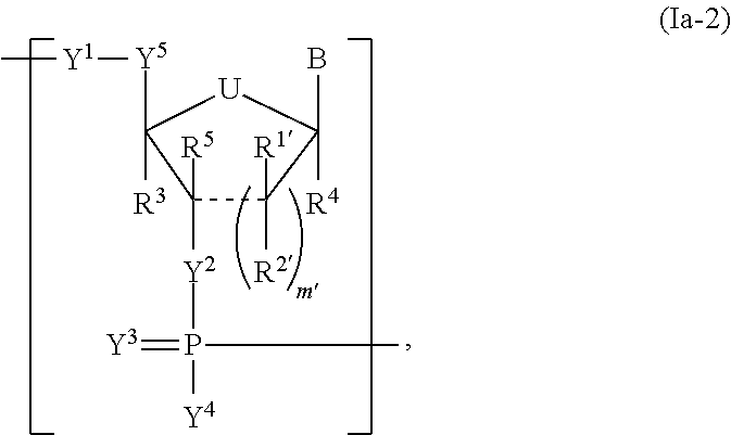
C00022

C00023

C00024

C00025

C00026

C00027

C00028

C00029

C00030

C00031

C00032

C00033

C00034

C00035

C00036

C00037

C00038

C00039

C00040

C00041
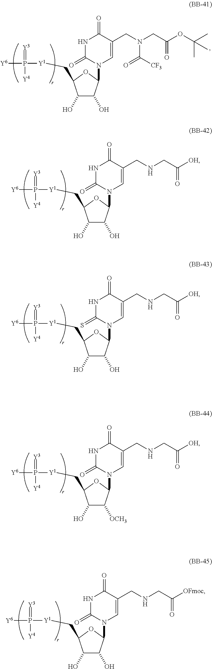
C00042

C00043

C00044

C00045

C00046

C00047

C00048

C00049

C00050

C00051

C00052

C00053

C00054

C00055
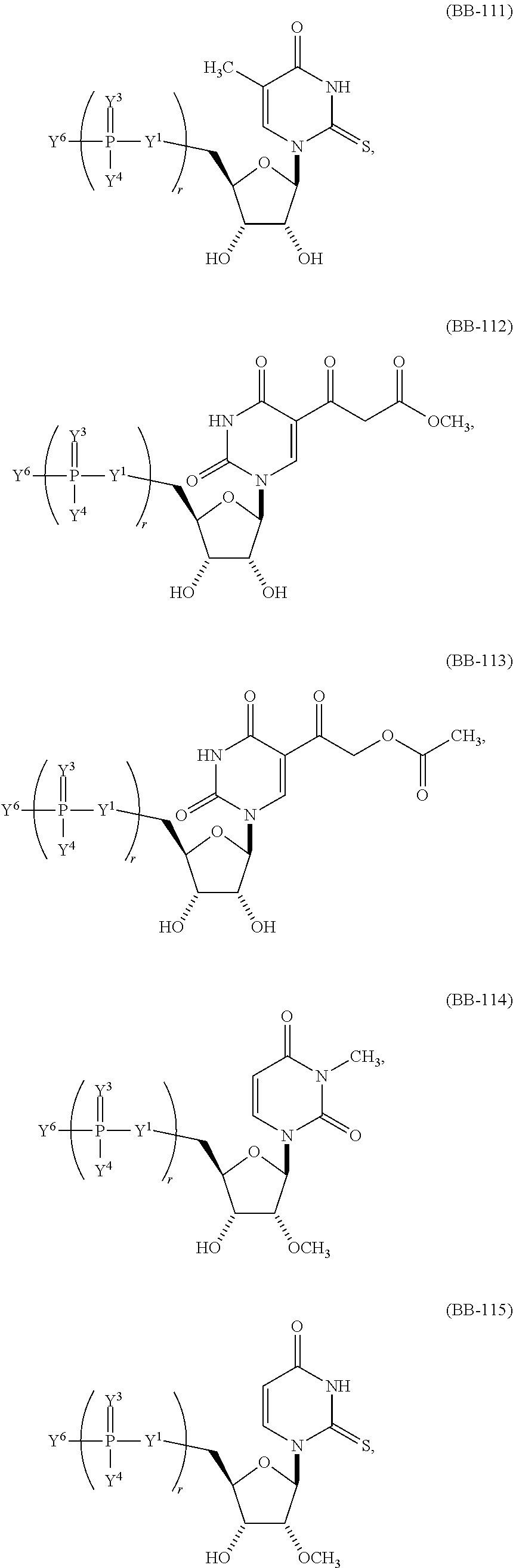
C00056

C00057

C00058

C00059

C00060

C00061

C00062

C00063

C00064

C00065

C00066

C00067

C00068

C00069

C00070

C00071

C00072

C00073

C00074

C00075

C00076

C00077

C00078
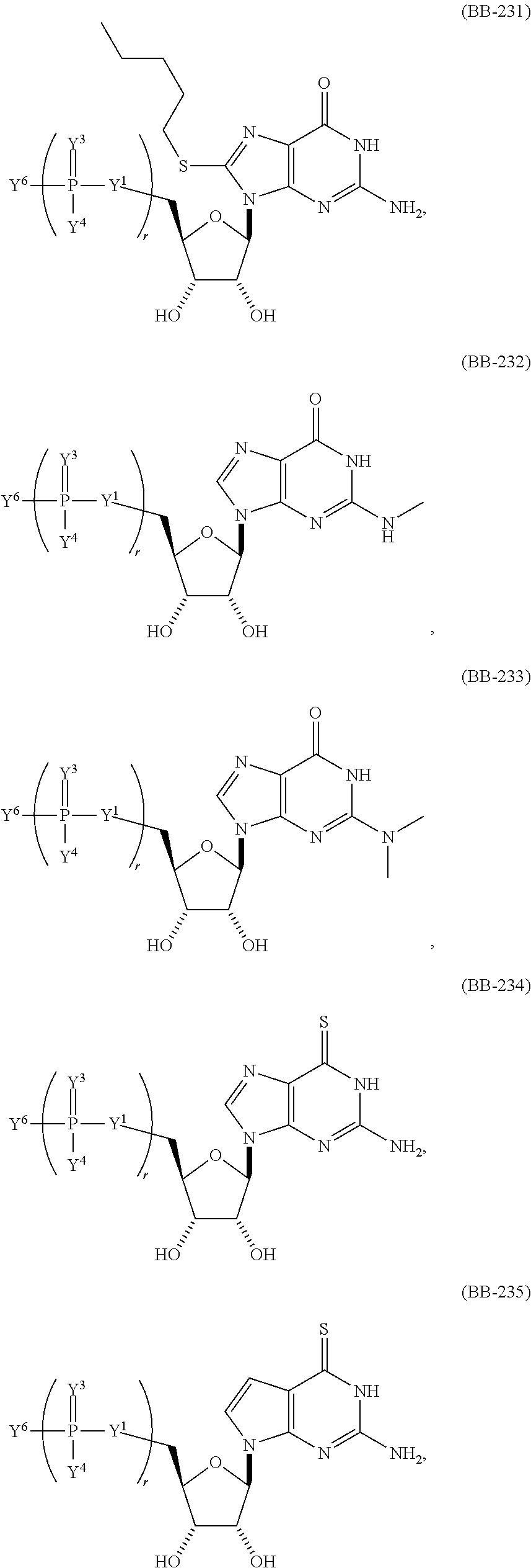
C00079

C00080

C00081

C00082

C00083

C00084

C00085

C00086

C00087

C00088

C00089

C00090

C00091

C00092

C00093

C00094

C00095

C00096
C00097

C00098
C00099

C00100

C00101

C00102

C00103

C00104

C00105

C00106

C00107

C00108

C00109

C00110

C00111

C00112

C00113

C00114

C00115

C00116

C00117

C00118

C00119

C00120

C00121

C00122

C00123

C00124

C00125

C00126

C00127

C00128

C00129

C00130

D00001

D00002

D00003

D00004

D00005

D00006

D00007

D00008

D00009

D00010

D00011

D00012

D00013

D00014

D00015

D00016

D00017

D00018

D00019

D00020

D00021

D00022

D00023

D00024

D00025

D00026

D00027

D00028

D00029

D00030

D00031

D00032

D00033

D00034

D00035

D00036

D00037

D00038

D00039

D00040

D00041

D00042

D00043

D00044

D00045

D00046

D00047

D00048

D00049

D00050

D00051

D00052

D00053

D00054

D00055

D00056

D00057
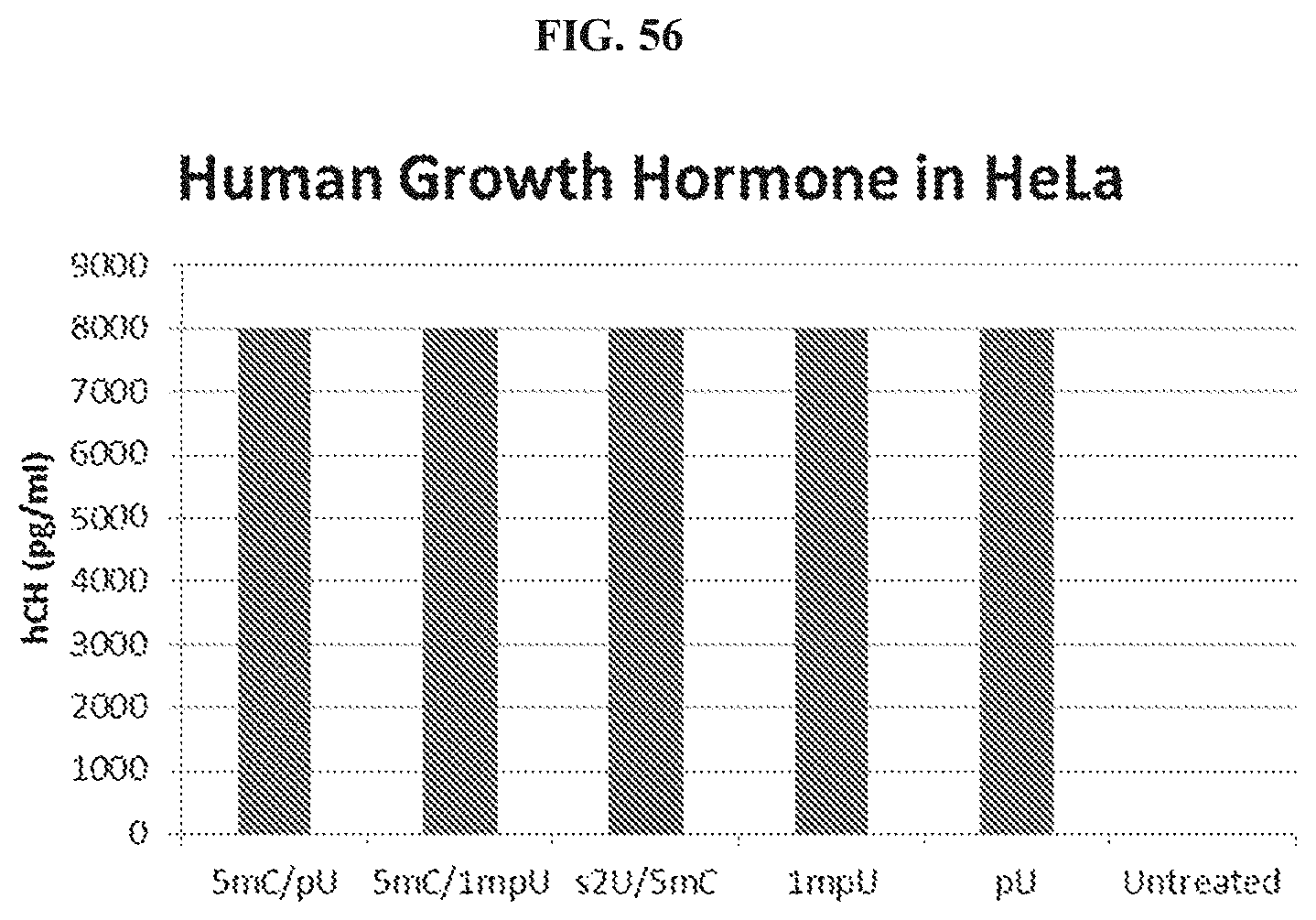
P00001

P00002

S00001
XML
uspto.report is an independent third-party trademark research tool that is not affiliated, endorsed, or sponsored by the United States Patent and Trademark Office (USPTO) or any other governmental organization. The information provided by uspto.report is based on publicly available data at the time of writing and is intended for informational purposes only.
While we strive to provide accurate and up-to-date information, we do not guarantee the accuracy, completeness, reliability, or suitability of the information displayed on this site. The use of this site is at your own risk. Any reliance you place on such information is therefore strictly at your own risk.
All official trademark data, including owner information, should be verified by visiting the official USPTO website at www.uspto.gov. This site is not intended to replace professional legal advice and should not be used as a substitute for consulting with a legal professional who is knowledgeable about trademark law.