Anti-PD-1 antibodies and methods of use thereof
Van Dijk , et al. Oc
U.S. patent number 10,450,373 [Application Number 16/399,262] was granted by the patent office on 2019-10-22 for anti-pd-1 antibodies and methods of use thereof. This patent grant is currently assigned to AGENUS INC., LUDWIG INSTITUTE FOR CANCER RESEARCH LTD, MEMORIAL SLOAN KETTERING CANCER CENTER. The grantee listed for this patent is AGENUS INC., LUDWIG INSTITUTE FOR CANCER RESEARCH LTD, MEMORIAL SLOAN KETTERING CANCER CENTER. Invention is credited to Rikke Baek Holmgaard, Taha Merghoub, Cornelia Anne Mundt, Gerd Ritter, David Adam Savitsky, David Schaer, Marc Van Dijk, Nicholas Stuart Wilson, Jedd David Wolchok, Roberta Zappasodi.


View All Diagrams
| United States Patent | 10,450,373 |
| Van Dijk , et al. | October 22, 2019 |
Anti-PD-1 antibodies and methods of use thereof
Abstract
The instant disclosure provides antibodies that specifically bind to human PD-1 and antagonize PD-1 function. Also provided are pharmaceutical compositions comprising these antibodies, nucleic acids encoding these antibodies, expression vectors and host cells for making these antibodies, and methods of treating a subject using these antibodies.
| Inventors: | Van Dijk; Marc (Bilthoven, NL), Mundt; Cornelia Anne (Lorrach, DE), Ritter; Gerd (New York, NY), Wolchok; Jedd David (New York, NY), Merghoub; Taha (Jersey City, NJ), Zappasodi; Roberta (New York, NY), Holmgaard; Rikke Baek (New York, NY), Schaer; David (Mamaroneck, NY), Savitsky; David Adam (Boxford, MA), Wilson; Nicholas Stuart (Somerville, MA) | ||||||||||
|---|---|---|---|---|---|---|---|---|---|---|---|
| Applicant: |
|
||||||||||
| Assignee: | AGENUS INC. (Lexington, MA) LUDWIG INSTITUTE FOR CANCER RESEARCH LTD (Zurich, CH) MEMORIAL SLOAN KETTERING CANCER CENTER (New York, NY) |
||||||||||
| Family ID: | 56896841 | ||||||||||
| Appl. No.: | 16/399,262 | ||||||||||
| Filed: | April 30, 2019 |
Prior Publication Data
| Document Identifier | Publication Date | |
|---|---|---|
| US 20190256601 A1 | Aug 22, 2019 | |
Related U.S. Patent Documents
| Application Number | Filing Date | Patent Number | Issue Date | ||
|---|---|---|---|---|---|
| 15254315 | Sep 1, 2016 | 10323091 | |||
| 62257195 | Nov 18, 2015 | ||||
| 62216043 | Sep 9, 2015 | ||||
| 62212851 | Sep 1, 2015 | ||||
| Current U.S. Class: | 1/1 |
| Current CPC Class: | A61P 35/00 (20180101); C07K 16/2818 (20130101); A61P 37/04 (20180101); A61P 43/00 (20180101); A61P 31/00 (20180101); C07K 16/2803 (20130101); C07K 2317/21 (20130101); C07K 2317/34 (20130101); C07K 2317/72 (20130101); C07K 2317/71 (20130101); A61K 2039/507 (20130101); C07K 2317/33 (20130101); C07K 2317/52 (20130101); C07K 2317/565 (20130101); C07K 2317/76 (20130101); C07K 2317/92 (20130101) |
| Current International Class: | C07K 16/28 (20060101); A61K 39/395 (20060101); A61K 39/00 (20060101) |
References Cited [Referenced By]
U.S. Patent Documents
| 5897862 | April 1999 | Hardy et al. |
| 6808710 | October 2004 | Wood et al. |
| 7029674 | April 2006 | Carreno et al. |
| 7332582 | February 2008 | Hardy et al. |
| 7521051 | April 2009 | Collins et al. |
| 7595048 | September 2009 | Honjo et al. |
| 7851598 | December 2010 | Davis |
| 7858746 | December 2010 | Honjo et al. |
| 8008449 | August 2011 | Korman et al. |
| 8168757 | May 2012 | Finnefrock et al. |
| 8354509 | January 2013 | Carven et al. |
| 8617546 | December 2013 | Kang et al. |
| 8735553 | May 2014 | Li et al. |
| 8741295 | June 2014 | Olive |
| 8927697 | January 2015 | Davis et al. |
| 8993731 | March 2015 | Tyson |
| 9067998 | June 2015 | Clube et al. |
| 9084776 | July 2015 | Korman et al. |
| 9102727 | August 2015 | Freeman et al. |
| 9132281 | September 2015 | Zeng et al. |
| 9163087 | October 2015 | Huchroo et al. |
| 9181342 | November 2015 | Davis |
| 9220776 | December 2015 | Sharma et al. |
| 9243052 | January 2016 | Olive et al. |
| 9358289 | June 2016 | Korman et al. |
| 9381244 | July 2016 | Noelle |
| 9394365 | July 2016 | Eisenbach-Schwartz et al. |
| 9499603 | November 2016 | Tyson |
| 9580504 | February 2017 | Rotem-Yehudar et al. |
| 9605070 | March 2017 | Sabatos-Peyton et al. |
| 9676853 | June 2017 | Zhou et al. |
| 9683048 | June 2017 | Freeman et al. |
| 9701749 | July 2017 | Shibayama et al. |
| 9771425 | September 2017 | Wang et al. |
| 9815898 | November 2017 | Freeman et al. |
| 10323091 | June 2019 | van Dijk |
| 2003/0059937 | March 2003 | Ruben et al. |
| 2006/0246071 | November 2006 | Green et al. |
| 2008/0152587 | June 2008 | Zhou et al. |
| 2009/0155164 | May 2009 | Brasel et al. |
| 2011/0110956 | May 2011 | Rothe et al. |
| 2011/0177070 | July 2011 | Lofquist et al. |
| 2012/0121591 | May 2012 | Sullivan et al. |
| 2012/0195879 | August 2012 | Walker et al. |
| 2014/0234331 | August 2014 | Korman et al. |
| 2014/0302070 | October 2014 | Chen et al. |
| 2014/0356363 | December 2014 | Zhou et al. |
| 2015/0125463 | May 2015 | Cogswell et al. |
| 2015/0190506 | July 2015 | Cheung et al. |
| 2015/0202291 | July 2015 | Bosch et al. |
| 2015/0203579 | July 2015 | Papadopoulos et al. |
| 2015/0210769 | July 2015 | Freeman et al. |
| 2015/0216970 | August 2015 | Grogan et al. |
| 2015/0344577 | December 2015 | Fu |
| 2016/0032014 | February 2016 | Michaels et al. |
| 2016/0051672 | February 2016 | Stewart et al. |
| 2016/0075783 | March 2016 | King et al. |
| 2016/0090417 | March 2016 | Cogswell et al. |
| 2016/0108123 | April 2016 | Freeman et al. |
| 2016/0130345 | May 2016 | Fotin-Mleczek et al. |
| 2016/0130348 | May 2016 | Langermann et al. |
| 2016/0159905 | June 2016 | Abdiche et al. |
| 2016/0176963 | June 2016 | Maurer et al. |
| 2016/0208000 | July 2016 | Smythe |
| 2016/0222113 | August 2016 | Buchanan et al. |
| 2016/0222121 | August 2016 | Johnson et al. |
| 2016/0264667 | September 2016 | Chen et al. |
| 2016/0272708 | September 2016 | Chen |
| 2016/0280786 | September 2016 | Harmans et al. |
| 2016/0311904 | October 2016 | Haas et al. |
| 2016/0319018 | November 2016 | Morsey et al. |
| 2016/0319019 | November 2016 | Amirina et al. |
| 2016/0339090 | November 2016 | Hacohen et al. |
| 2016/0340428 | November 2016 | Yang |
| 2016/0347836 | December 2016 | Grosso |
| 2016/0355589 | December 2016 | Williams et al. |
| 2016/0362489 | December 2016 | Yang |
| 2016/0362492 | December 2016 | Freeman et al. |
| 2016/0376367 | December 2016 | Yuan et al. |
| 2017/0007693 | January 2017 | Weiner et al. |
| 2017/0037127 | February 2017 | Grogan et al. |
| 2017/0037132 | February 2017 | Manekas et al. |
| 2017/0044256 | February 2017 | Grogan et al. |
| 2017/0044259 | February 2017 | Tipton et al. |
| 2017/0044260 | February 2017 | Baruah et al. |
| 2017/0088613 | March 2017 | Grogan et al. |
| 2017/0088618 | March 2017 | Bennett et al. |
| 2017/0088626 | March 2017 | Jure-Kunkel et al. |
| 2017/0130271 | May 2017 | Wong |
| 2017/0158776 | June 2017 | Feltquate et al. |
| 2017/0165325 | June 2017 | Sharpe et al. |
| 2017/0166637 | June 2017 | Ben-Moshe et al. |
| 2017/0166642 | June 2017 | Pantaleo et al. |
| 2017/0174774 | June 2017 | Coric et al. |
| 2017/0210806 | June 2017 | Liu |
| 2017/0198039 | July 2017 | Wong |
| 2017/0209574 | July 2017 | Cao et al. |
| 2017/0218068 | August 2017 | Lesterhuis et al. |
| 2017/0239351 | August 2017 | Hamdy et al. |
| 2017/0240644 | August 2017 | Zhou et al. |
| 2017/0247454 | August 2017 | Benz et al. |
| 2017/0247455 | August 2017 | Jure-Kunkel et al. |
| 2017/0267760 | September 2017 | Diaz et al. |
| 2017/0267762 | September 2017 | Qiu et al. |
| 1165616 | Nov 2004 | EP | |||
| 2133365 | Dec 2009 | EP | |||
| 2905030 | Aug 2015 | EP | |||
| 2906241 | Aug 2015 | EP | |||
| 2911669 | Sep 2015 | EP | |||
| 2931738 | Oct 2015 | EP | |||
| 2958588 | Dec 2015 | EP | |||
| 3036256 | Jun 2016 | EP | |||
| 3064220 | Sep 2016 | EP | |||
| 3160505 | May 2017 | EP | |||
| WO 2004072286 | Jan 2003 | WO | |||
| WO 2006121168 | Nov 2006 | WO | |||
| WO 2008156712 | Dec 2008 | WO | |||
| WO 2014179664 | Nov 2014 | WO | |||
| WO 2015181331 | Dec 2015 | WO | |||
| WO 2016077397 | May 2016 | WO | |||
| WO 2016100561 | Jun 2016 | WO | |||
| WO 2016127179 | Aug 2016 | WO | |||
| WO 2016137850 | Sep 2016 | WO | |||
| WO 2016137985 | Sep 2016 | WO | |||
| WO 2016149387 | Sep 2016 | WO | |||
| WO 2016168143 | Oct 2016 | WO | |||
| WO 2016172624 | Oct 2016 | WO | |||
| WO 2016179517 | Nov 2016 | WO | |||
| WO 2016183469 | Nov 2016 | WO | |||
| WO 2016196237 | Dec 2016 | WO | |||
| WO 2016196389 | Dec 2016 | WO | |||
| WO 2016205320 | Dec 2016 | WO | |||
| WO 2016210129 | Dec 2016 | WO | |||
| WO 2017016497 | Feb 2017 | WO | |||
| WO 2017019846 | Feb 2017 | WO | |||
| WO 2017024515 | Feb 2017 | WO | |||
| WO 2017054646 | Apr 2017 | WO | |||
| WO 2017055443 | Apr 2017 | WO | |||
| WO 2017055547 | Apr 2017 | WO | |||
| WO 2017058115 | Apr 2017 | WO | |||
| WO 2017059095 | Apr 2017 | WO | |||
| WO 2017079080 | May 2017 | WO | |||
| WO 2017079303 | May 2017 | WO | |||
| WO 2017087280 | May 2017 | WO | |||
| WO 2017087599 | May 2017 | WO | |||
| WO 2017096026 | Jun 2017 | WO | |||
| WO 2017103280 | Jun 2017 | WO | |||
| WO 2017107885 | Jun 2017 | WO | |||
| WO 2017124050 | Jul 2017 | WO | |||
| WO 2017125815 | Jul 2017 | WO | |||
| WO 2017129763 | Aug 2017 | WO | |||
| WO 2017129790 | Aug 2017 | WO | |||
| WO 2017132508 | Aug 2017 | WO | |||
| WO 2017132536 | Aug 2017 | WO | |||
| WO 2017133540 | Aug 2017 | WO | |||
| WO 2017147368 | Aug 2017 | WO | |||
| WO 2017151502 | Sep 2017 | WO | |||
| WO 2017151517 | Sep 2017 | WO | |||
| WO 2017160975 | Sep 2017 | WO | |||
| WO 2018106864 | Jun 2018 | WO | |||
Other References
|
Feb. 28, 2016 Q4 2015 Results--Earnings Call Transcript. cited by applicant . Aug. 3, 2017 Q2 2017 Results--Earnings Call Transcript. cited by applicant . Nov. 7, 2017 Q3 2017 Results--Earnings Call Transcript. cited by applicant . "A comprehensive immune-oncology Ecosystem" Cowen and Company 36th Annual Health Care Conference Mar. 2016. cited by applicant . Abdiche, Y.N., et al. (2016) "Assessing Kinetic and Epitopic Diversity Across Orthogonal Monoclonal Antibody Generation Platforms" mAbs, vol. 8, No. 2, pp. 264-277. cited by applicant . Afanasiev, O.K., et al., (2013) "Merkel Polyomavirus-Specific T Cells Fluctuate with Merkel Cell Carcinoma Burden and Express Therapeutically Targetable PD-1 and Tim-3 Exhaustion Markers" Clin. Cancer Res. 19(19):5351-60. cited by applicant . Agata, Y. et al., (1996) "Expression of the PD-1 antigen on the surface of stimulated mouse T and B lymphocytes." Int. Immunol 8:765-72. cited by applicant . "Agenus Announces Combination Clinical Trials of its Anti-CTLA4 (AGEN1884) & Anti-PD1 (AGEN2034)" Jan. 22, 2018 /PRNewswire/. cited by applicant . "Agenus Commences Phase I Clinical trials of its CTLA-4 Checkpoint Antibody to Treat Solid Tumors," Apr. 27, 2016 /Business Wire/. cited by applicant . "Agenus Investor Relations Deck" May 15, 2015. cited by applicant . "Agenus IR Deck" Dec. 2016. cited by applicant . "Agenus IR Presentation" Aug. 2016. cited by applicant . Agenus News vol. 1 Issue 1 (2018). cited by applicant . Agenus News vol. 1 Issue 2 (2018). cited by applicant . Agenus News vol. 1 Issue 3 (2018). cited by applicant . Agenus News vol. 1 Issue 8 (2018). cited by applicant . "Agenus Inc. Closes $230 Million Royalty Monetization with HealthCare Royalty Partners" Jan. 22, 2018 /PRNewswire/. cited by applicant . "Agenus Presents Clinical Responses of AGEN1884 (anti-CTLA-4) and AGEN2034 (anti-PD-1) at ASCO 2018" Jun. 4, 2018 /PRNewswire/. cited by applicant . "Agenus Presents Posters on Checkpoint Antibody Product Candidates at the American Association for Cancer Research (AACR) 2016 Annual Meeting" Apr. 18, 2016 (Business Wire). cited by applicant . "Agenus R&D Day" Nov. 19, 2015 New York, NY. cited by applicant . "Agenus Roadshow Slide Deck" May 15, 2015. cited by applicant . "Agenus Roadshow Slide Deck" Aug. 2016. cited by applicant . "Agenus to Present Clinical Data on Lead Programs at ASCO 2018" May 17, 2018 /PRNewswire/. cited by applicant . "Agenus to Present on Lead Antibody Programs AGEN1884 (CTLA-4) and AGEN2034 (PD-1) at SITC 2017" Nov. 9, 2017 /PRNewswire/. cited by applicant . "Agenus Background Slide Deck" Jan. 2017. cited by applicant . Ahmadzadeh, M., et al., (2009) "Tumor antigen-specific CD8 T cells infiltrating the tumor express high levels of PD-1 and are functionally impaired" Blood 114:1537-1544. cited by applicant . Allie, S.R., et al., (2011) "Programmed death 1 regulates development of central memory CD8 T cells after acute viral infection" J. Immunol. 186:6280-6286. cited by applicant . Anonymous (2014) "International Nonproprietary Names for Pharmaceutical Substances (INN)," WHO drug Information Rec. Wld Health Org. Resolution EB15.R7), pp. 1-44. [retrieved on Nov. 7, 2016]. cited by applicant . Antonia, S., et al., (2016) "Safety and antitumour activity of durvalumab plus tremelimumab in non-small cell lung cancer: a multicentre, phase 1b study" The Lancet Oncology 17(3):299-308. cited by applicant . Antonia, S.J., et al., (2016) "Nivolumab alone and nivolumab plus ipilimumab in recurrent small-cell lung cancer (CheckMate 032): a multicentre, open-label, phase 1/2 trial" The Lancet Oncology 17(7):883-895. cited by applicant . Attia, P., et al., (2005) "Autoimmunity Correlates With Tumor Regression in Patients With Metastatic Melanoma Treated With Anti--Cytotoxic T-Lymphocyte Antigen-4" Journal of Clinical Oncology 23:6043-6053. cited by applicant . Bally, A.P., et al., (2015) "NF-.kappa.B regulates PD-1 expression in macrophages" J. Immunol. 194:4545-4554. cited by applicant . Barber, D.L., et al., (2006) "Restoring function in exhausted CD8 T cells during chronic viral infection" Nature 439:682-687. cited by applicant . Baruch, K., et al., (2016) "PD-1 immune checkpoint blockade reduces pathology and improves memory in mouse models of Alzheimer's disease" Nature Med. 22(2):135-7. cited by applicant . Bauml et al (2016) "Preliminary results from KEYNOTE-055: Pembrolizumab after platinum and cetuximab failure in head and neck squamous cell carcinoma (HNSCC)" 2016 ASCO Annual Meeting: J Clin Oncol 34, 2016 (suppl; abstr 6011). cited by applicant . Bennett, F., et al., (2003) "Program death-1 engagement upon TCR activation has distinct effects on costimulation and cytokine-driven proliferation: attenuation of ICOS, IL-4, and IL-21, but not CD28, IL-7, and IL-15 responses" J. Immunol. 170:711-718. cited by applicant . Benson, D M et al. (2010) "The PD-1/PD-L1 axis modulates the natural killer cell versus multiple myeloma affect: a therapeutic target for CT-011, a novel monoclonal anti-PD-1 antibody," Blood, American Society of Hemotology, US 116(13):2286-2294. cited by applicant . Bifulco, C.B., et al., (2016) "Unmasking PD-1 Resistance by Next-Generation Sequencing" N. Engl. J. of Med. 375(9):888-889. cited by applicant . Blank, C., et al., (2004) "PD-L1/B7H-1 inhibits the effector phase of tumor rejection by T cell receptor (TCR) transgenic CD8+ T cells" Cancer Res. 64:1140-1145. cited by applicant . Blank, C., et al., (2005) "Interaction of PD-L1 on tumor cells with PD-1 on tumor-specific T cells as a mechanism of immune evasion: implications for tumor immunotherapy" Cancer Immunol. Immunother. 54:307-314. cited by applicant . Blank, C., et al., (2007) "Contribution of the PD-L1/PD-1 pathway to T-cell exhaustion: an update on implications for chronic infections and tumor evasion" Cancer Immunol. Immunother. 56:739-745. cited by applicant . Borcoman, E. et al. (2017) "Pembrolizumab in cervical cancer: latest evidence and clinical usefulness," Ther Adv Med Oncol. 9(6):431-439. cited by applicant . Borradori, L. et al., (2016) "Rescue therapy with anti-programmed cell death protein 1 inhibitors of advanced cutaneous squamous cell carcinoma and basosquamous carcinoma: preliminary experience in five cases" The British Journal of Dermatology, 175(6):1382-1386. cited by applicant . Boussiotis, V.A., et al., (2016) "Molecular and Biochemical Aspects of the PD-1 Checkpoint Pathway" N. Engl. J. Med. 375(18):1767-78. cited by applicant . Boutros, C., et al., (2016) "Safety profiles of anti-CTLA-4 and anti-PD-1 antibodies alone and in combination" Nature Reviews 13:473. cited by applicant . Boyd, S.D., et al. (2016) "Deep Sequencing and Human Antibody Repertoire Analysis" Current Opinion in Immunology, vol. 40, pp. 103-109. cited by applicant . Brahmer, J.R., et al., (2010) "Phase I Study of Single-Agent Anti-Programmed Death-1 (MDX-1106) in Refractory Solid Tumors: Safety, Clinical Activity, Pharmacodynamics, and Immunologic Correlates" J. Clin. Oncol. 28(19):3167-75. cited by applicant . Brahmer, J.R., et al., (2012) "Safety and activity of anti-PD-L1 antibody in patients with advanced cancer" N. Engl. J. Med. 366:2455-2465. cited by applicant . Brantsch, K.D. et al., (2008) "Analysis of risk factors determining prognosis of cutaneous squamous-cell carcinoma: a prospective study" The Lancet Oncology 9(8):713-720. cited by applicant . Bristol-Myers Squibb USA, 2015. Yervoy.RTM. (ipilimumab) package insert. cited by applicant . Bristol-Myers Squibb USA, 2016. Opdivo.RTM. (nivolumab) package insert. cited by applicant . Brougham, N.D.L.S., et al., (2012) "The incidence of metastasis from cutaneous squamous cell carcinoma and the impact of its risk factors" Journal of Surgical Oncology 106(7):811-815. cited by applicant . Brown, J.A., et al., (2003) "Blockade of programmed death-1 ligands on dendritic cells enhances T cell activation and cytokine production" J. Immunol. 170:1257-66. cited by applicant . Buchbinder, E.I., et al., (2016) "CTLA-4 and PD-1 Pathways: Similarities, Differences, and Implications of Their Inhibition" American Journal of Clinical Oncology, 39(1):98-106. cited by applicant . Bulliard, Y., et al., (2013) "Activating Fcy receptors contribute to the antitumor activities of immunoregulatory receptor-targeting antibodies" The Journal of Experimental Medicine 210(9):1685-1693. cited by applicant . Burova, E. et al. (2017) "Characterization of the anti-PD-1 antibody REGN2810 and its antitumor activity in human PD-1 knock-in mice," Mol. Cancer Ther. 16(5): 861-870. cited by applicant . Butte, M.J., et al., (2007) "Programmed death-1 ligand 1 interacts specifically with the B7-1 costimulatory molecule to inhibit T cell responses" Immunity 27:111-122. cited by applicant . Callahan, M.K., et al., (2014) "CTLA-4 and PD-1 Pathway Blockade: Combinations in the Clinic" Front. Oncol. 4(385):1-6. cited by applicant . Camacho, L.H., (2015) "CTLA-4 blockade with ipilimumab: biology, safety, efficacy, and future considerations" Cancer Medicine 4(5):661-672. cited by applicant . Carrizosa, et al., (2013) "New targets and new mechanisms in lung cancer." Oncology 27(5). cited by applicant . Carter, L. et al. (2003) "Cytotoxic T-lymphocyte antigen-4 and programmed death-1 function as negative regulators of lymphocyte activation," Immunol Res 28(1):49-59. cited by applicant . Carter, L., et al., (2002) "PD-1:PD-L inhibitory pathway affects both CD4(+) and CD8(+) T cells and is overcome by IL-2" Eur. J. Immunol. 32:634-43. cited by applicant . Casey, S.C., et al., (2016) "MYC regulates the antitumor immune response through CD47 and PD-L1" Science 352:227-231. cited by applicant . Chaft, J.E., (Mar. 30, 2017) "Immunotherapy for lung cancer and the landscape of combinations" Thoracis Oncology Service Memorial Sloan Kettering Cancer Center. cited by applicant . Chand, D., et al., "AGEN2034, a novel anti-PD-1 antibody that combines effectively with CTLA-4 pathway blockade to enhance T cell activity" Poster #P312 SITC Annual Meeting, Washington, DC, USA Nov. 9-12, 2017. cited by applicant . Chapman, K., (2007) "Preclinical safety testing of monoclonal antibodies: the significance of species relevance" Nat. Rev. Drug Discov. 6:120-126. cited by applicant . Chapon, M., et al., (2011) "Progressive upregulation of PD-1 in primary and metastatic melanomas associated with blunted TCR signaling in infiltrating T lymphocytes" J. Invest. Dermatol. 131:1300-1307. cited by applicant . Chauvin, J., et al., (2015) "TIGIT and PD-1 impair tumor antigen--specific CD8+ T cells in melanoma patients" 125(5):2046-58. cited by applicant . Chemnitz, J.M., et al., (2004) "SHP-1 and SHP-2 associate with immunoreceptor tyrosine-based switch motif of programmed death 1 upon primary human T cell stimulation, but only receptor ligation prevents T cell activation" J. Immunol. 173:945-954. cited by applicant . Chen, L., et al., (2015) "Anti-PD-1/PD-L1 therapy of human cancer: past, present, and future" J. Clin. Invest. 125:3384-3391. cited by applicant . Chen, S., et al., (2015) "Combination of 4-1BB agonist and PD-1 antagonist promotes antitumor effector/memory CD8 T cells in a poorly immunogenic tumor model" Cancer Immunol. Res. 3(2):149-160. cited by applicant . Chu, F., et al., (2014) "Anti-PD-1 antibodies for the treatment of B-cell lymphoma Importance of PD-1+ T-cell subsets" Oncolmmunology 3(e28101):1-3. cited by applicant . Chung, C.H., (2008) "Managing Premedications and the Risk for Reactions to Infusional Monoclonal Antibody Therapy" The Oncologist 13:725-732. cited by applicant . Collins, A.V., et al., (2002) "The interaction properties of costimulatory molecules revisited" Immunity 17:201-210. cited by applicant . Collins, M., et al., (2005) "The B7 family of immune-regulatory ligands" Genome Biology 6(6):223.1-223.7. cited by applicant . Conroy, P.J., et al. (2017) "Antibodies: From Novel Repertoires to Defining and Refining the Structure of Biologically Important Targets" Methods, vol. 116, pp. 12-22. cited by applicant . Coyne, G.O., et al., (2014) "Nivolumab: Promising Survival Signal Coupled With Limited Toxicity Raises Expectations" J. Clin. Oncol. 32(10):986-988. cited by applicant . Cross, R.S., et al., (2015) "Therapeutic DNA vaccination against colorectal cancer by targeting the MYB oncoprotein" Clin. Transl. Immunol. 4:e30. cited by applicant . Curran, M.A., et al., (2010) "PD-1 and CTLA-4 combination blockade expands infiltrating T cells and reduces regulatory T and myeloid cells within B16 melanoma tumors" Proc. Natl. Acad. Sci. USA 107:4275-4280. cited by applicant . Currie, A.J., et al., (2009) "Dual Control of Antitumor CD8 T Cells through the Programmed Death-1/Programmed Death-Ligand 1 Pathway and Immunosuppressive CD4 T Cells: Regulation and Counterregulation" J. Immunol. 183:7898-08. cited by applicant . Da Silva, R M et al. (2014) "Nivolumab: Anti-PD-1 monoclonal antibody cancer immunotherapy," Drugs of the Future, 39(1):15-24. cited by applicant . Dahan, R., (2015) "FcyRs Modulate the Anti-tumor Activity of Antibodies Targeting the PD-1/PD-L1 Axis." Cancer Cell 28(3):285-295. cited by applicant . Das, R., et al., (2015) "Combination therapy with anti-CTLA-4 and anti-PD-1 leads to distinct immunologic changes in vivo" J. Immunol. 194:950-959. cited by applicant . De Souza, P., et al., "Evaluation of Peripheral T-Cell Subset Proliferation as a Pharmacodynamic Assay to Guide the Development of Anti-CTLA-4 and PD-1 Antibody Combinations in Patients With Solid Tumors" CT104 Presented at the 2018 Annual Meeting of the American Association of Cancer Research, Apr. 14-18, 2018, in Chicago, IL, USA. cited by applicant . Demaria, S., et al., (2005) "Combining radiotherapy and immunotherapy: A revived partnership" International J. of Radiation Oncology Biology Physics 63(3):655-666. cited by applicant . Deng, L., et al., (2014) "Irradiation and anti-PD-L1 treatment synergistically promote antitumor immunity in mice" J. Clin. Invest. 124(2):687-695. cited by applicant . Disis, M.L. (2010) "Immune regulation of cancer" J. Clin. Oncol. 28:4531-4538. cited by applicant . Dolan, D.E., et al., (2014) "PD-1 Pathway Inhibitors: Changing the Landscape of Cancer Immunotherapy" Cancer Control 21(3):231-7. cited by applicant . Dong, H., et al., (2002) "Tumor-associated B7-H1 promotes T-cell apoptosis: a potential mechanism of immune evasion" Nat. Med. 8:787-9. cited by applicant . Dong, H., et al., (2003) "B7-H1 pathway and its role in the evasion of tumor immunity." J. Mol. Med. 81:281-7. cited by applicant . Drescher, C., et al., "Phase 1/2, Open-Label, Multiple Ascending Dose Trial of AGEN2034, an Anti--PD-1 Monoclonal Antibody, in Advanced Solid Malignancies: Results of Dose Escalation in Advanced Cancer and Expansion Cohorts in Subjects With Relapsed/Refractory Cervical Cancer" Poster No. 1158P Presented at European Society for Medical Oncology, Munich, Germany (2018). cited by applicant . Drouin, E., et al., "AGEN1884 and AGEN2041: Two functionally distinct anti-CTLA-4 antagonist antibodies" Poster No. #5005 Presented at the American Association for Cancer Research Annual Meeting 2016 New Orleans, LA, USA, Wednesday, Apr. 20, 2016. cited by applicant . Drouin, E., et al., "AGEN1884, an IgG1 anti-CTLA-4 antibody, combines effectively with PD-1 blockade in primary human T cell assays and in a non-human primate pharmacodynamic (PD) model" Poster: #27 Abstract: #3654 AACR Annual Meeting 2017, Washington, DC, USA Apr. 1-5, 2017. cited by applicant . Dudley, J.C., et al., (2016) "Microsatellite Instability as a Biomarker for PD-1 Blockade" Clin. Cancer Res. 22(4):813-20. cited by applicant . Duraiswamy, J., et al., (2013) "Dual Blockade of PD-1 and CTLA-4 Combined with Tumor Vaccine Effectively Restores T-Cell Rejection Function in Tumors" Cancer Res. 73(12):3591-603. cited by applicant . Duraiswamy, J., et al., (2013) "Therapeutic PD-1 pathway blockade augments with other modalities of immunotherapy T-cell function to prevent immune decline in ovarian cancer" Cancer Res. 73(23):6900-6912. cited by applicant . Dyck, L., et al., (2016) "Anti-PD-1 inhibits Foxp3+ Treg cell conversion and unleashes intratumoural effector T cells thereby enhancing the efficacy of a cancer vaccine in a mouse model" Cancer Immunol. Immunother. 65(12):1491-1498. cited by applicant . Eisenhauer, E.A., et al., (2009) "New response evaluation criteria in solid tumours: Revised RECIST guideline (version 1.1)" European Journal of Cancer 45:228-247. cited by applicant . "Emerging Leader in Immuno-Oncology" Nov. 2015, Lexington, MA. cited by applicant . "Enabling Best-in-Class I-O Combinations" Mar. 2018. cited by applicant . Evans, T.A., et al., (2010) "Functional diversity of Robo receptor immunoglobulin domains promotes distinct axon guidance decisions" Curr. Biol. 20:567-572. cited by applicant . Falchook, G.S., et al., (2016) "Responses of metastatic basal cell and cutaneous squamous cell carcinomas to anti-PD1 monoclonal antibody REGN2810" Journal for Immunotherapy of Cancer 4(1):70. cited by applicant . Ferrara, F., et al. (2015) "Recombinant Renewable Polyclonal Antibodies", mAbs, vol. 7 No. 1, pp. 32-41. cited by applicant . Ferris, R.K., et al., (2016) "Nivolumab for Recurrent Squamous-Cell Carcinoma of the Head and Neck" N. Engl. J. Med. 375(19):1856-1867. cited by applicant . Finco, D., et al., (2014) "Cytokine release assays: current practices and future directions" Cytokine 66:143-155. cited by applicant . Foote, M.C. et al., (2014) "Phase II study of single-agent panitumumab in patients with incurable cutaneous squamous cell carcinoma" Annals of Oncology, 25(10):2047-2052. cited by applicant . Four Agenus Abstracts Accepted for Presentation at the American Association for Cancer Research (AACR) 2017 Annual Meeting Mar. 7, 2017 /PRNewswire/. cited by applicant . Francisco, L.M., et al., (2010) "The PD-1 pathway in tolerance and autoimmunity" Immunol. Rev. 236:219-242. cited by applicant . Frazier, K.S., et al., (2015) "Scientific and Regulatory Policy Committee Points-to-consider Paper: Drug-induced Vascular Injury Associated with Nonsmall Molecule Therapeutics in Preclinical Development: Part I. Biotherapeutics" Toxicol. Pathol. 43:915-934. cited by applicant . Freeman, G.J et al. (2000) "Engagement of the PD-1 immunoinhibitory receptor by a novel B7 family member leads to negative regulation of lymphocyte activation" J. Exp. Med. 192:1027-1034. cited by applicant . Freeman, G.J., et al. (2006) "Reinvigorating exhausted HIV-specific T cells via PD-1-PD-1 ligand blockade" J. Exp. Med. 203:2223-2227. cited by applicant . Frenel, J.-S., et al., (2016) "Pembrolizumab in patients with advanced cervical squamous cell cancer: Preliminary results from the phase Ib KEYNOTE-028 study" J. Clin. Oncol. 34 (suppl), Abstract 5515. cited by applicant . Fu, J., et al., (2014) "Preclinical Evidence That PD1 Blockade Cooperates with Cancer Vaccine TEGVAX to Elicit Regression of Established Tumors" Cancer Res; 74(15):4042-52. cited by applicant . Garcia-Sanz, J.A., et al. (1996) "Translational control of interleukin 2 messenger RNA as a molecular mechanism of T cell anergy" J. Exp. Med. 184:159-164. cited by applicant . Gatalica, Z., et al., (2014) "Programmed Cell Death 1 (PD-1) and Its Ligand (PD-L1) in Common Cancers and Their Correlation with Molecular Cancer Type" Cancer Epidemiol Biomarkers Prev. 23(12); 2965-70. cited by applicant . Ge, Y., et al., (2013) "Blockade of PD-1/PD-L1 immune checkpoint during DC vaccination induces potent protective immunity against breast cancer in hu-SCID mice" Cancer Letters 336:253-259. cited by applicant . George, S. et al. (2017) "Loss of PTEN is associated with resistance to anti-PD-1 checkpoint blockade therapy in metastatic uterine leiomyosarcoma," Immunity. 46:197-204. cited by applicant . Ghebeh, H., et al., (2006) "The B7-H1 (PD-L1) T lymphocyte-inhibitory molecule is expressed in breast cancer patients with infiltrating ductal carcinoma: correlation with important high-risk prognostic factors" Neoplasia 8:190-198. cited by applicant . Ghiotto, M., et al., (2010) "PD-L1 and PD-L2 differ in their molecular mechanisms of interaction with PD-1" International Immunology 22(8):651-60. cited by applicant . Goldman, JW et al. (2017) "Nivolumab (N) plus Ipillimub (I) as first-line (1L) treatment for advanced (adv) NSCLC: 2-yr OS and long-term outcome from CheckMate 012," J Clin Oncol. 35(15): 4 pp. cited by applicant . Gonzalez, A., et al., "INCAGN1949, an Anti-OX40 Antibody With an Optimal Agonistic Profile and the Ability to Selectively Deplete Intratumoral Regulatory T Cells" 4703 Presented at the American Association for Cancer Research Annual Meeting 2017; Washington, DC, USA Apr. 1-5, 2017. cited by applicant . Gonzalez, et al., "INCAGN1876, a Unique GITR Agonist Antibody That Facilitates GITR Oligomerization" 3643 Presented at the American Association for Cancer Research Annual Meeting 2017, Washington, DC, USA Apr. 1-5, 2017. cited by applicant . Gooden, M.J., et al., (2011) "The prognostic influence of tumour-infiltrating lymphocytes in cancer: a systematic review with meta-analysis" Br. J. Cancer 105:93-103. cited by applicant . Gooden, M. et al. (2011) "HLA-E expression by gynecological cancers restrains tumor-infiltrating CD8+ T lymphocytes," PNAS 108(26):10656-10661. cited by applicant . Gros, A., et al., (2014) "PD-1 identifies the patient-specific CD8+ tumor-reactive repertoire infiltrating human tumors" Journal of Clinical Investigation 124(5):2246. cited by applicant . Grosso, J.F., et al., (2013) "CTLA-4 blockade in tumor models: an overview of preclinical and translational research" Cancer Immun. 13(5). cited by applicant . "Guideline on immunogenicity assessment of monoclonal antibodies intended for in vivo clinical use," European Medicines Agency, London, UK, 2012. cited by applicant . Hamanishi, J., et al., (2007) "Programmed cell death 1 ligand 1 and tumor infiltrating CD8+ T lymphocytes are prognostic factors of human ovarian cancer" Proc. Nat. Acd. Sci. USA 104(9):3360-5. cited by applicant . Hamid, O., et al. (2013) "Safety and tumor responses with lambrolizumab (anti-PD-1) in melanoma" N. Engl. J. Med. 369:134-144. cited by applicant . Haymaker, C., et al., (2012) "PD-1 and BTLA and CD8+ T-cell "exhaustion" in cancer "Exercising" an alternative viewpoint" Oncolmmunology 1(5):735-8. cited by applicant . Hellmann, M.D., et al., (2016) CheckMate 012: safety and efficacy of first-line nivolumab and ipilimumab in advanced NSCLC In ASCO Annual Meeting. Chicago: Proc Am Soc Clin Oncol. cited by applicant . Hirano, F. et al. (2005) "Blockade of B7-H1 and PD-1 by Monoclonal Antibodies Potentiates Cancer Therapeutic Immunity," Cancer Res 65(3):1089-1096. cited by applicant . Hodi, F.S., et al., (2014) "Long-term survival of ipilimumab-naive patients (pts) with advanced melanoma (MEL) treated with nivolumab (anti-PD-1, BMS-936558, ONO-4538) in a phase I trial" ASCO. cited by applicant . Howard, S.C., et al., (2011) "The Tumor Lysis Syndrome" N. Engl. J. Med. 364(19):1844-1854. cited by applicant . Huang, R., et al., (2015) "LAG3 and PD1 co-inhibitory molecules collaborate to limit CD8+ T cell signaling and dampen antitumor immunity in a murine ovarian cancer model" Oncotarget 6(29):27359-77. cited by applicant . Ichigotani, Y., et al., (2000) "Molecular cloning of a novel human gene (SIRP-B2) which encodes a new member of the SIRP/SHP S-1 protein family" J. Hum. Genet. 45:378-382. cited by applicant . "Integrated Approach to Immuno-oncology" Mar. 31, 2016. cited by applicant . "Integrated Immunotherapy: Enabling Best-in-Class 1-O combinations" Feb. 2017. cited by applicant . "Integrated Immunotherapy: Enabling Best-in-Class 1-O Combinations" Jan. 2018. cited by applicant . "Integrated Immunotherapy: Enabling Best-in-Class I-O Combinations" Jun. 2018. cited by applicant . "Integrated Immunotherapy: Enabling Best-in-Class I-O Combinations" Mar. 2018. cited by applicant . "Integrated Immunotherapy: Enabling Best-in-Class I-O Combinations" Nov. 2017. cited by applicant . "Integrated Solutions in Immuno-Oncology" Apr. 2016. cited by applicant . "Integrated Solutions in Immuno-Oncology" May 2016. cited by applicant . International Search report for PCT/US2016/049913 dated Jan. 20, 2017. cited by applicant . Intlekofer, A., et al., (2013) "At the Bench: preclinical rationale for CTLA-4 and PD-1 blockade as cancer immunotherapy" J. of Leukocyte Biol. 94:25-39. cited by applicant . Ishida M, et al., (2002) "Differential expression of PD-L1 and PD-L2, ligands for an inhibitory receptor PD-1, in the cells of lymphohematopoietic tissues" Immunol. Lett. 84(1):57-62. cited by applicant . Ishida, Y., et al., (1992) "Induced expression of PD-1, a novel member of the immunoglobulin gene superfamily, upon programmed cell death" Embo J. 11:3887-3895. cited by applicant . Iwai, Y. et al. (2003) "PD-1 inhibits antiviral immunity at the effector phase in the liver," J Exp Med 198(1):39-50. cited by applicant . Iwai, Y., et al. (2002) "Involvement of PD-L1 on tumor cells in the escape from host immune system and tumor immunotherapy by PD-L1 blockade" Proc. Natl. Acad. Sci. USA 99:12293-12297. cited by applicant . Iwai, Y., et al., (2002) "Microanatomical localization of PD-1 in human tonsils" Immunol. Lett. 83:215-220. cited by applicant . Jarkowski, A., et al., (2016) "Systemic Therapy in Advanced Cutaneous Squamous Cell Carcinoma (CSCC): The Roswell Park Experience and a Review of the Literature" American Journal of Clinical Oncology, 39(6):545-548. cited by applicant . Jawad, Z., "Targeting the next generation of checkpoint pathways using a disease-centric approach" ICI Europe Meeting Nov. 16-17, 2016. cited by applicant . Jin, H., et al., (2010) "Role of PD-1 in Regulating T-Cell Immunity" Current Topics in Microbiology and Immunology 350:17-36. cited by applicant . John, L.B., et al., (2013) "Anti-PD-1 Antibody Therapy Potently Enhances the Eradication of Established Tumors by Gene-Modified T Cells" Clin. Cancer Res. 19(20):5636-46. cited by applicant . John, L.B., et al., (2013) "Blockade of PD-1 immunosuppression boosts CAR T-cell therapy" Oncolmmunology 2(10):e26286-1-3. cited by applicant . Kamphorst, A.O., et al., (2013) "Manipulating the PD-1 pathway to improve immunity" Current Opinion in Immunology 25:381-388. cited by applicant . Karia, P.S., et al., (2013) "Cutaneous squamous cell carcinoma: estimated incidence of disease, nodal metastasis, and deaths from disease in the United States, 2012" Journal of the American Academy of Dermatology 68(6):957-966. cited by applicant . Kaufman, H., et al., (2016) "Avelumab (MSB0010718C; anti-{D-1) in patients with metastatic Merkel cell carcinoma previously treated with chemotherapy: results of the phase 2 JAVELIN Merkel 200 trial" Oral Presentation at the 52nd ASCO Annual Meeting Jun. 307 Chicago, IL Abstract No. 9508. cited by applicant . Keir, M.E., et al., (2005) "Programmed Death-1 (PD-1):PD-Ligand 1 Interactions Inhibit TCR-Mediated Positive Selection of Thymocytes" The Journal of Immunology 175:7372-7379. cited by applicant . Keir, M.E., et al., (2008) "PD-1 and Its Ligands in Tolerance and Immunity" Annu. Rev. Immunol. 26:677-704. cited by applicant . Kelderman, S., et al., (2015) "Mismatch Repair-Deficient Cancers Are Targets for Anti-PD-1 Therapy" Cancer Cell 28:11-13. cited by applicant . Khan, L., et al. (2017) "Cross-Neutralizing Anti-HIV-1 Human Single Chain Variable Fragments(scFvs) Against CD4 Binding Site and N332 Glycan Identified from a Recombinant Phage Library" Scientific Reports, vol. 7, Article No. 45163, 12 Pages. cited by applicant . Kinter, A.L., et al., (2008) "The common gamma-chain cytokines IL-2, IL-7, IL-15, and IL-21 induce the expression of programmed death-1 and its ligands" J. Immunol. 181:6738-6746. cited by applicant . Kleffel, S., et al., (2015) "Melanoma Cell-Intrinsic PD-1 Receptor Functions Promote Tumor Growth." Cell 162(6): 1242-1256. cited by applicant . Konishi, J., et al., (2004) "B7-H1 expression on non-small cell lung cancer cells and its relationship with tumor-infiltrating lymphocytes and their PD-1 expression" Clin. Cancer Res. 10:5094-100. cited by applicant . Konitzer, J.D., et al. (2017) "Generation of a Highly Diverse Panel of Antagonistic Chicken Monoclonal Antibodies Against the GIP Receptor" mAbs, vol. 9, No. 3, pp. 536-549. cited by applicant . Larkin, J., et al., (2015) "Combined Nivolumab and Ipilimumab or Monotherapy in Untreated Melanoma" New England Journal of Medicine 373(1):23-34. cited by applicant . Latchman, Y., et al., (2001) "PD-L2 is a second ligand for PD-1 and inhibits T cell activation" Nat. Immunol. 2:261-8. cited by applicant . Le, D.T., et al., (2015) "PD-1 Blockade in Tumors with Mismatch-Repair Deficiency" N. Engl. J. Med. 372:2509-2520. cited by applicant . Leach, M.W., et al. (2010) "Use of tissue cross-reactivity studies in the development of antibody-based biopharmaceuticals: history, experience, ethodology, and future directions" Toxicol. Pathol. 38:1138-1166. cited by applicant . Leach, M.W., et al., (2014) "Immunogenicity/hypersensitivity of biologics" Toxicol. Pathol. 42:293-300. cited by applicant . Leach, S., "Immune Checkpoint Inhibitors: Response rates in solid tumors" 2018 Gastrointestinal Cancers Symposium. cited by applicant . Le Tourneau, C., et al., (2009) "Dose Escalation Methods on Phase I Cancer Clinical Trials" J. Natl. Cancer Inst. 101:708-720. cited by applicant . Lee, J., et al. (2016) "Molecular-Level Analysis of the Serum Antibody Repertoire in Young Adults Before and After Seasonal Influenza Vaccination" Nature Medicine, vol. 22, No. 12, pp. 1456-1464. cited by applicant . Li, B., (2009) "Anti-Programmed Death-1Synergizes with Granulocyte Macrophage Colony-Stimulating Factor--Secreting Tumor Cell Immunotherapy roviding Therapeutic Benefit to Mice with Established Tumors" Clin Cancer Res 1623 2009;15(5). cited by applicant . Lin, D.Y., et al., (2008) "The PD-1/PD-L1 complex resembles the antigen-binding Fv domains of antibodies and T cell receptors" Proc. Natl. Acad. Sci. USA 105:3011-3016. cited by applicant . Lin, E.I., et al., (2015) "Mutational profiling of colorectal cancers with microsatellite Instability" Oncotarget 6(39):42334-44. cited by applicant . Lin, Y.M., et al., (2015) "High PD-L1 Expression Correlates with Metastasis and Poor Prognosis in Oral Squamous Cell Carcinoma" PLoS One 10:e0142656. cited by applicant . Liu, L., et al. (2015) "The BRAF and MEK Inhibitors Dabrafenib and Trametinib: Effects on Immune Function and in Combination with Immunomodulatory Antibodies Targeting PD-1, PD-L1, and CTLA-4" Clin. Cancer Res. 21:1639-1651. cited by applicant . Liu, X., et al., (2003) "B7DC/PDL2 Promotes Tumor Immunity by a PD-1-independent Mechanism" J. Exp. Med. 197(12):1721-30. cited by applicant . Li-Weber, M., (2003) "Regulation of IL4 Gene Expression by T Cells and Therapeutic Perspectives" Nature Reviews 3:534. cited by applicant . Li-Weber, M., et al., (2003) "Function and regulation of the CD95 (APO-1/Fas) ligand in the immune system" Semin. Immunol. 15:145-157. cited by applicant . Loke, P., et al., (2003) "PD-L1 and PD-L2 are differentially regulated by Th1 and Th2 cells" Proc. Natl. Acad. Sci. USA 100(9):5336-5341. cited by applicant . Long, G.V., et al., (2016) "Pembrolizumab (pembro) plus ipilimumab (ipi) for advanced melanoma: Results of the KEYNOTE-029 expansion cohort" Journal of Clinical Oncology, 34(15 suppl.):9506-9506. cited by applicant . Lote, H., et al., (2015) "PD-1 and PD-L1 blockade in gastrointestinal malignancies" Cancer Treatment Reviews 41:893-903. cited by applicant . Lowther, D.E., et al. (2016) "PD-1 marks dysfunctional regulatory T cells in malignant gliomas" 1(5):e85935. cited by applicant . Luke, J.J., et al., (2015) "PD-1 pathway inhibitors: The next generation of immunotherapy for advanced melanoma" Oncotarget 6(6):3479-92. cited by applicant . Macfarlane, A.W., et al., (2013) "PD-1 Expression on Peripheral Blood Cells Increases with Stage in Renal Cell Carcinoma Patients and Is Rapidly Reduced after Surgical Tumor Resection" Cancer Immunol Res; 2(4):320-31. cited by applicant . Macian, F. (2005) "NFAT proteins: key regulators of T-cell development and function" Nat. Rev. Immunol. 5:472-484. cited by applicant . Mahoney, K.M., et al., (2015) "The Next Immune-Checkpoint Inhibitors: PD-1/PD-L1 Blockade in Melanoma" Clin. Ther. 37:764-782. cited by applicant . Mamalis, A., et al., (2014) "Targeting the PD-1 Pathway: A Promising Future for the Treatment of Melanoma" Arch Dermatol Res. 306(6): 511-519. cited by applicant . Mangsbo, S.M., et al., (2010) "Enhanced Tumor Eradication by Combining CTLA-4 or PD-1 Blockade With CpG Therapy" J. Immunother. 33:225-235. cited by applicant . Marabelle, A. et al., (2013) "Depleting tumor-specific Tregs at a single site eradicates disseminated tumors" The Journal of Clinical Investigation 123(6):2447-2463. cited by applicant . Marzec, M., et al., (2008) "Oncogenic kinase NPM/ALK induces through STAT3 expression of immunosuppressive protein CD274 (PD-L1, B7-H1)" Proc. Natl. Acad. Sci. USA 105:20852-20857. cited by applicant . Matsuzaki, K., et al., (2010) "Tumor-infiltrating NY-ESO-1-specific CD8+ T cells are negatively regulated by LAG-3 and PD-1 in human ovarian cancer" Proc. Nat'l. Acad. Sci. USA 107(17):7875-7880. cited by applicant . Mcdermott, D.F., et al., (2011) "A phase I study to evaluate safety and antitumor activity of biweekly BMS-936558 (Anti-PD-1, MDX-1106/ONO-4538) in patients with RCC and other advanced refractory malignancies" J. Clin. Oncol. 29 (7 suppl. abstr 331). cited by applicant . Mcdermott, J. et al. (2015) "Pembrolizumab: PD-1 inhibition as a therapeutic strategy in cancer," Drugs of Today, 15(1):7-20. cited by applicant . Meng, X., et al., (2015) "Predictive biomarkers in PD-1/PD-L1 checkpoint blockade immunotherapy" Cancer Treatment Reviews 41(10):868-76. cited by applicant . Mezache, L., et al., (2015) "Enhanced expression of PD L1 in cervical intraepithelial neoplasia and cervical cancers" Modern Pathology 28(12):1594-1602. cited by applicant . Min, L., et al., (2013) "Anti-PD1 Following Ipilimumab for Mucosal Melanoma: Durable Tumor Response Associated with Severe Hypothyroidism and Rhabdomyolysis" Cancer Immunol. Res. 2(1):15-18. cited by applicant . Mittal, D., et al., (2014) "Antimetastatic effects of blocking PD-1 and the adenosine A2A receptor" Cancer Res. 74:3652-3658. cited by applicant . Mkrtichyan, M., et al., (2011) "Anti-PD-1 synergizes with cyclophosphamide to induce potent anti-tumor vaccine effects through novel mechanisms" Eur. J. Immunol. 41:2977-2986. cited by applicant . Moore, K.N., et al., "Phase 1/2 Open-Label, Multiple Ascending Dose Trial of AGEN2034, an anti-PD-1 Monoclonal Antibody, in Advanced Solid Malignancies: Results of Dose Escalation" 18/1200-B ASCO C-700-01, 230933 (2018) J Clin Oncol 36, 2018 (suppl; abstr 3086). cited by applicant . Moreau, T., et al., (1996) "CAMPATH-IH in multiple sclerosis" Mult. Scler. 1:357-365. cited by applicant . Neilsen, J.Z., et al., (2013) "Anti-PD-1 Blockade and Stereotactic Radiation Produce Long-Term Survival in Mice With Intracranial Gliomas" Int. J. Radiat. Oncol. Biol. Phys. 86(2): 343-349. cited by applicant . Nielsen, C., et al., (2004) "A putative regulatory polymorphism in PD-1 is associated with nephropathy in a population-based cohort of systemic lupus erythematosus patients" Lupus 13(7):510-6. cited by applicant . Ngiow, S.F., (2011) "Anti-TIM3 Antibody Promotes T Cell Ifn-y-Mediated Antitumor Immunity and Suppresses Established Tumors" Cancer Research 71(10) 3540:51. cited by applicant . Nimmerjahn, F., et al., (2007) "Fc-receptors as regulators of immunity" Adv. Immunol. 96:179-204. cited by applicant . Nishimura et al., (2001) "Autoimmune dilated cardiomyopathy in PD-1 receptor-deficient mice" Science 291:319-22. cited by applicant . Nishimura H, et al., (1996) "Developmentally regulated expression of the PD-1 protein on the surface of double-negative (CD4-CD8-) thymocytes" Int. Immunol. 8(5):773-80. cited by applicant . Nishimura H, et al., (2001) "PD-1: an inhibitory immunoreceptor involved in peripheral tolerance" Trends Immunol. 22(5):265-8. cited by applicant . Nishimura, H., et al., (1999) "Development of lupus-like autoimmune diseases by disruption of the PD-1 gene encoding an ITIM motif-carrying immunoreceptor" Immunity 11:141-51. cited by applicant . Norde, W.J., (2011) "PD-1/PD-L1 Interactions Contribute to Functional T-Cell Impairment in Patients Who Relapse with Cancer After Allogeneic Stem Cell Transplantation" Cancer Res. 71(15):5111-22. cited by applicant . Oddone, N. et al., (2009) "Metastatic cutaneous squamous cell carcinoma of the head and neck: the Immunosuppression, Treatment, Extranodal spread, and Margin status (ITEM) prognostic score to predict outcome and the need to improve survival" Cancer 115(9):1883-1891. cited by applicant . Odorizzi, et al., (2015) "Genetic absence of PD-1 promotes accumulation of terminally differentiated exhausted CD8+ T cells" J. Exp. Med. 212:1125-1137. cited by applicant . Oestreich, K.J., et al., (2008) "NFATc1 regulates PD-1 expression upon T cell activation" J. Immunol. 181:4832-4839. cited by applicant . Ohaegbulam, K.C., et al., (2015) "Human cancer immunotherapy with antibodies to the PD-1 and PD-L1 pathway" Trends in Molecular Medicine 21(1):24-33. cited by applicant . Okazaki, T., et al., (2001) "PD-1 immunoreceptor inhibits B cell receptor-mediated signaling by recruiting src homology 2-domain-containing tyrosine phosphatase 2 to phosphotyrosine" Proc. Nat'l. Acad. Sci. USA 98(24):13866-71. cited by applicant . Okazaki, T., et al., (2002) "New regulatory co-receptors: inducible co-stimulator and PD-1" Curr. Opin. Immunol. 14:391779-82. cited by applicant . Okazaki, T., et al., (2007) "PD-1 and PD-1 ligands: from discovery to clinical application" Int. Immunol. 19:813-824. cited by applicant . Oken, M.M. et al., (1982) "Toxicity and response criteria of the Eastern Cooperative Oncology Group" American Journal of Clinical Oncology 5(6):649-655. cited by applicant . Ott, P.A., et al., (2013) "CTLA-4 and PD-1/PD-L1 Blockade: New Immunotherapeutic Modalities with Durable Clinical Benefit in Melanoma Patients" Clin. Cancer Res. 19(19):5300-9. cited by applicant . Palacios, R. (1982) "Concanavalin A triggers T lymphocytes by directly interacting with their receptors for activation" J. Immunol. 128:337-342. cited by applicant . Pan, Z.K., et al., (2015) "Clinicopathological and prognostic significance of programmed cell death ligand1 (PD-L1) expression in patients with non-small cell lung cancer: a meta-analysis" J. Thorac. Dis. 7:462-470. cited by applicant . Pardoll D., et al., (2012) "Immunotherapy earns its spot in the ranks of cancer therapy" J. Exp. Med. 209(2):201-9. cited by applicant . Pardoll DM, (2012) "The blockade of immune checkpoints in cancer immunotherapy" Nat. Rev. Cancer 12(4):252-264. cited by applicant . Parish, C.R., et al., (2009) "Use of the intracellular fluorescent dye CFSE to monitor lymphocyte migration and proliferation" Curr. Protoc. Immunol. Chapter 4, Unit4 9. cited by applicant . Parola, et al. (Jan. 2018) "Integrating High-Throughput Screening and Sequencing for Monoclonal Antibody Discovery and Engineering", Immunology, vol. 153, No. 1, pp. 31-41. cited by applicant . Parry, R.et al., (2005) "CTLA-4 and PD-1 receptors inhibit T-cell activation by distinct mechanisms" Mol. Cell Biol. 25:9543-9553. cited by applicant . Parsa, A.T., et al., (2007) "Loss of tumor suppressor PTEN function increases B7-H1 expression and immunoresistance in glioma" Nat. Med. 13:84-88. cited by applicant . Parse, A.T., et al., (2006) Loss of tumor suppressor PTEN function increases B7-H1 expression and immmunoresistance in glioma. Nature Medicine 13(1):88-84. cited by applicant . Pascolutti, R., et al., (2016) "Structure and Dynamics of PD-L1 and an Ultra-High-Affinity PD-1 Receptor Mutant" Structure 24:1719-1728. cited by applicant . Patsoukis, N. et al., (2015) "PD-1 alters T-cell metabolic reprogramming by inhibiting glycolysis and promoting lipolysis and fatty acid oxidation" Nat. Commun. 6:6692. cited by applicant . Patsoukis, N., et al., (2012) "Selective effects of PD-1 on Akt and Ras pathways regulate molecular components of the cell cycle and inhibit T cell proliferation" Sci. Signal 5:ra46. cited by applicant . Patsoukis, N., et al., (2013) "PD-1 increases PTEN Phosphatase activity while decreasing PTEN protein stability by inhibiting casein kinase 2" Mol. Cell Biol. 33:3091-3098. cited by applicant . Pauken, K.E. et al., (2014) "TIGIT and CD226: Tipping the Balance between Costimulatory and Coinhibitory Molecules to Augment the Cancer Immunotherapy Toolkit" Cancer Cell 26:785-787. cited by applicant . Pauken, K.E., et al., (2015) "Overcoming T cell exhaustion in infection and cancer" Trends Immunol. 36:265-276. cited by applicant . Peng, W., et al., (2013) "Blockade of the PD-1 pathway enhances the efficacy of adoptive cell therapy against cancer" Oncolmmunology 2(2):e22691. cited by applicant . Pentcheba-Hoang, T., et al., (2007) "Programmed death-1 concentration at the immunological synapse is determined by ligand affinity and availability" Proc. Natl. Acad. Sci. USA 104(45):17765-70. cited by applicant . Petersson, K., et al., (2002) "Crystal structure of a SEA variant in complex with MHC class II reveals the ability of SEA to crosslink MHC molecules" Structure 10:1619-1626. cited by applicant . Phan, et al., (2003) "Cancer regression and autoimmunity induced by cytotoxic T lymphocyte-associated antigen 4 blockade in patients with metastatic melanoma" Proc. Nat'l. Acad. Sci. USA 100(14):8372-77. cited by applicant . "Phase 1/2 Clinical Trial of Agenus' anti-PD-1 Antibody Begins" Apr. 20, 2017 /PRNewswire/. cited by applicant . Philips, G.K., et al., (2014) "Therapeutic uses of anti-PD-1 and anti-PD-L1 antibodies" International Immunology 27(1):39-46. cited by applicant . Pickering, C.R. et al., (2014) "Mutational landscape of aggressive cutaneous squamous cell carcinoma" Clinical Cancer Research 20(24):6582-6592. cited by applicant . Pilon-Thomas, S., et al., (2010) "Blockade of Programmed Death Ligand 1 Enhances the Therapeutic Efficacy of Combination Immunotherapy against Melanoma" The Journal of Immunology 184:3442-3449. cited by applicant . Plieth, J. et al. (2001) "PD-1/PD-L1 Combination Therapies," PD-1 Report. Evaluate Corp. Boston, MA. cited by applicant . Ponce, R., et al., (2009) "Immunogenicity of biologically-derived therapeutics: assessment and interpretation of nonclinical safety studies" Regul. Toxicol. Pharmacol. 54:164-182. cited by applicant . Postow, M.A., et al., (2015) "Immune Checkpoint Blockade in Cancer Therapy" J. Clin. Oncol. 33(17):1974-82. cited by applicant . Postow, M.A., et al., (2015) "Nivolumab and ipilimumab versus ipilimumab in untreated melanoma" N. Engl. J. Med. 372(21):2006-17. cited by applicant . Prasad, D.V.R., (2003) "B7S1, a Novel B7 Family Member that Negatively Regulates T Cell Activation" Immunity 18:863-873. cited by applicant . Prokunina, L., et al., (2004) "Association of the PD-1.3A allele of the PDCD1 gene in patients with rheumatoid arthritis negative for rheumatoid factor and the shared epitope" Arthritis and Rheumatism 50(6):1770-1773. cited by applicant . Prokunina, L., et al., (2004) "The Genetic Basis of Systemic Lupus Erythematosus--Knowledge of Today and Thoughts for Tomorrow" Hum. Mol. Genet. 13(S1):R143-8. cited by applicant . Qureshi, O.S., et al., (2011) "Trans-endocytosis of CD80 and CD86: a molecular basis for the cell-extrinsic function of CTLA-4" Science 332(6029):600-603. cited by applicant . Reagan-Steiner, S., et al., (2016) "National, Regional, State, and Selected Local Area Vaccination Coverage Among Adolescents Aged 13-17 Years--United States" Morbidity and Mortality Weekly Report, 65(33):850-858. cited by applicant . Ribas, A., et al., (2012) "Tumor Immunotherapy Directed at PD-1" N. Engl. J. Med. 366(26):2517-9. cited by applicant . Ribas, A., et al., (2014) "The future of cancer therapy: Selecting patients who respond to PD-1/L1 blockade" Clin. Cancer Res. 20(19):4982-84. cited by applicant . Ribas, A., et al., (2016) "PD-1 Blockade Expands Intratumoral Memory T Cells" Cancer Immunol. Res. 4(3):194-203. cited by applicant . Riley, J.L. (2009) "PD-1 signaling in primary T cells" Immunol. Rev. 229:114-125. cited by applicant . Riley, J.L. et al. (2005) "The CD28 family: a T-cell rheostat for therapeutic control of T-cell activation" Blood 105:13-21. cited by applicant . Rizvi, N.A., et al., (2015) "Cancer immunology. Mutational landscape determines sensitivity to PD-1 blockade in non-small cell lung cancer" Science 348:124-128. cited by applicant . Robert, C., (2014) "Nivolumab in Previously Untreated Melanoma without BRAF Mutation" N. Engl. J. Med. 372:320-330. cited by applicant . Rodman & Renshaw Annual Global Investment Conference Sep. 2015. cited by applicant . Rojko, J.L., et al. (2014) "Formation, clearance, deposition, pathogenicity, and identification of biopharmaceutical-related immune complexes: review and case studies" Toxicol. Pathol. 42:725-764. cited by applicant . Romano, E., et al., (2015) "Ipilimumab-dependent cell-mediated cytotoxicity of regulatory T cells ex vivo by nonclassical monocytes in melanoma patients" Proc. Nat'l. Acad. Sci. USA 112(19):6140-6145. cited by applicant . Rosenblatt, J., et al., (2011) "PD-1 blockade by CT-011, anti PD-1 antibody, enhances ex-vivo T cell responses to autologous dendritic/myeloma fusion vaccine" J. Immunother. 34(5):409-418. cited by applicant . Rozali, E.N. et al., (2012) "Programmed death ligand 2 in cancer-induced immune suppression" Clin. Dev. Immunol. 2012:656340. cited by applicant . Sabatier, R., et al., (2015) "Prognostic and predictive value of PDL1 expression in breast cancer" Oncotarget 6:5449-5464. cited by applicant . Sakuishi, K., et al., (2010) "Targeting TIM-3 and PD-1 pathways to reverse T cell exhaustion and restore anti-tumor immunity" J. Exp. Med. 207(10):2187-2194. cited by applicant . Salama, A.D., et al., (2003) "Critical role of the programmed death-1 (PD-1) pathway in regulation of experimental autoimmune encephalomyelitis" J. Exp. Med. 198(1):71-78. cited by applicant . Savitsky, D., et al., "INCAGN02385 Is an Antagonist Antibody Targeting the Co-inhibitory Receptor LAG-3 for the Treatment of Human Malignancies" 3819 Presented at the American Association for Cancer Research 109th Annual Meeting Chicago, IL, USA, Apr. 14-18, 2018. cited by applicant . Scharping, N.E., et al., (2016) "Efficacy of PD-1 Blockade Is Potentiated by Metformin-Induced Reduction of Tumor Hypoxia" Cancer Immunol Res; 5(1):9-16. cited by applicant . Schmidt, L.H., et al., (2015) "PD-1 and PD-L1 Expression in NSCLC Indicate a Favorable Prognosis in Defined Subgroups" PLOS ONE 10(8):e0136023. cited by applicant . Schumacher, T.N., et al., (2015) "Neoantigens in cancer immunotherapy" Science 348(6230):69-74. cited by applicant . Selby, M.J., et al., (2013) "Anti-CTLA-4 antibodies of IgG2a isotype enhance antitumor activity through reduction of intratumoral regulatory T cells" Cancer Immunology Research, 1(1):32-42. cited by applicant . Seung, E., et al., (2013) "PD-1 Blockade in Chronically HIV-1-Infected Humanized Mice Suppresses Viral Loads" PLoS ONE 8(1):e77780. cited by applicant . Sharma, P., et al. (2015) "Immune checkpoint targeting in cancer therapy: toward combination strategies with curative potential" Cell 161:205-214. cited by applicant . Sharma, P., et al. (2015) "The future of immune checkpoint therapy" Science 348(6230):56-61. cited by applicant . Sharma, P., et al., (2016) "Nivolumab monotherapy in recurrent metastatic urothelial carcinoma (CheckMate 032): a multicentre, open-label, two-stage, multi-arm, phase 1/2 trial" The Lancet Oncology 17(11), pp. 1590-1598. cited by applicant . Sharpe, A.H. et al., (2002) "The B7-CD28 superfamily" Nature Reviews. Immunology 2(2):116-126. cited by applicant . Sheehan, J., et al. (2015) "Phage and Yeast Display" Microbiology Spectrum, vol. 3, No. 1, 17 Pages. cited by applicant . Sheppard, K.A. et al., (2004), "PD-1 inhibits T-cell receptor induced phosphorylation of the ZAP70/CD3zeta signalosome and downstream signaling to PKC.theta." FEBS Lett. 574:37-41. cited by applicant . Shih et al. (2014) "Clinical Impact of Checkpoint Inhibitors as Novel Cancer Therapies," Drugs. 74(17):1993-2013. cited by applicant . Sica, G.L., et al., (2003) "B7-H4, a Molecule of the B7 Family, Negatively Regulates T Cell Immunity" Immunity, 18:849-861. cited by applicant . Simon, S., et al., (2016) "PD-1 expression conditions T cell avidity within an antigen-specific repertoire" Oncoimmunology 5(1):e1104448. cited by applicant . Simpson, T.R. et al., (2013) "Fc-dependent depletion of tumor-infiltrating regulatory T cells co-defines the efficacy of anti-CTLA-4 therapy against melanoma" The Journal of Experimental Medicine 210(9):1695-1710. cited by applicant . Simpson-Abelson, M.R., et al., (2013) "Human ovarian tumor ascites fluids rapidly and reversibly inhibit T cell receptor induced NF-.kappa.B and NFAT signaling in tumor-associated T cells" Cancer Immun.13:14-24. cited by applicant . Slater, N.A. et al., (2016) "PD-L1 expression in cutaneous squamous cell carcinoma correlates with risk of metastasis" Journal of Cutaneous Pathology 43(8):663-670. cited by applicant . Soares, K.C., et al., (2015) "PD-1/PD-L1 blockade together with vaccine therapy facilitates effector T cell infiltration into pancreatic tumors" J. Immunother. 38(1): 1-11. cited by applicant . Stebbings, R., et al., (2007) "Cytokine storm in the phase I trial of monoclonal antibody TGN1412: better understanding the causes to improve preclinical testing of immunotherapeutics" J. Immunol. 179:3325-3331. cited by applicant . Stein, R. "Agonist Checkpoint Modulators: Challenges and Opportunities" PEGS Boston May 8, 2015. cited by applicant . Stein, R. "Immuno-Oncology" RBS Immunotherapy Conference Mar. 27, 2014. cited by applicant . Stein, R., "Next generation immunomodulatory antibodies: optimizing therapeutic impact" Blair Maidstone I-O Conference 2017. cited by applicant . Strome, S.E., et al., (2003) "B7-H1 blockade augments adoptive T-cell immunotherapy for squamous cell carcinoma" Cancer Res. 63:6501-6505. cited by applicant . Sun, Z., et al., (2015) "IL10 and PD-1 Cooperate to Limit the Activity of Tumor-Specific CD8+ T Cells" Cancer Res. 75(8):1635-44. cited by applicant . Sznol et al., (2013) "Antagonist antibodies to PD-1 and B7-H1 (PD-L1) in the treatment of advanced human cancer-response" Clinical Cancer Research 19(19):5542. cited by applicant . Sznol, M., et al., (2013) "Antagonist antibodies to PD-1 and B7-H1 (PD-L1) in the treatment of advanced human cancer" Clin. Cancer Res. 19:1021-1034. cited by applicant . Takeda, K., et al., (2010) "Combination therapy of established tumors by antibodies targeting immune activating and suppressing molecules" J. Immunol. 184:5493-5501. cited by applicant . Tan, S., et al., (2016) "An unexpected N-terminal loop in PD-1 dominates binding by nivolumab" Nature Communications 8:14369. cited by applicant . Tang, X., et al., (2015) "The advantages of PD1 activating chimeric receptor (PD1-ACR) engineered lymphocytes for PDL1+ cancer therapy" Am. J. Transl. Res. 7(3):460-473. cited by applicant . Taube, J.M., et al., (2014) "Association of PD-1, PD-1 Ligands, and Other Features of the Tumor Immune Microenvironment with Response to Anti-PD-1 Therapy" Clin Cancer Res; 20(19):5064-74. cited by applicant . Taylor, A., et al., (2016) "Glycogen Synthase Kinase 3 Inactivation Drives T-bet-Mediated Downregulation of Co-receptor PD-1 to Enhance CD8+ Cytolytic T Cell Responses" Immunity 44:274-286. cited by applicant . Tewari, K.S., et al., (2014) "Improved survival with bevacizumab in advanced cervical cancer" N. Engl. J. Med. 370(8):734-743. cited by applicant . Thomas, M. L., (1995) "Of ITAMs and ITIMs: turning on and off the B cell antigen receptor." J. Exp. Med. 181:1953-6. cited by applicant . Thompson, R.H., et al., (2007) "PD-1 is expressed by tumor-infiltrating immune cells and is associated with poor outcome for patients with renal cell carcinoma" Clin. Cancer Res. 13:1757-1761. cited by applicant . Three Agenus Abstracts Accepted for Presentation at the American Association for Cancer Research (AACR) 2018 Annual Meeting Apr. 11, 2018 /PRNewswire/. cited by applicant . Topalian, S.L., et al., (2012) "Safety, activity, and immune correlates of anti-PD-1 antibody in cancer" N. Engl. J. Med. 366:2443-2454. cited by applicant . Topalian, S.L., et al., (2012) "Targeting the PD-1/B7-H1(PD-L1) pathway to activate anti-tumor immunity." Curr. Opin. Immunol. 24(2):207-12. cited by applicant . Topalian, S.L., et al., (2015) "Immune Checkpoint Blockade: A Common Denominator Approach to Cancer Therapy" Cancer Cell 27:450-61. cited by applicant . Torre, L.A., et al., (2015) "Global cancer statistics, 2012" CA: A Cancer Journal for Clinicians 65(2):87-108. cited by applicant . Tseng, S.Y., et al., (2001) "B7-DC, a new dendritic cell molecule with potent costimulatory properties for T cells" J. Exp. Med. 193:839-846. cited by applicant . Turnis, M.E., et al., (2012) "Combinatorial immunotherapy PD-1 may not be LAG-ing behind any more" Oncolmmunology 1(7):1172-1174. cited by applicant . Tykodi, S.S., (2014) "PD-1 as an emerging therapeutic target in renal cell carcinoma: current evidence" OncoTargets and Therapy 7:1349-1359. cited by applicant . U.S. Food and Drug Administration (2016). FDA approves new, targeted treatment for bladder cancer. cited by applicant . U.S. Food and Drug Administration (2016). pembrolizumab (KEYTRUDA). cited by applicant . Van Regenmortel, M.H.V., (2018) "Development of a Preventive HIV Vaccine Requires Solving Inverse Problems Which Is Unattainable by Rational Vaccine Design" Frontiers in Immunology, vol. 8, Article 2009, 11 Pages. cited by applicant . Vessillier, S., et al., (2015) "Cytokine release assays for the prediction of therapeutic mAb safety in first-in man trials--Whole blood cytokine release assays are poorly predictive for TGN1412 cytokine storm" J. Immunol. Methods 424:43-52. cited by applicant . Victor, C.T., et al., (2015) "Radiation and dual checkpoint blockade activate non-redundant immune mechanisms in cancer" Nature 520(7547):373-7. cited by applicant . Vidal, J.M., et al., (2010) "In vitro cytokine release assays for predicting cytokine release syndrome: the current state-of-the-science. Report of a European Medicines Agency Workshop" Cytokine 51:213-215. cited by applicant . Vigdorovich, V. et al., (2013) "Structure and T cell inhibition properties of B7 family member, B7-H3" Structure 21(5):707-17. cited by applicant . Villasboas, J.C., et al., (2016) "Targeting the PD-1 pathway in patients with relapsed classic Hodgkin lymphoma following allogeneic stem cell transplant is safe and effective" Oncotarget 7(11):13260-4. cited by applicant . Vivier, E., et al., (1997) "Immunoreceptor tyrosine-based inhibition motifs." Immunol. Today 18:286-91. cited by applicant . Waight, J., et al., "INCAGN02390, a Novel Antagonist Antibody That Targets the Co-Inhibitory Receptor TIM-3" 3825 Presented at the American Association for Cancer Research 109th Annual Meeting Chicago, IL, USA, Apr. 14-18, 2018. cited by applicant . Walker, L.S.K., et al., (2015) "Confusing signals: recent progress in CTLA-4 biology" Trends in immunology 36(2):63-70. cited by applicant . Wang et al., (2014) "In vitro characterization of the anti-PD-1 antibody nivolumab, BMS-936558, and in vivo toxicology in non-human primates." Cancer Immunol. Res. 2(9):846-56. cited by applicant . Wang, Li, et al., (2008) "Programmed death 1 ligand signaling regulates the generation of adaptive Foxp3 CD4 regulatory T cells" PNAS 105(27):9331-9336. cited by applicant . Weber, J.S., et al., (2012) "Management of immune-related adverse events and kinetics of response with ipilimumab" Journal of Clinical Oncology 30(21):2691-2697. cited by applicant . Westin, J.R., et al., (2014) "Safety and activity of PD1 blockade by pidilizumab in combination with rituximab in patients with relapsed follicular lymphoma: a single group, open-label, phase 2 trial" Lancet 15:69-77. cited by applicant . Wherry, E.J., et al., (2007) "Molecular signature of CD8+ T cell exhaustion during chronic viral infection" Immunity 27:670-684. cited by applicant . Wherry, E.J., et al., (2015) "Molecular and cellular insights into T cell exhaustion" Nat. Rev. Immunol. 15:486-499. cited by applicant . Wilky, B.A., et al., "Phase 1 Open-Label, Ascending Dose Trial of AGEN1884, an anti-CTLA-4 Monoclonal Antibody, in Advanced Solid Malignancies: Dose Selection for Combination With PD-1 Blockade" 18/1200-A ASCO C-500-01, 225917 (2018). cited by applicant . Wilson, N. "Targeting TNFR Family Members: Therapeutic opportunities in immuno-oncology and immuno-inflammation" PEGS Boston 2016. cited by applicant . Wing, K. et al., (2008) "CTLA-4 control over Foxp3+ regulatory T cell function" Science 322(5899):271-275. cited by applicant . Wolchok, J.D. et al., (2016) "Updated results from a phase III trial of nivolumab (NIVO) combined with ipilimumab (IPI) in treatment-naive patients (pts) with advanced melanoma (MEL) (CheckMate 067)" Journal of Clinical Oncology 34(15 suppl.):9505-9505. cited by applicant . Wolchok, J.D., et al., (2009) "Guidelines for the Evaluation of Immune Therapy Activity in Solid Tumors: Immune-Related Response Criteria" Clin. Cancer Res. 15(23):7412-20. cited by applicant . Wolchok, J.D., et al., (2013) "Nivolumab plus ipilimumab in advanced melanoma" N. Engl. J. Med. 369:122-133. cited by applicant . Wolf, B., et al., (2012) "A whole blood in vitro cytokine release assay with aqueous monoclonal antibody presentation for the prediction of therapeutic protein induced cytokine release syndrome in humans" Cytokine 60:828-837. cited by applicant . Wong, R.M., et al., (2007) "Programmed death-1 blockade enhances expansion and functional capacity of human melanoma antigen-specific CTLs" International Immunology, 19(10):1223-1234. cited by applicant . Woo, S., et al., (2012) "Immune Inhibitory Molecules LAG-3 and PD-1 Synergistically Regulate T-cell Function to Promote Tumoral Immune Escape" Cancer Res. 72:917-927. cited by applicant . Xing, K., et al., (2015) "Dexamethasone enhances programmed cell death 1 (PD-1) expression during T cell activation: an insight into the optimum application of glucocorticoids in anti-cancer therapy" BMC Immunol. 16, 39. cited by applicant . Yang, W., et al., (2013) "Increased expression of programmed death (PD)-1 and its ligand PD-L1 correlates with impaired cell-mediated immunity in high-risk human papillomavirus-related cervical intraepithelial neoplasia" Immunology 139(4):513-522. cited by applicant . Yokosuka, T., et al., (2012) "Programmed cell death 1 forms negative costimulatory microclusters that directly inhibit T cell receptor signaling by recruiting phosphatase SHP2" J. Exp. Med. 209:1201-1217. cited by applicant . Young, A., et al., (2014) "Targeting Cancer-Derived Adenosine New Therapeutics Approaches" Cancer Discovery 4:879-888. cited by applicant . Yu, G., et al., (2015) "PD-1 blockade attenuates immunosuppressive myeloid cells due to inhibition of CD47/SIRPa axis in HPV negative head and neck squamous cell carcinoma" Oncotarget 6(39):42067-80. cited by applicant . Yu, S., et al., (2015) "Advancements in Recurrent and Metastatic Cervical Cancer" Am. J. Hematol. Oncol. 11:26-31. cited by applicant . Zaretsky, J.M., et al., (2016) "Mutations Associated with Acquired Resistance to PD-1 Blockade in Melanoma" N. Engl. J. Med. 375(9):819-829. cited by applicant . Zeng J., et al., (2013) "Anti-PD-1 Blockade and Stereotactic Radiation Produce Long-Term Survival in Mice with Intracranial Gliomas" Radiation Oncology Biology 86(2):343-349. cited by applicant . Zha, Y., et al., (2004) "Negative regulation of T-cell function by PD-1" Crit. Rev. Immunol. 24:229-237. cited by applicant . Zhao, R., et al., (2013) "HHLA2 is a member of the B7 family and inhibits human CD4 and CD8 T-cell function" Proc. Natl. Acad. Sci USA 110:9879-9884. cited by applicant . Zheng, P., et al., (2015) "Human Cancer Immunotherapy with PD-1/PD-L1 Blockade" Biomarkers in Cancer 7(s2):15-18. cited by applicant . Zhou, Q., et al., (2010) "Blockade of programmed death-1 pathway rescues the effector function of tumor-infiltrating T cells and enhances the antitumor efficacy of lentivector immunization" J. Immunol. 185:5082-5092. cited by applicant . Zhou, Q., et al. (2015) "Structural Repertoire of HIV-1-Neutralizing Antibodies Targeting the CD4 Supersite in 14 Donors" Cell, vol. 161, No. 6, pp. 1280-1292. cited by applicant . Zippelius, A., et al., (2015) "Induced PD-L1 expression mediates acquired resistance to agonistic anti-CD40 treatment" Cancer Immunol. Res. 3:236-244. cited by applicant . Zitvogel, L., et al., (2012) "Targeting PD-1/PD-L1 interactions for cancer immunotherapy" Oncolmmunology 1(8):1223-1225. cited by applicant . Zou, W., et al., (2008) "Inhibitory B7-family molecules in the tumour microenvironment" Nat. Rev. Immunol. 8:467-477. cited by applicant . Zou, W., et al., (2016) "PD-L1 (B7-H1) and PD-1 pathway blockade for cancer therapy: Mechanisms, response biomarkers, and combinations" Science 8(328):328rv4. cited by applicant . Zwald, F.O., et al., (2011) "Skin cancer in solid organ transplant recipients: advances in therapy and management: part I. Epidemiology of skin cancer in solid organ transplant recipients" Journal of the American Academy of Dermatology 65(2):253-261. cited by applicant . U.S. Appl. No. 15/254,315, filed Sep. 1, 2016, 2017/0081409, Mar. 23, 2017, Marc Van Dijk, U.S. Pat. No. 10,323,091, Jun. 18, 2019. cited by applicant . U.S. Appl. No. 16/435,175, filed Jun. 7, 2019, Marc Van Dijk. cited by applicant. |
Primary Examiner: Ouspenski; Ilia I
Attorney, Agent or Firm: Lathrop Gage LLP Wilkins; Andrew T.
Parent Case Text
CROSS-REFERENCE TO RELATED APPLICATIONS
This application is a continuation of U.S. patent application Ser. No. 15/254,315, filed Sep. 1, 2016 which claims priority to U.S. Provisional Application Nos. 62/212,851, filed Sep. 1, 2015; 62/216,043, filed Sep. 9, 2015; and 62/257,195, filed Nov. 18, 2015, each of which is incorporated by reference herein in its entirety.
Claims
What is claimed:
1. An isolated antibody that specifically binds to human PD-1, comprising a heavy chain variable region comprising complementarity determining regions CDRH1, CDRH2, and CDRH3, wherein CDRH1, CDRH2, and CDRH3 comprise the amino acid sequences of SEQ ID NOs: 1, 2, and 7, respectively; and a light chain variable region comprising the amino acid sequence of SEQ ID NO: 16.
2. The isolated antibody of claim 1, wherein the amino acid sequence of the light chain variable region consists of the amino acid sequence of SEQ ID NO: 16.
3. The isolated antibody of claim 1, wherein the antibody comprises a light chain comprising the amino acid sequence of SEQ ID NO: 19.
4. The isolated antibody of claim 3, wherein the amino acid sequence of the light chain consists of the amino acid sequence of SEQ ID NO: 19.
5. An isolated antibody that specifically binds to human PD-1, comprising a heavy chain comprising the amino acid sequence of SEQ ID NO: 20; and a light chain variable region comprising complementarity determining regions CDRL1, CDRL2, and CDRL3, wherein CDRL1, CDRL2, and CDRL3 comprise the amino acid sequences of SEQ ID NOs: 4, 5, and 6, respectively.
6. The isolated antibody of claim 5, wherein the amino acid sequence of the heavy chain consists of the amino acid sequence of SEQ ID NO: 20.
7. An isolated antibody that specifically binds to human PD-1, comprising a heavy chain variable region comprising the amino acid sequence of SEQ ID NO: 17; and a light chain variable region comprising the amino acid sequence of SEQ ID NO: 16.
8. The isolated antibody of claim 7, wherein the amino acid sequence of the heavy chain variable region consists of the amino acid sequence of SEQ ID NO: 17; and the amino acid sequence of the light chain variable region consists of the amino acid sequence of SEQ ID NO: 16.
9. The isolated antibody of claim 7, wherein the antibody comprises a heavy chain comprising the amino acid sequence of SEQ ID NO: 53.
10. The isolated antibody of claim 9, wherein the amino acid sequence of the heavy chain consists of the amino acid sequence of SEQ ID NO: 53.
11. The isolated antibody of claim 7, wherein the antibody comprises a heavy chain comprising the amino acid sequence of SEQ ID NO: 20.
12. The isolated antibody of claim 11, wherein the amino acid sequence of the heavy chain consists of the amino acid sequence of SEQ ID NO: 20.
13. The isolated antibody of claim 7, wherein the antibody comprises a light chain comprising the amino acid sequence of SEQ ID NO: 19.
14. The isolated antibody of claim 13, wherein the antibody comprises a heavy chain comprising the amino acid sequence of SEQ ID NO: 23.
15. The isolated antibody of claim 13, wherein the amino acid sequence of the light chain consists of the amino acid sequence of SEQ ID NO: 19.
16. An isolated antibody that specifically binds to human PD-1, comprising a heavy chain and a light chain, wherein the heavy chain comprises the amino acid sequence of SEQ ID NO: 53; and the light chain comprises the amino acid sequence of SEQ ID NO: 19.
17. The isolated antibody of claim 16, wherein the amino acid sequence of the heavy chain consists of the amino acid sequence of SEQ ID NO: 53.
18. The isolated antibody of claim 16, wherein the heavy chain comprises the amino acid sequence of SEQ ID NO: 20.
19. The isolated antibody of claim 18, wherein the amino acid sequence of the heavy chain consists of the amino acid sequence of SEQ ID NO: 20.
20. The isolated antibody of claim 16, wherein the amino acid sequence of the light chain consists of the amino acid sequence of SEQ ID NO: 19.
21. The isolated antibody of claim 16, wherein the amino acid sequence of the heavy chain consists of the amino acid sequence of SEQ ID NO: 53, and the amino acid sequence of the light chain consists of the amino acid sequence of SEQ ID NO: 19.
22. The isolated antibody of claim 16, wherein the heavy chain comprises the amino acid sequence of SEQ ID NO: 20, and the amino acid sequence of the light chain consists of the amino acid sequence of SEQ ID NO: 19.
23. The isolated antibody of claim 22, wherein the amino acid sequence of the heavy chain consists of the amino acid sequence of SEQ ID NO: 20, and the amino acid sequence of the light chain consists of the amino acid sequence of SEQ ID NO: 19.
24. A pharmaceutical composition comprising the antibody of claim 1 and a pharmaceutically acceptable carrier or excipient.
25. A pharmaceutical composition comprising the antibody of claim 5 and a pharmaceutically acceptable carrier or excipient.
26. A pharmaceutical composition comprising the antibody of claim 7 and a pharmaceutically acceptable carrier or excipient.
27. A pharmaceutical composition comprising the antibody of claim 8 and a pharmaceutically acceptable carrier or excipient.
28. A pharmaceutical composition comprising the antibody of claim 16 and a pharmaceutically acceptable carrier or excipient.
29. A pharmaceutical composition comprising the antibody of claim 21 and a pharmaceutically acceptable carrier or excipient.
30. A pharmaceutical composition comprising the antibody of claim 23 and a pharmaceutically acceptable carrier or excipient.
Description
1. FIELD
The instant disclosure relates to antibodies that specifically bind to human PD-1 and methods of using the same.
2. BACKGROUND
The protein programmed cell death protein 1 (PD-1) is an inhibitory member of the CD28 family of receptors. PD-1 is expressed on activated B cells, T cells, and myeloid cells (Agata et al. (1996) Int Immunol 8:765-72; Okazaki et al. (2002) Curr. Opin. Immunol. 14: 391779-82; Bennett et al. (2003) J Immunol 170:711-8). PD-1 is a 55 kDa type I transmembrane protein that is part of the Ig gene superfamily and contains a membrane proximal immunoreceptor tyrosine inhibitory motif (ITIM) and a membrane distal tyrosine-based switch motif (ITSM) (Thomas, M. L. (1995) J Exp Med 181:1953-6; Vivier, E and Daeron, M (1997) Immunol Today 18:286-91). Two ligands for PD-1 have been identified, PD-L1 and PD-L2, that have been shown to downregulate T cell activation upon binding to PD-1 (Freeman et al. (2000) J Exp Med 192:1027-34; Latchman et al. (2001) Nat Immunol 2:261-8; Carter et al. (2002) Eur J Immunol 32:634-43). PD-L1 is abundant in a variety of human cancers (Dong et al. (2002) Nat. Med. 8:787-9). The interaction between PD-1 and PD-L1 results in a decrease in tumor infiltrating lymphocytes, a decrease in T cell receptor mediated proliferation, and immune evasion by the cancerous cells (Dong et al. (2003) J. Mol. Med. 81:281-7; Blank et al. (2005) Cancer Immunol. Immunother. 54:307-314; Konishi et al. (2004) Clin. Cancer Res. 10:5094-100). This immune suppression can be reversed by inhibiting the local interaction of PD-1 with PD-L1, and the effect is additive when the interaction of PD-1 with PD-L2 is blocked as well (Iwai et al. (2002) Proc. Nat'l. Acad. Sci. USA 99:12293-7; Brown et al. (2003) J. Immunol. 170:1257-66).
PD-1 is an immune cell inhibitory molecule. PD-1 deficient animals develop various autoimmune phenotypes, including autoimmune cardiomyopathy and a lupus-like syndrome with arthritis and nephritis (Nishimura et al. (1999) Immunity 11:141-51; Nishimura et al. (2001) Science 291:319-22). Additionally, PD-1 has been found to play a role in autoimmune encephalomyelitis, systemic lupus erythematosus, graft-versus-host disease (GVHD), type I diabetes, and rheumatoid arthritis (Salama et al. (2003) J Exp Med 198:71-78; Prokunina and Alarcon-Riquelme (2004) Hum Mol Genet 13:R143; Nielsen et al. (2004) Lupus 13:510). In a murine B cell tumor line, the ITSM of PD-1 was shown to be essential to block BCR-mediated Ca.sup.2+-flux and tyrosine phosphorylation of downstream effector molecules (Okazaki et al. (2001) PNAS 98:13866-71).
Given the role of human PD-1 in modulating immune responses, therapeutic agents designed to antagonize PD-1 signaling hold great promise for the treatment of diseases that involve PD-1-mediated immune suppression.
3. SUMMARY
The instant disclosure provides antibodies that specifically bind to human PD-1 and antagonize PD-1 function, e.g., PD-1-mediated immune suppression. Also provided are pharmaceutical compositions comprising these antibodies, nucleic acids encoding these antibodies, expression vectors and host cells for making these antibodies, and methods of treating a subject using these antibodies. The antibodies disclosed herein are particularly useful for increasing T cell activation in response to an antigen (e.g., a tumor antigen or an infectious disease antigen) and/or decreasing Treg-mediated immune suppression, and hence for treating cancer in a subject or treating or preventing an infectious disease in a subject.
Accordingly, in one aspect, the instant disclosure provides an antibody or isolated antibody comprising a heavy chain variable region comprising complementarity determining regions CDRH1, CDRH2, and CDRH3 and a light chain variable region comprising complementarity determining regions CDRL1, CDRL2, and CDRL3, wherein: (a) CDRH1 comprises the amino acid sequence of SYGMH (SEQ ID NO: 1); (b) CDRH2 comprises the amino acid sequence of VIWX.sub.1DGSNX.sub.2YYADSVX.sub.3G (SEQ ID NO: 32), wherein X.sub.1 is Y or F; X.sub.2 is K or E; and X.sub.3 is K or M; (c) CDRH3 comprises the amino acid sequence of NX.sub.1DX.sub.2 (SEQ ID NO: 33), wherein X.sub.1 is G or V; and X.sub.2 is H or Y; (d) CDRL1 comprises the amino acid sequence of RASQSVSSNLA (SEQ ID NO: 4); (e) CDRL2 comprises the amino acid sequence of GASTRAT (SEQ ID NO: 5); and (f) CDRL3 comprises the amino acid sequence of QQYNNWPRT (SEQ ID NO: 6).
In certain embodiments, CDRH2 comprises an amino acid sequence selected from the group consisting of SEQ ID NOs: 2 and 34-36.
In certain embodiments, CDRH3 comprises an amino acid sequence selected from the group consisting of SEQ ID NOs: 3, 7, and 37.
In certain embodiments, CDRH1, CDRH2 and CDRH3 comprise the CDRH1, CDRH2 and CDRH3 amino acid sequences, respectively, set forth in SEQ ID NOs: 1, 2, and 3; 1, 2, and 7; 1, 2, and 37; 1, 34, and 7; 1, 35, and 7; or 1, 36, and 7.
In certain embodiments, the antibody comprises a heavy chain variable region comprising complementarity determining regions CDRH1, CDRH2 and CDRH3, and a light chain variable region comprising complementarity determining regions CDRL1, CDRL2 and CDRL3, wherein CDRH1, CDRH2, CDRH3, CDRL1, CDRL2, and CDRL3 comprise the amino acid sequences set forth in SEQ ID NOs: 1, 2, 3, 4, 5, and 6, respectively.
In certain embodiments, the antibody comprises a heavy chain variable region comprising complementarity determining regions CDRH1, CDRH2 and CDRH3, and a light chain variable region comprising complementarity determining regions CDRL1, CDRL2 and CDRL3, wherein CDRH1, CDRH2, CDRH3, CDRL1, CDRL2, and CDRL3 comprise the amino acid sequences set forth in SEQ ID NOs: 1, 2, 7, 4, 5, and 6, respectively.
In certain embodiments, the antibody comprises a heavy chain variable region comprising the amino acid sequence of SEQ ID NO: 49.
In certain embodiments, the antibody comprises a heavy chain variable region comprising an amino acid sequence which is at least 75%, 80%, 85%, 90%, 95%, or 100% identical to an amino acid sequence selected from the group consisting of SEQ ID NOs: 15, 17, and 26-31. In certain embodiments, the heavy chain variable region comprises an amino acid sequence selected from the group consisting of SEQ ID NOs: 15, 17, and 26-31. In certain embodiments, the heavy chain variable region comprises the amino acid sequence of SEQ ID NO: 15. In certain embodiments, the heavy chain variable region comprises the amino acid sequence of SEQ ID NO: 17. In certain embodiments, the heavy chain variable region comprises the amino acid sequence of SEQ ID NO: 26. In certain embodiments, the heavy chain variable region comprises the amino acid sequence of SEQ ID NO: 27. In certain embodiments, the heavy chain variable region comprises the amino acid sequence of SEQ ID NO: 28. In certain embodiments, the heavy chain variable region comprises the amino acid sequence of SEQ ID NO: 29. In certain embodiments, the heavy chain variable region comprises the amino acid sequence of SEQ ID NO: 30. In certain embodiments, the heavy chain variable region comprises the amino acid sequence of SEQ ID NO: 31. In certain embodiments, the antibody comprises a heavy chain comprising the amino acid sequence of SEQ ID NO: 18. In certain embodiments, the antibody comprises a heavy chain comprising the amino acid sequence of SEQ ID NO: 21. In certain embodiments, the antibody comprises a heavy chain comprising the amino acid sequence of SEQ ID NO: 20. In certain embodiments, the antibody comprises a heavy chain comprising the amino acid sequence of SEQ ID NO: 22. In certain embodiments, the antibody comprises a heavy chain comprising the amino acid sequence of SEQ ID NO: 23. In certain embodiments, the antibody comprises a heavy chain comprising the amino acid sequence of SEQ ID NO: 24. In certain embodiments, the antibody comprises a heavy chain comprising the amino acid sequence of SEQ ID NO: 25. In certain embodiments, the antibody comprises a heavy chain comprising the amino acid sequence of SEQ ID NO: 52. In certain embodiments, the antibody comprises a heavy chain comprising the amino acid sequence of SEQ ID NO: 53. In certain embodiments, the antibody comprises a heavy chain comprising the amino acid sequence of SEQ ID NO: 54. In certain embodiments, the antibody comprises a heavy chain comprising the amino acid sequence of SEQ ID NO: 55. In certain embodiments, the antibody comprises a heavy chain comprising the amino acid sequence of SEQ ID NO: 56. In certain embodiments, the antibody comprises a heavy chain comprising the amino acid sequence of SEQ ID NO: 57. In certain embodiments, the antibody comprises a heavy chain comprising the amino acid sequence of SEQ ID NO: 58.
In certain embodiments, the antibody comprises a heavy chain variable region having an amino acid sequence derived from a human IGHV3-33 germline sequence (e.g., IGHV3-33*01, e.g., having amino acid sequence of SEQ ID NO: 50).
In certain embodiments, the antibody comprises a light chain variable region comprising an amino acid sequence which is at least 75%, 80%, 85%, 90%, 95%, or 100% identical to the amino acid sequence of SEQ ID NO: 16. In certain embodiments, the light chain variable region comprises the amino acid sequence of SEQ ID NO: 16. In certain embodiments, the antibody comprises a light chain comprising the amino acid sequence of SEQ ID NO: 19.
In certain embodiments, the antibody comprises a light chain variable region having an amino acid sequence derived from a human IGKV3-15 germline sequence (e.g., IGKV3-15*01, e.g., having amino acid sequence of SEQ ID NO: 51).
In certain embodiments, the antibody comprises a heavy chain variable region and a light chain variable region, wherein the heavy chain variable region and the light chain variable region, respectively, comprise the amino acid sequences set forth in SEQ ID NOs: 15 and 16; 17 and 16; 26 and 16; 27 and 16; 28 and 16; 29 and 16; 30 and 16; or 31 and 16.
In another aspect, the instant disclosure provides an antibody or isolated antibody comprising: (a) a heavy chain variable region comprising the amino acid sequence of SEQ ID NO: 15, and (b) a light chain variable region comprising the amino acid sequence of SEQ ID NO: 16.
In another aspect, the instant disclosure provides an antibody or isolated antibody comprising: (a) a heavy chain variable region comprising the amino acid sequence of SEQ ID NO: 17, and (b) a light chain variable region comprising the amino acid sequence of SEQ ID NO: 16.
In another aspect, the instant disclosure provides an antibody or isolated antibody comprising: (a) a heavy chain variable region comprising the amino acid sequence of SEQ ID NO: 26, and (b) a light chain variable region comprising the amino acid sequence of SEQ ID NO: 16.
In another aspect, the instant disclosure provides an antibody or isolated antibody comprising: (a) a heavy chain variable region comprising the amino acid sequence of SEQ ID NO: 27, and (b) a light chain variable region comprising the amino acid sequence of SEQ ID NO: 16.
In another aspect, the instant disclosure provides an antibody or isolated antibody comprising: (a) a heavy chain variable region comprising the amino acid sequence of SEQ ID NO: 28, and (b) a light chain variable region comprising the amino acid sequence of SEQ ID NO: 16.
In another aspect, the instant disclosure provides an antibody or isolated antibody comprising: (a) a heavy chain variable region comprising the amino acid sequence of SEQ ID NO: 29, and (b) a light chain variable region comprising the amino acid sequence of SEQ ID NO: 16.
In another aspect, the instant disclosure provides an antibody or isolated antibody comprising: (a) a heavy chain variable region comprising the amino acid sequence of SEQ ID NO: 30, and (b) a light chain variable region comprising the amino acid sequence of SEQ ID NO: 16.
In another aspect, the instant disclosure provides an antibody or isolated antibody comprising: (a) a heavy chain variable region having an amino acid sequence derived from a human IGHV3-33 germline sequence (e.g., IGHV3-33*01, e.g., having amino acid sequence of SEQ ID NO: 50), and (b) a light chain variable region having an amino acid sequence derived from a human IGKV3-15 germline sequence (e.g., IGKV3-15*01, e.g., having amino acid sequence of SEQ ID NO: 51).
In another aspect, the instant disclosure provides an antibody or isolated antibody comprising: (a) a heavy chain variable region comprising the amino acid sequence of SEQ ID NO: 31, and (b) a light chain variable region comprising the amino acid sequence of SEQ ID NO: 16.
In another aspect, the instant disclosure provides an antibody or isolated antibody comprising a heavy chain variable region comprising an amino acid sequence selected from the group consisting of SEQ ID NOs: 15, 17, and 26-31.
In another aspect, the instant disclosure provides an antibody or isolated antibody comprising a light chain variable region comprising the amino acid sequence of SEQ ID NO: 16.
In another aspect, the instant disclosure provides an antibody or isolated antibody comprising: (a) a heavy chain comprising the amino acid sequence of SEQ ID NO: 18; and (b) a light chain comprising the amino acid sequence of SEQ ID NO: 19.
In another aspect, the instant disclosure provides an antibody or isolated antibody comprising: (a) a heavy chain comprising the amino acid sequence of SEQ ID NO: 20; and (b) a light chain comprising the amino acid sequence of SEQ ID NO: 19.
In another aspect, the instant disclosure provides an antibody or isolated antibody comprising: (a) a heavy chain comprising the amino acid sequence of SEQ ID NO: 21; and (b) a light chain comprising the amino acid sequence of SEQ ID NO: 19.
In another aspect, the instant disclosure provides an antibody or isolated antibody comprising: (a) a heavy chain comprising the amino acid sequence of SEQ ID NO: 22; and (b) a light chain comprising the amino acid sequence of SEQ ID NO: 19.
In another aspect, the instant disclosure provides an antibody or isolated antibody comprising: (a) a heavy chain comprising the amino acid sequence of SEQ ID NO: 23; and (b) a light chain comprising the amino acid sequence of SEQ ID NO: 19.
In another aspect, the instant disclosure provides an antibody or isolated antibody comprising: (a) a heavy chain comprising the amino acid sequence of SEQ ID NO: 52; and (b) a light chain comprising the amino acid sequence of SEQ ID NO: 19.
In another aspect, the instant disclosure provides an antibody or isolated antibody comprising: (a) a heavy chain comprising the amino acid sequence of SEQ ID NO: 53; and (b) a light chain comprising the amino acid sequence of SEQ ID NO: 19.
In another aspect, the instant disclosure provides an antibody or isolated antibody comprising: (a) a heavy chain comprising the amino acid sequence of SEQ ID NO: 54; and (b) a light chain comprising the amino acid sequence of SEQ ID NO: 19.
In another aspect, the instant disclosure provides an antibody or isolated antibody comprising: (a) a heavy chain comprising the amino acid sequence of SEQ ID NO: 55; and (b) a light chain comprising the amino acid sequence of SEQ ID NO: 19.
In another aspect, the instant disclosure provides an antibody or isolated antibody comprising: (a) a heavy chain comprising the amino acid sequence of SEQ ID NO: 56; and (b) a light chain comprising the amino acid sequence of SEQ ID NO: 19.
In another aspect, the instant disclosure provides an antibody or isolated antibody that specifically binds to human PD-1 comprising a heavy chain variable region comprising complementarity determining regions CDRH1, CDRH2, and CDRH3 and a light chain variable region comprising complementarity determining regions CDRL1, CDRL2, and CDRL3, wherein: (a) CDRH1 comprises the amino acid sequence of SYGMH (SEQ ID NO: 1); (b) CDRH2 comprises the amino acid sequence of VIWX.sub.1DGSNX.sub.2YYADSVX.sub.3G (SEQ ID NO: 32), wherein X.sub.1 is Y or F; X.sub.2 is K or E; and X.sub.3 is K or M; (c) CDRH3 comprises the amino acid sequence of NX.sub.1DX.sub.2 (SEQ ID NO: 33), wherein X.sub.1 is G or V; and X.sub.2 is H or Y; (d) CDRL1 comprises the amino acid sequence of RASQSVSSNLA (SEQ ID NO: 4); (e) CDRL2 comprises the amino acid sequence of GASTRAT (SEQ ID NO: 5); and (f) CDRL3 comprises the amino acid sequence of QQYNNWPRT (SEQ ID NO: 6).
In certain embodiments, CDRH2 comprises an amino acid sequence selected from the group consisting of SEQ ID NOs: 2 and 34-36.
In certain embodiments, CDRH3 comprises an amino acid sequence selected from the group consisting of SEQ ID NOs: 3, 7, and 37.
In certain embodiments, CDRH1, CDRH2 and CDRH3 comprise the CDRH1, CDRH2 and CDRH3 amino acid sequences, respectively, set forth in SEQ ID NOs: 1, 2, and 3; 1, 2, and 7; 1, 2, and 37; 1, 34, and 7; 1, 35, and 7; or 1, 36, and 7.
In certain embodiments, the antibody comprises a heavy chain variable region comprising complementarity determining regions CDRH1, CDRH2 and CDRH3, and a light chain variable region comprising complementarity determining regions CDRL1, CDRL2 and CDRL3, wherein CDRH1, CDRH2, CDRH3, CDRL1, CDRL2, and CDRL3 comprise the amino acid sequences set forth in SEQ ID NOs: 1, 2, 3, 4, 5, and 6, respectively.
In certain embodiments, the antibody comprises a heavy chain variable region comprising complementarity determining regions CDRH1, CDRH2 and CDRH3, and a light chain variable region comprising complementarity determining regions CDRL1, CDRL2 and CDRL3, wherein CDRH1, CDRH2, CDRH3, CDRL1, CDRL2, and CDRL3 comprise the amino acid sequences set forth in SEQ ID NOs: 1, 2, 7, 4, 5, and 6, respectively.
In certain embodiments, the antibody comprises a heavy chain variable region comprising the amino acid sequence of SEQ ID NO: 49.
In certain embodiments, the antibody comprises a heavy chain variable region comprising an amino acid sequence which is at least 75%, 80%, 85%, 90%, 95%, or 100% identical to an amino acid sequence selected from the group consisting of SEQ ID NOs: 15, 17, and 26-31. In certain embodiments, the heavy chain variable region comprises an amino acid sequence selected from the group consisting of SEQ ID NOs: 15, 17, and 26-31. In certain embodiments, the heavy chain variable region comprises the amino acid sequence of SEQ ID NO: 15. In certain embodiments, the heavy chain variable region comprises the amino acid sequence of SEQ ID NO: 17. In certain embodiments, the heavy chain variable region comprises the amino acid sequence of SEQ ID NO: 26. In certain embodiments, the heavy chain variable region comprises the amino acid sequence of SEQ ID NO: 27. In certain embodiments, the heavy chain variable region comprises the amino acid sequence of SEQ ID NO: 28. In certain embodiments, the heavy chain variable region comprises the amino acid sequence of SEQ ID NO: 29. In certain embodiments, the heavy chain variable region comprises the amino acid sequence of SEQ ID NO: 30. In certain embodiments, the heavy chain variable region comprises the amino acid sequence of SEQ ID NO: 31. In certain embodiments, the antibody comprises a heavy chain comprising the amino acid sequence of SEQ ID NO: 18. In certain embodiments, the antibody comprises a heavy chain comprising the amino acid sequence of SEQ ID NO: 21. In certain embodiments, the antibody comprises a heavy chain comprising the amino acid sequence of SEQ ID NO: 20. In certain embodiments, the antibody comprises a heavy chain comprising the amino acid sequence of SEQ ID NO: 22. In certain embodiments, the antibody comprises a heavy chain comprising the amino acid sequence of SEQ ID NO: 23. In certain embodiments, the antibody comprises a heavy chain comprising the amino acid sequence of SEQ ID NO: 24. In certain embodiments, the antibody comprises a heavy chain comprising the amino acid sequence of SEQ ID NO: 25. In certain embodiments, the antibody comprises a heavy chain comprising the amino acid sequence of SEQ ID NO: 52. In certain embodiments, the antibody comprises a heavy chain comprising the amino acid sequence of SEQ ID NO: 53. In certain embodiments, the antibody comprises a heavy chain comprising the amino acid sequence of SEQ ID NO: 54. In certain embodiments, the antibody comprises a heavy chain comprising the amino acid sequence of SEQ ID NO: 55. In certain embodiments, the antibody comprises a heavy chain comprising the amino acid sequence of SEQ ID NO: 56. In certain embodiments, the antibody comprises a heavy chain comprising the amino acid sequence of SEQ ID NO: 57. In certain embodiments, the antibody comprises a heavy chain comprising the amino acid sequence of SEQ ID NO: 58.
In certain embodiments, the antibody comprises a heavy chain variable region having an amino acid sequence derived from a human IGHV3-33 germline sequence (e.g., IGHV3-33*01, e.g., having amino acid sequence of SEQ ID NO: 50).
In certain embodiments, the antibody comprises a light chain variable region comprising an amino acid sequence which is at least 75%, 80%, 85%, 90%, 95%, or 100% identical to the amino acid sequence of SEQ ID NO: 16. In certain embodiments, the light chain variable region comprises the amino acid sequence of SEQ ID NO: 16. In certain embodiments, the antibody comprises a light chain comprising the amino acid sequence of SEQ ID NO: 19.
In certain embodiments, the antibody comprises a light chain variable region having an amino acid sequence derived from a human IGKV3-15 germline sequence (e.g., IGKV3-15*01, e.g., having amino acid sequence of SEQ ID NO: 51).
In certain embodiments, the antibody comprises a heavy chain variable region and a light chain variable region, wherein the heavy chain variable region and the light chain variable region, respectively, comprise the amino acid sequences set forth in SEQ ID NOs: 15 and 16; 17 and 16; 26 and 16; 27 and 16; 28 and 16; 29 and 16; 30 and 16; or 31 and 16.
In another aspect, the instant disclosure provides an antibody or isolated antibody that specifically binds to human PD-1, comprising: (a) a heavy chain variable region comprising the amino acid sequence of SEQ ID NO: 15, and (b) a light chain variable region comprising the amino acid sequence of SEQ ID NO: 16.
In another aspect, the instant disclosure provides an antibody or isolated antibody that specifically binds to human PD-1, comprising: (a) a heavy chain variable region comprising the amino acid sequence of SEQ ID NO: 17, and (b) a light chain variable region comprising the amino acid sequence of SEQ ID NO: 16.
In another aspect, the instant disclosure provides an antibody or isolated antibody that specifically binds to human PD-1, comprising: (a) a heavy chain variable region comprising the amino acid sequence of SEQ ID NO: 26, and (b) a light chain variable region comprising the amino acid sequence of SEQ ID NO: 16.
In another aspect, the instant disclosure provides an antibody or isolated antibody that specifically binds to human PD-1, comprising: (a) a heavy chain variable region comprising the amino acid sequence of SEQ ID NO: 27, and (b) a light chain variable region comprising the amino acid sequence of SEQ ID NO: 16.
In another aspect, the instant disclosure provides an antibody or isolated antibody that specifically binds to human PD-1, comprising: (a) a heavy chain variable region comprising the amino acid sequence of SEQ ID NO: 28, and (b) a light chain variable region comprising the amino acid sequence of SEQ ID NO: 16.
In another aspect, the instant disclosure provides an antibody or isolated antibody that specifically binds to human PD-1, comprising: (a) a heavy chain variable region comprising the amino acid sequence of SEQ ID NO: 29, and (b) a light chain variable region comprising the amino acid sequence of SEQ ID NO: 16.
In another aspect, the instant disclosure provides an antibody or isolated antibody that specifically binds to human PD-1, comprising: (a) a heavy chain variable region comprising the amino acid sequence of SEQ ID NO: 30, and (b) a light chain variable region comprising the amino acid sequence of SEQ ID NO: 16.
In another aspect, the instant disclosure provides an antibody or isolated antibody that specifically binds to human PD-1, comprising: (a) a heavy chain variable region comprising the amino acid sequence of SEQ ID NO: 31, and (b) a light chain variable region comprising the amino acid sequence of SEQ ID NO: 16.
In another aspect, the instant disclosure provides an antibody or isolated antibody that specifically binds to human PD-1, comprising: (a) a heavy chain variable region having an amino acid sequence derived from a human IGHV3-33 germline sequence (e.g., IGHV3-33*01, e.g., having amino acid sequence of SEQ ID NO: 50), and (b) a light chain variable region having an amino acid sequence derived from a human IGKV3-15 germline sequence (e.g., IGKV3-15*01, e.g., having amino acid sequence of SEQ ID NO: 51).
In another aspect, the instant disclosure provides an antibody or isolated antibody that specifically binds to human PD-1, comprising a heavy chain variable region comprising an amino acid sequence selected from the group consisting of SEQ ID NOs: 15, 17, and 26-31.
In another aspect, the instant disclosure provides an antibody or isolated antibody that specifically binds to human PD-1, comprising a light chain variable region comprising the amino acid sequence of SEQ ID NO: 16.
In another aspect, the instant disclosure provides an antibody or isolated antibody that specifically binds to human PD-1, comprising: (a) a heavy chain comprising the amino acid sequence of SEQ ID NO: 18; and (b) a light chain comprising the amino acid sequence of SEQ ID NO: 19.
In another aspect, the instant disclosure provides an antibody or isolated antibody that specifically binds to human PD-1, comprising: (a) a heavy chain comprising the amino acid sequence of SEQ ID NO: 20; and (b) a light chain comprising the amino acid sequence of SEQ ID NO: 19.
In another aspect, the instant disclosure provides an antibody or isolated antibody that specifically binds to human PD-1, comprising: (a) a heavy chain comprising the amino acid sequence of SEQ ID NO: 21; and (b) a light chain comprising the amino acid sequence of SEQ ID NO: 19.
In another aspect, the instant disclosure provides an antibody or isolated antibody that specifically binds to human PD-1, comprising: (a) a heavy chain comprising the amino acid sequence of SEQ ID NO: 22; and (b) a light chain comprising the amino acid sequence of SEQ ID NO: 19.
In another aspect, the instant disclosure provides an antibody or isolated antibody that specifically binds to human PD-1, comprising: (a) a heavy chain comprising the amino acid sequence of SEQ ID NO: 23; and (b) a light chain comprising the amino acid sequence of SEQ ID NO: 19.
In another aspect, the instant disclosure provides an antibody or isolated antibody that specifically binds to human PD-1, comprising: (a) a heavy chain comprising the amino acid sequence of SEQ ID NO: 52; and (b) a light chain comprising the amino acid sequence of SEQ ID NO: 19.
In another aspect, the instant disclosure provides an antibody or isolated antibody that specifically binds to human PD-1, comprising: (a) a heavy chain comprising the amino acid sequence of SEQ ID NO: 53; and (b) a light chain comprising the amino acid sequence of SEQ ID NO: 19.
In another aspect, the instant disclosure provides an antibody or isolated antibody that specifically binds to human PD-1, comprising: (a) a heavy chain comprising the amino acid sequence of SEQ ID NO: 54; and (b) a light chain comprising the amino acid sequence of SEQ ID NO: 19.
In another aspect, the instant disclosure provides an antibody or isolated antibody that specifically binds to human PD-1, comprising: (a) a heavy chain comprising the amino acid sequence of SEQ ID NO: 55; and (b) a light chain comprising the amino acid sequence of SEQ ID NO: 19.
In another aspect, the instant disclosure provides an antibody or isolated antibody that specifically binds to human PD-1, comprising: (a) a heavy chain comprising the amino acid sequence of SEQ ID NO: 56; and (b) a light chain comprising the amino acid sequence of SEQ ID NO: 19.
In another aspect, the instant disclosure provides an antibody or isolated antibody that cross-competes for binding to human PD-1 with any antibody disclosed herein. In another aspect the instant disclosure provides an antibody or isolated antibody that binds, e.g., specifically binds, to the same epitope of human PD-1 as any antibody disclosed herein.
In another aspect, the instant disclosure provides an antibody that binds, e.g., specifically binds, to an epitope of human PD-1. In certain embodiments, the antibody binds to an epitope of human PD-1 comprising, consisting essentially of, or consisting of residues 107-122 of SEQ ID NO: 74. In certain embodiments, the antibody binds to an epitope of human PD-1 consisting of residues 107-122 of SEQ ID NO: 74. In certain embodiments, the antibody binds to an epitope of human PD-1 comprising, consisting essentially of, or consisting of residues 5-22 of SEQ ID NO: 74. In certain embodiments, the antibody binds to an epitope of human PD-1 consisting of residues 5-22 of SEQ ID NO: 74. In certain embodiments, the antibody binds to an epitope of human PD-1 comprising, consisting essentially of, or consisting of residues 6-15 of SEQ ID NO: 74. In certain embodiments, the antibody binds to an epitope of human PD-1 consisting of residues 6-15 of SEQ ID NO: 74. In certain embodiments, the antibody binds to an epitope of human PD-1 comprising, consisting essentially of, or consisting of residues 130-138 of SEQ ID NO: 74. In certain embodiments, the antibody binds to an epitope of human PD-1 consisting of residues 130-138 of SEQ ID NO: 74. In certain embodiments, the antibody binds to an epitope of human PD-1 comprising, consisting essentially of, or consisting of residues 106-113 of SEQ ID NO: 74. In certain embodiments, the antibody binds to an epitope of human PD-1 consisting of residues 106-113 of SEQ ID NO: 74.
In another aspect, the instant disclosure provides an antibody for which, upon binding of the antibody to human PD-1 protein followed by addition of deuterium, the exchange of hydrogen in the human PD-1 protein with deuterium in a region comprising residues 107-122 of SEQ ID NO: 74 is substantially reduced relative to the exchange of hydrogen in the human PD-1 protein with deuterium in the same region in the absence of the antibody, as determined by hydrogen/deuterium exchange. In another aspect, the instant disclosure provides an antibody for which, upon binding of the antibody to human PD-1 protein followed by addition of deuterium, the exchange of hydrogen in the human PD-1 protein with deuterium in a region comprising residues 5-22 of SEQ ID NO: 74 is substantially reduced relative to the exchange of hydrogen in the human PD-1 protein with deuterium in the same region in the absence of the antibody, as determined by hydrogen/deuterium exchange.
In another aspect, the instant disclosure provides an antibody or isolated antibody that binds, e.g., specifically binds, to the same epitope of human PD-1 as any antibody of the present invention. In a preferred embodiment, the antibody binds to an epitope of human PD-1 comprising, consisting essentially of, or consisting of residues 107-122 of SEQ ID NO: 74. In another preferred embodiment, the antibody binds to an epitope of human PD-1 consisting of residues 107-122 of SEQ ID NO: 74. In another preferred embodiment, the antibody binds to an epitope of human PD-1 comprising, consisting essentially of, or consisting of residues 5-22 of SEQ ID NO: 74. In another preferred embodiment, the antibody binds to an epitope of human PD-1 consisting of residues 5-22 of SEQ ID NO: 74. In another preferred embodiment, the antibody binds to an epitope of human PD-1 comprising, consisting essentially of, or consisting of residues 6-15 of SEQ ID NO: 74. In another preferred embodiment, the antibody binds to an epitope of human PD-1 consisting of residues 6-15 of SEQ ID NO: 74. In another preferred embodiment, the antibody binds to an epitope of human PD-1 comprising, consisting essentially of, or consisting of residues 130-138 of SEQ ID NO: 74. In another preferred embodiment, the antibody binds to an epitope of human PD-1 consisting of residues 130-138 of SEQ ID NO: 74. In another preferred embodiment, the antibody binds to an epitope of human PD-1 comprising, consisting essentially of, or consisting of residues 106-113 of SEQ ID NO: 74. In another preferred embodiment, the antibody binds to an epitope of human PD-1 consisting of residues 106-113 of SEQ ID NO: 74. In a further preferred embodiment, the antibody binds to an epitope of human PD-1 comprising, consisting essentially of, or consisting of residues 6-15 of SEQ ID NO: 74, and/or an epitope of human PD-1 comprising, consisting essentially of, or consisting of residues 130-138 of SEQ ID NO: 74, and/or comprising, consisting essentially of, or consisting of residues 106-113 of SEQ ID NO: 74. The binding to an epitope is preferably determined by Pepscan analysis, in particular as described in the Examples. For example, binding to more than one above epitope sequences of human PD-1 may occur in the case of discontinuous epitopes.
In another aspect, the instant disclosure provides an antibody or isolated antibody that binds, e.g., specifically binds, to the same epitope of human PD-1 as any antibody of the present invention, for which, upon binding of the antibody to human PD-1 protein followed by addition of deuterium, the exchange of hydrogen in the human PD-1 protein with deuterium in a region comprising residues 107-122 of SEQ ID NO: 74 is substantially reduced relative to the exchange of hydrogen in the human PD-1 protein with deuterium in the same region in the absence of the antibody, as determined by hydrogen/deuterium exchange. In another aspect, the instant disclosure provides an antibody or isolated antibody that binds, e.g., specifically binds, to the same epitope of human PD-1 as any antibody of the present invention, and for which, upon binding of the antibody to human PD-1 protein followed by addition of deuterium, the exchange of hydrogen in the human PD-1 protein with deuterium in a region comprising residues 5-22 of SEQ ID NO: 74 is substantially reduced relative to the exchange of hydrogen in the human PD-1 protein with deuterium in the same region in the absence of the antibody, as determined by hydrogen/deuterium exchange. In a more preferred embodiment, upon binding of the antibody to human PD-1 protein followed by addition of deuterium, the exchange of hydrogen in the human PD-1 protein with deuterium in a region comprising residues 107-122 of SEQ ID NO: 74 is substantially reduced relative to the exchange of hydrogen in the human PD-1 protein with deuterium in the same region in the absence of the antibody, as determined by hydrogen/deuterium exchange, and, upon binding of the antibody to human PD-1 protein followed by addition of deuterium, the exchange of hydrogen in the human PD-1 protein with deuterium in a region comprising residues 5-22 of SEQ ID NO: 74 is substantially reduced relative to the exchange of hydrogen in the human PD-1 protein with deuterium in the same region in the absence of the antibody, as determined by hydrogen/deuterium exchange. For example, binding to more than one above epitope sequences of human PD-1 may occur in the case of discontinuous epitopes.
In another aspect, the instant disclosure provides an antibody or isolated antibody that binds, e.g., specifically binds, to the same epitope of human PD-1 as any antibody of the present invention, wherein the epitope is determined by hydrogen-deuterium exchange (HDX), in particular as described in the examples, or by Pepscan analysis, in particular as described in the examples, more preferably by hydrogen-deuterium exchange.
The following embodiments apply to all of the foregoing aspects.
In certain embodiments, the antibody comprises a heavy chain constant region selected from the group consisting of human IgG.sub.1, IgG.sub.2, IgG.sub.3, IgG.sub.4, IgA.sub.1, and IgA.sub.2. In certain embodiments, the antibody comprises a human IgG.sub.1 heavy chain constant region. In certain embodiments, the antibody comprises a human IgG.sub.1 heavy chain constant region that lacks a glycan moiety at position N297, according to the EU numbering system. In certain embodiments, the antibody comprises a human IgG.sub.1 heavy chain constant region comprising an N297A mutation, according to the EU numbering system. In certain embodiments, the antibody comprises a human IgG.sub.1 heavy chain constant region comprising an N297Q mutation, according to the EU numbering system. In certain embodiments, the antibody comprises a human IgG.sub.1 heavy chain constant region comprising a D265A mutation, according to the EU numbering system. In certain embodiments, the antibody comprises a human IgG.sub.4 heavy chain constant region comprising an S228P mutation, according to the EU numbering system.
In certain embodiments, the antibody comprises a human IgG heavy chain constant region that is a variant of a wild type human IgG heavy chain constant region, wherein the variant human IgG heavy chain constant region binds to human Fc receptor with lower affinity than the wild type human IgG heavy chain constant region binds to the human Fc receptor. In certain embodiments, the human Fc receptor is an Fc.gamma.R. In certain embodiments, the Fc.gamma.R is Fc.gamma.RIIB. In certain embodiments, the Fc.gamma.R is expressed on a cell selected from the group consisting of dendritic cells, monocytes, macrophages, neutrophils, granulocytes, B cells, and natural killer cells. In certain embodiments, the variant human IgG heavy chain constant region is a variant human IgG.sub.1, a variant human IgG.sub.2, or a variant human IgG.sub.4 heavy chain constant region. In certain embodiments, the antibody comprises a light chain constant region selected from the group consisting of human IgG.kappa. and IgG.lamda..
In certain embodiments, the antibody is a human antibody. In certain embodiments, the antibody is antagonistic to human PD-1. In certain embodiments, the antibody deactivates, reduces, or inhibits an activity of human PD-1. In certain embodiments, the antibody inhibits binding of human PD-1 to human PD-L1 or to human PD-L2. In certain embodiments, the antibody increases IL-2 production by peripheral blood mononuclear cells (PBMCs) stimulated with staphylococcal enterotoxin A (SEA). In certain embodiments, the antibody increases IFN.gamma. production of a co-culture of human T cells and allogenic dendritic cells. In certain embodiments, the antibody increases proliferation of anti-CD3-antibody-stimulated CD4+ or CD8+ T cells co-cultured with ovarian cancer ascites fluid. In certain embodiments, the antibody increases NFAT signaling in PD-1-expressing NFAT-luciferase reporter cells co-cultured with PD-L1-expressing target cells.
In certain embodiments, an antibody disclosed herein is conjugated to a cytotoxic agent, cytostatic agent, toxin, radionuclide, or detectable label.
In another aspect, the instant disclosure provides a pharmaceutical composition comprising an antibody disclosed herein and a pharmaceutically acceptable carrier or excipient.
In another aspect, the instant disclosure provides a polynucleotide or isolated polynucleotide encoding a heavy and/or light chain of an antibody disclosed herein. In another aspect, the instant disclosure provides a vector comprising the polynucleotide. In yet another aspect, the instant disclosure provides a recombinant host cell comprising the polynucleotide or the vector. In a further aspect, the instant disclosure provides a method of producing an antibody that binds to human PD-1, the method comprising culturing the host cell so that the polynucleotide is expressed and the antibody is produced. In a preferred embodiment, the method is an in vitro method.
In one embodiment, the present invention relates to an antibody of the invention, or a pharmaceutical composition of the invention, or a polynucleotide of the invention, or a vector of the invention, or a recombinant host cell of the invention for use as a medicament.
In one embodiment, the present invention relates to an antibody of the invention, or a pharmaceutical composition of the invention, or a polynucleotide of the invention, or a vector of the invention, or a recombinant host cell of the invention for use as a diagnostic.
In one embodiment, the present invention relates to the use of an antibody of the present invention for preparing pharmaceutical compositions or medicaments for immunotherapy. Preferably, the immunotherapy is for increasing the activity of T cells, optionally for treating cancer or treating or preventing an infectious disease.
In another aspect, the instant disclosure provides a method of increasing T cell activation in response to an antigen in a subject, the method comprising administering to the subject an effective amount of an antibody or pharmaceutical composition disclosed herein.
In another aspect, the instant disclosure provides a method of treating cancer in a subject, the method comprising administering to the subject an effective amount of an antibody or pharmaceutical composition disclosed herein. In certain embodiments, the cancer is selected from the group consisting of melanoma, head and neck cancer (e.g., head and neck squamous cancer), lung cancer (e.g., non-small cell lung cancer and small cell lung cancer), breast cancer (e.g., herceptin resistant breast cancer and trastuzumab-DM1 (T-DM1) resistant breast cancer), prostate cancer, glioblastoma multiforme, colorectal cancer, sarcoma, bladder cancer, cervical cancer, HPV-associated cancers, cancers of the vagina, cancers of the vulva, cancers of the penis, cancers of the anus, cancers of the rectum, cancers of the oropharynx, multiple myeloma, renal cell carcinoma, ovarian cancer, hepatocellular cancer, endometrial cancer, pancreatic cancer, lymphoma, and leukemia (e.g., elderly leukemia, acute myeloid leukemia (AML), and elderly AML). In certain embodiments, the antibody or pharmaceutical composition is administered subcutaneously or intravenously. In certain embodiments, the antibody or pharmaceutical composition is administered intratumorally. In certain embodiments, the antibody or pharmaceutical composition is delivered to a tumor draining lymph node. In certain embodiments, the antibody or pharmaceutical composition is administered intra-arterially.
In one aspect, the present invention relates to an antibody, polynucleotide, vector, recombinant host cell, and/or pharmaceutical composition of the present invention for use in a method for increasing T cell activation in response to an antigen.
In one aspect, the present invention relates to an antibody, polynucleotide, vector, recombinant host cell, and/or pharmaceutical composition of the present invention for use in a method for increasing T cell activation in response to an antigen in a subject.
In one aspect, the present invention relates to the use of an antibody, polynucleotide, vector, recombinant host cell, and/or pharmaceutical composition of the present invention for preparing a pharmaceutical composition for increasing T cell activation in response to an antigen in a subject.
In one aspect, the present invention relates to an antibody, polynucleotide, vector, recombinant host cell, and/or pharmaceutical composition of the present invention for use in a method for increasing T cell activation in response to an antigen in a subject comprising administering to the subject an effective amount of an antibody, polynucleotide, vector, recombinant host cell, and/or pharmaceutical composition of the invention.
In one aspect, the present invention relates to an antibody, polynucleotide, vector, recombinant host cell, and/or pharmaceutical composition of the present invention for use in a method for the treatment of cancer, preferably wherein the cancer is selected from the group consisting of melanoma, head and neck cancer (e.g., head and neck squamous cancer), lung cancer (e.g., non-small cell lung cancer and small cell lung cancer), breast cancer (e.g., herceptin resistant breast cancer and trastuzumab-DM1 (T-DM1) resistant breast cancer), prostate cancer, glioblastoma multiforme, colorectal cancer, sarcoma, bladder cancer, cervical cancer, HPV-associated cancers, cancers of the vagina, cancers of the vulva, cancers of the penis, cancers of the anus, cancers of the rectum, cancers of the oropharynx, multiple myeloma, renal cell carcinoma, ovarian cancer, hepatocellular cancer, endometrial cancer, pancreatic cancer, lymphoma, and leukemia (e.g., elderly leukemia, acute myeloid leukemia (AML), and elderly AML).
In one aspect, the present invention relates to an antibody, polynucleotide, vector, recombinant host cell, and/or pharmaceutical composition of the present invention for use in a method for the treatment of cancer in a subject, preferably wherein the cancer is selected from the group consisting of melanoma, head and neck cancer (e.g., head and neck squamous cancer), lung cancer (e.g., non-small cell lung cancer and small cell lung cancer), breast cancer (e.g., herceptin resistant breast cancer and trastuzumab-DM1 (T-DM1) resistant breast cancer), prostate cancer, glioblastoma multiforme, colorectal cancer, sarcoma, bladder cancer, cervical cancer, HPV-associated cancers, cancers of the vagina, cancers of the vulva, cancers of the penis, cancers of the anus, cancers of the rectum, cancers of the oropharynx, multiple myeloma, renal cell carcinoma, ovarian cancer, hepatocellular cancer, endometrial cancer, pancreatic cancer, lymphoma, and leukemia (e.g., elderly leukemia, acute myeloid leukemia (AML), and elderly AML).
In one aspect, the present invention relates to an antibody, polynucleotide, vector, recombinant host cell, and/or pharmaceutical composition of the present invention for use in a method for the treatment of cancer in a subject comprising administering to the subject an effective amount of an antibody, polynucleotide, vector, recombinant host cell, and/or pharmaceutical composition of the invention, preferably wherein the cancer is selected from the group consisting of melanoma, head and neck cancer (e.g., head and neck squamous cancer), lung cancer (e.g., non-small cell lung cancer and small cell lung cancer), breast cancer (e.g., herceptin resistant breast cancer and trastuzumab-DM1 (T-DM1) resistant breast cancer), prostate cancer, glioblastoma multiforme, colorectal cancer, sarcoma, bladder cancer, cervical cancer, HPV-associated cancers, cancers of the vagina, cancers of the vulva, cancers of the penis, cancers of the anus, cancers of the rectum, cancers of the oropharynx, multiple myeloma, renal cell carcinoma, ovarian cancer, hepatocellular cancer, endometrial cancer, pancreatic cancer, lymphoma, and leukemia (e.g., elderly leukemia, acute myeloid leukemia (AML), and elderly AML).
In a preferred embodiment of an antibody, polynucleotide, vector, recombinant host cell, and/or pharmaceutical composition for use of the present invention, the antibody, polynucleotide, vector, recombinant host cell, and/or pharmaceutical composition, more preferably the antibody or pharmaceutical composition, is administered subcutaneously or intravenously. In another preferred embodiment of an antibody, polynucleotide, vector, recombinant host cell, and/or pharmaceutical composition for use of the present invention, the antibody, polynucleotide, vector, recombinant host cell, and/or pharmaceutical composition, more preferably the antibody or pharmaceutical composition, is administered intratumorally or intra-arterially.
In certain embodiments, the foregoing methods further comprise administering an additional therapeutic agent to the subject. Therefore, in one preferred embodiment of an antibody, polynucleotide, vector, recombinant host cell, and/or pharmaceutical composition for use in a method of the present invention, the method further comprises administering an additional therapeutic agent to the subject.
In one aspect, the present invention relates to (a) an antibody, polynucleotide, vector, recombinant host cell, and/or pharmaceutical composition of the present invention and (b) an additional therapeutic agent for use as a medicament.
In one aspect, the present invention relates to (a) an antibody, polynucleotide, vector, recombinant host cell, and/or pharmaceutical composition of the present invention and (b) an additional therapeutic agent for use in a method for the treatment of cancer.
In one aspect, the present invention relates to a pharmaceutical composition, kit or kit-of-parts comprising (a) an antibody, polynucleotide, vector, recombinant host cell, and/or pharmaceutical composition of the present invention and (b) an additional therapeutic agent.
In certain embodiments, the additional therapeutic agent is a chemotherapeutic or a checkpoint targeting agent. In certain embodiments, the checkpoint targeting agent is selected from the group consisting of an antagonist anti-PD-1 antibody, an antagonist anti-PD-L1 antibody, an antagonist anti-PD-L2 antibody, an antagonist anti-CTLA-4 antibody, an antagonist anti-TIM-3 antibody, an antagonist anti-LAG-3 antibody, an antagonist anti-CEACAM1 antibody, an antagonist anti-TIGIT antibody, an agonist anti-CD137 antibody, an agonist anti-ICOS antibody, an agonist anti-GITR antibody, and an agonist anti-OX40 antibody. In certain embodiments, the additional therapeutic agent is an inhibitor of indoleamine-2,3-dioxygenase (IDO). In certain embodiments, the inhibitor is selected from the group consisting of epacadostat, F001287, indoximod, and NLG919. In certain embodiments, the additional therapeutic agent is a vaccine. In certain embodiments, the vaccine comprises a heat shock protein peptide complex (HSPPC) comprising a heat shock protein complexed with an antigenic peptide. In certain embodiments, the heat shock protein is hsc70 and is complexed with a tumor-associated antigenic peptide. In certain embodiments, the heat shock protein is gp96 protein and is complexed with a tumor-associated antigenic peptide, wherein the HSPPC is derived from a tumor obtained from a subject. In certain embodiments, the additional therapeutic agent comprises a TCR. In certain embodiments, the additional therapeutic agent is a soluble TCR. In certain embodiments, the additional therapeutic agent is a cell expressing a TCR. In certain embodiments, the additional therapeutic agent is a cell expressing a chimeric antigen receptor. In certain embodiments, the additional therapeutic agent is an antibody that specifically binds to a peptide-MHC complex. In certain embodiments, the additional therapeutic agent is an adjuvant. In one aspect, the present invention relates to (a) an antibody, polynucleotide, vector, recombinant host cell, and/or pharmaceutical composition of the present invention, as well as the use thereof for preparing medicines, and (b) a vaccine for use as a medicament, in particular, for use in a method for the treatment of cancer, preferably wherein the vaccine comprises a heat shock protein peptide complex (HSPPC) comprising a heat shock protein complexed with an antigenic peptide. In one aspect, the present invention relates to a pharmaceutical composition, kit or kit-of-parts comprising (a) an antibody, polynucleotide, vector, recombinant host cell, and/or pharmaceutical composition of the present invention and (b) a vaccine, preferably wherein the vaccine comprises a heat shock protein peptide complex (HSPPC) comprising a heat shock protein complexed with an antigenic peptide.
4. BRIEF DESCRIPTION OF THE DRAWINGS
FIGS. 1A, 1B, 1C, and 1D are graphs showing the binding of anti-PD-1 antibodies to activated primary human or cynomolgus T cells as measured by flow cytometry. The mean fluorescence intensity (MFI) was calculated and plotted against a range of antibody concentrations. In FIG. 1A, AGEN2046w, AGEN2047w, and a human IgG.sub.1 isotype control were measured for binding to Staphylococcus Enterotoxin A (SEA) stimulated human CD4+ T cells. AGEN2034w and a human IgG.sub.4 isotype control were tested against SEA stimulated human CD4+ T cells (FIGS. 1B and 1D) and Staphylococcus Enterotoxin B (SEB) stimulated cynomolgus CD4+ T cells (FIG. 1C).
FIG. 2 is a graph showing the binding of AGEN2034w to human PD-1-Fc, human ROBO2-Fc, human B7-H7-Fc, or SIRP.gamma.-His. The interaction was measured by suspension array technology and the median fluorescent intensity (MFI) is plotted against antibody concentrations.
FIGS. 3A, 3B, 3C, 3D, 3E, and 3F are graphs showing the percent of recombinant PD-L1-Fc and/or PD-L2-Fc binding to PD-1 coupled beads in the presence of a dose titration of anti-PD-1 antibodies. In FIGS. 3A, 3B, 3C, and 3D, the anti-PD-1 antibodies tested are AGEN2033w, AGEN2034w, AGEN2046w, and AGEN2047w, respectively. In FIGS. 3E and 3F, similar results are shown for AGEN2034w and an isotype control antibody.
FIGS. 4A, 4B, 4C, 4D, 4E, and 4F are graphs depicting the functional activity of anti-PD-1 antibodies on cultures of primary human PBMCs following SEA stimulation. FIG. 4A is a graph showing IL-2 production induced by anti-PD-1 antibodies AGEN2033w, AGEN2034w, AGEN2046w, AGEN2047w, and an IgG.sub.1 isotype control. The mean values (bar) of secreted IL-2 are shown. FIG. 4B is a graph showing IL-2 production in the presence of a dose titration of AGEN2034w as compared with an isotype control. Error bars represent one standard deviation. AGEN2034w or an isotype control antibody was tested in the presence or absence of anti-CTLA-4 antibody Ipilimumab (FIG. 4C), anti-TIGIT antibody pab2197 or pab2196 (FIG. 4D), anti-CD137 antibody pab2225 (FIG. 4E), or anti-OX40 antibody pab1928 (FIG. 4F).
FIGS. 5A, 5B, and 5C are results from studies examining the impact of Fc gamma receptor (Fc.gamma.R) engagement on the antagonistic activity of anti-PD-1 antibodies on primary human PBMCs following SEA stimulation. FIG. 5A is a graph showing IL-2 production induced by an anti-PD-1 reference antibody or an isotype control in the presence or absence of an anti-CD16 antibody. FIG. 5B is the result from a similar study examining IL-2 secretion induced by AGEN2034w or an isotype control in the presence of an isotype control, an anti-CD16 antibody, anti-CD32 antibody, or anti-CD64 antibody. FIG. 5C is a graph showing IL-2 production induced by AGEN2047w with wild type human IgG.sub.1 Fc region, three corresponding Fc mutants (N297A, S267E/L328F, and S239D/A330L/I332E), and their respective isotype controls. The mean values (bar) of secreted IL-2 are shown.
FIG. 6 is a graph showing IFN.gamma. production of a co-culture of human T cells and allogenic dendritic cells in the absence of any antibody or in the presence of an isotype control antibody or the anti-PD-1 antibody AGEN2034w. The mean values (bar) of IFN.gamma. are shown.
FIGS. 7A, 7B, and 7C are results from assays measuring proliferation of anti-CD3-antibody-stimulated T cells after co-culturing with ovarian cancer ascites fluid in the presence of AGEN2034w or an isotype control antibody. FIGS. 7A and 7B are representative histograms showing CFSE fluorescence from CD4+ and CD8+ T cells, respectively, in the presence of AGEN2034w or an isotype control antibody at 10 .mu.g/ml. FIG. 7C is a graph showing results from a similar study. In FIG. 7C, proliferation of CD4+ T cells, as measured by CFSE dilution, was normalized to proliferation of CD4+ T cells in the absence of ovarian cancer ascites fluid and plotted against antibody concentrations.
FIG. 8 is a graph showing response in a Jurkat NFAT-luciferase reporter assay induced by the anti-PD-1 antibody AGEN2034w or an IgG.sub.4 isotype control antibody. Response, as measured by luciferase expression, was normalized to the response induced in the presence of the isotype control antibody at the lowest concentration tested (fold induction) and plotted against antibody concentrations.
FIGS. 9A, 9B, 9C, 9D, 9E, and 9F are graphs showing the percent of recombinant PD-L1-Fc and PD-L2-Fc binding to PD-1 coupled beads in the presence of a dose titration of anti-PD-1 antibodies AGEN2001w (FIG. 9A), AGEN2002w (FIG. 9B), EP11_pl1_B03 (FIG. 9C), EP11_pl1_B05 (FIG. 9D), EP11_pl1_C02 (FIG. 9E), or EP11_pl1_C03 (FIG. 9F).
FIG. 10 is a graph depicting the functional activity of the anti-PD-1 antibody AGEN2002w or an IgG.sub.1 isotype control on cultures of primary human PBMCs following SEA stimulation, as demonstrated by IL-2 production. The mean values (bar) of secreted IL-2 are shown.
5. DETAILED DESCRIPTION
The instant disclosure provides antibodies that specifically bind to human PD-1 and antagonize PD-1 function, e.g., PD-1-mediated immune suppression. Also provided are pharmaceutical compositions comprising these antibodies, nucleic acids encoding these antibodies, expression vectors and host cells for making these antibodies, and methods of treating a subject using these antibodies. The antibodies disclosed herein are particularly useful for increasing T cell activation in response to an antigen (e.g., a tumor antigen or an infectious disease antigen), and hence for treating cancer in a subject or treating or preventing an infectious disease in a subject. All instances of "isolated antibodies" described herein are additionally contemplated as antibodies that may be, but need not be, isolated. All instances of "isolated polynucleotides" described herein are additionally contemplated as polynucleotides that may be, but need not be, isolated. All instances of "antibodies" described herein are additionally contemplated as antibodies that may be, but need not be, isolated. All instances of "polynucleotides" described herein are additionally contemplated as polynucleotides that may be, but need not be, isolated.
5.1 Definitions
As used herein, the terms "about" and "approximately," when used to modify a numeric value or numeric range, indicate that deviations of up to 5% to 10% above and up to 5% to 10% below the value or range remain within the intended meaning of the recited value or range.
As used herein, the term "PD-1" refers to the protein programmed cell death protein 1. PD-1 nucleotide and amino acid sequences are well known in the art. An exemplary human PD-1 amino acid sequence is set forth in GenBank deposit GI: 167857792.
As used herein, the terms "antibody" and "antibodies" include full length antibodies, antigen-binding fragments of full length antibodies, and molecules comprising antibody CDRs, VH regions or VL regions. Examples of antibodies include monoclonal antibodies, recombinantly produced antibodies, monospecific antibodies, multispecific antibodies (including bispecific antibodies), human antibodies, humanized antibodies, chimeric antibodies, immunoglobulins, synthetic antibodies, tetrameric antibodies comprising two heavy chain and two light chain molecules, an antibody light chain monomer, an antibody heavy chain monomer, an antibody light chain dimer, an antibody heavy chain dimer, an antibody light chain-antibody heavy chain pair, intrabodies, heteroconjugate antibodies, antibody-drug conjugates, single domain antibodies, monovalent antibodies, single chain antibodies or single-chain Fvs (scFv), camelized antibodies, affybodies, Fab fragments, F(ab')2 fragments, disulfide-linked Fvs (sdFv), anti-idiotypic (anti-Id) antibodies (including, e.g., anti-anti-Id antibodies), and antigen-binding fragments of any of the above. In certain embodiments, antibodies described herein refer to polyclonal antibody populations. Antibodies can be of any type (e.g., IgG, IgE, IgM, IgD, IgA or IgY), any class (e.g., IgG.sub.1, IgG.sub.2, IgG.sub.3, IgG.sub.4, IgA.sub.1 or IgA.sub.2), or any subclass (e.g., IgG.sub.2a or IgG.sub.2b) of immunoglobulin molecule. In certain embodiments, antibodies described herein are IgG antibodies, or a class (e.g., human IgG.sub.1 or IgG.sub.4) or subclass thereof. In a specific embodiment, the antibody is a humanized monoclonal antibody. In another specific embodiment, the antibody is a human monoclonal antibody. In certain embodiments, an antibody described herein is an IgG.sub.1 or IgG.sub.2 antibody.
As used herein, the terms "VH region" and "VL region" refer to single antibody heavy and light chain variable regions, respectively, comprising FR (Framework Regions) 1, 2, 3 and 4 and CDR (Complementarity Determining Regions) 1, 2 and 3 (see Kabat et al., (1991) Sequences of Proteins of Immunological Interest (NIH Publication No. 91-3242, Bethesda), which is herein incorporated by reference in its entirety).
As used herein, the term "CDR" or "complementarity determining region" means the noncontiguous antigen combining sites found within the variable region of both heavy and light chain polypeptides. These particular regions have been described by Kabat et al., J. Biol. Chem. 252, 6609-6616 (1977) and Kabat et al., Sequences of protein of immunological interest. (1991), by Chothia et al., J. Mol. Biol. 196:901-917 (1987), and by MacCallum et al., J. Mol. Biol. 262:732-745 (1996), all of which are herein incorporated by reference in their entireties, where the definitions include overlapping or subsets of amino acid residues when compared against each other. Preferably, the term "CDR" is a CDR as defined by Kabat, based on sequence comparisons. CDRH1, CDRH2 and CDRH3 denote the heavy chain CDRs, and CDRL1, CDRL2 and CDRL3 denote the light chain CDRs.
As used herein, the term "EU numbering system" refers to the EU numbering convention for the constant regions of an antibody, as described in Edelman, G. M. et al., Proc. Natl. Acad. USA, 63, 78-85 (1969) and Kabat et al., in "Sequences of Proteins of Immunological Interest", U.S. Dept. Health and Human Services, 5th edition, 1991, each of which is herein incorporated by reference in its entirety.
As used herein the term "framework (FR) amino acid residues" refers to those amino acids in the framework region of an immunoglobulin chain. The term "framework region" or "FR region" as used herein, includes the amino acid residues that are part of the variable region, but are not part of the CDRs (e.g., using the Kabat definition of CDRs).
As used herein, the terms "variable region" and "variable domain" are used interchangeably and are common in the art. The variable region typically refers to a portion of an antibody, generally, a portion of a light or heavy chain, typically about the amino-terminal 110 to 125 amino acids in the mature heavy chain and about 90 to 115 amino acids in the mature light chain, which differ extensively in sequence among antibodies and are used in the binding and specificity of a particular antibody for its particular antigen. The variability in sequence is concentrated in those regions called complementarity determining regions (CDRs) while the more highly conserved regions in the variable domain are called framework regions (FR). In certain embodiments, the variable region is a human variable region. In certain embodiments, the variable region comprises rodent or murine CDRs and human framework regions (FRs). In particular embodiments, the variable region is a primate (e.g., non-human primate) variable region. In certain embodiments, the variable region comprises rodent or murine CDRs and primate (e.g., non-human primate) framework regions (FRs).
The terms "VL" and "VL domain" are used interchangeably to refer to the light chain variable region of an antibody.
The terms "VH" and "VH domain" are used interchangeably to refer to the heavy chain variable region of an antibody.
As used herein, the terms "constant region" and "constant domain" are interchangeable and are common in the art. The constant region is an antibody portion, e.g., a carboxyl terminal portion of a light and/or heavy chain which is not directly involved in binding of an antibody to antigen but which can exhibit various effector functions, such as interaction with an Fc receptor (e.g., Fc gamma receptor). The constant region of an immunoglobulin molecule generally has a more conserved amino acid sequence relative to an immunoglobulin variable domain.
As used herein, the term "heavy chain" when used in reference to an antibody can refer to any distinct type, e.g., alpha (.alpha.), delta (.delta.), epsilon (.epsilon.), gamma (.gamma.), and mu (.mu.), based on the amino acid sequence of the constant domain, which give rise to IgA, IgD, IgE, IgG, and IgM classes of antibodies, respectively, including subclasses of IgG, e.g., IgG.sub.1, IgG.sub.2, IgG.sub.3, and IgG.sub.4.
As used herein, the term "light chain" when used in reference to an antibody can refer to any distinct type, e.g., kappa (.kappa.) or lambda (.lamda.) based on the amino acid sequence of the constant domains. Light chain amino acid sequences are well known in the art. In specific embodiments, the light chain is a human light chain.
"Binding affinity" generally refers to the strength of the sum total of non-covalent interactions between a single binding site of a molecule (e.g., an antibody) and its binding partner (e.g., an antigen). Unless indicated otherwise, as used herein, "binding affinity" refers to intrinsic binding affinity which reflects a 1:1 interaction between members of a binding pair (e.g., antibody and antigen). The affinity of a molecule X for its partner Y can generally be represented by the dissociation constant (K.sub.D). Affinity can be measured and/or expressed in a number of ways known in the art, including, but not limited to, equilibrium dissociation constant (K.sub.D), and equilibrium association constant (K.sub.A). The K.sub.D is calculated from the quotient of k.sub.off/k.sub.on, whereas K.sub.A is calculated from the quotient of k.sub.on/k.sub.off. k.sub.on refers to the association rate constant of, e.g., an antibody to an antigen, and k.sub.off refers to the dissociation rate constant of, e.g., an antibody to an antigen. The k.sub.on and k.sub.off can be determined by techniques known to one of ordinary skill in the art, such as BIAcore.RTM. or KinExA.
As used herein, the term "specifically binds to" refers to the ability of an antibody to bind to an antigen with a dissociation constant (K.sub.d) of at least about 1.times.10.sup.-6 M, 1.times.10.sup.-7 M, 1.times.10.sup.-8 M, 1.times.10.sup.-9 M, 1.times.10.sup.-10 M, 1.times.10.sup.-11 M, 1.times.10.sup.-12 M, or less, and/or bind to an antigen with an affinity that is at least two-fold greater than its affinity for a nonspecific antigen. In one embodiment, a molecule that specifically binds to an antigen can bind to other peptides or polypeptides, generally with lower affinity as determined by, e.g., immunoassays, BIAcore.RTM., KinExA 3000 instrument (Sapidyne Instruments, Boise, Id.), or other assays known in the art. In one embodiment, a molecule that specifically binds to an antigen binds to the antigen with an association constant (K.sub.A) that is at least 2 logs, 2.5 logs, 3 logs, 4 logs or greater than the K.sub.A when the molecule binds non-specifically to another antigen. In one embodiment, a molecule that specifically binds to an antigen binds to the antigen with a K.sub.d of 1.times.10.sup.-6 M or less, 1.times.10.sup.-7 M or less, 1.times.10.sup.-8 M or less, 1.times.10.sup.-9M or less, 1.times.10.sup.-10 M or less, 1.times.10.sup.-11 M or less, or 1.times.10.sup.-12 M or less.
In another specific embodiment, molecules that specifically bind to an antigen do not cross react with other proteins under similar binding conditions. In another specific embodiment, molecules that specifically bind to PD-1 do not cross react with other non-PD-1 proteins. In a specific embodiment, provided herein is an antibody that binds to PD-1 (e.g., human PD-1) with higher affinity than to another unrelated antigen. In certain embodiments, provided herein is an antibody that binds to PD-1 (e.g., human PD-1) with a 20%, 25%, 30%, 35%, 40%, 45%, 50%, 55%, 60%, 65%, 70%, 75%, 80%, 85%, 90%, 95% or higher affinity than to another, unrelated antigen as measured by, e.g., a radioimmunoassay, surface plasmon resonance, or kinetic exclusion assay. In a specific embodiment, the extent of binding of an anti-PD-1 antibody described herein to an unrelated, non-PD-1 protein is less than 10%, 15%, or 20% of the binding of the antibody to PD-1 protein as measured by, e.g., a radioimmunoassay.
As used herein, an "epitope" is a term in the art and refers to a localized region of an antigen to which an antibody can specifically bind. An epitope can be, for example, contiguous amino acids of a polypeptide (linear or contiguous epitope) or an epitope can, for example, come together from two or more non-contiguous regions of a polypeptide or polypeptides (conformational, non-linear, discontinuous, or non-contiguous epitope). In certain embodiments, the epitope to which an antibody binds can be determined by, e.g., NMR spectroscopy, X-ray diffraction crystallography studies, ELISA assays, hydrogen/deuterium exchange coupled with mass spectrometry (e.g., liquid chromatography electrospray mass spectrometry), array-based oligo-peptide scanning assays (e.g., constraining peptides using CLIPS (Chemical Linkage of Peptides onto Scaffolds) to map discontinuous or conformational epitopes), and/or mutagenesis mapping (e.g., site-directed mutagenesis mapping). For X-ray crystallography, crystallization may be accomplished using any of the known methods in the art (e.g., Giege R et al., (1994) Acta Crystallogr D Biol Crystallogr 50(Pt 4): 339-350; McPherson A (1990) Eur J Biochem 189: 1-23; Chayen N E (1997) Structure 5: 1269-1274; McPherson A (1976) J Biol Chem 251: 6300-6303, each of which is herein incorporated by reference in its entirety). Antibody:antigen crystals may be studied using well known X-ray diffraction techniques and may be refined using computer software such as X-PLOR (Yale University, 1992, distributed by Molecular Simulations, Inc.; see, e.g., Meth Enzymol (1985) volumes 114 & 115, eds Wyckoff H W et al.; U.S. 2004/0014194), and BUSTER (Bricogne G (1993) Acta Crystallogr D Biol Crystallogr 49(Pt 1): 37-60; Bricogne G (1997) Meth Enzymol 276A: 361-423, ed Carter C W; Roversi P et al., (2000) Acta Crystallogr D Biol Crystallogr 56(Pt 10): 1316-1323, each of which is herein incorporated by reference in its entirety). Mutagenesis mapping studies may be accomplished using any method known to one of skill in the art. See, e.g., Champe M et al., (1995) J Biol Chem 270: 1388-1394 and Cunningham B C & Wells J A (1989) Science 244: 1081-1085, each of which is herein incorporated by reference in its entirety, for a description of mutagenesis techniques, including alanine scanning mutagenesis techniques. CLIPS (Chemical Linkage of Peptides onto Scaffolds) is a technology to present one or more peptides in a structurally constrained configuration to behave as functional mimics of complex protein domains. See, e.g., U.S. Publication Nos. US 2008/0139407 A1 and US 2007/099240 A1, and U.S. Pat. No. 7,972,993, all of which are herein incorporated by reference in their entireties. In a specific embodiment, the epitope of an antibody is determined using alanine scanning mutagenesis studies. In a specific embodiment, the epitope of an antibody is determined using hydrogen/deuterium exchange coupled with mass spectrometry. In a specific embodiment, the epitope of an antibody is determined using CLIPS Epitope Mapping Technology from Pepscan Therapeutics.
As used herein, the terms "T cell receptor" and "TCR" are used interchangeably and refer to full length heterodimeric .alpha..beta. or .gamma..delta. TCRs, antigen-binding fragments of full length TCRs, and molecules comprising TCR CDRs or variable regions. Examples of TCRs include, but are not limited to, full length TCRs, antigen-binding fragments of full length TCRs, soluble TCRs lacking transmembrane and cytoplasmic regions, single-chain TCRs containing variable regions of TCRs attached by a flexible linker, TCR chains linked by an engineered disulfide bond, monospecific TCRs, multi-specific TCRs (including bispecific TCRs), TCR fusions, human TCRs, humanized TCRs, chimeric TCRs, recombinantly produced TCRs, and synthetic TCRs. The term encompasses wild-type TCRs and genetically engineered TCRs (e.g., a chimeric TCR comprising a chimeric TCR chain which includes a first portion from a TCR of a first species and a second portion from a TCR of a second species).
As used herein, the terms "major histocompatibility complex" and "MHC" are used interchangeably and refer to an MHC class I molecule and/or an MHC class II molecule.
As used herein, the term "peptide-MHC complex" refers to an MHC molecule (MHC class I or MHC class II) with a peptide bound in the art-recognized peptide binding pocket of the MHC.
As used herein, the term "treat," "treating," and "treatment" refer to therapeutic or preventative measures described herein. The methods of "treatment" employ administration of an antibody to a subject having a disease or disorder, or predisposed to having such a disease or disorder, in order to prevent, cure, delay, reduce the severity of, or ameliorate one or more symptoms of the disease or disorder or recurring disease or disorder, or in order to prolong the survival of a subject beyond that expected in the absence of such treatment.
As used herein, the term "effective amount" in the context of the administration of a therapy to a subject refers to the amount of a therapy that achieves a desired prophylactic or therapeutic effect.
As used herein, the term "subject" includes any human or non-human animal. In a preferred embodiment, the subject is a human or non-human mammal, more preferably a human.
The determination of "percent identity" between two sequences (e.g., amino acid sequences or nucleic acid sequences) can be accomplished using a mathematical algorithm. A specific, non-limiting example of a mathematical algorithm utilized for the comparison of two sequences is the algorithm of Karlin S & Altschul S F (1990) PNAS 87: 2264-2268, modified as in Karlin S & Altschul S F (1993) PNAS 90: 5873-5877, each of which is herein incorporated by reference in its entirety. Such an algorithm is incorporated into the NBLAST and XBLAST programs of Altschul S F et al., (1990) J Mol Biol 215: 403, which is herein incorporated by reference in its entirety. BLAST nucleotide searches can be performed with the NBLAST nucleotide program parameters set, e.g., for score=100, wordlength=12 to obtain nucleotide sequences homologous to a nucleic acid molecules described herein. BLAST protein searches can be performed with the XBLAST program parameters set, e.g., to score 50, wordlength=3 to obtain amino acid sequences homologous to a protein molecule described herein. To obtain gapped alignments for comparison purposes, Gapped BLAST can be utilized as described in Altschul S F et al., (1997) Nuc Acids Res 25: 3389-3402, which is herein incorporated by reference in its entirety. Alternatively, PSI BLAST can be used to perform an iterated search which detects distant relationships between molecules (Id.). When utilizing BLAST, Gapped BLAST, and PSI Blast programs, the default parameters of the respective programs (e.g., of) (BLAST and NBLAST) can be used (see, e.g., National Center for Biotechnology Information (NCBI) on the worldwide web, ncbi.nlm.nih.gov). Another specific, non-limiting example of a mathematical algorithm utilized for the comparison of sequences is the algorithm of Myers and Miller, 1988, CABIOS 4:11-17, which is herein incorporated by reference in its entirety. Such an algorithm is incorporated in the ALIGN program (version 2.0) which is part of the GCG sequence alignment software package. When utilizing the ALIGN program for comparing amino acid sequences, a PAM120 weight residue table, a gap length penalty of 12, and a gap penalty of 4 can be used.
The percent identity between two sequences can be determined using techniques similar to those described above, with or without allowing gaps. In calculating percent identity, typically only exact matches are counted.
5.2 Anti-PD-1 Antibodies
In one aspect the instant disclosure provides antibodies that specifically bind to human PD-1 and antagonize PD-1 function. The amino acid sequences of exemplary antibodies are set forth in Tables 1-6, herein.
TABLE-US-00001 TABLE 1 Sequences of variable regions, CDRs, and FRs of exemplary anti-PD-1 antibodies SEQ ID NO: Description Amino acid Sequence 1 AGEN2033w Kabat SYGMH CDRH1 2 AGEN2033w Kabat VIWYDGSNKYYADSVKG CDRH2 3 AGEN2033w Kabat NVDY CDRH3 4 AGEN2033w Kabat RASQSVSSNLA CDRL1 5 AGEN2033w Kabat GASTRAT CDRL2 6 AGEN2033w Kabat QQYNNWPRT CDRL3 7 AGEN2034w Kabat NGDH CDRH3 8 AGEN2033w IMGT GFTFSSYG CDRH1 9 AGEN2033w IMGT IWYDGSNK CDRH2 10 AGEN2033w IMGT ASNVDY CDRH3 11 AGEN2033w IMGT QSVSSN CDRL1 12 AGEN2033w IMGT GAS CDRL2 13 AGEN2033w IMGT QQYNNWPRT CDRL3 14 AGEN2034w IMGT ASNGDH CDRH3 15 AGEN2033w, QVQLVESGGGVVQPGRSLRLSCAASGFTFSSYGMHWV AGEN2046w VH RQAPGKGLEWVAVIWYDGSNKYYADSVKGRFTISRDN SKNTLYLQMNSLRAEDTAVYYCASNVDYWGQGTLVTV SS 16 AGEN2033w, EIVMTQSPATLSVSPGERATLSCRASQSVSSNLAWYQ AGEN2034w, QKPGQAPRLLIYGASTRATGIPARFSGSGSGTEFTLT AGEN2046w, ISSLQSEDFAVYYCQQYNNWPRTFGQGTKVEIK AGEN2047w VL 17 AGEN2034w, QVQLVESGGGVVQPGRSLRLSCAASGFTFSSYGMHWV AGEN2047w VH RQAPGKGLEWVAVIWYDGSNKYYADSVKGRFTISRDN SKNTLYLQMNSLRAEDTAVYYCASNGDHWGQGTLVTV SS 18 AGEN2033w heavy chain QVQLVESGGGVVQPGRSLRLSCAASGFTFSSYGMHWV IgG4 S228P RQAPGKGLEWVAVIWYDGSNKYYADSVKGRFTISRDN SKNTLYLQMNSLRAEDTAVYYCASNVDYWGQGTLVTV SSASTKGPSVFPLAPCSRSTSESTAALGCLVKDYFPE PVTVSWNSGALTSGVHTFPAVLQSSGLYSLSSVVTVP SSSLGTKTYTCNVDHKPSNTKVDKRVESKYGPPCPPC PAPEFLGGPSVFLFPPKPKDTLMISRTPEVTCVVVDV SQEDPEVQFNWYVDGVEVHNAKTKPREEQFNSTYRVV SVLTVLHQDWLNGKEYKCKVSNKGLPSSIEKTISKAK GQPREPQVYTLPPSQEEMTKNQVSLTCLVKGFYPSDI AVEWESNGQPENNYKTTPPVLDSDGSFFLYSRLTVDK SRWQEGNVFSCSVMHEALHNHYTQKSLSLSLGK 52 AGEN2033w heavy chain QVQLVESGGGVVQPGRSLRLSCAASGFTFSSYGMHWV IgG4 S228P(without C- RQAPGKGLEWVAVIWYDGSNKYYADSVKGRFTISRDN terminal lysine) SKNTLYLQMNSLRAEDTAVYYCASNVDYWGQGTLVTV SSASTKGPSVFPLAPCSRSTSESTAALGCLVKDYFPE PVTVSWNSGALTSGVHTFPAVLQSSGLYSLSSVVTVP SSSLGTKTYTCNVDHKPSNTKVDKRVESKYGPPCPPC PAPEFLGGPSVFLFPPKPKDTLMISRTPEVTCVVVDV SQEDPEVQFNWYVDGVEVHNAKTKPREEQFNSTYRVV SVLTVLHQDWLNGKEYKCKVSNKGLPSSIEKTISKAK GQPREPQVYTLPPSQEEMTKNQVSLTCLVKGFYPSDI AVEWESNGQPENNYKTTPPVLDSDGSFFLYSRLTVDK SRWQEGNVFSCSVMHEALHNHYTQKSLSLSLG 19 AGEN2033w, EIVMTQSPATLSVSPGERATLSCRASQSVSSNLAWYQ AGEN2034w, QKPGQAPRLLIYGASTRATGIPARFSGSGSGTEFTLT AGEN2046w, ISSLQSEDFAVYYCQQYNNWPRTFGQGTKVEIKRTVA AGEN2047w light chain APSVFIFPPSDEQLKSGTASVVCLLNNFYPREAKVQW KVDNALQSGNSQESVTEQDSKDSTYSLSSTLTLSKAD YEKHKVYACEVTHQGLSSPVTKSFNRGEC 20 AGEN2034w heavy chain QVQLVESGGGVVQPGRSLRLSCAASGFTFSSYGMHWV IgG4 S228P RQAPGKGLEWVAVIWYDGSNKYYADSVKGRFTISRDN SKNTLYLQMNSLRAEDTAVYYCASNGDHWGQGTLVTV SSASTKGPSVFPLAPCSRSTSESTAALGCLVKDYFPE PVTVSWNSGALTSGVHTFPAVLQSSGLYSLSSVVTVP SSSLGTKTYTCNVDHKPSNTKVDKRVESKYGPPCPPC PAPEFLGGPSVFLFPPKPKDTLMISRTPEVTCVVVDV SQEDPEVQFNWYVDGVEVHNAKTKPREEQFNSTYRVV SVLTVLHQDWLNGKEYKCKVSNKGLPSSIEKTISKAK GQPREPQVYTLPPSQEEMTKNQVSLTCLVKGFYPSDI AVEWESNGQPENNYKTTPPVLDSDGSFFLYSRLTVDK SRWQEGNVFSCSVMHEALHNHYTQKSLSLSLGK 53 AGEN2034w heavy chain QVQLVESGGGVVQPGRSLRLSCAASGFTFSSYGMHWV IgG4 S228P (without C- RQAPGKGLEWVAVIWYDGSNKYYADSVKGRFTISRDN terminal lysine) SKNTLYLQMNSLRAEDTAVYYCASNGDHWGQGTLVTV SSASTKGPSVFPLAPCSRSTSESTAALGCLVKDYFPE PVTVSWNSGALTSGVHTFPAVLQSSGLYSLSSVVTVP SSSLGTKTYTCNVDHKPSNTKVDKRVESKYGPPCPPC PAPEFLGGPSVFLFPPKPKDTLMISRTPEVTCVVVDV SQEDPEVQFNWYVDGVEVHNAKTKPREEQFNSTYRVV SVLTVLHQDWLNGKEYKCKVSNKGLPSSIEKTISKAK GQPREPQVYTLPPSQEEMTKNQVSLTCLVKGFYPSDI AVEWESNGQPENNYKTTPPVLDSDGSFFLYSRLTVDK SRWQEGNVFSCSVMHEALHNHYTQKSLSLSLG 21 AGEN2046w heavy chain QVQLVESGGGVVQPGRSLRLSCAASGFTFSSYGMHWV IgG1 RQAPGKGLEWVAVIWYDGSNKYYADSVKGRFTISRDN SKNTLYLQMNSLRAEDTAVYYCASNVDYWGQGTLVTV SSASTKGPSVFPLAPSSKSTSGGTAALGCLVKDYFPE PVTVSWNSGALTSGVHTFPAVLQSSGLYSLSSVVTVP SSSLGTQTYICNVNHKPSNTKVDKRVEPKSCDKTHTC PPCPAPELLGGPSVFLFPPKPKDTLMISRTPEVTCVV VDVSHEDPEVKFNWYVDGVEVHNAKTKPREEQYNSTY RVVSVLTVLHQDWLNGKEYKCKVSNKALPAPIEKTIS KAKGQPREPQVYTLPPSREEMTKNQVSLTCLVKGFYP SDIAVEWESNGQPENNYKTTPPVLDSDGSFFLYSKLT VDKSRWQQGNVFSCSVMHEALHNHYTQKSLSLSPGK 54 AGEN2046w heavy chain QVQLVESGGGVVQPGRSLRLSCAASGFTFSSYGMHWV IgG1 (without C-terminal RQAPGKGLEWVAVIWYDGSNKYYADSVKGRFTISRDN lysine) SKNTLYLQMNSLRAEDTAVYYCASNVDYWGQGTLVTV SSASTKGPSVFPLAPSSKSTSGGTAALGCLVKDYFPE PVTVSWNSGALTSGVHTFPAVLQSSGLYSLSSVVTVP SSSLGTQTYICNVNHKPSNTKVDKRVEPKSCDKTHTC PPCPAPELLGGPSVFLFPPKPKDTLMISRTPEVTCVV VDVSHEDPEVKFNWYVDGVEVHNAKTKPREEQYNSTY RVVSVLTVLHQDWLNGKEYKCKVSNKALPAPIEKTIS KAKGQPREPQVYTLPPSREEMTKNQVSLTCLVKGFYP SDIAVEWESNGQPENNYKTTPPVLDSDGSFFLYSKLT VDKSRWQQGNVFSCSVMHEALHNHYTQKSLSLSPG 22 AGEN2047w heavy chain QVQLVESGGGVVQPGRSLRLSCAASGFTFSSYGMHWV IgG1 RQAPGKGLEWVAVIWYDGSNKYYADSVKGRFTISRDN SKNTLYLQMNSLRAEDTAVYYCASNGDHWGQGTLVTV SSASTKGPSVFPLAPSSKSTSGGTAALGCLVKDYFPE PVTVSWNSGALTSGVHTFPAVLQSSGLYSLSSVVTVP SSSLGTQTYICNVNHKPSNTKVDKRVEPKSCDKTHTC PPCPAPELLGGPSVFLFPPKPKDTLMISRTPEVTCVV VDVSHEDPEVKFNWYVDGVEVHNAKTKPREEQYNSTY RVVSVLTVLHQDWLNGKEYKCKVSNKALPAPIEKTIS KAKGQPREPQVYTLPPSREEMTKNQVSLTCLVKGFYP SDIAVEWESNGQPENNYKTTPPVLDSDGSFFLYSKLT VDKSRWQQGNVFSCSVMHEALHNHYTQKSLSLSPGK 55 AGEN2047w heavy chain QVQLVESGGGVVQPGRSLRLSCAASGFTFSSYGMHWV IgG1 (without C-terminal RQAPGKGLEWVAVIWYDGSNKYYADSVKGRFTISRDN lysine) SKNTLYLQMNSLRAEDTAVYYCASNGDHWGQGTLVTV SSASTKGPSVFPLAPSSKSTSGGTAALGCLVKDYFPE PVTVSWNSGALTSGVHTFPAVLQSSGLYSLSSVVTVP SSSLGTQTYICNVNHKPSNTKVDKRVEPKSCDKTHTC PPCPAPELLGGPSVFLFPPKPKDTLMISRTPEVTCVV VDVSHEDPEVKFNWYVDGVEVHNAKTKPREEQYNSTY RVVSVLTVLHQDWLNGKEYKCKVSNKALPAPIEKTIS KAKGQPREPQVYTLPPSREEMTKNQVSLTCLVKGFYP SDIAVEWESNGQPENNYKTTPPVLDSDGSFFLYSKLT VDKSRWQQGNVFSCSVMHEALHNHYTQKSLSLSPG 23 AGEN2047w heavy chain QVQLVESGGGVVQPGRSLRLSCAASGFTFSSYGMHWV IgG1 N297A RQAPGKGLEWVAVIWYDGSNKYYADSVKGRFTISRDN SKNTLYLQMNSLRAEDTAVYYCASNGDHWGQGTLVTV SSASTKGPSVFPLAPSSKSTSGGTAALGCLVKDYFPE PVTVSWNSGALTSGVHTFPAVLQSSGLYSLSSVVTVP SSSLGTQTYICNVNHKPSNTKVDKRVEPKSCDKTHTC PPCPAPELLGGPSVFLFPPKPKDTLMISRTPEVTCVV VDVSHEDPEVKFNWYVDGVEVHNAKTKPREEQYASTY RVVSVLTVLHQDWLNGKEYKCKVSNKALPAPIEKTIS KAKGQPREPQVYTLPPSREEMTKNQVSLTCLVKGFYP SDIAVEWESNGQPENNYKTTPPVLDSDGSFFLYSKLT VDKSRWQQGNVFSCSVMHEALHNHYTQKSLSLSPGK 56 AGEN2047w heavy chain QVQLVESGGGVVQPGRSLRLSCAASGFTFSSYGMHWV IgG1 N297A (without C- RQAPGKGLEWVAVIWYDGSNKYYADSVKGRFTISRDN terminal lysine) SKNTLYLQMNSLRAEDTAVYYCASNGDHWGQGTLVTV SSASTKGPSVFPLAPSSKSTSGGTAALGCLVKDYFPE PVTVSWNSGALTSGVHTFPAVLQSSGLYSLSSVVTVP SSSLGTQTYICNVNHKPSNTKVDKRVEPKSCDKTHTC PPCPAPELLGGPSVFLFPPKPKDTLMISRTPEVTCVV VDVSHEDPEVKFNWYVDGVEVHNAKTKPREEQYASTY RVVSVLTVLHQDWLNGKEYKCKVSNKALPAPIEKTIS KAKGQPREPQVYTLPPSREEMTKNQVSLTCLVKGFYP SDIAVEWESNGQPENNYKTTPPVLDSDGSFFLYSKLT VDKSRWQQGNVFSCSVMHEALHNHYTQKSLSLSPG 24 AGEN2047w heavy chain QVQLVESGGGVVQPGRSLRLSCAASGFTFSSYGMHWV IgG1 S267E/L328F RQAPGKGLEWVAVIWYDGSNKYYADSVKGRFTISRDN SKNTLYLQMNSLRAEDTAVYYCASNGDHWGQGTLVTV SSASTKGPSVFPLAPSSKSTSGGTAALGCLVKDYFPE PVTVSWNSGALTSGVHTFPAVLQSSGLYSLSSVVTVP SSSLGTQTYICNVNHKPSNTKVDKRVEPKSCDKTHTC PPCPAPELLGGPSVFLFPPKPKDTLMISRTPEVTCVV VDVEHEDPEVKFNWYVDGVEVHNAKTKPREEQYNSTY RVVSVLTVLHQDWLNGKEYKCKVSNKAFPAPIEKTIS KAKGQPREPQVYTLPPSREEMTKNQVSLTCLVKGFYP SDIAVEWESNGQPENNYKTTPPVLDSDGSFFLYSKLT VDKSRWQQGNVFSCSVMHEALHNHYTQKSLSLSPGK 57 AGEN2047w heavy chain QVQLVESGGGVVQPGRSLRLSCAASGFTFSSYGMHWV IgG1 S267E/L328F RQAPGKGLEWVAVIWYDGSNKYYADSVKGRFTISRDN (without C-terminal lysine) SKNTLYLQMNSLRAEDTAVYYCASNGDHWGQGTLVTV SSASTKGPSVFPLAPSSKSTSGGTAALGCLVKDYFPE PVTVSWNSGALTSGVHTFPAVLQSSGLYSLSSVVTVP SSSLGTQTYICNVNHKPSNTKVDKRVEPKSCDKTHTC PPCPAPELLGGPSVFLFPPKPKDTLMISRTPEVTCVV VDVEHEDPEVKFNWYVDGVEVHNAKTKPREEQYNSTY RVVSVLTVLHQDWLNGKEYKCKVSNKAFPAPIEKTIS KAKGQPREPQVYTLPPSREEMTKNQVSLTCLVKGFYP SDIAVEWESNGQPENNYKTTPPVLDSDGSFFLYSKLT VDKSRWQQGNVFSCSVMHEALHNHYTQKSLSLSPG 25 AGEN2047w heavy chain QVQLVESGGGVVQPGRSLRLSCAASGFTFSSYGMHWV IgG1 S239D/A330L/I332E RQAPGKGLEWVAVIWYDGSNKYYADSVKGRFTISRDN SKNTLYLQMNSLRAEDTAVYYCASNGDHWGQGTLVTV SSASTKGPSVFPLAPSSKSTSGGTAALGCLVKDYFPE PVTVSWNSGALTSGVHTFPAVLQSSGLYSLSSVVTVP SSSLGTQTYICNVNHKPSNTKVDKRVEPKSCDKTHTC PPCPAPELLGGPDVFLFPPKPKDTLMISRTPEVTCVV VDVSHEDPEVKFNWYVDGVEVHNAKTKPREEQYNSTY RVVSVLTVLHQDWLNGKEYKCKVSNKALPLPEEKTIS KAKGQPREPQVYTLPPSREEMTKNQVSLTCLVKGFYP SDIAVEWESNGQPENNYKTTPPVLDSDGSFFLYSKLT VDKSRWQQGNVFSCSVMHEALHNHYTQKSLSLSPGK 58 AGEN2047w heavy chain QVQLVESGGGVVQPGRSLRLSCAASGFTFSSYGMHWV IgG1 S239D/A330L/I332E RQAPGKGLEWVAVIWYDGSNKYYADSVKGRFTISRDN (without C-terminal lysine) SKNTLYLQMNSLRAEDTAVYYCASNGDHWGQGTLVTV SSASTKGPSVFPLAPSSKSTSGGTAALGCLVKDYFPE PVTVSWNSGALTSGVHTFPAVLQSSGLYSLSSVVTVP SSSLGTQTYICNVNHKPSNTKVDKRVEPKSCDKTHTC PPCPAPELLGGPDVFLFPPKPKDTLMISRTPEVTCVV VDVSHEDPEVKFNWYVDGVEVHNAKTKPREEQYNSTY RVVSVLTVLHQDWLNGKEYKCKVSNKALPLPEEKTIS KAKGQPREPQVYTLPPSREEMTKNQVSLTCLVKGFYP SDIAVEWESNGQPENNYKTTPPVLDSDGSFFLYSKLT
VDKSRWQQGNVFSCSVMHEALHNHYTQKSLSLSPG 26 AGEN2001w VH QVQLVESGGGVVQPGRSLRLSCAASGFTFSSYGMHWV (BADD426-2614) RQAPGKGLEWVAVIWYDGSNKYYADSVKGRFTISRDN SKNTLYLQMNSLRAEDTAVYYCATNGDYWGQGTLVTV SS 27 AGEN2002w VH QVQLVESGGGVVQPGRSLRLSCAASGFTFSSYGMHWV (BADD426-2615) RQAPGKGLEWVAVIWYDGSNKYYADSVKGRFTISRDN SKNTLYLQMNSLRAEDTAVYYCASNGDYWGQGTLVTV SS 28 EP11_pl1_B03 QVQLVESGGGVVQPGRSLRLSCAASGFTFSSYGMHWV (BADD438-2743) RQAPGKGLEWVAVIWYDGSNEYYADSVKGRFTISRDN SKNTLYLQMNSLRAEDTAVYYCASNGDHWGQGTLVTV SS 29 EP11_pl1_B05 QVQLVESGGGMVQPGRSLRLSCAASGFTFSSYGMHWV (BADD438-2745) RQAPGKGLEWVAVIWFDGSNKYYADSVKGRFTISRDN SKNTLYLQMNSLRAEDTAVYYCASNGDHWGQGTLVTV SS 30 EP11_pl1_C02 QVQLVESGGGVVQPGRSLRLSCAASGFTFSSYGMHWV (BADD438-2746) RQAPGKGLEWVAVIWYDGSNKYYADSVKGRFTISRDN SKNTLYLQMNSLRAEDTAVYYCASNGDHWGHGTLVTV SS 31 EP11_pl1_C03 QVQLVESGGGVVQPGRSLRLSCAASGFTFSSYGMHWV (BADD438-2747) RQAPGKGLEWVAVIWYDGSNKYYADSVMGRFTISRDN SKNTLYLQMNSLRAEDTAVYYCASNGDHWGQGTLVTV SS 32 CDRH2 consensus VIWX.sub.1DGSNX.sub.2YYADSVX.sub.3G X.sub.1 is Y or F; X.sub.2 is K or E; and X.sub.3 is K or M 33 CDRH3 consensus NX.sub.1DX.sub.2 X1 is G or V; and X2 is H or Y 34 CDRH2 VIWYDGSNEYYADSVKG 35 CDRH2 VIWFDGSNKYYADSVKG 36 CDRH2 VIWYDGSNKYYADSVMG 37 CDRH3 NGDY 38 VH FR1 QVQLVESGGGVVQPGRSLRLSCAASGFTFS 39 VH FR1 QVQLVESGGGMVQPGRSLRLSCAASGFTFS 40 VH FR2 WVRQAPGKGLEWVA 41 VH FR3 RFTISRDNSKNTLYLQMNSLRAEDTAVYYCAS 42 VH FR3 RFTISRDNSKNTLYLQMNSLRAEDTAVYYCAT 43 VH FR4 WGQGTLVTVSS 44 VH FR4 WGHGTLVTVSS 45 VL FR1 EIVMTQSPATLSVSPGERATLSC 46 VL FR2 WYQQKPGQAPRLLTY 47 VL FR3 GIPARFSGSGSGTEFTLTISSLQSEDFAVYYC 48 VL FR4 FGQGTKVEIK 49 VH consensus sequence QVQLVESGGGX.sub.1VQPGRSLRLSCAASGFTFSSYGMHW VRQAPGKGLEWVAVIWX.sub.2DGSNX.sub.3YYADSVX.sub.4GRFTIS RDNSKNTLYLQMNSLRAEDTAVYYCAX.sub.5NX.sub.6DX.sub.7WGX.sub.8 GTLVTVSS X.sub.1 is V or M, X.sub.2 is Y or F, X.sub.3 is K or E, X.sub.4 is K or M, X.sub.5 is S or T, X.sub.6 is G or V, X.sub.7 is H or Y, and X.sub.8 is Q or H 50 IGHV3-33*01 germline QVQLVESGGGVVQPGRSLRLSCAASGFTFSSYGMHWV sequence RQAPGKGLEWVAVIWYDGSNKYYADSVKGRFTISRDN SKNTLYLQMNSLRAEDTAVYYCAR 51 IGKV3-15*01 germline EIVMTQSPATLSVSPGERATLSCRASQSVSSNLAWYQ sequence QKPGQAPRLLIYGASTRATGIPARFSGSGSGTEFTLT ISSLQSEDFAVYYCQQYNNWP 59 Human IgG1 G1m3 ASTKGPSVFPLAPSSKSTSGGTAALGCLVKDYFPEPV allotype (without C- TVSWNSGALTSGVHTFPAVLQSSGLYSLSSVVTVPSS terminal lysine) SLGTQTYICNVNHKPSNTKVDKRVEPKSCDKTHTCPP CPAPELLGGPSVFLFPPKPKDTLMISRTPEVTCVVVD VSHEDPEVKFNWYVDGVEVHNAKTKPREEQYNSTYRV VSVLTVLHQDWLNGKEYKCKVSNKALPAPIEKTISKA KGQPREPQVYTLPPSREEMTKNQVSLTCLVKGFYPSD IAVEWESNGQPENNYKTTPPVLDSDGSFFLYSKLTVD KSRWQQGNVFSCSVMHEALHNHYTQKSLSLSPG 60 Human IgG1 G1m3 ASTKGPSVFPLAPSSKSTSGGTAALGCLVKDYFPEPV allotype TVSWNSGALTSGVHTFPAVLQSSGLYSLSSVVTVPSS SLGTQTYICNVNHKPSNTKVDKRVEPKSCDKTHTCPP CPAPELLGGPSVFLFPPKPKDTLMISRTPEVTCVVVD VSHEDPEVKFNWYVDGVEVHNAKTKPREEQYNSTYRV VSVLTVLHQDWLNGKEYKCKVSNKALPAPIEKTISKA KGQPREPQVYTLPPSREEMTKNQVSLTCLVKGFYPSD IAVEWESNGQPENNYKTTPPVLDSDGSFFLYSKLTVD KSRWQQGNVFSCSVMHEALHNHYTQKSLSLSPGK 61 IgG1 N297A (without C- ASTKGPSVFPLAPSSKSTSGGTAALGCLVKDYFPEPV terminal lysine) TVSWNSGALTSGVHTFPAVLQSSGLYSLSSVVTVPSS SLGTQTYICNVNHKPSNTKVDKRVEPKSCDKTHTCPP CPAPELLGGPSVFLFPPKPKDTLMISRTPEVTCVVVD VSHEDPEVKFNWYVDGVEVHNAKTKPREEQYASTYRV VSVLTVLHQDWLNGKEYKCKVSNKALPAPIEKTISKA KGQPREPQVYTLPPSREEMTKNQVSLTCLVKGFYPSD IAVEWESNGQPENNYKTTPPVLDSDGSFFLYSKLTVD KSRWQQGNVFSCSVMHEALHNHYTQKSLSLSPG 62 IgG1 N297A ASTKGPSVFPLAPSSKSTSGGTAALGCLVKDYFPEPV TVSWNSGALTSGVHTFPAVLQSSGLYSLSSVVTVPSS SLGTQTYICNVNHKPSNTKVDKRVEPKSCDKTHTCPP CPAPELLGGPSVFLFPPKPKDTLMISRTPEVTCVVVD VSHEDPEVKFNWYVDGVEVHNAKTKPREEQYASTYRV VSVLTVLHQDWLNGKEYKCKVSNKALPAPIEKTISKA KGQPREPQVYTLPPSREEMTKNQVSLTCLVKGFYPSD IAVEWESNGQPENNYKTTPPVLDSDGSFFLYSKLTVD KSRWQQGNVFSCSVMHEALHNHYTQKSLSLSPGK 63 IgG4 S228P (without C- ASTKGPSVFPLAPCSRSTSESTAALGCLVKDYFPEPV terminal lysine) TVSWNSGALTSGVHTFPAVLQSSGLYSLSSVVTVPSS SLGTKTYTCNVDHKPSNTKVDKRVESKYGPPCPPCPA PEFLGGPSVFLFPPKPKDTLMISRTPEVTCVVVDVSQ EDPEVQFNWYVDGVEVHNAKTKPREEQFNSTYRVVSV LTVLHQDWLNGKEYKCKVSNKGLPSSIEKTISKAKGQ PREPQVYTLPPSQEEMTKNQVSLTCLVKGFYPSDIAV EWESNGQPENNYKTTPPVLDSDGSFFLYSRLTVDKSR WQEGNVFSCSVMHEALHNHYTQKSLSLSLG 64 IgG4 S228P ASTKGPSVFPLAPCSRSTSESTAALGCLVKDYFPEPV TVSWNSGALTSGVHTFPAVLQSSGLYSLSSVVTVPSS SLGTKTYTCNVDHKPSNTKVDKRVESKYGPPCPPCPA PEFLGGPSVFLFPPKPKDTLMISRTPEVTCVVVDVSQ EDPEVQFNWYVDGVEVHNAKTKPREEQFNSTYRVVSV LTVLHQDWLNGKEYKCKVSNKGLPSSIEKTISKAKGQ PREPQVYTLPPSQEEMTKNQVSLTCLVKGFYPSDIAV EWESNGQPENNYKTTPPVLDSDGSFFLYSRLTVDKSR WQEGNVFSCSVMHEALHNHYTQKSLSLSLGK 65 Human kappa light chain RTVAAPSVFIFPPSDEQLKSGTASVVCLLNNFYPREA constant region IGKC*01 KVQWKVDNALQSGNSQESVTEQDSKDSTYSLSSTLTL Km3 allotype SKADYEKHKVYACEVTHQGLSSPVTKSFNRGEC
TABLE-US-00002 TABLE 2 Heavy chain CDR sequences of exemplary anti-PD-1 antibodies.sup.1 Antibody CDRH1 (SEQ ID NO:) CDRH2 (SEQ ID NO:) CDRH3 (SEQ ID NO:) AGEN2033w SYGMH (1) VIWYDGSNKYYADSVKG (2) NVDY (3) AGEN2034w SYGMH (1) VIWYDGSNKYYADSVKG (2) NGDH (7) AGEN2001w SYGMH (1) VIWYDGSNKYYADSVKG (2) NGDY (37) AGEN2002w SYGMH (1) VIWYDGSNKYYADSVKG (2) NGDY (37) EP11_pl1_B03 SYGMH (1) VIWYDGSNEYYADSVKG (34) NGDH (7) EP11_pl1_B05 SYGMH (1) VIWFDGSNKYYADSVKG (35) NGDH (7) EP11_pl1_C02 SYGMH (1) VIWYDGSNKYYADSVKG (2) NGDH (7) EP11_pl1_C03 SYGMH (1) VIWYDGSNKYYADSVMG (36) NGDH (7) .sup.1The VH CDRs in Table 2 are determined according to Kabat.
TABLE-US-00003 TABLE 3 Light chain CDR sequences of exemplary anti-PD-1 antibodies.sup.2 Antibody CDRL1 (SEQ ID NO:) CDRL2 (SEQ ID NO:) CDRL3 (SEQ ID NO:) AGEN2033w RASQSVSSNLA (4) GASTRAT (5) QQYNNWPRT (6) AGEN2034w RASQSVSSNLA (4) GASTRAT (5) QQYNNWPRT (6) AGEN2001w RASQSVSSNLA (4) GASTRAT (5) QQYNNWPRT (6) AGEN2002w RASQSVSSNLA (4) GASTRAT (5) QQYNNWPRT (6) EP11_pl1_B03 RASQSVSSNLA (4) GASTRAT (5) QQYNNWPRT (6) EP11_pl1_B05 RASQSVSSNLA (4) GASTRAT (5) QQYNNWPRT (6) EP11_pl1_C02 RASQSVSSNLA (4) GASTRAT (5) QQYNNWPRT (6) EP11_pl1_C03 RASQSVSSNLA (4) GASTRAT (5) QQYNNWPRT (6) .sup.2The VL CDRs in Table 3 are determined according to Kabat.
TABLE-US-00004 TABLE 4 VH framework (FR) sequences of exemplary anti-PD-1 antibodies.sup.3 Antibody VH FR1 (SEQ ID NO:) VH FR2 (SEQ ID NO:) VH FR3 (SEQ ID NO:) VH FR4 (SEQ ID NO:) AGEN2033w QVQLVESGGGVVQPGR WVRQAPGKGLEWVA (40) RFTISRDNSKNTLYLQMN WGQGTLVTVSS (43) SLRLSCAASGFTFS (38) SLRAEDTAVYYCAS (41) AGEN2034w QVQLVESGGGVVQPGR WVRQAPGKGLEWVA (40) RFTISRDNSKNTLYLQMN WGQGTLVTVSS (43) SLRLSCAASGFTFS (38) SLRAEDTAVYYCAS (41) AGEN2001w QVQLVESGGGVVQPGR WVRQAPGKGLEWVA (40) RFTISRDNSKNTLYLQMN WGQGTLVTVSS (43) SLRLSCAASGFTFS (38) SLRAEDTAVYYCAT (42) AGEN2002w QVQLVESGGGVVQPGR WVRQAPGKGLEWVA (40) RFTISRDNSKNTLYLQMN WGQGTLVTVSS (43) SLRLSCAASGFTFS (38) SLRAEDTAVYYCAS (41) EP11_pl1_B03 QVQLVESGGGVVQPGR WVRQAPGKGLEWVA (40) RFTISRDNSKNTLYLQMN WGQGTLVTVSS (43) SLRLSCAASGFTFS (38) SLRAEDTAVYYCAS (41) EP11_pl1_B05 QVQLVESGGGMVQPG WVRQAPGKGLEWVA (40) RFTISRDNSKNTLYLQMN WGQGTLVTVSS (43) RSLRLSCAASGFTFS (39) SLRAEDTAVYYCAS (41) EP11_pl1_C02 QVQLVESGGGVVQPGR WVRQAPGKGLEWVA (40) RFTISRDNSKNTLYLQMN WGHGTLVTVSS (44) SLRLSCAASGFTFS (38) SLRAEDTAVYYCAS (41) EP11_pl1_C03 QVQLVESGGGVVQPGR WVRQAPGKGLEWVA (40) RFTISRDNSKNTLYLQMN WGQGTLVTVSS (43) SLRLSCAASGFTFS (38) SLRAEDTAVYYCAS (41) .sup.3The VH framework regions described in Table 4 are determined based upon the boundaries of the Kabat numbering system for CDRs. In other words, the VH CDRs are determined by Kabat and the framework regions are the amino acid residues surrounding the CDRs in the variable region in the format FR1, CDRH1, FR2, CDRH2, FR3, CDRH3, and FR4.
TABLE-US-00005 TABLE 5 VL framework (FR) sequences of exemplary anti-PD-1 antibodies.sup.4 Antibody VL FR1 (SEQ ID NO:) VL FR2 (SEQ ID NO:) VL FR3 (SEQ ID NO:) VL FR4 (SEQ ID NO:) AGEN2033w EIVMTQSPATLSVS WYQQKPGQAPRLLIY (46) GIPARFSGSGSGTEFTLT FGQGTKVEIK (48) PGERATLSC (45) ISSLQSEDFAVYYC (47) AGEN2034w EIVMTQSPATLSVS WYQQKPGQAPRLLIY (46) GIPARFSGSGSGTEFTLT FGQGTKVEIK (48) PGERATLSC (45) ISSLQSEDFAVYYC (47) AGEN2001w EIVMTQSPATLSVS WYQQKPGQAPRLLIY (46) GIPARFSGSGSGTEFTLT FGQGTKVEIK (48) PGERATLSC (45) ISSLQSEDFAVYYC (47) AGEN2002w EIVMTQSPATLSVS WYQQKPGQAPRLLIY (46) GIPARFSGSGSGTEFTLT FGQGTKVEIK (48) PGERATLSC (45) ISSLQSEDFAVYYC (47) EP11_pl1_B03 EIVMTQSPATLSVS WYQQKPGQAPRLLIY (46) GIPARFSGSGSGTEFTLT FGQGTKVEIK (48) PGERATLSC (45) ISSLQSEDFAVYYC (47) EP11_pl1_B05 EIVMTQSPATLSVS WYQQKPGQAPRLLIY (46) GIPARFSGSGSGTEFTLT FGQGTKVEIK (48) PGERATLSC (45) ISSLQSEDFAVYYC (47) EP11_pl1_C02 EIVMTQSPATLSVS WYQQKPGQAPRLLIY (46) GIPARFSGSGSGTEFTLT FGQGTKVEIK (48) PGERATLSC (45) ISSLQSEDFAVYYC (47) EP11_pl1_C03 EIVMTQSPATLSVS WYQQKPGQAPRLLIY (46) GIPARFSGSGSGTEFTLT FGQGTKVEIK (48) PGERATLSC (45) ISSLQSEDFAVYYC (47) .sup.4The VL framework regions described in Table 5 are determined based upon the boundaries of the Kabat numbering system for CDRs. In other words, the VL CDRs are determined by Kabat and the framework regions are the amino acid residues surrounding the CDRs in the variable region in the format FR1, CDRL1, FR2, CDRL2, FR3, CDRL3, and FR4.
TABLE-US-00006 TABLE 6 VH and VL sequences of exemplary anti-PD-1 antibodies Heavy chain SEQ Light chain SEQ Antibody variable region ID NO: variable region ID NO: AGEN2033w BADD438-2742 15 3738 16 AGEN2034w BADD438-2744 17 3738 16 AGEN2001w BADD426-2614 26 3738 16 AGEN2002w BADD426-2615 27 3738 16 EP11_pl1_B03 BADD438-2743 28 3738 16 EP11_pl1_B05 BADD438-2745 29 3738 16 EP11_pl1_C02 BADD438-2746 30 3738 16 EP11_pl1_C03 BADD438-2747 31 3738 16
TABLE-US-00007 TABLE 7 Exemplary sequences of PD-1 SEQ ID NO: Description Amino acid Sequence 74 Exemplary mature PGWFLDSPDRPWNPPTFSPALLVVTEGDNATFTCSFSNTS PD-1 sequence ESFVLNWYRMSPSNQTDKLAAFPEDRSQPGQDCRFRVTQL PNGRDFHMSVVRARRNDSGTYLCGAISLAPKAQIKESLRA ELRVTERRAEVPTAHPSPSPRPAGQFQTLVVGVVGGLLGS LVLLVWVLAVICSRAARGTIGARRTGQPLKEDPSAVPVFS VDYGELDFQWREKTPEPPVPCVPEQTEYATIVFPSGMGTS SPARRGSADGPRSAQPLRPEDGHCSWPL 75 PD-1 epitope SLAPKAQIKESLRAEL (residues 107-122) 76 PD-1 epitope LDSPDRPWNPPTFSPALL (residues 5-22) 77 PD-1 epitope DSPDRPWNPP (residues 6-15) 78 PD-1 epitope EVPTAHPSP (residues 130-138) 79 PD-1 epitope ISLAPKAQ (residues 106-113)
In certain embodiments, the instant disclosure provides an isolated antibody that specifically binds to human PD-1, comprising:
(a) CDRH1 comprises the amino acid sequence of SYGMH (SEQ ID NO: 1); and/or
(b) CDRH2 comprises the amino acid sequence of VIWX.sub.1DGSNX.sub.2YYADSVX.sub.3G (SEQ ID NO: 32), wherein
X.sub.1 is Y or F;
X.sub.2 is K or E; and
X.sub.3 is K or M; and/or
(c) CDRH3 comprises the amino acid sequence of NX.sub.1DX.sub.2 (SEQ ID NO: 33), wherein
X.sub.1 is G or V; and
X.sub.2 is H or Y; and/or
(d) CDRL1 comprises the amino acid sequence of RASQSVSSNLA (SEQ ID NO: 4); and/or
(e) CDRL2 comprises the amino acid sequence of GASTRAT (SEQ ID NO: 5); and/or
(f) CDRL3 comprises the amino acid sequence of QQYNNWPRT (SEQ ID NO: 6).
In certain embodiments, the antibody comprises one, two, or all three of the VH CDRs above. In certain embodiments, the antibody comprises the CDRH1 of one of the antibodies in Table 2. In certain embodiments, the antibody comprises the CDRH2 of one of the antibodies in Table 2. In certain embodiments, the antibody comprises the CDRH3 of one of the antibodies in Table 2. In certain embodiments, the antibody comprises one, two, or all three of VH CDRs of one of the antibodies in Table 2 (e.g., the VH CDRs in one row of Table 2, for example, all of the VH CDRs from AGEN2033w or AGEN2034w). In certain embodiments, the antibody comprises the VH frameworks described herein. In certain embodiments, the antibody comprises the VH framework regions of an antibody set forth in Table 4 (e.g., one, two, three, or four of the framework regions in one row of Table 4).
In certain embodiments, the antibody comprises one, two, or all three of the VL CDRs above. In certain embodiments, the antibody comprises the CDRL1 of one of the antibodies in Table 3. In certain embodiments, the antibody comprises the CDRL2 of one of the antibodies in Table 3. In certain embodiments, the antibody comprises the CDRL3 of one of the antibodies in Table 3. In certain embodiments, the antibody comprises one, two, or all three of the VL CDRs of one of the antibodies in Table 3 (e.g., the VL CDRs in one row of Table 3, for example, all of the VL CDRs from AGEN2033w or AGEN2034w). In certain embodiments, the antibody comprises the VL framework regions described herein. In certain embodiments, the antibody comprises the VL framework regions (FRs) of an antibody set forth in Table 5 (e.g., one, two, three, or four of the framework regions in one row of Table 5).
In certain embodiments, the instant disclosure provides an isolated antibody that specifically binds to human PD-1, comprising a heavy chain variable region comprising complementarity determining regions CDRH1, CDRH2 and CDRH3 and a light chain variable region comprising complementarity determining regions CDRL1, CDRL2 and CDRL3, wherein:
(a) CDRH1 comprises the amino acid sequence of SYGMH (SEQ ID NO: 1);
(b) CDRH2 comprises the amino acid sequence of VIWX.sub.1DGSNX.sub.2YYADSVX.sub.3G (SEQ ID NO: 32), wherein
X.sub.1 is Y or F;
X.sub.2 is K or E; and
X.sub.3 is K or M;
(c) CDRH3 comprises the amino acid sequence of NX.sub.1DX.sub.2 (SEQ ID NO: 33), wherein
X.sub.1 is G or V; and
X.sub.2 is H or Y;
(d) CDRL1 comprises the amino acid sequence of RASQSVSSNLA (SEQ ID NO: 4);
(e) CDRL2 comprises the amino acid sequence of GASTRAT (SEQ ID NO: 5); and
(f) CDRL3 comprises the amino acid sequence of QQYNNWPRT (SEQ ID NO: 6).
In certain embodiments, the CDRH2 comprises an amino acid sequence selected from the group consisting of SEQ ID NOs: 2 and 34-36. In certain embodiments, the CDRH3 comprises an amino acid sequence selected from the group consisting of SEQ ID NOs: 3, 7, and 37.
In certain embodiments, the instant disclosure provides an isolated antibody that specifically binds to human PD-1, comprising a heavy chain variable region comprising complementarity determining regions CDRH1, CDRH2 and CDRH3, wherein the CDRH1, CDRH2 and CDRH3 comprise the CDRH1, CDRH2 and CDRH3 amino acid sequences, respectively, set forth in SEQ ID NOs: 1, 2, and 3; 1, 2, and 7; 1, 2, and 37; 1, 34, and 7; 1, 35, and 7; or 1, 36, and 7.
In certain embodiments, the instant disclosure provides an isolated antibody that specifically binds to human PD-1, comprising a heavy chain variable region comprising complementarity determining regions CDRH1, CDRH2 and CDRH3, and a light chain variable region comprising complementarity determining regions CDRL1, CDRL2 and CDRL3, wherein CDRH1, CDRH2, CDRH3, CDRL1, CDRL2 and CDRL3 comprise the CDRH1, CDRH2, CDRH3, CDRL1, CDRL2 and CDRL3 amino acid sequences, respectively, set forth in SEQ ID NOs: 1, 2, 3, 4, 5, and 6; 1, 2, 7, 4, 5, and 6; 1, 2, 37, 4, 5, and 6; 1, 34, 7, 4, 5, and 6; 1, 35, 7, 4, 5, and 6; or 1, 36, 7, 4, 5, and 6, respectively.
In certain embodiments, the instant disclosure provides an isolated antibody that specifically binds to human PD-1, comprising:
(a) a CDRH1 comprising the amino acid sequence of SYGMH (SEQ ID NO: 1); and/or
(b) a CDRH2 comprising the amino acid sequence of VIWYDGSNKYYADSVKG (SEQ ID NO: 2); and/or
(c) a CDRH3 comprising the amino acid sequence of NVDY (SEQ ID NO: 3) or NGDH (SEQ ID NO: 7); and/or
(d) a CDRL1 comprising the amino acid sequence of RASQSVSSNLA (SEQ ID NO: 4); and/or
(e) a CDRL2 comprising the amino acid sequence of GASTRAT (SEQ ID NO: 5); and/or
(f) a CDRL3 comprising the amino acid sequence of QQYNNWPRT (SEQ ID NO: 6).
In certain embodiments, the antibody comprises one, two, or all three of the VH CDRs set forth in SEQ ID NOs: 1, 2, and 3. In certain embodiments, the antibody comprises one, two, or all three of the VH CDRs set forth in SEQ ID NOs: 1, 2, and 7. In certain embodiments, the antibody comprises one, two, or all three of the VL CDRs set forth in SEQ ID NOs: 4, 5, and 6.
In certain embodiments, the instant disclosure provides an isolated antibody that specifically binds to human PD-1, comprising a heavy chain variable region comprising complementarity determining regions CDRH1, CDRH2 and CDRH3, wherein the CDRH1, CDRH2 and CDRH3 comprise the CDRH1, CDRH2 and CDRH3 amino acid sequences set forth in SEQ ID NOs: 1, 2, and 3, respectively.
In certain embodiments, the instant disclosure provides an isolated antibody that specifically binds to human PD-1, comprising a heavy chain variable region comprising complementarity determining regions CDRH1, CDRH2 and CDRH3, wherein the CDRH1, CDRH2 and CDRH3 comprise the CDRH1, CDRH2 and CDRH3 amino acid sequences set forth in SEQ ID NOs: 1, 2, and 7, respectively.
In certain embodiments, the instant disclosure provides an isolated antibody that specifically binds to human PD-1, comprising a light chain variable region comprising complementarity determining regions CDRL1, CDRL2 and CDRL3, wherein the CDRL1, CDRL2 and CDRL3 comprise the CDRL1, CDRL2 and CDRL3 amino acid sequences set forth in SEQ ID NOs: 4, 5, and 6, respectively.
In certain embodiments, the antibody comprises a heavy chain variable region comprising CDRH1, CDRH2, and CDRH3 regions, and a light chain variable region comprising CDRL1, CDRL2, and CDRL3 regions, wherein the CDRH1, CDRH2, CDRH3, CDRL1, CDRL2, and CDRL3 regions comprise the amino acid sequences set forth in SEQ ID NOs: 1, 2, 3, 4, 5, and 6, respectively.
In certain embodiments, the antibody comprises a heavy chain variable region comprising CDRH1, CDRH2, and CDRH3 regions, and a light chain variable region comprising CDRL1, CDRL2, and CDRL3 regions, wherein the CDRH1, CDRH2, CDRH3, CDRL1, CDRL2, and CDRL3 regions comprise the amino acid sequences set forth in SEQ ID NOs: 1, 2, 7, 4, 5, and 6, respectively.
In certain embodiments, the CDRs of an antibody can be determined according to the Chothia numbering scheme, which refers to the location of immunoglobulin structural loops (see, e.g., Chothia C & Lesk A M, (1987), J Mol Biol 196: 901-917; Al-Lazikani B et al., (1997) J Mol Biol 273: 927-948; Chothia C et al., (1992) J Mol Biol 227: 799-817; Tramontano A et al., (1990) J Mol Biol 215(1): 175-82; and U.S. Pat. No. 7,709,226, all of which are herein incorporated by reference in their entireties). Typically, when using the Kabat numbering convention, the Chothia CDRH1 loop is present at heavy chain amino acids 26 to 32, 33, or 34, the Chothia CDRH2 loop is present at heavy chain amino acids 52 to 56, and the Chothia CDRH3 loop is present at heavy chain amino acids 95 to 102, while the Chothia CDRL1 loop is present at light chain amino acids 24 to 34, the Chothia CDRL2 loop is present at light chain amino acids 50 to 56, and the Chothia CDRL3 loop is present at light chain amino acids 89 to 97. The end of the Chothia CDRH1 loop when numbered using the Kabat numbering convention varies between H32 and H34 depending on the length of the loop (this is because the Kabat numbering scheme places the insertions at H35A and H35B; if neither 35A nor 35B is present, the loop ends at 32; if only 35A is present, the loop ends at 33; if both 35A and 35B are present, the loop ends at 34).
In certain embodiments, the instant disclosure provides an isolated antibody that specifically binds to human PD-1, the antibody comprising the Chothia VL CDRs of a VL of an antibody disclosed in Table 6 herein (e.g., AGEN2033w or AGEN2034w). In certain embodiments, the instant disclosure provides an isolated antibody that specifically binds to human PD-1, the antibody comprising the Chothia VH CDRs of an antibody disclosed in Table 6 herein (e.g., AGEN2033w or AGEN2034w). In certain embodiments, the instant disclosure provides an isolated antibody that specifically binds to human PD-1, the antibody comprising the Chothia VH CDRs and Chothia VL CDRs of an antibody disclosed in Table 6 herein (e.g., AGEN2033w or AGEN2034w). In certain embodiments, antibodies that specifically bind to human PD-1 comprise one or more CDRs, in which the Chothia and Kabat CDRs have the same amino acid sequence. In certain embodiments, the instant disclosure provides an isolated antibody that specifically binds to human PD-1 and comprises combinations of Kabat CDRs and Chothia CDRs.
In certain embodiments, the CDRs of an antibody can be determined according to the IMGT numbering system as described in Lefranc M-P, (1999) The Immunologist 7: 132-136 and Lefranc M-P et al., (1999) Nucleic Acids Res 27: 209-212, each of which is herein incorporated by reference in its entirety. According to the IMGT numbering scheme, CDRH1 is at positions 26 to 35, CDRH2 is at positions 51 to 57, CDRH3 is at positions 93 to 102, CDRL1 is at positions 27 to 32, CDRL2 is at positions 50 to 52, and CDRL3 is at positions 89 to 97. In a particular embodiment, the instant disclosure provides antibodies that specifically bind to human PD-1 and comprise CDRs of an antibody disclosed in Table 6 herein (e.g., AGEN2033w or AGEN2034w) as determined by the IMGT numbering system, for example, as described in Lefranc M-P (1999) supra and Lefranc M-P et al., (1999) supra.
In certain embodiments, the CDRs of an antibody can be determined according to MacCallum R M et al., (1996) J Mol Biol 262: 732-745, herein incorporated by reference in its entirety. See also, e.g., Martin A. "Protein Sequence and Structure Analysis of Antibody Variable Domains," in Antibody Engineering, Kontermann and Dithel, eds., Chapter 31, pp. 422-439, Springer-Verlag, Berlin (2001), herein incorporated by reference in its entirety. In a particular embodiment, the instant disclosure provides antibodies that specifically bind to human PD-1 and comprise CDRs of an antibody disclosed in Table 6 herein (e.g., AGEN2033w or AGEN2034w) as determined by the method in MacCallum R M et al., (1996) supra.
In certain embodiments, the CDRs of an antibody can be determined according to the AbM numbering scheme, which refers to AbM hypervariable regions, which represent a compromise between the Kabat CDRs and Chothia structural loops, and are used by Oxford Molecular's AbM antibody modeling software (Oxford Molecular Group, Inc.), herein incorporated by reference in its entirety. In a particular embodiment, the instant disclosure provides antibodies that specifically bind to human PD-1 and comprise CDRs of an antibody disclosed in Table 6 herein (e.g., AGEN2033w or AGEN2034w) as determined by the AbM numbering scheme.
Accordingly, in certain embodiments, the instant disclosure provides an isolated antibody that specifically binds to human PD-1, wherein the antibody comprises a heavy chain variable region comprising the CDRH1, CDRH2, and CDRH3 region amino acid sequences set forth in SEQ ID NO: 15, and the CDRL1, CDRL2, and CDRL3 region amino acid sequences set forth in SEQ ID NO: 16, wherein each CDR is defined in accordance with the Kabat definition, the Chothia definition, the combination of the Kabat definition and the Chothia definition, the IMGT numbering system, the AbM definition, or the contact definition of CDR.
Accordingly, in certain embodiments, the instant disclosure provides an isolated antibody that specifically binds to human PD-1, wherein the antibody comprises a heavy chain variable region comprising the CDRH1, CDRH2, and CDRH3 region amino acid sequences set forth in SEQ ID NO: 17, and the CDRL1, CDRL2, and CDRL3 region amino acid sequences set forth in SEQ ID NO: 16, wherein each CDR is defined in accordance with the Kabat definition, the Chothia definition, the combination of the Kabat definition and the Chothia definition, the IMGT numbering system, the AbM definition, or the contact definition of CDR.
In certain embodiments, the instant disclosure provides an isolated antibody that specifically binds to human PD-1, the antibody comprising a heavy chain variable region comprising CDRH1, CDRH2, and CDRH3 regions, and a light chain variable region comprising CDRL1, CDRL2, and CDRL3 regions, wherein the CDRH1, CDRH2, CDRH3, CDRL1, CDRL2, and CDRL3 regions comprise the amino acid sequences set forth in SEQ ID NOs: 8, 9, 10, 11, 12, and 13, respectively.
In certain embodiments, the instant disclosure provides an isolated antibody that specifically binds to human PD-1, the antibody comprising a heavy chain variable region comprising CDRH1, CDRH2, and CDRH3 regions, and a light chain variable region comprising CDRL1, CDRL2, and CDRL3 regions, wherein the CDRH1, CDRH2, CDRH3, CDRL1, CDRL2, and CDRL3 regions comprise the amino acid sequences set forth in SEQ ID NOs: 8, 9, 14, 11, 12, and 13, respectively.
In certain embodiments, the instant disclosure provides an isolated antibody that specifically binds to human PD-1, comprising a heavy chain variable region comprising the amino acid sequence of SEQ ID NO: 49.
In certain embodiments, the instant disclosure provides an isolated antibody that specifically binds to human PD-1, comprising a heavy chain variable region having an amino acid sequence derived from a human IGHV3-33 germline sequence. One or more regions selected from framework 1, framework 2, framework 3, CDRH1, and CDRH2 (e.g., two, three, four or five of these regions) can be derived from a human IGHV3-33 germline sequence. In one embodiment, framework 1, framework 2, framework 3, CDRH1, and CDRH2 are all derived from a human IGHV3-33 germline sequence. In certain embodiments, the instant disclosure provides an isolated antibody that specifically binds to human PD-1, comprising a heavy chain variable region comprising an amino acid sequence that is at least 75%, 80%, 85%, 90%, 95%, or 100% (e.g., at least 86, 87, 88, 89, 90, 91, 92, 93, 94, 95, 96, 97, 98 or 99%) identical to an amino acid sequence selected from the group consisting of SEQ ID NOs: 15, 17, and 26-31. In certain embodiments, the antibody comprises a heavy chain variable region having an amino acid sequence selected from the group consisting of SEQ ID NOs: 15, 17, and 26-31. In certain embodiments, the antibody comprises a heavy chain variable region having the amino acid sequence set forth in SEQ ID NO: 15. In certain embodiments, the antibody comprises a heavy chain variable region having the amino acid sequence set forth in SEQ ID NO: 17. In certain embodiments, the antibody comprises a heavy chain variable region having the amino acid sequence set forth in SEQ ID NO: 26. In certain embodiments, the antibody comprises a heavy chain variable region having the amino acid sequence set forth in SEQ ID NO: 27. In certain embodiments, the antibody comprises a heavy chain variable region having the amino acid sequence set forth in SEQ ID NO: 28. In certain embodiments, the antibody comprises a heavy chain variable region having the amino acid sequence set forth in SEQ ID NO: 29. In certain embodiments, the antibody comprises a heavy chain variable region having the amino acid sequence set forth in SEQ ID NO: 30. In certain embodiments, the antibody comprises a heavy chain variable region having the amino acid sequence set forth in SEQ ID NO: 31. In certain embodiments, the antibody comprises a heavy chain having the amino acid sequence set forth in SEQ ID NO: 18. In certain embodiments, the antibody comprises a heavy chain having the amino acid sequence set forth in SEQ ID NO: 21. In certain embodiments, the antibody comprises a heavy chain having the amino acid sequence set forth in SEQ ID NO: 20. In certain embodiments, the antibody comprises a heavy chain having the amino acid sequence set forth in SEQ ID NO: 22. In certain embodiments, the antibody comprises a heavy chain having the amino acid sequence set forth in SEQ ID NO: 23. In certain embodiments, the antibody comprises a heavy chain having the amino acid sequence set forth in SEQ ID NO: 24. In certain embodiments, the antibody comprises a heavy chain having the amino acid sequence set forth in SEQ ID NO: 25. In certain embodiments, the antibody comprises a heavy chain having the amino acid sequence set forth in SEQ ID NO: 52. In certain embodiments, the antibody comprises a heavy chain having the amino acid sequence set forth in SEQ ID NO: 53. In certain embodiments, the antibody comprises a heavy chain having the amino acid sequence set forth in SEQ ID NO: 54. In certain embodiments, the antibody comprises a heavy chain having the amino acid sequence set forth in SEQ ID NO: 55. In certain embodiments, the antibody comprises a heavy chain having the amino acid sequence set forth in SEQ ID NO: 56. In certain embodiments, the antibody comprises a heavy chain having the amino acid sequence set forth in SEQ ID NO: 57. In certain embodiments, the antibody comprises a heavy chain having the amino acid sequence set forth in SEQ ID NO: 58.
In certain embodiments, the instant disclosure provides an isolated antibody that specifically binds to human PD-1, comprising a light chain variable region having an amino acid sequence derived from a human IGKV3-15 germline sequence. One or more regions selected from framework 1, framework 2, framework 3, CDRL1, and CDRL2 (e.g., two, three, four or five of these regions) can be derived from a human IGKV3-15 germline sequence. In one embodiment, framework 1, framework 2, framework 3, CDRL1, and CDRL2 are all derived from a human IGKV3-15 germline sequence. In certain embodiments, the instant disclosure provides an isolated antibody that specifically binds to human PD-1, comprising a light chain variable region comprising an amino acid sequence that is at least 75%, 80%, 85%, 90%, 95%, or 100% (e.g., at least 86, 87, 88, 89, 90, 91, 92, 93, 94, 95, 96, 97, 98 or 99%) identical to the amino acid sequence set forth in SEQ ID NO: 16. In certain embodiments, the antibody comprises a light chain variable region having the amino acid sequence set forth in SEQ ID NO: 16. In certain embodiments, the antibody comprises a light chain having the amino acid sequence set forth in SEQ ID NO: 19.
In certain embodiments, the instant disclosure provides an isolated antibody that specifically binds to human PD-1, comprising a heavy chain variable region comprising an amino acid sequence that is at least 75%, 80%, 85%, 90%, 95%, or 100% (e.g., at least 86, 87, 88, 89, 90, 91, 92, 93, 94, 95, 96, 97, 98 or 99%) identical to the amino acid sequence set forth in SEQ ID NO: 15 or SEQ ID NO: 17, and a light chain variable region comprising an amino acid sequence that is at least 75%, 80%, 85%, 90%, 95%, or 100% (e.g., at least 86, 87, 88, 89, 90, 91, 92, 93, 94, 95, 96, 97, 98 or 99%) identical to the amino acid sequence set forth in SEQ ID NO: 16. In certain embodiments, the antibody comprises a heavy chain variable region having an amino acid sequence selected from the group consisting of SEQ ID NO: 15, 17, and 26-31, and a light chain variable region having the amino acid sequence set forth in SEQ ID NO: 16.
In certain embodiments, the antibody comprises the heavy chain variable region and light chain variable region amino acid sequences set forth in SEQ ID NOs: 15 and 16; 17 and 16; 26 and 16; 27 and 16; 28 and 16; 29 and 16; 30 and 16; or 31 and 16, respectively. In certain embodiments, the antibody comprises a heavy chain variable region having the amino acid sequence set forth in SEQ ID NO: 15, and a light chain variable region having the amino acid sequence set forth in SEQ ID NO: 16. In certain embodiments, the antibody comprises a heavy chain variable region having the amino acid sequence set forth in SEQ ID NO: 17, and a light chain variable region having the amino acid sequence set forth in SEQ ID NO: 16. In certain embodiments, the antibody comprises a heavy chain variable region having the amino acid sequence set forth in SEQ ID NO: 26, and a light chain variable region having the amino acid sequence set forth in SEQ ID NO: 16. In certain embodiments, the antibody comprises a heavy chain variable region having the amino acid sequence set forth in SEQ ID NO: 27, and a light chain variable region having the amino acid sequence set forth in SEQ ID NO: 16. In certain embodiments, the antibody comprises a heavy chain variable region having the amino acid sequence set forth in SEQ ID NO: 28, and a light chain variable region having the amino acid sequence set forth in SEQ ID NO: 16. In certain embodiments, the antibody comprises a heavy chain variable region having the amino acid sequence set forth in SEQ ID NO: 29, and a light chain variable region having the amino acid sequence set forth in SEQ ID NO: 16. In certain embodiments, the antibody comprises a heavy chain variable region having the amino acid sequence set forth in SEQ ID NO: 30, and a light chain variable region having the amino acid sequence set forth in SEQ ID NO: 16. In certain embodiments, the antibody comprises a heavy chain variable region having the amino acid sequence set forth in SEQ ID NO: 31, and a light chain variable region having the amino acid sequence set forth in SEQ ID NO: 16. In certain embodiments, the antibody comprises a heavy chain comprising the amino acid sequence of SEQ ID NO: 18; and a light chain comprising the amino acid sequence of SEQ ID NO: 19. In certain embodiments, the antibody comprises a heavy chain comprising the amino acid sequence of SEQ ID NO: 21; and a light chain comprising the amino acid sequence of SEQ ID NO: 19. In certain embodiments, the antibody comprises a heavy chain comprising the amino acid sequence of SEQ ID NO: 20; and a light chain comprising the amino acid sequence of SEQ ID NO: 19. In certain embodiments, the antibody comprises a heavy chain comprising the amino acid sequence of SEQ ID NO: 22; and a light chain comprising the amino acid sequence of SEQ ID NO: 19. In certain embodiments, the antibody comprises a heavy chain comprising the amino acid sequence of SEQ ID NO: 23; and a light chain comprising the amino acid sequence of SEQ ID NO: 19. In certain embodiments, the antibody comprises a heavy chain comprising the amino acid sequence of SEQ ID NO: 24; and a light chain comprising the amino acid sequence of SEQ ID NO: 19. In certain embodiments, the antibody comprises a heavy chain comprising the amino acid sequence of SEQ ID NO: 25; and a light chain comprising the amino acid sequence of SEQ ID NO: 19. In certain embodiments, the antibody comprises a heavy chain comprising the amino acid sequence of SEQ ID NO: 52; and a light chain comprising the amino acid sequence of SEQ ID NO: 19. In certain embodiments, the antibody comprises a heavy chain comprising the amino acid sequence of SEQ ID NO: 53; and a light chain comprising the amino acid sequence of SEQ ID NO: 19. In certain embodiments, the antibody comprises a heavy chain comprising the amino acid sequence of SEQ ID NO: 54; and a light chain comprising the amino acid sequence of SEQ ID NO: 19. In certain embodiments, the antibody comprises a heavy chain comprising the amino acid sequence of SEQ ID NO: 55; and a light chain comprising the amino acid sequence of SEQ ID NO: 19. In certain embodiments, the antibody comprises a heavy chain comprising the amino acid sequence of SEQ ID NO: 56; and a light chain comprising the amino acid sequence of SEQ ID NO: 19. In certain embodiments, the antibody comprises a heavy chain comprising the amino acid sequence of SEQ ID NO: 57; and a light chain comprising the amino acid sequence of SEQ ID NO: 19. In certain embodiments, the antibody comprises a heavy chain comprising the amino acid sequence of SEQ ID NO: 58; and a light chain comprising the amino acid sequence of SEQ ID NO: 19.
In certain embodiments, the instant disclosure provides an isolated antibody that cross-competes for binding to human PD-1 with an antibody comprising the heavy and light chain variable region amino acid sequences set forth in SEQ ID NOs: 15 and 16, respectively.
In certain embodiments, the instant disclosure provides an isolated antibody that binds to the same epitope on human PD-1 as an antibody comprising the heavy and light chain variable region amino acid sequences set forth in SEQ ID NOs: 15 and 16, respectively.
In certain embodiments, the instant disclosure provides an isolated antibody that cross-competes for binding to human PD-1 with an antibody comprising the heavy and light chain variable region amino acid sequences set forth in SEQ ID NOs: 17 and 16, respectively.
In certain embodiments, the instant disclosure provides an isolated antibody that binds to the same epitope on human PD-1 as an antibody comprising the heavy and light chain variable region amino acid sequences set forth in SEQ ID NOs: 17 and 16, respectively.
In certain embodiments, the instant disclosure provides an isolated antibody that cross-competes for binding to human PD-1 with an antibody comprising the heavy and light chain variable region amino acid sequences set forth in SEQ ID NOs: 26 and 16, respectively.
In certain embodiments, the instant disclosure provides an isolated antibody that binds to the same epitope on human PD-1 as an antibody comprising the heavy and light chain variable region amino acid sequences set forth in SEQ ID NOs: 26 and 16, respectively.
In certain embodiments, the instant disclosure provides an isolated antibody that cross-competes for binding to human PD-1 with an antibody comprising the heavy and light chain variable region amino acid sequences set forth in SEQ ID NOs: 27 and 16, respectively.
In certain embodiments, the instant disclosure provides an isolated antibody that binds to the same epitope on human PD-1 as an antibody comprising the heavy and light chain variable region amino acid sequences set forth in SEQ ID NOs: 27 and 16, respectively.
In certain embodiments, the instant disclosure provides an isolated antibody that cross-competes for binding to human PD-1 with an antibody comprising the heavy and light chain variable region amino acid sequences set forth in SEQ ID NOs: 28 and 16, respectively.
In certain embodiments, the instant disclosure provides an isolated antibody that binds to the same epitope on human PD-1 as an antibody comprising the heavy and light chain variable region amino acid sequences set forth in SEQ ID NOs: 28 and 16, respectively.
In certain embodiments, the instant disclosure provides an isolated antibody that cross-competes for binding to human PD-1 with an antibody comprising the heavy and light chain variable region amino acid sequences set forth in SEQ ID NOs: 29 and 16, respectively.
In certain embodiments, the instant disclosure provides an isolated antibody that binds to the same epitope on human PD-1 as an antibody comprising the heavy and light chain variable region amino acid sequences set forth in SEQ ID NOs: 29 and 16, respectively.
In certain embodiments, the instant disclosure provides an isolated antibody that cross-competes for binding to human PD-1 with an antibody comprising the heavy and light chain variable region amino acid sequences set forth in SEQ ID NOs: 30 and 16, respectively.
In certain embodiments, the instant disclosure provides an isolated antibody that binds to the same epitope on human PD-1 as an antibody comprising the heavy and light chain variable region amino acid sequences set forth in SEQ ID NOs: 30 and 16, respectively.
In certain embodiments, the instant disclosure provides an isolated antibody that cross-competes for binding to human PD-1 with an antibody comprising the heavy and light chain variable region amino acid sequences set forth in SEQ ID NOs: 31 and 16, respectively.
In certain embodiments, the instant disclosure provides an isolated antibody that binds to the same epitope on human PD-1 as an antibody comprising the heavy and light chain variable region amino acid sequences set forth in SEQ ID NOs: 31 and 16, respectively.
Any Ig constant region can be used in the antibodies disclosed herein. In certain embodiments, the Ig region is a human IgG.sub.1, IgG.sub.2, IgG.sub.3, IgG.sub.4, IgA.sub.1, and IgA.sub.2 heavy chain constant region. In certain embodiments, the Ig region is a human IgG.sub.1. In certain embodiments, the Ig region is a human IgG.sub.4. In certain embodiments, the Ig (e.g., IgG.sub.1) lacks the N-linked glycan moiety that is normally present at position N297 (according to the EU numbering system) in mature wild-type IgG.sub.1 antibodies in vivo. The lack of N297 glycan results in a substantial loss of effector function. Elimination of the N297 glycan can be achieved using any methods known in the art. For example, in certain embodiments, the elimination of the N297 glycan is achieved by mutation of the N297 residue to remove the glycosylation site. Accordingly, in certain embodiments, the antibodies disclosed herein comprise a heavy chain constant region (e.g., a human IgG.sub.1 heavy chain constant region) comprising an N297A mutation, according to the EU numbering system. In certain embodiments, the antibodies disclosed herein comprise a heavy chain constant region (e.g., an IgG.sub.1 heavy chain constant region) comprising an N297Q mutation, according to the EU numbering system. In certain embodiments, the antibodies disclosed herein comprise an IgG.sub.1 heavy chain constant region (e.g., a human IgG.sub.1 heavy chain constant region) comprising a D265A mutation, according to the EU numbering system. In certain embodiments, the antibodies disclosed herein comprise an IgG.sub.4 heavy chain constant region (e.g., a human IgG.sub.4 heavy chain constant region) comprising an S228P mutation, according to the EU numbering system.
In certain embodiments, the instant disclosure provides an isolated antibody that specifically binds to human PD-1, the antibody comprising a heavy chain constant region comprising the amino acid sequence of SEQ ID NO: 59, 60, 61, 62, 63, or 64. In certain embodiments, the instant disclosure provides an isolated antibody that specifically binds to human PD-1, the antibody comprising a heavy chain constant region comprising the amino acid sequence of SEQ ID NO: 59. In certain embodiments, the instant disclosure provides an isolated antibody that specifically binds to human PD-1, the antibody comprising a heavy chain constant region comprising the amino acid sequence of SEQ ID NO: 60. In certain embodiments, the instant disclosure provides an isolated antibody that specifically binds to human PD-1, the antibody comprising a heavy chain constant region comprising the amino acid sequence of SEQ ID NO: 61. In certain embodiments, the instant disclosure provides an isolated antibody that specifically binds to human PD-1, the antibody comprising a heavy chain constant region comprising the amino acid sequence of SEQ ID NO: 62. In certain embodiments, the instant disclosure provides an isolated antibody that specifically binds to human PD-1, the antibody comprising a heavy chain constant region comprising the amino acid sequence of SEQ ID NO: 63. In certain embodiments, the instant disclosure provides an isolated antibody that specifically binds to human PD-1, the antibody comprising a heavy chain constant region comprising the amino acid sequence of SEQ ID NO: 64. In certain embodiments, the instant disclosure provides an isolated antibody that specifically binds to human PD-1, the antibody comprising a light chain constant region comprising the amino acid sequence of SEQ ID NO: 65.
In certain embodiments, the IgG regions of the antibodies described herein have an increased affinity for CD32B (also known as Fc.gamma.RIIB or FCGR2B), e.g., as compared with an antibody with a wild-type Fc region, e.g., an IgG.sub.1 Fc. In certain embodiments, antibodies described herein have a selectively increased affinity for CD32B (Fc.gamma.RIIB) over both CD32A (Fc.gamma.RIIA) and CD16 (Fc.gamma.RIIIA) Sequence alterations that result in increased affinity for CD32B are known in the art, for example, in Mimoto et al., Protein Engineering, Design & Selection 10: 589-598 (2013), Chu et al., Molecular Immunology 45: 3926-3933 (2008), and Strohl, Current Opinion in Biology 20: 685-691 (2009), each of which is herein incorporated by reference in its entirety. In certain embodiments, the antibody comprises a heavy chain constant region, e.g., an IgG.sub.1 constant region, or fragment thereof comprising a mutation selected from the group consisting of: G236D, P238D, S239D, S267E, L328F, and L328E, and combinations thereof, numbered according to the EU numbering system. In certain embodiments, the antibody comprises a heavy chain constant region, e.g., an IgG.sub.1 constant region, or fragment thereof comprising S267E and L328F substitutions, numbered according to the EU numbering system. In certain embodiments, the antibody comprises a heavy chain constant region, e.g., an IgG.sub.1 constant region, or fragment thereof comprising P238D and L328E substitutions, numbered according to the EU numbering system. In certain embodiments, the antibody comprises a heavy chain constant region, e.g., an IgG.sub.1 constant region, or fragment thereof comprising a P238D substitution and substitution selected from the group consisting of E233D, G237D, H268D, P271G, A330R, and combinations thereof, numbered according to the EU numbering system. In certain embodiments, the antibody comprises a heavy chain constant region, e.g., an IgG.sub.1 constant region, or fragment thereof comprising P238D, E233D, G237D, H268D, P271G, and A330R substitutions, numbered according to the EU numbering system. In certain embodiments, the antibody comprises a heavy chain constant region, e.g., an IgG.sub.1 constant region, or fragment thereof comprising G236D and S267E, numbered according to the EU numbering system. In certain embodiments, the antibody comprises a heavy chain constant region, e.g., an IgG.sub.1 constant region, or fragment thereof comprising S239D and S267E, numbered according to the EU numbering system. In certain embodiments, the antibody comprises a heavy chain constant region, e.g., an IgG.sub.1 constant region, or fragment thereof comprising V262E, S267E, and L328F, numbered according to the EU numbering system. In certain embodiments, the antibody comprises a heavy chain constant region, e.g., an IgG.sub.1 constant region, or fragment thereof comprising V264E, S267E, and L328F, numbered according to the EU numbering system.
In certain embodiments, one, two, or more mutations (e.g., amino acid substitutions) are introduced into the Fc region of an antibody described herein (e.g., CH2 domain (residues 231-340 of human IgG.sub.1) and/or CH3 domain (residues 341-447 of human IgG.sub.1) and/or the hinge region, numbered according to the EU numbering system, to alter one or more functional properties of the antibody, such as serum half-life, complement fixation, Fc receptor binding and/or antigen-dependent cellular cytotoxicity.
In certain embodiments, one, two, or more mutations (e.g., amino acid substitutions) are introduced into the hinge region of the Fc region (CH1 domain) such that the number of cysteine residues in the hinge region are altered (e.g., increased or decreased) as described in, e.g., U.S. Pat. No. 5,677,425, herein incorporated by reference in its entirety. The number of cysteine residues in the hinge region of the CH1 domain may be altered to, e.g., facilitate assembly of the light and heavy chains, or to alter (e.g., increase or decrease) the stability of the antibody.
In a specific embodiment, one, two, or more amino acid mutations (e.g., substitutions, insertions or deletions) are introduced into an IgG constant domain, or FcRn-binding fragment thereof (preferably an Fc or hinge-Fc domain fragment) to alter (e.g., decrease or increase) half-life of the antibody in vivo. See, e.g., International Publication Nos. WO 02/060919; WO 98/23289; and WO 97/34631; and U.S. Pat. Nos. 5,869,046, 6,121,022, 6,277,375 and 6,165,745, all of which are herein incorporated by reference in their entireties, for examples of mutations that will alter (e.g., decrease or increase) the half-life of an antibody in vivo. In some embodiments, one, two or more amino acid mutations (e.g., substitutions, insertions, or deletions) are introduced into an IgG constant domain, or FcRn-binding fragment thereof (preferably an Fc or hinge-Fc domain fragment) to decrease the half-life of the antibody in vivo. In other embodiments, one, two or more amino acid mutations (e.g., substitutions, insertions or deletions) are introduced into an IgG constant domain, or FcRn-binding fragment thereof (preferably an Fc or hinge-Fc domain fragment) to increase the half-life of the antibody in vivo. In a specific embodiment, the antibodies may have one or more amino acid mutations (e.g., substitutions) in the second constant (CH2) domain (residues 231-340 of human IgG.sub.1) and/or the third constant (CH3) domain (residues 341-447 of human IgG.sub.1), numbered according to the EU numbering system. In a specific embodiment, the constant region of the IgG.sub.1 of an antibody described herein comprises a methionine (M) to tyrosine (Y) substitution in position 252, a serine (S) to threonine (T) substitution in position 254, and a threonine (T) to glutamic acid (E) substitution in position 256, numbered according to the EU numbering system. See U.S. Pat. No. 7,658,921, which is herein incorporated by reference in its entirety. This type of mutant IgG, referred to as "YTE mutant" has been shown to display fourfold increased half-life as compared to wild-type versions of the same antibody (see Dall'Acqua W F et al., (2006) J Biol Chem 281: 23514-24, which is herein incorporated by reference in its entirety). In certain embodiments, an antibody comprises an IgG constant domain comprising one, two, three or more amino acid substitutions of amino acid residues at positions 251-257, 285-290, 308-314, 385-389, and 428-436, numbered according to the EU numbering system.
In some embodiments, one, two, or more mutations (e.g., amino acid substitutions) are introduced into the Fc region of an antibody described herein (e.g., CH2 domain (residues 231-340 of human IgG.sub.1) and/or CH3 domain (residues 341-447 of human IgG.sub.1) and/or the hinge region, numbered according to the EU numbering system, to increase or decrease the affinity of the antibody for an Fc receptor (e.g., an activated Fc receptor) on the surface of an effector cell. Mutations in the Fc region of an antibody that decrease or increase the affinity of an antibody for an Fc receptor and techniques for introducing such mutations into the Fc receptor or fragment thereof are known to one of skill in the art. Examples of mutations in the Fc receptor of an antibody that can be made to alter the affinity of the antibody for an Fc receptor are described in, e.g., Smith P et al., (2012) PNAS 109: 6181-6186, U.S. Pat. No. 6,737,056, and International Publication Nos. WO 02/060919; WO 98/23289; and WO 97/34631, all of which are herein incorporated by reference in their entireties.
In a further embodiment, one, two, or more amino acid substitutions are introduced into an IgG constant domain Fc region to alter the effector function(s) of the antibody. For example, one or more amino acids selected from amino acid residues 234, 235, 236, 237, 297, 318, 320 and 322, numbered according to the EU numbering system, can be replaced with a different amino acid residue such that the antibody has an altered affinity for an effector ligand but retains the antigen-binding ability of the parent antibody. The effector ligand to which affinity is altered can be, for example, an Fc receptor or the C1 component of complement. This approach is described in further detail in U.S. Pat. Nos. 5,624,821 and 5,648,260, each of which is herein incorporated by reference in its entirety. In some embodiments, the deletion or inactivation (through point mutations or other means) of a constant region domain may reduce Fc receptor binding of the circulating antibody thereby increasing tumor localization. See, e.g., U.S. Pat. Nos. 5,585,097 and 8,591,886, each of which is herein incorporated by reference in its entirety, for a description of mutations that delete or inactivate the constant domain and thereby increase tumor localization. In certain embodiments, one or more amino acid substitutions may be introduced into the Fc region of an antibody described herein to remove potential glycosylation sites on Fc region, which may reduce Fc receptor binding (see, e.g., Shields R L et al., (2001) J Biol Chem 276: 6591-604, which is herein incorporated by reference in its entirety). In various embodiments, one or more of the following mutations in the constant region of an antibody described herein may be made: an N297A substitution; an N297Q substitution; a L235A substitution and a L237A substitution; a L234A substitution and a L235A substitution; a E233P substitution; a L234V substitution; a L235A substitution; a C236 deletion; a P238A substitution; a D265A substitution; a A327Q substitution; or a P329A substitution, numbered according to the EU numbering system. In certain embodiments, a mutation selected from the group consisting of D265A, P329A, and a combination thereof, numbered according to the EU numbering system, may be made in the constant region of an antibody described herein.
In a specific embodiment, an antibody described herein comprises the constant domain of an IgG.sub.1 with an N297Q or N297A amino acid substitution, numbered according to the EU numbering system. In one embodiment, an antibody described herein comprises the constant domain of an IgG.sub.1 with a mutation selected from the group consisting of D265A, P329A, and a combination thereof, numbered according to the EU numbering system. In another embodiment, an antibody described herein comprises the constant domain of an IgG.sub.1 with a mutation selected from the group consisting of L234A, L235A, and a combination thereof, numbered according to the EU numbering system. In certain embodiments, amino acid residues in the constant region of an antibody described herein in the positions corresponding to positions L234, L235, and D265 in a human IgG.sub.1 heavy chain, numbered according to the EU numbering system, are not L, L, and D, respectively. This approach is described in detail in International Publication No. WO 14/108483, which is herein incorporated by reference in its entirety. In a particular embodiment, the amino acids corresponding to positions L234, L235, and D265 in a human IgG.sub.1 heavy chain are F, E, and A; or A, A, and A, respectively, numbered according to the EU numbering system.
In certain embodiments, one or more amino acids selected from amino acid residues 329, 331, and 322 in the constant region of an antibody described herein, numbered according to the EU numbering system, can be replaced with a different amino acid residue such that the antibody has altered C1q binding and/or reduced or abolished complement dependent cytotoxicity (CDC). This approach is described in further detail in U.S. Pat. No. 6,194,551 (Idusogie et al), which is herein incorporated by reference in its entirety. In some embodiments, one or more amino acid residues within amino acid positions 231 to 238 in the N-terminal region of the CH2 domain of an antibody described herein are altered to thereby alter the ability of the antibody to fix complement, numbered according to the EU numbering system. This approach is described further in International Publication No. WO 94/29351, which is herein incorporated by reference in its entirety. In certain embodiments, the Fc region of an antibody described herein is modified to increase the ability of the antibody to mediate antibody dependent cellular cytotoxicity (ADCC) and/or to increase the affinity of the antibody for an Fc.gamma. receptor by mutating one or more amino acids (e.g., introducing amino acid substitutions) at the following positions: 238, 239, 248, 249, 252, 254, 255, 256, 258, 265, 267, 268, 269, 270, 272, 276, 278, 280, 283, 285, 286, 289, 290, 292, 293, 294, 295, 296, 298, 301, 303, 305, 307, 309, 312, 315, 320, 322, 324, 326, 327, 328, 329, 330, 331, 333, 334, 335, 337, 338, 340, 360, 373, 376, 378, 382, 388, 389, 398, 414, 416, 419, 430, 434, 435, 437, 438, or 439, numbered according to the EU numbering system. This approach is described further in International Publication No. WO 00/42072, which is herein incorporated by reference in its entirety.
In certain embodiments, an antibody described herein comprises the constant region of an IgG.sub.4 antibody and the serine at amino acid residue 228 of the heavy chain, numbered according to the EU numbering system, is substituted for proline. In certain embodiments, the instant disclosure provides an isolated antibody that specifically binds to human PD-1, the antibody comprising a heavy chain constant region comprising the amino acid sequence of SEQ ID NO: 63. In certain embodiments, the instant disclosure provides an isolated antibody that specifically binds to human PD-1, the antibody comprising a heavy chain constant region comprising the amino acid sequence of SEQ ID NO: 64.
In certain embodiments, any of the constant region mutations or modifications described herein can be introduced into one or both heavy chain constant regions of an antibody described herein having two heavy chain constant regions.
In certain embodiments, the instant disclosure provides an isolated antibody that specifically binds to human PD-1 and functions as an antagonist.
In certain embodiments, the instant disclosure provides an isolated antibody that specifically binds to human PD-1 and decreases PD-1 activity by at least 5%, 10%, 15%, 20%, 25%, 30%, 35%, 40%, 45%, 50%, 55%, 60%, 65%, 70%, 75%, 80%, 85%, 90%, 95%, 98%, or 99% as assessed by methods described herein and/or known to one of skill in the art, relative to PD-1 activity without any antibody or with an unrelated antibody (e.g., an antibody that does not specifically bind to human PD-1). In certain embodiments, the instant disclosure provides an isolated antibody that specifically binds to human PD-1 and decreases PD-1 activity by at least about 1.2 fold, 1.3 fold, 1.4 fold, 1.5 fold, 2 fold, 2.5 fold, 3 fold, 3.5 fold, 4 fold, 4.5 fold, 5 fold, 6 fold, 7 fold, 8 fold, 9 fold, 10 fold, 15 fold, 20 fold, 30 fold, 40 fold, 50 fold, 60 fold, 70 fold, 80 fold, 90 fold, or 100 fold as assessed by methods described herein and/or known to one of skill in the art, relative to PD-1 activity without any antibody or with an unrelated antibody (e.g., an antibody that does not specifically bind to human PD-1). Non-limiting examples of PD-1 activity can include PD-1 signaling, PD-1 binding to PD-1 ligand (e.g., PD-L1 or PD-L2), inhibition of cytokine production (e.g., IL-2 or IFN.gamma.), and inhibition of T cell proliferation. In certain embodiments, the instant disclosure provides an isolated antibody that specifically binds to human PD-1 and deactivates, reduces, or inhibits a PD-1 activity. In specific embodiments, a decrease in a PD-1 activity is assessed as described in the Examples, infra.
In specific embodiments, the instant disclosure provides an isolated antibody that specifically binds to human PD-1 and reduces PD-1 binding to its ligand (e.g., PD-L1 or PD-L2) by at least about 5%, 10%, 15%, 20%, 25%, 30%, 35%, 40%, 45%, 50%, 55%, 60%, 65%, 70%, 75%, 80%, 85%, 90%, 95%, 98%, or 99%, as assessed by methods described herein (see the Examples, infra) or known to one of skill in the art, relative to PD-1 binding to its ligand (e.g., PD-L1 or PD-L2) without any antibody or with an unrelated antibody (e.g., an antibody that does not specifically bind to human PD-1). In specific embodiments, the instant disclosure provides an isolated antibody that specifically binds to human PD-1 and reduces PD-1 binding to its ligand (e.g., PD-L1 or PD-L2) by at least about 1.2 fold, 1.3 fold, 1.4 fold, 1.5 fold, 2 fold, 2.5 fold, 3 fold, 3.5 fold, 4 fold, 4.5 fold, 5 fold, 6 fold, 7 fold, 8 fold, 9 fold, 10 fold, 15 fold, 20 fold, 30 fold, 40 fold, 50 fold, 60 fold, 70 fold, 80 fold, 90 fold, or 100 fold, as assessed by methods described herein (see the Examples, infra) or known to one of skill in the art, relative to PD-1 binding to its ligand (e.g., PD-L1 or PD-L2) without any antibody or with an unrelated antibody (e.g., an antibody that does not specifically bind to human PD-1).
In specific embodiments, the instant disclosure provides an isolated antibody that specifically binds to human PD-1 and increases cytokine production (e.g., IL-2 or IFN.gamma.) by at least about 5%, 10%, 15%, 20%, 25%, 30%, 35%, 40%, 45%, 50%, 55%, 60%, 65%, 70%, 75%, 80%, 85%, 90%, 95%, 98%, or 99%, as assessed by methods described herein (see the Examples, infra) or known to one of skill in the art, relative to cytokine production without any antibody or with an unrelated antibody (e.g., an antibody that does not specifically bind to human PD-1). In specific embodiments, the instant disclosure provides an isolated antibody that specifically binds to human PD-1 and increases cytokine production (e.g., IL-2 or IFN.gamma.) by at least about 1.2 fold, 1.3 fold, 1.4 fold, 1.5 fold, 2 fold, 2.5 fold, 3 fold, 3.5 fold, 4 fold, 4.5 fold, 5 fold, 6 fold, 7 fold, 8 fold, 9 fold, 10 fold, 15 fold, 20 fold, 30 fold, 40 fold, 50 fold, 60 fold, 70 fold, 80 fold, 90 fold, or 100 fold, as assessed by methods described herein (see the Examples, infra) or known to one of skill in the art, relative to cytokine production without any antibody or with an unrelated antibody (e.g., an antibody that does not specifically bind to human PD-1).
In specific embodiments, the instant disclosure provides an isolated antibody that specifically binds to human PD-1 and either alone or in combination with an anti-CTLA-4 antibody (e.g., ipilimumab or tremelimumab), an anti-TIGIT antibody, an anti-CD137 antibody (e.g., urelumab or utomilumab), or an anti-OX40 antibody (e.g., pogalizumab or tavolixizumab) increases IL-2 production in human peripheral blood mononuclear cells (PBMCs) in response to Staphylococcus Enterotoxin A (SEA) stimulation by at least about 1.2 fold, 1.3 fold, 1.4 fold, 1.5 fold, 2 fold, 2.5 fold, 3 fold, 3.5 fold, 4 fold, 4.5 fold, 5 fold, 6 fold, 7 fold, 8 fold, 9 fold, 10 fold, 15 fold, 20 fold, 30 fold, 40 fold, 50 fold, 60 fold, 70 fold, 80 fold, 90 fold, or 100 fold, as assessed by methods described herein (see the Examples, infra) or known to one of skill in the art, relative to IL-2 production without any antibody or with an unrelated antibody (e.g., an antibody that does not specifically bind to human PD-1).
In certain embodiments, human peripheral blood mononuclear cells (PBMCs) stimulated with Staphylococcus Enterotoxin A (SEA) in the presence of an antibody described herein, which specifically binds to human PD-1, either alone or in combination with an anti-CTLA-4 antibody (e.g., ipilimumab or tremelimumab), an anti-TIGIT antibody, an anti-CD137 antibody (e.g., urelumab or utomilumab), or an anti-OX40 antibody (e.g., pogalizumab or tavolixizumab), have increased IL-2 production by at least about 1.2 fold, 1.3 fold, 1.4 fold, 1.5 fold, 2 fold, 2.5 fold, 3 fold, 3.5 fold, 4 fold, 4.5 fold, 5 fold, 6 fold, 7 fold, 8 fold, 9 fold, 10 fold, 15 fold, 20 fold, 30 fold, 40 fold, 50 fold, 60 fold, 70 fold, 80 fold, 90 fold, or 100 fold relative to PBMCs only stimulated with SEA without any antibody or with an unrelated antibody (e.g., an antibody that does not specifically bind to human PD-1), as assessed by methods described herein (see the Examples, infra) or known to one of skill in the art.
In specific embodiments, the instant disclosure provides an isolated antibody that specifically binds to human PD-1 and increases IFN.gamma. production of a co-culture of human T cells and allogenic dendritic cells by at least about 1.2 fold, 1.3 fold, 1.4 fold, 1.5 fold, 2 fold, 2.5 fold, 3 fold, 3.5 fold, 4 fold, 4.5 fold, 5 fold, 6 fold, 7 fold, 8 fold, 9 fold, 10 fold, 15 fold, 20 fold, 30 fold, 40 fold, 50 fold, 60 fold, 70 fold, 80 fold, 90 fold, or 100 fold, as assessed by methods described herein (see the Examples, infra) or known to one of skill in the art, relative to IFN.gamma. production of a co-culture of human T cells and allogenic dendritic cells without any antibody or with an unrelated antibody (e.g., an antibody that does not specifically bind to human PD-1).
In certain embodiments, a co-culture of human T cells and allogenic dendritic cells in the presence of an antibody described herein, which specifically binds to human PD-1, has increased IFN.gamma. production by at least about 1.2 fold, 1.3 fold, 1.4 fold, 1.5 fold, 2 fold, 2.5 fold, 3 fold, 3.5 fold, 4 fold, 4.5 fold, 5 fold, 6 fold, 7 fold, 8 fold, 9 fold, 10 fold, 15 fold, 20 fold, 30 fold, 40 fold, 50 fold, 60 fold, 70 fold, 80 fold, 90 fold, or 100 fold relative to a co-culture of human T cells and allogenic dendritic cells without any antibody or with an unrelated antibody (e.g., an antibody that does not specifically bind to human PD-1), as assessed by methods described herein (see the Examples, infra) or known to one of skill in the art.
In specific embodiments, the instant disclosure provides an isolated antibody that specifically binds to human PD-1 and increases T cell proliferation by at least about 5%, 10%, 15%, 20%, 25%, 30%, 35%, 40%, 45%, 50%, 55%, 60%, 65%, 70%, 75%, 80%, 85%, 90%, 95%, 98%, or 99%, as assessed by methods described herein (see the Examples, infra) or known to one of skill in the art, relative to T cell proliferation without any antibody or with an unrelated antibody (e.g., an antibody that does not specifically bind to human PD-1). In specific embodiments, the instant disclosure provides an isolated antibody that specifically binds to human PD-1 and increases T cell proliferation by at least about 1.2 fold, 1.3 fold, 1.4 fold, 1.5 fold, 2 fold, 2.5 fold, 3 fold, 3.5 fold, 4 fold, 4.5 fold, 5 fold, 6 fold, 7 fold, 8 fold, 9 fold, 10 fold, 15 fold, 20 fold, 30 fold, 40 fold, 50 fold, 60 fold, 70 fold, 80 fold, 90 fold, or 100 fold, as assessed by methods described herein (see the Examples, infra) or known to one of skill in the art, relative to T cell proliferation without any antibody or with an unrelated antibody (e.g., an antibody that does not specifically bind to human PD-1).
In specific embodiments, the instant disclosure provides an isolated antibody that specifically binds to human PD-1 and increases proliferation of anti-CD3-antibody-stimulated CD4+ or CD8+ T cells co-cultured with ovarian cancer ascites fluid by at least about 1.2 fold, 1.3 fold, 1.4 fold, 1.5 fold, 2 fold, 2.5 fold, 3 fold, 3.5 fold, 4 fold, 4.5 fold, 5 fold, 6 fold, 7 fold, 8 fold, 9 fold, 10 fold, 15 fold, 20 fold, 30 fold, 40 fold, 50 fold, 60 fold, 70 fold, 80 fold, 90 fold, or 100 fold, as assessed by methods described herein (see the Examples, infra) or known to one of skill in the art, relative to proliferation of anti-CD3-antibody-stimulated CD4+ or CD8+ T cells co-cultured with ovarian cancer ascites fluid without any antibody or with an unrelated antibody (e.g., an antibody that does not specifically bind to human PD-1).
In specific embodiments, the instant disclosure provides an isolated antibody that specifically binds to human PD-1 and increases NFAT signaling in PD-1-expressing NFAT-luciferase reporter cells co-cultured with PD-L1-expressing target cells by at least about 1.2 fold, 1.3 fold, 1.4 fold, 1.5 fold, 2 fold, 2.5 fold, 3 fold, 3.5 fold, 4 fold, 4.5 fold, 5 fold, 6 fold, 7 fold, 8 fold, 9 fold, 10 fold, 15 fold, 20 fold, 30 fold, 40 fold, 50 fold, 60 fold, 70 fold, 80 fold, 90 fold, or 100 fold, as assessed by methods described herein (see the Examples, infra) or known to one of skill in the art, relative to NFAT signaling in PD-1-expressing NFAT-luciferase reporter cells co-cultured with PD-L1-expressing target cells without any antibody or with an unrelated antibody (e.g., an antibody that does not specifically bind to human PD-1).
5.3 Pharmaceutical Compositions
Provided herein are compositions comprising an anti-PD-1 antibody described herein having the desired degree of purity in a physiologically acceptable carrier, excipient or stabilizer (Remington's Pharmaceutical Sciences (1990) Mack Publishing Co., Easton, Pa.). Acceptable carriers, excipients, or stabilizers are nontoxic to recipients at the dosages and concentrations employed, and include buffers such as phosphate, citrate, and other organic acids; antioxidants including ascorbic acid and methionine; preservatives (such as octadecyldimethylbenzyl ammonium chloride; hexamethonium chloride; benzalkonium chloride, benzethonium chloride; phenol, butyl or benzyl alcohol; alkyl parabens such as methyl or propyl paraben; catechol; resorcinol; cyclohexanol; 3-pentanol; and m-cresol); low molecular weight (less than about 10 residues) polypeptides; proteins, such as serum albumin, gelatin, or immunoglobulins; hydrophilic polymers such as polyvinylpyrrolidone; amino acids such as glycine, glutamine, asparagine, histidine, arginine, or lysine; monosaccharides, disaccharides, and other carbohydrates including glucose, mannose, or dextrins; chelating agents such as EDTA; sugars such as sucrose, mannitol, trehalose or sorbitol; salt-forming counter-ions such as sodium; metal complexes (e.g., Zn-protein complexes); and/or non-ionic surfactants such as TWEEN.TM., PLURONICS.TM. or polyethylene glycol (PEG).
In a specific embodiment, pharmaceutical compositions comprise an anti-PD-1 antibody described herein, and optionally one or more additional prophylactic or therapeutic agents, in a pharmaceutically acceptable carrier. In a specific embodiment, pharmaceutical compositions comprise an effective amount of an antibody described herein, and optionally one or more additional prophylactic or therapeutic agents, in a pharmaceutically acceptable carrier. In some embodiments, the antibody is the only active ingredient included in the pharmaceutical composition. Pharmaceutical compositions described herein can be useful in inhibiting PD-1 activity and treating a condition, such as cancer or an infectious disease. In a preferred embodiment, the present invention relates to a pharmaceutical composition of the present invention comprising an anti-PD-1 antibody of the present invention for use as a medicament. In another preferred embodiment, the present invention relates to a pharmaceutical composition of the present invention for use in a method for the treatment of cancer or an infectious disease. In another preferred embodiment, the present invention relates to the use of an anti-PD-1 antibody of the present invention for preparing a pharmaceutical composition for treating cancer or an infectious disease.
Pharmaceutically acceptable carriers used in parenteral preparations include aqueous vehicles, nonaqueous vehicles, antimicrobial agents, isotonic agents, buffers, antioxidants, local anesthetics, suspending and dispersing agents, emulsifying agents, sequestering or chelating agents and other pharmaceutically acceptable substances. Examples of aqueous vehicles include Sodium Chloride Injection, Ringers Injection, Isotonic Dextrose Injection, Sterile Water Injection, Dextrose and Lactated Ringers Injection. Nonaqueous parenteral vehicles include fixed oils of vegetable origin, cottonseed oil, corn oil, sesame oil and peanut oil. Antimicrobial agents in bacteriostatic or fungistatic concentrations can be added to parenteral preparations packaged in multiple-dose containers which include phenols or cresols, mercurials, benzyl alcohol, chlorobutanol, methyl and propyl p-hydroxybenzoic acid esters, thimerosal, benzalkonium chloride and benzethonium chloride. Isotonic agents include sodium chloride and dextrose. Buffers include phosphate and citrate. Antioxidants include sodium bisulfate. Local anesthetics include procaine hydrochloride. Suspending and dispersing agents include sodium carboxymethylcelluose, hydroxypropyl methylcellulose and polyvinylpyrrolidone. Emulsifying agents include Polysorbate 80 (TWEEN.RTM. 80). A sequestering or chelating agent of metal ions includes EDTA. Pharmaceutical carriers also include ethyl alcohol, polyethylene glycol and propylene glycol for water miscible vehicles; and sodium hydroxide, hydrochloric acid, citric acid or lactic acid for pH adjustment.
A pharmaceutical composition may be formulated for any route of administration to a subject. Specific examples of routes of administration include intranasal, oral, pulmonary, transdermal, intradermal, and parenteral. Parenteral administration, characterized by either subcutaneous, intramuscular or intravenous injection, is also contemplated herein. Injectables can be prepared in conventional forms, either as liquid solutions or suspensions, solid forms suitable for solution or suspension in liquid prior to injection, or as emulsions. The injectables, solutions and emulsions also contain one or more excipients. Suitable excipients are, for example, water, saline, dextrose, glycerol or ethanol. In addition, if desired, the pharmaceutical compositions to be administered can also contain minor amounts of non-toxic auxiliary substances such as wetting or emulsifying agents, pH buffering agents, stabilizers, solubility enhancers, and other such agents, such as for example, sodium acetate, sorbitan monolaurate, triethanolamine oleate and cyclodextrins.
Preparations for parenteral administration of an antibody include sterile solutions ready for injection, sterile dry soluble products, such as lyophilized powders, ready to be combined with a solvent just prior to use, including hypodermic tablets, sterile suspensions ready for injection, sterile dry insoluble products ready to be combined with a vehicle just prior to use and sterile emulsions. The solutions may be either aqueous or nonaqueous.
If administered intravenously, suitable carriers include physiological saline or phosphate buffered saline (PBS), and solutions containing thickening and solubilizing agents, such as glucose, polyethylene glycol, and polypropylene glycol and mixtures thereof.
Topical mixtures comprising an antibody are prepared as described for the local and systemic administration. The resulting mixture can be a solution, suspension, emulsions or the like and can be formulated as creams, gels, ointments, emulsions, solutions, elixirs, lotions, suspensions, tinctures, pastes, foams, aerosols, irrigations, sprays, suppositories, bandages, dermal patches or any other formulations suitable for topical administration.
An anti-PD-1 antibody described herein can be formulated as an aerosol for topical application, such as by inhalation (see, e.g., U.S. Pat. Nos. 4,044,126, 4,414,209 and 4,364,923, which describe aerosols for delivery of a steroid useful for treatment of inflammatory diseases, particularly asthma and are incorporated by reference in their entireties). These formulations for administration to the respiratory tract can be in the form of an aerosol or solution for a nebulizer, or as a microfine powder for insufflations, alone or in combination with an inert carrier such as lactose. In such a case, the particles of the formulation will, in one embodiment, have diameters of less than 50 microns, in one embodiment less than 10 microns.
An anti-PD-1 antibody described herein can be formulated for local or topical application, such as for topical application to the skin and mucous membranes, such as in the eye, in the form of gels, creams, and lotions and for application to the eye or for intracisternal or intraspinal application. Topical administration is contemplated for transdermal delivery and also for administration to the eyes or mucosa, or for inhalation therapies. Nasal solutions of the antibody alone or in combination with other pharmaceutically acceptable excipients can also be administered.
Transdermal patches, including iontophoretic and electrophoretic devices, are well known to those of skill in the art, and can be used to administer an antibody. For example, such patches are disclosed in U.S. Pat. Nos. 6,267,983, 6,261,595, 6,256,533, 6,167,301, 6,024,975, 6,010715, 5,985,317, 5,983,134, 5,948,433, and 5,860,957, all of which are herein incorporated by reference in their entireties.
In certain embodiments, a pharmaceutical composition comprising an antibody described herein is a lyophilized powder, which can be reconstituted for administration as solutions, emulsions and other mixtures. It may also be reconstituted and formulated as solids or gels. The lyophilized powder is prepared by dissolving an antibody described herein, or a pharmaceutically acceptable derivative thereof, in a suitable solvent. In some embodiments, the lyophilized powder is sterile. The solvent may contain an excipient which improves the stability or other pharmacological component of the powder or reconstituted solution, prepared from the powder. Excipients that may be used include, but are not limited to, dextrose, sorbitol, fructose, corn syrup, xylitol, glycerin, glucose, sucrose or other suitable agent. The solvent may also contain a buffer, such as citrate, sodium or potassium phosphate or other such buffer known to those of skill in the art at, in one embodiment, about neutral pH. Subsequent sterile filtration of the solution followed by lyophilization under standard conditions known to those of skill in the art provides the desired formulation. In one embodiment, the resulting solution will be apportioned into vials for lyophilization. Each vial will contain a single dosage or multiple dosages of the compound. The lyophilized powder can be stored under appropriate conditions, such as at about 4.degree. C. to room temperature. Reconstitution of this lyophilized powder with water for injection provides a formulation for use in parenteral administration. For reconstitution, the lyophilized powder is added to sterile water or other suitable carrier. The precise amount depends upon the selected compound. Such amount can be empirically determined.
The anti-PD-1 antibodies described herein and other compositions provided herein can also be formulated to be targeted to a particular tissue, receptor, or other area of the body of the subject to be treated. Many such targeting methods are well known to those of skill in the art. All such targeting methods are contemplated herein for use in the instant compositions. For non-limiting examples of targeting methods, see, e.g., U.S. Pat. Nos. 6,316,652, 6,274,552, 6,271,359, 6,253,872, 6,139,865, 6,131,570, 6,120,751, 6,071,495, 6,060,082, 6,048,736, 6,039,975, 6,004,534, 5,985,307, 5,972,366, 5,900,252, 5,840,674, 5,759,542 and 5,709,874, all of which are herein incorporated by reference in their entireties. In a specific embodiment, an antibody described herein is targeted to a tumor.
The compositions to be used for in vivo administration can be sterile. This is readily accomplished by filtration through, e.g., sterile filtration membranes.
5.4 Methods of Use and Uses
In another aspect, the instant disclosure provides a method of treating a subject using the anti-PD-1 antibodies disclosed herein. Any disease or disorder in a subject that would benefit from inhibition of PD-1 function can be treated using the anti-PD-1 antibodies disclosed herein. The anti-PD-1 antibodies disclosed herein are particularly useful for inhibiting immune system tolerance to tumors, and accordingly can be used as an immunotherapy for subjects with cancer. For example, in certain embodiments, the instant disclosure provides a method of increasing T cell activation in response to an antigen in a subject, the method comprising administering to the subject an effective amount of an anti-PD-1 antibody or pharmaceutical composition thereof, as disclosed herein. In certain embodiments, the instant disclosure provides a method of treating cancer in a subject, the method comprising administering to the subject an effective amount of the antibody or pharmaceutical composition, as disclosed herein. Cancers that can be treated with the anti-PD-1 antibodies or pharmaceutical compositions disclosed herein include, without limitation, melanoma, head and neck cancer (e.g., head and neck squamous cancer), lung cancer (e.g., non-small cell lung cancer and small cell lung cancer), breast cancer (e.g., herceptin resistant breast cancer and trastuzumab-DM1 (T-DM1) resistant breast cancer), prostate cancer, glioblastoma multiforme, colorectal cancer, sarcoma, bladder cancer, cervical cancer, HPV-associated cancers, cancers of the vagina, cancers of the vulva, cancers of the penis, cancers of the anus, cancers of the rectum, cancers of the oropharynx, multiple myeloma, renal cell carcinoma, ovarian cancer, hepatocellular cancer, endometrial cancer, pancreatic cancer, lymphoma, and leukemia (e.g., elderly leukemia, acute myeloid leukemia (AML), and elderly AML). Therefore, the present invention relates in one embodiment to an antibody and/or pharmaceutical composition of the present invention for use as a medicament. In a preferred embodiment, the present invention relates to the use of an antibody and/or pharmaceutical composition of the present invention for preparing a medicine for use in a method of treating cancer in a subject, and/or for use for inhibiting immune system tolerance to tumors and/or for use in immunotherapy for subjects with cancer, and/or for use in a method of increasing T cell activation in response to an antigen in a subject. In a preferred embodiment, the cancer is selected from the group consisting of melanoma, head and neck cancer (e.g., head and neck squamous cancer), lung cancer (e.g., non-small cell lung cancer and small cell lung cancer), breast cancer (e.g., herceptin resistant breast cancer and trastuzumab-DM1 (T-DM1) resistant breast cancer), prostate cancer, glioblastoma multiforme, colorectal cancer, sarcoma, bladder cancer, cervical cancer, HPV-associated cancers, cancers of the vagina, cancers of the vulva, cancers of the penis, cancers of the anus, cancers of the rectum, cancers of the oropharynx, multiple myeloma, renal cell carcinoma, ovarian cancer, hepatocellular cancer, endometrial cancer, pancreatic cancer, lymphoma, and leukemia (e.g., elderly leukemia, acute myeloid leukemia (AML), and elderly AML).
Additional cancers that can be treated with the anti-PD-1 antibodies or pharmaceutical compositions disclosed herein include, without limitation, melanoma (e.g., metastatic malignant melanoma and cutaneous or intraocular malignant melanoma), renal cancer (e.g., clear cell carcinoma), prostate cancer (e.g., hormone refractory prostate adenocarcinoma), breast cancer, colon cancer, lung cancer (e.g., non-small cell lung cancer), bone cancer, pancreatic cancer, skin cancer, cancer of the head or neck, uterine cancer, ovarian cancer, rectal cancer, cancer of the anal region, stomach cancer, testicular cancer, uterine cancer, carcinoma of the fallopian tubes, carcinoma of the endometrium, carcinoma of the cervix, carcinoma of the vagina, carcinoma of the vulva, Hodgkin's Disease, non-Hodgkin's lymphoma, cancer of the esophagus, cancer of the small intestine, cancer of the endocrine system, cancer of the thyroid gland, cancer of the parathyroid gland, cancer of the adrenal gland, sarcoma of soft tissue, cancer of the urethra, cancer of the penis, chronic or acute leukemias including acute myeloid leukemia, chronic myeloid leukemia, acute lymphoblastic leukemia, chronic lymphocytic leukemia, solid tumors of childhood, lymphocytic lymphoma, cancer of the bladder, cancer of the kidney or ureter, carcinoma of the renal pelvis, neoplasm of the central nervous system (CNS), primary CNS lymphoma, tumor angiogenesis, spinal axis tumor, brain stem glioma, glioma, pituitary adenoma, Kaposi's sarcoma, epidermoid cancer, squamous cell cancer, T-cell lymphoma, environmentally induced cancers including those induced by asbestos, esophageal cancer, liver cancer, refractory or recurrent malignancies, metastatic cancers, cancers that express PD-L1, and combinations of said cancers.
In certain embodiments, the instant disclosure provides a method of preventing or treating an infectious disease in a subject, the method comprising administering to the subject an effective amount of an anti-PD-1 antibody or pharmaceutical composition thereof, as disclosed herein. In one embodiment, provided herein are methods for preventing and/or treating an infection (e.g., a viral infection, a bacterial infection, a fungal infection, a protozoal infection, or a parasitic infection). The infection prevented and/or treated in accordance with the methods can be caused by an infectious agent identified herein. In a specific embodiment, an anti-PD-1 antibody described herein or a composition thereof is the only active agent administered to a subject. In some embodiments, an anti-PD-1 antibody described herein or a composition thereof is used in combination with anti-infective interventions (e.g., antivirals, antibacterials, antifungals, or anti-helminthics) for the treatment of infectious diseases. Therefore, in a preferred embodiment, the present invention relates to an antibody, the use of such antibody for preparing pharmaceutical compositions, and/or pharmaceutical compositions of the present invention for use in a method of preventing and/or treating an infectious disease, more preferably wherein the antibody or pharmaceutical composition is the only active agent administered to a subject, or wherein the antibody or pharmaceutical composition is used in combination with anti-infective interventions.
Infectious diseases that can be treated and/or prevented by anti-PD-1 antibodies or pharmaceutical compositions disclosed herein are caused by infectious agents including but not limited to bacteria, parasites, fungi, protozae, and viruses. In a specific embodiment, the infectious disease treated and/or prevented by anti-PD-1 antibodies or pharmaceutical compositions disclosed herein is caused by a virus. Viral diseases or viral infections that can be prevented and/or treated in accordance with the methods described herein include, but are not limited to, those caused by hepatitis type A, hepatitis type B, hepatitis type C, influenza (e.g., influenza A or influenza B), varicella, adenovirus, herpes simplex type I (HSV-I), herpes simplex type II (HSV-II), rinderpest, rhinovirus, echovirus, rotavirus, respiratory syncytial virus, papilloma virus, papova virus, cytomegalovirus, echinovirus, arbovirus, huntavirus, coxsackie virus, mumps virus, measles virus, rubella virus, polio virus, small pox, Epstein Barr virus, human immunodeficiency virus type I (HIV-I), human immunodeficiency virus type II (HIV-II), and agents of viral diseases such as viral meningitis, encephalitis, dengue or small pox.
Bacterial infections that can be prevented and/or treated include infections caused by Escherichia coli, Klebsiella pneumoniae, Staphylococcus aureus, Enterococcus faecalis, Proteus vulgaris, Staphylococcus viridans, and Pseudomonas aeruginosa. Bacterial diseases caused by bacteria (e.g., Escherichia coli, Klebsiella pneumoniae, Staphylococcus aureus, Enterococcus faecalis, Proteus vulgaris, Staphylococcus viridans, and Pseudomonas aeruginosa) that can be prevented and/or treated in accordance with the methods described herein include, but are not limited to, Mycobacteria rickettsia, Mycoplasma, Neisseria, S. pneumonia, Borrelia burgdorferi (Lyme disease), Bacillus antracis (anthrax), tetanus, Streptococcus, Staphylococcus, mycobacterium, pertissus, cholera, plague, diptheria, chlamydia, S. aureus and legionella.
Protozoal diseases or protozoal infections caused by protozoa that can be prevented and/or treated in accordance with the methods described herein include, but are not limited to, leishmania, coccidiosis, trypanosoma schistosoma or malaria. Parasitic diseases or parasitic infections caused by parasites that can be prevented and/or treated in accordance with the methods described herein include, but are not limited to, chlamydia and rickettsia.
Fungal diseases or fungal infections that can be prevented and/or treated in accordance with the methods described herein include, but are not limited to, those caused by Candida infections, zygomycosis, Candida mastitis, progressive disseminated trichosporonosis with latent trichosporonemia, disseminated candidiasis, pulmonary paracoccidioidomycosis, pulmonary aspergillosis, Pneumocystis carinii pneumonia, cryptococcal meningitis, coccidioidal meningoencephalitis and cerebrospinal vasculitis, Aspergillus niger infection, Fusarium keratitis, paranasal sinus mycoses, Aspergillus fumigatus endocarditis, tibial dyschondroplasia, Candida glabrata vaginitis, oropharyngeal candidiasis, X-linked chronic granulomatous disease, tinea pedis, cutaneous candidiasis, mycotic placentitis, disseminated trichosporonosis, allergic bronchopulmonary aspergillosis, mycotic keratitis, Cryptococcus neoformans infection, fungal peritonitis, Curvularia geniculata infection, staphylococcal endophthalmitis, sporotrichosis, and dermatophytosis.
In certain embodiments, these methods further comprise administering an additional therapeutic agent to the subject. In certain embodiments, the additional therapeutic agent is a chemotherapeutic, a radiotherapeutic, or a checkpoint targeting agent. In certain embodiments, the checkpoint targeting agent is selected from the group consisting of an antagonist anti-CTLA-4 antibody, an antagonist anti-PD-L1 antibody, an antagonist anti-PD-L2 antibody, an antagonist anti-PD-1 antibody, an antagonist anti-TIM-3 antibody, an antagonist anti-LAG-3 antibody, an antagonist anti-CEACAM1 antibody, an antagonist anti-TIGIT antibody, an agonist anti-CD137 antibody, an agonist anti-ICOS antibody, an agonist anti-GITR antibody, and an agonist anti-OX40 antibody. In certain embodiments, an anti-PD-1 antibody disclosed herein is administered to a subject in combination with an antagonist anti-CTLA-4 antibody and an agonist anti-ICOS antibody. In certain embodiments, an anti-PD-1 antibody disclosed herein is administered to a subject in combination with an antagonist anti-CTLA-4 antibody. In certain embodiments, the instant disclosure provides a method of treating cancer in a subject, the method comprising administering to the subject an anti-PD-1 antibody or pharmaceutical composition thereof, as disclosed herein, in combination with an antagonist anti-CTLA-4 antibody or pharmaceutical composition thereof, wherein the cancer is selected from the group consisting of lung cancer (e.g., non-small cell lung cancer (NSCLC), e.g., first-line NSCLC), melanoma (e.g., first-line melanoma), and head and neck cancer (e.g., squamous cell carcinoma of the head and neck (SCCHN), e.g., first-line SCCHN).
In a preferred embodiment, the present invention relates to an antibody, the use of such antibody for preparing pharmaceutical compositions and/or pharmaceutical composition of the present invention for use in a method of the present invention, wherein the method further comprises administering an additional therapeutic agent to the subject. In another preferred embodiment, the present invention relates to (a) an antibody and/or pharmaceutical composition of the present invention and (b) an additional therapeutic agent for use as a medicament. In another preferred embodiment, the present invention relates to (a) an antibody and/or pharmaceutical composition of the present invention, and (b) an additional therapeutic agent for use in a method for the treatment of cancer. In a further embodiment, the present invention relates to a pharmaceutical composition, kit or kit-of-parts comprising (a) an antibody and/or pharmaceutical composition of the present invention and (b) an additional therapeutic agent. In one more preferred embodiment, the additional therapeutic agent is a chemotherapeutic, a radiotherapeutic, or a checkpoint targeting agent.
In certain embodiments, an anti-CTLA-4 antibody is used in methods disclosed herein. In certain embodiments, the anti-CTLA-4 antibody is Ipilimumab developed by Bristol-Myers Squibb. In certain embodiments, the anti-CTLA-4 antibody is Tremelimumab developed by Pfizer and Medimmune. In certain embodiments, the anti-CTLA-4 antibody is a Probody targeting CTLA-4 developed by CytomX and Bristol-Myers Squibb.
Non-limiting examples of anti-CTLA-4 antibodies that may be used in treatment methods disclosed herein are disclosed in the following patents and patent applications, all of which are herein incorporated by reference in their entireties: U.S. Pat. Nos. 6,984,720; 7,411,057; 7,034,121; 8,697,845; 8,518,404; U.S. Publication No. US 2009/0123477 A1; U.S. Publication No. US 2014/0105914 A1; U.S. Publication No. US 2013/0267688 A1; U.S. Publication No. US 2016/0145355 A1; PCT Publication No. WO 2014/207064 A1; and PCT Publication No. WO 2016/015675 A1.
In certain embodiments, an anti-PD-1 antibody disclosed herein is administered to a subject in combination with a compound that targets an immunomodulatory enzyme(s) such as IDO (indoleamine-(2,3)-dioxygenase) and/or TDO (tryptophan 2,3-dioxygenase). Therefore, in another more preferred embodiment, the additional therapeutic agent is a compound that targets an immunomodulatory enzyme(s), even more preferably an inhibitor of indoleamine-(2,3)-dioxygenase (IDO). In certain embodiments, such compound is selected from the group consisting of epacadostat (Incyte Corp; see, e.g., WO 2010/005958 which is incorporated by reference herein in its entirety), F001287 (Flexus Biosciences/Bristol-Myers Squibb), indoximod (NewLink Genetics), and NLG919 (NewLink Genetics). In one embodiment, the compound is epacadostat. In another embodiment, the compound is F001287. In another embodiment, the compound is indoximod. In another embodiment, the compound is NLG919.
In certain embodiments, an anti-PD-1 antibody disclosed herein is administered to a subject in combination with a vaccine. The vaccine can be, e.g., a peptide vaccine, a DNA vaccine, or an RNA vaccine. In certain embodiments, the vaccine is a heat shock protein based tumor vaccine or a heat shock protein based pathogen vaccine. In a specific embodiment, an anti-PD-1 antibody disclosed herein is administered to a subject in combination with a heat shock protein based tumor-vaccine. Heat shock proteins (HSPs) are a family of highly conserved proteins found ubiquitously across all species. Their expression can be powerfully induced to much higher levels as a result of heat shock or other forms of stress, including exposure to toxins, oxidative stress or glucose deprivation. Five families have been classified according to molecular weight: HSP-110, -90, -70, -60 and -28. HSPs deliver immunogenic peptides through the cross-presentation pathway in antigen presenting cells (APCs) such as macrophages and dendritic cells (DCs), leading to T cell activation. HSPs function as chaperone carriers of tumor-associated antigenic peptides forming complexes able to induce tumor-specific immunity. Upon release from dying tumor cells, the HSP-antigen complexes are taken up by antigen-presenting cells (APCs) wherein the antigens are processed into peptides that bind MHC class I and class II molecules leading to the activation of anti-tumor CD8+ and CD4+ T cells. The immunity elicited by HSP complexes derived from tumor preparations is specifically directed against the unique antigenic peptide repertoire expressed by the cancer of each subject. Therefore, in a further preferred embodiment, the present invention relates to (a) an antibody and/or pharmaceutical composition of the present invention and (b) a vaccine for use as a medicament, in particular for use in a method for the treatment of cancer. In another preferred embodiment, the present invention relates to a pharmaceutical composition, kit or kit-of-parts comprising (a) an antibody and/or pharmaceutical composition of the present invention and (b) a vaccine. In a further preferred embodiment, the vaccine is a heat shock protein based tumor vaccine or a heat shock protein based pathogen vaccine, more preferably a heat shock protein based tumor vaccine.
A heat shock protein peptide complex (HSPPC) is a protein peptide complex consisting of a heat shock protein non-covalently complexed with antigenic peptides. HSPPCs elicit both innate and adaptive immune responses. In a specific embodiment, the antigenic peptide(s) displays antigenicity for the cancer being treated. HSPPCs are efficiently seized by APCs via membrane receptors (mainly CD91) or by binding to Toll-like receptors. HSPPC internalization results in functional maturation of the APCs with chemokine and cytokine production leading to activation of natural killer cells (NK), monocytes and Th1 and Th-2-mediated immune responses. In certain embodiments, HSPPCs used in methods disclosed herein comprise one or more heat shock proteins from the hsp60, hsp70, or hsp90 family of stress proteins complexed with antigenic peptides. In certain embodiments, HSPPCs comprise hsc70, hsp70, hsp90, hsp110, grp170, gp96, calreticulin, or combinations of two or more thereof.
In a specific embodiment, the heat shock protein peptide complex (HSPPC) comprises recombinant heat shock proteins (e.g., hsp70 or hsc70) or a peptide-binding domain thereof complexed with recombinant antigenic peptides. Recombinant heat shock proteins can be produced by recombinant DNA technology, for example, using human hsc70 sequence as described in Dworniczak and Mirault, Nucleic Acids Res. 15:5181-5197 (1987) and GenBank accession no. P11142 and/or Y00371, each of which is incorporated herein by reference in its entirety. In certain embodiments, Hsp70 sequences are as described in Hunt and Morimoto Proc. Natl. Acad. Sci. U.S.A. 82 (19), 6455-6459 (1985) and GenBank accession no. P0DMV8 and/or M11717, each of which is incorporated herein by reference in its entirety. Antigenic peptides can also be prepared by recombinant DNA methods known in the art.
In certain embodiments, the antigenic peptides comprise a modified amino acid. In certain embodiments, the modified amino acid comprises a post-translational modification. In certain embodiments, the modified amino acid comprises a mimetic of a post-translational modification. In certain embodiments, the modified amino acid is a Tyr, Ser, Thr, Arg, Lys, or His that has been phosphorylated on a side chain hydroxyl or amine. In certain embodiments, the modified amino acid is a mimetic of a Tyr, Ser, Thr, Arg, Lys, or His amino acid that has been phosphorylated on a side chain hydroxyl or amine.
In a specific embodiment, an anti-PD-1 antibody disclosed herein is administered to a subject in combination with a heat shock protein peptide complex (HSPPC), e.g., heat shock protein peptide complex-96 (HSPPC-96), to treat cancer. HSPPC-96 comprises a 96 kDa heat shock protein (Hsp), gp96, complexed to antigenic peptides. HSPPC-96 is a cancer immunotherapy manufactured from a subject's tumor and contains the cancer's antigenic "fingerprint." In certain embodiments, this fingerprint contains unique antigens that are present only in that particular subject's specific cancer cells and injection of the vaccine is intended to stimulate the subject's immune system to recognize and attack any cells with the specific cancer fingerprint. Therefore, in another preferred embodiment, the present invention relates to an antibody and/or pharmaceutical composition of the present invention in combination with a heat shock protein peptide complex (HSPPC) for use as a medicament and/or for use in a method for the treatment of cancer.
In certain embodiments, the HSPPC, e.g., HSPPC-96, is produced from the tumor tissue of a subject. In a specific embodiment, the HSPPC (e.g., HSPPC-96) is produced from a tumor of the type of cancer or metastasis thereof being treated. In another specific embodiment, the HSPPC (e.g., HSPPC-96) is autologous to the subject being treated. In certain embodiments, the tumor tissue is non-necrotic tumor tissue. In certain embodiments, at least 1 gram (e.g., at least 1, at least 2, at least 3, at least 4, at least 5, at least 6, at least 7, at least 8, at least 9, or at least 10 grams) of non-necrotic tumor tissue is used to produce a vaccine regimen. In certain embodiments, after surgical resection, non-necrotic tumor tissue is frozen prior to use in vaccine preparation. In some embodiments, the HSPPC, e.g., HSPPC-96, is isolated from the tumor tissue by purification techniques, filtered and prepared for an injectable vaccine. In certain embodiments, a subject is administered 6-12 doses of the HSPPC, e.g., HSPCC-96. In such embodiments, the HSPPC, e.g., HSPPC-96, doses may be administered weekly for the first 4 doses and then biweekly for the 2-8 additional doses.
Further examples of HSPPCs that may be used in accordance with the methods described herein are disclosed in the following patents and patent applications, U.S. Pat. Nos. 6,391,306, 6,383,492, 6,403,095, 6,410,026, 6,436,404, 6,447,780, 6,447,781 and 6,610,659, all of which are herein incorporated by reference in their entireties.
In certain embodiments, an anti-PD-1 antibody disclosed herein is administered to a subject in combination with an adjuvant. Various adjuvants can be used depending on the treatment context. Non-limiting examples of appropriate adjuvants include, but not limited to, Complete Freund's Adjuvant (CFA), Incomplete Freund's Adjuvant (IFA), montanide ISA (incomplete Seppic adjuvant), the Ribi adjuvant system (RAS), Titer Max, muramyl peptides, Syntex Adjuvant Formulation (SAF), alum (aluminum hydroxide and/or aluminum phosphate), aluminum salt adjuvants, Gerbu.RTM. adjuvants, nitrocellulose absorbed antigen, encapsulated or entrapped antigen, 3 De-O-acylated monophosphoryl lipid A (3 D-MPL), immunostimulatory oligonucleotides, toll-like receptor (TLR) ligands, mannan-binding lectin (MBL) ligands, STING agonists, immuno-stimulating complexes such as saponins, Quil A, QS-21, QS-7, ISCOMATRIX, and others. Other adjuvants include CpG oligonucleotides and double stranded RNA molecules, such as poly(A) and poly(U). Combinations of the above adjuvants may also be used. See, e.g., U.S. Pat. Nos. 6,645,495; 7,029,678; and 7,858,589, all of which are incorporated herein by reference in their entireties. In one embodiment, the adjuvant used herein is QS-21 STIMULON.
In certain embodiments, an anti-PD-1 antibody disclosed herein is administered to a subject in combination with an additional therapeutic agent comprising a TCR. In certain embodiments, the additional therapeutic agent is a soluble TCR. In certain embodiments, the additional therapeutic agent is a cell expressing a TCR. Therefore, in another preferred embodiment, the present invention relates to an antibody and/or pharmaceutical composition of the present invention in combination with an additional therapeutic agent comprising a TCR for use as a medicament and/or for use in a method for the treatment of cancer.
In certain embodiments, an anti-PD-1 antibody disclosed herein is administered to a subject in combination with a cell expressing a chimeric antigen receptor (CAR). In certain embodiments, the cell is a T cell.
In certain embodiments, an anti-PD-1 antibody disclosed herein is administered to a subject in combination with a TCR mimic antibody. In certain embodiments, the TCR mimic antibody is an antibody that specifically binds to a peptide-MHC complex. For non-limiting examples of TCR mimic antibodies, see, e.g., U.S. Pat. No. 9,074,000 and U.S. Publication Nos. US 2009/0304679 A1 and US 2014/0134191 A1, all of which are incorporated herein by reference in their entireties.
The anti-PD-1 antibody and the additional therapeutic agent (e.g., chemotherapeutic, radiotherapeutic, checkpoint targeting agent, IDO inhibitor, vaccine, adjuvant, a soluble TCR, a cell expressing a TCR, a cell expressing a chimeric antigen receptor, and/or a TCR mimic antibody) can be administered separately, sequentially or concurrently as separate dosage forms. In one embodiment, an anti-PD-1 antibody is administered parenterally, and an IDO inhibitor is administered orally.
An antibody or pharmaceutical composition described herein may be delivered to a subject by a variety of routes. These include, but are not limited to, parenteral, intranasal, intratracheal, oral, intradermal, topical, intramuscular, intraperitoneal, transdermal, intravenous, intratumoral, conjunctival, intra-arterial, and subcutaneous routes. Pulmonary administration can also be employed, e.g., by use of an inhaler or nebulizer, and formulation with an aerosolizing agent for use as a spray. In certain embodiments, the antibody or pharmaceutical composition described herein is delivered subcutaneously or intravenously. In certain embodiments, the antibody or pharmaceutical composition described herein is delivered intra-arterially. In certain embodiments, the antibody or pharmaceutical composition described herein is delivered intratumorally. In certain embodiments, the antibody or pharmaceutical composition described herein is delivered to a tumor draining lymph node.
The amount of an antibody or composition which will be effective in the treatment and/or prevention of a condition will depend on the nature of the disease, and can be determined by standard clinical techniques.
The precise dose to be employed in a composition will also depend on the route of administration, and the seriousness of the infection or disease caused by it, and should be decided according to the judgment of the practitioner and each subject's circumstances. For example, effective doses may also vary depending upon means of administration, target site, physiological state of the patient (including age, body weight and health), whether the patient is human or an animal, other medications administered, or whether treatment is prophylactic or therapeutic. Usually, the patient is a human but non-human mammals including transgenic mammals can also be treated. Treatment dosages are optimally titrated to optimize safety and efficacy.
An anti-PD-1 antibody described herein can also be used to assay PD-1 protein levels in a biological sample using classical immunohistological methods known to those of skill in the art, including immunoassays, such as the enzyme linked immunosorbent assay (ELISA), immunoprecipitation, or Western blotting. Suitable antibody assay labels are known in the art and include enzyme labels, such as, glucose oxidase; radioisotopes, such as iodine (.sup.125I, .sup.121I), carbon (.sup.14C), sulfur (.sup.35S), tritium (.sup.3H), indium (.sup.121In) and technetium (.sup.99Tc); luminescent labels, such as luminol; and fluorescent labels, such as fluorescein and rhodamine, and biotin. Such labels can be used to label an antibody described herein. Alternatively, a second antibody that recognizes an anti-PD-1 antibody described herein can be labeled and used in combination with an anti-PD-1 antibody to detect PD-1 protein levels. Therefore, in one embodiment, the present invention relates to the use of an antibody of the present invention for in vitro detection of human PD-1 protein in a biological sample. In a further embodiment, the present invention relates to the use of an anti-PD-1 antibody of the invention, for assaying and/or detecting human PD-1 protein levels in a biological sample in vitro, preferably wherein the anti-PD-1 antibody is conjugated to a radionuclide or detectable label, and/or carries a label described herein, and/or wherein an immunohistological method is used.
Assaying for the expression level of PD-1 protein is intended to include qualitatively or quantitatively measuring or estimating the level of PD-1 protein in a first biological sample either directly (e.g., by determining or estimating absolute protein level) or relatively (e.g., by comparing to the disease associated protein level in a second biological sample). PD-1 polypeptide expression level in the first biological sample can be measured or estimated and compared to a standard PD-1 protein level, the standard being taken from a second biological sample obtained from an individual not having the disorder or being determined by averaging levels from a population of individuals not having the disorder. As will be appreciated in the art, once the "standard" PD-1 polypeptide level is known, it can be used repeatedly as a standard for comparison. Therefore, in a further embodiment, the present invention relates to an in vitro method for assaying and/or detecting PD-1 protein levels, in particular human PD-1 protein levels, in a biological sample, comprising qualitatively or quantitatively measuring or estimating the level of PD-1 protein, in particular of human PD-1 protein, in a biological sample, by an immunohistological method.
As used herein, the term "biological sample" refers to any biological sample obtained from a subject, cell line, tissue, or other source of cells potentially expressing PD-1. Methods for obtaining tissue biopsies and body fluids from animals (e.g., humans) are well known in the art. Biological samples include peripheral mononuclear blood cells.
An anti-PD-1 antibody described herein can be used for prognostic, diagnostic, monitoring and screening applications, including in vitro and in vivo applications well known and standard to the skilled artisan and based on the present description. Prognostic, diagnostic, monitoring and screening assays and kits for in vitro assessment and evaluation of immune system status and/or immune response may be utilized to predict, diagnose and monitor to evaluate patient samples including those known to have or suspected of having an immune system-dysfunction or with regard to an anticipated or desired immune system response, antigen response or vaccine response. The assessment and evaluation of immune system status and/or immune response is also useful in determining the suitability of a patient for a clinical trial of a drug or for the administration of a particular chemotherapeutic agent, a radiotherapeutic agent, or an antibody, including combinations thereof, versus a different agent or antibody. This type of prognostic and diagnostic monitoring and assessment is already in practice utilizing antibodies against the HER2 protein in breast cancer (HercepTest.TM., Dako) where the assay is also used to evaluate patients for antibody therapy using Herceptin.RTM.. In vivo applications include directed cell therapy and immune system modulation and radio imaging of immune responses. Therefore, in one embodiment, the present invention relates to an anti-PD-1 antibody, the use of such antibody for preparing a pharmaceutical composition and/or pharmaceutical compositions of the present invention for use as a diagnostic. In a preferred embodiment, the present invention relates to an anti-PD-1 antibody, the use of such antibody for preparing a pharmaceutical composition and/or pharmaceutical compositions of the present invention for use in a method for the prediction, diagnosis and/or monitoring of a subject having or suspected to have an immune system-dysfunction and/or with regard to an anticipated or desired immune system response, antigen response or vaccine response. In another embodiment, the present invention relates to the use of anti-PD-1 antibody of the invention, for predicting, diagnosing and/or monitoring of a subject having or suspected to have an immune system-dysfunction and/or with regard to an anticipated or desired immune system response, antigen response or vaccine response by assaying and/or detecting human PD-1 protein levels in a biological sample of the subject in vitro.
In one embodiment, an anti-PD-1 antibody can be used in immunohistochemistry of biopsy samples. Preferably, the method is an in vitro method. In another embodiment, an anti-PD-1 antibody can be used to detect levels of PD-1, or levels of cells which contain PD-1 on their membrane surface, which levels can then be linked to certain disease symptoms. Anti-PD-1 antibodies described herein may carry a detectable or functional label and/or may be conjugated to a radionuclide or detectable label. When fluorescence labels are used, currently available microscopy and fluorescence-activated cell sorter analysis (FACS) or combination of both methods procedures known in the art may be utilized to identify and to quantitate the specific binding members. Anti-PD-1 antibodies described herein may carry or may be conjugated to a fluorescence label. Exemplary fluorescence labels include, for example, reactive and conjugated probes, e.g., Aminocoumarin, Fluorescein and Texas red, Alexa Fluor dyes, Cy dyes and DyLight dyes. An anti-PD-1 antibody may carry or may be conjugated to a radioactive label or radionuclide, such as the isotopes .sup.3H, .sup.14C, .sup.32P, .sup.35S, .sup.36Cl, .sup.51Cr, .sup.57Co, .sup.58Co, .sup.59Fe, .sup.37Cu, .sup.90Y, .sup.99Tc, .sup.111In, .sup.117Lu, .sup.121I, .sup.124I, .sup.125I, .sup.131I, .sup.198Au, .sup.211At, .sup.213Bi, .sup.225Ac and .sup.186Re. When radioactive labels are used, currently available counting procedures known in the art may be utilized to identify and quantitate the specific binding of anti-PD-1 antibody to PD-1 (e.g., human PD-1). In the instance where the label is an enzyme, detection may be accomplished by any of the presently utilized colorimetric, spectrophotometric, fluorospectrophotometric, amperometric or gasometric techniques as known in the art. This can be achieved by contacting a sample or a control sample with an anti-PD-1 antibody under conditions that allow for the formation of a complex between the antibody and PD-1. Any complexes formed between the antibody and PD-1 are detected and compared in the sample and the control. In light of the specific binding of the antibodies described herein for PD-1, the antibodies can be used to specifically detect PD-1 expression on the surface of cells. The antibodies described herein can also be used to purify PD-1 via immunoaffinity purification. Also included herein is an assay system which may be prepared in the form of a test kit, kit or kit-of-parts for the quantitative analysis of the extent of the presence of, for instance, PD-1 or PD-1/PD-1 ligand complexes. The system, test kit, kit or kit-of-parts may comprise a labeled component, e.g., a labeled antibody, and one or more additional immunochemical reagents.
5.5 Polynucleotides, Vectors and Methods of Producing Anti-PD-1 Antibodies
In another aspect, provided herein are polynucleotides comprising a nucleotide sequence encoding an antibody described herein or a fragment thereof (e.g., a light chain variable region and/or heavy chain variable region) that specifically binds to a PD-1 (e.g., human PD-1) antigen, and vectors, e.g., vectors comprising such polynucleotides for recombinant expression in host cells (e.g., E. coli and mammalian cells). Provided herein are polynucleotides comprising nucleotide sequences encoding a heavy and/or light chain of any of the antibodies provided herein, as well as vectors comprising such polynucleotide sequences, e.g., expression vectors for their efficient expression in host cells, e.g., mammalian cells.
As used herein, an "isolated" polynucleotide or nucleic acid molecule is one which is separated from other nucleic acid molecules which are present in the natural source (e.g., in a mouse or a human) of the nucleic acid molecule. Moreover, an "isolated" nucleic acid molecule, such as a cDNA molecule, can be substantially free of other cellular material, or culture medium when produced by recombinant techniques, or substantially free of chemical precursors or other chemicals when chemically synthesized. For example, the language "substantially free" includes preparations of polynucleotide or nucleic acid molecule having less than about 15%, 10%, 5%, 2%, 1%, 0.5%, or 0.1% (in particular less than about 10%) of other material, e.g., cellular material, culture medium, other nucleic acid molecules, chemical precursors and/or other chemicals. In a specific embodiment, a nucleic acid molecule(s) encoding an antibody described herein is isolated or purified.
In particular aspects, provided herein are polynucleotides comprising nucleotide sequences encoding antibodies, which specifically bind to a PD-1 polypeptide (e.g., human PD-1) and comprises an amino acid sequence as described herein, as well as antibodies which compete with such antibodies for binding to a PD-1 polypeptide (e.g., in a dose-dependent manner), or which binds to the same epitope as that of such antibodies.
In certain aspects, provided herein are polynucleotides comprising a nucleotide sequence encoding the light chain or heavy chain of an antibody described herein. The polynucleotides can comprise nucleotide sequences encoding a light chain comprising the VL FRs and CDRs of antibodies described herein (see, e.g., Tables 3 and 5) or nucleotide sequences encoding a heavy chain comprising the VH FRs and CDRs of antibodies described herein (see, e.g., Tables 2 and 4).
Also provided herein are polynucleotides encoding an anti-PD-1 antibody that are optimized, e.g., by codon/RNA optimization, replacement with heterologous signal sequences, and elimination of mRNA instability elements. Methods to generate optimized nucleic acids encoding an anti-PD-1 antibody or a fragment thereof (e.g., light chain, heavy chain, VH domain, or VL domain) for recombinant expression by introducing codon changes and/or eliminating inhibitory regions in the mRNA can be carried out by adapting the optimization methods described in, e.g., U.S. Pat. Nos. 5,965,726; 6,174,666; 6,291,664; 6,414,132; and 6,794,498, accordingly, all of which are herein incorporated by reference in their entireties. For example, potential splice sites and instability elements (e.g., A/T or A/U rich elements) within the RNA can be mutated without altering the amino acids encoded by the nucleic acid sequences to increase stability of the RNA for recombinant expression. The alterations utilize the degeneracy of the genetic code, e.g., using an alternative codon for an identical amino acid. In some embodiments, it can be desirable to alter one or more codons to encode a conservative mutation, e.g., a similar amino acid with similar chemical structure and properties and/or function as the original amino acid. Such methods can increase expression of an anti-PD-1 antibody or fragment thereof by at least 1 fold, 2 fold, 3 fold, 4 fold, 5 fold, 10 fold, 20 fold, 30 fold, 40 fold, 50 fold, 60 fold, 70 fold, 80 fold, 90 fold, or 100 fold or more relative to the expression of an anti-PD-1 antibody encoded by polynucleotides that have not been optimized.
In certain embodiments, an optimized polynucleotide sequence encoding an anti-PD-1 antibody described herein or a fragment thereof (e.g., VL domain and/or VH domain) can hybridize to an antisense (e.g., complementary) polynucleotide of an unoptimized polynucleotide sequence encoding an anti-PD-1 antibody described herein or a fragment thereof (e.g., VL domain and/or VH domain). In specific embodiments, an optimized nucleotide sequence encoding an anti-PD-1 antibody described herein or a fragment hybridizes under high stringency conditions to antisense polynucleotide of an unoptimized polynucleotide sequence encoding an anti-PD-1 antibody described herein or a fragment thereof. In a specific embodiment, an optimized nucleotide sequence encoding an anti-PD-1 antibody described herein or a fragment thereof hybridizes under high stringency, intermediate or lower stringency hybridization conditions to an antisense polynucleotide of an unoptimized nucleotide sequence encoding an anti-PD-1 antibody described herein or a fragment thereof. Information regarding hybridization conditions has been described, see, e.g., U.S. Patent Application Publication No. US 2005/0048549 (e.g., paragraphs 72-73), which is incorporated herein by reference in its entirety.
The polynucleotides can be obtained, and the nucleotide sequence of the polynucleotides determined, by any method known in the art. Nucleotide sequences encoding antibodies described herein, e.g., antibodies described in Tables 1-6, and modified versions of these antibodies can be determined using methods well known in the art, i.e., nucleotide codons known to encode particular amino acids are assembled in such a way to generate a nucleic acid that encodes the antibody. Such a polynucleotide encoding the antibody can be assembled from chemically synthesized oligonucleotides (e.g., as described in Kutmeier G et al., (1994), BioTechniques 17: 242-6, which is herein incorporated by reference in its entirety), which, briefly, involves the synthesis of overlapping oligonucleotides containing portions of the sequence encoding the antibody, annealing and ligating of those oligonucleotides, and then amplification of the ligated oligonucleotides by PCR.
Alternatively, a polynucleotide encoding an antibody described herein can be generated from nucleic acid from a suitable source (e.g., a hybridoma) using methods well known in the art (e.g., PCR and other molecular cloning methods). For example, PCR amplification using synthetic primers hybridizable to the 3' and 5' ends of a known sequence can be performed using genomic DNA obtained from hybridoma cells producing the antibody of interest. Such PCR amplification methods can be used to obtain nucleic acids comprising the sequence encoding the light chain and/or heavy chain of an antibody. Such PCR amplification methods can be used to obtain nucleic acids comprising the sequence encoding the variable light chain region and/or the variable heavy chain region of an antibody. The amplified nucleic acids can be cloned into vectors for expression in host cells and for further cloning, for example, to generate chimeric and humanized antibodies.
If a clone containing a nucleic acid encoding a particular antibody is not available, but the sequence of the antibody molecule is known, a nucleic acid encoding the immunoglobulin can be chemically synthesized or obtained from a suitable source (e.g., an antibody cDNA library or a cDNA library generated from, or nucleic acid, preferably poly A+ RNA, isolated from, any tissue or cells expressing the antibody, such as hybridoma cells selected to express an antibody described herein) by PCR amplification using synthetic primers hybridizable to the 3' and 5' ends of the sequence or by cloning using an oligonucleotide probe specific for the particular gene sequence to identify, e.g., a cDNA clone from a cDNA library that encodes the antibody. Amplified nucleic acids generated by PCR can then be cloned into replicable cloning vectors using any method well known in the art.
DNA encoding anti-PD-1 antibodies described herein can be readily isolated and sequenced using conventional procedures (e.g., by using oligonucleotide probes that are capable of binding specifically to genes encoding the heavy and light chains of the anti-PD-1 antibodies). Hybridoma cells can serve as a source of such DNA. Once isolated, the DNA can be placed into expression vectors, which are then transfected into host cells such as E. coli cells, simian COS cells, Chinese hamster ovary (CHO) cells (e.g., CHO cells from the CHO GS System.TM. (Lonza)), or myeloma cells that do not otherwise produce immunoglobulin protein, to obtain the synthesis of anti-PD-1 antibodies in the recombinant host cells.
To generate whole antibodies, PCR primers including VH or VL nucleotide sequences, a restriction site, and a flanking sequence to protect the restriction site can be used to amplify the VH or VL sequences in scFv clones. Utilizing cloning techniques known to those of skill in the art, the PCR amplified VH domains can be cloned into vectors expressing a heavy chain constant region, e.g., the human gamma 4 constant region, and the PCR amplified VL domains can be cloned into vectors expressing a light chain constant region, e.g., human kappa or lambda constant regions. In certain embodiments, the vectors for expressing the VH or VL domains comprise an EF-1.alpha. promoter, a secretion signal, a cloning site for the variable region, constant domains, and a selection marker such as neomycin. The VH and VL domains can also be cloned into one vector expressing the necessary constant regions. The heavy chain conversion vectors and light chain conversion vectors are then co-transfected into cell lines to generate stable or transient cell lines that express full-length antibodies, e.g., IgG, using techniques known to those of skill in the art.
The DNA also can be modified, for example, by substituting the coding sequence for human heavy and light chain constant domains in place of the murine sequences, or by covalently joining to the immunoglobulin coding sequence all or part of the coding sequence for a non-immunoglobulin polypeptide.
Also provided are polynucleotides that hybridize under high stringency, intermediate or lower stringency hybridization conditions to polynucleotides that encode an antibody described herein. In specific embodiments, polynucleotides described herein hybridize under high stringency, intermediate or lower stringency hybridization conditions to polynucleotides encoding a VH domain and/or VL domain provided herein.
Hybridization conditions have been described in the art and are known to one of skill in the art. For example, hybridization under stringent conditions can involve hybridization to filter-bound DNA in 6.times. sodium chloride/sodium citrate (SSC) at about 45.degree. C. followed by one or more washes in 0.2.times.SSC/0.1% SDS at about 50-65.degree. C.; hybridization under highly stringent conditions can involve hybridization to filter-bound nucleic acid in 6.times.SSC at about 45.degree. C. followed by one or more washes in 0.1.times.SSC/0.2% SDS at about 68.degree. C. Hybridization under other stringent hybridization conditions are known to those of skill in the art and have been described, see, for example, Ausubel F M et al., eds., (1989) Current Protocols in Molecular Biology, Vol. I, Green Publishing Associates, Inc. and John Wiley & Sons, Inc., New York at pages 6.3.1-6.3.6 and 2.10.3, which is herein incorporated by reference in its entirety.
In certain aspects, provided herein are cells (e.g., host cells) expressing (e.g., recombinantly) antibodies described herein which specifically bind to PD-1 (e.g., human PD-1) and related polynucleotides and expression vectors. Provided herein are vectors (e.g., expression vectors) comprising polynucleotides comprising nucleotide sequences encoding anti-PD-1 antibodies or a fragment for recombinant expression in host cells, preferably in mammalian cells. Also provided herein are host cells comprising such vectors for recombinantly expressing anti-PD-1 antibodies described herein (e.g., human or humanized antibody). In a particular aspect, provided herein are methods for producing an antibody described herein, comprising expressing such antibody from a host cell.
Recombinant expression of an antibody described herein (e.g., a full-length antibody, heavy and/or light chain of an antibody, or a single chain antibody described herein) that specifically binds to PD-1 (e.g., human PD-1) involves construction of an expression vector containing a polynucleotide that encodes the antibody. Once a polynucleotide encoding an antibody molecule, heavy and/or light chain of an antibody, or a fragment thereof (e.g., heavy and/or light chain variable regions) described herein has been obtained, the vector for the production of the antibody molecule can be produced by recombinant DNA technology using techniques well known in the art. Thus, methods for preparing a protein by expressing a polynucleotide containing an antibody or antibody fragment (e.g., light chain or heavy chain) encoding nucleotide sequence are described herein. Methods which are well known to those skilled in the art can be used to construct expression vectors containing antibody or antibody fragment (e.g., light chain or heavy chain) coding sequences and appropriate transcriptional and translational control signals. These methods include, for example, in vitro recombinant DNA techniques, synthetic techniques, and in vivo genetic recombination. Also provided are replicable vectors comprising a nucleotide sequence encoding an antibody molecule described herein, a heavy or light chain of an antibody, a heavy or light chain variable region of an antibody or a fragment thereof, or a heavy or light chain CDR, operably linked to a promoter. Such vectors can, for example, include the nucleotide sequence encoding the constant region of the antibody molecule (see, e.g., International Publication Nos. WO 86/05807 and WO 89/01036; and U.S. Pat. No. 5,122,464, which are herein incorporated by reference in their entireties) and variable regions of the antibody can be cloned into such a vector for expression of the entire heavy, the entire light chain, or both the entire heavy and light chains.
An expression vector can be transferred to a cell (e.g., host cell) by conventional techniques and the resulting cells can then be cultured by conventional techniques to produce an antibody described herein or a fragment thereof. Thus, provided herein are host cells containing a polynucleotide encoding an antibody described herein or fragments thereof, or a heavy or light chain thereof, or fragment thereof, or a single chain antibody described herein, operably linked to a promoter for expression of such sequences in the host cell. In certain embodiments, for the expression of double-chained antibodies, vectors encoding both the heavy and light chains, individually, can be co-expressed in the host cell for expression of the entire immunoglobulin molecule, as detailed below. In certain embodiments, a host cell contains a vector comprising a polynucleotide encoding both the heavy chain and light chain of an antibody described herein, or a fragment thereof. In specific embodiments, a host cell contains two different vectors, a first vector comprising a polynucleotide encoding a heavy chain or a heavy chain variable region of an antibody described herein, or a fragment thereof, and a second vector comprising a polynucleotide encoding a light chain or a light chain variable region of an antibody described herein, or a fragment thereof. In other embodiments, a first host cell comprises a first vector comprising a polynucleotide encoding a heavy chain or a heavy chain variable region of an antibody described herein, or a fragment thereof, and a second host cell comprises a second vector comprising a polynucleotide encoding a light chain or a light chain variable region of an antibody described herein. In specific embodiments, a heavy chain/heavy chain variable region expressed by a first cell associated with a light chain/light chain variable region of a second cell to form an anti-PD-1 antibody described herein. In certain embodiments, provided herein is a population of host cells comprising such first host cell and such second host cell.
In a particular embodiment, provided herein is a population of vectors comprising a first vector comprising a polynucleotide encoding a light chain/light chain variable region of an anti-PD-1 antibody described herein, and a second vector comprising a polynucleotide encoding a heavy chain/heavy chain variable region of an anti-PD-1 antibody described herein.
A variety of host-expression vector systems can be utilized to express antibody molecules described herein (see, e.g., U.S. Pat. No. 5,807,715, which is herein incorporated by reference in its entirety). Such host-expression systems represent vehicles by which the coding sequences of interest can be produced and subsequently purified, but also represent cells which can, when transformed or transfected with the appropriate nucleotide coding sequences, express an antibody molecule described herein in situ. These include but are not limited to microorganisms such as bacteria (e.g., E. coli and B. subtilis) transformed with recombinant bacteriophage DNA, plasmid DNA or cosmid DNA expression vectors containing antibody coding sequences; yeast (e.g., Saccharomyces Pichia) transformed with recombinant yeast expression vectors containing antibody coding sequences; insect cell systems infected with recombinant virus expression vectors (e.g., baculovirus) containing antibody coding sequences; plant cell systems (e.g., green algae such as Chlamydomonas reinhardtii) infected with recombinant virus expression vectors (e.g., cauliflower mosaic virus, CaMV; tobacco mosaic virus, TMV) or transformed with recombinant plasmid expression vectors (e.g., Ti plasmid) containing antibody coding sequences; or mammalian cell systems (e.g., COS (e.g., COS1 or COS), CHO, BHK, MDCK, HEK 293, NS0, PER.C6, VERO, CRL7O3O, HsS78Bst, HeLa, and NIH 3T3, HEK-293T, HepG2, SP210, R1.1, B-W, L-M, BSC1, BSC40, YB/20 and BMT10 cells) harboring recombinant expression constructs containing promoters derived from the genome of mammalian cells (e.g., metallothionein promoter) or from mammalian viruses (e.g., the adenovirus late promoter; the vaccinia virus 7.5K promoter). In a specific embodiment, cells for expressing antibodies described herein are CHO cells, for example CHO cells from the CHO GS System.TM. (Lonza). In a particular embodiment, cells for expressing antibodies described herein are human cells, e.g., human cell lines. In a specific embodiment, a mammalian expression vector is pOptiVEC.TM. or pcDNA3.3. In a particular embodiment, bacterial cells such as Escherichia coli, or eukaryotic cells (e.g., mammalian cells), especially for the expression of whole recombinant antibody molecule, are used for the expression of a recombinant antibody molecule. For example, mammalian cells such as Chinese hamster ovary (CHO) cells, in conjunction with a vector such as the major intermediate early gene promoter element from human cytomegalovirus is an effective expression system for antibodies (Foecking M K & Hofstetter H (1986) Gene 45: 101-5; and Cockett M I et al., (1990) Biotechnology 8(7): 662-7, each of which is herein incorporated by reference in its entirety). In certain embodiments, antibodies described herein are produced by CHO cells or NS0 cells. In a specific embodiment, the expression of nucleotide sequences encoding antibodies described herein which specifically bind PD-1 (e.g., human PD-1) is regulated by a constitutive promoter, inducible promoter or tissue specific promoter.
In bacterial systems, a number of expression vectors can be advantageously selected depending upon the use intended for the antibody molecule being expressed. For example, when a large quantity of such an antibody is to be produced, for the generation of pharmaceutical compositions of an antibody molecule, vectors which direct the expression of high levels of fusion protein products that are readily purified can be desirable. Such vectors include, but are not limited to, the E. coli expression vector pUR278 (Ruether U & Mueller-Hill B (1983) EMBO J 2: 1791-1794), in which the antibody coding sequence can be ligated individually into the vector in frame with the lac Z coding region so that a fusion protein is produced; pIN vectors (Inouye S & Inouye M (1985) Nuc Acids Res 13: 3101-3109; Van Heeke G & Schuster S M (1989) J Biol Chem 24: 5503-5509); and the like, all of which are herein incorporated by reference in their entireties. For example, pGEX vectors can also be used to express foreign polypeptides as fusion proteins with glutathione 5-transferase (GST). In general, such fusion proteins are soluble and can easily be purified from lysed cells by adsorption and binding to matrix glutathione agarose beads followed by elution in the presence of free glutathione. The pGEX vectors are designed to include thrombin or factor Xa protease cleavage sites so that the cloned target gene product can be released from the GST moiety.
In an insect system, Autographa californica nuclear polyhedrosis virus (AcNPV), for example, can be used as a vector to express foreign genes. The virus grows in Spodoptera frugiperda cells. The antibody coding sequence can be cloned individually into non-essential regions (for example the polyhedrin gene) of the virus and placed under control of an AcNPV promoter (for example the polyhedrin promoter).
In mammalian host cells, a number of viral-based expression systems can be utilized. In cases where an adenovirus is used as an expression vector, the antibody coding sequence of interest can be ligated to an adenovirus transcription/translation control complex, e.g., the late promoter and tripartite leader sequence. This chimeric gene can then be inserted in the adenovirus genome by in vitro or in vivo recombination. Insertion in a non-essential region of the viral genome (e.g., region El or E3) will result in a recombinant virus that is viable and capable of expressing the antibody molecule in infected hosts (e.g., see Logan J & Shenk T (1984) PNAS 81(12): 3655-9, which is herein incorporated by reference in its entirety). Specific initiation signals can also be required for efficient translation of inserted antibody coding sequences. These signals include the ATG initiation codon and adjacent sequences. Furthermore, the initiation codon must be in phase with the reading frame of the desired coding sequence to ensure translation of the entire insert. These exogenous translational control signals and initiation codons can be of a variety of origins, both natural and synthetic. The efficiency of expression can be enhanced by the inclusion of appropriate transcription enhancer elements, transcription terminators, etc. (see, e.g., Bitter G et al., (1987) Methods Enzymol. 153: 516-544, which is herein incorporated by reference in its entirety).
In addition, a host cell strain can be chosen which modulates the expression of the inserted sequences, or modifies and processes the gene product in the specific fashion desired. Such modifications (e.g., glycosylation) and processing (e.g., cleavage) of protein products can be important for the function of the protein. Different host cells have characteristic and specific mechanisms for the post-translational processing and modification of proteins and gene products. Appropriate cell lines or host systems can be chosen to ensure the correct modification and processing of the foreign protein expressed. To this end, eukaryotic host cells which possess the cellular machinery for proper processing of the primary transcript, glycosylation, and phosphorylation of the gene product can be used. Such mammalian host cells include but are not limited to CHO, VERO, BHK, Hela, MDCK, HEK 293, NIH 3T3, W138, BT483, Hs578T, HTB2, BT2O and T47D, NS0 (a murine myeloma cell line that does not endogenously produce any immunoglobulin chains), CRL7O3O, COS (e.g., COS1 or COS), PER.C6, VERO, HsS78Bst, HEK-293T, HepG2, SP210, R1.1, B-W, L-M, BSC1, BSC40, YB/20, BMT10 and HsS78Bst cells. In certain embodiments, anti-PD-1 antibodies described herein are produced in mammalian cells, such as CHO cells.
In a specific embodiment, the antibodies described herein have reduced fucose content or no fucose content. Such antibodies can be produced using techniques known one skilled in the art. For example, the antibodies can be expressed in cells deficient or lacking the ability of to fucosylate. In a specific example, cell lines with a knockout of both alleles of .alpha.1,6-fucosyltransferase can be used to produce antibodies with reduced fucose content. The Potelligent.RTM. system (Lonza) is an example of such a system that can be used to produce antibodies with reduced fucose content.
For long-term, high-yield production of recombinant proteins, stable expression cells can be generated. For example, cell lines which stably express an anti-PD-1 antibody described herein can be engineered. In specific embodiments, a cell provided herein stably expresses a light chain/light chain variable region and a heavy chain/heavy chain variable region which associate to form an antibody described herein.
In certain aspects, rather than using expression vectors which contain viral origins of replication, host cells can be transformed with DNA controlled by appropriate expression control elements (e.g., promoter, enhancer, sequences, transcription terminators, polyadenylation sites, etc.), and a selectable marker. Following the introduction of the foreign DNA/polynucleotide, engineered cells can be allowed to grow for 1-2 days in an enriched media, and then are switched to a selective media. The selectable marker in the recombinant plasmid confers resistance to the selection and allows cells to stably integrate the plasmid into their chromosomes and grow to form foci which in turn can be cloned and expanded into cell lines. This method can advantageously be used to engineer cell lines which express an anti-PD-1 antibody described herein or a fragment thereof. Such engineered cell lines can be particularly useful in screening and evaluation of compositions that interact directly or indirectly with the antibody molecule.
A number of selection systems can be used, including but not limited to the herpes simplex virus thymidine kinase (Wigler M et al., (1977) Cell 11(1): 223-32), hypoxanthineguanine phosphoribosyltransferase (Szybalska E H & Szybalski W (1962) PNAS 48(12): 2026-2034) and adenine phosphoribosyltransferase (Lowy I et al., (1980) Cell 22(3): 817-23) genes in tk-, hgprt- or aprt-cells, respectively, all of which are herein incorporated by reference in their entireties. Also, antimetabolite resistance can be used as the basis of selection for the following genes: dhfr, which confers resistance to methotrexate (Wigler M et al., (1980) PNAS 77(6): 3567-70; O'Hare K et al., (1981) PNAS 78: 1527-31); gpt, which confers resistance to mycophenolic acid (Mulligan R C & Berg P (1981) PNAS 78(4): 2072-6); neo, which confers resistance to the aminoglycoside G-418 (Wu G Y & Wu C H (1991) Biotherapy 3: 87-95; Tolstoshev P (1993) Ann Rev Pharmacol Toxicol 32: 573-596; Mulligan R C (1993) Science 260: 926-932; and Morgan R A & Anderson W F (1993) Ann Rev Biochem 62: 191-217; Nabel G J & Felgner P L (1993) Trends Biotechnol 11(5): 211-5); and hygro, which confers resistance to hygromycin (Santerre R F et al., (1984) Gene 30(1-3): 147-56), all of which are herein incorporated by reference in their entireties. Methods commonly known in the art of recombinant DNA technology can be routinely applied to select the desired recombinant clone and such methods are described, for example, in Ausubel F M et al., (eds.), Current Protocols in Molecular Biology, John Wiley & Sons, N Y (1993); Kriegler M, Gene Transfer and Expression, A Laboratory Manual, Stockton Press, N Y (1990); and in Chapters 12 and 13, Dracopoli N C et al., (eds.), Current Protocols in Human Genetics, John Wiley & Sons, N Y (1994); Colbere-Garapin F et al., (1981) J Mol Biol 150: 1-14, which are incorporated by reference herein in their entireties.
The expression levels of an antibody molecule can be increased by vector amplification (for a review, see Bebbington C R & Hentschel C C G, The use of vectors based on gene amplification for the expression of cloned genes in mammalian cells in DNA cloning, Vol. 3 (Academic Press, New York, 1987), which is herein incorporated by reference in its entirety). When a marker in the vector system expressing antibody is amplifiable, increase in the level of inhibitor present in culture of host cell will increase the number of copies of the marker gene. Since the amplified region is associated with the antibody gene, production of the antibody will also increase (Crouse G F et al., (1983) Mol Cell Biol 3: 257-66, which is herein incorporated by reference in its entirety).
The host cell can be co-transfected with two or more expression vectors described herein, the first vector encoding a heavy chain derived polypeptide and the second vector encoding a light chain derived polypeptide. The two vectors can contain identical selectable markers which enable equal expression of heavy and light chain polypeptides. The host cells can be co-transfected with different amounts of the two or more expression vectors. For example, host cells can be transfected with any one of the following ratios of a first expression vector and a second expression vector: 1:1, 1:2, 1:3, 1:4, 1:5, 1:6, 1:7, 1:8, 1:9, 1:10, 1:12, 1:15, 1:20, 1:25, 1:30, 1:35, 1:40, 1:45, or 1:50.
Alternatively, a single vector can be used which encodes, and is capable of expressing, both heavy and light chain polypeptides. In such situations, the light chain should be placed before the heavy chain to avoid an excess of toxic free heavy chain (Proudfoot N J (1986) Nature 322: 562-565; and Kohler G (1980) PNAS 77: 2197-2199, each of which is herein incorporated by reference in its entirety). The coding sequences for the heavy and light chains can comprise cDNA or genomic DNA. The expression vector can be monocistronic or multicistronic. A multicistronic nucleic acid construct can encode 2, 3, 4, 5, 6, 7, 8, 9, 10 or more, or in the range of 2-5, 5-10 or 10-20 genes/nucleotide sequences. For example, a bicistronic nucleic acid construct can comprise in the following order a promoter, a first gene (e.g., heavy chain of an antibody described herein), and a second gene and (e.g., light chain of an antibody described herein). In such an expression vector, the transcription of both genes can be driven by the promoter, whereas the translation of the mRNA from the first gene can be by a cap-dependent scanning mechanism and the translation of the mRNA from the second gene can be by a cap-independent mechanism, e.g., by an IRES.
Once an antibody molecule described herein has been produced by recombinant expression, it can be purified by any method known in the art for purification of an immunoglobulin molecule, for example, by chromatography (e.g., ion exchange, affinity, particularly by affinity for the specific antigen after Protein A, and sizing column chromatography), centrifugation, differential solubility, or by any other standard technique for the purification of proteins. Further, the antibodies described herein can be fused to heterologous polypeptide sequences described herein or otherwise known in the art to facilitate purification.
In specific embodiments, an antibody described herein is isolated or purified. Generally, an isolated antibody is one that is substantially free of other antibodies with different antigenic specificities than the isolated antibody. For example, in a particular embodiment, a preparation of an antibody described herein is substantially free of cellular material and/or chemical precursors. The language "substantially free of cellular material" includes preparations of an antibody in which the antibody is separated from cellular components of the cells from which it is isolated or recombinantly produced. Thus, an antibody that is substantially free of cellular material includes preparations of antibody having less than about 30%, 20%, 10%, 5%, 2%, 1%, 0.5%, or 0.1% (by dry weight) of heterologous protein (also referred to herein as a "contaminating protein") and/or variants of an antibody, for example, different post-translational modified forms of an antibody or other different versions of an antibody (e.g., antibody fragments). When the antibody is recombinantly produced, it is also generally substantially free of culture medium, i.e., culture medium represents less than about 20%, 10%, 2%, 1%, 0.5%, or 0.1% of the volume of the protein preparation. When the antibody is produced by chemical synthesis, it is generally substantially free of chemical precursors or other chemicals, i.e., it is separated from chemical precursors or other chemicals which are involved in the synthesis of the protein. Accordingly, such preparations of the antibody have less than about 30%, 20%, 10%, or 5% (by dry weight) of chemical precursors or compounds other than the antibody of interest. In a specific embodiment, antibodies described herein are isolated or purified.
Antibodies or fragments thereof that specifically bind to PD-1 (e.g., human PD-1) can be produced by any method known in the art for the synthesis of antibodies, for example, by chemical synthesis or by recombinant expression techniques. The methods described herein employs, unless otherwise indicated, conventional techniques in molecular biology, microbiology, genetic analysis, recombinant DNA, organic chemistry, biochemistry, PCR, oligonucleotide synthesis and modification, nucleic acid hybridization, and related fields within the skill of the art. These techniques are described, for example, in the references cited herein and are fully explained in the literature. See, e.g., Maniatis T et al., (1982) Molecular Cloning: A Laboratory Manual, Cold Spring Harbor Laboratory Press; Sambrook J et al., (1989), Molecular Cloning: A Laboratory Manual, Second Edition, Cold Spring Harbor Laboratory Press; Sambrook J et al., (2001) Molecular Cloning: A Laboratory Manual, Cold Spring Harbor Laboratory Press, Cold Spring Harbor, N.Y.; Ausubel F M et al., Current Protocols in Molecular Biology, John Wiley & Sons (1987 and annual updates); Current Protocols in Immunology, John Wiley & Sons (1987 and annual updates) Gait (ed.) (1984) Oligonucleotide Synthesis: A Practical Approach, IRL Press; Eckstein (ed.) (1991) Oligonucleotides and Analogues: A Practical Approach, IRL Press; Birren B et al., (eds.) (1999) Genome Analysis: A Laboratory Manual, Cold Spring Harbor Laboratory Press, all of which are herein incorporated by reference in their entireties.
In a specific embodiment, an antibody described herein is an antibody (e.g., recombinant antibody) prepared, expressed, created or isolated by any means that involves creation, e.g., via synthesis, genetic engineering of DNA sequences. In certain embodiments, such antibody comprises sequences (e.g., DNA sequences or amino acid sequences) that do not naturally exist within the antibody germline repertoire of an animal or mammal (e.g., human) in vivo.
In one aspect, provided herein is a method of making an antibody which specifically binds to PD-1 (e.g., human PD-1) comprising culturing a cell or host cell described herein. Preferably, the method is performed in vitro. In a certain aspect, provided herein is a method of making an antibody which specifically binds to PD-1 (e.g., human PD-1) comprising expressing (e.g., recombinantly expressing) the antibody using a cell or host cell described herein (e.g., a cell or a host cell comprising polynucleotides encoding an antibody described herein). In a particular embodiment, the cell is an isolated cell. In a particular embodiment, the exogenous polynucleotides have been introduced into the cell. In a particular embodiment, the method further comprises the step of purifying the antibody obtained from the cell or host cell.
Methods for producing polyclonal antibodies are known in the art (see, for example, Chapter 11 in: Short Protocols in Molecular Biology, (2002) 5th Ed., Ausubel F M et al., eds., John Wiley and Sons, New York, which is herein incorporated by reference in its entirety).
Monoclonal antibodies can be prepared using a wide variety of techniques known in the art including the use of hybridoma, recombinant, and phage display technologies, or a combination thereof. For example, monoclonal antibodies can be produced using hybridoma techniques including those known in the art and taught, for example, in Harlow E & Lane D, Antibodies: A Laboratory Manual, (Cold Spring Harbor Laboratory Press, 2nd ed. 1988); Hammerling G J et al., in: Monoclonal Antibodies and T-Cell Hybridomas 563 681 (Elsevier, N.Y., 1981), each of which is herein incorporated by reference in its entirety. The term "monoclonal antibody" as used herein is not limited to antibodies produced through hybridoma technology. For example, monoclonal antibodies can be produced recombinantly from host cells exogenously expressing an antibody described herein or a fragment thereof, for example, light chain and/or heavy chain of such antibody.
In specific embodiments, a "monoclonal antibody," as used herein, is an antibody produced by a single cell (e.g., hybridoma or host cell producing a recombinant antibody), wherein the antibody specifically binds to PD-1 (e.g., human PD-1) as determined, e.g., by ELISA or other antigen-binding or competitive binding assay known in the art or in the examples provided herein. In particular embodiments, a monoclonal antibody can be a chimeric antibody or a humanized antibody. In certain embodiments, a monoclonal antibody is a monovalent antibody or multivalent (e.g., bivalent) antibody. In particular embodiments, a monoclonal antibody is a monospecific or multispecific antibody (e.g., bispecific antibody). Monoclonal antibodies described herein can, for example, be made by the hybridoma method as described in Kohler G & Milstein C (1975) Nature 256: 495, which is herein incorporated by reference in its entirety, or can, e.g., be isolated from phage libraries using the techniques as described herein, for example. Other methods for the preparation of clonal cell lines and of monoclonal antibodies expressed thereby are well known in the art (see, for example, Chapter 11 in: Short Protocols in Molecular Biology, (2002) 5th Ed., Ausubel F M et al., supra).
Methods for producing and screening for specific antibodies using hybridoma technology are routine and well known in the art. For example, in the hybridoma method, a mouse or other appropriate host animal, such as a sheep, goat, rabbit, rat, hamster or macaque monkey, is immunized to elicit lymphocytes that produce or are capable of producing antibodies that will specifically bind to the protein (e.g., PD-1 (e.g., human PD-1)) used for immunization. Alternatively, lymphocytes may be immunized in vitro. Lymphocytes then are fused with myeloma cells using a suitable fusing agent, such as polyethylene glycol, to form a hybridoma cell (Goding J W (Ed), Monoclonal Antibodies: Principles and Practice, pp. 59-103 (Academic Press, 1986), herein incorporated by reference in its entirety). Additionally, a RIMMS (repetitive immunization multiple sites) technique can be used to immunize an animal (Kilpatrick K E et al., (1997) Hybridoma 16:381-9, herein incorporated by reference in its entirety).
In some embodiments, mice (or other animals, such as rats, monkeys, donkeys, pigs, sheep, hamster, or dogs) can be immunized with an antigen (e.g., PD-1 (e.g., human PD-1)) and once an immune response is detected, e.g., antibodies specific for the antigen are detected in the mouse serum, the mouse spleen is harvested and splenocytes isolated. The splenocytes are then fused by well-known techniques to any suitable myeloma cells, for example cells from cell line SP20 available from the American Type Culture Collection (ATCC.RTM.) (Manassas, Va.), to form hybridomas. Hybridomas are selected and cloned by limited dilution. In certain embodiments, lymph nodes of the immunized mice are harvested and fused with NS0 myeloma cells.
The hybridoma cells thus prepared are seeded and grown in a suitable culture medium that preferably contains one or more substances that inhibit the growth or survival of the unfused, parental myeloma cells. For example, if the parental myeloma cells lack the enzyme hypoxanthine guanine phosphoribosyl transferase (HGPRT or HPRT), the culture medium for the hybridomas typically will include hypoxanthine, aminopterin, and thymidine (HAT medium), which substances prevent the growth of HGPRT-deficient cells.
Specific embodiments employ myeloma cells that fuse efficiently, support stable high-level production of antibody by the selected antibody-producing cells, and are sensitive to a medium such as HAT medium. Among these myeloma cell lines are murine myeloma lines, such as NS0 cell line or those derived from MOPC-21 and MPC-11 mouse tumors available from the Salk Institute Cell Distribution Center, San Diego, Calif., USA, and SP-2 or X63-Ag8.653 cells available from the American Type Culture Collection, Rockville, Md., USA. Human myeloma and mouse-human heteromyeloma cell lines also have been described for the production of human monoclonal antibodies (Kozbor D (1984) J Immunol 133: 3001-5; Brodeur et al., Monoclonal Antibody Production Techniques and Applications, pp. 51-63 (Marcel Dekker, Inc., New York, 1987), each of which is herein incorporated by reference in its entirety).
Culture medium in which hybridoma cells are growing is assayed for production of monoclonal antibodies directed against PD-1 (e.g., human PD-1). The binding specificity of monoclonal antibodies produced by hybridoma cells is determined by methods known in the art, for example, immunoprecipitation or by an in vitro binding assay, such as radioimmunoassay (MA) or enzyme-linked immunoabsorbent assay (ELISA).
After hybridoma cells are identified that produce antibodies of the desired specificity, affinity, and/or activity, the clones may be subcloned by limiting dilution procedures and grown by standard methods (Goding J W (Ed), Monoclonal Antibodies: Principles and Practice, supra). Suitable culture media for this purpose include, for example, D-MEM or RPMI 1640 medium. In addition, the hybridoma cells may be grown in vivo as ascites tumors in an animal.
The monoclonal antibodies secreted by the subclones are suitably separated from the culture medium, ascites fluid, or serum by conventional immunoglobulin purification procedures such as, for example, protein A-Sepharose, hydroxylapatite chromatography, gel electrophoresis, dialysis, or affinity chromatography.
Antibodies described herein include antibody fragments which recognize specific PD-1 (e.g., human PD-1) and can be generated by any technique known to those of skill in the art. For example, Fab and F(ab').sub.2 fragments described herein can be produced by proteolytic cleavage of immunoglobulin molecules, using enzymes such as papain (to produce Fab fragments) or pepsin (to produce F(ab').sub.2 fragments). A Fab fragment corresponds to one of the two identical arms of an antibody molecule and contains the complete light chain paired with the VH and CH1 domains of the heavy chain. A F(ab').sub.2 fragment contains the two antigen-binding arms of an antibody molecule linked by disulfide bonds in the hinge region.
Further, the antibodies described herein can also be generated using various phage display methods known in the art. In phage display methods, functional antibody domains are displayed on the surface of phage particles which carry the polynucleotide sequences encoding them. In particular, DNA sequences encoding VH and VL domains are amplified from animal cDNA libraries (e.g., human or murine cDNA libraries of affected tissues). The DNA encoding the VH and VL domains are recombined together with a scFv linker by PCR and cloned into a phagemid vector. The vector is electroporated in E. coli and the E. coli is infected with helper phage. Phage used in these methods are typically filamentous phage including fd and M13, and the VH and VL domains are usually recombinantly fused to either the phage gene III or gene VIII. Phage expressing an antigen binding domain that binds to a particular antigen can be selected or identified with antigen, e.g., using labeled antigen or antigen bound or captured to a solid surface or bead. Examples of phage display methods that can be used to make the antibodies described herein include those disclosed in Brinkman U et al., (1995) J Immunol Methods 182: 41-50; Ames R S et al., (1995) J Immunol Methods 184: 177-186; Kettleborough C A et al., (1994) Eur J Immunol 24: 952-958; Persic L et al., (1997) Gene 187: 9-18; Burton D R & Barbas C F (1994) Advan Immunol 57: 191-280; PCT Application No. PCT/GB91/001134; International Publication Nos. WO 90/02809, WO 91/10737, WO 92/01047, WO 92/18619, WO 93/1 1236, WO 95/15982, WO 95/20401, and WO 97/13844; and U.S. Pat. Nos. 5,698,426, 5,223,409, 5,403,484, 5,580,717, 5,427,908, 5,750,753, 5,821,047, 5,571,698, 5,427,908, 5,516,637, 5,780,225, 5,658,727, 5,733,743 and 5,969,108, all of which are herein incorporated by reference in their entireties.
As described in the above references, after phage selection, the antibody coding regions from the phage can be isolated and used to generate whole antibodies, including human antibodies, or any other desired antigen binding fragment, and expressed in any desired host, including mammalian cells, insect cells, plant cells, yeast, and bacteria, e.g., as described below. Techniques to recombinantly produce antibody fragments such as Fab, Fab' and F(ab').sub.2 fragments can also be employed using methods known in the art such as those disclosed in PCT publication No. WO 92/22324; Mullinax R L et al., (1992) BioTechniques 12(6): 864-9; Sawai H et al., (1995) Am J Reprod Immunol 34: 26-34; and Better M et al., (1988) Science 240: 1041-1043, all of which are herein incorporated by reference in their entireties.
In certain embodiments, to generate whole antibodies, PCR primers including VH or VL nucleotide sequences, a restriction site, and a flanking sequence to protect the restriction site can be used to amplify the VH or VL sequences from a template, e.g., scFv clones. Utilizing cloning techniques known to those of skill in the art, the PCR amplified VH domains can be cloned into vectors expressing a VH constant region, and the PCR amplified VL domains can be cloned into vectors expressing a VL constant region, e.g., human kappa or lambda constant regions. The VH and VL domains can also be cloned into one vector expressing the necessary constant regions. The heavy chain conversion vectors and light chain conversion vectors are then co-transfected into cell lines to generate stable or transient cell lines that express full-length antibodies, e.g., IgG, using techniques known to those of skill in the art.
A chimeric antibody is a molecule in which different portions of the antibody are derived from different immunoglobulin molecules. For example, a chimeric antibody can contain a variable region of a mouse or rat monoclonal antibody fused to a constant region of a human antibody. Methods for producing chimeric antibodies are known in the art. See, e.g., Morrison S L (1985) Science 229: 1202-7; Oi V T & Morrison S L (1986) BioTechniques 4: 214-221; Gillies S D et al., (1989) J Immunol Methods 125: 191-202; and U.S. Pat. Nos. 5,807,715, 4,816,567, 4,816,397, and 6,331,415, all of which are herein incorporated by reference in their entireties.
A humanized antibody is capable of binding to a predetermined antigen and which comprises a framework region having substantially the amino acid sequence of a human immunoglobulin and CDRs having substantially the amino acid sequence of a non-human immunoglobulin (e.g., a murine immunoglobulin). In particular embodiments, a humanized antibody also comprises at least a portion of an immunoglobulin constant region (Fc), typically that of a human immunoglobulin. The antibody also can include the CH1, hinge, CH2, CH3, and CH4 regions of the heavy chain. A humanized antibody can be selected from any class of immunoglobulins, including IgM, IgG, IgD, IgA and IgE, and any isotype, including IgG.sub.1, IgG.sub.2, IgG.sub.3 and IgG.sub.4. Humanized antibodies can be produced using a variety of techniques known in the art, including but not limited to, CDR-grafting (European Patent No. EP 239400; International Publication No. WO 91/09967; and U.S. Pat. Nos. 5,225,539, 5,530,101, and 5,585,089), veneering or resurfacing (European Patent Nos. EP 592106 and EP 519596; Padlan E A (1991) Mol Immunol 28(4/5): 489-498; Studnicka G M et al., (1994) Prot Engineering 7(6): 805-814; and Roguska M A et al., (1994) PNAS 91: 969-973), chain shuffling (U.S. Pat. No. 5,565,332), and techniques disclosed in, e.g., U.S. Pat. Nos. 6,407,213, 5,766,886, International Publication No. WO 93/17105; Tan P et al., (2002) J Immunol 169: 1119-25; Caldas C et al., (2000) Protein Eng. 13(5): 353-60; Morea V et al., (2000) Methods 20(3): 267-79; Baca M et al., (1997) J Biol Chem 272(16): 10678-84; Roguska M A et al., (1996) Protein Eng 9(10): 895 904; Couto J R et al., (1995) Cancer Res. 55 (23 Supp): 5973s-5977s; Couto J R et al., (1995) Cancer Res 55(8): 1717-22; Sandhu J S (1994) Gene 150(2): 409-10 and Pedersen J T et al., (1994) J Mol Biol 235(3): 959-73, all of which are herein incorporated by reference in their entireties. See also U.S. Application Publication No. US 2005/0042664 A1 (Feb. 24, 2005), which is incorporated by reference herein in its entirety.
Methods for making multispecific (e.g., bispecific antibodies) have been described, see, for example, U.S. Pat. Nos. 7,951,917; 7,183,076; 8,227,577; 5,837,242; 5,989,830; 5,869,620; 6,132,992 and 8,586,713, all of which are herein incorporated by reference in their entireties.
Single domain antibodies, for example, antibodies lacking the light chains, can be produced by methods well known in the art. See Riechmann L & Muyldermans S (1999) J Immunol 231: 25-38; Nuttall S D et al., (2000) Curr Pharm Biotechnol 1(3): 253-263; Muyldermans S, (2001) J Biotechnol 74(4): 277-302; U.S. Pat. No. 6,005,079; and International Publication Nos. WO 94/04678, WO 94/25591 and WO 01/44301, all of which are herein incorporated by reference in their entireties.
Further, antibodies that specifically bind to a PD-1 antigen can, in turn, be utilized to generate anti-idiotype antibodies that "mimic" an antigen using techniques well known to those skilled in the art. See, e.g., Greenspan N S & Bona C A (1989) FASEB J 7(5): 437-444; and Nissinoff A (1991) J Immunol 147(8): 2429-2438, each of which is herein incorporated by reference in its entirety.
In particular embodiments, an antibody described herein, which binds to the same epitope of PD-1 (e.g., human PD-1) as an anti-PD-1 antibody described herein, is a human antibody. In particular embodiments, an antibody described herein, which competitively blocks (e.g., in a dose-dependent manner) any one of the antibodies described herein, from binding to PD-1 (e.g., human PD-1), is a human antibody. Human antibodies can be produced using any method known in the art. For example, transgenic mice which are incapable of expressing functional endogenous immunoglobulins, but which can express human immunoglobulin genes, can be used. In particular, the human heavy and light chain immunoglobulin gene complexes can be introduced randomly or by homologous recombination into mouse embryonic stem cells. Alternatively, the human variable region, constant region, and diversity region can be introduced into mouse embryonic stem cells in addition to the human heavy and light chain genes. The mouse heavy and light chain immunoglobulin genes can be rendered non-functional separately or simultaneously with the introduction of human immunoglobulin loci by homologous recombination. In particular, homozygous deletion of the J.sub.H region prevents endogenous antibody production. The modified embryonic stem cells are expanded and microinjected into blastocysts to produce chimeric mice. The chimeric mice are then bred to produce homozygous offspring which express human antibodies. The transgenic mice are immunized in the normal fashion with a selected antigen, e.g., all or a portion of an antigen (e.g., PD-1). Monoclonal antibodies directed against the antigen can be obtained from the immunized, transgenic mice using conventional hybridoma technology. The human immunoglobulin transgenes harbored by the transgenic mice rearrange during B cell differentiation, and subsequently undergo class switching and somatic mutation. Thus, using such a technique, it is possible to produce therapeutically useful IgG, IgA, IgM and IgE antibodies. For an overview of this technology for producing human antibodies, see Lonberg N & Huszar D (1995) Int Rev Immunol 13:65-93, herein incorporated by reference in its entirety. For a detailed discussion of this technology for producing human antibodies and human monoclonal antibodies and protocols for producing such antibodies, see, e.g., International Publication Nos. WO 98/24893, WO 96/34096 and WO 96/33735; and U.S. Pat. Nos. 5,413,923, 5,625,126, 5,633,425, 5,569,825, 5,661,016, 5,545,806, 5,814,318 and 5,939,598, all of which are herein incorporated by reference in their entireties. Examples of mice capable of producing human antibodies include the Xenomouse.TM. (Abgenix, Inc.; U.S. Pat. Nos. 6,075,181 and 6,150,184), the HuAb-Mouse.TM. (Mederex, Inc./Gen Pharm; U.S. Pat. Nos. 5,545,806 and 5,569,825), the Trans Chromo Mouse.TM. (Kirin) and the KM Mouse.TM. (Medarex/Kirin), all of which are herein incorporated by reference in their entireties.
Human antibodies which specifically bind to PD-1 (e.g., human PD-1) can be made by a variety of methods known in the art including phage display methods described above using antibody libraries derived from human immunoglobulin sequences. See also U.S. Pat. Nos. 4,444,887, 4,716,111, and 5,885,793; and International Publication Nos. WO 98/46645, WO 98/50433, WO 98/24893, WO 98/16654, WO 96/34096, WO 96/33735, and WO 91/10741, all of which are herein incorporated by reference in their entireties.
In some embodiments, human antibodies can be produced using mouse-human hybridomas. For example, human peripheral blood lymphocytes transformed with Epstein-Barr virus (EBV) can be fused with mouse myeloma cells to produce mouse-human hybridomas secreting human monoclonal antibodies, and these mouse-human hybridomas can be screened to determine ones which secrete human monoclonal antibodies that specifically bind to a target antigen (e.g., PD-1 (e.g., human PD-1)). Such methods are known and are described in the art, see, e.g., Shinmoto H et al., (2004) Cytotechnology 46: 19-23; Naganawa Y et al., (2005) Human Antibodies 14: 27-31, each of which is herein incorporated by reference in its entirety.
5.6 Kits
Also provided, are kits comprising one or more antibodies described herein, or pharmaceutical composition or conjugates thereof. In a specific embodiment, provided herein is a pharmaceutical pack or kit comprising one or more containers filled with one or more of the ingredients of the pharmaceutical compositions described herein, such as one or more antibodies provided herein. In some embodiments, the kits contain a pharmaceutical composition described herein and any prophylactic or therapeutic agent, such as those described herein. In certain embodiments, the kits may contain a T cell mitogen, such as, e.g., phytohaemagglutinin (PHA) and/or phorbol myristate acetate (PMA), or a TCR complex stimulating antibody, such as an anti-CD3 antibody and anti-CD28 antibody. Optionally associated with such container(s) can be a notice in the form prescribed by a governmental agency regulating the manufacture, use or sale of pharmaceuticals or biological products, which notice reflects approval by the agency of manufacture, use or sale for human administration.
Also provided, are kits that can be used in the above methods. In one embodiment, a kit comprises an antibody described herein, preferably a purified antibody, in one or more containers. In a specific embodiment, kits described herein contain a substantially isolated PD-1 antigen (e.g., human PD-1) as a control. In another specific embodiment, the kits described herein further comprise a control antibody which does not react with a PD-1 antigen. In another specific embodiment, kits described herein contain one or more elements for detecting the binding of an antibody to a PD-1 antigen (e.g., the antibody can be conjugated to a detectable substrate such as a fluorescent compound, an enzymatic substrate, a radioactive compound or a luminescent compound, or a second antibody which recognizes the first antibody can be conjugated to a detectable substrate). In specific embodiments, a kit provided herein can include a recombinantly produced or chemically synthesized PD-1 antigen. The PD-1 antigen provided in the kit can also be attached to a solid support. In a more specific embodiment, the detecting means of the above described kit includes a solid support to which a PD-1 antigen is attached. Such a kit can also include a non-attached reporter-labeled anti-human antibody or anti-mouse/rat antibody. In this embodiment, binding of the antibody to the PD-1 antigen can be detected by binding of the said reporter-labeled antibody. In one embodiment, the present invention relates to the use of a kit of the present invention for in vitro assaying and/or detecting PD-1 antigen (e.g., human PD-1) in a biological sample.
6. EXAMPLES
The examples in this Section (i.e., Section 6) are offered by way of illustration, and not by way of limitation.
6.1 Example 1: Characterization of Anti-PD-1 Antibodies
This example describes the characterization of antibodies that specifically bind to human PD-1, in particular, antibodies designated AGEN2033w, AGEN2034w, AGEN2046w, and AGEN2047w. AGEN2033w and AGEN2046w share the same heavy chain variable region amino acid sequence (SEQ ID NO: 15) and the same light chain variable region amino acid sequence (SEQ ID NO: 16). AGEN2034w and AGEN2047w share the same heavy chain variable region amino acid sequence (SEQ ID NO: 17) and the same light chain variable region amino acid sequence (SEQ ID NO: 16). AGEN2033w and AGEN2034w are human IgG.sub.4 antibodies containing an S228P mutation (i.e., substitution of serine with proline at position 228 relative to the wild type IgG.sub.4 constant region) according to the EU numbering system, whereas AGEN2046w and AGEN2047w are human IgG.sub.1 antibodies. In addition, three Fc mutants of AGEN2047w were also characterized: an N297A mutant, an S267E/L328F double mutant, and an S239D/A330L/I332E triple mutant, numbered according to the EU numbering system.
6.1.1 Antibody Binding to PD-1 Expressed by Activated T Cells
The anti-PD-1 antibodies AGEN2046w, AGEN2047w, and AGEN2034w were examined for binding to activated peripheral blood mononuclear cells (PBMCs) by flow cytometry. Cryopreserved human PBMCs prepared from unpurified buffy coats (Research Blood Components, Catalog number (Cat#) 002) or cryopreserved cynomolgus PBMCs (Worldwide Primates Inc., customer order) were plated at 10.sup.5 cells/well in RPMI1640 medium supplemented with Normocin.TM. (InvivoGen, Cat# ant-nr-1) and 10% heat-inactivated FBS (Gibco, Cat#16140063) in 96-well NUNCLON delta surface plates (NUNC.TM.). The cells were cultured in the presence of 100 ng/ml of Staphylococcus Enterotoxin A (SEA; for human PBMCs) (Toxin Technologies, Cat# at101red) or Staphylococcus Enterotoxin B (SEB; for cynomolgus PBMCs) (Toxin Technology, Cat# bt202red) for 5 days at 37.degree. C., 5% CO.sub.2, and 97% humidity. The cells were then washed once with sample buffer (PBS+2% FBS+0.09% sodium azide) and incubated in the dark on ice with 100 .mu.l of serially diluted antibodies or isotype controls (10, 1, 0.1, 0.01, 0.001, and 0.0001 .mu.g/ml of AGEN2046w, AGEN2047w, or human IgG.sub.1 isotype control (LifeTein LLC, Cat# LT12031); or 25, 5, 1, 0.2, 0.04, 0.008, 0.0016, 0.00032, and 0.000064 .mu.g/ml of AGEN2034w or human IgG.sub.4 isotype control (LifeTein LLC, Cat# LT12034)). After 45 minutes, the cells were washed twice with sample buffer and then incubated with LIVE/DEAD.RTM. Fixable Near-IR Dead Cell Stain (Life Technologies, Cat# L10119), CD4-BV421 (Biolegend, Cat#317434), and goat F(ab').sub.2 anti-human IgG+A+M, R-PE (Life Technologies, Cat# AHI1707) for 30 minutes. The cells were washed twice with sample buffer and then resuspended in sample buffer and analyzed with a FACS Fortessa cytometer (Becton Dickinson). CD4+ T cells were gated and the mean fluorescence intensity (MFI) was recorded.
The anti-PD-1 antibodies AGEN2046w and AGEN2047w bound to activated human CD4+ T cells (FIG. 1A). AGEN2034w bound to activated human and cynomolgus CD4+ T cells (FIGS. 1B and 1C).
The binding of AGEN2034w to activated primary human T cells was measured again in a similar assay. Briefly, human PBMCs were cultured in the presence of 100 ng/ml of the SEA peptide for 5 days and then stained with serially diluted (50, 10, 2, 0.4, 0.080, 0.016, 0.0032, 0.00064, 0.000128, 0.0000256, 0.00000512, and 0.000001024 .mu.g/ml) AGEN2034w or a human IgG.sub.4 isotype control. Cells were analyzed with a FACS Fortessa cytometer (Becton Dickinson). CD4+ T cells were gated and the mean fluorescence intensity (MFI) of AGEN2034w-positive cells was determined.
As shown in FIG. 1D, AGEN2034w bound to activated primary human CD4+ T cells.
6.1.2 PD-1 Antibody Selectivity Assay
The selectivity of AGEN2034w for PD-1 was assessed against homologous proteins using suspension array technology.
Based on their amino acid sequence homology with PD-1, Ig superfamily proteins roundabout homolog 2 (ROBO2), B7 homolog 7 (B7-H7), and signal regulatory protein gamma (SIRP.gamma.) were selected for evaluation of binding by AGEN0234w using a suspension array assay. ROBO2, B7-H7 and SIRP.gamma. were identified as homologs of PD-1 by protein alignment applying Basic Local Alignment Search Tool (BLAST; NCBI). The sequence homology of human PD-1 to its homologs was as follows: human PD-1 versus (vs) human ROBO2: 27.9%; human PD-1 vs human SIRP.gamma. (SIRPG_HUMAN): 24.8%; and human PD-1 vs human B7-H7: 22.6%.
Recombinant proteins, human ROBO2-Fc chimera (R&D systems, Cat#3147-RB-050), human B7-H7-Fc chimera (R&D systems, Cat#8084-B7-050), human SIRP.gamma.-His chimera (Sino Biologicals, Cat#11828-H08H), and human PD-1-Fc chimera (R&D systems, Cat#1086-PD) were coupled to Luminex.RTM. microspheres (Luminex Corp, Cat# LC10005-01, LC10022-01, LC10046-01, LC10048-01, and LC10059-01) using N-hydroxysuccinimide (NHS) ester chemistry and incubated with a dose titration (7.5, 2.5, 0.833, 0.277, 0.0.0926, 0.0.0309, 0.0103, 0.0034, 0.0011, 0.0004, 0.0001, and 0.00004 .mu.g/ml) of AGEN2034w. An anti-human IgG antibody labeled with phycoerythrin (PE) was then added to detect AGEN2034w. Binding was assessed by a Luminex.RTM. 200 detection system.
The antibody AGEN2034w showed specific binding to human PD-1, and no significant binding to ROBO2, B7-H7, or SIRP.gamma. was observed at tested concentrations (FIG. 2).
6.1.3 Ligand Blocking Activity Determined by Suspension Array Technology
To determine whether anti-PD-1 antibodies block binding of ligands PD-L1 and PD-L2, a ranking assay setup was performed using suspension array technology. 1200 Luminex.RTM. beads in 5 .mu.l assay buffer (Luminex Corp, Cat#48 LC10014-48) were added to each well of 96-well half area plates (Corning, Inc., Cat#3884). The beads were coupled with PD-1 antigen PD-1-Fc chimera (R&D systems, Cat#1086-PD) via amine coupling with COOH bead surface. The coupling reaction was performed using 50 .mu.g/ml of PD-1 antigen and 1.times.10.sup.7 Luminex beads per ml. Standard NHS ester chemistry was used to form carbodiimide bonds between the primary amine groups of the antigen and the carboxyl groups on the bead surface (Luminex Xmap cookbook chapter 3).
Antigen coupling for proteins is a simple two-step carbodiimide procedure during which microsphere carboxyl groups are first activated with EDC (1-Ethyl-3-[3-dimethylaminopropyl]carbodiimide hydrochloride) reagent in the presence of Sulfo-NHS (N-hydroxysulfosuccinimide) to form a sulfo-NHS-ester intermediate. The reactive intermediate is then replaced by reaction with the primary amine of the target molecule (antibody, protein or peptide) to form a covalent amide bond. The coupled beads were incubated with different concentrations of anti-PD-1 antibodies in triplicates (final concentrations from 7.5 .mu.g/ml to 0.01 .mu.g/ml per well) for 1 hour at 20.degree. C. and 650 rpm. The antibodies tested were AGEN2033w, AGEN2034w, AGEN2046w, and AGEN2047w, an IgG.sub.1 isotype control, and an IgG.sub.4 isotype control. Subsequently, 30 .mu.l of R-PE labeled PD-L1-Fc (R&D Systems, Cat#156-B7) or PD-L2-Fc (R&D Systems, Cat#1224-PL) at a concentration of 1 nM was added to each well, giving a total well volume of 60 .mu.l (1200 beads per well and a final concentration of 0.5 nM of labeled PD-L1 or PD-L2). The labeling of the ligand was conducted using R-PE labeling kits (AbDSerotec, LYNX Rapid RPE Antibody Conjugation Kit, Cat# LNK023RPE) according to the manufacturer's protocol. Plates were analyzed using a Luminex.RTM. 200 system (Millipore). 100 beads were counted per well in 50 .mu.l sample volume. Ligand blocking potential was calculated using the MFI values of the non-competed signal (100% binding) of the ligand only control. A PE detectable signal indicated ligand binding to the antigen.
All of the anti-PD-1 antibodies tested inhibited binding of PD-L1 and PD-L2 to PD-1 (FIGS. 3A-3D).
The measurement of the ligand blocking activity of AGEN2034w was repeated in a similar assay. Briefly, recombinant PD-1-Fc chimera (R&D systems, Cat#1086-PD) was coupled to Luminex.RTM. microspheres (Luminex Corp, Cat# LC10048-01) using N-hydroxysuccinimide (NHS) ester chemistry. The PD-1-coupled beads were incubated with a dose titration (4.0.times.10.sup.-5-7.5 .mu.g/ml) of AGEN2034w or an isotype control antibody, followed by incubation of fluorescently labeled PD-L1-Fc (R&D Systems, Cat#156-B7) or PD-L2-Fc (R&D Systems, Cat#1224-PL). Subsequently, binding of PD-L1 or PD-L2 to the PD-1-coupled beads was assessed using a Luminex.RTM. 200 detection system and the median fluorescent intensity (MFI) was recorded.
The antibody AGEN2034w effectively blocked engagement of PD-1 with its ligands, PD-L1 (FIG. 3E) and PD-L2 (FIG. 3F).
6.1.4 Effect of Anti-PD-1 Antibodies on Human PBMCs Following Staphylococcus Enterotoxin A (SEA) Stimulation
The functional activity of anti-PD-1 antibodies on primary human T cells was assessed following SEA stimulation. Cryopreserved human PBMCs prepared from unpurified buffy coats (Research Blood Components, Cat#002) were plated at 10.sup.5 cells/well in RPMI1640 medium supplemented with Normocin.TM. (InvivoGen, Cat# ant-nr-1) and 10% heat-inactivated FBS (Gibco, Cat#16140063) in 96-well NUNCLON delta surface plates (NUNC.TM.). The cells were cultured in the presence of a fixed concentration (10 .mu.g/ml) or dose-range amounts of antibodies (50, 10, 2, 0.4, 0.08, 0.016, and 0.0032 .mu.g/ml) and a fixed amount of SEA (100 ng/ml, Toxin Technology, Cat# at101red) for 5 days at 37.degree. C., 5% CO.sub.2, and 97% humidity. The antibodies tested were AGEN2033w, AGEN2034w, AGEN2046w, AGEN2047w, and an IgG.sub.1 isotype control. Supernatant was collected and stored at -80.degree. C. until analysis. The titers of IL-2 were measured by electrochemiluminescence (MSD).
As shown in FIGS. 4A and 4B, the anti-PD-1 antibodies AGEN2033w, AGEN2034w, AGEN2046w, and AGEN2047w increased IL-2 production of PBMCs relative to isotype control in the presence of SEA stimulation.
Further, the antagonistic activity of AGEN2034w was examined either alone or in combination with anti-CTLA-4, anti-TIGIT, anti-CD137, or anti-OX40 antibodies in the primary PBMC assay described above. Briefly, cryopreserved human PBMCs prepared from unpurified buffy coats (Research Blood Components, Cat #002) were cultured with the SEA superantigen (Toxin Technology, Cat# at101red) (100 ng/ml in FIGS. 4C, 4D, and 4F; 200 ng/ml in FIG. 4E) and AGEN2034w (10 .mu.g/ml in FIGS. 4C and 4E; 5 .mu.g/ml in FIG. 4D; a dose range of 12, 6, 3, 0.3, 0.03, 0.003, 0.0003, and 0.0001 .mu.g/ml in FIG. 4F) or an isotype control antibody in the presence or absence of 5 .mu.g/ml of anti-CTLA-4 antibody Ipilimumab (Myoderm) (FIG. 4C), 10 .mu.g/ml of anti-TIGIT antibody pab2197 or pab2196 (FIG. 4D), 5 .mu.g/ml of anti-CD137 antibody pab2225 (FIG. 4E), or a dose range (12, 6, 3, 0.3, 0.03, 0.003, 0.0003, and 0.0001 .mu.g/ml) of anti-OX40 antibody pab1928 for 5 days. Supernatants were collected and titers of IL-2 were measured using AlphaLISA (Perkin Elmer, Cat# AL221C). Anti-TIGIT antibodies pab2197 and pab2196 were generated based on the variable region sequences of antibodies 10A7 and 1F4, respectively, provided in U.S. Application Publication No. US2013/0251720 (herein incorporated by reference in its entirety). Anti-CD137 antibody pab2225 was generated based on the variable region sequences of antibody 20H4 provided in U.S. Pat. No. 8,137,667 (herein incorporated by reference in its entirety). Anti-OX40 antibody pab1928 was generated based on the variable region sequences of antibody Hu106-122 provided in U.S. Patent Publication No. US 2013/0280275 (herein incorporated by reference in its entirety). The sequences of the anti-TIGIT, anti-CD137, and anti-OX40 antibodies are listed in Table 8.
TABLE-US-00008 TABLE 8 Sequences of anti-TIGIT, anti-CD137, and anti-OX40 antibodies SEQ ID NO: Description Amino acid sequence 66 pab2197 EVQLVESGGGLTQPGKSLKLSCEASGFTFSSFTMHWVRQSPGKGLE heavy chain WVAFIRSGSGIVFYADAVRGRFTISRDNAKNLLFLQMNDLKSEDTA MYYCARRPLGHNTFDSWGQGTLVTVSSASTKGPSVFPLAPSSKSTS GGTAALGCLVKDYFPEPVTVSWNSGALTSGVHTFPAVLQSSGLYSL SSVVTVPSSSLGTQTYICNVNHKPSNTKVDKRVEPKSCDKTHTCPP CPAPELLGGPSVFLFPPKPKDTLMISRTPEVTCVVVDVSHEDPEVK FNWYVDGVEVHNAKTKPREEQYNSTYRVVSVLTVLHQDWLNGKEYK CKVSNKALPAPIEKTISKAKGQPREPQVYTLPPSREEMTKNQVSLT CLVKGFYPSDIAVEWESNGQPENNYKTTPPVLDSDGSFFLYSKLTV DKSRWQQGNVFSCSVMHEALHNHYTQKSLSLSPG 67 pab2197 light DIVMTQSPSSLAVSPGEKVTMTCKSSQSLYYSGVKENLLAWYQQKP chain GQSPKLLIYYASIRFTGVPDRFTGSGSGTDYTLTITSVQAEDMGQY FCQQGINNPLTFGDGTKLEIKRTVAAPSVFIFPPSDEQLKSGTASV VCLLNNFYPREAKVQWKVDNALQSGNSQESVTEQDSKDSTYSLSST LTLSKADYEKHKVYACEVTHQGLSSPVTKSFNRGEC 68 pab2196 EVQLQQSGPELVKPGTSMKISCKASGYSFTGHLMNWVKQSHGKNLE heavy chain WIGLIIPYNGGTSYNQKFKGKATLTVDKSSSTAYMELLSLTSDDSA VYFCSRGLRGFYAMDYWGQGTSVTVSSASTKGPSVFPLAPSSKSTS GGTAALGCLVKDYFPEPVTVSWNSGALTSGVHTFPAVLQSSGLYSL SSVVTVPSSSLGTQTYICNVNHKPSNTKVDKRVEPKSCDKTHTCPP CPAPELLGGPSVFLFPPKPKDTLMISRTPEVTCVVVDVSHEDPEVK FNWYVDGVEVHNAKTKPREEQYNSTYRVVSVLTVLHQDWLNGKEYK CKVSNKALPAPIEKTISKAKGQPREPQVYTLPPSREEMTKNQVSLT CLVKGFYPSDIAVEWESNGQPENNYKTTPPVLDSDGSFFLYSKLTV DKSRWQQGNVFSCSVMHEALHNHYTQKSLSLSPG 69 pab2196 light DVVLTQTPLSLSVSFGDQVSISCRSSQSLVNSYGNTFLSWYLHKPG chain QSPQLLIFGISNRFSGVPDRFSGSGSGTDFTLKISTIKPEDLGMYY CLQGTHQPPTFGPGTKLEVKRTVAAPSVFIFPPSDEQLKSGTASVV CLLNNFYPREAKVQWKVDNALQSGNSQESVTEQDSKDSTYSLSSTL TLSKADYEKHKVYACEVTHQGLSSPVTKSFNRGEC 70 pab2225 QVQLQQWGAGLLKPSETLSLTCAVYGGSFSGYYWSWIRQSPEKGLE heavy chain WIGEINHGGYVTYNPSLESRVTISVDTSKNQFSLKLSSVTAADTAV YYCARDYGPGNYDWYFDLWGRGTLVTVSSASTKGPSVFPLAPSSKS TSGGTAALGCLVKDYFPEPVTVSWNSGALTSGVHTFPAVLQSSGLY SLSSVVTVPSSSLGTQTYICNVNHKPSNTKVDKRVEPKSCDKTHTC PPCPAPELLGGPSVFLFPPKPKDTLMISRTPEVTCVVVDVSHEDPE VKFNWYVDGVEVHNAKTKPREEQYNSTYRVVSVLTVLHQDWLNGKE YKCKVSNKALPAPIEKTISKAKGQPREPQVYTLPPSREEMTKNQVS LTCLVKGFYPSDIAVEWESNGQPENNYKTTPPVLDSDGSFFLYSKL TVDKSRWQQGNVFSCSVMHEALHNHYTQKSLSLSPG 71 pab2225 light EIVLTQSPATLSLSPGERATLSCRASQSVSSYLAWYQQKPGQAPRL chain LIYDASNRATGIPARFSGSGSGTDFTLTISSLEPEDFAVYYCQQRS NWPPALTFGGGTKVEIKRTVAAPSVFIFPPSDEQLKSGTASVVCLL NNFYPREAKVQWKVDNALQSGNSQESVTEQDSKDSTYSLSSTLTLS KADYEKHKVYACEVTHQGLSSPVTKSFNRGEC 72 pab1928 QVQLVQSGSELKKPGASVKVSCKASGYTFTDYSMHWVRQAPGQGLK heavy chain WMGWINTETGEPTYADDFKGRFVFSLDTSVSTAYLQISSLKAEDTA VYYCANPYYDYVSYYAMDYWGQGTTVTVSSASTKGPSVFPLAPSSK STSGGTAALGCLVKDYFPEPVTVSWNSGALTSGVHTFPAVLQSSGL YSLSSVVTVPSSSLGTQTYICNVNHKPSNTKVDKRVEPKSCDKTHT CPPCPAPELLGGPSVFLFPPKPKDTLMISRTPEVTCVVVDVSHEDP EVKFNWYVDGVEVHNAKTKPREEQYNSTYRVVSVLTVLHQDWLNGK EYKCKVSNKALPAPIEKTISKAKGQPREPQVYTLPPSREEMTKNQV SLTCLVKGFYPSDIAVEWESNGQPENNYKTTPPVLDSDGSFFLYSK LTVDKSRWQQGNVFSCSVMHEALHNHYTQKSLSLSPG 73 pab 1928 light DIQMTQSPSSLSASVGDRVTITCKASQDVSTAVAWYQQKPGKAPKL chain LIYSASYLYTGVPSRFSGSGSGTDFTFTISSLQPEDIATYYCQQHY STPRTFGQGTKLEIKRSVAAPSVFIFPPSDEQLKSGTASVVCLLNN FYPREAKVQWKVDNALQSGNSQESVTEQDSKDSTYSLSSTLTLSKA DYEKHKVYACEVTHQGLSSPVTKSFNRGEC
The anti-PD-1 antibody AGEN2034w, either alone or in combination with anti-CTLA-4 antibody Ipilimumab (FIG. 4C), anti-TIGIT antibody pab2197 or pab2196 (FIG. 4D), anti-CD137 antibody pab2225 (FIG. 4E), or anti-OX40 antibody pab1928 (FIG. 4F), enhanced IL-2 production in human PBMCs in the presence of the SEA superantigen.
6.1.5 Effect of Fc Gamma Receptor Binding on the Antagonistic Activity of Anti-PD-1 Antibodies
In this example, the effect of Fc.gamma.R binding on the antagonistic activity of anti-PD-1 antibodies was examined.
First, the antagonistic activity of an anti-PD-1 reference antibody was examined in the presence or absence of Fc.gamma.R blockers. Cryopreserved human PBMCs prepared from unpurified buffy coats (Research Blood Components, Cat#002) were plated at 10.sup.5 cells/well in RPMI1640 medium supplemented with Normocin.TM. (InvivoGen, Cat# ant-nr-1) and 10% heat-inactivated FBS (Gibco, Cat#16140063) in 96-well NUNCLON delta surface plates (NUNC.TM.). The cells were cultured with 100 ng/ml of SEA peptide (Toxin Technology, Cat# at101red) and 10 .mu.g/ml of an anti-PD-1 reference antibody or isotype control in the presence or absence of a fixed concentration of an Fc receptor blocking cocktail containing anti-CD16 antibody (1 .mu.g/ml, R&D systems, Cat# AF1330) and irrelevant IgG.sub.1 (25 .mu.g/ml, LifeTein, Cat# LT12031) for 5 days at 37.degree. C., 5% CO.sub.2, and 97% humidity. Supernatant was collected and stored at -80.degree. C. until analysis. The titers of IL-2 were measured by electrochemiluminescence (MSD).
Fc.gamma.R blockade using an anti-CD16 antibody enhanced the ability of the anti-PD-1 reference antibody to induce IL-2 secretion in this primary human PBMC assay (FIG. 5A).
A similar study was conducted to examine the impact of Fc.gamma.R blockade on the antagonistic activity of AGEN2034w. Briefly, human PBMCs were cultured with 100 ng/ml of SEA peptide (Toxin Technology, Cat# at101red), 10 .mu.g/ml of AGEN2034w or an isotype control antibody (HEL IgG.sub.1, LifeTein, Cat# LT12031), and 20 .mu.g/ml of an anti-CD16 antibody (Biolegend, Cat#302013), an anti-CD32 antibody (eBioscience, Cat#16-0329-81) which binds to both CD32A and CD32B, an anti-CD64 antibody (Biolegend, Cat#305016), or an isotype control (Biolegend, Cat#400543) for 5 days at 37.degree. C., 5% CO.sub.2, and 97% humidity. Supernatants were collected and stored at -80.degree. C. until analysis. The titers of IL-2 were measured by electrochemiluminescence (MSD).
Consistent with the result in FIG. 5A, Fc.gamma.R blockade using an anti-CD16 antibody, anti-CD32 antibody, or anti-CD64 antibody, increased IL-2 secretion induced by AGEN2034w in this human PBMC assay (FIG. 5B).
Next, three IgG.sub.1 Fc mutants of AGEN2047w (an N297A mutant, an S267E/L328F double mutant, and an S239D/A330L/I332E triple mutant, numbered according to the EU numbering system) were generated and compared against AGEN2047w, which comprises a wild type IgG.sub.1 constant region, in the human PBMC SEA assay described above. Briefly, cryopreserved human PBMCs were incubated with 100 ng/ml of SEA and 10 .mu.g/ml of anti-PD-1 antibodies AGEN2047w, AGEN2047w-N297A, AGEN2047w-S267E/L328F, AGEN2047w-S239D/A330L/I332E, or isotype control antibodies with respective wild type or mutant Fc regions.
As shown in FIG. 5C, the N297A mutant with diminished Fc.gamma.R binding further enhanced IL-2 secretion. In contrast, the S267E/L328F double mutant and the S239D/A330L/I332E triple mutant, both of which have enhanced Fc.gamma.R binding, induced less IL-2 production than that of wild type AGEN2047w.
6.1.6 Effect of Anti-PD-1 Antibody in Mixed Lymphocyte Reaction
Next, the anti-PD-1 antibody AGEN2034w was examined in a mixed lymphocyte reaction. Dendritic cells were derived from isolated CD14+ cells (Stemcell Technologies, Cat#18058) obtained from cryopreserved HLA-A2+ human PBMCs and differentiated first in the presence of 500 U/ml IL-4 (Peprotech, Cat#200-04-20UG) and 1000 U/ml GM-CSF (Peprotech, Cat#300-03-20UG) for 24 hours, and then in the presence of 1000 U/ml TNF.alpha. (Peprotech, Cat#300-01A-50UG), 10 ng/ml IL-10 (Peprotech, Cat#200-01B-10UG), 10 ng/ml IL-6 (Peprotech, Cat#200-06-20UG), and 1 .mu.M PGE2 (Sigma, Cat# P0409-5MG) for additional 24 hours. 50,000 pan T cells purified from an allogeneic HLA-A2-human PBMC donor by MACS column purification (Miltenyi Biotec, Cat#130-096-535) were co-cultured with 10,000 dendritic cells in the absence of any antibody or in the presence of 10 .mu.g/ml human IgG.sub.4 isotype control antibody (Biolegend, Cat#403402) or 10 .mu.g/ml AGEN2034w in RPMI (Corning, Cat#10-040-CM) containing 5% human AB sera (Corning, Cat#35-060-CI) and Penicillin/Streptomycin (Gibco, Cat#15140-122). Cultures were incubated for 5 days at 37.degree. C. and 5% CO.sub.2. Supernatant was assessed for steady-state concentrations of IFN.gamma. using AlphaLISA (Perkin Elmer, Cat# AL217C).
As shown in FIG. 6, AGEN2034w induced IFN.gamma. production in the co-culture of purified human T cells and in vitro-derived allogeneic dendritic cells.
6.1.7 Effect of Anti-PD-1 Antibody in an Ascites Fluid Suppression Assay
In this example, the anti-PD-1 antibody AGEN2034w was examined for its ability to relieve suppression of T cell proliferation induced by ovarian cancer ascites fluid. Briefly, primary human PBMCs were labeled with CFSE (Biolegend, Cat#423801) and then stimulated with 1 .mu.g/ml anti-CD3 antibody (eBioscience, Cat#16-0037-85) and 50% volume/volume of ovarian cancer ascites fluid in the presence of increasing concentrations (0.00000102-50 .mu.g/ml) of AGEN2034w or an IgG.sub.4 isotype control antibody (Biolegend, Cat#317434) for 4 to 5 days. The cells were immunostained with anti-human CD4 antibody (Biolegend, Cat#317434) or anti-human CD8 antibody (Biolegend, Cat#344710) and LIVE-DEAD viability stain (Life Technologies, Cat# L10119). Proliferation of CD4+ or CD8+ T cells, as illustrated by CFSE dilution, was measured by flow cytometry using BD Fortessa (Becton Dickinson).
As shown in FIGS. 7A-7C, co-culture with ovarian cancer ascites fluid reduced proliferation of anti-CD3-antibody-stimulated T cells and this reduction could be partially relieved by the anti-PD-1 antibody AGEN2034w.
6.1.8 Effect of Anti-PD-1 Antibody in a Jurkat NFAT-Luciferase Reporter Assay
Further, a reporter assay was utilized to probe the antagonistic activity of AGEN2034w. Specifically, in this reporter assay, co-culture of PD-L1-expressing target cells and PD-1-expressing reporter cells inhibited expression of a NFAT-luciferase reporter gene in the reporter cells. Blockade of PD-1/PD-L1 interaction by an anti-PD-1 antibody could relieve the inhibitory signal, leading to luciferase expression.
Briefly, PD-L1+ CHOK1 target cells (Promega, Cat# CS187108) were co-cultured with GloResponse.TM. NFAT-luc2/PD-1 Jurkat reporter cells (Promega, Cat# CS187102) in the presence of increasing concentrations (0-50 .mu.g/ml) of AGEN2034w or an isotype control antibody in RPMI-1640 medium (Corning, Cat#21-040-CV) supplemented with 2% heat-inactivated FBS (Gemini, Cat#100-106). After 6 hours of incubation, the efficacy of AGEN2034w to relieve suppression of the reporter gene induced by PD-L1 binding to PD-1 was determined by measuring luciferase using Bio-Glo.TM. Luciferase Assay System (Promega, Cat# G7941).
As shown in FIG. 8, the anti-PD-1 antibody AGEN2034w enhanced TCR signaling in a dose-dependent manner in this Jurkat NFAT-luciferase reporter assay.
6.2 Example 2: Characterization of Additional Anti-PD-1 Antibodies
In this example, the following six additional anti-PD-1 antibodies were characterized: AGEN2001w, AGEN2002w, EP11_pl1_B03, EP11_pl1_B05, EP11_pl1_C02, and EP11_pl1_C03. The variable heavy chain and variable light chain sequences of these antibodies are disclosed in Table 6.
6.2.1 Binding and Ligand Blocking Analysis of Anti-PD-1 Antibodies
The affinity of the six anti-PD-1 antibodies described above was analyzed by surface plasmon resonance. All six antibodies bound to recombinant human PD-1 (data not shown).
The ligand blocking activity of the six anti-PD-1 antibodies was examined using suspension array technology in an assay similar to the one described in Section 6.1.3. The coupled beads were incubated with different concentrations of anti-PD-1 antibodies in duplicates (final concentrations from 7.5 .mu.g/ml to 0.01 .mu.g/ml per well) for 1 hour at 20.degree. C. and 650 rpm. R-PE labeled PD-L1-Fc (R&D Systems, Cat#156-B7) or PD-L2-Fc (R&D Systems, Cat#1224-PL) was then added. The anti-PD-1 antibodies tested were AGEN2001w, AGEN2002w, EP11_pl1_B03, EP11_pl1_B05, EP11_pl1_C02, and EP11_pl1_C03. As shown in FIGS. 9A-9F, all of the anti-PD-1 antibodies tested blocked binding of PD-1 to PD-L1 and PD-L2 in a dose-dependent manner.
6.2.2 Effect of Anti-PD-1 Antibody on Human PBMCs Following Staphylococcus Enterotoxin A (SEA) Stimulation
The functional activity of the anti-PD-1 antibody AGEN2002w on human PBMCs was tested in an assay similar to the one described in Section 6.1.4. Briefly, cryopreserved human PBMCs prepared from unpurified buffy coats (Research Blood Components, Cat#002) were cultured in the presence of 10 .mu.g/ml of the anti-PD-1 antibody AGEN2002w or an isotype control antibody (HEL IgG.sub.1, LifeTein, Cat# LT12031) and 100 ng/ml of SEA peptide (Toxin Technologies, Cat# at101red) for 4 days at 37.degree. C., 5% CO.sub.2, and 97% humidity. Supernatant was collected and stored at -80.degree. C. until analysis. The titers of IL-2 were measured by electrochemiluminescence (MSD).
As shown in FIG. 10, the anti-PD-1 antibody AGEN2002w increased IL-2 production of primary human PBMCs in the presence of SEA stimulation.
6.3 Example 3: Epitope Mapping of Anti-PD-1 Antibody
The epitope of anti-PD-1 antibody AGEN2034w was characterized using hydrogen-deuterium exchange (HDX) mass spectrometry and a Pepscan analysis.
6.3.1 Epitope Mapping of Anti-PD-1 Fab Using Hydrogen-Deuterium Exchange (HDX) Mass Spectrometry
The interaction of a Fab fragment of AGEN2034w (AGEN2034w-Fab) with the extracellular domain of human PD-1 was studied by hydrogen-deuterium exchange (HDX) mass spectrometry.
Recombinant His-tagged human PD-1 was obtained from Sino Biological Inc (Cat#10377-H08H). When used, deglycosylated PD-1 was prepared from 300 .mu.s of recombinant His-tagged human PD-1 protein incubated with 6 .mu.l of PNGase F at 37.degree. C. for 4 hours. Fab fragment of anti-PD-1 antibody was prepared from AGEN2034w by protease treatment.
For pepsin/protease XVIII digestion, 4.0 .mu.g of native or deglycosylated human PD-1 in 125 .mu.l control buffer (50 mM phosphate, 100 mM sodium chloride at pH 7.4) was denatured by adding 135 .mu.l of 4 M guanidine hydrochloride, 0.85 M TCEP buffer (final pH is 2.5), and incubating the mixture for 3 minutes at 11.degree. C. Then, the mixture was subjected to on-column pepsin/protease XVIII digestion using an in-house packed pepsin/protease XVIII column and the resultant peptides were analyzed using a UPLC-MS system comprised of a Waters Acquity UPLC coupled to a Q Exactive.TM. Hybrid Quadrupole-Orbitrap Mass Spectrometer (Thermo). The peptides were separated on a 50 mm.times.1 mm C8 column with a 19 min gradient from 2-27% solvent B (0.2% formic acid in acetonitrile). Peptide identification was done through searching MS/MS data against the human PD-1 sequence with Mascot. The mass tolerance for the precursor and product ions was 10 ppm and 0.05 Da, respectively.
10 .mu.l human PD-1 (4.0 .mu.g) or 10 .mu.l human PD-1 and Fab mixture (4.0 .mu.g: 4.0 .mu.g) was incubated with 125 .mu.l deuterium oxide labeling buffer (50 mM sodium phosphate, 100 mM sodium chloride at pD 7.4) for 0 second, 60 seconds, 600 seconds, and 3600 seconds at 11.degree. C. Hydrogen/deuterium exchange was quenched by adding 135 .mu.l of 4 M guanidine hydrochloride, 0.85 M TCEP buffer (final pH is 2.5). Subsequently, the quenched samples were subjected to on column pepsin/protease XVIII digestion and LC-MS analysis as described above. The mass spectra were recorded in MS only mode.
Raw MS data was processed using HDX WorkBench, software for the analysis of H/D exchange MS data (J. Am. Soc. Mass Spectrom. 2012, 23 (9), 1512-1521, incorporated herein by reference in its entirety). The deuterium levels were calculated using the average mass difference between the deuteriated peptide and its native form (t.sub.0).
Sequence coverage of 85.4% was achieved for deglycosylated human PD-1 without His-tag. Most PD-1 peptides displayed identical or similar deuterium levels with and without the anti-human PD-1 Fab present. Several peptide segments, however, were found to have significantly decreased deuterium incorporation upon Fab binding. All the residues in this paragraph are numbered according to SEQ ID NO: 74. Deglycosylated human PD-1 showed strong reduction in deuterium uptake upon binding to anti-human PD-1 Fab at residues 107-122 (SLAPKAQIKESLRAEL) (SEQ ID NO: 75). In addition, a decrease in deuterium uptake was observed at residues 5-22 (LDSPDRPWNPPTFSPALL) (SEQ ID NO: 76) upon binding to anti-human PD-1 Fab.
6.3.2 Epitope Mapping of Anti-PD-1 Antibody Using a Pepscan Analysis
The binding of anti-PD-1 antibody AGEN2034w was measured against synthesized PD-1 peptide fragments prepared as a chip-bound peptide array. Analysis was performed by Pepscan Presto BV, Lelystad, the Netherlands. Briefly, to reconstruct epitopes of human PD-1, a library of peptides was synthesized. An amino functionalized polypropylene support was obtained by grafting with a proprietary hydrophilic polymer formulation, followed by reaction with t-butyloxycarbonyl-hexamethylenediamine (BocHMDA) using dicyclohexylcarbodiimide (DCC) with Nhydroxybenzotriazole (HOBt) and subsequent cleavage of the Boc-groups using trifluoroacetic acid (TFA). Standard Fmoc-peptide synthesis was used to synthesize peptides on the amino-functionalized solid support by custom modified JANUS liquid handling stations (Perkin Elmer). Synthesis of structural mimics was conducted using Pepscan's proprietary Chemically Linked Peptides on Scaffolds (CLIPS) technology. CLIPS technology allows to structure peptides into single loops, double loops, triple loops, sheet-like folds, helix-like folds and combinations thereof. The binding of antibody to each of the synthesized peptides was tested in a PEPSCAN-based ELISA. The peptide arrays were incubated with primary antibody solution overnight at 4.degree. C. After washing, the peptide arrays were incubated with a goat anti-human HRP conjugate (Southern Biotech, Cat#2010-05) for one hour at 25.degree. C. After washing, the peroxidase substrate 2,2'-azino-di-3-ethylbenzthiazoline sulfonate (ABTS) and 20 .mu.l/ml of 3% H.sub.2O.sub.2 were added. After one hour, the color development was measured and quantified with a charge coupled device (CCD)--camera and an image processing system.
The Pepscan study showed that the anti-PD-1 antibody AGEN2034w recognized stretches of human PD-1 including residues 6-15 (DSPDRPWNPP) (SEQ ID NO: 77), residues 130-138 (EVPTAHPSP) (SEQ ID NO: 78), and residues 106-113 (ISLAPKAQ) (SEQ ID NO: 79), numbered according to SEQ ID NO: 74.
The invention is not to be limited in scope by the specific embodiments described herein. Indeed, various modifications of the invention in addition to those described will become apparent to those skilled in the art from the foregoing description and accompanying figures. Such modifications are intended to fall within the scope of the appended claims.
All references (e.g., publications or patents or patent applications) cited herein are incorporated herein by reference in their entirety and for all purposes to the same extent as if each individual reference (e.g., publication or patent or patent application) was specifically and individually indicated to be incorporated by reference in its entirety for all purposes. Other embodiments are within the following claims.
SEQUENCE LISTINGS
1
7915PRTArtificial SequenceAGEN2033w Kabat CDRH1 1Ser Tyr Gly Met His1 5217PRTArtificial SequenceAGEN2033w Kabat CDRH2 2Val Ile Trp Tyr Asp Gly Ser Asn Lys Tyr Tyr Ala Asp Ser Val Lys1 5 10 15Gly34PRTArtificial SequenceAGEN2033w Kabat CDRH3 3Asn Val Asp Tyr1411PRTArtificial SequenceAGEN2033w Kabat CDRL1 4Arg Ala Ser Gln Ser Val Ser Ser Asn Leu Ala1 5 1057PRTArtificial SequenceAGEN2033w Kabat CDRL2 5Gly Ala Ser Thr Arg Ala Thr1 569PRTArtificial SequenceAGEN2033w Kabat CDRL3 6Gln Gln Tyr Asn Asn Trp Pro Arg Thr1 574PRTArtificial SequenceAGEN2034w Kabat CDRH3 7Asn Gly Asp His188PRTArtificial SequenceAGEN2033w IMGT CDRH1 8Gly Phe Thr Phe Ser Ser Tyr Gly1 598PRTArtificial SequenceAGEN2033w IMGT CDRH2 9Ile Trp Tyr Asp Gly Ser Asn Lys1 5106PRTArtificial SequenceAGEN2033w IMGT CDRH3 10Ala Ser Asn Val Asp Tyr1 5116PRTArtificial SequenceAGEN2033w IMGT CDRL1 11Gln Ser Val Ser Ser Asn1 5123PRTArtificial SequenceAGEN2033w IMGT CDRL2 12Gly Ala Ser1139PRTArtificial SequenceAGEN2033w IMGT CDRL3 13Gln Gln Tyr Asn Asn Trp Pro Arg Thr1 5146PRTArtificial SequenceAGEN2034w IMGT CDRH3 14Ala Ser Asn Gly Asp His1 515113PRTArtificial SequenceAGEN2033w, AGEN2046w VH 15Gln Val Gln Leu Val Glu Ser Gly Gly Gly Val Val Gln Pro Gly Arg1 5 10 15Ser Leu Arg Leu Ser Cys Ala Ala Ser Gly Phe Thr Phe Ser Ser Tyr 20 25 30Gly Met His Trp Val Arg Gln Ala Pro Gly Lys Gly Leu Glu Trp Val 35 40 45Ala Val Ile Trp Tyr Asp Gly Ser Asn Lys Tyr Tyr Ala Asp Ser Val 50 55 60Lys Gly Arg Phe Thr Ile Ser Arg Asp Asn Ser Lys Asn Thr Leu Tyr65 70 75 80Leu Gln Met Asn Ser Leu Arg Ala Glu Asp Thr Ala Val Tyr Tyr Cys 85 90 95Ala Ser Asn Val Asp Tyr Trp Gly Gln Gly Thr Leu Val Thr Val Ser 100 105 110Ser16107PRTArtificial SequenceAGEN2033w, AGEN2034w, AGEN2046w, AGEN2047w VL 16Glu Ile Val Met Thr Gln Ser Pro Ala Thr Leu Ser Val Ser Pro Gly1 5 10 15Glu Arg Ala Thr Leu Ser Cys Arg Ala Ser Gln Ser Val Ser Ser Asn 20 25 30Leu Ala Trp Tyr Gln Gln Lys Pro Gly Gln Ala Pro Arg Leu Leu Ile 35 40 45Tyr Gly Ala Ser Thr Arg Ala Thr Gly Ile Pro Ala Arg Phe Ser Gly 50 55 60Ser Gly Ser Gly Thr Glu Phe Thr Leu Thr Ile Ser Ser Leu Gln Ser65 70 75 80Glu Asp Phe Ala Val Tyr Tyr Cys Gln Gln Tyr Asn Asn Trp Pro Arg 85 90 95Thr Phe Gly Gln Gly Thr Lys Val Glu Ile Lys 100 10517113PRTArtificial SequenceAGEN2034w, AGEN2047w VH 17Gln Val Gln Leu Val Glu Ser Gly Gly Gly Val Val Gln Pro Gly Arg1 5 10 15Ser Leu Arg Leu Ser Cys Ala Ala Ser Gly Phe Thr Phe Ser Ser Tyr 20 25 30Gly Met His Trp Val Arg Gln Ala Pro Gly Lys Gly Leu Glu Trp Val 35 40 45Ala Val Ile Trp Tyr Asp Gly Ser Asn Lys Tyr Tyr Ala Asp Ser Val 50 55 60Lys Gly Arg Phe Thr Ile Ser Arg Asp Asn Ser Lys Asn Thr Leu Tyr65 70 75 80Leu Gln Met Asn Ser Leu Arg Ala Glu Asp Thr Ala Val Tyr Tyr Cys 85 90 95Ala Ser Asn Gly Asp His Trp Gly Gln Gly Thr Leu Val Thr Val Ser 100 105 110Ser18440PRTArtificial SequenceAGEN2033w heavy chain IgG4 S228P 18Gln Val Gln Leu Val Glu Ser Gly Gly Gly Val Val Gln Pro Gly Arg1 5 10 15Ser Leu Arg Leu Ser Cys Ala Ala Ser Gly Phe Thr Phe Ser Ser Tyr 20 25 30Gly Met His Trp Val Arg Gln Ala Pro Gly Lys Gly Leu Glu Trp Val 35 40 45Ala Val Ile Trp Tyr Asp Gly Ser Asn Lys Tyr Tyr Ala Asp Ser Val 50 55 60Lys Gly Arg Phe Thr Ile Ser Arg Asp Asn Ser Lys Asn Thr Leu Tyr65 70 75 80Leu Gln Met Asn Ser Leu Arg Ala Glu Asp Thr Ala Val Tyr Tyr Cys 85 90 95Ala Ser Asn Val Asp Tyr Trp Gly Gln Gly Thr Leu Val Thr Val Ser 100 105 110Ser Ala Ser Thr Lys Gly Pro Ser Val Phe Pro Leu Ala Pro Cys Ser 115 120 125Arg Ser Thr Ser Glu Ser Thr Ala Ala Leu Gly Cys Leu Val Lys Asp 130 135 140Tyr Phe Pro Glu Pro Val Thr Val Ser Trp Asn Ser Gly Ala Leu Thr145 150 155 160Ser Gly Val His Thr Phe Pro Ala Val Leu Gln Ser Ser Gly Leu Tyr 165 170 175Ser Leu Ser Ser Val Val Thr Val Pro Ser Ser Ser Leu Gly Thr Lys 180 185 190Thr Tyr Thr Cys Asn Val Asp His Lys Pro Ser Asn Thr Lys Val Asp 195 200 205Lys Arg Val Glu Ser Lys Tyr Gly Pro Pro Cys Pro Pro Cys Pro Ala 210 215 220Pro Glu Phe Leu Gly Gly Pro Ser Val Phe Leu Phe Pro Pro Lys Pro225 230 235 240Lys Asp Thr Leu Met Ile Ser Arg Thr Pro Glu Val Thr Cys Val Val 245 250 255Val Asp Val Ser Gln Glu Asp Pro Glu Val Gln Phe Asn Trp Tyr Val 260 265 270Asp Gly Val Glu Val His Asn Ala Lys Thr Lys Pro Arg Glu Glu Gln 275 280 285Phe Asn Ser Thr Tyr Arg Val Val Ser Val Leu Thr Val Leu His Gln 290 295 300Asp Trp Leu Asn Gly Lys Glu Tyr Lys Cys Lys Val Ser Asn Lys Gly305 310 315 320Leu Pro Ser Ser Ile Glu Lys Thr Ile Ser Lys Ala Lys Gly Gln Pro 325 330 335Arg Glu Pro Gln Val Tyr Thr Leu Pro Pro Ser Gln Glu Glu Met Thr 340 345 350Lys Asn Gln Val Ser Leu Thr Cys Leu Val Lys Gly Phe Tyr Pro Ser 355 360 365Asp Ile Ala Val Glu Trp Glu Ser Asn Gly Gln Pro Glu Asn Asn Tyr 370 375 380Lys Thr Thr Pro Pro Val Leu Asp Ser Asp Gly Ser Phe Phe Leu Tyr385 390 395 400Ser Arg Leu Thr Val Asp Lys Ser Arg Trp Gln Glu Gly Asn Val Phe 405 410 415Ser Cys Ser Val Met His Glu Ala Leu His Asn His Tyr Thr Gln Lys 420 425 430Ser Leu Ser Leu Ser Leu Gly Lys 435 44019214PRTArtificial SequenceAGEN2033w, AGEN2034w, AGEN2046w, AGEN2047w light chain 19Glu Ile Val Met Thr Gln Ser Pro Ala Thr Leu Ser Val Ser Pro Gly1 5 10 15Glu Arg Ala Thr Leu Ser Cys Arg Ala Ser Gln Ser Val Ser Ser Asn 20 25 30Leu Ala Trp Tyr Gln Gln Lys Pro Gly Gln Ala Pro Arg Leu Leu Ile 35 40 45Tyr Gly Ala Ser Thr Arg Ala Thr Gly Ile Pro Ala Arg Phe Ser Gly 50 55 60Ser Gly Ser Gly Thr Glu Phe Thr Leu Thr Ile Ser Ser Leu Gln Ser65 70 75 80Glu Asp Phe Ala Val Tyr Tyr Cys Gln Gln Tyr Asn Asn Trp Pro Arg 85 90 95Thr Phe Gly Gln Gly Thr Lys Val Glu Ile Lys Arg Thr Val Ala Ala 100 105 110Pro Ser Val Phe Ile Phe Pro Pro Ser Asp Glu Gln Leu Lys Ser Gly 115 120 125Thr Ala Ser Val Val Cys Leu Leu Asn Asn Phe Tyr Pro Arg Glu Ala 130 135 140Lys Val Gln Trp Lys Val Asp Asn Ala Leu Gln Ser Gly Asn Ser Gln145 150 155 160Glu Ser Val Thr Glu Gln Asp Ser Lys Asp Ser Thr Tyr Ser Leu Ser 165 170 175Ser Thr Leu Thr Leu Ser Lys Ala Asp Tyr Glu Lys His Lys Val Tyr 180 185 190Ala Cys Glu Val Thr His Gln Gly Leu Ser Ser Pro Val Thr Lys Ser 195 200 205Phe Asn Arg Gly Glu Cys 21020440PRTArtificial SequenceAGEN2034w heavy chain IgG4 S228P 20Gln Val Gln Leu Val Glu Ser Gly Gly Gly Val Val Gln Pro Gly Arg1 5 10 15Ser Leu Arg Leu Ser Cys Ala Ala Ser Gly Phe Thr Phe Ser Ser Tyr 20 25 30Gly Met His Trp Val Arg Gln Ala Pro Gly Lys Gly Leu Glu Trp Val 35 40 45Ala Val Ile Trp Tyr Asp Gly Ser Asn Lys Tyr Tyr Ala Asp Ser Val 50 55 60Lys Gly Arg Phe Thr Ile Ser Arg Asp Asn Ser Lys Asn Thr Leu Tyr65 70 75 80Leu Gln Met Asn Ser Leu Arg Ala Glu Asp Thr Ala Val Tyr Tyr Cys 85 90 95Ala Ser Asn Gly Asp His Trp Gly Gln Gly Thr Leu Val Thr Val Ser 100 105 110Ser Ala Ser Thr Lys Gly Pro Ser Val Phe Pro Leu Ala Pro Cys Ser 115 120 125Arg Ser Thr Ser Glu Ser Thr Ala Ala Leu Gly Cys Leu Val Lys Asp 130 135 140Tyr Phe Pro Glu Pro Val Thr Val Ser Trp Asn Ser Gly Ala Leu Thr145 150 155 160Ser Gly Val His Thr Phe Pro Ala Val Leu Gln Ser Ser Gly Leu Tyr 165 170 175Ser Leu Ser Ser Val Val Thr Val Pro Ser Ser Ser Leu Gly Thr Lys 180 185 190Thr Tyr Thr Cys Asn Val Asp His Lys Pro Ser Asn Thr Lys Val Asp 195 200 205Lys Arg Val Glu Ser Lys Tyr Gly Pro Pro Cys Pro Pro Cys Pro Ala 210 215 220Pro Glu Phe Leu Gly Gly Pro Ser Val Phe Leu Phe Pro Pro Lys Pro225 230 235 240Lys Asp Thr Leu Met Ile Ser Arg Thr Pro Glu Val Thr Cys Val Val 245 250 255Val Asp Val Ser Gln Glu Asp Pro Glu Val Gln Phe Asn Trp Tyr Val 260 265 270Asp Gly Val Glu Val His Asn Ala Lys Thr Lys Pro Arg Glu Glu Gln 275 280 285Phe Asn Ser Thr Tyr Arg Val Val Ser Val Leu Thr Val Leu His Gln 290 295 300Asp Trp Leu Asn Gly Lys Glu Tyr Lys Cys Lys Val Ser Asn Lys Gly305 310 315 320Leu Pro Ser Ser Ile Glu Lys Thr Ile Ser Lys Ala Lys Gly Gln Pro 325 330 335Arg Glu Pro Gln Val Tyr Thr Leu Pro Pro Ser Gln Glu Glu Met Thr 340 345 350Lys Asn Gln Val Ser Leu Thr Cys Leu Val Lys Gly Phe Tyr Pro Ser 355 360 365Asp Ile Ala Val Glu Trp Glu Ser Asn Gly Gln Pro Glu Asn Asn Tyr 370 375 380Lys Thr Thr Pro Pro Val Leu Asp Ser Asp Gly Ser Phe Phe Leu Tyr385 390 395 400Ser Arg Leu Thr Val Asp Lys Ser Arg Trp Gln Glu Gly Asn Val Phe 405 410 415Ser Cys Ser Val Met His Glu Ala Leu His Asn His Tyr Thr Gln Lys 420 425 430Ser Leu Ser Leu Ser Leu Gly Lys 435 44021443PRTArtificial SequenceAGEN2046w heavy chain IgG1 21Gln Val Gln Leu Val Glu Ser Gly Gly Gly Val Val Gln Pro Gly Arg1 5 10 15Ser Leu Arg Leu Ser Cys Ala Ala Ser Gly Phe Thr Phe Ser Ser Tyr 20 25 30Gly Met His Trp Val Arg Gln Ala Pro Gly Lys Gly Leu Glu Trp Val 35 40 45Ala Val Ile Trp Tyr Asp Gly Ser Asn Lys Tyr Tyr Ala Asp Ser Val 50 55 60Lys Gly Arg Phe Thr Ile Ser Arg Asp Asn Ser Lys Asn Thr Leu Tyr65 70 75 80Leu Gln Met Asn Ser Leu Arg Ala Glu Asp Thr Ala Val Tyr Tyr Cys 85 90 95Ala Ser Asn Val Asp Tyr Trp Gly Gln Gly Thr Leu Val Thr Val Ser 100 105 110Ser Ala Ser Thr Lys Gly Pro Ser Val Phe Pro Leu Ala Pro Ser Ser 115 120 125Lys Ser Thr Ser Gly Gly Thr Ala Ala Leu Gly Cys Leu Val Lys Asp 130 135 140Tyr Phe Pro Glu Pro Val Thr Val Ser Trp Asn Ser Gly Ala Leu Thr145 150 155 160Ser Gly Val His Thr Phe Pro Ala Val Leu Gln Ser Ser Gly Leu Tyr 165 170 175Ser Leu Ser Ser Val Val Thr Val Pro Ser Ser Ser Leu Gly Thr Gln 180 185 190Thr Tyr Ile Cys Asn Val Asn His Lys Pro Ser Asn Thr Lys Val Asp 195 200 205Lys Arg Val Glu Pro Lys Ser Cys Asp Lys Thr His Thr Cys Pro Pro 210 215 220Cys Pro Ala Pro Glu Leu Leu Gly Gly Pro Ser Val Phe Leu Phe Pro225 230 235 240Pro Lys Pro Lys Asp Thr Leu Met Ile Ser Arg Thr Pro Glu Val Thr 245 250 255Cys Val Val Val Asp Val Ser His Glu Asp Pro Glu Val Lys Phe Asn 260 265 270Trp Tyr Val Asp Gly Val Glu Val His Asn Ala Lys Thr Lys Pro Arg 275 280 285Glu Glu Gln Tyr Asn Ser Thr Tyr Arg Val Val Ser Val Leu Thr Val 290 295 300Leu His Gln Asp Trp Leu Asn Gly Lys Glu Tyr Lys Cys Lys Val Ser305 310 315 320Asn Lys Ala Leu Pro Ala Pro Ile Glu Lys Thr Ile Ser Lys Ala Lys 325 330 335Gly Gln Pro Arg Glu Pro Gln Val Tyr Thr Leu Pro Pro Ser Arg Glu 340 345 350Glu Met Thr Lys Asn Gln Val Ser Leu Thr Cys Leu Val Lys Gly Phe 355 360 365Tyr Pro Ser Asp Ile Ala Val Glu Trp Glu Ser Asn Gly Gln Pro Glu 370 375 380Asn Asn Tyr Lys Thr Thr Pro Pro Val Leu Asp Ser Asp Gly Ser Phe385 390 395 400Phe Leu Tyr Ser Lys Leu Thr Val Asp Lys Ser Arg Trp Gln Gln Gly 405 410 415Asn Val Phe Ser Cys Ser Val Met His Glu Ala Leu His Asn His Tyr 420 425 430Thr Gln Lys Ser Leu Ser Leu Ser Pro Gly Lys 435 44022443PRTArtificial SequenceAGEN2047w heavy chain IgG1 22Gln Val Gln Leu Val Glu Ser Gly Gly Gly Val Val Gln Pro Gly Arg1 5 10 15Ser Leu Arg Leu Ser Cys Ala Ala Ser Gly Phe Thr Phe Ser Ser Tyr 20 25 30Gly Met His Trp Val Arg Gln Ala Pro Gly Lys Gly Leu Glu Trp Val 35 40 45Ala Val Ile Trp Tyr Asp Gly Ser Asn Lys Tyr Tyr Ala Asp Ser Val 50 55 60Lys Gly Arg Phe Thr Ile Ser Arg Asp Asn Ser Lys Asn Thr Leu Tyr65 70 75 80Leu Gln Met Asn Ser Leu Arg Ala Glu Asp Thr Ala Val Tyr Tyr Cys 85 90 95Ala Ser Asn Gly Asp His Trp Gly Gln Gly Thr Leu Val Thr Val Ser 100 105 110Ser Ala Ser Thr Lys Gly Pro Ser Val Phe Pro Leu Ala Pro Ser Ser 115 120 125Lys Ser Thr Ser Gly Gly Thr Ala Ala Leu Gly Cys Leu Val Lys Asp 130 135 140Tyr Phe Pro Glu Pro Val Thr Val Ser Trp Asn Ser Gly Ala Leu Thr145 150 155 160Ser Gly Val His Thr Phe Pro Ala Val Leu Gln Ser Ser Gly Leu Tyr 165 170 175Ser Leu Ser Ser Val Val Thr Val Pro Ser Ser Ser Leu Gly Thr Gln 180 185 190Thr Tyr Ile Cys Asn Val Asn His Lys Pro Ser Asn Thr Lys Val Asp 195 200 205Lys Arg Val Glu Pro Lys Ser Cys Asp Lys Thr His Thr Cys Pro Pro 210 215 220Cys Pro Ala Pro Glu Leu Leu Gly Gly Pro Ser Val Phe Leu Phe Pro225 230 235 240Pro Lys Pro Lys Asp Thr Leu Met Ile Ser Arg Thr Pro Glu Val Thr 245 250 255Cys Val Val Val Asp Val Ser His Glu Asp Pro Glu Val Lys Phe Asn 260 265 270Trp Tyr Val Asp Gly Val Glu Val His Asn Ala Lys Thr Lys Pro Arg 275 280 285Glu Glu Gln Tyr Asn Ser Thr Tyr Arg Val Val Ser Val Leu Thr Val 290 295 300Leu His Gln Asp Trp Leu Asn Gly Lys Glu Tyr Lys Cys Lys Val Ser305 310 315 320Asn Lys Ala Leu Pro Ala Pro Ile Glu Lys Thr Ile Ser Lys Ala Lys 325 330 335Gly Gln Pro Arg Glu Pro Gln Val Tyr Thr Leu Pro Pro Ser Arg Glu 340 345 350Glu Met Thr Lys Asn Gln
Val Ser Leu Thr Cys Leu Val Lys Gly Phe 355 360 365Tyr Pro Ser Asp Ile Ala Val Glu Trp Glu Ser Asn Gly Gln Pro Glu 370 375 380Asn Asn Tyr Lys Thr Thr Pro Pro Val Leu Asp Ser Asp Gly Ser Phe385 390 395 400Phe Leu Tyr Ser Lys Leu Thr Val Asp Lys Ser Arg Trp Gln Gln Gly 405 410 415Asn Val Phe Ser Cys Ser Val Met His Glu Ala Leu His Asn His Tyr 420 425 430Thr Gln Lys Ser Leu Ser Leu Ser Pro Gly Lys 435 44023443PRTArtificial SequenceAGEN2047w heavy chain IgG1 N297A 23Gln Val Gln Leu Val Glu Ser Gly Gly Gly Val Val Gln Pro Gly Arg1 5 10 15Ser Leu Arg Leu Ser Cys Ala Ala Ser Gly Phe Thr Phe Ser Ser Tyr 20 25 30Gly Met His Trp Val Arg Gln Ala Pro Gly Lys Gly Leu Glu Trp Val 35 40 45Ala Val Ile Trp Tyr Asp Gly Ser Asn Lys Tyr Tyr Ala Asp Ser Val 50 55 60Lys Gly Arg Phe Thr Ile Ser Arg Asp Asn Ser Lys Asn Thr Leu Tyr65 70 75 80Leu Gln Met Asn Ser Leu Arg Ala Glu Asp Thr Ala Val Tyr Tyr Cys 85 90 95Ala Ser Asn Gly Asp His Trp Gly Gln Gly Thr Leu Val Thr Val Ser 100 105 110Ser Ala Ser Thr Lys Gly Pro Ser Val Phe Pro Leu Ala Pro Ser Ser 115 120 125Lys Ser Thr Ser Gly Gly Thr Ala Ala Leu Gly Cys Leu Val Lys Asp 130 135 140Tyr Phe Pro Glu Pro Val Thr Val Ser Trp Asn Ser Gly Ala Leu Thr145 150 155 160Ser Gly Val His Thr Phe Pro Ala Val Leu Gln Ser Ser Gly Leu Tyr 165 170 175Ser Leu Ser Ser Val Val Thr Val Pro Ser Ser Ser Leu Gly Thr Gln 180 185 190Thr Tyr Ile Cys Asn Val Asn His Lys Pro Ser Asn Thr Lys Val Asp 195 200 205Lys Arg Val Glu Pro Lys Ser Cys Asp Lys Thr His Thr Cys Pro Pro 210 215 220Cys Pro Ala Pro Glu Leu Leu Gly Gly Pro Ser Val Phe Leu Phe Pro225 230 235 240Pro Lys Pro Lys Asp Thr Leu Met Ile Ser Arg Thr Pro Glu Val Thr 245 250 255Cys Val Val Val Asp Val Ser His Glu Asp Pro Glu Val Lys Phe Asn 260 265 270Trp Tyr Val Asp Gly Val Glu Val His Asn Ala Lys Thr Lys Pro Arg 275 280 285Glu Glu Gln Tyr Ala Ser Thr Tyr Arg Val Val Ser Val Leu Thr Val 290 295 300Leu His Gln Asp Trp Leu Asn Gly Lys Glu Tyr Lys Cys Lys Val Ser305 310 315 320Asn Lys Ala Leu Pro Ala Pro Ile Glu Lys Thr Ile Ser Lys Ala Lys 325 330 335Gly Gln Pro Arg Glu Pro Gln Val Tyr Thr Leu Pro Pro Ser Arg Glu 340 345 350Glu Met Thr Lys Asn Gln Val Ser Leu Thr Cys Leu Val Lys Gly Phe 355 360 365Tyr Pro Ser Asp Ile Ala Val Glu Trp Glu Ser Asn Gly Gln Pro Glu 370 375 380Asn Asn Tyr Lys Thr Thr Pro Pro Val Leu Asp Ser Asp Gly Ser Phe385 390 395 400Phe Leu Tyr Ser Lys Leu Thr Val Asp Lys Ser Arg Trp Gln Gln Gly 405 410 415Asn Val Phe Ser Cys Ser Val Met His Glu Ala Leu His Asn His Tyr 420 425 430Thr Gln Lys Ser Leu Ser Leu Ser Pro Gly Lys 435 44024443PRTArtificial SequenceAGEN2047w heavy chain IgG1 S267E/L328F 24Gln Val Gln Leu Val Glu Ser Gly Gly Gly Val Val Gln Pro Gly Arg1 5 10 15Ser Leu Arg Leu Ser Cys Ala Ala Ser Gly Phe Thr Phe Ser Ser Tyr 20 25 30Gly Met His Trp Val Arg Gln Ala Pro Gly Lys Gly Leu Glu Trp Val 35 40 45Ala Val Ile Trp Tyr Asp Gly Ser Asn Lys Tyr Tyr Ala Asp Ser Val 50 55 60Lys Gly Arg Phe Thr Ile Ser Arg Asp Asn Ser Lys Asn Thr Leu Tyr65 70 75 80Leu Gln Met Asn Ser Leu Arg Ala Glu Asp Thr Ala Val Tyr Tyr Cys 85 90 95Ala Ser Asn Gly Asp His Trp Gly Gln Gly Thr Leu Val Thr Val Ser 100 105 110Ser Ala Ser Thr Lys Gly Pro Ser Val Phe Pro Leu Ala Pro Ser Ser 115 120 125Lys Ser Thr Ser Gly Gly Thr Ala Ala Leu Gly Cys Leu Val Lys Asp 130 135 140Tyr Phe Pro Glu Pro Val Thr Val Ser Trp Asn Ser Gly Ala Leu Thr145 150 155 160Ser Gly Val His Thr Phe Pro Ala Val Leu Gln Ser Ser Gly Leu Tyr 165 170 175Ser Leu Ser Ser Val Val Thr Val Pro Ser Ser Ser Leu Gly Thr Gln 180 185 190Thr Tyr Ile Cys Asn Val Asn His Lys Pro Ser Asn Thr Lys Val Asp 195 200 205Lys Arg Val Glu Pro Lys Ser Cys Asp Lys Thr His Thr Cys Pro Pro 210 215 220Cys Pro Ala Pro Glu Leu Leu Gly Gly Pro Ser Val Phe Leu Phe Pro225 230 235 240Pro Lys Pro Lys Asp Thr Leu Met Ile Ser Arg Thr Pro Glu Val Thr 245 250 255Cys Val Val Val Asp Val Glu His Glu Asp Pro Glu Val Lys Phe Asn 260 265 270Trp Tyr Val Asp Gly Val Glu Val His Asn Ala Lys Thr Lys Pro Arg 275 280 285Glu Glu Gln Tyr Asn Ser Thr Tyr Arg Val Val Ser Val Leu Thr Val 290 295 300Leu His Gln Asp Trp Leu Asn Gly Lys Glu Tyr Lys Cys Lys Val Ser305 310 315 320Asn Lys Ala Phe Pro Ala Pro Ile Glu Lys Thr Ile Ser Lys Ala Lys 325 330 335Gly Gln Pro Arg Glu Pro Gln Val Tyr Thr Leu Pro Pro Ser Arg Glu 340 345 350Glu Met Thr Lys Asn Gln Val Ser Leu Thr Cys Leu Val Lys Gly Phe 355 360 365Tyr Pro Ser Asp Ile Ala Val Glu Trp Glu Ser Asn Gly Gln Pro Glu 370 375 380Asn Asn Tyr Lys Thr Thr Pro Pro Val Leu Asp Ser Asp Gly Ser Phe385 390 395 400Phe Leu Tyr Ser Lys Leu Thr Val Asp Lys Ser Arg Trp Gln Gln Gly 405 410 415Asn Val Phe Ser Cys Ser Val Met His Glu Ala Leu His Asn His Tyr 420 425 430Thr Gln Lys Ser Leu Ser Leu Ser Pro Gly Lys 435 44025443PRTArtificial SequenceAGEN2047w heavy chain IgG1 S239D/A330L/I332E 25Gln Val Gln Leu Val Glu Ser Gly Gly Gly Val Val Gln Pro Gly Arg1 5 10 15Ser Leu Arg Leu Ser Cys Ala Ala Ser Gly Phe Thr Phe Ser Ser Tyr 20 25 30Gly Met His Trp Val Arg Gln Ala Pro Gly Lys Gly Leu Glu Trp Val 35 40 45Ala Val Ile Trp Tyr Asp Gly Ser Asn Lys Tyr Tyr Ala Asp Ser Val 50 55 60Lys Gly Arg Phe Thr Ile Ser Arg Asp Asn Ser Lys Asn Thr Leu Tyr65 70 75 80Leu Gln Met Asn Ser Leu Arg Ala Glu Asp Thr Ala Val Tyr Tyr Cys 85 90 95Ala Ser Asn Gly Asp His Trp Gly Gln Gly Thr Leu Val Thr Val Ser 100 105 110Ser Ala Ser Thr Lys Gly Pro Ser Val Phe Pro Leu Ala Pro Ser Ser 115 120 125Lys Ser Thr Ser Gly Gly Thr Ala Ala Leu Gly Cys Leu Val Lys Asp 130 135 140Tyr Phe Pro Glu Pro Val Thr Val Ser Trp Asn Ser Gly Ala Leu Thr145 150 155 160Ser Gly Val His Thr Phe Pro Ala Val Leu Gln Ser Ser Gly Leu Tyr 165 170 175Ser Leu Ser Ser Val Val Thr Val Pro Ser Ser Ser Leu Gly Thr Gln 180 185 190Thr Tyr Ile Cys Asn Val Asn His Lys Pro Ser Asn Thr Lys Val Asp 195 200 205Lys Arg Val Glu Pro Lys Ser Cys Asp Lys Thr His Thr Cys Pro Pro 210 215 220Cys Pro Ala Pro Glu Leu Leu Gly Gly Pro Asp Val Phe Leu Phe Pro225 230 235 240Pro Lys Pro Lys Asp Thr Leu Met Ile Ser Arg Thr Pro Glu Val Thr 245 250 255Cys Val Val Val Asp Val Ser His Glu Asp Pro Glu Val Lys Phe Asn 260 265 270Trp Tyr Val Asp Gly Val Glu Val His Asn Ala Lys Thr Lys Pro Arg 275 280 285Glu Glu Gln Tyr Asn Ser Thr Tyr Arg Val Val Ser Val Leu Thr Val 290 295 300Leu His Gln Asp Trp Leu Asn Gly Lys Glu Tyr Lys Cys Lys Val Ser305 310 315 320Asn Lys Ala Leu Pro Leu Pro Glu Glu Lys Thr Ile Ser Lys Ala Lys 325 330 335Gly Gln Pro Arg Glu Pro Gln Val Tyr Thr Leu Pro Pro Ser Arg Glu 340 345 350Glu Met Thr Lys Asn Gln Val Ser Leu Thr Cys Leu Val Lys Gly Phe 355 360 365Tyr Pro Ser Asp Ile Ala Val Glu Trp Glu Ser Asn Gly Gln Pro Glu 370 375 380Asn Asn Tyr Lys Thr Thr Pro Pro Val Leu Asp Ser Asp Gly Ser Phe385 390 395 400Phe Leu Tyr Ser Lys Leu Thr Val Asp Lys Ser Arg Trp Gln Gln Gly 405 410 415Asn Val Phe Ser Cys Ser Val Met His Glu Ala Leu His Asn His Tyr 420 425 430Thr Gln Lys Ser Leu Ser Leu Ser Pro Gly Lys 435 44026113PRTArtificial SequenceAGEN2001w VH (BADD426-2614) 26Gln Val Gln Leu Val Glu Ser Gly Gly Gly Val Val Gln Pro Gly Arg1 5 10 15Ser Leu Arg Leu Ser Cys Ala Ala Ser Gly Phe Thr Phe Ser Ser Tyr 20 25 30Gly Met His Trp Val Arg Gln Ala Pro Gly Lys Gly Leu Glu Trp Val 35 40 45Ala Val Ile Trp Tyr Asp Gly Ser Asn Lys Tyr Tyr Ala Asp Ser Val 50 55 60Lys Gly Arg Phe Thr Ile Ser Arg Asp Asn Ser Lys Asn Thr Leu Tyr65 70 75 80Leu Gln Met Asn Ser Leu Arg Ala Glu Asp Thr Ala Val Tyr Tyr Cys 85 90 95Ala Thr Asn Gly Asp Tyr Trp Gly Gln Gly Thr Leu Val Thr Val Ser 100 105 110Ser27113PRTArtificial SequenceAGEN2002w VH (BADD426-2615) 27Gln Val Gln Leu Val Glu Ser Gly Gly Gly Val Val Gln Pro Gly Arg1 5 10 15Ser Leu Arg Leu Ser Cys Ala Ala Ser Gly Phe Thr Phe Ser Ser Tyr 20 25 30Gly Met His Trp Val Arg Gln Ala Pro Gly Lys Gly Leu Glu Trp Val 35 40 45Ala Val Ile Trp Tyr Asp Gly Ser Asn Lys Tyr Tyr Ala Asp Ser Val 50 55 60Lys Gly Arg Phe Thr Ile Ser Arg Asp Asn Ser Lys Asn Thr Leu Tyr65 70 75 80Leu Gln Met Asn Ser Leu Arg Ala Glu Asp Thr Ala Val Tyr Tyr Cys 85 90 95Ala Ser Asn Gly Asp Tyr Trp Gly Gln Gly Thr Leu Val Thr Val Ser 100 105 110Ser28113PRTArtificial SequenceEP11_pl1_B03 (BADD438-2743) 28Gln Val Gln Leu Val Glu Ser Gly Gly Gly Val Val Gln Pro Gly Arg1 5 10 15Ser Leu Arg Leu Ser Cys Ala Ala Ser Gly Phe Thr Phe Ser Ser Tyr 20 25 30Gly Met His Trp Val Arg Gln Ala Pro Gly Lys Gly Leu Glu Trp Val 35 40 45Ala Val Ile Trp Tyr Asp Gly Ser Asn Glu Tyr Tyr Ala Asp Ser Val 50 55 60Lys Gly Arg Phe Thr Ile Ser Arg Asp Asn Ser Lys Asn Thr Leu Tyr65 70 75 80Leu Gln Met Asn Ser Leu Arg Ala Glu Asp Thr Ala Val Tyr Tyr Cys 85 90 95Ala Ser Asn Gly Asp His Trp Gly Gln Gly Thr Leu Val Thr Val Ser 100 105 110Ser29113PRTArtificial SequenceEP11_pl1_B05 (BADD438-2745) 29Gln Val Gln Leu Val Glu Ser Gly Gly Gly Met Val Gln Pro Gly Arg1 5 10 15Ser Leu Arg Leu Ser Cys Ala Ala Ser Gly Phe Thr Phe Ser Ser Tyr 20 25 30Gly Met His Trp Val Arg Gln Ala Pro Gly Lys Gly Leu Glu Trp Val 35 40 45Ala Val Ile Trp Phe Asp Gly Ser Asn Lys Tyr Tyr Ala Asp Ser Val 50 55 60Lys Gly Arg Phe Thr Ile Ser Arg Asp Asn Ser Lys Asn Thr Leu Tyr65 70 75 80Leu Gln Met Asn Ser Leu Arg Ala Glu Asp Thr Ala Val Tyr Tyr Cys 85 90 95Ala Ser Asn Gly Asp His Trp Gly Gln Gly Thr Leu Val Thr Val Ser 100 105 110Ser30113PRTArtificial SequenceEP11_pl1_C02 (BADD438-2746) 30Gln Val Gln Leu Val Glu Ser Gly Gly Gly Val Val Gln Pro Gly Arg1 5 10 15Ser Leu Arg Leu Ser Cys Ala Ala Ser Gly Phe Thr Phe Ser Ser Tyr 20 25 30Gly Met His Trp Val Arg Gln Ala Pro Gly Lys Gly Leu Glu Trp Val 35 40 45Ala Val Ile Trp Tyr Asp Gly Ser Asn Lys Tyr Tyr Ala Asp Ser Val 50 55 60Lys Gly Arg Phe Thr Ile Ser Arg Asp Asn Ser Lys Asn Thr Leu Tyr65 70 75 80Leu Gln Met Asn Ser Leu Arg Ala Glu Asp Thr Ala Val Tyr Tyr Cys 85 90 95Ala Ser Asn Gly Asp His Trp Gly His Gly Thr Leu Val Thr Val Ser 100 105 110Ser31113PRTArtificial SequenceEP11_pl1_C03 (BADD438-2747) 31Gln Val Gln Leu Val Glu Ser Gly Gly Gly Val Val Gln Pro Gly Arg1 5 10 15Ser Leu Arg Leu Ser Cys Ala Ala Ser Gly Phe Thr Phe Ser Ser Tyr 20 25 30Gly Met His Trp Val Arg Gln Ala Pro Gly Lys Gly Leu Glu Trp Val 35 40 45Ala Val Ile Trp Tyr Asp Gly Ser Asn Lys Tyr Tyr Ala Asp Ser Val 50 55 60Met Gly Arg Phe Thr Ile Ser Arg Asp Asn Ser Lys Asn Thr Leu Tyr65 70 75 80Leu Gln Met Asn Ser Leu Arg Ala Glu Asp Thr Ala Val Tyr Tyr Cys 85 90 95Ala Ser Asn Gly Asp His Trp Gly Gln Gly Thr Leu Val Thr Val Ser 100 105 110Ser3217PRTArtificial SequenceCDRH2 consensusVARIANT(4)..(4)Tyr or PheVARIANT(9)..(9)Lys or GluVARIANT(16)..(16)Lys or Met 32Val Ile Trp Xaa Asp Gly Ser Asn Xaa Tyr Tyr Ala Asp Ser Val Xaa1 5 10 15Gly334PRTArtificial SequenceCDRH3 consensusVARIANT(2)..(2)Gly or ValVARIANT(4)..(4)His or Tyr 33Asn Xaa Asp Xaa13417PRTArtificial SequenceCDRH2 34Val Ile Trp Tyr Asp Gly Ser Asn Glu Tyr Tyr Ala Asp Ser Val Lys1 5 10 15Gly3517PRTArtificial SequenceCDRH2 35Val Ile Trp Phe Asp Gly Ser Asn Lys Tyr Tyr Ala Asp Ser Val Lys1 5 10 15Gly3617PRTArtificial SequenceCDRH2 36Val Ile Trp Tyr Asp Gly Ser Asn Lys Tyr Tyr Ala Asp Ser Val Met1 5 10 15Gly374PRTArtificial SequenceCDRH3 37Asn Gly Asp Tyr13830PRTArtificial SequenceVH FR1 38Gln Val Gln Leu Val Glu Ser Gly Gly Gly Val Val Gln Pro Gly Arg1 5 10 15Ser Leu Arg Leu Ser Cys Ala Ala Ser Gly Phe Thr Phe Ser 20 25 303930PRTArtificial SequenceVH FR1 39Gln Val Gln Leu Val Glu Ser Gly Gly Gly Met Val Gln Pro Gly Arg1 5 10 15Ser Leu Arg Leu Ser Cys Ala Ala Ser Gly Phe Thr Phe Ser 20 25 304014PRTArtificial SequenceVH FR2 40Trp Val Arg Gln Ala Pro Gly Lys Gly Leu Glu Trp Val Ala1 5 104132PRTArtificial SequenceVH FR3 41Arg Phe Thr Ile Ser Arg Asp Asn Ser Lys Asn Thr Leu Tyr Leu Gln1 5 10 15Met Asn Ser Leu Arg Ala Glu Asp Thr Ala Val Tyr Tyr Cys Ala Ser 20 25 304232PRTArtificial SequenceVH FR3 42Arg Phe Thr Ile Ser Arg Asp Asn Ser Lys Asn Thr Leu Tyr Leu Gln1 5 10 15Met Asn Ser Leu Arg Ala Glu Asp Thr Ala Val Tyr Tyr Cys Ala Thr 20 25 304311PRTArtificial SequenceVH FR4 43Trp Gly Gln Gly Thr Leu Val Thr Val Ser Ser1 5 104411PRTArtificial SequenceVH
FR4 44Trp Gly His Gly Thr Leu Val Thr Val Ser Ser1 5 104523PRTArtificial SequenceVL FR1 45Glu Ile Val Met Thr Gln Ser Pro Ala Thr Leu Ser Val Ser Pro Gly1 5 10 15Glu Arg Ala Thr Leu Ser Cys 204615PRTArtificial SequenceVL FR2 46Trp Tyr Gln Gln Lys Pro Gly Gln Ala Pro Arg Leu Leu Ile Tyr1 5 10 154732PRTArtificial SequenceVL FR3 47Gly Ile Pro Ala Arg Phe Ser Gly Ser Gly Ser Gly Thr Glu Phe Thr1 5 10 15Leu Thr Ile Ser Ser Leu Gln Ser Glu Asp Phe Ala Val Tyr Tyr Cys 20 25 304810PRTArtificial SequenceVL FR4 48Phe Gly Gln Gly Thr Lys Val Glu Ile Lys1 5 1049113PRTArtificial SequenceVH consensus sequenceVARIANT(11)..(11)Val or MetVARIANT(53)..(53)Tyr or PheVARIANT(58)..(58)Lys or GluVARIANT(65)..(65)Lys or MetVARIANT(98)..(98)Ser or ThrVARIANT(100)..(100)Gly or ValVARIANT(102)..(102)His or TyrVARIANT(105)..(105)Gln or His 49Gln Val Gln Leu Val Glu Ser Gly Gly Gly Xaa Val Gln Pro Gly Arg1 5 10 15Ser Leu Arg Leu Ser Cys Ala Ala Ser Gly Phe Thr Phe Ser Ser Tyr 20 25 30Gly Met His Trp Val Arg Gln Ala Pro Gly Lys Gly Leu Glu Trp Val 35 40 45Ala Val Ile Trp Xaa Asp Gly Ser Asn Xaa Tyr Tyr Ala Asp Ser Val 50 55 60Xaa Gly Arg Phe Thr Ile Ser Arg Asp Asn Ser Lys Asn Thr Leu Tyr65 70 75 80Leu Gln Met Asn Ser Leu Arg Ala Glu Asp Thr Ala Val Tyr Tyr Cys 85 90 95Ala Xaa Asn Xaa Asp Xaa Trp Gly Xaa Gly Thr Leu Val Thr Val Ser 100 105 110Ser5098PRTHomo sapiens 50Gln Val Gln Leu Val Glu Ser Gly Gly Gly Val Val Gln Pro Gly Arg1 5 10 15Ser Leu Arg Leu Ser Cys Ala Ala Ser Gly Phe Thr Phe Ser Ser Tyr 20 25 30Gly Met His Trp Val Arg Gln Ala Pro Gly Lys Gly Leu Glu Trp Val 35 40 45Ala Val Ile Trp Tyr Asp Gly Ser Asn Lys Tyr Tyr Ala Asp Ser Val 50 55 60Lys Gly Arg Phe Thr Ile Ser Arg Asp Asn Ser Lys Asn Thr Leu Tyr65 70 75 80Leu Gln Met Asn Ser Leu Arg Ala Glu Asp Thr Ala Val Tyr Tyr Cys 85 90 95Ala Arg5195PRTHomo sapiens 51Glu Ile Val Met Thr Gln Ser Pro Ala Thr Leu Ser Val Ser Pro Gly1 5 10 15Glu Arg Ala Thr Leu Ser Cys Arg Ala Ser Gln Ser Val Ser Ser Asn 20 25 30Leu Ala Trp Tyr Gln Gln Lys Pro Gly Gln Ala Pro Arg Leu Leu Ile 35 40 45Tyr Gly Ala Ser Thr Arg Ala Thr Gly Ile Pro Ala Arg Phe Ser Gly 50 55 60Ser Gly Ser Gly Thr Glu Phe Thr Leu Thr Ile Ser Ser Leu Gln Ser65 70 75 80Glu Asp Phe Ala Val Tyr Tyr Cys Gln Gln Tyr Asn Asn Trp Pro 85 90 9552439PRTArtificial SequenceAGEN2033w heavy chain IgG4 S228P (without C- terminal lysine) 52Gln Val Gln Leu Val Glu Ser Gly Gly Gly Val Val Gln Pro Gly Arg1 5 10 15Ser Leu Arg Leu Ser Cys Ala Ala Ser Gly Phe Thr Phe Ser Ser Tyr 20 25 30Gly Met His Trp Val Arg Gln Ala Pro Gly Lys Gly Leu Glu Trp Val 35 40 45Ala Val Ile Trp Tyr Asp Gly Ser Asn Lys Tyr Tyr Ala Asp Ser Val 50 55 60Lys Gly Arg Phe Thr Ile Ser Arg Asp Asn Ser Lys Asn Thr Leu Tyr65 70 75 80Leu Gln Met Asn Ser Leu Arg Ala Glu Asp Thr Ala Val Tyr Tyr Cys 85 90 95Ala Ser Asn Val Asp Tyr Trp Gly Gln Gly Thr Leu Val Thr Val Ser 100 105 110Ser Ala Ser Thr Lys Gly Pro Ser Val Phe Pro Leu Ala Pro Cys Ser 115 120 125Arg Ser Thr Ser Glu Ser Thr Ala Ala Leu Gly Cys Leu Val Lys Asp 130 135 140Tyr Phe Pro Glu Pro Val Thr Val Ser Trp Asn Ser Gly Ala Leu Thr145 150 155 160Ser Gly Val His Thr Phe Pro Ala Val Leu Gln Ser Ser Gly Leu Tyr 165 170 175Ser Leu Ser Ser Val Val Thr Val Pro Ser Ser Ser Leu Gly Thr Lys 180 185 190Thr Tyr Thr Cys Asn Val Asp His Lys Pro Ser Asn Thr Lys Val Asp 195 200 205Lys Arg Val Glu Ser Lys Tyr Gly Pro Pro Cys Pro Pro Cys Pro Ala 210 215 220Pro Glu Phe Leu Gly Gly Pro Ser Val Phe Leu Phe Pro Pro Lys Pro225 230 235 240Lys Asp Thr Leu Met Ile Ser Arg Thr Pro Glu Val Thr Cys Val Val 245 250 255Val Asp Val Ser Gln Glu Asp Pro Glu Val Gln Phe Asn Trp Tyr Val 260 265 270Asp Gly Val Glu Val His Asn Ala Lys Thr Lys Pro Arg Glu Glu Gln 275 280 285Phe Asn Ser Thr Tyr Arg Val Val Ser Val Leu Thr Val Leu His Gln 290 295 300Asp Trp Leu Asn Gly Lys Glu Tyr Lys Cys Lys Val Ser Asn Lys Gly305 310 315 320Leu Pro Ser Ser Ile Glu Lys Thr Ile Ser Lys Ala Lys Gly Gln Pro 325 330 335Arg Glu Pro Gln Val Tyr Thr Leu Pro Pro Ser Gln Glu Glu Met Thr 340 345 350Lys Asn Gln Val Ser Leu Thr Cys Leu Val Lys Gly Phe Tyr Pro Ser 355 360 365Asp Ile Ala Val Glu Trp Glu Ser Asn Gly Gln Pro Glu Asn Asn Tyr 370 375 380Lys Thr Thr Pro Pro Val Leu Asp Ser Asp Gly Ser Phe Phe Leu Tyr385 390 395 400Ser Arg Leu Thr Val Asp Lys Ser Arg Trp Gln Glu Gly Asn Val Phe 405 410 415Ser Cys Ser Val Met His Glu Ala Leu His Asn His Tyr Thr Gln Lys 420 425 430Ser Leu Ser Leu Ser Leu Gly 43553439PRTArtificial SequenceAGEN2034w heavy chain IgG4 S228P (without C- terminal lysine) 53Gln Val Gln Leu Val Glu Ser Gly Gly Gly Val Val Gln Pro Gly Arg1 5 10 15Ser Leu Arg Leu Ser Cys Ala Ala Ser Gly Phe Thr Phe Ser Ser Tyr 20 25 30Gly Met His Trp Val Arg Gln Ala Pro Gly Lys Gly Leu Glu Trp Val 35 40 45Ala Val Ile Trp Tyr Asp Gly Ser Asn Lys Tyr Tyr Ala Asp Ser Val 50 55 60Lys Gly Arg Phe Thr Ile Ser Arg Asp Asn Ser Lys Asn Thr Leu Tyr65 70 75 80Leu Gln Met Asn Ser Leu Arg Ala Glu Asp Thr Ala Val Tyr Tyr Cys 85 90 95Ala Ser Asn Gly Asp His Trp Gly Gln Gly Thr Leu Val Thr Val Ser 100 105 110Ser Ala Ser Thr Lys Gly Pro Ser Val Phe Pro Leu Ala Pro Cys Ser 115 120 125Arg Ser Thr Ser Glu Ser Thr Ala Ala Leu Gly Cys Leu Val Lys Asp 130 135 140Tyr Phe Pro Glu Pro Val Thr Val Ser Trp Asn Ser Gly Ala Leu Thr145 150 155 160Ser Gly Val His Thr Phe Pro Ala Val Leu Gln Ser Ser Gly Leu Tyr 165 170 175Ser Leu Ser Ser Val Val Thr Val Pro Ser Ser Ser Leu Gly Thr Lys 180 185 190Thr Tyr Thr Cys Asn Val Asp His Lys Pro Ser Asn Thr Lys Val Asp 195 200 205Lys Arg Val Glu Ser Lys Tyr Gly Pro Pro Cys Pro Pro Cys Pro Ala 210 215 220Pro Glu Phe Leu Gly Gly Pro Ser Val Phe Leu Phe Pro Pro Lys Pro225 230 235 240Lys Asp Thr Leu Met Ile Ser Arg Thr Pro Glu Val Thr Cys Val Val 245 250 255Val Asp Val Ser Gln Glu Asp Pro Glu Val Gln Phe Asn Trp Tyr Val 260 265 270Asp Gly Val Glu Val His Asn Ala Lys Thr Lys Pro Arg Glu Glu Gln 275 280 285Phe Asn Ser Thr Tyr Arg Val Val Ser Val Leu Thr Val Leu His Gln 290 295 300Asp Trp Leu Asn Gly Lys Glu Tyr Lys Cys Lys Val Ser Asn Lys Gly305 310 315 320Leu Pro Ser Ser Ile Glu Lys Thr Ile Ser Lys Ala Lys Gly Gln Pro 325 330 335Arg Glu Pro Gln Val Tyr Thr Leu Pro Pro Ser Gln Glu Glu Met Thr 340 345 350Lys Asn Gln Val Ser Leu Thr Cys Leu Val Lys Gly Phe Tyr Pro Ser 355 360 365Asp Ile Ala Val Glu Trp Glu Ser Asn Gly Gln Pro Glu Asn Asn Tyr 370 375 380Lys Thr Thr Pro Pro Val Leu Asp Ser Asp Gly Ser Phe Phe Leu Tyr385 390 395 400Ser Arg Leu Thr Val Asp Lys Ser Arg Trp Gln Glu Gly Asn Val Phe 405 410 415Ser Cys Ser Val Met His Glu Ala Leu His Asn His Tyr Thr Gln Lys 420 425 430Ser Leu Ser Leu Ser Leu Gly 43554442PRTArtificial SequenceAGEN2046w heavy chain IgG1 (without C-terminal lysine) 54Gln Val Gln Leu Val Glu Ser Gly Gly Gly Val Val Gln Pro Gly Arg1 5 10 15Ser Leu Arg Leu Ser Cys Ala Ala Ser Gly Phe Thr Phe Ser Ser Tyr 20 25 30Gly Met His Trp Val Arg Gln Ala Pro Gly Lys Gly Leu Glu Trp Val 35 40 45Ala Val Ile Trp Tyr Asp Gly Ser Asn Lys Tyr Tyr Ala Asp Ser Val 50 55 60Lys Gly Arg Phe Thr Ile Ser Arg Asp Asn Ser Lys Asn Thr Leu Tyr65 70 75 80Leu Gln Met Asn Ser Leu Arg Ala Glu Asp Thr Ala Val Tyr Tyr Cys 85 90 95Ala Ser Asn Val Asp Tyr Trp Gly Gln Gly Thr Leu Val Thr Val Ser 100 105 110Ser Ala Ser Thr Lys Gly Pro Ser Val Phe Pro Leu Ala Pro Ser Ser 115 120 125Lys Ser Thr Ser Gly Gly Thr Ala Ala Leu Gly Cys Leu Val Lys Asp 130 135 140Tyr Phe Pro Glu Pro Val Thr Val Ser Trp Asn Ser Gly Ala Leu Thr145 150 155 160Ser Gly Val His Thr Phe Pro Ala Val Leu Gln Ser Ser Gly Leu Tyr 165 170 175Ser Leu Ser Ser Val Val Thr Val Pro Ser Ser Ser Leu Gly Thr Gln 180 185 190Thr Tyr Ile Cys Asn Val Asn His Lys Pro Ser Asn Thr Lys Val Asp 195 200 205Lys Arg Val Glu Pro Lys Ser Cys Asp Lys Thr His Thr Cys Pro Pro 210 215 220Cys Pro Ala Pro Glu Leu Leu Gly Gly Pro Ser Val Phe Leu Phe Pro225 230 235 240Pro Lys Pro Lys Asp Thr Leu Met Ile Ser Arg Thr Pro Glu Val Thr 245 250 255Cys Val Val Val Asp Val Ser His Glu Asp Pro Glu Val Lys Phe Asn 260 265 270Trp Tyr Val Asp Gly Val Glu Val His Asn Ala Lys Thr Lys Pro Arg 275 280 285Glu Glu Gln Tyr Asn Ser Thr Tyr Arg Val Val Ser Val Leu Thr Val 290 295 300Leu His Gln Asp Trp Leu Asn Gly Lys Glu Tyr Lys Cys Lys Val Ser305 310 315 320Asn Lys Ala Leu Pro Ala Pro Ile Glu Lys Thr Ile Ser Lys Ala Lys 325 330 335Gly Gln Pro Arg Glu Pro Gln Val Tyr Thr Leu Pro Pro Ser Arg Glu 340 345 350Glu Met Thr Lys Asn Gln Val Ser Leu Thr Cys Leu Val Lys Gly Phe 355 360 365Tyr Pro Ser Asp Ile Ala Val Glu Trp Glu Ser Asn Gly Gln Pro Glu 370 375 380Asn Asn Tyr Lys Thr Thr Pro Pro Val Leu Asp Ser Asp Gly Ser Phe385 390 395 400Phe Leu Tyr Ser Lys Leu Thr Val Asp Lys Ser Arg Trp Gln Gln Gly 405 410 415Asn Val Phe Ser Cys Ser Val Met His Glu Ala Leu His Asn His Tyr 420 425 430Thr Gln Lys Ser Leu Ser Leu Ser Pro Gly 435 44055442PRTArtificial SequenceAGEN2047w heavy chain IgG1 (without C-terminal lysine) 55Gln Val Gln Leu Val Glu Ser Gly Gly Gly Val Val Gln Pro Gly Arg1 5 10 15Ser Leu Arg Leu Ser Cys Ala Ala Ser Gly Phe Thr Phe Ser Ser Tyr 20 25 30Gly Met His Trp Val Arg Gln Ala Pro Gly Lys Gly Leu Glu Trp Val 35 40 45Ala Val Ile Trp Tyr Asp Gly Ser Asn Lys Tyr Tyr Ala Asp Ser Val 50 55 60Lys Gly Arg Phe Thr Ile Ser Arg Asp Asn Ser Lys Asn Thr Leu Tyr65 70 75 80Leu Gln Met Asn Ser Leu Arg Ala Glu Asp Thr Ala Val Tyr Tyr Cys 85 90 95Ala Ser Asn Gly Asp His Trp Gly Gln Gly Thr Leu Val Thr Val Ser 100 105 110Ser Ala Ser Thr Lys Gly Pro Ser Val Phe Pro Leu Ala Pro Ser Ser 115 120 125Lys Ser Thr Ser Gly Gly Thr Ala Ala Leu Gly Cys Leu Val Lys Asp 130 135 140Tyr Phe Pro Glu Pro Val Thr Val Ser Trp Asn Ser Gly Ala Leu Thr145 150 155 160Ser Gly Val His Thr Phe Pro Ala Val Leu Gln Ser Ser Gly Leu Tyr 165 170 175Ser Leu Ser Ser Val Val Thr Val Pro Ser Ser Ser Leu Gly Thr Gln 180 185 190Thr Tyr Ile Cys Asn Val Asn His Lys Pro Ser Asn Thr Lys Val Asp 195 200 205Lys Arg Val Glu Pro Lys Ser Cys Asp Lys Thr His Thr Cys Pro Pro 210 215 220Cys Pro Ala Pro Glu Leu Leu Gly Gly Pro Ser Val Phe Leu Phe Pro225 230 235 240Pro Lys Pro Lys Asp Thr Leu Met Ile Ser Arg Thr Pro Glu Val Thr 245 250 255Cys Val Val Val Asp Val Ser His Glu Asp Pro Glu Val Lys Phe Asn 260 265 270Trp Tyr Val Asp Gly Val Glu Val His Asn Ala Lys Thr Lys Pro Arg 275 280 285Glu Glu Gln Tyr Asn Ser Thr Tyr Arg Val Val Ser Val Leu Thr Val 290 295 300Leu His Gln Asp Trp Leu Asn Gly Lys Glu Tyr Lys Cys Lys Val Ser305 310 315 320Asn Lys Ala Leu Pro Ala Pro Ile Glu Lys Thr Ile Ser Lys Ala Lys 325 330 335Gly Gln Pro Arg Glu Pro Gln Val Tyr Thr Leu Pro Pro Ser Arg Glu 340 345 350Glu Met Thr Lys Asn Gln Val Ser Leu Thr Cys Leu Val Lys Gly Phe 355 360 365Tyr Pro Ser Asp Ile Ala Val Glu Trp Glu Ser Asn Gly Gln Pro Glu 370 375 380Asn Asn Tyr Lys Thr Thr Pro Pro Val Leu Asp Ser Asp Gly Ser Phe385 390 395 400Phe Leu Tyr Ser Lys Leu Thr Val Asp Lys Ser Arg Trp Gln Gln Gly 405 410 415Asn Val Phe Ser Cys Ser Val Met His Glu Ala Leu His Asn His Tyr 420 425 430Thr Gln Lys Ser Leu Ser Leu Ser Pro Gly 435 44056442PRTArtificial SequenceAGEN2047w heavy chain IgG1 N297A (without C- terminal lysine) 56Gln Val Gln Leu Val Glu Ser Gly Gly Gly Val Val Gln Pro Gly Arg1 5 10 15Ser Leu Arg Leu Ser Cys Ala Ala Ser Gly Phe Thr Phe Ser Ser Tyr 20 25 30Gly Met His Trp Val Arg Gln Ala Pro Gly Lys Gly Leu Glu Trp Val 35 40 45Ala Val Ile Trp Tyr Asp Gly Ser Asn Lys Tyr Tyr Ala Asp Ser Val 50 55 60Lys Gly Arg Phe Thr Ile Ser Arg Asp Asn Ser Lys Asn Thr Leu Tyr65 70 75 80Leu Gln Met Asn Ser Leu Arg Ala Glu Asp Thr Ala Val Tyr Tyr Cys 85 90 95Ala Ser Asn Gly Asp His Trp Gly Gln Gly Thr Leu Val Thr Val Ser 100 105 110Ser Ala Ser Thr Lys Gly Pro Ser Val Phe Pro Leu Ala Pro Ser Ser 115 120 125Lys Ser Thr Ser Gly Gly Thr Ala Ala Leu Gly Cys Leu Val Lys Asp 130 135 140Tyr Phe Pro Glu Pro Val Thr Val Ser Trp Asn Ser Gly Ala Leu Thr145 150 155 160Ser Gly Val His Thr Phe Pro Ala Val Leu Gln Ser Ser Gly Leu Tyr 165 170 175Ser Leu Ser Ser Val Val Thr Val Pro Ser Ser Ser Leu Gly Thr Gln
180 185 190Thr Tyr Ile Cys Asn Val Asn His Lys Pro Ser Asn Thr Lys Val Asp 195 200 205Lys Arg Val Glu Pro Lys Ser Cys Asp Lys Thr His Thr Cys Pro Pro 210 215 220Cys Pro Ala Pro Glu Leu Leu Gly Gly Pro Ser Val Phe Leu Phe Pro225 230 235 240Pro Lys Pro Lys Asp Thr Leu Met Ile Ser Arg Thr Pro Glu Val Thr 245 250 255Cys Val Val Val Asp Val Ser His Glu Asp Pro Glu Val Lys Phe Asn 260 265 270Trp Tyr Val Asp Gly Val Glu Val His Asn Ala Lys Thr Lys Pro Arg 275 280 285Glu Glu Gln Tyr Ala Ser Thr Tyr Arg Val Val Ser Val Leu Thr Val 290 295 300Leu His Gln Asp Trp Leu Asn Gly Lys Glu Tyr Lys Cys Lys Val Ser305 310 315 320Asn Lys Ala Leu Pro Ala Pro Ile Glu Lys Thr Ile Ser Lys Ala Lys 325 330 335Gly Gln Pro Arg Glu Pro Gln Val Tyr Thr Leu Pro Pro Ser Arg Glu 340 345 350Glu Met Thr Lys Asn Gln Val Ser Leu Thr Cys Leu Val Lys Gly Phe 355 360 365Tyr Pro Ser Asp Ile Ala Val Glu Trp Glu Ser Asn Gly Gln Pro Glu 370 375 380Asn Asn Tyr Lys Thr Thr Pro Pro Val Leu Asp Ser Asp Gly Ser Phe385 390 395 400Phe Leu Tyr Ser Lys Leu Thr Val Asp Lys Ser Arg Trp Gln Gln Gly 405 410 415Asn Val Phe Ser Cys Ser Val Met His Glu Ala Leu His Asn His Tyr 420 425 430Thr Gln Lys Ser Leu Ser Leu Ser Pro Gly 435 44057442PRTArtificial SequenceAGEN2047w heavy chain IgG1 S267E/L328F (without C-terminal lysine) 57Gln Val Gln Leu Val Glu Ser Gly Gly Gly Val Val Gln Pro Gly Arg1 5 10 15Ser Leu Arg Leu Ser Cys Ala Ala Ser Gly Phe Thr Phe Ser Ser Tyr 20 25 30Gly Met His Trp Val Arg Gln Ala Pro Gly Lys Gly Leu Glu Trp Val 35 40 45Ala Val Ile Trp Tyr Asp Gly Ser Asn Lys Tyr Tyr Ala Asp Ser Val 50 55 60Lys Gly Arg Phe Thr Ile Ser Arg Asp Asn Ser Lys Asn Thr Leu Tyr65 70 75 80Leu Gln Met Asn Ser Leu Arg Ala Glu Asp Thr Ala Val Tyr Tyr Cys 85 90 95Ala Ser Asn Gly Asp His Trp Gly Gln Gly Thr Leu Val Thr Val Ser 100 105 110Ser Ala Ser Thr Lys Gly Pro Ser Val Phe Pro Leu Ala Pro Ser Ser 115 120 125Lys Ser Thr Ser Gly Gly Thr Ala Ala Leu Gly Cys Leu Val Lys Asp 130 135 140Tyr Phe Pro Glu Pro Val Thr Val Ser Trp Asn Ser Gly Ala Leu Thr145 150 155 160Ser Gly Val His Thr Phe Pro Ala Val Leu Gln Ser Ser Gly Leu Tyr 165 170 175Ser Leu Ser Ser Val Val Thr Val Pro Ser Ser Ser Leu Gly Thr Gln 180 185 190Thr Tyr Ile Cys Asn Val Asn His Lys Pro Ser Asn Thr Lys Val Asp 195 200 205Lys Arg Val Glu Pro Lys Ser Cys Asp Lys Thr His Thr Cys Pro Pro 210 215 220Cys Pro Ala Pro Glu Leu Leu Gly Gly Pro Ser Val Phe Leu Phe Pro225 230 235 240Pro Lys Pro Lys Asp Thr Leu Met Ile Ser Arg Thr Pro Glu Val Thr 245 250 255Cys Val Val Val Asp Val Glu His Glu Asp Pro Glu Val Lys Phe Asn 260 265 270Trp Tyr Val Asp Gly Val Glu Val His Asn Ala Lys Thr Lys Pro Arg 275 280 285Glu Glu Gln Tyr Asn Ser Thr Tyr Arg Val Val Ser Val Leu Thr Val 290 295 300Leu His Gln Asp Trp Leu Asn Gly Lys Glu Tyr Lys Cys Lys Val Ser305 310 315 320Asn Lys Ala Phe Pro Ala Pro Ile Glu Lys Thr Ile Ser Lys Ala Lys 325 330 335Gly Gln Pro Arg Glu Pro Gln Val Tyr Thr Leu Pro Pro Ser Arg Glu 340 345 350Glu Met Thr Lys Asn Gln Val Ser Leu Thr Cys Leu Val Lys Gly Phe 355 360 365Tyr Pro Ser Asp Ile Ala Val Glu Trp Glu Ser Asn Gly Gln Pro Glu 370 375 380Asn Asn Tyr Lys Thr Thr Pro Pro Val Leu Asp Ser Asp Gly Ser Phe385 390 395 400Phe Leu Tyr Ser Lys Leu Thr Val Asp Lys Ser Arg Trp Gln Gln Gly 405 410 415Asn Val Phe Ser Cys Ser Val Met His Glu Ala Leu His Asn His Tyr 420 425 430Thr Gln Lys Ser Leu Ser Leu Ser Pro Gly 435 44058442PRTArtificial SequenceAGEN2047w heavy chain IgG1 S239D/A330L/I332E (without C-terminal lysine) 58Gln Val Gln Leu Val Glu Ser Gly Gly Gly Val Val Gln Pro Gly Arg1 5 10 15Ser Leu Arg Leu Ser Cys Ala Ala Ser Gly Phe Thr Phe Ser Ser Tyr 20 25 30Gly Met His Trp Val Arg Gln Ala Pro Gly Lys Gly Leu Glu Trp Val 35 40 45Ala Val Ile Trp Tyr Asp Gly Ser Asn Lys Tyr Tyr Ala Asp Ser Val 50 55 60Lys Gly Arg Phe Thr Ile Ser Arg Asp Asn Ser Lys Asn Thr Leu Tyr65 70 75 80Leu Gln Met Asn Ser Leu Arg Ala Glu Asp Thr Ala Val Tyr Tyr Cys 85 90 95Ala Ser Asn Gly Asp His Trp Gly Gln Gly Thr Leu Val Thr Val Ser 100 105 110Ser Ala Ser Thr Lys Gly Pro Ser Val Phe Pro Leu Ala Pro Ser Ser 115 120 125Lys Ser Thr Ser Gly Gly Thr Ala Ala Leu Gly Cys Leu Val Lys Asp 130 135 140Tyr Phe Pro Glu Pro Val Thr Val Ser Trp Asn Ser Gly Ala Leu Thr145 150 155 160Ser Gly Val His Thr Phe Pro Ala Val Leu Gln Ser Ser Gly Leu Tyr 165 170 175Ser Leu Ser Ser Val Val Thr Val Pro Ser Ser Ser Leu Gly Thr Gln 180 185 190Thr Tyr Ile Cys Asn Val Asn His Lys Pro Ser Asn Thr Lys Val Asp 195 200 205Lys Arg Val Glu Pro Lys Ser Cys Asp Lys Thr His Thr Cys Pro Pro 210 215 220Cys Pro Ala Pro Glu Leu Leu Gly Gly Pro Asp Val Phe Leu Phe Pro225 230 235 240Pro Lys Pro Lys Asp Thr Leu Met Ile Ser Arg Thr Pro Glu Val Thr 245 250 255Cys Val Val Val Asp Val Ser His Glu Asp Pro Glu Val Lys Phe Asn 260 265 270Trp Tyr Val Asp Gly Val Glu Val His Asn Ala Lys Thr Lys Pro Arg 275 280 285Glu Glu Gln Tyr Asn Ser Thr Tyr Arg Val Val Ser Val Leu Thr Val 290 295 300Leu His Gln Asp Trp Leu Asn Gly Lys Glu Tyr Lys Cys Lys Val Ser305 310 315 320Asn Lys Ala Leu Pro Leu Pro Glu Glu Lys Thr Ile Ser Lys Ala Lys 325 330 335Gly Gln Pro Arg Glu Pro Gln Val Tyr Thr Leu Pro Pro Ser Arg Glu 340 345 350Glu Met Thr Lys Asn Gln Val Ser Leu Thr Cys Leu Val Lys Gly Phe 355 360 365Tyr Pro Ser Asp Ile Ala Val Glu Trp Glu Ser Asn Gly Gln Pro Glu 370 375 380Asn Asn Tyr Lys Thr Thr Pro Pro Val Leu Asp Ser Asp Gly Ser Phe385 390 395 400Phe Leu Tyr Ser Lys Leu Thr Val Asp Lys Ser Arg Trp Gln Gln Gly 405 410 415Asn Val Phe Ser Cys Ser Val Met His Glu Ala Leu His Asn His Tyr 420 425 430Thr Gln Lys Ser Leu Ser Leu Ser Pro Gly 435 44059329PRTHomo sapiens 59Ala Ser Thr Lys Gly Pro Ser Val Phe Pro Leu Ala Pro Ser Ser Lys1 5 10 15Ser Thr Ser Gly Gly Thr Ala Ala Leu Gly Cys Leu Val Lys Asp Tyr 20 25 30Phe Pro Glu Pro Val Thr Val Ser Trp Asn Ser Gly Ala Leu Thr Ser 35 40 45Gly Val His Thr Phe Pro Ala Val Leu Gln Ser Ser Gly Leu Tyr Ser 50 55 60Leu Ser Ser Val Val Thr Val Pro Ser Ser Ser Leu Gly Thr Gln Thr65 70 75 80Tyr Ile Cys Asn Val Asn His Lys Pro Ser Asn Thr Lys Val Asp Lys 85 90 95Arg Val Glu Pro Lys Ser Cys Asp Lys Thr His Thr Cys Pro Pro Cys 100 105 110Pro Ala Pro Glu Leu Leu Gly Gly Pro Ser Val Phe Leu Phe Pro Pro 115 120 125Lys Pro Lys Asp Thr Leu Met Ile Ser Arg Thr Pro Glu Val Thr Cys 130 135 140Val Val Val Asp Val Ser His Glu Asp Pro Glu Val Lys Phe Asn Trp145 150 155 160Tyr Val Asp Gly Val Glu Val His Asn Ala Lys Thr Lys Pro Arg Glu 165 170 175Glu Gln Tyr Asn Ser Thr Tyr Arg Val Val Ser Val Leu Thr Val Leu 180 185 190His Gln Asp Trp Leu Asn Gly Lys Glu Tyr Lys Cys Lys Val Ser Asn 195 200 205Lys Ala Leu Pro Ala Pro Ile Glu Lys Thr Ile Ser Lys Ala Lys Gly 210 215 220Gln Pro Arg Glu Pro Gln Val Tyr Thr Leu Pro Pro Ser Arg Glu Glu225 230 235 240Met Thr Lys Asn Gln Val Ser Leu Thr Cys Leu Val Lys Gly Phe Tyr 245 250 255Pro Ser Asp Ile Ala Val Glu Trp Glu Ser Asn Gly Gln Pro Glu Asn 260 265 270Asn Tyr Lys Thr Thr Pro Pro Val Leu Asp Ser Asp Gly Ser Phe Phe 275 280 285Leu Tyr Ser Lys Leu Thr Val Asp Lys Ser Arg Trp Gln Gln Gly Asn 290 295 300Val Phe Ser Cys Ser Val Met His Glu Ala Leu His Asn His Tyr Thr305 310 315 320Gln Lys Ser Leu Ser Leu Ser Pro Gly 32560330PRTHomo sapiens 60Ala Ser Thr Lys Gly Pro Ser Val Phe Pro Leu Ala Pro Ser Ser Lys1 5 10 15Ser Thr Ser Gly Gly Thr Ala Ala Leu Gly Cys Leu Val Lys Asp Tyr 20 25 30Phe Pro Glu Pro Val Thr Val Ser Trp Asn Ser Gly Ala Leu Thr Ser 35 40 45Gly Val His Thr Phe Pro Ala Val Leu Gln Ser Ser Gly Leu Tyr Ser 50 55 60Leu Ser Ser Val Val Thr Val Pro Ser Ser Ser Leu Gly Thr Gln Thr65 70 75 80Tyr Ile Cys Asn Val Asn His Lys Pro Ser Asn Thr Lys Val Asp Lys 85 90 95Arg Val Glu Pro Lys Ser Cys Asp Lys Thr His Thr Cys Pro Pro Cys 100 105 110Pro Ala Pro Glu Leu Leu Gly Gly Pro Ser Val Phe Leu Phe Pro Pro 115 120 125Lys Pro Lys Asp Thr Leu Met Ile Ser Arg Thr Pro Glu Val Thr Cys 130 135 140Val Val Val Asp Val Ser His Glu Asp Pro Glu Val Lys Phe Asn Trp145 150 155 160Tyr Val Asp Gly Val Glu Val His Asn Ala Lys Thr Lys Pro Arg Glu 165 170 175Glu Gln Tyr Asn Ser Thr Tyr Arg Val Val Ser Val Leu Thr Val Leu 180 185 190His Gln Asp Trp Leu Asn Gly Lys Glu Tyr Lys Cys Lys Val Ser Asn 195 200 205Lys Ala Leu Pro Ala Pro Ile Glu Lys Thr Ile Ser Lys Ala Lys Gly 210 215 220Gln Pro Arg Glu Pro Gln Val Tyr Thr Leu Pro Pro Ser Arg Glu Glu225 230 235 240Met Thr Lys Asn Gln Val Ser Leu Thr Cys Leu Val Lys Gly Phe Tyr 245 250 255Pro Ser Asp Ile Ala Val Glu Trp Glu Ser Asn Gly Gln Pro Glu Asn 260 265 270Asn Tyr Lys Thr Thr Pro Pro Val Leu Asp Ser Asp Gly Ser Phe Phe 275 280 285Leu Tyr Ser Lys Leu Thr Val Asp Lys Ser Arg Trp Gln Gln Gly Asn 290 295 300Val Phe Ser Cys Ser Val Met His Glu Ala Leu His Asn His Tyr Thr305 310 315 320Gln Lys Ser Leu Ser Leu Ser Pro Gly Lys 325 33061329PRTArtificial SequenceIgG1 N297A (without C-terminal lysine) 61Ala Ser Thr Lys Gly Pro Ser Val Phe Pro Leu Ala Pro Ser Ser Lys1 5 10 15Ser Thr Ser Gly Gly Thr Ala Ala Leu Gly Cys Leu Val Lys Asp Tyr 20 25 30Phe Pro Glu Pro Val Thr Val Ser Trp Asn Ser Gly Ala Leu Thr Ser 35 40 45Gly Val His Thr Phe Pro Ala Val Leu Gln Ser Ser Gly Leu Tyr Ser 50 55 60Leu Ser Ser Val Val Thr Val Pro Ser Ser Ser Leu Gly Thr Gln Thr65 70 75 80Tyr Ile Cys Asn Val Asn His Lys Pro Ser Asn Thr Lys Val Asp Lys 85 90 95Arg Val Glu Pro Lys Ser Cys Asp Lys Thr His Thr Cys Pro Pro Cys 100 105 110Pro Ala Pro Glu Leu Leu Gly Gly Pro Ser Val Phe Leu Phe Pro Pro 115 120 125Lys Pro Lys Asp Thr Leu Met Ile Ser Arg Thr Pro Glu Val Thr Cys 130 135 140Val Val Val Asp Val Ser His Glu Asp Pro Glu Val Lys Phe Asn Trp145 150 155 160Tyr Val Asp Gly Val Glu Val His Asn Ala Lys Thr Lys Pro Arg Glu 165 170 175Glu Gln Tyr Ala Ser Thr Tyr Arg Val Val Ser Val Leu Thr Val Leu 180 185 190His Gln Asp Trp Leu Asn Gly Lys Glu Tyr Lys Cys Lys Val Ser Asn 195 200 205Lys Ala Leu Pro Ala Pro Ile Glu Lys Thr Ile Ser Lys Ala Lys Gly 210 215 220Gln Pro Arg Glu Pro Gln Val Tyr Thr Leu Pro Pro Ser Arg Glu Glu225 230 235 240Met Thr Lys Asn Gln Val Ser Leu Thr Cys Leu Val Lys Gly Phe Tyr 245 250 255Pro Ser Asp Ile Ala Val Glu Trp Glu Ser Asn Gly Gln Pro Glu Asn 260 265 270Asn Tyr Lys Thr Thr Pro Pro Val Leu Asp Ser Asp Gly Ser Phe Phe 275 280 285Leu Tyr Ser Lys Leu Thr Val Asp Lys Ser Arg Trp Gln Gln Gly Asn 290 295 300Val Phe Ser Cys Ser Val Met His Glu Ala Leu His Asn His Tyr Thr305 310 315 320Gln Lys Ser Leu Ser Leu Ser Pro Gly 32562330PRTArtificial SequenceIgG1 N297A 62Ala Ser Thr Lys Gly Pro Ser Val Phe Pro Leu Ala Pro Ser Ser Lys1 5 10 15Ser Thr Ser Gly Gly Thr Ala Ala Leu Gly Cys Leu Val Lys Asp Tyr 20 25 30Phe Pro Glu Pro Val Thr Val Ser Trp Asn Ser Gly Ala Leu Thr Ser 35 40 45Gly Val His Thr Phe Pro Ala Val Leu Gln Ser Ser Gly Leu Tyr Ser 50 55 60Leu Ser Ser Val Val Thr Val Pro Ser Ser Ser Leu Gly Thr Gln Thr65 70 75 80Tyr Ile Cys Asn Val Asn His Lys Pro Ser Asn Thr Lys Val Asp Lys 85 90 95Arg Val Glu Pro Lys Ser Cys Asp Lys Thr His Thr Cys Pro Pro Cys 100 105 110Pro Ala Pro Glu Leu Leu Gly Gly Pro Ser Val Phe Leu Phe Pro Pro 115 120 125Lys Pro Lys Asp Thr Leu Met Ile Ser Arg Thr Pro Glu Val Thr Cys 130 135 140Val Val Val Asp Val Ser His Glu Asp Pro Glu Val Lys Phe Asn Trp145 150 155 160Tyr Val Asp Gly Val Glu Val His Asn Ala Lys Thr Lys Pro Arg Glu 165 170 175Glu Gln Tyr Ala Ser Thr Tyr Arg Val Val Ser Val Leu Thr Val Leu 180 185 190His Gln Asp Trp Leu Asn Gly Lys Glu Tyr Lys Cys Lys Val Ser Asn 195 200 205Lys Ala Leu Pro Ala Pro Ile Glu Lys Thr Ile Ser Lys Ala Lys Gly 210 215 220Gln Pro Arg Glu Pro Gln Val Tyr Thr Leu Pro Pro Ser Arg Glu Glu225 230 235 240Met Thr Lys Asn Gln Val Ser Leu Thr Cys Leu Val Lys Gly Phe Tyr 245 250 255Pro Ser Asp Ile Ala Val Glu Trp Glu Ser Asn Gly Gln Pro Glu Asn 260 265 270Asn Tyr Lys Thr Thr Pro Pro Val Leu Asp Ser Asp Gly Ser Phe Phe 275 280 285Leu Tyr Ser Lys Leu Thr Val Asp Lys Ser Arg Trp Gln Gln Gly Asn 290
295 300Val Phe Ser Cys Ser Val Met His Glu Ala Leu His Asn His Tyr Thr305 310 315 320Gln Lys Ser Leu Ser Leu Ser Pro Gly Lys 325 33063326PRTArtificial SequenceIgG4 S228P (without C-terminal lysine) 63Ala Ser Thr Lys Gly Pro Ser Val Phe Pro Leu Ala Pro Cys Ser Arg1 5 10 15Ser Thr Ser Glu Ser Thr Ala Ala Leu Gly Cys Leu Val Lys Asp Tyr 20 25 30Phe Pro Glu Pro Val Thr Val Ser Trp Asn Ser Gly Ala Leu Thr Ser 35 40 45Gly Val His Thr Phe Pro Ala Val Leu Gln Ser Ser Gly Leu Tyr Ser 50 55 60Leu Ser Ser Val Val Thr Val Pro Ser Ser Ser Leu Gly Thr Lys Thr65 70 75 80Tyr Thr Cys Asn Val Asp His Lys Pro Ser Asn Thr Lys Val Asp Lys 85 90 95Arg Val Glu Ser Lys Tyr Gly Pro Pro Cys Pro Pro Cys Pro Ala Pro 100 105 110Glu Phe Leu Gly Gly Pro Ser Val Phe Leu Phe Pro Pro Lys Pro Lys 115 120 125Asp Thr Leu Met Ile Ser Arg Thr Pro Glu Val Thr Cys Val Val Val 130 135 140Asp Val Ser Gln Glu Asp Pro Glu Val Gln Phe Asn Trp Tyr Val Asp145 150 155 160Gly Val Glu Val His Asn Ala Lys Thr Lys Pro Arg Glu Glu Gln Phe 165 170 175Asn Ser Thr Tyr Arg Val Val Ser Val Leu Thr Val Leu His Gln Asp 180 185 190Trp Leu Asn Gly Lys Glu Tyr Lys Cys Lys Val Ser Asn Lys Gly Leu 195 200 205Pro Ser Ser Ile Glu Lys Thr Ile Ser Lys Ala Lys Gly Gln Pro Arg 210 215 220Glu Pro Gln Val Tyr Thr Leu Pro Pro Ser Gln Glu Glu Met Thr Lys225 230 235 240Asn Gln Val Ser Leu Thr Cys Leu Val Lys Gly Phe Tyr Pro Ser Asp 245 250 255Ile Ala Val Glu Trp Glu Ser Asn Gly Gln Pro Glu Asn Asn Tyr Lys 260 265 270Thr Thr Pro Pro Val Leu Asp Ser Asp Gly Ser Phe Phe Leu Tyr Ser 275 280 285Arg Leu Thr Val Asp Lys Ser Arg Trp Gln Glu Gly Asn Val Phe Ser 290 295 300Cys Ser Val Met His Glu Ala Leu His Asn His Tyr Thr Gln Lys Ser305 310 315 320Leu Ser Leu Ser Leu Gly 32564327PRTArtificial SequenceIgG4 S228P 64Ala Ser Thr Lys Gly Pro Ser Val Phe Pro Leu Ala Pro Cys Ser Arg1 5 10 15Ser Thr Ser Glu Ser Thr Ala Ala Leu Gly Cys Leu Val Lys Asp Tyr 20 25 30Phe Pro Glu Pro Val Thr Val Ser Trp Asn Ser Gly Ala Leu Thr Ser 35 40 45Gly Val His Thr Phe Pro Ala Val Leu Gln Ser Ser Gly Leu Tyr Ser 50 55 60Leu Ser Ser Val Val Thr Val Pro Ser Ser Ser Leu Gly Thr Lys Thr65 70 75 80Tyr Thr Cys Asn Val Asp His Lys Pro Ser Asn Thr Lys Val Asp Lys 85 90 95Arg Val Glu Ser Lys Tyr Gly Pro Pro Cys Pro Pro Cys Pro Ala Pro 100 105 110Glu Phe Leu Gly Gly Pro Ser Val Phe Leu Phe Pro Pro Lys Pro Lys 115 120 125Asp Thr Leu Met Ile Ser Arg Thr Pro Glu Val Thr Cys Val Val Val 130 135 140Asp Val Ser Gln Glu Asp Pro Glu Val Gln Phe Asn Trp Tyr Val Asp145 150 155 160Gly Val Glu Val His Asn Ala Lys Thr Lys Pro Arg Glu Glu Gln Phe 165 170 175Asn Ser Thr Tyr Arg Val Val Ser Val Leu Thr Val Leu His Gln Asp 180 185 190Trp Leu Asn Gly Lys Glu Tyr Lys Cys Lys Val Ser Asn Lys Gly Leu 195 200 205Pro Ser Ser Ile Glu Lys Thr Ile Ser Lys Ala Lys Gly Gln Pro Arg 210 215 220Glu Pro Gln Val Tyr Thr Leu Pro Pro Ser Gln Glu Glu Met Thr Lys225 230 235 240Asn Gln Val Ser Leu Thr Cys Leu Val Lys Gly Phe Tyr Pro Ser Asp 245 250 255Ile Ala Val Glu Trp Glu Ser Asn Gly Gln Pro Glu Asn Asn Tyr Lys 260 265 270Thr Thr Pro Pro Val Leu Asp Ser Asp Gly Ser Phe Phe Leu Tyr Ser 275 280 285Arg Leu Thr Val Asp Lys Ser Arg Trp Gln Glu Gly Asn Val Phe Ser 290 295 300Cys Ser Val Met His Glu Ala Leu His Asn His Tyr Thr Gln Lys Ser305 310 315 320Leu Ser Leu Ser Leu Gly Lys 32565107PRTHomo sapiens 65Arg Thr Val Ala Ala Pro Ser Val Phe Ile Phe Pro Pro Ser Asp Glu1 5 10 15Gln Leu Lys Ser Gly Thr Ala Ser Val Val Cys Leu Leu Asn Asn Phe 20 25 30Tyr Pro Arg Glu Ala Lys Val Gln Trp Lys Val Asp Asn Ala Leu Gln 35 40 45Ser Gly Asn Ser Gln Glu Ser Val Thr Glu Gln Asp Ser Lys Asp Ser 50 55 60Thr Tyr Ser Leu Ser Ser Thr Leu Thr Leu Ser Lys Ala Asp Tyr Glu65 70 75 80Lys His Lys Val Tyr Ala Cys Glu Val Thr His Gln Gly Leu Ser Ser 85 90 95Pro Val Thr Lys Ser Phe Asn Arg Gly Glu Cys 100 10566448PRTArtificial Sequencepab2197 heavy chain 66Glu Val Gln Leu Val Glu Ser Gly Gly Gly Leu Thr Gln Pro Gly Lys1 5 10 15Ser Leu Lys Leu Ser Cys Glu Ala Ser Gly Phe Thr Phe Ser Ser Phe 20 25 30Thr Met His Trp Val Arg Gln Ser Pro Gly Lys Gly Leu Glu Trp Val 35 40 45Ala Phe Ile Arg Ser Gly Ser Gly Ile Val Phe Tyr Ala Asp Ala Val 50 55 60Arg Gly Arg Phe Thr Ile Ser Arg Asp Asn Ala Lys Asn Leu Leu Phe65 70 75 80Leu Gln Met Asn Asp Leu Lys Ser Glu Asp Thr Ala Met Tyr Tyr Cys 85 90 95Ala Arg Arg Pro Leu Gly His Asn Thr Phe Asp Ser Trp Gly Gln Gly 100 105 110Thr Leu Val Thr Val Ser Ser Ala Ser Thr Lys Gly Pro Ser Val Phe 115 120 125Pro Leu Ala Pro Ser Ser Lys Ser Thr Ser Gly Gly Thr Ala Ala Leu 130 135 140Gly Cys Leu Val Lys Asp Tyr Phe Pro Glu Pro Val Thr Val Ser Trp145 150 155 160Asn Ser Gly Ala Leu Thr Ser Gly Val His Thr Phe Pro Ala Val Leu 165 170 175Gln Ser Ser Gly Leu Tyr Ser Leu Ser Ser Val Val Thr Val Pro Ser 180 185 190Ser Ser Leu Gly Thr Gln Thr Tyr Ile Cys Asn Val Asn His Lys Pro 195 200 205Ser Asn Thr Lys Val Asp Lys Arg Val Glu Pro Lys Ser Cys Asp Lys 210 215 220Thr His Thr Cys Pro Pro Cys Pro Ala Pro Glu Leu Leu Gly Gly Pro225 230 235 240Ser Val Phe Leu Phe Pro Pro Lys Pro Lys Asp Thr Leu Met Ile Ser 245 250 255Arg Thr Pro Glu Val Thr Cys Val Val Val Asp Val Ser His Glu Asp 260 265 270Pro Glu Val Lys Phe Asn Trp Tyr Val Asp Gly Val Glu Val His Asn 275 280 285Ala Lys Thr Lys Pro Arg Glu Glu Gln Tyr Asn Ser Thr Tyr Arg Val 290 295 300Val Ser Val Leu Thr Val Leu His Gln Asp Trp Leu Asn Gly Lys Glu305 310 315 320Tyr Lys Cys Lys Val Ser Asn Lys Ala Leu Pro Ala Pro Ile Glu Lys 325 330 335Thr Ile Ser Lys Ala Lys Gly Gln Pro Arg Glu Pro Gln Val Tyr Thr 340 345 350Leu Pro Pro Ser Arg Glu Glu Met Thr Lys Asn Gln Val Ser Leu Thr 355 360 365Cys Leu Val Lys Gly Phe Tyr Pro Ser Asp Ile Ala Val Glu Trp Glu 370 375 380Ser Asn Gly Gln Pro Glu Asn Asn Tyr Lys Thr Thr Pro Pro Val Leu385 390 395 400Asp Ser Asp Gly Ser Phe Phe Leu Tyr Ser Lys Leu Thr Val Asp Lys 405 410 415Ser Arg Trp Gln Gln Gly Asn Val Phe Ser Cys Ser Val Met His Glu 420 425 430Ala Leu His Asn His Tyr Thr Gln Lys Ser Leu Ser Leu Ser Pro Gly 435 440 44567220PRTArtificial Sequencepab2197 light chain 67Asp Ile Val Met Thr Gln Ser Pro Ser Ser Leu Ala Val Ser Pro Gly1 5 10 15Glu Lys Val Thr Met Thr Cys Lys Ser Ser Gln Ser Leu Tyr Tyr Ser 20 25 30Gly Val Lys Glu Asn Leu Leu Ala Trp Tyr Gln Gln Lys Pro Gly Gln 35 40 45Ser Pro Lys Leu Leu Ile Tyr Tyr Ala Ser Ile Arg Phe Thr Gly Val 50 55 60Pro Asp Arg Phe Thr Gly Ser Gly Ser Gly Thr Asp Tyr Thr Leu Thr65 70 75 80Ile Thr Ser Val Gln Ala Glu Asp Met Gly Gln Tyr Phe Cys Gln Gln 85 90 95Gly Ile Asn Asn Pro Leu Thr Phe Gly Asp Gly Thr Lys Leu Glu Ile 100 105 110Lys Arg Thr Val Ala Ala Pro Ser Val Phe Ile Phe Pro Pro Ser Asp 115 120 125Glu Gln Leu Lys Ser Gly Thr Ala Ser Val Val Cys Leu Leu Asn Asn 130 135 140Phe Tyr Pro Arg Glu Ala Lys Val Gln Trp Lys Val Asp Asn Ala Leu145 150 155 160Gln Ser Gly Asn Ser Gln Glu Ser Val Thr Glu Gln Asp Ser Lys Asp 165 170 175Ser Thr Tyr Ser Leu Ser Ser Thr Leu Thr Leu Ser Lys Ala Asp Tyr 180 185 190Glu Lys His Lys Val Tyr Ala Cys Glu Val Thr His Gln Gly Leu Ser 195 200 205Ser Pro Val Thr Lys Ser Phe Asn Arg Gly Glu Cys 210 215 22068448PRTArtificial Sequencepab2196 heavy chain 68Glu Val Gln Leu Gln Gln Ser Gly Pro Glu Leu Val Lys Pro Gly Thr1 5 10 15Ser Met Lys Ile Ser Cys Lys Ala Ser Gly Tyr Ser Phe Thr Gly His 20 25 30Leu Met Asn Trp Val Lys Gln Ser His Gly Lys Asn Leu Glu Trp Ile 35 40 45Gly Leu Ile Ile Pro Tyr Asn Gly Gly Thr Ser Tyr Asn Gln Lys Phe 50 55 60Lys Gly Lys Ala Thr Leu Thr Val Asp Lys Ser Ser Ser Thr Ala Tyr65 70 75 80Met Glu Leu Leu Ser Leu Thr Ser Asp Asp Ser Ala Val Tyr Phe Cys 85 90 95Ser Arg Gly Leu Arg Gly Phe Tyr Ala Met Asp Tyr Trp Gly Gln Gly 100 105 110Thr Ser Val Thr Val Ser Ser Ala Ser Thr Lys Gly Pro Ser Val Phe 115 120 125Pro Leu Ala Pro Ser Ser Lys Ser Thr Ser Gly Gly Thr Ala Ala Leu 130 135 140Gly Cys Leu Val Lys Asp Tyr Phe Pro Glu Pro Val Thr Val Ser Trp145 150 155 160Asn Ser Gly Ala Leu Thr Ser Gly Val His Thr Phe Pro Ala Val Leu 165 170 175Gln Ser Ser Gly Leu Tyr Ser Leu Ser Ser Val Val Thr Val Pro Ser 180 185 190Ser Ser Leu Gly Thr Gln Thr Tyr Ile Cys Asn Val Asn His Lys Pro 195 200 205Ser Asn Thr Lys Val Asp Lys Arg Val Glu Pro Lys Ser Cys Asp Lys 210 215 220Thr His Thr Cys Pro Pro Cys Pro Ala Pro Glu Leu Leu Gly Gly Pro225 230 235 240Ser Val Phe Leu Phe Pro Pro Lys Pro Lys Asp Thr Leu Met Ile Ser 245 250 255Arg Thr Pro Glu Val Thr Cys Val Val Val Asp Val Ser His Glu Asp 260 265 270Pro Glu Val Lys Phe Asn Trp Tyr Val Asp Gly Val Glu Val His Asn 275 280 285Ala Lys Thr Lys Pro Arg Glu Glu Gln Tyr Asn Ser Thr Tyr Arg Val 290 295 300Val Ser Val Leu Thr Val Leu His Gln Asp Trp Leu Asn Gly Lys Glu305 310 315 320Tyr Lys Cys Lys Val Ser Asn Lys Ala Leu Pro Ala Pro Ile Glu Lys 325 330 335Thr Ile Ser Lys Ala Lys Gly Gln Pro Arg Glu Pro Gln Val Tyr Thr 340 345 350Leu Pro Pro Ser Arg Glu Glu Met Thr Lys Asn Gln Val Ser Leu Thr 355 360 365Cys Leu Val Lys Gly Phe Tyr Pro Ser Asp Ile Ala Val Glu Trp Glu 370 375 380Ser Asn Gly Gln Pro Glu Asn Asn Tyr Lys Thr Thr Pro Pro Val Leu385 390 395 400Asp Ser Asp Gly Ser Phe Phe Leu Tyr Ser Lys Leu Thr Val Asp Lys 405 410 415Ser Arg Trp Gln Gln Gly Asn Val Phe Ser Cys Ser Val Met His Glu 420 425 430Ala Leu His Asn His Tyr Thr Gln Lys Ser Leu Ser Leu Ser Pro Gly 435 440 44569219PRTArtificial Sequencepab2196 light chain 69Asp Val Val Leu Thr Gln Thr Pro Leu Ser Leu Ser Val Ser Phe Gly1 5 10 15Asp Gln Val Ser Ile Ser Cys Arg Ser Ser Gln Ser Leu Val Asn Ser 20 25 30Tyr Gly Asn Thr Phe Leu Ser Trp Tyr Leu His Lys Pro Gly Gln Ser 35 40 45Pro Gln Leu Leu Ile Phe Gly Ile Ser Asn Arg Phe Ser Gly Val Pro 50 55 60Asp Arg Phe Ser Gly Ser Gly Ser Gly Thr Asp Phe Thr Leu Lys Ile65 70 75 80Ser Thr Ile Lys Pro Glu Asp Leu Gly Met Tyr Tyr Cys Leu Gln Gly 85 90 95Thr His Gln Pro Pro Thr Phe Gly Pro Gly Thr Lys Leu Glu Val Lys 100 105 110Arg Thr Val Ala Ala Pro Ser Val Phe Ile Phe Pro Pro Ser Asp Glu 115 120 125Gln Leu Lys Ser Gly Thr Ala Ser Val Val Cys Leu Leu Asn Asn Phe 130 135 140Tyr Pro Arg Glu Ala Lys Val Gln Trp Lys Val Asp Asn Ala Leu Gln145 150 155 160Ser Gly Asn Ser Gln Glu Ser Val Thr Glu Gln Asp Ser Lys Asp Ser 165 170 175Thr Tyr Ser Leu Ser Ser Thr Leu Thr Leu Ser Lys Ala Asp Tyr Glu 180 185 190Lys His Lys Val Tyr Ala Cys Glu Val Thr His Gln Gly Leu Ser Ser 195 200 205Pro Val Thr Lys Ser Phe Asn Arg Gly Glu Cys 210 21570450PRTArtificial Sequencepab2225 heavy chain 70Gln Val Gln Leu Gln Gln Trp Gly Ala Gly Leu Leu Lys Pro Ser Glu1 5 10 15Thr Leu Ser Leu Thr Cys Ala Val Tyr Gly Gly Ser Phe Ser Gly Tyr 20 25 30Tyr Trp Ser Trp Ile Arg Gln Ser Pro Glu Lys Gly Leu Glu Trp Ile 35 40 45Gly Glu Ile Asn His Gly Gly Tyr Val Thr Tyr Asn Pro Ser Leu Glu 50 55 60Ser Arg Val Thr Ile Ser Val Asp Thr Ser Lys Asn Gln Phe Ser Leu65 70 75 80Lys Leu Ser Ser Val Thr Ala Ala Asp Thr Ala Val Tyr Tyr Cys Ala 85 90 95Arg Asp Tyr Gly Pro Gly Asn Tyr Asp Trp Tyr Phe Asp Leu Trp Gly 100 105 110Arg Gly Thr Leu Val Thr Val Ser Ser Ala Ser Thr Lys Gly Pro Ser 115 120 125Val Phe Pro Leu Ala Pro Ser Ser Lys Ser Thr Ser Gly Gly Thr Ala 130 135 140Ala Leu Gly Cys Leu Val Lys Asp Tyr Phe Pro Glu Pro Val Thr Val145 150 155 160Ser Trp Asn Ser Gly Ala Leu Thr Ser Gly Val His Thr Phe Pro Ala 165 170 175Val Leu Gln Ser Ser Gly Leu Tyr Ser Leu Ser Ser Val Val Thr Val 180 185 190Pro Ser Ser Ser Leu Gly Thr Gln Thr Tyr Ile Cys Asn Val Asn His 195 200 205Lys Pro Ser Asn Thr Lys Val Asp Lys Arg Val Glu Pro Lys Ser Cys 210 215 220Asp Lys Thr His Thr Cys Pro Pro Cys Pro Ala Pro Glu Leu Leu Gly225 230 235 240Gly Pro Ser Val Phe Leu Phe Pro Pro Lys Pro Lys Asp Thr Leu Met 245 250 255Ile Ser Arg Thr Pro Glu Val Thr Cys Val Val Val Asp Val Ser His 260 265 270Glu Asp Pro Glu Val Lys Phe Asn Trp Tyr Val Asp Gly Val Glu Val 275 280 285His Asn Ala Lys Thr Lys Pro Arg Glu Glu Gln Tyr Asn Ser Thr Tyr 290 295
300Arg Val Val Ser Val Leu Thr Val Leu His Gln Asp Trp Leu Asn Gly305 310 315 320Lys Glu Tyr Lys Cys Lys Val Ser Asn Lys Ala Leu Pro Ala Pro Ile 325 330 335Glu Lys Thr Ile Ser Lys Ala Lys Gly Gln Pro Arg Glu Pro Gln Val 340 345 350Tyr Thr Leu Pro Pro Ser Arg Glu Glu Met Thr Lys Asn Gln Val Ser 355 360 365Leu Thr Cys Leu Val Lys Gly Phe Tyr Pro Ser Asp Ile Ala Val Glu 370 375 380Trp Glu Ser Asn Gly Gln Pro Glu Asn Asn Tyr Lys Thr Thr Pro Pro385 390 395 400Val Leu Asp Ser Asp Gly Ser Phe Phe Leu Tyr Ser Lys Leu Thr Val 405 410 415Asp Lys Ser Arg Trp Gln Gln Gly Asn Val Phe Ser Cys Ser Val Met 420 425 430His Glu Ala Leu His Asn His Tyr Thr Gln Lys Ser Leu Ser Leu Ser 435 440 445Pro Gly 45071216PRTArtificial Sequencepab2225 light chain 71Glu Ile Val Leu Thr Gln Ser Pro Ala Thr Leu Ser Leu Ser Pro Gly1 5 10 15Glu Arg Ala Thr Leu Ser Cys Arg Ala Ser Gln Ser Val Ser Ser Tyr 20 25 30Leu Ala Trp Tyr Gln Gln Lys Pro Gly Gln Ala Pro Arg Leu Leu Ile 35 40 45Tyr Asp Ala Ser Asn Arg Ala Thr Gly Ile Pro Ala Arg Phe Ser Gly 50 55 60Ser Gly Ser Gly Thr Asp Phe Thr Leu Thr Ile Ser Ser Leu Glu Pro65 70 75 80Glu Asp Phe Ala Val Tyr Tyr Cys Gln Gln Arg Ser Asn Trp Pro Pro 85 90 95Ala Leu Thr Phe Gly Gly Gly Thr Lys Val Glu Ile Lys Arg Thr Val 100 105 110Ala Ala Pro Ser Val Phe Ile Phe Pro Pro Ser Asp Glu Gln Leu Lys 115 120 125Ser Gly Thr Ala Ser Val Val Cys Leu Leu Asn Asn Phe Tyr Pro Arg 130 135 140Glu Ala Lys Val Gln Trp Lys Val Asp Asn Ala Leu Gln Ser Gly Asn145 150 155 160Ser Gln Glu Ser Val Thr Glu Gln Asp Ser Lys Asp Ser Thr Tyr Ser 165 170 175Leu Ser Ser Thr Leu Thr Leu Ser Lys Ala Asp Tyr Glu Lys His Lys 180 185 190Val Tyr Ala Cys Glu Val Thr His Gln Gly Leu Ser Ser Pro Val Thr 195 200 205Lys Ser Phe Asn Arg Gly Glu Cys 210 21572451PRTArtificial Sequencepab1928 heavy chain 72Gln Val Gln Leu Val Gln Ser Gly Ser Glu Leu Lys Lys Pro Gly Ala1 5 10 15Ser Val Lys Val Ser Cys Lys Ala Ser Gly Tyr Thr Phe Thr Asp Tyr 20 25 30Ser Met His Trp Val Arg Gln Ala Pro Gly Gln Gly Leu Lys Trp Met 35 40 45Gly Trp Ile Asn Thr Glu Thr Gly Glu Pro Thr Tyr Ala Asp Asp Phe 50 55 60Lys Gly Arg Phe Val Phe Ser Leu Asp Thr Ser Val Ser Thr Ala Tyr65 70 75 80Leu Gln Ile Ser Ser Leu Lys Ala Glu Asp Thr Ala Val Tyr Tyr Cys 85 90 95Ala Asn Pro Tyr Tyr Asp Tyr Val Ser Tyr Tyr Ala Met Asp Tyr Trp 100 105 110Gly Gln Gly Thr Thr Val Thr Val Ser Ser Ala Ser Thr Lys Gly Pro 115 120 125Ser Val Phe Pro Leu Ala Pro Ser Ser Lys Ser Thr Ser Gly Gly Thr 130 135 140Ala Ala Leu Gly Cys Leu Val Lys Asp Tyr Phe Pro Glu Pro Val Thr145 150 155 160Val Ser Trp Asn Ser Gly Ala Leu Thr Ser Gly Val His Thr Phe Pro 165 170 175Ala Val Leu Gln Ser Ser Gly Leu Tyr Ser Leu Ser Ser Val Val Thr 180 185 190Val Pro Ser Ser Ser Leu Gly Thr Gln Thr Tyr Ile Cys Asn Val Asn 195 200 205His Lys Pro Ser Asn Thr Lys Val Asp Lys Arg Val Glu Pro Lys Ser 210 215 220Cys Asp Lys Thr His Thr Cys Pro Pro Cys Pro Ala Pro Glu Leu Leu225 230 235 240Gly Gly Pro Ser Val Phe Leu Phe Pro Pro Lys Pro Lys Asp Thr Leu 245 250 255Met Ile Ser Arg Thr Pro Glu Val Thr Cys Val Val Val Asp Val Ser 260 265 270His Glu Asp Pro Glu Val Lys Phe Asn Trp Tyr Val Asp Gly Val Glu 275 280 285Val His Asn Ala Lys Thr Lys Pro Arg Glu Glu Gln Tyr Asn Ser Thr 290 295 300Tyr Arg Val Val Ser Val Leu Thr Val Leu His Gln Asp Trp Leu Asn305 310 315 320Gly Lys Glu Tyr Lys Cys Lys Val Ser Asn Lys Ala Leu Pro Ala Pro 325 330 335Ile Glu Lys Thr Ile Ser Lys Ala Lys Gly Gln Pro Arg Glu Pro Gln 340 345 350Val Tyr Thr Leu Pro Pro Ser Arg Glu Glu Met Thr Lys Asn Gln Val 355 360 365Ser Leu Thr Cys Leu Val Lys Gly Phe Tyr Pro Ser Asp Ile Ala Val 370 375 380Glu Trp Glu Ser Asn Gly Gln Pro Glu Asn Asn Tyr Lys Thr Thr Pro385 390 395 400Pro Val Leu Asp Ser Asp Gly Ser Phe Phe Leu Tyr Ser Lys Leu Thr 405 410 415Val Asp Lys Ser Arg Trp Gln Gln Gly Asn Val Phe Ser Cys Ser Val 420 425 430Met His Glu Ala Leu His Asn His Tyr Thr Gln Lys Ser Leu Ser Leu 435 440 445Ser Pro Gly 45073214PRTArtificial Sequencepab1928 light chain 73Asp Ile Gln Met Thr Gln Ser Pro Ser Ser Leu Ser Ala Ser Val Gly1 5 10 15Asp Arg Val Thr Ile Thr Cys Lys Ala Ser Gln Asp Val Ser Thr Ala 20 25 30Val Ala Trp Tyr Gln Gln Lys Pro Gly Lys Ala Pro Lys Leu Leu Ile 35 40 45Tyr Ser Ala Ser Tyr Leu Tyr Thr Gly Val Pro Ser Arg Phe Ser Gly 50 55 60Ser Gly Ser Gly Thr Asp Phe Thr Phe Thr Ile Ser Ser Leu Gln Pro65 70 75 80Glu Asp Ile Ala Thr Tyr Tyr Cys Gln Gln His Tyr Ser Thr Pro Arg 85 90 95Thr Phe Gly Gln Gly Thr Lys Leu Glu Ile Lys Arg Ser Val Ala Ala 100 105 110Pro Ser Val Phe Ile Phe Pro Pro Ser Asp Glu Gln Leu Lys Ser Gly 115 120 125Thr Ala Ser Val Val Cys Leu Leu Asn Asn Phe Tyr Pro Arg Glu Ala 130 135 140Lys Val Gln Trp Lys Val Asp Asn Ala Leu Gln Ser Gly Asn Ser Gln145 150 155 160Glu Ser Val Thr Glu Gln Asp Ser Lys Asp Ser Thr Tyr Ser Leu Ser 165 170 175Ser Thr Leu Thr Leu Ser Lys Ala Asp Tyr Glu Lys His Lys Val Tyr 180 185 190Ala Cys Glu Val Thr His Gln Gly Leu Ser Ser Pro Val Thr Lys Ser 195 200 205Phe Asn Arg Gly Glu Cys 21074268PRTHomo sapiens 74Pro Gly Trp Phe Leu Asp Ser Pro Asp Arg Pro Trp Asn Pro Pro Thr1 5 10 15Phe Ser Pro Ala Leu Leu Val Val Thr Glu Gly Asp Asn Ala Thr Phe 20 25 30Thr Cys Ser Phe Ser Asn Thr Ser Glu Ser Phe Val Leu Asn Trp Tyr 35 40 45Arg Met Ser Pro Ser Asn Gln Thr Asp Lys Leu Ala Ala Phe Pro Glu 50 55 60Asp Arg Ser Gln Pro Gly Gln Asp Cys Arg Phe Arg Val Thr Gln Leu65 70 75 80Pro Asn Gly Arg Asp Phe His Met Ser Val Val Arg Ala Arg Arg Asn 85 90 95Asp Ser Gly Thr Tyr Leu Cys Gly Ala Ile Ser Leu Ala Pro Lys Ala 100 105 110Gln Ile Lys Glu Ser Leu Arg Ala Glu Leu Arg Val Thr Glu Arg Arg 115 120 125Ala Glu Val Pro Thr Ala His Pro Ser Pro Ser Pro Arg Pro Ala Gly 130 135 140Gln Phe Gln Thr Leu Val Val Gly Val Val Gly Gly Leu Leu Gly Ser145 150 155 160Leu Val Leu Leu Val Trp Val Leu Ala Val Ile Cys Ser Arg Ala Ala 165 170 175Arg Gly Thr Ile Gly Ala Arg Arg Thr Gly Gln Pro Leu Lys Glu Asp 180 185 190Pro Ser Ala Val Pro Val Phe Ser Val Asp Tyr Gly Glu Leu Asp Phe 195 200 205Gln Trp Arg Glu Lys Thr Pro Glu Pro Pro Val Pro Cys Val Pro Glu 210 215 220Gln Thr Glu Tyr Ala Thr Ile Val Phe Pro Ser Gly Met Gly Thr Ser225 230 235 240Ser Pro Ala Arg Arg Gly Ser Ala Asp Gly Pro Arg Ser Ala Gln Pro 245 250 255Leu Arg Pro Glu Asp Gly His Cys Ser Trp Pro Leu 260 2657516PRTHomo sapiens 75Ser Leu Ala Pro Lys Ala Gln Ile Lys Glu Ser Leu Arg Ala Glu Leu1 5 10 157618PRTHomo sapiens 76Leu Asp Ser Pro Asp Arg Pro Trp Asn Pro Pro Thr Phe Ser Pro Ala1 5 10 15Leu Leu7710PRTHomo sapiens 77Asp Ser Pro Asp Arg Pro Trp Asn Pro Pro1 5 10789PRTHomo sapiens 78Glu Val Pro Thr Ala His Pro Ser Pro1 5798PRTHomo sapiens 79Ile Ser Leu Ala Pro Lys Ala Gln1 5
* * * * *
D00001
D00002
D00003
D00004
D00005
D00006
D00007
D00008
D00009
D00010
D00011
D00012
D00013

D00014
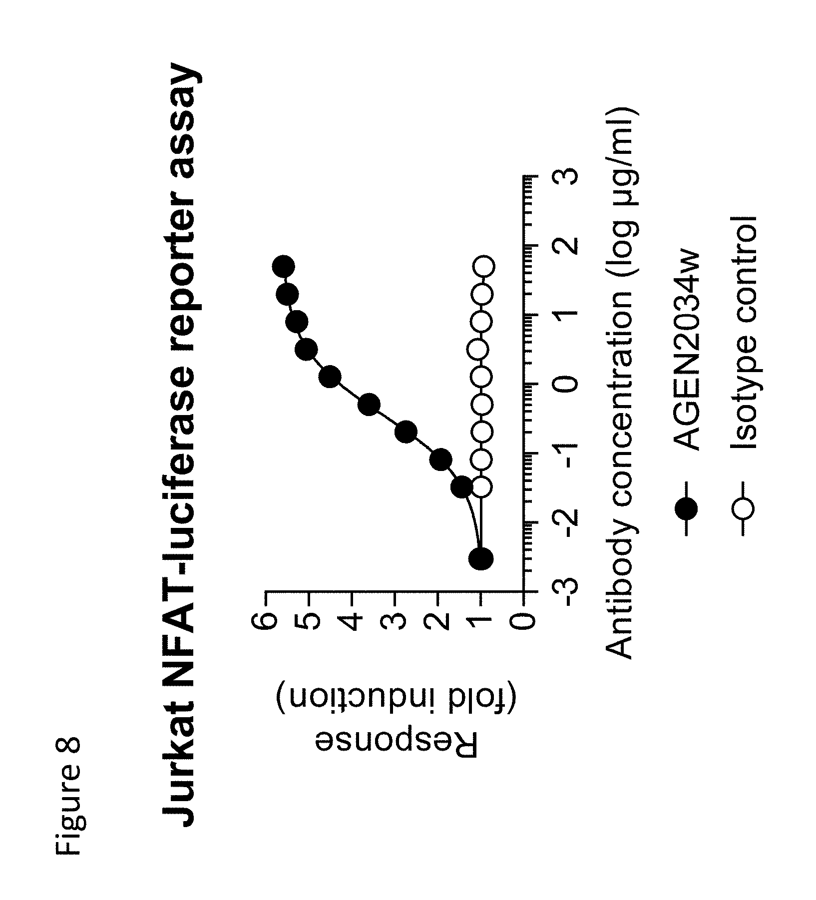
D00015

D00016

D00017

S00001
XML
uspto.report is an independent third-party trademark research tool that is not affiliated, endorsed, or sponsored by the United States Patent and Trademark Office (USPTO) or any other governmental organization. The information provided by uspto.report is based on publicly available data at the time of writing and is intended for informational purposes only.
While we strive to provide accurate and up-to-date information, we do not guarantee the accuracy, completeness, reliability, or suitability of the information displayed on this site. The use of this site is at your own risk. Any reliance you place on such information is therefore strictly at your own risk.
All official trademark data, including owner information, should be verified by visiting the official USPTO website at www.uspto.gov. This site is not intended to replace professional legal advice and should not be used as a substitute for consulting with a legal professional who is knowledgeable about trademark law.